
| ELISA Enzyme-linked immunosorbent assay. Read more |
| WB Western blot : The quality of antibodies used in this technique is crucial for correct and specific protein identification. Diagenode offers huge selection of highly sensitive and specific western blot-validated antibodies. Learn more about: Load... Read more |
| DB Dot blotting Read more |
| IF Immunofluorescence: Diagenode offers huge selection of highly sensitive antibodies validated in IF. Immunofluorescence using the Diagenode monoclonal antibody directed against CRISPR/Cas9 HeLa cells transfected with a Cas9 expression vector (... Read more |
H2A.XS139p polyclonal antibody
Catalog Number
Format
Price

Polyclonal antibody raised in rabbit against the region of histone H2A.X containing the phosphorylated serine 139 (H2A.XS139p), using a KLH-conjugated synthetic peptide.
| Lot | A2099P |
|---|---|
| Concentration | 2.75 µg/µl |
| Species reactivity | Human |
| Type | Polyclonal |
| Purity | Affinity purified |
| Host | Rabbit |
| Precautions | This product is for research use only. Not for use in diagnostic or therapeutic procedures. |
| Applications | Suggested dilution | References |
|---|---|---|
| ELISA | 1:10,000 | Fig 1 |
| Dot Blotting | 1:10,000 | Fig 2 |
| Western Blotting | 1:1,000 | Fig 3 |
| Immunofluorescence | 1:500 | Fig 4 |
- Validation Data
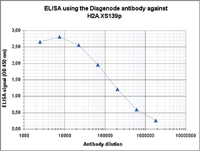
Figure 1. Determination of the antibody titer
To determine the titer of the antibody, an ELISA was performed using a serial dilution of the Diagenode antibody directed against H2A.XS139p (Cat. No. C15410219) in antigen coated wells. The antigen used was a peptide containing the histone modification of interest. By plotting the absorbance against the antibody dilution (Figure 1), the titer of the antibody was estimated to be 1:170,000.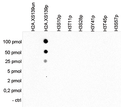
Figure 2. Cross reactivity tests using the Diagenode antibody directed against H2A.XS139p
To test the cross reactivity of the Diagenode antibody against H2A. XS139p (Cat. No. C15410219), a Dot Blot analysis was performed with peptides containing other histone phosphorylations and the unmodified H2A.X. One hundred to 0.2 pmol of the respective peptides were spotted on a membrane. The antibody was used at a dilution of 1:10,000. Figure 2 shows a high specificity of the antibody for the modification of interest.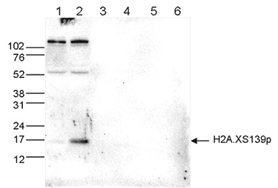
Figure 3. Western blot analysis using the Diagenode antibody directed against H2A.XS139p
Western blot was performed on histone extracts (15 μg) from untreated U2OS cells (lane 1) or from U2OS cells treated with camptothecin (lane 2), and on 1 μg of recombinant histone H2A, H2B, H3 and H4 (lane 3, 4, 5 and 6, respectively) using the Diagenode antibody against H2A.XS139p (Cat. No. C15410219). The antibody was diluted 1:1,000 in TBS-Tween containing 5% skimmed milk. The marker (in kDa) is shown on the left.
Figure 4. Immunofluorescence using the Diagenode antibody directed against H2A.XS139p
U2OS cells, either treated with camptothecin (figure 4B) or untreated (figure 4A), were stained with the Diagenode antibody against H2A.XS139p (cat. No. C15410219) and with DAPI. Cells were fixed with 4% formaldehyde for 10’ and blocked with PBS/TX-100 containing 5% normal goat serum and 1% BSA. The cells were immunofluorescently labelled with the H2A.XS139p antibody (left) diluted 1:500 in blocking solution followed by an anti-rabbit antibody conjugated to Alexa488. The middle panel shows staining of the nuclei with DAPI. A merge of the two stainings is shown on the right. - 出版物

How to properly cite our product/service in your work
We strongly recommend using this: H2A.XS139p polyclonal antibody (Hologic Diagenode Cat# C15410219 Lot# A2099P). Click here to copy to clipboard.
Using our products or services in your publication? Let us know!