| ELISA Enzyme-linked immunosorbent assay. Read more |
| DB Dot blotting Read more |
| WB Western blot : The quality of antibodies used in this technique is crucial for correct and specific protein identification. Diagenode offers huge selection of highly sensitive and specific western blot-validated antibodies. Learn more about: Load... Read more |
| ChIP-seq (ab) Read more |
| ChIP-qPCR (ab) Read more |
H3K36me3 polyclonal antibody
Catalog Number
Format
Price

Polyclonal antibody raised in rabbit against histone H3 containing the trimethylated lysine 36 (H3K36me3), using a KLH-conjugated synthetic peptide.
| Lot | A114-001 |
|---|---|
| Concentration | Not determined |
| Species reactivity | Human, mouse: positive. Other species: not tested. |
| Type | Polyclonal |
| Purity | Whole antiserum from rabbit containing 0.05% azide. |
| Host | Rabbit |
| Storage Conditions | Store at -20°C; for long storage, store at -80°C. Avoid multiple freeze-thaw cycles. |
| Precautions | This product is for research use only. Not for use in diagnostic or therapeutic procedures. |
| Applications | Suggested dilution | References |
|---|---|---|
| ChIP/ChIP-seq * | 5 - 10 µl/ChIP | Fig 1, 2 |
| ELISA | 1:100 - 1:500 | Fig 3 |
| Dot Blotting | 1:100,000 | Fig 4 |
| Western Blotting | 1:1,000 | Fig 5 |
* Please note that the optimal antibody amount per IP should be determined by the end-user. We recommend testing 1-10 µl per IP.
- Validation Data
A.
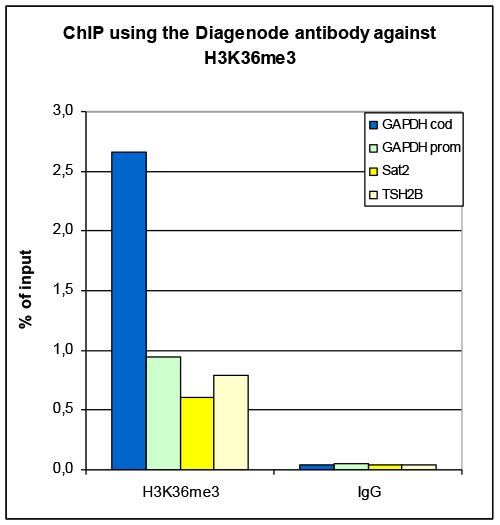
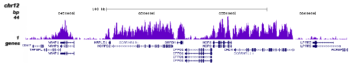
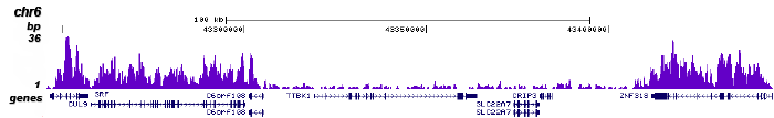
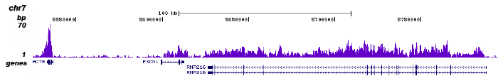
Figure 1. ChIP-seq results obtained with the Diagenode antibody directed against H3K36me3
ChIP was performed with 5 µl of the Diagenode antibody against H3K36me3 (cat. No. CS-058-050) on sheared chromatin from 1 million HeLaS3 cells using the “Auto Histone ChIP-seq” kit (cat. No. AB-Auto02-A100) on the IP-Star automated system. IgG (2 µg/IP) was used as a negative IP control. The IP’d DNA was analysed by QPCR with optimized PCR primer pairs for the coding and promoter region of the active GAPDH gene, for the coding region of the inactive TSH2B gene and for the Sat2 satellite repeat (figure 2A). The IP’d DNA was subsequently analysed with an Illumina Genome Analyzer. Library preparation, cluster generation and sequencing were performed according to the manufacturer’s instructions. The 36 bp tags were aligned to the human genome using the ELAND algorithm. Figure 2B shows the results in 200 kb regions of chromosome 12 (including the GAPDH positive control), 6 and 7 and 14. These results clearly show an enrichment of the H3K36me3 at active genesB.
C.
D.
E.
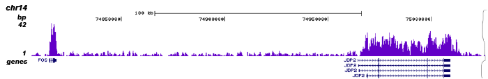
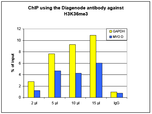
Figure 2. ChIP results obtained with the Diagenode antibody directed against H3K36me3
ChIP assays were performed using human osteosarcoma (U2OS) cells, the Diagenode antibody against H3K36me3 (cat. No. CS-058-100) and optimized PCR primer sets for qPCR. Chromatin was sheared with the Diagenode “Shearing ChIP” kit (cat. No. kch-redmod-100). ChIP was performed with the “OneDay ChIP” kit (cat. No. kch-oneDIP-060), using sheared chromatin from 1.6 million cells. A titration of the antibody consisting of 2, 5, 10 and 15 µl per ChIP experiment was analysed. IgG (5 µg/IP) was used as a negative IP control. Quantitative PCR was performed using primer sets for the housekeeping gene GAPDH and for myogenic differentiation gene (MYOD). Figure 1 shows the recovery, expressed as a % of input (the relative amount of immunoprecipitated DNA compared to input DNA after qPCR analysis.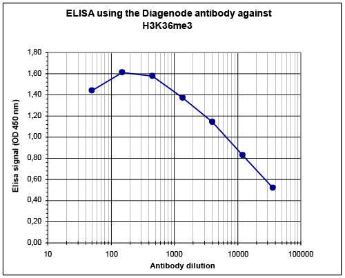
Figure 3. Determination of the titer
To determine the titer, an ELISA was performed using a serial dilution of the Diagenode antibody directed against H3K36me3 (cat. No. CS-058-100). The antigen used was a peptide containing the histone modification of interest. By plotting the absorbance against the antibody dilution (Figure 3), the titer of the antibody was estimated to be 1:12,700.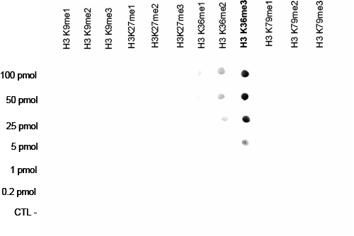
Figure 4. Cross reactivity test using the Diagenode antibody directed against H3K36me3
A Dot Blot analysis was performed to test the cross reactivity of the Diagenode antibody against H3K36me3 (cat. No. CS-058-050) with peptides containing other modifications of histone H3. Other histone modifications include mono- and dimethylation of the same lysine and mono-, di- and trimethylation of lysine 9, 27 and 79. One hundred to 0.2 pmol of peptide containing the respective histone modification were spotted on a membrane. The antibody was used at a dilution of 1:100,000. Figure 4 shows a high specificity of the antibody for the modification of interest.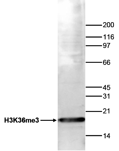
Figure 5. Western blot analysis using the Diagenode antibody directed against H3K36me3
Histone extracts (15 µg) from HeLa cells were analysed by Western blot using the Diagenode antibody against H3K36me3 (cat. No. CS-058-100) diluted 1:1,000 in TBS-Tween containing 5% skimmed milk. The position of the protein of interest is indicated on the left; the marker (in kDa) is shown on the right. - 出版物

How to properly cite our product/service in your work
We strongly recommend using this: H3K36me3 polyclonal antibody (Hologic Diagenode Cat# C15310058 Lot# A114-001 ). Click here to copy to clipboard.
Using our products or services in your publication? Let us know!
The histone demethylase JMJD2A/KDM4A links ribosomal RNA transcription to nutrients and growth factors availability
Salifou K, Ray S, Verrier L, Aguirrebengoa M, Trouche D, Panov KI, Vandromme M
The interplay between methylation and demethylation of histone lysine residues is an essential component of gene expression regulation and there is considerable interest in elucidating the roles of proteins involved. Here we report that histone demethylase KDM4A/JMJD2A, which is involved in the regulation of cell pr...The transcriptional and epigenomic foundations of ground state pluripotency.
Marks H, Kalkan T, Menafra R, Denissov S, Jones K, Hofemeister H, Nichols J, Kranz A, Francis Stewart A, Smith A, Stunnenberg HG
Mouse embryonic stem (ES) cells grown in serum exhibit greater heterogeneity in morphology and expression of pluripotency factors than ES cells cultured in defined medium with inhibitors of two kinases (Mek and GSK3), a condition known as "2i" postulated to establish a naive ground state. We show that the transcript...High-resolution analysis of epigenetic changes associated with X inactivation.
Marks H, Chow JC, Denissov S, Françoijs KJ, Brockdorff N, Heard E, Stunnenberg HG
Differentiation of female murine ES cells triggers silencing of one X chromosome through X-chromosome inactivation (XCI). Immunofluorescence studies showed that soon after Xist RNA coating the inactive X (Xi) undergoes many heterochromatic changes, including the acquisition of H3K27me3. However, the mechanisms that ...