| IHC Immunohistochemistry Read more |
| IF Immunofluorescence: Diagenode offers huge selection of highly sensitive antibodies validated in IF. Immunofluorescence using the Diagenode monoclonal antibody directed against CRISPR/Cas9 HeLa cells transfected with a Cas9 expression vector (... Read more |
| WB Western blot : The quality of antibodies used in this technique is crucial for correct and specific protein identification. Diagenode offers huge selection of highly sensitive and specific western blot-validated antibodies. Learn more about: Load... Read more |
| DB Dot blotting Read more |
| ChIP-qPCR (ab) Read more |
H3K4me3T6p polyclonal antibody
Catalog Number
Format
Price

Polyclonal antibody raised in rabbit against H3 (trimethyl Lys4, p Thr6), using a KLH-conjugated synthetic peptide.
| Lot | 001 |
|---|---|
| Concentration | 0.55 μg/μl |
| Species reactivity | Human, mouse, C. elegans, rat, chicken, Xenopus, Drosophila, plant |
| Type | Polyclonal |
| Purity | Affinity purified |
| Host | Rabbit |
| Precautions | This product is for research use only. Not for use in diagnostic or therapeutic procedures. |
| Applications | Suggested dilution | References |
|---|---|---|
| ChIP | 2-5 μg/million cells | Fig 1 |
| Immunohistochemistry | 1:50 | |
| Immunofluorescence | 1:50 | Fig 2 |
| Western Blotting | 1:500 | Fig 3, 4 |
| Dot Blotting | 1:1,000 | Fig 5 |
- Validation Data
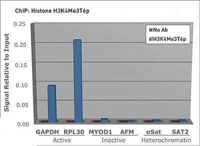
Figure 1. H3K4me3T6p antibody ChIP results
Chromatin Immunoprecipitation of H3K4me3T6p antibody. Chromatin from one million formaldehyde cross-linked Hela cells was used with 2 μg of H3K4me3T6p antibody alongside a no antibody (No Ab) control, DNA was measured by qPCR and normalized to total input.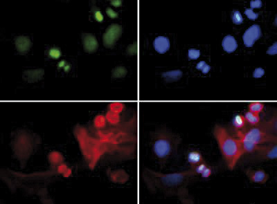
Figure 2. H3K4me3T6p antibody Immunofluorescence results
Immunofluorescence of H3K4me3T6p antibody. Tissue: HeLa cells. Fixation: 0.5% PFA. Primary antibody used at a 1:50 dilution for 1 h at RT. Secondary antibody: FITC secondary antibody at 1:10,000 for 45 min at RT. Localization: Histone H3K4me3T6p is nuclear and chromosomal. Staining: H3K4me3T6p is expressed in green and the nuclei and alpha-tubulin are counterstained with DAPI (blue) and Dylight 594 (red).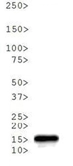
Figure 3. H3K4me3T6p antibody Western blot results
Western Blot of Rabbit H3K4me3T6p antibody. 30 μg C. elegans embryo lysate. Primary antibody at 1:500 overnight at 4°C. Secondary antibody: IRDye800TM rabbit secondary antibody at 1:10,000 for 45 min at RT. Predicted/Observed size: ~15 kDa. Other band(s): None.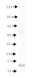
Figure 4. H3K4me3T6p antibody Western blot results
Western Blot of H3K4me3T6p antibody. 30 μg HeLa histone extracts. Primary antibody at 1:500 overnight at 4°C. Secondary antibody: IRDye800TM rabbit secondary antibody at 1:10,000 for 45 min at RT. Predicted/Observed size: ~15 kDa. Other band(s): None.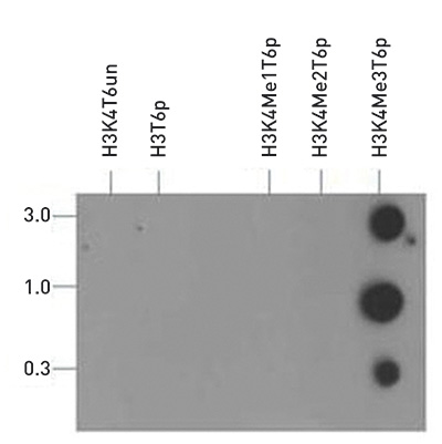
Figure 5. H3K4me3T6p antibody Dot blot results
Dot Blot of Rabbit H3K4me3T6p antibody. Lane 1: Unmodified. Lane 2: T6p. Lane 3: K4Me1T6p. Lane 4: K4Me2T6p. Lane 5: K4Me3T6p. Load: 3, 1, and 0.3 picomoles of peptide. Primary antibody used at a 1:1,000 dilution for 45 min at 4°C. Secondary antibody: DylightTM488 rabbit secondary antibody at 1:10,000 for 45 min at RT. - 出版物

How to properly cite our product/service in your work
We strongly recommend using this: H3K4me3T6p polyclonal antibody (Hologic Diagenode Cat# C15410281 Lot# 001). Click here to copy to clipboard.
Using our products or services in your publication? Let us know!