Notice (8): Undefined variable: solution_of_interest [APP/View/Products/view.ctp, line 755]Code Context<!-- BEGIN: REQUEST_FORM MODAL -->
<div id="request_formModal" class="reveal-modal medium" data-reveal aria-labelledby="modalTitle" aria-hidden="true" role="dialog">
<?= $this->element('Forms/simple_form', array('solution_of_interest' => $solution_of_interest, 'header' => $header, 'message' => $message, 'campaign_id' => $campaign_id)) ?>
$viewFile = '/home/website-server/www/app/View/Products/view.ctp'
$dataForView = array(
'language' => 'jp',
'meta_keywords' => '',
'meta_description' => 'H3K9me3 monoclonal antibody - Classic',
'meta_title' => 'H3K9me3 monoclonal antibody - Classic',
'product' => array(
'Product' => array(
'id' => '1984',
'antibody_id' => '139',
'name' => 'H3K9me3 monoclonal antibody ',
'description' => '<p><span>Monoclonal antibody raised in mouse against histone H3 trimethylated at lysine 9 (H3K9me3), using a KLH-conjugated synthetic peptide.</span></p>',
'label1' => 'Validation Data',
'info1' => '<div class="row">
<div class="small-6 columns">
<p><img src="https://www.diagenode.com/img/product/antibodies/C15200146-ChIP.png" alt="H3K9me3 Antibody ChIP Grade" style="display: block; border: 1px solid black; margin-left: auto; margin-right: auto;" /></p>
</div>
<div class="small-6 columns">
<p><small><strong> Figure 1. ChIP results obtained with the Diagenode monoclonal antibody directed against H3K9me3 </strong><br />ChIP assays were performed on human HeLa cells using the Diagenode monclonal antibody against H3K9me3 (cat. No. C15200146). ChIP was performed with the ““iDeal ChIP-seq” kit (cat. No. (cat. No. C01010051), using sheared chromatin from 500,000 cells. A titration consisting of 0.5, 1, and 5 µg of antibody per ChIP experiment was analyzed. IgG (1 µg/IP) was used as a negative IP control. QPCR was performed with primers for the promoters of the active EIF4A2 and GAPDH genes, used as negative controls, and for the ZNF510 gene and the Sat2 satellite repeat region, used as positive controls. Figure 1 shows the recovery, expressed as a % of input (the relative amount of immunoprecipitated DNA compared to input DNA after qPCR analysis).</small></p>
</div>
</div>
<div class="row">
<div class="small-12 columns">
<p>A.<img src="https://www.diagenode.com/img/product/antibodies/c15200146-chipseq-a.jpg" alt="H3K9me3 Antibody ChIP-seq Grade " /></p>
<p>B.<img src="https://www.diagenode.com/img/product/antibodies/c15200146-chipseq-b.jpg" alt="H3K9me3 Antibody ChIP-seq Grade" /></p>
<p>C. <img src="https://www.diagenode.com/img/product/antibodies/c15200146-chipseq-c.jpg" alt="H3K9me3 Antibody ChIP-seq Grade " /></p>
</div>
</div>
<div class="row">
<div class="small-12 columns">
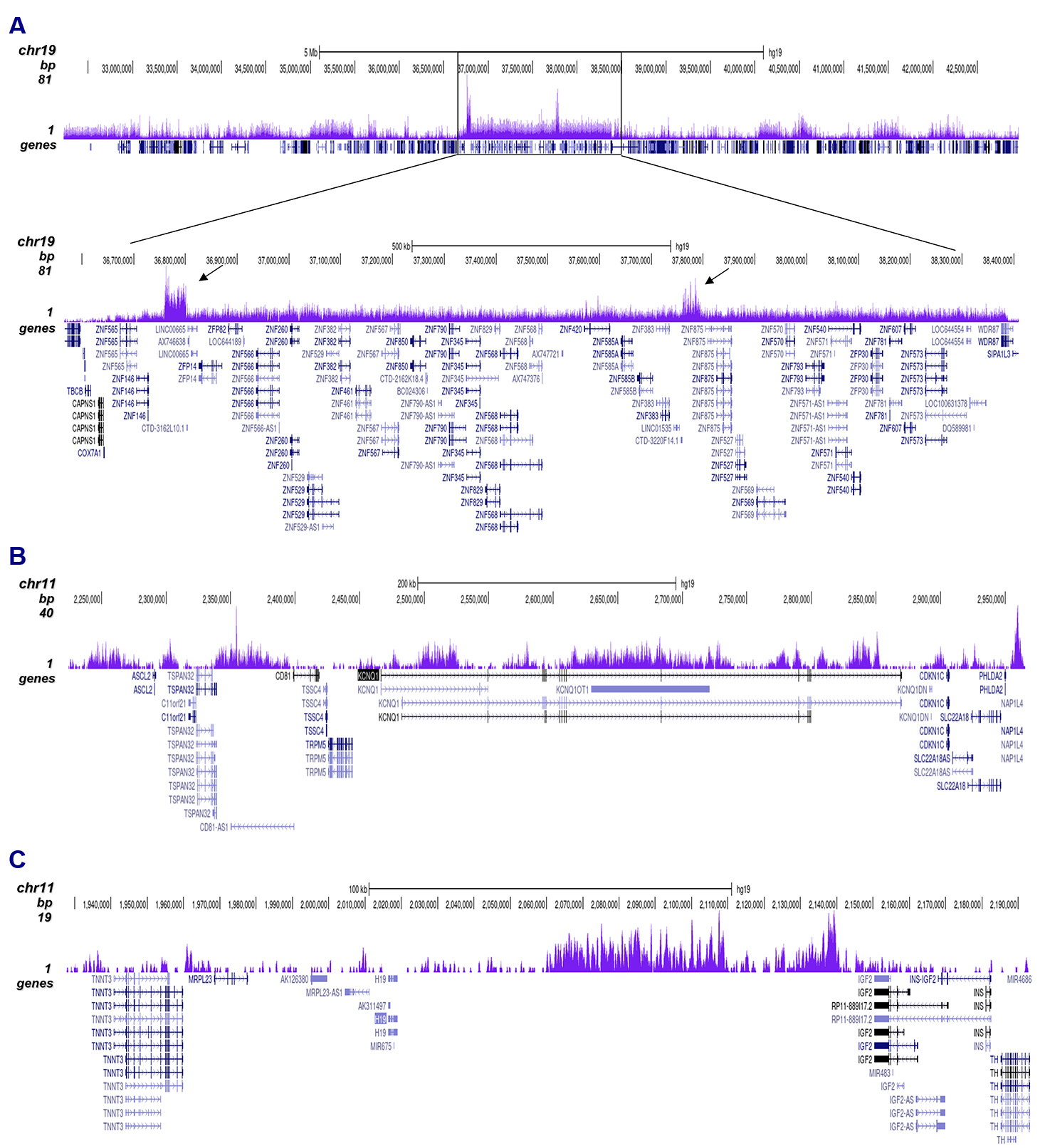
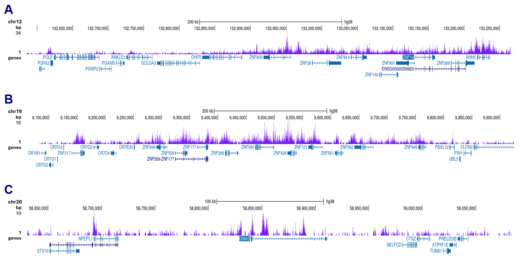
<p><strong>Figure 2. ChIP-seq results obtained with the Diagenode antibody directed against H3K9me3</strong><br />ChIP was performed with 0.5 μg of the Diagenode antibody against H3K9me3 (cat. No. C15200146) on sheared chromatin from 500,000 HeLa cells using the “iDeal ChIP-seq” kit as described above. The IP’d DNA was subsequently analysed on an Illumina HiSeq 2000. Library preparation, cluster generation and sequencing were performed according to the manufacturer’s instructions. The 50 bp tags were aligned to the human genome using the BWA algorithm. Figure 2A shows the signal distribution along the long arm of chromosome 19 and a zoomin to an enriched region containing several ZNF repeat genes. The arrows indicate two satellite repeat regions which exhibit a stronger signal. Figures 2B and 2C show the enrichment at the H19 and KCNQ1 imprinted genes.</p>
</div>
</div>
<div class="row">
<div class="small-6 columns">
<p><img src="https://www.diagenode.com/img/product/antibodies/c15200146-elisa.png" style="display: block; margin-left: auto; border: 1px solid black; margin-right: auto;" /></p>
</div>
<div class="small-6 columns">
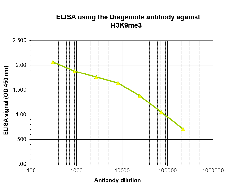
<p><small><strong> Figure 3. Determination of the antibody titer </strong><br />To determine the titer of the antibody, an ELISA was performed using a serial dilution of the antibody directed against H3K9me3 (cat. No. C15200146) in antigen coated wells. The antigen used was a peptide containing the histone modification of interest. By plotting the absorbance against the antibody dilution (Figure 3), the titer of the antibody was estimated to be 1:14,500.</small></p>
</div>
</div>
<div class="row">
<div class="small-4 columns">
<p><img src="https://www.diagenode.com/img/product/antibodies/C15200146-DotBlot.png" alt="H3K9me3 Antibody validated in Dot Blot" style="display: block; margin-left: auto; margin-right: auto;" /></p>
</div>
<div class="small-8 columns">
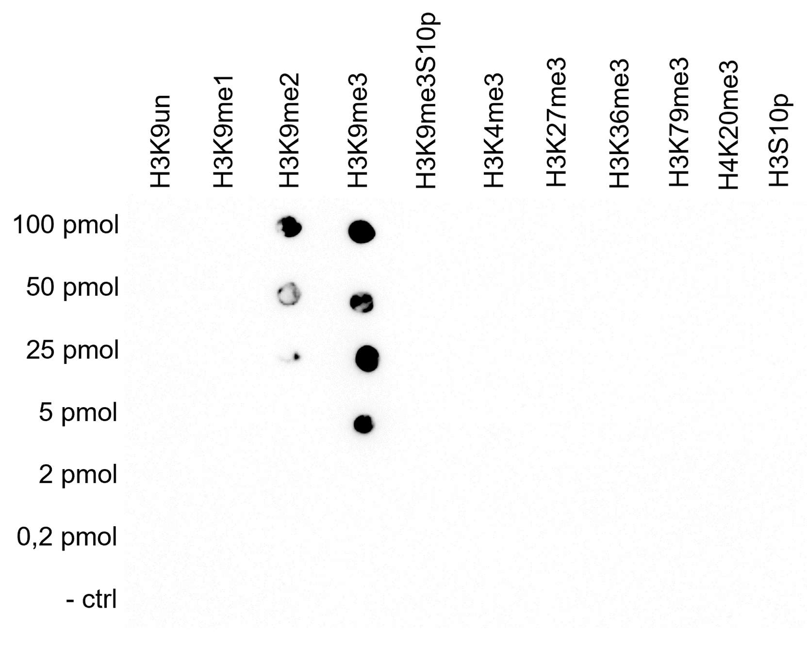
<p><small><strong> Figure 4. Cross reactivity test using the Diagenode monoclonal antibody directed against H3K9me3 </strong><br />A Dot Blot analysis was performed to test the cross reactivity of the Diagenode monoclonal antibody against H3K9me3 (cat. No. C15200146) with peptides containing different modifications of histone H3 or H4 and the unmodified H3K9 sequence. One hundred to 0.2 pmol of peptide containing the respective histone modification were spotted on a membrane. The antibody was used at a dilution of 1:100,000. Figure 4 shows a high specificity of the antibody for the modification of interest, with some cross reaction with the H3K9me2 peptide.</small></p>
</div>
</div>
<div class="row">
<div class="small-4 columns">
<p><img src="https://www.diagenode.com/img/product/antibodies/C15200146-WesternBlot.png" alt="H3K9me3 Antibody validated in Western Blot" style="display: block; margin-left: auto; margin-right: auto;" /></p>
</div>
<div class="small-8 columns">
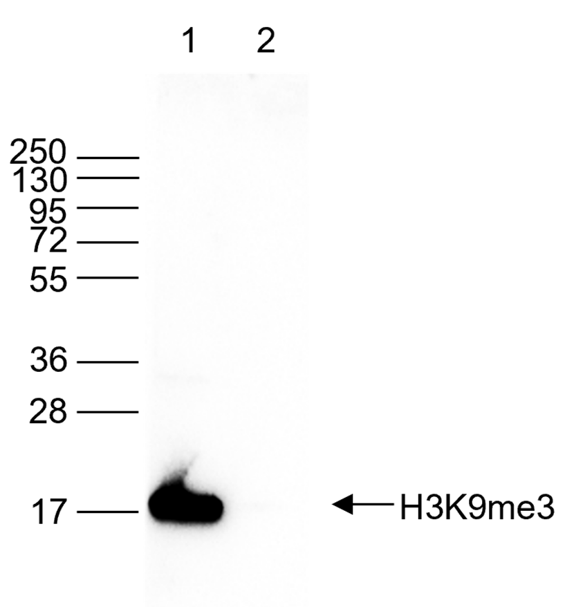
<p><small><strong> Figure 5. Western blot analysis using the Diagenode monoclonal antibody directed against H3K9me3 </strong><br />Western blot was performed on histone extracts (15 µg, lane 1) from HeLa cells, and on 1 µg of recombinant histone H3 (lane 2) using the Diagenode monoclonal antibody against H3K9me3 (cat. No. C15200146). The antibody was diluted 1:1,000 in TBS-Tween containing 5% skimmed milk. The marker (in kDa) is shown on the left, the position of the protein is indicated on the right.</small></p>
</div>
</div>
<div class="row">
<div class="small-4 columns">
<p><img src="https://www.diagenode.com/img/product/antibodies/C15200146-IF.png" alt="H3K9me3 Antibody validated in Immunofluorescence" style="display: block; margin-left: auto; margin-right: auto;" /></p>
</div>
<div class="small-8 columns">
<p><small><strong> Figure 6. Immunofluorescence using the Diagenode monoclonal antibody directed against H3K9me3 </strong><br />HeLa cells were stained with the Diagenode antibody against H3K9me3 (cat. No. C15200146) and with DAPI. Cells were fixed with 4% formaldehyde for 10’ and blocked with PBS/TX-100 containing 1% BSA. The cells were immunofluorescently labelled with the H3K9me3 antibody (middle) diluted 1:500 in blocking solution followed by an anti-mouse antibody conjugated to Alexa594. The left panel shows staining of the nuclei with DAPI. A merge of both stainings is shown on the right.</small></p>
</div>
</div>',
'label2' => '',
'info2' => '<p>Histones are present in the chromosomes of eukaryotic cells. They are rich in the amino acids arginine and lysine and have been greatly conserved during evolution. Histones pack the DNA into tight masses of chromatin. Two core histones of each class H2A, H2B, H3 and H4 assemble and are wrapped by 146 base pairs of DNA to form one octameric nucleosome. Histone tails undergo numerous post-translational modifications, which either directly or indirectly alter chromatin structure to facilitate transcriptional activation or repression or other nuclear processes. In addition to the genetic code, combinations of the different histone modifications reveal the so-called “histone code”. Histone methylation and demethylation is dynamically regulated by respectively histone methyl transferases and histone demethylases. Methylation of histone H3K9 is associated with gene repression.</p>',
'label3' => '',
'info3' => '',
'format' => '50 μg',
'catalog_number' => 'C15200146',
'old_catalog_number' => 'MAb-146-050',
'sf_code' => 'C15200146-D001-000581',
'type' => 'FRE',
'search_order' => '03-Antibody',
'price_EUR' => '380',
'price_USD' => '380',
'price_GBP' => '340',
'price_JPY' => '59525',
'price_CNY' => '',
'price_AUD' => '950',
'country' => 'ALL',
'except_countries' => 'None',
'quote' => false,
'in_stock' => false,
'featured' => false,
'no_promo' => false,
'online' => true,
'master' => true,
'last_datasheet_update' => '0000-00-00',
'slug' => 'h3k9me3-monoclonal-antibody-classic-50-mg',
'meta_title' => 'H3K9me3 monoclonal antibody - Classic',
'meta_keywords' => '',
'meta_description' => 'H3K9me3 monoclonal antibody - Classic',
'modified' => '2024-01-31 16:31:46',
'created' => '2015-06-29 14:08:20',
'locale' => 'jpn'
),
'Antibody' => array(
'host' => '*****',
'id' => '139',
'name' => 'H3K9me3 monoclonal antibody',
'description' => 'Histones are present in the chromosomes of eukaryotic cells. They are rich in the amino acids arginine and lysine and have been greatly conserved during evolution. Histones pack the DNA into tight masses of chromatin. Two core histones of each class H2A, H2B, H3 and H4 assemble and are wrapped by 146 base pairs of DNA to form one octameric nucleosome. Histone tails undergo numerous post-translational modifications, which either directly or indirectly alter chromatin structure to facilitate transcriptional activation or repression or other nuclear processes. In addition to the genetic code, combinations of the different histone modifications reveal the so-called “histone code”. Histone methylation and demethylation is dynamically regulated by respectively histone methyl transferases and histone demethylases. Methylation of histone H3K9 is associated with gene repression.',
'clonality' => '',
'isotype' => '',
'lot' => '003',
'concentration' => '1.7 µg/µl',
'reactivity' => 'Human, mouse, fungi: positive.',
'type' => 'Monoclonal',
'purity' => 'Protein A purified',
'classification' => '',
'application_table' => '<table>
<thead>
<tr style="height: 38px;">
<th style="height: 38px;">Applications</th>
<th style="height: 38px;">Suggested dilution</th>
<th style="height: 38px;">References</th>
</tr>
</thead>
<tbody>
<tr style="height: 42px;">
<td style="height: 42px;">ChIP/ChIP-seq <sup>*</sup></td>
<td style="height: 42px;">0.5 - 1 μg/ChIP</td>
<td style="height: 42px;">Fig 1, 2</td>
</tr>
<tr style="height: 42px;">
<td style="height: 42px;">ELISA</td>
<td style="height: 42px;">1:100</td>
<td style="height: 42px;">Fig 3</td>
</tr>
<tr style="height: 38px;">
<td style="height: 38px;">Dot Blotting</td>
<td style="height: 38px;">1:100,000</td>
<td style="height: 38px;">Fig 4</td>
</tr>
<tr style="height: 38px;">
<td style="height: 38px;">Western Blotting</td>
<td style="height: 38px;">1:1,000</td>
<td style="height: 38px;">Fig 5</td>
</tr>
<tr style="height: 38px;">
<td style="height: 38px;">IF</td>
<td style="height: 38px;">1:500</td>
<td style="height: 38px;">Fig 6</td>
</tr>
</tbody>
</table>
<p><small><sup>*</sup> Please note that the optimal antibody amount per IP should be determined by the end-user. We recommend testing 1-5 μg per IP.</small></p>',
'storage_conditions' => 'Store at -20°C; for long storage, store at -80°C. Avoid multiple freeze-thaw cycles.',
'storage_buffer' => 'PBS containing 0.05% azide.',
'precautions' => 'This product is for research use only. Not for use in diagnostic or therapeutic procedures.',
'uniprot_acc' => '',
'slug' => '',
'meta_keywords' => '',
'meta_description' => '',
'modified' => '2024-01-31 16:42:21',
'created' => '0000-00-00 00:00:00',
'select_label' => '139 - H3K9me3 monoclonal antibody (003 - 1.7 µg/µl - Human, mouse, fungi: positive. - Protein A purified - Mouse)'
),
'Slave' => array(
(int) 0 => array(
[maximum depth reached]
)
),
'Group' => array(
'Group' => array(
[maximum depth reached]
),
'Master' => array(
[maximum depth reached]
),
'Product' => array(
[maximum depth reached]
)
),
'Related' => array(),
'Application' => array(
(int) 0 => array(
[maximum depth reached]
),
(int) 1 => array(
[maximum depth reached]
),
(int) 2 => array(
[maximum depth reached]
),
(int) 3 => array(
[maximum depth reached]
)
),
'Category' => array(
(int) 0 => array(
[maximum depth reached]
),
(int) 1 => array(
[maximum depth reached]
),
(int) 2 => array(
[maximum depth reached]
)
),
'Document' => array(
(int) 0 => array(
[maximum depth reached]
),
(int) 1 => array(
[maximum depth reached]
),
(int) 2 => array(
[maximum depth reached]
)
),
'Feature' => array(),
'Image' => array(
(int) 0 => array(
[maximum depth reached]
)
),
'Promotion' => array(),
'Protocol' => array(),
'Publication' => array(
(int) 0 => array(
[maximum depth reached]
),
(int) 1 => array(
[maximum depth reached]
),
(int) 2 => array(
[maximum depth reached]
),
(int) 3 => array(
[maximum depth reached]
),
(int) 4 => array(
[maximum depth reached]
),
(int) 5 => array(
[maximum depth reached]
),
(int) 6 => array(
[maximum depth reached]
)
),
'Testimonial' => array(),
'Area' => array(),
'SafetySheet' => array(
(int) 0 => array(
[maximum depth reached]
),
(int) 1 => array(
[maximum depth reached]
),
(int) 2 => array(
[maximum depth reached]
),
(int) 3 => array(
[maximum depth reached]
),
(int) 4 => array(
[maximum depth reached]
),
(int) 5 => array(
[maximum depth reached]
),
(int) 6 => array(
[maximum depth reached]
),
(int) 7 => array(
[maximum depth reached]
)
)
),
'meta_canonical' => 'https://www.diagenode.com/jp/p/h3k9me3-monoclonal-antibody-classic-50-mg'
)
$language = 'jp'
$meta_keywords = ''
$meta_description = 'H3K9me3 monoclonal antibody - Classic'
$meta_title = 'H3K9me3 monoclonal antibody - Classic'
$product = array(
'Product' => array(
'id' => '1984',
'antibody_id' => '139',
'name' => 'H3K9me3 monoclonal antibody ',
'description' => '<p><span>Monoclonal antibody raised in mouse against histone H3 trimethylated at lysine 9 (H3K9me3), using a KLH-conjugated synthetic peptide.</span></p>',
'label1' => 'Validation Data',
'info1' => '<div class="row">
<div class="small-6 columns">
<p><img src="https://www.diagenode.com/img/product/antibodies/C15200146-ChIP.png" alt="H3K9me3 Antibody ChIP Grade" style="display: block; border: 1px solid black; margin-left: auto; margin-right: auto;" /></p>
</div>
<div class="small-6 columns">
<p><small><strong> Figure 1. ChIP results obtained with the Diagenode monoclonal antibody directed against H3K9me3 </strong><br />ChIP assays were performed on human HeLa cells using the Diagenode monclonal antibody against H3K9me3 (cat. No. C15200146). ChIP was performed with the ““iDeal ChIP-seq” kit (cat. No. (cat. No. C01010051), using sheared chromatin from 500,000 cells. A titration consisting of 0.5, 1, and 5 µg of antibody per ChIP experiment was analyzed. IgG (1 µg/IP) was used as a negative IP control. QPCR was performed with primers for the promoters of the active EIF4A2 and GAPDH genes, used as negative controls, and for the ZNF510 gene and the Sat2 satellite repeat region, used as positive controls. Figure 1 shows the recovery, expressed as a % of input (the relative amount of immunoprecipitated DNA compared to input DNA after qPCR analysis).</small></p>
</div>
</div>
<div class="row">
<div class="small-12 columns">
<p>A.<img src="https://www.diagenode.com/img/product/antibodies/c15200146-chipseq-a.jpg" alt="H3K9me3 Antibody ChIP-seq Grade " /></p>
<p>B.<img src="https://www.diagenode.com/img/product/antibodies/c15200146-chipseq-b.jpg" alt="H3K9me3 Antibody ChIP-seq Grade" /></p>
<p>C. <img src="https://www.diagenode.com/img/product/antibodies/c15200146-chipseq-c.jpg" alt="H3K9me3 Antibody ChIP-seq Grade " /></p>
</div>
</div>
<div class="row">
<div class="small-12 columns">
<p><strong>Figure 2. ChIP-seq results obtained with the Diagenode antibody directed against H3K9me3</strong><br />ChIP was performed with 0.5 μg of the Diagenode antibody against H3K9me3 (cat. No. C15200146) on sheared chromatin from 500,000 HeLa cells using the “iDeal ChIP-seq” kit as described above. The IP’d DNA was subsequently analysed on an Illumina HiSeq 2000. Library preparation, cluster generation and sequencing were performed according to the manufacturer’s instructions. The 50 bp tags were aligned to the human genome using the BWA algorithm. Figure 2A shows the signal distribution along the long arm of chromosome 19 and a zoomin to an enriched region containing several ZNF repeat genes. The arrows indicate two satellite repeat regions which exhibit a stronger signal. Figures 2B and 2C show the enrichment at the H19 and KCNQ1 imprinted genes.</p>
</div>
</div>
<div class="row">
<div class="small-6 columns">
<p><img src="https://www.diagenode.com/img/product/antibodies/c15200146-elisa.png" style="display: block; margin-left: auto; border: 1px solid black; margin-right: auto;" /></p>
</div>
<div class="small-6 columns">
<p><small><strong> Figure 3. Determination of the antibody titer </strong><br />To determine the titer of the antibody, an ELISA was performed using a serial dilution of the antibody directed against H3K9me3 (cat. No. C15200146) in antigen coated wells. The antigen used was a peptide containing the histone modification of interest. By plotting the absorbance against the antibody dilution (Figure 3), the titer of the antibody was estimated to be 1:14,500.</small></p>
</div>
</div>
<div class="row">
<div class="small-4 columns">
<p><img src="https://www.diagenode.com/img/product/antibodies/C15200146-DotBlot.png" alt="H3K9me3 Antibody validated in Dot Blot" style="display: block; margin-left: auto; margin-right: auto;" /></p>
</div>
<div class="small-8 columns">
<p><small><strong> Figure 4. Cross reactivity test using the Diagenode monoclonal antibody directed against H3K9me3 </strong><br />A Dot Blot analysis was performed to test the cross reactivity of the Diagenode monoclonal antibody against H3K9me3 (cat. No. C15200146) with peptides containing different modifications of histone H3 or H4 and the unmodified H3K9 sequence. One hundred to 0.2 pmol of peptide containing the respective histone modification were spotted on a membrane. The antibody was used at a dilution of 1:100,000. Figure 4 shows a high specificity of the antibody for the modification of interest, with some cross reaction with the H3K9me2 peptide.</small></p>
</div>
</div>
<div class="row">
<div class="small-4 columns">
<p><img src="https://www.diagenode.com/img/product/antibodies/C15200146-WesternBlot.png" alt="H3K9me3 Antibody validated in Western Blot" style="display: block; margin-left: auto; margin-right: auto;" /></p>
</div>
<div class="small-8 columns">
<p><small><strong> Figure 5. Western blot analysis using the Diagenode monoclonal antibody directed against H3K9me3 </strong><br />Western blot was performed on histone extracts (15 µg, lane 1) from HeLa cells, and on 1 µg of recombinant histone H3 (lane 2) using the Diagenode monoclonal antibody against H3K9me3 (cat. No. C15200146). The antibody was diluted 1:1,000 in TBS-Tween containing 5% skimmed milk. The marker (in kDa) is shown on the left, the position of the protein is indicated on the right.</small></p>
</div>
</div>
<div class="row">
<div class="small-4 columns">
<p><img src="https://www.diagenode.com/img/product/antibodies/C15200146-IF.png" alt="H3K9me3 Antibody validated in Immunofluorescence" style="display: block; margin-left: auto; margin-right: auto;" /></p>
</div>
<div class="small-8 columns">
<p><small><strong> Figure 6. Immunofluorescence using the Diagenode monoclonal antibody directed against H3K9me3 </strong><br />HeLa cells were stained with the Diagenode antibody against H3K9me3 (cat. No. C15200146) and with DAPI. Cells were fixed with 4% formaldehyde for 10’ and blocked with PBS/TX-100 containing 1% BSA. The cells were immunofluorescently labelled with the H3K9me3 antibody (middle) diluted 1:500 in blocking solution followed by an anti-mouse antibody conjugated to Alexa594. The left panel shows staining of the nuclei with DAPI. A merge of both stainings is shown on the right.</small></p>
</div>
</div>',
'label2' => 'Target Description',
'info2' => '<p>Histones are present in the chromosomes of eukaryotic cells. They are rich in the amino acids arginine and lysine and have been greatly conserved during evolution. Histones pack the DNA into tight masses of chromatin. Two core histones of each class H2A, H2B, H3 and H4 assemble and are wrapped by 146 base pairs of DNA to form one octameric nucleosome. Histone tails undergo numerous post-translational modifications, which either directly or indirectly alter chromatin structure to facilitate transcriptional activation or repression or other nuclear processes. In addition to the genetic code, combinations of the different histone modifications reveal the so-called “histone code”. Histone methylation and demethylation is dynamically regulated by respectively histone methyl transferases and histone demethylases. Methylation of histone H3K9 is associated with gene repression.</p>',
'label3' => '',
'info3' => '',
'format' => '50 μg',
'catalog_number' => 'C15200146',
'old_catalog_number' => 'MAb-146-050',
'sf_code' => 'C15200146-D001-000581',
'type' => 'FRE',
'search_order' => '03-Antibody',
'price_EUR' => '380',
'price_USD' => '380',
'price_GBP' => '340',
'price_JPY' => '59525',
'price_CNY' => '',
'price_AUD' => '950',
'country' => 'ALL',
'except_countries' => 'None',
'quote' => false,
'in_stock' => false,
'featured' => false,
'no_promo' => false,
'online' => true,
'master' => true,
'last_datasheet_update' => '0000-00-00',
'slug' => 'h3k9me3-monoclonal-antibody-classic-50-mg',
'meta_title' => 'H3K9me3 monoclonal antibody - Classic',
'meta_keywords' => '',
'meta_description' => 'H3K9me3 monoclonal antibody - Classic',
'modified' => '2024-01-31 16:31:46',
'created' => '2015-06-29 14:08:20',
'locale' => 'jpn'
),
'Antibody' => array(
'host' => '*****',
'id' => '139',
'name' => 'H3K9me3 monoclonal antibody',
'description' => 'Histones are present in the chromosomes of eukaryotic cells. They are rich in the amino acids arginine and lysine and have been greatly conserved during evolution. Histones pack the DNA into tight masses of chromatin. Two core histones of each class H2A, H2B, H3 and H4 assemble and are wrapped by 146 base pairs of DNA to form one octameric nucleosome. Histone tails undergo numerous post-translational modifications, which either directly or indirectly alter chromatin structure to facilitate transcriptional activation or repression or other nuclear processes. In addition to the genetic code, combinations of the different histone modifications reveal the so-called “histone code”. Histone methylation and demethylation is dynamically regulated by respectively histone methyl transferases and histone demethylases. Methylation of histone H3K9 is associated with gene repression.',
'clonality' => '',
'isotype' => '',
'lot' => '003',
'concentration' => '1.7 µg/µl',
'reactivity' => 'Human, mouse, fungi: positive.',
'type' => 'Monoclonal',
'purity' => 'Protein A purified',
'classification' => '',
'application_table' => '<table>
<thead>
<tr style="height: 38px;">
<th style="height: 38px;">Applications</th>
<th style="height: 38px;">Suggested dilution</th>
<th style="height: 38px;">References</th>
</tr>
</thead>
<tbody>
<tr style="height: 42px;">
<td style="height: 42px;">ChIP/ChIP-seq <sup>*</sup></td>
<td style="height: 42px;">0.5 - 1 μg/ChIP</td>
<td style="height: 42px;">Fig 1, 2</td>
</tr>
<tr style="height: 42px;">
<td style="height: 42px;">ELISA</td>
<td style="height: 42px;">1:100</td>
<td style="height: 42px;">Fig 3</td>
</tr>
<tr style="height: 38px;">
<td style="height: 38px;">Dot Blotting</td>
<td style="height: 38px;">1:100,000</td>
<td style="height: 38px;">Fig 4</td>
</tr>
<tr style="height: 38px;">
<td style="height: 38px;">Western Blotting</td>
<td style="height: 38px;">1:1,000</td>
<td style="height: 38px;">Fig 5</td>
</tr>
<tr style="height: 38px;">
<td style="height: 38px;">IF</td>
<td style="height: 38px;">1:500</td>
<td style="height: 38px;">Fig 6</td>
</tr>
</tbody>
</table>
<p><small><sup>*</sup> Please note that the optimal antibody amount per IP should be determined by the end-user. We recommend testing 1-5 μg per IP.</small></p>',
'storage_conditions' => 'Store at -20°C; for long storage, store at -80°C. Avoid multiple freeze-thaw cycles.',
'storage_buffer' => 'PBS containing 0.05% azide.',
'precautions' => 'This product is for research use only. Not for use in diagnostic or therapeutic procedures.',
'uniprot_acc' => '',
'slug' => '',
'meta_keywords' => '',
'meta_description' => '',
'modified' => '2024-01-31 16:42:21',
'created' => '0000-00-00 00:00:00',
'select_label' => '139 - H3K9me3 monoclonal antibody (003 - 1.7 µg/µl - Human, mouse, fungi: positive. - Protein A purified - Mouse)'
),
'Slave' => array(
(int) 0 => array(
'id' => '263',
'name' => 'C15200146',
'product_id' => '1984',
'modified' => '2019-06-19 08:41:56',
'created' => '2019-06-19 08:41:56'
)
),
'Group' => array(
'Group' => array(
'id' => '263',
'name' => 'C15200146',
'product_id' => '1984',
'modified' => '2019-06-19 08:41:56',
'created' => '2019-06-19 08:41:56'
),
'Master' => array(
'id' => '1984',
'antibody_id' => '139',
'name' => 'H3K9me3 Antibody',
'description' => '<p><span>Monoclonal antibody raised in mouse against <strong>histone H3 trimethylated at lysine 9</strong> (<strong>H3K9me3</strong>), using a KLH-conjugated synthetic peptide.</span></p>',
'label1' => 'Validation Data',
'info1' => '<div class="row">
<div class="small-6 columns">
<p><img src="https://www.diagenode.com/img/product/antibodies/C15200146-ChIP.png" alt="H3K9me3 Antibody ChIP Grade" style="display: block; border: 1px solid black; margin-left: auto; margin-right: auto;" /></p>
</div>
<div class="small-6 columns">
<p><small><strong> Figure 1. ChIP results obtained with the Diagenode monoclonal antibody directed against H3K9me3 </strong><br />ChIP assays were performed on human HeLa cells using the Diagenode monclonal antibody against H3K9me3 (cat. No. C15200146). ChIP was performed with the ““iDeal ChIP-seq” kit (cat. No. (cat. No. C01010051), using sheared chromatin from 500,000 cells. A titration consisting of 0.5, 1, and 5 µg of antibody per ChIP experiment was analyzed. IgG (1 µg/IP) was used as a negative IP control. QPCR was performed with primers for the promoters of the active EIF4A2 and GAPDH genes, used as negative controls, and for the ZNF510 gene and the Sat2 satellite repeat region, used as positive controls. Figure 1 shows the recovery, expressed as a % of input (the relative amount of immunoprecipitated DNA compared to input DNA after qPCR analysis).</small></p>
</div>
</div>
<div class="row">
<div class="small-12 columns">
<p>A.<img src="https://www.diagenode.com/img/product/antibodies/c15200146-chipseq-a.jpg" alt="H3K9me3 Antibody ChIP-seq Grade " /></p>
<p>B.<img src="https://www.diagenode.com/img/product/antibodies/c15200146-chipseq-b.jpg" alt="H3K9me3 Antibody ChIP-seq Grade" /></p>
<p>C. <img src="https://www.diagenode.com/img/product/antibodies/c15200146-chipseq-c.jpg" alt="H3K9me3 Antibody ChIP-seq Grade " /></p>
</div>
</div>
<div class="row">
<div class="small-12 columns">
<p><strong>Figure 2. ChIP-seq results obtained with the Diagenode antibody directed against H3K9me3</strong><br />ChIP was performed with 0.5 μg of the Diagenode antibody against H3K9me3 (cat. No. C15200146) on sheared chromatin from 500,000 HeLa cells using the “iDeal ChIP-seq” kit as described above. The IP’d DNA was subsequently analysed on an Illumina HiSeq 2000. Library preparation, cluster generation and sequencing were performed according to the manufacturer’s instructions. The 50 bp tags were aligned to the human genome using the BWA algorithm. Figure 2A shows the signal distribution along the long arm of chromosome 19 and a zoomin to an enriched region containing several ZNF repeat genes. The arrows indicate two satellite repeat regions which exhibit a stronger signal. Figures 2B and 2C show the enrichment at the H19 and KCNQ1 imprinted genes.</p>
</div>
</div>
<div class="row">
<div class="small-6 columns">
<p><img src="https://www.diagenode.com/img/product/antibodies/c15200146-elisa.png" style="display: block; margin-left: auto; border: 1px solid black; margin-right: auto;" /></p>
</div>
<div class="small-6 columns">
<p><small><strong> Figure 3. Determination of the antibody titer </strong><br />To determine the titer of the antibody, an ELISA was performed using a serial dilution of the antibody directed against H3K9me3 (cat. No. C15200146) in antigen coated wells. The antigen used was a peptide containing the histone modification of interest. By plotting the absorbance against the antibody dilution (Figure 3), the titer of the antibody was estimated to be 1:14,500.</small></p>
</div>
</div>
<div class="row">
<div class="small-4 columns">
<p><img src="https://www.diagenode.com/img/product/antibodies/C15200146-DotBlot.png" alt="H3K9me3 Antibody validated in Dot Blot" style="display: block; margin-left: auto; margin-right: auto;" /></p>
</div>
<div class="small-8 columns">
<p><small><strong> Figure 4. Cross reactivity test using the Diagenode monoclonal antibody directed against H3K9me3 </strong><br />A Dot Blot analysis was performed to test the cross reactivity of the Diagenode monoclonal antibody against H3K9me3 (cat. No. C15200146) with peptides containing different modifications of histone H3 or H4 and the unmodified H3K9 sequence. One hundred to 0.2 pmol of peptide containing the respective histone modification were spotted on a membrane. The antibody was used at a dilution of 1:100,000. Figure 4 shows a high specificity of the antibody for the modification of interest, with some cross reaction with the H3K9me2 peptide.</small></p>
</div>
</div>
<div class="row">
<div class="small-4 columns">
<p><img src="https://www.diagenode.com/img/product/antibodies/C15200146-WesternBlot.png" alt="H3K9me3 Antibody validated in Western Blot" style="display: block; margin-left: auto; margin-right: auto;" /></p>
</div>
<div class="small-8 columns">
<p><small><strong> Figure 5. Western blot analysis using the Diagenode monoclonal antibody directed against H3K9me3 </strong><br />Western blot was performed on histone extracts (15 µg, lane 1) from HeLa cells, and on 1 µg of recombinant histone H3 (lane 2) using the Diagenode monoclonal antibody against H3K9me3 (cat. No. C15200146). The antibody was diluted 1:1,000 in TBS-Tween containing 5% skimmed milk. The marker (in kDa) is shown on the left, the position of the protein is indicated on the right.</small></p>
</div>
</div>
<div class="row">
<div class="small-4 columns">
<p><img src="https://www.diagenode.com/img/product/antibodies/C15200146-IF.png" alt="H3K9me3 Antibody validated in Immunofluorescence" style="display: block; margin-left: auto; margin-right: auto;" /></p>
</div>
<div class="small-8 columns">
<p><small><strong> Figure 6. Immunofluorescence using the Diagenode monoclonal antibody directed against H3K9me3 </strong><br />HeLa cells were stained with the Diagenode antibody against H3K9me3 (cat. No. C15200146) and with DAPI. Cells were fixed with 4% formaldehyde for 10’ and blocked with PBS/TX-100 containing 1% BSA. The cells were immunofluorescently labelled with the H3K9me3 antibody (middle) diluted 1:500 in blocking solution followed by an anti-mouse antibody conjugated to Alexa594. The left panel shows staining of the nuclei with DAPI. A merge of both stainings is shown on the right.</small></p>
</div>
</div>',
'label2' => 'Target Description',
'info2' => '<p>Histones are present in the chromosomes of eukaryotic cells. They are rich in the amino acids arginine and lysine and have been greatly conserved during evolution. Histones pack the DNA into tight masses of chromatin. Two core histones of each class H2A, H2B, H3 and H4 assemble and are wrapped by 146 base pairs of DNA to form one octameric nucleosome. Histone tails undergo numerous post-translational modifications, which either directly or indirectly alter chromatin structure to facilitate transcriptional activation or repression or other nuclear processes. In addition to the genetic code, combinations of the different histone modifications reveal the so-called “histone code”. Histone methylation and demethylation is dynamically regulated by respectively histone methyl transferases and histone demethylases. Methylation of histone H3K9 is associated with gene repression.</p>',
'label3' => '',
'info3' => '',
'format' => '50 μg',
'catalog_number' => 'C15200146',
'old_catalog_number' => 'MAb-146-050',
'sf_code' => 'C15200146-D001-000581',
'type' => 'FRE',
'search_order' => '03-Antibody',
'price_EUR' => '380',
'price_USD' => '380',
'price_GBP' => '340',
'price_JPY' => '59525',
'price_CNY' => '',
'price_AUD' => '950',
'country' => 'ALL',
'except_countries' => 'None',
'quote' => false,
'in_stock' => false,
'featured' => false,
'no_promo' => false,
'online' => true,
'master' => true,
'last_datasheet_update' => '0000-00-00',
'slug' => 'h3k9me3-monoclonal-antibody-classic-50-mg',
'meta_title' => 'H3K9me3 Antibody - ChIP Grade (C15200146) | Diagenode',
'meta_keywords' => '',
'meta_description' => 'H3K9me3 (Histone H3 trimethylated at lysine 9) Monoclonal Antibody validated in ChIP-qPCR, WB, DB and IF. Batch-specific data available on the website. Sample size available.',
'modified' => '2024-01-31 16:31:46',
'created' => '2015-06-29 14:08:20'
),
'Product' => array(
(int) 0 => array(
[maximum depth reached]
)
)
),
'Related' => array(),
'Application' => array(
(int) 0 => array(
'id' => '28',
'position' => '10',
'parent_id' => '40',
'name' => 'DB',
'description' => '<p>Dot blotting</p>',
'in_footer' => false,
'in_menu' => false,
'online' => true,
'tabular' => true,
'slug' => 'dot-blotting',
'meta_keywords' => 'Dot blotting,Monoclonal & Polyclonal antibody,',
'meta_description' => 'Diagenode offers Monoclonal & Polyclonal antibodies for Dot blotting applications',
'meta_title' => 'Dot blotting Antibodies - Monoclonal & Polyclonal antibody | Diagenode',
'modified' => '2016-01-13 14:40:49',
'created' => '2015-07-08 13:45:05',
'ProductsApplication' => array(
[maximum depth reached]
)
),
(int) 1 => array(
'id' => '19',
'position' => '10',
'parent_id' => '40',
'name' => 'WB',
'description' => '<p><strong>Western blot</strong> : The quality of antibodies used in this technique is crucial for correct and specific protein identification. Diagenode offers huge selection of highly sensitive and specific western blot-validated antibodies.</p>
<p>Learn more about: <a href="https://www.diagenode.com/applications/western-blot">Loading control, MW marker visualization</a><em>. <br /></em></p>
<p><em></em>Check our selection of antibodies validated in Western blot.</p>',
'in_footer' => false,
'in_menu' => false,
'online' => true,
'tabular' => true,
'slug' => 'western-blot-antibodies',
'meta_keywords' => ' Western Blot Antibodies ,western blot protocol,Western Blotting Products,Polyclonal antibodies ,monoclonal antibodies ',
'meta_description' => 'Diagenode offers a wide range of antibodies and technical support for western blot applications',
'meta_title' => ' Western Blot - Monoclonal antibody - Polyclonal antibody | Diagenode',
'modified' => '2016-04-26 12:44:51',
'created' => '2015-01-07 09:20:00',
'ProductsApplication' => array(
[maximum depth reached]
)
),
(int) 2 => array(
'id' => '43',
'position' => '10',
'parent_id' => '40',
'name' => 'ChIP-qPCR (ab)',
'description' => '',
'in_footer' => false,
'in_menu' => false,
'online' => true,
'tabular' => true,
'slug' => 'chip-qpcr-antibodies',
'meta_keywords' => 'Chromatin Immunoprecipitation Sequencing,ChIP-Seq,ChIP-seq grade antibodies,DNA purification,qPCR,Shearing of chromatin',
'meta_description' => 'Diagenode offers a wide range of antibodies and technical support for ChIP-qPCR applications',
'meta_title' => 'ChIP Quantitative PCR Antibodies (ChIP-qPCR) | Diagenode',
'modified' => '2016-01-20 11:30:24',
'created' => '2015-10-20 11:45:36',
'ProductsApplication' => array(
[maximum depth reached]
)
),
(int) 3 => array(
'id' => '29',
'position' => '10',
'parent_id' => '40',
'name' => 'IF',
'description' => '<p><strong>Immunofluorescence</strong>:</p>
<p>Diagenode offers huge selection of highly sensitive antibodies validated in IF.</p>
<p><img src="https://www.diagenode.com/img/product/antibodies/C15200229-IF.jpg" alt="" height="245" width="256" /></p>
<p><sup><strong>Immunofluorescence using the Diagenode monoclonal antibody directed against CRISPR/Cas9</strong></sup></p>
<p><sup>HeLa cells transfected with a Cas9 expression vector (left) or untransfected cells (right) were fixed in methanol at -20°C, permeabilized with acetone at -20°C and blocked with PBS containing 2% BSA. The cells were stained with the Cas9 C-terminal antibody (Cat. No. C15200229) diluted 1:400, followed by incubation with an anti-mouse secondary antibody coupled to AF488. The bottom images show counter-staining of the nuclei with Hoechst 33342.</sup></p>
<h5><sup>Check our selection of antibodies validated in IF.</sup></h5>',
'in_footer' => false,
'in_menu' => false,
'online' => true,
'tabular' => true,
'slug' => 'immunofluorescence',
'meta_keywords' => 'Immunofluorescence,Monoclonal antibody,Polyclonal antibody',
'meta_description' => 'Diagenode offers a wide range of antibodies and technical support for Immunofluorescence applications',
'meta_title' => 'Immunofluorescence - Monoclonal antibody - Polyclonal antibody | Diagenode',
'modified' => '2016-04-27 16:23:10',
'created' => '2015-07-08 13:46:02',
'ProductsApplication' => array(
[maximum depth reached]
)
)
),
'Category' => array(
(int) 0 => array(
'id' => '111',
'position' => '40',
'parent_id' => '4',
'name' => 'Histone antibodies',
'description' => '<p>Histones are the main protein components of chromatin involved in the compaction of DNA into nucleosomes, the basic units of chromatin. A <strong>nucleosome</strong> consists of one pair of each of the core histones (<strong>H2A</strong>, <strong>H2B</strong>, <strong>H3</strong> and <strong>H4</strong>) forming an octameric structure wrapped by 146 base pairs of DNA. The different nucleosomes are linked by the linker histone<strong> H1, </strong>allowing for further condensation of chromatin.</p>
<p>The core histones have a globular structure with large unstructured N-terminal tails protruding from the nucleosome. They can undergo to multiple post-translational modifications (PTM), mainly at the N-terminal tails. These <strong>post-translational modifications </strong>include methylation, acetylation, phosphorylation, ubiquitinylation, citrullination, sumoylation, deamination and crotonylation. The most well characterized PTMs are <strong>methylation,</strong> <strong>acetylation and phosphorylation</strong>. Histone methylation occurs mainly on lysine (K) residues, which can be mono-, di- or tri-methylated, and on arginines (R), which can be mono-methylated and symmetrically or asymmetrically di-methylated. Histone acetylation occurs on lysines and histone phosphorylation mainly on serines (S), threonines (T) and tyrosines (Y).</p>
<p>The PTMs of the different residues are involved in numerous processes such as DNA repair, DNA replication and chromosome condensation. They influence the chromatin organization and can be positively or negatively associated with gene expression. Trimethylation of H3K4, H3K36 and H3K79, and lysine acetylation generally result in an open chromatin configuration (figure below) and are therefore associated with <strong>euchromatin</strong> and gene activation. Trimethylation of H3K9, K3K27 and H4K20, on the other hand, is enriched in <strong>heterochromatin </strong>and associated with gene silencing. The combination of different histone modifications is called the "<strong>histone code</strong>”, analogous to the genetic code.</p>
<p><img src="https://www.diagenode.com/img/categories/antibodies/histone-marks-illustration.png" /></p>
<p>Diagenode is proud to offer a large range of antibodies against histones and histone modifications. Our antibodies are highly specific and have been validated in many applications, including <strong>ChIP</strong> and <strong>ChIP-seq</strong>.</p>
<p>Diagenode’s collection includes antibodies recognizing:</p>
<ul>
<li><strong>Histone H1 variants</strong></li>
<li><strong>Histone H2A, H2A variants and histone H2A</strong> <strong>modifications</strong> (serine phosphorylation, lysine acetylation, lysine ubiquitinylation)</li>
<li><strong>Histone H2B and H2B</strong> <strong>modifications </strong>(serine phosphorylation, lysine acetylation)</li>
<li><strong>Histone H3 and H3 modifications </strong>(lysine methylation (mono-, di- and tri-methylated), lysine acetylation, serine phosphorylation, threonine phosphorylation, arginine methylation (mono-methylated, symmetrically and asymmetrically di-methylated))</li>
<li><strong>Histone H4 and H4 modifications (</strong>lysine methylation (mono-, di- and tri-methylated), lysine acetylation, arginine methylation (mono-methylated and symmetrically di-methylated), serine phosphorylation )</li>
</ul>
<p><span style="font-weight: 400;"><strong>HDAC's HAT's, HMT's and other</strong> <strong>enzymes</strong> which modify histones can be found in the category <a href="../categories/chromatin-modifying-proteins-histone-transferase">Histone modifying enzymes</a><br /></span></p>
<p><span style="font-weight: 400;"> Diagenode’s highly validated antibodies:</span></p>
<ul>
<li><span style="font-weight: 400;"> Highly sensitive and specific</span></li>
<li><span style="font-weight: 400;"> Cost-effective (requires less antibody per reaction)</span></li>
<li><span style="font-weight: 400;"> Batch-specific data is available on the website</span></li>
<li><span style="font-weight: 400;"> Expert technical support</span></li>
<li><span style="font-weight: 400;"> Sample sizes available</span></li>
<li><span style="font-weight: 400;"> 100% satisfaction guarantee</span></li>
</ul>',
'no_promo' => false,
'in_menu' => false,
'online' => true,
'tabular' => false,
'hide' => true,
'all_format' => false,
'is_antibody' => true,
'slug' => 'histone-antibodies',
'cookies_tag_id' => null,
'meta_keywords' => 'Histone, antibody, histone h1, histone h2, histone h3, histone h4',
'meta_description' => 'Polyclonal and Monoclonal Antibodies against Histones and their modifications validated for many applications, including Chromatin Immunoprecipitation (ChIP) and ChIP-Sequencing (ChIP-seq)',
'meta_title' => 'Histone and Modified Histone Antibodies | Diagenode',
'modified' => '2020-09-17 13:34:56',
'created' => '2016-04-01 16:01:32',
'ProductsCategory' => array(
[maximum depth reached]
),
'CookiesTag' => array([maximum depth reached])
),
(int) 1 => array(
'id' => '103',
'position' => '0',
'parent_id' => '4',
'name' => 'All antibodies',
'description' => '<p><span style="font-weight: 400;">All Diagenode’s antibodies are listed below. Please, use our Quick search field to find the antibody of interest by target name, application, purity.</span></p>
<p><span style="font-weight: 400;">Diagenode’s highly validated antibodies:</span></p>
<ul>
<li>Highly sensitive and specific</li>
<li>Cost-effective (requires less antibody per reaction)</li>
<li>Batch-specific data is available on the website</li>
<li>Expert technical support</li>
<li>Sample sizes available</li>
<li>100% satisfaction guarantee</li>
</ul>',
'no_promo' => false,
'in_menu' => true,
'online' => true,
'tabular' => false,
'hide' => true,
'all_format' => false,
'is_antibody' => true,
'slug' => 'all-antibodies',
'cookies_tag_id' => null,
'meta_keywords' => 'Antibodies,Premium Antibodies,Classic,Pioneer',
'meta_description' => 'Diagenode Offers Strict quality standards with Rigorous QC and validated Antibodies. Classified based on level of validation for flexibility of Application. Comprehensive selection of histone and non-histone Antibodies',
'meta_title' => 'Diagenode's selection of Antibodies is exclusively dedicated for Epigenetic Research | Diagenode',
'modified' => '2019-07-03 10:55:44',
'created' => '2015-11-02 14:49:22',
'ProductsCategory' => array(
[maximum depth reached]
),
'CookiesTag' => array([maximum depth reached])
),
(int) 2 => array(
'id' => '127',
'position' => '10',
'parent_id' => '4',
'name' => 'ChIP-grade antibodies',
'description' => '<div class="row">
<div class="small-12 columns"><center></center>
<p><br />Chromatin immunoprecipitation (<b>ChIP</b>) is a technique to study the associations of proteins with the specific genomic regions in intact cells. One of the most important steps of this protocol is the immunoprecipitation of targeted protein using the antibody specifically recognizing it. The quality of antibodies used in ChIP is essential for the success of the experiment. Diagenode offers extensively validated ChIP-grade antibodies, confirmed for their specificity, and high level of performance in ChIP. Each batch is validated, and batch-specific data are available on the website.</p>
<p></p>
</div>
</div>
<p><strong>ChIP results</strong> obtained with the antibody directed against H3K4me3 (Cat. No. <a href="../p/h3k4me3-polyclonal-antibody-premium-50-ug-50-ul">C15410003</a>). </p>
<div class="row">
<div class="small-12 medium-6 large-6 columns"><img src="https://www.diagenode.com/img/product/antibodies/C15410003-fig1-ChIP.jpg" alt="" width="400" height="315" /> </div>
<div class="small-12 medium-6 large-6 columns">
<p></p>
<p></p>
<p></p>
</div>
</div>
<p></p>
<p>Our aim at Diagenode is to offer the largest collection of highly specific <strong>ChIP-grade antibodies</strong>. We add new antibodies monthly. Find your ChIP-grade antibody in the list below and check more information about tested applications, extensive validation data, and product information.</p>',
'no_promo' => false,
'in_menu' => true,
'online' => true,
'tabular' => false,
'hide' => true,
'all_format' => false,
'is_antibody' => true,
'slug' => 'chip-grade-antibodies',
'cookies_tag_id' => null,
'meta_keywords' => 'ChIP-grade antibodies, polyclonal antibody, monoclonal antibody, Diagenode',
'meta_description' => 'Diagenode Offers Extensively Validated ChIP-Grade Antibodies, Confirmed for their Specificity, and high level of Performance in Chromatin Immunoprecipitation ChIP',
'meta_title' => 'Chromatin immunoprecipitation ChIP-grade antibodies | Diagenode',
'modified' => '2024-11-19 17:27:07',
'created' => '2017-02-14 11:16:04',
'ProductsCategory' => array(
[maximum depth reached]
),
'CookiesTag' => array([maximum depth reached])
)
),
'Document' => array(
(int) 0 => array(
'id' => '652',
'name' => 'Datasheet H3K9me3 C15200146',
'description' => '<p>Datasheet description</p>',
'image_id' => null,
'type' => 'Datasheet',
'url' => 'files/products/antibodies/Datasheet_H3K9me3_C15200146.pdf',
'slug' => 'datasheet-h3k9me3-C15200146',
'meta_keywords' => '',
'meta_description' => '',
'modified' => '2015-11-20 17:14:23',
'created' => '2015-07-07 11:47:44',
'ProductsDocument' => array(
[maximum depth reached]
)
),
(int) 1 => array(
'id' => '11',
'name' => 'Antibodies you can trust',
'description' => '<p style="text-align: justify;"><span>Epigenetic research tools have evolved over time from endpoint PCR to qPCR to the analyses of large sets of genome-wide sequencing data. ChIP sequencing (ChIP-seq) has now become the gold standard method for chromatin studies, given the accuracy and coverage scale of the approach over other methods. Successful ChIP-seq, however, requires a higher level of experimental accuracy and consistency in all steps of ChIP than ever before. Particularly crucial is the quality of ChIP antibodies. </span></p>',
'image_id' => null,
'type' => 'Poster',
'url' => 'files/posters/Antibodies_you_can_trust_Poster.pdf',
'slug' => 'antibodies-you-can-trust-poster',
'meta_keywords' => '',
'meta_description' => '',
'modified' => '2015-10-01 20:18:31',
'created' => '2015-07-03 16:05:15',
'ProductsDocument' => array(
[maximum depth reached]
)
),
(int) 2 => array(
'id' => '38',
'name' => 'Epigenetic Antibodies Brochure',
'description' => '<p>More than in any other immuoprecipitation assays, quality antibodies are critical tools in many epigenetics experiments. Since 10 years, Diagenode has developed the most stringent quality production available on the market for antibodies exclusively focused on epigenetic uses. All our antibodies have been qualified to work in epigenetic applications.</p>',
'image_id' => null,
'type' => 'Brochure',
'url' => 'files/brochures/Epigenetic_Antibodies_Brochure.pdf',
'slug' => 'epigenetic-antibodies-brochure',
'meta_keywords' => '',
'meta_description' => '',
'modified' => '2016-06-15 11:24:06',
'created' => '2015-07-03 16:05:27',
'ProductsDocument' => array(
[maximum depth reached]
)
)
),
'Feature' => array(),
'Image' => array(
(int) 0 => array(
'id' => '1783',
'name' => 'product/antibodies/chipseq-grade-ab-icon.png',
'alt' => 'ChIP-seq Grade',
'modified' => '2020-11-27 07:04:40',
'created' => '2018-03-15 15:54:09',
'ProductsImage' => array(
[maximum depth reached]
)
)
),
'Promotion' => array(),
'Protocol' => array(),
'Publication' => array(
(int) 0 => array(
'id' => '3183',
'name' => 'High resolution methylation analysis of the HoxA5 regulatory region in different somatic tissues of laboratory mouse during development',
'authors' => 'Sinha P. et al.',
'description' => '<p>Homeobox genes encode a group of DNA binding regulatory proteins whose key function occurs in the spatial-temporal organization of genome during embryonic development and differentiation. The role of these Hox genes during ontogenesis makes it an important model for research. HoxA5 is a member of Hox gene family playing a central role during axial body patterning and morphogenesis. DNA modification studies have shown that the function of Hox genes is partly governed by the methylation-mediated gene expression regulation. Therefore the study aimed to investigate the role of epigenetic events in regulation of tissue-specific expression pattern of HoxA5 gene during mammalian development. The methodology adopted were sodium bisulfite genomic DNA sequencing, quantitative real-time PCR and chromatin-immunoprecipitation (ChIP). Methylation profiling of HoxA5 gene promoter shows higher methylation in adult as compared to fetus in various somatic tissues of mouse being highest in adult spleen. However q-PCR results show higher expression during fetal stages being highest in fetal intestine followed by brain, liver and spleen. These results clearly indicate a strict correlation between DNA methylation and tissue-specific gene expression. The findings of chromatin-immunoprecipitation (ChIP) have also reinforced that epigenetic event like DNA methylation plays important role in the regulation of tissue specific expression of HoxA5.</p>',
'date' => '2017-01-02',
'pmid' => 'https://www.ncbi.nlm.nih.gov/pubmed/28363633',
'doi' => '',
'modified' => '2017-05-22 09:48:38',
'created' => '2017-05-22 09:48:38',
'ProductsPublication' => array(
[maximum depth reached]
)
),
(int) 1 => array(
'id' => '2979',
'name' => 'Methylation of the Sox9 and Oct4 promoters and its correlation with gene expression during testicular development in the laboratory mouse',
'authors' => 'Pamnani M et al.',
'description' => '<p>Sox9 and Oct4 are two important regulatory factors involved in mammalian development. Sox9, a member of the group E Sox transcription factor family, has a crucial role in the development of the genitourinary system, while Oct4, commonly known as octamer binding transcription factor 4, belongs to class V of the transcription family. The expression of these two proteins exhibits a dynamic pattern with regard to their expression sites and levels. The aim of this study was to investigate the role of de novo methylation in the regulation of the tissue- and site-specific expression of these proteins. The dynamics of the de novo methylation of 15 CpGs and six CpGs in Sox9 and Oct4 respectively, was studied with sodium bisulfite genomic DNA sequencing in mouse testis at different developmental stages. Consistent methylation of three CpGs was observed in adult ovary in which the expression of Sox9 was feeble, while the level of methylation in somatic tissue was greater in Oct4 compared to germinal tissue. The promoter-chromatin status of Sox9 was also studied with a chromatin immune-precipitation assay.</p>',
'date' => '2016-07-04',
'pmid' => 'http://www.ncbi.nlm.nih.gov/pubmed/27381637',
'doi' => '10.1590/1678-4685-GMB-2015-0172',
'modified' => '2016-07-11 12:31:08',
'created' => '2016-07-11 12:31:08',
'ProductsPublication' => array(
[maximum depth reached]
)
),
(int) 2 => array(
'id' => '2943',
'name' => 'Heat shock represses rRNA synthesis by inactivation of TIF-IA and lncRNA-dependent changes in nucleosome positioning',
'authors' => 'Zhao Z et al.',
'description' => '<p>Attenuation of ribosome biogenesis in suboptimal growth environments is crucial for cellular homeostasis and genetic integrity. Here, we show that shutdown of rRNA synthesis in response to elevated temperature is brought about by mechanisms that target both the RNA polymerase I (Pol I) transcription machinery and the epigenetic signature of the rDNA promoter. Upon heat shock, the basal transcription factor TIF-IA is inactivated by inhibition of CK2-dependent phosphorylations at Ser170/172. Attenuation of pre-rRNA synthesis in response to heat stress is accompanied by upregulation of <em>PAPAS</em>, a long non-coding RNA (lncRNA) that is transcribed in antisense orientation to pre-rRNA. <em>PAPAS</em> interacts with CHD4, the adenosine triphosphatase subunit of NuRD, leading to deacetylation of histones and movement of the promoter-bound nucleosome into a position that is refractory to transcription initiation. The results exemplify how stress-induced inactivation of TIF-IA and lncRNA-dependent changes of chromatin structure ensure repression of rRNA synthesis in response to thermo-stress.</p>',
'date' => '2016-06-01',
'pmid' => 'http://nar.oxfordjournals.org/content/early/2016/06/01/nar.gkw496.abstract',
'doi' => ' 10.1093/nar/gkw496',
'modified' => '2016-06-08 09:55:03',
'created' => '2016-06-08 09:55:03',
'ProductsPublication' => array(
[maximum depth reached]
)
),
(int) 3 => array(
'id' => '2854',
'name' => 'The histone demethylase JMJD2A/KDM4A links ribosomal RNA transcription to nutrients and growth factors availability',
'authors' => 'Salifou K, Ray S, Verrier L, Aguirrebengoa M, Trouche D, Panov KI, Vandromme M',
'description' => '<p>The interplay between methylation and demethylation of histone lysine residues is an essential component of gene expression regulation and there is considerable interest in elucidating the roles of proteins involved. Here we report that histone demethylase KDM4A/JMJD2A, which is involved in the regulation of cell proliferation and is overexpressed in some cancers, interacts with RNA Polymerase I, associates with active ribosomal RNA genes and is required for serum-induced activation of rDNA transcription. We propose that KDM4A controls the initial stages of transition from 'poised', non-transcribed rDNA chromatin into its active form. We show that PI3K, a major signalling transducer central for cell proliferation and survival, controls cellular localization of KDM4A and consequently its association with ribosomal DNA through the SGK1 downstream kinase. We propose that the interplay between PI3K/SGK1 signalling cascade and KDM4A constitutes a mechanism by which cells adapt ribosome biogenesis level to the availability of growth factors and nutrients.</p>',
'date' => '2016-01-05',
'pmid' => 'http://www.ncbi.nlm.nih.gov/pubmed/26729372',
'doi' => '10.1038/ncomms10174',
'modified' => '2016-03-14 16:18:32',
'created' => '2016-03-14 16:15:36',
'ProductsPublication' => array(
[maximum depth reached]
)
),
(int) 4 => array(
'id' => '2901',
'name' => 'A high-resolution imaging approach to investigate chromatin architecture in complex tissues',
'authors' => 'Linhoff MW, Garg SK, Mandel G',
'description' => '<p></p>
<div class="abstract">
<p>We present ChromATin, a quantitative high-resolution imaging approach for investigating chromatin organization in complex tissues. This method combines analysis of epigenetic modifications by immunostaining, localization of specific DNA sequences by FISH, and high-resolution segregation of nuclear compartments using array tomography (AT) imaging. We then apply this approach to examine how the genome is organized in the mammalian brain using female Rett syndrome mice, which are a mosaic of normal and <em>Mecp2</em>-null cells. Side-by-side comparisons within the same field reveal distinct heterochromatin territories in wild-type neurons that are altered in <em>Mecp2</em>-null nuclei. Mutant neurons exhibit increased chromatin compaction and a striking redistribution of the H4K20me3 histone modification into pericentromeric heterochromatin, a territory occupied normally by MeCP2. These events are not observed in every neuronal cell type, highlighting ChromATin as a powerful in situ method for examining cell-type-specific differences in chromatin architecture in complex tissues.</p>
</div>',
'date' => '2015-09-24',
'pmid' => 'http://www.cell.com/cell/abstract/S0092-8674%2815%2901122-8',
'doi' => ' http://dx.doi.org/10.1016/j.cell.2015.09.002',
'modified' => '2016-05-06 10:06:31',
'created' => '2016-05-06 10:06:31',
'ProductsPublication' => array(
[maximum depth reached]
)
),
(int) 5 => array(
'id' => '1478',
'name' => 'Deciphering the cryptic genome: genome-wide analyses of the rice pathogen Fusarium fujikuroi reveal complex regulation of secondary metabolism and novel metabolites.',
'authors' => 'Wiemann P, Sieber CM, von Bargen KW, Studt L, Niehaus EM, Espino JJ, Huß K, Michielse CB, Albermann S, Wagner D, Bergner SV, Connolly LR, Fischer A, Reuter G, Kleigrewe K, Bald T, Wingfield BD, Ophir R, Freeman S, Hippler M, Smith KM, Brown DW, Proctor RH',
'description' => 'The fungus Fusarium fujikuroi causes "bakanae" disease of rice due to its ability to produce gibberellins (GAs), but it is also known for producing harmful mycotoxins. However, the genetic capacity for the whole arsenal of natural compounds and their role in the fungus' interaction with rice remained unknown. Here, we present a high-quality genome sequence of F. fujikuroi that was assembled into 12 scaffolds corresponding to the 12 chromosomes described for the fungus. We used the genome sequence along with ChIP-seq, transcriptome, proteome, and HPLC-FTMS-based metabolome analyses to identify the potential secondary metabolite biosynthetic gene clusters and to examine their regulation in response to nitrogen availability and plant signals. The results indicate that expression of most but not all gene clusters correlate with proteome and ChIP-seq data. Comparison of the F. fujikuroi genome to those of six other fusaria revealed that only a small number of gene clusters are conserved among these species, thus providing new insights into the divergence of secondary metabolism in the genus Fusarium. Noteworthy, GA biosynthetic genes are present in some related species, but GA biosynthesis is limited to F. fujikuroi, suggesting that this provides a selective advantage during infection of the preferred host plant rice. Among the genome sequences analyzed, one cluster that includes a polyketide synthase gene (PKS19) and another that includes a non-ribosomal peptide synthetase gene (NRPS31) are unique to F. fujikuroi. The metabolites derived from these clusters were identified by HPLC-FTMS-based analyses of engineered F. fujikuroi strains overexpressing cluster genes. In planta expression studies suggest a specific role for the PKS19-derived product during rice infection. Thus, our results indicate that combined comparative genomics and genome-wide experimental analyses identified novel genes and secondary metabolites that contribute to the evolutionary success of F. fujikuroi as a rice pathogen. ',
'date' => '2013-06-27',
'pmid' => 'http://www.ncbi.nlm.nih.gov/pubmed/23825955',
'doi' => '',
'modified' => '2015-07-24 15:39:00',
'created' => '2015-07-24 15:39:00',
'ProductsPublication' => array(
[maximum depth reached]
)
),
(int) 6 => array(
'id' => '1537',
'name' => 'A novel mechanism for CTCF in the epigenetic regulation of Bax in breast cancer cells.',
'authors' => 'Méndez-Catalá CF, Gretton S, Vostrov A, Pugacheva E, Farrar D, Ito Y, Docquier F, Kita GX, Murrell A, Lobanenkov V, Klenova E.',
'description' => '<p>We previously reported the association of elevated levels of the multifunctional transcription factor, CCCTC binding factor (CTCF), in breast cancer cells with the specific anti-apoptotic function of CTCF. To understand the molecular mechanisms of this phenomenon, we investigated regulation of the human Bax gene by CTCF in breast and non-breast cells. Two CTCF binding sites (CTSs) within the Bax promoter were identified. In all cells, breast and non-breast, active histone modifications were present at these CTSs, DNA harboring this region was unmethylated, and levels of Bax mRNA and protein were similar. Nevertheless, up-regulation of Bax mRNA and protein and apoptotic cell death were observed only in breast cancer cells depleted of CTCF. We proposed that increased CTCF binding to the Bax promoter in breast cancer cells, by comparison with non-breast cells, may be mechanistically linked to the specific apoptotic phenotype in CTCF-depleted breast cancer cells. In this study, we show that CTCF binding was enriched at the Bax CTSs in breast cancer cells and tumors; in contrast, binding of other transcription factors (SP1, WT1, EGR1, and c-Myc) was generally increased in non-breast cells and normal breast tissues. Our findings suggest a novel mechanism for CTCF in the epigenetic regulation of Bax in breast cancer cells, whereby elevated levels of CTCF support preferential binding of CTCF to the Bax CTSs. In this context, CTCF functions as a transcriptional repressor counteracting influences of positive regulatory factors; depletion of breast cancer cells from CTCF therefore results in the activation of Bax and apoptosis.</p>',
'date' => '0000-00-00',
'pmid' => 'http://www.ncbi.nlm.nih.gov/pubmed/23908591',
'doi' => '',
'modified' => '2016-04-15 10:06:12',
'created' => '2015-07-24 15:39:00',
'ProductsPublication' => array(
[maximum depth reached]
)
)
),
'Testimonial' => array(),
'Area' => array(),
'SafetySheet' => array(
(int) 0 => array(
'id' => '1424',
'name' => 'H3K9me3 Antibody SDS US en',
'language' => 'en',
'url' => 'files/SDS/H3K9me3/SDS-C15200146-H3K9me3_Antibody-US-en-GHS_2_0.pdf',
'countries' => 'US',
'modified' => '2021-08-30 13:31:32',
'created' => '2021-08-30 13:31:32',
'ProductsSafetySheet' => array(
[maximum depth reached]
)
),
(int) 1 => array(
'id' => '1422',
'name' => 'H3K9me3 Antibody SDS GB en',
'language' => 'en',
'url' => 'files/SDS/H3K9me3/SDS-C15200146-H3K9me3_Antibody-GB-en-GHS_2_0.pdf',
'countries' => 'GB',
'modified' => '2021-08-30 13:31:32',
'created' => '2021-08-30 13:31:32',
'ProductsSafetySheet' => array(
[maximum depth reached]
)
),
(int) 2 => array(
'id' => '1417',
'name' => 'H3K9me3 Antibody SDS BE fr',
'language' => 'fr',
'url' => 'files/SDS/H3K9me3/SDS-C15200146-H3K9me3_Antibody-BE-fr-GHS_2_0.pdf',
'countries' => 'BE',
'modified' => '2021-08-30 13:31:32',
'created' => '2021-08-30 13:31:32',
'ProductsSafetySheet' => array(
[maximum depth reached]
)
),
(int) 3 => array(
'id' => '1421',
'name' => 'H3K9me3 Antibody SDS FR fr',
'language' => 'fr',
'url' => 'files/SDS/H3K9me3/SDS-C15200146-H3K9me3_Antibody-FR-fr-GHS_2_0.pdf',
'countries' => 'FR',
'modified' => '2021-08-30 13:31:32',
'created' => '2021-08-30 13:31:32',
'ProductsSafetySheet' => array(
[maximum depth reached]
)
),
(int) 4 => array(
'id' => '1420',
'name' => 'H3K9me3 Antibody SDS ES es',
'language' => 'es',
'url' => 'files/SDS/H3K9me3/SDS-C15200146-H3K9me3_Antibody-ES-es-GHS_2_0.pdf',
'countries' => 'ES',
'modified' => '2021-08-30 13:31:32',
'created' => '2021-08-30 13:31:32',
'ProductsSafetySheet' => array(
[maximum depth reached]
)
),
(int) 5 => array(
'id' => '1419',
'name' => 'H3K9me3 Antibody SDS DE de',
'language' => 'de',
'url' => 'files/SDS/H3K9me3/SDS-C15200146-H3K9me3_Antibody-DE-de-GHS_2_0.pdf',
'countries' => 'DE',
'modified' => '2021-08-30 13:31:32',
'created' => '2021-08-30 13:31:32',
'ProductsSafetySheet' => array(
[maximum depth reached]
)
),
(int) 6 => array(
'id' => '1423',
'name' => 'H3K9me3 Antibody SDS JP ja',
'language' => 'ja',
'url' => 'files/SDS/H3K9me3/SDS-C15200146-H3K9me3_Antibody-JP-ja-GHS_3_0.pdf',
'countries' => 'JP',
'modified' => '2021-08-30 13:31:32',
'created' => '2021-08-30 13:31:32',
'ProductsSafetySheet' => array(
[maximum depth reached]
)
),
(int) 7 => array(
'id' => '1418',
'name' => 'H3K9me3 Antibody SDS BE nl',
'language' => 'nl',
'url' => 'files/SDS/H3K9me3/SDS-C15200146-H3K9me3_Antibody-BE-nl-GHS_2_0.pdf',
'countries' => 'BE',
'modified' => '2021-08-30 13:31:32',
'created' => '2021-08-30 13:31:32',
'ProductsSafetySheet' => array(
[maximum depth reached]
)
)
)
)
$meta_canonical = 'https://www.diagenode.com/jp/p/h3k9me3-monoclonal-antibody-classic-50-mg'
$country = 'US'
$countries_allowed = array(
(int) 0 => 'CA',
(int) 1 => 'US',
(int) 2 => 'IE',
(int) 3 => 'GB',
(int) 4 => 'DK',
(int) 5 => 'NO',
(int) 6 => 'SE',
(int) 7 => 'FI',
(int) 8 => 'NL',
(int) 9 => 'BE',
(int) 10 => 'LU',
(int) 11 => 'FR',
(int) 12 => 'DE',
(int) 13 => 'CH',
(int) 14 => 'AT',
(int) 15 => 'ES',
(int) 16 => 'IT',
(int) 17 => 'PT'
)
$outsource = true
$other_formats = array(
(int) 0 => array(
'id' => '3025',
'antibody_id' => '139',
'name' => 'H3K9me3 Antibody (sample size)',
'description' => '',
'label1' => 'Validation Data',
'info1' => '',
'label2' => '',
'info2' => '',
'label3' => '',
'info3' => '',
'format' => '10 µg',
'catalog_number' => 'C15200146-10',
'old_catalog_number' => '',
'sf_code' => 'C15200146-D001-000582',
'type' => 'FRE',
'search_order' => '03-Antibody',
'price_EUR' => '105',
'price_USD' => '115',
'price_GBP' => '100',
'price_JPY' => '16450',
'price_CNY' => '0',
'price_AUD' => '288',
'country' => 'ALL',
'except_countries' => 'None',
'quote' => false,
'in_stock' => false,
'featured' => false,
'no_promo' => false,
'online' => true,
'master' => false,
'last_datasheet_update' => '',
'slug' => 'h3k9me3-monoclonal-antibody-classic-10-ug',
'meta_title' => 'H3K9me3 Antibody - ChIP Grade (C15200146) | Diagenode',
'meta_keywords' => '',
'meta_description' => 'H3K9me3 (Histone H3 trimethylated at lysine 9) Monoclonal Antibody validated in ChIP-qPCR, DB, WB and IF. Batch-specific data available on the website. Sample size available.',
'modified' => '2024-01-31 16:32:25',
'created' => '2019-06-19 08:40:41',
'ProductsGroup' => array(
'id' => '298',
'product_id' => '3025',
'group_id' => '263'
)
)
)
$pro = array(
'id' => '3025',
'antibody_id' => '139',
'name' => 'H3K9me3 Antibody (sample size)',
'description' => '',
'label1' => 'Validation Data',
'info1' => '',
'label2' => '',
'info2' => '',
'label3' => '',
'info3' => '',
'format' => '10 µg',
'catalog_number' => 'C15200146-10',
'old_catalog_number' => '',
'sf_code' => 'C15200146-D001-000582',
'type' => 'FRE',
'search_order' => '03-Antibody',
'price_EUR' => '105',
'price_USD' => '115',
'price_GBP' => '100',
'price_JPY' => '16450',
'price_CNY' => '0',
'price_AUD' => '288',
'country' => 'ALL',
'except_countries' => 'None',
'quote' => false,
'in_stock' => false,
'featured' => false,
'no_promo' => false,
'online' => true,
'master' => false,
'last_datasheet_update' => '',
'slug' => 'h3k9me3-monoclonal-antibody-classic-10-ug',
'meta_title' => 'H3K9me3 Antibody - ChIP Grade (C15200146) | Diagenode',
'meta_keywords' => '',
'meta_description' => 'H3K9me3 (Histone H3 trimethylated at lysine 9) Monoclonal Antibody validated in ChIP-qPCR, DB, WB and IF. Batch-specific data available on the website. Sample size available.',
'modified' => '2024-01-31 16:32:25',
'created' => '2019-06-19 08:40:41',
'ProductsGroup' => array(
'id' => '298',
'product_id' => '3025',
'group_id' => '263'
)
)
$edit = ''
$testimonials = ''
$featured_testimonials = ''
$related_products = ''
$rrbs_service = array(
(int) 0 => (int) 1894,
(int) 1 => (int) 1895
)
$chipseq_service = array(
(int) 0 => (int) 2683,
(int) 1 => (int) 1835,
(int) 2 => (int) 1836,
(int) 3 => (int) 2684,
(int) 4 => (int) 1838,
(int) 5 => (int) 1839,
(int) 6 => (int) 1856
)
$labelize = object(Closure) {
}
$old_catalog_number = ''
$country_code = 'US'
$other_format = array(
'id' => '3025',
'antibody_id' => '139',
'name' => 'H3K9me3 Antibody (sample size)',
'description' => '',
'label1' => 'Validation Data',
'info1' => '',
'label2' => '',
'info2' => '',
'label3' => '',
'info3' => '',
'format' => '10 µg',
'catalog_number' => 'C15200146-10',
'old_catalog_number' => '',
'sf_code' => 'C15200146-D001-000582',
'type' => 'FRE',
'search_order' => '03-Antibody',
'price_EUR' => '105',
'price_USD' => '115',
'price_GBP' => '100',
'price_JPY' => '16450',
'price_CNY' => '0',
'price_AUD' => '288',
'country' => 'ALL',
'except_countries' => 'None',
'quote' => false,
'in_stock' => false,
'featured' => false,
'no_promo' => false,
'online' => true,
'master' => false,
'last_datasheet_update' => '',
'slug' => 'h3k9me3-monoclonal-antibody-classic-10-ug',
'meta_title' => 'H3K9me3 Antibody - ChIP Grade (C15200146) | Diagenode',
'meta_keywords' => '',
'meta_description' => 'H3K9me3 (Histone H3 trimethylated at lysine 9) Monoclonal Antibody validated in ChIP-qPCR, DB, WB and IF. Batch-specific data available on the website. Sample size available.',
'modified' => '2024-01-31 16:32:25',
'created' => '2019-06-19 08:40:41',
'ProductsGroup' => array(
'id' => '298',
'product_id' => '3025',
'group_id' => '263'
)
)
$img = 'banners/banner-cut_tag-chipmentation-500.jpg'
$label = '<img src="/img/banners/banner-customizer-back.png" alt=""/>'
$application = array(
'id' => '29',
'position' => '10',
'parent_id' => '40',
'name' => 'IF',
'description' => '<p><strong>Immunofluorescence</strong>:</p>
<p>Diagenode offers huge selection of highly sensitive antibodies validated in IF.</p>
<p><img src="https://www.diagenode.com/img/product/antibodies/C15200229-IF.jpg" alt="" height="245" width="256" /></p>
<p><sup><strong>Immunofluorescence using the Diagenode monoclonal antibody directed against CRISPR/Cas9</strong></sup></p>
<p><sup>HeLa cells transfected with a Cas9 expression vector (left) or untransfected cells (right) were fixed in methanol at -20°C, permeabilized with acetone at -20°C and blocked with PBS containing 2% BSA. The cells were stained with the Cas9 C-terminal antibody (Cat. No. C15200229) diluted 1:400, followed by incubation with an anti-mouse secondary antibody coupled to AF488. The bottom images show counter-staining of the nuclei with Hoechst 33342.</sup></p>
<h5><sup>Check our selection of antibodies validated in IF.</sup></h5>',
'in_footer' => false,
'in_menu' => false,
'online' => true,
'tabular' => true,
'slug' => 'immunofluorescence',
'meta_keywords' => 'Immunofluorescence,Monoclonal antibody,Polyclonal antibody',
'meta_description' => 'Diagenode offers a wide range of antibodies and technical support for Immunofluorescence applications',
'meta_title' => 'Immunofluorescence - Monoclonal antibody - Polyclonal antibody | Diagenode',
'modified' => '2016-04-27 16:23:10',
'created' => '2015-07-08 13:46:02',
'ProductsApplication' => array(
'id' => '4684',
'product_id' => '1984',
'application_id' => '29'
)
)
$slugs = array(
(int) 0 => 'immunofluorescence'
)
$applications = array(
'id' => '29',
'position' => '10',
'parent_id' => '40',
'name' => 'IF',
'description' => '<p><strong>Immunofluorescence</strong>:</p>
<p>Diagenode offers huge selection of highly sensitive antibodies validated in IF.</p>
<p><img src="https://www.diagenode.com/img/product/antibodies/C15200229-IF.jpg" alt="" height="245" width="256" /></p>
<p><sup><strong>Immunofluorescence using the Diagenode monoclonal antibody directed against CRISPR/Cas9</strong></sup></p>
<p><sup>HeLa cells transfected with a Cas9 expression vector (left) or untransfected cells (right) were fixed in methanol at -20°C, permeabilized with acetone at -20°C and blocked with PBS containing 2% BSA. The cells were stained with the Cas9 C-terminal antibody (Cat. No. C15200229) diluted 1:400, followed by incubation with an anti-mouse secondary antibody coupled to AF488. The bottom images show counter-staining of the nuclei with Hoechst 33342.</sup></p>
<h5><sup>Check our selection of antibodies validated in IF.</sup></h5>',
'in_footer' => false,
'in_menu' => false,
'online' => true,
'tabular' => true,
'slug' => 'immunofluorescence',
'meta_keywords' => 'Immunofluorescence,Monoclonal antibody,Polyclonal antibody',
'meta_description' => 'Diagenode offers a wide range of antibodies and technical support for Immunofluorescence applications',
'meta_title' => 'Immunofluorescence - Monoclonal antibody - Polyclonal antibody | Diagenode',
'modified' => '2016-04-27 16:23:10',
'created' => '2015-07-08 13:46:02',
'locale' => 'jpn'
)
$description = '<p><strong>Immunofluorescence</strong>:</p>
<p>Diagenode offers huge selection of highly sensitive antibodies validated in IF.</p>
<p><img src="https://www.diagenode.com/img/product/antibodies/C15200229-IF.jpg" alt="" height="245" width="256" /></p>
<p><sup><strong>Immunofluorescence using the Diagenode monoclonal antibody directed against CRISPR/Cas9</strong></sup></p>
<p><sup>HeLa cells transfected with a Cas9 expression vector (left) or untransfected cells (right) were fixed in methanol at -20°C, permeabilized with acetone at -20°C and blocked with PBS containing 2% BSA. The cells were stained with the Cas9 C-terminal antibody (Cat. No. C15200229) diluted 1:400, followed by incubation with an anti-mouse secondary antibody coupled to AF488. The bottom images show counter-staining of the nuclei with Hoechst 33342.</sup></p>
<h5><sup>Check our selection of antibodies validated in IF.</sup></h5>'
$name = 'IF'
$document = array(
'id' => '38',
'name' => 'Epigenetic Antibodies Brochure',
'description' => '<p>More than in any other immuoprecipitation assays, quality antibodies are critical tools in many epigenetics experiments. Since 10 years, Diagenode has developed the most stringent quality production available on the market for antibodies exclusively focused on epigenetic uses. All our antibodies have been qualified to work in epigenetic applications.</p>',
'image_id' => null,
'type' => 'Brochure',
'url' => 'files/brochures/Epigenetic_Antibodies_Brochure.pdf',
'slug' => 'epigenetic-antibodies-brochure',
'meta_keywords' => '',
'meta_description' => '',
'modified' => '2016-06-15 11:24:06',
'created' => '2015-07-03 16:05:27',
'ProductsDocument' => array(
'id' => '1866',
'product_id' => '1984',
'document_id' => '38'
)
)
$sds = array(
'id' => '1418',
'name' => 'H3K9me3 Antibody SDS BE nl',
'language' => 'nl',
'url' => 'files/SDS/H3K9me3/SDS-C15200146-H3K9me3_Antibody-BE-nl-GHS_2_0.pdf',
'countries' => 'BE',
'modified' => '2021-08-30 13:31:32',
'created' => '2021-08-30 13:31:32',
'ProductsSafetySheet' => array(
'id' => '2481',
'product_id' => '1984',
'safety_sheet_id' => '1418'
)
)
$publication = array(
'id' => '1537',
'name' => 'A novel mechanism for CTCF in the epigenetic regulation of Bax in breast cancer cells.',
'authors' => 'Méndez-Catalá CF, Gretton S, Vostrov A, Pugacheva E, Farrar D, Ito Y, Docquier F, Kita GX, Murrell A, Lobanenkov V, Klenova E.',
'description' => '<p>We previously reported the association of elevated levels of the multifunctional transcription factor, CCCTC binding factor (CTCF), in breast cancer cells with the specific anti-apoptotic function of CTCF. To understand the molecular mechanisms of this phenomenon, we investigated regulation of the human Bax gene by CTCF in breast and non-breast cells. Two CTCF binding sites (CTSs) within the Bax promoter were identified. In all cells, breast and non-breast, active histone modifications were present at these CTSs, DNA harboring this region was unmethylated, and levels of Bax mRNA and protein were similar. Nevertheless, up-regulation of Bax mRNA and protein and apoptotic cell death were observed only in breast cancer cells depleted of CTCF. We proposed that increased CTCF binding to the Bax promoter in breast cancer cells, by comparison with non-breast cells, may be mechanistically linked to the specific apoptotic phenotype in CTCF-depleted breast cancer cells. In this study, we show that CTCF binding was enriched at the Bax CTSs in breast cancer cells and tumors; in contrast, binding of other transcription factors (SP1, WT1, EGR1, and c-Myc) was generally increased in non-breast cells and normal breast tissues. Our findings suggest a novel mechanism for CTCF in the epigenetic regulation of Bax in breast cancer cells, whereby elevated levels of CTCF support preferential binding of CTCF to the Bax CTSs. In this context, CTCF functions as a transcriptional repressor counteracting influences of positive regulatory factors; depletion of breast cancer cells from CTCF therefore results in the activation of Bax and apoptosis.</p>',
'date' => '0000-00-00',
'pmid' => 'http://www.ncbi.nlm.nih.gov/pubmed/23908591',
'doi' => '',
'modified' => '2016-04-15 10:06:12',
'created' => '2015-07-24 15:39:00',
'ProductsPublication' => array(
'id' => '996',
'product_id' => '1984',
'publication_id' => '1537'
)
)
$externalLink = ' <a href="http://www.ncbi.nlm.nih.gov/pubmed/23908591" target="_blank"><i class="fa fa-external-link"></i></a>'
include - APP/View/Products/view.ctp, line 755
View::_evaluate() - CORE/Cake/View/View.php, line 971
View::_render() - CORE/Cake/View/View.php, line 933
View::render() - CORE/Cake/View/View.php, line 473
Controller::render() - CORE/Cake/Controller/Controller.php, line 963
ProductsController::slug() - APP/Controller/ProductsController.php, line 1052
ReflectionMethod::invokeArgs() - [internal], line ??
Controller::invokeAction() - CORE/Cake/Controller/Controller.php, line 491
Dispatcher::_invoke() - CORE/Cake/Routing/Dispatcher.php, line 193
Dispatcher::dispatch() - CORE/Cake/Routing/Dispatcher.php, line 167
[main] - APP/webroot/index.php, line 118
Notice (8): Undefined variable: header [APP/View/Products/view.ctp, line 755]Code Context<!-- BEGIN: REQUEST_FORM MODAL -->
<div id="request_formModal" class="reveal-modal medium" data-reveal aria-labelledby="modalTitle" aria-hidden="true" role="dialog">
<?= $this->element('Forms/simple_form', array('solution_of_interest' => $solution_of_interest, 'header' => $header, 'message' => $message, 'campaign_id' => $campaign_id)) ?>
$viewFile = '/home/website-server/www/app/View/Products/view.ctp'
$dataForView = array(
'language' => 'jp',
'meta_keywords' => '',
'meta_description' => 'H3K9me3 monoclonal antibody - Classic',
'meta_title' => 'H3K9me3 monoclonal antibody - Classic',
'product' => array(
'Product' => array(
'id' => '1984',
'antibody_id' => '139',
'name' => 'H3K9me3 monoclonal antibody ',
'description' => '<p><span>Monoclonal antibody raised in mouse against histone H3 trimethylated at lysine 9 (H3K9me3), using a KLH-conjugated synthetic peptide.</span></p>',
'label1' => 'Validation Data',
'info1' => '<div class="row">
<div class="small-6 columns">
<p><img src="https://www.diagenode.com/img/product/antibodies/C15200146-ChIP.png" alt="H3K9me3 Antibody ChIP Grade" style="display: block; border: 1px solid black; margin-left: auto; margin-right: auto;" /></p>
</div>
<div class="small-6 columns">
<p><small><strong> Figure 1. ChIP results obtained with the Diagenode monoclonal antibody directed against H3K9me3 </strong><br />ChIP assays were performed on human HeLa cells using the Diagenode monclonal antibody against H3K9me3 (cat. No. C15200146). ChIP was performed with the ““iDeal ChIP-seq” kit (cat. No. (cat. No. C01010051), using sheared chromatin from 500,000 cells. A titration consisting of 0.5, 1, and 5 µg of antibody per ChIP experiment was analyzed. IgG (1 µg/IP) was used as a negative IP control. QPCR was performed with primers for the promoters of the active EIF4A2 and GAPDH genes, used as negative controls, and for the ZNF510 gene and the Sat2 satellite repeat region, used as positive controls. Figure 1 shows the recovery, expressed as a % of input (the relative amount of immunoprecipitated DNA compared to input DNA after qPCR analysis).</small></p>
</div>
</div>
<div class="row">
<div class="small-12 columns">
<p>A.<img src="https://www.diagenode.com/img/product/antibodies/c15200146-chipseq-a.jpg" alt="H3K9me3 Antibody ChIP-seq Grade " /></p>
<p>B.<img src="https://www.diagenode.com/img/product/antibodies/c15200146-chipseq-b.jpg" alt="H3K9me3 Antibody ChIP-seq Grade" /></p>
<p>C. <img src="https://www.diagenode.com/img/product/antibodies/c15200146-chipseq-c.jpg" alt="H3K9me3 Antibody ChIP-seq Grade " /></p>
</div>
</div>
<div class="row">
<div class="small-12 columns">
<p><strong>Figure 2. ChIP-seq results obtained with the Diagenode antibody directed against H3K9me3</strong><br />ChIP was performed with 0.5 μg of the Diagenode antibody against H3K9me3 (cat. No. C15200146) on sheared chromatin from 500,000 HeLa cells using the “iDeal ChIP-seq” kit as described above. The IP’d DNA was subsequently analysed on an Illumina HiSeq 2000. Library preparation, cluster generation and sequencing were performed according to the manufacturer’s instructions. The 50 bp tags were aligned to the human genome using the BWA algorithm. Figure 2A shows the signal distribution along the long arm of chromosome 19 and a zoomin to an enriched region containing several ZNF repeat genes. The arrows indicate two satellite repeat regions which exhibit a stronger signal. Figures 2B and 2C show the enrichment at the H19 and KCNQ1 imprinted genes.</p>
</div>
</div>
<div class="row">
<div class="small-6 columns">
<p><img src="https://www.diagenode.com/img/product/antibodies/c15200146-elisa.png" style="display: block; margin-left: auto; border: 1px solid black; margin-right: auto;" /></p>
</div>
<div class="small-6 columns">
<p><small><strong> Figure 3. Determination of the antibody titer </strong><br />To determine the titer of the antibody, an ELISA was performed using a serial dilution of the antibody directed against H3K9me3 (cat. No. C15200146) in antigen coated wells. The antigen used was a peptide containing the histone modification of interest. By plotting the absorbance against the antibody dilution (Figure 3), the titer of the antibody was estimated to be 1:14,500.</small></p>
</div>
</div>
<div class="row">
<div class="small-4 columns">
<p><img src="https://www.diagenode.com/img/product/antibodies/C15200146-DotBlot.png" alt="H3K9me3 Antibody validated in Dot Blot" style="display: block; margin-left: auto; margin-right: auto;" /></p>
</div>
<div class="small-8 columns">
<p><small><strong> Figure 4. Cross reactivity test using the Diagenode monoclonal antibody directed against H3K9me3 </strong><br />A Dot Blot analysis was performed to test the cross reactivity of the Diagenode monoclonal antibody against H3K9me3 (cat. No. C15200146) with peptides containing different modifications of histone H3 or H4 and the unmodified H3K9 sequence. One hundred to 0.2 pmol of peptide containing the respective histone modification were spotted on a membrane. The antibody was used at a dilution of 1:100,000. Figure 4 shows a high specificity of the antibody for the modification of interest, with some cross reaction with the H3K9me2 peptide.</small></p>
</div>
</div>
<div class="row">
<div class="small-4 columns">
<p><img src="https://www.diagenode.com/img/product/antibodies/C15200146-WesternBlot.png" alt="H3K9me3 Antibody validated in Western Blot" style="display: block; margin-left: auto; margin-right: auto;" /></p>
</div>
<div class="small-8 columns">
<p><small><strong> Figure 5. Western blot analysis using the Diagenode monoclonal antibody directed against H3K9me3 </strong><br />Western blot was performed on histone extracts (15 µg, lane 1) from HeLa cells, and on 1 µg of recombinant histone H3 (lane 2) using the Diagenode monoclonal antibody against H3K9me3 (cat. No. C15200146). The antibody was diluted 1:1,000 in TBS-Tween containing 5% skimmed milk. The marker (in kDa) is shown on the left, the position of the protein is indicated on the right.</small></p>
</div>
</div>
<div class="row">
<div class="small-4 columns">
<p><img src="https://www.diagenode.com/img/product/antibodies/C15200146-IF.png" alt="H3K9me3 Antibody validated in Immunofluorescence" style="display: block; margin-left: auto; margin-right: auto;" /></p>
</div>
<div class="small-8 columns">
<p><small><strong> Figure 6. Immunofluorescence using the Diagenode monoclonal antibody directed against H3K9me3 </strong><br />HeLa cells were stained with the Diagenode antibody against H3K9me3 (cat. No. C15200146) and with DAPI. Cells were fixed with 4% formaldehyde for 10’ and blocked with PBS/TX-100 containing 1% BSA. The cells were immunofluorescently labelled with the H3K9me3 antibody (middle) diluted 1:500 in blocking solution followed by an anti-mouse antibody conjugated to Alexa594. The left panel shows staining of the nuclei with DAPI. A merge of both stainings is shown on the right.</small></p>
</div>
</div>',
'label2' => '',
'info2' => '<p>Histones are present in the chromosomes of eukaryotic cells. They are rich in the amino acids arginine and lysine and have been greatly conserved during evolution. Histones pack the DNA into tight masses of chromatin. Two core histones of each class H2A, H2B, H3 and H4 assemble and are wrapped by 146 base pairs of DNA to form one octameric nucleosome. Histone tails undergo numerous post-translational modifications, which either directly or indirectly alter chromatin structure to facilitate transcriptional activation or repression or other nuclear processes. In addition to the genetic code, combinations of the different histone modifications reveal the so-called “histone code”. Histone methylation and demethylation is dynamically regulated by respectively histone methyl transferases and histone demethylases. Methylation of histone H3K9 is associated with gene repression.</p>',
'label3' => '',
'info3' => '',
'format' => '50 μg',
'catalog_number' => 'C15200146',
'old_catalog_number' => 'MAb-146-050',
'sf_code' => 'C15200146-D001-000581',
'type' => 'FRE',
'search_order' => '03-Antibody',
'price_EUR' => '380',
'price_USD' => '380',
'price_GBP' => '340',
'price_JPY' => '59525',
'price_CNY' => '',
'price_AUD' => '950',
'country' => 'ALL',
'except_countries' => 'None',
'quote' => false,
'in_stock' => false,
'featured' => false,
'no_promo' => false,
'online' => true,
'master' => true,
'last_datasheet_update' => '0000-00-00',
'slug' => 'h3k9me3-monoclonal-antibody-classic-50-mg',
'meta_title' => 'H3K9me3 monoclonal antibody - Classic',
'meta_keywords' => '',
'meta_description' => 'H3K9me3 monoclonal antibody - Classic',
'modified' => '2024-01-31 16:31:46',
'created' => '2015-06-29 14:08:20',
'locale' => 'jpn'
),
'Antibody' => array(
'host' => '*****',
'id' => '139',
'name' => 'H3K9me3 monoclonal antibody',
'description' => 'Histones are present in the chromosomes of eukaryotic cells. They are rich in the amino acids arginine and lysine and have been greatly conserved during evolution. Histones pack the DNA into tight masses of chromatin. Two core histones of each class H2A, H2B, H3 and H4 assemble and are wrapped by 146 base pairs of DNA to form one octameric nucleosome. Histone tails undergo numerous post-translational modifications, which either directly or indirectly alter chromatin structure to facilitate transcriptional activation or repression or other nuclear processes. In addition to the genetic code, combinations of the different histone modifications reveal the so-called “histone code”. Histone methylation and demethylation is dynamically regulated by respectively histone methyl transferases and histone demethylases. Methylation of histone H3K9 is associated with gene repression.',
'clonality' => '',
'isotype' => '',
'lot' => '003',
'concentration' => '1.7 µg/µl',
'reactivity' => 'Human, mouse, fungi: positive.',
'type' => 'Monoclonal',
'purity' => 'Protein A purified',
'classification' => '',
'application_table' => '<table>
<thead>
<tr style="height: 38px;">
<th style="height: 38px;">Applications</th>
<th style="height: 38px;">Suggested dilution</th>
<th style="height: 38px;">References</th>
</tr>
</thead>
<tbody>
<tr style="height: 42px;">
<td style="height: 42px;">ChIP/ChIP-seq <sup>*</sup></td>
<td style="height: 42px;">0.5 - 1 μg/ChIP</td>
<td style="height: 42px;">Fig 1, 2</td>
</tr>
<tr style="height: 42px;">
<td style="height: 42px;">ELISA</td>
<td style="height: 42px;">1:100</td>
<td style="height: 42px;">Fig 3</td>
</tr>
<tr style="height: 38px;">
<td style="height: 38px;">Dot Blotting</td>
<td style="height: 38px;">1:100,000</td>
<td style="height: 38px;">Fig 4</td>
</tr>
<tr style="height: 38px;">
<td style="height: 38px;">Western Blotting</td>
<td style="height: 38px;">1:1,000</td>
<td style="height: 38px;">Fig 5</td>
</tr>
<tr style="height: 38px;">
<td style="height: 38px;">IF</td>
<td style="height: 38px;">1:500</td>
<td style="height: 38px;">Fig 6</td>
</tr>
</tbody>
</table>
<p><small><sup>*</sup> Please note that the optimal antibody amount per IP should be determined by the end-user. We recommend testing 1-5 μg per IP.</small></p>',
'storage_conditions' => 'Store at -20°C; for long storage, store at -80°C. Avoid multiple freeze-thaw cycles.',
'storage_buffer' => 'PBS containing 0.05% azide.',
'precautions' => 'This product is for research use only. Not for use in diagnostic or therapeutic procedures.',
'uniprot_acc' => '',
'slug' => '',
'meta_keywords' => '',
'meta_description' => '',
'modified' => '2024-01-31 16:42:21',
'created' => '0000-00-00 00:00:00',
'select_label' => '139 - H3K9me3 monoclonal antibody (003 - 1.7 µg/µl - Human, mouse, fungi: positive. - Protein A purified - Mouse)'
),
'Slave' => array(
(int) 0 => array(
[maximum depth reached]
)
),
'Group' => array(
'Group' => array(
[maximum depth reached]
),
'Master' => array(
[maximum depth reached]
),
'Product' => array(
[maximum depth reached]
)
),
'Related' => array(),
'Application' => array(
(int) 0 => array(
[maximum depth reached]
),
(int) 1 => array(
[maximum depth reached]
),
(int) 2 => array(
[maximum depth reached]
),
(int) 3 => array(
[maximum depth reached]
)
),
'Category' => array(
(int) 0 => array(
[maximum depth reached]
),
(int) 1 => array(
[maximum depth reached]
),
(int) 2 => array(
[maximum depth reached]
)
),
'Document' => array(
(int) 0 => array(
[maximum depth reached]
),
(int) 1 => array(
[maximum depth reached]
),
(int) 2 => array(
[maximum depth reached]
)
),
'Feature' => array(),
'Image' => array(
(int) 0 => array(
[maximum depth reached]
)
),
'Promotion' => array(),
'Protocol' => array(),
'Publication' => array(
(int) 0 => array(
[maximum depth reached]
),
(int) 1 => array(
[maximum depth reached]
),
(int) 2 => array(
[maximum depth reached]
),
(int) 3 => array(
[maximum depth reached]
),
(int) 4 => array(
[maximum depth reached]
),
(int) 5 => array(
[maximum depth reached]
),
(int) 6 => array(
[maximum depth reached]
)
),
'Testimonial' => array(),
'Area' => array(),
'SafetySheet' => array(
(int) 0 => array(
[maximum depth reached]
),
(int) 1 => array(
[maximum depth reached]
),
(int) 2 => array(
[maximum depth reached]
),
(int) 3 => array(
[maximum depth reached]
),
(int) 4 => array(
[maximum depth reached]
),
(int) 5 => array(
[maximum depth reached]
),
(int) 6 => array(
[maximum depth reached]
),
(int) 7 => array(
[maximum depth reached]
)
)
),
'meta_canonical' => 'https://www.diagenode.com/jp/p/h3k9me3-monoclonal-antibody-classic-50-mg'
)
$language = 'jp'
$meta_keywords = ''
$meta_description = 'H3K9me3 monoclonal antibody - Classic'
$meta_title = 'H3K9me3 monoclonal antibody - Classic'
$product = array(
'Product' => array(
'id' => '1984',
'antibody_id' => '139',
'name' => 'H3K9me3 monoclonal antibody ',
'description' => '<p><span>Monoclonal antibody raised in mouse against histone H3 trimethylated at lysine 9 (H3K9me3), using a KLH-conjugated synthetic peptide.</span></p>',
'label1' => 'Validation Data',
'info1' => '<div class="row">
<div class="small-6 columns">
<p><img src="https://www.diagenode.com/img/product/antibodies/C15200146-ChIP.png" alt="H3K9me3 Antibody ChIP Grade" style="display: block; border: 1px solid black; margin-left: auto; margin-right: auto;" /></p>
</div>
<div class="small-6 columns">
<p><small><strong> Figure 1. ChIP results obtained with the Diagenode monoclonal antibody directed against H3K9me3 </strong><br />ChIP assays were performed on human HeLa cells using the Diagenode monclonal antibody against H3K9me3 (cat. No. C15200146). ChIP was performed with the ““iDeal ChIP-seq” kit (cat. No. (cat. No. C01010051), using sheared chromatin from 500,000 cells. A titration consisting of 0.5, 1, and 5 µg of antibody per ChIP experiment was analyzed. IgG (1 µg/IP) was used as a negative IP control. QPCR was performed with primers for the promoters of the active EIF4A2 and GAPDH genes, used as negative controls, and for the ZNF510 gene and the Sat2 satellite repeat region, used as positive controls. Figure 1 shows the recovery, expressed as a % of input (the relative amount of immunoprecipitated DNA compared to input DNA after qPCR analysis).</small></p>
</div>
</div>
<div class="row">
<div class="small-12 columns">
<p>A.<img src="https://www.diagenode.com/img/product/antibodies/c15200146-chipseq-a.jpg" alt="H3K9me3 Antibody ChIP-seq Grade " /></p>
<p>B.<img src="https://www.diagenode.com/img/product/antibodies/c15200146-chipseq-b.jpg" alt="H3K9me3 Antibody ChIP-seq Grade" /></p>
<p>C. <img src="https://www.diagenode.com/img/product/antibodies/c15200146-chipseq-c.jpg" alt="H3K9me3 Antibody ChIP-seq Grade " /></p>
</div>
</div>
<div class="row">
<div class="small-12 columns">
<p><strong>Figure 2. ChIP-seq results obtained with the Diagenode antibody directed against H3K9me3</strong><br />ChIP was performed with 0.5 μg of the Diagenode antibody against H3K9me3 (cat. No. C15200146) on sheared chromatin from 500,000 HeLa cells using the “iDeal ChIP-seq” kit as described above. The IP’d DNA was subsequently analysed on an Illumina HiSeq 2000. Library preparation, cluster generation and sequencing were performed according to the manufacturer’s instructions. The 50 bp tags were aligned to the human genome using the BWA algorithm. Figure 2A shows the signal distribution along the long arm of chromosome 19 and a zoomin to an enriched region containing several ZNF repeat genes. The arrows indicate two satellite repeat regions which exhibit a stronger signal. Figures 2B and 2C show the enrichment at the H19 and KCNQ1 imprinted genes.</p>
</div>
</div>
<div class="row">
<div class="small-6 columns">
<p><img src="https://www.diagenode.com/img/product/antibodies/c15200146-elisa.png" style="display: block; margin-left: auto; border: 1px solid black; margin-right: auto;" /></p>
</div>
<div class="small-6 columns">
<p><small><strong> Figure 3. Determination of the antibody titer </strong><br />To determine the titer of the antibody, an ELISA was performed using a serial dilution of the antibody directed against H3K9me3 (cat. No. C15200146) in antigen coated wells. The antigen used was a peptide containing the histone modification of interest. By plotting the absorbance against the antibody dilution (Figure 3), the titer of the antibody was estimated to be 1:14,500.</small></p>
</div>
</div>
<div class="row">
<div class="small-4 columns">
<p><img src="https://www.diagenode.com/img/product/antibodies/C15200146-DotBlot.png" alt="H3K9me3 Antibody validated in Dot Blot" style="display: block; margin-left: auto; margin-right: auto;" /></p>
</div>
<div class="small-8 columns">
<p><small><strong> Figure 4. Cross reactivity test using the Diagenode monoclonal antibody directed against H3K9me3 </strong><br />A Dot Blot analysis was performed to test the cross reactivity of the Diagenode monoclonal antibody against H3K9me3 (cat. No. C15200146) with peptides containing different modifications of histone H3 or H4 and the unmodified H3K9 sequence. One hundred to 0.2 pmol of peptide containing the respective histone modification were spotted on a membrane. The antibody was used at a dilution of 1:100,000. Figure 4 shows a high specificity of the antibody for the modification of interest, with some cross reaction with the H3K9me2 peptide.</small></p>
</div>
</div>
<div class="row">
<div class="small-4 columns">
<p><img src="https://www.diagenode.com/img/product/antibodies/C15200146-WesternBlot.png" alt="H3K9me3 Antibody validated in Western Blot" style="display: block; margin-left: auto; margin-right: auto;" /></p>
</div>
<div class="small-8 columns">
<p><small><strong> Figure 5. Western blot analysis using the Diagenode monoclonal antibody directed against H3K9me3 </strong><br />Western blot was performed on histone extracts (15 µg, lane 1) from HeLa cells, and on 1 µg of recombinant histone H3 (lane 2) using the Diagenode monoclonal antibody against H3K9me3 (cat. No. C15200146). The antibody was diluted 1:1,000 in TBS-Tween containing 5% skimmed milk. The marker (in kDa) is shown on the left, the position of the protein is indicated on the right.</small></p>
</div>
</div>
<div class="row">
<div class="small-4 columns">
<p><img src="https://www.diagenode.com/img/product/antibodies/C15200146-IF.png" alt="H3K9me3 Antibody validated in Immunofluorescence" style="display: block; margin-left: auto; margin-right: auto;" /></p>
</div>
<div class="small-8 columns">
<p><small><strong> Figure 6. Immunofluorescence using the Diagenode monoclonal antibody directed against H3K9me3 </strong><br />HeLa cells were stained with the Diagenode antibody against H3K9me3 (cat. No. C15200146) and with DAPI. Cells were fixed with 4% formaldehyde for 10’ and blocked with PBS/TX-100 containing 1% BSA. The cells were immunofluorescently labelled with the H3K9me3 antibody (middle) diluted 1:500 in blocking solution followed by an anti-mouse antibody conjugated to Alexa594. The left panel shows staining of the nuclei with DAPI. A merge of both stainings is shown on the right.</small></p>
</div>
</div>',
'label2' => 'Target Description',
'info2' => '<p>Histones are present in the chromosomes of eukaryotic cells. They are rich in the amino acids arginine and lysine and have been greatly conserved during evolution. Histones pack the DNA into tight masses of chromatin. Two core histones of each class H2A, H2B, H3 and H4 assemble and are wrapped by 146 base pairs of DNA to form one octameric nucleosome. Histone tails undergo numerous post-translational modifications, which either directly or indirectly alter chromatin structure to facilitate transcriptional activation or repression or other nuclear processes. In addition to the genetic code, combinations of the different histone modifications reveal the so-called “histone code”. Histone methylation and demethylation is dynamically regulated by respectively histone methyl transferases and histone demethylases. Methylation of histone H3K9 is associated with gene repression.</p>',
'label3' => '',
'info3' => '',
'format' => '50 μg',
'catalog_number' => 'C15200146',
'old_catalog_number' => 'MAb-146-050',
'sf_code' => 'C15200146-D001-000581',
'type' => 'FRE',
'search_order' => '03-Antibody',
'price_EUR' => '380',
'price_USD' => '380',
'price_GBP' => '340',
'price_JPY' => '59525',
'price_CNY' => '',
'price_AUD' => '950',
'country' => 'ALL',
'except_countries' => 'None',
'quote' => false,
'in_stock' => false,
'featured' => false,
'no_promo' => false,
'online' => true,
'master' => true,
'last_datasheet_update' => '0000-00-00',
'slug' => 'h3k9me3-monoclonal-antibody-classic-50-mg',
'meta_title' => 'H3K9me3 monoclonal antibody - Classic',
'meta_keywords' => '',
'meta_description' => 'H3K9me3 monoclonal antibody - Classic',
'modified' => '2024-01-31 16:31:46',
'created' => '2015-06-29 14:08:20',
'locale' => 'jpn'
),
'Antibody' => array(
'host' => '*****',
'id' => '139',
'name' => 'H3K9me3 monoclonal antibody',
'description' => 'Histones are present in the chromosomes of eukaryotic cells. They are rich in the amino acids arginine and lysine and have been greatly conserved during evolution. Histones pack the DNA into tight masses of chromatin. Two core histones of each class H2A, H2B, H3 and H4 assemble and are wrapped by 146 base pairs of DNA to form one octameric nucleosome. Histone tails undergo numerous post-translational modifications, which either directly or indirectly alter chromatin structure to facilitate transcriptional activation or repression or other nuclear processes. In addition to the genetic code, combinations of the different histone modifications reveal the so-called “histone code”. Histone methylation and demethylation is dynamically regulated by respectively histone methyl transferases and histone demethylases. Methylation of histone H3K9 is associated with gene repression.',
'clonality' => '',
'isotype' => '',
'lot' => '003',
'concentration' => '1.7 µg/µl',
'reactivity' => 'Human, mouse, fungi: positive.',
'type' => 'Monoclonal',
'purity' => 'Protein A purified',
'classification' => '',
'application_table' => '<table>
<thead>
<tr style="height: 38px;">
<th style="height: 38px;">Applications</th>
<th style="height: 38px;">Suggested dilution</th>
<th style="height: 38px;">References</th>
</tr>
</thead>
<tbody>
<tr style="height: 42px;">
<td style="height: 42px;">ChIP/ChIP-seq <sup>*</sup></td>
<td style="height: 42px;">0.5 - 1 μg/ChIP</td>
<td style="height: 42px;">Fig 1, 2</td>
</tr>
<tr style="height: 42px;">
<td style="height: 42px;">ELISA</td>
<td style="height: 42px;">1:100</td>
<td style="height: 42px;">Fig 3</td>
</tr>
<tr style="height: 38px;">
<td style="height: 38px;">Dot Blotting</td>
<td style="height: 38px;">1:100,000</td>
<td style="height: 38px;">Fig 4</td>
</tr>
<tr style="height: 38px;">
<td style="height: 38px;">Western Blotting</td>
<td style="height: 38px;">1:1,000</td>
<td style="height: 38px;">Fig 5</td>
</tr>
<tr style="height: 38px;">
<td style="height: 38px;">IF</td>
<td style="height: 38px;">1:500</td>
<td style="height: 38px;">Fig 6</td>
</tr>
</tbody>
</table>
<p><small><sup>*</sup> Please note that the optimal antibody amount per IP should be determined by the end-user. We recommend testing 1-5 μg per IP.</small></p>',
'storage_conditions' => 'Store at -20°C; for long storage, store at -80°C. Avoid multiple freeze-thaw cycles.',
'storage_buffer' => 'PBS containing 0.05% azide.',
'precautions' => 'This product is for research use only. Not for use in diagnostic or therapeutic procedures.',
'uniprot_acc' => '',
'slug' => '',
'meta_keywords' => '',
'meta_description' => '',
'modified' => '2024-01-31 16:42:21',
'created' => '0000-00-00 00:00:00',
'select_label' => '139 - H3K9me3 monoclonal antibody (003 - 1.7 µg/µl - Human, mouse, fungi: positive. - Protein A purified - Mouse)'
),
'Slave' => array(
(int) 0 => array(
'id' => '263',
'name' => 'C15200146',
'product_id' => '1984',
'modified' => '2019-06-19 08:41:56',
'created' => '2019-06-19 08:41:56'
)
),
'Group' => array(
'Group' => array(
'id' => '263',
'name' => 'C15200146',
'product_id' => '1984',
'modified' => '2019-06-19 08:41:56',
'created' => '2019-06-19 08:41:56'
),
'Master' => array(
'id' => '1984',
'antibody_id' => '139',
'name' => 'H3K9me3 Antibody',
'description' => '<p><span>Monoclonal antibody raised in mouse against <strong>histone H3 trimethylated at lysine 9</strong> (<strong>H3K9me3</strong>), using a KLH-conjugated synthetic peptide.</span></p>',
'label1' => 'Validation Data',
'info1' => '<div class="row">
<div class="small-6 columns">
<p><img src="https://www.diagenode.com/img/product/antibodies/C15200146-ChIP.png" alt="H3K9me3 Antibody ChIP Grade" style="display: block; border: 1px solid black; margin-left: auto; margin-right: auto;" /></p>
</div>
<div class="small-6 columns">
<p><small><strong> Figure 1. ChIP results obtained with the Diagenode monoclonal antibody directed against H3K9me3 </strong><br />ChIP assays were performed on human HeLa cells using the Diagenode monclonal antibody against H3K9me3 (cat. No. C15200146). ChIP was performed with the ““iDeal ChIP-seq” kit (cat. No. (cat. No. C01010051), using sheared chromatin from 500,000 cells. A titration consisting of 0.5, 1, and 5 µg of antibody per ChIP experiment was analyzed. IgG (1 µg/IP) was used as a negative IP control. QPCR was performed with primers for the promoters of the active EIF4A2 and GAPDH genes, used as negative controls, and for the ZNF510 gene and the Sat2 satellite repeat region, used as positive controls. Figure 1 shows the recovery, expressed as a % of input (the relative amount of immunoprecipitated DNA compared to input DNA after qPCR analysis).</small></p>
</div>
</div>
<div class="row">
<div class="small-12 columns">
<p>A.<img src="https://www.diagenode.com/img/product/antibodies/c15200146-chipseq-a.jpg" alt="H3K9me3 Antibody ChIP-seq Grade " /></p>
<p>B.<img src="https://www.diagenode.com/img/product/antibodies/c15200146-chipseq-b.jpg" alt="H3K9me3 Antibody ChIP-seq Grade" /></p>
<p>C. <img src="https://www.diagenode.com/img/product/antibodies/c15200146-chipseq-c.jpg" alt="H3K9me3 Antibody ChIP-seq Grade " /></p>
</div>
</div>
<div class="row">
<div class="small-12 columns">
<p><strong>Figure 2. ChIP-seq results obtained with the Diagenode antibody directed against H3K9me3</strong><br />ChIP was performed with 0.5 μg of the Diagenode antibody against H3K9me3 (cat. No. C15200146) on sheared chromatin from 500,000 HeLa cells using the “iDeal ChIP-seq” kit as described above. The IP’d DNA was subsequently analysed on an Illumina HiSeq 2000. Library preparation, cluster generation and sequencing were performed according to the manufacturer’s instructions. The 50 bp tags were aligned to the human genome using the BWA algorithm. Figure 2A shows the signal distribution along the long arm of chromosome 19 and a zoomin to an enriched region containing several ZNF repeat genes. The arrows indicate two satellite repeat regions which exhibit a stronger signal. Figures 2B and 2C show the enrichment at the H19 and KCNQ1 imprinted genes.</p>
</div>
</div>
<div class="row">
<div class="small-6 columns">
<p><img src="https://www.diagenode.com/img/product/antibodies/c15200146-elisa.png" style="display: block; margin-left: auto; border: 1px solid black; margin-right: auto;" /></p>
</div>
<div class="small-6 columns">
<p><small><strong> Figure 3. Determination of the antibody titer </strong><br />To determine the titer of the antibody, an ELISA was performed using a serial dilution of the antibody directed against H3K9me3 (cat. No. C15200146) in antigen coated wells. The antigen used was a peptide containing the histone modification of interest. By plotting the absorbance against the antibody dilution (Figure 3), the titer of the antibody was estimated to be 1:14,500.</small></p>
</div>
</div>
<div class="row">
<div class="small-4 columns">
<p><img src="https://www.diagenode.com/img/product/antibodies/C15200146-DotBlot.png" alt="H3K9me3 Antibody validated in Dot Blot" style="display: block; margin-left: auto; margin-right: auto;" /></p>
</div>
<div class="small-8 columns">
<p><small><strong> Figure 4. Cross reactivity test using the Diagenode monoclonal antibody directed against H3K9me3 </strong><br />A Dot Blot analysis was performed to test the cross reactivity of the Diagenode monoclonal antibody against H3K9me3 (cat. No. C15200146) with peptides containing different modifications of histone H3 or H4 and the unmodified H3K9 sequence. One hundred to 0.2 pmol of peptide containing the respective histone modification were spotted on a membrane. The antibody was used at a dilution of 1:100,000. Figure 4 shows a high specificity of the antibody for the modification of interest, with some cross reaction with the H3K9me2 peptide.</small></p>
</div>
</div>
<div class="row">
<div class="small-4 columns">
<p><img src="https://www.diagenode.com/img/product/antibodies/C15200146-WesternBlot.png" alt="H3K9me3 Antibody validated in Western Blot" style="display: block; margin-left: auto; margin-right: auto;" /></p>
</div>
<div class="small-8 columns">
<p><small><strong> Figure 5. Western blot analysis using the Diagenode monoclonal antibody directed against H3K9me3 </strong><br />Western blot was performed on histone extracts (15 µg, lane 1) from HeLa cells, and on 1 µg of recombinant histone H3 (lane 2) using the Diagenode monoclonal antibody against H3K9me3 (cat. No. C15200146). The antibody was diluted 1:1,000 in TBS-Tween containing 5% skimmed milk. The marker (in kDa) is shown on the left, the position of the protein is indicated on the right.</small></p>
</div>
</div>
<div class="row">
<div class="small-4 columns">
<p><img src="https://www.diagenode.com/img/product/antibodies/C15200146-IF.png" alt="H3K9me3 Antibody validated in Immunofluorescence" style="display: block; margin-left: auto; margin-right: auto;" /></p>
</div>
<div class="small-8 columns">
<p><small><strong> Figure 6. Immunofluorescence using the Diagenode monoclonal antibody directed against H3K9me3 </strong><br />HeLa cells were stained with the Diagenode antibody against H3K9me3 (cat. No. C15200146) and with DAPI. Cells were fixed with 4% formaldehyde for 10’ and blocked with PBS/TX-100 containing 1% BSA. The cells were immunofluorescently labelled with the H3K9me3 antibody (middle) diluted 1:500 in blocking solution followed by an anti-mouse antibody conjugated to Alexa594. The left panel shows staining of the nuclei with DAPI. A merge of both stainings is shown on the right.</small></p>
</div>
</div>',
'label2' => 'Target Description',
'info2' => '<p>Histones are present in the chromosomes of eukaryotic cells. They are rich in the amino acids arginine and lysine and have been greatly conserved during evolution. Histones pack the DNA into tight masses of chromatin. Two core histones of each class H2A, H2B, H3 and H4 assemble and are wrapped by 146 base pairs of DNA to form one octameric nucleosome. Histone tails undergo numerous post-translational modifications, which either directly or indirectly alter chromatin structure to facilitate transcriptional activation or repression or other nuclear processes. In addition to the genetic code, combinations of the different histone modifications reveal the so-called “histone code”. Histone methylation and demethylation is dynamically regulated by respectively histone methyl transferases and histone demethylases. Methylation of histone H3K9 is associated with gene repression.</p>',
'label3' => '',
'info3' => '',
'format' => '50 μg',
'catalog_number' => 'C15200146',
'old_catalog_number' => 'MAb-146-050',
'sf_code' => 'C15200146-D001-000581',
'type' => 'FRE',
'search_order' => '03-Antibody',
'price_EUR' => '380',
'price_USD' => '380',
'price_GBP' => '340',
'price_JPY' => '59525',
'price_CNY' => '',
'price_AUD' => '950',
'country' => 'ALL',
'except_countries' => 'None',
'quote' => false,
'in_stock' => false,
'featured' => false,
'no_promo' => false,
'online' => true,
'master' => true,
'last_datasheet_update' => '0000-00-00',
'slug' => 'h3k9me3-monoclonal-antibody-classic-50-mg',
'meta_title' => 'H3K9me3 Antibody - ChIP Grade (C15200146) | Diagenode',
'meta_keywords' => '',
'meta_description' => 'H3K9me3 (Histone H3 trimethylated at lysine 9) Monoclonal Antibody validated in ChIP-qPCR, WB, DB and IF. Batch-specific data available on the website. Sample size available.',
'modified' => '2024-01-31 16:31:46',
'created' => '2015-06-29 14:08:20'
),
'Product' => array(
(int) 0 => array(
[maximum depth reached]
)
)
),
'Related' => array(),
'Application' => array(
(int) 0 => array(
'id' => '28',
'position' => '10',
'parent_id' => '40',
'name' => 'DB',
'description' => '<p>Dot blotting</p>',
'in_footer' => false,
'in_menu' => false,
'online' => true,
'tabular' => true,
'slug' => 'dot-blotting',
'meta_keywords' => 'Dot blotting,Monoclonal & Polyclonal antibody,',
'meta_description' => 'Diagenode offers Monoclonal & Polyclonal antibodies for Dot blotting applications',
'meta_title' => 'Dot blotting Antibodies - Monoclonal & Polyclonal antibody | Diagenode',
'modified' => '2016-01-13 14:40:49',
'created' => '2015-07-08 13:45:05',
'ProductsApplication' => array(
[maximum depth reached]
)
),
(int) 1 => array(
'id' => '19',
'position' => '10',
'parent_id' => '40',
'name' => 'WB',
'description' => '<p><strong>Western blot</strong> : The quality of antibodies used in this technique is crucial for correct and specific protein identification. Diagenode offers huge selection of highly sensitive and specific western blot-validated antibodies.</p>
<p>Learn more about: <a href="https://www.diagenode.com/applications/western-blot">Loading control, MW marker visualization</a><em>. <br /></em></p>
<p><em></em>Check our selection of antibodies validated in Western blot.</p>',
'in_footer' => false,
'in_menu' => false,
'online' => true,
'tabular' => true,
'slug' => 'western-blot-antibodies',
'meta_keywords' => ' Western Blot Antibodies ,western blot protocol,Western Blotting Products,Polyclonal antibodies ,monoclonal antibodies ',
'meta_description' => 'Diagenode offers a wide range of antibodies and technical support for western blot applications',
'meta_title' => ' Western Blot - Monoclonal antibody - Polyclonal antibody | Diagenode',
'modified' => '2016-04-26 12:44:51',
'created' => '2015-01-07 09:20:00',
'ProductsApplication' => array(
[maximum depth reached]
)
),
(int) 2 => array(
'id' => '43',
'position' => '10',
'parent_id' => '40',
'name' => 'ChIP-qPCR (ab)',
'description' => '',
'in_footer' => false,
'in_menu' => false,
'online' => true,
'tabular' => true,
'slug' => 'chip-qpcr-antibodies',
'meta_keywords' => 'Chromatin Immunoprecipitation Sequencing,ChIP-Seq,ChIP-seq grade antibodies,DNA purification,qPCR,Shearing of chromatin',
'meta_description' => 'Diagenode offers a wide range of antibodies and technical support for ChIP-qPCR applications',
'meta_title' => 'ChIP Quantitative PCR Antibodies (ChIP-qPCR) | Diagenode',
'modified' => '2016-01-20 11:30:24',
'created' => '2015-10-20 11:45:36',
'ProductsApplication' => array(
[maximum depth reached]
)
),
(int) 3 => array(
'id' => '29',
'position' => '10',
'parent_id' => '40',
'name' => 'IF',
'description' => '<p><strong>Immunofluorescence</strong>:</p>
<p>Diagenode offers huge selection of highly sensitive antibodies validated in IF.</p>
<p><img src="https://www.diagenode.com/img/product/antibodies/C15200229-IF.jpg" alt="" height="245" width="256" /></p>
<p><sup><strong>Immunofluorescence using the Diagenode monoclonal antibody directed against CRISPR/Cas9</strong></sup></p>
<p><sup>HeLa cells transfected with a Cas9 expression vector (left) or untransfected cells (right) were fixed in methanol at -20°C, permeabilized with acetone at -20°C and blocked with PBS containing 2% BSA. The cells were stained with the Cas9 C-terminal antibody (Cat. No. C15200229) diluted 1:400, followed by incubation with an anti-mouse secondary antibody coupled to AF488. The bottom images show counter-staining of the nuclei with Hoechst 33342.</sup></p>
<h5><sup>Check our selection of antibodies validated in IF.</sup></h5>',
'in_footer' => false,
'in_menu' => false,
'online' => true,
'tabular' => true,
'slug' => 'immunofluorescence',
'meta_keywords' => 'Immunofluorescence,Monoclonal antibody,Polyclonal antibody',
'meta_description' => 'Diagenode offers a wide range of antibodies and technical support for Immunofluorescence applications',
'meta_title' => 'Immunofluorescence - Monoclonal antibody - Polyclonal antibody | Diagenode',
'modified' => '2016-04-27 16:23:10',
'created' => '2015-07-08 13:46:02',
'ProductsApplication' => array(
[maximum depth reached]
)
)
),
'Category' => array(
(int) 0 => array(
'id' => '111',
'position' => '40',
'parent_id' => '4',
'name' => 'Histone antibodies',
'description' => '<p>Histones are the main protein components of chromatin involved in the compaction of DNA into nucleosomes, the basic units of chromatin. A <strong>nucleosome</strong> consists of one pair of each of the core histones (<strong>H2A</strong>, <strong>H2B</strong>, <strong>H3</strong> and <strong>H4</strong>) forming an octameric structure wrapped by 146 base pairs of DNA. The different nucleosomes are linked by the linker histone<strong> H1, </strong>allowing for further condensation of chromatin.</p>
<p>The core histones have a globular structure with large unstructured N-terminal tails protruding from the nucleosome. They can undergo to multiple post-translational modifications (PTM), mainly at the N-terminal tails. These <strong>post-translational modifications </strong>include methylation, acetylation, phosphorylation, ubiquitinylation, citrullination, sumoylation, deamination and crotonylation. The most well characterized PTMs are <strong>methylation,</strong> <strong>acetylation and phosphorylation</strong>. Histone methylation occurs mainly on lysine (K) residues, which can be mono-, di- or tri-methylated, and on arginines (R), which can be mono-methylated and symmetrically or asymmetrically di-methylated. Histone acetylation occurs on lysines and histone phosphorylation mainly on serines (S), threonines (T) and tyrosines (Y).</p>
<p>The PTMs of the different residues are involved in numerous processes such as DNA repair, DNA replication and chromosome condensation. They influence the chromatin organization and can be positively or negatively associated with gene expression. Trimethylation of H3K4, H3K36 and H3K79, and lysine acetylation generally result in an open chromatin configuration (figure below) and are therefore associated with <strong>euchromatin</strong> and gene activation. Trimethylation of H3K9, K3K27 and H4K20, on the other hand, is enriched in <strong>heterochromatin </strong>and associated with gene silencing. The combination of different histone modifications is called the "<strong>histone code</strong>”, analogous to the genetic code.</p>
<p><img src="https://www.diagenode.com/img/categories/antibodies/histone-marks-illustration.png" /></p>
<p>Diagenode is proud to offer a large range of antibodies against histones and histone modifications. Our antibodies are highly specific and have been validated in many applications, including <strong>ChIP</strong> and <strong>ChIP-seq</strong>.</p>
<p>Diagenode’s collection includes antibodies recognizing:</p>
<ul>
<li><strong>Histone H1 variants</strong></li>
<li><strong>Histone H2A, H2A variants and histone H2A</strong> <strong>modifications</strong> (serine phosphorylation, lysine acetylation, lysine ubiquitinylation)</li>
<li><strong>Histone H2B and H2B</strong> <strong>modifications </strong>(serine phosphorylation, lysine acetylation)</li>
<li><strong>Histone H3 and H3 modifications </strong>(lysine methylation (mono-, di- and tri-methylated), lysine acetylation, serine phosphorylation, threonine phosphorylation, arginine methylation (mono-methylated, symmetrically and asymmetrically di-methylated))</li>
<li><strong>Histone H4 and H4 modifications (</strong>lysine methylation (mono-, di- and tri-methylated), lysine acetylation, arginine methylation (mono-methylated and symmetrically di-methylated), serine phosphorylation )</li>
</ul>
<p><span style="font-weight: 400;"><strong>HDAC's HAT's, HMT's and other</strong> <strong>enzymes</strong> which modify histones can be found in the category <a href="../categories/chromatin-modifying-proteins-histone-transferase">Histone modifying enzymes</a><br /></span></p>
<p><span style="font-weight: 400;"> Diagenode’s highly validated antibodies:</span></p>
<ul>
<li><span style="font-weight: 400;"> Highly sensitive and specific</span></li>
<li><span style="font-weight: 400;"> Cost-effective (requires less antibody per reaction)</span></li>
<li><span style="font-weight: 400;"> Batch-specific data is available on the website</span></li>
<li><span style="font-weight: 400;"> Expert technical support</span></li>
<li><span style="font-weight: 400;"> Sample sizes available</span></li>
<li><span style="font-weight: 400;"> 100% satisfaction guarantee</span></li>
</ul>',
'no_promo' => false,
'in_menu' => false,
'online' => true,
'tabular' => false,
'hide' => true,
'all_format' => false,
'is_antibody' => true,
'slug' => 'histone-antibodies',
'cookies_tag_id' => null,
'meta_keywords' => 'Histone, antibody, histone h1, histone h2, histone h3, histone h4',
'meta_description' => 'Polyclonal and Monoclonal Antibodies against Histones and their modifications validated for many applications, including Chromatin Immunoprecipitation (ChIP) and ChIP-Sequencing (ChIP-seq)',
'meta_title' => 'Histone and Modified Histone Antibodies | Diagenode',
'modified' => '2020-09-17 13:34:56',
'created' => '2016-04-01 16:01:32',
'ProductsCategory' => array(
[maximum depth reached]
),
'CookiesTag' => array([maximum depth reached])
),
(int) 1 => array(
'id' => '103',
'position' => '0',
'parent_id' => '4',
'name' => 'All antibodies',
'description' => '<p><span style="font-weight: 400;">All Diagenode’s antibodies are listed below. Please, use our Quick search field to find the antibody of interest by target name, application, purity.</span></p>
<p><span style="font-weight: 400;">Diagenode’s highly validated antibodies:</span></p>
<ul>
<li>Highly sensitive and specific</li>
<li>Cost-effective (requires less antibody per reaction)</li>
<li>Batch-specific data is available on the website</li>
<li>Expert technical support</li>
<li>Sample sizes available</li>
<li>100% satisfaction guarantee</li>
</ul>',
'no_promo' => false,
'in_menu' => true,
'online' => true,
'tabular' => false,
'hide' => true,
'all_format' => false,
'is_antibody' => true,
'slug' => 'all-antibodies',
'cookies_tag_id' => null,
'meta_keywords' => 'Antibodies,Premium Antibodies,Classic,Pioneer',
'meta_description' => 'Diagenode Offers Strict quality standards with Rigorous QC and validated Antibodies. Classified based on level of validation for flexibility of Application. Comprehensive selection of histone and non-histone Antibodies',
'meta_title' => 'Diagenode's selection of Antibodies is exclusively dedicated for Epigenetic Research | Diagenode',
'modified' => '2019-07-03 10:55:44',
'created' => '2015-11-02 14:49:22',
'ProductsCategory' => array(
[maximum depth reached]
),
'CookiesTag' => array([maximum depth reached])
),
(int) 2 => array(
'id' => '127',
'position' => '10',
'parent_id' => '4',
'name' => 'ChIP-grade antibodies',
'description' => '<div class="row">
<div class="small-12 columns"><center></center>
<p><br />Chromatin immunoprecipitation (<b>ChIP</b>) is a technique to study the associations of proteins with the specific genomic regions in intact cells. One of the most important steps of this protocol is the immunoprecipitation of targeted protein using the antibody specifically recognizing it. The quality of antibodies used in ChIP is essential for the success of the experiment. Diagenode offers extensively validated ChIP-grade antibodies, confirmed for their specificity, and high level of performance in ChIP. Each batch is validated, and batch-specific data are available on the website.</p>
<p></p>
</div>
</div>
<p><strong>ChIP results</strong> obtained with the antibody directed against H3K4me3 (Cat. No. <a href="../p/h3k4me3-polyclonal-antibody-premium-50-ug-50-ul">C15410003</a>). </p>
<div class="row">
<div class="small-12 medium-6 large-6 columns"><img src="https://www.diagenode.com/img/product/antibodies/C15410003-fig1-ChIP.jpg" alt="" width="400" height="315" /> </div>
<div class="small-12 medium-6 large-6 columns">
<p></p>
<p></p>
<p></p>
</div>
</div>
<p></p>
<p>Our aim at Diagenode is to offer the largest collection of highly specific <strong>ChIP-grade antibodies</strong>. We add new antibodies monthly. Find your ChIP-grade antibody in the list below and check more information about tested applications, extensive validation data, and product information.</p>',
'no_promo' => false,
'in_menu' => true,
'online' => true,
'tabular' => false,
'hide' => true,
'all_format' => false,
'is_antibody' => true,
'slug' => 'chip-grade-antibodies',
'cookies_tag_id' => null,
'meta_keywords' => 'ChIP-grade antibodies, polyclonal antibody, monoclonal antibody, Diagenode',
'meta_description' => 'Diagenode Offers Extensively Validated ChIP-Grade Antibodies, Confirmed for their Specificity, and high level of Performance in Chromatin Immunoprecipitation ChIP',
'meta_title' => 'Chromatin immunoprecipitation ChIP-grade antibodies | Diagenode',
'modified' => '2024-11-19 17:27:07',
'created' => '2017-02-14 11:16:04',
'ProductsCategory' => array(
[maximum depth reached]
),
'CookiesTag' => array([maximum depth reached])
)
),
'Document' => array(
(int) 0 => array(
'id' => '652',
'name' => 'Datasheet H3K9me3 C15200146',
'description' => '<p>Datasheet description</p>',
'image_id' => null,
'type' => 'Datasheet',
'url' => 'files/products/antibodies/Datasheet_H3K9me3_C15200146.pdf',
'slug' => 'datasheet-h3k9me3-C15200146',
'meta_keywords' => '',
'meta_description' => '',
'modified' => '2015-11-20 17:14:23',
'created' => '2015-07-07 11:47:44',
'ProductsDocument' => array(
[maximum depth reached]
)
),
(int) 1 => array(
'id' => '11',
'name' => 'Antibodies you can trust',
'description' => '<p style="text-align: justify;"><span>Epigenetic research tools have evolved over time from endpoint PCR to qPCR to the analyses of large sets of genome-wide sequencing data. ChIP sequencing (ChIP-seq) has now become the gold standard method for chromatin studies, given the accuracy and coverage scale of the approach over other methods. Successful ChIP-seq, however, requires a higher level of experimental accuracy and consistency in all steps of ChIP than ever before. Particularly crucial is the quality of ChIP antibodies. </span></p>',
'image_id' => null,
'type' => 'Poster',
'url' => 'files/posters/Antibodies_you_can_trust_Poster.pdf',
'slug' => 'antibodies-you-can-trust-poster',
'meta_keywords' => '',
'meta_description' => '',
'modified' => '2015-10-01 20:18:31',
'created' => '2015-07-03 16:05:15',
'ProductsDocument' => array(
[maximum depth reached]
)
),
(int) 2 => array(
'id' => '38',
'name' => 'Epigenetic Antibodies Brochure',
'description' => '<p>More than in any other immuoprecipitation assays, quality antibodies are critical tools in many epigenetics experiments. Since 10 years, Diagenode has developed the most stringent quality production available on the market for antibodies exclusively focused on epigenetic uses. All our antibodies have been qualified to work in epigenetic applications.</p>',
'image_id' => null,
'type' => 'Brochure',
'url' => 'files/brochures/Epigenetic_Antibodies_Brochure.pdf',
'slug' => 'epigenetic-antibodies-brochure',
'meta_keywords' => '',
'meta_description' => '',
'modified' => '2016-06-15 11:24:06',
'created' => '2015-07-03 16:05:27',
'ProductsDocument' => array(
[maximum depth reached]
)
)
),
'Feature' => array(),
'Image' => array(
(int) 0 => array(
'id' => '1783',
'name' => 'product/antibodies/chipseq-grade-ab-icon.png',
'alt' => 'ChIP-seq Grade',
'modified' => '2020-11-27 07:04:40',
'created' => '2018-03-15 15:54:09',
'ProductsImage' => array(
[maximum depth reached]
)
)
),
'Promotion' => array(),
'Protocol' => array(),
'Publication' => array(
(int) 0 => array(
'id' => '3183',
'name' => 'High resolution methylation analysis of the HoxA5 regulatory region in different somatic tissues of laboratory mouse during development',
'authors' => 'Sinha P. et al.',
'description' => '<p>Homeobox genes encode a group of DNA binding regulatory proteins whose key function occurs in the spatial-temporal organization of genome during embryonic development and differentiation. The role of these Hox genes during ontogenesis makes it an important model for research. HoxA5 is a member of Hox gene family playing a central role during axial body patterning and morphogenesis. DNA modification studies have shown that the function of Hox genes is partly governed by the methylation-mediated gene expression regulation. Therefore the study aimed to investigate the role of epigenetic events in regulation of tissue-specific expression pattern of HoxA5 gene during mammalian development. The methodology adopted were sodium bisulfite genomic DNA sequencing, quantitative real-time PCR and chromatin-immunoprecipitation (ChIP). Methylation profiling of HoxA5 gene promoter shows higher methylation in adult as compared to fetus in various somatic tissues of mouse being highest in adult spleen. However q-PCR results show higher expression during fetal stages being highest in fetal intestine followed by brain, liver and spleen. These results clearly indicate a strict correlation between DNA methylation and tissue-specific gene expression. The findings of chromatin-immunoprecipitation (ChIP) have also reinforced that epigenetic event like DNA methylation plays important role in the regulation of tissue specific expression of HoxA5.</p>',
'date' => '2017-01-02',
'pmid' => 'https://www.ncbi.nlm.nih.gov/pubmed/28363633',
'doi' => '',
'modified' => '2017-05-22 09:48:38',
'created' => '2017-05-22 09:48:38',
'ProductsPublication' => array(
[maximum depth reached]
)
),
(int) 1 => array(
'id' => '2979',
'name' => 'Methylation of the Sox9 and Oct4 promoters and its correlation with gene expression during testicular development in the laboratory mouse',
'authors' => 'Pamnani M et al.',
'description' => '<p>Sox9 and Oct4 are two important regulatory factors involved in mammalian development. Sox9, a member of the group E Sox transcription factor family, has a crucial role in the development of the genitourinary system, while Oct4, commonly known as octamer binding transcription factor 4, belongs to class V of the transcription family. The expression of these two proteins exhibits a dynamic pattern with regard to their expression sites and levels. The aim of this study was to investigate the role of de novo methylation in the regulation of the tissue- and site-specific expression of these proteins. The dynamics of the de novo methylation of 15 CpGs and six CpGs in Sox9 and Oct4 respectively, was studied with sodium bisulfite genomic DNA sequencing in mouse testis at different developmental stages. Consistent methylation of three CpGs was observed in adult ovary in which the expression of Sox9 was feeble, while the level of methylation in somatic tissue was greater in Oct4 compared to germinal tissue. The promoter-chromatin status of Sox9 was also studied with a chromatin immune-precipitation assay.</p>',
'date' => '2016-07-04',
'pmid' => 'http://www.ncbi.nlm.nih.gov/pubmed/27381637',
'doi' => '10.1590/1678-4685-GMB-2015-0172',
'modified' => '2016-07-11 12:31:08',
'created' => '2016-07-11 12:31:08',
'ProductsPublication' => array(
[maximum depth reached]
)
),
(int) 2 => array(
'id' => '2943',
'name' => 'Heat shock represses rRNA synthesis by inactivation of TIF-IA and lncRNA-dependent changes in nucleosome positioning',
'authors' => 'Zhao Z et al.',
'description' => '<p>Attenuation of ribosome biogenesis in suboptimal growth environments is crucial for cellular homeostasis and genetic integrity. Here, we show that shutdown of rRNA synthesis in response to elevated temperature is brought about by mechanisms that target both the RNA polymerase I (Pol I) transcription machinery and the epigenetic signature of the rDNA promoter. Upon heat shock, the basal transcription factor TIF-IA is inactivated by inhibition of CK2-dependent phosphorylations at Ser170/172. Attenuation of pre-rRNA synthesis in response to heat stress is accompanied by upregulation of <em>PAPAS</em>, a long non-coding RNA (lncRNA) that is transcribed in antisense orientation to pre-rRNA. <em>PAPAS</em> interacts with CHD4, the adenosine triphosphatase subunit of NuRD, leading to deacetylation of histones and movement of the promoter-bound nucleosome into a position that is refractory to transcription initiation. The results exemplify how stress-induced inactivation of TIF-IA and lncRNA-dependent changes of chromatin structure ensure repression of rRNA synthesis in response to thermo-stress.</p>',
'date' => '2016-06-01',
'pmid' => 'http://nar.oxfordjournals.org/content/early/2016/06/01/nar.gkw496.abstract',
'doi' => ' 10.1093/nar/gkw496',
'modified' => '2016-06-08 09:55:03',
'created' => '2016-06-08 09:55:03',
'ProductsPublication' => array(
[maximum depth reached]
)
),
(int) 3 => array(
'id' => '2854',
'name' => 'The histone demethylase JMJD2A/KDM4A links ribosomal RNA transcription to nutrients and growth factors availability',
'authors' => 'Salifou K, Ray S, Verrier L, Aguirrebengoa M, Trouche D, Panov KI, Vandromme M',
'description' => '<p>The interplay between methylation and demethylation of histone lysine residues is an essential component of gene expression regulation and there is considerable interest in elucidating the roles of proteins involved. Here we report that histone demethylase KDM4A/JMJD2A, which is involved in the regulation of cell proliferation and is overexpressed in some cancers, interacts with RNA Polymerase I, associates with active ribosomal RNA genes and is required for serum-induced activation of rDNA transcription. We propose that KDM4A controls the initial stages of transition from 'poised', non-transcribed rDNA chromatin into its active form. We show that PI3K, a major signalling transducer central for cell proliferation and survival, controls cellular localization of KDM4A and consequently its association with ribosomal DNA through the SGK1 downstream kinase. We propose that the interplay between PI3K/SGK1 signalling cascade and KDM4A constitutes a mechanism by which cells adapt ribosome biogenesis level to the availability of growth factors and nutrients.</p>',
'date' => '2016-01-05',
'pmid' => 'http://www.ncbi.nlm.nih.gov/pubmed/26729372',
'doi' => '10.1038/ncomms10174',
'modified' => '2016-03-14 16:18:32',
'created' => '2016-03-14 16:15:36',
'ProductsPublication' => array(
[maximum depth reached]
)
),
(int) 4 => array(
'id' => '2901',
'name' => 'A high-resolution imaging approach to investigate chromatin architecture in complex tissues',
'authors' => 'Linhoff MW, Garg SK, Mandel G',
'description' => '<p></p>
<div class="abstract">
<p>We present ChromATin, a quantitative high-resolution imaging approach for investigating chromatin organization in complex tissues. This method combines analysis of epigenetic modifications by immunostaining, localization of specific DNA sequences by FISH, and high-resolution segregation of nuclear compartments using array tomography (AT) imaging. We then apply this approach to examine how the genome is organized in the mammalian brain using female Rett syndrome mice, which are a mosaic of normal and <em>Mecp2</em>-null cells. Side-by-side comparisons within the same field reveal distinct heterochromatin territories in wild-type neurons that are altered in <em>Mecp2</em>-null nuclei. Mutant neurons exhibit increased chromatin compaction and a striking redistribution of the H4K20me3 histone modification into pericentromeric heterochromatin, a territory occupied normally by MeCP2. These events are not observed in every neuronal cell type, highlighting ChromATin as a powerful in situ method for examining cell-type-specific differences in chromatin architecture in complex tissues.</p>
</div>',
'date' => '2015-09-24',
'pmid' => 'http://www.cell.com/cell/abstract/S0092-8674%2815%2901122-8',
'doi' => ' http://dx.doi.org/10.1016/j.cell.2015.09.002',
'modified' => '2016-05-06 10:06:31',
'created' => '2016-05-06 10:06:31',
'ProductsPublication' => array(
[maximum depth reached]
)
),
(int) 5 => array(
'id' => '1478',
'name' => 'Deciphering the cryptic genome: genome-wide analyses of the rice pathogen Fusarium fujikuroi reveal complex regulation of secondary metabolism and novel metabolites.',
'authors' => 'Wiemann P, Sieber CM, von Bargen KW, Studt L, Niehaus EM, Espino JJ, Huß K, Michielse CB, Albermann S, Wagner D, Bergner SV, Connolly LR, Fischer A, Reuter G, Kleigrewe K, Bald T, Wingfield BD, Ophir R, Freeman S, Hippler M, Smith KM, Brown DW, Proctor RH',
'description' => 'The fungus Fusarium fujikuroi causes "bakanae" disease of rice due to its ability to produce gibberellins (GAs), but it is also known for producing harmful mycotoxins. However, the genetic capacity for the whole arsenal of natural compounds and their role in the fungus' interaction with rice remained unknown. Here, we present a high-quality genome sequence of F. fujikuroi that was assembled into 12 scaffolds corresponding to the 12 chromosomes described for the fungus. We used the genome sequence along with ChIP-seq, transcriptome, proteome, and HPLC-FTMS-based metabolome analyses to identify the potential secondary metabolite biosynthetic gene clusters and to examine their regulation in response to nitrogen availability and plant signals. The results indicate that expression of most but not all gene clusters correlate with proteome and ChIP-seq data. Comparison of the F. fujikuroi genome to those of six other fusaria revealed that only a small number of gene clusters are conserved among these species, thus providing new insights into the divergence of secondary metabolism in the genus Fusarium. Noteworthy, GA biosynthetic genes are present in some related species, but GA biosynthesis is limited to F. fujikuroi, suggesting that this provides a selective advantage during infection of the preferred host plant rice. Among the genome sequences analyzed, one cluster that includes a polyketide synthase gene (PKS19) and another that includes a non-ribosomal peptide synthetase gene (NRPS31) are unique to F. fujikuroi. The metabolites derived from these clusters were identified by HPLC-FTMS-based analyses of engineered F. fujikuroi strains overexpressing cluster genes. In planta expression studies suggest a specific role for the PKS19-derived product during rice infection. Thus, our results indicate that combined comparative genomics and genome-wide experimental analyses identified novel genes and secondary metabolites that contribute to the evolutionary success of F. fujikuroi as a rice pathogen. ',
'date' => '2013-06-27',
'pmid' => 'http://www.ncbi.nlm.nih.gov/pubmed/23825955',
'doi' => '',
'modified' => '2015-07-24 15:39:00',
'created' => '2015-07-24 15:39:00',
'ProductsPublication' => array(
[maximum depth reached]
)
),
(int) 6 => array(
'id' => '1537',
'name' => 'A novel mechanism for CTCF in the epigenetic regulation of Bax in breast cancer cells.',
'authors' => 'Méndez-Catalá CF, Gretton S, Vostrov A, Pugacheva E, Farrar D, Ito Y, Docquier F, Kita GX, Murrell A, Lobanenkov V, Klenova E.',
'description' => '<p>We previously reported the association of elevated levels of the multifunctional transcription factor, CCCTC binding factor (CTCF), in breast cancer cells with the specific anti-apoptotic function of CTCF. To understand the molecular mechanisms of this phenomenon, we investigated regulation of the human Bax gene by CTCF in breast and non-breast cells. Two CTCF binding sites (CTSs) within the Bax promoter were identified. In all cells, breast and non-breast, active histone modifications were present at these CTSs, DNA harboring this region was unmethylated, and levels of Bax mRNA and protein were similar. Nevertheless, up-regulation of Bax mRNA and protein and apoptotic cell death were observed only in breast cancer cells depleted of CTCF. We proposed that increased CTCF binding to the Bax promoter in breast cancer cells, by comparison with non-breast cells, may be mechanistically linked to the specific apoptotic phenotype in CTCF-depleted breast cancer cells. In this study, we show that CTCF binding was enriched at the Bax CTSs in breast cancer cells and tumors; in contrast, binding of other transcription factors (SP1, WT1, EGR1, and c-Myc) was generally increased in non-breast cells and normal breast tissues. Our findings suggest a novel mechanism for CTCF in the epigenetic regulation of Bax in breast cancer cells, whereby elevated levels of CTCF support preferential binding of CTCF to the Bax CTSs. In this context, CTCF functions as a transcriptional repressor counteracting influences of positive regulatory factors; depletion of breast cancer cells from CTCF therefore results in the activation of Bax and apoptosis.</p>',
'date' => '0000-00-00',
'pmid' => 'http://www.ncbi.nlm.nih.gov/pubmed/23908591',
'doi' => '',
'modified' => '2016-04-15 10:06:12',
'created' => '2015-07-24 15:39:00',
'ProductsPublication' => array(
[maximum depth reached]
)
)
),
'Testimonial' => array(),
'Area' => array(),
'SafetySheet' => array(
(int) 0 => array(
'id' => '1424',
'name' => 'H3K9me3 Antibody SDS US en',
'language' => 'en',
'url' => 'files/SDS/H3K9me3/SDS-C15200146-H3K9me3_Antibody-US-en-GHS_2_0.pdf',
'countries' => 'US',
'modified' => '2021-08-30 13:31:32',
'created' => '2021-08-30 13:31:32',
'ProductsSafetySheet' => array(
[maximum depth reached]
)
),
(int) 1 => array(
'id' => '1422',
'name' => 'H3K9me3 Antibody SDS GB en',
'language' => 'en',
'url' => 'files/SDS/H3K9me3/SDS-C15200146-H3K9me3_Antibody-GB-en-GHS_2_0.pdf',
'countries' => 'GB',
'modified' => '2021-08-30 13:31:32',
'created' => '2021-08-30 13:31:32',
'ProductsSafetySheet' => array(
[maximum depth reached]
)
),
(int) 2 => array(
'id' => '1417',
'name' => 'H3K9me3 Antibody SDS BE fr',
'language' => 'fr',
'url' => 'files/SDS/H3K9me3/SDS-C15200146-H3K9me3_Antibody-BE-fr-GHS_2_0.pdf',
'countries' => 'BE',
'modified' => '2021-08-30 13:31:32',
'created' => '2021-08-30 13:31:32',
'ProductsSafetySheet' => array(
[maximum depth reached]
)
),
(int) 3 => array(
'id' => '1421',
'name' => 'H3K9me3 Antibody SDS FR fr',
'language' => 'fr',
'url' => 'files/SDS/H3K9me3/SDS-C15200146-H3K9me3_Antibody-FR-fr-GHS_2_0.pdf',
'countries' => 'FR',
'modified' => '2021-08-30 13:31:32',
'created' => '2021-08-30 13:31:32',
'ProductsSafetySheet' => array(
[maximum depth reached]
)
),
(int) 4 => array(
'id' => '1420',
'name' => 'H3K9me3 Antibody SDS ES es',
'language' => 'es',
'url' => 'files/SDS/H3K9me3/SDS-C15200146-H3K9me3_Antibody-ES-es-GHS_2_0.pdf',
'countries' => 'ES',
'modified' => '2021-08-30 13:31:32',
'created' => '2021-08-30 13:31:32',
'ProductsSafetySheet' => array(
[maximum depth reached]
)
),
(int) 5 => array(
'id' => '1419',
'name' => 'H3K9me3 Antibody SDS DE de',
'language' => 'de',
'url' => 'files/SDS/H3K9me3/SDS-C15200146-H3K9me3_Antibody-DE-de-GHS_2_0.pdf',
'countries' => 'DE',
'modified' => '2021-08-30 13:31:32',
'created' => '2021-08-30 13:31:32',
'ProductsSafetySheet' => array(
[maximum depth reached]
)
),
(int) 6 => array(
'id' => '1423',
'name' => 'H3K9me3 Antibody SDS JP ja',
'language' => 'ja',
'url' => 'files/SDS/H3K9me3/SDS-C15200146-H3K9me3_Antibody-JP-ja-GHS_3_0.pdf',
'countries' => 'JP',
'modified' => '2021-08-30 13:31:32',
'created' => '2021-08-30 13:31:32',
'ProductsSafetySheet' => array(
[maximum depth reached]
)
),
(int) 7 => array(
'id' => '1418',
'name' => 'H3K9me3 Antibody SDS BE nl',
'language' => 'nl',
'url' => 'files/SDS/H3K9me3/SDS-C15200146-H3K9me3_Antibody-BE-nl-GHS_2_0.pdf',
'countries' => 'BE',
'modified' => '2021-08-30 13:31:32',
'created' => '2021-08-30 13:31:32',
'ProductsSafetySheet' => array(
[maximum depth reached]
)
)
)
)
$meta_canonical = 'https://www.diagenode.com/jp/p/h3k9me3-monoclonal-antibody-classic-50-mg'
$country = 'US'
$countries_allowed = array(
(int) 0 => 'CA',
(int) 1 => 'US',
(int) 2 => 'IE',
(int) 3 => 'GB',
(int) 4 => 'DK',
(int) 5 => 'NO',
(int) 6 => 'SE',
(int) 7 => 'FI',
(int) 8 => 'NL',
(int) 9 => 'BE',
(int) 10 => 'LU',
(int) 11 => 'FR',
(int) 12 => 'DE',
(int) 13 => 'CH',
(int) 14 => 'AT',
(int) 15 => 'ES',
(int) 16 => 'IT',
(int) 17 => 'PT'
)
$outsource = true
$other_formats = array(
(int) 0 => array(
'id' => '3025',
'antibody_id' => '139',
'name' => 'H3K9me3 Antibody (sample size)',
'description' => '',
'label1' => 'Validation Data',
'info1' => '',
'label2' => '',
'info2' => '',
'label3' => '',
'info3' => '',
'format' => '10 µg',
'catalog_number' => 'C15200146-10',
'old_catalog_number' => '',
'sf_code' => 'C15200146-D001-000582',
'type' => 'FRE',
'search_order' => '03-Antibody',
'price_EUR' => '105',
'price_USD' => '115',
'price_GBP' => '100',
'price_JPY' => '16450',
'price_CNY' => '0',
'price_AUD' => '288',
'country' => 'ALL',
'except_countries' => 'None',
'quote' => false,
'in_stock' => false,
'featured' => false,
'no_promo' => false,
'online' => true,
'master' => false,
'last_datasheet_update' => '',
'slug' => 'h3k9me3-monoclonal-antibody-classic-10-ug',
'meta_title' => 'H3K9me3 Antibody - ChIP Grade (C15200146) | Diagenode',
'meta_keywords' => '',
'meta_description' => 'H3K9me3 (Histone H3 trimethylated at lysine 9) Monoclonal Antibody validated in ChIP-qPCR, DB, WB and IF. Batch-specific data available on the website. Sample size available.',
'modified' => '2024-01-31 16:32:25',
'created' => '2019-06-19 08:40:41',
'ProductsGroup' => array(
'id' => '298',
'product_id' => '3025',
'group_id' => '263'
)
)
)
$pro = array(
'id' => '3025',
'antibody_id' => '139',
'name' => 'H3K9me3 Antibody (sample size)',
'description' => '',
'label1' => 'Validation Data',
'info1' => '',
'label2' => '',
'info2' => '',
'label3' => '',
'info3' => '',
'format' => '10 µg',
'catalog_number' => 'C15200146-10',
'old_catalog_number' => '',
'sf_code' => 'C15200146-D001-000582',
'type' => 'FRE',
'search_order' => '03-Antibody',
'price_EUR' => '105',
'price_USD' => '115',
'price_GBP' => '100',
'price_JPY' => '16450',
'price_CNY' => '0',
'price_AUD' => '288',
'country' => 'ALL',
'except_countries' => 'None',
'quote' => false,
'in_stock' => false,
'featured' => false,
'no_promo' => false,
'online' => true,
'master' => false,
'last_datasheet_update' => '',
'slug' => 'h3k9me3-monoclonal-antibody-classic-10-ug',
'meta_title' => 'H3K9me3 Antibody - ChIP Grade (C15200146) | Diagenode',
'meta_keywords' => '',
'meta_description' => 'H3K9me3 (Histone H3 trimethylated at lysine 9) Monoclonal Antibody validated in ChIP-qPCR, DB, WB and IF. Batch-specific data available on the website. Sample size available.',
'modified' => '2024-01-31 16:32:25',
'created' => '2019-06-19 08:40:41',
'ProductsGroup' => array(
'id' => '298',
'product_id' => '3025',
'group_id' => '263'
)
)
$edit = ''
$testimonials = ''
$featured_testimonials = ''
$related_products = ''
$rrbs_service = array(
(int) 0 => (int) 1894,
(int) 1 => (int) 1895
)
$chipseq_service = array(
(int) 0 => (int) 2683,
(int) 1 => (int) 1835,
(int) 2 => (int) 1836,
(int) 3 => (int) 2684,
(int) 4 => (int) 1838,
(int) 5 => (int) 1839,
(int) 6 => (int) 1856
)
$labelize = object(Closure) {
}
$old_catalog_number = ''
$country_code = 'US'
$other_format = array(
'id' => '3025',
'antibody_id' => '139',
'name' => 'H3K9me3 Antibody (sample size)',
'description' => '',
'label1' => 'Validation Data',
'info1' => '',
'label2' => '',
'info2' => '',
'label3' => '',
'info3' => '',
'format' => '10 µg',
'catalog_number' => 'C15200146-10',
'old_catalog_number' => '',
'sf_code' => 'C15200146-D001-000582',
'type' => 'FRE',
'search_order' => '03-Antibody',
'price_EUR' => '105',
'price_USD' => '115',
'price_GBP' => '100',
'price_JPY' => '16450',
'price_CNY' => '0',
'price_AUD' => '288',
'country' => 'ALL',
'except_countries' => 'None',
'quote' => false,
'in_stock' => false,
'featured' => false,
'no_promo' => false,
'online' => true,
'master' => false,
'last_datasheet_update' => '',
'slug' => 'h3k9me3-monoclonal-antibody-classic-10-ug',
'meta_title' => 'H3K9me3 Antibody - ChIP Grade (C15200146) | Diagenode',
'meta_keywords' => '',
'meta_description' => 'H3K9me3 (Histone H3 trimethylated at lysine 9) Monoclonal Antibody validated in ChIP-qPCR, DB, WB and IF. Batch-specific data available on the website. Sample size available.',
'modified' => '2024-01-31 16:32:25',
'created' => '2019-06-19 08:40:41',
'ProductsGroup' => array(
'id' => '298',
'product_id' => '3025',
'group_id' => '263'
)
)
$img = 'banners/banner-cut_tag-chipmentation-500.jpg'
$label = '<img src="/img/banners/banner-customizer-back.png" alt=""/>'
$application = array(
'id' => '29',
'position' => '10',
'parent_id' => '40',
'name' => 'IF',
'description' => '<p><strong>Immunofluorescence</strong>:</p>
<p>Diagenode offers huge selection of highly sensitive antibodies validated in IF.</p>
<p><img src="https://www.diagenode.com/img/product/antibodies/C15200229-IF.jpg" alt="" height="245" width="256" /></p>
<p><sup><strong>Immunofluorescence using the Diagenode monoclonal antibody directed against CRISPR/Cas9</strong></sup></p>
<p><sup>HeLa cells transfected with a Cas9 expression vector (left) or untransfected cells (right) were fixed in methanol at -20°C, permeabilized with acetone at -20°C and blocked with PBS containing 2% BSA. The cells were stained with the Cas9 C-terminal antibody (Cat. No. C15200229) diluted 1:400, followed by incubation with an anti-mouse secondary antibody coupled to AF488. The bottom images show counter-staining of the nuclei with Hoechst 33342.</sup></p>
<h5><sup>Check our selection of antibodies validated in IF.</sup></h5>',
'in_footer' => false,
'in_menu' => false,
'online' => true,
'tabular' => true,
'slug' => 'immunofluorescence',
'meta_keywords' => 'Immunofluorescence,Monoclonal antibody,Polyclonal antibody',
'meta_description' => 'Diagenode offers a wide range of antibodies and technical support for Immunofluorescence applications',
'meta_title' => 'Immunofluorescence - Monoclonal antibody - Polyclonal antibody | Diagenode',
'modified' => '2016-04-27 16:23:10',
'created' => '2015-07-08 13:46:02',
'ProductsApplication' => array(
'id' => '4684',
'product_id' => '1984',
'application_id' => '29'
)
)
$slugs = array(
(int) 0 => 'immunofluorescence'
)
$applications = array(
'id' => '29',
'position' => '10',
'parent_id' => '40',
'name' => 'IF',
'description' => '<p><strong>Immunofluorescence</strong>:</p>
<p>Diagenode offers huge selection of highly sensitive antibodies validated in IF.</p>
<p><img src="https://www.diagenode.com/img/product/antibodies/C15200229-IF.jpg" alt="" height="245" width="256" /></p>
<p><sup><strong>Immunofluorescence using the Diagenode monoclonal antibody directed against CRISPR/Cas9</strong></sup></p>
<p><sup>HeLa cells transfected with a Cas9 expression vector (left) or untransfected cells (right) were fixed in methanol at -20°C, permeabilized with acetone at -20°C and blocked with PBS containing 2% BSA. The cells were stained with the Cas9 C-terminal antibody (Cat. No. C15200229) diluted 1:400, followed by incubation with an anti-mouse secondary antibody coupled to AF488. The bottom images show counter-staining of the nuclei with Hoechst 33342.</sup></p>
<h5><sup>Check our selection of antibodies validated in IF.</sup></h5>',
'in_footer' => false,
'in_menu' => false,
'online' => true,
'tabular' => true,
'slug' => 'immunofluorescence',
'meta_keywords' => 'Immunofluorescence,Monoclonal antibody,Polyclonal antibody',
'meta_description' => 'Diagenode offers a wide range of antibodies and technical support for Immunofluorescence applications',
'meta_title' => 'Immunofluorescence - Monoclonal antibody - Polyclonal antibody | Diagenode',
'modified' => '2016-04-27 16:23:10',
'created' => '2015-07-08 13:46:02',
'locale' => 'jpn'
)
$description = '<p><strong>Immunofluorescence</strong>:</p>
<p>Diagenode offers huge selection of highly sensitive antibodies validated in IF.</p>
<p><img src="https://www.diagenode.com/img/product/antibodies/C15200229-IF.jpg" alt="" height="245" width="256" /></p>
<p><sup><strong>Immunofluorescence using the Diagenode monoclonal antibody directed against CRISPR/Cas9</strong></sup></p>
<p><sup>HeLa cells transfected with a Cas9 expression vector (left) or untransfected cells (right) were fixed in methanol at -20°C, permeabilized with acetone at -20°C and blocked with PBS containing 2% BSA. The cells were stained with the Cas9 C-terminal antibody (Cat. No. C15200229) diluted 1:400, followed by incubation with an anti-mouse secondary antibody coupled to AF488. The bottom images show counter-staining of the nuclei with Hoechst 33342.</sup></p>
<h5><sup>Check our selection of antibodies validated in IF.</sup></h5>'
$name = 'IF'
$document = array(
'id' => '38',
'name' => 'Epigenetic Antibodies Brochure',
'description' => '<p>More than in any other immuoprecipitation assays, quality antibodies are critical tools in many epigenetics experiments. Since 10 years, Diagenode has developed the most stringent quality production available on the market for antibodies exclusively focused on epigenetic uses. All our antibodies have been qualified to work in epigenetic applications.</p>',
'image_id' => null,
'type' => 'Brochure',
'url' => 'files/brochures/Epigenetic_Antibodies_Brochure.pdf',
'slug' => 'epigenetic-antibodies-brochure',
'meta_keywords' => '',
'meta_description' => '',
'modified' => '2016-06-15 11:24:06',
'created' => '2015-07-03 16:05:27',
'ProductsDocument' => array(
'id' => '1866',
'product_id' => '1984',
'document_id' => '38'
)
)
$sds = array(
'id' => '1418',
'name' => 'H3K9me3 Antibody SDS BE nl',
'language' => 'nl',
'url' => 'files/SDS/H3K9me3/SDS-C15200146-H3K9me3_Antibody-BE-nl-GHS_2_0.pdf',
'countries' => 'BE',
'modified' => '2021-08-30 13:31:32',
'created' => '2021-08-30 13:31:32',
'ProductsSafetySheet' => array(
'id' => '2481',
'product_id' => '1984',
'safety_sheet_id' => '1418'
)
)
$publication = array(
'id' => '1537',
'name' => 'A novel mechanism for CTCF in the epigenetic regulation of Bax in breast cancer cells.',
'authors' => 'Méndez-Catalá CF, Gretton S, Vostrov A, Pugacheva E, Farrar D, Ito Y, Docquier F, Kita GX, Murrell A, Lobanenkov V, Klenova E.',
'description' => '<p>We previously reported the association of elevated levels of the multifunctional transcription factor, CCCTC binding factor (CTCF), in breast cancer cells with the specific anti-apoptotic function of CTCF. To understand the molecular mechanisms of this phenomenon, we investigated regulation of the human Bax gene by CTCF in breast and non-breast cells. Two CTCF binding sites (CTSs) within the Bax promoter were identified. In all cells, breast and non-breast, active histone modifications were present at these CTSs, DNA harboring this region was unmethylated, and levels of Bax mRNA and protein were similar. Nevertheless, up-regulation of Bax mRNA and protein and apoptotic cell death were observed only in breast cancer cells depleted of CTCF. We proposed that increased CTCF binding to the Bax promoter in breast cancer cells, by comparison with non-breast cells, may be mechanistically linked to the specific apoptotic phenotype in CTCF-depleted breast cancer cells. In this study, we show that CTCF binding was enriched at the Bax CTSs in breast cancer cells and tumors; in contrast, binding of other transcription factors (SP1, WT1, EGR1, and c-Myc) was generally increased in non-breast cells and normal breast tissues. Our findings suggest a novel mechanism for CTCF in the epigenetic regulation of Bax in breast cancer cells, whereby elevated levels of CTCF support preferential binding of CTCF to the Bax CTSs. In this context, CTCF functions as a transcriptional repressor counteracting influences of positive regulatory factors; depletion of breast cancer cells from CTCF therefore results in the activation of Bax and apoptosis.</p>',
'date' => '0000-00-00',
'pmid' => 'http://www.ncbi.nlm.nih.gov/pubmed/23908591',
'doi' => '',
'modified' => '2016-04-15 10:06:12',
'created' => '2015-07-24 15:39:00',
'ProductsPublication' => array(
'id' => '996',
'product_id' => '1984',
'publication_id' => '1537'
)
)
$externalLink = ' <a href="http://www.ncbi.nlm.nih.gov/pubmed/23908591" target="_blank"><i class="fa fa-external-link"></i></a>'
include - APP/View/Products/view.ctp, line 755
View::_evaluate() - CORE/Cake/View/View.php, line 971
View::_render() - CORE/Cake/View/View.php, line 933
View::render() - CORE/Cake/View/View.php, line 473
Controller::render() - CORE/Cake/Controller/Controller.php, line 963
ProductsController::slug() - APP/Controller/ProductsController.php, line 1052
ReflectionMethod::invokeArgs() - [internal], line ??
Controller::invokeAction() - CORE/Cake/Controller/Controller.php, line 491
Dispatcher::_invoke() - CORE/Cake/Routing/Dispatcher.php, line 193
Dispatcher::dispatch() - CORE/Cake/Routing/Dispatcher.php, line 167
[main] - APP/webroot/index.php, line 118
Notice (8): Undefined variable: message [APP/View/Products/view.ctp, line 755]Code Context<!-- BEGIN: REQUEST_FORM MODAL -->
<div id="request_formModal" class="reveal-modal medium" data-reveal aria-labelledby="modalTitle" aria-hidden="true" role="dialog">
<?= $this->element('Forms/simple_form', array('solution_of_interest' => $solution_of_interest, 'header' => $header, 'message' => $message, 'campaign_id' => $campaign_id)) ?>
$viewFile = '/home/website-server/www/app/View/Products/view.ctp'
$dataForView = array(
'language' => 'jp',
'meta_keywords' => '',
'meta_description' => 'H3K9me3 monoclonal antibody - Classic',
'meta_title' => 'H3K9me3 monoclonal antibody - Classic',
'product' => array(
'Product' => array(
'id' => '1984',
'antibody_id' => '139',
'name' => 'H3K9me3 monoclonal antibody ',
'description' => '<p><span>Monoclonal antibody raised in mouse against histone H3 trimethylated at lysine 9 (H3K9me3), using a KLH-conjugated synthetic peptide.</span></p>',
'label1' => 'Validation Data',
'info1' => '<div class="row">
<div class="small-6 columns">
<p><img src="https://www.diagenode.com/img/product/antibodies/C15200146-ChIP.png" alt="H3K9me3 Antibody ChIP Grade" style="display: block; border: 1px solid black; margin-left: auto; margin-right: auto;" /></p>
</div>
<div class="small-6 columns">
<p><small><strong> Figure 1. ChIP results obtained with the Diagenode monoclonal antibody directed against H3K9me3 </strong><br />ChIP assays were performed on human HeLa cells using the Diagenode monclonal antibody against H3K9me3 (cat. No. C15200146). ChIP was performed with the ““iDeal ChIP-seq” kit (cat. No. (cat. No. C01010051), using sheared chromatin from 500,000 cells. A titration consisting of 0.5, 1, and 5 µg of antibody per ChIP experiment was analyzed. IgG (1 µg/IP) was used as a negative IP control. QPCR was performed with primers for the promoters of the active EIF4A2 and GAPDH genes, used as negative controls, and for the ZNF510 gene and the Sat2 satellite repeat region, used as positive controls. Figure 1 shows the recovery, expressed as a % of input (the relative amount of immunoprecipitated DNA compared to input DNA after qPCR analysis).</small></p>
</div>
</div>
<div class="row">
<div class="small-12 columns">
<p>A.<img src="https://www.diagenode.com/img/product/antibodies/c15200146-chipseq-a.jpg" alt="H3K9me3 Antibody ChIP-seq Grade " /></p>
<p>B.<img src="https://www.diagenode.com/img/product/antibodies/c15200146-chipseq-b.jpg" alt="H3K9me3 Antibody ChIP-seq Grade" /></p>
<p>C. <img src="https://www.diagenode.com/img/product/antibodies/c15200146-chipseq-c.jpg" alt="H3K9me3 Antibody ChIP-seq Grade " /></p>
</div>
</div>
<div class="row">
<div class="small-12 columns">
<p><strong>Figure 2. ChIP-seq results obtained with the Diagenode antibody directed against H3K9me3</strong><br />ChIP was performed with 0.5 μg of the Diagenode antibody against H3K9me3 (cat. No. C15200146) on sheared chromatin from 500,000 HeLa cells using the “iDeal ChIP-seq” kit as described above. The IP’d DNA was subsequently analysed on an Illumina HiSeq 2000. Library preparation, cluster generation and sequencing were performed according to the manufacturer’s instructions. The 50 bp tags were aligned to the human genome using the BWA algorithm. Figure 2A shows the signal distribution along the long arm of chromosome 19 and a zoomin to an enriched region containing several ZNF repeat genes. The arrows indicate two satellite repeat regions which exhibit a stronger signal. Figures 2B and 2C show the enrichment at the H19 and KCNQ1 imprinted genes.</p>
</div>
</div>
<div class="row">
<div class="small-6 columns">
<p><img src="https://www.diagenode.com/img/product/antibodies/c15200146-elisa.png" style="display: block; margin-left: auto; border: 1px solid black; margin-right: auto;" /></p>
</div>
<div class="small-6 columns">
<p><small><strong> Figure 3. Determination of the antibody titer </strong><br />To determine the titer of the antibody, an ELISA was performed using a serial dilution of the antibody directed against H3K9me3 (cat. No. C15200146) in antigen coated wells. The antigen used was a peptide containing the histone modification of interest. By plotting the absorbance against the antibody dilution (Figure 3), the titer of the antibody was estimated to be 1:14,500.</small></p>
</div>
</div>
<div class="row">
<div class="small-4 columns">
<p><img src="https://www.diagenode.com/img/product/antibodies/C15200146-DotBlot.png" alt="H3K9me3 Antibody validated in Dot Blot" style="display: block; margin-left: auto; margin-right: auto;" /></p>
</div>
<div class="small-8 columns">
<p><small><strong> Figure 4. Cross reactivity test using the Diagenode monoclonal antibody directed against H3K9me3 </strong><br />A Dot Blot analysis was performed to test the cross reactivity of the Diagenode monoclonal antibody against H3K9me3 (cat. No. C15200146) with peptides containing different modifications of histone H3 or H4 and the unmodified H3K9 sequence. One hundred to 0.2 pmol of peptide containing the respective histone modification were spotted on a membrane. The antibody was used at a dilution of 1:100,000. Figure 4 shows a high specificity of the antibody for the modification of interest, with some cross reaction with the H3K9me2 peptide.</small></p>
</div>
</div>
<div class="row">
<div class="small-4 columns">
<p><img src="https://www.diagenode.com/img/product/antibodies/C15200146-WesternBlot.png" alt="H3K9me3 Antibody validated in Western Blot" style="display: block; margin-left: auto; margin-right: auto;" /></p>
</div>
<div class="small-8 columns">
<p><small><strong> Figure 5. Western blot analysis using the Diagenode monoclonal antibody directed against H3K9me3 </strong><br />Western blot was performed on histone extracts (15 µg, lane 1) from HeLa cells, and on 1 µg of recombinant histone H3 (lane 2) using the Diagenode monoclonal antibody against H3K9me3 (cat. No. C15200146). The antibody was diluted 1:1,000 in TBS-Tween containing 5% skimmed milk. The marker (in kDa) is shown on the left, the position of the protein is indicated on the right.</small></p>
</div>
</div>
<div class="row">
<div class="small-4 columns">
<p><img src="https://www.diagenode.com/img/product/antibodies/C15200146-IF.png" alt="H3K9me3 Antibody validated in Immunofluorescence" style="display: block; margin-left: auto; margin-right: auto;" /></p>
</div>
<div class="small-8 columns">
<p><small><strong> Figure 6. Immunofluorescence using the Diagenode monoclonal antibody directed against H3K9me3 </strong><br />HeLa cells were stained with the Diagenode antibody against H3K9me3 (cat. No. C15200146) and with DAPI. Cells were fixed with 4% formaldehyde for 10’ and blocked with PBS/TX-100 containing 1% BSA. The cells were immunofluorescently labelled with the H3K9me3 antibody (middle) diluted 1:500 in blocking solution followed by an anti-mouse antibody conjugated to Alexa594. The left panel shows staining of the nuclei with DAPI. A merge of both stainings is shown on the right.</small></p>
</div>
</div>',
'label2' => '',
'info2' => '<p>Histones are present in the chromosomes of eukaryotic cells. They are rich in the amino acids arginine and lysine and have been greatly conserved during evolution. Histones pack the DNA into tight masses of chromatin. Two core histones of each class H2A, H2B, H3 and H4 assemble and are wrapped by 146 base pairs of DNA to form one octameric nucleosome. Histone tails undergo numerous post-translational modifications, which either directly or indirectly alter chromatin structure to facilitate transcriptional activation or repression or other nuclear processes. In addition to the genetic code, combinations of the different histone modifications reveal the so-called “histone code”. Histone methylation and demethylation is dynamically regulated by respectively histone methyl transferases and histone demethylases. Methylation of histone H3K9 is associated with gene repression.</p>',
'label3' => '',
'info3' => '',
'format' => '50 μg',
'catalog_number' => 'C15200146',
'old_catalog_number' => 'MAb-146-050',
'sf_code' => 'C15200146-D001-000581',
'type' => 'FRE',
'search_order' => '03-Antibody',
'price_EUR' => '380',
'price_USD' => '380',
'price_GBP' => '340',
'price_JPY' => '59525',
'price_CNY' => '',
'price_AUD' => '950',
'country' => 'ALL',
'except_countries' => 'None',
'quote' => false,
'in_stock' => false,
'featured' => false,
'no_promo' => false,
'online' => true,
'master' => true,
'last_datasheet_update' => '0000-00-00',
'slug' => 'h3k9me3-monoclonal-antibody-classic-50-mg',
'meta_title' => 'H3K9me3 monoclonal antibody - Classic',
'meta_keywords' => '',
'meta_description' => 'H3K9me3 monoclonal antibody - Classic',
'modified' => '2024-01-31 16:31:46',
'created' => '2015-06-29 14:08:20',
'locale' => 'jpn'
),
'Antibody' => array(
'host' => '*****',
'id' => '139',
'name' => 'H3K9me3 monoclonal antibody',
'description' => 'Histones are present in the chromosomes of eukaryotic cells. They are rich in the amino acids arginine and lysine and have been greatly conserved during evolution. Histones pack the DNA into tight masses of chromatin. Two core histones of each class H2A, H2B, H3 and H4 assemble and are wrapped by 146 base pairs of DNA to form one octameric nucleosome. Histone tails undergo numerous post-translational modifications, which either directly or indirectly alter chromatin structure to facilitate transcriptional activation or repression or other nuclear processes. In addition to the genetic code, combinations of the different histone modifications reveal the so-called “histone code”. Histone methylation and demethylation is dynamically regulated by respectively histone methyl transferases and histone demethylases. Methylation of histone H3K9 is associated with gene repression.',
'clonality' => '',
'isotype' => '',
'lot' => '003',
'concentration' => '1.7 µg/µl',
'reactivity' => 'Human, mouse, fungi: positive.',
'type' => 'Monoclonal',
'purity' => 'Protein A purified',
'classification' => '',
'application_table' => '<table>
<thead>
<tr style="height: 38px;">
<th style="height: 38px;">Applications</th>
<th style="height: 38px;">Suggested dilution</th>
<th style="height: 38px;">References</th>
</tr>
</thead>
<tbody>
<tr style="height: 42px;">
<td style="height: 42px;">ChIP/ChIP-seq <sup>*</sup></td>
<td style="height: 42px;">0.5 - 1 μg/ChIP</td>
<td style="height: 42px;">Fig 1, 2</td>
</tr>
<tr style="height: 42px;">
<td style="height: 42px;">ELISA</td>
<td style="height: 42px;">1:100</td>
<td style="height: 42px;">Fig 3</td>
</tr>
<tr style="height: 38px;">
<td style="height: 38px;">Dot Blotting</td>
<td style="height: 38px;">1:100,000</td>
<td style="height: 38px;">Fig 4</td>
</tr>
<tr style="height: 38px;">
<td style="height: 38px;">Western Blotting</td>
<td style="height: 38px;">1:1,000</td>
<td style="height: 38px;">Fig 5</td>
</tr>
<tr style="height: 38px;">
<td style="height: 38px;">IF</td>
<td style="height: 38px;">1:500</td>
<td style="height: 38px;">Fig 6</td>
</tr>
</tbody>
</table>
<p><small><sup>*</sup> Please note that the optimal antibody amount per IP should be determined by the end-user. We recommend testing 1-5 μg per IP.</small></p>',
'storage_conditions' => 'Store at -20°C; for long storage, store at -80°C. Avoid multiple freeze-thaw cycles.',
'storage_buffer' => 'PBS containing 0.05% azide.',
'precautions' => 'This product is for research use only. Not for use in diagnostic or therapeutic procedures.',
'uniprot_acc' => '',
'slug' => '',
'meta_keywords' => '',
'meta_description' => '',
'modified' => '2024-01-31 16:42:21',
'created' => '0000-00-00 00:00:00',
'select_label' => '139 - H3K9me3 monoclonal antibody (003 - 1.7 µg/µl - Human, mouse, fungi: positive. - Protein A purified - Mouse)'
),
'Slave' => array(
(int) 0 => array(
[maximum depth reached]
)
),
'Group' => array(
'Group' => array(
[maximum depth reached]
),
'Master' => array(
[maximum depth reached]
),
'Product' => array(
[maximum depth reached]
)
),
'Related' => array(),
'Application' => array(
(int) 0 => array(
[maximum depth reached]
),
(int) 1 => array(
[maximum depth reached]
),
(int) 2 => array(
[maximum depth reached]
),
(int) 3 => array(
[maximum depth reached]
)
),
'Category' => array(
(int) 0 => array(
[maximum depth reached]
),
(int) 1 => array(
[maximum depth reached]
),
(int) 2 => array(
[maximum depth reached]
)
),
'Document' => array(
(int) 0 => array(
[maximum depth reached]
),
(int) 1 => array(
[maximum depth reached]
),
(int) 2 => array(
[maximum depth reached]
)
),
'Feature' => array(),
'Image' => array(
(int) 0 => array(
[maximum depth reached]
)
),
'Promotion' => array(),
'Protocol' => array(),
'Publication' => array(
(int) 0 => array(
[maximum depth reached]
),
(int) 1 => array(
[maximum depth reached]
),
(int) 2 => array(
[maximum depth reached]
),
(int) 3 => array(
[maximum depth reached]
),
(int) 4 => array(
[maximum depth reached]
),
(int) 5 => array(
[maximum depth reached]
),
(int) 6 => array(
[maximum depth reached]
)
),
'Testimonial' => array(),
'Area' => array(),
'SafetySheet' => array(
(int) 0 => array(
[maximum depth reached]
),
(int) 1 => array(
[maximum depth reached]
),
(int) 2 => array(
[maximum depth reached]
),
(int) 3 => array(
[maximum depth reached]
),
(int) 4 => array(
[maximum depth reached]
),
(int) 5 => array(
[maximum depth reached]
),
(int) 6 => array(
[maximum depth reached]
),
(int) 7 => array(
[maximum depth reached]
)
)
),
'meta_canonical' => 'https://www.diagenode.com/jp/p/h3k9me3-monoclonal-antibody-classic-50-mg'
)
$language = 'jp'
$meta_keywords = ''
$meta_description = 'H3K9me3 monoclonal antibody - Classic'
$meta_title = 'H3K9me3 monoclonal antibody - Classic'
$product = array(
'Product' => array(
'id' => '1984',
'antibody_id' => '139',
'name' => 'H3K9me3 monoclonal antibody ',
'description' => '<p><span>Monoclonal antibody raised in mouse against histone H3 trimethylated at lysine 9 (H3K9me3), using a KLH-conjugated synthetic peptide.</span></p>',
'label1' => 'Validation Data',
'info1' => '<div class="row">
<div class="small-6 columns">
<p><img src="https://www.diagenode.com/img/product/antibodies/C15200146-ChIP.png" alt="H3K9me3 Antibody ChIP Grade" style="display: block; border: 1px solid black; margin-left: auto; margin-right: auto;" /></p>
</div>
<div class="small-6 columns">
<p><small><strong> Figure 1. ChIP results obtained with the Diagenode monoclonal antibody directed against H3K9me3 </strong><br />ChIP assays were performed on human HeLa cells using the Diagenode monclonal antibody against H3K9me3 (cat. No. C15200146). ChIP was performed with the ““iDeal ChIP-seq” kit (cat. No. (cat. No. C01010051), using sheared chromatin from 500,000 cells. A titration consisting of 0.5, 1, and 5 µg of antibody per ChIP experiment was analyzed. IgG (1 µg/IP) was used as a negative IP control. QPCR was performed with primers for the promoters of the active EIF4A2 and GAPDH genes, used as negative controls, and for the ZNF510 gene and the Sat2 satellite repeat region, used as positive controls. Figure 1 shows the recovery, expressed as a % of input (the relative amount of immunoprecipitated DNA compared to input DNA after qPCR analysis).</small></p>
</div>
</div>
<div class="row">
<div class="small-12 columns">
<p>A.<img src="https://www.diagenode.com/img/product/antibodies/c15200146-chipseq-a.jpg" alt="H3K9me3 Antibody ChIP-seq Grade " /></p>
<p>B.<img src="https://www.diagenode.com/img/product/antibodies/c15200146-chipseq-b.jpg" alt="H3K9me3 Antibody ChIP-seq Grade" /></p>
<p>C. <img src="https://www.diagenode.com/img/product/antibodies/c15200146-chipseq-c.jpg" alt="H3K9me3 Antibody ChIP-seq Grade " /></p>
</div>
</div>
<div class="row">
<div class="small-12 columns">
<p><strong>Figure 2. ChIP-seq results obtained with the Diagenode antibody directed against H3K9me3</strong><br />ChIP was performed with 0.5 μg of the Diagenode antibody against H3K9me3 (cat. No. C15200146) on sheared chromatin from 500,000 HeLa cells using the “iDeal ChIP-seq” kit as described above. The IP’d DNA was subsequently analysed on an Illumina HiSeq 2000. Library preparation, cluster generation and sequencing were performed according to the manufacturer’s instructions. The 50 bp tags were aligned to the human genome using the BWA algorithm. Figure 2A shows the signal distribution along the long arm of chromosome 19 and a zoomin to an enriched region containing several ZNF repeat genes. The arrows indicate two satellite repeat regions which exhibit a stronger signal. Figures 2B and 2C show the enrichment at the H19 and KCNQ1 imprinted genes.</p>
</div>
</div>
<div class="row">
<div class="small-6 columns">
<p><img src="https://www.diagenode.com/img/product/antibodies/c15200146-elisa.png" style="display: block; margin-left: auto; border: 1px solid black; margin-right: auto;" /></p>
</div>
<div class="small-6 columns">
<p><small><strong> Figure 3. Determination of the antibody titer </strong><br />To determine the titer of the antibody, an ELISA was performed using a serial dilution of the antibody directed against H3K9me3 (cat. No. C15200146) in antigen coated wells. The antigen used was a peptide containing the histone modification of interest. By plotting the absorbance against the antibody dilution (Figure 3), the titer of the antibody was estimated to be 1:14,500.</small></p>
</div>
</div>
<div class="row">
<div class="small-4 columns">
<p><img src="https://www.diagenode.com/img/product/antibodies/C15200146-DotBlot.png" alt="H3K9me3 Antibody validated in Dot Blot" style="display: block; margin-left: auto; margin-right: auto;" /></p>
</div>
<div class="small-8 columns">
<p><small><strong> Figure 4. Cross reactivity test using the Diagenode monoclonal antibody directed against H3K9me3 </strong><br />A Dot Blot analysis was performed to test the cross reactivity of the Diagenode monoclonal antibody against H3K9me3 (cat. No. C15200146) with peptides containing different modifications of histone H3 or H4 and the unmodified H3K9 sequence. One hundred to 0.2 pmol of peptide containing the respective histone modification were spotted on a membrane. The antibody was used at a dilution of 1:100,000. Figure 4 shows a high specificity of the antibody for the modification of interest, with some cross reaction with the H3K9me2 peptide.</small></p>
</div>
</div>
<div class="row">
<div class="small-4 columns">
<p><img src="https://www.diagenode.com/img/product/antibodies/C15200146-WesternBlot.png" alt="H3K9me3 Antibody validated in Western Blot" style="display: block; margin-left: auto; margin-right: auto;" /></p>
</div>
<div class="small-8 columns">
<p><small><strong> Figure 5. Western blot analysis using the Diagenode monoclonal antibody directed against H3K9me3 </strong><br />Western blot was performed on histone extracts (15 µg, lane 1) from HeLa cells, and on 1 µg of recombinant histone H3 (lane 2) using the Diagenode monoclonal antibody against H3K9me3 (cat. No. C15200146). The antibody was diluted 1:1,000 in TBS-Tween containing 5% skimmed milk. The marker (in kDa) is shown on the left, the position of the protein is indicated on the right.</small></p>
</div>
</div>
<div class="row">
<div class="small-4 columns">
<p><img src="https://www.diagenode.com/img/product/antibodies/C15200146-IF.png" alt="H3K9me3 Antibody validated in Immunofluorescence" style="display: block; margin-left: auto; margin-right: auto;" /></p>
</div>
<div class="small-8 columns">
<p><small><strong> Figure 6. Immunofluorescence using the Diagenode monoclonal antibody directed against H3K9me3 </strong><br />HeLa cells were stained with the Diagenode antibody against H3K9me3 (cat. No. C15200146) and with DAPI. Cells were fixed with 4% formaldehyde for 10’ and blocked with PBS/TX-100 containing 1% BSA. The cells were immunofluorescently labelled with the H3K9me3 antibody (middle) diluted 1:500 in blocking solution followed by an anti-mouse antibody conjugated to Alexa594. The left panel shows staining of the nuclei with DAPI. A merge of both stainings is shown on the right.</small></p>
</div>
</div>',
'label2' => 'Target Description',
'info2' => '<p>Histones are present in the chromosomes of eukaryotic cells. They are rich in the amino acids arginine and lysine and have been greatly conserved during evolution. Histones pack the DNA into tight masses of chromatin. Two core histones of each class H2A, H2B, H3 and H4 assemble and are wrapped by 146 base pairs of DNA to form one octameric nucleosome. Histone tails undergo numerous post-translational modifications, which either directly or indirectly alter chromatin structure to facilitate transcriptional activation or repression or other nuclear processes. In addition to the genetic code, combinations of the different histone modifications reveal the so-called “histone code”. Histone methylation and demethylation is dynamically regulated by respectively histone methyl transferases and histone demethylases. Methylation of histone H3K9 is associated with gene repression.</p>',
'label3' => '',
'info3' => '',
'format' => '50 μg',
'catalog_number' => 'C15200146',
'old_catalog_number' => 'MAb-146-050',
'sf_code' => 'C15200146-D001-000581',
'type' => 'FRE',
'search_order' => '03-Antibody',
'price_EUR' => '380',
'price_USD' => '380',
'price_GBP' => '340',
'price_JPY' => '59525',
'price_CNY' => '',
'price_AUD' => '950',
'country' => 'ALL',
'except_countries' => 'None',
'quote' => false,
'in_stock' => false,
'featured' => false,
'no_promo' => false,
'online' => true,
'master' => true,
'last_datasheet_update' => '0000-00-00',
'slug' => 'h3k9me3-monoclonal-antibody-classic-50-mg',
'meta_title' => 'H3K9me3 monoclonal antibody - Classic',
'meta_keywords' => '',
'meta_description' => 'H3K9me3 monoclonal antibody - Classic',
'modified' => '2024-01-31 16:31:46',
'created' => '2015-06-29 14:08:20',
'locale' => 'jpn'
),
'Antibody' => array(
'host' => '*****',
'id' => '139',
'name' => 'H3K9me3 monoclonal antibody',
'description' => 'Histones are present in the chromosomes of eukaryotic cells. They are rich in the amino acids arginine and lysine and have been greatly conserved during evolution. Histones pack the DNA into tight masses of chromatin. Two core histones of each class H2A, H2B, H3 and H4 assemble and are wrapped by 146 base pairs of DNA to form one octameric nucleosome. Histone tails undergo numerous post-translational modifications, which either directly or indirectly alter chromatin structure to facilitate transcriptional activation or repression or other nuclear processes. In addition to the genetic code, combinations of the different histone modifications reveal the so-called “histone code”. Histone methylation and demethylation is dynamically regulated by respectively histone methyl transferases and histone demethylases. Methylation of histone H3K9 is associated with gene repression.',
'clonality' => '',
'isotype' => '',
'lot' => '003',
'concentration' => '1.7 µg/µl',
'reactivity' => 'Human, mouse, fungi: positive.',
'type' => 'Monoclonal',
'purity' => 'Protein A purified',
'classification' => '',
'application_table' => '<table>
<thead>
<tr style="height: 38px;">
<th style="height: 38px;">Applications</th>
<th style="height: 38px;">Suggested dilution</th>
<th style="height: 38px;">References</th>
</tr>
</thead>
<tbody>
<tr style="height: 42px;">
<td style="height: 42px;">ChIP/ChIP-seq <sup>*</sup></td>
<td style="height: 42px;">0.5 - 1 μg/ChIP</td>
<td style="height: 42px;">Fig 1, 2</td>
</tr>
<tr style="height: 42px;">
<td style="height: 42px;">ELISA</td>
<td style="height: 42px;">1:100</td>
<td style="height: 42px;">Fig 3</td>
</tr>
<tr style="height: 38px;">
<td style="height: 38px;">Dot Blotting</td>
<td style="height: 38px;">1:100,000</td>
<td style="height: 38px;">Fig 4</td>
</tr>
<tr style="height: 38px;">
<td style="height: 38px;">Western Blotting</td>
<td style="height: 38px;">1:1,000</td>
<td style="height: 38px;">Fig 5</td>
</tr>
<tr style="height: 38px;">
<td style="height: 38px;">IF</td>
<td style="height: 38px;">1:500</td>
<td style="height: 38px;">Fig 6</td>
</tr>
</tbody>
</table>
<p><small><sup>*</sup> Please note that the optimal antibody amount per IP should be determined by the end-user. We recommend testing 1-5 μg per IP.</small></p>',
'storage_conditions' => 'Store at -20°C; for long storage, store at -80°C. Avoid multiple freeze-thaw cycles.',
'storage_buffer' => 'PBS containing 0.05% azide.',
'precautions' => 'This product is for research use only. Not for use in diagnostic or therapeutic procedures.',
'uniprot_acc' => '',
'slug' => '',
'meta_keywords' => '',
'meta_description' => '',
'modified' => '2024-01-31 16:42:21',
'created' => '0000-00-00 00:00:00',
'select_label' => '139 - H3K9me3 monoclonal antibody (003 - 1.7 µg/µl - Human, mouse, fungi: positive. - Protein A purified - Mouse)'
),
'Slave' => array(
(int) 0 => array(
'id' => '263',
'name' => 'C15200146',
'product_id' => '1984',
'modified' => '2019-06-19 08:41:56',
'created' => '2019-06-19 08:41:56'
)
),
'Group' => array(
'Group' => array(
'id' => '263',
'name' => 'C15200146',
'product_id' => '1984',
'modified' => '2019-06-19 08:41:56',
'created' => '2019-06-19 08:41:56'
),
'Master' => array(
'id' => '1984',
'antibody_id' => '139',
'name' => 'H3K9me3 Antibody',
'description' => '<p><span>Monoclonal antibody raised in mouse against <strong>histone H3 trimethylated at lysine 9</strong> (<strong>H3K9me3</strong>), using a KLH-conjugated synthetic peptide.</span></p>',
'label1' => 'Validation Data',
'info1' => '<div class="row">
<div class="small-6 columns">
<p><img src="https://www.diagenode.com/img/product/antibodies/C15200146-ChIP.png" alt="H3K9me3 Antibody ChIP Grade" style="display: block; border: 1px solid black; margin-left: auto; margin-right: auto;" /></p>
</div>
<div class="small-6 columns">
<p><small><strong> Figure 1. ChIP results obtained with the Diagenode monoclonal antibody directed against H3K9me3 </strong><br />ChIP assays were performed on human HeLa cells using the Diagenode monclonal antibody against H3K9me3 (cat. No. C15200146). ChIP was performed with the ““iDeal ChIP-seq” kit (cat. No. (cat. No. C01010051), using sheared chromatin from 500,000 cells. A titration consisting of 0.5, 1, and 5 µg of antibody per ChIP experiment was analyzed. IgG (1 µg/IP) was used as a negative IP control. QPCR was performed with primers for the promoters of the active EIF4A2 and GAPDH genes, used as negative controls, and for the ZNF510 gene and the Sat2 satellite repeat region, used as positive controls. Figure 1 shows the recovery, expressed as a % of input (the relative amount of immunoprecipitated DNA compared to input DNA after qPCR analysis).</small></p>
</div>
</div>
<div class="row">
<div class="small-12 columns">
<p>A.<img src="https://www.diagenode.com/img/product/antibodies/c15200146-chipseq-a.jpg" alt="H3K9me3 Antibody ChIP-seq Grade " /></p>
<p>B.<img src="https://www.diagenode.com/img/product/antibodies/c15200146-chipseq-b.jpg" alt="H3K9me3 Antibody ChIP-seq Grade" /></p>
<p>C. <img src="https://www.diagenode.com/img/product/antibodies/c15200146-chipseq-c.jpg" alt="H3K9me3 Antibody ChIP-seq Grade " /></p>
</div>
</div>
<div class="row">
<div class="small-12 columns">
<p><strong>Figure 2. ChIP-seq results obtained with the Diagenode antibody directed against H3K9me3</strong><br />ChIP was performed with 0.5 μg of the Diagenode antibody against H3K9me3 (cat. No. C15200146) on sheared chromatin from 500,000 HeLa cells using the “iDeal ChIP-seq” kit as described above. The IP’d DNA was subsequently analysed on an Illumina HiSeq 2000. Library preparation, cluster generation and sequencing were performed according to the manufacturer’s instructions. The 50 bp tags were aligned to the human genome using the BWA algorithm. Figure 2A shows the signal distribution along the long arm of chromosome 19 and a zoomin to an enriched region containing several ZNF repeat genes. The arrows indicate two satellite repeat regions which exhibit a stronger signal. Figures 2B and 2C show the enrichment at the H19 and KCNQ1 imprinted genes.</p>
</div>
</div>
<div class="row">
<div class="small-6 columns">
<p><img src="https://www.diagenode.com/img/product/antibodies/c15200146-elisa.png" style="display: block; margin-left: auto; border: 1px solid black; margin-right: auto;" /></p>
</div>
<div class="small-6 columns">
<p><small><strong> Figure 3. Determination of the antibody titer </strong><br />To determine the titer of the antibody, an ELISA was performed using a serial dilution of the antibody directed against H3K9me3 (cat. No. C15200146) in antigen coated wells. The antigen used was a peptide containing the histone modification of interest. By plotting the absorbance against the antibody dilution (Figure 3), the titer of the antibody was estimated to be 1:14,500.</small></p>
</div>
</div>
<div class="row">
<div class="small-4 columns">
<p><img src="https://www.diagenode.com/img/product/antibodies/C15200146-DotBlot.png" alt="H3K9me3 Antibody validated in Dot Blot" style="display: block; margin-left: auto; margin-right: auto;" /></p>
</div>
<div class="small-8 columns">
<p><small><strong> Figure 4. Cross reactivity test using the Diagenode monoclonal antibody directed against H3K9me3 </strong><br />A Dot Blot analysis was performed to test the cross reactivity of the Diagenode monoclonal antibody against H3K9me3 (cat. No. C15200146) with peptides containing different modifications of histone H3 or H4 and the unmodified H3K9 sequence. One hundred to 0.2 pmol of peptide containing the respective histone modification were spotted on a membrane. The antibody was used at a dilution of 1:100,000. Figure 4 shows a high specificity of the antibody for the modification of interest, with some cross reaction with the H3K9me2 peptide.</small></p>
</div>
</div>
<div class="row">
<div class="small-4 columns">
<p><img src="https://www.diagenode.com/img/product/antibodies/C15200146-WesternBlot.png" alt="H3K9me3 Antibody validated in Western Blot" style="display: block; margin-left: auto; margin-right: auto;" /></p>
</div>
<div class="small-8 columns">
<p><small><strong> Figure 5. Western blot analysis using the Diagenode monoclonal antibody directed against H3K9me3 </strong><br />Western blot was performed on histone extracts (15 µg, lane 1) from HeLa cells, and on 1 µg of recombinant histone H3 (lane 2) using the Diagenode monoclonal antibody against H3K9me3 (cat. No. C15200146). The antibody was diluted 1:1,000 in TBS-Tween containing 5% skimmed milk. The marker (in kDa) is shown on the left, the position of the protein is indicated on the right.</small></p>
</div>
</div>
<div class="row">
<div class="small-4 columns">
<p><img src="https://www.diagenode.com/img/product/antibodies/C15200146-IF.png" alt="H3K9me3 Antibody validated in Immunofluorescence" style="display: block; margin-left: auto; margin-right: auto;" /></p>
</div>
<div class="small-8 columns">
<p><small><strong> Figure 6. Immunofluorescence using the Diagenode monoclonal antibody directed against H3K9me3 </strong><br />HeLa cells were stained with the Diagenode antibody against H3K9me3 (cat. No. C15200146) and with DAPI. Cells were fixed with 4% formaldehyde for 10’ and blocked with PBS/TX-100 containing 1% BSA. The cells were immunofluorescently labelled with the H3K9me3 antibody (middle) diluted 1:500 in blocking solution followed by an anti-mouse antibody conjugated to Alexa594. The left panel shows staining of the nuclei with DAPI. A merge of both stainings is shown on the right.</small></p>
</div>
</div>',
'label2' => 'Target Description',
'info2' => '<p>Histones are present in the chromosomes of eukaryotic cells. They are rich in the amino acids arginine and lysine and have been greatly conserved during evolution. Histones pack the DNA into tight masses of chromatin. Two core histones of each class H2A, H2B, H3 and H4 assemble and are wrapped by 146 base pairs of DNA to form one octameric nucleosome. Histone tails undergo numerous post-translational modifications, which either directly or indirectly alter chromatin structure to facilitate transcriptional activation or repression or other nuclear processes. In addition to the genetic code, combinations of the different histone modifications reveal the so-called “histone code”. Histone methylation and demethylation is dynamically regulated by respectively histone methyl transferases and histone demethylases. Methylation of histone H3K9 is associated with gene repression.</p>',
'label3' => '',
'info3' => '',
'format' => '50 μg',
'catalog_number' => 'C15200146',
'old_catalog_number' => 'MAb-146-050',
'sf_code' => 'C15200146-D001-000581',
'type' => 'FRE',
'search_order' => '03-Antibody',
'price_EUR' => '380',
'price_USD' => '380',
'price_GBP' => '340',
'price_JPY' => '59525',
'price_CNY' => '',
'price_AUD' => '950',
'country' => 'ALL',
'except_countries' => 'None',
'quote' => false,
'in_stock' => false,
'featured' => false,
'no_promo' => false,
'online' => true,
'master' => true,
'last_datasheet_update' => '0000-00-00',
'slug' => 'h3k9me3-monoclonal-antibody-classic-50-mg',
'meta_title' => 'H3K9me3 Antibody - ChIP Grade (C15200146) | Diagenode',
'meta_keywords' => '',
'meta_description' => 'H3K9me3 (Histone H3 trimethylated at lysine 9) Monoclonal Antibody validated in ChIP-qPCR, WB, DB and IF. Batch-specific data available on the website. Sample size available.',
'modified' => '2024-01-31 16:31:46',
'created' => '2015-06-29 14:08:20'
),
'Product' => array(
(int) 0 => array(
[maximum depth reached]
)
)
),
'Related' => array(),
'Application' => array(
(int) 0 => array(
'id' => '28',
'position' => '10',
'parent_id' => '40',
'name' => 'DB',
'description' => '<p>Dot blotting</p>',
'in_footer' => false,
'in_menu' => false,
'online' => true,
'tabular' => true,
'slug' => 'dot-blotting',
'meta_keywords' => 'Dot blotting,Monoclonal & Polyclonal antibody,',
'meta_description' => 'Diagenode offers Monoclonal & Polyclonal antibodies for Dot blotting applications',
'meta_title' => 'Dot blotting Antibodies - Monoclonal & Polyclonal antibody | Diagenode',
'modified' => '2016-01-13 14:40:49',
'created' => '2015-07-08 13:45:05',
'ProductsApplication' => array(
[maximum depth reached]
)
),
(int) 1 => array(
'id' => '19',
'position' => '10',
'parent_id' => '40',
'name' => 'WB',
'description' => '<p><strong>Western blot</strong> : The quality of antibodies used in this technique is crucial for correct and specific protein identification. Diagenode offers huge selection of highly sensitive and specific western blot-validated antibodies.</p>
<p>Learn more about: <a href="https://www.diagenode.com/applications/western-blot">Loading control, MW marker visualization</a><em>. <br /></em></p>
<p><em></em>Check our selection of antibodies validated in Western blot.</p>',
'in_footer' => false,
'in_menu' => false,
'online' => true,
'tabular' => true,
'slug' => 'western-blot-antibodies',
'meta_keywords' => ' Western Blot Antibodies ,western blot protocol,Western Blotting Products,Polyclonal antibodies ,monoclonal antibodies ',
'meta_description' => 'Diagenode offers a wide range of antibodies and technical support for western blot applications',
'meta_title' => ' Western Blot - Monoclonal antibody - Polyclonal antibody | Diagenode',
'modified' => '2016-04-26 12:44:51',
'created' => '2015-01-07 09:20:00',
'ProductsApplication' => array(
[maximum depth reached]
)
),
(int) 2 => array(
'id' => '43',
'position' => '10',
'parent_id' => '40',
'name' => 'ChIP-qPCR (ab)',
'description' => '',
'in_footer' => false,
'in_menu' => false,
'online' => true,
'tabular' => true,
'slug' => 'chip-qpcr-antibodies',
'meta_keywords' => 'Chromatin Immunoprecipitation Sequencing,ChIP-Seq,ChIP-seq grade antibodies,DNA purification,qPCR,Shearing of chromatin',
'meta_description' => 'Diagenode offers a wide range of antibodies and technical support for ChIP-qPCR applications',
'meta_title' => 'ChIP Quantitative PCR Antibodies (ChIP-qPCR) | Diagenode',
'modified' => '2016-01-20 11:30:24',
'created' => '2015-10-20 11:45:36',
'ProductsApplication' => array(
[maximum depth reached]
)
),
(int) 3 => array(
'id' => '29',
'position' => '10',
'parent_id' => '40',
'name' => 'IF',
'description' => '<p><strong>Immunofluorescence</strong>:</p>
<p>Diagenode offers huge selection of highly sensitive antibodies validated in IF.</p>
<p><img src="https://www.diagenode.com/img/product/antibodies/C15200229-IF.jpg" alt="" height="245" width="256" /></p>
<p><sup><strong>Immunofluorescence using the Diagenode monoclonal antibody directed against CRISPR/Cas9</strong></sup></p>
<p><sup>HeLa cells transfected with a Cas9 expression vector (left) or untransfected cells (right) were fixed in methanol at -20°C, permeabilized with acetone at -20°C and blocked with PBS containing 2% BSA. The cells were stained with the Cas9 C-terminal antibody (Cat. No. C15200229) diluted 1:400, followed by incubation with an anti-mouse secondary antibody coupled to AF488. The bottom images show counter-staining of the nuclei with Hoechst 33342.</sup></p>
<h5><sup>Check our selection of antibodies validated in IF.</sup></h5>',
'in_footer' => false,
'in_menu' => false,
'online' => true,
'tabular' => true,
'slug' => 'immunofluorescence',
'meta_keywords' => 'Immunofluorescence,Monoclonal antibody,Polyclonal antibody',
'meta_description' => 'Diagenode offers a wide range of antibodies and technical support for Immunofluorescence applications',
'meta_title' => 'Immunofluorescence - Monoclonal antibody - Polyclonal antibody | Diagenode',
'modified' => '2016-04-27 16:23:10',
'created' => '2015-07-08 13:46:02',
'ProductsApplication' => array(
[maximum depth reached]
)
)
),
'Category' => array(
(int) 0 => array(
'id' => '111',
'position' => '40',
'parent_id' => '4',
'name' => 'Histone antibodies',
'description' => '<p>Histones are the main protein components of chromatin involved in the compaction of DNA into nucleosomes, the basic units of chromatin. A <strong>nucleosome</strong> consists of one pair of each of the core histones (<strong>H2A</strong>, <strong>H2B</strong>, <strong>H3</strong> and <strong>H4</strong>) forming an octameric structure wrapped by 146 base pairs of DNA. The different nucleosomes are linked by the linker histone<strong> H1, </strong>allowing for further condensation of chromatin.</p>
<p>The core histones have a globular structure with large unstructured N-terminal tails protruding from the nucleosome. They can undergo to multiple post-translational modifications (PTM), mainly at the N-terminal tails. These <strong>post-translational modifications </strong>include methylation, acetylation, phosphorylation, ubiquitinylation, citrullination, sumoylation, deamination and crotonylation. The most well characterized PTMs are <strong>methylation,</strong> <strong>acetylation and phosphorylation</strong>. Histone methylation occurs mainly on lysine (K) residues, which can be mono-, di- or tri-methylated, and on arginines (R), which can be mono-methylated and symmetrically or asymmetrically di-methylated. Histone acetylation occurs on lysines and histone phosphorylation mainly on serines (S), threonines (T) and tyrosines (Y).</p>
<p>The PTMs of the different residues are involved in numerous processes such as DNA repair, DNA replication and chromosome condensation. They influence the chromatin organization and can be positively or negatively associated with gene expression. Trimethylation of H3K4, H3K36 and H3K79, and lysine acetylation generally result in an open chromatin configuration (figure below) and are therefore associated with <strong>euchromatin</strong> and gene activation. Trimethylation of H3K9, K3K27 and H4K20, on the other hand, is enriched in <strong>heterochromatin </strong>and associated with gene silencing. The combination of different histone modifications is called the "<strong>histone code</strong>”, analogous to the genetic code.</p>
<p><img src="https://www.diagenode.com/img/categories/antibodies/histone-marks-illustration.png" /></p>
<p>Diagenode is proud to offer a large range of antibodies against histones and histone modifications. Our antibodies are highly specific and have been validated in many applications, including <strong>ChIP</strong> and <strong>ChIP-seq</strong>.</p>
<p>Diagenode’s collection includes antibodies recognizing:</p>
<ul>
<li><strong>Histone H1 variants</strong></li>
<li><strong>Histone H2A, H2A variants and histone H2A</strong> <strong>modifications</strong> (serine phosphorylation, lysine acetylation, lysine ubiquitinylation)</li>
<li><strong>Histone H2B and H2B</strong> <strong>modifications </strong>(serine phosphorylation, lysine acetylation)</li>
<li><strong>Histone H3 and H3 modifications </strong>(lysine methylation (mono-, di- and tri-methylated), lysine acetylation, serine phosphorylation, threonine phosphorylation, arginine methylation (mono-methylated, symmetrically and asymmetrically di-methylated))</li>
<li><strong>Histone H4 and H4 modifications (</strong>lysine methylation (mono-, di- and tri-methylated), lysine acetylation, arginine methylation (mono-methylated and symmetrically di-methylated), serine phosphorylation )</li>
</ul>
<p><span style="font-weight: 400;"><strong>HDAC's HAT's, HMT's and other</strong> <strong>enzymes</strong> which modify histones can be found in the category <a href="../categories/chromatin-modifying-proteins-histone-transferase">Histone modifying enzymes</a><br /></span></p>
<p><span style="font-weight: 400;"> Diagenode’s highly validated antibodies:</span></p>
<ul>
<li><span style="font-weight: 400;"> Highly sensitive and specific</span></li>
<li><span style="font-weight: 400;"> Cost-effective (requires less antibody per reaction)</span></li>
<li><span style="font-weight: 400;"> Batch-specific data is available on the website</span></li>
<li><span style="font-weight: 400;"> Expert technical support</span></li>
<li><span style="font-weight: 400;"> Sample sizes available</span></li>
<li><span style="font-weight: 400;"> 100% satisfaction guarantee</span></li>
</ul>',
'no_promo' => false,
'in_menu' => false,
'online' => true,
'tabular' => false,
'hide' => true,
'all_format' => false,
'is_antibody' => true,
'slug' => 'histone-antibodies',
'cookies_tag_id' => null,
'meta_keywords' => 'Histone, antibody, histone h1, histone h2, histone h3, histone h4',
'meta_description' => 'Polyclonal and Monoclonal Antibodies against Histones and their modifications validated for many applications, including Chromatin Immunoprecipitation (ChIP) and ChIP-Sequencing (ChIP-seq)',
'meta_title' => 'Histone and Modified Histone Antibodies | Diagenode',
'modified' => '2020-09-17 13:34:56',
'created' => '2016-04-01 16:01:32',
'ProductsCategory' => array(
[maximum depth reached]
),
'CookiesTag' => array([maximum depth reached])
),
(int) 1 => array(
'id' => '103',
'position' => '0',
'parent_id' => '4',
'name' => 'All antibodies',
'description' => '<p><span style="font-weight: 400;">All Diagenode’s antibodies are listed below. Please, use our Quick search field to find the antibody of interest by target name, application, purity.</span></p>
<p><span style="font-weight: 400;">Diagenode’s highly validated antibodies:</span></p>
<ul>
<li>Highly sensitive and specific</li>
<li>Cost-effective (requires less antibody per reaction)</li>
<li>Batch-specific data is available on the website</li>
<li>Expert technical support</li>
<li>Sample sizes available</li>
<li>100% satisfaction guarantee</li>
</ul>',
'no_promo' => false,
'in_menu' => true,
'online' => true,
'tabular' => false,
'hide' => true,
'all_format' => false,
'is_antibody' => true,
'slug' => 'all-antibodies',
'cookies_tag_id' => null,
'meta_keywords' => 'Antibodies,Premium Antibodies,Classic,Pioneer',
'meta_description' => 'Diagenode Offers Strict quality standards with Rigorous QC and validated Antibodies. Classified based on level of validation for flexibility of Application. Comprehensive selection of histone and non-histone Antibodies',
'meta_title' => 'Diagenode's selection of Antibodies is exclusively dedicated for Epigenetic Research | Diagenode',
'modified' => '2019-07-03 10:55:44',
'created' => '2015-11-02 14:49:22',
'ProductsCategory' => array(
[maximum depth reached]
),
'CookiesTag' => array([maximum depth reached])
),
(int) 2 => array(
'id' => '127',
'position' => '10',
'parent_id' => '4',
'name' => 'ChIP-grade antibodies',
'description' => '<div class="row">
<div class="small-12 columns"><center></center>
<p><br />Chromatin immunoprecipitation (<b>ChIP</b>) is a technique to study the associations of proteins with the specific genomic regions in intact cells. One of the most important steps of this protocol is the immunoprecipitation of targeted protein using the antibody specifically recognizing it. The quality of antibodies used in ChIP is essential for the success of the experiment. Diagenode offers extensively validated ChIP-grade antibodies, confirmed for their specificity, and high level of performance in ChIP. Each batch is validated, and batch-specific data are available on the website.</p>
<p></p>
</div>
</div>
<p><strong>ChIP results</strong> obtained with the antibody directed against H3K4me3 (Cat. No. <a href="../p/h3k4me3-polyclonal-antibody-premium-50-ug-50-ul">C15410003</a>). </p>
<div class="row">
<div class="small-12 medium-6 large-6 columns"><img src="https://www.diagenode.com/img/product/antibodies/C15410003-fig1-ChIP.jpg" alt="" width="400" height="315" /> </div>
<div class="small-12 medium-6 large-6 columns">
<p></p>
<p></p>
<p></p>
</div>
</div>
<p></p>
<p>Our aim at Diagenode is to offer the largest collection of highly specific <strong>ChIP-grade antibodies</strong>. We add new antibodies monthly. Find your ChIP-grade antibody in the list below and check more information about tested applications, extensive validation data, and product information.</p>',
'no_promo' => false,
'in_menu' => true,
'online' => true,
'tabular' => false,
'hide' => true,
'all_format' => false,
'is_antibody' => true,
'slug' => 'chip-grade-antibodies',
'cookies_tag_id' => null,
'meta_keywords' => 'ChIP-grade antibodies, polyclonal antibody, monoclonal antibody, Diagenode',
'meta_description' => 'Diagenode Offers Extensively Validated ChIP-Grade Antibodies, Confirmed for their Specificity, and high level of Performance in Chromatin Immunoprecipitation ChIP',
'meta_title' => 'Chromatin immunoprecipitation ChIP-grade antibodies | Diagenode',
'modified' => '2024-11-19 17:27:07',
'created' => '2017-02-14 11:16:04',
'ProductsCategory' => array(
[maximum depth reached]
),
'CookiesTag' => array([maximum depth reached])
)
),
'Document' => array(
(int) 0 => array(
'id' => '652',
'name' => 'Datasheet H3K9me3 C15200146',
'description' => '<p>Datasheet description</p>',
'image_id' => null,
'type' => 'Datasheet',
'url' => 'files/products/antibodies/Datasheet_H3K9me3_C15200146.pdf',
'slug' => 'datasheet-h3k9me3-C15200146',
'meta_keywords' => '',
'meta_description' => '',
'modified' => '2015-11-20 17:14:23',
'created' => '2015-07-07 11:47:44',
'ProductsDocument' => array(
[maximum depth reached]
)
),
(int) 1 => array(
'id' => '11',
'name' => 'Antibodies you can trust',
'description' => '<p style="text-align: justify;"><span>Epigenetic research tools have evolved over time from endpoint PCR to qPCR to the analyses of large sets of genome-wide sequencing data. ChIP sequencing (ChIP-seq) has now become the gold standard method for chromatin studies, given the accuracy and coverage scale of the approach over other methods. Successful ChIP-seq, however, requires a higher level of experimental accuracy and consistency in all steps of ChIP than ever before. Particularly crucial is the quality of ChIP antibodies. </span></p>',
'image_id' => null,
'type' => 'Poster',
'url' => 'files/posters/Antibodies_you_can_trust_Poster.pdf',
'slug' => 'antibodies-you-can-trust-poster',
'meta_keywords' => '',
'meta_description' => '',
'modified' => '2015-10-01 20:18:31',
'created' => '2015-07-03 16:05:15',
'ProductsDocument' => array(
[maximum depth reached]
)
),
(int) 2 => array(
'id' => '38',
'name' => 'Epigenetic Antibodies Brochure',
'description' => '<p>More than in any other immuoprecipitation assays, quality antibodies are critical tools in many epigenetics experiments. Since 10 years, Diagenode has developed the most stringent quality production available on the market for antibodies exclusively focused on epigenetic uses. All our antibodies have been qualified to work in epigenetic applications.</p>',
'image_id' => null,
'type' => 'Brochure',
'url' => 'files/brochures/Epigenetic_Antibodies_Brochure.pdf',
'slug' => 'epigenetic-antibodies-brochure',
'meta_keywords' => '',
'meta_description' => '',
'modified' => '2016-06-15 11:24:06',
'created' => '2015-07-03 16:05:27',
'ProductsDocument' => array(
[maximum depth reached]
)
)
),
'Feature' => array(),
'Image' => array(
(int) 0 => array(
'id' => '1783',
'name' => 'product/antibodies/chipseq-grade-ab-icon.png',
'alt' => 'ChIP-seq Grade',
'modified' => '2020-11-27 07:04:40',
'created' => '2018-03-15 15:54:09',
'ProductsImage' => array(
[maximum depth reached]
)
)
),
'Promotion' => array(),
'Protocol' => array(),
'Publication' => array(
(int) 0 => array(
'id' => '3183',
'name' => 'High resolution methylation analysis of the HoxA5 regulatory region in different somatic tissues of laboratory mouse during development',
'authors' => 'Sinha P. et al.',
'description' => '<p>Homeobox genes encode a group of DNA binding regulatory proteins whose key function occurs in the spatial-temporal organization of genome during embryonic development and differentiation. The role of these Hox genes during ontogenesis makes it an important model for research. HoxA5 is a member of Hox gene family playing a central role during axial body patterning and morphogenesis. DNA modification studies have shown that the function of Hox genes is partly governed by the methylation-mediated gene expression regulation. Therefore the study aimed to investigate the role of epigenetic events in regulation of tissue-specific expression pattern of HoxA5 gene during mammalian development. The methodology adopted were sodium bisulfite genomic DNA sequencing, quantitative real-time PCR and chromatin-immunoprecipitation (ChIP). Methylation profiling of HoxA5 gene promoter shows higher methylation in adult as compared to fetus in various somatic tissues of mouse being highest in adult spleen. However q-PCR results show higher expression during fetal stages being highest in fetal intestine followed by brain, liver and spleen. These results clearly indicate a strict correlation between DNA methylation and tissue-specific gene expression. The findings of chromatin-immunoprecipitation (ChIP) have also reinforced that epigenetic event like DNA methylation plays important role in the regulation of tissue specific expression of HoxA5.</p>',
'date' => '2017-01-02',
'pmid' => 'https://www.ncbi.nlm.nih.gov/pubmed/28363633',
'doi' => '',
'modified' => '2017-05-22 09:48:38',
'created' => '2017-05-22 09:48:38',
'ProductsPublication' => array(
[maximum depth reached]
)
),
(int) 1 => array(
'id' => '2979',
'name' => 'Methylation of the Sox9 and Oct4 promoters and its correlation with gene expression during testicular development in the laboratory mouse',
'authors' => 'Pamnani M et al.',
'description' => '<p>Sox9 and Oct4 are two important regulatory factors involved in mammalian development. Sox9, a member of the group E Sox transcription factor family, has a crucial role in the development of the genitourinary system, while Oct4, commonly known as octamer binding transcription factor 4, belongs to class V of the transcription family. The expression of these two proteins exhibits a dynamic pattern with regard to their expression sites and levels. The aim of this study was to investigate the role of de novo methylation in the regulation of the tissue- and site-specific expression of these proteins. The dynamics of the de novo methylation of 15 CpGs and six CpGs in Sox9 and Oct4 respectively, was studied with sodium bisulfite genomic DNA sequencing in mouse testis at different developmental stages. Consistent methylation of three CpGs was observed in adult ovary in which the expression of Sox9 was feeble, while the level of methylation in somatic tissue was greater in Oct4 compared to germinal tissue. The promoter-chromatin status of Sox9 was also studied with a chromatin immune-precipitation assay.</p>',
'date' => '2016-07-04',
'pmid' => 'http://www.ncbi.nlm.nih.gov/pubmed/27381637',
'doi' => '10.1590/1678-4685-GMB-2015-0172',
'modified' => '2016-07-11 12:31:08',
'created' => '2016-07-11 12:31:08',
'ProductsPublication' => array(
[maximum depth reached]
)
),
(int) 2 => array(
'id' => '2943',
'name' => 'Heat shock represses rRNA synthesis by inactivation of TIF-IA and lncRNA-dependent changes in nucleosome positioning',
'authors' => 'Zhao Z et al.',
'description' => '<p>Attenuation of ribosome biogenesis in suboptimal growth environments is crucial for cellular homeostasis and genetic integrity. Here, we show that shutdown of rRNA synthesis in response to elevated temperature is brought about by mechanisms that target both the RNA polymerase I (Pol I) transcription machinery and the epigenetic signature of the rDNA promoter. Upon heat shock, the basal transcription factor TIF-IA is inactivated by inhibition of CK2-dependent phosphorylations at Ser170/172. Attenuation of pre-rRNA synthesis in response to heat stress is accompanied by upregulation of <em>PAPAS</em>, a long non-coding RNA (lncRNA) that is transcribed in antisense orientation to pre-rRNA. <em>PAPAS</em> interacts with CHD4, the adenosine triphosphatase subunit of NuRD, leading to deacetylation of histones and movement of the promoter-bound nucleosome into a position that is refractory to transcription initiation. The results exemplify how stress-induced inactivation of TIF-IA and lncRNA-dependent changes of chromatin structure ensure repression of rRNA synthesis in response to thermo-stress.</p>',
'date' => '2016-06-01',
'pmid' => 'http://nar.oxfordjournals.org/content/early/2016/06/01/nar.gkw496.abstract',
'doi' => ' 10.1093/nar/gkw496',
'modified' => '2016-06-08 09:55:03',
'created' => '2016-06-08 09:55:03',
'ProductsPublication' => array(
[maximum depth reached]
)
),
(int) 3 => array(
'id' => '2854',
'name' => 'The histone demethylase JMJD2A/KDM4A links ribosomal RNA transcription to nutrients and growth factors availability',
'authors' => 'Salifou K, Ray S, Verrier L, Aguirrebengoa M, Trouche D, Panov KI, Vandromme M',
'description' => '<p>The interplay between methylation and demethylation of histone lysine residues is an essential component of gene expression regulation and there is considerable interest in elucidating the roles of proteins involved. Here we report that histone demethylase KDM4A/JMJD2A, which is involved in the regulation of cell proliferation and is overexpressed in some cancers, interacts with RNA Polymerase I, associates with active ribosomal RNA genes and is required for serum-induced activation of rDNA transcription. We propose that KDM4A controls the initial stages of transition from 'poised', non-transcribed rDNA chromatin into its active form. We show that PI3K, a major signalling transducer central for cell proliferation and survival, controls cellular localization of KDM4A and consequently its association with ribosomal DNA through the SGK1 downstream kinase. We propose that the interplay between PI3K/SGK1 signalling cascade and KDM4A constitutes a mechanism by which cells adapt ribosome biogenesis level to the availability of growth factors and nutrients.</p>',
'date' => '2016-01-05',
'pmid' => 'http://www.ncbi.nlm.nih.gov/pubmed/26729372',
'doi' => '10.1038/ncomms10174',
'modified' => '2016-03-14 16:18:32',
'created' => '2016-03-14 16:15:36',
'ProductsPublication' => array(
[maximum depth reached]
)
),
(int) 4 => array(
'id' => '2901',
'name' => 'A high-resolution imaging approach to investigate chromatin architecture in complex tissues',
'authors' => 'Linhoff MW, Garg SK, Mandel G',
'description' => '<p></p>
<div class="abstract">
<p>We present ChromATin, a quantitative high-resolution imaging approach for investigating chromatin organization in complex tissues. This method combines analysis of epigenetic modifications by immunostaining, localization of specific DNA sequences by FISH, and high-resolution segregation of nuclear compartments using array tomography (AT) imaging. We then apply this approach to examine how the genome is organized in the mammalian brain using female Rett syndrome mice, which are a mosaic of normal and <em>Mecp2</em>-null cells. Side-by-side comparisons within the same field reveal distinct heterochromatin territories in wild-type neurons that are altered in <em>Mecp2</em>-null nuclei. Mutant neurons exhibit increased chromatin compaction and a striking redistribution of the H4K20me3 histone modification into pericentromeric heterochromatin, a territory occupied normally by MeCP2. These events are not observed in every neuronal cell type, highlighting ChromATin as a powerful in situ method for examining cell-type-specific differences in chromatin architecture in complex tissues.</p>
</div>',
'date' => '2015-09-24',
'pmid' => 'http://www.cell.com/cell/abstract/S0092-8674%2815%2901122-8',
'doi' => ' http://dx.doi.org/10.1016/j.cell.2015.09.002',
'modified' => '2016-05-06 10:06:31',
'created' => '2016-05-06 10:06:31',
'ProductsPublication' => array(
[maximum depth reached]
)
),
(int) 5 => array(
'id' => '1478',
'name' => 'Deciphering the cryptic genome: genome-wide analyses of the rice pathogen Fusarium fujikuroi reveal complex regulation of secondary metabolism and novel metabolites.',
'authors' => 'Wiemann P, Sieber CM, von Bargen KW, Studt L, Niehaus EM, Espino JJ, Huß K, Michielse CB, Albermann S, Wagner D, Bergner SV, Connolly LR, Fischer A, Reuter G, Kleigrewe K, Bald T, Wingfield BD, Ophir R, Freeman S, Hippler M, Smith KM, Brown DW, Proctor RH',
'description' => 'The fungus Fusarium fujikuroi causes "bakanae" disease of rice due to its ability to produce gibberellins (GAs), but it is also known for producing harmful mycotoxins. However, the genetic capacity for the whole arsenal of natural compounds and their role in the fungus' interaction with rice remained unknown. Here, we present a high-quality genome sequence of F. fujikuroi that was assembled into 12 scaffolds corresponding to the 12 chromosomes described for the fungus. We used the genome sequence along with ChIP-seq, transcriptome, proteome, and HPLC-FTMS-based metabolome analyses to identify the potential secondary metabolite biosynthetic gene clusters and to examine their regulation in response to nitrogen availability and plant signals. The results indicate that expression of most but not all gene clusters correlate with proteome and ChIP-seq data. Comparison of the F. fujikuroi genome to those of six other fusaria revealed that only a small number of gene clusters are conserved among these species, thus providing new insights into the divergence of secondary metabolism in the genus Fusarium. Noteworthy, GA biosynthetic genes are present in some related species, but GA biosynthesis is limited to F. fujikuroi, suggesting that this provides a selective advantage during infection of the preferred host plant rice. Among the genome sequences analyzed, one cluster that includes a polyketide synthase gene (PKS19) and another that includes a non-ribosomal peptide synthetase gene (NRPS31) are unique to F. fujikuroi. The metabolites derived from these clusters were identified by HPLC-FTMS-based analyses of engineered F. fujikuroi strains overexpressing cluster genes. In planta expression studies suggest a specific role for the PKS19-derived product during rice infection. Thus, our results indicate that combined comparative genomics and genome-wide experimental analyses identified novel genes and secondary metabolites that contribute to the evolutionary success of F. fujikuroi as a rice pathogen. ',
'date' => '2013-06-27',
'pmid' => 'http://www.ncbi.nlm.nih.gov/pubmed/23825955',
'doi' => '',
'modified' => '2015-07-24 15:39:00',
'created' => '2015-07-24 15:39:00',
'ProductsPublication' => array(
[maximum depth reached]
)
),
(int) 6 => array(
'id' => '1537',
'name' => 'A novel mechanism for CTCF in the epigenetic regulation of Bax in breast cancer cells.',
'authors' => 'Méndez-Catalá CF, Gretton S, Vostrov A, Pugacheva E, Farrar D, Ito Y, Docquier F, Kita GX, Murrell A, Lobanenkov V, Klenova E.',
'description' => '<p>We previously reported the association of elevated levels of the multifunctional transcription factor, CCCTC binding factor (CTCF), in breast cancer cells with the specific anti-apoptotic function of CTCF. To understand the molecular mechanisms of this phenomenon, we investigated regulation of the human Bax gene by CTCF in breast and non-breast cells. Two CTCF binding sites (CTSs) within the Bax promoter were identified. In all cells, breast and non-breast, active histone modifications were present at these CTSs, DNA harboring this region was unmethylated, and levels of Bax mRNA and protein were similar. Nevertheless, up-regulation of Bax mRNA and protein and apoptotic cell death were observed only in breast cancer cells depleted of CTCF. We proposed that increased CTCF binding to the Bax promoter in breast cancer cells, by comparison with non-breast cells, may be mechanistically linked to the specific apoptotic phenotype in CTCF-depleted breast cancer cells. In this study, we show that CTCF binding was enriched at the Bax CTSs in breast cancer cells and tumors; in contrast, binding of other transcription factors (SP1, WT1, EGR1, and c-Myc) was generally increased in non-breast cells and normal breast tissues. Our findings suggest a novel mechanism for CTCF in the epigenetic regulation of Bax in breast cancer cells, whereby elevated levels of CTCF support preferential binding of CTCF to the Bax CTSs. In this context, CTCF functions as a transcriptional repressor counteracting influences of positive regulatory factors; depletion of breast cancer cells from CTCF therefore results in the activation of Bax and apoptosis.</p>',
'date' => '0000-00-00',
'pmid' => 'http://www.ncbi.nlm.nih.gov/pubmed/23908591',
'doi' => '',
'modified' => '2016-04-15 10:06:12',
'created' => '2015-07-24 15:39:00',
'ProductsPublication' => array(
[maximum depth reached]
)
)
),
'Testimonial' => array(),
'Area' => array(),
'SafetySheet' => array(
(int) 0 => array(
'id' => '1424',
'name' => 'H3K9me3 Antibody SDS US en',
'language' => 'en',
'url' => 'files/SDS/H3K9me3/SDS-C15200146-H3K9me3_Antibody-US-en-GHS_2_0.pdf',
'countries' => 'US',
'modified' => '2021-08-30 13:31:32',
'created' => '2021-08-30 13:31:32',
'ProductsSafetySheet' => array(
[maximum depth reached]
)
),
(int) 1 => array(
'id' => '1422',
'name' => 'H3K9me3 Antibody SDS GB en',
'language' => 'en',
'url' => 'files/SDS/H3K9me3/SDS-C15200146-H3K9me3_Antibody-GB-en-GHS_2_0.pdf',
'countries' => 'GB',
'modified' => '2021-08-30 13:31:32',
'created' => '2021-08-30 13:31:32',
'ProductsSafetySheet' => array(
[maximum depth reached]
)
),
(int) 2 => array(
'id' => '1417',
'name' => 'H3K9me3 Antibody SDS BE fr',
'language' => 'fr',
'url' => 'files/SDS/H3K9me3/SDS-C15200146-H3K9me3_Antibody-BE-fr-GHS_2_0.pdf',
'countries' => 'BE',
'modified' => '2021-08-30 13:31:32',
'created' => '2021-08-30 13:31:32',
'ProductsSafetySheet' => array(
[maximum depth reached]
)
),
(int) 3 => array(
'id' => '1421',
'name' => 'H3K9me3 Antibody SDS FR fr',
'language' => 'fr',
'url' => 'files/SDS/H3K9me3/SDS-C15200146-H3K9me3_Antibody-FR-fr-GHS_2_0.pdf',
'countries' => 'FR',
'modified' => '2021-08-30 13:31:32',
'created' => '2021-08-30 13:31:32',
'ProductsSafetySheet' => array(
[maximum depth reached]
)
),
(int) 4 => array(
'id' => '1420',
'name' => 'H3K9me3 Antibody SDS ES es',
'language' => 'es',
'url' => 'files/SDS/H3K9me3/SDS-C15200146-H3K9me3_Antibody-ES-es-GHS_2_0.pdf',
'countries' => 'ES',
'modified' => '2021-08-30 13:31:32',
'created' => '2021-08-30 13:31:32',
'ProductsSafetySheet' => array(
[maximum depth reached]
)
),
(int) 5 => array(
'id' => '1419',
'name' => 'H3K9me3 Antibody SDS DE de',
'language' => 'de',
'url' => 'files/SDS/H3K9me3/SDS-C15200146-H3K9me3_Antibody-DE-de-GHS_2_0.pdf',
'countries' => 'DE',
'modified' => '2021-08-30 13:31:32',
'created' => '2021-08-30 13:31:32',
'ProductsSafetySheet' => array(
[maximum depth reached]
)
),
(int) 6 => array(
'id' => '1423',
'name' => 'H3K9me3 Antibody SDS JP ja',
'language' => 'ja',
'url' => 'files/SDS/H3K9me3/SDS-C15200146-H3K9me3_Antibody-JP-ja-GHS_3_0.pdf',
'countries' => 'JP',
'modified' => '2021-08-30 13:31:32',
'created' => '2021-08-30 13:31:32',
'ProductsSafetySheet' => array(
[maximum depth reached]
)
),
(int) 7 => array(
'id' => '1418',
'name' => 'H3K9me3 Antibody SDS BE nl',
'language' => 'nl',
'url' => 'files/SDS/H3K9me3/SDS-C15200146-H3K9me3_Antibody-BE-nl-GHS_2_0.pdf',
'countries' => 'BE',
'modified' => '2021-08-30 13:31:32',
'created' => '2021-08-30 13:31:32',
'ProductsSafetySheet' => array(
[maximum depth reached]
)
)
)
)
$meta_canonical = 'https://www.diagenode.com/jp/p/h3k9me3-monoclonal-antibody-classic-50-mg'
$country = 'US'
$countries_allowed = array(
(int) 0 => 'CA',
(int) 1 => 'US',
(int) 2 => 'IE',
(int) 3 => 'GB',
(int) 4 => 'DK',
(int) 5 => 'NO',
(int) 6 => 'SE',
(int) 7 => 'FI',
(int) 8 => 'NL',
(int) 9 => 'BE',
(int) 10 => 'LU',
(int) 11 => 'FR',
(int) 12 => 'DE',
(int) 13 => 'CH',
(int) 14 => 'AT',
(int) 15 => 'ES',
(int) 16 => 'IT',
(int) 17 => 'PT'
)
$outsource = true
$other_formats = array(
(int) 0 => array(
'id' => '3025',
'antibody_id' => '139',
'name' => 'H3K9me3 Antibody (sample size)',
'description' => '',
'label1' => 'Validation Data',
'info1' => '',
'label2' => '',
'info2' => '',
'label3' => '',
'info3' => '',
'format' => '10 µg',
'catalog_number' => 'C15200146-10',
'old_catalog_number' => '',
'sf_code' => 'C15200146-D001-000582',
'type' => 'FRE',
'search_order' => '03-Antibody',
'price_EUR' => '105',
'price_USD' => '115',
'price_GBP' => '100',
'price_JPY' => '16450',
'price_CNY' => '0',
'price_AUD' => '288',
'country' => 'ALL',
'except_countries' => 'None',
'quote' => false,
'in_stock' => false,
'featured' => false,
'no_promo' => false,
'online' => true,
'master' => false,
'last_datasheet_update' => '',
'slug' => 'h3k9me3-monoclonal-antibody-classic-10-ug',
'meta_title' => 'H3K9me3 Antibody - ChIP Grade (C15200146) | Diagenode',
'meta_keywords' => '',
'meta_description' => 'H3K9me3 (Histone H3 trimethylated at lysine 9) Monoclonal Antibody validated in ChIP-qPCR, DB, WB and IF. Batch-specific data available on the website. Sample size available.',
'modified' => '2024-01-31 16:32:25',
'created' => '2019-06-19 08:40:41',
'ProductsGroup' => array(
'id' => '298',
'product_id' => '3025',
'group_id' => '263'
)
)
)
$pro = array(
'id' => '3025',
'antibody_id' => '139',
'name' => 'H3K9me3 Antibody (sample size)',
'description' => '',
'label1' => 'Validation Data',
'info1' => '',
'label2' => '',
'info2' => '',
'label3' => '',
'info3' => '',
'format' => '10 µg',
'catalog_number' => 'C15200146-10',
'old_catalog_number' => '',
'sf_code' => 'C15200146-D001-000582',
'type' => 'FRE',
'search_order' => '03-Antibody',
'price_EUR' => '105',
'price_USD' => '115',
'price_GBP' => '100',
'price_JPY' => '16450',
'price_CNY' => '0',
'price_AUD' => '288',
'country' => 'ALL',
'except_countries' => 'None',
'quote' => false,
'in_stock' => false,
'featured' => false,
'no_promo' => false,
'online' => true,
'master' => false,
'last_datasheet_update' => '',
'slug' => 'h3k9me3-monoclonal-antibody-classic-10-ug',
'meta_title' => 'H3K9me3 Antibody - ChIP Grade (C15200146) | Diagenode',
'meta_keywords' => '',
'meta_description' => 'H3K9me3 (Histone H3 trimethylated at lysine 9) Monoclonal Antibody validated in ChIP-qPCR, DB, WB and IF. Batch-specific data available on the website. Sample size available.',
'modified' => '2024-01-31 16:32:25',
'created' => '2019-06-19 08:40:41',
'ProductsGroup' => array(
'id' => '298',
'product_id' => '3025',
'group_id' => '263'
)
)
$edit = ''
$testimonials = ''
$featured_testimonials = ''
$related_products = ''
$rrbs_service = array(
(int) 0 => (int) 1894,
(int) 1 => (int) 1895
)
$chipseq_service = array(
(int) 0 => (int) 2683,
(int) 1 => (int) 1835,
(int) 2 => (int) 1836,
(int) 3 => (int) 2684,
(int) 4 => (int) 1838,
(int) 5 => (int) 1839,
(int) 6 => (int) 1856
)
$labelize = object(Closure) {
}
$old_catalog_number = ''
$country_code = 'US'
$other_format = array(
'id' => '3025',
'antibody_id' => '139',
'name' => 'H3K9me3 Antibody (sample size)',
'description' => '',
'label1' => 'Validation Data',
'info1' => '',
'label2' => '',
'info2' => '',
'label3' => '',
'info3' => '',
'format' => '10 µg',
'catalog_number' => 'C15200146-10',
'old_catalog_number' => '',
'sf_code' => 'C15200146-D001-000582',
'type' => 'FRE',
'search_order' => '03-Antibody',
'price_EUR' => '105',
'price_USD' => '115',
'price_GBP' => '100',
'price_JPY' => '16450',
'price_CNY' => '0',
'price_AUD' => '288',
'country' => 'ALL',
'except_countries' => 'None',
'quote' => false,
'in_stock' => false,
'featured' => false,
'no_promo' => false,
'online' => true,
'master' => false,
'last_datasheet_update' => '',
'slug' => 'h3k9me3-monoclonal-antibody-classic-10-ug',
'meta_title' => 'H3K9me3 Antibody - ChIP Grade (C15200146) | Diagenode',
'meta_keywords' => '',
'meta_description' => 'H3K9me3 (Histone H3 trimethylated at lysine 9) Monoclonal Antibody validated in ChIP-qPCR, DB, WB and IF. Batch-specific data available on the website. Sample size available.',
'modified' => '2024-01-31 16:32:25',
'created' => '2019-06-19 08:40:41',
'ProductsGroup' => array(
'id' => '298',
'product_id' => '3025',
'group_id' => '263'
)
)
$img = 'banners/banner-cut_tag-chipmentation-500.jpg'
$label = '<img src="/img/banners/banner-customizer-back.png" alt=""/>'
$application = array(
'id' => '29',
'position' => '10',
'parent_id' => '40',
'name' => 'IF',
'description' => '<p><strong>Immunofluorescence</strong>:</p>
<p>Diagenode offers huge selection of highly sensitive antibodies validated in IF.</p>
<p><img src="https://www.diagenode.com/img/product/antibodies/C15200229-IF.jpg" alt="" height="245" width="256" /></p>
<p><sup><strong>Immunofluorescence using the Diagenode monoclonal antibody directed against CRISPR/Cas9</strong></sup></p>
<p><sup>HeLa cells transfected with a Cas9 expression vector (left) or untransfected cells (right) were fixed in methanol at -20°C, permeabilized with acetone at -20°C and blocked with PBS containing 2% BSA. The cells were stained with the Cas9 C-terminal antibody (Cat. No. C15200229) diluted 1:400, followed by incubation with an anti-mouse secondary antibody coupled to AF488. The bottom images show counter-staining of the nuclei with Hoechst 33342.</sup></p>
<h5><sup>Check our selection of antibodies validated in IF.</sup></h5>',
'in_footer' => false,
'in_menu' => false,
'online' => true,
'tabular' => true,
'slug' => 'immunofluorescence',
'meta_keywords' => 'Immunofluorescence,Monoclonal antibody,Polyclonal antibody',
'meta_description' => 'Diagenode offers a wide range of antibodies and technical support for Immunofluorescence applications',
'meta_title' => 'Immunofluorescence - Monoclonal antibody - Polyclonal antibody | Diagenode',
'modified' => '2016-04-27 16:23:10',
'created' => '2015-07-08 13:46:02',
'ProductsApplication' => array(
'id' => '4684',
'product_id' => '1984',
'application_id' => '29'
)
)
$slugs = array(
(int) 0 => 'immunofluorescence'
)
$applications = array(
'id' => '29',
'position' => '10',
'parent_id' => '40',
'name' => 'IF',
'description' => '<p><strong>Immunofluorescence</strong>:</p>
<p>Diagenode offers huge selection of highly sensitive antibodies validated in IF.</p>
<p><img src="https://www.diagenode.com/img/product/antibodies/C15200229-IF.jpg" alt="" height="245" width="256" /></p>
<p><sup><strong>Immunofluorescence using the Diagenode monoclonal antibody directed against CRISPR/Cas9</strong></sup></p>
<p><sup>HeLa cells transfected with a Cas9 expression vector (left) or untransfected cells (right) were fixed in methanol at -20°C, permeabilized with acetone at -20°C and blocked with PBS containing 2% BSA. The cells were stained with the Cas9 C-terminal antibody (Cat. No. C15200229) diluted 1:400, followed by incubation with an anti-mouse secondary antibody coupled to AF488. The bottom images show counter-staining of the nuclei with Hoechst 33342.</sup></p>
<h5><sup>Check our selection of antibodies validated in IF.</sup></h5>',
'in_footer' => false,
'in_menu' => false,
'online' => true,
'tabular' => true,
'slug' => 'immunofluorescence',
'meta_keywords' => 'Immunofluorescence,Monoclonal antibody,Polyclonal antibody',
'meta_description' => 'Diagenode offers a wide range of antibodies and technical support for Immunofluorescence applications',
'meta_title' => 'Immunofluorescence - Monoclonal antibody - Polyclonal antibody | Diagenode',
'modified' => '2016-04-27 16:23:10',
'created' => '2015-07-08 13:46:02',
'locale' => 'jpn'
)
$description = '<p><strong>Immunofluorescence</strong>:</p>
<p>Diagenode offers huge selection of highly sensitive antibodies validated in IF.</p>
<p><img src="https://www.diagenode.com/img/product/antibodies/C15200229-IF.jpg" alt="" height="245" width="256" /></p>
<p><sup><strong>Immunofluorescence using the Diagenode monoclonal antibody directed against CRISPR/Cas9</strong></sup></p>
<p><sup>HeLa cells transfected with a Cas9 expression vector (left) or untransfected cells (right) were fixed in methanol at -20°C, permeabilized with acetone at -20°C and blocked with PBS containing 2% BSA. The cells were stained with the Cas9 C-terminal antibody (Cat. No. C15200229) diluted 1:400, followed by incubation with an anti-mouse secondary antibody coupled to AF488. The bottom images show counter-staining of the nuclei with Hoechst 33342.</sup></p>
<h5><sup>Check our selection of antibodies validated in IF.</sup></h5>'
$name = 'IF'
$document = array(
'id' => '38',
'name' => 'Epigenetic Antibodies Brochure',
'description' => '<p>More than in any other immuoprecipitation assays, quality antibodies are critical tools in many epigenetics experiments. Since 10 years, Diagenode has developed the most stringent quality production available on the market for antibodies exclusively focused on epigenetic uses. All our antibodies have been qualified to work in epigenetic applications.</p>',
'image_id' => null,
'type' => 'Brochure',
'url' => 'files/brochures/Epigenetic_Antibodies_Brochure.pdf',
'slug' => 'epigenetic-antibodies-brochure',
'meta_keywords' => '',
'meta_description' => '',
'modified' => '2016-06-15 11:24:06',
'created' => '2015-07-03 16:05:27',
'ProductsDocument' => array(
'id' => '1866',
'product_id' => '1984',
'document_id' => '38'
)
)
$sds = array(
'id' => '1418',
'name' => 'H3K9me3 Antibody SDS BE nl',
'language' => 'nl',
'url' => 'files/SDS/H3K9me3/SDS-C15200146-H3K9me3_Antibody-BE-nl-GHS_2_0.pdf',
'countries' => 'BE',
'modified' => '2021-08-30 13:31:32',
'created' => '2021-08-30 13:31:32',
'ProductsSafetySheet' => array(
'id' => '2481',
'product_id' => '1984',
'safety_sheet_id' => '1418'
)
)
$publication = array(
'id' => '1537',
'name' => 'A novel mechanism for CTCF in the epigenetic regulation of Bax in breast cancer cells.',
'authors' => 'Méndez-Catalá CF, Gretton S, Vostrov A, Pugacheva E, Farrar D, Ito Y, Docquier F, Kita GX, Murrell A, Lobanenkov V, Klenova E.',
'description' => '<p>We previously reported the association of elevated levels of the multifunctional transcription factor, CCCTC binding factor (CTCF), in breast cancer cells with the specific anti-apoptotic function of CTCF. To understand the molecular mechanisms of this phenomenon, we investigated regulation of the human Bax gene by CTCF in breast and non-breast cells. Two CTCF binding sites (CTSs) within the Bax promoter were identified. In all cells, breast and non-breast, active histone modifications were present at these CTSs, DNA harboring this region was unmethylated, and levels of Bax mRNA and protein were similar. Nevertheless, up-regulation of Bax mRNA and protein and apoptotic cell death were observed only in breast cancer cells depleted of CTCF. We proposed that increased CTCF binding to the Bax promoter in breast cancer cells, by comparison with non-breast cells, may be mechanistically linked to the specific apoptotic phenotype in CTCF-depleted breast cancer cells. In this study, we show that CTCF binding was enriched at the Bax CTSs in breast cancer cells and tumors; in contrast, binding of other transcription factors (SP1, WT1, EGR1, and c-Myc) was generally increased in non-breast cells and normal breast tissues. Our findings suggest a novel mechanism for CTCF in the epigenetic regulation of Bax in breast cancer cells, whereby elevated levels of CTCF support preferential binding of CTCF to the Bax CTSs. In this context, CTCF functions as a transcriptional repressor counteracting influences of positive regulatory factors; depletion of breast cancer cells from CTCF therefore results in the activation of Bax and apoptosis.</p>',
'date' => '0000-00-00',
'pmid' => 'http://www.ncbi.nlm.nih.gov/pubmed/23908591',
'doi' => '',
'modified' => '2016-04-15 10:06:12',
'created' => '2015-07-24 15:39:00',
'ProductsPublication' => array(
'id' => '996',
'product_id' => '1984',
'publication_id' => '1537'
)
)
$externalLink = ' <a href="http://www.ncbi.nlm.nih.gov/pubmed/23908591" target="_blank"><i class="fa fa-external-link"></i></a>'
include - APP/View/Products/view.ctp, line 755
View::_evaluate() - CORE/Cake/View/View.php, line 971
View::_render() - CORE/Cake/View/View.php, line 933
View::render() - CORE/Cake/View/View.php, line 473
Controller::render() - CORE/Cake/Controller/Controller.php, line 963
ProductsController::slug() - APP/Controller/ProductsController.php, line 1052
ReflectionMethod::invokeArgs() - [internal], line ??
Controller::invokeAction() - CORE/Cake/Controller/Controller.php, line 491
Dispatcher::_invoke() - CORE/Cake/Routing/Dispatcher.php, line 193
Dispatcher::dispatch() - CORE/Cake/Routing/Dispatcher.php, line 167
[main] - APP/webroot/index.php, line 118
Notice (8): Undefined variable: campaign_id [APP/View/Products/view.ctp, line 755]Code Context<!-- BEGIN: REQUEST_FORM MODAL -->
<div id="request_formModal" class="reveal-modal medium" data-reveal aria-labelledby="modalTitle" aria-hidden="true" role="dialog">
<?= $this->element('Forms/simple_form', array('solution_of_interest' => $solution_of_interest, 'header' => $header, 'message' => $message, 'campaign_id' => $campaign_id)) ?>
$viewFile = '/home/website-server/www/app/View/Products/view.ctp'
$dataForView = array(
'language' => 'jp',
'meta_keywords' => '',
'meta_description' => 'H3K9me3 monoclonal antibody - Classic',
'meta_title' => 'H3K9me3 monoclonal antibody - Classic',
'product' => array(
'Product' => array(
'id' => '1984',
'antibody_id' => '139',
'name' => 'H3K9me3 monoclonal antibody ',
'description' => '<p><span>Monoclonal antibody raised in mouse against histone H3 trimethylated at lysine 9 (H3K9me3), using a KLH-conjugated synthetic peptide.</span></p>',
'label1' => 'Validation Data',
'info1' => '<div class="row">
<div class="small-6 columns">
<p><img src="https://www.diagenode.com/img/product/antibodies/C15200146-ChIP.png" alt="H3K9me3 Antibody ChIP Grade" style="display: block; border: 1px solid black; margin-left: auto; margin-right: auto;" /></p>
</div>
<div class="small-6 columns">
<p><small><strong> Figure 1. ChIP results obtained with the Diagenode monoclonal antibody directed against H3K9me3 </strong><br />ChIP assays were performed on human HeLa cells using the Diagenode monclonal antibody against H3K9me3 (cat. No. C15200146). ChIP was performed with the ““iDeal ChIP-seq” kit (cat. No. (cat. No. C01010051), using sheared chromatin from 500,000 cells. A titration consisting of 0.5, 1, and 5 µg of antibody per ChIP experiment was analyzed. IgG (1 µg/IP) was used as a negative IP control. QPCR was performed with primers for the promoters of the active EIF4A2 and GAPDH genes, used as negative controls, and for the ZNF510 gene and the Sat2 satellite repeat region, used as positive controls. Figure 1 shows the recovery, expressed as a % of input (the relative amount of immunoprecipitated DNA compared to input DNA after qPCR analysis).</small></p>
</div>
</div>
<div class="row">
<div class="small-12 columns">
<p>A.<img src="https://www.diagenode.com/img/product/antibodies/c15200146-chipseq-a.jpg" alt="H3K9me3 Antibody ChIP-seq Grade " /></p>
<p>B.<img src="https://www.diagenode.com/img/product/antibodies/c15200146-chipseq-b.jpg" alt="H3K9me3 Antibody ChIP-seq Grade" /></p>
<p>C. <img src="https://www.diagenode.com/img/product/antibodies/c15200146-chipseq-c.jpg" alt="H3K9me3 Antibody ChIP-seq Grade " /></p>
</div>
</div>
<div class="row">
<div class="small-12 columns">
<p><strong>Figure 2. ChIP-seq results obtained with the Diagenode antibody directed against H3K9me3</strong><br />ChIP was performed with 0.5 μg of the Diagenode antibody against H3K9me3 (cat. No. C15200146) on sheared chromatin from 500,000 HeLa cells using the “iDeal ChIP-seq” kit as described above. The IP’d DNA was subsequently analysed on an Illumina HiSeq 2000. Library preparation, cluster generation and sequencing were performed according to the manufacturer’s instructions. The 50 bp tags were aligned to the human genome using the BWA algorithm. Figure 2A shows the signal distribution along the long arm of chromosome 19 and a zoomin to an enriched region containing several ZNF repeat genes. The arrows indicate two satellite repeat regions which exhibit a stronger signal. Figures 2B and 2C show the enrichment at the H19 and KCNQ1 imprinted genes.</p>
</div>
</div>
<div class="row">
<div class="small-6 columns">
<p><img src="https://www.diagenode.com/img/product/antibodies/c15200146-elisa.png" style="display: block; margin-left: auto; border: 1px solid black; margin-right: auto;" /></p>
</div>
<div class="small-6 columns">
<p><small><strong> Figure 3. Determination of the antibody titer </strong><br />To determine the titer of the antibody, an ELISA was performed using a serial dilution of the antibody directed against H3K9me3 (cat. No. C15200146) in antigen coated wells. The antigen used was a peptide containing the histone modification of interest. By plotting the absorbance against the antibody dilution (Figure 3), the titer of the antibody was estimated to be 1:14,500.</small></p>
</div>
</div>
<div class="row">
<div class="small-4 columns">
<p><img src="https://www.diagenode.com/img/product/antibodies/C15200146-DotBlot.png" alt="H3K9me3 Antibody validated in Dot Blot" style="display: block; margin-left: auto; margin-right: auto;" /></p>
</div>
<div class="small-8 columns">
<p><small><strong> Figure 4. Cross reactivity test using the Diagenode monoclonal antibody directed against H3K9me3 </strong><br />A Dot Blot analysis was performed to test the cross reactivity of the Diagenode monoclonal antibody against H3K9me3 (cat. No. C15200146) with peptides containing different modifications of histone H3 or H4 and the unmodified H3K9 sequence. One hundred to 0.2 pmol of peptide containing the respective histone modification were spotted on a membrane. The antibody was used at a dilution of 1:100,000. Figure 4 shows a high specificity of the antibody for the modification of interest, with some cross reaction with the H3K9me2 peptide.</small></p>
</div>
</div>
<div class="row">
<div class="small-4 columns">
<p><img src="https://www.diagenode.com/img/product/antibodies/C15200146-WesternBlot.png" alt="H3K9me3 Antibody validated in Western Blot" style="display: block; margin-left: auto; margin-right: auto;" /></p>
</div>
<div class="small-8 columns">
<p><small><strong> Figure 5. Western blot analysis using the Diagenode monoclonal antibody directed against H3K9me3 </strong><br />Western blot was performed on histone extracts (15 µg, lane 1) from HeLa cells, and on 1 µg of recombinant histone H3 (lane 2) using the Diagenode monoclonal antibody against H3K9me3 (cat. No. C15200146). The antibody was diluted 1:1,000 in TBS-Tween containing 5% skimmed milk. The marker (in kDa) is shown on the left, the position of the protein is indicated on the right.</small></p>
</div>
</div>
<div class="row">
<div class="small-4 columns">
<p><img src="https://www.diagenode.com/img/product/antibodies/C15200146-IF.png" alt="H3K9me3 Antibody validated in Immunofluorescence" style="display: block; margin-left: auto; margin-right: auto;" /></p>
</div>
<div class="small-8 columns">
<p><small><strong> Figure 6. Immunofluorescence using the Diagenode monoclonal antibody directed against H3K9me3 </strong><br />HeLa cells were stained with the Diagenode antibody against H3K9me3 (cat. No. C15200146) and with DAPI. Cells were fixed with 4% formaldehyde for 10’ and blocked with PBS/TX-100 containing 1% BSA. The cells were immunofluorescently labelled with the H3K9me3 antibody (middle) diluted 1:500 in blocking solution followed by an anti-mouse antibody conjugated to Alexa594. The left panel shows staining of the nuclei with DAPI. A merge of both stainings is shown on the right.</small></p>
</div>
</div>',
'label2' => '',
'info2' => '<p>Histones are present in the chromosomes of eukaryotic cells. They are rich in the amino acids arginine and lysine and have been greatly conserved during evolution. Histones pack the DNA into tight masses of chromatin. Two core histones of each class H2A, H2B, H3 and H4 assemble and are wrapped by 146 base pairs of DNA to form one octameric nucleosome. Histone tails undergo numerous post-translational modifications, which either directly or indirectly alter chromatin structure to facilitate transcriptional activation or repression or other nuclear processes. In addition to the genetic code, combinations of the different histone modifications reveal the so-called “histone code”. Histone methylation and demethylation is dynamically regulated by respectively histone methyl transferases and histone demethylases. Methylation of histone H3K9 is associated with gene repression.</p>',
'label3' => '',
'info3' => '',
'format' => '50 μg',
'catalog_number' => 'C15200146',
'old_catalog_number' => 'MAb-146-050',
'sf_code' => 'C15200146-D001-000581',
'type' => 'FRE',
'search_order' => '03-Antibody',
'price_EUR' => '380',
'price_USD' => '380',
'price_GBP' => '340',
'price_JPY' => '59525',
'price_CNY' => '',
'price_AUD' => '950',
'country' => 'ALL',
'except_countries' => 'None',
'quote' => false,
'in_stock' => false,
'featured' => false,
'no_promo' => false,
'online' => true,
'master' => true,
'last_datasheet_update' => '0000-00-00',
'slug' => 'h3k9me3-monoclonal-antibody-classic-50-mg',
'meta_title' => 'H3K9me3 monoclonal antibody - Classic',
'meta_keywords' => '',
'meta_description' => 'H3K9me3 monoclonal antibody - Classic',
'modified' => '2024-01-31 16:31:46',
'created' => '2015-06-29 14:08:20',
'locale' => 'jpn'
),
'Antibody' => array(
'host' => '*****',
'id' => '139',
'name' => 'H3K9me3 monoclonal antibody',
'description' => 'Histones are present in the chromosomes of eukaryotic cells. They are rich in the amino acids arginine and lysine and have been greatly conserved during evolution. Histones pack the DNA into tight masses of chromatin. Two core histones of each class H2A, H2B, H3 and H4 assemble and are wrapped by 146 base pairs of DNA to form one octameric nucleosome. Histone tails undergo numerous post-translational modifications, which either directly or indirectly alter chromatin structure to facilitate transcriptional activation or repression or other nuclear processes. In addition to the genetic code, combinations of the different histone modifications reveal the so-called “histone code”. Histone methylation and demethylation is dynamically regulated by respectively histone methyl transferases and histone demethylases. Methylation of histone H3K9 is associated with gene repression.',
'clonality' => '',
'isotype' => '',
'lot' => '003',
'concentration' => '1.7 µg/µl',
'reactivity' => 'Human, mouse, fungi: positive.',
'type' => 'Monoclonal',
'purity' => 'Protein A purified',
'classification' => '',
'application_table' => '<table>
<thead>
<tr style="height: 38px;">
<th style="height: 38px;">Applications</th>
<th style="height: 38px;">Suggested dilution</th>
<th style="height: 38px;">References</th>
</tr>
</thead>
<tbody>
<tr style="height: 42px;">
<td style="height: 42px;">ChIP/ChIP-seq <sup>*</sup></td>
<td style="height: 42px;">0.5 - 1 μg/ChIP</td>
<td style="height: 42px;">Fig 1, 2</td>
</tr>
<tr style="height: 42px;">
<td style="height: 42px;">ELISA</td>
<td style="height: 42px;">1:100</td>
<td style="height: 42px;">Fig 3</td>
</tr>
<tr style="height: 38px;">
<td style="height: 38px;">Dot Blotting</td>
<td style="height: 38px;">1:100,000</td>
<td style="height: 38px;">Fig 4</td>
</tr>
<tr style="height: 38px;">
<td style="height: 38px;">Western Blotting</td>
<td style="height: 38px;">1:1,000</td>
<td style="height: 38px;">Fig 5</td>
</tr>
<tr style="height: 38px;">
<td style="height: 38px;">IF</td>
<td style="height: 38px;">1:500</td>
<td style="height: 38px;">Fig 6</td>
</tr>
</tbody>
</table>
<p><small><sup>*</sup> Please note that the optimal antibody amount per IP should be determined by the end-user. We recommend testing 1-5 μg per IP.</small></p>',
'storage_conditions' => 'Store at -20°C; for long storage, store at -80°C. Avoid multiple freeze-thaw cycles.',
'storage_buffer' => 'PBS containing 0.05% azide.',
'precautions' => 'This product is for research use only. Not for use in diagnostic or therapeutic procedures.',
'uniprot_acc' => '',
'slug' => '',
'meta_keywords' => '',
'meta_description' => '',
'modified' => '2024-01-31 16:42:21',
'created' => '0000-00-00 00:00:00',
'select_label' => '139 - H3K9me3 monoclonal antibody (003 - 1.7 µg/µl - Human, mouse, fungi: positive. - Protein A purified - Mouse)'
),
'Slave' => array(
(int) 0 => array(
[maximum depth reached]
)
),
'Group' => array(
'Group' => array(
[maximum depth reached]
),
'Master' => array(
[maximum depth reached]
),
'Product' => array(
[maximum depth reached]
)
),
'Related' => array(),
'Application' => array(
(int) 0 => array(
[maximum depth reached]
),
(int) 1 => array(
[maximum depth reached]
),
(int) 2 => array(
[maximum depth reached]
),
(int) 3 => array(
[maximum depth reached]
)
),
'Category' => array(
(int) 0 => array(
[maximum depth reached]
),
(int) 1 => array(
[maximum depth reached]
),
(int) 2 => array(
[maximum depth reached]
)
),
'Document' => array(
(int) 0 => array(
[maximum depth reached]
),
(int) 1 => array(
[maximum depth reached]
),
(int) 2 => array(
[maximum depth reached]
)
),
'Feature' => array(),
'Image' => array(
(int) 0 => array(
[maximum depth reached]
)
),
'Promotion' => array(),
'Protocol' => array(),
'Publication' => array(
(int) 0 => array(
[maximum depth reached]
),
(int) 1 => array(
[maximum depth reached]
),
(int) 2 => array(
[maximum depth reached]
),
(int) 3 => array(
[maximum depth reached]
),
(int) 4 => array(
[maximum depth reached]
),
(int) 5 => array(
[maximum depth reached]
),
(int) 6 => array(
[maximum depth reached]
)
),
'Testimonial' => array(),
'Area' => array(),
'SafetySheet' => array(
(int) 0 => array(
[maximum depth reached]
),
(int) 1 => array(
[maximum depth reached]
),
(int) 2 => array(
[maximum depth reached]
),
(int) 3 => array(
[maximum depth reached]
),
(int) 4 => array(
[maximum depth reached]
),
(int) 5 => array(
[maximum depth reached]
),
(int) 6 => array(
[maximum depth reached]
),
(int) 7 => array(
[maximum depth reached]
)
)
),
'meta_canonical' => 'https://www.diagenode.com/jp/p/h3k9me3-monoclonal-antibody-classic-50-mg'
)
$language = 'jp'
$meta_keywords = ''
$meta_description = 'H3K9me3 monoclonal antibody - Classic'
$meta_title = 'H3K9me3 monoclonal antibody - Classic'
$product = array(
'Product' => array(
'id' => '1984',
'antibody_id' => '139',
'name' => 'H3K9me3 monoclonal antibody ',
'description' => '<p><span>Monoclonal antibody raised in mouse against histone H3 trimethylated at lysine 9 (H3K9me3), using a KLH-conjugated synthetic peptide.</span></p>',
'label1' => 'Validation Data',
'info1' => '<div class="row">
<div class="small-6 columns">
<p><img src="https://www.diagenode.com/img/product/antibodies/C15200146-ChIP.png" alt="H3K9me3 Antibody ChIP Grade" style="display: block; border: 1px solid black; margin-left: auto; margin-right: auto;" /></p>
</div>
<div class="small-6 columns">
<p><small><strong> Figure 1. ChIP results obtained with the Diagenode monoclonal antibody directed against H3K9me3 </strong><br />ChIP assays were performed on human HeLa cells using the Diagenode monclonal antibody against H3K9me3 (cat. No. C15200146). ChIP was performed with the ““iDeal ChIP-seq” kit (cat. No. (cat. No. C01010051), using sheared chromatin from 500,000 cells. A titration consisting of 0.5, 1, and 5 µg of antibody per ChIP experiment was analyzed. IgG (1 µg/IP) was used as a negative IP control. QPCR was performed with primers for the promoters of the active EIF4A2 and GAPDH genes, used as negative controls, and for the ZNF510 gene and the Sat2 satellite repeat region, used as positive controls. Figure 1 shows the recovery, expressed as a % of input (the relative amount of immunoprecipitated DNA compared to input DNA after qPCR analysis).</small></p>
</div>
</div>
<div class="row">
<div class="small-12 columns">
<p>A.<img src="https://www.diagenode.com/img/product/antibodies/c15200146-chipseq-a.jpg" alt="H3K9me3 Antibody ChIP-seq Grade " /></p>
<p>B.<img src="https://www.diagenode.com/img/product/antibodies/c15200146-chipseq-b.jpg" alt="H3K9me3 Antibody ChIP-seq Grade" /></p>
<p>C. <img src="https://www.diagenode.com/img/product/antibodies/c15200146-chipseq-c.jpg" alt="H3K9me3 Antibody ChIP-seq Grade " /></p>
</div>
</div>
<div class="row">
<div class="small-12 columns">
<p><strong>Figure 2. ChIP-seq results obtained with the Diagenode antibody directed against H3K9me3</strong><br />ChIP was performed with 0.5 μg of the Diagenode antibody against H3K9me3 (cat. No. C15200146) on sheared chromatin from 500,000 HeLa cells using the “iDeal ChIP-seq” kit as described above. The IP’d DNA was subsequently analysed on an Illumina HiSeq 2000. Library preparation, cluster generation and sequencing were performed according to the manufacturer’s instructions. The 50 bp tags were aligned to the human genome using the BWA algorithm. Figure 2A shows the signal distribution along the long arm of chromosome 19 and a zoomin to an enriched region containing several ZNF repeat genes. The arrows indicate two satellite repeat regions which exhibit a stronger signal. Figures 2B and 2C show the enrichment at the H19 and KCNQ1 imprinted genes.</p>
</div>
</div>
<div class="row">
<div class="small-6 columns">
<p><img src="https://www.diagenode.com/img/product/antibodies/c15200146-elisa.png" style="display: block; margin-left: auto; border: 1px solid black; margin-right: auto;" /></p>
</div>
<div class="small-6 columns">
<p><small><strong> Figure 3. Determination of the antibody titer </strong><br />To determine the titer of the antibody, an ELISA was performed using a serial dilution of the antibody directed against H3K9me3 (cat. No. C15200146) in antigen coated wells. The antigen used was a peptide containing the histone modification of interest. By plotting the absorbance against the antibody dilution (Figure 3), the titer of the antibody was estimated to be 1:14,500.</small></p>
</div>
</div>
<div class="row">
<div class="small-4 columns">
<p><img src="https://www.diagenode.com/img/product/antibodies/C15200146-DotBlot.png" alt="H3K9me3 Antibody validated in Dot Blot" style="display: block; margin-left: auto; margin-right: auto;" /></p>
</div>
<div class="small-8 columns">
<p><small><strong> Figure 4. Cross reactivity test using the Diagenode monoclonal antibody directed against H3K9me3 </strong><br />A Dot Blot analysis was performed to test the cross reactivity of the Diagenode monoclonal antibody against H3K9me3 (cat. No. C15200146) with peptides containing different modifications of histone H3 or H4 and the unmodified H3K9 sequence. One hundred to 0.2 pmol of peptide containing the respective histone modification were spotted on a membrane. The antibody was used at a dilution of 1:100,000. Figure 4 shows a high specificity of the antibody for the modification of interest, with some cross reaction with the H3K9me2 peptide.</small></p>
</div>
</div>
<div class="row">
<div class="small-4 columns">
<p><img src="https://www.diagenode.com/img/product/antibodies/C15200146-WesternBlot.png" alt="H3K9me3 Antibody validated in Western Blot" style="display: block; margin-left: auto; margin-right: auto;" /></p>
</div>
<div class="small-8 columns">
<p><small><strong> Figure 5. Western blot analysis using the Diagenode monoclonal antibody directed against H3K9me3 </strong><br />Western blot was performed on histone extracts (15 µg, lane 1) from HeLa cells, and on 1 µg of recombinant histone H3 (lane 2) using the Diagenode monoclonal antibody against H3K9me3 (cat. No. C15200146). The antibody was diluted 1:1,000 in TBS-Tween containing 5% skimmed milk. The marker (in kDa) is shown on the left, the position of the protein is indicated on the right.</small></p>
</div>
</div>
<div class="row">
<div class="small-4 columns">
<p><img src="https://www.diagenode.com/img/product/antibodies/C15200146-IF.png" alt="H3K9me3 Antibody validated in Immunofluorescence" style="display: block; margin-left: auto; margin-right: auto;" /></p>
</div>
<div class="small-8 columns">
<p><small><strong> Figure 6. Immunofluorescence using the Diagenode monoclonal antibody directed against H3K9me3 </strong><br />HeLa cells were stained with the Diagenode antibody against H3K9me3 (cat. No. C15200146) and with DAPI. Cells were fixed with 4% formaldehyde for 10’ and blocked with PBS/TX-100 containing 1% BSA. The cells were immunofluorescently labelled with the H3K9me3 antibody (middle) diluted 1:500 in blocking solution followed by an anti-mouse antibody conjugated to Alexa594. The left panel shows staining of the nuclei with DAPI. A merge of both stainings is shown on the right.</small></p>
</div>
</div>',
'label2' => 'Target Description',
'info2' => '<p>Histones are present in the chromosomes of eukaryotic cells. They are rich in the amino acids arginine and lysine and have been greatly conserved during evolution. Histones pack the DNA into tight masses of chromatin. Two core histones of each class H2A, H2B, H3 and H4 assemble and are wrapped by 146 base pairs of DNA to form one octameric nucleosome. Histone tails undergo numerous post-translational modifications, which either directly or indirectly alter chromatin structure to facilitate transcriptional activation or repression or other nuclear processes. In addition to the genetic code, combinations of the different histone modifications reveal the so-called “histone code”. Histone methylation and demethylation is dynamically regulated by respectively histone methyl transferases and histone demethylases. Methylation of histone H3K9 is associated with gene repression.</p>',
'label3' => '',
'info3' => '',
'format' => '50 μg',
'catalog_number' => 'C15200146',
'old_catalog_number' => 'MAb-146-050',
'sf_code' => 'C15200146-D001-000581',
'type' => 'FRE',
'search_order' => '03-Antibody',
'price_EUR' => '380',
'price_USD' => '380',
'price_GBP' => '340',
'price_JPY' => '59525',
'price_CNY' => '',
'price_AUD' => '950',
'country' => 'ALL',
'except_countries' => 'None',
'quote' => false,
'in_stock' => false,
'featured' => false,
'no_promo' => false,
'online' => true,
'master' => true,
'last_datasheet_update' => '0000-00-00',
'slug' => 'h3k9me3-monoclonal-antibody-classic-50-mg',
'meta_title' => 'H3K9me3 monoclonal antibody - Classic',
'meta_keywords' => '',
'meta_description' => 'H3K9me3 monoclonal antibody - Classic',
'modified' => '2024-01-31 16:31:46',
'created' => '2015-06-29 14:08:20',
'locale' => 'jpn'
),
'Antibody' => array(
'host' => '*****',
'id' => '139',
'name' => 'H3K9me3 monoclonal antibody',
'description' => 'Histones are present in the chromosomes of eukaryotic cells. They are rich in the amino acids arginine and lysine and have been greatly conserved during evolution. Histones pack the DNA into tight masses of chromatin. Two core histones of each class H2A, H2B, H3 and H4 assemble and are wrapped by 146 base pairs of DNA to form one octameric nucleosome. Histone tails undergo numerous post-translational modifications, which either directly or indirectly alter chromatin structure to facilitate transcriptional activation or repression or other nuclear processes. In addition to the genetic code, combinations of the different histone modifications reveal the so-called “histone code”. Histone methylation and demethylation is dynamically regulated by respectively histone methyl transferases and histone demethylases. Methylation of histone H3K9 is associated with gene repression.',
'clonality' => '',
'isotype' => '',
'lot' => '003',
'concentration' => '1.7 µg/µl',
'reactivity' => 'Human, mouse, fungi: positive.',
'type' => 'Monoclonal',
'purity' => 'Protein A purified',
'classification' => '',
'application_table' => '<table>
<thead>
<tr style="height: 38px;">
<th style="height: 38px;">Applications</th>
<th style="height: 38px;">Suggested dilution</th>
<th style="height: 38px;">References</th>
</tr>
</thead>
<tbody>
<tr style="height: 42px;">
<td style="height: 42px;">ChIP/ChIP-seq <sup>*</sup></td>
<td style="height: 42px;">0.5 - 1 μg/ChIP</td>
<td style="height: 42px;">Fig 1, 2</td>
</tr>
<tr style="height: 42px;">
<td style="height: 42px;">ELISA</td>
<td style="height: 42px;">1:100</td>
<td style="height: 42px;">Fig 3</td>
</tr>
<tr style="height: 38px;">
<td style="height: 38px;">Dot Blotting</td>
<td style="height: 38px;">1:100,000</td>
<td style="height: 38px;">Fig 4</td>
</tr>
<tr style="height: 38px;">
<td style="height: 38px;">Western Blotting</td>
<td style="height: 38px;">1:1,000</td>
<td style="height: 38px;">Fig 5</td>
</tr>
<tr style="height: 38px;">
<td style="height: 38px;">IF</td>
<td style="height: 38px;">1:500</td>
<td style="height: 38px;">Fig 6</td>
</tr>
</tbody>
</table>
<p><small><sup>*</sup> Please note that the optimal antibody amount per IP should be determined by the end-user. We recommend testing 1-5 μg per IP.</small></p>',
'storage_conditions' => 'Store at -20°C; for long storage, store at -80°C. Avoid multiple freeze-thaw cycles.',
'storage_buffer' => 'PBS containing 0.05% azide.',
'precautions' => 'This product is for research use only. Not for use in diagnostic or therapeutic procedures.',
'uniprot_acc' => '',
'slug' => '',
'meta_keywords' => '',
'meta_description' => '',
'modified' => '2024-01-31 16:42:21',
'created' => '0000-00-00 00:00:00',
'select_label' => '139 - H3K9me3 monoclonal antibody (003 - 1.7 µg/µl - Human, mouse, fungi: positive. - Protein A purified - Mouse)'
),
'Slave' => array(
(int) 0 => array(
'id' => '263',
'name' => 'C15200146',
'product_id' => '1984',
'modified' => '2019-06-19 08:41:56',
'created' => '2019-06-19 08:41:56'
)
),
'Group' => array(
'Group' => array(
'id' => '263',
'name' => 'C15200146',
'product_id' => '1984',
'modified' => '2019-06-19 08:41:56',
'created' => '2019-06-19 08:41:56'
),
'Master' => array(
'id' => '1984',
'antibody_id' => '139',
'name' => 'H3K9me3 Antibody',
'description' => '<p><span>Monoclonal antibody raised in mouse against <strong>histone H3 trimethylated at lysine 9</strong> (<strong>H3K9me3</strong>), using a KLH-conjugated synthetic peptide.</span></p>',
'label1' => 'Validation Data',
'info1' => '<div class="row">
<div class="small-6 columns">
<p><img src="https://www.diagenode.com/img/product/antibodies/C15200146-ChIP.png" alt="H3K9me3 Antibody ChIP Grade" style="display: block; border: 1px solid black; margin-left: auto; margin-right: auto;" /></p>
</div>
<div class="small-6 columns">
<p><small><strong> Figure 1. ChIP results obtained with the Diagenode monoclonal antibody directed against H3K9me3 </strong><br />ChIP assays were performed on human HeLa cells using the Diagenode monclonal antibody against H3K9me3 (cat. No. C15200146). ChIP was performed with the ““iDeal ChIP-seq” kit (cat. No. (cat. No. C01010051), using sheared chromatin from 500,000 cells. A titration consisting of 0.5, 1, and 5 µg of antibody per ChIP experiment was analyzed. IgG (1 µg/IP) was used as a negative IP control. QPCR was performed with primers for the promoters of the active EIF4A2 and GAPDH genes, used as negative controls, and for the ZNF510 gene and the Sat2 satellite repeat region, used as positive controls. Figure 1 shows the recovery, expressed as a % of input (the relative amount of immunoprecipitated DNA compared to input DNA after qPCR analysis).</small></p>
</div>
</div>
<div class="row">
<div class="small-12 columns">
<p>A.<img src="https://www.diagenode.com/img/product/antibodies/c15200146-chipseq-a.jpg" alt="H3K9me3 Antibody ChIP-seq Grade " /></p>
<p>B.<img src="https://www.diagenode.com/img/product/antibodies/c15200146-chipseq-b.jpg" alt="H3K9me3 Antibody ChIP-seq Grade" /></p>
<p>C. <img src="https://www.diagenode.com/img/product/antibodies/c15200146-chipseq-c.jpg" alt="H3K9me3 Antibody ChIP-seq Grade " /></p>
</div>
</div>
<div class="row">
<div class="small-12 columns">
<p><strong>Figure 2. ChIP-seq results obtained with the Diagenode antibody directed against H3K9me3</strong><br />ChIP was performed with 0.5 μg of the Diagenode antibody against H3K9me3 (cat. No. C15200146) on sheared chromatin from 500,000 HeLa cells using the “iDeal ChIP-seq” kit as described above. The IP’d DNA was subsequently analysed on an Illumina HiSeq 2000. Library preparation, cluster generation and sequencing were performed according to the manufacturer’s instructions. The 50 bp tags were aligned to the human genome using the BWA algorithm. Figure 2A shows the signal distribution along the long arm of chromosome 19 and a zoomin to an enriched region containing several ZNF repeat genes. The arrows indicate two satellite repeat regions which exhibit a stronger signal. Figures 2B and 2C show the enrichment at the H19 and KCNQ1 imprinted genes.</p>
</div>
</div>
<div class="row">
<div class="small-6 columns">
<p><img src="https://www.diagenode.com/img/product/antibodies/c15200146-elisa.png" style="display: block; margin-left: auto; border: 1px solid black; margin-right: auto;" /></p>
</div>
<div class="small-6 columns">
<p><small><strong> Figure 3. Determination of the antibody titer </strong><br />To determine the titer of the antibody, an ELISA was performed using a serial dilution of the antibody directed against H3K9me3 (cat. No. C15200146) in antigen coated wells. The antigen used was a peptide containing the histone modification of interest. By plotting the absorbance against the antibody dilution (Figure 3), the titer of the antibody was estimated to be 1:14,500.</small></p>
</div>
</div>
<div class="row">
<div class="small-4 columns">
<p><img src="https://www.diagenode.com/img/product/antibodies/C15200146-DotBlot.png" alt="H3K9me3 Antibody validated in Dot Blot" style="display: block; margin-left: auto; margin-right: auto;" /></p>
</div>
<div class="small-8 columns">
<p><small><strong> Figure 4. Cross reactivity test using the Diagenode monoclonal antibody directed against H3K9me3 </strong><br />A Dot Blot analysis was performed to test the cross reactivity of the Diagenode monoclonal antibody against H3K9me3 (cat. No. C15200146) with peptides containing different modifications of histone H3 or H4 and the unmodified H3K9 sequence. One hundred to 0.2 pmol of peptide containing the respective histone modification were spotted on a membrane. The antibody was used at a dilution of 1:100,000. Figure 4 shows a high specificity of the antibody for the modification of interest, with some cross reaction with the H3K9me2 peptide.</small></p>
</div>
</div>
<div class="row">
<div class="small-4 columns">
<p><img src="https://www.diagenode.com/img/product/antibodies/C15200146-WesternBlot.png" alt="H3K9me3 Antibody validated in Western Blot" style="display: block; margin-left: auto; margin-right: auto;" /></p>
</div>
<div class="small-8 columns">
<p><small><strong> Figure 5. Western blot analysis using the Diagenode monoclonal antibody directed against H3K9me3 </strong><br />Western blot was performed on histone extracts (15 µg, lane 1) from HeLa cells, and on 1 µg of recombinant histone H3 (lane 2) using the Diagenode monoclonal antibody against H3K9me3 (cat. No. C15200146). The antibody was diluted 1:1,000 in TBS-Tween containing 5% skimmed milk. The marker (in kDa) is shown on the left, the position of the protein is indicated on the right.</small></p>
</div>
</div>
<div class="row">
<div class="small-4 columns">
<p><img src="https://www.diagenode.com/img/product/antibodies/C15200146-IF.png" alt="H3K9me3 Antibody validated in Immunofluorescence" style="display: block; margin-left: auto; margin-right: auto;" /></p>
</div>
<div class="small-8 columns">
<p><small><strong> Figure 6. Immunofluorescence using the Diagenode monoclonal antibody directed against H3K9me3 </strong><br />HeLa cells were stained with the Diagenode antibody against H3K9me3 (cat. No. C15200146) and with DAPI. Cells were fixed with 4% formaldehyde for 10’ and blocked with PBS/TX-100 containing 1% BSA. The cells were immunofluorescently labelled with the H3K9me3 antibody (middle) diluted 1:500 in blocking solution followed by an anti-mouse antibody conjugated to Alexa594. The left panel shows staining of the nuclei with DAPI. A merge of both stainings is shown on the right.</small></p>
</div>
</div>',
'label2' => 'Target Description',
'info2' => '<p>Histones are present in the chromosomes of eukaryotic cells. They are rich in the amino acids arginine and lysine and have been greatly conserved during evolution. Histones pack the DNA into tight masses of chromatin. Two core histones of each class H2A, H2B, H3 and H4 assemble and are wrapped by 146 base pairs of DNA to form one octameric nucleosome. Histone tails undergo numerous post-translational modifications, which either directly or indirectly alter chromatin structure to facilitate transcriptional activation or repression or other nuclear processes. In addition to the genetic code, combinations of the different histone modifications reveal the so-called “histone code”. Histone methylation and demethylation is dynamically regulated by respectively histone methyl transferases and histone demethylases. Methylation of histone H3K9 is associated with gene repression.</p>',
'label3' => '',
'info3' => '',
'format' => '50 μg',
'catalog_number' => 'C15200146',
'old_catalog_number' => 'MAb-146-050',
'sf_code' => 'C15200146-D001-000581',
'type' => 'FRE',
'search_order' => '03-Antibody',
'price_EUR' => '380',
'price_USD' => '380',
'price_GBP' => '340',
'price_JPY' => '59525',
'price_CNY' => '',
'price_AUD' => '950',
'country' => 'ALL',
'except_countries' => 'None',
'quote' => false,
'in_stock' => false,
'featured' => false,
'no_promo' => false,
'online' => true,
'master' => true,
'last_datasheet_update' => '0000-00-00',
'slug' => 'h3k9me3-monoclonal-antibody-classic-50-mg',
'meta_title' => 'H3K9me3 Antibody - ChIP Grade (C15200146) | Diagenode',
'meta_keywords' => '',
'meta_description' => 'H3K9me3 (Histone H3 trimethylated at lysine 9) Monoclonal Antibody validated in ChIP-qPCR, WB, DB and IF. Batch-specific data available on the website. Sample size available.',
'modified' => '2024-01-31 16:31:46',
'created' => '2015-06-29 14:08:20'
),
'Product' => array(
(int) 0 => array(
[maximum depth reached]
)
)
),
'Related' => array(),
'Application' => array(
(int) 0 => array(
'id' => '28',
'position' => '10',
'parent_id' => '40',
'name' => 'DB',
'description' => '<p>Dot blotting</p>',
'in_footer' => false,
'in_menu' => false,
'online' => true,
'tabular' => true,
'slug' => 'dot-blotting',
'meta_keywords' => 'Dot blotting,Monoclonal & Polyclonal antibody,',
'meta_description' => 'Diagenode offers Monoclonal & Polyclonal antibodies for Dot blotting applications',
'meta_title' => 'Dot blotting Antibodies - Monoclonal & Polyclonal antibody | Diagenode',
'modified' => '2016-01-13 14:40:49',
'created' => '2015-07-08 13:45:05',
'ProductsApplication' => array(
[maximum depth reached]
)
),
(int) 1 => array(
'id' => '19',
'position' => '10',
'parent_id' => '40',
'name' => 'WB',
'description' => '<p><strong>Western blot</strong> : The quality of antibodies used in this technique is crucial for correct and specific protein identification. Diagenode offers huge selection of highly sensitive and specific western blot-validated antibodies.</p>
<p>Learn more about: <a href="https://www.diagenode.com/applications/western-blot">Loading control, MW marker visualization</a><em>. <br /></em></p>
<p><em></em>Check our selection of antibodies validated in Western blot.</p>',
'in_footer' => false,
'in_menu' => false,
'online' => true,
'tabular' => true,
'slug' => 'western-blot-antibodies',
'meta_keywords' => ' Western Blot Antibodies ,western blot protocol,Western Blotting Products,Polyclonal antibodies ,monoclonal antibodies ',
'meta_description' => 'Diagenode offers a wide range of antibodies and technical support for western blot applications',
'meta_title' => ' Western Blot - Monoclonal antibody - Polyclonal antibody | Diagenode',
'modified' => '2016-04-26 12:44:51',
'created' => '2015-01-07 09:20:00',
'ProductsApplication' => array(
[maximum depth reached]
)
),
(int) 2 => array(
'id' => '43',
'position' => '10',
'parent_id' => '40',
'name' => 'ChIP-qPCR (ab)',
'description' => '',
'in_footer' => false,
'in_menu' => false,
'online' => true,
'tabular' => true,
'slug' => 'chip-qpcr-antibodies',
'meta_keywords' => 'Chromatin Immunoprecipitation Sequencing,ChIP-Seq,ChIP-seq grade antibodies,DNA purification,qPCR,Shearing of chromatin',
'meta_description' => 'Diagenode offers a wide range of antibodies and technical support for ChIP-qPCR applications',
'meta_title' => 'ChIP Quantitative PCR Antibodies (ChIP-qPCR) | Diagenode',
'modified' => '2016-01-20 11:30:24',
'created' => '2015-10-20 11:45:36',
'ProductsApplication' => array(
[maximum depth reached]
)
),
(int) 3 => array(
'id' => '29',
'position' => '10',
'parent_id' => '40',
'name' => 'IF',
'description' => '<p><strong>Immunofluorescence</strong>:</p>
<p>Diagenode offers huge selection of highly sensitive antibodies validated in IF.</p>
<p><img src="https://www.diagenode.com/img/product/antibodies/C15200229-IF.jpg" alt="" height="245" width="256" /></p>
<p><sup><strong>Immunofluorescence using the Diagenode monoclonal antibody directed against CRISPR/Cas9</strong></sup></p>
<p><sup>HeLa cells transfected with a Cas9 expression vector (left) or untransfected cells (right) were fixed in methanol at -20°C, permeabilized with acetone at -20°C and blocked with PBS containing 2% BSA. The cells were stained with the Cas9 C-terminal antibody (Cat. No. C15200229) diluted 1:400, followed by incubation with an anti-mouse secondary antibody coupled to AF488. The bottom images show counter-staining of the nuclei with Hoechst 33342.</sup></p>
<h5><sup>Check our selection of antibodies validated in IF.</sup></h5>',
'in_footer' => false,
'in_menu' => false,
'online' => true,
'tabular' => true,
'slug' => 'immunofluorescence',
'meta_keywords' => 'Immunofluorescence,Monoclonal antibody,Polyclonal antibody',
'meta_description' => 'Diagenode offers a wide range of antibodies and technical support for Immunofluorescence applications',
'meta_title' => 'Immunofluorescence - Monoclonal antibody - Polyclonal antibody | Diagenode',
'modified' => '2016-04-27 16:23:10',
'created' => '2015-07-08 13:46:02',
'ProductsApplication' => array(
[maximum depth reached]
)
)
),
'Category' => array(
(int) 0 => array(
'id' => '111',
'position' => '40',
'parent_id' => '4',
'name' => 'Histone antibodies',
'description' => '<p>Histones are the main protein components of chromatin involved in the compaction of DNA into nucleosomes, the basic units of chromatin. A <strong>nucleosome</strong> consists of one pair of each of the core histones (<strong>H2A</strong>, <strong>H2B</strong>, <strong>H3</strong> and <strong>H4</strong>) forming an octameric structure wrapped by 146 base pairs of DNA. The different nucleosomes are linked by the linker histone<strong> H1, </strong>allowing for further condensation of chromatin.</p>
<p>The core histones have a globular structure with large unstructured N-terminal tails protruding from the nucleosome. They can undergo to multiple post-translational modifications (PTM), mainly at the N-terminal tails. These <strong>post-translational modifications </strong>include methylation, acetylation, phosphorylation, ubiquitinylation, citrullination, sumoylation, deamination and crotonylation. The most well characterized PTMs are <strong>methylation,</strong> <strong>acetylation and phosphorylation</strong>. Histone methylation occurs mainly on lysine (K) residues, which can be mono-, di- or tri-methylated, and on arginines (R), which can be mono-methylated and symmetrically or asymmetrically di-methylated. Histone acetylation occurs on lysines and histone phosphorylation mainly on serines (S), threonines (T) and tyrosines (Y).</p>
<p>The PTMs of the different residues are involved in numerous processes such as DNA repair, DNA replication and chromosome condensation. They influence the chromatin organization and can be positively or negatively associated with gene expression. Trimethylation of H3K4, H3K36 and H3K79, and lysine acetylation generally result in an open chromatin configuration (figure below) and are therefore associated with <strong>euchromatin</strong> and gene activation. Trimethylation of H3K9, K3K27 and H4K20, on the other hand, is enriched in <strong>heterochromatin </strong>and associated with gene silencing. The combination of different histone modifications is called the "<strong>histone code</strong>”, analogous to the genetic code.</p>
<p><img src="https://www.diagenode.com/img/categories/antibodies/histone-marks-illustration.png" /></p>
<p>Diagenode is proud to offer a large range of antibodies against histones and histone modifications. Our antibodies are highly specific and have been validated in many applications, including <strong>ChIP</strong> and <strong>ChIP-seq</strong>.</p>
<p>Diagenode’s collection includes antibodies recognizing:</p>
<ul>
<li><strong>Histone H1 variants</strong></li>
<li><strong>Histone H2A, H2A variants and histone H2A</strong> <strong>modifications</strong> (serine phosphorylation, lysine acetylation, lysine ubiquitinylation)</li>
<li><strong>Histone H2B and H2B</strong> <strong>modifications </strong>(serine phosphorylation, lysine acetylation)</li>
<li><strong>Histone H3 and H3 modifications </strong>(lysine methylation (mono-, di- and tri-methylated), lysine acetylation, serine phosphorylation, threonine phosphorylation, arginine methylation (mono-methylated, symmetrically and asymmetrically di-methylated))</li>
<li><strong>Histone H4 and H4 modifications (</strong>lysine methylation (mono-, di- and tri-methylated), lysine acetylation, arginine methylation (mono-methylated and symmetrically di-methylated), serine phosphorylation )</li>
</ul>
<p><span style="font-weight: 400;"><strong>HDAC's HAT's, HMT's and other</strong> <strong>enzymes</strong> which modify histones can be found in the category <a href="../categories/chromatin-modifying-proteins-histone-transferase">Histone modifying enzymes</a><br /></span></p>
<p><span style="font-weight: 400;"> Diagenode’s highly validated antibodies:</span></p>
<ul>
<li><span style="font-weight: 400;"> Highly sensitive and specific</span></li>
<li><span style="font-weight: 400;"> Cost-effective (requires less antibody per reaction)</span></li>
<li><span style="font-weight: 400;"> Batch-specific data is available on the website</span></li>
<li><span style="font-weight: 400;"> Expert technical support</span></li>
<li><span style="font-weight: 400;"> Sample sizes available</span></li>
<li><span style="font-weight: 400;"> 100% satisfaction guarantee</span></li>
</ul>',
'no_promo' => false,
'in_menu' => false,
'online' => true,
'tabular' => false,
'hide' => true,
'all_format' => false,
'is_antibody' => true,
'slug' => 'histone-antibodies',
'cookies_tag_id' => null,
'meta_keywords' => 'Histone, antibody, histone h1, histone h2, histone h3, histone h4',
'meta_description' => 'Polyclonal and Monoclonal Antibodies against Histones and their modifications validated for many applications, including Chromatin Immunoprecipitation (ChIP) and ChIP-Sequencing (ChIP-seq)',
'meta_title' => 'Histone and Modified Histone Antibodies | Diagenode',
'modified' => '2020-09-17 13:34:56',
'created' => '2016-04-01 16:01:32',
'ProductsCategory' => array(
[maximum depth reached]
),
'CookiesTag' => array([maximum depth reached])
),
(int) 1 => array(
'id' => '103',
'position' => '0',
'parent_id' => '4',
'name' => 'All antibodies',
'description' => '<p><span style="font-weight: 400;">All Diagenode’s antibodies are listed below. Please, use our Quick search field to find the antibody of interest by target name, application, purity.</span></p>
<p><span style="font-weight: 400;">Diagenode’s highly validated antibodies:</span></p>
<ul>
<li>Highly sensitive and specific</li>
<li>Cost-effective (requires less antibody per reaction)</li>
<li>Batch-specific data is available on the website</li>
<li>Expert technical support</li>
<li>Sample sizes available</li>
<li>100% satisfaction guarantee</li>
</ul>',
'no_promo' => false,
'in_menu' => true,
'online' => true,
'tabular' => false,
'hide' => true,
'all_format' => false,
'is_antibody' => true,
'slug' => 'all-antibodies',
'cookies_tag_id' => null,
'meta_keywords' => 'Antibodies,Premium Antibodies,Classic,Pioneer',
'meta_description' => 'Diagenode Offers Strict quality standards with Rigorous QC and validated Antibodies. Classified based on level of validation for flexibility of Application. Comprehensive selection of histone and non-histone Antibodies',
'meta_title' => 'Diagenode's selection of Antibodies is exclusively dedicated for Epigenetic Research | Diagenode',
'modified' => '2019-07-03 10:55:44',
'created' => '2015-11-02 14:49:22',
'ProductsCategory' => array(
[maximum depth reached]
),
'CookiesTag' => array([maximum depth reached])
),
(int) 2 => array(
'id' => '127',
'position' => '10',
'parent_id' => '4',
'name' => 'ChIP-grade antibodies',
'description' => '<div class="row">
<div class="small-12 columns"><center></center>
<p><br />Chromatin immunoprecipitation (<b>ChIP</b>) is a technique to study the associations of proteins with the specific genomic regions in intact cells. One of the most important steps of this protocol is the immunoprecipitation of targeted protein using the antibody specifically recognizing it. The quality of antibodies used in ChIP is essential for the success of the experiment. Diagenode offers extensively validated ChIP-grade antibodies, confirmed for their specificity, and high level of performance in ChIP. Each batch is validated, and batch-specific data are available on the website.</p>
<p></p>
</div>
</div>
<p><strong>ChIP results</strong> obtained with the antibody directed against H3K4me3 (Cat. No. <a href="../p/h3k4me3-polyclonal-antibody-premium-50-ug-50-ul">C15410003</a>). </p>
<div class="row">
<div class="small-12 medium-6 large-6 columns"><img src="https://www.diagenode.com/img/product/antibodies/C15410003-fig1-ChIP.jpg" alt="" width="400" height="315" /> </div>
<div class="small-12 medium-6 large-6 columns">
<p></p>
<p></p>
<p></p>
</div>
</div>
<p></p>
<p>Our aim at Diagenode is to offer the largest collection of highly specific <strong>ChIP-grade antibodies</strong>. We add new antibodies monthly. Find your ChIP-grade antibody in the list below and check more information about tested applications, extensive validation data, and product information.</p>',
'no_promo' => false,
'in_menu' => true,
'online' => true,
'tabular' => false,
'hide' => true,
'all_format' => false,
'is_antibody' => true,
'slug' => 'chip-grade-antibodies',
'cookies_tag_id' => null,
'meta_keywords' => 'ChIP-grade antibodies, polyclonal antibody, monoclonal antibody, Diagenode',
'meta_description' => 'Diagenode Offers Extensively Validated ChIP-Grade Antibodies, Confirmed for their Specificity, and high level of Performance in Chromatin Immunoprecipitation ChIP',
'meta_title' => 'Chromatin immunoprecipitation ChIP-grade antibodies | Diagenode',
'modified' => '2024-11-19 17:27:07',
'created' => '2017-02-14 11:16:04',
'ProductsCategory' => array(
[maximum depth reached]
),
'CookiesTag' => array([maximum depth reached])
)
),
'Document' => array(
(int) 0 => array(
'id' => '652',
'name' => 'Datasheet H3K9me3 C15200146',
'description' => '<p>Datasheet description</p>',
'image_id' => null,
'type' => 'Datasheet',
'url' => 'files/products/antibodies/Datasheet_H3K9me3_C15200146.pdf',
'slug' => 'datasheet-h3k9me3-C15200146',
'meta_keywords' => '',
'meta_description' => '',
'modified' => '2015-11-20 17:14:23',
'created' => '2015-07-07 11:47:44',
'ProductsDocument' => array(
[maximum depth reached]
)
),
(int) 1 => array(
'id' => '11',
'name' => 'Antibodies you can trust',
'description' => '<p style="text-align: justify;"><span>Epigenetic research tools have evolved over time from endpoint PCR to qPCR to the analyses of large sets of genome-wide sequencing data. ChIP sequencing (ChIP-seq) has now become the gold standard method for chromatin studies, given the accuracy and coverage scale of the approach over other methods. Successful ChIP-seq, however, requires a higher level of experimental accuracy and consistency in all steps of ChIP than ever before. Particularly crucial is the quality of ChIP antibodies. </span></p>',
'image_id' => null,
'type' => 'Poster',
'url' => 'files/posters/Antibodies_you_can_trust_Poster.pdf',
'slug' => 'antibodies-you-can-trust-poster',
'meta_keywords' => '',
'meta_description' => '',
'modified' => '2015-10-01 20:18:31',
'created' => '2015-07-03 16:05:15',
'ProductsDocument' => array(
[maximum depth reached]
)
),
(int) 2 => array(
'id' => '38',
'name' => 'Epigenetic Antibodies Brochure',
'description' => '<p>More than in any other immuoprecipitation assays, quality antibodies are critical tools in many epigenetics experiments. Since 10 years, Diagenode has developed the most stringent quality production available on the market for antibodies exclusively focused on epigenetic uses. All our antibodies have been qualified to work in epigenetic applications.</p>',
'image_id' => null,
'type' => 'Brochure',
'url' => 'files/brochures/Epigenetic_Antibodies_Brochure.pdf',
'slug' => 'epigenetic-antibodies-brochure',
'meta_keywords' => '',
'meta_description' => '',
'modified' => '2016-06-15 11:24:06',
'created' => '2015-07-03 16:05:27',
'ProductsDocument' => array(
[maximum depth reached]
)
)
),
'Feature' => array(),
'Image' => array(
(int) 0 => array(
'id' => '1783',
'name' => 'product/antibodies/chipseq-grade-ab-icon.png',
'alt' => 'ChIP-seq Grade',
'modified' => '2020-11-27 07:04:40',
'created' => '2018-03-15 15:54:09',
'ProductsImage' => array(
[maximum depth reached]
)
)
),
'Promotion' => array(),
'Protocol' => array(),
'Publication' => array(
(int) 0 => array(
'id' => '3183',
'name' => 'High resolution methylation analysis of the HoxA5 regulatory region in different somatic tissues of laboratory mouse during development',
'authors' => 'Sinha P. et al.',
'description' => '<p>Homeobox genes encode a group of DNA binding regulatory proteins whose key function occurs in the spatial-temporal organization of genome during embryonic development and differentiation. The role of these Hox genes during ontogenesis makes it an important model for research. HoxA5 is a member of Hox gene family playing a central role during axial body patterning and morphogenesis. DNA modification studies have shown that the function of Hox genes is partly governed by the methylation-mediated gene expression regulation. Therefore the study aimed to investigate the role of epigenetic events in regulation of tissue-specific expression pattern of HoxA5 gene during mammalian development. The methodology adopted were sodium bisulfite genomic DNA sequencing, quantitative real-time PCR and chromatin-immunoprecipitation (ChIP). Methylation profiling of HoxA5 gene promoter shows higher methylation in adult as compared to fetus in various somatic tissues of mouse being highest in adult spleen. However q-PCR results show higher expression during fetal stages being highest in fetal intestine followed by brain, liver and spleen. These results clearly indicate a strict correlation between DNA methylation and tissue-specific gene expression. The findings of chromatin-immunoprecipitation (ChIP) have also reinforced that epigenetic event like DNA methylation plays important role in the regulation of tissue specific expression of HoxA5.</p>',
'date' => '2017-01-02',
'pmid' => 'https://www.ncbi.nlm.nih.gov/pubmed/28363633',
'doi' => '',
'modified' => '2017-05-22 09:48:38',
'created' => '2017-05-22 09:48:38',
'ProductsPublication' => array(
[maximum depth reached]
)
),
(int) 1 => array(
'id' => '2979',
'name' => 'Methylation of the Sox9 and Oct4 promoters and its correlation with gene expression during testicular development in the laboratory mouse',
'authors' => 'Pamnani M et al.',
'description' => '<p>Sox9 and Oct4 are two important regulatory factors involved in mammalian development. Sox9, a member of the group E Sox transcription factor family, has a crucial role in the development of the genitourinary system, while Oct4, commonly known as octamer binding transcription factor 4, belongs to class V of the transcription family. The expression of these two proteins exhibits a dynamic pattern with regard to their expression sites and levels. The aim of this study was to investigate the role of de novo methylation in the regulation of the tissue- and site-specific expression of these proteins. The dynamics of the de novo methylation of 15 CpGs and six CpGs in Sox9 and Oct4 respectively, was studied with sodium bisulfite genomic DNA sequencing in mouse testis at different developmental stages. Consistent methylation of three CpGs was observed in adult ovary in which the expression of Sox9 was feeble, while the level of methylation in somatic tissue was greater in Oct4 compared to germinal tissue. The promoter-chromatin status of Sox9 was also studied with a chromatin immune-precipitation assay.</p>',
'date' => '2016-07-04',
'pmid' => 'http://www.ncbi.nlm.nih.gov/pubmed/27381637',
'doi' => '10.1590/1678-4685-GMB-2015-0172',
'modified' => '2016-07-11 12:31:08',
'created' => '2016-07-11 12:31:08',
'ProductsPublication' => array(
[maximum depth reached]
)
),
(int) 2 => array(
'id' => '2943',
'name' => 'Heat shock represses rRNA synthesis by inactivation of TIF-IA and lncRNA-dependent changes in nucleosome positioning',
'authors' => 'Zhao Z et al.',
'description' => '<p>Attenuation of ribosome biogenesis in suboptimal growth environments is crucial for cellular homeostasis and genetic integrity. Here, we show that shutdown of rRNA synthesis in response to elevated temperature is brought about by mechanisms that target both the RNA polymerase I (Pol I) transcription machinery and the epigenetic signature of the rDNA promoter. Upon heat shock, the basal transcription factor TIF-IA is inactivated by inhibition of CK2-dependent phosphorylations at Ser170/172. Attenuation of pre-rRNA synthesis in response to heat stress is accompanied by upregulation of <em>PAPAS</em>, a long non-coding RNA (lncRNA) that is transcribed in antisense orientation to pre-rRNA. <em>PAPAS</em> interacts with CHD4, the adenosine triphosphatase subunit of NuRD, leading to deacetylation of histones and movement of the promoter-bound nucleosome into a position that is refractory to transcription initiation. The results exemplify how stress-induced inactivation of TIF-IA and lncRNA-dependent changes of chromatin structure ensure repression of rRNA synthesis in response to thermo-stress.</p>',
'date' => '2016-06-01',
'pmid' => 'http://nar.oxfordjournals.org/content/early/2016/06/01/nar.gkw496.abstract',
'doi' => ' 10.1093/nar/gkw496',
'modified' => '2016-06-08 09:55:03',
'created' => '2016-06-08 09:55:03',
'ProductsPublication' => array(
[maximum depth reached]
)
),
(int) 3 => array(
'id' => '2854',
'name' => 'The histone demethylase JMJD2A/KDM4A links ribosomal RNA transcription to nutrients and growth factors availability',
'authors' => 'Salifou K, Ray S, Verrier L, Aguirrebengoa M, Trouche D, Panov KI, Vandromme M',
'description' => '<p>The interplay between methylation and demethylation of histone lysine residues is an essential component of gene expression regulation and there is considerable interest in elucidating the roles of proteins involved. Here we report that histone demethylase KDM4A/JMJD2A, which is involved in the regulation of cell proliferation and is overexpressed in some cancers, interacts with RNA Polymerase I, associates with active ribosomal RNA genes and is required for serum-induced activation of rDNA transcription. We propose that KDM4A controls the initial stages of transition from 'poised', non-transcribed rDNA chromatin into its active form. We show that PI3K, a major signalling transducer central for cell proliferation and survival, controls cellular localization of KDM4A and consequently its association with ribosomal DNA through the SGK1 downstream kinase. We propose that the interplay between PI3K/SGK1 signalling cascade and KDM4A constitutes a mechanism by which cells adapt ribosome biogenesis level to the availability of growth factors and nutrients.</p>',
'date' => '2016-01-05',
'pmid' => 'http://www.ncbi.nlm.nih.gov/pubmed/26729372',
'doi' => '10.1038/ncomms10174',
'modified' => '2016-03-14 16:18:32',
'created' => '2016-03-14 16:15:36',
'ProductsPublication' => array(
[maximum depth reached]
)
),
(int) 4 => array(
'id' => '2901',
'name' => 'A high-resolution imaging approach to investigate chromatin architecture in complex tissues',
'authors' => 'Linhoff MW, Garg SK, Mandel G',
'description' => '<p></p>
<div class="abstract">
<p>We present ChromATin, a quantitative high-resolution imaging approach for investigating chromatin organization in complex tissues. This method combines analysis of epigenetic modifications by immunostaining, localization of specific DNA sequences by FISH, and high-resolution segregation of nuclear compartments using array tomography (AT) imaging. We then apply this approach to examine how the genome is organized in the mammalian brain using female Rett syndrome mice, which are a mosaic of normal and <em>Mecp2</em>-null cells. Side-by-side comparisons within the same field reveal distinct heterochromatin territories in wild-type neurons that are altered in <em>Mecp2</em>-null nuclei. Mutant neurons exhibit increased chromatin compaction and a striking redistribution of the H4K20me3 histone modification into pericentromeric heterochromatin, a territory occupied normally by MeCP2. These events are not observed in every neuronal cell type, highlighting ChromATin as a powerful in situ method for examining cell-type-specific differences in chromatin architecture in complex tissues.</p>
</div>',
'date' => '2015-09-24',
'pmid' => 'http://www.cell.com/cell/abstract/S0092-8674%2815%2901122-8',
'doi' => ' http://dx.doi.org/10.1016/j.cell.2015.09.002',
'modified' => '2016-05-06 10:06:31',
'created' => '2016-05-06 10:06:31',
'ProductsPublication' => array(
[maximum depth reached]
)
),
(int) 5 => array(
'id' => '1478',
'name' => 'Deciphering the cryptic genome: genome-wide analyses of the rice pathogen Fusarium fujikuroi reveal complex regulation of secondary metabolism and novel metabolites.',
'authors' => 'Wiemann P, Sieber CM, von Bargen KW, Studt L, Niehaus EM, Espino JJ, Huß K, Michielse CB, Albermann S, Wagner D, Bergner SV, Connolly LR, Fischer A, Reuter G, Kleigrewe K, Bald T, Wingfield BD, Ophir R, Freeman S, Hippler M, Smith KM, Brown DW, Proctor RH',
'description' => 'The fungus Fusarium fujikuroi causes "bakanae" disease of rice due to its ability to produce gibberellins (GAs), but it is also known for producing harmful mycotoxins. However, the genetic capacity for the whole arsenal of natural compounds and their role in the fungus' interaction with rice remained unknown. Here, we present a high-quality genome sequence of F. fujikuroi that was assembled into 12 scaffolds corresponding to the 12 chromosomes described for the fungus. We used the genome sequence along with ChIP-seq, transcriptome, proteome, and HPLC-FTMS-based metabolome analyses to identify the potential secondary metabolite biosynthetic gene clusters and to examine their regulation in response to nitrogen availability and plant signals. The results indicate that expression of most but not all gene clusters correlate with proteome and ChIP-seq data. Comparison of the F. fujikuroi genome to those of six other fusaria revealed that only a small number of gene clusters are conserved among these species, thus providing new insights into the divergence of secondary metabolism in the genus Fusarium. Noteworthy, GA biosynthetic genes are present in some related species, but GA biosynthesis is limited to F. fujikuroi, suggesting that this provides a selective advantage during infection of the preferred host plant rice. Among the genome sequences analyzed, one cluster that includes a polyketide synthase gene (PKS19) and another that includes a non-ribosomal peptide synthetase gene (NRPS31) are unique to F. fujikuroi. The metabolites derived from these clusters were identified by HPLC-FTMS-based analyses of engineered F. fujikuroi strains overexpressing cluster genes. In planta expression studies suggest a specific role for the PKS19-derived product during rice infection. Thus, our results indicate that combined comparative genomics and genome-wide experimental analyses identified novel genes and secondary metabolites that contribute to the evolutionary success of F. fujikuroi as a rice pathogen. ',
'date' => '2013-06-27',
'pmid' => 'http://www.ncbi.nlm.nih.gov/pubmed/23825955',
'doi' => '',
'modified' => '2015-07-24 15:39:00',
'created' => '2015-07-24 15:39:00',
'ProductsPublication' => array(
[maximum depth reached]
)
),
(int) 6 => array(
'id' => '1537',
'name' => 'A novel mechanism for CTCF in the epigenetic regulation of Bax in breast cancer cells.',
'authors' => 'Méndez-Catalá CF, Gretton S, Vostrov A, Pugacheva E, Farrar D, Ito Y, Docquier F, Kita GX, Murrell A, Lobanenkov V, Klenova E.',
'description' => '<p>We previously reported the association of elevated levels of the multifunctional transcription factor, CCCTC binding factor (CTCF), in breast cancer cells with the specific anti-apoptotic function of CTCF. To understand the molecular mechanisms of this phenomenon, we investigated regulation of the human Bax gene by CTCF in breast and non-breast cells. Two CTCF binding sites (CTSs) within the Bax promoter were identified. In all cells, breast and non-breast, active histone modifications were present at these CTSs, DNA harboring this region was unmethylated, and levels of Bax mRNA and protein were similar. Nevertheless, up-regulation of Bax mRNA and protein and apoptotic cell death were observed only in breast cancer cells depleted of CTCF. We proposed that increased CTCF binding to the Bax promoter in breast cancer cells, by comparison with non-breast cells, may be mechanistically linked to the specific apoptotic phenotype in CTCF-depleted breast cancer cells. In this study, we show that CTCF binding was enriched at the Bax CTSs in breast cancer cells and tumors; in contrast, binding of other transcription factors (SP1, WT1, EGR1, and c-Myc) was generally increased in non-breast cells and normal breast tissues. Our findings suggest a novel mechanism for CTCF in the epigenetic regulation of Bax in breast cancer cells, whereby elevated levels of CTCF support preferential binding of CTCF to the Bax CTSs. In this context, CTCF functions as a transcriptional repressor counteracting influences of positive regulatory factors; depletion of breast cancer cells from CTCF therefore results in the activation of Bax and apoptosis.</p>',
'date' => '0000-00-00',
'pmid' => 'http://www.ncbi.nlm.nih.gov/pubmed/23908591',
'doi' => '',
'modified' => '2016-04-15 10:06:12',
'created' => '2015-07-24 15:39:00',
'ProductsPublication' => array(
[maximum depth reached]
)
)
),
'Testimonial' => array(),
'Area' => array(),
'SafetySheet' => array(
(int) 0 => array(
'id' => '1424',
'name' => 'H3K9me3 Antibody SDS US en',
'language' => 'en',
'url' => 'files/SDS/H3K9me3/SDS-C15200146-H3K9me3_Antibody-US-en-GHS_2_0.pdf',
'countries' => 'US',
'modified' => '2021-08-30 13:31:32',
'created' => '2021-08-30 13:31:32',
'ProductsSafetySheet' => array(
[maximum depth reached]
)
),
(int) 1 => array(
'id' => '1422',
'name' => 'H3K9me3 Antibody SDS GB en',
'language' => 'en',
'url' => 'files/SDS/H3K9me3/SDS-C15200146-H3K9me3_Antibody-GB-en-GHS_2_0.pdf',
'countries' => 'GB',
'modified' => '2021-08-30 13:31:32',
'created' => '2021-08-30 13:31:32',
'ProductsSafetySheet' => array(
[maximum depth reached]
)
),
(int) 2 => array(
'id' => '1417',
'name' => 'H3K9me3 Antibody SDS BE fr',
'language' => 'fr',
'url' => 'files/SDS/H3K9me3/SDS-C15200146-H3K9me3_Antibody-BE-fr-GHS_2_0.pdf',
'countries' => 'BE',
'modified' => '2021-08-30 13:31:32',
'created' => '2021-08-30 13:31:32',
'ProductsSafetySheet' => array(
[maximum depth reached]
)
),
(int) 3 => array(
'id' => '1421',
'name' => 'H3K9me3 Antibody SDS FR fr',
'language' => 'fr',
'url' => 'files/SDS/H3K9me3/SDS-C15200146-H3K9me3_Antibody-FR-fr-GHS_2_0.pdf',
'countries' => 'FR',
'modified' => '2021-08-30 13:31:32',
'created' => '2021-08-30 13:31:32',
'ProductsSafetySheet' => array(
[maximum depth reached]
)
),
(int) 4 => array(
'id' => '1420',
'name' => 'H3K9me3 Antibody SDS ES es',
'language' => 'es',
'url' => 'files/SDS/H3K9me3/SDS-C15200146-H3K9me3_Antibody-ES-es-GHS_2_0.pdf',
'countries' => 'ES',
'modified' => '2021-08-30 13:31:32',
'created' => '2021-08-30 13:31:32',
'ProductsSafetySheet' => array(
[maximum depth reached]
)
),
(int) 5 => array(
'id' => '1419',
'name' => 'H3K9me3 Antibody SDS DE de',
'language' => 'de',
'url' => 'files/SDS/H3K9me3/SDS-C15200146-H3K9me3_Antibody-DE-de-GHS_2_0.pdf',
'countries' => 'DE',
'modified' => '2021-08-30 13:31:32',
'created' => '2021-08-30 13:31:32',
'ProductsSafetySheet' => array(
[maximum depth reached]
)
),
(int) 6 => array(
'id' => '1423',
'name' => 'H3K9me3 Antibody SDS JP ja',
'language' => 'ja',
'url' => 'files/SDS/H3K9me3/SDS-C15200146-H3K9me3_Antibody-JP-ja-GHS_3_0.pdf',
'countries' => 'JP',
'modified' => '2021-08-30 13:31:32',
'created' => '2021-08-30 13:31:32',
'ProductsSafetySheet' => array(
[maximum depth reached]
)
),
(int) 7 => array(
'id' => '1418',
'name' => 'H3K9me3 Antibody SDS BE nl',
'language' => 'nl',
'url' => 'files/SDS/H3K9me3/SDS-C15200146-H3K9me3_Antibody-BE-nl-GHS_2_0.pdf',
'countries' => 'BE',
'modified' => '2021-08-30 13:31:32',
'created' => '2021-08-30 13:31:32',
'ProductsSafetySheet' => array(
[maximum depth reached]
)
)
)
)
$meta_canonical = 'https://www.diagenode.com/jp/p/h3k9me3-monoclonal-antibody-classic-50-mg'
$country = 'US'
$countries_allowed = array(
(int) 0 => 'CA',
(int) 1 => 'US',
(int) 2 => 'IE',
(int) 3 => 'GB',
(int) 4 => 'DK',
(int) 5 => 'NO',
(int) 6 => 'SE',
(int) 7 => 'FI',
(int) 8 => 'NL',
(int) 9 => 'BE',
(int) 10 => 'LU',
(int) 11 => 'FR',
(int) 12 => 'DE',
(int) 13 => 'CH',
(int) 14 => 'AT',
(int) 15 => 'ES',
(int) 16 => 'IT',
(int) 17 => 'PT'
)
$outsource = true
$other_formats = array(
(int) 0 => array(
'id' => '3025',
'antibody_id' => '139',
'name' => 'H3K9me3 Antibody (sample size)',
'description' => '',
'label1' => 'Validation Data',
'info1' => '',
'label2' => '',
'info2' => '',
'label3' => '',
'info3' => '',
'format' => '10 µg',
'catalog_number' => 'C15200146-10',
'old_catalog_number' => '',
'sf_code' => 'C15200146-D001-000582',
'type' => 'FRE',
'search_order' => '03-Antibody',
'price_EUR' => '105',
'price_USD' => '115',
'price_GBP' => '100',
'price_JPY' => '16450',
'price_CNY' => '0',
'price_AUD' => '288',
'country' => 'ALL',
'except_countries' => 'None',
'quote' => false,
'in_stock' => false,
'featured' => false,
'no_promo' => false,
'online' => true,
'master' => false,
'last_datasheet_update' => '',
'slug' => 'h3k9me3-monoclonal-antibody-classic-10-ug',
'meta_title' => 'H3K9me3 Antibody - ChIP Grade (C15200146) | Diagenode',
'meta_keywords' => '',
'meta_description' => 'H3K9me3 (Histone H3 trimethylated at lysine 9) Monoclonal Antibody validated in ChIP-qPCR, DB, WB and IF. Batch-specific data available on the website. Sample size available.',
'modified' => '2024-01-31 16:32:25',
'created' => '2019-06-19 08:40:41',
'ProductsGroup' => array(
'id' => '298',
'product_id' => '3025',
'group_id' => '263'
)
)
)
$pro = array(
'id' => '3025',
'antibody_id' => '139',
'name' => 'H3K9me3 Antibody (sample size)',
'description' => '',
'label1' => 'Validation Data',
'info1' => '',
'label2' => '',
'info2' => '',
'label3' => '',
'info3' => '',
'format' => '10 µg',
'catalog_number' => 'C15200146-10',
'old_catalog_number' => '',
'sf_code' => 'C15200146-D001-000582',
'type' => 'FRE',
'search_order' => '03-Antibody',
'price_EUR' => '105',
'price_USD' => '115',
'price_GBP' => '100',
'price_JPY' => '16450',
'price_CNY' => '0',
'price_AUD' => '288',
'country' => 'ALL',
'except_countries' => 'None',
'quote' => false,
'in_stock' => false,
'featured' => false,
'no_promo' => false,
'online' => true,
'master' => false,
'last_datasheet_update' => '',
'slug' => 'h3k9me3-monoclonal-antibody-classic-10-ug',
'meta_title' => 'H3K9me3 Antibody - ChIP Grade (C15200146) | Diagenode',
'meta_keywords' => '',
'meta_description' => 'H3K9me3 (Histone H3 trimethylated at lysine 9) Monoclonal Antibody validated in ChIP-qPCR, DB, WB and IF. Batch-specific data available on the website. Sample size available.',
'modified' => '2024-01-31 16:32:25',
'created' => '2019-06-19 08:40:41',
'ProductsGroup' => array(
'id' => '298',
'product_id' => '3025',
'group_id' => '263'
)
)
$edit = ''
$testimonials = ''
$featured_testimonials = ''
$related_products = ''
$rrbs_service = array(
(int) 0 => (int) 1894,
(int) 1 => (int) 1895
)
$chipseq_service = array(
(int) 0 => (int) 2683,
(int) 1 => (int) 1835,
(int) 2 => (int) 1836,
(int) 3 => (int) 2684,
(int) 4 => (int) 1838,
(int) 5 => (int) 1839,
(int) 6 => (int) 1856
)
$labelize = object(Closure) {
}
$old_catalog_number = ''
$country_code = 'US'
$other_format = array(
'id' => '3025',
'antibody_id' => '139',
'name' => 'H3K9me3 Antibody (sample size)',
'description' => '',
'label1' => 'Validation Data',
'info1' => '',
'label2' => '',
'info2' => '',
'label3' => '',
'info3' => '',
'format' => '10 µg',
'catalog_number' => 'C15200146-10',
'old_catalog_number' => '',
'sf_code' => 'C15200146-D001-000582',
'type' => 'FRE',
'search_order' => '03-Antibody',
'price_EUR' => '105',
'price_USD' => '115',
'price_GBP' => '100',
'price_JPY' => '16450',
'price_CNY' => '0',
'price_AUD' => '288',
'country' => 'ALL',
'except_countries' => 'None',
'quote' => false,
'in_stock' => false,
'featured' => false,
'no_promo' => false,
'online' => true,
'master' => false,
'last_datasheet_update' => '',
'slug' => 'h3k9me3-monoclonal-antibody-classic-10-ug',
'meta_title' => 'H3K9me3 Antibody - ChIP Grade (C15200146) | Diagenode',
'meta_keywords' => '',
'meta_description' => 'H3K9me3 (Histone H3 trimethylated at lysine 9) Monoclonal Antibody validated in ChIP-qPCR, DB, WB and IF. Batch-specific data available on the website. Sample size available.',
'modified' => '2024-01-31 16:32:25',
'created' => '2019-06-19 08:40:41',
'ProductsGroup' => array(
'id' => '298',
'product_id' => '3025',
'group_id' => '263'
)
)
$img = 'banners/banner-cut_tag-chipmentation-500.jpg'
$label = '<img src="/img/banners/banner-customizer-back.png" alt=""/>'
$application = array(
'id' => '29',
'position' => '10',
'parent_id' => '40',
'name' => 'IF',
'description' => '<p><strong>Immunofluorescence</strong>:</p>
<p>Diagenode offers huge selection of highly sensitive antibodies validated in IF.</p>
<p><img src="https://www.diagenode.com/img/product/antibodies/C15200229-IF.jpg" alt="" height="245" width="256" /></p>
<p><sup><strong>Immunofluorescence using the Diagenode monoclonal antibody directed against CRISPR/Cas9</strong></sup></p>
<p><sup>HeLa cells transfected with a Cas9 expression vector (left) or untransfected cells (right) were fixed in methanol at -20°C, permeabilized with acetone at -20°C and blocked with PBS containing 2% BSA. The cells were stained with the Cas9 C-terminal antibody (Cat. No. C15200229) diluted 1:400, followed by incubation with an anti-mouse secondary antibody coupled to AF488. The bottom images show counter-staining of the nuclei with Hoechst 33342.</sup></p>
<h5><sup>Check our selection of antibodies validated in IF.</sup></h5>',
'in_footer' => false,
'in_menu' => false,
'online' => true,
'tabular' => true,
'slug' => 'immunofluorescence',
'meta_keywords' => 'Immunofluorescence,Monoclonal antibody,Polyclonal antibody',
'meta_description' => 'Diagenode offers a wide range of antibodies and technical support for Immunofluorescence applications',
'meta_title' => 'Immunofluorescence - Monoclonal antibody - Polyclonal antibody | Diagenode',
'modified' => '2016-04-27 16:23:10',
'created' => '2015-07-08 13:46:02',
'ProductsApplication' => array(
'id' => '4684',
'product_id' => '1984',
'application_id' => '29'
)
)
$slugs = array(
(int) 0 => 'immunofluorescence'
)
$applications = array(
'id' => '29',
'position' => '10',
'parent_id' => '40',
'name' => 'IF',
'description' => '<p><strong>Immunofluorescence</strong>:</p>
<p>Diagenode offers huge selection of highly sensitive antibodies validated in IF.</p>
<p><img src="https://www.diagenode.com/img/product/antibodies/C15200229-IF.jpg" alt="" height="245" width="256" /></p>
<p><sup><strong>Immunofluorescence using the Diagenode monoclonal antibody directed against CRISPR/Cas9</strong></sup></p>
<p><sup>HeLa cells transfected with a Cas9 expression vector (left) or untransfected cells (right) were fixed in methanol at -20°C, permeabilized with acetone at -20°C and blocked with PBS containing 2% BSA. The cells were stained with the Cas9 C-terminal antibody (Cat. No. C15200229) diluted 1:400, followed by incubation with an anti-mouse secondary antibody coupled to AF488. The bottom images show counter-staining of the nuclei with Hoechst 33342.</sup></p>
<h5><sup>Check our selection of antibodies validated in IF.</sup></h5>',
'in_footer' => false,
'in_menu' => false,
'online' => true,
'tabular' => true,
'slug' => 'immunofluorescence',
'meta_keywords' => 'Immunofluorescence,Monoclonal antibody,Polyclonal antibody',
'meta_description' => 'Diagenode offers a wide range of antibodies and technical support for Immunofluorescence applications',
'meta_title' => 'Immunofluorescence - Monoclonal antibody - Polyclonal antibody | Diagenode',
'modified' => '2016-04-27 16:23:10',
'created' => '2015-07-08 13:46:02',
'locale' => 'jpn'
)
$description = '<p><strong>Immunofluorescence</strong>:</p>
<p>Diagenode offers huge selection of highly sensitive antibodies validated in IF.</p>
<p><img src="https://www.diagenode.com/img/product/antibodies/C15200229-IF.jpg" alt="" height="245" width="256" /></p>
<p><sup><strong>Immunofluorescence using the Diagenode monoclonal antibody directed against CRISPR/Cas9</strong></sup></p>
<p><sup>HeLa cells transfected with a Cas9 expression vector (left) or untransfected cells (right) were fixed in methanol at -20°C, permeabilized with acetone at -20°C and blocked with PBS containing 2% BSA. The cells were stained with the Cas9 C-terminal antibody (Cat. No. C15200229) diluted 1:400, followed by incubation with an anti-mouse secondary antibody coupled to AF488. The bottom images show counter-staining of the nuclei with Hoechst 33342.</sup></p>
<h5><sup>Check our selection of antibodies validated in IF.</sup></h5>'
$name = 'IF'
$document = array(
'id' => '38',
'name' => 'Epigenetic Antibodies Brochure',
'description' => '<p>More than in any other immuoprecipitation assays, quality antibodies are critical tools in many epigenetics experiments. Since 10 years, Diagenode has developed the most stringent quality production available on the market for antibodies exclusively focused on epigenetic uses. All our antibodies have been qualified to work in epigenetic applications.</p>',
'image_id' => null,
'type' => 'Brochure',
'url' => 'files/brochures/Epigenetic_Antibodies_Brochure.pdf',
'slug' => 'epigenetic-antibodies-brochure',
'meta_keywords' => '',
'meta_description' => '',
'modified' => '2016-06-15 11:24:06',
'created' => '2015-07-03 16:05:27',
'ProductsDocument' => array(
'id' => '1866',
'product_id' => '1984',
'document_id' => '38'
)
)
$sds = array(
'id' => '1418',
'name' => 'H3K9me3 Antibody SDS BE nl',
'language' => 'nl',
'url' => 'files/SDS/H3K9me3/SDS-C15200146-H3K9me3_Antibody-BE-nl-GHS_2_0.pdf',
'countries' => 'BE',
'modified' => '2021-08-30 13:31:32',
'created' => '2021-08-30 13:31:32',
'ProductsSafetySheet' => array(
'id' => '2481',
'product_id' => '1984',
'safety_sheet_id' => '1418'
)
)
$publication = array(
'id' => '1537',
'name' => 'A novel mechanism for CTCF in the epigenetic regulation of Bax in breast cancer cells.',
'authors' => 'Méndez-Catalá CF, Gretton S, Vostrov A, Pugacheva E, Farrar D, Ito Y, Docquier F, Kita GX, Murrell A, Lobanenkov V, Klenova E.',
'description' => '<p>We previously reported the association of elevated levels of the multifunctional transcription factor, CCCTC binding factor (CTCF), in breast cancer cells with the specific anti-apoptotic function of CTCF. To understand the molecular mechanisms of this phenomenon, we investigated regulation of the human Bax gene by CTCF in breast and non-breast cells. Two CTCF binding sites (CTSs) within the Bax promoter were identified. In all cells, breast and non-breast, active histone modifications were present at these CTSs, DNA harboring this region was unmethylated, and levels of Bax mRNA and protein were similar. Nevertheless, up-regulation of Bax mRNA and protein and apoptotic cell death were observed only in breast cancer cells depleted of CTCF. We proposed that increased CTCF binding to the Bax promoter in breast cancer cells, by comparison with non-breast cells, may be mechanistically linked to the specific apoptotic phenotype in CTCF-depleted breast cancer cells. In this study, we show that CTCF binding was enriched at the Bax CTSs in breast cancer cells and tumors; in contrast, binding of other transcription factors (SP1, WT1, EGR1, and c-Myc) was generally increased in non-breast cells and normal breast tissues. Our findings suggest a novel mechanism for CTCF in the epigenetic regulation of Bax in breast cancer cells, whereby elevated levels of CTCF support preferential binding of CTCF to the Bax CTSs. In this context, CTCF functions as a transcriptional repressor counteracting influences of positive regulatory factors; depletion of breast cancer cells from CTCF therefore results in the activation of Bax and apoptosis.</p>',
'date' => '0000-00-00',
'pmid' => 'http://www.ncbi.nlm.nih.gov/pubmed/23908591',
'doi' => '',
'modified' => '2016-04-15 10:06:12',
'created' => '2015-07-24 15:39:00',
'ProductsPublication' => array(
'id' => '996',
'product_id' => '1984',
'publication_id' => '1537'
)
)
$externalLink = ' <a href="http://www.ncbi.nlm.nih.gov/pubmed/23908591" target="_blank"><i class="fa fa-external-link"></i></a>'
include - APP/View/Products/view.ctp, line 755
View::_evaluate() - CORE/Cake/View/View.php, line 971
View::_render() - CORE/Cake/View/View.php, line 933
View::render() - CORE/Cake/View/View.php, line 473
Controller::render() - CORE/Cake/Controller/Controller.php, line 963
ProductsController::slug() - APP/Controller/ProductsController.php, line 1052
ReflectionMethod::invokeArgs() - [internal], line ??
Controller::invokeAction() - CORE/Cake/Controller/Controller.php, line 491
Dispatcher::_invoke() - CORE/Cake/Routing/Dispatcher.php, line 193
Dispatcher::dispatch() - CORE/Cake/Routing/Dispatcher.php, line 167
[main] - APP/webroot/index.php, line 118
×