Notice (8): Undefined variable: solution_of_interest [APP/View/Products/view.ctp, line 755]Code Context<!-- BEGIN: REQUEST_FORM MODAL -->
<div id="request_formModal" class="reveal-modal medium" data-reveal aria-labelledby="modalTitle" aria-hidden="true" role="dialog">
<?= $this->element('Forms/simple_form', array('solution_of_interest' => $solution_of_interest, 'header' => $header, 'message' => $message, 'campaign_id' => $campaign_id)) ?>
$viewFile = '/home/website-server/www/app/View/Products/view.ctp'
$dataForView = array(
'language' => 'jp',
'meta_keywords' => '',
'meta_description' => 'NFKB p65 polyclonal antibody - Classic',
'meta_title' => 'NFKB p65 polyclonal antibody - Classic',
'product' => array(
'Product' => array(
'id' => '2167',
'antibody_id' => '403',
'name' => 'NFKB p65 polyclonal antibody ',
'description' => '<p><span>Alternative names: <strong>RELA</strong>, <strong>NFKB3</strong>, <strong>p65</strong></span></p>
<p><span>Polyclonal antibody raised in rabbit against NFKB p65 (Rel A), using a KLH-conjugated synthetic peptide.</span></p>',
'label1' => 'Validation Data',
'info1' => '<div class="row">
<div class="small-4 columns">
<p><img src="https://www.diagenode.com/img/product/antibodies/C15310256-ChIP.png" alt="NFKB p65 Antibody ChIP Grade" caption="false" width="288" height="191" /></p>
</div>
<div class="small-8 columns">
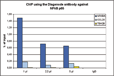
<p><small><strong> Figure 1. ChIP results obtained with the Diagenode antibody directed against NFkB p65. </strong><br />ChIP assays were performed using human HeLa cells, treated with TNFalpha, the Diagenode antibody against NFkB p65 (Cat. No. C15310256) and optimized PCR primer sets for qPCR. ChIP was performed with the “iDeal ChIP-seq” kit (Cat. No. C01010055), using sheared chromatin from 4 million cells. A titration of the antibody consisting of 1, 2.5 and 5 µl per ChIP experiment was analysed. IgG (1 µg/IP) was used as negative IP control. QPCR was performed with primers for the NFKBIA and CCL20 genes, used as positive controls, and for TSH2B, used as negative control. Figure 1 shows the recovery, expressed as a % of input (the relative amount of immunoprecipitated DNA compared to input DNA after qPCR analysis). </small></p>
</div>
</div>
<p></p>
<p></p>
<p></p>
<p></p>
<p></p>
<div class="row">
<div class="small-6 columns">
<p><img src="https://www.diagenode.com/img/product/antibodies/C15310256-ChIPseq-figA.png" alt="NFKB p65 Antibody ChIP-seq Grade" caption="false" width="447" height="53" /></p>
<p><img src="https://www.diagenode.com/img/product/antibodies/C15310256-ChIPseq-figB.png" alt="NFKB p65 Antibody for ChIP-seq " caption="false" width="447" height="92" /></p>
<p><img src="https://www.diagenode.com/img/product/antibodies/C15310256-ChIPseq-figC.png" alt="NFKB p65 Antibody for ChIP-seq assay" caption="false" width="447" height="53" /></p>
<p><img src="https://www.diagenode.com/img/product/antibodies/C15310256-ChIPseq-figD.png" alt="NFKB p65 Antibody validated in ChIP-seq " caption="false" width="447" height="54" /></p>
</div>
<div class="small-6 columns">
<p><small><strong> Figure 2. ChIP-seq results obtained with the Diagenode antibody directed against NFkB p65 </strong><br />ChIP was performed on sheared chromatin from 4 million HeLa cells using 1 µg of the Diagenode antibody against NFkB p65 (Cat. No. C15310256) as described above. The IP’d DNA was subsequently analysed on an Illumina HiSeq. Library preparation, cluster generation and sequencing were performed according to the manufacturer’s instructions. The 51 bp tags were aligned to the human genome using the BWA algorithm. Figure 2 shows the enrichment along the complete sequence and a 2 Mb region of human chromosome 2 (fig 2A and B), and in a two genomic regions surrounding the NFKBIA and CCL20 positive control genes. </small></p>
</div>
</div>
<p></p>
<p></p>
<p></p>
<p></p>
<p></p>
<p></p>
<p></p>
<p></p>
<p></p>
<p></p>
<p></p>
<p></p>
<p></p>
<p></p>
<p></p>
<div class="row">
<div class="small-4 columns">
<p><img src="https://www.diagenode.com/img/product/antibodies/C15310256-WB.png" alt="NFKB p65 Antibody validated in Western Blot" width="147" height="262" caption="false" /></p>
</div>
<div class="small-8 columns">
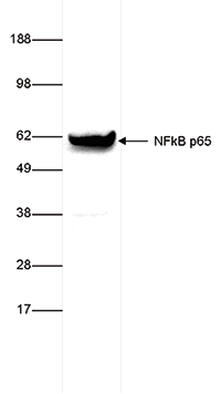
<p><small><strong>Figure 3. NFKB p65 antibody western blot results </strong><br />Whole cell extracts from HeLa cells (35 µg) were analysed by Western blot using the Diagenode antibody against NFkB p65 (Cat. No. C15310256) diluted 1:5,000. The position of the protein of interest is indicated on the right (expected size: 65 kDa); the marker (in kDa) is shown on the left. </small></p>
</div>
</div>
<p></p>
<p></p>
<div class="row">
<div class="small-4 columns">
<p><img src="https://www.diagenode.com/img/product/antibodies/C15310256-IC.png" alt="NFKB p65 Antibody validated in Immunohistochemistry" caption="false" width="266" height="198" /></p>
</div>
<div class="small-8 columns">
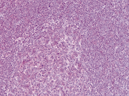
<p><small><strong> Figure 4.</strong> NFKB p65 antibody Immunohistochemistry results Formalin fixed paraffin embedded lymphocytes and germinal center cells of the tonsil were stained with the Diagenode antibody against NFkB p65 (Cat. No. C15310256) diluted 1:400 followed by a peroxidase labelled goat anti-rabbit secondary antibody. Figure 4 shows moderate positive nuclear or cytoplasmic staining. </small></p>
</div>
</div>',
'label2' => '',
'info2' => '<p>This antibody recognizes NFKB p65 which is a component of NFKB. NFKB was originally identified as a factor that binds to the immunoglobulin kappa light chain enhancer in B cells. It was subsequently found in non-B cells in an inactive cytoplasmic form consisting of NFkappaB bound to IkappaB. NFkappaB was originally identified as a heterodimeric DNA binding protein complex consisting of p65 (RelA) and p50 (NFKB1) subunits. Other identified subunits include p52 (NFKB2), c-Rel, and RelB. The p65, cRel, and RelB subunits are responsible for transactivation. The p50 and p52 subunits possess DNA binding activity but limited ability to transactivate. p52 has been reported to form transcriptionally active heterodimers with the NFkappaB subunit p65, similar to p50/p65 heterodimers. The heterodimers of p52/p65 and p50/p65 are regulated by physical inactivation in the cytoplasm by IkB-a. IkB-a binds to the p65 subunit, preventing nuclear localization and DNA binding. Low levels of p52 and p50 homodimers can also exist in cells.</p>',
'label3' => '',
'info3' => '',
'format' => '100 μl',
'catalog_number' => 'C15310256',
'old_catalog_number' => '',
'sf_code' => 'C15310256-D001-001161',
'type' => 'FRE',
'search_order' => '03-Antibody',
'price_EUR' => '445',
'price_USD' => '460',
'price_GBP' => '405',
'price_JPY' => '72915',
'price_CNY' => '',
'price_AUD' => '1150',
'country' => 'ALL',
'except_countries' => 'Japan',
'quote' => false,
'in_stock' => false,
'featured' => false,
'no_promo' => false,
'online' => true,
'master' => true,
'last_datasheet_update' => '0000-00-00',
'slug' => 'nfkb-p65-polyclonal-antibody-classic-100-ml',
'meta_title' => 'NFKB p65 polyclonal antibody - Classic',
'meta_keywords' => '',
'meta_description' => 'NFKB p65 polyclonal antibody - Classic',
'modified' => '2021-12-23 12:26:13',
'created' => '2015-06-29 14:08:20',
'locale' => 'jpn'
),
'Antibody' => array(
'host' => '*****',
'id' => '403',
'name' => 'NFKB p65 polyclonal antibody - Classic',
'description' => 'This antibody recognizes NFKB p65 which is a component of NFKB. NFKB was originally identified as a factor that binds to the immunoglobulin kappa light chain enhancer in B cells. It was subsequently found in non-B cells in an inactive cytoplasmic form consisting of NFkappaB bound to IkappaB. NFkappaB was originally identified as a heterodimeric DNA binding protein complex consisting of p65 (RelA) and p50 (NFKB1) subunits. Other identified subunits include p52 (NFKB2), c-Rel, and RelB. The p65, cRel, and RelB subunits are responsible for transactivation. The p50 and p52 subunits possess DNA binding activity but limited ability to transactivate. p52 has been reported to form transcriptionally active heterodimers with the NFkappaB subunit p65, similar to p50/p65 heterodimers. The heterodimers of p52/p65 and p50/p65 are regulated by physical inactivation in the cytoplasm by IkB-a. IkB-a binds to the p65 subunit, preventing nuclear localization and DNA binding. Low levels of p52 and p50 homodimers can also exist in cells.',
'clonality' => '',
'isotype' => '',
'lot' => '24468',
'concentration' => 'not determined',
'reactivity' => 'Human, mouse, rat',
'type' => 'Polyclonal',
'purity' => 'Whole antiserum',
'classification' => '',
'application_table' => '<table>
<thead>
<tr>
<th>Applications</th>
<th>Suggested dilution</th>
<th>References</th>
</tr>
</thead>
<tbody>
<tr>
<td>ChIP/ChIP-seq <sup>*</sup></td>
<td>1 µl/IP</td>
<td>Fig 1, 2</td>
</tr>
<tr>
<td>Western Blotting</td>
<td>1:2,000 - 1:5,000</td>
<td>Fig 3</td>
</tr>
<tr>
<td>ELISA</td>
<td>1:5,000</td>
<td></td>
</tr>
<tr>
<td>Immunohistochemistry</td>
<td>1:500 - 1:2,000</td>
<td>Fig 4</td>
</tr>
<tr>
<td>Gel Shift</td>
<td>1:500</td>
<td></td>
</tr>
</tbody>
</table>
<p><small><sup>*</sup> Please note that the optimal antibody amount per IP should be determined by the end-user. We recommend testing 1-5 µg per IP.</small></p>',
'storage_conditions' => '',
'storage_buffer' => '',
'precautions' => 'This product is for research use only. Not for use in diagnostic or therapeutic procedures.',
'uniprot_acc' => '',
'slug' => '',
'meta_keywords' => '',
'meta_description' => '',
'modified' => '2019-09-09 16:57:03',
'created' => '2015-07-27 09:55:56',
'select_label' => '403 - NFKB p65 polyclonal antibody - Classic (24468 - not determined - Human, mouse, rat - Whole antiserum - Rabbit)'
),
'Slave' => array(
(int) 0 => array(
[maximum depth reached]
),
(int) 1 => array(
[maximum depth reached]
)
),
'Group' => array(
'Group' => array(
[maximum depth reached]
),
'Master' => array(
[maximum depth reached]
),
'Product' => array([maximum depth reached])
),
'Related' => array(),
'Application' => array(
(int) 0 => array(
[maximum depth reached]
),
(int) 1 => array(
[maximum depth reached]
),
(int) 2 => array(
[maximum depth reached]
),
(int) 3 => array(
[maximum depth reached]
),
(int) 4 => array(
[maximum depth reached]
)
),
'Category' => array(
(int) 0 => array(
[maximum depth reached]
),
(int) 1 => array(
[maximum depth reached]
),
(int) 2 => array(
[maximum depth reached]
),
(int) 3 => array(
[maximum depth reached]
)
),
'Document' => array(
(int) 0 => array(
[maximum depth reached]
),
(int) 1 => array(
[maximum depth reached]
),
(int) 2 => array(
[maximum depth reached]
)
),
'Feature' => array(),
'Image' => array(
(int) 0 => array(
[maximum depth reached]
)
),
'Promotion' => array(),
'Protocol' => array(),
'Publication' => array(
(int) 0 => array(
[maximum depth reached]
),
(int) 1 => array(
[maximum depth reached]
),
(int) 2 => array(
[maximum depth reached]
),
(int) 3 => array(
[maximum depth reached]
),
(int) 4 => array(
[maximum depth reached]
),
(int) 5 => array(
[maximum depth reached]
),
(int) 6 => array(
[maximum depth reached]
)
),
'Testimonial' => array(),
'Area' => array(),
'SafetySheet' => array(
(int) 0 => array(
[maximum depth reached]
),
(int) 1 => array(
[maximum depth reached]
),
(int) 2 => array(
[maximum depth reached]
),
(int) 3 => array(
[maximum depth reached]
),
(int) 4 => array(
[maximum depth reached]
),
(int) 5 => array(
[maximum depth reached]
),
(int) 6 => array(
[maximum depth reached]
),
(int) 7 => array(
[maximum depth reached]
)
)
),
'meta_canonical' => 'https://www.diagenode.com/jp/p/nfkb-p65-polyclonal-antibody-classic-100-ml'
)
$language = 'jp'
$meta_keywords = ''
$meta_description = 'NFKB p65 polyclonal antibody - Classic'
$meta_title = 'NFKB p65 polyclonal antibody - Classic'
$product = array(
'Product' => array(
'id' => '2167',
'antibody_id' => '403',
'name' => 'NFKB p65 polyclonal antibody ',
'description' => '<p><span>Alternative names: <strong>RELA</strong>, <strong>NFKB3</strong>, <strong>p65</strong></span></p>
<p><span>Polyclonal antibody raised in rabbit against NFKB p65 (Rel A), using a KLH-conjugated synthetic peptide.</span></p>',
'label1' => 'Validation Data',
'info1' => '<div class="row">
<div class="small-4 columns">
<p><img src="https://www.diagenode.com/img/product/antibodies/C15310256-ChIP.png" alt="NFKB p65 Antibody ChIP Grade" caption="false" width="288" height="191" /></p>
</div>
<div class="small-8 columns">
<p><small><strong> Figure 1. ChIP results obtained with the Diagenode antibody directed against NFkB p65. </strong><br />ChIP assays were performed using human HeLa cells, treated with TNFalpha, the Diagenode antibody against NFkB p65 (Cat. No. C15310256) and optimized PCR primer sets for qPCR. ChIP was performed with the “iDeal ChIP-seq” kit (Cat. No. C01010055), using sheared chromatin from 4 million cells. A titration of the antibody consisting of 1, 2.5 and 5 µl per ChIP experiment was analysed. IgG (1 µg/IP) was used as negative IP control. QPCR was performed with primers for the NFKBIA and CCL20 genes, used as positive controls, and for TSH2B, used as negative control. Figure 1 shows the recovery, expressed as a % of input (the relative amount of immunoprecipitated DNA compared to input DNA after qPCR analysis). </small></p>
</div>
</div>
<p></p>
<p></p>
<p></p>
<p></p>
<p></p>
<div class="row">
<div class="small-6 columns">
<p><img src="https://www.diagenode.com/img/product/antibodies/C15310256-ChIPseq-figA.png" alt="NFKB p65 Antibody ChIP-seq Grade" caption="false" width="447" height="53" /></p>
<p><img src="https://www.diagenode.com/img/product/antibodies/C15310256-ChIPseq-figB.png" alt="NFKB p65 Antibody for ChIP-seq " caption="false" width="447" height="92" /></p>
<p><img src="https://www.diagenode.com/img/product/antibodies/C15310256-ChIPseq-figC.png" alt="NFKB p65 Antibody for ChIP-seq assay" caption="false" width="447" height="53" /></p>
<p><img src="https://www.diagenode.com/img/product/antibodies/C15310256-ChIPseq-figD.png" alt="NFKB p65 Antibody validated in ChIP-seq " caption="false" width="447" height="54" /></p>
</div>
<div class="small-6 columns">
<p><small><strong> Figure 2. ChIP-seq results obtained with the Diagenode antibody directed against NFkB p65 </strong><br />ChIP was performed on sheared chromatin from 4 million HeLa cells using 1 µg of the Diagenode antibody against NFkB p65 (Cat. No. C15310256) as described above. The IP’d DNA was subsequently analysed on an Illumina HiSeq. Library preparation, cluster generation and sequencing were performed according to the manufacturer’s instructions. The 51 bp tags were aligned to the human genome using the BWA algorithm. Figure 2 shows the enrichment along the complete sequence and a 2 Mb region of human chromosome 2 (fig 2A and B), and in a two genomic regions surrounding the NFKBIA and CCL20 positive control genes. </small></p>
</div>
</div>
<p></p>
<p></p>
<p></p>
<p></p>
<p></p>
<p></p>
<p></p>
<p></p>
<p></p>
<p></p>
<p></p>
<p></p>
<p></p>
<p></p>
<p></p>
<div class="row">
<div class="small-4 columns">
<p><img src="https://www.diagenode.com/img/product/antibodies/C15310256-WB.png" alt="NFKB p65 Antibody validated in Western Blot" width="147" height="262" caption="false" /></p>
</div>
<div class="small-8 columns">
<p><small><strong>Figure 3. NFKB p65 antibody western blot results </strong><br />Whole cell extracts from HeLa cells (35 µg) were analysed by Western blot using the Diagenode antibody against NFkB p65 (Cat. No. C15310256) diluted 1:5,000. The position of the protein of interest is indicated on the right (expected size: 65 kDa); the marker (in kDa) is shown on the left. </small></p>
</div>
</div>
<p></p>
<p></p>
<div class="row">
<div class="small-4 columns">
<p><img src="https://www.diagenode.com/img/product/antibodies/C15310256-IC.png" alt="NFKB p65 Antibody validated in Immunohistochemistry" caption="false" width="266" height="198" /></p>
</div>
<div class="small-8 columns">
<p><small><strong> Figure 4.</strong> NFKB p65 antibody Immunohistochemistry results Formalin fixed paraffin embedded lymphocytes and germinal center cells of the tonsil were stained with the Diagenode antibody against NFkB p65 (Cat. No. C15310256) diluted 1:400 followed by a peroxidase labelled goat anti-rabbit secondary antibody. Figure 4 shows moderate positive nuclear or cytoplasmic staining. </small></p>
</div>
</div>',
'label2' => 'Target Description',
'info2' => '<p>This antibody recognizes NFKB p65 which is a component of NFKB. NFKB was originally identified as a factor that binds to the immunoglobulin kappa light chain enhancer in B cells. It was subsequently found in non-B cells in an inactive cytoplasmic form consisting of NFkappaB bound to IkappaB. NFkappaB was originally identified as a heterodimeric DNA binding protein complex consisting of p65 (RelA) and p50 (NFKB1) subunits. Other identified subunits include p52 (NFKB2), c-Rel, and RelB. The p65, cRel, and RelB subunits are responsible for transactivation. The p50 and p52 subunits possess DNA binding activity but limited ability to transactivate. p52 has been reported to form transcriptionally active heterodimers with the NFkappaB subunit p65, similar to p50/p65 heterodimers. The heterodimers of p52/p65 and p50/p65 are regulated by physical inactivation in the cytoplasm by IkB-a. IkB-a binds to the p65 subunit, preventing nuclear localization and DNA binding. Low levels of p52 and p50 homodimers can also exist in cells.</p>',
'label3' => '',
'info3' => '',
'format' => '100 μl',
'catalog_number' => 'C15310256',
'old_catalog_number' => '',
'sf_code' => 'C15310256-D001-001161',
'type' => 'FRE',
'search_order' => '03-Antibody',
'price_EUR' => '445',
'price_USD' => '460',
'price_GBP' => '405',
'price_JPY' => '72915',
'price_CNY' => '',
'price_AUD' => '1150',
'country' => 'ALL',
'except_countries' => 'Japan',
'quote' => false,
'in_stock' => false,
'featured' => false,
'no_promo' => false,
'online' => true,
'master' => true,
'last_datasheet_update' => '0000-00-00',
'slug' => 'nfkb-p65-polyclonal-antibody-classic-100-ml',
'meta_title' => 'NFKB p65 polyclonal antibody - Classic',
'meta_keywords' => '',
'meta_description' => 'NFKB p65 polyclonal antibody - Classic',
'modified' => '2021-12-23 12:26:13',
'created' => '2015-06-29 14:08:20',
'locale' => 'jpn'
),
'Antibody' => array(
'host' => '*****',
'id' => '403',
'name' => 'NFKB p65 polyclonal antibody - Classic',
'description' => 'This antibody recognizes NFKB p65 which is a component of NFKB. NFKB was originally identified as a factor that binds to the immunoglobulin kappa light chain enhancer in B cells. It was subsequently found in non-B cells in an inactive cytoplasmic form consisting of NFkappaB bound to IkappaB. NFkappaB was originally identified as a heterodimeric DNA binding protein complex consisting of p65 (RelA) and p50 (NFKB1) subunits. Other identified subunits include p52 (NFKB2), c-Rel, and RelB. The p65, cRel, and RelB subunits are responsible for transactivation. The p50 and p52 subunits possess DNA binding activity but limited ability to transactivate. p52 has been reported to form transcriptionally active heterodimers with the NFkappaB subunit p65, similar to p50/p65 heterodimers. The heterodimers of p52/p65 and p50/p65 are regulated by physical inactivation in the cytoplasm by IkB-a. IkB-a binds to the p65 subunit, preventing nuclear localization and DNA binding. Low levels of p52 and p50 homodimers can also exist in cells.',
'clonality' => '',
'isotype' => '',
'lot' => '24468',
'concentration' => 'not determined',
'reactivity' => 'Human, mouse, rat',
'type' => 'Polyclonal',
'purity' => 'Whole antiserum',
'classification' => '',
'application_table' => '<table>
<thead>
<tr>
<th>Applications</th>
<th>Suggested dilution</th>
<th>References</th>
</tr>
</thead>
<tbody>
<tr>
<td>ChIP/ChIP-seq <sup>*</sup></td>
<td>1 µl/IP</td>
<td>Fig 1, 2</td>
</tr>
<tr>
<td>Western Blotting</td>
<td>1:2,000 - 1:5,000</td>
<td>Fig 3</td>
</tr>
<tr>
<td>ELISA</td>
<td>1:5,000</td>
<td></td>
</tr>
<tr>
<td>Immunohistochemistry</td>
<td>1:500 - 1:2,000</td>
<td>Fig 4</td>
</tr>
<tr>
<td>Gel Shift</td>
<td>1:500</td>
<td></td>
</tr>
</tbody>
</table>
<p><small><sup>*</sup> Please note that the optimal antibody amount per IP should be determined by the end-user. We recommend testing 1-5 µg per IP.</small></p>',
'storage_conditions' => '',
'storage_buffer' => '',
'precautions' => 'This product is for research use only. Not for use in diagnostic or therapeutic procedures.',
'uniprot_acc' => '',
'slug' => '',
'meta_keywords' => '',
'meta_description' => '',
'modified' => '2019-09-09 16:57:03',
'created' => '2015-07-27 09:55:56',
'select_label' => '403 - NFKB p65 polyclonal antibody - Classic (24468 - not determined - Human, mouse, rat - Whole antiserum - Rabbit)'
),
'Slave' => array(
(int) 0 => array(
'id' => '256',
'name' => 'C15310256',
'product_id' => '2167',
'modified' => '2019-02-13 14:30:12',
'created' => '2019-02-13 14:30:12'
),
(int) 1 => array(
'id' => '352',
'name' => 'C15310256',
'product_id' => '2167',
'modified' => '2022-03-17 12:29:00',
'created' => '2022-03-17 12:29:00'
)
),
'Group' => array(
'Group' => array(
'id' => '256',
'name' => 'C15310256',
'product_id' => '2167',
'modified' => '2019-02-13 14:30:12',
'created' => '2019-02-13 14:30:12'
),
'Master' => array(
'id' => '2167',
'antibody_id' => '403',
'name' => 'NFKB p65 Antibody',
'description' => '<p><span>Alternative names: <strong>RELA</strong>, <strong>NFKB3</strong>, <strong>p65</strong></span></p>
<p><span>Polyclonal antibody raised in rabbit against <strong>NFKB p65 (Rel A)</strong>, using a KLH-conjugated synthetic peptide.</span></p>',
'label1' => 'Validation Data',
'info1' => '<div class="row">
<div class="small-4 columns">
<p><img src="https://www.diagenode.com/img/product/antibodies/C15310256-ChIP.png" alt="NFKB p65 Antibody ChIP Grade" caption="false" width="288" height="191" /></p>
</div>
<div class="small-8 columns">
<p><small><strong> Figure 1. ChIP results obtained with the Diagenode antibody directed against NFkB p65. </strong><br />ChIP assays were performed using human HeLa cells, treated with TNFalpha, the Diagenode antibody against NFkB p65 (Cat. No. C15310256) and optimized PCR primer sets for qPCR. ChIP was performed with the “iDeal ChIP-seq” kit (Cat. No. C01010055), using sheared chromatin from 4 million cells. A titration of the antibody consisting of 1, 2.5 and 5 µl per ChIP experiment was analysed. IgG (1 µg/IP) was used as negative IP control. QPCR was performed with primers for the NFKBIA and CCL20 genes, used as positive controls, and for TSH2B, used as negative control. Figure 1 shows the recovery, expressed as a % of input (the relative amount of immunoprecipitated DNA compared to input DNA after qPCR analysis). </small></p>
</div>
</div>
<p></p>
<p></p>
<p></p>
<p></p>
<p></p>
<div class="row">
<div class="small-6 columns">
<p><img src="https://www.diagenode.com/img/product/antibodies/C15310256-ChIPseq-figA.png" alt="NFKB p65 Antibody ChIP-seq Grade" caption="false" width="447" height="53" /></p>
<p><img src="https://www.diagenode.com/img/product/antibodies/C15310256-ChIPseq-figB.png" alt="NFKB p65 Antibody for ChIP-seq " caption="false" width="447" height="92" /></p>
<p><img src="https://www.diagenode.com/img/product/antibodies/C15310256-ChIPseq-figC.png" alt="NFKB p65 Antibody for ChIP-seq assay" caption="false" width="447" height="53" /></p>
<p><img src="https://www.diagenode.com/img/product/antibodies/C15310256-ChIPseq-figD.png" alt="NFKB p65 Antibody validated in ChIP-seq " caption="false" width="447" height="54" /></p>
</div>
<div class="small-6 columns">
<p><small><strong> Figure 2. ChIP-seq results obtained with the Diagenode antibody directed against NFkB p65 </strong><br />ChIP was performed on sheared chromatin from 4 million HeLa cells using 1 µg of the Diagenode antibody against NFkB p65 (Cat. No. C15310256) as described above. The IP’d DNA was subsequently analysed on an Illumina HiSeq. Library preparation, cluster generation and sequencing were performed according to the manufacturer’s instructions. The 51 bp tags were aligned to the human genome using the BWA algorithm. Figure 2 shows the enrichment along the complete sequence and a 2 Mb region of human chromosome 2 (fig 2A and B), and in a two genomic regions surrounding the NFKBIA and CCL20 positive control genes. </small></p>
</div>
</div>
<p></p>
<p></p>
<p></p>
<p></p>
<p></p>
<p></p>
<p></p>
<p></p>
<p></p>
<p></p>
<p></p>
<p></p>
<p></p>
<p></p>
<p></p>
<div class="row">
<div class="small-4 columns">
<p><img src="https://www.diagenode.com/img/product/antibodies/C15310256-WB.png" alt="NFKB p65 Antibody validated in Western Blot" width="147" height="262" caption="false" /></p>
</div>
<div class="small-8 columns">
<p><small><strong>Figure 3. NFKB p65 antibody western blot results </strong><br />Whole cell extracts from HeLa cells (35 µg) were analysed by Western blot using the Diagenode antibody against NFkB p65 (Cat. No. C15310256) diluted 1:5,000. The position of the protein of interest is indicated on the right (expected size: 65 kDa); the marker (in kDa) is shown on the left. </small></p>
</div>
</div>
<p></p>
<p></p>
<div class="row">
<div class="small-4 columns">
<p><img src="https://www.diagenode.com/img/product/antibodies/C15310256-IC.png" alt="NFKB p65 Antibody validated in Immunohistochemistry" caption="false" width="266" height="198" /></p>
</div>
<div class="small-8 columns">
<p><small><strong> Figure 4.</strong> NFKB p65 antibody Immunohistochemistry results Formalin fixed paraffin embedded lymphocytes and germinal center cells of the tonsil were stained with the Diagenode antibody against NFkB p65 (Cat. No. C15310256) diluted 1:400 followed by a peroxidase labelled goat anti-rabbit secondary antibody. Figure 4 shows moderate positive nuclear or cytoplasmic staining. </small></p>
</div>
</div>',
'label2' => 'Target Description',
'info2' => '<p>This antibody recognizes NFKB p65 which is a component of NFKB. NFKB was originally identified as a factor that binds to the immunoglobulin kappa light chain enhancer in B cells. It was subsequently found in non-B cells in an inactive cytoplasmic form consisting of NFkappaB bound to IkappaB. NFkappaB was originally identified as a heterodimeric DNA binding protein complex consisting of p65 (RelA) and p50 (NFKB1) subunits. Other identified subunits include p52 (NFKB2), c-Rel, and RelB. The p65, cRel, and RelB subunits are responsible for transactivation. The p50 and p52 subunits possess DNA binding activity but limited ability to transactivate. p52 has been reported to form transcriptionally active heterodimers with the NFkappaB subunit p65, similar to p50/p65 heterodimers. The heterodimers of p52/p65 and p50/p65 are regulated by physical inactivation in the cytoplasm by IkB-a. IkB-a binds to the p65 subunit, preventing nuclear localization and DNA binding. Low levels of p52 and p50 homodimers can also exist in cells.</p>',
'label3' => '',
'info3' => '',
'format' => '100 μl',
'catalog_number' => 'C15310256',
'old_catalog_number' => '',
'sf_code' => 'C15310256-D001-001161',
'type' => 'FRE',
'search_order' => '03-Antibody',
'price_EUR' => '445',
'price_USD' => '460',
'price_GBP' => '405',
'price_JPY' => '72915',
'price_CNY' => '',
'price_AUD' => '1150',
'country' => 'ALL',
'except_countries' => 'Japan',
'quote' => false,
'in_stock' => false,
'featured' => false,
'no_promo' => false,
'online' => true,
'master' => true,
'last_datasheet_update' => '0000-00-00',
'slug' => 'nfkb-p65-polyclonal-antibody-classic-100-ml',
'meta_title' => 'NFKB p65 Antibody - ChIP-seq Grade (C15310256) | Diagenode',
'meta_keywords' => '',
'meta_description' => 'NFKB p65 Polyclonal Antibody validated in ChIP-seq, ChIP-qPCR, ELISA, WB and IHC. Alternative names: RELA, NFKB3, p65. Sample size available.',
'modified' => '2021-12-23 12:26:13',
'created' => '2015-06-29 14:08:20'
),
'Product' => array()
),
'Related' => array(),
'Application' => array(
(int) 0 => array(
'id' => '20',
'position' => '10',
'parent_id' => '40',
'name' => 'ELISA',
'description' => '<div class="row">
<div class="small-12 medium-12 large-12 columns">Enzyme-linked immunosorbent assay.</div>
</div>',
'in_footer' => false,
'in_menu' => false,
'online' => true,
'tabular' => true,
'slug' => 'elisa-antibodies',
'meta_keywords' => ' ELISA Antibodies,Monoclonal antibody, Polyclonal antibody',
'meta_description' => 'Diagenode offers Monoclonal & Polyclonal antibodies for ELISA applications',
'meta_title' => 'ELISA Antibodies - Monoclonal & Polyclonal antibody | Diagenode',
'modified' => '2016-01-13 12:21:41',
'created' => '2014-07-08 08:13:28',
'ProductsApplication' => array(
[maximum depth reached]
)
),
(int) 1 => array(
'id' => '32',
'position' => '10',
'parent_id' => '40',
'name' => 'IHC',
'description' => '<p>Immunohistochemistry</p>',
'in_footer' => false,
'in_menu' => false,
'online' => true,
'tabular' => true,
'slug' => 'immunohistochemistry',
'meta_keywords' => 'immunohistochemistry,Monoclonal antibody,Polyclonal antibody',
'meta_description' => 'Diagenode offers a wide range of antibodies and technical support for immunohistochemistry applications',
'meta_title' => 'Immunohistochemistry - Monoclonal antibody - Polyclonal antibody | Diagenode',
'modified' => '2016-01-13 12:23:53',
'created' => '2015-07-08 13:49:03',
'ProductsApplication' => array(
[maximum depth reached]
)
),
(int) 2 => array(
'id' => '19',
'position' => '10',
'parent_id' => '40',
'name' => 'WB',
'description' => '<p><strong>Western blot</strong> : The quality of antibodies used in this technique is crucial for correct and specific protein identification. Diagenode offers huge selection of highly sensitive and specific western blot-validated antibodies.</p>
<p>Learn more about: <a href="https://www.diagenode.com/applications/western-blot">Loading control, MW marker visualization</a><em>. <br /></em></p>
<p><em></em>Check our selection of antibodies validated in Western blot.</p>',
'in_footer' => false,
'in_menu' => false,
'online' => true,
'tabular' => true,
'slug' => 'western-blot-antibodies',
'meta_keywords' => ' Western Blot Antibodies ,western blot protocol,Western Blotting Products,Polyclonal antibodies ,monoclonal antibodies ',
'meta_description' => 'Diagenode offers a wide range of antibodies and technical support for western blot applications',
'meta_title' => ' Western Blot - Monoclonal antibody - Polyclonal antibody | Diagenode',
'modified' => '2016-04-26 12:44:51',
'created' => '2015-01-07 09:20:00',
'ProductsApplication' => array(
[maximum depth reached]
)
),
(int) 3 => array(
'id' => '42',
'position' => '10',
'parent_id' => '40',
'name' => 'ChIP-seq (ab)',
'description' => '',
'in_footer' => false,
'in_menu' => false,
'online' => true,
'tabular' => true,
'slug' => 'chip-seq-antibodies',
'meta_keywords' => 'Chromatin Immunoprecipitation Sequencing,ChIP-Seq,ChIP-seq grade antibodies,DNA purification,qPCR,Shearing of chromatin',
'meta_description' => 'Diagenode offers a wide range of antibodies and technical support for ChIP Sequencing applications',
'meta_title' => 'ChIP Sequencing Antibodies (ChIP-Seq) | Diagenode',
'modified' => '2016-01-20 11:06:19',
'created' => '2015-10-20 11:44:45',
'ProductsApplication' => array(
[maximum depth reached]
)
),
(int) 4 => array(
'id' => '43',
'position' => '10',
'parent_id' => '40',
'name' => 'ChIP-qPCR (ab)',
'description' => '',
'in_footer' => false,
'in_menu' => false,
'online' => true,
'tabular' => true,
'slug' => 'chip-qpcr-antibodies',
'meta_keywords' => 'Chromatin Immunoprecipitation Sequencing,ChIP-Seq,ChIP-seq grade antibodies,DNA purification,qPCR,Shearing of chromatin',
'meta_description' => 'Diagenode offers a wide range of antibodies and technical support for ChIP-qPCR applications',
'meta_title' => 'ChIP Quantitative PCR Antibodies (ChIP-qPCR) | Diagenode',
'modified' => '2016-01-20 11:30:24',
'created' => '2015-10-20 11:45:36',
'ProductsApplication' => array(
[maximum depth reached]
)
)
),
'Category' => array(
(int) 0 => array(
'id' => '30',
'position' => '50',
'parent_id' => '4',
'name' => 'Transcription',
'description' => '<p><span style="font-weight: 400;">The list of Diagenode’s highly specific antibodies for transcription studies includes the antibodies against many transcription factors and nuclear receptors. Check the list below to see our targets.</span></p>
<p><span style="font-weight: 400;">Diagenode’s highly validated antibodies:</span></p>
<ul>
<li><span style="font-weight: 400;"> Highly sensitive and specific</span></li>
<li><span style="font-weight: 400;"> Cost-effective (requires less antibody per reaction)</span></li>
<li><span style="font-weight: 400;"> Batch-specific data is available on the website</span></li>
<li><span style="font-weight: 400;"> Expert technical support</span></li>
<li><span style="font-weight: 400;"> Sample sizes available</span></li>
<li><span style="font-weight: 400;"> 100% satisfaction guarantee</span></li>
</ul>',
'no_promo' => false,
'in_menu' => false,
'online' => true,
'tabular' => false,
'hide' => true,
'all_format' => false,
'is_antibody' => true,
'slug' => 'transcription-factor',
'cookies_tag_id' => null,
'meta_keywords' => ' Transcription factor antibodies,monoclonal antibodies,polyclonal antibodies',
'meta_description' => 'Diagenode offers polyclonal and monoclonal antibodies for Transcription studie',
'meta_title' => 'Transcription factor Antibodies | Diagenode',
'modified' => '2020-07-06 12:59:19',
'created' => '2015-03-12 10:20:08',
'ProductsCategory' => array(
[maximum depth reached]
),
'CookiesTag' => array([maximum depth reached])
),
(int) 1 => array(
'id' => '17',
'position' => '10',
'parent_id' => '4',
'name' => 'ChIP-seq grade antibodies',
'description' => '<p><b>Unparalleled ChIP-Seq results with the most rigorously validated antibodies</b></p>
<p><span style="font-weight: 400;">Diagenode provides leading solutions for epigenetic research. Because ChIP-seq is a widely-used technique, we validate our antibodies in ChIP and ChIP-seq experiments (in addition to conventional methods like Western blot, Dot blot, ELISA, and immunofluorescence) to provide the highest quality antibody. We standardize our validation and production to guarantee high product quality without technical bias. Diagenode guarantees ChIP-seq grade antibody performance under our suggested conditions.</span></p>
<div class="row">
<div class="small-12 medium-9 large-9 columns">
<p><strong>ChIP-seq profile</strong> of active (H3K4me3 and H3K36me3) and inactive (H3K27me3) marks using Diagenode antibodies.</p>
<img src="https://www.diagenode.com/img/categories/antibodies/chip-seq-grade-antibodies.png" /></div>
<div class="small-12 medium-3 large-3 columns">
<p><small> ChIP was performed on sheared chromatin from 100,000 K562 cells using iDeal ChIP-seq kit for Histones (cat. No. C01010051) with 1 µg of the Diagenode antibodies against H3K27me3 (cat. No. C15410195) and H3K4me3 (cat. No. C15410003), and 0.5 µg of the antibody against H3K36me3 (cat. No. C15410192). The IP'd DNA was subsequently analysed on an Illumina Genome Analyzer. Library preparation, cluster generation and sequencing were performed according to the manufacturer's instructions. The 36 bp tags were aligned to the human genome using the ELAND algorithm. The figure shows the signal distribution along the complete sequence of human chromosome 3, a zoomin to a 10 Mb region and a further zoomin to a 1.5 Mb region. </small></p>
</div>
</div>
<p>Diagenode’s highly validated antibodies:</p>
<ul>
<li>Highly sensitive and specific</li>
<li>Cost-effective (requires less antibody per reaction)</li>
<li>Batch-specific data is available on the website</li>
<li>Expert technical support</li>
<li>Sample sizes available</li>
<li>100% satisfaction guarantee</li>
</ul>',
'no_promo' => false,
'in_menu' => true,
'online' => true,
'tabular' => false,
'hide' => true,
'all_format' => false,
'is_antibody' => true,
'slug' => 'chip-seq-grade-antibodies',
'cookies_tag_id' => null,
'meta_keywords' => 'ChIP-seq grade antibodies,polyclonal antibody,WB, ELISA, ChIP-seq (ab), ChIP-qPCR (ab)',
'meta_description' => 'Diagenode Offers Wide Range of Validated ChIP-Seq Grade Antibodies for Unparalleled ChIP-Seq Results',
'meta_title' => 'Chromatin Immunoprecipitation ChIP-Seq Grade Antibodies | Diagenode',
'modified' => '2019-07-03 10:57:22',
'created' => '2015-02-16 02:24:01',
'ProductsCategory' => array(
[maximum depth reached]
),
'CookiesTag' => array([maximum depth reached])
),
(int) 2 => array(
'id' => '103',
'position' => '0',
'parent_id' => '4',
'name' => 'All antibodies',
'description' => '<p><span style="font-weight: 400;">All Diagenode’s antibodies are listed below. Please, use our Quick search field to find the antibody of interest by target name, application, purity.</span></p>
<p><span style="font-weight: 400;">Diagenode’s highly validated antibodies:</span></p>
<ul>
<li>Highly sensitive and specific</li>
<li>Cost-effective (requires less antibody per reaction)</li>
<li>Batch-specific data is available on the website</li>
<li>Expert technical support</li>
<li>Sample sizes available</li>
<li>100% satisfaction guarantee</li>
</ul>',
'no_promo' => false,
'in_menu' => true,
'online' => true,
'tabular' => false,
'hide' => true,
'all_format' => false,
'is_antibody' => true,
'slug' => 'all-antibodies',
'cookies_tag_id' => null,
'meta_keywords' => 'Antibodies,Premium Antibodies,Classic,Pioneer',
'meta_description' => 'Diagenode Offers Strict quality standards with Rigorous QC and validated Antibodies. Classified based on level of validation for flexibility of Application. Comprehensive selection of histone and non-histone Antibodies',
'meta_title' => 'Diagenode's selection of Antibodies is exclusively dedicated for Epigenetic Research | Diagenode',
'modified' => '2019-07-03 10:55:44',
'created' => '2015-11-02 14:49:22',
'ProductsCategory' => array(
[maximum depth reached]
),
'CookiesTag' => array([maximum depth reached])
),
(int) 3 => array(
'id' => '127',
'position' => '10',
'parent_id' => '4',
'name' => 'ChIP-grade antibodies',
'description' => '<div class="row">
<div class="small-12 columns"><center></center>
<p><br />Chromatin immunoprecipitation (<b>ChIP</b>) is a technique to study the associations of proteins with the specific genomic regions in intact cells. One of the most important steps of this protocol is the immunoprecipitation of targeted protein using the antibody specifically recognizing it. The quality of antibodies used in ChIP is essential for the success of the experiment. Diagenode offers extensively validated ChIP-grade antibodies, confirmed for their specificity, and high level of performance in ChIP. Each batch is validated, and batch-specific data are available on the website.</p>
<p></p>
</div>
</div>
<p><strong>ChIP results</strong> obtained with the antibody directed against H3K4me3 (Cat. No. <a href="../p/h3k4me3-polyclonal-antibody-premium-50-ug-50-ul">C15410003</a>). </p>
<div class="row">
<div class="small-12 medium-6 large-6 columns"><img src="https://www.diagenode.com/img/product/antibodies/C15410003-fig1-ChIP.jpg" alt="" width="400" height="315" /> </div>
<div class="small-12 medium-6 large-6 columns">
<p></p>
<p></p>
<p></p>
</div>
</div>
<p></p>
<p>Our aim at Diagenode is to offer the largest collection of highly specific <strong>ChIP-grade antibodies</strong>. We add new antibodies monthly. Find your ChIP-grade antibody in the list below and check more information about tested applications, extensive validation data, and product information.</p>',
'no_promo' => false,
'in_menu' => true,
'online' => true,
'tabular' => false,
'hide' => true,
'all_format' => false,
'is_antibody' => true,
'slug' => 'chip-grade-antibodies',
'cookies_tag_id' => null,
'meta_keywords' => 'ChIP-grade antibodies, polyclonal antibody, monoclonal antibody, Diagenode',
'meta_description' => 'Diagenode Offers Extensively Validated ChIP-Grade Antibodies, Confirmed for their Specificity, and high level of Performance in Chromatin Immunoprecipitation ChIP',
'meta_title' => 'Chromatin immunoprecipitation ChIP-grade antibodies | Diagenode',
'modified' => '2024-11-19 17:27:07',
'created' => '2017-02-14 11:16:04',
'ProductsCategory' => array(
[maximum depth reached]
),
'CookiesTag' => array([maximum depth reached])
)
),
'Document' => array(
(int) 0 => array(
'id' => '398',
'name' => 'Datasheet NFKBp65 C15310256',
'description' => 'Datasheet description',
'image_id' => null,
'type' => 'Datasheet',
'url' => 'files/products/antibodies/Datasheet_NFKBp65_C15310256.pdf',
'slug' => 'datasheet-nfkbp65-c15310256',
'meta_keywords' => null,
'meta_description' => null,
'modified' => '2015-07-07 11:47:44',
'created' => '2015-07-07 11:47:44',
'ProductsDocument' => array(
[maximum depth reached]
)
),
(int) 1 => array(
'id' => '11',
'name' => 'Antibodies you can trust',
'description' => '<p style="text-align: justify;"><span>Epigenetic research tools have evolved over time from endpoint PCR to qPCR to the analyses of large sets of genome-wide sequencing data. ChIP sequencing (ChIP-seq) has now become the gold standard method for chromatin studies, given the accuracy and coverage scale of the approach over other methods. Successful ChIP-seq, however, requires a higher level of experimental accuracy and consistency in all steps of ChIP than ever before. Particularly crucial is the quality of ChIP antibodies. </span></p>',
'image_id' => null,
'type' => 'Poster',
'url' => 'files/posters/Antibodies_you_can_trust_Poster.pdf',
'slug' => 'antibodies-you-can-trust-poster',
'meta_keywords' => '',
'meta_description' => '',
'modified' => '2015-10-01 20:18:31',
'created' => '2015-07-03 16:05:15',
'ProductsDocument' => array(
[maximum depth reached]
)
),
(int) 2 => array(
'id' => '38',
'name' => 'Epigenetic Antibodies Brochure',
'description' => '<p>More than in any other immuoprecipitation assays, quality antibodies are critical tools in many epigenetics experiments. Since 10 years, Diagenode has developed the most stringent quality production available on the market for antibodies exclusively focused on epigenetic uses. All our antibodies have been qualified to work in epigenetic applications.</p>',
'image_id' => null,
'type' => 'Brochure',
'url' => 'files/brochures/Epigenetic_Antibodies_Brochure.pdf',
'slug' => 'epigenetic-antibodies-brochure',
'meta_keywords' => '',
'meta_description' => '',
'modified' => '2016-06-15 11:24:06',
'created' => '2015-07-03 16:05:27',
'ProductsDocument' => array(
[maximum depth reached]
)
)
),
'Feature' => array(),
'Image' => array(
(int) 0 => array(
'id' => '1783',
'name' => 'product/antibodies/chipseq-grade-ab-icon.png',
'alt' => 'ChIP-seq Grade',
'modified' => '2020-11-27 07:04:40',
'created' => '2018-03-15 15:54:09',
'ProductsImage' => array(
[maximum depth reached]
)
)
),
'Promotion' => array(),
'Protocol' => array(),
'Publication' => array(
(int) 0 => array(
'id' => '5021',
'name' => 'Peroxiredoxin 1-Toll-like receptor 4-p65 axis inhibits receptor activator of nuclear factor kappa-B ligand-mediated osteoclast differentiation',
'authors' => 'Jisu Park et al.',
'description' => '<p><span>Peroxiredoxin 1 (PRDX1), an intracellular antioxidant enzyme, has emerged as a regulator of inflammatory responses via Toll-like receptor 4 (TLR4) signaling. Despite this, the mechanistic details of the PRDX1-TLR4 axis and its impact on osteoclast differentiation remain elusive. Here, we show that PRDX1 suppresses RANKL-induced osteoclast differentiation. Utilizing pharmacological inhibitors, we reveal that PRDX1 inhibits osteoclastogenesis through both TLR4/TRIF and TLR4/MyD88 pathways. Transcriptome analysis revealed PRDX1-mediated alterations in gene expression, particularly upregulating serum amyloid A3 (</span><em>Saa3</em><span>) and aconitate decarboxylase 1 (</span><em>Acod1</em><span>). Mechanistically, PRDX1-TLR4 signaling activates p65, promoting<span> </span></span><em>Saa3</em><span><span> </span>and<span> </span></span><em>Acod1</em><span><span> </span>expression while inhibiting<span> </span></span><em>Nfatc1</em><span>, a master regulator of osteoclastogenesis. Remarkably, PRDX1 redirects p65 binding from<span> </span></span><em>Nfatc1</em><span><span> </span>to<span> </span></span><em>Saa3</em><span><span> </span>and<span> </span></span><em>Acod1</em><span><span> </span>promoters, thereby suppressing osteoclast formation. Structural analysis showed that a monomeric PRDX1 mutant with enhanced TLR4 binding exhibited the potent inhibition of osteoclast differentiation. These findings reveal the PRDX1-TLR4 axis’s role in inhibiting osteoclastogenesis, offering potential therapeutic insights for bone disorders.</span></p>',
'date' => '2024-11-26',
'pmid' => 'https://pmc.ncbi.nlm.nih.gov/articles/PMC11667055/',
'doi' => '10.1016/j.isci.2024.111455',
'modified' => '2025-01-06 12:02:17',
'created' => '2025-01-06 12:02:17',
'ProductsPublication' => array(
[maximum depth reached]
)
),
(int) 1 => array(
'id' => '4818',
'name' => 'PBRM1-deficient PBAF complexes target aberrant genomic loci to activatethe NF-κB pathway in clear cell renal cell carcinoma.',
'authors' => 'Yao X. et al.',
'description' => '<p><span>PBRM1 encodes an accessory subunit of the PBAF SWI/SNF chromatin remodeller, and the inactivation of PBRM1 is a frequent event in kidney cancer. However, the impact of PBRM1 loss on chromatin remodelling is not well examined. Here we show that, in VHL-deficient renal tumours, PBRM1 deficiency results in ectopic PBAF complexes that localize to de novo genomic loci, activating the pro-tumourigenic NF-κB pathway. PBRM1-deficient PBAF complexes retain the association between SMARCA4 and ARID2, but have loosely tethered BRD7. The PBAF complexes redistribute from promoter proximal regions to distal enhancers containing NF-κB motifs, heightening NF-κB activity in PBRM1-deficient models and clinical samples. The ATPase function of SMARCA4 maintains chromatin occupancy of pre-existing and newly acquired RELA specific to PBRM1 loss, activating downstream target gene expression. Proteasome inhibitor bortezomib abrogates RELA occupancy, suppresses NF-κB activation and delays growth of PBRM1-deficient tumours. In conclusion, PBRM1 safeguards the chromatin by repressing aberrant liberation of pro-tumourigenic NF-κB target genes by residual PBRM1-deficient PBAF complexes.</span></p>',
'date' => '2023-04-01',
'pmid' => 'https://www.ncbi.nlm.nih.gov/pubmed/37095322',
'doi' => '10.1038/s41556-023-01122-y',
'modified' => '2023-06-19 10:03:36',
'created' => '2023-06-13 21:11:31',
'ProductsPublication' => array(
[maximum depth reached]
)
),
(int) 2 => array(
'id' => '4482',
'name' => 'Vitamin C enhances NF-κB-driven epigenomic reprogramming andboosts the immunogenic properties of dendritic cells.',
'authors' => 'Morante-Palacios O. et al.',
'description' => '<p>Dendritic cells (DCs), the most potent antigen-presenting cells, are necessary for effective activation of naïve T cells. DCs' immunological properties are modulated in response to various stimuli. Active DNA demethylation is crucial for DC differentiation and function. Vitamin C, a known cofactor of ten-eleven translocation (TET) enzymes, drives active demethylation. Vitamin C has recently emerged as a promising adjuvant for several types of cancer; however, its effects on human immune cells are poorly understood. In this study, we investigate the epigenomic and transcriptomic reprogramming orchestrated by vitamin C in monocyte-derived DC differentiation and maturation. Vitamin C triggers extensive demethylation at NF-κB/p65 binding sites, together with concordant upregulation of antigen-presentation and immune response-related genes during DC maturation. p65 interacts with TET2 and mediates the aforementioned vitamin C-mediated changes, as demonstrated by pharmacological inhibition. Moreover, vitamin C increases TNFβ production in DCs through NF-κB, in concordance with the upregulation of its coding gene and the demethylation of adjacent CpGs. Finally, vitamin C enhances DC's ability to stimulate the proliferation of autologous antigen-specific T cells. We propose that vitamin C could potentially improve monocyte-derived DC-based cell therapies.</p>',
'date' => '2022-10-01',
'pmid' => 'https://www.ncbi.nlm.nih.gov/pubmed/36305821',
'doi' => '10.1093/nar/gkac941',
'modified' => '2022-11-18 12:30:06',
'created' => '2022-11-15 09:26:20',
'ProductsPublication' => array(
[maximum depth reached]
)
),
(int) 3 => array(
'id' => '3999',
'name' => 'The CDK4/6-EZH2 pathway is a potential therapeutic target for psoriasis.',
'authors' => 'Müller A, Dickmanns A, Resch C, Schäkel K, Hailfinger S, Dobbelstein M, Schulze-Osthoff K, Kramer D',
'description' => '<p>Psoriasis is a frequent inflammatory skin disease characterized by keratinocyte hyperproliferation and a disease-related infiltration of immune cells. Here, we identified a novel pro-inflammatory signaling pathway driven by the cyclin-dependent kinases (CDK) 4 and 6 and the methyltransferase EZH2 as a valid target for psoriasis therapy. Delineation of the pathway revealed that CDK4/6 phosphorylated EZH2 in keratinocytes, thereby triggering a methylation-induced activation of STAT3. Subsequently, active STAT3 resulted in the induction of IκBζ (IkappaBzeta), which is a key pro-inflammatory transcription factor required for cytokine synthesis in psoriasis. Pharmacological or genetic inhibition of CDK4/6 or EZH2 abrogated psoriasis-related pro-inflammatory gene expression by suppressing IκBζ induction in keratinocytes. Importantly, topical application of CDK4/6 or EZH2 inhibitors on the skin was sufficient to fully prevent the development of psoriasis in various mouse models by suppressing STAT3-mediated IκBζ expression. Moreover, we found a hyperactivation of the CDK4/6-EZH2 pathway in human and mouse psoriatic skin lesions. Thus, this study not only identifies a novel psoriasis-relevant pro-inflammatory pathway, but also proposes the repurposing of CDK4/6 or EZH2 inhibitors as a new therapeutic option for psoriasis patients.</p>',
'date' => '2020-07-23',
'pmid' => 'http://www.pubmed.gov/32701505',
'doi' => '10.1172/JCI134217',
'modified' => '2020-09-01 14:42:01',
'created' => '2020-08-21 16:41:39',
'ProductsPublication' => array(
[maximum depth reached]
)
),
(int) 4 => array(
'id' => '3843',
'name' => 'AP-1 activity induced by co-stimulation is required for chromatin opening during T cell activation.',
'authors' => 'Yukawa M, Jagannathan S, Vallabh S, Kartashov AV, Chen X, Weirauch MT, Barski A',
'description' => '<p>Activation of T cells is dependent on the organized and timely opening and closing of chromatin. Herein, we identify AP-1 as the transcription factor that directs most of this remodeling. Chromatin accessibility profiling showed quick opening of closed chromatin in naive T cells within 5 h of activation. These newly opened regions were strongly enriched for the AP-1 motif, and indeed, ChIP-seq demonstrated AP-1 binding at >70% of them. Broad inhibition of AP-1 activity prevented chromatin opening at AP-1 sites and reduced the expression of nearby genes. Similarly, induction of anergy in the absence of co-stimulation during activation was associated with reduced induction of AP-1 and a failure of proper chromatin remodeling. The translational relevance of these findings was highlighted by the substantial overlap of AP-1-dependent elements with risk loci for multiple immune diseases, including multiple sclerosis, inflammatory bowel disease, and allergic disease. Our findings define AP-1 as the key link between T cell activation and chromatin remodeling.</p>',
'date' => '2020-01-06',
'pmid' => 'http://www.pubmed.gov/31653690',
'doi' => '10.1084/jem.20182009',
'modified' => '2020-02-13 11:13:00',
'created' => '2020-02-13 10:02:44',
'ProductsPublication' => array(
[maximum depth reached]
)
),
(int) 5 => array(
'id' => '3643',
'name' => 'RRAD, IL4I1, CDKN1A, and SERPINE1 genes are potentially co-regulated by NF-κB and p53 transcription factors in cells exposed to high doses of ionizing radiation.',
'authors' => 'Szołtysek K, Janus P, Zając G, Stokowy T, Walaszczyk A, Widłak W, Wojtaś B, Gielniewski B, Cockell S, Perkins ND, Kimmel M, Widlak P',
'description' => '<p>BACKGROUND: The cellular response to ionizing radiation involves activation of p53-dependent pathways and activation of the atypical NF-κB pathway. The crosstalk between these two transcriptional networks include (co)regulation of common gene targets. Here we looked for novel genes potentially (co)regulated by p53 and NF-κB using integrative genomics screening in human osteosarcoma U2-OS cells irradiated with a high dose (4 and 10 Gy). Radiation-induced expression in cells with silenced TP53 or RELA (coding the p65 NF-κB subunit) genes was analyzed by RNA-Seq while radiation-enhanced binding of p53 and RelA in putative regulatory regions was analyzed by ChIP-Seq, then selected candidates were validated by qPCR. RESULTS: We identified a subset of radiation-modulated genes whose expression was affected by silencing of both TP53 and RELA, and a subset of radiation-upregulated genes where radiation stimulated binding of both p53 and RelA. For three genes, namely IL4I1, SERPINE1, and CDKN1A, an antagonistic effect of the TP53 and RELA silencing was consistent with radiation-enhanced binding of both p53 and RelA. This suggested the possibility of a direct antagonistic (co)regulation by both factors: activation by NF-κB and inhibition by p53 of IL4I1, and activation by p53 and inhibition by NF-κB of CDKN1A and SERPINE1. On the other hand, radiation-enhanced binding of both p53 and RelA was observed in a putative regulatory region of the RRAD gene whose expression was downregulated both by TP53 and RELA silencing, which suggested a possibility of direct (co)activation by both factors. CONCLUSIONS: Four new candidates for genes directly co-regulated by NF-κB and p53 were revealed.</p>',
'date' => '2018-11-12',
'pmid' => 'http://www.pubmed.gov/30419821',
'doi' => '10.1186/s12864-018-5211-y',
'modified' => '2019-06-07 10:18:29',
'created' => '2019-06-06 12:11:18',
'ProductsPublication' => array(
[maximum depth reached]
)
),
(int) 6 => array(
'id' => '3540',
'name' => 'Pro-inflammatory cytokine and high doses of ionizing radiation have similar effects on the expression of NF-kappaB-dependent genes.',
'authors' => 'Janus P, Szołtysek K, Zając G, Stokowy T, Walaszczyk A, Widłak W, Wojtaś B, Gielniewski B, Iwanaszko M, Braun R, Cockell S, Perkins ND, Kimmel M, Widlak P',
'description' => '<p>The NF-κB transcription factors are activated via diverse molecular mechanisms in response to various types of stimuli. A plethora of functions associated with specific sets of target genes could be regulated differentially by this factor, affecting cellular response to stress including an anticancer treatment. Here we aimed to compare subsets of NF-κB-dependent genes induced in cells stimulated with a pro-inflammatory cytokine and in cells damaged by a high dose of ionizing radiation (4 and 10 Gy). The RelA-containing NF-κB species were activated by the canonical TNFα-induced and the atypical radiation-induced pathways in human osteosarcoma cells. NF-κB-dependent genes were identified using the gene expression profiling (by RNA-Seq) in cells with downregulated RELA combined with the global profiling of RelA binding sites (by ChIP-Seq), with subsequent validation of selected candidates by quantitative PCR. There were 37 NF-κB-dependent protein-coding genes identified: in all cases RelA bound in their regulatory regions upon activation while downregulation of RELA suppressed their stimulus-induced upregulation, which apparently indicated the positive regulation mode. This set of genes included a few "novel" NF-κB-dependent species. Moreover, the evidence for possible negative regulation of ATF3 gene by NF-κB was collected. The kinetics of the NF-κB activation was slower in cells exposed to radiation than in cytokine-stimulated ones. However, subsets of NF-κB-dependent genes upregulated by both types of stimuli were essentially the same. Hence, one should expect that similar cellular processes resulting from activation of the NF-κB pathway could be induced in cells responding to pro-inflammatory cytokines and in cells where so-called "sterile inflammation" response was initiated by radiation-induced damage.</p>',
'date' => '2018-06-01',
'pmid' => 'http://www.pubmed.gov/29476964',
'doi' => '10.1016/j.cellsig.2018.02.011',
'modified' => '2019-02-28 10:39:26',
'created' => '2019-02-27 12:54:44',
'ProductsPublication' => array(
[maximum depth reached]
)
)
),
'Testimonial' => array(),
'Area' => array(),
'SafetySheet' => array(
(int) 0 => array(
'id' => '168',
'name' => 'NFKB p65 antibody SDS US en',
'language' => 'en',
'url' => 'files/SDS/NFKB-p65/SDS-C15310256-NFKB_p65_Antibody-US-en-GHS_2_0.pdf',
'countries' => 'US',
'modified' => '2020-06-08 15:06:00',
'created' => '2020-06-08 15:06:00',
'ProductsSafetySheet' => array(
[maximum depth reached]
)
),
(int) 1 => array(
'id' => '166',
'name' => 'NFKB p65 antibody SDS GB en',
'language' => 'en',
'url' => 'files/SDS/NFKB-p65/SDS-C15310256-NFKB_p65_Antibody-GB-en-GHS_2_0.pdf',
'countries' => 'GB',
'modified' => '2020-06-08 15:04:45',
'created' => '2020-06-08 15:04:45',
'ProductsSafetySheet' => array(
[maximum depth reached]
)
),
(int) 2 => array(
'id' => '164',
'name' => 'NFKB p65 antibody SDS ES es',
'language' => 'es',
'url' => 'files/SDS/NFKB-p65/SDS-C15310256-NFKB_p65_Antibody-ES-es-GHS_2_0.pdf',
'countries' => 'ES',
'modified' => '2020-06-08 15:03:15',
'created' => '2020-06-08 15:03:15',
'ProductsSafetySheet' => array(
[maximum depth reached]
)
),
(int) 3 => array(
'id' => '167',
'name' => 'NFKB p65 antibody SDS JP ja',
'language' => 'ja',
'url' => 'files/SDS/NFKB-p65/SDS-C15310256-NFKB_p65_Antibody-JP-ja-GHS_2_0.pdf',
'countries' => 'JP',
'modified' => '2020-06-08 15:05:24',
'created' => '2020-06-08 15:05:24',
'ProductsSafetySheet' => array(
[maximum depth reached]
)
),
(int) 4 => array(
'id' => '163',
'name' => 'NFKB p65 antibody SDS DE de',
'language' => 'de',
'url' => 'files/SDS/NFKB-p65/SDS-C15310256-NFKB_p65_Antibody-DE-de-GHS_2_0.pdf',
'countries' => 'DE',
'modified' => '2020-06-08 15:02:37',
'created' => '2020-06-08 15:02:37',
'ProductsSafetySheet' => array(
[maximum depth reached]
)
),
(int) 5 => array(
'id' => '162',
'name' => 'NFKB p65 antibody SDS BE nl',
'language' => 'nl',
'url' => 'files/SDS/NFKB-p65/SDS-C15310256-NFKB_p65_Antibody-BE-nl-GHS_2_0.pdf',
'countries' => 'BE',
'modified' => '2020-06-08 15:01:59',
'created' => '2020-06-08 15:01:59',
'ProductsSafetySheet' => array(
[maximum depth reached]
)
),
(int) 6 => array(
'id' => '165',
'name' => 'NFKB p65 antibody SDS FR fr',
'language' => 'fr',
'url' => 'files/SDS/NFKB-p65/SDS-C15310256-NFKB_p65_Antibody-FR-fr-GHS_2_0.pdf',
'countries' => 'FR',
'modified' => '2020-06-08 15:04:07',
'created' => '2020-06-08 15:04:07',
'ProductsSafetySheet' => array(
[maximum depth reached]
)
),
(int) 7 => array(
'id' => '161',
'name' => 'NFKB p65 antibody SDS BE fr',
'language' => 'fr',
'url' => 'files/SDS/NFKB-p65/SDS-C15310256-NFKB_p65_Antibody-BE-fr-GHS_2_0.pdf',
'countries' => 'BE',
'modified' => '2020-06-08 15:00:10',
'created' => '2020-06-08 15:00:10',
'ProductsSafetySheet' => array(
[maximum depth reached]
)
)
)
)
$meta_canonical = 'https://www.diagenode.com/jp/p/nfkb-p65-polyclonal-antibody-classic-100-ml'
$country = 'US'
$countries_allowed = array(
(int) 0 => 'CA',
(int) 1 => 'US',
(int) 2 => 'IE',
(int) 3 => 'GB',
(int) 4 => 'DK',
(int) 5 => 'NO',
(int) 6 => 'SE',
(int) 7 => 'FI',
(int) 8 => 'NL',
(int) 9 => 'BE',
(int) 10 => 'LU',
(int) 11 => 'FR',
(int) 12 => 'DE',
(int) 13 => 'CH',
(int) 14 => 'AT',
(int) 15 => 'ES',
(int) 16 => 'IT',
(int) 17 => 'PT'
)
$outsource = true
$other_formats = array()
$edit = ''
$testimonials = ''
$featured_testimonials = ''
$related_products = ''
$rrbs_service = array(
(int) 0 => (int) 1894,
(int) 1 => (int) 1895
)
$chipseq_service = array(
(int) 0 => (int) 2683,
(int) 1 => (int) 1835,
(int) 2 => (int) 1836,
(int) 3 => (int) 2684,
(int) 4 => (int) 1838,
(int) 5 => (int) 1839,
(int) 6 => (int) 1856
)
$labelize = object(Closure) {
}
$old_catalog_number = ''
$country_code = 'US'
$img = 'banners/banner-cut_tag-chipmentation-500.jpg'
$label = '<img src="/img/banners/banner-customizer-back.png" alt=""/>'
$application = array(
'id' => '43',
'position' => '10',
'parent_id' => '40',
'name' => 'ChIP-qPCR (ab)',
'description' => '',
'in_footer' => false,
'in_menu' => false,
'online' => true,
'tabular' => true,
'slug' => 'chip-qpcr-antibodies',
'meta_keywords' => 'Chromatin Immunoprecipitation Sequencing,ChIP-Seq,ChIP-seq grade antibodies,DNA purification,qPCR,Shearing of chromatin',
'meta_description' => 'Diagenode offers a wide range of antibodies and technical support for ChIP-qPCR applications',
'meta_title' => 'ChIP Quantitative PCR Antibodies (ChIP-qPCR) | Diagenode',
'modified' => '2016-01-20 11:30:24',
'created' => '2015-10-20 11:45:36',
'ProductsApplication' => array(
'id' => '4113',
'product_id' => '2167',
'application_id' => '43'
)
)
$slugs = array(
(int) 0 => 'chip-qpcr-antibodies'
)
$applications = array(
'id' => '43',
'position' => '10',
'parent_id' => '40',
'name' => 'ChIP-qPCR (ab)',
'description' => '',
'in_footer' => false,
'in_menu' => false,
'online' => true,
'tabular' => true,
'slug' => 'chip-qpcr-antibodies',
'meta_keywords' => 'Chromatin Immunoprecipitation Sequencing,ChIP-Seq,ChIP-seq grade antibodies,DNA purification,qPCR,Shearing of chromatin',
'meta_description' => 'Diagenode offers a wide range of antibodies and technical support for ChIP-qPCR applications',
'meta_title' => 'ChIP Quantitative PCR Antibodies (ChIP-qPCR) | Diagenode',
'modified' => '2016-01-20 11:30:24',
'created' => '2015-10-20 11:45:36',
'locale' => 'jpn'
)
$description = ''
$name = 'ChIP-qPCR (ab)'
$document = array(
'id' => '38',
'name' => 'Epigenetic Antibodies Brochure',
'description' => '<p>More than in any other immuoprecipitation assays, quality antibodies are critical tools in many epigenetics experiments. Since 10 years, Diagenode has developed the most stringent quality production available on the market for antibodies exclusively focused on epigenetic uses. All our antibodies have been qualified to work in epigenetic applications.</p>',
'image_id' => null,
'type' => 'Brochure',
'url' => 'files/brochures/Epigenetic_Antibodies_Brochure.pdf',
'slug' => 'epigenetic-antibodies-brochure',
'meta_keywords' => '',
'meta_description' => '',
'modified' => '2016-06-15 11:24:06',
'created' => '2015-07-03 16:05:27',
'ProductsDocument' => array(
'id' => '1436',
'product_id' => '2167',
'document_id' => '38'
)
)
$sds = array(
'id' => '161',
'name' => 'NFKB p65 antibody SDS BE fr',
'language' => 'fr',
'url' => 'files/SDS/NFKB-p65/SDS-C15310256-NFKB_p65_Antibody-BE-fr-GHS_2_0.pdf',
'countries' => 'BE',
'modified' => '2020-06-08 15:00:10',
'created' => '2020-06-08 15:00:10',
'ProductsSafetySheet' => array(
'id' => '321',
'product_id' => '2167',
'safety_sheet_id' => '161'
)
)
$publication = array(
'id' => '3540',
'name' => 'Pro-inflammatory cytokine and high doses of ionizing radiation have similar effects on the expression of NF-kappaB-dependent genes.',
'authors' => 'Janus P, Szołtysek K, Zając G, Stokowy T, Walaszczyk A, Widłak W, Wojtaś B, Gielniewski B, Iwanaszko M, Braun R, Cockell S, Perkins ND, Kimmel M, Widlak P',
'description' => '<p>The NF-κB transcription factors are activated via diverse molecular mechanisms in response to various types of stimuli. A plethora of functions associated with specific sets of target genes could be regulated differentially by this factor, affecting cellular response to stress including an anticancer treatment. Here we aimed to compare subsets of NF-κB-dependent genes induced in cells stimulated with a pro-inflammatory cytokine and in cells damaged by a high dose of ionizing radiation (4 and 10 Gy). The RelA-containing NF-κB species were activated by the canonical TNFα-induced and the atypical radiation-induced pathways in human osteosarcoma cells. NF-κB-dependent genes were identified using the gene expression profiling (by RNA-Seq) in cells with downregulated RELA combined with the global profiling of RelA binding sites (by ChIP-Seq), with subsequent validation of selected candidates by quantitative PCR. There were 37 NF-κB-dependent protein-coding genes identified: in all cases RelA bound in their regulatory regions upon activation while downregulation of RELA suppressed their stimulus-induced upregulation, which apparently indicated the positive regulation mode. This set of genes included a few "novel" NF-κB-dependent species. Moreover, the evidence for possible negative regulation of ATF3 gene by NF-κB was collected. The kinetics of the NF-κB activation was slower in cells exposed to radiation than in cytokine-stimulated ones. However, subsets of NF-κB-dependent genes upregulated by both types of stimuli were essentially the same. Hence, one should expect that similar cellular processes resulting from activation of the NF-κB pathway could be induced in cells responding to pro-inflammatory cytokines and in cells where so-called "sterile inflammation" response was initiated by radiation-induced damage.</p>',
'date' => '2018-06-01',
'pmid' => 'http://www.pubmed.gov/29476964',
'doi' => '10.1016/j.cellsig.2018.02.011',
'modified' => '2019-02-28 10:39:26',
'created' => '2019-02-27 12:54:44',
'ProductsPublication' => array(
'id' => '3246',
'product_id' => '2167',
'publication_id' => '3540'
)
)
$externalLink = ' <a href="http://www.pubmed.gov/29476964" target="_blank"><i class="fa fa-external-link"></i></a>'
include - APP/View/Products/view.ctp, line 755
View::_evaluate() - CORE/Cake/View/View.php, line 971
View::_render() - CORE/Cake/View/View.php, line 933
View::render() - CORE/Cake/View/View.php, line 473
Controller::render() - CORE/Cake/Controller/Controller.php, line 963
ProductsController::slug() - APP/Controller/ProductsController.php, line 1052
ReflectionMethod::invokeArgs() - [internal], line ??
Controller::invokeAction() - CORE/Cake/Controller/Controller.php, line 491
Dispatcher::_invoke() - CORE/Cake/Routing/Dispatcher.php, line 193
Dispatcher::dispatch() - CORE/Cake/Routing/Dispatcher.php, line 167
[main] - APP/webroot/index.php, line 118
Notice (8): Undefined variable: header [APP/View/Products/view.ctp, line 755]Code Context<!-- BEGIN: REQUEST_FORM MODAL -->
<div id="request_formModal" class="reveal-modal medium" data-reveal aria-labelledby="modalTitle" aria-hidden="true" role="dialog">
<?= $this->element('Forms/simple_form', array('solution_of_interest' => $solution_of_interest, 'header' => $header, 'message' => $message, 'campaign_id' => $campaign_id)) ?>
$viewFile = '/home/website-server/www/app/View/Products/view.ctp'
$dataForView = array(
'language' => 'jp',
'meta_keywords' => '',
'meta_description' => 'NFKB p65 polyclonal antibody - Classic',
'meta_title' => 'NFKB p65 polyclonal antibody - Classic',
'product' => array(
'Product' => array(
'id' => '2167',
'antibody_id' => '403',
'name' => 'NFKB p65 polyclonal antibody ',
'description' => '<p><span>Alternative names: <strong>RELA</strong>, <strong>NFKB3</strong>, <strong>p65</strong></span></p>
<p><span>Polyclonal antibody raised in rabbit against NFKB p65 (Rel A), using a KLH-conjugated synthetic peptide.</span></p>',
'label1' => 'Validation Data',
'info1' => '<div class="row">
<div class="small-4 columns">
<p><img src="https://www.diagenode.com/img/product/antibodies/C15310256-ChIP.png" alt="NFKB p65 Antibody ChIP Grade" caption="false" width="288" height="191" /></p>
</div>
<div class="small-8 columns">
<p><small><strong> Figure 1. ChIP results obtained with the Diagenode antibody directed against NFkB p65. </strong><br />ChIP assays were performed using human HeLa cells, treated with TNFalpha, the Diagenode antibody against NFkB p65 (Cat. No. C15310256) and optimized PCR primer sets for qPCR. ChIP was performed with the “iDeal ChIP-seq” kit (Cat. No. C01010055), using sheared chromatin from 4 million cells. A titration of the antibody consisting of 1, 2.5 and 5 µl per ChIP experiment was analysed. IgG (1 µg/IP) was used as negative IP control. QPCR was performed with primers for the NFKBIA and CCL20 genes, used as positive controls, and for TSH2B, used as negative control. Figure 1 shows the recovery, expressed as a % of input (the relative amount of immunoprecipitated DNA compared to input DNA after qPCR analysis). </small></p>
</div>
</div>
<p></p>
<p></p>
<p></p>
<p></p>
<p></p>
<div class="row">
<div class="small-6 columns">
<p><img src="https://www.diagenode.com/img/product/antibodies/C15310256-ChIPseq-figA.png" alt="NFKB p65 Antibody ChIP-seq Grade" caption="false" width="447" height="53" /></p>
<p><img src="https://www.diagenode.com/img/product/antibodies/C15310256-ChIPseq-figB.png" alt="NFKB p65 Antibody for ChIP-seq " caption="false" width="447" height="92" /></p>
<p><img src="https://www.diagenode.com/img/product/antibodies/C15310256-ChIPseq-figC.png" alt="NFKB p65 Antibody for ChIP-seq assay" caption="false" width="447" height="53" /></p>
<p><img src="https://www.diagenode.com/img/product/antibodies/C15310256-ChIPseq-figD.png" alt="NFKB p65 Antibody validated in ChIP-seq " caption="false" width="447" height="54" /></p>
</div>
<div class="small-6 columns">
<p><small><strong> Figure 2. ChIP-seq results obtained with the Diagenode antibody directed against NFkB p65 </strong><br />ChIP was performed on sheared chromatin from 4 million HeLa cells using 1 µg of the Diagenode antibody against NFkB p65 (Cat. No. C15310256) as described above. The IP’d DNA was subsequently analysed on an Illumina HiSeq. Library preparation, cluster generation and sequencing were performed according to the manufacturer’s instructions. The 51 bp tags were aligned to the human genome using the BWA algorithm. Figure 2 shows the enrichment along the complete sequence and a 2 Mb region of human chromosome 2 (fig 2A and B), and in a two genomic regions surrounding the NFKBIA and CCL20 positive control genes. </small></p>
</div>
</div>
<p></p>
<p></p>
<p></p>
<p></p>
<p></p>
<p></p>
<p></p>
<p></p>
<p></p>
<p></p>
<p></p>
<p></p>
<p></p>
<p></p>
<p></p>
<div class="row">
<div class="small-4 columns">
<p><img src="https://www.diagenode.com/img/product/antibodies/C15310256-WB.png" alt="NFKB p65 Antibody validated in Western Blot" width="147" height="262" caption="false" /></p>
</div>
<div class="small-8 columns">
<p><small><strong>Figure 3. NFKB p65 antibody western blot results </strong><br />Whole cell extracts from HeLa cells (35 µg) were analysed by Western blot using the Diagenode antibody against NFkB p65 (Cat. No. C15310256) diluted 1:5,000. The position of the protein of interest is indicated on the right (expected size: 65 kDa); the marker (in kDa) is shown on the left. </small></p>
</div>
</div>
<p></p>
<p></p>
<div class="row">
<div class="small-4 columns">
<p><img src="https://www.diagenode.com/img/product/antibodies/C15310256-IC.png" alt="NFKB p65 Antibody validated in Immunohistochemistry" caption="false" width="266" height="198" /></p>
</div>
<div class="small-8 columns">
<p><small><strong> Figure 4.</strong> NFKB p65 antibody Immunohistochemistry results Formalin fixed paraffin embedded lymphocytes and germinal center cells of the tonsil were stained with the Diagenode antibody against NFkB p65 (Cat. No. C15310256) diluted 1:400 followed by a peroxidase labelled goat anti-rabbit secondary antibody. Figure 4 shows moderate positive nuclear or cytoplasmic staining. </small></p>
</div>
</div>',
'label2' => '',
'info2' => '<p>This antibody recognizes NFKB p65 which is a component of NFKB. NFKB was originally identified as a factor that binds to the immunoglobulin kappa light chain enhancer in B cells. It was subsequently found in non-B cells in an inactive cytoplasmic form consisting of NFkappaB bound to IkappaB. NFkappaB was originally identified as a heterodimeric DNA binding protein complex consisting of p65 (RelA) and p50 (NFKB1) subunits. Other identified subunits include p52 (NFKB2), c-Rel, and RelB. The p65, cRel, and RelB subunits are responsible for transactivation. The p50 and p52 subunits possess DNA binding activity but limited ability to transactivate. p52 has been reported to form transcriptionally active heterodimers with the NFkappaB subunit p65, similar to p50/p65 heterodimers. The heterodimers of p52/p65 and p50/p65 are regulated by physical inactivation in the cytoplasm by IkB-a. IkB-a binds to the p65 subunit, preventing nuclear localization and DNA binding. Low levels of p52 and p50 homodimers can also exist in cells.</p>',
'label3' => '',
'info3' => '',
'format' => '100 μl',
'catalog_number' => 'C15310256',
'old_catalog_number' => '',
'sf_code' => 'C15310256-D001-001161',
'type' => 'FRE',
'search_order' => '03-Antibody',
'price_EUR' => '445',
'price_USD' => '460',
'price_GBP' => '405',
'price_JPY' => '72915',
'price_CNY' => '',
'price_AUD' => '1150',
'country' => 'ALL',
'except_countries' => 'Japan',
'quote' => false,
'in_stock' => false,
'featured' => false,
'no_promo' => false,
'online' => true,
'master' => true,
'last_datasheet_update' => '0000-00-00',
'slug' => 'nfkb-p65-polyclonal-antibody-classic-100-ml',
'meta_title' => 'NFKB p65 polyclonal antibody - Classic',
'meta_keywords' => '',
'meta_description' => 'NFKB p65 polyclonal antibody - Classic',
'modified' => '2021-12-23 12:26:13',
'created' => '2015-06-29 14:08:20',
'locale' => 'jpn'
),
'Antibody' => array(
'host' => '*****',
'id' => '403',
'name' => 'NFKB p65 polyclonal antibody - Classic',
'description' => 'This antibody recognizes NFKB p65 which is a component of NFKB. NFKB was originally identified as a factor that binds to the immunoglobulin kappa light chain enhancer in B cells. It was subsequently found in non-B cells in an inactive cytoplasmic form consisting of NFkappaB bound to IkappaB. NFkappaB was originally identified as a heterodimeric DNA binding protein complex consisting of p65 (RelA) and p50 (NFKB1) subunits. Other identified subunits include p52 (NFKB2), c-Rel, and RelB. The p65, cRel, and RelB subunits are responsible for transactivation. The p50 and p52 subunits possess DNA binding activity but limited ability to transactivate. p52 has been reported to form transcriptionally active heterodimers with the NFkappaB subunit p65, similar to p50/p65 heterodimers. The heterodimers of p52/p65 and p50/p65 are regulated by physical inactivation in the cytoplasm by IkB-a. IkB-a binds to the p65 subunit, preventing nuclear localization and DNA binding. Low levels of p52 and p50 homodimers can also exist in cells.',
'clonality' => '',
'isotype' => '',
'lot' => '24468',
'concentration' => 'not determined',
'reactivity' => 'Human, mouse, rat',
'type' => 'Polyclonal',
'purity' => 'Whole antiserum',
'classification' => '',
'application_table' => '<table>
<thead>
<tr>
<th>Applications</th>
<th>Suggested dilution</th>
<th>References</th>
</tr>
</thead>
<tbody>
<tr>
<td>ChIP/ChIP-seq <sup>*</sup></td>
<td>1 µl/IP</td>
<td>Fig 1, 2</td>
</tr>
<tr>
<td>Western Blotting</td>
<td>1:2,000 - 1:5,000</td>
<td>Fig 3</td>
</tr>
<tr>
<td>ELISA</td>
<td>1:5,000</td>
<td></td>
</tr>
<tr>
<td>Immunohistochemistry</td>
<td>1:500 - 1:2,000</td>
<td>Fig 4</td>
</tr>
<tr>
<td>Gel Shift</td>
<td>1:500</td>
<td></td>
</tr>
</tbody>
</table>
<p><small><sup>*</sup> Please note that the optimal antibody amount per IP should be determined by the end-user. We recommend testing 1-5 µg per IP.</small></p>',
'storage_conditions' => '',
'storage_buffer' => '',
'precautions' => 'This product is for research use only. Not for use in diagnostic or therapeutic procedures.',
'uniprot_acc' => '',
'slug' => '',
'meta_keywords' => '',
'meta_description' => '',
'modified' => '2019-09-09 16:57:03',
'created' => '2015-07-27 09:55:56',
'select_label' => '403 - NFKB p65 polyclonal antibody - Classic (24468 - not determined - Human, mouse, rat - Whole antiserum - Rabbit)'
),
'Slave' => array(
(int) 0 => array(
[maximum depth reached]
),
(int) 1 => array(
[maximum depth reached]
)
),
'Group' => array(
'Group' => array(
[maximum depth reached]
),
'Master' => array(
[maximum depth reached]
),
'Product' => array([maximum depth reached])
),
'Related' => array(),
'Application' => array(
(int) 0 => array(
[maximum depth reached]
),
(int) 1 => array(
[maximum depth reached]
),
(int) 2 => array(
[maximum depth reached]
),
(int) 3 => array(
[maximum depth reached]
),
(int) 4 => array(
[maximum depth reached]
)
),
'Category' => array(
(int) 0 => array(
[maximum depth reached]
),
(int) 1 => array(
[maximum depth reached]
),
(int) 2 => array(
[maximum depth reached]
),
(int) 3 => array(
[maximum depth reached]
)
),
'Document' => array(
(int) 0 => array(
[maximum depth reached]
),
(int) 1 => array(
[maximum depth reached]
),
(int) 2 => array(
[maximum depth reached]
)
),
'Feature' => array(),
'Image' => array(
(int) 0 => array(
[maximum depth reached]
)
),
'Promotion' => array(),
'Protocol' => array(),
'Publication' => array(
(int) 0 => array(
[maximum depth reached]
),
(int) 1 => array(
[maximum depth reached]
),
(int) 2 => array(
[maximum depth reached]
),
(int) 3 => array(
[maximum depth reached]
),
(int) 4 => array(
[maximum depth reached]
),
(int) 5 => array(
[maximum depth reached]
),
(int) 6 => array(
[maximum depth reached]
)
),
'Testimonial' => array(),
'Area' => array(),
'SafetySheet' => array(
(int) 0 => array(
[maximum depth reached]
),
(int) 1 => array(
[maximum depth reached]
),
(int) 2 => array(
[maximum depth reached]
),
(int) 3 => array(
[maximum depth reached]
),
(int) 4 => array(
[maximum depth reached]
),
(int) 5 => array(
[maximum depth reached]
),
(int) 6 => array(
[maximum depth reached]
),
(int) 7 => array(
[maximum depth reached]
)
)
),
'meta_canonical' => 'https://www.diagenode.com/jp/p/nfkb-p65-polyclonal-antibody-classic-100-ml'
)
$language = 'jp'
$meta_keywords = ''
$meta_description = 'NFKB p65 polyclonal antibody - Classic'
$meta_title = 'NFKB p65 polyclonal antibody - Classic'
$product = array(
'Product' => array(
'id' => '2167',
'antibody_id' => '403',
'name' => 'NFKB p65 polyclonal antibody ',
'description' => '<p><span>Alternative names: <strong>RELA</strong>, <strong>NFKB3</strong>, <strong>p65</strong></span></p>
<p><span>Polyclonal antibody raised in rabbit against NFKB p65 (Rel A), using a KLH-conjugated synthetic peptide.</span></p>',
'label1' => 'Validation Data',
'info1' => '<div class="row">
<div class="small-4 columns">
<p><img src="https://www.diagenode.com/img/product/antibodies/C15310256-ChIP.png" alt="NFKB p65 Antibody ChIP Grade" caption="false" width="288" height="191" /></p>
</div>
<div class="small-8 columns">
<p><small><strong> Figure 1. ChIP results obtained with the Diagenode antibody directed against NFkB p65. </strong><br />ChIP assays were performed using human HeLa cells, treated with TNFalpha, the Diagenode antibody against NFkB p65 (Cat. No. C15310256) and optimized PCR primer sets for qPCR. ChIP was performed with the “iDeal ChIP-seq” kit (Cat. No. C01010055), using sheared chromatin from 4 million cells. A titration of the antibody consisting of 1, 2.5 and 5 µl per ChIP experiment was analysed. IgG (1 µg/IP) was used as negative IP control. QPCR was performed with primers for the NFKBIA and CCL20 genes, used as positive controls, and for TSH2B, used as negative control. Figure 1 shows the recovery, expressed as a % of input (the relative amount of immunoprecipitated DNA compared to input DNA after qPCR analysis). </small></p>
</div>
</div>
<p></p>
<p></p>
<p></p>
<p></p>
<p></p>
<div class="row">
<div class="small-6 columns">
<p><img src="https://www.diagenode.com/img/product/antibodies/C15310256-ChIPseq-figA.png" alt="NFKB p65 Antibody ChIP-seq Grade" caption="false" width="447" height="53" /></p>
<p><img src="https://www.diagenode.com/img/product/antibodies/C15310256-ChIPseq-figB.png" alt="NFKB p65 Antibody for ChIP-seq " caption="false" width="447" height="92" /></p>
<p><img src="https://www.diagenode.com/img/product/antibodies/C15310256-ChIPseq-figC.png" alt="NFKB p65 Antibody for ChIP-seq assay" caption="false" width="447" height="53" /></p>
<p><img src="https://www.diagenode.com/img/product/antibodies/C15310256-ChIPseq-figD.png" alt="NFKB p65 Antibody validated in ChIP-seq " caption="false" width="447" height="54" /></p>
</div>
<div class="small-6 columns">
<p><small><strong> Figure 2. ChIP-seq results obtained with the Diagenode antibody directed against NFkB p65 </strong><br />ChIP was performed on sheared chromatin from 4 million HeLa cells using 1 µg of the Diagenode antibody against NFkB p65 (Cat. No. C15310256) as described above. The IP’d DNA was subsequently analysed on an Illumina HiSeq. Library preparation, cluster generation and sequencing were performed according to the manufacturer’s instructions. The 51 bp tags were aligned to the human genome using the BWA algorithm. Figure 2 shows the enrichment along the complete sequence and a 2 Mb region of human chromosome 2 (fig 2A and B), and in a two genomic regions surrounding the NFKBIA and CCL20 positive control genes. </small></p>
</div>
</div>
<p></p>
<p></p>
<p></p>
<p></p>
<p></p>
<p></p>
<p></p>
<p></p>
<p></p>
<p></p>
<p></p>
<p></p>
<p></p>
<p></p>
<p></p>
<div class="row">
<div class="small-4 columns">
<p><img src="https://www.diagenode.com/img/product/antibodies/C15310256-WB.png" alt="NFKB p65 Antibody validated in Western Blot" width="147" height="262" caption="false" /></p>
</div>
<div class="small-8 columns">
<p><small><strong>Figure 3. NFKB p65 antibody western blot results </strong><br />Whole cell extracts from HeLa cells (35 µg) were analysed by Western blot using the Diagenode antibody against NFkB p65 (Cat. No. C15310256) diluted 1:5,000. The position of the protein of interest is indicated on the right (expected size: 65 kDa); the marker (in kDa) is shown on the left. </small></p>
</div>
</div>
<p></p>
<p></p>
<div class="row">
<div class="small-4 columns">
<p><img src="https://www.diagenode.com/img/product/antibodies/C15310256-IC.png" alt="NFKB p65 Antibody validated in Immunohistochemistry" caption="false" width="266" height="198" /></p>
</div>
<div class="small-8 columns">
<p><small><strong> Figure 4.</strong> NFKB p65 antibody Immunohistochemistry results Formalin fixed paraffin embedded lymphocytes and germinal center cells of the tonsil were stained with the Diagenode antibody against NFkB p65 (Cat. No. C15310256) diluted 1:400 followed by a peroxidase labelled goat anti-rabbit secondary antibody. Figure 4 shows moderate positive nuclear or cytoplasmic staining. </small></p>
</div>
</div>',
'label2' => 'Target Description',
'info2' => '<p>This antibody recognizes NFKB p65 which is a component of NFKB. NFKB was originally identified as a factor that binds to the immunoglobulin kappa light chain enhancer in B cells. It was subsequently found in non-B cells in an inactive cytoplasmic form consisting of NFkappaB bound to IkappaB. NFkappaB was originally identified as a heterodimeric DNA binding protein complex consisting of p65 (RelA) and p50 (NFKB1) subunits. Other identified subunits include p52 (NFKB2), c-Rel, and RelB. The p65, cRel, and RelB subunits are responsible for transactivation. The p50 and p52 subunits possess DNA binding activity but limited ability to transactivate. p52 has been reported to form transcriptionally active heterodimers with the NFkappaB subunit p65, similar to p50/p65 heterodimers. The heterodimers of p52/p65 and p50/p65 are regulated by physical inactivation in the cytoplasm by IkB-a. IkB-a binds to the p65 subunit, preventing nuclear localization and DNA binding. Low levels of p52 and p50 homodimers can also exist in cells.</p>',
'label3' => '',
'info3' => '',
'format' => '100 μl',
'catalog_number' => 'C15310256',
'old_catalog_number' => '',
'sf_code' => 'C15310256-D001-001161',
'type' => 'FRE',
'search_order' => '03-Antibody',
'price_EUR' => '445',
'price_USD' => '460',
'price_GBP' => '405',
'price_JPY' => '72915',
'price_CNY' => '',
'price_AUD' => '1150',
'country' => 'ALL',
'except_countries' => 'Japan',
'quote' => false,
'in_stock' => false,
'featured' => false,
'no_promo' => false,
'online' => true,
'master' => true,
'last_datasheet_update' => '0000-00-00',
'slug' => 'nfkb-p65-polyclonal-antibody-classic-100-ml',
'meta_title' => 'NFKB p65 polyclonal antibody - Classic',
'meta_keywords' => '',
'meta_description' => 'NFKB p65 polyclonal antibody - Classic',
'modified' => '2021-12-23 12:26:13',
'created' => '2015-06-29 14:08:20',
'locale' => 'jpn'
),
'Antibody' => array(
'host' => '*****',
'id' => '403',
'name' => 'NFKB p65 polyclonal antibody - Classic',
'description' => 'This antibody recognizes NFKB p65 which is a component of NFKB. NFKB was originally identified as a factor that binds to the immunoglobulin kappa light chain enhancer in B cells. It was subsequently found in non-B cells in an inactive cytoplasmic form consisting of NFkappaB bound to IkappaB. NFkappaB was originally identified as a heterodimeric DNA binding protein complex consisting of p65 (RelA) and p50 (NFKB1) subunits. Other identified subunits include p52 (NFKB2), c-Rel, and RelB. The p65, cRel, and RelB subunits are responsible for transactivation. The p50 and p52 subunits possess DNA binding activity but limited ability to transactivate. p52 has been reported to form transcriptionally active heterodimers with the NFkappaB subunit p65, similar to p50/p65 heterodimers. The heterodimers of p52/p65 and p50/p65 are regulated by physical inactivation in the cytoplasm by IkB-a. IkB-a binds to the p65 subunit, preventing nuclear localization and DNA binding. Low levels of p52 and p50 homodimers can also exist in cells.',
'clonality' => '',
'isotype' => '',
'lot' => '24468',
'concentration' => 'not determined',
'reactivity' => 'Human, mouse, rat',
'type' => 'Polyclonal',
'purity' => 'Whole antiserum',
'classification' => '',
'application_table' => '<table>
<thead>
<tr>
<th>Applications</th>
<th>Suggested dilution</th>
<th>References</th>
</tr>
</thead>
<tbody>
<tr>
<td>ChIP/ChIP-seq <sup>*</sup></td>
<td>1 µl/IP</td>
<td>Fig 1, 2</td>
</tr>
<tr>
<td>Western Blotting</td>
<td>1:2,000 - 1:5,000</td>
<td>Fig 3</td>
</tr>
<tr>
<td>ELISA</td>
<td>1:5,000</td>
<td></td>
</tr>
<tr>
<td>Immunohistochemistry</td>
<td>1:500 - 1:2,000</td>
<td>Fig 4</td>
</tr>
<tr>
<td>Gel Shift</td>
<td>1:500</td>
<td></td>
</tr>
</tbody>
</table>
<p><small><sup>*</sup> Please note that the optimal antibody amount per IP should be determined by the end-user. We recommend testing 1-5 µg per IP.</small></p>',
'storage_conditions' => '',
'storage_buffer' => '',
'precautions' => 'This product is for research use only. Not for use in diagnostic or therapeutic procedures.',
'uniprot_acc' => '',
'slug' => '',
'meta_keywords' => '',
'meta_description' => '',
'modified' => '2019-09-09 16:57:03',
'created' => '2015-07-27 09:55:56',
'select_label' => '403 - NFKB p65 polyclonal antibody - Classic (24468 - not determined - Human, mouse, rat - Whole antiserum - Rabbit)'
),
'Slave' => array(
(int) 0 => array(
'id' => '256',
'name' => 'C15310256',
'product_id' => '2167',
'modified' => '2019-02-13 14:30:12',
'created' => '2019-02-13 14:30:12'
),
(int) 1 => array(
'id' => '352',
'name' => 'C15310256',
'product_id' => '2167',
'modified' => '2022-03-17 12:29:00',
'created' => '2022-03-17 12:29:00'
)
),
'Group' => array(
'Group' => array(
'id' => '256',
'name' => 'C15310256',
'product_id' => '2167',
'modified' => '2019-02-13 14:30:12',
'created' => '2019-02-13 14:30:12'
),
'Master' => array(
'id' => '2167',
'antibody_id' => '403',
'name' => 'NFKB p65 Antibody',
'description' => '<p><span>Alternative names: <strong>RELA</strong>, <strong>NFKB3</strong>, <strong>p65</strong></span></p>
<p><span>Polyclonal antibody raised in rabbit against <strong>NFKB p65 (Rel A)</strong>, using a KLH-conjugated synthetic peptide.</span></p>',
'label1' => 'Validation Data',
'info1' => '<div class="row">
<div class="small-4 columns">
<p><img src="https://www.diagenode.com/img/product/antibodies/C15310256-ChIP.png" alt="NFKB p65 Antibody ChIP Grade" caption="false" width="288" height="191" /></p>
</div>
<div class="small-8 columns">
<p><small><strong> Figure 1. ChIP results obtained with the Diagenode antibody directed against NFkB p65. </strong><br />ChIP assays were performed using human HeLa cells, treated with TNFalpha, the Diagenode antibody against NFkB p65 (Cat. No. C15310256) and optimized PCR primer sets for qPCR. ChIP was performed with the “iDeal ChIP-seq” kit (Cat. No. C01010055), using sheared chromatin from 4 million cells. A titration of the antibody consisting of 1, 2.5 and 5 µl per ChIP experiment was analysed. IgG (1 µg/IP) was used as negative IP control. QPCR was performed with primers for the NFKBIA and CCL20 genes, used as positive controls, and for TSH2B, used as negative control. Figure 1 shows the recovery, expressed as a % of input (the relative amount of immunoprecipitated DNA compared to input DNA after qPCR analysis). </small></p>
</div>
</div>
<p></p>
<p></p>
<p></p>
<p></p>
<p></p>
<div class="row">
<div class="small-6 columns">
<p><img src="https://www.diagenode.com/img/product/antibodies/C15310256-ChIPseq-figA.png" alt="NFKB p65 Antibody ChIP-seq Grade" caption="false" width="447" height="53" /></p>
<p><img src="https://www.diagenode.com/img/product/antibodies/C15310256-ChIPseq-figB.png" alt="NFKB p65 Antibody for ChIP-seq " caption="false" width="447" height="92" /></p>
<p><img src="https://www.diagenode.com/img/product/antibodies/C15310256-ChIPseq-figC.png" alt="NFKB p65 Antibody for ChIP-seq assay" caption="false" width="447" height="53" /></p>
<p><img src="https://www.diagenode.com/img/product/antibodies/C15310256-ChIPseq-figD.png" alt="NFKB p65 Antibody validated in ChIP-seq " caption="false" width="447" height="54" /></p>
</div>
<div class="small-6 columns">
<p><small><strong> Figure 2. ChIP-seq results obtained with the Diagenode antibody directed against NFkB p65 </strong><br />ChIP was performed on sheared chromatin from 4 million HeLa cells using 1 µg of the Diagenode antibody against NFkB p65 (Cat. No. C15310256) as described above. The IP’d DNA was subsequently analysed on an Illumina HiSeq. Library preparation, cluster generation and sequencing were performed according to the manufacturer’s instructions. The 51 bp tags were aligned to the human genome using the BWA algorithm. Figure 2 shows the enrichment along the complete sequence and a 2 Mb region of human chromosome 2 (fig 2A and B), and in a two genomic regions surrounding the NFKBIA and CCL20 positive control genes. </small></p>
</div>
</div>
<p></p>
<p></p>
<p></p>
<p></p>
<p></p>
<p></p>
<p></p>
<p></p>
<p></p>
<p></p>
<p></p>
<p></p>
<p></p>
<p></p>
<p></p>
<div class="row">
<div class="small-4 columns">
<p><img src="https://www.diagenode.com/img/product/antibodies/C15310256-WB.png" alt="NFKB p65 Antibody validated in Western Blot" width="147" height="262" caption="false" /></p>
</div>
<div class="small-8 columns">
<p><small><strong>Figure 3. NFKB p65 antibody western blot results </strong><br />Whole cell extracts from HeLa cells (35 µg) were analysed by Western blot using the Diagenode antibody against NFkB p65 (Cat. No. C15310256) diluted 1:5,000. The position of the protein of interest is indicated on the right (expected size: 65 kDa); the marker (in kDa) is shown on the left. </small></p>
</div>
</div>
<p></p>
<p></p>
<div class="row">
<div class="small-4 columns">
<p><img src="https://www.diagenode.com/img/product/antibodies/C15310256-IC.png" alt="NFKB p65 Antibody validated in Immunohistochemistry" caption="false" width="266" height="198" /></p>
</div>
<div class="small-8 columns">
<p><small><strong> Figure 4.</strong> NFKB p65 antibody Immunohistochemistry results Formalin fixed paraffin embedded lymphocytes and germinal center cells of the tonsil were stained with the Diagenode antibody against NFkB p65 (Cat. No. C15310256) diluted 1:400 followed by a peroxidase labelled goat anti-rabbit secondary antibody. Figure 4 shows moderate positive nuclear or cytoplasmic staining. </small></p>
</div>
</div>',
'label2' => 'Target Description',
'info2' => '<p>This antibody recognizes NFKB p65 which is a component of NFKB. NFKB was originally identified as a factor that binds to the immunoglobulin kappa light chain enhancer in B cells. It was subsequently found in non-B cells in an inactive cytoplasmic form consisting of NFkappaB bound to IkappaB. NFkappaB was originally identified as a heterodimeric DNA binding protein complex consisting of p65 (RelA) and p50 (NFKB1) subunits. Other identified subunits include p52 (NFKB2), c-Rel, and RelB. The p65, cRel, and RelB subunits are responsible for transactivation. The p50 and p52 subunits possess DNA binding activity but limited ability to transactivate. p52 has been reported to form transcriptionally active heterodimers with the NFkappaB subunit p65, similar to p50/p65 heterodimers. The heterodimers of p52/p65 and p50/p65 are regulated by physical inactivation in the cytoplasm by IkB-a. IkB-a binds to the p65 subunit, preventing nuclear localization and DNA binding. Low levels of p52 and p50 homodimers can also exist in cells.</p>',
'label3' => '',
'info3' => '',
'format' => '100 μl',
'catalog_number' => 'C15310256',
'old_catalog_number' => '',
'sf_code' => 'C15310256-D001-001161',
'type' => 'FRE',
'search_order' => '03-Antibody',
'price_EUR' => '445',
'price_USD' => '460',
'price_GBP' => '405',
'price_JPY' => '72915',
'price_CNY' => '',
'price_AUD' => '1150',
'country' => 'ALL',
'except_countries' => 'Japan',
'quote' => false,
'in_stock' => false,
'featured' => false,
'no_promo' => false,
'online' => true,
'master' => true,
'last_datasheet_update' => '0000-00-00',
'slug' => 'nfkb-p65-polyclonal-antibody-classic-100-ml',
'meta_title' => 'NFKB p65 Antibody - ChIP-seq Grade (C15310256) | Diagenode',
'meta_keywords' => '',
'meta_description' => 'NFKB p65 Polyclonal Antibody validated in ChIP-seq, ChIP-qPCR, ELISA, WB and IHC. Alternative names: RELA, NFKB3, p65. Sample size available.',
'modified' => '2021-12-23 12:26:13',
'created' => '2015-06-29 14:08:20'
),
'Product' => array()
),
'Related' => array(),
'Application' => array(
(int) 0 => array(
'id' => '20',
'position' => '10',
'parent_id' => '40',
'name' => 'ELISA',
'description' => '<div class="row">
<div class="small-12 medium-12 large-12 columns">Enzyme-linked immunosorbent assay.</div>
</div>',
'in_footer' => false,
'in_menu' => false,
'online' => true,
'tabular' => true,
'slug' => 'elisa-antibodies',
'meta_keywords' => ' ELISA Antibodies,Monoclonal antibody, Polyclonal antibody',
'meta_description' => 'Diagenode offers Monoclonal & Polyclonal antibodies for ELISA applications',
'meta_title' => 'ELISA Antibodies - Monoclonal & Polyclonal antibody | Diagenode',
'modified' => '2016-01-13 12:21:41',
'created' => '2014-07-08 08:13:28',
'ProductsApplication' => array(
[maximum depth reached]
)
),
(int) 1 => array(
'id' => '32',
'position' => '10',
'parent_id' => '40',
'name' => 'IHC',
'description' => '<p>Immunohistochemistry</p>',
'in_footer' => false,
'in_menu' => false,
'online' => true,
'tabular' => true,
'slug' => 'immunohistochemistry',
'meta_keywords' => 'immunohistochemistry,Monoclonal antibody,Polyclonal antibody',
'meta_description' => 'Diagenode offers a wide range of antibodies and technical support for immunohistochemistry applications',
'meta_title' => 'Immunohistochemistry - Monoclonal antibody - Polyclonal antibody | Diagenode',
'modified' => '2016-01-13 12:23:53',
'created' => '2015-07-08 13:49:03',
'ProductsApplication' => array(
[maximum depth reached]
)
),
(int) 2 => array(
'id' => '19',
'position' => '10',
'parent_id' => '40',
'name' => 'WB',
'description' => '<p><strong>Western blot</strong> : The quality of antibodies used in this technique is crucial for correct and specific protein identification. Diagenode offers huge selection of highly sensitive and specific western blot-validated antibodies.</p>
<p>Learn more about: <a href="https://www.diagenode.com/applications/western-blot">Loading control, MW marker visualization</a><em>. <br /></em></p>
<p><em></em>Check our selection of antibodies validated in Western blot.</p>',
'in_footer' => false,
'in_menu' => false,
'online' => true,
'tabular' => true,
'slug' => 'western-blot-antibodies',
'meta_keywords' => ' Western Blot Antibodies ,western blot protocol,Western Blotting Products,Polyclonal antibodies ,monoclonal antibodies ',
'meta_description' => 'Diagenode offers a wide range of antibodies and technical support for western blot applications',
'meta_title' => ' Western Blot - Monoclonal antibody - Polyclonal antibody | Diagenode',
'modified' => '2016-04-26 12:44:51',
'created' => '2015-01-07 09:20:00',
'ProductsApplication' => array(
[maximum depth reached]
)
),
(int) 3 => array(
'id' => '42',
'position' => '10',
'parent_id' => '40',
'name' => 'ChIP-seq (ab)',
'description' => '',
'in_footer' => false,
'in_menu' => false,
'online' => true,
'tabular' => true,
'slug' => 'chip-seq-antibodies',
'meta_keywords' => 'Chromatin Immunoprecipitation Sequencing,ChIP-Seq,ChIP-seq grade antibodies,DNA purification,qPCR,Shearing of chromatin',
'meta_description' => 'Diagenode offers a wide range of antibodies and technical support for ChIP Sequencing applications',
'meta_title' => 'ChIP Sequencing Antibodies (ChIP-Seq) | Diagenode',
'modified' => '2016-01-20 11:06:19',
'created' => '2015-10-20 11:44:45',
'ProductsApplication' => array(
[maximum depth reached]
)
),
(int) 4 => array(
'id' => '43',
'position' => '10',
'parent_id' => '40',
'name' => 'ChIP-qPCR (ab)',
'description' => '',
'in_footer' => false,
'in_menu' => false,
'online' => true,
'tabular' => true,
'slug' => 'chip-qpcr-antibodies',
'meta_keywords' => 'Chromatin Immunoprecipitation Sequencing,ChIP-Seq,ChIP-seq grade antibodies,DNA purification,qPCR,Shearing of chromatin',
'meta_description' => 'Diagenode offers a wide range of antibodies and technical support for ChIP-qPCR applications',
'meta_title' => 'ChIP Quantitative PCR Antibodies (ChIP-qPCR) | Diagenode',
'modified' => '2016-01-20 11:30:24',
'created' => '2015-10-20 11:45:36',
'ProductsApplication' => array(
[maximum depth reached]
)
)
),
'Category' => array(
(int) 0 => array(
'id' => '30',
'position' => '50',
'parent_id' => '4',
'name' => 'Transcription',
'description' => '<p><span style="font-weight: 400;">The list of Diagenode’s highly specific antibodies for transcription studies includes the antibodies against many transcription factors and nuclear receptors. Check the list below to see our targets.</span></p>
<p><span style="font-weight: 400;">Diagenode’s highly validated antibodies:</span></p>
<ul>
<li><span style="font-weight: 400;"> Highly sensitive and specific</span></li>
<li><span style="font-weight: 400;"> Cost-effective (requires less antibody per reaction)</span></li>
<li><span style="font-weight: 400;"> Batch-specific data is available on the website</span></li>
<li><span style="font-weight: 400;"> Expert technical support</span></li>
<li><span style="font-weight: 400;"> Sample sizes available</span></li>
<li><span style="font-weight: 400;"> 100% satisfaction guarantee</span></li>
</ul>',
'no_promo' => false,
'in_menu' => false,
'online' => true,
'tabular' => false,
'hide' => true,
'all_format' => false,
'is_antibody' => true,
'slug' => 'transcription-factor',
'cookies_tag_id' => null,
'meta_keywords' => ' Transcription factor antibodies,monoclonal antibodies,polyclonal antibodies',
'meta_description' => 'Diagenode offers polyclonal and monoclonal antibodies for Transcription studie',
'meta_title' => 'Transcription factor Antibodies | Diagenode',
'modified' => '2020-07-06 12:59:19',
'created' => '2015-03-12 10:20:08',
'ProductsCategory' => array(
[maximum depth reached]
),
'CookiesTag' => array([maximum depth reached])
),
(int) 1 => array(
'id' => '17',
'position' => '10',
'parent_id' => '4',
'name' => 'ChIP-seq grade antibodies',
'description' => '<p><b>Unparalleled ChIP-Seq results with the most rigorously validated antibodies</b></p>
<p><span style="font-weight: 400;">Diagenode provides leading solutions for epigenetic research. Because ChIP-seq is a widely-used technique, we validate our antibodies in ChIP and ChIP-seq experiments (in addition to conventional methods like Western blot, Dot blot, ELISA, and immunofluorescence) to provide the highest quality antibody. We standardize our validation and production to guarantee high product quality without technical bias. Diagenode guarantees ChIP-seq grade antibody performance under our suggested conditions.</span></p>
<div class="row">
<div class="small-12 medium-9 large-9 columns">
<p><strong>ChIP-seq profile</strong> of active (H3K4me3 and H3K36me3) and inactive (H3K27me3) marks using Diagenode antibodies.</p>
<img src="https://www.diagenode.com/img/categories/antibodies/chip-seq-grade-antibodies.png" /></div>
<div class="small-12 medium-3 large-3 columns">
<p><small> ChIP was performed on sheared chromatin from 100,000 K562 cells using iDeal ChIP-seq kit for Histones (cat. No. C01010051) with 1 µg of the Diagenode antibodies against H3K27me3 (cat. No. C15410195) and H3K4me3 (cat. No. C15410003), and 0.5 µg of the antibody against H3K36me3 (cat. No. C15410192). The IP'd DNA was subsequently analysed on an Illumina Genome Analyzer. Library preparation, cluster generation and sequencing were performed according to the manufacturer's instructions. The 36 bp tags were aligned to the human genome using the ELAND algorithm. The figure shows the signal distribution along the complete sequence of human chromosome 3, a zoomin to a 10 Mb region and a further zoomin to a 1.5 Mb region. </small></p>
</div>
</div>
<p>Diagenode’s highly validated antibodies:</p>
<ul>
<li>Highly sensitive and specific</li>
<li>Cost-effective (requires less antibody per reaction)</li>
<li>Batch-specific data is available on the website</li>
<li>Expert technical support</li>
<li>Sample sizes available</li>
<li>100% satisfaction guarantee</li>
</ul>',
'no_promo' => false,
'in_menu' => true,
'online' => true,
'tabular' => false,
'hide' => true,
'all_format' => false,
'is_antibody' => true,
'slug' => 'chip-seq-grade-antibodies',
'cookies_tag_id' => null,
'meta_keywords' => 'ChIP-seq grade antibodies,polyclonal antibody,WB, ELISA, ChIP-seq (ab), ChIP-qPCR (ab)',
'meta_description' => 'Diagenode Offers Wide Range of Validated ChIP-Seq Grade Antibodies for Unparalleled ChIP-Seq Results',
'meta_title' => 'Chromatin Immunoprecipitation ChIP-Seq Grade Antibodies | Diagenode',
'modified' => '2019-07-03 10:57:22',
'created' => '2015-02-16 02:24:01',
'ProductsCategory' => array(
[maximum depth reached]
),
'CookiesTag' => array([maximum depth reached])
),
(int) 2 => array(
'id' => '103',
'position' => '0',
'parent_id' => '4',
'name' => 'All antibodies',
'description' => '<p><span style="font-weight: 400;">All Diagenode’s antibodies are listed below. Please, use our Quick search field to find the antibody of interest by target name, application, purity.</span></p>
<p><span style="font-weight: 400;">Diagenode’s highly validated antibodies:</span></p>
<ul>
<li>Highly sensitive and specific</li>
<li>Cost-effective (requires less antibody per reaction)</li>
<li>Batch-specific data is available on the website</li>
<li>Expert technical support</li>
<li>Sample sizes available</li>
<li>100% satisfaction guarantee</li>
</ul>',
'no_promo' => false,
'in_menu' => true,
'online' => true,
'tabular' => false,
'hide' => true,
'all_format' => false,
'is_antibody' => true,
'slug' => 'all-antibodies',
'cookies_tag_id' => null,
'meta_keywords' => 'Antibodies,Premium Antibodies,Classic,Pioneer',
'meta_description' => 'Diagenode Offers Strict quality standards with Rigorous QC and validated Antibodies. Classified based on level of validation for flexibility of Application. Comprehensive selection of histone and non-histone Antibodies',
'meta_title' => 'Diagenode's selection of Antibodies is exclusively dedicated for Epigenetic Research | Diagenode',
'modified' => '2019-07-03 10:55:44',
'created' => '2015-11-02 14:49:22',
'ProductsCategory' => array(
[maximum depth reached]
),
'CookiesTag' => array([maximum depth reached])
),
(int) 3 => array(
'id' => '127',
'position' => '10',
'parent_id' => '4',
'name' => 'ChIP-grade antibodies',
'description' => '<div class="row">
<div class="small-12 columns"><center></center>
<p><br />Chromatin immunoprecipitation (<b>ChIP</b>) is a technique to study the associations of proteins with the specific genomic regions in intact cells. One of the most important steps of this protocol is the immunoprecipitation of targeted protein using the antibody specifically recognizing it. The quality of antibodies used in ChIP is essential for the success of the experiment. Diagenode offers extensively validated ChIP-grade antibodies, confirmed for their specificity, and high level of performance in ChIP. Each batch is validated, and batch-specific data are available on the website.</p>
<p></p>
</div>
</div>
<p><strong>ChIP results</strong> obtained with the antibody directed against H3K4me3 (Cat. No. <a href="../p/h3k4me3-polyclonal-antibody-premium-50-ug-50-ul">C15410003</a>). </p>
<div class="row">
<div class="small-12 medium-6 large-6 columns"><img src="https://www.diagenode.com/img/product/antibodies/C15410003-fig1-ChIP.jpg" alt="" width="400" height="315" /> </div>
<div class="small-12 medium-6 large-6 columns">
<p></p>
<p></p>
<p></p>
</div>
</div>
<p></p>
<p>Our aim at Diagenode is to offer the largest collection of highly specific <strong>ChIP-grade antibodies</strong>. We add new antibodies monthly. Find your ChIP-grade antibody in the list below and check more information about tested applications, extensive validation data, and product information.</p>',
'no_promo' => false,
'in_menu' => true,
'online' => true,
'tabular' => false,
'hide' => true,
'all_format' => false,
'is_antibody' => true,
'slug' => 'chip-grade-antibodies',
'cookies_tag_id' => null,
'meta_keywords' => 'ChIP-grade antibodies, polyclonal antibody, monoclonal antibody, Diagenode',
'meta_description' => 'Diagenode Offers Extensively Validated ChIP-Grade Antibodies, Confirmed for their Specificity, and high level of Performance in Chromatin Immunoprecipitation ChIP',
'meta_title' => 'Chromatin immunoprecipitation ChIP-grade antibodies | Diagenode',
'modified' => '2024-11-19 17:27:07',
'created' => '2017-02-14 11:16:04',
'ProductsCategory' => array(
[maximum depth reached]
),
'CookiesTag' => array([maximum depth reached])
)
),
'Document' => array(
(int) 0 => array(
'id' => '398',
'name' => 'Datasheet NFKBp65 C15310256',
'description' => 'Datasheet description',
'image_id' => null,
'type' => 'Datasheet',
'url' => 'files/products/antibodies/Datasheet_NFKBp65_C15310256.pdf',
'slug' => 'datasheet-nfkbp65-c15310256',
'meta_keywords' => null,
'meta_description' => null,
'modified' => '2015-07-07 11:47:44',
'created' => '2015-07-07 11:47:44',
'ProductsDocument' => array(
[maximum depth reached]
)
),
(int) 1 => array(
'id' => '11',
'name' => 'Antibodies you can trust',
'description' => '<p style="text-align: justify;"><span>Epigenetic research tools have evolved over time from endpoint PCR to qPCR to the analyses of large sets of genome-wide sequencing data. ChIP sequencing (ChIP-seq) has now become the gold standard method for chromatin studies, given the accuracy and coverage scale of the approach over other methods. Successful ChIP-seq, however, requires a higher level of experimental accuracy and consistency in all steps of ChIP than ever before. Particularly crucial is the quality of ChIP antibodies. </span></p>',
'image_id' => null,
'type' => 'Poster',
'url' => 'files/posters/Antibodies_you_can_trust_Poster.pdf',
'slug' => 'antibodies-you-can-trust-poster',
'meta_keywords' => '',
'meta_description' => '',
'modified' => '2015-10-01 20:18:31',
'created' => '2015-07-03 16:05:15',
'ProductsDocument' => array(
[maximum depth reached]
)
),
(int) 2 => array(
'id' => '38',
'name' => 'Epigenetic Antibodies Brochure',
'description' => '<p>More than in any other immuoprecipitation assays, quality antibodies are critical tools in many epigenetics experiments. Since 10 years, Diagenode has developed the most stringent quality production available on the market for antibodies exclusively focused on epigenetic uses. All our antibodies have been qualified to work in epigenetic applications.</p>',
'image_id' => null,
'type' => 'Brochure',
'url' => 'files/brochures/Epigenetic_Antibodies_Brochure.pdf',
'slug' => 'epigenetic-antibodies-brochure',
'meta_keywords' => '',
'meta_description' => '',
'modified' => '2016-06-15 11:24:06',
'created' => '2015-07-03 16:05:27',
'ProductsDocument' => array(
[maximum depth reached]
)
)
),
'Feature' => array(),
'Image' => array(
(int) 0 => array(
'id' => '1783',
'name' => 'product/antibodies/chipseq-grade-ab-icon.png',
'alt' => 'ChIP-seq Grade',
'modified' => '2020-11-27 07:04:40',
'created' => '2018-03-15 15:54:09',
'ProductsImage' => array(
[maximum depth reached]
)
)
),
'Promotion' => array(),
'Protocol' => array(),
'Publication' => array(
(int) 0 => array(
'id' => '5021',
'name' => 'Peroxiredoxin 1-Toll-like receptor 4-p65 axis inhibits receptor activator of nuclear factor kappa-B ligand-mediated osteoclast differentiation',
'authors' => 'Jisu Park et al.',
'description' => '<p><span>Peroxiredoxin 1 (PRDX1), an intracellular antioxidant enzyme, has emerged as a regulator of inflammatory responses via Toll-like receptor 4 (TLR4) signaling. Despite this, the mechanistic details of the PRDX1-TLR4 axis and its impact on osteoclast differentiation remain elusive. Here, we show that PRDX1 suppresses RANKL-induced osteoclast differentiation. Utilizing pharmacological inhibitors, we reveal that PRDX1 inhibits osteoclastogenesis through both TLR4/TRIF and TLR4/MyD88 pathways. Transcriptome analysis revealed PRDX1-mediated alterations in gene expression, particularly upregulating serum amyloid A3 (</span><em>Saa3</em><span>) and aconitate decarboxylase 1 (</span><em>Acod1</em><span>). Mechanistically, PRDX1-TLR4 signaling activates p65, promoting<span> </span></span><em>Saa3</em><span><span> </span>and<span> </span></span><em>Acod1</em><span><span> </span>expression while inhibiting<span> </span></span><em>Nfatc1</em><span>, a master regulator of osteoclastogenesis. Remarkably, PRDX1 redirects p65 binding from<span> </span></span><em>Nfatc1</em><span><span> </span>to<span> </span></span><em>Saa3</em><span><span> </span>and<span> </span></span><em>Acod1</em><span><span> </span>promoters, thereby suppressing osteoclast formation. Structural analysis showed that a monomeric PRDX1 mutant with enhanced TLR4 binding exhibited the potent inhibition of osteoclast differentiation. These findings reveal the PRDX1-TLR4 axis’s role in inhibiting osteoclastogenesis, offering potential therapeutic insights for bone disorders.</span></p>',
'date' => '2024-11-26',
'pmid' => 'https://pmc.ncbi.nlm.nih.gov/articles/PMC11667055/',
'doi' => '10.1016/j.isci.2024.111455',
'modified' => '2025-01-06 12:02:17',
'created' => '2025-01-06 12:02:17',
'ProductsPublication' => array(
[maximum depth reached]
)
),
(int) 1 => array(
'id' => '4818',
'name' => 'PBRM1-deficient PBAF complexes target aberrant genomic loci to activatethe NF-κB pathway in clear cell renal cell carcinoma.',
'authors' => 'Yao X. et al.',
'description' => '<p><span>PBRM1 encodes an accessory subunit of the PBAF SWI/SNF chromatin remodeller, and the inactivation of PBRM1 is a frequent event in kidney cancer. However, the impact of PBRM1 loss on chromatin remodelling is not well examined. Here we show that, in VHL-deficient renal tumours, PBRM1 deficiency results in ectopic PBAF complexes that localize to de novo genomic loci, activating the pro-tumourigenic NF-κB pathway. PBRM1-deficient PBAF complexes retain the association between SMARCA4 and ARID2, but have loosely tethered BRD7. The PBAF complexes redistribute from promoter proximal regions to distal enhancers containing NF-κB motifs, heightening NF-κB activity in PBRM1-deficient models and clinical samples. The ATPase function of SMARCA4 maintains chromatin occupancy of pre-existing and newly acquired RELA specific to PBRM1 loss, activating downstream target gene expression. Proteasome inhibitor bortezomib abrogates RELA occupancy, suppresses NF-κB activation and delays growth of PBRM1-deficient tumours. In conclusion, PBRM1 safeguards the chromatin by repressing aberrant liberation of pro-tumourigenic NF-κB target genes by residual PBRM1-deficient PBAF complexes.</span></p>',
'date' => '2023-04-01',
'pmid' => 'https://www.ncbi.nlm.nih.gov/pubmed/37095322',
'doi' => '10.1038/s41556-023-01122-y',
'modified' => '2023-06-19 10:03:36',
'created' => '2023-06-13 21:11:31',
'ProductsPublication' => array(
[maximum depth reached]
)
),
(int) 2 => array(
'id' => '4482',
'name' => 'Vitamin C enhances NF-κB-driven epigenomic reprogramming andboosts the immunogenic properties of dendritic cells.',
'authors' => 'Morante-Palacios O. et al.',
'description' => '<p>Dendritic cells (DCs), the most potent antigen-presenting cells, are necessary for effective activation of naïve T cells. DCs' immunological properties are modulated in response to various stimuli. Active DNA demethylation is crucial for DC differentiation and function. Vitamin C, a known cofactor of ten-eleven translocation (TET) enzymes, drives active demethylation. Vitamin C has recently emerged as a promising adjuvant for several types of cancer; however, its effects on human immune cells are poorly understood. In this study, we investigate the epigenomic and transcriptomic reprogramming orchestrated by vitamin C in monocyte-derived DC differentiation and maturation. Vitamin C triggers extensive demethylation at NF-κB/p65 binding sites, together with concordant upregulation of antigen-presentation and immune response-related genes during DC maturation. p65 interacts with TET2 and mediates the aforementioned vitamin C-mediated changes, as demonstrated by pharmacological inhibition. Moreover, vitamin C increases TNFβ production in DCs through NF-κB, in concordance with the upregulation of its coding gene and the demethylation of adjacent CpGs. Finally, vitamin C enhances DC's ability to stimulate the proliferation of autologous antigen-specific T cells. We propose that vitamin C could potentially improve monocyte-derived DC-based cell therapies.</p>',
'date' => '2022-10-01',
'pmid' => 'https://www.ncbi.nlm.nih.gov/pubmed/36305821',
'doi' => '10.1093/nar/gkac941',
'modified' => '2022-11-18 12:30:06',
'created' => '2022-11-15 09:26:20',
'ProductsPublication' => array(
[maximum depth reached]
)
),
(int) 3 => array(
'id' => '3999',
'name' => 'The CDK4/6-EZH2 pathway is a potential therapeutic target for psoriasis.',
'authors' => 'Müller A, Dickmanns A, Resch C, Schäkel K, Hailfinger S, Dobbelstein M, Schulze-Osthoff K, Kramer D',
'description' => '<p>Psoriasis is a frequent inflammatory skin disease characterized by keratinocyte hyperproliferation and a disease-related infiltration of immune cells. Here, we identified a novel pro-inflammatory signaling pathway driven by the cyclin-dependent kinases (CDK) 4 and 6 and the methyltransferase EZH2 as a valid target for psoriasis therapy. Delineation of the pathway revealed that CDK4/6 phosphorylated EZH2 in keratinocytes, thereby triggering a methylation-induced activation of STAT3. Subsequently, active STAT3 resulted in the induction of IκBζ (IkappaBzeta), which is a key pro-inflammatory transcription factor required for cytokine synthesis in psoriasis. Pharmacological or genetic inhibition of CDK4/6 or EZH2 abrogated psoriasis-related pro-inflammatory gene expression by suppressing IκBζ induction in keratinocytes. Importantly, topical application of CDK4/6 or EZH2 inhibitors on the skin was sufficient to fully prevent the development of psoriasis in various mouse models by suppressing STAT3-mediated IκBζ expression. Moreover, we found a hyperactivation of the CDK4/6-EZH2 pathway in human and mouse psoriatic skin lesions. Thus, this study not only identifies a novel psoriasis-relevant pro-inflammatory pathway, but also proposes the repurposing of CDK4/6 or EZH2 inhibitors as a new therapeutic option for psoriasis patients.</p>',
'date' => '2020-07-23',
'pmid' => 'http://www.pubmed.gov/32701505',
'doi' => '10.1172/JCI134217',
'modified' => '2020-09-01 14:42:01',
'created' => '2020-08-21 16:41:39',
'ProductsPublication' => array(
[maximum depth reached]
)
),
(int) 4 => array(
'id' => '3843',
'name' => 'AP-1 activity induced by co-stimulation is required for chromatin opening during T cell activation.',
'authors' => 'Yukawa M, Jagannathan S, Vallabh S, Kartashov AV, Chen X, Weirauch MT, Barski A',
'description' => '<p>Activation of T cells is dependent on the organized and timely opening and closing of chromatin. Herein, we identify AP-1 as the transcription factor that directs most of this remodeling. Chromatin accessibility profiling showed quick opening of closed chromatin in naive T cells within 5 h of activation. These newly opened regions were strongly enriched for the AP-1 motif, and indeed, ChIP-seq demonstrated AP-1 binding at >70% of them. Broad inhibition of AP-1 activity prevented chromatin opening at AP-1 sites and reduced the expression of nearby genes. Similarly, induction of anergy in the absence of co-stimulation during activation was associated with reduced induction of AP-1 and a failure of proper chromatin remodeling. The translational relevance of these findings was highlighted by the substantial overlap of AP-1-dependent elements with risk loci for multiple immune diseases, including multiple sclerosis, inflammatory bowel disease, and allergic disease. Our findings define AP-1 as the key link between T cell activation and chromatin remodeling.</p>',
'date' => '2020-01-06',
'pmid' => 'http://www.pubmed.gov/31653690',
'doi' => '10.1084/jem.20182009',
'modified' => '2020-02-13 11:13:00',
'created' => '2020-02-13 10:02:44',
'ProductsPublication' => array(
[maximum depth reached]
)
),
(int) 5 => array(
'id' => '3643',
'name' => 'RRAD, IL4I1, CDKN1A, and SERPINE1 genes are potentially co-regulated by NF-κB and p53 transcription factors in cells exposed to high doses of ionizing radiation.',
'authors' => 'Szołtysek K, Janus P, Zając G, Stokowy T, Walaszczyk A, Widłak W, Wojtaś B, Gielniewski B, Cockell S, Perkins ND, Kimmel M, Widlak P',
'description' => '<p>BACKGROUND: The cellular response to ionizing radiation involves activation of p53-dependent pathways and activation of the atypical NF-κB pathway. The crosstalk between these two transcriptional networks include (co)regulation of common gene targets. Here we looked for novel genes potentially (co)regulated by p53 and NF-κB using integrative genomics screening in human osteosarcoma U2-OS cells irradiated with a high dose (4 and 10 Gy). Radiation-induced expression in cells with silenced TP53 or RELA (coding the p65 NF-κB subunit) genes was analyzed by RNA-Seq while radiation-enhanced binding of p53 and RelA in putative regulatory regions was analyzed by ChIP-Seq, then selected candidates were validated by qPCR. RESULTS: We identified a subset of radiation-modulated genes whose expression was affected by silencing of both TP53 and RELA, and a subset of radiation-upregulated genes where radiation stimulated binding of both p53 and RelA. For three genes, namely IL4I1, SERPINE1, and CDKN1A, an antagonistic effect of the TP53 and RELA silencing was consistent with radiation-enhanced binding of both p53 and RelA. This suggested the possibility of a direct antagonistic (co)regulation by both factors: activation by NF-κB and inhibition by p53 of IL4I1, and activation by p53 and inhibition by NF-κB of CDKN1A and SERPINE1. On the other hand, radiation-enhanced binding of both p53 and RelA was observed in a putative regulatory region of the RRAD gene whose expression was downregulated both by TP53 and RELA silencing, which suggested a possibility of direct (co)activation by both factors. CONCLUSIONS: Four new candidates for genes directly co-regulated by NF-κB and p53 were revealed.</p>',
'date' => '2018-11-12',
'pmid' => 'http://www.pubmed.gov/30419821',
'doi' => '10.1186/s12864-018-5211-y',
'modified' => '2019-06-07 10:18:29',
'created' => '2019-06-06 12:11:18',
'ProductsPublication' => array(
[maximum depth reached]
)
),
(int) 6 => array(
'id' => '3540',
'name' => 'Pro-inflammatory cytokine and high doses of ionizing radiation have similar effects on the expression of NF-kappaB-dependent genes.',
'authors' => 'Janus P, Szołtysek K, Zając G, Stokowy T, Walaszczyk A, Widłak W, Wojtaś B, Gielniewski B, Iwanaszko M, Braun R, Cockell S, Perkins ND, Kimmel M, Widlak P',
'description' => '<p>The NF-κB transcription factors are activated via diverse molecular mechanisms in response to various types of stimuli. A plethora of functions associated with specific sets of target genes could be regulated differentially by this factor, affecting cellular response to stress including an anticancer treatment. Here we aimed to compare subsets of NF-κB-dependent genes induced in cells stimulated with a pro-inflammatory cytokine and in cells damaged by a high dose of ionizing radiation (4 and 10 Gy). The RelA-containing NF-κB species were activated by the canonical TNFα-induced and the atypical radiation-induced pathways in human osteosarcoma cells. NF-κB-dependent genes were identified using the gene expression profiling (by RNA-Seq) in cells with downregulated RELA combined with the global profiling of RelA binding sites (by ChIP-Seq), with subsequent validation of selected candidates by quantitative PCR. There were 37 NF-κB-dependent protein-coding genes identified: in all cases RelA bound in their regulatory regions upon activation while downregulation of RELA suppressed their stimulus-induced upregulation, which apparently indicated the positive regulation mode. This set of genes included a few "novel" NF-κB-dependent species. Moreover, the evidence for possible negative regulation of ATF3 gene by NF-κB was collected. The kinetics of the NF-κB activation was slower in cells exposed to radiation than in cytokine-stimulated ones. However, subsets of NF-κB-dependent genes upregulated by both types of stimuli were essentially the same. Hence, one should expect that similar cellular processes resulting from activation of the NF-κB pathway could be induced in cells responding to pro-inflammatory cytokines and in cells where so-called "sterile inflammation" response was initiated by radiation-induced damage.</p>',
'date' => '2018-06-01',
'pmid' => 'http://www.pubmed.gov/29476964',
'doi' => '10.1016/j.cellsig.2018.02.011',
'modified' => '2019-02-28 10:39:26',
'created' => '2019-02-27 12:54:44',
'ProductsPublication' => array(
[maximum depth reached]
)
)
),
'Testimonial' => array(),
'Area' => array(),
'SafetySheet' => array(
(int) 0 => array(
'id' => '168',
'name' => 'NFKB p65 antibody SDS US en',
'language' => 'en',
'url' => 'files/SDS/NFKB-p65/SDS-C15310256-NFKB_p65_Antibody-US-en-GHS_2_0.pdf',
'countries' => 'US',
'modified' => '2020-06-08 15:06:00',
'created' => '2020-06-08 15:06:00',
'ProductsSafetySheet' => array(
[maximum depth reached]
)
),
(int) 1 => array(
'id' => '166',
'name' => 'NFKB p65 antibody SDS GB en',
'language' => 'en',
'url' => 'files/SDS/NFKB-p65/SDS-C15310256-NFKB_p65_Antibody-GB-en-GHS_2_0.pdf',
'countries' => 'GB',
'modified' => '2020-06-08 15:04:45',
'created' => '2020-06-08 15:04:45',
'ProductsSafetySheet' => array(
[maximum depth reached]
)
),
(int) 2 => array(
'id' => '164',
'name' => 'NFKB p65 antibody SDS ES es',
'language' => 'es',
'url' => 'files/SDS/NFKB-p65/SDS-C15310256-NFKB_p65_Antibody-ES-es-GHS_2_0.pdf',
'countries' => 'ES',
'modified' => '2020-06-08 15:03:15',
'created' => '2020-06-08 15:03:15',
'ProductsSafetySheet' => array(
[maximum depth reached]
)
),
(int) 3 => array(
'id' => '167',
'name' => 'NFKB p65 antibody SDS JP ja',
'language' => 'ja',
'url' => 'files/SDS/NFKB-p65/SDS-C15310256-NFKB_p65_Antibody-JP-ja-GHS_2_0.pdf',
'countries' => 'JP',
'modified' => '2020-06-08 15:05:24',
'created' => '2020-06-08 15:05:24',
'ProductsSafetySheet' => array(
[maximum depth reached]
)
),
(int) 4 => array(
'id' => '163',
'name' => 'NFKB p65 antibody SDS DE de',
'language' => 'de',
'url' => 'files/SDS/NFKB-p65/SDS-C15310256-NFKB_p65_Antibody-DE-de-GHS_2_0.pdf',
'countries' => 'DE',
'modified' => '2020-06-08 15:02:37',
'created' => '2020-06-08 15:02:37',
'ProductsSafetySheet' => array(
[maximum depth reached]
)
),
(int) 5 => array(
'id' => '162',
'name' => 'NFKB p65 antibody SDS BE nl',
'language' => 'nl',
'url' => 'files/SDS/NFKB-p65/SDS-C15310256-NFKB_p65_Antibody-BE-nl-GHS_2_0.pdf',
'countries' => 'BE',
'modified' => '2020-06-08 15:01:59',
'created' => '2020-06-08 15:01:59',
'ProductsSafetySheet' => array(
[maximum depth reached]
)
),
(int) 6 => array(
'id' => '165',
'name' => 'NFKB p65 antibody SDS FR fr',
'language' => 'fr',
'url' => 'files/SDS/NFKB-p65/SDS-C15310256-NFKB_p65_Antibody-FR-fr-GHS_2_0.pdf',
'countries' => 'FR',
'modified' => '2020-06-08 15:04:07',
'created' => '2020-06-08 15:04:07',
'ProductsSafetySheet' => array(
[maximum depth reached]
)
),
(int) 7 => array(
'id' => '161',
'name' => 'NFKB p65 antibody SDS BE fr',
'language' => 'fr',
'url' => 'files/SDS/NFKB-p65/SDS-C15310256-NFKB_p65_Antibody-BE-fr-GHS_2_0.pdf',
'countries' => 'BE',
'modified' => '2020-06-08 15:00:10',
'created' => '2020-06-08 15:00:10',
'ProductsSafetySheet' => array(
[maximum depth reached]
)
)
)
)
$meta_canonical = 'https://www.diagenode.com/jp/p/nfkb-p65-polyclonal-antibody-classic-100-ml'
$country = 'US'
$countries_allowed = array(
(int) 0 => 'CA',
(int) 1 => 'US',
(int) 2 => 'IE',
(int) 3 => 'GB',
(int) 4 => 'DK',
(int) 5 => 'NO',
(int) 6 => 'SE',
(int) 7 => 'FI',
(int) 8 => 'NL',
(int) 9 => 'BE',
(int) 10 => 'LU',
(int) 11 => 'FR',
(int) 12 => 'DE',
(int) 13 => 'CH',
(int) 14 => 'AT',
(int) 15 => 'ES',
(int) 16 => 'IT',
(int) 17 => 'PT'
)
$outsource = true
$other_formats = array()
$edit = ''
$testimonials = ''
$featured_testimonials = ''
$related_products = ''
$rrbs_service = array(
(int) 0 => (int) 1894,
(int) 1 => (int) 1895
)
$chipseq_service = array(
(int) 0 => (int) 2683,
(int) 1 => (int) 1835,
(int) 2 => (int) 1836,
(int) 3 => (int) 2684,
(int) 4 => (int) 1838,
(int) 5 => (int) 1839,
(int) 6 => (int) 1856
)
$labelize = object(Closure) {
}
$old_catalog_number = ''
$country_code = 'US'
$img = 'banners/banner-cut_tag-chipmentation-500.jpg'
$label = '<img src="/img/banners/banner-customizer-back.png" alt=""/>'
$application = array(
'id' => '43',
'position' => '10',
'parent_id' => '40',
'name' => 'ChIP-qPCR (ab)',
'description' => '',
'in_footer' => false,
'in_menu' => false,
'online' => true,
'tabular' => true,
'slug' => 'chip-qpcr-antibodies',
'meta_keywords' => 'Chromatin Immunoprecipitation Sequencing,ChIP-Seq,ChIP-seq grade antibodies,DNA purification,qPCR,Shearing of chromatin',
'meta_description' => 'Diagenode offers a wide range of antibodies and technical support for ChIP-qPCR applications',
'meta_title' => 'ChIP Quantitative PCR Antibodies (ChIP-qPCR) | Diagenode',
'modified' => '2016-01-20 11:30:24',
'created' => '2015-10-20 11:45:36',
'ProductsApplication' => array(
'id' => '4113',
'product_id' => '2167',
'application_id' => '43'
)
)
$slugs = array(
(int) 0 => 'chip-qpcr-antibodies'
)
$applications = array(
'id' => '43',
'position' => '10',
'parent_id' => '40',
'name' => 'ChIP-qPCR (ab)',
'description' => '',
'in_footer' => false,
'in_menu' => false,
'online' => true,
'tabular' => true,
'slug' => 'chip-qpcr-antibodies',
'meta_keywords' => 'Chromatin Immunoprecipitation Sequencing,ChIP-Seq,ChIP-seq grade antibodies,DNA purification,qPCR,Shearing of chromatin',
'meta_description' => 'Diagenode offers a wide range of antibodies and technical support for ChIP-qPCR applications',
'meta_title' => 'ChIP Quantitative PCR Antibodies (ChIP-qPCR) | Diagenode',
'modified' => '2016-01-20 11:30:24',
'created' => '2015-10-20 11:45:36',
'locale' => 'jpn'
)
$description = ''
$name = 'ChIP-qPCR (ab)'
$document = array(
'id' => '38',
'name' => 'Epigenetic Antibodies Brochure',
'description' => '<p>More than in any other immuoprecipitation assays, quality antibodies are critical tools in many epigenetics experiments. Since 10 years, Diagenode has developed the most stringent quality production available on the market for antibodies exclusively focused on epigenetic uses. All our antibodies have been qualified to work in epigenetic applications.</p>',
'image_id' => null,
'type' => 'Brochure',
'url' => 'files/brochures/Epigenetic_Antibodies_Brochure.pdf',
'slug' => 'epigenetic-antibodies-brochure',
'meta_keywords' => '',
'meta_description' => '',
'modified' => '2016-06-15 11:24:06',
'created' => '2015-07-03 16:05:27',
'ProductsDocument' => array(
'id' => '1436',
'product_id' => '2167',
'document_id' => '38'
)
)
$sds = array(
'id' => '161',
'name' => 'NFKB p65 antibody SDS BE fr',
'language' => 'fr',
'url' => 'files/SDS/NFKB-p65/SDS-C15310256-NFKB_p65_Antibody-BE-fr-GHS_2_0.pdf',
'countries' => 'BE',
'modified' => '2020-06-08 15:00:10',
'created' => '2020-06-08 15:00:10',
'ProductsSafetySheet' => array(
'id' => '321',
'product_id' => '2167',
'safety_sheet_id' => '161'
)
)
$publication = array(
'id' => '3540',
'name' => 'Pro-inflammatory cytokine and high doses of ionizing radiation have similar effects on the expression of NF-kappaB-dependent genes.',
'authors' => 'Janus P, Szołtysek K, Zając G, Stokowy T, Walaszczyk A, Widłak W, Wojtaś B, Gielniewski B, Iwanaszko M, Braun R, Cockell S, Perkins ND, Kimmel M, Widlak P',
'description' => '<p>The NF-κB transcription factors are activated via diverse molecular mechanisms in response to various types of stimuli. A plethora of functions associated with specific sets of target genes could be regulated differentially by this factor, affecting cellular response to stress including an anticancer treatment. Here we aimed to compare subsets of NF-κB-dependent genes induced in cells stimulated with a pro-inflammatory cytokine and in cells damaged by a high dose of ionizing radiation (4 and 10 Gy). The RelA-containing NF-κB species were activated by the canonical TNFα-induced and the atypical radiation-induced pathways in human osteosarcoma cells. NF-κB-dependent genes were identified using the gene expression profiling (by RNA-Seq) in cells with downregulated RELA combined with the global profiling of RelA binding sites (by ChIP-Seq), with subsequent validation of selected candidates by quantitative PCR. There were 37 NF-κB-dependent protein-coding genes identified: in all cases RelA bound in their regulatory regions upon activation while downregulation of RELA suppressed their stimulus-induced upregulation, which apparently indicated the positive regulation mode. This set of genes included a few "novel" NF-κB-dependent species. Moreover, the evidence for possible negative regulation of ATF3 gene by NF-κB was collected. The kinetics of the NF-κB activation was slower in cells exposed to radiation than in cytokine-stimulated ones. However, subsets of NF-κB-dependent genes upregulated by both types of stimuli were essentially the same. Hence, one should expect that similar cellular processes resulting from activation of the NF-κB pathway could be induced in cells responding to pro-inflammatory cytokines and in cells where so-called "sterile inflammation" response was initiated by radiation-induced damage.</p>',
'date' => '2018-06-01',
'pmid' => 'http://www.pubmed.gov/29476964',
'doi' => '10.1016/j.cellsig.2018.02.011',
'modified' => '2019-02-28 10:39:26',
'created' => '2019-02-27 12:54:44',
'ProductsPublication' => array(
'id' => '3246',
'product_id' => '2167',
'publication_id' => '3540'
)
)
$externalLink = ' <a href="http://www.pubmed.gov/29476964" target="_blank"><i class="fa fa-external-link"></i></a>'
include - APP/View/Products/view.ctp, line 755
View::_evaluate() - CORE/Cake/View/View.php, line 971
View::_render() - CORE/Cake/View/View.php, line 933
View::render() - CORE/Cake/View/View.php, line 473
Controller::render() - CORE/Cake/Controller/Controller.php, line 963
ProductsController::slug() - APP/Controller/ProductsController.php, line 1052
ReflectionMethod::invokeArgs() - [internal], line ??
Controller::invokeAction() - CORE/Cake/Controller/Controller.php, line 491
Dispatcher::_invoke() - CORE/Cake/Routing/Dispatcher.php, line 193
Dispatcher::dispatch() - CORE/Cake/Routing/Dispatcher.php, line 167
[main] - APP/webroot/index.php, line 118
Notice (8): Undefined variable: message [APP/View/Products/view.ctp, line 755]Code Context<!-- BEGIN: REQUEST_FORM MODAL -->
<div id="request_formModal" class="reveal-modal medium" data-reveal aria-labelledby="modalTitle" aria-hidden="true" role="dialog">
<?= $this->element('Forms/simple_form', array('solution_of_interest' => $solution_of_interest, 'header' => $header, 'message' => $message, 'campaign_id' => $campaign_id)) ?>
$viewFile = '/home/website-server/www/app/View/Products/view.ctp'
$dataForView = array(
'language' => 'jp',
'meta_keywords' => '',
'meta_description' => 'NFKB p65 polyclonal antibody - Classic',
'meta_title' => 'NFKB p65 polyclonal antibody - Classic',
'product' => array(
'Product' => array(
'id' => '2167',
'antibody_id' => '403',
'name' => 'NFKB p65 polyclonal antibody ',
'description' => '<p><span>Alternative names: <strong>RELA</strong>, <strong>NFKB3</strong>, <strong>p65</strong></span></p>
<p><span>Polyclonal antibody raised in rabbit against NFKB p65 (Rel A), using a KLH-conjugated synthetic peptide.</span></p>',
'label1' => 'Validation Data',
'info1' => '<div class="row">
<div class="small-4 columns">
<p><img src="https://www.diagenode.com/img/product/antibodies/C15310256-ChIP.png" alt="NFKB p65 Antibody ChIP Grade" caption="false" width="288" height="191" /></p>
</div>
<div class="small-8 columns">
<p><small><strong> Figure 1. ChIP results obtained with the Diagenode antibody directed against NFkB p65. </strong><br />ChIP assays were performed using human HeLa cells, treated with TNFalpha, the Diagenode antibody against NFkB p65 (Cat. No. C15310256) and optimized PCR primer sets for qPCR. ChIP was performed with the “iDeal ChIP-seq” kit (Cat. No. C01010055), using sheared chromatin from 4 million cells. A titration of the antibody consisting of 1, 2.5 and 5 µl per ChIP experiment was analysed. IgG (1 µg/IP) was used as negative IP control. QPCR was performed with primers for the NFKBIA and CCL20 genes, used as positive controls, and for TSH2B, used as negative control. Figure 1 shows the recovery, expressed as a % of input (the relative amount of immunoprecipitated DNA compared to input DNA after qPCR analysis). </small></p>
</div>
</div>
<p></p>
<p></p>
<p></p>
<p></p>
<p></p>
<div class="row">
<div class="small-6 columns">
<p><img src="https://www.diagenode.com/img/product/antibodies/C15310256-ChIPseq-figA.png" alt="NFKB p65 Antibody ChIP-seq Grade" caption="false" width="447" height="53" /></p>
<p><img src="https://www.diagenode.com/img/product/antibodies/C15310256-ChIPseq-figB.png" alt="NFKB p65 Antibody for ChIP-seq " caption="false" width="447" height="92" /></p>
<p><img src="https://www.diagenode.com/img/product/antibodies/C15310256-ChIPseq-figC.png" alt="NFKB p65 Antibody for ChIP-seq assay" caption="false" width="447" height="53" /></p>
<p><img src="https://www.diagenode.com/img/product/antibodies/C15310256-ChIPseq-figD.png" alt="NFKB p65 Antibody validated in ChIP-seq " caption="false" width="447" height="54" /></p>
</div>
<div class="small-6 columns">
<p><small><strong> Figure 2. ChIP-seq results obtained with the Diagenode antibody directed against NFkB p65 </strong><br />ChIP was performed on sheared chromatin from 4 million HeLa cells using 1 µg of the Diagenode antibody against NFkB p65 (Cat. No. C15310256) as described above. The IP’d DNA was subsequently analysed on an Illumina HiSeq. Library preparation, cluster generation and sequencing were performed according to the manufacturer’s instructions. The 51 bp tags were aligned to the human genome using the BWA algorithm. Figure 2 shows the enrichment along the complete sequence and a 2 Mb region of human chromosome 2 (fig 2A and B), and in a two genomic regions surrounding the NFKBIA and CCL20 positive control genes. </small></p>
</div>
</div>
<p></p>
<p></p>
<p></p>
<p></p>
<p></p>
<p></p>
<p></p>
<p></p>
<p></p>
<p></p>
<p></p>
<p></p>
<p></p>
<p></p>
<p></p>
<div class="row">
<div class="small-4 columns">
<p><img src="https://www.diagenode.com/img/product/antibodies/C15310256-WB.png" alt="NFKB p65 Antibody validated in Western Blot" width="147" height="262" caption="false" /></p>
</div>
<div class="small-8 columns">
<p><small><strong>Figure 3. NFKB p65 antibody western blot results </strong><br />Whole cell extracts from HeLa cells (35 µg) were analysed by Western blot using the Diagenode antibody against NFkB p65 (Cat. No. C15310256) diluted 1:5,000. The position of the protein of interest is indicated on the right (expected size: 65 kDa); the marker (in kDa) is shown on the left. </small></p>
</div>
</div>
<p></p>
<p></p>
<div class="row">
<div class="small-4 columns">
<p><img src="https://www.diagenode.com/img/product/antibodies/C15310256-IC.png" alt="NFKB p65 Antibody validated in Immunohistochemistry" caption="false" width="266" height="198" /></p>
</div>
<div class="small-8 columns">
<p><small><strong> Figure 4.</strong> NFKB p65 antibody Immunohistochemistry results Formalin fixed paraffin embedded lymphocytes and germinal center cells of the tonsil were stained with the Diagenode antibody against NFkB p65 (Cat. No. C15310256) diluted 1:400 followed by a peroxidase labelled goat anti-rabbit secondary antibody. Figure 4 shows moderate positive nuclear or cytoplasmic staining. </small></p>
</div>
</div>',
'label2' => '',
'info2' => '<p>This antibody recognizes NFKB p65 which is a component of NFKB. NFKB was originally identified as a factor that binds to the immunoglobulin kappa light chain enhancer in B cells. It was subsequently found in non-B cells in an inactive cytoplasmic form consisting of NFkappaB bound to IkappaB. NFkappaB was originally identified as a heterodimeric DNA binding protein complex consisting of p65 (RelA) and p50 (NFKB1) subunits. Other identified subunits include p52 (NFKB2), c-Rel, and RelB. The p65, cRel, and RelB subunits are responsible for transactivation. The p50 and p52 subunits possess DNA binding activity but limited ability to transactivate. p52 has been reported to form transcriptionally active heterodimers with the NFkappaB subunit p65, similar to p50/p65 heterodimers. The heterodimers of p52/p65 and p50/p65 are regulated by physical inactivation in the cytoplasm by IkB-a. IkB-a binds to the p65 subunit, preventing nuclear localization and DNA binding. Low levels of p52 and p50 homodimers can also exist in cells.</p>',
'label3' => '',
'info3' => '',
'format' => '100 μl',
'catalog_number' => 'C15310256',
'old_catalog_number' => '',
'sf_code' => 'C15310256-D001-001161',
'type' => 'FRE',
'search_order' => '03-Antibody',
'price_EUR' => '445',
'price_USD' => '460',
'price_GBP' => '405',
'price_JPY' => '72915',
'price_CNY' => '',
'price_AUD' => '1150',
'country' => 'ALL',
'except_countries' => 'Japan',
'quote' => false,
'in_stock' => false,
'featured' => false,
'no_promo' => false,
'online' => true,
'master' => true,
'last_datasheet_update' => '0000-00-00',
'slug' => 'nfkb-p65-polyclonal-antibody-classic-100-ml',
'meta_title' => 'NFKB p65 polyclonal antibody - Classic',
'meta_keywords' => '',
'meta_description' => 'NFKB p65 polyclonal antibody - Classic',
'modified' => '2021-12-23 12:26:13',
'created' => '2015-06-29 14:08:20',
'locale' => 'jpn'
),
'Antibody' => array(
'host' => '*****',
'id' => '403',
'name' => 'NFKB p65 polyclonal antibody - Classic',
'description' => 'This antibody recognizes NFKB p65 which is a component of NFKB. NFKB was originally identified as a factor that binds to the immunoglobulin kappa light chain enhancer in B cells. It was subsequently found in non-B cells in an inactive cytoplasmic form consisting of NFkappaB bound to IkappaB. NFkappaB was originally identified as a heterodimeric DNA binding protein complex consisting of p65 (RelA) and p50 (NFKB1) subunits. Other identified subunits include p52 (NFKB2), c-Rel, and RelB. The p65, cRel, and RelB subunits are responsible for transactivation. The p50 and p52 subunits possess DNA binding activity but limited ability to transactivate. p52 has been reported to form transcriptionally active heterodimers with the NFkappaB subunit p65, similar to p50/p65 heterodimers. The heterodimers of p52/p65 and p50/p65 are regulated by physical inactivation in the cytoplasm by IkB-a. IkB-a binds to the p65 subunit, preventing nuclear localization and DNA binding. Low levels of p52 and p50 homodimers can also exist in cells.',
'clonality' => '',
'isotype' => '',
'lot' => '24468',
'concentration' => 'not determined',
'reactivity' => 'Human, mouse, rat',
'type' => 'Polyclonal',
'purity' => 'Whole antiserum',
'classification' => '',
'application_table' => '<table>
<thead>
<tr>
<th>Applications</th>
<th>Suggested dilution</th>
<th>References</th>
</tr>
</thead>
<tbody>
<tr>
<td>ChIP/ChIP-seq <sup>*</sup></td>
<td>1 µl/IP</td>
<td>Fig 1, 2</td>
</tr>
<tr>
<td>Western Blotting</td>
<td>1:2,000 - 1:5,000</td>
<td>Fig 3</td>
</tr>
<tr>
<td>ELISA</td>
<td>1:5,000</td>
<td></td>
</tr>
<tr>
<td>Immunohistochemistry</td>
<td>1:500 - 1:2,000</td>
<td>Fig 4</td>
</tr>
<tr>
<td>Gel Shift</td>
<td>1:500</td>
<td></td>
</tr>
</tbody>
</table>
<p><small><sup>*</sup> Please note that the optimal antibody amount per IP should be determined by the end-user. We recommend testing 1-5 µg per IP.</small></p>',
'storage_conditions' => '',
'storage_buffer' => '',
'precautions' => 'This product is for research use only. Not for use in diagnostic or therapeutic procedures.',
'uniprot_acc' => '',
'slug' => '',
'meta_keywords' => '',
'meta_description' => '',
'modified' => '2019-09-09 16:57:03',
'created' => '2015-07-27 09:55:56',
'select_label' => '403 - NFKB p65 polyclonal antibody - Classic (24468 - not determined - Human, mouse, rat - Whole antiserum - Rabbit)'
),
'Slave' => array(
(int) 0 => array(
[maximum depth reached]
),
(int) 1 => array(
[maximum depth reached]
)
),
'Group' => array(
'Group' => array(
[maximum depth reached]
),
'Master' => array(
[maximum depth reached]
),
'Product' => array([maximum depth reached])
),
'Related' => array(),
'Application' => array(
(int) 0 => array(
[maximum depth reached]
),
(int) 1 => array(
[maximum depth reached]
),
(int) 2 => array(
[maximum depth reached]
),
(int) 3 => array(
[maximum depth reached]
),
(int) 4 => array(
[maximum depth reached]
)
),
'Category' => array(
(int) 0 => array(
[maximum depth reached]
),
(int) 1 => array(
[maximum depth reached]
),
(int) 2 => array(
[maximum depth reached]
),
(int) 3 => array(
[maximum depth reached]
)
),
'Document' => array(
(int) 0 => array(
[maximum depth reached]
),
(int) 1 => array(
[maximum depth reached]
),
(int) 2 => array(
[maximum depth reached]
)
),
'Feature' => array(),
'Image' => array(
(int) 0 => array(
[maximum depth reached]
)
),
'Promotion' => array(),
'Protocol' => array(),
'Publication' => array(
(int) 0 => array(
[maximum depth reached]
),
(int) 1 => array(
[maximum depth reached]
),
(int) 2 => array(
[maximum depth reached]
),
(int) 3 => array(
[maximum depth reached]
),
(int) 4 => array(
[maximum depth reached]
),
(int) 5 => array(
[maximum depth reached]
),
(int) 6 => array(
[maximum depth reached]
)
),
'Testimonial' => array(),
'Area' => array(),
'SafetySheet' => array(
(int) 0 => array(
[maximum depth reached]
),
(int) 1 => array(
[maximum depth reached]
),
(int) 2 => array(
[maximum depth reached]
),
(int) 3 => array(
[maximum depth reached]
),
(int) 4 => array(
[maximum depth reached]
),
(int) 5 => array(
[maximum depth reached]
),
(int) 6 => array(
[maximum depth reached]
),
(int) 7 => array(
[maximum depth reached]
)
)
),
'meta_canonical' => 'https://www.diagenode.com/jp/p/nfkb-p65-polyclonal-antibody-classic-100-ml'
)
$language = 'jp'
$meta_keywords = ''
$meta_description = 'NFKB p65 polyclonal antibody - Classic'
$meta_title = 'NFKB p65 polyclonal antibody - Classic'
$product = array(
'Product' => array(
'id' => '2167',
'antibody_id' => '403',
'name' => 'NFKB p65 polyclonal antibody ',
'description' => '<p><span>Alternative names: <strong>RELA</strong>, <strong>NFKB3</strong>, <strong>p65</strong></span></p>
<p><span>Polyclonal antibody raised in rabbit against NFKB p65 (Rel A), using a KLH-conjugated synthetic peptide.</span></p>',
'label1' => 'Validation Data',
'info1' => '<div class="row">
<div class="small-4 columns">
<p><img src="https://www.diagenode.com/img/product/antibodies/C15310256-ChIP.png" alt="NFKB p65 Antibody ChIP Grade" caption="false" width="288" height="191" /></p>
</div>
<div class="small-8 columns">
<p><small><strong> Figure 1. ChIP results obtained with the Diagenode antibody directed against NFkB p65. </strong><br />ChIP assays were performed using human HeLa cells, treated with TNFalpha, the Diagenode antibody against NFkB p65 (Cat. No. C15310256) and optimized PCR primer sets for qPCR. ChIP was performed with the “iDeal ChIP-seq” kit (Cat. No. C01010055), using sheared chromatin from 4 million cells. A titration of the antibody consisting of 1, 2.5 and 5 µl per ChIP experiment was analysed. IgG (1 µg/IP) was used as negative IP control. QPCR was performed with primers for the NFKBIA and CCL20 genes, used as positive controls, and for TSH2B, used as negative control. Figure 1 shows the recovery, expressed as a % of input (the relative amount of immunoprecipitated DNA compared to input DNA after qPCR analysis). </small></p>
</div>
</div>
<p></p>
<p></p>
<p></p>
<p></p>
<p></p>
<div class="row">
<div class="small-6 columns">
<p><img src="https://www.diagenode.com/img/product/antibodies/C15310256-ChIPseq-figA.png" alt="NFKB p65 Antibody ChIP-seq Grade" caption="false" width="447" height="53" /></p>
<p><img src="https://www.diagenode.com/img/product/antibodies/C15310256-ChIPseq-figB.png" alt="NFKB p65 Antibody for ChIP-seq " caption="false" width="447" height="92" /></p>
<p><img src="https://www.diagenode.com/img/product/antibodies/C15310256-ChIPseq-figC.png" alt="NFKB p65 Antibody for ChIP-seq assay" caption="false" width="447" height="53" /></p>
<p><img src="https://www.diagenode.com/img/product/antibodies/C15310256-ChIPseq-figD.png" alt="NFKB p65 Antibody validated in ChIP-seq " caption="false" width="447" height="54" /></p>
</div>
<div class="small-6 columns">
<p><small><strong> Figure 2. ChIP-seq results obtained with the Diagenode antibody directed against NFkB p65 </strong><br />ChIP was performed on sheared chromatin from 4 million HeLa cells using 1 µg of the Diagenode antibody against NFkB p65 (Cat. No. C15310256) as described above. The IP’d DNA was subsequently analysed on an Illumina HiSeq. Library preparation, cluster generation and sequencing were performed according to the manufacturer’s instructions. The 51 bp tags were aligned to the human genome using the BWA algorithm. Figure 2 shows the enrichment along the complete sequence and a 2 Mb region of human chromosome 2 (fig 2A and B), and in a two genomic regions surrounding the NFKBIA and CCL20 positive control genes. </small></p>
</div>
</div>
<p></p>
<p></p>
<p></p>
<p></p>
<p></p>
<p></p>
<p></p>
<p></p>
<p></p>
<p></p>
<p></p>
<p></p>
<p></p>
<p></p>
<p></p>
<div class="row">
<div class="small-4 columns">
<p><img src="https://www.diagenode.com/img/product/antibodies/C15310256-WB.png" alt="NFKB p65 Antibody validated in Western Blot" width="147" height="262" caption="false" /></p>
</div>
<div class="small-8 columns">
<p><small><strong>Figure 3. NFKB p65 antibody western blot results </strong><br />Whole cell extracts from HeLa cells (35 µg) were analysed by Western blot using the Diagenode antibody against NFkB p65 (Cat. No. C15310256) diluted 1:5,000. The position of the protein of interest is indicated on the right (expected size: 65 kDa); the marker (in kDa) is shown on the left. </small></p>
</div>
</div>
<p></p>
<p></p>
<div class="row">
<div class="small-4 columns">
<p><img src="https://www.diagenode.com/img/product/antibodies/C15310256-IC.png" alt="NFKB p65 Antibody validated in Immunohistochemistry" caption="false" width="266" height="198" /></p>
</div>
<div class="small-8 columns">
<p><small><strong> Figure 4.</strong> NFKB p65 antibody Immunohistochemistry results Formalin fixed paraffin embedded lymphocytes and germinal center cells of the tonsil were stained with the Diagenode antibody against NFkB p65 (Cat. No. C15310256) diluted 1:400 followed by a peroxidase labelled goat anti-rabbit secondary antibody. Figure 4 shows moderate positive nuclear or cytoplasmic staining. </small></p>
</div>
</div>',
'label2' => 'Target Description',
'info2' => '<p>This antibody recognizes NFKB p65 which is a component of NFKB. NFKB was originally identified as a factor that binds to the immunoglobulin kappa light chain enhancer in B cells. It was subsequently found in non-B cells in an inactive cytoplasmic form consisting of NFkappaB bound to IkappaB. NFkappaB was originally identified as a heterodimeric DNA binding protein complex consisting of p65 (RelA) and p50 (NFKB1) subunits. Other identified subunits include p52 (NFKB2), c-Rel, and RelB. The p65, cRel, and RelB subunits are responsible for transactivation. The p50 and p52 subunits possess DNA binding activity but limited ability to transactivate. p52 has been reported to form transcriptionally active heterodimers with the NFkappaB subunit p65, similar to p50/p65 heterodimers. The heterodimers of p52/p65 and p50/p65 are regulated by physical inactivation in the cytoplasm by IkB-a. IkB-a binds to the p65 subunit, preventing nuclear localization and DNA binding. Low levels of p52 and p50 homodimers can also exist in cells.</p>',
'label3' => '',
'info3' => '',
'format' => '100 μl',
'catalog_number' => 'C15310256',
'old_catalog_number' => '',
'sf_code' => 'C15310256-D001-001161',
'type' => 'FRE',
'search_order' => '03-Antibody',
'price_EUR' => '445',
'price_USD' => '460',
'price_GBP' => '405',
'price_JPY' => '72915',
'price_CNY' => '',
'price_AUD' => '1150',
'country' => 'ALL',
'except_countries' => 'Japan',
'quote' => false,
'in_stock' => false,
'featured' => false,
'no_promo' => false,
'online' => true,
'master' => true,
'last_datasheet_update' => '0000-00-00',
'slug' => 'nfkb-p65-polyclonal-antibody-classic-100-ml',
'meta_title' => 'NFKB p65 polyclonal antibody - Classic',
'meta_keywords' => '',
'meta_description' => 'NFKB p65 polyclonal antibody - Classic',
'modified' => '2021-12-23 12:26:13',
'created' => '2015-06-29 14:08:20',
'locale' => 'jpn'
),
'Antibody' => array(
'host' => '*****',
'id' => '403',
'name' => 'NFKB p65 polyclonal antibody - Classic',
'description' => 'This antibody recognizes NFKB p65 which is a component of NFKB. NFKB was originally identified as a factor that binds to the immunoglobulin kappa light chain enhancer in B cells. It was subsequently found in non-B cells in an inactive cytoplasmic form consisting of NFkappaB bound to IkappaB. NFkappaB was originally identified as a heterodimeric DNA binding protein complex consisting of p65 (RelA) and p50 (NFKB1) subunits. Other identified subunits include p52 (NFKB2), c-Rel, and RelB. The p65, cRel, and RelB subunits are responsible for transactivation. The p50 and p52 subunits possess DNA binding activity but limited ability to transactivate. p52 has been reported to form transcriptionally active heterodimers with the NFkappaB subunit p65, similar to p50/p65 heterodimers. The heterodimers of p52/p65 and p50/p65 are regulated by physical inactivation in the cytoplasm by IkB-a. IkB-a binds to the p65 subunit, preventing nuclear localization and DNA binding. Low levels of p52 and p50 homodimers can also exist in cells.',
'clonality' => '',
'isotype' => '',
'lot' => '24468',
'concentration' => 'not determined',
'reactivity' => 'Human, mouse, rat',
'type' => 'Polyclonal',
'purity' => 'Whole antiserum',
'classification' => '',
'application_table' => '<table>
<thead>
<tr>
<th>Applications</th>
<th>Suggested dilution</th>
<th>References</th>
</tr>
</thead>
<tbody>
<tr>
<td>ChIP/ChIP-seq <sup>*</sup></td>
<td>1 µl/IP</td>
<td>Fig 1, 2</td>
</tr>
<tr>
<td>Western Blotting</td>
<td>1:2,000 - 1:5,000</td>
<td>Fig 3</td>
</tr>
<tr>
<td>ELISA</td>
<td>1:5,000</td>
<td></td>
</tr>
<tr>
<td>Immunohistochemistry</td>
<td>1:500 - 1:2,000</td>
<td>Fig 4</td>
</tr>
<tr>
<td>Gel Shift</td>
<td>1:500</td>
<td></td>
</tr>
</tbody>
</table>
<p><small><sup>*</sup> Please note that the optimal antibody amount per IP should be determined by the end-user. We recommend testing 1-5 µg per IP.</small></p>',
'storage_conditions' => '',
'storage_buffer' => '',
'precautions' => 'This product is for research use only. Not for use in diagnostic or therapeutic procedures.',
'uniprot_acc' => '',
'slug' => '',
'meta_keywords' => '',
'meta_description' => '',
'modified' => '2019-09-09 16:57:03',
'created' => '2015-07-27 09:55:56',
'select_label' => '403 - NFKB p65 polyclonal antibody - Classic (24468 - not determined - Human, mouse, rat - Whole antiserum - Rabbit)'
),
'Slave' => array(
(int) 0 => array(
'id' => '256',
'name' => 'C15310256',
'product_id' => '2167',
'modified' => '2019-02-13 14:30:12',
'created' => '2019-02-13 14:30:12'
),
(int) 1 => array(
'id' => '352',
'name' => 'C15310256',
'product_id' => '2167',
'modified' => '2022-03-17 12:29:00',
'created' => '2022-03-17 12:29:00'
)
),
'Group' => array(
'Group' => array(
'id' => '256',
'name' => 'C15310256',
'product_id' => '2167',
'modified' => '2019-02-13 14:30:12',
'created' => '2019-02-13 14:30:12'
),
'Master' => array(
'id' => '2167',
'antibody_id' => '403',
'name' => 'NFKB p65 Antibody',
'description' => '<p><span>Alternative names: <strong>RELA</strong>, <strong>NFKB3</strong>, <strong>p65</strong></span></p>
<p><span>Polyclonal antibody raised in rabbit against <strong>NFKB p65 (Rel A)</strong>, using a KLH-conjugated synthetic peptide.</span></p>',
'label1' => 'Validation Data',
'info1' => '<div class="row">
<div class="small-4 columns">
<p><img src="https://www.diagenode.com/img/product/antibodies/C15310256-ChIP.png" alt="NFKB p65 Antibody ChIP Grade" caption="false" width="288" height="191" /></p>
</div>
<div class="small-8 columns">
<p><small><strong> Figure 1. ChIP results obtained with the Diagenode antibody directed against NFkB p65. </strong><br />ChIP assays were performed using human HeLa cells, treated with TNFalpha, the Diagenode antibody against NFkB p65 (Cat. No. C15310256) and optimized PCR primer sets for qPCR. ChIP was performed with the “iDeal ChIP-seq” kit (Cat. No. C01010055), using sheared chromatin from 4 million cells. A titration of the antibody consisting of 1, 2.5 and 5 µl per ChIP experiment was analysed. IgG (1 µg/IP) was used as negative IP control. QPCR was performed with primers for the NFKBIA and CCL20 genes, used as positive controls, and for TSH2B, used as negative control. Figure 1 shows the recovery, expressed as a % of input (the relative amount of immunoprecipitated DNA compared to input DNA after qPCR analysis). </small></p>
</div>
</div>
<p></p>
<p></p>
<p></p>
<p></p>
<p></p>
<div class="row">
<div class="small-6 columns">
<p><img src="https://www.diagenode.com/img/product/antibodies/C15310256-ChIPseq-figA.png" alt="NFKB p65 Antibody ChIP-seq Grade" caption="false" width="447" height="53" /></p>
<p><img src="https://www.diagenode.com/img/product/antibodies/C15310256-ChIPseq-figB.png" alt="NFKB p65 Antibody for ChIP-seq " caption="false" width="447" height="92" /></p>
<p><img src="https://www.diagenode.com/img/product/antibodies/C15310256-ChIPseq-figC.png" alt="NFKB p65 Antibody for ChIP-seq assay" caption="false" width="447" height="53" /></p>
<p><img src="https://www.diagenode.com/img/product/antibodies/C15310256-ChIPseq-figD.png" alt="NFKB p65 Antibody validated in ChIP-seq " caption="false" width="447" height="54" /></p>
</div>
<div class="small-6 columns">
<p><small><strong> Figure 2. ChIP-seq results obtained with the Diagenode antibody directed against NFkB p65 </strong><br />ChIP was performed on sheared chromatin from 4 million HeLa cells using 1 µg of the Diagenode antibody against NFkB p65 (Cat. No. C15310256) as described above. The IP’d DNA was subsequently analysed on an Illumina HiSeq. Library preparation, cluster generation and sequencing were performed according to the manufacturer’s instructions. The 51 bp tags were aligned to the human genome using the BWA algorithm. Figure 2 shows the enrichment along the complete sequence and a 2 Mb region of human chromosome 2 (fig 2A and B), and in a two genomic regions surrounding the NFKBIA and CCL20 positive control genes. </small></p>
</div>
</div>
<p></p>
<p></p>
<p></p>
<p></p>
<p></p>
<p></p>
<p></p>
<p></p>
<p></p>
<p></p>
<p></p>
<p></p>
<p></p>
<p></p>
<p></p>
<div class="row">
<div class="small-4 columns">
<p><img src="https://www.diagenode.com/img/product/antibodies/C15310256-WB.png" alt="NFKB p65 Antibody validated in Western Blot" width="147" height="262" caption="false" /></p>
</div>
<div class="small-8 columns">
<p><small><strong>Figure 3. NFKB p65 antibody western blot results </strong><br />Whole cell extracts from HeLa cells (35 µg) were analysed by Western blot using the Diagenode antibody against NFkB p65 (Cat. No. C15310256) diluted 1:5,000. The position of the protein of interest is indicated on the right (expected size: 65 kDa); the marker (in kDa) is shown on the left. </small></p>
</div>
</div>
<p></p>
<p></p>
<div class="row">
<div class="small-4 columns">
<p><img src="https://www.diagenode.com/img/product/antibodies/C15310256-IC.png" alt="NFKB p65 Antibody validated in Immunohistochemistry" caption="false" width="266" height="198" /></p>
</div>
<div class="small-8 columns">
<p><small><strong> Figure 4.</strong> NFKB p65 antibody Immunohistochemistry results Formalin fixed paraffin embedded lymphocytes and germinal center cells of the tonsil were stained with the Diagenode antibody against NFkB p65 (Cat. No. C15310256) diluted 1:400 followed by a peroxidase labelled goat anti-rabbit secondary antibody. Figure 4 shows moderate positive nuclear or cytoplasmic staining. </small></p>
</div>
</div>',
'label2' => 'Target Description',
'info2' => '<p>This antibody recognizes NFKB p65 which is a component of NFKB. NFKB was originally identified as a factor that binds to the immunoglobulin kappa light chain enhancer in B cells. It was subsequently found in non-B cells in an inactive cytoplasmic form consisting of NFkappaB bound to IkappaB. NFkappaB was originally identified as a heterodimeric DNA binding protein complex consisting of p65 (RelA) and p50 (NFKB1) subunits. Other identified subunits include p52 (NFKB2), c-Rel, and RelB. The p65, cRel, and RelB subunits are responsible for transactivation. The p50 and p52 subunits possess DNA binding activity but limited ability to transactivate. p52 has been reported to form transcriptionally active heterodimers with the NFkappaB subunit p65, similar to p50/p65 heterodimers. The heterodimers of p52/p65 and p50/p65 are regulated by physical inactivation in the cytoplasm by IkB-a. IkB-a binds to the p65 subunit, preventing nuclear localization and DNA binding. Low levels of p52 and p50 homodimers can also exist in cells.</p>',
'label3' => '',
'info3' => '',
'format' => '100 μl',
'catalog_number' => 'C15310256',
'old_catalog_number' => '',
'sf_code' => 'C15310256-D001-001161',
'type' => 'FRE',
'search_order' => '03-Antibody',
'price_EUR' => '445',
'price_USD' => '460',
'price_GBP' => '405',
'price_JPY' => '72915',
'price_CNY' => '',
'price_AUD' => '1150',
'country' => 'ALL',
'except_countries' => 'Japan',
'quote' => false,
'in_stock' => false,
'featured' => false,
'no_promo' => false,
'online' => true,
'master' => true,
'last_datasheet_update' => '0000-00-00',
'slug' => 'nfkb-p65-polyclonal-antibody-classic-100-ml',
'meta_title' => 'NFKB p65 Antibody - ChIP-seq Grade (C15310256) | Diagenode',
'meta_keywords' => '',
'meta_description' => 'NFKB p65 Polyclonal Antibody validated in ChIP-seq, ChIP-qPCR, ELISA, WB and IHC. Alternative names: RELA, NFKB3, p65. Sample size available.',
'modified' => '2021-12-23 12:26:13',
'created' => '2015-06-29 14:08:20'
),
'Product' => array()
),
'Related' => array(),
'Application' => array(
(int) 0 => array(
'id' => '20',
'position' => '10',
'parent_id' => '40',
'name' => 'ELISA',
'description' => '<div class="row">
<div class="small-12 medium-12 large-12 columns">Enzyme-linked immunosorbent assay.</div>
</div>',
'in_footer' => false,
'in_menu' => false,
'online' => true,
'tabular' => true,
'slug' => 'elisa-antibodies',
'meta_keywords' => ' ELISA Antibodies,Monoclonal antibody, Polyclonal antibody',
'meta_description' => 'Diagenode offers Monoclonal & Polyclonal antibodies for ELISA applications',
'meta_title' => 'ELISA Antibodies - Monoclonal & Polyclonal antibody | Diagenode',
'modified' => '2016-01-13 12:21:41',
'created' => '2014-07-08 08:13:28',
'ProductsApplication' => array(
[maximum depth reached]
)
),
(int) 1 => array(
'id' => '32',
'position' => '10',
'parent_id' => '40',
'name' => 'IHC',
'description' => '<p>Immunohistochemistry</p>',
'in_footer' => false,
'in_menu' => false,
'online' => true,
'tabular' => true,
'slug' => 'immunohistochemistry',
'meta_keywords' => 'immunohistochemistry,Monoclonal antibody,Polyclonal antibody',
'meta_description' => 'Diagenode offers a wide range of antibodies and technical support for immunohistochemistry applications',
'meta_title' => 'Immunohistochemistry - Monoclonal antibody - Polyclonal antibody | Diagenode',
'modified' => '2016-01-13 12:23:53',
'created' => '2015-07-08 13:49:03',
'ProductsApplication' => array(
[maximum depth reached]
)
),
(int) 2 => array(
'id' => '19',
'position' => '10',
'parent_id' => '40',
'name' => 'WB',
'description' => '<p><strong>Western blot</strong> : The quality of antibodies used in this technique is crucial for correct and specific protein identification. Diagenode offers huge selection of highly sensitive and specific western blot-validated antibodies.</p>
<p>Learn more about: <a href="https://www.diagenode.com/applications/western-blot">Loading control, MW marker visualization</a><em>. <br /></em></p>
<p><em></em>Check our selection of antibodies validated in Western blot.</p>',
'in_footer' => false,
'in_menu' => false,
'online' => true,
'tabular' => true,
'slug' => 'western-blot-antibodies',
'meta_keywords' => ' Western Blot Antibodies ,western blot protocol,Western Blotting Products,Polyclonal antibodies ,monoclonal antibodies ',
'meta_description' => 'Diagenode offers a wide range of antibodies and technical support for western blot applications',
'meta_title' => ' Western Blot - Monoclonal antibody - Polyclonal antibody | Diagenode',
'modified' => '2016-04-26 12:44:51',
'created' => '2015-01-07 09:20:00',
'ProductsApplication' => array(
[maximum depth reached]
)
),
(int) 3 => array(
'id' => '42',
'position' => '10',
'parent_id' => '40',
'name' => 'ChIP-seq (ab)',
'description' => '',
'in_footer' => false,
'in_menu' => false,
'online' => true,
'tabular' => true,
'slug' => 'chip-seq-antibodies',
'meta_keywords' => 'Chromatin Immunoprecipitation Sequencing,ChIP-Seq,ChIP-seq grade antibodies,DNA purification,qPCR,Shearing of chromatin',
'meta_description' => 'Diagenode offers a wide range of antibodies and technical support for ChIP Sequencing applications',
'meta_title' => 'ChIP Sequencing Antibodies (ChIP-Seq) | Diagenode',
'modified' => '2016-01-20 11:06:19',
'created' => '2015-10-20 11:44:45',
'ProductsApplication' => array(
[maximum depth reached]
)
),
(int) 4 => array(
'id' => '43',
'position' => '10',
'parent_id' => '40',
'name' => 'ChIP-qPCR (ab)',
'description' => '',
'in_footer' => false,
'in_menu' => false,
'online' => true,
'tabular' => true,
'slug' => 'chip-qpcr-antibodies',
'meta_keywords' => 'Chromatin Immunoprecipitation Sequencing,ChIP-Seq,ChIP-seq grade antibodies,DNA purification,qPCR,Shearing of chromatin',
'meta_description' => 'Diagenode offers a wide range of antibodies and technical support for ChIP-qPCR applications',
'meta_title' => 'ChIP Quantitative PCR Antibodies (ChIP-qPCR) | Diagenode',
'modified' => '2016-01-20 11:30:24',
'created' => '2015-10-20 11:45:36',
'ProductsApplication' => array(
[maximum depth reached]
)
)
),
'Category' => array(
(int) 0 => array(
'id' => '30',
'position' => '50',
'parent_id' => '4',
'name' => 'Transcription',
'description' => '<p><span style="font-weight: 400;">The list of Diagenode’s highly specific antibodies for transcription studies includes the antibodies against many transcription factors and nuclear receptors. Check the list below to see our targets.</span></p>
<p><span style="font-weight: 400;">Diagenode’s highly validated antibodies:</span></p>
<ul>
<li><span style="font-weight: 400;"> Highly sensitive and specific</span></li>
<li><span style="font-weight: 400;"> Cost-effective (requires less antibody per reaction)</span></li>
<li><span style="font-weight: 400;"> Batch-specific data is available on the website</span></li>
<li><span style="font-weight: 400;"> Expert technical support</span></li>
<li><span style="font-weight: 400;"> Sample sizes available</span></li>
<li><span style="font-weight: 400;"> 100% satisfaction guarantee</span></li>
</ul>',
'no_promo' => false,
'in_menu' => false,
'online' => true,
'tabular' => false,
'hide' => true,
'all_format' => false,
'is_antibody' => true,
'slug' => 'transcription-factor',
'cookies_tag_id' => null,
'meta_keywords' => ' Transcription factor antibodies,monoclonal antibodies,polyclonal antibodies',
'meta_description' => 'Diagenode offers polyclonal and monoclonal antibodies for Transcription studie',
'meta_title' => 'Transcription factor Antibodies | Diagenode',
'modified' => '2020-07-06 12:59:19',
'created' => '2015-03-12 10:20:08',
'ProductsCategory' => array(
[maximum depth reached]
),
'CookiesTag' => array([maximum depth reached])
),
(int) 1 => array(
'id' => '17',
'position' => '10',
'parent_id' => '4',
'name' => 'ChIP-seq grade antibodies',
'description' => '<p><b>Unparalleled ChIP-Seq results with the most rigorously validated antibodies</b></p>
<p><span style="font-weight: 400;">Diagenode provides leading solutions for epigenetic research. Because ChIP-seq is a widely-used technique, we validate our antibodies in ChIP and ChIP-seq experiments (in addition to conventional methods like Western blot, Dot blot, ELISA, and immunofluorescence) to provide the highest quality antibody. We standardize our validation and production to guarantee high product quality without technical bias. Diagenode guarantees ChIP-seq grade antibody performance under our suggested conditions.</span></p>
<div class="row">
<div class="small-12 medium-9 large-9 columns">
<p><strong>ChIP-seq profile</strong> of active (H3K4me3 and H3K36me3) and inactive (H3K27me3) marks using Diagenode antibodies.</p>
<img src="https://www.diagenode.com/img/categories/antibodies/chip-seq-grade-antibodies.png" /></div>
<div class="small-12 medium-3 large-3 columns">
<p><small> ChIP was performed on sheared chromatin from 100,000 K562 cells using iDeal ChIP-seq kit for Histones (cat. No. C01010051) with 1 µg of the Diagenode antibodies against H3K27me3 (cat. No. C15410195) and H3K4me3 (cat. No. C15410003), and 0.5 µg of the antibody against H3K36me3 (cat. No. C15410192). The IP'd DNA was subsequently analysed on an Illumina Genome Analyzer. Library preparation, cluster generation and sequencing were performed according to the manufacturer's instructions. The 36 bp tags were aligned to the human genome using the ELAND algorithm. The figure shows the signal distribution along the complete sequence of human chromosome 3, a zoomin to a 10 Mb region and a further zoomin to a 1.5 Mb region. </small></p>
</div>
</div>
<p>Diagenode’s highly validated antibodies:</p>
<ul>
<li>Highly sensitive and specific</li>
<li>Cost-effective (requires less antibody per reaction)</li>
<li>Batch-specific data is available on the website</li>
<li>Expert technical support</li>
<li>Sample sizes available</li>
<li>100% satisfaction guarantee</li>
</ul>',
'no_promo' => false,
'in_menu' => true,
'online' => true,
'tabular' => false,
'hide' => true,
'all_format' => false,
'is_antibody' => true,
'slug' => 'chip-seq-grade-antibodies',
'cookies_tag_id' => null,
'meta_keywords' => 'ChIP-seq grade antibodies,polyclonal antibody,WB, ELISA, ChIP-seq (ab), ChIP-qPCR (ab)',
'meta_description' => 'Diagenode Offers Wide Range of Validated ChIP-Seq Grade Antibodies for Unparalleled ChIP-Seq Results',
'meta_title' => 'Chromatin Immunoprecipitation ChIP-Seq Grade Antibodies | Diagenode',
'modified' => '2019-07-03 10:57:22',
'created' => '2015-02-16 02:24:01',
'ProductsCategory' => array(
[maximum depth reached]
),
'CookiesTag' => array([maximum depth reached])
),
(int) 2 => array(
'id' => '103',
'position' => '0',
'parent_id' => '4',
'name' => 'All antibodies',
'description' => '<p><span style="font-weight: 400;">All Diagenode’s antibodies are listed below. Please, use our Quick search field to find the antibody of interest by target name, application, purity.</span></p>
<p><span style="font-weight: 400;">Diagenode’s highly validated antibodies:</span></p>
<ul>
<li>Highly sensitive and specific</li>
<li>Cost-effective (requires less antibody per reaction)</li>
<li>Batch-specific data is available on the website</li>
<li>Expert technical support</li>
<li>Sample sizes available</li>
<li>100% satisfaction guarantee</li>
</ul>',
'no_promo' => false,
'in_menu' => true,
'online' => true,
'tabular' => false,
'hide' => true,
'all_format' => false,
'is_antibody' => true,
'slug' => 'all-antibodies',
'cookies_tag_id' => null,
'meta_keywords' => 'Antibodies,Premium Antibodies,Classic,Pioneer',
'meta_description' => 'Diagenode Offers Strict quality standards with Rigorous QC and validated Antibodies. Classified based on level of validation for flexibility of Application. Comprehensive selection of histone and non-histone Antibodies',
'meta_title' => 'Diagenode's selection of Antibodies is exclusively dedicated for Epigenetic Research | Diagenode',
'modified' => '2019-07-03 10:55:44',
'created' => '2015-11-02 14:49:22',
'ProductsCategory' => array(
[maximum depth reached]
),
'CookiesTag' => array([maximum depth reached])
),
(int) 3 => array(
'id' => '127',
'position' => '10',
'parent_id' => '4',
'name' => 'ChIP-grade antibodies',
'description' => '<div class="row">
<div class="small-12 columns"><center></center>
<p><br />Chromatin immunoprecipitation (<b>ChIP</b>) is a technique to study the associations of proteins with the specific genomic regions in intact cells. One of the most important steps of this protocol is the immunoprecipitation of targeted protein using the antibody specifically recognizing it. The quality of antibodies used in ChIP is essential for the success of the experiment. Diagenode offers extensively validated ChIP-grade antibodies, confirmed for their specificity, and high level of performance in ChIP. Each batch is validated, and batch-specific data are available on the website.</p>
<p></p>
</div>
</div>
<p><strong>ChIP results</strong> obtained with the antibody directed against H3K4me3 (Cat. No. <a href="../p/h3k4me3-polyclonal-antibody-premium-50-ug-50-ul">C15410003</a>). </p>
<div class="row">
<div class="small-12 medium-6 large-6 columns"><img src="https://www.diagenode.com/img/product/antibodies/C15410003-fig1-ChIP.jpg" alt="" width="400" height="315" /> </div>
<div class="small-12 medium-6 large-6 columns">
<p></p>
<p></p>
<p></p>
</div>
</div>
<p></p>
<p>Our aim at Diagenode is to offer the largest collection of highly specific <strong>ChIP-grade antibodies</strong>. We add new antibodies monthly. Find your ChIP-grade antibody in the list below and check more information about tested applications, extensive validation data, and product information.</p>',
'no_promo' => false,
'in_menu' => true,
'online' => true,
'tabular' => false,
'hide' => true,
'all_format' => false,
'is_antibody' => true,
'slug' => 'chip-grade-antibodies',
'cookies_tag_id' => null,
'meta_keywords' => 'ChIP-grade antibodies, polyclonal antibody, monoclonal antibody, Diagenode',
'meta_description' => 'Diagenode Offers Extensively Validated ChIP-Grade Antibodies, Confirmed for their Specificity, and high level of Performance in Chromatin Immunoprecipitation ChIP',
'meta_title' => 'Chromatin immunoprecipitation ChIP-grade antibodies | Diagenode',
'modified' => '2024-11-19 17:27:07',
'created' => '2017-02-14 11:16:04',
'ProductsCategory' => array(
[maximum depth reached]
),
'CookiesTag' => array([maximum depth reached])
)
),
'Document' => array(
(int) 0 => array(
'id' => '398',
'name' => 'Datasheet NFKBp65 C15310256',
'description' => 'Datasheet description',
'image_id' => null,
'type' => 'Datasheet',
'url' => 'files/products/antibodies/Datasheet_NFKBp65_C15310256.pdf',
'slug' => 'datasheet-nfkbp65-c15310256',
'meta_keywords' => null,
'meta_description' => null,
'modified' => '2015-07-07 11:47:44',
'created' => '2015-07-07 11:47:44',
'ProductsDocument' => array(
[maximum depth reached]
)
),
(int) 1 => array(
'id' => '11',
'name' => 'Antibodies you can trust',
'description' => '<p style="text-align: justify;"><span>Epigenetic research tools have evolved over time from endpoint PCR to qPCR to the analyses of large sets of genome-wide sequencing data. ChIP sequencing (ChIP-seq) has now become the gold standard method for chromatin studies, given the accuracy and coverage scale of the approach over other methods. Successful ChIP-seq, however, requires a higher level of experimental accuracy and consistency in all steps of ChIP than ever before. Particularly crucial is the quality of ChIP antibodies. </span></p>',
'image_id' => null,
'type' => 'Poster',
'url' => 'files/posters/Antibodies_you_can_trust_Poster.pdf',
'slug' => 'antibodies-you-can-trust-poster',
'meta_keywords' => '',
'meta_description' => '',
'modified' => '2015-10-01 20:18:31',
'created' => '2015-07-03 16:05:15',
'ProductsDocument' => array(
[maximum depth reached]
)
),
(int) 2 => array(
'id' => '38',
'name' => 'Epigenetic Antibodies Brochure',
'description' => '<p>More than in any other immuoprecipitation assays, quality antibodies are critical tools in many epigenetics experiments. Since 10 years, Diagenode has developed the most stringent quality production available on the market for antibodies exclusively focused on epigenetic uses. All our antibodies have been qualified to work in epigenetic applications.</p>',
'image_id' => null,
'type' => 'Brochure',
'url' => 'files/brochures/Epigenetic_Antibodies_Brochure.pdf',
'slug' => 'epigenetic-antibodies-brochure',
'meta_keywords' => '',
'meta_description' => '',
'modified' => '2016-06-15 11:24:06',
'created' => '2015-07-03 16:05:27',
'ProductsDocument' => array(
[maximum depth reached]
)
)
),
'Feature' => array(),
'Image' => array(
(int) 0 => array(
'id' => '1783',
'name' => 'product/antibodies/chipseq-grade-ab-icon.png',
'alt' => 'ChIP-seq Grade',
'modified' => '2020-11-27 07:04:40',
'created' => '2018-03-15 15:54:09',
'ProductsImage' => array(
[maximum depth reached]
)
)
),
'Promotion' => array(),
'Protocol' => array(),
'Publication' => array(
(int) 0 => array(
'id' => '5021',
'name' => 'Peroxiredoxin 1-Toll-like receptor 4-p65 axis inhibits receptor activator of nuclear factor kappa-B ligand-mediated osteoclast differentiation',
'authors' => 'Jisu Park et al.',
'description' => '<p><span>Peroxiredoxin 1 (PRDX1), an intracellular antioxidant enzyme, has emerged as a regulator of inflammatory responses via Toll-like receptor 4 (TLR4) signaling. Despite this, the mechanistic details of the PRDX1-TLR4 axis and its impact on osteoclast differentiation remain elusive. Here, we show that PRDX1 suppresses RANKL-induced osteoclast differentiation. Utilizing pharmacological inhibitors, we reveal that PRDX1 inhibits osteoclastogenesis through both TLR4/TRIF and TLR4/MyD88 pathways. Transcriptome analysis revealed PRDX1-mediated alterations in gene expression, particularly upregulating serum amyloid A3 (</span><em>Saa3</em><span>) and aconitate decarboxylase 1 (</span><em>Acod1</em><span>). Mechanistically, PRDX1-TLR4 signaling activates p65, promoting<span> </span></span><em>Saa3</em><span><span> </span>and<span> </span></span><em>Acod1</em><span><span> </span>expression while inhibiting<span> </span></span><em>Nfatc1</em><span>, a master regulator of osteoclastogenesis. Remarkably, PRDX1 redirects p65 binding from<span> </span></span><em>Nfatc1</em><span><span> </span>to<span> </span></span><em>Saa3</em><span><span> </span>and<span> </span></span><em>Acod1</em><span><span> </span>promoters, thereby suppressing osteoclast formation. Structural analysis showed that a monomeric PRDX1 mutant with enhanced TLR4 binding exhibited the potent inhibition of osteoclast differentiation. These findings reveal the PRDX1-TLR4 axis’s role in inhibiting osteoclastogenesis, offering potential therapeutic insights for bone disorders.</span></p>',
'date' => '2024-11-26',
'pmid' => 'https://pmc.ncbi.nlm.nih.gov/articles/PMC11667055/',
'doi' => '10.1016/j.isci.2024.111455',
'modified' => '2025-01-06 12:02:17',
'created' => '2025-01-06 12:02:17',
'ProductsPublication' => array(
[maximum depth reached]
)
),
(int) 1 => array(
'id' => '4818',
'name' => 'PBRM1-deficient PBAF complexes target aberrant genomic loci to activatethe NF-κB pathway in clear cell renal cell carcinoma.',
'authors' => 'Yao X. et al.',
'description' => '<p><span>PBRM1 encodes an accessory subunit of the PBAF SWI/SNF chromatin remodeller, and the inactivation of PBRM1 is a frequent event in kidney cancer. However, the impact of PBRM1 loss on chromatin remodelling is not well examined. Here we show that, in VHL-deficient renal tumours, PBRM1 deficiency results in ectopic PBAF complexes that localize to de novo genomic loci, activating the pro-tumourigenic NF-κB pathway. PBRM1-deficient PBAF complexes retain the association between SMARCA4 and ARID2, but have loosely tethered BRD7. The PBAF complexes redistribute from promoter proximal regions to distal enhancers containing NF-κB motifs, heightening NF-κB activity in PBRM1-deficient models and clinical samples. The ATPase function of SMARCA4 maintains chromatin occupancy of pre-existing and newly acquired RELA specific to PBRM1 loss, activating downstream target gene expression. Proteasome inhibitor bortezomib abrogates RELA occupancy, suppresses NF-κB activation and delays growth of PBRM1-deficient tumours. In conclusion, PBRM1 safeguards the chromatin by repressing aberrant liberation of pro-tumourigenic NF-κB target genes by residual PBRM1-deficient PBAF complexes.</span></p>',
'date' => '2023-04-01',
'pmid' => 'https://www.ncbi.nlm.nih.gov/pubmed/37095322',
'doi' => '10.1038/s41556-023-01122-y',
'modified' => '2023-06-19 10:03:36',
'created' => '2023-06-13 21:11:31',
'ProductsPublication' => array(
[maximum depth reached]
)
),
(int) 2 => array(
'id' => '4482',
'name' => 'Vitamin C enhances NF-κB-driven epigenomic reprogramming andboosts the immunogenic properties of dendritic cells.',
'authors' => 'Morante-Palacios O. et al.',
'description' => '<p>Dendritic cells (DCs), the most potent antigen-presenting cells, are necessary for effective activation of naïve T cells. DCs' immunological properties are modulated in response to various stimuli. Active DNA demethylation is crucial for DC differentiation and function. Vitamin C, a known cofactor of ten-eleven translocation (TET) enzymes, drives active demethylation. Vitamin C has recently emerged as a promising adjuvant for several types of cancer; however, its effects on human immune cells are poorly understood. In this study, we investigate the epigenomic and transcriptomic reprogramming orchestrated by vitamin C in monocyte-derived DC differentiation and maturation. Vitamin C triggers extensive demethylation at NF-κB/p65 binding sites, together with concordant upregulation of antigen-presentation and immune response-related genes during DC maturation. p65 interacts with TET2 and mediates the aforementioned vitamin C-mediated changes, as demonstrated by pharmacological inhibition. Moreover, vitamin C increases TNFβ production in DCs through NF-κB, in concordance with the upregulation of its coding gene and the demethylation of adjacent CpGs. Finally, vitamin C enhances DC's ability to stimulate the proliferation of autologous antigen-specific T cells. We propose that vitamin C could potentially improve monocyte-derived DC-based cell therapies.</p>',
'date' => '2022-10-01',
'pmid' => 'https://www.ncbi.nlm.nih.gov/pubmed/36305821',
'doi' => '10.1093/nar/gkac941',
'modified' => '2022-11-18 12:30:06',
'created' => '2022-11-15 09:26:20',
'ProductsPublication' => array(
[maximum depth reached]
)
),
(int) 3 => array(
'id' => '3999',
'name' => 'The CDK4/6-EZH2 pathway is a potential therapeutic target for psoriasis.',
'authors' => 'Müller A, Dickmanns A, Resch C, Schäkel K, Hailfinger S, Dobbelstein M, Schulze-Osthoff K, Kramer D',
'description' => '<p>Psoriasis is a frequent inflammatory skin disease characterized by keratinocyte hyperproliferation and a disease-related infiltration of immune cells. Here, we identified a novel pro-inflammatory signaling pathway driven by the cyclin-dependent kinases (CDK) 4 and 6 and the methyltransferase EZH2 as a valid target for psoriasis therapy. Delineation of the pathway revealed that CDK4/6 phosphorylated EZH2 in keratinocytes, thereby triggering a methylation-induced activation of STAT3. Subsequently, active STAT3 resulted in the induction of IκBζ (IkappaBzeta), which is a key pro-inflammatory transcription factor required for cytokine synthesis in psoriasis. Pharmacological or genetic inhibition of CDK4/6 or EZH2 abrogated psoriasis-related pro-inflammatory gene expression by suppressing IκBζ induction in keratinocytes. Importantly, topical application of CDK4/6 or EZH2 inhibitors on the skin was sufficient to fully prevent the development of psoriasis in various mouse models by suppressing STAT3-mediated IκBζ expression. Moreover, we found a hyperactivation of the CDK4/6-EZH2 pathway in human and mouse psoriatic skin lesions. Thus, this study not only identifies a novel psoriasis-relevant pro-inflammatory pathway, but also proposes the repurposing of CDK4/6 or EZH2 inhibitors as a new therapeutic option for psoriasis patients.</p>',
'date' => '2020-07-23',
'pmid' => 'http://www.pubmed.gov/32701505',
'doi' => '10.1172/JCI134217',
'modified' => '2020-09-01 14:42:01',
'created' => '2020-08-21 16:41:39',
'ProductsPublication' => array(
[maximum depth reached]
)
),
(int) 4 => array(
'id' => '3843',
'name' => 'AP-1 activity induced by co-stimulation is required for chromatin opening during T cell activation.',
'authors' => 'Yukawa M, Jagannathan S, Vallabh S, Kartashov AV, Chen X, Weirauch MT, Barski A',
'description' => '<p>Activation of T cells is dependent on the organized and timely opening and closing of chromatin. Herein, we identify AP-1 as the transcription factor that directs most of this remodeling. Chromatin accessibility profiling showed quick opening of closed chromatin in naive T cells within 5 h of activation. These newly opened regions were strongly enriched for the AP-1 motif, and indeed, ChIP-seq demonstrated AP-1 binding at >70% of them. Broad inhibition of AP-1 activity prevented chromatin opening at AP-1 sites and reduced the expression of nearby genes. Similarly, induction of anergy in the absence of co-stimulation during activation was associated with reduced induction of AP-1 and a failure of proper chromatin remodeling. The translational relevance of these findings was highlighted by the substantial overlap of AP-1-dependent elements with risk loci for multiple immune diseases, including multiple sclerosis, inflammatory bowel disease, and allergic disease. Our findings define AP-1 as the key link between T cell activation and chromatin remodeling.</p>',
'date' => '2020-01-06',
'pmid' => 'http://www.pubmed.gov/31653690',
'doi' => '10.1084/jem.20182009',
'modified' => '2020-02-13 11:13:00',
'created' => '2020-02-13 10:02:44',
'ProductsPublication' => array(
[maximum depth reached]
)
),
(int) 5 => array(
'id' => '3643',
'name' => 'RRAD, IL4I1, CDKN1A, and SERPINE1 genes are potentially co-regulated by NF-κB and p53 transcription factors in cells exposed to high doses of ionizing radiation.',
'authors' => 'Szołtysek K, Janus P, Zając G, Stokowy T, Walaszczyk A, Widłak W, Wojtaś B, Gielniewski B, Cockell S, Perkins ND, Kimmel M, Widlak P',
'description' => '<p>BACKGROUND: The cellular response to ionizing radiation involves activation of p53-dependent pathways and activation of the atypical NF-κB pathway. The crosstalk between these two transcriptional networks include (co)regulation of common gene targets. Here we looked for novel genes potentially (co)regulated by p53 and NF-κB using integrative genomics screening in human osteosarcoma U2-OS cells irradiated with a high dose (4 and 10 Gy). Radiation-induced expression in cells with silenced TP53 or RELA (coding the p65 NF-κB subunit) genes was analyzed by RNA-Seq while radiation-enhanced binding of p53 and RelA in putative regulatory regions was analyzed by ChIP-Seq, then selected candidates were validated by qPCR. RESULTS: We identified a subset of radiation-modulated genes whose expression was affected by silencing of both TP53 and RELA, and a subset of radiation-upregulated genes where radiation stimulated binding of both p53 and RelA. For three genes, namely IL4I1, SERPINE1, and CDKN1A, an antagonistic effect of the TP53 and RELA silencing was consistent with radiation-enhanced binding of both p53 and RelA. This suggested the possibility of a direct antagonistic (co)regulation by both factors: activation by NF-κB and inhibition by p53 of IL4I1, and activation by p53 and inhibition by NF-κB of CDKN1A and SERPINE1. On the other hand, radiation-enhanced binding of both p53 and RelA was observed in a putative regulatory region of the RRAD gene whose expression was downregulated both by TP53 and RELA silencing, which suggested a possibility of direct (co)activation by both factors. CONCLUSIONS: Four new candidates for genes directly co-regulated by NF-κB and p53 were revealed.</p>',
'date' => '2018-11-12',
'pmid' => 'http://www.pubmed.gov/30419821',
'doi' => '10.1186/s12864-018-5211-y',
'modified' => '2019-06-07 10:18:29',
'created' => '2019-06-06 12:11:18',
'ProductsPublication' => array(
[maximum depth reached]
)
),
(int) 6 => array(
'id' => '3540',
'name' => 'Pro-inflammatory cytokine and high doses of ionizing radiation have similar effects on the expression of NF-kappaB-dependent genes.',
'authors' => 'Janus P, Szołtysek K, Zając G, Stokowy T, Walaszczyk A, Widłak W, Wojtaś B, Gielniewski B, Iwanaszko M, Braun R, Cockell S, Perkins ND, Kimmel M, Widlak P',
'description' => '<p>The NF-κB transcription factors are activated via diverse molecular mechanisms in response to various types of stimuli. A plethora of functions associated with specific sets of target genes could be regulated differentially by this factor, affecting cellular response to stress including an anticancer treatment. Here we aimed to compare subsets of NF-κB-dependent genes induced in cells stimulated with a pro-inflammatory cytokine and in cells damaged by a high dose of ionizing radiation (4 and 10 Gy). The RelA-containing NF-κB species were activated by the canonical TNFα-induced and the atypical radiation-induced pathways in human osteosarcoma cells. NF-κB-dependent genes were identified using the gene expression profiling (by RNA-Seq) in cells with downregulated RELA combined with the global profiling of RelA binding sites (by ChIP-Seq), with subsequent validation of selected candidates by quantitative PCR. There were 37 NF-κB-dependent protein-coding genes identified: in all cases RelA bound in their regulatory regions upon activation while downregulation of RELA suppressed their stimulus-induced upregulation, which apparently indicated the positive regulation mode. This set of genes included a few "novel" NF-κB-dependent species. Moreover, the evidence for possible negative regulation of ATF3 gene by NF-κB was collected. The kinetics of the NF-κB activation was slower in cells exposed to radiation than in cytokine-stimulated ones. However, subsets of NF-κB-dependent genes upregulated by both types of stimuli were essentially the same. Hence, one should expect that similar cellular processes resulting from activation of the NF-κB pathway could be induced in cells responding to pro-inflammatory cytokines and in cells where so-called "sterile inflammation" response was initiated by radiation-induced damage.</p>',
'date' => '2018-06-01',
'pmid' => 'http://www.pubmed.gov/29476964',
'doi' => '10.1016/j.cellsig.2018.02.011',
'modified' => '2019-02-28 10:39:26',
'created' => '2019-02-27 12:54:44',
'ProductsPublication' => array(
[maximum depth reached]
)
)
),
'Testimonial' => array(),
'Area' => array(),
'SafetySheet' => array(
(int) 0 => array(
'id' => '168',
'name' => 'NFKB p65 antibody SDS US en',
'language' => 'en',
'url' => 'files/SDS/NFKB-p65/SDS-C15310256-NFKB_p65_Antibody-US-en-GHS_2_0.pdf',
'countries' => 'US',
'modified' => '2020-06-08 15:06:00',
'created' => '2020-06-08 15:06:00',
'ProductsSafetySheet' => array(
[maximum depth reached]
)
),
(int) 1 => array(
'id' => '166',
'name' => 'NFKB p65 antibody SDS GB en',
'language' => 'en',
'url' => 'files/SDS/NFKB-p65/SDS-C15310256-NFKB_p65_Antibody-GB-en-GHS_2_0.pdf',
'countries' => 'GB',
'modified' => '2020-06-08 15:04:45',
'created' => '2020-06-08 15:04:45',
'ProductsSafetySheet' => array(
[maximum depth reached]
)
),
(int) 2 => array(
'id' => '164',
'name' => 'NFKB p65 antibody SDS ES es',
'language' => 'es',
'url' => 'files/SDS/NFKB-p65/SDS-C15310256-NFKB_p65_Antibody-ES-es-GHS_2_0.pdf',
'countries' => 'ES',
'modified' => '2020-06-08 15:03:15',
'created' => '2020-06-08 15:03:15',
'ProductsSafetySheet' => array(
[maximum depth reached]
)
),
(int) 3 => array(
'id' => '167',
'name' => 'NFKB p65 antibody SDS JP ja',
'language' => 'ja',
'url' => 'files/SDS/NFKB-p65/SDS-C15310256-NFKB_p65_Antibody-JP-ja-GHS_2_0.pdf',
'countries' => 'JP',
'modified' => '2020-06-08 15:05:24',
'created' => '2020-06-08 15:05:24',
'ProductsSafetySheet' => array(
[maximum depth reached]
)
),
(int) 4 => array(
'id' => '163',
'name' => 'NFKB p65 antibody SDS DE de',
'language' => 'de',
'url' => 'files/SDS/NFKB-p65/SDS-C15310256-NFKB_p65_Antibody-DE-de-GHS_2_0.pdf',
'countries' => 'DE',
'modified' => '2020-06-08 15:02:37',
'created' => '2020-06-08 15:02:37',
'ProductsSafetySheet' => array(
[maximum depth reached]
)
),
(int) 5 => array(
'id' => '162',
'name' => 'NFKB p65 antibody SDS BE nl',
'language' => 'nl',
'url' => 'files/SDS/NFKB-p65/SDS-C15310256-NFKB_p65_Antibody-BE-nl-GHS_2_0.pdf',
'countries' => 'BE',
'modified' => '2020-06-08 15:01:59',
'created' => '2020-06-08 15:01:59',
'ProductsSafetySheet' => array(
[maximum depth reached]
)
),
(int) 6 => array(
'id' => '165',
'name' => 'NFKB p65 antibody SDS FR fr',
'language' => 'fr',
'url' => 'files/SDS/NFKB-p65/SDS-C15310256-NFKB_p65_Antibody-FR-fr-GHS_2_0.pdf',
'countries' => 'FR',
'modified' => '2020-06-08 15:04:07',
'created' => '2020-06-08 15:04:07',
'ProductsSafetySheet' => array(
[maximum depth reached]
)
),
(int) 7 => array(
'id' => '161',
'name' => 'NFKB p65 antibody SDS BE fr',
'language' => 'fr',
'url' => 'files/SDS/NFKB-p65/SDS-C15310256-NFKB_p65_Antibody-BE-fr-GHS_2_0.pdf',
'countries' => 'BE',
'modified' => '2020-06-08 15:00:10',
'created' => '2020-06-08 15:00:10',
'ProductsSafetySheet' => array(
[maximum depth reached]
)
)
)
)
$meta_canonical = 'https://www.diagenode.com/jp/p/nfkb-p65-polyclonal-antibody-classic-100-ml'
$country = 'US'
$countries_allowed = array(
(int) 0 => 'CA',
(int) 1 => 'US',
(int) 2 => 'IE',
(int) 3 => 'GB',
(int) 4 => 'DK',
(int) 5 => 'NO',
(int) 6 => 'SE',
(int) 7 => 'FI',
(int) 8 => 'NL',
(int) 9 => 'BE',
(int) 10 => 'LU',
(int) 11 => 'FR',
(int) 12 => 'DE',
(int) 13 => 'CH',
(int) 14 => 'AT',
(int) 15 => 'ES',
(int) 16 => 'IT',
(int) 17 => 'PT'
)
$outsource = true
$other_formats = array()
$edit = ''
$testimonials = ''
$featured_testimonials = ''
$related_products = ''
$rrbs_service = array(
(int) 0 => (int) 1894,
(int) 1 => (int) 1895
)
$chipseq_service = array(
(int) 0 => (int) 2683,
(int) 1 => (int) 1835,
(int) 2 => (int) 1836,
(int) 3 => (int) 2684,
(int) 4 => (int) 1838,
(int) 5 => (int) 1839,
(int) 6 => (int) 1856
)
$labelize = object(Closure) {
}
$old_catalog_number = ''
$country_code = 'US'
$img = 'banners/banner-cut_tag-chipmentation-500.jpg'
$label = '<img src="/img/banners/banner-customizer-back.png" alt=""/>'
$application = array(
'id' => '43',
'position' => '10',
'parent_id' => '40',
'name' => 'ChIP-qPCR (ab)',
'description' => '',
'in_footer' => false,
'in_menu' => false,
'online' => true,
'tabular' => true,
'slug' => 'chip-qpcr-antibodies',
'meta_keywords' => 'Chromatin Immunoprecipitation Sequencing,ChIP-Seq,ChIP-seq grade antibodies,DNA purification,qPCR,Shearing of chromatin',
'meta_description' => 'Diagenode offers a wide range of antibodies and technical support for ChIP-qPCR applications',
'meta_title' => 'ChIP Quantitative PCR Antibodies (ChIP-qPCR) | Diagenode',
'modified' => '2016-01-20 11:30:24',
'created' => '2015-10-20 11:45:36',
'ProductsApplication' => array(
'id' => '4113',
'product_id' => '2167',
'application_id' => '43'
)
)
$slugs = array(
(int) 0 => 'chip-qpcr-antibodies'
)
$applications = array(
'id' => '43',
'position' => '10',
'parent_id' => '40',
'name' => 'ChIP-qPCR (ab)',
'description' => '',
'in_footer' => false,
'in_menu' => false,
'online' => true,
'tabular' => true,
'slug' => 'chip-qpcr-antibodies',
'meta_keywords' => 'Chromatin Immunoprecipitation Sequencing,ChIP-Seq,ChIP-seq grade antibodies,DNA purification,qPCR,Shearing of chromatin',
'meta_description' => 'Diagenode offers a wide range of antibodies and technical support for ChIP-qPCR applications',
'meta_title' => 'ChIP Quantitative PCR Antibodies (ChIP-qPCR) | Diagenode',
'modified' => '2016-01-20 11:30:24',
'created' => '2015-10-20 11:45:36',
'locale' => 'jpn'
)
$description = ''
$name = 'ChIP-qPCR (ab)'
$document = array(
'id' => '38',
'name' => 'Epigenetic Antibodies Brochure',
'description' => '<p>More than in any other immuoprecipitation assays, quality antibodies are critical tools in many epigenetics experiments. Since 10 years, Diagenode has developed the most stringent quality production available on the market for antibodies exclusively focused on epigenetic uses. All our antibodies have been qualified to work in epigenetic applications.</p>',
'image_id' => null,
'type' => 'Brochure',
'url' => 'files/brochures/Epigenetic_Antibodies_Brochure.pdf',
'slug' => 'epigenetic-antibodies-brochure',
'meta_keywords' => '',
'meta_description' => '',
'modified' => '2016-06-15 11:24:06',
'created' => '2015-07-03 16:05:27',
'ProductsDocument' => array(
'id' => '1436',
'product_id' => '2167',
'document_id' => '38'
)
)
$sds = array(
'id' => '161',
'name' => 'NFKB p65 antibody SDS BE fr',
'language' => 'fr',
'url' => 'files/SDS/NFKB-p65/SDS-C15310256-NFKB_p65_Antibody-BE-fr-GHS_2_0.pdf',
'countries' => 'BE',
'modified' => '2020-06-08 15:00:10',
'created' => '2020-06-08 15:00:10',
'ProductsSafetySheet' => array(
'id' => '321',
'product_id' => '2167',
'safety_sheet_id' => '161'
)
)
$publication = array(
'id' => '3540',
'name' => 'Pro-inflammatory cytokine and high doses of ionizing radiation have similar effects on the expression of NF-kappaB-dependent genes.',
'authors' => 'Janus P, Szołtysek K, Zając G, Stokowy T, Walaszczyk A, Widłak W, Wojtaś B, Gielniewski B, Iwanaszko M, Braun R, Cockell S, Perkins ND, Kimmel M, Widlak P',
'description' => '<p>The NF-κB transcription factors are activated via diverse molecular mechanisms in response to various types of stimuli. A plethora of functions associated with specific sets of target genes could be regulated differentially by this factor, affecting cellular response to stress including an anticancer treatment. Here we aimed to compare subsets of NF-κB-dependent genes induced in cells stimulated with a pro-inflammatory cytokine and in cells damaged by a high dose of ionizing radiation (4 and 10 Gy). The RelA-containing NF-κB species were activated by the canonical TNFα-induced and the atypical radiation-induced pathways in human osteosarcoma cells. NF-κB-dependent genes were identified using the gene expression profiling (by RNA-Seq) in cells with downregulated RELA combined with the global profiling of RelA binding sites (by ChIP-Seq), with subsequent validation of selected candidates by quantitative PCR. There were 37 NF-κB-dependent protein-coding genes identified: in all cases RelA bound in their regulatory regions upon activation while downregulation of RELA suppressed their stimulus-induced upregulation, which apparently indicated the positive regulation mode. This set of genes included a few "novel" NF-κB-dependent species. Moreover, the evidence for possible negative regulation of ATF3 gene by NF-κB was collected. The kinetics of the NF-κB activation was slower in cells exposed to radiation than in cytokine-stimulated ones. However, subsets of NF-κB-dependent genes upregulated by both types of stimuli were essentially the same. Hence, one should expect that similar cellular processes resulting from activation of the NF-κB pathway could be induced in cells responding to pro-inflammatory cytokines and in cells where so-called "sterile inflammation" response was initiated by radiation-induced damage.</p>',
'date' => '2018-06-01',
'pmid' => 'http://www.pubmed.gov/29476964',
'doi' => '10.1016/j.cellsig.2018.02.011',
'modified' => '2019-02-28 10:39:26',
'created' => '2019-02-27 12:54:44',
'ProductsPublication' => array(
'id' => '3246',
'product_id' => '2167',
'publication_id' => '3540'
)
)
$externalLink = ' <a href="http://www.pubmed.gov/29476964" target="_blank"><i class="fa fa-external-link"></i></a>'
include - APP/View/Products/view.ctp, line 755
View::_evaluate() - CORE/Cake/View/View.php, line 971
View::_render() - CORE/Cake/View/View.php, line 933
View::render() - CORE/Cake/View/View.php, line 473
Controller::render() - CORE/Cake/Controller/Controller.php, line 963
ProductsController::slug() - APP/Controller/ProductsController.php, line 1052
ReflectionMethod::invokeArgs() - [internal], line ??
Controller::invokeAction() - CORE/Cake/Controller/Controller.php, line 491
Dispatcher::_invoke() - CORE/Cake/Routing/Dispatcher.php, line 193
Dispatcher::dispatch() - CORE/Cake/Routing/Dispatcher.php, line 167
[main] - APP/webroot/index.php, line 118
Notice (8): Undefined variable: campaign_id [APP/View/Products/view.ctp, line 755]Code Context<!-- BEGIN: REQUEST_FORM MODAL -->
<div id="request_formModal" class="reveal-modal medium" data-reveal aria-labelledby="modalTitle" aria-hidden="true" role="dialog">
<?= $this->element('Forms/simple_form', array('solution_of_interest' => $solution_of_interest, 'header' => $header, 'message' => $message, 'campaign_id' => $campaign_id)) ?>
$viewFile = '/home/website-server/www/app/View/Products/view.ctp'
$dataForView = array(
'language' => 'jp',
'meta_keywords' => '',
'meta_description' => 'NFKB p65 polyclonal antibody - Classic',
'meta_title' => 'NFKB p65 polyclonal antibody - Classic',
'product' => array(
'Product' => array(
'id' => '2167',
'antibody_id' => '403',
'name' => 'NFKB p65 polyclonal antibody ',
'description' => '<p><span>Alternative names: <strong>RELA</strong>, <strong>NFKB3</strong>, <strong>p65</strong></span></p>
<p><span>Polyclonal antibody raised in rabbit against NFKB p65 (Rel A), using a KLH-conjugated synthetic peptide.</span></p>',
'label1' => 'Validation Data',
'info1' => '<div class="row">
<div class="small-4 columns">
<p><img src="https://www.diagenode.com/img/product/antibodies/C15310256-ChIP.png" alt="NFKB p65 Antibody ChIP Grade" caption="false" width="288" height="191" /></p>
</div>
<div class="small-8 columns">
<p><small><strong> Figure 1. ChIP results obtained with the Diagenode antibody directed against NFkB p65. </strong><br />ChIP assays were performed using human HeLa cells, treated with TNFalpha, the Diagenode antibody against NFkB p65 (Cat. No. C15310256) and optimized PCR primer sets for qPCR. ChIP was performed with the “iDeal ChIP-seq” kit (Cat. No. C01010055), using sheared chromatin from 4 million cells. A titration of the antibody consisting of 1, 2.5 and 5 µl per ChIP experiment was analysed. IgG (1 µg/IP) was used as negative IP control. QPCR was performed with primers for the NFKBIA and CCL20 genes, used as positive controls, and for TSH2B, used as negative control. Figure 1 shows the recovery, expressed as a % of input (the relative amount of immunoprecipitated DNA compared to input DNA after qPCR analysis). </small></p>
</div>
</div>
<p></p>
<p></p>
<p></p>
<p></p>
<p></p>
<div class="row">
<div class="small-6 columns">
<p><img src="https://www.diagenode.com/img/product/antibodies/C15310256-ChIPseq-figA.png" alt="NFKB p65 Antibody ChIP-seq Grade" caption="false" width="447" height="53" /></p>
<p><img src="https://www.diagenode.com/img/product/antibodies/C15310256-ChIPseq-figB.png" alt="NFKB p65 Antibody for ChIP-seq " caption="false" width="447" height="92" /></p>
<p><img src="https://www.diagenode.com/img/product/antibodies/C15310256-ChIPseq-figC.png" alt="NFKB p65 Antibody for ChIP-seq assay" caption="false" width="447" height="53" /></p>
<p><img src="https://www.diagenode.com/img/product/antibodies/C15310256-ChIPseq-figD.png" alt="NFKB p65 Antibody validated in ChIP-seq " caption="false" width="447" height="54" /></p>
</div>
<div class="small-6 columns">
<p><small><strong> Figure 2. ChIP-seq results obtained with the Diagenode antibody directed against NFkB p65 </strong><br />ChIP was performed on sheared chromatin from 4 million HeLa cells using 1 µg of the Diagenode antibody against NFkB p65 (Cat. No. C15310256) as described above. The IP’d DNA was subsequently analysed on an Illumina HiSeq. Library preparation, cluster generation and sequencing were performed according to the manufacturer’s instructions. The 51 bp tags were aligned to the human genome using the BWA algorithm. Figure 2 shows the enrichment along the complete sequence and a 2 Mb region of human chromosome 2 (fig 2A and B), and in a two genomic regions surrounding the NFKBIA and CCL20 positive control genes. </small></p>
</div>
</div>
<p></p>
<p></p>
<p></p>
<p></p>
<p></p>
<p></p>
<p></p>
<p></p>
<p></p>
<p></p>
<p></p>
<p></p>
<p></p>
<p></p>
<p></p>
<div class="row">
<div class="small-4 columns">
<p><img src="https://www.diagenode.com/img/product/antibodies/C15310256-WB.png" alt="NFKB p65 Antibody validated in Western Blot" width="147" height="262" caption="false" /></p>
</div>
<div class="small-8 columns">
<p><small><strong>Figure 3. NFKB p65 antibody western blot results </strong><br />Whole cell extracts from HeLa cells (35 µg) were analysed by Western blot using the Diagenode antibody against NFkB p65 (Cat. No. C15310256) diluted 1:5,000. The position of the protein of interest is indicated on the right (expected size: 65 kDa); the marker (in kDa) is shown on the left. </small></p>
</div>
</div>
<p></p>
<p></p>
<div class="row">
<div class="small-4 columns">
<p><img src="https://www.diagenode.com/img/product/antibodies/C15310256-IC.png" alt="NFKB p65 Antibody validated in Immunohistochemistry" caption="false" width="266" height="198" /></p>
</div>
<div class="small-8 columns">
<p><small><strong> Figure 4.</strong> NFKB p65 antibody Immunohistochemistry results Formalin fixed paraffin embedded lymphocytes and germinal center cells of the tonsil were stained with the Diagenode antibody against NFkB p65 (Cat. No. C15310256) diluted 1:400 followed by a peroxidase labelled goat anti-rabbit secondary antibody. Figure 4 shows moderate positive nuclear or cytoplasmic staining. </small></p>
</div>
</div>',
'label2' => '',
'info2' => '<p>This antibody recognizes NFKB p65 which is a component of NFKB. NFKB was originally identified as a factor that binds to the immunoglobulin kappa light chain enhancer in B cells. It was subsequently found in non-B cells in an inactive cytoplasmic form consisting of NFkappaB bound to IkappaB. NFkappaB was originally identified as a heterodimeric DNA binding protein complex consisting of p65 (RelA) and p50 (NFKB1) subunits. Other identified subunits include p52 (NFKB2), c-Rel, and RelB. The p65, cRel, and RelB subunits are responsible for transactivation. The p50 and p52 subunits possess DNA binding activity but limited ability to transactivate. p52 has been reported to form transcriptionally active heterodimers with the NFkappaB subunit p65, similar to p50/p65 heterodimers. The heterodimers of p52/p65 and p50/p65 are regulated by physical inactivation in the cytoplasm by IkB-a. IkB-a binds to the p65 subunit, preventing nuclear localization and DNA binding. Low levels of p52 and p50 homodimers can also exist in cells.</p>',
'label3' => '',
'info3' => '',
'format' => '100 μl',
'catalog_number' => 'C15310256',
'old_catalog_number' => '',
'sf_code' => 'C15310256-D001-001161',
'type' => 'FRE',
'search_order' => '03-Antibody',
'price_EUR' => '445',
'price_USD' => '460',
'price_GBP' => '405',
'price_JPY' => '72915',
'price_CNY' => '',
'price_AUD' => '1150',
'country' => 'ALL',
'except_countries' => 'Japan',
'quote' => false,
'in_stock' => false,
'featured' => false,
'no_promo' => false,
'online' => true,
'master' => true,
'last_datasheet_update' => '0000-00-00',
'slug' => 'nfkb-p65-polyclonal-antibody-classic-100-ml',
'meta_title' => 'NFKB p65 polyclonal antibody - Classic',
'meta_keywords' => '',
'meta_description' => 'NFKB p65 polyclonal antibody - Classic',
'modified' => '2021-12-23 12:26:13',
'created' => '2015-06-29 14:08:20',
'locale' => 'jpn'
),
'Antibody' => array(
'host' => '*****',
'id' => '403',
'name' => 'NFKB p65 polyclonal antibody - Classic',
'description' => 'This antibody recognizes NFKB p65 which is a component of NFKB. NFKB was originally identified as a factor that binds to the immunoglobulin kappa light chain enhancer in B cells. It was subsequently found in non-B cells in an inactive cytoplasmic form consisting of NFkappaB bound to IkappaB. NFkappaB was originally identified as a heterodimeric DNA binding protein complex consisting of p65 (RelA) and p50 (NFKB1) subunits. Other identified subunits include p52 (NFKB2), c-Rel, and RelB. The p65, cRel, and RelB subunits are responsible for transactivation. The p50 and p52 subunits possess DNA binding activity but limited ability to transactivate. p52 has been reported to form transcriptionally active heterodimers with the NFkappaB subunit p65, similar to p50/p65 heterodimers. The heterodimers of p52/p65 and p50/p65 are regulated by physical inactivation in the cytoplasm by IkB-a. IkB-a binds to the p65 subunit, preventing nuclear localization and DNA binding. Low levels of p52 and p50 homodimers can also exist in cells.',
'clonality' => '',
'isotype' => '',
'lot' => '24468',
'concentration' => 'not determined',
'reactivity' => 'Human, mouse, rat',
'type' => 'Polyclonal',
'purity' => 'Whole antiserum',
'classification' => '',
'application_table' => '<table>
<thead>
<tr>
<th>Applications</th>
<th>Suggested dilution</th>
<th>References</th>
</tr>
</thead>
<tbody>
<tr>
<td>ChIP/ChIP-seq <sup>*</sup></td>
<td>1 µl/IP</td>
<td>Fig 1, 2</td>
</tr>
<tr>
<td>Western Blotting</td>
<td>1:2,000 - 1:5,000</td>
<td>Fig 3</td>
</tr>
<tr>
<td>ELISA</td>
<td>1:5,000</td>
<td></td>
</tr>
<tr>
<td>Immunohistochemistry</td>
<td>1:500 - 1:2,000</td>
<td>Fig 4</td>
</tr>
<tr>
<td>Gel Shift</td>
<td>1:500</td>
<td></td>
</tr>
</tbody>
</table>
<p><small><sup>*</sup> Please note that the optimal antibody amount per IP should be determined by the end-user. We recommend testing 1-5 µg per IP.</small></p>',
'storage_conditions' => '',
'storage_buffer' => '',
'precautions' => 'This product is for research use only. Not for use in diagnostic or therapeutic procedures.',
'uniprot_acc' => '',
'slug' => '',
'meta_keywords' => '',
'meta_description' => '',
'modified' => '2019-09-09 16:57:03',
'created' => '2015-07-27 09:55:56',
'select_label' => '403 - NFKB p65 polyclonal antibody - Classic (24468 - not determined - Human, mouse, rat - Whole antiserum - Rabbit)'
),
'Slave' => array(
(int) 0 => array(
[maximum depth reached]
),
(int) 1 => array(
[maximum depth reached]
)
),
'Group' => array(
'Group' => array(
[maximum depth reached]
),
'Master' => array(
[maximum depth reached]
),
'Product' => array([maximum depth reached])
),
'Related' => array(),
'Application' => array(
(int) 0 => array(
[maximum depth reached]
),
(int) 1 => array(
[maximum depth reached]
),
(int) 2 => array(
[maximum depth reached]
),
(int) 3 => array(
[maximum depth reached]
),
(int) 4 => array(
[maximum depth reached]
)
),
'Category' => array(
(int) 0 => array(
[maximum depth reached]
),
(int) 1 => array(
[maximum depth reached]
),
(int) 2 => array(
[maximum depth reached]
),
(int) 3 => array(
[maximum depth reached]
)
),
'Document' => array(
(int) 0 => array(
[maximum depth reached]
),
(int) 1 => array(
[maximum depth reached]
),
(int) 2 => array(
[maximum depth reached]
)
),
'Feature' => array(),
'Image' => array(
(int) 0 => array(
[maximum depth reached]
)
),
'Promotion' => array(),
'Protocol' => array(),
'Publication' => array(
(int) 0 => array(
[maximum depth reached]
),
(int) 1 => array(
[maximum depth reached]
),
(int) 2 => array(
[maximum depth reached]
),
(int) 3 => array(
[maximum depth reached]
),
(int) 4 => array(
[maximum depth reached]
),
(int) 5 => array(
[maximum depth reached]
),
(int) 6 => array(
[maximum depth reached]
)
),
'Testimonial' => array(),
'Area' => array(),
'SafetySheet' => array(
(int) 0 => array(
[maximum depth reached]
),
(int) 1 => array(
[maximum depth reached]
),
(int) 2 => array(
[maximum depth reached]
),
(int) 3 => array(
[maximum depth reached]
),
(int) 4 => array(
[maximum depth reached]
),
(int) 5 => array(
[maximum depth reached]
),
(int) 6 => array(
[maximum depth reached]
),
(int) 7 => array(
[maximum depth reached]
)
)
),
'meta_canonical' => 'https://www.diagenode.com/jp/p/nfkb-p65-polyclonal-antibody-classic-100-ml'
)
$language = 'jp'
$meta_keywords = ''
$meta_description = 'NFKB p65 polyclonal antibody - Classic'
$meta_title = 'NFKB p65 polyclonal antibody - Classic'
$product = array(
'Product' => array(
'id' => '2167',
'antibody_id' => '403',
'name' => 'NFKB p65 polyclonal antibody ',
'description' => '<p><span>Alternative names: <strong>RELA</strong>, <strong>NFKB3</strong>, <strong>p65</strong></span></p>
<p><span>Polyclonal antibody raised in rabbit against NFKB p65 (Rel A), using a KLH-conjugated synthetic peptide.</span></p>',
'label1' => 'Validation Data',
'info1' => '<div class="row">
<div class="small-4 columns">
<p><img src="https://www.diagenode.com/img/product/antibodies/C15310256-ChIP.png" alt="NFKB p65 Antibody ChIP Grade" caption="false" width="288" height="191" /></p>
</div>
<div class="small-8 columns">
<p><small><strong> Figure 1. ChIP results obtained with the Diagenode antibody directed against NFkB p65. </strong><br />ChIP assays were performed using human HeLa cells, treated with TNFalpha, the Diagenode antibody against NFkB p65 (Cat. No. C15310256) and optimized PCR primer sets for qPCR. ChIP was performed with the “iDeal ChIP-seq” kit (Cat. No. C01010055), using sheared chromatin from 4 million cells. A titration of the antibody consisting of 1, 2.5 and 5 µl per ChIP experiment was analysed. IgG (1 µg/IP) was used as negative IP control. QPCR was performed with primers for the NFKBIA and CCL20 genes, used as positive controls, and for TSH2B, used as negative control. Figure 1 shows the recovery, expressed as a % of input (the relative amount of immunoprecipitated DNA compared to input DNA after qPCR analysis). </small></p>
</div>
</div>
<p></p>
<p></p>
<p></p>
<p></p>
<p></p>
<div class="row">
<div class="small-6 columns">
<p><img src="https://www.diagenode.com/img/product/antibodies/C15310256-ChIPseq-figA.png" alt="NFKB p65 Antibody ChIP-seq Grade" caption="false" width="447" height="53" /></p>
<p><img src="https://www.diagenode.com/img/product/antibodies/C15310256-ChIPseq-figB.png" alt="NFKB p65 Antibody for ChIP-seq " caption="false" width="447" height="92" /></p>
<p><img src="https://www.diagenode.com/img/product/antibodies/C15310256-ChIPseq-figC.png" alt="NFKB p65 Antibody for ChIP-seq assay" caption="false" width="447" height="53" /></p>
<p><img src="https://www.diagenode.com/img/product/antibodies/C15310256-ChIPseq-figD.png" alt="NFKB p65 Antibody validated in ChIP-seq " caption="false" width="447" height="54" /></p>
</div>
<div class="small-6 columns">
<p><small><strong> Figure 2. ChIP-seq results obtained with the Diagenode antibody directed against NFkB p65 </strong><br />ChIP was performed on sheared chromatin from 4 million HeLa cells using 1 µg of the Diagenode antibody against NFkB p65 (Cat. No. C15310256) as described above. The IP’d DNA was subsequently analysed on an Illumina HiSeq. Library preparation, cluster generation and sequencing were performed according to the manufacturer’s instructions. The 51 bp tags were aligned to the human genome using the BWA algorithm. Figure 2 shows the enrichment along the complete sequence and a 2 Mb region of human chromosome 2 (fig 2A and B), and in a two genomic regions surrounding the NFKBIA and CCL20 positive control genes. </small></p>
</div>
</div>
<p></p>
<p></p>
<p></p>
<p></p>
<p></p>
<p></p>
<p></p>
<p></p>
<p></p>
<p></p>
<p></p>
<p></p>
<p></p>
<p></p>
<p></p>
<div class="row">
<div class="small-4 columns">
<p><img src="https://www.diagenode.com/img/product/antibodies/C15310256-WB.png" alt="NFKB p65 Antibody validated in Western Blot" width="147" height="262" caption="false" /></p>
</div>
<div class="small-8 columns">
<p><small><strong>Figure 3. NFKB p65 antibody western blot results </strong><br />Whole cell extracts from HeLa cells (35 µg) were analysed by Western blot using the Diagenode antibody against NFkB p65 (Cat. No. C15310256) diluted 1:5,000. The position of the protein of interest is indicated on the right (expected size: 65 kDa); the marker (in kDa) is shown on the left. </small></p>
</div>
</div>
<p></p>
<p></p>
<div class="row">
<div class="small-4 columns">
<p><img src="https://www.diagenode.com/img/product/antibodies/C15310256-IC.png" alt="NFKB p65 Antibody validated in Immunohistochemistry" caption="false" width="266" height="198" /></p>
</div>
<div class="small-8 columns">
<p><small><strong> Figure 4.</strong> NFKB p65 antibody Immunohistochemistry results Formalin fixed paraffin embedded lymphocytes and germinal center cells of the tonsil were stained with the Diagenode antibody against NFkB p65 (Cat. No. C15310256) diluted 1:400 followed by a peroxidase labelled goat anti-rabbit secondary antibody. Figure 4 shows moderate positive nuclear or cytoplasmic staining. </small></p>
</div>
</div>',
'label2' => 'Target Description',
'info2' => '<p>This antibody recognizes NFKB p65 which is a component of NFKB. NFKB was originally identified as a factor that binds to the immunoglobulin kappa light chain enhancer in B cells. It was subsequently found in non-B cells in an inactive cytoplasmic form consisting of NFkappaB bound to IkappaB. NFkappaB was originally identified as a heterodimeric DNA binding protein complex consisting of p65 (RelA) and p50 (NFKB1) subunits. Other identified subunits include p52 (NFKB2), c-Rel, and RelB. The p65, cRel, and RelB subunits are responsible for transactivation. The p50 and p52 subunits possess DNA binding activity but limited ability to transactivate. p52 has been reported to form transcriptionally active heterodimers with the NFkappaB subunit p65, similar to p50/p65 heterodimers. The heterodimers of p52/p65 and p50/p65 are regulated by physical inactivation in the cytoplasm by IkB-a. IkB-a binds to the p65 subunit, preventing nuclear localization and DNA binding. Low levels of p52 and p50 homodimers can also exist in cells.</p>',
'label3' => '',
'info3' => '',
'format' => '100 μl',
'catalog_number' => 'C15310256',
'old_catalog_number' => '',
'sf_code' => 'C15310256-D001-001161',
'type' => 'FRE',
'search_order' => '03-Antibody',
'price_EUR' => '445',
'price_USD' => '460',
'price_GBP' => '405',
'price_JPY' => '72915',
'price_CNY' => '',
'price_AUD' => '1150',
'country' => 'ALL',
'except_countries' => 'Japan',
'quote' => false,
'in_stock' => false,
'featured' => false,
'no_promo' => false,
'online' => true,
'master' => true,
'last_datasheet_update' => '0000-00-00',
'slug' => 'nfkb-p65-polyclonal-antibody-classic-100-ml',
'meta_title' => 'NFKB p65 polyclonal antibody - Classic',
'meta_keywords' => '',
'meta_description' => 'NFKB p65 polyclonal antibody - Classic',
'modified' => '2021-12-23 12:26:13',
'created' => '2015-06-29 14:08:20',
'locale' => 'jpn'
),
'Antibody' => array(
'host' => '*****',
'id' => '403',
'name' => 'NFKB p65 polyclonal antibody - Classic',
'description' => 'This antibody recognizes NFKB p65 which is a component of NFKB. NFKB was originally identified as a factor that binds to the immunoglobulin kappa light chain enhancer in B cells. It was subsequently found in non-B cells in an inactive cytoplasmic form consisting of NFkappaB bound to IkappaB. NFkappaB was originally identified as a heterodimeric DNA binding protein complex consisting of p65 (RelA) and p50 (NFKB1) subunits. Other identified subunits include p52 (NFKB2), c-Rel, and RelB. The p65, cRel, and RelB subunits are responsible for transactivation. The p50 and p52 subunits possess DNA binding activity but limited ability to transactivate. p52 has been reported to form transcriptionally active heterodimers with the NFkappaB subunit p65, similar to p50/p65 heterodimers. The heterodimers of p52/p65 and p50/p65 are regulated by physical inactivation in the cytoplasm by IkB-a. IkB-a binds to the p65 subunit, preventing nuclear localization and DNA binding. Low levels of p52 and p50 homodimers can also exist in cells.',
'clonality' => '',
'isotype' => '',
'lot' => '24468',
'concentration' => 'not determined',
'reactivity' => 'Human, mouse, rat',
'type' => 'Polyclonal',
'purity' => 'Whole antiserum',
'classification' => '',
'application_table' => '<table>
<thead>
<tr>
<th>Applications</th>
<th>Suggested dilution</th>
<th>References</th>
</tr>
</thead>
<tbody>
<tr>
<td>ChIP/ChIP-seq <sup>*</sup></td>
<td>1 µl/IP</td>
<td>Fig 1, 2</td>
</tr>
<tr>
<td>Western Blotting</td>
<td>1:2,000 - 1:5,000</td>
<td>Fig 3</td>
</tr>
<tr>
<td>ELISA</td>
<td>1:5,000</td>
<td></td>
</tr>
<tr>
<td>Immunohistochemistry</td>
<td>1:500 - 1:2,000</td>
<td>Fig 4</td>
</tr>
<tr>
<td>Gel Shift</td>
<td>1:500</td>
<td></td>
</tr>
</tbody>
</table>
<p><small><sup>*</sup> Please note that the optimal antibody amount per IP should be determined by the end-user. We recommend testing 1-5 µg per IP.</small></p>',
'storage_conditions' => '',
'storage_buffer' => '',
'precautions' => 'This product is for research use only. Not for use in diagnostic or therapeutic procedures.',
'uniprot_acc' => '',
'slug' => '',
'meta_keywords' => '',
'meta_description' => '',
'modified' => '2019-09-09 16:57:03',
'created' => '2015-07-27 09:55:56',
'select_label' => '403 - NFKB p65 polyclonal antibody - Classic (24468 - not determined - Human, mouse, rat - Whole antiserum - Rabbit)'
),
'Slave' => array(
(int) 0 => array(
'id' => '256',
'name' => 'C15310256',
'product_id' => '2167',
'modified' => '2019-02-13 14:30:12',
'created' => '2019-02-13 14:30:12'
),
(int) 1 => array(
'id' => '352',
'name' => 'C15310256',
'product_id' => '2167',
'modified' => '2022-03-17 12:29:00',
'created' => '2022-03-17 12:29:00'
)
),
'Group' => array(
'Group' => array(
'id' => '256',
'name' => 'C15310256',
'product_id' => '2167',
'modified' => '2019-02-13 14:30:12',
'created' => '2019-02-13 14:30:12'
),
'Master' => array(
'id' => '2167',
'antibody_id' => '403',
'name' => 'NFKB p65 Antibody',
'description' => '<p><span>Alternative names: <strong>RELA</strong>, <strong>NFKB3</strong>, <strong>p65</strong></span></p>
<p><span>Polyclonal antibody raised in rabbit against <strong>NFKB p65 (Rel A)</strong>, using a KLH-conjugated synthetic peptide.</span></p>',
'label1' => 'Validation Data',
'info1' => '<div class="row">
<div class="small-4 columns">
<p><img src="https://www.diagenode.com/img/product/antibodies/C15310256-ChIP.png" alt="NFKB p65 Antibody ChIP Grade" caption="false" width="288" height="191" /></p>
</div>
<div class="small-8 columns">
<p><small><strong> Figure 1. ChIP results obtained with the Diagenode antibody directed against NFkB p65. </strong><br />ChIP assays were performed using human HeLa cells, treated with TNFalpha, the Diagenode antibody against NFkB p65 (Cat. No. C15310256) and optimized PCR primer sets for qPCR. ChIP was performed with the “iDeal ChIP-seq” kit (Cat. No. C01010055), using sheared chromatin from 4 million cells. A titration of the antibody consisting of 1, 2.5 and 5 µl per ChIP experiment was analysed. IgG (1 µg/IP) was used as negative IP control. QPCR was performed with primers for the NFKBIA and CCL20 genes, used as positive controls, and for TSH2B, used as negative control. Figure 1 shows the recovery, expressed as a % of input (the relative amount of immunoprecipitated DNA compared to input DNA after qPCR analysis). </small></p>
</div>
</div>
<p></p>
<p></p>
<p></p>
<p></p>
<p></p>
<div class="row">
<div class="small-6 columns">
<p><img src="https://www.diagenode.com/img/product/antibodies/C15310256-ChIPseq-figA.png" alt="NFKB p65 Antibody ChIP-seq Grade" caption="false" width="447" height="53" /></p>
<p><img src="https://www.diagenode.com/img/product/antibodies/C15310256-ChIPseq-figB.png" alt="NFKB p65 Antibody for ChIP-seq " caption="false" width="447" height="92" /></p>
<p><img src="https://www.diagenode.com/img/product/antibodies/C15310256-ChIPseq-figC.png" alt="NFKB p65 Antibody for ChIP-seq assay" caption="false" width="447" height="53" /></p>
<p><img src="https://www.diagenode.com/img/product/antibodies/C15310256-ChIPseq-figD.png" alt="NFKB p65 Antibody validated in ChIP-seq " caption="false" width="447" height="54" /></p>
</div>
<div class="small-6 columns">
<p><small><strong> Figure 2. ChIP-seq results obtained with the Diagenode antibody directed against NFkB p65 </strong><br />ChIP was performed on sheared chromatin from 4 million HeLa cells using 1 µg of the Diagenode antibody against NFkB p65 (Cat. No. C15310256) as described above. The IP’d DNA was subsequently analysed on an Illumina HiSeq. Library preparation, cluster generation and sequencing were performed according to the manufacturer’s instructions. The 51 bp tags were aligned to the human genome using the BWA algorithm. Figure 2 shows the enrichment along the complete sequence and a 2 Mb region of human chromosome 2 (fig 2A and B), and in a two genomic regions surrounding the NFKBIA and CCL20 positive control genes. </small></p>
</div>
</div>
<p></p>
<p></p>
<p></p>
<p></p>
<p></p>
<p></p>
<p></p>
<p></p>
<p></p>
<p></p>
<p></p>
<p></p>
<p></p>
<p></p>
<p></p>
<div class="row">
<div class="small-4 columns">
<p><img src="https://www.diagenode.com/img/product/antibodies/C15310256-WB.png" alt="NFKB p65 Antibody validated in Western Blot" width="147" height="262" caption="false" /></p>
</div>
<div class="small-8 columns">
<p><small><strong>Figure 3. NFKB p65 antibody western blot results </strong><br />Whole cell extracts from HeLa cells (35 µg) were analysed by Western blot using the Diagenode antibody against NFkB p65 (Cat. No. C15310256) diluted 1:5,000. The position of the protein of interest is indicated on the right (expected size: 65 kDa); the marker (in kDa) is shown on the left. </small></p>
</div>
</div>
<p></p>
<p></p>
<div class="row">
<div class="small-4 columns">
<p><img src="https://www.diagenode.com/img/product/antibodies/C15310256-IC.png" alt="NFKB p65 Antibody validated in Immunohistochemistry" caption="false" width="266" height="198" /></p>
</div>
<div class="small-8 columns">
<p><small><strong> Figure 4.</strong> NFKB p65 antibody Immunohistochemistry results Formalin fixed paraffin embedded lymphocytes and germinal center cells of the tonsil were stained with the Diagenode antibody against NFkB p65 (Cat. No. C15310256) diluted 1:400 followed by a peroxidase labelled goat anti-rabbit secondary antibody. Figure 4 shows moderate positive nuclear or cytoplasmic staining. </small></p>
</div>
</div>',
'label2' => 'Target Description',
'info2' => '<p>This antibody recognizes NFKB p65 which is a component of NFKB. NFKB was originally identified as a factor that binds to the immunoglobulin kappa light chain enhancer in B cells. It was subsequently found in non-B cells in an inactive cytoplasmic form consisting of NFkappaB bound to IkappaB. NFkappaB was originally identified as a heterodimeric DNA binding protein complex consisting of p65 (RelA) and p50 (NFKB1) subunits. Other identified subunits include p52 (NFKB2), c-Rel, and RelB. The p65, cRel, and RelB subunits are responsible for transactivation. The p50 and p52 subunits possess DNA binding activity but limited ability to transactivate. p52 has been reported to form transcriptionally active heterodimers with the NFkappaB subunit p65, similar to p50/p65 heterodimers. The heterodimers of p52/p65 and p50/p65 are regulated by physical inactivation in the cytoplasm by IkB-a. IkB-a binds to the p65 subunit, preventing nuclear localization and DNA binding. Low levels of p52 and p50 homodimers can also exist in cells.</p>',
'label3' => '',
'info3' => '',
'format' => '100 μl',
'catalog_number' => 'C15310256',
'old_catalog_number' => '',
'sf_code' => 'C15310256-D001-001161',
'type' => 'FRE',
'search_order' => '03-Antibody',
'price_EUR' => '445',
'price_USD' => '460',
'price_GBP' => '405',
'price_JPY' => '72915',
'price_CNY' => '',
'price_AUD' => '1150',
'country' => 'ALL',
'except_countries' => 'Japan',
'quote' => false,
'in_stock' => false,
'featured' => false,
'no_promo' => false,
'online' => true,
'master' => true,
'last_datasheet_update' => '0000-00-00',
'slug' => 'nfkb-p65-polyclonal-antibody-classic-100-ml',
'meta_title' => 'NFKB p65 Antibody - ChIP-seq Grade (C15310256) | Diagenode',
'meta_keywords' => '',
'meta_description' => 'NFKB p65 Polyclonal Antibody validated in ChIP-seq, ChIP-qPCR, ELISA, WB and IHC. Alternative names: RELA, NFKB3, p65. Sample size available.',
'modified' => '2021-12-23 12:26:13',
'created' => '2015-06-29 14:08:20'
),
'Product' => array()
),
'Related' => array(),
'Application' => array(
(int) 0 => array(
'id' => '20',
'position' => '10',
'parent_id' => '40',
'name' => 'ELISA',
'description' => '<div class="row">
<div class="small-12 medium-12 large-12 columns">Enzyme-linked immunosorbent assay.</div>
</div>',
'in_footer' => false,
'in_menu' => false,
'online' => true,
'tabular' => true,
'slug' => 'elisa-antibodies',
'meta_keywords' => ' ELISA Antibodies,Monoclonal antibody, Polyclonal antibody',
'meta_description' => 'Diagenode offers Monoclonal & Polyclonal antibodies for ELISA applications',
'meta_title' => 'ELISA Antibodies - Monoclonal & Polyclonal antibody | Diagenode',
'modified' => '2016-01-13 12:21:41',
'created' => '2014-07-08 08:13:28',
'ProductsApplication' => array(
[maximum depth reached]
)
),
(int) 1 => array(
'id' => '32',
'position' => '10',
'parent_id' => '40',
'name' => 'IHC',
'description' => '<p>Immunohistochemistry</p>',
'in_footer' => false,
'in_menu' => false,
'online' => true,
'tabular' => true,
'slug' => 'immunohistochemistry',
'meta_keywords' => 'immunohistochemistry,Monoclonal antibody,Polyclonal antibody',
'meta_description' => 'Diagenode offers a wide range of antibodies and technical support for immunohistochemistry applications',
'meta_title' => 'Immunohistochemistry - Monoclonal antibody - Polyclonal antibody | Diagenode',
'modified' => '2016-01-13 12:23:53',
'created' => '2015-07-08 13:49:03',
'ProductsApplication' => array(
[maximum depth reached]
)
),
(int) 2 => array(
'id' => '19',
'position' => '10',
'parent_id' => '40',
'name' => 'WB',
'description' => '<p><strong>Western blot</strong> : The quality of antibodies used in this technique is crucial for correct and specific protein identification. Diagenode offers huge selection of highly sensitive and specific western blot-validated antibodies.</p>
<p>Learn more about: <a href="https://www.diagenode.com/applications/western-blot">Loading control, MW marker visualization</a><em>. <br /></em></p>
<p><em></em>Check our selection of antibodies validated in Western blot.</p>',
'in_footer' => false,
'in_menu' => false,
'online' => true,
'tabular' => true,
'slug' => 'western-blot-antibodies',
'meta_keywords' => ' Western Blot Antibodies ,western blot protocol,Western Blotting Products,Polyclonal antibodies ,monoclonal antibodies ',
'meta_description' => 'Diagenode offers a wide range of antibodies and technical support for western blot applications',
'meta_title' => ' Western Blot - Monoclonal antibody - Polyclonal antibody | Diagenode',
'modified' => '2016-04-26 12:44:51',
'created' => '2015-01-07 09:20:00',
'ProductsApplication' => array(
[maximum depth reached]
)
),
(int) 3 => array(
'id' => '42',
'position' => '10',
'parent_id' => '40',
'name' => 'ChIP-seq (ab)',
'description' => '',
'in_footer' => false,
'in_menu' => false,
'online' => true,
'tabular' => true,
'slug' => 'chip-seq-antibodies',
'meta_keywords' => 'Chromatin Immunoprecipitation Sequencing,ChIP-Seq,ChIP-seq grade antibodies,DNA purification,qPCR,Shearing of chromatin',
'meta_description' => 'Diagenode offers a wide range of antibodies and technical support for ChIP Sequencing applications',
'meta_title' => 'ChIP Sequencing Antibodies (ChIP-Seq) | Diagenode',
'modified' => '2016-01-20 11:06:19',
'created' => '2015-10-20 11:44:45',
'ProductsApplication' => array(
[maximum depth reached]
)
),
(int) 4 => array(
'id' => '43',
'position' => '10',
'parent_id' => '40',
'name' => 'ChIP-qPCR (ab)',
'description' => '',
'in_footer' => false,
'in_menu' => false,
'online' => true,
'tabular' => true,
'slug' => 'chip-qpcr-antibodies',
'meta_keywords' => 'Chromatin Immunoprecipitation Sequencing,ChIP-Seq,ChIP-seq grade antibodies,DNA purification,qPCR,Shearing of chromatin',
'meta_description' => 'Diagenode offers a wide range of antibodies and technical support for ChIP-qPCR applications',
'meta_title' => 'ChIP Quantitative PCR Antibodies (ChIP-qPCR) | Diagenode',
'modified' => '2016-01-20 11:30:24',
'created' => '2015-10-20 11:45:36',
'ProductsApplication' => array(
[maximum depth reached]
)
)
),
'Category' => array(
(int) 0 => array(
'id' => '30',
'position' => '50',
'parent_id' => '4',
'name' => 'Transcription',
'description' => '<p><span style="font-weight: 400;">The list of Diagenode’s highly specific antibodies for transcription studies includes the antibodies against many transcription factors and nuclear receptors. Check the list below to see our targets.</span></p>
<p><span style="font-weight: 400;">Diagenode’s highly validated antibodies:</span></p>
<ul>
<li><span style="font-weight: 400;"> Highly sensitive and specific</span></li>
<li><span style="font-weight: 400;"> Cost-effective (requires less antibody per reaction)</span></li>
<li><span style="font-weight: 400;"> Batch-specific data is available on the website</span></li>
<li><span style="font-weight: 400;"> Expert technical support</span></li>
<li><span style="font-weight: 400;"> Sample sizes available</span></li>
<li><span style="font-weight: 400;"> 100% satisfaction guarantee</span></li>
</ul>',
'no_promo' => false,
'in_menu' => false,
'online' => true,
'tabular' => false,
'hide' => true,
'all_format' => false,
'is_antibody' => true,
'slug' => 'transcription-factor',
'cookies_tag_id' => null,
'meta_keywords' => ' Transcription factor antibodies,monoclonal antibodies,polyclonal antibodies',
'meta_description' => 'Diagenode offers polyclonal and monoclonal antibodies for Transcription studie',
'meta_title' => 'Transcription factor Antibodies | Diagenode',
'modified' => '2020-07-06 12:59:19',
'created' => '2015-03-12 10:20:08',
'ProductsCategory' => array(
[maximum depth reached]
),
'CookiesTag' => array([maximum depth reached])
),
(int) 1 => array(
'id' => '17',
'position' => '10',
'parent_id' => '4',
'name' => 'ChIP-seq grade antibodies',
'description' => '<p><b>Unparalleled ChIP-Seq results with the most rigorously validated antibodies</b></p>
<p><span style="font-weight: 400;">Diagenode provides leading solutions for epigenetic research. Because ChIP-seq is a widely-used technique, we validate our antibodies in ChIP and ChIP-seq experiments (in addition to conventional methods like Western blot, Dot blot, ELISA, and immunofluorescence) to provide the highest quality antibody. We standardize our validation and production to guarantee high product quality without technical bias. Diagenode guarantees ChIP-seq grade antibody performance under our suggested conditions.</span></p>
<div class="row">
<div class="small-12 medium-9 large-9 columns">
<p><strong>ChIP-seq profile</strong> of active (H3K4me3 and H3K36me3) and inactive (H3K27me3) marks using Diagenode antibodies.</p>
<img src="https://www.diagenode.com/img/categories/antibodies/chip-seq-grade-antibodies.png" /></div>
<div class="small-12 medium-3 large-3 columns">
<p><small> ChIP was performed on sheared chromatin from 100,000 K562 cells using iDeal ChIP-seq kit for Histones (cat. No. C01010051) with 1 µg of the Diagenode antibodies against H3K27me3 (cat. No. C15410195) and H3K4me3 (cat. No. C15410003), and 0.5 µg of the antibody against H3K36me3 (cat. No. C15410192). The IP'd DNA was subsequently analysed on an Illumina Genome Analyzer. Library preparation, cluster generation and sequencing were performed according to the manufacturer's instructions. The 36 bp tags were aligned to the human genome using the ELAND algorithm. The figure shows the signal distribution along the complete sequence of human chromosome 3, a zoomin to a 10 Mb region and a further zoomin to a 1.5 Mb region. </small></p>
</div>
</div>
<p>Diagenode’s highly validated antibodies:</p>
<ul>
<li>Highly sensitive and specific</li>
<li>Cost-effective (requires less antibody per reaction)</li>
<li>Batch-specific data is available on the website</li>
<li>Expert technical support</li>
<li>Sample sizes available</li>
<li>100% satisfaction guarantee</li>
</ul>',
'no_promo' => false,
'in_menu' => true,
'online' => true,
'tabular' => false,
'hide' => true,
'all_format' => false,
'is_antibody' => true,
'slug' => 'chip-seq-grade-antibodies',
'cookies_tag_id' => null,
'meta_keywords' => 'ChIP-seq grade antibodies,polyclonal antibody,WB, ELISA, ChIP-seq (ab), ChIP-qPCR (ab)',
'meta_description' => 'Diagenode Offers Wide Range of Validated ChIP-Seq Grade Antibodies for Unparalleled ChIP-Seq Results',
'meta_title' => 'Chromatin Immunoprecipitation ChIP-Seq Grade Antibodies | Diagenode',
'modified' => '2019-07-03 10:57:22',
'created' => '2015-02-16 02:24:01',
'ProductsCategory' => array(
[maximum depth reached]
),
'CookiesTag' => array([maximum depth reached])
),
(int) 2 => array(
'id' => '103',
'position' => '0',
'parent_id' => '4',
'name' => 'All antibodies',
'description' => '<p><span style="font-weight: 400;">All Diagenode’s antibodies are listed below. Please, use our Quick search field to find the antibody of interest by target name, application, purity.</span></p>
<p><span style="font-weight: 400;">Diagenode’s highly validated antibodies:</span></p>
<ul>
<li>Highly sensitive and specific</li>
<li>Cost-effective (requires less antibody per reaction)</li>
<li>Batch-specific data is available on the website</li>
<li>Expert technical support</li>
<li>Sample sizes available</li>
<li>100% satisfaction guarantee</li>
</ul>',
'no_promo' => false,
'in_menu' => true,
'online' => true,
'tabular' => false,
'hide' => true,
'all_format' => false,
'is_antibody' => true,
'slug' => 'all-antibodies',
'cookies_tag_id' => null,
'meta_keywords' => 'Antibodies,Premium Antibodies,Classic,Pioneer',
'meta_description' => 'Diagenode Offers Strict quality standards with Rigorous QC and validated Antibodies. Classified based on level of validation for flexibility of Application. Comprehensive selection of histone and non-histone Antibodies',
'meta_title' => 'Diagenode's selection of Antibodies is exclusively dedicated for Epigenetic Research | Diagenode',
'modified' => '2019-07-03 10:55:44',
'created' => '2015-11-02 14:49:22',
'ProductsCategory' => array(
[maximum depth reached]
),
'CookiesTag' => array([maximum depth reached])
),
(int) 3 => array(
'id' => '127',
'position' => '10',
'parent_id' => '4',
'name' => 'ChIP-grade antibodies',
'description' => '<div class="row">
<div class="small-12 columns"><center></center>
<p><br />Chromatin immunoprecipitation (<b>ChIP</b>) is a technique to study the associations of proteins with the specific genomic regions in intact cells. One of the most important steps of this protocol is the immunoprecipitation of targeted protein using the antibody specifically recognizing it. The quality of antibodies used in ChIP is essential for the success of the experiment. Diagenode offers extensively validated ChIP-grade antibodies, confirmed for their specificity, and high level of performance in ChIP. Each batch is validated, and batch-specific data are available on the website.</p>
<p></p>
</div>
</div>
<p><strong>ChIP results</strong> obtained with the antibody directed against H3K4me3 (Cat. No. <a href="../p/h3k4me3-polyclonal-antibody-premium-50-ug-50-ul">C15410003</a>). </p>
<div class="row">
<div class="small-12 medium-6 large-6 columns"><img src="https://www.diagenode.com/img/product/antibodies/C15410003-fig1-ChIP.jpg" alt="" width="400" height="315" /> </div>
<div class="small-12 medium-6 large-6 columns">
<p></p>
<p></p>
<p></p>
</div>
</div>
<p></p>
<p>Our aim at Diagenode is to offer the largest collection of highly specific <strong>ChIP-grade antibodies</strong>. We add new antibodies monthly. Find your ChIP-grade antibody in the list below and check more information about tested applications, extensive validation data, and product information.</p>',
'no_promo' => false,
'in_menu' => true,
'online' => true,
'tabular' => false,
'hide' => true,
'all_format' => false,
'is_antibody' => true,
'slug' => 'chip-grade-antibodies',
'cookies_tag_id' => null,
'meta_keywords' => 'ChIP-grade antibodies, polyclonal antibody, monoclonal antibody, Diagenode',
'meta_description' => 'Diagenode Offers Extensively Validated ChIP-Grade Antibodies, Confirmed for their Specificity, and high level of Performance in Chromatin Immunoprecipitation ChIP',
'meta_title' => 'Chromatin immunoprecipitation ChIP-grade antibodies | Diagenode',
'modified' => '2024-11-19 17:27:07',
'created' => '2017-02-14 11:16:04',
'ProductsCategory' => array(
[maximum depth reached]
),
'CookiesTag' => array([maximum depth reached])
)
),
'Document' => array(
(int) 0 => array(
'id' => '398',
'name' => 'Datasheet NFKBp65 C15310256',
'description' => 'Datasheet description',
'image_id' => null,
'type' => 'Datasheet',
'url' => 'files/products/antibodies/Datasheet_NFKBp65_C15310256.pdf',
'slug' => 'datasheet-nfkbp65-c15310256',
'meta_keywords' => null,
'meta_description' => null,
'modified' => '2015-07-07 11:47:44',
'created' => '2015-07-07 11:47:44',
'ProductsDocument' => array(
[maximum depth reached]
)
),
(int) 1 => array(
'id' => '11',
'name' => 'Antibodies you can trust',
'description' => '<p style="text-align: justify;"><span>Epigenetic research tools have evolved over time from endpoint PCR to qPCR to the analyses of large sets of genome-wide sequencing data. ChIP sequencing (ChIP-seq) has now become the gold standard method for chromatin studies, given the accuracy and coverage scale of the approach over other methods. Successful ChIP-seq, however, requires a higher level of experimental accuracy and consistency in all steps of ChIP than ever before. Particularly crucial is the quality of ChIP antibodies. </span></p>',
'image_id' => null,
'type' => 'Poster',
'url' => 'files/posters/Antibodies_you_can_trust_Poster.pdf',
'slug' => 'antibodies-you-can-trust-poster',
'meta_keywords' => '',
'meta_description' => '',
'modified' => '2015-10-01 20:18:31',
'created' => '2015-07-03 16:05:15',
'ProductsDocument' => array(
[maximum depth reached]
)
),
(int) 2 => array(
'id' => '38',
'name' => 'Epigenetic Antibodies Brochure',
'description' => '<p>More than in any other immuoprecipitation assays, quality antibodies are critical tools in many epigenetics experiments. Since 10 years, Diagenode has developed the most stringent quality production available on the market for antibodies exclusively focused on epigenetic uses. All our antibodies have been qualified to work in epigenetic applications.</p>',
'image_id' => null,
'type' => 'Brochure',
'url' => 'files/brochures/Epigenetic_Antibodies_Brochure.pdf',
'slug' => 'epigenetic-antibodies-brochure',
'meta_keywords' => '',
'meta_description' => '',
'modified' => '2016-06-15 11:24:06',
'created' => '2015-07-03 16:05:27',
'ProductsDocument' => array(
[maximum depth reached]
)
)
),
'Feature' => array(),
'Image' => array(
(int) 0 => array(
'id' => '1783',
'name' => 'product/antibodies/chipseq-grade-ab-icon.png',
'alt' => 'ChIP-seq Grade',
'modified' => '2020-11-27 07:04:40',
'created' => '2018-03-15 15:54:09',
'ProductsImage' => array(
[maximum depth reached]
)
)
),
'Promotion' => array(),
'Protocol' => array(),
'Publication' => array(
(int) 0 => array(
'id' => '5021',
'name' => 'Peroxiredoxin 1-Toll-like receptor 4-p65 axis inhibits receptor activator of nuclear factor kappa-B ligand-mediated osteoclast differentiation',
'authors' => 'Jisu Park et al.',
'description' => '<p><span>Peroxiredoxin 1 (PRDX1), an intracellular antioxidant enzyme, has emerged as a regulator of inflammatory responses via Toll-like receptor 4 (TLR4) signaling. Despite this, the mechanistic details of the PRDX1-TLR4 axis and its impact on osteoclast differentiation remain elusive. Here, we show that PRDX1 suppresses RANKL-induced osteoclast differentiation. Utilizing pharmacological inhibitors, we reveal that PRDX1 inhibits osteoclastogenesis through both TLR4/TRIF and TLR4/MyD88 pathways. Transcriptome analysis revealed PRDX1-mediated alterations in gene expression, particularly upregulating serum amyloid A3 (</span><em>Saa3</em><span>) and aconitate decarboxylase 1 (</span><em>Acod1</em><span>). Mechanistically, PRDX1-TLR4 signaling activates p65, promoting<span> </span></span><em>Saa3</em><span><span> </span>and<span> </span></span><em>Acod1</em><span><span> </span>expression while inhibiting<span> </span></span><em>Nfatc1</em><span>, a master regulator of osteoclastogenesis. Remarkably, PRDX1 redirects p65 binding from<span> </span></span><em>Nfatc1</em><span><span> </span>to<span> </span></span><em>Saa3</em><span><span> </span>and<span> </span></span><em>Acod1</em><span><span> </span>promoters, thereby suppressing osteoclast formation. Structural analysis showed that a monomeric PRDX1 mutant with enhanced TLR4 binding exhibited the potent inhibition of osteoclast differentiation. These findings reveal the PRDX1-TLR4 axis’s role in inhibiting osteoclastogenesis, offering potential therapeutic insights for bone disorders.</span></p>',
'date' => '2024-11-26',
'pmid' => 'https://pmc.ncbi.nlm.nih.gov/articles/PMC11667055/',
'doi' => '10.1016/j.isci.2024.111455',
'modified' => '2025-01-06 12:02:17',
'created' => '2025-01-06 12:02:17',
'ProductsPublication' => array(
[maximum depth reached]
)
),
(int) 1 => array(
'id' => '4818',
'name' => 'PBRM1-deficient PBAF complexes target aberrant genomic loci to activatethe NF-κB pathway in clear cell renal cell carcinoma.',
'authors' => 'Yao X. et al.',
'description' => '<p><span>PBRM1 encodes an accessory subunit of the PBAF SWI/SNF chromatin remodeller, and the inactivation of PBRM1 is a frequent event in kidney cancer. However, the impact of PBRM1 loss on chromatin remodelling is not well examined. Here we show that, in VHL-deficient renal tumours, PBRM1 deficiency results in ectopic PBAF complexes that localize to de novo genomic loci, activating the pro-tumourigenic NF-κB pathway. PBRM1-deficient PBAF complexes retain the association between SMARCA4 and ARID2, but have loosely tethered BRD7. The PBAF complexes redistribute from promoter proximal regions to distal enhancers containing NF-κB motifs, heightening NF-κB activity in PBRM1-deficient models and clinical samples. The ATPase function of SMARCA4 maintains chromatin occupancy of pre-existing and newly acquired RELA specific to PBRM1 loss, activating downstream target gene expression. Proteasome inhibitor bortezomib abrogates RELA occupancy, suppresses NF-κB activation and delays growth of PBRM1-deficient tumours. In conclusion, PBRM1 safeguards the chromatin by repressing aberrant liberation of pro-tumourigenic NF-κB target genes by residual PBRM1-deficient PBAF complexes.</span></p>',
'date' => '2023-04-01',
'pmid' => 'https://www.ncbi.nlm.nih.gov/pubmed/37095322',
'doi' => '10.1038/s41556-023-01122-y',
'modified' => '2023-06-19 10:03:36',
'created' => '2023-06-13 21:11:31',
'ProductsPublication' => array(
[maximum depth reached]
)
),
(int) 2 => array(
'id' => '4482',
'name' => 'Vitamin C enhances NF-κB-driven epigenomic reprogramming andboosts the immunogenic properties of dendritic cells.',
'authors' => 'Morante-Palacios O. et al.',
'description' => '<p>Dendritic cells (DCs), the most potent antigen-presenting cells, are necessary for effective activation of naïve T cells. DCs' immunological properties are modulated in response to various stimuli. Active DNA demethylation is crucial for DC differentiation and function. Vitamin C, a known cofactor of ten-eleven translocation (TET) enzymes, drives active demethylation. Vitamin C has recently emerged as a promising adjuvant for several types of cancer; however, its effects on human immune cells are poorly understood. In this study, we investigate the epigenomic and transcriptomic reprogramming orchestrated by vitamin C in monocyte-derived DC differentiation and maturation. Vitamin C triggers extensive demethylation at NF-κB/p65 binding sites, together with concordant upregulation of antigen-presentation and immune response-related genes during DC maturation. p65 interacts with TET2 and mediates the aforementioned vitamin C-mediated changes, as demonstrated by pharmacological inhibition. Moreover, vitamin C increases TNFβ production in DCs through NF-κB, in concordance with the upregulation of its coding gene and the demethylation of adjacent CpGs. Finally, vitamin C enhances DC's ability to stimulate the proliferation of autologous antigen-specific T cells. We propose that vitamin C could potentially improve monocyte-derived DC-based cell therapies.</p>',
'date' => '2022-10-01',
'pmid' => 'https://www.ncbi.nlm.nih.gov/pubmed/36305821',
'doi' => '10.1093/nar/gkac941',
'modified' => '2022-11-18 12:30:06',
'created' => '2022-11-15 09:26:20',
'ProductsPublication' => array(
[maximum depth reached]
)
),
(int) 3 => array(
'id' => '3999',
'name' => 'The CDK4/6-EZH2 pathway is a potential therapeutic target for psoriasis.',
'authors' => 'Müller A, Dickmanns A, Resch C, Schäkel K, Hailfinger S, Dobbelstein M, Schulze-Osthoff K, Kramer D',
'description' => '<p>Psoriasis is a frequent inflammatory skin disease characterized by keratinocyte hyperproliferation and a disease-related infiltration of immune cells. Here, we identified a novel pro-inflammatory signaling pathway driven by the cyclin-dependent kinases (CDK) 4 and 6 and the methyltransferase EZH2 as a valid target for psoriasis therapy. Delineation of the pathway revealed that CDK4/6 phosphorylated EZH2 in keratinocytes, thereby triggering a methylation-induced activation of STAT3. Subsequently, active STAT3 resulted in the induction of IκBζ (IkappaBzeta), which is a key pro-inflammatory transcription factor required for cytokine synthesis in psoriasis. Pharmacological or genetic inhibition of CDK4/6 or EZH2 abrogated psoriasis-related pro-inflammatory gene expression by suppressing IκBζ induction in keratinocytes. Importantly, topical application of CDK4/6 or EZH2 inhibitors on the skin was sufficient to fully prevent the development of psoriasis in various mouse models by suppressing STAT3-mediated IκBζ expression. Moreover, we found a hyperactivation of the CDK4/6-EZH2 pathway in human and mouse psoriatic skin lesions. Thus, this study not only identifies a novel psoriasis-relevant pro-inflammatory pathway, but also proposes the repurposing of CDK4/6 or EZH2 inhibitors as a new therapeutic option for psoriasis patients.</p>',
'date' => '2020-07-23',
'pmid' => 'http://www.pubmed.gov/32701505',
'doi' => '10.1172/JCI134217',
'modified' => '2020-09-01 14:42:01',
'created' => '2020-08-21 16:41:39',
'ProductsPublication' => array(
[maximum depth reached]
)
),
(int) 4 => array(
'id' => '3843',
'name' => 'AP-1 activity induced by co-stimulation is required for chromatin opening during T cell activation.',
'authors' => 'Yukawa M, Jagannathan S, Vallabh S, Kartashov AV, Chen X, Weirauch MT, Barski A',
'description' => '<p>Activation of T cells is dependent on the organized and timely opening and closing of chromatin. Herein, we identify AP-1 as the transcription factor that directs most of this remodeling. Chromatin accessibility profiling showed quick opening of closed chromatin in naive T cells within 5 h of activation. These newly opened regions were strongly enriched for the AP-1 motif, and indeed, ChIP-seq demonstrated AP-1 binding at >70% of them. Broad inhibition of AP-1 activity prevented chromatin opening at AP-1 sites and reduced the expression of nearby genes. Similarly, induction of anergy in the absence of co-stimulation during activation was associated with reduced induction of AP-1 and a failure of proper chromatin remodeling. The translational relevance of these findings was highlighted by the substantial overlap of AP-1-dependent elements with risk loci for multiple immune diseases, including multiple sclerosis, inflammatory bowel disease, and allergic disease. Our findings define AP-1 as the key link between T cell activation and chromatin remodeling.</p>',
'date' => '2020-01-06',
'pmid' => 'http://www.pubmed.gov/31653690',
'doi' => '10.1084/jem.20182009',
'modified' => '2020-02-13 11:13:00',
'created' => '2020-02-13 10:02:44',
'ProductsPublication' => array(
[maximum depth reached]
)
),
(int) 5 => array(
'id' => '3643',
'name' => 'RRAD, IL4I1, CDKN1A, and SERPINE1 genes are potentially co-regulated by NF-κB and p53 transcription factors in cells exposed to high doses of ionizing radiation.',
'authors' => 'Szołtysek K, Janus P, Zając G, Stokowy T, Walaszczyk A, Widłak W, Wojtaś B, Gielniewski B, Cockell S, Perkins ND, Kimmel M, Widlak P',
'description' => '<p>BACKGROUND: The cellular response to ionizing radiation involves activation of p53-dependent pathways and activation of the atypical NF-κB pathway. The crosstalk between these two transcriptional networks include (co)regulation of common gene targets. Here we looked for novel genes potentially (co)regulated by p53 and NF-κB using integrative genomics screening in human osteosarcoma U2-OS cells irradiated with a high dose (4 and 10 Gy). Radiation-induced expression in cells with silenced TP53 or RELA (coding the p65 NF-κB subunit) genes was analyzed by RNA-Seq while radiation-enhanced binding of p53 and RelA in putative regulatory regions was analyzed by ChIP-Seq, then selected candidates were validated by qPCR. RESULTS: We identified a subset of radiation-modulated genes whose expression was affected by silencing of both TP53 and RELA, and a subset of radiation-upregulated genes where radiation stimulated binding of both p53 and RelA. For three genes, namely IL4I1, SERPINE1, and CDKN1A, an antagonistic effect of the TP53 and RELA silencing was consistent with radiation-enhanced binding of both p53 and RelA. This suggested the possibility of a direct antagonistic (co)regulation by both factors: activation by NF-κB and inhibition by p53 of IL4I1, and activation by p53 and inhibition by NF-κB of CDKN1A and SERPINE1. On the other hand, radiation-enhanced binding of both p53 and RelA was observed in a putative regulatory region of the RRAD gene whose expression was downregulated both by TP53 and RELA silencing, which suggested a possibility of direct (co)activation by both factors. CONCLUSIONS: Four new candidates for genes directly co-regulated by NF-κB and p53 were revealed.</p>',
'date' => '2018-11-12',
'pmid' => 'http://www.pubmed.gov/30419821',
'doi' => '10.1186/s12864-018-5211-y',
'modified' => '2019-06-07 10:18:29',
'created' => '2019-06-06 12:11:18',
'ProductsPublication' => array(
[maximum depth reached]
)
),
(int) 6 => array(
'id' => '3540',
'name' => 'Pro-inflammatory cytokine and high doses of ionizing radiation have similar effects on the expression of NF-kappaB-dependent genes.',
'authors' => 'Janus P, Szołtysek K, Zając G, Stokowy T, Walaszczyk A, Widłak W, Wojtaś B, Gielniewski B, Iwanaszko M, Braun R, Cockell S, Perkins ND, Kimmel M, Widlak P',
'description' => '<p>The NF-κB transcription factors are activated via diverse molecular mechanisms in response to various types of stimuli. A plethora of functions associated with specific sets of target genes could be regulated differentially by this factor, affecting cellular response to stress including an anticancer treatment. Here we aimed to compare subsets of NF-κB-dependent genes induced in cells stimulated with a pro-inflammatory cytokine and in cells damaged by a high dose of ionizing radiation (4 and 10 Gy). The RelA-containing NF-κB species were activated by the canonical TNFα-induced and the atypical radiation-induced pathways in human osteosarcoma cells. NF-κB-dependent genes were identified using the gene expression profiling (by RNA-Seq) in cells with downregulated RELA combined with the global profiling of RelA binding sites (by ChIP-Seq), with subsequent validation of selected candidates by quantitative PCR. There were 37 NF-κB-dependent protein-coding genes identified: in all cases RelA bound in their regulatory regions upon activation while downregulation of RELA suppressed their stimulus-induced upregulation, which apparently indicated the positive regulation mode. This set of genes included a few "novel" NF-κB-dependent species. Moreover, the evidence for possible negative regulation of ATF3 gene by NF-κB was collected. The kinetics of the NF-κB activation was slower in cells exposed to radiation than in cytokine-stimulated ones. However, subsets of NF-κB-dependent genes upregulated by both types of stimuli were essentially the same. Hence, one should expect that similar cellular processes resulting from activation of the NF-κB pathway could be induced in cells responding to pro-inflammatory cytokines and in cells where so-called "sterile inflammation" response was initiated by radiation-induced damage.</p>',
'date' => '2018-06-01',
'pmid' => 'http://www.pubmed.gov/29476964',
'doi' => '10.1016/j.cellsig.2018.02.011',
'modified' => '2019-02-28 10:39:26',
'created' => '2019-02-27 12:54:44',
'ProductsPublication' => array(
[maximum depth reached]
)
)
),
'Testimonial' => array(),
'Area' => array(),
'SafetySheet' => array(
(int) 0 => array(
'id' => '168',
'name' => 'NFKB p65 antibody SDS US en',
'language' => 'en',
'url' => 'files/SDS/NFKB-p65/SDS-C15310256-NFKB_p65_Antibody-US-en-GHS_2_0.pdf',
'countries' => 'US',
'modified' => '2020-06-08 15:06:00',
'created' => '2020-06-08 15:06:00',
'ProductsSafetySheet' => array(
[maximum depth reached]
)
),
(int) 1 => array(
'id' => '166',
'name' => 'NFKB p65 antibody SDS GB en',
'language' => 'en',
'url' => 'files/SDS/NFKB-p65/SDS-C15310256-NFKB_p65_Antibody-GB-en-GHS_2_0.pdf',
'countries' => 'GB',
'modified' => '2020-06-08 15:04:45',
'created' => '2020-06-08 15:04:45',
'ProductsSafetySheet' => array(
[maximum depth reached]
)
),
(int) 2 => array(
'id' => '164',
'name' => 'NFKB p65 antibody SDS ES es',
'language' => 'es',
'url' => 'files/SDS/NFKB-p65/SDS-C15310256-NFKB_p65_Antibody-ES-es-GHS_2_0.pdf',
'countries' => 'ES',
'modified' => '2020-06-08 15:03:15',
'created' => '2020-06-08 15:03:15',
'ProductsSafetySheet' => array(
[maximum depth reached]
)
),
(int) 3 => array(
'id' => '167',
'name' => 'NFKB p65 antibody SDS JP ja',
'language' => 'ja',
'url' => 'files/SDS/NFKB-p65/SDS-C15310256-NFKB_p65_Antibody-JP-ja-GHS_2_0.pdf',
'countries' => 'JP',
'modified' => '2020-06-08 15:05:24',
'created' => '2020-06-08 15:05:24',
'ProductsSafetySheet' => array(
[maximum depth reached]
)
),
(int) 4 => array(
'id' => '163',
'name' => 'NFKB p65 antibody SDS DE de',
'language' => 'de',
'url' => 'files/SDS/NFKB-p65/SDS-C15310256-NFKB_p65_Antibody-DE-de-GHS_2_0.pdf',
'countries' => 'DE',
'modified' => '2020-06-08 15:02:37',
'created' => '2020-06-08 15:02:37',
'ProductsSafetySheet' => array(
[maximum depth reached]
)
),
(int) 5 => array(
'id' => '162',
'name' => 'NFKB p65 antibody SDS BE nl',
'language' => 'nl',
'url' => 'files/SDS/NFKB-p65/SDS-C15310256-NFKB_p65_Antibody-BE-nl-GHS_2_0.pdf',
'countries' => 'BE',
'modified' => '2020-06-08 15:01:59',
'created' => '2020-06-08 15:01:59',
'ProductsSafetySheet' => array(
[maximum depth reached]
)
),
(int) 6 => array(
'id' => '165',
'name' => 'NFKB p65 antibody SDS FR fr',
'language' => 'fr',
'url' => 'files/SDS/NFKB-p65/SDS-C15310256-NFKB_p65_Antibody-FR-fr-GHS_2_0.pdf',
'countries' => 'FR',
'modified' => '2020-06-08 15:04:07',
'created' => '2020-06-08 15:04:07',
'ProductsSafetySheet' => array(
[maximum depth reached]
)
),
(int) 7 => array(
'id' => '161',
'name' => 'NFKB p65 antibody SDS BE fr',
'language' => 'fr',
'url' => 'files/SDS/NFKB-p65/SDS-C15310256-NFKB_p65_Antibody-BE-fr-GHS_2_0.pdf',
'countries' => 'BE',
'modified' => '2020-06-08 15:00:10',
'created' => '2020-06-08 15:00:10',
'ProductsSafetySheet' => array(
[maximum depth reached]
)
)
)
)
$meta_canonical = 'https://www.diagenode.com/jp/p/nfkb-p65-polyclonal-antibody-classic-100-ml'
$country = 'US'
$countries_allowed = array(
(int) 0 => 'CA',
(int) 1 => 'US',
(int) 2 => 'IE',
(int) 3 => 'GB',
(int) 4 => 'DK',
(int) 5 => 'NO',
(int) 6 => 'SE',
(int) 7 => 'FI',
(int) 8 => 'NL',
(int) 9 => 'BE',
(int) 10 => 'LU',
(int) 11 => 'FR',
(int) 12 => 'DE',
(int) 13 => 'CH',
(int) 14 => 'AT',
(int) 15 => 'ES',
(int) 16 => 'IT',
(int) 17 => 'PT'
)
$outsource = true
$other_formats = array()
$edit = ''
$testimonials = ''
$featured_testimonials = ''
$related_products = ''
$rrbs_service = array(
(int) 0 => (int) 1894,
(int) 1 => (int) 1895
)
$chipseq_service = array(
(int) 0 => (int) 2683,
(int) 1 => (int) 1835,
(int) 2 => (int) 1836,
(int) 3 => (int) 2684,
(int) 4 => (int) 1838,
(int) 5 => (int) 1839,
(int) 6 => (int) 1856
)
$labelize = object(Closure) {
}
$old_catalog_number = ''
$country_code = 'US'
$img = 'banners/banner-cut_tag-chipmentation-500.jpg'
$label = '<img src="/img/banners/banner-customizer-back.png" alt=""/>'
$application = array(
'id' => '43',
'position' => '10',
'parent_id' => '40',
'name' => 'ChIP-qPCR (ab)',
'description' => '',
'in_footer' => false,
'in_menu' => false,
'online' => true,
'tabular' => true,
'slug' => 'chip-qpcr-antibodies',
'meta_keywords' => 'Chromatin Immunoprecipitation Sequencing,ChIP-Seq,ChIP-seq grade antibodies,DNA purification,qPCR,Shearing of chromatin',
'meta_description' => 'Diagenode offers a wide range of antibodies and technical support for ChIP-qPCR applications',
'meta_title' => 'ChIP Quantitative PCR Antibodies (ChIP-qPCR) | Diagenode',
'modified' => '2016-01-20 11:30:24',
'created' => '2015-10-20 11:45:36',
'ProductsApplication' => array(
'id' => '4113',
'product_id' => '2167',
'application_id' => '43'
)
)
$slugs = array(
(int) 0 => 'chip-qpcr-antibodies'
)
$applications = array(
'id' => '43',
'position' => '10',
'parent_id' => '40',
'name' => 'ChIP-qPCR (ab)',
'description' => '',
'in_footer' => false,
'in_menu' => false,
'online' => true,
'tabular' => true,
'slug' => 'chip-qpcr-antibodies',
'meta_keywords' => 'Chromatin Immunoprecipitation Sequencing,ChIP-Seq,ChIP-seq grade antibodies,DNA purification,qPCR,Shearing of chromatin',
'meta_description' => 'Diagenode offers a wide range of antibodies and technical support for ChIP-qPCR applications',
'meta_title' => 'ChIP Quantitative PCR Antibodies (ChIP-qPCR) | Diagenode',
'modified' => '2016-01-20 11:30:24',
'created' => '2015-10-20 11:45:36',
'locale' => 'jpn'
)
$description = ''
$name = 'ChIP-qPCR (ab)'
$document = array(
'id' => '38',
'name' => 'Epigenetic Antibodies Brochure',
'description' => '<p>More than in any other immuoprecipitation assays, quality antibodies are critical tools in many epigenetics experiments. Since 10 years, Diagenode has developed the most stringent quality production available on the market for antibodies exclusively focused on epigenetic uses. All our antibodies have been qualified to work in epigenetic applications.</p>',
'image_id' => null,
'type' => 'Brochure',
'url' => 'files/brochures/Epigenetic_Antibodies_Brochure.pdf',
'slug' => 'epigenetic-antibodies-brochure',
'meta_keywords' => '',
'meta_description' => '',
'modified' => '2016-06-15 11:24:06',
'created' => '2015-07-03 16:05:27',
'ProductsDocument' => array(
'id' => '1436',
'product_id' => '2167',
'document_id' => '38'
)
)
$sds = array(
'id' => '161',
'name' => 'NFKB p65 antibody SDS BE fr',
'language' => 'fr',
'url' => 'files/SDS/NFKB-p65/SDS-C15310256-NFKB_p65_Antibody-BE-fr-GHS_2_0.pdf',
'countries' => 'BE',
'modified' => '2020-06-08 15:00:10',
'created' => '2020-06-08 15:00:10',
'ProductsSafetySheet' => array(
'id' => '321',
'product_id' => '2167',
'safety_sheet_id' => '161'
)
)
$publication = array(
'id' => '3540',
'name' => 'Pro-inflammatory cytokine and high doses of ionizing radiation have similar effects on the expression of NF-kappaB-dependent genes.',
'authors' => 'Janus P, Szołtysek K, Zając G, Stokowy T, Walaszczyk A, Widłak W, Wojtaś B, Gielniewski B, Iwanaszko M, Braun R, Cockell S, Perkins ND, Kimmel M, Widlak P',
'description' => '<p>The NF-κB transcription factors are activated via diverse molecular mechanisms in response to various types of stimuli. A plethora of functions associated with specific sets of target genes could be regulated differentially by this factor, affecting cellular response to stress including an anticancer treatment. Here we aimed to compare subsets of NF-κB-dependent genes induced in cells stimulated with a pro-inflammatory cytokine and in cells damaged by a high dose of ionizing radiation (4 and 10 Gy). The RelA-containing NF-κB species were activated by the canonical TNFα-induced and the atypical radiation-induced pathways in human osteosarcoma cells. NF-κB-dependent genes were identified using the gene expression profiling (by RNA-Seq) in cells with downregulated RELA combined with the global profiling of RelA binding sites (by ChIP-Seq), with subsequent validation of selected candidates by quantitative PCR. There were 37 NF-κB-dependent protein-coding genes identified: in all cases RelA bound in their regulatory regions upon activation while downregulation of RELA suppressed their stimulus-induced upregulation, which apparently indicated the positive regulation mode. This set of genes included a few "novel" NF-κB-dependent species. Moreover, the evidence for possible negative regulation of ATF3 gene by NF-κB was collected. The kinetics of the NF-κB activation was slower in cells exposed to radiation than in cytokine-stimulated ones. However, subsets of NF-κB-dependent genes upregulated by both types of stimuli were essentially the same. Hence, one should expect that similar cellular processes resulting from activation of the NF-κB pathway could be induced in cells responding to pro-inflammatory cytokines and in cells where so-called "sterile inflammation" response was initiated by radiation-induced damage.</p>',
'date' => '2018-06-01',
'pmid' => 'http://www.pubmed.gov/29476964',
'doi' => '10.1016/j.cellsig.2018.02.011',
'modified' => '2019-02-28 10:39:26',
'created' => '2019-02-27 12:54:44',
'ProductsPublication' => array(
'id' => '3246',
'product_id' => '2167',
'publication_id' => '3540'
)
)
$externalLink = ' <a href="http://www.pubmed.gov/29476964" target="_blank"><i class="fa fa-external-link"></i></a>'
include - APP/View/Products/view.ctp, line 755
View::_evaluate() - CORE/Cake/View/View.php, line 971
View::_render() - CORE/Cake/View/View.php, line 933
View::render() - CORE/Cake/View/View.php, line 473
Controller::render() - CORE/Cake/Controller/Controller.php, line 963
ProductsController::slug() - APP/Controller/ProductsController.php, line 1052
ReflectionMethod::invokeArgs() - [internal], line ??
Controller::invokeAction() - CORE/Cake/Controller/Controller.php, line 491
Dispatcher::_invoke() - CORE/Cake/Routing/Dispatcher.php, line 193
Dispatcher::dispatch() - CORE/Cake/Routing/Dispatcher.php, line 167
[main] - APP/webroot/index.php, line 118
×