Notice (8): Undefined variable: solution_of_interest [APP/View/Products/view.ctp, line 755]Code Context<!-- BEGIN: REQUEST_FORM MODAL -->
<div id="request_formModal" class="reveal-modal medium" data-reveal aria-labelledby="modalTitle" aria-hidden="true" role="dialog">
<?= $this->element('Forms/simple_form', array('solution_of_interest' => $solution_of_interest, 'header' => $header, 'message' => $message, 'campaign_id' => $campaign_id)) ?>
$viewFile = '/home/website-server/www/app/View/Products/view.ctp'
$dataForView = array(
'language' => 'jp',
'meta_keywords' => '',
'meta_description' => 'Rabbit IgG',
'meta_title' => 'Rabbit IgG',
'product' => array(
'Product' => array(
'id' => '2283',
'antibody_id' => '327',
'name' => 'Rabbit IgG',
'description' => '<p><span>The negative Ctrl IgG from rabbit has been extensively validated in chromatin immunoprecipitation assays (ChIP). It contains a spectrum of the IgG subclasses present in serum of healthy animals. This IgG preparation is intended for use as a negative control in ChIP experiments for specific antibodies made in rabbit. The negative Ctrl IgG from rabbit should be used for ChIP in parallel with specific antibody at the same concentration as the specific antibody. It is included in our LowCell# ChIP kit (Cat. No. kch-maglow-A16) kit and available separately.</span></p>',
'label1' => 'Validation data',
'info1' => '<div class="row">
<div class="small-4 columns">
<p><img src="https://www.diagenode.com/img/product/antibodies/C15410206-chip.jpg" alt="Rabbit IgG Antibody ChIP Grade" /></p>
</div>
<div class="small-8 columns">
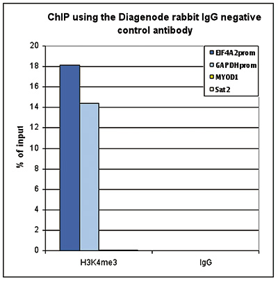
<p><small> <strong>Figure 1. ChIP with the Diagenode rabbit IgG negative control antibody</strong><br />ChIP assays were performed using the Diagenode rabbit polyclonal antibody against H3K4me3 (Cat. No. C15410003) and the “iDeal ChIPseq” kit (Cat. No. C01010051) on sheared chromatin from 1 million HeLa cells. Rabbit IgG (cat. No. C15410206) was used as a negative IP control. One μg of antibody per ChIP experiment was used for both antibodies. Quantitative PCR was performed with primers specific for the promoters of the active GAPDH and EIF4A2 genes, and for the inactive MYOD1 gene and the Sat2 satellite repeat. Figure 1 shows the recovery, expressed as a % of input (the relative amount of immunoprecipitated DNA compared to input DNA after qPCR analysis).</small></p>
</div>
</div>
<div class="row">
<div class="small-4 columns">
<p><img src="https://www.diagenode.com/img/product/antibodies/C15410206-if.jpg" alt="Rabbit IgG validated in Immunofluorescence" /></p>
</div>
<div class="small-8 columns">
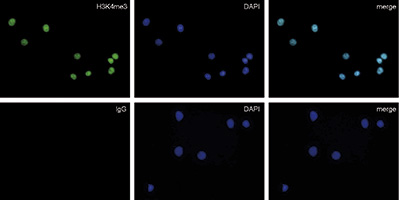
<p><small> <strong>Figure 2. Immunofluorescence with the Diagenode rabbit IgG negative control antibody</strong><br />HeLa cells were stained with the Diagenode rabbit polyclonal antibody against H3K4me3 (Cat. No. C15410003) (top) and with DAPI. Rabbit IgG (Cat. No. C15410206) was used as a negative control (bottom). Cells were fixed with 4% formaldehyde for 10’ and blocked with PBS/TX-100 containing 5% normal goat serum and 1% BSA. The cells were immunofluorescently labeled with the H3K4me3 or rabbit IgG negative control antibody (left) diluted 1:200 in blocking solution followed by an anti-rabbit antibody conjugated to Alexa488. The middle panel shows staining of the nuclei with DAPI. A merge of the two stainings is shown on the right.</small></p>
</div>
</div>',
'label2' => '',
'info2' => '<p>The negative control IgG from rabbit has been extensively validated in chromatin immunoprecipitation (ChIP). It contains a spectrum of the IgG subclasses present in serum of healthy rabbits. This IgG preparation is intended for use as a negative control in ChIP, MeDIP, IF and other experiments performed with specific antibodies made in rabbit. The negative control IgG from rabbit should be used in parallel with the specific antibody at the same concentration. It is also included in many of our ChIP and MeDIP kits.</p>',
'label3' => '',
'info3' => '',
'format' => '250 µg/250 µl',
'catalog_number' => 'C15410206',
'old_catalog_number' => 'kch-504-250',
'sf_code' => 'C15410206-363',
'type' => 'FRE',
'search_order' => '03-Antibody',
'price_EUR' => '70',
'price_USD' => '85',
'price_GBP' => '70',
'price_JPY' => '10965',
'price_CNY' => '',
'price_AUD' => '212',
'country' => 'ALL',
'except_countries' => 'None',
'quote' => false,
'in_stock' => false,
'featured' => false,
'no_promo' => false,
'online' => true,
'master' => true,
'last_datasheet_update' => '0000-00-00',
'slug' => 'rabbit-igg-250-ug-250-ul',
'meta_title' => 'Rabbit IgG',
'meta_keywords' => '',
'meta_description' => 'Rabbit IgG',
'modified' => '2024-01-17 14:04:06',
'created' => '2015-06-29 14:08:20',
'locale' => 'jpn'
),
'Antibody' => array(
'host' => '*****',
'id' => '327',
'name' => 'Rabbit IgG',
'description' => 'The negative Ctrl IgG from rabbit has been extensively validated in chromatin immunoprecipitation assays (ChIP). It contains a spectrum of the IgG subclasses present in serum of healthy animals. This IgG preparation is intended for use as a negative contr',
'clonality' => '',
'isotype' => '',
'lot' => 'RIG002B',
'concentration' => '1.0 µg/µl',
'reactivity' => 'none',
'type' => 'Polyclonal',
'purity' => 'Protein A purified',
'classification' => '',
'application_table' => '',
'storage_conditions' => '',
'storage_buffer' => '',
'precautions' => 'This product is for research use only. Not for use in diagnostic or therapeutic procedures.',
'uniprot_acc' => '',
'slug' => '',
'meta_keywords' => '',
'meta_description' => '',
'modified' => '2023-02-16 13:33:22',
'created' => '0000-00-00 00:00:00',
'select_label' => '327 - Rabbit IgG (RIG002B - 1.0 µg/µl - none - Protein A purified - Rabbit)'
),
'Slave' => array(),
'Group' => array(),
'Related' => array(),
'Application' => array(
(int) 0 => array(
[maximum depth reached]
),
(int) 1 => array(
[maximum depth reached]
)
),
'Category' => array(
(int) 0 => array(
[maximum depth reached]
),
(int) 1 => array(
[maximum depth reached]
),
(int) 2 => array(
[maximum depth reached]
),
(int) 3 => array(
[maximum depth reached]
)
),
'Document' => array(
(int) 0 => array(
[maximum depth reached]
),
(int) 1 => array(
[maximum depth reached]
),
(int) 2 => array(
[maximum depth reached]
)
),
'Feature' => array(),
'Image' => array(
(int) 0 => array(
[maximum depth reached]
)
),
'Promotion' => array(),
'Protocol' => array(),
'Publication' => array(
(int) 0 => array(
[maximum depth reached]
),
(int) 1 => array(
[maximum depth reached]
),
(int) 2 => array(
[maximum depth reached]
),
(int) 3 => array(
[maximum depth reached]
),
(int) 4 => array(
[maximum depth reached]
),
(int) 5 => array(
[maximum depth reached]
),
(int) 6 => array(
[maximum depth reached]
),
(int) 7 => array(
[maximum depth reached]
),
(int) 8 => array(
[maximum depth reached]
),
(int) 9 => array(
[maximum depth reached]
),
(int) 10 => array(
[maximum depth reached]
),
(int) 11 => array(
[maximum depth reached]
),
(int) 12 => array(
[maximum depth reached]
),
(int) 13 => array(
[maximum depth reached]
),
(int) 14 => array(
[maximum depth reached]
),
(int) 15 => array(
[maximum depth reached]
),
(int) 16 => array(
[maximum depth reached]
),
(int) 17 => array(
[maximum depth reached]
),
(int) 18 => array(
[maximum depth reached]
),
(int) 19 => array(
[maximum depth reached]
),
(int) 20 => array(
[maximum depth reached]
),
(int) 21 => array(
[maximum depth reached]
),
(int) 22 => array(
[maximum depth reached]
),
(int) 23 => array(
[maximum depth reached]
),
(int) 24 => array(
[maximum depth reached]
),
(int) 25 => array(
[maximum depth reached]
),
(int) 26 => array(
[maximum depth reached]
),
(int) 27 => array(
[maximum depth reached]
),
(int) 28 => array(
[maximum depth reached]
),
(int) 29 => array(
[maximum depth reached]
),
(int) 30 => array(
[maximum depth reached]
),
(int) 31 => array(
[maximum depth reached]
),
(int) 32 => array(
[maximum depth reached]
),
(int) 33 => array(
[maximum depth reached]
),
(int) 34 => array(
[maximum depth reached]
),
(int) 35 => array(
[maximum depth reached]
),
(int) 36 => array(
[maximum depth reached]
),
(int) 37 => array(
[maximum depth reached]
),
(int) 38 => array(
[maximum depth reached]
),
(int) 39 => array(
[maximum depth reached]
),
(int) 40 => array(
[maximum depth reached]
),
(int) 41 => array(
[maximum depth reached]
),
(int) 42 => array(
[maximum depth reached]
),
(int) 43 => array(
[maximum depth reached]
),
(int) 44 => array(
[maximum depth reached]
),
(int) 45 => array(
[maximum depth reached]
),
(int) 46 => array(
[maximum depth reached]
),
(int) 47 => array(
[maximum depth reached]
),
(int) 48 => array(
[maximum depth reached]
),
(int) 49 => array(
[maximum depth reached]
),
(int) 50 => array(
[maximum depth reached]
),
(int) 51 => array(
[maximum depth reached]
),
(int) 52 => array(
[maximum depth reached]
),
(int) 53 => array(
[maximum depth reached]
),
(int) 54 => array(
[maximum depth reached]
),
(int) 55 => array(
[maximum depth reached]
),
(int) 56 => array(
[maximum depth reached]
),
(int) 57 => array(
[maximum depth reached]
),
(int) 58 => array(
[maximum depth reached]
),
(int) 59 => array(
[maximum depth reached]
),
(int) 60 => array(
[maximum depth reached]
),
(int) 61 => array(
[maximum depth reached]
),
(int) 62 => array(
[maximum depth reached]
),
(int) 63 => array(
[maximum depth reached]
),
(int) 64 => array(
[maximum depth reached]
),
(int) 65 => array(
[maximum depth reached]
),
(int) 66 => array(
[maximum depth reached]
),
(int) 67 => array(
[maximum depth reached]
),
(int) 68 => array(
[maximum depth reached]
),
(int) 69 => array(
[maximum depth reached]
),
(int) 70 => array(
[maximum depth reached]
),
(int) 71 => array(
[maximum depth reached]
),
(int) 72 => array(
[maximum depth reached]
),
(int) 73 => array(
[maximum depth reached]
),
(int) 74 => array(
[maximum depth reached]
),
(int) 75 => array(
[maximum depth reached]
),
(int) 76 => array(
[maximum depth reached]
),
(int) 77 => array(
[maximum depth reached]
),
(int) 78 => array(
[maximum depth reached]
),
(int) 79 => array(
[maximum depth reached]
),
(int) 80 => array(
[maximum depth reached]
),
(int) 81 => array(
[maximum depth reached]
),
(int) 82 => array(
[maximum depth reached]
),
(int) 83 => array(
[maximum depth reached]
),
(int) 84 => array(
[maximum depth reached]
),
(int) 85 => array(
[maximum depth reached]
),
(int) 86 => array(
[maximum depth reached]
),
(int) 87 => array(
[maximum depth reached]
),
(int) 88 => array(
[maximum depth reached]
),
(int) 89 => array(
[maximum depth reached]
),
(int) 90 => array(
[maximum depth reached]
),
(int) 91 => array(
[maximum depth reached]
),
(int) 92 => array(
[maximum depth reached]
),
(int) 93 => array(
[maximum depth reached]
),
(int) 94 => array(
[maximum depth reached]
),
(int) 95 => array(
[maximum depth reached]
),
(int) 96 => array(
[maximum depth reached]
),
(int) 97 => array(
[maximum depth reached]
),
(int) 98 => array(
[maximum depth reached]
),
(int) 99 => array(
[maximum depth reached]
),
(int) 100 => array(
[maximum depth reached]
),
(int) 101 => array(
[maximum depth reached]
),
(int) 102 => array(
[maximum depth reached]
),
(int) 103 => array(
[maximum depth reached]
),
(int) 104 => array(
[maximum depth reached]
),
(int) 105 => array(
[maximum depth reached]
),
(int) 106 => array(
[maximum depth reached]
),
(int) 107 => array(
[maximum depth reached]
),
(int) 108 => array(
[maximum depth reached]
),
(int) 109 => array(
[maximum depth reached]
),
(int) 110 => array(
[maximum depth reached]
),
(int) 111 => array(
[maximum depth reached]
),
(int) 112 => array(
[maximum depth reached]
),
(int) 113 => array(
[maximum depth reached]
),
(int) 114 => array(
[maximum depth reached]
),
(int) 115 => array(
[maximum depth reached]
),
(int) 116 => array(
[maximum depth reached]
),
(int) 117 => array(
[maximum depth reached]
),
(int) 118 => array(
[maximum depth reached]
),
(int) 119 => array(
[maximum depth reached]
),
(int) 120 => array(
[maximum depth reached]
),
(int) 121 => array(
[maximum depth reached]
),
(int) 122 => array(
[maximum depth reached]
),
(int) 123 => array(
[maximum depth reached]
),
(int) 124 => array(
[maximum depth reached]
),
(int) 125 => array(
[maximum depth reached]
),
(int) 126 => array(
[maximum depth reached]
),
(int) 127 => array(
[maximum depth reached]
),
(int) 128 => array(
[maximum depth reached]
),
(int) 129 => array(
[maximum depth reached]
),
(int) 130 => array(
[maximum depth reached]
),
(int) 131 => array(
[maximum depth reached]
),
(int) 132 => array(
[maximum depth reached]
),
(int) 133 => array(
[maximum depth reached]
),
(int) 134 => array(
[maximum depth reached]
),
(int) 135 => array(
[maximum depth reached]
),
(int) 136 => array(
[maximum depth reached]
),
(int) 137 => array(
[maximum depth reached]
),
(int) 138 => array(
[maximum depth reached]
),
(int) 139 => array(
[maximum depth reached]
),
(int) 140 => array(
[maximum depth reached]
),
(int) 141 => array(
[maximum depth reached]
),
(int) 142 => array(
[maximum depth reached]
),
(int) 143 => array(
[maximum depth reached]
),
(int) 144 => array(
[maximum depth reached]
),
(int) 145 => array(
[maximum depth reached]
),
(int) 146 => array(
[maximum depth reached]
),
(int) 147 => array(
[maximum depth reached]
),
(int) 148 => array(
[maximum depth reached]
),
(int) 149 => array(
[maximum depth reached]
),
(int) 150 => array(
[maximum depth reached]
)
),
'Testimonial' => array(
(int) 0 => array(
[maximum depth reached]
)
),
'Area' => array(),
'SafetySheet' => array(
(int) 0 => array(
[maximum depth reached]
),
(int) 1 => array(
[maximum depth reached]
),
(int) 2 => array(
[maximum depth reached]
),
(int) 3 => array(
[maximum depth reached]
),
(int) 4 => array(
[maximum depth reached]
),
(int) 5 => array(
[maximum depth reached]
),
(int) 6 => array(
[maximum depth reached]
),
(int) 7 => array(
[maximum depth reached]
)
)
)
)
$language = 'jp'
$meta_keywords = ''
$meta_description = 'Rabbit IgG'
$meta_title = 'Rabbit IgG'
$product = array(
'Product' => array(
'id' => '2283',
'antibody_id' => '327',
'name' => 'Rabbit IgG',
'description' => '<p><span>The negative Ctrl IgG from rabbit has been extensively validated in chromatin immunoprecipitation assays (ChIP). It contains a spectrum of the IgG subclasses present in serum of healthy animals. This IgG preparation is intended for use as a negative control in ChIP experiments for specific antibodies made in rabbit. The negative Ctrl IgG from rabbit should be used for ChIP in parallel with specific antibody at the same concentration as the specific antibody. It is included in our LowCell# ChIP kit (Cat. No. kch-maglow-A16) kit and available separately.</span></p>',
'label1' => 'Validation data',
'info1' => '<div class="row">
<div class="small-4 columns">
<p><img src="https://www.diagenode.com/img/product/antibodies/C15410206-chip.jpg" alt="Rabbit IgG Antibody ChIP Grade" /></p>
</div>
<div class="small-8 columns">
<p><small> <strong>Figure 1. ChIP with the Diagenode rabbit IgG negative control antibody</strong><br />ChIP assays were performed using the Diagenode rabbit polyclonal antibody against H3K4me3 (Cat. No. C15410003) and the “iDeal ChIPseq” kit (Cat. No. C01010051) on sheared chromatin from 1 million HeLa cells. Rabbit IgG (cat. No. C15410206) was used as a negative IP control. One μg of antibody per ChIP experiment was used for both antibodies. Quantitative PCR was performed with primers specific for the promoters of the active GAPDH and EIF4A2 genes, and for the inactive MYOD1 gene and the Sat2 satellite repeat. Figure 1 shows the recovery, expressed as a % of input (the relative amount of immunoprecipitated DNA compared to input DNA after qPCR analysis).</small></p>
</div>
</div>
<div class="row">
<div class="small-4 columns">
<p><img src="https://www.diagenode.com/img/product/antibodies/C15410206-if.jpg" alt="Rabbit IgG validated in Immunofluorescence" /></p>
</div>
<div class="small-8 columns">
<p><small> <strong>Figure 2. Immunofluorescence with the Diagenode rabbit IgG negative control antibody</strong><br />HeLa cells were stained with the Diagenode rabbit polyclonal antibody against H3K4me3 (Cat. No. C15410003) (top) and with DAPI. Rabbit IgG (Cat. No. C15410206) was used as a negative control (bottom). Cells were fixed with 4% formaldehyde for 10’ and blocked with PBS/TX-100 containing 5% normal goat serum and 1% BSA. The cells were immunofluorescently labeled with the H3K4me3 or rabbit IgG negative control antibody (left) diluted 1:200 in blocking solution followed by an anti-rabbit antibody conjugated to Alexa488. The middle panel shows staining of the nuclei with DAPI. A merge of the two stainings is shown on the right.</small></p>
</div>
</div>',
'label2' => '',
'info2' => '<p>The negative control IgG from rabbit has been extensively validated in chromatin immunoprecipitation (ChIP). It contains a spectrum of the IgG subclasses present in serum of healthy rabbits. This IgG preparation is intended for use as a negative control in ChIP, MeDIP, IF and other experiments performed with specific antibodies made in rabbit. The negative control IgG from rabbit should be used in parallel with the specific antibody at the same concentration. It is also included in many of our ChIP and MeDIP kits.</p>',
'label3' => '',
'info3' => '',
'format' => '250 µg/250 µl',
'catalog_number' => 'C15410206',
'old_catalog_number' => 'kch-504-250',
'sf_code' => 'C15410206-363',
'type' => 'FRE',
'search_order' => '03-Antibody',
'price_EUR' => '70',
'price_USD' => '85',
'price_GBP' => '70',
'price_JPY' => '10965',
'price_CNY' => '',
'price_AUD' => '212',
'country' => 'ALL',
'except_countries' => 'None',
'quote' => false,
'in_stock' => false,
'featured' => false,
'no_promo' => false,
'online' => true,
'master' => true,
'last_datasheet_update' => '0000-00-00',
'slug' => 'rabbit-igg-250-ug-250-ul',
'meta_title' => 'Rabbit IgG',
'meta_keywords' => '',
'meta_description' => 'Rabbit IgG',
'modified' => '2024-01-17 14:04:06',
'created' => '2015-06-29 14:08:20',
'locale' => 'jpn'
),
'Antibody' => array(
'host' => '*****',
'id' => '327',
'name' => 'Rabbit IgG',
'description' => 'The negative Ctrl IgG from rabbit has been extensively validated in chromatin immunoprecipitation assays (ChIP). It contains a spectrum of the IgG subclasses present in serum of healthy animals. This IgG preparation is intended for use as a negative contr',
'clonality' => '',
'isotype' => '',
'lot' => 'RIG002B',
'concentration' => '1.0 µg/µl',
'reactivity' => 'none',
'type' => 'Polyclonal',
'purity' => 'Protein A purified',
'classification' => '',
'application_table' => '',
'storage_conditions' => '',
'storage_buffer' => '',
'precautions' => 'This product is for research use only. Not for use in diagnostic or therapeutic procedures.',
'uniprot_acc' => '',
'slug' => '',
'meta_keywords' => '',
'meta_description' => '',
'modified' => '2023-02-16 13:33:22',
'created' => '0000-00-00 00:00:00',
'select_label' => '327 - Rabbit IgG (RIG002B - 1.0 µg/µl - none - Protein A purified - Rabbit)'
),
'Slave' => array(),
'Group' => array(),
'Related' => array(),
'Application' => array(
(int) 0 => array(
'id' => '7',
'position' => '10',
'parent_id' => '1',
'name' => 'Methylated DNA immunoprecipitation',
'description' => '<div class="row extra-spaced">
<div class="small-12 medium-3 large-3 columns"><center><a href="https://www.ncbi.nlm.nih.gov/pubmed/30429608" target="_blank"><img src="https://www.diagenode.com/img/banners/banner-nature-publication-580.png" /></a></center></div>
<div class="small-12 medium-9 large-9 columns">
<h3>Sensitive tumour detection and classification using plasma cell-free DNA methylomes<br /><a href="https://www.ncbi.nlm.nih.gov/pubmed/30429608" target="_blank">Read the publication</a></h3>
<h3 class="c-article-title u-h1" data-test="article-title" itemprop="name headline">Preparation of cfMeDIP-seq libraries for methylome profiling of plasma cell-free DNA<br /><a href="https://www.nature.com/articles/s41596-019-0202-2" target="_blank" title="cfMeDIP-seq Nature Method">Read the method</a></h3>
</div>
</div>
<div class="row">
<div class="large-12 columns"><span>The Methylated DNA Immunoprecipitation is based on the affinity purification of methylated and hydroxymethylated DNA using, respectively, an antibody directed against 5-methylcytosine (5-mC) in the case of MeDIP or 5-hydroxymethylcytosine (5-hmC) in the case of hMeDIP.</span><br />
<h2></h2>
<h2>How it works</h2>
<p>In brief, Methyl DNA IP is performed as follows: Genomic DNA from cultured cells or tissues is prepared, sheared, and then denatured. Then, immunoselection and immunoprecipitation can take place using the antibody directed against 5 methylcytosine and antibody binding beads. After isolation and purification is performed, the IP’d methylated DNA is ready for any subsequent analysis as qPCR, amplification, hybridization on microarrays or next generation sequencing.</p>
<h2>Applications</h2>
<div align="center"><a href="https://www.diagenode.com/en/p/magmedip-kit-x48-48-rxns" class="center alert radius button"> qPCR analysis</a></div>
<div align="center"><a href="https://www.diagenode.com/en/p/magmedip-seq-package-V2-x10" class="center alert radius button"> NGS analysis </a></div>
<h2>Advantages</h2>
<ul style="font-size: 19px;" class="nobullet">
<li><i class="fa fa-arrow-circle-right"></i> <strong>Unaffected</strong> DNA</li>
<li><i class="fa fa-arrow-circle-right"></i> <strong>High enrichment</strong> yield</li>
<li><i class="fa fa-arrow-circle-right"></i> <strong>Robust</strong> & <strong>reproducible</strong> techniques</li>
<li><i class="fa fa-arrow-circle-right"></i> <strong>NGS</strong> compatible</li>
</ul>
<h2></h2>
</div>
</div>
<div id="gtx-trans" style="position: absolute; left: 17px; top: 652.938px;">
<div class="gtx-trans-icon"></div>
</div>',
'in_footer' => false,
'in_menu' => true,
'online' => true,
'tabular' => true,
'slug' => 'methylated-dna-immunoprecipitation',
'meta_keywords' => 'Methylated DNA immunoprecipitation,Epigenetic,DNA Methylation,qPCR,5 methylcytosine (5-mC)',
'meta_description' => 'Methylated DNA immunoprecipitation method is based on the affinity purification of methylated DNA using an antibody directed against 5 methylcytosine (5-mC). ',
'meta_title' => 'Methylated DNA immunoprecipitation(MeDIP) - Dna methylation | Diagenode',
'modified' => '2021-08-19 12:08:03',
'created' => '2014-09-14 05:33:34',
'ProductsApplication' => array(
[maximum depth reached]
)
),
(int) 1 => array(
'id' => '43',
'position' => '10',
'parent_id' => '40',
'name' => 'ChIP-qPCR (ab)',
'description' => '',
'in_footer' => false,
'in_menu' => false,
'online' => true,
'tabular' => true,
'slug' => 'chip-qpcr-antibodies',
'meta_keywords' => 'Chromatin Immunoprecipitation Sequencing,ChIP-Seq,ChIP-seq grade antibodies,DNA purification,qPCR,Shearing of chromatin',
'meta_description' => 'Diagenode offers a wide range of antibodies and technical support for ChIP-qPCR applications',
'meta_title' => 'ChIP Quantitative PCR Antibodies (ChIP-qPCR) | Diagenode',
'modified' => '2016-01-20 11:30:24',
'created' => '2015-10-20 11:45:36',
'ProductsApplication' => array(
[maximum depth reached]
)
)
),
'Category' => array(
(int) 0 => array(
'id' => '22',
'position' => '40',
'parent_id' => '4',
'name' => 'Isotype controls',
'description' => '<p><span style="font-weight: 400;">Diagenode offers the negative Ctrl IgG from rabbit, rat and mouse. These extensively validated antibodies can be used as negative controls in ChIP, IF, hMeDIP or other experiments performed with specific antibodies made in rabbit, rat or mouse, respectively.</span></p>
<p><span style="font-weight: 400;">Diagenode’s highly validated antibodies:</span></p>
<ul>
<li>Highly sensitive and specific</li>
<li>Cost-effective (requires less antibody per reaction)</li>
<li>Batch-specific data is available on the website</li>
<li>Expert technical support</li>
<li>Sample sizes available</li>
<li>100% satisfaction guarantee</li>
</ul>',
'no_promo' => false,
'in_menu' => false,
'online' => true,
'tabular' => false,
'hide' => true,
'all_format' => false,
'is_antibody' => true,
'slug' => 'isotype-controls',
'cookies_tag_id' => null,
'meta_keywords' => 'Isotype controls,DNA immunoprecipitation,Methylated DNA immunoprecipitation',
'meta_description' => 'Diagenode provides Isotype controls for Methylated DNA immunoprecipitation',
'meta_title' => 'Isotype controls for Methylated DNA immunoprecipitation | Diagenode',
'modified' => '2019-07-04 16:19:36',
'created' => '2014-09-30 14:23:34',
'ProductsCategory' => array(
[maximum depth reached]
),
'CookiesTag' => array([maximum depth reached])
),
(int) 1 => array(
'id' => '103',
'position' => '0',
'parent_id' => '4',
'name' => 'All antibodies',
'description' => '<p><span style="font-weight: 400;">All Diagenode’s antibodies are listed below. Please, use our Quick search field to find the antibody of interest by target name, application, purity.</span></p>
<p><span style="font-weight: 400;">Diagenode’s highly validated antibodies:</span></p>
<ul>
<li>Highly sensitive and specific</li>
<li>Cost-effective (requires less antibody per reaction)</li>
<li>Batch-specific data is available on the website</li>
<li>Expert technical support</li>
<li>Sample sizes available</li>
<li>100% satisfaction guarantee</li>
</ul>',
'no_promo' => false,
'in_menu' => true,
'online' => true,
'tabular' => false,
'hide' => true,
'all_format' => false,
'is_antibody' => true,
'slug' => 'all-antibodies',
'cookies_tag_id' => null,
'meta_keywords' => 'Antibodies,Premium Antibodies,Classic,Pioneer',
'meta_description' => 'Diagenode Offers Strict quality standards with Rigorous QC and validated Antibodies. Classified based on level of validation for flexibility of Application. Comprehensive selection of histone and non-histone Antibodies',
'meta_title' => 'Diagenode's selection of Antibodies is exclusively dedicated for Epigenetic Research | Diagenode',
'modified' => '2019-07-03 10:55:44',
'created' => '2015-11-02 14:49:22',
'ProductsCategory' => array(
[maximum depth reached]
),
'CookiesTag' => array([maximum depth reached])
),
(int) 2 => array(
'id' => '127',
'position' => '10',
'parent_id' => '4',
'name' => 'ChIP-grade antibodies',
'description' => '<div class="row">
<div class="small-12 columns"><center></center>
<p><br />Chromatin immunoprecipitation (<b>ChIP</b>) is a technique to study the associations of proteins with the specific genomic regions in intact cells. One of the most important steps of this protocol is the immunoprecipitation of targeted protein using the antibody specifically recognizing it. The quality of antibodies used in ChIP is essential for the success of the experiment. Diagenode offers extensively validated ChIP-grade antibodies, confirmed for their specificity, and high level of performance in ChIP. Each batch is validated, and batch-specific data are available on the website.</p>
<p></p>
</div>
</div>
<p><strong>ChIP results</strong> obtained with the antibody directed against H3K4me3 (Cat. No. <a href="../p/h3k4me3-polyclonal-antibody-premium-50-ug-50-ul">C15410003</a>). </p>
<div class="row">
<div class="small-12 medium-6 large-6 columns"><img src="https://www.diagenode.com/img/product/antibodies/C15410003-fig1-ChIP.jpg" alt="" width="400" height="315" /> </div>
<div class="small-12 medium-6 large-6 columns">
<p></p>
<p></p>
<p></p>
</div>
</div>
<p></p>
<p>Our aim at Diagenode is to offer the largest collection of highly specific <strong>ChIP-grade antibodies</strong>. We add new antibodies monthly. Find your ChIP-grade antibody in the list below and check more information about tested applications, extensive validation data, and product information.</p>',
'no_promo' => false,
'in_menu' => true,
'online' => true,
'tabular' => false,
'hide' => true,
'all_format' => false,
'is_antibody' => true,
'slug' => 'chip-grade-antibodies',
'cookies_tag_id' => null,
'meta_keywords' => 'ChIP-grade antibodies, polyclonal antibody, monoclonal antibody, Diagenode',
'meta_description' => 'Diagenode Offers Extensively Validated ChIP-Grade Antibodies, Confirmed for their Specificity, and high level of Performance in Chromatin Immunoprecipitation ChIP',
'meta_title' => 'Chromatin immunoprecipitation ChIP-grade antibodies | Diagenode',
'modified' => '2024-11-19 17:27:07',
'created' => '2017-02-14 11:16:04',
'ProductsCategory' => array(
[maximum depth reached]
),
'CookiesTag' => array([maximum depth reached])
),
(int) 3 => array(
'id' => '142',
'position' => '4',
'parent_id' => '13',
'name' => 'CUT&Tag',
'description' => '<p><img alt=" iDeal CUT&Tag for Histones" src="https://www.diagenode.com/img/banners/banner-cutruncuttag-small.png" /></p>
<div class="row">
<div class="small-12 medium-8 large-8 columns"><br />
<p><strong>CUT&Tag</strong>-sequencing (<strong>C</strong>leavage <strong>U</strong>nder <strong>T</strong>argets and <strong>Tag</strong>mentation) is a new alternative method to ChIP-seq combining antibody-targeted controlled cleavage by a protein A-Tn5 fusion with massively parallel DNA sequencing to identify the binding sites of DNA-associated proteins. At Diagenode we offer a complete solution for CUT&Tag – our iDeal CUT&Tag for Histones (developped for histone marks and some non-histone proteins), but also stand-alone fusion protein – pA-Tn5 Transposase. Moreover, we have validated our <a href="https://www.diagenode.com/en/categories/chip-seq-grade-antibodies">ChIP-seq grade antibodies</a> in <a href="https://www.diagenode.com/en/categories/cut-and-tag-antibodies">CUT&Tag</a> proving their high performance in this assay.</p>
<br /> <a href="https://www.diagenode.com/files/application_notes/AN-iDealCUTandTag.pdf"><img src="https://www.diagenode.com/img/banners/cutandtag-appnote.png" /></a><br /><br /></div>
<div class="small-12 medium-4 large-4 columns"><center>
<script>// <![CDATA[
var date = new Date(); var heure = date.getHours(); var jour = date.getDay(); var semaine = Math.floor(date.getDate() / 7) + 1; if (jour === 2 && ( (heure >= 9 && heure < 9.5) || (heure >= 18 && heure < 18.5) )) { document.write('<a href="https://us02web.zoom.us/j/85467619762"><img src="https://www.diagenode.com/img/epicafe-ON.gif"></a>'); } else { document.write('<a href="https://go.diagenode.com/l/928883/2023-04-26/3kq1v"><img src="https://www.diagenode.com/img/epicafe-OFF.png"></a>'); }
// ]]></script>
</center></div>
</div>
<ul class="accordion" data-accordion="">
<li class="accordion-navigation"><a href="#v5" style="color: #13b29c;"><i class="fa fa-caret-right"></i> How does it work?</a>
<div id="v5" class="content">
<p>The iDeal CUT&Tag protocol involves the binding of cells on a solid phase ConA magnetic beads, allowing magnetic handling of the cells for the major steps of the protocol. Bead-bound cells are permeabilized, incubated with primary antibody against a target of interest and secondary antibody. Then, Diagenode’s protein pA-Tn5 Transposase - loaded is bound to the complex. Protein A guides Tn5 transposase on chromatin to the antibody attached to its target. Tn5 transposase is activated by Mg+2 ions to insert the sequencing adaptors into genomic regions of interest. DNA is then purified and the tagmented genomic regions of interest are amplified by PCR using Diagenode’s Primer Indexes for tagmented libraries.</p>
<img src="https://www.diagenode.com/img/product/kits/workflow-cutandtag.jpg" /></div>
<h2>Products for CUT&Tag assay</h2>
<h3 class="diacol">Complete solutions</h3>
<ul class="nobullet">
<li><i class="fa fa-arrow-circle-right"></i> <a href="https://www.diagenode.com/en/p/ideal-cut-and-tag-kit-for-histones-24" target="_blank">iDeal CUT&Tag kit for Histones</a></li>
<li><i class="fa fa-arrow-circle-right"></i> <a href="https://www.diagenode.com/en/p/antibody-package-cut-and-tag-antirabbit-24" target="_blank">Antibody package for CUT&Tag (anti-rabbit)</a></li>
<li><i class="fa fa-arrow-circle-right"></i> <a href="https://www.diagenode.com/en/p/antibody-package-cut-and-tag-antimouse-24" target="_blank">Antibody package for CUT&Tag (anti-mouse)</a></li>
</ul>
<h3 class="diacol">Fusion protein</h3>
<ul class="nobullet">
<li><i class="fa fa-arrow-circle-right"></i> <a href="https://www.diagenode.com/en/products/view/3064" target="_blank">pA/Tn5 Transposase (loaded)</a></li>
<li><i class="fa fa-arrow-circle-right"></i> <a href="https://www.diagenode.com/en/products/view/3065" target="_blank">pA/Tn5 Transposase (unloaded)</a></li>
</ul>
<h3 class="diacol">CUT&Tag grade antibodies</h3>
<ul class="nobullet">
<li>Antibodies <a href="https://www.diagenode.com/en/applications/cut-and-tag">validated in CUT&Tag</a></li>
<li>Check out our list of <a href="https://www.diagenode.com/en/categories/chip-seq-grade-antibodies" target="_blank">ChIP-seq grade antibodies</a></li>
<li>Read more about the performance of Diagenode antibodies in <a href="https://www.diagenode.com/en/pages/cut-and-tag" target="_blank">CUT&Tag</a></li>
</ul>
<h3 class="diacol">Positive & Negative CUT&Tag control</h3>
<ul class="nobullet">
<li><i class="fa fa-arrow-circle-right"></i> <a href="https://www.diagenode.com/en/p/antibody-package-cut-and-tag-antirabbit-24" target="_blank">Antibody package for CUT&Tag (anti-rabbit)</a></li>
<li><i class="fa fa-arrow-circle-right"></i> <a href="https://www.diagenode.com/en/p/antibody-package-cut-and-tag-antimouse-24" target="_blank">Antibody package for CUT&Tag (anti-mouse)</a></li>
</ul>
<h3 class="diacol">DNA purification</h3>
<p style="padding-left: 30px;"><a href="https://www.diagenode.com/en/p/ipure-kit-v2-x24">IPure kit v2<br /></a><a href="https://www.diagenode.com/en/p/microchip-diapure-columns-50-rxns">MicroChIP DiaPure columns</a></p>
<h3 class="diacol">Sequencing indexes</h3>
<ul class="nobullet">
<li><i class="fa fa-arrow-circle-right"></i> <a href="https://www.diagenode.com/en/categories/primer-indexes-for-tagmented-libraries" target="_blank">Primer indexes for tagmented libraries</a></li>
</ul>
</li>
</ul>',
'no_promo' => false,
'in_menu' => true,
'online' => true,
'tabular' => false,
'hide' => false,
'all_format' => false,
'is_antibody' => false,
'slug' => 'cutandtag',
'cookies_tag_id' => null,
'meta_keywords' => '',
'meta_description' => 'Rapid chromatin profiling assay, optimized protocol, all reagents provided. Easy sample handling due to ConA magnetic beads. Highly specific CUT&Tag grade antibodies available. ',
'meta_title' => 'Diagenode provides advanced solutions for high performance CUT&Tag experiments',
'modified' => '2024-03-07 17:14:34',
'created' => '2020-04-28 15:07:36',
'ProductsCategory' => array(
[maximum depth reached]
),
'CookiesTag' => array([maximum depth reached])
)
),
'Document' => array(
(int) 0 => array(
'id' => '617',
'name' => 'Datasheet rabbitIgG kch-504-250',
'description' => '<p>Datasheet description</p>',
'image_id' => null,
'type' => 'Datasheet',
'url' => 'files/products/antibodies/Datasheet_rabbitIgG_kch-504-250.pdf',
'slug' => 'datasheet-rabbitigg-kch-504-250',
'meta_keywords' => '',
'meta_description' => '',
'modified' => '2016-10-28 09:37:19',
'created' => '2015-07-07 11:47:44',
'ProductsDocument' => array(
[maximum depth reached]
)
),
(int) 1 => array(
'id' => '38',
'name' => 'Epigenetic Antibodies Brochure',
'description' => '<p>More than in any other immuoprecipitation assays, quality antibodies are critical tools in many epigenetics experiments. Since 10 years, Diagenode has developed the most stringent quality production available on the market for antibodies exclusively focused on epigenetic uses. All our antibodies have been qualified to work in epigenetic applications.</p>',
'image_id' => null,
'type' => 'Brochure',
'url' => 'files/brochures/Epigenetic_Antibodies_Brochure.pdf',
'slug' => 'epigenetic-antibodies-brochure',
'meta_keywords' => '',
'meta_description' => '',
'modified' => '2016-06-15 11:24:06',
'created' => '2015-07-03 16:05:27',
'ProductsDocument' => array(
[maximum depth reached]
)
),
(int) 2 => array(
'id' => '11',
'name' => 'Antibodies you can trust',
'description' => '<p style="text-align: justify;"><span>Epigenetic research tools have evolved over time from endpoint PCR to qPCR to the analyses of large sets of genome-wide sequencing data. ChIP sequencing (ChIP-seq) has now become the gold standard method for chromatin studies, given the accuracy and coverage scale of the approach over other methods. Successful ChIP-seq, however, requires a higher level of experimental accuracy and consistency in all steps of ChIP than ever before. Particularly crucial is the quality of ChIP antibodies. </span></p>',
'image_id' => null,
'type' => 'Poster',
'url' => 'files/posters/Antibodies_you_can_trust_Poster.pdf',
'slug' => 'antibodies-you-can-trust-poster',
'meta_keywords' => '',
'meta_description' => '',
'modified' => '2015-10-01 20:18:31',
'created' => '2015-07-03 16:05:15',
'ProductsDocument' => array(
[maximum depth reached]
)
)
),
'Feature' => array(),
'Image' => array(
(int) 0 => array(
'id' => '250',
'name' => 'product/antibodies/antibody.png',
'alt' => 'Mouse IgG',
'modified' => '2020-11-27 07:00:09',
'created' => '2015-07-17 10:12:18',
'ProductsImage' => array(
[maximum depth reached]
)
)
),
'Promotion' => array(),
'Protocol' => array(),
'Publication' => array(
(int) 0 => array(
'id' => '4958',
'name' => 'Legionella pneumophila modulates macrophage functions through epigenetic reprogramming via the C-type lectin receptor Mincle',
'authors' => 'Stegmann F. et al.',
'description' => '<p><em>Legionella pneumophila</em><span><span> </span>is a pathogen which can lead to a severe form of pneumonia in humans known as Legionnaires disease after replication in alveolar macrophages. Viable<span> </span></span><em>L. pneumophila</em><span><span> </span>actively secrete effector molecules to modulate the host’s immune response. Here, we report that<span> </span></span><em>L. pneumophila</em><span>-derived factors reprogram macrophages into a tolerogenic state, a process to which the C-type lectin receptor Mincle (CLEC4E) markedly contributes. The underlying epigenetic state is characterized by increases of the closing mark H3K9me3 and decreases of the opening mark H3K4me3, subsequently leading to the reduced secretion of the cytokines TNF, IL-6, IL-12, the production of reactive oxygen species, and cell-surface expression of MHC-II and CD80 upon re-stimulation. In summary, these findings provide important implications for our understanding of Legionellosis and the contribution of Mincle to reprogramming of macrophages by<span> </span></span><em>L. pneumophila</em><span>.</span></p>',
'date' => '2024-09-20',
'pmid' => 'https://www.sciencedirect.com/science/article/pii/S2589004224019254#:~:text=L.,crucial%20for%20mediating%20tolerance%20induction.',
'doi' => 'https://doi.org/10.1016/j.isci.2024.110700',
'modified' => '2024-09-02 10:06:00',
'created' => '2024-09-02 10:06:00',
'ProductsPublication' => array(
[maximum depth reached]
)
),
(int) 1 => array(
'id' => '4968',
'name' => 'Innate immune training restores pro-reparative myeloid functions to promote remyelination in the aged central nervous system',
'authors' => 'Tiwari V. et al.',
'description' => '<p><span>The reduced ability of the central nervous system to regenerate with increasing age limits functional recovery following demyelinating injury. Previous work has shown that myelin debris can overwhelm the metabolic capacity of microglia, thereby impeding tissue regeneration in aging, but the underlying mechanisms are unknown. In a model of demyelination, we found that a substantial number of genes that were not effectively activated in aged myeloid cells displayed epigenetic modifications associated with restricted chromatin accessibility. Ablation of two class I histone deacetylases in microglia was sufficient to restore the capacity of aged mice to remyelinate lesioned tissue. We used Bacillus Calmette-Guerin (BCG), a live-attenuated vaccine, to train the innate immune system and detected epigenetic reprogramming of brain-resident myeloid cells and functional restoration of myelin debris clearance and lesion recovery. Our results provide insight into aging-associated decline in myeloid function and how this decay can be prevented by innate immune reprogramming.</span></p>',
'date' => '2024-07-24',
'pmid' => 'https://www.cell.com/immunity/fulltext/S1074-7613(24)00348-0',
'doi' => '',
'modified' => '2024-09-02 17:05:54',
'created' => '2024-09-02 17:05:54',
'ProductsPublication' => array(
[maximum depth reached]
)
),
(int) 2 => array(
'id' => '4945',
'name' => 'Master corepressor inactivation through multivalent SLiM-induced polymerization mediated by the oncogene suppressor RAI2',
'authors' => 'Goradia N. et al.',
'description' => '<p><span>While the elucidation of regulatory mechanisms of folded proteins is facilitated due to their amenability to high-resolution structural characterization, investigation of these mechanisms in disordered proteins is more challenging due to their structural heterogeneity, which can be captured by a variety of biophysical approaches. Here, we used the transcriptional master corepressor CtBP, which binds the putative metastasis suppressor RAI2 through repetitive SLiMs, as a model system. Using cryo-electron microscopy embedded in an integrative structural biology approach, we show that RAI2 unexpectedly induces CtBP polymerization through filaments of stacked tetrameric CtBP layers. These filaments lead to RAI2-mediated CtBP nuclear foci and relieve its corepressor function in RAI2-expressing cancer cells. The impact of RAI2-mediated CtBP loss-of-function is illustrated by the analysis of a diverse cohort of prostate cancer patients, which reveals a substantial decrease in RAI2 in advanced treatment-resistant cancer subtypes. As RAI2-like SLiM motifs are found in a wide range of organisms, including pathogenic viruses, our findings serve as a paradigm for diverse functional effects through multivalent interaction-mediated polymerization by disordered proteins in healthy and diseased conditions.</span></p>',
'date' => '2024-06-19',
'pmid' => 'https://www.nature.com/articles/s41467-024-49488-3',
'doi' => 'https://doi.org/10.1038/s41467-024-49488-3',
'modified' => '2024-06-24 17:11:37',
'created' => '2024-06-24 17:11:37',
'ProductsPublication' => array(
[maximum depth reached]
)
),
(int) 3 => array(
'id' => '4950',
'name' => 'Master corepressor inactivation through multivalent SLiM-induced polymerization mediated by the oncogene suppressor RAI2',
'authors' => 'Nishit Goradia et al.',
'description' => '<p><span>While the elucidation of regulatory mechanisms of folded proteins is facilitated due to their amenability to high-resolution structural characterization, investigation of these mechanisms in disordered proteins is more challenging due to their structural heterogeneity, which can be captured by a variety of biophysical approaches. Here, we used the transcriptional master corepressor CtBP, which binds the putative metastasis suppressor RAI2 through repetitive SLiMs, as a model system. Using cryo-electron microscopy embedded in an integrative structural biology approach, we show that RAI2 unexpectedly induces CtBP polymerization through filaments of stacked tetrameric CtBP layers. These filaments lead to RAI2-mediated CtBP nuclear foci and relieve its corepressor function in RAI2-expressing cancer cells. The impact of RAI2-mediated CtBP loss-of-function is illustrated by the analysis of a diverse cohort of prostate cancer patients, which reveals a substantial decrease in RAI2 in advanced treatment-resistant cancer subtypes. As RAI2-like SLiM motifs are found in a wide range of organisms, including pathogenic viruses, our findings serve as a paradigm for diverse functional effects through multivalent interaction-mediated polymerization by disordered proteins in healthy and diseased conditions.</span></p>',
'date' => '2024-06-19',
'pmid' => 'https://www.nature.com/articles/s41467-024-49488-3',
'doi' => ' https://doi.org/10.1038/s41467-024-49488-3',
'modified' => '2024-07-04 15:50:54',
'created' => '2024-07-04 15:50:54',
'ProductsPublication' => array(
[maximum depth reached]
)
),
(int) 4 => array(
'id' => '4920',
'name' => 'Focal cortical dysplasia type II-dependent maladaptive myelination in the human frontal lobe',
'authors' => 'Donkels C. et al.',
'description' => '<p><span>Focal cortical dysplasias (FCDs) are local malformations of the human neocortex and a leading cause of intractable epilepsy. FCDs are classified into different subtypes including FCD IIa and IIb, characterized by a blurred gray-white matter boundary or a transmantle sign indicating abnormal white matter myelination. Recently, we have shown that myelination is also compromised in the gray matter of FCD IIa of the temporal lobe. Since myelination is key for brain function, we investigated whether deficient myelination is a feature affecting also other FCD subtypes and brain areas. Here, we focused on the gray matter of FCD IIa and IIb from the frontal lobe. We applied </span><em>in situ</em><span><span> </span>hybridization, immunohistochemistry and electron microscopy to quantify oligodendrocytes, to visualize the myelination pattern and to determine ultrastructurally the axon diameter and the myelin sheath thickness. In addition, we analyzed the transcriptional regulation of myelin-associated transcripts by real-time RT-qPCR and chromatin immunoprecipitation (ChIP). We show that densities of myelinating oligodendrocytes and the extension of myelinated fibers up to layer II were unaltered in both FCD types but myelinated fibers appeared fractured mainly in FCD IIa. Interestingly, both FCD types presented with larger axon diameters when compared to controls. A significant correlation of axon diameter and myelin sheath thickness was found for FCD IIb and controls, whereas in FCD IIa large caliber axons were less myelinated. This was mirrored by a down-regulation of myelin-associated mRNAs and by reduced binding-capacities of the transcription factor MYRF to promoters of myelin-associated genes. FCD IIb, however, had significantly elevated transcript levels and MYRF-binding capacities reflecting the need for more myelin due to increased axon diameters. These data show that FCD IIa and IIb are characterized by divergent signs of maladaptive myelination which may contribute to the epileptic phenotype and underline the view of separate disease entities.</span></p>',
'date' => '2024-03-06',
'pmid' => 'https://www.biorxiv.org/content/10.1101/2024.03.02.582894v1',
'doi' => 'https://doi.org/10.1101/2024.03.02.582894',
'modified' => '2024-03-12 11:24:48',
'created' => '2024-03-12 11:24:48',
'ProductsPublication' => array(
[maximum depth reached]
)
),
(int) 5 => array(
'id' => '4860',
'name' => 'Identification of a deltaNp63-Dependent Basal-Like ASubtype-Specific Transcribed Enhancer Program (B-STEP) in Aggressive Pancreatic Ductal Adenocarcinoma.',
'authors' => 'Wang X. et al.',
'description' => '<p>A major hurdle to the application of precision oncology in pancreatic cancer is the lack of molecular stratification approaches and targeted therapy for defined molecular subtypes. In this work, we sought to gain further insight and identify molecular and epigenetic signatures of the basal-like A pancreatic ductal adenocarcinoma (PDAC) subgroup that can be applied to clinical samples for patient stratification and/or therapy monitoring. We generated and integrated global gene expression and epigenome mapping data from patient-derived xenograft (PDX) models to identify subtype-specific enhancer regions that were validated in patient-derived samples. In addition, complementary nascent transcription and chromatin topology (HiChIP) analyses revealed a basal-like A subtype-specific transcribed enhancer program (B-STEP) in PDAC characterized by enhancer RNA (eRNA) production that is associated with more frequent chromatin interactions and subtype-specific gene activation. Importantly, we successfully confirmed the validity of eRNA detection as a possible histological approach for PDAC patient stratification by performing RNA in situ hybridization analyses for subtype-specific eRNAs on pathological tissue samples. Thus, this study provides proof-of-concept that subtype-specific epigenetic changes relevant for PDAC progression can be detected at a single cell level in complex, heterogeneous, primary tumor material. Implications: Subtype-specific enhancer activity analysis via detection of eRNAs on a single cell level in patient material can be used as a potential tool for treatment stratification.</p>',
'date' => '2023-06-01',
'pmid' => 'https://pubmed.ncbi.nlm.nih.gov/37279184/',
'doi' => '10.1158/1541-7786.MCR-22-0916',
'modified' => '2023-08-01 14:51:22',
'created' => '2023-08-01 15:59:38',
'ProductsPublication' => array(
[maximum depth reached]
)
),
(int) 6 => array(
'id' => '4616',
'name' => 'Myelodysplastic Syndrome associated TET2 mutations affect NK cellfunction and genome methylation.',
'authors' => 'Boy M. et al.',
'description' => '<p>Myelodysplastic syndromes (MDS) are clonal hematopoietic disorders, representing high risk of progression to acute myeloid leukaemia, and frequently associated to somatic mutations, notably in the epigenetic regulator TET2. Natural Killer (NK) cells play a role in the anti-leukemic immune response via their cytolytic activity. Here we show that patients with MDS clones harbouring mutations in the TET2 gene are characterised by phenotypic defects in their circulating NK cells. Remarkably, NK cells and MDS clones from the same patient share the TET2 genotype, and the NK cells are characterised by increased methylation of genomic DNA and reduced expression of Killer Immunoglobulin-like receptors (KIR), perforin, and TNF-α. In vitro inhibition of TET2 in NK cells of healthy donors reduces their cytotoxicity, supporting its critical role in NK cell function. Conversely, NK cells from patients treated with azacytidine (#NCT02985190; https://clinicaltrials.gov/ ) show increased KIR and cytolytic protein expression, and IFN-γ production. Altogether, our findings show that, in addition to their oncogenic consequences in the myeloid cell subsets, TET2 mutations contribute to repressing NK-cell function in MDS patients.</p>',
'date' => '2023-02-01',
'pmid' => 'https://www.ncbi.nlm.nih.gov/pubmed/36737440',
'doi' => '10.1038/s41467-023-36193-w',
'modified' => '2023-04-04 08:43:27',
'created' => '2023-02-21 09:59:46',
'ProductsPublication' => array(
[maximum depth reached]
)
),
(int) 7 => array(
'id' => '4692',
'name' => 'Temporal modification of H3K9/14ac and H3K4me3 histone marksmediates mechano-responsive gene expression during the accommodationprocess in poplar',
'authors' => 'Ghosh R. et al.',
'description' => '<p>Plants can attenuate their molecular response to repetitive mechanical stimulation as a function of their mechanical history. For instance, a single bending of stem is sufficient to attenuate the gene expression in poplar plants to the subsequent mechanical stimulation, and the state of desensitization can last for several days. The role of histone modifications in memory gene expression and modulating plant response to abiotic or biotic signals is well known. However, such information is still lacking to explain the attenuated expression pattern of mechano-responsive genes in plants under repetitive stimulation. Using poplar as a model plant in this study, we first measured the global level of H3K9/14ac and H3K4me3 marks in the bent stem. The result shows that a single mild bending of the stem for 6 seconds is sufficient to alter the global level of the H3K9/14ac mark in poplar, highlighting the fact that plants are extremely sensitive to mechanical signals. Next, we analyzed the temporal dynamics of these two active histone marks at attenuated (PtaZFP2, PtaXET6, and PtaACA13) and non-attenuated (PtaHRD) mechano-responsive loci during the desensitization and resensitization phases. Enrichment of H3K9/14ac and H3K4me3 in the regulatory region of attenuated genes correlates well with their transient expression pattern after the first bending. Moreover, the levels of H3K4me3 correlate well with their expression pattern after the second bending at desensitization (3 days after the first bending) as well as resensitization (5 days after the first bending) phases. On the other hand, H3K9/14ac status correlates only with their attenuated expression pattern at the desensitization phase. The expression efficiency of the attenuated genes was restored after the second bending in the histone deacetylase inhibitor-treated plants. While both histone modifications contribute to the expression of attenuated genes, mechanostimulated expression of the non-attenuated PtaHRD gene seems to be H3K4me3 dependent.</p>',
'date' => '2023-02-01',
'pmid' => 'https://doi.org/10.1101%2F2023.02.12.526104',
'doi' => '10.1101/2023.02.12.526104',
'modified' => '2023-04-14 09:20:38',
'created' => '2023-02-28 12:19:11',
'ProductsPublication' => array(
[maximum depth reached]
)
),
(int) 8 => array(
'id' => '4670',
'name' => 'Epigenetic regulation of plastin 3 expression by the macrosatelliteDXZ4 and the transcriptional regulator CHD4.',
'authors' => 'Strathmann E. A. et al.',
'description' => '<p>Dysregulated Plastin 3 (PLS3) levels associate with a wide range of skeletal and neuromuscular disorders and the most common types of solid and hematopoietic cancer. Most importantly, PLS3 overexpression protects against spinal muscular atrophy. Despite its crucial role in F-actin dynamics in healthy cells and its involvement in many diseases, the mechanisms that regulate PLS3 expression are unknown. Interestingly, PLS3 is an X-linked gene and all asymptomatic SMN1-deleted individuals in SMA-discordant families who exhibit PLS3 upregulation are female, suggesting that PLS3 may escape X chromosome inactivation. To elucidate mechanisms contributing to PLS3 regulation, we performed a multi-omics analysis in two SMA-discordant families using lymphoblastoid cell lines and iPSC-derived spinal motor neurons originated from fibroblasts. We show that PLS3 tissue-specifically escapes X-inactivation. PLS3 is located ∼500 kb proximal to the DXZ4 macrosatellite, which is essential for X chromosome inactivation. By applying molecular combing in a total of 25 lymphoblastoid cell lines (asymptomatic individuals, individuals with SMA, control subjects) with variable PLS3 expression, we found a significant correlation between the copy number of DXZ4 monomers and PLS3 levels. Additionally, we identified chromodomain helicase DNA binding protein 4 (CHD4) as an epigenetic transcriptional regulator of PLS3 and validated co-regulation of the two genes by siRNA-mediated knock-down and overexpression of CHD4. We show that CHD4 binds the PLS3 promoter by performing chromatin immunoprecipitation and that CHD4/NuRD activates the transcription of PLS3 by dual-luciferase promoter assays. Thus, we provide evidence for a multilevel epigenetic regulation of PLS3 that may help to understand the protective or disease-associated PLS3 dysregulation.</p>',
'date' => '2023-02-01',
'pmid' => 'https://doi.org/10.1016%2Fj.ajhg.2023.02.004',
'doi' => '10.1016/j.ajhg.2023.02.004',
'modified' => '2023-04-14 09:36:04',
'created' => '2023-02-28 12:19:11',
'ProductsPublication' => array(
[maximum depth reached]
)
),
(int) 9 => array(
'id' => '4545',
'name' => 'Histone Deacetylases 1 and 2 target gene regulatory networks of nephronprogenitors to control nephrogenesis.',
'authors' => 'Liu Hongbing et al.',
'description' => '<p>Our studies demonstrated the critical role of Histone deacetylases (HDACs) in the regulation of nephrogenesis. To better understand the key pathways regulated by HDAC1/2 in early nephrogenesis, we performed chromatin immunoprecipitation sequencing (ChIP-Seq) of Hdac1/2 on isolated nephron progenitor cells (NPCs) from mouse E16.5 kidneys. Our analysis revealed that 11802 (40.4\%) of Hdac1 peaks overlap with Hdac2 peaks, further demonstrates the redundant role of Hdac1 and Hdac2 during nephrogenesis. Common Hdac1/2 peaks are densely concentrated close to the transcriptional start site (TSS). GREAT Gene Ontology analysis of overlapping Hdac1/2 peaks reveals that Hdac1/2 are associated with metanephric nephron morphogenesis, chromatin assembly or disassembly, as well as other DNA checkpoints. Pathway analysis shows that negative regulation of Wnt signaling pathway is one of Hdac1/2's most significant function in NPCs. Known motif analysis indicated that Hdac1 is enriched in motifs for Six2, Hox family, and Tcf family members, which are essential for self-renewal and differentiation of nephron progenitors. Interestingly, we found the enrichment of HDAC1/2 at the enhancer and promoter regions of actively transcribed genes, especially those concerned with NPC self-renewal. HDAC1/2 simultaneously activate or repress the expression of different genes to maintain the cellular state of nephron progenitors. We used the Integrative Genomics Viewer to visualize these target genes associated with each function and found that Hdac1/2 co-bound to the enhancers or/and promoters of genes associated with nephron morphogenesis, differentiation, and cell cycle control. Taken together, our ChIP-Seq analysis demonstrates that Hdac1/2 directly regulate the molecular cascades essential for nephrogenesis.</p>',
'date' => '2022-11-01',
'pmid' => 'https://www.ncbi.nlm.nih.gov/pubmed/36356658',
'doi' => '10.1016/j.bcp.2022.115341',
'modified' => '2022-11-24 10:24:07',
'created' => '2022-11-24 08:49:52',
'ProductsPublication' => array(
[maximum depth reached]
)
),
(int) 10 => array(
'id' => '4479',
'name' => 'Systems-biology analysis of rheumatoid arthritis fibroblast-likesynoviocytes implicates cell line-specific transcription factor function.',
'authors' => 'Ainsworth R. I. et al.',
'description' => '<p>Rheumatoid arthritis (RA) is an immune-mediated disease affecting diarthrodial joints that remains an unmet medical need despite improved therapy. This limitation likely reflects the diversity of pathogenic pathways in RA, with individual patients demonstrating variable responses to targeted therapies. Better understanding of RA pathogenesis would be aided by a more complete characterization of the disease. To tackle this challenge, we develop and apply a systems biology approach to identify important transcription factors (TFs) in individual RA fibroblast-like synoviocyte (FLS) cell lines by integrating transcriptomic and epigenomic information. Based on the relative importance of the identified TFs, we stratify the RA FLS cell lines into two subtypes with distinct phenotypes and predicted active pathways. We biologically validate these predictions for the top subtype-specific TF RARα and demonstrate differential regulation of TGFβ signaling in the two subtypes. This study characterizes clusters of RA cell lines with distinctive TF biology by integrating transcriptomic and epigenomic data, which could pave the way towards a greater understanding of disease heterogeneity.</p>',
'date' => '2022-10-01',
'pmid' => 'https://www.ncbi.nlm.nih.gov/pubmed/36266270',
'doi' => '10.1038/s41467-022-33785-w',
'modified' => '2022-11-18 12:24:55',
'created' => '2022-11-15 09:26:20',
'ProductsPublication' => array(
[maximum depth reached]
)
),
(int) 11 => array(
'id' => '4482',
'name' => 'Vitamin C enhances NF-κB-driven epigenomic reprogramming andboosts the immunogenic properties of dendritic cells.',
'authors' => 'Morante-Palacios O. et al.',
'description' => '<p>Dendritic cells (DCs), the most potent antigen-presenting cells, are necessary for effective activation of naïve T cells. DCs' immunological properties are modulated in response to various stimuli. Active DNA demethylation is crucial for DC differentiation and function. Vitamin C, a known cofactor of ten-eleven translocation (TET) enzymes, drives active demethylation. Vitamin C has recently emerged as a promising adjuvant for several types of cancer; however, its effects on human immune cells are poorly understood. In this study, we investigate the epigenomic and transcriptomic reprogramming orchestrated by vitamin C in monocyte-derived DC differentiation and maturation. Vitamin C triggers extensive demethylation at NF-κB/p65 binding sites, together with concordant upregulation of antigen-presentation and immune response-related genes during DC maturation. p65 interacts with TET2 and mediates the aforementioned vitamin C-mediated changes, as demonstrated by pharmacological inhibition. Moreover, vitamin C increases TNFβ production in DCs through NF-κB, in concordance with the upregulation of its coding gene and the demethylation of adjacent CpGs. Finally, vitamin C enhances DC's ability to stimulate the proliferation of autologous antigen-specific T cells. We propose that vitamin C could potentially improve monocyte-derived DC-based cell therapies.</p>',
'date' => '2022-10-01',
'pmid' => 'https://www.ncbi.nlm.nih.gov/pubmed/36305821',
'doi' => '10.1093/nar/gkac941',
'modified' => '2022-11-18 12:30:06',
'created' => '2022-11-15 09:26:20',
'ProductsPublication' => array(
[maximum depth reached]
)
),
(int) 12 => array(
'id' => '4495',
'name' => 'Exploration of nuclear body-enhanced sumoylation reveals that PMLrepresses 2-cell features of embryonic stem cells.',
'authors' => 'Tessier S. et al.',
'description' => '<p>Membrane-less organelles are condensates formed by phase separation whose functions often remain enigmatic. Upon oxidative stress, PML scaffolds Nuclear Bodies (NBs) to regulate senescence or metabolic adaptation. PML NBs recruit many partner proteins, but the actual biochemical mechanism underlying their pleiotropic functions remains elusive. Similarly, PML role in embryonic stem cell (ESC) and retro-element biology is unsettled. Here we demonstrate that PML is essential for oxidative stress-driven partner SUMO2/3 conjugation in mouse ESCs (mESCs) or leukemia, a process often followed by their poly-ubiquitination and degradation. Functionally, PML is required for stress responses in mESCs. Differential proteomics unravel the KAP1 complex as a PML NB-dependent SUMO2-target in arsenic-treated APL mice or mESCs. PML-driven KAP1 sumoylation enables activation of this key epigenetic repressor implicated in retro-element silencing. Accordingly, Pml mESCs re-express transposable elements and display 2-Cell-Like features, the latter enforced by PML-controlled SUMO2-conjugation of DPPA2. Thus, PML orchestrates mESC state by coordinating SUMO2-conjugation of different transcriptional regulators, raising new hypotheses about PML roles in cancer.</p>',
'date' => '2022-09-01',
'pmid' => 'https://www.ncbi.nlm.nih.gov/pubmed/36175410',
'doi' => '10.1038/s41467-022-33147-6',
'modified' => '2022-11-21 10:21:48',
'created' => '2022-11-15 09:26:20',
'ProductsPublication' => array(
[maximum depth reached]
)
),
(int) 13 => array(
'id' => '4451',
'name' => 'bESCs from cloned embryos do not retain transcriptomic or epigenetic memory from somatic donor cells.',
'authors' => 'Navarro M. et al.',
'description' => '<p>Embryonic stem cells (ESC) indefinitely maintain the pluripotent state of the blastocyst epiblast. Stem cells are invaluable for studying development and lineage commitment, and in livestock they constitute a useful tool for genomic improvement and in vitro breeding programs. Although these cells have been recently derived from bovine blastocysts, a detailed characterization of their molecular state is still lacking. Here, we apply cutting-edge technologies to analyze the transcriptomic and epigenomic landscape of bovine ESC (bESC) obtained from in vitro fertilized (IVF) and somatic cell nuclear transfer (SCNT) embryos. Bovine ESC were efficiently derived from SCNT and IVF embryos and expressed pluripotency markers while retaining genome stability. Transcriptome analysis revealed that only 46 genes were differentially expressed between IVF- and SCNT-derived bESC, which did not reflect significant deviation in cellular function. Interrogating the histone marks H3K4me3, H3K9me3 and H3K27me3 with CUT\&Tag, we found that the epigenomes of both bESC groups were virtually indistinguishable. Minor epigenetic differences were randomly distributed throughout the genome and were not associated with differentially expressed or developmentally important genes. Finally, categorization of genomic regions according to their combined histone mark signal demonstrated that all bESC shared the same epigenomic signatures, especially at promoters. Overall, we conclude that bESC derived from SCNT and IVF are transcriptomically and epigenetically analogous, allowing for the production of an unlimited source of pluripotent cells from high genetic merit organisms without resorting to genome editing techniques.</p>',
'date' => '2022-08-01',
'pmid' => 'https://pubmed.ncbi.nlm.nih.gov/35951478/',
'doi' => '10.1530/REP-22-0063',
'modified' => '2022-10-21 09:31:32',
'created' => '2022-09-28 09:53:13',
'ProductsPublication' => array(
[maximum depth reached]
)
),
(int) 14 => array(
'id' => '4552',
'name' => 'Prolonged FOS activity disrupts a global myogenic transcriptionalprogram by altering 3D chromatin architecture in primary muscleprogenitor cells.',
'authors' => 'Barutcu A Rasim et al.',
'description' => '<p>BACKGROUND: The AP-1 transcription factor, FBJ osteosarcoma oncogene (FOS), is induced in adult muscle satellite cells (SCs) within hours following muscle damage and is required for effective stem cell activation and muscle repair. However, why FOS is rapidly downregulated before SCs enter cell cycle as progenitor cells (i.e., transiently expressed) remains unclear. Further, whether boosting FOS levels in the proliferating progeny of SCs can enhance their myogenic properties needs further evaluation. METHODS: We established an inducible, FOS expression system to evaluate the impact of persistent FOS activity in muscle progenitor cells ex vivo. We performed various assays to measure cellular proliferation and differentiation, as well as uncover changes in RNA levels and three-dimensional (3D) chromatin interactions. RESULTS: Persistent FOS activity in primary muscle progenitor cells severely antagonizes their ability to differentiate and form myotubes within the first 2 weeks in culture. RNA-seq analysis revealed that ectopic FOS activity in muscle progenitor cells suppressed a global pro-myogenic transcriptional program, while activating a stress-induced, mitogen-activated protein kinase (MAPK) transcriptional signature. Additionally, we observed various FOS-dependent, chromosomal re-organization events in A/B compartments, topologically associated domains (TADs), and genomic loops near FOS-regulated genes. CONCLUSIONS: Our results suggest that elevated FOS activity in recently activated muscle progenitor cells perturbs cellular differentiation by altering the 3D chromosome organization near critical pro-myogenic genes. This work highlights the crucial importance of tightly controlling FOS expression in the muscle lineage and suggests that in states of chronic stress or disease, persistent FOS activity in muscle precursor cells may disrupt the muscle-forming process.</p>',
'date' => '2022-08-01',
'pmid' => 'https://www.ncbi.nlm.nih.gov/pubmed/35971133',
'doi' => '10.1186/s13395-022-00303-x',
'modified' => '2022-11-24 10:11:55',
'created' => '2022-11-24 08:49:52',
'ProductsPublication' => array(
[maximum depth reached]
)
),
(int) 15 => array(
'id' => '4836',
'name' => 'Caffeine intake exerts dual genome-wide effects on hippocampal metabolismand learning-dependent transcription.',
'authors' => 'Paiva I. et al.',
'description' => '<p>Caffeine is the most widely consumed psychoactive substance in the world. Strikingly, the molecular pathways engaged by its regular consumption remain unclear. We herein addressed the mechanisms associated with habitual (chronic) caffeine consumption in the mouse hippocampus using untargeted orthogonal omics techniques. Our results revealed that chronic caffeine exerts concerted pleiotropic effects in the hippocampus at the epigenomic, proteomic, and metabolomic levels. Caffeine lowered metabolism-related processes (e.g., at the level of metabolomics and gene expression) in bulk tissue, while it induced neuron-specific epigenetic changes at synaptic transmission/plasticity-related genes and increased experience-driven transcriptional activity. Altogether, these findings suggest that regular caffeine intake improves the signal-to-noise ratio during information encoding, in part through fine-tuning of metabolic genes, while boosting the salience of information processing during learning in neuronal circuits.</p>',
'date' => '2022-06-01',
'pmid' => 'https://www.ncbi.nlm.nih.gov/pubmed/35536645',
'doi' => '10.1172/JCI149371',
'modified' => '2023-08-01 13:52:29',
'created' => '2023-08-01 15:59:38',
'ProductsPublication' => array(
[maximum depth reached]
)
),
(int) 16 => array(
'id' => '4220',
'name' => 'Effects of GSK-J4 on JMJD3 Histone Demethylase in Mouse Prostate Cancer Xenografts',
'authors' => 'Sanchez A. et al.',
'description' => '<p><strong class="sub-title">Background/aim:<span> </span></strong>Histone methylation status is required to control gene expression. H3K27me3 is an epigenetic tri-methylation modification to histone H3 controlled by the demethylase JMJD3. JMJD3 is dysregulated in a wide range of cancers and has been shown to control the expression of a specific growth-modulatory gene signature, making it an interesting candidate to better understand prostate tumor progression in vivo. This study aimed to identify the impact of JMJD3 inhibition by its inhibitor, GSK4, on prostate tumor growth in vivo.</p>
<p><strong class="sub-title">Materials and methods:<span> </span></strong>Prostate cancer cell lines were implanted into Balb/c nude male mice. The effects of the selective JMJD3 inhibitor GSK-J4 on tumor growth were analyzed by bioluminescence assays and H3K27me3-regulated changes in gene expression were analyzed by ChIP-qPCR and RT-qPCR.</p>
<p><strong class="sub-title">Results:<span> </span></strong>JMJD3 inhibition contributed to an increase in tumor growth in androgen-independent (AR-) xenografts and a decrease in androgen-dependent (AR+). GSK-J4 treatment modulated H3K27me3 enrichment on the gene panel in DU-145-luc xenografts while it had little effect on PC3-luc and no effect on LNCaP-luc. Effects of JMJD3 inhibition affected the panel gene expression.</p>
<p><strong class="sub-title">Conclusion:<span> </span></strong>JMJD3 has a differential effect in prostate tumor progression according to AR status. Our results suggest that JMJD3 is able to play a role independently of its demethylase function in androgen-independent prostate cancer. The effects of GSK-J4 on AR+ prostate xenografts led to a decrease in tumor growth.</p>',
'date' => '2022-05-01',
'pmid' => 'https://pubmed.ncbi.nlm.nih.gov/35430567/',
'doi' => '10.21873/cgp.20324',
'modified' => '2022-04-21 11:54:21',
'created' => '2022-04-21 11:54:21',
'ProductsPublication' => array(
[maximum depth reached]
)
),
(int) 17 => array(
'id' => '4218',
'name' => 'AUXIN RESPONSE FACTOR 16 (StARF16) regulates defense gene StNPR1 upon infection with necrotrophic pathogen in potato.',
'authors' => 'Kalsi HS et al.',
'description' => '<p><span>We demonstrate a new regulatory mechanism in the jasmonic acid (JA) and salicylic acid (SA) mediated crosstalk in potato defense response, wherein, miR160 target StARF16 (a gene involved in growth and development) binds to the promoter of StNPR1 (a defense gene) and negatively regulates its expression to suppress the SA pathway. Overall, our study establishes the importance of StARF16 in regulation of StNPR1 during JA mediated defense response upon necrotrophic pathogen interaction. Plants employ antagonistic crosstalk between salicylic acid (SA) and jasmonic acid (JA) to effectively defend them from pathogens. During biotrophic pathogen attack, SA pathway activates and suppresses the JA pathway via NONEXPRESSOR OF PATHOGENESIS-RELATED GENES 1 (NPR1). However, upon necrotrophic pathogen attack, how JA-mediated defense response suppresses the SA pathway, is still not well-understood. Recently StARF10 (AUXIN RESPONSE FACTOR), a miR160 target, has been shown to regulate SA and binds to the promoter of StGH3.6 (GRETCHEN HAGEN3), a gene proposed to maintain the balance between the free SA and auxin in plants. In the current study, we investigated the role of StARF16 (a miR160 target) in the regulation of the defense gene StNPR1 in potato upon activation of the JA pathway. We observed that a negative correlation exists between StNPR1 and StARF16 upon infection with the pathogen. The results were further confirmed through the exogenous application of SA and JA. Using yeast one-hybrid assay, we demonstrated that StARF16 binds to the StNPR1 promoter through putative ARF binding sites. Additionally, through protoplast transfection and chromatin immunoprecipitation experiments, we showed that StARF16 could bind to the StNPR1 promoter and regulate its expression. Co-transfection assays using promoter deletion constructs established that ARF binding sites are present in the 2.6 kb sequence upstream to the StNPR1 gene and play a key role in its regulation during infection. In summary, we demonstrate the importance of StARF16 in the regulation of StNPR1, and thus SA pathway, during JA-mediated defense response upon necrotrophic pathogen interaction.</span></p>',
'date' => '2022-04-05',
'pmid' => 'https://pubmed.ncbi.nlm.nih.gov/35380408/',
'doi' => '10.1007/s11103-022-01261-0',
'modified' => '2022-04-15 13:14:24',
'created' => '2022-04-15 13:13:23',
'ProductsPublication' => array(
[maximum depth reached]
)
),
(int) 18 => array(
'id' => '4402',
'name' => 'The CpG Island-Binding Protein SAMD1 Contributes to anUnfavorable Gene Signature in HepG2 Hepatocellular CarcinomaCells.',
'authors' => 'Simon C. et al.',
'description' => '<p>The unmethylated CpG island-binding protein SAMD1 is upregulated in many human cancer types, but its cancer-related role has not yet been investigated. Here, we used the hepatocellular carcinoma cell line HepG2 as a cancer model and investigated the cellular and transcriptional roles of SAMD1 using ChIP-Seq and RNA-Seq. SAMD1 targets several thousand gene promoters, where it acts predominantly as a transcriptional repressor. HepG2 cells with SAMD1 deletion showed slightly reduced proliferation, but strongly impaired clonogenicity. This phenotype was accompanied by the decreased expression of pro-proliferative genes, including MYC target genes. Consistently, we observed a decrease in the active H3K4me2 histone mark at most promoters, irrespective of SAMD1 binding. Conversely, we noticed an increase in interferon response pathways and a gain of H3K4me2 at a subset of enhancers that were enriched for IFN-stimulated response elements (ISREs). We identified key transcription factor genes, such as , , and , that were directly repressed by SAMD1. Moreover, SAMD1 deletion also led to the derepression of the PI3K-inhibitor , contributing to diminished mTOR signaling and ribosome biogenesis pathways. Our work suggests that SAMD1 is involved in establishing a pro-proliferative setting in hepatocellular carcinoma cells. Inhibiting SAMD1's function in liver cancer cells may therefore lead to a more favorable gene signature.</p>',
'date' => '2022-04-01',
'pmid' => 'https://www.ncbi.nlm.nih.gov/pubmed/35453756',
'doi' => '10.3390/biology11040557',
'modified' => '2022-08-11 14:45:43',
'created' => '2022-08-11 12:14:50',
'ProductsPublication' => array(
[maximum depth reached]
)
),
(int) 19 => array(
'id' => '4326',
'name' => 'Loss of KMT2C reprograms the epigenomic landscape in hPSCsresulting in NODAL overexpression and a failure of hemogenic endotheliumspecification.',
'authors' => 'Maurya Shailendra et al.',
'description' => '<p>Germline or somatic variation in the family of KMT2 lysine methyltransferases have been associated with a variety of congenital disorders and cancers. Notably, -fusions are prevalent in 70\% of infant leukaemias but fail to phenocopy short latency leukaemogenesis in mammalian models, suggesting additional factors are necessary for transformation. Given the lack of additional somatic mutation, the role of epigenetic regulation in cell specification, and our prior results of germline variation in infant leukaemia patients, we hypothesized that germline dysfunction of KMT2C altered haematopoietic specification. In isogenic KO hPSCs, we found genome-wide differences in histone modifications at active and poised enhancers, leading to gene expression profiles akin to mesendoderm rather than mesoderm highlighted by a significant increase in NODAL expression and WNT inhibition, ultimately resulting in a lack of hemogenic endothelium specification. These unbiased multi-omic results provide new evidence for germline mechanisms increasing risk of early leukaemogenesis.</p>',
'date' => '2022-01-01',
'pmid' => 'https://doi.org/10.1080%2F15592294.2021.1954780',
'doi' => '10.1080/15592294.2021.1954780',
'modified' => '2022-06-20 09:27:45',
'created' => '2022-05-19 10:41:50',
'ProductsPublication' => array(
[maximum depth reached]
)
),
(int) 20 => array(
'id' => '4409',
'name' => 'Effects of GSK-J4 on JMJD3 Histone Demethylase in MouseProstate Cancer Xenografts.',
'authors' => 'Sanchez A. et al.',
'description' => '<p>BACKGROUND/AIM: Histone methylation status is required to control gene expression. H3K27me3 is an epigenetic tri-methylation modification to histone H3 controlled by the demethylase JMJD3. JMJD3 is dysregulated in a wide range of cancers and has been shown to control the expression of a specific growth-modulatory gene signature, making it an interesting candidate to better understand prostate tumor progression in vivo. This study aimed to identify the impact of JMJD3 inhibition by its inhibitor, GSK4, on prostate tumor growth in vivo. MATERIALS AND METHODS: Prostate cancer cell lines were implanted into Balb/c nude male mice. The effects of the selective JMJD3 inhibitor GSK-J4 on tumor growth were analyzed by bioluminescence assays and H3K27me3-regulated changes in gene expression were analyzed by ChIP-qPCR and RT-qPCR. RESULTS: JMJD3 inhibition contributed to an increase in tumor growth in androgen-independent (AR-) xenografts and a decrease in androgen-dependent (AR+). GSK-J4 treatment modulated H3K27me3 enrichment on the gene panel in DU-145-luc xenografts while it had little effect on PC3-luc and no effect on LNCaP-luc. Effects of JMJD3 inhibition affected the panel gene expression. CONCLUSION: JMJD3 has a differential effect in prostate tumor progression according to AR status. Our results suggest that JMJD3 is able to play a role independently of its demethylase function in androgen-independent prostate cancer. The effects of GSK-J4 on AR+ prostate xenografts led to a decrease in tumor growth.</p>',
'date' => '2022-01-01',
'pmid' => 'https://www.ncbi.nlm.nih.gov/pubmed/35430567',
'doi' => '10.21873/cgp.20324',
'modified' => '2022-08-11 15:11:58',
'created' => '2022-08-11 12:14:50',
'ProductsPublication' => array(
[maximum depth reached]
)
),
(int) 21 => array(
'id' => '4319',
'name' => 'Regulatory interplay between Vav1, Syk and β-catenin occurs in lungcancer cells.',
'authors' => 'Boudria Rofia et al. ',
'description' => '<p>Vav1 exhibits two signal transducing properties as an adaptor protein and a regulator of cytoskeleton organization through its Guanine nucleotide Exchange Factor module. Although the expression of Vav1 is restricted to the hematopoietic lineage, its ectopic expression has been unraveled in a number of solid tumors. In this study, we show that in lung cancer cells, as such in hematopoietic cells, Vav1 interacts with the Spleen Tyrosine Kinase, Syk. Likewise, Syk interacts with β-catenin and, together with Vav1, regulates the phosphorylation status of β-catenin. Depletion of Vav1, Syk or β-catenin inhibits Rac1 activity and decreases cell migration suggesting the interplay of the three effectors to a common signaling pathway. This model is further supported by the finding that in turn, β-catenin regulates the transcription of Syk gene expression. This study highlights the elaborated connection between Vav1, Syk and β-catenin and the contribution of the trio to cell migration.</p>',
'date' => '2021-10-01',
'pmid' => 'https://doi.org/10.1016%2Fj.cellsig.2021.110079',
'doi' => '10.1016/j.cellsig.2021.110079',
'modified' => '2022-06-20 09:32:21',
'created' => '2022-05-19 10:41:50',
'ProductsPublication' => array(
[maximum depth reached]
)
),
(int) 22 => array(
'id' => '4294',
'name' => 'DOT1L O-GlcNAcylation promotes its protein stability andMLL-fusion leukemia cell proliferation.',
'authors' => 'Song Tanjing et al.',
'description' => '<p>Histone lysine methylation functions at the interface of the extracellular environment and intracellular gene expression. DOT1L is a versatile histone H3K79 methyltransferase with a prominent role in MLL-fusion leukemia, yet little is known about how DOT1L responds to extracellular stimuli. Here, we report that DOT1L protein stability is regulated by the extracellular glucose level through the hexosamine biosynthetic pathway (HBP). Mechanistically, DOT1L is O-GlcNAcylated at evolutionarily conserved S1511 in its C terminus. We identify UBE3C as a DOT1L E3 ubiquitin ligase promoting DOT1L degradation whose interaction with DOT1L is susceptible to O-GlcNAcylation. Consequently, HBP enhances H3K79 methylation and expression of critical DOT1L target genes such as HOXA9/MEIS1, promoting cell proliferation in MLL-fusion leukemia. Inhibiting HBP or O-GlcNAc transferase (OGT) increases cellular sensitivity to DOT1L inhibitor. Overall, our work uncovers O-GlcNAcylation and UBE3C as critical determinants of DOT1L protein abundance, revealing a mechanism by which glucose metabolism affects malignancy progression through histone methylation.</p>',
'date' => '2021-09-01',
'pmid' => 'https://www.ncbi.nlm.nih.gov/pubmed/34551297',
'doi' => '10.1016/j.celrep.2021.109739',
'modified' => '2022-05-24 09:20:37',
'created' => '2022-05-19 10:41:50',
'ProductsPublication' => array(
[maximum depth reached]
)
),
(int) 23 => array(
'id' => '4296',
'name' => 'WT1 regulates HOXB9 gene expression in a bidirectional way.',
'authors' => 'Schmidt Valentin et al.',
'description' => '<p>The homeoboxB9 (HOXB9) gene is necessary for specification of the anterior-posterior body axis during embryonic development and expressed in various types of cancer. Here we show that the Wilms tumor transcription factor WT1 regulates the HOXB9 gene in a bidirectional manner. Silencing of WT1 activates HOXB9 in Wt1 expressing renal cell adenocarcinoma-derived 786-0 cells, mesonephric M15 cells and ex vivo cultured murine embryonic kidneys. In contrast, HOXB9 expression in U2OS osteosarcoma and human embryonic kidney (HEK) 293 cells, which lack endogenous WT1, is enhanced by overexpression of WT1. Consistently, Hoxb9 promoter activity is stimulated by WT1 in transiently transfected U2OS and HEK293 cells, but inhibited in M15 cells with CRISPR/Cas9-mediated Wt1 deletion. Electrophoretic mobility shift assay and chromatin immunoprecipitation demonstrate binding of WT1 to the HOXB9 promoter in WT1-overexpressing U2OS cells and M15 cells. BASP1, a transcriptional co-repressor of WT1, is associated with the HOXB9 promoter in the chromatin of these cell lines. Co-transfection of U2OS and HEK293 cells with BASP1 plus WT1 prevents the stimulatory effect of WT1 on the HOXB9 promoter. Our findings identify HOXB9 as a novel downstream target gene of WT1. Depending on the endogenous expression of WT1, forced changes in WT1 can either stimulate or repress HOXB9, and the inhibitory effect of WT1 on transcription of HOXB9 involves BASP1. Consistent with inhibition of Hoxb9 expression by WT1, both transcripts are distributed in an almost non-overlapping pattern in embryonic mouse kidneys. Regulation of HOXB9 expression by WT1 might become relevant during kidney development and cancer progression.</p>',
'date' => '2021-09-01',
'pmid' => 'https://www.ncbi.nlm.nih.gov/pubmed/34508900',
'doi' => '10.1016/j.bbagrm.2021.194764',
'modified' => '2022-05-24 09:38:00',
'created' => '2022-05-19 10:41:50',
'ProductsPublication' => array(
[maximum depth reached]
)
),
(int) 24 => array(
'id' => '4324',
'name' => 'Environmental enrichment preserves a young DNA methylation landscape inthe aged mouse hippocampus',
'authors' => 'Zocher S. et al. ',
'description' => '<p>The decline of brain function during aging is associated with epigenetic changes, including DNA methylation. Lifestyle interventions can improve brain function during aging, but their influence on age-related epigenetic changes is unknown. Using genome-wide DNA methylation sequencing, we here show that experiencing a stimulus-rich environment counteracts age-related DNA methylation changes in the hippocampal dentate gyrus of mice. Specifically, environmental enrichment prevented the aging-induced CpG hypomethylation at target sites of the methyl-CpG-binding protein Mecp2, which is critical to neuronal function. The genes at which environmental enrichment counteracted aging effects have described roles in neuronal plasticity, neuronal cell communication and adult hippocampal neurogenesis and are dysregulated with age-related cognitive decline in the human brain. Our results highlight the stimulating effects of environmental enrichment on hippocampal plasticity at the level of DNA methylation and give molecular insights into the specific aspects of brain aging that can be counteracted by lifestyle interventions.</p>',
'date' => '2021-06-01',
'pmid' => 'https://www.ncbi.nlm.nih.gov/pubmed/34162876',
'doi' => '10.1038/s41467-021-23993-1',
'modified' => '2022-08-03 15:56:05',
'created' => '2022-05-19 10:41:50',
'ProductsPublication' => array(
[maximum depth reached]
)
),
(int) 25 => array(
'id' => '4343',
'name' => 'The SAM domain-containing protein 1 (SAMD1) acts as a repressivechromatin regulator at unmethylated CpG islands',
'authors' => 'Stielow B. et al. ',
'description' => '<p>CpG islands (CGIs) are key regulatory DNA elements at most promoters, but how they influence the chromatin status and transcription remains elusive. Here, we identify and characterize SAMD1 (SAM domain-containing protein 1) as an unmethylated CGI-binding protein. SAMD1 has an atypical winged-helix domain that directly recognizes unmethylated CpG-containing DNA via simultaneous interactions with both the major and the minor groove. The SAM domain interacts with L3MBTL3, but it can also homopolymerize into a closed pentameric ring. At a genome-wide level, SAMD1 localizes to H3K4me3-decorated CGIs, where it acts as a repressor. SAMD1 tethers L3MBTL3 to chromatin and interacts with the KDM1A histone demethylase complex to modulate H3K4me2 and H3K4me3 levels at CGIs, thereby providing a mechanism for SAMD1-mediated transcriptional repression. The absence of SAMD1 impairs ES cell differentiation processes, leading to misregulation of key biological pathways. Together, our work establishes SAMD1 as a newly identified chromatin regulator acting at unmethylated CGIs.</p>',
'date' => '2021-05-01',
'pmid' => 'https://www.ncbi.nlm.nih.gov/pubmed/33980486',
'doi' => '10.1126/sciadv.abf2229',
'modified' => '2022-08-03 16:34:24',
'created' => '2022-05-19 10:41:50',
'ProductsPublication' => array(
[maximum depth reached]
)
),
(int) 26 => array(
'id' => '4132',
'name' => 'USP22 Suppresses Expression in Acute Colitis and Inflammation-AssociatedColorectal Cancer.',
'authors' => 'Kosinsky, R. L. et al.',
'description' => '<p>As a member of the 11-gene "death-from-cancer" gene expression signature, ubiquitin-specific protease 22 (USP22) has been considered an oncogene in various human malignancies, including colorectal cancer (CRC). We recently identified an unexpected tumor-suppressive function of USP22 in CRC and detected intestinal inflammation after deletion in mice. We aimed to investigate the function of USP22 in intestinal inflammation as well as inflammation-associated CRC. We evaluated the effects of a conditional, intestine-specific knockout of during dextran sodium sulfate (DSS)-induced colitis and in a model for inflammation-associated CRC. Mice were analyzed phenotypically and histologically. Differentially regulated genes were identified in USP22-deficient human CRC cells and the occupancy of active histone markers was determined using chromatin immunoprecipitation. The knockout of increased inflammation-associated symptoms after DSS treatment locally and systemically. In addition, deletion resulted in increased inflammation-associated colorectal tumor growth. Mechanistically, USP22 depletion in human CRC cells induced a profound upregulation of secreted protein acidic and rich in cysteine () by affecting H3K27ac and H2Bub1 occupancy on the gene. The induction of was confirmed in vivo in our intestinal -deficient mice. Together, our findings uncover that USP22 controls expression and inflammation intensity in colitis and CRC.</p>',
'date' => '2021-04-01',
'pmid' => 'https://www.ncbi.nlm.nih.gov/pubmed/33920268',
'doi' => '10.3390/cancers13081817',
'modified' => '2021-12-10 17:09:43',
'created' => '2021-12-06 15:53:19',
'ProductsPublication' => array(
[maximum depth reached]
)
),
(int) 27 => array(
'id' => '4151',
'name' => 'The epigenetic landscape in purified myonuclei from fast and slow muscles',
'authors' => 'Bengtsen, M. et al.',
'description' => '<p>Muscle cells have different phenotypes adapted to different usage and can be grossly divided into fast/glycolytic and slow/oxidative types. While most muscles contain a mixture of such fiber types, we aimed at providing a genome-wide analysis of chromatin environment by ChIP-Seq in two muscle extremes, the almost completely fast/glycolytic extensor digitorum longus (EDL) and slow/oxidative soleus muscles. Muscle is a heterogeneous tissue where less than 60\% of the nuclei are inside muscle fibers. Since cellular homogeneity is critical in epigenome-wide association studies we devised a new method for purifying skeletal muscle nuclei from whole tissue based on the nuclear envelope protein Pericentriolar material 1 (PCM1) being a specific marker for myonuclei. Using antibody labeling and a magnetic-assisted sorting approach we were able to sort out myonuclei with 95\% purity. The sorting eliminated influence from other cell types in the tissue and improved the myo-specific signal. A genome-wide comparison of the epigenetic landscape in EDL and soleus reflected the functional properties of the two muscles each with a distinct regulatory program involving distal enhancers, including a glycolytic super-enhancer in the EDL. The two muscles are also regulated by different sets of transcription factors; e.g. in soleus binding sites for MEF2C, NFATC2 and PPARA were enriched, while in EDL MYOD1 and SOX1 binding sites were found to be overrepresented. In addition, novel factors for muscle regulation such as MAF, ZFX and ZBTB14 were identified.</p>',
'date' => '2021-02-01',
'pmid' => 'https://doi.org/10.1101%2F2021.02.04.429545',
'doi' => '10.1101/2021.02.04.429545',
'modified' => '2021-12-14 09:40:02',
'created' => '2021-12-06 15:53:19',
'ProductsPublication' => array(
[maximum depth reached]
)
),
(int) 28 => array(
'id' => '4188',
'name' => 'Inhibition of HIV-1 gene transcription by KAP1 in myeloid lineage.',
'authors' => 'Ait-Ammar A. et al.',
'description' => '<p>HIV-1 latency generates reservoirs that prevent viral eradication by the current therapies. To find strategies toward an HIV cure, detailed understandings of the molecular mechanisms underlying establishment and persistence of the reservoirs are needed. The cellular transcription factor KAP1 is known as a potent repressor of gene transcription. Here we report that KAP1 represses HIV-1 gene expression in myeloid cells including microglial cells, the major reservoir of the central nervous system. Mechanistically, KAP1 interacts and colocalizes with the viral transactivator Tat to promote its degradation via the proteasome pathway and repress HIV-1 gene expression. In myeloid models of latent HIV-1 infection, the depletion of KAP1 increased viral gene elongation and reactivated HIV-1 expression. Bound to the latent HIV-1 promoter, KAP1 associates and cooperates with CTIP2, a key epigenetic silencer of HIV-1 expression in microglial cells. In addition, Tat and CTIP2 compete for KAP1 binding suggesting a dynamic modulation of the KAP1 cellular partners upon HIV-1 infection. Altogether, our results suggest that KAP1 contributes to the establishment and the persistence of HIV-1 latency in myeloid cells.</p>',
'date' => '2021-01-01',
'pmid' => 'https://www.ncbi.nlm.nih.gov/pubmed/33514850',
'doi' => '10.1038/s41598-021-82164-w',
'modified' => '2022-01-05 15:08:41',
'created' => '2021-12-06 15:53:19',
'ProductsPublication' => array(
[maximum depth reached]
)
),
(int) 29 => array(
'id' => '4056',
'name' => 'Multi-omic comparison of Alzheimer's variants in human ESC-derivedmicroglia reveals convergence at APOE.',
'authors' => 'Liu, Tongfei and Zhu, Bing and Liu, Yan and Zhang, Xiaoming and Yin, Junand Li, Xiaoguang and Jiang, LuLin and Hodges, Andrew P and Rosenthal, SaraBrin and Zhou, Lisa and Yancey, Joel and McQuade, Amanda and Blurton-Jones,Mathew and Tanzi, Rudolph E an',
'description' => '<p>Variations in many genes linked to sporadic Alzheimer's disease (AD) show abundant expression in microglia, but relationships among these genes remain largely elusive. Here, we establish isogenic human ESC-derived microglia-like cell lines (hMGLs) harboring AD variants in CD33, INPP5D, SORL1, and TREM2 loci and curate a comprehensive atlas comprising ATAC-seq, ChIP-seq, RNA-seq, and proteomics datasets. AD-like expression signatures are observed in AD mutant SORL1 and TREM2 hMGLs, while integrative multi-omic analysis of combined epigenetic and expression datasets indicates up-regulation of APOE as a convergent pathogenic node. We also observe cross-regulatory relationships between SORL1 and TREM2, in which SORL1R744X hMGLs induce TREM2 expression to enhance APOE expression. AD-associated SORL1 and TREM2 mutations also impaired hMGL Aβ uptake in an APOE-dependent manner in vitro and attenuated Aβ uptake/clearance in mouse AD brain xenotransplants. Using this modeling and analysis platform for human microglia, we provide new insight into epistatic interactions in AD genes and demonstrate convergence of microglial AD genes at the APOE locus.</p>',
'date' => '2020-12-01',
'pmid' => 'https://www.ncbi.nlm.nih.gov/pubmed/32941599',
'doi' => '10.1084/jem.20200474',
'modified' => '2021-02-19 17:18:23',
'created' => '2021-02-18 10:21:53',
'ProductsPublication' => array(
[maximum depth reached]
)
),
(int) 30 => array(
'id' => '4207',
'name' => 'EZH2 and KDM6B Expressions Are Associated with Specific EpigeneticSignatures during EMT in Non Small Cell Lung Carcinomas.',
'authors' => 'Lachat C. et al. ',
'description' => '<p>The role of Epigenetics in Epithelial Mesenchymal Transition (EMT) has recently emerged. Two epigenetic enzymes with paradoxical roles have previously been associated to EMT, EZH2 (Enhancer of Zeste 2 Polycomb Repressive Complex 2 (PRC2) Subunit), a lysine methyltranserase able to add the H3K27me3 mark, and the histone demethylase KDM6B (Lysine Demethylase 6B), which can remove the H3K27me3 mark. Nevertheless, it still remains unclear how these enzymes, with apparent opposite activities, could both promote EMT. In this study, we evaluated the function of these two enzymes using an EMT-inducible model, the lung cancer A549 cell line. ChIP-seq coupled with transcriptomic analysis showed that EZH2 and KDM6B were able to target and modulate the expression of different genes during EMT. Based on this analysis, we described INHBB, WTN5B, and ADAMTS6 as new EMT markers regulated by epigenetic modifications and directly implicated in EMT induction.</p>',
'date' => '2020-12-01',
'pmid' => 'https://www.ncbi.nlm.nih.gov/pubmed/33291363',
'doi' => '10.3390/cancers12123649',
'modified' => '2022-01-13 14:50:18',
'created' => '2021-12-06 15:53:19',
'ProductsPublication' => array(
[maximum depth reached]
)
),
(int) 31 => array(
'id' => '4052',
'name' => 'StE(z)2, a Polycomb group methyltransferase and deposition of H3K27me3 andH3K4me3 regulate the expression of tuberization genes in potato.',
'authors' => 'Kumar, Amit and Kondhare, Kirtikumar R and Malankar, Nilam N and Banerjee,Anjan K',
'description' => '<p>Polycomb Repressive Complex (PRC) group proteins regulate various developmental processes in plants by repressing the target genes via H3K27 trimethylation, whereas their function is antagonized by Trithorax group proteins-mediated H3K4 trimethylation. Tuberization in potato is widely studied, but the role of histone modifications in this process is unknown. Recently, we showed that overexpression of StMSI1 (a PRC2 member) alters the expression of tuberization genes in potato. As MSI1 lacks histone-modification activity, we hypothesized that this altered expression could be caused by another PRC2 member, StE(z)2 (a potential H3K27 methyltransferase in potato). Here, we demonstrate that short-day photoperiod influences StE(z)2 expression in leaf and stolon. Moreover, StE(z)2 overexpression alters plant architecture and reduces tuber yield, whereas its knockdown enhanced the yield. ChIP-sequencing using short-day induced stolons revealed that several tuberization and phytohormone-related genes, such as StBEL5/11/29, StSWEET11B, StGA2OX1 and StPIN1 carry H3K4me3 or H3K27me3 marks and/or are StE(z)2 targets. Interestingly, we noticed that another important tuberization gene, StSP6A is targeted by StE(z)2 in leaves and had increased deposition of H3K27me3 under non-induced (long-day) conditions compared to SD. Overall, we show that StE(z)2 and deposition of H3K27me3 and/or H3K4me3 marks could regulate the expression of key tuberization genes in potato.</p>',
'date' => '2020-10-01',
'pmid' => 'https://www.ncbi.nlm.nih.gov/pubmed/33048134',
'doi' => '10.1093/jxb/eraa468',
'modified' => '2021-02-19 14:55:34',
'created' => '2021-02-18 10:21:53',
'ProductsPublication' => array(
[maximum depth reached]
)
),
(int) 32 => array(
'id' => '4062',
'name' => 'Digging Deeper into Breast Cancer Epigenetics: Insights from ChemicalInhibition of Histone Acetyltransferase TIP60 .',
'authors' => 'Idrissou, Mouhamed and Lebert, Andre and Boisnier, Tiphanie and Sanchez,Anna and Houfaf Khoufaf, Fatma Zohra and Penault-Llorca, Frédérique andBignon, Yves-Jean and Bernard-Gallon, Dominique',
'description' => '<p>Breast cancer is often sporadic due to several factors. Among them, the deregulation of epigenetic proteins may be involved. TIP60 or KAT5 is an acetyltransferase that regulates gene transcription through the chromatin structure. This pleiotropic protein acts in several cellular pathways by acetylating proteins. RNA and protein expressions of TIP60 were shown to decrease in some breast cancer subtypes, particularly in triple-negative breast cancer (TNBC), where a low expression of TIP60 was exhibited compared with luminal subtypes. In this study, the inhibition of the residual activity of TIP60 in breast cancer cell lines was investigated by using two chemical inhibitors, TH1834 and NU9056, first on the acetylation of the specific target, lysine 4 of histone 3 (H3K4) by immunoblotting, and second, by chromatin immunoprecipitation (ChIP)-qPCR (-quantitative Polymerase Chain Reaction). Subsequently, significant decreases or a trend toward decrease of H3K4ac in the different chromatin compartments were observed. In addition, the expression of 48 human nuclear receptors was studied with TaqMan Low-Density Array in these breast cancer cell lines treated with TIP60 inhibitors. The statistical analysis allowed us to comprehensively characterize the androgen receptor and receptors in TNBC cell lines after TH1834 or NU9056 treatment. The understanding of the residual activity of TIP60 in the evolution of breast cancer might be a major asset in the fight against this disease, and could allow TIP60 to be used as a biomarker or therapeutic target for breast cancer progression in the future.</p>',
'date' => '2020-10-01',
'pmid' => 'https://www.ncbi.nlm.nih.gov/pubmed/32960142',
'doi' => '10.1089/omi.2020.0104',
'modified' => '2021-02-19 17:39:52',
'created' => '2021-02-18 10:21:53',
'ProductsPublication' => array(
[maximum depth reached]
)
),
(int) 33 => array(
'id' => '4010',
'name' => 'Combined treatment with CBP and BET inhibitors reverses inadvertentactivation of detrimental super enhancer programs in DIPG cells.',
'authors' => 'Wiese, M and Hamdan, FH and Kubiak, K and Diederichs, C and Gielen, GHand Nussbaumer, G and Carcaboso, AM and Hulleman, E and Johnsen, SA andKramm, CM',
'description' => '<p>Diffuse intrinsic pontine gliomas (DIPG) are the most aggressive brain tumors in children with 5-year survival rates of only 2%. About 85% of all DIPG are characterized by a lysine-to-methionine substitution in histone 3, which leads to global H3K27 hypomethylation accompanied by H3K27 hyperacetylation. Hyperacetylation in DIPG favors the action of the Bromodomain and Extra-Terminal (BET) protein BRD4, and leads to the reprogramming of the enhancer landscape contributing to the activation of DIPG super enhancer-driven oncogenes. The activity of the acetyltransferase CREB-binding protein (CBP) is enhanced by BRD4 and associated with acetylation of nucleosomes at super enhancers (SE). In addition, CBP contributes to transcriptional activation through its function as a scaffold and protein bridge. Monotherapy with either a CBP (ICG-001) or BET inhibitor (JQ1) led to the reduction of tumor-related characteristics. Interestingly, combined treatment induced strong cytotoxic effects in H3.3K27M-mutated DIPG cell lines. RNA sequencing and chromatin immunoprecipitation revealed that these effects were caused by the inactivation of DIPG SE-controlled tumor-related genes. However, single treatment with ICG-001 or JQ1, respectively, led to activation of a subgroup of detrimental super enhancers. Combinatorial treatment reversed the inadvertent activation of these super enhancers and rescued the effect of ICG-001 and JQ1 single treatment on enhancer-driven oncogenes in H3K27M-mutated DIPG, but not in H3 wild-type pedHGG cells. In conclusion, combinatorial treatment with CBP and BET inhibitors is highly efficient in H3K27M-mutant DIPG due to reversal of inadvertent activation of detrimental SE programs in comparison with monotherapy.</p>',
'date' => '2020-08-21',
'pmid' => 'http://www.pubmed.gov/32826850',
'doi' => '10.1038/s41419-020-02800-7',
'modified' => '2020-12-18 13:25:09',
'created' => '2020-10-12 14:54:59',
'ProductsPublication' => array(
[maximum depth reached]
)
),
(int) 34 => array(
'id' => '4029',
'name' => 'The transcription factor scleraxis differentially regulates gene expressionin tenocytes isolated at different developmental stages.',
'authors' => 'Paterson, YZ and Evans, N and Kan, S and Cribbs, A and Henson, FMD andGuest, DJ',
'description' => '<p>The transcription factor scleraxis (SCX) is expressed throughout tendon development and plays a key role in directing tendon wound healing. However, little is known regarding its role in fetal or young postnatal tendons, stages in development that are known for their enhanced regenerative capabilities. Here we used RNA-sequencing to compare the transcriptome of adult and fetal tenocytes following SCX knockdown. SCX knockdown had a larger effect on gene expression in fetal tenocytes, effecting 477 genes in comparison to the 183 genes effected in adult tenocytes, indicating that scleraxis-dependent processes may differ in these two developmental stages. Gene ontology, network and pathway analysis revealed an overrepresentation of extracellular matrix (ECM) remodelling processes within both comparisons. These included several matrix metalloproteinases, proteoglycans and collagens, some of which were also investigated in SCX knockdown tenocytes from young postnatal foals. Using chromatin immunoprecipitation, we also identified novel genes that SCX differentially interacts with in adult and fetal tenocytes. These results indicate a role for SCX in modulating ECM synthesis and breakdown and provides a useful dataset for further study into SCX gene regulation.</p>',
'date' => '2020-08-11',
'pmid' => 'http://www.pubmed.gov/32795590',
'doi' => '10.1016/j.mod.2020.103635',
'modified' => '2020-12-16 17:57:29',
'created' => '2020-10-12 14:54:59',
'ProductsPublication' => array(
[maximum depth reached]
)
),
(int) 35 => array(
'id' => '4011',
'name' => 'Exploring the virulence gene interactome with CRISPR/dCas9 in the humanmalaria parasite.',
'authors' => 'Bryant, JM and Baumgarten, S and Dingli, F and Loew, D and Sinha, A andClaës, A and Preiser, PR and Dedon, PC and Scherf, A',
'description' => '<p>Mutually exclusive expression of the var multigene family is key to immune evasion and pathogenesis in Plasmodium falciparum, but few factors have been shown to play a direct role. We adapted a CRISPR-based proteomics approach to identify novel factors associated with var genes in their natural chromatin context. Catalytically inactive Cas9 ("dCas9") was targeted to var gene regulatory elements, immunoprecipitated, and analyzed with mass spectrometry. Known and novel factors were enriched including structural proteins, DNA helicases, and chromatin remodelers. Functional characterization of PfISWI, an evolutionarily divergent putative chromatin remodeler enriched at the var gene promoter, revealed a role in transcriptional activation. Proteomics of PfISWI identified several proteins enriched at the var gene promoter such as acetyl-CoA synthetase, a putative MORC protein, and an ApiAP2 transcription factor. These findings validate the CRISPR/dCas9 proteomics method and define a new var gene-associated chromatin complex. This study establishes a tool for targeted chromatin purification of unaltered genomic loci and identifies novel chromatin-associated factors potentially involved in transcriptional control and/or chromatin organization of virulence genes in the human malaria parasite.</p>',
'date' => '2020-08-02',
'pmid' => 'http://www.pubmed.gov/32816370',
'doi' => 'https://dx.doi.org/10.15252%2Fmsb.20209569',
'modified' => '2020-12-18 13:26:33',
'created' => '2020-10-12 14:54:59',
'ProductsPublication' => array(
[maximum depth reached]
)
),
(int) 36 => array(
'id' => '4783',
'name' => 'Role of JMJD3 Demethylase and Its Inhibitor GSK-J4 in Regulation of MGMT, TRA2A, RPS6KA2 and U2AF1 Genes in Prostate Cancer Cell Lines.',
'authors' => 'Sanchez A. et al.',
'description' => '<p>Abstract not availabale</p>',
'date' => '2020-08-01',
'pmid' => 'https://www.ncbi.nlm.nih.gov/pubmed/32525734',
'doi' => '10.1089/omi.2020.0054',
'modified' => '2023-06-13 09:27:40',
'created' => '2023-05-05 12:34:24',
'ProductsPublication' => array(
[maximum depth reached]
)
),
(int) 37 => array(
'id' => '3978',
'name' => 'OxLDL-mediated immunologic memory in endothelial cells.',
'authors' => 'Sohrabi Y, Lagache SMM, Voges VC, Semo D, Sonntag G, Hanemann I, Kahles F, Waltenberger J, Findeisen HM',
'description' => '<p>Trained innate immunity describes the metabolic reprogramming and long-term proinflammatory activation of innate immune cells in response to different pathogen or damage associated molecular patterns, such as oxidized low-density lipoprotein (oxLDL). Here, we have investigated whether the regulatory networks of trained innate immunity also control endothelial cell activation following oxLDL treatment. Human aortic endothelial cells (HAECs) were primed with oxLDL for 24 h. After a resting time of 4 days, cells were restimulated with the TLR2-agonist PAM3cys4. OxLDL priming induced a proinflammatory memory with increased production of inflammatory cytokines such as IL-6, IL-8 and MCP-1 in response to PAM3cys4 restimulation. This memory formation was dependent on TLR2 activation. Furthermore, oxLDL priming of HAECs caused characteristic metabolic and epigenetic reprogramming, including activated mTOR-HIF1α-signaling with increases in glucose consumption and lactate production, as well as epigenetic modifications in inflammatory gene promoters. Inhibition of mTOR-HIF1α-signaling or histone methyltransferases blocked the observed phenotype. Furthermore, primed HAECs showed epigenetic activation of ICAM-1 and increased ICAM-1 expression in a HIF1α-dependent manner. Accordingly, live cell imaging revealed increased monocyte adhesion and transmigration following oxLDL priming. In summary, we demonstrate that oxLDL-mediated endothelial cell activation represents an immunologic event, which triggers metabolic and epigenetic reprogramming. Molecular mechanisms regulating trained innate immunity in innate immune cells also regulate this sustained proinflammatory phenotype in HAECs with enhanced atheroprone cell functions. Further research is necessary to elucidate the detailed metabolic regulation and the functional relevance for atherosclerosis formation in vivo.</p>',
'date' => '2020-07-26',
'pmid' => 'http://www.pubmed.gov/32726647',
'doi' => '10.1016/j.yjmcc.2020.07.006',
'modified' => '2020-08-10 13:08:21',
'created' => '2020-08-10 12:12:25',
'ProductsPublication' => array(
[maximum depth reached]
)
),
(int) 38 => array(
'id' => '4032',
'name' => 'MeCP2 regulates gene expression through recognition of H3K27me3.',
'authors' => 'Lee, W and Kim, J and Yun, JM and Ohn, T and Gong, Q',
'description' => '<p>MeCP2 plays a multifaceted role in gene expression regulation and chromatin organization. Interaction between MeCP2 and methylated DNA in the regulation of gene expression is well established. However, the widespread distribution of MeCP2 suggests it has additional interactions with chromatin. Here we demonstrate, by both biochemical and genomic analyses, that MeCP2 directly interacts with nucleosomes and its genomic distribution correlates with that of H3K27me3. In particular, the methyl-CpG-binding domain of MeCP2 shows preferential interactions with H3K27me3. We further observe that the impact of MeCP2 on transcriptional changes correlates with histone post-translational modification patterns. Our findings indicate that MeCP2 interacts with genomic loci via binding to DNA as well as histones, and that interaction between MeCP2 and histone proteins plays a key role in gene expression regulation.</p>',
'date' => '2020-07-19',
'pmid' => 'http://www.pubmed.gov/32561780',
'doi' => '10.1038/s41467-020-16907-0',
'modified' => '2020-12-16 18:05:17',
'created' => '2020-10-12 14:54:59',
'ProductsPublication' => array(
[maximum depth reached]
)
),
(int) 39 => array(
'id' => '3956',
'name' => 'AP-1 controls the p11-dependent antidepressant response.',
'authors' => 'Chottekalapanda RU, Kalik S, Gresack J, Ayala A, Gao M, Wang W, Meller S, Aly A, Schaefer A, Greengard P',
'description' => '<p>Selective serotonin reuptake inhibitors (SSRIs) are the most widely prescribed drugs for mood disorders. While the mechanism of SSRI action is still unknown, SSRIs are thought to exert therapeutic effects by elevating extracellular serotonin levels in the brain, and remodel the structural and functional alterations dysregulated during depression. To determine their precise mode of action, we tested whether such neuroadaptive processes are modulated by regulation of specific gene expression programs. Here we identify a transcriptional program regulated by activator protein-1 (AP-1) complex, formed by c-Fos and c-Jun that is selectively activated prior to the onset of the chronic SSRI response. The AP-1 transcriptional program modulates the expression of key neuronal remodeling genes, including S100a10 (p11), linking neuronal plasticity to the antidepressant response. We find that AP-1 function is required for the antidepressant effect in vivo. Furthermore, we demonstrate how neurochemical pathways of BDNF and FGF2, through the MAPK, PI3K, and JNK cascades, regulate AP-1 function to mediate the beneficial effects of the antidepressant response. Here we put forth a sequential molecular network to track the antidepressant response and provide a new avenue that could be used to accelerate or potentiate antidepressant responses by triggering neuroplasticity.</p>',
'date' => '2020-05-21',
'pmid' => 'http://www.pubmed.gov/32439846',
'doi' => '10.1038/s41380-020-0767-8',
'modified' => '2020-08-17 09:17:39',
'created' => '2020-08-10 12:12:25',
'ProductsPublication' => array(
[maximum depth reached]
)
),
(int) 40 => array(
'id' => '3953',
'name' => 'Attenuated Epigenetic Suppression of Muscle Stem Cell Necroptosis Is Required for Efficient Regeneration of Dystrophic Muscles.',
'authors' => 'Sreenivasan K, Ianni A, Künne C, Strilic B, Günther S, Perdiguero E, Krüger M, Spuler S, Offermanns S, Gómez-Del Arco P, Redondo JM, Munoz-Canoves P, Kim J, Braun T',
'description' => '<p>Somatic stem cells expand massively during tissue regeneration, which might require control of cell fitness, allowing elimination of non-competitive, potentially harmful cells. How or if such cells are removed to restore organ function is not fully understood. Here, we show that a substantial fraction of muscle stem cells (MuSCs) undergo necroptosis because of epigenetic rewiring during chronic skeletal muscle regeneration, which is required for efficient regeneration of dystrophic muscles. Inhibition of necroptosis strongly enhances suppression of MuSC expansion in a non-cell-autonomous manner. Prevention of necroptosis in MuSCs of healthy muscles is mediated by the chromatin remodeler CHD4, which directly represses the necroptotic effector Ripk3, while CHD4-dependent Ripk3 repression is dramatically attenuated in dystrophic muscles. Loss of Ripk3 repression by inactivation of Chd4 causes massive necroptosis of MuSCs, abolishing regeneration. Our study demonstrates how programmed cell death in MuSCs is tightly controlled to achieve optimal tissue regeneration.</p>',
'date' => '2020-05-19',
'pmid' => 'http://www.pubmed.gov/32433961',
'doi' => '10.1016/j.celrep.2020.107652',
'modified' => '2020-08-17 09:51:58',
'created' => '2020-08-10 12:12:25',
'ProductsPublication' => array(
[maximum depth reached]
)
),
(int) 41 => array(
'id' => '3945',
'name' => '2,4-dienoyl-CoA reductase regulates lipid homeostasis in treatment-resistant prostate cancer.',
'authors' => 'Blomme A, Ford CA, Mui E, Patel R, Ntala C, Jamieson LE, Planque M, McGregor GH, Peixoto P, Hervouet E, Nixon C, Salji M, Gaughan L, Markert E, Repiscak P, Sumpton D, Blanco GR, Lilla S, Kamphorst JJ, Graham D, Faulds K, MacKay GM, Fendt SM, Zanivan S, Le',
'description' => '<p>Despite the clinical success of Androgen Receptor (AR)-targeted therapies, reactivation of AR signalling remains the main driver of castration-resistant prostate cancer (CRPC) progression. In this study, we perform a comprehensive unbiased characterisation of LNCaP cells chronically exposed to multiple AR inhibitors (ARI). Combined proteomics and metabolomics analyses implicate an acquired metabolic phenotype common in ARI-resistant cells and associated with perturbed glucose and lipid metabolism. To exploit this phenotype, we delineate a subset of proteins consistently associated with ARI resistance and highlight mitochondrial 2,4-dienoyl-CoA reductase (DECR1), an auxiliary enzyme of beta-oxidation, as a clinically relevant biomarker for CRPC. Mechanistically, DECR1 participates in redox homeostasis by controlling the balance between saturated and unsaturated phospholipids. DECR1 knockout induces ER stress and sensitises CRPC cells to ferroptosis. In vivo, DECR1 deletion impairs lipid metabolism and reduces CRPC tumour growth, emphasizing the importance of DECR1 in the development of treatment resistance.</p>',
'date' => '2020-05-19',
'pmid' => 'http://www.pubmed.gov/32427840',
'doi' => '10.1038/s41467-020-16126-7',
'modified' => '2020-08-17 10:12:37',
'created' => '2020-08-10 12:12:25',
'ProductsPublication' => array(
[maximum depth reached]
)
),
(int) 42 => array(
'id' => '3959',
'name' => 'The domesticated transposase ALP2 mediates formation of a novel Polycomb protein complex by direct interaction with MSI1, a core subunit of Polycomb Repressive Complex 2 (PRC2).',
'authors' => 'Velanis CN, Perera P, Thomson B, de Leau E, Liang SC, Hartwig B, Förderer A, Thornton H, Arede P, Chen J, Webb KM, Gümüs S, De Jaeger G, Page CA, Hancock CN, Spanos C, Rappsilber J, Voigt P, Turck F, Wellmer F, Goodrich J',
'description' => '<p>A large fraction of plant genomes is composed of transposable elements (TE), which provide a potential source of novel genes through "domestication"-the process whereby the proteins encoded by TE diverge in sequence, lose their ability to catalyse transposition and instead acquire novel functions for their hosts. In Arabidopsis, ANTAGONIST OF LIKE HETEROCHROMATIN PROTEIN 1 (ALP1) arose by domestication of the nuclease component of Harbinger class TE and acquired a new function as a component of POLYCOMB REPRESSIVE COMPLEX 2 (PRC2), a histone H3K27me3 methyltransferase involved in regulation of host genes and in some cases TE. It was not clear how ALP1 associated with PRC2, nor what the functional consequence was. Here, we identify ALP2 genetically as a suppressor of Polycomb-group (PcG) mutant phenotypes and show that it arose from the second, DNA binding component of Harbinger transposases. Molecular analysis of PcG compromised backgrounds reveals that ALP genes oppose silencing and H3K27me3 deposition at key PcG target genes. Proteomic analysis reveals that ALP1 and ALP2 are components of a variant PRC2 complex that contains the four core components but lacks plant-specific accessory components such as the H3K27me3 reader LIKE HETEROCHROMATION PROTEIN 1 (LHP1). We show that the N-terminus of ALP2 interacts directly with ALP1, whereas the C-terminus of ALP2 interacts with MULTICOPY SUPPRESSOR OF IRA1 (MSI1), a core component of PRC2. Proteomic analysis reveals that in alp2 mutant backgrounds ALP1 protein no longer associates with PRC2, consistent with a role for ALP2 in recruitment of ALP1. We suggest that the propensity of Harbinger TE to insert in gene-rich regions of the genome, together with the modular two component nature of their transposases, has predisposed them for domestication and incorporation into chromatin modifying complexes.</p>',
'date' => '2020-05-01',
'pmid' => 'http://www.pubmed.gov/32463832',
'doi' => '10.1371/journal.pgen.1008681',
'modified' => '2020-08-12 09:51:53',
'created' => '2020-08-10 12:12:25',
'ProductsPublication' => array(
[maximum depth reached]
)
),
(int) 43 => array(
'id' => '3938',
'name' => 'Aging-regulated anti-apoptotic long non-coding RNA Sarrah augments recovery from acute myocardial infarction.',
'authors' => 'Trembinski DJ, Bink DI, Theodorou K, Sommer J, Fischer A, van Bergen A, Kuo CC, Costa IG, Schürmann C, Leisegang MS, Brandes RP, Alekseeva T, Brill B, Wietelmann A, Johnson CN, Spring-Connell A, Kaulich M, Werfel S, Engelhardt S, Hirt MN, Yorgan K, Eschen',
'description' => '<p>Long non-coding RNAs (lncRNAs) contribute to cardiac (patho)physiology. Aging is the major risk factor for cardiovascular disease with cardiomyocyte apoptosis as one underlying cause. Here, we report the identification of the aging-regulated lncRNA Sarrah (ENSMUST00000140003) that is anti-apoptotic in cardiomyocytes. Importantly, loss of SARRAH (OXCT1-AS1) in human engineered heart tissue results in impaired contractile force development. SARRAH directly binds to the promoters of genes downregulated after SARRAH silencing via RNA-DNA triple helix formation and cardiomyocytes lacking the triple helix forming domain of Sarrah show an increase in apoptosis. One of the direct SARRAH targets is NRF2, and restoration of NRF2 levels after SARRAH silencing partially rescues the reduction in cell viability. Overexpression of Sarrah in mice shows better recovery of cardiac contractile function after AMI compared to control mice. In summary, we identified the anti-apoptotic evolutionary conserved lncRNA Sarrah, which is downregulated by aging, as a regulator of cardiomyocyte survival.</p>',
'date' => '2020-04-27',
'pmid' => 'http://www.pubmed.gov/32341350',
'doi' => '10.1038/s41467-020-15995-2',
'modified' => '2020-08-17 10:30:19',
'created' => '2020-08-10 12:12:25',
'ProductsPublication' => array(
[maximum depth reached]
)
),
(int) 44 => array(
'id' => '3923',
'name' => 'Differential modulation of the androgen receptor for prostate cancer therapy depends on the DNA response element.',
'authors' => 'Kregel S, Bagamasbad P, He S, LaPensee E, Raji Y, Brogley M, Chinnaiyan A, Cieslik M, Robins DM',
'description' => '<p>Androgen receptor (AR) action is a hallmark of prostate cancer (PCa) with androgen deprivation being standard therapy. Yet, resistance arises and aberrant AR signaling promotes disease. We sought compounds that inhibited genes driving cancer but not normal growth and hypothesized that genes with consensus androgen response elements (cAREs) drive proliferation but genes with selective elements (sAREs) promote differentiation. In a high-throughput promoter-dependent drug screen, doxorubicin (dox) exhibited this ability, acting on DNA rather than AR. This dox effect was observed at low doses for multiple AR target genes in multiple PCa cell lines and also occurred in vivo. Transcriptomic analyses revealed that low dox downregulated cell cycle genes while high dox upregulated DNA damage response genes. In chromatin immunoprecipitation (ChIP) assays with low dox, AR binding to sARE-containing enhancers increased, whereas AR was lost from cAREs. Further, ChIP-seq analysis revealed a subset of genes for which AR binding in low dox increased at pre-existing sites that included sites for prostate-specific factors such as FOXA1. AR dependence on cofactors at sAREs may be the basis for differential modulation by dox that preserves expression of genes for survival but not cancer progression. Repurposing of dox may provide unique opportunities for PCa treatment.</p>',
'date' => '2020-03-21',
'pmid' => 'http://www.pubmed.gov/32198885',
'doi' => '10.1093/nar/gkaa178',
'modified' => '2020-08-17 10:54:27',
'created' => '2020-08-10 12:12:25',
'ProductsPublication' => array(
[maximum depth reached]
)
),
(int) 45 => array(
'id' => '3889',
'name' => 'LXR Activation Induces a Proinflammatory Trained Innate Immunity-Phenotype in Human Monocytes',
'authors' => 'Sohrabi Yahya, Sonntag Glenn V. H., Braun Laura C., Lagache Sina M. M., Liebmann Marie, Klotz Luisa, Godfrey Rinesh, Kahles Florian, Waltenberger Johannes, Findeisen Hannes M.',
'description' => '<p>The concept of trained innate immunity describes a long-term proinflammatory memory in innate immune cells. Trained innate immunity is regulated through reprogramming of cellular metabolic pathways including cholesterol and fatty acid synthesis. Here, we have analyzed the role of Liver X Receptor (LXR), a key regulator of cholesterol and fatty acid homeostasis, in trained innate immunity.</p>',
'date' => '2020-03-10',
'pmid' => 'https://www.frontiersin.org/articles/10.3389/fimmu.2020.00353/full',
'doi' => '10.3389/fimmu.2020.00353',
'modified' => '2020-03-20 17:19:37',
'created' => '2020-03-13 13:45:54',
'ProductsPublication' => array(
[maximum depth reached]
)
),
(int) 46 => array(
'id' => '3888',
'name' => 'HDAC3 functions as a positive regulator in Notch signal transduction.',
'authors' => 'Ferrante F, Giaimo BD, Bartkuhn M, Zimmermann T, Close V, Mertens D, Nist A, Stiewe T, Meier-Soelch J, Kracht M, Just S, Klöble P, Oswald F, Borggrefe T',
'description' => '<p>Aberrant Notch signaling plays a pivotal role in T-cell acute lymphoblastic leukemia (T-ALL) and chronic lymphocytic leukemia (CLL). Amplitude and duration of the Notch response is controlled by ubiquitin-dependent proteasomal degradation of the Notch1 intracellular domain (NICD1), a hallmark of the leukemogenic process. Here, we show that HDAC3 controls NICD1 acetylation levels directly affecting NICD1 protein stability. Either genetic loss-of-function of HDAC3 or nanomolar concentrations of HDAC inhibitor apicidin lead to downregulation of Notch target genes accompanied by a local reduction of histone acetylation. Importantly, an HDAC3-insensitive NICD1 mutant is more stable but biologically less active. Collectively, these data show a new HDAC3- and acetylation-dependent mechanism that may be exploited to treat Notch1-dependent leukemias.</p>',
'date' => '2020-02-28',
'pmid' => 'http://www.pubmed.gov/32107550',
'doi' => '10.1093/nar/gkaa088',
'modified' => '2020-03-20 17:21:31',
'created' => '2020-03-13 13:45:54',
'ProductsPublication' => array(
[maximum depth reached]
)
),
(int) 47 => array(
'id' => '3931',
'name' => 'Transferrin Receptor 1 Regulates Thermogenic Capacity and Cell Fate in Brown/Beige Adipocytes',
'authors' => 'Jin Li, Xiaohan Pan, Guihua Pan, Zijun Song, Yao He, Susu Zhang, Xueru Ye, Xiang Yang, Enjun Xie, Xinhui Wang, Xudong Mai, Xiangju Yin, Biyao Tang, Xuan Shu, Pengyu Chen, Xiaoshuang Dai, Ye Tian, Liheng Yao, Mulan Han, Guohuan Xu, Huijie Zhang, Jia Sun, H',
'description' => '<p>Iron homeostasis is essential for maintaining cellular function in a wide range of cell types. However, whether iron affects the thermogenic properties of adipocytes is currently unknown. Using integrative analyses of multi-omics data, transferrin receptor 1 (Tfr1) is identified as a candidate for regulating thermogenesis in beige adipocytes. Furthermore, it is shown that mice lacking Tfr1 specifically in adipocytes have impaired thermogenesis, increased insulin resistance, and low-grade inflammation accompanied by iron deficiency and mitochondrial dysfunction. Mechanistically, the cold treatment in beige adipocytes selectively stabilizes hypoxia-inducible factor 1-alpha (HIF1α), upregulating the Tfr1 gene, and thermogenic adipocyte-specific Hif1α deletion reduces thermogenic gene expression in beige fat without altering core body temperature. Notably, Tfr1 deficiency in interscapular brown adipose tissue (iBAT) leads to the transdifferentiation of brown preadipocytes into white adipocytes and muscle cells; in contrast, long-term exposure to a low-iron diet fails to phenocopy the transdifferentiation effect found in Tfr1-deficient mice. Moreover, mice lacking transmembrane serine protease 6 (Tmprss6) develop iron deficiency in both inguinal white adipose tissue (iWAT) and iBAT, and have impaired cold-induced beige adipocyte formation and brown fat thermogenesis. Taken together, these findings indicate that Tfr1 plays an essential role in thermogenic adipocytes via both iron-dependent and iron-independent mechanisms.</p>',
'date' => '2020-02-24',
'pmid' => 'https://onlinelibrary.wiley.com/doi/10.1002/advs.201903366',
'doi' => 'https://doi.org/10.1002/advs.201903366',
'modified' => '2020-08-17 10:42:09',
'created' => '2020-08-10 12:12:25',
'ProductsPublication' => array(
[maximum depth reached]
)
),
(int) 48 => array(
'id' => '3874',
'name' => 'Recombination may occur in the absence of transcription in the immunoglobulin heavy chain recombination centre.',
'authors' => 'Oudinet C, Braikia FZ, Dauba A, Khamlichi AA',
'description' => '<p>Developing B cells undergo V(D)J recombination to generate a vast repertoire of Ig molecules. V(D)J recombination is initiated by the RAG1/RAG2 complex in recombination centres (RCs), where gene segments become accessible to the complex. Whether transcription is the causal factor of accessibility or whether it is a side product of other processes that generate accessibility remains a controversial issue. At the IgH locus, V(D)J recombination is controlled by Eμ enhancer, which directs the transcriptional, epigenetic and recombinational events in the IgH RC. Deletion of Eμ enhancer affects both transcription and recombination, making it difficult to conclude if Eμ controls the two processes through the same or different mechanisms. By using a mouse line carrying a CpG-rich sequence upstream of Eμ enhancer and analyzing transcription and recombination at the single-cell level, we found that recombination could occur in the RC in the absence of detectable transcription, suggesting that Eμ controls transcription and recombination through distinct mechanisms. Moreover, while the normally Eμ-dependent transcription and demethylating activities were impaired, recruitment of chromatin remodeling complexes was unaffected. RAG1 was efficiently recruited, thus compensating for the defective transcription-associated recruitment of RAG2, and providing a mechanistic basis for RAG1/RAG2 assembly to initiate V(D)J recombination.</p>',
'date' => '2020-02-22',
'pmid' => 'http://www.pubmed.gov/32086526',
'doi' => '10.1093/nar/gkaa108',
'modified' => '2020-03-20 17:40:41',
'created' => '2020-03-13 13:45:54',
'ProductsPublication' => array(
[maximum depth reached]
)
),
(int) 49 => array(
'id' => '3883',
'name' => 'Targeting Macrophage Histone H3 Modification as a Leishmania Strategy to Dampen the NF-κB/NLRP3-Mediated Inflammatory Response.',
'authors' => 'Lecoeur H, Prina E, Rosazza T, Kokou K, N'Diaye P, Aulner N, Varet H, Bussotti G, Xing Y, Milon G, Weil R, Meng G, Späth GF',
'description' => '<p>Aberrant macrophage activation during intracellular infection generates immunopathologies that can cause severe human morbidity. A better understanding of immune subversion strategies and macrophage phenotypic and functional responses is necessary to design host-directed intervention strategies. Here, we uncover a fine-tuned transcriptional response that is induced in primary and lesional macrophages infected by the parasite Leishmania amazonensis and dampens NF-κB and NLRP3 inflammasome activation. Subversion is amastigote-specific and characterized by a decreased expression of activating and increased expression of de-activating components of these pro-inflammatory pathways, thus revealing a regulatory dichotomy that abrogates the anti-microbial response. Changes in transcript abundance correlate with histone H3K9/14 hypoacetylation and H3K4 hypo-trimethylation in infected primary and lesional macrophages at promoters of NF-κB-related, pro-inflammatory genes. Our results reveal a Leishmania immune subversion strategy targeting host cell epigenetic regulation to establish conditions beneficial for parasite survival and open avenues for host-directed, anti-microbial drug discovery.</p>',
'date' => '2020-02-11',
'pmid' => 'http://www.pubmed.gov/32049017',
'doi' => '10.1016/j.celrep.2020.01.030',
'modified' => '2020-03-20 17:29:47',
'created' => '2020-03-13 13:45:54',
'ProductsPublication' => array(
[maximum depth reached]
)
),
(int) 50 => array(
'id' => '3866',
'name' => 'Inhibition of histone deacetylation rescues phenotype in a mouse model of Birk-Barel intellectual disability syndrome.',
'authors' => 'Cooper A, Butto T, Hammer N, Jagannath S, Fend-Guella DL, Akhtar J, Radyushkin K, Lesage F, Winter J, Strand S, Roeper J, Zechner U, Schweiger S',
'description' => '<p>Mutations in the actively expressed, maternal allele of the imprinted KCNK9 gene cause Birk-Barel intellectual disability syndrome (BBIDS). Using a BBIDS mouse model, we identify here a partial rescue of the BBIDS-like behavioral and neuronal phenotypes mediated via residual expression from the paternal Kcnk9 (Kcnk9) allele. We further demonstrate that the second-generation HDAC inhibitor CI-994 induces enhanced expression from the paternally silenced Kcnk9 allele and leads to a full rescue of the behavioral phenotype suggesting CI-994 as a promising molecule for BBIDS therapy. Thus, these findings suggest a potential approach to improve cognitive dysfunction in a mouse model of an imprinting disorder.</p>',
'date' => '2020-01-24',
'pmid' => 'http://www.pubmed.gov/31980599',
'doi' => '10.1038/s41467-019-13918-4',
'modified' => '2020-03-20 17:50:11',
'created' => '2020-03-13 13:45:54',
'ProductsPublication' => array(
[maximum depth reached]
)
),
(int) 51 => array(
'id' => '4068',
'name' => 'TIP60/P400/H4K12ac Plays a Role as a Heterochromatin Back-up Skeleton inBreast Cancer.',
'authors' => 'Idrissou, Mouhamed and Boisnier, Tiphanie and Sanchez, Anna and Khoufaf,Fatma Zohra Houfaf and Penault-Llorca, Frederique and Bignon, Yves-Jean andBernard-Gallon, Dominique',
'description' => '<p>BACKGROUND/AIM: In breast cancer, initiation of carcinogenesis leads to epigenetic dysregulation, which can lead for example to the loss of the heterochromatin skeleton SUV39H1/H3K9me3/HP1 or the supposed secondary skeleton TIP60/P400/H4K12ac/BRD (2/4), which allows the maintenance of chromatin integrity and plasticity. This study investigated the relationship between TIP60, P400 and H4K12ac and their implications in breast tumors. MATERIALS AND METHODS: Seventy-seven patients diagnosed with breast cancer were included in this study. Chromatin immunoprecipitation (ChIP) assay was used to identify chromatin modifications. Western blot and reverse transcription and quantitative real-time PCR were used to determine protein and gene expression, respectively. RESULTS: We verified the variation in H4K12ac enrichment and the co-localization of H4K12ac and TIP60 on the euchromatin and heterochromatin genes, respectively, by ChIP-qPCR and ChIP-reChIP, which showed an enrichment of H4K12ac on specific genes in tumors compared to the adjacent healthy tissue and a co-localization of H4K12ac with TIP60 in different breast tumor types. Furthermore, RNA and protein expression of TIP60 and P400 was investigated and overexpression of TIP60 and P400 mRNA was associated with tumor aggressiveness. CONCLUSION: There is a potential interaction between H4K12ac and TIP60 in heterochromatin or euchromatin in breast tumors.</p>',
'date' => '2020-01-01',
'pmid' => 'https://www.ncbi.nlm.nih.gov/pubmed/33099470',
'doi' => '10.21873/cgp.20223',
'modified' => '2021-02-19 17:52:18',
'created' => '2021-02-18 10:21:53',
'ProductsPublication' => array(
[maximum depth reached]
)
),
(int) 52 => array(
'id' => '3847',
'name' => 'The Inhibition of the Histone Methyltransferase EZH2 by DZNEP or SiRNA Demonstrates Its Involvement in MGMT, TRA2A, RPS6KA2, and U2AF1 Gene Regulation in Prostate Cancer.',
'authors' => 'El Ouardi D, Idrissou M, Sanchez A, Penault-Llorca F, Bignon YJ, Guy L, Bernard-Gallon D',
'description' => '<p>In France, prostate cancer is the most common cancer in men (Bray et al., 2018). Previously, our team has reported the involvement of epigenetic factors in prostate cancer (Ngollo et al., 2014, 2017). The histone 3 lysine 27 trimethylation (H3K27me3) is a repressive mark that induces chromatin compaction and thus gene inactivation. This mark is regulated positively by the methyltransferase EZH2 that found to be overexpressed in prostate cancer.</p>',
'date' => '2019-12-31',
'pmid' => 'http://www.pubmed.gov/31895624',
'doi' => '10.1089/omi.2019.0162',
'modified' => '2020-02-20 11:10:06',
'created' => '2020-02-13 10:02:44',
'ProductsPublication' => array(
[maximum depth reached]
)
),
(int) 53 => array(
'id' => '3838',
'name' => 'Unraveling the role of H3K4 trimethylation and lncRNA HOTAIR in SATB1 and DUSP4-dependent survival of virulent Mycobacterium tuberculosis in macrophages',
'authors' => 'Subuddhi Arijita, Kumar Manish, Majumder Debayan, Sarkar Arijita, Ghosh Zhumur, Vasudevan Madavan, Kundu Manikuntala, Basu Joyoti',
'description' => '<p>The modification of chromatin influences host transcriptional programs during bacterial infection, at times skewing the balance in favor of pathogen survival. To test the role of chromatin modifications during Mycobacterium tuberculosis infection, we analysed genome-wide deposition of H3K4me3 marks in macrophages infected with either avirulent M. tuberculosis H37Ra or virulent H37Rv, by chromatin immunoprecipitation, followed by sequencing. We validated differences in association of H3K4me3 at the loci of special AT-rich sequence binding protein 1 (SATB1) and dual specificity MAP kinase phosphatase 4 (DUSP4) between H37Rv and H37Ra-infected macrophages, and demonstrated their role in regulating bacterial survival in macrophages as well as the expression of chemokines. SATB1 repressed gp91phox (an NADPH oxidase subunit) thereby regulating reactive oxygen species (ROS) generation during infection. Long non-coding RNA HOX transcript antisense RNA (HOTAIR) was upregulated in H37Ra-, but downregulated in H37Rv-infected macrophages. HOTAIR overexpression correlated with deposition of repressive H3K27me3 marks around the TSSs of DUSP4 and SATB1, suggesting that its downregulation favors the transcription of SATB1 and DUSP4. In summary, we have delineated histone modification- and lncRNA-dependent mechanisms regulating gene expression patterns facilitating survival of virulent M. tuberculosis. Our observations raise the possibility of harnessing histone-modifying enzymes to develop host-directed therapies for tuberculosis.</p>',
'date' => '2019-12-22',
'pmid' => 'https://doi.org/10.1016/j.tube.2019.101897',
'doi' => '10.1016/j.tube.2019.101897',
'modified' => '2020-02-20 11:22:43',
'created' => '2020-02-13 10:02:44',
'ProductsPublication' => array(
[maximum depth reached]
)
),
(int) 54 => array(
'id' => '3839',
'name' => 'Functionally Annotating Regulatory Elements in the Equine Genome Using Histone Mark ChIP-Seq.',
'authors' => 'Kingsley NB, Kern C, Creppe C, Hales EN, Zhou H, Kalbfleisch TS, MacLeod JN, Petersen JL, Finno CJ, Bellone RR',
'description' => '<p>One of the primary aims of the Functional Annotation of ANimal Genomes (FAANG) initiative is to characterize tissue-specific regulation within animal genomes. To this end, we used chromatin immunoprecipitation followed by sequencing (ChIP-Seq) to map four histone modifications (H3K4me1, H3K4me3, H3K27ac, and H3K27me3) in eight prioritized tissues collected as part of the FAANG equine biobank from two thoroughbred mares. Data were generated according to optimized experimental parameters developed during quality control testing. To ensure that we obtained sufficient ChIP and successful peak-calling, data and peak-calls were assessed using six quality metrics, replicate comparisons, and site-specific evaluations. Tissue specificity was explored by identifying binding motifs within unique active regions, and motifs were further characterized by gene ontology (GO) and protein-protein interaction analyses. The histone marks identified in this study represent some of the first resources for tissue-specific regulation within the equine genome. As such, these publicly available annotation data can be used to advance equine studies investigating health, performance, reproduction, and other traits of economic interest in the horse.</p>',
'date' => '2019-12-18',
'pmid' => 'http://www.pubmed.gov/31861495',
'doi' => '10.3390/genes11010003',
'modified' => '2020-02-20 11:20:25',
'created' => '2020-02-13 10:02:44',
'ProductsPublication' => array(
[maximum depth reached]
)
),
(int) 55 => array(
'id' => '3830',
'name' => 'Trained immunity modulates inflammation-induced fibrosis.',
'authors' => 'Jeljeli M, Riccio LGC, Doridot L, Chêne C, Nicco C, Chouzenoux S, Deletang Q, Allanore Y, Kavian N, Batteux F',
'description' => '<p>Chronic inflammation and fibrosis can result from inappropriately activated immune responses that are mediated by macrophages. Macrophages can acquire memory-like characteristics in response to antigen exposure. Here, we show the effect of BCG or low-dose LPS stimulation on macrophage phenotype, cytokine production, chromatin and metabolic modifications. Low-dose LPS training alleviates fibrosis and inflammation in a mouse model of systemic sclerosis (SSc), whereas BCG-training exacerbates disease in this model. Adoptive transfer of low-dose LPS-trained or BCG-trained macrophages also has beneficial or harmful effects, respectively. Furthermore, coculture with low-dose LPS trained macrophages reduces the fibro-inflammatory profile of fibroblasts from mice and patients with SSc, indicating that trained immunity might be a phenomenon that can be targeted to treat SSc and other autoimmune and inflammatory fibrotic disorders.</p>',
'date' => '2019-12-11',
'pmid' => 'http://www.pubmed.gov/31827093',
'doi' => '10.1038/s41467-019-13636-x',
'modified' => '2020-02-25 13:32:01',
'created' => '2020-02-13 10:02:44',
'ProductsPublication' => array(
[maximum depth reached]
)
),
(int) 56 => array(
'id' => '3833',
'name' => 'SIRT1/2 orchestrate acquisition of DNA methylation and loss of histone H3 activating marks to prevent premature activation of inflammatory genes in macrophages.',
'authors' => 'Li T, Garcia-Gomez A, Morante-Palacios O, Ciudad L, Özkaramehmet S, Van Dijck E, Rodríguez-Ubreva J, Vaquero A, Ballestar E',
'description' => '<p>Sirtuins 1 and 2 (SIRT1/2) are two NAD-dependent deacetylases with major roles in inflammation. In addition to deacetylating histones and other proteins, SIRT1/2-mediated regulation is coupled with other epigenetic enzymes. Here, we investigate the links between SIRT1/2 activity and DNA methylation in macrophage differentiation due to their relevance in myeloid cells. SIRT1/2 display drastic upregulation during macrophage differentiation and their inhibition impacts the expression of many inflammation-related genes. In this context, SIRT1/2 inhibition abrogates DNA methylation gains, but does not affect demethylation. Inhibition of hypermethylation occurs at many inflammatory loci, which results in more drastic upregulation of their expression upon macrophage polarization following bacterial lipopolysaccharide (LPS) challenge. SIRT1/2-mediated gains of methylation concur with decreases in activating histone marks, and their inhibition revert these histone marks to resemble an open chromatin. Remarkably, specific inhibition of DNA methyltransferases is sufficient to upregulate inflammatory genes that are maintained in a silent state by SIRT1/2. Both SIRT1 and SIRT2 directly interact with DNMT3B, and their binding to proinflammatory genes is lost upon exposure to LPS or through pharmacological inhibition of their activity. In all, we describe a novel role for SIRT1/2 to restrict premature activation of proinflammatory genes.</p>',
'date' => '2019-12-04',
'pmid' => 'http://www.pubmed.gov/31799621',
'doi' => '10.1093/nar/gkz1127',
'modified' => '2020-02-25 13:27:46',
'created' => '2020-02-13 10:02:44',
'ProductsPublication' => array(
[maximum depth reached]
)
),
(int) 57 => array(
'id' => '3831',
'name' => 'USP22-dependent HSP90AB1 expression promotes resistance to HSP90 inhibition in mammary and colorectal cancer.',
'authors' => 'Kosinsky RL, Helms M, Zerche M, Wohn L, Dyas A, Prokakis E, Kazerouni ZB, Bedi U, Wegwitz F, Johnsen SA',
'description' => '<p>As a member of the 11-gene "death-from-cancer" gene expression signature, overexpression of the Ubiquitin-Specific Protease 22 (USP22) was associated with poor prognosis in various human malignancies. To investigate the function of USP22 in cancer development and progression, we sought to detect common USP22-dependent molecular mechanisms in human colorectal and breast cancer cell lines. We performed mRNA-seq to compare gene expression profiles of various colorectal (SW837, SW480, HCT116) and mammary (HCC1954 and MCF10A) cell lines upon siRNA-mediated knockdown of USP22. Intriguingly, while USP22 depletion had highly heterogeneous effects across the cell lines, all cell lines displayed a common reduction in the expression of Heat Shock Protein 90 Alpha Family Class B Member 1 (HSP90AB1). The downregulation of HSP90AB1 was confirmed at the protein level in these cell lines as well as in colorectal and mammary tumors in mice with tissue-specific Usp22 deletions. Mechanistically, we detected a significant reduction of H3K9ac on the HSP90AB1 gene in USP22-deficient cells. Interestingly, USP22-deficient cells displayed a high dependence on HSP90AB1 expression and diminishing HSP90 activity further using the HSP90 inhibitor Ganetespib resulted in increased therapeutic vulnerability in both colorectal and breast cancer cells in vitro. Accordingly, subcutaneously transplanted CRC cells deficient in USP22 expression displayed increased sensitivity towards Ganetespib treatment in vivo. Together, we discovered that HSP90AB1 is USP22-dependent and that cooperative targeting of USP22 and HSP90 may provide an effective approach to the treatment of colorectal and breast cancer.</p>',
'date' => '2019-12-04',
'pmid' => 'http://www.pubmed.gov/31801945',
'doi' => '10.1038/s41419-019-2141-9',
'modified' => '2020-02-25 13:30:21',
'created' => '2020-02-13 10:02:44',
'ProductsPublication' => array(
[maximum depth reached]
)
),
(int) 58 => array(
'id' => '3817',
'name' => 'Autoregulation of RCO by Low-Affinity Binding Modulates Cytokinin Action and Shapes Leaf Diversity.',
'authors' => 'Hajheidari M, Wang Y, Bhatia N, Vuolo F, Franco-Zorrilla JM, Karady M, Mentink RA, Wu A, Oluwatobi BR, Müller B, Dello Ioio R, Laurent S, Ljung K, Huijser P, Gan X, Tsiantis M',
'description' => '<p>Mechanisms through which the evolution of gene regulation causes morphological diversity are largely unclear. The tremendous shape variation among plant leaves offers attractive opportunities to address this question. In cruciferous plants, the REDUCED COMPLEXITY (RCO) homeodomain protein evolved via gene duplication and acquired a novel expression domain that contributed to leaf shape diversity. However, the molecular pathways through which RCO regulates leaf growth are unknown. A key question is to identify genome-wide transcriptional targets of RCO and the DNA sequences to which RCO binds. We investigate this question using Cardamine hirsuta, which has complex leaves, and its relative Arabidopsis thaliana, which evolved simple leaves through loss of RCO. We demonstrate that RCO directly regulates genes controlling homeostasis of the hormone cytokinin to repress growth at the leaf base. Elevating cytokinin signaling in the RCO expression domain is sufficient to both transform A. thaliana simple leaves into complex ones and partially bypass the requirement for RCO in C. hirsuta complex leaf development. We also identify RCO as its own target gene. RCO directly represses its own transcription via an array of low-affinity binding sites, which evolved after RCO duplicated from its progenitor sequence. This autorepression is required to limit RCO expression. Thus, evolution of low-affinity binding sites created a negative autoregulatory loop that facilitated leaf shape evolution by defining RCO expression and fine-tuning cytokinin activity. In summary, we identify a transcriptional mechanism through which conflicts between novelty and pleiotropy are resolved during evolution and lead to morphological differences between species.</p>',
'date' => '2019-11-20',
'pmid' => 'http://www.pubmed.gov/31761704',
'doi' => '10.1016/j.cub.2019.10.040',
'modified' => '2019-12-05 10:55:58',
'created' => '2019-12-02 15:25:44',
'ProductsPublication' => array(
[maximum depth reached]
)
),
(int) 59 => array(
'id' => '3797',
'name' => 'MicroRNAs Establish the Right-Handed Dominance of the Heart Laterality Pathway in Vertebrates',
'authors' => 'Rago Luciano, Castroviejo Noemi, Fazilaty Hassan, Garcia-Asencio Francisco, Ocaña Oscar H., Galcerán Joan, Nieto M. Angela',
'description' => '<p>Despite their external bilateral symmetry, vertebrates have internal left/right (L/R) asymmetries required for optimal organ function. BMP-induced epithelial to mesenchymal transition (EMT) in the lateral plate mesoderm (LPM) triggers L/R asymmetric cell movements toward the midline, higher from the right, which are crucial for heart laterality in vertebrates. However, how the L/R asymmetric levels of EMT factors are achieved is not known. Here, we show that the posterior-to-anterior Nodal wave upregulates several microRNAs (miRNAs) to transiently attenuate the levels of EMT factors (Prrx1a and Snail1) on the left LPM in a Pitx2-independent manner in the fish and mouse. These data clarify the role of Nodal in heart laterality and explain how Nodal and BMP exert their respective dominance on the left and right sides through the mutual inhibition of their respective targets, ensuring the proper balance of L/R information required for heart laterality and morphogenesis.</p>',
'date' => '2019-11-18',
'pmid' => 'https://www.sciencedirect.com/science/article/abs/pii/S1534580719307683',
'doi' => '10.1016/j.devcel.2019.09.012',
'modified' => '2019-12-05 11:33:19',
'created' => '2019-12-02 15:25:44',
'ProductsPublication' => array(
[maximum depth reached]
)
),
(int) 60 => array(
'id' => '4054',
'name' => 'Methionine metabolism in health and cancer: a nexus of diet and precisionmedicine.',
'authors' => 'Sanderson, Sydney M and Gao, Xia and Dai, Ziwei and Locasale, Jason W',
'description' => '<p>Methionine uptake and metabolism is involved in a host of cellular functions including methylation reactions, redox maintenance, polyamine synthesis and coupling to folate metabolism, thus coordinating nucleotide and redox status. Each of these functions has been shown in many contexts to be relevant for cancer pathogenesis. Intriguingly, the levels of methionine obtained from the diet can have a large effect on cellular methionine metabolism. This establishes a link between nutrition and tumour cell metabolism that may allow for tumour-specific metabolic vulnerabilities that can be influenced by diet. Recently, a number of studies have begun to investigate the molecular and cellular mechanisms that underlie the interaction between nutrition, methionine metabolism and effects on health and cancer.</p>',
'date' => '2019-11-01',
'pmid' => 'https://www.ncbi.nlm.nih.gov/pubmed/31515518',
'doi' => '10.1038/s41568-019-0187-8',
'modified' => '2021-02-19 14:59:36',
'created' => '2021-02-18 10:21:53',
'ProductsPublication' => array(
[maximum depth reached]
)
),
(int) 61 => array(
'id' => '3769',
'name' => 'Free heme regulates placenta growth factor through NRF2-antioxidant response signaling.',
'authors' => 'Kapetanaki MG, Gbotosho OT, Sharma D, Weidert F, Ofori-Acquah SF, Kato GJ',
'description' => '<p>Free heme activates erythroblasts to express and secrete Placenta Growth Factor (PlGF), an angiogenic peptide of the VEGF family. High circulating levels of PlGF have been associated in experimental animals and in patients with sickle cell disease with echocardiographic markers of pulmonary hypertension, a life-limiting complication associated with more intense hemolysis. We now show that the mechanism of heme regulation of PlGF requires the contribution of the key antioxidant response regulator NRF2. Mimicking the effect of heme, the NRF2 agonist sulforaphane stimulates the PlGF transcript level nearly 30-fold in cultured human erythroblastoid cells. Heme and sulforaphane also induce transcripts for NRF2 itself, its partners MAFF and MAFG, and its competitor BACH1. Furthermore, heme induction of the PlGF transcript is significantly diminished by the NRF2 inhibitor brusatol and by siRNA knockdown of the NRF2 and/or MAFG transcription factors. Chromatin immunoprecipitation experiments show that heme induces NRF2 to bind directly to the PlGF promoter region. In complementary in vivo experiments, mice injected with heme show a significant increase in their plasma PlGF protein as early as 3 h after treatment. Our results reveal an important mechanism of PlGF regulation, adding to the growing literature that supports the pivotal importance of the NRF2 axis in the pathobiology of sickle cell disease.</p>',
'date' => '2019-08-10',
'pmid' => 'http://www.pubmed.gov/31408727',
'doi' => '10.1016/j.freeradbiomed.2019.08.009',
'modified' => '2019-10-03 09:19:43',
'created' => '2019-10-02 16:16:55',
'ProductsPublication' => array(
[maximum depth reached]
)
),
(int) 62 => array(
'id' => '3745',
'name' => 'Elevated cyclic-AMP represses expression of exchange protein activated by cAMP (EPAC1) by inhibiting YAP-TEAD activity and HDAC-mediated histone deacetylation.',
'authors' => 'Ebrahimighaei R, McNeill MC, Smith SA, Wray JP, Ford KL, Newby AC, Bond M',
'description' => '<p>Ligand-induced activation of Exchange Protein Activated by cAMP-1 (EPAC1) is implicated in numerous physiological and pathological processes, including cardiac fibrosis where changes in EPAC1 expression have been detected. However, little is known about how EPAC1 expression is regulated. Therefore, we investigated regulation of EPAC1 expression by cAMP in cardiac fibroblasts. Elevation of cAMP using forskolin, cAMP-analogues or adenosine A2B-receptor activation significantly reduced EPAC1 mRNA and protein levels and inhibited formation of F-actin stress fibres. Inhibition of actin polymerisation with cytochalasin-D, latrunculin-B or the ROCK inhibitor, Y-27632, mimicked effects of cAMP on EPAC1 mRNA and protein levels. Elevated cAMP also inhibited activity of an EPAC1 promoter-reporter gene, which contained a consensus binding element for TEAD, which is a target for inhibition by cAMP. Inhibition of TEAD activity using siRNA-silencing of its co-factors YAP and TAZ, expression of dominant-negative TEAD or treatment with YAP-TEAD inhibitors, significantly inhibited EPAC1 expression. However, whereas expression of constitutively-active YAP completely reversed forskolin inhibition of EPAC1-promoter activity it did not rescue EPAC1 mRNA levels. Chromatin-immunoprecipitation detected a significant reduction in histone3-lysine27-acetylation at the EPAC1 proximal promoter in response to forskolin stimulation. HDAC1/3 inhibition partially reversed forskolin inhibition of EPAC1 expression, which was completely rescued by simultaneously expressing constitutively active YAP. Taken together, these data demonstrate that cAMP downregulates EPAC1 gene expression via disrupting the actin cytoskeleton, which inhibits YAP/TAZ-TEAD activity in concert with HDAC-mediated histone deacetylation at the EPAC1 proximal promoter. This represents a novel negative feedback mechanism controlling EPAC1 levels in response to cAMP elevation.</p>',
'date' => '2019-06-27',
'pmid' => 'http://www.pubmed.gov/31255721',
'doi' => '10.1016/j.bbamcr.2019.06.013',
'modified' => '2019-08-06 16:34:40',
'created' => '2019-07-31 13:35:50',
'ProductsPublication' => array(
[maximum depth reached]
)
),
(int) 63 => array(
'id' => '3631',
'name' => 'Guidelines for optimized gene knockout using CRISPR/Cas9',
'authors' => 'Campenhout CV et al.',
'description' => '<p>CRISPR/Cas9 technology has evolved as the most powerful approach to generate genetic models both for fundamental and preclinical research. Despite its apparent simplicity, the outcome of a genome-editing experiment can be substantially impacted by technical parameters and biological considerations. Here, we present guidelines and tools to optimize CRISPR/Cas9 genome-targeting efficiency and specificity. The nature of the target locus, the design of the single guide RNA and the choice of the delivery method should all be carefully considered prior to a genome-editing experiment. Different methods can also be used to detect off-target cleavages and decrease the risk of unwanted mutations. Together, these optimized tools and proper controls are essential to the assessment of CRISPR/Cas9 genome-editing experiments.</p>',
'date' => '2019-05-01',
'pmid' => 'https://www.ncbi.nlm.nih.gov/pubmed/31039627',
'doi' => '10.2144/btn-2018-0187',
'modified' => '2019-05-09 15:37:50',
'created' => '2019-05-09 15:37:50',
'ProductsPublication' => array(
[maximum depth reached]
)
),
(int) 64 => array(
'id' => '3677',
'name' => 'Modulation of Gene Silencing by Cdc7p via H4 K16 Acetylation and Phosphorylation of Chromatin Assembly Factor CAF-1 in .',
'authors' => 'Young TJ, Cui Y, Irudayaraj J, Kirchmaier AL',
'description' => '<p>CAF-1 is an evolutionarily conserved H3/H4 histone chaperone that plays a key role in replication-coupled chromatin assembly and is targeted to the replication fork via interactions with PCNA, which, if disrupted, leads to epigenetic defects. In , when the silent mating-type locus contains point mutations within the silencer, Sir protein association and silencing is lost. However, mutation of , encoding an S-phase-specific kinase, or subunits of the H4 K16-specific acetyltransferase complex SAS-I, restore silencing to this crippled , Here, we observed that loss of Cac1p, the largest subunit of CAF-1, also restores silencing at , and silencing in both Δ and mutants is suppressed by overexpression of We demonstrate Cdc7p and Cac1p interact in S phase, but not in G1, consistent with observed cell cycle-dependent phosphorylation of Cac1p, and hypoacetylation of chromatin at H4 K16 in both and Δ mutants. Moreover, silencing at ** is restored in cells expressing cac1p mutants lacking Cdc7p phosphorylation sites. We also discovered that Δ and synthetically interact negatively in the presence of DNA damage, but that Cdc7p phosphorylation sites on Cac1p are not required for responses to DNA damage. Combined, our results support a model in which Cdc7p regulates replication-coupled histone modification via a -dependent mechanism involving H4 K16ac deposition, and thereby silencing, while CAF-1-dependent replication- and repair-coupled chromatin assembly are functional in the absence of phosphorylation of Cdc7p consensus sites on CAF-1.</p>',
'date' => '2019-04-01',
'pmid' => 'http://www.pubmed.gov/30728156',
'doi' => '10.1534/genetics.118.301858',
'modified' => '2019-07-01 11:21:49',
'created' => '2019-06-21 14:55:31',
'ProductsPublication' => array(
[maximum depth reached]
)
),
(int) 65 => array(
'id' => '3630',
'name' => 'Hyper-Editing of Cell-Cycle Regulatory and Tumor Suppressor RNA Promotes Malignant Progenitor Propagation.',
'authors' => 'Jiang Q, Isquith J, Zipeto MA, Diep RH, Pham J, Delos Santos N, Reynoso E, Chau J, Leu H, Lazzari E, Melese E, Ma W, Fang R, Minden M, Morris S, Ren B, Pineda G, Holm F, Jamieson C',
'description' => '<p>Adenosine deaminase associated with RNA1 (ADAR1) deregulation contributes to therapeutic resistance in many malignancies. Here we show that ADAR1-induced hyper-editing in normal human hematopoietic progenitors impairs miR-26a maturation, which represses CDKN1A expression indirectly via EZH2, thereby accelerating cell-cycle transit. However, in blast crisis chronic myeloid leukemia progenitors, loss of EZH2 expression and increased CDKN1A oppose cell-cycle transit. Moreover, A-to-I editing of both the MDM2 regulatory microRNA and its binding site within the 3' UTR region stabilizes MDM2 transcripts, thereby enhancing blast crisis progenitor propagation. These data reveal a dual mechanism governing malignant transformation of progenitors that is predicated on hyper-editing of cell-cycle-regulatory miRNAs and the 3' UTR binding site of tumor suppressor miRNAs.</p>',
'date' => '2019-01-14',
'pmid' => 'http://www.pubmed.gov/30612940',
'doi' => '10.1016/j.ccell.2018.11.017',
'modified' => '2019-05-08 12:25:16',
'created' => '2019-04-25 11:11:44',
'ProductsPublication' => array(
[maximum depth reached]
)
),
(int) 66 => array(
'id' => '3651',
'name' => 'DeltaNp63-dependent super enhancers define molecular identity in pancreatic cancer by an interconnected transcription factor network.',
'authors' => 'Hamdan FH, Johnsen SA',
'description' => '<p>Molecular subtyping of cancer offers tremendous promise for the optimization of a precision oncology approach to anticancer therapy. Recent advances in pancreatic cancer research uncovered various molecular subtypes with tumors expressing a squamous/basal-like gene expression signature displaying a worse prognosis. Through unbiased epigenome mapping, we identified deltaNp63 as a major driver of a gene signature in pancreatic cancer cell lines, which we report to faithfully represent the highly aggressive pancreatic squamous subtype observed in vivo, and display the specific epigenetic marking of genes associated with decreased survival. Importantly, depletion of deltaNp63 in these systems significantly decreased cell proliferation and gene expression patterns associated with a squamous subtype and transcriptionally mimicked a subtype switch. Using genomic localization data of deltaNp63 in pancreatic cancer cell lines coupled with epigenome mapping data from patient-derived xenografts, we uncovered that deltaNp63 mainly exerts its effects by activating subtype-specific super enhancers. Furthermore, we identified a group of 45 subtype-specific super enhancers that are associated with poorer prognosis and are highly dependent on deltaNp63. Genes associated with these enhancers included a network of transcription factors, including HIF1A, BHLHE40, and RXRA, which form a highly intertwined transcriptional regulatory network with deltaNp63 to further activate downstream genes associated with poor survival.</p>',
'date' => '2018-12-26',
'pmid' => 'http://www.pubmed.gov/30541891',
'doi' => '10.1073/pnas.1812915116',
'modified' => '2019-06-07 09:29:25',
'created' => '2019-06-06 12:11:18',
'ProductsPublication' => array(
[maximum depth reached]
)
),
(int) 67 => array(
'id' => '3455',
'name' => 'Deletion of an intronic HIF-2α binding site suppresses hypoxia-induced WT1 expression.',
'authors' => 'Krueger K, Catanese L, Sciesielski LK, Kirschner KM, Scholz H',
'description' => '<p>Hypoxia-inducible factors (HIFs) play a key role in the adaptation to low oxygen by interacting with hypoxia response elements (HREs) in the genome. Cellular levels of the HIF-2α transcription factor subunit influence the histopathology and clinical outcome of neuroblastoma, a malignant childhood tumor of the sympathetic ganglia. Expression of the Wilms tumor gene, WT1, marks a group of high-risk neuroblastoma. Here, we identify WT1 as a downstream target of HIF-2α in Kelly neuroblastoma cells. In chromatin immunoprecipitation assays, HIF-2α bound to a HRE in intron 3 of the WT1 gene, but not to another predicted HIF binding site (HBS) in the first intron. The identified element conferred oxygen sensitivity to otherwise hypoxia-resistant WT1 and SV40 promoter constructs. Deletion of the HBS in the intronic HRE by genome editing abolished WT1 expression in hypoxic neuroblastoma cells. Physical interaction between the HRE and the WT1 promoter in normoxic and hypoxic Kelly cells was shown by chromosome conformation capture assays. These findings demonstrate that binding of HIF-2α to an oxygen-sensitive enhancer in intron 3 stimulates transcription of the WT1 gene in neuroblastoma cells by hypoxia-independent chromatin looping. This novel regulatory mechanism may have implications for the biology and prognosis of neuroblastoma.</p>',
'date' => '2018-11-20',
'pmid' => 'http://www.pubmed.gov/30468780',
'doi' => '10.1016/j.bbagrm.2018.11.003',
'modified' => '2019-02-15 20:38:02',
'created' => '2019-02-14 15:01:22',
'ProductsPublication' => array(
[maximum depth reached]
)
),
(int) 68 => array(
'id' => '3635',
'name' => 'TIP60: an actor in acetylation of H3K4 and tumor development in breast cancer.',
'authors' => 'Judes G, Dubois L, Rifaï K, Idrissou M, Mishellany F, Pajon A, Besse S, Daures M, Degoul F, Bignon YJ, Penault-Llorca F, Bernard-Gallon D',
'description' => '<p>AIM: The acetyltransferase TIP60 is reported to be downregulated in several cancers, in particular breast cancer, but the molecular mechanisms resulting from its alteration are still unclear. MATERIALS & METHODS: In breast tumors, H3K4ac enrichment and its link with TIP60 were evaluated by chromatin immunoprecipitation-qPCR and re-chromatin immunoprecipitation techniques. To assess the biological roles of TIP60 in breast cancer, two cell lines of breast cancer, MDA-MB-231 (ER-) and MCF-7 (ER+) were transfected with shRNA specifically targeting TIP60 and injected to athymic Balb-c mice. RESULTS: We identified a potential target of TIP60, H3K4. We show that an underexpression of TIP60 could contribute to a reduction of H3K4 acetylation in breast cancer. An increase in tumor development was noted in sh-TIP60 MDA-MB-231 xenografts and a slowdown of tumor growth in sh-TIP60 MCF-7 xenografts. CONCLUSION: This is evidence that the underexpression of TIP60 observed in breast cancer can promote the tumorigenesis of ER-negative tumors.</p>',
'date' => '2018-11-01',
'pmid' => 'http://www.pubmed.gov/30324811',
'doi' => '10.2217/epi-2018-0004',
'modified' => '2019-06-07 10:29:04',
'created' => '2019-06-06 12:11:18',
'ProductsPublication' => array(
[maximum depth reached]
)
),
(int) 69 => array(
'id' => '3495',
'name' => 'Differential Methylation of H3K79 Reveals DOT1L Target Genes and Function in the Cerebellum In Vivo.',
'authors' => 'Bovio PP, Franz H, Heidrich S, Rauleac T, Kilpert F, Manke T, Vogel T',
'description' => '<p>The disruptor of telomeric silencing 1-like (DOT1L) mediates methylation of histone H3 at position lysine 79 (H3K79). Conditional knockout of Dot1l in mouse cerebellar granule cells (Dot1l-cKO) led to a smaller external granular layer with fewer precursors of granule neurons. Dot1l-cKO mice had impaired proliferation and differentiation of granular progenitors, which resulted in a smaller cerebellum. Mutant mice showed mild ataxia in motor behavior tests. In contrast, Purkinje cell-specific conditional knockout mice showed no obvious phenotype. Genome-wide transcription analysis of Dot1l-cKO cerebella using microarrays revealed changes in genes that function in cell cycle, cell migration, axon guidance, and metabolism. To identify direct DOT1L target genes, we used genome-wide profiling of H3K79me2 and transcriptional analysis. Analysis of differentially methylated regions (DR) and differentially expressed genes (DE) revealed in total 12 putative DOT1L target genes in Dot1l-cKO affecting signaling (Tnfaip8l3, B3galt5), transcription (Otx1), cell migration and axon guidance (Sema4a, Sema5a, Robo1), cholesterol and lipid metabolism (Lss, Cyp51), cell cycle (Cdkn1a), calcium-dependent cell-adhesion or exocytosis (Pcdh17, Cadps2), and unknown function (Fam174b). Dysregulated expression of these target genes might be implicated in the ataxia phenotype observed in Dot1l-cKO.</p>',
'date' => '2018-10-10',
'pmid' => 'http://www.pubmed.org/30302725',
'doi' => '10.1007/s12035-018-1377-1',
'modified' => '2019-02-27 15:54:08',
'created' => '2019-02-27 12:54:44',
'ProductsPublication' => array(
[maximum depth reached]
)
),
(int) 70 => array(
'id' => '3555',
'name' => 'Differential Methylation of H3K79 Reveals DOT1L Target Genes and Function in the Cerebellum In Vivo.',
'authors' => 'Bovio PP, Franz H, Heidrich S, Rauleac T, Kilpert F, Manke T, Vogel T',
'description' => '<p>The disruptor of telomeric silencing 1-like (DOT1L) mediates methylation of histone H3 at position lysine 79 (H3K79). Conditional knockout of Dot1l in mouse cerebellar granule cells (Dot1l-cKO) led to a smaller external granular layer with fewer precursors of granule neurons. Dot1l-cKO mice had impaired proliferation and differentiation of granular progenitors, which resulted in a smaller cerebellum. Mutant mice showed mild ataxia in motor behavior tests. In contrast, Purkinje cell-specific conditional knockout mice showed no obvious phenotype. Genome-wide transcription analysis of Dot1l-cKO cerebella using microarrays revealed changes in genes that function in cell cycle, cell migration, axon guidance, and metabolism. To identify direct DOT1L target genes, we used genome-wide profiling of H3K79me2 and transcriptional analysis. Analysis of differentially methylated regions (DR) and differentially expressed genes (DE) revealed in total 12 putative DOT1L target genes in Dot1l-cKO affecting signaling (Tnfaip8l3, B3galt5), transcription (Otx1), cell migration and axon guidance (Sema4a, Sema5a, Robo1), cholesterol and lipid metabolism (Lss, Cyp51), cell cycle (Cdkn1a), calcium-dependent cell-adhesion or exocytosis (Pcdh17, Cadps2), and unknown function (Fam174b). Dysregulated expression of these target genes might be implicated in the ataxia phenotype observed in Dot1l-cKO.</p>',
'date' => '2018-10-10',
'pmid' => 'http://www.pubmed.org/30302725',
'doi' => '10.1007/s12035-018-1377-1',
'modified' => '2019-03-25 11:06:13',
'created' => '2019-03-21 14:12:08',
'ProductsPublication' => array(
[maximum depth reached]
)
),
(int) 71 => array(
'id' => '3400',
'name' => 'Cellular localization of the cell cycle inhibitor Cdkn1c controls growth arrest of adult skeletal muscle stem cells',
'authors' => 'Despoina Mademtzoglou, Yoko Asakura, Matthew J Borok, Sonia Alonso-Martin, Philippos Mourikis, Yusaku Kodaka, Amrudha Mohan, Atsushi Asakura, Frederic Relaix',
'description' => '<p>Adult skeletal muscle maintenance and regeneration depend on efficient muscle stem cell (MuSC) functions. The mechanisms coordinating cell cycle with activation, renewal, and differentiation of MuSCs remain poorly understood. Here, we investigated how adult MuSCs are regulated by CDKN1c (p57kip2), a cyclin-dependent kinase inhibitor, using mouse molecular genetics. In the absence of CDKN1c, skeletal muscle repair is severely impaired after injury. We show that CDKN1c is not expressed in quiescent MuSCs, while being induced in activated and proliferating myoblasts and maintained in differentiating myogenic cells. In agreement, isolated Cdkn1c-deficient primary myoblasts display differentiation defects and increased proliferation. We further show that the subcellular localization of CDKN1c is dynamic; while CDKN1c is initially localized to the cytoplasm of activated/proliferating myoblasts, progressive nuclear translocation leads to growth arrest during differentiation. We propose that CDKN1c activity is restricted to differentiating myoblasts by regulated cyto-nuclear relocalization, coordinating the balance between proliferation and growth arrest.</p>',
'date' => '2018-10-04',
'pmid' => 'http://www.pubmed.gov/30284969',
'doi' => '10.7554/eLife.33337.001',
'modified' => '2018-11-09 11:33:57',
'created' => '2018-11-08 12:59:45',
'ProductsPublication' => array(
[maximum depth reached]
)
),
(int) 72 => array(
'id' => '3557',
'name' => 'Cellular localization of the cell cycle inhibitor Cdkn1c controls growth arrest of adult skeletal muscle stem cells',
'authors' => 'Despoina Mademtzoglou, Yoko Asakura, Matthew J Borok, Sonia Alonso-Martin, Philippos Mourikis, Yusaku Kodaka, Amrudha Mohan, Atsushi Asakura Is a corresponding author , Frederic Relaix ',
'description' => '<p>Adult skeletal muscle maintenance and regeneration depend on efficient muscle stem cell (MuSC) functions. The mechanisms coordinating cell cycle with activation, renewal, and differentiation of MuSCs remain poorly understood. Here, we investigated how adult MuSCs are regulated by CDKN1c (p57kip2), a cyclin-dependent kinase inhibitor, using mouse molecular genetics. In the absence of CDKN1c, skeletal muscle repair is severely impaired after injury. We show that CDKN1c is not expressed in quiescent MuSCs, while being induced in activated and proliferating myoblasts and maintained in differentiating myogenic cells. In agreement, isolated Cdkn1c-deficient primary myoblasts display differentiation defects and increased proliferation. We further show that the subcellular localization of CDKN1c is dynamic; while CDKN1c is initially localized to the cytoplasm of activated/proliferating myoblasts, progressive nuclear translocation leads to growth arrest during differentiation. We propose that CDKN1c activity is restricted to differentiating myoblasts by regulated cyto-nuclear relocalization, coordinating the balance between proliferation and growth arrest.</p>',
'date' => '2018-10-03',
'pmid' => 'https://www.ncbi.nlm.nih.gov/pubmed/30284969',
'doi' => '10.7554/eLife.33337.001',
'modified' => '2019-03-25 11:08:29',
'created' => '2019-03-21 14:12:08',
'ProductsPublication' => array(
[maximum depth reached]
)
),
(int) 73 => array(
'id' => '3581',
'name' => 'Loss of SETDB1 decompacts the inactive X chromosome in part through reactivation of an enhancer in the IL1RAPL1 gene.',
'authors' => 'Sun Z, Chadwick BP',
'description' => '<p>BACKGROUND: The product of dosage compensation in female mammals is the inactive X chromosome (Xi). Xi facultative heterochromatin is organized into two different types, one of which is defined by histone H3 trimethylated at lysine 9 (H3K9me3). The rationale for this study was to assess SET domain bifurcated 1 (SETDB1) as a candidate for maintaining this repressive modification at the human Xi. RESULTS: Here, we show that loss of SETDB1 does not result in large-scale H3K9me3 changes at the Xi, but unexpectedly we observed striking decompaction of the Xi territory. Close examination revealed a 0.5 Mb region of the Xi that transitioned from H3K9me3 heterochromatin to euchromatin within the 3' end of the IL1RAPL1 gene that is part of a common chromosome fragile site that is frequently deleted or rearranged in patients afflicted with intellectual disability and other neurological ailments. Centrally located within this interval is a powerful enhancer adjacent to an ERVL-MaLR element. In the absence of SETDB1, the enhancer is reactivated on the Xi coupled with bidirectional transcription from the ERVL-MaLR element. Xa deletion of the enhancer/ERVL-MaLR resulted in loss of full-length IL1RAPL1 transcript in cis, coupled with trans decompaction of the Xi chromosome territory, whereas Xi deletion increased detection of full-length IL1RAPL1 transcript in trans, but did not impact Xi compaction. CONCLUSIONS: These data support a critical role for SETDB1 in maintaining the ERVL-MaLR element and adjacent enhancer in the 3' end of the IL1RAPL1 gene in a silent state to facilitate Xi compaction.</p>',
'date' => '2018-08-13',
'pmid' => 'http://www.pubmed.gov/30103804',
'doi' => '10.1186/s13072-018-0218-9',
'modified' => '2019-04-17 15:52:38',
'created' => '2019-04-16 12:25:30',
'ProductsPublication' => array(
[maximum depth reached]
)
),
(int) 74 => array(
'id' => '3588',
'name' => 'The Alzheimer's disease-associated TREM2 gene is regulated by p53 tumor suppressor protein.',
'authors' => 'Zajkowicz A, Gdowicz-Kłosok A, Krześniak M, Janus P, Łasut B, Rusin M',
'description' => '<p>TREM2 mutations evoke neurodegenerative disorders, and recently genetic variants of this gene were correlated to increased risk of Alzheimer's disease. The signaling cascade originating from the TREM2 membrane receptor includes its binding partner TYROBP, BLNK adapter protein, and SYK kinase, which can be activated by p53. Moreover, in silico identification of a putative p53 response element (RE) at the TREM2 promoter led us to hypothesize that TREM2 and other pathway elements may be regulated in p53-dependent manner. To stimulate p53 in synergistic fashion, we exposed A549 lung cancer cells to actinomycin D and nutlin-3a (A + N). In these cells, exposure to A + N triggered expression of TREM2, TYROBP, SYK and BLNK in p53-dependent manner. TREM2 was also activated by A + N in U-2 OS osteosarcoma and A375 melanoma cell lines. Interestingly, nutlin-3a, a specific activator of p53, acting alone stimulated TREM2 in U-2 OS cells. Using in vitro mutagenesis, chromatin immunoprecipitation, and luciferase reporter assays, we confirmed the presence of the p53 RE in TREM2 promoter. Furthermore, activation of TREM2 and TYROBP by p53 was strongly inhibited by CHIR-98014, a potent and specific inhibitor of glycogen synthase kinase-3 (GSK-3). We conclude that TREM2 is a direct p53-target gene, and that activation of TREM2 by A + N or nutlin-3a may be critically dependent on GSK-3 function.</p>',
'date' => '2018-08-10',
'pmid' => 'http://www.pubmed.gov/29842899',
'doi' => '10.1016/j.neulet.2018.05.037',
'modified' => '2019-04-17 15:23:53',
'created' => '2019-04-16 12:25:30',
'ProductsPublication' => array(
[maximum depth reached]
)
),
(int) 75 => array(
'id' => '3551',
'name' => 'HIV-2/SIV viral protein X counteracts HUSH repressor complex.',
'authors' => 'Ghina Chougui, Soundasse Munir-Matloob, Roy Matkovic, Michaël M Martin, Marina Morel, Hichem Lahouassa, Marjorie Leduc, Bertha Cecilia Ramirez, Lucie Etienne and Florence Margottin-Goguet',
'description' => '<p>To evade host immune defences, human immunodeficiency viruses 1 and 2 (HIV-1 and HIV-2) have evolved auxiliary proteins that target cell restriction factors. Viral protein X (Vpx) from the HIV-2/SIVsmm lineage enhances viral infection by antagonizing SAMHD1 (refs ), but this antagonism is not sufficient to explain all Vpx phenotypes. Here, through a proteomic screen, we identified another Vpx target-HUSH (TASOR, MPP8 and periphilin)-a complex involved in position-effect variegation. HUSH downregulation by Vpx is observed in primary cells and HIV-2-infected cells. Vpx binds HUSH and induces its proteasomal degradation through the recruitment of the DCAF1 ubiquitin ligase adaptor, independently from SAMHD1 antagonism. As a consequence, Vpx is able to reactivate HIV latent proviruses, unlike Vpx mutants, which are unable to induce HUSH degradation. Although antagonism of human HUSH is not conserved among all lentiviral lineages including HIV-1, it is a feature of viral protein R (Vpr) from simian immunodeficiency viruses (SIVs) of African green monkeys and from the divergent SIV of l'Hoest's monkey, arguing in favour of an ancient lentiviral species-specific vpx/vpr gene function. Altogether, our results suggest the HUSH complex as a restriction factor, active in primary CD4 T cells and counteracted by Vpx, therefore providing a molecular link between intrinsic immunity and epigenetic control.</p>',
'date' => '2018-08-01',
'pmid' => 'http://www.pubmed.gov/29891865',
'doi' => '10.1038/s41564-018-0179-6',
'modified' => '2019-02-28 10:20:23',
'created' => '2019-02-27 12:54:44',
'ProductsPublication' => array(
[maximum depth reached]
)
),
(int) 76 => array(
'id' => '3564',
'name' => 'Atopic asthma after rhinovirus-induced wheezing is associated with DNA methylation change in the SMAD3 gene promoter.',
'authors' => 'Lund RJ, Osmala M, Malonzo M, Lukkarinen M, Leino A, Salmi J, Vuorikoski S, Turunen R, Vuorinen T, Akdis C, Lähdesmäki H, Lahesmaa R, Jartti T',
'description' => '<p>Children with rhinovirus-induced severe early wheezing have an increased risk of developing asthma later in life. The exact molecular mechanisms for this association are still mostly unknown. To identify potential changes in the transcriptional and epigenetic regulation in rhinovirus-associated atopic or nonatopic asthma, we analyzed a cohort of 5-year-old children (n = 45) according to the virus etiology of the first severe wheezing episode at the mean age of 13 months and to 5-year asthma outcome. The development of atopic asthma in children with early rhinovirus-induced wheezing was associated with DNA methylation changes at several genomic sites in chromosomal regions previously linked to asthma. The strongest changes in atopic asthma were detected in the promoter region of SMAD3 gene at chr 15q22.33 and introns of DDO/METTL24 genes at 6q21. These changes were validated to be present also at the average age of 8 years.</p>',
'date' => '2018-08-01',
'pmid' => 'http://www.pubmed.gov/29729188',
'doi' => '10.1111/all.13473',
'modified' => '2019-03-25 11:19:56',
'created' => '2019-03-21 14:12:08',
'ProductsPublication' => array(
[maximum depth reached]
)
),
(int) 77 => array(
'id' => '3379',
'name' => 'SIRT1-dependent epigenetic regulation of H3 and H4 histone acetylation in human breast cancer',
'authors' => 'Khaldoun Rifaï et al.',
'description' => '<p>Breast cancer is the most frequently diagnosed malignancy in women worldwide. It is well established that the complexity of carcinogenesis involves profound epigenetic deregulations that contribute to the tumorigenesis process. Deregulated H3 and H4 acetylated histone marks are amongst those alterations. Sirtuin-1 (SIRT1) is a class-III histone deacetylase deeply involved in apoptosis, genomic stability, gene expression regulation and breast tumorigenesis. However, the underlying molecular mechanism by which SIRT1 regulates H3 and H4 acetylated marks, and consequently cancer-related gene expression in breast cancer, remains uncharacterized. In this study, we elucidated SIRT1 epigenetic role and analyzed the link between the latter and histones H3 and H4 epigenetic marks in all 5 molecular subtypes of breast cancer. Using a cohort of 135 human breast tumors and their matched normal tissues, as well as 5 human-derived cell lines, we identified H3k4ac as a new prime target of SIRT1 in breast cancer. We also uncovered an inverse correlation between SIRT1 and the 3 epigenetic marks H3k4ac, H3k9ac and H4k16ac expression patterns. We showed that SIRT1 modulates the acetylation patterns of histones H3 and H4 in breast cancer. Moreover, SIRT1 regulates its H3 acetylated targets in a subtype-specific manner. Furthermore, SIRT1 siRNA-mediated knockdown increases histone acetylation levels at 6 breast cancer-related gene promoters: <em>AR</em>, <em>BRCA1</em>, <em>ERS1</em>, <em>ERS2</em>, <em>EZH2</em> and <em>EP300</em>. In summary, this report characterizes for the first time the epigenetic behavior of SIRT1 in human breast carcinoma. These novel findings point to a potential use of SIRT1 as an epigenetic therapeutic target in breast cancer.</p>',
'date' => '2018-07-17',
'pmid' => 'http://www.oncotarget.com/index.php?journal=oncotarget&page=article&op=view&path[]=25771&path[]=80619',
'doi' => '',
'modified' => '2018-08-09 10:47:58',
'created' => '2018-07-26 12:02:12',
'ProductsPublication' => array(
[maximum depth reached]
)
),
(int) 78 => array(
'id' => '3518',
'name' => 'Cyclin G and the Polycomb Repressive complexes PRC1 and PR-DUB cooperate for developmental stability',
'authors' => 'Delphine Dardalhon-Cume´nal1, Jerome Deraze, Camille A. Dupont, Valerie Ribeiro, Anne Coleno-Costes, Juliette Pouch, Stephane Le Crom, Helène Thomassin,Vincent Debat, Neel B. Randsholt1, Frederique Peronnet',
'description' => '<p>In Drosophila, ubiquitous expression of a short Cyclin G isoform generates extreme developmental noise estimated by fluctuating asymmetry (FA), providing a model to tackle developmental stability. This transcriptional cyclin interacts with chromatin regulators of the Enhancer of Trithorax and Polycomb (ETP) and Polycomb families. This led us to investigate the importance of these interactions in developmental stability. Deregulation of Cyclin G highlights an organ intrinsic control of developmental noise, linked to the ETP-interacting domain, and enhanced by mutations in genes encoding members of the Polycomb Repressive complexes PRC1 and PR-DUB. Deep-sequencing of wing imaginal discs deregulating CycG reveals that high developmental noise correlates with up-regulation of genes involved in translation and down-regulation of genes involved in energy production. Most Cyclin G direct transcriptional targets are also direct targets of PRC1 and RNAPolII in the developing wing. Altogether, our results suggest that Cyclin G, PRC1 and PR-DUB cooperate for developmental stability</p>',
'date' => '2018-06-11',
'pmid' => 'pubmed.gov/29995890 ',
'doi' => '10.1371/journal.pgen.1007498',
'modified' => '2019-02-28 10:37:24',
'created' => '2019-02-27 12:54:44',
'ProductsPublication' => array(
[maximum depth reached]
)
),
(int) 79 => array(
'id' => '3428',
'name' => 'Epigenetic regulation of vascular NADPH oxidase expression and reactive oxygen species production by histone deacetylase-dependent mechanisms in experimental diabetes.',
'authors' => 'Manea SA, Antonescu ML, Fenyo IM, Raicu M, Simionescu M, Manea A',
'description' => '<p>Reactive oxygen species (ROS) generated by up-regulated NADPH oxidase (Nox) contribute to structural-functional alterations of the vascular wall in diabetes. Epigenetic mechanisms, such as histone acetylation, emerged as important regulators of gene expression in cardiovascular disorders. Since their role in diabetes is still elusive we hypothesized that histone deacetylase (HDAC)-dependent mechanisms could mediate vascular Nox overexpression in diabetic conditions. Non-diabetic and streptozotocin-induced diabetic C57BL/6J mice were randomized to receive vehicle or suberoylanilide hydroxamic acid (SAHA), a pan-HDAC inhibitor. In vitro studies were performed on a human aortic smooth muscle cell (SMC) line. Aortic SMCs typically express Nox1, Nox4, and Nox5 subtypes. HDAC1 and HDAC2 proteins along with Nox1, Nox2, and Nox4 levels were found significantly elevated in the aortas of diabetic mice compared to non-diabetic animals. Treatment of diabetic mice with SAHA mitigated the aortic expression of Nox1, Nox2, and Nox4 subtypes and NADPH-stimulated ROS production. High concentrations of glucose increased HDAC1 and HDAC2 protein levels in cultured SMCs. SAHA significantly reduced the high glucose-induced Nox1/4/5 expression, ROS production, and the formation malondialdehyde-protein adducts in SMCs. Overexpression of HDAC2 up-regulated the Nox1/4/5 gene promoter activities in SMCs. Physical interactions of HDAC1/2 and p300 proteins with Nox1/4/5 promoters were detected at the sites of active transcription. High glucose induced histone H3K27 acetylation enrichment at the promoters of Nox1/4/5 genes in SMCs. The novel data of this study indicate that HDACs mediate vascular Nox up-regulation in diabetes. HDAC inhibition reduces vascular ROS production in experimental diabetes, possibly by a mechanism involving negative regulation of Nox expression.</p>',
'date' => '2018-06-01',
'pmid' => 'http://www.pubmed.gov/29587244',
'doi' => '10.1016/j.redox.2018.03.011',
'modified' => '2018-12-31 11:46:31',
'created' => '2018-12-04 09:51:07',
'ProductsPublication' => array(
[maximum depth reached]
)
),
(int) 80 => array(
'id' => '3590',
'name' => 'Estrogen receptor α dependent regulation of estrogen related receptor β and its role in cell cycle in breast cancer.',
'authors' => 'Madhu Krishna B, Chaudhary S, Mishra DR, Naik SK, Suklabaidya S, Adhya AK, Mishra SK',
'description' => '<p>BACKGROUND: Breast cancer (BC) is highly heterogeneous with ~ 60-70% of estrogen receptor positive BC patient's response to anti-hormone therapy. Estrogen receptors (ERs) play an important role in breast cancer progression and treatment. Estrogen related receptors (ERRs) are a group of nuclear receptors which belong to orphan nuclear receptors, which have sequence homology with ERs and share target genes. Here, we investigated the possible role and clinicopathological importance of ERRβ in breast cancer. METHODS: Estrogen related receptor β (ERRβ) expression was examined using tissue microarray slides (TMA) of Breast Carcinoma patients with adjacent normal by immunohistochemistry and in breast cancer cell lines. In order to investigate whether ERRβ is a direct target of ERα, we investigated the expression of ERRβ in short hairpin ribonucleic acid knockdown of ERα breast cancer cells by western blot, qRT-PCR and RT-PCR. We further confirmed the binding of ERα by electrophoretic mobility shift assay (EMSA), chromatin immunoprecipitation (ChIP), Re-ChIP and luciferase assays. Fluorescence-activated cell sorting analysis (FACS) was performed to elucidate the role of ERRβ in cell cycle regulation. A Kaplan-Meier Survival analysis of GEO dataset was performed to correlate the expression of ERRβ with survival in breast cancer patients. RESULTS: Tissue microarray (TMA) analysis showed that ERRβ is significantly down-regulated in breast carcinoma tissue samples compared to adjacent normal. ER + ve breast tumors and cell lines showed a significant expression of ERRβ compared to ER-ve tumors and cell lines. Estrogen treatment significantly induced the expression of ERRβ and it was ERα dependent. Mechanistic analyses indicate that ERα directly targets ERRβ through estrogen response element and ERRβ also mediates cell cycle regulation through p18, p21 and cyclin D1 in breast cancer cells. Our results also showed the up-regulation of ERRβ promoter activity in ectopically co-expressed ERα and ERRβ breast cancer cell lines. Fluorescence-activated cell sorting analysis (FACS) showed increased G0/G1 phase cell population in ERRβ overexpressed MCF7 cells. Furthermore, ERRβ expression was inversely correlated with overall survival in breast cancer. Collectively our results suggest cell cycle and tumor suppressor role of ERRβ in breast cancer cells which provide a potential avenue to target ERRβ signaling pathway in breast cancer. CONCLUSION: Our results indicate that ERRβ is a negative regulator of cell cycle and a possible tumor suppressor in breast cancer. ERRβ could be therapeutic target for the treatment of breast cancer.</p>',
'date' => '2018-05-30',
'pmid' => 'http://www.pubmed.gov/29843638',
'doi' => '10.1186/s12885-018-4528-x',
'modified' => '2019-04-17 15:18:29',
'created' => '2019-04-16 12:25:30',
'ProductsPublication' => array(
[maximum depth reached]
)
),
(int) 81 => array(
'id' => '3595',
'name' => 'Reciprocal signalling by Notch-Collagen V-CALCR retains muscle stem cells in their niche.',
'authors' => 'Baghdadi MB, Castel D, Machado L, Fukada SI, Birk DE, Relaix F, Tajbakhsh S, Mourikis P',
'description' => '<p>The cell microenvironment, which is critical for stem cell maintenance, contains both cellular and non-cellular components, including secreted growth factors and the extracellular matrix. Although Notch and other signalling pathways have previously been reported to regulate quiescence of stem cells, the composition and source of molecules that maintain the stem cell niche remain largely unknown. Here we show that adult muscle satellite (stem) cells in mice produce extracellular matrix collagens to maintain quiescence in a cell-autonomous manner. Using chromatin immunoprecipitation followed by sequencing, we identified NOTCH1/RBPJ-bound regulatory elements adjacent to specific collagen genes, the expression of which is deregulated in Notch-mutant mice. Moreover, we show that Collagen V (COLV) produced by satellite cells is a critical component of the quiescent niche, as depletion of COLV by conditional deletion of the Col5a1 gene leads to anomalous cell cycle entry and gradual diminution of the stem cell pool. Notably, the interaction of COLV with satellite cells is mediated by the Calcitonin receptor, for which COLV acts as a surrogate local ligand. Systemic administration of a calcitonin derivative is sufficient to rescue the quiescence and self-renewal defects found in COLV-null satellite cells. This study reveals a Notch-COLV-Calcitonin receptor signalling cascade that maintains satellite cells in a quiescent state in a cell-autonomous fashion, and raises the possibility that similar reciprocal mechanisms act in diverse stem cell populations.</p>',
'date' => '2018-05-23',
'pmid' => 'http://www.pubmed.gov/29795344',
'doi' => '10.1038/s41586-018-0144-9',
'modified' => '2019-04-17 15:12:55',
'created' => '2019-04-16 12:25:30',
'ProductsPublication' => array(
[maximum depth reached]
)
),
(int) 82 => array(
'id' => '3589',
'name' => 'A new metabolic gene signature in prostate cancer regulated by JMJD3 and EZH2.',
'authors' => 'Daures M, Idrissou M, Judes G, Rifaï K, Penault-Llorca F, Bignon YJ, Guy L, Bernard-Gallon D',
'description' => '<p>Histone methylation is essential for gene expression control. Trimethylated lysine 27 of histone 3 (H3K27me3) is controlled by the balance between the activities of JMJD3 demethylase and EZH2 methyltransferase. This epigenetic mark has been shown to be deregulated in prostate cancer, and evidence shows H3K27me3 enrichment on gene promoters in prostate cancer. To study the impact of this enrichment, a transcriptomic analysis with TaqMan Low Density Array (TLDA) of several genes was studied on prostate biopsies divided into three clinical grades: normal ( = 23) and two tumor groups that differed in their aggressiveness (Gleason score ≤ 7 ( = 20) and >7 ( = 19)). ANOVA demonstrated that expression of the gene set was upregulated in tumors and correlated with Gleason score, thus discriminating between the three clinical groups. Six genes involved in key cellular processes stood out: , , , , and . Chromatin immunoprecipitation demonstrated collocation of EZH2 and JMJD3 on gene promoters that was dependent on disease stage. Gene set expression was also evaluated on prostate cancer cell lines (DU 145, PC-3 and LNCaP) treated with an inhibitor of JMJD3 (GSK-J4) or EZH2 (DZNeP) to study their involvement in gene regulation. Results showed a difference in GSK-J4 sensitivity under PTEN status of cell lines and an opposite gene expression profile according to androgen status of cells. In summary, our data describe the impacts of JMJD3 and EZH2 on a new gene signature involved in prostate cancer that may help identify diagnostic and therapeutic targets in prostate cancer.</p>',
'date' => '2018-05-04',
'pmid' => 'http://www.pubmed.gov/29805743',
'doi' => '10.18632/oncotarget.25182',
'modified' => '2019-04-17 15:21:33',
'created' => '2019-04-16 12:25:30',
'ProductsPublication' => array(
[maximum depth reached]
)
),
(int) 83 => array(
'id' => '3533',
'name' => 'A Specific PfEMP1 Is Expressed in P. falciparum Sporozoites and Plays a Role in Hepatocyte Infection.',
'authors' => 'Zanghì G, Vembar SS, Baumgarten S, Ding S, Guizetti J, Bryant JM, Mattei D, Jensen ATR, Rénia L, Goh YS, Sauerwein R, Hermsen CC, Franetich JF, Bordessoulles M, Silvie O, Soulard V, Scatton O, Chen P, Mecheri S, Mazier D, Scherf A',
'description' => '<p>Heterochromatin plays a central role in the process of immune evasion, pathogenesis, and transmission of the malaria parasite Plasmodium falciparum during blood stage infection. Here, we use ChIP sequencing to demonstrate that sporozoites from mosquito salivary glands expand heterochromatin at subtelomeric regions to silence blood-stage-specific genes. Our data also revealed that heterochromatin enrichment is predictive of the transcription status of clonally variant genes members that mediate cytoadhesion in blood stage parasites. A specific member (here called NF54var) of the var gene family remains euchromatic, and the resultant PfEMP1 (NF54_SpzPfEMP1) is expressed at the sporozoite surface. NF54_SpzPfEMP1-specific antibodies efficiently block hepatocyte infection in a strain-specific manner. Furthermore, human volunteers immunized with infective sporozoites developed antibodies against NF54_SpzPfEMP1. Overall, we show that the epigenetic signature of var genes is reset in mosquito stages. Moreover, the identification of a strain-specific sporozoite PfEMP1 is highly relevant for vaccine design based on sporozoites.</p>',
'date' => '2018-03-13',
'pmid' => 'http://www.pubmed.gov/29539423',
'doi' => '10.1016/j.celrep.2018.02.075',
'modified' => '2019-02-28 10:47:11',
'created' => '2019-02-27 12:54:44',
'ProductsPublication' => array(
[maximum depth reached]
)
),
(int) 84 => array(
'id' => '3463',
'name' => 'Epigenetic modifiers promote mitochondrial biogenesis and oxidative metabolism leading to enhanced differentiation of neuroprogenitor cells.',
'authors' => 'Martine Uittenbogaard, Christine A. Brantner, Anne Chiaramello1',
'description' => '<p>During neural development, epigenetic modulation of chromatin acetylation is part of a dynamic, sequential and critical process to steer the fate of multipotent neural progenitors toward a specific lineage. Pan-HDAC inhibitors (HDCis) trigger neuronal differentiation by generating an "acetylation" signature and promoting the expression of neurogenic bHLH transcription factors. Our studies and others have revealed a link between neuronal differentiation and increase of mitochondrial mass. However, the neuronal regulation of mitochondrial biogenesis has remained largely unexplored. Here, we show that the HDACi, sodium butyrate (NaBt), promotes mitochondrial biogenesis via the NRF-1/Tfam axis in embryonic hippocampal progenitor cells and neuroprogenitor-like PC12-NeuroD6 cells, thereby enhancing their neuronal differentiation competency. Increased mitochondrial DNA replication by several pan-HDACis indicates a common mechanism by which they regulate mitochondrial biogenesis. NaBt also induces coordinates mitochondrial ultrastructural changes and enhanced OXPHOS metabolism, thereby increasing key mitochondrial bioenergetics parameters in neural progenitor cells. NaBt also endows the neuronal cells with increased mitochondrial spare capacity to confer resistance to oxidative stress associated with neuronal differentiation. We demonstrate that mitochondrial biogenesis is under HDAC-mediated epigenetic regulation, the timing of which is consistent with its integrative role during neuronal differentiation. Thus, our findings add a new facet to our mechanistic understanding of how pan-HDACis induce differentiation of neuronal progenitor cells. Our results reveal the concept that epigenetic modulation of the mitochondrial pool prior to neurotrophic signaling dictates the efficiency of initiation of neuronal differentiation during the transition from progenitor to differentiating neuronal cells. The histone acetyltransferase CREB-binding protein plays a key role in regulating the mitochondrial biomass. By ChIP-seq analysis, we show that NaBt confers an H3K27ac epigenetic signature in several interconnected nodes of nuclear genes vital for neuronal differentiation and mitochondrial reprogramming. Collectively, our study reports a novel developmental epigenetic layer that couples mitochondrial biogenesis to neuronal differentiation.</p>',
'date' => '2018-03-02',
'pmid' => 'http://www.pubmed.gov/29500414',
'doi' => '10.1038/s41419-018-0396-1',
'modified' => '2019-02-15 21:21:45',
'created' => '2019-02-14 15:01:22',
'ProductsPublication' => array(
[maximum depth reached]
)
),
(int) 85 => array(
'id' => '3390',
'name' => 'The CUE1 domain of the SNF2-like chromatin remodeler SMARCAD1 mediates its association with KRAB-associated protein 1 (KAP1) and KAP1 target genes.',
'authors' => 'Ding D, Bergmaier P, Sachs P, Klangwart M, Rückert T, Bartels N, Demmers J, Dekker M, Poot RA, Mermoud JE',
'description' => '<p>Chromatin in embryonic stem cells (ESCs) differs markedly from that in somatic cells, with ESCs exhibiting a more open chromatin configuration. Accordingly, ATP-dependent chromatin remodeling complexes are important regulators of ESC homeostasis. Depletion of the remodeler SMARCAD1, an ATPase of the SNF2 family, has been shown to affect stem cell state, but the mechanistic explanation for this effect is unknown. Here, we set out to gain further insights into the function of SMARCAD1 in mouse ESCs. We identified KRAB-associated protein 1 (KAP1) as the stoichiometric binding partner of SMARCAD1 in ESCs. We found that this interaction occurs on chromatin and that SMARCAD1 binds to different classes of KAP1 target genes, including zinc finger protein (ZFP) and imprinted genes. We also found that the RING B-box coiled-coil (RBCC) domain in KAP1 and the proximal coupling of ubiquitin conjugation to ER degradation (CUE) domain in SMARCAD1 mediate their direct interaction. Of note, retention of SMARCAD1 in the nucleus depended on KAP1 in both mouse ESCs and human somatic cells. Mutations in the CUE1 domain of SMARCAD1 perturbed the binding to KAP1 and Accordingly, an intact CUE1 domain was required for tethering this remodeler to the nucleus. Moreover, mutation of the CUE1 domain compromised SMARCAD1 binding to KAP1 target genes. Taken together, our results reveal a mechanism that localizes SMARCAD1 to genomic sites through the interaction of SMARCAD1's CUE1 motif with KAP1.</p>',
'date' => '2018-02-23',
'pmid' => 'http://www.pubmed.gov/29284678',
'doi' => '10.1074/jbc.RA117.000959',
'modified' => '2018-11-09 12:27:47',
'created' => '2018-11-08 12:59:45',
'ProductsPublication' => array(
[maximum depth reached]
)
),
(int) 86 => array(
'id' => '3385',
'name' => 'MLL2 conveys transcription-independent H3K4 trimethylation in oocytes',
'authors' => 'Hanna C.W. et al.',
'description' => '<p>Histone 3 K4 trimethylation (depositing H3K4me3 marks) is typically associated with active promoters yet paradoxically occurs at untranscribed domains. Research to delineate the mechanisms of targeting H3K4 methyltransferases is ongoing. The oocyte provides an attractive system to investigate these mechanisms, because extensive H3K4me3 acquisition occurs in nondividing cells. We developed low-input chromatin immunoprecipitation to interrogate H3K4me3, H3K27ac and H3K27me3 marks throughout oogenesis. In nongrowing oocytes, H3K4me3 was restricted to active promoters, but as oogenesis progressed, H3K4me3 accumulated in a transcription-independent manner and was targeted to intergenic regions, putative enhancers and silent H3K27me3-marked promoters. Ablation of the H3K4 methyltransferase gene Mll2 resulted in loss of transcription-independent H3K4 trimethylation but had limited effects on transcription-coupled H3K4 trimethylation or gene expression. Deletion of Dnmt3a and Dnmt3b showed that DNA methylation protects regions from acquiring H3K4me3. Our findings reveal two independent mechanisms of targeting H3K4me3 to genomic elements, with MLL2 recruited to unmethylated CpG-rich regions independently of transcription.</p>',
'date' => '2018-01-02',
'pmid' => 'https://www.ncbi.nlm.nih.gov/pubmed/29323282',
'doi' => '',
'modified' => '2018-08-07 10:26:20',
'created' => '2018-08-07 10:26:20',
'ProductsPublication' => array(
[maximum depth reached]
)
),
(int) 87 => array(
'id' => '3329',
'name' => 'EZH2 Histone Methyltransferase and JMJD3 Histone Demethylase Implications in Prostate Cancer',
'authors' => 'Idrissou M. et al.',
'description' => '',
'date' => '2017-12-01',
'pmid' => 'https://www.ncbi.nlm.nih.gov/pubmed/29161520',
'doi' => '',
'modified' => '2018-02-07 10:14:18',
'created' => '2018-02-07 10:14:18',
'ProductsPublication' => array(
[maximum depth reached]
)
),
(int) 88 => array(
'id' => '3292',
'name' => 'Distinguishing States of Arrest: Genome-Wide Descriptions of Cellular Quiescence Using ChIP-Seq and RNA-Seq Analysis.',
'authors' => 'Srivastava S. et al.',
'description' => '<p>Regenerative potential in adult stem cells is closely associated with the establishment of-and exit from-a temporary state of quiescence. Emerging evidence not only provides a rationale for the link between lineage determination programs and cell cycle regulation but also highlights the understanding of quiescence as an actively maintained cellular program, encompassing networks and mechanisms beyond mitotic inactivity or metabolic restriction. Interrogating the quiescent genome and transcriptome using deep-sequencing technologies offers an unprecedented view of the global mechanisms governing this reversibly arrested cellular state and its importance for cell identity. While many efforts have identified and isolated pure target stem cell populations from a variety of adult tissues, there is a growing appreciation that their isolation from the stem cell niche in vivo leads to activation and loss of hallmarks of quiescence. Thus, in vitro models that recapitulate the dynamic reversibly arrested stem cell state in culture and lend themselves to comparison with the activated or differentiated state are useful templates for genome-wide analysis of the quiescence network.In this chapter, we describe the methods that can be adopted for whole genome epigenomic and transcriptomic analysis of cells derived from one such established culture model where mouse myoblasts are triggered to enter or exit quiescence as homogeneous populations. The ability to synchronize myoblasts in G<sub>0</sub> permits insights into the genome in "deep quiescence." The culture methods for generating large populations of quiescent myoblasts in either 2D or 3D culture formats are described in detail in a previous chapter in this series (Arora et al. Methods Mol Biol 1556:283-302, 2017). Among the attractive features of this model are that genes isolated from quiescent myoblasts in culture mark satellite cells in vivo (Sachidanandan et al., J Cell Sci 115:2701-2712, 2002) providing a validation of its approximation of the molecular state of true stem cells. Here, we provide our working protocols for ChIP-seq and RNA-seq analysis, focusing on those experimental elements that require standardization for optimal analysis of chromatin and RNA from quiescent myoblasts, and permitting useful and revealing comparisons with proliferating myoblasts or differentiated myotubes.</p>',
'date' => '2017-10-13',
'pmid' => 'https://www.ncbi.nlm.nih.gov/pubmed/29030824',
'doi' => '',
'modified' => '2017-12-05 09:14:02',
'created' => '2017-12-04 10:43:02',
'ProductsPublication' => array(
[maximum depth reached]
)
),
(int) 89 => array(
'id' => '3234',
'name' => 'Chromatin Immunoprecipitation (ChIP) in Mouse T-cell Lines',
'authors' => 'Giaimo B.D. et al.',
'description' => '<p>Signaling pathways regulate gene expression programs via the modulation of the chromatin structure at different levels, such as by post-translational modifications (PTMs) of histone tails, the exchange of canonical histones with histone variants, and nucleosome eviction. Such regulation requires the binding of signal-sensitive transcription factors (TFs) that recruit chromatin-modifying enzymes at regulatory elements defined as enhancers. Understanding how signaling cascades regulate enhancer activity requires a comprehensive analysis of the binding of TFs, chromatin modifying enzymes, and the occupancy of specific histone marks and histone variants. Chromatin immunoprecipitation (ChIP) assays utilize highly specific antibodies to immunoprecipitate specific protein/DNA complexes. The subsequent analysis of the purified DNA allows for the identification the region occupied by the protein recognized by the antibody. This work describes a protocol to efficiently perform ChIP of histone proteins in a mature mouse T-cell line. The presented protocol allows for the performance of ChIP assays in a reasonable timeframe and with high reproducibility.</p>',
'date' => '2017-06-17',
'pmid' => 'https://www.ncbi.nlm.nih.gov/pubmed/28654055',
'doi' => '',
'modified' => '2017-08-24 10:13:18',
'created' => '2017-08-24 10:13:18',
'ProductsPublication' => array(
[maximum depth reached]
)
),
(int) 90 => array(
'id' => '3238',
'name' => 'Neuropeptide Y expression marks partially differentiated β cells in mice and humans',
'authors' => 'Rodnoi P. et al.',
'description' => '<p>β Cells are formed in embryonic life by differentiation of endocrine progenitors and expand by replication during neonatal life, followed by transition into functional maturity. In this study, we addressed the potential contribution of neuropeptide Y (NPY) in pancreatic β cell development and maturation. We show that NPY expression is restricted from the progenitor populations during pancreatic development and marks functionally immature β cells in fetal and neonatal mice and humans. NPY expression is epigenetically downregulated in β cells upon maturation. Neonatal β cells that express NPY are more replicative, and knockdown of NPY expression in neonatal mouse islets reduces replication and enhances insulin secretion in response to high glucose. These data show that NPY expression likely promotes replication and contributes to impaired glucose responsiveness in neonatal β cells. We show that NPY expression reemerges in β cells in mice fed with high-fat diet as well as in diabetes in mice and humans, establishing a potential new mechanism to explain impaired β cell maturity in diabetes. Together, these studies highlight the contribution of NPY in the regulation of β cell differentiation and have potential applications for β cell supplementation for diabetes therapy.</p>',
'date' => '2017-06-15',
'pmid' => 'https://www.ncbi.nlm.nih.gov/pubmed/28614797',
'doi' => '',
'modified' => '2017-08-29 09:25:05',
'created' => '2017-08-29 09:25:05',
'ProductsPublication' => array(
[maximum depth reached]
)
),
(int) 91 => array(
'id' => '3221',
'name' => 'Histone deacetylase inhibitors potentiate photodynamic therapy in colon cancer cells marked by chromatin-mediated epigenetic regulation of CDKN1A',
'authors' => 'Halaburková A. et al.',
'description' => '<div class="js-CollapseSection">
<div xmlns:func="http://oscar.fig.bmc.com" xmlns="http://www.w3.org/1999/xhtml" class="AbstractSection" id="ASec1">
<h3 xmlns="" class="Heading">Background</h3>
<p id="Par1" class="Para">Hypericin-mediated photodynamic therapy (HY-PDT) has recently captured increased attention as an alternative minimally invasive anticancer treatment, although cancer cells may acquire resistance. Therefore, combination treatments may be necessary to enhance HY-PDT efficacy. Histone deacetylase inhibitors (HDACis) are often used in combination treatments due to their non-genotoxic properties and epigenetic potential to sensitize cells to external stimuli. Therefore, this study attempts for the first time to investigate the therapeutic effects of HDACis in combination with visible light-mediated PDT against cancer. Specifically, the colorectal cancer cell model was used due to its known resistance to HY-PDT.</p>
</div>
<div xmlns:func="http://oscar.fig.bmc.com" xmlns="http://www.w3.org/1999/xhtml" class="AbstractSection" id="ASec2">
<h3 xmlns="" class="Heading">Results</h3>
<p id="Par2" class="Para">Two chemical groups of HDACis were tested in combination with HY-PDT: the hydroxamic acids Saha and Trichostatin A, and the short-chain fatty acids valproic acid and sodium phenylbutyrate (NaPB), as inhibitors of all-class versus nuclear HDACs, respectively. The selected HDACis manifest a favorable clinical toxicity profile and showed similar potencies and mechanisms in intragroup comparisons but different biological effects in intergroup analyses. HDACi combination with HY-PDT significantly attenuated cancer cell resistance to treatment and caused the two HDACi groups to become similarly potent. However, the short-chain fatty acids, in combination with HY-PDT, showed increased selectivity towards inhibition of HDACs versus other key epigenetic enzymes, and NaPB induced the strongest expression of the otherwise silenced tumor suppressor <em xmlns="" class="EmphasisTypeItalic">CDKN1A</em>, a hallmark gene for HDACi-mediated chromatin modulation. Epigenetic regulation of <em xmlns="" class="EmphasisTypeItalic">CDKN1A</em> by NaPB was associated with histone acetylation at enhancer and promoter elements rather than histone or DNA methylation at those or other regulatory regions of this gene. Moreover, NaPB, compared to the other HDACis, caused milder effects on global histone acetylation, suggesting a more specific effect on <em xmlns="" class="EmphasisTypeItalic">CDKN1A</em> chromatin architecture relative to global chromatin structure. The mechanism of NaPB + HY-PDT was <em xmlns="" class="EmphasisTypeItalic">P53</em>-dependent and likely driven by the HY-PDT rather than the NaPB constituent.</p>
</div>
<div xmlns:func="http://oscar.fig.bmc.com" xmlns="http://www.w3.org/1999/xhtml" class="AbstractSection" id="ASec3">
<h3 xmlns="" class="Heading">Conclusions</h3>
<p id="Par3" class="Para">Our results show that HDACis potentiate the antitumor efficacy of HY-PDT in colorectal cancer cells, overcoming their resistance to this drug and epigenetically reactivating the expression of <em xmlns="" class="EmphasisTypeItalic">CDKN1A</em>. Besides their therapeutic potential, hypericin and these HDACis are non-genotoxic constituents of dietary agents, hence, represent interesting targets for investigating mechanisms of dietary-based cancer prevention.</p>
</div>
</div>',
'date' => '2017-06-08',
'pmid' => 'https://clinicalepigeneticsjournal.biomedcentral.com/articles/10.1186/s13148-017-0359-x',
'doi' => '',
'modified' => '2017-08-18 14:07:39',
'created' => '2017-08-18 14:07:39',
'ProductsPublication' => array(
[maximum depth reached]
)
),
(int) 92 => array(
'id' => '3358',
'name' => 'Characterization of the Polycomb-Group Mark H3K27me3 in Unicellular Algae',
'authors' => 'Mikulski P. et al.',
'description' => '<p>Polycomb Group (PcG) proteins mediate chromatin repression in plants and animals by catalyzing H3K27 methylation and H2AK118/119 mono-ubiquitination through the activity of the Polycomb repressive complex 2 (PRC2) and PRC1, respectively. PcG proteins were extensively studied in higher plants, but their function and target genes in unicellular branches of the green lineage remain largely unknown. To shed light on PcG function and <i>modus operandi</i> in a broad evolutionary context, we demonstrate phylogenetic relationship of core PRC1 and PRC2 proteins and H3K27me3 biochemical presence in several unicellular algae of different phylogenetic subclades. We focus then on one of the species, the model red alga <i>Cyanidioschizon merolae</i>, and show that H3K27me3 occupies both, genes and repetitive elements, and mediates the strength of repression depending on the differential occupancy over gene bodies. Furthermore, we report that H3K27me3 in <i>C. merolae</i> is enriched in telomeric and subtelomeric regions of the chromosomes and has unique preferential binding toward intein-containing genes involved in protein splicing. Thus, our study gives important insight for Polycomb-mediated repression in lower eukaryotes, uncovering a previously unknown link between H3K27me3 targets and protein splicing.</p>',
'date' => '2017-04-26',
'pmid' => 'https://www.ncbi.nlm.nih.gov/pubmed/28491069',
'doi' => '',
'modified' => '2018-04-05 13:09:46',
'created' => '2018-04-05 13:09:46',
'ProductsPublication' => array(
[maximum depth reached]
)
),
(int) 93 => array(
'id' => '3162',
'name' => 'DNA breaks and chromatin structural changes enhance the transcription of Autoimmune Regulator target genes',
'authors' => 'Guha M. et al.',
'description' => '<p>The autoimmune regulator (AIRE) protein is the key factor in thymic negative selection of autoreactive T cells by promoting the ectopic expression of tissue-specific genes in the thymic medullary epithelium. Mutations in AIRE cause a monogenic autoimmune disease called autoimmune polyendocrinopathy-candidiasis-ectodermal dystrophy. AIRE has been shown to promote DNA breaks via its interaction with topoisomerase 2 (TOP2). In this study, we investigated topoisomerase-induced DNA breaks and chromatin structural alterations in conjunction with AIRE-dependent gene expression. Using RNA sequencing, we found that inhibition of TOP2 religation activity by etoposide in AIRE-expressing cells had a synergistic effect on genes with low expression levels. AIRE-mediated transcription was not only enhanced by TOP2 inhibition but also by the TOP1 inhibitor camptothecin. The transcriptional activation was associated with structural rearrangements in chromatin, notably the accumulation of γH2AX and the exchange of histone H1 with HMGB1 at AIRE target gene promoters. In addition, we found the transcriptional up-regulation to co-occur with the chromatin structural changes within the genomic cluster of carcinoembryonic antigen-like cellular adhesion molecule genes. Overall, our results suggest that the presence of AIRE can trigger molecular events leading to an altered chromatin landscape and the enhanced transcription of low-expressed genes.</p>',
'date' => '2017-04-21',
'pmid' => 'https://www.ncbi.nlm.nih.gov/pubmed/28242760',
'doi' => '',
'modified' => '2017-04-27 16:03:48',
'created' => '2017-04-27 16:03:48',
'ProductsPublication' => array(
[maximum depth reached]
)
),
(int) 94 => array(
'id' => '3193',
'name' => 'Global analysis of H3K27me3 as an epigenetic marker in prostate cancer progression',
'authors' => 'Ngollo M. et al.',
'description' => '<div class="">
<h4>BACKGROUND:</h4>
<p><abstracttext label="BACKGROUND" nlmcategory="BACKGROUND">H3K27me3 histone marks shape the inhibition of gene transcription. In prostate cancer, the deregulation of H3K27me3 marks might play a role in prostate tumor progression.</abstracttext></p>
<h4>METHODS:</h4>
<p><abstracttext label="METHODS" nlmcategory="METHODS">We investigated genome-wide H3K27me3 histone methylation profile using chromatin immunoprecipitation (ChIP) and 2X400K promoter microarrays to identify differentially-enriched regions in biopsy samples from prostate cancer patients. H3K27me3 marks were assessed in 34 prostate tumors: 11 with Gleason score > 7 (GS > 7), 10 with Gleason score ≤ 7 (GS ≤ 7), and 13 morphologically normal prostate samples.</abstracttext></p>
<h4>RESULTS:</h4>
<p><abstracttext label="RESULTS" nlmcategory="RESULTS">Here, H3K27me3 profiling identified an average of 386 enriched-genes on promoter regions in healthy control group versus 545 genes in GS ≤ 7 and 748 genes in GS > 7 group. We then ran a factorial discriminant analysis (FDA) and compared the enriched genes in prostate-tumor biopsies and normal biopsies using ANOVA to identify significantly differentially-enriched genes. The analysis identified ALG5, EXOSC8, CBX1, GRID2, GRIN3B, ING3, MYO1D, NPHP3-AS1, MSH6, FBXO11, SND1, SPATS2, TENM4 and TRA2A genes. These genes are possibly associated with prostate cancer. Notably, the H3K27me3 histone mark emerged as a novel regulatory mechanism in poor-prognosis prostate cancer.</abstracttext></p>
<h4>CONCLUSIONS:</h4>
<p><abstracttext label="CONCLUSIONS" nlmcategory="CONCLUSIONS">Our findings point to epigenetic mark H3K27me3 as an important event in prostate carcinogenesis and progression. The results reported here provide new molecular insights into the pathogenesis of prostate cancer.</abstracttext></p>
</div>',
'date' => '2017-04-12',
'pmid' => 'https://www.ncbi.nlm.nih.gov/pubmed/28403887',
'doi' => '',
'modified' => '2017-06-19 14:07:35',
'created' => '2017-06-19 14:05:03',
'ProductsPublication' => array(
[maximum depth reached]
)
),
(int) 95 => array(
'id' => '3185',
'name' => 'Trimethylation and Acetylation of β-Catenin at Lysine 49 Represent Key Elements in ESC Pluripotency',
'authors' => 'Hoffmeyer K. et al.',
'description' => '<p>Wnt/β-catenin signaling is required for embryonic stem cell (ESC) pluripotency by inducing mesodermal differentiation and inhibiting neuronal differentiation; however, how β-catenin counter-regulates these differentiation pathways is unknown. Here, we show that lysine 49 (K49) of β-catenin is trimethylated (β-catMe3) by Ezh2 or acetylated (β-catAc) by Cbp. Significantly, β-catMe3 acts as a transcriptional co-repressor of the neuronal differentiation genes sox1 and sox3, whereas β-catAc acts as a transcriptional co-activator of the key mesodermal differentiation gene t-brachyury (t-bra). Furthermore, β-catMe3 and β-catAc are alternatively enriched on repressed or activated genes, respectively, during ESC and adult stem cell differentiation into neuronal or mesodermal progenitor cell lineages. Importantly, expression of a β-catenin K49A mutant results in major defects in ESC differentiation. We conclude that β-catenin K49 trimethylation and acetylation are key elements in regulating ESC pluripotency and differentiation potential.</p>',
'date' => '2017-03-21',
'pmid' => 'https://www.ncbi.nlm.nih.gov/pubmed/28329675',
'doi' => '',
'modified' => '2017-05-22 10:08:58',
'created' => '2017-05-22 10:08:58',
'ProductsPublication' => array(
[maximum depth reached]
)
),
(int) 96 => array(
'id' => '3130',
'name' => 'Suppression of RUNX1/ETO oncogenic activity by a small molecule inhibitor of tetramerization',
'authors' => 'Schanda J. et al.',
'description' => '<p>RUNX1/ETO, the product of the t(8;21) chromosomal translocation, is required for the onset and maintenance of one of the most common forms of acute myeloid leukemia (AML). RUNX1/ETO has a modular structure and, besides the DN A-binding domain (Runt), contains four evolutionary conserved functional domains named nervy homology regions 1-4 (NHR1 to N HR4). The NHR domains serve as docking sites for a variety of different proteins and in addition the N HR2 domain mediates tetramerization through hydrophobic and ionic /polar interactions . Tetramerization is essential for RUNX1/ETO oncogenic activity. Destabilization of the RUNX1/ETO high molecular weight complex abrogates RUNX1/ETO oncogenic activity. Using a structure-based virtual screening, we identified several small molecule inhibitors mimicking the tetramerization hot spot within the NHR2 domain of RUNX1/ETO. One of these compounds, 7.44, was of particular interest as it showed biological activity in vitro and in vivo.</p>',
'date' => '2017-02-02',
'pmid' => 'https://www.ncbi.nlm.nih.gov/pubmed/28154087',
'doi' => '',
'modified' => '2017-02-23 11:58:56',
'created' => '2017-02-23 11:50:26',
'ProductsPublication' => array(
[maximum depth reached]
)
),
(int) 97 => array(
'id' => '3118',
'name' => 'Snail2 and Zeb2 repress P-Cadherin to define embryonic territories in the chick embryo',
'authors' => 'Acloque H. et al.',
'description' => '<p>Snail and Zeb transcription factors induce epithelial to mesenchymal transition (EMT) in embryonic and adult tissues by direct repression of <em>E-Cadherin</em> transcription. The repression of E-Cadherin transcription by the EMT inducers Snail1 and Zeb2 plays a fundamental role in defining embryonic territories in the mouse, as E-Cadherin needs to be downregulated in the primitive streak and in the epiblast concomitant with the formation of mesendodermal precursors and the neural plate, respectively. Here we show that in the chick embryo, <em>E-Cadherin</em> is weakly expressed in the epiblast at pre-primitive streak stages where it is substituted by <em>P-Cadherin</em>. We also show that <em>Snail2</em> and <em>Zeb2</em> repress <em>P-Cadherin</em> transcription in the primitive streak and the neural plate, respectively. This indicates that <em>E-</em> and <em>P-Cadherin</em> expression patterns evolved differently between chick and mouse. As such, the Snail1/E-Cadherin axis described in the early mouse embryo corresponds to Snail2/P-Cadherin in the chick, but both Snail factors and Zeb2 fulfill a similar role in chick and mouse in directly repressing ectodermal <em>Cadherins</em> to promote the delamination of mesendodermal precursors at gastrulation and the proper specification of the neural ectoderm during neural induction.</p>',
'date' => '2017-01-13',
'pmid' => 'http://dev.biologists.org/content/early/2017/01/13/dev.142562',
'doi' => '',
'modified' => '2017-02-14 17:05:50',
'created' => '2017-02-14 17:05:50',
'ProductsPublication' => array(
[maximum depth reached]
)
),
(int) 98 => array(
'id' => '3002',
'name' => 'Phenotypic Plasticity through Transcriptional Regulation of the Evolutionary Hotspot Gene tan in Drosophila melanogaster',
'authors' => 'Gibert JM et al.',
'description' => '<p>Phenotypic plasticity is the ability of a given genotype to produce different phenotypes in response to distinct environmental conditions. Phenotypic plasticity can be adaptive. Furthermore, it is thought to facilitate evolution. Although phenotypic plasticity is a widespread phenomenon, its molecular mechanisms are only beginning to be unravelled. Environmental conditions can affect gene expression through modification of chromatin structure, mainly via histone modifications, nucleosome remodelling or DNA methylation, suggesting that phenotypic plasticity might partly be due to chromatin plasticity. As a model of phenotypic plasticity, we study abdominal pigmentation of Drosophila melanogaster females, which is temperature sensitive. Abdominal pigmentation is indeed darker in females grown at 18°C than at 29°C. This phenomenon is thought to be adaptive as the dark pigmentation produced at lower temperature increases body temperature. We show here that temperature modulates the expression of tan (t), a pigmentation gene involved in melanin production. t is expressed 7 times more at 18°C than at 29°C in female abdominal epidermis. Genetic experiments show that modulation of t expression by temperature is essential for female abdominal pigmentation plasticity. Temperature modulates the activity of an enhancer of t without modifying compaction of its chromatin or level of the active histone mark H3K27ac. By contrast, the active mark H3K4me3 on the t promoter is strongly modulated by temperature. The H3K4 methyl-transferase involved in this process is likely Trithorax, as we show that it regulates t expression and the H3K4me3 level on the t promoter and also participates in female pigmentation and its plasticity. Interestingly, t was previously shown to be involved in inter-individual variation of female abdominal pigmentation in Drosophila melanogaster, and in abdominal pigmentation divergence between Drosophila species. Sensitivity of t expression to environmental conditions might therefore give more substrate for selection, explaining why this gene has frequently been involved in evolution of pigmentation.</p>',
'date' => '2016-08-10',
'pmid' => 'http://www.ncbi.nlm.nih.gov/pubmed/27508387',
'doi' => '',
'modified' => '2016-08-25 17:23:22',
'created' => '2016-08-25 17:23:22',
'ProductsPublication' => array(
[maximum depth reached]
)
),
(int) 99 => array(
'id' => '3023',
'name' => 'MEF2C protects bone marrow B-lymphoid progenitors during stress haematopoiesis',
'authors' => 'Wang W et al.',
'description' => '<p>DNA double strand break (DSB) repair is critical for generation of B-cell receptors, which are pre-requisite for B-cell progenitor survival. However, the transcription factors that promote DSB repair in B cells are not known. Here we show that MEF2C enhances the expression of DNA repair and recombination factors in B-cell progenitors, promoting DSB repair, V(D)J recombination and cell survival. Although Mef2c-deficient mice maintain relatively intact peripheral B-lymphoid cellularity during homeostasis, they exhibit poor B-lymphoid recovery after sub-lethal irradiation and 5-fluorouracil injection. MEF2C binds active regulatory regions with high-chromatin accessibility in DNA repair and V(D)J genes in both mouse B-cell progenitors and human B lymphoblasts. Loss of Mef2c in pre-B cells reduces chromatin accessibility in multiple regulatory regions of the MEF2C-activated genes. MEF2C therefore protects B lymphopoiesis during stress by ensuring proper expression of genes that encode DNA repair and B-cell factors.</p>',
'date' => '2016-08-10',
'pmid' => 'http://www.ncbi.nlm.nih.gov/pubmed/27507714',
'doi' => '',
'modified' => '2016-08-31 10:42:58',
'created' => '2016-08-31 10:42:58',
'ProductsPublication' => array(
[maximum depth reached]
)
),
(int) 100 => array(
'id' => '2997',
'name' => 'Role of CREB on heme oxygenase-1 induction in adrenal cells: involvement of the PI3K pathway',
'authors' => 'Astort F et al.',
'description' => '<p>In addition to the well-known function of ACTH as the main regulator of adrenal steroidogenesis, we have previously demonstrated its effect on the transcriptional stimulation of HO-1 expression, a component of the cellular antioxidant defense system. In agreement, we hereby demonstrate that, in adrenocortical Y1 cells, HO-1 induction correlates with a significant prevention of the generation of reactive oxygen species induced by H2O2/Fe(2+) ACTH/cAMP-dependent activation of redox-imbalanced related factors such as NRF2 or NFκB and the participation of MAPKs in this mechanism was, however, discarded based on results with specific inhibitors and reporter plasmids. We suggest the involvement of CREB in HO-1 induction by ACTH/cAMP, as transfection of cells with a dominant-negative isoform of CREB (DN-CREB-M1) decreased, while overexpression of CREB increased HO-1 protein levels. Sequence screening of the murine HO-1 promoter revealed CRE-like sites located at -146 and -37 of the transcription start site and ChIP studies indicated that this region recruits phosphorylated CREB (pCREB) upon cAMP stimulation in Y1 cells. In agreement, H89 (PKA inhibitor) or cotransfection with DN-CREB-M1 prevented the 8Br-cAMP-dependent increase in luciferase activity in cells transfected with pHO-1[-295/+74].LUC. ACTH and cAMP treatment induced the activation of the PI3K/Akt signaling pathway in a PKA-independent mechanism. Inhibition of this pathway prevented the cAMP-dependent increase in HO-1 protein levels and luciferase activity in cells transfected with pHO-1[-295/+74].LUC. Finally, here we show a crosstalk between the cAMP/PKA and PI3K pathways that affects the binding of p-CREB to its cognate element in the murine promoter of the Hmox1 gene.</p>',
'date' => '2016-08-01',
'pmid' => 'http://www.ncbi.nlm.nih.gov/pubmed/27412767',
'doi' => '10.1530/JME-16-0005',
'modified' => '2016-08-23 17:08:45',
'created' => '2016-08-23 17:08:45',
'ProductsPublication' => array(
[maximum depth reached]
)
),
(int) 101 => array(
'id' => '2988',
'name' => 'H3K4 acetylation, H3K9 acetylation and H3K27 methylation in breast tumor molecular subtypes',
'authors' => 'Judes G et al.',
'description' => '<div class="">
<h4>AIM:</h4>
<p><abstracttext label="AIM" nlmcategory="OBJECTIVE">Here, we investigated how the St Gallen breast molecular subtypes displayed distinct histone H3 profiles.</abstracttext></p>
<h4>PATIENTS & METHODS:</h4>
<p><abstracttext label="PATIENTS & METHODS" nlmcategory="METHODS">192 breast tumors divided into five St Gallen molecular subtypes (luminal A, luminal B HER2-, luminal B HER2+, HER2+ and basal-like) were evaluated for their histone H3 modifications on gene promoters.</abstracttext></p>
<h4>RESULTS:</h4>
<p><abstracttext label="RESULTS" nlmcategory="RESULTS">ANOVA analysis allowed to identify specific H3 signatures according to three groups of genes: hormonal receptor genes (ERS1, ERS2, PGR), genes modifying histones (EZH2, P300, SRC3) and tumor suppressor gene (BRCA1). A similar profile inside high-risk cancers (luminal B [HER2+], HER2+ and basal-like) compared with low-risk cancers including luminal A and luminal B (HER2-) were demonstrated.</abstracttext></p>
<h4>CONCLUSION:</h4>
<p><abstracttext label="CONCLUSION" nlmcategory="CONCLUSIONS">The H3 modifications might contribute to clarify the differences between breast cancer subtypes.</abstracttext></p>
</div>',
'date' => '2016-07-18',
'pmid' => 'http://www.ncbi.nlm.nih.gov/pubmed/27424567',
'doi' => '10.2217/epi-2016-0015',
'modified' => '2016-07-28 10:36:20',
'created' => '2016-07-28 10:36:20',
'ProductsPublication' => array(
[maximum depth reached]
)
),
(int) 102 => array(
'id' => '2980',
'name' => 'Epigenetic Modifications with DZNep, NaBu and SAHA in Luminal and Mesenchymal-like Breast Cancer Subtype Cells',
'authors' => 'Dagdemir A et al.',
'description' => '<h4>BACKGROUND/AIM:</h4>
<p><abstracttext label="BACKGROUND/AIM" nlmcategory="OBJECTIVE">Numerous studies have shown that breast cancer and epigenetic mechanisms have a very powerful interactive relation. The MCF7 cell line, representative of luminal subtype and the MDA-MB 231 cell line representative of mesenchymal-like subtype were treated respectively with a Histone Methyl Transferase Inhibitors (HMTi), 3-Deazaneplanocin hydrochloride (DZNep), two histone deacetylase inhibitors (HDACi), sodium butyrate (NaBu), and suberoylanilide hydroxamic acid (SAHA) for 48 h.</abstracttext></p>
<h4>MATERIALS AND METHODS:</h4>
<p><abstracttext label="MATERIALS AND METHODS" nlmcategory="METHODS">Chromatin immunoprecipitation (ChIP) was used to observe HDACis (SAHA and NaBu) and HMTi (DZNep) impact on histones and more specifically on H3K27me3, H3K9ac and H3K4ac marks with Q-PCR analysis of BRCA1, SRC3 and P300 genes. Furthermore, the HDACi and HMTi effects on mRNA and protein expression of BRCA1, SRC3 and P300 genes were checked. In addition, statistical analyses were used.</abstracttext></p>
<h4>RESULTS:</h4>
<p><abstracttext label="RESULTS" nlmcategory="RESULTS">In the MCF7 luminal subtype with positive ER, H3k4ac was significantly increased on BRCA1 with SAHA. On the contrary, in the MDA-MB 231 breast cancer cell line, representative of mesenchymal-like subtype with negative estrogen receptor, HDACis had no effect. Also, DZNEP decreased significantly H3K27me3 on BRCA1 in MDA-MB 231. Besides, on SRC3, a significant increase for H3K4ac was obtained in MCF7 treated with SAHA. And DZNEP had no effect in MCF7. Also, in MDA-MB 231 treated with DZNEP, H3K27me3 significantly decreased on SRC3 while H3K4ac was significantly increased in MDA-MB-231 treated with SAHA or NaBu for P300.</abstracttext></p>
<h4>CONCLUSION:</h4>
<p><abstracttext label="CONCLUSION" nlmcategory="CONCLUSIONS">Luminal and mesenchymal-like breast cancer subtype cell lines seemed to act differently to HDACis (SAHA and NaBu) or HMTi (DZNEP) treatments.</abstracttext></p>',
'date' => '2016-07-01',
'pmid' => 'http://www.ncbi.nlm.nih.gov/pubmed/27365379',
'doi' => '',
'modified' => '2016-07-12 12:50:21',
'created' => '2016-07-12 12:46:04',
'ProductsPublication' => array(
[maximum depth reached]
)
),
(int) 103 => array(
'id' => '2983',
'name' => 'Expression of the Parkinson's Disease-Associated Gene Alpha-Synuclein is Regulated by the Neuronal Cell Fate Determinant TRIM32',
'authors' => 'Pavlou MA et al.',
'description' => '<p>Alpha-synuclein is an abundant neuronal protein which has been associated with physiological processes like synaptic function, neurogenesis, and neuronal differentiation but also with pathological neurodegeneration. Indeed, alpha-synuclein (snca) is one of the major genes implicated in Parkinson's disease (PD). However, little is known about the regulation of alpha-synuclein expression. Unveiling the mechanisms that control its regulation is of high importance, as it will enable to further investigate and comprehend the physiological role of alpha-synuclein as well as its potential contribution in the aetiology of PD. Previously, we have shown that the protein TRIM32 regulates fate specification of neural stem cells. Here, we investigated the impact of TRIM32 on snca expression regulation in vitro and in vivo in neural stem cells and neurons. We demonstrated that TRIM32 is positively influencing snca expression in a neuronal cell line, while the absence of TRIM32 is causing deregulated levels of snca transcripts. Finally, we provided evidence that TRIM32 binds to the promoter region of snca, suggesting a novel mechanism of its transcriptional regulation. On the one hand, the presented data link the PD-associated gene alpha-synuclein to the neuronal cell fate determinant TRIM32 and thereby support the concept that PD is a neurodevelopmental disorder. On the other hand, they imply that defects in olfactory bulb adult neurogenesis might contribute to early PD-associated non-motor symptoms like hyposmia.</p>',
'date' => '2016-06-23',
'pmid' => 'http://www.ncbi.nlm.nih.gov/pubmed/27339877',
'doi' => '',
'modified' => '2016-07-13 11:56:37',
'created' => '2016-07-13 11:56:37',
'ProductsPublication' => array(
[maximum depth reached]
)
),
(int) 104 => array(
'id' => '2940',
'name' => 'PHF13 is a molecular reader and transcriptional co-regulator of H3K4me2/3',
'authors' => 'Chung HR et al. ',
'description' => '<p>PHF13 is a chromatin affiliated protein with a functional role in differentiation, cell division, DNA damage response and higher chromatin order. To gain insight into PHF13's ability to modulate these processes, we elucidate the mechanisms targeting PHF13 to chromatin, its genome wide localization and its molecular chromatin context. Size exclusion chromatography, mass spectrometry, X-ray crystallography and ChIP sequencing demonstrate that PHF13 binds chromatin in a multivalent fashion via direct interactions with H3K4me2/3 and DNA, and indirectly via interactions with PRC2 and RNA PolII. Furthermore, PHF13 depletion disrupted the interactions between PRC2, RNA PolII S5P, H3K4me3 and H3K27me3 and resulted in the up and down regulation of genes functionally enriched in transcriptional regulation, DNA binding, cell cycle, differentiation and chromatin organization. Together our findings argue that PHF13 is an H3K4me2/3 molecular reader and transcriptional co-regulator, affording it the ability to impact different chromatin processes.</p>',
'date' => '2016-05-25',
'pmid' => 'http://www.ncbi.nlm.nih.gov/pubmed/27223324',
'doi' => ' 10.7554/eLife.10607',
'modified' => '2016-06-03 10:20:00',
'created' => '2016-06-03 10:20:00',
'ProductsPublication' => array(
[maximum depth reached]
)
),
(int) 105 => array(
'id' => '2969',
'name' => 'mTOR transcriptionally and post-transcriptionally regulates Npm1 gene expression to contribute to enhanced proliferation in cells with Pten inactivation',
'authors' => 'Boudra R et al.',
'description' => '<p>The mammalian target of rapamycin (mTOR) plays essential roles in the regulation of growth-related processes such as protein synthesis, cell sizing and metabolism in both normal and pathological growing conditions. These functions of mTOR are thought to be largely a consequence of its cytoplasmic activity in regulating translation rate, but accumulating data highlight supplementary role(s) for this serine/threonine kinase within the nucleus. Indeed, the nuclear activities of mTOR are currently associated with the control of protein biosynthetic capacity through its ability to regulate the expression of gene products involved in the control of ribosomal biogenesis and proliferation. Using primary murine embryo fibroblasts (MEFs), we observed that cells with overactive mTOR signaling displayed higher abundance for the growth-associated Npm1 protein, in what represents a novel mechanism of Npm1 gene regulation. We show that Npm1 gene expression is dependent on mTOR as demonstrated by treatment of wild-type and Pten inactivated MEFs cultured with rapamycin or by transient transfections of small interfering RNA directed against mTOR. In accordance, the mTOR kinase localizes to the Npm1 promoter gene in vivo and it enhances the activity of a human NPM1-luciferase reporter gene providing an opportunity for direct control. Interestingly, rapamycin did not dislodge mTOR from the Npm1 promoter but rather strongly destabilized the Npm1 transcript by increasing its turnover. Using a prostate-specific Pten-deleted mouse model of cancer, Npm1 mRNA levels were found up-regulated and sensitive to rapamycin. Finally, we also showed that Npm1 is required to promote mTOR-dependent cell proliferation. We therefore proposed a model whereby mTOR is closely involved in the transcriptional and posttranscriptional regulation of Npm1 gene expression with implications in development and diseases including cancer.</p>',
'date' => '2016-05-18',
'pmid' => 'http://www.ncbi.nlm.nih.gov/pubmed/27050906',
'doi' => '10.1080/15384101.2016.1166319',
'modified' => '2016-06-29 10:09:30',
'created' => '2016-06-29 10:09:30',
'ProductsPublication' => array(
[maximum depth reached]
)
),
(int) 106 => array(
'id' => '2937',
'name' => 'EZH2 is overexpressed in adrenocortical carcinoma and is associated with disease progression.',
'authors' => 'Drelon C et al.',
'description' => '<p>Adrenal Cortex Carcinoma (ACC) is an aggressive tumour with poor prognosis. Common alterations in patients include constitutive WNT/β-catenin signalling and overexpression of the growth factor IGF2. However, the combination of both alterations in transgenic mice is not sufficient to trigger malignant tumour progression, suggesting that other alterations are required to allow development of carcinomas. Here, we have conducted a study of publicly available gene expression data from three cohorts of ACC patients to identify relevant alterations. Our data show that the histone methyltransferase EZH2 is overexpressed in ACC in the three cohorts. This overexpression is the result of deregulated P53/RB/E2F pathway activity and is associated with increased proliferation and poorer prognosis in patients. Inhibition of EZH2 by RNA interference or pharmacological treatment with DZNep inhibits cellular growth, wound healing and clonogenic growth and induces apoptosis of H295R cells in culture. Further growth inhibition is obtained when DZNep is combined with mitotane, the gold-standard treatment for ACC. Altogether, these observations suggest that overexpression of EZH2 is associated with aggressive progression and may constitute an interesting therapeutic target in the context of ACC.</p>',
'date' => '2016-05-05',
'pmid' => 'http://www.ncbi.nlm.nih.gov/pubmed/27149985',
'doi' => '',
'modified' => '2016-05-27 10:12:33',
'created' => '2016-05-27 10:08:49',
'ProductsPublication' => array(
[maximum depth reached]
)
),
(int) 107 => array(
'id' => '2916',
'name' => 'Kaiso mediates human ICR1 methylation maintenance and H19 transcriptional fine regulation',
'authors' => 'Bohne F et al.',
'description' => '<div id="__sec1" class="sec sec-first">
<h3>Background</h3>
<p id="__p1" class="p p-first-last">Genomic imprinting evolved in a common ancestor to marsupials and eutherian mammals and ensured the transcription of developmentally important genes from defined parental alleles. The regulation of imprinted genes is often mediated by differentially methylated imprinting control regions (ICRs) that are bound by different proteins in an allele-specific manner, thus forming unique chromatin loops regulating enhancer-promoter interactions. Factors that maintain the allele-specific methylation therefore are essential for the proper transcriptional regulation of imprinted genes. Binding of CCCTC-binding factor (CTCF) to the IGF2/H19-ICR1 is thought to be the key regulator of maternal ICR1 function. Disturbances of the allele-specific CTCF binding are causative for imprinting disorders like the Silver-Russell syndrome (SRS) or the Beckwith-Wiedemann syndrome (BWS), the latter one being associated with a dramatically increased risk to develop nephroblastomas.</p>
</div>
<div id="__sec2" class="sec">
<h3>Methods</h3>
<p id="__p2" class="p p-first-last">Kaiso binding to the human ICR1 was detected and analyzed by chromatin immunoprecipitation (ChIP) and electrophoretic mobility shift assays (EMSA). The role of Kaiso-ICR1 binding on DNA methylation was tested by lentiviral Kaiso knockdown and CRISPR/Cas9 mediated editing of a Kaiso binding site.</p>
</div>
<div id="__sec3" class="sec">
<h3>Results</h3>
<p id="__p3" class="p p-first-last">We find that another protein, Kaiso (ZBTB33), characterized as binding to methylated CpG repeats as well as to unmethylated consensus sequences, specifically binds to the human ICR1 and its unmethylated Kaiso binding site (KBS) within the ICR1. Depletion of Kaiso transcription as well as deletion of the ICR1-KBS by CRISPR/Cas9 genome editing results in reduced methylation of the paternal ICR1. Additionally, Kaiso affects transcription of the lncRNA <em>H19</em> and specifies a role for ICR1 in the transcriptional regulation of this imprinted gene.</p>
</div>
<div id="__sec4" class="sec">
<h3>Conclusions</h3>
<p id="__p4" class="p p-first-last">Kaiso binding to unmethylated KBS in the human ICR1 is necessary for ICR1 methylation maintenance and affects transcription rates of the lncRNA <em>H19</em>.</p>
</div>',
'date' => '2016-05-04',
'pmid' => 'http://www.ncbi.nlm.nih.gov/pmc/articles/PMC4857248/',
'doi' => ' 10.1186/s13148-016-0215-4',
'modified' => '2016-05-12 12:43:07',
'created' => '2016-05-12 12:43:07',
'ProductsPublication' => array(
[maximum depth reached]
)
),
(int) 108 => array(
'id' => '2896',
'name' => 'EZH2 regulates neuroepithelium structure and neuroblast proliferation by repressing p21',
'authors' => 'Akizu N, García MA, Estarás C, Fueyo R, Badosa C, de la Cruz X, Martínez-Balbás MA',
'description' => '<p>The function of EZH2 as a transcription repressor is well characterized. However, its role during vertebrate development is still poorly understood, particularly in neurogenesis. Here, we uncover the role of EZH2 in controlling the integrity of the neural tube and allowing proper progenitor proliferation. We demonstrate that knocking down the EZH2 in chick embryo neural tubes unexpectedly disrupts the neuroepithelium (NE) structure, correlating with alteration of the Rho pathway, and reduces neural progenitor proliferation. Moreover, we use transcriptional profiling and functional assays to show that EZH2-mediated repression of p21<sup>WAF1/CIP1</sup> contributes to both processes. Accordingly, overexpression of cytoplasmic p21<sup>WAF1/CIP1</sup> induces NE structural alterations and p21<sup>WAF1/CIP1</sup> suppression rescues proliferation defects and partially compensates for the structural alterations and the Rho activity. Overall, our findings describe a new role of EZH2 in controlling the NE integrity in the neural tube to allow proper progenitor proliferation.</p>',
'date' => '2016-04-20',
'pmid' => 'http://rsob.royalsocietypublishing.org/content/6/4/150227',
'doi' => '10.1098/rsob.150227 ',
'modified' => '2016-04-27 10:42:40',
'created' => '2016-04-27 10:42:40',
'ProductsPublication' => array(
[maximum depth reached]
)
),
(int) 109 => array(
'id' => '2898',
'name' => 'Active and Repressive Chromatin-Associated Proteome after MPA Treatment and the Role of Midkine in Epithelial Monolayer Permeability',
'authors' => 'Khan N, Lenz C, Binder L, Pantakani DVK, Asif AR ',
'description' => '<p>Mycophenolic acid (MPA) is prescribed to maintain allografts in organ-transplanted patients. However, gastrointestinal (GI) complications, particularly diarrhea, are frequently observed as a side effect following MPA therapy. We recently reported that MPA altered the tight junction (TJ)-mediated barrier function in a Caco-2 cell monolayer model system. This study investigates whether MPA induces epigenetic changes which lead to GI complications, especially diarrhea. Methods: We employed a Chromatin Immunoprecipitation-O-Proteomics (ChIP-O-Proteomics) approach to identify proteins associated with active (H3K4me3) as well as repressive (H3K27me3) chromatin histone modifications in MPA-treated cells, and further characterized the role of midkine, a H3K4me3-associated protein, in the context of epithelial monolayer permeability. Results: We identified a total of 333 and 306 proteins associated with active and repressive histone modification marks, respectively. Among them, 241 proteins were common both in active and repressive chromatin, 92 proteins were associated exclusively with the active histone modification mark, while 65 proteins remained specific to repressive chromatin. Our results show that 45 proteins which bind to the active and seven proteins which bind to the repressive chromatin region exhibited significantly altered abundance in MPA-treated cells as compared to DMSO control cells. A number of novel proteins whose function is not known in bowel barrier regulation were among the identified proteins, including midkine. Our functional integrity assays on the Caco-2 cell monolayer showed that the inhibition of midkine expression prior to MPA treatment could completely block the MPA-mediated increase in barrier permeability. Conclusions: The ChIP-O-Proteomics approach delivered a number of novel proteins with potential implications in MPA toxicity. Consequently, it can be proposed that midkine inhibition could be a potent therapeutic approach to prevent the MPA-mediated increase in TJ permeability and leak flux diarrhea in organ transplant patients.</p>',
'date' => '2016-04-20',
'pmid' => 'http://www.mdpi.com/1422-0067/17/4/597',
'doi' => '10.3390/ijms17040597',
'modified' => '2016-05-08 09:02:49',
'created' => '2016-04-29 10:12:04',
'ProductsPublication' => array(
[maximum depth reached]
)
),
(int) 110 => array(
'id' => '2897',
'name' => 'Overexpression of caspase 7 is ERα dependent to affect proliferation and cell growth in breast cancer cells by targeting p21(Cip)',
'authors' => 'Chaudhary S, Madhukrishna B, Adhya AK, Keshari S, Mishra SK',
'description' => '<p>Caspase 7 (CASP7) expression has important function during cell cycle progression and cell growth in certain cancer cells and is also involved in the development and differentiation of dental tissues. However, the function of CASP7 in breast cancer cells is unclear. The aim of this study was to analyze the expression of CASP7 in breast carcinoma patients and determine the role of CASP7 in regulating tumorigenicity in breast cancer cells. In this study, we show that the CASP7 expression is high in breast carcinoma tissues compared with normal counterpart. The ectopic expression of CASP7 is significantly associated with ERα expression status and persistently elevated in different stages of the breast tumor grades. High level of CASP7 expression showed better prognosis in breast cancer patients with systemic endocrine therapy as observed from Kaplan-Meier analysis. S3 and S4, estrogen responsive element (ERE) in the CASP7 promoter, is important for estrogen-ERα-mediated CASP7 overexpression. Increased recruitment of p300, acetylated H3 and pol II in the ERE region of CASP7 promoter is observed after hormone stimulation. Ectopic expression of CASP7 in breast cancer cells results in cell growth and proliferation inhibition via p21(Cip) reduction, whereas small interfering RNA (siRNA) mediated reduction of CASP7 rescued p21(Cip) levels. We also show that pro- and active forms of CASP7 is located in the nucleus apart from cytoplasmic region of breast cancer cells. The proliferation and growth of breast cancer cells is significantly reduced by broad-spectrum peptide inhibitors and siRNA of CASP7. Taken together, our findings show that CASP7 is aberrantly expressed in breast cancer and contributes to cell growth and proliferation by downregulating p21(Cip) protein, suggesting that targeting CASP7-positive breast cancer could be one of the potential therapeutic strategies.</p>',
'date' => '2016-04-18',
'pmid' => 'http://www.ncbi.nlm.nih.gov/pubmed/27089142',
'doi' => '10.1038/oncsis.2016.12',
'modified' => '2016-04-28 10:15:00',
'created' => '2016-04-28 10:15:00',
'ProductsPublication' => array(
[maximum depth reached]
)
),
(int) 111 => array(
'id' => '2894',
'name' => 'Comprehensive genome and epigenome characterization of CHO cells in response to evolutionary pressures and over time',
'authors' => 'Feichtinger J, Hernández I, Fischer C, Hanscho M, Auer N, Hackl M, Jadhav V, Baumann M, Krempl PM, Schmidl C, Farlik M, Schuster M, Merkel A, Sommer A, Heath S, Rico D, Bock C, Thallinger GG, Borth N',
'description' => '<p>The most striking characteristic of CHO cells is their adaptability, which enables efficient production of proteins as well as growth under a variety of culture conditions, but also results in genomic and phenotypic instability. To investigate the relative contribution of genomic and epigenetic modifications towards phenotype evolution, comprehensive genome and epigenome data are presented for 6 related CHO cell lines, both in response to perturbations (different culture conditions and media as well as selection of a specific phenotype with increased transient productivity) and in steady state (prolonged time in culture under constant conditions). Clear transitions were observed in DNA-methylation patterns upon each perturbation, while few changes occurred over time under constant conditions. Only minor DNA-methylation changes were observed between exponential and stationary growth phase, however, throughout a batch culture the histone modification pattern underwent continuous adaptation. Variation in genome sequence between the 6 cell lines on the level of SNPs, InDels and structural variants is high, both upon perturbation and under constant conditions over time. The here presented comprehensive resource may open the door to improved control and manipulation of gene expression during industrial bioprocesses based on epigenetic mechanisms</p>',
'date' => '2016-04-12',
'pmid' => 'http://www.ncbi.nlm.nih.gov/pubmed/27072894',
'doi' => '10.1002/bit.25990',
'modified' => '2016-04-22 12:53:44',
'created' => '2016-04-22 12:37:44',
'ProductsPublication' => array(
[maximum depth reached]
)
),
(int) 112 => array(
'id' => '2903',
'name' => 'PIAS1 binds p300 and behaves as a coactivator or corepressor of the transcription factor c-Myb dependent on SUMO-status',
'authors' => 'Ledsaak M, Bengtsen M, Molværsmyr AK, Fuglerud BM, Matre V, Eskeland R, Gabrielsen OS',
'description' => '<p>The PIAS proteins (Protein Inhibitor of Activated STATs) constitute a family of multifunctional nuclear proteins operating as SUMO E3 ligases and being involved in a multitude of interactions. They participate in a range of biological processes, also beyond their well-established role in the immune system and cytokine signalling. They act both as transcriptional corepressors and coactivators depending on the context. In the present work, we investigated mechanisms by which PIAS1 causes activation or repression of c-Myb dependent target genes. Analysis of global expression data shows that c-Myb and PIAS1 knockdowns affect a subset of common targets, but with a dual outcome consistent with a role of PIAS1 as either a corepressor or coactivator. Our mechanistic studies show that PIAS1 engages in a novel interaction with the acetyltransferase and coactivator p300. Interaction and ChIP analysis suggest a bridging function where PIAS1 enhances p300 recruitment to c-Myb-bound sites through interaction with both proteins. In addition, the E3 activity of PIAS1 enhances further its coactivation. Remarkably, the SUMO status of c-Myb had a decisive role, indicating a SUMO-dependent switch in the way PIAS1 affects c-Myb, either as a coactivator or corepressor. Removal of the two major SUMO-conjugation sites in c-Myb (2KR mutant), which enhances its activity significantly, turned PIAS1 into a corepressor. Also, p300 was less efficiently recruited to chromatin by c-Myb-2KR. We propose that PIAS1 acts as a "protein inhibitor of activated c-Myb" in the absence of SUMOylation while, in its presence, PIAS behaves as a "protein activator of repressed c-Myb"</p>',
'date' => '2016-03-29',
'pmid' => 'http://www.ncbi.nlm.nih.gov/pubmed/27032383',
'doi' => ' 10.1016/j.bbagrm.2016.03.011',
'modified' => '2016-05-06 10:28:32',
'created' => '2016-05-06 10:28:32',
'ProductsPublication' => array(
[maximum depth reached]
)
),
(int) 113 => array(
'id' => '2824',
'name' => 'The JMJD3 Histone Demethylase and the EZH2 Histone Methyltransferase in Prostate Cancer',
'authors' => 'Daures M, Ngollo M, Judes G, Rifaï K, Kemeny JL, Penault-Llorca F, Bignon YJ, Guy L, Bernard-Gallon D',
'description' => '<p>Prostate cancer is themost common cancer in men. It has been clearly established that genetic and epigenetic alterations of histone 3 lysine 27 trimethylation (H3K27me3) are common events in prostate cancer. This mark is deregulated in prostate cancer (Ngollo et al., 2014). Furthermore, H3K27me3 levels are determined by the balance between activities of histone methyltransferase EZH2 (enhancer of zeste homolog 2) and histone demethylase JMJD3 (jumonji domain containing 3). It is well known that EZH2 is upregulated in prostate cancer (Varambally et al., 2002) but only one study has shown overexpression of JMJD3 at the protein level in prostate cancer (Xiang et al., 2007). <br />Here, the analysis of JMJD3 and EZH2 were performed at mRNA and protein levels in prostate cancer cell lines (LNCaP and PC-3), normal cell line (PWR-1E), and as well as prostate biopsies.</p>',
'date' => '2016-02-12',
'pmid' => 'http://www.ncbi.nlm.nih.gov/pubmed/26871869',
'doi' => '10.1089/omi.2015.0113',
'modified' => '2016-02-17 11:42:08',
'created' => '2016-02-17 11:39:07',
'ProductsPublication' => array(
[maximum depth reached]
)
),
(int) 114 => array(
'id' => '2819',
'name' => 'Expression of the MOZ-TIF2 oncoprotein in mice represses senescence.',
'authors' => 'Largeot A, Perez-Campo FM, Marinopoulou E, Lie-A-Ling M, Kouskoff V, Lacaud G.',
'description' => '<p><span>The </span><span class="highlight">MOZ-TIF2</span><span> translocation, that fuses MOZ (Monocytic Leukemia Zinc finger protein) histone acetyltransferase (HAT) with the nuclear co-activator TIF2, is associated the development of Acute Myeloid Leukemia. We recently showed that in the absence of MOZ HAT activity, p16</span><span>INK4a</span><span>transcriptional levels are significantly increased, triggering an early entrance into replicative </span><span class="highlight">senescence</span><span>. Since oncogenic fusion proteins must bypass cellular safeguard mechanisms, such as </span><span class="highlight">senescence</span><span> or apoptosis in order to induce leukemia, we hypothesized that this repressive activity of MOZ over p16</span><span>INK4a</span><span> transcription could be preserved, or even reinforced, in MOZ leukemogenic fusion proteins, such as </span><span class="highlight">MOZ-TIF2</span><span>. We demonstrate here that, indeed, </span><span class="highlight">MOZ-TIF2</span><span> silences the </span><span class="highlight">expression</span><span> of the CDKN2A locus (p16</span><span>INK4a</span><span> and p19</span><span>ARF</span><span>), inhibits the triggering of </span><span class="highlight">senescence</span><span> and enhances proliferation, providing conditions favourable to the development of leukemia. Furthermore, we show that abolishing the MOZ HAT activity of the fusion protein leads to a significant increase in the </span><span class="highlight">expression</span><span> of the CDKN2A locus and the number of hematopoietic progenitors undergoing </span><span class="highlight">senescence</span><span>. Finally, we demonstrate that inhibition of </span><span class="highlight">senescence</span><span> by </span><span class="highlight">MOZ-TIF2</span><span> is associated with increased apoptosis, suggesting a role of the fusion protein in p53 apoptosis-versus-</span><span class="highlight">senescence</span><span> balance. Our results underscore the importance of the HAT activity of MOZ, preserved in the fusion protein, for the repression of the CDKN2A locus transcription and the subsequent block of </span><span class="highlight">senescence</span><span>, a necessary step for the survival of leukemic cells.</span></p>',
'date' => '2016-02-04',
'pmid' => 'http://pubmed.gov/26854485',
'doi' => '10.1016/j.exphem.2015.12.006',
'modified' => '2016-02-11 15:47:59',
'created' => '2016-02-11 15:47:59',
'ProductsPublication' => array(
[maximum depth reached]
)
),
(int) 115 => array(
'id' => '2842',
'name' => 'Chromatin Immunoprecipitation Assay for the Identification of Arabidopsis Protein-DNA Interactions In Vivo',
'authors' => 'Komar DN, Mouriz A, Jarillo JA, Piñeiro M',
'description' => '<p>Intricate gene regulatory networks orchestrate biological processes and developmental transitions in plants. Selective transcriptional activation and silencing of genes mediate the response of plants to environmental signals and developmental cues. Therefore, insights into the mechanisms that control plant gene expression are essential to gain a deep understanding of how biological processes are regulated in plants. The chromatin immunoprecipitation (ChIP) technique described here is a procedure to identify the DNA-binding sites of proteins in genes or genomic regions of the model species Arabidopsis thaliana. The interactions with DNA of proteins of interest such as transcription factors, chromatin proteins or posttranslationally modified versions of histones can be efficiently analyzed with the ChIP protocol. This method is based on the fixation of protein-DNA interactions in vivo, random fragmentation of chromatin, immunoprecipitation of protein-DNA complexes with specific antibodies, and quantification of the DNA associated with the protein of interest by PCR techniques. The use of this methodology in Arabidopsis has contributed significantly to unveil transcriptional regulatory mechanisms that control a variety of plant biological processes. This approach allowed the identification of the binding sites of the Arabidopsis chromatin protein EBS to regulatory regions of the master gene of flowering FT. The impact of this protein in the accumulation of particular histone marks in the genomic region of FT was also revealed through ChIP analysis.</p>',
'date' => '2016-01-14',
'pmid' => 'http://www.ncbi.nlm.nih.gov/pubmed/26863263',
'doi' => '10.3791/53422',
'modified' => '2017-01-04 14:16:52',
'created' => '2016-03-09 17:05:45',
'ProductsPublication' => array(
[maximum depth reached]
)
),
(int) 116 => array(
'id' => '2877',
'name' => 'DOT1L Activity Promotes Proliferation and Protects Cortical Neural Stem Cells from Activation of ATF4-DDIT3-Mediated ER Stress In Vitro',
'authors' => 'Roidl D, Hellbach N, Bovio PP, Villarreal A, Heidrich S, Nestel S, Grüning BA, Boenisch U, Vogel T',
'description' => '<p>Growing evidence suggests that the lysine methyltransferase DOT1L/KMT4 has important roles in proliferation, survival, and differentiation of stem cells in development and in disease. We investigated the function of DOT1L in neural stem cells (NSCs) of the cerebral cortex. The pharmacological inhibition and shRNA-mediated knockdown of DOT1L impaired proliferation and survival of NSCs. DOT1L inhibition specifically induced genes that are activated during the unfolded protein response (UPR) in the endoplasmic reticulum (ER). Chromatin-immunoprecipitation analyses revealed that two genes encoding for central molecules involved in the ER stress response, Atf4 and Ddit3 (Chop), are marked with H3K79 methylation. Interference with DOT1L activity resulted in transcriptional activation of both genes accompanied by decreased levels of H3K79 dimethylation. Although downstream effectors of the UPR, such as Ppp1r15a/Gadd34, Atf3, and Tnfrsf10b/Dr5 were also transcriptionally activated, this most likely occurred in response to increased ATF4 expression rather than as a direct consequence of altered H3K79 methylation. While stem cells are particularly vulnerable to stress, the UPR and ER stress have not been extensively studied in these cells yet. Since activation of the ER stress program is also implicated in directing stem cells into differentiation or to maintain a proliferative status, the UPR must be tightly regulated. Our and published data suggest that histone modifications, including H3K4me3, H3K14ac, and H3K79me2, are implicated in the control of transcriptional activation of ER stress genes. In this context, the loss of H3K79me2 at the Atf4- and Ddit3-promoters appears to mark a point-of-no-return that activates the death program in NSCs.</p>',
'date' => '2016-01-01',
'pmid' => 'http://www.ncbi.nlm.nih.gov/pubmed/26299268',
'doi' => '10.1002/stem.2187',
'modified' => '2016-03-30 12:03:02',
'created' => '2016-03-30 12:03:02',
'ProductsPublication' => array(
[maximum depth reached]
)
),
(int) 117 => array(
'id' => '2922',
'name' => 'A highly conserved NF-κB-responsive enhancer is critical for thymic expression of Aire in mice',
'authors' => 'Haljasorg U et al.',
'description' => '<p>Autoimmune regulator (Aire) has a unique expression pattern in thymic medullary epithelial cells (mTECs), in which it plays a critical role in the activation of tissue-specific antigens. The expression of Aire in mTECs is activated by receptor activator of nuclear factor κB (RANK) signaling; however, the molecular mechanism behind this activation is unknown. Here, we characterize a conserved noncoding sequence 1 (CNS1) containing two NF-κB binding sites upstream of the Aire coding region. We show that CNS1-deficient mice lack thymic expression of Aire and share several features of Aire-knockout mice, including downregulation of Aire-dependent genes, impaired terminal differentiation of the mTEC population, and reduced production of thymic Treg cells. In addition, we show that CNS1 is indispensable for RANK-induced Aire expression and that CNS1 is activated by NF-κB pathway complexes containing RelA. Together, our results indicate that CNS1 is a critical link between RANK signaling, NF-κB activation, and thymic expression of Aire.</p>',
'date' => '2015-12-01',
'pmid' => 'http://www.ncbi.nlm.nih.gov/pubmed/26364592',
'doi' => '10.1002/eji.201545928',
'modified' => '2016-05-13 15:13:55',
'created' => '2016-05-13 15:13:55',
'ProductsPublication' => array(
[maximum depth reached]
)
),
(int) 118 => array(
'id' => '2769',
'name' => 'Identification of Critical Elements for Regulation of Inorganic Pyrophosphatase (PPA1) in MCF7 Breast Cancer Cells.',
'authors' => 'Mishra DR, Chaudhary S, Krishna BM, Mishra SK',
'description' => 'Cytosolic inorganic pyrophosphatase plays an important role in the cellular metabolism by hydrolyzing inorganic pyrophosphate (PPi) formed as a by-product of various metabolic reactions. Inorganic pyrophosphatases are known to be associated with important functions related to the growth and development of various organisms. In humans, the expression of inorganic pyrophosphatase (PPA1) is deregulated in different types of cancer and is involved in the migration and invasion of gastric cancer cells and proliferation of ovarian cancer cells. However, the transcriptional regulation of the gene encoding PPA1 is poorly understood. To gain insights into PPA1 gene regulation, a 1217 bp of its 5'-flanking region was cloned and analyzed. The 5'-deletion analysis of the promoter revealed a 266 bp proximal promoter region exhibit most of the transcriptional activity and upon sequence analysis, three putative Sp1 binding sites were found to be present in this region. Binding of Sp1 to the PPA1 promoter was confirmed by Electrophoretic mobility shift assay (EMSA) and Chromatin immunoprecipitation (ChIP) assay. Importance of these binding sites was verified by site-directed mutagenesis and overexpression of Sp1 transactivates PPA1 promoter activity, upregulates protein expression and increases chromatin accessibility. p300 binds to the PPA1 promoter and stimulates Sp1 induced promoter activity. Trichostatin A (TSA), a histone deacetylase (HDAC) inhibitor induces PPA1 promoter activity and protein expression and HAT activity of p300 was important in regulation of PPA1 expression. These results demonstrated that PPA1 is positively regulated by Sp1 and p300 coactivates Sp1 induced PPA1 promoter activity and histone acetylation/deacetylation may contribute to a local chromatin remodeling across the PPA1 promoter. Further, knockdown of PPA1 decreased colony formation and viability of MCF7 cells.',
'date' => '2015-04-29',
'pmid' => 'https://www.ncbi.nlm.nih.gov/pubmed/25923237',
'doi' => '',
'modified' => '2015-07-24 15:39:05',
'created' => '2015-07-24 15:39:05',
'ProductsPublication' => array(
[maximum depth reached]
)
),
(int) 119 => array(
'id' => '2513',
'name' => 'The histone demethylase enzyme KDM3A is a key estrogen receptor regulator in breast cancer.',
'authors' => 'Wade MA, Jones D, Wilson L, Stockley J, Coffey K, Robson CN, Gaughan L',
'description' => '<p>Endocrine therapy has successfully been used to treat estrogen receptor (ER)-positive breast cancer, but this invariably fails with cancers becoming refractory to treatment. Emerging evidence has suggested that fluctuations in ER co-regulatory protein expression may facilitate resistance to therapy and be involved in breast cancer progression. To date, a small number of enzymes that control methylation status of histones have been identified as co-regulators of ER signalling. We have identified the histone H3 lysine 9 mono- and di-methyl demethylase enzyme KDM3A as a positive regulator of ER activity. Here, we demonstrate that depletion of KDM3A by RNAi abrogates the recruitment of the ER to cis-regulatory elements within target gene promoters, thereby inhibiting estrogen-induced gene expression changes. Global gene expression analysis of KDM3A-depleted cells identified gene clusters associated with cell growth. Consistent with this, we show that knockdown of KDM3A reduces ER-positive cell proliferation and demonstrate that KDM3A is required for growth in a model of endocrine therapy-resistant disease. Crucially, we show that KDM3A catalytic activity is required for both ER-target gene expression and cell growth, demonstrating that developing compounds which target demethylase enzymatic activity may be efficacious in treating both ER-positive and endocrine therapy-resistant disease.</p>',
'date' => '2015-01-09',
'pmid' => 'https://www.ncbi.nlm.nih.gov/pubmed/25488809',
'doi' => '',
'modified' => '2016-05-03 11:59:18',
'created' => '2015-07-24 15:39:04',
'ProductsPublication' => array(
[maximum depth reached]
)
),
(int) 120 => array(
'id' => '2510',
'name' => 'Identification of SCAN Domain Zinc-Finger Gene ZNF449 as a Novel Factor of Chondrogenesis.',
'authors' => 'Okada K, Fukai A, Mori D, Hosaka Y, Yano F, Chung UI, Kawaguchi H, Tanaka S, Ikeda T, Saito T',
'description' => 'Transcription factors SOX9, SOX5 and SOX6 are indispensable for generation and differentiation of chondrocytes. However, molecular mechanisms to induce the SOX genes are poorly understood. To address this issue, we previously determined the human embryonic enhancer of SOX6 by 5'RACE analysis, and identified the 46-bp core enhancer region (CES6). We initially performed yeast one-hybrid assay for screening other chondrogenic factors using CES6 as bait, and identified a zinc finger protein ZNF449. ZNF449 and Zfp449, a counterpart in mouse, transactivated enhancers or promoters of SOX6, SOX9 and COL2A1. Zfp449 was expressed in mesenchyme-derived tissues including cartilage, calvaria, muscle and tendon, as well as in other tissues including brain, lung and kidney. In limb cartilage of mouse embryo, Zfp449 protein was abundantly located in periarticular chondrocytes, and decreased in accordance with the differentiation. Zfp449 protein was also detected in articular cartilage of an adult mouse. During chondrogenic differentiation of human mesenchymal stem cells, ZNF449 was increased at an early stage, and its overexpression enhanced SOX9 and SOX6 only at the initial stage of the differentiation. We further generated Zfp449 knockout mice to examine the in vivo roles; however, no obvious abnormality was observed in skeletal development or articular cartilage homeostasis. ZNF449 may regulate chondrogenic differentiation from mesenchymal progenitor cells, although the underlying mechanisms are still unknown.',
'date' => '2014-12-29',
'pmid' => 'https://www.ncbi.nlm.nih.gov/pubmed/25546433',
'doi' => '',
'modified' => '2015-07-24 15:39:04',
'created' => '2015-07-24 15:39:04',
'ProductsPublication' => array(
[maximum depth reached]
)
),
(int) 121 => array(
'id' => '2459',
'name' => 'A Distal Locus Element Mediates IFN-γ Priming of Lipopolysaccharide-Stimulated TNF Gene Expression.',
'authors' => 'Chow NA, Jasenosky LD, Goldfeld AE',
'description' => 'Interferon γ (IFN-γ) priming sensitizes monocytes and macrophages to lipopolysaccharide (LPS) stimulation, resulting in augmented expression of a set of genes including TNF. Here, we demonstrate that IFN-γ priming of LPS-stimulated TNF transcription requires a distal TNF/LT locus element 8 kb upstream of the TNF transcription start site (hHS-8). IFN-γ stimulation leads to increased DNase I accessibility of hHS-8 and its recruitment of interferon regulatory factor 1 (IRF1), and subsequent LPS stimulation enhances H3K27 acetylation and induces enhancer RNA synthesis at hHS-8. Ablation of IRF1 or targeting the hHS-8 IRF1 binding site in vivo with Cas9 linked to the KRAB repressive domain abolishes IFN-γ priming, but does not affect LPS induction of the gene. Thus, IFN-γ poises a distal enhancer in the TNF/LT locus by chromatin remodeling and IRF1 recruitment, which then drives enhanced TNF gene expression in response to a secondary toll-like receptor (TLR) stimulus.',
'date' => '2014-12-11',
'pmid' => 'https://www.ncbi.nlm.nih.gov/pubmed/25482561',
'doi' => '',
'modified' => '2015-07-24 15:39:04',
'created' => '2015-07-24 15:39:04',
'ProductsPublication' => array(
[maximum depth reached]
)
),
(int) 122 => array(
'id' => '2243',
'name' => 'The cytokine TGF-β co-opts signaling via STAT3-STAT4 to promote the differentiation of human TFH cells.',
'authors' => 'Schmitt N, Liu Y, Bentebibel SE, Munagala I, Bourdery L, Venuprasad K, Banchereau J, Ueno H',
'description' => 'Understanding the developmental mechanisms of follicular helper T cells (TFH cells) in humans is relevant to the clinic. However, the factors that drive the differentiation of human CD4(+) helper T cells into TFH cells remain largely undefined. Here we found that transforming growth factor-β (TGF-β) provided critical additional signals for the transcription factors STAT3 and STAT4 to promote initial TFH differentiation in humans. This mechanism did not appear to be shared by mouse helper T cells. Developing human TFH cells that expressed the transcriptional repressor Bcl-6 also expressed RORγt, a transcription factor typically expressed by the TH17 subset of helper T cells. Our study documents a mechanism by which TFH cells and TH17 cells emerge together in inflammatory environments in humans, as is often observed in many human autoimmune diseases.',
'date' => '2014-09-01',
'pmid' => 'https://www.ncbi.nlm.nih.gov/pubmed/25064073',
'doi' => '',
'modified' => '2015-07-24 15:39:03',
'created' => '2015-07-24 15:39:03',
'ProductsPublication' => array(
[maximum depth reached]
)
),
(int) 123 => array(
'id' => '2053',
'name' => 'The interaction of MYC with the trithorax protein ASH2L promotes gene transcription by regulating H3K27 modification.',
'authors' => 'Ullius A, Lüscher-Firzlaff J, Costa IG, Walsemann G, Forst AH, Gusmao EG, Kapelle K, Kleine H, Kremmer E, Vervoorts J, Lüscher B',
'description' => 'The appropriate expression of the roughly 30,000 human genes requires multiple layers of control. The oncoprotein MYC, a transcriptional regulator, contributes to many of the identified control mechanisms, including the regulation of chromatin, RNA polymerases, and RNA processing. Moreover, MYC recruits core histone-modifying enzymes to DNA. We identified an additional transcriptional cofactor complex that interacts with MYC and that is important for gene transcription. We found that the trithorax protein ASH2L and MYC interact directly in vitro and co-localize in cells and on chromatin. ASH2L is a core subunit of KMT2 methyltransferase complexes that target histone H3 lysine 4 (H3K4), a mark associated with open chromatin. Indeed, MYC associates with H3K4 methyltransferase activity, dependent on the presence of ASH2L. MYC does not regulate this methyltransferase activity but stimulates demethylation and subsequently acetylation of H3K27. KMT2 complexes have been reported to associate with histone H3K27-specific demethylases, while CBP/p300, which interact with MYC, acetylate H3K27. Finally WDR5, another core subunit of KMT2 complexes, also binds directly to MYC and in genome-wide analyses MYC and WDR5 are associated with transcribed promoters. Thus, our findings suggest that MYC and ASH2L-KMT2 complexes cooperate in gene transcription by controlling H3K27 modifications and thereby regulate bivalent chromatin.',
'date' => '2014-07-01',
'pmid' => 'https://www.ncbi.nlm.nih.gov/pubmed/24782528',
'doi' => '',
'modified' => '2015-07-24 15:39:02',
'created' => '2015-07-24 15:39:02',
'ProductsPublication' => array(
[maximum depth reached]
)
),
(int) 124 => array(
'id' => '2026',
'name' => 'Therapeutic targeting of BET bromodomain proteins in castration-resistant prostate cancer.',
'authors' => 'Asangani IA, Dommeti VL, Wang X, Malik R, Cieslik M, Yang R, Escara-Wilke J, Wilder-Romans K, Dhanireddy S, Engelke C, Iyer MK, Jing X, Wu YM, Cao X, Qin ZS, Wang S, Feng FY, Chinnaiyan AM',
'description' => 'Men who develop metastatic castration-resistant prostate cancer (CRPC) invariably succumb to the disease. Progression to CRPC after androgen ablation therapy is predominantly driven by deregulated androgen receptor (AR) signalling. Despite the success of recently approved therapies targeting AR signalling, such as abiraterone and second-generation anti-androgens including MDV3100 (also known as enzalutamide), durable responses are limited, presumably owing to acquired resistance. Recently, JQ1 and I-BET762 two selective small-molecule inhibitors that target the amino-terminal bromodomains of BRD4, have been shown to exhibit anti-proliferative effects in a range of malignancies. Here we show that AR-signalling-competent human CRPC cell lines are preferentially sensitive to bromodomain and extraterminal (BET) inhibition. BRD4 physically interacts with the N-terminal domain of AR and can be disrupted by JQ1 (refs 11, 13). Like the direct AR antagonist MDV3100, JQ1 disrupted AR recruitment to target gene loci. By contrast with MDV3100, JQ1 functions downstream of AR, and more potently abrogated BRD4 localization to AR target loci and AR-mediated gene transcription, including induction of the TMPRSS2-ERG gene fusion and its oncogenic activity. In vivo, BET bromodomain inhibition was more efficacious than direct AR antagonism in CRPC xenograft mouse models. Taken together, these studies provide a novel epigenetic approach for the concerted blockade of oncogenic drivers in advanced prostate cancer.',
'date' => '2014-06-12',
'pmid' => 'https://www.ncbi.nlm.nih.gov/pubmed/24759320',
'doi' => '',
'modified' => '2015-07-24 15:39:02',
'created' => '2015-07-24 15:39:02',
'ProductsPublication' => array(
[maximum depth reached]
)
),
(int) 125 => array(
'id' => '1890',
'name' => 'Epigenetics of prostate cancer: distribution of histone H3K27me3 biomarkers in peri-tumoral tissue.',
'authors' => 'Ngollo M, Dagdemir A, Judes G, Kemeny JL, Penault-Llorca F, Boiteux JP, Lebert A, Bignon YJ, Guy L, Bernard-Gallon D',
'description' => '<p>Prostate cancer is the second most common cause of cancer and the sixth leading cause of cancer fatalities in men world- wide (Ferlay et al., 2010). Genetic abnormalities and mutations are primary causative factors, but epigenetic mechanisms are now recognized as playing a key role in prostate cancer de- velopment. Epigenetics is defined as the study of mitotically and/or meiotically heritable changes in gene function that do not involve a change in DNA sequence (Dupont et al., 2009).</p>',
'date' => '2014-03-01',
'pmid' => 'https://www.ncbi.nlm.nih.gov/pubmed/24517089',
'doi' => '',
'modified' => '2016-05-04 14:16:29',
'created' => '2015-07-24 15:39:02',
'ProductsPublication' => array(
[maximum depth reached]
)
),
(int) 126 => array(
'id' => '1849',
'name' => 'Extensive amplification of the E2F transcription factor binding sites by transposons during evolution of Brassica species.',
'authors' => 'Hénaff E, Vives C, Desvoyes B, Chaurasia A, Payet J, Gutierrez C, Casacuberta JM',
'description' => 'Transposable elements (TEs) are major players in genome evolution. The effects of their movement vary from gene knockouts to more subtle effects such as changes in gene expression. It has recently been shown that TEs may contain transcription factor binding sites (TFBSs), and it has been proposed that they may rewire new genes into existing transcriptional networks. However, little is known about the dynamics of this process and its effect on transcription factor binding. Here we show that TEs have extensively amplified the number of sequences that match the E2F TFBS during Brassica speciation, and, as a result, as many as 85% of the sequences that fit the E2F TFBS consensus are within TEs in some Brassica species. We show that these sequences found within TEs bind E2Fa in vivo, which indicates a direct effect of these TEs on E2F-mediated gene regulation. Our results suggest that the TEs located close to genes may directly participate in gene promoters, whereas those located far from genes may have an indirect effect by diluting the effective amount of E2F protein able to bind to its cognate promoters. These results illustrate an extreme case of the effect of TEs in TFBS evolution, and suggest a singular way by which they affect host genes by modulating essential transcriptional networks.',
'date' => '2014-03-01',
'pmid' => 'https://www.ncbi.nlm.nih.gov/pubmed/24447172',
'doi' => '',
'modified' => '2015-07-24 15:39:01',
'created' => '2015-07-24 15:39:01',
'ProductsPublication' => array(
[maximum depth reached]
)
),
(int) 127 => array(
'id' => '1867',
'name' => 'Lysine-specific demethylase 1 regulates differentiation onset and migration of trophoblast stem cells.',
'authors' => 'Zhu D, Hölz S, Metzger E, Pavlovic M, Jandausch A, Jilg C, Galgoczy P, Herz C, Moser M, Metzger D, Günther T, Arnold SJ, Schüle R',
'description' => 'Propagation and differentiation of stem cell populations are tightly regulated to provide sufficient cell numbers for tissue formation while maintaining the stem cell pool. Embryonic parts of the mammalian placenta are generated from differentiating trophoblast stem cells (TSCs) invading the maternal decidua. Here we demonstrate that lysine-specific demethylase 1 (Lsd1) regulates differentiation onset of TSCs. Deletion of Lsd1 in mice results in the reduction of TSC number, diminished formation of trophectoderm tissues and early embryonic lethality. Lsd1-deficient TSCs display features of differentiation initiation, including alterations of cell morphology, and increased migration and invasion. We show that increased TSC motility is mediated by the premature expression of the transcription factor Ovol2 that is directly repressed by Lsd1 in undifferentiated cells. In summary, our data demonstrate that the epigenetic modifier Lsd1 functions as a gatekeeper for the differentiation onset of TSCs, whereby differentiation-associated cell migration is controlled by the transcription factor Ovol2.',
'date' => '2014-01-22',
'pmid' => 'https://www.ncbi.nlm.nih.gov/pubmed/24448552',
'doi' => '',
'modified' => '2015-07-24 15:39:02',
'created' => '2015-07-24 15:39:02',
'ProductsPublication' => array(
[maximum depth reached]
)
),
(int) 128 => array(
'id' => '1736',
'name' => 'H19 lncRNA controls gene expression of the Imprinted Gene Network by recruiting MBD1.',
'authors' => 'Monnier P, Martinet C, Pontis J, Stancheva I, Ait-Si-Ali S, Dandolo L',
'description' => '<p>The H19 gene controls the expression of several genes within the Imprinted Gene Network (IGN), involved in growth control of the embryo. However, the underlying mechanisms of this control remain elusive. Here, we identified the methyl-CpG-binding domain protein 1 MBD1 as a physical and functional partner of the H19 long noncoding RNA (lncRNA). The H19 lncRNA-MBD1 complex is required for the control of five genes of the IGN. For three of these genes-Igf2 (insulin-like growth factor 2), Slc38a4 (solute carrier family 38 member 4), and Peg1 (paternally expressed gene 1)-both MBD1 and H3K9me3 binding were detected on their differentially methylated regions. The H19 lncRNA-MBD1 complex, through its interaction with histone lysine methyltransferases, therefore acts by bringing repressive histone marks on the differentially methylated regions of these three direct targets of the H19 gene. Our data suggest that, besides the differential DNA methylation found on the differentially methylated regions of imprinted genes, an additional fine tuning of the expressed allele is achieved by a modulation of the H3K9me3 marks, mediated by the association of the H19 lncRNA with chromatin-modifying complexes, such as MBD1. This results in a precise control of the level of expression of growth factors in the embryo.</p>',
'date' => '2013-12-17',
'pmid' => 'https://www.ncbi.nlm.nih.gov/pubmed/24297921',
'doi' => '10.1073/pnas.1310201110',
'modified' => '2016-03-20 11:32:54',
'created' => '2015-07-24 15:39:01',
'ProductsPublication' => array(
[maximum depth reached]
)
),
(int) 129 => array(
'id' => '1493',
'name' => 'Alu Elements in ANRIL Non-Coding RNA at Chromosome 9p21 Modulate Atherogenic Cell Functions through Trans-Regulation of Gene Networks.',
'authors' => 'Holdt LM, Hoffmann S, Sass K, Langenberger D, Scholz M, Krohn K, Finstermeier K, Stahringer A, Wilfert W, Beutner F, Gielen S, Schuler G, Gäbel G, Bergert H, Bechmann I, Stadler PF, Thiery J, Teupser D',
'description' => 'The chromosome 9p21 (Chr9p21) locus of coronary artery disease has been identified in the first surge of genome-wide association and is the strongest genetic factor of atherosclerosis known today. Chr9p21 encodes the long non-coding RNA (ncRNA) antisense non-coding RNA in the INK4 locus (ANRIL). ANRIL expression is associated with the Chr9p21 genotype and correlated with atherosclerosis severity. Here, we report on the molecular mechanisms through which ANRIL regulates target-genes in trans, leading to increased cell proliferation, increased cell adhesion and decreased apoptosis, which are all essential mechanisms of atherogenesis. Importantly, trans-regulation was dependent on Alu motifs, which marked the promoters of ANRIL target genes and were mirrored in ANRIL RNA transcripts. ANRIL bound Polycomb group proteins that were highly enriched in the proximity of Alu motifs across the genome and were recruited to promoters of target genes upon ANRIL over-expression. The functional relevance of Alu motifs in ANRIL was confirmed by deletion and mutagenesis, reversing trans-regulation and atherogenic cell functions. ANRIL-regulated networks were confirmed in 2280 individuals with and without coronary artery disease and functionally validated in primary cells from patients carrying the Chr9p21 risk allele. Our study provides a molecular mechanism for pro-atherogenic effects of ANRIL at Chr9p21 and suggests a novel role for Alu elements in epigenetic gene regulation by long ncRNAs.',
'date' => '2013-07-01',
'pmid' => 'https://www.ncbi.nlm.nih.gov/pubmed/23861667',
'doi' => '',
'modified' => '2015-07-24 15:39:00',
'created' => '2015-07-24 15:39:00',
'ProductsPublication' => array(
[maximum depth reached]
)
),
(int) 130 => array(
'id' => '1401',
'name' => 'Progesterone receptor induces bcl-x expression through intragenic binding sites favoring RNA polymerase II elongation.',
'authors' => 'Bertucci PY, Nacht AS, Alló M, Rocha-Viegas L, Ballaré C, Soronellas D, Castellano G, Zaurin R, Kornblihtt AR, Beato M, Vicent GP, Pecci A',
'description' => 'Steroid receptors were classically described for regulating transcription by binding to target gene promoters. However, genome-wide studies reveal that steroid receptors-binding sites are mainly located at intragenic regions. To determine the role of these sites, we examined the effect of progestins on the transcription of the bcl-x gene, where only intragenic progesterone receptor-binding sites (PRbs) were identified. We found that in response to hormone treatment, the PR is recruited to these sites along with two histone acetyltransferases CREB-binding protein (CBP) and GCN5, leading to an increase in histone H3 and H4 acetylation and to the binding of the SWI/SNF complex. Concomitant, a more relaxed chromatin was detected along bcl-x gene mainly in the regions surrounding the intragenic PRbs. PR also mediated the recruitment of the positive elongation factor pTEFb, favoring RNA polymerase II (Pol II) elongation activity. Together these events promoted the re-distribution of the active Pol II toward the 3'-end of the gene and a decrease in the ratio between proximal and distal transcription. These results suggest a novel mechanism by which PR regulates gene expression by facilitating the proper passage of the polymerase along hormone-dependent genes.',
'date' => '2013-07-01',
'pmid' => 'https://www.ncbi.nlm.nih.gov/pubmed/23640331',
'doi' => '',
'modified' => '2015-07-24 15:39:00',
'created' => '2015-07-24 15:39:00',
'ProductsPublication' => array(
[maximum depth reached]
)
),
(int) 131 => array(
'id' => '1414',
'name' => 'Liver x receptors protect from development of prostatic intra-epithelial neoplasia in mice.',
'authors' => 'Pommier AJ, Dufour J, Alves G, Viennois E, De Boussac H, Trousson A, Volle DH, Caira F, Val P, Arnaud P, Lobaccaro JM, Baron S',
'description' => 'LXR (Liver X Receptors) act as "sensor" proteins that regulate cholesterol uptake, storage, and efflux. LXR signaling is known to influence proliferation of different cell types including human prostatic carcinoma (PCa) cell lines. This study shows that deletion of LXR in mouse fed a high-cholesterol diet recapitulates initial steps of PCa development. Elevation of circulating cholesterol in Lxrαβ-/- double knockout mice results in aberrant cholesterol ester accumulation and prostatic intra-epithelial neoplasia. This phenotype is linked to increased expression of the histone methyl transferase EZH2 (Enhancer of Zeste Homolog 2), which results in the down-regulation of the tumor suppressors Msmb and Nkx3.1 through increased methylation of lysine 27 of histone H3 (H3K27) on their promoter regions. Altogether, our data provide a novel link between LXR, cholesterol homeostasis, and epigenetic control of tumor suppressor gene expression.',
'date' => '2013-05-01',
'pmid' => 'https://www.ncbi.nlm.nih.gov/pubmed/23675307',
'doi' => '',
'modified' => '2015-07-24 15:39:00',
'created' => '2015-07-24 15:39:00',
'ProductsPublication' => array(
[maximum depth reached]
)
),
(int) 132 => array(
'id' => '1497',
'name' => 'Histone lysine trimethylation or acetylation can be modulated by phytoestrogen, estrogen or anti-HDAC in breast cancer cell lines.',
'authors' => 'Dagdemir A, Durif J, Ngollo M, Bignon YJ, Bernard-Gallon D',
'description' => '<p>AIM: The isoflavones genistein, daidzein and equol (daidzein metabolite) have been reported to interact with epigenetic modifications, specifically hypermethylation of tumor suppressor genes. The objective of this study was to analyze and understand the mechanisms by which phytoestrogens act on chromatin in breast cancer cell lines. MATERIALS & METHODS: Two breast cancer cell lines, MCF-7 and MDA-MB 231, were treated with genistein (18.5 µM), daidzein (78.5 µM), equol (12.8 µM), 17β-estradiol (10 nM) and suberoylanilide hydroxamic acid (1 µM) for 48 h. A control with untreated cells was performed. 17β-estradiol and an anti-HDAC were used to compare their actions with phytoestrogens. The chromatin immunoprecipitation coupled with quantitative PCR was used to follow soy phytoestrogen effects on H3 and H4 histones on H3K27me3, H3K9me3, H3K4me3, H4K8ac and H3K4ac marks, and we selected six genes (EZH2, BRCA1, ERα, ERβ, SRC3 and P300) for analysis. RESULTS: Soy phytoestrogens induced a decrease in trimethylated marks and an increase in acetylating marks studied at six selected genes. CONCLUSION: We demonstrated that soy phytoestrogens tend to modify transcription through the demethylation and acetylation of histones in breast cancer cell lines.</p>',
'date' => '2013-02-01',
'pmid' => 'https://www.ncbi.nlm.nih.gov/pubmed/23414320',
'doi' => '',
'modified' => '2016-05-03 12:17:35',
'created' => '2015-07-24 15:39:00',
'ProductsPublication' => array(
[maximum depth reached]
)
),
(int) 133 => array(
'id' => '1266',
'name' => 'Interplay between KLF4 and ZEB2/SIP1 in the regulation of E-cadherin expression',
'authors' => 'Koopmansch B, Berx G, Foidart J-M, Gilles C, Winkler R',
'description' => 'E-cadherin expression is repressed by ZEB2/SIP1 while it is induced by KLF4. Independent data from the literature indicate that these two transcription factors could bind close to each other in the proximal region of the E-cadherin gene promoter. We have here explored a potential competition between ZEB2 and KLF4 for the binding to the E-cadherin promoter. We show an inverse correlation between ZEB2 expression levels and KLF4 recruitment on the E-cadherin promoter in three breast cancer cell lines and in A431/HA.ZEB2 cells in which ZEB2 expression is induced by doxycycline (DOX). We identified a region of the E-cadherin promoter bound by KLF4 which is necessary for the activation of the E-cadherin promoter activity after KLF4 overexpression. This region is localized between positions -28 and -10 and thus overlaps with one of the ZEB2 binding sites. Deleting the bipartite ZEB2 binding site results in increased KLF4 induced E-cadherin promoter activity. Taken together, our results suggest that E-cadherin expression in cancer cells is controlled by a balance between ZEB2 and KLF4 expression levels.',
'date' => '2013-01-29',
'pmid' => 'http://www.sciencedirect.com/science/article/pii/S0006291X13001496',
'doi' => '',
'modified' => '2015-07-24 15:38:59',
'created' => '2015-07-24 15:38:59',
'ProductsPublication' => array(
[maximum depth reached]
)
),
(int) 134 => array(
'id' => '1068',
'name' => 'Inhibition of Tumor Promotion by Parthenolide: Epigenetic Modulation of p21.',
'authors' => 'Ghantous A, Saikali M, Rau T, Gali-Muhtasib H, Schneider-Stock R, Darwiche N',
'description' => 'The promotion stage in the multistep process of epidermal tumorigenesis is NF-кB-dependent, epigenetically regulated, and reversible, thus, a suitable target for chemoprevention. We investigated whether the NF-кB inhibitor, parthenolide, currently in cancer clinical trials, attenuates tumor promotion by modulating the epigenetically regulated NF-кB target genes, p21 and cyclin D1. Parthenolide selectively inhibited the growth of neoplastic keratinocytes while sparing normal ones. Specifically, in JB6P+ cells, a model of tumor promotion, noncytotoxic parthenolide concentrations abrogated tumor promoter-induced cell proliferation and anchorage-independent growth. Furthermore, parthenolide decreased tumor promoter-induced NF-кB activity, increased p21, and decreased cyclin D1 expression. In parthenolide-treated cells, p21 transcription correlated with relaxed chromatin and p65/NF-кB binding at the p21 promoter. However, cyclin D1 transcription correlated more with p65/NF-кB binding than with chromatin structure at the cyclin D1 promoter. Epigenetic regulation by parthenolide seemed specific, as parthenolide did not alter global histone acetylation and methylation and histone deacetylase activity. Because p21 expression by parthenolide was sustained, we used p21-siRNA and p21 -/- cancer cells and showed that the loss of p21 is cytoprotective against parthenolide. Low parthenolide concentrations (0.25 mg/kg) inhibited tumor growth of promoted JB6P+ cells in xenograft immunocompromised mice using two different chemoprevention protocols. Tissue microarray of mouse tumors showed that parthenolide decreased scores of the cell proliferation marker Ki67 and p65/NF-кB, whereas it increased p21 expression. These results show that low doses of parthenolide inhibit tumor promotion and epigenetically modulate p21 expression, highlighting the potential role of this drug as a chemopreventive agent and in epigenetic cancer therapy. Cancer Prev Res; 5(11); 1298-309. ©2012 AACR.',
'date' => '2012-11-01',
'pmid' => 'https://www.ncbi.nlm.nih.gov/pubmed/23037503',
'doi' => '',
'modified' => '2015-07-24 15:38:59',
'created' => '2015-07-24 15:38:59',
'ProductsPublication' => array(
[maximum depth reached]
)
),
(int) 135 => array(
'id' => '794',
'name' => 'Genome-wide localization and expression profiling establish Sp2 as a sequence-specific transcription factor regulating vitally important genes.',
'authors' => 'Terrados G, Finkernagel F, Stielow B, Sadic D, Neubert J, Herdt O, Krause M, Scharfe M, Jarek M, Suske G',
'description' => 'The transcription factor Sp2 is essential for early mouse development and for proliferation of mouse embryonic fibroblasts in culture. Yet its mechanisms of action and its target genes are largely unknown. In this study, we have combined RNA interference, in vitro DNA binding, chromatin immunoprecipitation sequencing and global gene-expression profiling to investigate the role of Sp2 for cellular functions, to define target sites and to identify genes regulated by Sp2. We show that Sp2 is important for cellular proliferation that it binds to GC-boxes and occupies proximal promoters of genes essential for vital cellular processes including gene expression, replication, metabolism and signalling. Moreover, we identified important key target genes and cellular pathways that are directly regulated by Sp2. Most significantly, Sp2 binds and activates numerous sequence-specific transcription factor and co-activator genes, and represses the whole battery of cholesterol synthesis genes. Our results establish Sp2 as a sequence-specific regulator of vitally important genes.',
'date' => '2012-06-07',
'pmid' => 'https://www.ncbi.nlm.nih.gov/pubmed/22684502',
'doi' => '',
'modified' => '2015-07-24 15:38:58',
'created' => '2015-07-24 15:38:58',
'ProductsPublication' => array(
[maximum depth reached]
)
),
(int) 136 => array(
'id' => '719',
'name' => 'Lysine-specific demethylase 1 (LSD1) and histone deacetylase 1 (HDAC1) synergistically repress proinflammatory cytokines and classical complement pathway components',
'authors' => 'Janzer A, Lim S, Fronhoffs F, Niazy N, Buettner R, Kirfel J',
'description' => '',
'date' => '2012-05-18',
'pmid' => 'http://www.ncbi.nlm.nih.gov/pubmed/22542627',
'doi' => '',
'modified' => '2015-07-24 15:38:58',
'created' => '2015-07-24 15:38:58',
'ProductsPublication' => array(
[maximum depth reached]
)
),
(int) 137 => array(
'id' => '756',
'name' => 'DNA methylation in an intron of the IBM1 histone demethylase gene stabilizes chromatin modification patterns.',
'authors' => 'Rigal M, Kevei Z, Pélissier T, Mathieu O',
'description' => 'The stability of epigenetic patterns is critical for genome integrity and gene expression. This highly coordinated process involves interrelated positive and negative regulators that impact distinct epigenetic marks, including DNA methylation and dimethylation at histone H3 lysine 9 (H3K9me2). In Arabidopsis, mutations in the DNA methyltransferase MET1, which maintains CG methylation, result in aberrant patterns of other epigenetic marks, including ectopic non-CG methylation and the relocation of H3K9me2 from heterochromatin into gene-rich chromosome regions. Here, we show that the expression of the H3K9 demethylase IBM1 (increase in BONSAI methylation 1) requires DNA methylation. Surprisingly, the regulatory methylated region is contained in an unusually large intron that is conserved in IBM1 orthologues. The re-establishment of IBM1 expression in met1 mutants restored the wild-type H3K9me2 nuclear patterns, non-CG DNA methylation and transcriptional patterns at selected loci, which included DNA demethylase genes. These results provide a mechanistic explanation for long-standing puzzling observations in met1 mutants and reveal yet another layer of control in the interplay between DNA methylation and histone modification, which stabilizes DNA methylation patterns at genes.',
'date' => '2012-05-11',
'pmid' => 'https://www.ncbi.nlm.nih.gov/pubmed/22580822',
'doi' => '',
'modified' => '2015-07-24 15:38:58',
'created' => '2015-07-24 15:38:58',
'ProductsPublication' => array(
[maximum depth reached]
)
),
(int) 138 => array(
'id' => '731',
'name' => 'Dendritic Cells Activated by IFN-γ/STAT1 Express IL-31 Receptor and Release Proinflammatory Mediators upon IL-31 Treatment.',
'authors' => 'Horejs-Hoeck J, Schwarz H, Lamprecht S, Maier E, Hainzl S, Schmittner M, Posselt G, Stoecklinger A, Hawranek T, Duschl A',
'description' => 'IL-31 is a T cell-derived cytokine that signals via a heterodimeric receptor composed of IL-31Rα and oncostatin M receptor β. Although several studies have aimed to investigate IL-31-mediated effects, the biological functions of this cytokine are currently not well understood. IL-31 expression correlates with the expression of IL-4 and IL-13 and is associated with atopic dermatitis in humans, indicating that IL-31 is involved in Th2-mediated skin inflammation. Because dendritic cells are the main activators of Th cell responses, we posed the question of whether dendritic cells express the IL-31R complex and govern immune responses triggered by IL-31. In the current study, we report that primary human CD1c(+) as well as monocyte-derived dendritic cells significantly upregulate the IL-31Rα receptor chain upon stimulation with IFN-γ. EMSAs, chromatin immunoprecipitation assays, and small interfering RNA-based silencing assays revealed that STAT1 is the main transcription factor involved in IFN-γ-dependent IL-31Rα expression. Subsequent IL-31 stimulation resulted in a dose-dependent release of proinflammatory mediators, including TNF-α, IL-6, CXCL8, CCL2, CCL5, and CCL22. Because these cytokines are crucially involved in skin inflammation, we hypothesize that IL-31-specific activation of dendritic cells may be part of a positive feedback loop driving the progression of inflammatory skin diseases.',
'date' => '2012-04-25',
'pmid' => 'https://www.ncbi.nlm.nih.gov/pubmed/22539792',
'doi' => '',
'modified' => '2015-07-24 15:38:58',
'created' => '2015-07-24 15:38:58',
'ProductsPublication' => array(
[maximum depth reached]
)
),
(int) 139 => array(
'id' => '508',
'name' => 'C/EBPβ and RUNX2 cooperate to degrade cartilage with MMP-13 as the target and HIF-2α as the inducer in chondrocytes.',
'authors' => 'Hirata M, Kugimiya F, Fukai A, Saito T, Yano F, Ikeda T, Mabuchi A, Sapkota BR, Akune T, Nishida N, Yoshimura N, Nakagawa T, Tokunaga K, Nakamura K, Chung UI, Kawaguchi H',
'description' => 'To elucidate the molecular mechanism underlying the endochondral ossification process during the skeletal growth and osteoarthritis (OA) development, we examined the signal network around CCAAT/enhancer-binding protein-β (C/EBPβ, encoded by CEBPB), a potent regulator of this process. Computational predictions and a C/EBP motif-reporter assay identified RUNX2 as the most potent transcriptional partner of C/EBPβ in chondrocytes. C/EBPβ and RUNX2 were induced and co-localized in highly differentiated chondrocytes during the skeletal growth and OA development of mice and humans. The compound knockout of Cebpb and Runx2 in mice caused growth retardation and resistance to OA with decreases in cartilage degradation and matrix metalloproteinase-13 (Mmp-13) expression. C/EBPβ and RUNX2 cooperatively enhanced promoter activity of MMP13 through specific binding to a C/EBP-binding motif and an osteoblast-specific cis-acting element 2 motif as a protein complex. Human genetic studies failed to show the association of human CEBPB gene polymorphisms with knee OA, nor was there a genetic variation around the identified responsive region in the human MMP13 promoter. However, hypoxia-inducible factor-2α (HIF-2α), a functional and genetic regulator of knee OA through promoting endochondral ossification, was identified as a potent and functional inducer of C/EBPβ expression in chondrocytes by the CEBPB promoter assay. Hence, C/EBPβ and RUNX2, with MMP-13 as the target and HIF-2α as the inducer, control cartilage degradation. This molecular network in chondrocytes may represent a therapeutic target for OA.',
'date' => '2012-03-01',
'pmid' => 'https://www.ncbi.nlm.nih.gov/pubmed/22095691',
'doi' => '',
'modified' => '2015-07-24 15:38:57',
'created' => '2015-07-24 15:38:57',
'ProductsPublication' => array(
[maximum depth reached]
)
),
(int) 140 => array(
'id' => '850',
'name' => 'Bivalent histone modifications in stem cells poise miRNA loci for CpG island hypermethylation in human cancer.',
'authors' => 'Iliou MS, Lujambio A, Portela A, Brüstle O, Koch P, Andersson-Vincent PH, Sundström E, Hovatta O, Esteller M',
'description' => 'It has been proposed that the existence of stem cell epigenetic patterns confer a greater likelihood of CpG island hypermethylation on tumor suppressor-coding genes in cancer. The suggested mechanism is based on the Polycomb-mediated methylation of K27 of histone H3 and the recruitment of DNA methyltransferases on the promoters of tumor suppressor genes in cancer cells, when those genes are preferentially pre-marked in embryonic stem cells (ESCs) with bivalent chromatin domains. On the other hand, miRNAs appear to be dysregulated in cancer, with many studies reporting silencing of miRNA genes due to aberrant hypermethylation of their promoter regions. We wondered whether a pre-existing histone modification profile in stem cells might also contribute to the DNA methylation-associated silencing of miRNA genes in cancer. To address this, we examined a group of tumor suppressor miRNA genes previously reported to become hypermethylated and inactivated specifically in cancer cells. We analyzed the epigenetic events that take place along their promoters in human embryonic stem cells and in transformed cells. Our results suggest that there is a positive correlation between the existence of bivalent chromatin domains on miRNA promoters in ESCs and the hypermethylation of those genes in cancer, leading us to conclude that this epigenetic mark could be a mechanism that prepares miRNA promoters for further DNA hypermethylation in human tumors.',
'date' => '2011-11-01',
'pmid' => 'https://www.ncbi.nlm.nih.gov/pubmed/22048248',
'doi' => '',
'modified' => '2015-07-24 15:38:58',
'created' => '2015-07-24 15:38:58',
'ProductsPublication' => array(
[maximum depth reached]
)
),
(int) 141 => array(
'id' => '1006',
'name' => 'Reciprocal repression between Sox3 and snail transcription factors defines embryonic territories at gastrulation.',
'authors' => 'Acloque H, Ocaña OH, Matheu A, Rizzoti K, Wise C, Lovell-Badge R, Nieto MA',
'description' => 'In developing amniote embryos, the first epithelial-to-mesenchymal transition (EMT) occurs at gastrulation, when a subset of epiblast cells moves to the primitive streak and undergoes EMT to internalize and generate the mesoderm and the endoderm. We show that in the chick embryo this decision to internalize is mediated by reciprocal transcriptional repression of Snail2 and Sox3 factors. We also show that the relationship between Sox3 and Snail is conserved in the mouse embryo and in human cancer cells. In the embryo, Snail-expressing cells ingress at the primitive streak, whereas Sox3-positive cells, which are unable to ingress, ensure the formation of ectodermal derivatives. Thus, the subdivision of the early embryo into the two main territories, ectodermal and mesendodermal, is regulated by changes in cell behavior mediated by the antagonistic relationship between Sox3 and Snail transcription factors.',
'date' => '2011-09-13',
'pmid' => 'https://www.ncbi.nlm.nih.gov/pubmed/21920318',
'doi' => '',
'modified' => '2015-07-24 15:38:59',
'created' => '2015-07-24 15:38:59',
'ProductsPublication' => array(
[maximum depth reached]
)
),
(int) 142 => array(
'id' => '56',
'name' => 'Disruption of the histone acetyltransferase MYST4 leads to a Noonan syndrome-like phenotype and hyperactivated MAPK signaling in humans and mice.',
'authors' => 'Kraft M, Cirstea IC, Voss AK, Thomas T, Goehring I, Sheikh BN, Gordon L, Scott H, Smyth GK, Ahmadian MR, Trautmann U, Zenker M, Tartaglia M, Ekici A, Reis A, Dörr HG, Rauch A, Thiel CT',
'description' => 'Epigenetic regulation of gene expression, through covalent modification of histones, is a key process controlling growth and development. Accordingly, the transcription factors regulating these processes are important targets of genetic diseases. However, surprisingly little is known about the relationship between aberrant epigenetic states, the cellular process affected, and their phenotypic consequences. By chromosomal breakpoint mapping in a patient with a Noonan syndrome-like phenotype that encompassed short stature, blepharoptosis, and attention deficit hyperactivity disorder, we identified haploinsufficiency of the histone acetyltransferase gene MYST histone acetyltransferase (monocytic leukemia) 4 (MYST4), as the underlying cause of the phenotype. Using acetylation, whole genome expression, and ChIP studies in cells from the patient, cell lines in which MYST4 expression was knocked down using siRNA, and the Myst4 querkopf mouse, we found that H3 acetylation is important for neural, craniofacial, and skeletal morphogenesis, mainly through its ability to specifically regulating the MAPK signaling pathway. This finding further elucidates the complex role of histone modifications in mammalian development and adds what we believe to be a new mechanism to the pathogenic phenotypes resulting from misregulation of the RAS signaling pathway.',
'date' => '2011-09-01',
'pmid' => 'https://www.ncbi.nlm.nih.gov/pubmed/21804188',
'doi' => '',
'modified' => '2015-07-24 15:38:56',
'created' => '2015-07-24 15:38:56',
'ProductsPublication' => array(
[maximum depth reached]
)
),
(int) 143 => array(
'id' => '287',
'name' => 'Epigenetic profile of the euchromatic region of human Y chromosome.',
'authors' => 'Singh NP, Madabhushi SR, Srivastava S, Senthilkumar R, Neeraja C, Khosla S, Mishra RK',
'description' => 'The genome of a multi-cellular organism acquires various functional capabilities in different cell types by means of distinct chromatin modifications and packaging states. Acquired during early development, the cell type-specific epigenotype is maintained by cellular memory mechanisms that involve epigenetic modifications. Here we present the epigenetic status of the euchromatic region of the human Y chromosome that has mostly been ignored in earlier whole genome epigenetic mapping studies. Using ChIP-on-chip approach, we mapped H3K9ac, H3K9me3, H3K27me3 modifications and CTCF binding sites while DNA methylation analysis of selected CpG islands was done using bisulfite sequencing. The global pattern of histone modifications observed on the Y chromosome reflects the functional state and evolutionary history of the sequences that constitute it. The combination of histone and DNA modifications, along with CTCF association in some cases, reveals the transcriptional potential of all protein coding genes including the sex-determining gene SRY and the oncogene TSPY. We also observe preferential association of histone marks with different tandem repeats, suggesting their importance in genome organization and gene regulation. Our results present the first large scale epigenetic analysis of the human Y chromosome and link a number of cis-elements to epigenetic regulatory mechanisms, enabling an understanding of such mechanisms in Y chromosome linked disorders.',
'date' => '2011-05-01',
'pmid' => 'https://www.ncbi.nlm.nih.gov/pubmed/21252296',
'doi' => '',
'modified' => '2015-07-24 15:38:57',
'created' => '2015-07-24 15:38:57',
'ProductsPublication' => array(
[maximum depth reached]
)
),
(int) 144 => array(
'id' => '255',
'name' => 'Inhibition of suppressive T cell factor 1 (TCF-1) isoforms in naive CD4+ T cells is mediated by IL-4/STAT6 signaling.',
'authors' => 'Maier E, Hebenstreit D, Posselt G, Hammerl P, Duschl A, Horejs-Hoeck J',
'description' => 'The Wnt pathway transcription factor T cell factor 1 (TCF-1) plays essential roles in the control of several developmental processes, including T cell development in the thymus. Although previously regarded as being required only during early T cell development, recent studies demonstrate an important role for TCF-1 in T helper 2 (Th2) cell polarization. TCF-1 was shown to activate expression of the Th2 transcription factor GATA-binding protein 3 (GATA3) and thus to promote the development of IL-4-producing Th2 cells independent of STAT6 signaling. In this study, we show that TCF-1 is down-regulated in human naive CD4(+) T cells cultured under Th2-polarizing conditions. The down-regulation is largely due to the polarizing cytokine IL-4 because IL-4 alone is sufficient to substantially inhibit TCF-1 expression. The IL-4-induced suppression of TCF-1 is mediated by STAT6, as shown by electrophoretic mobility shift assays, chromatin immunoprecipitation, and STAT6 knockdown experiments. Moreover, we found that IL-4/STAT6 predominantly inhibits the shorter, dominant-negative TCF-1 isoforms, which were reported to inhibit IL-4 transcription. Thus, this study provides a model for an IL-4/STAT6-dependent fine tuning mechanism of TCF-1-driven T helper cell polarization.',
'date' => '2011-01-14',
'pmid' => 'https://www.ncbi.nlm.nih.gov/pubmed/20980261',
'doi' => '',
'modified' => '2015-07-24 15:38:57',
'created' => '2015-07-24 15:38:57',
'ProductsPublication' => array(
[maximum depth reached]
)
),
(int) 145 => array(
'id' => '922',
'name' => 'Epigenetic regulation on the 5'-proximal CpG island of human porcine endogenous retrovirus subgroup A receptor 2/GPR172B.',
'authors' => 'Nakaya Y, Shojima T, Yasuda J, Imakawa K, Miyazawa T',
'description' => 'Porcine endogenous retroviruses (PERVs) have been considered one of the major risks of xenotransplantation from pigs to humans. PERV-A efficiently utilizes human PERV-A receptor 2 (HuPAR-2)/GPR172B to infect human cells; however, there has been no study on the regulation mechanisms of HuPAR-2/GPR172B expression. In this study, we examined the expression of HuPAR-2/GPR172B from the standpoint of epigenetic regulation and discussed the risks of PERV-A infection in xenotransplantation. Quantitative real-time RT-PCR revealed that HuPAR-2 mRNA was preferentially expressed in placental tissue, whereas it was highly suppressed in BeWo cells (a human choriocarcinoma cell line) and HEK293 cells. A CpG island containing the HuPAR-2 transcription starting site was identified by in silico analysis. The DNA methylation ratio (the relative quantity of methylcytosine to total cytosine) and histone modification (H3K9me3) levels in the CpG island measured by bisulfite genomic sequencing and ChIP assay, respectively, were inversely correlated with the mRNA levels. Both HuPAR-2 mRNA and HuPAR-2 protein were up-regulated in HEK293 cells by inhibiting DNA methylation and histone deacetylation. Additionally, promoter/enhancer activities within the CpG island were suppressed by in vitro DNA methylation. Our results demonstrated that epigenetic modification regulates HuPAR-2 expression.',
'date' => '2011-01-01',
'pmid' => 'https://www.ncbi.nlm.nih.gov/pubmed/20951222',
'doi' => '',
'modified' => '2015-07-24 15:38:58',
'created' => '2015-07-24 15:38:58',
'ProductsPublication' => array(
[maximum depth reached]
)
),
(int) 146 => array(
'id' => '380',
'name' => 'Epigenetic activation of SOX11 in lymphoid neoplasms by histone modifications.',
'authors' => 'Vegliante MC, Royo C, Palomero J, Salaverria I, Balint B, Martín-Guerrero I, Agirre X, Lujambio A, Richter J, Xargay-Torrent S, Bea S, Hernandez L, Enjuanes A, Calasanz MJ, Rosenwald A, Ott G, Roman-Gomez J, Prosper F, Esteller M, Jares P, Siebert R, Camp',
'description' => 'Recent studies have shown aberrant expression of SOX11 in various types of aggressive B-cell neoplasms. To elucidate the molecular mechanisms leading to such deregulation, we performed a comprehensive SOX11 gene expression and epigenetic study in stem cells, normal hematopoietic cells and different lymphoid neoplasms. We observed that SOX11 expression is associated with unmethylated DNA and presence of activating histone marks (H3K9/14Ac and H3K4me3) in embryonic stem cells and some aggressive B-cell neoplasms. In contrast, adult stem cells, normal hematopoietic cells and other lymphoid neoplasms do not express SOX11. Such repression was associated with silencing histone marks H3K9me2 and H3K27me3. The SOX11 promoter of non-malignant cells was consistently unmethylated whereas lymphoid neoplasms with silenced SOX11 tended to acquire DNA hypermethylation. SOX11 silencing in cell lines was reversed by the histone deacetylase inhibitor SAHA but not by the DNA methyltransferase inhibitor AZA. These data indicate that, although DNA hypermethylation of SOX11 is frequent in lymphoid neoplasms, it seems to be functionally inert, as SOX11 is already silenced in the hematopoietic system. In contrast, the pathogenic role of SOX11 is associated with its de novo expression in some aggressive lymphoid malignancies, which is mediated by a shift from inactivating to activating histone modifications.',
'date' => '2011-01-01',
'pmid' => 'https://www.ncbi.nlm.nih.gov/pubmed/21738649',
'doi' => '',
'modified' => '2015-07-24 15:38:57',
'created' => '2015-07-24 15:38:57',
'ProductsPublication' => array(
[maximum depth reached]
)
),
(int) 147 => array(
'id' => '81',
'name' => 'Survival motor neuron gene 2 silencing by DNA methylation correlates with spinal muscular atrophy disease severity and can be bypassed by histone deacetylase inhibition.',
'authors' => 'Hauke J, Riessland M, Lunke S, Eyüpoglu IY, Blümcke I, El-Osta A, Wirth B, Hahnen E',
'description' => 'Spinal muscular atrophy (SMA), a common neuromuscular disorder, is caused by homozygous absence of the survival motor neuron gene 1 (SMN1), while the disease severity is mainly influenced by the number of SMN2 gene copies. This correlation is not absolute, suggesting the existence of yet unknown factors modulating disease progression. We demonstrate that the SMN2 gene is subject to gene silencing by DNA methylation. SMN2 contains four CpG islands which present highly conserved methylation patterns and little interindividual variations in SMN1-deleted SMA patients. The comprehensive analysis of SMN2 methylation in patients suffering from severe versus mild SMA carrying identical SMN2 copy numbers revealed a correlation of CpG methylation at the positions -290 and -296 with the disease severity and the activity of the first transcriptional start site of SMN2 at position -296. These results provide first evidence that SMN2 alleles are functionally not equivalent due to differences in DNA methylation. We demonstrate that the methyl-CpG-binding protein 2, a transcriptional repressor, binds to the critical SMN2 promoter region in a methylation-dependent manner. However, inhibition of SMN2 gene silencing conferred by DNA methylation might represent a promising strategy for pharmacologic SMA therapy. We identified histone deacetylase (HDAC) inhibitors including vorinostat and romidepsin which are able to bypass SMN2 gene silencing by DNA methylation, while others such as valproic acid and phenylbutyrate do not, due to HDAC isoenzyme specificities. These findings indicate that DNA methylation is functionally important regarding SMA disease progression and pharmacological SMN2 gene activation which might have implications for future SMA therapy regimens.',
'date' => '2009-01-15',
'pmid' => 'https://www.ncbi.nlm.nih.gov/pubmed/18971205',
'doi' => '',
'modified' => '2015-07-24 15:38:56',
'created' => '2015-07-24 15:38:56',
'ProductsPublication' => array(
[maximum depth reached]
)
),
(int) 148 => array(
'id' => '1085',
'name' => 'Role of Transcriptional Corepressor CtBP1 in Prostate Cancer Progression',
'authors' => 'Wang R, Asangani IA, Chakravarthi BV, Ateeq B, Lonigro RJ, Cao Q, Mani RS, Camacho DF, McGregor N, Schumann TE, Jing X, Menawat R, Tomlins SA, Zheng H, Otte AP, Mehra R, Siddiqui J, Dhanasekaran SM, Nyati MK, Pienta KJ, Palanisamy N, Kunju LP, Rubin MA, C',
'description' => 'Transcriptional repressors and corepressors play a critical role in cellular homeostasis and are frequently altered in cancer. C-terminal binding protein 1 (CtBP1), a transcriptional corepressor that regulates the expression of tumor suppressors and genes involved in cell death, is known to play a role in multiple cancers. In this study, we observed the overexpression and mislocalization of CtBP1 in metastatic prostate cancer and demonstrated the functional significance of CtBP1 in prostate cancer progression. Transient and stable knockdown of CtBP1 in prostate cancer cells inhibited their proliferation and invasion. Expression profiling studies of prostate cancer cell lines revealed that multiple tumor suppressor genes are repressed by CtBP1. Furthermore, our studies indicate a role for CtBP1 in conferring radiation resistance to prostate cancer cell lines. In vivo studies using chicken chorioallantoic membrane assay, xenograft studies, and murine metastasis models suggested a role for CtBP1 in prostate tumor growth and metastasis. Taken together, our studies demonstrated that dysregulated expression of CtBP1 plays an important role in prostate cancer progression and may serve as a viable therapeutic target.',
'date' => '0000-00-00',
'pmid' => 'http://www.ncbi.nlm.nih.gov/pubmed/23097625',
'doi' => '',
'modified' => '2015-07-24 15:38:59',
'created' => '2015-07-24 15:38:59',
'ProductsPublication' => array(
[maximum depth reached]
)
),
(int) 149 => array(
'id' => '2051',
'name' => 'A ChIP-on-chip tiling array approach detects functional histone-free regions associated with boundaries at vertebrate HOX genes',
'authors' => 'Srivastava S, Sowpati DT, Garapati HS, Puri D, Dhawan J, Mishra RK.',
'description' => '',
'date' => '0000-00-00',
'pmid' => '',
'doi' => '',
'modified' => '2015-07-24 15:39:02',
'created' => '2015-07-24 15:39:02',
'ProductsPublication' => array(
[maximum depth reached]
)
),
(int) 150 => array(
'id' => '4549',
'name' => 'BET protein inhibition sensitizes glioblastoma cells to temozolomidetreatment by attenuating MGMT expression',
'authors' => 'Tancredi A. et al.',
'description' => '<p>Bromodomain and extra-terminal tail (BET) proteins have been identified as potential epigenetic targets in cancer, including glioblastoma. These epigenetic modifiers link the histone code to gene transcription that can be disrupted with small molecule BET inhibitors (BETi). With the aim of developing rational combination treatments for glioblastoma, we analyzed BETi-induced differential gene expression in glioblastoma derived-spheres, and identified 6 distinct response patterns. To uncover emerging actionable vulnerabilities that can be targeted with a second drug, we extracted the 169 significantly disturbed DNA Damage Response genes and inspected their response pattern. The most prominent candidate with consistent downregulation, was the O-6-methylguanine-DNA methyltransferase (MGMT) gene, a known resistance factor for alkylating agent therapy in glioblastoma. BETi not only reduced MGMT expression in GBM cells, but also inhibited its induction, typically observed upon temozolomide treatment. To determine the potential clinical relevance, we evaluated the specificity of the effect on MGMT expression and MGMT mediated treatment resistance to temozolomide. BETi-mediated attenuation of MGMT expression was associated with reduction of BRD4- and Pol II-binding at the MGMT promoter. On the functional level, we demonstrated that ectopic expression of MGMT under an unrelated promoter was not affected by BETi, while under the same conditions, pharmacologic inhibition of MGMT restored the sensitivity to temozolomide, reflected in an increased level of g-H2AX, a proxy for DNA double-strand breaks. Importantly, expression of MSH6 and MSH2, which are required for sensitivity to unrepaired O6-methylGuanin-lesions, was only briefly affected by BETi. Taken together, the addition of BET-inhibitors to the current standard of care, comprising temozolomide treatment, may sensitize the 50\% of patients whose glioblastoma exert an unmethylated MGMT promoter.</p>',
'date' => '0000-00-00',
'pmid' => 'https://www.researchsquare.com/article/rs-1832996/v1',
'doi' => '10.21203/rs.3.rs-1832996/v1',
'modified' => '2022-11-24 10:06:26',
'created' => '2022-11-24 08:49:52',
'ProductsPublication' => array(
[maximum depth reached]
)
)
),
'Testimonial' => array(
(int) 0 => array(
'id' => '53',
'name' => 'antibodies-florian-heidelberg',
'description' => '<p>In life sciences, epigenetics is nowadays the most rapid developing field with new astonishing discoveries made every day. To keep pace with this field, we are in need of reliable tools to foster our research - tools Diagenode provides us with. From <strong>antibodies</strong> to <strong>automated solutions</strong> - all from one source and with robust support. Antibodies used in our lab: H3K27me3 polyclonal antibody – Premium, H3K4me3 polyclonal antibody – Premium, H3K9me3 polyclonal antibody – Premium, H3K4me1 polyclonal antibody – Premium, CTCF polyclonal antibody – Classic, Rabbit IgG.</p>',
'author' => 'Dr. Florian Uhle, Dept. of Anesthesiology, Heidelberg University Hospital, Germany',
'featured' => false,
'slug' => 'antibodies-florian-heidelberg',
'meta_keywords' => '',
'meta_description' => '',
'modified' => '2016-03-11 10:43:28',
'created' => '2016-03-10 16:56:56',
'ProductsTestimonial' => array(
[maximum depth reached]
)
)
),
'Area' => array(),
'SafetySheet' => array(
(int) 0 => array(
'id' => '3554',
'name' => 'SDS C15410206 rabbit IgG GB en',
'language' => 'en',
'url' => 'files/SDS/Rabbit_IgG/SDS-C15410206-rabbit_IgG-GB-en-GHS_2_0.pdf',
'countries' => 'GB',
'modified' => '2024-01-17 14:00:44',
'created' => '2024-01-17 14:00:44',
'ProductsSafetySheet' => array(
[maximum depth reached]
)
),
(int) 1 => array(
'id' => '3560',
'name' => 'SDS C15410206 rabbit IgG US en',
'language' => 'en',
'url' => 'files/SDS/Rabbit_IgG/SDS-C15410206-rabbit_IgG-US-en-GHS_2_0.pdf',
'countries' => 'US',
'modified' => '2024-01-17 14:02:45',
'created' => '2024-01-17 14:02:45',
'ProductsSafetySheet' => array(
[maximum depth reached]
)
),
(int) 2 => array(
'id' => '3558',
'name' => 'SDS C15410206 rabbit IgG BE nl',
'language' => 'nl',
'url' => 'files/SDS/Rabbit_IgG/SDS-C15410206-rabbit_IgG-BE-nl-GHS_2_0.pdf',
'countries' => 'BE',
'modified' => '2024-01-17 14:02:02',
'created' => '2024-01-17 14:02:02',
'ProductsSafetySheet' => array(
[maximum depth reached]
)
),
(int) 3 => array(
'id' => '3557',
'name' => 'SDS C15410206 rabbit IgG DE de',
'language' => 'de',
'url' => 'files/SDS/Rabbit_IgG/SDS-C15410206-rabbit_IgG-DE-de-GHS_2_0.pdf',
'countries' => 'DE',
'modified' => '2024-01-17 14:01:41',
'created' => '2024-01-17 14:01:41',
'ProductsSafetySheet' => array(
[maximum depth reached]
)
),
(int) 4 => array(
'id' => '3561',
'name' => 'SDS C15410206 rabbit IgG JP ja',
'language' => 'ja',
'url' => 'files/SDS/Rabbit_IgG/SDS-C15410206-rabbit_IgG-JP-ja-GHS_3_0.pdf',
'countries' => 'JP',
'modified' => '2024-01-17 14:03:06',
'created' => '2024-01-17 14:03:06',
'ProductsSafetySheet' => array(
[maximum depth reached]
)
),
(int) 5 => array(
'id' => '3556',
'name' => 'SDS C15410206 rabbit IgG ES es',
'language' => 'es',
'url' => 'files/SDS/Rabbit_IgG/SDS-C15410206-rabbit_IgG-ES-es-GHS_2_0.pdf',
'countries' => 'ES',
'modified' => '2024-01-17 14:01:23',
'created' => '2024-01-17 14:01:23',
'ProductsSafetySheet' => array(
[maximum depth reached]
)
),
(int) 6 => array(
'id' => '3555',
'name' => 'SDS C15410206 rabbit IgG FR fr',
'language' => 'fr',
'url' => 'files/SDS/Rabbit_IgG/SDS-C15410206-rabbit_IgG-FR-fr-GHS_2_0.pdf',
'countries' => 'FR',
'modified' => '2024-01-17 14:01:04',
'created' => '2024-01-17 14:01:04',
'ProductsSafetySheet' => array(
[maximum depth reached]
)
),
(int) 7 => array(
'id' => '3559',
'name' => 'SDS C15410206 rabbit IgG BE fr',
'language' => 'fr',
'url' => 'files/SDS/Rabbit_IgG/SDS-C15410206-rabbit_IgG-BE-fr-GHS_2_0.pdf',
'countries' => 'BE',
'modified' => '2024-01-17 14:02:25',
'created' => '2024-01-17 14:02:25',
'ProductsSafetySheet' => array(
[maximum depth reached]
)
)
)
)
$country = 'US'
$countries_allowed = array(
(int) 0 => 'CA',
(int) 1 => 'US',
(int) 2 => 'IE',
(int) 3 => 'GB',
(int) 4 => 'DK',
(int) 5 => 'NO',
(int) 6 => 'SE',
(int) 7 => 'FI',
(int) 8 => 'NL',
(int) 9 => 'BE',
(int) 10 => 'LU',
(int) 11 => 'FR',
(int) 12 => 'DE',
(int) 13 => 'CH',
(int) 14 => 'AT',
(int) 15 => 'ES',
(int) 16 => 'IT',
(int) 17 => 'PT'
)
$outsource = false
$other_formats = array()
$edit = ''
$testimonials = '<blockquote><p>In life sciences, epigenetics is nowadays the most rapid developing field with new astonishing discoveries made every day. To keep pace with this field, we are in need of reliable tools to foster our research - tools Diagenode provides us with. From <strong>antibodies</strong> to <strong>automated solutions</strong> - all from one source and with robust support. Antibodies used in our lab: H3K27me3 polyclonal antibody – Premium, H3K4me3 polyclonal antibody – Premium, H3K9me3 polyclonal antibody – Premium, H3K4me1 polyclonal antibody – Premium, CTCF polyclonal antibody – Classic, Rabbit IgG.</p><cite>Dr. Florian Uhle, Dept. of Anesthesiology, Heidelberg University Hospital, Germany</cite></blockquote>
'
$featured_testimonials = ''
$testimonial = array(
'id' => '53',
'name' => 'antibodies-florian-heidelberg',
'description' => '<p>In life sciences, epigenetics is nowadays the most rapid developing field with new astonishing discoveries made every day. To keep pace with this field, we are in need of reliable tools to foster our research - tools Diagenode provides us with. From <strong>antibodies</strong> to <strong>automated solutions</strong> - all from one source and with robust support. Antibodies used in our lab: H3K27me3 polyclonal antibody – Premium, H3K4me3 polyclonal antibody – Premium, H3K9me3 polyclonal antibody – Premium, H3K4me1 polyclonal antibody – Premium, CTCF polyclonal antibody – Classic, Rabbit IgG.</p>',
'author' => 'Dr. Florian Uhle, Dept. of Anesthesiology, Heidelberg University Hospital, Germany',
'featured' => false,
'slug' => 'antibodies-florian-heidelberg',
'meta_keywords' => '',
'meta_description' => '',
'modified' => '2016-03-11 10:43:28',
'created' => '2016-03-10 16:56:56',
'ProductsTestimonial' => array(
'id' => '123',
'product_id' => '2283',
'testimonial_id' => '53'
)
)
$related_products = ''
$rrbs_service = array(
(int) 0 => (int) 1894,
(int) 1 => (int) 1895
)
$chipseq_service = array(
(int) 0 => (int) 2683,
(int) 1 => (int) 1835,
(int) 2 => (int) 1836,
(int) 3 => (int) 2684,
(int) 4 => (int) 1838,
(int) 5 => (int) 1839,
(int) 6 => (int) 1856
)
$labelize = object(Closure) {
}
$old_catalog_number = '<br/><small><span style="color:#CCC">(kch-504-250)</span></small>'
$country_code = 'US'
$img = 'banners/banner-cut_tag-chipmentation-500.jpg'
$label = '<img src="/img/banners/banner-customizer-back.png" alt=""/>'
$application = array(
'id' => '43',
'position' => '10',
'parent_id' => '40',
'name' => 'ChIP-qPCR (ab)',
'description' => '',
'in_footer' => false,
'in_menu' => false,
'online' => true,
'tabular' => true,
'slug' => 'chip-qpcr-antibodies',
'meta_keywords' => 'Chromatin Immunoprecipitation Sequencing,ChIP-Seq,ChIP-seq grade antibodies,DNA purification,qPCR,Shearing of chromatin',
'meta_description' => 'Diagenode offers a wide range of antibodies and technical support for ChIP-qPCR applications',
'meta_title' => 'ChIP Quantitative PCR Antibodies (ChIP-qPCR) | Diagenode',
'modified' => '2016-01-20 11:30:24',
'created' => '2015-10-20 11:45:36',
'ProductsApplication' => array(
'id' => '4166',
'product_id' => '2283',
'application_id' => '43'
)
)
$slugs = array(
(int) 0 => 'chip-qpcr-antibodies'
)
$applications = array(
'id' => '43',
'position' => '10',
'parent_id' => '40',
'name' => 'ChIP-qPCR (ab)',
'description' => '',
'in_footer' => false,
'in_menu' => false,
'online' => true,
'tabular' => true,
'slug' => 'chip-qpcr-antibodies',
'meta_keywords' => 'Chromatin Immunoprecipitation Sequencing,ChIP-Seq,ChIP-seq grade antibodies,DNA purification,qPCR,Shearing of chromatin',
'meta_description' => 'Diagenode offers a wide range of antibodies and technical support for ChIP-qPCR applications',
'meta_title' => 'ChIP Quantitative PCR Antibodies (ChIP-qPCR) | Diagenode',
'modified' => '2016-01-20 11:30:24',
'created' => '2015-10-20 11:45:36',
'locale' => 'jpn'
)
$description = ''
$name = 'ChIP-qPCR (ab)'
$document = array(
'id' => '11',
'name' => 'Antibodies you can trust',
'description' => '<p style="text-align: justify;"><span>Epigenetic research tools have evolved over time from endpoint PCR to qPCR to the analyses of large sets of genome-wide sequencing data. ChIP sequencing (ChIP-seq) has now become the gold standard method for chromatin studies, given the accuracy and coverage scale of the approach over other methods. Successful ChIP-seq, however, requires a higher level of experimental accuracy and consistency in all steps of ChIP than ever before. Particularly crucial is the quality of ChIP antibodies. </span></p>',
'image_id' => null,
'type' => 'Poster',
'url' => 'files/posters/Antibodies_you_can_trust_Poster.pdf',
'slug' => 'antibodies-you-can-trust-poster',
'meta_keywords' => '',
'meta_description' => '',
'modified' => '2015-10-01 20:18:31',
'created' => '2015-07-03 16:05:15',
'ProductsDocument' => array(
'id' => '1816',
'product_id' => '2283',
'document_id' => '11'
)
)
$sds = array(
'id' => '3559',
'name' => 'SDS C15410206 rabbit IgG BE fr',
'language' => 'fr',
'url' => 'files/SDS/Rabbit_IgG/SDS-C15410206-rabbit_IgG-BE-fr-GHS_2_0.pdf',
'countries' => 'BE',
'modified' => '2024-01-17 14:02:25',
'created' => '2024-01-17 14:02:25',
'ProductsSafetySheet' => array(
'id' => '5810',
'product_id' => '2283',
'safety_sheet_id' => '3559'
)
)
$publication = array(
'id' => '4549',
'name' => 'BET protein inhibition sensitizes glioblastoma cells to temozolomidetreatment by attenuating MGMT expression',
'authors' => 'Tancredi A. et al.',
'description' => '<p>Bromodomain and extra-terminal tail (BET) proteins have been identified as potential epigenetic targets in cancer, including glioblastoma. These epigenetic modifiers link the histone code to gene transcription that can be disrupted with small molecule BET inhibitors (BETi). With the aim of developing rational combination treatments for glioblastoma, we analyzed BETi-induced differential gene expression in glioblastoma derived-spheres, and identified 6 distinct response patterns. To uncover emerging actionable vulnerabilities that can be targeted with a second drug, we extracted the 169 significantly disturbed DNA Damage Response genes and inspected their response pattern. The most prominent candidate with consistent downregulation, was the O-6-methylguanine-DNA methyltransferase (MGMT) gene, a known resistance factor for alkylating agent therapy in glioblastoma. BETi not only reduced MGMT expression in GBM cells, but also inhibited its induction, typically observed upon temozolomide treatment. To determine the potential clinical relevance, we evaluated the specificity of the effect on MGMT expression and MGMT mediated treatment resistance to temozolomide. BETi-mediated attenuation of MGMT expression was associated with reduction of BRD4- and Pol II-binding at the MGMT promoter. On the functional level, we demonstrated that ectopic expression of MGMT under an unrelated promoter was not affected by BETi, while under the same conditions, pharmacologic inhibition of MGMT restored the sensitivity to temozolomide, reflected in an increased level of g-H2AX, a proxy for DNA double-strand breaks. Importantly, expression of MSH6 and MSH2, which are required for sensitivity to unrepaired O6-methylGuanin-lesions, was only briefly affected by BETi. Taken together, the addition of BET-inhibitors to the current standard of care, comprising temozolomide treatment, may sensitize the 50\% of patients whose glioblastoma exert an unmethylated MGMT promoter.</p>',
'date' => '0000-00-00',
'pmid' => 'https://www.researchsquare.com/article/rs-1832996/v1',
'doi' => '10.21203/rs.3.rs-1832996/v1',
'modified' => '2022-11-24 10:06:26',
'created' => '2022-11-24 08:49:52',
'ProductsPublication' => array(
'id' => '6422',
'product_id' => '2283',
'publication_id' => '4549'
)
)
$externalLink = ' <a href="https://www.researchsquare.com/article/rs-1832996/v1" target="_blank"><i class="fa fa-external-link"></i></a>'
include - APP/View/Products/view.ctp, line 755
View::_evaluate() - CORE/Cake/View/View.php, line 971
View::_render() - CORE/Cake/View/View.php, line 933
View::render() - CORE/Cake/View/View.php, line 473
Controller::render() - CORE/Cake/Controller/Controller.php, line 963
ProductsController::slug() - APP/Controller/ProductsController.php, line 1052
ReflectionMethod::invokeArgs() - [internal], line ??
Controller::invokeAction() - CORE/Cake/Controller/Controller.php, line 491
Dispatcher::_invoke() - CORE/Cake/Routing/Dispatcher.php, line 193
Dispatcher::dispatch() - CORE/Cake/Routing/Dispatcher.php, line 167
[main] - APP/webroot/index.php, line 118
Notice (8): Undefined variable: header [APP/View/Products/view.ctp, line 755]Code Context<!-- BEGIN: REQUEST_FORM MODAL -->
<div id="request_formModal" class="reveal-modal medium" data-reveal aria-labelledby="modalTitle" aria-hidden="true" role="dialog">
<?= $this->element('Forms/simple_form', array('solution_of_interest' => $solution_of_interest, 'header' => $header, 'message' => $message, 'campaign_id' => $campaign_id)) ?>
$viewFile = '/home/website-server/www/app/View/Products/view.ctp'
$dataForView = array(
'language' => 'jp',
'meta_keywords' => '',
'meta_description' => 'Rabbit IgG',
'meta_title' => 'Rabbit IgG',
'product' => array(
'Product' => array(
'id' => '2283',
'antibody_id' => '327',
'name' => 'Rabbit IgG',
'description' => '<p><span>The negative Ctrl IgG from rabbit has been extensively validated in chromatin immunoprecipitation assays (ChIP). It contains a spectrum of the IgG subclasses present in serum of healthy animals. This IgG preparation is intended for use as a negative control in ChIP experiments for specific antibodies made in rabbit. The negative Ctrl IgG from rabbit should be used for ChIP in parallel with specific antibody at the same concentration as the specific antibody. It is included in our LowCell# ChIP kit (Cat. No. kch-maglow-A16) kit and available separately.</span></p>',
'label1' => 'Validation data',
'info1' => '<div class="row">
<div class="small-4 columns">
<p><img src="https://www.diagenode.com/img/product/antibodies/C15410206-chip.jpg" alt="Rabbit IgG Antibody ChIP Grade" /></p>
</div>
<div class="small-8 columns">
<p><small> <strong>Figure 1. ChIP with the Diagenode rabbit IgG negative control antibody</strong><br />ChIP assays were performed using the Diagenode rabbit polyclonal antibody against H3K4me3 (Cat. No. C15410003) and the “iDeal ChIPseq” kit (Cat. No. C01010051) on sheared chromatin from 1 million HeLa cells. Rabbit IgG (cat. No. C15410206) was used as a negative IP control. One μg of antibody per ChIP experiment was used for both antibodies. Quantitative PCR was performed with primers specific for the promoters of the active GAPDH and EIF4A2 genes, and for the inactive MYOD1 gene and the Sat2 satellite repeat. Figure 1 shows the recovery, expressed as a % of input (the relative amount of immunoprecipitated DNA compared to input DNA after qPCR analysis).</small></p>
</div>
</div>
<div class="row">
<div class="small-4 columns">
<p><img src="https://www.diagenode.com/img/product/antibodies/C15410206-if.jpg" alt="Rabbit IgG validated in Immunofluorescence" /></p>
</div>
<div class="small-8 columns">
<p><small> <strong>Figure 2. Immunofluorescence with the Diagenode rabbit IgG negative control antibody</strong><br />HeLa cells were stained with the Diagenode rabbit polyclonal antibody against H3K4me3 (Cat. No. C15410003) (top) and with DAPI. Rabbit IgG (Cat. No. C15410206) was used as a negative control (bottom). Cells were fixed with 4% formaldehyde for 10’ and blocked with PBS/TX-100 containing 5% normal goat serum and 1% BSA. The cells were immunofluorescently labeled with the H3K4me3 or rabbit IgG negative control antibody (left) diluted 1:200 in blocking solution followed by an anti-rabbit antibody conjugated to Alexa488. The middle panel shows staining of the nuclei with DAPI. A merge of the two stainings is shown on the right.</small></p>
</div>
</div>',
'label2' => '',
'info2' => '<p>The negative control IgG from rabbit has been extensively validated in chromatin immunoprecipitation (ChIP). It contains a spectrum of the IgG subclasses present in serum of healthy rabbits. This IgG preparation is intended for use as a negative control in ChIP, MeDIP, IF and other experiments performed with specific antibodies made in rabbit. The negative control IgG from rabbit should be used in parallel with the specific antibody at the same concentration. It is also included in many of our ChIP and MeDIP kits.</p>',
'label3' => '',
'info3' => '',
'format' => '250 µg/250 µl',
'catalog_number' => 'C15410206',
'old_catalog_number' => 'kch-504-250',
'sf_code' => 'C15410206-363',
'type' => 'FRE',
'search_order' => '03-Antibody',
'price_EUR' => '70',
'price_USD' => '85',
'price_GBP' => '70',
'price_JPY' => '10965',
'price_CNY' => '',
'price_AUD' => '212',
'country' => 'ALL',
'except_countries' => 'None',
'quote' => false,
'in_stock' => false,
'featured' => false,
'no_promo' => false,
'online' => true,
'master' => true,
'last_datasheet_update' => '0000-00-00',
'slug' => 'rabbit-igg-250-ug-250-ul',
'meta_title' => 'Rabbit IgG',
'meta_keywords' => '',
'meta_description' => 'Rabbit IgG',
'modified' => '2024-01-17 14:04:06',
'created' => '2015-06-29 14:08:20',
'locale' => 'jpn'
),
'Antibody' => array(
'host' => '*****',
'id' => '327',
'name' => 'Rabbit IgG',
'description' => 'The negative Ctrl IgG from rabbit has been extensively validated in chromatin immunoprecipitation assays (ChIP). It contains a spectrum of the IgG subclasses present in serum of healthy animals. This IgG preparation is intended for use as a negative contr',
'clonality' => '',
'isotype' => '',
'lot' => 'RIG002B',
'concentration' => '1.0 µg/µl',
'reactivity' => 'none',
'type' => 'Polyclonal',
'purity' => 'Protein A purified',
'classification' => '',
'application_table' => '',
'storage_conditions' => '',
'storage_buffer' => '',
'precautions' => 'This product is for research use only. Not for use in diagnostic or therapeutic procedures.',
'uniprot_acc' => '',
'slug' => '',
'meta_keywords' => '',
'meta_description' => '',
'modified' => '2023-02-16 13:33:22',
'created' => '0000-00-00 00:00:00',
'select_label' => '327 - Rabbit IgG (RIG002B - 1.0 µg/µl - none - Protein A purified - Rabbit)'
),
'Slave' => array(),
'Group' => array(),
'Related' => array(),
'Application' => array(
(int) 0 => array(
[maximum depth reached]
),
(int) 1 => array(
[maximum depth reached]
)
),
'Category' => array(
(int) 0 => array(
[maximum depth reached]
),
(int) 1 => array(
[maximum depth reached]
),
(int) 2 => array(
[maximum depth reached]
),
(int) 3 => array(
[maximum depth reached]
)
),
'Document' => array(
(int) 0 => array(
[maximum depth reached]
),
(int) 1 => array(
[maximum depth reached]
),
(int) 2 => array(
[maximum depth reached]
)
),
'Feature' => array(),
'Image' => array(
(int) 0 => array(
[maximum depth reached]
)
),
'Promotion' => array(),
'Protocol' => array(),
'Publication' => array(
(int) 0 => array(
[maximum depth reached]
),
(int) 1 => array(
[maximum depth reached]
),
(int) 2 => array(
[maximum depth reached]
),
(int) 3 => array(
[maximum depth reached]
),
(int) 4 => array(
[maximum depth reached]
),
(int) 5 => array(
[maximum depth reached]
),
(int) 6 => array(
[maximum depth reached]
),
(int) 7 => array(
[maximum depth reached]
),
(int) 8 => array(
[maximum depth reached]
),
(int) 9 => array(
[maximum depth reached]
),
(int) 10 => array(
[maximum depth reached]
),
(int) 11 => array(
[maximum depth reached]
),
(int) 12 => array(
[maximum depth reached]
),
(int) 13 => array(
[maximum depth reached]
),
(int) 14 => array(
[maximum depth reached]
),
(int) 15 => array(
[maximum depth reached]
),
(int) 16 => array(
[maximum depth reached]
),
(int) 17 => array(
[maximum depth reached]
),
(int) 18 => array(
[maximum depth reached]
),
(int) 19 => array(
[maximum depth reached]
),
(int) 20 => array(
[maximum depth reached]
),
(int) 21 => array(
[maximum depth reached]
),
(int) 22 => array(
[maximum depth reached]
),
(int) 23 => array(
[maximum depth reached]
),
(int) 24 => array(
[maximum depth reached]
),
(int) 25 => array(
[maximum depth reached]
),
(int) 26 => array(
[maximum depth reached]
),
(int) 27 => array(
[maximum depth reached]
),
(int) 28 => array(
[maximum depth reached]
),
(int) 29 => array(
[maximum depth reached]
),
(int) 30 => array(
[maximum depth reached]
),
(int) 31 => array(
[maximum depth reached]
),
(int) 32 => array(
[maximum depth reached]
),
(int) 33 => array(
[maximum depth reached]
),
(int) 34 => array(
[maximum depth reached]
),
(int) 35 => array(
[maximum depth reached]
),
(int) 36 => array(
[maximum depth reached]
),
(int) 37 => array(
[maximum depth reached]
),
(int) 38 => array(
[maximum depth reached]
),
(int) 39 => array(
[maximum depth reached]
),
(int) 40 => array(
[maximum depth reached]
),
(int) 41 => array(
[maximum depth reached]
),
(int) 42 => array(
[maximum depth reached]
),
(int) 43 => array(
[maximum depth reached]
),
(int) 44 => array(
[maximum depth reached]
),
(int) 45 => array(
[maximum depth reached]
),
(int) 46 => array(
[maximum depth reached]
),
(int) 47 => array(
[maximum depth reached]
),
(int) 48 => array(
[maximum depth reached]
),
(int) 49 => array(
[maximum depth reached]
),
(int) 50 => array(
[maximum depth reached]
),
(int) 51 => array(
[maximum depth reached]
),
(int) 52 => array(
[maximum depth reached]
),
(int) 53 => array(
[maximum depth reached]
),
(int) 54 => array(
[maximum depth reached]
),
(int) 55 => array(
[maximum depth reached]
),
(int) 56 => array(
[maximum depth reached]
),
(int) 57 => array(
[maximum depth reached]
),
(int) 58 => array(
[maximum depth reached]
),
(int) 59 => array(
[maximum depth reached]
),
(int) 60 => array(
[maximum depth reached]
),
(int) 61 => array(
[maximum depth reached]
),
(int) 62 => array(
[maximum depth reached]
),
(int) 63 => array(
[maximum depth reached]
),
(int) 64 => array(
[maximum depth reached]
),
(int) 65 => array(
[maximum depth reached]
),
(int) 66 => array(
[maximum depth reached]
),
(int) 67 => array(
[maximum depth reached]
),
(int) 68 => array(
[maximum depth reached]
),
(int) 69 => array(
[maximum depth reached]
),
(int) 70 => array(
[maximum depth reached]
),
(int) 71 => array(
[maximum depth reached]
),
(int) 72 => array(
[maximum depth reached]
),
(int) 73 => array(
[maximum depth reached]
),
(int) 74 => array(
[maximum depth reached]
),
(int) 75 => array(
[maximum depth reached]
),
(int) 76 => array(
[maximum depth reached]
),
(int) 77 => array(
[maximum depth reached]
),
(int) 78 => array(
[maximum depth reached]
),
(int) 79 => array(
[maximum depth reached]
),
(int) 80 => array(
[maximum depth reached]
),
(int) 81 => array(
[maximum depth reached]
),
(int) 82 => array(
[maximum depth reached]
),
(int) 83 => array(
[maximum depth reached]
),
(int) 84 => array(
[maximum depth reached]
),
(int) 85 => array(
[maximum depth reached]
),
(int) 86 => array(
[maximum depth reached]
),
(int) 87 => array(
[maximum depth reached]
),
(int) 88 => array(
[maximum depth reached]
),
(int) 89 => array(
[maximum depth reached]
),
(int) 90 => array(
[maximum depth reached]
),
(int) 91 => array(
[maximum depth reached]
),
(int) 92 => array(
[maximum depth reached]
),
(int) 93 => array(
[maximum depth reached]
),
(int) 94 => array(
[maximum depth reached]
),
(int) 95 => array(
[maximum depth reached]
),
(int) 96 => array(
[maximum depth reached]
),
(int) 97 => array(
[maximum depth reached]
),
(int) 98 => array(
[maximum depth reached]
),
(int) 99 => array(
[maximum depth reached]
),
(int) 100 => array(
[maximum depth reached]
),
(int) 101 => array(
[maximum depth reached]
),
(int) 102 => array(
[maximum depth reached]
),
(int) 103 => array(
[maximum depth reached]
),
(int) 104 => array(
[maximum depth reached]
),
(int) 105 => array(
[maximum depth reached]
),
(int) 106 => array(
[maximum depth reached]
),
(int) 107 => array(
[maximum depth reached]
),
(int) 108 => array(
[maximum depth reached]
),
(int) 109 => array(
[maximum depth reached]
),
(int) 110 => array(
[maximum depth reached]
),
(int) 111 => array(
[maximum depth reached]
),
(int) 112 => array(
[maximum depth reached]
),
(int) 113 => array(
[maximum depth reached]
),
(int) 114 => array(
[maximum depth reached]
),
(int) 115 => array(
[maximum depth reached]
),
(int) 116 => array(
[maximum depth reached]
),
(int) 117 => array(
[maximum depth reached]
),
(int) 118 => array(
[maximum depth reached]
),
(int) 119 => array(
[maximum depth reached]
),
(int) 120 => array(
[maximum depth reached]
),
(int) 121 => array(
[maximum depth reached]
),
(int) 122 => array(
[maximum depth reached]
),
(int) 123 => array(
[maximum depth reached]
),
(int) 124 => array(
[maximum depth reached]
),
(int) 125 => array(
[maximum depth reached]
),
(int) 126 => array(
[maximum depth reached]
),
(int) 127 => array(
[maximum depth reached]
),
(int) 128 => array(
[maximum depth reached]
),
(int) 129 => array(
[maximum depth reached]
),
(int) 130 => array(
[maximum depth reached]
),
(int) 131 => array(
[maximum depth reached]
),
(int) 132 => array(
[maximum depth reached]
),
(int) 133 => array(
[maximum depth reached]
),
(int) 134 => array(
[maximum depth reached]
),
(int) 135 => array(
[maximum depth reached]
),
(int) 136 => array(
[maximum depth reached]
),
(int) 137 => array(
[maximum depth reached]
),
(int) 138 => array(
[maximum depth reached]
),
(int) 139 => array(
[maximum depth reached]
),
(int) 140 => array(
[maximum depth reached]
),
(int) 141 => array(
[maximum depth reached]
),
(int) 142 => array(
[maximum depth reached]
),
(int) 143 => array(
[maximum depth reached]
),
(int) 144 => array(
[maximum depth reached]
),
(int) 145 => array(
[maximum depth reached]
),
(int) 146 => array(
[maximum depth reached]
),
(int) 147 => array(
[maximum depth reached]
),
(int) 148 => array(
[maximum depth reached]
),
(int) 149 => array(
[maximum depth reached]
),
(int) 150 => array(
[maximum depth reached]
)
),
'Testimonial' => array(
(int) 0 => array(
[maximum depth reached]
)
),
'Area' => array(),
'SafetySheet' => array(
(int) 0 => array(
[maximum depth reached]
),
(int) 1 => array(
[maximum depth reached]
),
(int) 2 => array(
[maximum depth reached]
),
(int) 3 => array(
[maximum depth reached]
),
(int) 4 => array(
[maximum depth reached]
),
(int) 5 => array(
[maximum depth reached]
),
(int) 6 => array(
[maximum depth reached]
),
(int) 7 => array(
[maximum depth reached]
)
)
)
)
$language = 'jp'
$meta_keywords = ''
$meta_description = 'Rabbit IgG'
$meta_title = 'Rabbit IgG'
$product = array(
'Product' => array(
'id' => '2283',
'antibody_id' => '327',
'name' => 'Rabbit IgG',
'description' => '<p><span>The negative Ctrl IgG from rabbit has been extensively validated in chromatin immunoprecipitation assays (ChIP). It contains a spectrum of the IgG subclasses present in serum of healthy animals. This IgG preparation is intended for use as a negative control in ChIP experiments for specific antibodies made in rabbit. The negative Ctrl IgG from rabbit should be used for ChIP in parallel with specific antibody at the same concentration as the specific antibody. It is included in our LowCell# ChIP kit (Cat. No. kch-maglow-A16) kit and available separately.</span></p>',
'label1' => 'Validation data',
'info1' => '<div class="row">
<div class="small-4 columns">
<p><img src="https://www.diagenode.com/img/product/antibodies/C15410206-chip.jpg" alt="Rabbit IgG Antibody ChIP Grade" /></p>
</div>
<div class="small-8 columns">
<p><small> <strong>Figure 1. ChIP with the Diagenode rabbit IgG negative control antibody</strong><br />ChIP assays were performed using the Diagenode rabbit polyclonal antibody against H3K4me3 (Cat. No. C15410003) and the “iDeal ChIPseq” kit (Cat. No. C01010051) on sheared chromatin from 1 million HeLa cells. Rabbit IgG (cat. No. C15410206) was used as a negative IP control. One μg of antibody per ChIP experiment was used for both antibodies. Quantitative PCR was performed with primers specific for the promoters of the active GAPDH and EIF4A2 genes, and for the inactive MYOD1 gene and the Sat2 satellite repeat. Figure 1 shows the recovery, expressed as a % of input (the relative amount of immunoprecipitated DNA compared to input DNA after qPCR analysis).</small></p>
</div>
</div>
<div class="row">
<div class="small-4 columns">
<p><img src="https://www.diagenode.com/img/product/antibodies/C15410206-if.jpg" alt="Rabbit IgG validated in Immunofluorescence" /></p>
</div>
<div class="small-8 columns">
<p><small> <strong>Figure 2. Immunofluorescence with the Diagenode rabbit IgG negative control antibody</strong><br />HeLa cells were stained with the Diagenode rabbit polyclonal antibody against H3K4me3 (Cat. No. C15410003) (top) and with DAPI. Rabbit IgG (Cat. No. C15410206) was used as a negative control (bottom). Cells were fixed with 4% formaldehyde for 10’ and blocked with PBS/TX-100 containing 5% normal goat serum and 1% BSA. The cells were immunofluorescently labeled with the H3K4me3 or rabbit IgG negative control antibody (left) diluted 1:200 in blocking solution followed by an anti-rabbit antibody conjugated to Alexa488. The middle panel shows staining of the nuclei with DAPI. A merge of the two stainings is shown on the right.</small></p>
</div>
</div>',
'label2' => '',
'info2' => '<p>The negative control IgG from rabbit has been extensively validated in chromatin immunoprecipitation (ChIP). It contains a spectrum of the IgG subclasses present in serum of healthy rabbits. This IgG preparation is intended for use as a negative control in ChIP, MeDIP, IF and other experiments performed with specific antibodies made in rabbit. The negative control IgG from rabbit should be used in parallel with the specific antibody at the same concentration. It is also included in many of our ChIP and MeDIP kits.</p>',
'label3' => '',
'info3' => '',
'format' => '250 µg/250 µl',
'catalog_number' => 'C15410206',
'old_catalog_number' => 'kch-504-250',
'sf_code' => 'C15410206-363',
'type' => 'FRE',
'search_order' => '03-Antibody',
'price_EUR' => '70',
'price_USD' => '85',
'price_GBP' => '70',
'price_JPY' => '10965',
'price_CNY' => '',
'price_AUD' => '212',
'country' => 'ALL',
'except_countries' => 'None',
'quote' => false,
'in_stock' => false,
'featured' => false,
'no_promo' => false,
'online' => true,
'master' => true,
'last_datasheet_update' => '0000-00-00',
'slug' => 'rabbit-igg-250-ug-250-ul',
'meta_title' => 'Rabbit IgG',
'meta_keywords' => '',
'meta_description' => 'Rabbit IgG',
'modified' => '2024-01-17 14:04:06',
'created' => '2015-06-29 14:08:20',
'locale' => 'jpn'
),
'Antibody' => array(
'host' => '*****',
'id' => '327',
'name' => 'Rabbit IgG',
'description' => 'The negative Ctrl IgG from rabbit has been extensively validated in chromatin immunoprecipitation assays (ChIP). It contains a spectrum of the IgG subclasses present in serum of healthy animals. This IgG preparation is intended for use as a negative contr',
'clonality' => '',
'isotype' => '',
'lot' => 'RIG002B',
'concentration' => '1.0 µg/µl',
'reactivity' => 'none',
'type' => 'Polyclonal',
'purity' => 'Protein A purified',
'classification' => '',
'application_table' => '',
'storage_conditions' => '',
'storage_buffer' => '',
'precautions' => 'This product is for research use only. Not for use in diagnostic or therapeutic procedures.',
'uniprot_acc' => '',
'slug' => '',
'meta_keywords' => '',
'meta_description' => '',
'modified' => '2023-02-16 13:33:22',
'created' => '0000-00-00 00:00:00',
'select_label' => '327 - Rabbit IgG (RIG002B - 1.0 µg/µl - none - Protein A purified - Rabbit)'
),
'Slave' => array(),
'Group' => array(),
'Related' => array(),
'Application' => array(
(int) 0 => array(
'id' => '7',
'position' => '10',
'parent_id' => '1',
'name' => 'Methylated DNA immunoprecipitation',
'description' => '<div class="row extra-spaced">
<div class="small-12 medium-3 large-3 columns"><center><a href="https://www.ncbi.nlm.nih.gov/pubmed/30429608" target="_blank"><img src="https://www.diagenode.com/img/banners/banner-nature-publication-580.png" /></a></center></div>
<div class="small-12 medium-9 large-9 columns">
<h3>Sensitive tumour detection and classification using plasma cell-free DNA methylomes<br /><a href="https://www.ncbi.nlm.nih.gov/pubmed/30429608" target="_blank">Read the publication</a></h3>
<h3 class="c-article-title u-h1" data-test="article-title" itemprop="name headline">Preparation of cfMeDIP-seq libraries for methylome profiling of plasma cell-free DNA<br /><a href="https://www.nature.com/articles/s41596-019-0202-2" target="_blank" title="cfMeDIP-seq Nature Method">Read the method</a></h3>
</div>
</div>
<div class="row">
<div class="large-12 columns"><span>The Methylated DNA Immunoprecipitation is based on the affinity purification of methylated and hydroxymethylated DNA using, respectively, an antibody directed against 5-methylcytosine (5-mC) in the case of MeDIP or 5-hydroxymethylcytosine (5-hmC) in the case of hMeDIP.</span><br />
<h2></h2>
<h2>How it works</h2>
<p>In brief, Methyl DNA IP is performed as follows: Genomic DNA from cultured cells or tissues is prepared, sheared, and then denatured. Then, immunoselection and immunoprecipitation can take place using the antibody directed against 5 methylcytosine and antibody binding beads. After isolation and purification is performed, the IP’d methylated DNA is ready for any subsequent analysis as qPCR, amplification, hybridization on microarrays or next generation sequencing.</p>
<h2>Applications</h2>
<div align="center"><a href="https://www.diagenode.com/en/p/magmedip-kit-x48-48-rxns" class="center alert radius button"> qPCR analysis</a></div>
<div align="center"><a href="https://www.diagenode.com/en/p/magmedip-seq-package-V2-x10" class="center alert radius button"> NGS analysis </a></div>
<h2>Advantages</h2>
<ul style="font-size: 19px;" class="nobullet">
<li><i class="fa fa-arrow-circle-right"></i> <strong>Unaffected</strong> DNA</li>
<li><i class="fa fa-arrow-circle-right"></i> <strong>High enrichment</strong> yield</li>
<li><i class="fa fa-arrow-circle-right"></i> <strong>Robust</strong> & <strong>reproducible</strong> techniques</li>
<li><i class="fa fa-arrow-circle-right"></i> <strong>NGS</strong> compatible</li>
</ul>
<h2></h2>
</div>
</div>
<div id="gtx-trans" style="position: absolute; left: 17px; top: 652.938px;">
<div class="gtx-trans-icon"></div>
</div>',
'in_footer' => false,
'in_menu' => true,
'online' => true,
'tabular' => true,
'slug' => 'methylated-dna-immunoprecipitation',
'meta_keywords' => 'Methylated DNA immunoprecipitation,Epigenetic,DNA Methylation,qPCR,5 methylcytosine (5-mC)',
'meta_description' => 'Methylated DNA immunoprecipitation method is based on the affinity purification of methylated DNA using an antibody directed against 5 methylcytosine (5-mC). ',
'meta_title' => 'Methylated DNA immunoprecipitation(MeDIP) - Dna methylation | Diagenode',
'modified' => '2021-08-19 12:08:03',
'created' => '2014-09-14 05:33:34',
'ProductsApplication' => array(
[maximum depth reached]
)
),
(int) 1 => array(
'id' => '43',
'position' => '10',
'parent_id' => '40',
'name' => 'ChIP-qPCR (ab)',
'description' => '',
'in_footer' => false,
'in_menu' => false,
'online' => true,
'tabular' => true,
'slug' => 'chip-qpcr-antibodies',
'meta_keywords' => 'Chromatin Immunoprecipitation Sequencing,ChIP-Seq,ChIP-seq grade antibodies,DNA purification,qPCR,Shearing of chromatin',
'meta_description' => 'Diagenode offers a wide range of antibodies and technical support for ChIP-qPCR applications',
'meta_title' => 'ChIP Quantitative PCR Antibodies (ChIP-qPCR) | Diagenode',
'modified' => '2016-01-20 11:30:24',
'created' => '2015-10-20 11:45:36',
'ProductsApplication' => array(
[maximum depth reached]
)
)
),
'Category' => array(
(int) 0 => array(
'id' => '22',
'position' => '40',
'parent_id' => '4',
'name' => 'Isotype controls',
'description' => '<p><span style="font-weight: 400;">Diagenode offers the negative Ctrl IgG from rabbit, rat and mouse. These extensively validated antibodies can be used as negative controls in ChIP, IF, hMeDIP or other experiments performed with specific antibodies made in rabbit, rat or mouse, respectively.</span></p>
<p><span style="font-weight: 400;">Diagenode’s highly validated antibodies:</span></p>
<ul>
<li>Highly sensitive and specific</li>
<li>Cost-effective (requires less antibody per reaction)</li>
<li>Batch-specific data is available on the website</li>
<li>Expert technical support</li>
<li>Sample sizes available</li>
<li>100% satisfaction guarantee</li>
</ul>',
'no_promo' => false,
'in_menu' => false,
'online' => true,
'tabular' => false,
'hide' => true,
'all_format' => false,
'is_antibody' => true,
'slug' => 'isotype-controls',
'cookies_tag_id' => null,
'meta_keywords' => 'Isotype controls,DNA immunoprecipitation,Methylated DNA immunoprecipitation',
'meta_description' => 'Diagenode provides Isotype controls for Methylated DNA immunoprecipitation',
'meta_title' => 'Isotype controls for Methylated DNA immunoprecipitation | Diagenode',
'modified' => '2019-07-04 16:19:36',
'created' => '2014-09-30 14:23:34',
'ProductsCategory' => array(
[maximum depth reached]
),
'CookiesTag' => array([maximum depth reached])
),
(int) 1 => array(
'id' => '103',
'position' => '0',
'parent_id' => '4',
'name' => 'All antibodies',
'description' => '<p><span style="font-weight: 400;">All Diagenode’s antibodies are listed below. Please, use our Quick search field to find the antibody of interest by target name, application, purity.</span></p>
<p><span style="font-weight: 400;">Diagenode’s highly validated antibodies:</span></p>
<ul>
<li>Highly sensitive and specific</li>
<li>Cost-effective (requires less antibody per reaction)</li>
<li>Batch-specific data is available on the website</li>
<li>Expert technical support</li>
<li>Sample sizes available</li>
<li>100% satisfaction guarantee</li>
</ul>',
'no_promo' => false,
'in_menu' => true,
'online' => true,
'tabular' => false,
'hide' => true,
'all_format' => false,
'is_antibody' => true,
'slug' => 'all-antibodies',
'cookies_tag_id' => null,
'meta_keywords' => 'Antibodies,Premium Antibodies,Classic,Pioneer',
'meta_description' => 'Diagenode Offers Strict quality standards with Rigorous QC and validated Antibodies. Classified based on level of validation for flexibility of Application. Comprehensive selection of histone and non-histone Antibodies',
'meta_title' => 'Diagenode's selection of Antibodies is exclusively dedicated for Epigenetic Research | Diagenode',
'modified' => '2019-07-03 10:55:44',
'created' => '2015-11-02 14:49:22',
'ProductsCategory' => array(
[maximum depth reached]
),
'CookiesTag' => array([maximum depth reached])
),
(int) 2 => array(
'id' => '127',
'position' => '10',
'parent_id' => '4',
'name' => 'ChIP-grade antibodies',
'description' => '<div class="row">
<div class="small-12 columns"><center></center>
<p><br />Chromatin immunoprecipitation (<b>ChIP</b>) is a technique to study the associations of proteins with the specific genomic regions in intact cells. One of the most important steps of this protocol is the immunoprecipitation of targeted protein using the antibody specifically recognizing it. The quality of antibodies used in ChIP is essential for the success of the experiment. Diagenode offers extensively validated ChIP-grade antibodies, confirmed for their specificity, and high level of performance in ChIP. Each batch is validated, and batch-specific data are available on the website.</p>
<p></p>
</div>
</div>
<p><strong>ChIP results</strong> obtained with the antibody directed against H3K4me3 (Cat. No. <a href="../p/h3k4me3-polyclonal-antibody-premium-50-ug-50-ul">C15410003</a>). </p>
<div class="row">
<div class="small-12 medium-6 large-6 columns"><img src="https://www.diagenode.com/img/product/antibodies/C15410003-fig1-ChIP.jpg" alt="" width="400" height="315" /> </div>
<div class="small-12 medium-6 large-6 columns">
<p></p>
<p></p>
<p></p>
</div>
</div>
<p></p>
<p>Our aim at Diagenode is to offer the largest collection of highly specific <strong>ChIP-grade antibodies</strong>. We add new antibodies monthly. Find your ChIP-grade antibody in the list below and check more information about tested applications, extensive validation data, and product information.</p>',
'no_promo' => false,
'in_menu' => true,
'online' => true,
'tabular' => false,
'hide' => true,
'all_format' => false,
'is_antibody' => true,
'slug' => 'chip-grade-antibodies',
'cookies_tag_id' => null,
'meta_keywords' => 'ChIP-grade antibodies, polyclonal antibody, monoclonal antibody, Diagenode',
'meta_description' => 'Diagenode Offers Extensively Validated ChIP-Grade Antibodies, Confirmed for their Specificity, and high level of Performance in Chromatin Immunoprecipitation ChIP',
'meta_title' => 'Chromatin immunoprecipitation ChIP-grade antibodies | Diagenode',
'modified' => '2024-11-19 17:27:07',
'created' => '2017-02-14 11:16:04',
'ProductsCategory' => array(
[maximum depth reached]
),
'CookiesTag' => array([maximum depth reached])
),
(int) 3 => array(
'id' => '142',
'position' => '4',
'parent_id' => '13',
'name' => 'CUT&Tag',
'description' => '<p><img alt=" iDeal CUT&Tag for Histones" src="https://www.diagenode.com/img/banners/banner-cutruncuttag-small.png" /></p>
<div class="row">
<div class="small-12 medium-8 large-8 columns"><br />
<p><strong>CUT&Tag</strong>-sequencing (<strong>C</strong>leavage <strong>U</strong>nder <strong>T</strong>argets and <strong>Tag</strong>mentation) is a new alternative method to ChIP-seq combining antibody-targeted controlled cleavage by a protein A-Tn5 fusion with massively parallel DNA sequencing to identify the binding sites of DNA-associated proteins. At Diagenode we offer a complete solution for CUT&Tag – our iDeal CUT&Tag for Histones (developped for histone marks and some non-histone proteins), but also stand-alone fusion protein – pA-Tn5 Transposase. Moreover, we have validated our <a href="https://www.diagenode.com/en/categories/chip-seq-grade-antibodies">ChIP-seq grade antibodies</a> in <a href="https://www.diagenode.com/en/categories/cut-and-tag-antibodies">CUT&Tag</a> proving their high performance in this assay.</p>
<br /> <a href="https://www.diagenode.com/files/application_notes/AN-iDealCUTandTag.pdf"><img src="https://www.diagenode.com/img/banners/cutandtag-appnote.png" /></a><br /><br /></div>
<div class="small-12 medium-4 large-4 columns"><center>
<script>// <![CDATA[
var date = new Date(); var heure = date.getHours(); var jour = date.getDay(); var semaine = Math.floor(date.getDate() / 7) + 1; if (jour === 2 && ( (heure >= 9 && heure < 9.5) || (heure >= 18 && heure < 18.5) )) { document.write('<a href="https://us02web.zoom.us/j/85467619762"><img src="https://www.diagenode.com/img/epicafe-ON.gif"></a>'); } else { document.write('<a href="https://go.diagenode.com/l/928883/2023-04-26/3kq1v"><img src="https://www.diagenode.com/img/epicafe-OFF.png"></a>'); }
// ]]></script>
</center></div>
</div>
<ul class="accordion" data-accordion="">
<li class="accordion-navigation"><a href="#v5" style="color: #13b29c;"><i class="fa fa-caret-right"></i> How does it work?</a>
<div id="v5" class="content">
<p>The iDeal CUT&Tag protocol involves the binding of cells on a solid phase ConA magnetic beads, allowing magnetic handling of the cells for the major steps of the protocol. Bead-bound cells are permeabilized, incubated with primary antibody against a target of interest and secondary antibody. Then, Diagenode’s protein pA-Tn5 Transposase - loaded is bound to the complex. Protein A guides Tn5 transposase on chromatin to the antibody attached to its target. Tn5 transposase is activated by Mg+2 ions to insert the sequencing adaptors into genomic regions of interest. DNA is then purified and the tagmented genomic regions of interest are amplified by PCR using Diagenode’s Primer Indexes for tagmented libraries.</p>
<img src="https://www.diagenode.com/img/product/kits/workflow-cutandtag.jpg" /></div>
<h2>Products for CUT&Tag assay</h2>
<h3 class="diacol">Complete solutions</h3>
<ul class="nobullet">
<li><i class="fa fa-arrow-circle-right"></i> <a href="https://www.diagenode.com/en/p/ideal-cut-and-tag-kit-for-histones-24" target="_blank">iDeal CUT&Tag kit for Histones</a></li>
<li><i class="fa fa-arrow-circle-right"></i> <a href="https://www.diagenode.com/en/p/antibody-package-cut-and-tag-antirabbit-24" target="_blank">Antibody package for CUT&Tag (anti-rabbit)</a></li>
<li><i class="fa fa-arrow-circle-right"></i> <a href="https://www.diagenode.com/en/p/antibody-package-cut-and-tag-antimouse-24" target="_blank">Antibody package for CUT&Tag (anti-mouse)</a></li>
</ul>
<h3 class="diacol">Fusion protein</h3>
<ul class="nobullet">
<li><i class="fa fa-arrow-circle-right"></i> <a href="https://www.diagenode.com/en/products/view/3064" target="_blank">pA/Tn5 Transposase (loaded)</a></li>
<li><i class="fa fa-arrow-circle-right"></i> <a href="https://www.diagenode.com/en/products/view/3065" target="_blank">pA/Tn5 Transposase (unloaded)</a></li>
</ul>
<h3 class="diacol">CUT&Tag grade antibodies</h3>
<ul class="nobullet">
<li>Antibodies <a href="https://www.diagenode.com/en/applications/cut-and-tag">validated in CUT&Tag</a></li>
<li>Check out our list of <a href="https://www.diagenode.com/en/categories/chip-seq-grade-antibodies" target="_blank">ChIP-seq grade antibodies</a></li>
<li>Read more about the performance of Diagenode antibodies in <a href="https://www.diagenode.com/en/pages/cut-and-tag" target="_blank">CUT&Tag</a></li>
</ul>
<h3 class="diacol">Positive & Negative CUT&Tag control</h3>
<ul class="nobullet">
<li><i class="fa fa-arrow-circle-right"></i> <a href="https://www.diagenode.com/en/p/antibody-package-cut-and-tag-antirabbit-24" target="_blank">Antibody package for CUT&Tag (anti-rabbit)</a></li>
<li><i class="fa fa-arrow-circle-right"></i> <a href="https://www.diagenode.com/en/p/antibody-package-cut-and-tag-antimouse-24" target="_blank">Antibody package for CUT&Tag (anti-mouse)</a></li>
</ul>
<h3 class="diacol">DNA purification</h3>
<p style="padding-left: 30px;"><a href="https://www.diagenode.com/en/p/ipure-kit-v2-x24">IPure kit v2<br /></a><a href="https://www.diagenode.com/en/p/microchip-diapure-columns-50-rxns">MicroChIP DiaPure columns</a></p>
<h3 class="diacol">Sequencing indexes</h3>
<ul class="nobullet">
<li><i class="fa fa-arrow-circle-right"></i> <a href="https://www.diagenode.com/en/categories/primer-indexes-for-tagmented-libraries" target="_blank">Primer indexes for tagmented libraries</a></li>
</ul>
</li>
</ul>',
'no_promo' => false,
'in_menu' => true,
'online' => true,
'tabular' => false,
'hide' => false,
'all_format' => false,
'is_antibody' => false,
'slug' => 'cutandtag',
'cookies_tag_id' => null,
'meta_keywords' => '',
'meta_description' => 'Rapid chromatin profiling assay, optimized protocol, all reagents provided. Easy sample handling due to ConA magnetic beads. Highly specific CUT&Tag grade antibodies available. ',
'meta_title' => 'Diagenode provides advanced solutions for high performance CUT&Tag experiments',
'modified' => '2024-03-07 17:14:34',
'created' => '2020-04-28 15:07:36',
'ProductsCategory' => array(
[maximum depth reached]
),
'CookiesTag' => array([maximum depth reached])
)
),
'Document' => array(
(int) 0 => array(
'id' => '617',
'name' => 'Datasheet rabbitIgG kch-504-250',
'description' => '<p>Datasheet description</p>',
'image_id' => null,
'type' => 'Datasheet',
'url' => 'files/products/antibodies/Datasheet_rabbitIgG_kch-504-250.pdf',
'slug' => 'datasheet-rabbitigg-kch-504-250',
'meta_keywords' => '',
'meta_description' => '',
'modified' => '2016-10-28 09:37:19',
'created' => '2015-07-07 11:47:44',
'ProductsDocument' => array(
[maximum depth reached]
)
),
(int) 1 => array(
'id' => '38',
'name' => 'Epigenetic Antibodies Brochure',
'description' => '<p>More than in any other immuoprecipitation assays, quality antibodies are critical tools in many epigenetics experiments. Since 10 years, Diagenode has developed the most stringent quality production available on the market for antibodies exclusively focused on epigenetic uses. All our antibodies have been qualified to work in epigenetic applications.</p>',
'image_id' => null,
'type' => 'Brochure',
'url' => 'files/brochures/Epigenetic_Antibodies_Brochure.pdf',
'slug' => 'epigenetic-antibodies-brochure',
'meta_keywords' => '',
'meta_description' => '',
'modified' => '2016-06-15 11:24:06',
'created' => '2015-07-03 16:05:27',
'ProductsDocument' => array(
[maximum depth reached]
)
),
(int) 2 => array(
'id' => '11',
'name' => 'Antibodies you can trust',
'description' => '<p style="text-align: justify;"><span>Epigenetic research tools have evolved over time from endpoint PCR to qPCR to the analyses of large sets of genome-wide sequencing data. ChIP sequencing (ChIP-seq) has now become the gold standard method for chromatin studies, given the accuracy and coverage scale of the approach over other methods. Successful ChIP-seq, however, requires a higher level of experimental accuracy and consistency in all steps of ChIP than ever before. Particularly crucial is the quality of ChIP antibodies. </span></p>',
'image_id' => null,
'type' => 'Poster',
'url' => 'files/posters/Antibodies_you_can_trust_Poster.pdf',
'slug' => 'antibodies-you-can-trust-poster',
'meta_keywords' => '',
'meta_description' => '',
'modified' => '2015-10-01 20:18:31',
'created' => '2015-07-03 16:05:15',
'ProductsDocument' => array(
[maximum depth reached]
)
)
),
'Feature' => array(),
'Image' => array(
(int) 0 => array(
'id' => '250',
'name' => 'product/antibodies/antibody.png',
'alt' => 'Mouse IgG',
'modified' => '2020-11-27 07:00:09',
'created' => '2015-07-17 10:12:18',
'ProductsImage' => array(
[maximum depth reached]
)
)
),
'Promotion' => array(),
'Protocol' => array(),
'Publication' => array(
(int) 0 => array(
'id' => '4958',
'name' => 'Legionella pneumophila modulates macrophage functions through epigenetic reprogramming via the C-type lectin receptor Mincle',
'authors' => 'Stegmann F. et al.',
'description' => '<p><em>Legionella pneumophila</em><span><span> </span>is a pathogen which can lead to a severe form of pneumonia in humans known as Legionnaires disease after replication in alveolar macrophages. Viable<span> </span></span><em>L. pneumophila</em><span><span> </span>actively secrete effector molecules to modulate the host’s immune response. Here, we report that<span> </span></span><em>L. pneumophila</em><span>-derived factors reprogram macrophages into a tolerogenic state, a process to which the C-type lectin receptor Mincle (CLEC4E) markedly contributes. The underlying epigenetic state is characterized by increases of the closing mark H3K9me3 and decreases of the opening mark H3K4me3, subsequently leading to the reduced secretion of the cytokines TNF, IL-6, IL-12, the production of reactive oxygen species, and cell-surface expression of MHC-II and CD80 upon re-stimulation. In summary, these findings provide important implications for our understanding of Legionellosis and the contribution of Mincle to reprogramming of macrophages by<span> </span></span><em>L. pneumophila</em><span>.</span></p>',
'date' => '2024-09-20',
'pmid' => 'https://www.sciencedirect.com/science/article/pii/S2589004224019254#:~:text=L.,crucial%20for%20mediating%20tolerance%20induction.',
'doi' => 'https://doi.org/10.1016/j.isci.2024.110700',
'modified' => '2024-09-02 10:06:00',
'created' => '2024-09-02 10:06:00',
'ProductsPublication' => array(
[maximum depth reached]
)
),
(int) 1 => array(
'id' => '4968',
'name' => 'Innate immune training restores pro-reparative myeloid functions to promote remyelination in the aged central nervous system',
'authors' => 'Tiwari V. et al.',
'description' => '<p><span>The reduced ability of the central nervous system to regenerate with increasing age limits functional recovery following demyelinating injury. Previous work has shown that myelin debris can overwhelm the metabolic capacity of microglia, thereby impeding tissue regeneration in aging, but the underlying mechanisms are unknown. In a model of demyelination, we found that a substantial number of genes that were not effectively activated in aged myeloid cells displayed epigenetic modifications associated with restricted chromatin accessibility. Ablation of two class I histone deacetylases in microglia was sufficient to restore the capacity of aged mice to remyelinate lesioned tissue. We used Bacillus Calmette-Guerin (BCG), a live-attenuated vaccine, to train the innate immune system and detected epigenetic reprogramming of brain-resident myeloid cells and functional restoration of myelin debris clearance and lesion recovery. Our results provide insight into aging-associated decline in myeloid function and how this decay can be prevented by innate immune reprogramming.</span></p>',
'date' => '2024-07-24',
'pmid' => 'https://www.cell.com/immunity/fulltext/S1074-7613(24)00348-0',
'doi' => '',
'modified' => '2024-09-02 17:05:54',
'created' => '2024-09-02 17:05:54',
'ProductsPublication' => array(
[maximum depth reached]
)
),
(int) 2 => array(
'id' => '4945',
'name' => 'Master corepressor inactivation through multivalent SLiM-induced polymerization mediated by the oncogene suppressor RAI2',
'authors' => 'Goradia N. et al.',
'description' => '<p><span>While the elucidation of regulatory mechanisms of folded proteins is facilitated due to their amenability to high-resolution structural characterization, investigation of these mechanisms in disordered proteins is more challenging due to their structural heterogeneity, which can be captured by a variety of biophysical approaches. Here, we used the transcriptional master corepressor CtBP, which binds the putative metastasis suppressor RAI2 through repetitive SLiMs, as a model system. Using cryo-electron microscopy embedded in an integrative structural biology approach, we show that RAI2 unexpectedly induces CtBP polymerization through filaments of stacked tetrameric CtBP layers. These filaments lead to RAI2-mediated CtBP nuclear foci and relieve its corepressor function in RAI2-expressing cancer cells. The impact of RAI2-mediated CtBP loss-of-function is illustrated by the analysis of a diverse cohort of prostate cancer patients, which reveals a substantial decrease in RAI2 in advanced treatment-resistant cancer subtypes. As RAI2-like SLiM motifs are found in a wide range of organisms, including pathogenic viruses, our findings serve as a paradigm for diverse functional effects through multivalent interaction-mediated polymerization by disordered proteins in healthy and diseased conditions.</span></p>',
'date' => '2024-06-19',
'pmid' => 'https://www.nature.com/articles/s41467-024-49488-3',
'doi' => 'https://doi.org/10.1038/s41467-024-49488-3',
'modified' => '2024-06-24 17:11:37',
'created' => '2024-06-24 17:11:37',
'ProductsPublication' => array(
[maximum depth reached]
)
),
(int) 3 => array(
'id' => '4950',
'name' => 'Master corepressor inactivation through multivalent SLiM-induced polymerization mediated by the oncogene suppressor RAI2',
'authors' => 'Nishit Goradia et al.',
'description' => '<p><span>While the elucidation of regulatory mechanisms of folded proteins is facilitated due to their amenability to high-resolution structural characterization, investigation of these mechanisms in disordered proteins is more challenging due to their structural heterogeneity, which can be captured by a variety of biophysical approaches. Here, we used the transcriptional master corepressor CtBP, which binds the putative metastasis suppressor RAI2 through repetitive SLiMs, as a model system. Using cryo-electron microscopy embedded in an integrative structural biology approach, we show that RAI2 unexpectedly induces CtBP polymerization through filaments of stacked tetrameric CtBP layers. These filaments lead to RAI2-mediated CtBP nuclear foci and relieve its corepressor function in RAI2-expressing cancer cells. The impact of RAI2-mediated CtBP loss-of-function is illustrated by the analysis of a diverse cohort of prostate cancer patients, which reveals a substantial decrease in RAI2 in advanced treatment-resistant cancer subtypes. As RAI2-like SLiM motifs are found in a wide range of organisms, including pathogenic viruses, our findings serve as a paradigm for diverse functional effects through multivalent interaction-mediated polymerization by disordered proteins in healthy and diseased conditions.</span></p>',
'date' => '2024-06-19',
'pmid' => 'https://www.nature.com/articles/s41467-024-49488-3',
'doi' => ' https://doi.org/10.1038/s41467-024-49488-3',
'modified' => '2024-07-04 15:50:54',
'created' => '2024-07-04 15:50:54',
'ProductsPublication' => array(
[maximum depth reached]
)
),
(int) 4 => array(
'id' => '4920',
'name' => 'Focal cortical dysplasia type II-dependent maladaptive myelination in the human frontal lobe',
'authors' => 'Donkels C. et al.',
'description' => '<p><span>Focal cortical dysplasias (FCDs) are local malformations of the human neocortex and a leading cause of intractable epilepsy. FCDs are classified into different subtypes including FCD IIa and IIb, characterized by a blurred gray-white matter boundary or a transmantle sign indicating abnormal white matter myelination. Recently, we have shown that myelination is also compromised in the gray matter of FCD IIa of the temporal lobe. Since myelination is key for brain function, we investigated whether deficient myelination is a feature affecting also other FCD subtypes and brain areas. Here, we focused on the gray matter of FCD IIa and IIb from the frontal lobe. We applied </span><em>in situ</em><span><span> </span>hybridization, immunohistochemistry and electron microscopy to quantify oligodendrocytes, to visualize the myelination pattern and to determine ultrastructurally the axon diameter and the myelin sheath thickness. In addition, we analyzed the transcriptional regulation of myelin-associated transcripts by real-time RT-qPCR and chromatin immunoprecipitation (ChIP). We show that densities of myelinating oligodendrocytes and the extension of myelinated fibers up to layer II were unaltered in both FCD types but myelinated fibers appeared fractured mainly in FCD IIa. Interestingly, both FCD types presented with larger axon diameters when compared to controls. A significant correlation of axon diameter and myelin sheath thickness was found for FCD IIb and controls, whereas in FCD IIa large caliber axons were less myelinated. This was mirrored by a down-regulation of myelin-associated mRNAs and by reduced binding-capacities of the transcription factor MYRF to promoters of myelin-associated genes. FCD IIb, however, had significantly elevated transcript levels and MYRF-binding capacities reflecting the need for more myelin due to increased axon diameters. These data show that FCD IIa and IIb are characterized by divergent signs of maladaptive myelination which may contribute to the epileptic phenotype and underline the view of separate disease entities.</span></p>',
'date' => '2024-03-06',
'pmid' => 'https://www.biorxiv.org/content/10.1101/2024.03.02.582894v1',
'doi' => 'https://doi.org/10.1101/2024.03.02.582894',
'modified' => '2024-03-12 11:24:48',
'created' => '2024-03-12 11:24:48',
'ProductsPublication' => array(
[maximum depth reached]
)
),
(int) 5 => array(
'id' => '4860',
'name' => 'Identification of a deltaNp63-Dependent Basal-Like ASubtype-Specific Transcribed Enhancer Program (B-STEP) in Aggressive Pancreatic Ductal Adenocarcinoma.',
'authors' => 'Wang X. et al.',
'description' => '<p>A major hurdle to the application of precision oncology in pancreatic cancer is the lack of molecular stratification approaches and targeted therapy for defined molecular subtypes. In this work, we sought to gain further insight and identify molecular and epigenetic signatures of the basal-like A pancreatic ductal adenocarcinoma (PDAC) subgroup that can be applied to clinical samples for patient stratification and/or therapy monitoring. We generated and integrated global gene expression and epigenome mapping data from patient-derived xenograft (PDX) models to identify subtype-specific enhancer regions that were validated in patient-derived samples. In addition, complementary nascent transcription and chromatin topology (HiChIP) analyses revealed a basal-like A subtype-specific transcribed enhancer program (B-STEP) in PDAC characterized by enhancer RNA (eRNA) production that is associated with more frequent chromatin interactions and subtype-specific gene activation. Importantly, we successfully confirmed the validity of eRNA detection as a possible histological approach for PDAC patient stratification by performing RNA in situ hybridization analyses for subtype-specific eRNAs on pathological tissue samples. Thus, this study provides proof-of-concept that subtype-specific epigenetic changes relevant for PDAC progression can be detected at a single cell level in complex, heterogeneous, primary tumor material. Implications: Subtype-specific enhancer activity analysis via detection of eRNAs on a single cell level in patient material can be used as a potential tool for treatment stratification.</p>',
'date' => '2023-06-01',
'pmid' => 'https://pubmed.ncbi.nlm.nih.gov/37279184/',
'doi' => '10.1158/1541-7786.MCR-22-0916',
'modified' => '2023-08-01 14:51:22',
'created' => '2023-08-01 15:59:38',
'ProductsPublication' => array(
[maximum depth reached]
)
),
(int) 6 => array(
'id' => '4616',
'name' => 'Myelodysplastic Syndrome associated TET2 mutations affect NK cellfunction and genome methylation.',
'authors' => 'Boy M. et al.',
'description' => '<p>Myelodysplastic syndromes (MDS) are clonal hematopoietic disorders, representing high risk of progression to acute myeloid leukaemia, and frequently associated to somatic mutations, notably in the epigenetic regulator TET2. Natural Killer (NK) cells play a role in the anti-leukemic immune response via their cytolytic activity. Here we show that patients with MDS clones harbouring mutations in the TET2 gene are characterised by phenotypic defects in their circulating NK cells. Remarkably, NK cells and MDS clones from the same patient share the TET2 genotype, and the NK cells are characterised by increased methylation of genomic DNA and reduced expression of Killer Immunoglobulin-like receptors (KIR), perforin, and TNF-α. In vitro inhibition of TET2 in NK cells of healthy donors reduces their cytotoxicity, supporting its critical role in NK cell function. Conversely, NK cells from patients treated with azacytidine (#NCT02985190; https://clinicaltrials.gov/ ) show increased KIR and cytolytic protein expression, and IFN-γ production. Altogether, our findings show that, in addition to their oncogenic consequences in the myeloid cell subsets, TET2 mutations contribute to repressing NK-cell function in MDS patients.</p>',
'date' => '2023-02-01',
'pmid' => 'https://www.ncbi.nlm.nih.gov/pubmed/36737440',
'doi' => '10.1038/s41467-023-36193-w',
'modified' => '2023-04-04 08:43:27',
'created' => '2023-02-21 09:59:46',
'ProductsPublication' => array(
[maximum depth reached]
)
),
(int) 7 => array(
'id' => '4692',
'name' => 'Temporal modification of H3K9/14ac and H3K4me3 histone marksmediates mechano-responsive gene expression during the accommodationprocess in poplar',
'authors' => 'Ghosh R. et al.',
'description' => '<p>Plants can attenuate their molecular response to repetitive mechanical stimulation as a function of their mechanical history. For instance, a single bending of stem is sufficient to attenuate the gene expression in poplar plants to the subsequent mechanical stimulation, and the state of desensitization can last for several days. The role of histone modifications in memory gene expression and modulating plant response to abiotic or biotic signals is well known. However, such information is still lacking to explain the attenuated expression pattern of mechano-responsive genes in plants under repetitive stimulation. Using poplar as a model plant in this study, we first measured the global level of H3K9/14ac and H3K4me3 marks in the bent stem. The result shows that a single mild bending of the stem for 6 seconds is sufficient to alter the global level of the H3K9/14ac mark in poplar, highlighting the fact that plants are extremely sensitive to mechanical signals. Next, we analyzed the temporal dynamics of these two active histone marks at attenuated (PtaZFP2, PtaXET6, and PtaACA13) and non-attenuated (PtaHRD) mechano-responsive loci during the desensitization and resensitization phases. Enrichment of H3K9/14ac and H3K4me3 in the regulatory region of attenuated genes correlates well with their transient expression pattern after the first bending. Moreover, the levels of H3K4me3 correlate well with their expression pattern after the second bending at desensitization (3 days after the first bending) as well as resensitization (5 days after the first bending) phases. On the other hand, H3K9/14ac status correlates only with their attenuated expression pattern at the desensitization phase. The expression efficiency of the attenuated genes was restored after the second bending in the histone deacetylase inhibitor-treated plants. While both histone modifications contribute to the expression of attenuated genes, mechanostimulated expression of the non-attenuated PtaHRD gene seems to be H3K4me3 dependent.</p>',
'date' => '2023-02-01',
'pmid' => 'https://doi.org/10.1101%2F2023.02.12.526104',
'doi' => '10.1101/2023.02.12.526104',
'modified' => '2023-04-14 09:20:38',
'created' => '2023-02-28 12:19:11',
'ProductsPublication' => array(
[maximum depth reached]
)
),
(int) 8 => array(
'id' => '4670',
'name' => 'Epigenetic regulation of plastin 3 expression by the macrosatelliteDXZ4 and the transcriptional regulator CHD4.',
'authors' => 'Strathmann E. A. et al.',
'description' => '<p>Dysregulated Plastin 3 (PLS3) levels associate with a wide range of skeletal and neuromuscular disorders and the most common types of solid and hematopoietic cancer. Most importantly, PLS3 overexpression protects against spinal muscular atrophy. Despite its crucial role in F-actin dynamics in healthy cells and its involvement in many diseases, the mechanisms that regulate PLS3 expression are unknown. Interestingly, PLS3 is an X-linked gene and all asymptomatic SMN1-deleted individuals in SMA-discordant families who exhibit PLS3 upregulation are female, suggesting that PLS3 may escape X chromosome inactivation. To elucidate mechanisms contributing to PLS3 regulation, we performed a multi-omics analysis in two SMA-discordant families using lymphoblastoid cell lines and iPSC-derived spinal motor neurons originated from fibroblasts. We show that PLS3 tissue-specifically escapes X-inactivation. PLS3 is located ∼500 kb proximal to the DXZ4 macrosatellite, which is essential for X chromosome inactivation. By applying molecular combing in a total of 25 lymphoblastoid cell lines (asymptomatic individuals, individuals with SMA, control subjects) with variable PLS3 expression, we found a significant correlation between the copy number of DXZ4 monomers and PLS3 levels. Additionally, we identified chromodomain helicase DNA binding protein 4 (CHD4) as an epigenetic transcriptional regulator of PLS3 and validated co-regulation of the two genes by siRNA-mediated knock-down and overexpression of CHD4. We show that CHD4 binds the PLS3 promoter by performing chromatin immunoprecipitation and that CHD4/NuRD activates the transcription of PLS3 by dual-luciferase promoter assays. Thus, we provide evidence for a multilevel epigenetic regulation of PLS3 that may help to understand the protective or disease-associated PLS3 dysregulation.</p>',
'date' => '2023-02-01',
'pmid' => 'https://doi.org/10.1016%2Fj.ajhg.2023.02.004',
'doi' => '10.1016/j.ajhg.2023.02.004',
'modified' => '2023-04-14 09:36:04',
'created' => '2023-02-28 12:19:11',
'ProductsPublication' => array(
[maximum depth reached]
)
),
(int) 9 => array(
'id' => '4545',
'name' => 'Histone Deacetylases 1 and 2 target gene regulatory networks of nephronprogenitors to control nephrogenesis.',
'authors' => 'Liu Hongbing et al.',
'description' => '<p>Our studies demonstrated the critical role of Histone deacetylases (HDACs) in the regulation of nephrogenesis. To better understand the key pathways regulated by HDAC1/2 in early nephrogenesis, we performed chromatin immunoprecipitation sequencing (ChIP-Seq) of Hdac1/2 on isolated nephron progenitor cells (NPCs) from mouse E16.5 kidneys. Our analysis revealed that 11802 (40.4\%) of Hdac1 peaks overlap with Hdac2 peaks, further demonstrates the redundant role of Hdac1 and Hdac2 during nephrogenesis. Common Hdac1/2 peaks are densely concentrated close to the transcriptional start site (TSS). GREAT Gene Ontology analysis of overlapping Hdac1/2 peaks reveals that Hdac1/2 are associated with metanephric nephron morphogenesis, chromatin assembly or disassembly, as well as other DNA checkpoints. Pathway analysis shows that negative regulation of Wnt signaling pathway is one of Hdac1/2's most significant function in NPCs. Known motif analysis indicated that Hdac1 is enriched in motifs for Six2, Hox family, and Tcf family members, which are essential for self-renewal and differentiation of nephron progenitors. Interestingly, we found the enrichment of HDAC1/2 at the enhancer and promoter regions of actively transcribed genes, especially those concerned with NPC self-renewal. HDAC1/2 simultaneously activate or repress the expression of different genes to maintain the cellular state of nephron progenitors. We used the Integrative Genomics Viewer to visualize these target genes associated with each function and found that Hdac1/2 co-bound to the enhancers or/and promoters of genes associated with nephron morphogenesis, differentiation, and cell cycle control. Taken together, our ChIP-Seq analysis demonstrates that Hdac1/2 directly regulate the molecular cascades essential for nephrogenesis.</p>',
'date' => '2022-11-01',
'pmid' => 'https://www.ncbi.nlm.nih.gov/pubmed/36356658',
'doi' => '10.1016/j.bcp.2022.115341',
'modified' => '2022-11-24 10:24:07',
'created' => '2022-11-24 08:49:52',
'ProductsPublication' => array(
[maximum depth reached]
)
),
(int) 10 => array(
'id' => '4479',
'name' => 'Systems-biology analysis of rheumatoid arthritis fibroblast-likesynoviocytes implicates cell line-specific transcription factor function.',
'authors' => 'Ainsworth R. I. et al.',
'description' => '<p>Rheumatoid arthritis (RA) is an immune-mediated disease affecting diarthrodial joints that remains an unmet medical need despite improved therapy. This limitation likely reflects the diversity of pathogenic pathways in RA, with individual patients demonstrating variable responses to targeted therapies. Better understanding of RA pathogenesis would be aided by a more complete characterization of the disease. To tackle this challenge, we develop and apply a systems biology approach to identify important transcription factors (TFs) in individual RA fibroblast-like synoviocyte (FLS) cell lines by integrating transcriptomic and epigenomic information. Based on the relative importance of the identified TFs, we stratify the RA FLS cell lines into two subtypes with distinct phenotypes and predicted active pathways. We biologically validate these predictions for the top subtype-specific TF RARα and demonstrate differential regulation of TGFβ signaling in the two subtypes. This study characterizes clusters of RA cell lines with distinctive TF biology by integrating transcriptomic and epigenomic data, which could pave the way towards a greater understanding of disease heterogeneity.</p>',
'date' => '2022-10-01',
'pmid' => 'https://www.ncbi.nlm.nih.gov/pubmed/36266270',
'doi' => '10.1038/s41467-022-33785-w',
'modified' => '2022-11-18 12:24:55',
'created' => '2022-11-15 09:26:20',
'ProductsPublication' => array(
[maximum depth reached]
)
),
(int) 11 => array(
'id' => '4482',
'name' => 'Vitamin C enhances NF-κB-driven epigenomic reprogramming andboosts the immunogenic properties of dendritic cells.',
'authors' => 'Morante-Palacios O. et al.',
'description' => '<p>Dendritic cells (DCs), the most potent antigen-presenting cells, are necessary for effective activation of naïve T cells. DCs' immunological properties are modulated in response to various stimuli. Active DNA demethylation is crucial for DC differentiation and function. Vitamin C, a known cofactor of ten-eleven translocation (TET) enzymes, drives active demethylation. Vitamin C has recently emerged as a promising adjuvant for several types of cancer; however, its effects on human immune cells are poorly understood. In this study, we investigate the epigenomic and transcriptomic reprogramming orchestrated by vitamin C in monocyte-derived DC differentiation and maturation. Vitamin C triggers extensive demethylation at NF-κB/p65 binding sites, together with concordant upregulation of antigen-presentation and immune response-related genes during DC maturation. p65 interacts with TET2 and mediates the aforementioned vitamin C-mediated changes, as demonstrated by pharmacological inhibition. Moreover, vitamin C increases TNFβ production in DCs through NF-κB, in concordance with the upregulation of its coding gene and the demethylation of adjacent CpGs. Finally, vitamin C enhances DC's ability to stimulate the proliferation of autologous antigen-specific T cells. We propose that vitamin C could potentially improve monocyte-derived DC-based cell therapies.</p>',
'date' => '2022-10-01',
'pmid' => 'https://www.ncbi.nlm.nih.gov/pubmed/36305821',
'doi' => '10.1093/nar/gkac941',
'modified' => '2022-11-18 12:30:06',
'created' => '2022-11-15 09:26:20',
'ProductsPublication' => array(
[maximum depth reached]
)
),
(int) 12 => array(
'id' => '4495',
'name' => 'Exploration of nuclear body-enhanced sumoylation reveals that PMLrepresses 2-cell features of embryonic stem cells.',
'authors' => 'Tessier S. et al.',
'description' => '<p>Membrane-less organelles are condensates formed by phase separation whose functions often remain enigmatic. Upon oxidative stress, PML scaffolds Nuclear Bodies (NBs) to regulate senescence or metabolic adaptation. PML NBs recruit many partner proteins, but the actual biochemical mechanism underlying their pleiotropic functions remains elusive. Similarly, PML role in embryonic stem cell (ESC) and retro-element biology is unsettled. Here we demonstrate that PML is essential for oxidative stress-driven partner SUMO2/3 conjugation in mouse ESCs (mESCs) or leukemia, a process often followed by their poly-ubiquitination and degradation. Functionally, PML is required for stress responses in mESCs. Differential proteomics unravel the KAP1 complex as a PML NB-dependent SUMO2-target in arsenic-treated APL mice or mESCs. PML-driven KAP1 sumoylation enables activation of this key epigenetic repressor implicated in retro-element silencing. Accordingly, Pml mESCs re-express transposable elements and display 2-Cell-Like features, the latter enforced by PML-controlled SUMO2-conjugation of DPPA2. Thus, PML orchestrates mESC state by coordinating SUMO2-conjugation of different transcriptional regulators, raising new hypotheses about PML roles in cancer.</p>',
'date' => '2022-09-01',
'pmid' => 'https://www.ncbi.nlm.nih.gov/pubmed/36175410',
'doi' => '10.1038/s41467-022-33147-6',
'modified' => '2022-11-21 10:21:48',
'created' => '2022-11-15 09:26:20',
'ProductsPublication' => array(
[maximum depth reached]
)
),
(int) 13 => array(
'id' => '4451',
'name' => 'bESCs from cloned embryos do not retain transcriptomic or epigenetic memory from somatic donor cells.',
'authors' => 'Navarro M. et al.',
'description' => '<p>Embryonic stem cells (ESC) indefinitely maintain the pluripotent state of the blastocyst epiblast. Stem cells are invaluable for studying development and lineage commitment, and in livestock they constitute a useful tool for genomic improvement and in vitro breeding programs. Although these cells have been recently derived from bovine blastocysts, a detailed characterization of their molecular state is still lacking. Here, we apply cutting-edge technologies to analyze the transcriptomic and epigenomic landscape of bovine ESC (bESC) obtained from in vitro fertilized (IVF) and somatic cell nuclear transfer (SCNT) embryos. Bovine ESC were efficiently derived from SCNT and IVF embryos and expressed pluripotency markers while retaining genome stability. Transcriptome analysis revealed that only 46 genes were differentially expressed between IVF- and SCNT-derived bESC, which did not reflect significant deviation in cellular function. Interrogating the histone marks H3K4me3, H3K9me3 and H3K27me3 with CUT\&Tag, we found that the epigenomes of both bESC groups were virtually indistinguishable. Minor epigenetic differences were randomly distributed throughout the genome and were not associated with differentially expressed or developmentally important genes. Finally, categorization of genomic regions according to their combined histone mark signal demonstrated that all bESC shared the same epigenomic signatures, especially at promoters. Overall, we conclude that bESC derived from SCNT and IVF are transcriptomically and epigenetically analogous, allowing for the production of an unlimited source of pluripotent cells from high genetic merit organisms without resorting to genome editing techniques.</p>',
'date' => '2022-08-01',
'pmid' => 'https://pubmed.ncbi.nlm.nih.gov/35951478/',
'doi' => '10.1530/REP-22-0063',
'modified' => '2022-10-21 09:31:32',
'created' => '2022-09-28 09:53:13',
'ProductsPublication' => array(
[maximum depth reached]
)
),
(int) 14 => array(
'id' => '4552',
'name' => 'Prolonged FOS activity disrupts a global myogenic transcriptionalprogram by altering 3D chromatin architecture in primary muscleprogenitor cells.',
'authors' => 'Barutcu A Rasim et al.',
'description' => '<p>BACKGROUND: The AP-1 transcription factor, FBJ osteosarcoma oncogene (FOS), is induced in adult muscle satellite cells (SCs) within hours following muscle damage and is required for effective stem cell activation and muscle repair. However, why FOS is rapidly downregulated before SCs enter cell cycle as progenitor cells (i.e., transiently expressed) remains unclear. Further, whether boosting FOS levels in the proliferating progeny of SCs can enhance their myogenic properties needs further evaluation. METHODS: We established an inducible, FOS expression system to evaluate the impact of persistent FOS activity in muscle progenitor cells ex vivo. We performed various assays to measure cellular proliferation and differentiation, as well as uncover changes in RNA levels and three-dimensional (3D) chromatin interactions. RESULTS: Persistent FOS activity in primary muscle progenitor cells severely antagonizes their ability to differentiate and form myotubes within the first 2 weeks in culture. RNA-seq analysis revealed that ectopic FOS activity in muscle progenitor cells suppressed a global pro-myogenic transcriptional program, while activating a stress-induced, mitogen-activated protein kinase (MAPK) transcriptional signature. Additionally, we observed various FOS-dependent, chromosomal re-organization events in A/B compartments, topologically associated domains (TADs), and genomic loops near FOS-regulated genes. CONCLUSIONS: Our results suggest that elevated FOS activity in recently activated muscle progenitor cells perturbs cellular differentiation by altering the 3D chromosome organization near critical pro-myogenic genes. This work highlights the crucial importance of tightly controlling FOS expression in the muscle lineage and suggests that in states of chronic stress or disease, persistent FOS activity in muscle precursor cells may disrupt the muscle-forming process.</p>',
'date' => '2022-08-01',
'pmid' => 'https://www.ncbi.nlm.nih.gov/pubmed/35971133',
'doi' => '10.1186/s13395-022-00303-x',
'modified' => '2022-11-24 10:11:55',
'created' => '2022-11-24 08:49:52',
'ProductsPublication' => array(
[maximum depth reached]
)
),
(int) 15 => array(
'id' => '4836',
'name' => 'Caffeine intake exerts dual genome-wide effects on hippocampal metabolismand learning-dependent transcription.',
'authors' => 'Paiva I. et al.',
'description' => '<p>Caffeine is the most widely consumed psychoactive substance in the world. Strikingly, the molecular pathways engaged by its regular consumption remain unclear. We herein addressed the mechanisms associated with habitual (chronic) caffeine consumption in the mouse hippocampus using untargeted orthogonal omics techniques. Our results revealed that chronic caffeine exerts concerted pleiotropic effects in the hippocampus at the epigenomic, proteomic, and metabolomic levels. Caffeine lowered metabolism-related processes (e.g., at the level of metabolomics and gene expression) in bulk tissue, while it induced neuron-specific epigenetic changes at synaptic transmission/plasticity-related genes and increased experience-driven transcriptional activity. Altogether, these findings suggest that regular caffeine intake improves the signal-to-noise ratio during information encoding, in part through fine-tuning of metabolic genes, while boosting the salience of information processing during learning in neuronal circuits.</p>',
'date' => '2022-06-01',
'pmid' => 'https://www.ncbi.nlm.nih.gov/pubmed/35536645',
'doi' => '10.1172/JCI149371',
'modified' => '2023-08-01 13:52:29',
'created' => '2023-08-01 15:59:38',
'ProductsPublication' => array(
[maximum depth reached]
)
),
(int) 16 => array(
'id' => '4220',
'name' => 'Effects of GSK-J4 on JMJD3 Histone Demethylase in Mouse Prostate Cancer Xenografts',
'authors' => 'Sanchez A. et al.',
'description' => '<p><strong class="sub-title">Background/aim:<span> </span></strong>Histone methylation status is required to control gene expression. H3K27me3 is an epigenetic tri-methylation modification to histone H3 controlled by the demethylase JMJD3. JMJD3 is dysregulated in a wide range of cancers and has been shown to control the expression of a specific growth-modulatory gene signature, making it an interesting candidate to better understand prostate tumor progression in vivo. This study aimed to identify the impact of JMJD3 inhibition by its inhibitor, GSK4, on prostate tumor growth in vivo.</p>
<p><strong class="sub-title">Materials and methods:<span> </span></strong>Prostate cancer cell lines were implanted into Balb/c nude male mice. The effects of the selective JMJD3 inhibitor GSK-J4 on tumor growth were analyzed by bioluminescence assays and H3K27me3-regulated changes in gene expression were analyzed by ChIP-qPCR and RT-qPCR.</p>
<p><strong class="sub-title">Results:<span> </span></strong>JMJD3 inhibition contributed to an increase in tumor growth in androgen-independent (AR-) xenografts and a decrease in androgen-dependent (AR+). GSK-J4 treatment modulated H3K27me3 enrichment on the gene panel in DU-145-luc xenografts while it had little effect on PC3-luc and no effect on LNCaP-luc. Effects of JMJD3 inhibition affected the panel gene expression.</p>
<p><strong class="sub-title">Conclusion:<span> </span></strong>JMJD3 has a differential effect in prostate tumor progression according to AR status. Our results suggest that JMJD3 is able to play a role independently of its demethylase function in androgen-independent prostate cancer. The effects of GSK-J4 on AR+ prostate xenografts led to a decrease in tumor growth.</p>',
'date' => '2022-05-01',
'pmid' => 'https://pubmed.ncbi.nlm.nih.gov/35430567/',
'doi' => '10.21873/cgp.20324',
'modified' => '2022-04-21 11:54:21',
'created' => '2022-04-21 11:54:21',
'ProductsPublication' => array(
[maximum depth reached]
)
),
(int) 17 => array(
'id' => '4218',
'name' => 'AUXIN RESPONSE FACTOR 16 (StARF16) regulates defense gene StNPR1 upon infection with necrotrophic pathogen in potato.',
'authors' => 'Kalsi HS et al.',
'description' => '<p><span>We demonstrate a new regulatory mechanism in the jasmonic acid (JA) and salicylic acid (SA) mediated crosstalk in potato defense response, wherein, miR160 target StARF16 (a gene involved in growth and development) binds to the promoter of StNPR1 (a defense gene) and negatively regulates its expression to suppress the SA pathway. Overall, our study establishes the importance of StARF16 in regulation of StNPR1 during JA mediated defense response upon necrotrophic pathogen interaction. Plants employ antagonistic crosstalk between salicylic acid (SA) and jasmonic acid (JA) to effectively defend them from pathogens. During biotrophic pathogen attack, SA pathway activates and suppresses the JA pathway via NONEXPRESSOR OF PATHOGENESIS-RELATED GENES 1 (NPR1). However, upon necrotrophic pathogen attack, how JA-mediated defense response suppresses the SA pathway, is still not well-understood. Recently StARF10 (AUXIN RESPONSE FACTOR), a miR160 target, has been shown to regulate SA and binds to the promoter of StGH3.6 (GRETCHEN HAGEN3), a gene proposed to maintain the balance between the free SA and auxin in plants. In the current study, we investigated the role of StARF16 (a miR160 target) in the regulation of the defense gene StNPR1 in potato upon activation of the JA pathway. We observed that a negative correlation exists between StNPR1 and StARF16 upon infection with the pathogen. The results were further confirmed through the exogenous application of SA and JA. Using yeast one-hybrid assay, we demonstrated that StARF16 binds to the StNPR1 promoter through putative ARF binding sites. Additionally, through protoplast transfection and chromatin immunoprecipitation experiments, we showed that StARF16 could bind to the StNPR1 promoter and regulate its expression. Co-transfection assays using promoter deletion constructs established that ARF binding sites are present in the 2.6 kb sequence upstream to the StNPR1 gene and play a key role in its regulation during infection. In summary, we demonstrate the importance of StARF16 in the regulation of StNPR1, and thus SA pathway, during JA-mediated defense response upon necrotrophic pathogen interaction.</span></p>',
'date' => '2022-04-05',
'pmid' => 'https://pubmed.ncbi.nlm.nih.gov/35380408/',
'doi' => '10.1007/s11103-022-01261-0',
'modified' => '2022-04-15 13:14:24',
'created' => '2022-04-15 13:13:23',
'ProductsPublication' => array(
[maximum depth reached]
)
),
(int) 18 => array(
'id' => '4402',
'name' => 'The CpG Island-Binding Protein SAMD1 Contributes to anUnfavorable Gene Signature in HepG2 Hepatocellular CarcinomaCells.',
'authors' => 'Simon C. et al.',
'description' => '<p>The unmethylated CpG island-binding protein SAMD1 is upregulated in many human cancer types, but its cancer-related role has not yet been investigated. Here, we used the hepatocellular carcinoma cell line HepG2 as a cancer model and investigated the cellular and transcriptional roles of SAMD1 using ChIP-Seq and RNA-Seq. SAMD1 targets several thousand gene promoters, where it acts predominantly as a transcriptional repressor. HepG2 cells with SAMD1 deletion showed slightly reduced proliferation, but strongly impaired clonogenicity. This phenotype was accompanied by the decreased expression of pro-proliferative genes, including MYC target genes. Consistently, we observed a decrease in the active H3K4me2 histone mark at most promoters, irrespective of SAMD1 binding. Conversely, we noticed an increase in interferon response pathways and a gain of H3K4me2 at a subset of enhancers that were enriched for IFN-stimulated response elements (ISREs). We identified key transcription factor genes, such as , , and , that were directly repressed by SAMD1. Moreover, SAMD1 deletion also led to the derepression of the PI3K-inhibitor , contributing to diminished mTOR signaling and ribosome biogenesis pathways. Our work suggests that SAMD1 is involved in establishing a pro-proliferative setting in hepatocellular carcinoma cells. Inhibiting SAMD1's function in liver cancer cells may therefore lead to a more favorable gene signature.</p>',
'date' => '2022-04-01',
'pmid' => 'https://www.ncbi.nlm.nih.gov/pubmed/35453756',
'doi' => '10.3390/biology11040557',
'modified' => '2022-08-11 14:45:43',
'created' => '2022-08-11 12:14:50',
'ProductsPublication' => array(
[maximum depth reached]
)
),
(int) 19 => array(
'id' => '4326',
'name' => 'Loss of KMT2C reprograms the epigenomic landscape in hPSCsresulting in NODAL overexpression and a failure of hemogenic endotheliumspecification.',
'authors' => 'Maurya Shailendra et al.',
'description' => '<p>Germline or somatic variation in the family of KMT2 lysine methyltransferases have been associated with a variety of congenital disorders and cancers. Notably, -fusions are prevalent in 70\% of infant leukaemias but fail to phenocopy short latency leukaemogenesis in mammalian models, suggesting additional factors are necessary for transformation. Given the lack of additional somatic mutation, the role of epigenetic regulation in cell specification, and our prior results of germline variation in infant leukaemia patients, we hypothesized that germline dysfunction of KMT2C altered haematopoietic specification. In isogenic KO hPSCs, we found genome-wide differences in histone modifications at active and poised enhancers, leading to gene expression profiles akin to mesendoderm rather than mesoderm highlighted by a significant increase in NODAL expression and WNT inhibition, ultimately resulting in a lack of hemogenic endothelium specification. These unbiased multi-omic results provide new evidence for germline mechanisms increasing risk of early leukaemogenesis.</p>',
'date' => '2022-01-01',
'pmid' => 'https://doi.org/10.1080%2F15592294.2021.1954780',
'doi' => '10.1080/15592294.2021.1954780',
'modified' => '2022-06-20 09:27:45',
'created' => '2022-05-19 10:41:50',
'ProductsPublication' => array(
[maximum depth reached]
)
),
(int) 20 => array(
'id' => '4409',
'name' => 'Effects of GSK-J4 on JMJD3 Histone Demethylase in MouseProstate Cancer Xenografts.',
'authors' => 'Sanchez A. et al.',
'description' => '<p>BACKGROUND/AIM: Histone methylation status is required to control gene expression. H3K27me3 is an epigenetic tri-methylation modification to histone H3 controlled by the demethylase JMJD3. JMJD3 is dysregulated in a wide range of cancers and has been shown to control the expression of a specific growth-modulatory gene signature, making it an interesting candidate to better understand prostate tumor progression in vivo. This study aimed to identify the impact of JMJD3 inhibition by its inhibitor, GSK4, on prostate tumor growth in vivo. MATERIALS AND METHODS: Prostate cancer cell lines were implanted into Balb/c nude male mice. The effects of the selective JMJD3 inhibitor GSK-J4 on tumor growth were analyzed by bioluminescence assays and H3K27me3-regulated changes in gene expression were analyzed by ChIP-qPCR and RT-qPCR. RESULTS: JMJD3 inhibition contributed to an increase in tumor growth in androgen-independent (AR-) xenografts and a decrease in androgen-dependent (AR+). GSK-J4 treatment modulated H3K27me3 enrichment on the gene panel in DU-145-luc xenografts while it had little effect on PC3-luc and no effect on LNCaP-luc. Effects of JMJD3 inhibition affected the panel gene expression. CONCLUSION: JMJD3 has a differential effect in prostate tumor progression according to AR status. Our results suggest that JMJD3 is able to play a role independently of its demethylase function in androgen-independent prostate cancer. The effects of GSK-J4 on AR+ prostate xenografts led to a decrease in tumor growth.</p>',
'date' => '2022-01-01',
'pmid' => 'https://www.ncbi.nlm.nih.gov/pubmed/35430567',
'doi' => '10.21873/cgp.20324',
'modified' => '2022-08-11 15:11:58',
'created' => '2022-08-11 12:14:50',
'ProductsPublication' => array(
[maximum depth reached]
)
),
(int) 21 => array(
'id' => '4319',
'name' => 'Regulatory interplay between Vav1, Syk and β-catenin occurs in lungcancer cells.',
'authors' => 'Boudria Rofia et al. ',
'description' => '<p>Vav1 exhibits two signal transducing properties as an adaptor protein and a regulator of cytoskeleton organization through its Guanine nucleotide Exchange Factor module. Although the expression of Vav1 is restricted to the hematopoietic lineage, its ectopic expression has been unraveled in a number of solid tumors. In this study, we show that in lung cancer cells, as such in hematopoietic cells, Vav1 interacts with the Spleen Tyrosine Kinase, Syk. Likewise, Syk interacts with β-catenin and, together with Vav1, regulates the phosphorylation status of β-catenin. Depletion of Vav1, Syk or β-catenin inhibits Rac1 activity and decreases cell migration suggesting the interplay of the three effectors to a common signaling pathway. This model is further supported by the finding that in turn, β-catenin regulates the transcription of Syk gene expression. This study highlights the elaborated connection between Vav1, Syk and β-catenin and the contribution of the trio to cell migration.</p>',
'date' => '2021-10-01',
'pmid' => 'https://doi.org/10.1016%2Fj.cellsig.2021.110079',
'doi' => '10.1016/j.cellsig.2021.110079',
'modified' => '2022-06-20 09:32:21',
'created' => '2022-05-19 10:41:50',
'ProductsPublication' => array(
[maximum depth reached]
)
),
(int) 22 => array(
'id' => '4294',
'name' => 'DOT1L O-GlcNAcylation promotes its protein stability andMLL-fusion leukemia cell proliferation.',
'authors' => 'Song Tanjing et al.',
'description' => '<p>Histone lysine methylation functions at the interface of the extracellular environment and intracellular gene expression. DOT1L is a versatile histone H3K79 methyltransferase with a prominent role in MLL-fusion leukemia, yet little is known about how DOT1L responds to extracellular stimuli. Here, we report that DOT1L protein stability is regulated by the extracellular glucose level through the hexosamine biosynthetic pathway (HBP). Mechanistically, DOT1L is O-GlcNAcylated at evolutionarily conserved S1511 in its C terminus. We identify UBE3C as a DOT1L E3 ubiquitin ligase promoting DOT1L degradation whose interaction with DOT1L is susceptible to O-GlcNAcylation. Consequently, HBP enhances H3K79 methylation and expression of critical DOT1L target genes such as HOXA9/MEIS1, promoting cell proliferation in MLL-fusion leukemia. Inhibiting HBP or O-GlcNAc transferase (OGT) increases cellular sensitivity to DOT1L inhibitor. Overall, our work uncovers O-GlcNAcylation and UBE3C as critical determinants of DOT1L protein abundance, revealing a mechanism by which glucose metabolism affects malignancy progression through histone methylation.</p>',
'date' => '2021-09-01',
'pmid' => 'https://www.ncbi.nlm.nih.gov/pubmed/34551297',
'doi' => '10.1016/j.celrep.2021.109739',
'modified' => '2022-05-24 09:20:37',
'created' => '2022-05-19 10:41:50',
'ProductsPublication' => array(
[maximum depth reached]
)
),
(int) 23 => array(
'id' => '4296',
'name' => 'WT1 regulates HOXB9 gene expression in a bidirectional way.',
'authors' => 'Schmidt Valentin et al.',
'description' => '<p>The homeoboxB9 (HOXB9) gene is necessary for specification of the anterior-posterior body axis during embryonic development and expressed in various types of cancer. Here we show that the Wilms tumor transcription factor WT1 regulates the HOXB9 gene in a bidirectional manner. Silencing of WT1 activates HOXB9 in Wt1 expressing renal cell adenocarcinoma-derived 786-0 cells, mesonephric M15 cells and ex vivo cultured murine embryonic kidneys. In contrast, HOXB9 expression in U2OS osteosarcoma and human embryonic kidney (HEK) 293 cells, which lack endogenous WT1, is enhanced by overexpression of WT1. Consistently, Hoxb9 promoter activity is stimulated by WT1 in transiently transfected U2OS and HEK293 cells, but inhibited in M15 cells with CRISPR/Cas9-mediated Wt1 deletion. Electrophoretic mobility shift assay and chromatin immunoprecipitation demonstrate binding of WT1 to the HOXB9 promoter in WT1-overexpressing U2OS cells and M15 cells. BASP1, a transcriptional co-repressor of WT1, is associated with the HOXB9 promoter in the chromatin of these cell lines. Co-transfection of U2OS and HEK293 cells with BASP1 plus WT1 prevents the stimulatory effect of WT1 on the HOXB9 promoter. Our findings identify HOXB9 as a novel downstream target gene of WT1. Depending on the endogenous expression of WT1, forced changes in WT1 can either stimulate or repress HOXB9, and the inhibitory effect of WT1 on transcription of HOXB9 involves BASP1. Consistent with inhibition of Hoxb9 expression by WT1, both transcripts are distributed in an almost non-overlapping pattern in embryonic mouse kidneys. Regulation of HOXB9 expression by WT1 might become relevant during kidney development and cancer progression.</p>',
'date' => '2021-09-01',
'pmid' => 'https://www.ncbi.nlm.nih.gov/pubmed/34508900',
'doi' => '10.1016/j.bbagrm.2021.194764',
'modified' => '2022-05-24 09:38:00',
'created' => '2022-05-19 10:41:50',
'ProductsPublication' => array(
[maximum depth reached]
)
),
(int) 24 => array(
'id' => '4324',
'name' => 'Environmental enrichment preserves a young DNA methylation landscape inthe aged mouse hippocampus',
'authors' => 'Zocher S. et al. ',
'description' => '<p>The decline of brain function during aging is associated with epigenetic changes, including DNA methylation. Lifestyle interventions can improve brain function during aging, but their influence on age-related epigenetic changes is unknown. Using genome-wide DNA methylation sequencing, we here show that experiencing a stimulus-rich environment counteracts age-related DNA methylation changes in the hippocampal dentate gyrus of mice. Specifically, environmental enrichment prevented the aging-induced CpG hypomethylation at target sites of the methyl-CpG-binding protein Mecp2, which is critical to neuronal function. The genes at which environmental enrichment counteracted aging effects have described roles in neuronal plasticity, neuronal cell communication and adult hippocampal neurogenesis and are dysregulated with age-related cognitive decline in the human brain. Our results highlight the stimulating effects of environmental enrichment on hippocampal plasticity at the level of DNA methylation and give molecular insights into the specific aspects of brain aging that can be counteracted by lifestyle interventions.</p>',
'date' => '2021-06-01',
'pmid' => 'https://www.ncbi.nlm.nih.gov/pubmed/34162876',
'doi' => '10.1038/s41467-021-23993-1',
'modified' => '2022-08-03 15:56:05',
'created' => '2022-05-19 10:41:50',
'ProductsPublication' => array(
[maximum depth reached]
)
),
(int) 25 => array(
'id' => '4343',
'name' => 'The SAM domain-containing protein 1 (SAMD1) acts as a repressivechromatin regulator at unmethylated CpG islands',
'authors' => 'Stielow B. et al. ',
'description' => '<p>CpG islands (CGIs) are key regulatory DNA elements at most promoters, but how they influence the chromatin status and transcription remains elusive. Here, we identify and characterize SAMD1 (SAM domain-containing protein 1) as an unmethylated CGI-binding protein. SAMD1 has an atypical winged-helix domain that directly recognizes unmethylated CpG-containing DNA via simultaneous interactions with both the major and the minor groove. The SAM domain interacts with L3MBTL3, but it can also homopolymerize into a closed pentameric ring. At a genome-wide level, SAMD1 localizes to H3K4me3-decorated CGIs, where it acts as a repressor. SAMD1 tethers L3MBTL3 to chromatin and interacts with the KDM1A histone demethylase complex to modulate H3K4me2 and H3K4me3 levels at CGIs, thereby providing a mechanism for SAMD1-mediated transcriptional repression. The absence of SAMD1 impairs ES cell differentiation processes, leading to misregulation of key biological pathways. Together, our work establishes SAMD1 as a newly identified chromatin regulator acting at unmethylated CGIs.</p>',
'date' => '2021-05-01',
'pmid' => 'https://www.ncbi.nlm.nih.gov/pubmed/33980486',
'doi' => '10.1126/sciadv.abf2229',
'modified' => '2022-08-03 16:34:24',
'created' => '2022-05-19 10:41:50',
'ProductsPublication' => array(
[maximum depth reached]
)
),
(int) 26 => array(
'id' => '4132',
'name' => 'USP22 Suppresses Expression in Acute Colitis and Inflammation-AssociatedColorectal Cancer.',
'authors' => 'Kosinsky, R. L. et al.',
'description' => '<p>As a member of the 11-gene "death-from-cancer" gene expression signature, ubiquitin-specific protease 22 (USP22) has been considered an oncogene in various human malignancies, including colorectal cancer (CRC). We recently identified an unexpected tumor-suppressive function of USP22 in CRC and detected intestinal inflammation after deletion in mice. We aimed to investigate the function of USP22 in intestinal inflammation as well as inflammation-associated CRC. We evaluated the effects of a conditional, intestine-specific knockout of during dextran sodium sulfate (DSS)-induced colitis and in a model for inflammation-associated CRC. Mice were analyzed phenotypically and histologically. Differentially regulated genes were identified in USP22-deficient human CRC cells and the occupancy of active histone markers was determined using chromatin immunoprecipitation. The knockout of increased inflammation-associated symptoms after DSS treatment locally and systemically. In addition, deletion resulted in increased inflammation-associated colorectal tumor growth. Mechanistically, USP22 depletion in human CRC cells induced a profound upregulation of secreted protein acidic and rich in cysteine () by affecting H3K27ac and H2Bub1 occupancy on the gene. The induction of was confirmed in vivo in our intestinal -deficient mice. Together, our findings uncover that USP22 controls expression and inflammation intensity in colitis and CRC.</p>',
'date' => '2021-04-01',
'pmid' => 'https://www.ncbi.nlm.nih.gov/pubmed/33920268',
'doi' => '10.3390/cancers13081817',
'modified' => '2021-12-10 17:09:43',
'created' => '2021-12-06 15:53:19',
'ProductsPublication' => array(
[maximum depth reached]
)
),
(int) 27 => array(
'id' => '4151',
'name' => 'The epigenetic landscape in purified myonuclei from fast and slow muscles',
'authors' => 'Bengtsen, M. et al.',
'description' => '<p>Muscle cells have different phenotypes adapted to different usage and can be grossly divided into fast/glycolytic and slow/oxidative types. While most muscles contain a mixture of such fiber types, we aimed at providing a genome-wide analysis of chromatin environment by ChIP-Seq in two muscle extremes, the almost completely fast/glycolytic extensor digitorum longus (EDL) and slow/oxidative soleus muscles. Muscle is a heterogeneous tissue where less than 60\% of the nuclei are inside muscle fibers. Since cellular homogeneity is critical in epigenome-wide association studies we devised a new method for purifying skeletal muscle nuclei from whole tissue based on the nuclear envelope protein Pericentriolar material 1 (PCM1) being a specific marker for myonuclei. Using antibody labeling and a magnetic-assisted sorting approach we were able to sort out myonuclei with 95\% purity. The sorting eliminated influence from other cell types in the tissue and improved the myo-specific signal. A genome-wide comparison of the epigenetic landscape in EDL and soleus reflected the functional properties of the two muscles each with a distinct regulatory program involving distal enhancers, including a glycolytic super-enhancer in the EDL. The two muscles are also regulated by different sets of transcription factors; e.g. in soleus binding sites for MEF2C, NFATC2 and PPARA were enriched, while in EDL MYOD1 and SOX1 binding sites were found to be overrepresented. In addition, novel factors for muscle regulation such as MAF, ZFX and ZBTB14 were identified.</p>',
'date' => '2021-02-01',
'pmid' => 'https://doi.org/10.1101%2F2021.02.04.429545',
'doi' => '10.1101/2021.02.04.429545',
'modified' => '2021-12-14 09:40:02',
'created' => '2021-12-06 15:53:19',
'ProductsPublication' => array(
[maximum depth reached]
)
),
(int) 28 => array(
'id' => '4188',
'name' => 'Inhibition of HIV-1 gene transcription by KAP1 in myeloid lineage.',
'authors' => 'Ait-Ammar A. et al.',
'description' => '<p>HIV-1 latency generates reservoirs that prevent viral eradication by the current therapies. To find strategies toward an HIV cure, detailed understandings of the molecular mechanisms underlying establishment and persistence of the reservoirs are needed. The cellular transcription factor KAP1 is known as a potent repressor of gene transcription. Here we report that KAP1 represses HIV-1 gene expression in myeloid cells including microglial cells, the major reservoir of the central nervous system. Mechanistically, KAP1 interacts and colocalizes with the viral transactivator Tat to promote its degradation via the proteasome pathway and repress HIV-1 gene expression. In myeloid models of latent HIV-1 infection, the depletion of KAP1 increased viral gene elongation and reactivated HIV-1 expression. Bound to the latent HIV-1 promoter, KAP1 associates and cooperates with CTIP2, a key epigenetic silencer of HIV-1 expression in microglial cells. In addition, Tat and CTIP2 compete for KAP1 binding suggesting a dynamic modulation of the KAP1 cellular partners upon HIV-1 infection. Altogether, our results suggest that KAP1 contributes to the establishment and the persistence of HIV-1 latency in myeloid cells.</p>',
'date' => '2021-01-01',
'pmid' => 'https://www.ncbi.nlm.nih.gov/pubmed/33514850',
'doi' => '10.1038/s41598-021-82164-w',
'modified' => '2022-01-05 15:08:41',
'created' => '2021-12-06 15:53:19',
'ProductsPublication' => array(
[maximum depth reached]
)
),
(int) 29 => array(
'id' => '4056',
'name' => 'Multi-omic comparison of Alzheimer's variants in human ESC-derivedmicroglia reveals convergence at APOE.',
'authors' => 'Liu, Tongfei and Zhu, Bing and Liu, Yan and Zhang, Xiaoming and Yin, Junand Li, Xiaoguang and Jiang, LuLin and Hodges, Andrew P and Rosenthal, SaraBrin and Zhou, Lisa and Yancey, Joel and McQuade, Amanda and Blurton-Jones,Mathew and Tanzi, Rudolph E an',
'description' => '<p>Variations in many genes linked to sporadic Alzheimer's disease (AD) show abundant expression in microglia, but relationships among these genes remain largely elusive. Here, we establish isogenic human ESC-derived microglia-like cell lines (hMGLs) harboring AD variants in CD33, INPP5D, SORL1, and TREM2 loci and curate a comprehensive atlas comprising ATAC-seq, ChIP-seq, RNA-seq, and proteomics datasets. AD-like expression signatures are observed in AD mutant SORL1 and TREM2 hMGLs, while integrative multi-omic analysis of combined epigenetic and expression datasets indicates up-regulation of APOE as a convergent pathogenic node. We also observe cross-regulatory relationships between SORL1 and TREM2, in which SORL1R744X hMGLs induce TREM2 expression to enhance APOE expression. AD-associated SORL1 and TREM2 mutations also impaired hMGL Aβ uptake in an APOE-dependent manner in vitro and attenuated Aβ uptake/clearance in mouse AD brain xenotransplants. Using this modeling and analysis platform for human microglia, we provide new insight into epistatic interactions in AD genes and demonstrate convergence of microglial AD genes at the APOE locus.</p>',
'date' => '2020-12-01',
'pmid' => 'https://www.ncbi.nlm.nih.gov/pubmed/32941599',
'doi' => '10.1084/jem.20200474',
'modified' => '2021-02-19 17:18:23',
'created' => '2021-02-18 10:21:53',
'ProductsPublication' => array(
[maximum depth reached]
)
),
(int) 30 => array(
'id' => '4207',
'name' => 'EZH2 and KDM6B Expressions Are Associated with Specific EpigeneticSignatures during EMT in Non Small Cell Lung Carcinomas.',
'authors' => 'Lachat C. et al. ',
'description' => '<p>The role of Epigenetics in Epithelial Mesenchymal Transition (EMT) has recently emerged. Two epigenetic enzymes with paradoxical roles have previously been associated to EMT, EZH2 (Enhancer of Zeste 2 Polycomb Repressive Complex 2 (PRC2) Subunit), a lysine methyltranserase able to add the H3K27me3 mark, and the histone demethylase KDM6B (Lysine Demethylase 6B), which can remove the H3K27me3 mark. Nevertheless, it still remains unclear how these enzymes, with apparent opposite activities, could both promote EMT. In this study, we evaluated the function of these two enzymes using an EMT-inducible model, the lung cancer A549 cell line. ChIP-seq coupled with transcriptomic analysis showed that EZH2 and KDM6B were able to target and modulate the expression of different genes during EMT. Based on this analysis, we described INHBB, WTN5B, and ADAMTS6 as new EMT markers regulated by epigenetic modifications and directly implicated in EMT induction.</p>',
'date' => '2020-12-01',
'pmid' => 'https://www.ncbi.nlm.nih.gov/pubmed/33291363',
'doi' => '10.3390/cancers12123649',
'modified' => '2022-01-13 14:50:18',
'created' => '2021-12-06 15:53:19',
'ProductsPublication' => array(
[maximum depth reached]
)
),
(int) 31 => array(
'id' => '4052',
'name' => 'StE(z)2, a Polycomb group methyltransferase and deposition of H3K27me3 andH3K4me3 regulate the expression of tuberization genes in potato.',
'authors' => 'Kumar, Amit and Kondhare, Kirtikumar R and Malankar, Nilam N and Banerjee,Anjan K',
'description' => '<p>Polycomb Repressive Complex (PRC) group proteins regulate various developmental processes in plants by repressing the target genes via H3K27 trimethylation, whereas their function is antagonized by Trithorax group proteins-mediated H3K4 trimethylation. Tuberization in potato is widely studied, but the role of histone modifications in this process is unknown. Recently, we showed that overexpression of StMSI1 (a PRC2 member) alters the expression of tuberization genes in potato. As MSI1 lacks histone-modification activity, we hypothesized that this altered expression could be caused by another PRC2 member, StE(z)2 (a potential H3K27 methyltransferase in potato). Here, we demonstrate that short-day photoperiod influences StE(z)2 expression in leaf and stolon. Moreover, StE(z)2 overexpression alters plant architecture and reduces tuber yield, whereas its knockdown enhanced the yield. ChIP-sequencing using short-day induced stolons revealed that several tuberization and phytohormone-related genes, such as StBEL5/11/29, StSWEET11B, StGA2OX1 and StPIN1 carry H3K4me3 or H3K27me3 marks and/or are StE(z)2 targets. Interestingly, we noticed that another important tuberization gene, StSP6A is targeted by StE(z)2 in leaves and had increased deposition of H3K27me3 under non-induced (long-day) conditions compared to SD. Overall, we show that StE(z)2 and deposition of H3K27me3 and/or H3K4me3 marks could regulate the expression of key tuberization genes in potato.</p>',
'date' => '2020-10-01',
'pmid' => 'https://www.ncbi.nlm.nih.gov/pubmed/33048134',
'doi' => '10.1093/jxb/eraa468',
'modified' => '2021-02-19 14:55:34',
'created' => '2021-02-18 10:21:53',
'ProductsPublication' => array(
[maximum depth reached]
)
),
(int) 32 => array(
'id' => '4062',
'name' => 'Digging Deeper into Breast Cancer Epigenetics: Insights from ChemicalInhibition of Histone Acetyltransferase TIP60 .',
'authors' => 'Idrissou, Mouhamed and Lebert, Andre and Boisnier, Tiphanie and Sanchez,Anna and Houfaf Khoufaf, Fatma Zohra and Penault-Llorca, Frédérique andBignon, Yves-Jean and Bernard-Gallon, Dominique',
'description' => '<p>Breast cancer is often sporadic due to several factors. Among them, the deregulation of epigenetic proteins may be involved. TIP60 or KAT5 is an acetyltransferase that regulates gene transcription through the chromatin structure. This pleiotropic protein acts in several cellular pathways by acetylating proteins. RNA and protein expressions of TIP60 were shown to decrease in some breast cancer subtypes, particularly in triple-negative breast cancer (TNBC), where a low expression of TIP60 was exhibited compared with luminal subtypes. In this study, the inhibition of the residual activity of TIP60 in breast cancer cell lines was investigated by using two chemical inhibitors, TH1834 and NU9056, first on the acetylation of the specific target, lysine 4 of histone 3 (H3K4) by immunoblotting, and second, by chromatin immunoprecipitation (ChIP)-qPCR (-quantitative Polymerase Chain Reaction). Subsequently, significant decreases or a trend toward decrease of H3K4ac in the different chromatin compartments were observed. In addition, the expression of 48 human nuclear receptors was studied with TaqMan Low-Density Array in these breast cancer cell lines treated with TIP60 inhibitors. The statistical analysis allowed us to comprehensively characterize the androgen receptor and receptors in TNBC cell lines after TH1834 or NU9056 treatment. The understanding of the residual activity of TIP60 in the evolution of breast cancer might be a major asset in the fight against this disease, and could allow TIP60 to be used as a biomarker or therapeutic target for breast cancer progression in the future.</p>',
'date' => '2020-10-01',
'pmid' => 'https://www.ncbi.nlm.nih.gov/pubmed/32960142',
'doi' => '10.1089/omi.2020.0104',
'modified' => '2021-02-19 17:39:52',
'created' => '2021-02-18 10:21:53',
'ProductsPublication' => array(
[maximum depth reached]
)
),
(int) 33 => array(
'id' => '4010',
'name' => 'Combined treatment with CBP and BET inhibitors reverses inadvertentactivation of detrimental super enhancer programs in DIPG cells.',
'authors' => 'Wiese, M and Hamdan, FH and Kubiak, K and Diederichs, C and Gielen, GHand Nussbaumer, G and Carcaboso, AM and Hulleman, E and Johnsen, SA andKramm, CM',
'description' => '<p>Diffuse intrinsic pontine gliomas (DIPG) are the most aggressive brain tumors in children with 5-year survival rates of only 2%. About 85% of all DIPG are characterized by a lysine-to-methionine substitution in histone 3, which leads to global H3K27 hypomethylation accompanied by H3K27 hyperacetylation. Hyperacetylation in DIPG favors the action of the Bromodomain and Extra-Terminal (BET) protein BRD4, and leads to the reprogramming of the enhancer landscape contributing to the activation of DIPG super enhancer-driven oncogenes. The activity of the acetyltransferase CREB-binding protein (CBP) is enhanced by BRD4 and associated with acetylation of nucleosomes at super enhancers (SE). In addition, CBP contributes to transcriptional activation through its function as a scaffold and protein bridge. Monotherapy with either a CBP (ICG-001) or BET inhibitor (JQ1) led to the reduction of tumor-related characteristics. Interestingly, combined treatment induced strong cytotoxic effects in H3.3K27M-mutated DIPG cell lines. RNA sequencing and chromatin immunoprecipitation revealed that these effects were caused by the inactivation of DIPG SE-controlled tumor-related genes. However, single treatment with ICG-001 or JQ1, respectively, led to activation of a subgroup of detrimental super enhancers. Combinatorial treatment reversed the inadvertent activation of these super enhancers and rescued the effect of ICG-001 and JQ1 single treatment on enhancer-driven oncogenes in H3K27M-mutated DIPG, but not in H3 wild-type pedHGG cells. In conclusion, combinatorial treatment with CBP and BET inhibitors is highly efficient in H3K27M-mutant DIPG due to reversal of inadvertent activation of detrimental SE programs in comparison with monotherapy.</p>',
'date' => '2020-08-21',
'pmid' => 'http://www.pubmed.gov/32826850',
'doi' => '10.1038/s41419-020-02800-7',
'modified' => '2020-12-18 13:25:09',
'created' => '2020-10-12 14:54:59',
'ProductsPublication' => array(
[maximum depth reached]
)
),
(int) 34 => array(
'id' => '4029',
'name' => 'The transcription factor scleraxis differentially regulates gene expressionin tenocytes isolated at different developmental stages.',
'authors' => 'Paterson, YZ and Evans, N and Kan, S and Cribbs, A and Henson, FMD andGuest, DJ',
'description' => '<p>The transcription factor scleraxis (SCX) is expressed throughout tendon development and plays a key role in directing tendon wound healing. However, little is known regarding its role in fetal or young postnatal tendons, stages in development that are known for their enhanced regenerative capabilities. Here we used RNA-sequencing to compare the transcriptome of adult and fetal tenocytes following SCX knockdown. SCX knockdown had a larger effect on gene expression in fetal tenocytes, effecting 477 genes in comparison to the 183 genes effected in adult tenocytes, indicating that scleraxis-dependent processes may differ in these two developmental stages. Gene ontology, network and pathway analysis revealed an overrepresentation of extracellular matrix (ECM) remodelling processes within both comparisons. These included several matrix metalloproteinases, proteoglycans and collagens, some of which were also investigated in SCX knockdown tenocytes from young postnatal foals. Using chromatin immunoprecipitation, we also identified novel genes that SCX differentially interacts with in adult and fetal tenocytes. These results indicate a role for SCX in modulating ECM synthesis and breakdown and provides a useful dataset for further study into SCX gene regulation.</p>',
'date' => '2020-08-11',
'pmid' => 'http://www.pubmed.gov/32795590',
'doi' => '10.1016/j.mod.2020.103635',
'modified' => '2020-12-16 17:57:29',
'created' => '2020-10-12 14:54:59',
'ProductsPublication' => array(
[maximum depth reached]
)
),
(int) 35 => array(
'id' => '4011',
'name' => 'Exploring the virulence gene interactome with CRISPR/dCas9 in the humanmalaria parasite.',
'authors' => 'Bryant, JM and Baumgarten, S and Dingli, F and Loew, D and Sinha, A andClaës, A and Preiser, PR and Dedon, PC and Scherf, A',
'description' => '<p>Mutually exclusive expression of the var multigene family is key to immune evasion and pathogenesis in Plasmodium falciparum, but few factors have been shown to play a direct role. We adapted a CRISPR-based proteomics approach to identify novel factors associated with var genes in their natural chromatin context. Catalytically inactive Cas9 ("dCas9") was targeted to var gene regulatory elements, immunoprecipitated, and analyzed with mass spectrometry. Known and novel factors were enriched including structural proteins, DNA helicases, and chromatin remodelers. Functional characterization of PfISWI, an evolutionarily divergent putative chromatin remodeler enriched at the var gene promoter, revealed a role in transcriptional activation. Proteomics of PfISWI identified several proteins enriched at the var gene promoter such as acetyl-CoA synthetase, a putative MORC protein, and an ApiAP2 transcription factor. These findings validate the CRISPR/dCas9 proteomics method and define a new var gene-associated chromatin complex. This study establishes a tool for targeted chromatin purification of unaltered genomic loci and identifies novel chromatin-associated factors potentially involved in transcriptional control and/or chromatin organization of virulence genes in the human malaria parasite.</p>',
'date' => '2020-08-02',
'pmid' => 'http://www.pubmed.gov/32816370',
'doi' => 'https://dx.doi.org/10.15252%2Fmsb.20209569',
'modified' => '2020-12-18 13:26:33',
'created' => '2020-10-12 14:54:59',
'ProductsPublication' => array(
[maximum depth reached]
)
),
(int) 36 => array(
'id' => '4783',
'name' => 'Role of JMJD3 Demethylase and Its Inhibitor GSK-J4 in Regulation of MGMT, TRA2A, RPS6KA2 and U2AF1 Genes in Prostate Cancer Cell Lines.',
'authors' => 'Sanchez A. et al.',
'description' => '<p>Abstract not availabale</p>',
'date' => '2020-08-01',
'pmid' => 'https://www.ncbi.nlm.nih.gov/pubmed/32525734',
'doi' => '10.1089/omi.2020.0054',
'modified' => '2023-06-13 09:27:40',
'created' => '2023-05-05 12:34:24',
'ProductsPublication' => array(
[maximum depth reached]
)
),
(int) 37 => array(
'id' => '3978',
'name' => 'OxLDL-mediated immunologic memory in endothelial cells.',
'authors' => 'Sohrabi Y, Lagache SMM, Voges VC, Semo D, Sonntag G, Hanemann I, Kahles F, Waltenberger J, Findeisen HM',
'description' => '<p>Trained innate immunity describes the metabolic reprogramming and long-term proinflammatory activation of innate immune cells in response to different pathogen or damage associated molecular patterns, such as oxidized low-density lipoprotein (oxLDL). Here, we have investigated whether the regulatory networks of trained innate immunity also control endothelial cell activation following oxLDL treatment. Human aortic endothelial cells (HAECs) were primed with oxLDL for 24 h. After a resting time of 4 days, cells were restimulated with the TLR2-agonist PAM3cys4. OxLDL priming induced a proinflammatory memory with increased production of inflammatory cytokines such as IL-6, IL-8 and MCP-1 in response to PAM3cys4 restimulation. This memory formation was dependent on TLR2 activation. Furthermore, oxLDL priming of HAECs caused characteristic metabolic and epigenetic reprogramming, including activated mTOR-HIF1α-signaling with increases in glucose consumption and lactate production, as well as epigenetic modifications in inflammatory gene promoters. Inhibition of mTOR-HIF1α-signaling or histone methyltransferases blocked the observed phenotype. Furthermore, primed HAECs showed epigenetic activation of ICAM-1 and increased ICAM-1 expression in a HIF1α-dependent manner. Accordingly, live cell imaging revealed increased monocyte adhesion and transmigration following oxLDL priming. In summary, we demonstrate that oxLDL-mediated endothelial cell activation represents an immunologic event, which triggers metabolic and epigenetic reprogramming. Molecular mechanisms regulating trained innate immunity in innate immune cells also regulate this sustained proinflammatory phenotype in HAECs with enhanced atheroprone cell functions. Further research is necessary to elucidate the detailed metabolic regulation and the functional relevance for atherosclerosis formation in vivo.</p>',
'date' => '2020-07-26',
'pmid' => 'http://www.pubmed.gov/32726647',
'doi' => '10.1016/j.yjmcc.2020.07.006',
'modified' => '2020-08-10 13:08:21',
'created' => '2020-08-10 12:12:25',
'ProductsPublication' => array(
[maximum depth reached]
)
),
(int) 38 => array(
'id' => '4032',
'name' => 'MeCP2 regulates gene expression through recognition of H3K27me3.',
'authors' => 'Lee, W and Kim, J and Yun, JM and Ohn, T and Gong, Q',
'description' => '<p>MeCP2 plays a multifaceted role in gene expression regulation and chromatin organization. Interaction between MeCP2 and methylated DNA in the regulation of gene expression is well established. However, the widespread distribution of MeCP2 suggests it has additional interactions with chromatin. Here we demonstrate, by both biochemical and genomic analyses, that MeCP2 directly interacts with nucleosomes and its genomic distribution correlates with that of H3K27me3. In particular, the methyl-CpG-binding domain of MeCP2 shows preferential interactions with H3K27me3. We further observe that the impact of MeCP2 on transcriptional changes correlates with histone post-translational modification patterns. Our findings indicate that MeCP2 interacts with genomic loci via binding to DNA as well as histones, and that interaction between MeCP2 and histone proteins plays a key role in gene expression regulation.</p>',
'date' => '2020-07-19',
'pmid' => 'http://www.pubmed.gov/32561780',
'doi' => '10.1038/s41467-020-16907-0',
'modified' => '2020-12-16 18:05:17',
'created' => '2020-10-12 14:54:59',
'ProductsPublication' => array(
[maximum depth reached]
)
),
(int) 39 => array(
'id' => '3956',
'name' => 'AP-1 controls the p11-dependent antidepressant response.',
'authors' => 'Chottekalapanda RU, Kalik S, Gresack J, Ayala A, Gao M, Wang W, Meller S, Aly A, Schaefer A, Greengard P',
'description' => '<p>Selective serotonin reuptake inhibitors (SSRIs) are the most widely prescribed drugs for mood disorders. While the mechanism of SSRI action is still unknown, SSRIs are thought to exert therapeutic effects by elevating extracellular serotonin levels in the brain, and remodel the structural and functional alterations dysregulated during depression. To determine their precise mode of action, we tested whether such neuroadaptive processes are modulated by regulation of specific gene expression programs. Here we identify a transcriptional program regulated by activator protein-1 (AP-1) complex, formed by c-Fos and c-Jun that is selectively activated prior to the onset of the chronic SSRI response. The AP-1 transcriptional program modulates the expression of key neuronal remodeling genes, including S100a10 (p11), linking neuronal plasticity to the antidepressant response. We find that AP-1 function is required for the antidepressant effect in vivo. Furthermore, we demonstrate how neurochemical pathways of BDNF and FGF2, through the MAPK, PI3K, and JNK cascades, regulate AP-1 function to mediate the beneficial effects of the antidepressant response. Here we put forth a sequential molecular network to track the antidepressant response and provide a new avenue that could be used to accelerate or potentiate antidepressant responses by triggering neuroplasticity.</p>',
'date' => '2020-05-21',
'pmid' => 'http://www.pubmed.gov/32439846',
'doi' => '10.1038/s41380-020-0767-8',
'modified' => '2020-08-17 09:17:39',
'created' => '2020-08-10 12:12:25',
'ProductsPublication' => array(
[maximum depth reached]
)
),
(int) 40 => array(
'id' => '3953',
'name' => 'Attenuated Epigenetic Suppression of Muscle Stem Cell Necroptosis Is Required for Efficient Regeneration of Dystrophic Muscles.',
'authors' => 'Sreenivasan K, Ianni A, Künne C, Strilic B, Günther S, Perdiguero E, Krüger M, Spuler S, Offermanns S, Gómez-Del Arco P, Redondo JM, Munoz-Canoves P, Kim J, Braun T',
'description' => '<p>Somatic stem cells expand massively during tissue regeneration, which might require control of cell fitness, allowing elimination of non-competitive, potentially harmful cells. How or if such cells are removed to restore organ function is not fully understood. Here, we show that a substantial fraction of muscle stem cells (MuSCs) undergo necroptosis because of epigenetic rewiring during chronic skeletal muscle regeneration, which is required for efficient regeneration of dystrophic muscles. Inhibition of necroptosis strongly enhances suppression of MuSC expansion in a non-cell-autonomous manner. Prevention of necroptosis in MuSCs of healthy muscles is mediated by the chromatin remodeler CHD4, which directly represses the necroptotic effector Ripk3, while CHD4-dependent Ripk3 repression is dramatically attenuated in dystrophic muscles. Loss of Ripk3 repression by inactivation of Chd4 causes massive necroptosis of MuSCs, abolishing regeneration. Our study demonstrates how programmed cell death in MuSCs is tightly controlled to achieve optimal tissue regeneration.</p>',
'date' => '2020-05-19',
'pmid' => 'http://www.pubmed.gov/32433961',
'doi' => '10.1016/j.celrep.2020.107652',
'modified' => '2020-08-17 09:51:58',
'created' => '2020-08-10 12:12:25',
'ProductsPublication' => array(
[maximum depth reached]
)
),
(int) 41 => array(
'id' => '3945',
'name' => '2,4-dienoyl-CoA reductase regulates lipid homeostasis in treatment-resistant prostate cancer.',
'authors' => 'Blomme A, Ford CA, Mui E, Patel R, Ntala C, Jamieson LE, Planque M, McGregor GH, Peixoto P, Hervouet E, Nixon C, Salji M, Gaughan L, Markert E, Repiscak P, Sumpton D, Blanco GR, Lilla S, Kamphorst JJ, Graham D, Faulds K, MacKay GM, Fendt SM, Zanivan S, Le',
'description' => '<p>Despite the clinical success of Androgen Receptor (AR)-targeted therapies, reactivation of AR signalling remains the main driver of castration-resistant prostate cancer (CRPC) progression. In this study, we perform a comprehensive unbiased characterisation of LNCaP cells chronically exposed to multiple AR inhibitors (ARI). Combined proteomics and metabolomics analyses implicate an acquired metabolic phenotype common in ARI-resistant cells and associated with perturbed glucose and lipid metabolism. To exploit this phenotype, we delineate a subset of proteins consistently associated with ARI resistance and highlight mitochondrial 2,4-dienoyl-CoA reductase (DECR1), an auxiliary enzyme of beta-oxidation, as a clinically relevant biomarker for CRPC. Mechanistically, DECR1 participates in redox homeostasis by controlling the balance between saturated and unsaturated phospholipids. DECR1 knockout induces ER stress and sensitises CRPC cells to ferroptosis. In vivo, DECR1 deletion impairs lipid metabolism and reduces CRPC tumour growth, emphasizing the importance of DECR1 in the development of treatment resistance.</p>',
'date' => '2020-05-19',
'pmid' => 'http://www.pubmed.gov/32427840',
'doi' => '10.1038/s41467-020-16126-7',
'modified' => '2020-08-17 10:12:37',
'created' => '2020-08-10 12:12:25',
'ProductsPublication' => array(
[maximum depth reached]
)
),
(int) 42 => array(
'id' => '3959',
'name' => 'The domesticated transposase ALP2 mediates formation of a novel Polycomb protein complex by direct interaction with MSI1, a core subunit of Polycomb Repressive Complex 2 (PRC2).',
'authors' => 'Velanis CN, Perera P, Thomson B, de Leau E, Liang SC, Hartwig B, Förderer A, Thornton H, Arede P, Chen J, Webb KM, Gümüs S, De Jaeger G, Page CA, Hancock CN, Spanos C, Rappsilber J, Voigt P, Turck F, Wellmer F, Goodrich J',
'description' => '<p>A large fraction of plant genomes is composed of transposable elements (TE), which provide a potential source of novel genes through "domestication"-the process whereby the proteins encoded by TE diverge in sequence, lose their ability to catalyse transposition and instead acquire novel functions for their hosts. In Arabidopsis, ANTAGONIST OF LIKE HETEROCHROMATIN PROTEIN 1 (ALP1) arose by domestication of the nuclease component of Harbinger class TE and acquired a new function as a component of POLYCOMB REPRESSIVE COMPLEX 2 (PRC2), a histone H3K27me3 methyltransferase involved in regulation of host genes and in some cases TE. It was not clear how ALP1 associated with PRC2, nor what the functional consequence was. Here, we identify ALP2 genetically as a suppressor of Polycomb-group (PcG) mutant phenotypes and show that it arose from the second, DNA binding component of Harbinger transposases. Molecular analysis of PcG compromised backgrounds reveals that ALP genes oppose silencing and H3K27me3 deposition at key PcG target genes. Proteomic analysis reveals that ALP1 and ALP2 are components of a variant PRC2 complex that contains the four core components but lacks plant-specific accessory components such as the H3K27me3 reader LIKE HETEROCHROMATION PROTEIN 1 (LHP1). We show that the N-terminus of ALP2 interacts directly with ALP1, whereas the C-terminus of ALP2 interacts with MULTICOPY SUPPRESSOR OF IRA1 (MSI1), a core component of PRC2. Proteomic analysis reveals that in alp2 mutant backgrounds ALP1 protein no longer associates with PRC2, consistent with a role for ALP2 in recruitment of ALP1. We suggest that the propensity of Harbinger TE to insert in gene-rich regions of the genome, together with the modular two component nature of their transposases, has predisposed them for domestication and incorporation into chromatin modifying complexes.</p>',
'date' => '2020-05-01',
'pmid' => 'http://www.pubmed.gov/32463832',
'doi' => '10.1371/journal.pgen.1008681',
'modified' => '2020-08-12 09:51:53',
'created' => '2020-08-10 12:12:25',
'ProductsPublication' => array(
[maximum depth reached]
)
),
(int) 43 => array(
'id' => '3938',
'name' => 'Aging-regulated anti-apoptotic long non-coding RNA Sarrah augments recovery from acute myocardial infarction.',
'authors' => 'Trembinski DJ, Bink DI, Theodorou K, Sommer J, Fischer A, van Bergen A, Kuo CC, Costa IG, Schürmann C, Leisegang MS, Brandes RP, Alekseeva T, Brill B, Wietelmann A, Johnson CN, Spring-Connell A, Kaulich M, Werfel S, Engelhardt S, Hirt MN, Yorgan K, Eschen',
'description' => '<p>Long non-coding RNAs (lncRNAs) contribute to cardiac (patho)physiology. Aging is the major risk factor for cardiovascular disease with cardiomyocyte apoptosis as one underlying cause. Here, we report the identification of the aging-regulated lncRNA Sarrah (ENSMUST00000140003) that is anti-apoptotic in cardiomyocytes. Importantly, loss of SARRAH (OXCT1-AS1) in human engineered heart tissue results in impaired contractile force development. SARRAH directly binds to the promoters of genes downregulated after SARRAH silencing via RNA-DNA triple helix formation and cardiomyocytes lacking the triple helix forming domain of Sarrah show an increase in apoptosis. One of the direct SARRAH targets is NRF2, and restoration of NRF2 levels after SARRAH silencing partially rescues the reduction in cell viability. Overexpression of Sarrah in mice shows better recovery of cardiac contractile function after AMI compared to control mice. In summary, we identified the anti-apoptotic evolutionary conserved lncRNA Sarrah, which is downregulated by aging, as a regulator of cardiomyocyte survival.</p>',
'date' => '2020-04-27',
'pmid' => 'http://www.pubmed.gov/32341350',
'doi' => '10.1038/s41467-020-15995-2',
'modified' => '2020-08-17 10:30:19',
'created' => '2020-08-10 12:12:25',
'ProductsPublication' => array(
[maximum depth reached]
)
),
(int) 44 => array(
'id' => '3923',
'name' => 'Differential modulation of the androgen receptor for prostate cancer therapy depends on the DNA response element.',
'authors' => 'Kregel S, Bagamasbad P, He S, LaPensee E, Raji Y, Brogley M, Chinnaiyan A, Cieslik M, Robins DM',
'description' => '<p>Androgen receptor (AR) action is a hallmark of prostate cancer (PCa) with androgen deprivation being standard therapy. Yet, resistance arises and aberrant AR signaling promotes disease. We sought compounds that inhibited genes driving cancer but not normal growth and hypothesized that genes with consensus androgen response elements (cAREs) drive proliferation but genes with selective elements (sAREs) promote differentiation. In a high-throughput promoter-dependent drug screen, doxorubicin (dox) exhibited this ability, acting on DNA rather than AR. This dox effect was observed at low doses for multiple AR target genes in multiple PCa cell lines and also occurred in vivo. Transcriptomic analyses revealed that low dox downregulated cell cycle genes while high dox upregulated DNA damage response genes. In chromatin immunoprecipitation (ChIP) assays with low dox, AR binding to sARE-containing enhancers increased, whereas AR was lost from cAREs. Further, ChIP-seq analysis revealed a subset of genes for which AR binding in low dox increased at pre-existing sites that included sites for prostate-specific factors such as FOXA1. AR dependence on cofactors at sAREs may be the basis for differential modulation by dox that preserves expression of genes for survival but not cancer progression. Repurposing of dox may provide unique opportunities for PCa treatment.</p>',
'date' => '2020-03-21',
'pmid' => 'http://www.pubmed.gov/32198885',
'doi' => '10.1093/nar/gkaa178',
'modified' => '2020-08-17 10:54:27',
'created' => '2020-08-10 12:12:25',
'ProductsPublication' => array(
[maximum depth reached]
)
),
(int) 45 => array(
'id' => '3889',
'name' => 'LXR Activation Induces a Proinflammatory Trained Innate Immunity-Phenotype in Human Monocytes',
'authors' => 'Sohrabi Yahya, Sonntag Glenn V. H., Braun Laura C., Lagache Sina M. M., Liebmann Marie, Klotz Luisa, Godfrey Rinesh, Kahles Florian, Waltenberger Johannes, Findeisen Hannes M.',
'description' => '<p>The concept of trained innate immunity describes a long-term proinflammatory memory in innate immune cells. Trained innate immunity is regulated through reprogramming of cellular metabolic pathways including cholesterol and fatty acid synthesis. Here, we have analyzed the role of Liver X Receptor (LXR), a key regulator of cholesterol and fatty acid homeostasis, in trained innate immunity.</p>',
'date' => '2020-03-10',
'pmid' => 'https://www.frontiersin.org/articles/10.3389/fimmu.2020.00353/full',
'doi' => '10.3389/fimmu.2020.00353',
'modified' => '2020-03-20 17:19:37',
'created' => '2020-03-13 13:45:54',
'ProductsPublication' => array(
[maximum depth reached]
)
),
(int) 46 => array(
'id' => '3888',
'name' => 'HDAC3 functions as a positive regulator in Notch signal transduction.',
'authors' => 'Ferrante F, Giaimo BD, Bartkuhn M, Zimmermann T, Close V, Mertens D, Nist A, Stiewe T, Meier-Soelch J, Kracht M, Just S, Klöble P, Oswald F, Borggrefe T',
'description' => '<p>Aberrant Notch signaling plays a pivotal role in T-cell acute lymphoblastic leukemia (T-ALL) and chronic lymphocytic leukemia (CLL). Amplitude and duration of the Notch response is controlled by ubiquitin-dependent proteasomal degradation of the Notch1 intracellular domain (NICD1), a hallmark of the leukemogenic process. Here, we show that HDAC3 controls NICD1 acetylation levels directly affecting NICD1 protein stability. Either genetic loss-of-function of HDAC3 or nanomolar concentrations of HDAC inhibitor apicidin lead to downregulation of Notch target genes accompanied by a local reduction of histone acetylation. Importantly, an HDAC3-insensitive NICD1 mutant is more stable but biologically less active. Collectively, these data show a new HDAC3- and acetylation-dependent mechanism that may be exploited to treat Notch1-dependent leukemias.</p>',
'date' => '2020-02-28',
'pmid' => 'http://www.pubmed.gov/32107550',
'doi' => '10.1093/nar/gkaa088',
'modified' => '2020-03-20 17:21:31',
'created' => '2020-03-13 13:45:54',
'ProductsPublication' => array(
[maximum depth reached]
)
),
(int) 47 => array(
'id' => '3931',
'name' => 'Transferrin Receptor 1 Regulates Thermogenic Capacity and Cell Fate in Brown/Beige Adipocytes',
'authors' => 'Jin Li, Xiaohan Pan, Guihua Pan, Zijun Song, Yao He, Susu Zhang, Xueru Ye, Xiang Yang, Enjun Xie, Xinhui Wang, Xudong Mai, Xiangju Yin, Biyao Tang, Xuan Shu, Pengyu Chen, Xiaoshuang Dai, Ye Tian, Liheng Yao, Mulan Han, Guohuan Xu, Huijie Zhang, Jia Sun, H',
'description' => '<p>Iron homeostasis is essential for maintaining cellular function in a wide range of cell types. However, whether iron affects the thermogenic properties of adipocytes is currently unknown. Using integrative analyses of multi-omics data, transferrin receptor 1 (Tfr1) is identified as a candidate for regulating thermogenesis in beige adipocytes. Furthermore, it is shown that mice lacking Tfr1 specifically in adipocytes have impaired thermogenesis, increased insulin resistance, and low-grade inflammation accompanied by iron deficiency and mitochondrial dysfunction. Mechanistically, the cold treatment in beige adipocytes selectively stabilizes hypoxia-inducible factor 1-alpha (HIF1α), upregulating the Tfr1 gene, and thermogenic adipocyte-specific Hif1α deletion reduces thermogenic gene expression in beige fat without altering core body temperature. Notably, Tfr1 deficiency in interscapular brown adipose tissue (iBAT) leads to the transdifferentiation of brown preadipocytes into white adipocytes and muscle cells; in contrast, long-term exposure to a low-iron diet fails to phenocopy the transdifferentiation effect found in Tfr1-deficient mice. Moreover, mice lacking transmembrane serine protease 6 (Tmprss6) develop iron deficiency in both inguinal white adipose tissue (iWAT) and iBAT, and have impaired cold-induced beige adipocyte formation and brown fat thermogenesis. Taken together, these findings indicate that Tfr1 plays an essential role in thermogenic adipocytes via both iron-dependent and iron-independent mechanisms.</p>',
'date' => '2020-02-24',
'pmid' => 'https://onlinelibrary.wiley.com/doi/10.1002/advs.201903366',
'doi' => 'https://doi.org/10.1002/advs.201903366',
'modified' => '2020-08-17 10:42:09',
'created' => '2020-08-10 12:12:25',
'ProductsPublication' => array(
[maximum depth reached]
)
),
(int) 48 => array(
'id' => '3874',
'name' => 'Recombination may occur in the absence of transcription in the immunoglobulin heavy chain recombination centre.',
'authors' => 'Oudinet C, Braikia FZ, Dauba A, Khamlichi AA',
'description' => '<p>Developing B cells undergo V(D)J recombination to generate a vast repertoire of Ig molecules. V(D)J recombination is initiated by the RAG1/RAG2 complex in recombination centres (RCs), where gene segments become accessible to the complex. Whether transcription is the causal factor of accessibility or whether it is a side product of other processes that generate accessibility remains a controversial issue. At the IgH locus, V(D)J recombination is controlled by Eμ enhancer, which directs the transcriptional, epigenetic and recombinational events in the IgH RC. Deletion of Eμ enhancer affects both transcription and recombination, making it difficult to conclude if Eμ controls the two processes through the same or different mechanisms. By using a mouse line carrying a CpG-rich sequence upstream of Eμ enhancer and analyzing transcription and recombination at the single-cell level, we found that recombination could occur in the RC in the absence of detectable transcription, suggesting that Eμ controls transcription and recombination through distinct mechanisms. Moreover, while the normally Eμ-dependent transcription and demethylating activities were impaired, recruitment of chromatin remodeling complexes was unaffected. RAG1 was efficiently recruited, thus compensating for the defective transcription-associated recruitment of RAG2, and providing a mechanistic basis for RAG1/RAG2 assembly to initiate V(D)J recombination.</p>',
'date' => '2020-02-22',
'pmid' => 'http://www.pubmed.gov/32086526',
'doi' => '10.1093/nar/gkaa108',
'modified' => '2020-03-20 17:40:41',
'created' => '2020-03-13 13:45:54',
'ProductsPublication' => array(
[maximum depth reached]
)
),
(int) 49 => array(
'id' => '3883',
'name' => 'Targeting Macrophage Histone H3 Modification as a Leishmania Strategy to Dampen the NF-κB/NLRP3-Mediated Inflammatory Response.',
'authors' => 'Lecoeur H, Prina E, Rosazza T, Kokou K, N'Diaye P, Aulner N, Varet H, Bussotti G, Xing Y, Milon G, Weil R, Meng G, Späth GF',
'description' => '<p>Aberrant macrophage activation during intracellular infection generates immunopathologies that can cause severe human morbidity. A better understanding of immune subversion strategies and macrophage phenotypic and functional responses is necessary to design host-directed intervention strategies. Here, we uncover a fine-tuned transcriptional response that is induced in primary and lesional macrophages infected by the parasite Leishmania amazonensis and dampens NF-κB and NLRP3 inflammasome activation. Subversion is amastigote-specific and characterized by a decreased expression of activating and increased expression of de-activating components of these pro-inflammatory pathways, thus revealing a regulatory dichotomy that abrogates the anti-microbial response. Changes in transcript abundance correlate with histone H3K9/14 hypoacetylation and H3K4 hypo-trimethylation in infected primary and lesional macrophages at promoters of NF-κB-related, pro-inflammatory genes. Our results reveal a Leishmania immune subversion strategy targeting host cell epigenetic regulation to establish conditions beneficial for parasite survival and open avenues for host-directed, anti-microbial drug discovery.</p>',
'date' => '2020-02-11',
'pmid' => 'http://www.pubmed.gov/32049017',
'doi' => '10.1016/j.celrep.2020.01.030',
'modified' => '2020-03-20 17:29:47',
'created' => '2020-03-13 13:45:54',
'ProductsPublication' => array(
[maximum depth reached]
)
),
(int) 50 => array(
'id' => '3866',
'name' => 'Inhibition of histone deacetylation rescues phenotype in a mouse model of Birk-Barel intellectual disability syndrome.',
'authors' => 'Cooper A, Butto T, Hammer N, Jagannath S, Fend-Guella DL, Akhtar J, Radyushkin K, Lesage F, Winter J, Strand S, Roeper J, Zechner U, Schweiger S',
'description' => '<p>Mutations in the actively expressed, maternal allele of the imprinted KCNK9 gene cause Birk-Barel intellectual disability syndrome (BBIDS). Using a BBIDS mouse model, we identify here a partial rescue of the BBIDS-like behavioral and neuronal phenotypes mediated via residual expression from the paternal Kcnk9 (Kcnk9) allele. We further demonstrate that the second-generation HDAC inhibitor CI-994 induces enhanced expression from the paternally silenced Kcnk9 allele and leads to a full rescue of the behavioral phenotype suggesting CI-994 as a promising molecule for BBIDS therapy. Thus, these findings suggest a potential approach to improve cognitive dysfunction in a mouse model of an imprinting disorder.</p>',
'date' => '2020-01-24',
'pmid' => 'http://www.pubmed.gov/31980599',
'doi' => '10.1038/s41467-019-13918-4',
'modified' => '2020-03-20 17:50:11',
'created' => '2020-03-13 13:45:54',
'ProductsPublication' => array(
[maximum depth reached]
)
),
(int) 51 => array(
'id' => '4068',
'name' => 'TIP60/P400/H4K12ac Plays a Role as a Heterochromatin Back-up Skeleton inBreast Cancer.',
'authors' => 'Idrissou, Mouhamed and Boisnier, Tiphanie and Sanchez, Anna and Khoufaf,Fatma Zohra Houfaf and Penault-Llorca, Frederique and Bignon, Yves-Jean andBernard-Gallon, Dominique',
'description' => '<p>BACKGROUND/AIM: In breast cancer, initiation of carcinogenesis leads to epigenetic dysregulation, which can lead for example to the loss of the heterochromatin skeleton SUV39H1/H3K9me3/HP1 or the supposed secondary skeleton TIP60/P400/H4K12ac/BRD (2/4), which allows the maintenance of chromatin integrity and plasticity. This study investigated the relationship between TIP60, P400 and H4K12ac and their implications in breast tumors. MATERIALS AND METHODS: Seventy-seven patients diagnosed with breast cancer were included in this study. Chromatin immunoprecipitation (ChIP) assay was used to identify chromatin modifications. Western blot and reverse transcription and quantitative real-time PCR were used to determine protein and gene expression, respectively. RESULTS: We verified the variation in H4K12ac enrichment and the co-localization of H4K12ac and TIP60 on the euchromatin and heterochromatin genes, respectively, by ChIP-qPCR and ChIP-reChIP, which showed an enrichment of H4K12ac on specific genes in tumors compared to the adjacent healthy tissue and a co-localization of H4K12ac with TIP60 in different breast tumor types. Furthermore, RNA and protein expression of TIP60 and P400 was investigated and overexpression of TIP60 and P400 mRNA was associated with tumor aggressiveness. CONCLUSION: There is a potential interaction between H4K12ac and TIP60 in heterochromatin or euchromatin in breast tumors.</p>',
'date' => '2020-01-01',
'pmid' => 'https://www.ncbi.nlm.nih.gov/pubmed/33099470',
'doi' => '10.21873/cgp.20223',
'modified' => '2021-02-19 17:52:18',
'created' => '2021-02-18 10:21:53',
'ProductsPublication' => array(
[maximum depth reached]
)
),
(int) 52 => array(
'id' => '3847',
'name' => 'The Inhibition of the Histone Methyltransferase EZH2 by DZNEP or SiRNA Demonstrates Its Involvement in MGMT, TRA2A, RPS6KA2, and U2AF1 Gene Regulation in Prostate Cancer.',
'authors' => 'El Ouardi D, Idrissou M, Sanchez A, Penault-Llorca F, Bignon YJ, Guy L, Bernard-Gallon D',
'description' => '<p>In France, prostate cancer is the most common cancer in men (Bray et al., 2018). Previously, our team has reported the involvement of epigenetic factors in prostate cancer (Ngollo et al., 2014, 2017). The histone 3 lysine 27 trimethylation (H3K27me3) is a repressive mark that induces chromatin compaction and thus gene inactivation. This mark is regulated positively by the methyltransferase EZH2 that found to be overexpressed in prostate cancer.</p>',
'date' => '2019-12-31',
'pmid' => 'http://www.pubmed.gov/31895624',
'doi' => '10.1089/omi.2019.0162',
'modified' => '2020-02-20 11:10:06',
'created' => '2020-02-13 10:02:44',
'ProductsPublication' => array(
[maximum depth reached]
)
),
(int) 53 => array(
'id' => '3838',
'name' => 'Unraveling the role of H3K4 trimethylation and lncRNA HOTAIR in SATB1 and DUSP4-dependent survival of virulent Mycobacterium tuberculosis in macrophages',
'authors' => 'Subuddhi Arijita, Kumar Manish, Majumder Debayan, Sarkar Arijita, Ghosh Zhumur, Vasudevan Madavan, Kundu Manikuntala, Basu Joyoti',
'description' => '<p>The modification of chromatin influences host transcriptional programs during bacterial infection, at times skewing the balance in favor of pathogen survival. To test the role of chromatin modifications during Mycobacterium tuberculosis infection, we analysed genome-wide deposition of H3K4me3 marks in macrophages infected with either avirulent M. tuberculosis H37Ra or virulent H37Rv, by chromatin immunoprecipitation, followed by sequencing. We validated differences in association of H3K4me3 at the loci of special AT-rich sequence binding protein 1 (SATB1) and dual specificity MAP kinase phosphatase 4 (DUSP4) between H37Rv and H37Ra-infected macrophages, and demonstrated their role in regulating bacterial survival in macrophages as well as the expression of chemokines. SATB1 repressed gp91phox (an NADPH oxidase subunit) thereby regulating reactive oxygen species (ROS) generation during infection. Long non-coding RNA HOX transcript antisense RNA (HOTAIR) was upregulated in H37Ra-, but downregulated in H37Rv-infected macrophages. HOTAIR overexpression correlated with deposition of repressive H3K27me3 marks around the TSSs of DUSP4 and SATB1, suggesting that its downregulation favors the transcription of SATB1 and DUSP4. In summary, we have delineated histone modification- and lncRNA-dependent mechanisms regulating gene expression patterns facilitating survival of virulent M. tuberculosis. Our observations raise the possibility of harnessing histone-modifying enzymes to develop host-directed therapies for tuberculosis.</p>',
'date' => '2019-12-22',
'pmid' => 'https://doi.org/10.1016/j.tube.2019.101897',
'doi' => '10.1016/j.tube.2019.101897',
'modified' => '2020-02-20 11:22:43',
'created' => '2020-02-13 10:02:44',
'ProductsPublication' => array(
[maximum depth reached]
)
),
(int) 54 => array(
'id' => '3839',
'name' => 'Functionally Annotating Regulatory Elements in the Equine Genome Using Histone Mark ChIP-Seq.',
'authors' => 'Kingsley NB, Kern C, Creppe C, Hales EN, Zhou H, Kalbfleisch TS, MacLeod JN, Petersen JL, Finno CJ, Bellone RR',
'description' => '<p>One of the primary aims of the Functional Annotation of ANimal Genomes (FAANG) initiative is to characterize tissue-specific regulation within animal genomes. To this end, we used chromatin immunoprecipitation followed by sequencing (ChIP-Seq) to map four histone modifications (H3K4me1, H3K4me3, H3K27ac, and H3K27me3) in eight prioritized tissues collected as part of the FAANG equine biobank from two thoroughbred mares. Data were generated according to optimized experimental parameters developed during quality control testing. To ensure that we obtained sufficient ChIP and successful peak-calling, data and peak-calls were assessed using six quality metrics, replicate comparisons, and site-specific evaluations. Tissue specificity was explored by identifying binding motifs within unique active regions, and motifs were further characterized by gene ontology (GO) and protein-protein interaction analyses. The histone marks identified in this study represent some of the first resources for tissue-specific regulation within the equine genome. As such, these publicly available annotation data can be used to advance equine studies investigating health, performance, reproduction, and other traits of economic interest in the horse.</p>',
'date' => '2019-12-18',
'pmid' => 'http://www.pubmed.gov/31861495',
'doi' => '10.3390/genes11010003',
'modified' => '2020-02-20 11:20:25',
'created' => '2020-02-13 10:02:44',
'ProductsPublication' => array(
[maximum depth reached]
)
),
(int) 55 => array(
'id' => '3830',
'name' => 'Trained immunity modulates inflammation-induced fibrosis.',
'authors' => 'Jeljeli M, Riccio LGC, Doridot L, Chêne C, Nicco C, Chouzenoux S, Deletang Q, Allanore Y, Kavian N, Batteux F',
'description' => '<p>Chronic inflammation and fibrosis can result from inappropriately activated immune responses that are mediated by macrophages. Macrophages can acquire memory-like characteristics in response to antigen exposure. Here, we show the effect of BCG or low-dose LPS stimulation on macrophage phenotype, cytokine production, chromatin and metabolic modifications. Low-dose LPS training alleviates fibrosis and inflammation in a mouse model of systemic sclerosis (SSc), whereas BCG-training exacerbates disease in this model. Adoptive transfer of low-dose LPS-trained or BCG-trained macrophages also has beneficial or harmful effects, respectively. Furthermore, coculture with low-dose LPS trained macrophages reduces the fibro-inflammatory profile of fibroblasts from mice and patients with SSc, indicating that trained immunity might be a phenomenon that can be targeted to treat SSc and other autoimmune and inflammatory fibrotic disorders.</p>',
'date' => '2019-12-11',
'pmid' => 'http://www.pubmed.gov/31827093',
'doi' => '10.1038/s41467-019-13636-x',
'modified' => '2020-02-25 13:32:01',
'created' => '2020-02-13 10:02:44',
'ProductsPublication' => array(
[maximum depth reached]
)
),
(int) 56 => array(
'id' => '3833',
'name' => 'SIRT1/2 orchestrate acquisition of DNA methylation and loss of histone H3 activating marks to prevent premature activation of inflammatory genes in macrophages.',
'authors' => 'Li T, Garcia-Gomez A, Morante-Palacios O, Ciudad L, Özkaramehmet S, Van Dijck E, Rodríguez-Ubreva J, Vaquero A, Ballestar E',
'description' => '<p>Sirtuins 1 and 2 (SIRT1/2) are two NAD-dependent deacetylases with major roles in inflammation. In addition to deacetylating histones and other proteins, SIRT1/2-mediated regulation is coupled with other epigenetic enzymes. Here, we investigate the links between SIRT1/2 activity and DNA methylation in macrophage differentiation due to their relevance in myeloid cells. SIRT1/2 display drastic upregulation during macrophage differentiation and their inhibition impacts the expression of many inflammation-related genes. In this context, SIRT1/2 inhibition abrogates DNA methylation gains, but does not affect demethylation. Inhibition of hypermethylation occurs at many inflammatory loci, which results in more drastic upregulation of their expression upon macrophage polarization following bacterial lipopolysaccharide (LPS) challenge. SIRT1/2-mediated gains of methylation concur with decreases in activating histone marks, and their inhibition revert these histone marks to resemble an open chromatin. Remarkably, specific inhibition of DNA methyltransferases is sufficient to upregulate inflammatory genes that are maintained in a silent state by SIRT1/2. Both SIRT1 and SIRT2 directly interact with DNMT3B, and their binding to proinflammatory genes is lost upon exposure to LPS or through pharmacological inhibition of their activity. In all, we describe a novel role for SIRT1/2 to restrict premature activation of proinflammatory genes.</p>',
'date' => '2019-12-04',
'pmid' => 'http://www.pubmed.gov/31799621',
'doi' => '10.1093/nar/gkz1127',
'modified' => '2020-02-25 13:27:46',
'created' => '2020-02-13 10:02:44',
'ProductsPublication' => array(
[maximum depth reached]
)
),
(int) 57 => array(
'id' => '3831',
'name' => 'USP22-dependent HSP90AB1 expression promotes resistance to HSP90 inhibition in mammary and colorectal cancer.',
'authors' => 'Kosinsky RL, Helms M, Zerche M, Wohn L, Dyas A, Prokakis E, Kazerouni ZB, Bedi U, Wegwitz F, Johnsen SA',
'description' => '<p>As a member of the 11-gene "death-from-cancer" gene expression signature, overexpression of the Ubiquitin-Specific Protease 22 (USP22) was associated with poor prognosis in various human malignancies. To investigate the function of USP22 in cancer development and progression, we sought to detect common USP22-dependent molecular mechanisms in human colorectal and breast cancer cell lines. We performed mRNA-seq to compare gene expression profiles of various colorectal (SW837, SW480, HCT116) and mammary (HCC1954 and MCF10A) cell lines upon siRNA-mediated knockdown of USP22. Intriguingly, while USP22 depletion had highly heterogeneous effects across the cell lines, all cell lines displayed a common reduction in the expression of Heat Shock Protein 90 Alpha Family Class B Member 1 (HSP90AB1). The downregulation of HSP90AB1 was confirmed at the protein level in these cell lines as well as in colorectal and mammary tumors in mice with tissue-specific Usp22 deletions. Mechanistically, we detected a significant reduction of H3K9ac on the HSP90AB1 gene in USP22-deficient cells. Interestingly, USP22-deficient cells displayed a high dependence on HSP90AB1 expression and diminishing HSP90 activity further using the HSP90 inhibitor Ganetespib resulted in increased therapeutic vulnerability in both colorectal and breast cancer cells in vitro. Accordingly, subcutaneously transplanted CRC cells deficient in USP22 expression displayed increased sensitivity towards Ganetespib treatment in vivo. Together, we discovered that HSP90AB1 is USP22-dependent and that cooperative targeting of USP22 and HSP90 may provide an effective approach to the treatment of colorectal and breast cancer.</p>',
'date' => '2019-12-04',
'pmid' => 'http://www.pubmed.gov/31801945',
'doi' => '10.1038/s41419-019-2141-9',
'modified' => '2020-02-25 13:30:21',
'created' => '2020-02-13 10:02:44',
'ProductsPublication' => array(
[maximum depth reached]
)
),
(int) 58 => array(
'id' => '3817',
'name' => 'Autoregulation of RCO by Low-Affinity Binding Modulates Cytokinin Action and Shapes Leaf Diversity.',
'authors' => 'Hajheidari M, Wang Y, Bhatia N, Vuolo F, Franco-Zorrilla JM, Karady M, Mentink RA, Wu A, Oluwatobi BR, Müller B, Dello Ioio R, Laurent S, Ljung K, Huijser P, Gan X, Tsiantis M',
'description' => '<p>Mechanisms through which the evolution of gene regulation causes morphological diversity are largely unclear. The tremendous shape variation among plant leaves offers attractive opportunities to address this question. In cruciferous plants, the REDUCED COMPLEXITY (RCO) homeodomain protein evolved via gene duplication and acquired a novel expression domain that contributed to leaf shape diversity. However, the molecular pathways through which RCO regulates leaf growth are unknown. A key question is to identify genome-wide transcriptional targets of RCO and the DNA sequences to which RCO binds. We investigate this question using Cardamine hirsuta, which has complex leaves, and its relative Arabidopsis thaliana, which evolved simple leaves through loss of RCO. We demonstrate that RCO directly regulates genes controlling homeostasis of the hormone cytokinin to repress growth at the leaf base. Elevating cytokinin signaling in the RCO expression domain is sufficient to both transform A. thaliana simple leaves into complex ones and partially bypass the requirement for RCO in C. hirsuta complex leaf development. We also identify RCO as its own target gene. RCO directly represses its own transcription via an array of low-affinity binding sites, which evolved after RCO duplicated from its progenitor sequence. This autorepression is required to limit RCO expression. Thus, evolution of low-affinity binding sites created a negative autoregulatory loop that facilitated leaf shape evolution by defining RCO expression and fine-tuning cytokinin activity. In summary, we identify a transcriptional mechanism through which conflicts between novelty and pleiotropy are resolved during evolution and lead to morphological differences between species.</p>',
'date' => '2019-11-20',
'pmid' => 'http://www.pubmed.gov/31761704',
'doi' => '10.1016/j.cub.2019.10.040',
'modified' => '2019-12-05 10:55:58',
'created' => '2019-12-02 15:25:44',
'ProductsPublication' => array(
[maximum depth reached]
)
),
(int) 59 => array(
'id' => '3797',
'name' => 'MicroRNAs Establish the Right-Handed Dominance of the Heart Laterality Pathway in Vertebrates',
'authors' => 'Rago Luciano, Castroviejo Noemi, Fazilaty Hassan, Garcia-Asencio Francisco, Ocaña Oscar H., Galcerán Joan, Nieto M. Angela',
'description' => '<p>Despite their external bilateral symmetry, vertebrates have internal left/right (L/R) asymmetries required for optimal organ function. BMP-induced epithelial to mesenchymal transition (EMT) in the lateral plate mesoderm (LPM) triggers L/R asymmetric cell movements toward the midline, higher from the right, which are crucial for heart laterality in vertebrates. However, how the L/R asymmetric levels of EMT factors are achieved is not known. Here, we show that the posterior-to-anterior Nodal wave upregulates several microRNAs (miRNAs) to transiently attenuate the levels of EMT factors (Prrx1a and Snail1) on the left LPM in a Pitx2-independent manner in the fish and mouse. These data clarify the role of Nodal in heart laterality and explain how Nodal and BMP exert their respective dominance on the left and right sides through the mutual inhibition of their respective targets, ensuring the proper balance of L/R information required for heart laterality and morphogenesis.</p>',
'date' => '2019-11-18',
'pmid' => 'https://www.sciencedirect.com/science/article/abs/pii/S1534580719307683',
'doi' => '10.1016/j.devcel.2019.09.012',
'modified' => '2019-12-05 11:33:19',
'created' => '2019-12-02 15:25:44',
'ProductsPublication' => array(
[maximum depth reached]
)
),
(int) 60 => array(
'id' => '4054',
'name' => 'Methionine metabolism in health and cancer: a nexus of diet and precisionmedicine.',
'authors' => 'Sanderson, Sydney M and Gao, Xia and Dai, Ziwei and Locasale, Jason W',
'description' => '<p>Methionine uptake and metabolism is involved in a host of cellular functions including methylation reactions, redox maintenance, polyamine synthesis and coupling to folate metabolism, thus coordinating nucleotide and redox status. Each of these functions has been shown in many contexts to be relevant for cancer pathogenesis. Intriguingly, the levels of methionine obtained from the diet can have a large effect on cellular methionine metabolism. This establishes a link between nutrition and tumour cell metabolism that may allow for tumour-specific metabolic vulnerabilities that can be influenced by diet. Recently, a number of studies have begun to investigate the molecular and cellular mechanisms that underlie the interaction between nutrition, methionine metabolism and effects on health and cancer.</p>',
'date' => '2019-11-01',
'pmid' => 'https://www.ncbi.nlm.nih.gov/pubmed/31515518',
'doi' => '10.1038/s41568-019-0187-8',
'modified' => '2021-02-19 14:59:36',
'created' => '2021-02-18 10:21:53',
'ProductsPublication' => array(
[maximum depth reached]
)
),
(int) 61 => array(
'id' => '3769',
'name' => 'Free heme regulates placenta growth factor through NRF2-antioxidant response signaling.',
'authors' => 'Kapetanaki MG, Gbotosho OT, Sharma D, Weidert F, Ofori-Acquah SF, Kato GJ',
'description' => '<p>Free heme activates erythroblasts to express and secrete Placenta Growth Factor (PlGF), an angiogenic peptide of the VEGF family. High circulating levels of PlGF have been associated in experimental animals and in patients with sickle cell disease with echocardiographic markers of pulmonary hypertension, a life-limiting complication associated with more intense hemolysis. We now show that the mechanism of heme regulation of PlGF requires the contribution of the key antioxidant response regulator NRF2. Mimicking the effect of heme, the NRF2 agonist sulforaphane stimulates the PlGF transcript level nearly 30-fold in cultured human erythroblastoid cells. Heme and sulforaphane also induce transcripts for NRF2 itself, its partners MAFF and MAFG, and its competitor BACH1. Furthermore, heme induction of the PlGF transcript is significantly diminished by the NRF2 inhibitor brusatol and by siRNA knockdown of the NRF2 and/or MAFG transcription factors. Chromatin immunoprecipitation experiments show that heme induces NRF2 to bind directly to the PlGF promoter region. In complementary in vivo experiments, mice injected with heme show a significant increase in their plasma PlGF protein as early as 3 h after treatment. Our results reveal an important mechanism of PlGF regulation, adding to the growing literature that supports the pivotal importance of the NRF2 axis in the pathobiology of sickle cell disease.</p>',
'date' => '2019-08-10',
'pmid' => 'http://www.pubmed.gov/31408727',
'doi' => '10.1016/j.freeradbiomed.2019.08.009',
'modified' => '2019-10-03 09:19:43',
'created' => '2019-10-02 16:16:55',
'ProductsPublication' => array(
[maximum depth reached]
)
),
(int) 62 => array(
'id' => '3745',
'name' => 'Elevated cyclic-AMP represses expression of exchange protein activated by cAMP (EPAC1) by inhibiting YAP-TEAD activity and HDAC-mediated histone deacetylation.',
'authors' => 'Ebrahimighaei R, McNeill MC, Smith SA, Wray JP, Ford KL, Newby AC, Bond M',
'description' => '<p>Ligand-induced activation of Exchange Protein Activated by cAMP-1 (EPAC1) is implicated in numerous physiological and pathological processes, including cardiac fibrosis where changes in EPAC1 expression have been detected. However, little is known about how EPAC1 expression is regulated. Therefore, we investigated regulation of EPAC1 expression by cAMP in cardiac fibroblasts. Elevation of cAMP using forskolin, cAMP-analogues or adenosine A2B-receptor activation significantly reduced EPAC1 mRNA and protein levels and inhibited formation of F-actin stress fibres. Inhibition of actin polymerisation with cytochalasin-D, latrunculin-B or the ROCK inhibitor, Y-27632, mimicked effects of cAMP on EPAC1 mRNA and protein levels. Elevated cAMP also inhibited activity of an EPAC1 promoter-reporter gene, which contained a consensus binding element for TEAD, which is a target for inhibition by cAMP. Inhibition of TEAD activity using siRNA-silencing of its co-factors YAP and TAZ, expression of dominant-negative TEAD or treatment with YAP-TEAD inhibitors, significantly inhibited EPAC1 expression. However, whereas expression of constitutively-active YAP completely reversed forskolin inhibition of EPAC1-promoter activity it did not rescue EPAC1 mRNA levels. Chromatin-immunoprecipitation detected a significant reduction in histone3-lysine27-acetylation at the EPAC1 proximal promoter in response to forskolin stimulation. HDAC1/3 inhibition partially reversed forskolin inhibition of EPAC1 expression, which was completely rescued by simultaneously expressing constitutively active YAP. Taken together, these data demonstrate that cAMP downregulates EPAC1 gene expression via disrupting the actin cytoskeleton, which inhibits YAP/TAZ-TEAD activity in concert with HDAC-mediated histone deacetylation at the EPAC1 proximal promoter. This represents a novel negative feedback mechanism controlling EPAC1 levels in response to cAMP elevation.</p>',
'date' => '2019-06-27',
'pmid' => 'http://www.pubmed.gov/31255721',
'doi' => '10.1016/j.bbamcr.2019.06.013',
'modified' => '2019-08-06 16:34:40',
'created' => '2019-07-31 13:35:50',
'ProductsPublication' => array(
[maximum depth reached]
)
),
(int) 63 => array(
'id' => '3631',
'name' => 'Guidelines for optimized gene knockout using CRISPR/Cas9',
'authors' => 'Campenhout CV et al.',
'description' => '<p>CRISPR/Cas9 technology has evolved as the most powerful approach to generate genetic models both for fundamental and preclinical research. Despite its apparent simplicity, the outcome of a genome-editing experiment can be substantially impacted by technical parameters and biological considerations. Here, we present guidelines and tools to optimize CRISPR/Cas9 genome-targeting efficiency and specificity. The nature of the target locus, the design of the single guide RNA and the choice of the delivery method should all be carefully considered prior to a genome-editing experiment. Different methods can also be used to detect off-target cleavages and decrease the risk of unwanted mutations. Together, these optimized tools and proper controls are essential to the assessment of CRISPR/Cas9 genome-editing experiments.</p>',
'date' => '2019-05-01',
'pmid' => 'https://www.ncbi.nlm.nih.gov/pubmed/31039627',
'doi' => '10.2144/btn-2018-0187',
'modified' => '2019-05-09 15:37:50',
'created' => '2019-05-09 15:37:50',
'ProductsPublication' => array(
[maximum depth reached]
)
),
(int) 64 => array(
'id' => '3677',
'name' => 'Modulation of Gene Silencing by Cdc7p via H4 K16 Acetylation and Phosphorylation of Chromatin Assembly Factor CAF-1 in .',
'authors' => 'Young TJ, Cui Y, Irudayaraj J, Kirchmaier AL',
'description' => '<p>CAF-1 is an evolutionarily conserved H3/H4 histone chaperone that plays a key role in replication-coupled chromatin assembly and is targeted to the replication fork via interactions with PCNA, which, if disrupted, leads to epigenetic defects. In , when the silent mating-type locus contains point mutations within the silencer, Sir protein association and silencing is lost. However, mutation of , encoding an S-phase-specific kinase, or subunits of the H4 K16-specific acetyltransferase complex SAS-I, restore silencing to this crippled , Here, we observed that loss of Cac1p, the largest subunit of CAF-1, also restores silencing at , and silencing in both Δ and mutants is suppressed by overexpression of We demonstrate Cdc7p and Cac1p interact in S phase, but not in G1, consistent with observed cell cycle-dependent phosphorylation of Cac1p, and hypoacetylation of chromatin at H4 K16 in both and Δ mutants. Moreover, silencing at ** is restored in cells expressing cac1p mutants lacking Cdc7p phosphorylation sites. We also discovered that Δ and synthetically interact negatively in the presence of DNA damage, but that Cdc7p phosphorylation sites on Cac1p are not required for responses to DNA damage. Combined, our results support a model in which Cdc7p regulates replication-coupled histone modification via a -dependent mechanism involving H4 K16ac deposition, and thereby silencing, while CAF-1-dependent replication- and repair-coupled chromatin assembly are functional in the absence of phosphorylation of Cdc7p consensus sites on CAF-1.</p>',
'date' => '2019-04-01',
'pmid' => 'http://www.pubmed.gov/30728156',
'doi' => '10.1534/genetics.118.301858',
'modified' => '2019-07-01 11:21:49',
'created' => '2019-06-21 14:55:31',
'ProductsPublication' => array(
[maximum depth reached]
)
),
(int) 65 => array(
'id' => '3630',
'name' => 'Hyper-Editing of Cell-Cycle Regulatory and Tumor Suppressor RNA Promotes Malignant Progenitor Propagation.',
'authors' => 'Jiang Q, Isquith J, Zipeto MA, Diep RH, Pham J, Delos Santos N, Reynoso E, Chau J, Leu H, Lazzari E, Melese E, Ma W, Fang R, Minden M, Morris S, Ren B, Pineda G, Holm F, Jamieson C',
'description' => '<p>Adenosine deaminase associated with RNA1 (ADAR1) deregulation contributes to therapeutic resistance in many malignancies. Here we show that ADAR1-induced hyper-editing in normal human hematopoietic progenitors impairs miR-26a maturation, which represses CDKN1A expression indirectly via EZH2, thereby accelerating cell-cycle transit. However, in blast crisis chronic myeloid leukemia progenitors, loss of EZH2 expression and increased CDKN1A oppose cell-cycle transit. Moreover, A-to-I editing of both the MDM2 regulatory microRNA and its binding site within the 3' UTR region stabilizes MDM2 transcripts, thereby enhancing blast crisis progenitor propagation. These data reveal a dual mechanism governing malignant transformation of progenitors that is predicated on hyper-editing of cell-cycle-regulatory miRNAs and the 3' UTR binding site of tumor suppressor miRNAs.</p>',
'date' => '2019-01-14',
'pmid' => 'http://www.pubmed.gov/30612940',
'doi' => '10.1016/j.ccell.2018.11.017',
'modified' => '2019-05-08 12:25:16',
'created' => '2019-04-25 11:11:44',
'ProductsPublication' => array(
[maximum depth reached]
)
),
(int) 66 => array(
'id' => '3651',
'name' => 'DeltaNp63-dependent super enhancers define molecular identity in pancreatic cancer by an interconnected transcription factor network.',
'authors' => 'Hamdan FH, Johnsen SA',
'description' => '<p>Molecular subtyping of cancer offers tremendous promise for the optimization of a precision oncology approach to anticancer therapy. Recent advances in pancreatic cancer research uncovered various molecular subtypes with tumors expressing a squamous/basal-like gene expression signature displaying a worse prognosis. Through unbiased epigenome mapping, we identified deltaNp63 as a major driver of a gene signature in pancreatic cancer cell lines, which we report to faithfully represent the highly aggressive pancreatic squamous subtype observed in vivo, and display the specific epigenetic marking of genes associated with decreased survival. Importantly, depletion of deltaNp63 in these systems significantly decreased cell proliferation and gene expression patterns associated with a squamous subtype and transcriptionally mimicked a subtype switch. Using genomic localization data of deltaNp63 in pancreatic cancer cell lines coupled with epigenome mapping data from patient-derived xenografts, we uncovered that deltaNp63 mainly exerts its effects by activating subtype-specific super enhancers. Furthermore, we identified a group of 45 subtype-specific super enhancers that are associated with poorer prognosis and are highly dependent on deltaNp63. Genes associated with these enhancers included a network of transcription factors, including HIF1A, BHLHE40, and RXRA, which form a highly intertwined transcriptional regulatory network with deltaNp63 to further activate downstream genes associated with poor survival.</p>',
'date' => '2018-12-26',
'pmid' => 'http://www.pubmed.gov/30541891',
'doi' => '10.1073/pnas.1812915116',
'modified' => '2019-06-07 09:29:25',
'created' => '2019-06-06 12:11:18',
'ProductsPublication' => array(
[maximum depth reached]
)
),
(int) 67 => array(
'id' => '3455',
'name' => 'Deletion of an intronic HIF-2α binding site suppresses hypoxia-induced WT1 expression.',
'authors' => 'Krueger K, Catanese L, Sciesielski LK, Kirschner KM, Scholz H',
'description' => '<p>Hypoxia-inducible factors (HIFs) play a key role in the adaptation to low oxygen by interacting with hypoxia response elements (HREs) in the genome. Cellular levels of the HIF-2α transcription factor subunit influence the histopathology and clinical outcome of neuroblastoma, a malignant childhood tumor of the sympathetic ganglia. Expression of the Wilms tumor gene, WT1, marks a group of high-risk neuroblastoma. Here, we identify WT1 as a downstream target of HIF-2α in Kelly neuroblastoma cells. In chromatin immunoprecipitation assays, HIF-2α bound to a HRE in intron 3 of the WT1 gene, but not to another predicted HIF binding site (HBS) in the first intron. The identified element conferred oxygen sensitivity to otherwise hypoxia-resistant WT1 and SV40 promoter constructs. Deletion of the HBS in the intronic HRE by genome editing abolished WT1 expression in hypoxic neuroblastoma cells. Physical interaction between the HRE and the WT1 promoter in normoxic and hypoxic Kelly cells was shown by chromosome conformation capture assays. These findings demonstrate that binding of HIF-2α to an oxygen-sensitive enhancer in intron 3 stimulates transcription of the WT1 gene in neuroblastoma cells by hypoxia-independent chromatin looping. This novel regulatory mechanism may have implications for the biology and prognosis of neuroblastoma.</p>',
'date' => '2018-11-20',
'pmid' => 'http://www.pubmed.gov/30468780',
'doi' => '10.1016/j.bbagrm.2018.11.003',
'modified' => '2019-02-15 20:38:02',
'created' => '2019-02-14 15:01:22',
'ProductsPublication' => array(
[maximum depth reached]
)
),
(int) 68 => array(
'id' => '3635',
'name' => 'TIP60: an actor in acetylation of H3K4 and tumor development in breast cancer.',
'authors' => 'Judes G, Dubois L, Rifaï K, Idrissou M, Mishellany F, Pajon A, Besse S, Daures M, Degoul F, Bignon YJ, Penault-Llorca F, Bernard-Gallon D',
'description' => '<p>AIM: The acetyltransferase TIP60 is reported to be downregulated in several cancers, in particular breast cancer, but the molecular mechanisms resulting from its alteration are still unclear. MATERIALS & METHODS: In breast tumors, H3K4ac enrichment and its link with TIP60 were evaluated by chromatin immunoprecipitation-qPCR and re-chromatin immunoprecipitation techniques. To assess the biological roles of TIP60 in breast cancer, two cell lines of breast cancer, MDA-MB-231 (ER-) and MCF-7 (ER+) were transfected with shRNA specifically targeting TIP60 and injected to athymic Balb-c mice. RESULTS: We identified a potential target of TIP60, H3K4. We show that an underexpression of TIP60 could contribute to a reduction of H3K4 acetylation in breast cancer. An increase in tumor development was noted in sh-TIP60 MDA-MB-231 xenografts and a slowdown of tumor growth in sh-TIP60 MCF-7 xenografts. CONCLUSION: This is evidence that the underexpression of TIP60 observed in breast cancer can promote the tumorigenesis of ER-negative tumors.</p>',
'date' => '2018-11-01',
'pmid' => 'http://www.pubmed.gov/30324811',
'doi' => '10.2217/epi-2018-0004',
'modified' => '2019-06-07 10:29:04',
'created' => '2019-06-06 12:11:18',
'ProductsPublication' => array(
[maximum depth reached]
)
),
(int) 69 => array(
'id' => '3495',
'name' => 'Differential Methylation of H3K79 Reveals DOT1L Target Genes and Function in the Cerebellum In Vivo.',
'authors' => 'Bovio PP, Franz H, Heidrich S, Rauleac T, Kilpert F, Manke T, Vogel T',
'description' => '<p>The disruptor of telomeric silencing 1-like (DOT1L) mediates methylation of histone H3 at position lysine 79 (H3K79). Conditional knockout of Dot1l in mouse cerebellar granule cells (Dot1l-cKO) led to a smaller external granular layer with fewer precursors of granule neurons. Dot1l-cKO mice had impaired proliferation and differentiation of granular progenitors, which resulted in a smaller cerebellum. Mutant mice showed mild ataxia in motor behavior tests. In contrast, Purkinje cell-specific conditional knockout mice showed no obvious phenotype. Genome-wide transcription analysis of Dot1l-cKO cerebella using microarrays revealed changes in genes that function in cell cycle, cell migration, axon guidance, and metabolism. To identify direct DOT1L target genes, we used genome-wide profiling of H3K79me2 and transcriptional analysis. Analysis of differentially methylated regions (DR) and differentially expressed genes (DE) revealed in total 12 putative DOT1L target genes in Dot1l-cKO affecting signaling (Tnfaip8l3, B3galt5), transcription (Otx1), cell migration and axon guidance (Sema4a, Sema5a, Robo1), cholesterol and lipid metabolism (Lss, Cyp51), cell cycle (Cdkn1a), calcium-dependent cell-adhesion or exocytosis (Pcdh17, Cadps2), and unknown function (Fam174b). Dysregulated expression of these target genes might be implicated in the ataxia phenotype observed in Dot1l-cKO.</p>',
'date' => '2018-10-10',
'pmid' => 'http://www.pubmed.org/30302725',
'doi' => '10.1007/s12035-018-1377-1',
'modified' => '2019-02-27 15:54:08',
'created' => '2019-02-27 12:54:44',
'ProductsPublication' => array(
[maximum depth reached]
)
),
(int) 70 => array(
'id' => '3555',
'name' => 'Differential Methylation of H3K79 Reveals DOT1L Target Genes and Function in the Cerebellum In Vivo.',
'authors' => 'Bovio PP, Franz H, Heidrich S, Rauleac T, Kilpert F, Manke T, Vogel T',
'description' => '<p>The disruptor of telomeric silencing 1-like (DOT1L) mediates methylation of histone H3 at position lysine 79 (H3K79). Conditional knockout of Dot1l in mouse cerebellar granule cells (Dot1l-cKO) led to a smaller external granular layer with fewer precursors of granule neurons. Dot1l-cKO mice had impaired proliferation and differentiation of granular progenitors, which resulted in a smaller cerebellum. Mutant mice showed mild ataxia in motor behavior tests. In contrast, Purkinje cell-specific conditional knockout mice showed no obvious phenotype. Genome-wide transcription analysis of Dot1l-cKO cerebella using microarrays revealed changes in genes that function in cell cycle, cell migration, axon guidance, and metabolism. To identify direct DOT1L target genes, we used genome-wide profiling of H3K79me2 and transcriptional analysis. Analysis of differentially methylated regions (DR) and differentially expressed genes (DE) revealed in total 12 putative DOT1L target genes in Dot1l-cKO affecting signaling (Tnfaip8l3, B3galt5), transcription (Otx1), cell migration and axon guidance (Sema4a, Sema5a, Robo1), cholesterol and lipid metabolism (Lss, Cyp51), cell cycle (Cdkn1a), calcium-dependent cell-adhesion or exocytosis (Pcdh17, Cadps2), and unknown function (Fam174b). Dysregulated expression of these target genes might be implicated in the ataxia phenotype observed in Dot1l-cKO.</p>',
'date' => '2018-10-10',
'pmid' => 'http://www.pubmed.org/30302725',
'doi' => '10.1007/s12035-018-1377-1',
'modified' => '2019-03-25 11:06:13',
'created' => '2019-03-21 14:12:08',
'ProductsPublication' => array(
[maximum depth reached]
)
),
(int) 71 => array(
'id' => '3400',
'name' => 'Cellular localization of the cell cycle inhibitor Cdkn1c controls growth arrest of adult skeletal muscle stem cells',
'authors' => 'Despoina Mademtzoglou, Yoko Asakura, Matthew J Borok, Sonia Alonso-Martin, Philippos Mourikis, Yusaku Kodaka, Amrudha Mohan, Atsushi Asakura, Frederic Relaix',
'description' => '<p>Adult skeletal muscle maintenance and regeneration depend on efficient muscle stem cell (MuSC) functions. The mechanisms coordinating cell cycle with activation, renewal, and differentiation of MuSCs remain poorly understood. Here, we investigated how adult MuSCs are regulated by CDKN1c (p57kip2), a cyclin-dependent kinase inhibitor, using mouse molecular genetics. In the absence of CDKN1c, skeletal muscle repair is severely impaired after injury. We show that CDKN1c is not expressed in quiescent MuSCs, while being induced in activated and proliferating myoblasts and maintained in differentiating myogenic cells. In agreement, isolated Cdkn1c-deficient primary myoblasts display differentiation defects and increased proliferation. We further show that the subcellular localization of CDKN1c is dynamic; while CDKN1c is initially localized to the cytoplasm of activated/proliferating myoblasts, progressive nuclear translocation leads to growth arrest during differentiation. We propose that CDKN1c activity is restricted to differentiating myoblasts by regulated cyto-nuclear relocalization, coordinating the balance between proliferation and growth arrest.</p>',
'date' => '2018-10-04',
'pmid' => 'http://www.pubmed.gov/30284969',
'doi' => '10.7554/eLife.33337.001',
'modified' => '2018-11-09 11:33:57',
'created' => '2018-11-08 12:59:45',
'ProductsPublication' => array(
[maximum depth reached]
)
),
(int) 72 => array(
'id' => '3557',
'name' => 'Cellular localization of the cell cycle inhibitor Cdkn1c controls growth arrest of adult skeletal muscle stem cells',
'authors' => 'Despoina Mademtzoglou, Yoko Asakura, Matthew J Borok, Sonia Alonso-Martin, Philippos Mourikis, Yusaku Kodaka, Amrudha Mohan, Atsushi Asakura Is a corresponding author , Frederic Relaix ',
'description' => '<p>Adult skeletal muscle maintenance and regeneration depend on efficient muscle stem cell (MuSC) functions. The mechanisms coordinating cell cycle with activation, renewal, and differentiation of MuSCs remain poorly understood. Here, we investigated how adult MuSCs are regulated by CDKN1c (p57kip2), a cyclin-dependent kinase inhibitor, using mouse molecular genetics. In the absence of CDKN1c, skeletal muscle repair is severely impaired after injury. We show that CDKN1c is not expressed in quiescent MuSCs, while being induced in activated and proliferating myoblasts and maintained in differentiating myogenic cells. In agreement, isolated Cdkn1c-deficient primary myoblasts display differentiation defects and increased proliferation. We further show that the subcellular localization of CDKN1c is dynamic; while CDKN1c is initially localized to the cytoplasm of activated/proliferating myoblasts, progressive nuclear translocation leads to growth arrest during differentiation. We propose that CDKN1c activity is restricted to differentiating myoblasts by regulated cyto-nuclear relocalization, coordinating the balance between proliferation and growth arrest.</p>',
'date' => '2018-10-03',
'pmid' => 'https://www.ncbi.nlm.nih.gov/pubmed/30284969',
'doi' => '10.7554/eLife.33337.001',
'modified' => '2019-03-25 11:08:29',
'created' => '2019-03-21 14:12:08',
'ProductsPublication' => array(
[maximum depth reached]
)
),
(int) 73 => array(
'id' => '3581',
'name' => 'Loss of SETDB1 decompacts the inactive X chromosome in part through reactivation of an enhancer in the IL1RAPL1 gene.',
'authors' => 'Sun Z, Chadwick BP',
'description' => '<p>BACKGROUND: The product of dosage compensation in female mammals is the inactive X chromosome (Xi). Xi facultative heterochromatin is organized into two different types, one of which is defined by histone H3 trimethylated at lysine 9 (H3K9me3). The rationale for this study was to assess SET domain bifurcated 1 (SETDB1) as a candidate for maintaining this repressive modification at the human Xi. RESULTS: Here, we show that loss of SETDB1 does not result in large-scale H3K9me3 changes at the Xi, but unexpectedly we observed striking decompaction of the Xi territory. Close examination revealed a 0.5 Mb region of the Xi that transitioned from H3K9me3 heterochromatin to euchromatin within the 3' end of the IL1RAPL1 gene that is part of a common chromosome fragile site that is frequently deleted or rearranged in patients afflicted with intellectual disability and other neurological ailments. Centrally located within this interval is a powerful enhancer adjacent to an ERVL-MaLR element. In the absence of SETDB1, the enhancer is reactivated on the Xi coupled with bidirectional transcription from the ERVL-MaLR element. Xa deletion of the enhancer/ERVL-MaLR resulted in loss of full-length IL1RAPL1 transcript in cis, coupled with trans decompaction of the Xi chromosome territory, whereas Xi deletion increased detection of full-length IL1RAPL1 transcript in trans, but did not impact Xi compaction. CONCLUSIONS: These data support a critical role for SETDB1 in maintaining the ERVL-MaLR element and adjacent enhancer in the 3' end of the IL1RAPL1 gene in a silent state to facilitate Xi compaction.</p>',
'date' => '2018-08-13',
'pmid' => 'http://www.pubmed.gov/30103804',
'doi' => '10.1186/s13072-018-0218-9',
'modified' => '2019-04-17 15:52:38',
'created' => '2019-04-16 12:25:30',
'ProductsPublication' => array(
[maximum depth reached]
)
),
(int) 74 => array(
'id' => '3588',
'name' => 'The Alzheimer's disease-associated TREM2 gene is regulated by p53 tumor suppressor protein.',
'authors' => 'Zajkowicz A, Gdowicz-Kłosok A, Krześniak M, Janus P, Łasut B, Rusin M',
'description' => '<p>TREM2 mutations evoke neurodegenerative disorders, and recently genetic variants of this gene were correlated to increased risk of Alzheimer's disease. The signaling cascade originating from the TREM2 membrane receptor includes its binding partner TYROBP, BLNK adapter protein, and SYK kinase, which can be activated by p53. Moreover, in silico identification of a putative p53 response element (RE) at the TREM2 promoter led us to hypothesize that TREM2 and other pathway elements may be regulated in p53-dependent manner. To stimulate p53 in synergistic fashion, we exposed A549 lung cancer cells to actinomycin D and nutlin-3a (A + N). In these cells, exposure to A + N triggered expression of TREM2, TYROBP, SYK and BLNK in p53-dependent manner. TREM2 was also activated by A + N in U-2 OS osteosarcoma and A375 melanoma cell lines. Interestingly, nutlin-3a, a specific activator of p53, acting alone stimulated TREM2 in U-2 OS cells. Using in vitro mutagenesis, chromatin immunoprecipitation, and luciferase reporter assays, we confirmed the presence of the p53 RE in TREM2 promoter. Furthermore, activation of TREM2 and TYROBP by p53 was strongly inhibited by CHIR-98014, a potent and specific inhibitor of glycogen synthase kinase-3 (GSK-3). We conclude that TREM2 is a direct p53-target gene, and that activation of TREM2 by A + N or nutlin-3a may be critically dependent on GSK-3 function.</p>',
'date' => '2018-08-10',
'pmid' => 'http://www.pubmed.gov/29842899',
'doi' => '10.1016/j.neulet.2018.05.037',
'modified' => '2019-04-17 15:23:53',
'created' => '2019-04-16 12:25:30',
'ProductsPublication' => array(
[maximum depth reached]
)
),
(int) 75 => array(
'id' => '3551',
'name' => 'HIV-2/SIV viral protein X counteracts HUSH repressor complex.',
'authors' => 'Ghina Chougui, Soundasse Munir-Matloob, Roy Matkovic, Michaël M Martin, Marina Morel, Hichem Lahouassa, Marjorie Leduc, Bertha Cecilia Ramirez, Lucie Etienne and Florence Margottin-Goguet',
'description' => '<p>To evade host immune defences, human immunodeficiency viruses 1 and 2 (HIV-1 and HIV-2) have evolved auxiliary proteins that target cell restriction factors. Viral protein X (Vpx) from the HIV-2/SIVsmm lineage enhances viral infection by antagonizing SAMHD1 (refs ), but this antagonism is not sufficient to explain all Vpx phenotypes. Here, through a proteomic screen, we identified another Vpx target-HUSH (TASOR, MPP8 and periphilin)-a complex involved in position-effect variegation. HUSH downregulation by Vpx is observed in primary cells and HIV-2-infected cells. Vpx binds HUSH and induces its proteasomal degradation through the recruitment of the DCAF1 ubiquitin ligase adaptor, independently from SAMHD1 antagonism. As a consequence, Vpx is able to reactivate HIV latent proviruses, unlike Vpx mutants, which are unable to induce HUSH degradation. Although antagonism of human HUSH is not conserved among all lentiviral lineages including HIV-1, it is a feature of viral protein R (Vpr) from simian immunodeficiency viruses (SIVs) of African green monkeys and from the divergent SIV of l'Hoest's monkey, arguing in favour of an ancient lentiviral species-specific vpx/vpr gene function. Altogether, our results suggest the HUSH complex as a restriction factor, active in primary CD4 T cells and counteracted by Vpx, therefore providing a molecular link between intrinsic immunity and epigenetic control.</p>',
'date' => '2018-08-01',
'pmid' => 'http://www.pubmed.gov/29891865',
'doi' => '10.1038/s41564-018-0179-6',
'modified' => '2019-02-28 10:20:23',
'created' => '2019-02-27 12:54:44',
'ProductsPublication' => array(
[maximum depth reached]
)
),
(int) 76 => array(
'id' => '3564',
'name' => 'Atopic asthma after rhinovirus-induced wheezing is associated with DNA methylation change in the SMAD3 gene promoter.',
'authors' => 'Lund RJ, Osmala M, Malonzo M, Lukkarinen M, Leino A, Salmi J, Vuorikoski S, Turunen R, Vuorinen T, Akdis C, Lähdesmäki H, Lahesmaa R, Jartti T',
'description' => '<p>Children with rhinovirus-induced severe early wheezing have an increased risk of developing asthma later in life. The exact molecular mechanisms for this association are still mostly unknown. To identify potential changes in the transcriptional and epigenetic regulation in rhinovirus-associated atopic or nonatopic asthma, we analyzed a cohort of 5-year-old children (n = 45) according to the virus etiology of the first severe wheezing episode at the mean age of 13 months and to 5-year asthma outcome. The development of atopic asthma in children with early rhinovirus-induced wheezing was associated with DNA methylation changes at several genomic sites in chromosomal regions previously linked to asthma. The strongest changes in atopic asthma were detected in the promoter region of SMAD3 gene at chr 15q22.33 and introns of DDO/METTL24 genes at 6q21. These changes were validated to be present also at the average age of 8 years.</p>',
'date' => '2018-08-01',
'pmid' => 'http://www.pubmed.gov/29729188',
'doi' => '10.1111/all.13473',
'modified' => '2019-03-25 11:19:56',
'created' => '2019-03-21 14:12:08',
'ProductsPublication' => array(
[maximum depth reached]
)
),
(int) 77 => array(
'id' => '3379',
'name' => 'SIRT1-dependent epigenetic regulation of H3 and H4 histone acetylation in human breast cancer',
'authors' => 'Khaldoun Rifaï et al.',
'description' => '<p>Breast cancer is the most frequently diagnosed malignancy in women worldwide. It is well established that the complexity of carcinogenesis involves profound epigenetic deregulations that contribute to the tumorigenesis process. Deregulated H3 and H4 acetylated histone marks are amongst those alterations. Sirtuin-1 (SIRT1) is a class-III histone deacetylase deeply involved in apoptosis, genomic stability, gene expression regulation and breast tumorigenesis. However, the underlying molecular mechanism by which SIRT1 regulates H3 and H4 acetylated marks, and consequently cancer-related gene expression in breast cancer, remains uncharacterized. In this study, we elucidated SIRT1 epigenetic role and analyzed the link between the latter and histones H3 and H4 epigenetic marks in all 5 molecular subtypes of breast cancer. Using a cohort of 135 human breast tumors and their matched normal tissues, as well as 5 human-derived cell lines, we identified H3k4ac as a new prime target of SIRT1 in breast cancer. We also uncovered an inverse correlation between SIRT1 and the 3 epigenetic marks H3k4ac, H3k9ac and H4k16ac expression patterns. We showed that SIRT1 modulates the acetylation patterns of histones H3 and H4 in breast cancer. Moreover, SIRT1 regulates its H3 acetylated targets in a subtype-specific manner. Furthermore, SIRT1 siRNA-mediated knockdown increases histone acetylation levels at 6 breast cancer-related gene promoters: <em>AR</em>, <em>BRCA1</em>, <em>ERS1</em>, <em>ERS2</em>, <em>EZH2</em> and <em>EP300</em>. In summary, this report characterizes for the first time the epigenetic behavior of SIRT1 in human breast carcinoma. These novel findings point to a potential use of SIRT1 as an epigenetic therapeutic target in breast cancer.</p>',
'date' => '2018-07-17',
'pmid' => 'http://www.oncotarget.com/index.php?journal=oncotarget&page=article&op=view&path[]=25771&path[]=80619',
'doi' => '',
'modified' => '2018-08-09 10:47:58',
'created' => '2018-07-26 12:02:12',
'ProductsPublication' => array(
[maximum depth reached]
)
),
(int) 78 => array(
'id' => '3518',
'name' => 'Cyclin G and the Polycomb Repressive complexes PRC1 and PR-DUB cooperate for developmental stability',
'authors' => 'Delphine Dardalhon-Cume´nal1, Jerome Deraze, Camille A. Dupont, Valerie Ribeiro, Anne Coleno-Costes, Juliette Pouch, Stephane Le Crom, Helène Thomassin,Vincent Debat, Neel B. Randsholt1, Frederique Peronnet',
'description' => '<p>In Drosophila, ubiquitous expression of a short Cyclin G isoform generates extreme developmental noise estimated by fluctuating asymmetry (FA), providing a model to tackle developmental stability. This transcriptional cyclin interacts with chromatin regulators of the Enhancer of Trithorax and Polycomb (ETP) and Polycomb families. This led us to investigate the importance of these interactions in developmental stability. Deregulation of Cyclin G highlights an organ intrinsic control of developmental noise, linked to the ETP-interacting domain, and enhanced by mutations in genes encoding members of the Polycomb Repressive complexes PRC1 and PR-DUB. Deep-sequencing of wing imaginal discs deregulating CycG reveals that high developmental noise correlates with up-regulation of genes involved in translation and down-regulation of genes involved in energy production. Most Cyclin G direct transcriptional targets are also direct targets of PRC1 and RNAPolII in the developing wing. Altogether, our results suggest that Cyclin G, PRC1 and PR-DUB cooperate for developmental stability</p>',
'date' => '2018-06-11',
'pmid' => 'pubmed.gov/29995890 ',
'doi' => '10.1371/journal.pgen.1007498',
'modified' => '2019-02-28 10:37:24',
'created' => '2019-02-27 12:54:44',
'ProductsPublication' => array(
[maximum depth reached]
)
),
(int) 79 => array(
'id' => '3428',
'name' => 'Epigenetic regulation of vascular NADPH oxidase expression and reactive oxygen species production by histone deacetylase-dependent mechanisms in experimental diabetes.',
'authors' => 'Manea SA, Antonescu ML, Fenyo IM, Raicu M, Simionescu M, Manea A',
'description' => '<p>Reactive oxygen species (ROS) generated by up-regulated NADPH oxidase (Nox) contribute to structural-functional alterations of the vascular wall in diabetes. Epigenetic mechanisms, such as histone acetylation, emerged as important regulators of gene expression in cardiovascular disorders. Since their role in diabetes is still elusive we hypothesized that histone deacetylase (HDAC)-dependent mechanisms could mediate vascular Nox overexpression in diabetic conditions. Non-diabetic and streptozotocin-induced diabetic C57BL/6J mice were randomized to receive vehicle or suberoylanilide hydroxamic acid (SAHA), a pan-HDAC inhibitor. In vitro studies were performed on a human aortic smooth muscle cell (SMC) line. Aortic SMCs typically express Nox1, Nox4, and Nox5 subtypes. HDAC1 and HDAC2 proteins along with Nox1, Nox2, and Nox4 levels were found significantly elevated in the aortas of diabetic mice compared to non-diabetic animals. Treatment of diabetic mice with SAHA mitigated the aortic expression of Nox1, Nox2, and Nox4 subtypes and NADPH-stimulated ROS production. High concentrations of glucose increased HDAC1 and HDAC2 protein levels in cultured SMCs. SAHA significantly reduced the high glucose-induced Nox1/4/5 expression, ROS production, and the formation malondialdehyde-protein adducts in SMCs. Overexpression of HDAC2 up-regulated the Nox1/4/5 gene promoter activities in SMCs. Physical interactions of HDAC1/2 and p300 proteins with Nox1/4/5 promoters were detected at the sites of active transcription. High glucose induced histone H3K27 acetylation enrichment at the promoters of Nox1/4/5 genes in SMCs. The novel data of this study indicate that HDACs mediate vascular Nox up-regulation in diabetes. HDAC inhibition reduces vascular ROS production in experimental diabetes, possibly by a mechanism involving negative regulation of Nox expression.</p>',
'date' => '2018-06-01',
'pmid' => 'http://www.pubmed.gov/29587244',
'doi' => '10.1016/j.redox.2018.03.011',
'modified' => '2018-12-31 11:46:31',
'created' => '2018-12-04 09:51:07',
'ProductsPublication' => array(
[maximum depth reached]
)
),
(int) 80 => array(
'id' => '3590',
'name' => 'Estrogen receptor α dependent regulation of estrogen related receptor β and its role in cell cycle in breast cancer.',
'authors' => 'Madhu Krishna B, Chaudhary S, Mishra DR, Naik SK, Suklabaidya S, Adhya AK, Mishra SK',
'description' => '<p>BACKGROUND: Breast cancer (BC) is highly heterogeneous with ~ 60-70% of estrogen receptor positive BC patient's response to anti-hormone therapy. Estrogen receptors (ERs) play an important role in breast cancer progression and treatment. Estrogen related receptors (ERRs) are a group of nuclear receptors which belong to orphan nuclear receptors, which have sequence homology with ERs and share target genes. Here, we investigated the possible role and clinicopathological importance of ERRβ in breast cancer. METHODS: Estrogen related receptor β (ERRβ) expression was examined using tissue microarray slides (TMA) of Breast Carcinoma patients with adjacent normal by immunohistochemistry and in breast cancer cell lines. In order to investigate whether ERRβ is a direct target of ERα, we investigated the expression of ERRβ in short hairpin ribonucleic acid knockdown of ERα breast cancer cells by western blot, qRT-PCR and RT-PCR. We further confirmed the binding of ERα by electrophoretic mobility shift assay (EMSA), chromatin immunoprecipitation (ChIP), Re-ChIP and luciferase assays. Fluorescence-activated cell sorting analysis (FACS) was performed to elucidate the role of ERRβ in cell cycle regulation. A Kaplan-Meier Survival analysis of GEO dataset was performed to correlate the expression of ERRβ with survival in breast cancer patients. RESULTS: Tissue microarray (TMA) analysis showed that ERRβ is significantly down-regulated in breast carcinoma tissue samples compared to adjacent normal. ER + ve breast tumors and cell lines showed a significant expression of ERRβ compared to ER-ve tumors and cell lines. Estrogen treatment significantly induced the expression of ERRβ and it was ERα dependent. Mechanistic analyses indicate that ERα directly targets ERRβ through estrogen response element and ERRβ also mediates cell cycle regulation through p18, p21 and cyclin D1 in breast cancer cells. Our results also showed the up-regulation of ERRβ promoter activity in ectopically co-expressed ERα and ERRβ breast cancer cell lines. Fluorescence-activated cell sorting analysis (FACS) showed increased G0/G1 phase cell population in ERRβ overexpressed MCF7 cells. Furthermore, ERRβ expression was inversely correlated with overall survival in breast cancer. Collectively our results suggest cell cycle and tumor suppressor role of ERRβ in breast cancer cells which provide a potential avenue to target ERRβ signaling pathway in breast cancer. CONCLUSION: Our results indicate that ERRβ is a negative regulator of cell cycle and a possible tumor suppressor in breast cancer. ERRβ could be therapeutic target for the treatment of breast cancer.</p>',
'date' => '2018-05-30',
'pmid' => 'http://www.pubmed.gov/29843638',
'doi' => '10.1186/s12885-018-4528-x',
'modified' => '2019-04-17 15:18:29',
'created' => '2019-04-16 12:25:30',
'ProductsPublication' => array(
[maximum depth reached]
)
),
(int) 81 => array(
'id' => '3595',
'name' => 'Reciprocal signalling by Notch-Collagen V-CALCR retains muscle stem cells in their niche.',
'authors' => 'Baghdadi MB, Castel D, Machado L, Fukada SI, Birk DE, Relaix F, Tajbakhsh S, Mourikis P',
'description' => '<p>The cell microenvironment, which is critical for stem cell maintenance, contains both cellular and non-cellular components, including secreted growth factors and the extracellular matrix. Although Notch and other signalling pathways have previously been reported to regulate quiescence of stem cells, the composition and source of molecules that maintain the stem cell niche remain largely unknown. Here we show that adult muscle satellite (stem) cells in mice produce extracellular matrix collagens to maintain quiescence in a cell-autonomous manner. Using chromatin immunoprecipitation followed by sequencing, we identified NOTCH1/RBPJ-bound regulatory elements adjacent to specific collagen genes, the expression of which is deregulated in Notch-mutant mice. Moreover, we show that Collagen V (COLV) produced by satellite cells is a critical component of the quiescent niche, as depletion of COLV by conditional deletion of the Col5a1 gene leads to anomalous cell cycle entry and gradual diminution of the stem cell pool. Notably, the interaction of COLV with satellite cells is mediated by the Calcitonin receptor, for which COLV acts as a surrogate local ligand. Systemic administration of a calcitonin derivative is sufficient to rescue the quiescence and self-renewal defects found in COLV-null satellite cells. This study reveals a Notch-COLV-Calcitonin receptor signalling cascade that maintains satellite cells in a quiescent state in a cell-autonomous fashion, and raises the possibility that similar reciprocal mechanisms act in diverse stem cell populations.</p>',
'date' => '2018-05-23',
'pmid' => 'http://www.pubmed.gov/29795344',
'doi' => '10.1038/s41586-018-0144-9',
'modified' => '2019-04-17 15:12:55',
'created' => '2019-04-16 12:25:30',
'ProductsPublication' => array(
[maximum depth reached]
)
),
(int) 82 => array(
'id' => '3589',
'name' => 'A new metabolic gene signature in prostate cancer regulated by JMJD3 and EZH2.',
'authors' => 'Daures M, Idrissou M, Judes G, Rifaï K, Penault-Llorca F, Bignon YJ, Guy L, Bernard-Gallon D',
'description' => '<p>Histone methylation is essential for gene expression control. Trimethylated lysine 27 of histone 3 (H3K27me3) is controlled by the balance between the activities of JMJD3 demethylase and EZH2 methyltransferase. This epigenetic mark has been shown to be deregulated in prostate cancer, and evidence shows H3K27me3 enrichment on gene promoters in prostate cancer. To study the impact of this enrichment, a transcriptomic analysis with TaqMan Low Density Array (TLDA) of several genes was studied on prostate biopsies divided into three clinical grades: normal ( = 23) and two tumor groups that differed in their aggressiveness (Gleason score ≤ 7 ( = 20) and >7 ( = 19)). ANOVA demonstrated that expression of the gene set was upregulated in tumors and correlated with Gleason score, thus discriminating between the three clinical groups. Six genes involved in key cellular processes stood out: , , , , and . Chromatin immunoprecipitation demonstrated collocation of EZH2 and JMJD3 on gene promoters that was dependent on disease stage. Gene set expression was also evaluated on prostate cancer cell lines (DU 145, PC-3 and LNCaP) treated with an inhibitor of JMJD3 (GSK-J4) or EZH2 (DZNeP) to study their involvement in gene regulation. Results showed a difference in GSK-J4 sensitivity under PTEN status of cell lines and an opposite gene expression profile according to androgen status of cells. In summary, our data describe the impacts of JMJD3 and EZH2 on a new gene signature involved in prostate cancer that may help identify diagnostic and therapeutic targets in prostate cancer.</p>',
'date' => '2018-05-04',
'pmid' => 'http://www.pubmed.gov/29805743',
'doi' => '10.18632/oncotarget.25182',
'modified' => '2019-04-17 15:21:33',
'created' => '2019-04-16 12:25:30',
'ProductsPublication' => array(
[maximum depth reached]
)
),
(int) 83 => array(
'id' => '3533',
'name' => 'A Specific PfEMP1 Is Expressed in P. falciparum Sporozoites and Plays a Role in Hepatocyte Infection.',
'authors' => 'Zanghì G, Vembar SS, Baumgarten S, Ding S, Guizetti J, Bryant JM, Mattei D, Jensen ATR, Rénia L, Goh YS, Sauerwein R, Hermsen CC, Franetich JF, Bordessoulles M, Silvie O, Soulard V, Scatton O, Chen P, Mecheri S, Mazier D, Scherf A',
'description' => '<p>Heterochromatin plays a central role in the process of immune evasion, pathogenesis, and transmission of the malaria parasite Plasmodium falciparum during blood stage infection. Here, we use ChIP sequencing to demonstrate that sporozoites from mosquito salivary glands expand heterochromatin at subtelomeric regions to silence blood-stage-specific genes. Our data also revealed that heterochromatin enrichment is predictive of the transcription status of clonally variant genes members that mediate cytoadhesion in blood stage parasites. A specific member (here called NF54var) of the var gene family remains euchromatic, and the resultant PfEMP1 (NF54_SpzPfEMP1) is expressed at the sporozoite surface. NF54_SpzPfEMP1-specific antibodies efficiently block hepatocyte infection in a strain-specific manner. Furthermore, human volunteers immunized with infective sporozoites developed antibodies against NF54_SpzPfEMP1. Overall, we show that the epigenetic signature of var genes is reset in mosquito stages. Moreover, the identification of a strain-specific sporozoite PfEMP1 is highly relevant for vaccine design based on sporozoites.</p>',
'date' => '2018-03-13',
'pmid' => 'http://www.pubmed.gov/29539423',
'doi' => '10.1016/j.celrep.2018.02.075',
'modified' => '2019-02-28 10:47:11',
'created' => '2019-02-27 12:54:44',
'ProductsPublication' => array(
[maximum depth reached]
)
),
(int) 84 => array(
'id' => '3463',
'name' => 'Epigenetic modifiers promote mitochondrial biogenesis and oxidative metabolism leading to enhanced differentiation of neuroprogenitor cells.',
'authors' => 'Martine Uittenbogaard, Christine A. Brantner, Anne Chiaramello1',
'description' => '<p>During neural development, epigenetic modulation of chromatin acetylation is part of a dynamic, sequential and critical process to steer the fate of multipotent neural progenitors toward a specific lineage. Pan-HDAC inhibitors (HDCis) trigger neuronal differentiation by generating an "acetylation" signature and promoting the expression of neurogenic bHLH transcription factors. Our studies and others have revealed a link between neuronal differentiation and increase of mitochondrial mass. However, the neuronal regulation of mitochondrial biogenesis has remained largely unexplored. Here, we show that the HDACi, sodium butyrate (NaBt), promotes mitochondrial biogenesis via the NRF-1/Tfam axis in embryonic hippocampal progenitor cells and neuroprogenitor-like PC12-NeuroD6 cells, thereby enhancing their neuronal differentiation competency. Increased mitochondrial DNA replication by several pan-HDACis indicates a common mechanism by which they regulate mitochondrial biogenesis. NaBt also induces coordinates mitochondrial ultrastructural changes and enhanced OXPHOS metabolism, thereby increasing key mitochondrial bioenergetics parameters in neural progenitor cells. NaBt also endows the neuronal cells with increased mitochondrial spare capacity to confer resistance to oxidative stress associated with neuronal differentiation. We demonstrate that mitochondrial biogenesis is under HDAC-mediated epigenetic regulation, the timing of which is consistent with its integrative role during neuronal differentiation. Thus, our findings add a new facet to our mechanistic understanding of how pan-HDACis induce differentiation of neuronal progenitor cells. Our results reveal the concept that epigenetic modulation of the mitochondrial pool prior to neurotrophic signaling dictates the efficiency of initiation of neuronal differentiation during the transition from progenitor to differentiating neuronal cells. The histone acetyltransferase CREB-binding protein plays a key role in regulating the mitochondrial biomass. By ChIP-seq analysis, we show that NaBt confers an H3K27ac epigenetic signature in several interconnected nodes of nuclear genes vital for neuronal differentiation and mitochondrial reprogramming. Collectively, our study reports a novel developmental epigenetic layer that couples mitochondrial biogenesis to neuronal differentiation.</p>',
'date' => '2018-03-02',
'pmid' => 'http://www.pubmed.gov/29500414',
'doi' => '10.1038/s41419-018-0396-1',
'modified' => '2019-02-15 21:21:45',
'created' => '2019-02-14 15:01:22',
'ProductsPublication' => array(
[maximum depth reached]
)
),
(int) 85 => array(
'id' => '3390',
'name' => 'The CUE1 domain of the SNF2-like chromatin remodeler SMARCAD1 mediates its association with KRAB-associated protein 1 (KAP1) and KAP1 target genes.',
'authors' => 'Ding D, Bergmaier P, Sachs P, Klangwart M, Rückert T, Bartels N, Demmers J, Dekker M, Poot RA, Mermoud JE',
'description' => '<p>Chromatin in embryonic stem cells (ESCs) differs markedly from that in somatic cells, with ESCs exhibiting a more open chromatin configuration. Accordingly, ATP-dependent chromatin remodeling complexes are important regulators of ESC homeostasis. Depletion of the remodeler SMARCAD1, an ATPase of the SNF2 family, has been shown to affect stem cell state, but the mechanistic explanation for this effect is unknown. Here, we set out to gain further insights into the function of SMARCAD1 in mouse ESCs. We identified KRAB-associated protein 1 (KAP1) as the stoichiometric binding partner of SMARCAD1 in ESCs. We found that this interaction occurs on chromatin and that SMARCAD1 binds to different classes of KAP1 target genes, including zinc finger protein (ZFP) and imprinted genes. We also found that the RING B-box coiled-coil (RBCC) domain in KAP1 and the proximal coupling of ubiquitin conjugation to ER degradation (CUE) domain in SMARCAD1 mediate their direct interaction. Of note, retention of SMARCAD1 in the nucleus depended on KAP1 in both mouse ESCs and human somatic cells. Mutations in the CUE1 domain of SMARCAD1 perturbed the binding to KAP1 and Accordingly, an intact CUE1 domain was required for tethering this remodeler to the nucleus. Moreover, mutation of the CUE1 domain compromised SMARCAD1 binding to KAP1 target genes. Taken together, our results reveal a mechanism that localizes SMARCAD1 to genomic sites through the interaction of SMARCAD1's CUE1 motif with KAP1.</p>',
'date' => '2018-02-23',
'pmid' => 'http://www.pubmed.gov/29284678',
'doi' => '10.1074/jbc.RA117.000959',
'modified' => '2018-11-09 12:27:47',
'created' => '2018-11-08 12:59:45',
'ProductsPublication' => array(
[maximum depth reached]
)
),
(int) 86 => array(
'id' => '3385',
'name' => 'MLL2 conveys transcription-independent H3K4 trimethylation in oocytes',
'authors' => 'Hanna C.W. et al.',
'description' => '<p>Histone 3 K4 trimethylation (depositing H3K4me3 marks) is typically associated with active promoters yet paradoxically occurs at untranscribed domains. Research to delineate the mechanisms of targeting H3K4 methyltransferases is ongoing. The oocyte provides an attractive system to investigate these mechanisms, because extensive H3K4me3 acquisition occurs in nondividing cells. We developed low-input chromatin immunoprecipitation to interrogate H3K4me3, H3K27ac and H3K27me3 marks throughout oogenesis. In nongrowing oocytes, H3K4me3 was restricted to active promoters, but as oogenesis progressed, H3K4me3 accumulated in a transcription-independent manner and was targeted to intergenic regions, putative enhancers and silent H3K27me3-marked promoters. Ablation of the H3K4 methyltransferase gene Mll2 resulted in loss of transcription-independent H3K4 trimethylation but had limited effects on transcription-coupled H3K4 trimethylation or gene expression. Deletion of Dnmt3a and Dnmt3b showed that DNA methylation protects regions from acquiring H3K4me3. Our findings reveal two independent mechanisms of targeting H3K4me3 to genomic elements, with MLL2 recruited to unmethylated CpG-rich regions independently of transcription.</p>',
'date' => '2018-01-02',
'pmid' => 'https://www.ncbi.nlm.nih.gov/pubmed/29323282',
'doi' => '',
'modified' => '2018-08-07 10:26:20',
'created' => '2018-08-07 10:26:20',
'ProductsPublication' => array(
[maximum depth reached]
)
),
(int) 87 => array(
'id' => '3329',
'name' => 'EZH2 Histone Methyltransferase and JMJD3 Histone Demethylase Implications in Prostate Cancer',
'authors' => 'Idrissou M. et al.',
'description' => '',
'date' => '2017-12-01',
'pmid' => 'https://www.ncbi.nlm.nih.gov/pubmed/29161520',
'doi' => '',
'modified' => '2018-02-07 10:14:18',
'created' => '2018-02-07 10:14:18',
'ProductsPublication' => array(
[maximum depth reached]
)
),
(int) 88 => array(
'id' => '3292',
'name' => 'Distinguishing States of Arrest: Genome-Wide Descriptions of Cellular Quiescence Using ChIP-Seq and RNA-Seq Analysis.',
'authors' => 'Srivastava S. et al.',
'description' => '<p>Regenerative potential in adult stem cells is closely associated with the establishment of-and exit from-a temporary state of quiescence. Emerging evidence not only provides a rationale for the link between lineage determination programs and cell cycle regulation but also highlights the understanding of quiescence as an actively maintained cellular program, encompassing networks and mechanisms beyond mitotic inactivity or metabolic restriction. Interrogating the quiescent genome and transcriptome using deep-sequencing technologies offers an unprecedented view of the global mechanisms governing this reversibly arrested cellular state and its importance for cell identity. While many efforts have identified and isolated pure target stem cell populations from a variety of adult tissues, there is a growing appreciation that their isolation from the stem cell niche in vivo leads to activation and loss of hallmarks of quiescence. Thus, in vitro models that recapitulate the dynamic reversibly arrested stem cell state in culture and lend themselves to comparison with the activated or differentiated state are useful templates for genome-wide analysis of the quiescence network.In this chapter, we describe the methods that can be adopted for whole genome epigenomic and transcriptomic analysis of cells derived from one such established culture model where mouse myoblasts are triggered to enter or exit quiescence as homogeneous populations. The ability to synchronize myoblasts in G<sub>0</sub> permits insights into the genome in "deep quiescence." The culture methods for generating large populations of quiescent myoblasts in either 2D or 3D culture formats are described in detail in a previous chapter in this series (Arora et al. Methods Mol Biol 1556:283-302, 2017). Among the attractive features of this model are that genes isolated from quiescent myoblasts in culture mark satellite cells in vivo (Sachidanandan et al., J Cell Sci 115:2701-2712, 2002) providing a validation of its approximation of the molecular state of true stem cells. Here, we provide our working protocols for ChIP-seq and RNA-seq analysis, focusing on those experimental elements that require standardization for optimal analysis of chromatin and RNA from quiescent myoblasts, and permitting useful and revealing comparisons with proliferating myoblasts or differentiated myotubes.</p>',
'date' => '2017-10-13',
'pmid' => 'https://www.ncbi.nlm.nih.gov/pubmed/29030824',
'doi' => '',
'modified' => '2017-12-05 09:14:02',
'created' => '2017-12-04 10:43:02',
'ProductsPublication' => array(
[maximum depth reached]
)
),
(int) 89 => array(
'id' => '3234',
'name' => 'Chromatin Immunoprecipitation (ChIP) in Mouse T-cell Lines',
'authors' => 'Giaimo B.D. et al.',
'description' => '<p>Signaling pathways regulate gene expression programs via the modulation of the chromatin structure at different levels, such as by post-translational modifications (PTMs) of histone tails, the exchange of canonical histones with histone variants, and nucleosome eviction. Such regulation requires the binding of signal-sensitive transcription factors (TFs) that recruit chromatin-modifying enzymes at regulatory elements defined as enhancers. Understanding how signaling cascades regulate enhancer activity requires a comprehensive analysis of the binding of TFs, chromatin modifying enzymes, and the occupancy of specific histone marks and histone variants. Chromatin immunoprecipitation (ChIP) assays utilize highly specific antibodies to immunoprecipitate specific protein/DNA complexes. The subsequent analysis of the purified DNA allows for the identification the region occupied by the protein recognized by the antibody. This work describes a protocol to efficiently perform ChIP of histone proteins in a mature mouse T-cell line. The presented protocol allows for the performance of ChIP assays in a reasonable timeframe and with high reproducibility.</p>',
'date' => '2017-06-17',
'pmid' => 'https://www.ncbi.nlm.nih.gov/pubmed/28654055',
'doi' => '',
'modified' => '2017-08-24 10:13:18',
'created' => '2017-08-24 10:13:18',
'ProductsPublication' => array(
[maximum depth reached]
)
),
(int) 90 => array(
'id' => '3238',
'name' => 'Neuropeptide Y expression marks partially differentiated β cells in mice and humans',
'authors' => 'Rodnoi P. et al.',
'description' => '<p>β Cells are formed in embryonic life by differentiation of endocrine progenitors and expand by replication during neonatal life, followed by transition into functional maturity. In this study, we addressed the potential contribution of neuropeptide Y (NPY) in pancreatic β cell development and maturation. We show that NPY expression is restricted from the progenitor populations during pancreatic development and marks functionally immature β cells in fetal and neonatal mice and humans. NPY expression is epigenetically downregulated in β cells upon maturation. Neonatal β cells that express NPY are more replicative, and knockdown of NPY expression in neonatal mouse islets reduces replication and enhances insulin secretion in response to high glucose. These data show that NPY expression likely promotes replication and contributes to impaired glucose responsiveness in neonatal β cells. We show that NPY expression reemerges in β cells in mice fed with high-fat diet as well as in diabetes in mice and humans, establishing a potential new mechanism to explain impaired β cell maturity in diabetes. Together, these studies highlight the contribution of NPY in the regulation of β cell differentiation and have potential applications for β cell supplementation for diabetes therapy.</p>',
'date' => '2017-06-15',
'pmid' => 'https://www.ncbi.nlm.nih.gov/pubmed/28614797',
'doi' => '',
'modified' => '2017-08-29 09:25:05',
'created' => '2017-08-29 09:25:05',
'ProductsPublication' => array(
[maximum depth reached]
)
),
(int) 91 => array(
'id' => '3221',
'name' => 'Histone deacetylase inhibitors potentiate photodynamic therapy in colon cancer cells marked by chromatin-mediated epigenetic regulation of CDKN1A',
'authors' => 'Halaburková A. et al.',
'description' => '<div class="js-CollapseSection">
<div xmlns:func="http://oscar.fig.bmc.com" xmlns="http://www.w3.org/1999/xhtml" class="AbstractSection" id="ASec1">
<h3 xmlns="" class="Heading">Background</h3>
<p id="Par1" class="Para">Hypericin-mediated photodynamic therapy (HY-PDT) has recently captured increased attention as an alternative minimally invasive anticancer treatment, although cancer cells may acquire resistance. Therefore, combination treatments may be necessary to enhance HY-PDT efficacy. Histone deacetylase inhibitors (HDACis) are often used in combination treatments due to their non-genotoxic properties and epigenetic potential to sensitize cells to external stimuli. Therefore, this study attempts for the first time to investigate the therapeutic effects of HDACis in combination with visible light-mediated PDT against cancer. Specifically, the colorectal cancer cell model was used due to its known resistance to HY-PDT.</p>
</div>
<div xmlns:func="http://oscar.fig.bmc.com" xmlns="http://www.w3.org/1999/xhtml" class="AbstractSection" id="ASec2">
<h3 xmlns="" class="Heading">Results</h3>
<p id="Par2" class="Para">Two chemical groups of HDACis were tested in combination with HY-PDT: the hydroxamic acids Saha and Trichostatin A, and the short-chain fatty acids valproic acid and sodium phenylbutyrate (NaPB), as inhibitors of all-class versus nuclear HDACs, respectively. The selected HDACis manifest a favorable clinical toxicity profile and showed similar potencies and mechanisms in intragroup comparisons but different biological effects in intergroup analyses. HDACi combination with HY-PDT significantly attenuated cancer cell resistance to treatment and caused the two HDACi groups to become similarly potent. However, the short-chain fatty acids, in combination with HY-PDT, showed increased selectivity towards inhibition of HDACs versus other key epigenetic enzymes, and NaPB induced the strongest expression of the otherwise silenced tumor suppressor <em xmlns="" class="EmphasisTypeItalic">CDKN1A</em>, a hallmark gene for HDACi-mediated chromatin modulation. Epigenetic regulation of <em xmlns="" class="EmphasisTypeItalic">CDKN1A</em> by NaPB was associated with histone acetylation at enhancer and promoter elements rather than histone or DNA methylation at those or other regulatory regions of this gene. Moreover, NaPB, compared to the other HDACis, caused milder effects on global histone acetylation, suggesting a more specific effect on <em xmlns="" class="EmphasisTypeItalic">CDKN1A</em> chromatin architecture relative to global chromatin structure. The mechanism of NaPB + HY-PDT was <em xmlns="" class="EmphasisTypeItalic">P53</em>-dependent and likely driven by the HY-PDT rather than the NaPB constituent.</p>
</div>
<div xmlns:func="http://oscar.fig.bmc.com" xmlns="http://www.w3.org/1999/xhtml" class="AbstractSection" id="ASec3">
<h3 xmlns="" class="Heading">Conclusions</h3>
<p id="Par3" class="Para">Our results show that HDACis potentiate the antitumor efficacy of HY-PDT in colorectal cancer cells, overcoming their resistance to this drug and epigenetically reactivating the expression of <em xmlns="" class="EmphasisTypeItalic">CDKN1A</em>. Besides their therapeutic potential, hypericin and these HDACis are non-genotoxic constituents of dietary agents, hence, represent interesting targets for investigating mechanisms of dietary-based cancer prevention.</p>
</div>
</div>',
'date' => '2017-06-08',
'pmid' => 'https://clinicalepigeneticsjournal.biomedcentral.com/articles/10.1186/s13148-017-0359-x',
'doi' => '',
'modified' => '2017-08-18 14:07:39',
'created' => '2017-08-18 14:07:39',
'ProductsPublication' => array(
[maximum depth reached]
)
),
(int) 92 => array(
'id' => '3358',
'name' => 'Characterization of the Polycomb-Group Mark H3K27me3 in Unicellular Algae',
'authors' => 'Mikulski P. et al.',
'description' => '<p>Polycomb Group (PcG) proteins mediate chromatin repression in plants and animals by catalyzing H3K27 methylation and H2AK118/119 mono-ubiquitination through the activity of the Polycomb repressive complex 2 (PRC2) and PRC1, respectively. PcG proteins were extensively studied in higher plants, but their function and target genes in unicellular branches of the green lineage remain largely unknown. To shed light on PcG function and <i>modus operandi</i> in a broad evolutionary context, we demonstrate phylogenetic relationship of core PRC1 and PRC2 proteins and H3K27me3 biochemical presence in several unicellular algae of different phylogenetic subclades. We focus then on one of the species, the model red alga <i>Cyanidioschizon merolae</i>, and show that H3K27me3 occupies both, genes and repetitive elements, and mediates the strength of repression depending on the differential occupancy over gene bodies. Furthermore, we report that H3K27me3 in <i>C. merolae</i> is enriched in telomeric and subtelomeric regions of the chromosomes and has unique preferential binding toward intein-containing genes involved in protein splicing. Thus, our study gives important insight for Polycomb-mediated repression in lower eukaryotes, uncovering a previously unknown link between H3K27me3 targets and protein splicing.</p>',
'date' => '2017-04-26',
'pmid' => 'https://www.ncbi.nlm.nih.gov/pubmed/28491069',
'doi' => '',
'modified' => '2018-04-05 13:09:46',
'created' => '2018-04-05 13:09:46',
'ProductsPublication' => array(
[maximum depth reached]
)
),
(int) 93 => array(
'id' => '3162',
'name' => 'DNA breaks and chromatin structural changes enhance the transcription of Autoimmune Regulator target genes',
'authors' => 'Guha M. et al.',
'description' => '<p>The autoimmune regulator (AIRE) protein is the key factor in thymic negative selection of autoreactive T cells by promoting the ectopic expression of tissue-specific genes in the thymic medullary epithelium. Mutations in AIRE cause a monogenic autoimmune disease called autoimmune polyendocrinopathy-candidiasis-ectodermal dystrophy. AIRE has been shown to promote DNA breaks via its interaction with topoisomerase 2 (TOP2). In this study, we investigated topoisomerase-induced DNA breaks and chromatin structural alterations in conjunction with AIRE-dependent gene expression. Using RNA sequencing, we found that inhibition of TOP2 religation activity by etoposide in AIRE-expressing cells had a synergistic effect on genes with low expression levels. AIRE-mediated transcription was not only enhanced by TOP2 inhibition but also by the TOP1 inhibitor camptothecin. The transcriptional activation was associated with structural rearrangements in chromatin, notably the accumulation of γH2AX and the exchange of histone H1 with HMGB1 at AIRE target gene promoters. In addition, we found the transcriptional up-regulation to co-occur with the chromatin structural changes within the genomic cluster of carcinoembryonic antigen-like cellular adhesion molecule genes. Overall, our results suggest that the presence of AIRE can trigger molecular events leading to an altered chromatin landscape and the enhanced transcription of low-expressed genes.</p>',
'date' => '2017-04-21',
'pmid' => 'https://www.ncbi.nlm.nih.gov/pubmed/28242760',
'doi' => '',
'modified' => '2017-04-27 16:03:48',
'created' => '2017-04-27 16:03:48',
'ProductsPublication' => array(
[maximum depth reached]
)
),
(int) 94 => array(
'id' => '3193',
'name' => 'Global analysis of H3K27me3 as an epigenetic marker in prostate cancer progression',
'authors' => 'Ngollo M. et al.',
'description' => '<div class="">
<h4>BACKGROUND:</h4>
<p><abstracttext label="BACKGROUND" nlmcategory="BACKGROUND">H3K27me3 histone marks shape the inhibition of gene transcription. In prostate cancer, the deregulation of H3K27me3 marks might play a role in prostate tumor progression.</abstracttext></p>
<h4>METHODS:</h4>
<p><abstracttext label="METHODS" nlmcategory="METHODS">We investigated genome-wide H3K27me3 histone methylation profile using chromatin immunoprecipitation (ChIP) and 2X400K promoter microarrays to identify differentially-enriched regions in biopsy samples from prostate cancer patients. H3K27me3 marks were assessed in 34 prostate tumors: 11 with Gleason score > 7 (GS > 7), 10 with Gleason score ≤ 7 (GS ≤ 7), and 13 morphologically normal prostate samples.</abstracttext></p>
<h4>RESULTS:</h4>
<p><abstracttext label="RESULTS" nlmcategory="RESULTS">Here, H3K27me3 profiling identified an average of 386 enriched-genes on promoter regions in healthy control group versus 545 genes in GS ≤ 7 and 748 genes in GS > 7 group. We then ran a factorial discriminant analysis (FDA) and compared the enriched genes in prostate-tumor biopsies and normal biopsies using ANOVA to identify significantly differentially-enriched genes. The analysis identified ALG5, EXOSC8, CBX1, GRID2, GRIN3B, ING3, MYO1D, NPHP3-AS1, MSH6, FBXO11, SND1, SPATS2, TENM4 and TRA2A genes. These genes are possibly associated with prostate cancer. Notably, the H3K27me3 histone mark emerged as a novel regulatory mechanism in poor-prognosis prostate cancer.</abstracttext></p>
<h4>CONCLUSIONS:</h4>
<p><abstracttext label="CONCLUSIONS" nlmcategory="CONCLUSIONS">Our findings point to epigenetic mark H3K27me3 as an important event in prostate carcinogenesis and progression. The results reported here provide new molecular insights into the pathogenesis of prostate cancer.</abstracttext></p>
</div>',
'date' => '2017-04-12',
'pmid' => 'https://www.ncbi.nlm.nih.gov/pubmed/28403887',
'doi' => '',
'modified' => '2017-06-19 14:07:35',
'created' => '2017-06-19 14:05:03',
'ProductsPublication' => array(
[maximum depth reached]
)
),
(int) 95 => array(
'id' => '3185',
'name' => 'Trimethylation and Acetylation of β-Catenin at Lysine 49 Represent Key Elements in ESC Pluripotency',
'authors' => 'Hoffmeyer K. et al.',
'description' => '<p>Wnt/β-catenin signaling is required for embryonic stem cell (ESC) pluripotency by inducing mesodermal differentiation and inhibiting neuronal differentiation; however, how β-catenin counter-regulates these differentiation pathways is unknown. Here, we show that lysine 49 (K49) of β-catenin is trimethylated (β-catMe3) by Ezh2 or acetylated (β-catAc) by Cbp. Significantly, β-catMe3 acts as a transcriptional co-repressor of the neuronal differentiation genes sox1 and sox3, whereas β-catAc acts as a transcriptional co-activator of the key mesodermal differentiation gene t-brachyury (t-bra). Furthermore, β-catMe3 and β-catAc are alternatively enriched on repressed or activated genes, respectively, during ESC and adult stem cell differentiation into neuronal or mesodermal progenitor cell lineages. Importantly, expression of a β-catenin K49A mutant results in major defects in ESC differentiation. We conclude that β-catenin K49 trimethylation and acetylation are key elements in regulating ESC pluripotency and differentiation potential.</p>',
'date' => '2017-03-21',
'pmid' => 'https://www.ncbi.nlm.nih.gov/pubmed/28329675',
'doi' => '',
'modified' => '2017-05-22 10:08:58',
'created' => '2017-05-22 10:08:58',
'ProductsPublication' => array(
[maximum depth reached]
)
),
(int) 96 => array(
'id' => '3130',
'name' => 'Suppression of RUNX1/ETO oncogenic activity by a small molecule inhibitor of tetramerization',
'authors' => 'Schanda J. et al.',
'description' => '<p>RUNX1/ETO, the product of the t(8;21) chromosomal translocation, is required for the onset and maintenance of one of the most common forms of acute myeloid leukemia (AML). RUNX1/ETO has a modular structure and, besides the DN A-binding domain (Runt), contains four evolutionary conserved functional domains named nervy homology regions 1-4 (NHR1 to N HR4). The NHR domains serve as docking sites for a variety of different proteins and in addition the N HR2 domain mediates tetramerization through hydrophobic and ionic /polar interactions . Tetramerization is essential for RUNX1/ETO oncogenic activity. Destabilization of the RUNX1/ETO high molecular weight complex abrogates RUNX1/ETO oncogenic activity. Using a structure-based virtual screening, we identified several small molecule inhibitors mimicking the tetramerization hot spot within the NHR2 domain of RUNX1/ETO. One of these compounds, 7.44, was of particular interest as it showed biological activity in vitro and in vivo.</p>',
'date' => '2017-02-02',
'pmid' => 'https://www.ncbi.nlm.nih.gov/pubmed/28154087',
'doi' => '',
'modified' => '2017-02-23 11:58:56',
'created' => '2017-02-23 11:50:26',
'ProductsPublication' => array(
[maximum depth reached]
)
),
(int) 97 => array(
'id' => '3118',
'name' => 'Snail2 and Zeb2 repress P-Cadherin to define embryonic territories in the chick embryo',
'authors' => 'Acloque H. et al.',
'description' => '<p>Snail and Zeb transcription factors induce epithelial to mesenchymal transition (EMT) in embryonic and adult tissues by direct repression of <em>E-Cadherin</em> transcription. The repression of E-Cadherin transcription by the EMT inducers Snail1 and Zeb2 plays a fundamental role in defining embryonic territories in the mouse, as E-Cadherin needs to be downregulated in the primitive streak and in the epiblast concomitant with the formation of mesendodermal precursors and the neural plate, respectively. Here we show that in the chick embryo, <em>E-Cadherin</em> is weakly expressed in the epiblast at pre-primitive streak stages where it is substituted by <em>P-Cadherin</em>. We also show that <em>Snail2</em> and <em>Zeb2</em> repress <em>P-Cadherin</em> transcription in the primitive streak and the neural plate, respectively. This indicates that <em>E-</em> and <em>P-Cadherin</em> expression patterns evolved differently between chick and mouse. As such, the Snail1/E-Cadherin axis described in the early mouse embryo corresponds to Snail2/P-Cadherin in the chick, but both Snail factors and Zeb2 fulfill a similar role in chick and mouse in directly repressing ectodermal <em>Cadherins</em> to promote the delamination of mesendodermal precursors at gastrulation and the proper specification of the neural ectoderm during neural induction.</p>',
'date' => '2017-01-13',
'pmid' => 'http://dev.biologists.org/content/early/2017/01/13/dev.142562',
'doi' => '',
'modified' => '2017-02-14 17:05:50',
'created' => '2017-02-14 17:05:50',
'ProductsPublication' => array(
[maximum depth reached]
)
),
(int) 98 => array(
'id' => '3002',
'name' => 'Phenotypic Plasticity through Transcriptional Regulation of the Evolutionary Hotspot Gene tan in Drosophila melanogaster',
'authors' => 'Gibert JM et al.',
'description' => '<p>Phenotypic plasticity is the ability of a given genotype to produce different phenotypes in response to distinct environmental conditions. Phenotypic plasticity can be adaptive. Furthermore, it is thought to facilitate evolution. Although phenotypic plasticity is a widespread phenomenon, its molecular mechanisms are only beginning to be unravelled. Environmental conditions can affect gene expression through modification of chromatin structure, mainly via histone modifications, nucleosome remodelling or DNA methylation, suggesting that phenotypic plasticity might partly be due to chromatin plasticity. As a model of phenotypic plasticity, we study abdominal pigmentation of Drosophila melanogaster females, which is temperature sensitive. Abdominal pigmentation is indeed darker in females grown at 18°C than at 29°C. This phenomenon is thought to be adaptive as the dark pigmentation produced at lower temperature increases body temperature. We show here that temperature modulates the expression of tan (t), a pigmentation gene involved in melanin production. t is expressed 7 times more at 18°C than at 29°C in female abdominal epidermis. Genetic experiments show that modulation of t expression by temperature is essential for female abdominal pigmentation plasticity. Temperature modulates the activity of an enhancer of t without modifying compaction of its chromatin or level of the active histone mark H3K27ac. By contrast, the active mark H3K4me3 on the t promoter is strongly modulated by temperature. The H3K4 methyl-transferase involved in this process is likely Trithorax, as we show that it regulates t expression and the H3K4me3 level on the t promoter and also participates in female pigmentation and its plasticity. Interestingly, t was previously shown to be involved in inter-individual variation of female abdominal pigmentation in Drosophila melanogaster, and in abdominal pigmentation divergence between Drosophila species. Sensitivity of t expression to environmental conditions might therefore give more substrate for selection, explaining why this gene has frequently been involved in evolution of pigmentation.</p>',
'date' => '2016-08-10',
'pmid' => 'http://www.ncbi.nlm.nih.gov/pubmed/27508387',
'doi' => '',
'modified' => '2016-08-25 17:23:22',
'created' => '2016-08-25 17:23:22',
'ProductsPublication' => array(
[maximum depth reached]
)
),
(int) 99 => array(
'id' => '3023',
'name' => 'MEF2C protects bone marrow B-lymphoid progenitors during stress haematopoiesis',
'authors' => 'Wang W et al.',
'description' => '<p>DNA double strand break (DSB) repair is critical for generation of B-cell receptors, which are pre-requisite for B-cell progenitor survival. However, the transcription factors that promote DSB repair in B cells are not known. Here we show that MEF2C enhances the expression of DNA repair and recombination factors in B-cell progenitors, promoting DSB repair, V(D)J recombination and cell survival. Although Mef2c-deficient mice maintain relatively intact peripheral B-lymphoid cellularity during homeostasis, they exhibit poor B-lymphoid recovery after sub-lethal irradiation and 5-fluorouracil injection. MEF2C binds active regulatory regions with high-chromatin accessibility in DNA repair and V(D)J genes in both mouse B-cell progenitors and human B lymphoblasts. Loss of Mef2c in pre-B cells reduces chromatin accessibility in multiple regulatory regions of the MEF2C-activated genes. MEF2C therefore protects B lymphopoiesis during stress by ensuring proper expression of genes that encode DNA repair and B-cell factors.</p>',
'date' => '2016-08-10',
'pmid' => 'http://www.ncbi.nlm.nih.gov/pubmed/27507714',
'doi' => '',
'modified' => '2016-08-31 10:42:58',
'created' => '2016-08-31 10:42:58',
'ProductsPublication' => array(
[maximum depth reached]
)
),
(int) 100 => array(
'id' => '2997',
'name' => 'Role of CREB on heme oxygenase-1 induction in adrenal cells: involvement of the PI3K pathway',
'authors' => 'Astort F et al.',
'description' => '<p>In addition to the well-known function of ACTH as the main regulator of adrenal steroidogenesis, we have previously demonstrated its effect on the transcriptional stimulation of HO-1 expression, a component of the cellular antioxidant defense system. In agreement, we hereby demonstrate that, in adrenocortical Y1 cells, HO-1 induction correlates with a significant prevention of the generation of reactive oxygen species induced by H2O2/Fe(2+) ACTH/cAMP-dependent activation of redox-imbalanced related factors such as NRF2 or NFκB and the participation of MAPKs in this mechanism was, however, discarded based on results with specific inhibitors and reporter plasmids. We suggest the involvement of CREB in HO-1 induction by ACTH/cAMP, as transfection of cells with a dominant-negative isoform of CREB (DN-CREB-M1) decreased, while overexpression of CREB increased HO-1 protein levels. Sequence screening of the murine HO-1 promoter revealed CRE-like sites located at -146 and -37 of the transcription start site and ChIP studies indicated that this region recruits phosphorylated CREB (pCREB) upon cAMP stimulation in Y1 cells. In agreement, H89 (PKA inhibitor) or cotransfection with DN-CREB-M1 prevented the 8Br-cAMP-dependent increase in luciferase activity in cells transfected with pHO-1[-295/+74].LUC. ACTH and cAMP treatment induced the activation of the PI3K/Akt signaling pathway in a PKA-independent mechanism. Inhibition of this pathway prevented the cAMP-dependent increase in HO-1 protein levels and luciferase activity in cells transfected with pHO-1[-295/+74].LUC. Finally, here we show a crosstalk between the cAMP/PKA and PI3K pathways that affects the binding of p-CREB to its cognate element in the murine promoter of the Hmox1 gene.</p>',
'date' => '2016-08-01',
'pmid' => 'http://www.ncbi.nlm.nih.gov/pubmed/27412767',
'doi' => '10.1530/JME-16-0005',
'modified' => '2016-08-23 17:08:45',
'created' => '2016-08-23 17:08:45',
'ProductsPublication' => array(
[maximum depth reached]
)
),
(int) 101 => array(
'id' => '2988',
'name' => 'H3K4 acetylation, H3K9 acetylation and H3K27 methylation in breast tumor molecular subtypes',
'authors' => 'Judes G et al.',
'description' => '<div class="">
<h4>AIM:</h4>
<p><abstracttext label="AIM" nlmcategory="OBJECTIVE">Here, we investigated how the St Gallen breast molecular subtypes displayed distinct histone H3 profiles.</abstracttext></p>
<h4>PATIENTS & METHODS:</h4>
<p><abstracttext label="PATIENTS & METHODS" nlmcategory="METHODS">192 breast tumors divided into five St Gallen molecular subtypes (luminal A, luminal B HER2-, luminal B HER2+, HER2+ and basal-like) were evaluated for their histone H3 modifications on gene promoters.</abstracttext></p>
<h4>RESULTS:</h4>
<p><abstracttext label="RESULTS" nlmcategory="RESULTS">ANOVA analysis allowed to identify specific H3 signatures according to three groups of genes: hormonal receptor genes (ERS1, ERS2, PGR), genes modifying histones (EZH2, P300, SRC3) and tumor suppressor gene (BRCA1). A similar profile inside high-risk cancers (luminal B [HER2+], HER2+ and basal-like) compared with low-risk cancers including luminal A and luminal B (HER2-) were demonstrated.</abstracttext></p>
<h4>CONCLUSION:</h4>
<p><abstracttext label="CONCLUSION" nlmcategory="CONCLUSIONS">The H3 modifications might contribute to clarify the differences between breast cancer subtypes.</abstracttext></p>
</div>',
'date' => '2016-07-18',
'pmid' => 'http://www.ncbi.nlm.nih.gov/pubmed/27424567',
'doi' => '10.2217/epi-2016-0015',
'modified' => '2016-07-28 10:36:20',
'created' => '2016-07-28 10:36:20',
'ProductsPublication' => array(
[maximum depth reached]
)
),
(int) 102 => array(
'id' => '2980',
'name' => 'Epigenetic Modifications with DZNep, NaBu and SAHA in Luminal and Mesenchymal-like Breast Cancer Subtype Cells',
'authors' => 'Dagdemir A et al.',
'description' => '<h4>BACKGROUND/AIM:</h4>
<p><abstracttext label="BACKGROUND/AIM" nlmcategory="OBJECTIVE">Numerous studies have shown that breast cancer and epigenetic mechanisms have a very powerful interactive relation. The MCF7 cell line, representative of luminal subtype and the MDA-MB 231 cell line representative of mesenchymal-like subtype were treated respectively with a Histone Methyl Transferase Inhibitors (HMTi), 3-Deazaneplanocin hydrochloride (DZNep), two histone deacetylase inhibitors (HDACi), sodium butyrate (NaBu), and suberoylanilide hydroxamic acid (SAHA) for 48 h.</abstracttext></p>
<h4>MATERIALS AND METHODS:</h4>
<p><abstracttext label="MATERIALS AND METHODS" nlmcategory="METHODS">Chromatin immunoprecipitation (ChIP) was used to observe HDACis (SAHA and NaBu) and HMTi (DZNep) impact on histones and more specifically on H3K27me3, H3K9ac and H3K4ac marks with Q-PCR analysis of BRCA1, SRC3 and P300 genes. Furthermore, the HDACi and HMTi effects on mRNA and protein expression of BRCA1, SRC3 and P300 genes were checked. In addition, statistical analyses were used.</abstracttext></p>
<h4>RESULTS:</h4>
<p><abstracttext label="RESULTS" nlmcategory="RESULTS">In the MCF7 luminal subtype with positive ER, H3k4ac was significantly increased on BRCA1 with SAHA. On the contrary, in the MDA-MB 231 breast cancer cell line, representative of mesenchymal-like subtype with negative estrogen receptor, HDACis had no effect. Also, DZNEP decreased significantly H3K27me3 on BRCA1 in MDA-MB 231. Besides, on SRC3, a significant increase for H3K4ac was obtained in MCF7 treated with SAHA. And DZNEP had no effect in MCF7. Also, in MDA-MB 231 treated with DZNEP, H3K27me3 significantly decreased on SRC3 while H3K4ac was significantly increased in MDA-MB-231 treated with SAHA or NaBu for P300.</abstracttext></p>
<h4>CONCLUSION:</h4>
<p><abstracttext label="CONCLUSION" nlmcategory="CONCLUSIONS">Luminal and mesenchymal-like breast cancer subtype cell lines seemed to act differently to HDACis (SAHA and NaBu) or HMTi (DZNEP) treatments.</abstracttext></p>',
'date' => '2016-07-01',
'pmid' => 'http://www.ncbi.nlm.nih.gov/pubmed/27365379',
'doi' => '',
'modified' => '2016-07-12 12:50:21',
'created' => '2016-07-12 12:46:04',
'ProductsPublication' => array(
[maximum depth reached]
)
),
(int) 103 => array(
'id' => '2983',
'name' => 'Expression of the Parkinson's Disease-Associated Gene Alpha-Synuclein is Regulated by the Neuronal Cell Fate Determinant TRIM32',
'authors' => 'Pavlou MA et al.',
'description' => '<p>Alpha-synuclein is an abundant neuronal protein which has been associated with physiological processes like synaptic function, neurogenesis, and neuronal differentiation but also with pathological neurodegeneration. Indeed, alpha-synuclein (snca) is one of the major genes implicated in Parkinson's disease (PD). However, little is known about the regulation of alpha-synuclein expression. Unveiling the mechanisms that control its regulation is of high importance, as it will enable to further investigate and comprehend the physiological role of alpha-synuclein as well as its potential contribution in the aetiology of PD. Previously, we have shown that the protein TRIM32 regulates fate specification of neural stem cells. Here, we investigated the impact of TRIM32 on snca expression regulation in vitro and in vivo in neural stem cells and neurons. We demonstrated that TRIM32 is positively influencing snca expression in a neuronal cell line, while the absence of TRIM32 is causing deregulated levels of snca transcripts. Finally, we provided evidence that TRIM32 binds to the promoter region of snca, suggesting a novel mechanism of its transcriptional regulation. On the one hand, the presented data link the PD-associated gene alpha-synuclein to the neuronal cell fate determinant TRIM32 and thereby support the concept that PD is a neurodevelopmental disorder. On the other hand, they imply that defects in olfactory bulb adult neurogenesis might contribute to early PD-associated non-motor symptoms like hyposmia.</p>',
'date' => '2016-06-23',
'pmid' => 'http://www.ncbi.nlm.nih.gov/pubmed/27339877',
'doi' => '',
'modified' => '2016-07-13 11:56:37',
'created' => '2016-07-13 11:56:37',
'ProductsPublication' => array(
[maximum depth reached]
)
),
(int) 104 => array(
'id' => '2940',
'name' => 'PHF13 is a molecular reader and transcriptional co-regulator of H3K4me2/3',
'authors' => 'Chung HR et al. ',
'description' => '<p>PHF13 is a chromatin affiliated protein with a functional role in differentiation, cell division, DNA damage response and higher chromatin order. To gain insight into PHF13's ability to modulate these processes, we elucidate the mechanisms targeting PHF13 to chromatin, its genome wide localization and its molecular chromatin context. Size exclusion chromatography, mass spectrometry, X-ray crystallography and ChIP sequencing demonstrate that PHF13 binds chromatin in a multivalent fashion via direct interactions with H3K4me2/3 and DNA, and indirectly via interactions with PRC2 and RNA PolII. Furthermore, PHF13 depletion disrupted the interactions between PRC2, RNA PolII S5P, H3K4me3 and H3K27me3 and resulted in the up and down regulation of genes functionally enriched in transcriptional regulation, DNA binding, cell cycle, differentiation and chromatin organization. Together our findings argue that PHF13 is an H3K4me2/3 molecular reader and transcriptional co-regulator, affording it the ability to impact different chromatin processes.</p>',
'date' => '2016-05-25',
'pmid' => 'http://www.ncbi.nlm.nih.gov/pubmed/27223324',
'doi' => ' 10.7554/eLife.10607',
'modified' => '2016-06-03 10:20:00',
'created' => '2016-06-03 10:20:00',
'ProductsPublication' => array(
[maximum depth reached]
)
),
(int) 105 => array(
'id' => '2969',
'name' => 'mTOR transcriptionally and post-transcriptionally regulates Npm1 gene expression to contribute to enhanced proliferation in cells with Pten inactivation',
'authors' => 'Boudra R et al.',
'description' => '<p>The mammalian target of rapamycin (mTOR) plays essential roles in the regulation of growth-related processes such as protein synthesis, cell sizing and metabolism in both normal and pathological growing conditions. These functions of mTOR are thought to be largely a consequence of its cytoplasmic activity in regulating translation rate, but accumulating data highlight supplementary role(s) for this serine/threonine kinase within the nucleus. Indeed, the nuclear activities of mTOR are currently associated with the control of protein biosynthetic capacity through its ability to regulate the expression of gene products involved in the control of ribosomal biogenesis and proliferation. Using primary murine embryo fibroblasts (MEFs), we observed that cells with overactive mTOR signaling displayed higher abundance for the growth-associated Npm1 protein, in what represents a novel mechanism of Npm1 gene regulation. We show that Npm1 gene expression is dependent on mTOR as demonstrated by treatment of wild-type and Pten inactivated MEFs cultured with rapamycin or by transient transfections of small interfering RNA directed against mTOR. In accordance, the mTOR kinase localizes to the Npm1 promoter gene in vivo and it enhances the activity of a human NPM1-luciferase reporter gene providing an opportunity for direct control. Interestingly, rapamycin did not dislodge mTOR from the Npm1 promoter but rather strongly destabilized the Npm1 transcript by increasing its turnover. Using a prostate-specific Pten-deleted mouse model of cancer, Npm1 mRNA levels were found up-regulated and sensitive to rapamycin. Finally, we also showed that Npm1 is required to promote mTOR-dependent cell proliferation. We therefore proposed a model whereby mTOR is closely involved in the transcriptional and posttranscriptional regulation of Npm1 gene expression with implications in development and diseases including cancer.</p>',
'date' => '2016-05-18',
'pmid' => 'http://www.ncbi.nlm.nih.gov/pubmed/27050906',
'doi' => '10.1080/15384101.2016.1166319',
'modified' => '2016-06-29 10:09:30',
'created' => '2016-06-29 10:09:30',
'ProductsPublication' => array(
[maximum depth reached]
)
),
(int) 106 => array(
'id' => '2937',
'name' => 'EZH2 is overexpressed in adrenocortical carcinoma and is associated with disease progression.',
'authors' => 'Drelon C et al.',
'description' => '<p>Adrenal Cortex Carcinoma (ACC) is an aggressive tumour with poor prognosis. Common alterations in patients include constitutive WNT/β-catenin signalling and overexpression of the growth factor IGF2. However, the combination of both alterations in transgenic mice is not sufficient to trigger malignant tumour progression, suggesting that other alterations are required to allow development of carcinomas. Here, we have conducted a study of publicly available gene expression data from three cohorts of ACC patients to identify relevant alterations. Our data show that the histone methyltransferase EZH2 is overexpressed in ACC in the three cohorts. This overexpression is the result of deregulated P53/RB/E2F pathway activity and is associated with increased proliferation and poorer prognosis in patients. Inhibition of EZH2 by RNA interference or pharmacological treatment with DZNep inhibits cellular growth, wound healing and clonogenic growth and induces apoptosis of H295R cells in culture. Further growth inhibition is obtained when DZNep is combined with mitotane, the gold-standard treatment for ACC. Altogether, these observations suggest that overexpression of EZH2 is associated with aggressive progression and may constitute an interesting therapeutic target in the context of ACC.</p>',
'date' => '2016-05-05',
'pmid' => 'http://www.ncbi.nlm.nih.gov/pubmed/27149985',
'doi' => '',
'modified' => '2016-05-27 10:12:33',
'created' => '2016-05-27 10:08:49',
'ProductsPublication' => array(
[maximum depth reached]
)
),
(int) 107 => array(
'id' => '2916',
'name' => 'Kaiso mediates human ICR1 methylation maintenance and H19 transcriptional fine regulation',
'authors' => 'Bohne F et al.',
'description' => '<div id="__sec1" class="sec sec-first">
<h3>Background</h3>
<p id="__p1" class="p p-first-last">Genomic imprinting evolved in a common ancestor to marsupials and eutherian mammals and ensured the transcription of developmentally important genes from defined parental alleles. The regulation of imprinted genes is often mediated by differentially methylated imprinting control regions (ICRs) that are bound by different proteins in an allele-specific manner, thus forming unique chromatin loops regulating enhancer-promoter interactions. Factors that maintain the allele-specific methylation therefore are essential for the proper transcriptional regulation of imprinted genes. Binding of CCCTC-binding factor (CTCF) to the IGF2/H19-ICR1 is thought to be the key regulator of maternal ICR1 function. Disturbances of the allele-specific CTCF binding are causative for imprinting disorders like the Silver-Russell syndrome (SRS) or the Beckwith-Wiedemann syndrome (BWS), the latter one being associated with a dramatically increased risk to develop nephroblastomas.</p>
</div>
<div id="__sec2" class="sec">
<h3>Methods</h3>
<p id="__p2" class="p p-first-last">Kaiso binding to the human ICR1 was detected and analyzed by chromatin immunoprecipitation (ChIP) and electrophoretic mobility shift assays (EMSA). The role of Kaiso-ICR1 binding on DNA methylation was tested by lentiviral Kaiso knockdown and CRISPR/Cas9 mediated editing of a Kaiso binding site.</p>
</div>
<div id="__sec3" class="sec">
<h3>Results</h3>
<p id="__p3" class="p p-first-last">We find that another protein, Kaiso (ZBTB33), characterized as binding to methylated CpG repeats as well as to unmethylated consensus sequences, specifically binds to the human ICR1 and its unmethylated Kaiso binding site (KBS) within the ICR1. Depletion of Kaiso transcription as well as deletion of the ICR1-KBS by CRISPR/Cas9 genome editing results in reduced methylation of the paternal ICR1. Additionally, Kaiso affects transcription of the lncRNA <em>H19</em> and specifies a role for ICR1 in the transcriptional regulation of this imprinted gene.</p>
</div>
<div id="__sec4" class="sec">
<h3>Conclusions</h3>
<p id="__p4" class="p p-first-last">Kaiso binding to unmethylated KBS in the human ICR1 is necessary for ICR1 methylation maintenance and affects transcription rates of the lncRNA <em>H19</em>.</p>
</div>',
'date' => '2016-05-04',
'pmid' => 'http://www.ncbi.nlm.nih.gov/pmc/articles/PMC4857248/',
'doi' => ' 10.1186/s13148-016-0215-4',
'modified' => '2016-05-12 12:43:07',
'created' => '2016-05-12 12:43:07',
'ProductsPublication' => array(
[maximum depth reached]
)
),
(int) 108 => array(
'id' => '2896',
'name' => 'EZH2 regulates neuroepithelium structure and neuroblast proliferation by repressing p21',
'authors' => 'Akizu N, García MA, Estarás C, Fueyo R, Badosa C, de la Cruz X, Martínez-Balbás MA',
'description' => '<p>The function of EZH2 as a transcription repressor is well characterized. However, its role during vertebrate development is still poorly understood, particularly in neurogenesis. Here, we uncover the role of EZH2 in controlling the integrity of the neural tube and allowing proper progenitor proliferation. We demonstrate that knocking down the EZH2 in chick embryo neural tubes unexpectedly disrupts the neuroepithelium (NE) structure, correlating with alteration of the Rho pathway, and reduces neural progenitor proliferation. Moreover, we use transcriptional profiling and functional assays to show that EZH2-mediated repression of p21<sup>WAF1/CIP1</sup> contributes to both processes. Accordingly, overexpression of cytoplasmic p21<sup>WAF1/CIP1</sup> induces NE structural alterations and p21<sup>WAF1/CIP1</sup> suppression rescues proliferation defects and partially compensates for the structural alterations and the Rho activity. Overall, our findings describe a new role of EZH2 in controlling the NE integrity in the neural tube to allow proper progenitor proliferation.</p>',
'date' => '2016-04-20',
'pmid' => 'http://rsob.royalsocietypublishing.org/content/6/4/150227',
'doi' => '10.1098/rsob.150227 ',
'modified' => '2016-04-27 10:42:40',
'created' => '2016-04-27 10:42:40',
'ProductsPublication' => array(
[maximum depth reached]
)
),
(int) 109 => array(
'id' => '2898',
'name' => 'Active and Repressive Chromatin-Associated Proteome after MPA Treatment and the Role of Midkine in Epithelial Monolayer Permeability',
'authors' => 'Khan N, Lenz C, Binder L, Pantakani DVK, Asif AR ',
'description' => '<p>Mycophenolic acid (MPA) is prescribed to maintain allografts in organ-transplanted patients. However, gastrointestinal (GI) complications, particularly diarrhea, are frequently observed as a side effect following MPA therapy. We recently reported that MPA altered the tight junction (TJ)-mediated barrier function in a Caco-2 cell monolayer model system. This study investigates whether MPA induces epigenetic changes which lead to GI complications, especially diarrhea. Methods: We employed a Chromatin Immunoprecipitation-O-Proteomics (ChIP-O-Proteomics) approach to identify proteins associated with active (H3K4me3) as well as repressive (H3K27me3) chromatin histone modifications in MPA-treated cells, and further characterized the role of midkine, a H3K4me3-associated protein, in the context of epithelial monolayer permeability. Results: We identified a total of 333 and 306 proteins associated with active and repressive histone modification marks, respectively. Among them, 241 proteins were common both in active and repressive chromatin, 92 proteins were associated exclusively with the active histone modification mark, while 65 proteins remained specific to repressive chromatin. Our results show that 45 proteins which bind to the active and seven proteins which bind to the repressive chromatin region exhibited significantly altered abundance in MPA-treated cells as compared to DMSO control cells. A number of novel proteins whose function is not known in bowel barrier regulation were among the identified proteins, including midkine. Our functional integrity assays on the Caco-2 cell monolayer showed that the inhibition of midkine expression prior to MPA treatment could completely block the MPA-mediated increase in barrier permeability. Conclusions: The ChIP-O-Proteomics approach delivered a number of novel proteins with potential implications in MPA toxicity. Consequently, it can be proposed that midkine inhibition could be a potent therapeutic approach to prevent the MPA-mediated increase in TJ permeability and leak flux diarrhea in organ transplant patients.</p>',
'date' => '2016-04-20',
'pmid' => 'http://www.mdpi.com/1422-0067/17/4/597',
'doi' => '10.3390/ijms17040597',
'modified' => '2016-05-08 09:02:49',
'created' => '2016-04-29 10:12:04',
'ProductsPublication' => array(
[maximum depth reached]
)
),
(int) 110 => array(
'id' => '2897',
'name' => 'Overexpression of caspase 7 is ERα dependent to affect proliferation and cell growth in breast cancer cells by targeting p21(Cip)',
'authors' => 'Chaudhary S, Madhukrishna B, Adhya AK, Keshari S, Mishra SK',
'description' => '<p>Caspase 7 (CASP7) expression has important function during cell cycle progression and cell growth in certain cancer cells and is also involved in the development and differentiation of dental tissues. However, the function of CASP7 in breast cancer cells is unclear. The aim of this study was to analyze the expression of CASP7 in breast carcinoma patients and determine the role of CASP7 in regulating tumorigenicity in breast cancer cells. In this study, we show that the CASP7 expression is high in breast carcinoma tissues compared with normal counterpart. The ectopic expression of CASP7 is significantly associated with ERα expression status and persistently elevated in different stages of the breast tumor grades. High level of CASP7 expression showed better prognosis in breast cancer patients with systemic endocrine therapy as observed from Kaplan-Meier analysis. S3 and S4, estrogen responsive element (ERE) in the CASP7 promoter, is important for estrogen-ERα-mediated CASP7 overexpression. Increased recruitment of p300, acetylated H3 and pol II in the ERE region of CASP7 promoter is observed after hormone stimulation. Ectopic expression of CASP7 in breast cancer cells results in cell growth and proliferation inhibition via p21(Cip) reduction, whereas small interfering RNA (siRNA) mediated reduction of CASP7 rescued p21(Cip) levels. We also show that pro- and active forms of CASP7 is located in the nucleus apart from cytoplasmic region of breast cancer cells. The proliferation and growth of breast cancer cells is significantly reduced by broad-spectrum peptide inhibitors and siRNA of CASP7. Taken together, our findings show that CASP7 is aberrantly expressed in breast cancer and contributes to cell growth and proliferation by downregulating p21(Cip) protein, suggesting that targeting CASP7-positive breast cancer could be one of the potential therapeutic strategies.</p>',
'date' => '2016-04-18',
'pmid' => 'http://www.ncbi.nlm.nih.gov/pubmed/27089142',
'doi' => '10.1038/oncsis.2016.12',
'modified' => '2016-04-28 10:15:00',
'created' => '2016-04-28 10:15:00',
'ProductsPublication' => array(
[maximum depth reached]
)
),
(int) 111 => array(
'id' => '2894',
'name' => 'Comprehensive genome and epigenome characterization of CHO cells in response to evolutionary pressures and over time',
'authors' => 'Feichtinger J, Hernández I, Fischer C, Hanscho M, Auer N, Hackl M, Jadhav V, Baumann M, Krempl PM, Schmidl C, Farlik M, Schuster M, Merkel A, Sommer A, Heath S, Rico D, Bock C, Thallinger GG, Borth N',
'description' => '<p>The most striking characteristic of CHO cells is their adaptability, which enables efficient production of proteins as well as growth under a variety of culture conditions, but also results in genomic and phenotypic instability. To investigate the relative contribution of genomic and epigenetic modifications towards phenotype evolution, comprehensive genome and epigenome data are presented for 6 related CHO cell lines, both in response to perturbations (different culture conditions and media as well as selection of a specific phenotype with increased transient productivity) and in steady state (prolonged time in culture under constant conditions). Clear transitions were observed in DNA-methylation patterns upon each perturbation, while few changes occurred over time under constant conditions. Only minor DNA-methylation changes were observed between exponential and stationary growth phase, however, throughout a batch culture the histone modification pattern underwent continuous adaptation. Variation in genome sequence between the 6 cell lines on the level of SNPs, InDels and structural variants is high, both upon perturbation and under constant conditions over time. The here presented comprehensive resource may open the door to improved control and manipulation of gene expression during industrial bioprocesses based on epigenetic mechanisms</p>',
'date' => '2016-04-12',
'pmid' => 'http://www.ncbi.nlm.nih.gov/pubmed/27072894',
'doi' => '10.1002/bit.25990',
'modified' => '2016-04-22 12:53:44',
'created' => '2016-04-22 12:37:44',
'ProductsPublication' => array(
[maximum depth reached]
)
),
(int) 112 => array(
'id' => '2903',
'name' => 'PIAS1 binds p300 and behaves as a coactivator or corepressor of the transcription factor c-Myb dependent on SUMO-status',
'authors' => 'Ledsaak M, Bengtsen M, Molværsmyr AK, Fuglerud BM, Matre V, Eskeland R, Gabrielsen OS',
'description' => '<p>The PIAS proteins (Protein Inhibitor of Activated STATs) constitute a family of multifunctional nuclear proteins operating as SUMO E3 ligases and being involved in a multitude of interactions. They participate in a range of biological processes, also beyond their well-established role in the immune system and cytokine signalling. They act both as transcriptional corepressors and coactivators depending on the context. In the present work, we investigated mechanisms by which PIAS1 causes activation or repression of c-Myb dependent target genes. Analysis of global expression data shows that c-Myb and PIAS1 knockdowns affect a subset of common targets, but with a dual outcome consistent with a role of PIAS1 as either a corepressor or coactivator. Our mechanistic studies show that PIAS1 engages in a novel interaction with the acetyltransferase and coactivator p300. Interaction and ChIP analysis suggest a bridging function where PIAS1 enhances p300 recruitment to c-Myb-bound sites through interaction with both proteins. In addition, the E3 activity of PIAS1 enhances further its coactivation. Remarkably, the SUMO status of c-Myb had a decisive role, indicating a SUMO-dependent switch in the way PIAS1 affects c-Myb, either as a coactivator or corepressor. Removal of the two major SUMO-conjugation sites in c-Myb (2KR mutant), which enhances its activity significantly, turned PIAS1 into a corepressor. Also, p300 was less efficiently recruited to chromatin by c-Myb-2KR. We propose that PIAS1 acts as a "protein inhibitor of activated c-Myb" in the absence of SUMOylation while, in its presence, PIAS behaves as a "protein activator of repressed c-Myb"</p>',
'date' => '2016-03-29',
'pmid' => 'http://www.ncbi.nlm.nih.gov/pubmed/27032383',
'doi' => ' 10.1016/j.bbagrm.2016.03.011',
'modified' => '2016-05-06 10:28:32',
'created' => '2016-05-06 10:28:32',
'ProductsPublication' => array(
[maximum depth reached]
)
),
(int) 113 => array(
'id' => '2824',
'name' => 'The JMJD3 Histone Demethylase and the EZH2 Histone Methyltransferase in Prostate Cancer',
'authors' => 'Daures M, Ngollo M, Judes G, Rifaï K, Kemeny JL, Penault-Llorca F, Bignon YJ, Guy L, Bernard-Gallon D',
'description' => '<p>Prostate cancer is themost common cancer in men. It has been clearly established that genetic and epigenetic alterations of histone 3 lysine 27 trimethylation (H3K27me3) are common events in prostate cancer. This mark is deregulated in prostate cancer (Ngollo et al., 2014). Furthermore, H3K27me3 levels are determined by the balance between activities of histone methyltransferase EZH2 (enhancer of zeste homolog 2) and histone demethylase JMJD3 (jumonji domain containing 3). It is well known that EZH2 is upregulated in prostate cancer (Varambally et al., 2002) but only one study has shown overexpression of JMJD3 at the protein level in prostate cancer (Xiang et al., 2007). <br />Here, the analysis of JMJD3 and EZH2 were performed at mRNA and protein levels in prostate cancer cell lines (LNCaP and PC-3), normal cell line (PWR-1E), and as well as prostate biopsies.</p>',
'date' => '2016-02-12',
'pmid' => 'http://www.ncbi.nlm.nih.gov/pubmed/26871869',
'doi' => '10.1089/omi.2015.0113',
'modified' => '2016-02-17 11:42:08',
'created' => '2016-02-17 11:39:07',
'ProductsPublication' => array(
[maximum depth reached]
)
),
(int) 114 => array(
'id' => '2819',
'name' => 'Expression of the MOZ-TIF2 oncoprotein in mice represses senescence.',
'authors' => 'Largeot A, Perez-Campo FM, Marinopoulou E, Lie-A-Ling M, Kouskoff V, Lacaud G.',
'description' => '<p><span>The </span><span class="highlight">MOZ-TIF2</span><span> translocation, that fuses MOZ (Monocytic Leukemia Zinc finger protein) histone acetyltransferase (HAT) with the nuclear co-activator TIF2, is associated the development of Acute Myeloid Leukemia. We recently showed that in the absence of MOZ HAT activity, p16</span><span>INK4a</span><span>transcriptional levels are significantly increased, triggering an early entrance into replicative </span><span class="highlight">senescence</span><span>. Since oncogenic fusion proteins must bypass cellular safeguard mechanisms, such as </span><span class="highlight">senescence</span><span> or apoptosis in order to induce leukemia, we hypothesized that this repressive activity of MOZ over p16</span><span>INK4a</span><span> transcription could be preserved, or even reinforced, in MOZ leukemogenic fusion proteins, such as </span><span class="highlight">MOZ-TIF2</span><span>. We demonstrate here that, indeed, </span><span class="highlight">MOZ-TIF2</span><span> silences the </span><span class="highlight">expression</span><span> of the CDKN2A locus (p16</span><span>INK4a</span><span> and p19</span><span>ARF</span><span>), inhibits the triggering of </span><span class="highlight">senescence</span><span> and enhances proliferation, providing conditions favourable to the development of leukemia. Furthermore, we show that abolishing the MOZ HAT activity of the fusion protein leads to a significant increase in the </span><span class="highlight">expression</span><span> of the CDKN2A locus and the number of hematopoietic progenitors undergoing </span><span class="highlight">senescence</span><span>. Finally, we demonstrate that inhibition of </span><span class="highlight">senescence</span><span> by </span><span class="highlight">MOZ-TIF2</span><span> is associated with increased apoptosis, suggesting a role of the fusion protein in p53 apoptosis-versus-</span><span class="highlight">senescence</span><span> balance. Our results underscore the importance of the HAT activity of MOZ, preserved in the fusion protein, for the repression of the CDKN2A locus transcription and the subsequent block of </span><span class="highlight">senescence</span><span>, a necessary step for the survival of leukemic cells.</span></p>',
'date' => '2016-02-04',
'pmid' => 'http://pubmed.gov/26854485',
'doi' => '10.1016/j.exphem.2015.12.006',
'modified' => '2016-02-11 15:47:59',
'created' => '2016-02-11 15:47:59',
'ProductsPublication' => array(
[maximum depth reached]
)
),
(int) 115 => array(
'id' => '2842',
'name' => 'Chromatin Immunoprecipitation Assay for the Identification of Arabidopsis Protein-DNA Interactions In Vivo',
'authors' => 'Komar DN, Mouriz A, Jarillo JA, Piñeiro M',
'description' => '<p>Intricate gene regulatory networks orchestrate biological processes and developmental transitions in plants. Selective transcriptional activation and silencing of genes mediate the response of plants to environmental signals and developmental cues. Therefore, insights into the mechanisms that control plant gene expression are essential to gain a deep understanding of how biological processes are regulated in plants. The chromatin immunoprecipitation (ChIP) technique described here is a procedure to identify the DNA-binding sites of proteins in genes or genomic regions of the model species Arabidopsis thaliana. The interactions with DNA of proteins of interest such as transcription factors, chromatin proteins or posttranslationally modified versions of histones can be efficiently analyzed with the ChIP protocol. This method is based on the fixation of protein-DNA interactions in vivo, random fragmentation of chromatin, immunoprecipitation of protein-DNA complexes with specific antibodies, and quantification of the DNA associated with the protein of interest by PCR techniques. The use of this methodology in Arabidopsis has contributed significantly to unveil transcriptional regulatory mechanisms that control a variety of plant biological processes. This approach allowed the identification of the binding sites of the Arabidopsis chromatin protein EBS to regulatory regions of the master gene of flowering FT. The impact of this protein in the accumulation of particular histone marks in the genomic region of FT was also revealed through ChIP analysis.</p>',
'date' => '2016-01-14',
'pmid' => 'http://www.ncbi.nlm.nih.gov/pubmed/26863263',
'doi' => '10.3791/53422',
'modified' => '2017-01-04 14:16:52',
'created' => '2016-03-09 17:05:45',
'ProductsPublication' => array(
[maximum depth reached]
)
),
(int) 116 => array(
'id' => '2877',
'name' => 'DOT1L Activity Promotes Proliferation and Protects Cortical Neural Stem Cells from Activation of ATF4-DDIT3-Mediated ER Stress In Vitro',
'authors' => 'Roidl D, Hellbach N, Bovio PP, Villarreal A, Heidrich S, Nestel S, Grüning BA, Boenisch U, Vogel T',
'description' => '<p>Growing evidence suggests that the lysine methyltransferase DOT1L/KMT4 has important roles in proliferation, survival, and differentiation of stem cells in development and in disease. We investigated the function of DOT1L in neural stem cells (NSCs) of the cerebral cortex. The pharmacological inhibition and shRNA-mediated knockdown of DOT1L impaired proliferation and survival of NSCs. DOT1L inhibition specifically induced genes that are activated during the unfolded protein response (UPR) in the endoplasmic reticulum (ER). Chromatin-immunoprecipitation analyses revealed that two genes encoding for central molecules involved in the ER stress response, Atf4 and Ddit3 (Chop), are marked with H3K79 methylation. Interference with DOT1L activity resulted in transcriptional activation of both genes accompanied by decreased levels of H3K79 dimethylation. Although downstream effectors of the UPR, such as Ppp1r15a/Gadd34, Atf3, and Tnfrsf10b/Dr5 were also transcriptionally activated, this most likely occurred in response to increased ATF4 expression rather than as a direct consequence of altered H3K79 methylation. While stem cells are particularly vulnerable to stress, the UPR and ER stress have not been extensively studied in these cells yet. Since activation of the ER stress program is also implicated in directing stem cells into differentiation or to maintain a proliferative status, the UPR must be tightly regulated. Our and published data suggest that histone modifications, including H3K4me3, H3K14ac, and H3K79me2, are implicated in the control of transcriptional activation of ER stress genes. In this context, the loss of H3K79me2 at the Atf4- and Ddit3-promoters appears to mark a point-of-no-return that activates the death program in NSCs.</p>',
'date' => '2016-01-01',
'pmid' => 'http://www.ncbi.nlm.nih.gov/pubmed/26299268',
'doi' => '10.1002/stem.2187',
'modified' => '2016-03-30 12:03:02',
'created' => '2016-03-30 12:03:02',
'ProductsPublication' => array(
[maximum depth reached]
)
),
(int) 117 => array(
'id' => '2922',
'name' => 'A highly conserved NF-κB-responsive enhancer is critical for thymic expression of Aire in mice',
'authors' => 'Haljasorg U et al.',
'description' => '<p>Autoimmune regulator (Aire) has a unique expression pattern in thymic medullary epithelial cells (mTECs), in which it plays a critical role in the activation of tissue-specific antigens. The expression of Aire in mTECs is activated by receptor activator of nuclear factor κB (RANK) signaling; however, the molecular mechanism behind this activation is unknown. Here, we characterize a conserved noncoding sequence 1 (CNS1) containing two NF-κB binding sites upstream of the Aire coding region. We show that CNS1-deficient mice lack thymic expression of Aire and share several features of Aire-knockout mice, including downregulation of Aire-dependent genes, impaired terminal differentiation of the mTEC population, and reduced production of thymic Treg cells. In addition, we show that CNS1 is indispensable for RANK-induced Aire expression and that CNS1 is activated by NF-κB pathway complexes containing RelA. Together, our results indicate that CNS1 is a critical link between RANK signaling, NF-κB activation, and thymic expression of Aire.</p>',
'date' => '2015-12-01',
'pmid' => 'http://www.ncbi.nlm.nih.gov/pubmed/26364592',
'doi' => '10.1002/eji.201545928',
'modified' => '2016-05-13 15:13:55',
'created' => '2016-05-13 15:13:55',
'ProductsPublication' => array(
[maximum depth reached]
)
),
(int) 118 => array(
'id' => '2769',
'name' => 'Identification of Critical Elements for Regulation of Inorganic Pyrophosphatase (PPA1) in MCF7 Breast Cancer Cells.',
'authors' => 'Mishra DR, Chaudhary S, Krishna BM, Mishra SK',
'description' => 'Cytosolic inorganic pyrophosphatase plays an important role in the cellular metabolism by hydrolyzing inorganic pyrophosphate (PPi) formed as a by-product of various metabolic reactions. Inorganic pyrophosphatases are known to be associated with important functions related to the growth and development of various organisms. In humans, the expression of inorganic pyrophosphatase (PPA1) is deregulated in different types of cancer and is involved in the migration and invasion of gastric cancer cells and proliferation of ovarian cancer cells. However, the transcriptional regulation of the gene encoding PPA1 is poorly understood. To gain insights into PPA1 gene regulation, a 1217 bp of its 5'-flanking region was cloned and analyzed. The 5'-deletion analysis of the promoter revealed a 266 bp proximal promoter region exhibit most of the transcriptional activity and upon sequence analysis, three putative Sp1 binding sites were found to be present in this region. Binding of Sp1 to the PPA1 promoter was confirmed by Electrophoretic mobility shift assay (EMSA) and Chromatin immunoprecipitation (ChIP) assay. Importance of these binding sites was verified by site-directed mutagenesis and overexpression of Sp1 transactivates PPA1 promoter activity, upregulates protein expression and increases chromatin accessibility. p300 binds to the PPA1 promoter and stimulates Sp1 induced promoter activity. Trichostatin A (TSA), a histone deacetylase (HDAC) inhibitor induces PPA1 promoter activity and protein expression and HAT activity of p300 was important in regulation of PPA1 expression. These results demonstrated that PPA1 is positively regulated by Sp1 and p300 coactivates Sp1 induced PPA1 promoter activity and histone acetylation/deacetylation may contribute to a local chromatin remodeling across the PPA1 promoter. Further, knockdown of PPA1 decreased colony formation and viability of MCF7 cells.',
'date' => '2015-04-29',
'pmid' => 'https://www.ncbi.nlm.nih.gov/pubmed/25923237',
'doi' => '',
'modified' => '2015-07-24 15:39:05',
'created' => '2015-07-24 15:39:05',
'ProductsPublication' => array(
[maximum depth reached]
)
),
(int) 119 => array(
'id' => '2513',
'name' => 'The histone demethylase enzyme KDM3A is a key estrogen receptor regulator in breast cancer.',
'authors' => 'Wade MA, Jones D, Wilson L, Stockley J, Coffey K, Robson CN, Gaughan L',
'description' => '<p>Endocrine therapy has successfully been used to treat estrogen receptor (ER)-positive breast cancer, but this invariably fails with cancers becoming refractory to treatment. Emerging evidence has suggested that fluctuations in ER co-regulatory protein expression may facilitate resistance to therapy and be involved in breast cancer progression. To date, a small number of enzymes that control methylation status of histones have been identified as co-regulators of ER signalling. We have identified the histone H3 lysine 9 mono- and di-methyl demethylase enzyme KDM3A as a positive regulator of ER activity. Here, we demonstrate that depletion of KDM3A by RNAi abrogates the recruitment of the ER to cis-regulatory elements within target gene promoters, thereby inhibiting estrogen-induced gene expression changes. Global gene expression analysis of KDM3A-depleted cells identified gene clusters associated with cell growth. Consistent with this, we show that knockdown of KDM3A reduces ER-positive cell proliferation and demonstrate that KDM3A is required for growth in a model of endocrine therapy-resistant disease. Crucially, we show that KDM3A catalytic activity is required for both ER-target gene expression and cell growth, demonstrating that developing compounds which target demethylase enzymatic activity may be efficacious in treating both ER-positive and endocrine therapy-resistant disease.</p>',
'date' => '2015-01-09',
'pmid' => 'https://www.ncbi.nlm.nih.gov/pubmed/25488809',
'doi' => '',
'modified' => '2016-05-03 11:59:18',
'created' => '2015-07-24 15:39:04',
'ProductsPublication' => array(
[maximum depth reached]
)
),
(int) 120 => array(
'id' => '2510',
'name' => 'Identification of SCAN Domain Zinc-Finger Gene ZNF449 as a Novel Factor of Chondrogenesis.',
'authors' => 'Okada K, Fukai A, Mori D, Hosaka Y, Yano F, Chung UI, Kawaguchi H, Tanaka S, Ikeda T, Saito T',
'description' => 'Transcription factors SOX9, SOX5 and SOX6 are indispensable for generation and differentiation of chondrocytes. However, molecular mechanisms to induce the SOX genes are poorly understood. To address this issue, we previously determined the human embryonic enhancer of SOX6 by 5'RACE analysis, and identified the 46-bp core enhancer region (CES6). We initially performed yeast one-hybrid assay for screening other chondrogenic factors using CES6 as bait, and identified a zinc finger protein ZNF449. ZNF449 and Zfp449, a counterpart in mouse, transactivated enhancers or promoters of SOX6, SOX9 and COL2A1. Zfp449 was expressed in mesenchyme-derived tissues including cartilage, calvaria, muscle and tendon, as well as in other tissues including brain, lung and kidney. In limb cartilage of mouse embryo, Zfp449 protein was abundantly located in periarticular chondrocytes, and decreased in accordance with the differentiation. Zfp449 protein was also detected in articular cartilage of an adult mouse. During chondrogenic differentiation of human mesenchymal stem cells, ZNF449 was increased at an early stage, and its overexpression enhanced SOX9 and SOX6 only at the initial stage of the differentiation. We further generated Zfp449 knockout mice to examine the in vivo roles; however, no obvious abnormality was observed in skeletal development or articular cartilage homeostasis. ZNF449 may regulate chondrogenic differentiation from mesenchymal progenitor cells, although the underlying mechanisms are still unknown.',
'date' => '2014-12-29',
'pmid' => 'https://www.ncbi.nlm.nih.gov/pubmed/25546433',
'doi' => '',
'modified' => '2015-07-24 15:39:04',
'created' => '2015-07-24 15:39:04',
'ProductsPublication' => array(
[maximum depth reached]
)
),
(int) 121 => array(
'id' => '2459',
'name' => 'A Distal Locus Element Mediates IFN-γ Priming of Lipopolysaccharide-Stimulated TNF Gene Expression.',
'authors' => 'Chow NA, Jasenosky LD, Goldfeld AE',
'description' => 'Interferon γ (IFN-γ) priming sensitizes monocytes and macrophages to lipopolysaccharide (LPS) stimulation, resulting in augmented expression of a set of genes including TNF. Here, we demonstrate that IFN-γ priming of LPS-stimulated TNF transcription requires a distal TNF/LT locus element 8 kb upstream of the TNF transcription start site (hHS-8). IFN-γ stimulation leads to increased DNase I accessibility of hHS-8 and its recruitment of interferon regulatory factor 1 (IRF1), and subsequent LPS stimulation enhances H3K27 acetylation and induces enhancer RNA synthesis at hHS-8. Ablation of IRF1 or targeting the hHS-8 IRF1 binding site in vivo with Cas9 linked to the KRAB repressive domain abolishes IFN-γ priming, but does not affect LPS induction of the gene. Thus, IFN-γ poises a distal enhancer in the TNF/LT locus by chromatin remodeling and IRF1 recruitment, which then drives enhanced TNF gene expression in response to a secondary toll-like receptor (TLR) stimulus.',
'date' => '2014-12-11',
'pmid' => 'https://www.ncbi.nlm.nih.gov/pubmed/25482561',
'doi' => '',
'modified' => '2015-07-24 15:39:04',
'created' => '2015-07-24 15:39:04',
'ProductsPublication' => array(
[maximum depth reached]
)
),
(int) 122 => array(
'id' => '2243',
'name' => 'The cytokine TGF-β co-opts signaling via STAT3-STAT4 to promote the differentiation of human TFH cells.',
'authors' => 'Schmitt N, Liu Y, Bentebibel SE, Munagala I, Bourdery L, Venuprasad K, Banchereau J, Ueno H',
'description' => 'Understanding the developmental mechanisms of follicular helper T cells (TFH cells) in humans is relevant to the clinic. However, the factors that drive the differentiation of human CD4(+) helper T cells into TFH cells remain largely undefined. Here we found that transforming growth factor-β (TGF-β) provided critical additional signals for the transcription factors STAT3 and STAT4 to promote initial TFH differentiation in humans. This mechanism did not appear to be shared by mouse helper T cells. Developing human TFH cells that expressed the transcriptional repressor Bcl-6 also expressed RORγt, a transcription factor typically expressed by the TH17 subset of helper T cells. Our study documents a mechanism by which TFH cells and TH17 cells emerge together in inflammatory environments in humans, as is often observed in many human autoimmune diseases.',
'date' => '2014-09-01',
'pmid' => 'https://www.ncbi.nlm.nih.gov/pubmed/25064073',
'doi' => '',
'modified' => '2015-07-24 15:39:03',
'created' => '2015-07-24 15:39:03',
'ProductsPublication' => array(
[maximum depth reached]
)
),
(int) 123 => array(
'id' => '2053',
'name' => 'The interaction of MYC with the trithorax protein ASH2L promotes gene transcription by regulating H3K27 modification.',
'authors' => 'Ullius A, Lüscher-Firzlaff J, Costa IG, Walsemann G, Forst AH, Gusmao EG, Kapelle K, Kleine H, Kremmer E, Vervoorts J, Lüscher B',
'description' => 'The appropriate expression of the roughly 30,000 human genes requires multiple layers of control. The oncoprotein MYC, a transcriptional regulator, contributes to many of the identified control mechanisms, including the regulation of chromatin, RNA polymerases, and RNA processing. Moreover, MYC recruits core histone-modifying enzymes to DNA. We identified an additional transcriptional cofactor complex that interacts with MYC and that is important for gene transcription. We found that the trithorax protein ASH2L and MYC interact directly in vitro and co-localize in cells and on chromatin. ASH2L is a core subunit of KMT2 methyltransferase complexes that target histone H3 lysine 4 (H3K4), a mark associated with open chromatin. Indeed, MYC associates with H3K4 methyltransferase activity, dependent on the presence of ASH2L. MYC does not regulate this methyltransferase activity but stimulates demethylation and subsequently acetylation of H3K27. KMT2 complexes have been reported to associate with histone H3K27-specific demethylases, while CBP/p300, which interact with MYC, acetylate H3K27. Finally WDR5, another core subunit of KMT2 complexes, also binds directly to MYC and in genome-wide analyses MYC and WDR5 are associated with transcribed promoters. Thus, our findings suggest that MYC and ASH2L-KMT2 complexes cooperate in gene transcription by controlling H3K27 modifications and thereby regulate bivalent chromatin.',
'date' => '2014-07-01',
'pmid' => 'https://www.ncbi.nlm.nih.gov/pubmed/24782528',
'doi' => '',
'modified' => '2015-07-24 15:39:02',
'created' => '2015-07-24 15:39:02',
'ProductsPublication' => array(
[maximum depth reached]
)
),
(int) 124 => array(
'id' => '2026',
'name' => 'Therapeutic targeting of BET bromodomain proteins in castration-resistant prostate cancer.',
'authors' => 'Asangani IA, Dommeti VL, Wang X, Malik R, Cieslik M, Yang R, Escara-Wilke J, Wilder-Romans K, Dhanireddy S, Engelke C, Iyer MK, Jing X, Wu YM, Cao X, Qin ZS, Wang S, Feng FY, Chinnaiyan AM',
'description' => 'Men who develop metastatic castration-resistant prostate cancer (CRPC) invariably succumb to the disease. Progression to CRPC after androgen ablation therapy is predominantly driven by deregulated androgen receptor (AR) signalling. Despite the success of recently approved therapies targeting AR signalling, such as abiraterone and second-generation anti-androgens including MDV3100 (also known as enzalutamide), durable responses are limited, presumably owing to acquired resistance. Recently, JQ1 and I-BET762 two selective small-molecule inhibitors that target the amino-terminal bromodomains of BRD4, have been shown to exhibit anti-proliferative effects in a range of malignancies. Here we show that AR-signalling-competent human CRPC cell lines are preferentially sensitive to bromodomain and extraterminal (BET) inhibition. BRD4 physically interacts with the N-terminal domain of AR and can be disrupted by JQ1 (refs 11, 13). Like the direct AR antagonist MDV3100, JQ1 disrupted AR recruitment to target gene loci. By contrast with MDV3100, JQ1 functions downstream of AR, and more potently abrogated BRD4 localization to AR target loci and AR-mediated gene transcription, including induction of the TMPRSS2-ERG gene fusion and its oncogenic activity. In vivo, BET bromodomain inhibition was more efficacious than direct AR antagonism in CRPC xenograft mouse models. Taken together, these studies provide a novel epigenetic approach for the concerted blockade of oncogenic drivers in advanced prostate cancer.',
'date' => '2014-06-12',
'pmid' => 'https://www.ncbi.nlm.nih.gov/pubmed/24759320',
'doi' => '',
'modified' => '2015-07-24 15:39:02',
'created' => '2015-07-24 15:39:02',
'ProductsPublication' => array(
[maximum depth reached]
)
),
(int) 125 => array(
'id' => '1890',
'name' => 'Epigenetics of prostate cancer: distribution of histone H3K27me3 biomarkers in peri-tumoral tissue.',
'authors' => 'Ngollo M, Dagdemir A, Judes G, Kemeny JL, Penault-Llorca F, Boiteux JP, Lebert A, Bignon YJ, Guy L, Bernard-Gallon D',
'description' => '<p>Prostate cancer is the second most common cause of cancer and the sixth leading cause of cancer fatalities in men world- wide (Ferlay et al., 2010). Genetic abnormalities and mutations are primary causative factors, but epigenetic mechanisms are now recognized as playing a key role in prostate cancer de- velopment. Epigenetics is defined as the study of mitotically and/or meiotically heritable changes in gene function that do not involve a change in DNA sequence (Dupont et al., 2009).</p>',
'date' => '2014-03-01',
'pmid' => 'https://www.ncbi.nlm.nih.gov/pubmed/24517089',
'doi' => '',
'modified' => '2016-05-04 14:16:29',
'created' => '2015-07-24 15:39:02',
'ProductsPublication' => array(
[maximum depth reached]
)
),
(int) 126 => array(
'id' => '1849',
'name' => 'Extensive amplification of the E2F transcription factor binding sites by transposons during evolution of Brassica species.',
'authors' => 'Hénaff E, Vives C, Desvoyes B, Chaurasia A, Payet J, Gutierrez C, Casacuberta JM',
'description' => 'Transposable elements (TEs) are major players in genome evolution. The effects of their movement vary from gene knockouts to more subtle effects such as changes in gene expression. It has recently been shown that TEs may contain transcription factor binding sites (TFBSs), and it has been proposed that they may rewire new genes into existing transcriptional networks. However, little is known about the dynamics of this process and its effect on transcription factor binding. Here we show that TEs have extensively amplified the number of sequences that match the E2F TFBS during Brassica speciation, and, as a result, as many as 85% of the sequences that fit the E2F TFBS consensus are within TEs in some Brassica species. We show that these sequences found within TEs bind E2Fa in vivo, which indicates a direct effect of these TEs on E2F-mediated gene regulation. Our results suggest that the TEs located close to genes may directly participate in gene promoters, whereas those located far from genes may have an indirect effect by diluting the effective amount of E2F protein able to bind to its cognate promoters. These results illustrate an extreme case of the effect of TEs in TFBS evolution, and suggest a singular way by which they affect host genes by modulating essential transcriptional networks.',
'date' => '2014-03-01',
'pmid' => 'https://www.ncbi.nlm.nih.gov/pubmed/24447172',
'doi' => '',
'modified' => '2015-07-24 15:39:01',
'created' => '2015-07-24 15:39:01',
'ProductsPublication' => array(
[maximum depth reached]
)
),
(int) 127 => array(
'id' => '1867',
'name' => 'Lysine-specific demethylase 1 regulates differentiation onset and migration of trophoblast stem cells.',
'authors' => 'Zhu D, Hölz S, Metzger E, Pavlovic M, Jandausch A, Jilg C, Galgoczy P, Herz C, Moser M, Metzger D, Günther T, Arnold SJ, Schüle R',
'description' => 'Propagation and differentiation of stem cell populations are tightly regulated to provide sufficient cell numbers for tissue formation while maintaining the stem cell pool. Embryonic parts of the mammalian placenta are generated from differentiating trophoblast stem cells (TSCs) invading the maternal decidua. Here we demonstrate that lysine-specific demethylase 1 (Lsd1) regulates differentiation onset of TSCs. Deletion of Lsd1 in mice results in the reduction of TSC number, diminished formation of trophectoderm tissues and early embryonic lethality. Lsd1-deficient TSCs display features of differentiation initiation, including alterations of cell morphology, and increased migration and invasion. We show that increased TSC motility is mediated by the premature expression of the transcription factor Ovol2 that is directly repressed by Lsd1 in undifferentiated cells. In summary, our data demonstrate that the epigenetic modifier Lsd1 functions as a gatekeeper for the differentiation onset of TSCs, whereby differentiation-associated cell migration is controlled by the transcription factor Ovol2.',
'date' => '2014-01-22',
'pmid' => 'https://www.ncbi.nlm.nih.gov/pubmed/24448552',
'doi' => '',
'modified' => '2015-07-24 15:39:02',
'created' => '2015-07-24 15:39:02',
'ProductsPublication' => array(
[maximum depth reached]
)
),
(int) 128 => array(
'id' => '1736',
'name' => 'H19 lncRNA controls gene expression of the Imprinted Gene Network by recruiting MBD1.',
'authors' => 'Monnier P, Martinet C, Pontis J, Stancheva I, Ait-Si-Ali S, Dandolo L',
'description' => '<p>The H19 gene controls the expression of several genes within the Imprinted Gene Network (IGN), involved in growth control of the embryo. However, the underlying mechanisms of this control remain elusive. Here, we identified the methyl-CpG-binding domain protein 1 MBD1 as a physical and functional partner of the H19 long noncoding RNA (lncRNA). The H19 lncRNA-MBD1 complex is required for the control of five genes of the IGN. For three of these genes-Igf2 (insulin-like growth factor 2), Slc38a4 (solute carrier family 38 member 4), and Peg1 (paternally expressed gene 1)-both MBD1 and H3K9me3 binding were detected on their differentially methylated regions. The H19 lncRNA-MBD1 complex, through its interaction with histone lysine methyltransferases, therefore acts by bringing repressive histone marks on the differentially methylated regions of these three direct targets of the H19 gene. Our data suggest that, besides the differential DNA methylation found on the differentially methylated regions of imprinted genes, an additional fine tuning of the expressed allele is achieved by a modulation of the H3K9me3 marks, mediated by the association of the H19 lncRNA with chromatin-modifying complexes, such as MBD1. This results in a precise control of the level of expression of growth factors in the embryo.</p>',
'date' => '2013-12-17',
'pmid' => 'https://www.ncbi.nlm.nih.gov/pubmed/24297921',
'doi' => '10.1073/pnas.1310201110',
'modified' => '2016-03-20 11:32:54',
'created' => '2015-07-24 15:39:01',
'ProductsPublication' => array(
[maximum depth reached]
)
),
(int) 129 => array(
'id' => '1493',
'name' => 'Alu Elements in ANRIL Non-Coding RNA at Chromosome 9p21 Modulate Atherogenic Cell Functions through Trans-Regulation of Gene Networks.',
'authors' => 'Holdt LM, Hoffmann S, Sass K, Langenberger D, Scholz M, Krohn K, Finstermeier K, Stahringer A, Wilfert W, Beutner F, Gielen S, Schuler G, Gäbel G, Bergert H, Bechmann I, Stadler PF, Thiery J, Teupser D',
'description' => 'The chromosome 9p21 (Chr9p21) locus of coronary artery disease has been identified in the first surge of genome-wide association and is the strongest genetic factor of atherosclerosis known today. Chr9p21 encodes the long non-coding RNA (ncRNA) antisense non-coding RNA in the INK4 locus (ANRIL). ANRIL expression is associated with the Chr9p21 genotype and correlated with atherosclerosis severity. Here, we report on the molecular mechanisms through which ANRIL regulates target-genes in trans, leading to increased cell proliferation, increased cell adhesion and decreased apoptosis, which are all essential mechanisms of atherogenesis. Importantly, trans-regulation was dependent on Alu motifs, which marked the promoters of ANRIL target genes and were mirrored in ANRIL RNA transcripts. ANRIL bound Polycomb group proteins that were highly enriched in the proximity of Alu motifs across the genome and were recruited to promoters of target genes upon ANRIL over-expression. The functional relevance of Alu motifs in ANRIL was confirmed by deletion and mutagenesis, reversing trans-regulation and atherogenic cell functions. ANRIL-regulated networks were confirmed in 2280 individuals with and without coronary artery disease and functionally validated in primary cells from patients carrying the Chr9p21 risk allele. Our study provides a molecular mechanism for pro-atherogenic effects of ANRIL at Chr9p21 and suggests a novel role for Alu elements in epigenetic gene regulation by long ncRNAs.',
'date' => '2013-07-01',
'pmid' => 'https://www.ncbi.nlm.nih.gov/pubmed/23861667',
'doi' => '',
'modified' => '2015-07-24 15:39:00',
'created' => '2015-07-24 15:39:00',
'ProductsPublication' => array(
[maximum depth reached]
)
),
(int) 130 => array(
'id' => '1401',
'name' => 'Progesterone receptor induces bcl-x expression through intragenic binding sites favoring RNA polymerase II elongation.',
'authors' => 'Bertucci PY, Nacht AS, Alló M, Rocha-Viegas L, Ballaré C, Soronellas D, Castellano G, Zaurin R, Kornblihtt AR, Beato M, Vicent GP, Pecci A',
'description' => 'Steroid receptors were classically described for regulating transcription by binding to target gene promoters. However, genome-wide studies reveal that steroid receptors-binding sites are mainly located at intragenic regions. To determine the role of these sites, we examined the effect of progestins on the transcription of the bcl-x gene, where only intragenic progesterone receptor-binding sites (PRbs) were identified. We found that in response to hormone treatment, the PR is recruited to these sites along with two histone acetyltransferases CREB-binding protein (CBP) and GCN5, leading to an increase in histone H3 and H4 acetylation and to the binding of the SWI/SNF complex. Concomitant, a more relaxed chromatin was detected along bcl-x gene mainly in the regions surrounding the intragenic PRbs. PR also mediated the recruitment of the positive elongation factor pTEFb, favoring RNA polymerase II (Pol II) elongation activity. Together these events promoted the re-distribution of the active Pol II toward the 3'-end of the gene and a decrease in the ratio between proximal and distal transcription. These results suggest a novel mechanism by which PR regulates gene expression by facilitating the proper passage of the polymerase along hormone-dependent genes.',
'date' => '2013-07-01',
'pmid' => 'https://www.ncbi.nlm.nih.gov/pubmed/23640331',
'doi' => '',
'modified' => '2015-07-24 15:39:00',
'created' => '2015-07-24 15:39:00',
'ProductsPublication' => array(
[maximum depth reached]
)
),
(int) 131 => array(
'id' => '1414',
'name' => 'Liver x receptors protect from development of prostatic intra-epithelial neoplasia in mice.',
'authors' => 'Pommier AJ, Dufour J, Alves G, Viennois E, De Boussac H, Trousson A, Volle DH, Caira F, Val P, Arnaud P, Lobaccaro JM, Baron S',
'description' => 'LXR (Liver X Receptors) act as "sensor" proteins that regulate cholesterol uptake, storage, and efflux. LXR signaling is known to influence proliferation of different cell types including human prostatic carcinoma (PCa) cell lines. This study shows that deletion of LXR in mouse fed a high-cholesterol diet recapitulates initial steps of PCa development. Elevation of circulating cholesterol in Lxrαβ-/- double knockout mice results in aberrant cholesterol ester accumulation and prostatic intra-epithelial neoplasia. This phenotype is linked to increased expression of the histone methyl transferase EZH2 (Enhancer of Zeste Homolog 2), which results in the down-regulation of the tumor suppressors Msmb and Nkx3.1 through increased methylation of lysine 27 of histone H3 (H3K27) on their promoter regions. Altogether, our data provide a novel link between LXR, cholesterol homeostasis, and epigenetic control of tumor suppressor gene expression.',
'date' => '2013-05-01',
'pmid' => 'https://www.ncbi.nlm.nih.gov/pubmed/23675307',
'doi' => '',
'modified' => '2015-07-24 15:39:00',
'created' => '2015-07-24 15:39:00',
'ProductsPublication' => array(
[maximum depth reached]
)
),
(int) 132 => array(
'id' => '1497',
'name' => 'Histone lysine trimethylation or acetylation can be modulated by phytoestrogen, estrogen or anti-HDAC in breast cancer cell lines.',
'authors' => 'Dagdemir A, Durif J, Ngollo M, Bignon YJ, Bernard-Gallon D',
'description' => '<p>AIM: The isoflavones genistein, daidzein and equol (daidzein metabolite) have been reported to interact with epigenetic modifications, specifically hypermethylation of tumor suppressor genes. The objective of this study was to analyze and understand the mechanisms by which phytoestrogens act on chromatin in breast cancer cell lines. MATERIALS & METHODS: Two breast cancer cell lines, MCF-7 and MDA-MB 231, were treated with genistein (18.5 µM), daidzein (78.5 µM), equol (12.8 µM), 17β-estradiol (10 nM) and suberoylanilide hydroxamic acid (1 µM) for 48 h. A control with untreated cells was performed. 17β-estradiol and an anti-HDAC were used to compare their actions with phytoestrogens. The chromatin immunoprecipitation coupled with quantitative PCR was used to follow soy phytoestrogen effects on H3 and H4 histones on H3K27me3, H3K9me3, H3K4me3, H4K8ac and H3K4ac marks, and we selected six genes (EZH2, BRCA1, ERα, ERβ, SRC3 and P300) for analysis. RESULTS: Soy phytoestrogens induced a decrease in trimethylated marks and an increase in acetylating marks studied at six selected genes. CONCLUSION: We demonstrated that soy phytoestrogens tend to modify transcription through the demethylation and acetylation of histones in breast cancer cell lines.</p>',
'date' => '2013-02-01',
'pmid' => 'https://www.ncbi.nlm.nih.gov/pubmed/23414320',
'doi' => '',
'modified' => '2016-05-03 12:17:35',
'created' => '2015-07-24 15:39:00',
'ProductsPublication' => array(
[maximum depth reached]
)
),
(int) 133 => array(
'id' => '1266',
'name' => 'Interplay between KLF4 and ZEB2/SIP1 in the regulation of E-cadherin expression',
'authors' => 'Koopmansch B, Berx G, Foidart J-M, Gilles C, Winkler R',
'description' => 'E-cadherin expression is repressed by ZEB2/SIP1 while it is induced by KLF4. Independent data from the literature indicate that these two transcription factors could bind close to each other in the proximal region of the E-cadherin gene promoter. We have here explored a potential competition between ZEB2 and KLF4 for the binding to the E-cadherin promoter. We show an inverse correlation between ZEB2 expression levels and KLF4 recruitment on the E-cadherin promoter in three breast cancer cell lines and in A431/HA.ZEB2 cells in which ZEB2 expression is induced by doxycycline (DOX). We identified a region of the E-cadherin promoter bound by KLF4 which is necessary for the activation of the E-cadherin promoter activity after KLF4 overexpression. This region is localized between positions -28 and -10 and thus overlaps with one of the ZEB2 binding sites. Deleting the bipartite ZEB2 binding site results in increased KLF4 induced E-cadherin promoter activity. Taken together, our results suggest that E-cadherin expression in cancer cells is controlled by a balance between ZEB2 and KLF4 expression levels.',
'date' => '2013-01-29',
'pmid' => 'http://www.sciencedirect.com/science/article/pii/S0006291X13001496',
'doi' => '',
'modified' => '2015-07-24 15:38:59',
'created' => '2015-07-24 15:38:59',
'ProductsPublication' => array(
[maximum depth reached]
)
),
(int) 134 => array(
'id' => '1068',
'name' => 'Inhibition of Tumor Promotion by Parthenolide: Epigenetic Modulation of p21.',
'authors' => 'Ghantous A, Saikali M, Rau T, Gali-Muhtasib H, Schneider-Stock R, Darwiche N',
'description' => 'The promotion stage in the multistep process of epidermal tumorigenesis is NF-кB-dependent, epigenetically regulated, and reversible, thus, a suitable target for chemoprevention. We investigated whether the NF-кB inhibitor, parthenolide, currently in cancer clinical trials, attenuates tumor promotion by modulating the epigenetically regulated NF-кB target genes, p21 and cyclin D1. Parthenolide selectively inhibited the growth of neoplastic keratinocytes while sparing normal ones. Specifically, in JB6P+ cells, a model of tumor promotion, noncytotoxic parthenolide concentrations abrogated tumor promoter-induced cell proliferation and anchorage-independent growth. Furthermore, parthenolide decreased tumor promoter-induced NF-кB activity, increased p21, and decreased cyclin D1 expression. In parthenolide-treated cells, p21 transcription correlated with relaxed chromatin and p65/NF-кB binding at the p21 promoter. However, cyclin D1 transcription correlated more with p65/NF-кB binding than with chromatin structure at the cyclin D1 promoter. Epigenetic regulation by parthenolide seemed specific, as parthenolide did not alter global histone acetylation and methylation and histone deacetylase activity. Because p21 expression by parthenolide was sustained, we used p21-siRNA and p21 -/- cancer cells and showed that the loss of p21 is cytoprotective against parthenolide. Low parthenolide concentrations (0.25 mg/kg) inhibited tumor growth of promoted JB6P+ cells in xenograft immunocompromised mice using two different chemoprevention protocols. Tissue microarray of mouse tumors showed that parthenolide decreased scores of the cell proliferation marker Ki67 and p65/NF-кB, whereas it increased p21 expression. These results show that low doses of parthenolide inhibit tumor promotion and epigenetically modulate p21 expression, highlighting the potential role of this drug as a chemopreventive agent and in epigenetic cancer therapy. Cancer Prev Res; 5(11); 1298-309. ©2012 AACR.',
'date' => '2012-11-01',
'pmid' => 'https://www.ncbi.nlm.nih.gov/pubmed/23037503',
'doi' => '',
'modified' => '2015-07-24 15:38:59',
'created' => '2015-07-24 15:38:59',
'ProductsPublication' => array(
[maximum depth reached]
)
),
(int) 135 => array(
'id' => '794',
'name' => 'Genome-wide localization and expression profiling establish Sp2 as a sequence-specific transcription factor regulating vitally important genes.',
'authors' => 'Terrados G, Finkernagel F, Stielow B, Sadic D, Neubert J, Herdt O, Krause M, Scharfe M, Jarek M, Suske G',
'description' => 'The transcription factor Sp2 is essential for early mouse development and for proliferation of mouse embryonic fibroblasts in culture. Yet its mechanisms of action and its target genes are largely unknown. In this study, we have combined RNA interference, in vitro DNA binding, chromatin immunoprecipitation sequencing and global gene-expression profiling to investigate the role of Sp2 for cellular functions, to define target sites and to identify genes regulated by Sp2. We show that Sp2 is important for cellular proliferation that it binds to GC-boxes and occupies proximal promoters of genes essential for vital cellular processes including gene expression, replication, metabolism and signalling. Moreover, we identified important key target genes and cellular pathways that are directly regulated by Sp2. Most significantly, Sp2 binds and activates numerous sequence-specific transcription factor and co-activator genes, and represses the whole battery of cholesterol synthesis genes. Our results establish Sp2 as a sequence-specific regulator of vitally important genes.',
'date' => '2012-06-07',
'pmid' => 'https://www.ncbi.nlm.nih.gov/pubmed/22684502',
'doi' => '',
'modified' => '2015-07-24 15:38:58',
'created' => '2015-07-24 15:38:58',
'ProductsPublication' => array(
[maximum depth reached]
)
),
(int) 136 => array(
'id' => '719',
'name' => 'Lysine-specific demethylase 1 (LSD1) and histone deacetylase 1 (HDAC1) synergistically repress proinflammatory cytokines and classical complement pathway components',
'authors' => 'Janzer A, Lim S, Fronhoffs F, Niazy N, Buettner R, Kirfel J',
'description' => '',
'date' => '2012-05-18',
'pmid' => 'http://www.ncbi.nlm.nih.gov/pubmed/22542627',
'doi' => '',
'modified' => '2015-07-24 15:38:58',
'created' => '2015-07-24 15:38:58',
'ProductsPublication' => array(
[maximum depth reached]
)
),
(int) 137 => array(
'id' => '756',
'name' => 'DNA methylation in an intron of the IBM1 histone demethylase gene stabilizes chromatin modification patterns.',
'authors' => 'Rigal M, Kevei Z, Pélissier T, Mathieu O',
'description' => 'The stability of epigenetic patterns is critical for genome integrity and gene expression. This highly coordinated process involves interrelated positive and negative regulators that impact distinct epigenetic marks, including DNA methylation and dimethylation at histone H3 lysine 9 (H3K9me2). In Arabidopsis, mutations in the DNA methyltransferase MET1, which maintains CG methylation, result in aberrant patterns of other epigenetic marks, including ectopic non-CG methylation and the relocation of H3K9me2 from heterochromatin into gene-rich chromosome regions. Here, we show that the expression of the H3K9 demethylase IBM1 (increase in BONSAI methylation 1) requires DNA methylation. Surprisingly, the regulatory methylated region is contained in an unusually large intron that is conserved in IBM1 orthologues. The re-establishment of IBM1 expression in met1 mutants restored the wild-type H3K9me2 nuclear patterns, non-CG DNA methylation and transcriptional patterns at selected loci, which included DNA demethylase genes. These results provide a mechanistic explanation for long-standing puzzling observations in met1 mutants and reveal yet another layer of control in the interplay between DNA methylation and histone modification, which stabilizes DNA methylation patterns at genes.',
'date' => '2012-05-11',
'pmid' => 'https://www.ncbi.nlm.nih.gov/pubmed/22580822',
'doi' => '',
'modified' => '2015-07-24 15:38:58',
'created' => '2015-07-24 15:38:58',
'ProductsPublication' => array(
[maximum depth reached]
)
),
(int) 138 => array(
'id' => '731',
'name' => 'Dendritic Cells Activated by IFN-γ/STAT1 Express IL-31 Receptor and Release Proinflammatory Mediators upon IL-31 Treatment.',
'authors' => 'Horejs-Hoeck J, Schwarz H, Lamprecht S, Maier E, Hainzl S, Schmittner M, Posselt G, Stoecklinger A, Hawranek T, Duschl A',
'description' => 'IL-31 is a T cell-derived cytokine that signals via a heterodimeric receptor composed of IL-31Rα and oncostatin M receptor β. Although several studies have aimed to investigate IL-31-mediated effects, the biological functions of this cytokine are currently not well understood. IL-31 expression correlates with the expression of IL-4 and IL-13 and is associated with atopic dermatitis in humans, indicating that IL-31 is involved in Th2-mediated skin inflammation. Because dendritic cells are the main activators of Th cell responses, we posed the question of whether dendritic cells express the IL-31R complex and govern immune responses triggered by IL-31. In the current study, we report that primary human CD1c(+) as well as monocyte-derived dendritic cells significantly upregulate the IL-31Rα receptor chain upon stimulation with IFN-γ. EMSAs, chromatin immunoprecipitation assays, and small interfering RNA-based silencing assays revealed that STAT1 is the main transcription factor involved in IFN-γ-dependent IL-31Rα expression. Subsequent IL-31 stimulation resulted in a dose-dependent release of proinflammatory mediators, including TNF-α, IL-6, CXCL8, CCL2, CCL5, and CCL22. Because these cytokines are crucially involved in skin inflammation, we hypothesize that IL-31-specific activation of dendritic cells may be part of a positive feedback loop driving the progression of inflammatory skin diseases.',
'date' => '2012-04-25',
'pmid' => 'https://www.ncbi.nlm.nih.gov/pubmed/22539792',
'doi' => '',
'modified' => '2015-07-24 15:38:58',
'created' => '2015-07-24 15:38:58',
'ProductsPublication' => array(
[maximum depth reached]
)
),
(int) 139 => array(
'id' => '508',
'name' => 'C/EBPβ and RUNX2 cooperate to degrade cartilage with MMP-13 as the target and HIF-2α as the inducer in chondrocytes.',
'authors' => 'Hirata M, Kugimiya F, Fukai A, Saito T, Yano F, Ikeda T, Mabuchi A, Sapkota BR, Akune T, Nishida N, Yoshimura N, Nakagawa T, Tokunaga K, Nakamura K, Chung UI, Kawaguchi H',
'description' => 'To elucidate the molecular mechanism underlying the endochondral ossification process during the skeletal growth and osteoarthritis (OA) development, we examined the signal network around CCAAT/enhancer-binding protein-β (C/EBPβ, encoded by CEBPB), a potent regulator of this process. Computational predictions and a C/EBP motif-reporter assay identified RUNX2 as the most potent transcriptional partner of C/EBPβ in chondrocytes. C/EBPβ and RUNX2 were induced and co-localized in highly differentiated chondrocytes during the skeletal growth and OA development of mice and humans. The compound knockout of Cebpb and Runx2 in mice caused growth retardation and resistance to OA with decreases in cartilage degradation and matrix metalloproteinase-13 (Mmp-13) expression. C/EBPβ and RUNX2 cooperatively enhanced promoter activity of MMP13 through specific binding to a C/EBP-binding motif and an osteoblast-specific cis-acting element 2 motif as a protein complex. Human genetic studies failed to show the association of human CEBPB gene polymorphisms with knee OA, nor was there a genetic variation around the identified responsive region in the human MMP13 promoter. However, hypoxia-inducible factor-2α (HIF-2α), a functional and genetic regulator of knee OA through promoting endochondral ossification, was identified as a potent and functional inducer of C/EBPβ expression in chondrocytes by the CEBPB promoter assay. Hence, C/EBPβ and RUNX2, with MMP-13 as the target and HIF-2α as the inducer, control cartilage degradation. This molecular network in chondrocytes may represent a therapeutic target for OA.',
'date' => '2012-03-01',
'pmid' => 'https://www.ncbi.nlm.nih.gov/pubmed/22095691',
'doi' => '',
'modified' => '2015-07-24 15:38:57',
'created' => '2015-07-24 15:38:57',
'ProductsPublication' => array(
[maximum depth reached]
)
),
(int) 140 => array(
'id' => '850',
'name' => 'Bivalent histone modifications in stem cells poise miRNA loci for CpG island hypermethylation in human cancer.',
'authors' => 'Iliou MS, Lujambio A, Portela A, Brüstle O, Koch P, Andersson-Vincent PH, Sundström E, Hovatta O, Esteller M',
'description' => 'It has been proposed that the existence of stem cell epigenetic patterns confer a greater likelihood of CpG island hypermethylation on tumor suppressor-coding genes in cancer. The suggested mechanism is based on the Polycomb-mediated methylation of K27 of histone H3 and the recruitment of DNA methyltransferases on the promoters of tumor suppressor genes in cancer cells, when those genes are preferentially pre-marked in embryonic stem cells (ESCs) with bivalent chromatin domains. On the other hand, miRNAs appear to be dysregulated in cancer, with many studies reporting silencing of miRNA genes due to aberrant hypermethylation of their promoter regions. We wondered whether a pre-existing histone modification profile in stem cells might also contribute to the DNA methylation-associated silencing of miRNA genes in cancer. To address this, we examined a group of tumor suppressor miRNA genes previously reported to become hypermethylated and inactivated specifically in cancer cells. We analyzed the epigenetic events that take place along their promoters in human embryonic stem cells and in transformed cells. Our results suggest that there is a positive correlation between the existence of bivalent chromatin domains on miRNA promoters in ESCs and the hypermethylation of those genes in cancer, leading us to conclude that this epigenetic mark could be a mechanism that prepares miRNA promoters for further DNA hypermethylation in human tumors.',
'date' => '2011-11-01',
'pmid' => 'https://www.ncbi.nlm.nih.gov/pubmed/22048248',
'doi' => '',
'modified' => '2015-07-24 15:38:58',
'created' => '2015-07-24 15:38:58',
'ProductsPublication' => array(
[maximum depth reached]
)
),
(int) 141 => array(
'id' => '1006',
'name' => 'Reciprocal repression between Sox3 and snail transcription factors defines embryonic territories at gastrulation.',
'authors' => 'Acloque H, Ocaña OH, Matheu A, Rizzoti K, Wise C, Lovell-Badge R, Nieto MA',
'description' => 'In developing amniote embryos, the first epithelial-to-mesenchymal transition (EMT) occurs at gastrulation, when a subset of epiblast cells moves to the primitive streak and undergoes EMT to internalize and generate the mesoderm and the endoderm. We show that in the chick embryo this decision to internalize is mediated by reciprocal transcriptional repression of Snail2 and Sox3 factors. We also show that the relationship between Sox3 and Snail is conserved in the mouse embryo and in human cancer cells. In the embryo, Snail-expressing cells ingress at the primitive streak, whereas Sox3-positive cells, which are unable to ingress, ensure the formation of ectodermal derivatives. Thus, the subdivision of the early embryo into the two main territories, ectodermal and mesendodermal, is regulated by changes in cell behavior mediated by the antagonistic relationship between Sox3 and Snail transcription factors.',
'date' => '2011-09-13',
'pmid' => 'https://www.ncbi.nlm.nih.gov/pubmed/21920318',
'doi' => '',
'modified' => '2015-07-24 15:38:59',
'created' => '2015-07-24 15:38:59',
'ProductsPublication' => array(
[maximum depth reached]
)
),
(int) 142 => array(
'id' => '56',
'name' => 'Disruption of the histone acetyltransferase MYST4 leads to a Noonan syndrome-like phenotype and hyperactivated MAPK signaling in humans and mice.',
'authors' => 'Kraft M, Cirstea IC, Voss AK, Thomas T, Goehring I, Sheikh BN, Gordon L, Scott H, Smyth GK, Ahmadian MR, Trautmann U, Zenker M, Tartaglia M, Ekici A, Reis A, Dörr HG, Rauch A, Thiel CT',
'description' => 'Epigenetic regulation of gene expression, through covalent modification of histones, is a key process controlling growth and development. Accordingly, the transcription factors regulating these processes are important targets of genetic diseases. However, surprisingly little is known about the relationship between aberrant epigenetic states, the cellular process affected, and their phenotypic consequences. By chromosomal breakpoint mapping in a patient with a Noonan syndrome-like phenotype that encompassed short stature, blepharoptosis, and attention deficit hyperactivity disorder, we identified haploinsufficiency of the histone acetyltransferase gene MYST histone acetyltransferase (monocytic leukemia) 4 (MYST4), as the underlying cause of the phenotype. Using acetylation, whole genome expression, and ChIP studies in cells from the patient, cell lines in which MYST4 expression was knocked down using siRNA, and the Myst4 querkopf mouse, we found that H3 acetylation is important for neural, craniofacial, and skeletal morphogenesis, mainly through its ability to specifically regulating the MAPK signaling pathway. This finding further elucidates the complex role of histone modifications in mammalian development and adds what we believe to be a new mechanism to the pathogenic phenotypes resulting from misregulation of the RAS signaling pathway.',
'date' => '2011-09-01',
'pmid' => 'https://www.ncbi.nlm.nih.gov/pubmed/21804188',
'doi' => '',
'modified' => '2015-07-24 15:38:56',
'created' => '2015-07-24 15:38:56',
'ProductsPublication' => array(
[maximum depth reached]
)
),
(int) 143 => array(
'id' => '287',
'name' => 'Epigenetic profile of the euchromatic region of human Y chromosome.',
'authors' => 'Singh NP, Madabhushi SR, Srivastava S, Senthilkumar R, Neeraja C, Khosla S, Mishra RK',
'description' => 'The genome of a multi-cellular organism acquires various functional capabilities in different cell types by means of distinct chromatin modifications and packaging states. Acquired during early development, the cell type-specific epigenotype is maintained by cellular memory mechanisms that involve epigenetic modifications. Here we present the epigenetic status of the euchromatic region of the human Y chromosome that has mostly been ignored in earlier whole genome epigenetic mapping studies. Using ChIP-on-chip approach, we mapped H3K9ac, H3K9me3, H3K27me3 modifications and CTCF binding sites while DNA methylation analysis of selected CpG islands was done using bisulfite sequencing. The global pattern of histone modifications observed on the Y chromosome reflects the functional state and evolutionary history of the sequences that constitute it. The combination of histone and DNA modifications, along with CTCF association in some cases, reveals the transcriptional potential of all protein coding genes including the sex-determining gene SRY and the oncogene TSPY. We also observe preferential association of histone marks with different tandem repeats, suggesting their importance in genome organization and gene regulation. Our results present the first large scale epigenetic analysis of the human Y chromosome and link a number of cis-elements to epigenetic regulatory mechanisms, enabling an understanding of such mechanisms in Y chromosome linked disorders.',
'date' => '2011-05-01',
'pmid' => 'https://www.ncbi.nlm.nih.gov/pubmed/21252296',
'doi' => '',
'modified' => '2015-07-24 15:38:57',
'created' => '2015-07-24 15:38:57',
'ProductsPublication' => array(
[maximum depth reached]
)
),
(int) 144 => array(
'id' => '255',
'name' => 'Inhibition of suppressive T cell factor 1 (TCF-1) isoforms in naive CD4+ T cells is mediated by IL-4/STAT6 signaling.',
'authors' => 'Maier E, Hebenstreit D, Posselt G, Hammerl P, Duschl A, Horejs-Hoeck J',
'description' => 'The Wnt pathway transcription factor T cell factor 1 (TCF-1) plays essential roles in the control of several developmental processes, including T cell development in the thymus. Although previously regarded as being required only during early T cell development, recent studies demonstrate an important role for TCF-1 in T helper 2 (Th2) cell polarization. TCF-1 was shown to activate expression of the Th2 transcription factor GATA-binding protein 3 (GATA3) and thus to promote the development of IL-4-producing Th2 cells independent of STAT6 signaling. In this study, we show that TCF-1 is down-regulated in human naive CD4(+) T cells cultured under Th2-polarizing conditions. The down-regulation is largely due to the polarizing cytokine IL-4 because IL-4 alone is sufficient to substantially inhibit TCF-1 expression. The IL-4-induced suppression of TCF-1 is mediated by STAT6, as shown by electrophoretic mobility shift assays, chromatin immunoprecipitation, and STAT6 knockdown experiments. Moreover, we found that IL-4/STAT6 predominantly inhibits the shorter, dominant-negative TCF-1 isoforms, which were reported to inhibit IL-4 transcription. Thus, this study provides a model for an IL-4/STAT6-dependent fine tuning mechanism of TCF-1-driven T helper cell polarization.',
'date' => '2011-01-14',
'pmid' => 'https://www.ncbi.nlm.nih.gov/pubmed/20980261',
'doi' => '',
'modified' => '2015-07-24 15:38:57',
'created' => '2015-07-24 15:38:57',
'ProductsPublication' => array(
[maximum depth reached]
)
),
(int) 145 => array(
'id' => '922',
'name' => 'Epigenetic regulation on the 5'-proximal CpG island of human porcine endogenous retrovirus subgroup A receptor 2/GPR172B.',
'authors' => 'Nakaya Y, Shojima T, Yasuda J, Imakawa K, Miyazawa T',
'description' => 'Porcine endogenous retroviruses (PERVs) have been considered one of the major risks of xenotransplantation from pigs to humans. PERV-A efficiently utilizes human PERV-A receptor 2 (HuPAR-2)/GPR172B to infect human cells; however, there has been no study on the regulation mechanisms of HuPAR-2/GPR172B expression. In this study, we examined the expression of HuPAR-2/GPR172B from the standpoint of epigenetic regulation and discussed the risks of PERV-A infection in xenotransplantation. Quantitative real-time RT-PCR revealed that HuPAR-2 mRNA was preferentially expressed in placental tissue, whereas it was highly suppressed in BeWo cells (a human choriocarcinoma cell line) and HEK293 cells. A CpG island containing the HuPAR-2 transcription starting site was identified by in silico analysis. The DNA methylation ratio (the relative quantity of methylcytosine to total cytosine) and histone modification (H3K9me3) levels in the CpG island measured by bisulfite genomic sequencing and ChIP assay, respectively, were inversely correlated with the mRNA levels. Both HuPAR-2 mRNA and HuPAR-2 protein were up-regulated in HEK293 cells by inhibiting DNA methylation and histone deacetylation. Additionally, promoter/enhancer activities within the CpG island were suppressed by in vitro DNA methylation. Our results demonstrated that epigenetic modification regulates HuPAR-2 expression.',
'date' => '2011-01-01',
'pmid' => 'https://www.ncbi.nlm.nih.gov/pubmed/20951222',
'doi' => '',
'modified' => '2015-07-24 15:38:58',
'created' => '2015-07-24 15:38:58',
'ProductsPublication' => array(
[maximum depth reached]
)
),
(int) 146 => array(
'id' => '380',
'name' => 'Epigenetic activation of SOX11 in lymphoid neoplasms by histone modifications.',
'authors' => 'Vegliante MC, Royo C, Palomero J, Salaverria I, Balint B, Martín-Guerrero I, Agirre X, Lujambio A, Richter J, Xargay-Torrent S, Bea S, Hernandez L, Enjuanes A, Calasanz MJ, Rosenwald A, Ott G, Roman-Gomez J, Prosper F, Esteller M, Jares P, Siebert R, Camp',
'description' => 'Recent studies have shown aberrant expression of SOX11 in various types of aggressive B-cell neoplasms. To elucidate the molecular mechanisms leading to such deregulation, we performed a comprehensive SOX11 gene expression and epigenetic study in stem cells, normal hematopoietic cells and different lymphoid neoplasms. We observed that SOX11 expression is associated with unmethylated DNA and presence of activating histone marks (H3K9/14Ac and H3K4me3) in embryonic stem cells and some aggressive B-cell neoplasms. In contrast, adult stem cells, normal hematopoietic cells and other lymphoid neoplasms do not express SOX11. Such repression was associated with silencing histone marks H3K9me2 and H3K27me3. The SOX11 promoter of non-malignant cells was consistently unmethylated whereas lymphoid neoplasms with silenced SOX11 tended to acquire DNA hypermethylation. SOX11 silencing in cell lines was reversed by the histone deacetylase inhibitor SAHA but not by the DNA methyltransferase inhibitor AZA. These data indicate that, although DNA hypermethylation of SOX11 is frequent in lymphoid neoplasms, it seems to be functionally inert, as SOX11 is already silenced in the hematopoietic system. In contrast, the pathogenic role of SOX11 is associated with its de novo expression in some aggressive lymphoid malignancies, which is mediated by a shift from inactivating to activating histone modifications.',
'date' => '2011-01-01',
'pmid' => 'https://www.ncbi.nlm.nih.gov/pubmed/21738649',
'doi' => '',
'modified' => '2015-07-24 15:38:57',
'created' => '2015-07-24 15:38:57',
'ProductsPublication' => array(
[maximum depth reached]
)
),
(int) 147 => array(
'id' => '81',
'name' => 'Survival motor neuron gene 2 silencing by DNA methylation correlates with spinal muscular atrophy disease severity and can be bypassed by histone deacetylase inhibition.',
'authors' => 'Hauke J, Riessland M, Lunke S, Eyüpoglu IY, Blümcke I, El-Osta A, Wirth B, Hahnen E',
'description' => 'Spinal muscular atrophy (SMA), a common neuromuscular disorder, is caused by homozygous absence of the survival motor neuron gene 1 (SMN1), while the disease severity is mainly influenced by the number of SMN2 gene copies. This correlation is not absolute, suggesting the existence of yet unknown factors modulating disease progression. We demonstrate that the SMN2 gene is subject to gene silencing by DNA methylation. SMN2 contains four CpG islands which present highly conserved methylation patterns and little interindividual variations in SMN1-deleted SMA patients. The comprehensive analysis of SMN2 methylation in patients suffering from severe versus mild SMA carrying identical SMN2 copy numbers revealed a correlation of CpG methylation at the positions -290 and -296 with the disease severity and the activity of the first transcriptional start site of SMN2 at position -296. These results provide first evidence that SMN2 alleles are functionally not equivalent due to differences in DNA methylation. We demonstrate that the methyl-CpG-binding protein 2, a transcriptional repressor, binds to the critical SMN2 promoter region in a methylation-dependent manner. However, inhibition of SMN2 gene silencing conferred by DNA methylation might represent a promising strategy for pharmacologic SMA therapy. We identified histone deacetylase (HDAC) inhibitors including vorinostat and romidepsin which are able to bypass SMN2 gene silencing by DNA methylation, while others such as valproic acid and phenylbutyrate do not, due to HDAC isoenzyme specificities. These findings indicate that DNA methylation is functionally important regarding SMA disease progression and pharmacological SMN2 gene activation which might have implications for future SMA therapy regimens.',
'date' => '2009-01-15',
'pmid' => 'https://www.ncbi.nlm.nih.gov/pubmed/18971205',
'doi' => '',
'modified' => '2015-07-24 15:38:56',
'created' => '2015-07-24 15:38:56',
'ProductsPublication' => array(
[maximum depth reached]
)
),
(int) 148 => array(
'id' => '1085',
'name' => 'Role of Transcriptional Corepressor CtBP1 in Prostate Cancer Progression',
'authors' => 'Wang R, Asangani IA, Chakravarthi BV, Ateeq B, Lonigro RJ, Cao Q, Mani RS, Camacho DF, McGregor N, Schumann TE, Jing X, Menawat R, Tomlins SA, Zheng H, Otte AP, Mehra R, Siddiqui J, Dhanasekaran SM, Nyati MK, Pienta KJ, Palanisamy N, Kunju LP, Rubin MA, C',
'description' => 'Transcriptional repressors and corepressors play a critical role in cellular homeostasis and are frequently altered in cancer. C-terminal binding protein 1 (CtBP1), a transcriptional corepressor that regulates the expression of tumor suppressors and genes involved in cell death, is known to play a role in multiple cancers. In this study, we observed the overexpression and mislocalization of CtBP1 in metastatic prostate cancer and demonstrated the functional significance of CtBP1 in prostate cancer progression. Transient and stable knockdown of CtBP1 in prostate cancer cells inhibited their proliferation and invasion. Expression profiling studies of prostate cancer cell lines revealed that multiple tumor suppressor genes are repressed by CtBP1. Furthermore, our studies indicate a role for CtBP1 in conferring radiation resistance to prostate cancer cell lines. In vivo studies using chicken chorioallantoic membrane assay, xenograft studies, and murine metastasis models suggested a role for CtBP1 in prostate tumor growth and metastasis. Taken together, our studies demonstrated that dysregulated expression of CtBP1 plays an important role in prostate cancer progression and may serve as a viable therapeutic target.',
'date' => '0000-00-00',
'pmid' => 'http://www.ncbi.nlm.nih.gov/pubmed/23097625',
'doi' => '',
'modified' => '2015-07-24 15:38:59',
'created' => '2015-07-24 15:38:59',
'ProductsPublication' => array(
[maximum depth reached]
)
),
(int) 149 => array(
'id' => '2051',
'name' => 'A ChIP-on-chip tiling array approach detects functional histone-free regions associated with boundaries at vertebrate HOX genes',
'authors' => 'Srivastava S, Sowpati DT, Garapati HS, Puri D, Dhawan J, Mishra RK.',
'description' => '',
'date' => '0000-00-00',
'pmid' => '',
'doi' => '',
'modified' => '2015-07-24 15:39:02',
'created' => '2015-07-24 15:39:02',
'ProductsPublication' => array(
[maximum depth reached]
)
),
(int) 150 => array(
'id' => '4549',
'name' => 'BET protein inhibition sensitizes glioblastoma cells to temozolomidetreatment by attenuating MGMT expression',
'authors' => 'Tancredi A. et al.',
'description' => '<p>Bromodomain and extra-terminal tail (BET) proteins have been identified as potential epigenetic targets in cancer, including glioblastoma. These epigenetic modifiers link the histone code to gene transcription that can be disrupted with small molecule BET inhibitors (BETi). With the aim of developing rational combination treatments for glioblastoma, we analyzed BETi-induced differential gene expression in glioblastoma derived-spheres, and identified 6 distinct response patterns. To uncover emerging actionable vulnerabilities that can be targeted with a second drug, we extracted the 169 significantly disturbed DNA Damage Response genes and inspected their response pattern. The most prominent candidate with consistent downregulation, was the O-6-methylguanine-DNA methyltransferase (MGMT) gene, a known resistance factor for alkylating agent therapy in glioblastoma. BETi not only reduced MGMT expression in GBM cells, but also inhibited its induction, typically observed upon temozolomide treatment. To determine the potential clinical relevance, we evaluated the specificity of the effect on MGMT expression and MGMT mediated treatment resistance to temozolomide. BETi-mediated attenuation of MGMT expression was associated with reduction of BRD4- and Pol II-binding at the MGMT promoter. On the functional level, we demonstrated that ectopic expression of MGMT under an unrelated promoter was not affected by BETi, while under the same conditions, pharmacologic inhibition of MGMT restored the sensitivity to temozolomide, reflected in an increased level of g-H2AX, a proxy for DNA double-strand breaks. Importantly, expression of MSH6 and MSH2, which are required for sensitivity to unrepaired O6-methylGuanin-lesions, was only briefly affected by BETi. Taken together, the addition of BET-inhibitors to the current standard of care, comprising temozolomide treatment, may sensitize the 50\% of patients whose glioblastoma exert an unmethylated MGMT promoter.</p>',
'date' => '0000-00-00',
'pmid' => 'https://www.researchsquare.com/article/rs-1832996/v1',
'doi' => '10.21203/rs.3.rs-1832996/v1',
'modified' => '2022-11-24 10:06:26',
'created' => '2022-11-24 08:49:52',
'ProductsPublication' => array(
[maximum depth reached]
)
)
),
'Testimonial' => array(
(int) 0 => array(
'id' => '53',
'name' => 'antibodies-florian-heidelberg',
'description' => '<p>In life sciences, epigenetics is nowadays the most rapid developing field with new astonishing discoveries made every day. To keep pace with this field, we are in need of reliable tools to foster our research - tools Diagenode provides us with. From <strong>antibodies</strong> to <strong>automated solutions</strong> - all from one source and with robust support. Antibodies used in our lab: H3K27me3 polyclonal antibody – Premium, H3K4me3 polyclonal antibody – Premium, H3K9me3 polyclonal antibody – Premium, H3K4me1 polyclonal antibody – Premium, CTCF polyclonal antibody – Classic, Rabbit IgG.</p>',
'author' => 'Dr. Florian Uhle, Dept. of Anesthesiology, Heidelberg University Hospital, Germany',
'featured' => false,
'slug' => 'antibodies-florian-heidelberg',
'meta_keywords' => '',
'meta_description' => '',
'modified' => '2016-03-11 10:43:28',
'created' => '2016-03-10 16:56:56',
'ProductsTestimonial' => array(
[maximum depth reached]
)
)
),
'Area' => array(),
'SafetySheet' => array(
(int) 0 => array(
'id' => '3554',
'name' => 'SDS C15410206 rabbit IgG GB en',
'language' => 'en',
'url' => 'files/SDS/Rabbit_IgG/SDS-C15410206-rabbit_IgG-GB-en-GHS_2_0.pdf',
'countries' => 'GB',
'modified' => '2024-01-17 14:00:44',
'created' => '2024-01-17 14:00:44',
'ProductsSafetySheet' => array(
[maximum depth reached]
)
),
(int) 1 => array(
'id' => '3560',
'name' => 'SDS C15410206 rabbit IgG US en',
'language' => 'en',
'url' => 'files/SDS/Rabbit_IgG/SDS-C15410206-rabbit_IgG-US-en-GHS_2_0.pdf',
'countries' => 'US',
'modified' => '2024-01-17 14:02:45',
'created' => '2024-01-17 14:02:45',
'ProductsSafetySheet' => array(
[maximum depth reached]
)
),
(int) 2 => array(
'id' => '3558',
'name' => 'SDS C15410206 rabbit IgG BE nl',
'language' => 'nl',
'url' => 'files/SDS/Rabbit_IgG/SDS-C15410206-rabbit_IgG-BE-nl-GHS_2_0.pdf',
'countries' => 'BE',
'modified' => '2024-01-17 14:02:02',
'created' => '2024-01-17 14:02:02',
'ProductsSafetySheet' => array(
[maximum depth reached]
)
),
(int) 3 => array(
'id' => '3557',
'name' => 'SDS C15410206 rabbit IgG DE de',
'language' => 'de',
'url' => 'files/SDS/Rabbit_IgG/SDS-C15410206-rabbit_IgG-DE-de-GHS_2_0.pdf',
'countries' => 'DE',
'modified' => '2024-01-17 14:01:41',
'created' => '2024-01-17 14:01:41',
'ProductsSafetySheet' => array(
[maximum depth reached]
)
),
(int) 4 => array(
'id' => '3561',
'name' => 'SDS C15410206 rabbit IgG JP ja',
'language' => 'ja',
'url' => 'files/SDS/Rabbit_IgG/SDS-C15410206-rabbit_IgG-JP-ja-GHS_3_0.pdf',
'countries' => 'JP',
'modified' => '2024-01-17 14:03:06',
'created' => '2024-01-17 14:03:06',
'ProductsSafetySheet' => array(
[maximum depth reached]
)
),
(int) 5 => array(
'id' => '3556',
'name' => 'SDS C15410206 rabbit IgG ES es',
'language' => 'es',
'url' => 'files/SDS/Rabbit_IgG/SDS-C15410206-rabbit_IgG-ES-es-GHS_2_0.pdf',
'countries' => 'ES',
'modified' => '2024-01-17 14:01:23',
'created' => '2024-01-17 14:01:23',
'ProductsSafetySheet' => array(
[maximum depth reached]
)
),
(int) 6 => array(
'id' => '3555',
'name' => 'SDS C15410206 rabbit IgG FR fr',
'language' => 'fr',
'url' => 'files/SDS/Rabbit_IgG/SDS-C15410206-rabbit_IgG-FR-fr-GHS_2_0.pdf',
'countries' => 'FR',
'modified' => '2024-01-17 14:01:04',
'created' => '2024-01-17 14:01:04',
'ProductsSafetySheet' => array(
[maximum depth reached]
)
),
(int) 7 => array(
'id' => '3559',
'name' => 'SDS C15410206 rabbit IgG BE fr',
'language' => 'fr',
'url' => 'files/SDS/Rabbit_IgG/SDS-C15410206-rabbit_IgG-BE-fr-GHS_2_0.pdf',
'countries' => 'BE',
'modified' => '2024-01-17 14:02:25',
'created' => '2024-01-17 14:02:25',
'ProductsSafetySheet' => array(
[maximum depth reached]
)
)
)
)
$country = 'US'
$countries_allowed = array(
(int) 0 => 'CA',
(int) 1 => 'US',
(int) 2 => 'IE',
(int) 3 => 'GB',
(int) 4 => 'DK',
(int) 5 => 'NO',
(int) 6 => 'SE',
(int) 7 => 'FI',
(int) 8 => 'NL',
(int) 9 => 'BE',
(int) 10 => 'LU',
(int) 11 => 'FR',
(int) 12 => 'DE',
(int) 13 => 'CH',
(int) 14 => 'AT',
(int) 15 => 'ES',
(int) 16 => 'IT',
(int) 17 => 'PT'
)
$outsource = false
$other_formats = array()
$edit = ''
$testimonials = '<blockquote><p>In life sciences, epigenetics is nowadays the most rapid developing field with new astonishing discoveries made every day. To keep pace with this field, we are in need of reliable tools to foster our research - tools Diagenode provides us with. From <strong>antibodies</strong> to <strong>automated solutions</strong> - all from one source and with robust support. Antibodies used in our lab: H3K27me3 polyclonal antibody – Premium, H3K4me3 polyclonal antibody – Premium, H3K9me3 polyclonal antibody – Premium, H3K4me1 polyclonal antibody – Premium, CTCF polyclonal antibody – Classic, Rabbit IgG.</p><cite>Dr. Florian Uhle, Dept. of Anesthesiology, Heidelberg University Hospital, Germany</cite></blockquote>
'
$featured_testimonials = ''
$testimonial = array(
'id' => '53',
'name' => 'antibodies-florian-heidelberg',
'description' => '<p>In life sciences, epigenetics is nowadays the most rapid developing field with new astonishing discoveries made every day. To keep pace with this field, we are in need of reliable tools to foster our research - tools Diagenode provides us with. From <strong>antibodies</strong> to <strong>automated solutions</strong> - all from one source and with robust support. Antibodies used in our lab: H3K27me3 polyclonal antibody – Premium, H3K4me3 polyclonal antibody – Premium, H3K9me3 polyclonal antibody – Premium, H3K4me1 polyclonal antibody – Premium, CTCF polyclonal antibody – Classic, Rabbit IgG.</p>',
'author' => 'Dr. Florian Uhle, Dept. of Anesthesiology, Heidelberg University Hospital, Germany',
'featured' => false,
'slug' => 'antibodies-florian-heidelberg',
'meta_keywords' => '',
'meta_description' => '',
'modified' => '2016-03-11 10:43:28',
'created' => '2016-03-10 16:56:56',
'ProductsTestimonial' => array(
'id' => '123',
'product_id' => '2283',
'testimonial_id' => '53'
)
)
$related_products = ''
$rrbs_service = array(
(int) 0 => (int) 1894,
(int) 1 => (int) 1895
)
$chipseq_service = array(
(int) 0 => (int) 2683,
(int) 1 => (int) 1835,
(int) 2 => (int) 1836,
(int) 3 => (int) 2684,
(int) 4 => (int) 1838,
(int) 5 => (int) 1839,
(int) 6 => (int) 1856
)
$labelize = object(Closure) {
}
$old_catalog_number = '<br/><small><span style="color:#CCC">(kch-504-250)</span></small>'
$country_code = 'US'
$img = 'banners/banner-cut_tag-chipmentation-500.jpg'
$label = '<img src="/img/banners/banner-customizer-back.png" alt=""/>'
$application = array(
'id' => '43',
'position' => '10',
'parent_id' => '40',
'name' => 'ChIP-qPCR (ab)',
'description' => '',
'in_footer' => false,
'in_menu' => false,
'online' => true,
'tabular' => true,
'slug' => 'chip-qpcr-antibodies',
'meta_keywords' => 'Chromatin Immunoprecipitation Sequencing,ChIP-Seq,ChIP-seq grade antibodies,DNA purification,qPCR,Shearing of chromatin',
'meta_description' => 'Diagenode offers a wide range of antibodies and technical support for ChIP-qPCR applications',
'meta_title' => 'ChIP Quantitative PCR Antibodies (ChIP-qPCR) | Diagenode',
'modified' => '2016-01-20 11:30:24',
'created' => '2015-10-20 11:45:36',
'ProductsApplication' => array(
'id' => '4166',
'product_id' => '2283',
'application_id' => '43'
)
)
$slugs = array(
(int) 0 => 'chip-qpcr-antibodies'
)
$applications = array(
'id' => '43',
'position' => '10',
'parent_id' => '40',
'name' => 'ChIP-qPCR (ab)',
'description' => '',
'in_footer' => false,
'in_menu' => false,
'online' => true,
'tabular' => true,
'slug' => 'chip-qpcr-antibodies',
'meta_keywords' => 'Chromatin Immunoprecipitation Sequencing,ChIP-Seq,ChIP-seq grade antibodies,DNA purification,qPCR,Shearing of chromatin',
'meta_description' => 'Diagenode offers a wide range of antibodies and technical support for ChIP-qPCR applications',
'meta_title' => 'ChIP Quantitative PCR Antibodies (ChIP-qPCR) | Diagenode',
'modified' => '2016-01-20 11:30:24',
'created' => '2015-10-20 11:45:36',
'locale' => 'jpn'
)
$description = ''
$name = 'ChIP-qPCR (ab)'
$document = array(
'id' => '11',
'name' => 'Antibodies you can trust',
'description' => '<p style="text-align: justify;"><span>Epigenetic research tools have evolved over time from endpoint PCR to qPCR to the analyses of large sets of genome-wide sequencing data. ChIP sequencing (ChIP-seq) has now become the gold standard method for chromatin studies, given the accuracy and coverage scale of the approach over other methods. Successful ChIP-seq, however, requires a higher level of experimental accuracy and consistency in all steps of ChIP than ever before. Particularly crucial is the quality of ChIP antibodies. </span></p>',
'image_id' => null,
'type' => 'Poster',
'url' => 'files/posters/Antibodies_you_can_trust_Poster.pdf',
'slug' => 'antibodies-you-can-trust-poster',
'meta_keywords' => '',
'meta_description' => '',
'modified' => '2015-10-01 20:18:31',
'created' => '2015-07-03 16:05:15',
'ProductsDocument' => array(
'id' => '1816',
'product_id' => '2283',
'document_id' => '11'
)
)
$sds = array(
'id' => '3559',
'name' => 'SDS C15410206 rabbit IgG BE fr',
'language' => 'fr',
'url' => 'files/SDS/Rabbit_IgG/SDS-C15410206-rabbit_IgG-BE-fr-GHS_2_0.pdf',
'countries' => 'BE',
'modified' => '2024-01-17 14:02:25',
'created' => '2024-01-17 14:02:25',
'ProductsSafetySheet' => array(
'id' => '5810',
'product_id' => '2283',
'safety_sheet_id' => '3559'
)
)
$publication = array(
'id' => '4549',
'name' => 'BET protein inhibition sensitizes glioblastoma cells to temozolomidetreatment by attenuating MGMT expression',
'authors' => 'Tancredi A. et al.',
'description' => '<p>Bromodomain and extra-terminal tail (BET) proteins have been identified as potential epigenetic targets in cancer, including glioblastoma. These epigenetic modifiers link the histone code to gene transcription that can be disrupted with small molecule BET inhibitors (BETi). With the aim of developing rational combination treatments for glioblastoma, we analyzed BETi-induced differential gene expression in glioblastoma derived-spheres, and identified 6 distinct response patterns. To uncover emerging actionable vulnerabilities that can be targeted with a second drug, we extracted the 169 significantly disturbed DNA Damage Response genes and inspected their response pattern. The most prominent candidate with consistent downregulation, was the O-6-methylguanine-DNA methyltransferase (MGMT) gene, a known resistance factor for alkylating agent therapy in glioblastoma. BETi not only reduced MGMT expression in GBM cells, but also inhibited its induction, typically observed upon temozolomide treatment. To determine the potential clinical relevance, we evaluated the specificity of the effect on MGMT expression and MGMT mediated treatment resistance to temozolomide. BETi-mediated attenuation of MGMT expression was associated with reduction of BRD4- and Pol II-binding at the MGMT promoter. On the functional level, we demonstrated that ectopic expression of MGMT under an unrelated promoter was not affected by BETi, while under the same conditions, pharmacologic inhibition of MGMT restored the sensitivity to temozolomide, reflected in an increased level of g-H2AX, a proxy for DNA double-strand breaks. Importantly, expression of MSH6 and MSH2, which are required for sensitivity to unrepaired O6-methylGuanin-lesions, was only briefly affected by BETi. Taken together, the addition of BET-inhibitors to the current standard of care, comprising temozolomide treatment, may sensitize the 50\% of patients whose glioblastoma exert an unmethylated MGMT promoter.</p>',
'date' => '0000-00-00',
'pmid' => 'https://www.researchsquare.com/article/rs-1832996/v1',
'doi' => '10.21203/rs.3.rs-1832996/v1',
'modified' => '2022-11-24 10:06:26',
'created' => '2022-11-24 08:49:52',
'ProductsPublication' => array(
'id' => '6422',
'product_id' => '2283',
'publication_id' => '4549'
)
)
$externalLink = ' <a href="https://www.researchsquare.com/article/rs-1832996/v1" target="_blank"><i class="fa fa-external-link"></i></a>'
include - APP/View/Products/view.ctp, line 755
View::_evaluate() - CORE/Cake/View/View.php, line 971
View::_render() - CORE/Cake/View/View.php, line 933
View::render() - CORE/Cake/View/View.php, line 473
Controller::render() - CORE/Cake/Controller/Controller.php, line 963
ProductsController::slug() - APP/Controller/ProductsController.php, line 1052
ReflectionMethod::invokeArgs() - [internal], line ??
Controller::invokeAction() - CORE/Cake/Controller/Controller.php, line 491
Dispatcher::_invoke() - CORE/Cake/Routing/Dispatcher.php, line 193
Dispatcher::dispatch() - CORE/Cake/Routing/Dispatcher.php, line 167
[main] - APP/webroot/index.php, line 118
Notice (8): Undefined variable: message [APP/View/Products/view.ctp, line 755]Code Context<!-- BEGIN: REQUEST_FORM MODAL -->
<div id="request_formModal" class="reveal-modal medium" data-reveal aria-labelledby="modalTitle" aria-hidden="true" role="dialog">
<?= $this->element('Forms/simple_form', array('solution_of_interest' => $solution_of_interest, 'header' => $header, 'message' => $message, 'campaign_id' => $campaign_id)) ?>
$viewFile = '/home/website-server/www/app/View/Products/view.ctp'
$dataForView = array(
'language' => 'jp',
'meta_keywords' => '',
'meta_description' => 'Rabbit IgG',
'meta_title' => 'Rabbit IgG',
'product' => array(
'Product' => array(
'id' => '2283',
'antibody_id' => '327',
'name' => 'Rabbit IgG',
'description' => '<p><span>The negative Ctrl IgG from rabbit has been extensively validated in chromatin immunoprecipitation assays (ChIP). It contains a spectrum of the IgG subclasses present in serum of healthy animals. This IgG preparation is intended for use as a negative control in ChIP experiments for specific antibodies made in rabbit. The negative Ctrl IgG from rabbit should be used for ChIP in parallel with specific antibody at the same concentration as the specific antibody. It is included in our LowCell# ChIP kit (Cat. No. kch-maglow-A16) kit and available separately.</span></p>',
'label1' => 'Validation data',
'info1' => '<div class="row">
<div class="small-4 columns">
<p><img src="https://www.diagenode.com/img/product/antibodies/C15410206-chip.jpg" alt="Rabbit IgG Antibody ChIP Grade" /></p>
</div>
<div class="small-8 columns">
<p><small> <strong>Figure 1. ChIP with the Diagenode rabbit IgG negative control antibody</strong><br />ChIP assays were performed using the Diagenode rabbit polyclonal antibody against H3K4me3 (Cat. No. C15410003) and the “iDeal ChIPseq” kit (Cat. No. C01010051) on sheared chromatin from 1 million HeLa cells. Rabbit IgG (cat. No. C15410206) was used as a negative IP control. One μg of antibody per ChIP experiment was used for both antibodies. Quantitative PCR was performed with primers specific for the promoters of the active GAPDH and EIF4A2 genes, and for the inactive MYOD1 gene and the Sat2 satellite repeat. Figure 1 shows the recovery, expressed as a % of input (the relative amount of immunoprecipitated DNA compared to input DNA after qPCR analysis).</small></p>
</div>
</div>
<div class="row">
<div class="small-4 columns">
<p><img src="https://www.diagenode.com/img/product/antibodies/C15410206-if.jpg" alt="Rabbit IgG validated in Immunofluorescence" /></p>
</div>
<div class="small-8 columns">
<p><small> <strong>Figure 2. Immunofluorescence with the Diagenode rabbit IgG negative control antibody</strong><br />HeLa cells were stained with the Diagenode rabbit polyclonal antibody against H3K4me3 (Cat. No. C15410003) (top) and with DAPI. Rabbit IgG (Cat. No. C15410206) was used as a negative control (bottom). Cells were fixed with 4% formaldehyde for 10’ and blocked with PBS/TX-100 containing 5% normal goat serum and 1% BSA. The cells were immunofluorescently labeled with the H3K4me3 or rabbit IgG negative control antibody (left) diluted 1:200 in blocking solution followed by an anti-rabbit antibody conjugated to Alexa488. The middle panel shows staining of the nuclei with DAPI. A merge of the two stainings is shown on the right.</small></p>
</div>
</div>',
'label2' => '',
'info2' => '<p>The negative control IgG from rabbit has been extensively validated in chromatin immunoprecipitation (ChIP). It contains a spectrum of the IgG subclasses present in serum of healthy rabbits. This IgG preparation is intended for use as a negative control in ChIP, MeDIP, IF and other experiments performed with specific antibodies made in rabbit. The negative control IgG from rabbit should be used in parallel with the specific antibody at the same concentration. It is also included in many of our ChIP and MeDIP kits.</p>',
'label3' => '',
'info3' => '',
'format' => '250 µg/250 µl',
'catalog_number' => 'C15410206',
'old_catalog_number' => 'kch-504-250',
'sf_code' => 'C15410206-363',
'type' => 'FRE',
'search_order' => '03-Antibody',
'price_EUR' => '70',
'price_USD' => '85',
'price_GBP' => '70',
'price_JPY' => '10965',
'price_CNY' => '',
'price_AUD' => '212',
'country' => 'ALL',
'except_countries' => 'None',
'quote' => false,
'in_stock' => false,
'featured' => false,
'no_promo' => false,
'online' => true,
'master' => true,
'last_datasheet_update' => '0000-00-00',
'slug' => 'rabbit-igg-250-ug-250-ul',
'meta_title' => 'Rabbit IgG',
'meta_keywords' => '',
'meta_description' => 'Rabbit IgG',
'modified' => '2024-01-17 14:04:06',
'created' => '2015-06-29 14:08:20',
'locale' => 'jpn'
),
'Antibody' => array(
'host' => '*****',
'id' => '327',
'name' => 'Rabbit IgG',
'description' => 'The negative Ctrl IgG from rabbit has been extensively validated in chromatin immunoprecipitation assays (ChIP). It contains a spectrum of the IgG subclasses present in serum of healthy animals. This IgG preparation is intended for use as a negative contr',
'clonality' => '',
'isotype' => '',
'lot' => 'RIG002B',
'concentration' => '1.0 µg/µl',
'reactivity' => 'none',
'type' => 'Polyclonal',
'purity' => 'Protein A purified',
'classification' => '',
'application_table' => '',
'storage_conditions' => '',
'storage_buffer' => '',
'precautions' => 'This product is for research use only. Not for use in diagnostic or therapeutic procedures.',
'uniprot_acc' => '',
'slug' => '',
'meta_keywords' => '',
'meta_description' => '',
'modified' => '2023-02-16 13:33:22',
'created' => '0000-00-00 00:00:00',
'select_label' => '327 - Rabbit IgG (RIG002B - 1.0 µg/µl - none - Protein A purified - Rabbit)'
),
'Slave' => array(),
'Group' => array(),
'Related' => array(),
'Application' => array(
(int) 0 => array(
[maximum depth reached]
),
(int) 1 => array(
[maximum depth reached]
)
),
'Category' => array(
(int) 0 => array(
[maximum depth reached]
),
(int) 1 => array(
[maximum depth reached]
),
(int) 2 => array(
[maximum depth reached]
),
(int) 3 => array(
[maximum depth reached]
)
),
'Document' => array(
(int) 0 => array(
[maximum depth reached]
),
(int) 1 => array(
[maximum depth reached]
),
(int) 2 => array(
[maximum depth reached]
)
),
'Feature' => array(),
'Image' => array(
(int) 0 => array(
[maximum depth reached]
)
),
'Promotion' => array(),
'Protocol' => array(),
'Publication' => array(
(int) 0 => array(
[maximum depth reached]
),
(int) 1 => array(
[maximum depth reached]
),
(int) 2 => array(
[maximum depth reached]
),
(int) 3 => array(
[maximum depth reached]
),
(int) 4 => array(
[maximum depth reached]
),
(int) 5 => array(
[maximum depth reached]
),
(int) 6 => array(
[maximum depth reached]
),
(int) 7 => array(
[maximum depth reached]
),
(int) 8 => array(
[maximum depth reached]
),
(int) 9 => array(
[maximum depth reached]
),
(int) 10 => array(
[maximum depth reached]
),
(int) 11 => array(
[maximum depth reached]
),
(int) 12 => array(
[maximum depth reached]
),
(int) 13 => array(
[maximum depth reached]
),
(int) 14 => array(
[maximum depth reached]
),
(int) 15 => array(
[maximum depth reached]
),
(int) 16 => array(
[maximum depth reached]
),
(int) 17 => array(
[maximum depth reached]
),
(int) 18 => array(
[maximum depth reached]
),
(int) 19 => array(
[maximum depth reached]
),
(int) 20 => array(
[maximum depth reached]
),
(int) 21 => array(
[maximum depth reached]
),
(int) 22 => array(
[maximum depth reached]
),
(int) 23 => array(
[maximum depth reached]
),
(int) 24 => array(
[maximum depth reached]
),
(int) 25 => array(
[maximum depth reached]
),
(int) 26 => array(
[maximum depth reached]
),
(int) 27 => array(
[maximum depth reached]
),
(int) 28 => array(
[maximum depth reached]
),
(int) 29 => array(
[maximum depth reached]
),
(int) 30 => array(
[maximum depth reached]
),
(int) 31 => array(
[maximum depth reached]
),
(int) 32 => array(
[maximum depth reached]
),
(int) 33 => array(
[maximum depth reached]
),
(int) 34 => array(
[maximum depth reached]
),
(int) 35 => array(
[maximum depth reached]
),
(int) 36 => array(
[maximum depth reached]
),
(int) 37 => array(
[maximum depth reached]
),
(int) 38 => array(
[maximum depth reached]
),
(int) 39 => array(
[maximum depth reached]
),
(int) 40 => array(
[maximum depth reached]
),
(int) 41 => array(
[maximum depth reached]
),
(int) 42 => array(
[maximum depth reached]
),
(int) 43 => array(
[maximum depth reached]
),
(int) 44 => array(
[maximum depth reached]
),
(int) 45 => array(
[maximum depth reached]
),
(int) 46 => array(
[maximum depth reached]
),
(int) 47 => array(
[maximum depth reached]
),
(int) 48 => array(
[maximum depth reached]
),
(int) 49 => array(
[maximum depth reached]
),
(int) 50 => array(
[maximum depth reached]
),
(int) 51 => array(
[maximum depth reached]
),
(int) 52 => array(
[maximum depth reached]
),
(int) 53 => array(
[maximum depth reached]
),
(int) 54 => array(
[maximum depth reached]
),
(int) 55 => array(
[maximum depth reached]
),
(int) 56 => array(
[maximum depth reached]
),
(int) 57 => array(
[maximum depth reached]
),
(int) 58 => array(
[maximum depth reached]
),
(int) 59 => array(
[maximum depth reached]
),
(int) 60 => array(
[maximum depth reached]
),
(int) 61 => array(
[maximum depth reached]
),
(int) 62 => array(
[maximum depth reached]
),
(int) 63 => array(
[maximum depth reached]
),
(int) 64 => array(
[maximum depth reached]
),
(int) 65 => array(
[maximum depth reached]
),
(int) 66 => array(
[maximum depth reached]
),
(int) 67 => array(
[maximum depth reached]
),
(int) 68 => array(
[maximum depth reached]
),
(int) 69 => array(
[maximum depth reached]
),
(int) 70 => array(
[maximum depth reached]
),
(int) 71 => array(
[maximum depth reached]
),
(int) 72 => array(
[maximum depth reached]
),
(int) 73 => array(
[maximum depth reached]
),
(int) 74 => array(
[maximum depth reached]
),
(int) 75 => array(
[maximum depth reached]
),
(int) 76 => array(
[maximum depth reached]
),
(int) 77 => array(
[maximum depth reached]
),
(int) 78 => array(
[maximum depth reached]
),
(int) 79 => array(
[maximum depth reached]
),
(int) 80 => array(
[maximum depth reached]
),
(int) 81 => array(
[maximum depth reached]
),
(int) 82 => array(
[maximum depth reached]
),
(int) 83 => array(
[maximum depth reached]
),
(int) 84 => array(
[maximum depth reached]
),
(int) 85 => array(
[maximum depth reached]
),
(int) 86 => array(
[maximum depth reached]
),
(int) 87 => array(
[maximum depth reached]
),
(int) 88 => array(
[maximum depth reached]
),
(int) 89 => array(
[maximum depth reached]
),
(int) 90 => array(
[maximum depth reached]
),
(int) 91 => array(
[maximum depth reached]
),
(int) 92 => array(
[maximum depth reached]
),
(int) 93 => array(
[maximum depth reached]
),
(int) 94 => array(
[maximum depth reached]
),
(int) 95 => array(
[maximum depth reached]
),
(int) 96 => array(
[maximum depth reached]
),
(int) 97 => array(
[maximum depth reached]
),
(int) 98 => array(
[maximum depth reached]
),
(int) 99 => array(
[maximum depth reached]
),
(int) 100 => array(
[maximum depth reached]
),
(int) 101 => array(
[maximum depth reached]
),
(int) 102 => array(
[maximum depth reached]
),
(int) 103 => array(
[maximum depth reached]
),
(int) 104 => array(
[maximum depth reached]
),
(int) 105 => array(
[maximum depth reached]
),
(int) 106 => array(
[maximum depth reached]
),
(int) 107 => array(
[maximum depth reached]
),
(int) 108 => array(
[maximum depth reached]
),
(int) 109 => array(
[maximum depth reached]
),
(int) 110 => array(
[maximum depth reached]
),
(int) 111 => array(
[maximum depth reached]
),
(int) 112 => array(
[maximum depth reached]
),
(int) 113 => array(
[maximum depth reached]
),
(int) 114 => array(
[maximum depth reached]
),
(int) 115 => array(
[maximum depth reached]
),
(int) 116 => array(
[maximum depth reached]
),
(int) 117 => array(
[maximum depth reached]
),
(int) 118 => array(
[maximum depth reached]
),
(int) 119 => array(
[maximum depth reached]
),
(int) 120 => array(
[maximum depth reached]
),
(int) 121 => array(
[maximum depth reached]
),
(int) 122 => array(
[maximum depth reached]
),
(int) 123 => array(
[maximum depth reached]
),
(int) 124 => array(
[maximum depth reached]
),
(int) 125 => array(
[maximum depth reached]
),
(int) 126 => array(
[maximum depth reached]
),
(int) 127 => array(
[maximum depth reached]
),
(int) 128 => array(
[maximum depth reached]
),
(int) 129 => array(
[maximum depth reached]
),
(int) 130 => array(
[maximum depth reached]
),
(int) 131 => array(
[maximum depth reached]
),
(int) 132 => array(
[maximum depth reached]
),
(int) 133 => array(
[maximum depth reached]
),
(int) 134 => array(
[maximum depth reached]
),
(int) 135 => array(
[maximum depth reached]
),
(int) 136 => array(
[maximum depth reached]
),
(int) 137 => array(
[maximum depth reached]
),
(int) 138 => array(
[maximum depth reached]
),
(int) 139 => array(
[maximum depth reached]
),
(int) 140 => array(
[maximum depth reached]
),
(int) 141 => array(
[maximum depth reached]
),
(int) 142 => array(
[maximum depth reached]
),
(int) 143 => array(
[maximum depth reached]
),
(int) 144 => array(
[maximum depth reached]
),
(int) 145 => array(
[maximum depth reached]
),
(int) 146 => array(
[maximum depth reached]
),
(int) 147 => array(
[maximum depth reached]
),
(int) 148 => array(
[maximum depth reached]
),
(int) 149 => array(
[maximum depth reached]
),
(int) 150 => array(
[maximum depth reached]
)
),
'Testimonial' => array(
(int) 0 => array(
[maximum depth reached]
)
),
'Area' => array(),
'SafetySheet' => array(
(int) 0 => array(
[maximum depth reached]
),
(int) 1 => array(
[maximum depth reached]
),
(int) 2 => array(
[maximum depth reached]
),
(int) 3 => array(
[maximum depth reached]
),
(int) 4 => array(
[maximum depth reached]
),
(int) 5 => array(
[maximum depth reached]
),
(int) 6 => array(
[maximum depth reached]
),
(int) 7 => array(
[maximum depth reached]
)
)
)
)
$language = 'jp'
$meta_keywords = ''
$meta_description = 'Rabbit IgG'
$meta_title = 'Rabbit IgG'
$product = array(
'Product' => array(
'id' => '2283',
'antibody_id' => '327',
'name' => 'Rabbit IgG',
'description' => '<p><span>The negative Ctrl IgG from rabbit has been extensively validated in chromatin immunoprecipitation assays (ChIP). It contains a spectrum of the IgG subclasses present in serum of healthy animals. This IgG preparation is intended for use as a negative control in ChIP experiments for specific antibodies made in rabbit. The negative Ctrl IgG from rabbit should be used for ChIP in parallel with specific antibody at the same concentration as the specific antibody. It is included in our LowCell# ChIP kit (Cat. No. kch-maglow-A16) kit and available separately.</span></p>',
'label1' => 'Validation data',
'info1' => '<div class="row">
<div class="small-4 columns">
<p><img src="https://www.diagenode.com/img/product/antibodies/C15410206-chip.jpg" alt="Rabbit IgG Antibody ChIP Grade" /></p>
</div>
<div class="small-8 columns">
<p><small> <strong>Figure 1. ChIP with the Diagenode rabbit IgG negative control antibody</strong><br />ChIP assays were performed using the Diagenode rabbit polyclonal antibody against H3K4me3 (Cat. No. C15410003) and the “iDeal ChIPseq” kit (Cat. No. C01010051) on sheared chromatin from 1 million HeLa cells. Rabbit IgG (cat. No. C15410206) was used as a negative IP control. One μg of antibody per ChIP experiment was used for both antibodies. Quantitative PCR was performed with primers specific for the promoters of the active GAPDH and EIF4A2 genes, and for the inactive MYOD1 gene and the Sat2 satellite repeat. Figure 1 shows the recovery, expressed as a % of input (the relative amount of immunoprecipitated DNA compared to input DNA after qPCR analysis).</small></p>
</div>
</div>
<div class="row">
<div class="small-4 columns">
<p><img src="https://www.diagenode.com/img/product/antibodies/C15410206-if.jpg" alt="Rabbit IgG validated in Immunofluorescence" /></p>
</div>
<div class="small-8 columns">
<p><small> <strong>Figure 2. Immunofluorescence with the Diagenode rabbit IgG negative control antibody</strong><br />HeLa cells were stained with the Diagenode rabbit polyclonal antibody against H3K4me3 (Cat. No. C15410003) (top) and with DAPI. Rabbit IgG (Cat. No. C15410206) was used as a negative control (bottom). Cells were fixed with 4% formaldehyde for 10’ and blocked with PBS/TX-100 containing 5% normal goat serum and 1% BSA. The cells were immunofluorescently labeled with the H3K4me3 or rabbit IgG negative control antibody (left) diluted 1:200 in blocking solution followed by an anti-rabbit antibody conjugated to Alexa488. The middle panel shows staining of the nuclei with DAPI. A merge of the two stainings is shown on the right.</small></p>
</div>
</div>',
'label2' => '',
'info2' => '<p>The negative control IgG from rabbit has been extensively validated in chromatin immunoprecipitation (ChIP). It contains a spectrum of the IgG subclasses present in serum of healthy rabbits. This IgG preparation is intended for use as a negative control in ChIP, MeDIP, IF and other experiments performed with specific antibodies made in rabbit. The negative control IgG from rabbit should be used in parallel with the specific antibody at the same concentration. It is also included in many of our ChIP and MeDIP kits.</p>',
'label3' => '',
'info3' => '',
'format' => '250 µg/250 µl',
'catalog_number' => 'C15410206',
'old_catalog_number' => 'kch-504-250',
'sf_code' => 'C15410206-363',
'type' => 'FRE',
'search_order' => '03-Antibody',
'price_EUR' => '70',
'price_USD' => '85',
'price_GBP' => '70',
'price_JPY' => '10965',
'price_CNY' => '',
'price_AUD' => '212',
'country' => 'ALL',
'except_countries' => 'None',
'quote' => false,
'in_stock' => false,
'featured' => false,
'no_promo' => false,
'online' => true,
'master' => true,
'last_datasheet_update' => '0000-00-00',
'slug' => 'rabbit-igg-250-ug-250-ul',
'meta_title' => 'Rabbit IgG',
'meta_keywords' => '',
'meta_description' => 'Rabbit IgG',
'modified' => '2024-01-17 14:04:06',
'created' => '2015-06-29 14:08:20',
'locale' => 'jpn'
),
'Antibody' => array(
'host' => '*****',
'id' => '327',
'name' => 'Rabbit IgG',
'description' => 'The negative Ctrl IgG from rabbit has been extensively validated in chromatin immunoprecipitation assays (ChIP). It contains a spectrum of the IgG subclasses present in serum of healthy animals. This IgG preparation is intended for use as a negative contr',
'clonality' => '',
'isotype' => '',
'lot' => 'RIG002B',
'concentration' => '1.0 µg/µl',
'reactivity' => 'none',
'type' => 'Polyclonal',
'purity' => 'Protein A purified',
'classification' => '',
'application_table' => '',
'storage_conditions' => '',
'storage_buffer' => '',
'precautions' => 'This product is for research use only. Not for use in diagnostic or therapeutic procedures.',
'uniprot_acc' => '',
'slug' => '',
'meta_keywords' => '',
'meta_description' => '',
'modified' => '2023-02-16 13:33:22',
'created' => '0000-00-00 00:00:00',
'select_label' => '327 - Rabbit IgG (RIG002B - 1.0 µg/µl - none - Protein A purified - Rabbit)'
),
'Slave' => array(),
'Group' => array(),
'Related' => array(),
'Application' => array(
(int) 0 => array(
'id' => '7',
'position' => '10',
'parent_id' => '1',
'name' => 'Methylated DNA immunoprecipitation',
'description' => '<div class="row extra-spaced">
<div class="small-12 medium-3 large-3 columns"><center><a href="https://www.ncbi.nlm.nih.gov/pubmed/30429608" target="_blank"><img src="https://www.diagenode.com/img/banners/banner-nature-publication-580.png" /></a></center></div>
<div class="small-12 medium-9 large-9 columns">
<h3>Sensitive tumour detection and classification using plasma cell-free DNA methylomes<br /><a href="https://www.ncbi.nlm.nih.gov/pubmed/30429608" target="_blank">Read the publication</a></h3>
<h3 class="c-article-title u-h1" data-test="article-title" itemprop="name headline">Preparation of cfMeDIP-seq libraries for methylome profiling of plasma cell-free DNA<br /><a href="https://www.nature.com/articles/s41596-019-0202-2" target="_blank" title="cfMeDIP-seq Nature Method">Read the method</a></h3>
</div>
</div>
<div class="row">
<div class="large-12 columns"><span>The Methylated DNA Immunoprecipitation is based on the affinity purification of methylated and hydroxymethylated DNA using, respectively, an antibody directed against 5-methylcytosine (5-mC) in the case of MeDIP or 5-hydroxymethylcytosine (5-hmC) in the case of hMeDIP.</span><br />
<h2></h2>
<h2>How it works</h2>
<p>In brief, Methyl DNA IP is performed as follows: Genomic DNA from cultured cells or tissues is prepared, sheared, and then denatured. Then, immunoselection and immunoprecipitation can take place using the antibody directed against 5 methylcytosine and antibody binding beads. After isolation and purification is performed, the IP’d methylated DNA is ready for any subsequent analysis as qPCR, amplification, hybridization on microarrays or next generation sequencing.</p>
<h2>Applications</h2>
<div align="center"><a href="https://www.diagenode.com/en/p/magmedip-kit-x48-48-rxns" class="center alert radius button"> qPCR analysis</a></div>
<div align="center"><a href="https://www.diagenode.com/en/p/magmedip-seq-package-V2-x10" class="center alert radius button"> NGS analysis </a></div>
<h2>Advantages</h2>
<ul style="font-size: 19px;" class="nobullet">
<li><i class="fa fa-arrow-circle-right"></i> <strong>Unaffected</strong> DNA</li>
<li><i class="fa fa-arrow-circle-right"></i> <strong>High enrichment</strong> yield</li>
<li><i class="fa fa-arrow-circle-right"></i> <strong>Robust</strong> & <strong>reproducible</strong> techniques</li>
<li><i class="fa fa-arrow-circle-right"></i> <strong>NGS</strong> compatible</li>
</ul>
<h2></h2>
</div>
</div>
<div id="gtx-trans" style="position: absolute; left: 17px; top: 652.938px;">
<div class="gtx-trans-icon"></div>
</div>',
'in_footer' => false,
'in_menu' => true,
'online' => true,
'tabular' => true,
'slug' => 'methylated-dna-immunoprecipitation',
'meta_keywords' => 'Methylated DNA immunoprecipitation,Epigenetic,DNA Methylation,qPCR,5 methylcytosine (5-mC)',
'meta_description' => 'Methylated DNA immunoprecipitation method is based on the affinity purification of methylated DNA using an antibody directed against 5 methylcytosine (5-mC). ',
'meta_title' => 'Methylated DNA immunoprecipitation(MeDIP) - Dna methylation | Diagenode',
'modified' => '2021-08-19 12:08:03',
'created' => '2014-09-14 05:33:34',
'ProductsApplication' => array(
[maximum depth reached]
)
),
(int) 1 => array(
'id' => '43',
'position' => '10',
'parent_id' => '40',
'name' => 'ChIP-qPCR (ab)',
'description' => '',
'in_footer' => false,
'in_menu' => false,
'online' => true,
'tabular' => true,
'slug' => 'chip-qpcr-antibodies',
'meta_keywords' => 'Chromatin Immunoprecipitation Sequencing,ChIP-Seq,ChIP-seq grade antibodies,DNA purification,qPCR,Shearing of chromatin',
'meta_description' => 'Diagenode offers a wide range of antibodies and technical support for ChIP-qPCR applications',
'meta_title' => 'ChIP Quantitative PCR Antibodies (ChIP-qPCR) | Diagenode',
'modified' => '2016-01-20 11:30:24',
'created' => '2015-10-20 11:45:36',
'ProductsApplication' => array(
[maximum depth reached]
)
)
),
'Category' => array(
(int) 0 => array(
'id' => '22',
'position' => '40',
'parent_id' => '4',
'name' => 'Isotype controls',
'description' => '<p><span style="font-weight: 400;">Diagenode offers the negative Ctrl IgG from rabbit, rat and mouse. These extensively validated antibodies can be used as negative controls in ChIP, IF, hMeDIP or other experiments performed with specific antibodies made in rabbit, rat or mouse, respectively.</span></p>
<p><span style="font-weight: 400;">Diagenode’s highly validated antibodies:</span></p>
<ul>
<li>Highly sensitive and specific</li>
<li>Cost-effective (requires less antibody per reaction)</li>
<li>Batch-specific data is available on the website</li>
<li>Expert technical support</li>
<li>Sample sizes available</li>
<li>100% satisfaction guarantee</li>
</ul>',
'no_promo' => false,
'in_menu' => false,
'online' => true,
'tabular' => false,
'hide' => true,
'all_format' => false,
'is_antibody' => true,
'slug' => 'isotype-controls',
'cookies_tag_id' => null,
'meta_keywords' => 'Isotype controls,DNA immunoprecipitation,Methylated DNA immunoprecipitation',
'meta_description' => 'Diagenode provides Isotype controls for Methylated DNA immunoprecipitation',
'meta_title' => 'Isotype controls for Methylated DNA immunoprecipitation | Diagenode',
'modified' => '2019-07-04 16:19:36',
'created' => '2014-09-30 14:23:34',
'ProductsCategory' => array(
[maximum depth reached]
),
'CookiesTag' => array([maximum depth reached])
),
(int) 1 => array(
'id' => '103',
'position' => '0',
'parent_id' => '4',
'name' => 'All antibodies',
'description' => '<p><span style="font-weight: 400;">All Diagenode’s antibodies are listed below. Please, use our Quick search field to find the antibody of interest by target name, application, purity.</span></p>
<p><span style="font-weight: 400;">Diagenode’s highly validated antibodies:</span></p>
<ul>
<li>Highly sensitive and specific</li>
<li>Cost-effective (requires less antibody per reaction)</li>
<li>Batch-specific data is available on the website</li>
<li>Expert technical support</li>
<li>Sample sizes available</li>
<li>100% satisfaction guarantee</li>
</ul>',
'no_promo' => false,
'in_menu' => true,
'online' => true,
'tabular' => false,
'hide' => true,
'all_format' => false,
'is_antibody' => true,
'slug' => 'all-antibodies',
'cookies_tag_id' => null,
'meta_keywords' => 'Antibodies,Premium Antibodies,Classic,Pioneer',
'meta_description' => 'Diagenode Offers Strict quality standards with Rigorous QC and validated Antibodies. Classified based on level of validation for flexibility of Application. Comprehensive selection of histone and non-histone Antibodies',
'meta_title' => 'Diagenode's selection of Antibodies is exclusively dedicated for Epigenetic Research | Diagenode',
'modified' => '2019-07-03 10:55:44',
'created' => '2015-11-02 14:49:22',
'ProductsCategory' => array(
[maximum depth reached]
),
'CookiesTag' => array([maximum depth reached])
),
(int) 2 => array(
'id' => '127',
'position' => '10',
'parent_id' => '4',
'name' => 'ChIP-grade antibodies',
'description' => '<div class="row">
<div class="small-12 columns"><center></center>
<p><br />Chromatin immunoprecipitation (<b>ChIP</b>) is a technique to study the associations of proteins with the specific genomic regions in intact cells. One of the most important steps of this protocol is the immunoprecipitation of targeted protein using the antibody specifically recognizing it. The quality of antibodies used in ChIP is essential for the success of the experiment. Diagenode offers extensively validated ChIP-grade antibodies, confirmed for their specificity, and high level of performance in ChIP. Each batch is validated, and batch-specific data are available on the website.</p>
<p></p>
</div>
</div>
<p><strong>ChIP results</strong> obtained with the antibody directed against H3K4me3 (Cat. No. <a href="../p/h3k4me3-polyclonal-antibody-premium-50-ug-50-ul">C15410003</a>). </p>
<div class="row">
<div class="small-12 medium-6 large-6 columns"><img src="https://www.diagenode.com/img/product/antibodies/C15410003-fig1-ChIP.jpg" alt="" width="400" height="315" /> </div>
<div class="small-12 medium-6 large-6 columns">
<p></p>
<p></p>
<p></p>
</div>
</div>
<p></p>
<p>Our aim at Diagenode is to offer the largest collection of highly specific <strong>ChIP-grade antibodies</strong>. We add new antibodies monthly. Find your ChIP-grade antibody in the list below and check more information about tested applications, extensive validation data, and product information.</p>',
'no_promo' => false,
'in_menu' => true,
'online' => true,
'tabular' => false,
'hide' => true,
'all_format' => false,
'is_antibody' => true,
'slug' => 'chip-grade-antibodies',
'cookies_tag_id' => null,
'meta_keywords' => 'ChIP-grade antibodies, polyclonal antibody, monoclonal antibody, Diagenode',
'meta_description' => 'Diagenode Offers Extensively Validated ChIP-Grade Antibodies, Confirmed for their Specificity, and high level of Performance in Chromatin Immunoprecipitation ChIP',
'meta_title' => 'Chromatin immunoprecipitation ChIP-grade antibodies | Diagenode',
'modified' => '2024-11-19 17:27:07',
'created' => '2017-02-14 11:16:04',
'ProductsCategory' => array(
[maximum depth reached]
),
'CookiesTag' => array([maximum depth reached])
),
(int) 3 => array(
'id' => '142',
'position' => '4',
'parent_id' => '13',
'name' => 'CUT&Tag',
'description' => '<p><img alt=" iDeal CUT&Tag for Histones" src="https://www.diagenode.com/img/banners/banner-cutruncuttag-small.png" /></p>
<div class="row">
<div class="small-12 medium-8 large-8 columns"><br />
<p><strong>CUT&Tag</strong>-sequencing (<strong>C</strong>leavage <strong>U</strong>nder <strong>T</strong>argets and <strong>Tag</strong>mentation) is a new alternative method to ChIP-seq combining antibody-targeted controlled cleavage by a protein A-Tn5 fusion with massively parallel DNA sequencing to identify the binding sites of DNA-associated proteins. At Diagenode we offer a complete solution for CUT&Tag – our iDeal CUT&Tag for Histones (developped for histone marks and some non-histone proteins), but also stand-alone fusion protein – pA-Tn5 Transposase. Moreover, we have validated our <a href="https://www.diagenode.com/en/categories/chip-seq-grade-antibodies">ChIP-seq grade antibodies</a> in <a href="https://www.diagenode.com/en/categories/cut-and-tag-antibodies">CUT&Tag</a> proving their high performance in this assay.</p>
<br /> <a href="https://www.diagenode.com/files/application_notes/AN-iDealCUTandTag.pdf"><img src="https://www.diagenode.com/img/banners/cutandtag-appnote.png" /></a><br /><br /></div>
<div class="small-12 medium-4 large-4 columns"><center>
<script>// <![CDATA[
var date = new Date(); var heure = date.getHours(); var jour = date.getDay(); var semaine = Math.floor(date.getDate() / 7) + 1; if (jour === 2 && ( (heure >= 9 && heure < 9.5) || (heure >= 18 && heure < 18.5) )) { document.write('<a href="https://us02web.zoom.us/j/85467619762"><img src="https://www.diagenode.com/img/epicafe-ON.gif"></a>'); } else { document.write('<a href="https://go.diagenode.com/l/928883/2023-04-26/3kq1v"><img src="https://www.diagenode.com/img/epicafe-OFF.png"></a>'); }
// ]]></script>
</center></div>
</div>
<ul class="accordion" data-accordion="">
<li class="accordion-navigation"><a href="#v5" style="color: #13b29c;"><i class="fa fa-caret-right"></i> How does it work?</a>
<div id="v5" class="content">
<p>The iDeal CUT&Tag protocol involves the binding of cells on a solid phase ConA magnetic beads, allowing magnetic handling of the cells for the major steps of the protocol. Bead-bound cells are permeabilized, incubated with primary antibody against a target of interest and secondary antibody. Then, Diagenode’s protein pA-Tn5 Transposase - loaded is bound to the complex. Protein A guides Tn5 transposase on chromatin to the antibody attached to its target. Tn5 transposase is activated by Mg+2 ions to insert the sequencing adaptors into genomic regions of interest. DNA is then purified and the tagmented genomic regions of interest are amplified by PCR using Diagenode’s Primer Indexes for tagmented libraries.</p>
<img src="https://www.diagenode.com/img/product/kits/workflow-cutandtag.jpg" /></div>
<h2>Products for CUT&Tag assay</h2>
<h3 class="diacol">Complete solutions</h3>
<ul class="nobullet">
<li><i class="fa fa-arrow-circle-right"></i> <a href="https://www.diagenode.com/en/p/ideal-cut-and-tag-kit-for-histones-24" target="_blank">iDeal CUT&Tag kit for Histones</a></li>
<li><i class="fa fa-arrow-circle-right"></i> <a href="https://www.diagenode.com/en/p/antibody-package-cut-and-tag-antirabbit-24" target="_blank">Antibody package for CUT&Tag (anti-rabbit)</a></li>
<li><i class="fa fa-arrow-circle-right"></i> <a href="https://www.diagenode.com/en/p/antibody-package-cut-and-tag-antimouse-24" target="_blank">Antibody package for CUT&Tag (anti-mouse)</a></li>
</ul>
<h3 class="diacol">Fusion protein</h3>
<ul class="nobullet">
<li><i class="fa fa-arrow-circle-right"></i> <a href="https://www.diagenode.com/en/products/view/3064" target="_blank">pA/Tn5 Transposase (loaded)</a></li>
<li><i class="fa fa-arrow-circle-right"></i> <a href="https://www.diagenode.com/en/products/view/3065" target="_blank">pA/Tn5 Transposase (unloaded)</a></li>
</ul>
<h3 class="diacol">CUT&Tag grade antibodies</h3>
<ul class="nobullet">
<li>Antibodies <a href="https://www.diagenode.com/en/applications/cut-and-tag">validated in CUT&Tag</a></li>
<li>Check out our list of <a href="https://www.diagenode.com/en/categories/chip-seq-grade-antibodies" target="_blank">ChIP-seq grade antibodies</a></li>
<li>Read more about the performance of Diagenode antibodies in <a href="https://www.diagenode.com/en/pages/cut-and-tag" target="_blank">CUT&Tag</a></li>
</ul>
<h3 class="diacol">Positive & Negative CUT&Tag control</h3>
<ul class="nobullet">
<li><i class="fa fa-arrow-circle-right"></i> <a href="https://www.diagenode.com/en/p/antibody-package-cut-and-tag-antirabbit-24" target="_blank">Antibody package for CUT&Tag (anti-rabbit)</a></li>
<li><i class="fa fa-arrow-circle-right"></i> <a href="https://www.diagenode.com/en/p/antibody-package-cut-and-tag-antimouse-24" target="_blank">Antibody package for CUT&Tag (anti-mouse)</a></li>
</ul>
<h3 class="diacol">DNA purification</h3>
<p style="padding-left: 30px;"><a href="https://www.diagenode.com/en/p/ipure-kit-v2-x24">IPure kit v2<br /></a><a href="https://www.diagenode.com/en/p/microchip-diapure-columns-50-rxns">MicroChIP DiaPure columns</a></p>
<h3 class="diacol">Sequencing indexes</h3>
<ul class="nobullet">
<li><i class="fa fa-arrow-circle-right"></i> <a href="https://www.diagenode.com/en/categories/primer-indexes-for-tagmented-libraries" target="_blank">Primer indexes for tagmented libraries</a></li>
</ul>
</li>
</ul>',
'no_promo' => false,
'in_menu' => true,
'online' => true,
'tabular' => false,
'hide' => false,
'all_format' => false,
'is_antibody' => false,
'slug' => 'cutandtag',
'cookies_tag_id' => null,
'meta_keywords' => '',
'meta_description' => 'Rapid chromatin profiling assay, optimized protocol, all reagents provided. Easy sample handling due to ConA magnetic beads. Highly specific CUT&Tag grade antibodies available. ',
'meta_title' => 'Diagenode provides advanced solutions for high performance CUT&Tag experiments',
'modified' => '2024-03-07 17:14:34',
'created' => '2020-04-28 15:07:36',
'ProductsCategory' => array(
[maximum depth reached]
),
'CookiesTag' => array([maximum depth reached])
)
),
'Document' => array(
(int) 0 => array(
'id' => '617',
'name' => 'Datasheet rabbitIgG kch-504-250',
'description' => '<p>Datasheet description</p>',
'image_id' => null,
'type' => 'Datasheet',
'url' => 'files/products/antibodies/Datasheet_rabbitIgG_kch-504-250.pdf',
'slug' => 'datasheet-rabbitigg-kch-504-250',
'meta_keywords' => '',
'meta_description' => '',
'modified' => '2016-10-28 09:37:19',
'created' => '2015-07-07 11:47:44',
'ProductsDocument' => array(
[maximum depth reached]
)
),
(int) 1 => array(
'id' => '38',
'name' => 'Epigenetic Antibodies Brochure',
'description' => '<p>More than in any other immuoprecipitation assays, quality antibodies are critical tools in many epigenetics experiments. Since 10 years, Diagenode has developed the most stringent quality production available on the market for antibodies exclusively focused on epigenetic uses. All our antibodies have been qualified to work in epigenetic applications.</p>',
'image_id' => null,
'type' => 'Brochure',
'url' => 'files/brochures/Epigenetic_Antibodies_Brochure.pdf',
'slug' => 'epigenetic-antibodies-brochure',
'meta_keywords' => '',
'meta_description' => '',
'modified' => '2016-06-15 11:24:06',
'created' => '2015-07-03 16:05:27',
'ProductsDocument' => array(
[maximum depth reached]
)
),
(int) 2 => array(
'id' => '11',
'name' => 'Antibodies you can trust',
'description' => '<p style="text-align: justify;"><span>Epigenetic research tools have evolved over time from endpoint PCR to qPCR to the analyses of large sets of genome-wide sequencing data. ChIP sequencing (ChIP-seq) has now become the gold standard method for chromatin studies, given the accuracy and coverage scale of the approach over other methods. Successful ChIP-seq, however, requires a higher level of experimental accuracy and consistency in all steps of ChIP than ever before. Particularly crucial is the quality of ChIP antibodies. </span></p>',
'image_id' => null,
'type' => 'Poster',
'url' => 'files/posters/Antibodies_you_can_trust_Poster.pdf',
'slug' => 'antibodies-you-can-trust-poster',
'meta_keywords' => '',
'meta_description' => '',
'modified' => '2015-10-01 20:18:31',
'created' => '2015-07-03 16:05:15',
'ProductsDocument' => array(
[maximum depth reached]
)
)
),
'Feature' => array(),
'Image' => array(
(int) 0 => array(
'id' => '250',
'name' => 'product/antibodies/antibody.png',
'alt' => 'Mouse IgG',
'modified' => '2020-11-27 07:00:09',
'created' => '2015-07-17 10:12:18',
'ProductsImage' => array(
[maximum depth reached]
)
)
),
'Promotion' => array(),
'Protocol' => array(),
'Publication' => array(
(int) 0 => array(
'id' => '4958',
'name' => 'Legionella pneumophila modulates macrophage functions through epigenetic reprogramming via the C-type lectin receptor Mincle',
'authors' => 'Stegmann F. et al.',
'description' => '<p><em>Legionella pneumophila</em><span><span> </span>is a pathogen which can lead to a severe form of pneumonia in humans known as Legionnaires disease after replication in alveolar macrophages. Viable<span> </span></span><em>L. pneumophila</em><span><span> </span>actively secrete effector molecules to modulate the host’s immune response. Here, we report that<span> </span></span><em>L. pneumophila</em><span>-derived factors reprogram macrophages into a tolerogenic state, a process to which the C-type lectin receptor Mincle (CLEC4E) markedly contributes. The underlying epigenetic state is characterized by increases of the closing mark H3K9me3 and decreases of the opening mark H3K4me3, subsequently leading to the reduced secretion of the cytokines TNF, IL-6, IL-12, the production of reactive oxygen species, and cell-surface expression of MHC-II and CD80 upon re-stimulation. In summary, these findings provide important implications for our understanding of Legionellosis and the contribution of Mincle to reprogramming of macrophages by<span> </span></span><em>L. pneumophila</em><span>.</span></p>',
'date' => '2024-09-20',
'pmid' => 'https://www.sciencedirect.com/science/article/pii/S2589004224019254#:~:text=L.,crucial%20for%20mediating%20tolerance%20induction.',
'doi' => 'https://doi.org/10.1016/j.isci.2024.110700',
'modified' => '2024-09-02 10:06:00',
'created' => '2024-09-02 10:06:00',
'ProductsPublication' => array(
[maximum depth reached]
)
),
(int) 1 => array(
'id' => '4968',
'name' => 'Innate immune training restores pro-reparative myeloid functions to promote remyelination in the aged central nervous system',
'authors' => 'Tiwari V. et al.',
'description' => '<p><span>The reduced ability of the central nervous system to regenerate with increasing age limits functional recovery following demyelinating injury. Previous work has shown that myelin debris can overwhelm the metabolic capacity of microglia, thereby impeding tissue regeneration in aging, but the underlying mechanisms are unknown. In a model of demyelination, we found that a substantial number of genes that were not effectively activated in aged myeloid cells displayed epigenetic modifications associated with restricted chromatin accessibility. Ablation of two class I histone deacetylases in microglia was sufficient to restore the capacity of aged mice to remyelinate lesioned tissue. We used Bacillus Calmette-Guerin (BCG), a live-attenuated vaccine, to train the innate immune system and detected epigenetic reprogramming of brain-resident myeloid cells and functional restoration of myelin debris clearance and lesion recovery. Our results provide insight into aging-associated decline in myeloid function and how this decay can be prevented by innate immune reprogramming.</span></p>',
'date' => '2024-07-24',
'pmid' => 'https://www.cell.com/immunity/fulltext/S1074-7613(24)00348-0',
'doi' => '',
'modified' => '2024-09-02 17:05:54',
'created' => '2024-09-02 17:05:54',
'ProductsPublication' => array(
[maximum depth reached]
)
),
(int) 2 => array(
'id' => '4945',
'name' => 'Master corepressor inactivation through multivalent SLiM-induced polymerization mediated by the oncogene suppressor RAI2',
'authors' => 'Goradia N. et al.',
'description' => '<p><span>While the elucidation of regulatory mechanisms of folded proteins is facilitated due to their amenability to high-resolution structural characterization, investigation of these mechanisms in disordered proteins is more challenging due to their structural heterogeneity, which can be captured by a variety of biophysical approaches. Here, we used the transcriptional master corepressor CtBP, which binds the putative metastasis suppressor RAI2 through repetitive SLiMs, as a model system. Using cryo-electron microscopy embedded in an integrative structural biology approach, we show that RAI2 unexpectedly induces CtBP polymerization through filaments of stacked tetrameric CtBP layers. These filaments lead to RAI2-mediated CtBP nuclear foci and relieve its corepressor function in RAI2-expressing cancer cells. The impact of RAI2-mediated CtBP loss-of-function is illustrated by the analysis of a diverse cohort of prostate cancer patients, which reveals a substantial decrease in RAI2 in advanced treatment-resistant cancer subtypes. As RAI2-like SLiM motifs are found in a wide range of organisms, including pathogenic viruses, our findings serve as a paradigm for diverse functional effects through multivalent interaction-mediated polymerization by disordered proteins in healthy and diseased conditions.</span></p>',
'date' => '2024-06-19',
'pmid' => 'https://www.nature.com/articles/s41467-024-49488-3',
'doi' => 'https://doi.org/10.1038/s41467-024-49488-3',
'modified' => '2024-06-24 17:11:37',
'created' => '2024-06-24 17:11:37',
'ProductsPublication' => array(
[maximum depth reached]
)
),
(int) 3 => array(
'id' => '4950',
'name' => 'Master corepressor inactivation through multivalent SLiM-induced polymerization mediated by the oncogene suppressor RAI2',
'authors' => 'Nishit Goradia et al.',
'description' => '<p><span>While the elucidation of regulatory mechanisms of folded proteins is facilitated due to their amenability to high-resolution structural characterization, investigation of these mechanisms in disordered proteins is more challenging due to their structural heterogeneity, which can be captured by a variety of biophysical approaches. Here, we used the transcriptional master corepressor CtBP, which binds the putative metastasis suppressor RAI2 through repetitive SLiMs, as a model system. Using cryo-electron microscopy embedded in an integrative structural biology approach, we show that RAI2 unexpectedly induces CtBP polymerization through filaments of stacked tetrameric CtBP layers. These filaments lead to RAI2-mediated CtBP nuclear foci and relieve its corepressor function in RAI2-expressing cancer cells. The impact of RAI2-mediated CtBP loss-of-function is illustrated by the analysis of a diverse cohort of prostate cancer patients, which reveals a substantial decrease in RAI2 in advanced treatment-resistant cancer subtypes. As RAI2-like SLiM motifs are found in a wide range of organisms, including pathogenic viruses, our findings serve as a paradigm for diverse functional effects through multivalent interaction-mediated polymerization by disordered proteins in healthy and diseased conditions.</span></p>',
'date' => '2024-06-19',
'pmid' => 'https://www.nature.com/articles/s41467-024-49488-3',
'doi' => ' https://doi.org/10.1038/s41467-024-49488-3',
'modified' => '2024-07-04 15:50:54',
'created' => '2024-07-04 15:50:54',
'ProductsPublication' => array(
[maximum depth reached]
)
),
(int) 4 => array(
'id' => '4920',
'name' => 'Focal cortical dysplasia type II-dependent maladaptive myelination in the human frontal lobe',
'authors' => 'Donkels C. et al.',
'description' => '<p><span>Focal cortical dysplasias (FCDs) are local malformations of the human neocortex and a leading cause of intractable epilepsy. FCDs are classified into different subtypes including FCD IIa and IIb, characterized by a blurred gray-white matter boundary or a transmantle sign indicating abnormal white matter myelination. Recently, we have shown that myelination is also compromised in the gray matter of FCD IIa of the temporal lobe. Since myelination is key for brain function, we investigated whether deficient myelination is a feature affecting also other FCD subtypes and brain areas. Here, we focused on the gray matter of FCD IIa and IIb from the frontal lobe. We applied </span><em>in situ</em><span><span> </span>hybridization, immunohistochemistry and electron microscopy to quantify oligodendrocytes, to visualize the myelination pattern and to determine ultrastructurally the axon diameter and the myelin sheath thickness. In addition, we analyzed the transcriptional regulation of myelin-associated transcripts by real-time RT-qPCR and chromatin immunoprecipitation (ChIP). We show that densities of myelinating oligodendrocytes and the extension of myelinated fibers up to layer II were unaltered in both FCD types but myelinated fibers appeared fractured mainly in FCD IIa. Interestingly, both FCD types presented with larger axon diameters when compared to controls. A significant correlation of axon diameter and myelin sheath thickness was found for FCD IIb and controls, whereas in FCD IIa large caliber axons were less myelinated. This was mirrored by a down-regulation of myelin-associated mRNAs and by reduced binding-capacities of the transcription factor MYRF to promoters of myelin-associated genes. FCD IIb, however, had significantly elevated transcript levels and MYRF-binding capacities reflecting the need for more myelin due to increased axon diameters. These data show that FCD IIa and IIb are characterized by divergent signs of maladaptive myelination which may contribute to the epileptic phenotype and underline the view of separate disease entities.</span></p>',
'date' => '2024-03-06',
'pmid' => 'https://www.biorxiv.org/content/10.1101/2024.03.02.582894v1',
'doi' => 'https://doi.org/10.1101/2024.03.02.582894',
'modified' => '2024-03-12 11:24:48',
'created' => '2024-03-12 11:24:48',
'ProductsPublication' => array(
[maximum depth reached]
)
),
(int) 5 => array(
'id' => '4860',
'name' => 'Identification of a deltaNp63-Dependent Basal-Like ASubtype-Specific Transcribed Enhancer Program (B-STEP) in Aggressive Pancreatic Ductal Adenocarcinoma.',
'authors' => 'Wang X. et al.',
'description' => '<p>A major hurdle to the application of precision oncology in pancreatic cancer is the lack of molecular stratification approaches and targeted therapy for defined molecular subtypes. In this work, we sought to gain further insight and identify molecular and epigenetic signatures of the basal-like A pancreatic ductal adenocarcinoma (PDAC) subgroup that can be applied to clinical samples for patient stratification and/or therapy monitoring. We generated and integrated global gene expression and epigenome mapping data from patient-derived xenograft (PDX) models to identify subtype-specific enhancer regions that were validated in patient-derived samples. In addition, complementary nascent transcription and chromatin topology (HiChIP) analyses revealed a basal-like A subtype-specific transcribed enhancer program (B-STEP) in PDAC characterized by enhancer RNA (eRNA) production that is associated with more frequent chromatin interactions and subtype-specific gene activation. Importantly, we successfully confirmed the validity of eRNA detection as a possible histological approach for PDAC patient stratification by performing RNA in situ hybridization analyses for subtype-specific eRNAs on pathological tissue samples. Thus, this study provides proof-of-concept that subtype-specific epigenetic changes relevant for PDAC progression can be detected at a single cell level in complex, heterogeneous, primary tumor material. Implications: Subtype-specific enhancer activity analysis via detection of eRNAs on a single cell level in patient material can be used as a potential tool for treatment stratification.</p>',
'date' => '2023-06-01',
'pmid' => 'https://pubmed.ncbi.nlm.nih.gov/37279184/',
'doi' => '10.1158/1541-7786.MCR-22-0916',
'modified' => '2023-08-01 14:51:22',
'created' => '2023-08-01 15:59:38',
'ProductsPublication' => array(
[maximum depth reached]
)
),
(int) 6 => array(
'id' => '4616',
'name' => 'Myelodysplastic Syndrome associated TET2 mutations affect NK cellfunction and genome methylation.',
'authors' => 'Boy M. et al.',
'description' => '<p>Myelodysplastic syndromes (MDS) are clonal hematopoietic disorders, representing high risk of progression to acute myeloid leukaemia, and frequently associated to somatic mutations, notably in the epigenetic regulator TET2. Natural Killer (NK) cells play a role in the anti-leukemic immune response via their cytolytic activity. Here we show that patients with MDS clones harbouring mutations in the TET2 gene are characterised by phenotypic defects in their circulating NK cells. Remarkably, NK cells and MDS clones from the same patient share the TET2 genotype, and the NK cells are characterised by increased methylation of genomic DNA and reduced expression of Killer Immunoglobulin-like receptors (KIR), perforin, and TNF-α. In vitro inhibition of TET2 in NK cells of healthy donors reduces their cytotoxicity, supporting its critical role in NK cell function. Conversely, NK cells from patients treated with azacytidine (#NCT02985190; https://clinicaltrials.gov/ ) show increased KIR and cytolytic protein expression, and IFN-γ production. Altogether, our findings show that, in addition to their oncogenic consequences in the myeloid cell subsets, TET2 mutations contribute to repressing NK-cell function in MDS patients.</p>',
'date' => '2023-02-01',
'pmid' => 'https://www.ncbi.nlm.nih.gov/pubmed/36737440',
'doi' => '10.1038/s41467-023-36193-w',
'modified' => '2023-04-04 08:43:27',
'created' => '2023-02-21 09:59:46',
'ProductsPublication' => array(
[maximum depth reached]
)
),
(int) 7 => array(
'id' => '4692',
'name' => 'Temporal modification of H3K9/14ac and H3K4me3 histone marksmediates mechano-responsive gene expression during the accommodationprocess in poplar',
'authors' => 'Ghosh R. et al.',
'description' => '<p>Plants can attenuate their molecular response to repetitive mechanical stimulation as a function of their mechanical history. For instance, a single bending of stem is sufficient to attenuate the gene expression in poplar plants to the subsequent mechanical stimulation, and the state of desensitization can last for several days. The role of histone modifications in memory gene expression and modulating plant response to abiotic or biotic signals is well known. However, such information is still lacking to explain the attenuated expression pattern of mechano-responsive genes in plants under repetitive stimulation. Using poplar as a model plant in this study, we first measured the global level of H3K9/14ac and H3K4me3 marks in the bent stem. The result shows that a single mild bending of the stem for 6 seconds is sufficient to alter the global level of the H3K9/14ac mark in poplar, highlighting the fact that plants are extremely sensitive to mechanical signals. Next, we analyzed the temporal dynamics of these two active histone marks at attenuated (PtaZFP2, PtaXET6, and PtaACA13) and non-attenuated (PtaHRD) mechano-responsive loci during the desensitization and resensitization phases. Enrichment of H3K9/14ac and H3K4me3 in the regulatory region of attenuated genes correlates well with their transient expression pattern after the first bending. Moreover, the levels of H3K4me3 correlate well with their expression pattern after the second bending at desensitization (3 days after the first bending) as well as resensitization (5 days after the first bending) phases. On the other hand, H3K9/14ac status correlates only with their attenuated expression pattern at the desensitization phase. The expression efficiency of the attenuated genes was restored after the second bending in the histone deacetylase inhibitor-treated plants. While both histone modifications contribute to the expression of attenuated genes, mechanostimulated expression of the non-attenuated PtaHRD gene seems to be H3K4me3 dependent.</p>',
'date' => '2023-02-01',
'pmid' => 'https://doi.org/10.1101%2F2023.02.12.526104',
'doi' => '10.1101/2023.02.12.526104',
'modified' => '2023-04-14 09:20:38',
'created' => '2023-02-28 12:19:11',
'ProductsPublication' => array(
[maximum depth reached]
)
),
(int) 8 => array(
'id' => '4670',
'name' => 'Epigenetic regulation of plastin 3 expression by the macrosatelliteDXZ4 and the transcriptional regulator CHD4.',
'authors' => 'Strathmann E. A. et al.',
'description' => '<p>Dysregulated Plastin 3 (PLS3) levels associate with a wide range of skeletal and neuromuscular disorders and the most common types of solid and hematopoietic cancer. Most importantly, PLS3 overexpression protects against spinal muscular atrophy. Despite its crucial role in F-actin dynamics in healthy cells and its involvement in many diseases, the mechanisms that regulate PLS3 expression are unknown. Interestingly, PLS3 is an X-linked gene and all asymptomatic SMN1-deleted individuals in SMA-discordant families who exhibit PLS3 upregulation are female, suggesting that PLS3 may escape X chromosome inactivation. To elucidate mechanisms contributing to PLS3 regulation, we performed a multi-omics analysis in two SMA-discordant families using lymphoblastoid cell lines and iPSC-derived spinal motor neurons originated from fibroblasts. We show that PLS3 tissue-specifically escapes X-inactivation. PLS3 is located ∼500 kb proximal to the DXZ4 macrosatellite, which is essential for X chromosome inactivation. By applying molecular combing in a total of 25 lymphoblastoid cell lines (asymptomatic individuals, individuals with SMA, control subjects) with variable PLS3 expression, we found a significant correlation between the copy number of DXZ4 monomers and PLS3 levels. Additionally, we identified chromodomain helicase DNA binding protein 4 (CHD4) as an epigenetic transcriptional regulator of PLS3 and validated co-regulation of the two genes by siRNA-mediated knock-down and overexpression of CHD4. We show that CHD4 binds the PLS3 promoter by performing chromatin immunoprecipitation and that CHD4/NuRD activates the transcription of PLS3 by dual-luciferase promoter assays. Thus, we provide evidence for a multilevel epigenetic regulation of PLS3 that may help to understand the protective or disease-associated PLS3 dysregulation.</p>',
'date' => '2023-02-01',
'pmid' => 'https://doi.org/10.1016%2Fj.ajhg.2023.02.004',
'doi' => '10.1016/j.ajhg.2023.02.004',
'modified' => '2023-04-14 09:36:04',
'created' => '2023-02-28 12:19:11',
'ProductsPublication' => array(
[maximum depth reached]
)
),
(int) 9 => array(
'id' => '4545',
'name' => 'Histone Deacetylases 1 and 2 target gene regulatory networks of nephronprogenitors to control nephrogenesis.',
'authors' => 'Liu Hongbing et al.',
'description' => '<p>Our studies demonstrated the critical role of Histone deacetylases (HDACs) in the regulation of nephrogenesis. To better understand the key pathways regulated by HDAC1/2 in early nephrogenesis, we performed chromatin immunoprecipitation sequencing (ChIP-Seq) of Hdac1/2 on isolated nephron progenitor cells (NPCs) from mouse E16.5 kidneys. Our analysis revealed that 11802 (40.4\%) of Hdac1 peaks overlap with Hdac2 peaks, further demonstrates the redundant role of Hdac1 and Hdac2 during nephrogenesis. Common Hdac1/2 peaks are densely concentrated close to the transcriptional start site (TSS). GREAT Gene Ontology analysis of overlapping Hdac1/2 peaks reveals that Hdac1/2 are associated with metanephric nephron morphogenesis, chromatin assembly or disassembly, as well as other DNA checkpoints. Pathway analysis shows that negative regulation of Wnt signaling pathway is one of Hdac1/2's most significant function in NPCs. Known motif analysis indicated that Hdac1 is enriched in motifs for Six2, Hox family, and Tcf family members, which are essential for self-renewal and differentiation of nephron progenitors. Interestingly, we found the enrichment of HDAC1/2 at the enhancer and promoter regions of actively transcribed genes, especially those concerned with NPC self-renewal. HDAC1/2 simultaneously activate or repress the expression of different genes to maintain the cellular state of nephron progenitors. We used the Integrative Genomics Viewer to visualize these target genes associated with each function and found that Hdac1/2 co-bound to the enhancers or/and promoters of genes associated with nephron morphogenesis, differentiation, and cell cycle control. Taken together, our ChIP-Seq analysis demonstrates that Hdac1/2 directly regulate the molecular cascades essential for nephrogenesis.</p>',
'date' => '2022-11-01',
'pmid' => 'https://www.ncbi.nlm.nih.gov/pubmed/36356658',
'doi' => '10.1016/j.bcp.2022.115341',
'modified' => '2022-11-24 10:24:07',
'created' => '2022-11-24 08:49:52',
'ProductsPublication' => array(
[maximum depth reached]
)
),
(int) 10 => array(
'id' => '4479',
'name' => 'Systems-biology analysis of rheumatoid arthritis fibroblast-likesynoviocytes implicates cell line-specific transcription factor function.',
'authors' => 'Ainsworth R. I. et al.',
'description' => '<p>Rheumatoid arthritis (RA) is an immune-mediated disease affecting diarthrodial joints that remains an unmet medical need despite improved therapy. This limitation likely reflects the diversity of pathogenic pathways in RA, with individual patients demonstrating variable responses to targeted therapies. Better understanding of RA pathogenesis would be aided by a more complete characterization of the disease. To tackle this challenge, we develop and apply a systems biology approach to identify important transcription factors (TFs) in individual RA fibroblast-like synoviocyte (FLS) cell lines by integrating transcriptomic and epigenomic information. Based on the relative importance of the identified TFs, we stratify the RA FLS cell lines into two subtypes with distinct phenotypes and predicted active pathways. We biologically validate these predictions for the top subtype-specific TF RARα and demonstrate differential regulation of TGFβ signaling in the two subtypes. This study characterizes clusters of RA cell lines with distinctive TF biology by integrating transcriptomic and epigenomic data, which could pave the way towards a greater understanding of disease heterogeneity.</p>',
'date' => '2022-10-01',
'pmid' => 'https://www.ncbi.nlm.nih.gov/pubmed/36266270',
'doi' => '10.1038/s41467-022-33785-w',
'modified' => '2022-11-18 12:24:55',
'created' => '2022-11-15 09:26:20',
'ProductsPublication' => array(
[maximum depth reached]
)
),
(int) 11 => array(
'id' => '4482',
'name' => 'Vitamin C enhances NF-κB-driven epigenomic reprogramming andboosts the immunogenic properties of dendritic cells.',
'authors' => 'Morante-Palacios O. et al.',
'description' => '<p>Dendritic cells (DCs), the most potent antigen-presenting cells, are necessary for effective activation of naïve T cells. DCs' immunological properties are modulated in response to various stimuli. Active DNA demethylation is crucial for DC differentiation and function. Vitamin C, a known cofactor of ten-eleven translocation (TET) enzymes, drives active demethylation. Vitamin C has recently emerged as a promising adjuvant for several types of cancer; however, its effects on human immune cells are poorly understood. In this study, we investigate the epigenomic and transcriptomic reprogramming orchestrated by vitamin C in monocyte-derived DC differentiation and maturation. Vitamin C triggers extensive demethylation at NF-κB/p65 binding sites, together with concordant upregulation of antigen-presentation and immune response-related genes during DC maturation. p65 interacts with TET2 and mediates the aforementioned vitamin C-mediated changes, as demonstrated by pharmacological inhibition. Moreover, vitamin C increases TNFβ production in DCs through NF-κB, in concordance with the upregulation of its coding gene and the demethylation of adjacent CpGs. Finally, vitamin C enhances DC's ability to stimulate the proliferation of autologous antigen-specific T cells. We propose that vitamin C could potentially improve monocyte-derived DC-based cell therapies.</p>',
'date' => '2022-10-01',
'pmid' => 'https://www.ncbi.nlm.nih.gov/pubmed/36305821',
'doi' => '10.1093/nar/gkac941',
'modified' => '2022-11-18 12:30:06',
'created' => '2022-11-15 09:26:20',
'ProductsPublication' => array(
[maximum depth reached]
)
),
(int) 12 => array(
'id' => '4495',
'name' => 'Exploration of nuclear body-enhanced sumoylation reveals that PMLrepresses 2-cell features of embryonic stem cells.',
'authors' => 'Tessier S. et al.',
'description' => '<p>Membrane-less organelles are condensates formed by phase separation whose functions often remain enigmatic. Upon oxidative stress, PML scaffolds Nuclear Bodies (NBs) to regulate senescence or metabolic adaptation. PML NBs recruit many partner proteins, but the actual biochemical mechanism underlying their pleiotropic functions remains elusive. Similarly, PML role in embryonic stem cell (ESC) and retro-element biology is unsettled. Here we demonstrate that PML is essential for oxidative stress-driven partner SUMO2/3 conjugation in mouse ESCs (mESCs) or leukemia, a process often followed by their poly-ubiquitination and degradation. Functionally, PML is required for stress responses in mESCs. Differential proteomics unravel the KAP1 complex as a PML NB-dependent SUMO2-target in arsenic-treated APL mice or mESCs. PML-driven KAP1 sumoylation enables activation of this key epigenetic repressor implicated in retro-element silencing. Accordingly, Pml mESCs re-express transposable elements and display 2-Cell-Like features, the latter enforced by PML-controlled SUMO2-conjugation of DPPA2. Thus, PML orchestrates mESC state by coordinating SUMO2-conjugation of different transcriptional regulators, raising new hypotheses about PML roles in cancer.</p>',
'date' => '2022-09-01',
'pmid' => 'https://www.ncbi.nlm.nih.gov/pubmed/36175410',
'doi' => '10.1038/s41467-022-33147-6',
'modified' => '2022-11-21 10:21:48',
'created' => '2022-11-15 09:26:20',
'ProductsPublication' => array(
[maximum depth reached]
)
),
(int) 13 => array(
'id' => '4451',
'name' => 'bESCs from cloned embryos do not retain transcriptomic or epigenetic memory from somatic donor cells.',
'authors' => 'Navarro M. et al.',
'description' => '<p>Embryonic stem cells (ESC) indefinitely maintain the pluripotent state of the blastocyst epiblast. Stem cells are invaluable for studying development and lineage commitment, and in livestock they constitute a useful tool for genomic improvement and in vitro breeding programs. Although these cells have been recently derived from bovine blastocysts, a detailed characterization of their molecular state is still lacking. Here, we apply cutting-edge technologies to analyze the transcriptomic and epigenomic landscape of bovine ESC (bESC) obtained from in vitro fertilized (IVF) and somatic cell nuclear transfer (SCNT) embryos. Bovine ESC were efficiently derived from SCNT and IVF embryos and expressed pluripotency markers while retaining genome stability. Transcriptome analysis revealed that only 46 genes were differentially expressed between IVF- and SCNT-derived bESC, which did not reflect significant deviation in cellular function. Interrogating the histone marks H3K4me3, H3K9me3 and H3K27me3 with CUT\&Tag, we found that the epigenomes of both bESC groups were virtually indistinguishable. Minor epigenetic differences were randomly distributed throughout the genome and were not associated with differentially expressed or developmentally important genes. Finally, categorization of genomic regions according to their combined histone mark signal demonstrated that all bESC shared the same epigenomic signatures, especially at promoters. Overall, we conclude that bESC derived from SCNT and IVF are transcriptomically and epigenetically analogous, allowing for the production of an unlimited source of pluripotent cells from high genetic merit organisms without resorting to genome editing techniques.</p>',
'date' => '2022-08-01',
'pmid' => 'https://pubmed.ncbi.nlm.nih.gov/35951478/',
'doi' => '10.1530/REP-22-0063',
'modified' => '2022-10-21 09:31:32',
'created' => '2022-09-28 09:53:13',
'ProductsPublication' => array(
[maximum depth reached]
)
),
(int) 14 => array(
'id' => '4552',
'name' => 'Prolonged FOS activity disrupts a global myogenic transcriptionalprogram by altering 3D chromatin architecture in primary muscleprogenitor cells.',
'authors' => 'Barutcu A Rasim et al.',
'description' => '<p>BACKGROUND: The AP-1 transcription factor, FBJ osteosarcoma oncogene (FOS), is induced in adult muscle satellite cells (SCs) within hours following muscle damage and is required for effective stem cell activation and muscle repair. However, why FOS is rapidly downregulated before SCs enter cell cycle as progenitor cells (i.e., transiently expressed) remains unclear. Further, whether boosting FOS levels in the proliferating progeny of SCs can enhance their myogenic properties needs further evaluation. METHODS: We established an inducible, FOS expression system to evaluate the impact of persistent FOS activity in muscle progenitor cells ex vivo. We performed various assays to measure cellular proliferation and differentiation, as well as uncover changes in RNA levels and three-dimensional (3D) chromatin interactions. RESULTS: Persistent FOS activity in primary muscle progenitor cells severely antagonizes their ability to differentiate and form myotubes within the first 2 weeks in culture. RNA-seq analysis revealed that ectopic FOS activity in muscle progenitor cells suppressed a global pro-myogenic transcriptional program, while activating a stress-induced, mitogen-activated protein kinase (MAPK) transcriptional signature. Additionally, we observed various FOS-dependent, chromosomal re-organization events in A/B compartments, topologically associated domains (TADs), and genomic loops near FOS-regulated genes. CONCLUSIONS: Our results suggest that elevated FOS activity in recently activated muscle progenitor cells perturbs cellular differentiation by altering the 3D chromosome organization near critical pro-myogenic genes. This work highlights the crucial importance of tightly controlling FOS expression in the muscle lineage and suggests that in states of chronic stress or disease, persistent FOS activity in muscle precursor cells may disrupt the muscle-forming process.</p>',
'date' => '2022-08-01',
'pmid' => 'https://www.ncbi.nlm.nih.gov/pubmed/35971133',
'doi' => '10.1186/s13395-022-00303-x',
'modified' => '2022-11-24 10:11:55',
'created' => '2022-11-24 08:49:52',
'ProductsPublication' => array(
[maximum depth reached]
)
),
(int) 15 => array(
'id' => '4836',
'name' => 'Caffeine intake exerts dual genome-wide effects on hippocampal metabolismand learning-dependent transcription.',
'authors' => 'Paiva I. et al.',
'description' => '<p>Caffeine is the most widely consumed psychoactive substance in the world. Strikingly, the molecular pathways engaged by its regular consumption remain unclear. We herein addressed the mechanisms associated with habitual (chronic) caffeine consumption in the mouse hippocampus using untargeted orthogonal omics techniques. Our results revealed that chronic caffeine exerts concerted pleiotropic effects in the hippocampus at the epigenomic, proteomic, and metabolomic levels. Caffeine lowered metabolism-related processes (e.g., at the level of metabolomics and gene expression) in bulk tissue, while it induced neuron-specific epigenetic changes at synaptic transmission/plasticity-related genes and increased experience-driven transcriptional activity. Altogether, these findings suggest that regular caffeine intake improves the signal-to-noise ratio during information encoding, in part through fine-tuning of metabolic genes, while boosting the salience of information processing during learning in neuronal circuits.</p>',
'date' => '2022-06-01',
'pmid' => 'https://www.ncbi.nlm.nih.gov/pubmed/35536645',
'doi' => '10.1172/JCI149371',
'modified' => '2023-08-01 13:52:29',
'created' => '2023-08-01 15:59:38',
'ProductsPublication' => array(
[maximum depth reached]
)
),
(int) 16 => array(
'id' => '4220',
'name' => 'Effects of GSK-J4 on JMJD3 Histone Demethylase in Mouse Prostate Cancer Xenografts',
'authors' => 'Sanchez A. et al.',
'description' => '<p><strong class="sub-title">Background/aim:<span> </span></strong>Histone methylation status is required to control gene expression. H3K27me3 is an epigenetic tri-methylation modification to histone H3 controlled by the demethylase JMJD3. JMJD3 is dysregulated in a wide range of cancers and has been shown to control the expression of a specific growth-modulatory gene signature, making it an interesting candidate to better understand prostate tumor progression in vivo. This study aimed to identify the impact of JMJD3 inhibition by its inhibitor, GSK4, on prostate tumor growth in vivo.</p>
<p><strong class="sub-title">Materials and methods:<span> </span></strong>Prostate cancer cell lines were implanted into Balb/c nude male mice. The effects of the selective JMJD3 inhibitor GSK-J4 on tumor growth were analyzed by bioluminescence assays and H3K27me3-regulated changes in gene expression were analyzed by ChIP-qPCR and RT-qPCR.</p>
<p><strong class="sub-title">Results:<span> </span></strong>JMJD3 inhibition contributed to an increase in tumor growth in androgen-independent (AR-) xenografts and a decrease in androgen-dependent (AR+). GSK-J4 treatment modulated H3K27me3 enrichment on the gene panel in DU-145-luc xenografts while it had little effect on PC3-luc and no effect on LNCaP-luc. Effects of JMJD3 inhibition affected the panel gene expression.</p>
<p><strong class="sub-title">Conclusion:<span> </span></strong>JMJD3 has a differential effect in prostate tumor progression according to AR status. Our results suggest that JMJD3 is able to play a role independently of its demethylase function in androgen-independent prostate cancer. The effects of GSK-J4 on AR+ prostate xenografts led to a decrease in tumor growth.</p>',
'date' => '2022-05-01',
'pmid' => 'https://pubmed.ncbi.nlm.nih.gov/35430567/',
'doi' => '10.21873/cgp.20324',
'modified' => '2022-04-21 11:54:21',
'created' => '2022-04-21 11:54:21',
'ProductsPublication' => array(
[maximum depth reached]
)
),
(int) 17 => array(
'id' => '4218',
'name' => 'AUXIN RESPONSE FACTOR 16 (StARF16) regulates defense gene StNPR1 upon infection with necrotrophic pathogen in potato.',
'authors' => 'Kalsi HS et al.',
'description' => '<p><span>We demonstrate a new regulatory mechanism in the jasmonic acid (JA) and salicylic acid (SA) mediated crosstalk in potato defense response, wherein, miR160 target StARF16 (a gene involved in growth and development) binds to the promoter of StNPR1 (a defense gene) and negatively regulates its expression to suppress the SA pathway. Overall, our study establishes the importance of StARF16 in regulation of StNPR1 during JA mediated defense response upon necrotrophic pathogen interaction. Plants employ antagonistic crosstalk between salicylic acid (SA) and jasmonic acid (JA) to effectively defend them from pathogens. During biotrophic pathogen attack, SA pathway activates and suppresses the JA pathway via NONEXPRESSOR OF PATHOGENESIS-RELATED GENES 1 (NPR1). However, upon necrotrophic pathogen attack, how JA-mediated defense response suppresses the SA pathway, is still not well-understood. Recently StARF10 (AUXIN RESPONSE FACTOR), a miR160 target, has been shown to regulate SA and binds to the promoter of StGH3.6 (GRETCHEN HAGEN3), a gene proposed to maintain the balance between the free SA and auxin in plants. In the current study, we investigated the role of StARF16 (a miR160 target) in the regulation of the defense gene StNPR1 in potato upon activation of the JA pathway. We observed that a negative correlation exists between StNPR1 and StARF16 upon infection with the pathogen. The results were further confirmed through the exogenous application of SA and JA. Using yeast one-hybrid assay, we demonstrated that StARF16 binds to the StNPR1 promoter through putative ARF binding sites. Additionally, through protoplast transfection and chromatin immunoprecipitation experiments, we showed that StARF16 could bind to the StNPR1 promoter and regulate its expression. Co-transfection assays using promoter deletion constructs established that ARF binding sites are present in the 2.6 kb sequence upstream to the StNPR1 gene and play a key role in its regulation during infection. In summary, we demonstrate the importance of StARF16 in the regulation of StNPR1, and thus SA pathway, during JA-mediated defense response upon necrotrophic pathogen interaction.</span></p>',
'date' => '2022-04-05',
'pmid' => 'https://pubmed.ncbi.nlm.nih.gov/35380408/',
'doi' => '10.1007/s11103-022-01261-0',
'modified' => '2022-04-15 13:14:24',
'created' => '2022-04-15 13:13:23',
'ProductsPublication' => array(
[maximum depth reached]
)
),
(int) 18 => array(
'id' => '4402',
'name' => 'The CpG Island-Binding Protein SAMD1 Contributes to anUnfavorable Gene Signature in HepG2 Hepatocellular CarcinomaCells.',
'authors' => 'Simon C. et al.',
'description' => '<p>The unmethylated CpG island-binding protein SAMD1 is upregulated in many human cancer types, but its cancer-related role has not yet been investigated. Here, we used the hepatocellular carcinoma cell line HepG2 as a cancer model and investigated the cellular and transcriptional roles of SAMD1 using ChIP-Seq and RNA-Seq. SAMD1 targets several thousand gene promoters, where it acts predominantly as a transcriptional repressor. HepG2 cells with SAMD1 deletion showed slightly reduced proliferation, but strongly impaired clonogenicity. This phenotype was accompanied by the decreased expression of pro-proliferative genes, including MYC target genes. Consistently, we observed a decrease in the active H3K4me2 histone mark at most promoters, irrespective of SAMD1 binding. Conversely, we noticed an increase in interferon response pathways and a gain of H3K4me2 at a subset of enhancers that were enriched for IFN-stimulated response elements (ISREs). We identified key transcription factor genes, such as , , and , that were directly repressed by SAMD1. Moreover, SAMD1 deletion also led to the derepression of the PI3K-inhibitor , contributing to diminished mTOR signaling and ribosome biogenesis pathways. Our work suggests that SAMD1 is involved in establishing a pro-proliferative setting in hepatocellular carcinoma cells. Inhibiting SAMD1's function in liver cancer cells may therefore lead to a more favorable gene signature.</p>',
'date' => '2022-04-01',
'pmid' => 'https://www.ncbi.nlm.nih.gov/pubmed/35453756',
'doi' => '10.3390/biology11040557',
'modified' => '2022-08-11 14:45:43',
'created' => '2022-08-11 12:14:50',
'ProductsPublication' => array(
[maximum depth reached]
)
),
(int) 19 => array(
'id' => '4326',
'name' => 'Loss of KMT2C reprograms the epigenomic landscape in hPSCsresulting in NODAL overexpression and a failure of hemogenic endotheliumspecification.',
'authors' => 'Maurya Shailendra et al.',
'description' => '<p>Germline or somatic variation in the family of KMT2 lysine methyltransferases have been associated with a variety of congenital disorders and cancers. Notably, -fusions are prevalent in 70\% of infant leukaemias but fail to phenocopy short latency leukaemogenesis in mammalian models, suggesting additional factors are necessary for transformation. Given the lack of additional somatic mutation, the role of epigenetic regulation in cell specification, and our prior results of germline variation in infant leukaemia patients, we hypothesized that germline dysfunction of KMT2C altered haematopoietic specification. In isogenic KO hPSCs, we found genome-wide differences in histone modifications at active and poised enhancers, leading to gene expression profiles akin to mesendoderm rather than mesoderm highlighted by a significant increase in NODAL expression and WNT inhibition, ultimately resulting in a lack of hemogenic endothelium specification. These unbiased multi-omic results provide new evidence for germline mechanisms increasing risk of early leukaemogenesis.</p>',
'date' => '2022-01-01',
'pmid' => 'https://doi.org/10.1080%2F15592294.2021.1954780',
'doi' => '10.1080/15592294.2021.1954780',
'modified' => '2022-06-20 09:27:45',
'created' => '2022-05-19 10:41:50',
'ProductsPublication' => array(
[maximum depth reached]
)
),
(int) 20 => array(
'id' => '4409',
'name' => 'Effects of GSK-J4 on JMJD3 Histone Demethylase in MouseProstate Cancer Xenografts.',
'authors' => 'Sanchez A. et al.',
'description' => '<p>BACKGROUND/AIM: Histone methylation status is required to control gene expression. H3K27me3 is an epigenetic tri-methylation modification to histone H3 controlled by the demethylase JMJD3. JMJD3 is dysregulated in a wide range of cancers and has been shown to control the expression of a specific growth-modulatory gene signature, making it an interesting candidate to better understand prostate tumor progression in vivo. This study aimed to identify the impact of JMJD3 inhibition by its inhibitor, GSK4, on prostate tumor growth in vivo. MATERIALS AND METHODS: Prostate cancer cell lines were implanted into Balb/c nude male mice. The effects of the selective JMJD3 inhibitor GSK-J4 on tumor growth were analyzed by bioluminescence assays and H3K27me3-regulated changes in gene expression were analyzed by ChIP-qPCR and RT-qPCR. RESULTS: JMJD3 inhibition contributed to an increase in tumor growth in androgen-independent (AR-) xenografts and a decrease in androgen-dependent (AR+). GSK-J4 treatment modulated H3K27me3 enrichment on the gene panel in DU-145-luc xenografts while it had little effect on PC3-luc and no effect on LNCaP-luc. Effects of JMJD3 inhibition affected the panel gene expression. CONCLUSION: JMJD3 has a differential effect in prostate tumor progression according to AR status. Our results suggest that JMJD3 is able to play a role independently of its demethylase function in androgen-independent prostate cancer. The effects of GSK-J4 on AR+ prostate xenografts led to a decrease in tumor growth.</p>',
'date' => '2022-01-01',
'pmid' => 'https://www.ncbi.nlm.nih.gov/pubmed/35430567',
'doi' => '10.21873/cgp.20324',
'modified' => '2022-08-11 15:11:58',
'created' => '2022-08-11 12:14:50',
'ProductsPublication' => array(
[maximum depth reached]
)
),
(int) 21 => array(
'id' => '4319',
'name' => 'Regulatory interplay between Vav1, Syk and β-catenin occurs in lungcancer cells.',
'authors' => 'Boudria Rofia et al. ',
'description' => '<p>Vav1 exhibits two signal transducing properties as an adaptor protein and a regulator of cytoskeleton organization through its Guanine nucleotide Exchange Factor module. Although the expression of Vav1 is restricted to the hematopoietic lineage, its ectopic expression has been unraveled in a number of solid tumors. In this study, we show that in lung cancer cells, as such in hematopoietic cells, Vav1 interacts with the Spleen Tyrosine Kinase, Syk. Likewise, Syk interacts with β-catenin and, together with Vav1, regulates the phosphorylation status of β-catenin. Depletion of Vav1, Syk or β-catenin inhibits Rac1 activity and decreases cell migration suggesting the interplay of the three effectors to a common signaling pathway. This model is further supported by the finding that in turn, β-catenin regulates the transcription of Syk gene expression. This study highlights the elaborated connection between Vav1, Syk and β-catenin and the contribution of the trio to cell migration.</p>',
'date' => '2021-10-01',
'pmid' => 'https://doi.org/10.1016%2Fj.cellsig.2021.110079',
'doi' => '10.1016/j.cellsig.2021.110079',
'modified' => '2022-06-20 09:32:21',
'created' => '2022-05-19 10:41:50',
'ProductsPublication' => array(
[maximum depth reached]
)
),
(int) 22 => array(
'id' => '4294',
'name' => 'DOT1L O-GlcNAcylation promotes its protein stability andMLL-fusion leukemia cell proliferation.',
'authors' => 'Song Tanjing et al.',
'description' => '<p>Histone lysine methylation functions at the interface of the extracellular environment and intracellular gene expression. DOT1L is a versatile histone H3K79 methyltransferase with a prominent role in MLL-fusion leukemia, yet little is known about how DOT1L responds to extracellular stimuli. Here, we report that DOT1L protein stability is regulated by the extracellular glucose level through the hexosamine biosynthetic pathway (HBP). Mechanistically, DOT1L is O-GlcNAcylated at evolutionarily conserved S1511 in its C terminus. We identify UBE3C as a DOT1L E3 ubiquitin ligase promoting DOT1L degradation whose interaction with DOT1L is susceptible to O-GlcNAcylation. Consequently, HBP enhances H3K79 methylation and expression of critical DOT1L target genes such as HOXA9/MEIS1, promoting cell proliferation in MLL-fusion leukemia. Inhibiting HBP or O-GlcNAc transferase (OGT) increases cellular sensitivity to DOT1L inhibitor. Overall, our work uncovers O-GlcNAcylation and UBE3C as critical determinants of DOT1L protein abundance, revealing a mechanism by which glucose metabolism affects malignancy progression through histone methylation.</p>',
'date' => '2021-09-01',
'pmid' => 'https://www.ncbi.nlm.nih.gov/pubmed/34551297',
'doi' => '10.1016/j.celrep.2021.109739',
'modified' => '2022-05-24 09:20:37',
'created' => '2022-05-19 10:41:50',
'ProductsPublication' => array(
[maximum depth reached]
)
),
(int) 23 => array(
'id' => '4296',
'name' => 'WT1 regulates HOXB9 gene expression in a bidirectional way.',
'authors' => 'Schmidt Valentin et al.',
'description' => '<p>The homeoboxB9 (HOXB9) gene is necessary for specification of the anterior-posterior body axis during embryonic development and expressed in various types of cancer. Here we show that the Wilms tumor transcription factor WT1 regulates the HOXB9 gene in a bidirectional manner. Silencing of WT1 activates HOXB9 in Wt1 expressing renal cell adenocarcinoma-derived 786-0 cells, mesonephric M15 cells and ex vivo cultured murine embryonic kidneys. In contrast, HOXB9 expression in U2OS osteosarcoma and human embryonic kidney (HEK) 293 cells, which lack endogenous WT1, is enhanced by overexpression of WT1. Consistently, Hoxb9 promoter activity is stimulated by WT1 in transiently transfected U2OS and HEK293 cells, but inhibited in M15 cells with CRISPR/Cas9-mediated Wt1 deletion. Electrophoretic mobility shift assay and chromatin immunoprecipitation demonstrate binding of WT1 to the HOXB9 promoter in WT1-overexpressing U2OS cells and M15 cells. BASP1, a transcriptional co-repressor of WT1, is associated with the HOXB9 promoter in the chromatin of these cell lines. Co-transfection of U2OS and HEK293 cells with BASP1 plus WT1 prevents the stimulatory effect of WT1 on the HOXB9 promoter. Our findings identify HOXB9 as a novel downstream target gene of WT1. Depending on the endogenous expression of WT1, forced changes in WT1 can either stimulate or repress HOXB9, and the inhibitory effect of WT1 on transcription of HOXB9 involves BASP1. Consistent with inhibition of Hoxb9 expression by WT1, both transcripts are distributed in an almost non-overlapping pattern in embryonic mouse kidneys. Regulation of HOXB9 expression by WT1 might become relevant during kidney development and cancer progression.</p>',
'date' => '2021-09-01',
'pmid' => 'https://www.ncbi.nlm.nih.gov/pubmed/34508900',
'doi' => '10.1016/j.bbagrm.2021.194764',
'modified' => '2022-05-24 09:38:00',
'created' => '2022-05-19 10:41:50',
'ProductsPublication' => array(
[maximum depth reached]
)
),
(int) 24 => array(
'id' => '4324',
'name' => 'Environmental enrichment preserves a young DNA methylation landscape inthe aged mouse hippocampus',
'authors' => 'Zocher S. et al. ',
'description' => '<p>The decline of brain function during aging is associated with epigenetic changes, including DNA methylation. Lifestyle interventions can improve brain function during aging, but their influence on age-related epigenetic changes is unknown. Using genome-wide DNA methylation sequencing, we here show that experiencing a stimulus-rich environment counteracts age-related DNA methylation changes in the hippocampal dentate gyrus of mice. Specifically, environmental enrichment prevented the aging-induced CpG hypomethylation at target sites of the methyl-CpG-binding protein Mecp2, which is critical to neuronal function. The genes at which environmental enrichment counteracted aging effects have described roles in neuronal plasticity, neuronal cell communication and adult hippocampal neurogenesis and are dysregulated with age-related cognitive decline in the human brain. Our results highlight the stimulating effects of environmental enrichment on hippocampal plasticity at the level of DNA methylation and give molecular insights into the specific aspects of brain aging that can be counteracted by lifestyle interventions.</p>',
'date' => '2021-06-01',
'pmid' => 'https://www.ncbi.nlm.nih.gov/pubmed/34162876',
'doi' => '10.1038/s41467-021-23993-1',
'modified' => '2022-08-03 15:56:05',
'created' => '2022-05-19 10:41:50',
'ProductsPublication' => array(
[maximum depth reached]
)
),
(int) 25 => array(
'id' => '4343',
'name' => 'The SAM domain-containing protein 1 (SAMD1) acts as a repressivechromatin regulator at unmethylated CpG islands',
'authors' => 'Stielow B. et al. ',
'description' => '<p>CpG islands (CGIs) are key regulatory DNA elements at most promoters, but how they influence the chromatin status and transcription remains elusive. Here, we identify and characterize SAMD1 (SAM domain-containing protein 1) as an unmethylated CGI-binding protein. SAMD1 has an atypical winged-helix domain that directly recognizes unmethylated CpG-containing DNA via simultaneous interactions with both the major and the minor groove. The SAM domain interacts with L3MBTL3, but it can also homopolymerize into a closed pentameric ring. At a genome-wide level, SAMD1 localizes to H3K4me3-decorated CGIs, where it acts as a repressor. SAMD1 tethers L3MBTL3 to chromatin and interacts with the KDM1A histone demethylase complex to modulate H3K4me2 and H3K4me3 levels at CGIs, thereby providing a mechanism for SAMD1-mediated transcriptional repression. The absence of SAMD1 impairs ES cell differentiation processes, leading to misregulation of key biological pathways. Together, our work establishes SAMD1 as a newly identified chromatin regulator acting at unmethylated CGIs.</p>',
'date' => '2021-05-01',
'pmid' => 'https://www.ncbi.nlm.nih.gov/pubmed/33980486',
'doi' => '10.1126/sciadv.abf2229',
'modified' => '2022-08-03 16:34:24',
'created' => '2022-05-19 10:41:50',
'ProductsPublication' => array(
[maximum depth reached]
)
),
(int) 26 => array(
'id' => '4132',
'name' => 'USP22 Suppresses Expression in Acute Colitis and Inflammation-AssociatedColorectal Cancer.',
'authors' => 'Kosinsky, R. L. et al.',
'description' => '<p>As a member of the 11-gene "death-from-cancer" gene expression signature, ubiquitin-specific protease 22 (USP22) has been considered an oncogene in various human malignancies, including colorectal cancer (CRC). We recently identified an unexpected tumor-suppressive function of USP22 in CRC and detected intestinal inflammation after deletion in mice. We aimed to investigate the function of USP22 in intestinal inflammation as well as inflammation-associated CRC. We evaluated the effects of a conditional, intestine-specific knockout of during dextran sodium sulfate (DSS)-induced colitis and in a model for inflammation-associated CRC. Mice were analyzed phenotypically and histologically. Differentially regulated genes were identified in USP22-deficient human CRC cells and the occupancy of active histone markers was determined using chromatin immunoprecipitation. The knockout of increased inflammation-associated symptoms after DSS treatment locally and systemically. In addition, deletion resulted in increased inflammation-associated colorectal tumor growth. Mechanistically, USP22 depletion in human CRC cells induced a profound upregulation of secreted protein acidic and rich in cysteine () by affecting H3K27ac and H2Bub1 occupancy on the gene. The induction of was confirmed in vivo in our intestinal -deficient mice. Together, our findings uncover that USP22 controls expression and inflammation intensity in colitis and CRC.</p>',
'date' => '2021-04-01',
'pmid' => 'https://www.ncbi.nlm.nih.gov/pubmed/33920268',
'doi' => '10.3390/cancers13081817',
'modified' => '2021-12-10 17:09:43',
'created' => '2021-12-06 15:53:19',
'ProductsPublication' => array(
[maximum depth reached]
)
),
(int) 27 => array(
'id' => '4151',
'name' => 'The epigenetic landscape in purified myonuclei from fast and slow muscles',
'authors' => 'Bengtsen, M. et al.',
'description' => '<p>Muscle cells have different phenotypes adapted to different usage and can be grossly divided into fast/glycolytic and slow/oxidative types. While most muscles contain a mixture of such fiber types, we aimed at providing a genome-wide analysis of chromatin environment by ChIP-Seq in two muscle extremes, the almost completely fast/glycolytic extensor digitorum longus (EDL) and slow/oxidative soleus muscles. Muscle is a heterogeneous tissue where less than 60\% of the nuclei are inside muscle fibers. Since cellular homogeneity is critical in epigenome-wide association studies we devised a new method for purifying skeletal muscle nuclei from whole tissue based on the nuclear envelope protein Pericentriolar material 1 (PCM1) being a specific marker for myonuclei. Using antibody labeling and a magnetic-assisted sorting approach we were able to sort out myonuclei with 95\% purity. The sorting eliminated influence from other cell types in the tissue and improved the myo-specific signal. A genome-wide comparison of the epigenetic landscape in EDL and soleus reflected the functional properties of the two muscles each with a distinct regulatory program involving distal enhancers, including a glycolytic super-enhancer in the EDL. The two muscles are also regulated by different sets of transcription factors; e.g. in soleus binding sites for MEF2C, NFATC2 and PPARA were enriched, while in EDL MYOD1 and SOX1 binding sites were found to be overrepresented. In addition, novel factors for muscle regulation such as MAF, ZFX and ZBTB14 were identified.</p>',
'date' => '2021-02-01',
'pmid' => 'https://doi.org/10.1101%2F2021.02.04.429545',
'doi' => '10.1101/2021.02.04.429545',
'modified' => '2021-12-14 09:40:02',
'created' => '2021-12-06 15:53:19',
'ProductsPublication' => array(
[maximum depth reached]
)
),
(int) 28 => array(
'id' => '4188',
'name' => 'Inhibition of HIV-1 gene transcription by KAP1 in myeloid lineage.',
'authors' => 'Ait-Ammar A. et al.',
'description' => '<p>HIV-1 latency generates reservoirs that prevent viral eradication by the current therapies. To find strategies toward an HIV cure, detailed understandings of the molecular mechanisms underlying establishment and persistence of the reservoirs are needed. The cellular transcription factor KAP1 is known as a potent repressor of gene transcription. Here we report that KAP1 represses HIV-1 gene expression in myeloid cells including microglial cells, the major reservoir of the central nervous system. Mechanistically, KAP1 interacts and colocalizes with the viral transactivator Tat to promote its degradation via the proteasome pathway and repress HIV-1 gene expression. In myeloid models of latent HIV-1 infection, the depletion of KAP1 increased viral gene elongation and reactivated HIV-1 expression. Bound to the latent HIV-1 promoter, KAP1 associates and cooperates with CTIP2, a key epigenetic silencer of HIV-1 expression in microglial cells. In addition, Tat and CTIP2 compete for KAP1 binding suggesting a dynamic modulation of the KAP1 cellular partners upon HIV-1 infection. Altogether, our results suggest that KAP1 contributes to the establishment and the persistence of HIV-1 latency in myeloid cells.</p>',
'date' => '2021-01-01',
'pmid' => 'https://www.ncbi.nlm.nih.gov/pubmed/33514850',
'doi' => '10.1038/s41598-021-82164-w',
'modified' => '2022-01-05 15:08:41',
'created' => '2021-12-06 15:53:19',
'ProductsPublication' => array(
[maximum depth reached]
)
),
(int) 29 => array(
'id' => '4056',
'name' => 'Multi-omic comparison of Alzheimer's variants in human ESC-derivedmicroglia reveals convergence at APOE.',
'authors' => 'Liu, Tongfei and Zhu, Bing and Liu, Yan and Zhang, Xiaoming and Yin, Junand Li, Xiaoguang and Jiang, LuLin and Hodges, Andrew P and Rosenthal, SaraBrin and Zhou, Lisa and Yancey, Joel and McQuade, Amanda and Blurton-Jones,Mathew and Tanzi, Rudolph E an',
'description' => '<p>Variations in many genes linked to sporadic Alzheimer's disease (AD) show abundant expression in microglia, but relationships among these genes remain largely elusive. Here, we establish isogenic human ESC-derived microglia-like cell lines (hMGLs) harboring AD variants in CD33, INPP5D, SORL1, and TREM2 loci and curate a comprehensive atlas comprising ATAC-seq, ChIP-seq, RNA-seq, and proteomics datasets. AD-like expression signatures are observed in AD mutant SORL1 and TREM2 hMGLs, while integrative multi-omic analysis of combined epigenetic and expression datasets indicates up-regulation of APOE as a convergent pathogenic node. We also observe cross-regulatory relationships between SORL1 and TREM2, in which SORL1R744X hMGLs induce TREM2 expression to enhance APOE expression. AD-associated SORL1 and TREM2 mutations also impaired hMGL Aβ uptake in an APOE-dependent manner in vitro and attenuated Aβ uptake/clearance in mouse AD brain xenotransplants. Using this modeling and analysis platform for human microglia, we provide new insight into epistatic interactions in AD genes and demonstrate convergence of microglial AD genes at the APOE locus.</p>',
'date' => '2020-12-01',
'pmid' => 'https://www.ncbi.nlm.nih.gov/pubmed/32941599',
'doi' => '10.1084/jem.20200474',
'modified' => '2021-02-19 17:18:23',
'created' => '2021-02-18 10:21:53',
'ProductsPublication' => array(
[maximum depth reached]
)
),
(int) 30 => array(
'id' => '4207',
'name' => 'EZH2 and KDM6B Expressions Are Associated with Specific EpigeneticSignatures during EMT in Non Small Cell Lung Carcinomas.',
'authors' => 'Lachat C. et al. ',
'description' => '<p>The role of Epigenetics in Epithelial Mesenchymal Transition (EMT) has recently emerged. Two epigenetic enzymes with paradoxical roles have previously been associated to EMT, EZH2 (Enhancer of Zeste 2 Polycomb Repressive Complex 2 (PRC2) Subunit), a lysine methyltranserase able to add the H3K27me3 mark, and the histone demethylase KDM6B (Lysine Demethylase 6B), which can remove the H3K27me3 mark. Nevertheless, it still remains unclear how these enzymes, with apparent opposite activities, could both promote EMT. In this study, we evaluated the function of these two enzymes using an EMT-inducible model, the lung cancer A549 cell line. ChIP-seq coupled with transcriptomic analysis showed that EZH2 and KDM6B were able to target and modulate the expression of different genes during EMT. Based on this analysis, we described INHBB, WTN5B, and ADAMTS6 as new EMT markers regulated by epigenetic modifications and directly implicated in EMT induction.</p>',
'date' => '2020-12-01',
'pmid' => 'https://www.ncbi.nlm.nih.gov/pubmed/33291363',
'doi' => '10.3390/cancers12123649',
'modified' => '2022-01-13 14:50:18',
'created' => '2021-12-06 15:53:19',
'ProductsPublication' => array(
[maximum depth reached]
)
),
(int) 31 => array(
'id' => '4052',
'name' => 'StE(z)2, a Polycomb group methyltransferase and deposition of H3K27me3 andH3K4me3 regulate the expression of tuberization genes in potato.',
'authors' => 'Kumar, Amit and Kondhare, Kirtikumar R and Malankar, Nilam N and Banerjee,Anjan K',
'description' => '<p>Polycomb Repressive Complex (PRC) group proteins regulate various developmental processes in plants by repressing the target genes via H3K27 trimethylation, whereas their function is antagonized by Trithorax group proteins-mediated H3K4 trimethylation. Tuberization in potato is widely studied, but the role of histone modifications in this process is unknown. Recently, we showed that overexpression of StMSI1 (a PRC2 member) alters the expression of tuberization genes in potato. As MSI1 lacks histone-modification activity, we hypothesized that this altered expression could be caused by another PRC2 member, StE(z)2 (a potential H3K27 methyltransferase in potato). Here, we demonstrate that short-day photoperiod influences StE(z)2 expression in leaf and stolon. Moreover, StE(z)2 overexpression alters plant architecture and reduces tuber yield, whereas its knockdown enhanced the yield. ChIP-sequencing using short-day induced stolons revealed that several tuberization and phytohormone-related genes, such as StBEL5/11/29, StSWEET11B, StGA2OX1 and StPIN1 carry H3K4me3 or H3K27me3 marks and/or are StE(z)2 targets. Interestingly, we noticed that another important tuberization gene, StSP6A is targeted by StE(z)2 in leaves and had increased deposition of H3K27me3 under non-induced (long-day) conditions compared to SD. Overall, we show that StE(z)2 and deposition of H3K27me3 and/or H3K4me3 marks could regulate the expression of key tuberization genes in potato.</p>',
'date' => '2020-10-01',
'pmid' => 'https://www.ncbi.nlm.nih.gov/pubmed/33048134',
'doi' => '10.1093/jxb/eraa468',
'modified' => '2021-02-19 14:55:34',
'created' => '2021-02-18 10:21:53',
'ProductsPublication' => array(
[maximum depth reached]
)
),
(int) 32 => array(
'id' => '4062',
'name' => 'Digging Deeper into Breast Cancer Epigenetics: Insights from ChemicalInhibition of Histone Acetyltransferase TIP60 .',
'authors' => 'Idrissou, Mouhamed and Lebert, Andre and Boisnier, Tiphanie and Sanchez,Anna and Houfaf Khoufaf, Fatma Zohra and Penault-Llorca, Frédérique andBignon, Yves-Jean and Bernard-Gallon, Dominique',
'description' => '<p>Breast cancer is often sporadic due to several factors. Among them, the deregulation of epigenetic proteins may be involved. TIP60 or KAT5 is an acetyltransferase that regulates gene transcription through the chromatin structure. This pleiotropic protein acts in several cellular pathways by acetylating proteins. RNA and protein expressions of TIP60 were shown to decrease in some breast cancer subtypes, particularly in triple-negative breast cancer (TNBC), where a low expression of TIP60 was exhibited compared with luminal subtypes. In this study, the inhibition of the residual activity of TIP60 in breast cancer cell lines was investigated by using two chemical inhibitors, TH1834 and NU9056, first on the acetylation of the specific target, lysine 4 of histone 3 (H3K4) by immunoblotting, and second, by chromatin immunoprecipitation (ChIP)-qPCR (-quantitative Polymerase Chain Reaction). Subsequently, significant decreases or a trend toward decrease of H3K4ac in the different chromatin compartments were observed. In addition, the expression of 48 human nuclear receptors was studied with TaqMan Low-Density Array in these breast cancer cell lines treated with TIP60 inhibitors. The statistical analysis allowed us to comprehensively characterize the androgen receptor and receptors in TNBC cell lines after TH1834 or NU9056 treatment. The understanding of the residual activity of TIP60 in the evolution of breast cancer might be a major asset in the fight against this disease, and could allow TIP60 to be used as a biomarker or therapeutic target for breast cancer progression in the future.</p>',
'date' => '2020-10-01',
'pmid' => 'https://www.ncbi.nlm.nih.gov/pubmed/32960142',
'doi' => '10.1089/omi.2020.0104',
'modified' => '2021-02-19 17:39:52',
'created' => '2021-02-18 10:21:53',
'ProductsPublication' => array(
[maximum depth reached]
)
),
(int) 33 => array(
'id' => '4010',
'name' => 'Combined treatment with CBP and BET inhibitors reverses inadvertentactivation of detrimental super enhancer programs in DIPG cells.',
'authors' => 'Wiese, M and Hamdan, FH and Kubiak, K and Diederichs, C and Gielen, GHand Nussbaumer, G and Carcaboso, AM and Hulleman, E and Johnsen, SA andKramm, CM',
'description' => '<p>Diffuse intrinsic pontine gliomas (DIPG) are the most aggressive brain tumors in children with 5-year survival rates of only 2%. About 85% of all DIPG are characterized by a lysine-to-methionine substitution in histone 3, which leads to global H3K27 hypomethylation accompanied by H3K27 hyperacetylation. Hyperacetylation in DIPG favors the action of the Bromodomain and Extra-Terminal (BET) protein BRD4, and leads to the reprogramming of the enhancer landscape contributing to the activation of DIPG super enhancer-driven oncogenes. The activity of the acetyltransferase CREB-binding protein (CBP) is enhanced by BRD4 and associated with acetylation of nucleosomes at super enhancers (SE). In addition, CBP contributes to transcriptional activation through its function as a scaffold and protein bridge. Monotherapy with either a CBP (ICG-001) or BET inhibitor (JQ1) led to the reduction of tumor-related characteristics. Interestingly, combined treatment induced strong cytotoxic effects in H3.3K27M-mutated DIPG cell lines. RNA sequencing and chromatin immunoprecipitation revealed that these effects were caused by the inactivation of DIPG SE-controlled tumor-related genes. However, single treatment with ICG-001 or JQ1, respectively, led to activation of a subgroup of detrimental super enhancers. Combinatorial treatment reversed the inadvertent activation of these super enhancers and rescued the effect of ICG-001 and JQ1 single treatment on enhancer-driven oncogenes in H3K27M-mutated DIPG, but not in H3 wild-type pedHGG cells. In conclusion, combinatorial treatment with CBP and BET inhibitors is highly efficient in H3K27M-mutant DIPG due to reversal of inadvertent activation of detrimental SE programs in comparison with monotherapy.</p>',
'date' => '2020-08-21',
'pmid' => 'http://www.pubmed.gov/32826850',
'doi' => '10.1038/s41419-020-02800-7',
'modified' => '2020-12-18 13:25:09',
'created' => '2020-10-12 14:54:59',
'ProductsPublication' => array(
[maximum depth reached]
)
),
(int) 34 => array(
'id' => '4029',
'name' => 'The transcription factor scleraxis differentially regulates gene expressionin tenocytes isolated at different developmental stages.',
'authors' => 'Paterson, YZ and Evans, N and Kan, S and Cribbs, A and Henson, FMD andGuest, DJ',
'description' => '<p>The transcription factor scleraxis (SCX) is expressed throughout tendon development and plays a key role in directing tendon wound healing. However, little is known regarding its role in fetal or young postnatal tendons, stages in development that are known for their enhanced regenerative capabilities. Here we used RNA-sequencing to compare the transcriptome of adult and fetal tenocytes following SCX knockdown. SCX knockdown had a larger effect on gene expression in fetal tenocytes, effecting 477 genes in comparison to the 183 genes effected in adult tenocytes, indicating that scleraxis-dependent processes may differ in these two developmental stages. Gene ontology, network and pathway analysis revealed an overrepresentation of extracellular matrix (ECM) remodelling processes within both comparisons. These included several matrix metalloproteinases, proteoglycans and collagens, some of which were also investigated in SCX knockdown tenocytes from young postnatal foals. Using chromatin immunoprecipitation, we also identified novel genes that SCX differentially interacts with in adult and fetal tenocytes. These results indicate a role for SCX in modulating ECM synthesis and breakdown and provides a useful dataset for further study into SCX gene regulation.</p>',
'date' => '2020-08-11',
'pmid' => 'http://www.pubmed.gov/32795590',
'doi' => '10.1016/j.mod.2020.103635',
'modified' => '2020-12-16 17:57:29',
'created' => '2020-10-12 14:54:59',
'ProductsPublication' => array(
[maximum depth reached]
)
),
(int) 35 => array(
'id' => '4011',
'name' => 'Exploring the virulence gene interactome with CRISPR/dCas9 in the humanmalaria parasite.',
'authors' => 'Bryant, JM and Baumgarten, S and Dingli, F and Loew, D and Sinha, A andClaës, A and Preiser, PR and Dedon, PC and Scherf, A',
'description' => '<p>Mutually exclusive expression of the var multigene family is key to immune evasion and pathogenesis in Plasmodium falciparum, but few factors have been shown to play a direct role. We adapted a CRISPR-based proteomics approach to identify novel factors associated with var genes in their natural chromatin context. Catalytically inactive Cas9 ("dCas9") was targeted to var gene regulatory elements, immunoprecipitated, and analyzed with mass spectrometry. Known and novel factors were enriched including structural proteins, DNA helicases, and chromatin remodelers. Functional characterization of PfISWI, an evolutionarily divergent putative chromatin remodeler enriched at the var gene promoter, revealed a role in transcriptional activation. Proteomics of PfISWI identified several proteins enriched at the var gene promoter such as acetyl-CoA synthetase, a putative MORC protein, and an ApiAP2 transcription factor. These findings validate the CRISPR/dCas9 proteomics method and define a new var gene-associated chromatin complex. This study establishes a tool for targeted chromatin purification of unaltered genomic loci and identifies novel chromatin-associated factors potentially involved in transcriptional control and/or chromatin organization of virulence genes in the human malaria parasite.</p>',
'date' => '2020-08-02',
'pmid' => 'http://www.pubmed.gov/32816370',
'doi' => 'https://dx.doi.org/10.15252%2Fmsb.20209569',
'modified' => '2020-12-18 13:26:33',
'created' => '2020-10-12 14:54:59',
'ProductsPublication' => array(
[maximum depth reached]
)
),
(int) 36 => array(
'id' => '4783',
'name' => 'Role of JMJD3 Demethylase and Its Inhibitor GSK-J4 in Regulation of MGMT, TRA2A, RPS6KA2 and U2AF1 Genes in Prostate Cancer Cell Lines.',
'authors' => 'Sanchez A. et al.',
'description' => '<p>Abstract not availabale</p>',
'date' => '2020-08-01',
'pmid' => 'https://www.ncbi.nlm.nih.gov/pubmed/32525734',
'doi' => '10.1089/omi.2020.0054',
'modified' => '2023-06-13 09:27:40',
'created' => '2023-05-05 12:34:24',
'ProductsPublication' => array(
[maximum depth reached]
)
),
(int) 37 => array(
'id' => '3978',
'name' => 'OxLDL-mediated immunologic memory in endothelial cells.',
'authors' => 'Sohrabi Y, Lagache SMM, Voges VC, Semo D, Sonntag G, Hanemann I, Kahles F, Waltenberger J, Findeisen HM',
'description' => '<p>Trained innate immunity describes the metabolic reprogramming and long-term proinflammatory activation of innate immune cells in response to different pathogen or damage associated molecular patterns, such as oxidized low-density lipoprotein (oxLDL). Here, we have investigated whether the regulatory networks of trained innate immunity also control endothelial cell activation following oxLDL treatment. Human aortic endothelial cells (HAECs) were primed with oxLDL for 24 h. After a resting time of 4 days, cells were restimulated with the TLR2-agonist PAM3cys4. OxLDL priming induced a proinflammatory memory with increased production of inflammatory cytokines such as IL-6, IL-8 and MCP-1 in response to PAM3cys4 restimulation. This memory formation was dependent on TLR2 activation. Furthermore, oxLDL priming of HAECs caused characteristic metabolic and epigenetic reprogramming, including activated mTOR-HIF1α-signaling with increases in glucose consumption and lactate production, as well as epigenetic modifications in inflammatory gene promoters. Inhibition of mTOR-HIF1α-signaling or histone methyltransferases blocked the observed phenotype. Furthermore, primed HAECs showed epigenetic activation of ICAM-1 and increased ICAM-1 expression in a HIF1α-dependent manner. Accordingly, live cell imaging revealed increased monocyte adhesion and transmigration following oxLDL priming. In summary, we demonstrate that oxLDL-mediated endothelial cell activation represents an immunologic event, which triggers metabolic and epigenetic reprogramming. Molecular mechanisms regulating trained innate immunity in innate immune cells also regulate this sustained proinflammatory phenotype in HAECs with enhanced atheroprone cell functions. Further research is necessary to elucidate the detailed metabolic regulation and the functional relevance for atherosclerosis formation in vivo.</p>',
'date' => '2020-07-26',
'pmid' => 'http://www.pubmed.gov/32726647',
'doi' => '10.1016/j.yjmcc.2020.07.006',
'modified' => '2020-08-10 13:08:21',
'created' => '2020-08-10 12:12:25',
'ProductsPublication' => array(
[maximum depth reached]
)
),
(int) 38 => array(
'id' => '4032',
'name' => 'MeCP2 regulates gene expression through recognition of H3K27me3.',
'authors' => 'Lee, W and Kim, J and Yun, JM and Ohn, T and Gong, Q',
'description' => '<p>MeCP2 plays a multifaceted role in gene expression regulation and chromatin organization. Interaction between MeCP2 and methylated DNA in the regulation of gene expression is well established. However, the widespread distribution of MeCP2 suggests it has additional interactions with chromatin. Here we demonstrate, by both biochemical and genomic analyses, that MeCP2 directly interacts with nucleosomes and its genomic distribution correlates with that of H3K27me3. In particular, the methyl-CpG-binding domain of MeCP2 shows preferential interactions with H3K27me3. We further observe that the impact of MeCP2 on transcriptional changes correlates with histone post-translational modification patterns. Our findings indicate that MeCP2 interacts with genomic loci via binding to DNA as well as histones, and that interaction between MeCP2 and histone proteins plays a key role in gene expression regulation.</p>',
'date' => '2020-07-19',
'pmid' => 'http://www.pubmed.gov/32561780',
'doi' => '10.1038/s41467-020-16907-0',
'modified' => '2020-12-16 18:05:17',
'created' => '2020-10-12 14:54:59',
'ProductsPublication' => array(
[maximum depth reached]
)
),
(int) 39 => array(
'id' => '3956',
'name' => 'AP-1 controls the p11-dependent antidepressant response.',
'authors' => 'Chottekalapanda RU, Kalik S, Gresack J, Ayala A, Gao M, Wang W, Meller S, Aly A, Schaefer A, Greengard P',
'description' => '<p>Selective serotonin reuptake inhibitors (SSRIs) are the most widely prescribed drugs for mood disorders. While the mechanism of SSRI action is still unknown, SSRIs are thought to exert therapeutic effects by elevating extracellular serotonin levels in the brain, and remodel the structural and functional alterations dysregulated during depression. To determine their precise mode of action, we tested whether such neuroadaptive processes are modulated by regulation of specific gene expression programs. Here we identify a transcriptional program regulated by activator protein-1 (AP-1) complex, formed by c-Fos and c-Jun that is selectively activated prior to the onset of the chronic SSRI response. The AP-1 transcriptional program modulates the expression of key neuronal remodeling genes, including S100a10 (p11), linking neuronal plasticity to the antidepressant response. We find that AP-1 function is required for the antidepressant effect in vivo. Furthermore, we demonstrate how neurochemical pathways of BDNF and FGF2, through the MAPK, PI3K, and JNK cascades, regulate AP-1 function to mediate the beneficial effects of the antidepressant response. Here we put forth a sequential molecular network to track the antidepressant response and provide a new avenue that could be used to accelerate or potentiate antidepressant responses by triggering neuroplasticity.</p>',
'date' => '2020-05-21',
'pmid' => 'http://www.pubmed.gov/32439846',
'doi' => '10.1038/s41380-020-0767-8',
'modified' => '2020-08-17 09:17:39',
'created' => '2020-08-10 12:12:25',
'ProductsPublication' => array(
[maximum depth reached]
)
),
(int) 40 => array(
'id' => '3953',
'name' => 'Attenuated Epigenetic Suppression of Muscle Stem Cell Necroptosis Is Required for Efficient Regeneration of Dystrophic Muscles.',
'authors' => 'Sreenivasan K, Ianni A, Künne C, Strilic B, Günther S, Perdiguero E, Krüger M, Spuler S, Offermanns S, Gómez-Del Arco P, Redondo JM, Munoz-Canoves P, Kim J, Braun T',
'description' => '<p>Somatic stem cells expand massively during tissue regeneration, which might require control of cell fitness, allowing elimination of non-competitive, potentially harmful cells. How or if such cells are removed to restore organ function is not fully understood. Here, we show that a substantial fraction of muscle stem cells (MuSCs) undergo necroptosis because of epigenetic rewiring during chronic skeletal muscle regeneration, which is required for efficient regeneration of dystrophic muscles. Inhibition of necroptosis strongly enhances suppression of MuSC expansion in a non-cell-autonomous manner. Prevention of necroptosis in MuSCs of healthy muscles is mediated by the chromatin remodeler CHD4, which directly represses the necroptotic effector Ripk3, while CHD4-dependent Ripk3 repression is dramatically attenuated in dystrophic muscles. Loss of Ripk3 repression by inactivation of Chd4 causes massive necroptosis of MuSCs, abolishing regeneration. Our study demonstrates how programmed cell death in MuSCs is tightly controlled to achieve optimal tissue regeneration.</p>',
'date' => '2020-05-19',
'pmid' => 'http://www.pubmed.gov/32433961',
'doi' => '10.1016/j.celrep.2020.107652',
'modified' => '2020-08-17 09:51:58',
'created' => '2020-08-10 12:12:25',
'ProductsPublication' => array(
[maximum depth reached]
)
),
(int) 41 => array(
'id' => '3945',
'name' => '2,4-dienoyl-CoA reductase regulates lipid homeostasis in treatment-resistant prostate cancer.',
'authors' => 'Blomme A, Ford CA, Mui E, Patel R, Ntala C, Jamieson LE, Planque M, McGregor GH, Peixoto P, Hervouet E, Nixon C, Salji M, Gaughan L, Markert E, Repiscak P, Sumpton D, Blanco GR, Lilla S, Kamphorst JJ, Graham D, Faulds K, MacKay GM, Fendt SM, Zanivan S, Le',
'description' => '<p>Despite the clinical success of Androgen Receptor (AR)-targeted therapies, reactivation of AR signalling remains the main driver of castration-resistant prostate cancer (CRPC) progression. In this study, we perform a comprehensive unbiased characterisation of LNCaP cells chronically exposed to multiple AR inhibitors (ARI). Combined proteomics and metabolomics analyses implicate an acquired metabolic phenotype common in ARI-resistant cells and associated with perturbed glucose and lipid metabolism. To exploit this phenotype, we delineate a subset of proteins consistently associated with ARI resistance and highlight mitochondrial 2,4-dienoyl-CoA reductase (DECR1), an auxiliary enzyme of beta-oxidation, as a clinically relevant biomarker for CRPC. Mechanistically, DECR1 participates in redox homeostasis by controlling the balance between saturated and unsaturated phospholipids. DECR1 knockout induces ER stress and sensitises CRPC cells to ferroptosis. In vivo, DECR1 deletion impairs lipid metabolism and reduces CRPC tumour growth, emphasizing the importance of DECR1 in the development of treatment resistance.</p>',
'date' => '2020-05-19',
'pmid' => 'http://www.pubmed.gov/32427840',
'doi' => '10.1038/s41467-020-16126-7',
'modified' => '2020-08-17 10:12:37',
'created' => '2020-08-10 12:12:25',
'ProductsPublication' => array(
[maximum depth reached]
)
),
(int) 42 => array(
'id' => '3959',
'name' => 'The domesticated transposase ALP2 mediates formation of a novel Polycomb protein complex by direct interaction with MSI1, a core subunit of Polycomb Repressive Complex 2 (PRC2).',
'authors' => 'Velanis CN, Perera P, Thomson B, de Leau E, Liang SC, Hartwig B, Förderer A, Thornton H, Arede P, Chen J, Webb KM, Gümüs S, De Jaeger G, Page CA, Hancock CN, Spanos C, Rappsilber J, Voigt P, Turck F, Wellmer F, Goodrich J',
'description' => '<p>A large fraction of plant genomes is composed of transposable elements (TE), which provide a potential source of novel genes through "domestication"-the process whereby the proteins encoded by TE diverge in sequence, lose their ability to catalyse transposition and instead acquire novel functions for their hosts. In Arabidopsis, ANTAGONIST OF LIKE HETEROCHROMATIN PROTEIN 1 (ALP1) arose by domestication of the nuclease component of Harbinger class TE and acquired a new function as a component of POLYCOMB REPRESSIVE COMPLEX 2 (PRC2), a histone H3K27me3 methyltransferase involved in regulation of host genes and in some cases TE. It was not clear how ALP1 associated with PRC2, nor what the functional consequence was. Here, we identify ALP2 genetically as a suppressor of Polycomb-group (PcG) mutant phenotypes and show that it arose from the second, DNA binding component of Harbinger transposases. Molecular analysis of PcG compromised backgrounds reveals that ALP genes oppose silencing and H3K27me3 deposition at key PcG target genes. Proteomic analysis reveals that ALP1 and ALP2 are components of a variant PRC2 complex that contains the four core components but lacks plant-specific accessory components such as the H3K27me3 reader LIKE HETEROCHROMATION PROTEIN 1 (LHP1). We show that the N-terminus of ALP2 interacts directly with ALP1, whereas the C-terminus of ALP2 interacts with MULTICOPY SUPPRESSOR OF IRA1 (MSI1), a core component of PRC2. Proteomic analysis reveals that in alp2 mutant backgrounds ALP1 protein no longer associates with PRC2, consistent with a role for ALP2 in recruitment of ALP1. We suggest that the propensity of Harbinger TE to insert in gene-rich regions of the genome, together with the modular two component nature of their transposases, has predisposed them for domestication and incorporation into chromatin modifying complexes.</p>',
'date' => '2020-05-01',
'pmid' => 'http://www.pubmed.gov/32463832',
'doi' => '10.1371/journal.pgen.1008681',
'modified' => '2020-08-12 09:51:53',
'created' => '2020-08-10 12:12:25',
'ProductsPublication' => array(
[maximum depth reached]
)
),
(int) 43 => array(
'id' => '3938',
'name' => 'Aging-regulated anti-apoptotic long non-coding RNA Sarrah augments recovery from acute myocardial infarction.',
'authors' => 'Trembinski DJ, Bink DI, Theodorou K, Sommer J, Fischer A, van Bergen A, Kuo CC, Costa IG, Schürmann C, Leisegang MS, Brandes RP, Alekseeva T, Brill B, Wietelmann A, Johnson CN, Spring-Connell A, Kaulich M, Werfel S, Engelhardt S, Hirt MN, Yorgan K, Eschen',
'description' => '<p>Long non-coding RNAs (lncRNAs) contribute to cardiac (patho)physiology. Aging is the major risk factor for cardiovascular disease with cardiomyocyte apoptosis as one underlying cause. Here, we report the identification of the aging-regulated lncRNA Sarrah (ENSMUST00000140003) that is anti-apoptotic in cardiomyocytes. Importantly, loss of SARRAH (OXCT1-AS1) in human engineered heart tissue results in impaired contractile force development. SARRAH directly binds to the promoters of genes downregulated after SARRAH silencing via RNA-DNA triple helix formation and cardiomyocytes lacking the triple helix forming domain of Sarrah show an increase in apoptosis. One of the direct SARRAH targets is NRF2, and restoration of NRF2 levels after SARRAH silencing partially rescues the reduction in cell viability. Overexpression of Sarrah in mice shows better recovery of cardiac contractile function after AMI compared to control mice. In summary, we identified the anti-apoptotic evolutionary conserved lncRNA Sarrah, which is downregulated by aging, as a regulator of cardiomyocyte survival.</p>',
'date' => '2020-04-27',
'pmid' => 'http://www.pubmed.gov/32341350',
'doi' => '10.1038/s41467-020-15995-2',
'modified' => '2020-08-17 10:30:19',
'created' => '2020-08-10 12:12:25',
'ProductsPublication' => array(
[maximum depth reached]
)
),
(int) 44 => array(
'id' => '3923',
'name' => 'Differential modulation of the androgen receptor for prostate cancer therapy depends on the DNA response element.',
'authors' => 'Kregel S, Bagamasbad P, He S, LaPensee E, Raji Y, Brogley M, Chinnaiyan A, Cieslik M, Robins DM',
'description' => '<p>Androgen receptor (AR) action is a hallmark of prostate cancer (PCa) with androgen deprivation being standard therapy. Yet, resistance arises and aberrant AR signaling promotes disease. We sought compounds that inhibited genes driving cancer but not normal growth and hypothesized that genes with consensus androgen response elements (cAREs) drive proliferation but genes with selective elements (sAREs) promote differentiation. In a high-throughput promoter-dependent drug screen, doxorubicin (dox) exhibited this ability, acting on DNA rather than AR. This dox effect was observed at low doses for multiple AR target genes in multiple PCa cell lines and also occurred in vivo. Transcriptomic analyses revealed that low dox downregulated cell cycle genes while high dox upregulated DNA damage response genes. In chromatin immunoprecipitation (ChIP) assays with low dox, AR binding to sARE-containing enhancers increased, whereas AR was lost from cAREs. Further, ChIP-seq analysis revealed a subset of genes for which AR binding in low dox increased at pre-existing sites that included sites for prostate-specific factors such as FOXA1. AR dependence on cofactors at sAREs may be the basis for differential modulation by dox that preserves expression of genes for survival but not cancer progression. Repurposing of dox may provide unique opportunities for PCa treatment.</p>',
'date' => '2020-03-21',
'pmid' => 'http://www.pubmed.gov/32198885',
'doi' => '10.1093/nar/gkaa178',
'modified' => '2020-08-17 10:54:27',
'created' => '2020-08-10 12:12:25',
'ProductsPublication' => array(
[maximum depth reached]
)
),
(int) 45 => array(
'id' => '3889',
'name' => 'LXR Activation Induces a Proinflammatory Trained Innate Immunity-Phenotype in Human Monocytes',
'authors' => 'Sohrabi Yahya, Sonntag Glenn V. H., Braun Laura C., Lagache Sina M. M., Liebmann Marie, Klotz Luisa, Godfrey Rinesh, Kahles Florian, Waltenberger Johannes, Findeisen Hannes M.',
'description' => '<p>The concept of trained innate immunity describes a long-term proinflammatory memory in innate immune cells. Trained innate immunity is regulated through reprogramming of cellular metabolic pathways including cholesterol and fatty acid synthesis. Here, we have analyzed the role of Liver X Receptor (LXR), a key regulator of cholesterol and fatty acid homeostasis, in trained innate immunity.</p>',
'date' => '2020-03-10',
'pmid' => 'https://www.frontiersin.org/articles/10.3389/fimmu.2020.00353/full',
'doi' => '10.3389/fimmu.2020.00353',
'modified' => '2020-03-20 17:19:37',
'created' => '2020-03-13 13:45:54',
'ProductsPublication' => array(
[maximum depth reached]
)
),
(int) 46 => array(
'id' => '3888',
'name' => 'HDAC3 functions as a positive regulator in Notch signal transduction.',
'authors' => 'Ferrante F, Giaimo BD, Bartkuhn M, Zimmermann T, Close V, Mertens D, Nist A, Stiewe T, Meier-Soelch J, Kracht M, Just S, Klöble P, Oswald F, Borggrefe T',
'description' => '<p>Aberrant Notch signaling plays a pivotal role in T-cell acute lymphoblastic leukemia (T-ALL) and chronic lymphocytic leukemia (CLL). Amplitude and duration of the Notch response is controlled by ubiquitin-dependent proteasomal degradation of the Notch1 intracellular domain (NICD1), a hallmark of the leukemogenic process. Here, we show that HDAC3 controls NICD1 acetylation levels directly affecting NICD1 protein stability. Either genetic loss-of-function of HDAC3 or nanomolar concentrations of HDAC inhibitor apicidin lead to downregulation of Notch target genes accompanied by a local reduction of histone acetylation. Importantly, an HDAC3-insensitive NICD1 mutant is more stable but biologically less active. Collectively, these data show a new HDAC3- and acetylation-dependent mechanism that may be exploited to treat Notch1-dependent leukemias.</p>',
'date' => '2020-02-28',
'pmid' => 'http://www.pubmed.gov/32107550',
'doi' => '10.1093/nar/gkaa088',
'modified' => '2020-03-20 17:21:31',
'created' => '2020-03-13 13:45:54',
'ProductsPublication' => array(
[maximum depth reached]
)
),
(int) 47 => array(
'id' => '3931',
'name' => 'Transferrin Receptor 1 Regulates Thermogenic Capacity and Cell Fate in Brown/Beige Adipocytes',
'authors' => 'Jin Li, Xiaohan Pan, Guihua Pan, Zijun Song, Yao He, Susu Zhang, Xueru Ye, Xiang Yang, Enjun Xie, Xinhui Wang, Xudong Mai, Xiangju Yin, Biyao Tang, Xuan Shu, Pengyu Chen, Xiaoshuang Dai, Ye Tian, Liheng Yao, Mulan Han, Guohuan Xu, Huijie Zhang, Jia Sun, H',
'description' => '<p>Iron homeostasis is essential for maintaining cellular function in a wide range of cell types. However, whether iron affects the thermogenic properties of adipocytes is currently unknown. Using integrative analyses of multi-omics data, transferrin receptor 1 (Tfr1) is identified as a candidate for regulating thermogenesis in beige adipocytes. Furthermore, it is shown that mice lacking Tfr1 specifically in adipocytes have impaired thermogenesis, increased insulin resistance, and low-grade inflammation accompanied by iron deficiency and mitochondrial dysfunction. Mechanistically, the cold treatment in beige adipocytes selectively stabilizes hypoxia-inducible factor 1-alpha (HIF1α), upregulating the Tfr1 gene, and thermogenic adipocyte-specific Hif1α deletion reduces thermogenic gene expression in beige fat without altering core body temperature. Notably, Tfr1 deficiency in interscapular brown adipose tissue (iBAT) leads to the transdifferentiation of brown preadipocytes into white adipocytes and muscle cells; in contrast, long-term exposure to a low-iron diet fails to phenocopy the transdifferentiation effect found in Tfr1-deficient mice. Moreover, mice lacking transmembrane serine protease 6 (Tmprss6) develop iron deficiency in both inguinal white adipose tissue (iWAT) and iBAT, and have impaired cold-induced beige adipocyte formation and brown fat thermogenesis. Taken together, these findings indicate that Tfr1 plays an essential role in thermogenic adipocytes via both iron-dependent and iron-independent mechanisms.</p>',
'date' => '2020-02-24',
'pmid' => 'https://onlinelibrary.wiley.com/doi/10.1002/advs.201903366',
'doi' => 'https://doi.org/10.1002/advs.201903366',
'modified' => '2020-08-17 10:42:09',
'created' => '2020-08-10 12:12:25',
'ProductsPublication' => array(
[maximum depth reached]
)
),
(int) 48 => array(
'id' => '3874',
'name' => 'Recombination may occur in the absence of transcription in the immunoglobulin heavy chain recombination centre.',
'authors' => 'Oudinet C, Braikia FZ, Dauba A, Khamlichi AA',
'description' => '<p>Developing B cells undergo V(D)J recombination to generate a vast repertoire of Ig molecules. V(D)J recombination is initiated by the RAG1/RAG2 complex in recombination centres (RCs), where gene segments become accessible to the complex. Whether transcription is the causal factor of accessibility or whether it is a side product of other processes that generate accessibility remains a controversial issue. At the IgH locus, V(D)J recombination is controlled by Eμ enhancer, which directs the transcriptional, epigenetic and recombinational events in the IgH RC. Deletion of Eμ enhancer affects both transcription and recombination, making it difficult to conclude if Eμ controls the two processes through the same or different mechanisms. By using a mouse line carrying a CpG-rich sequence upstream of Eμ enhancer and analyzing transcription and recombination at the single-cell level, we found that recombination could occur in the RC in the absence of detectable transcription, suggesting that Eμ controls transcription and recombination through distinct mechanisms. Moreover, while the normally Eμ-dependent transcription and demethylating activities were impaired, recruitment of chromatin remodeling complexes was unaffected. RAG1 was efficiently recruited, thus compensating for the defective transcription-associated recruitment of RAG2, and providing a mechanistic basis for RAG1/RAG2 assembly to initiate V(D)J recombination.</p>',
'date' => '2020-02-22',
'pmid' => 'http://www.pubmed.gov/32086526',
'doi' => '10.1093/nar/gkaa108',
'modified' => '2020-03-20 17:40:41',
'created' => '2020-03-13 13:45:54',
'ProductsPublication' => array(
[maximum depth reached]
)
),
(int) 49 => array(
'id' => '3883',
'name' => 'Targeting Macrophage Histone H3 Modification as a Leishmania Strategy to Dampen the NF-κB/NLRP3-Mediated Inflammatory Response.',
'authors' => 'Lecoeur H, Prina E, Rosazza T, Kokou K, N'Diaye P, Aulner N, Varet H, Bussotti G, Xing Y, Milon G, Weil R, Meng G, Späth GF',
'description' => '<p>Aberrant macrophage activation during intracellular infection generates immunopathologies that can cause severe human morbidity. A better understanding of immune subversion strategies and macrophage phenotypic and functional responses is necessary to design host-directed intervention strategies. Here, we uncover a fine-tuned transcriptional response that is induced in primary and lesional macrophages infected by the parasite Leishmania amazonensis and dampens NF-κB and NLRP3 inflammasome activation. Subversion is amastigote-specific and characterized by a decreased expression of activating and increased expression of de-activating components of these pro-inflammatory pathways, thus revealing a regulatory dichotomy that abrogates the anti-microbial response. Changes in transcript abundance correlate with histone H3K9/14 hypoacetylation and H3K4 hypo-trimethylation in infected primary and lesional macrophages at promoters of NF-κB-related, pro-inflammatory genes. Our results reveal a Leishmania immune subversion strategy targeting host cell epigenetic regulation to establish conditions beneficial for parasite survival and open avenues for host-directed, anti-microbial drug discovery.</p>',
'date' => '2020-02-11',
'pmid' => 'http://www.pubmed.gov/32049017',
'doi' => '10.1016/j.celrep.2020.01.030',
'modified' => '2020-03-20 17:29:47',
'created' => '2020-03-13 13:45:54',
'ProductsPublication' => array(
[maximum depth reached]
)
),
(int) 50 => array(
'id' => '3866',
'name' => 'Inhibition of histone deacetylation rescues phenotype in a mouse model of Birk-Barel intellectual disability syndrome.',
'authors' => 'Cooper A, Butto T, Hammer N, Jagannath S, Fend-Guella DL, Akhtar J, Radyushkin K, Lesage F, Winter J, Strand S, Roeper J, Zechner U, Schweiger S',
'description' => '<p>Mutations in the actively expressed, maternal allele of the imprinted KCNK9 gene cause Birk-Barel intellectual disability syndrome (BBIDS). Using a BBIDS mouse model, we identify here a partial rescue of the BBIDS-like behavioral and neuronal phenotypes mediated via residual expression from the paternal Kcnk9 (Kcnk9) allele. We further demonstrate that the second-generation HDAC inhibitor CI-994 induces enhanced expression from the paternally silenced Kcnk9 allele and leads to a full rescue of the behavioral phenotype suggesting CI-994 as a promising molecule for BBIDS therapy. Thus, these findings suggest a potential approach to improve cognitive dysfunction in a mouse model of an imprinting disorder.</p>',
'date' => '2020-01-24',
'pmid' => 'http://www.pubmed.gov/31980599',
'doi' => '10.1038/s41467-019-13918-4',
'modified' => '2020-03-20 17:50:11',
'created' => '2020-03-13 13:45:54',
'ProductsPublication' => array(
[maximum depth reached]
)
),
(int) 51 => array(
'id' => '4068',
'name' => 'TIP60/P400/H4K12ac Plays a Role as a Heterochromatin Back-up Skeleton inBreast Cancer.',
'authors' => 'Idrissou, Mouhamed and Boisnier, Tiphanie and Sanchez, Anna and Khoufaf,Fatma Zohra Houfaf and Penault-Llorca, Frederique and Bignon, Yves-Jean andBernard-Gallon, Dominique',
'description' => '<p>BACKGROUND/AIM: In breast cancer, initiation of carcinogenesis leads to epigenetic dysregulation, which can lead for example to the loss of the heterochromatin skeleton SUV39H1/H3K9me3/HP1 or the supposed secondary skeleton TIP60/P400/H4K12ac/BRD (2/4), which allows the maintenance of chromatin integrity and plasticity. This study investigated the relationship between TIP60, P400 and H4K12ac and their implications in breast tumors. MATERIALS AND METHODS: Seventy-seven patients diagnosed with breast cancer were included in this study. Chromatin immunoprecipitation (ChIP) assay was used to identify chromatin modifications. Western blot and reverse transcription and quantitative real-time PCR were used to determine protein and gene expression, respectively. RESULTS: We verified the variation in H4K12ac enrichment and the co-localization of H4K12ac and TIP60 on the euchromatin and heterochromatin genes, respectively, by ChIP-qPCR and ChIP-reChIP, which showed an enrichment of H4K12ac on specific genes in tumors compared to the adjacent healthy tissue and a co-localization of H4K12ac with TIP60 in different breast tumor types. Furthermore, RNA and protein expression of TIP60 and P400 was investigated and overexpression of TIP60 and P400 mRNA was associated with tumor aggressiveness. CONCLUSION: There is a potential interaction between H4K12ac and TIP60 in heterochromatin or euchromatin in breast tumors.</p>',
'date' => '2020-01-01',
'pmid' => 'https://www.ncbi.nlm.nih.gov/pubmed/33099470',
'doi' => '10.21873/cgp.20223',
'modified' => '2021-02-19 17:52:18',
'created' => '2021-02-18 10:21:53',
'ProductsPublication' => array(
[maximum depth reached]
)
),
(int) 52 => array(
'id' => '3847',
'name' => 'The Inhibition of the Histone Methyltransferase EZH2 by DZNEP or SiRNA Demonstrates Its Involvement in MGMT, TRA2A, RPS6KA2, and U2AF1 Gene Regulation in Prostate Cancer.',
'authors' => 'El Ouardi D, Idrissou M, Sanchez A, Penault-Llorca F, Bignon YJ, Guy L, Bernard-Gallon D',
'description' => '<p>In France, prostate cancer is the most common cancer in men (Bray et al., 2018). Previously, our team has reported the involvement of epigenetic factors in prostate cancer (Ngollo et al., 2014, 2017). The histone 3 lysine 27 trimethylation (H3K27me3) is a repressive mark that induces chromatin compaction and thus gene inactivation. This mark is regulated positively by the methyltransferase EZH2 that found to be overexpressed in prostate cancer.</p>',
'date' => '2019-12-31',
'pmid' => 'http://www.pubmed.gov/31895624',
'doi' => '10.1089/omi.2019.0162',
'modified' => '2020-02-20 11:10:06',
'created' => '2020-02-13 10:02:44',
'ProductsPublication' => array(
[maximum depth reached]
)
),
(int) 53 => array(
'id' => '3838',
'name' => 'Unraveling the role of H3K4 trimethylation and lncRNA HOTAIR in SATB1 and DUSP4-dependent survival of virulent Mycobacterium tuberculosis in macrophages',
'authors' => 'Subuddhi Arijita, Kumar Manish, Majumder Debayan, Sarkar Arijita, Ghosh Zhumur, Vasudevan Madavan, Kundu Manikuntala, Basu Joyoti',
'description' => '<p>The modification of chromatin influences host transcriptional programs during bacterial infection, at times skewing the balance in favor of pathogen survival. To test the role of chromatin modifications during Mycobacterium tuberculosis infection, we analysed genome-wide deposition of H3K4me3 marks in macrophages infected with either avirulent M. tuberculosis H37Ra or virulent H37Rv, by chromatin immunoprecipitation, followed by sequencing. We validated differences in association of H3K4me3 at the loci of special AT-rich sequence binding protein 1 (SATB1) and dual specificity MAP kinase phosphatase 4 (DUSP4) between H37Rv and H37Ra-infected macrophages, and demonstrated their role in regulating bacterial survival in macrophages as well as the expression of chemokines. SATB1 repressed gp91phox (an NADPH oxidase subunit) thereby regulating reactive oxygen species (ROS) generation during infection. Long non-coding RNA HOX transcript antisense RNA (HOTAIR) was upregulated in H37Ra-, but downregulated in H37Rv-infected macrophages. HOTAIR overexpression correlated with deposition of repressive H3K27me3 marks around the TSSs of DUSP4 and SATB1, suggesting that its downregulation favors the transcription of SATB1 and DUSP4. In summary, we have delineated histone modification- and lncRNA-dependent mechanisms regulating gene expression patterns facilitating survival of virulent M. tuberculosis. Our observations raise the possibility of harnessing histone-modifying enzymes to develop host-directed therapies for tuberculosis.</p>',
'date' => '2019-12-22',
'pmid' => 'https://doi.org/10.1016/j.tube.2019.101897',
'doi' => '10.1016/j.tube.2019.101897',
'modified' => '2020-02-20 11:22:43',
'created' => '2020-02-13 10:02:44',
'ProductsPublication' => array(
[maximum depth reached]
)
),
(int) 54 => array(
'id' => '3839',
'name' => 'Functionally Annotating Regulatory Elements in the Equine Genome Using Histone Mark ChIP-Seq.',
'authors' => 'Kingsley NB, Kern C, Creppe C, Hales EN, Zhou H, Kalbfleisch TS, MacLeod JN, Petersen JL, Finno CJ, Bellone RR',
'description' => '<p>One of the primary aims of the Functional Annotation of ANimal Genomes (FAANG) initiative is to characterize tissue-specific regulation within animal genomes. To this end, we used chromatin immunoprecipitation followed by sequencing (ChIP-Seq) to map four histone modifications (H3K4me1, H3K4me3, H3K27ac, and H3K27me3) in eight prioritized tissues collected as part of the FAANG equine biobank from two thoroughbred mares. Data were generated according to optimized experimental parameters developed during quality control testing. To ensure that we obtained sufficient ChIP and successful peak-calling, data and peak-calls were assessed using six quality metrics, replicate comparisons, and site-specific evaluations. Tissue specificity was explored by identifying binding motifs within unique active regions, and motifs were further characterized by gene ontology (GO) and protein-protein interaction analyses. The histone marks identified in this study represent some of the first resources for tissue-specific regulation within the equine genome. As such, these publicly available annotation data can be used to advance equine studies investigating health, performance, reproduction, and other traits of economic interest in the horse.</p>',
'date' => '2019-12-18',
'pmid' => 'http://www.pubmed.gov/31861495',
'doi' => '10.3390/genes11010003',
'modified' => '2020-02-20 11:20:25',
'created' => '2020-02-13 10:02:44',
'ProductsPublication' => array(
[maximum depth reached]
)
),
(int) 55 => array(
'id' => '3830',
'name' => 'Trained immunity modulates inflammation-induced fibrosis.',
'authors' => 'Jeljeli M, Riccio LGC, Doridot L, Chêne C, Nicco C, Chouzenoux S, Deletang Q, Allanore Y, Kavian N, Batteux F',
'description' => '<p>Chronic inflammation and fibrosis can result from inappropriately activated immune responses that are mediated by macrophages. Macrophages can acquire memory-like characteristics in response to antigen exposure. Here, we show the effect of BCG or low-dose LPS stimulation on macrophage phenotype, cytokine production, chromatin and metabolic modifications. Low-dose LPS training alleviates fibrosis and inflammation in a mouse model of systemic sclerosis (SSc), whereas BCG-training exacerbates disease in this model. Adoptive transfer of low-dose LPS-trained or BCG-trained macrophages also has beneficial or harmful effects, respectively. Furthermore, coculture with low-dose LPS trained macrophages reduces the fibro-inflammatory profile of fibroblasts from mice and patients with SSc, indicating that trained immunity might be a phenomenon that can be targeted to treat SSc and other autoimmune and inflammatory fibrotic disorders.</p>',
'date' => '2019-12-11',
'pmid' => 'http://www.pubmed.gov/31827093',
'doi' => '10.1038/s41467-019-13636-x',
'modified' => '2020-02-25 13:32:01',
'created' => '2020-02-13 10:02:44',
'ProductsPublication' => array(
[maximum depth reached]
)
),
(int) 56 => array(
'id' => '3833',
'name' => 'SIRT1/2 orchestrate acquisition of DNA methylation and loss of histone H3 activating marks to prevent premature activation of inflammatory genes in macrophages.',
'authors' => 'Li T, Garcia-Gomez A, Morante-Palacios O, Ciudad L, Özkaramehmet S, Van Dijck E, Rodríguez-Ubreva J, Vaquero A, Ballestar E',
'description' => '<p>Sirtuins 1 and 2 (SIRT1/2) are two NAD-dependent deacetylases with major roles in inflammation. In addition to deacetylating histones and other proteins, SIRT1/2-mediated regulation is coupled with other epigenetic enzymes. Here, we investigate the links between SIRT1/2 activity and DNA methylation in macrophage differentiation due to their relevance in myeloid cells. SIRT1/2 display drastic upregulation during macrophage differentiation and their inhibition impacts the expression of many inflammation-related genes. In this context, SIRT1/2 inhibition abrogates DNA methylation gains, but does not affect demethylation. Inhibition of hypermethylation occurs at many inflammatory loci, which results in more drastic upregulation of their expression upon macrophage polarization following bacterial lipopolysaccharide (LPS) challenge. SIRT1/2-mediated gains of methylation concur with decreases in activating histone marks, and their inhibition revert these histone marks to resemble an open chromatin. Remarkably, specific inhibition of DNA methyltransferases is sufficient to upregulate inflammatory genes that are maintained in a silent state by SIRT1/2. Both SIRT1 and SIRT2 directly interact with DNMT3B, and their binding to proinflammatory genes is lost upon exposure to LPS or through pharmacological inhibition of their activity. In all, we describe a novel role for SIRT1/2 to restrict premature activation of proinflammatory genes.</p>',
'date' => '2019-12-04',
'pmid' => 'http://www.pubmed.gov/31799621',
'doi' => '10.1093/nar/gkz1127',
'modified' => '2020-02-25 13:27:46',
'created' => '2020-02-13 10:02:44',
'ProductsPublication' => array(
[maximum depth reached]
)
),
(int) 57 => array(
'id' => '3831',
'name' => 'USP22-dependent HSP90AB1 expression promotes resistance to HSP90 inhibition in mammary and colorectal cancer.',
'authors' => 'Kosinsky RL, Helms M, Zerche M, Wohn L, Dyas A, Prokakis E, Kazerouni ZB, Bedi U, Wegwitz F, Johnsen SA',
'description' => '<p>As a member of the 11-gene "death-from-cancer" gene expression signature, overexpression of the Ubiquitin-Specific Protease 22 (USP22) was associated with poor prognosis in various human malignancies. To investigate the function of USP22 in cancer development and progression, we sought to detect common USP22-dependent molecular mechanisms in human colorectal and breast cancer cell lines. We performed mRNA-seq to compare gene expression profiles of various colorectal (SW837, SW480, HCT116) and mammary (HCC1954 and MCF10A) cell lines upon siRNA-mediated knockdown of USP22. Intriguingly, while USP22 depletion had highly heterogeneous effects across the cell lines, all cell lines displayed a common reduction in the expression of Heat Shock Protein 90 Alpha Family Class B Member 1 (HSP90AB1). The downregulation of HSP90AB1 was confirmed at the protein level in these cell lines as well as in colorectal and mammary tumors in mice with tissue-specific Usp22 deletions. Mechanistically, we detected a significant reduction of H3K9ac on the HSP90AB1 gene in USP22-deficient cells. Interestingly, USP22-deficient cells displayed a high dependence on HSP90AB1 expression and diminishing HSP90 activity further using the HSP90 inhibitor Ganetespib resulted in increased therapeutic vulnerability in both colorectal and breast cancer cells in vitro. Accordingly, subcutaneously transplanted CRC cells deficient in USP22 expression displayed increased sensitivity towards Ganetespib treatment in vivo. Together, we discovered that HSP90AB1 is USP22-dependent and that cooperative targeting of USP22 and HSP90 may provide an effective approach to the treatment of colorectal and breast cancer.</p>',
'date' => '2019-12-04',
'pmid' => 'http://www.pubmed.gov/31801945',
'doi' => '10.1038/s41419-019-2141-9',
'modified' => '2020-02-25 13:30:21',
'created' => '2020-02-13 10:02:44',
'ProductsPublication' => array(
[maximum depth reached]
)
),
(int) 58 => array(
'id' => '3817',
'name' => 'Autoregulation of RCO by Low-Affinity Binding Modulates Cytokinin Action and Shapes Leaf Diversity.',
'authors' => 'Hajheidari M, Wang Y, Bhatia N, Vuolo F, Franco-Zorrilla JM, Karady M, Mentink RA, Wu A, Oluwatobi BR, Müller B, Dello Ioio R, Laurent S, Ljung K, Huijser P, Gan X, Tsiantis M',
'description' => '<p>Mechanisms through which the evolution of gene regulation causes morphological diversity are largely unclear. The tremendous shape variation among plant leaves offers attractive opportunities to address this question. In cruciferous plants, the REDUCED COMPLEXITY (RCO) homeodomain protein evolved via gene duplication and acquired a novel expression domain that contributed to leaf shape diversity. However, the molecular pathways through which RCO regulates leaf growth are unknown. A key question is to identify genome-wide transcriptional targets of RCO and the DNA sequences to which RCO binds. We investigate this question using Cardamine hirsuta, which has complex leaves, and its relative Arabidopsis thaliana, which evolved simple leaves through loss of RCO. We demonstrate that RCO directly regulates genes controlling homeostasis of the hormone cytokinin to repress growth at the leaf base. Elevating cytokinin signaling in the RCO expression domain is sufficient to both transform A. thaliana simple leaves into complex ones and partially bypass the requirement for RCO in C. hirsuta complex leaf development. We also identify RCO as its own target gene. RCO directly represses its own transcription via an array of low-affinity binding sites, which evolved after RCO duplicated from its progenitor sequence. This autorepression is required to limit RCO expression. Thus, evolution of low-affinity binding sites created a negative autoregulatory loop that facilitated leaf shape evolution by defining RCO expression and fine-tuning cytokinin activity. In summary, we identify a transcriptional mechanism through which conflicts between novelty and pleiotropy are resolved during evolution and lead to morphological differences between species.</p>',
'date' => '2019-11-20',
'pmid' => 'http://www.pubmed.gov/31761704',
'doi' => '10.1016/j.cub.2019.10.040',
'modified' => '2019-12-05 10:55:58',
'created' => '2019-12-02 15:25:44',
'ProductsPublication' => array(
[maximum depth reached]
)
),
(int) 59 => array(
'id' => '3797',
'name' => 'MicroRNAs Establish the Right-Handed Dominance of the Heart Laterality Pathway in Vertebrates',
'authors' => 'Rago Luciano, Castroviejo Noemi, Fazilaty Hassan, Garcia-Asencio Francisco, Ocaña Oscar H., Galcerán Joan, Nieto M. Angela',
'description' => '<p>Despite their external bilateral symmetry, vertebrates have internal left/right (L/R) asymmetries required for optimal organ function. BMP-induced epithelial to mesenchymal transition (EMT) in the lateral plate mesoderm (LPM) triggers L/R asymmetric cell movements toward the midline, higher from the right, which are crucial for heart laterality in vertebrates. However, how the L/R asymmetric levels of EMT factors are achieved is not known. Here, we show that the posterior-to-anterior Nodal wave upregulates several microRNAs (miRNAs) to transiently attenuate the levels of EMT factors (Prrx1a and Snail1) on the left LPM in a Pitx2-independent manner in the fish and mouse. These data clarify the role of Nodal in heart laterality and explain how Nodal and BMP exert their respective dominance on the left and right sides through the mutual inhibition of their respective targets, ensuring the proper balance of L/R information required for heart laterality and morphogenesis.</p>',
'date' => '2019-11-18',
'pmid' => 'https://www.sciencedirect.com/science/article/abs/pii/S1534580719307683',
'doi' => '10.1016/j.devcel.2019.09.012',
'modified' => '2019-12-05 11:33:19',
'created' => '2019-12-02 15:25:44',
'ProductsPublication' => array(
[maximum depth reached]
)
),
(int) 60 => array(
'id' => '4054',
'name' => 'Methionine metabolism in health and cancer: a nexus of diet and precisionmedicine.',
'authors' => 'Sanderson, Sydney M and Gao, Xia and Dai, Ziwei and Locasale, Jason W',
'description' => '<p>Methionine uptake and metabolism is involved in a host of cellular functions including methylation reactions, redox maintenance, polyamine synthesis and coupling to folate metabolism, thus coordinating nucleotide and redox status. Each of these functions has been shown in many contexts to be relevant for cancer pathogenesis. Intriguingly, the levels of methionine obtained from the diet can have a large effect on cellular methionine metabolism. This establishes a link between nutrition and tumour cell metabolism that may allow for tumour-specific metabolic vulnerabilities that can be influenced by diet. Recently, a number of studies have begun to investigate the molecular and cellular mechanisms that underlie the interaction between nutrition, methionine metabolism and effects on health and cancer.</p>',
'date' => '2019-11-01',
'pmid' => 'https://www.ncbi.nlm.nih.gov/pubmed/31515518',
'doi' => '10.1038/s41568-019-0187-8',
'modified' => '2021-02-19 14:59:36',
'created' => '2021-02-18 10:21:53',
'ProductsPublication' => array(
[maximum depth reached]
)
),
(int) 61 => array(
'id' => '3769',
'name' => 'Free heme regulates placenta growth factor through NRF2-antioxidant response signaling.',
'authors' => 'Kapetanaki MG, Gbotosho OT, Sharma D, Weidert F, Ofori-Acquah SF, Kato GJ',
'description' => '<p>Free heme activates erythroblasts to express and secrete Placenta Growth Factor (PlGF), an angiogenic peptide of the VEGF family. High circulating levels of PlGF have been associated in experimental animals and in patients with sickle cell disease with echocardiographic markers of pulmonary hypertension, a life-limiting complication associated with more intense hemolysis. We now show that the mechanism of heme regulation of PlGF requires the contribution of the key antioxidant response regulator NRF2. Mimicking the effect of heme, the NRF2 agonist sulforaphane stimulates the PlGF transcript level nearly 30-fold in cultured human erythroblastoid cells. Heme and sulforaphane also induce transcripts for NRF2 itself, its partners MAFF and MAFG, and its competitor BACH1. Furthermore, heme induction of the PlGF transcript is significantly diminished by the NRF2 inhibitor brusatol and by siRNA knockdown of the NRF2 and/or MAFG transcription factors. Chromatin immunoprecipitation experiments show that heme induces NRF2 to bind directly to the PlGF promoter region. In complementary in vivo experiments, mice injected with heme show a significant increase in their plasma PlGF protein as early as 3 h after treatment. Our results reveal an important mechanism of PlGF regulation, adding to the growing literature that supports the pivotal importance of the NRF2 axis in the pathobiology of sickle cell disease.</p>',
'date' => '2019-08-10',
'pmid' => 'http://www.pubmed.gov/31408727',
'doi' => '10.1016/j.freeradbiomed.2019.08.009',
'modified' => '2019-10-03 09:19:43',
'created' => '2019-10-02 16:16:55',
'ProductsPublication' => array(
[maximum depth reached]
)
),
(int) 62 => array(
'id' => '3745',
'name' => 'Elevated cyclic-AMP represses expression of exchange protein activated by cAMP (EPAC1) by inhibiting YAP-TEAD activity and HDAC-mediated histone deacetylation.',
'authors' => 'Ebrahimighaei R, McNeill MC, Smith SA, Wray JP, Ford KL, Newby AC, Bond M',
'description' => '<p>Ligand-induced activation of Exchange Protein Activated by cAMP-1 (EPAC1) is implicated in numerous physiological and pathological processes, including cardiac fibrosis where changes in EPAC1 expression have been detected. However, little is known about how EPAC1 expression is regulated. Therefore, we investigated regulation of EPAC1 expression by cAMP in cardiac fibroblasts. Elevation of cAMP using forskolin, cAMP-analogues or adenosine A2B-receptor activation significantly reduced EPAC1 mRNA and protein levels and inhibited formation of F-actin stress fibres. Inhibition of actin polymerisation with cytochalasin-D, latrunculin-B or the ROCK inhibitor, Y-27632, mimicked effects of cAMP on EPAC1 mRNA and protein levels. Elevated cAMP also inhibited activity of an EPAC1 promoter-reporter gene, which contained a consensus binding element for TEAD, which is a target for inhibition by cAMP. Inhibition of TEAD activity using siRNA-silencing of its co-factors YAP and TAZ, expression of dominant-negative TEAD or treatment with YAP-TEAD inhibitors, significantly inhibited EPAC1 expression. However, whereas expression of constitutively-active YAP completely reversed forskolin inhibition of EPAC1-promoter activity it did not rescue EPAC1 mRNA levels. Chromatin-immunoprecipitation detected a significant reduction in histone3-lysine27-acetylation at the EPAC1 proximal promoter in response to forskolin stimulation. HDAC1/3 inhibition partially reversed forskolin inhibition of EPAC1 expression, which was completely rescued by simultaneously expressing constitutively active YAP. Taken together, these data demonstrate that cAMP downregulates EPAC1 gene expression via disrupting the actin cytoskeleton, which inhibits YAP/TAZ-TEAD activity in concert with HDAC-mediated histone deacetylation at the EPAC1 proximal promoter. This represents a novel negative feedback mechanism controlling EPAC1 levels in response to cAMP elevation.</p>',
'date' => '2019-06-27',
'pmid' => 'http://www.pubmed.gov/31255721',
'doi' => '10.1016/j.bbamcr.2019.06.013',
'modified' => '2019-08-06 16:34:40',
'created' => '2019-07-31 13:35:50',
'ProductsPublication' => array(
[maximum depth reached]
)
),
(int) 63 => array(
'id' => '3631',
'name' => 'Guidelines for optimized gene knockout using CRISPR/Cas9',
'authors' => 'Campenhout CV et al.',
'description' => '<p>CRISPR/Cas9 technology has evolved as the most powerful approach to generate genetic models both for fundamental and preclinical research. Despite its apparent simplicity, the outcome of a genome-editing experiment can be substantially impacted by technical parameters and biological considerations. Here, we present guidelines and tools to optimize CRISPR/Cas9 genome-targeting efficiency and specificity. The nature of the target locus, the design of the single guide RNA and the choice of the delivery method should all be carefully considered prior to a genome-editing experiment. Different methods can also be used to detect off-target cleavages and decrease the risk of unwanted mutations. Together, these optimized tools and proper controls are essential to the assessment of CRISPR/Cas9 genome-editing experiments.</p>',
'date' => '2019-05-01',
'pmid' => 'https://www.ncbi.nlm.nih.gov/pubmed/31039627',
'doi' => '10.2144/btn-2018-0187',
'modified' => '2019-05-09 15:37:50',
'created' => '2019-05-09 15:37:50',
'ProductsPublication' => array(
[maximum depth reached]
)
),
(int) 64 => array(
'id' => '3677',
'name' => 'Modulation of Gene Silencing by Cdc7p via H4 K16 Acetylation and Phosphorylation of Chromatin Assembly Factor CAF-1 in .',
'authors' => 'Young TJ, Cui Y, Irudayaraj J, Kirchmaier AL',
'description' => '<p>CAF-1 is an evolutionarily conserved H3/H4 histone chaperone that plays a key role in replication-coupled chromatin assembly and is targeted to the replication fork via interactions with PCNA, which, if disrupted, leads to epigenetic defects. In , when the silent mating-type locus contains point mutations within the silencer, Sir protein association and silencing is lost. However, mutation of , encoding an S-phase-specific kinase, or subunits of the H4 K16-specific acetyltransferase complex SAS-I, restore silencing to this crippled , Here, we observed that loss of Cac1p, the largest subunit of CAF-1, also restores silencing at , and silencing in both Δ and mutants is suppressed by overexpression of We demonstrate Cdc7p and Cac1p interact in S phase, but not in G1, consistent with observed cell cycle-dependent phosphorylation of Cac1p, and hypoacetylation of chromatin at H4 K16 in both and Δ mutants. Moreover, silencing at ** is restored in cells expressing cac1p mutants lacking Cdc7p phosphorylation sites. We also discovered that Δ and synthetically interact negatively in the presence of DNA damage, but that Cdc7p phosphorylation sites on Cac1p are not required for responses to DNA damage. Combined, our results support a model in which Cdc7p regulates replication-coupled histone modification via a -dependent mechanism involving H4 K16ac deposition, and thereby silencing, while CAF-1-dependent replication- and repair-coupled chromatin assembly are functional in the absence of phosphorylation of Cdc7p consensus sites on CAF-1.</p>',
'date' => '2019-04-01',
'pmid' => 'http://www.pubmed.gov/30728156',
'doi' => '10.1534/genetics.118.301858',
'modified' => '2019-07-01 11:21:49',
'created' => '2019-06-21 14:55:31',
'ProductsPublication' => array(
[maximum depth reached]
)
),
(int) 65 => array(
'id' => '3630',
'name' => 'Hyper-Editing of Cell-Cycle Regulatory and Tumor Suppressor RNA Promotes Malignant Progenitor Propagation.',
'authors' => 'Jiang Q, Isquith J, Zipeto MA, Diep RH, Pham J, Delos Santos N, Reynoso E, Chau J, Leu H, Lazzari E, Melese E, Ma W, Fang R, Minden M, Morris S, Ren B, Pineda G, Holm F, Jamieson C',
'description' => '<p>Adenosine deaminase associated with RNA1 (ADAR1) deregulation contributes to therapeutic resistance in many malignancies. Here we show that ADAR1-induced hyper-editing in normal human hematopoietic progenitors impairs miR-26a maturation, which represses CDKN1A expression indirectly via EZH2, thereby accelerating cell-cycle transit. However, in blast crisis chronic myeloid leukemia progenitors, loss of EZH2 expression and increased CDKN1A oppose cell-cycle transit. Moreover, A-to-I editing of both the MDM2 regulatory microRNA and its binding site within the 3' UTR region stabilizes MDM2 transcripts, thereby enhancing blast crisis progenitor propagation. These data reveal a dual mechanism governing malignant transformation of progenitors that is predicated on hyper-editing of cell-cycle-regulatory miRNAs and the 3' UTR binding site of tumor suppressor miRNAs.</p>',
'date' => '2019-01-14',
'pmid' => 'http://www.pubmed.gov/30612940',
'doi' => '10.1016/j.ccell.2018.11.017',
'modified' => '2019-05-08 12:25:16',
'created' => '2019-04-25 11:11:44',
'ProductsPublication' => array(
[maximum depth reached]
)
),
(int) 66 => array(
'id' => '3651',
'name' => 'DeltaNp63-dependent super enhancers define molecular identity in pancreatic cancer by an interconnected transcription factor network.',
'authors' => 'Hamdan FH, Johnsen SA',
'description' => '<p>Molecular subtyping of cancer offers tremendous promise for the optimization of a precision oncology approach to anticancer therapy. Recent advances in pancreatic cancer research uncovered various molecular subtypes with tumors expressing a squamous/basal-like gene expression signature displaying a worse prognosis. Through unbiased epigenome mapping, we identified deltaNp63 as a major driver of a gene signature in pancreatic cancer cell lines, which we report to faithfully represent the highly aggressive pancreatic squamous subtype observed in vivo, and display the specific epigenetic marking of genes associated with decreased survival. Importantly, depletion of deltaNp63 in these systems significantly decreased cell proliferation and gene expression patterns associated with a squamous subtype and transcriptionally mimicked a subtype switch. Using genomic localization data of deltaNp63 in pancreatic cancer cell lines coupled with epigenome mapping data from patient-derived xenografts, we uncovered that deltaNp63 mainly exerts its effects by activating subtype-specific super enhancers. Furthermore, we identified a group of 45 subtype-specific super enhancers that are associated with poorer prognosis and are highly dependent on deltaNp63. Genes associated with these enhancers included a network of transcription factors, including HIF1A, BHLHE40, and RXRA, which form a highly intertwined transcriptional regulatory network with deltaNp63 to further activate downstream genes associated with poor survival.</p>',
'date' => '2018-12-26',
'pmid' => 'http://www.pubmed.gov/30541891',
'doi' => '10.1073/pnas.1812915116',
'modified' => '2019-06-07 09:29:25',
'created' => '2019-06-06 12:11:18',
'ProductsPublication' => array(
[maximum depth reached]
)
),
(int) 67 => array(
'id' => '3455',
'name' => 'Deletion of an intronic HIF-2α binding site suppresses hypoxia-induced WT1 expression.',
'authors' => 'Krueger K, Catanese L, Sciesielski LK, Kirschner KM, Scholz H',
'description' => '<p>Hypoxia-inducible factors (HIFs) play a key role in the adaptation to low oxygen by interacting with hypoxia response elements (HREs) in the genome. Cellular levels of the HIF-2α transcription factor subunit influence the histopathology and clinical outcome of neuroblastoma, a malignant childhood tumor of the sympathetic ganglia. Expression of the Wilms tumor gene, WT1, marks a group of high-risk neuroblastoma. Here, we identify WT1 as a downstream target of HIF-2α in Kelly neuroblastoma cells. In chromatin immunoprecipitation assays, HIF-2α bound to a HRE in intron 3 of the WT1 gene, but not to another predicted HIF binding site (HBS) in the first intron. The identified element conferred oxygen sensitivity to otherwise hypoxia-resistant WT1 and SV40 promoter constructs. Deletion of the HBS in the intronic HRE by genome editing abolished WT1 expression in hypoxic neuroblastoma cells. Physical interaction between the HRE and the WT1 promoter in normoxic and hypoxic Kelly cells was shown by chromosome conformation capture assays. These findings demonstrate that binding of HIF-2α to an oxygen-sensitive enhancer in intron 3 stimulates transcription of the WT1 gene in neuroblastoma cells by hypoxia-independent chromatin looping. This novel regulatory mechanism may have implications for the biology and prognosis of neuroblastoma.</p>',
'date' => '2018-11-20',
'pmid' => 'http://www.pubmed.gov/30468780',
'doi' => '10.1016/j.bbagrm.2018.11.003',
'modified' => '2019-02-15 20:38:02',
'created' => '2019-02-14 15:01:22',
'ProductsPublication' => array(
[maximum depth reached]
)
),
(int) 68 => array(
'id' => '3635',
'name' => 'TIP60: an actor in acetylation of H3K4 and tumor development in breast cancer.',
'authors' => 'Judes G, Dubois L, Rifaï K, Idrissou M, Mishellany F, Pajon A, Besse S, Daures M, Degoul F, Bignon YJ, Penault-Llorca F, Bernard-Gallon D',
'description' => '<p>AIM: The acetyltransferase TIP60 is reported to be downregulated in several cancers, in particular breast cancer, but the molecular mechanisms resulting from its alteration are still unclear. MATERIALS & METHODS: In breast tumors, H3K4ac enrichment and its link with TIP60 were evaluated by chromatin immunoprecipitation-qPCR and re-chromatin immunoprecipitation techniques. To assess the biological roles of TIP60 in breast cancer, two cell lines of breast cancer, MDA-MB-231 (ER-) and MCF-7 (ER+) were transfected with shRNA specifically targeting TIP60 and injected to athymic Balb-c mice. RESULTS: We identified a potential target of TIP60, H3K4. We show that an underexpression of TIP60 could contribute to a reduction of H3K4 acetylation in breast cancer. An increase in tumor development was noted in sh-TIP60 MDA-MB-231 xenografts and a slowdown of tumor growth in sh-TIP60 MCF-7 xenografts. CONCLUSION: This is evidence that the underexpression of TIP60 observed in breast cancer can promote the tumorigenesis of ER-negative tumors.</p>',
'date' => '2018-11-01',
'pmid' => 'http://www.pubmed.gov/30324811',
'doi' => '10.2217/epi-2018-0004',
'modified' => '2019-06-07 10:29:04',
'created' => '2019-06-06 12:11:18',
'ProductsPublication' => array(
[maximum depth reached]
)
),
(int) 69 => array(
'id' => '3495',
'name' => 'Differential Methylation of H3K79 Reveals DOT1L Target Genes and Function in the Cerebellum In Vivo.',
'authors' => 'Bovio PP, Franz H, Heidrich S, Rauleac T, Kilpert F, Manke T, Vogel T',
'description' => '<p>The disruptor of telomeric silencing 1-like (DOT1L) mediates methylation of histone H3 at position lysine 79 (H3K79). Conditional knockout of Dot1l in mouse cerebellar granule cells (Dot1l-cKO) led to a smaller external granular layer with fewer precursors of granule neurons. Dot1l-cKO mice had impaired proliferation and differentiation of granular progenitors, which resulted in a smaller cerebellum. Mutant mice showed mild ataxia in motor behavior tests. In contrast, Purkinje cell-specific conditional knockout mice showed no obvious phenotype. Genome-wide transcription analysis of Dot1l-cKO cerebella using microarrays revealed changes in genes that function in cell cycle, cell migration, axon guidance, and metabolism. To identify direct DOT1L target genes, we used genome-wide profiling of H3K79me2 and transcriptional analysis. Analysis of differentially methylated regions (DR) and differentially expressed genes (DE) revealed in total 12 putative DOT1L target genes in Dot1l-cKO affecting signaling (Tnfaip8l3, B3galt5), transcription (Otx1), cell migration and axon guidance (Sema4a, Sema5a, Robo1), cholesterol and lipid metabolism (Lss, Cyp51), cell cycle (Cdkn1a), calcium-dependent cell-adhesion or exocytosis (Pcdh17, Cadps2), and unknown function (Fam174b). Dysregulated expression of these target genes might be implicated in the ataxia phenotype observed in Dot1l-cKO.</p>',
'date' => '2018-10-10',
'pmid' => 'http://www.pubmed.org/30302725',
'doi' => '10.1007/s12035-018-1377-1',
'modified' => '2019-02-27 15:54:08',
'created' => '2019-02-27 12:54:44',
'ProductsPublication' => array(
[maximum depth reached]
)
),
(int) 70 => array(
'id' => '3555',
'name' => 'Differential Methylation of H3K79 Reveals DOT1L Target Genes and Function in the Cerebellum In Vivo.',
'authors' => 'Bovio PP, Franz H, Heidrich S, Rauleac T, Kilpert F, Manke T, Vogel T',
'description' => '<p>The disruptor of telomeric silencing 1-like (DOT1L) mediates methylation of histone H3 at position lysine 79 (H3K79). Conditional knockout of Dot1l in mouse cerebellar granule cells (Dot1l-cKO) led to a smaller external granular layer with fewer precursors of granule neurons. Dot1l-cKO mice had impaired proliferation and differentiation of granular progenitors, which resulted in a smaller cerebellum. Mutant mice showed mild ataxia in motor behavior tests. In contrast, Purkinje cell-specific conditional knockout mice showed no obvious phenotype. Genome-wide transcription analysis of Dot1l-cKO cerebella using microarrays revealed changes in genes that function in cell cycle, cell migration, axon guidance, and metabolism. To identify direct DOT1L target genes, we used genome-wide profiling of H3K79me2 and transcriptional analysis. Analysis of differentially methylated regions (DR) and differentially expressed genes (DE) revealed in total 12 putative DOT1L target genes in Dot1l-cKO affecting signaling (Tnfaip8l3, B3galt5), transcription (Otx1), cell migration and axon guidance (Sema4a, Sema5a, Robo1), cholesterol and lipid metabolism (Lss, Cyp51), cell cycle (Cdkn1a), calcium-dependent cell-adhesion or exocytosis (Pcdh17, Cadps2), and unknown function (Fam174b). Dysregulated expression of these target genes might be implicated in the ataxia phenotype observed in Dot1l-cKO.</p>',
'date' => '2018-10-10',
'pmid' => 'http://www.pubmed.org/30302725',
'doi' => '10.1007/s12035-018-1377-1',
'modified' => '2019-03-25 11:06:13',
'created' => '2019-03-21 14:12:08',
'ProductsPublication' => array(
[maximum depth reached]
)
),
(int) 71 => array(
'id' => '3400',
'name' => 'Cellular localization of the cell cycle inhibitor Cdkn1c controls growth arrest of adult skeletal muscle stem cells',
'authors' => 'Despoina Mademtzoglou, Yoko Asakura, Matthew J Borok, Sonia Alonso-Martin, Philippos Mourikis, Yusaku Kodaka, Amrudha Mohan, Atsushi Asakura, Frederic Relaix',
'description' => '<p>Adult skeletal muscle maintenance and regeneration depend on efficient muscle stem cell (MuSC) functions. The mechanisms coordinating cell cycle with activation, renewal, and differentiation of MuSCs remain poorly understood. Here, we investigated how adult MuSCs are regulated by CDKN1c (p57kip2), a cyclin-dependent kinase inhibitor, using mouse molecular genetics. In the absence of CDKN1c, skeletal muscle repair is severely impaired after injury. We show that CDKN1c is not expressed in quiescent MuSCs, while being induced in activated and proliferating myoblasts and maintained in differentiating myogenic cells. In agreement, isolated Cdkn1c-deficient primary myoblasts display differentiation defects and increased proliferation. We further show that the subcellular localization of CDKN1c is dynamic; while CDKN1c is initially localized to the cytoplasm of activated/proliferating myoblasts, progressive nuclear translocation leads to growth arrest during differentiation. We propose that CDKN1c activity is restricted to differentiating myoblasts by regulated cyto-nuclear relocalization, coordinating the balance between proliferation and growth arrest.</p>',
'date' => '2018-10-04',
'pmid' => 'http://www.pubmed.gov/30284969',
'doi' => '10.7554/eLife.33337.001',
'modified' => '2018-11-09 11:33:57',
'created' => '2018-11-08 12:59:45',
'ProductsPublication' => array(
[maximum depth reached]
)
),
(int) 72 => array(
'id' => '3557',
'name' => 'Cellular localization of the cell cycle inhibitor Cdkn1c controls growth arrest of adult skeletal muscle stem cells',
'authors' => 'Despoina Mademtzoglou, Yoko Asakura, Matthew J Borok, Sonia Alonso-Martin, Philippos Mourikis, Yusaku Kodaka, Amrudha Mohan, Atsushi Asakura Is a corresponding author , Frederic Relaix ',
'description' => '<p>Adult skeletal muscle maintenance and regeneration depend on efficient muscle stem cell (MuSC) functions. The mechanisms coordinating cell cycle with activation, renewal, and differentiation of MuSCs remain poorly understood. Here, we investigated how adult MuSCs are regulated by CDKN1c (p57kip2), a cyclin-dependent kinase inhibitor, using mouse molecular genetics. In the absence of CDKN1c, skeletal muscle repair is severely impaired after injury. We show that CDKN1c is not expressed in quiescent MuSCs, while being induced in activated and proliferating myoblasts and maintained in differentiating myogenic cells. In agreement, isolated Cdkn1c-deficient primary myoblasts display differentiation defects and increased proliferation. We further show that the subcellular localization of CDKN1c is dynamic; while CDKN1c is initially localized to the cytoplasm of activated/proliferating myoblasts, progressive nuclear translocation leads to growth arrest during differentiation. We propose that CDKN1c activity is restricted to differentiating myoblasts by regulated cyto-nuclear relocalization, coordinating the balance between proliferation and growth arrest.</p>',
'date' => '2018-10-03',
'pmid' => 'https://www.ncbi.nlm.nih.gov/pubmed/30284969',
'doi' => '10.7554/eLife.33337.001',
'modified' => '2019-03-25 11:08:29',
'created' => '2019-03-21 14:12:08',
'ProductsPublication' => array(
[maximum depth reached]
)
),
(int) 73 => array(
'id' => '3581',
'name' => 'Loss of SETDB1 decompacts the inactive X chromosome in part through reactivation of an enhancer in the IL1RAPL1 gene.',
'authors' => 'Sun Z, Chadwick BP',
'description' => '<p>BACKGROUND: The product of dosage compensation in female mammals is the inactive X chromosome (Xi). Xi facultative heterochromatin is organized into two different types, one of which is defined by histone H3 trimethylated at lysine 9 (H3K9me3). The rationale for this study was to assess SET domain bifurcated 1 (SETDB1) as a candidate for maintaining this repressive modification at the human Xi. RESULTS: Here, we show that loss of SETDB1 does not result in large-scale H3K9me3 changes at the Xi, but unexpectedly we observed striking decompaction of the Xi territory. Close examination revealed a 0.5 Mb region of the Xi that transitioned from H3K9me3 heterochromatin to euchromatin within the 3' end of the IL1RAPL1 gene that is part of a common chromosome fragile site that is frequently deleted or rearranged in patients afflicted with intellectual disability and other neurological ailments. Centrally located within this interval is a powerful enhancer adjacent to an ERVL-MaLR element. In the absence of SETDB1, the enhancer is reactivated on the Xi coupled with bidirectional transcription from the ERVL-MaLR element. Xa deletion of the enhancer/ERVL-MaLR resulted in loss of full-length IL1RAPL1 transcript in cis, coupled with trans decompaction of the Xi chromosome territory, whereas Xi deletion increased detection of full-length IL1RAPL1 transcript in trans, but did not impact Xi compaction. CONCLUSIONS: These data support a critical role for SETDB1 in maintaining the ERVL-MaLR element and adjacent enhancer in the 3' end of the IL1RAPL1 gene in a silent state to facilitate Xi compaction.</p>',
'date' => '2018-08-13',
'pmid' => 'http://www.pubmed.gov/30103804',
'doi' => '10.1186/s13072-018-0218-9',
'modified' => '2019-04-17 15:52:38',
'created' => '2019-04-16 12:25:30',
'ProductsPublication' => array(
[maximum depth reached]
)
),
(int) 74 => array(
'id' => '3588',
'name' => 'The Alzheimer's disease-associated TREM2 gene is regulated by p53 tumor suppressor protein.',
'authors' => 'Zajkowicz A, Gdowicz-Kłosok A, Krześniak M, Janus P, Łasut B, Rusin M',
'description' => '<p>TREM2 mutations evoke neurodegenerative disorders, and recently genetic variants of this gene were correlated to increased risk of Alzheimer's disease. The signaling cascade originating from the TREM2 membrane receptor includes its binding partner TYROBP, BLNK adapter protein, and SYK kinase, which can be activated by p53. Moreover, in silico identification of a putative p53 response element (RE) at the TREM2 promoter led us to hypothesize that TREM2 and other pathway elements may be regulated in p53-dependent manner. To stimulate p53 in synergistic fashion, we exposed A549 lung cancer cells to actinomycin D and nutlin-3a (A + N). In these cells, exposure to A + N triggered expression of TREM2, TYROBP, SYK and BLNK in p53-dependent manner. TREM2 was also activated by A + N in U-2 OS osteosarcoma and A375 melanoma cell lines. Interestingly, nutlin-3a, a specific activator of p53, acting alone stimulated TREM2 in U-2 OS cells. Using in vitro mutagenesis, chromatin immunoprecipitation, and luciferase reporter assays, we confirmed the presence of the p53 RE in TREM2 promoter. Furthermore, activation of TREM2 and TYROBP by p53 was strongly inhibited by CHIR-98014, a potent and specific inhibitor of glycogen synthase kinase-3 (GSK-3). We conclude that TREM2 is a direct p53-target gene, and that activation of TREM2 by A + N or nutlin-3a may be critically dependent on GSK-3 function.</p>',
'date' => '2018-08-10',
'pmid' => 'http://www.pubmed.gov/29842899',
'doi' => '10.1016/j.neulet.2018.05.037',
'modified' => '2019-04-17 15:23:53',
'created' => '2019-04-16 12:25:30',
'ProductsPublication' => array(
[maximum depth reached]
)
),
(int) 75 => array(
'id' => '3551',
'name' => 'HIV-2/SIV viral protein X counteracts HUSH repressor complex.',
'authors' => 'Ghina Chougui, Soundasse Munir-Matloob, Roy Matkovic, Michaël M Martin, Marina Morel, Hichem Lahouassa, Marjorie Leduc, Bertha Cecilia Ramirez, Lucie Etienne and Florence Margottin-Goguet',
'description' => '<p>To evade host immune defences, human immunodeficiency viruses 1 and 2 (HIV-1 and HIV-2) have evolved auxiliary proteins that target cell restriction factors. Viral protein X (Vpx) from the HIV-2/SIVsmm lineage enhances viral infection by antagonizing SAMHD1 (refs ), but this antagonism is not sufficient to explain all Vpx phenotypes. Here, through a proteomic screen, we identified another Vpx target-HUSH (TASOR, MPP8 and periphilin)-a complex involved in position-effect variegation. HUSH downregulation by Vpx is observed in primary cells and HIV-2-infected cells. Vpx binds HUSH and induces its proteasomal degradation through the recruitment of the DCAF1 ubiquitin ligase adaptor, independently from SAMHD1 antagonism. As a consequence, Vpx is able to reactivate HIV latent proviruses, unlike Vpx mutants, which are unable to induce HUSH degradation. Although antagonism of human HUSH is not conserved among all lentiviral lineages including HIV-1, it is a feature of viral protein R (Vpr) from simian immunodeficiency viruses (SIVs) of African green monkeys and from the divergent SIV of l'Hoest's monkey, arguing in favour of an ancient lentiviral species-specific vpx/vpr gene function. Altogether, our results suggest the HUSH complex as a restriction factor, active in primary CD4 T cells and counteracted by Vpx, therefore providing a molecular link between intrinsic immunity and epigenetic control.</p>',
'date' => '2018-08-01',
'pmid' => 'http://www.pubmed.gov/29891865',
'doi' => '10.1038/s41564-018-0179-6',
'modified' => '2019-02-28 10:20:23',
'created' => '2019-02-27 12:54:44',
'ProductsPublication' => array(
[maximum depth reached]
)
),
(int) 76 => array(
'id' => '3564',
'name' => 'Atopic asthma after rhinovirus-induced wheezing is associated with DNA methylation change in the SMAD3 gene promoter.',
'authors' => 'Lund RJ, Osmala M, Malonzo M, Lukkarinen M, Leino A, Salmi J, Vuorikoski S, Turunen R, Vuorinen T, Akdis C, Lähdesmäki H, Lahesmaa R, Jartti T',
'description' => '<p>Children with rhinovirus-induced severe early wheezing have an increased risk of developing asthma later in life. The exact molecular mechanisms for this association are still mostly unknown. To identify potential changes in the transcriptional and epigenetic regulation in rhinovirus-associated atopic or nonatopic asthma, we analyzed a cohort of 5-year-old children (n = 45) according to the virus etiology of the first severe wheezing episode at the mean age of 13 months and to 5-year asthma outcome. The development of atopic asthma in children with early rhinovirus-induced wheezing was associated with DNA methylation changes at several genomic sites in chromosomal regions previously linked to asthma. The strongest changes in atopic asthma were detected in the promoter region of SMAD3 gene at chr 15q22.33 and introns of DDO/METTL24 genes at 6q21. These changes were validated to be present also at the average age of 8 years.</p>',
'date' => '2018-08-01',
'pmid' => 'http://www.pubmed.gov/29729188',
'doi' => '10.1111/all.13473',
'modified' => '2019-03-25 11:19:56',
'created' => '2019-03-21 14:12:08',
'ProductsPublication' => array(
[maximum depth reached]
)
),
(int) 77 => array(
'id' => '3379',
'name' => 'SIRT1-dependent epigenetic regulation of H3 and H4 histone acetylation in human breast cancer',
'authors' => 'Khaldoun Rifaï et al.',
'description' => '<p>Breast cancer is the most frequently diagnosed malignancy in women worldwide. It is well established that the complexity of carcinogenesis involves profound epigenetic deregulations that contribute to the tumorigenesis process. Deregulated H3 and H4 acetylated histone marks are amongst those alterations. Sirtuin-1 (SIRT1) is a class-III histone deacetylase deeply involved in apoptosis, genomic stability, gene expression regulation and breast tumorigenesis. However, the underlying molecular mechanism by which SIRT1 regulates H3 and H4 acetylated marks, and consequently cancer-related gene expression in breast cancer, remains uncharacterized. In this study, we elucidated SIRT1 epigenetic role and analyzed the link between the latter and histones H3 and H4 epigenetic marks in all 5 molecular subtypes of breast cancer. Using a cohort of 135 human breast tumors and their matched normal tissues, as well as 5 human-derived cell lines, we identified H3k4ac as a new prime target of SIRT1 in breast cancer. We also uncovered an inverse correlation between SIRT1 and the 3 epigenetic marks H3k4ac, H3k9ac and H4k16ac expression patterns. We showed that SIRT1 modulates the acetylation patterns of histones H3 and H4 in breast cancer. Moreover, SIRT1 regulates its H3 acetylated targets in a subtype-specific manner. Furthermore, SIRT1 siRNA-mediated knockdown increases histone acetylation levels at 6 breast cancer-related gene promoters: <em>AR</em>, <em>BRCA1</em>, <em>ERS1</em>, <em>ERS2</em>, <em>EZH2</em> and <em>EP300</em>. In summary, this report characterizes for the first time the epigenetic behavior of SIRT1 in human breast carcinoma. These novel findings point to a potential use of SIRT1 as an epigenetic therapeutic target in breast cancer.</p>',
'date' => '2018-07-17',
'pmid' => 'http://www.oncotarget.com/index.php?journal=oncotarget&page=article&op=view&path[]=25771&path[]=80619',
'doi' => '',
'modified' => '2018-08-09 10:47:58',
'created' => '2018-07-26 12:02:12',
'ProductsPublication' => array(
[maximum depth reached]
)
),
(int) 78 => array(
'id' => '3518',
'name' => 'Cyclin G and the Polycomb Repressive complexes PRC1 and PR-DUB cooperate for developmental stability',
'authors' => 'Delphine Dardalhon-Cume´nal1, Jerome Deraze, Camille A. Dupont, Valerie Ribeiro, Anne Coleno-Costes, Juliette Pouch, Stephane Le Crom, Helène Thomassin,Vincent Debat, Neel B. Randsholt1, Frederique Peronnet',
'description' => '<p>In Drosophila, ubiquitous expression of a short Cyclin G isoform generates extreme developmental noise estimated by fluctuating asymmetry (FA), providing a model to tackle developmental stability. This transcriptional cyclin interacts with chromatin regulators of the Enhancer of Trithorax and Polycomb (ETP) and Polycomb families. This led us to investigate the importance of these interactions in developmental stability. Deregulation of Cyclin G highlights an organ intrinsic control of developmental noise, linked to the ETP-interacting domain, and enhanced by mutations in genes encoding members of the Polycomb Repressive complexes PRC1 and PR-DUB. Deep-sequencing of wing imaginal discs deregulating CycG reveals that high developmental noise correlates with up-regulation of genes involved in translation and down-regulation of genes involved in energy production. Most Cyclin G direct transcriptional targets are also direct targets of PRC1 and RNAPolII in the developing wing. Altogether, our results suggest that Cyclin G, PRC1 and PR-DUB cooperate for developmental stability</p>',
'date' => '2018-06-11',
'pmid' => 'pubmed.gov/29995890 ',
'doi' => '10.1371/journal.pgen.1007498',
'modified' => '2019-02-28 10:37:24',
'created' => '2019-02-27 12:54:44',
'ProductsPublication' => array(
[maximum depth reached]
)
),
(int) 79 => array(
'id' => '3428',
'name' => 'Epigenetic regulation of vascular NADPH oxidase expression and reactive oxygen species production by histone deacetylase-dependent mechanisms in experimental diabetes.',
'authors' => 'Manea SA, Antonescu ML, Fenyo IM, Raicu M, Simionescu M, Manea A',
'description' => '<p>Reactive oxygen species (ROS) generated by up-regulated NADPH oxidase (Nox) contribute to structural-functional alterations of the vascular wall in diabetes. Epigenetic mechanisms, such as histone acetylation, emerged as important regulators of gene expression in cardiovascular disorders. Since their role in diabetes is still elusive we hypothesized that histone deacetylase (HDAC)-dependent mechanisms could mediate vascular Nox overexpression in diabetic conditions. Non-diabetic and streptozotocin-induced diabetic C57BL/6J mice were randomized to receive vehicle or suberoylanilide hydroxamic acid (SAHA), a pan-HDAC inhibitor. In vitro studies were performed on a human aortic smooth muscle cell (SMC) line. Aortic SMCs typically express Nox1, Nox4, and Nox5 subtypes. HDAC1 and HDAC2 proteins along with Nox1, Nox2, and Nox4 levels were found significantly elevated in the aortas of diabetic mice compared to non-diabetic animals. Treatment of diabetic mice with SAHA mitigated the aortic expression of Nox1, Nox2, and Nox4 subtypes and NADPH-stimulated ROS production. High concentrations of glucose increased HDAC1 and HDAC2 protein levels in cultured SMCs. SAHA significantly reduced the high glucose-induced Nox1/4/5 expression, ROS production, and the formation malondialdehyde-protein adducts in SMCs. Overexpression of HDAC2 up-regulated the Nox1/4/5 gene promoter activities in SMCs. Physical interactions of HDAC1/2 and p300 proteins with Nox1/4/5 promoters were detected at the sites of active transcription. High glucose induced histone H3K27 acetylation enrichment at the promoters of Nox1/4/5 genes in SMCs. The novel data of this study indicate that HDACs mediate vascular Nox up-regulation in diabetes. HDAC inhibition reduces vascular ROS production in experimental diabetes, possibly by a mechanism involving negative regulation of Nox expression.</p>',
'date' => '2018-06-01',
'pmid' => 'http://www.pubmed.gov/29587244',
'doi' => '10.1016/j.redox.2018.03.011',
'modified' => '2018-12-31 11:46:31',
'created' => '2018-12-04 09:51:07',
'ProductsPublication' => array(
[maximum depth reached]
)
),
(int) 80 => array(
'id' => '3590',
'name' => 'Estrogen receptor α dependent regulation of estrogen related receptor β and its role in cell cycle in breast cancer.',
'authors' => 'Madhu Krishna B, Chaudhary S, Mishra DR, Naik SK, Suklabaidya S, Adhya AK, Mishra SK',
'description' => '<p>BACKGROUND: Breast cancer (BC) is highly heterogeneous with ~ 60-70% of estrogen receptor positive BC patient's response to anti-hormone therapy. Estrogen receptors (ERs) play an important role in breast cancer progression and treatment. Estrogen related receptors (ERRs) are a group of nuclear receptors which belong to orphan nuclear receptors, which have sequence homology with ERs and share target genes. Here, we investigated the possible role and clinicopathological importance of ERRβ in breast cancer. METHODS: Estrogen related receptor β (ERRβ) expression was examined using tissue microarray slides (TMA) of Breast Carcinoma patients with adjacent normal by immunohistochemistry and in breast cancer cell lines. In order to investigate whether ERRβ is a direct target of ERα, we investigated the expression of ERRβ in short hairpin ribonucleic acid knockdown of ERα breast cancer cells by western blot, qRT-PCR and RT-PCR. We further confirmed the binding of ERα by electrophoretic mobility shift assay (EMSA), chromatin immunoprecipitation (ChIP), Re-ChIP and luciferase assays. Fluorescence-activated cell sorting analysis (FACS) was performed to elucidate the role of ERRβ in cell cycle regulation. A Kaplan-Meier Survival analysis of GEO dataset was performed to correlate the expression of ERRβ with survival in breast cancer patients. RESULTS: Tissue microarray (TMA) analysis showed that ERRβ is significantly down-regulated in breast carcinoma tissue samples compared to adjacent normal. ER + ve breast tumors and cell lines showed a significant expression of ERRβ compared to ER-ve tumors and cell lines. Estrogen treatment significantly induced the expression of ERRβ and it was ERα dependent. Mechanistic analyses indicate that ERα directly targets ERRβ through estrogen response element and ERRβ also mediates cell cycle regulation through p18, p21 and cyclin D1 in breast cancer cells. Our results also showed the up-regulation of ERRβ promoter activity in ectopically co-expressed ERα and ERRβ breast cancer cell lines. Fluorescence-activated cell sorting analysis (FACS) showed increased G0/G1 phase cell population in ERRβ overexpressed MCF7 cells. Furthermore, ERRβ expression was inversely correlated with overall survival in breast cancer. Collectively our results suggest cell cycle and tumor suppressor role of ERRβ in breast cancer cells which provide a potential avenue to target ERRβ signaling pathway in breast cancer. CONCLUSION: Our results indicate that ERRβ is a negative regulator of cell cycle and a possible tumor suppressor in breast cancer. ERRβ could be therapeutic target for the treatment of breast cancer.</p>',
'date' => '2018-05-30',
'pmid' => 'http://www.pubmed.gov/29843638',
'doi' => '10.1186/s12885-018-4528-x',
'modified' => '2019-04-17 15:18:29',
'created' => '2019-04-16 12:25:30',
'ProductsPublication' => array(
[maximum depth reached]
)
),
(int) 81 => array(
'id' => '3595',
'name' => 'Reciprocal signalling by Notch-Collagen V-CALCR retains muscle stem cells in their niche.',
'authors' => 'Baghdadi MB, Castel D, Machado L, Fukada SI, Birk DE, Relaix F, Tajbakhsh S, Mourikis P',
'description' => '<p>The cell microenvironment, which is critical for stem cell maintenance, contains both cellular and non-cellular components, including secreted growth factors and the extracellular matrix. Although Notch and other signalling pathways have previously been reported to regulate quiescence of stem cells, the composition and source of molecules that maintain the stem cell niche remain largely unknown. Here we show that adult muscle satellite (stem) cells in mice produce extracellular matrix collagens to maintain quiescence in a cell-autonomous manner. Using chromatin immunoprecipitation followed by sequencing, we identified NOTCH1/RBPJ-bound regulatory elements adjacent to specific collagen genes, the expression of which is deregulated in Notch-mutant mice. Moreover, we show that Collagen V (COLV) produced by satellite cells is a critical component of the quiescent niche, as depletion of COLV by conditional deletion of the Col5a1 gene leads to anomalous cell cycle entry and gradual diminution of the stem cell pool. Notably, the interaction of COLV with satellite cells is mediated by the Calcitonin receptor, for which COLV acts as a surrogate local ligand. Systemic administration of a calcitonin derivative is sufficient to rescue the quiescence and self-renewal defects found in COLV-null satellite cells. This study reveals a Notch-COLV-Calcitonin receptor signalling cascade that maintains satellite cells in a quiescent state in a cell-autonomous fashion, and raises the possibility that similar reciprocal mechanisms act in diverse stem cell populations.</p>',
'date' => '2018-05-23',
'pmid' => 'http://www.pubmed.gov/29795344',
'doi' => '10.1038/s41586-018-0144-9',
'modified' => '2019-04-17 15:12:55',
'created' => '2019-04-16 12:25:30',
'ProductsPublication' => array(
[maximum depth reached]
)
),
(int) 82 => array(
'id' => '3589',
'name' => 'A new metabolic gene signature in prostate cancer regulated by JMJD3 and EZH2.',
'authors' => 'Daures M, Idrissou M, Judes G, Rifaï K, Penault-Llorca F, Bignon YJ, Guy L, Bernard-Gallon D',
'description' => '<p>Histone methylation is essential for gene expression control. Trimethylated lysine 27 of histone 3 (H3K27me3) is controlled by the balance between the activities of JMJD3 demethylase and EZH2 methyltransferase. This epigenetic mark has been shown to be deregulated in prostate cancer, and evidence shows H3K27me3 enrichment on gene promoters in prostate cancer. To study the impact of this enrichment, a transcriptomic analysis with TaqMan Low Density Array (TLDA) of several genes was studied on prostate biopsies divided into three clinical grades: normal ( = 23) and two tumor groups that differed in their aggressiveness (Gleason score ≤ 7 ( = 20) and >7 ( = 19)). ANOVA demonstrated that expression of the gene set was upregulated in tumors and correlated with Gleason score, thus discriminating between the three clinical groups. Six genes involved in key cellular processes stood out: , , , , and . Chromatin immunoprecipitation demonstrated collocation of EZH2 and JMJD3 on gene promoters that was dependent on disease stage. Gene set expression was also evaluated on prostate cancer cell lines (DU 145, PC-3 and LNCaP) treated with an inhibitor of JMJD3 (GSK-J4) or EZH2 (DZNeP) to study their involvement in gene regulation. Results showed a difference in GSK-J4 sensitivity under PTEN status of cell lines and an opposite gene expression profile according to androgen status of cells. In summary, our data describe the impacts of JMJD3 and EZH2 on a new gene signature involved in prostate cancer that may help identify diagnostic and therapeutic targets in prostate cancer.</p>',
'date' => '2018-05-04',
'pmid' => 'http://www.pubmed.gov/29805743',
'doi' => '10.18632/oncotarget.25182',
'modified' => '2019-04-17 15:21:33',
'created' => '2019-04-16 12:25:30',
'ProductsPublication' => array(
[maximum depth reached]
)
),
(int) 83 => array(
'id' => '3533',
'name' => 'A Specific PfEMP1 Is Expressed in P. falciparum Sporozoites and Plays a Role in Hepatocyte Infection.',
'authors' => 'Zanghì G, Vembar SS, Baumgarten S, Ding S, Guizetti J, Bryant JM, Mattei D, Jensen ATR, Rénia L, Goh YS, Sauerwein R, Hermsen CC, Franetich JF, Bordessoulles M, Silvie O, Soulard V, Scatton O, Chen P, Mecheri S, Mazier D, Scherf A',
'description' => '<p>Heterochromatin plays a central role in the process of immune evasion, pathogenesis, and transmission of the malaria parasite Plasmodium falciparum during blood stage infection. Here, we use ChIP sequencing to demonstrate that sporozoites from mosquito salivary glands expand heterochromatin at subtelomeric regions to silence blood-stage-specific genes. Our data also revealed that heterochromatin enrichment is predictive of the transcription status of clonally variant genes members that mediate cytoadhesion in blood stage parasites. A specific member (here called NF54var) of the var gene family remains euchromatic, and the resultant PfEMP1 (NF54_SpzPfEMP1) is expressed at the sporozoite surface. NF54_SpzPfEMP1-specific antibodies efficiently block hepatocyte infection in a strain-specific manner. Furthermore, human volunteers immunized with infective sporozoites developed antibodies against NF54_SpzPfEMP1. Overall, we show that the epigenetic signature of var genes is reset in mosquito stages. Moreover, the identification of a strain-specific sporozoite PfEMP1 is highly relevant for vaccine design based on sporozoites.</p>',
'date' => '2018-03-13',
'pmid' => 'http://www.pubmed.gov/29539423',
'doi' => '10.1016/j.celrep.2018.02.075',
'modified' => '2019-02-28 10:47:11',
'created' => '2019-02-27 12:54:44',
'ProductsPublication' => array(
[maximum depth reached]
)
),
(int) 84 => array(
'id' => '3463',
'name' => 'Epigenetic modifiers promote mitochondrial biogenesis and oxidative metabolism leading to enhanced differentiation of neuroprogenitor cells.',
'authors' => 'Martine Uittenbogaard, Christine A. Brantner, Anne Chiaramello1',
'description' => '<p>During neural development, epigenetic modulation of chromatin acetylation is part of a dynamic, sequential and critical process to steer the fate of multipotent neural progenitors toward a specific lineage. Pan-HDAC inhibitors (HDCis) trigger neuronal differentiation by generating an "acetylation" signature and promoting the expression of neurogenic bHLH transcription factors. Our studies and others have revealed a link between neuronal differentiation and increase of mitochondrial mass. However, the neuronal regulation of mitochondrial biogenesis has remained largely unexplored. Here, we show that the HDACi, sodium butyrate (NaBt), promotes mitochondrial biogenesis via the NRF-1/Tfam axis in embryonic hippocampal progenitor cells and neuroprogenitor-like PC12-NeuroD6 cells, thereby enhancing their neuronal differentiation competency. Increased mitochondrial DNA replication by several pan-HDACis indicates a common mechanism by which they regulate mitochondrial biogenesis. NaBt also induces coordinates mitochondrial ultrastructural changes and enhanced OXPHOS metabolism, thereby increasing key mitochondrial bioenergetics parameters in neural progenitor cells. NaBt also endows the neuronal cells with increased mitochondrial spare capacity to confer resistance to oxidative stress associated with neuronal differentiation. We demonstrate that mitochondrial biogenesis is under HDAC-mediated epigenetic regulation, the timing of which is consistent with its integrative role during neuronal differentiation. Thus, our findings add a new facet to our mechanistic understanding of how pan-HDACis induce differentiation of neuronal progenitor cells. Our results reveal the concept that epigenetic modulation of the mitochondrial pool prior to neurotrophic signaling dictates the efficiency of initiation of neuronal differentiation during the transition from progenitor to differentiating neuronal cells. The histone acetyltransferase CREB-binding protein plays a key role in regulating the mitochondrial biomass. By ChIP-seq analysis, we show that NaBt confers an H3K27ac epigenetic signature in several interconnected nodes of nuclear genes vital for neuronal differentiation and mitochondrial reprogramming. Collectively, our study reports a novel developmental epigenetic layer that couples mitochondrial biogenesis to neuronal differentiation.</p>',
'date' => '2018-03-02',
'pmid' => 'http://www.pubmed.gov/29500414',
'doi' => '10.1038/s41419-018-0396-1',
'modified' => '2019-02-15 21:21:45',
'created' => '2019-02-14 15:01:22',
'ProductsPublication' => array(
[maximum depth reached]
)
),
(int) 85 => array(
'id' => '3390',
'name' => 'The CUE1 domain of the SNF2-like chromatin remodeler SMARCAD1 mediates its association with KRAB-associated protein 1 (KAP1) and KAP1 target genes.',
'authors' => 'Ding D, Bergmaier P, Sachs P, Klangwart M, Rückert T, Bartels N, Demmers J, Dekker M, Poot RA, Mermoud JE',
'description' => '<p>Chromatin in embryonic stem cells (ESCs) differs markedly from that in somatic cells, with ESCs exhibiting a more open chromatin configuration. Accordingly, ATP-dependent chromatin remodeling complexes are important regulators of ESC homeostasis. Depletion of the remodeler SMARCAD1, an ATPase of the SNF2 family, has been shown to affect stem cell state, but the mechanistic explanation for this effect is unknown. Here, we set out to gain further insights into the function of SMARCAD1 in mouse ESCs. We identified KRAB-associated protein 1 (KAP1) as the stoichiometric binding partner of SMARCAD1 in ESCs. We found that this interaction occurs on chromatin and that SMARCAD1 binds to different classes of KAP1 target genes, including zinc finger protein (ZFP) and imprinted genes. We also found that the RING B-box coiled-coil (RBCC) domain in KAP1 and the proximal coupling of ubiquitin conjugation to ER degradation (CUE) domain in SMARCAD1 mediate their direct interaction. Of note, retention of SMARCAD1 in the nucleus depended on KAP1 in both mouse ESCs and human somatic cells. Mutations in the CUE1 domain of SMARCAD1 perturbed the binding to KAP1 and Accordingly, an intact CUE1 domain was required for tethering this remodeler to the nucleus. Moreover, mutation of the CUE1 domain compromised SMARCAD1 binding to KAP1 target genes. Taken together, our results reveal a mechanism that localizes SMARCAD1 to genomic sites through the interaction of SMARCAD1's CUE1 motif with KAP1.</p>',
'date' => '2018-02-23',
'pmid' => 'http://www.pubmed.gov/29284678',
'doi' => '10.1074/jbc.RA117.000959',
'modified' => '2018-11-09 12:27:47',
'created' => '2018-11-08 12:59:45',
'ProductsPublication' => array(
[maximum depth reached]
)
),
(int) 86 => array(
'id' => '3385',
'name' => 'MLL2 conveys transcription-independent H3K4 trimethylation in oocytes',
'authors' => 'Hanna C.W. et al.',
'description' => '<p>Histone 3 K4 trimethylation (depositing H3K4me3 marks) is typically associated with active promoters yet paradoxically occurs at untranscribed domains. Research to delineate the mechanisms of targeting H3K4 methyltransferases is ongoing. The oocyte provides an attractive system to investigate these mechanisms, because extensive H3K4me3 acquisition occurs in nondividing cells. We developed low-input chromatin immunoprecipitation to interrogate H3K4me3, H3K27ac and H3K27me3 marks throughout oogenesis. In nongrowing oocytes, H3K4me3 was restricted to active promoters, but as oogenesis progressed, H3K4me3 accumulated in a transcription-independent manner and was targeted to intergenic regions, putative enhancers and silent H3K27me3-marked promoters. Ablation of the H3K4 methyltransferase gene Mll2 resulted in loss of transcription-independent H3K4 trimethylation but had limited effects on transcription-coupled H3K4 trimethylation or gene expression. Deletion of Dnmt3a and Dnmt3b showed that DNA methylation protects regions from acquiring H3K4me3. Our findings reveal two independent mechanisms of targeting H3K4me3 to genomic elements, with MLL2 recruited to unmethylated CpG-rich regions independently of transcription.</p>',
'date' => '2018-01-02',
'pmid' => 'https://www.ncbi.nlm.nih.gov/pubmed/29323282',
'doi' => '',
'modified' => '2018-08-07 10:26:20',
'created' => '2018-08-07 10:26:20',
'ProductsPublication' => array(
[maximum depth reached]
)
),
(int) 87 => array(
'id' => '3329',
'name' => 'EZH2 Histone Methyltransferase and JMJD3 Histone Demethylase Implications in Prostate Cancer',
'authors' => 'Idrissou M. et al.',
'description' => '',
'date' => '2017-12-01',
'pmid' => 'https://www.ncbi.nlm.nih.gov/pubmed/29161520',
'doi' => '',
'modified' => '2018-02-07 10:14:18',
'created' => '2018-02-07 10:14:18',
'ProductsPublication' => array(
[maximum depth reached]
)
),
(int) 88 => array(
'id' => '3292',
'name' => 'Distinguishing States of Arrest: Genome-Wide Descriptions of Cellular Quiescence Using ChIP-Seq and RNA-Seq Analysis.',
'authors' => 'Srivastava S. et al.',
'description' => '<p>Regenerative potential in adult stem cells is closely associated with the establishment of-and exit from-a temporary state of quiescence. Emerging evidence not only provides a rationale for the link between lineage determination programs and cell cycle regulation but also highlights the understanding of quiescence as an actively maintained cellular program, encompassing networks and mechanisms beyond mitotic inactivity or metabolic restriction. Interrogating the quiescent genome and transcriptome using deep-sequencing technologies offers an unprecedented view of the global mechanisms governing this reversibly arrested cellular state and its importance for cell identity. While many efforts have identified and isolated pure target stem cell populations from a variety of adult tissues, there is a growing appreciation that their isolation from the stem cell niche in vivo leads to activation and loss of hallmarks of quiescence. Thus, in vitro models that recapitulate the dynamic reversibly arrested stem cell state in culture and lend themselves to comparison with the activated or differentiated state are useful templates for genome-wide analysis of the quiescence network.In this chapter, we describe the methods that can be adopted for whole genome epigenomic and transcriptomic analysis of cells derived from one such established culture model where mouse myoblasts are triggered to enter or exit quiescence as homogeneous populations. The ability to synchronize myoblasts in G<sub>0</sub> permits insights into the genome in "deep quiescence." The culture methods for generating large populations of quiescent myoblasts in either 2D or 3D culture formats are described in detail in a previous chapter in this series (Arora et al. Methods Mol Biol 1556:283-302, 2017). Among the attractive features of this model are that genes isolated from quiescent myoblasts in culture mark satellite cells in vivo (Sachidanandan et al., J Cell Sci 115:2701-2712, 2002) providing a validation of its approximation of the molecular state of true stem cells. Here, we provide our working protocols for ChIP-seq and RNA-seq analysis, focusing on those experimental elements that require standardization for optimal analysis of chromatin and RNA from quiescent myoblasts, and permitting useful and revealing comparisons with proliferating myoblasts or differentiated myotubes.</p>',
'date' => '2017-10-13',
'pmid' => 'https://www.ncbi.nlm.nih.gov/pubmed/29030824',
'doi' => '',
'modified' => '2017-12-05 09:14:02',
'created' => '2017-12-04 10:43:02',
'ProductsPublication' => array(
[maximum depth reached]
)
),
(int) 89 => array(
'id' => '3234',
'name' => 'Chromatin Immunoprecipitation (ChIP) in Mouse T-cell Lines',
'authors' => 'Giaimo B.D. et al.',
'description' => '<p>Signaling pathways regulate gene expression programs via the modulation of the chromatin structure at different levels, such as by post-translational modifications (PTMs) of histone tails, the exchange of canonical histones with histone variants, and nucleosome eviction. Such regulation requires the binding of signal-sensitive transcription factors (TFs) that recruit chromatin-modifying enzymes at regulatory elements defined as enhancers. Understanding how signaling cascades regulate enhancer activity requires a comprehensive analysis of the binding of TFs, chromatin modifying enzymes, and the occupancy of specific histone marks and histone variants. Chromatin immunoprecipitation (ChIP) assays utilize highly specific antibodies to immunoprecipitate specific protein/DNA complexes. The subsequent analysis of the purified DNA allows for the identification the region occupied by the protein recognized by the antibody. This work describes a protocol to efficiently perform ChIP of histone proteins in a mature mouse T-cell line. The presented protocol allows for the performance of ChIP assays in a reasonable timeframe and with high reproducibility.</p>',
'date' => '2017-06-17',
'pmid' => 'https://www.ncbi.nlm.nih.gov/pubmed/28654055',
'doi' => '',
'modified' => '2017-08-24 10:13:18',
'created' => '2017-08-24 10:13:18',
'ProductsPublication' => array(
[maximum depth reached]
)
),
(int) 90 => array(
'id' => '3238',
'name' => 'Neuropeptide Y expression marks partially differentiated β cells in mice and humans',
'authors' => 'Rodnoi P. et al.',
'description' => '<p>β Cells are formed in embryonic life by differentiation of endocrine progenitors and expand by replication during neonatal life, followed by transition into functional maturity. In this study, we addressed the potential contribution of neuropeptide Y (NPY) in pancreatic β cell development and maturation. We show that NPY expression is restricted from the progenitor populations during pancreatic development and marks functionally immature β cells in fetal and neonatal mice and humans. NPY expression is epigenetically downregulated in β cells upon maturation. Neonatal β cells that express NPY are more replicative, and knockdown of NPY expression in neonatal mouse islets reduces replication and enhances insulin secretion in response to high glucose. These data show that NPY expression likely promotes replication and contributes to impaired glucose responsiveness in neonatal β cells. We show that NPY expression reemerges in β cells in mice fed with high-fat diet as well as in diabetes in mice and humans, establishing a potential new mechanism to explain impaired β cell maturity in diabetes. Together, these studies highlight the contribution of NPY in the regulation of β cell differentiation and have potential applications for β cell supplementation for diabetes therapy.</p>',
'date' => '2017-06-15',
'pmid' => 'https://www.ncbi.nlm.nih.gov/pubmed/28614797',
'doi' => '',
'modified' => '2017-08-29 09:25:05',
'created' => '2017-08-29 09:25:05',
'ProductsPublication' => array(
[maximum depth reached]
)
),
(int) 91 => array(
'id' => '3221',
'name' => 'Histone deacetylase inhibitors potentiate photodynamic therapy in colon cancer cells marked by chromatin-mediated epigenetic regulation of CDKN1A',
'authors' => 'Halaburková A. et al.',
'description' => '<div class="js-CollapseSection">
<div xmlns:func="http://oscar.fig.bmc.com" xmlns="http://www.w3.org/1999/xhtml" class="AbstractSection" id="ASec1">
<h3 xmlns="" class="Heading">Background</h3>
<p id="Par1" class="Para">Hypericin-mediated photodynamic therapy (HY-PDT) has recently captured increased attention as an alternative minimally invasive anticancer treatment, although cancer cells may acquire resistance. Therefore, combination treatments may be necessary to enhance HY-PDT efficacy. Histone deacetylase inhibitors (HDACis) are often used in combination treatments due to their non-genotoxic properties and epigenetic potential to sensitize cells to external stimuli. Therefore, this study attempts for the first time to investigate the therapeutic effects of HDACis in combination with visible light-mediated PDT against cancer. Specifically, the colorectal cancer cell model was used due to its known resistance to HY-PDT.</p>
</div>
<div xmlns:func="http://oscar.fig.bmc.com" xmlns="http://www.w3.org/1999/xhtml" class="AbstractSection" id="ASec2">
<h3 xmlns="" class="Heading">Results</h3>
<p id="Par2" class="Para">Two chemical groups of HDACis were tested in combination with HY-PDT: the hydroxamic acids Saha and Trichostatin A, and the short-chain fatty acids valproic acid and sodium phenylbutyrate (NaPB), as inhibitors of all-class versus nuclear HDACs, respectively. The selected HDACis manifest a favorable clinical toxicity profile and showed similar potencies and mechanisms in intragroup comparisons but different biological effects in intergroup analyses. HDACi combination with HY-PDT significantly attenuated cancer cell resistance to treatment and caused the two HDACi groups to become similarly potent. However, the short-chain fatty acids, in combination with HY-PDT, showed increased selectivity towards inhibition of HDACs versus other key epigenetic enzymes, and NaPB induced the strongest expression of the otherwise silenced tumor suppressor <em xmlns="" class="EmphasisTypeItalic">CDKN1A</em>, a hallmark gene for HDACi-mediated chromatin modulation. Epigenetic regulation of <em xmlns="" class="EmphasisTypeItalic">CDKN1A</em> by NaPB was associated with histone acetylation at enhancer and promoter elements rather than histone or DNA methylation at those or other regulatory regions of this gene. Moreover, NaPB, compared to the other HDACis, caused milder effects on global histone acetylation, suggesting a more specific effect on <em xmlns="" class="EmphasisTypeItalic">CDKN1A</em> chromatin architecture relative to global chromatin structure. The mechanism of NaPB + HY-PDT was <em xmlns="" class="EmphasisTypeItalic">P53</em>-dependent and likely driven by the HY-PDT rather than the NaPB constituent.</p>
</div>
<div xmlns:func="http://oscar.fig.bmc.com" xmlns="http://www.w3.org/1999/xhtml" class="AbstractSection" id="ASec3">
<h3 xmlns="" class="Heading">Conclusions</h3>
<p id="Par3" class="Para">Our results show that HDACis potentiate the antitumor efficacy of HY-PDT in colorectal cancer cells, overcoming their resistance to this drug and epigenetically reactivating the expression of <em xmlns="" class="EmphasisTypeItalic">CDKN1A</em>. Besides their therapeutic potential, hypericin and these HDACis are non-genotoxic constituents of dietary agents, hence, represent interesting targets for investigating mechanisms of dietary-based cancer prevention.</p>
</div>
</div>',
'date' => '2017-06-08',
'pmid' => 'https://clinicalepigeneticsjournal.biomedcentral.com/articles/10.1186/s13148-017-0359-x',
'doi' => '',
'modified' => '2017-08-18 14:07:39',
'created' => '2017-08-18 14:07:39',
'ProductsPublication' => array(
[maximum depth reached]
)
),
(int) 92 => array(
'id' => '3358',
'name' => 'Characterization of the Polycomb-Group Mark H3K27me3 in Unicellular Algae',
'authors' => 'Mikulski P. et al.',
'description' => '<p>Polycomb Group (PcG) proteins mediate chromatin repression in plants and animals by catalyzing H3K27 methylation and H2AK118/119 mono-ubiquitination through the activity of the Polycomb repressive complex 2 (PRC2) and PRC1, respectively. PcG proteins were extensively studied in higher plants, but their function and target genes in unicellular branches of the green lineage remain largely unknown. To shed light on PcG function and <i>modus operandi</i> in a broad evolutionary context, we demonstrate phylogenetic relationship of core PRC1 and PRC2 proteins and H3K27me3 biochemical presence in several unicellular algae of different phylogenetic subclades. We focus then on one of the species, the model red alga <i>Cyanidioschizon merolae</i>, and show that H3K27me3 occupies both, genes and repetitive elements, and mediates the strength of repression depending on the differential occupancy over gene bodies. Furthermore, we report that H3K27me3 in <i>C. merolae</i> is enriched in telomeric and subtelomeric regions of the chromosomes and has unique preferential binding toward intein-containing genes involved in protein splicing. Thus, our study gives important insight for Polycomb-mediated repression in lower eukaryotes, uncovering a previously unknown link between H3K27me3 targets and protein splicing.</p>',
'date' => '2017-04-26',
'pmid' => 'https://www.ncbi.nlm.nih.gov/pubmed/28491069',
'doi' => '',
'modified' => '2018-04-05 13:09:46',
'created' => '2018-04-05 13:09:46',
'ProductsPublication' => array(
[maximum depth reached]
)
),
(int) 93 => array(
'id' => '3162',
'name' => 'DNA breaks and chromatin structural changes enhance the transcription of Autoimmune Regulator target genes',
'authors' => 'Guha M. et al.',
'description' => '<p>The autoimmune regulator (AIRE) protein is the key factor in thymic negative selection of autoreactive T cells by promoting the ectopic expression of tissue-specific genes in the thymic medullary epithelium. Mutations in AIRE cause a monogenic autoimmune disease called autoimmune polyendocrinopathy-candidiasis-ectodermal dystrophy. AIRE has been shown to promote DNA breaks via its interaction with topoisomerase 2 (TOP2). In this study, we investigated topoisomerase-induced DNA breaks and chromatin structural alterations in conjunction with AIRE-dependent gene expression. Using RNA sequencing, we found that inhibition of TOP2 religation activity by etoposide in AIRE-expressing cells had a synergistic effect on genes with low expression levels. AIRE-mediated transcription was not only enhanced by TOP2 inhibition but also by the TOP1 inhibitor camptothecin. The transcriptional activation was associated with structural rearrangements in chromatin, notably the accumulation of γH2AX and the exchange of histone H1 with HMGB1 at AIRE target gene promoters. In addition, we found the transcriptional up-regulation to co-occur with the chromatin structural changes within the genomic cluster of carcinoembryonic antigen-like cellular adhesion molecule genes. Overall, our results suggest that the presence of AIRE can trigger molecular events leading to an altered chromatin landscape and the enhanced transcription of low-expressed genes.</p>',
'date' => '2017-04-21',
'pmid' => 'https://www.ncbi.nlm.nih.gov/pubmed/28242760',
'doi' => '',
'modified' => '2017-04-27 16:03:48',
'created' => '2017-04-27 16:03:48',
'ProductsPublication' => array(
[maximum depth reached]
)
),
(int) 94 => array(
'id' => '3193',
'name' => 'Global analysis of H3K27me3 as an epigenetic marker in prostate cancer progression',
'authors' => 'Ngollo M. et al.',
'description' => '<div class="">
<h4>BACKGROUND:</h4>
<p><abstracttext label="BACKGROUND" nlmcategory="BACKGROUND">H3K27me3 histone marks shape the inhibition of gene transcription. In prostate cancer, the deregulation of H3K27me3 marks might play a role in prostate tumor progression.</abstracttext></p>
<h4>METHODS:</h4>
<p><abstracttext label="METHODS" nlmcategory="METHODS">We investigated genome-wide H3K27me3 histone methylation profile using chromatin immunoprecipitation (ChIP) and 2X400K promoter microarrays to identify differentially-enriched regions in biopsy samples from prostate cancer patients. H3K27me3 marks were assessed in 34 prostate tumors: 11 with Gleason score > 7 (GS > 7), 10 with Gleason score ≤ 7 (GS ≤ 7), and 13 morphologically normal prostate samples.</abstracttext></p>
<h4>RESULTS:</h4>
<p><abstracttext label="RESULTS" nlmcategory="RESULTS">Here, H3K27me3 profiling identified an average of 386 enriched-genes on promoter regions in healthy control group versus 545 genes in GS ≤ 7 and 748 genes in GS > 7 group. We then ran a factorial discriminant analysis (FDA) and compared the enriched genes in prostate-tumor biopsies and normal biopsies using ANOVA to identify significantly differentially-enriched genes. The analysis identified ALG5, EXOSC8, CBX1, GRID2, GRIN3B, ING3, MYO1D, NPHP3-AS1, MSH6, FBXO11, SND1, SPATS2, TENM4 and TRA2A genes. These genes are possibly associated with prostate cancer. Notably, the H3K27me3 histone mark emerged as a novel regulatory mechanism in poor-prognosis prostate cancer.</abstracttext></p>
<h4>CONCLUSIONS:</h4>
<p><abstracttext label="CONCLUSIONS" nlmcategory="CONCLUSIONS">Our findings point to epigenetic mark H3K27me3 as an important event in prostate carcinogenesis and progression. The results reported here provide new molecular insights into the pathogenesis of prostate cancer.</abstracttext></p>
</div>',
'date' => '2017-04-12',
'pmid' => 'https://www.ncbi.nlm.nih.gov/pubmed/28403887',
'doi' => '',
'modified' => '2017-06-19 14:07:35',
'created' => '2017-06-19 14:05:03',
'ProductsPublication' => array(
[maximum depth reached]
)
),
(int) 95 => array(
'id' => '3185',
'name' => 'Trimethylation and Acetylation of β-Catenin at Lysine 49 Represent Key Elements in ESC Pluripotency',
'authors' => 'Hoffmeyer K. et al.',
'description' => '<p>Wnt/β-catenin signaling is required for embryonic stem cell (ESC) pluripotency by inducing mesodermal differentiation and inhibiting neuronal differentiation; however, how β-catenin counter-regulates these differentiation pathways is unknown. Here, we show that lysine 49 (K49) of β-catenin is trimethylated (β-catMe3) by Ezh2 or acetylated (β-catAc) by Cbp. Significantly, β-catMe3 acts as a transcriptional co-repressor of the neuronal differentiation genes sox1 and sox3, whereas β-catAc acts as a transcriptional co-activator of the key mesodermal differentiation gene t-brachyury (t-bra). Furthermore, β-catMe3 and β-catAc are alternatively enriched on repressed or activated genes, respectively, during ESC and adult stem cell differentiation into neuronal or mesodermal progenitor cell lineages. Importantly, expression of a β-catenin K49A mutant results in major defects in ESC differentiation. We conclude that β-catenin K49 trimethylation and acetylation are key elements in regulating ESC pluripotency and differentiation potential.</p>',
'date' => '2017-03-21',
'pmid' => 'https://www.ncbi.nlm.nih.gov/pubmed/28329675',
'doi' => '',
'modified' => '2017-05-22 10:08:58',
'created' => '2017-05-22 10:08:58',
'ProductsPublication' => array(
[maximum depth reached]
)
),
(int) 96 => array(
'id' => '3130',
'name' => 'Suppression of RUNX1/ETO oncogenic activity by a small molecule inhibitor of tetramerization',
'authors' => 'Schanda J. et al.',
'description' => '<p>RUNX1/ETO, the product of the t(8;21) chromosomal translocation, is required for the onset and maintenance of one of the most common forms of acute myeloid leukemia (AML). RUNX1/ETO has a modular structure and, besides the DN A-binding domain (Runt), contains four evolutionary conserved functional domains named nervy homology regions 1-4 (NHR1 to N HR4). The NHR domains serve as docking sites for a variety of different proteins and in addition the N HR2 domain mediates tetramerization through hydrophobic and ionic /polar interactions . Tetramerization is essential for RUNX1/ETO oncogenic activity. Destabilization of the RUNX1/ETO high molecular weight complex abrogates RUNX1/ETO oncogenic activity. Using a structure-based virtual screening, we identified several small molecule inhibitors mimicking the tetramerization hot spot within the NHR2 domain of RUNX1/ETO. One of these compounds, 7.44, was of particular interest as it showed biological activity in vitro and in vivo.</p>',
'date' => '2017-02-02',
'pmid' => 'https://www.ncbi.nlm.nih.gov/pubmed/28154087',
'doi' => '',
'modified' => '2017-02-23 11:58:56',
'created' => '2017-02-23 11:50:26',
'ProductsPublication' => array(
[maximum depth reached]
)
),
(int) 97 => array(
'id' => '3118',
'name' => 'Snail2 and Zeb2 repress P-Cadherin to define embryonic territories in the chick embryo',
'authors' => 'Acloque H. et al.',
'description' => '<p>Snail and Zeb transcription factors induce epithelial to mesenchymal transition (EMT) in embryonic and adult tissues by direct repression of <em>E-Cadherin</em> transcription. The repression of E-Cadherin transcription by the EMT inducers Snail1 and Zeb2 plays a fundamental role in defining embryonic territories in the mouse, as E-Cadherin needs to be downregulated in the primitive streak and in the epiblast concomitant with the formation of mesendodermal precursors and the neural plate, respectively. Here we show that in the chick embryo, <em>E-Cadherin</em> is weakly expressed in the epiblast at pre-primitive streak stages where it is substituted by <em>P-Cadherin</em>. We also show that <em>Snail2</em> and <em>Zeb2</em> repress <em>P-Cadherin</em> transcription in the primitive streak and the neural plate, respectively. This indicates that <em>E-</em> and <em>P-Cadherin</em> expression patterns evolved differently between chick and mouse. As such, the Snail1/E-Cadherin axis described in the early mouse embryo corresponds to Snail2/P-Cadherin in the chick, but both Snail factors and Zeb2 fulfill a similar role in chick and mouse in directly repressing ectodermal <em>Cadherins</em> to promote the delamination of mesendodermal precursors at gastrulation and the proper specification of the neural ectoderm during neural induction.</p>',
'date' => '2017-01-13',
'pmid' => 'http://dev.biologists.org/content/early/2017/01/13/dev.142562',
'doi' => '',
'modified' => '2017-02-14 17:05:50',
'created' => '2017-02-14 17:05:50',
'ProductsPublication' => array(
[maximum depth reached]
)
),
(int) 98 => array(
'id' => '3002',
'name' => 'Phenotypic Plasticity through Transcriptional Regulation of the Evolutionary Hotspot Gene tan in Drosophila melanogaster',
'authors' => 'Gibert JM et al.',
'description' => '<p>Phenotypic plasticity is the ability of a given genotype to produce different phenotypes in response to distinct environmental conditions. Phenotypic plasticity can be adaptive. Furthermore, it is thought to facilitate evolution. Although phenotypic plasticity is a widespread phenomenon, its molecular mechanisms are only beginning to be unravelled. Environmental conditions can affect gene expression through modification of chromatin structure, mainly via histone modifications, nucleosome remodelling or DNA methylation, suggesting that phenotypic plasticity might partly be due to chromatin plasticity. As a model of phenotypic plasticity, we study abdominal pigmentation of Drosophila melanogaster females, which is temperature sensitive. Abdominal pigmentation is indeed darker in females grown at 18°C than at 29°C. This phenomenon is thought to be adaptive as the dark pigmentation produced at lower temperature increases body temperature. We show here that temperature modulates the expression of tan (t), a pigmentation gene involved in melanin production. t is expressed 7 times more at 18°C than at 29°C in female abdominal epidermis. Genetic experiments show that modulation of t expression by temperature is essential for female abdominal pigmentation plasticity. Temperature modulates the activity of an enhancer of t without modifying compaction of its chromatin or level of the active histone mark H3K27ac. By contrast, the active mark H3K4me3 on the t promoter is strongly modulated by temperature. The H3K4 methyl-transferase involved in this process is likely Trithorax, as we show that it regulates t expression and the H3K4me3 level on the t promoter and also participates in female pigmentation and its plasticity. Interestingly, t was previously shown to be involved in inter-individual variation of female abdominal pigmentation in Drosophila melanogaster, and in abdominal pigmentation divergence between Drosophila species. Sensitivity of t expression to environmental conditions might therefore give more substrate for selection, explaining why this gene has frequently been involved in evolution of pigmentation.</p>',
'date' => '2016-08-10',
'pmid' => 'http://www.ncbi.nlm.nih.gov/pubmed/27508387',
'doi' => '',
'modified' => '2016-08-25 17:23:22',
'created' => '2016-08-25 17:23:22',
'ProductsPublication' => array(
[maximum depth reached]
)
),
(int) 99 => array(
'id' => '3023',
'name' => 'MEF2C protects bone marrow B-lymphoid progenitors during stress haematopoiesis',
'authors' => 'Wang W et al.',
'description' => '<p>DNA double strand break (DSB) repair is critical for generation of B-cell receptors, which are pre-requisite for B-cell progenitor survival. However, the transcription factors that promote DSB repair in B cells are not known. Here we show that MEF2C enhances the expression of DNA repair and recombination factors in B-cell progenitors, promoting DSB repair, V(D)J recombination and cell survival. Although Mef2c-deficient mice maintain relatively intact peripheral B-lymphoid cellularity during homeostasis, they exhibit poor B-lymphoid recovery after sub-lethal irradiation and 5-fluorouracil injection. MEF2C binds active regulatory regions with high-chromatin accessibility in DNA repair and V(D)J genes in both mouse B-cell progenitors and human B lymphoblasts. Loss of Mef2c in pre-B cells reduces chromatin accessibility in multiple regulatory regions of the MEF2C-activated genes. MEF2C therefore protects B lymphopoiesis during stress by ensuring proper expression of genes that encode DNA repair and B-cell factors.</p>',
'date' => '2016-08-10',
'pmid' => 'http://www.ncbi.nlm.nih.gov/pubmed/27507714',
'doi' => '',
'modified' => '2016-08-31 10:42:58',
'created' => '2016-08-31 10:42:58',
'ProductsPublication' => array(
[maximum depth reached]
)
),
(int) 100 => array(
'id' => '2997',
'name' => 'Role of CREB on heme oxygenase-1 induction in adrenal cells: involvement of the PI3K pathway',
'authors' => 'Astort F et al.',
'description' => '<p>In addition to the well-known function of ACTH as the main regulator of adrenal steroidogenesis, we have previously demonstrated its effect on the transcriptional stimulation of HO-1 expression, a component of the cellular antioxidant defense system. In agreement, we hereby demonstrate that, in adrenocortical Y1 cells, HO-1 induction correlates with a significant prevention of the generation of reactive oxygen species induced by H2O2/Fe(2+) ACTH/cAMP-dependent activation of redox-imbalanced related factors such as NRF2 or NFκB and the participation of MAPKs in this mechanism was, however, discarded based on results with specific inhibitors and reporter plasmids. We suggest the involvement of CREB in HO-1 induction by ACTH/cAMP, as transfection of cells with a dominant-negative isoform of CREB (DN-CREB-M1) decreased, while overexpression of CREB increased HO-1 protein levels. Sequence screening of the murine HO-1 promoter revealed CRE-like sites located at -146 and -37 of the transcription start site and ChIP studies indicated that this region recruits phosphorylated CREB (pCREB) upon cAMP stimulation in Y1 cells. In agreement, H89 (PKA inhibitor) or cotransfection with DN-CREB-M1 prevented the 8Br-cAMP-dependent increase in luciferase activity in cells transfected with pHO-1[-295/+74].LUC. ACTH and cAMP treatment induced the activation of the PI3K/Akt signaling pathway in a PKA-independent mechanism. Inhibition of this pathway prevented the cAMP-dependent increase in HO-1 protein levels and luciferase activity in cells transfected with pHO-1[-295/+74].LUC. Finally, here we show a crosstalk between the cAMP/PKA and PI3K pathways that affects the binding of p-CREB to its cognate element in the murine promoter of the Hmox1 gene.</p>',
'date' => '2016-08-01',
'pmid' => 'http://www.ncbi.nlm.nih.gov/pubmed/27412767',
'doi' => '10.1530/JME-16-0005',
'modified' => '2016-08-23 17:08:45',
'created' => '2016-08-23 17:08:45',
'ProductsPublication' => array(
[maximum depth reached]
)
),
(int) 101 => array(
'id' => '2988',
'name' => 'H3K4 acetylation, H3K9 acetylation and H3K27 methylation in breast tumor molecular subtypes',
'authors' => 'Judes G et al.',
'description' => '<div class="">
<h4>AIM:</h4>
<p><abstracttext label="AIM" nlmcategory="OBJECTIVE">Here, we investigated how the St Gallen breast molecular subtypes displayed distinct histone H3 profiles.</abstracttext></p>
<h4>PATIENTS & METHODS:</h4>
<p><abstracttext label="PATIENTS & METHODS" nlmcategory="METHODS">192 breast tumors divided into five St Gallen molecular subtypes (luminal A, luminal B HER2-, luminal B HER2+, HER2+ and basal-like) were evaluated for their histone H3 modifications on gene promoters.</abstracttext></p>
<h4>RESULTS:</h4>
<p><abstracttext label="RESULTS" nlmcategory="RESULTS">ANOVA analysis allowed to identify specific H3 signatures according to three groups of genes: hormonal receptor genes (ERS1, ERS2, PGR), genes modifying histones (EZH2, P300, SRC3) and tumor suppressor gene (BRCA1). A similar profile inside high-risk cancers (luminal B [HER2+], HER2+ and basal-like) compared with low-risk cancers including luminal A and luminal B (HER2-) were demonstrated.</abstracttext></p>
<h4>CONCLUSION:</h4>
<p><abstracttext label="CONCLUSION" nlmcategory="CONCLUSIONS">The H3 modifications might contribute to clarify the differences between breast cancer subtypes.</abstracttext></p>
</div>',
'date' => '2016-07-18',
'pmid' => 'http://www.ncbi.nlm.nih.gov/pubmed/27424567',
'doi' => '10.2217/epi-2016-0015',
'modified' => '2016-07-28 10:36:20',
'created' => '2016-07-28 10:36:20',
'ProductsPublication' => array(
[maximum depth reached]
)
),
(int) 102 => array(
'id' => '2980',
'name' => 'Epigenetic Modifications with DZNep, NaBu and SAHA in Luminal and Mesenchymal-like Breast Cancer Subtype Cells',
'authors' => 'Dagdemir A et al.',
'description' => '<h4>BACKGROUND/AIM:</h4>
<p><abstracttext label="BACKGROUND/AIM" nlmcategory="OBJECTIVE">Numerous studies have shown that breast cancer and epigenetic mechanisms have a very powerful interactive relation. The MCF7 cell line, representative of luminal subtype and the MDA-MB 231 cell line representative of mesenchymal-like subtype were treated respectively with a Histone Methyl Transferase Inhibitors (HMTi), 3-Deazaneplanocin hydrochloride (DZNep), two histone deacetylase inhibitors (HDACi), sodium butyrate (NaBu), and suberoylanilide hydroxamic acid (SAHA) for 48 h.</abstracttext></p>
<h4>MATERIALS AND METHODS:</h4>
<p><abstracttext label="MATERIALS AND METHODS" nlmcategory="METHODS">Chromatin immunoprecipitation (ChIP) was used to observe HDACis (SAHA and NaBu) and HMTi (DZNep) impact on histones and more specifically on H3K27me3, H3K9ac and H3K4ac marks with Q-PCR analysis of BRCA1, SRC3 and P300 genes. Furthermore, the HDACi and HMTi effects on mRNA and protein expression of BRCA1, SRC3 and P300 genes were checked. In addition, statistical analyses were used.</abstracttext></p>
<h4>RESULTS:</h4>
<p><abstracttext label="RESULTS" nlmcategory="RESULTS">In the MCF7 luminal subtype with positive ER, H3k4ac was significantly increased on BRCA1 with SAHA. On the contrary, in the MDA-MB 231 breast cancer cell line, representative of mesenchymal-like subtype with negative estrogen receptor, HDACis had no effect. Also, DZNEP decreased significantly H3K27me3 on BRCA1 in MDA-MB 231. Besides, on SRC3, a significant increase for H3K4ac was obtained in MCF7 treated with SAHA. And DZNEP had no effect in MCF7. Also, in MDA-MB 231 treated with DZNEP, H3K27me3 significantly decreased on SRC3 while H3K4ac was significantly increased in MDA-MB-231 treated with SAHA or NaBu for P300.</abstracttext></p>
<h4>CONCLUSION:</h4>
<p><abstracttext label="CONCLUSION" nlmcategory="CONCLUSIONS">Luminal and mesenchymal-like breast cancer subtype cell lines seemed to act differently to HDACis (SAHA and NaBu) or HMTi (DZNEP) treatments.</abstracttext></p>',
'date' => '2016-07-01',
'pmid' => 'http://www.ncbi.nlm.nih.gov/pubmed/27365379',
'doi' => '',
'modified' => '2016-07-12 12:50:21',
'created' => '2016-07-12 12:46:04',
'ProductsPublication' => array(
[maximum depth reached]
)
),
(int) 103 => array(
'id' => '2983',
'name' => 'Expression of the Parkinson's Disease-Associated Gene Alpha-Synuclein is Regulated by the Neuronal Cell Fate Determinant TRIM32',
'authors' => 'Pavlou MA et al.',
'description' => '<p>Alpha-synuclein is an abundant neuronal protein which has been associated with physiological processes like synaptic function, neurogenesis, and neuronal differentiation but also with pathological neurodegeneration. Indeed, alpha-synuclein (snca) is one of the major genes implicated in Parkinson's disease (PD). However, little is known about the regulation of alpha-synuclein expression. Unveiling the mechanisms that control its regulation is of high importance, as it will enable to further investigate and comprehend the physiological role of alpha-synuclein as well as its potential contribution in the aetiology of PD. Previously, we have shown that the protein TRIM32 regulates fate specification of neural stem cells. Here, we investigated the impact of TRIM32 on snca expression regulation in vitro and in vivo in neural stem cells and neurons. We demonstrated that TRIM32 is positively influencing snca expression in a neuronal cell line, while the absence of TRIM32 is causing deregulated levels of snca transcripts. Finally, we provided evidence that TRIM32 binds to the promoter region of snca, suggesting a novel mechanism of its transcriptional regulation. On the one hand, the presented data link the PD-associated gene alpha-synuclein to the neuronal cell fate determinant TRIM32 and thereby support the concept that PD is a neurodevelopmental disorder. On the other hand, they imply that defects in olfactory bulb adult neurogenesis might contribute to early PD-associated non-motor symptoms like hyposmia.</p>',
'date' => '2016-06-23',
'pmid' => 'http://www.ncbi.nlm.nih.gov/pubmed/27339877',
'doi' => '',
'modified' => '2016-07-13 11:56:37',
'created' => '2016-07-13 11:56:37',
'ProductsPublication' => array(
[maximum depth reached]
)
),
(int) 104 => array(
'id' => '2940',
'name' => 'PHF13 is a molecular reader and transcriptional co-regulator of H3K4me2/3',
'authors' => 'Chung HR et al. ',
'description' => '<p>PHF13 is a chromatin affiliated protein with a functional role in differentiation, cell division, DNA damage response and higher chromatin order. To gain insight into PHF13's ability to modulate these processes, we elucidate the mechanisms targeting PHF13 to chromatin, its genome wide localization and its molecular chromatin context. Size exclusion chromatography, mass spectrometry, X-ray crystallography and ChIP sequencing demonstrate that PHF13 binds chromatin in a multivalent fashion via direct interactions with H3K4me2/3 and DNA, and indirectly via interactions with PRC2 and RNA PolII. Furthermore, PHF13 depletion disrupted the interactions between PRC2, RNA PolII S5P, H3K4me3 and H3K27me3 and resulted in the up and down regulation of genes functionally enriched in transcriptional regulation, DNA binding, cell cycle, differentiation and chromatin organization. Together our findings argue that PHF13 is an H3K4me2/3 molecular reader and transcriptional co-regulator, affording it the ability to impact different chromatin processes.</p>',
'date' => '2016-05-25',
'pmid' => 'http://www.ncbi.nlm.nih.gov/pubmed/27223324',
'doi' => ' 10.7554/eLife.10607',
'modified' => '2016-06-03 10:20:00',
'created' => '2016-06-03 10:20:00',
'ProductsPublication' => array(
[maximum depth reached]
)
),
(int) 105 => array(
'id' => '2969',
'name' => 'mTOR transcriptionally and post-transcriptionally regulates Npm1 gene expression to contribute to enhanced proliferation in cells with Pten inactivation',
'authors' => 'Boudra R et al.',
'description' => '<p>The mammalian target of rapamycin (mTOR) plays essential roles in the regulation of growth-related processes such as protein synthesis, cell sizing and metabolism in both normal and pathological growing conditions. These functions of mTOR are thought to be largely a consequence of its cytoplasmic activity in regulating translation rate, but accumulating data highlight supplementary role(s) for this serine/threonine kinase within the nucleus. Indeed, the nuclear activities of mTOR are currently associated with the control of protein biosynthetic capacity through its ability to regulate the expression of gene products involved in the control of ribosomal biogenesis and proliferation. Using primary murine embryo fibroblasts (MEFs), we observed that cells with overactive mTOR signaling displayed higher abundance for the growth-associated Npm1 protein, in what represents a novel mechanism of Npm1 gene regulation. We show that Npm1 gene expression is dependent on mTOR as demonstrated by treatment of wild-type and Pten inactivated MEFs cultured with rapamycin or by transient transfections of small interfering RNA directed against mTOR. In accordance, the mTOR kinase localizes to the Npm1 promoter gene in vivo and it enhances the activity of a human NPM1-luciferase reporter gene providing an opportunity for direct control. Interestingly, rapamycin did not dislodge mTOR from the Npm1 promoter but rather strongly destabilized the Npm1 transcript by increasing its turnover. Using a prostate-specific Pten-deleted mouse model of cancer, Npm1 mRNA levels were found up-regulated and sensitive to rapamycin. Finally, we also showed that Npm1 is required to promote mTOR-dependent cell proliferation. We therefore proposed a model whereby mTOR is closely involved in the transcriptional and posttranscriptional regulation of Npm1 gene expression with implications in development and diseases including cancer.</p>',
'date' => '2016-05-18',
'pmid' => 'http://www.ncbi.nlm.nih.gov/pubmed/27050906',
'doi' => '10.1080/15384101.2016.1166319',
'modified' => '2016-06-29 10:09:30',
'created' => '2016-06-29 10:09:30',
'ProductsPublication' => array(
[maximum depth reached]
)
),
(int) 106 => array(
'id' => '2937',
'name' => 'EZH2 is overexpressed in adrenocortical carcinoma and is associated with disease progression.',
'authors' => 'Drelon C et al.',
'description' => '<p>Adrenal Cortex Carcinoma (ACC) is an aggressive tumour with poor prognosis. Common alterations in patients include constitutive WNT/β-catenin signalling and overexpression of the growth factor IGF2. However, the combination of both alterations in transgenic mice is not sufficient to trigger malignant tumour progression, suggesting that other alterations are required to allow development of carcinomas. Here, we have conducted a study of publicly available gene expression data from three cohorts of ACC patients to identify relevant alterations. Our data show that the histone methyltransferase EZH2 is overexpressed in ACC in the three cohorts. This overexpression is the result of deregulated P53/RB/E2F pathway activity and is associated with increased proliferation and poorer prognosis in patients. Inhibition of EZH2 by RNA interference or pharmacological treatment with DZNep inhibits cellular growth, wound healing and clonogenic growth and induces apoptosis of H295R cells in culture. Further growth inhibition is obtained when DZNep is combined with mitotane, the gold-standard treatment for ACC. Altogether, these observations suggest that overexpression of EZH2 is associated with aggressive progression and may constitute an interesting therapeutic target in the context of ACC.</p>',
'date' => '2016-05-05',
'pmid' => 'http://www.ncbi.nlm.nih.gov/pubmed/27149985',
'doi' => '',
'modified' => '2016-05-27 10:12:33',
'created' => '2016-05-27 10:08:49',
'ProductsPublication' => array(
[maximum depth reached]
)
),
(int) 107 => array(
'id' => '2916',
'name' => 'Kaiso mediates human ICR1 methylation maintenance and H19 transcriptional fine regulation',
'authors' => 'Bohne F et al.',
'description' => '<div id="__sec1" class="sec sec-first">
<h3>Background</h3>
<p id="__p1" class="p p-first-last">Genomic imprinting evolved in a common ancestor to marsupials and eutherian mammals and ensured the transcription of developmentally important genes from defined parental alleles. The regulation of imprinted genes is often mediated by differentially methylated imprinting control regions (ICRs) that are bound by different proteins in an allele-specific manner, thus forming unique chromatin loops regulating enhancer-promoter interactions. Factors that maintain the allele-specific methylation therefore are essential for the proper transcriptional regulation of imprinted genes. Binding of CCCTC-binding factor (CTCF) to the IGF2/H19-ICR1 is thought to be the key regulator of maternal ICR1 function. Disturbances of the allele-specific CTCF binding are causative for imprinting disorders like the Silver-Russell syndrome (SRS) or the Beckwith-Wiedemann syndrome (BWS), the latter one being associated with a dramatically increased risk to develop nephroblastomas.</p>
</div>
<div id="__sec2" class="sec">
<h3>Methods</h3>
<p id="__p2" class="p p-first-last">Kaiso binding to the human ICR1 was detected and analyzed by chromatin immunoprecipitation (ChIP) and electrophoretic mobility shift assays (EMSA). The role of Kaiso-ICR1 binding on DNA methylation was tested by lentiviral Kaiso knockdown and CRISPR/Cas9 mediated editing of a Kaiso binding site.</p>
</div>
<div id="__sec3" class="sec">
<h3>Results</h3>
<p id="__p3" class="p p-first-last">We find that another protein, Kaiso (ZBTB33), characterized as binding to methylated CpG repeats as well as to unmethylated consensus sequences, specifically binds to the human ICR1 and its unmethylated Kaiso binding site (KBS) within the ICR1. Depletion of Kaiso transcription as well as deletion of the ICR1-KBS by CRISPR/Cas9 genome editing results in reduced methylation of the paternal ICR1. Additionally, Kaiso affects transcription of the lncRNA <em>H19</em> and specifies a role for ICR1 in the transcriptional regulation of this imprinted gene.</p>
</div>
<div id="__sec4" class="sec">
<h3>Conclusions</h3>
<p id="__p4" class="p p-first-last">Kaiso binding to unmethylated KBS in the human ICR1 is necessary for ICR1 methylation maintenance and affects transcription rates of the lncRNA <em>H19</em>.</p>
</div>',
'date' => '2016-05-04',
'pmid' => 'http://www.ncbi.nlm.nih.gov/pmc/articles/PMC4857248/',
'doi' => ' 10.1186/s13148-016-0215-4',
'modified' => '2016-05-12 12:43:07',
'created' => '2016-05-12 12:43:07',
'ProductsPublication' => array(
[maximum depth reached]
)
),
(int) 108 => array(
'id' => '2896',
'name' => 'EZH2 regulates neuroepithelium structure and neuroblast proliferation by repressing p21',
'authors' => 'Akizu N, García MA, Estarás C, Fueyo R, Badosa C, de la Cruz X, Martínez-Balbás MA',
'description' => '<p>The function of EZH2 as a transcription repressor is well characterized. However, its role during vertebrate development is still poorly understood, particularly in neurogenesis. Here, we uncover the role of EZH2 in controlling the integrity of the neural tube and allowing proper progenitor proliferation. We demonstrate that knocking down the EZH2 in chick embryo neural tubes unexpectedly disrupts the neuroepithelium (NE) structure, correlating with alteration of the Rho pathway, and reduces neural progenitor proliferation. Moreover, we use transcriptional profiling and functional assays to show that EZH2-mediated repression of p21<sup>WAF1/CIP1</sup> contributes to both processes. Accordingly, overexpression of cytoplasmic p21<sup>WAF1/CIP1</sup> induces NE structural alterations and p21<sup>WAF1/CIP1</sup> suppression rescues proliferation defects and partially compensates for the structural alterations and the Rho activity. Overall, our findings describe a new role of EZH2 in controlling the NE integrity in the neural tube to allow proper progenitor proliferation.</p>',
'date' => '2016-04-20',
'pmid' => 'http://rsob.royalsocietypublishing.org/content/6/4/150227',
'doi' => '10.1098/rsob.150227 ',
'modified' => '2016-04-27 10:42:40',
'created' => '2016-04-27 10:42:40',
'ProductsPublication' => array(
[maximum depth reached]
)
),
(int) 109 => array(
'id' => '2898',
'name' => 'Active and Repressive Chromatin-Associated Proteome after MPA Treatment and the Role of Midkine in Epithelial Monolayer Permeability',
'authors' => 'Khan N, Lenz C, Binder L, Pantakani DVK, Asif AR ',
'description' => '<p>Mycophenolic acid (MPA) is prescribed to maintain allografts in organ-transplanted patients. However, gastrointestinal (GI) complications, particularly diarrhea, are frequently observed as a side effect following MPA therapy. We recently reported that MPA altered the tight junction (TJ)-mediated barrier function in a Caco-2 cell monolayer model system. This study investigates whether MPA induces epigenetic changes which lead to GI complications, especially diarrhea. Methods: We employed a Chromatin Immunoprecipitation-O-Proteomics (ChIP-O-Proteomics) approach to identify proteins associated with active (H3K4me3) as well as repressive (H3K27me3) chromatin histone modifications in MPA-treated cells, and further characterized the role of midkine, a H3K4me3-associated protein, in the context of epithelial monolayer permeability. Results: We identified a total of 333 and 306 proteins associated with active and repressive histone modification marks, respectively. Among them, 241 proteins were common both in active and repressive chromatin, 92 proteins were associated exclusively with the active histone modification mark, while 65 proteins remained specific to repressive chromatin. Our results show that 45 proteins which bind to the active and seven proteins which bind to the repressive chromatin region exhibited significantly altered abundance in MPA-treated cells as compared to DMSO control cells. A number of novel proteins whose function is not known in bowel barrier regulation were among the identified proteins, including midkine. Our functional integrity assays on the Caco-2 cell monolayer showed that the inhibition of midkine expression prior to MPA treatment could completely block the MPA-mediated increase in barrier permeability. Conclusions: The ChIP-O-Proteomics approach delivered a number of novel proteins with potential implications in MPA toxicity. Consequently, it can be proposed that midkine inhibition could be a potent therapeutic approach to prevent the MPA-mediated increase in TJ permeability and leak flux diarrhea in organ transplant patients.</p>',
'date' => '2016-04-20',
'pmid' => 'http://www.mdpi.com/1422-0067/17/4/597',
'doi' => '10.3390/ijms17040597',
'modified' => '2016-05-08 09:02:49',
'created' => '2016-04-29 10:12:04',
'ProductsPublication' => array(
[maximum depth reached]
)
),
(int) 110 => array(
'id' => '2897',
'name' => 'Overexpression of caspase 7 is ERα dependent to affect proliferation and cell growth in breast cancer cells by targeting p21(Cip)',
'authors' => 'Chaudhary S, Madhukrishna B, Adhya AK, Keshari S, Mishra SK',
'description' => '<p>Caspase 7 (CASP7) expression has important function during cell cycle progression and cell growth in certain cancer cells and is also involved in the development and differentiation of dental tissues. However, the function of CASP7 in breast cancer cells is unclear. The aim of this study was to analyze the expression of CASP7 in breast carcinoma patients and determine the role of CASP7 in regulating tumorigenicity in breast cancer cells. In this study, we show that the CASP7 expression is high in breast carcinoma tissues compared with normal counterpart. The ectopic expression of CASP7 is significantly associated with ERα expression status and persistently elevated in different stages of the breast tumor grades. High level of CASP7 expression showed better prognosis in breast cancer patients with systemic endocrine therapy as observed from Kaplan-Meier analysis. S3 and S4, estrogen responsive element (ERE) in the CASP7 promoter, is important for estrogen-ERα-mediated CASP7 overexpression. Increased recruitment of p300, acetylated H3 and pol II in the ERE region of CASP7 promoter is observed after hormone stimulation. Ectopic expression of CASP7 in breast cancer cells results in cell growth and proliferation inhibition via p21(Cip) reduction, whereas small interfering RNA (siRNA) mediated reduction of CASP7 rescued p21(Cip) levels. We also show that pro- and active forms of CASP7 is located in the nucleus apart from cytoplasmic region of breast cancer cells. The proliferation and growth of breast cancer cells is significantly reduced by broad-spectrum peptide inhibitors and siRNA of CASP7. Taken together, our findings show that CASP7 is aberrantly expressed in breast cancer and contributes to cell growth and proliferation by downregulating p21(Cip) protein, suggesting that targeting CASP7-positive breast cancer could be one of the potential therapeutic strategies.</p>',
'date' => '2016-04-18',
'pmid' => 'http://www.ncbi.nlm.nih.gov/pubmed/27089142',
'doi' => '10.1038/oncsis.2016.12',
'modified' => '2016-04-28 10:15:00',
'created' => '2016-04-28 10:15:00',
'ProductsPublication' => array(
[maximum depth reached]
)
),
(int) 111 => array(
'id' => '2894',
'name' => 'Comprehensive genome and epigenome characterization of CHO cells in response to evolutionary pressures and over time',
'authors' => 'Feichtinger J, Hernández I, Fischer C, Hanscho M, Auer N, Hackl M, Jadhav V, Baumann M, Krempl PM, Schmidl C, Farlik M, Schuster M, Merkel A, Sommer A, Heath S, Rico D, Bock C, Thallinger GG, Borth N',
'description' => '<p>The most striking characteristic of CHO cells is their adaptability, which enables efficient production of proteins as well as growth under a variety of culture conditions, but also results in genomic and phenotypic instability. To investigate the relative contribution of genomic and epigenetic modifications towards phenotype evolution, comprehensive genome and epigenome data are presented for 6 related CHO cell lines, both in response to perturbations (different culture conditions and media as well as selection of a specific phenotype with increased transient productivity) and in steady state (prolonged time in culture under constant conditions). Clear transitions were observed in DNA-methylation patterns upon each perturbation, while few changes occurred over time under constant conditions. Only minor DNA-methylation changes were observed between exponential and stationary growth phase, however, throughout a batch culture the histone modification pattern underwent continuous adaptation. Variation in genome sequence between the 6 cell lines on the level of SNPs, InDels and structural variants is high, both upon perturbation and under constant conditions over time. The here presented comprehensive resource may open the door to improved control and manipulation of gene expression during industrial bioprocesses based on epigenetic mechanisms</p>',
'date' => '2016-04-12',
'pmid' => 'http://www.ncbi.nlm.nih.gov/pubmed/27072894',
'doi' => '10.1002/bit.25990',
'modified' => '2016-04-22 12:53:44',
'created' => '2016-04-22 12:37:44',
'ProductsPublication' => array(
[maximum depth reached]
)
),
(int) 112 => array(
'id' => '2903',
'name' => 'PIAS1 binds p300 and behaves as a coactivator or corepressor of the transcription factor c-Myb dependent on SUMO-status',
'authors' => 'Ledsaak M, Bengtsen M, Molværsmyr AK, Fuglerud BM, Matre V, Eskeland R, Gabrielsen OS',
'description' => '<p>The PIAS proteins (Protein Inhibitor of Activated STATs) constitute a family of multifunctional nuclear proteins operating as SUMO E3 ligases and being involved in a multitude of interactions. They participate in a range of biological processes, also beyond their well-established role in the immune system and cytokine signalling. They act both as transcriptional corepressors and coactivators depending on the context. In the present work, we investigated mechanisms by which PIAS1 causes activation or repression of c-Myb dependent target genes. Analysis of global expression data shows that c-Myb and PIAS1 knockdowns affect a subset of common targets, but with a dual outcome consistent with a role of PIAS1 as either a corepressor or coactivator. Our mechanistic studies show that PIAS1 engages in a novel interaction with the acetyltransferase and coactivator p300. Interaction and ChIP analysis suggest a bridging function where PIAS1 enhances p300 recruitment to c-Myb-bound sites through interaction with both proteins. In addition, the E3 activity of PIAS1 enhances further its coactivation. Remarkably, the SUMO status of c-Myb had a decisive role, indicating a SUMO-dependent switch in the way PIAS1 affects c-Myb, either as a coactivator or corepressor. Removal of the two major SUMO-conjugation sites in c-Myb (2KR mutant), which enhances its activity significantly, turned PIAS1 into a corepressor. Also, p300 was less efficiently recruited to chromatin by c-Myb-2KR. We propose that PIAS1 acts as a "protein inhibitor of activated c-Myb" in the absence of SUMOylation while, in its presence, PIAS behaves as a "protein activator of repressed c-Myb"</p>',
'date' => '2016-03-29',
'pmid' => 'http://www.ncbi.nlm.nih.gov/pubmed/27032383',
'doi' => ' 10.1016/j.bbagrm.2016.03.011',
'modified' => '2016-05-06 10:28:32',
'created' => '2016-05-06 10:28:32',
'ProductsPublication' => array(
[maximum depth reached]
)
),
(int) 113 => array(
'id' => '2824',
'name' => 'The JMJD3 Histone Demethylase and the EZH2 Histone Methyltransferase in Prostate Cancer',
'authors' => 'Daures M, Ngollo M, Judes G, Rifaï K, Kemeny JL, Penault-Llorca F, Bignon YJ, Guy L, Bernard-Gallon D',
'description' => '<p>Prostate cancer is themost common cancer in men. It has been clearly established that genetic and epigenetic alterations of histone 3 lysine 27 trimethylation (H3K27me3) are common events in prostate cancer. This mark is deregulated in prostate cancer (Ngollo et al., 2014). Furthermore, H3K27me3 levels are determined by the balance between activities of histone methyltransferase EZH2 (enhancer of zeste homolog 2) and histone demethylase JMJD3 (jumonji domain containing 3). It is well known that EZH2 is upregulated in prostate cancer (Varambally et al., 2002) but only one study has shown overexpression of JMJD3 at the protein level in prostate cancer (Xiang et al., 2007). <br />Here, the analysis of JMJD3 and EZH2 were performed at mRNA and protein levels in prostate cancer cell lines (LNCaP and PC-3), normal cell line (PWR-1E), and as well as prostate biopsies.</p>',
'date' => '2016-02-12',
'pmid' => 'http://www.ncbi.nlm.nih.gov/pubmed/26871869',
'doi' => '10.1089/omi.2015.0113',
'modified' => '2016-02-17 11:42:08',
'created' => '2016-02-17 11:39:07',
'ProductsPublication' => array(
[maximum depth reached]
)
),
(int) 114 => array(
'id' => '2819',
'name' => 'Expression of the MOZ-TIF2 oncoprotein in mice represses senescence.',
'authors' => 'Largeot A, Perez-Campo FM, Marinopoulou E, Lie-A-Ling M, Kouskoff V, Lacaud G.',
'description' => '<p><span>The </span><span class="highlight">MOZ-TIF2</span><span> translocation, that fuses MOZ (Monocytic Leukemia Zinc finger protein) histone acetyltransferase (HAT) with the nuclear co-activator TIF2, is associated the development of Acute Myeloid Leukemia. We recently showed that in the absence of MOZ HAT activity, p16</span><span>INK4a</span><span>transcriptional levels are significantly increased, triggering an early entrance into replicative </span><span class="highlight">senescence</span><span>. Since oncogenic fusion proteins must bypass cellular safeguard mechanisms, such as </span><span class="highlight">senescence</span><span> or apoptosis in order to induce leukemia, we hypothesized that this repressive activity of MOZ over p16</span><span>INK4a</span><span> transcription could be preserved, or even reinforced, in MOZ leukemogenic fusion proteins, such as </span><span class="highlight">MOZ-TIF2</span><span>. We demonstrate here that, indeed, </span><span class="highlight">MOZ-TIF2</span><span> silences the </span><span class="highlight">expression</span><span> of the CDKN2A locus (p16</span><span>INK4a</span><span> and p19</span><span>ARF</span><span>), inhibits the triggering of </span><span class="highlight">senescence</span><span> and enhances proliferation, providing conditions favourable to the development of leukemia. Furthermore, we show that abolishing the MOZ HAT activity of the fusion protein leads to a significant increase in the </span><span class="highlight">expression</span><span> of the CDKN2A locus and the number of hematopoietic progenitors undergoing </span><span class="highlight">senescence</span><span>. Finally, we demonstrate that inhibition of </span><span class="highlight">senescence</span><span> by </span><span class="highlight">MOZ-TIF2</span><span> is associated with increased apoptosis, suggesting a role of the fusion protein in p53 apoptosis-versus-</span><span class="highlight">senescence</span><span> balance. Our results underscore the importance of the HAT activity of MOZ, preserved in the fusion protein, for the repression of the CDKN2A locus transcription and the subsequent block of </span><span class="highlight">senescence</span><span>, a necessary step for the survival of leukemic cells.</span></p>',
'date' => '2016-02-04',
'pmid' => 'http://pubmed.gov/26854485',
'doi' => '10.1016/j.exphem.2015.12.006',
'modified' => '2016-02-11 15:47:59',
'created' => '2016-02-11 15:47:59',
'ProductsPublication' => array(
[maximum depth reached]
)
),
(int) 115 => array(
'id' => '2842',
'name' => 'Chromatin Immunoprecipitation Assay for the Identification of Arabidopsis Protein-DNA Interactions In Vivo',
'authors' => 'Komar DN, Mouriz A, Jarillo JA, Piñeiro M',
'description' => '<p>Intricate gene regulatory networks orchestrate biological processes and developmental transitions in plants. Selective transcriptional activation and silencing of genes mediate the response of plants to environmental signals and developmental cues. Therefore, insights into the mechanisms that control plant gene expression are essential to gain a deep understanding of how biological processes are regulated in plants. The chromatin immunoprecipitation (ChIP) technique described here is a procedure to identify the DNA-binding sites of proteins in genes or genomic regions of the model species Arabidopsis thaliana. The interactions with DNA of proteins of interest such as transcription factors, chromatin proteins or posttranslationally modified versions of histones can be efficiently analyzed with the ChIP protocol. This method is based on the fixation of protein-DNA interactions in vivo, random fragmentation of chromatin, immunoprecipitation of protein-DNA complexes with specific antibodies, and quantification of the DNA associated with the protein of interest by PCR techniques. The use of this methodology in Arabidopsis has contributed significantly to unveil transcriptional regulatory mechanisms that control a variety of plant biological processes. This approach allowed the identification of the binding sites of the Arabidopsis chromatin protein EBS to regulatory regions of the master gene of flowering FT. The impact of this protein in the accumulation of particular histone marks in the genomic region of FT was also revealed through ChIP analysis.</p>',
'date' => '2016-01-14',
'pmid' => 'http://www.ncbi.nlm.nih.gov/pubmed/26863263',
'doi' => '10.3791/53422',
'modified' => '2017-01-04 14:16:52',
'created' => '2016-03-09 17:05:45',
'ProductsPublication' => array(
[maximum depth reached]
)
),
(int) 116 => array(
'id' => '2877',
'name' => 'DOT1L Activity Promotes Proliferation and Protects Cortical Neural Stem Cells from Activation of ATF4-DDIT3-Mediated ER Stress In Vitro',
'authors' => 'Roidl D, Hellbach N, Bovio PP, Villarreal A, Heidrich S, Nestel S, Grüning BA, Boenisch U, Vogel T',
'description' => '<p>Growing evidence suggests that the lysine methyltransferase DOT1L/KMT4 has important roles in proliferation, survival, and differentiation of stem cells in development and in disease. We investigated the function of DOT1L in neural stem cells (NSCs) of the cerebral cortex. The pharmacological inhibition and shRNA-mediated knockdown of DOT1L impaired proliferation and survival of NSCs. DOT1L inhibition specifically induced genes that are activated during the unfolded protein response (UPR) in the endoplasmic reticulum (ER). Chromatin-immunoprecipitation analyses revealed that two genes encoding for central molecules involved in the ER stress response, Atf4 and Ddit3 (Chop), are marked with H3K79 methylation. Interference with DOT1L activity resulted in transcriptional activation of both genes accompanied by decreased levels of H3K79 dimethylation. Although downstream effectors of the UPR, such as Ppp1r15a/Gadd34, Atf3, and Tnfrsf10b/Dr5 were also transcriptionally activated, this most likely occurred in response to increased ATF4 expression rather than as a direct consequence of altered H3K79 methylation. While stem cells are particularly vulnerable to stress, the UPR and ER stress have not been extensively studied in these cells yet. Since activation of the ER stress program is also implicated in directing stem cells into differentiation or to maintain a proliferative status, the UPR must be tightly regulated. Our and published data suggest that histone modifications, including H3K4me3, H3K14ac, and H3K79me2, are implicated in the control of transcriptional activation of ER stress genes. In this context, the loss of H3K79me2 at the Atf4- and Ddit3-promoters appears to mark a point-of-no-return that activates the death program in NSCs.</p>',
'date' => '2016-01-01',
'pmid' => 'http://www.ncbi.nlm.nih.gov/pubmed/26299268',
'doi' => '10.1002/stem.2187',
'modified' => '2016-03-30 12:03:02',
'created' => '2016-03-30 12:03:02',
'ProductsPublication' => array(
[maximum depth reached]
)
),
(int) 117 => array(
'id' => '2922',
'name' => 'A highly conserved NF-κB-responsive enhancer is critical for thymic expression of Aire in mice',
'authors' => 'Haljasorg U et al.',
'description' => '<p>Autoimmune regulator (Aire) has a unique expression pattern in thymic medullary epithelial cells (mTECs), in which it plays a critical role in the activation of tissue-specific antigens. The expression of Aire in mTECs is activated by receptor activator of nuclear factor κB (RANK) signaling; however, the molecular mechanism behind this activation is unknown. Here, we characterize a conserved noncoding sequence 1 (CNS1) containing two NF-κB binding sites upstream of the Aire coding region. We show that CNS1-deficient mice lack thymic expression of Aire and share several features of Aire-knockout mice, including downregulation of Aire-dependent genes, impaired terminal differentiation of the mTEC population, and reduced production of thymic Treg cells. In addition, we show that CNS1 is indispensable for RANK-induced Aire expression and that CNS1 is activated by NF-κB pathway complexes containing RelA. Together, our results indicate that CNS1 is a critical link between RANK signaling, NF-κB activation, and thymic expression of Aire.</p>',
'date' => '2015-12-01',
'pmid' => 'http://www.ncbi.nlm.nih.gov/pubmed/26364592',
'doi' => '10.1002/eji.201545928',
'modified' => '2016-05-13 15:13:55',
'created' => '2016-05-13 15:13:55',
'ProductsPublication' => array(
[maximum depth reached]
)
),
(int) 118 => array(
'id' => '2769',
'name' => 'Identification of Critical Elements for Regulation of Inorganic Pyrophosphatase (PPA1) in MCF7 Breast Cancer Cells.',
'authors' => 'Mishra DR, Chaudhary S, Krishna BM, Mishra SK',
'description' => 'Cytosolic inorganic pyrophosphatase plays an important role in the cellular metabolism by hydrolyzing inorganic pyrophosphate (PPi) formed as a by-product of various metabolic reactions. Inorganic pyrophosphatases are known to be associated with important functions related to the growth and development of various organisms. In humans, the expression of inorganic pyrophosphatase (PPA1) is deregulated in different types of cancer and is involved in the migration and invasion of gastric cancer cells and proliferation of ovarian cancer cells. However, the transcriptional regulation of the gene encoding PPA1 is poorly understood. To gain insights into PPA1 gene regulation, a 1217 bp of its 5'-flanking region was cloned and analyzed. The 5'-deletion analysis of the promoter revealed a 266 bp proximal promoter region exhibit most of the transcriptional activity and upon sequence analysis, three putative Sp1 binding sites were found to be present in this region. Binding of Sp1 to the PPA1 promoter was confirmed by Electrophoretic mobility shift assay (EMSA) and Chromatin immunoprecipitation (ChIP) assay. Importance of these binding sites was verified by site-directed mutagenesis and overexpression of Sp1 transactivates PPA1 promoter activity, upregulates protein expression and increases chromatin accessibility. p300 binds to the PPA1 promoter and stimulates Sp1 induced promoter activity. Trichostatin A (TSA), a histone deacetylase (HDAC) inhibitor induces PPA1 promoter activity and protein expression and HAT activity of p300 was important in regulation of PPA1 expression. These results demonstrated that PPA1 is positively regulated by Sp1 and p300 coactivates Sp1 induced PPA1 promoter activity and histone acetylation/deacetylation may contribute to a local chromatin remodeling across the PPA1 promoter. Further, knockdown of PPA1 decreased colony formation and viability of MCF7 cells.',
'date' => '2015-04-29',
'pmid' => 'https://www.ncbi.nlm.nih.gov/pubmed/25923237',
'doi' => '',
'modified' => '2015-07-24 15:39:05',
'created' => '2015-07-24 15:39:05',
'ProductsPublication' => array(
[maximum depth reached]
)
),
(int) 119 => array(
'id' => '2513',
'name' => 'The histone demethylase enzyme KDM3A is a key estrogen receptor regulator in breast cancer.',
'authors' => 'Wade MA, Jones D, Wilson L, Stockley J, Coffey K, Robson CN, Gaughan L',
'description' => '<p>Endocrine therapy has successfully been used to treat estrogen receptor (ER)-positive breast cancer, but this invariably fails with cancers becoming refractory to treatment. Emerging evidence has suggested that fluctuations in ER co-regulatory protein expression may facilitate resistance to therapy and be involved in breast cancer progression. To date, a small number of enzymes that control methylation status of histones have been identified as co-regulators of ER signalling. We have identified the histone H3 lysine 9 mono- and di-methyl demethylase enzyme KDM3A as a positive regulator of ER activity. Here, we demonstrate that depletion of KDM3A by RNAi abrogates the recruitment of the ER to cis-regulatory elements within target gene promoters, thereby inhibiting estrogen-induced gene expression changes. Global gene expression analysis of KDM3A-depleted cells identified gene clusters associated with cell growth. Consistent with this, we show that knockdown of KDM3A reduces ER-positive cell proliferation and demonstrate that KDM3A is required for growth in a model of endocrine therapy-resistant disease. Crucially, we show that KDM3A catalytic activity is required for both ER-target gene expression and cell growth, demonstrating that developing compounds which target demethylase enzymatic activity may be efficacious in treating both ER-positive and endocrine therapy-resistant disease.</p>',
'date' => '2015-01-09',
'pmid' => 'https://www.ncbi.nlm.nih.gov/pubmed/25488809',
'doi' => '',
'modified' => '2016-05-03 11:59:18',
'created' => '2015-07-24 15:39:04',
'ProductsPublication' => array(
[maximum depth reached]
)
),
(int) 120 => array(
'id' => '2510',
'name' => 'Identification of SCAN Domain Zinc-Finger Gene ZNF449 as a Novel Factor of Chondrogenesis.',
'authors' => 'Okada K, Fukai A, Mori D, Hosaka Y, Yano F, Chung UI, Kawaguchi H, Tanaka S, Ikeda T, Saito T',
'description' => 'Transcription factors SOX9, SOX5 and SOX6 are indispensable for generation and differentiation of chondrocytes. However, molecular mechanisms to induce the SOX genes are poorly understood. To address this issue, we previously determined the human embryonic enhancer of SOX6 by 5'RACE analysis, and identified the 46-bp core enhancer region (CES6). We initially performed yeast one-hybrid assay for screening other chondrogenic factors using CES6 as bait, and identified a zinc finger protein ZNF449. ZNF449 and Zfp449, a counterpart in mouse, transactivated enhancers or promoters of SOX6, SOX9 and COL2A1. Zfp449 was expressed in mesenchyme-derived tissues including cartilage, calvaria, muscle and tendon, as well as in other tissues including brain, lung and kidney. In limb cartilage of mouse embryo, Zfp449 protein was abundantly located in periarticular chondrocytes, and decreased in accordance with the differentiation. Zfp449 protein was also detected in articular cartilage of an adult mouse. During chondrogenic differentiation of human mesenchymal stem cells, ZNF449 was increased at an early stage, and its overexpression enhanced SOX9 and SOX6 only at the initial stage of the differentiation. We further generated Zfp449 knockout mice to examine the in vivo roles; however, no obvious abnormality was observed in skeletal development or articular cartilage homeostasis. ZNF449 may regulate chondrogenic differentiation from mesenchymal progenitor cells, although the underlying mechanisms are still unknown.',
'date' => '2014-12-29',
'pmid' => 'https://www.ncbi.nlm.nih.gov/pubmed/25546433',
'doi' => '',
'modified' => '2015-07-24 15:39:04',
'created' => '2015-07-24 15:39:04',
'ProductsPublication' => array(
[maximum depth reached]
)
),
(int) 121 => array(
'id' => '2459',
'name' => 'A Distal Locus Element Mediates IFN-γ Priming of Lipopolysaccharide-Stimulated TNF Gene Expression.',
'authors' => 'Chow NA, Jasenosky LD, Goldfeld AE',
'description' => 'Interferon γ (IFN-γ) priming sensitizes monocytes and macrophages to lipopolysaccharide (LPS) stimulation, resulting in augmented expression of a set of genes including TNF. Here, we demonstrate that IFN-γ priming of LPS-stimulated TNF transcription requires a distal TNF/LT locus element 8 kb upstream of the TNF transcription start site (hHS-8). IFN-γ stimulation leads to increased DNase I accessibility of hHS-8 and its recruitment of interferon regulatory factor 1 (IRF1), and subsequent LPS stimulation enhances H3K27 acetylation and induces enhancer RNA synthesis at hHS-8. Ablation of IRF1 or targeting the hHS-8 IRF1 binding site in vivo with Cas9 linked to the KRAB repressive domain abolishes IFN-γ priming, but does not affect LPS induction of the gene. Thus, IFN-γ poises a distal enhancer in the TNF/LT locus by chromatin remodeling and IRF1 recruitment, which then drives enhanced TNF gene expression in response to a secondary toll-like receptor (TLR) stimulus.',
'date' => '2014-12-11',
'pmid' => 'https://www.ncbi.nlm.nih.gov/pubmed/25482561',
'doi' => '',
'modified' => '2015-07-24 15:39:04',
'created' => '2015-07-24 15:39:04',
'ProductsPublication' => array(
[maximum depth reached]
)
),
(int) 122 => array(
'id' => '2243',
'name' => 'The cytokine TGF-β co-opts signaling via STAT3-STAT4 to promote the differentiation of human TFH cells.',
'authors' => 'Schmitt N, Liu Y, Bentebibel SE, Munagala I, Bourdery L, Venuprasad K, Banchereau J, Ueno H',
'description' => 'Understanding the developmental mechanisms of follicular helper T cells (TFH cells) in humans is relevant to the clinic. However, the factors that drive the differentiation of human CD4(+) helper T cells into TFH cells remain largely undefined. Here we found that transforming growth factor-β (TGF-β) provided critical additional signals for the transcription factors STAT3 and STAT4 to promote initial TFH differentiation in humans. This mechanism did not appear to be shared by mouse helper T cells. Developing human TFH cells that expressed the transcriptional repressor Bcl-6 also expressed RORγt, a transcription factor typically expressed by the TH17 subset of helper T cells. Our study documents a mechanism by which TFH cells and TH17 cells emerge together in inflammatory environments in humans, as is often observed in many human autoimmune diseases.',
'date' => '2014-09-01',
'pmid' => 'https://www.ncbi.nlm.nih.gov/pubmed/25064073',
'doi' => '',
'modified' => '2015-07-24 15:39:03',
'created' => '2015-07-24 15:39:03',
'ProductsPublication' => array(
[maximum depth reached]
)
),
(int) 123 => array(
'id' => '2053',
'name' => 'The interaction of MYC with the trithorax protein ASH2L promotes gene transcription by regulating H3K27 modification.',
'authors' => 'Ullius A, Lüscher-Firzlaff J, Costa IG, Walsemann G, Forst AH, Gusmao EG, Kapelle K, Kleine H, Kremmer E, Vervoorts J, Lüscher B',
'description' => 'The appropriate expression of the roughly 30,000 human genes requires multiple layers of control. The oncoprotein MYC, a transcriptional regulator, contributes to many of the identified control mechanisms, including the regulation of chromatin, RNA polymerases, and RNA processing. Moreover, MYC recruits core histone-modifying enzymes to DNA. We identified an additional transcriptional cofactor complex that interacts with MYC and that is important for gene transcription. We found that the trithorax protein ASH2L and MYC interact directly in vitro and co-localize in cells and on chromatin. ASH2L is a core subunit of KMT2 methyltransferase complexes that target histone H3 lysine 4 (H3K4), a mark associated with open chromatin. Indeed, MYC associates with H3K4 methyltransferase activity, dependent on the presence of ASH2L. MYC does not regulate this methyltransferase activity but stimulates demethylation and subsequently acetylation of H3K27. KMT2 complexes have been reported to associate with histone H3K27-specific demethylases, while CBP/p300, which interact with MYC, acetylate H3K27. Finally WDR5, another core subunit of KMT2 complexes, also binds directly to MYC and in genome-wide analyses MYC and WDR5 are associated with transcribed promoters. Thus, our findings suggest that MYC and ASH2L-KMT2 complexes cooperate in gene transcription by controlling H3K27 modifications and thereby regulate bivalent chromatin.',
'date' => '2014-07-01',
'pmid' => 'https://www.ncbi.nlm.nih.gov/pubmed/24782528',
'doi' => '',
'modified' => '2015-07-24 15:39:02',
'created' => '2015-07-24 15:39:02',
'ProductsPublication' => array(
[maximum depth reached]
)
),
(int) 124 => array(
'id' => '2026',
'name' => 'Therapeutic targeting of BET bromodomain proteins in castration-resistant prostate cancer.',
'authors' => 'Asangani IA, Dommeti VL, Wang X, Malik R, Cieslik M, Yang R, Escara-Wilke J, Wilder-Romans K, Dhanireddy S, Engelke C, Iyer MK, Jing X, Wu YM, Cao X, Qin ZS, Wang S, Feng FY, Chinnaiyan AM',
'description' => 'Men who develop metastatic castration-resistant prostate cancer (CRPC) invariably succumb to the disease. Progression to CRPC after androgen ablation therapy is predominantly driven by deregulated androgen receptor (AR) signalling. Despite the success of recently approved therapies targeting AR signalling, such as abiraterone and second-generation anti-androgens including MDV3100 (also known as enzalutamide), durable responses are limited, presumably owing to acquired resistance. Recently, JQ1 and I-BET762 two selective small-molecule inhibitors that target the amino-terminal bromodomains of BRD4, have been shown to exhibit anti-proliferative effects in a range of malignancies. Here we show that AR-signalling-competent human CRPC cell lines are preferentially sensitive to bromodomain and extraterminal (BET) inhibition. BRD4 physically interacts with the N-terminal domain of AR and can be disrupted by JQ1 (refs 11, 13). Like the direct AR antagonist MDV3100, JQ1 disrupted AR recruitment to target gene loci. By contrast with MDV3100, JQ1 functions downstream of AR, and more potently abrogated BRD4 localization to AR target loci and AR-mediated gene transcription, including induction of the TMPRSS2-ERG gene fusion and its oncogenic activity. In vivo, BET bromodomain inhibition was more efficacious than direct AR antagonism in CRPC xenograft mouse models. Taken together, these studies provide a novel epigenetic approach for the concerted blockade of oncogenic drivers in advanced prostate cancer.',
'date' => '2014-06-12',
'pmid' => 'https://www.ncbi.nlm.nih.gov/pubmed/24759320',
'doi' => '',
'modified' => '2015-07-24 15:39:02',
'created' => '2015-07-24 15:39:02',
'ProductsPublication' => array(
[maximum depth reached]
)
),
(int) 125 => array(
'id' => '1890',
'name' => 'Epigenetics of prostate cancer: distribution of histone H3K27me3 biomarkers in peri-tumoral tissue.',
'authors' => 'Ngollo M, Dagdemir A, Judes G, Kemeny JL, Penault-Llorca F, Boiteux JP, Lebert A, Bignon YJ, Guy L, Bernard-Gallon D',
'description' => '<p>Prostate cancer is the second most common cause of cancer and the sixth leading cause of cancer fatalities in men world- wide (Ferlay et al., 2010). Genetic abnormalities and mutations are primary causative factors, but epigenetic mechanisms are now recognized as playing a key role in prostate cancer de- velopment. Epigenetics is defined as the study of mitotically and/or meiotically heritable changes in gene function that do not involve a change in DNA sequence (Dupont et al., 2009).</p>',
'date' => '2014-03-01',
'pmid' => 'https://www.ncbi.nlm.nih.gov/pubmed/24517089',
'doi' => '',
'modified' => '2016-05-04 14:16:29',
'created' => '2015-07-24 15:39:02',
'ProductsPublication' => array(
[maximum depth reached]
)
),
(int) 126 => array(
'id' => '1849',
'name' => 'Extensive amplification of the E2F transcription factor binding sites by transposons during evolution of Brassica species.',
'authors' => 'Hénaff E, Vives C, Desvoyes B, Chaurasia A, Payet J, Gutierrez C, Casacuberta JM',
'description' => 'Transposable elements (TEs) are major players in genome evolution. The effects of their movement vary from gene knockouts to more subtle effects such as changes in gene expression. It has recently been shown that TEs may contain transcription factor binding sites (TFBSs), and it has been proposed that they may rewire new genes into existing transcriptional networks. However, little is known about the dynamics of this process and its effect on transcription factor binding. Here we show that TEs have extensively amplified the number of sequences that match the E2F TFBS during Brassica speciation, and, as a result, as many as 85% of the sequences that fit the E2F TFBS consensus are within TEs in some Brassica species. We show that these sequences found within TEs bind E2Fa in vivo, which indicates a direct effect of these TEs on E2F-mediated gene regulation. Our results suggest that the TEs located close to genes may directly participate in gene promoters, whereas those located far from genes may have an indirect effect by diluting the effective amount of E2F protein able to bind to its cognate promoters. These results illustrate an extreme case of the effect of TEs in TFBS evolution, and suggest a singular way by which they affect host genes by modulating essential transcriptional networks.',
'date' => '2014-03-01',
'pmid' => 'https://www.ncbi.nlm.nih.gov/pubmed/24447172',
'doi' => '',
'modified' => '2015-07-24 15:39:01',
'created' => '2015-07-24 15:39:01',
'ProductsPublication' => array(
[maximum depth reached]
)
),
(int) 127 => array(
'id' => '1867',
'name' => 'Lysine-specific demethylase 1 regulates differentiation onset and migration of trophoblast stem cells.',
'authors' => 'Zhu D, Hölz S, Metzger E, Pavlovic M, Jandausch A, Jilg C, Galgoczy P, Herz C, Moser M, Metzger D, Günther T, Arnold SJ, Schüle R',
'description' => 'Propagation and differentiation of stem cell populations are tightly regulated to provide sufficient cell numbers for tissue formation while maintaining the stem cell pool. Embryonic parts of the mammalian placenta are generated from differentiating trophoblast stem cells (TSCs) invading the maternal decidua. Here we demonstrate that lysine-specific demethylase 1 (Lsd1) regulates differentiation onset of TSCs. Deletion of Lsd1 in mice results in the reduction of TSC number, diminished formation of trophectoderm tissues and early embryonic lethality. Lsd1-deficient TSCs display features of differentiation initiation, including alterations of cell morphology, and increased migration and invasion. We show that increased TSC motility is mediated by the premature expression of the transcription factor Ovol2 that is directly repressed by Lsd1 in undifferentiated cells. In summary, our data demonstrate that the epigenetic modifier Lsd1 functions as a gatekeeper for the differentiation onset of TSCs, whereby differentiation-associated cell migration is controlled by the transcription factor Ovol2.',
'date' => '2014-01-22',
'pmid' => 'https://www.ncbi.nlm.nih.gov/pubmed/24448552',
'doi' => '',
'modified' => '2015-07-24 15:39:02',
'created' => '2015-07-24 15:39:02',
'ProductsPublication' => array(
[maximum depth reached]
)
),
(int) 128 => array(
'id' => '1736',
'name' => 'H19 lncRNA controls gene expression of the Imprinted Gene Network by recruiting MBD1.',
'authors' => 'Monnier P, Martinet C, Pontis J, Stancheva I, Ait-Si-Ali S, Dandolo L',
'description' => '<p>The H19 gene controls the expression of several genes within the Imprinted Gene Network (IGN), involved in growth control of the embryo. However, the underlying mechanisms of this control remain elusive. Here, we identified the methyl-CpG-binding domain protein 1 MBD1 as a physical and functional partner of the H19 long noncoding RNA (lncRNA). The H19 lncRNA-MBD1 complex is required for the control of five genes of the IGN. For three of these genes-Igf2 (insulin-like growth factor 2), Slc38a4 (solute carrier family 38 member 4), and Peg1 (paternally expressed gene 1)-both MBD1 and H3K9me3 binding were detected on their differentially methylated regions. The H19 lncRNA-MBD1 complex, through its interaction with histone lysine methyltransferases, therefore acts by bringing repressive histone marks on the differentially methylated regions of these three direct targets of the H19 gene. Our data suggest that, besides the differential DNA methylation found on the differentially methylated regions of imprinted genes, an additional fine tuning of the expressed allele is achieved by a modulation of the H3K9me3 marks, mediated by the association of the H19 lncRNA with chromatin-modifying complexes, such as MBD1. This results in a precise control of the level of expression of growth factors in the embryo.</p>',
'date' => '2013-12-17',
'pmid' => 'https://www.ncbi.nlm.nih.gov/pubmed/24297921',
'doi' => '10.1073/pnas.1310201110',
'modified' => '2016-03-20 11:32:54',
'created' => '2015-07-24 15:39:01',
'ProductsPublication' => array(
[maximum depth reached]
)
),
(int) 129 => array(
'id' => '1493',
'name' => 'Alu Elements in ANRIL Non-Coding RNA at Chromosome 9p21 Modulate Atherogenic Cell Functions through Trans-Regulation of Gene Networks.',
'authors' => 'Holdt LM, Hoffmann S, Sass K, Langenberger D, Scholz M, Krohn K, Finstermeier K, Stahringer A, Wilfert W, Beutner F, Gielen S, Schuler G, Gäbel G, Bergert H, Bechmann I, Stadler PF, Thiery J, Teupser D',
'description' => 'The chromosome 9p21 (Chr9p21) locus of coronary artery disease has been identified in the first surge of genome-wide association and is the strongest genetic factor of atherosclerosis known today. Chr9p21 encodes the long non-coding RNA (ncRNA) antisense non-coding RNA in the INK4 locus (ANRIL). ANRIL expression is associated with the Chr9p21 genotype and correlated with atherosclerosis severity. Here, we report on the molecular mechanisms through which ANRIL regulates target-genes in trans, leading to increased cell proliferation, increased cell adhesion and decreased apoptosis, which are all essential mechanisms of atherogenesis. Importantly, trans-regulation was dependent on Alu motifs, which marked the promoters of ANRIL target genes and were mirrored in ANRIL RNA transcripts. ANRIL bound Polycomb group proteins that were highly enriched in the proximity of Alu motifs across the genome and were recruited to promoters of target genes upon ANRIL over-expression. The functional relevance of Alu motifs in ANRIL was confirmed by deletion and mutagenesis, reversing trans-regulation and atherogenic cell functions. ANRIL-regulated networks were confirmed in 2280 individuals with and without coronary artery disease and functionally validated in primary cells from patients carrying the Chr9p21 risk allele. Our study provides a molecular mechanism for pro-atherogenic effects of ANRIL at Chr9p21 and suggests a novel role for Alu elements in epigenetic gene regulation by long ncRNAs.',
'date' => '2013-07-01',
'pmid' => 'https://www.ncbi.nlm.nih.gov/pubmed/23861667',
'doi' => '',
'modified' => '2015-07-24 15:39:00',
'created' => '2015-07-24 15:39:00',
'ProductsPublication' => array(
[maximum depth reached]
)
),
(int) 130 => array(
'id' => '1401',
'name' => 'Progesterone receptor induces bcl-x expression through intragenic binding sites favoring RNA polymerase II elongation.',
'authors' => 'Bertucci PY, Nacht AS, Alló M, Rocha-Viegas L, Ballaré C, Soronellas D, Castellano G, Zaurin R, Kornblihtt AR, Beato M, Vicent GP, Pecci A',
'description' => 'Steroid receptors were classically described for regulating transcription by binding to target gene promoters. However, genome-wide studies reveal that steroid receptors-binding sites are mainly located at intragenic regions. To determine the role of these sites, we examined the effect of progestins on the transcription of the bcl-x gene, where only intragenic progesterone receptor-binding sites (PRbs) were identified. We found that in response to hormone treatment, the PR is recruited to these sites along with two histone acetyltransferases CREB-binding protein (CBP) and GCN5, leading to an increase in histone H3 and H4 acetylation and to the binding of the SWI/SNF complex. Concomitant, a more relaxed chromatin was detected along bcl-x gene mainly in the regions surrounding the intragenic PRbs. PR also mediated the recruitment of the positive elongation factor pTEFb, favoring RNA polymerase II (Pol II) elongation activity. Together these events promoted the re-distribution of the active Pol II toward the 3'-end of the gene and a decrease in the ratio between proximal and distal transcription. These results suggest a novel mechanism by which PR regulates gene expression by facilitating the proper passage of the polymerase along hormone-dependent genes.',
'date' => '2013-07-01',
'pmid' => 'https://www.ncbi.nlm.nih.gov/pubmed/23640331',
'doi' => '',
'modified' => '2015-07-24 15:39:00',
'created' => '2015-07-24 15:39:00',
'ProductsPublication' => array(
[maximum depth reached]
)
),
(int) 131 => array(
'id' => '1414',
'name' => 'Liver x receptors protect from development of prostatic intra-epithelial neoplasia in mice.',
'authors' => 'Pommier AJ, Dufour J, Alves G, Viennois E, De Boussac H, Trousson A, Volle DH, Caira F, Val P, Arnaud P, Lobaccaro JM, Baron S',
'description' => 'LXR (Liver X Receptors) act as "sensor" proteins that regulate cholesterol uptake, storage, and efflux. LXR signaling is known to influence proliferation of different cell types including human prostatic carcinoma (PCa) cell lines. This study shows that deletion of LXR in mouse fed a high-cholesterol diet recapitulates initial steps of PCa development. Elevation of circulating cholesterol in Lxrαβ-/- double knockout mice results in aberrant cholesterol ester accumulation and prostatic intra-epithelial neoplasia. This phenotype is linked to increased expression of the histone methyl transferase EZH2 (Enhancer of Zeste Homolog 2), which results in the down-regulation of the tumor suppressors Msmb and Nkx3.1 through increased methylation of lysine 27 of histone H3 (H3K27) on their promoter regions. Altogether, our data provide a novel link between LXR, cholesterol homeostasis, and epigenetic control of tumor suppressor gene expression.',
'date' => '2013-05-01',
'pmid' => 'https://www.ncbi.nlm.nih.gov/pubmed/23675307',
'doi' => '',
'modified' => '2015-07-24 15:39:00',
'created' => '2015-07-24 15:39:00',
'ProductsPublication' => array(
[maximum depth reached]
)
),
(int) 132 => array(
'id' => '1497',
'name' => 'Histone lysine trimethylation or acetylation can be modulated by phytoestrogen, estrogen or anti-HDAC in breast cancer cell lines.',
'authors' => 'Dagdemir A, Durif J, Ngollo M, Bignon YJ, Bernard-Gallon D',
'description' => '<p>AIM: The isoflavones genistein, daidzein and equol (daidzein metabolite) have been reported to interact with epigenetic modifications, specifically hypermethylation of tumor suppressor genes. The objective of this study was to analyze and understand the mechanisms by which phytoestrogens act on chromatin in breast cancer cell lines. MATERIALS & METHODS: Two breast cancer cell lines, MCF-7 and MDA-MB 231, were treated with genistein (18.5 µM), daidzein (78.5 µM), equol (12.8 µM), 17β-estradiol (10 nM) and suberoylanilide hydroxamic acid (1 µM) for 48 h. A control with untreated cells was performed. 17β-estradiol and an anti-HDAC were used to compare their actions with phytoestrogens. The chromatin immunoprecipitation coupled with quantitative PCR was used to follow soy phytoestrogen effects on H3 and H4 histones on H3K27me3, H3K9me3, H3K4me3, H4K8ac and H3K4ac marks, and we selected six genes (EZH2, BRCA1, ERα, ERβ, SRC3 and P300) for analysis. RESULTS: Soy phytoestrogens induced a decrease in trimethylated marks and an increase in acetylating marks studied at six selected genes. CONCLUSION: We demonstrated that soy phytoestrogens tend to modify transcription through the demethylation and acetylation of histones in breast cancer cell lines.</p>',
'date' => '2013-02-01',
'pmid' => 'https://www.ncbi.nlm.nih.gov/pubmed/23414320',
'doi' => '',
'modified' => '2016-05-03 12:17:35',
'created' => '2015-07-24 15:39:00',
'ProductsPublication' => array(
[maximum depth reached]
)
),
(int) 133 => array(
'id' => '1266',
'name' => 'Interplay between KLF4 and ZEB2/SIP1 in the regulation of E-cadherin expression',
'authors' => 'Koopmansch B, Berx G, Foidart J-M, Gilles C, Winkler R',
'description' => 'E-cadherin expression is repressed by ZEB2/SIP1 while it is induced by KLF4. Independent data from the literature indicate that these two transcription factors could bind close to each other in the proximal region of the E-cadherin gene promoter. We have here explored a potential competition between ZEB2 and KLF4 for the binding to the E-cadherin promoter. We show an inverse correlation between ZEB2 expression levels and KLF4 recruitment on the E-cadherin promoter in three breast cancer cell lines and in A431/HA.ZEB2 cells in which ZEB2 expression is induced by doxycycline (DOX). We identified a region of the E-cadherin promoter bound by KLF4 which is necessary for the activation of the E-cadherin promoter activity after KLF4 overexpression. This region is localized between positions -28 and -10 and thus overlaps with one of the ZEB2 binding sites. Deleting the bipartite ZEB2 binding site results in increased KLF4 induced E-cadherin promoter activity. Taken together, our results suggest that E-cadherin expression in cancer cells is controlled by a balance between ZEB2 and KLF4 expression levels.',
'date' => '2013-01-29',
'pmid' => 'http://www.sciencedirect.com/science/article/pii/S0006291X13001496',
'doi' => '',
'modified' => '2015-07-24 15:38:59',
'created' => '2015-07-24 15:38:59',
'ProductsPublication' => array(
[maximum depth reached]
)
),
(int) 134 => array(
'id' => '1068',
'name' => 'Inhibition of Tumor Promotion by Parthenolide: Epigenetic Modulation of p21.',
'authors' => 'Ghantous A, Saikali M, Rau T, Gali-Muhtasib H, Schneider-Stock R, Darwiche N',
'description' => 'The promotion stage in the multistep process of epidermal tumorigenesis is NF-кB-dependent, epigenetically regulated, and reversible, thus, a suitable target for chemoprevention. We investigated whether the NF-кB inhibitor, parthenolide, currently in cancer clinical trials, attenuates tumor promotion by modulating the epigenetically regulated NF-кB target genes, p21 and cyclin D1. Parthenolide selectively inhibited the growth of neoplastic keratinocytes while sparing normal ones. Specifically, in JB6P+ cells, a model of tumor promotion, noncytotoxic parthenolide concentrations abrogated tumor promoter-induced cell proliferation and anchorage-independent growth. Furthermore, parthenolide decreased tumor promoter-induced NF-кB activity, increased p21, and decreased cyclin D1 expression. In parthenolide-treated cells, p21 transcription correlated with relaxed chromatin and p65/NF-кB binding at the p21 promoter. However, cyclin D1 transcription correlated more with p65/NF-кB binding than with chromatin structure at the cyclin D1 promoter. Epigenetic regulation by parthenolide seemed specific, as parthenolide did not alter global histone acetylation and methylation and histone deacetylase activity. Because p21 expression by parthenolide was sustained, we used p21-siRNA and p21 -/- cancer cells and showed that the loss of p21 is cytoprotective against parthenolide. Low parthenolide concentrations (0.25 mg/kg) inhibited tumor growth of promoted JB6P+ cells in xenograft immunocompromised mice using two different chemoprevention protocols. Tissue microarray of mouse tumors showed that parthenolide decreased scores of the cell proliferation marker Ki67 and p65/NF-кB, whereas it increased p21 expression. These results show that low doses of parthenolide inhibit tumor promotion and epigenetically modulate p21 expression, highlighting the potential role of this drug as a chemopreventive agent and in epigenetic cancer therapy. Cancer Prev Res; 5(11); 1298-309. ©2012 AACR.',
'date' => '2012-11-01',
'pmid' => 'https://www.ncbi.nlm.nih.gov/pubmed/23037503',
'doi' => '',
'modified' => '2015-07-24 15:38:59',
'created' => '2015-07-24 15:38:59',
'ProductsPublication' => array(
[maximum depth reached]
)
),
(int) 135 => array(
'id' => '794',
'name' => 'Genome-wide localization and expression profiling establish Sp2 as a sequence-specific transcription factor regulating vitally important genes.',
'authors' => 'Terrados G, Finkernagel F, Stielow B, Sadic D, Neubert J, Herdt O, Krause M, Scharfe M, Jarek M, Suske G',
'description' => 'The transcription factor Sp2 is essential for early mouse development and for proliferation of mouse embryonic fibroblasts in culture. Yet its mechanisms of action and its target genes are largely unknown. In this study, we have combined RNA interference, in vitro DNA binding, chromatin immunoprecipitation sequencing and global gene-expression profiling to investigate the role of Sp2 for cellular functions, to define target sites and to identify genes regulated by Sp2. We show that Sp2 is important for cellular proliferation that it binds to GC-boxes and occupies proximal promoters of genes essential for vital cellular processes including gene expression, replication, metabolism and signalling. Moreover, we identified important key target genes and cellular pathways that are directly regulated by Sp2. Most significantly, Sp2 binds and activates numerous sequence-specific transcription factor and co-activator genes, and represses the whole battery of cholesterol synthesis genes. Our results establish Sp2 as a sequence-specific regulator of vitally important genes.',
'date' => '2012-06-07',
'pmid' => 'https://www.ncbi.nlm.nih.gov/pubmed/22684502',
'doi' => '',
'modified' => '2015-07-24 15:38:58',
'created' => '2015-07-24 15:38:58',
'ProductsPublication' => array(
[maximum depth reached]
)
),
(int) 136 => array(
'id' => '719',
'name' => 'Lysine-specific demethylase 1 (LSD1) and histone deacetylase 1 (HDAC1) synergistically repress proinflammatory cytokines and classical complement pathway components',
'authors' => 'Janzer A, Lim S, Fronhoffs F, Niazy N, Buettner R, Kirfel J',
'description' => '',
'date' => '2012-05-18',
'pmid' => 'http://www.ncbi.nlm.nih.gov/pubmed/22542627',
'doi' => '',
'modified' => '2015-07-24 15:38:58',
'created' => '2015-07-24 15:38:58',
'ProductsPublication' => array(
[maximum depth reached]
)
),
(int) 137 => array(
'id' => '756',
'name' => 'DNA methylation in an intron of the IBM1 histone demethylase gene stabilizes chromatin modification patterns.',
'authors' => 'Rigal M, Kevei Z, Pélissier T, Mathieu O',
'description' => 'The stability of epigenetic patterns is critical for genome integrity and gene expression. This highly coordinated process involves interrelated positive and negative regulators that impact distinct epigenetic marks, including DNA methylation and dimethylation at histone H3 lysine 9 (H3K9me2). In Arabidopsis, mutations in the DNA methyltransferase MET1, which maintains CG methylation, result in aberrant patterns of other epigenetic marks, including ectopic non-CG methylation and the relocation of H3K9me2 from heterochromatin into gene-rich chromosome regions. Here, we show that the expression of the H3K9 demethylase IBM1 (increase in BONSAI methylation 1) requires DNA methylation. Surprisingly, the regulatory methylated region is contained in an unusually large intron that is conserved in IBM1 orthologues. The re-establishment of IBM1 expression in met1 mutants restored the wild-type H3K9me2 nuclear patterns, non-CG DNA methylation and transcriptional patterns at selected loci, which included DNA demethylase genes. These results provide a mechanistic explanation for long-standing puzzling observations in met1 mutants and reveal yet another layer of control in the interplay between DNA methylation and histone modification, which stabilizes DNA methylation patterns at genes.',
'date' => '2012-05-11',
'pmid' => 'https://www.ncbi.nlm.nih.gov/pubmed/22580822',
'doi' => '',
'modified' => '2015-07-24 15:38:58',
'created' => '2015-07-24 15:38:58',
'ProductsPublication' => array(
[maximum depth reached]
)
),
(int) 138 => array(
'id' => '731',
'name' => 'Dendritic Cells Activated by IFN-γ/STAT1 Express IL-31 Receptor and Release Proinflammatory Mediators upon IL-31 Treatment.',
'authors' => 'Horejs-Hoeck J, Schwarz H, Lamprecht S, Maier E, Hainzl S, Schmittner M, Posselt G, Stoecklinger A, Hawranek T, Duschl A',
'description' => 'IL-31 is a T cell-derived cytokine that signals via a heterodimeric receptor composed of IL-31Rα and oncostatin M receptor β. Although several studies have aimed to investigate IL-31-mediated effects, the biological functions of this cytokine are currently not well understood. IL-31 expression correlates with the expression of IL-4 and IL-13 and is associated with atopic dermatitis in humans, indicating that IL-31 is involved in Th2-mediated skin inflammation. Because dendritic cells are the main activators of Th cell responses, we posed the question of whether dendritic cells express the IL-31R complex and govern immune responses triggered by IL-31. In the current study, we report that primary human CD1c(+) as well as monocyte-derived dendritic cells significantly upregulate the IL-31Rα receptor chain upon stimulation with IFN-γ. EMSAs, chromatin immunoprecipitation assays, and small interfering RNA-based silencing assays revealed that STAT1 is the main transcription factor involved in IFN-γ-dependent IL-31Rα expression. Subsequent IL-31 stimulation resulted in a dose-dependent release of proinflammatory mediators, including TNF-α, IL-6, CXCL8, CCL2, CCL5, and CCL22. Because these cytokines are crucially involved in skin inflammation, we hypothesize that IL-31-specific activation of dendritic cells may be part of a positive feedback loop driving the progression of inflammatory skin diseases.',
'date' => '2012-04-25',
'pmid' => 'https://www.ncbi.nlm.nih.gov/pubmed/22539792',
'doi' => '',
'modified' => '2015-07-24 15:38:58',
'created' => '2015-07-24 15:38:58',
'ProductsPublication' => array(
[maximum depth reached]
)
),
(int) 139 => array(
'id' => '508',
'name' => 'C/EBPβ and RUNX2 cooperate to degrade cartilage with MMP-13 as the target and HIF-2α as the inducer in chondrocytes.',
'authors' => 'Hirata M, Kugimiya F, Fukai A, Saito T, Yano F, Ikeda T, Mabuchi A, Sapkota BR, Akune T, Nishida N, Yoshimura N, Nakagawa T, Tokunaga K, Nakamura K, Chung UI, Kawaguchi H',
'description' => 'To elucidate the molecular mechanism underlying the endochondral ossification process during the skeletal growth and osteoarthritis (OA) development, we examined the signal network around CCAAT/enhancer-binding protein-β (C/EBPβ, encoded by CEBPB), a potent regulator of this process. Computational predictions and a C/EBP motif-reporter assay identified RUNX2 as the most potent transcriptional partner of C/EBPβ in chondrocytes. C/EBPβ and RUNX2 were induced and co-localized in highly differentiated chondrocytes during the skeletal growth and OA development of mice and humans. The compound knockout of Cebpb and Runx2 in mice caused growth retardation and resistance to OA with decreases in cartilage degradation and matrix metalloproteinase-13 (Mmp-13) expression. C/EBPβ and RUNX2 cooperatively enhanced promoter activity of MMP13 through specific binding to a C/EBP-binding motif and an osteoblast-specific cis-acting element 2 motif as a protein complex. Human genetic studies failed to show the association of human CEBPB gene polymorphisms with knee OA, nor was there a genetic variation around the identified responsive region in the human MMP13 promoter. However, hypoxia-inducible factor-2α (HIF-2α), a functional and genetic regulator of knee OA through promoting endochondral ossification, was identified as a potent and functional inducer of C/EBPβ expression in chondrocytes by the CEBPB promoter assay. Hence, C/EBPβ and RUNX2, with MMP-13 as the target and HIF-2α as the inducer, control cartilage degradation. This molecular network in chondrocytes may represent a therapeutic target for OA.',
'date' => '2012-03-01',
'pmid' => 'https://www.ncbi.nlm.nih.gov/pubmed/22095691',
'doi' => '',
'modified' => '2015-07-24 15:38:57',
'created' => '2015-07-24 15:38:57',
'ProductsPublication' => array(
[maximum depth reached]
)
),
(int) 140 => array(
'id' => '850',
'name' => 'Bivalent histone modifications in stem cells poise miRNA loci for CpG island hypermethylation in human cancer.',
'authors' => 'Iliou MS, Lujambio A, Portela A, Brüstle O, Koch P, Andersson-Vincent PH, Sundström E, Hovatta O, Esteller M',
'description' => 'It has been proposed that the existence of stem cell epigenetic patterns confer a greater likelihood of CpG island hypermethylation on tumor suppressor-coding genes in cancer. The suggested mechanism is based on the Polycomb-mediated methylation of K27 of histone H3 and the recruitment of DNA methyltransferases on the promoters of tumor suppressor genes in cancer cells, when those genes are preferentially pre-marked in embryonic stem cells (ESCs) with bivalent chromatin domains. On the other hand, miRNAs appear to be dysregulated in cancer, with many studies reporting silencing of miRNA genes due to aberrant hypermethylation of their promoter regions. We wondered whether a pre-existing histone modification profile in stem cells might also contribute to the DNA methylation-associated silencing of miRNA genes in cancer. To address this, we examined a group of tumor suppressor miRNA genes previously reported to become hypermethylated and inactivated specifically in cancer cells. We analyzed the epigenetic events that take place along their promoters in human embryonic stem cells and in transformed cells. Our results suggest that there is a positive correlation between the existence of bivalent chromatin domains on miRNA promoters in ESCs and the hypermethylation of those genes in cancer, leading us to conclude that this epigenetic mark could be a mechanism that prepares miRNA promoters for further DNA hypermethylation in human tumors.',
'date' => '2011-11-01',
'pmid' => 'https://www.ncbi.nlm.nih.gov/pubmed/22048248',
'doi' => '',
'modified' => '2015-07-24 15:38:58',
'created' => '2015-07-24 15:38:58',
'ProductsPublication' => array(
[maximum depth reached]
)
),
(int) 141 => array(
'id' => '1006',
'name' => 'Reciprocal repression between Sox3 and snail transcription factors defines embryonic territories at gastrulation.',
'authors' => 'Acloque H, Ocaña OH, Matheu A, Rizzoti K, Wise C, Lovell-Badge R, Nieto MA',
'description' => 'In developing amniote embryos, the first epithelial-to-mesenchymal transition (EMT) occurs at gastrulation, when a subset of epiblast cells moves to the primitive streak and undergoes EMT to internalize and generate the mesoderm and the endoderm. We show that in the chick embryo this decision to internalize is mediated by reciprocal transcriptional repression of Snail2 and Sox3 factors. We also show that the relationship between Sox3 and Snail is conserved in the mouse embryo and in human cancer cells. In the embryo, Snail-expressing cells ingress at the primitive streak, whereas Sox3-positive cells, which are unable to ingress, ensure the formation of ectodermal derivatives. Thus, the subdivision of the early embryo into the two main territories, ectodermal and mesendodermal, is regulated by changes in cell behavior mediated by the antagonistic relationship between Sox3 and Snail transcription factors.',
'date' => '2011-09-13',
'pmid' => 'https://www.ncbi.nlm.nih.gov/pubmed/21920318',
'doi' => '',
'modified' => '2015-07-24 15:38:59',
'created' => '2015-07-24 15:38:59',
'ProductsPublication' => array(
[maximum depth reached]
)
),
(int) 142 => array(
'id' => '56',
'name' => 'Disruption of the histone acetyltransferase MYST4 leads to a Noonan syndrome-like phenotype and hyperactivated MAPK signaling in humans and mice.',
'authors' => 'Kraft M, Cirstea IC, Voss AK, Thomas T, Goehring I, Sheikh BN, Gordon L, Scott H, Smyth GK, Ahmadian MR, Trautmann U, Zenker M, Tartaglia M, Ekici A, Reis A, Dörr HG, Rauch A, Thiel CT',
'description' => 'Epigenetic regulation of gene expression, through covalent modification of histones, is a key process controlling growth and development. Accordingly, the transcription factors regulating these processes are important targets of genetic diseases. However, surprisingly little is known about the relationship between aberrant epigenetic states, the cellular process affected, and their phenotypic consequences. By chromosomal breakpoint mapping in a patient with a Noonan syndrome-like phenotype that encompassed short stature, blepharoptosis, and attention deficit hyperactivity disorder, we identified haploinsufficiency of the histone acetyltransferase gene MYST histone acetyltransferase (monocytic leukemia) 4 (MYST4), as the underlying cause of the phenotype. Using acetylation, whole genome expression, and ChIP studies in cells from the patient, cell lines in which MYST4 expression was knocked down using siRNA, and the Myst4 querkopf mouse, we found that H3 acetylation is important for neural, craniofacial, and skeletal morphogenesis, mainly through its ability to specifically regulating the MAPK signaling pathway. This finding further elucidates the complex role of histone modifications in mammalian development and adds what we believe to be a new mechanism to the pathogenic phenotypes resulting from misregulation of the RAS signaling pathway.',
'date' => '2011-09-01',
'pmid' => 'https://www.ncbi.nlm.nih.gov/pubmed/21804188',
'doi' => '',
'modified' => '2015-07-24 15:38:56',
'created' => '2015-07-24 15:38:56',
'ProductsPublication' => array(
[maximum depth reached]
)
),
(int) 143 => array(
'id' => '287',
'name' => 'Epigenetic profile of the euchromatic region of human Y chromosome.',
'authors' => 'Singh NP, Madabhushi SR, Srivastava S, Senthilkumar R, Neeraja C, Khosla S, Mishra RK',
'description' => 'The genome of a multi-cellular organism acquires various functional capabilities in different cell types by means of distinct chromatin modifications and packaging states. Acquired during early development, the cell type-specific epigenotype is maintained by cellular memory mechanisms that involve epigenetic modifications. Here we present the epigenetic status of the euchromatic region of the human Y chromosome that has mostly been ignored in earlier whole genome epigenetic mapping studies. Using ChIP-on-chip approach, we mapped H3K9ac, H3K9me3, H3K27me3 modifications and CTCF binding sites while DNA methylation analysis of selected CpG islands was done using bisulfite sequencing. The global pattern of histone modifications observed on the Y chromosome reflects the functional state and evolutionary history of the sequences that constitute it. The combination of histone and DNA modifications, along with CTCF association in some cases, reveals the transcriptional potential of all protein coding genes including the sex-determining gene SRY and the oncogene TSPY. We also observe preferential association of histone marks with different tandem repeats, suggesting their importance in genome organization and gene regulation. Our results present the first large scale epigenetic analysis of the human Y chromosome and link a number of cis-elements to epigenetic regulatory mechanisms, enabling an understanding of such mechanisms in Y chromosome linked disorders.',
'date' => '2011-05-01',
'pmid' => 'https://www.ncbi.nlm.nih.gov/pubmed/21252296',
'doi' => '',
'modified' => '2015-07-24 15:38:57',
'created' => '2015-07-24 15:38:57',
'ProductsPublication' => array(
[maximum depth reached]
)
),
(int) 144 => array(
'id' => '255',
'name' => 'Inhibition of suppressive T cell factor 1 (TCF-1) isoforms in naive CD4+ T cells is mediated by IL-4/STAT6 signaling.',
'authors' => 'Maier E, Hebenstreit D, Posselt G, Hammerl P, Duschl A, Horejs-Hoeck J',
'description' => 'The Wnt pathway transcription factor T cell factor 1 (TCF-1) plays essential roles in the control of several developmental processes, including T cell development in the thymus. Although previously regarded as being required only during early T cell development, recent studies demonstrate an important role for TCF-1 in T helper 2 (Th2) cell polarization. TCF-1 was shown to activate expression of the Th2 transcription factor GATA-binding protein 3 (GATA3) and thus to promote the development of IL-4-producing Th2 cells independent of STAT6 signaling. In this study, we show that TCF-1 is down-regulated in human naive CD4(+) T cells cultured under Th2-polarizing conditions. The down-regulation is largely due to the polarizing cytokine IL-4 because IL-4 alone is sufficient to substantially inhibit TCF-1 expression. The IL-4-induced suppression of TCF-1 is mediated by STAT6, as shown by electrophoretic mobility shift assays, chromatin immunoprecipitation, and STAT6 knockdown experiments. Moreover, we found that IL-4/STAT6 predominantly inhibits the shorter, dominant-negative TCF-1 isoforms, which were reported to inhibit IL-4 transcription. Thus, this study provides a model for an IL-4/STAT6-dependent fine tuning mechanism of TCF-1-driven T helper cell polarization.',
'date' => '2011-01-14',
'pmid' => 'https://www.ncbi.nlm.nih.gov/pubmed/20980261',
'doi' => '',
'modified' => '2015-07-24 15:38:57',
'created' => '2015-07-24 15:38:57',
'ProductsPublication' => array(
[maximum depth reached]
)
),
(int) 145 => array(
'id' => '922',
'name' => 'Epigenetic regulation on the 5'-proximal CpG island of human porcine endogenous retrovirus subgroup A receptor 2/GPR172B.',
'authors' => 'Nakaya Y, Shojima T, Yasuda J, Imakawa K, Miyazawa T',
'description' => 'Porcine endogenous retroviruses (PERVs) have been considered one of the major risks of xenotransplantation from pigs to humans. PERV-A efficiently utilizes human PERV-A receptor 2 (HuPAR-2)/GPR172B to infect human cells; however, there has been no study on the regulation mechanisms of HuPAR-2/GPR172B expression. In this study, we examined the expression of HuPAR-2/GPR172B from the standpoint of epigenetic regulation and discussed the risks of PERV-A infection in xenotransplantation. Quantitative real-time RT-PCR revealed that HuPAR-2 mRNA was preferentially expressed in placental tissue, whereas it was highly suppressed in BeWo cells (a human choriocarcinoma cell line) and HEK293 cells. A CpG island containing the HuPAR-2 transcription starting site was identified by in silico analysis. The DNA methylation ratio (the relative quantity of methylcytosine to total cytosine) and histone modification (H3K9me3) levels in the CpG island measured by bisulfite genomic sequencing and ChIP assay, respectively, were inversely correlated with the mRNA levels. Both HuPAR-2 mRNA and HuPAR-2 protein were up-regulated in HEK293 cells by inhibiting DNA methylation and histone deacetylation. Additionally, promoter/enhancer activities within the CpG island were suppressed by in vitro DNA methylation. Our results demonstrated that epigenetic modification regulates HuPAR-2 expression.',
'date' => '2011-01-01',
'pmid' => 'https://www.ncbi.nlm.nih.gov/pubmed/20951222',
'doi' => '',
'modified' => '2015-07-24 15:38:58',
'created' => '2015-07-24 15:38:58',
'ProductsPublication' => array(
[maximum depth reached]
)
),
(int) 146 => array(
'id' => '380',
'name' => 'Epigenetic activation of SOX11 in lymphoid neoplasms by histone modifications.',
'authors' => 'Vegliante MC, Royo C, Palomero J, Salaverria I, Balint B, Martín-Guerrero I, Agirre X, Lujambio A, Richter J, Xargay-Torrent S, Bea S, Hernandez L, Enjuanes A, Calasanz MJ, Rosenwald A, Ott G, Roman-Gomez J, Prosper F, Esteller M, Jares P, Siebert R, Camp',
'description' => 'Recent studies have shown aberrant expression of SOX11 in various types of aggressive B-cell neoplasms. To elucidate the molecular mechanisms leading to such deregulation, we performed a comprehensive SOX11 gene expression and epigenetic study in stem cells, normal hematopoietic cells and different lymphoid neoplasms. We observed that SOX11 expression is associated with unmethylated DNA and presence of activating histone marks (H3K9/14Ac and H3K4me3) in embryonic stem cells and some aggressive B-cell neoplasms. In contrast, adult stem cells, normal hematopoietic cells and other lymphoid neoplasms do not express SOX11. Such repression was associated with silencing histone marks H3K9me2 and H3K27me3. The SOX11 promoter of non-malignant cells was consistently unmethylated whereas lymphoid neoplasms with silenced SOX11 tended to acquire DNA hypermethylation. SOX11 silencing in cell lines was reversed by the histone deacetylase inhibitor SAHA but not by the DNA methyltransferase inhibitor AZA. These data indicate that, although DNA hypermethylation of SOX11 is frequent in lymphoid neoplasms, it seems to be functionally inert, as SOX11 is already silenced in the hematopoietic system. In contrast, the pathogenic role of SOX11 is associated with its de novo expression in some aggressive lymphoid malignancies, which is mediated by a shift from inactivating to activating histone modifications.',
'date' => '2011-01-01',
'pmid' => 'https://www.ncbi.nlm.nih.gov/pubmed/21738649',
'doi' => '',
'modified' => '2015-07-24 15:38:57',
'created' => '2015-07-24 15:38:57',
'ProductsPublication' => array(
[maximum depth reached]
)
),
(int) 147 => array(
'id' => '81',
'name' => 'Survival motor neuron gene 2 silencing by DNA methylation correlates with spinal muscular atrophy disease severity and can be bypassed by histone deacetylase inhibition.',
'authors' => 'Hauke J, Riessland M, Lunke S, Eyüpoglu IY, Blümcke I, El-Osta A, Wirth B, Hahnen E',
'description' => 'Spinal muscular atrophy (SMA), a common neuromuscular disorder, is caused by homozygous absence of the survival motor neuron gene 1 (SMN1), while the disease severity is mainly influenced by the number of SMN2 gene copies. This correlation is not absolute, suggesting the existence of yet unknown factors modulating disease progression. We demonstrate that the SMN2 gene is subject to gene silencing by DNA methylation. SMN2 contains four CpG islands which present highly conserved methylation patterns and little interindividual variations in SMN1-deleted SMA patients. The comprehensive analysis of SMN2 methylation in patients suffering from severe versus mild SMA carrying identical SMN2 copy numbers revealed a correlation of CpG methylation at the positions -290 and -296 with the disease severity and the activity of the first transcriptional start site of SMN2 at position -296. These results provide first evidence that SMN2 alleles are functionally not equivalent due to differences in DNA methylation. We demonstrate that the methyl-CpG-binding protein 2, a transcriptional repressor, binds to the critical SMN2 promoter region in a methylation-dependent manner. However, inhibition of SMN2 gene silencing conferred by DNA methylation might represent a promising strategy for pharmacologic SMA therapy. We identified histone deacetylase (HDAC) inhibitors including vorinostat and romidepsin which are able to bypass SMN2 gene silencing by DNA methylation, while others such as valproic acid and phenylbutyrate do not, due to HDAC isoenzyme specificities. These findings indicate that DNA methylation is functionally important regarding SMA disease progression and pharmacological SMN2 gene activation which might have implications for future SMA therapy regimens.',
'date' => '2009-01-15',
'pmid' => 'https://www.ncbi.nlm.nih.gov/pubmed/18971205',
'doi' => '',
'modified' => '2015-07-24 15:38:56',
'created' => '2015-07-24 15:38:56',
'ProductsPublication' => array(
[maximum depth reached]
)
),
(int) 148 => array(
'id' => '1085',
'name' => 'Role of Transcriptional Corepressor CtBP1 in Prostate Cancer Progression',
'authors' => 'Wang R, Asangani IA, Chakravarthi BV, Ateeq B, Lonigro RJ, Cao Q, Mani RS, Camacho DF, McGregor N, Schumann TE, Jing X, Menawat R, Tomlins SA, Zheng H, Otte AP, Mehra R, Siddiqui J, Dhanasekaran SM, Nyati MK, Pienta KJ, Palanisamy N, Kunju LP, Rubin MA, C',
'description' => 'Transcriptional repressors and corepressors play a critical role in cellular homeostasis and are frequently altered in cancer. C-terminal binding protein 1 (CtBP1), a transcriptional corepressor that regulates the expression of tumor suppressors and genes involved in cell death, is known to play a role in multiple cancers. In this study, we observed the overexpression and mislocalization of CtBP1 in metastatic prostate cancer and demonstrated the functional significance of CtBP1 in prostate cancer progression. Transient and stable knockdown of CtBP1 in prostate cancer cells inhibited their proliferation and invasion. Expression profiling studies of prostate cancer cell lines revealed that multiple tumor suppressor genes are repressed by CtBP1. Furthermore, our studies indicate a role for CtBP1 in conferring radiation resistance to prostate cancer cell lines. In vivo studies using chicken chorioallantoic membrane assay, xenograft studies, and murine metastasis models suggested a role for CtBP1 in prostate tumor growth and metastasis. Taken together, our studies demonstrated that dysregulated expression of CtBP1 plays an important role in prostate cancer progression and may serve as a viable therapeutic target.',
'date' => '0000-00-00',
'pmid' => 'http://www.ncbi.nlm.nih.gov/pubmed/23097625',
'doi' => '',
'modified' => '2015-07-24 15:38:59',
'created' => '2015-07-24 15:38:59',
'ProductsPublication' => array(
[maximum depth reached]
)
),
(int) 149 => array(
'id' => '2051',
'name' => 'A ChIP-on-chip tiling array approach detects functional histone-free regions associated with boundaries at vertebrate HOX genes',
'authors' => 'Srivastava S, Sowpati DT, Garapati HS, Puri D, Dhawan J, Mishra RK.',
'description' => '',
'date' => '0000-00-00',
'pmid' => '',
'doi' => '',
'modified' => '2015-07-24 15:39:02',
'created' => '2015-07-24 15:39:02',
'ProductsPublication' => array(
[maximum depth reached]
)
),
(int) 150 => array(
'id' => '4549',
'name' => 'BET protein inhibition sensitizes glioblastoma cells to temozolomidetreatment by attenuating MGMT expression',
'authors' => 'Tancredi A. et al.',
'description' => '<p>Bromodomain and extra-terminal tail (BET) proteins have been identified as potential epigenetic targets in cancer, including glioblastoma. These epigenetic modifiers link the histone code to gene transcription that can be disrupted with small molecule BET inhibitors (BETi). With the aim of developing rational combination treatments for glioblastoma, we analyzed BETi-induced differential gene expression in glioblastoma derived-spheres, and identified 6 distinct response patterns. To uncover emerging actionable vulnerabilities that can be targeted with a second drug, we extracted the 169 significantly disturbed DNA Damage Response genes and inspected their response pattern. The most prominent candidate with consistent downregulation, was the O-6-methylguanine-DNA methyltransferase (MGMT) gene, a known resistance factor for alkylating agent therapy in glioblastoma. BETi not only reduced MGMT expression in GBM cells, but also inhibited its induction, typically observed upon temozolomide treatment. To determine the potential clinical relevance, we evaluated the specificity of the effect on MGMT expression and MGMT mediated treatment resistance to temozolomide. BETi-mediated attenuation of MGMT expression was associated with reduction of BRD4- and Pol II-binding at the MGMT promoter. On the functional level, we demonstrated that ectopic expression of MGMT under an unrelated promoter was not affected by BETi, while under the same conditions, pharmacologic inhibition of MGMT restored the sensitivity to temozolomide, reflected in an increased level of g-H2AX, a proxy for DNA double-strand breaks. Importantly, expression of MSH6 and MSH2, which are required for sensitivity to unrepaired O6-methylGuanin-lesions, was only briefly affected by BETi. Taken together, the addition of BET-inhibitors to the current standard of care, comprising temozolomide treatment, may sensitize the 50\% of patients whose glioblastoma exert an unmethylated MGMT promoter.</p>',
'date' => '0000-00-00',
'pmid' => 'https://www.researchsquare.com/article/rs-1832996/v1',
'doi' => '10.21203/rs.3.rs-1832996/v1',
'modified' => '2022-11-24 10:06:26',
'created' => '2022-11-24 08:49:52',
'ProductsPublication' => array(
[maximum depth reached]
)
)
),
'Testimonial' => array(
(int) 0 => array(
'id' => '53',
'name' => 'antibodies-florian-heidelberg',
'description' => '<p>In life sciences, epigenetics is nowadays the most rapid developing field with new astonishing discoveries made every day. To keep pace with this field, we are in need of reliable tools to foster our research - tools Diagenode provides us with. From <strong>antibodies</strong> to <strong>automated solutions</strong> - all from one source and with robust support. Antibodies used in our lab: H3K27me3 polyclonal antibody – Premium, H3K4me3 polyclonal antibody – Premium, H3K9me3 polyclonal antibody – Premium, H3K4me1 polyclonal antibody – Premium, CTCF polyclonal antibody – Classic, Rabbit IgG.</p>',
'author' => 'Dr. Florian Uhle, Dept. of Anesthesiology, Heidelberg University Hospital, Germany',
'featured' => false,
'slug' => 'antibodies-florian-heidelberg',
'meta_keywords' => '',
'meta_description' => '',
'modified' => '2016-03-11 10:43:28',
'created' => '2016-03-10 16:56:56',
'ProductsTestimonial' => array(
[maximum depth reached]
)
)
),
'Area' => array(),
'SafetySheet' => array(
(int) 0 => array(
'id' => '3554',
'name' => 'SDS C15410206 rabbit IgG GB en',
'language' => 'en',
'url' => 'files/SDS/Rabbit_IgG/SDS-C15410206-rabbit_IgG-GB-en-GHS_2_0.pdf',
'countries' => 'GB',
'modified' => '2024-01-17 14:00:44',
'created' => '2024-01-17 14:00:44',
'ProductsSafetySheet' => array(
[maximum depth reached]
)
),
(int) 1 => array(
'id' => '3560',
'name' => 'SDS C15410206 rabbit IgG US en',
'language' => 'en',
'url' => 'files/SDS/Rabbit_IgG/SDS-C15410206-rabbit_IgG-US-en-GHS_2_0.pdf',
'countries' => 'US',
'modified' => '2024-01-17 14:02:45',
'created' => '2024-01-17 14:02:45',
'ProductsSafetySheet' => array(
[maximum depth reached]
)
),
(int) 2 => array(
'id' => '3558',
'name' => 'SDS C15410206 rabbit IgG BE nl',
'language' => 'nl',
'url' => 'files/SDS/Rabbit_IgG/SDS-C15410206-rabbit_IgG-BE-nl-GHS_2_0.pdf',
'countries' => 'BE',
'modified' => '2024-01-17 14:02:02',
'created' => '2024-01-17 14:02:02',
'ProductsSafetySheet' => array(
[maximum depth reached]
)
),
(int) 3 => array(
'id' => '3557',
'name' => 'SDS C15410206 rabbit IgG DE de',
'language' => 'de',
'url' => 'files/SDS/Rabbit_IgG/SDS-C15410206-rabbit_IgG-DE-de-GHS_2_0.pdf',
'countries' => 'DE',
'modified' => '2024-01-17 14:01:41',
'created' => '2024-01-17 14:01:41',
'ProductsSafetySheet' => array(
[maximum depth reached]
)
),
(int) 4 => array(
'id' => '3561',
'name' => 'SDS C15410206 rabbit IgG JP ja',
'language' => 'ja',
'url' => 'files/SDS/Rabbit_IgG/SDS-C15410206-rabbit_IgG-JP-ja-GHS_3_0.pdf',
'countries' => 'JP',
'modified' => '2024-01-17 14:03:06',
'created' => '2024-01-17 14:03:06',
'ProductsSafetySheet' => array(
[maximum depth reached]
)
),
(int) 5 => array(
'id' => '3556',
'name' => 'SDS C15410206 rabbit IgG ES es',
'language' => 'es',
'url' => 'files/SDS/Rabbit_IgG/SDS-C15410206-rabbit_IgG-ES-es-GHS_2_0.pdf',
'countries' => 'ES',
'modified' => '2024-01-17 14:01:23',
'created' => '2024-01-17 14:01:23',
'ProductsSafetySheet' => array(
[maximum depth reached]
)
),
(int) 6 => array(
'id' => '3555',
'name' => 'SDS C15410206 rabbit IgG FR fr',
'language' => 'fr',
'url' => 'files/SDS/Rabbit_IgG/SDS-C15410206-rabbit_IgG-FR-fr-GHS_2_0.pdf',
'countries' => 'FR',
'modified' => '2024-01-17 14:01:04',
'created' => '2024-01-17 14:01:04',
'ProductsSafetySheet' => array(
[maximum depth reached]
)
),
(int) 7 => array(
'id' => '3559',
'name' => 'SDS C15410206 rabbit IgG BE fr',
'language' => 'fr',
'url' => 'files/SDS/Rabbit_IgG/SDS-C15410206-rabbit_IgG-BE-fr-GHS_2_0.pdf',
'countries' => 'BE',
'modified' => '2024-01-17 14:02:25',
'created' => '2024-01-17 14:02:25',
'ProductsSafetySheet' => array(
[maximum depth reached]
)
)
)
)
$country = 'US'
$countries_allowed = array(
(int) 0 => 'CA',
(int) 1 => 'US',
(int) 2 => 'IE',
(int) 3 => 'GB',
(int) 4 => 'DK',
(int) 5 => 'NO',
(int) 6 => 'SE',
(int) 7 => 'FI',
(int) 8 => 'NL',
(int) 9 => 'BE',
(int) 10 => 'LU',
(int) 11 => 'FR',
(int) 12 => 'DE',
(int) 13 => 'CH',
(int) 14 => 'AT',
(int) 15 => 'ES',
(int) 16 => 'IT',
(int) 17 => 'PT'
)
$outsource = false
$other_formats = array()
$edit = ''
$testimonials = '<blockquote><p>In life sciences, epigenetics is nowadays the most rapid developing field with new astonishing discoveries made every day. To keep pace with this field, we are in need of reliable tools to foster our research - tools Diagenode provides us with. From <strong>antibodies</strong> to <strong>automated solutions</strong> - all from one source and with robust support. Antibodies used in our lab: H3K27me3 polyclonal antibody – Premium, H3K4me3 polyclonal antibody – Premium, H3K9me3 polyclonal antibody – Premium, H3K4me1 polyclonal antibody – Premium, CTCF polyclonal antibody – Classic, Rabbit IgG.</p><cite>Dr. Florian Uhle, Dept. of Anesthesiology, Heidelberg University Hospital, Germany</cite></blockquote>
'
$featured_testimonials = ''
$testimonial = array(
'id' => '53',
'name' => 'antibodies-florian-heidelberg',
'description' => '<p>In life sciences, epigenetics is nowadays the most rapid developing field with new astonishing discoveries made every day. To keep pace with this field, we are in need of reliable tools to foster our research - tools Diagenode provides us with. From <strong>antibodies</strong> to <strong>automated solutions</strong> - all from one source and with robust support. Antibodies used in our lab: H3K27me3 polyclonal antibody – Premium, H3K4me3 polyclonal antibody – Premium, H3K9me3 polyclonal antibody – Premium, H3K4me1 polyclonal antibody – Premium, CTCF polyclonal antibody – Classic, Rabbit IgG.</p>',
'author' => 'Dr. Florian Uhle, Dept. of Anesthesiology, Heidelberg University Hospital, Germany',
'featured' => false,
'slug' => 'antibodies-florian-heidelberg',
'meta_keywords' => '',
'meta_description' => '',
'modified' => '2016-03-11 10:43:28',
'created' => '2016-03-10 16:56:56',
'ProductsTestimonial' => array(
'id' => '123',
'product_id' => '2283',
'testimonial_id' => '53'
)
)
$related_products = ''
$rrbs_service = array(
(int) 0 => (int) 1894,
(int) 1 => (int) 1895
)
$chipseq_service = array(
(int) 0 => (int) 2683,
(int) 1 => (int) 1835,
(int) 2 => (int) 1836,
(int) 3 => (int) 2684,
(int) 4 => (int) 1838,
(int) 5 => (int) 1839,
(int) 6 => (int) 1856
)
$labelize = object(Closure) {
}
$old_catalog_number = '<br/><small><span style="color:#CCC">(kch-504-250)</span></small>'
$country_code = 'US'
$img = 'banners/banner-cut_tag-chipmentation-500.jpg'
$label = '<img src="/img/banners/banner-customizer-back.png" alt=""/>'
$application = array(
'id' => '43',
'position' => '10',
'parent_id' => '40',
'name' => 'ChIP-qPCR (ab)',
'description' => '',
'in_footer' => false,
'in_menu' => false,
'online' => true,
'tabular' => true,
'slug' => 'chip-qpcr-antibodies',
'meta_keywords' => 'Chromatin Immunoprecipitation Sequencing,ChIP-Seq,ChIP-seq grade antibodies,DNA purification,qPCR,Shearing of chromatin',
'meta_description' => 'Diagenode offers a wide range of antibodies and technical support for ChIP-qPCR applications',
'meta_title' => 'ChIP Quantitative PCR Antibodies (ChIP-qPCR) | Diagenode',
'modified' => '2016-01-20 11:30:24',
'created' => '2015-10-20 11:45:36',
'ProductsApplication' => array(
'id' => '4166',
'product_id' => '2283',
'application_id' => '43'
)
)
$slugs = array(
(int) 0 => 'chip-qpcr-antibodies'
)
$applications = array(
'id' => '43',
'position' => '10',
'parent_id' => '40',
'name' => 'ChIP-qPCR (ab)',
'description' => '',
'in_footer' => false,
'in_menu' => false,
'online' => true,
'tabular' => true,
'slug' => 'chip-qpcr-antibodies',
'meta_keywords' => 'Chromatin Immunoprecipitation Sequencing,ChIP-Seq,ChIP-seq grade antibodies,DNA purification,qPCR,Shearing of chromatin',
'meta_description' => 'Diagenode offers a wide range of antibodies and technical support for ChIP-qPCR applications',
'meta_title' => 'ChIP Quantitative PCR Antibodies (ChIP-qPCR) | Diagenode',
'modified' => '2016-01-20 11:30:24',
'created' => '2015-10-20 11:45:36',
'locale' => 'jpn'
)
$description = ''
$name = 'ChIP-qPCR (ab)'
$document = array(
'id' => '11',
'name' => 'Antibodies you can trust',
'description' => '<p style="text-align: justify;"><span>Epigenetic research tools have evolved over time from endpoint PCR to qPCR to the analyses of large sets of genome-wide sequencing data. ChIP sequencing (ChIP-seq) has now become the gold standard method for chromatin studies, given the accuracy and coverage scale of the approach over other methods. Successful ChIP-seq, however, requires a higher level of experimental accuracy and consistency in all steps of ChIP than ever before. Particularly crucial is the quality of ChIP antibodies. </span></p>',
'image_id' => null,
'type' => 'Poster',
'url' => 'files/posters/Antibodies_you_can_trust_Poster.pdf',
'slug' => 'antibodies-you-can-trust-poster',
'meta_keywords' => '',
'meta_description' => '',
'modified' => '2015-10-01 20:18:31',
'created' => '2015-07-03 16:05:15',
'ProductsDocument' => array(
'id' => '1816',
'product_id' => '2283',
'document_id' => '11'
)
)
$sds = array(
'id' => '3559',
'name' => 'SDS C15410206 rabbit IgG BE fr',
'language' => 'fr',
'url' => 'files/SDS/Rabbit_IgG/SDS-C15410206-rabbit_IgG-BE-fr-GHS_2_0.pdf',
'countries' => 'BE',
'modified' => '2024-01-17 14:02:25',
'created' => '2024-01-17 14:02:25',
'ProductsSafetySheet' => array(
'id' => '5810',
'product_id' => '2283',
'safety_sheet_id' => '3559'
)
)
$publication = array(
'id' => '4549',
'name' => 'BET protein inhibition sensitizes glioblastoma cells to temozolomidetreatment by attenuating MGMT expression',
'authors' => 'Tancredi A. et al.',
'description' => '<p>Bromodomain and extra-terminal tail (BET) proteins have been identified as potential epigenetic targets in cancer, including glioblastoma. These epigenetic modifiers link the histone code to gene transcription that can be disrupted with small molecule BET inhibitors (BETi). With the aim of developing rational combination treatments for glioblastoma, we analyzed BETi-induced differential gene expression in glioblastoma derived-spheres, and identified 6 distinct response patterns. To uncover emerging actionable vulnerabilities that can be targeted with a second drug, we extracted the 169 significantly disturbed DNA Damage Response genes and inspected their response pattern. The most prominent candidate with consistent downregulation, was the O-6-methylguanine-DNA methyltransferase (MGMT) gene, a known resistance factor for alkylating agent therapy in glioblastoma. BETi not only reduced MGMT expression in GBM cells, but also inhibited its induction, typically observed upon temozolomide treatment. To determine the potential clinical relevance, we evaluated the specificity of the effect on MGMT expression and MGMT mediated treatment resistance to temozolomide. BETi-mediated attenuation of MGMT expression was associated with reduction of BRD4- and Pol II-binding at the MGMT promoter. On the functional level, we demonstrated that ectopic expression of MGMT under an unrelated promoter was not affected by BETi, while under the same conditions, pharmacologic inhibition of MGMT restored the sensitivity to temozolomide, reflected in an increased level of g-H2AX, a proxy for DNA double-strand breaks. Importantly, expression of MSH6 and MSH2, which are required for sensitivity to unrepaired O6-methylGuanin-lesions, was only briefly affected by BETi. Taken together, the addition of BET-inhibitors to the current standard of care, comprising temozolomide treatment, may sensitize the 50\% of patients whose glioblastoma exert an unmethylated MGMT promoter.</p>',
'date' => '0000-00-00',
'pmid' => 'https://www.researchsquare.com/article/rs-1832996/v1',
'doi' => '10.21203/rs.3.rs-1832996/v1',
'modified' => '2022-11-24 10:06:26',
'created' => '2022-11-24 08:49:52',
'ProductsPublication' => array(
'id' => '6422',
'product_id' => '2283',
'publication_id' => '4549'
)
)
$externalLink = ' <a href="https://www.researchsquare.com/article/rs-1832996/v1" target="_blank"><i class="fa fa-external-link"></i></a>'
include - APP/View/Products/view.ctp, line 755
View::_evaluate() - CORE/Cake/View/View.php, line 971
View::_render() - CORE/Cake/View/View.php, line 933
View::render() - CORE/Cake/View/View.php, line 473
Controller::render() - CORE/Cake/Controller/Controller.php, line 963
ProductsController::slug() - APP/Controller/ProductsController.php, line 1052
ReflectionMethod::invokeArgs() - [internal], line ??
Controller::invokeAction() - CORE/Cake/Controller/Controller.php, line 491
Dispatcher::_invoke() - CORE/Cake/Routing/Dispatcher.php, line 193
Dispatcher::dispatch() - CORE/Cake/Routing/Dispatcher.php, line 167
[main] - APP/webroot/index.php, line 118
Notice (8): Undefined variable: campaign_id [APP/View/Products/view.ctp, line 755]Code Context<!-- BEGIN: REQUEST_FORM MODAL -->
<div id="request_formModal" class="reveal-modal medium" data-reveal aria-labelledby="modalTitle" aria-hidden="true" role="dialog">
<?= $this->element('Forms/simple_form', array('solution_of_interest' => $solution_of_interest, 'header' => $header, 'message' => $message, 'campaign_id' => $campaign_id)) ?>
$viewFile = '/home/website-server/www/app/View/Products/view.ctp'
$dataForView = array(
'language' => 'jp',
'meta_keywords' => '',
'meta_description' => 'Rabbit IgG',
'meta_title' => 'Rabbit IgG',
'product' => array(
'Product' => array(
'id' => '2283',
'antibody_id' => '327',
'name' => 'Rabbit IgG',
'description' => '<p><span>The negative Ctrl IgG from rabbit has been extensively validated in chromatin immunoprecipitation assays (ChIP). It contains a spectrum of the IgG subclasses present in serum of healthy animals. This IgG preparation is intended for use as a negative control in ChIP experiments for specific antibodies made in rabbit. The negative Ctrl IgG from rabbit should be used for ChIP in parallel with specific antibody at the same concentration as the specific antibody. It is included in our LowCell# ChIP kit (Cat. No. kch-maglow-A16) kit and available separately.</span></p>',
'label1' => 'Validation data',
'info1' => '<div class="row">
<div class="small-4 columns">
<p><img src="https://www.diagenode.com/img/product/antibodies/C15410206-chip.jpg" alt="Rabbit IgG Antibody ChIP Grade" /></p>
</div>
<div class="small-8 columns">
<p><small> <strong>Figure 1. ChIP with the Diagenode rabbit IgG negative control antibody</strong><br />ChIP assays were performed using the Diagenode rabbit polyclonal antibody against H3K4me3 (Cat. No. C15410003) and the “iDeal ChIPseq” kit (Cat. No. C01010051) on sheared chromatin from 1 million HeLa cells. Rabbit IgG (cat. No. C15410206) was used as a negative IP control. One μg of antibody per ChIP experiment was used for both antibodies. Quantitative PCR was performed with primers specific for the promoters of the active GAPDH and EIF4A2 genes, and for the inactive MYOD1 gene and the Sat2 satellite repeat. Figure 1 shows the recovery, expressed as a % of input (the relative amount of immunoprecipitated DNA compared to input DNA after qPCR analysis).</small></p>
</div>
</div>
<div class="row">
<div class="small-4 columns">
<p><img src="https://www.diagenode.com/img/product/antibodies/C15410206-if.jpg" alt="Rabbit IgG validated in Immunofluorescence" /></p>
</div>
<div class="small-8 columns">
<p><small> <strong>Figure 2. Immunofluorescence with the Diagenode rabbit IgG negative control antibody</strong><br />HeLa cells were stained with the Diagenode rabbit polyclonal antibody against H3K4me3 (Cat. No. C15410003) (top) and with DAPI. Rabbit IgG (Cat. No. C15410206) was used as a negative control (bottom). Cells were fixed with 4% formaldehyde for 10’ and blocked with PBS/TX-100 containing 5% normal goat serum and 1% BSA. The cells were immunofluorescently labeled with the H3K4me3 or rabbit IgG negative control antibody (left) diluted 1:200 in blocking solution followed by an anti-rabbit antibody conjugated to Alexa488. The middle panel shows staining of the nuclei with DAPI. A merge of the two stainings is shown on the right.</small></p>
</div>
</div>',
'label2' => '',
'info2' => '<p>The negative control IgG from rabbit has been extensively validated in chromatin immunoprecipitation (ChIP). It contains a spectrum of the IgG subclasses present in serum of healthy rabbits. This IgG preparation is intended for use as a negative control in ChIP, MeDIP, IF and other experiments performed with specific antibodies made in rabbit. The negative control IgG from rabbit should be used in parallel with the specific antibody at the same concentration. It is also included in many of our ChIP and MeDIP kits.</p>',
'label3' => '',
'info3' => '',
'format' => '250 µg/250 µl',
'catalog_number' => 'C15410206',
'old_catalog_number' => 'kch-504-250',
'sf_code' => 'C15410206-363',
'type' => 'FRE',
'search_order' => '03-Antibody',
'price_EUR' => '70',
'price_USD' => '85',
'price_GBP' => '70',
'price_JPY' => '10965',
'price_CNY' => '',
'price_AUD' => '212',
'country' => 'ALL',
'except_countries' => 'None',
'quote' => false,
'in_stock' => false,
'featured' => false,
'no_promo' => false,
'online' => true,
'master' => true,
'last_datasheet_update' => '0000-00-00',
'slug' => 'rabbit-igg-250-ug-250-ul',
'meta_title' => 'Rabbit IgG',
'meta_keywords' => '',
'meta_description' => 'Rabbit IgG',
'modified' => '2024-01-17 14:04:06',
'created' => '2015-06-29 14:08:20',
'locale' => 'jpn'
),
'Antibody' => array(
'host' => '*****',
'id' => '327',
'name' => 'Rabbit IgG',
'description' => 'The negative Ctrl IgG from rabbit has been extensively validated in chromatin immunoprecipitation assays (ChIP). It contains a spectrum of the IgG subclasses present in serum of healthy animals. This IgG preparation is intended for use as a negative contr',
'clonality' => '',
'isotype' => '',
'lot' => 'RIG002B',
'concentration' => '1.0 µg/µl',
'reactivity' => 'none',
'type' => 'Polyclonal',
'purity' => 'Protein A purified',
'classification' => '',
'application_table' => '',
'storage_conditions' => '',
'storage_buffer' => '',
'precautions' => 'This product is for research use only. Not for use in diagnostic or therapeutic procedures.',
'uniprot_acc' => '',
'slug' => '',
'meta_keywords' => '',
'meta_description' => '',
'modified' => '2023-02-16 13:33:22',
'created' => '0000-00-00 00:00:00',
'select_label' => '327 - Rabbit IgG (RIG002B - 1.0 µg/µl - none - Protein A purified - Rabbit)'
),
'Slave' => array(),
'Group' => array(),
'Related' => array(),
'Application' => array(
(int) 0 => array(
[maximum depth reached]
),
(int) 1 => array(
[maximum depth reached]
)
),
'Category' => array(
(int) 0 => array(
[maximum depth reached]
),
(int) 1 => array(
[maximum depth reached]
),
(int) 2 => array(
[maximum depth reached]
),
(int) 3 => array(
[maximum depth reached]
)
),
'Document' => array(
(int) 0 => array(
[maximum depth reached]
),
(int) 1 => array(
[maximum depth reached]
),
(int) 2 => array(
[maximum depth reached]
)
),
'Feature' => array(),
'Image' => array(
(int) 0 => array(
[maximum depth reached]
)
),
'Promotion' => array(),
'Protocol' => array(),
'Publication' => array(
(int) 0 => array(
[maximum depth reached]
),
(int) 1 => array(
[maximum depth reached]
),
(int) 2 => array(
[maximum depth reached]
),
(int) 3 => array(
[maximum depth reached]
),
(int) 4 => array(
[maximum depth reached]
),
(int) 5 => array(
[maximum depth reached]
),
(int) 6 => array(
[maximum depth reached]
),
(int) 7 => array(
[maximum depth reached]
),
(int) 8 => array(
[maximum depth reached]
),
(int) 9 => array(
[maximum depth reached]
),
(int) 10 => array(
[maximum depth reached]
),
(int) 11 => array(
[maximum depth reached]
),
(int) 12 => array(
[maximum depth reached]
),
(int) 13 => array(
[maximum depth reached]
),
(int) 14 => array(
[maximum depth reached]
),
(int) 15 => array(
[maximum depth reached]
),
(int) 16 => array(
[maximum depth reached]
),
(int) 17 => array(
[maximum depth reached]
),
(int) 18 => array(
[maximum depth reached]
),
(int) 19 => array(
[maximum depth reached]
),
(int) 20 => array(
[maximum depth reached]
),
(int) 21 => array(
[maximum depth reached]
),
(int) 22 => array(
[maximum depth reached]
),
(int) 23 => array(
[maximum depth reached]
),
(int) 24 => array(
[maximum depth reached]
),
(int) 25 => array(
[maximum depth reached]
),
(int) 26 => array(
[maximum depth reached]
),
(int) 27 => array(
[maximum depth reached]
),
(int) 28 => array(
[maximum depth reached]
),
(int) 29 => array(
[maximum depth reached]
),
(int) 30 => array(
[maximum depth reached]
),
(int) 31 => array(
[maximum depth reached]
),
(int) 32 => array(
[maximum depth reached]
),
(int) 33 => array(
[maximum depth reached]
),
(int) 34 => array(
[maximum depth reached]
),
(int) 35 => array(
[maximum depth reached]
),
(int) 36 => array(
[maximum depth reached]
),
(int) 37 => array(
[maximum depth reached]
),
(int) 38 => array(
[maximum depth reached]
),
(int) 39 => array(
[maximum depth reached]
),
(int) 40 => array(
[maximum depth reached]
),
(int) 41 => array(
[maximum depth reached]
),
(int) 42 => array(
[maximum depth reached]
),
(int) 43 => array(
[maximum depth reached]
),
(int) 44 => array(
[maximum depth reached]
),
(int) 45 => array(
[maximum depth reached]
),
(int) 46 => array(
[maximum depth reached]
),
(int) 47 => array(
[maximum depth reached]
),
(int) 48 => array(
[maximum depth reached]
),
(int) 49 => array(
[maximum depth reached]
),
(int) 50 => array(
[maximum depth reached]
),
(int) 51 => array(
[maximum depth reached]
),
(int) 52 => array(
[maximum depth reached]
),
(int) 53 => array(
[maximum depth reached]
),
(int) 54 => array(
[maximum depth reached]
),
(int) 55 => array(
[maximum depth reached]
),
(int) 56 => array(
[maximum depth reached]
),
(int) 57 => array(
[maximum depth reached]
),
(int) 58 => array(
[maximum depth reached]
),
(int) 59 => array(
[maximum depth reached]
),
(int) 60 => array(
[maximum depth reached]
),
(int) 61 => array(
[maximum depth reached]
),
(int) 62 => array(
[maximum depth reached]
),
(int) 63 => array(
[maximum depth reached]
),
(int) 64 => array(
[maximum depth reached]
),
(int) 65 => array(
[maximum depth reached]
),
(int) 66 => array(
[maximum depth reached]
),
(int) 67 => array(
[maximum depth reached]
),
(int) 68 => array(
[maximum depth reached]
),
(int) 69 => array(
[maximum depth reached]
),
(int) 70 => array(
[maximum depth reached]
),
(int) 71 => array(
[maximum depth reached]
),
(int) 72 => array(
[maximum depth reached]
),
(int) 73 => array(
[maximum depth reached]
),
(int) 74 => array(
[maximum depth reached]
),
(int) 75 => array(
[maximum depth reached]
),
(int) 76 => array(
[maximum depth reached]
),
(int) 77 => array(
[maximum depth reached]
),
(int) 78 => array(
[maximum depth reached]
),
(int) 79 => array(
[maximum depth reached]
),
(int) 80 => array(
[maximum depth reached]
),
(int) 81 => array(
[maximum depth reached]
),
(int) 82 => array(
[maximum depth reached]
),
(int) 83 => array(
[maximum depth reached]
),
(int) 84 => array(
[maximum depth reached]
),
(int) 85 => array(
[maximum depth reached]
),
(int) 86 => array(
[maximum depth reached]
),
(int) 87 => array(
[maximum depth reached]
),
(int) 88 => array(
[maximum depth reached]
),
(int) 89 => array(
[maximum depth reached]
),
(int) 90 => array(
[maximum depth reached]
),
(int) 91 => array(
[maximum depth reached]
),
(int) 92 => array(
[maximum depth reached]
),
(int) 93 => array(
[maximum depth reached]
),
(int) 94 => array(
[maximum depth reached]
),
(int) 95 => array(
[maximum depth reached]
),
(int) 96 => array(
[maximum depth reached]
),
(int) 97 => array(
[maximum depth reached]
),
(int) 98 => array(
[maximum depth reached]
),
(int) 99 => array(
[maximum depth reached]
),
(int) 100 => array(
[maximum depth reached]
),
(int) 101 => array(
[maximum depth reached]
),
(int) 102 => array(
[maximum depth reached]
),
(int) 103 => array(
[maximum depth reached]
),
(int) 104 => array(
[maximum depth reached]
),
(int) 105 => array(
[maximum depth reached]
),
(int) 106 => array(
[maximum depth reached]
),
(int) 107 => array(
[maximum depth reached]
),
(int) 108 => array(
[maximum depth reached]
),
(int) 109 => array(
[maximum depth reached]
),
(int) 110 => array(
[maximum depth reached]
),
(int) 111 => array(
[maximum depth reached]
),
(int) 112 => array(
[maximum depth reached]
),
(int) 113 => array(
[maximum depth reached]
),
(int) 114 => array(
[maximum depth reached]
),
(int) 115 => array(
[maximum depth reached]
),
(int) 116 => array(
[maximum depth reached]
),
(int) 117 => array(
[maximum depth reached]
),
(int) 118 => array(
[maximum depth reached]
),
(int) 119 => array(
[maximum depth reached]
),
(int) 120 => array(
[maximum depth reached]
),
(int) 121 => array(
[maximum depth reached]
),
(int) 122 => array(
[maximum depth reached]
),
(int) 123 => array(
[maximum depth reached]
),
(int) 124 => array(
[maximum depth reached]
),
(int) 125 => array(
[maximum depth reached]
),
(int) 126 => array(
[maximum depth reached]
),
(int) 127 => array(
[maximum depth reached]
),
(int) 128 => array(
[maximum depth reached]
),
(int) 129 => array(
[maximum depth reached]
),
(int) 130 => array(
[maximum depth reached]
),
(int) 131 => array(
[maximum depth reached]
),
(int) 132 => array(
[maximum depth reached]
),
(int) 133 => array(
[maximum depth reached]
),
(int) 134 => array(
[maximum depth reached]
),
(int) 135 => array(
[maximum depth reached]
),
(int) 136 => array(
[maximum depth reached]
),
(int) 137 => array(
[maximum depth reached]
),
(int) 138 => array(
[maximum depth reached]
),
(int) 139 => array(
[maximum depth reached]
),
(int) 140 => array(
[maximum depth reached]
),
(int) 141 => array(
[maximum depth reached]
),
(int) 142 => array(
[maximum depth reached]
),
(int) 143 => array(
[maximum depth reached]
),
(int) 144 => array(
[maximum depth reached]
),
(int) 145 => array(
[maximum depth reached]
),
(int) 146 => array(
[maximum depth reached]
),
(int) 147 => array(
[maximum depth reached]
),
(int) 148 => array(
[maximum depth reached]
),
(int) 149 => array(
[maximum depth reached]
),
(int) 150 => array(
[maximum depth reached]
)
),
'Testimonial' => array(
(int) 0 => array(
[maximum depth reached]
)
),
'Area' => array(),
'SafetySheet' => array(
(int) 0 => array(
[maximum depth reached]
),
(int) 1 => array(
[maximum depth reached]
),
(int) 2 => array(
[maximum depth reached]
),
(int) 3 => array(
[maximum depth reached]
),
(int) 4 => array(
[maximum depth reached]
),
(int) 5 => array(
[maximum depth reached]
),
(int) 6 => array(
[maximum depth reached]
),
(int) 7 => array(
[maximum depth reached]
)
)
)
)
$language = 'jp'
$meta_keywords = ''
$meta_description = 'Rabbit IgG'
$meta_title = 'Rabbit IgG'
$product = array(
'Product' => array(
'id' => '2283',
'antibody_id' => '327',
'name' => 'Rabbit IgG',
'description' => '<p><span>The negative Ctrl IgG from rabbit has been extensively validated in chromatin immunoprecipitation assays (ChIP). It contains a spectrum of the IgG subclasses present in serum of healthy animals. This IgG preparation is intended for use as a negative control in ChIP experiments for specific antibodies made in rabbit. The negative Ctrl IgG from rabbit should be used for ChIP in parallel with specific antibody at the same concentration as the specific antibody. It is included in our LowCell# ChIP kit (Cat. No. kch-maglow-A16) kit and available separately.</span></p>',
'label1' => 'Validation data',
'info1' => '<div class="row">
<div class="small-4 columns">
<p><img src="https://www.diagenode.com/img/product/antibodies/C15410206-chip.jpg" alt="Rabbit IgG Antibody ChIP Grade" /></p>
</div>
<div class="small-8 columns">
<p><small> <strong>Figure 1. ChIP with the Diagenode rabbit IgG negative control antibody</strong><br />ChIP assays were performed using the Diagenode rabbit polyclonal antibody against H3K4me3 (Cat. No. C15410003) and the “iDeal ChIPseq” kit (Cat. No. C01010051) on sheared chromatin from 1 million HeLa cells. Rabbit IgG (cat. No. C15410206) was used as a negative IP control. One μg of antibody per ChIP experiment was used for both antibodies. Quantitative PCR was performed with primers specific for the promoters of the active GAPDH and EIF4A2 genes, and for the inactive MYOD1 gene and the Sat2 satellite repeat. Figure 1 shows the recovery, expressed as a % of input (the relative amount of immunoprecipitated DNA compared to input DNA after qPCR analysis).</small></p>
</div>
</div>
<div class="row">
<div class="small-4 columns">
<p><img src="https://www.diagenode.com/img/product/antibodies/C15410206-if.jpg" alt="Rabbit IgG validated in Immunofluorescence" /></p>
</div>
<div class="small-8 columns">
<p><small> <strong>Figure 2. Immunofluorescence with the Diagenode rabbit IgG negative control antibody</strong><br />HeLa cells were stained with the Diagenode rabbit polyclonal antibody against H3K4me3 (Cat. No. C15410003) (top) and with DAPI. Rabbit IgG (Cat. No. C15410206) was used as a negative control (bottom). Cells were fixed with 4% formaldehyde for 10’ and blocked with PBS/TX-100 containing 5% normal goat serum and 1% BSA. The cells were immunofluorescently labeled with the H3K4me3 or rabbit IgG negative control antibody (left) diluted 1:200 in blocking solution followed by an anti-rabbit antibody conjugated to Alexa488. The middle panel shows staining of the nuclei with DAPI. A merge of the two stainings is shown on the right.</small></p>
</div>
</div>',
'label2' => '',
'info2' => '<p>The negative control IgG from rabbit has been extensively validated in chromatin immunoprecipitation (ChIP). It contains a spectrum of the IgG subclasses present in serum of healthy rabbits. This IgG preparation is intended for use as a negative control in ChIP, MeDIP, IF and other experiments performed with specific antibodies made in rabbit. The negative control IgG from rabbit should be used in parallel with the specific antibody at the same concentration. It is also included in many of our ChIP and MeDIP kits.</p>',
'label3' => '',
'info3' => '',
'format' => '250 µg/250 µl',
'catalog_number' => 'C15410206',
'old_catalog_number' => 'kch-504-250',
'sf_code' => 'C15410206-363',
'type' => 'FRE',
'search_order' => '03-Antibody',
'price_EUR' => '70',
'price_USD' => '85',
'price_GBP' => '70',
'price_JPY' => '10965',
'price_CNY' => '',
'price_AUD' => '212',
'country' => 'ALL',
'except_countries' => 'None',
'quote' => false,
'in_stock' => false,
'featured' => false,
'no_promo' => false,
'online' => true,
'master' => true,
'last_datasheet_update' => '0000-00-00',
'slug' => 'rabbit-igg-250-ug-250-ul',
'meta_title' => 'Rabbit IgG',
'meta_keywords' => '',
'meta_description' => 'Rabbit IgG',
'modified' => '2024-01-17 14:04:06',
'created' => '2015-06-29 14:08:20',
'locale' => 'jpn'
),
'Antibody' => array(
'host' => '*****',
'id' => '327',
'name' => 'Rabbit IgG',
'description' => 'The negative Ctrl IgG from rabbit has been extensively validated in chromatin immunoprecipitation assays (ChIP). It contains a spectrum of the IgG subclasses present in serum of healthy animals. This IgG preparation is intended for use as a negative contr',
'clonality' => '',
'isotype' => '',
'lot' => 'RIG002B',
'concentration' => '1.0 µg/µl',
'reactivity' => 'none',
'type' => 'Polyclonal',
'purity' => 'Protein A purified',
'classification' => '',
'application_table' => '',
'storage_conditions' => '',
'storage_buffer' => '',
'precautions' => 'This product is for research use only. Not for use in diagnostic or therapeutic procedures.',
'uniprot_acc' => '',
'slug' => '',
'meta_keywords' => '',
'meta_description' => '',
'modified' => '2023-02-16 13:33:22',
'created' => '0000-00-00 00:00:00',
'select_label' => '327 - Rabbit IgG (RIG002B - 1.0 µg/µl - none - Protein A purified - Rabbit)'
),
'Slave' => array(),
'Group' => array(),
'Related' => array(),
'Application' => array(
(int) 0 => array(
'id' => '7',
'position' => '10',
'parent_id' => '1',
'name' => 'Methylated DNA immunoprecipitation',
'description' => '<div class="row extra-spaced">
<div class="small-12 medium-3 large-3 columns"><center><a href="https://www.ncbi.nlm.nih.gov/pubmed/30429608" target="_blank"><img src="https://www.diagenode.com/img/banners/banner-nature-publication-580.png" /></a></center></div>
<div class="small-12 medium-9 large-9 columns">
<h3>Sensitive tumour detection and classification using plasma cell-free DNA methylomes<br /><a href="https://www.ncbi.nlm.nih.gov/pubmed/30429608" target="_blank">Read the publication</a></h3>
<h3 class="c-article-title u-h1" data-test="article-title" itemprop="name headline">Preparation of cfMeDIP-seq libraries for methylome profiling of plasma cell-free DNA<br /><a href="https://www.nature.com/articles/s41596-019-0202-2" target="_blank" title="cfMeDIP-seq Nature Method">Read the method</a></h3>
</div>
</div>
<div class="row">
<div class="large-12 columns"><span>The Methylated DNA Immunoprecipitation is based on the affinity purification of methylated and hydroxymethylated DNA using, respectively, an antibody directed against 5-methylcytosine (5-mC) in the case of MeDIP or 5-hydroxymethylcytosine (5-hmC) in the case of hMeDIP.</span><br />
<h2></h2>
<h2>How it works</h2>
<p>In brief, Methyl DNA IP is performed as follows: Genomic DNA from cultured cells or tissues is prepared, sheared, and then denatured. Then, immunoselection and immunoprecipitation can take place using the antibody directed against 5 methylcytosine and antibody binding beads. After isolation and purification is performed, the IP’d methylated DNA is ready for any subsequent analysis as qPCR, amplification, hybridization on microarrays or next generation sequencing.</p>
<h2>Applications</h2>
<div align="center"><a href="https://www.diagenode.com/en/p/magmedip-kit-x48-48-rxns" class="center alert radius button"> qPCR analysis</a></div>
<div align="center"><a href="https://www.diagenode.com/en/p/magmedip-seq-package-V2-x10" class="center alert radius button"> NGS analysis </a></div>
<h2>Advantages</h2>
<ul style="font-size: 19px;" class="nobullet">
<li><i class="fa fa-arrow-circle-right"></i> <strong>Unaffected</strong> DNA</li>
<li><i class="fa fa-arrow-circle-right"></i> <strong>High enrichment</strong> yield</li>
<li><i class="fa fa-arrow-circle-right"></i> <strong>Robust</strong> & <strong>reproducible</strong> techniques</li>
<li><i class="fa fa-arrow-circle-right"></i> <strong>NGS</strong> compatible</li>
</ul>
<h2></h2>
</div>
</div>
<div id="gtx-trans" style="position: absolute; left: 17px; top: 652.938px;">
<div class="gtx-trans-icon"></div>
</div>',
'in_footer' => false,
'in_menu' => true,
'online' => true,
'tabular' => true,
'slug' => 'methylated-dna-immunoprecipitation',
'meta_keywords' => 'Methylated DNA immunoprecipitation,Epigenetic,DNA Methylation,qPCR,5 methylcytosine (5-mC)',
'meta_description' => 'Methylated DNA immunoprecipitation method is based on the affinity purification of methylated DNA using an antibody directed against 5 methylcytosine (5-mC). ',
'meta_title' => 'Methylated DNA immunoprecipitation(MeDIP) - Dna methylation | Diagenode',
'modified' => '2021-08-19 12:08:03',
'created' => '2014-09-14 05:33:34',
'ProductsApplication' => array(
[maximum depth reached]
)
),
(int) 1 => array(
'id' => '43',
'position' => '10',
'parent_id' => '40',
'name' => 'ChIP-qPCR (ab)',
'description' => '',
'in_footer' => false,
'in_menu' => false,
'online' => true,
'tabular' => true,
'slug' => 'chip-qpcr-antibodies',
'meta_keywords' => 'Chromatin Immunoprecipitation Sequencing,ChIP-Seq,ChIP-seq grade antibodies,DNA purification,qPCR,Shearing of chromatin',
'meta_description' => 'Diagenode offers a wide range of antibodies and technical support for ChIP-qPCR applications',
'meta_title' => 'ChIP Quantitative PCR Antibodies (ChIP-qPCR) | Diagenode',
'modified' => '2016-01-20 11:30:24',
'created' => '2015-10-20 11:45:36',
'ProductsApplication' => array(
[maximum depth reached]
)
)
),
'Category' => array(
(int) 0 => array(
'id' => '22',
'position' => '40',
'parent_id' => '4',
'name' => 'Isotype controls',
'description' => '<p><span style="font-weight: 400;">Diagenode offers the negative Ctrl IgG from rabbit, rat and mouse. These extensively validated antibodies can be used as negative controls in ChIP, IF, hMeDIP or other experiments performed with specific antibodies made in rabbit, rat or mouse, respectively.</span></p>
<p><span style="font-weight: 400;">Diagenode’s highly validated antibodies:</span></p>
<ul>
<li>Highly sensitive and specific</li>
<li>Cost-effective (requires less antibody per reaction)</li>
<li>Batch-specific data is available on the website</li>
<li>Expert technical support</li>
<li>Sample sizes available</li>
<li>100% satisfaction guarantee</li>
</ul>',
'no_promo' => false,
'in_menu' => false,
'online' => true,
'tabular' => false,
'hide' => true,
'all_format' => false,
'is_antibody' => true,
'slug' => 'isotype-controls',
'cookies_tag_id' => null,
'meta_keywords' => 'Isotype controls,DNA immunoprecipitation,Methylated DNA immunoprecipitation',
'meta_description' => 'Diagenode provides Isotype controls for Methylated DNA immunoprecipitation',
'meta_title' => 'Isotype controls for Methylated DNA immunoprecipitation | Diagenode',
'modified' => '2019-07-04 16:19:36',
'created' => '2014-09-30 14:23:34',
'ProductsCategory' => array(
[maximum depth reached]
),
'CookiesTag' => array([maximum depth reached])
),
(int) 1 => array(
'id' => '103',
'position' => '0',
'parent_id' => '4',
'name' => 'All antibodies',
'description' => '<p><span style="font-weight: 400;">All Diagenode’s antibodies are listed below. Please, use our Quick search field to find the antibody of interest by target name, application, purity.</span></p>
<p><span style="font-weight: 400;">Diagenode’s highly validated antibodies:</span></p>
<ul>
<li>Highly sensitive and specific</li>
<li>Cost-effective (requires less antibody per reaction)</li>
<li>Batch-specific data is available on the website</li>
<li>Expert technical support</li>
<li>Sample sizes available</li>
<li>100% satisfaction guarantee</li>
</ul>',
'no_promo' => false,
'in_menu' => true,
'online' => true,
'tabular' => false,
'hide' => true,
'all_format' => false,
'is_antibody' => true,
'slug' => 'all-antibodies',
'cookies_tag_id' => null,
'meta_keywords' => 'Antibodies,Premium Antibodies,Classic,Pioneer',
'meta_description' => 'Diagenode Offers Strict quality standards with Rigorous QC and validated Antibodies. Classified based on level of validation for flexibility of Application. Comprehensive selection of histone and non-histone Antibodies',
'meta_title' => 'Diagenode's selection of Antibodies is exclusively dedicated for Epigenetic Research | Diagenode',
'modified' => '2019-07-03 10:55:44',
'created' => '2015-11-02 14:49:22',
'ProductsCategory' => array(
[maximum depth reached]
),
'CookiesTag' => array([maximum depth reached])
),
(int) 2 => array(
'id' => '127',
'position' => '10',
'parent_id' => '4',
'name' => 'ChIP-grade antibodies',
'description' => '<div class="row">
<div class="small-12 columns"><center></center>
<p><br />Chromatin immunoprecipitation (<b>ChIP</b>) is a technique to study the associations of proteins with the specific genomic regions in intact cells. One of the most important steps of this protocol is the immunoprecipitation of targeted protein using the antibody specifically recognizing it. The quality of antibodies used in ChIP is essential for the success of the experiment. Diagenode offers extensively validated ChIP-grade antibodies, confirmed for their specificity, and high level of performance in ChIP. Each batch is validated, and batch-specific data are available on the website.</p>
<p></p>
</div>
</div>
<p><strong>ChIP results</strong> obtained with the antibody directed against H3K4me3 (Cat. No. <a href="../p/h3k4me3-polyclonal-antibody-premium-50-ug-50-ul">C15410003</a>). </p>
<div class="row">
<div class="small-12 medium-6 large-6 columns"><img src="https://www.diagenode.com/img/product/antibodies/C15410003-fig1-ChIP.jpg" alt="" width="400" height="315" /> </div>
<div class="small-12 medium-6 large-6 columns">
<p></p>
<p></p>
<p></p>
</div>
</div>
<p></p>
<p>Our aim at Diagenode is to offer the largest collection of highly specific <strong>ChIP-grade antibodies</strong>. We add new antibodies monthly. Find your ChIP-grade antibody in the list below and check more information about tested applications, extensive validation data, and product information.</p>',
'no_promo' => false,
'in_menu' => true,
'online' => true,
'tabular' => false,
'hide' => true,
'all_format' => false,
'is_antibody' => true,
'slug' => 'chip-grade-antibodies',
'cookies_tag_id' => null,
'meta_keywords' => 'ChIP-grade antibodies, polyclonal antibody, monoclonal antibody, Diagenode',
'meta_description' => 'Diagenode Offers Extensively Validated ChIP-Grade Antibodies, Confirmed for their Specificity, and high level of Performance in Chromatin Immunoprecipitation ChIP',
'meta_title' => 'Chromatin immunoprecipitation ChIP-grade antibodies | Diagenode',
'modified' => '2024-11-19 17:27:07',
'created' => '2017-02-14 11:16:04',
'ProductsCategory' => array(
[maximum depth reached]
),
'CookiesTag' => array([maximum depth reached])
),
(int) 3 => array(
'id' => '142',
'position' => '4',
'parent_id' => '13',
'name' => 'CUT&Tag',
'description' => '<p><img alt=" iDeal CUT&Tag for Histones" src="https://www.diagenode.com/img/banners/banner-cutruncuttag-small.png" /></p>
<div class="row">
<div class="small-12 medium-8 large-8 columns"><br />
<p><strong>CUT&Tag</strong>-sequencing (<strong>C</strong>leavage <strong>U</strong>nder <strong>T</strong>argets and <strong>Tag</strong>mentation) is a new alternative method to ChIP-seq combining antibody-targeted controlled cleavage by a protein A-Tn5 fusion with massively parallel DNA sequencing to identify the binding sites of DNA-associated proteins. At Diagenode we offer a complete solution for CUT&Tag – our iDeal CUT&Tag for Histones (developped for histone marks and some non-histone proteins), but also stand-alone fusion protein – pA-Tn5 Transposase. Moreover, we have validated our <a href="https://www.diagenode.com/en/categories/chip-seq-grade-antibodies">ChIP-seq grade antibodies</a> in <a href="https://www.diagenode.com/en/categories/cut-and-tag-antibodies">CUT&Tag</a> proving their high performance in this assay.</p>
<br /> <a href="https://www.diagenode.com/files/application_notes/AN-iDealCUTandTag.pdf"><img src="https://www.diagenode.com/img/banners/cutandtag-appnote.png" /></a><br /><br /></div>
<div class="small-12 medium-4 large-4 columns"><center>
<script>// <![CDATA[
var date = new Date(); var heure = date.getHours(); var jour = date.getDay(); var semaine = Math.floor(date.getDate() / 7) + 1; if (jour === 2 && ( (heure >= 9 && heure < 9.5) || (heure >= 18 && heure < 18.5) )) { document.write('<a href="https://us02web.zoom.us/j/85467619762"><img src="https://www.diagenode.com/img/epicafe-ON.gif"></a>'); } else { document.write('<a href="https://go.diagenode.com/l/928883/2023-04-26/3kq1v"><img src="https://www.diagenode.com/img/epicafe-OFF.png"></a>'); }
// ]]></script>
</center></div>
</div>
<ul class="accordion" data-accordion="">
<li class="accordion-navigation"><a href="#v5" style="color: #13b29c;"><i class="fa fa-caret-right"></i> How does it work?</a>
<div id="v5" class="content">
<p>The iDeal CUT&Tag protocol involves the binding of cells on a solid phase ConA magnetic beads, allowing magnetic handling of the cells for the major steps of the protocol. Bead-bound cells are permeabilized, incubated with primary antibody against a target of interest and secondary antibody. Then, Diagenode’s protein pA-Tn5 Transposase - loaded is bound to the complex. Protein A guides Tn5 transposase on chromatin to the antibody attached to its target. Tn5 transposase is activated by Mg+2 ions to insert the sequencing adaptors into genomic regions of interest. DNA is then purified and the tagmented genomic regions of interest are amplified by PCR using Diagenode’s Primer Indexes for tagmented libraries.</p>
<img src="https://www.diagenode.com/img/product/kits/workflow-cutandtag.jpg" /></div>
<h2>Products for CUT&Tag assay</h2>
<h3 class="diacol">Complete solutions</h3>
<ul class="nobullet">
<li><i class="fa fa-arrow-circle-right"></i> <a href="https://www.diagenode.com/en/p/ideal-cut-and-tag-kit-for-histones-24" target="_blank">iDeal CUT&Tag kit for Histones</a></li>
<li><i class="fa fa-arrow-circle-right"></i> <a href="https://www.diagenode.com/en/p/antibody-package-cut-and-tag-antirabbit-24" target="_blank">Antibody package for CUT&Tag (anti-rabbit)</a></li>
<li><i class="fa fa-arrow-circle-right"></i> <a href="https://www.diagenode.com/en/p/antibody-package-cut-and-tag-antimouse-24" target="_blank">Antibody package for CUT&Tag (anti-mouse)</a></li>
</ul>
<h3 class="diacol">Fusion protein</h3>
<ul class="nobullet">
<li><i class="fa fa-arrow-circle-right"></i> <a href="https://www.diagenode.com/en/products/view/3064" target="_blank">pA/Tn5 Transposase (loaded)</a></li>
<li><i class="fa fa-arrow-circle-right"></i> <a href="https://www.diagenode.com/en/products/view/3065" target="_blank">pA/Tn5 Transposase (unloaded)</a></li>
</ul>
<h3 class="diacol">CUT&Tag grade antibodies</h3>
<ul class="nobullet">
<li>Antibodies <a href="https://www.diagenode.com/en/applications/cut-and-tag">validated in CUT&Tag</a></li>
<li>Check out our list of <a href="https://www.diagenode.com/en/categories/chip-seq-grade-antibodies" target="_blank">ChIP-seq grade antibodies</a></li>
<li>Read more about the performance of Diagenode antibodies in <a href="https://www.diagenode.com/en/pages/cut-and-tag" target="_blank">CUT&Tag</a></li>
</ul>
<h3 class="diacol">Positive & Negative CUT&Tag control</h3>
<ul class="nobullet">
<li><i class="fa fa-arrow-circle-right"></i> <a href="https://www.diagenode.com/en/p/antibody-package-cut-and-tag-antirabbit-24" target="_blank">Antibody package for CUT&Tag (anti-rabbit)</a></li>
<li><i class="fa fa-arrow-circle-right"></i> <a href="https://www.diagenode.com/en/p/antibody-package-cut-and-tag-antimouse-24" target="_blank">Antibody package for CUT&Tag (anti-mouse)</a></li>
</ul>
<h3 class="diacol">DNA purification</h3>
<p style="padding-left: 30px;"><a href="https://www.diagenode.com/en/p/ipure-kit-v2-x24">IPure kit v2<br /></a><a href="https://www.diagenode.com/en/p/microchip-diapure-columns-50-rxns">MicroChIP DiaPure columns</a></p>
<h3 class="diacol">Sequencing indexes</h3>
<ul class="nobullet">
<li><i class="fa fa-arrow-circle-right"></i> <a href="https://www.diagenode.com/en/categories/primer-indexes-for-tagmented-libraries" target="_blank">Primer indexes for tagmented libraries</a></li>
</ul>
</li>
</ul>',
'no_promo' => false,
'in_menu' => true,
'online' => true,
'tabular' => false,
'hide' => false,
'all_format' => false,
'is_antibody' => false,
'slug' => 'cutandtag',
'cookies_tag_id' => null,
'meta_keywords' => '',
'meta_description' => 'Rapid chromatin profiling assay, optimized protocol, all reagents provided. Easy sample handling due to ConA magnetic beads. Highly specific CUT&Tag grade antibodies available. ',
'meta_title' => 'Diagenode provides advanced solutions for high performance CUT&Tag experiments',
'modified' => '2024-03-07 17:14:34',
'created' => '2020-04-28 15:07:36',
'ProductsCategory' => array(
[maximum depth reached]
),
'CookiesTag' => array([maximum depth reached])
)
),
'Document' => array(
(int) 0 => array(
'id' => '617',
'name' => 'Datasheet rabbitIgG kch-504-250',
'description' => '<p>Datasheet description</p>',
'image_id' => null,
'type' => 'Datasheet',
'url' => 'files/products/antibodies/Datasheet_rabbitIgG_kch-504-250.pdf',
'slug' => 'datasheet-rabbitigg-kch-504-250',
'meta_keywords' => '',
'meta_description' => '',
'modified' => '2016-10-28 09:37:19',
'created' => '2015-07-07 11:47:44',
'ProductsDocument' => array(
[maximum depth reached]
)
),
(int) 1 => array(
'id' => '38',
'name' => 'Epigenetic Antibodies Brochure',
'description' => '<p>More than in any other immuoprecipitation assays, quality antibodies are critical tools in many epigenetics experiments. Since 10 years, Diagenode has developed the most stringent quality production available on the market for antibodies exclusively focused on epigenetic uses. All our antibodies have been qualified to work in epigenetic applications.</p>',
'image_id' => null,
'type' => 'Brochure',
'url' => 'files/brochures/Epigenetic_Antibodies_Brochure.pdf',
'slug' => 'epigenetic-antibodies-brochure',
'meta_keywords' => '',
'meta_description' => '',
'modified' => '2016-06-15 11:24:06',
'created' => '2015-07-03 16:05:27',
'ProductsDocument' => array(
[maximum depth reached]
)
),
(int) 2 => array(
'id' => '11',
'name' => 'Antibodies you can trust',
'description' => '<p style="text-align: justify;"><span>Epigenetic research tools have evolved over time from endpoint PCR to qPCR to the analyses of large sets of genome-wide sequencing data. ChIP sequencing (ChIP-seq) has now become the gold standard method for chromatin studies, given the accuracy and coverage scale of the approach over other methods. Successful ChIP-seq, however, requires a higher level of experimental accuracy and consistency in all steps of ChIP than ever before. Particularly crucial is the quality of ChIP antibodies. </span></p>',
'image_id' => null,
'type' => 'Poster',
'url' => 'files/posters/Antibodies_you_can_trust_Poster.pdf',
'slug' => 'antibodies-you-can-trust-poster',
'meta_keywords' => '',
'meta_description' => '',
'modified' => '2015-10-01 20:18:31',
'created' => '2015-07-03 16:05:15',
'ProductsDocument' => array(
[maximum depth reached]
)
)
),
'Feature' => array(),
'Image' => array(
(int) 0 => array(
'id' => '250',
'name' => 'product/antibodies/antibody.png',
'alt' => 'Mouse IgG',
'modified' => '2020-11-27 07:00:09',
'created' => '2015-07-17 10:12:18',
'ProductsImage' => array(
[maximum depth reached]
)
)
),
'Promotion' => array(),
'Protocol' => array(),
'Publication' => array(
(int) 0 => array(
'id' => '4958',
'name' => 'Legionella pneumophila modulates macrophage functions through epigenetic reprogramming via the C-type lectin receptor Mincle',
'authors' => 'Stegmann F. et al.',
'description' => '<p><em>Legionella pneumophila</em><span><span> </span>is a pathogen which can lead to a severe form of pneumonia in humans known as Legionnaires disease after replication in alveolar macrophages. Viable<span> </span></span><em>L. pneumophila</em><span><span> </span>actively secrete effector molecules to modulate the host’s immune response. Here, we report that<span> </span></span><em>L. pneumophila</em><span>-derived factors reprogram macrophages into a tolerogenic state, a process to which the C-type lectin receptor Mincle (CLEC4E) markedly contributes. The underlying epigenetic state is characterized by increases of the closing mark H3K9me3 and decreases of the opening mark H3K4me3, subsequently leading to the reduced secretion of the cytokines TNF, IL-6, IL-12, the production of reactive oxygen species, and cell-surface expression of MHC-II and CD80 upon re-stimulation. In summary, these findings provide important implications for our understanding of Legionellosis and the contribution of Mincle to reprogramming of macrophages by<span> </span></span><em>L. pneumophila</em><span>.</span></p>',
'date' => '2024-09-20',
'pmid' => 'https://www.sciencedirect.com/science/article/pii/S2589004224019254#:~:text=L.,crucial%20for%20mediating%20tolerance%20induction.',
'doi' => 'https://doi.org/10.1016/j.isci.2024.110700',
'modified' => '2024-09-02 10:06:00',
'created' => '2024-09-02 10:06:00',
'ProductsPublication' => array(
[maximum depth reached]
)
),
(int) 1 => array(
'id' => '4968',
'name' => 'Innate immune training restores pro-reparative myeloid functions to promote remyelination in the aged central nervous system',
'authors' => 'Tiwari V. et al.',
'description' => '<p><span>The reduced ability of the central nervous system to regenerate with increasing age limits functional recovery following demyelinating injury. Previous work has shown that myelin debris can overwhelm the metabolic capacity of microglia, thereby impeding tissue regeneration in aging, but the underlying mechanisms are unknown. In a model of demyelination, we found that a substantial number of genes that were not effectively activated in aged myeloid cells displayed epigenetic modifications associated with restricted chromatin accessibility. Ablation of two class I histone deacetylases in microglia was sufficient to restore the capacity of aged mice to remyelinate lesioned tissue. We used Bacillus Calmette-Guerin (BCG), a live-attenuated vaccine, to train the innate immune system and detected epigenetic reprogramming of brain-resident myeloid cells and functional restoration of myelin debris clearance and lesion recovery. Our results provide insight into aging-associated decline in myeloid function and how this decay can be prevented by innate immune reprogramming.</span></p>',
'date' => '2024-07-24',
'pmid' => 'https://www.cell.com/immunity/fulltext/S1074-7613(24)00348-0',
'doi' => '',
'modified' => '2024-09-02 17:05:54',
'created' => '2024-09-02 17:05:54',
'ProductsPublication' => array(
[maximum depth reached]
)
),
(int) 2 => array(
'id' => '4945',
'name' => 'Master corepressor inactivation through multivalent SLiM-induced polymerization mediated by the oncogene suppressor RAI2',
'authors' => 'Goradia N. et al.',
'description' => '<p><span>While the elucidation of regulatory mechanisms of folded proteins is facilitated due to their amenability to high-resolution structural characterization, investigation of these mechanisms in disordered proteins is more challenging due to their structural heterogeneity, which can be captured by a variety of biophysical approaches. Here, we used the transcriptional master corepressor CtBP, which binds the putative metastasis suppressor RAI2 through repetitive SLiMs, as a model system. Using cryo-electron microscopy embedded in an integrative structural biology approach, we show that RAI2 unexpectedly induces CtBP polymerization through filaments of stacked tetrameric CtBP layers. These filaments lead to RAI2-mediated CtBP nuclear foci and relieve its corepressor function in RAI2-expressing cancer cells. The impact of RAI2-mediated CtBP loss-of-function is illustrated by the analysis of a diverse cohort of prostate cancer patients, which reveals a substantial decrease in RAI2 in advanced treatment-resistant cancer subtypes. As RAI2-like SLiM motifs are found in a wide range of organisms, including pathogenic viruses, our findings serve as a paradigm for diverse functional effects through multivalent interaction-mediated polymerization by disordered proteins in healthy and diseased conditions.</span></p>',
'date' => '2024-06-19',
'pmid' => 'https://www.nature.com/articles/s41467-024-49488-3',
'doi' => 'https://doi.org/10.1038/s41467-024-49488-3',
'modified' => '2024-06-24 17:11:37',
'created' => '2024-06-24 17:11:37',
'ProductsPublication' => array(
[maximum depth reached]
)
),
(int) 3 => array(
'id' => '4950',
'name' => 'Master corepressor inactivation through multivalent SLiM-induced polymerization mediated by the oncogene suppressor RAI2',
'authors' => 'Nishit Goradia et al.',
'description' => '<p><span>While the elucidation of regulatory mechanisms of folded proteins is facilitated due to their amenability to high-resolution structural characterization, investigation of these mechanisms in disordered proteins is more challenging due to their structural heterogeneity, which can be captured by a variety of biophysical approaches. Here, we used the transcriptional master corepressor CtBP, which binds the putative metastasis suppressor RAI2 through repetitive SLiMs, as a model system. Using cryo-electron microscopy embedded in an integrative structural biology approach, we show that RAI2 unexpectedly induces CtBP polymerization through filaments of stacked tetrameric CtBP layers. These filaments lead to RAI2-mediated CtBP nuclear foci and relieve its corepressor function in RAI2-expressing cancer cells. The impact of RAI2-mediated CtBP loss-of-function is illustrated by the analysis of a diverse cohort of prostate cancer patients, which reveals a substantial decrease in RAI2 in advanced treatment-resistant cancer subtypes. As RAI2-like SLiM motifs are found in a wide range of organisms, including pathogenic viruses, our findings serve as a paradigm for diverse functional effects through multivalent interaction-mediated polymerization by disordered proteins in healthy and diseased conditions.</span></p>',
'date' => '2024-06-19',
'pmid' => 'https://www.nature.com/articles/s41467-024-49488-3',
'doi' => ' https://doi.org/10.1038/s41467-024-49488-3',
'modified' => '2024-07-04 15:50:54',
'created' => '2024-07-04 15:50:54',
'ProductsPublication' => array(
[maximum depth reached]
)
),
(int) 4 => array(
'id' => '4920',
'name' => 'Focal cortical dysplasia type II-dependent maladaptive myelination in the human frontal lobe',
'authors' => 'Donkels C. et al.',
'description' => '<p><span>Focal cortical dysplasias (FCDs) are local malformations of the human neocortex and a leading cause of intractable epilepsy. FCDs are classified into different subtypes including FCD IIa and IIb, characterized by a blurred gray-white matter boundary or a transmantle sign indicating abnormal white matter myelination. Recently, we have shown that myelination is also compromised in the gray matter of FCD IIa of the temporal lobe. Since myelination is key for brain function, we investigated whether deficient myelination is a feature affecting also other FCD subtypes and brain areas. Here, we focused on the gray matter of FCD IIa and IIb from the frontal lobe. We applied </span><em>in situ</em><span><span> </span>hybridization, immunohistochemistry and electron microscopy to quantify oligodendrocytes, to visualize the myelination pattern and to determine ultrastructurally the axon diameter and the myelin sheath thickness. In addition, we analyzed the transcriptional regulation of myelin-associated transcripts by real-time RT-qPCR and chromatin immunoprecipitation (ChIP). We show that densities of myelinating oligodendrocytes and the extension of myelinated fibers up to layer II were unaltered in both FCD types but myelinated fibers appeared fractured mainly in FCD IIa. Interestingly, both FCD types presented with larger axon diameters when compared to controls. A significant correlation of axon diameter and myelin sheath thickness was found for FCD IIb and controls, whereas in FCD IIa large caliber axons were less myelinated. This was mirrored by a down-regulation of myelin-associated mRNAs and by reduced binding-capacities of the transcription factor MYRF to promoters of myelin-associated genes. FCD IIb, however, had significantly elevated transcript levels and MYRF-binding capacities reflecting the need for more myelin due to increased axon diameters. These data show that FCD IIa and IIb are characterized by divergent signs of maladaptive myelination which may contribute to the epileptic phenotype and underline the view of separate disease entities.</span></p>',
'date' => '2024-03-06',
'pmid' => 'https://www.biorxiv.org/content/10.1101/2024.03.02.582894v1',
'doi' => 'https://doi.org/10.1101/2024.03.02.582894',
'modified' => '2024-03-12 11:24:48',
'created' => '2024-03-12 11:24:48',
'ProductsPublication' => array(
[maximum depth reached]
)
),
(int) 5 => array(
'id' => '4860',
'name' => 'Identification of a deltaNp63-Dependent Basal-Like ASubtype-Specific Transcribed Enhancer Program (B-STEP) in Aggressive Pancreatic Ductal Adenocarcinoma.',
'authors' => 'Wang X. et al.',
'description' => '<p>A major hurdle to the application of precision oncology in pancreatic cancer is the lack of molecular stratification approaches and targeted therapy for defined molecular subtypes. In this work, we sought to gain further insight and identify molecular and epigenetic signatures of the basal-like A pancreatic ductal adenocarcinoma (PDAC) subgroup that can be applied to clinical samples for patient stratification and/or therapy monitoring. We generated and integrated global gene expression and epigenome mapping data from patient-derived xenograft (PDX) models to identify subtype-specific enhancer regions that were validated in patient-derived samples. In addition, complementary nascent transcription and chromatin topology (HiChIP) analyses revealed a basal-like A subtype-specific transcribed enhancer program (B-STEP) in PDAC characterized by enhancer RNA (eRNA) production that is associated with more frequent chromatin interactions and subtype-specific gene activation. Importantly, we successfully confirmed the validity of eRNA detection as a possible histological approach for PDAC patient stratification by performing RNA in situ hybridization analyses for subtype-specific eRNAs on pathological tissue samples. Thus, this study provides proof-of-concept that subtype-specific epigenetic changes relevant for PDAC progression can be detected at a single cell level in complex, heterogeneous, primary tumor material. Implications: Subtype-specific enhancer activity analysis via detection of eRNAs on a single cell level in patient material can be used as a potential tool for treatment stratification.</p>',
'date' => '2023-06-01',
'pmid' => 'https://pubmed.ncbi.nlm.nih.gov/37279184/',
'doi' => '10.1158/1541-7786.MCR-22-0916',
'modified' => '2023-08-01 14:51:22',
'created' => '2023-08-01 15:59:38',
'ProductsPublication' => array(
[maximum depth reached]
)
),
(int) 6 => array(
'id' => '4616',
'name' => 'Myelodysplastic Syndrome associated TET2 mutations affect NK cellfunction and genome methylation.',
'authors' => 'Boy M. et al.',
'description' => '<p>Myelodysplastic syndromes (MDS) are clonal hematopoietic disorders, representing high risk of progression to acute myeloid leukaemia, and frequently associated to somatic mutations, notably in the epigenetic regulator TET2. Natural Killer (NK) cells play a role in the anti-leukemic immune response via their cytolytic activity. Here we show that patients with MDS clones harbouring mutations in the TET2 gene are characterised by phenotypic defects in their circulating NK cells. Remarkably, NK cells and MDS clones from the same patient share the TET2 genotype, and the NK cells are characterised by increased methylation of genomic DNA and reduced expression of Killer Immunoglobulin-like receptors (KIR), perforin, and TNF-α. In vitro inhibition of TET2 in NK cells of healthy donors reduces their cytotoxicity, supporting its critical role in NK cell function. Conversely, NK cells from patients treated with azacytidine (#NCT02985190; https://clinicaltrials.gov/ ) show increased KIR and cytolytic protein expression, and IFN-γ production. Altogether, our findings show that, in addition to their oncogenic consequences in the myeloid cell subsets, TET2 mutations contribute to repressing NK-cell function in MDS patients.</p>',
'date' => '2023-02-01',
'pmid' => 'https://www.ncbi.nlm.nih.gov/pubmed/36737440',
'doi' => '10.1038/s41467-023-36193-w',
'modified' => '2023-04-04 08:43:27',
'created' => '2023-02-21 09:59:46',
'ProductsPublication' => array(
[maximum depth reached]
)
),
(int) 7 => array(
'id' => '4692',
'name' => 'Temporal modification of H3K9/14ac and H3K4me3 histone marksmediates mechano-responsive gene expression during the accommodationprocess in poplar',
'authors' => 'Ghosh R. et al.',
'description' => '<p>Plants can attenuate their molecular response to repetitive mechanical stimulation as a function of their mechanical history. For instance, a single bending of stem is sufficient to attenuate the gene expression in poplar plants to the subsequent mechanical stimulation, and the state of desensitization can last for several days. The role of histone modifications in memory gene expression and modulating plant response to abiotic or biotic signals is well known. However, such information is still lacking to explain the attenuated expression pattern of mechano-responsive genes in plants under repetitive stimulation. Using poplar as a model plant in this study, we first measured the global level of H3K9/14ac and H3K4me3 marks in the bent stem. The result shows that a single mild bending of the stem for 6 seconds is sufficient to alter the global level of the H3K9/14ac mark in poplar, highlighting the fact that plants are extremely sensitive to mechanical signals. Next, we analyzed the temporal dynamics of these two active histone marks at attenuated (PtaZFP2, PtaXET6, and PtaACA13) and non-attenuated (PtaHRD) mechano-responsive loci during the desensitization and resensitization phases. Enrichment of H3K9/14ac and H3K4me3 in the regulatory region of attenuated genes correlates well with their transient expression pattern after the first bending. Moreover, the levels of H3K4me3 correlate well with their expression pattern after the second bending at desensitization (3 days after the first bending) as well as resensitization (5 days after the first bending) phases. On the other hand, H3K9/14ac status correlates only with their attenuated expression pattern at the desensitization phase. The expression efficiency of the attenuated genes was restored after the second bending in the histone deacetylase inhibitor-treated plants. While both histone modifications contribute to the expression of attenuated genes, mechanostimulated expression of the non-attenuated PtaHRD gene seems to be H3K4me3 dependent.</p>',
'date' => '2023-02-01',
'pmid' => 'https://doi.org/10.1101%2F2023.02.12.526104',
'doi' => '10.1101/2023.02.12.526104',
'modified' => '2023-04-14 09:20:38',
'created' => '2023-02-28 12:19:11',
'ProductsPublication' => array(
[maximum depth reached]
)
),
(int) 8 => array(
'id' => '4670',
'name' => 'Epigenetic regulation of plastin 3 expression by the macrosatelliteDXZ4 and the transcriptional regulator CHD4.',
'authors' => 'Strathmann E. A. et al.',
'description' => '<p>Dysregulated Plastin 3 (PLS3) levels associate with a wide range of skeletal and neuromuscular disorders and the most common types of solid and hematopoietic cancer. Most importantly, PLS3 overexpression protects against spinal muscular atrophy. Despite its crucial role in F-actin dynamics in healthy cells and its involvement in many diseases, the mechanisms that regulate PLS3 expression are unknown. Interestingly, PLS3 is an X-linked gene and all asymptomatic SMN1-deleted individuals in SMA-discordant families who exhibit PLS3 upregulation are female, suggesting that PLS3 may escape X chromosome inactivation. To elucidate mechanisms contributing to PLS3 regulation, we performed a multi-omics analysis in two SMA-discordant families using lymphoblastoid cell lines and iPSC-derived spinal motor neurons originated from fibroblasts. We show that PLS3 tissue-specifically escapes X-inactivation. PLS3 is located ∼500 kb proximal to the DXZ4 macrosatellite, which is essential for X chromosome inactivation. By applying molecular combing in a total of 25 lymphoblastoid cell lines (asymptomatic individuals, individuals with SMA, control subjects) with variable PLS3 expression, we found a significant correlation between the copy number of DXZ4 monomers and PLS3 levels. Additionally, we identified chromodomain helicase DNA binding protein 4 (CHD4) as an epigenetic transcriptional regulator of PLS3 and validated co-regulation of the two genes by siRNA-mediated knock-down and overexpression of CHD4. We show that CHD4 binds the PLS3 promoter by performing chromatin immunoprecipitation and that CHD4/NuRD activates the transcription of PLS3 by dual-luciferase promoter assays. Thus, we provide evidence for a multilevel epigenetic regulation of PLS3 that may help to understand the protective or disease-associated PLS3 dysregulation.</p>',
'date' => '2023-02-01',
'pmid' => 'https://doi.org/10.1016%2Fj.ajhg.2023.02.004',
'doi' => '10.1016/j.ajhg.2023.02.004',
'modified' => '2023-04-14 09:36:04',
'created' => '2023-02-28 12:19:11',
'ProductsPublication' => array(
[maximum depth reached]
)
),
(int) 9 => array(
'id' => '4545',
'name' => 'Histone Deacetylases 1 and 2 target gene regulatory networks of nephronprogenitors to control nephrogenesis.',
'authors' => 'Liu Hongbing et al.',
'description' => '<p>Our studies demonstrated the critical role of Histone deacetylases (HDACs) in the regulation of nephrogenesis. To better understand the key pathways regulated by HDAC1/2 in early nephrogenesis, we performed chromatin immunoprecipitation sequencing (ChIP-Seq) of Hdac1/2 on isolated nephron progenitor cells (NPCs) from mouse E16.5 kidneys. Our analysis revealed that 11802 (40.4\%) of Hdac1 peaks overlap with Hdac2 peaks, further demonstrates the redundant role of Hdac1 and Hdac2 during nephrogenesis. Common Hdac1/2 peaks are densely concentrated close to the transcriptional start site (TSS). GREAT Gene Ontology analysis of overlapping Hdac1/2 peaks reveals that Hdac1/2 are associated with metanephric nephron morphogenesis, chromatin assembly or disassembly, as well as other DNA checkpoints. Pathway analysis shows that negative regulation of Wnt signaling pathway is one of Hdac1/2's most significant function in NPCs. Known motif analysis indicated that Hdac1 is enriched in motifs for Six2, Hox family, and Tcf family members, which are essential for self-renewal and differentiation of nephron progenitors. Interestingly, we found the enrichment of HDAC1/2 at the enhancer and promoter regions of actively transcribed genes, especially those concerned with NPC self-renewal. HDAC1/2 simultaneously activate or repress the expression of different genes to maintain the cellular state of nephron progenitors. We used the Integrative Genomics Viewer to visualize these target genes associated with each function and found that Hdac1/2 co-bound to the enhancers or/and promoters of genes associated with nephron morphogenesis, differentiation, and cell cycle control. Taken together, our ChIP-Seq analysis demonstrates that Hdac1/2 directly regulate the molecular cascades essential for nephrogenesis.</p>',
'date' => '2022-11-01',
'pmid' => 'https://www.ncbi.nlm.nih.gov/pubmed/36356658',
'doi' => '10.1016/j.bcp.2022.115341',
'modified' => '2022-11-24 10:24:07',
'created' => '2022-11-24 08:49:52',
'ProductsPublication' => array(
[maximum depth reached]
)
),
(int) 10 => array(
'id' => '4479',
'name' => 'Systems-biology analysis of rheumatoid arthritis fibroblast-likesynoviocytes implicates cell line-specific transcription factor function.',
'authors' => 'Ainsworth R. I. et al.',
'description' => '<p>Rheumatoid arthritis (RA) is an immune-mediated disease affecting diarthrodial joints that remains an unmet medical need despite improved therapy. This limitation likely reflects the diversity of pathogenic pathways in RA, with individual patients demonstrating variable responses to targeted therapies. Better understanding of RA pathogenesis would be aided by a more complete characterization of the disease. To tackle this challenge, we develop and apply a systems biology approach to identify important transcription factors (TFs) in individual RA fibroblast-like synoviocyte (FLS) cell lines by integrating transcriptomic and epigenomic information. Based on the relative importance of the identified TFs, we stratify the RA FLS cell lines into two subtypes with distinct phenotypes and predicted active pathways. We biologically validate these predictions for the top subtype-specific TF RARα and demonstrate differential regulation of TGFβ signaling in the two subtypes. This study characterizes clusters of RA cell lines with distinctive TF biology by integrating transcriptomic and epigenomic data, which could pave the way towards a greater understanding of disease heterogeneity.</p>',
'date' => '2022-10-01',
'pmid' => 'https://www.ncbi.nlm.nih.gov/pubmed/36266270',
'doi' => '10.1038/s41467-022-33785-w',
'modified' => '2022-11-18 12:24:55',
'created' => '2022-11-15 09:26:20',
'ProductsPublication' => array(
[maximum depth reached]
)
),
(int) 11 => array(
'id' => '4482',
'name' => 'Vitamin C enhances NF-κB-driven epigenomic reprogramming andboosts the immunogenic properties of dendritic cells.',
'authors' => 'Morante-Palacios O. et al.',
'description' => '<p>Dendritic cells (DCs), the most potent antigen-presenting cells, are necessary for effective activation of naïve T cells. DCs' immunological properties are modulated in response to various stimuli. Active DNA demethylation is crucial for DC differentiation and function. Vitamin C, a known cofactor of ten-eleven translocation (TET) enzymes, drives active demethylation. Vitamin C has recently emerged as a promising adjuvant for several types of cancer; however, its effects on human immune cells are poorly understood. In this study, we investigate the epigenomic and transcriptomic reprogramming orchestrated by vitamin C in monocyte-derived DC differentiation and maturation. Vitamin C triggers extensive demethylation at NF-κB/p65 binding sites, together with concordant upregulation of antigen-presentation and immune response-related genes during DC maturation. p65 interacts with TET2 and mediates the aforementioned vitamin C-mediated changes, as demonstrated by pharmacological inhibition. Moreover, vitamin C increases TNFβ production in DCs through NF-κB, in concordance with the upregulation of its coding gene and the demethylation of adjacent CpGs. Finally, vitamin C enhances DC's ability to stimulate the proliferation of autologous antigen-specific T cells. We propose that vitamin C could potentially improve monocyte-derived DC-based cell therapies.</p>',
'date' => '2022-10-01',
'pmid' => 'https://www.ncbi.nlm.nih.gov/pubmed/36305821',
'doi' => '10.1093/nar/gkac941',
'modified' => '2022-11-18 12:30:06',
'created' => '2022-11-15 09:26:20',
'ProductsPublication' => array(
[maximum depth reached]
)
),
(int) 12 => array(
'id' => '4495',
'name' => 'Exploration of nuclear body-enhanced sumoylation reveals that PMLrepresses 2-cell features of embryonic stem cells.',
'authors' => 'Tessier S. et al.',
'description' => '<p>Membrane-less organelles are condensates formed by phase separation whose functions often remain enigmatic. Upon oxidative stress, PML scaffolds Nuclear Bodies (NBs) to regulate senescence or metabolic adaptation. PML NBs recruit many partner proteins, but the actual biochemical mechanism underlying their pleiotropic functions remains elusive. Similarly, PML role in embryonic stem cell (ESC) and retro-element biology is unsettled. Here we demonstrate that PML is essential for oxidative stress-driven partner SUMO2/3 conjugation in mouse ESCs (mESCs) or leukemia, a process often followed by their poly-ubiquitination and degradation. Functionally, PML is required for stress responses in mESCs. Differential proteomics unravel the KAP1 complex as a PML NB-dependent SUMO2-target in arsenic-treated APL mice or mESCs. PML-driven KAP1 sumoylation enables activation of this key epigenetic repressor implicated in retro-element silencing. Accordingly, Pml mESCs re-express transposable elements and display 2-Cell-Like features, the latter enforced by PML-controlled SUMO2-conjugation of DPPA2. Thus, PML orchestrates mESC state by coordinating SUMO2-conjugation of different transcriptional regulators, raising new hypotheses about PML roles in cancer.</p>',
'date' => '2022-09-01',
'pmid' => 'https://www.ncbi.nlm.nih.gov/pubmed/36175410',
'doi' => '10.1038/s41467-022-33147-6',
'modified' => '2022-11-21 10:21:48',
'created' => '2022-11-15 09:26:20',
'ProductsPublication' => array(
[maximum depth reached]
)
),
(int) 13 => array(
'id' => '4451',
'name' => 'bESCs from cloned embryos do not retain transcriptomic or epigenetic memory from somatic donor cells.',
'authors' => 'Navarro M. et al.',
'description' => '<p>Embryonic stem cells (ESC) indefinitely maintain the pluripotent state of the blastocyst epiblast. Stem cells are invaluable for studying development and lineage commitment, and in livestock they constitute a useful tool for genomic improvement and in vitro breeding programs. Although these cells have been recently derived from bovine blastocysts, a detailed characterization of their molecular state is still lacking. Here, we apply cutting-edge technologies to analyze the transcriptomic and epigenomic landscape of bovine ESC (bESC) obtained from in vitro fertilized (IVF) and somatic cell nuclear transfer (SCNT) embryos. Bovine ESC were efficiently derived from SCNT and IVF embryos and expressed pluripotency markers while retaining genome stability. Transcriptome analysis revealed that only 46 genes were differentially expressed between IVF- and SCNT-derived bESC, which did not reflect significant deviation in cellular function. Interrogating the histone marks H3K4me3, H3K9me3 and H3K27me3 with CUT\&Tag, we found that the epigenomes of both bESC groups were virtually indistinguishable. Minor epigenetic differences were randomly distributed throughout the genome and were not associated with differentially expressed or developmentally important genes. Finally, categorization of genomic regions according to their combined histone mark signal demonstrated that all bESC shared the same epigenomic signatures, especially at promoters. Overall, we conclude that bESC derived from SCNT and IVF are transcriptomically and epigenetically analogous, allowing for the production of an unlimited source of pluripotent cells from high genetic merit organisms without resorting to genome editing techniques.</p>',
'date' => '2022-08-01',
'pmid' => 'https://pubmed.ncbi.nlm.nih.gov/35951478/',
'doi' => '10.1530/REP-22-0063',
'modified' => '2022-10-21 09:31:32',
'created' => '2022-09-28 09:53:13',
'ProductsPublication' => array(
[maximum depth reached]
)
),
(int) 14 => array(
'id' => '4552',
'name' => 'Prolonged FOS activity disrupts a global myogenic transcriptionalprogram by altering 3D chromatin architecture in primary muscleprogenitor cells.',
'authors' => 'Barutcu A Rasim et al.',
'description' => '<p>BACKGROUND: The AP-1 transcription factor, FBJ osteosarcoma oncogene (FOS), is induced in adult muscle satellite cells (SCs) within hours following muscle damage and is required for effective stem cell activation and muscle repair. However, why FOS is rapidly downregulated before SCs enter cell cycle as progenitor cells (i.e., transiently expressed) remains unclear. Further, whether boosting FOS levels in the proliferating progeny of SCs can enhance their myogenic properties needs further evaluation. METHODS: We established an inducible, FOS expression system to evaluate the impact of persistent FOS activity in muscle progenitor cells ex vivo. We performed various assays to measure cellular proliferation and differentiation, as well as uncover changes in RNA levels and three-dimensional (3D) chromatin interactions. RESULTS: Persistent FOS activity in primary muscle progenitor cells severely antagonizes their ability to differentiate and form myotubes within the first 2 weeks in culture. RNA-seq analysis revealed that ectopic FOS activity in muscle progenitor cells suppressed a global pro-myogenic transcriptional program, while activating a stress-induced, mitogen-activated protein kinase (MAPK) transcriptional signature. Additionally, we observed various FOS-dependent, chromosomal re-organization events in A/B compartments, topologically associated domains (TADs), and genomic loops near FOS-regulated genes. CONCLUSIONS: Our results suggest that elevated FOS activity in recently activated muscle progenitor cells perturbs cellular differentiation by altering the 3D chromosome organization near critical pro-myogenic genes. This work highlights the crucial importance of tightly controlling FOS expression in the muscle lineage and suggests that in states of chronic stress or disease, persistent FOS activity in muscle precursor cells may disrupt the muscle-forming process.</p>',
'date' => '2022-08-01',
'pmid' => 'https://www.ncbi.nlm.nih.gov/pubmed/35971133',
'doi' => '10.1186/s13395-022-00303-x',
'modified' => '2022-11-24 10:11:55',
'created' => '2022-11-24 08:49:52',
'ProductsPublication' => array(
[maximum depth reached]
)
),
(int) 15 => array(
'id' => '4836',
'name' => 'Caffeine intake exerts dual genome-wide effects on hippocampal metabolismand learning-dependent transcription.',
'authors' => 'Paiva I. et al.',
'description' => '<p>Caffeine is the most widely consumed psychoactive substance in the world. Strikingly, the molecular pathways engaged by its regular consumption remain unclear. We herein addressed the mechanisms associated with habitual (chronic) caffeine consumption in the mouse hippocampus using untargeted orthogonal omics techniques. Our results revealed that chronic caffeine exerts concerted pleiotropic effects in the hippocampus at the epigenomic, proteomic, and metabolomic levels. Caffeine lowered metabolism-related processes (e.g., at the level of metabolomics and gene expression) in bulk tissue, while it induced neuron-specific epigenetic changes at synaptic transmission/plasticity-related genes and increased experience-driven transcriptional activity. Altogether, these findings suggest that regular caffeine intake improves the signal-to-noise ratio during information encoding, in part through fine-tuning of metabolic genes, while boosting the salience of information processing during learning in neuronal circuits.</p>',
'date' => '2022-06-01',
'pmid' => 'https://www.ncbi.nlm.nih.gov/pubmed/35536645',
'doi' => '10.1172/JCI149371',
'modified' => '2023-08-01 13:52:29',
'created' => '2023-08-01 15:59:38',
'ProductsPublication' => array(
[maximum depth reached]
)
),
(int) 16 => array(
'id' => '4220',
'name' => 'Effects of GSK-J4 on JMJD3 Histone Demethylase in Mouse Prostate Cancer Xenografts',
'authors' => 'Sanchez A. et al.',
'description' => '<p><strong class="sub-title">Background/aim:<span> </span></strong>Histone methylation status is required to control gene expression. H3K27me3 is an epigenetic tri-methylation modification to histone H3 controlled by the demethylase JMJD3. JMJD3 is dysregulated in a wide range of cancers and has been shown to control the expression of a specific growth-modulatory gene signature, making it an interesting candidate to better understand prostate tumor progression in vivo. This study aimed to identify the impact of JMJD3 inhibition by its inhibitor, GSK4, on prostate tumor growth in vivo.</p>
<p><strong class="sub-title">Materials and methods:<span> </span></strong>Prostate cancer cell lines were implanted into Balb/c nude male mice. The effects of the selective JMJD3 inhibitor GSK-J4 on tumor growth were analyzed by bioluminescence assays and H3K27me3-regulated changes in gene expression were analyzed by ChIP-qPCR and RT-qPCR.</p>
<p><strong class="sub-title">Results:<span> </span></strong>JMJD3 inhibition contributed to an increase in tumor growth in androgen-independent (AR-) xenografts and a decrease in androgen-dependent (AR+). GSK-J4 treatment modulated H3K27me3 enrichment on the gene panel in DU-145-luc xenografts while it had little effect on PC3-luc and no effect on LNCaP-luc. Effects of JMJD3 inhibition affected the panel gene expression.</p>
<p><strong class="sub-title">Conclusion:<span> </span></strong>JMJD3 has a differential effect in prostate tumor progression according to AR status. Our results suggest that JMJD3 is able to play a role independently of its demethylase function in androgen-independent prostate cancer. The effects of GSK-J4 on AR+ prostate xenografts led to a decrease in tumor growth.</p>',
'date' => '2022-05-01',
'pmid' => 'https://pubmed.ncbi.nlm.nih.gov/35430567/',
'doi' => '10.21873/cgp.20324',
'modified' => '2022-04-21 11:54:21',
'created' => '2022-04-21 11:54:21',
'ProductsPublication' => array(
[maximum depth reached]
)
),
(int) 17 => array(
'id' => '4218',
'name' => 'AUXIN RESPONSE FACTOR 16 (StARF16) regulates defense gene StNPR1 upon infection with necrotrophic pathogen in potato.',
'authors' => 'Kalsi HS et al.',
'description' => '<p><span>We demonstrate a new regulatory mechanism in the jasmonic acid (JA) and salicylic acid (SA) mediated crosstalk in potato defense response, wherein, miR160 target StARF16 (a gene involved in growth and development) binds to the promoter of StNPR1 (a defense gene) and negatively regulates its expression to suppress the SA pathway. Overall, our study establishes the importance of StARF16 in regulation of StNPR1 during JA mediated defense response upon necrotrophic pathogen interaction. Plants employ antagonistic crosstalk between salicylic acid (SA) and jasmonic acid (JA) to effectively defend them from pathogens. During biotrophic pathogen attack, SA pathway activates and suppresses the JA pathway via NONEXPRESSOR OF PATHOGENESIS-RELATED GENES 1 (NPR1). However, upon necrotrophic pathogen attack, how JA-mediated defense response suppresses the SA pathway, is still not well-understood. Recently StARF10 (AUXIN RESPONSE FACTOR), a miR160 target, has been shown to regulate SA and binds to the promoter of StGH3.6 (GRETCHEN HAGEN3), a gene proposed to maintain the balance between the free SA and auxin in plants. In the current study, we investigated the role of StARF16 (a miR160 target) in the regulation of the defense gene StNPR1 in potato upon activation of the JA pathway. We observed that a negative correlation exists between StNPR1 and StARF16 upon infection with the pathogen. The results were further confirmed through the exogenous application of SA and JA. Using yeast one-hybrid assay, we demonstrated that StARF16 binds to the StNPR1 promoter through putative ARF binding sites. Additionally, through protoplast transfection and chromatin immunoprecipitation experiments, we showed that StARF16 could bind to the StNPR1 promoter and regulate its expression. Co-transfection assays using promoter deletion constructs established that ARF binding sites are present in the 2.6 kb sequence upstream to the StNPR1 gene and play a key role in its regulation during infection. In summary, we demonstrate the importance of StARF16 in the regulation of StNPR1, and thus SA pathway, during JA-mediated defense response upon necrotrophic pathogen interaction.</span></p>',
'date' => '2022-04-05',
'pmid' => 'https://pubmed.ncbi.nlm.nih.gov/35380408/',
'doi' => '10.1007/s11103-022-01261-0',
'modified' => '2022-04-15 13:14:24',
'created' => '2022-04-15 13:13:23',
'ProductsPublication' => array(
[maximum depth reached]
)
),
(int) 18 => array(
'id' => '4402',
'name' => 'The CpG Island-Binding Protein SAMD1 Contributes to anUnfavorable Gene Signature in HepG2 Hepatocellular CarcinomaCells.',
'authors' => 'Simon C. et al.',
'description' => '<p>The unmethylated CpG island-binding protein SAMD1 is upregulated in many human cancer types, but its cancer-related role has not yet been investigated. Here, we used the hepatocellular carcinoma cell line HepG2 as a cancer model and investigated the cellular and transcriptional roles of SAMD1 using ChIP-Seq and RNA-Seq. SAMD1 targets several thousand gene promoters, where it acts predominantly as a transcriptional repressor. HepG2 cells with SAMD1 deletion showed slightly reduced proliferation, but strongly impaired clonogenicity. This phenotype was accompanied by the decreased expression of pro-proliferative genes, including MYC target genes. Consistently, we observed a decrease in the active H3K4me2 histone mark at most promoters, irrespective of SAMD1 binding. Conversely, we noticed an increase in interferon response pathways and a gain of H3K4me2 at a subset of enhancers that were enriched for IFN-stimulated response elements (ISREs). We identified key transcription factor genes, such as , , and , that were directly repressed by SAMD1. Moreover, SAMD1 deletion also led to the derepression of the PI3K-inhibitor , contributing to diminished mTOR signaling and ribosome biogenesis pathways. Our work suggests that SAMD1 is involved in establishing a pro-proliferative setting in hepatocellular carcinoma cells. Inhibiting SAMD1's function in liver cancer cells may therefore lead to a more favorable gene signature.</p>',
'date' => '2022-04-01',
'pmid' => 'https://www.ncbi.nlm.nih.gov/pubmed/35453756',
'doi' => '10.3390/biology11040557',
'modified' => '2022-08-11 14:45:43',
'created' => '2022-08-11 12:14:50',
'ProductsPublication' => array(
[maximum depth reached]
)
),
(int) 19 => array(
'id' => '4326',
'name' => 'Loss of KMT2C reprograms the epigenomic landscape in hPSCsresulting in NODAL overexpression and a failure of hemogenic endotheliumspecification.',
'authors' => 'Maurya Shailendra et al.',
'description' => '<p>Germline or somatic variation in the family of KMT2 lysine methyltransferases have been associated with a variety of congenital disorders and cancers. Notably, -fusions are prevalent in 70\% of infant leukaemias but fail to phenocopy short latency leukaemogenesis in mammalian models, suggesting additional factors are necessary for transformation. Given the lack of additional somatic mutation, the role of epigenetic regulation in cell specification, and our prior results of germline variation in infant leukaemia patients, we hypothesized that germline dysfunction of KMT2C altered haematopoietic specification. In isogenic KO hPSCs, we found genome-wide differences in histone modifications at active and poised enhancers, leading to gene expression profiles akin to mesendoderm rather than mesoderm highlighted by a significant increase in NODAL expression and WNT inhibition, ultimately resulting in a lack of hemogenic endothelium specification. These unbiased multi-omic results provide new evidence for germline mechanisms increasing risk of early leukaemogenesis.</p>',
'date' => '2022-01-01',
'pmid' => 'https://doi.org/10.1080%2F15592294.2021.1954780',
'doi' => '10.1080/15592294.2021.1954780',
'modified' => '2022-06-20 09:27:45',
'created' => '2022-05-19 10:41:50',
'ProductsPublication' => array(
[maximum depth reached]
)
),
(int) 20 => array(
'id' => '4409',
'name' => 'Effects of GSK-J4 on JMJD3 Histone Demethylase in MouseProstate Cancer Xenografts.',
'authors' => 'Sanchez A. et al.',
'description' => '<p>BACKGROUND/AIM: Histone methylation status is required to control gene expression. H3K27me3 is an epigenetic tri-methylation modification to histone H3 controlled by the demethylase JMJD3. JMJD3 is dysregulated in a wide range of cancers and has been shown to control the expression of a specific growth-modulatory gene signature, making it an interesting candidate to better understand prostate tumor progression in vivo. This study aimed to identify the impact of JMJD3 inhibition by its inhibitor, GSK4, on prostate tumor growth in vivo. MATERIALS AND METHODS: Prostate cancer cell lines were implanted into Balb/c nude male mice. The effects of the selective JMJD3 inhibitor GSK-J4 on tumor growth were analyzed by bioluminescence assays and H3K27me3-regulated changes in gene expression were analyzed by ChIP-qPCR and RT-qPCR. RESULTS: JMJD3 inhibition contributed to an increase in tumor growth in androgen-independent (AR-) xenografts and a decrease in androgen-dependent (AR+). GSK-J4 treatment modulated H3K27me3 enrichment on the gene panel in DU-145-luc xenografts while it had little effect on PC3-luc and no effect on LNCaP-luc. Effects of JMJD3 inhibition affected the panel gene expression. CONCLUSION: JMJD3 has a differential effect in prostate tumor progression according to AR status. Our results suggest that JMJD3 is able to play a role independently of its demethylase function in androgen-independent prostate cancer. The effects of GSK-J4 on AR+ prostate xenografts led to a decrease in tumor growth.</p>',
'date' => '2022-01-01',
'pmid' => 'https://www.ncbi.nlm.nih.gov/pubmed/35430567',
'doi' => '10.21873/cgp.20324',
'modified' => '2022-08-11 15:11:58',
'created' => '2022-08-11 12:14:50',
'ProductsPublication' => array(
[maximum depth reached]
)
),
(int) 21 => array(
'id' => '4319',
'name' => 'Regulatory interplay between Vav1, Syk and β-catenin occurs in lungcancer cells.',
'authors' => 'Boudria Rofia et al. ',
'description' => '<p>Vav1 exhibits two signal transducing properties as an adaptor protein and a regulator of cytoskeleton organization through its Guanine nucleotide Exchange Factor module. Although the expression of Vav1 is restricted to the hematopoietic lineage, its ectopic expression has been unraveled in a number of solid tumors. In this study, we show that in lung cancer cells, as such in hematopoietic cells, Vav1 interacts with the Spleen Tyrosine Kinase, Syk. Likewise, Syk interacts with β-catenin and, together with Vav1, regulates the phosphorylation status of β-catenin. Depletion of Vav1, Syk or β-catenin inhibits Rac1 activity and decreases cell migration suggesting the interplay of the three effectors to a common signaling pathway. This model is further supported by the finding that in turn, β-catenin regulates the transcription of Syk gene expression. This study highlights the elaborated connection between Vav1, Syk and β-catenin and the contribution of the trio to cell migration.</p>',
'date' => '2021-10-01',
'pmid' => 'https://doi.org/10.1016%2Fj.cellsig.2021.110079',
'doi' => '10.1016/j.cellsig.2021.110079',
'modified' => '2022-06-20 09:32:21',
'created' => '2022-05-19 10:41:50',
'ProductsPublication' => array(
[maximum depth reached]
)
),
(int) 22 => array(
'id' => '4294',
'name' => 'DOT1L O-GlcNAcylation promotes its protein stability andMLL-fusion leukemia cell proliferation.',
'authors' => 'Song Tanjing et al.',
'description' => '<p>Histone lysine methylation functions at the interface of the extracellular environment and intracellular gene expression. DOT1L is a versatile histone H3K79 methyltransferase with a prominent role in MLL-fusion leukemia, yet little is known about how DOT1L responds to extracellular stimuli. Here, we report that DOT1L protein stability is regulated by the extracellular glucose level through the hexosamine biosynthetic pathway (HBP). Mechanistically, DOT1L is O-GlcNAcylated at evolutionarily conserved S1511 in its C terminus. We identify UBE3C as a DOT1L E3 ubiquitin ligase promoting DOT1L degradation whose interaction with DOT1L is susceptible to O-GlcNAcylation. Consequently, HBP enhances H3K79 methylation and expression of critical DOT1L target genes such as HOXA9/MEIS1, promoting cell proliferation in MLL-fusion leukemia. Inhibiting HBP or O-GlcNAc transferase (OGT) increases cellular sensitivity to DOT1L inhibitor. Overall, our work uncovers O-GlcNAcylation and UBE3C as critical determinants of DOT1L protein abundance, revealing a mechanism by which glucose metabolism affects malignancy progression through histone methylation.</p>',
'date' => '2021-09-01',
'pmid' => 'https://www.ncbi.nlm.nih.gov/pubmed/34551297',
'doi' => '10.1016/j.celrep.2021.109739',
'modified' => '2022-05-24 09:20:37',
'created' => '2022-05-19 10:41:50',
'ProductsPublication' => array(
[maximum depth reached]
)
),
(int) 23 => array(
'id' => '4296',
'name' => 'WT1 regulates HOXB9 gene expression in a bidirectional way.',
'authors' => 'Schmidt Valentin et al.',
'description' => '<p>The homeoboxB9 (HOXB9) gene is necessary for specification of the anterior-posterior body axis during embryonic development and expressed in various types of cancer. Here we show that the Wilms tumor transcription factor WT1 regulates the HOXB9 gene in a bidirectional manner. Silencing of WT1 activates HOXB9 in Wt1 expressing renal cell adenocarcinoma-derived 786-0 cells, mesonephric M15 cells and ex vivo cultured murine embryonic kidneys. In contrast, HOXB9 expression in U2OS osteosarcoma and human embryonic kidney (HEK) 293 cells, which lack endogenous WT1, is enhanced by overexpression of WT1. Consistently, Hoxb9 promoter activity is stimulated by WT1 in transiently transfected U2OS and HEK293 cells, but inhibited in M15 cells with CRISPR/Cas9-mediated Wt1 deletion. Electrophoretic mobility shift assay and chromatin immunoprecipitation demonstrate binding of WT1 to the HOXB9 promoter in WT1-overexpressing U2OS cells and M15 cells. BASP1, a transcriptional co-repressor of WT1, is associated with the HOXB9 promoter in the chromatin of these cell lines. Co-transfection of U2OS and HEK293 cells with BASP1 plus WT1 prevents the stimulatory effect of WT1 on the HOXB9 promoter. Our findings identify HOXB9 as a novel downstream target gene of WT1. Depending on the endogenous expression of WT1, forced changes in WT1 can either stimulate or repress HOXB9, and the inhibitory effect of WT1 on transcription of HOXB9 involves BASP1. Consistent with inhibition of Hoxb9 expression by WT1, both transcripts are distributed in an almost non-overlapping pattern in embryonic mouse kidneys. Regulation of HOXB9 expression by WT1 might become relevant during kidney development and cancer progression.</p>',
'date' => '2021-09-01',
'pmid' => 'https://www.ncbi.nlm.nih.gov/pubmed/34508900',
'doi' => '10.1016/j.bbagrm.2021.194764',
'modified' => '2022-05-24 09:38:00',
'created' => '2022-05-19 10:41:50',
'ProductsPublication' => array(
[maximum depth reached]
)
),
(int) 24 => array(
'id' => '4324',
'name' => 'Environmental enrichment preserves a young DNA methylation landscape inthe aged mouse hippocampus',
'authors' => 'Zocher S. et al. ',
'description' => '<p>The decline of brain function during aging is associated with epigenetic changes, including DNA methylation. Lifestyle interventions can improve brain function during aging, but their influence on age-related epigenetic changes is unknown. Using genome-wide DNA methylation sequencing, we here show that experiencing a stimulus-rich environment counteracts age-related DNA methylation changes in the hippocampal dentate gyrus of mice. Specifically, environmental enrichment prevented the aging-induced CpG hypomethylation at target sites of the methyl-CpG-binding protein Mecp2, which is critical to neuronal function. The genes at which environmental enrichment counteracted aging effects have described roles in neuronal plasticity, neuronal cell communication and adult hippocampal neurogenesis and are dysregulated with age-related cognitive decline in the human brain. Our results highlight the stimulating effects of environmental enrichment on hippocampal plasticity at the level of DNA methylation and give molecular insights into the specific aspects of brain aging that can be counteracted by lifestyle interventions.</p>',
'date' => '2021-06-01',
'pmid' => 'https://www.ncbi.nlm.nih.gov/pubmed/34162876',
'doi' => '10.1038/s41467-021-23993-1',
'modified' => '2022-08-03 15:56:05',
'created' => '2022-05-19 10:41:50',
'ProductsPublication' => array(
[maximum depth reached]
)
),
(int) 25 => array(
'id' => '4343',
'name' => 'The SAM domain-containing protein 1 (SAMD1) acts as a repressivechromatin regulator at unmethylated CpG islands',
'authors' => 'Stielow B. et al. ',
'description' => '<p>CpG islands (CGIs) are key regulatory DNA elements at most promoters, but how they influence the chromatin status and transcription remains elusive. Here, we identify and characterize SAMD1 (SAM domain-containing protein 1) as an unmethylated CGI-binding protein. SAMD1 has an atypical winged-helix domain that directly recognizes unmethylated CpG-containing DNA via simultaneous interactions with both the major and the minor groove. The SAM domain interacts with L3MBTL3, but it can also homopolymerize into a closed pentameric ring. At a genome-wide level, SAMD1 localizes to H3K4me3-decorated CGIs, where it acts as a repressor. SAMD1 tethers L3MBTL3 to chromatin and interacts with the KDM1A histone demethylase complex to modulate H3K4me2 and H3K4me3 levels at CGIs, thereby providing a mechanism for SAMD1-mediated transcriptional repression. The absence of SAMD1 impairs ES cell differentiation processes, leading to misregulation of key biological pathways. Together, our work establishes SAMD1 as a newly identified chromatin regulator acting at unmethylated CGIs.</p>',
'date' => '2021-05-01',
'pmid' => 'https://www.ncbi.nlm.nih.gov/pubmed/33980486',
'doi' => '10.1126/sciadv.abf2229',
'modified' => '2022-08-03 16:34:24',
'created' => '2022-05-19 10:41:50',
'ProductsPublication' => array(
[maximum depth reached]
)
),
(int) 26 => array(
'id' => '4132',
'name' => 'USP22 Suppresses Expression in Acute Colitis and Inflammation-AssociatedColorectal Cancer.',
'authors' => 'Kosinsky, R. L. et al.',
'description' => '<p>As a member of the 11-gene "death-from-cancer" gene expression signature, ubiquitin-specific protease 22 (USP22) has been considered an oncogene in various human malignancies, including colorectal cancer (CRC). We recently identified an unexpected tumor-suppressive function of USP22 in CRC and detected intestinal inflammation after deletion in mice. We aimed to investigate the function of USP22 in intestinal inflammation as well as inflammation-associated CRC. We evaluated the effects of a conditional, intestine-specific knockout of during dextran sodium sulfate (DSS)-induced colitis and in a model for inflammation-associated CRC. Mice were analyzed phenotypically and histologically. Differentially regulated genes were identified in USP22-deficient human CRC cells and the occupancy of active histone markers was determined using chromatin immunoprecipitation. The knockout of increased inflammation-associated symptoms after DSS treatment locally and systemically. In addition, deletion resulted in increased inflammation-associated colorectal tumor growth. Mechanistically, USP22 depletion in human CRC cells induced a profound upregulation of secreted protein acidic and rich in cysteine () by affecting H3K27ac and H2Bub1 occupancy on the gene. The induction of was confirmed in vivo in our intestinal -deficient mice. Together, our findings uncover that USP22 controls expression and inflammation intensity in colitis and CRC.</p>',
'date' => '2021-04-01',
'pmid' => 'https://www.ncbi.nlm.nih.gov/pubmed/33920268',
'doi' => '10.3390/cancers13081817',
'modified' => '2021-12-10 17:09:43',
'created' => '2021-12-06 15:53:19',
'ProductsPublication' => array(
[maximum depth reached]
)
),
(int) 27 => array(
'id' => '4151',
'name' => 'The epigenetic landscape in purified myonuclei from fast and slow muscles',
'authors' => 'Bengtsen, M. et al.',
'description' => '<p>Muscle cells have different phenotypes adapted to different usage and can be grossly divided into fast/glycolytic and slow/oxidative types. While most muscles contain a mixture of such fiber types, we aimed at providing a genome-wide analysis of chromatin environment by ChIP-Seq in two muscle extremes, the almost completely fast/glycolytic extensor digitorum longus (EDL) and slow/oxidative soleus muscles. Muscle is a heterogeneous tissue where less than 60\% of the nuclei are inside muscle fibers. Since cellular homogeneity is critical in epigenome-wide association studies we devised a new method for purifying skeletal muscle nuclei from whole tissue based on the nuclear envelope protein Pericentriolar material 1 (PCM1) being a specific marker for myonuclei. Using antibody labeling and a magnetic-assisted sorting approach we were able to sort out myonuclei with 95\% purity. The sorting eliminated influence from other cell types in the tissue and improved the myo-specific signal. A genome-wide comparison of the epigenetic landscape in EDL and soleus reflected the functional properties of the two muscles each with a distinct regulatory program involving distal enhancers, including a glycolytic super-enhancer in the EDL. The two muscles are also regulated by different sets of transcription factors; e.g. in soleus binding sites for MEF2C, NFATC2 and PPARA were enriched, while in EDL MYOD1 and SOX1 binding sites were found to be overrepresented. In addition, novel factors for muscle regulation such as MAF, ZFX and ZBTB14 were identified.</p>',
'date' => '2021-02-01',
'pmid' => 'https://doi.org/10.1101%2F2021.02.04.429545',
'doi' => '10.1101/2021.02.04.429545',
'modified' => '2021-12-14 09:40:02',
'created' => '2021-12-06 15:53:19',
'ProductsPublication' => array(
[maximum depth reached]
)
),
(int) 28 => array(
'id' => '4188',
'name' => 'Inhibition of HIV-1 gene transcription by KAP1 in myeloid lineage.',
'authors' => 'Ait-Ammar A. et al.',
'description' => '<p>HIV-1 latency generates reservoirs that prevent viral eradication by the current therapies. To find strategies toward an HIV cure, detailed understandings of the molecular mechanisms underlying establishment and persistence of the reservoirs are needed. The cellular transcription factor KAP1 is known as a potent repressor of gene transcription. Here we report that KAP1 represses HIV-1 gene expression in myeloid cells including microglial cells, the major reservoir of the central nervous system. Mechanistically, KAP1 interacts and colocalizes with the viral transactivator Tat to promote its degradation via the proteasome pathway and repress HIV-1 gene expression. In myeloid models of latent HIV-1 infection, the depletion of KAP1 increased viral gene elongation and reactivated HIV-1 expression. Bound to the latent HIV-1 promoter, KAP1 associates and cooperates with CTIP2, a key epigenetic silencer of HIV-1 expression in microglial cells. In addition, Tat and CTIP2 compete for KAP1 binding suggesting a dynamic modulation of the KAP1 cellular partners upon HIV-1 infection. Altogether, our results suggest that KAP1 contributes to the establishment and the persistence of HIV-1 latency in myeloid cells.</p>',
'date' => '2021-01-01',
'pmid' => 'https://www.ncbi.nlm.nih.gov/pubmed/33514850',
'doi' => '10.1038/s41598-021-82164-w',
'modified' => '2022-01-05 15:08:41',
'created' => '2021-12-06 15:53:19',
'ProductsPublication' => array(
[maximum depth reached]
)
),
(int) 29 => array(
'id' => '4056',
'name' => 'Multi-omic comparison of Alzheimer's variants in human ESC-derivedmicroglia reveals convergence at APOE.',
'authors' => 'Liu, Tongfei and Zhu, Bing and Liu, Yan and Zhang, Xiaoming and Yin, Junand Li, Xiaoguang and Jiang, LuLin and Hodges, Andrew P and Rosenthal, SaraBrin and Zhou, Lisa and Yancey, Joel and McQuade, Amanda and Blurton-Jones,Mathew and Tanzi, Rudolph E an',
'description' => '<p>Variations in many genes linked to sporadic Alzheimer's disease (AD) show abundant expression in microglia, but relationships among these genes remain largely elusive. Here, we establish isogenic human ESC-derived microglia-like cell lines (hMGLs) harboring AD variants in CD33, INPP5D, SORL1, and TREM2 loci and curate a comprehensive atlas comprising ATAC-seq, ChIP-seq, RNA-seq, and proteomics datasets. AD-like expression signatures are observed in AD mutant SORL1 and TREM2 hMGLs, while integrative multi-omic analysis of combined epigenetic and expression datasets indicates up-regulation of APOE as a convergent pathogenic node. We also observe cross-regulatory relationships between SORL1 and TREM2, in which SORL1R744X hMGLs induce TREM2 expression to enhance APOE expression. AD-associated SORL1 and TREM2 mutations also impaired hMGL Aβ uptake in an APOE-dependent manner in vitro and attenuated Aβ uptake/clearance in mouse AD brain xenotransplants. Using this modeling and analysis platform for human microglia, we provide new insight into epistatic interactions in AD genes and demonstrate convergence of microglial AD genes at the APOE locus.</p>',
'date' => '2020-12-01',
'pmid' => 'https://www.ncbi.nlm.nih.gov/pubmed/32941599',
'doi' => '10.1084/jem.20200474',
'modified' => '2021-02-19 17:18:23',
'created' => '2021-02-18 10:21:53',
'ProductsPublication' => array(
[maximum depth reached]
)
),
(int) 30 => array(
'id' => '4207',
'name' => 'EZH2 and KDM6B Expressions Are Associated with Specific EpigeneticSignatures during EMT in Non Small Cell Lung Carcinomas.',
'authors' => 'Lachat C. et al. ',
'description' => '<p>The role of Epigenetics in Epithelial Mesenchymal Transition (EMT) has recently emerged. Two epigenetic enzymes with paradoxical roles have previously been associated to EMT, EZH2 (Enhancer of Zeste 2 Polycomb Repressive Complex 2 (PRC2) Subunit), a lysine methyltranserase able to add the H3K27me3 mark, and the histone demethylase KDM6B (Lysine Demethylase 6B), which can remove the H3K27me3 mark. Nevertheless, it still remains unclear how these enzymes, with apparent opposite activities, could both promote EMT. In this study, we evaluated the function of these two enzymes using an EMT-inducible model, the lung cancer A549 cell line. ChIP-seq coupled with transcriptomic analysis showed that EZH2 and KDM6B were able to target and modulate the expression of different genes during EMT. Based on this analysis, we described INHBB, WTN5B, and ADAMTS6 as new EMT markers regulated by epigenetic modifications and directly implicated in EMT induction.</p>',
'date' => '2020-12-01',
'pmid' => 'https://www.ncbi.nlm.nih.gov/pubmed/33291363',
'doi' => '10.3390/cancers12123649',
'modified' => '2022-01-13 14:50:18',
'created' => '2021-12-06 15:53:19',
'ProductsPublication' => array(
[maximum depth reached]
)
),
(int) 31 => array(
'id' => '4052',
'name' => 'StE(z)2, a Polycomb group methyltransferase and deposition of H3K27me3 andH3K4me3 regulate the expression of tuberization genes in potato.',
'authors' => 'Kumar, Amit and Kondhare, Kirtikumar R and Malankar, Nilam N and Banerjee,Anjan K',
'description' => '<p>Polycomb Repressive Complex (PRC) group proteins regulate various developmental processes in plants by repressing the target genes via H3K27 trimethylation, whereas their function is antagonized by Trithorax group proteins-mediated H3K4 trimethylation. Tuberization in potato is widely studied, but the role of histone modifications in this process is unknown. Recently, we showed that overexpression of StMSI1 (a PRC2 member) alters the expression of tuberization genes in potato. As MSI1 lacks histone-modification activity, we hypothesized that this altered expression could be caused by another PRC2 member, StE(z)2 (a potential H3K27 methyltransferase in potato). Here, we demonstrate that short-day photoperiod influences StE(z)2 expression in leaf and stolon. Moreover, StE(z)2 overexpression alters plant architecture and reduces tuber yield, whereas its knockdown enhanced the yield. ChIP-sequencing using short-day induced stolons revealed that several tuberization and phytohormone-related genes, such as StBEL5/11/29, StSWEET11B, StGA2OX1 and StPIN1 carry H3K4me3 or H3K27me3 marks and/or are StE(z)2 targets. Interestingly, we noticed that another important tuberization gene, StSP6A is targeted by StE(z)2 in leaves and had increased deposition of H3K27me3 under non-induced (long-day) conditions compared to SD. Overall, we show that StE(z)2 and deposition of H3K27me3 and/or H3K4me3 marks could regulate the expression of key tuberization genes in potato.</p>',
'date' => '2020-10-01',
'pmid' => 'https://www.ncbi.nlm.nih.gov/pubmed/33048134',
'doi' => '10.1093/jxb/eraa468',
'modified' => '2021-02-19 14:55:34',
'created' => '2021-02-18 10:21:53',
'ProductsPublication' => array(
[maximum depth reached]
)
),
(int) 32 => array(
'id' => '4062',
'name' => 'Digging Deeper into Breast Cancer Epigenetics: Insights from ChemicalInhibition of Histone Acetyltransferase TIP60 .',
'authors' => 'Idrissou, Mouhamed and Lebert, Andre and Boisnier, Tiphanie and Sanchez,Anna and Houfaf Khoufaf, Fatma Zohra and Penault-Llorca, Frédérique andBignon, Yves-Jean and Bernard-Gallon, Dominique',
'description' => '<p>Breast cancer is often sporadic due to several factors. Among them, the deregulation of epigenetic proteins may be involved. TIP60 or KAT5 is an acetyltransferase that regulates gene transcription through the chromatin structure. This pleiotropic protein acts in several cellular pathways by acetylating proteins. RNA and protein expressions of TIP60 were shown to decrease in some breast cancer subtypes, particularly in triple-negative breast cancer (TNBC), where a low expression of TIP60 was exhibited compared with luminal subtypes. In this study, the inhibition of the residual activity of TIP60 in breast cancer cell lines was investigated by using two chemical inhibitors, TH1834 and NU9056, first on the acetylation of the specific target, lysine 4 of histone 3 (H3K4) by immunoblotting, and second, by chromatin immunoprecipitation (ChIP)-qPCR (-quantitative Polymerase Chain Reaction). Subsequently, significant decreases or a trend toward decrease of H3K4ac in the different chromatin compartments were observed. In addition, the expression of 48 human nuclear receptors was studied with TaqMan Low-Density Array in these breast cancer cell lines treated with TIP60 inhibitors. The statistical analysis allowed us to comprehensively characterize the androgen receptor and receptors in TNBC cell lines after TH1834 or NU9056 treatment. The understanding of the residual activity of TIP60 in the evolution of breast cancer might be a major asset in the fight against this disease, and could allow TIP60 to be used as a biomarker or therapeutic target for breast cancer progression in the future.</p>',
'date' => '2020-10-01',
'pmid' => 'https://www.ncbi.nlm.nih.gov/pubmed/32960142',
'doi' => '10.1089/omi.2020.0104',
'modified' => '2021-02-19 17:39:52',
'created' => '2021-02-18 10:21:53',
'ProductsPublication' => array(
[maximum depth reached]
)
),
(int) 33 => array(
'id' => '4010',
'name' => 'Combined treatment with CBP and BET inhibitors reverses inadvertentactivation of detrimental super enhancer programs in DIPG cells.',
'authors' => 'Wiese, M and Hamdan, FH and Kubiak, K and Diederichs, C and Gielen, GHand Nussbaumer, G and Carcaboso, AM and Hulleman, E and Johnsen, SA andKramm, CM',
'description' => '<p>Diffuse intrinsic pontine gliomas (DIPG) are the most aggressive brain tumors in children with 5-year survival rates of only 2%. About 85% of all DIPG are characterized by a lysine-to-methionine substitution in histone 3, which leads to global H3K27 hypomethylation accompanied by H3K27 hyperacetylation. Hyperacetylation in DIPG favors the action of the Bromodomain and Extra-Terminal (BET) protein BRD4, and leads to the reprogramming of the enhancer landscape contributing to the activation of DIPG super enhancer-driven oncogenes. The activity of the acetyltransferase CREB-binding protein (CBP) is enhanced by BRD4 and associated with acetylation of nucleosomes at super enhancers (SE). In addition, CBP contributes to transcriptional activation through its function as a scaffold and protein bridge. Monotherapy with either a CBP (ICG-001) or BET inhibitor (JQ1) led to the reduction of tumor-related characteristics. Interestingly, combined treatment induced strong cytotoxic effects in H3.3K27M-mutated DIPG cell lines. RNA sequencing and chromatin immunoprecipitation revealed that these effects were caused by the inactivation of DIPG SE-controlled tumor-related genes. However, single treatment with ICG-001 or JQ1, respectively, led to activation of a subgroup of detrimental super enhancers. Combinatorial treatment reversed the inadvertent activation of these super enhancers and rescued the effect of ICG-001 and JQ1 single treatment on enhancer-driven oncogenes in H3K27M-mutated DIPG, but not in H3 wild-type pedHGG cells. In conclusion, combinatorial treatment with CBP and BET inhibitors is highly efficient in H3K27M-mutant DIPG due to reversal of inadvertent activation of detrimental SE programs in comparison with monotherapy.</p>',
'date' => '2020-08-21',
'pmid' => 'http://www.pubmed.gov/32826850',
'doi' => '10.1038/s41419-020-02800-7',
'modified' => '2020-12-18 13:25:09',
'created' => '2020-10-12 14:54:59',
'ProductsPublication' => array(
[maximum depth reached]
)
),
(int) 34 => array(
'id' => '4029',
'name' => 'The transcription factor scleraxis differentially regulates gene expressionin tenocytes isolated at different developmental stages.',
'authors' => 'Paterson, YZ and Evans, N and Kan, S and Cribbs, A and Henson, FMD andGuest, DJ',
'description' => '<p>The transcription factor scleraxis (SCX) is expressed throughout tendon development and plays a key role in directing tendon wound healing. However, little is known regarding its role in fetal or young postnatal tendons, stages in development that are known for their enhanced regenerative capabilities. Here we used RNA-sequencing to compare the transcriptome of adult and fetal tenocytes following SCX knockdown. SCX knockdown had a larger effect on gene expression in fetal tenocytes, effecting 477 genes in comparison to the 183 genes effected in adult tenocytes, indicating that scleraxis-dependent processes may differ in these two developmental stages. Gene ontology, network and pathway analysis revealed an overrepresentation of extracellular matrix (ECM) remodelling processes within both comparisons. These included several matrix metalloproteinases, proteoglycans and collagens, some of which were also investigated in SCX knockdown tenocytes from young postnatal foals. Using chromatin immunoprecipitation, we also identified novel genes that SCX differentially interacts with in adult and fetal tenocytes. These results indicate a role for SCX in modulating ECM synthesis and breakdown and provides a useful dataset for further study into SCX gene regulation.</p>',
'date' => '2020-08-11',
'pmid' => 'http://www.pubmed.gov/32795590',
'doi' => '10.1016/j.mod.2020.103635',
'modified' => '2020-12-16 17:57:29',
'created' => '2020-10-12 14:54:59',
'ProductsPublication' => array(
[maximum depth reached]
)
),
(int) 35 => array(
'id' => '4011',
'name' => 'Exploring the virulence gene interactome with CRISPR/dCas9 in the humanmalaria parasite.',
'authors' => 'Bryant, JM and Baumgarten, S and Dingli, F and Loew, D and Sinha, A andClaës, A and Preiser, PR and Dedon, PC and Scherf, A',
'description' => '<p>Mutually exclusive expression of the var multigene family is key to immune evasion and pathogenesis in Plasmodium falciparum, but few factors have been shown to play a direct role. We adapted a CRISPR-based proteomics approach to identify novel factors associated with var genes in their natural chromatin context. Catalytically inactive Cas9 ("dCas9") was targeted to var gene regulatory elements, immunoprecipitated, and analyzed with mass spectrometry. Known and novel factors were enriched including structural proteins, DNA helicases, and chromatin remodelers. Functional characterization of PfISWI, an evolutionarily divergent putative chromatin remodeler enriched at the var gene promoter, revealed a role in transcriptional activation. Proteomics of PfISWI identified several proteins enriched at the var gene promoter such as acetyl-CoA synthetase, a putative MORC protein, and an ApiAP2 transcription factor. These findings validate the CRISPR/dCas9 proteomics method and define a new var gene-associated chromatin complex. This study establishes a tool for targeted chromatin purification of unaltered genomic loci and identifies novel chromatin-associated factors potentially involved in transcriptional control and/or chromatin organization of virulence genes in the human malaria parasite.</p>',
'date' => '2020-08-02',
'pmid' => 'http://www.pubmed.gov/32816370',
'doi' => 'https://dx.doi.org/10.15252%2Fmsb.20209569',
'modified' => '2020-12-18 13:26:33',
'created' => '2020-10-12 14:54:59',
'ProductsPublication' => array(
[maximum depth reached]
)
),
(int) 36 => array(
'id' => '4783',
'name' => 'Role of JMJD3 Demethylase and Its Inhibitor GSK-J4 in Regulation of MGMT, TRA2A, RPS6KA2 and U2AF1 Genes in Prostate Cancer Cell Lines.',
'authors' => 'Sanchez A. et al.',
'description' => '<p>Abstract not availabale</p>',
'date' => '2020-08-01',
'pmid' => 'https://www.ncbi.nlm.nih.gov/pubmed/32525734',
'doi' => '10.1089/omi.2020.0054',
'modified' => '2023-06-13 09:27:40',
'created' => '2023-05-05 12:34:24',
'ProductsPublication' => array(
[maximum depth reached]
)
),
(int) 37 => array(
'id' => '3978',
'name' => 'OxLDL-mediated immunologic memory in endothelial cells.',
'authors' => 'Sohrabi Y, Lagache SMM, Voges VC, Semo D, Sonntag G, Hanemann I, Kahles F, Waltenberger J, Findeisen HM',
'description' => '<p>Trained innate immunity describes the metabolic reprogramming and long-term proinflammatory activation of innate immune cells in response to different pathogen or damage associated molecular patterns, such as oxidized low-density lipoprotein (oxLDL). Here, we have investigated whether the regulatory networks of trained innate immunity also control endothelial cell activation following oxLDL treatment. Human aortic endothelial cells (HAECs) were primed with oxLDL for 24 h. After a resting time of 4 days, cells were restimulated with the TLR2-agonist PAM3cys4. OxLDL priming induced a proinflammatory memory with increased production of inflammatory cytokines such as IL-6, IL-8 and MCP-1 in response to PAM3cys4 restimulation. This memory formation was dependent on TLR2 activation. Furthermore, oxLDL priming of HAECs caused characteristic metabolic and epigenetic reprogramming, including activated mTOR-HIF1α-signaling with increases in glucose consumption and lactate production, as well as epigenetic modifications in inflammatory gene promoters. Inhibition of mTOR-HIF1α-signaling or histone methyltransferases blocked the observed phenotype. Furthermore, primed HAECs showed epigenetic activation of ICAM-1 and increased ICAM-1 expression in a HIF1α-dependent manner. Accordingly, live cell imaging revealed increased monocyte adhesion and transmigration following oxLDL priming. In summary, we demonstrate that oxLDL-mediated endothelial cell activation represents an immunologic event, which triggers metabolic and epigenetic reprogramming. Molecular mechanisms regulating trained innate immunity in innate immune cells also regulate this sustained proinflammatory phenotype in HAECs with enhanced atheroprone cell functions. Further research is necessary to elucidate the detailed metabolic regulation and the functional relevance for atherosclerosis formation in vivo.</p>',
'date' => '2020-07-26',
'pmid' => 'http://www.pubmed.gov/32726647',
'doi' => '10.1016/j.yjmcc.2020.07.006',
'modified' => '2020-08-10 13:08:21',
'created' => '2020-08-10 12:12:25',
'ProductsPublication' => array(
[maximum depth reached]
)
),
(int) 38 => array(
'id' => '4032',
'name' => 'MeCP2 regulates gene expression through recognition of H3K27me3.',
'authors' => 'Lee, W and Kim, J and Yun, JM and Ohn, T and Gong, Q',
'description' => '<p>MeCP2 plays a multifaceted role in gene expression regulation and chromatin organization. Interaction between MeCP2 and methylated DNA in the regulation of gene expression is well established. However, the widespread distribution of MeCP2 suggests it has additional interactions with chromatin. Here we demonstrate, by both biochemical and genomic analyses, that MeCP2 directly interacts with nucleosomes and its genomic distribution correlates with that of H3K27me3. In particular, the methyl-CpG-binding domain of MeCP2 shows preferential interactions with H3K27me3. We further observe that the impact of MeCP2 on transcriptional changes correlates with histone post-translational modification patterns. Our findings indicate that MeCP2 interacts with genomic loci via binding to DNA as well as histones, and that interaction between MeCP2 and histone proteins plays a key role in gene expression regulation.</p>',
'date' => '2020-07-19',
'pmid' => 'http://www.pubmed.gov/32561780',
'doi' => '10.1038/s41467-020-16907-0',
'modified' => '2020-12-16 18:05:17',
'created' => '2020-10-12 14:54:59',
'ProductsPublication' => array(
[maximum depth reached]
)
),
(int) 39 => array(
'id' => '3956',
'name' => 'AP-1 controls the p11-dependent antidepressant response.',
'authors' => 'Chottekalapanda RU, Kalik S, Gresack J, Ayala A, Gao M, Wang W, Meller S, Aly A, Schaefer A, Greengard P',
'description' => '<p>Selective serotonin reuptake inhibitors (SSRIs) are the most widely prescribed drugs for mood disorders. While the mechanism of SSRI action is still unknown, SSRIs are thought to exert therapeutic effects by elevating extracellular serotonin levels in the brain, and remodel the structural and functional alterations dysregulated during depression. To determine their precise mode of action, we tested whether such neuroadaptive processes are modulated by regulation of specific gene expression programs. Here we identify a transcriptional program regulated by activator protein-1 (AP-1) complex, formed by c-Fos and c-Jun that is selectively activated prior to the onset of the chronic SSRI response. The AP-1 transcriptional program modulates the expression of key neuronal remodeling genes, including S100a10 (p11), linking neuronal plasticity to the antidepressant response. We find that AP-1 function is required for the antidepressant effect in vivo. Furthermore, we demonstrate how neurochemical pathways of BDNF and FGF2, through the MAPK, PI3K, and JNK cascades, regulate AP-1 function to mediate the beneficial effects of the antidepressant response. Here we put forth a sequential molecular network to track the antidepressant response and provide a new avenue that could be used to accelerate or potentiate antidepressant responses by triggering neuroplasticity.</p>',
'date' => '2020-05-21',
'pmid' => 'http://www.pubmed.gov/32439846',
'doi' => '10.1038/s41380-020-0767-8',
'modified' => '2020-08-17 09:17:39',
'created' => '2020-08-10 12:12:25',
'ProductsPublication' => array(
[maximum depth reached]
)
),
(int) 40 => array(
'id' => '3953',
'name' => 'Attenuated Epigenetic Suppression of Muscle Stem Cell Necroptosis Is Required for Efficient Regeneration of Dystrophic Muscles.',
'authors' => 'Sreenivasan K, Ianni A, Künne C, Strilic B, Günther S, Perdiguero E, Krüger M, Spuler S, Offermanns S, Gómez-Del Arco P, Redondo JM, Munoz-Canoves P, Kim J, Braun T',
'description' => '<p>Somatic stem cells expand massively during tissue regeneration, which might require control of cell fitness, allowing elimination of non-competitive, potentially harmful cells. How or if such cells are removed to restore organ function is not fully understood. Here, we show that a substantial fraction of muscle stem cells (MuSCs) undergo necroptosis because of epigenetic rewiring during chronic skeletal muscle regeneration, which is required for efficient regeneration of dystrophic muscles. Inhibition of necroptosis strongly enhances suppression of MuSC expansion in a non-cell-autonomous manner. Prevention of necroptosis in MuSCs of healthy muscles is mediated by the chromatin remodeler CHD4, which directly represses the necroptotic effector Ripk3, while CHD4-dependent Ripk3 repression is dramatically attenuated in dystrophic muscles. Loss of Ripk3 repression by inactivation of Chd4 causes massive necroptosis of MuSCs, abolishing regeneration. Our study demonstrates how programmed cell death in MuSCs is tightly controlled to achieve optimal tissue regeneration.</p>',
'date' => '2020-05-19',
'pmid' => 'http://www.pubmed.gov/32433961',
'doi' => '10.1016/j.celrep.2020.107652',
'modified' => '2020-08-17 09:51:58',
'created' => '2020-08-10 12:12:25',
'ProductsPublication' => array(
[maximum depth reached]
)
),
(int) 41 => array(
'id' => '3945',
'name' => '2,4-dienoyl-CoA reductase regulates lipid homeostasis in treatment-resistant prostate cancer.',
'authors' => 'Blomme A, Ford CA, Mui E, Patel R, Ntala C, Jamieson LE, Planque M, McGregor GH, Peixoto P, Hervouet E, Nixon C, Salji M, Gaughan L, Markert E, Repiscak P, Sumpton D, Blanco GR, Lilla S, Kamphorst JJ, Graham D, Faulds K, MacKay GM, Fendt SM, Zanivan S, Le',
'description' => '<p>Despite the clinical success of Androgen Receptor (AR)-targeted therapies, reactivation of AR signalling remains the main driver of castration-resistant prostate cancer (CRPC) progression. In this study, we perform a comprehensive unbiased characterisation of LNCaP cells chronically exposed to multiple AR inhibitors (ARI). Combined proteomics and metabolomics analyses implicate an acquired metabolic phenotype common in ARI-resistant cells and associated with perturbed glucose and lipid metabolism. To exploit this phenotype, we delineate a subset of proteins consistently associated with ARI resistance and highlight mitochondrial 2,4-dienoyl-CoA reductase (DECR1), an auxiliary enzyme of beta-oxidation, as a clinically relevant biomarker for CRPC. Mechanistically, DECR1 participates in redox homeostasis by controlling the balance between saturated and unsaturated phospholipids. DECR1 knockout induces ER stress and sensitises CRPC cells to ferroptosis. In vivo, DECR1 deletion impairs lipid metabolism and reduces CRPC tumour growth, emphasizing the importance of DECR1 in the development of treatment resistance.</p>',
'date' => '2020-05-19',
'pmid' => 'http://www.pubmed.gov/32427840',
'doi' => '10.1038/s41467-020-16126-7',
'modified' => '2020-08-17 10:12:37',
'created' => '2020-08-10 12:12:25',
'ProductsPublication' => array(
[maximum depth reached]
)
),
(int) 42 => array(
'id' => '3959',
'name' => 'The domesticated transposase ALP2 mediates formation of a novel Polycomb protein complex by direct interaction with MSI1, a core subunit of Polycomb Repressive Complex 2 (PRC2).',
'authors' => 'Velanis CN, Perera P, Thomson B, de Leau E, Liang SC, Hartwig B, Förderer A, Thornton H, Arede P, Chen J, Webb KM, Gümüs S, De Jaeger G, Page CA, Hancock CN, Spanos C, Rappsilber J, Voigt P, Turck F, Wellmer F, Goodrich J',
'description' => '<p>A large fraction of plant genomes is composed of transposable elements (TE), which provide a potential source of novel genes through "domestication"-the process whereby the proteins encoded by TE diverge in sequence, lose their ability to catalyse transposition and instead acquire novel functions for their hosts. In Arabidopsis, ANTAGONIST OF LIKE HETEROCHROMATIN PROTEIN 1 (ALP1) arose by domestication of the nuclease component of Harbinger class TE and acquired a new function as a component of POLYCOMB REPRESSIVE COMPLEX 2 (PRC2), a histone H3K27me3 methyltransferase involved in regulation of host genes and in some cases TE. It was not clear how ALP1 associated with PRC2, nor what the functional consequence was. Here, we identify ALP2 genetically as a suppressor of Polycomb-group (PcG) mutant phenotypes and show that it arose from the second, DNA binding component of Harbinger transposases. Molecular analysis of PcG compromised backgrounds reveals that ALP genes oppose silencing and H3K27me3 deposition at key PcG target genes. Proteomic analysis reveals that ALP1 and ALP2 are components of a variant PRC2 complex that contains the four core components but lacks plant-specific accessory components such as the H3K27me3 reader LIKE HETEROCHROMATION PROTEIN 1 (LHP1). We show that the N-terminus of ALP2 interacts directly with ALP1, whereas the C-terminus of ALP2 interacts with MULTICOPY SUPPRESSOR OF IRA1 (MSI1), a core component of PRC2. Proteomic analysis reveals that in alp2 mutant backgrounds ALP1 protein no longer associates with PRC2, consistent with a role for ALP2 in recruitment of ALP1. We suggest that the propensity of Harbinger TE to insert in gene-rich regions of the genome, together with the modular two component nature of their transposases, has predisposed them for domestication and incorporation into chromatin modifying complexes.</p>',
'date' => '2020-05-01',
'pmid' => 'http://www.pubmed.gov/32463832',
'doi' => '10.1371/journal.pgen.1008681',
'modified' => '2020-08-12 09:51:53',
'created' => '2020-08-10 12:12:25',
'ProductsPublication' => array(
[maximum depth reached]
)
),
(int) 43 => array(
'id' => '3938',
'name' => 'Aging-regulated anti-apoptotic long non-coding RNA Sarrah augments recovery from acute myocardial infarction.',
'authors' => 'Trembinski DJ, Bink DI, Theodorou K, Sommer J, Fischer A, van Bergen A, Kuo CC, Costa IG, Schürmann C, Leisegang MS, Brandes RP, Alekseeva T, Brill B, Wietelmann A, Johnson CN, Spring-Connell A, Kaulich M, Werfel S, Engelhardt S, Hirt MN, Yorgan K, Eschen',
'description' => '<p>Long non-coding RNAs (lncRNAs) contribute to cardiac (patho)physiology. Aging is the major risk factor for cardiovascular disease with cardiomyocyte apoptosis as one underlying cause. Here, we report the identification of the aging-regulated lncRNA Sarrah (ENSMUST00000140003) that is anti-apoptotic in cardiomyocytes. Importantly, loss of SARRAH (OXCT1-AS1) in human engineered heart tissue results in impaired contractile force development. SARRAH directly binds to the promoters of genes downregulated after SARRAH silencing via RNA-DNA triple helix formation and cardiomyocytes lacking the triple helix forming domain of Sarrah show an increase in apoptosis. One of the direct SARRAH targets is NRF2, and restoration of NRF2 levels after SARRAH silencing partially rescues the reduction in cell viability. Overexpression of Sarrah in mice shows better recovery of cardiac contractile function after AMI compared to control mice. In summary, we identified the anti-apoptotic evolutionary conserved lncRNA Sarrah, which is downregulated by aging, as a regulator of cardiomyocyte survival.</p>',
'date' => '2020-04-27',
'pmid' => 'http://www.pubmed.gov/32341350',
'doi' => '10.1038/s41467-020-15995-2',
'modified' => '2020-08-17 10:30:19',
'created' => '2020-08-10 12:12:25',
'ProductsPublication' => array(
[maximum depth reached]
)
),
(int) 44 => array(
'id' => '3923',
'name' => 'Differential modulation of the androgen receptor for prostate cancer therapy depends on the DNA response element.',
'authors' => 'Kregel S, Bagamasbad P, He S, LaPensee E, Raji Y, Brogley M, Chinnaiyan A, Cieslik M, Robins DM',
'description' => '<p>Androgen receptor (AR) action is a hallmark of prostate cancer (PCa) with androgen deprivation being standard therapy. Yet, resistance arises and aberrant AR signaling promotes disease. We sought compounds that inhibited genes driving cancer but not normal growth and hypothesized that genes with consensus androgen response elements (cAREs) drive proliferation but genes with selective elements (sAREs) promote differentiation. In a high-throughput promoter-dependent drug screen, doxorubicin (dox) exhibited this ability, acting on DNA rather than AR. This dox effect was observed at low doses for multiple AR target genes in multiple PCa cell lines and also occurred in vivo. Transcriptomic analyses revealed that low dox downregulated cell cycle genes while high dox upregulated DNA damage response genes. In chromatin immunoprecipitation (ChIP) assays with low dox, AR binding to sARE-containing enhancers increased, whereas AR was lost from cAREs. Further, ChIP-seq analysis revealed a subset of genes for which AR binding in low dox increased at pre-existing sites that included sites for prostate-specific factors such as FOXA1. AR dependence on cofactors at sAREs may be the basis for differential modulation by dox that preserves expression of genes for survival but not cancer progression. Repurposing of dox may provide unique opportunities for PCa treatment.</p>',
'date' => '2020-03-21',
'pmid' => 'http://www.pubmed.gov/32198885',
'doi' => '10.1093/nar/gkaa178',
'modified' => '2020-08-17 10:54:27',
'created' => '2020-08-10 12:12:25',
'ProductsPublication' => array(
[maximum depth reached]
)
),
(int) 45 => array(
'id' => '3889',
'name' => 'LXR Activation Induces a Proinflammatory Trained Innate Immunity-Phenotype in Human Monocytes',
'authors' => 'Sohrabi Yahya, Sonntag Glenn V. H., Braun Laura C., Lagache Sina M. M., Liebmann Marie, Klotz Luisa, Godfrey Rinesh, Kahles Florian, Waltenberger Johannes, Findeisen Hannes M.',
'description' => '<p>The concept of trained innate immunity describes a long-term proinflammatory memory in innate immune cells. Trained innate immunity is regulated through reprogramming of cellular metabolic pathways including cholesterol and fatty acid synthesis. Here, we have analyzed the role of Liver X Receptor (LXR), a key regulator of cholesterol and fatty acid homeostasis, in trained innate immunity.</p>',
'date' => '2020-03-10',
'pmid' => 'https://www.frontiersin.org/articles/10.3389/fimmu.2020.00353/full',
'doi' => '10.3389/fimmu.2020.00353',
'modified' => '2020-03-20 17:19:37',
'created' => '2020-03-13 13:45:54',
'ProductsPublication' => array(
[maximum depth reached]
)
),
(int) 46 => array(
'id' => '3888',
'name' => 'HDAC3 functions as a positive regulator in Notch signal transduction.',
'authors' => 'Ferrante F, Giaimo BD, Bartkuhn M, Zimmermann T, Close V, Mertens D, Nist A, Stiewe T, Meier-Soelch J, Kracht M, Just S, Klöble P, Oswald F, Borggrefe T',
'description' => '<p>Aberrant Notch signaling plays a pivotal role in T-cell acute lymphoblastic leukemia (T-ALL) and chronic lymphocytic leukemia (CLL). Amplitude and duration of the Notch response is controlled by ubiquitin-dependent proteasomal degradation of the Notch1 intracellular domain (NICD1), a hallmark of the leukemogenic process. Here, we show that HDAC3 controls NICD1 acetylation levels directly affecting NICD1 protein stability. Either genetic loss-of-function of HDAC3 or nanomolar concentrations of HDAC inhibitor apicidin lead to downregulation of Notch target genes accompanied by a local reduction of histone acetylation. Importantly, an HDAC3-insensitive NICD1 mutant is more stable but biologically less active. Collectively, these data show a new HDAC3- and acetylation-dependent mechanism that may be exploited to treat Notch1-dependent leukemias.</p>',
'date' => '2020-02-28',
'pmid' => 'http://www.pubmed.gov/32107550',
'doi' => '10.1093/nar/gkaa088',
'modified' => '2020-03-20 17:21:31',
'created' => '2020-03-13 13:45:54',
'ProductsPublication' => array(
[maximum depth reached]
)
),
(int) 47 => array(
'id' => '3931',
'name' => 'Transferrin Receptor 1 Regulates Thermogenic Capacity and Cell Fate in Brown/Beige Adipocytes',
'authors' => 'Jin Li, Xiaohan Pan, Guihua Pan, Zijun Song, Yao He, Susu Zhang, Xueru Ye, Xiang Yang, Enjun Xie, Xinhui Wang, Xudong Mai, Xiangju Yin, Biyao Tang, Xuan Shu, Pengyu Chen, Xiaoshuang Dai, Ye Tian, Liheng Yao, Mulan Han, Guohuan Xu, Huijie Zhang, Jia Sun, H',
'description' => '<p>Iron homeostasis is essential for maintaining cellular function in a wide range of cell types. However, whether iron affects the thermogenic properties of adipocytes is currently unknown. Using integrative analyses of multi-omics data, transferrin receptor 1 (Tfr1) is identified as a candidate for regulating thermogenesis in beige adipocytes. Furthermore, it is shown that mice lacking Tfr1 specifically in adipocytes have impaired thermogenesis, increased insulin resistance, and low-grade inflammation accompanied by iron deficiency and mitochondrial dysfunction. Mechanistically, the cold treatment in beige adipocytes selectively stabilizes hypoxia-inducible factor 1-alpha (HIF1α), upregulating the Tfr1 gene, and thermogenic adipocyte-specific Hif1α deletion reduces thermogenic gene expression in beige fat without altering core body temperature. Notably, Tfr1 deficiency in interscapular brown adipose tissue (iBAT) leads to the transdifferentiation of brown preadipocytes into white adipocytes and muscle cells; in contrast, long-term exposure to a low-iron diet fails to phenocopy the transdifferentiation effect found in Tfr1-deficient mice. Moreover, mice lacking transmembrane serine protease 6 (Tmprss6) develop iron deficiency in both inguinal white adipose tissue (iWAT) and iBAT, and have impaired cold-induced beige adipocyte formation and brown fat thermogenesis. Taken together, these findings indicate that Tfr1 plays an essential role in thermogenic adipocytes via both iron-dependent and iron-independent mechanisms.</p>',
'date' => '2020-02-24',
'pmid' => 'https://onlinelibrary.wiley.com/doi/10.1002/advs.201903366',
'doi' => 'https://doi.org/10.1002/advs.201903366',
'modified' => '2020-08-17 10:42:09',
'created' => '2020-08-10 12:12:25',
'ProductsPublication' => array(
[maximum depth reached]
)
),
(int) 48 => array(
'id' => '3874',
'name' => 'Recombination may occur in the absence of transcription in the immunoglobulin heavy chain recombination centre.',
'authors' => 'Oudinet C, Braikia FZ, Dauba A, Khamlichi AA',
'description' => '<p>Developing B cells undergo V(D)J recombination to generate a vast repertoire of Ig molecules. V(D)J recombination is initiated by the RAG1/RAG2 complex in recombination centres (RCs), where gene segments become accessible to the complex. Whether transcription is the causal factor of accessibility or whether it is a side product of other processes that generate accessibility remains a controversial issue. At the IgH locus, V(D)J recombination is controlled by Eμ enhancer, which directs the transcriptional, epigenetic and recombinational events in the IgH RC. Deletion of Eμ enhancer affects both transcription and recombination, making it difficult to conclude if Eμ controls the two processes through the same or different mechanisms. By using a mouse line carrying a CpG-rich sequence upstream of Eμ enhancer and analyzing transcription and recombination at the single-cell level, we found that recombination could occur in the RC in the absence of detectable transcription, suggesting that Eμ controls transcription and recombination through distinct mechanisms. Moreover, while the normally Eμ-dependent transcription and demethylating activities were impaired, recruitment of chromatin remodeling complexes was unaffected. RAG1 was efficiently recruited, thus compensating for the defective transcription-associated recruitment of RAG2, and providing a mechanistic basis for RAG1/RAG2 assembly to initiate V(D)J recombination.</p>',
'date' => '2020-02-22',
'pmid' => 'http://www.pubmed.gov/32086526',
'doi' => '10.1093/nar/gkaa108',
'modified' => '2020-03-20 17:40:41',
'created' => '2020-03-13 13:45:54',
'ProductsPublication' => array(
[maximum depth reached]
)
),
(int) 49 => array(
'id' => '3883',
'name' => 'Targeting Macrophage Histone H3 Modification as a Leishmania Strategy to Dampen the NF-κB/NLRP3-Mediated Inflammatory Response.',
'authors' => 'Lecoeur H, Prina E, Rosazza T, Kokou K, N'Diaye P, Aulner N, Varet H, Bussotti G, Xing Y, Milon G, Weil R, Meng G, Späth GF',
'description' => '<p>Aberrant macrophage activation during intracellular infection generates immunopathologies that can cause severe human morbidity. A better understanding of immune subversion strategies and macrophage phenotypic and functional responses is necessary to design host-directed intervention strategies. Here, we uncover a fine-tuned transcriptional response that is induced in primary and lesional macrophages infected by the parasite Leishmania amazonensis and dampens NF-κB and NLRP3 inflammasome activation. Subversion is amastigote-specific and characterized by a decreased expression of activating and increased expression of de-activating components of these pro-inflammatory pathways, thus revealing a regulatory dichotomy that abrogates the anti-microbial response. Changes in transcript abundance correlate with histone H3K9/14 hypoacetylation and H3K4 hypo-trimethylation in infected primary and lesional macrophages at promoters of NF-κB-related, pro-inflammatory genes. Our results reveal a Leishmania immune subversion strategy targeting host cell epigenetic regulation to establish conditions beneficial for parasite survival and open avenues for host-directed, anti-microbial drug discovery.</p>',
'date' => '2020-02-11',
'pmid' => 'http://www.pubmed.gov/32049017',
'doi' => '10.1016/j.celrep.2020.01.030',
'modified' => '2020-03-20 17:29:47',
'created' => '2020-03-13 13:45:54',
'ProductsPublication' => array(
[maximum depth reached]
)
),
(int) 50 => array(
'id' => '3866',
'name' => 'Inhibition of histone deacetylation rescues phenotype in a mouse model of Birk-Barel intellectual disability syndrome.',
'authors' => 'Cooper A, Butto T, Hammer N, Jagannath S, Fend-Guella DL, Akhtar J, Radyushkin K, Lesage F, Winter J, Strand S, Roeper J, Zechner U, Schweiger S',
'description' => '<p>Mutations in the actively expressed, maternal allele of the imprinted KCNK9 gene cause Birk-Barel intellectual disability syndrome (BBIDS). Using a BBIDS mouse model, we identify here a partial rescue of the BBIDS-like behavioral and neuronal phenotypes mediated via residual expression from the paternal Kcnk9 (Kcnk9) allele. We further demonstrate that the second-generation HDAC inhibitor CI-994 induces enhanced expression from the paternally silenced Kcnk9 allele and leads to a full rescue of the behavioral phenotype suggesting CI-994 as a promising molecule for BBIDS therapy. Thus, these findings suggest a potential approach to improve cognitive dysfunction in a mouse model of an imprinting disorder.</p>',
'date' => '2020-01-24',
'pmid' => 'http://www.pubmed.gov/31980599',
'doi' => '10.1038/s41467-019-13918-4',
'modified' => '2020-03-20 17:50:11',
'created' => '2020-03-13 13:45:54',
'ProductsPublication' => array(
[maximum depth reached]
)
),
(int) 51 => array(
'id' => '4068',
'name' => 'TIP60/P400/H4K12ac Plays a Role as a Heterochromatin Back-up Skeleton inBreast Cancer.',
'authors' => 'Idrissou, Mouhamed and Boisnier, Tiphanie and Sanchez, Anna and Khoufaf,Fatma Zohra Houfaf and Penault-Llorca, Frederique and Bignon, Yves-Jean andBernard-Gallon, Dominique',
'description' => '<p>BACKGROUND/AIM: In breast cancer, initiation of carcinogenesis leads to epigenetic dysregulation, which can lead for example to the loss of the heterochromatin skeleton SUV39H1/H3K9me3/HP1 or the supposed secondary skeleton TIP60/P400/H4K12ac/BRD (2/4), which allows the maintenance of chromatin integrity and plasticity. This study investigated the relationship between TIP60, P400 and H4K12ac and their implications in breast tumors. MATERIALS AND METHODS: Seventy-seven patients diagnosed with breast cancer were included in this study. Chromatin immunoprecipitation (ChIP) assay was used to identify chromatin modifications. Western blot and reverse transcription and quantitative real-time PCR were used to determine protein and gene expression, respectively. RESULTS: We verified the variation in H4K12ac enrichment and the co-localization of H4K12ac and TIP60 on the euchromatin and heterochromatin genes, respectively, by ChIP-qPCR and ChIP-reChIP, which showed an enrichment of H4K12ac on specific genes in tumors compared to the adjacent healthy tissue and a co-localization of H4K12ac with TIP60 in different breast tumor types. Furthermore, RNA and protein expression of TIP60 and P400 was investigated and overexpression of TIP60 and P400 mRNA was associated with tumor aggressiveness. CONCLUSION: There is a potential interaction between H4K12ac and TIP60 in heterochromatin or euchromatin in breast tumors.</p>',
'date' => '2020-01-01',
'pmid' => 'https://www.ncbi.nlm.nih.gov/pubmed/33099470',
'doi' => '10.21873/cgp.20223',
'modified' => '2021-02-19 17:52:18',
'created' => '2021-02-18 10:21:53',
'ProductsPublication' => array(
[maximum depth reached]
)
),
(int) 52 => array(
'id' => '3847',
'name' => 'The Inhibition of the Histone Methyltransferase EZH2 by DZNEP or SiRNA Demonstrates Its Involvement in MGMT, TRA2A, RPS6KA2, and U2AF1 Gene Regulation in Prostate Cancer.',
'authors' => 'El Ouardi D, Idrissou M, Sanchez A, Penault-Llorca F, Bignon YJ, Guy L, Bernard-Gallon D',
'description' => '<p>In France, prostate cancer is the most common cancer in men (Bray et al., 2018). Previously, our team has reported the involvement of epigenetic factors in prostate cancer (Ngollo et al., 2014, 2017). The histone 3 lysine 27 trimethylation (H3K27me3) is a repressive mark that induces chromatin compaction and thus gene inactivation. This mark is regulated positively by the methyltransferase EZH2 that found to be overexpressed in prostate cancer.</p>',
'date' => '2019-12-31',
'pmid' => 'http://www.pubmed.gov/31895624',
'doi' => '10.1089/omi.2019.0162',
'modified' => '2020-02-20 11:10:06',
'created' => '2020-02-13 10:02:44',
'ProductsPublication' => array(
[maximum depth reached]
)
),
(int) 53 => array(
'id' => '3838',
'name' => 'Unraveling the role of H3K4 trimethylation and lncRNA HOTAIR in SATB1 and DUSP4-dependent survival of virulent Mycobacterium tuberculosis in macrophages',
'authors' => 'Subuddhi Arijita, Kumar Manish, Majumder Debayan, Sarkar Arijita, Ghosh Zhumur, Vasudevan Madavan, Kundu Manikuntala, Basu Joyoti',
'description' => '<p>The modification of chromatin influences host transcriptional programs during bacterial infection, at times skewing the balance in favor of pathogen survival. To test the role of chromatin modifications during Mycobacterium tuberculosis infection, we analysed genome-wide deposition of H3K4me3 marks in macrophages infected with either avirulent M. tuberculosis H37Ra or virulent H37Rv, by chromatin immunoprecipitation, followed by sequencing. We validated differences in association of H3K4me3 at the loci of special AT-rich sequence binding protein 1 (SATB1) and dual specificity MAP kinase phosphatase 4 (DUSP4) between H37Rv and H37Ra-infected macrophages, and demonstrated their role in regulating bacterial survival in macrophages as well as the expression of chemokines. SATB1 repressed gp91phox (an NADPH oxidase subunit) thereby regulating reactive oxygen species (ROS) generation during infection. Long non-coding RNA HOX transcript antisense RNA (HOTAIR) was upregulated in H37Ra-, but downregulated in H37Rv-infected macrophages. HOTAIR overexpression correlated with deposition of repressive H3K27me3 marks around the TSSs of DUSP4 and SATB1, suggesting that its downregulation favors the transcription of SATB1 and DUSP4. In summary, we have delineated histone modification- and lncRNA-dependent mechanisms regulating gene expression patterns facilitating survival of virulent M. tuberculosis. Our observations raise the possibility of harnessing histone-modifying enzymes to develop host-directed therapies for tuberculosis.</p>',
'date' => '2019-12-22',
'pmid' => 'https://doi.org/10.1016/j.tube.2019.101897',
'doi' => '10.1016/j.tube.2019.101897',
'modified' => '2020-02-20 11:22:43',
'created' => '2020-02-13 10:02:44',
'ProductsPublication' => array(
[maximum depth reached]
)
),
(int) 54 => array(
'id' => '3839',
'name' => 'Functionally Annotating Regulatory Elements in the Equine Genome Using Histone Mark ChIP-Seq.',
'authors' => 'Kingsley NB, Kern C, Creppe C, Hales EN, Zhou H, Kalbfleisch TS, MacLeod JN, Petersen JL, Finno CJ, Bellone RR',
'description' => '<p>One of the primary aims of the Functional Annotation of ANimal Genomes (FAANG) initiative is to characterize tissue-specific regulation within animal genomes. To this end, we used chromatin immunoprecipitation followed by sequencing (ChIP-Seq) to map four histone modifications (H3K4me1, H3K4me3, H3K27ac, and H3K27me3) in eight prioritized tissues collected as part of the FAANG equine biobank from two thoroughbred mares. Data were generated according to optimized experimental parameters developed during quality control testing. To ensure that we obtained sufficient ChIP and successful peak-calling, data and peak-calls were assessed using six quality metrics, replicate comparisons, and site-specific evaluations. Tissue specificity was explored by identifying binding motifs within unique active regions, and motifs were further characterized by gene ontology (GO) and protein-protein interaction analyses. The histone marks identified in this study represent some of the first resources for tissue-specific regulation within the equine genome. As such, these publicly available annotation data can be used to advance equine studies investigating health, performance, reproduction, and other traits of economic interest in the horse.</p>',
'date' => '2019-12-18',
'pmid' => 'http://www.pubmed.gov/31861495',
'doi' => '10.3390/genes11010003',
'modified' => '2020-02-20 11:20:25',
'created' => '2020-02-13 10:02:44',
'ProductsPublication' => array(
[maximum depth reached]
)
),
(int) 55 => array(
'id' => '3830',
'name' => 'Trained immunity modulates inflammation-induced fibrosis.',
'authors' => 'Jeljeli M, Riccio LGC, Doridot L, Chêne C, Nicco C, Chouzenoux S, Deletang Q, Allanore Y, Kavian N, Batteux F',
'description' => '<p>Chronic inflammation and fibrosis can result from inappropriately activated immune responses that are mediated by macrophages. Macrophages can acquire memory-like characteristics in response to antigen exposure. Here, we show the effect of BCG or low-dose LPS stimulation on macrophage phenotype, cytokine production, chromatin and metabolic modifications. Low-dose LPS training alleviates fibrosis and inflammation in a mouse model of systemic sclerosis (SSc), whereas BCG-training exacerbates disease in this model. Adoptive transfer of low-dose LPS-trained or BCG-trained macrophages also has beneficial or harmful effects, respectively. Furthermore, coculture with low-dose LPS trained macrophages reduces the fibro-inflammatory profile of fibroblasts from mice and patients with SSc, indicating that trained immunity might be a phenomenon that can be targeted to treat SSc and other autoimmune and inflammatory fibrotic disorders.</p>',
'date' => '2019-12-11',
'pmid' => 'http://www.pubmed.gov/31827093',
'doi' => '10.1038/s41467-019-13636-x',
'modified' => '2020-02-25 13:32:01',
'created' => '2020-02-13 10:02:44',
'ProductsPublication' => array(
[maximum depth reached]
)
),
(int) 56 => array(
'id' => '3833',
'name' => 'SIRT1/2 orchestrate acquisition of DNA methylation and loss of histone H3 activating marks to prevent premature activation of inflammatory genes in macrophages.',
'authors' => 'Li T, Garcia-Gomez A, Morante-Palacios O, Ciudad L, Özkaramehmet S, Van Dijck E, Rodríguez-Ubreva J, Vaquero A, Ballestar E',
'description' => '<p>Sirtuins 1 and 2 (SIRT1/2) are two NAD-dependent deacetylases with major roles in inflammation. In addition to deacetylating histones and other proteins, SIRT1/2-mediated regulation is coupled with other epigenetic enzymes. Here, we investigate the links between SIRT1/2 activity and DNA methylation in macrophage differentiation due to their relevance in myeloid cells. SIRT1/2 display drastic upregulation during macrophage differentiation and their inhibition impacts the expression of many inflammation-related genes. In this context, SIRT1/2 inhibition abrogates DNA methylation gains, but does not affect demethylation. Inhibition of hypermethylation occurs at many inflammatory loci, which results in more drastic upregulation of their expression upon macrophage polarization following bacterial lipopolysaccharide (LPS) challenge. SIRT1/2-mediated gains of methylation concur with decreases in activating histone marks, and their inhibition revert these histone marks to resemble an open chromatin. Remarkably, specific inhibition of DNA methyltransferases is sufficient to upregulate inflammatory genes that are maintained in a silent state by SIRT1/2. Both SIRT1 and SIRT2 directly interact with DNMT3B, and their binding to proinflammatory genes is lost upon exposure to LPS or through pharmacological inhibition of their activity. In all, we describe a novel role for SIRT1/2 to restrict premature activation of proinflammatory genes.</p>',
'date' => '2019-12-04',
'pmid' => 'http://www.pubmed.gov/31799621',
'doi' => '10.1093/nar/gkz1127',
'modified' => '2020-02-25 13:27:46',
'created' => '2020-02-13 10:02:44',
'ProductsPublication' => array(
[maximum depth reached]
)
),
(int) 57 => array(
'id' => '3831',
'name' => 'USP22-dependent HSP90AB1 expression promotes resistance to HSP90 inhibition in mammary and colorectal cancer.',
'authors' => 'Kosinsky RL, Helms M, Zerche M, Wohn L, Dyas A, Prokakis E, Kazerouni ZB, Bedi U, Wegwitz F, Johnsen SA',
'description' => '<p>As a member of the 11-gene "death-from-cancer" gene expression signature, overexpression of the Ubiquitin-Specific Protease 22 (USP22) was associated with poor prognosis in various human malignancies. To investigate the function of USP22 in cancer development and progression, we sought to detect common USP22-dependent molecular mechanisms in human colorectal and breast cancer cell lines. We performed mRNA-seq to compare gene expression profiles of various colorectal (SW837, SW480, HCT116) and mammary (HCC1954 and MCF10A) cell lines upon siRNA-mediated knockdown of USP22. Intriguingly, while USP22 depletion had highly heterogeneous effects across the cell lines, all cell lines displayed a common reduction in the expression of Heat Shock Protein 90 Alpha Family Class B Member 1 (HSP90AB1). The downregulation of HSP90AB1 was confirmed at the protein level in these cell lines as well as in colorectal and mammary tumors in mice with tissue-specific Usp22 deletions. Mechanistically, we detected a significant reduction of H3K9ac on the HSP90AB1 gene in USP22-deficient cells. Interestingly, USP22-deficient cells displayed a high dependence on HSP90AB1 expression and diminishing HSP90 activity further using the HSP90 inhibitor Ganetespib resulted in increased therapeutic vulnerability in both colorectal and breast cancer cells in vitro. Accordingly, subcutaneously transplanted CRC cells deficient in USP22 expression displayed increased sensitivity towards Ganetespib treatment in vivo. Together, we discovered that HSP90AB1 is USP22-dependent and that cooperative targeting of USP22 and HSP90 may provide an effective approach to the treatment of colorectal and breast cancer.</p>',
'date' => '2019-12-04',
'pmid' => 'http://www.pubmed.gov/31801945',
'doi' => '10.1038/s41419-019-2141-9',
'modified' => '2020-02-25 13:30:21',
'created' => '2020-02-13 10:02:44',
'ProductsPublication' => array(
[maximum depth reached]
)
),
(int) 58 => array(
'id' => '3817',
'name' => 'Autoregulation of RCO by Low-Affinity Binding Modulates Cytokinin Action and Shapes Leaf Diversity.',
'authors' => 'Hajheidari M, Wang Y, Bhatia N, Vuolo F, Franco-Zorrilla JM, Karady M, Mentink RA, Wu A, Oluwatobi BR, Müller B, Dello Ioio R, Laurent S, Ljung K, Huijser P, Gan X, Tsiantis M',
'description' => '<p>Mechanisms through which the evolution of gene regulation causes morphological diversity are largely unclear. The tremendous shape variation among plant leaves offers attractive opportunities to address this question. In cruciferous plants, the REDUCED COMPLEXITY (RCO) homeodomain protein evolved via gene duplication and acquired a novel expression domain that contributed to leaf shape diversity. However, the molecular pathways through which RCO regulates leaf growth are unknown. A key question is to identify genome-wide transcriptional targets of RCO and the DNA sequences to which RCO binds. We investigate this question using Cardamine hirsuta, which has complex leaves, and its relative Arabidopsis thaliana, which evolved simple leaves through loss of RCO. We demonstrate that RCO directly regulates genes controlling homeostasis of the hormone cytokinin to repress growth at the leaf base. Elevating cytokinin signaling in the RCO expression domain is sufficient to both transform A. thaliana simple leaves into complex ones and partially bypass the requirement for RCO in C. hirsuta complex leaf development. We also identify RCO as its own target gene. RCO directly represses its own transcription via an array of low-affinity binding sites, which evolved after RCO duplicated from its progenitor sequence. This autorepression is required to limit RCO expression. Thus, evolution of low-affinity binding sites created a negative autoregulatory loop that facilitated leaf shape evolution by defining RCO expression and fine-tuning cytokinin activity. In summary, we identify a transcriptional mechanism through which conflicts between novelty and pleiotropy are resolved during evolution and lead to morphological differences between species.</p>',
'date' => '2019-11-20',
'pmid' => 'http://www.pubmed.gov/31761704',
'doi' => '10.1016/j.cub.2019.10.040',
'modified' => '2019-12-05 10:55:58',
'created' => '2019-12-02 15:25:44',
'ProductsPublication' => array(
[maximum depth reached]
)
),
(int) 59 => array(
'id' => '3797',
'name' => 'MicroRNAs Establish the Right-Handed Dominance of the Heart Laterality Pathway in Vertebrates',
'authors' => 'Rago Luciano, Castroviejo Noemi, Fazilaty Hassan, Garcia-Asencio Francisco, Ocaña Oscar H., Galcerán Joan, Nieto M. Angela',
'description' => '<p>Despite their external bilateral symmetry, vertebrates have internal left/right (L/R) asymmetries required for optimal organ function. BMP-induced epithelial to mesenchymal transition (EMT) in the lateral plate mesoderm (LPM) triggers L/R asymmetric cell movements toward the midline, higher from the right, which are crucial for heart laterality in vertebrates. However, how the L/R asymmetric levels of EMT factors are achieved is not known. Here, we show that the posterior-to-anterior Nodal wave upregulates several microRNAs (miRNAs) to transiently attenuate the levels of EMT factors (Prrx1a and Snail1) on the left LPM in a Pitx2-independent manner in the fish and mouse. These data clarify the role of Nodal in heart laterality and explain how Nodal and BMP exert their respective dominance on the left and right sides through the mutual inhibition of their respective targets, ensuring the proper balance of L/R information required for heart laterality and morphogenesis.</p>',
'date' => '2019-11-18',
'pmid' => 'https://www.sciencedirect.com/science/article/abs/pii/S1534580719307683',
'doi' => '10.1016/j.devcel.2019.09.012',
'modified' => '2019-12-05 11:33:19',
'created' => '2019-12-02 15:25:44',
'ProductsPublication' => array(
[maximum depth reached]
)
),
(int) 60 => array(
'id' => '4054',
'name' => 'Methionine metabolism in health and cancer: a nexus of diet and precisionmedicine.',
'authors' => 'Sanderson, Sydney M and Gao, Xia and Dai, Ziwei and Locasale, Jason W',
'description' => '<p>Methionine uptake and metabolism is involved in a host of cellular functions including methylation reactions, redox maintenance, polyamine synthesis and coupling to folate metabolism, thus coordinating nucleotide and redox status. Each of these functions has been shown in many contexts to be relevant for cancer pathogenesis. Intriguingly, the levels of methionine obtained from the diet can have a large effect on cellular methionine metabolism. This establishes a link between nutrition and tumour cell metabolism that may allow for tumour-specific metabolic vulnerabilities that can be influenced by diet. Recently, a number of studies have begun to investigate the molecular and cellular mechanisms that underlie the interaction between nutrition, methionine metabolism and effects on health and cancer.</p>',
'date' => '2019-11-01',
'pmid' => 'https://www.ncbi.nlm.nih.gov/pubmed/31515518',
'doi' => '10.1038/s41568-019-0187-8',
'modified' => '2021-02-19 14:59:36',
'created' => '2021-02-18 10:21:53',
'ProductsPublication' => array(
[maximum depth reached]
)
),
(int) 61 => array(
'id' => '3769',
'name' => 'Free heme regulates placenta growth factor through NRF2-antioxidant response signaling.',
'authors' => 'Kapetanaki MG, Gbotosho OT, Sharma D, Weidert F, Ofori-Acquah SF, Kato GJ',
'description' => '<p>Free heme activates erythroblasts to express and secrete Placenta Growth Factor (PlGF), an angiogenic peptide of the VEGF family. High circulating levels of PlGF have been associated in experimental animals and in patients with sickle cell disease with echocardiographic markers of pulmonary hypertension, a life-limiting complication associated with more intense hemolysis. We now show that the mechanism of heme regulation of PlGF requires the contribution of the key antioxidant response regulator NRF2. Mimicking the effect of heme, the NRF2 agonist sulforaphane stimulates the PlGF transcript level nearly 30-fold in cultured human erythroblastoid cells. Heme and sulforaphane also induce transcripts for NRF2 itself, its partners MAFF and MAFG, and its competitor BACH1. Furthermore, heme induction of the PlGF transcript is significantly diminished by the NRF2 inhibitor brusatol and by siRNA knockdown of the NRF2 and/or MAFG transcription factors. Chromatin immunoprecipitation experiments show that heme induces NRF2 to bind directly to the PlGF promoter region. In complementary in vivo experiments, mice injected with heme show a significant increase in their plasma PlGF protein as early as 3 h after treatment. Our results reveal an important mechanism of PlGF regulation, adding to the growing literature that supports the pivotal importance of the NRF2 axis in the pathobiology of sickle cell disease.</p>',
'date' => '2019-08-10',
'pmid' => 'http://www.pubmed.gov/31408727',
'doi' => '10.1016/j.freeradbiomed.2019.08.009',
'modified' => '2019-10-03 09:19:43',
'created' => '2019-10-02 16:16:55',
'ProductsPublication' => array(
[maximum depth reached]
)
),
(int) 62 => array(
'id' => '3745',
'name' => 'Elevated cyclic-AMP represses expression of exchange protein activated by cAMP (EPAC1) by inhibiting YAP-TEAD activity and HDAC-mediated histone deacetylation.',
'authors' => 'Ebrahimighaei R, McNeill MC, Smith SA, Wray JP, Ford KL, Newby AC, Bond M',
'description' => '<p>Ligand-induced activation of Exchange Protein Activated by cAMP-1 (EPAC1) is implicated in numerous physiological and pathological processes, including cardiac fibrosis where changes in EPAC1 expression have been detected. However, little is known about how EPAC1 expression is regulated. Therefore, we investigated regulation of EPAC1 expression by cAMP in cardiac fibroblasts. Elevation of cAMP using forskolin, cAMP-analogues or adenosine A2B-receptor activation significantly reduced EPAC1 mRNA and protein levels and inhibited formation of F-actin stress fibres. Inhibition of actin polymerisation with cytochalasin-D, latrunculin-B or the ROCK inhibitor, Y-27632, mimicked effects of cAMP on EPAC1 mRNA and protein levels. Elevated cAMP also inhibited activity of an EPAC1 promoter-reporter gene, which contained a consensus binding element for TEAD, which is a target for inhibition by cAMP. Inhibition of TEAD activity using siRNA-silencing of its co-factors YAP and TAZ, expression of dominant-negative TEAD or treatment with YAP-TEAD inhibitors, significantly inhibited EPAC1 expression. However, whereas expression of constitutively-active YAP completely reversed forskolin inhibition of EPAC1-promoter activity it did not rescue EPAC1 mRNA levels. Chromatin-immunoprecipitation detected a significant reduction in histone3-lysine27-acetylation at the EPAC1 proximal promoter in response to forskolin stimulation. HDAC1/3 inhibition partially reversed forskolin inhibition of EPAC1 expression, which was completely rescued by simultaneously expressing constitutively active YAP. Taken together, these data demonstrate that cAMP downregulates EPAC1 gene expression via disrupting the actin cytoskeleton, which inhibits YAP/TAZ-TEAD activity in concert with HDAC-mediated histone deacetylation at the EPAC1 proximal promoter. This represents a novel negative feedback mechanism controlling EPAC1 levels in response to cAMP elevation.</p>',
'date' => '2019-06-27',
'pmid' => 'http://www.pubmed.gov/31255721',
'doi' => '10.1016/j.bbamcr.2019.06.013',
'modified' => '2019-08-06 16:34:40',
'created' => '2019-07-31 13:35:50',
'ProductsPublication' => array(
[maximum depth reached]
)
),
(int) 63 => array(
'id' => '3631',
'name' => 'Guidelines for optimized gene knockout using CRISPR/Cas9',
'authors' => 'Campenhout CV et al.',
'description' => '<p>CRISPR/Cas9 technology has evolved as the most powerful approach to generate genetic models both for fundamental and preclinical research. Despite its apparent simplicity, the outcome of a genome-editing experiment can be substantially impacted by technical parameters and biological considerations. Here, we present guidelines and tools to optimize CRISPR/Cas9 genome-targeting efficiency and specificity. The nature of the target locus, the design of the single guide RNA and the choice of the delivery method should all be carefully considered prior to a genome-editing experiment. Different methods can also be used to detect off-target cleavages and decrease the risk of unwanted mutations. Together, these optimized tools and proper controls are essential to the assessment of CRISPR/Cas9 genome-editing experiments.</p>',
'date' => '2019-05-01',
'pmid' => 'https://www.ncbi.nlm.nih.gov/pubmed/31039627',
'doi' => '10.2144/btn-2018-0187',
'modified' => '2019-05-09 15:37:50',
'created' => '2019-05-09 15:37:50',
'ProductsPublication' => array(
[maximum depth reached]
)
),
(int) 64 => array(
'id' => '3677',
'name' => 'Modulation of Gene Silencing by Cdc7p via H4 K16 Acetylation and Phosphorylation of Chromatin Assembly Factor CAF-1 in .',
'authors' => 'Young TJ, Cui Y, Irudayaraj J, Kirchmaier AL',
'description' => '<p>CAF-1 is an evolutionarily conserved H3/H4 histone chaperone that plays a key role in replication-coupled chromatin assembly and is targeted to the replication fork via interactions with PCNA, which, if disrupted, leads to epigenetic defects. In , when the silent mating-type locus contains point mutations within the silencer, Sir protein association and silencing is lost. However, mutation of , encoding an S-phase-specific kinase, or subunits of the H4 K16-specific acetyltransferase complex SAS-I, restore silencing to this crippled , Here, we observed that loss of Cac1p, the largest subunit of CAF-1, also restores silencing at , and silencing in both Δ and mutants is suppressed by overexpression of We demonstrate Cdc7p and Cac1p interact in S phase, but not in G1, consistent with observed cell cycle-dependent phosphorylation of Cac1p, and hypoacetylation of chromatin at H4 K16 in both and Δ mutants. Moreover, silencing at ** is restored in cells expressing cac1p mutants lacking Cdc7p phosphorylation sites. We also discovered that Δ and synthetically interact negatively in the presence of DNA damage, but that Cdc7p phosphorylation sites on Cac1p are not required for responses to DNA damage. Combined, our results support a model in which Cdc7p regulates replication-coupled histone modification via a -dependent mechanism involving H4 K16ac deposition, and thereby silencing, while CAF-1-dependent replication- and repair-coupled chromatin assembly are functional in the absence of phosphorylation of Cdc7p consensus sites on CAF-1.</p>',
'date' => '2019-04-01',
'pmid' => 'http://www.pubmed.gov/30728156',
'doi' => '10.1534/genetics.118.301858',
'modified' => '2019-07-01 11:21:49',
'created' => '2019-06-21 14:55:31',
'ProductsPublication' => array(
[maximum depth reached]
)
),
(int) 65 => array(
'id' => '3630',
'name' => 'Hyper-Editing of Cell-Cycle Regulatory and Tumor Suppressor RNA Promotes Malignant Progenitor Propagation.',
'authors' => 'Jiang Q, Isquith J, Zipeto MA, Diep RH, Pham J, Delos Santos N, Reynoso E, Chau J, Leu H, Lazzari E, Melese E, Ma W, Fang R, Minden M, Morris S, Ren B, Pineda G, Holm F, Jamieson C',
'description' => '<p>Adenosine deaminase associated with RNA1 (ADAR1) deregulation contributes to therapeutic resistance in many malignancies. Here we show that ADAR1-induced hyper-editing in normal human hematopoietic progenitors impairs miR-26a maturation, which represses CDKN1A expression indirectly via EZH2, thereby accelerating cell-cycle transit. However, in blast crisis chronic myeloid leukemia progenitors, loss of EZH2 expression and increased CDKN1A oppose cell-cycle transit. Moreover, A-to-I editing of both the MDM2 regulatory microRNA and its binding site within the 3' UTR region stabilizes MDM2 transcripts, thereby enhancing blast crisis progenitor propagation. These data reveal a dual mechanism governing malignant transformation of progenitors that is predicated on hyper-editing of cell-cycle-regulatory miRNAs and the 3' UTR binding site of tumor suppressor miRNAs.</p>',
'date' => '2019-01-14',
'pmid' => 'http://www.pubmed.gov/30612940',
'doi' => '10.1016/j.ccell.2018.11.017',
'modified' => '2019-05-08 12:25:16',
'created' => '2019-04-25 11:11:44',
'ProductsPublication' => array(
[maximum depth reached]
)
),
(int) 66 => array(
'id' => '3651',
'name' => 'DeltaNp63-dependent super enhancers define molecular identity in pancreatic cancer by an interconnected transcription factor network.',
'authors' => 'Hamdan FH, Johnsen SA',
'description' => '<p>Molecular subtyping of cancer offers tremendous promise for the optimization of a precision oncology approach to anticancer therapy. Recent advances in pancreatic cancer research uncovered various molecular subtypes with tumors expressing a squamous/basal-like gene expression signature displaying a worse prognosis. Through unbiased epigenome mapping, we identified deltaNp63 as a major driver of a gene signature in pancreatic cancer cell lines, which we report to faithfully represent the highly aggressive pancreatic squamous subtype observed in vivo, and display the specific epigenetic marking of genes associated with decreased survival. Importantly, depletion of deltaNp63 in these systems significantly decreased cell proliferation and gene expression patterns associated with a squamous subtype and transcriptionally mimicked a subtype switch. Using genomic localization data of deltaNp63 in pancreatic cancer cell lines coupled with epigenome mapping data from patient-derived xenografts, we uncovered that deltaNp63 mainly exerts its effects by activating subtype-specific super enhancers. Furthermore, we identified a group of 45 subtype-specific super enhancers that are associated with poorer prognosis and are highly dependent on deltaNp63. Genes associated with these enhancers included a network of transcription factors, including HIF1A, BHLHE40, and RXRA, which form a highly intertwined transcriptional regulatory network with deltaNp63 to further activate downstream genes associated with poor survival.</p>',
'date' => '2018-12-26',
'pmid' => 'http://www.pubmed.gov/30541891',
'doi' => '10.1073/pnas.1812915116',
'modified' => '2019-06-07 09:29:25',
'created' => '2019-06-06 12:11:18',
'ProductsPublication' => array(
[maximum depth reached]
)
),
(int) 67 => array(
'id' => '3455',
'name' => 'Deletion of an intronic HIF-2α binding site suppresses hypoxia-induced WT1 expression.',
'authors' => 'Krueger K, Catanese L, Sciesielski LK, Kirschner KM, Scholz H',
'description' => '<p>Hypoxia-inducible factors (HIFs) play a key role in the adaptation to low oxygen by interacting with hypoxia response elements (HREs) in the genome. Cellular levels of the HIF-2α transcription factor subunit influence the histopathology and clinical outcome of neuroblastoma, a malignant childhood tumor of the sympathetic ganglia. Expression of the Wilms tumor gene, WT1, marks a group of high-risk neuroblastoma. Here, we identify WT1 as a downstream target of HIF-2α in Kelly neuroblastoma cells. In chromatin immunoprecipitation assays, HIF-2α bound to a HRE in intron 3 of the WT1 gene, but not to another predicted HIF binding site (HBS) in the first intron. The identified element conferred oxygen sensitivity to otherwise hypoxia-resistant WT1 and SV40 promoter constructs. Deletion of the HBS in the intronic HRE by genome editing abolished WT1 expression in hypoxic neuroblastoma cells. Physical interaction between the HRE and the WT1 promoter in normoxic and hypoxic Kelly cells was shown by chromosome conformation capture assays. These findings demonstrate that binding of HIF-2α to an oxygen-sensitive enhancer in intron 3 stimulates transcription of the WT1 gene in neuroblastoma cells by hypoxia-independent chromatin looping. This novel regulatory mechanism may have implications for the biology and prognosis of neuroblastoma.</p>',
'date' => '2018-11-20',
'pmid' => 'http://www.pubmed.gov/30468780',
'doi' => '10.1016/j.bbagrm.2018.11.003',
'modified' => '2019-02-15 20:38:02',
'created' => '2019-02-14 15:01:22',
'ProductsPublication' => array(
[maximum depth reached]
)
),
(int) 68 => array(
'id' => '3635',
'name' => 'TIP60: an actor in acetylation of H3K4 and tumor development in breast cancer.',
'authors' => 'Judes G, Dubois L, Rifaï K, Idrissou M, Mishellany F, Pajon A, Besse S, Daures M, Degoul F, Bignon YJ, Penault-Llorca F, Bernard-Gallon D',
'description' => '<p>AIM: The acetyltransferase TIP60 is reported to be downregulated in several cancers, in particular breast cancer, but the molecular mechanisms resulting from its alteration are still unclear. MATERIALS & METHODS: In breast tumors, H3K4ac enrichment and its link with TIP60 were evaluated by chromatin immunoprecipitation-qPCR and re-chromatin immunoprecipitation techniques. To assess the biological roles of TIP60 in breast cancer, two cell lines of breast cancer, MDA-MB-231 (ER-) and MCF-7 (ER+) were transfected with shRNA specifically targeting TIP60 and injected to athymic Balb-c mice. RESULTS: We identified a potential target of TIP60, H3K4. We show that an underexpression of TIP60 could contribute to a reduction of H3K4 acetylation in breast cancer. An increase in tumor development was noted in sh-TIP60 MDA-MB-231 xenografts and a slowdown of tumor growth in sh-TIP60 MCF-7 xenografts. CONCLUSION: This is evidence that the underexpression of TIP60 observed in breast cancer can promote the tumorigenesis of ER-negative tumors.</p>',
'date' => '2018-11-01',
'pmid' => 'http://www.pubmed.gov/30324811',
'doi' => '10.2217/epi-2018-0004',
'modified' => '2019-06-07 10:29:04',
'created' => '2019-06-06 12:11:18',
'ProductsPublication' => array(
[maximum depth reached]
)
),
(int) 69 => array(
'id' => '3495',
'name' => 'Differential Methylation of H3K79 Reveals DOT1L Target Genes and Function in the Cerebellum In Vivo.',
'authors' => 'Bovio PP, Franz H, Heidrich S, Rauleac T, Kilpert F, Manke T, Vogel T',
'description' => '<p>The disruptor of telomeric silencing 1-like (DOT1L) mediates methylation of histone H3 at position lysine 79 (H3K79). Conditional knockout of Dot1l in mouse cerebellar granule cells (Dot1l-cKO) led to a smaller external granular layer with fewer precursors of granule neurons. Dot1l-cKO mice had impaired proliferation and differentiation of granular progenitors, which resulted in a smaller cerebellum. Mutant mice showed mild ataxia in motor behavior tests. In contrast, Purkinje cell-specific conditional knockout mice showed no obvious phenotype. Genome-wide transcription analysis of Dot1l-cKO cerebella using microarrays revealed changes in genes that function in cell cycle, cell migration, axon guidance, and metabolism. To identify direct DOT1L target genes, we used genome-wide profiling of H3K79me2 and transcriptional analysis. Analysis of differentially methylated regions (DR) and differentially expressed genes (DE) revealed in total 12 putative DOT1L target genes in Dot1l-cKO affecting signaling (Tnfaip8l3, B3galt5), transcription (Otx1), cell migration and axon guidance (Sema4a, Sema5a, Robo1), cholesterol and lipid metabolism (Lss, Cyp51), cell cycle (Cdkn1a), calcium-dependent cell-adhesion or exocytosis (Pcdh17, Cadps2), and unknown function (Fam174b). Dysregulated expression of these target genes might be implicated in the ataxia phenotype observed in Dot1l-cKO.</p>',
'date' => '2018-10-10',
'pmid' => 'http://www.pubmed.org/30302725',
'doi' => '10.1007/s12035-018-1377-1',
'modified' => '2019-02-27 15:54:08',
'created' => '2019-02-27 12:54:44',
'ProductsPublication' => array(
[maximum depth reached]
)
),
(int) 70 => array(
'id' => '3555',
'name' => 'Differential Methylation of H3K79 Reveals DOT1L Target Genes and Function in the Cerebellum In Vivo.',
'authors' => 'Bovio PP, Franz H, Heidrich S, Rauleac T, Kilpert F, Manke T, Vogel T',
'description' => '<p>The disruptor of telomeric silencing 1-like (DOT1L) mediates methylation of histone H3 at position lysine 79 (H3K79). Conditional knockout of Dot1l in mouse cerebellar granule cells (Dot1l-cKO) led to a smaller external granular layer with fewer precursors of granule neurons. Dot1l-cKO mice had impaired proliferation and differentiation of granular progenitors, which resulted in a smaller cerebellum. Mutant mice showed mild ataxia in motor behavior tests. In contrast, Purkinje cell-specific conditional knockout mice showed no obvious phenotype. Genome-wide transcription analysis of Dot1l-cKO cerebella using microarrays revealed changes in genes that function in cell cycle, cell migration, axon guidance, and metabolism. To identify direct DOT1L target genes, we used genome-wide profiling of H3K79me2 and transcriptional analysis. Analysis of differentially methylated regions (DR) and differentially expressed genes (DE) revealed in total 12 putative DOT1L target genes in Dot1l-cKO affecting signaling (Tnfaip8l3, B3galt5), transcription (Otx1), cell migration and axon guidance (Sema4a, Sema5a, Robo1), cholesterol and lipid metabolism (Lss, Cyp51), cell cycle (Cdkn1a), calcium-dependent cell-adhesion or exocytosis (Pcdh17, Cadps2), and unknown function (Fam174b). Dysregulated expression of these target genes might be implicated in the ataxia phenotype observed in Dot1l-cKO.</p>',
'date' => '2018-10-10',
'pmid' => 'http://www.pubmed.org/30302725',
'doi' => '10.1007/s12035-018-1377-1',
'modified' => '2019-03-25 11:06:13',
'created' => '2019-03-21 14:12:08',
'ProductsPublication' => array(
[maximum depth reached]
)
),
(int) 71 => array(
'id' => '3400',
'name' => 'Cellular localization of the cell cycle inhibitor Cdkn1c controls growth arrest of adult skeletal muscle stem cells',
'authors' => 'Despoina Mademtzoglou, Yoko Asakura, Matthew J Borok, Sonia Alonso-Martin, Philippos Mourikis, Yusaku Kodaka, Amrudha Mohan, Atsushi Asakura, Frederic Relaix',
'description' => '<p>Adult skeletal muscle maintenance and regeneration depend on efficient muscle stem cell (MuSC) functions. The mechanisms coordinating cell cycle with activation, renewal, and differentiation of MuSCs remain poorly understood. Here, we investigated how adult MuSCs are regulated by CDKN1c (p57kip2), a cyclin-dependent kinase inhibitor, using mouse molecular genetics. In the absence of CDKN1c, skeletal muscle repair is severely impaired after injury. We show that CDKN1c is not expressed in quiescent MuSCs, while being induced in activated and proliferating myoblasts and maintained in differentiating myogenic cells. In agreement, isolated Cdkn1c-deficient primary myoblasts display differentiation defects and increased proliferation. We further show that the subcellular localization of CDKN1c is dynamic; while CDKN1c is initially localized to the cytoplasm of activated/proliferating myoblasts, progressive nuclear translocation leads to growth arrest during differentiation. We propose that CDKN1c activity is restricted to differentiating myoblasts by regulated cyto-nuclear relocalization, coordinating the balance between proliferation and growth arrest.</p>',
'date' => '2018-10-04',
'pmid' => 'http://www.pubmed.gov/30284969',
'doi' => '10.7554/eLife.33337.001',
'modified' => '2018-11-09 11:33:57',
'created' => '2018-11-08 12:59:45',
'ProductsPublication' => array(
[maximum depth reached]
)
),
(int) 72 => array(
'id' => '3557',
'name' => 'Cellular localization of the cell cycle inhibitor Cdkn1c controls growth arrest of adult skeletal muscle stem cells',
'authors' => 'Despoina Mademtzoglou, Yoko Asakura, Matthew J Borok, Sonia Alonso-Martin, Philippos Mourikis, Yusaku Kodaka, Amrudha Mohan, Atsushi Asakura Is a corresponding author , Frederic Relaix ',
'description' => '<p>Adult skeletal muscle maintenance and regeneration depend on efficient muscle stem cell (MuSC) functions. The mechanisms coordinating cell cycle with activation, renewal, and differentiation of MuSCs remain poorly understood. Here, we investigated how adult MuSCs are regulated by CDKN1c (p57kip2), a cyclin-dependent kinase inhibitor, using mouse molecular genetics. In the absence of CDKN1c, skeletal muscle repair is severely impaired after injury. We show that CDKN1c is not expressed in quiescent MuSCs, while being induced in activated and proliferating myoblasts and maintained in differentiating myogenic cells. In agreement, isolated Cdkn1c-deficient primary myoblasts display differentiation defects and increased proliferation. We further show that the subcellular localization of CDKN1c is dynamic; while CDKN1c is initially localized to the cytoplasm of activated/proliferating myoblasts, progressive nuclear translocation leads to growth arrest during differentiation. We propose that CDKN1c activity is restricted to differentiating myoblasts by regulated cyto-nuclear relocalization, coordinating the balance between proliferation and growth arrest.</p>',
'date' => '2018-10-03',
'pmid' => 'https://www.ncbi.nlm.nih.gov/pubmed/30284969',
'doi' => '10.7554/eLife.33337.001',
'modified' => '2019-03-25 11:08:29',
'created' => '2019-03-21 14:12:08',
'ProductsPublication' => array(
[maximum depth reached]
)
),
(int) 73 => array(
'id' => '3581',
'name' => 'Loss of SETDB1 decompacts the inactive X chromosome in part through reactivation of an enhancer in the IL1RAPL1 gene.',
'authors' => 'Sun Z, Chadwick BP',
'description' => '<p>BACKGROUND: The product of dosage compensation in female mammals is the inactive X chromosome (Xi). Xi facultative heterochromatin is organized into two different types, one of which is defined by histone H3 trimethylated at lysine 9 (H3K9me3). The rationale for this study was to assess SET domain bifurcated 1 (SETDB1) as a candidate for maintaining this repressive modification at the human Xi. RESULTS: Here, we show that loss of SETDB1 does not result in large-scale H3K9me3 changes at the Xi, but unexpectedly we observed striking decompaction of the Xi territory. Close examination revealed a 0.5 Mb region of the Xi that transitioned from H3K9me3 heterochromatin to euchromatin within the 3' end of the IL1RAPL1 gene that is part of a common chromosome fragile site that is frequently deleted or rearranged in patients afflicted with intellectual disability and other neurological ailments. Centrally located within this interval is a powerful enhancer adjacent to an ERVL-MaLR element. In the absence of SETDB1, the enhancer is reactivated on the Xi coupled with bidirectional transcription from the ERVL-MaLR element. Xa deletion of the enhancer/ERVL-MaLR resulted in loss of full-length IL1RAPL1 transcript in cis, coupled with trans decompaction of the Xi chromosome territory, whereas Xi deletion increased detection of full-length IL1RAPL1 transcript in trans, but did not impact Xi compaction. CONCLUSIONS: These data support a critical role for SETDB1 in maintaining the ERVL-MaLR element and adjacent enhancer in the 3' end of the IL1RAPL1 gene in a silent state to facilitate Xi compaction.</p>',
'date' => '2018-08-13',
'pmid' => 'http://www.pubmed.gov/30103804',
'doi' => '10.1186/s13072-018-0218-9',
'modified' => '2019-04-17 15:52:38',
'created' => '2019-04-16 12:25:30',
'ProductsPublication' => array(
[maximum depth reached]
)
),
(int) 74 => array(
'id' => '3588',
'name' => 'The Alzheimer's disease-associated TREM2 gene is regulated by p53 tumor suppressor protein.',
'authors' => 'Zajkowicz A, Gdowicz-Kłosok A, Krześniak M, Janus P, Łasut B, Rusin M',
'description' => '<p>TREM2 mutations evoke neurodegenerative disorders, and recently genetic variants of this gene were correlated to increased risk of Alzheimer's disease. The signaling cascade originating from the TREM2 membrane receptor includes its binding partner TYROBP, BLNK adapter protein, and SYK kinase, which can be activated by p53. Moreover, in silico identification of a putative p53 response element (RE) at the TREM2 promoter led us to hypothesize that TREM2 and other pathway elements may be regulated in p53-dependent manner. To stimulate p53 in synergistic fashion, we exposed A549 lung cancer cells to actinomycin D and nutlin-3a (A + N). In these cells, exposure to A + N triggered expression of TREM2, TYROBP, SYK and BLNK in p53-dependent manner. TREM2 was also activated by A + N in U-2 OS osteosarcoma and A375 melanoma cell lines. Interestingly, nutlin-3a, a specific activator of p53, acting alone stimulated TREM2 in U-2 OS cells. Using in vitro mutagenesis, chromatin immunoprecipitation, and luciferase reporter assays, we confirmed the presence of the p53 RE in TREM2 promoter. Furthermore, activation of TREM2 and TYROBP by p53 was strongly inhibited by CHIR-98014, a potent and specific inhibitor of glycogen synthase kinase-3 (GSK-3). We conclude that TREM2 is a direct p53-target gene, and that activation of TREM2 by A + N or nutlin-3a may be critically dependent on GSK-3 function.</p>',
'date' => '2018-08-10',
'pmid' => 'http://www.pubmed.gov/29842899',
'doi' => '10.1016/j.neulet.2018.05.037',
'modified' => '2019-04-17 15:23:53',
'created' => '2019-04-16 12:25:30',
'ProductsPublication' => array(
[maximum depth reached]
)
),
(int) 75 => array(
'id' => '3551',
'name' => 'HIV-2/SIV viral protein X counteracts HUSH repressor complex.',
'authors' => 'Ghina Chougui, Soundasse Munir-Matloob, Roy Matkovic, Michaël M Martin, Marina Morel, Hichem Lahouassa, Marjorie Leduc, Bertha Cecilia Ramirez, Lucie Etienne and Florence Margottin-Goguet',
'description' => '<p>To evade host immune defences, human immunodeficiency viruses 1 and 2 (HIV-1 and HIV-2) have evolved auxiliary proteins that target cell restriction factors. Viral protein X (Vpx) from the HIV-2/SIVsmm lineage enhances viral infection by antagonizing SAMHD1 (refs ), but this antagonism is not sufficient to explain all Vpx phenotypes. Here, through a proteomic screen, we identified another Vpx target-HUSH (TASOR, MPP8 and periphilin)-a complex involved in position-effect variegation. HUSH downregulation by Vpx is observed in primary cells and HIV-2-infected cells. Vpx binds HUSH and induces its proteasomal degradation through the recruitment of the DCAF1 ubiquitin ligase adaptor, independently from SAMHD1 antagonism. As a consequence, Vpx is able to reactivate HIV latent proviruses, unlike Vpx mutants, which are unable to induce HUSH degradation. Although antagonism of human HUSH is not conserved among all lentiviral lineages including HIV-1, it is a feature of viral protein R (Vpr) from simian immunodeficiency viruses (SIVs) of African green monkeys and from the divergent SIV of l'Hoest's monkey, arguing in favour of an ancient lentiviral species-specific vpx/vpr gene function. Altogether, our results suggest the HUSH complex as a restriction factor, active in primary CD4 T cells and counteracted by Vpx, therefore providing a molecular link between intrinsic immunity and epigenetic control.</p>',
'date' => '2018-08-01',
'pmid' => 'http://www.pubmed.gov/29891865',
'doi' => '10.1038/s41564-018-0179-6',
'modified' => '2019-02-28 10:20:23',
'created' => '2019-02-27 12:54:44',
'ProductsPublication' => array(
[maximum depth reached]
)
),
(int) 76 => array(
'id' => '3564',
'name' => 'Atopic asthma after rhinovirus-induced wheezing is associated with DNA methylation change in the SMAD3 gene promoter.',
'authors' => 'Lund RJ, Osmala M, Malonzo M, Lukkarinen M, Leino A, Salmi J, Vuorikoski S, Turunen R, Vuorinen T, Akdis C, Lähdesmäki H, Lahesmaa R, Jartti T',
'description' => '<p>Children with rhinovirus-induced severe early wheezing have an increased risk of developing asthma later in life. The exact molecular mechanisms for this association are still mostly unknown. To identify potential changes in the transcriptional and epigenetic regulation in rhinovirus-associated atopic or nonatopic asthma, we analyzed a cohort of 5-year-old children (n = 45) according to the virus etiology of the first severe wheezing episode at the mean age of 13 months and to 5-year asthma outcome. The development of atopic asthma in children with early rhinovirus-induced wheezing was associated with DNA methylation changes at several genomic sites in chromosomal regions previously linked to asthma. The strongest changes in atopic asthma were detected in the promoter region of SMAD3 gene at chr 15q22.33 and introns of DDO/METTL24 genes at 6q21. These changes were validated to be present also at the average age of 8 years.</p>',
'date' => '2018-08-01',
'pmid' => 'http://www.pubmed.gov/29729188',
'doi' => '10.1111/all.13473',
'modified' => '2019-03-25 11:19:56',
'created' => '2019-03-21 14:12:08',
'ProductsPublication' => array(
[maximum depth reached]
)
),
(int) 77 => array(
'id' => '3379',
'name' => 'SIRT1-dependent epigenetic regulation of H3 and H4 histone acetylation in human breast cancer',
'authors' => 'Khaldoun Rifaï et al.',
'description' => '<p>Breast cancer is the most frequently diagnosed malignancy in women worldwide. It is well established that the complexity of carcinogenesis involves profound epigenetic deregulations that contribute to the tumorigenesis process. Deregulated H3 and H4 acetylated histone marks are amongst those alterations. Sirtuin-1 (SIRT1) is a class-III histone deacetylase deeply involved in apoptosis, genomic stability, gene expression regulation and breast tumorigenesis. However, the underlying molecular mechanism by which SIRT1 regulates H3 and H4 acetylated marks, and consequently cancer-related gene expression in breast cancer, remains uncharacterized. In this study, we elucidated SIRT1 epigenetic role and analyzed the link between the latter and histones H3 and H4 epigenetic marks in all 5 molecular subtypes of breast cancer. Using a cohort of 135 human breast tumors and their matched normal tissues, as well as 5 human-derived cell lines, we identified H3k4ac as a new prime target of SIRT1 in breast cancer. We also uncovered an inverse correlation between SIRT1 and the 3 epigenetic marks H3k4ac, H3k9ac and H4k16ac expression patterns. We showed that SIRT1 modulates the acetylation patterns of histones H3 and H4 in breast cancer. Moreover, SIRT1 regulates its H3 acetylated targets in a subtype-specific manner. Furthermore, SIRT1 siRNA-mediated knockdown increases histone acetylation levels at 6 breast cancer-related gene promoters: <em>AR</em>, <em>BRCA1</em>, <em>ERS1</em>, <em>ERS2</em>, <em>EZH2</em> and <em>EP300</em>. In summary, this report characterizes for the first time the epigenetic behavior of SIRT1 in human breast carcinoma. These novel findings point to a potential use of SIRT1 as an epigenetic therapeutic target in breast cancer.</p>',
'date' => '2018-07-17',
'pmid' => 'http://www.oncotarget.com/index.php?journal=oncotarget&page=article&op=view&path[]=25771&path[]=80619',
'doi' => '',
'modified' => '2018-08-09 10:47:58',
'created' => '2018-07-26 12:02:12',
'ProductsPublication' => array(
[maximum depth reached]
)
),
(int) 78 => array(
'id' => '3518',
'name' => 'Cyclin G and the Polycomb Repressive complexes PRC1 and PR-DUB cooperate for developmental stability',
'authors' => 'Delphine Dardalhon-Cume´nal1, Jerome Deraze, Camille A. Dupont, Valerie Ribeiro, Anne Coleno-Costes, Juliette Pouch, Stephane Le Crom, Helène Thomassin,Vincent Debat, Neel B. Randsholt1, Frederique Peronnet',
'description' => '<p>In Drosophila, ubiquitous expression of a short Cyclin G isoform generates extreme developmental noise estimated by fluctuating asymmetry (FA), providing a model to tackle developmental stability. This transcriptional cyclin interacts with chromatin regulators of the Enhancer of Trithorax and Polycomb (ETP) and Polycomb families. This led us to investigate the importance of these interactions in developmental stability. Deregulation of Cyclin G highlights an organ intrinsic control of developmental noise, linked to the ETP-interacting domain, and enhanced by mutations in genes encoding members of the Polycomb Repressive complexes PRC1 and PR-DUB. Deep-sequencing of wing imaginal discs deregulating CycG reveals that high developmental noise correlates with up-regulation of genes involved in translation and down-regulation of genes involved in energy production. Most Cyclin G direct transcriptional targets are also direct targets of PRC1 and RNAPolII in the developing wing. Altogether, our results suggest that Cyclin G, PRC1 and PR-DUB cooperate for developmental stability</p>',
'date' => '2018-06-11',
'pmid' => 'pubmed.gov/29995890 ',
'doi' => '10.1371/journal.pgen.1007498',
'modified' => '2019-02-28 10:37:24',
'created' => '2019-02-27 12:54:44',
'ProductsPublication' => array(
[maximum depth reached]
)
),
(int) 79 => array(
'id' => '3428',
'name' => 'Epigenetic regulation of vascular NADPH oxidase expression and reactive oxygen species production by histone deacetylase-dependent mechanisms in experimental diabetes.',
'authors' => 'Manea SA, Antonescu ML, Fenyo IM, Raicu M, Simionescu M, Manea A',
'description' => '<p>Reactive oxygen species (ROS) generated by up-regulated NADPH oxidase (Nox) contribute to structural-functional alterations of the vascular wall in diabetes. Epigenetic mechanisms, such as histone acetylation, emerged as important regulators of gene expression in cardiovascular disorders. Since their role in diabetes is still elusive we hypothesized that histone deacetylase (HDAC)-dependent mechanisms could mediate vascular Nox overexpression in diabetic conditions. Non-diabetic and streptozotocin-induced diabetic C57BL/6J mice were randomized to receive vehicle or suberoylanilide hydroxamic acid (SAHA), a pan-HDAC inhibitor. In vitro studies were performed on a human aortic smooth muscle cell (SMC) line. Aortic SMCs typically express Nox1, Nox4, and Nox5 subtypes. HDAC1 and HDAC2 proteins along with Nox1, Nox2, and Nox4 levels were found significantly elevated in the aortas of diabetic mice compared to non-diabetic animals. Treatment of diabetic mice with SAHA mitigated the aortic expression of Nox1, Nox2, and Nox4 subtypes and NADPH-stimulated ROS production. High concentrations of glucose increased HDAC1 and HDAC2 protein levels in cultured SMCs. SAHA significantly reduced the high glucose-induced Nox1/4/5 expression, ROS production, and the formation malondialdehyde-protein adducts in SMCs. Overexpression of HDAC2 up-regulated the Nox1/4/5 gene promoter activities in SMCs. Physical interactions of HDAC1/2 and p300 proteins with Nox1/4/5 promoters were detected at the sites of active transcription. High glucose induced histone H3K27 acetylation enrichment at the promoters of Nox1/4/5 genes in SMCs. The novel data of this study indicate that HDACs mediate vascular Nox up-regulation in diabetes. HDAC inhibition reduces vascular ROS production in experimental diabetes, possibly by a mechanism involving negative regulation of Nox expression.</p>',
'date' => '2018-06-01',
'pmid' => 'http://www.pubmed.gov/29587244',
'doi' => '10.1016/j.redox.2018.03.011',
'modified' => '2018-12-31 11:46:31',
'created' => '2018-12-04 09:51:07',
'ProductsPublication' => array(
[maximum depth reached]
)
),
(int) 80 => array(
'id' => '3590',
'name' => 'Estrogen receptor α dependent regulation of estrogen related receptor β and its role in cell cycle in breast cancer.',
'authors' => 'Madhu Krishna B, Chaudhary S, Mishra DR, Naik SK, Suklabaidya S, Adhya AK, Mishra SK',
'description' => '<p>BACKGROUND: Breast cancer (BC) is highly heterogeneous with ~ 60-70% of estrogen receptor positive BC patient's response to anti-hormone therapy. Estrogen receptors (ERs) play an important role in breast cancer progression and treatment. Estrogen related receptors (ERRs) are a group of nuclear receptors which belong to orphan nuclear receptors, which have sequence homology with ERs and share target genes. Here, we investigated the possible role and clinicopathological importance of ERRβ in breast cancer. METHODS: Estrogen related receptor β (ERRβ) expression was examined using tissue microarray slides (TMA) of Breast Carcinoma patients with adjacent normal by immunohistochemistry and in breast cancer cell lines. In order to investigate whether ERRβ is a direct target of ERα, we investigated the expression of ERRβ in short hairpin ribonucleic acid knockdown of ERα breast cancer cells by western blot, qRT-PCR and RT-PCR. We further confirmed the binding of ERα by electrophoretic mobility shift assay (EMSA), chromatin immunoprecipitation (ChIP), Re-ChIP and luciferase assays. Fluorescence-activated cell sorting analysis (FACS) was performed to elucidate the role of ERRβ in cell cycle regulation. A Kaplan-Meier Survival analysis of GEO dataset was performed to correlate the expression of ERRβ with survival in breast cancer patients. RESULTS: Tissue microarray (TMA) analysis showed that ERRβ is significantly down-regulated in breast carcinoma tissue samples compared to adjacent normal. ER + ve breast tumors and cell lines showed a significant expression of ERRβ compared to ER-ve tumors and cell lines. Estrogen treatment significantly induced the expression of ERRβ and it was ERα dependent. Mechanistic analyses indicate that ERα directly targets ERRβ through estrogen response element and ERRβ also mediates cell cycle regulation through p18, p21 and cyclin D1 in breast cancer cells. Our results also showed the up-regulation of ERRβ promoter activity in ectopically co-expressed ERα and ERRβ breast cancer cell lines. Fluorescence-activated cell sorting analysis (FACS) showed increased G0/G1 phase cell population in ERRβ overexpressed MCF7 cells. Furthermore, ERRβ expression was inversely correlated with overall survival in breast cancer. Collectively our results suggest cell cycle and tumor suppressor role of ERRβ in breast cancer cells which provide a potential avenue to target ERRβ signaling pathway in breast cancer. CONCLUSION: Our results indicate that ERRβ is a negative regulator of cell cycle and a possible tumor suppressor in breast cancer. ERRβ could be therapeutic target for the treatment of breast cancer.</p>',
'date' => '2018-05-30',
'pmid' => 'http://www.pubmed.gov/29843638',
'doi' => '10.1186/s12885-018-4528-x',
'modified' => '2019-04-17 15:18:29',
'created' => '2019-04-16 12:25:30',
'ProductsPublication' => array(
[maximum depth reached]
)
),
(int) 81 => array(
'id' => '3595',
'name' => 'Reciprocal signalling by Notch-Collagen V-CALCR retains muscle stem cells in their niche.',
'authors' => 'Baghdadi MB, Castel D, Machado L, Fukada SI, Birk DE, Relaix F, Tajbakhsh S, Mourikis P',
'description' => '<p>The cell microenvironment, which is critical for stem cell maintenance, contains both cellular and non-cellular components, including secreted growth factors and the extracellular matrix. Although Notch and other signalling pathways have previously been reported to regulate quiescence of stem cells, the composition and source of molecules that maintain the stem cell niche remain largely unknown. Here we show that adult muscle satellite (stem) cells in mice produce extracellular matrix collagens to maintain quiescence in a cell-autonomous manner. Using chromatin immunoprecipitation followed by sequencing, we identified NOTCH1/RBPJ-bound regulatory elements adjacent to specific collagen genes, the expression of which is deregulated in Notch-mutant mice. Moreover, we show that Collagen V (COLV) produced by satellite cells is a critical component of the quiescent niche, as depletion of COLV by conditional deletion of the Col5a1 gene leads to anomalous cell cycle entry and gradual diminution of the stem cell pool. Notably, the interaction of COLV with satellite cells is mediated by the Calcitonin receptor, for which COLV acts as a surrogate local ligand. Systemic administration of a calcitonin derivative is sufficient to rescue the quiescence and self-renewal defects found in COLV-null satellite cells. This study reveals a Notch-COLV-Calcitonin receptor signalling cascade that maintains satellite cells in a quiescent state in a cell-autonomous fashion, and raises the possibility that similar reciprocal mechanisms act in diverse stem cell populations.</p>',
'date' => '2018-05-23',
'pmid' => 'http://www.pubmed.gov/29795344',
'doi' => '10.1038/s41586-018-0144-9',
'modified' => '2019-04-17 15:12:55',
'created' => '2019-04-16 12:25:30',
'ProductsPublication' => array(
[maximum depth reached]
)
),
(int) 82 => array(
'id' => '3589',
'name' => 'A new metabolic gene signature in prostate cancer regulated by JMJD3 and EZH2.',
'authors' => 'Daures M, Idrissou M, Judes G, Rifaï K, Penault-Llorca F, Bignon YJ, Guy L, Bernard-Gallon D',
'description' => '<p>Histone methylation is essential for gene expression control. Trimethylated lysine 27 of histone 3 (H3K27me3) is controlled by the balance between the activities of JMJD3 demethylase and EZH2 methyltransferase. This epigenetic mark has been shown to be deregulated in prostate cancer, and evidence shows H3K27me3 enrichment on gene promoters in prostate cancer. To study the impact of this enrichment, a transcriptomic analysis with TaqMan Low Density Array (TLDA) of several genes was studied on prostate biopsies divided into three clinical grades: normal ( = 23) and two tumor groups that differed in their aggressiveness (Gleason score ≤ 7 ( = 20) and >7 ( = 19)). ANOVA demonstrated that expression of the gene set was upregulated in tumors and correlated with Gleason score, thus discriminating between the three clinical groups. Six genes involved in key cellular processes stood out: , , , , and . Chromatin immunoprecipitation demonstrated collocation of EZH2 and JMJD3 on gene promoters that was dependent on disease stage. Gene set expression was also evaluated on prostate cancer cell lines (DU 145, PC-3 and LNCaP) treated with an inhibitor of JMJD3 (GSK-J4) or EZH2 (DZNeP) to study their involvement in gene regulation. Results showed a difference in GSK-J4 sensitivity under PTEN status of cell lines and an opposite gene expression profile according to androgen status of cells. In summary, our data describe the impacts of JMJD3 and EZH2 on a new gene signature involved in prostate cancer that may help identify diagnostic and therapeutic targets in prostate cancer.</p>',
'date' => '2018-05-04',
'pmid' => 'http://www.pubmed.gov/29805743',
'doi' => '10.18632/oncotarget.25182',
'modified' => '2019-04-17 15:21:33',
'created' => '2019-04-16 12:25:30',
'ProductsPublication' => array(
[maximum depth reached]
)
),
(int) 83 => array(
'id' => '3533',
'name' => 'A Specific PfEMP1 Is Expressed in P. falciparum Sporozoites and Plays a Role in Hepatocyte Infection.',
'authors' => 'Zanghì G, Vembar SS, Baumgarten S, Ding S, Guizetti J, Bryant JM, Mattei D, Jensen ATR, Rénia L, Goh YS, Sauerwein R, Hermsen CC, Franetich JF, Bordessoulles M, Silvie O, Soulard V, Scatton O, Chen P, Mecheri S, Mazier D, Scherf A',
'description' => '<p>Heterochromatin plays a central role in the process of immune evasion, pathogenesis, and transmission of the malaria parasite Plasmodium falciparum during blood stage infection. Here, we use ChIP sequencing to demonstrate that sporozoites from mosquito salivary glands expand heterochromatin at subtelomeric regions to silence blood-stage-specific genes. Our data also revealed that heterochromatin enrichment is predictive of the transcription status of clonally variant genes members that mediate cytoadhesion in blood stage parasites. A specific member (here called NF54var) of the var gene family remains euchromatic, and the resultant PfEMP1 (NF54_SpzPfEMP1) is expressed at the sporozoite surface. NF54_SpzPfEMP1-specific antibodies efficiently block hepatocyte infection in a strain-specific manner. Furthermore, human volunteers immunized with infective sporozoites developed antibodies against NF54_SpzPfEMP1. Overall, we show that the epigenetic signature of var genes is reset in mosquito stages. Moreover, the identification of a strain-specific sporozoite PfEMP1 is highly relevant for vaccine design based on sporozoites.</p>',
'date' => '2018-03-13',
'pmid' => 'http://www.pubmed.gov/29539423',
'doi' => '10.1016/j.celrep.2018.02.075',
'modified' => '2019-02-28 10:47:11',
'created' => '2019-02-27 12:54:44',
'ProductsPublication' => array(
[maximum depth reached]
)
),
(int) 84 => array(
'id' => '3463',
'name' => 'Epigenetic modifiers promote mitochondrial biogenesis and oxidative metabolism leading to enhanced differentiation of neuroprogenitor cells.',
'authors' => 'Martine Uittenbogaard, Christine A. Brantner, Anne Chiaramello1',
'description' => '<p>During neural development, epigenetic modulation of chromatin acetylation is part of a dynamic, sequential and critical process to steer the fate of multipotent neural progenitors toward a specific lineage. Pan-HDAC inhibitors (HDCis) trigger neuronal differentiation by generating an "acetylation" signature and promoting the expression of neurogenic bHLH transcription factors. Our studies and others have revealed a link between neuronal differentiation and increase of mitochondrial mass. However, the neuronal regulation of mitochondrial biogenesis has remained largely unexplored. Here, we show that the HDACi, sodium butyrate (NaBt), promotes mitochondrial biogenesis via the NRF-1/Tfam axis in embryonic hippocampal progenitor cells and neuroprogenitor-like PC12-NeuroD6 cells, thereby enhancing their neuronal differentiation competency. Increased mitochondrial DNA replication by several pan-HDACis indicates a common mechanism by which they regulate mitochondrial biogenesis. NaBt also induces coordinates mitochondrial ultrastructural changes and enhanced OXPHOS metabolism, thereby increasing key mitochondrial bioenergetics parameters in neural progenitor cells. NaBt also endows the neuronal cells with increased mitochondrial spare capacity to confer resistance to oxidative stress associated with neuronal differentiation. We demonstrate that mitochondrial biogenesis is under HDAC-mediated epigenetic regulation, the timing of which is consistent with its integrative role during neuronal differentiation. Thus, our findings add a new facet to our mechanistic understanding of how pan-HDACis induce differentiation of neuronal progenitor cells. Our results reveal the concept that epigenetic modulation of the mitochondrial pool prior to neurotrophic signaling dictates the efficiency of initiation of neuronal differentiation during the transition from progenitor to differentiating neuronal cells. The histone acetyltransferase CREB-binding protein plays a key role in regulating the mitochondrial biomass. By ChIP-seq analysis, we show that NaBt confers an H3K27ac epigenetic signature in several interconnected nodes of nuclear genes vital for neuronal differentiation and mitochondrial reprogramming. Collectively, our study reports a novel developmental epigenetic layer that couples mitochondrial biogenesis to neuronal differentiation.</p>',
'date' => '2018-03-02',
'pmid' => 'http://www.pubmed.gov/29500414',
'doi' => '10.1038/s41419-018-0396-1',
'modified' => '2019-02-15 21:21:45',
'created' => '2019-02-14 15:01:22',
'ProductsPublication' => array(
[maximum depth reached]
)
),
(int) 85 => array(
'id' => '3390',
'name' => 'The CUE1 domain of the SNF2-like chromatin remodeler SMARCAD1 mediates its association with KRAB-associated protein 1 (KAP1) and KAP1 target genes.',
'authors' => 'Ding D, Bergmaier P, Sachs P, Klangwart M, Rückert T, Bartels N, Demmers J, Dekker M, Poot RA, Mermoud JE',
'description' => '<p>Chromatin in embryonic stem cells (ESCs) differs markedly from that in somatic cells, with ESCs exhibiting a more open chromatin configuration. Accordingly, ATP-dependent chromatin remodeling complexes are important regulators of ESC homeostasis. Depletion of the remodeler SMARCAD1, an ATPase of the SNF2 family, has been shown to affect stem cell state, but the mechanistic explanation for this effect is unknown. Here, we set out to gain further insights into the function of SMARCAD1 in mouse ESCs. We identified KRAB-associated protein 1 (KAP1) as the stoichiometric binding partner of SMARCAD1 in ESCs. We found that this interaction occurs on chromatin and that SMARCAD1 binds to different classes of KAP1 target genes, including zinc finger protein (ZFP) and imprinted genes. We also found that the RING B-box coiled-coil (RBCC) domain in KAP1 and the proximal coupling of ubiquitin conjugation to ER degradation (CUE) domain in SMARCAD1 mediate their direct interaction. Of note, retention of SMARCAD1 in the nucleus depended on KAP1 in both mouse ESCs and human somatic cells. Mutations in the CUE1 domain of SMARCAD1 perturbed the binding to KAP1 and Accordingly, an intact CUE1 domain was required for tethering this remodeler to the nucleus. Moreover, mutation of the CUE1 domain compromised SMARCAD1 binding to KAP1 target genes. Taken together, our results reveal a mechanism that localizes SMARCAD1 to genomic sites through the interaction of SMARCAD1's CUE1 motif with KAP1.</p>',
'date' => '2018-02-23',
'pmid' => 'http://www.pubmed.gov/29284678',
'doi' => '10.1074/jbc.RA117.000959',
'modified' => '2018-11-09 12:27:47',
'created' => '2018-11-08 12:59:45',
'ProductsPublication' => array(
[maximum depth reached]
)
),
(int) 86 => array(
'id' => '3385',
'name' => 'MLL2 conveys transcription-independent H3K4 trimethylation in oocytes',
'authors' => 'Hanna C.W. et al.',
'description' => '<p>Histone 3 K4 trimethylation (depositing H3K4me3 marks) is typically associated with active promoters yet paradoxically occurs at untranscribed domains. Research to delineate the mechanisms of targeting H3K4 methyltransferases is ongoing. The oocyte provides an attractive system to investigate these mechanisms, because extensive H3K4me3 acquisition occurs in nondividing cells. We developed low-input chromatin immunoprecipitation to interrogate H3K4me3, H3K27ac and H3K27me3 marks throughout oogenesis. In nongrowing oocytes, H3K4me3 was restricted to active promoters, but as oogenesis progressed, H3K4me3 accumulated in a transcription-independent manner and was targeted to intergenic regions, putative enhancers and silent H3K27me3-marked promoters. Ablation of the H3K4 methyltransferase gene Mll2 resulted in loss of transcription-independent H3K4 trimethylation but had limited effects on transcription-coupled H3K4 trimethylation or gene expression. Deletion of Dnmt3a and Dnmt3b showed that DNA methylation protects regions from acquiring H3K4me3. Our findings reveal two independent mechanisms of targeting H3K4me3 to genomic elements, with MLL2 recruited to unmethylated CpG-rich regions independently of transcription.</p>',
'date' => '2018-01-02',
'pmid' => 'https://www.ncbi.nlm.nih.gov/pubmed/29323282',
'doi' => '',
'modified' => '2018-08-07 10:26:20',
'created' => '2018-08-07 10:26:20',
'ProductsPublication' => array(
[maximum depth reached]
)
),
(int) 87 => array(
'id' => '3329',
'name' => 'EZH2 Histone Methyltransferase and JMJD3 Histone Demethylase Implications in Prostate Cancer',
'authors' => 'Idrissou M. et al.',
'description' => '',
'date' => '2017-12-01',
'pmid' => 'https://www.ncbi.nlm.nih.gov/pubmed/29161520',
'doi' => '',
'modified' => '2018-02-07 10:14:18',
'created' => '2018-02-07 10:14:18',
'ProductsPublication' => array(
[maximum depth reached]
)
),
(int) 88 => array(
'id' => '3292',
'name' => 'Distinguishing States of Arrest: Genome-Wide Descriptions of Cellular Quiescence Using ChIP-Seq and RNA-Seq Analysis.',
'authors' => 'Srivastava S. et al.',
'description' => '<p>Regenerative potential in adult stem cells is closely associated with the establishment of-and exit from-a temporary state of quiescence. Emerging evidence not only provides a rationale for the link between lineage determination programs and cell cycle regulation but also highlights the understanding of quiescence as an actively maintained cellular program, encompassing networks and mechanisms beyond mitotic inactivity or metabolic restriction. Interrogating the quiescent genome and transcriptome using deep-sequencing technologies offers an unprecedented view of the global mechanisms governing this reversibly arrested cellular state and its importance for cell identity. While many efforts have identified and isolated pure target stem cell populations from a variety of adult tissues, there is a growing appreciation that their isolation from the stem cell niche in vivo leads to activation and loss of hallmarks of quiescence. Thus, in vitro models that recapitulate the dynamic reversibly arrested stem cell state in culture and lend themselves to comparison with the activated or differentiated state are useful templates for genome-wide analysis of the quiescence network.In this chapter, we describe the methods that can be adopted for whole genome epigenomic and transcriptomic analysis of cells derived from one such established culture model where mouse myoblasts are triggered to enter or exit quiescence as homogeneous populations. The ability to synchronize myoblasts in G<sub>0</sub> permits insights into the genome in "deep quiescence." The culture methods for generating large populations of quiescent myoblasts in either 2D or 3D culture formats are described in detail in a previous chapter in this series (Arora et al. Methods Mol Biol 1556:283-302, 2017). Among the attractive features of this model are that genes isolated from quiescent myoblasts in culture mark satellite cells in vivo (Sachidanandan et al., J Cell Sci 115:2701-2712, 2002) providing a validation of its approximation of the molecular state of true stem cells. Here, we provide our working protocols for ChIP-seq and RNA-seq analysis, focusing on those experimental elements that require standardization for optimal analysis of chromatin and RNA from quiescent myoblasts, and permitting useful and revealing comparisons with proliferating myoblasts or differentiated myotubes.</p>',
'date' => '2017-10-13',
'pmid' => 'https://www.ncbi.nlm.nih.gov/pubmed/29030824',
'doi' => '',
'modified' => '2017-12-05 09:14:02',
'created' => '2017-12-04 10:43:02',
'ProductsPublication' => array(
[maximum depth reached]
)
),
(int) 89 => array(
'id' => '3234',
'name' => 'Chromatin Immunoprecipitation (ChIP) in Mouse T-cell Lines',
'authors' => 'Giaimo B.D. et al.',
'description' => '<p>Signaling pathways regulate gene expression programs via the modulation of the chromatin structure at different levels, such as by post-translational modifications (PTMs) of histone tails, the exchange of canonical histones with histone variants, and nucleosome eviction. Such regulation requires the binding of signal-sensitive transcription factors (TFs) that recruit chromatin-modifying enzymes at regulatory elements defined as enhancers. Understanding how signaling cascades regulate enhancer activity requires a comprehensive analysis of the binding of TFs, chromatin modifying enzymes, and the occupancy of specific histone marks and histone variants. Chromatin immunoprecipitation (ChIP) assays utilize highly specific antibodies to immunoprecipitate specific protein/DNA complexes. The subsequent analysis of the purified DNA allows for the identification the region occupied by the protein recognized by the antibody. This work describes a protocol to efficiently perform ChIP of histone proteins in a mature mouse T-cell line. The presented protocol allows for the performance of ChIP assays in a reasonable timeframe and with high reproducibility.</p>',
'date' => '2017-06-17',
'pmid' => 'https://www.ncbi.nlm.nih.gov/pubmed/28654055',
'doi' => '',
'modified' => '2017-08-24 10:13:18',
'created' => '2017-08-24 10:13:18',
'ProductsPublication' => array(
[maximum depth reached]
)
),
(int) 90 => array(
'id' => '3238',
'name' => 'Neuropeptide Y expression marks partially differentiated β cells in mice and humans',
'authors' => 'Rodnoi P. et al.',
'description' => '<p>β Cells are formed in embryonic life by differentiation of endocrine progenitors and expand by replication during neonatal life, followed by transition into functional maturity. In this study, we addressed the potential contribution of neuropeptide Y (NPY) in pancreatic β cell development and maturation. We show that NPY expression is restricted from the progenitor populations during pancreatic development and marks functionally immature β cells in fetal and neonatal mice and humans. NPY expression is epigenetically downregulated in β cells upon maturation. Neonatal β cells that express NPY are more replicative, and knockdown of NPY expression in neonatal mouse islets reduces replication and enhances insulin secretion in response to high glucose. These data show that NPY expression likely promotes replication and contributes to impaired glucose responsiveness in neonatal β cells. We show that NPY expression reemerges in β cells in mice fed with high-fat diet as well as in diabetes in mice and humans, establishing a potential new mechanism to explain impaired β cell maturity in diabetes. Together, these studies highlight the contribution of NPY in the regulation of β cell differentiation and have potential applications for β cell supplementation for diabetes therapy.</p>',
'date' => '2017-06-15',
'pmid' => 'https://www.ncbi.nlm.nih.gov/pubmed/28614797',
'doi' => '',
'modified' => '2017-08-29 09:25:05',
'created' => '2017-08-29 09:25:05',
'ProductsPublication' => array(
[maximum depth reached]
)
),
(int) 91 => array(
'id' => '3221',
'name' => 'Histone deacetylase inhibitors potentiate photodynamic therapy in colon cancer cells marked by chromatin-mediated epigenetic regulation of CDKN1A',
'authors' => 'Halaburková A. et al.',
'description' => '<div class="js-CollapseSection">
<div xmlns:func="http://oscar.fig.bmc.com" xmlns="http://www.w3.org/1999/xhtml" class="AbstractSection" id="ASec1">
<h3 xmlns="" class="Heading">Background</h3>
<p id="Par1" class="Para">Hypericin-mediated photodynamic therapy (HY-PDT) has recently captured increased attention as an alternative minimally invasive anticancer treatment, although cancer cells may acquire resistance. Therefore, combination treatments may be necessary to enhance HY-PDT efficacy. Histone deacetylase inhibitors (HDACis) are often used in combination treatments due to their non-genotoxic properties and epigenetic potential to sensitize cells to external stimuli. Therefore, this study attempts for the first time to investigate the therapeutic effects of HDACis in combination with visible light-mediated PDT against cancer. Specifically, the colorectal cancer cell model was used due to its known resistance to HY-PDT.</p>
</div>
<div xmlns:func="http://oscar.fig.bmc.com" xmlns="http://www.w3.org/1999/xhtml" class="AbstractSection" id="ASec2">
<h3 xmlns="" class="Heading">Results</h3>
<p id="Par2" class="Para">Two chemical groups of HDACis were tested in combination with HY-PDT: the hydroxamic acids Saha and Trichostatin A, and the short-chain fatty acids valproic acid and sodium phenylbutyrate (NaPB), as inhibitors of all-class versus nuclear HDACs, respectively. The selected HDACis manifest a favorable clinical toxicity profile and showed similar potencies and mechanisms in intragroup comparisons but different biological effects in intergroup analyses. HDACi combination with HY-PDT significantly attenuated cancer cell resistance to treatment and caused the two HDACi groups to become similarly potent. However, the short-chain fatty acids, in combination with HY-PDT, showed increased selectivity towards inhibition of HDACs versus other key epigenetic enzymes, and NaPB induced the strongest expression of the otherwise silenced tumor suppressor <em xmlns="" class="EmphasisTypeItalic">CDKN1A</em>, a hallmark gene for HDACi-mediated chromatin modulation. Epigenetic regulation of <em xmlns="" class="EmphasisTypeItalic">CDKN1A</em> by NaPB was associated with histone acetylation at enhancer and promoter elements rather than histone or DNA methylation at those or other regulatory regions of this gene. Moreover, NaPB, compared to the other HDACis, caused milder effects on global histone acetylation, suggesting a more specific effect on <em xmlns="" class="EmphasisTypeItalic">CDKN1A</em> chromatin architecture relative to global chromatin structure. The mechanism of NaPB + HY-PDT was <em xmlns="" class="EmphasisTypeItalic">P53</em>-dependent and likely driven by the HY-PDT rather than the NaPB constituent.</p>
</div>
<div xmlns:func="http://oscar.fig.bmc.com" xmlns="http://www.w3.org/1999/xhtml" class="AbstractSection" id="ASec3">
<h3 xmlns="" class="Heading">Conclusions</h3>
<p id="Par3" class="Para">Our results show that HDACis potentiate the antitumor efficacy of HY-PDT in colorectal cancer cells, overcoming their resistance to this drug and epigenetically reactivating the expression of <em xmlns="" class="EmphasisTypeItalic">CDKN1A</em>. Besides their therapeutic potential, hypericin and these HDACis are non-genotoxic constituents of dietary agents, hence, represent interesting targets for investigating mechanisms of dietary-based cancer prevention.</p>
</div>
</div>',
'date' => '2017-06-08',
'pmid' => 'https://clinicalepigeneticsjournal.biomedcentral.com/articles/10.1186/s13148-017-0359-x',
'doi' => '',
'modified' => '2017-08-18 14:07:39',
'created' => '2017-08-18 14:07:39',
'ProductsPublication' => array(
[maximum depth reached]
)
),
(int) 92 => array(
'id' => '3358',
'name' => 'Characterization of the Polycomb-Group Mark H3K27me3 in Unicellular Algae',
'authors' => 'Mikulski P. et al.',
'description' => '<p>Polycomb Group (PcG) proteins mediate chromatin repression in plants and animals by catalyzing H3K27 methylation and H2AK118/119 mono-ubiquitination through the activity of the Polycomb repressive complex 2 (PRC2) and PRC1, respectively. PcG proteins were extensively studied in higher plants, but their function and target genes in unicellular branches of the green lineage remain largely unknown. To shed light on PcG function and <i>modus operandi</i> in a broad evolutionary context, we demonstrate phylogenetic relationship of core PRC1 and PRC2 proteins and H3K27me3 biochemical presence in several unicellular algae of different phylogenetic subclades. We focus then on one of the species, the model red alga <i>Cyanidioschizon merolae</i>, and show that H3K27me3 occupies both, genes and repetitive elements, and mediates the strength of repression depending on the differential occupancy over gene bodies. Furthermore, we report that H3K27me3 in <i>C. merolae</i> is enriched in telomeric and subtelomeric regions of the chromosomes and has unique preferential binding toward intein-containing genes involved in protein splicing. Thus, our study gives important insight for Polycomb-mediated repression in lower eukaryotes, uncovering a previously unknown link between H3K27me3 targets and protein splicing.</p>',
'date' => '2017-04-26',
'pmid' => 'https://www.ncbi.nlm.nih.gov/pubmed/28491069',
'doi' => '',
'modified' => '2018-04-05 13:09:46',
'created' => '2018-04-05 13:09:46',
'ProductsPublication' => array(
[maximum depth reached]
)
),
(int) 93 => array(
'id' => '3162',
'name' => 'DNA breaks and chromatin structural changes enhance the transcription of Autoimmune Regulator target genes',
'authors' => 'Guha M. et al.',
'description' => '<p>The autoimmune regulator (AIRE) protein is the key factor in thymic negative selection of autoreactive T cells by promoting the ectopic expression of tissue-specific genes in the thymic medullary epithelium. Mutations in AIRE cause a monogenic autoimmune disease called autoimmune polyendocrinopathy-candidiasis-ectodermal dystrophy. AIRE has been shown to promote DNA breaks via its interaction with topoisomerase 2 (TOP2). In this study, we investigated topoisomerase-induced DNA breaks and chromatin structural alterations in conjunction with AIRE-dependent gene expression. Using RNA sequencing, we found that inhibition of TOP2 religation activity by etoposide in AIRE-expressing cells had a synergistic effect on genes with low expression levels. AIRE-mediated transcription was not only enhanced by TOP2 inhibition but also by the TOP1 inhibitor camptothecin. The transcriptional activation was associated with structural rearrangements in chromatin, notably the accumulation of γH2AX and the exchange of histone H1 with HMGB1 at AIRE target gene promoters. In addition, we found the transcriptional up-regulation to co-occur with the chromatin structural changes within the genomic cluster of carcinoembryonic antigen-like cellular adhesion molecule genes. Overall, our results suggest that the presence of AIRE can trigger molecular events leading to an altered chromatin landscape and the enhanced transcription of low-expressed genes.</p>',
'date' => '2017-04-21',
'pmid' => 'https://www.ncbi.nlm.nih.gov/pubmed/28242760',
'doi' => '',
'modified' => '2017-04-27 16:03:48',
'created' => '2017-04-27 16:03:48',
'ProductsPublication' => array(
[maximum depth reached]
)
),
(int) 94 => array(
'id' => '3193',
'name' => 'Global analysis of H3K27me3 as an epigenetic marker in prostate cancer progression',
'authors' => 'Ngollo M. et al.',
'description' => '<div class="">
<h4>BACKGROUND:</h4>
<p><abstracttext label="BACKGROUND" nlmcategory="BACKGROUND">H3K27me3 histone marks shape the inhibition of gene transcription. In prostate cancer, the deregulation of H3K27me3 marks might play a role in prostate tumor progression.</abstracttext></p>
<h4>METHODS:</h4>
<p><abstracttext label="METHODS" nlmcategory="METHODS">We investigated genome-wide H3K27me3 histone methylation profile using chromatin immunoprecipitation (ChIP) and 2X400K promoter microarrays to identify differentially-enriched regions in biopsy samples from prostate cancer patients. H3K27me3 marks were assessed in 34 prostate tumors: 11 with Gleason score > 7 (GS > 7), 10 with Gleason score ≤ 7 (GS ≤ 7), and 13 morphologically normal prostate samples.</abstracttext></p>
<h4>RESULTS:</h4>
<p><abstracttext label="RESULTS" nlmcategory="RESULTS">Here, H3K27me3 profiling identified an average of 386 enriched-genes on promoter regions in healthy control group versus 545 genes in GS ≤ 7 and 748 genes in GS > 7 group. We then ran a factorial discriminant analysis (FDA) and compared the enriched genes in prostate-tumor biopsies and normal biopsies using ANOVA to identify significantly differentially-enriched genes. The analysis identified ALG5, EXOSC8, CBX1, GRID2, GRIN3B, ING3, MYO1D, NPHP3-AS1, MSH6, FBXO11, SND1, SPATS2, TENM4 and TRA2A genes. These genes are possibly associated with prostate cancer. Notably, the H3K27me3 histone mark emerged as a novel regulatory mechanism in poor-prognosis prostate cancer.</abstracttext></p>
<h4>CONCLUSIONS:</h4>
<p><abstracttext label="CONCLUSIONS" nlmcategory="CONCLUSIONS">Our findings point to epigenetic mark H3K27me3 as an important event in prostate carcinogenesis and progression. The results reported here provide new molecular insights into the pathogenesis of prostate cancer.</abstracttext></p>
</div>',
'date' => '2017-04-12',
'pmid' => 'https://www.ncbi.nlm.nih.gov/pubmed/28403887',
'doi' => '',
'modified' => '2017-06-19 14:07:35',
'created' => '2017-06-19 14:05:03',
'ProductsPublication' => array(
[maximum depth reached]
)
),
(int) 95 => array(
'id' => '3185',
'name' => 'Trimethylation and Acetylation of β-Catenin at Lysine 49 Represent Key Elements in ESC Pluripotency',
'authors' => 'Hoffmeyer K. et al.',
'description' => '<p>Wnt/β-catenin signaling is required for embryonic stem cell (ESC) pluripotency by inducing mesodermal differentiation and inhibiting neuronal differentiation; however, how β-catenin counter-regulates these differentiation pathways is unknown. Here, we show that lysine 49 (K49) of β-catenin is trimethylated (β-catMe3) by Ezh2 or acetylated (β-catAc) by Cbp. Significantly, β-catMe3 acts as a transcriptional co-repressor of the neuronal differentiation genes sox1 and sox3, whereas β-catAc acts as a transcriptional co-activator of the key mesodermal differentiation gene t-brachyury (t-bra). Furthermore, β-catMe3 and β-catAc are alternatively enriched on repressed or activated genes, respectively, during ESC and adult stem cell differentiation into neuronal or mesodermal progenitor cell lineages. Importantly, expression of a β-catenin K49A mutant results in major defects in ESC differentiation. We conclude that β-catenin K49 trimethylation and acetylation are key elements in regulating ESC pluripotency and differentiation potential.</p>',
'date' => '2017-03-21',
'pmid' => 'https://www.ncbi.nlm.nih.gov/pubmed/28329675',
'doi' => '',
'modified' => '2017-05-22 10:08:58',
'created' => '2017-05-22 10:08:58',
'ProductsPublication' => array(
[maximum depth reached]
)
),
(int) 96 => array(
'id' => '3130',
'name' => 'Suppression of RUNX1/ETO oncogenic activity by a small molecule inhibitor of tetramerization',
'authors' => 'Schanda J. et al.',
'description' => '<p>RUNX1/ETO, the product of the t(8;21) chromosomal translocation, is required for the onset and maintenance of one of the most common forms of acute myeloid leukemia (AML). RUNX1/ETO has a modular structure and, besides the DN A-binding domain (Runt), contains four evolutionary conserved functional domains named nervy homology regions 1-4 (NHR1 to N HR4). The NHR domains serve as docking sites for a variety of different proteins and in addition the N HR2 domain mediates tetramerization through hydrophobic and ionic /polar interactions . Tetramerization is essential for RUNX1/ETO oncogenic activity. Destabilization of the RUNX1/ETO high molecular weight complex abrogates RUNX1/ETO oncogenic activity. Using a structure-based virtual screening, we identified several small molecule inhibitors mimicking the tetramerization hot spot within the NHR2 domain of RUNX1/ETO. One of these compounds, 7.44, was of particular interest as it showed biological activity in vitro and in vivo.</p>',
'date' => '2017-02-02',
'pmid' => 'https://www.ncbi.nlm.nih.gov/pubmed/28154087',
'doi' => '',
'modified' => '2017-02-23 11:58:56',
'created' => '2017-02-23 11:50:26',
'ProductsPublication' => array(
[maximum depth reached]
)
),
(int) 97 => array(
'id' => '3118',
'name' => 'Snail2 and Zeb2 repress P-Cadherin to define embryonic territories in the chick embryo',
'authors' => 'Acloque H. et al.',
'description' => '<p>Snail and Zeb transcription factors induce epithelial to mesenchymal transition (EMT) in embryonic and adult tissues by direct repression of <em>E-Cadherin</em> transcription. The repression of E-Cadherin transcription by the EMT inducers Snail1 and Zeb2 plays a fundamental role in defining embryonic territories in the mouse, as E-Cadherin needs to be downregulated in the primitive streak and in the epiblast concomitant with the formation of mesendodermal precursors and the neural plate, respectively. Here we show that in the chick embryo, <em>E-Cadherin</em> is weakly expressed in the epiblast at pre-primitive streak stages where it is substituted by <em>P-Cadherin</em>. We also show that <em>Snail2</em> and <em>Zeb2</em> repress <em>P-Cadherin</em> transcription in the primitive streak and the neural plate, respectively. This indicates that <em>E-</em> and <em>P-Cadherin</em> expression patterns evolved differently between chick and mouse. As such, the Snail1/E-Cadherin axis described in the early mouse embryo corresponds to Snail2/P-Cadherin in the chick, but both Snail factors and Zeb2 fulfill a similar role in chick and mouse in directly repressing ectodermal <em>Cadherins</em> to promote the delamination of mesendodermal precursors at gastrulation and the proper specification of the neural ectoderm during neural induction.</p>',
'date' => '2017-01-13',
'pmid' => 'http://dev.biologists.org/content/early/2017/01/13/dev.142562',
'doi' => '',
'modified' => '2017-02-14 17:05:50',
'created' => '2017-02-14 17:05:50',
'ProductsPublication' => array(
[maximum depth reached]
)
),
(int) 98 => array(
'id' => '3002',
'name' => 'Phenotypic Plasticity through Transcriptional Regulation of the Evolutionary Hotspot Gene tan in Drosophila melanogaster',
'authors' => 'Gibert JM et al.',
'description' => '<p>Phenotypic plasticity is the ability of a given genotype to produce different phenotypes in response to distinct environmental conditions. Phenotypic plasticity can be adaptive. Furthermore, it is thought to facilitate evolution. Although phenotypic plasticity is a widespread phenomenon, its molecular mechanisms are only beginning to be unravelled. Environmental conditions can affect gene expression through modification of chromatin structure, mainly via histone modifications, nucleosome remodelling or DNA methylation, suggesting that phenotypic plasticity might partly be due to chromatin plasticity. As a model of phenotypic plasticity, we study abdominal pigmentation of Drosophila melanogaster females, which is temperature sensitive. Abdominal pigmentation is indeed darker in females grown at 18°C than at 29°C. This phenomenon is thought to be adaptive as the dark pigmentation produced at lower temperature increases body temperature. We show here that temperature modulates the expression of tan (t), a pigmentation gene involved in melanin production. t is expressed 7 times more at 18°C than at 29°C in female abdominal epidermis. Genetic experiments show that modulation of t expression by temperature is essential for female abdominal pigmentation plasticity. Temperature modulates the activity of an enhancer of t without modifying compaction of its chromatin or level of the active histone mark H3K27ac. By contrast, the active mark H3K4me3 on the t promoter is strongly modulated by temperature. The H3K4 methyl-transferase involved in this process is likely Trithorax, as we show that it regulates t expression and the H3K4me3 level on the t promoter and also participates in female pigmentation and its plasticity. Interestingly, t was previously shown to be involved in inter-individual variation of female abdominal pigmentation in Drosophila melanogaster, and in abdominal pigmentation divergence between Drosophila species. Sensitivity of t expression to environmental conditions might therefore give more substrate for selection, explaining why this gene has frequently been involved in evolution of pigmentation.</p>',
'date' => '2016-08-10',
'pmid' => 'http://www.ncbi.nlm.nih.gov/pubmed/27508387',
'doi' => '',
'modified' => '2016-08-25 17:23:22',
'created' => '2016-08-25 17:23:22',
'ProductsPublication' => array(
[maximum depth reached]
)
),
(int) 99 => array(
'id' => '3023',
'name' => 'MEF2C protects bone marrow B-lymphoid progenitors during stress haematopoiesis',
'authors' => 'Wang W et al.',
'description' => '<p>DNA double strand break (DSB) repair is critical for generation of B-cell receptors, which are pre-requisite for B-cell progenitor survival. However, the transcription factors that promote DSB repair in B cells are not known. Here we show that MEF2C enhances the expression of DNA repair and recombination factors in B-cell progenitors, promoting DSB repair, V(D)J recombination and cell survival. Although Mef2c-deficient mice maintain relatively intact peripheral B-lymphoid cellularity during homeostasis, they exhibit poor B-lymphoid recovery after sub-lethal irradiation and 5-fluorouracil injection. MEF2C binds active regulatory regions with high-chromatin accessibility in DNA repair and V(D)J genes in both mouse B-cell progenitors and human B lymphoblasts. Loss of Mef2c in pre-B cells reduces chromatin accessibility in multiple regulatory regions of the MEF2C-activated genes. MEF2C therefore protects B lymphopoiesis during stress by ensuring proper expression of genes that encode DNA repair and B-cell factors.</p>',
'date' => '2016-08-10',
'pmid' => 'http://www.ncbi.nlm.nih.gov/pubmed/27507714',
'doi' => '',
'modified' => '2016-08-31 10:42:58',
'created' => '2016-08-31 10:42:58',
'ProductsPublication' => array(
[maximum depth reached]
)
),
(int) 100 => array(
'id' => '2997',
'name' => 'Role of CREB on heme oxygenase-1 induction in adrenal cells: involvement of the PI3K pathway',
'authors' => 'Astort F et al.',
'description' => '<p>In addition to the well-known function of ACTH as the main regulator of adrenal steroidogenesis, we have previously demonstrated its effect on the transcriptional stimulation of HO-1 expression, a component of the cellular antioxidant defense system. In agreement, we hereby demonstrate that, in adrenocortical Y1 cells, HO-1 induction correlates with a significant prevention of the generation of reactive oxygen species induced by H2O2/Fe(2+) ACTH/cAMP-dependent activation of redox-imbalanced related factors such as NRF2 or NFκB and the participation of MAPKs in this mechanism was, however, discarded based on results with specific inhibitors and reporter plasmids. We suggest the involvement of CREB in HO-1 induction by ACTH/cAMP, as transfection of cells with a dominant-negative isoform of CREB (DN-CREB-M1) decreased, while overexpression of CREB increased HO-1 protein levels. Sequence screening of the murine HO-1 promoter revealed CRE-like sites located at -146 and -37 of the transcription start site and ChIP studies indicated that this region recruits phosphorylated CREB (pCREB) upon cAMP stimulation in Y1 cells. In agreement, H89 (PKA inhibitor) or cotransfection with DN-CREB-M1 prevented the 8Br-cAMP-dependent increase in luciferase activity in cells transfected with pHO-1[-295/+74].LUC. ACTH and cAMP treatment induced the activation of the PI3K/Akt signaling pathway in a PKA-independent mechanism. Inhibition of this pathway prevented the cAMP-dependent increase in HO-1 protein levels and luciferase activity in cells transfected with pHO-1[-295/+74].LUC. Finally, here we show a crosstalk between the cAMP/PKA and PI3K pathways that affects the binding of p-CREB to its cognate element in the murine promoter of the Hmox1 gene.</p>',
'date' => '2016-08-01',
'pmid' => 'http://www.ncbi.nlm.nih.gov/pubmed/27412767',
'doi' => '10.1530/JME-16-0005',
'modified' => '2016-08-23 17:08:45',
'created' => '2016-08-23 17:08:45',
'ProductsPublication' => array(
[maximum depth reached]
)
),
(int) 101 => array(
'id' => '2988',
'name' => 'H3K4 acetylation, H3K9 acetylation and H3K27 methylation in breast tumor molecular subtypes',
'authors' => 'Judes G et al.',
'description' => '<div class="">
<h4>AIM:</h4>
<p><abstracttext label="AIM" nlmcategory="OBJECTIVE">Here, we investigated how the St Gallen breast molecular subtypes displayed distinct histone H3 profiles.</abstracttext></p>
<h4>PATIENTS & METHODS:</h4>
<p><abstracttext label="PATIENTS & METHODS" nlmcategory="METHODS">192 breast tumors divided into five St Gallen molecular subtypes (luminal A, luminal B HER2-, luminal B HER2+, HER2+ and basal-like) were evaluated for their histone H3 modifications on gene promoters.</abstracttext></p>
<h4>RESULTS:</h4>
<p><abstracttext label="RESULTS" nlmcategory="RESULTS">ANOVA analysis allowed to identify specific H3 signatures according to three groups of genes: hormonal receptor genes (ERS1, ERS2, PGR), genes modifying histones (EZH2, P300, SRC3) and tumor suppressor gene (BRCA1). A similar profile inside high-risk cancers (luminal B [HER2+], HER2+ and basal-like) compared with low-risk cancers including luminal A and luminal B (HER2-) were demonstrated.</abstracttext></p>
<h4>CONCLUSION:</h4>
<p><abstracttext label="CONCLUSION" nlmcategory="CONCLUSIONS">The H3 modifications might contribute to clarify the differences between breast cancer subtypes.</abstracttext></p>
</div>',
'date' => '2016-07-18',
'pmid' => 'http://www.ncbi.nlm.nih.gov/pubmed/27424567',
'doi' => '10.2217/epi-2016-0015',
'modified' => '2016-07-28 10:36:20',
'created' => '2016-07-28 10:36:20',
'ProductsPublication' => array(
[maximum depth reached]
)
),
(int) 102 => array(
'id' => '2980',
'name' => 'Epigenetic Modifications with DZNep, NaBu and SAHA in Luminal and Mesenchymal-like Breast Cancer Subtype Cells',
'authors' => 'Dagdemir A et al.',
'description' => '<h4>BACKGROUND/AIM:</h4>
<p><abstracttext label="BACKGROUND/AIM" nlmcategory="OBJECTIVE">Numerous studies have shown that breast cancer and epigenetic mechanisms have a very powerful interactive relation. The MCF7 cell line, representative of luminal subtype and the MDA-MB 231 cell line representative of mesenchymal-like subtype were treated respectively with a Histone Methyl Transferase Inhibitors (HMTi), 3-Deazaneplanocin hydrochloride (DZNep), two histone deacetylase inhibitors (HDACi), sodium butyrate (NaBu), and suberoylanilide hydroxamic acid (SAHA) for 48 h.</abstracttext></p>
<h4>MATERIALS AND METHODS:</h4>
<p><abstracttext label="MATERIALS AND METHODS" nlmcategory="METHODS">Chromatin immunoprecipitation (ChIP) was used to observe HDACis (SAHA and NaBu) and HMTi (DZNep) impact on histones and more specifically on H3K27me3, H3K9ac and H3K4ac marks with Q-PCR analysis of BRCA1, SRC3 and P300 genes. Furthermore, the HDACi and HMTi effects on mRNA and protein expression of BRCA1, SRC3 and P300 genes were checked. In addition, statistical analyses were used.</abstracttext></p>
<h4>RESULTS:</h4>
<p><abstracttext label="RESULTS" nlmcategory="RESULTS">In the MCF7 luminal subtype with positive ER, H3k4ac was significantly increased on BRCA1 with SAHA. On the contrary, in the MDA-MB 231 breast cancer cell line, representative of mesenchymal-like subtype with negative estrogen receptor, HDACis had no effect. Also, DZNEP decreased significantly H3K27me3 on BRCA1 in MDA-MB 231. Besides, on SRC3, a significant increase for H3K4ac was obtained in MCF7 treated with SAHA. And DZNEP had no effect in MCF7. Also, in MDA-MB 231 treated with DZNEP, H3K27me3 significantly decreased on SRC3 while H3K4ac was significantly increased in MDA-MB-231 treated with SAHA or NaBu for P300.</abstracttext></p>
<h4>CONCLUSION:</h4>
<p><abstracttext label="CONCLUSION" nlmcategory="CONCLUSIONS">Luminal and mesenchymal-like breast cancer subtype cell lines seemed to act differently to HDACis (SAHA and NaBu) or HMTi (DZNEP) treatments.</abstracttext></p>',
'date' => '2016-07-01',
'pmid' => 'http://www.ncbi.nlm.nih.gov/pubmed/27365379',
'doi' => '',
'modified' => '2016-07-12 12:50:21',
'created' => '2016-07-12 12:46:04',
'ProductsPublication' => array(
[maximum depth reached]
)
),
(int) 103 => array(
'id' => '2983',
'name' => 'Expression of the Parkinson's Disease-Associated Gene Alpha-Synuclein is Regulated by the Neuronal Cell Fate Determinant TRIM32',
'authors' => 'Pavlou MA et al.',
'description' => '<p>Alpha-synuclein is an abundant neuronal protein which has been associated with physiological processes like synaptic function, neurogenesis, and neuronal differentiation but also with pathological neurodegeneration. Indeed, alpha-synuclein (snca) is one of the major genes implicated in Parkinson's disease (PD). However, little is known about the regulation of alpha-synuclein expression. Unveiling the mechanisms that control its regulation is of high importance, as it will enable to further investigate and comprehend the physiological role of alpha-synuclein as well as its potential contribution in the aetiology of PD. Previously, we have shown that the protein TRIM32 regulates fate specification of neural stem cells. Here, we investigated the impact of TRIM32 on snca expression regulation in vitro and in vivo in neural stem cells and neurons. We demonstrated that TRIM32 is positively influencing snca expression in a neuronal cell line, while the absence of TRIM32 is causing deregulated levels of snca transcripts. Finally, we provided evidence that TRIM32 binds to the promoter region of snca, suggesting a novel mechanism of its transcriptional regulation. On the one hand, the presented data link the PD-associated gene alpha-synuclein to the neuronal cell fate determinant TRIM32 and thereby support the concept that PD is a neurodevelopmental disorder. On the other hand, they imply that defects in olfactory bulb adult neurogenesis might contribute to early PD-associated non-motor symptoms like hyposmia.</p>',
'date' => '2016-06-23',
'pmid' => 'http://www.ncbi.nlm.nih.gov/pubmed/27339877',
'doi' => '',
'modified' => '2016-07-13 11:56:37',
'created' => '2016-07-13 11:56:37',
'ProductsPublication' => array(
[maximum depth reached]
)
),
(int) 104 => array(
'id' => '2940',
'name' => 'PHF13 is a molecular reader and transcriptional co-regulator of H3K4me2/3',
'authors' => 'Chung HR et al. ',
'description' => '<p>PHF13 is a chromatin affiliated protein with a functional role in differentiation, cell division, DNA damage response and higher chromatin order. To gain insight into PHF13's ability to modulate these processes, we elucidate the mechanisms targeting PHF13 to chromatin, its genome wide localization and its molecular chromatin context. Size exclusion chromatography, mass spectrometry, X-ray crystallography and ChIP sequencing demonstrate that PHF13 binds chromatin in a multivalent fashion via direct interactions with H3K4me2/3 and DNA, and indirectly via interactions with PRC2 and RNA PolII. Furthermore, PHF13 depletion disrupted the interactions between PRC2, RNA PolII S5P, H3K4me3 and H3K27me3 and resulted in the up and down regulation of genes functionally enriched in transcriptional regulation, DNA binding, cell cycle, differentiation and chromatin organization. Together our findings argue that PHF13 is an H3K4me2/3 molecular reader and transcriptional co-regulator, affording it the ability to impact different chromatin processes.</p>',
'date' => '2016-05-25',
'pmid' => 'http://www.ncbi.nlm.nih.gov/pubmed/27223324',
'doi' => ' 10.7554/eLife.10607',
'modified' => '2016-06-03 10:20:00',
'created' => '2016-06-03 10:20:00',
'ProductsPublication' => array(
[maximum depth reached]
)
),
(int) 105 => array(
'id' => '2969',
'name' => 'mTOR transcriptionally and post-transcriptionally regulates Npm1 gene expression to contribute to enhanced proliferation in cells with Pten inactivation',
'authors' => 'Boudra R et al.',
'description' => '<p>The mammalian target of rapamycin (mTOR) plays essential roles in the regulation of growth-related processes such as protein synthesis, cell sizing and metabolism in both normal and pathological growing conditions. These functions of mTOR are thought to be largely a consequence of its cytoplasmic activity in regulating translation rate, but accumulating data highlight supplementary role(s) for this serine/threonine kinase within the nucleus. Indeed, the nuclear activities of mTOR are currently associated with the control of protein biosynthetic capacity through its ability to regulate the expression of gene products involved in the control of ribosomal biogenesis and proliferation. Using primary murine embryo fibroblasts (MEFs), we observed that cells with overactive mTOR signaling displayed higher abundance for the growth-associated Npm1 protein, in what represents a novel mechanism of Npm1 gene regulation. We show that Npm1 gene expression is dependent on mTOR as demonstrated by treatment of wild-type and Pten inactivated MEFs cultured with rapamycin or by transient transfections of small interfering RNA directed against mTOR. In accordance, the mTOR kinase localizes to the Npm1 promoter gene in vivo and it enhances the activity of a human NPM1-luciferase reporter gene providing an opportunity for direct control. Interestingly, rapamycin did not dislodge mTOR from the Npm1 promoter but rather strongly destabilized the Npm1 transcript by increasing its turnover. Using a prostate-specific Pten-deleted mouse model of cancer, Npm1 mRNA levels were found up-regulated and sensitive to rapamycin. Finally, we also showed that Npm1 is required to promote mTOR-dependent cell proliferation. We therefore proposed a model whereby mTOR is closely involved in the transcriptional and posttranscriptional regulation of Npm1 gene expression with implications in development and diseases including cancer.</p>',
'date' => '2016-05-18',
'pmid' => 'http://www.ncbi.nlm.nih.gov/pubmed/27050906',
'doi' => '10.1080/15384101.2016.1166319',
'modified' => '2016-06-29 10:09:30',
'created' => '2016-06-29 10:09:30',
'ProductsPublication' => array(
[maximum depth reached]
)
),
(int) 106 => array(
'id' => '2937',
'name' => 'EZH2 is overexpressed in adrenocortical carcinoma and is associated with disease progression.',
'authors' => 'Drelon C et al.',
'description' => '<p>Adrenal Cortex Carcinoma (ACC) is an aggressive tumour with poor prognosis. Common alterations in patients include constitutive WNT/β-catenin signalling and overexpression of the growth factor IGF2. However, the combination of both alterations in transgenic mice is not sufficient to trigger malignant tumour progression, suggesting that other alterations are required to allow development of carcinomas. Here, we have conducted a study of publicly available gene expression data from three cohorts of ACC patients to identify relevant alterations. Our data show that the histone methyltransferase EZH2 is overexpressed in ACC in the three cohorts. This overexpression is the result of deregulated P53/RB/E2F pathway activity and is associated with increased proliferation and poorer prognosis in patients. Inhibition of EZH2 by RNA interference or pharmacological treatment with DZNep inhibits cellular growth, wound healing and clonogenic growth and induces apoptosis of H295R cells in culture. Further growth inhibition is obtained when DZNep is combined with mitotane, the gold-standard treatment for ACC. Altogether, these observations suggest that overexpression of EZH2 is associated with aggressive progression and may constitute an interesting therapeutic target in the context of ACC.</p>',
'date' => '2016-05-05',
'pmid' => 'http://www.ncbi.nlm.nih.gov/pubmed/27149985',
'doi' => '',
'modified' => '2016-05-27 10:12:33',
'created' => '2016-05-27 10:08:49',
'ProductsPublication' => array(
[maximum depth reached]
)
),
(int) 107 => array(
'id' => '2916',
'name' => 'Kaiso mediates human ICR1 methylation maintenance and H19 transcriptional fine regulation',
'authors' => 'Bohne F et al.',
'description' => '<div id="__sec1" class="sec sec-first">
<h3>Background</h3>
<p id="__p1" class="p p-first-last">Genomic imprinting evolved in a common ancestor to marsupials and eutherian mammals and ensured the transcription of developmentally important genes from defined parental alleles. The regulation of imprinted genes is often mediated by differentially methylated imprinting control regions (ICRs) that are bound by different proteins in an allele-specific manner, thus forming unique chromatin loops regulating enhancer-promoter interactions. Factors that maintain the allele-specific methylation therefore are essential for the proper transcriptional regulation of imprinted genes. Binding of CCCTC-binding factor (CTCF) to the IGF2/H19-ICR1 is thought to be the key regulator of maternal ICR1 function. Disturbances of the allele-specific CTCF binding are causative for imprinting disorders like the Silver-Russell syndrome (SRS) or the Beckwith-Wiedemann syndrome (BWS), the latter one being associated with a dramatically increased risk to develop nephroblastomas.</p>
</div>
<div id="__sec2" class="sec">
<h3>Methods</h3>
<p id="__p2" class="p p-first-last">Kaiso binding to the human ICR1 was detected and analyzed by chromatin immunoprecipitation (ChIP) and electrophoretic mobility shift assays (EMSA). The role of Kaiso-ICR1 binding on DNA methylation was tested by lentiviral Kaiso knockdown and CRISPR/Cas9 mediated editing of a Kaiso binding site.</p>
</div>
<div id="__sec3" class="sec">
<h3>Results</h3>
<p id="__p3" class="p p-first-last">We find that another protein, Kaiso (ZBTB33), characterized as binding to methylated CpG repeats as well as to unmethylated consensus sequences, specifically binds to the human ICR1 and its unmethylated Kaiso binding site (KBS) within the ICR1. Depletion of Kaiso transcription as well as deletion of the ICR1-KBS by CRISPR/Cas9 genome editing results in reduced methylation of the paternal ICR1. Additionally, Kaiso affects transcription of the lncRNA <em>H19</em> and specifies a role for ICR1 in the transcriptional regulation of this imprinted gene.</p>
</div>
<div id="__sec4" class="sec">
<h3>Conclusions</h3>
<p id="__p4" class="p p-first-last">Kaiso binding to unmethylated KBS in the human ICR1 is necessary for ICR1 methylation maintenance and affects transcription rates of the lncRNA <em>H19</em>.</p>
</div>',
'date' => '2016-05-04',
'pmid' => 'http://www.ncbi.nlm.nih.gov/pmc/articles/PMC4857248/',
'doi' => ' 10.1186/s13148-016-0215-4',
'modified' => '2016-05-12 12:43:07',
'created' => '2016-05-12 12:43:07',
'ProductsPublication' => array(
[maximum depth reached]
)
),
(int) 108 => array(
'id' => '2896',
'name' => 'EZH2 regulates neuroepithelium structure and neuroblast proliferation by repressing p21',
'authors' => 'Akizu N, García MA, Estarás C, Fueyo R, Badosa C, de la Cruz X, Martínez-Balbás MA',
'description' => '<p>The function of EZH2 as a transcription repressor is well characterized. However, its role during vertebrate development is still poorly understood, particularly in neurogenesis. Here, we uncover the role of EZH2 in controlling the integrity of the neural tube and allowing proper progenitor proliferation. We demonstrate that knocking down the EZH2 in chick embryo neural tubes unexpectedly disrupts the neuroepithelium (NE) structure, correlating with alteration of the Rho pathway, and reduces neural progenitor proliferation. Moreover, we use transcriptional profiling and functional assays to show that EZH2-mediated repression of p21<sup>WAF1/CIP1</sup> contributes to both processes. Accordingly, overexpression of cytoplasmic p21<sup>WAF1/CIP1</sup> induces NE structural alterations and p21<sup>WAF1/CIP1</sup> suppression rescues proliferation defects and partially compensates for the structural alterations and the Rho activity. Overall, our findings describe a new role of EZH2 in controlling the NE integrity in the neural tube to allow proper progenitor proliferation.</p>',
'date' => '2016-04-20',
'pmid' => 'http://rsob.royalsocietypublishing.org/content/6/4/150227',
'doi' => '10.1098/rsob.150227 ',
'modified' => '2016-04-27 10:42:40',
'created' => '2016-04-27 10:42:40',
'ProductsPublication' => array(
[maximum depth reached]
)
),
(int) 109 => array(
'id' => '2898',
'name' => 'Active and Repressive Chromatin-Associated Proteome after MPA Treatment and the Role of Midkine in Epithelial Monolayer Permeability',
'authors' => 'Khan N, Lenz C, Binder L, Pantakani DVK, Asif AR ',
'description' => '<p>Mycophenolic acid (MPA) is prescribed to maintain allografts in organ-transplanted patients. However, gastrointestinal (GI) complications, particularly diarrhea, are frequently observed as a side effect following MPA therapy. We recently reported that MPA altered the tight junction (TJ)-mediated barrier function in a Caco-2 cell monolayer model system. This study investigates whether MPA induces epigenetic changes which lead to GI complications, especially diarrhea. Methods: We employed a Chromatin Immunoprecipitation-O-Proteomics (ChIP-O-Proteomics) approach to identify proteins associated with active (H3K4me3) as well as repressive (H3K27me3) chromatin histone modifications in MPA-treated cells, and further characterized the role of midkine, a H3K4me3-associated protein, in the context of epithelial monolayer permeability. Results: We identified a total of 333 and 306 proteins associated with active and repressive histone modification marks, respectively. Among them, 241 proteins were common both in active and repressive chromatin, 92 proteins were associated exclusively with the active histone modification mark, while 65 proteins remained specific to repressive chromatin. Our results show that 45 proteins which bind to the active and seven proteins which bind to the repressive chromatin region exhibited significantly altered abundance in MPA-treated cells as compared to DMSO control cells. A number of novel proteins whose function is not known in bowel barrier regulation were among the identified proteins, including midkine. Our functional integrity assays on the Caco-2 cell monolayer showed that the inhibition of midkine expression prior to MPA treatment could completely block the MPA-mediated increase in barrier permeability. Conclusions: The ChIP-O-Proteomics approach delivered a number of novel proteins with potential implications in MPA toxicity. Consequently, it can be proposed that midkine inhibition could be a potent therapeutic approach to prevent the MPA-mediated increase in TJ permeability and leak flux diarrhea in organ transplant patients.</p>',
'date' => '2016-04-20',
'pmid' => 'http://www.mdpi.com/1422-0067/17/4/597',
'doi' => '10.3390/ijms17040597',
'modified' => '2016-05-08 09:02:49',
'created' => '2016-04-29 10:12:04',
'ProductsPublication' => array(
[maximum depth reached]
)
),
(int) 110 => array(
'id' => '2897',
'name' => 'Overexpression of caspase 7 is ERα dependent to affect proliferation and cell growth in breast cancer cells by targeting p21(Cip)',
'authors' => 'Chaudhary S, Madhukrishna B, Adhya AK, Keshari S, Mishra SK',
'description' => '<p>Caspase 7 (CASP7) expression has important function during cell cycle progression and cell growth in certain cancer cells and is also involved in the development and differentiation of dental tissues. However, the function of CASP7 in breast cancer cells is unclear. The aim of this study was to analyze the expression of CASP7 in breast carcinoma patients and determine the role of CASP7 in regulating tumorigenicity in breast cancer cells. In this study, we show that the CASP7 expression is high in breast carcinoma tissues compared with normal counterpart. The ectopic expression of CASP7 is significantly associated with ERα expression status and persistently elevated in different stages of the breast tumor grades. High level of CASP7 expression showed better prognosis in breast cancer patients with systemic endocrine therapy as observed from Kaplan-Meier analysis. S3 and S4, estrogen responsive element (ERE) in the CASP7 promoter, is important for estrogen-ERα-mediated CASP7 overexpression. Increased recruitment of p300, acetylated H3 and pol II in the ERE region of CASP7 promoter is observed after hormone stimulation. Ectopic expression of CASP7 in breast cancer cells results in cell growth and proliferation inhibition via p21(Cip) reduction, whereas small interfering RNA (siRNA) mediated reduction of CASP7 rescued p21(Cip) levels. We also show that pro- and active forms of CASP7 is located in the nucleus apart from cytoplasmic region of breast cancer cells. The proliferation and growth of breast cancer cells is significantly reduced by broad-spectrum peptide inhibitors and siRNA of CASP7. Taken together, our findings show that CASP7 is aberrantly expressed in breast cancer and contributes to cell growth and proliferation by downregulating p21(Cip) protein, suggesting that targeting CASP7-positive breast cancer could be one of the potential therapeutic strategies.</p>',
'date' => '2016-04-18',
'pmid' => 'http://www.ncbi.nlm.nih.gov/pubmed/27089142',
'doi' => '10.1038/oncsis.2016.12',
'modified' => '2016-04-28 10:15:00',
'created' => '2016-04-28 10:15:00',
'ProductsPublication' => array(
[maximum depth reached]
)
),
(int) 111 => array(
'id' => '2894',
'name' => 'Comprehensive genome and epigenome characterization of CHO cells in response to evolutionary pressures and over time',
'authors' => 'Feichtinger J, Hernández I, Fischer C, Hanscho M, Auer N, Hackl M, Jadhav V, Baumann M, Krempl PM, Schmidl C, Farlik M, Schuster M, Merkel A, Sommer A, Heath S, Rico D, Bock C, Thallinger GG, Borth N',
'description' => '<p>The most striking characteristic of CHO cells is their adaptability, which enables efficient production of proteins as well as growth under a variety of culture conditions, but also results in genomic and phenotypic instability. To investigate the relative contribution of genomic and epigenetic modifications towards phenotype evolution, comprehensive genome and epigenome data are presented for 6 related CHO cell lines, both in response to perturbations (different culture conditions and media as well as selection of a specific phenotype with increased transient productivity) and in steady state (prolonged time in culture under constant conditions). Clear transitions were observed in DNA-methylation patterns upon each perturbation, while few changes occurred over time under constant conditions. Only minor DNA-methylation changes were observed between exponential and stationary growth phase, however, throughout a batch culture the histone modification pattern underwent continuous adaptation. Variation in genome sequence between the 6 cell lines on the level of SNPs, InDels and structural variants is high, both upon perturbation and under constant conditions over time. The here presented comprehensive resource may open the door to improved control and manipulation of gene expression during industrial bioprocesses based on epigenetic mechanisms</p>',
'date' => '2016-04-12',
'pmid' => 'http://www.ncbi.nlm.nih.gov/pubmed/27072894',
'doi' => '10.1002/bit.25990',
'modified' => '2016-04-22 12:53:44',
'created' => '2016-04-22 12:37:44',
'ProductsPublication' => array(
[maximum depth reached]
)
),
(int) 112 => array(
'id' => '2903',
'name' => 'PIAS1 binds p300 and behaves as a coactivator or corepressor of the transcription factor c-Myb dependent on SUMO-status',
'authors' => 'Ledsaak M, Bengtsen M, Molværsmyr AK, Fuglerud BM, Matre V, Eskeland R, Gabrielsen OS',
'description' => '<p>The PIAS proteins (Protein Inhibitor of Activated STATs) constitute a family of multifunctional nuclear proteins operating as SUMO E3 ligases and being involved in a multitude of interactions. They participate in a range of biological processes, also beyond their well-established role in the immune system and cytokine signalling. They act both as transcriptional corepressors and coactivators depending on the context. In the present work, we investigated mechanisms by which PIAS1 causes activation or repression of c-Myb dependent target genes. Analysis of global expression data shows that c-Myb and PIAS1 knockdowns affect a subset of common targets, but with a dual outcome consistent with a role of PIAS1 as either a corepressor or coactivator. Our mechanistic studies show that PIAS1 engages in a novel interaction with the acetyltransferase and coactivator p300. Interaction and ChIP analysis suggest a bridging function where PIAS1 enhances p300 recruitment to c-Myb-bound sites through interaction with both proteins. In addition, the E3 activity of PIAS1 enhances further its coactivation. Remarkably, the SUMO status of c-Myb had a decisive role, indicating a SUMO-dependent switch in the way PIAS1 affects c-Myb, either as a coactivator or corepressor. Removal of the two major SUMO-conjugation sites in c-Myb (2KR mutant), which enhances its activity significantly, turned PIAS1 into a corepressor. Also, p300 was less efficiently recruited to chromatin by c-Myb-2KR. We propose that PIAS1 acts as a "protein inhibitor of activated c-Myb" in the absence of SUMOylation while, in its presence, PIAS behaves as a "protein activator of repressed c-Myb"</p>',
'date' => '2016-03-29',
'pmid' => 'http://www.ncbi.nlm.nih.gov/pubmed/27032383',
'doi' => ' 10.1016/j.bbagrm.2016.03.011',
'modified' => '2016-05-06 10:28:32',
'created' => '2016-05-06 10:28:32',
'ProductsPublication' => array(
[maximum depth reached]
)
),
(int) 113 => array(
'id' => '2824',
'name' => 'The JMJD3 Histone Demethylase and the EZH2 Histone Methyltransferase in Prostate Cancer',
'authors' => 'Daures M, Ngollo M, Judes G, Rifaï K, Kemeny JL, Penault-Llorca F, Bignon YJ, Guy L, Bernard-Gallon D',
'description' => '<p>Prostate cancer is themost common cancer in men. It has been clearly established that genetic and epigenetic alterations of histone 3 lysine 27 trimethylation (H3K27me3) are common events in prostate cancer. This mark is deregulated in prostate cancer (Ngollo et al., 2014). Furthermore, H3K27me3 levels are determined by the balance between activities of histone methyltransferase EZH2 (enhancer of zeste homolog 2) and histone demethylase JMJD3 (jumonji domain containing 3). It is well known that EZH2 is upregulated in prostate cancer (Varambally et al., 2002) but only one study has shown overexpression of JMJD3 at the protein level in prostate cancer (Xiang et al., 2007). <br />Here, the analysis of JMJD3 and EZH2 were performed at mRNA and protein levels in prostate cancer cell lines (LNCaP and PC-3), normal cell line (PWR-1E), and as well as prostate biopsies.</p>',
'date' => '2016-02-12',
'pmid' => 'http://www.ncbi.nlm.nih.gov/pubmed/26871869',
'doi' => '10.1089/omi.2015.0113',
'modified' => '2016-02-17 11:42:08',
'created' => '2016-02-17 11:39:07',
'ProductsPublication' => array(
[maximum depth reached]
)
),
(int) 114 => array(
'id' => '2819',
'name' => 'Expression of the MOZ-TIF2 oncoprotein in mice represses senescence.',
'authors' => 'Largeot A, Perez-Campo FM, Marinopoulou E, Lie-A-Ling M, Kouskoff V, Lacaud G.',
'description' => '<p><span>The </span><span class="highlight">MOZ-TIF2</span><span> translocation, that fuses MOZ (Monocytic Leukemia Zinc finger protein) histone acetyltransferase (HAT) with the nuclear co-activator TIF2, is associated the development of Acute Myeloid Leukemia. We recently showed that in the absence of MOZ HAT activity, p16</span><span>INK4a</span><span>transcriptional levels are significantly increased, triggering an early entrance into replicative </span><span class="highlight">senescence</span><span>. Since oncogenic fusion proteins must bypass cellular safeguard mechanisms, such as </span><span class="highlight">senescence</span><span> or apoptosis in order to induce leukemia, we hypothesized that this repressive activity of MOZ over p16</span><span>INK4a</span><span> transcription could be preserved, or even reinforced, in MOZ leukemogenic fusion proteins, such as </span><span class="highlight">MOZ-TIF2</span><span>. We demonstrate here that, indeed, </span><span class="highlight">MOZ-TIF2</span><span> silences the </span><span class="highlight">expression</span><span> of the CDKN2A locus (p16</span><span>INK4a</span><span> and p19</span><span>ARF</span><span>), inhibits the triggering of </span><span class="highlight">senescence</span><span> and enhances proliferation, providing conditions favourable to the development of leukemia. Furthermore, we show that abolishing the MOZ HAT activity of the fusion protein leads to a significant increase in the </span><span class="highlight">expression</span><span> of the CDKN2A locus and the number of hematopoietic progenitors undergoing </span><span class="highlight">senescence</span><span>. Finally, we demonstrate that inhibition of </span><span class="highlight">senescence</span><span> by </span><span class="highlight">MOZ-TIF2</span><span> is associated with increased apoptosis, suggesting a role of the fusion protein in p53 apoptosis-versus-</span><span class="highlight">senescence</span><span> balance. Our results underscore the importance of the HAT activity of MOZ, preserved in the fusion protein, for the repression of the CDKN2A locus transcription and the subsequent block of </span><span class="highlight">senescence</span><span>, a necessary step for the survival of leukemic cells.</span></p>',
'date' => '2016-02-04',
'pmid' => 'http://pubmed.gov/26854485',
'doi' => '10.1016/j.exphem.2015.12.006',
'modified' => '2016-02-11 15:47:59',
'created' => '2016-02-11 15:47:59',
'ProductsPublication' => array(
[maximum depth reached]
)
),
(int) 115 => array(
'id' => '2842',
'name' => 'Chromatin Immunoprecipitation Assay for the Identification of Arabidopsis Protein-DNA Interactions In Vivo',
'authors' => 'Komar DN, Mouriz A, Jarillo JA, Piñeiro M',
'description' => '<p>Intricate gene regulatory networks orchestrate biological processes and developmental transitions in plants. Selective transcriptional activation and silencing of genes mediate the response of plants to environmental signals and developmental cues. Therefore, insights into the mechanisms that control plant gene expression are essential to gain a deep understanding of how biological processes are regulated in plants. The chromatin immunoprecipitation (ChIP) technique described here is a procedure to identify the DNA-binding sites of proteins in genes or genomic regions of the model species Arabidopsis thaliana. The interactions with DNA of proteins of interest such as transcription factors, chromatin proteins or posttranslationally modified versions of histones can be efficiently analyzed with the ChIP protocol. This method is based on the fixation of protein-DNA interactions in vivo, random fragmentation of chromatin, immunoprecipitation of protein-DNA complexes with specific antibodies, and quantification of the DNA associated with the protein of interest by PCR techniques. The use of this methodology in Arabidopsis has contributed significantly to unveil transcriptional regulatory mechanisms that control a variety of plant biological processes. This approach allowed the identification of the binding sites of the Arabidopsis chromatin protein EBS to regulatory regions of the master gene of flowering FT. The impact of this protein in the accumulation of particular histone marks in the genomic region of FT was also revealed through ChIP analysis.</p>',
'date' => '2016-01-14',
'pmid' => 'http://www.ncbi.nlm.nih.gov/pubmed/26863263',
'doi' => '10.3791/53422',
'modified' => '2017-01-04 14:16:52',
'created' => '2016-03-09 17:05:45',
'ProductsPublication' => array(
[maximum depth reached]
)
),
(int) 116 => array(
'id' => '2877',
'name' => 'DOT1L Activity Promotes Proliferation and Protects Cortical Neural Stem Cells from Activation of ATF4-DDIT3-Mediated ER Stress In Vitro',
'authors' => 'Roidl D, Hellbach N, Bovio PP, Villarreal A, Heidrich S, Nestel S, Grüning BA, Boenisch U, Vogel T',
'description' => '<p>Growing evidence suggests that the lysine methyltransferase DOT1L/KMT4 has important roles in proliferation, survival, and differentiation of stem cells in development and in disease. We investigated the function of DOT1L in neural stem cells (NSCs) of the cerebral cortex. The pharmacological inhibition and shRNA-mediated knockdown of DOT1L impaired proliferation and survival of NSCs. DOT1L inhibition specifically induced genes that are activated during the unfolded protein response (UPR) in the endoplasmic reticulum (ER). Chromatin-immunoprecipitation analyses revealed that two genes encoding for central molecules involved in the ER stress response, Atf4 and Ddit3 (Chop), are marked with H3K79 methylation. Interference with DOT1L activity resulted in transcriptional activation of both genes accompanied by decreased levels of H3K79 dimethylation. Although downstream effectors of the UPR, such as Ppp1r15a/Gadd34, Atf3, and Tnfrsf10b/Dr5 were also transcriptionally activated, this most likely occurred in response to increased ATF4 expression rather than as a direct consequence of altered H3K79 methylation. While stem cells are particularly vulnerable to stress, the UPR and ER stress have not been extensively studied in these cells yet. Since activation of the ER stress program is also implicated in directing stem cells into differentiation or to maintain a proliferative status, the UPR must be tightly regulated. Our and published data suggest that histone modifications, including H3K4me3, H3K14ac, and H3K79me2, are implicated in the control of transcriptional activation of ER stress genes. In this context, the loss of H3K79me2 at the Atf4- and Ddit3-promoters appears to mark a point-of-no-return that activates the death program in NSCs.</p>',
'date' => '2016-01-01',
'pmid' => 'http://www.ncbi.nlm.nih.gov/pubmed/26299268',
'doi' => '10.1002/stem.2187',
'modified' => '2016-03-30 12:03:02',
'created' => '2016-03-30 12:03:02',
'ProductsPublication' => array(
[maximum depth reached]
)
),
(int) 117 => array(
'id' => '2922',
'name' => 'A highly conserved NF-κB-responsive enhancer is critical for thymic expression of Aire in mice',
'authors' => 'Haljasorg U et al.',
'description' => '<p>Autoimmune regulator (Aire) has a unique expression pattern in thymic medullary epithelial cells (mTECs), in which it plays a critical role in the activation of tissue-specific antigens. The expression of Aire in mTECs is activated by receptor activator of nuclear factor κB (RANK) signaling; however, the molecular mechanism behind this activation is unknown. Here, we characterize a conserved noncoding sequence 1 (CNS1) containing two NF-κB binding sites upstream of the Aire coding region. We show that CNS1-deficient mice lack thymic expression of Aire and share several features of Aire-knockout mice, including downregulation of Aire-dependent genes, impaired terminal differentiation of the mTEC population, and reduced production of thymic Treg cells. In addition, we show that CNS1 is indispensable for RANK-induced Aire expression and that CNS1 is activated by NF-κB pathway complexes containing RelA. Together, our results indicate that CNS1 is a critical link between RANK signaling, NF-κB activation, and thymic expression of Aire.</p>',
'date' => '2015-12-01',
'pmid' => 'http://www.ncbi.nlm.nih.gov/pubmed/26364592',
'doi' => '10.1002/eji.201545928',
'modified' => '2016-05-13 15:13:55',
'created' => '2016-05-13 15:13:55',
'ProductsPublication' => array(
[maximum depth reached]
)
),
(int) 118 => array(
'id' => '2769',
'name' => 'Identification of Critical Elements for Regulation of Inorganic Pyrophosphatase (PPA1) in MCF7 Breast Cancer Cells.',
'authors' => 'Mishra DR, Chaudhary S, Krishna BM, Mishra SK',
'description' => 'Cytosolic inorganic pyrophosphatase plays an important role in the cellular metabolism by hydrolyzing inorganic pyrophosphate (PPi) formed as a by-product of various metabolic reactions. Inorganic pyrophosphatases are known to be associated with important functions related to the growth and development of various organisms. In humans, the expression of inorganic pyrophosphatase (PPA1) is deregulated in different types of cancer and is involved in the migration and invasion of gastric cancer cells and proliferation of ovarian cancer cells. However, the transcriptional regulation of the gene encoding PPA1 is poorly understood. To gain insights into PPA1 gene regulation, a 1217 bp of its 5'-flanking region was cloned and analyzed. The 5'-deletion analysis of the promoter revealed a 266 bp proximal promoter region exhibit most of the transcriptional activity and upon sequence analysis, three putative Sp1 binding sites were found to be present in this region. Binding of Sp1 to the PPA1 promoter was confirmed by Electrophoretic mobility shift assay (EMSA) and Chromatin immunoprecipitation (ChIP) assay. Importance of these binding sites was verified by site-directed mutagenesis and overexpression of Sp1 transactivates PPA1 promoter activity, upregulates protein expression and increases chromatin accessibility. p300 binds to the PPA1 promoter and stimulates Sp1 induced promoter activity. Trichostatin A (TSA), a histone deacetylase (HDAC) inhibitor induces PPA1 promoter activity and protein expression and HAT activity of p300 was important in regulation of PPA1 expression. These results demonstrated that PPA1 is positively regulated by Sp1 and p300 coactivates Sp1 induced PPA1 promoter activity and histone acetylation/deacetylation may contribute to a local chromatin remodeling across the PPA1 promoter. Further, knockdown of PPA1 decreased colony formation and viability of MCF7 cells.',
'date' => '2015-04-29',
'pmid' => 'https://www.ncbi.nlm.nih.gov/pubmed/25923237',
'doi' => '',
'modified' => '2015-07-24 15:39:05',
'created' => '2015-07-24 15:39:05',
'ProductsPublication' => array(
[maximum depth reached]
)
),
(int) 119 => array(
'id' => '2513',
'name' => 'The histone demethylase enzyme KDM3A is a key estrogen receptor regulator in breast cancer.',
'authors' => 'Wade MA, Jones D, Wilson L, Stockley J, Coffey K, Robson CN, Gaughan L',
'description' => '<p>Endocrine therapy has successfully been used to treat estrogen receptor (ER)-positive breast cancer, but this invariably fails with cancers becoming refractory to treatment. Emerging evidence has suggested that fluctuations in ER co-regulatory protein expression may facilitate resistance to therapy and be involved in breast cancer progression. To date, a small number of enzymes that control methylation status of histones have been identified as co-regulators of ER signalling. We have identified the histone H3 lysine 9 mono- and di-methyl demethylase enzyme KDM3A as a positive regulator of ER activity. Here, we demonstrate that depletion of KDM3A by RNAi abrogates the recruitment of the ER to cis-regulatory elements within target gene promoters, thereby inhibiting estrogen-induced gene expression changes. Global gene expression analysis of KDM3A-depleted cells identified gene clusters associated with cell growth. Consistent with this, we show that knockdown of KDM3A reduces ER-positive cell proliferation and demonstrate that KDM3A is required for growth in a model of endocrine therapy-resistant disease. Crucially, we show that KDM3A catalytic activity is required for both ER-target gene expression and cell growth, demonstrating that developing compounds which target demethylase enzymatic activity may be efficacious in treating both ER-positive and endocrine therapy-resistant disease.</p>',
'date' => '2015-01-09',
'pmid' => 'https://www.ncbi.nlm.nih.gov/pubmed/25488809',
'doi' => '',
'modified' => '2016-05-03 11:59:18',
'created' => '2015-07-24 15:39:04',
'ProductsPublication' => array(
[maximum depth reached]
)
),
(int) 120 => array(
'id' => '2510',
'name' => 'Identification of SCAN Domain Zinc-Finger Gene ZNF449 as a Novel Factor of Chondrogenesis.',
'authors' => 'Okada K, Fukai A, Mori D, Hosaka Y, Yano F, Chung UI, Kawaguchi H, Tanaka S, Ikeda T, Saito T',
'description' => 'Transcription factors SOX9, SOX5 and SOX6 are indispensable for generation and differentiation of chondrocytes. However, molecular mechanisms to induce the SOX genes are poorly understood. To address this issue, we previously determined the human embryonic enhancer of SOX6 by 5'RACE analysis, and identified the 46-bp core enhancer region (CES6). We initially performed yeast one-hybrid assay for screening other chondrogenic factors using CES6 as bait, and identified a zinc finger protein ZNF449. ZNF449 and Zfp449, a counterpart in mouse, transactivated enhancers or promoters of SOX6, SOX9 and COL2A1. Zfp449 was expressed in mesenchyme-derived tissues including cartilage, calvaria, muscle and tendon, as well as in other tissues including brain, lung and kidney. In limb cartilage of mouse embryo, Zfp449 protein was abundantly located in periarticular chondrocytes, and decreased in accordance with the differentiation. Zfp449 protein was also detected in articular cartilage of an adult mouse. During chondrogenic differentiation of human mesenchymal stem cells, ZNF449 was increased at an early stage, and its overexpression enhanced SOX9 and SOX6 only at the initial stage of the differentiation. We further generated Zfp449 knockout mice to examine the in vivo roles; however, no obvious abnormality was observed in skeletal development or articular cartilage homeostasis. ZNF449 may regulate chondrogenic differentiation from mesenchymal progenitor cells, although the underlying mechanisms are still unknown.',
'date' => '2014-12-29',
'pmid' => 'https://www.ncbi.nlm.nih.gov/pubmed/25546433',
'doi' => '',
'modified' => '2015-07-24 15:39:04',
'created' => '2015-07-24 15:39:04',
'ProductsPublication' => array(
[maximum depth reached]
)
),
(int) 121 => array(
'id' => '2459',
'name' => 'A Distal Locus Element Mediates IFN-γ Priming of Lipopolysaccharide-Stimulated TNF Gene Expression.',
'authors' => 'Chow NA, Jasenosky LD, Goldfeld AE',
'description' => 'Interferon γ (IFN-γ) priming sensitizes monocytes and macrophages to lipopolysaccharide (LPS) stimulation, resulting in augmented expression of a set of genes including TNF. Here, we demonstrate that IFN-γ priming of LPS-stimulated TNF transcription requires a distal TNF/LT locus element 8 kb upstream of the TNF transcription start site (hHS-8). IFN-γ stimulation leads to increased DNase I accessibility of hHS-8 and its recruitment of interferon regulatory factor 1 (IRF1), and subsequent LPS stimulation enhances H3K27 acetylation and induces enhancer RNA synthesis at hHS-8. Ablation of IRF1 or targeting the hHS-8 IRF1 binding site in vivo with Cas9 linked to the KRAB repressive domain abolishes IFN-γ priming, but does not affect LPS induction of the gene. Thus, IFN-γ poises a distal enhancer in the TNF/LT locus by chromatin remodeling and IRF1 recruitment, which then drives enhanced TNF gene expression in response to a secondary toll-like receptor (TLR) stimulus.',
'date' => '2014-12-11',
'pmid' => 'https://www.ncbi.nlm.nih.gov/pubmed/25482561',
'doi' => '',
'modified' => '2015-07-24 15:39:04',
'created' => '2015-07-24 15:39:04',
'ProductsPublication' => array(
[maximum depth reached]
)
),
(int) 122 => array(
'id' => '2243',
'name' => 'The cytokine TGF-β co-opts signaling via STAT3-STAT4 to promote the differentiation of human TFH cells.',
'authors' => 'Schmitt N, Liu Y, Bentebibel SE, Munagala I, Bourdery L, Venuprasad K, Banchereau J, Ueno H',
'description' => 'Understanding the developmental mechanisms of follicular helper T cells (TFH cells) in humans is relevant to the clinic. However, the factors that drive the differentiation of human CD4(+) helper T cells into TFH cells remain largely undefined. Here we found that transforming growth factor-β (TGF-β) provided critical additional signals for the transcription factors STAT3 and STAT4 to promote initial TFH differentiation in humans. This mechanism did not appear to be shared by mouse helper T cells. Developing human TFH cells that expressed the transcriptional repressor Bcl-6 also expressed RORγt, a transcription factor typically expressed by the TH17 subset of helper T cells. Our study documents a mechanism by which TFH cells and TH17 cells emerge together in inflammatory environments in humans, as is often observed in many human autoimmune diseases.',
'date' => '2014-09-01',
'pmid' => 'https://www.ncbi.nlm.nih.gov/pubmed/25064073',
'doi' => '',
'modified' => '2015-07-24 15:39:03',
'created' => '2015-07-24 15:39:03',
'ProductsPublication' => array(
[maximum depth reached]
)
),
(int) 123 => array(
'id' => '2053',
'name' => 'The interaction of MYC with the trithorax protein ASH2L promotes gene transcription by regulating H3K27 modification.',
'authors' => 'Ullius A, Lüscher-Firzlaff J, Costa IG, Walsemann G, Forst AH, Gusmao EG, Kapelle K, Kleine H, Kremmer E, Vervoorts J, Lüscher B',
'description' => 'The appropriate expression of the roughly 30,000 human genes requires multiple layers of control. The oncoprotein MYC, a transcriptional regulator, contributes to many of the identified control mechanisms, including the regulation of chromatin, RNA polymerases, and RNA processing. Moreover, MYC recruits core histone-modifying enzymes to DNA. We identified an additional transcriptional cofactor complex that interacts with MYC and that is important for gene transcription. We found that the trithorax protein ASH2L and MYC interact directly in vitro and co-localize in cells and on chromatin. ASH2L is a core subunit of KMT2 methyltransferase complexes that target histone H3 lysine 4 (H3K4), a mark associated with open chromatin. Indeed, MYC associates with H3K4 methyltransferase activity, dependent on the presence of ASH2L. MYC does not regulate this methyltransferase activity but stimulates demethylation and subsequently acetylation of H3K27. KMT2 complexes have been reported to associate with histone H3K27-specific demethylases, while CBP/p300, which interact with MYC, acetylate H3K27. Finally WDR5, another core subunit of KMT2 complexes, also binds directly to MYC and in genome-wide analyses MYC and WDR5 are associated with transcribed promoters. Thus, our findings suggest that MYC and ASH2L-KMT2 complexes cooperate in gene transcription by controlling H3K27 modifications and thereby regulate bivalent chromatin.',
'date' => '2014-07-01',
'pmid' => 'https://www.ncbi.nlm.nih.gov/pubmed/24782528',
'doi' => '',
'modified' => '2015-07-24 15:39:02',
'created' => '2015-07-24 15:39:02',
'ProductsPublication' => array(
[maximum depth reached]
)
),
(int) 124 => array(
'id' => '2026',
'name' => 'Therapeutic targeting of BET bromodomain proteins in castration-resistant prostate cancer.',
'authors' => 'Asangani IA, Dommeti VL, Wang X, Malik R, Cieslik M, Yang R, Escara-Wilke J, Wilder-Romans K, Dhanireddy S, Engelke C, Iyer MK, Jing X, Wu YM, Cao X, Qin ZS, Wang S, Feng FY, Chinnaiyan AM',
'description' => 'Men who develop metastatic castration-resistant prostate cancer (CRPC) invariably succumb to the disease. Progression to CRPC after androgen ablation therapy is predominantly driven by deregulated androgen receptor (AR) signalling. Despite the success of recently approved therapies targeting AR signalling, such as abiraterone and second-generation anti-androgens including MDV3100 (also known as enzalutamide), durable responses are limited, presumably owing to acquired resistance. Recently, JQ1 and I-BET762 two selective small-molecule inhibitors that target the amino-terminal bromodomains of BRD4, have been shown to exhibit anti-proliferative effects in a range of malignancies. Here we show that AR-signalling-competent human CRPC cell lines are preferentially sensitive to bromodomain and extraterminal (BET) inhibition. BRD4 physically interacts with the N-terminal domain of AR and can be disrupted by JQ1 (refs 11, 13). Like the direct AR antagonist MDV3100, JQ1 disrupted AR recruitment to target gene loci. By contrast with MDV3100, JQ1 functions downstream of AR, and more potently abrogated BRD4 localization to AR target loci and AR-mediated gene transcription, including induction of the TMPRSS2-ERG gene fusion and its oncogenic activity. In vivo, BET bromodomain inhibition was more efficacious than direct AR antagonism in CRPC xenograft mouse models. Taken together, these studies provide a novel epigenetic approach for the concerted blockade of oncogenic drivers in advanced prostate cancer.',
'date' => '2014-06-12',
'pmid' => 'https://www.ncbi.nlm.nih.gov/pubmed/24759320',
'doi' => '',
'modified' => '2015-07-24 15:39:02',
'created' => '2015-07-24 15:39:02',
'ProductsPublication' => array(
[maximum depth reached]
)
),
(int) 125 => array(
'id' => '1890',
'name' => 'Epigenetics of prostate cancer: distribution of histone H3K27me3 biomarkers in peri-tumoral tissue.',
'authors' => 'Ngollo M, Dagdemir A, Judes G, Kemeny JL, Penault-Llorca F, Boiteux JP, Lebert A, Bignon YJ, Guy L, Bernard-Gallon D',
'description' => '<p>Prostate cancer is the second most common cause of cancer and the sixth leading cause of cancer fatalities in men world- wide (Ferlay et al., 2010). Genetic abnormalities and mutations are primary causative factors, but epigenetic mechanisms are now recognized as playing a key role in prostate cancer de- velopment. Epigenetics is defined as the study of mitotically and/or meiotically heritable changes in gene function that do not involve a change in DNA sequence (Dupont et al., 2009).</p>',
'date' => '2014-03-01',
'pmid' => 'https://www.ncbi.nlm.nih.gov/pubmed/24517089',
'doi' => '',
'modified' => '2016-05-04 14:16:29',
'created' => '2015-07-24 15:39:02',
'ProductsPublication' => array(
[maximum depth reached]
)
),
(int) 126 => array(
'id' => '1849',
'name' => 'Extensive amplification of the E2F transcription factor binding sites by transposons during evolution of Brassica species.',
'authors' => 'Hénaff E, Vives C, Desvoyes B, Chaurasia A, Payet J, Gutierrez C, Casacuberta JM',
'description' => 'Transposable elements (TEs) are major players in genome evolution. The effects of their movement vary from gene knockouts to more subtle effects such as changes in gene expression. It has recently been shown that TEs may contain transcription factor binding sites (TFBSs), and it has been proposed that they may rewire new genes into existing transcriptional networks. However, little is known about the dynamics of this process and its effect on transcription factor binding. Here we show that TEs have extensively amplified the number of sequences that match the E2F TFBS during Brassica speciation, and, as a result, as many as 85% of the sequences that fit the E2F TFBS consensus are within TEs in some Brassica species. We show that these sequences found within TEs bind E2Fa in vivo, which indicates a direct effect of these TEs on E2F-mediated gene regulation. Our results suggest that the TEs located close to genes may directly participate in gene promoters, whereas those located far from genes may have an indirect effect by diluting the effective amount of E2F protein able to bind to its cognate promoters. These results illustrate an extreme case of the effect of TEs in TFBS evolution, and suggest a singular way by which they affect host genes by modulating essential transcriptional networks.',
'date' => '2014-03-01',
'pmid' => 'https://www.ncbi.nlm.nih.gov/pubmed/24447172',
'doi' => '',
'modified' => '2015-07-24 15:39:01',
'created' => '2015-07-24 15:39:01',
'ProductsPublication' => array(
[maximum depth reached]
)
),
(int) 127 => array(
'id' => '1867',
'name' => 'Lysine-specific demethylase 1 regulates differentiation onset and migration of trophoblast stem cells.',
'authors' => 'Zhu D, Hölz S, Metzger E, Pavlovic M, Jandausch A, Jilg C, Galgoczy P, Herz C, Moser M, Metzger D, Günther T, Arnold SJ, Schüle R',
'description' => 'Propagation and differentiation of stem cell populations are tightly regulated to provide sufficient cell numbers for tissue formation while maintaining the stem cell pool. Embryonic parts of the mammalian placenta are generated from differentiating trophoblast stem cells (TSCs) invading the maternal decidua. Here we demonstrate that lysine-specific demethylase 1 (Lsd1) regulates differentiation onset of TSCs. Deletion of Lsd1 in mice results in the reduction of TSC number, diminished formation of trophectoderm tissues and early embryonic lethality. Lsd1-deficient TSCs display features of differentiation initiation, including alterations of cell morphology, and increased migration and invasion. We show that increased TSC motility is mediated by the premature expression of the transcription factor Ovol2 that is directly repressed by Lsd1 in undifferentiated cells. In summary, our data demonstrate that the epigenetic modifier Lsd1 functions as a gatekeeper for the differentiation onset of TSCs, whereby differentiation-associated cell migration is controlled by the transcription factor Ovol2.',
'date' => '2014-01-22',
'pmid' => 'https://www.ncbi.nlm.nih.gov/pubmed/24448552',
'doi' => '',
'modified' => '2015-07-24 15:39:02',
'created' => '2015-07-24 15:39:02',
'ProductsPublication' => array(
[maximum depth reached]
)
),
(int) 128 => array(
'id' => '1736',
'name' => 'H19 lncRNA controls gene expression of the Imprinted Gene Network by recruiting MBD1.',
'authors' => 'Monnier P, Martinet C, Pontis J, Stancheva I, Ait-Si-Ali S, Dandolo L',
'description' => '<p>The H19 gene controls the expression of several genes within the Imprinted Gene Network (IGN), involved in growth control of the embryo. However, the underlying mechanisms of this control remain elusive. Here, we identified the methyl-CpG-binding domain protein 1 MBD1 as a physical and functional partner of the H19 long noncoding RNA (lncRNA). The H19 lncRNA-MBD1 complex is required for the control of five genes of the IGN. For three of these genes-Igf2 (insulin-like growth factor 2), Slc38a4 (solute carrier family 38 member 4), and Peg1 (paternally expressed gene 1)-both MBD1 and H3K9me3 binding were detected on their differentially methylated regions. The H19 lncRNA-MBD1 complex, through its interaction with histone lysine methyltransferases, therefore acts by bringing repressive histone marks on the differentially methylated regions of these three direct targets of the H19 gene. Our data suggest that, besides the differential DNA methylation found on the differentially methylated regions of imprinted genes, an additional fine tuning of the expressed allele is achieved by a modulation of the H3K9me3 marks, mediated by the association of the H19 lncRNA with chromatin-modifying complexes, such as MBD1. This results in a precise control of the level of expression of growth factors in the embryo.</p>',
'date' => '2013-12-17',
'pmid' => 'https://www.ncbi.nlm.nih.gov/pubmed/24297921',
'doi' => '10.1073/pnas.1310201110',
'modified' => '2016-03-20 11:32:54',
'created' => '2015-07-24 15:39:01',
'ProductsPublication' => array(
[maximum depth reached]
)
),
(int) 129 => array(
'id' => '1493',
'name' => 'Alu Elements in ANRIL Non-Coding RNA at Chromosome 9p21 Modulate Atherogenic Cell Functions through Trans-Regulation of Gene Networks.',
'authors' => 'Holdt LM, Hoffmann S, Sass K, Langenberger D, Scholz M, Krohn K, Finstermeier K, Stahringer A, Wilfert W, Beutner F, Gielen S, Schuler G, Gäbel G, Bergert H, Bechmann I, Stadler PF, Thiery J, Teupser D',
'description' => 'The chromosome 9p21 (Chr9p21) locus of coronary artery disease has been identified in the first surge of genome-wide association and is the strongest genetic factor of atherosclerosis known today. Chr9p21 encodes the long non-coding RNA (ncRNA) antisense non-coding RNA in the INK4 locus (ANRIL). ANRIL expression is associated with the Chr9p21 genotype and correlated with atherosclerosis severity. Here, we report on the molecular mechanisms through which ANRIL regulates target-genes in trans, leading to increased cell proliferation, increased cell adhesion and decreased apoptosis, which are all essential mechanisms of atherogenesis. Importantly, trans-regulation was dependent on Alu motifs, which marked the promoters of ANRIL target genes and were mirrored in ANRIL RNA transcripts. ANRIL bound Polycomb group proteins that were highly enriched in the proximity of Alu motifs across the genome and were recruited to promoters of target genes upon ANRIL over-expression. The functional relevance of Alu motifs in ANRIL was confirmed by deletion and mutagenesis, reversing trans-regulation and atherogenic cell functions. ANRIL-regulated networks were confirmed in 2280 individuals with and without coronary artery disease and functionally validated in primary cells from patients carrying the Chr9p21 risk allele. Our study provides a molecular mechanism for pro-atherogenic effects of ANRIL at Chr9p21 and suggests a novel role for Alu elements in epigenetic gene regulation by long ncRNAs.',
'date' => '2013-07-01',
'pmid' => 'https://www.ncbi.nlm.nih.gov/pubmed/23861667',
'doi' => '',
'modified' => '2015-07-24 15:39:00',
'created' => '2015-07-24 15:39:00',
'ProductsPublication' => array(
[maximum depth reached]
)
),
(int) 130 => array(
'id' => '1401',
'name' => 'Progesterone receptor induces bcl-x expression through intragenic binding sites favoring RNA polymerase II elongation.',
'authors' => 'Bertucci PY, Nacht AS, Alló M, Rocha-Viegas L, Ballaré C, Soronellas D, Castellano G, Zaurin R, Kornblihtt AR, Beato M, Vicent GP, Pecci A',
'description' => 'Steroid receptors were classically described for regulating transcription by binding to target gene promoters. However, genome-wide studies reveal that steroid receptors-binding sites are mainly located at intragenic regions. To determine the role of these sites, we examined the effect of progestins on the transcription of the bcl-x gene, where only intragenic progesterone receptor-binding sites (PRbs) were identified. We found that in response to hormone treatment, the PR is recruited to these sites along with two histone acetyltransferases CREB-binding protein (CBP) and GCN5, leading to an increase in histone H3 and H4 acetylation and to the binding of the SWI/SNF complex. Concomitant, a more relaxed chromatin was detected along bcl-x gene mainly in the regions surrounding the intragenic PRbs. PR also mediated the recruitment of the positive elongation factor pTEFb, favoring RNA polymerase II (Pol II) elongation activity. Together these events promoted the re-distribution of the active Pol II toward the 3'-end of the gene and a decrease in the ratio between proximal and distal transcription. These results suggest a novel mechanism by which PR regulates gene expression by facilitating the proper passage of the polymerase along hormone-dependent genes.',
'date' => '2013-07-01',
'pmid' => 'https://www.ncbi.nlm.nih.gov/pubmed/23640331',
'doi' => '',
'modified' => '2015-07-24 15:39:00',
'created' => '2015-07-24 15:39:00',
'ProductsPublication' => array(
[maximum depth reached]
)
),
(int) 131 => array(
'id' => '1414',
'name' => 'Liver x receptors protect from development of prostatic intra-epithelial neoplasia in mice.',
'authors' => 'Pommier AJ, Dufour J, Alves G, Viennois E, De Boussac H, Trousson A, Volle DH, Caira F, Val P, Arnaud P, Lobaccaro JM, Baron S',
'description' => 'LXR (Liver X Receptors) act as "sensor" proteins that regulate cholesterol uptake, storage, and efflux. LXR signaling is known to influence proliferation of different cell types including human prostatic carcinoma (PCa) cell lines. This study shows that deletion of LXR in mouse fed a high-cholesterol diet recapitulates initial steps of PCa development. Elevation of circulating cholesterol in Lxrαβ-/- double knockout mice results in aberrant cholesterol ester accumulation and prostatic intra-epithelial neoplasia. This phenotype is linked to increased expression of the histone methyl transferase EZH2 (Enhancer of Zeste Homolog 2), which results in the down-regulation of the tumor suppressors Msmb and Nkx3.1 through increased methylation of lysine 27 of histone H3 (H3K27) on their promoter regions. Altogether, our data provide a novel link between LXR, cholesterol homeostasis, and epigenetic control of tumor suppressor gene expression.',
'date' => '2013-05-01',
'pmid' => 'https://www.ncbi.nlm.nih.gov/pubmed/23675307',
'doi' => '',
'modified' => '2015-07-24 15:39:00',
'created' => '2015-07-24 15:39:00',
'ProductsPublication' => array(
[maximum depth reached]
)
),
(int) 132 => array(
'id' => '1497',
'name' => 'Histone lysine trimethylation or acetylation can be modulated by phytoestrogen, estrogen or anti-HDAC in breast cancer cell lines.',
'authors' => 'Dagdemir A, Durif J, Ngollo M, Bignon YJ, Bernard-Gallon D',
'description' => '<p>AIM: The isoflavones genistein, daidzein and equol (daidzein metabolite) have been reported to interact with epigenetic modifications, specifically hypermethylation of tumor suppressor genes. The objective of this study was to analyze and understand the mechanisms by which phytoestrogens act on chromatin in breast cancer cell lines. MATERIALS & METHODS: Two breast cancer cell lines, MCF-7 and MDA-MB 231, were treated with genistein (18.5 µM), daidzein (78.5 µM), equol (12.8 µM), 17β-estradiol (10 nM) and suberoylanilide hydroxamic acid (1 µM) for 48 h. A control with untreated cells was performed. 17β-estradiol and an anti-HDAC were used to compare their actions with phytoestrogens. The chromatin immunoprecipitation coupled with quantitative PCR was used to follow soy phytoestrogen effects on H3 and H4 histones on H3K27me3, H3K9me3, H3K4me3, H4K8ac and H3K4ac marks, and we selected six genes (EZH2, BRCA1, ERα, ERβ, SRC3 and P300) for analysis. RESULTS: Soy phytoestrogens induced a decrease in trimethylated marks and an increase in acetylating marks studied at six selected genes. CONCLUSION: We demonstrated that soy phytoestrogens tend to modify transcription through the demethylation and acetylation of histones in breast cancer cell lines.</p>',
'date' => '2013-02-01',
'pmid' => 'https://www.ncbi.nlm.nih.gov/pubmed/23414320',
'doi' => '',
'modified' => '2016-05-03 12:17:35',
'created' => '2015-07-24 15:39:00',
'ProductsPublication' => array(
[maximum depth reached]
)
),
(int) 133 => array(
'id' => '1266',
'name' => 'Interplay between KLF4 and ZEB2/SIP1 in the regulation of E-cadherin expression',
'authors' => 'Koopmansch B, Berx G, Foidart J-M, Gilles C, Winkler R',
'description' => 'E-cadherin expression is repressed by ZEB2/SIP1 while it is induced by KLF4. Independent data from the literature indicate that these two transcription factors could bind close to each other in the proximal region of the E-cadherin gene promoter. We have here explored a potential competition between ZEB2 and KLF4 for the binding to the E-cadherin promoter. We show an inverse correlation between ZEB2 expression levels and KLF4 recruitment on the E-cadherin promoter in three breast cancer cell lines and in A431/HA.ZEB2 cells in which ZEB2 expression is induced by doxycycline (DOX). We identified a region of the E-cadherin promoter bound by KLF4 which is necessary for the activation of the E-cadherin promoter activity after KLF4 overexpression. This region is localized between positions -28 and -10 and thus overlaps with one of the ZEB2 binding sites. Deleting the bipartite ZEB2 binding site results in increased KLF4 induced E-cadherin promoter activity. Taken together, our results suggest that E-cadherin expression in cancer cells is controlled by a balance between ZEB2 and KLF4 expression levels.',
'date' => '2013-01-29',
'pmid' => 'http://www.sciencedirect.com/science/article/pii/S0006291X13001496',
'doi' => '',
'modified' => '2015-07-24 15:38:59',
'created' => '2015-07-24 15:38:59',
'ProductsPublication' => array(
[maximum depth reached]
)
),
(int) 134 => array(
'id' => '1068',
'name' => 'Inhibition of Tumor Promotion by Parthenolide: Epigenetic Modulation of p21.',
'authors' => 'Ghantous A, Saikali M, Rau T, Gali-Muhtasib H, Schneider-Stock R, Darwiche N',
'description' => 'The promotion stage in the multistep process of epidermal tumorigenesis is NF-кB-dependent, epigenetically regulated, and reversible, thus, a suitable target for chemoprevention. We investigated whether the NF-кB inhibitor, parthenolide, currently in cancer clinical trials, attenuates tumor promotion by modulating the epigenetically regulated NF-кB target genes, p21 and cyclin D1. Parthenolide selectively inhibited the growth of neoplastic keratinocytes while sparing normal ones. Specifically, in JB6P+ cells, a model of tumor promotion, noncytotoxic parthenolide concentrations abrogated tumor promoter-induced cell proliferation and anchorage-independent growth. Furthermore, parthenolide decreased tumor promoter-induced NF-кB activity, increased p21, and decreased cyclin D1 expression. In parthenolide-treated cells, p21 transcription correlated with relaxed chromatin and p65/NF-кB binding at the p21 promoter. However, cyclin D1 transcription correlated more with p65/NF-кB binding than with chromatin structure at the cyclin D1 promoter. Epigenetic regulation by parthenolide seemed specific, as parthenolide did not alter global histone acetylation and methylation and histone deacetylase activity. Because p21 expression by parthenolide was sustained, we used p21-siRNA and p21 -/- cancer cells and showed that the loss of p21 is cytoprotective against parthenolide. Low parthenolide concentrations (0.25 mg/kg) inhibited tumor growth of promoted JB6P+ cells in xenograft immunocompromised mice using two different chemoprevention protocols. Tissue microarray of mouse tumors showed that parthenolide decreased scores of the cell proliferation marker Ki67 and p65/NF-кB, whereas it increased p21 expression. These results show that low doses of parthenolide inhibit tumor promotion and epigenetically modulate p21 expression, highlighting the potential role of this drug as a chemopreventive agent and in epigenetic cancer therapy. Cancer Prev Res; 5(11); 1298-309. ©2012 AACR.',
'date' => '2012-11-01',
'pmid' => 'https://www.ncbi.nlm.nih.gov/pubmed/23037503',
'doi' => '',
'modified' => '2015-07-24 15:38:59',
'created' => '2015-07-24 15:38:59',
'ProductsPublication' => array(
[maximum depth reached]
)
),
(int) 135 => array(
'id' => '794',
'name' => 'Genome-wide localization and expression profiling establish Sp2 as a sequence-specific transcription factor regulating vitally important genes.',
'authors' => 'Terrados G, Finkernagel F, Stielow B, Sadic D, Neubert J, Herdt O, Krause M, Scharfe M, Jarek M, Suske G',
'description' => 'The transcription factor Sp2 is essential for early mouse development and for proliferation of mouse embryonic fibroblasts in culture. Yet its mechanisms of action and its target genes are largely unknown. In this study, we have combined RNA interference, in vitro DNA binding, chromatin immunoprecipitation sequencing and global gene-expression profiling to investigate the role of Sp2 for cellular functions, to define target sites and to identify genes regulated by Sp2. We show that Sp2 is important for cellular proliferation that it binds to GC-boxes and occupies proximal promoters of genes essential for vital cellular processes including gene expression, replication, metabolism and signalling. Moreover, we identified important key target genes and cellular pathways that are directly regulated by Sp2. Most significantly, Sp2 binds and activates numerous sequence-specific transcription factor and co-activator genes, and represses the whole battery of cholesterol synthesis genes. Our results establish Sp2 as a sequence-specific regulator of vitally important genes.',
'date' => '2012-06-07',
'pmid' => 'https://www.ncbi.nlm.nih.gov/pubmed/22684502',
'doi' => '',
'modified' => '2015-07-24 15:38:58',
'created' => '2015-07-24 15:38:58',
'ProductsPublication' => array(
[maximum depth reached]
)
),
(int) 136 => array(
'id' => '719',
'name' => 'Lysine-specific demethylase 1 (LSD1) and histone deacetylase 1 (HDAC1) synergistically repress proinflammatory cytokines and classical complement pathway components',
'authors' => 'Janzer A, Lim S, Fronhoffs F, Niazy N, Buettner R, Kirfel J',
'description' => '',
'date' => '2012-05-18',
'pmid' => 'http://www.ncbi.nlm.nih.gov/pubmed/22542627',
'doi' => '',
'modified' => '2015-07-24 15:38:58',
'created' => '2015-07-24 15:38:58',
'ProductsPublication' => array(
[maximum depth reached]
)
),
(int) 137 => array(
'id' => '756',
'name' => 'DNA methylation in an intron of the IBM1 histone demethylase gene stabilizes chromatin modification patterns.',
'authors' => 'Rigal M, Kevei Z, Pélissier T, Mathieu O',
'description' => 'The stability of epigenetic patterns is critical for genome integrity and gene expression. This highly coordinated process involves interrelated positive and negative regulators that impact distinct epigenetic marks, including DNA methylation and dimethylation at histone H3 lysine 9 (H3K9me2). In Arabidopsis, mutations in the DNA methyltransferase MET1, which maintains CG methylation, result in aberrant patterns of other epigenetic marks, including ectopic non-CG methylation and the relocation of H3K9me2 from heterochromatin into gene-rich chromosome regions. Here, we show that the expression of the H3K9 demethylase IBM1 (increase in BONSAI methylation 1) requires DNA methylation. Surprisingly, the regulatory methylated region is contained in an unusually large intron that is conserved in IBM1 orthologues. The re-establishment of IBM1 expression in met1 mutants restored the wild-type H3K9me2 nuclear patterns, non-CG DNA methylation and transcriptional patterns at selected loci, which included DNA demethylase genes. These results provide a mechanistic explanation for long-standing puzzling observations in met1 mutants and reveal yet another layer of control in the interplay between DNA methylation and histone modification, which stabilizes DNA methylation patterns at genes.',
'date' => '2012-05-11',
'pmid' => 'https://www.ncbi.nlm.nih.gov/pubmed/22580822',
'doi' => '',
'modified' => '2015-07-24 15:38:58',
'created' => '2015-07-24 15:38:58',
'ProductsPublication' => array(
[maximum depth reached]
)
),
(int) 138 => array(
'id' => '731',
'name' => 'Dendritic Cells Activated by IFN-γ/STAT1 Express IL-31 Receptor and Release Proinflammatory Mediators upon IL-31 Treatment.',
'authors' => 'Horejs-Hoeck J, Schwarz H, Lamprecht S, Maier E, Hainzl S, Schmittner M, Posselt G, Stoecklinger A, Hawranek T, Duschl A',
'description' => 'IL-31 is a T cell-derived cytokine that signals via a heterodimeric receptor composed of IL-31Rα and oncostatin M receptor β. Although several studies have aimed to investigate IL-31-mediated effects, the biological functions of this cytokine are currently not well understood. IL-31 expression correlates with the expression of IL-4 and IL-13 and is associated with atopic dermatitis in humans, indicating that IL-31 is involved in Th2-mediated skin inflammation. Because dendritic cells are the main activators of Th cell responses, we posed the question of whether dendritic cells express the IL-31R complex and govern immune responses triggered by IL-31. In the current study, we report that primary human CD1c(+) as well as monocyte-derived dendritic cells significantly upregulate the IL-31Rα receptor chain upon stimulation with IFN-γ. EMSAs, chromatin immunoprecipitation assays, and small interfering RNA-based silencing assays revealed that STAT1 is the main transcription factor involved in IFN-γ-dependent IL-31Rα expression. Subsequent IL-31 stimulation resulted in a dose-dependent release of proinflammatory mediators, including TNF-α, IL-6, CXCL8, CCL2, CCL5, and CCL22. Because these cytokines are crucially involved in skin inflammation, we hypothesize that IL-31-specific activation of dendritic cells may be part of a positive feedback loop driving the progression of inflammatory skin diseases.',
'date' => '2012-04-25',
'pmid' => 'https://www.ncbi.nlm.nih.gov/pubmed/22539792',
'doi' => '',
'modified' => '2015-07-24 15:38:58',
'created' => '2015-07-24 15:38:58',
'ProductsPublication' => array(
[maximum depth reached]
)
),
(int) 139 => array(
'id' => '508',
'name' => 'C/EBPβ and RUNX2 cooperate to degrade cartilage with MMP-13 as the target and HIF-2α as the inducer in chondrocytes.',
'authors' => 'Hirata M, Kugimiya F, Fukai A, Saito T, Yano F, Ikeda T, Mabuchi A, Sapkota BR, Akune T, Nishida N, Yoshimura N, Nakagawa T, Tokunaga K, Nakamura K, Chung UI, Kawaguchi H',
'description' => 'To elucidate the molecular mechanism underlying the endochondral ossification process during the skeletal growth and osteoarthritis (OA) development, we examined the signal network around CCAAT/enhancer-binding protein-β (C/EBPβ, encoded by CEBPB), a potent regulator of this process. Computational predictions and a C/EBP motif-reporter assay identified RUNX2 as the most potent transcriptional partner of C/EBPβ in chondrocytes. C/EBPβ and RUNX2 were induced and co-localized in highly differentiated chondrocytes during the skeletal growth and OA development of mice and humans. The compound knockout of Cebpb and Runx2 in mice caused growth retardation and resistance to OA with decreases in cartilage degradation and matrix metalloproteinase-13 (Mmp-13) expression. C/EBPβ and RUNX2 cooperatively enhanced promoter activity of MMP13 through specific binding to a C/EBP-binding motif and an osteoblast-specific cis-acting element 2 motif as a protein complex. Human genetic studies failed to show the association of human CEBPB gene polymorphisms with knee OA, nor was there a genetic variation around the identified responsive region in the human MMP13 promoter. However, hypoxia-inducible factor-2α (HIF-2α), a functional and genetic regulator of knee OA through promoting endochondral ossification, was identified as a potent and functional inducer of C/EBPβ expression in chondrocytes by the CEBPB promoter assay. Hence, C/EBPβ and RUNX2, with MMP-13 as the target and HIF-2α as the inducer, control cartilage degradation. This molecular network in chondrocytes may represent a therapeutic target for OA.',
'date' => '2012-03-01',
'pmid' => 'https://www.ncbi.nlm.nih.gov/pubmed/22095691',
'doi' => '',
'modified' => '2015-07-24 15:38:57',
'created' => '2015-07-24 15:38:57',
'ProductsPublication' => array(
[maximum depth reached]
)
),
(int) 140 => array(
'id' => '850',
'name' => 'Bivalent histone modifications in stem cells poise miRNA loci for CpG island hypermethylation in human cancer.',
'authors' => 'Iliou MS, Lujambio A, Portela A, Brüstle O, Koch P, Andersson-Vincent PH, Sundström E, Hovatta O, Esteller M',
'description' => 'It has been proposed that the existence of stem cell epigenetic patterns confer a greater likelihood of CpG island hypermethylation on tumor suppressor-coding genes in cancer. The suggested mechanism is based on the Polycomb-mediated methylation of K27 of histone H3 and the recruitment of DNA methyltransferases on the promoters of tumor suppressor genes in cancer cells, when those genes are preferentially pre-marked in embryonic stem cells (ESCs) with bivalent chromatin domains. On the other hand, miRNAs appear to be dysregulated in cancer, with many studies reporting silencing of miRNA genes due to aberrant hypermethylation of their promoter regions. We wondered whether a pre-existing histone modification profile in stem cells might also contribute to the DNA methylation-associated silencing of miRNA genes in cancer. To address this, we examined a group of tumor suppressor miRNA genes previously reported to become hypermethylated and inactivated specifically in cancer cells. We analyzed the epigenetic events that take place along their promoters in human embryonic stem cells and in transformed cells. Our results suggest that there is a positive correlation between the existence of bivalent chromatin domains on miRNA promoters in ESCs and the hypermethylation of those genes in cancer, leading us to conclude that this epigenetic mark could be a mechanism that prepares miRNA promoters for further DNA hypermethylation in human tumors.',
'date' => '2011-11-01',
'pmid' => 'https://www.ncbi.nlm.nih.gov/pubmed/22048248',
'doi' => '',
'modified' => '2015-07-24 15:38:58',
'created' => '2015-07-24 15:38:58',
'ProductsPublication' => array(
[maximum depth reached]
)
),
(int) 141 => array(
'id' => '1006',
'name' => 'Reciprocal repression between Sox3 and snail transcription factors defines embryonic territories at gastrulation.',
'authors' => 'Acloque H, Ocaña OH, Matheu A, Rizzoti K, Wise C, Lovell-Badge R, Nieto MA',
'description' => 'In developing amniote embryos, the first epithelial-to-mesenchymal transition (EMT) occurs at gastrulation, when a subset of epiblast cells moves to the primitive streak and undergoes EMT to internalize and generate the mesoderm and the endoderm. We show that in the chick embryo this decision to internalize is mediated by reciprocal transcriptional repression of Snail2 and Sox3 factors. We also show that the relationship between Sox3 and Snail is conserved in the mouse embryo and in human cancer cells. In the embryo, Snail-expressing cells ingress at the primitive streak, whereas Sox3-positive cells, which are unable to ingress, ensure the formation of ectodermal derivatives. Thus, the subdivision of the early embryo into the two main territories, ectodermal and mesendodermal, is regulated by changes in cell behavior mediated by the antagonistic relationship between Sox3 and Snail transcription factors.',
'date' => '2011-09-13',
'pmid' => 'https://www.ncbi.nlm.nih.gov/pubmed/21920318',
'doi' => '',
'modified' => '2015-07-24 15:38:59',
'created' => '2015-07-24 15:38:59',
'ProductsPublication' => array(
[maximum depth reached]
)
),
(int) 142 => array(
'id' => '56',
'name' => 'Disruption of the histone acetyltransferase MYST4 leads to a Noonan syndrome-like phenotype and hyperactivated MAPK signaling in humans and mice.',
'authors' => 'Kraft M, Cirstea IC, Voss AK, Thomas T, Goehring I, Sheikh BN, Gordon L, Scott H, Smyth GK, Ahmadian MR, Trautmann U, Zenker M, Tartaglia M, Ekici A, Reis A, Dörr HG, Rauch A, Thiel CT',
'description' => 'Epigenetic regulation of gene expression, through covalent modification of histones, is a key process controlling growth and development. Accordingly, the transcription factors regulating these processes are important targets of genetic diseases. However, surprisingly little is known about the relationship between aberrant epigenetic states, the cellular process affected, and their phenotypic consequences. By chromosomal breakpoint mapping in a patient with a Noonan syndrome-like phenotype that encompassed short stature, blepharoptosis, and attention deficit hyperactivity disorder, we identified haploinsufficiency of the histone acetyltransferase gene MYST histone acetyltransferase (monocytic leukemia) 4 (MYST4), as the underlying cause of the phenotype. Using acetylation, whole genome expression, and ChIP studies in cells from the patient, cell lines in which MYST4 expression was knocked down using siRNA, and the Myst4 querkopf mouse, we found that H3 acetylation is important for neural, craniofacial, and skeletal morphogenesis, mainly through its ability to specifically regulating the MAPK signaling pathway. This finding further elucidates the complex role of histone modifications in mammalian development and adds what we believe to be a new mechanism to the pathogenic phenotypes resulting from misregulation of the RAS signaling pathway.',
'date' => '2011-09-01',
'pmid' => 'https://www.ncbi.nlm.nih.gov/pubmed/21804188',
'doi' => '',
'modified' => '2015-07-24 15:38:56',
'created' => '2015-07-24 15:38:56',
'ProductsPublication' => array(
[maximum depth reached]
)
),
(int) 143 => array(
'id' => '287',
'name' => 'Epigenetic profile of the euchromatic region of human Y chromosome.',
'authors' => 'Singh NP, Madabhushi SR, Srivastava S, Senthilkumar R, Neeraja C, Khosla S, Mishra RK',
'description' => 'The genome of a multi-cellular organism acquires various functional capabilities in different cell types by means of distinct chromatin modifications and packaging states. Acquired during early development, the cell type-specific epigenotype is maintained by cellular memory mechanisms that involve epigenetic modifications. Here we present the epigenetic status of the euchromatic region of the human Y chromosome that has mostly been ignored in earlier whole genome epigenetic mapping studies. Using ChIP-on-chip approach, we mapped H3K9ac, H3K9me3, H3K27me3 modifications and CTCF binding sites while DNA methylation analysis of selected CpG islands was done using bisulfite sequencing. The global pattern of histone modifications observed on the Y chromosome reflects the functional state and evolutionary history of the sequences that constitute it. The combination of histone and DNA modifications, along with CTCF association in some cases, reveals the transcriptional potential of all protein coding genes including the sex-determining gene SRY and the oncogene TSPY. We also observe preferential association of histone marks with different tandem repeats, suggesting their importance in genome organization and gene regulation. Our results present the first large scale epigenetic analysis of the human Y chromosome and link a number of cis-elements to epigenetic regulatory mechanisms, enabling an understanding of such mechanisms in Y chromosome linked disorders.',
'date' => '2011-05-01',
'pmid' => 'https://www.ncbi.nlm.nih.gov/pubmed/21252296',
'doi' => '',
'modified' => '2015-07-24 15:38:57',
'created' => '2015-07-24 15:38:57',
'ProductsPublication' => array(
[maximum depth reached]
)
),
(int) 144 => array(
'id' => '255',
'name' => 'Inhibition of suppressive T cell factor 1 (TCF-1) isoforms in naive CD4+ T cells is mediated by IL-4/STAT6 signaling.',
'authors' => 'Maier E, Hebenstreit D, Posselt G, Hammerl P, Duschl A, Horejs-Hoeck J',
'description' => 'The Wnt pathway transcription factor T cell factor 1 (TCF-1) plays essential roles in the control of several developmental processes, including T cell development in the thymus. Although previously regarded as being required only during early T cell development, recent studies demonstrate an important role for TCF-1 in T helper 2 (Th2) cell polarization. TCF-1 was shown to activate expression of the Th2 transcription factor GATA-binding protein 3 (GATA3) and thus to promote the development of IL-4-producing Th2 cells independent of STAT6 signaling. In this study, we show that TCF-1 is down-regulated in human naive CD4(+) T cells cultured under Th2-polarizing conditions. The down-regulation is largely due to the polarizing cytokine IL-4 because IL-4 alone is sufficient to substantially inhibit TCF-1 expression. The IL-4-induced suppression of TCF-1 is mediated by STAT6, as shown by electrophoretic mobility shift assays, chromatin immunoprecipitation, and STAT6 knockdown experiments. Moreover, we found that IL-4/STAT6 predominantly inhibits the shorter, dominant-negative TCF-1 isoforms, which were reported to inhibit IL-4 transcription. Thus, this study provides a model for an IL-4/STAT6-dependent fine tuning mechanism of TCF-1-driven T helper cell polarization.',
'date' => '2011-01-14',
'pmid' => 'https://www.ncbi.nlm.nih.gov/pubmed/20980261',
'doi' => '',
'modified' => '2015-07-24 15:38:57',
'created' => '2015-07-24 15:38:57',
'ProductsPublication' => array(
[maximum depth reached]
)
),
(int) 145 => array(
'id' => '922',
'name' => 'Epigenetic regulation on the 5'-proximal CpG island of human porcine endogenous retrovirus subgroup A receptor 2/GPR172B.',
'authors' => 'Nakaya Y, Shojima T, Yasuda J, Imakawa K, Miyazawa T',
'description' => 'Porcine endogenous retroviruses (PERVs) have been considered one of the major risks of xenotransplantation from pigs to humans. PERV-A efficiently utilizes human PERV-A receptor 2 (HuPAR-2)/GPR172B to infect human cells; however, there has been no study on the regulation mechanisms of HuPAR-2/GPR172B expression. In this study, we examined the expression of HuPAR-2/GPR172B from the standpoint of epigenetic regulation and discussed the risks of PERV-A infection in xenotransplantation. Quantitative real-time RT-PCR revealed that HuPAR-2 mRNA was preferentially expressed in placental tissue, whereas it was highly suppressed in BeWo cells (a human choriocarcinoma cell line) and HEK293 cells. A CpG island containing the HuPAR-2 transcription starting site was identified by in silico analysis. The DNA methylation ratio (the relative quantity of methylcytosine to total cytosine) and histone modification (H3K9me3) levels in the CpG island measured by bisulfite genomic sequencing and ChIP assay, respectively, were inversely correlated with the mRNA levels. Both HuPAR-2 mRNA and HuPAR-2 protein were up-regulated in HEK293 cells by inhibiting DNA methylation and histone deacetylation. Additionally, promoter/enhancer activities within the CpG island were suppressed by in vitro DNA methylation. Our results demonstrated that epigenetic modification regulates HuPAR-2 expression.',
'date' => '2011-01-01',
'pmid' => 'https://www.ncbi.nlm.nih.gov/pubmed/20951222',
'doi' => '',
'modified' => '2015-07-24 15:38:58',
'created' => '2015-07-24 15:38:58',
'ProductsPublication' => array(
[maximum depth reached]
)
),
(int) 146 => array(
'id' => '380',
'name' => 'Epigenetic activation of SOX11 in lymphoid neoplasms by histone modifications.',
'authors' => 'Vegliante MC, Royo C, Palomero J, Salaverria I, Balint B, Martín-Guerrero I, Agirre X, Lujambio A, Richter J, Xargay-Torrent S, Bea S, Hernandez L, Enjuanes A, Calasanz MJ, Rosenwald A, Ott G, Roman-Gomez J, Prosper F, Esteller M, Jares P, Siebert R, Camp',
'description' => 'Recent studies have shown aberrant expression of SOX11 in various types of aggressive B-cell neoplasms. To elucidate the molecular mechanisms leading to such deregulation, we performed a comprehensive SOX11 gene expression and epigenetic study in stem cells, normal hematopoietic cells and different lymphoid neoplasms. We observed that SOX11 expression is associated with unmethylated DNA and presence of activating histone marks (H3K9/14Ac and H3K4me3) in embryonic stem cells and some aggressive B-cell neoplasms. In contrast, adult stem cells, normal hematopoietic cells and other lymphoid neoplasms do not express SOX11. Such repression was associated with silencing histone marks H3K9me2 and H3K27me3. The SOX11 promoter of non-malignant cells was consistently unmethylated whereas lymphoid neoplasms with silenced SOX11 tended to acquire DNA hypermethylation. SOX11 silencing in cell lines was reversed by the histone deacetylase inhibitor SAHA but not by the DNA methyltransferase inhibitor AZA. These data indicate that, although DNA hypermethylation of SOX11 is frequent in lymphoid neoplasms, it seems to be functionally inert, as SOX11 is already silenced in the hematopoietic system. In contrast, the pathogenic role of SOX11 is associated with its de novo expression in some aggressive lymphoid malignancies, which is mediated by a shift from inactivating to activating histone modifications.',
'date' => '2011-01-01',
'pmid' => 'https://www.ncbi.nlm.nih.gov/pubmed/21738649',
'doi' => '',
'modified' => '2015-07-24 15:38:57',
'created' => '2015-07-24 15:38:57',
'ProductsPublication' => array(
[maximum depth reached]
)
),
(int) 147 => array(
'id' => '81',
'name' => 'Survival motor neuron gene 2 silencing by DNA methylation correlates with spinal muscular atrophy disease severity and can be bypassed by histone deacetylase inhibition.',
'authors' => 'Hauke J, Riessland M, Lunke S, Eyüpoglu IY, Blümcke I, El-Osta A, Wirth B, Hahnen E',
'description' => 'Spinal muscular atrophy (SMA), a common neuromuscular disorder, is caused by homozygous absence of the survival motor neuron gene 1 (SMN1), while the disease severity is mainly influenced by the number of SMN2 gene copies. This correlation is not absolute, suggesting the existence of yet unknown factors modulating disease progression. We demonstrate that the SMN2 gene is subject to gene silencing by DNA methylation. SMN2 contains four CpG islands which present highly conserved methylation patterns and little interindividual variations in SMN1-deleted SMA patients. The comprehensive analysis of SMN2 methylation in patients suffering from severe versus mild SMA carrying identical SMN2 copy numbers revealed a correlation of CpG methylation at the positions -290 and -296 with the disease severity and the activity of the first transcriptional start site of SMN2 at position -296. These results provide first evidence that SMN2 alleles are functionally not equivalent due to differences in DNA methylation. We demonstrate that the methyl-CpG-binding protein 2, a transcriptional repressor, binds to the critical SMN2 promoter region in a methylation-dependent manner. However, inhibition of SMN2 gene silencing conferred by DNA methylation might represent a promising strategy for pharmacologic SMA therapy. We identified histone deacetylase (HDAC) inhibitors including vorinostat and romidepsin which are able to bypass SMN2 gene silencing by DNA methylation, while others such as valproic acid and phenylbutyrate do not, due to HDAC isoenzyme specificities. These findings indicate that DNA methylation is functionally important regarding SMA disease progression and pharmacological SMN2 gene activation which might have implications for future SMA therapy regimens.',
'date' => '2009-01-15',
'pmid' => 'https://www.ncbi.nlm.nih.gov/pubmed/18971205',
'doi' => '',
'modified' => '2015-07-24 15:38:56',
'created' => '2015-07-24 15:38:56',
'ProductsPublication' => array(
[maximum depth reached]
)
),
(int) 148 => array(
'id' => '1085',
'name' => 'Role of Transcriptional Corepressor CtBP1 in Prostate Cancer Progression',
'authors' => 'Wang R, Asangani IA, Chakravarthi BV, Ateeq B, Lonigro RJ, Cao Q, Mani RS, Camacho DF, McGregor N, Schumann TE, Jing X, Menawat R, Tomlins SA, Zheng H, Otte AP, Mehra R, Siddiqui J, Dhanasekaran SM, Nyati MK, Pienta KJ, Palanisamy N, Kunju LP, Rubin MA, C',
'description' => 'Transcriptional repressors and corepressors play a critical role in cellular homeostasis and are frequently altered in cancer. C-terminal binding protein 1 (CtBP1), a transcriptional corepressor that regulates the expression of tumor suppressors and genes involved in cell death, is known to play a role in multiple cancers. In this study, we observed the overexpression and mislocalization of CtBP1 in metastatic prostate cancer and demonstrated the functional significance of CtBP1 in prostate cancer progression. Transient and stable knockdown of CtBP1 in prostate cancer cells inhibited their proliferation and invasion. Expression profiling studies of prostate cancer cell lines revealed that multiple tumor suppressor genes are repressed by CtBP1. Furthermore, our studies indicate a role for CtBP1 in conferring radiation resistance to prostate cancer cell lines. In vivo studies using chicken chorioallantoic membrane assay, xenograft studies, and murine metastasis models suggested a role for CtBP1 in prostate tumor growth and metastasis. Taken together, our studies demonstrated that dysregulated expression of CtBP1 plays an important role in prostate cancer progression and may serve as a viable therapeutic target.',
'date' => '0000-00-00',
'pmid' => 'http://www.ncbi.nlm.nih.gov/pubmed/23097625',
'doi' => '',
'modified' => '2015-07-24 15:38:59',
'created' => '2015-07-24 15:38:59',
'ProductsPublication' => array(
[maximum depth reached]
)
),
(int) 149 => array(
'id' => '2051',
'name' => 'A ChIP-on-chip tiling array approach detects functional histone-free regions associated with boundaries at vertebrate HOX genes',
'authors' => 'Srivastava S, Sowpati DT, Garapati HS, Puri D, Dhawan J, Mishra RK.',
'description' => '',
'date' => '0000-00-00',
'pmid' => '',
'doi' => '',
'modified' => '2015-07-24 15:39:02',
'created' => '2015-07-24 15:39:02',
'ProductsPublication' => array(
[maximum depth reached]
)
),
(int) 150 => array(
'id' => '4549',
'name' => 'BET protein inhibition sensitizes glioblastoma cells to temozolomidetreatment by attenuating MGMT expression',
'authors' => 'Tancredi A. et al.',
'description' => '<p>Bromodomain and extra-terminal tail (BET) proteins have been identified as potential epigenetic targets in cancer, including glioblastoma. These epigenetic modifiers link the histone code to gene transcription that can be disrupted with small molecule BET inhibitors (BETi). With the aim of developing rational combination treatments for glioblastoma, we analyzed BETi-induced differential gene expression in glioblastoma derived-spheres, and identified 6 distinct response patterns. To uncover emerging actionable vulnerabilities that can be targeted with a second drug, we extracted the 169 significantly disturbed DNA Damage Response genes and inspected their response pattern. The most prominent candidate with consistent downregulation, was the O-6-methylguanine-DNA methyltransferase (MGMT) gene, a known resistance factor for alkylating agent therapy in glioblastoma. BETi not only reduced MGMT expression in GBM cells, but also inhibited its induction, typically observed upon temozolomide treatment. To determine the potential clinical relevance, we evaluated the specificity of the effect on MGMT expression and MGMT mediated treatment resistance to temozolomide. BETi-mediated attenuation of MGMT expression was associated with reduction of BRD4- and Pol II-binding at the MGMT promoter. On the functional level, we demonstrated that ectopic expression of MGMT under an unrelated promoter was not affected by BETi, while under the same conditions, pharmacologic inhibition of MGMT restored the sensitivity to temozolomide, reflected in an increased level of g-H2AX, a proxy for DNA double-strand breaks. Importantly, expression of MSH6 and MSH2, which are required for sensitivity to unrepaired O6-methylGuanin-lesions, was only briefly affected by BETi. Taken together, the addition of BET-inhibitors to the current standard of care, comprising temozolomide treatment, may sensitize the 50\% of patients whose glioblastoma exert an unmethylated MGMT promoter.</p>',
'date' => '0000-00-00',
'pmid' => 'https://www.researchsquare.com/article/rs-1832996/v1',
'doi' => '10.21203/rs.3.rs-1832996/v1',
'modified' => '2022-11-24 10:06:26',
'created' => '2022-11-24 08:49:52',
'ProductsPublication' => array(
[maximum depth reached]
)
)
),
'Testimonial' => array(
(int) 0 => array(
'id' => '53',
'name' => 'antibodies-florian-heidelberg',
'description' => '<p>In life sciences, epigenetics is nowadays the most rapid developing field with new astonishing discoveries made every day. To keep pace with this field, we are in need of reliable tools to foster our research - tools Diagenode provides us with. From <strong>antibodies</strong> to <strong>automated solutions</strong> - all from one source and with robust support. Antibodies used in our lab: H3K27me3 polyclonal antibody – Premium, H3K4me3 polyclonal antibody – Premium, H3K9me3 polyclonal antibody – Premium, H3K4me1 polyclonal antibody – Premium, CTCF polyclonal antibody – Classic, Rabbit IgG.</p>',
'author' => 'Dr. Florian Uhle, Dept. of Anesthesiology, Heidelberg University Hospital, Germany',
'featured' => false,
'slug' => 'antibodies-florian-heidelberg',
'meta_keywords' => '',
'meta_description' => '',
'modified' => '2016-03-11 10:43:28',
'created' => '2016-03-10 16:56:56',
'ProductsTestimonial' => array(
[maximum depth reached]
)
)
),
'Area' => array(),
'SafetySheet' => array(
(int) 0 => array(
'id' => '3554',
'name' => 'SDS C15410206 rabbit IgG GB en',
'language' => 'en',
'url' => 'files/SDS/Rabbit_IgG/SDS-C15410206-rabbit_IgG-GB-en-GHS_2_0.pdf',
'countries' => 'GB',
'modified' => '2024-01-17 14:00:44',
'created' => '2024-01-17 14:00:44',
'ProductsSafetySheet' => array(
[maximum depth reached]
)
),
(int) 1 => array(
'id' => '3560',
'name' => 'SDS C15410206 rabbit IgG US en',
'language' => 'en',
'url' => 'files/SDS/Rabbit_IgG/SDS-C15410206-rabbit_IgG-US-en-GHS_2_0.pdf',
'countries' => 'US',
'modified' => '2024-01-17 14:02:45',
'created' => '2024-01-17 14:02:45',
'ProductsSafetySheet' => array(
[maximum depth reached]
)
),
(int) 2 => array(
'id' => '3558',
'name' => 'SDS C15410206 rabbit IgG BE nl',
'language' => 'nl',
'url' => 'files/SDS/Rabbit_IgG/SDS-C15410206-rabbit_IgG-BE-nl-GHS_2_0.pdf',
'countries' => 'BE',
'modified' => '2024-01-17 14:02:02',
'created' => '2024-01-17 14:02:02',
'ProductsSafetySheet' => array(
[maximum depth reached]
)
),
(int) 3 => array(
'id' => '3557',
'name' => 'SDS C15410206 rabbit IgG DE de',
'language' => 'de',
'url' => 'files/SDS/Rabbit_IgG/SDS-C15410206-rabbit_IgG-DE-de-GHS_2_0.pdf',
'countries' => 'DE',
'modified' => '2024-01-17 14:01:41',
'created' => '2024-01-17 14:01:41',
'ProductsSafetySheet' => array(
[maximum depth reached]
)
),
(int) 4 => array(
'id' => '3561',
'name' => 'SDS C15410206 rabbit IgG JP ja',
'language' => 'ja',
'url' => 'files/SDS/Rabbit_IgG/SDS-C15410206-rabbit_IgG-JP-ja-GHS_3_0.pdf',
'countries' => 'JP',
'modified' => '2024-01-17 14:03:06',
'created' => '2024-01-17 14:03:06',
'ProductsSafetySheet' => array(
[maximum depth reached]
)
),
(int) 5 => array(
'id' => '3556',
'name' => 'SDS C15410206 rabbit IgG ES es',
'language' => 'es',
'url' => 'files/SDS/Rabbit_IgG/SDS-C15410206-rabbit_IgG-ES-es-GHS_2_0.pdf',
'countries' => 'ES',
'modified' => '2024-01-17 14:01:23',
'created' => '2024-01-17 14:01:23',
'ProductsSafetySheet' => array(
[maximum depth reached]
)
),
(int) 6 => array(
'id' => '3555',
'name' => 'SDS C15410206 rabbit IgG FR fr',
'language' => 'fr',
'url' => 'files/SDS/Rabbit_IgG/SDS-C15410206-rabbit_IgG-FR-fr-GHS_2_0.pdf',
'countries' => 'FR',
'modified' => '2024-01-17 14:01:04',
'created' => '2024-01-17 14:01:04',
'ProductsSafetySheet' => array(
[maximum depth reached]
)
),
(int) 7 => array(
'id' => '3559',
'name' => 'SDS C15410206 rabbit IgG BE fr',
'language' => 'fr',
'url' => 'files/SDS/Rabbit_IgG/SDS-C15410206-rabbit_IgG-BE-fr-GHS_2_0.pdf',
'countries' => 'BE',
'modified' => '2024-01-17 14:02:25',
'created' => '2024-01-17 14:02:25',
'ProductsSafetySheet' => array(
[maximum depth reached]
)
)
)
)
$country = 'US'
$countries_allowed = array(
(int) 0 => 'CA',
(int) 1 => 'US',
(int) 2 => 'IE',
(int) 3 => 'GB',
(int) 4 => 'DK',
(int) 5 => 'NO',
(int) 6 => 'SE',
(int) 7 => 'FI',
(int) 8 => 'NL',
(int) 9 => 'BE',
(int) 10 => 'LU',
(int) 11 => 'FR',
(int) 12 => 'DE',
(int) 13 => 'CH',
(int) 14 => 'AT',
(int) 15 => 'ES',
(int) 16 => 'IT',
(int) 17 => 'PT'
)
$outsource = false
$other_formats = array()
$edit = ''
$testimonials = '<blockquote><p>In life sciences, epigenetics is nowadays the most rapid developing field with new astonishing discoveries made every day. To keep pace with this field, we are in need of reliable tools to foster our research - tools Diagenode provides us with. From <strong>antibodies</strong> to <strong>automated solutions</strong> - all from one source and with robust support. Antibodies used in our lab: H3K27me3 polyclonal antibody – Premium, H3K4me3 polyclonal antibody – Premium, H3K9me3 polyclonal antibody – Premium, H3K4me1 polyclonal antibody – Premium, CTCF polyclonal antibody – Classic, Rabbit IgG.</p><cite>Dr. Florian Uhle, Dept. of Anesthesiology, Heidelberg University Hospital, Germany</cite></blockquote>
'
$featured_testimonials = ''
$testimonial = array(
'id' => '53',
'name' => 'antibodies-florian-heidelberg',
'description' => '<p>In life sciences, epigenetics is nowadays the most rapid developing field with new astonishing discoveries made every day. To keep pace with this field, we are in need of reliable tools to foster our research - tools Diagenode provides us with. From <strong>antibodies</strong> to <strong>automated solutions</strong> - all from one source and with robust support. Antibodies used in our lab: H3K27me3 polyclonal antibody – Premium, H3K4me3 polyclonal antibody – Premium, H3K9me3 polyclonal antibody – Premium, H3K4me1 polyclonal antibody – Premium, CTCF polyclonal antibody – Classic, Rabbit IgG.</p>',
'author' => 'Dr. Florian Uhle, Dept. of Anesthesiology, Heidelberg University Hospital, Germany',
'featured' => false,
'slug' => 'antibodies-florian-heidelberg',
'meta_keywords' => '',
'meta_description' => '',
'modified' => '2016-03-11 10:43:28',
'created' => '2016-03-10 16:56:56',
'ProductsTestimonial' => array(
'id' => '123',
'product_id' => '2283',
'testimonial_id' => '53'
)
)
$related_products = ''
$rrbs_service = array(
(int) 0 => (int) 1894,
(int) 1 => (int) 1895
)
$chipseq_service = array(
(int) 0 => (int) 2683,
(int) 1 => (int) 1835,
(int) 2 => (int) 1836,
(int) 3 => (int) 2684,
(int) 4 => (int) 1838,
(int) 5 => (int) 1839,
(int) 6 => (int) 1856
)
$labelize = object(Closure) {
}
$old_catalog_number = '<br/><small><span style="color:#CCC">(kch-504-250)</span></small>'
$country_code = 'US'
$img = 'banners/banner-cut_tag-chipmentation-500.jpg'
$label = '<img src="/img/banners/banner-customizer-back.png" alt=""/>'
$application = array(
'id' => '43',
'position' => '10',
'parent_id' => '40',
'name' => 'ChIP-qPCR (ab)',
'description' => '',
'in_footer' => false,
'in_menu' => false,
'online' => true,
'tabular' => true,
'slug' => 'chip-qpcr-antibodies',
'meta_keywords' => 'Chromatin Immunoprecipitation Sequencing,ChIP-Seq,ChIP-seq grade antibodies,DNA purification,qPCR,Shearing of chromatin',
'meta_description' => 'Diagenode offers a wide range of antibodies and technical support for ChIP-qPCR applications',
'meta_title' => 'ChIP Quantitative PCR Antibodies (ChIP-qPCR) | Diagenode',
'modified' => '2016-01-20 11:30:24',
'created' => '2015-10-20 11:45:36',
'ProductsApplication' => array(
'id' => '4166',
'product_id' => '2283',
'application_id' => '43'
)
)
$slugs = array(
(int) 0 => 'chip-qpcr-antibodies'
)
$applications = array(
'id' => '43',
'position' => '10',
'parent_id' => '40',
'name' => 'ChIP-qPCR (ab)',
'description' => '',
'in_footer' => false,
'in_menu' => false,
'online' => true,
'tabular' => true,
'slug' => 'chip-qpcr-antibodies',
'meta_keywords' => 'Chromatin Immunoprecipitation Sequencing,ChIP-Seq,ChIP-seq grade antibodies,DNA purification,qPCR,Shearing of chromatin',
'meta_description' => 'Diagenode offers a wide range of antibodies and technical support for ChIP-qPCR applications',
'meta_title' => 'ChIP Quantitative PCR Antibodies (ChIP-qPCR) | Diagenode',
'modified' => '2016-01-20 11:30:24',
'created' => '2015-10-20 11:45:36',
'locale' => 'jpn'
)
$description = ''
$name = 'ChIP-qPCR (ab)'
$document = array(
'id' => '11',
'name' => 'Antibodies you can trust',
'description' => '<p style="text-align: justify;"><span>Epigenetic research tools have evolved over time from endpoint PCR to qPCR to the analyses of large sets of genome-wide sequencing data. ChIP sequencing (ChIP-seq) has now become the gold standard method for chromatin studies, given the accuracy and coverage scale of the approach over other methods. Successful ChIP-seq, however, requires a higher level of experimental accuracy and consistency in all steps of ChIP than ever before. Particularly crucial is the quality of ChIP antibodies. </span></p>',
'image_id' => null,
'type' => 'Poster',
'url' => 'files/posters/Antibodies_you_can_trust_Poster.pdf',
'slug' => 'antibodies-you-can-trust-poster',
'meta_keywords' => '',
'meta_description' => '',
'modified' => '2015-10-01 20:18:31',
'created' => '2015-07-03 16:05:15',
'ProductsDocument' => array(
'id' => '1816',
'product_id' => '2283',
'document_id' => '11'
)
)
$sds = array(
'id' => '3559',
'name' => 'SDS C15410206 rabbit IgG BE fr',
'language' => 'fr',
'url' => 'files/SDS/Rabbit_IgG/SDS-C15410206-rabbit_IgG-BE-fr-GHS_2_0.pdf',
'countries' => 'BE',
'modified' => '2024-01-17 14:02:25',
'created' => '2024-01-17 14:02:25',
'ProductsSafetySheet' => array(
'id' => '5810',
'product_id' => '2283',
'safety_sheet_id' => '3559'
)
)
$publication = array(
'id' => '4549',
'name' => 'BET protein inhibition sensitizes glioblastoma cells to temozolomidetreatment by attenuating MGMT expression',
'authors' => 'Tancredi A. et al.',
'description' => '<p>Bromodomain and extra-terminal tail (BET) proteins have been identified as potential epigenetic targets in cancer, including glioblastoma. These epigenetic modifiers link the histone code to gene transcription that can be disrupted with small molecule BET inhibitors (BETi). With the aim of developing rational combination treatments for glioblastoma, we analyzed BETi-induced differential gene expression in glioblastoma derived-spheres, and identified 6 distinct response patterns. To uncover emerging actionable vulnerabilities that can be targeted with a second drug, we extracted the 169 significantly disturbed DNA Damage Response genes and inspected their response pattern. The most prominent candidate with consistent downregulation, was the O-6-methylguanine-DNA methyltransferase (MGMT) gene, a known resistance factor for alkylating agent therapy in glioblastoma. BETi not only reduced MGMT expression in GBM cells, but also inhibited its induction, typically observed upon temozolomide treatment. To determine the potential clinical relevance, we evaluated the specificity of the effect on MGMT expression and MGMT mediated treatment resistance to temozolomide. BETi-mediated attenuation of MGMT expression was associated with reduction of BRD4- and Pol II-binding at the MGMT promoter. On the functional level, we demonstrated that ectopic expression of MGMT under an unrelated promoter was not affected by BETi, while under the same conditions, pharmacologic inhibition of MGMT restored the sensitivity to temozolomide, reflected in an increased level of g-H2AX, a proxy for DNA double-strand breaks. Importantly, expression of MSH6 and MSH2, which are required for sensitivity to unrepaired O6-methylGuanin-lesions, was only briefly affected by BETi. Taken together, the addition of BET-inhibitors to the current standard of care, comprising temozolomide treatment, may sensitize the 50\% of patients whose glioblastoma exert an unmethylated MGMT promoter.</p>',
'date' => '0000-00-00',
'pmid' => 'https://www.researchsquare.com/article/rs-1832996/v1',
'doi' => '10.21203/rs.3.rs-1832996/v1',
'modified' => '2022-11-24 10:06:26',
'created' => '2022-11-24 08:49:52',
'ProductsPublication' => array(
'id' => '6422',
'product_id' => '2283',
'publication_id' => '4549'
)
)
$externalLink = ' <a href="https://www.researchsquare.com/article/rs-1832996/v1" target="_blank"><i class="fa fa-external-link"></i></a>'
include - APP/View/Products/view.ctp, line 755
View::_evaluate() - CORE/Cake/View/View.php, line 971
View::_render() - CORE/Cake/View/View.php, line 933
View::render() - CORE/Cake/View/View.php, line 473
Controller::render() - CORE/Cake/Controller/Controller.php, line 963
ProductsController::slug() - APP/Controller/ProductsController.php, line 1052
ReflectionMethod::invokeArgs() - [internal], line ??
Controller::invokeAction() - CORE/Cake/Controller/Controller.php, line 491
Dispatcher::_invoke() - CORE/Cake/Routing/Dispatcher.php, line 193
Dispatcher::dispatch() - CORE/Cake/Routing/Dispatcher.php, line 167
[main] - APP/webroot/index.php, line 118
×