| IF Immunofluorescence: Diagenode offers huge selection of highly sensitive antibodies validated in IF. Immunofluorescence using the Diagenode monoclonal antibody directed against CRISPR/Cas9 HeLa cells transfected with a Cas9 expression vector (... Read more |
| IHC Immunohistochemistry Read more |
| IP Immunoprecipitation Read more |
| WB Western blot : The quality of antibodies used in this technique is crucial for correct and specific protein identification. Diagenode offers huge selection of highly sensitive and specific western blot-validated antibodies. Learn more about: Load... Read more |
| ChIP-qPCR (ab) Read more |
FUBP1 polyclonal antibody
Catalog Number
Format
Price

Polyclonal antibody raised in rabbit against FUBP1 (far upstream element-binding protein 1), using a recombinant protein.
| Lot | 40912 |
|---|---|
| Concentration | 1 μg/μl |
| Species reactivity | Human |
| Type | Polyclonal |
| Purity | Affinity purified |
| Host | Rabbit |
| Precautions | This product is for research use only. Not for use in diagnostic or therapeutic procedures. |
| Applications | Suggested dilution * | References |
|---|---|---|
| ChIP assay | 5 μg/ChIP | Fig 1 |
| ICC/IF | 1:100 - 1:1,000 | Fig 5 |
| IHC | 1:100 - 1:1,000 | Fig 3 |
| Immunoprecipitation | 1:100 - 1:500 | Fig 2 |
| Western Blotting | 1:100 - 1:1,000 | Fig 4 |
- Validation Data
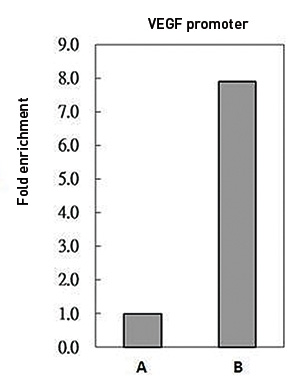
Figure 1. FUBP1 ChIP results
ChIP was performed with HeLa chromatin extract and 5 μg of either control rabbit IgG or FUBP1 antibody. The precipitated DNA was detected by PCR with primer set targeting to p21 FUSE.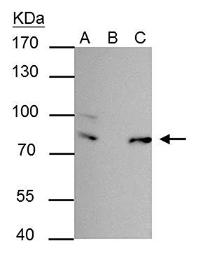
Figure 2. FUBP1 IP results
FUBP1 antibody immunoprecipitates FUBP1 protein in IP experiments. IP Sample: 293T whole cell extract A: 30 μg whole cell extract of FUBP1 protein expressing 293T cells B: Control with 2.5 μg of pre-immune rabbit IgG C : Immunoprecipitation of FUBP1 by 2.5 μg of FUBP1 antibody. The immunoprecipitated FUBP1 protein was detected by western blot with the FUBP1 antibody diluted 1:1,000.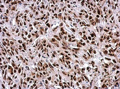
Figure 3. IHC
Immunohistochemical analysis of paraffin-embedded HeLa xenograft, using FUBP1 antibody at a 1:500 dilution.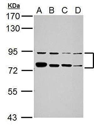
Figure 4. Western blot
Sample: 30 μg of whole cell lysate A: Jurkat B: K562 C: THP-1 D: NCI-H929 7.5%SDS PAGE FUBP1 antibody diluted 1:5,000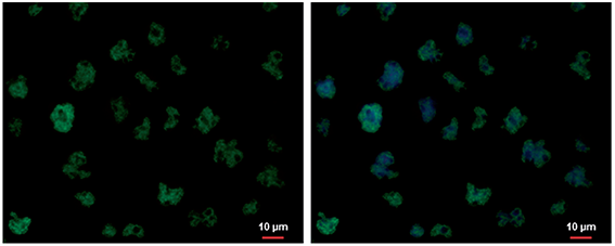
Figure 5. FUBP1 IF results
FUBP1 antibody detects FUBP1 protein at nucleus by immunofluorescent analysis. Sample: Jurkat cells were fixed in 4% paraformaldehyde at RT for 15 min. Green: FUBP1 protein stained by FUBP1 antibody diluted 1:500. Blue: Hoechst 33342 staining. - 出版物

How to properly cite our product/service in your work
We strongly recommend using this: FUBP1 polyclonal antibody (Hologic Diagenode Cat# C15410233-100 Lot# 40912). Click here to copy to clipboard.
Using our products or services in your publication? Let us know!