Notice (8): Undefined variable: solution_of_interest [APP/View/Products/view.ctp, line 755]Code Context<!-- BEGIN: REQUEST_FORM MODAL -->
<div id="request_formModal" class="reveal-modal medium" data-reveal aria-labelledby="modalTitle" aria-hidden="true" role="dialog">
<?= $this->element('Forms/simple_form', array('solution_of_interest' => $solution_of_interest, 'header' => $header, 'message' => $message, 'campaign_id' => $campaign_id)) ?>
$viewFile = '/home/website-server/www/app/View/Products/view.ctp'
$dataForView = array(
'language' => 'jp',
'meta_keywords' => '',
'meta_description' => 'H3K27ac polyclonal antibody - Classic',
'meta_title' => 'H3K27ac polyclonal antibody - Classic',
'product' => array(
'Product' => array(
'id' => '2260',
'antibody_id' => '108',
'name' => 'H3K27ac polyclonal antibody ',
'description' => '<p><span>Polyclonal antibody raised in rabbit against histone H3, acetylated at lysine 27 (H3K27ac), using a KLH-conjugated synthetic peptide.</span></p>',
'label1' => 'Validation Data',
'info1' => '<div class="row">
<div class="small-6 columns">
<p><img src="https://www.diagenode.com/img/product/antibodies/C15410174_ChIP.jpg" alt="H3K27ac Antibody ChIP Grade" caption="false" width="400" height="303" /></p>
</div>
<div class="small-6 columns">
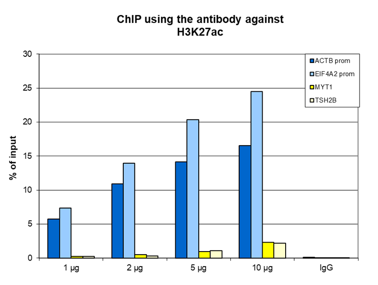
<p><small><strong>Figure 1. ChIP results obtained with the Diagenode antibody directed against H3K27ac</strong><br /> ChIP assays were performed using HeLa cells, the Diagenode antibody against H3K27ac (Cat. No. C15410174) and optimized PCR primer sets for qPCR. ChIP was performed with the “Auto Histone ChIP-seq” kit using sheared chromatin from 1 million cells. A titration of the antibody consisting of 1, 2, 5 and 10 µg per ChIP experiment was analysed. IgG (2 µg/IP) was used as negative IP control. Quantitative PCR was performed with primers for the promoter of the active ACTB and EIF4A2 genes, used as positive controls, and for the coding region of the inactive MYT1 and TSH2B genes, used as negative controls. Figure 1 shows the recovery, expressed as a % of input (the relative amount of immunoprecipitated DNA compared to input DNA after qPCR analysis). These results are in accordance with the observation that H3K27 acetylation is enriched at the promoters of active genes.</small></p>
</div>
</div>
<div class="row">
<div class="small-6 columns">
<p>A. <img src="https://www.diagenode.com/img/product/antibodies/C15410174_ChIPSeq_A.jpg" alt="H3K27ac Antibody for ChIP" caption="false" width="400" height="370" /></p>
</div>
<div class="small-6 columns">
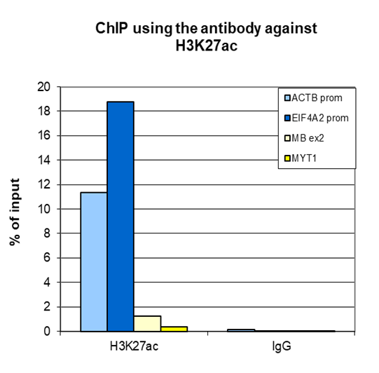
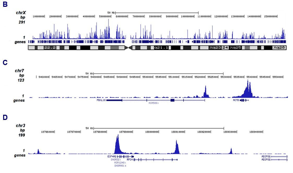
<p><small><strong>Figure 2. ChIP-seq results obtained with the Diagenode antibody directed against H3K27ac</strong><br /> ChIP was performed on sheared chromatin from 1 million HeLaS3 cells using 2 µg of the Diagenode antibody against H3K27ac (Cat. No. C15410174) as described above. Quantitative PCR was performed with primers for the promoter of the active ACTB and EIF4A2 genes, used as positive controls, and for the coding region of the inactive MYT1 and MB genes, used as negative controls (Figure 2A). The IP’d DNA was subsequently analysed on an Illumina Genome Analyzer. Library preparation, cluster generation and sequencing were performed according to the manufacturer’s instructions. The 36 bp tags were aligned to the human genome using the ELAND algorithm. Figure 2B shows the peak distribution along the complete X-chromosome and in two regions surrounding the ACTB and EIF4A2 positive control genes, respectively (figure 2C and D).</small></p>
</div>
</div>
<div class="extra-spaced"></div>
<div class="row">
<div class="small-12 columns">
<p>B. <img src="https://www.diagenode.com/img/product/antibodies/C15410174_ChIPSeq_B.jpg" alt="H3K27ac Antibody for ChIP-seq" caption="false" width="700" height="105" /></p>
<p>C. <img src="https://www.diagenode.com/img/product/antibodies/C15410174_ChIPSeq_C.jpg" alt="H3K27ac Antibody ChIP-seq Grade" caption="false" width="700" height="95" /></p>
<p>D. <img src="https://www.diagenode.com/img/product/antibodies/C15410174_ChIPSeq_D.jpg" alt="H3K27ac Antibody for ChIP-seq assay" caption="false" width="700" height="114" /></p>
</div>
</div>
<div class="row">
<div class="small-6 columns">
<p><img src="https://www.diagenode.com/img/product/antibodies/C15410174_ELISA.jpg" alt="H3K27ac Antibody ELISA Validation" caption="false" width="400" height="330" /></p>
</div>
<div class="small-6 columns">
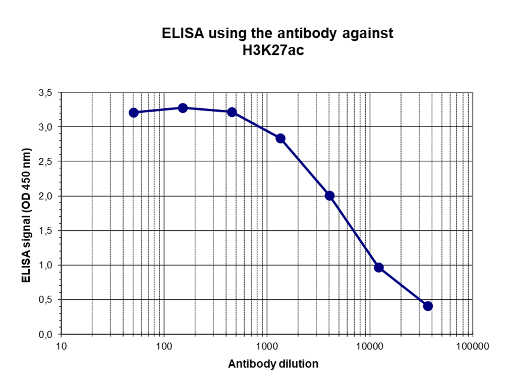
<p><small><strong>Figure 3. Determination of the antibody titer</strong><br /> To determine the titer of the antibody, an ELISA was performed using a serial dilution of the Diagenode antibody directed against H3K27ac (Cat. No. C15410174) in antigen coated wells. The antigen used was a peptide containing the histone modification of interest. By plotting the absorbance against the antibody dilution (Figure 3), the titer of the antibody was estimated to be 1:4,000. </small></p>
</div>
</div>
<div class="row">
<div class="small-6 columns">
<p><img src="https://www.diagenode.com/img/product/antibodies/C15410174_DotBlot.jpg" alt="H3K27ac Antibody Dot Blot Validation" caption="false" width="400" height="237" /></p>
</div>
<div class="small-6 columns">
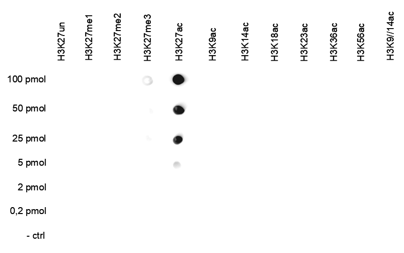
<p><small><strong>Figure 4. Cross reactivity test using the Diagenode antibody directed against H3K27ac</strong><br /> A Dot Blot analysis was performed to test the cross reactivity of the Diagenode antibody against H3K27ac (Cat. No. C15410174) with peptides containing other histone modifications and the unmodified H3K27 sequence. One hundred to 0.2 pmol of the respective peptides were spotted on a membrane. The antibody was used at a dilution of 1:25,000. Figure 4 shows a high specificity of the antibody for the modification of interest. </small></p>
</div>
</div>
<div class="extra-spaced"></div>
<div class="row">
<div class="small-6 columns">
<p><img src="https://www.diagenode.com/img/product/antibodies/C15410174_WB.jpg" alt="H3K27ac Antibody for Western Blot" caption="false" width="400" height="297" /></p>
</div>
<div class="small-6 columns">
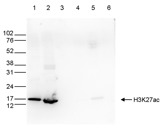
<p><small><strong>Figure 5. Western blot analysis using the Diagenode antibody directed against H3K27ac</strong><br /> Western blot was performed on whole cell (40 µg, lane 1) and histone extracts (15 µg, lane 2) from HeLa cells, and on 1 µg of recombinant histone H2A, H2B, H3 and H4 (lane 3, 4, 5 and 6, respectively) using the Diagenode antibody against H3K27ac (Cat. No. C15410174). The antibody was diluted 1:1,000 in TBS-Tween containing 5% skimmed milk. The position of the protein of interest is indicated on the right; the marker (in kDa) is shown on the left. </small></p>
</div>
</div>
<div class="row">
<div class="small-6 columns">
<p><img src="https://www.diagenode.com/img/product/antibodies/C15410174_IF.jpg" alt="H3K27ac Antibody for Immunofluorescence " caption="false" width="400" height="99" /></p>
</div>
<div class="small-6 columns">
<p><small><strong>Figure 6. Immunofluorescence using the Diagenode antibody directed against H3K27ac</strong><br /> HeLa cells were stained with the Diagenode antibody against H3K27ac (Cat. No. C15410174) and with DAPI. Cells were fixed with 4% formaldehyde for 10’ and blocked with PBS/TX-100 containing 5% normal goat serum and 1% BSA. The cells were immunofluorescently labelled with the H3K27ac antibody (left) diluted 1:500 in blocking solution followed by an anti-rabbit antibody conjugated to Alexa488. The middle panel shows staining of the nuclei with DAPI. A merge of the two stainings is shown on the right. </small></p>
</div>
</div>',
'label2' => '',
'info2' => '<p>Histones are the main constituents of the protein part of chromosomes of eukaryotic cells. They are rich in the amino acids arginine and lysine and have been greatly conserved during evolution. Histones pack the DNA into tight masses of chromatin. Two core histones of each class H2A, H2B, H3 and H4 assemble and are wrapped by 146 base pairs of DNA to form one octameric nucleosome. Histone tails undergo numerous post-translational modifications, which either directly or indirectly alter chromatin structure to facilitate transcriptional activation or repression or other nuclear processes. In addition to the genetic code, combinations of the different histone modifications reveal the so-called “histone code”. Histone methylation and demethylation is dynamically regulated by respectively histone methyl transferases and histone demethylases.</p>',
'label3' => '',
'info3' => '',
'format' => '50 μg',
'catalog_number' => 'C15410174',
'old_catalog_number' => 'pAb-174-050',
'sf_code' => 'C15410174-D001-000581',
'type' => 'FRE',
'search_order' => '03-Antibody',
'price_EUR' => '380',
'price_USD' => '380',
'price_GBP' => '340',
'price_JPY' => '59525',
'price_CNY' => '',
'price_AUD' => '950',
'country' => 'ALL',
'except_countries' => 'None',
'quote' => false,
'in_stock' => false,
'featured' => false,
'no_promo' => false,
'online' => true,
'master' => true,
'last_datasheet_update' => 'July 26, 2019',
'slug' => 'h3k27ac-polyclonal-antibody-classic-50-mg-42-ml',
'meta_title' => 'H3K27ac polyclonal antibody - Classic',
'meta_keywords' => '',
'meta_description' => 'H3K27ac polyclonal antibody - Classic',
'modified' => '2024-01-17 16:45:16',
'created' => '2015-06-29 14:08:20',
'locale' => 'jpn'
),
'Antibody' => array(
'host' => '*****',
'id' => '108',
'name' => 'H3K27ac polyclonal antibody',
'description' => 'Histones are the main constituents of the protein part of chromosomes of eukaryotic cells. They are rich in the amino acids arginine and lysine and have been greatly conserved during evolution. Histones pack the DNA into tight masses of chromatin. Two core histones of each class H2A, H2B, H3 and H4 assemble and are wrapped by 146 base pairs of DNA to form one octameric nucleosome. Histone tails undergo numerous post-translational modifications, which either directly or indirectly alter chromatin structure to facilitate transcriptional activation or repression or other nuclear processes. In addition to the genetic code, combinations of the different histone modifications reveal the so-called “histone code”. Histone methylation and demethylation is dynamically regulated by respectively histone methyl transferases and histone demethylases.',
'clonality' => '',
'isotype' => '',
'lot' => 'A7071-001P',
'concentration' => '1.2 µg/µl',
'reactivity' => 'Human, mouse, rat, pig: positive. Other species: not tested.',
'type' => 'Polyclonal',
'purity' => 'Affinity purified polyclonal antibody.',
'classification' => 'Classic',
'application_table' => '<table>
<thead>
<tr>
<th>Applications</th>
<th>Suggested dilution</th>
<th>References</th>
</tr>
</thead>
<tbody>
<tr>
<td>ChIP/ChIP-seq <sup>*</sup></td>
<td>1-2 µg/ChIP</td>
<td>Fig 1, 2</td>
</tr>
<tr>
<td>ELISA</td>
<td>1:100</td>
<td>Fig 3</td>
</tr>
<tr>
<td>Dot Blotting</td>
<td>1:25,000</td>
<td>Fig 4</td>
</tr>
<tr>
<td>Western Blotting</td>
<td>1:1,000</td>
<td>Fig 5</td>
</tr>
<tr>
<td>Immunofluorescence</td>
<td>1:500</td>
<td>Fig 6</td>
</tr>
</tbody>
</table>
<p></p>
<p><small><sup>*</sup> Please note that the optimal antibody amount per IP should be determined by the end-user. We recommend testing 1-5 µg per IP.</small></p>',
'storage_conditions' => 'Store at -20°C; for long storage, store at -80°C. Avoid multiple freeze-thaw cycles.',
'storage_buffer' => 'PBS containing 0.05% azide and 0.05% ProClin 300.',
'precautions' => 'This product is for research use only. Not for use in diagnostic or therapeutic procedures.',
'uniprot_acc' => '',
'slug' => '',
'meta_keywords' => '',
'meta_description' => '',
'modified' => '2022-08-04 13:01:05',
'created' => '0000-00-00 00:00:00',
'select_label' => '108 - H3K27ac polyclonal antibody (A7071-001P - 1.2 µg/µl - Human, mouse, rat, pig: positive. Other species: not tested. - Affinity purified polyclonal antibody. - Rabbit)'
),
'Slave' => array(
(int) 0 => array(
[maximum depth reached]
)
),
'Group' => array(
'Group' => array(
[maximum depth reached]
),
'Master' => array(
[maximum depth reached]
),
'Product' => array(
[maximum depth reached]
)
),
'Related' => array(
(int) 0 => array(
[maximum depth reached]
),
(int) 1 => array(
[maximum depth reached]
),
(int) 2 => array(
[maximum depth reached]
),
(int) 3 => array(
[maximum depth reached]
),
(int) 4 => array(
[maximum depth reached]
),
(int) 5 => array(
[maximum depth reached]
),
(int) 6 => array(
[maximum depth reached]
)
),
'Application' => array(
(int) 0 => array(
[maximum depth reached]
),
(int) 1 => array(
[maximum depth reached]
),
(int) 2 => array(
[maximum depth reached]
),
(int) 3 => array(
[maximum depth reached]
),
(int) 4 => array(
[maximum depth reached]
),
(int) 5 => array(
[maximum depth reached]
)
),
'Category' => array(
(int) 0 => array(
[maximum depth reached]
),
(int) 1 => array(
[maximum depth reached]
),
(int) 2 => array(
[maximum depth reached]
),
(int) 3 => array(
[maximum depth reached]
)
),
'Document' => array(
(int) 0 => array(
[maximum depth reached]
),
(int) 1 => array(
[maximum depth reached]
),
(int) 2 => array(
[maximum depth reached]
)
),
'Feature' => array(),
'Image' => array(
(int) 0 => array(
[maximum depth reached]
)
),
'Promotion' => array(),
'Protocol' => array(),
'Publication' => array(
(int) 0 => array(
[maximum depth reached]
),
(int) 1 => array(
[maximum depth reached]
),
(int) 2 => array(
[maximum depth reached]
),
(int) 3 => array(
[maximum depth reached]
),
(int) 4 => array(
[maximum depth reached]
),
(int) 5 => array(
[maximum depth reached]
),
(int) 6 => array(
[maximum depth reached]
),
(int) 7 => array(
[maximum depth reached]
),
(int) 8 => array(
[maximum depth reached]
),
(int) 9 => array(
[maximum depth reached]
),
(int) 10 => array(
[maximum depth reached]
),
(int) 11 => array(
[maximum depth reached]
),
(int) 12 => array(
[maximum depth reached]
),
(int) 13 => array(
[maximum depth reached]
),
(int) 14 => array(
[maximum depth reached]
),
(int) 15 => array(
[maximum depth reached]
),
(int) 16 => array(
[maximum depth reached]
),
(int) 17 => array(
[maximum depth reached]
),
(int) 18 => array(
[maximum depth reached]
),
(int) 19 => array(
[maximum depth reached]
),
(int) 20 => array(
[maximum depth reached]
),
(int) 21 => array(
[maximum depth reached]
),
(int) 22 => array(
[maximum depth reached]
),
(int) 23 => array(
[maximum depth reached]
),
(int) 24 => array(
[maximum depth reached]
),
(int) 25 => array(
[maximum depth reached]
),
(int) 26 => array(
[maximum depth reached]
),
(int) 27 => array(
[maximum depth reached]
),
(int) 28 => array(
[maximum depth reached]
),
(int) 29 => array(
[maximum depth reached]
),
(int) 30 => array(
[maximum depth reached]
),
(int) 31 => array(
[maximum depth reached]
),
(int) 32 => array(
[maximum depth reached]
),
(int) 33 => array(
[maximum depth reached]
),
(int) 34 => array(
[maximum depth reached]
),
(int) 35 => array(
[maximum depth reached]
),
(int) 36 => array(
[maximum depth reached]
),
(int) 37 => array(
[maximum depth reached]
),
(int) 38 => array(
[maximum depth reached]
),
(int) 39 => array(
[maximum depth reached]
),
(int) 40 => array(
[maximum depth reached]
),
(int) 41 => array(
[maximum depth reached]
),
(int) 42 => array(
[maximum depth reached]
),
(int) 43 => array(
[maximum depth reached]
),
(int) 44 => array(
[maximum depth reached]
),
(int) 45 => array(
[maximum depth reached]
),
(int) 46 => array(
[maximum depth reached]
)
),
'Testimonial' => array(),
'Area' => array(),
'SafetySheet' => array(
(int) 0 => array(
[maximum depth reached]
),
(int) 1 => array(
[maximum depth reached]
),
(int) 2 => array(
[maximum depth reached]
),
(int) 3 => array(
[maximum depth reached]
),
(int) 4 => array(
[maximum depth reached]
),
(int) 5 => array(
[maximum depth reached]
),
(int) 6 => array(
[maximum depth reached]
),
(int) 7 => array(
[maximum depth reached]
)
)
),
'meta_canonical' => 'https://www.diagenode.com/jp/p/h3k27ac-polyclonal-antibody-classic-50-mg-42-ml'
)
$language = 'jp'
$meta_keywords = ''
$meta_description = 'H3K27ac polyclonal antibody - Classic'
$meta_title = 'H3K27ac polyclonal antibody - Classic'
$product = array(
'Product' => array(
'id' => '2260',
'antibody_id' => '108',
'name' => 'H3K27ac polyclonal antibody ',
'description' => '<p><span>Polyclonal antibody raised in rabbit against histone H3, acetylated at lysine 27 (H3K27ac), using a KLH-conjugated synthetic peptide.</span></p>',
'label1' => 'Validation Data',
'info1' => '<div class="row">
<div class="small-6 columns">
<p><img src="https://www.diagenode.com/img/product/antibodies/C15410174_ChIP.jpg" alt="H3K27ac Antibody ChIP Grade" caption="false" width="400" height="303" /></p>
</div>
<div class="small-6 columns">
<p><small><strong>Figure 1. ChIP results obtained with the Diagenode antibody directed against H3K27ac</strong><br /> ChIP assays were performed using HeLa cells, the Diagenode antibody against H3K27ac (Cat. No. C15410174) and optimized PCR primer sets for qPCR. ChIP was performed with the “Auto Histone ChIP-seq” kit using sheared chromatin from 1 million cells. A titration of the antibody consisting of 1, 2, 5 and 10 µg per ChIP experiment was analysed. IgG (2 µg/IP) was used as negative IP control. Quantitative PCR was performed with primers for the promoter of the active ACTB and EIF4A2 genes, used as positive controls, and for the coding region of the inactive MYT1 and TSH2B genes, used as negative controls. Figure 1 shows the recovery, expressed as a % of input (the relative amount of immunoprecipitated DNA compared to input DNA after qPCR analysis). These results are in accordance with the observation that H3K27 acetylation is enriched at the promoters of active genes.</small></p>
</div>
</div>
<div class="row">
<div class="small-6 columns">
<p>A. <img src="https://www.diagenode.com/img/product/antibodies/C15410174_ChIPSeq_A.jpg" alt="H3K27ac Antibody for ChIP" caption="false" width="400" height="370" /></p>
</div>
<div class="small-6 columns">
<p><small><strong>Figure 2. ChIP-seq results obtained with the Diagenode antibody directed against H3K27ac</strong><br /> ChIP was performed on sheared chromatin from 1 million HeLaS3 cells using 2 µg of the Diagenode antibody against H3K27ac (Cat. No. C15410174) as described above. Quantitative PCR was performed with primers for the promoter of the active ACTB and EIF4A2 genes, used as positive controls, and for the coding region of the inactive MYT1 and MB genes, used as negative controls (Figure 2A). The IP’d DNA was subsequently analysed on an Illumina Genome Analyzer. Library preparation, cluster generation and sequencing were performed according to the manufacturer’s instructions. The 36 bp tags were aligned to the human genome using the ELAND algorithm. Figure 2B shows the peak distribution along the complete X-chromosome and in two regions surrounding the ACTB and EIF4A2 positive control genes, respectively (figure 2C and D).</small></p>
</div>
</div>
<div class="extra-spaced"></div>
<div class="row">
<div class="small-12 columns">
<p>B. <img src="https://www.diagenode.com/img/product/antibodies/C15410174_ChIPSeq_B.jpg" alt="H3K27ac Antibody for ChIP-seq" caption="false" width="700" height="105" /></p>
<p>C. <img src="https://www.diagenode.com/img/product/antibodies/C15410174_ChIPSeq_C.jpg" alt="H3K27ac Antibody ChIP-seq Grade" caption="false" width="700" height="95" /></p>
<p>D. <img src="https://www.diagenode.com/img/product/antibodies/C15410174_ChIPSeq_D.jpg" alt="H3K27ac Antibody for ChIP-seq assay" caption="false" width="700" height="114" /></p>
</div>
</div>
<div class="row">
<div class="small-6 columns">
<p><img src="https://www.diagenode.com/img/product/antibodies/C15410174_ELISA.jpg" alt="H3K27ac Antibody ELISA Validation" caption="false" width="400" height="330" /></p>
</div>
<div class="small-6 columns">
<p><small><strong>Figure 3. Determination of the antibody titer</strong><br /> To determine the titer of the antibody, an ELISA was performed using a serial dilution of the Diagenode antibody directed against H3K27ac (Cat. No. C15410174) in antigen coated wells. The antigen used was a peptide containing the histone modification of interest. By plotting the absorbance against the antibody dilution (Figure 3), the titer of the antibody was estimated to be 1:4,000. </small></p>
</div>
</div>
<div class="row">
<div class="small-6 columns">
<p><img src="https://www.diagenode.com/img/product/antibodies/C15410174_DotBlot.jpg" alt="H3K27ac Antibody Dot Blot Validation" caption="false" width="400" height="237" /></p>
</div>
<div class="small-6 columns">
<p><small><strong>Figure 4. Cross reactivity test using the Diagenode antibody directed against H3K27ac</strong><br /> A Dot Blot analysis was performed to test the cross reactivity of the Diagenode antibody against H3K27ac (Cat. No. C15410174) with peptides containing other histone modifications and the unmodified H3K27 sequence. One hundred to 0.2 pmol of the respective peptides were spotted on a membrane. The antibody was used at a dilution of 1:25,000. Figure 4 shows a high specificity of the antibody for the modification of interest. </small></p>
</div>
</div>
<div class="extra-spaced"></div>
<div class="row">
<div class="small-6 columns">
<p><img src="https://www.diagenode.com/img/product/antibodies/C15410174_WB.jpg" alt="H3K27ac Antibody for Western Blot" caption="false" width="400" height="297" /></p>
</div>
<div class="small-6 columns">
<p><small><strong>Figure 5. Western blot analysis using the Diagenode antibody directed against H3K27ac</strong><br /> Western blot was performed on whole cell (40 µg, lane 1) and histone extracts (15 µg, lane 2) from HeLa cells, and on 1 µg of recombinant histone H2A, H2B, H3 and H4 (lane 3, 4, 5 and 6, respectively) using the Diagenode antibody against H3K27ac (Cat. No. C15410174). The antibody was diluted 1:1,000 in TBS-Tween containing 5% skimmed milk. The position of the protein of interest is indicated on the right; the marker (in kDa) is shown on the left. </small></p>
</div>
</div>
<div class="row">
<div class="small-6 columns">
<p><img src="https://www.diagenode.com/img/product/antibodies/C15410174_IF.jpg" alt="H3K27ac Antibody for Immunofluorescence " caption="false" width="400" height="99" /></p>
</div>
<div class="small-6 columns">
<p><small><strong>Figure 6. Immunofluorescence using the Diagenode antibody directed against H3K27ac</strong><br /> HeLa cells were stained with the Diagenode antibody against H3K27ac (Cat. No. C15410174) and with DAPI. Cells were fixed with 4% formaldehyde for 10’ and blocked with PBS/TX-100 containing 5% normal goat serum and 1% BSA. The cells were immunofluorescently labelled with the H3K27ac antibody (left) diluted 1:500 in blocking solution followed by an anti-rabbit antibody conjugated to Alexa488. The middle panel shows staining of the nuclei with DAPI. A merge of the two stainings is shown on the right. </small></p>
</div>
</div>',
'label2' => 'Target Description',
'info2' => '<p>Histones are the main constituents of the protein part of chromosomes of eukaryotic cells. They are rich in the amino acids arginine and lysine and have been greatly conserved during evolution. Histones pack the DNA into tight masses of chromatin. Two core histones of each class H2A, H2B, H3 and H4 assemble and are wrapped by 146 base pairs of DNA to form one octameric nucleosome. Histone tails undergo numerous post-translational modifications, which either directly or indirectly alter chromatin structure to facilitate transcriptional activation or repression or other nuclear processes. In addition to the genetic code, combinations of the different histone modifications reveal the so-called “histone code”. Histone methylation and demethylation is dynamically regulated by respectively histone methyl transferases and histone demethylases.</p>',
'label3' => '',
'info3' => '',
'format' => '50 μg',
'catalog_number' => 'C15410174',
'old_catalog_number' => 'pAb-174-050',
'sf_code' => 'C15410174-D001-000581',
'type' => 'FRE',
'search_order' => '03-Antibody',
'price_EUR' => '380',
'price_USD' => '380',
'price_GBP' => '340',
'price_JPY' => '59525',
'price_CNY' => '',
'price_AUD' => '950',
'country' => 'ALL',
'except_countries' => 'None',
'quote' => false,
'in_stock' => false,
'featured' => false,
'no_promo' => false,
'online' => true,
'master' => true,
'last_datasheet_update' => 'July 26, 2019',
'slug' => 'h3k27ac-polyclonal-antibody-classic-50-mg-42-ml',
'meta_title' => 'H3K27ac polyclonal antibody - Classic',
'meta_keywords' => '',
'meta_description' => 'H3K27ac polyclonal antibody - Classic',
'modified' => '2024-01-17 16:45:16',
'created' => '2015-06-29 14:08:20',
'locale' => 'jpn'
),
'Antibody' => array(
'host' => '*****',
'id' => '108',
'name' => 'H3K27ac polyclonal antibody',
'description' => 'Histones are the main constituents of the protein part of chromosomes of eukaryotic cells. They are rich in the amino acids arginine and lysine and have been greatly conserved during evolution. Histones pack the DNA into tight masses of chromatin. Two core histones of each class H2A, H2B, H3 and H4 assemble and are wrapped by 146 base pairs of DNA to form one octameric nucleosome. Histone tails undergo numerous post-translational modifications, which either directly or indirectly alter chromatin structure to facilitate transcriptional activation or repression or other nuclear processes. In addition to the genetic code, combinations of the different histone modifications reveal the so-called “histone code”. Histone methylation and demethylation is dynamically regulated by respectively histone methyl transferases and histone demethylases.',
'clonality' => '',
'isotype' => '',
'lot' => 'A7071-001P',
'concentration' => '1.2 µg/µl',
'reactivity' => 'Human, mouse, rat, pig: positive. Other species: not tested.',
'type' => 'Polyclonal',
'purity' => 'Affinity purified polyclonal antibody.',
'classification' => 'Classic',
'application_table' => '<table>
<thead>
<tr>
<th>Applications</th>
<th>Suggested dilution</th>
<th>References</th>
</tr>
</thead>
<tbody>
<tr>
<td>ChIP/ChIP-seq <sup>*</sup></td>
<td>1-2 µg/ChIP</td>
<td>Fig 1, 2</td>
</tr>
<tr>
<td>ELISA</td>
<td>1:100</td>
<td>Fig 3</td>
</tr>
<tr>
<td>Dot Blotting</td>
<td>1:25,000</td>
<td>Fig 4</td>
</tr>
<tr>
<td>Western Blotting</td>
<td>1:1,000</td>
<td>Fig 5</td>
</tr>
<tr>
<td>Immunofluorescence</td>
<td>1:500</td>
<td>Fig 6</td>
</tr>
</tbody>
</table>
<p></p>
<p><small><sup>*</sup> Please note that the optimal antibody amount per IP should be determined by the end-user. We recommend testing 1-5 µg per IP.</small></p>',
'storage_conditions' => 'Store at -20°C; for long storage, store at -80°C. Avoid multiple freeze-thaw cycles.',
'storage_buffer' => 'PBS containing 0.05% azide and 0.05% ProClin 300.',
'precautions' => 'This product is for research use only. Not for use in diagnostic or therapeutic procedures.',
'uniprot_acc' => '',
'slug' => '',
'meta_keywords' => '',
'meta_description' => '',
'modified' => '2022-08-04 13:01:05',
'created' => '0000-00-00 00:00:00',
'select_label' => '108 - H3K27ac polyclonal antibody (A7071-001P - 1.2 µg/µl - Human, mouse, rat, pig: positive. Other species: not tested. - Affinity purified polyclonal antibody. - Rabbit)'
),
'Slave' => array(
(int) 0 => array(
'id' => '24',
'name' => 'C15410174',
'product_id' => '2260',
'modified' => '2016-02-18 18:04:50',
'created' => '2016-02-18 18:04:50'
)
),
'Group' => array(
'Group' => array(
'id' => '24',
'name' => 'C15410174',
'product_id' => '2260',
'modified' => '2016-02-18 18:04:50',
'created' => '2016-02-18 18:04:50'
),
'Master' => array(
'id' => '2260',
'antibody_id' => '108',
'name' => 'H3K27ac Antibody',
'description' => '<p><span>Polyclonal antibody raised in rabbit against histone <strong>H3, acetylated at lysine 27</strong> (<strong>H3K27ac</strong>), using a KLH-conjugated synthetic peptide.</span></p>',
'label1' => 'Validation Data',
'info1' => '<div class="row">
<div class="small-6 columns">
<p><img src="https://www.diagenode.com/img/product/antibodies/C15410174_ChIP.jpg" alt="H3K27ac Antibody ChIP Grade" caption="false" width="400" height="303" /></p>
</div>
<div class="small-6 columns">
<p><small><strong>Figure 1. ChIP results obtained with the Diagenode antibody directed against H3K27ac</strong><br /> ChIP assays were performed using HeLa cells, the Diagenode antibody against H3K27ac (Cat. No. C15410174) and optimized PCR primer sets for qPCR. ChIP was performed with the “Auto Histone ChIP-seq” kit using sheared chromatin from 1 million cells. A titration of the antibody consisting of 1, 2, 5 and 10 µg per ChIP experiment was analysed. IgG (2 µg/IP) was used as negative IP control. Quantitative PCR was performed with primers for the promoter of the active ACTB and EIF4A2 genes, used as positive controls, and for the coding region of the inactive MYT1 and TSH2B genes, used as negative controls. Figure 1 shows the recovery, expressed as a % of input (the relative amount of immunoprecipitated DNA compared to input DNA after qPCR analysis). These results are in accordance with the observation that H3K27 acetylation is enriched at the promoters of active genes.</small></p>
</div>
</div>
<div class="row">
<div class="small-6 columns">
<p>A. <img src="https://www.diagenode.com/img/product/antibodies/C15410174_ChIPSeq_A.jpg" alt="H3K27ac Antibody for ChIP" caption="false" width="400" height="370" /></p>
</div>
<div class="small-6 columns">
<p><small><strong>Figure 2. ChIP-seq results obtained with the Diagenode antibody directed against H3K27ac</strong><br /> ChIP was performed on sheared chromatin from 1 million HeLaS3 cells using 2 µg of the Diagenode antibody against H3K27ac (Cat. No. C15410174) as described above. Quantitative PCR was performed with primers for the promoter of the active ACTB and EIF4A2 genes, used as positive controls, and for the coding region of the inactive MYT1 and MB genes, used as negative controls (Figure 2A). The IP’d DNA was subsequently analysed on an Illumina Genome Analyzer. Library preparation, cluster generation and sequencing were performed according to the manufacturer’s instructions. The 36 bp tags were aligned to the human genome using the ELAND algorithm. Figure 2B shows the peak distribution along the complete X-chromosome and in two regions surrounding the ACTB and EIF4A2 positive control genes, respectively (figure 2C and D).</small></p>
</div>
</div>
<div class="extra-spaced"></div>
<div class="row">
<div class="small-12 columns">
<p>B. <img src="https://www.diagenode.com/img/product/antibodies/C15410174_ChIPSeq_B.jpg" alt="H3K27ac Antibody for ChIP-seq" caption="false" width="700" height="105" /></p>
<p>C. <img src="https://www.diagenode.com/img/product/antibodies/C15410174_ChIPSeq_C.jpg" alt="H3K27ac Antibody ChIP-seq Grade" caption="false" width="700" height="95" /></p>
<p>D. <img src="https://www.diagenode.com/img/product/antibodies/C15410174_ChIPSeq_D.jpg" alt="H3K27ac Antibody for ChIP-seq assay" caption="false" width="700" height="114" /></p>
</div>
</div>
<div class="row">
<div class="small-6 columns">
<p><img src="https://www.diagenode.com/img/product/antibodies/C15410174_ELISA.jpg" alt="H3K27ac Antibody ELISA Validation" caption="false" width="400" height="330" /></p>
</div>
<div class="small-6 columns">
<p><small><strong>Figure 3. Determination of the antibody titer</strong><br /> To determine the titer of the antibody, an ELISA was performed using a serial dilution of the Diagenode antibody directed against H3K27ac (Cat. No. C15410174) in antigen coated wells. The antigen used was a peptide containing the histone modification of interest. By plotting the absorbance against the antibody dilution (Figure 3), the titer of the antibody was estimated to be 1:4,000. </small></p>
</div>
</div>
<div class="row">
<div class="small-6 columns">
<p><img src="https://www.diagenode.com/img/product/antibodies/C15410174_DotBlot.jpg" alt="H3K27ac Antibody Dot Blot Validation" caption="false" width="400" height="237" /></p>
</div>
<div class="small-6 columns">
<p><small><strong>Figure 4. Cross reactivity test using the Diagenode antibody directed against H3K27ac</strong><br /> A Dot Blot analysis was performed to test the cross reactivity of the Diagenode antibody against H3K27ac (Cat. No. C15410174) with peptides containing other histone modifications and the unmodified H3K27 sequence. One hundred to 0.2 pmol of the respective peptides were spotted on a membrane. The antibody was used at a dilution of 1:25,000. Figure 4 shows a high specificity of the antibody for the modification of interest. </small></p>
</div>
</div>
<div class="extra-spaced"></div>
<div class="row">
<div class="small-6 columns">
<p><img src="https://www.diagenode.com/img/product/antibodies/C15410174_WB.jpg" alt="H3K27ac Antibody for Western Blot" caption="false" width="400" height="297" /></p>
</div>
<div class="small-6 columns">
<p><small><strong>Figure 5. Western blot analysis using the Diagenode antibody directed against H3K27ac</strong><br /> Western blot was performed on whole cell (40 µg, lane 1) and histone extracts (15 µg, lane 2) from HeLa cells, and on 1 µg of recombinant histone H2A, H2B, H3 and H4 (lane 3, 4, 5 and 6, respectively) using the Diagenode antibody against H3K27ac (Cat. No. C15410174). The antibody was diluted 1:1,000 in TBS-Tween containing 5% skimmed milk. The position of the protein of interest is indicated on the right; the marker (in kDa) is shown on the left. </small></p>
</div>
</div>
<div class="row">
<div class="small-6 columns">
<p><img src="https://www.diagenode.com/img/product/antibodies/C15410174_IF.jpg" alt="H3K27ac Antibody for Immunofluorescence " caption="false" width="400" height="99" /></p>
</div>
<div class="small-6 columns">
<p><small><strong>Figure 6. Immunofluorescence using the Diagenode antibody directed against H3K27ac</strong><br /> HeLa cells were stained with the Diagenode antibody against H3K27ac (Cat. No. C15410174) and with DAPI. Cells were fixed with 4% formaldehyde for 10’ and blocked with PBS/TX-100 containing 5% normal goat serum and 1% BSA. The cells were immunofluorescently labelled with the H3K27ac antibody (left) diluted 1:500 in blocking solution followed by an anti-rabbit antibody conjugated to Alexa488. The middle panel shows staining of the nuclei with DAPI. A merge of the two stainings is shown on the right. </small></p>
</div>
</div>',
'label2' => 'Target Description',
'info2' => '<p>Histones are the main constituents of the protein part of chromosomes of eukaryotic cells. They are rich in the amino acids arginine and lysine and have been greatly conserved during evolution. Histones pack the DNA into tight masses of chromatin. Two core histones of each class H2A, H2B, H3 and H4 assemble and are wrapped by 146 base pairs of DNA to form one octameric nucleosome. Histone tails undergo numerous post-translational modifications, which either directly or indirectly alter chromatin structure to facilitate transcriptional activation or repression or other nuclear processes. In addition to the genetic code, combinations of the different histone modifications reveal the so-called “histone code”. Histone methylation and demethylation is dynamically regulated by respectively histone methyl transferases and histone demethylases.</p>',
'label3' => '',
'info3' => '',
'format' => '50 μg',
'catalog_number' => 'C15410174',
'old_catalog_number' => 'pAb-174-050',
'sf_code' => 'C15410174-D001-000581',
'type' => 'FRE',
'search_order' => '03-Antibody',
'price_EUR' => '380',
'price_USD' => '380',
'price_GBP' => '340',
'price_JPY' => '59525',
'price_CNY' => '',
'price_AUD' => '950',
'country' => 'ALL',
'except_countries' => 'None',
'quote' => false,
'in_stock' => false,
'featured' => false,
'no_promo' => false,
'online' => true,
'master' => true,
'last_datasheet_update' => 'July 26, 2019',
'slug' => 'h3k27ac-polyclonal-antibody-classic-50-mg-42-ml',
'meta_title' => 'H3K27ac Antibody - ChIP-seq Grade (C15410174) | Diagenode',
'meta_keywords' => '',
'meta_description' => 'H3K27ac (Histone H3 acetylated at lysine 27) Polyclonal Antibody validated in ChIP-seq, ChIP-qPCR, ELISA, DB, WB and IF. Batch-specific data available on the website. Sample size available. ',
'modified' => '2024-01-17 16:45:16',
'created' => '2015-06-29 14:08:20'
),
'Product' => array(
(int) 0 => array(
[maximum depth reached]
)
)
),
'Related' => array(
(int) 0 => array(
'id' => '1787',
'antibody_id' => null,
'name' => 'Bioruptor<sup>®</sup> Pico sonication device',
'description' => '<p><a href="https://go.diagenode.com/bioruptor-upgrade"><img src="https://www.diagenode.com/img/banners/banner-br-trade.png" /></a></p>
<p>The Bioruptor® Pico (2013-2019) represented a breakthrough for shearing micro-volumes of 5 μl to larger volumes of up to 2 ml. <span>The new generation keeps the features you like the most and bring even more innovation. Check it now:</span></p>
<center><span></span></center><center><a href="https://www.diagenode.com/p/bioruptorpico2"> <img alt="New Bioruptor Pico" src="https://www.diagenode.com/img/product/shearing_technologies/new-pico-product-banner.jpg" /></a></center>
<p></p>
<p><span>Watch our short video about the Bioruptor Pico and how it can help you accomplish perfect shearing for any application including chromatin shearing, DNA shearing for NGS, unmatched DNA extraction from FFPE samples, RNA shearing, protein extraction, and much more.</span></p>
<p>
<script>// <![CDATA[
(function(){var qs,js,q,s,d=document,gi=d.getElementById,ce=d.createElement,gt=d.getElementsByTagName,id='typef_orm',b='https://s3-eu-west-1.amazonaws.com/share.typeform.com/';if(!gi.call(d,id)){js=ce.call(d,'script');js.id=id;js.src=b+'share.js';q=gt.call(d,'script')[0];q.parentNode.insertBefore(js,q)}id=id+'_';if(!gi.call(d,id)){qs=ce.call(d,'link');qs.rel='stylesheet';qs.id=id;qs.href=b+'share-button.css';s=gt.call(d,'head')[0];s.appendChild(qs,s)}})()
// ]]></script>
</p>
<center><iframe width="560" height="315" src="https://www.youtube.com/embed/ckLc4owudIM" frameborder="0" allowfullscreen="allowfullscreen"></iframe></center><center>
<p></p>
</center><center><a href="https://www.diagenode.com/en/pages/osha"><img src="https://www.diagenode.com/img/banners/banner-osha-580.jpg" width="635" height="243" /></a></center>
<div id="ConnectiveDocSignExtentionInstalled" data-extension-version="1.0.4"></div>
<div id="ConnectiveDocSignExtentionInstalled" data-extension-version="1.0.4"></div>',
'label1' => 'User manual ',
'info1' => '<p><a href="https://www.diagenode.com/files/products/shearing_technology/bioruptor/Bioruptor_pico_cooler_manual.pdf">Download</a></p>
<div id="ConnectiveDocSignExtentionInstalled" data-extension-version="1.0.4"></div>
<script src="chrome-extension://hhojmcideegachlhfgfdhailpfhgknjm/web_accessible_resources/index.js"></script>
<div id="ConnectiveDocSignExtentionInstalled" data-extension-version="1.0.4"></div>
<div id="ConnectiveDocSignExtentionInstalled" data-extension-version="1.0.4"></div>',
'label2' => 'Recommended settings for DNA shearing with Bioruptor® Pico',
'info2' => '<p>Follow our guidelines and find the good parameters for your expected DNA size: <a href="https://pybrevet.typeform.com/to/o8cQfM">DNA shearing with the Bioruptor® Pico</a></p>
<p></p>
<p>
<script>// <![CDATA[
(function(){var qs,js,q,s,d=document,gi=d.getElementById,ce=d.createElement,gt=d.getElementsByTagName,id='typef_orm',b='https://s3-eu-west-1.amazonaws.com/share.typeform.com/';if(!gi.call(d,id)){js=ce.call(d,'script');js.id=id;js.src=b+'share.js';q=gt.call(d,'script')[0];q.parentNode.insertBefore(js,q)}id=id+'_';if(!gi.call(d,id)){qs=ce.call(d,'link');qs.rel='stylesheet';qs.id=id;qs.href=b+'share-button.css';s=gt.call(d,'head')[0];s.appendChild(qs,s)}})()
// ]]></script>
</p>
<div id="ConnectiveDocSignExtentionInstalled" data-extension-version="1.0.4"></div>
<script src="chrome-extension://hhojmcideegachlhfgfdhailpfhgknjm/web_accessible_resources/index.js"></script>
<div id="ConnectiveDocSignExtentionInstalled" data-extension-version="1.0.4"></div>
<div id="ConnectiveDocSignExtentionInstalled" data-extension-version="1.0.4"></div>',
'label3' => 'Available chromatin shearing kits',
'info3' => '<p>It is important to establish optimal conditions to shear crosslinked chromatin to get the correct fragment sizes needed for ChIP. Usually this process requires both optimizing sonication conditions as well as optimizing SDS concentration, which is laborious. With the Chromatin Shearing Optimization Kits, optimization is fast and easy - we provide optimization reagents with varying concentrations of SDS. Moreover, our Chromatin Shearing Optimization Kits can be used for the optimization of chromatin preparation with our kits for ChIP.</p>
<table style="width: 925px;">
<tbody>
<tr valign="middle">
<td style="width: 213px;"></td>
<td style="text-align: center; width: 208px;"><strong><a href="../p/chromatin-shearing-optimization-kit-low-sds-100-million-cells">Chromatin Shearing Kit Low SDS (for Histones)</a></strong></td>
<td style="text-align: center; width: 180px;"><strong><a href="../p/chromatin-shearing-optimization-kit-low-sds-for-tfs-25-rxns">Chromatin Shearing Kit Low SDS (for TF)</a></strong></td>
<td style="text-align: center; width: 154px;"><strong><a href="../p/chromatin-shearing-optimization-kit-high-sds-100-million-cells">Chromatin Shearing Kit High SDS</a></strong></td>
<td style="text-align: center; width: 155px;"><strong><a href="../p/chromatin-shearing-plant-chip-seq-kit">Chromatin Shearing Kit (for Plant)</a></strong></td>
</tr>
<tr style="background-color: #fff;" valign="middle">
<td style="width: 213px;">
<p style="text-align: left;"><strong>SDS concentration</strong></p>
</td>
<td style="text-align: center; width: 208px;">
<p style="text-align: center;">< 0.1%</p>
</td>
<td style="text-align: center; width: 180px;">
<p style="text-align: center;">0.2%</p>
</td>
<td style="text-align: center; width: 154px;">
<p style="text-align: center;">1%</p>
</td>
<td style="text-align: center; width: 155px;">
<p style="text-align: center;">0.5%</p>
</td>
</tr>
<tr style="background-color: #fff;" valign="middle">
<td style="width: 213px;">
<p style="text-align: left;"><strong>Nuclei isolation</strong></p>
</td>
<td style="text-align: center; width: 208px;">
<p style="text-align: center;">Yes</p>
</td>
<td style="text-align: center; width: 180px;">
<p style="text-align: center;">Yes</p>
</td>
<td style="text-align: center; width: 154px;">
<p style="text-align: center;">No</p>
</td>
<td style="text-align: center; width: 155px;">
<p style="text-align: center;">Yes</p>
</td>
</tr>
<tr style="background-color: #fff;" valign="middle">
<td style="width: 213px;">
<p style="text-align: left;"><strong>Allows for shearing of... cells/tissue</strong></p>
</td>
<td style="text-align: center; width: 208px;">
<p style="text-align: center;">100 million cells</p>
</td>
<td style="text-align: center; width: 180px;">
<p style="text-align: center;">100 million cells</p>
</td>
<td style="text-align: center; width: 154px;">
<p style="text-align: center;">100 million cells</p>
</td>
<td style="text-align: center; width: 155px;">
<p style="text-align: center;">up to 25 g of tissue</p>
</td>
</tr>
<tr style="background-color: #fff;" valign="middle">
<td style="width: 213px;">
<p style="text-align: left;"><strong>Corresponding to shearing buffers from</strong></p>
</td>
<td style="text-align: center; width: 208px;">
<p style="text-align: center;"><a href="../p/ideal-chip-seq-kit-x24-24-rxns">iDeal ChIP-seq kit for Histones</a></p>
<p style="text-align: center;"><a href="https://www.diagenode.com/en/p/manual-chipmentation-kit-for-histones-24-rxns">ChIPmentation Kit for Histones</a></p>
</td>
<td style="text-align: center; width: 180px;">
<p style="text-align: center;"><a href="../p/ideal-chip-seq-kit-for-transcription-factors-x24-24-rxns">iDeal ChIP-seq Kit for Transcription Factors</a></p>
<p style="text-align: center;"><a href="../p/ideal-chip-qpcr-kit">iDeal ChIP qPCR kit</a></p>
</td>
<td style="text-align: center; width: 154px;">
<p style="text-align: center;"><a href="../p/true-microchip-kit-x16-16-rxns">True MicroChIP kit</a></p>
</td>
<td style="text-align: center; width: 155px;">
<p style="text-align: center;"><a href="../p/universal-plant-chip-seq-kit-x24-24-rxns">Universal Plant <br />ChIP-seq kit</a></p>
</td>
</tr>
</tbody>
</table>
<div id="ConnectiveDocSignExtentionInstalled" data-extension-version="1.0.4"></div>
<script src="chrome-extension://hhojmcideegachlhfgfdhailpfhgknjm/web_accessible_resources/index.js"></script>
<div id="ConnectiveDocSignExtentionInstalled" data-extension-version="1.0.4"></div>
<div id="ConnectiveDocSignExtentionInstalled" data-extension-version="1.0.4"></div>',
'format' => '1 unit',
'catalog_number' => 'B01060010',
'old_catalog_number' => '',
'sf_code' => 'B01060010-',
'type' => 'ACC',
'search_order' => '00-Machine',
'price_EUR' => '24000',
'price_USD' => '27500',
'price_GBP' => '21000',
'price_JPY' => '3759600',
'price_CNY' => 'Discontinued',
'price_AUD' => '68750',
'country' => 'ALL',
'except_countries' => 'None',
'quote' => false,
'in_stock' => true,
'featured' => false,
'no_promo' => false,
'online' => true,
'master' => true,
'last_datasheet_update' => '0000-00-00',
'slug' => 'bioruptor-pico-sonication-device',
'meta_title' => 'Bioruptor® Pico sonication device for RNA,Chromatin and DNA shearing for Next-Generation-Sequencing | Diagenode',
'meta_keywords' => 'Bioruptor, sonication, Next-Generation-Sequencing,DNA shearing,Protein extraction',
'meta_description' => 'An all-in-one shearing system Ideal for DNA shearing for Next-Generation-Sequencing,Chromatin shearing,RNA shearing,Protein extraction from tissues and cells and FFPE DNA extraction',
'modified' => '2023-12-20 14:21:02',
'created' => '2015-06-29 14:08:20',
'ProductsRelated' => array(
[maximum depth reached]
),
'Image' => array(
[maximum depth reached]
)
),
(int) 1 => array(
'id' => '1836',
'antibody_id' => null,
'name' => 'iDeal ChIP-seq kit for Histones',
'description' => '<p><a href="https://www.diagenode.com/files/products/kits/ideal-chipseq-for-histones-complete-manual.pdf"><img src="https://www.diagenode.com/img/buttons/bt-manual.png" /></a></p>
<p>Don’t risk wasting your precious sequencing samples. Diagenode’s validated <strong>iDeal ChIP-seq kit for Histones</strong> has everything you need for a successful start-to-finish <strong>ChIP of histones prior to Next-Generation Sequencing</strong>. The complete kit contains all buffers and reagents for cell lysis, chromatin shearing, immunoprecipitation and DNA purification. In addition, unlike competing solutions, the kit contains positive and negative control antibodies (H3K4me3 and IgG, respectively) as well as positive and negative control PCR primers pairs (GAPDH TSS and Myoglobin exon 2, respectively) for your convenience and a guarantee of optimal results. The kit has been validated on multiple histone marks.</p>
<p> The iDeal ChIP-seq kit for Histones<strong> </strong>is perfect for <strong>cells</strong> (<strong>100,000 cells</strong> to <strong>1,000,000 cells</strong> per IP) and has been validated for <strong>tissues</strong> (<strong>1.5 mg</strong> to <strong>5 mg</strong> of tissue per IP).</p>
<p> The iDeal ChIP-seq kit is the only kit on the market validated for the major sequencing systems. Our expertise in ChIP-seq tools allows reproducible and efficient results every time.</p>
<p></p>
<p> <strong></strong></p>
<p></p>',
'label1' => 'Characteristics',
'info1' => '<ul style="list-style-type: disc;">
<li>Highly <strong>optimized</strong> protocol for ChIP-seq from cells and tissues</li>
<li><strong>Validated</strong> for ChIP-seq with multiple histones marks</li>
<li>Most <strong>complete</strong> kit available (covers all steps, including the control antibodies and primers)</li>
<li>Optimized chromatin preparation in combination with the Bioruptor ensuring the best <strong>epitope integrity</strong></li>
<li>Magnetic beads make ChIP easy, fast and more <strong>reproducible</strong></li>
<li>Combination with Diagenode ChIP-seq antibodies provides high yields with excellent <strong>specificity</strong> and <strong>sensitivity</strong></li>
<li>Purified DNA suitable for any downstream application</li>
<li>Easy-to-follow protocol</li>
</ul>
<p>Note: to obtain optimal results, this kit should be used in combination with the DiaMag1.5 - magnetic rack.</p>
<h3>ChIP-seq on cells</h3>
<p><img src="https://www.diagenode.com/img/product/kits/iDeal-kit-C01010053-figure-1.jpg" alt="Figure 1A" style="display: block; margin-left: auto; margin-right: auto;" /></p>
<p><strong>Figure 1A. The high consistency of the iDeal ChIP-seq kit on the Ion Torrent™ PGM™ (Life Technologies) and GAIIx (Illumina<sup>®</sup>)</strong><br /> ChIP was performed on sheared chromatin from 1 million HelaS3 cells using the iDeal ChIP-seq kit and 1 µg of H3K4me3 positive control antibody. Two different biological samples have been analyzed using two different sequencers - GAIIx (Illumina<sup>®</sup>) and PGM™ (Ion Torrent™). The expected ChIP-seq profile for H3K4me3 on the GAPDH promoter region has been obtained.<br /> Image A shows a several hundred bp along chr12 with high similarity of read distribution despite the radically different sequencers. Image B is a close capture focusing on the GAPDH that shows that even the peak structure is similar.</p>
<p class="text-center"><strong>Perfect match between ChIP-seq data obtained with the iDeal ChIP-seq workflow and reference dataset</strong></p>
<p><img src="https://www.diagenode.com/img/product/kits/perfect-match-between-chipseq-data.png" alt="Figure 1B" style="display: block; margin-left: auto; margin-right: auto;" /></p>
<p><strong>Figure 1B.</strong> The ChIP-seq dataset from this experiment has been compared with a reference dataset from the Broad Institute. We observed a perfect match between the top 40% of Diagenode peaks and the reference dataset. Based on the NIH Encode project criterion, ChIP-seq results are considered reproducible between an original and reproduced dataset if the top 40% of peaks have at least an 80% overlap ratio with the compared dataset.</p>
<p><img src="https://www.diagenode.com/img/product/kits/iDeal-kit-C01010053-figure-2.jpg" alt="Figure 2" style="display: block; margin-left: auto; margin-right: auto;" /></p>
<p><strong>Figure 2. Efficient and easy chromatin shearing using the Bioruptor<sup>®</sup> and Shearing buffer iS1 from the iDeal ChIP-seq kit</strong><br /> Chromatin from 1 million of Hela cells was sheared using the Bioruptor<sup>®</sup> combined with the Bioruptor<sup>®</sup> Water cooler (Cat No. BioAcc-cool) during 3 rounds of 10 cycles of 30 seconds “ON” / 30 seconds “OFF” at HIGH power setting (position H). Diagenode 1.5 ml TPX tubes (Cat No. M-50001) were used for chromatin shearing. Samples were gently vortexed before and after performing each sonication round (rounds of 10 cycles), followed by a short centrifugation at 4°C to recover the sample volume at the bottom of the tube. The sheared chromatin was then decross-linked as described in the kit manual and analyzed by agarose gel electrophoresis.</p>
<p><img src="https://www.diagenode.com/img/product/kits/iDeal-kit-C01010053-figure-3.jpg" alt="Figure 3" style="display: block; margin-left: auto; margin-right: auto;" width="264" height="320" /></p>
<p><strong>Figure 3. Validation of ChIP by qPCR: reliable results using Diagenode’s ChIP-seq grade H3K4me3 antibody, isotype control and sets of validated primers</strong><br /> Specific enrichment on positive loci (GAPDH, EIF4A2, c-fos promoter regions) comparing to no enrichment on negative loci (TSH2B promoter region and Myoglobin exon 2) was detected by qPCR. Samples were prepared using the Diagenode iDeal ChIP-seq kit. Diagenode ChIP-seq grade antibody against H3K4me3 and the corresponding isotype control IgG were used for immunoprecipitation. qPCR amplification was performed with sets of validated primers.</p>
<h3>ChIP-seq on tissue</h3>
<p><img src="https://www.diagenode.com/img/product/kits/ideal-figure-h3k4me3.jpg" alt="Figure 4A" style="display: block; margin-left: auto; margin-right: auto;" /></p>
<p><strong>Figure 4A.</strong> Chromatin Immunoprecipitation has been performed using chromatin from mouse liver tissue, the iDeal ChIP-seq kit for Histones and the Diagenode ChIP-seq-grade H3K4me3 (Cat. No. C15410003) antibody. The IP'd DNA was subsequently analysed on an Illumina® HiSeq. Library preparation, cluster generation and sequencing were performed according to the manufacturer's instructions. This figure shows the peak distribution in a region surrounding the GAPDH positive control gene.</p>
<p><img src="https://www.diagenode.com/img/product/kits/match-of-the-top40-peaks-2.png" alt="Figure 4B" caption="false" style="display: block; margin-left: auto; margin-right: auto;" width="700" height="280" /></p>
<p><strong>Figure 4B.</strong> The ChIP-seq dataset from this experiment has been compared with a reference dataset from the Broad Institute. We observed a perfect match between the top 40% of Diagenode peaks and the reference dataset. Based on the NIH Encode project criterion, ChIP-seq results are considered reproducible between an original and reproduced dataset if the top 40% of peaks have at least an 80% overlap ratio with the compared dataset.</p>',
'label2' => 'Species, cell lines, tissues tested',
'info2' => '<p>The iDeal ChIP-seq Kit for Histones is compatible with a broad variety of cell lines, tissues and species - some examples are shown below. Other species / cell lines / tissues can be used with this kit.</p>
<p><u>Cell lines:</u></p>
<p>Human: A549, A673, CD8+ T, Blood vascular endothelial cells, Lymphatic endothelial cells, fibroblasts, K562, MDA-MB231</p>
<p>Pig: Alveolar macrophages</p>
<p>Mouse: C2C12, primary HSPC, synovial fibroblasts, HeLa-S3, FACS sorted cells from embryonic kidneys, macrophages, mesodermal cells, myoblasts, NPC, salivary glands, spermatids, spermatocytes, skeletal muscle stem cells, stem cells, Th2</p>
<p>Hamster: CHO</p>
<p>Other cell lines / species: compatible, not tested</p>
<p><u>Tissues</u></p>
<p>Bee – brain</p>
<p>Daphnia – whole animal</p>
<p>Horse – brain, heart, lamina, liver, lung, skeletal muscles, ovary</p>
<p>Human – Erwing sarcoma tumor samples</p>
<p>Other tissues: compatible, not tested</p>
<p>Did you use the iDeal ChIP-seq for Histones Kit on other cell line / tissue / species? <a href="mailto:agnieszka.zelisko@diagenode.com?subject=Species, cell lines, tissues tested with the iDeal ChIP-seq Kit for TF&body=Dear Customer,%0D%0A%0D%0APlease, leave below your feedback about the iDeal ChIP-seq for Transcription Factors (cell / tissue type, species, other information...).%0D%0A%0D%0AThank you for sharing with us your experience !%0D%0A%0D%0ABest regards,%0D%0A%0D%0AAgnieszka Zelisko-Schmidt, PhD">Let us know!</a></p>',
'label3' => ' Additional solutions compatible with iDeal ChIP-seq Kit for Histones',
'info3' => '<p><a href="../p/chromatin-shearing-optimization-kit-low-sds-100-million-cells">Chromatin EasyShear Kit - Ultra Low SDS </a>optimizes chromatin shearing, a critical step for ChIP.</p>
<p> The <a href="https://www.diagenode.com/en/categories/library-preparation-for-ChIP-seq">MicroPlex Library Preparation Kit </a>provides easy and optimal library preparation of ChIPed samples.</p>
<p><a href="../categories/chip-seq-grade-antibodies">ChIP-seq grade anti-histone antibodies</a> provide high yields with excellent specificity and sensitivity.</p>
<p> Plus, for our IP-Star Automation users for automated ChIP, check out our <a href="../p/auto-ideal-chip-seq-kit-for-histones-x24-24-rxns">automated</a> version of this kit.</p>',
'format' => '4 chrom. prep./24 IPs',
'catalog_number' => 'C01010051',
'old_catalog_number' => 'AB-001-0024',
'sf_code' => 'C01010051-',
'type' => 'RFR',
'search_order' => '04-undefined',
'price_EUR' => '915',
'price_USD' => '1130',
'price_GBP' => '840',
'price_JPY' => '143335',
'price_CNY' => '',
'price_AUD' => '2825',
'country' => 'ALL',
'except_countries' => 'None',
'quote' => false,
'in_stock' => false,
'featured' => true,
'no_promo' => false,
'online' => true,
'master' => true,
'last_datasheet_update' => '0000-00-00',
'slug' => 'ideal-chip-seq-kit-x24-24-rxns',
'meta_title' => 'iDeal ChIP-seq kit x24',
'meta_keywords' => '',
'meta_description' => 'iDeal ChIP-seq kit x24',
'modified' => '2023-04-20 16:00:20',
'created' => '2015-06-29 14:08:20',
'ProductsRelated' => array(
[maximum depth reached]
),
'Image' => array(
[maximum depth reached]
)
),
(int) 2 => array(
'id' => '1856',
'antibody_id' => null,
'name' => 'True MicroChIP-seq Kit',
'description' => '<p><a href="https://www.diagenode.com/files/products/kits/truemicrochipseq-kit-manual.pdf"><img src="https://www.diagenode.com/img/buttons/bt-manual.png" /></a></p>
<p>The <b>True </b><b>MicroChIP-seq</b><b> kit </b>provides a robust ChIP protocol suitable for the investigation of histone modifications within chromatin from as few as <b>10 000 cells</b>, including <b>FACS sorted cells</b>. The kit can be used for chromatin preparation for downstream ChIP-qPCR or ChIP-seq analysis. The <b>complete kit</b> contains everything you need for start-to-finish ChIP including all validated buffers and reagents for chromatin shearing, immunoprecipitation and DNA purification for exceptional <strong>ChIP-qPCR</strong> or <strong>ChIP-seq</strong> results. In addition, positive control antibodies and negative control PCR primers are included for your convenience and assurance of result sensitivity and specificity.</p>
<p>The True MicroChIP-seq kit offers unique benefits:</p>
<ul>
<li>An <b>optimized chromatin preparation </b>protocol compatible with low number of cells (<b>10.000</b>) in combination with the Bioruptor™ shearing device</li>
<li>Most <b>complete kit </b>available (covers all steps and includes control antibodies and primers)</li>
<li><b>Magnetic beads </b>make ChIP easy, fast, and more reproducible</li>
<li>MicroChIP DiaPure columns (included in the kit) enable the <b>maximum recovery </b>of immunoprecipitation DNA suitable for any downstream application</li>
<li><b>Excellent </b><b>ChIP</b><b>-seq </b>result when combined with <a href="https://www.diagenode.com/en/categories/library-preparation-for-ChIP-seq">MicroPlex</a><a href="https://www.diagenode.com/en/categories/library-preparation-for-ChIP-seq"> Library Preparation kit </a>adapted for low input</li>
</ul>
<p>For fast ChIP-seq on low input – check out Diagenode’s <a href="https://www.diagenode.com/en/p/uchipmentation-for-histones-24-rxns">µ</a><a href="https://www.diagenode.com/en/p/uchipmentation-for-histones-24-rxns">ChIPmentation</a><a href="https://www.diagenode.com/en/p/uchipmentation-for-histones-24-rxns"> for histones</a>.</p>
<p><sub>The True MicroChIP-seq kit, Cat. No. C01010132 is an upgraded version of the kit True MicroChIP, Cat. No. C01010130, with the new validated protocols (e.g. FACS sorted cells) and MicroChIP DiaPure columns included in the kit.</sub></p>',
'label1' => 'Characteristics',
'info1' => '<ul>
<li><b>Revolutionary:</b> Only 10,000 cells needed for complete ChIP-seq procedure</li>
<li><b>Validated on</b> studies for histone marks</li>
<li><b>Automated protocol </b>for the IP-Star<sup>®</sup> Compact Automated Platform available</li>
</ul>
<p></p>
<p>The True MicroChIP-seq kit protocol has been optimized for the use of 10,000 - 100,000 cells per immunoprecipitation reaction. Regarding chromatin immunoprecipitation, three protocol variants have been optimized:<br />starting with a batch, starting with an individual sample and starting with the FACS-sorted cells.</p>
<div><button id="readmorebtn" style="background-color: #b02736; color: white; border-radius: 5px; border: none; padding: 5px;">Show Workflow</button></div>
<p><br /> <img src="https://www.diagenode.com/img/product/kits/workflow-microchip.png" id="workflowchip" class="hidden" width="600px" /></p>
<p>
<script type="text/javascript">// <![CDATA[
const bouton = document.querySelector('#readmorebtn');
const workflow = document.getElementById('workflowchip');
bouton.addEventListener('click', () => workflow.classList.toggle('hidden'))
// ]]></script>
</p>
<div class="extra-spaced" align="center"></div>
<div class="row">
<div class="carrousel" style="background-position: center;">
<div class="container">
<div class="row" style="background: rgba(255,255,255,0.1);">
<div class="large-12 columns truemicro-slider" id="truemicro-slider">
<div>
<h3>High efficiency ChIP on 10,000 cells</h3>
<div class="large-10 small-12 medium-10 large-centered medium-centered small-centered columns"><img src="https://www.diagenode.com/img/product/kits/true-micro-chip-histone-results.png" width="800px" /></div>
<div class="large-10 small-12 medium-10 large-centered medium-centered small-centered columns"><center>
<p><small><strong>Figure 1. </strong>ChIP efficiency on 10,000 cells. ChIP was performed on human Hela cells using the Diagenode antibodies <a href="https://www.diagenode.com/en/p/h3k4me3-polyclonal-antibody-premium-50-ug-50-ul">H3K4me3</a> (Cat. No. C15410003), <a href="https://www.diagenode.com/en/p/h3k27ac-polyclonal-antibody-classic-50-mg-42-ml">H3K27ac</a> (C15410174), <a href="https://www.diagenode.com/en/p/h3k9me3-polyclonal-antibody-classic-50-ug">H3K9me3</a> (C15410056) and <a href="https://www.diagenode.com/en/p/h3k27me3-polyclonal-antibody-classic-50-mg-34-ml">H3K27me3</a> (C15410069). Sheared chromatin from 10,000 cells and 0.1 µg (H3K27ac), 0.25 µg (H3K4me3 and H3K27me3) or 0.5 µg (H3K9me3) of the antibody were used per IP. Corresponding amount of IgG was used as control. Quantitative PCR was performed with primers for corresponding positive and negative loci. Figure shows the recovery, expressed as a % of input (the relative amount of immunoprecipitated DNA compared to input DNA after qPCR analysis).</small></p>
</center></div>
</div>
<div>
<h3>True MicroChIP-seq protocol in a combination with MicroPlex library preparation kit results in reliable and accurate sequencing data</h3>
<div class="large-10 small-12 medium-10 large-centered medium-centered small-centered columns"><img src="https://www.diagenode.com/img/product/kits/fig2-truemicro.jpg" alt="True MicroChip results" width="800px" /></div>
<div class="large-10 small-12 medium-10 large-centered medium-centered small-centered columns"><center>
<p><small><strong>Figure 2.</strong> Integrative genomics viewer (IGV) visualization of ChIP-seq experiments using 50.000 of K562 cells. ChIP has been performed accordingly to True MicroChIP protocol followed by the library preparation using MicroPlex Library Preparation Kit (C05010001). The above figure shows the peaks from ChIP-seq experiments using the following antibodies: H3K4me1 (C15410194), H3K9/14ac (C15410200), H3K27ac (C15410196) and H3K36me3 (C15410192).</small></p>
</center></div>
</div>
<div>
<h3>Successful chromatin profiling from 10.000 of FACS-sorted cells</h3>
<div class="large-10 small-12 medium-10 large-centered medium-centered small-centered columns"><img src="https://www.diagenode.com/img/product/kits/fig3ab-truemicro.jpg" alt="small non coding RNA" width="800px" /></div>
<div class="large-10 small-12 medium-10 large-centered medium-centered small-centered columns"><center>
<p><small><strong>Figure 3.</strong> (A) Integrative genomics viewer (IGV) visualization of ChIP-seq experiments and heatmap 3kb upstream and downstream of the TSS (B) for H3K4me3. ChIP has been performed using 10.000 of FACS-sorted cells (K562) and H3K4me3 antibody (C15410003) accordingly to True MicroChIP protocol followed by the library preparation using MicroPlex Library Preparation Kit (C05010001). Data were compared to ENCODE standards.</small></p>
</center></div>
</div>
</div>
</div>
</div>
</div>
</div>
<p>
<script type="text/javascript">// <![CDATA[
$('.truemicro-slider').slick({
arrows: true,
dots: true,
autoplay:true,
autoplaySpeed: 3000
});
// ]]></script>
</p>',
'label2' => 'Additional solutions compatible with the True MicroChIP-seq Kit',
'info2' => '<p><span style="font-weight: 400;">The <a href="https://www.diagenode.com/en/p/chromatin-shearing-optimization-kit-high-sds-100-million-cells">Chromatin EasyShear Kit – High SDS</a></span><span style="font-weight: 400;"> Recommended for the optimizing chromatin shearing.</span></p>
<p><a href="https://www.diagenode.com/en/categories/chip-seq-grade-antibodies"><span style="font-weight: 400;">ChIP-seq grade antibodies</span></a><span style="font-weight: 400;"> for high yields, specificity, and sensitivity.</span></p>
<p><span style="font-weight: 400;">Check the list of available </span><a href="https://www.diagenode.com/en/categories/primer-pairs"><span style="font-weight: 400;">primer pairs</span></a><span style="font-weight: 400;"> designed for high specificity to specific genomic regions.</span></p>
<p><span style="font-weight: 400;">For library preparation of immunoprecipitated samples we recommend to use the </span><b> </b><a href="https://www.diagenode.com/en/categories/library-preparation-for-ChIP-seq"><span style="font-weight: 400;">MicroPlex Library Preparation Kit</span></a><span style="font-weight: 400;"> - validated for library preparation from picogram inputs.</span></p>
<p><span style="font-weight: 400;">For IP-Star Automation users, check out the </span><a href="https://www.diagenode.com/en/p/auto-true-microchip-kit-16-rxns"><span style="font-weight: 400;">automated version</span></a><span style="font-weight: 400;"> of this kit.</span></p>
<p><span style="font-weight: 400;">Application note: </span><a href="https://www.diagenode.com/files/application_notes/Diagenode_AATI_Joint.pdf"><span style="font-weight: 400;">Best Workflow Practices for ChIP-seq Analysis with Small Samples</span></a></p>
<p></p>',
'label3' => 'Species, cell lines, tissues tested',
'info3' => '<p>The True MicroChIP-seq kit is compatible with a broad variety of cell lines, tissues and species - some examples are shown below. Other species / cell lines / tissues can be used with this kit.</p>
<p><strong>Cell lines:</strong></p>
<p>Bovine: blastocysts,<br />Drosophila: embryos, salivary glands<br />Human: EndoC-ẞH1 cells, HeLa cells, PBMC, urothelial cells<br />Mouse: adipocytes, B cells, blastocysts, pre-B cells, BMDM cells, chondrocytes, embryonic stem cells, KH2 cells, LSK cells, macrophages, MEP cells, microglia, NK cells, oocytes, pancreatic cells, P19Cl6 cells, RPE cells,</p>
<p>Other cell lines / species: compatible, not tested</p>
<p><strong>Tissues:</strong></p>
<p>Horse: adipose tissue</p>
<p>Mice: intestine tissue</p>
<p>Other tissues: not tested</p>',
'format' => '20 rxns',
'catalog_number' => 'C01010132',
'old_catalog_number' => 'C01010130',
'sf_code' => 'C01010132-',
'type' => 'RFR',
'search_order' => '04-undefined',
'price_EUR' => '625',
'price_USD' => '680',
'price_GBP' => '575',
'price_JPY' => '97905',
'price_CNY' => '',
'price_AUD' => '1700',
'country' => 'ALL',
'except_countries' => 'None',
'quote' => false,
'in_stock' => false,
'featured' => true,
'no_promo' => false,
'online' => true,
'master' => true,
'last_datasheet_update' => '0000-00-00',
'slug' => 'true-microchip-kit-x16-16-rxns',
'meta_title' => 'True MicroChIP-seq Kit | Diagenode C01010132',
'meta_keywords' => '',
'meta_description' => 'True MicroChIP-seq Kit provides a robust ChIP protocol suitable for the investigation of histone modifications within chromatin from as few as 10 000 cells, including FACS sorted cells. Compatible with ChIP-qPCR as well as ChIP-seq.',
'modified' => '2023-04-20 16:06:10',
'created' => '2015-06-29 14:08:20',
'ProductsRelated' => array(
[maximum depth reached]
),
'Image' => array(
[maximum depth reached]
)
),
(int) 3 => array(
'id' => '1927',
'antibody_id' => null,
'name' => 'MicroPlex Library Preparation Kit v2 (12 indexes)',
'description' => '<p><a href="https://www.diagenode.com/files/products/kits/MicroPlex-Libary-Prep-Kit-v2-manual.pdf"><img src="https://www.diagenode.com/img/buttons/bt-manual.png" /></a></p>
<p><span><strong>Specifically optimized for ChIP-seq</strong></span><br /><br /><span>The MicroPlex Library Preparation™ kit is the only kit on the market which is validated for ChIP-seq and which allows the preparation of indexed libraries from just picogram inputs. In combination with the </span><a href="./true-microchip-kit-x16-16-rxns">True MicroChIP kit</a><span>, it allows for performing ChIP-seq on as few as 10,000 cells. Less input, fewer steps, fewer supplies, faster time to results! </span></p>
<p>The MicroPlex v2 kit (Cat. No. C05010012) contains all necessary reagents including single indexes for multiplexing up to 12 samples using single barcoding. For higher multiplexing (using dual indexes) check <a href="https://www.diagenode.com/en/p/microplex-lib-prep-kit-v3-48-rxns">MicroPlex Library Preparation Kits v3</a>.</p>',
'label1' => 'Characteristics',
'info1' => '<ul>
<li><strong>1 tube, 2 hours, 3 steps</strong> protocol</li>
<li><strong>Input: </strong>50 pg – 50 ng</li>
<li><strong>Reduce potential bias</strong> - few PCR amplification cycles needed</li>
<li><strong>High sensitivity ChIP-seq</strong> - low PCR duplication rate</li>
<li><strong>Great multiplexing flexibility</strong> with 12 barcodes (8 nt) included</li>
<li><strong>Validated with the <a href="https://www.diagenode.com/p/sx-8g-ip-star-compact-automated-system-1-unit" title="IP-Star Automated System">IP-Star<sup>®</sup> Automated Platform</a></strong></li>
</ul>
<h3>How it works</h3>
<center><img src="https://www.diagenode.com/img/product/kits/microplex-method-overview-v2.png" /></center>
<p style="margin-bottom: 0;"><small><strong>Microplex workflow - protocol with single indexes</strong><br />An input of 50 pg to 50 ng of fragmented dsDNA is converted into sequencing-ready libraries for Illumina® NGS platforms using a fast and simple 3-step protocol</small></p>
<ul class="accordion" data-accordion="" id="readmore" style="margin-left: 0;">
<li class="accordion-navigation"><a href="#first" style="background: #ffffff; padding: 0rem; margin: 0rem; color: #13b2a2;"><small>Read more about MicroPlex workflow</small></a>
<div id="first" class="content">
<p><small><strong>Step 1. Template Preparation</strong> provides efficient repair of the fragmented double-stranded DNA input.</small></p>
<p><small>In this step, the DNA is repaired and yields molecules with blunt ends.</small></p>
<p><small><strong>Step 2. Library Synthesis.</strong> enables ligation of MicroPlex patented stem- loop adapters.</small></p>
<p><small>In the next step, stem-loop adaptors with blocked 5’ ends are ligated with high efficiency to the 5’ end of the genomic DNA, leaving a nick at the 3’ end. The adaptors cannot ligate to each other and do not have single- strand tails, both of which contribute to non-specific background found with many other NGS preparations.</small></p>
<p><small><strong>Step 3. Library Amplification</strong> enables extension of the template, cleavage of the stem-loop adaptors, and amplification of the library. Illumina- compatible indexes are also introduced using a high-fidelity, highly- processive, low-bias DNA polymerase.</small></p>
<p><small>In the final step, the 3’ ends of the genomic DNA are extended to complete library synthesis and Illumina-compatible indexes are added through a high-fidelity amplification. Any remaining free adaptors are destroyed. Hands-on time and the risk of contamination are minimized by using a single tube and eliminating intermediate purifications.</small></p>
<p><small>Obtained libraries are purified, quantified and sized. The libraries pooling can be performed as well before sequencing.</small></p>
</div>
</li>
</ul>
<p></p>
<h3>Reliable detection of enrichments in ChIP-seq</h3>
<p><img src="https://www.diagenode.com/img/product/kits/microplex-library-prep-kit-figure-a.png" alt="Reliable detection of enrichments in ChIP-seq figure 1" style="display: block; margin-left: auto; margin-right: auto;" /></p>
<p><strong>Figure A.</strong> ChIP has been peformed with H3K4me3 antibody, amplification of 17 pg of DNA ChIP'd from 10.000 cells and amplification of 35 pg of DNA ChIP'd from 100.000 cells (control experiment). The IP'd DNA was amplified and transformed into a sequencing-ready preparation for the Illumina plateform with the MicroPlex Library Preparation kit. The library was then analysed on an Illumina<sup>®</sup> Genome Analyzer. Cluster generation and sequencing were performed according to the manufacturer's instructions.</p>
<p><img src="https://www.diagenode.com/img/product/kits/microplex-library-prep-kit-figure-b.png" alt="Reliable detection of enrichments in ChIP-seq figure 2" style="display: block; margin-left: auto; margin-right: auto;" /></p>
<p><strong>Figure B.</strong> We observed a perfect match between the top 40% of True MicroChIP peaks and the reference dataset. Based on the NIH Encode project criterion, ChIP-seq results are considered reproducible between an original and reproduced dataset if the top 40% of peaks have at least an 80% overlap ratio with the compared dataset.</p>',
'label2' => '',
'info2' => '',
'label3' => '',
'info3' => '',
'format' => '12 rxns',
'catalog_number' => 'C05010012',
'old_catalog_number' => 'C05010010',
'sf_code' => 'C05010012-',
'type' => 'FRE',
'search_order' => '04-undefined',
'price_EUR' => '935',
'price_USD' => '1215',
'price_GBP' => '835',
'price_JPY' => '146470',
'price_CNY' => '',
'price_AUD' => '3038',
'country' => 'ALL',
'except_countries' => 'None',
'quote' => false,
'in_stock' => false,
'featured' => false,
'no_promo' => false,
'online' => true,
'master' => true,
'last_datasheet_update' => '0000-00-00',
'slug' => 'microplex-library-preparation-kit-v2-x12-12-indices-12-rxns',
'meta_title' => 'MicroPlex Library Preparation Kit v2 x12 (12 indices)',
'meta_keywords' => '',
'meta_description' => 'MicroPlex Library Preparation Kit v2 x12 (12 indices)',
'modified' => '2023-04-20 15:01:16',
'created' => '2015-06-29 14:08:20',
'ProductsRelated' => array(
[maximum depth reached]
),
'Image' => array(
[maximum depth reached]
)
),
(int) 4 => array(
'id' => '2173',
'antibody_id' => '115',
'name' => 'H3K4me3 Antibody',
'description' => '<p><span>Polyclonal antibody raised in rabbit against the region of histone <strong>H3 containing the trimethylated lysine 4</strong> (<strong>H3K4me3</strong>), using a KLH-conjugated synthetic peptide.</span></p>',
'label1' => 'Validation data',
'info1' => '<div class="row">
<div class="small-6 columns"><center><img src="https://www.diagenode.com/img/product/antibodies/C15410003-fig1-ChIP.jpg" /></center></div>
<div class="small-6 columns">
<p><small><strong>Figure 1. ChIP results obtained with the Diagenode antibody directed against H3K4me3</strong><br />ChIP assays were performed using human K562 cells, the Diagenode antibody against H3K4me3 (cat. No. C15410003) and optimized PCR primer pairs for qPCR. ChIP was performed with the iDeal ChIP-seq kit (cat. No. C01010051), using sheared chromatin from 500,000 cells. A titration consisting of 0.5, 1, 2 and 5 µg of antibody per ChIP experiment was analyzed. IgG (1 µg/IP) was used as a negative IP control. Quantitative PCR was performed with primers specific for the promoter of the active genes GAPDH and EIF4A2, used as positive controls, and for the inactive MYOD1 gene and the Sat2 satellite repeat, used as negative controls. Figure 1 shows the recovery, expressed as a % of input (the relative amount of immunoprecipitated DNA compared to input DNA after qPCR analysis). </small></p>
</div>
</div>
<p></p>
<div class="row">
<div class="small-12 columns"><center>A.<img src="https://www.diagenode.com/img/product/antibodies/C15410003-fig2a-ChIP-seq.jpg" width="800" /></center><center>B.<img src="https://www.diagenode.com/img/product/antibodies/C15410003-fig2b-ChIP-seq.jpg" width="800" /></center><center>C.<img src="https://www.diagenode.com/img/product/antibodies/C15410003-fig2c-ChIP-seq.jpg" width="800" /></center><center>D.<img src="https://www.diagenode.com/img/product/antibodies/C15410003-fig2d-ChIP-seq.jpg" width="800" /></center></div>
</div>
<div class="row">
<div class="small-12 columns">
<p><small><strong>Figure 2. ChIP-seq results obtained with the Diagenode antibody directed against H3K4me3</strong><br />ChIP was performed on sheared chromatin from 1 million HeLaS3 cells using 1 µg of the Diagenode antibody against H3K4me3 (cat. No. C15410003) as described above. The IP'd DNA was subsequently analysed on an Illumina Genome Analyzer. Library preparation, cluster generation and sequencing were performed according to the manufacturer's instructions. The 36 bp tags were aligned to the human genome using the ELAND algorithm. Figure 2 shows the peak distribution along the complete sequence and a 600 kb region of the X-chromosome (figure 2A and B) and in two regions surrounding the GAPDH and EIF4A2 positive control genes, respectively (figure 2C and D). These results clearly show an enrichment of the H3K4 trimethylation at the promoters of active genes.</small></p>
</div>
</div>
<div class="row">
<div class="small-12 columns"><center>A.<img src="https://www.diagenode.com/img/product/antibodies/C15410003-cuttag-a.png" width="800" /></center></div>
<div class="small-12 columns"><center>B.<img src="https://www.diagenode.com/img/product/antibodies/C15410003-cuttag-b.png" width="800" /></center></div>
</div>
<div class="row">
<div class="small-12 columns">
<p><small><strong>Figure 3. Cut&Tag results obtained with the Diagenode antibody directed against H3K4me3</strong><br />CUT&TAG (Kaya-Okur, H.S., Nat Commun 10, 1930, 2019) was performed on 50,000 K562 cells using 0.5 µg of the Diagenode antibody against H3K4me3 (cat. No. C15410003) and the Diagenode pA-Tn5 transposase (C01070001). The libraries were subsequently analysed on an Illumina NextSeq 500 sequencer (2x75 paired-end reads) according to the manufacturer's instructions. The tags were aligned to the human genome (hg19) using the BWA algorithm. Figure 3 shows the peak distribution in 2 genomic regions surrounding the FOS gene on chromosome 14 and the ACTB gene on chromosome 7 (figure 3A and B, respectively).</small></p>
</div>
</div>
<div class="row">
<div class="small-6 columns"><center><img src="https://www.diagenode.com/img/product/antibodies/C15410003-fig3-ELISA.jpg" width="350" /></center><center></center><center></center><center></center><center></center></div>
<div class="small-6 columns">
<p><small><strong>Figure 4. Determination of the antibody titer</strong><br />To determine the titer of the antibody, an ELISA was performed using a serial dilution of the Diagenode antibody against H3K4me3 (cat. No. C15410003). The antigen used was a peptide containing the histone modification of interest. By plotting the absorbance against the antibody dilution (Figure 4), the titer of the antibody was estimated to be 1:11,000.</small></p>
</div>
</div>
<div class="row">
<div class="small-6 columns"><img src="https://www.diagenode.com/img/product/antibodies/C15410003-fig4-DB.jpg" /></div>
<div class="small-6 columns">
<p><small><strong>Figure 5. Cross reactivity tests using the Diagenode antibody directed against H3K4me3</strong><br />To test the cross reactivity of the Diagenode antibody against H3K4me3 (cat. No. C15410003), a Dot Blot analysis was performed with peptides containing other histone modifications and the unmodified H3K4. One hundred to 0.2 pmol of the respective peptides were spotted on a membrane. The antibody was used at a dilution of 1:2,000. Figure 5A shows a high specificity of the antibody for the modification of interest.</small></p>
</div>
</div>
<div class="row">
<div class="small-4 columns"><img src="https://www.diagenode.com/img/product/antibodies/C15410003-fig5-WB.jpg" /></div>
<div class="small-8 columns">
<p><small><strong>Figure 6. Western blot analysis using the Diagenode antibody directed against H3K4me3</strong><br />Western blot was performed on whole cell extracts (40 µg, lane 1) from HeLa cells, and on 1 µg of recombinant histone H3 (lane 2) using the Diagenode antibody against H3K4me3 (cat. No. C15410003). The antibody was diluted 1:1,000 in TBS-Tween containing 5% skimmed milk. The position of the protein of interest is indicated on the right; the marker (in kDa) is shown on the left.</small></p>
</div>
</div>
<div class="row">
<div class="small-12 columns"><center><img src="https://www.diagenode.com/img/product/antibodies/C15410003-fig6-if.jpg" /></center></div>
</div>
<div class="row">
<div class="small-12 columns">
<p><small><strong>Figure 7. Immunofluorescence using the Diagenode antibody directed against H3K4me3</strong><br />HeLa cells were stained with the Diagenode antibody against H3K4me3 (cat. No. C15410003) and with DAPI. Cells were fixed with 4% formaldehyde for 20’ and blocked with PBS/TX-100 containing 5% normal goat serum. The cells were immunofluorescently labelled with the H3K4me3 antibody (left) diluted 1:200 in blocking solution followed by an anti-rabbit antibody conjugated to Alexa568 or with DAPI (middle), which specifically labels DNA. The right picture shows a merge of both stainings.</small></p>
</div>
</div>
<script src="chrome-extension://hhojmcideegachlhfgfdhailpfhgknjm/web_accessible_resources/index.js"></script>',
'label2' => 'Target Description',
'info2' => '<p>Histones are the main constituents of the protein part of chromosomes of eukaryotic cells. They are rich in the amino acids arginine and lysine and have been greatly conserved during evolution. Histones pack the DNA into tight masses of chromatin. Two core histones of each class H2A, H2B, H3 and H4 assemble and are wrapped by 146 base pairs of DNA to form one octameric nucleosome. Histone tails undergo numerous post-translational modifications, which either directly or indirectly alter chromatin structure to facilitate transcriptional activation or repression or other nuclear processes. In addition to the genetic code, combinations of the different histone modifications reveal the so-called "histone code". Histone methylation and demethylation is dynamically regulated by respectively histone methyl transferases and histone demethylases. Methylation of histone H3K4 is associated with activation of gene transcription.</p>
<script src="chrome-extension://hhojmcideegachlhfgfdhailpfhgknjm/web_accessible_resources/index.js"></script>',
'label3' => '',
'info3' => '<p></p>
<script src="chrome-extension://hhojmcideegachlhfgfdhailpfhgknjm/web_accessible_resources/index.js"></script>',
'format' => '50 µg',
'catalog_number' => 'C15410003-50',
'old_catalog_number' => 'pAb-003-050',
'sf_code' => 'C15410003-D001-000581',
'type' => 'FRE',
'search_order' => '03-Antibody',
'price_EUR' => '480',
'price_USD' => '470',
'price_GBP' => '430',
'price_JPY' => '75190',
'price_CNY' => '',
'price_AUD' => '1175',
'country' => 'ALL',
'except_countries' => 'None',
'quote' => false,
'in_stock' => false,
'featured' => false,
'no_promo' => false,
'online' => true,
'master' => true,
'last_datasheet_update' => 'January 8, 2021',
'slug' => 'h3k4me3-polyclonal-antibody-premium-50-ug-50-ul',
'meta_title' => 'H3K4me3 Antibody - ChIP-seq Grade (C15410003) | Diagenode',
'meta_keywords' => '',
'meta_description' => 'H3K4me3 (Histone H3 trimethylated at lysine 4) Polyclonal Antibody validated in ChIP-seq, ChIP-qPCR, CUT&Tag, ELISA, DB, WB and IF. Specificity confirmed by Peptide array. Batch-specific data available on the website. Sample size available.',
'modified' => '2024-01-28 11:25:44',
'created' => '2015-06-29 14:08:20',
'ProductsRelated' => array(
[maximum depth reached]
),
'Image' => array(
[maximum depth reached]
)
),
(int) 5 => array(
'id' => '2264',
'antibody_id' => '121',
'name' => 'H3K9me3 Antibody',
'description' => '<p><span>Polyclonal antibody raised in rabbit against the region of histone<strong> H3 containing the trimethylated lysine 9</strong> (<strong>H3K9me3</strong>), using a KLH-conjugated synthetic peptide.</span></p>',
'label1' => 'Validation Data',
'info1' => '<div class="row">
<div class="small-6 columns"><center><img src="https://www.diagenode.com/img/product/antibodies/C15410193-ChIP-Fig1.png" /></center></div>
<div class="small-6 columns">
<p><small><strong>Figure 1. ChIP results obtained with the Diagenode antibody directed against H3K9me3</strong><br />ChIP assays were performed using human HeLa cells, the Diagenode antibody against H3K9me3 (cat. No. C15410193) and optimized PCR primer sets for qPCR. ChIP was performed on sheared chromatin from 1 million HeLaS3 cells using the “iDeal ChIP-seq” kit (cat. No. C01010051). A titration of the antibody consisting of 0.5, 1, 2, and 5 µg per ChIP experiment was analysed. IgG (1 µg/IP) was used as negative IP control. QPCR was performed with primers for the heterochromatin marker Sat2 and for the ZNF510 gene, used as positive controls, and for the promoters of the active EIF4A2 and GAPDH genes, used as negative controls. Figure 1 shows the recovery, expressed as a % of input (the relative amount of immunoprecipitated DNA compared to input DNA after qPCR analysis).</small></p>
</div>
</div>
<div class="row">
<div class="small-12 columns"><center>A.<img src="https://www.diagenode.com/img/product/antibodies/C15410193-ChIP-Fig2a.png" width="700" /></center><center>B.<img src="https://www.diagenode.com/img/product/antibodies/C15410193-ChIP-Fig2b.png" width="700" /></center><center>C.<img src="https://www.diagenode.com/img/product/antibodies/C15410193-ChIP-Fig2c.png" width="700" /></center><center>D.<img src="https://www.diagenode.com/img/product/antibodies/C15410193-ChIP-Fig2d.png" width="700" /></center></div>
</div>
<div class="row">
<div class="small-12 columns">
<p><small><strong>Figure 2. ChIP-seq results obtained with the Diagenode antibody directed against H3K9me3</strong><br />ChIP was performed with 0.5 µg of the Diagenode antibody against H3K9me3 (cat. No. C15410193) on sheared chromatin from 1,000,000 HeLa cells using the “iDeal ChIP-seq” kit as described above. The IP'd DNA was subsequently analysed on an Illumina HiSeq 2000. Library preparation, cluster generation and sequencing were performed according to the manufacturer's instructions. The 50 bp tags were aligned to the human genome using the BWA algorithm. Figure 2A shows the signal distribution along the long arm of chromosome 19 and a zoomin to an enriched region containing several ZNF repeat genes. The arrows indicate two satellite repeat regions which exhibit a stronger signal. Figures 2B, 2C and 2D show the enrichment along the ZNF510 positive control target and at the H19 and KCNQ1 imprinted genes.</small></p>
</div>
</div>
<div class="row">
<div class="small-12 columns"><center>A.<img src="https://www.diagenode.com/img/product/antibodies/C15410193-CT-Fig3a.png" width="700" /></center><center>B.<img src="https://www.diagenode.com/img/product/antibodies/C15410193-CT-Fig3b.png" width="700" /></center></div>
</div>
<div class="row">
<div class="small-12 columns">
<p><small><strong>Figure 3. Cut&Tag results obtained with the Diagenode antibody directed against H3K9me3</strong><br />CUT&TAG (Kaya-Okur, H.S., Nat Commun 10, 1930, 2019) was performed on 50,000 K562 cells using 1 µg of the Diagenode antibody against H3K9me3 (cat. No. C15410193) and the Diagenode pA-Tn5 transposase (C01070001). The libraries were subsequently analysed on an Illumina NextSeq 500 sequencer (2x75 paired-end reads) according to the manufacturer's instructions. The tags were aligned to the human genome (hg19) using the BWA algorithm. Figure 3 shows the peak distribution in a genomic regions on chromosome 1 containing several ZNF repeat genes and in a genomic region surrounding the KCNQ1 imprinting control gene on chromosome 11 (figure 3A and B, respectively).</small></p>
</div>
</div>
<div class="row">
<div class="small-6 columns"><center><img src="https://www.diagenode.com/img/product/antibodies/C15410193-Elisa-Fig4.png" /></center></div>
<div class="small-6 columns">
<p><small><strong>Figure 4. Determination of the antibody titer</strong><br />To determine the titer of the antibody, an ELISA was performed using a serial dilution of the antibody directed against human H3K9me3 (cat. No. C15410193) in antigen coated wells. The antigen used was a peptide containing the histone modification of interest. By plotting the absorbance against the antibody dilution (Figure 4), the titer of the antibody was estimated to be 1:87,000.</small></p>
</div>
</div>
<div class="row">
<div class="small-4 columns"><center><img src="https://www.diagenode.com/img/product/antibodies/C15410193-DB-Fig5.png" /></center></div>
<div class="small-8 columns">
<p><small><strong>Figure 5. Cross reactivity tests using the Diagenode antibody directed against H3K9me3</strong><br />A Dot Blot analysis was performed to test the cross reactivity of the Diagenode antibody against H3K9me3 (cat. No. C15410193) with peptides containing other modifications and unmodified sequences of histone H3 and H4. One hundred to 0.2 pmol of the peptide containing the respective histone modification were spotted on a membrane. The antibody was used at a dilution of 1:20,000. Figure 5 shows a high specificity of the antibody for the modification of interest.</small></p>
</div>
</div>
<div class="row">
<div class="small-4 columns"><center><img src="https://www.diagenode.com/img/product/antibodies/C15410193-WB-Fig6.png" /></center></div>
<div class="small-8 columns">
<p><small><strong>Figure 6. Western blot analysis using the Diagenode antibody directed against H3K9me3</strong><br />Western blot was performed on whole cell (25 µg, lane 1) and histone extracts (15 µg, lane 2) from HeLa cells, and on 1 µg of recombinant histone H2A, H2B, H3 and H4 (lane 3, 4, 5 and 6, respectively) using the Diagenode antibody against H3K9me3 (cat. No. C15410193). The antibody was diluted 1:1,000 in TBS-Tween containing 5% skimmed milk. The position of the protein of interest is indicated on the right; the marker (in kDa) is shown on the left.</small></p>
</div>
</div>
<div class="row">
<div class="small-12 columns"><center><img src="https://www.diagenode.com/img/product/antibodies/C15410193-IF-Fig7.png" /></center></div>
</div>
<div class="row">
<div class="small-12 columns">
<p><small><strong>Figure 7. Immunofluorescence using the Diagenode antibody directed against H3K9me3</strong><br />HeLa cells were stained with the Diagenode antibody against H3K9me3 (cat. No. C15410193) and with DAPI. Cells were fixed with 4% formaldehyde for 10’ and blocked with PBS/TX-100 containing 5% normal goat serum and 1% BSA. The cells were immunofluorescently labelled with the H3K9me3 antibody (middle) diluted 1:500 in blocking solution followed by an anti-rabbit antibody conjugated to Alexa488. The left panel shows staining of the nuclei with DAPI. A merge of both stainings is shown on the right.</small></p>
</div>
</div>',
'label2' => 'Target Description',
'info2' => '<p>Histones are the main constituents of the protein part of chromosomes of eukaryotic cells. They are rich in the amino acids arginine and lysine and have been greatly conserved during evolution. Histones pack the DNA into tight masses of chromatin. Two core histones of each class H2A, H2B, H3 and H4 assemble and are wrapped by 146 base pairs of DNA to form one octameric nucleosome. Histone tails undergo numerous post-translational modifications, which either directly or indirectly alter chromatin structure to facilitate transcriptional activation or repression or other nuclear processes. In addition to the genetic code, combinations of the different histone modifications reveal the so-called “histone code”. Histone methylation and demethylation is dynamically regulated by respectively histone methyl transferases and histone demethylases. Trimethylation of histone H3K9 is associated with inactive genomic regions, satellite repeats and ZNF gene repeats.</p>',
'label3' => '',
'info3' => '',
'format' => '50 μg',
'catalog_number' => 'C15410193',
'old_catalog_number' => 'pAb-193-050',
'sf_code' => 'C15410193-D001-000581',
'type' => 'FRE',
'search_order' => '03-Antibody',
'price_EUR' => '480',
'price_USD' => '470',
'price_GBP' => '430',
'price_JPY' => '75190',
'price_CNY' => '0',
'price_AUD' => '1175',
'country' => 'ALL',
'except_countries' => 'None',
'quote' => false,
'in_stock' => false,
'featured' => false,
'no_promo' => false,
'online' => true,
'master' => true,
'last_datasheet_update' => 'December 12, 2017',
'slug' => 'h3k9me3-polyclonal-antibody-premium-50-mg',
'meta_title' => 'H3K9me3 Antibody - ChIP-seq Grade (C15410193) | Diagenode',
'meta_keywords' => '',
'meta_description' => 'H3K9me3 (Histone H3 trimethylated at lysine 9) Polyclonal Antibody validated in ChIP-seq, ChIP-qPCR, CUT&Tag, ELISA, DB, WB and IF. Specificity confirmed by Peptide array assay. Batch-specific data available on the website. Sample size available.',
'modified' => '2021-10-20 09:55:53',
'created' => '2015-06-29 14:08:20',
'ProductsRelated' => array(
[maximum depth reached]
),
'Image' => array(
[maximum depth reached]
)
),
(int) 6 => array(
'id' => '2268',
'antibody_id' => '70',
'name' => 'H3K27me3 Antibody',
'description' => '<p>Polyclonal antibody raised in rabbit against the region of histone <strong>H3 containing the trimethylated lysine 27</strong> (<strong>H3K27me3</strong>), using a KLH-conjugated synthetic peptide.</p>',
'label1' => 'Validation Data',
'info1' => '<div class="row">
<div class="small-6 columns">
<p>A. <img src="https://www.diagenode.com/img/product/antibodies/C15410195-ChIP-Fig1.png" alt="H3K27me3 Antibody ChIP Grade" /></p>
<p>B. <img src="https://www.diagenode.com/img/product/antibodies/C15410195-ChIP-Fig2.png" alt="H3K27me3 Antibody for ChIP" /></p>
</div>
<div class="small-6 columns">
<p><small><strong>Figure 1. ChIP results obtained with the Diagenode antibody directed against H3K27me3</strong><br />ChIP assays were performed using human HeLa cells, the Diagenode antibody against H3K27me3 (Cat. No. C15410195) and optimized PCR primer pairs for qPCR. ChIP was performed with the “iDeal ChIP-seq” kit (Cat. No. C01010051), using sheared chromatin from 1 million cells. The chromatin was spiked with a panel of in vitro assembled nucleosomes, each containing a specific lysine methylation. A titration consisting of 0.5, 1, 2 and 5 µg of antibody per ChIP experiment was analyzed. IgG (1 µg/IP) was used as a negative IP control.</small></p>
<p><small><strong>Figure 1A.</strong> Quantitative PCR was performed with primers specific for the promoter of the active GAPDH and EIF4A2 genes, used as negative controls, and for the inactive TSH2B and MYT1 genes, used as positive controls. The graph shows the recovery, expressed as a % of input (the relative amount of immunoprecipitated DNA compared to input DNA after qPCR analysis).</small></p>
<p><small><strong>Figure 1B.</strong> Recovery of the nucleosomes carrying the H3K27me1, H3K27me2, H3K27me3, H3K4me3, H3K9me3 and H3K36me3 modifications and the unmodified H3K27 as determined by qPCR. The figure clearly shows the antibody is very specific in ChIP for the H3K27me3 modification.</small></p>
</div>
</div>
<div class="row">
<div class="small-12 columns">
<p>A. <img src="https://www.diagenode.com/img/product/antibodies/C15410195-ChIP-Fig2a.png" alt="H3K27me3 Antibody ChIP-seq Grade" /></p>
</div>
</div>
<div class="extra-spaced"></div>
<div class="row">
<div class="small-12 columns">
<p>B. <img src="https://www.diagenode.com/img/product/antibodies/C15410195-ChIP-Fig2b.png" alt="H3K27me3 Antibody for ChIP-seq" /></p>
<p>C. <img src="https://www.diagenode.com/img/product/antibodies/C15410195-ChIP-Fig2c.png" alt="H3K27me3 Antibody for ChIP-seq assay" /></p>
<p>D. <img src="https://www.diagenode.com/img/product/antibodies/C15410195-ChIP-Fig2d.png" alt="H3K27me3 Antibody validated in ChIP-seq" /></p>
</div>
</div>
<div class="row">
<div class="small-12 columns">
<p><small><strong>Figure 2. ChIP-seq results obtained with the Diagenode antibody directed against H3K27me3</strong><br />ChIP was performed on sheared chromatin from 1 million HeLa cells using 1 µg of the Diagenode antibody against H3K27me3 (Cat. No. C15410195) as described above. The IP'd DNA was subsequently analysed on an Illumina HiSeq. Library preparation, cluster generation and sequencing were performed according to the manufacturer's instructions. The 50 bp tags were aligned to the human genome using the BWA algorithm. Figure 2 shows the enrichment in genomic regions of chromosome 6 and 20, surrounding the TSH2B and MYT1 positive control genes (fig 2A and 2B, respectively), and in two genomic regions of chromosome 1 and X (figure 2C and D).</small></p>
</div>
</div>
<div class="extra-spaced"></div>
<div class="row">
<div class="small-12 columns">
<p>A. <img src="https://www.diagenode.com/img/product/antibodies/C15410195-CUTTAG-Fig3A.png" /></p>
<p>B. <img src="https://www.diagenode.com/img/product/antibodies/C15410195-CUTTAG-Fig3B.png" /></p>
</div>
</div>
<div class="row">
<div class="small-12 columns">
<p><small><strong>Figure 3. Cut&Tag results obtained with the Diagenode antibody directed against H3K27me3</strong><br />CUT&TAG (Kaya-Okur, H.S., Nat Commun 10, 1930, 2019) was performed on 50,000 K562 cells using 1 µg of the Diagenode antibody against H3K27me3 (cat. No. C15410195) and the Diagenode pA-Tn5 transposase (C01070001). The libraries were subsequently analysed on an Illumina NextSeq 500 sequencer (2x75 paired-end reads) according to the manufacturer's instructions. The tags were aligned to the human genome (hg19) using the BWA algorithm. Figure 3 shows the peak distribution in 2 genomic regions on chromosome and 13 and 20 (figure 3A and B, respectively).</small></p>
</div>
</div>
<div class="extra-spaced"></div>
<div class="row">
<div class="small-6 columns">
<p><img src="https://www.diagenode.com/img/product/antibodies/C15410195-ELISA-Fig4.png" alt="H3K27me3 Antibody ELISA Validation " /></p>
</div>
<div class="small-6 columns">
<p><small><strong>Figure 4. Determination of the antibody titer</strong><br />To determine the titer of the antibody, an ELISA was performed using a serial dilution of the Diagenode antibody directed against H3K27me3 (Cat. No. C15410195). The antigen used was a peptide containing the histone modification of interest. By plotting the absorbance against the antibody dilution (Figure 4), the titer of the antibody was estimated to be 1:3,000.</small></p>
</div>
</div>
<div class="extra-spaced"></div>
<div class="row">
<div class="small-6 columns">
<p><img src="https://www.diagenode.com/img/product/antibodies/C15410195-DB-Fig5a.png" alt="H3K27me3 Antibody Dot Blot Validation " /></p>
</div>
<div class="small-6 columns">
<p><small><strong>Figure 5. Cross reactivity tests using the Diagenode antibody directed against H3K27me3</strong><br />A Dot Blot analysis was performed to test the cross reactivity of the Diagenode antibody against H3K27me3 (Cat. No. C15410195) with peptides containing other modifications of histone H3 and H4 and the unmodified H3K27 sequence. One hundred to 0.2 pmol of the peptide containing the respective histone modification were spotted on a membrane. The antibody was used at a dilution of 1:5,000. Figure 5 shows a high specificity of the antibody for the modification of interest. Please note that the antibody also recognizes the modification if S28 is phosphorylated.</small></p>
</div>
</div>
<div class="row">
<div class="small-6 columns">
<p><img src="https://www.diagenode.com/img/product/antibodies/C15410195-WB-Fig6.png" alt="H3K27me3 Antibody validated in Western Blot" /></p>
</div>
<div class="small-6 columns">
<p><small><strong>Figure 6. Western blot analysis using the Diagenode antibody directed against H3K27me3</strong><br />Western blot was performed on whole cell (25 µg, lane 1) and histone extracts (15 µg, lane 2) from HeLa cells, and on 1 µg of recombinant histone H2A, H2B, H3 and H4 (lane 3, 4, 5 and 6, respectively) using the Diagenode antibody against H3K27me3 (cat. No. C15410195) diluted 1:500 in TBS-Tween containing 5% skimmed milk. The position of the protein of interest is indicated on the right; the marker (in kDa) is shown on the left.</small></p>
</div>
</div>
<div class="row">
<div class="small-12 columns">
<p><img src="https://www.diagenode.com/img/product/antibodies/C15410195-IF-Fig7.png" alt="H3K27me3 Antibody validated for Immunofluorescence" /></p>
</div>
</div>
<div class="row">
<div class="small-12 columns">
<p><small><strong>Figure 7. Immunofluorescence using the Diagenode antibody directed against H3K27me3</strong><br />Human HeLa cells were stained with the Diagenode antibody against H3K27me3 (Cat. No. C15410195) and with DAPI. Cells were fixed with 4% formaldehyde for 10’ and blocked with PBS/TX-100 containing 5% normal goat serum and 1% BSA. The cells were immunofluorescently labelled with the H3K27me3 antibody (left) diluted 1:200 in blocking solution followed by an anti-rabbit antibody conjugated to Alexa488. The middle panel shows staining of the nuclei with DAPI. A merge of the two stainings is shown on the right.</small></p>
</div>
</div>',
'label2' => 'Target Description',
'info2' => '<p><small>Histones are the main constituents of the protein part of chromosomes of eukaryotic cells. They are rich in the amino acids arginine and lysine and have been greatly conserved during evolution. Histones pack the DNA into tight masses of chromatin. Two core histones of each class H2A, H2B, H3 and H4 assemble and are wrapped by 146 base pairs of DNA to form one octameric nucleosome. Histone tails undergo numerous post-translational modifications, which alter chromatin structure to facilitate transcriptional activation, repression or other nuclear processes. In addition to the genetic code, combinations of the different histone modifications reveal the so-called “histone code”. Histone methylation and demethylation is regulated by histone methyl transferases and histone demethylases. Methylation of histone H3K27 is associated with inactive genomic regions.</small></p>',
'label3' => '',
'info3' => '',
'format' => '50 μg',
'catalog_number' => 'C15410195',
'old_catalog_number' => 'pAb-195-050',
'sf_code' => 'C15410195-D001-000581',
'type' => 'FRE',
'search_order' => '03-Antibody',
'price_EUR' => '480',
'price_USD' => '470',
'price_GBP' => '430',
'price_JPY' => '75190',
'price_CNY' => '',
'price_AUD' => '1175',
'country' => 'ALL',
'except_countries' => 'None',
'quote' => false,
'in_stock' => false,
'featured' => false,
'no_promo' => false,
'online' => true,
'master' => true,
'last_datasheet_update' => 'January 14, 2021',
'slug' => 'h3k27me3-polyclonal-antibody-premium-50-mg-27-ml',
'meta_title' => 'H3K27me3 Antibody - ChIP-seq Grade (C15410195) | Diagenode',
'meta_keywords' => '',
'meta_description' => 'H3K27me3 (Histone H3 trimethylated at lysine 27) Polyclonal Antibody validated in ChIP-seq, ChIP-qPCR, CUT&Tag, ELISA, DB, WB and IF. Specificity confirmed by Peptide array assay. Batch-specific data available on the website. Sample size available.',
'modified' => '2024-01-17 13:55:58',
'created' => '2015-06-29 14:08:20',
'ProductsRelated' => array(
[maximum depth reached]
),
'Image' => array(
[maximum depth reached]
)
)
),
'Application' => array(
(int) 0 => array(
'id' => '20',
'position' => '10',
'parent_id' => '40',
'name' => 'ELISA',
'description' => '<div class="row">
<div class="small-12 medium-12 large-12 columns">Enzyme-linked immunosorbent assay.</div>
</div>',
'in_footer' => false,
'in_menu' => false,
'online' => true,
'tabular' => true,
'slug' => 'elisa-antibodies',
'meta_keywords' => ' ELISA Antibodies,Monoclonal antibody, Polyclonal antibody',
'meta_description' => 'Diagenode offers Monoclonal & Polyclonal antibodies for ELISA applications',
'meta_title' => 'ELISA Antibodies - Monoclonal & Polyclonal antibody | Diagenode',
'modified' => '2016-01-13 12:21:41',
'created' => '2014-07-08 08:13:28',
'ProductsApplication' => array(
[maximum depth reached]
)
),
(int) 1 => array(
'id' => '28',
'position' => '10',
'parent_id' => '40',
'name' => 'DB',
'description' => '<p>Dot blotting</p>',
'in_footer' => false,
'in_menu' => false,
'online' => true,
'tabular' => true,
'slug' => 'dot-blotting',
'meta_keywords' => 'Dot blotting,Monoclonal & Polyclonal antibody,',
'meta_description' => 'Diagenode offers Monoclonal & Polyclonal antibodies for Dot blotting applications',
'meta_title' => 'Dot blotting Antibodies - Monoclonal & Polyclonal antibody | Diagenode',
'modified' => '2016-01-13 14:40:49',
'created' => '2015-07-08 13:45:05',
'ProductsApplication' => array(
[maximum depth reached]
)
),
(int) 2 => array(
'id' => '19',
'position' => '10',
'parent_id' => '40',
'name' => 'WB',
'description' => '<p><strong>Western blot</strong> : The quality of antibodies used in this technique is crucial for correct and specific protein identification. Diagenode offers huge selection of highly sensitive and specific western blot-validated antibodies.</p>
<p>Learn more about: <a href="https://www.diagenode.com/applications/western-blot">Loading control, MW marker visualization</a><em>. <br /></em></p>
<p><em></em>Check our selection of antibodies validated in Western blot.</p>',
'in_footer' => false,
'in_menu' => false,
'online' => true,
'tabular' => true,
'slug' => 'western-blot-antibodies',
'meta_keywords' => ' Western Blot Antibodies ,western blot protocol,Western Blotting Products,Polyclonal antibodies ,monoclonal antibodies ',
'meta_description' => 'Diagenode offers a wide range of antibodies and technical support for western blot applications',
'meta_title' => ' Western Blot - Monoclonal antibody - Polyclonal antibody | Diagenode',
'modified' => '2016-04-26 12:44:51',
'created' => '2015-01-07 09:20:00',
'ProductsApplication' => array(
[maximum depth reached]
)
),
(int) 3 => array(
'id' => '29',
'position' => '10',
'parent_id' => '40',
'name' => 'IF',
'description' => '<p><strong>Immunofluorescence</strong>:</p>
<p>Diagenode offers huge selection of highly sensitive antibodies validated in IF.</p>
<p><img src="https://www.diagenode.com/img/product/antibodies/C15200229-IF.jpg" alt="" height="245" width="256" /></p>
<p><sup><strong>Immunofluorescence using the Diagenode monoclonal antibody directed against CRISPR/Cas9</strong></sup></p>
<p><sup>HeLa cells transfected with a Cas9 expression vector (left) or untransfected cells (right) were fixed in methanol at -20°C, permeabilized with acetone at -20°C and blocked with PBS containing 2% BSA. The cells were stained with the Cas9 C-terminal antibody (Cat. No. C15200229) diluted 1:400, followed by incubation with an anti-mouse secondary antibody coupled to AF488. The bottom images show counter-staining of the nuclei with Hoechst 33342.</sup></p>
<h5><sup>Check our selection of antibodies validated in IF.</sup></h5>',
'in_footer' => false,
'in_menu' => false,
'online' => true,
'tabular' => true,
'slug' => 'immunofluorescence',
'meta_keywords' => 'Immunofluorescence,Monoclonal antibody,Polyclonal antibody',
'meta_description' => 'Diagenode offers a wide range of antibodies and technical support for Immunofluorescence applications',
'meta_title' => 'Immunofluorescence - Monoclonal antibody - Polyclonal antibody | Diagenode',
'modified' => '2016-04-27 16:23:10',
'created' => '2015-07-08 13:46:02',
'ProductsApplication' => array(
[maximum depth reached]
)
),
(int) 4 => array(
'id' => '42',
'position' => '10',
'parent_id' => '40',
'name' => 'ChIP-seq (ab)',
'description' => '',
'in_footer' => false,
'in_menu' => false,
'online' => true,
'tabular' => true,
'slug' => 'chip-seq-antibodies',
'meta_keywords' => 'Chromatin Immunoprecipitation Sequencing,ChIP-Seq,ChIP-seq grade antibodies,DNA purification,qPCR,Shearing of chromatin',
'meta_description' => 'Diagenode offers a wide range of antibodies and technical support for ChIP Sequencing applications',
'meta_title' => 'ChIP Sequencing Antibodies (ChIP-Seq) | Diagenode',
'modified' => '2016-01-20 11:06:19',
'created' => '2015-10-20 11:44:45',
'ProductsApplication' => array(
[maximum depth reached]
)
),
(int) 5 => array(
'id' => '43',
'position' => '10',
'parent_id' => '40',
'name' => 'ChIP-qPCR (ab)',
'description' => '',
'in_footer' => false,
'in_menu' => false,
'online' => true,
'tabular' => true,
'slug' => 'chip-qpcr-antibodies',
'meta_keywords' => 'Chromatin Immunoprecipitation Sequencing,ChIP-Seq,ChIP-seq grade antibodies,DNA purification,qPCR,Shearing of chromatin',
'meta_description' => 'Diagenode offers a wide range of antibodies and technical support for ChIP-qPCR applications',
'meta_title' => 'ChIP Quantitative PCR Antibodies (ChIP-qPCR) | Diagenode',
'modified' => '2016-01-20 11:30:24',
'created' => '2015-10-20 11:45:36',
'ProductsApplication' => array(
[maximum depth reached]
)
)
),
'Category' => array(
(int) 0 => array(
'id' => '111',
'position' => '40',
'parent_id' => '4',
'name' => 'Histone antibodies',
'description' => '<p>Histones are the main protein components of chromatin involved in the compaction of DNA into nucleosomes, the basic units of chromatin. A <strong>nucleosome</strong> consists of one pair of each of the core histones (<strong>H2A</strong>, <strong>H2B</strong>, <strong>H3</strong> and <strong>H4</strong>) forming an octameric structure wrapped by 146 base pairs of DNA. The different nucleosomes are linked by the linker histone<strong> H1, </strong>allowing for further condensation of chromatin.</p>
<p>The core histones have a globular structure with large unstructured N-terminal tails protruding from the nucleosome. They can undergo to multiple post-translational modifications (PTM), mainly at the N-terminal tails. These <strong>post-translational modifications </strong>include methylation, acetylation, phosphorylation, ubiquitinylation, citrullination, sumoylation, deamination and crotonylation. The most well characterized PTMs are <strong>methylation,</strong> <strong>acetylation and phosphorylation</strong>. Histone methylation occurs mainly on lysine (K) residues, which can be mono-, di- or tri-methylated, and on arginines (R), which can be mono-methylated and symmetrically or asymmetrically di-methylated. Histone acetylation occurs on lysines and histone phosphorylation mainly on serines (S), threonines (T) and tyrosines (Y).</p>
<p>The PTMs of the different residues are involved in numerous processes such as DNA repair, DNA replication and chromosome condensation. They influence the chromatin organization and can be positively or negatively associated with gene expression. Trimethylation of H3K4, H3K36 and H3K79, and lysine acetylation generally result in an open chromatin configuration (figure below) and are therefore associated with <strong>euchromatin</strong> and gene activation. Trimethylation of H3K9, K3K27 and H4K20, on the other hand, is enriched in <strong>heterochromatin </strong>and associated with gene silencing. The combination of different histone modifications is called the "<strong>histone code</strong>”, analogous to the genetic code.</p>
<p><img src="https://www.diagenode.com/img/categories/antibodies/histone-marks-illustration.png" /></p>
<p>Diagenode is proud to offer a large range of antibodies against histones and histone modifications. Our antibodies are highly specific and have been validated in many applications, including <strong>ChIP</strong> and <strong>ChIP-seq</strong>.</p>
<p>Diagenode’s collection includes antibodies recognizing:</p>
<ul>
<li><strong>Histone H1 variants</strong></li>
<li><strong>Histone H2A, H2A variants and histone H2A</strong> <strong>modifications</strong> (serine phosphorylation, lysine acetylation, lysine ubiquitinylation)</li>
<li><strong>Histone H2B and H2B</strong> <strong>modifications </strong>(serine phosphorylation, lysine acetylation)</li>
<li><strong>Histone H3 and H3 modifications </strong>(lysine methylation (mono-, di- and tri-methylated), lysine acetylation, serine phosphorylation, threonine phosphorylation, arginine methylation (mono-methylated, symmetrically and asymmetrically di-methylated))</li>
<li><strong>Histone H4 and H4 modifications (</strong>lysine methylation (mono-, di- and tri-methylated), lysine acetylation, arginine methylation (mono-methylated and symmetrically di-methylated), serine phosphorylation )</li>
</ul>
<p><span style="font-weight: 400;"><strong>HDAC's HAT's, HMT's and other</strong> <strong>enzymes</strong> which modify histones can be found in the category <a href="../categories/chromatin-modifying-proteins-histone-transferase">Histone modifying enzymes</a><br /></span></p>
<p><span style="font-weight: 400;"> Diagenode’s highly validated antibodies:</span></p>
<ul>
<li><span style="font-weight: 400;"> Highly sensitive and specific</span></li>
<li><span style="font-weight: 400;"> Cost-effective (requires less antibody per reaction)</span></li>
<li><span style="font-weight: 400;"> Batch-specific data is available on the website</span></li>
<li><span style="font-weight: 400;"> Expert technical support</span></li>
<li><span style="font-weight: 400;"> Sample sizes available</span></li>
<li><span style="font-weight: 400;"> 100% satisfaction guarantee</span></li>
</ul>',
'no_promo' => false,
'in_menu' => false,
'online' => true,
'tabular' => false,
'hide' => true,
'all_format' => false,
'is_antibody' => true,
'slug' => 'histone-antibodies',
'cookies_tag_id' => null,
'meta_keywords' => 'Histone, antibody, histone h1, histone h2, histone h3, histone h4',
'meta_description' => 'Polyclonal and Monoclonal Antibodies against Histones and their modifications validated for many applications, including Chromatin Immunoprecipitation (ChIP) and ChIP-Sequencing (ChIP-seq)',
'meta_title' => 'Histone and Modified Histone Antibodies | Diagenode',
'modified' => '2020-09-17 13:34:56',
'created' => '2016-04-01 16:01:32',
'ProductsCategory' => array(
[maximum depth reached]
),
'CookiesTag' => array([maximum depth reached])
),
(int) 1 => array(
'id' => '17',
'position' => '10',
'parent_id' => '4',
'name' => 'ChIP-seq grade antibodies',
'description' => '<p><b>Unparalleled ChIP-Seq results with the most rigorously validated antibodies</b></p>
<p><span style="font-weight: 400;">Diagenode provides leading solutions for epigenetic research. Because ChIP-seq is a widely-used technique, we validate our antibodies in ChIP and ChIP-seq experiments (in addition to conventional methods like Western blot, Dot blot, ELISA, and immunofluorescence) to provide the highest quality antibody. We standardize our validation and production to guarantee high product quality without technical bias. Diagenode guarantees ChIP-seq grade antibody performance under our suggested conditions.</span></p>
<div class="row">
<div class="small-12 medium-9 large-9 columns">
<p><strong>ChIP-seq profile</strong> of active (H3K4me3 and H3K36me3) and inactive (H3K27me3) marks using Diagenode antibodies.</p>
<img src="https://www.diagenode.com/img/categories/antibodies/chip-seq-grade-antibodies.png" /></div>
<div class="small-12 medium-3 large-3 columns">
<p><small> ChIP was performed on sheared chromatin from 100,000 K562 cells using iDeal ChIP-seq kit for Histones (cat. No. C01010051) with 1 µg of the Diagenode antibodies against H3K27me3 (cat. No. C15410195) and H3K4me3 (cat. No. C15410003), and 0.5 µg of the antibody against H3K36me3 (cat. No. C15410192). The IP'd DNA was subsequently analysed on an Illumina Genome Analyzer. Library preparation, cluster generation and sequencing were performed according to the manufacturer's instructions. The 36 bp tags were aligned to the human genome using the ELAND algorithm. The figure shows the signal distribution along the complete sequence of human chromosome 3, a zoomin to a 10 Mb region and a further zoomin to a 1.5 Mb region. </small></p>
</div>
</div>
<p>Diagenode’s highly validated antibodies:</p>
<ul>
<li>Highly sensitive and specific</li>
<li>Cost-effective (requires less antibody per reaction)</li>
<li>Batch-specific data is available on the website</li>
<li>Expert technical support</li>
<li>Sample sizes available</li>
<li>100% satisfaction guarantee</li>
</ul>',
'no_promo' => false,
'in_menu' => true,
'online' => true,
'tabular' => false,
'hide' => true,
'all_format' => false,
'is_antibody' => true,
'slug' => 'chip-seq-grade-antibodies',
'cookies_tag_id' => null,
'meta_keywords' => 'ChIP-seq grade antibodies,polyclonal antibody,WB, ELISA, ChIP-seq (ab), ChIP-qPCR (ab)',
'meta_description' => 'Diagenode Offers Wide Range of Validated ChIP-Seq Grade Antibodies for Unparalleled ChIP-Seq Results',
'meta_title' => 'Chromatin Immunoprecipitation ChIP-Seq Grade Antibodies | Diagenode',
'modified' => '2019-07-03 10:57:22',
'created' => '2015-02-16 02:24:01',
'ProductsCategory' => array(
[maximum depth reached]
),
'CookiesTag' => array([maximum depth reached])
),
(int) 2 => array(
'id' => '103',
'position' => '0',
'parent_id' => '4',
'name' => 'All antibodies',
'description' => '<p><span style="font-weight: 400;">All Diagenode’s antibodies are listed below. Please, use our Quick search field to find the antibody of interest by target name, application, purity.</span></p>
<p><span style="font-weight: 400;">Diagenode’s highly validated antibodies:</span></p>
<ul>
<li>Highly sensitive and specific</li>
<li>Cost-effective (requires less antibody per reaction)</li>
<li>Batch-specific data is available on the website</li>
<li>Expert technical support</li>
<li>Sample sizes available</li>
<li>100% satisfaction guarantee</li>
</ul>',
'no_promo' => false,
'in_menu' => true,
'online' => true,
'tabular' => false,
'hide' => true,
'all_format' => false,
'is_antibody' => true,
'slug' => 'all-antibodies',
'cookies_tag_id' => null,
'meta_keywords' => 'Antibodies,Premium Antibodies,Classic,Pioneer',
'meta_description' => 'Diagenode Offers Strict quality standards with Rigorous QC and validated Antibodies. Classified based on level of validation for flexibility of Application. Comprehensive selection of histone and non-histone Antibodies',
'meta_title' => 'Diagenode's selection of Antibodies is exclusively dedicated for Epigenetic Research | Diagenode',
'modified' => '2019-07-03 10:55:44',
'created' => '2015-11-02 14:49:22',
'ProductsCategory' => array(
[maximum depth reached]
),
'CookiesTag' => array([maximum depth reached])
),
(int) 3 => array(
'id' => '127',
'position' => '10',
'parent_id' => '4',
'name' => 'ChIP-grade antibodies',
'description' => '<div class="row">
<div class="small-10 columns"><center></center>
<p><br />Chromatin immunoprecipitation (<b>ChIP</b>) is a technique to study the associations of proteins with the specific genomic regions in intact cells. One of the most important steps of this protocol is the immunoprecipitation of targeted protein using the antibody specifically recognizing it. The quality of antibodies used in ChIP is essential for the success of the experiment. Diagenode offers extensively validated ChIP-grade antibodies, confirmed for their specificity, and high level of performance in ChIP. Each batch is validated, and batch-specific data are available on the website.</p>
<p></p>
</div>
<div class="small-2 columns"><img src="https://www.diagenode.com/emailing/images/epi-success-guaranteed-icon.png" alt="Epigenetic success guaranteed" /></div>
</div>
<p><strong>ChIP results</strong> obtained with the antibody directed against H3K4me3 (Cat. No. <a href="../p/h3k4me3-polyclonal-antibody-premium-50-ug-50-ul">C15410003</a>). </p>
<div class="row">
<div class="small-12 medium-6 large-6 columns"><img src="https://www.diagenode.com/img/product/antibodies/C15410003-fig1-ChIP.jpg" alt="" width="400" height="315" /> </div>
<div class="small-12 medium-6 large-6 columns">
<p></p>
<p></p>
<p></p>
</div>
</div>
<p></p>
<p>Our aim at Diagenode is to offer the largest collection of highly specific <strong>ChIP-grade antibodies</strong>. We add new antibodies monthly. Find your ChIP-grade antibody in the list below and check more information about tested applications, extensive validation data, and product information.</p>',
'no_promo' => false,
'in_menu' => true,
'online' => true,
'tabular' => false,
'hide' => true,
'all_format' => false,
'is_antibody' => true,
'slug' => 'chip-grade-antibodies',
'cookies_tag_id' => null,
'meta_keywords' => 'ChIP-grade antibodies, polyclonal antibody, monoclonal antibody, Diagenode',
'meta_description' => 'Diagenode Offers Extensively Validated ChIP-Grade Antibodies, Confirmed for their Specificity, and high level of Performance in Chromatin Immunoprecipitation ChIP',
'meta_title' => 'Chromatin immunoprecipitation ChIP-grade antibodies | Diagenode',
'modified' => '2021-07-01 10:22:38',
'created' => '2017-02-14 11:16:04',
'ProductsCategory' => array(
[maximum depth reached]
),
'CookiesTag' => array([maximum depth reached])
)
),
'Document' => array(
(int) 0 => array(
'id' => '361',
'name' => 'Datasheet H3K27ac pAb-174-050',
'description' => '<p><span>Polyclonal antibody raised in rabbit against histone H3, acetylated at lysine 27 (H3K27ac), using a KLH-conjugated synthetic peptide.</span></p>',
'image_id' => null,
'type' => 'Datasheet',
'url' => 'files/products/antibodies/Datasheet_H3K27ac_pAb-174-050.pdf',
'slug' => 'datasheet-h3k27ac-pab-174-050',
'meta_keywords' => '',
'meta_description' => '',
'modified' => '2015-08-28 23:26:56',
'created' => '2015-07-07 11:47:44',
'ProductsDocument' => array(
[maximum depth reached]
)
),
(int) 1 => array(
'id' => '38',
'name' => 'Epigenetic Antibodies Brochure',
'description' => '<p>More than in any other immuoprecipitation assays, quality antibodies are critical tools in many epigenetics experiments. Since 10 years, Diagenode has developed the most stringent quality production available on the market for antibodies exclusively focused on epigenetic uses. All our antibodies have been qualified to work in epigenetic applications.</p>',
'image_id' => null,
'type' => 'Brochure',
'url' => 'files/brochures/Epigenetic_Antibodies_Brochure.pdf',
'slug' => 'epigenetic-antibodies-brochure',
'meta_keywords' => '',
'meta_description' => '',
'modified' => '2016-06-15 11:24:06',
'created' => '2015-07-03 16:05:27',
'ProductsDocument' => array(
[maximum depth reached]
)
),
(int) 2 => array(
'id' => '11',
'name' => 'Antibodies you can trust',
'description' => '<p style="text-align: justify;"><span>Epigenetic research tools have evolved over time from endpoint PCR to qPCR to the analyses of large sets of genome-wide sequencing data. ChIP sequencing (ChIP-seq) has now become the gold standard method for chromatin studies, given the accuracy and coverage scale of the approach over other methods. Successful ChIP-seq, however, requires a higher level of experimental accuracy and consistency in all steps of ChIP than ever before. Particularly crucial is the quality of ChIP antibodies. </span></p>',
'image_id' => null,
'type' => 'Poster',
'url' => 'files/posters/Antibodies_you_can_trust_Poster.pdf',
'slug' => 'antibodies-you-can-trust-poster',
'meta_keywords' => '',
'meta_description' => '',
'modified' => '2015-10-01 20:18:31',
'created' => '2015-07-03 16:05:15',
'ProductsDocument' => array(
[maximum depth reached]
)
)
),
'Feature' => array(),
'Image' => array(
(int) 0 => array(
'id' => '1783',
'name' => 'product/antibodies/chipseq-grade-ab-icon.png',
'alt' => 'ChIP-seq Grade',
'modified' => '2020-11-27 07:04:40',
'created' => '2018-03-15 15:54:09',
'ProductsImage' => array(
[maximum depth reached]
)
)
),
'Promotion' => array(),
'Protocol' => array(),
'Publication' => array(
(int) 0 => array(
'id' => '4748',
'name' => 'Incomplete transcriptional dosage compensation of vertebrate sexchromosomes is balanced by post-transcriptional compensation',
'authors' => 'Lister N. C. et al.',
'description' => '<p>Heteromorphic sex chromosomes (XY or ZW) present problems of gene dosage imbalance between the sexes, and with the autosomes. Mammalian X chromosome inactivation was long thought to imply a critical need for dosage compensation in vertebrates. However, the universal importance of sex chromosome dosage compensation was questioned by mRNA abundance measurements that demonstrated sex chromosome transcripts are neither balanced between the sexes or with autosomes in monotreme mammals or birds. Here, we demonstrate unbalanced mRNA levels of X genes in platypus males and females that correlate with differential loading of histone modifications, and confirm that transcripts of Z genes are unbalanced between males and females also in chicken. However, we found that in both species, median male to female protein abundance ratios were 1:1, implying an additional level of post-transcriptional control. We conclude that parity of sex chromosome output is achieved in birds, as well as all mammal groups, by a combination of transcriptional and post-transcriptional control, consistent with an essential role for sex chromosome dosage compensation in vertebrates.</p>',
'date' => '2023-02-01',
'pmid' => 'https://doi.org/10.1101%2F2023.02.23.529605',
'doi' => '10.1101/2023.02.23.529605',
'modified' => '2023-06-14 08:59:05',
'created' => '2023-03-02 17:27:08',
'ProductsPublication' => array(
[maximum depth reached]
)
),
(int) 1 => array(
'id' => '4605',
'name' => 'Gene Regulatory Interactions at Lamina-Associated Domains',
'authors' => 'Madsen-Østerbye J. et al.',
'description' => '<p>The nuclear lamina provides a repressive chromatin environment at the nuclear periphery. However, whereas most genes in lamina-associated domains (LADs) are inactive, over ten percent reside in local euchromatic contexts and are expressed. How these genes are regulated and whether they are able to interact with regulatory elements remain unclear. Here, we integrate publicly available enhancer-capture Hi-C data with our own chromatin state and transcriptomic datasets to show that inferred enhancers of active genes in LADs are able to form connections with other enhancers within LADs and outside LADs. Fluorescence in situ hybridization analyses show proximity changes between differentially expressed genes in LADs and distant enhancers upon the induction of adipogenic differentiation. We also provide evidence of involvement of lamin A/C, but not lamin B1, in repressing genes at the border of an in-LAD active region within a topological domain. Our data favor a model where the spatial topology of chromatin at the nuclear lamina is compatible with gene expression in this dynamic nuclear compartment.</p>',
'date' => '2023-01-01',
'pmid' => 'https://doi.org/10.3390%2Fgenes14020334',
'doi' => '10.3390/genes14020334',
'modified' => '2023-04-04 08:57:32',
'created' => '2023-02-21 09:59:46',
'ProductsPublication' => array(
[maximum depth reached]
)
),
(int) 2 => array(
'id' => '4490',
'name' => 'Repression and 3D-restructuring resolves regulatory conflicts inevolutionarily rearranged genomes.',
'authors' => 'Ringel A. et al.',
'description' => '<p>Regulatory landscapes drive complex developmental gene expression, but it remains unclear how their integrity is maintained when incorporating novel genes and functions during evolution. Here, we investigated how a placental mammal-specific gene, Zfp42, emerged in an ancient vertebrate topologically associated domain (TAD) without adopting or disrupting the conserved expression of its gene, Fat1. In ESCs, physical TAD partitioning separates Zfp42 and Fat1 with distinct local enhancers that drive their independent expression. This separation is driven by chromatin activity and not CTCF/cohesin. In contrast, in embryonic limbs, inactive Zfp42 shares Fat1's intact TAD without responding to active Fat1 enhancers. However, neither Fat1 enhancer-incompatibility nor nuclear envelope-attachment account for Zfp42's unresponsiveness. Rather, Zfp42's promoter is rendered inert to enhancers by context-dependent DNA methylation. Thus, diverse mechanisms enabled the integration of independent Zfp42 regulation in the Fat1 locus. Critically, such regulatory complexity appears common in evolution as, genome wide, most TADs contain multiple independently expressed genes.</p>',
'date' => '2022-09-01',
'pmid' => 'https://www.ncbi.nlm.nih.gov/pubmed/36179666',
'doi' => '10.1016/j.cell.2022.09.006',
'modified' => '2022-11-18 12:39:16',
'created' => '2022-11-15 09:26:20',
'ProductsPublication' => array(
[maximum depth reached]
)
),
(int) 3 => array(
'id' => '4515',
'name' => 'Epigenetic remodeling of downstream enhancer regions is linked toselective expression of the IL1F10 gene in differentiated humankeratinocytes.',
'authors' => 'Talabot-Ayer D. et al.',
'description' => '<p>Interleukin (IL)-38, encoded by the IL1F10 gene, is a member of the IL-1 family of cytokines. IL-38 is constitutively expressed in epithelia in healthy humans, and in particular in epidermal keratinocytes in the skin. IL-38 expression is closely correlated with keratinocyte differentiation. The aim of this study was to further characterize the regulation of IL1F10 expression and the mechanisms involved in its selective induction in differentiated human keratinocytes. We observed coordinated expression of two IL1F10 transcripts, transcribed from two different promoters, upon differentiation of primary human keratinocytes. Using ENCODE datasets and ChIP-qPCR on ex vivo isolated normal human epidermis, we identified regulatory regions located downstream of the IL1F10 gene, which displayed features of differentiated keratinocyte-specific enhancers. Expression of the IL1F10 gene was linked to changes in the epigenetic landscape at these downstream enhancer regions in human epidermis. Overexpression of the transcription factors KLF4 and TAp63β in an immortalized normal human keratinocyte (iNHK) cell line promoted the expression of mRNA encoding the differentiation markers keratin 10 and involucrin, and of IL1F10. ChIP-qPCR experiments indicated that KLF4 and TAp63β overexpression also modified the chromatin state of the proximal downstream enhancer region, suggesting a role for KLF4 and TAp63β in directly or indirectly regulating IL1F10 transcription. In conclusion, expression of the IL1F10 gene in differentiated keratinocytes in normal human epidermis involves coordinated transcription from two promoters and is linked to epigenetic remodeling of enhancer regions located downstream of the gene.</p>',
'date' => '2022-08-01',
'pmid' => 'https://www.ncbi.nlm.nih.gov/pubmed/35961432',
'doi' => '10.1016/j.gene.2022.146800',
'modified' => '2022-11-24 08:49:31',
'created' => '2022-11-15 09:26:20',
'ProductsPublication' => array(
[maximum depth reached]
)
),
(int) 4 => array(
'id' => '4417',
'name' => 'HOTAIR interacts with PRC2 complex regulating the regional preadipocytetranscriptome and human fat distribution.',
'authors' => 'Kuo Feng-Chih et al.',
'description' => '<p>Mechanisms governing regional human adipose tissue (AT) development remain undefined. Here, we show that the long non-coding RNA HOTAIR (HOX transcript antisense RNA) is exclusively expressed in gluteofemoral AT, where it is essential for adipocyte development. We find that HOTAIR interacts with polycomb repressive complex 2 (PRC2) and we identify core HOTAIR-PRC2 target genes involved in adipocyte lineage determination. Repression of target genes coincides with PRC2 promoter occupancy and H3K27 trimethylation. HOTAIR is also involved in modifying the gluteal adipocyte transcriptome through alternative splicing. Gluteal-specific expression of HOTAIR is maintained by defined regions of open chromatin across the HOTAIR promoter. HOTAIR expression levels can be modified by hormonal (estrogen, glucocorticoids) and genetic variation (rs1443512 is a HOTAIR eQTL associated with reduced gynoid fat mass). These data identify HOTAIR as a dynamic regulator of the gluteal adipocyte transcriptome and epigenome with functional importance for human regional AT development.</p>',
'date' => '2022-07-01',
'pmid' => 'https://www.ncbi.nlm.nih.gov/pubmed/35905723',
'doi' => '10.1016/j.celrep.2022.111136',
'modified' => '2022-09-27 14:41:23',
'created' => '2022-09-08 16:32:20',
'ProductsPublication' => array(
[maximum depth reached]
)
),
(int) 5 => array(
'id' => '4459',
'name' => 'Nox4 promotes endothelial differentiation through chromatin remodeling.',
'authors' => 'Hahner F. et al.',
'description' => '<p>RATIONALE: Nox4 is a constitutively active NADPH oxidase that constantly produces low levels of HO. Thereby, Nox4 contributes to cell homeostasis and long-term processes, such as differentiation. The high expression of Nox4 seen in endothelial cells contrasts with the low abundance of Nox4 in stem cells, which are accordingly characterized by low levels of HO. We hypothesize that Nox4 is a major contributor to endothelial differentiation, is induced during the process of differentiation, and facilitates homeostasis of the resulting endothelial cells. OBJECTIVE: To determine the role of No×4 in differentiation of murine inducible pluripotent stem cells (miPSC) into endothelial cells (ECs). METHODS AND RESULTS: miPSC, generated from mouse embryonic wildtype (WT) and Nox4 fibroblasts, were differentiated into endothelial cells (miPSC-EC) by stimulation with BMP4 and VEGF. During this process, Nox4 expression increased and knockout of Nox4 prolonged the abundance of pluripotency markers, while expression of endothelial markers was delayed in differentiating Nox4-depleted iPSCs. Eventually, angiogenic capacity of iPSC-ECs is reduced in Nox4 deficient cells, indicating that an absence of Nox4 diminishes stability of the reached phenotype. As an underlying mechanism, we identified JmjD3 as a redox target of Nox4. iPSC-ECs lacking Nox4 display a lower nuclear abundance of the histone demethylase JmjD3, resulting in an increased triple methylation of histone 3 (H3K27me3), which serves as a repressive mark for several genes involved in differentiation. CONCLUSIONS: Nox4 promotes differentiation of miPSCs into ECs by oxidation of JmjD3 and subsequent demethylation of H3K27me3, which forced endothelial differentiation and stability.</p>',
'date' => '2022-07-01',
'pmid' => 'https://www.ncbi.nlm.nih.gov/pubmed/35810713',
'doi' => '10.1016/j.redox.2022.102381',
'modified' => '2022-10-21 09:45:35',
'created' => '2022-09-28 09:53:13',
'ProductsPublication' => array(
[maximum depth reached]
)
),
(int) 6 => array(
'id' => '4524',
'name' => 'Local euchromatin enrichment in lamina-associated domains anticipatestheir repositioning in the adipogenic lineage.',
'authors' => 'Madsen-Østerbye J. et al.',
'description' => '<p>BACKGROUND: Interactions of chromatin with the nuclear lamina via lamina-associated domains (LADs) confer structural stability to the genome. The dynamics of positioning of LADs during differentiation, and how LADs impinge on developmental gene expression, remains, however, elusive. RESULTS: We examined changes in the association of lamin B1 with the genome in the first 72 h of differentiation of adipose stem cells into adipocytes. We demonstrate a repositioning of entire stand-alone LADs and of LAD edges as a prominent nuclear structural feature of early adipogenesis. Whereas adipogenic genes are released from LADs, LADs sequester downregulated or repressed genes irrelevant for the adipose lineage. However, LAD repositioning only partly concurs with gene expression changes. Differentially expressed genes in LADs, including LADs conserved throughout differentiation, reside in local euchromatic and lamin-depleted sub-domains. In these sub-domains, pre-differentiation histone modification profiles correlate with the LAD versus inter-LAD outcome of these genes during adipogenic commitment. Lastly, we link differentially expressed genes in LADs to short-range enhancers which overall co-partition with these genes in LADs versus inter-LADs during differentiation. CONCLUSIONS: We conclude that LADs are predictable structural features of adipose nuclear architecture that restrain non-adipogenic genes in a repressive environment.</p>',
'date' => '2022-04-01',
'pmid' => 'https://www.ncbi.nlm.nih.gov/pubmed/35410387',
'doi' => '10.1186/s13059-022-02662-6',
'modified' => '2022-11-24 09:08:01',
'created' => '2022-11-15 09:26:20',
'ProductsPublication' => array(
[maximum depth reached]
)
),
(int) 7 => array(
'id' => '4379',
'name' => 'Screening of ETO2-GLIS2-induced Super Enhancers identifiestargetable cooperative dependencies in acute megakaryoblastic leukemia.',
'authors' => 'Benbarche S. et al.',
'description' => '<p>Super Enhancers (SEs) are clusters of regulatory elements associated with cell identity and disease. However, whether these elements are induced by oncogenes and can regulate gene modules cooperating for cancer cell transformation or maintenance remains elusive. To address this question, we conducted a genome-wide CRISPRi-based screening of SEs in ETO2-GLIS2 acute megakaryoblastic leukemia. This approach revealed SEs essential for leukemic cell growth and survival that are induced by ETO2-GLIS2 expression. In particular, we identified a de novo SE specific of this leukemia subtype and regulating expression of tyrosine kinase-associated receptors and . Combined expression of these two receptors was required for leukemic cell growth, and CRISPRi-mediated inhibition of this SE or treatment with tyrosine kinase inhibitors impaired progression of leukemia in vivo in patient-derived xenografts experiments. Our results show that fusion oncogenes, such as ETO2-GLIS2, can induce activation of SEs regulating essential gene modules synergizing for leukemia progression.</p>',
'date' => '2022-02-01',
'pmid' => 'https://www.ncbi.nlm.nih.gov/pubmed/35138899',
'doi' => '10.1126/sciadv.abg9455',
'modified' => '2022-08-04 16:20:06',
'created' => '2022-08-04 14:55:36',
'ProductsPublication' => array(
[maximum depth reached]
)
),
(int) 8 => array(
'id' => '4326',
'name' => 'Loss of KMT2C reprograms the epigenomic landscape in hPSCsresulting in NODAL overexpression and a failure of hemogenic endotheliumspecification.',
'authors' => 'Maurya Shailendra et al.',
'description' => '<p>Germline or somatic variation in the family of KMT2 lysine methyltransferases have been associated with a variety of congenital disorders and cancers. Notably, -fusions are prevalent in 70\% of infant leukaemias but fail to phenocopy short latency leukaemogenesis in mammalian models, suggesting additional factors are necessary for transformation. Given the lack of additional somatic mutation, the role of epigenetic regulation in cell specification, and our prior results of germline variation in infant leukaemia patients, we hypothesized that germline dysfunction of KMT2C altered haematopoietic specification. In isogenic KO hPSCs, we found genome-wide differences in histone modifications at active and poised enhancers, leading to gene expression profiles akin to mesendoderm rather than mesoderm highlighted by a significant increase in NODAL expression and WNT inhibition, ultimately resulting in a lack of hemogenic endothelium specification. These unbiased multi-omic results provide new evidence for germline mechanisms increasing risk of early leukaemogenesis.</p>',
'date' => '2022-01-01',
'pmid' => 'https://doi.org/10.1080%2F15592294.2021.1954780',
'doi' => '10.1080/15592294.2021.1954780',
'modified' => '2022-06-20 09:27:45',
'created' => '2022-05-19 10:41:50',
'ProductsPublication' => array(
[maximum depth reached]
)
),
(int) 9 => array(
'id' => '4243',
'name' => 'SETD2-mediated epigenetic regulation of noncanonical Wnt5A duringosteoclastogenesis',
'authors' => 'Deb M. et al.',
'description' => '<p>Graphic abstract To define the role of SETD2 in the WNT5a signaling in the context of osteoclastogenesis, we exploited two different models: in vitro osteoclast differentiation, and K/BxN serum-induced arthritis model. We found that SETD2 and WNT5a were upregulated during osteoclast differentiation and after induction of arthritis. Using gain- and loss-of-function approaches in the myeloid cell, we confirmed that SETD2 regulated the osteoclast markers, and WNT5a via modulating active histone marks by enriching H3K36me3, and by reducing repressive H3K27me3 mark. Additionally, during osteoclastic differentiation, the transcription of Wnt5a was also associated with the active histone H3K9 and H4K8 acetylations. Mechanistically, SETD2 directed induction of NF-κβ expression facilitated the recruitment of H3K9Ac and H4K8Ac around the TSS region of the Wnt5a gene, thereby, assisting osteoclast differentiation. Together these findings for the first time revealed that SETD2 mediated epigenetic regulation of Wnt5a plays a critical role in osteoclastogenesis and induced arthritis. Model for the Role of SETD2 dependent regulation of osteoclastic differentiation. A In monocyte cells SETD2-dependent H3K36 trimethylation help to create open chromatin region along with active enhancer mark, H3K27Ac. This chromatin state facilitated the loss of a suppressive H3K27me3 mark. B Additionally, SETD2 mediated induction of NF-κβ expression leads to the recruitment of histone acetyl transferases, P300/PCAF, to the Wnt5a gene and establish H3K9Ac and H4K8Ac marks. Along with other activation marks, these acetylation marks help in Wnt5a transcription which leads to osteoclastogenesis. Supplementary Information The online version contains supplementary material available at 10.1186/s13148-021-01125-2.</p>',
'date' => '2021-10-01',
'pmid' => 'https://www.ncbi.nlm.nih.gov/pubmed/34663428',
'doi' => '10.1186/s13148-021-01125-2',
'modified' => '2022-05-20 09:17:27',
'created' => '2022-05-19 10:41:50',
'ProductsPublication' => array(
[maximum depth reached]
)
),
(int) 10 => array(
'id' => '4300',
'name' => 'Activated Histone Acetyltransferase p300/CBP-Related SignallingPathways Mediate Up-Regulation of NADPH Oxidase,Inflammation, and Fibrosis in Diabetic Kidney',
'authors' => 'Alexandra-Gela Lazar et al.',
'description' => '<p>Accumulating evidence implicates the histone acetylation-based epigenetic mechanisms in the pathoetiology of diabetes-associated micro-/macrovascular complications. Diabetic kidney disease (DKD) is a progressive chronic inflammatory microvascular disorder ultimately leading to glomerulosclerosis and kidney failure. We hypothesized that histone acetyltransferase p300/CBP may be involved in mediating diabetes-accelerated renal damage. In this study, we aimed at investigating the potential role of p300/CBP in the up-regulation of renal NADPH oxidase (Nox), reactive oxygen species (ROS) production, inflammation, and fibrosis in diabetic mice. Diabetic C57BL/6J mice were randomized to receive 10 mg/kg C646, a selective p300/CBP inhibitor, or its vehicle for 4 weeks. We found that in the kidney of C646-treated diabetic mice, the level of H3K27ac, an epigenetic mark of active gene expression, was significantly reduced. Pharmacological inhibition of p300/CBP significantly down-regulated the diabetes-induced enhanced expression of Nox subtypes, pro-inflammatory, and pro-fibrotic molecules in the kidney of mice, and the glomerular ROS overproduction. Our study provides evidence that the activation of p300/CBP enhances ROS production, potentially generated by up-regulated Nox, inflammation, and the production of extracellular matrix proteins in the diabetic kidney. The data suggest that p300/CBP-pharmacological inhibitors may be attractive tools to modulate diabetes-associated pathological processes to efficiently reduce the burden of DKD.</p>',
'date' => '2021-08-01',
'pmid' => 'https://www.mdpi.com/2076-3921/10/9/1356',
'doi' => '10.3390/antiox10091356',
'modified' => '2022-06-20 09:06:40',
'created' => '2022-05-19 10:41:50',
'ProductsPublication' => array(
[maximum depth reached]
)
),
(int) 11 => array(
'id' => '4139',
'name' => 'Cell-specific alterations inPitx1regulatory landscape activation caused bythe loss of a single enhancer',
'authors' => 'Rouco, R. et al.',
'description' => '<p>Most developmental genes rely on multiple transcriptional enhancers for their accurate expression during embryogenesis. Because enhancers may have partially redundant activities, the loss of one of them often leads to a partial loss of gene expression and concurrent moderate phenotypic outcome, if any. While such a phenomenon has been observed in many instances, the nature of the underlying mechanisms remains elusive. We used the Pitx1 testbed locus to characterize in detail the regulatory and cellular identity alterations following the deletion in vivo of one of its enhancers (Pen), which normally accounts for 30 percent of Pitx1 expression in hindlimb buds. By combining single cell transcriptomics and a novel in embryo cell tracing approach, we observed that this global decrease in Pitx1 expression results from both an increase in the number of non- or low-expressing cells, and a decrease in the number of high-expressing cells. We found that the over-representation of Pitx1 non/low-expressing cells originates from a failure of the Pitx1 locus to coordinate enhancer activities and 3D chromatin changes. The resulting increase in Pitx1 non/low-expressing cells eventually affects the proximal limb more severely than the distal limb, leading to a clubfoot phenotype likely produced through a localized heterochrony and concurrent loss of irregular connective tissue. This data suggests that, in some cases, redundant enhancers may be used to locally enforce a robust activation of their host regulatory landscapes.</p>',
'date' => '2021-03-01',
'pmid' => 'https://doi.org/10.1101%2F2021.03.10.434611',
'doi' => '10.1101/2021.03.10.434611',
'modified' => '2021-12-13 09:18:01',
'created' => '2021-12-06 15:53:19',
'ProductsPublication' => array(
[maximum depth reached]
)
),
(int) 12 => array(
'id' => '4196',
'name' => 'Functional annotations of three domestic animal genomes provide vitalresources for comparative and agricultural research.',
'authors' => 'Kern C. et al.',
'description' => '<p>Gene regulatory elements are central drivers of phenotypic variation and thus of critical importance towards understanding the genetics of complex traits. The Functional Annotation of Animal Genomes consortium was formed to collaboratively annotate the functional elements in animal genomes, starting with domesticated animals. Here we present an expansive collection of datasets from eight diverse tissues in three important agricultural species: chicken (Gallus gallus), pig (Sus scrofa), and cattle (Bos taurus). Comparative analysis of these datasets and those from the human and mouse Encyclopedia of DNA Elements projects reveal that a core set of regulatory elements are functionally conserved independent of divergence between species, and that tissue-specific transcription factor occupancy at regulatory elements and their predicted target genes are also conserved. These datasets represent a unique opportunity for the emerging field of comparative epigenomics, as well as the agricultural research community, including species that are globally important food resources.</p>',
'date' => '2021-03-01',
'pmid' => 'https://doi.org/10.1038%2Fs41467-021-22100-8',
'doi' => '10.1038/s41467-021-22100-8',
'modified' => '2022-01-06 14:30:41',
'created' => '2021-12-06 15:53:19',
'ProductsPublication' => array(
[maximum depth reached]
)
),
(int) 13 => array(
'id' => '4098',
'name' => 'A Tumor Suppressor Enhancer of PTEN in T-cell development and leukemia',
'authors' => 'L. Tottone at al.',
'description' => '<p>Long-range oncogenic enhancers play an important role in cancer. Yet, whether similar regulation of tumor suppressor genes is relevant remains unclear. Loss of expression of PTEN is associated with the pathogenesis of various cancers, including T-cell leukemia (T-ALL). Here, we identify a highly conserved distal enhancer (PE) that interacts with the <em>PTEN</em> promoter in multiple hematopoietic populations, including T-cells, and acts as a hub of relevant transcription factors in T-ALL. Consistently, loss of PE leads to reduced <em>PTEN</em> levels in T-ALL cells. Moreover, PE-null mice show reduced <em>Pten</em> levels in thymocytes and accelerated development of NOTCH1-induced T-ALL. Furthermore, secondary loss of PE in established leukemias leads to accelerated progression and a gene expression signature driven by <em>Pten</em> loss. Finally, we uncovered recurrent deletions encompassing PE in T-ALL, which are associated with decreased <em>PTEN</em> levels. Altogether, our results identify PE as the first long-range tumor suppressor enhancer directly implicated in cancer.</p>',
'date' => '2021-01-01',
'pmid' => 'https://pubmed.ncbi.nlm.nih.gov/33458694/',
'doi' => '10.1158/2643-3230.BCD-20-0201 ',
'modified' => '2021-05-04 09:51:10',
'created' => '2021-05-04 09:51:10',
'ProductsPublication' => array(
[maximum depth reached]
)
),
(int) 14 => array(
'id' => '4207',
'name' => 'EZH2 and KDM6B Expressions Are Associated with Specific EpigeneticSignatures during EMT in Non Small Cell Lung Carcinomas.',
'authors' => 'Lachat C. et al. ',
'description' => '<p>The role of Epigenetics in Epithelial Mesenchymal Transition (EMT) has recently emerged. Two epigenetic enzymes with paradoxical roles have previously been associated to EMT, EZH2 (Enhancer of Zeste 2 Polycomb Repressive Complex 2 (PRC2) Subunit), a lysine methyltranserase able to add the H3K27me3 mark, and the histone demethylase KDM6B (Lysine Demethylase 6B), which can remove the H3K27me3 mark. Nevertheless, it still remains unclear how these enzymes, with apparent opposite activities, could both promote EMT. In this study, we evaluated the function of these two enzymes using an EMT-inducible model, the lung cancer A549 cell line. ChIP-seq coupled with transcriptomic analysis showed that EZH2 and KDM6B were able to target and modulate the expression of different genes during EMT. Based on this analysis, we described INHBB, WTN5B, and ADAMTS6 as new EMT markers regulated by epigenetic modifications and directly implicated in EMT induction.</p>',
'date' => '2020-12-01',
'pmid' => 'https://www.ncbi.nlm.nih.gov/pubmed/33291363',
'doi' => '10.3390/cancers12123649',
'modified' => '2022-01-13 14:50:18',
'created' => '2021-12-06 15:53:19',
'ProductsPublication' => array(
[maximum depth reached]
)
),
(int) 15 => array(
'id' => '4044',
'name' => 'Enhancer hijacking determines extrachromosomal circular MYCN ampliconarchitecture in neuroblastoma.',
'authors' => 'Helmsauer, Konstantin and Valieva, Maria E and Ali, Salaheddine andChamorro González, Rocío and Schöpflin, Robert and Röefzaad, Claudiaand Bei, Yi and Dorado Garcia, Heathcliff and Rodriguez-Fos, Elias andPuiggròs, Montserrat and Kasack, Katharina and ',
'description' => '<p>MYCN amplification drives one in six cases of neuroblastoma. The supernumerary gene copies are commonly found on highly rearranged, extrachromosomal circular DNA (ecDNA). The exact amplicon structure has not been described thus far and the functional relevance of its rearrangements is unknown. Here, we analyze the MYCN amplicon structure using short-read and Nanopore sequencing and its chromatin landscape using ChIP-seq, ATAC-seq and Hi-C. This reveals two distinct classes of amplicons which explain the regulatory requirements for MYCN overexpression. The first class always co-amplifies a proximal enhancer driven by the noradrenergic core regulatory circuit (CRC). The second class of MYCN amplicons is characterized by high structural complexity, lacks key local enhancers, and instead contains distal chromosomal fragments harboring CRC-driven enhancers. Thus, ectopic enhancer hijacking can compensate for the loss of local gene regulatory elements and explains a large component of the structural diversity observed in MYCN amplification.</p>',
'date' => '2020-11-01',
'pmid' => 'https://www.ncbi.nlm.nih.gov/pubmed/33199677',
'doi' => '10.1038/s41467-020-19452-y',
'modified' => '2021-02-19 13:52:39',
'created' => '2021-02-18 10:21:53',
'ProductsPublication' => array(
[maximum depth reached]
)
),
(int) 16 => array(
'id' => '4213',
'name' => 'ΔNp63 is a pioneer factor that binds inaccessible chromatin and elicitchromatin remodeling',
'authors' => 'Yu X. et al.',
'description' => '<p>Background: ΔNp63 is a master transcriptional regulator playing critical roles in epidermal development and other cellular processes. Recent studies suggest that ΔNp63 functions as a pioneer factor that can target its binding sites within inaccessible chromatin and induce chromatin remodeling. Methods: In order to examine if ΔNp63 can bind to inaccessible chromatin and to determine if specific histone modifications are required for binding we induced ΔNp63 expression in two p63 naive cell line. ΔNp63 binding was then examined by ChIP-seq and the chromatin at ΔNp63 targets sites was examined before and after binding. Further analysis with competitive nucleosome binding assays was used to determine how ΔNp63 directly interacts with nucleosomes. Results: Our results show that before ΔNp63 binding, targeted sites lack histone modifications, indicating ΔNp63’s capability to bind at unmodified chromatin. Moreover, the majority of the sites that are bound by ectopic ΔNp63 expression exist in an inaccessible state. Once bound ΔNp63 induces acetylation of the histone and the repositioning of nucleosomes at its binding sites. Further analysis with competitive nucleosome binding assays reveal that ΔNp63 can bind directly to nucleosome edges with significant binding inhibition occurring within 50 bp of the nucleosome dyad. Conclusion: Overall, our results demonstrate that ΔNp63 is a pioneer factor that binds nucleosome edges at inaccessible un-modified chromatin sites and induces histone acetylation and nucleosome repositioning.</p>',
'date' => '2020-11-01',
'pmid' => 'https://doi.org/10.21203%2Frs.3.rs-111164%2Fv1',
'doi' => '10.21203/rs.3.rs-111164/v1',
'modified' => '2022-01-13 15:14:55',
'created' => '2021-12-06 15:53:19',
'ProductsPublication' => array(
[maximum depth reached]
)
),
(int) 17 => array(
'id' => '4004',
'name' => 'Distinct and temporary-restricted epigenetic mechanisms regulate human αβ and γδ T cell development ',
'authors' => 'Roels J, Kuchmiy A, De Decker M, et al. ',
'description' => '<p>The development of TCRαβ and TCRγδ T cells comprises a step-wise process in which regulatory events control differentiation and lineage outcome. To clarify these mechanisms, we employed RNA-sequencing, ATAC-sequencing and ChIPmentation on well-defined thymocyte subsets that represent the continuum of human T cell development. The chromatin accessibility dynamics show clear stage specificity and reveal that human T cell-lineage commitment is marked by GATA3- and BCL11B-dependent closing of PU.1 sites. A temporary increase in H3K27me3 without open chromatin modifications is unique for β-selection, whereas emerging γδ T cells, which originate from common precursors of β-selected cells, show large chromatin accessibility changes due to strong T cell receptor (TCR) signaling. Furthermore, we unravel distinct chromatin landscapes between CD4<sup>+</sup> and CD8<sup>+</sup> αβ-lineage cells that support their effector functions and reveal gene-specific mechanisms that define mature T cells. This resource provides a framework for studying gene regulatory mechanisms that drive normal and malignant human T cell development.</p>',
'date' => '2020-07-27',
'pmid' => 'https://pubmed.ncbi.nlm.nih.gov/32719521/',
'doi' => ' 10.1038/s41590-020-0747-9 ',
'modified' => '2021-01-29 14:12:02',
'created' => '2020-09-11 15:17:58',
'ProductsPublication' => array(
[maximum depth reached]
)
),
(int) 18 => array(
'id' => '3888',
'name' => 'HDAC3 functions as a positive regulator in Notch signal transduction.',
'authors' => 'Ferrante F, Giaimo BD, Bartkuhn M, Zimmermann T, Close V, Mertens D, Nist A, Stiewe T, Meier-Soelch J, Kracht M, Just S, Klöble P, Oswald F, Borggrefe T',
'description' => '<p>Aberrant Notch signaling plays a pivotal role in T-cell acute lymphoblastic leukemia (T-ALL) and chronic lymphocytic leukemia (CLL). Amplitude and duration of the Notch response is controlled by ubiquitin-dependent proteasomal degradation of the Notch1 intracellular domain (NICD1), a hallmark of the leukemogenic process. Here, we show that HDAC3 controls NICD1 acetylation levels directly affecting NICD1 protein stability. Either genetic loss-of-function of HDAC3 or nanomolar concentrations of HDAC inhibitor apicidin lead to downregulation of Notch target genes accompanied by a local reduction of histone acetylation. Importantly, an HDAC3-insensitive NICD1 mutant is more stable but biologically less active. Collectively, these data show a new HDAC3- and acetylation-dependent mechanism that may be exploited to treat Notch1-dependent leukemias.</p>',
'date' => '2020-02-28',
'pmid' => 'http://www.pubmed.gov/32107550',
'doi' => '10.1093/nar/gkaa088',
'modified' => '2020-03-20 17:21:31',
'created' => '2020-03-13 13:45:54',
'ProductsPublication' => array(
[maximum depth reached]
)
),
(int) 19 => array(
'id' => '3861',
'name' => 'Long non-coding RNA uc.291 controls epithelial differentiation by interfering with the ACTL6A/BAF complex.',
'authors' => 'Panatta E, Lena AM, Mancini M, Smirnov A, Marini A, Delli Ponti R, Botta-Orfila T, Tartaglia GG, Mauriello A, Zhang X, Calin GA, Melino G, Candi E',
'description' => '<p>The mechanisms that regulate the switch between epidermal progenitor state and differentiation are not fully understood. Recent findings indicate that the chromatin remodelling BAF complex (Brg1-associated factor complex or SWI/SNF complex) and the transcription factor p63 mutually recruit one another to open chromatin during epidermal differentiation. Here, we identify a long non-coding transcript that includes an ultraconserved element, uc.291, which physically interacts with ACTL6A and modulates chromatin remodelling to allow differentiation. Loss of uc.291 expression, both in primary keratinocytes and in three-dimensional skin equivalents, inhibits differentiation as indicated by epidermal differentiation complex genes down-regulation. ChIP experiments reveal that upon uc.291 depletion, ACTL6A is bound to the differentiation gene promoters and inhibits BAF complex targeting to induce terminal differentiation genes. In the presence of uc.291, the ACTL6A inhibitory effect is released, allowing chromatin changes to promote the expression of differentiation genes. Thus, uc.291 interacts with ACTL6A to modulate chromatin remodelling activity, allowing the transcription of late differentiation genes.</p>',
'date' => '2020-02-04',
'pmid' => 'http://www.pubmed.gov/32017402',
'doi' => '10.15252/embr.201846734',
'modified' => '2020-03-20 17:54:17',
'created' => '2020-03-13 13:45:54',
'ProductsPublication' => array(
[maximum depth reached]
)
),
(int) 20 => array(
'id' => '3809',
'name' => 'Distinct IL-1α-responsive enhancers promote acute and coordinated changes in chromatin topology in a hierarchical manner.',
'authors' => 'Weiterer SS, Meier-Soelch J, Georgomanolis T, Mizi A, Beyerlein A, Weiser H, Brant L, Mayr-Buro C, Jurida L, Beuerlein K, Müller H, Weber A, Tenekeci U, Dittrich-Breiholz O, Bartkuhn M, Nist A, Stiewe T, van IJcken WF, Riedlinger T, Schmitz ML, Papantonis',
'description' => '<p>How cytokine-driven changes in chromatin topology are converted into gene regulatory circuits during inflammation still remains unclear. Here, we show that interleukin (IL)-1α induces acute and widespread changes in chromatin accessibility via the TAK1 kinase and NF-κB at regions that are highly enriched for inflammatory disease-relevant SNPs. Two enhancers in the extended chemokine locus on human chromosome 4 regulate the IL-1α-inducible IL8 and CXCL1-3 genes. Both enhancers engage in dynamic spatial interactions with gene promoters in an IL-1α/TAK1-inducible manner. Microdeletions of p65-binding sites in either of the two enhancers impair NF-κB recruitment, suppress activation and biallelic transcription of the IL8/CXCL2 genes, and reshuffle higher-order chromatin interactions as judged by i4C interactome profiles. Notably, these findings support a dominant role of the IL8 "master" enhancer in the regulation of sustained IL-1α signaling, as well as for IL-8 and IL-6 secretion. CRISPR-guided transactivation of the IL8 locus or cross-TAD regulation by TNFα-responsive enhancers in a different model locus supports the existence of complex enhancer hierarchies in response to cytokine stimulation that prime and orchestrate proinflammatory chromatin responses downstream of NF-κB.</p>',
'date' => '2019-11-07',
'pmid' => 'http://www.pubmed.gov/31701553',
'doi' => '31701553',
'modified' => '2019-12-05 11:05:32',
'created' => '2019-12-02 15:25:44',
'ProductsPublication' => array(
[maximum depth reached]
)
),
(int) 21 => array(
'id' => '3755',
'name' => 'Functional dissection of the Sox9-Kcnj2 locus identifies nonessential and instructive roles of TAD architecture.',
'authors' => 'Despang A, Schöpflin R, Franke M, Ali S, Jerković I, Paliou C, Chan WL, Timmermann B, Wittler L, Vingron M, Mundlos S, Ibrahim DM',
'description' => '<p>The genome is organized in three-dimensional units called topologically associating domains (TADs), through a process dependent on the cooperative action of cohesin and the DNA-binding factor CTCF. Genomic rearrangements of TADs have been shown to cause gene misexpression and disease, but genome-wide depletion of CTCF has no drastic effects on transcription. Here, we investigate TAD function in vivo in mouse limb buds at the Sox9-Kcnj2 locus. We show that the removal of all major CTCF sites at the boundary and within the TAD resulted in a fusion of neighboring TADs, without major effects on gene expression. Gene misexpression and disease phenotypes, however, were achieved by redirecting regulatory activity through inversions and/or the repositioning of boundaries. Thus, TAD structures provide robustness and precision but are not essential for developmental gene regulation. Aberrant disease-related gene activation is not induced by a mere loss of insulation but requires CTCF-dependent redirection of enhancer-promoter contacts.</p>',
'date' => '2019-08-01',
'pmid' => 'http://www.pubmed.gov/31358994',
'doi' => '10.1038/s41588-019-0466-z',
'modified' => '2019-10-03 10:10:54',
'created' => '2019-10-02 16:16:55',
'ProductsPublication' => array(
[maximum depth reached]
)
),
(int) 22 => array(
'id' => '3745',
'name' => 'Elevated cyclic-AMP represses expression of exchange protein activated by cAMP (EPAC1) by inhibiting YAP-TEAD activity and HDAC-mediated histone deacetylation.',
'authors' => 'Ebrahimighaei R, McNeill MC, Smith SA, Wray JP, Ford KL, Newby AC, Bond M',
'description' => '<p>Ligand-induced activation of Exchange Protein Activated by cAMP-1 (EPAC1) is implicated in numerous physiological and pathological processes, including cardiac fibrosis where changes in EPAC1 expression have been detected. However, little is known about how EPAC1 expression is regulated. Therefore, we investigated regulation of EPAC1 expression by cAMP in cardiac fibroblasts. Elevation of cAMP using forskolin, cAMP-analogues or adenosine A2B-receptor activation significantly reduced EPAC1 mRNA and protein levels and inhibited formation of F-actin stress fibres. Inhibition of actin polymerisation with cytochalasin-D, latrunculin-B or the ROCK inhibitor, Y-27632, mimicked effects of cAMP on EPAC1 mRNA and protein levels. Elevated cAMP also inhibited activity of an EPAC1 promoter-reporter gene, which contained a consensus binding element for TEAD, which is a target for inhibition by cAMP. Inhibition of TEAD activity using siRNA-silencing of its co-factors YAP and TAZ, expression of dominant-negative TEAD or treatment with YAP-TEAD inhibitors, significantly inhibited EPAC1 expression. However, whereas expression of constitutively-active YAP completely reversed forskolin inhibition of EPAC1-promoter activity it did not rescue EPAC1 mRNA levels. Chromatin-immunoprecipitation detected a significant reduction in histone3-lysine27-acetylation at the EPAC1 proximal promoter in response to forskolin stimulation. HDAC1/3 inhibition partially reversed forskolin inhibition of EPAC1 expression, which was completely rescued by simultaneously expressing constitutively active YAP. Taken together, these data demonstrate that cAMP downregulates EPAC1 gene expression via disrupting the actin cytoskeleton, which inhibits YAP/TAZ-TEAD activity in concert with HDAC-mediated histone deacetylation at the EPAC1 proximal promoter. This represents a novel negative feedback mechanism controlling EPAC1 levels in response to cAMP elevation.</p>',
'date' => '2019-06-27',
'pmid' => 'http://www.pubmed.gov/31255721',
'doi' => '10.1016/j.bbamcr.2019.06.013',
'modified' => '2019-08-06 16:34:40',
'created' => '2019-07-31 13:35:50',
'ProductsPublication' => array(
[maximum depth reached]
)
),
(int) 23 => array(
'id' => '3722',
'name' => 'Preformed chromatin topology assists transcriptional robustness of during limb development.',
'authors' => 'Paliou C, Guckelberger P, Schöpflin R, Heinrich V, Esposito A, Chiariello AM, Bianco S, Annunziatella C, Helmuth J, Haas S, Jerković I, Brieske N, Wittler L, Timmermann B, Nicodemi M, Vingron M, Mundlos S, Andrey G',
'description' => '<p>Long-range gene regulation involves physical proximity between enhancers and promoters to generate precise patterns of gene expression in space and time. However, in some cases, proximity coincides with gene activation, whereas, in others, preformed topologies already exist before activation. In this study, we investigate the preformed configuration underlying the regulation of the gene by its unique limb enhancer, the , in vivo during mouse development. Abrogating the constitutive transcription covering the region led to a shift within the contacts and a moderate reduction in transcription. Deletion of the CTCF binding sites around the resulted in the loss of the preformed interaction and a 50% decrease in expression but no phenotype, suggesting an additional, CTCF-independent mechanism of promoter-enhancer communication. This residual activity, however, was diminished by combining the loss of CTCF binding with a hypomorphic allele, resulting in severe loss of function and digit agenesis. Our results indicate that the preformed chromatin structure of the locus is sustained by multiple components and acts to reinforce enhancer-promoter communication for robust transcription.</p>',
'date' => '2019-05-30',
'pmid' => 'http://www.pubmed.gov/31147463',
'doi' => '10.1101/528877.',
'modified' => '2019-08-07 10:30:01',
'created' => '2019-07-31 13:35:50',
'ProductsPublication' => array(
[maximum depth reached]
)
),
(int) 24 => array(
'id' => '3613',
'name' => 'Point mutations in the PDX1 transactivation domain impair human β-cell development and function.',
'authors' => 'Wang X, Sterr M, Ansarullah , Burtscher I, Böttcher A, Beckenbauer J, Siehler J, Meitinger T, Häring HU, Staiger H, Cernilogar FM, Schotta G, Irmler M, Beckers J, Wright CVE, Bakhti M, Lickert H',
'description' => '<p>OBJECTIVE: Hundreds of missense mutations in the coding region of PDX1 exist; however, if these mutations predispose to diabetes mellitus is unknown. METHODS: In this study, we screened a large cohort of subjects with increased risk for diabetes and identified two subjects with impaired glucose tolerance carrying common, heterozygous, missense mutations in the PDX1 coding region leading to single amino acid exchanges (P33T, C18R) in its transactivation domain. We generated iPSCs from patients with heterozygous PDX1, PDX1 mutations and engineered isogenic cell lines carrying homozygous PDX1, PDX1 mutations and a heterozygous PDX1 loss-of-function mutation (PDX1). RESULTS: Using an in vitro β-cell differentiation protocol, we demonstrated that both, heterozygous PDX1, PDX1 and homozygous PDX1, PDX1 mutations impair β-cell differentiation and function. Furthermore, PDX1 and PDX1 mutations reduced differentiation efficiency of pancreatic progenitors (PPs), due to downregulation of PDX1-bound genes, including transcription factors MNX1 and PDX1 as well as insulin resistance gene CES1. Additionally, both PDX1 and PDX1 mutations in PPs reduced the expression of PDX1-bound genes including the long-noncoding RNA, MEG3 and the imprinted gene NNAT, both involved in insulin synthesis and secretion. CONCLUSIONS: Our results reveal mechanistic details of how common coding mutations in PDX1 impair human pancreatic endocrine lineage formation and β-cell function and contribute to the predisposition for diabetes.</p>',
'date' => '2019-03-20',
'pmid' => 'http://www.pubmed.gov/30930126',
'doi' => '10.1016/j.molmet.2019.03.006',
'modified' => '2019-04-17 14:43:53',
'created' => '2019-04-16 12:25:30',
'ProductsPublication' => array(
[maximum depth reached]
)
),
(int) 25 => array(
'id' => '3660',
'name' => 'Global distribution of DNA hydroxymethylation and DNA methylation in chronic lymphocytic leukemia.',
'authors' => 'Wernig-Zorc S, Yadav MP, Kopparapu PK, Bemark M, Kristjansdottir HL, Andersson PO, Kanduri C, Kanduri M',
'description' => '<p>BACKGROUND: Chronic lymphocytic leukemia (CLL) has been a good model system to understand the functional role of 5-methylcytosine (5-mC) in cancer progression. More recently, an oxidized form of 5-mC, 5-hydroxymethylcytosine (5-hmC) has gained lot of attention as a regulatory epigenetic modification with prognostic and diagnostic implications for several cancers. However, there is no global study exploring the role of 5-hydroxymethylcytosine (5-hmC) levels in CLL. Herein, using mass spectrometry and hMeDIP-sequencing, we analysed the dynamics of 5-hmC during B cell maturation and CLL pathogenesis. RESULTS: We show that naïve B-cells had higher levels of 5-hmC and 5-mC compared to non-class switched and class-switched memory B-cells. We found a significant decrease in global 5-mC levels in CLL patients (n = 15) compared to naïve and memory B cells, with no changes detected between the CLL prognostic groups. On the other hand, global 5-hmC levels of CLL patients were similar to memory B cells and reduced compared to naïve B cells. Interestingly, 5-hmC levels were increased at regulatory regions such as gene-body, CpG island shores and shelves and 5-hmC distribution over the gene-body positively correlated with degree of transcriptional activity. Importantly, CLL samples showed aberrant 5-hmC and 5-mC pattern over gene-body compared to well-defined patterns in normal B-cells. Integrated analysis of 5-hmC and RNA-sequencing from CLL datasets identified three novel oncogenic drivers that could have potential roles in CLL development and progression. CONCLUSIONS: Thus, our study suggests that the global loss of 5-hmC, accompanied by its significant increase at the gene regulatory regions, constitute a novel hallmark of CLL pathogenesis. Our combined analysis of 5-mC and 5-hmC sequencing provided insights into the potential role of 5-hmC in modulating gene expression changes during CLL pathogenesis.</p>',
'date' => '2019-01-07',
'pmid' => 'http://www.pubmed.gov/30616658',
'doi' => '10.1186/s13072‑018‑0252‑7',
'modified' => '2019-07-01 11:46:16',
'created' => '2019-06-21 14:55:31',
'ProductsPublication' => array(
[maximum depth reached]
)
),
(int) 26 => array(
'id' => '3607',
'name' => 'Mutant p63 Affects Epidermal Cell Identity through Rewiring the Enhancer Landscape.',
'authors' => 'Qu J, Tanis SEJ, Smits JPH, Kouwenhoven EN, Oti M, van den Bogaard EH, Logie C, Stunnenberg HG, van Bokhoven H, Mulder KW, Zhou H',
'description' => '<p>Transcription factor p63 is a key regulator of epidermal keratinocyte proliferation and differentiation. Mutations in the p63 DNA-binding domain are associated with ectrodactyly, ectodermal dysplasia, and cleft lip/palate (EEC) syndrome. However, the underlying molecular mechanism of these mutations remains unclear. Here, we characterized the transcriptome and epigenome of p63 mutant keratinocytes derived from EEC patients. The transcriptome of p63 mutant keratinocytes deviated from the normal epidermal cell identity. Epigenomic analyses showed an altered enhancer landscape in p63 mutant keratinocytes contributed by loss of p63-bound active enhancers and unexpected gain of enhancers. The gained enhancers were frequently bound by deregulated transcription factors such as RUNX1. Reversing RUNX1 overexpression partially rescued deregulated gene expression and the altered enhancer landscape. Our findings identify a disease mechanism whereby mutant p63 rewires the enhancer landscape and affects epidermal cell identity, consolidating the pivotal role of p63 in controlling the enhancer landscape of epidermal keratinocytes.</p>',
'date' => '2018-12-18',
'pmid' => 'http://www.pubmed.gov/30566872',
'doi' => '10.1016/j.celrep.2018.11.039',
'modified' => '2019-04-17 14:51:18',
'created' => '2019-04-16 12:25:30',
'ProductsPublication' => array(
[maximum depth reached]
)
),
(int) 27 => array(
'id' => '3455',
'name' => 'Deletion of an intronic HIF-2α binding site suppresses hypoxia-induced WT1 expression.',
'authors' => 'Krueger K, Catanese L, Sciesielski LK, Kirschner KM, Scholz H',
'description' => '<p>Hypoxia-inducible factors (HIFs) play a key role in the adaptation to low oxygen by interacting with hypoxia response elements (HREs) in the genome. Cellular levels of the HIF-2α transcription factor subunit influence the histopathology and clinical outcome of neuroblastoma, a malignant childhood tumor of the sympathetic ganglia. Expression of the Wilms tumor gene, WT1, marks a group of high-risk neuroblastoma. Here, we identify WT1 as a downstream target of HIF-2α in Kelly neuroblastoma cells. In chromatin immunoprecipitation assays, HIF-2α bound to a HRE in intron 3 of the WT1 gene, but not to another predicted HIF binding site (HBS) in the first intron. The identified element conferred oxygen sensitivity to otherwise hypoxia-resistant WT1 and SV40 promoter constructs. Deletion of the HBS in the intronic HRE by genome editing abolished WT1 expression in hypoxic neuroblastoma cells. Physical interaction between the HRE and the WT1 promoter in normoxic and hypoxic Kelly cells was shown by chromosome conformation capture assays. These findings demonstrate that binding of HIF-2α to an oxygen-sensitive enhancer in intron 3 stimulates transcription of the WT1 gene in neuroblastoma cells by hypoxia-independent chromatin looping. This novel regulatory mechanism may have implications for the biology and prognosis of neuroblastoma.</p>',
'date' => '2018-11-20',
'pmid' => 'http://www.pubmed.gov/30468780',
'doi' => '10.1016/j.bbagrm.2018.11.003',
'modified' => '2019-02-15 20:38:02',
'created' => '2019-02-14 15:01:22',
'ProductsPublication' => array(
[maximum depth reached]
)
),
(int) 28 => array(
'id' => '3570',
'name' => 'Sequentially acting SOX proteins orchestrate astrocyte- and oligodendrocyte-specific gene expression.',
'authors' => 'Klum S, Zaouter C, Alekseenko Z, Björklund ÅK, Hagey DW, Ericson J, Muhr J, Bergsland M',
'description' => '<p>SOX transcription factors have important roles during astrocyte and oligodendrocyte development, but how glial genes are specified and activated in a sub-lineage-specific fashion remains unknown. Here, we define glial-specific gene expression in the developing spinal cord using single-cell RNA-sequencing. Moreover, by ChIP-seq analyses we show that these glial gene sets are extensively preselected already in multipotent neural precursor cells through prebinding by SOX3. In the subsequent lineage-restricted glial precursor cells, astrocyte genes become additionally targeted by SOX9 at DNA regions strongly enriched for Nfi binding motifs. Oligodendrocyte genes instead are prebound by SOX9 only, at sites which during oligodendrocyte maturation are targeted by SOX10. Interestingly, reporter gene assays and functional studies in the spinal cord reveal that SOX3 binding represses the synergistic activation of astrocyte genes by SOX9 and NFIA, whereas oligodendrocyte genes are activated in a combinatorial manner by SOX9 and SOX10. These genome-wide studies demonstrate how sequentially expressed SOX proteins act on lineage-specific regulatory DNA elements to coordinate glial gene expression both in a temporal and in a sub-lineage-specific fashion.</p>',
'date' => '2018-11-01',
'pmid' => 'http://www.pubmed.gov/30166336',
'doi' => '10.15252/embr.201846635',
'modified' => '2019-03-21 17:22:34',
'created' => '2019-03-21 14:12:08',
'ProductsPublication' => array(
[maximum depth reached]
)
),
(int) 29 => array(
'id' => '3451',
'name' => 'Histone variant H2A.Z deposition and acetylation directs the canonical Notch signaling response.',
'authors' => 'Giaimo BD, Ferrante F, Vallejo DM, Hein K, Gutierrez-Perez I, Nist A, Stiewe T, Mittler G, Herold S, Zimmermann T, Bartkuhn M, Schwarz P, Oswald F, Dominguez M, Borggrefe T',
'description' => '<p>A fundamental as yet incompletely understood feature of Notch signal transduction is a transcriptional shift from repression to activation that depends on chromatin regulation mediated by transcription factor RBP-J and associated cofactors. Incorporation of histone variants alter the functional properties of chromatin and are implicated in the regulation of gene expression. Here, we show that depletion of histone variant H2A.Z leads to upregulation of canonical Notch target genes and that the H2A.Z-chaperone TRRAP/p400/Tip60 complex physically associates with RBP-J at Notch-dependent enhancers. When targeted to RBP-J-bound enhancers, the acetyltransferase Tip60 acetylates H2A.Z and upregulates Notch target gene expression. Importantly, the Drosophila homologs of Tip60, p400 and H2A.Z modulate Notch signaling response and growth in vivo. Together, our data reveal that loading and acetylation of H2A.Z are required to assure tight control of canonical Notch activation.</p>',
'date' => '2018-09-19',
'pmid' => 'http://www.pubmed.gov/29986055',
'doi' => '10.1093/nar/gky551',
'modified' => '2019-02-15 20:44:16',
'created' => '2019-02-14 15:01:22',
'ProductsPublication' => array(
[maximum depth reached]
)
),
(int) 30 => array(
'id' => '3428',
'name' => 'Epigenetic regulation of vascular NADPH oxidase expression and reactive oxygen species production by histone deacetylase-dependent mechanisms in experimental diabetes.',
'authors' => 'Manea SA, Antonescu ML, Fenyo IM, Raicu M, Simionescu M, Manea A',
'description' => '<p>Reactive oxygen species (ROS) generated by up-regulated NADPH oxidase (Nox) contribute to structural-functional alterations of the vascular wall in diabetes. Epigenetic mechanisms, such as histone acetylation, emerged as important regulators of gene expression in cardiovascular disorders. Since their role in diabetes is still elusive we hypothesized that histone deacetylase (HDAC)-dependent mechanisms could mediate vascular Nox overexpression in diabetic conditions. Non-diabetic and streptozotocin-induced diabetic C57BL/6J mice were randomized to receive vehicle or suberoylanilide hydroxamic acid (SAHA), a pan-HDAC inhibitor. In vitro studies were performed on a human aortic smooth muscle cell (SMC) line. Aortic SMCs typically express Nox1, Nox4, and Nox5 subtypes. HDAC1 and HDAC2 proteins along with Nox1, Nox2, and Nox4 levels were found significantly elevated in the aortas of diabetic mice compared to non-diabetic animals. Treatment of diabetic mice with SAHA mitigated the aortic expression of Nox1, Nox2, and Nox4 subtypes and NADPH-stimulated ROS production. High concentrations of glucose increased HDAC1 and HDAC2 protein levels in cultured SMCs. SAHA significantly reduced the high glucose-induced Nox1/4/5 expression, ROS production, and the formation malondialdehyde-protein adducts in SMCs. Overexpression of HDAC2 up-regulated the Nox1/4/5 gene promoter activities in SMCs. Physical interactions of HDAC1/2 and p300 proteins with Nox1/4/5 promoters were detected at the sites of active transcription. High glucose induced histone H3K27 acetylation enrichment at the promoters of Nox1/4/5 genes in SMCs. The novel data of this study indicate that HDACs mediate vascular Nox up-regulation in diabetes. HDAC inhibition reduces vascular ROS production in experimental diabetes, possibly by a mechanism involving negative regulation of Nox expression.</p>',
'date' => '2018-06-01',
'pmid' => 'http://www.pubmed.gov/29587244',
'doi' => '10.1016/j.redox.2018.03.011',
'modified' => '2018-12-31 11:46:31',
'created' => '2018-12-04 09:51:07',
'ProductsPublication' => array(
[maximum depth reached]
)
),
(int) 31 => array(
'id' => '3425',
'name' => 'HMGB2 Loss upon Senescence Entry Disrupts Genomic Organization and Induces CTCF Clustering across Cell Types.',
'authors' => 'Zirkel A, Nikolic M, Sofiadis K, Mallm JP, Brackley CA, Gothe H, Drechsel O, Becker C, Altmüller J, Josipovic N, Georgomanolis T, Brant L, Franzen J, Koker M, Gusmao EG, Costa IG, Ullrich RT, Wagner W, Roukos V, Nürnberg P, Marenduzzo D, Rippe K, Papanton',
'description' => '<p>Processes like cellular senescence are characterized by complex events giving rise to heterogeneous cell populations. However, the early molecular events driving this cascade remain elusive. We hypothesized that senescence entry is triggered by an early disruption of the cells' three-dimensional (3D) genome organization. To test this, we combined Hi-C, single-cell and population transcriptomics, imaging, and in silico modeling of three distinct cells types entering senescence. Genes involved in DNA conformation maintenance are suppressed upon senescence entry across all cell types. We show that nuclear depletion of the abundant HMGB2 protein occurs early on the path to senescence and coincides with the dramatic spatial clustering of CTCF. Knocking down HMGB2 suffices for senescence-induced CTCF clustering and for loop reshuffling, while ectopically expressing HMGB2 rescues these effects. Our data suggest that HMGB2-mediated genomic reorganization constitutes a primer for the ensuing senescent program.</p>',
'date' => '2018-05-17',
'pmid' => 'http://www.pubmed.gov/29706538',
'doi' => '10.1016/j.molcel.2018.03.030',
'modified' => '2018-12-31 11:48:40',
'created' => '2018-12-04 09:51:07',
'ProductsPublication' => array(
[maximum depth reached]
)
),
(int) 32 => array(
'id' => '3444',
'name' => 'Genome-wide analysis of PDX1 target genes in human pancreatic progenitors.',
'authors' => 'Wang X, Sterr M, Burtscher I, Chen S, Hieronimus A, Machicao F, Staiger H, Häring HU, Lederer G, Meitinger T, Cernilogar FM, Schotta G, Irmler M, Beckers J, Hrabě de Angelis M, Ray M, Wright CVE, Bakhti M, Lickert H',
'description' => '<p>OBJECTIVE: Homozygous loss-of-function mutations in the gene coding for the homeobox transcription factor (TF) PDX1 leads to pancreatic agenesis, whereas heterozygous mutations can cause Maturity-Onset Diabetes of the Young 4 (MODY4). Although the function of Pdx1 is well studied in pre-clinical models during insulin-producing β-cell development and homeostasis, it remains elusive how this TF controls human pancreas development by regulating a downstream transcriptional program. Also, comparative studies of PDX1 binding patterns in pancreatic progenitors and adult β-cells have not been conducted so far. Furthermore, many studies reported the association between single nucleotide polymorphisms (SNPs) and T2DM, and it has been shown that islet enhancers are enriched in T2DM-associated SNPs. Whether regions, harboring T2DM-associated SNPs are PDX1 bound and active at the pancreatic progenitor stage has not been reported so far. METHODS: In this study, we have generated a novel induced pluripotent stem cell (iPSC) line that efficiently differentiates into human pancreatic progenitors (PPs). Furthermore, PDX1 and H3K27ac chromatin immunoprecipitation sequencing (ChIP-seq) was used to identify PDX1 transcriptional targets and active enhancer and promoter regions. To address potential differences in the function of PDX1 during development and adulthood, we compared PDX1 binding profiles from PPs and adult islets. Moreover, combining ChIP-seq and GWAS meta-analysis data we identified T2DM-associated SNPs in PDX1 binding sites and active chromatin regions. RESULTS: ChIP-seq for PDX1 revealed a total of 8088 PDX1-bound regions that map to 5664 genes in iPSC-derived PPs. The PDX1 target regions include important pancreatic TFs, such as PDX1 itself, RFX6, HNF1B, and MEIS1, which were activated during the differentiation process as revealed by the active chromatin mark H3K27ac and mRNA expression profiling, suggesting that auto-regulatory feedback regulation maintains PDX1 expression and initiates a pancreatic TF program. Remarkably, we identified several PDX1 target genes that have not been reported in the literature in human so far, including RFX3, required for ciliogenesis and endocrine differentiation in mouse, and the ligand of the Notch receptor DLL1, which is important for endocrine induction and tip-trunk patterning. The comparison of PDX1 profiles from PPs and adult human islets identified sets of stage-specific target genes, associated with early pancreas development and adult β-cell function, respectively. Furthermore, we found an enrichment of T2DM-associated SNPs in active chromatin regions from iPSC-derived PPs. Two of these SNPs fall into PDX1 occupied sites that are located in the intronic regions of TCF7L2 and HNF1B. Both of these genes are key transcriptional regulators of endocrine induction and mutations in cis-regulatory regions predispose to diabetes. CONCLUSIONS: Our data provide stage-specific target genes of PDX1 during in vitro differentiation of stem cells into pancreatic progenitors that could be useful to identify pathways and molecular targets that predispose for diabetes. In addition, we show that T2DM-associated SNPs are enriched in active chromatin regions at the pancreatic progenitor stage, suggesting that the susceptibility to T2DM might originate from imperfect execution of a β-cell developmental program.</p>',
'date' => '2018-03-01',
'pmid' => 'http://www.pubmed.gov/29396371',
'doi' => '10.1016/j.molmet.2018.01.011',
'modified' => '2019-02-15 21:27:03',
'created' => '2019-02-14 15:01:22',
'ProductsPublication' => array(
[maximum depth reached]
)
),
(int) 33 => array(
'id' => '3257',
'name' => 'A lipodystrophy-causing lamin A mutant alters conformation and epigenetic regulation of the anti-adipogenic MIR335 locus',
'authors' => 'Oldenburg A. et al.',
'description' => '<p>Mutations in the <i>Lamin A/C</i> (<i>LMNA</i>) gene-encoding nuclear LMNA cause laminopathies, which include partial lipodystrophies associated with metabolic syndromes. The lipodystrophy-associated LMNA p.R482W mutation is known to impair adipogenic differentiation, but the mechanisms involved are unclear. We show in this study that the lamin A p.R482W hot spot mutation prevents adipogenic gene expression by epigenetically deregulating long-range enhancers of the anti-adipogenic <i>MIR335</i> microRNA gene in human adipocyte progenitor cells. The R482W mutation results in a loss of function of differentiation-dependent lamin A binding to the <i>MIR335</i> locus. This impairs H3K27 methylation and instead favors H3K27 acetylation on <i>MIR335</i> enhancers. The lamin A mutation further promotes spatial clustering of <i>MIR335</i> enhancer and promoter elements along with overexpression of the <i>MIR355</i> gene after adipogenic induction. Our results link a laminopathy-causing lamin A mutation to an unsuspected deregulation of chromatin states and spatial conformation of an miRNA locus critical for adipose progenitor cell fate.</p>',
'date' => '2017-09-04',
'pmid' => 'https://www.ncbi.nlm.nih.gov/pubmed/28751304',
'doi' => '',
'modified' => '2017-10-05 11:08:52',
'created' => '2017-10-05 11:08:52',
'ProductsPublication' => array(
[maximum depth reached]
)
),
(int) 34 => array(
'id' => '3234',
'name' => 'Chromatin Immunoprecipitation (ChIP) in Mouse T-cell Lines',
'authors' => 'Giaimo B.D. et al.',
'description' => '<p>Signaling pathways regulate gene expression programs via the modulation of the chromatin structure at different levels, such as by post-translational modifications (PTMs) of histone tails, the exchange of canonical histones with histone variants, and nucleosome eviction. Such regulation requires the binding of signal-sensitive transcription factors (TFs) that recruit chromatin-modifying enzymes at regulatory elements defined as enhancers. Understanding how signaling cascades regulate enhancer activity requires a comprehensive analysis of the binding of TFs, chromatin modifying enzymes, and the occupancy of specific histone marks and histone variants. Chromatin immunoprecipitation (ChIP) assays utilize highly specific antibodies to immunoprecipitate specific protein/DNA complexes. The subsequent analysis of the purified DNA allows for the identification the region occupied by the protein recognized by the antibody. This work describes a protocol to efficiently perform ChIP of histone proteins in a mature mouse T-cell line. The presented protocol allows for the performance of ChIP assays in a reasonable timeframe and with high reproducibility.</p>',
'date' => '2017-06-17',
'pmid' => 'https://www.ncbi.nlm.nih.gov/pubmed/28654055',
'doi' => '',
'modified' => '2017-08-24 10:13:18',
'created' => '2017-08-24 10:13:18',
'ProductsPublication' => array(
[maximum depth reached]
)
),
(int) 35 => array(
'id' => '3241',
'name' => 'Evolutionary re-wiring of p63 and the epigenomic regulatory landscape in keratinocytes and its potential implications on species-specific gene expression and phenotypes',
'authors' => 'Sethi I. et al.',
'description' => '<p>Although epidermal keratinocyte development and differentiation proceeds in similar fashion between humans and mice, evolutionary pressures have also wrought significant species-specific physiological differences. These differences between species could arise in part, by the rewiring of regulatory network due to changes in the global targets of lineage-specific transcriptional master regulators such as p63. Here we have performed a systematic and comparative analysis of the p63 target gene network within the integrated framework of the transcriptomic and epigenomic landscape of mouse and human keratinocytes. We determined that there exists a core set of ∼1600 genomic regions distributed among enhancers and super-enhancers, which are conserved and occupied by p63 in keratinocytes from both species. Notably, these DNA segments are typified by consensus p63 binding motifs under purifying selection and are associated with genes involved in key keratinocyte and skin-centric biological processes. However, the majority of the p63-bound mouse target regions consist of either murine-specific DNA elements that are not alignable to the human genome or exhibit no p63 binding in the orthologous syntenic regions, typifying an occupancy lost subset. Our results suggest that these evolutionarily divergent regions have undergone significant turnover of p63 binding sites and are associated with an underlying inactive and inaccessible chromatin state, indicative of their selective functional activity in the transcriptional regulatory network in mouse but not human. Furthermore, we demonstrate that this selective targeting of genes by p63 correlates with subtle, but measurable transcriptional differences in mouse and human keratinocytes that converges on major metabolic processes, which often exhibit species-specific trends. Collectively our study offers possible molecular explanation for the observable phenotypic differences between the mouse and human skin and broadly informs on the prevailing principles that govern the tug-of-war between evolutionary forces of rigidity and plasticity over transcriptional regulatory programs.</p>',
'date' => '2017-05-13',
'pmid' => 'https://www.ncbi.nlm.nih.gov/pubmed/28505376',
'doi' => '',
'modified' => '2017-08-29 12:01:20',
'created' => '2017-08-29 12:01:20',
'ProductsPublication' => array(
[maximum depth reached]
)
),
(int) 36 => array(
'id' => '3178',
'name' => 'Chd7 is indispensable for mammalian brain development through activation of a neuronal differentiation programme',
'authors' => 'Feng W. et al.',
'description' => '<p>Mutations in chromatin modifier genes are frequently associated with neurodevelopmental diseases. We herein demonstrate that the chromodomain helicase DNA-binding protein 7 (Chd7), frequently associated with CHARGE syndrome, is indispensable for normal cerebellar development. Genetic inactivation of Chd7 in cerebellar granule neuron progenitors leads to cerebellar hypoplasia in mice, due to the impairment of granule neuron differentiation, induction of apoptosis and abnormal localization of Purkinje cells, which closely recapitulates known clinical features in the cerebella of CHARGE patients. Combinatory molecular analyses reveal that Chd7 is required for the maintenance of open chromatin and thus activation of genes essential for granule neuron differentiation. We further demonstrate that both Chd7 and Top2b are necessary for the transcription of a set of long neuronal genes in cerebellar granule neurons. Altogether, our comprehensive analyses reveal a mechanism with chromatin remodellers governing brain development via controlling a core transcriptional programme for cell-specific differentiation.</p>',
'date' => '2017-03-20',
'pmid' => 'https://www.ncbi.nlm.nih.gov/labs/articles/28317875/',
'doi' => '',
'modified' => '2017-05-17 10:46:40',
'created' => '2017-05-17 10:46:40',
'ProductsPublication' => array(
[maximum depth reached]
)
),
(int) 37 => array(
'id' => '3144',
'name' => 'MLL-AF9 and MLL-AF4 oncofusion proteins bind a distinct enhancer repertoire and target the RUNX1 program in 11q23 acute myeloid leukemia.',
'authors' => 'Prange KH et al.',
'description' => '<p>In 11q23 leukemias, the N-terminal part of the mixed lineage leukemia (MLL) gene is fused to >60 different partner genes. In order to define a core set of MLL rearranged targets, we investigated the genome-wide binding of the MLL-AF9 and MLL-AF4 fusion proteins and associated epigenetic signatures in acute myeloid leukemia (AML) cell lines THP-1 and MV4-11. We uncovered both common as well as specific MLL-AF9 and MLL-AF4 target genes, which were all marked by H3K79me2, H3K27ac and H3K4me3. Apart from promoter binding, we also identified MLL-AF9 and MLL-AF4 binding at specific subsets of non-overlapping active distal regulatory elements. Despite this differential enhancer binding, MLL-AF9 and MLL-AF4 still direct a common gene program, which represents part of the RUNX1 gene program and constitutes of CD34<sup>+</sup> and monocyte-specific genes. Comparing these data sets identified several zinc finger transcription factors (TFs) as potential MLL-AF9 co-regulators. Together, these results suggest that MLL fusions collaborate with specific subsets of TFs to deregulate the RUNX1 gene program in 11q23 AMLs.</p>',
'date' => '2017-01-23',
'pmid' => 'https://www.ncbi.nlm.nih.gov/pubmed/28114278',
'doi' => '',
'modified' => '2017-03-23 15:13:45',
'created' => '2017-03-23 15:13:45',
'ProductsPublication' => array(
[maximum depth reached]
)
),
(int) 38 => array(
'id' => '3080',
'name' => 'Genetic variation at the 8q24.21 renal cancer susceptibility locus affects HIF binding to a MYC enhancer',
'authors' => 'Grampp S. et al.',
'description' => '<p>Clear cell renal cell carcinoma (ccRCC) is characterized by loss of function of the von Hippel-Lindau tumour suppressor (VHL) and unrestrained activation of hypoxia-inducible transcription factors (HIFs). Genetic and epigenetic determinants have an impact on HIF pathways. A recent genome-wide association study on renal cancer susceptibility identified single-nucleotide polymorphisms (SNPs) in an intergenic region located between the oncogenes MYC and PVT1. Here using assays of chromatin conformation, allele-specific chromatin immunoprecipitation and genome editing, we show that HIF binding to this regulatory element is necessary to trans-activate MYC and PVT1 expression specifically in cells of renal tubular origins. Moreover, we demonstrate that the risk-associated polymorphisms increase chromatin accessibility and activity as well as HIF binding to the enhancer. These findings provide further evidence that genetic variation at HIF-binding sites modulates the oncogenic transcriptional output of the VHL-HIF axis and provide a functional explanation for the disease-associated effects of SNPs in ccRCC.</p>',
'date' => '2016-10-24',
'pmid' => 'https://www.ncbi.nlm.nih.gov/pubmed/27774982',
'doi' => '',
'modified' => '2016-12-12 16:52:59',
'created' => '2016-12-12 16:52:59',
'ProductsPublication' => array(
[maximum depth reached]
)
),
(int) 39 => array(
'id' => '3002',
'name' => 'Phenotypic Plasticity through Transcriptional Regulation of the Evolutionary Hotspot Gene tan in Drosophila melanogaster',
'authors' => 'Gibert JM et al.',
'description' => '<p>Phenotypic plasticity is the ability of a given genotype to produce different phenotypes in response to distinct environmental conditions. Phenotypic plasticity can be adaptive. Furthermore, it is thought to facilitate evolution. Although phenotypic plasticity is a widespread phenomenon, its molecular mechanisms are only beginning to be unravelled. Environmental conditions can affect gene expression through modification of chromatin structure, mainly via histone modifications, nucleosome remodelling or DNA methylation, suggesting that phenotypic plasticity might partly be due to chromatin plasticity. As a model of phenotypic plasticity, we study abdominal pigmentation of Drosophila melanogaster females, which is temperature sensitive. Abdominal pigmentation is indeed darker in females grown at 18°C than at 29°C. This phenomenon is thought to be adaptive as the dark pigmentation produced at lower temperature increases body temperature. We show here that temperature modulates the expression of tan (t), a pigmentation gene involved in melanin production. t is expressed 7 times more at 18°C than at 29°C in female abdominal epidermis. Genetic experiments show that modulation of t expression by temperature is essential for female abdominal pigmentation plasticity. Temperature modulates the activity of an enhancer of t without modifying compaction of its chromatin or level of the active histone mark H3K27ac. By contrast, the active mark H3K4me3 on the t promoter is strongly modulated by temperature. The H3K4 methyl-transferase involved in this process is likely Trithorax, as we show that it regulates t expression and the H3K4me3 level on the t promoter and also participates in female pigmentation and its plasticity. Interestingly, t was previously shown to be involved in inter-individual variation of female abdominal pigmentation in Drosophila melanogaster, and in abdominal pigmentation divergence between Drosophila species. Sensitivity of t expression to environmental conditions might therefore give more substrate for selection, explaining why this gene has frequently been involved in evolution of pigmentation.</p>',
'date' => '2016-08-10',
'pmid' => 'http://www.ncbi.nlm.nih.gov/pubmed/27508387',
'doi' => '',
'modified' => '2016-08-25 17:23:22',
'created' => '2016-08-25 17:23:22',
'ProductsPublication' => array(
[maximum depth reached]
)
),
(int) 40 => array(
'id' => '2993',
'name' => 'Premalignant SOX2 overexpression in the fallopian tubes of ovarian cancer patients: Discovery and validation studies',
'authors' => 'Hellner K et al.',
'description' => '<p>Current screening methods for ovarian cancer can only detect advanced disease. Earlier detection has proved difficult because the molecular precursors involved in the natural history of the disease are unknown. To identify early driver mutations in ovarian cancer cells, we used dense whole genome sequencing of micrometastases and microscopic residual disease collected at three time points over three years from a single patient during treatment for high-grade serous ovarian cancer (HGSOC). The functional and clinical significance of the identified mutations was examined using a combination of population-based whole genome sequencing, targeted deep sequencing, multi-center analysis of protein expression, loss of function experiments in an in-vivo reporter assay and mammalian models, and gain of function experiments in primary cultured fallopian tube epithelial (FTE) cells. We identified frequent mutations involving a 40kb distal repressor region for the key stem cell differentiation gene SOX2. In the apparently normal FTE, the region was also mutated. This was associated with a profound increase in SOX2 expression (p<2<sup>-16</sup>), which was not found in patients without cancer (n=108). Importantly, we show that SOX2 overexpression in FTE is nearly ubiquitous in patients with HGSOCs (n=100), and common in BRCA1-BRCA2 mutation carriers (n=71) who underwent prophylactic salpingo-oophorectomy. We propose that the finding of SOX2 overexpression in FTE could be exploited to develop biomarkers for detecting disease at a premalignant stage, which would reduce mortality from this devastating disease.</p>',
'date' => '2016-07-02',
'pmid' => 'http://www.ncbi.nlm.nih.gov/pubmed/27492892',
'doi' => '10.1016/j.ebiom.2016.06.048',
'modified' => '2016-08-23 10:06:07',
'created' => '2016-08-23 10:06:07',
'ProductsPublication' => array(
[maximum depth reached]
)
),
(int) 41 => array(
'id' => '2888',
'name' => 'Genome-wide p63-regulated gene expression in differentiating epidermal keratinocytes',
'authors' => 'Otia M, Kouwenhovena EN, Zhou H',
'description' => '<p>The transcription factor p63 is a key regulator in epidermal keratinocyte proliferation and differentiation. However, the role of p63 in gene regulation during these processes is not well understood. To investigate this, we recently generated genome-wide profiles of gene expression, p63 binding sites and active regulatory regions with the H3K27ac histone mark (Kouwenhoven et al., 2015). We showed that only a subset of p63 binding sites are active in keratinocytes, and that differentiation-associated gene expression dynamics correlate with the activity of p63 binding sites rather than with their occurrence per se. Here we describe in detail the generation and processing of the ChIP-seq and RNA-seq datasets used in this study. These data sets are deposited in the Gene Expression Omnibus (GEO) repository under the accession number <a class="linkText" onclick="notifyGadgets("GSE59827",event)" onmouseover="className='linkTextClicked';" onmouseout="className='linkText';" style="cursor: hand;">GSE59827</a></p>',
'date' => '2015-09-01',
'pmid' => 'http://www.sciencedirect.com/science/article/pii/S2213596015001038',
'doi' => '10.1016/j.gdata.2015.06.002',
'modified' => '2016-04-11 11:16:23',
'created' => '2016-04-11 11:16:23',
'ProductsPublication' => array(
[maximum depth reached]
)
),
(int) 42 => array(
'id' => '2817',
'name' => 'Spatiotemporal control of estrogen-responsive transcription in ERα-positive breast cancer cells.',
'authors' => 'P-Y Hsu, H-K Hsu, T-H Hsiao, Z Ye, E Wang, A L Profit, I Jatoi, Y Chen, N B Kirma, V X Jin, Z D Sharp and T H-M Huang',
'description' => '<p><span>Recruitment of transcription machinery to target promoters for aberrant gene expression has been well studied, but underlying control directed by distant-acting enhancers remains unclear in cancer development. Our previous study demonstrated that distant estrogen response elements (DEREs) located on chromosome 20q13 are frequently amplified and translocated to other chromosomes in ERα-positive breast cancer cells. In this study, we used three-dimensional interphase fluorescence in situ hybridization to decipher spatiotemporal gathering of multiple DEREs in the nucleus. Upon estrogen stimulation, scattered 20q13 DEREs were mobilized to form regulatory depots for synchronized gene expression of target loci. A chromosome conformation capture assay coupled with chromatin immunoprecipitation further uncovered that ERα-bound regulatory depots are tethered to heterochromatin protein 1 (HP1) for coordinated chromatin movement and histone modifications of target loci, resulting in transcription repression. Neutralizing HP1 function dysregulated the formation of DERE-involved regulatory depots and transcription inactivation of candidate tumor-suppressor genes. Deletion of amplified DEREs using the CRISPR/Cas9 genomic-editing system profoundly altered transcriptional profiles of proliferation-associated signaling networks, resulting in reduction of cancer cell growth. These findings reveal a formerly uncharacterized feature wherein multiple copies of the amplicon congregate as transcriptional units in the nucleus for synchronous regulation of function-related loci in tumorigenesis. Disruption of their assembly can be a new strategy for treating breast cancers and other malignancies</span></p>',
'date' => '2015-08-24',
'pmid' => 'http://www.ncbi.nlm.nih.gov/pubmed/26300005',
'doi' => '10.1038/onc.2015.298',
'modified' => '2016-02-10 16:20:01',
'created' => '2016-02-10 16:20:01',
'ProductsPublication' => array(
[maximum depth reached]
)
),
(int) 43 => array(
'id' => '2889',
'name' => 'Transcription factor p63 bookmarks and regulates dynamic enhancers during epidermal differentiation',
'authors' => 'Kouwenhoven EN, Oti M, Niehues H, van Heeringen SJ, Schalkwijk J, Stunnenberg HG, van Bokhoven H, Zhou H',
'description' => '<p>The transcription factor p63 plays a pivotal role in keratinocyte proliferation and differentiation in the epidermis. However, how p63 regulates epidermal genes during differentiation is not yet clear. Using epigenome profiling of differentiating human primary epidermal keratinocytes, we characterized a catalog of dynamically regulated genes and p63-bound regulatory elements that are relevant for epithelial development and related diseases. p63-bound regulatory elements occur as single or clustered enhancers, and remarkably, only a subset is active as defined by the co-presence of the active enhancer mark histone modification H3K27ac in epidermal keratinocytes. We show that the dynamics of gene expression correlates with the activity of p63-bound enhancers rather than with p63 binding itself. The activity of p63-bound enhancers is likely determined by other transcription factors that cooperate with p63. Our data show that inactive p63-bound enhancers in epidermal keratinocytes may be active during the development of other epithelial-related structures such as limbs and suggest that p63 bookmarks genomic loci during the commitment of the epithelial lineage and regulates genes through temporal- and spatial-specific active enhancers.</p>',
'date' => '2015-07-01',
'pmid' => 'http://www.ncbi.nlm.nih.gov/pubmed/26034101',
'doi' => ' 10.15252/embr.201439941',
'modified' => '2016-04-11 11:18:25',
'created' => '2016-04-11 11:18:25',
'ProductsPublication' => array(
[maximum depth reached]
)
),
(int) 44 => array(
'id' => '2590',
'name' => 'The Activation of IL-1-Induced Enhancers Depends on TAK1 Kinase Activity and NF-κB p65.',
'authors' => 'Jurida L, Soelch J, Bartkuhn M, Handschick K, Müller H, Newel D, Weber A, Dittrich-Breiholz O, Schneider H, Bhuju S, Saul VV, Schmitz ML, Kracht M',
'description' => 'The inflammatory gene response requires activation of the protein kinase TAK1, but it is currently unknown how TAK1-derived signals coordinate transcriptional programs in the genome. We determined the genome-wide binding of the TAK1-controlled NF-κB subunit p65 in relation to active enhancers and promoters of transcribed genes by chromatin immunoprecipitation sequencing (ChIP-seq) experiments. Out of 35,000 active enhancer regions, 410 H3K4me1-positive enhancers show interleukin 1 (IL-1)-induced H3K27ac and p65 binding. Inhibition of TAK1 or IKK2 or depletion of p65 blocked inducible enhancer activation and gene expression. As exemplified by the CXC chemokine cluster located on chromosome 4, the TAK1-p65 pathway also regulates the recruitment kinetics of the histone acetyltransferase CBP, of NF-κB p50, and of AP-1 transcription factors to both promoters and enhancers. This study provides a high-resolution view of epigenetic changes occurring during the IL-1 response and allows the genome-wide identification of a distinct class of inducible p65 NF-κB-dependent enhancers in epithelial cells.',
'date' => '2015-02-04',
'pmid' => 'https://www.ncbi.nlm.nih.gov/pubmed/25660023',
'doi' => '',
'modified' => '2015-07-24 15:39:05',
'created' => '2015-07-24 15:39:05',
'ProductsPublication' => array(
[maximum depth reached]
)
),
(int) 45 => array(
'id' => '2027',
'name' => 'Nitric oxide-induced neuronal to glial lineage fate-change depends on NRSF/REST function in neural progenitor cells.',
'authors' => 'Bergsland M, Covacu R, Perez Estrada C, Svensson M, Brundin L',
'description' => 'Degeneration of CNS tissue commonly occurs during neuroinflammatory conditions, such as multiple sclerosis (MS) and neurotrauma. During such conditions, neural stem/progenitor cell (NPC) populations have been suggested to provide new cells to degenerated areas. In the normal brain, NPCs from the SVZ generate neurons that settle in the olfactory bulb or striatum. However, during neuroinflammatory conditions NPCs migrate toward the site of injury to form oligodendrocytes and astrocytes, whereas newly formed neurons are less abundant. Thus, the specific NPC lineage fate decisions appear to respond to signals from the local environment. The instructive signals from inflammation have been suggested to rely on excessive levels of the free radical nitric oxide (NO), which is an essential component of the innate immune response, as NO promotes neuronal to glial cell fate conversion of differentiating rat NPCs in vitro. Here we demonstrate that the NO-induced neuronal to glial fate conversion is dependent on the transcription factor NRSF/REST. Chromatin modification status of a number of neuronal and glial lineage restricted genes was altered upon NO-exposure. These changes coincided with gene expression alterations, demonstrating a global shift towards glial potential. Interestingly, by blocking the function of NRSF/REST, alterations in chromatin modifications were lost and the NO-induced neuronal to glial switch was suppressed. This implicates NRSF/REST as a key factor in the NPC-specific response to innate immunity and suggests a novel mechanism by which signaling from inflamed tissue promotes the formation of glial cells. Stem Cells 2014.',
'date' => '2014-05-08',
'pmid' => 'https://www.ncbi.nlm.nih.gov/pubmed/24807147',
'doi' => '',
'modified' => '2015-07-24 15:39:02',
'created' => '2015-07-24 15:39:02',
'ProductsPublication' => array(
[maximum depth reached]
)
),
(int) 46 => array(
'id' => '1793',
'name' => 'A novel microscopy-based high-throughput screening method to identify proteins that regulate global histone modification levels.',
'authors' => 'Baas R, Lelieveld D, van Teeffelen H, Lijnzaad P, Castelijns B, van Schaik FM, Vermeulen M, Egan DA, Timmers HT, de Graaf P',
'description' => '<p>Posttranslational modifications of histones play an important role in the regulation of gene expression and chromatin structure in eukaryotes. The balance between chromatin factors depositing (writers) and removing (erasers) histone marks regulates the steady-state levels of chromatin modifications. Here we describe a novel microscopy-based screening method to identify proteins that regulate histone modification levels in a high-throughput fashion. We named our method CROSS, for Chromatin Regulation Ontology SiRNA Screening. CROSS is based on an siRNA library targeting the expression of 529 proteins involved in chromatin regulation. As a proof of principle, we used CROSS to identify chromatin factors involved in histone H3 methylation on either lysine-4 or lysine-27. Furthermore, we show that CROSS can be used to identify chromatin factors that affect growth in cancer cell lines. Taken together, CROSS is a powerful method to identify the writers and erasers of novel and known chromatin marks and facilitates the identification of drugs targeting epigenetic modifications.</p>',
'date' => '2014-02-01',
'pmid' => 'https://www.ncbi.nlm.nih.gov/pubmed/24334265',
'doi' => '',
'modified' => '2016-04-12 09:46:40',
'created' => '2015-07-24 15:39:01',
'ProductsPublication' => array(
[maximum depth reached]
)
)
),
'Testimonial' => array(),
'Area' => array(),
'SafetySheet' => array(
(int) 0 => array(
'id' => '3616',
'name' => 'SDS C15410174 H3K27ac Antibody US en',
'language' => 'en',
'url' => 'files/SDS/H3K27ac/SDS-C15410174-H3K27ac_Antibody-US-en-GHS_2_0.pdf',
'countries' => 'US',
'modified' => '2024-01-17 16:44:21',
'created' => '2024-01-17 16:44:21',
'ProductsSafetySheet' => array(
[maximum depth reached]
)
),
(int) 1 => array(
'id' => '3610',
'name' => 'SDS C15410174 H3K27ac Antibody GB en',
'language' => 'en',
'url' => 'files/SDS/H3K27ac/SDS-C15410174-H3K27ac_Antibody-GB-en-GHS_2_0.pdf',
'countries' => 'GB',
'modified' => '2024-01-17 16:42:29',
'created' => '2024-01-17 16:42:29',
'ProductsSafetySheet' => array(
[maximum depth reached]
)
),
(int) 2 => array(
'id' => '3613',
'name' => 'SDS C15410174 H3K27ac Antibody DE de',
'language' => 'de',
'url' => 'files/SDS/H3K27ac/SDS-C15410174-H3K27ac_Antibody-DE-de-GHS_2_0-1.pdf',
'countries' => 'DE',
'modified' => '2024-01-17 16:43:26',
'created' => '2024-01-17 16:43:26',
'ProductsSafetySheet' => array(
[maximum depth reached]
)
),
(int) 3 => array(
'id' => '3612',
'name' => 'SDS C15410174 H3K27ac Antibody ES es',
'language' => 'es',
'url' => 'files/SDS/H3K27ac/SDS-C15410174-H3K27ac_Antibody-ES-es-GHS_2_0.pdf',
'countries' => 'ES',
'modified' => '2024-01-17 16:43:10',
'created' => '2024-01-17 16:43:10',
'ProductsSafetySheet' => array(
[maximum depth reached]
)
),
(int) 4 => array(
'id' => '3615',
'name' => 'SDS C15410174 H3K27ac Antibody BE fr',
'language' => 'fr',
'url' => 'files/SDS/H3K27ac/SDS-C15410174-H3K27ac_Antibody-BE-fr-GHS_2_0.pdf',
'countries' => 'BE',
'modified' => '2024-01-17 16:44:02',
'created' => '2024-01-17 16:44:02',
'ProductsSafetySheet' => array(
[maximum depth reached]
)
),
(int) 5 => array(
'id' => '3611',
'name' => 'SDS C15410174 H3K27ac Antibody FR fr',
'language' => 'fr',
'url' => 'files/SDS/H3K27ac/SDS-C15410174-H3K27ac_Antibody-FR-fr-GHS_2_0.pdf',
'countries' => 'FR',
'modified' => '2024-01-17 16:42:51',
'created' => '2024-01-17 16:42:51',
'ProductsSafetySheet' => array(
[maximum depth reached]
)
),
(int) 6 => array(
'id' => '3614',
'name' => 'SDS C15410174 H3K27ac Antibody BE nl',
'language' => 'nl',
'url' => 'files/SDS/H3K27ac/SDS-C15410174-H3K27ac_Antibody-BE-nl-GHS_2_0.pdf',
'countries' => 'BE',
'modified' => '2024-01-17 16:43:43',
'created' => '2024-01-17 16:43:43',
'ProductsSafetySheet' => array(
[maximum depth reached]
)
),
(int) 7 => array(
'id' => '3617',
'name' => 'SDS C15410174 H3K27ac Antibody JP ja',
'language' => 'ja',
'url' => 'files/SDS/H3K27ac/SDS-C15410174-H3K27ac_Antibody-JP-ja-GHS_3_0.pdf',
'countries' => 'JP',
'modified' => '2024-01-17 16:44:41',
'created' => '2024-01-17 16:44:41',
'ProductsSafetySheet' => array(
[maximum depth reached]
)
)
)
)
$meta_canonical = 'https://www.diagenode.com/jp/p/h3k27ac-polyclonal-antibody-classic-50-mg-42-ml'
$country = 'US'
$countries_allowed = array(
(int) 0 => 'CA',
(int) 1 => 'US',
(int) 2 => 'IE',
(int) 3 => 'GB',
(int) 4 => 'DK',
(int) 5 => 'NO',
(int) 6 => 'SE',
(int) 7 => 'FI',
(int) 8 => 'NL',
(int) 9 => 'BE',
(int) 10 => 'LU',
(int) 11 => 'FR',
(int) 12 => 'DE',
(int) 13 => 'CH',
(int) 14 => 'AT',
(int) 15 => 'ES',
(int) 16 => 'IT',
(int) 17 => 'PT'
)
$outsource = true
$other_formats = array(
(int) 0 => array(
'id' => '2668',
'antibody_id' => '108',
'name' => 'H3K27ac Antibody (sample size)',
'description' => '<p><span>Polyclonal antibody raised in rabbit against histone <strong>H3, acetylated at lysine 27</strong> (<strong>H3K27ac</strong>), using a KLH-conjugated synthetic peptide.</span></p>',
'label1' => 'Validation Data',
'info1' => '<div class="row">
<div class="small-4 columns">
<p><img src="https://www.diagenode.com/img/product/antibodies/C15410174_ChIP.jpg" alt="H3K27ac Antibody ChIP Grade " caption="false" width="278" height="211" /></p>
</div>
<div class="small-8 columns">
<p><small> <strong>Figure 1. ChIP results obtained with the Diagenode antibody directed against H3K27ac</strong><br /> ChIP assays were performed using HeLa cells, the Diagenode antibody against H3K27ac (Cat. No. C15410174) and optimized PCR primer sets for qPCR. ChIP was performed with the “Auto Histone ChIP-seq” kit (Cat. No. C01010022), using sheared chromatin from 1 million cells. A titration of the antibody consisting of 1, 2, 5 and 10 μg per ChIP experiment was analysed. IgG (2 μg/IP) was used as negative IP control. Quantitative PCR was performed with primers for the promoter of the active ACTB (Cat. No. pp-1005-050) and EIF4A2 genes, used as positive controls, and for the coding region of the inactive MYT1 and TSH2B genes, used as negative controls. Figure 1 shows the recovery, expressed as a % of input (the relative amount of immunoprecipitated DNA compared to input DNA after qPCR analysis). These results are in accordance with the observation that H3K27 acetylation is enriched at the promoters of active genes. </small></p>
</div>
</div>
<div class="row">
<div class="small-4 columns">
<p><img src="https://www.diagenode.com/img/product/antibodies/C15410174_ChIPSeq_A.jpg" alt="H3K27ac Antibody for ChIP-seq Grade" caption="false" width="278" height="257" /></p>
</div>
<div class="small-8 columns">
<p><small> <strong>Figure 2. ChIP-seq results obtained with the Diagenode antibody directed against H3K27ac</strong><br /> ChIP was performed on sheared chromatin from 1 million HeLaS3 cells using 2 μg of the Diagenode antibody against H3K27ac (Cat. No. C15410174) as described above. Quantitative PCR was performed with primers for the promoter of the active ACTB (Cat. No. pp-1005-050) and EIF4A2 genes, used as positive controls, and for the coding region of the inactive MYT1 and MB (Cat. No. C17011006) genes, used as negative controls (Figure 2A). The IP’d DNA was subsequently analysed on an Illumina Genome Analyzer. Library preparation, cluster generation and sequencing were performed according to the manufacturer’s instructions. The 36 bp tags were aligned to the human genome using the ELAND algorithm. Figure 2B shows the peak distribution along the complete X-chromosome and in two regions surrounding the ACTB and EIF4A2 positive control genes, respectively (figure 2C and D). </small></p>
<p><img src="https://www.diagenode.com/img/product/antibodies/C15410174_ChIPSeq_B.jpg" alt="H3K27ac Antibody for ChIP-seq" caption="false" width="586" height="88" /></p>
<p><img src="https://www.diagenode.com/img/product/antibodies/C15410174_ChIPSeq_C.jpg" alt="H3K27ac Antibody ChIP-seq Grade" caption="false" width="586" height="79" /></p>
<p><img src="https://www.diagenode.com/img/product/antibodies/C15410174_ChIPSeq_D.jpg" alt="H3K27ac Antibody for ChIP-seq assay" caption="false" width="586" height="95" /></p>
</div>
</div>
<div class="row">
<div class="small-4 columns">
<p><img src="https://www.diagenode.com/img/product/antibodies/C15410174_ELISA.jpg" alt="H3K27ac Antibody ELISA Validation" caption="false" width="278" height="229" /></p>
</div>
<div class="small-8 columns">
<p><small> <strong>Figure 3. Determination of the antibody titer</strong><br /> To determine the titer of the antibody, an ELISA was performed using a serial dilution of the Diagenode antibody directed against H3K27ac (Cat. No. C15410174) in antigen coated wells. The antigen used was a peptide containing the histone modification of interest. By plotting the absorbance against the antibody dilution (Figure 3), the titer of the antibody was estimated to be 1:4,000. </small></p>
</div>
</div>
<div class="row">
<div class="small-4 columns">
<p><img src="https://www.diagenode.com/img/product/antibodies/C15410174_DotBlot.jpg" alt="H3K27ac Antibody validation in Dot Blot " caption="false" width="278" height="164" /></p>
</div>
<div class="small-8 columns">
<p><small> <strong>Figure 4. <span>Dot Blot </span>analysis using the Diagenode antibody directed against H3K27ac</strong><br /> A Dot Blot analysis was performed to test the cross reactivity of the Diagenode antibody against H3K27ac (Cat. No. C15410174) with peptides containing other histone modifications and the unmodified H3K27 sequence. One hundred to 0.2 pmol of the respective peptides were spotted on a membrane. The antibody was used at a dilution of 1:25,000. Figure 4 shows a high specificity of the antibody for the modification of interest. </small></p>
</div>
</div>
<div class="row">
<div class="small-4 columns">
<p><img src="https://www.diagenode.com/img/product/antibodies/C15410174_WB.jpg" alt="H3K27ac Antibody for Western Blot" caption="false" width="278" height="206" /></p>
</div>
<div class="small-8 columns">
<p><small> <strong>Figure 5. Western blot analysis using the Diagenode antibody directed against H3K27ac</strong><br /> Western blot was performed on whole cell (40 μg, lane 1) and histone extracts (15 μg, lane 2) from HeLa cells, and on 1 μg of recombinant histone H2A, H2B, H3 and H4 (lane 3, 4, 5 and 6, respectively) using the Diagenode antibody against H3K27ac (Cat. No. C15410174). The antibody was diluted 1:1,000 in TBS-Tween containing 5% skimmed milk. The position of the protein of interest is indicated on the right; the marker (in kDa) is shown on the left. </small></p>
</div>
</div>
<div class="row">
<div class="small-5 columns">
<p><img src="https://www.diagenode.com/img/product/antibodies/C15410174_IF.jpg" alt="H3K27ac Antibody for Immunofluorescence " caption="false" width="355" height="88" /></p>
</div>
<div class="small-7 columns">
<p><small> <strong>Figure 6. Immunofluorescence using the Diagenode antibody directed against H3K27ac</strong><br /> HeLa cells were stained with the Diagenode antibody against H3K27ac (Cat. No. C15410174) and with DAPI. Cells were fixed with 4% formaldehyde for 10’ and blocked with PBS/TX-100 containing 5% normal goat serum and 1% BSA. The cells were immunofluorescently labelled with the H3K27ac antibody (left) diluted 1:500 in blocking solution followed by an anti-rabbit antibody conjugated to Alexa488. The middle panel shows staining of the nuclei with DAPI. A merge of the two stainings is shown on the right. </small></p>
</div>
</div>',
'label2' => '',
'info2' => '',
'label3' => '',
'info3' => '',
'format' => '10 μg',
'catalog_number' => 'C15410174-10',
'old_catalog_number' => '',
'sf_code' => 'C15410174-D001-000582',
'type' => 'FRE',
'search_order' => '03-Antibody',
'price_EUR' => '105',
'price_USD' => '115',
'price_GBP' => '100',
'price_JPY' => '16450',
'price_CNY' => '',
'price_AUD' => '288',
'country' => 'ALL',
'except_countries' => 'None',
'quote' => false,
'in_stock' => false,
'featured' => false,
'no_promo' => false,
'online' => true,
'master' => false,
'last_datasheet_update' => '0000-00-00',
'slug' => 'h3k27ac-polyclonal-antibody-classic-sample-size-10-ug',
'meta_title' => 'H3K27ac Antibody - ChIP-seq Grade (C15410174-10) | Diagenode',
'meta_keywords' => '',
'meta_description' => 'H3K27ac (Histone H3 acetylated at lysine 27) Polyclonal Antibody validated in ChIP-seq, ChIP-qPCR, ELISA, DB, WB and IF. Batch-specific data available on the website. Sample size available',
'modified' => '2024-01-17 16:45:40',
'created' => '2015-07-30 12:23:18',
'ProductsGroup' => array(
'id' => '31',
'product_id' => '2668',
'group_id' => '24'
)
)
)
$pro = array(
'id' => '2668',
'antibody_id' => '108',
'name' => 'H3K27ac Antibody (sample size)',
'description' => '<p><span>Polyclonal antibody raised in rabbit against histone <strong>H3, acetylated at lysine 27</strong> (<strong>H3K27ac</strong>), using a KLH-conjugated synthetic peptide.</span></p>',
'label1' => 'Validation Data',
'info1' => '<div class="row">
<div class="small-4 columns">
<p><img src="https://www.diagenode.com/img/product/antibodies/C15410174_ChIP.jpg" alt="H3K27ac Antibody ChIP Grade " caption="false" width="278" height="211" /></p>
</div>
<div class="small-8 columns">
<p><small> <strong>Figure 1. ChIP results obtained with the Diagenode antibody directed against H3K27ac</strong><br /> ChIP assays were performed using HeLa cells, the Diagenode antibody against H3K27ac (Cat. No. C15410174) and optimized PCR primer sets for qPCR. ChIP was performed with the “Auto Histone ChIP-seq” kit (Cat. No. C01010022), using sheared chromatin from 1 million cells. A titration of the antibody consisting of 1, 2, 5 and 10 μg per ChIP experiment was analysed. IgG (2 μg/IP) was used as negative IP control. Quantitative PCR was performed with primers for the promoter of the active ACTB (Cat. No. pp-1005-050) and EIF4A2 genes, used as positive controls, and for the coding region of the inactive MYT1 and TSH2B genes, used as negative controls. Figure 1 shows the recovery, expressed as a % of input (the relative amount of immunoprecipitated DNA compared to input DNA after qPCR analysis). These results are in accordance with the observation that H3K27 acetylation is enriched at the promoters of active genes. </small></p>
</div>
</div>
<div class="row">
<div class="small-4 columns">
<p><img src="https://www.diagenode.com/img/product/antibodies/C15410174_ChIPSeq_A.jpg" alt="H3K27ac Antibody for ChIP-seq Grade" caption="false" width="278" height="257" /></p>
</div>
<div class="small-8 columns">
<p><small> <strong>Figure 2. ChIP-seq results obtained with the Diagenode antibody directed against H3K27ac</strong><br /> ChIP was performed on sheared chromatin from 1 million HeLaS3 cells using 2 μg of the Diagenode antibody against H3K27ac (Cat. No. C15410174) as described above. Quantitative PCR was performed with primers for the promoter of the active ACTB (Cat. No. pp-1005-050) and EIF4A2 genes, used as positive controls, and for the coding region of the inactive MYT1 and MB (Cat. No. C17011006) genes, used as negative controls (Figure 2A). The IP’d DNA was subsequently analysed on an Illumina Genome Analyzer. Library preparation, cluster generation and sequencing were performed according to the manufacturer’s instructions. The 36 bp tags were aligned to the human genome using the ELAND algorithm. Figure 2B shows the peak distribution along the complete X-chromosome and in two regions surrounding the ACTB and EIF4A2 positive control genes, respectively (figure 2C and D). </small></p>
<p><img src="https://www.diagenode.com/img/product/antibodies/C15410174_ChIPSeq_B.jpg" alt="H3K27ac Antibody for ChIP-seq" caption="false" width="586" height="88" /></p>
<p><img src="https://www.diagenode.com/img/product/antibodies/C15410174_ChIPSeq_C.jpg" alt="H3K27ac Antibody ChIP-seq Grade" caption="false" width="586" height="79" /></p>
<p><img src="https://www.diagenode.com/img/product/antibodies/C15410174_ChIPSeq_D.jpg" alt="H3K27ac Antibody for ChIP-seq assay" caption="false" width="586" height="95" /></p>
</div>
</div>
<div class="row">
<div class="small-4 columns">
<p><img src="https://www.diagenode.com/img/product/antibodies/C15410174_ELISA.jpg" alt="H3K27ac Antibody ELISA Validation" caption="false" width="278" height="229" /></p>
</div>
<div class="small-8 columns">
<p><small> <strong>Figure 3. Determination of the antibody titer</strong><br /> To determine the titer of the antibody, an ELISA was performed using a serial dilution of the Diagenode antibody directed against H3K27ac (Cat. No. C15410174) in antigen coated wells. The antigen used was a peptide containing the histone modification of interest. By plotting the absorbance against the antibody dilution (Figure 3), the titer of the antibody was estimated to be 1:4,000. </small></p>
</div>
</div>
<div class="row">
<div class="small-4 columns">
<p><img src="https://www.diagenode.com/img/product/antibodies/C15410174_DotBlot.jpg" alt="H3K27ac Antibody validation in Dot Blot " caption="false" width="278" height="164" /></p>
</div>
<div class="small-8 columns">
<p><small> <strong>Figure 4. <span>Dot Blot </span>analysis using the Diagenode antibody directed against H3K27ac</strong><br /> A Dot Blot analysis was performed to test the cross reactivity of the Diagenode antibody against H3K27ac (Cat. No. C15410174) with peptides containing other histone modifications and the unmodified H3K27 sequence. One hundred to 0.2 pmol of the respective peptides were spotted on a membrane. The antibody was used at a dilution of 1:25,000. Figure 4 shows a high specificity of the antibody for the modification of interest. </small></p>
</div>
</div>
<div class="row">
<div class="small-4 columns">
<p><img src="https://www.diagenode.com/img/product/antibodies/C15410174_WB.jpg" alt="H3K27ac Antibody for Western Blot" caption="false" width="278" height="206" /></p>
</div>
<div class="small-8 columns">
<p><small> <strong>Figure 5. Western blot analysis using the Diagenode antibody directed against H3K27ac</strong><br /> Western blot was performed on whole cell (40 μg, lane 1) and histone extracts (15 μg, lane 2) from HeLa cells, and on 1 μg of recombinant histone H2A, H2B, H3 and H4 (lane 3, 4, 5 and 6, respectively) using the Diagenode antibody against H3K27ac (Cat. No. C15410174). The antibody was diluted 1:1,000 in TBS-Tween containing 5% skimmed milk. The position of the protein of interest is indicated on the right; the marker (in kDa) is shown on the left. </small></p>
</div>
</div>
<div class="row">
<div class="small-5 columns">
<p><img src="https://www.diagenode.com/img/product/antibodies/C15410174_IF.jpg" alt="H3K27ac Antibody for Immunofluorescence " caption="false" width="355" height="88" /></p>
</div>
<div class="small-7 columns">
<p><small> <strong>Figure 6. Immunofluorescence using the Diagenode antibody directed against H3K27ac</strong><br /> HeLa cells were stained with the Diagenode antibody against H3K27ac (Cat. No. C15410174) and with DAPI. Cells were fixed with 4% formaldehyde for 10’ and blocked with PBS/TX-100 containing 5% normal goat serum and 1% BSA. The cells were immunofluorescently labelled with the H3K27ac antibody (left) diluted 1:500 in blocking solution followed by an anti-rabbit antibody conjugated to Alexa488. The middle panel shows staining of the nuclei with DAPI. A merge of the two stainings is shown on the right. </small></p>
</div>
</div>',
'label2' => '',
'info2' => '',
'label3' => '',
'info3' => '',
'format' => '10 μg',
'catalog_number' => 'C15410174-10',
'old_catalog_number' => '',
'sf_code' => 'C15410174-D001-000582',
'type' => 'FRE',
'search_order' => '03-Antibody',
'price_EUR' => '105',
'price_USD' => '115',
'price_GBP' => '100',
'price_JPY' => '16450',
'price_CNY' => '',
'price_AUD' => '288',
'country' => 'ALL',
'except_countries' => 'None',
'quote' => false,
'in_stock' => false,
'featured' => false,
'no_promo' => false,
'online' => true,
'master' => false,
'last_datasheet_update' => '0000-00-00',
'slug' => 'h3k27ac-polyclonal-antibody-classic-sample-size-10-ug',
'meta_title' => 'H3K27ac Antibody - ChIP-seq Grade (C15410174-10) | Diagenode',
'meta_keywords' => '',
'meta_description' => 'H3K27ac (Histone H3 acetylated at lysine 27) Polyclonal Antibody validated in ChIP-seq, ChIP-qPCR, ELISA, DB, WB and IF. Batch-specific data available on the website. Sample size available',
'modified' => '2024-01-17 16:45:40',
'created' => '2015-07-30 12:23:18',
'ProductsGroup' => array(
'id' => '31',
'product_id' => '2668',
'group_id' => '24'
)
)
$edit = ''
$testimonials = ''
$featured_testimonials = ''
$related_products = '<li>
<div class="row">
<div class="small-12 columns">
<a href="/jp/p/bioruptor-pico-sonication-device"><img src="/img/product/shearing_technologies/bioruptor_pico.jpg" alt="Bioruptor pico next gen sequencing " class="th"/></a> </div>
<div class="small-12 columns">
<div class="small-6 columns" style="padding-left:0px;padding-right:0px;margin-top:-6px;margin-left:-1px">
<span class="success label" style="">B01060010</span>
</div>
<div class="small-6 columns text-right" style="padding-left:0px;padding-right:0px;margin-top:-6px">
<!--a href="#" style="color:#B21329"><i class="fa fa-info-circle"></i></a-->
</div>
</div>
<div class="small-12 columns" >
<h6 style="height:60px">Picoruptor Sonicator</h6>
</div>
</div>
</li>
<li>
<div class="row">
<div class="small-12 columns">
<a href="/jp/p/ideal-chip-seq-kit-x24-24-rxns"><img src="/img/product/kits/chip-kit-icon.png" alt="ChIP kit icon" class="th"/></a> </div>
<div class="small-12 columns">
<div class="small-6 columns" style="padding-left:0px;padding-right:0px;margin-top:-6px;margin-left:-1px">
<span class="success label" style="">C01010051</span>
</div>
<div class="small-6 columns text-right" style="padding-left:0px;padding-right:0px;margin-top:-6px">
<!--a href="#" style="color:#B21329"><i class="fa fa-info-circle"></i></a-->
<!-- BEGIN: ADD TO CART MODAL --><div id="cartModal-1836" class="reveal-modal small" data-reveal aria-labelledby="modalTitle" aria-hidden="true" role="dialog">
<form action="/jp/carts/add/1836" id="CartAdd/1836Form" method="post" accept-charset="utf-8"><div style="display:none;"><input type="hidden" name="_method" value="POST"/></div><input type="hidden" name="data[Cart][product_id]" value="1836" id="CartProductId"/>
<div class="row">
<div class="small-12 medium-12 large-12 columns">
<p><strong><input name="data[Cart][quantity]" placeholder="1" value="1" min="1" style="width:60px;display:inline" type="number" id="CartQuantity" required="required"/></strong>iDeal ChIP-seq kit for Histones個カートに追加。</p>
<div class="row">
<div class="small-6 medium-6 large-6 columns">
<button class="alert small button expand" onclick="$(this).addToCart('iDeal ChIP-seq kit for Histones',
'C01010051',
'1130',
$('#CartQuantity').val());" name="checkout" id="checkout" value="checkout" type="submit">お会計</button> </div>
<div class="small-6 medium-6 large-6 columns">
<button class="alert small button expand" onclick="$(this).addToCart('iDeal ChIP-seq kit for Histones',
'C01010051',
'1130',
$('#CartQuantity').val());" name="keepshop" id="keepshop" type="submit">お買い物を続ける</button> </div>
</div>
</div>
</div>
</form><a class="close-reveal-modal" aria-label="Close">×</a></div><!-- END: ADD TO CART MODAL --><a href="#" id="ideal-chip-seq-kit-x24-24-rxns" data-reveal-id="cartModal-1836" class="" style="color:#B21329"><i class="fa fa-cart-plus"></i></a>
</div>
</div>
<div class="small-12 columns" >
<h6 style="height:60px">iDeal ChIP-seq kit for Histones</h6>
</div>
</div>
</li>
<li>
<div class="row">
<div class="small-12 columns">
<a href="/jp/p/true-microchip-kit-x16-16-rxns"><img src="/img/product/kits/chip-kit-icon.png" alt="ChIP kit icon" class="th"/></a> </div>
<div class="small-12 columns">
<div class="small-6 columns" style="padding-left:0px;padding-right:0px;margin-top:-6px;margin-left:-1px">
<span class="success label" style="">C01010132</span>
</div>
<div class="small-6 columns text-right" style="padding-left:0px;padding-right:0px;margin-top:-6px">
<!--a href="#" style="color:#B21329"><i class="fa fa-info-circle"></i></a-->
<!-- BEGIN: ADD TO CART MODAL --><div id="cartModal-1856" class="reveal-modal small" data-reveal aria-labelledby="modalTitle" aria-hidden="true" role="dialog">
<form action="/jp/carts/add/1856" id="CartAdd/1856Form" method="post" accept-charset="utf-8"><div style="display:none;"><input type="hidden" name="_method" value="POST"/></div><input type="hidden" name="data[Cart][product_id]" value="1856" id="CartProductId"/>
<div class="row">
<div class="small-12 medium-12 large-12 columns">
<p><strong><input name="data[Cart][quantity]" placeholder="1" value="1" min="1" style="width:60px;display:inline" type="number" id="CartQuantity" required="required"/></strong>True MicroChIP-seq Kit個カートに追加。</p>
<div class="row">
<div class="small-6 medium-6 large-6 columns">
<button class="alert small button expand" onclick="$(this).addToCart('True MicroChIP-seq Kit',
'C01010132',
'680',
$('#CartQuantity').val());" name="checkout" id="checkout" value="checkout" type="submit">お会計</button> </div>
<div class="small-6 medium-6 large-6 columns">
<button class="alert small button expand" onclick="$(this).addToCart('True MicroChIP-seq Kit',
'C01010132',
'680',
$('#CartQuantity').val());" name="keepshop" id="keepshop" type="submit">お買い物を続ける</button> </div>
</div>
</div>
</div>
</form><a class="close-reveal-modal" aria-label="Close">×</a></div><!-- END: ADD TO CART MODAL --><a href="#" id="true-microchip-kit-x16-16-rxns" data-reveal-id="cartModal-1856" class="" style="color:#B21329"><i class="fa fa-cart-plus"></i></a>
</div>
</div>
<div class="small-12 columns" >
<h6 style="height:60px">True MicroChIP kit</h6>
</div>
</div>
</li>
<li>
<div class="row">
<div class="small-12 columns">
<a href="/jp/p/microplex-library-preparation-kit-v2-x12-12-indices-12-rxns"><img src="/img/product/kits/chip-kit-icon.png" alt="ChIP kit icon" class="th"/></a> </div>
<div class="small-12 columns">
<div class="small-6 columns" style="padding-left:0px;padding-right:0px;margin-top:-6px;margin-left:-1px">
<span class="success label" style="">C05010012</span>
</div>
<div class="small-6 columns text-right" style="padding-left:0px;padding-right:0px;margin-top:-6px">
<!--a href="#" style="color:#B21329"><i class="fa fa-info-circle"></i></a-->
<!-- BEGIN: ADD TO CART MODAL --><div id="cartModal-1927" class="reveal-modal small" data-reveal aria-labelledby="modalTitle" aria-hidden="true" role="dialog">
<form action="/jp/carts/add/1927" id="CartAdd/1927Form" method="post" accept-charset="utf-8"><div style="display:none;"><input type="hidden" name="_method" value="POST"/></div><input type="hidden" name="data[Cart][product_id]" value="1927" id="CartProductId"/>
<div class="row">
<div class="small-12 medium-12 large-12 columns">
<p><strong><input name="data[Cart][quantity]" placeholder="1" value="1" min="1" style="width:60px;display:inline" type="number" id="CartQuantity" required="required"/></strong>MicroPlex Library Preparation Kit v2 (12 indexes)個カートに追加。</p>
<div class="row">
<div class="small-6 medium-6 large-6 columns">
<button class="alert small button expand" onclick="$(this).addToCart('MicroPlex Library Preparation Kit v2 (12 indexes)',
'C05010012',
'1215',
$('#CartQuantity').val());" name="checkout" id="checkout" value="checkout" type="submit">お会計</button> </div>
<div class="small-6 medium-6 large-6 columns">
<button class="alert small button expand" onclick="$(this).addToCart('MicroPlex Library Preparation Kit v2 (12 indexes)',
'C05010012',
'1215',
$('#CartQuantity').val());" name="keepshop" id="keepshop" type="submit">お買い物を続ける</button> </div>
</div>
</div>
</div>
</form><a class="close-reveal-modal" aria-label="Close">×</a></div><!-- END: ADD TO CART MODAL --><a href="#" id="microplex-library-preparation-kit-v2-x12-12-indices-12-rxns" data-reveal-id="cartModal-1927" class="" style="color:#B21329"><i class="fa fa-cart-plus"></i></a>
</div>
</div>
<div class="small-12 columns" >
<h6 style="height:60px">MicroPlex Library Preparation Kit v2 (12 indices)</h6>
</div>
</div>
</li>
<li>
<div class="row">
<div class="small-12 columns">
<a href="/jp/p/h3k4me3-polyclonal-antibody-premium-50-ug-50-ul"><img src="/img/product/antibodies/ab-cuttag-icon.png" alt="cut and tag antibody icon" class="th"/></a> </div>
<div class="small-12 columns">
<div class="small-6 columns" style="padding-left:0px;padding-right:0px;margin-top:-6px;margin-left:-1px">
<span class="success label" style="">C15410003-50</span>
</div>
<div class="small-6 columns text-right" style="padding-left:0px;padding-right:0px;margin-top:-6px">
<!--a href="#" style="color:#B21329"><i class="fa fa-info-circle"></i></a-->
<!-- BEGIN: ADD TO CART MODAL --><div id="cartModal-2173" class="reveal-modal small" data-reveal aria-labelledby="modalTitle" aria-hidden="true" role="dialog">
<form action="/jp/carts/add/2173" id="CartAdd/2173Form" method="post" accept-charset="utf-8"><div style="display:none;"><input type="hidden" name="_method" value="POST"/></div><input type="hidden" name="data[Cart][product_id]" value="2173" id="CartProductId"/>
<div class="row">
<div class="small-12 medium-12 large-12 columns">
<p><strong><input name="data[Cart][quantity]" placeholder="1" value="1" min="1" style="width:60px;display:inline" type="number" id="CartQuantity" required="required"/></strong>H3K4me3 Antibody個カートに追加。</p>
<div class="row">
<div class="small-6 medium-6 large-6 columns">
<button class="alert small button expand" onclick="$(this).addToCart('H3K4me3 Antibody',
'C15410003-50',
'470',
$('#CartQuantity').val());" name="checkout" id="checkout" value="checkout" type="submit">お会計</button> </div>
<div class="small-6 medium-6 large-6 columns">
<button class="alert small button expand" onclick="$(this).addToCart('H3K4me3 Antibody',
'C15410003-50',
'470',
$('#CartQuantity').val());" name="keepshop" id="keepshop" type="submit">お買い物を続ける</button> </div>
</div>
</div>
</div>
</form><a class="close-reveal-modal" aria-label="Close">×</a></div><!-- END: ADD TO CART MODAL --><a href="#" id="h3k4me3-polyclonal-antibody-premium-50-ug-50-ul" data-reveal-id="cartModal-2173" class="" style="color:#B21329"><i class="fa fa-cart-plus"></i></a>
</div>
</div>
<div class="small-12 columns" >
<h6 style="height:60px">H3K4me3 polyclonal antibody </h6>
</div>
</div>
</li>
<li>
<div class="row">
<div class="small-12 columns">
<a href="/jp/p/h3k9me3-polyclonal-antibody-premium-50-mg"><img src="/img/product/antibodies/ab-cuttag-icon.png" alt="cut and tag antibody icon" class="th"/></a> </div>
<div class="small-12 columns">
<div class="small-6 columns" style="padding-left:0px;padding-right:0px;margin-top:-6px;margin-left:-1px">
<span class="success label" style="">C15410193</span>
</div>
<div class="small-6 columns text-right" style="padding-left:0px;padding-right:0px;margin-top:-6px">
<!--a href="#" style="color:#B21329"><i class="fa fa-info-circle"></i></a-->
<!-- BEGIN: ADD TO CART MODAL --><div id="cartModal-2264" class="reveal-modal small" data-reveal aria-labelledby="modalTitle" aria-hidden="true" role="dialog">
<form action="/jp/carts/add/2264" id="CartAdd/2264Form" method="post" accept-charset="utf-8"><div style="display:none;"><input type="hidden" name="_method" value="POST"/></div><input type="hidden" name="data[Cart][product_id]" value="2264" id="CartProductId"/>
<div class="row">
<div class="small-12 medium-12 large-12 columns">
<p><strong><input name="data[Cart][quantity]" placeholder="1" value="1" min="1" style="width:60px;display:inline" type="number" id="CartQuantity" required="required"/></strong>H3K9me3 Antibody個カートに追加。</p>
<div class="row">
<div class="small-6 medium-6 large-6 columns">
<button class="alert small button expand" onclick="$(this).addToCart('H3K9me3 Antibody',
'C15410193',
'470',
$('#CartQuantity').val());" name="checkout" id="checkout" value="checkout" type="submit">お会計</button> </div>
<div class="small-6 medium-6 large-6 columns">
<button class="alert small button expand" onclick="$(this).addToCart('H3K9me3 Antibody',
'C15410193',
'470',
$('#CartQuantity').val());" name="keepshop" id="keepshop" type="submit">お買い物を続ける</button> </div>
</div>
</div>
</div>
</form><a class="close-reveal-modal" aria-label="Close">×</a></div><!-- END: ADD TO CART MODAL --><a href="#" id="h3k9me3-polyclonal-antibody-premium-50-mg" data-reveal-id="cartModal-2264" class="" style="color:#B21329"><i class="fa fa-cart-plus"></i></a>
</div>
</div>
<div class="small-12 columns" >
<h6 style="height:60px">H3K9me3 polyclonal antibody </h6>
</div>
</div>
</li>
<li>
<div class="row">
<div class="small-12 columns">
<a href="/jp/p/h3k27me3-polyclonal-antibody-premium-50-mg-27-ml"><img src="/img/product/antibodies/ab-cuttag-icon.png" alt="cut and tag antibody icon" class="th"/></a> </div>
<div class="small-12 columns">
<div class="small-6 columns" style="padding-left:0px;padding-right:0px;margin-top:-6px;margin-left:-1px">
<span class="success label" style="">C15410195</span>
</div>
<div class="small-6 columns text-right" style="padding-left:0px;padding-right:0px;margin-top:-6px">
<!--a href="#" style="color:#B21329"><i class="fa fa-info-circle"></i></a-->
<!-- BEGIN: ADD TO CART MODAL --><div id="cartModal-2268" class="reveal-modal small" data-reveal aria-labelledby="modalTitle" aria-hidden="true" role="dialog">
<form action="/jp/carts/add/2268" id="CartAdd/2268Form" method="post" accept-charset="utf-8"><div style="display:none;"><input type="hidden" name="_method" value="POST"/></div><input type="hidden" name="data[Cart][product_id]" value="2268" id="CartProductId"/>
<div class="row">
<div class="small-12 medium-12 large-12 columns">
<p><strong><input name="data[Cart][quantity]" placeholder="1" value="1" min="1" style="width:60px;display:inline" type="number" id="CartQuantity" required="required"/></strong>H3K27me3 Antibody個カートに追加。</p>
<div class="row">
<div class="small-6 medium-6 large-6 columns">
<button class="alert small button expand" onclick="$(this).addToCart('H3K27me3 Antibody',
'C15410195',
'470',
$('#CartQuantity').val());" name="checkout" id="checkout" value="checkout" type="submit">お会計</button> </div>
<div class="small-6 medium-6 large-6 columns">
<button class="alert small button expand" onclick="$(this).addToCart('H3K27me3 Antibody',
'C15410195',
'470',
$('#CartQuantity').val());" name="keepshop" id="keepshop" type="submit">お買い物を続ける</button> </div>
</div>
</div>
</div>
</form><a class="close-reveal-modal" aria-label="Close">×</a></div><!-- END: ADD TO CART MODAL --><a href="#" id="h3k27me3-polyclonal-antibody-premium-50-mg-27-ml" data-reveal-id="cartModal-2268" class="" style="color:#B21329"><i class="fa fa-cart-plus"></i></a>
</div>
</div>
<div class="small-12 columns" >
<h6 style="height:60px">H3K27me3 polyclonal antibody </h6>
</div>
</div>
</li>
'
$related = array(
'id' => '2268',
'antibody_id' => '70',
'name' => 'H3K27me3 Antibody',
'description' => '<p>Polyclonal antibody raised in rabbit against the region of histone <strong>H3 containing the trimethylated lysine 27</strong> (<strong>H3K27me3</strong>), using a KLH-conjugated synthetic peptide.</p>',
'label1' => 'Validation Data',
'info1' => '<div class="row">
<div class="small-6 columns">
<p>A. <img src="https://www.diagenode.com/img/product/antibodies/C15410195-ChIP-Fig1.png" alt="H3K27me3 Antibody ChIP Grade" /></p>
<p>B. <img src="https://www.diagenode.com/img/product/antibodies/C15410195-ChIP-Fig2.png" alt="H3K27me3 Antibody for ChIP" /></p>
</div>
<div class="small-6 columns">
<p><small><strong>Figure 1. ChIP results obtained with the Diagenode antibody directed against H3K27me3</strong><br />ChIP assays were performed using human HeLa cells, the Diagenode antibody against H3K27me3 (Cat. No. C15410195) and optimized PCR primer pairs for qPCR. ChIP was performed with the “iDeal ChIP-seq” kit (Cat. No. C01010051), using sheared chromatin from 1 million cells. The chromatin was spiked with a panel of in vitro assembled nucleosomes, each containing a specific lysine methylation. A titration consisting of 0.5, 1, 2 and 5 µg of antibody per ChIP experiment was analyzed. IgG (1 µg/IP) was used as a negative IP control.</small></p>
<p><small><strong>Figure 1A.</strong> Quantitative PCR was performed with primers specific for the promoter of the active GAPDH and EIF4A2 genes, used as negative controls, and for the inactive TSH2B and MYT1 genes, used as positive controls. The graph shows the recovery, expressed as a % of input (the relative amount of immunoprecipitated DNA compared to input DNA after qPCR analysis).</small></p>
<p><small><strong>Figure 1B.</strong> Recovery of the nucleosomes carrying the H3K27me1, H3K27me2, H3K27me3, H3K4me3, H3K9me3 and H3K36me3 modifications and the unmodified H3K27 as determined by qPCR. The figure clearly shows the antibody is very specific in ChIP for the H3K27me3 modification.</small></p>
</div>
</div>
<div class="row">
<div class="small-12 columns">
<p>A. <img src="https://www.diagenode.com/img/product/antibodies/C15410195-ChIP-Fig2a.png" alt="H3K27me3 Antibody ChIP-seq Grade" /></p>
</div>
</div>
<div class="extra-spaced"></div>
<div class="row">
<div class="small-12 columns">
<p>B. <img src="https://www.diagenode.com/img/product/antibodies/C15410195-ChIP-Fig2b.png" alt="H3K27me3 Antibody for ChIP-seq" /></p>
<p>C. <img src="https://www.diagenode.com/img/product/antibodies/C15410195-ChIP-Fig2c.png" alt="H3K27me3 Antibody for ChIP-seq assay" /></p>
<p>D. <img src="https://www.diagenode.com/img/product/antibodies/C15410195-ChIP-Fig2d.png" alt="H3K27me3 Antibody validated in ChIP-seq" /></p>
</div>
</div>
<div class="row">
<div class="small-12 columns">
<p><small><strong>Figure 2. ChIP-seq results obtained with the Diagenode antibody directed against H3K27me3</strong><br />ChIP was performed on sheared chromatin from 1 million HeLa cells using 1 µg of the Diagenode antibody against H3K27me3 (Cat. No. C15410195) as described above. The IP'd DNA was subsequently analysed on an Illumina HiSeq. Library preparation, cluster generation and sequencing were performed according to the manufacturer's instructions. The 50 bp tags were aligned to the human genome using the BWA algorithm. Figure 2 shows the enrichment in genomic regions of chromosome 6 and 20, surrounding the TSH2B and MYT1 positive control genes (fig 2A and 2B, respectively), and in two genomic regions of chromosome 1 and X (figure 2C and D).</small></p>
</div>
</div>
<div class="extra-spaced"></div>
<div class="row">
<div class="small-12 columns">
<p>A. <img src="https://www.diagenode.com/img/product/antibodies/C15410195-CUTTAG-Fig3A.png" /></p>
<p>B. <img src="https://www.diagenode.com/img/product/antibodies/C15410195-CUTTAG-Fig3B.png" /></p>
</div>
</div>
<div class="row">
<div class="small-12 columns">
<p><small><strong>Figure 3. Cut&Tag results obtained with the Diagenode antibody directed against H3K27me3</strong><br />CUT&TAG (Kaya-Okur, H.S., Nat Commun 10, 1930, 2019) was performed on 50,000 K562 cells using 1 µg of the Diagenode antibody against H3K27me3 (cat. No. C15410195) and the Diagenode pA-Tn5 transposase (C01070001). The libraries were subsequently analysed on an Illumina NextSeq 500 sequencer (2x75 paired-end reads) according to the manufacturer's instructions. The tags were aligned to the human genome (hg19) using the BWA algorithm. Figure 3 shows the peak distribution in 2 genomic regions on chromosome and 13 and 20 (figure 3A and B, respectively).</small></p>
</div>
</div>
<div class="extra-spaced"></div>
<div class="row">
<div class="small-6 columns">
<p><img src="https://www.diagenode.com/img/product/antibodies/C15410195-ELISA-Fig4.png" alt="H3K27me3 Antibody ELISA Validation " /></p>
</div>
<div class="small-6 columns">
<p><small><strong>Figure 4. Determination of the antibody titer</strong><br />To determine the titer of the antibody, an ELISA was performed using a serial dilution of the Diagenode antibody directed against H3K27me3 (Cat. No. C15410195). The antigen used was a peptide containing the histone modification of interest. By plotting the absorbance against the antibody dilution (Figure 4), the titer of the antibody was estimated to be 1:3,000.</small></p>
</div>
</div>
<div class="extra-spaced"></div>
<div class="row">
<div class="small-6 columns">
<p><img src="https://www.diagenode.com/img/product/antibodies/C15410195-DB-Fig5a.png" alt="H3K27me3 Antibody Dot Blot Validation " /></p>
</div>
<div class="small-6 columns">
<p><small><strong>Figure 5. Cross reactivity tests using the Diagenode antibody directed against H3K27me3</strong><br />A Dot Blot analysis was performed to test the cross reactivity of the Diagenode antibody against H3K27me3 (Cat. No. C15410195) with peptides containing other modifications of histone H3 and H4 and the unmodified H3K27 sequence. One hundred to 0.2 pmol of the peptide containing the respective histone modification were spotted on a membrane. The antibody was used at a dilution of 1:5,000. Figure 5 shows a high specificity of the antibody for the modification of interest. Please note that the antibody also recognizes the modification if S28 is phosphorylated.</small></p>
</div>
</div>
<div class="row">
<div class="small-6 columns">
<p><img src="https://www.diagenode.com/img/product/antibodies/C15410195-WB-Fig6.png" alt="H3K27me3 Antibody validated in Western Blot" /></p>
</div>
<div class="small-6 columns">
<p><small><strong>Figure 6. Western blot analysis using the Diagenode antibody directed against H3K27me3</strong><br />Western blot was performed on whole cell (25 µg, lane 1) and histone extracts (15 µg, lane 2) from HeLa cells, and on 1 µg of recombinant histone H2A, H2B, H3 and H4 (lane 3, 4, 5 and 6, respectively) using the Diagenode antibody against H3K27me3 (cat. No. C15410195) diluted 1:500 in TBS-Tween containing 5% skimmed milk. The position of the protein of interest is indicated on the right; the marker (in kDa) is shown on the left.</small></p>
</div>
</div>
<div class="row">
<div class="small-12 columns">
<p><img src="https://www.diagenode.com/img/product/antibodies/C15410195-IF-Fig7.png" alt="H3K27me3 Antibody validated for Immunofluorescence" /></p>
</div>
</div>
<div class="row">
<div class="small-12 columns">
<p><small><strong>Figure 7. Immunofluorescence using the Diagenode antibody directed against H3K27me3</strong><br />Human HeLa cells were stained with the Diagenode antibody against H3K27me3 (Cat. No. C15410195) and with DAPI. Cells were fixed with 4% formaldehyde for 10’ and blocked with PBS/TX-100 containing 5% normal goat serum and 1% BSA. The cells were immunofluorescently labelled with the H3K27me3 antibody (left) diluted 1:200 in blocking solution followed by an anti-rabbit antibody conjugated to Alexa488. The middle panel shows staining of the nuclei with DAPI. A merge of the two stainings is shown on the right.</small></p>
</div>
</div>',
'label2' => 'Target Description',
'info2' => '<p><small>Histones are the main constituents of the protein part of chromosomes of eukaryotic cells. They are rich in the amino acids arginine and lysine and have been greatly conserved during evolution. Histones pack the DNA into tight masses of chromatin. Two core histones of each class H2A, H2B, H3 and H4 assemble and are wrapped by 146 base pairs of DNA to form one octameric nucleosome. Histone tails undergo numerous post-translational modifications, which alter chromatin structure to facilitate transcriptional activation, repression or other nuclear processes. In addition to the genetic code, combinations of the different histone modifications reveal the so-called “histone code”. Histone methylation and demethylation is regulated by histone methyl transferases and histone demethylases. Methylation of histone H3K27 is associated with inactive genomic regions.</small></p>',
'label3' => '',
'info3' => '',
'format' => '50 μg',
'catalog_number' => 'C15410195',
'old_catalog_number' => 'pAb-195-050',
'sf_code' => 'C15410195-D001-000581',
'type' => 'FRE',
'search_order' => '03-Antibody',
'price_EUR' => '480',
'price_USD' => '470',
'price_GBP' => '430',
'price_JPY' => '75190',
'price_CNY' => '',
'price_AUD' => '1175',
'country' => 'ALL',
'except_countries' => 'None',
'quote' => false,
'in_stock' => false,
'featured' => false,
'no_promo' => false,
'online' => true,
'master' => true,
'last_datasheet_update' => 'January 14, 2021',
'slug' => 'h3k27me3-polyclonal-antibody-premium-50-mg-27-ml',
'meta_title' => 'H3K27me3 Antibody - ChIP-seq Grade (C15410195) | Diagenode',
'meta_keywords' => '',
'meta_description' => 'H3K27me3 (Histone H3 trimethylated at lysine 27) Polyclonal Antibody validated in ChIP-seq, ChIP-qPCR, CUT&Tag, ELISA, DB, WB and IF. Specificity confirmed by Peptide array assay. Batch-specific data available on the website. Sample size available.',
'modified' => '2024-01-17 13:55:58',
'created' => '2015-06-29 14:08:20',
'ProductsRelated' => array(
'id' => '2599',
'product_id' => '2260',
'related_id' => '2268'
),
'Image' => array(
(int) 0 => array(
'id' => '1815',
'name' => 'product/antibodies/ab-cuttag-icon.png',
'alt' => 'cut and tag antibody icon',
'modified' => '2021-02-11 12:45:34',
'created' => '2021-02-11 12:45:34',
'ProductsImage' => array(
[maximum depth reached]
)
)
)
)
$rrbs_service = array(
(int) 0 => (int) 1894,
(int) 1 => (int) 1895
)
$chipseq_service = array(
(int) 0 => (int) 2683,
(int) 1 => (int) 1835,
(int) 2 => (int) 1836,
(int) 3 => (int) 2684,
(int) 4 => (int) 1838,
(int) 5 => (int) 1839,
(int) 6 => (int) 1856
)
$labelize = object(Closure) {
}
$old_catalog_number = ''
$country_code = 'US'
$other_format = array(
'id' => '2668',
'antibody_id' => '108',
'name' => 'H3K27ac Antibody (sample size)',
'description' => '<p><span>Polyclonal antibody raised in rabbit against histone <strong>H3, acetylated at lysine 27</strong> (<strong>H3K27ac</strong>), using a KLH-conjugated synthetic peptide.</span></p>',
'label1' => 'Validation Data',
'info1' => '<div class="row">
<div class="small-4 columns">
<p><img src="https://www.diagenode.com/img/product/antibodies/C15410174_ChIP.jpg" alt="H3K27ac Antibody ChIP Grade " caption="false" width="278" height="211" /></p>
</div>
<div class="small-8 columns">
<p><small> <strong>Figure 1. ChIP results obtained with the Diagenode antibody directed against H3K27ac</strong><br /> ChIP assays were performed using HeLa cells, the Diagenode antibody against H3K27ac (Cat. No. C15410174) and optimized PCR primer sets for qPCR. ChIP was performed with the “Auto Histone ChIP-seq” kit (Cat. No. C01010022), using sheared chromatin from 1 million cells. A titration of the antibody consisting of 1, 2, 5 and 10 μg per ChIP experiment was analysed. IgG (2 μg/IP) was used as negative IP control. Quantitative PCR was performed with primers for the promoter of the active ACTB (Cat. No. pp-1005-050) and EIF4A2 genes, used as positive controls, and for the coding region of the inactive MYT1 and TSH2B genes, used as negative controls. Figure 1 shows the recovery, expressed as a % of input (the relative amount of immunoprecipitated DNA compared to input DNA after qPCR analysis). These results are in accordance with the observation that H3K27 acetylation is enriched at the promoters of active genes. </small></p>
</div>
</div>
<div class="row">
<div class="small-4 columns">
<p><img src="https://www.diagenode.com/img/product/antibodies/C15410174_ChIPSeq_A.jpg" alt="H3K27ac Antibody for ChIP-seq Grade" caption="false" width="278" height="257" /></p>
</div>
<div class="small-8 columns">
<p><small> <strong>Figure 2. ChIP-seq results obtained with the Diagenode antibody directed against H3K27ac</strong><br /> ChIP was performed on sheared chromatin from 1 million HeLaS3 cells using 2 μg of the Diagenode antibody against H3K27ac (Cat. No. C15410174) as described above. Quantitative PCR was performed with primers for the promoter of the active ACTB (Cat. No. pp-1005-050) and EIF4A2 genes, used as positive controls, and for the coding region of the inactive MYT1 and MB (Cat. No. C17011006) genes, used as negative controls (Figure 2A). The IP’d DNA was subsequently analysed on an Illumina Genome Analyzer. Library preparation, cluster generation and sequencing were performed according to the manufacturer’s instructions. The 36 bp tags were aligned to the human genome using the ELAND algorithm. Figure 2B shows the peak distribution along the complete X-chromosome and in two regions surrounding the ACTB and EIF4A2 positive control genes, respectively (figure 2C and D). </small></p>
<p><img src="https://www.diagenode.com/img/product/antibodies/C15410174_ChIPSeq_B.jpg" alt="H3K27ac Antibody for ChIP-seq" caption="false" width="586" height="88" /></p>
<p><img src="https://www.diagenode.com/img/product/antibodies/C15410174_ChIPSeq_C.jpg" alt="H3K27ac Antibody ChIP-seq Grade" caption="false" width="586" height="79" /></p>
<p><img src="https://www.diagenode.com/img/product/antibodies/C15410174_ChIPSeq_D.jpg" alt="H3K27ac Antibody for ChIP-seq assay" caption="false" width="586" height="95" /></p>
</div>
</div>
<div class="row">
<div class="small-4 columns">
<p><img src="https://www.diagenode.com/img/product/antibodies/C15410174_ELISA.jpg" alt="H3K27ac Antibody ELISA Validation" caption="false" width="278" height="229" /></p>
</div>
<div class="small-8 columns">
<p><small> <strong>Figure 3. Determination of the antibody titer</strong><br /> To determine the titer of the antibody, an ELISA was performed using a serial dilution of the Diagenode antibody directed against H3K27ac (Cat. No. C15410174) in antigen coated wells. The antigen used was a peptide containing the histone modification of interest. By plotting the absorbance against the antibody dilution (Figure 3), the titer of the antibody was estimated to be 1:4,000. </small></p>
</div>
</div>
<div class="row">
<div class="small-4 columns">
<p><img src="https://www.diagenode.com/img/product/antibodies/C15410174_DotBlot.jpg" alt="H3K27ac Antibody validation in Dot Blot " caption="false" width="278" height="164" /></p>
</div>
<div class="small-8 columns">
<p><small> <strong>Figure 4. <span>Dot Blot </span>analysis using the Diagenode antibody directed against H3K27ac</strong><br /> A Dot Blot analysis was performed to test the cross reactivity of the Diagenode antibody against H3K27ac (Cat. No. C15410174) with peptides containing other histone modifications and the unmodified H3K27 sequence. One hundred to 0.2 pmol of the respective peptides were spotted on a membrane. The antibody was used at a dilution of 1:25,000. Figure 4 shows a high specificity of the antibody for the modification of interest. </small></p>
</div>
</div>
<div class="row">
<div class="small-4 columns">
<p><img src="https://www.diagenode.com/img/product/antibodies/C15410174_WB.jpg" alt="H3K27ac Antibody for Western Blot" caption="false" width="278" height="206" /></p>
</div>
<div class="small-8 columns">
<p><small> <strong>Figure 5. Western blot analysis using the Diagenode antibody directed against H3K27ac</strong><br /> Western blot was performed on whole cell (40 μg, lane 1) and histone extracts (15 μg, lane 2) from HeLa cells, and on 1 μg of recombinant histone H2A, H2B, H3 and H4 (lane 3, 4, 5 and 6, respectively) using the Diagenode antibody against H3K27ac (Cat. No. C15410174). The antibody was diluted 1:1,000 in TBS-Tween containing 5% skimmed milk. The position of the protein of interest is indicated on the right; the marker (in kDa) is shown on the left. </small></p>
</div>
</div>
<div class="row">
<div class="small-5 columns">
<p><img src="https://www.diagenode.com/img/product/antibodies/C15410174_IF.jpg" alt="H3K27ac Antibody for Immunofluorescence " caption="false" width="355" height="88" /></p>
</div>
<div class="small-7 columns">
<p><small> <strong>Figure 6. Immunofluorescence using the Diagenode antibody directed against H3K27ac</strong><br /> HeLa cells were stained with the Diagenode antibody against H3K27ac (Cat. No. C15410174) and with DAPI. Cells were fixed with 4% formaldehyde for 10’ and blocked with PBS/TX-100 containing 5% normal goat serum and 1% BSA. The cells were immunofluorescently labelled with the H3K27ac antibody (left) diluted 1:500 in blocking solution followed by an anti-rabbit antibody conjugated to Alexa488. The middle panel shows staining of the nuclei with DAPI. A merge of the two stainings is shown on the right. </small></p>
</div>
</div>',
'label2' => '',
'info2' => '',
'label3' => '',
'info3' => '',
'format' => '10 μg',
'catalog_number' => 'C15410174-10',
'old_catalog_number' => '',
'sf_code' => 'C15410174-D001-000582',
'type' => 'FRE',
'search_order' => '03-Antibody',
'price_EUR' => '105',
'price_USD' => '115',
'price_GBP' => '100',
'price_JPY' => '16450',
'price_CNY' => '',
'price_AUD' => '288',
'country' => 'ALL',
'except_countries' => 'None',
'quote' => false,
'in_stock' => false,
'featured' => false,
'no_promo' => false,
'online' => true,
'master' => false,
'last_datasheet_update' => '0000-00-00',
'slug' => 'h3k27ac-polyclonal-antibody-classic-sample-size-10-ug',
'meta_title' => 'H3K27ac Antibody - ChIP-seq Grade (C15410174-10) | Diagenode',
'meta_keywords' => '',
'meta_description' => 'H3K27ac (Histone H3 acetylated at lysine 27) Polyclonal Antibody validated in ChIP-seq, ChIP-qPCR, ELISA, DB, WB and IF. Batch-specific data available on the website. Sample size available',
'modified' => '2024-01-17 16:45:40',
'created' => '2015-07-30 12:23:18',
'ProductsGroup' => array(
'id' => '31',
'product_id' => '2668',
'group_id' => '24'
)
)
$img = 'banners/banner-cut_tag-chipmentation-500.jpg'
$label = '<img src="/img/banners/banner-customizer-back.png" alt=""/>'
$application = array(
'id' => '43',
'position' => '10',
'parent_id' => '40',
'name' => 'ChIP-qPCR (ab)',
'description' => '',
'in_footer' => false,
'in_menu' => false,
'online' => true,
'tabular' => true,
'slug' => 'chip-qpcr-antibodies',
'meta_keywords' => 'Chromatin Immunoprecipitation Sequencing,ChIP-Seq,ChIP-seq grade antibodies,DNA purification,qPCR,Shearing of chromatin',
'meta_description' => 'Diagenode offers a wide range of antibodies and technical support for ChIP-qPCR applications',
'meta_title' => 'ChIP Quantitative PCR Antibodies (ChIP-qPCR) | Diagenode',
'modified' => '2016-01-20 11:30:24',
'created' => '2015-10-20 11:45:36',
'ProductsApplication' => array(
'id' => '4033',
'product_id' => '2260',
'application_id' => '43'
)
)
$slugs = array(
(int) 0 => 'chip-qpcr-antibodies'
)
$applications = array(
'id' => '43',
'position' => '10',
'parent_id' => '40',
'name' => 'ChIP-qPCR (ab)',
'description' => '',
'in_footer' => false,
'in_menu' => false,
'online' => true,
'tabular' => true,
'slug' => 'chip-qpcr-antibodies',
'meta_keywords' => 'Chromatin Immunoprecipitation Sequencing,ChIP-Seq,ChIP-seq grade antibodies,DNA purification,qPCR,Shearing of chromatin',
'meta_description' => 'Diagenode offers a wide range of antibodies and technical support for ChIP-qPCR applications',
'meta_title' => 'ChIP Quantitative PCR Antibodies (ChIP-qPCR) | Diagenode',
'modified' => '2016-01-20 11:30:24',
'created' => '2015-10-20 11:45:36',
'locale' => 'jpn'
)
$description = ''
$name = 'ChIP-qPCR (ab)'
$document = array(
'id' => '11',
'name' => 'Antibodies you can trust',
'description' => '<p style="text-align: justify;"><span>Epigenetic research tools have evolved over time from endpoint PCR to qPCR to the analyses of large sets of genome-wide sequencing data. ChIP sequencing (ChIP-seq) has now become the gold standard method for chromatin studies, given the accuracy and coverage scale of the approach over other methods. Successful ChIP-seq, however, requires a higher level of experimental accuracy and consistency in all steps of ChIP than ever before. Particularly crucial is the quality of ChIP antibodies. </span></p>',
'image_id' => null,
'type' => 'Poster',
'url' => 'files/posters/Antibodies_you_can_trust_Poster.pdf',
'slug' => 'antibodies-you-can-trust-poster',
'meta_keywords' => '',
'meta_description' => '',
'modified' => '2015-10-01 20:18:31',
'created' => '2015-07-03 16:05:15',
'ProductsDocument' => array(
'id' => '1326',
'product_id' => '2260',
'document_id' => '11'
)
)
$sds = array(
'id' => '3617',
'name' => 'SDS C15410174 H3K27ac Antibody JP ja',
'language' => 'ja',
'url' => 'files/SDS/H3K27ac/SDS-C15410174-H3K27ac_Antibody-JP-ja-GHS_3_0.pdf',
'countries' => 'JP',
'modified' => '2024-01-17 16:44:41',
'created' => '2024-01-17 16:44:41',
'ProductsSafetySheet' => array(
'id' => '5893',
'product_id' => '2260',
'safety_sheet_id' => '3617'
)
)
$publication = array(
'id' => '1793',
'name' => 'A novel microscopy-based high-throughput screening method to identify proteins that regulate global histone modification levels.',
'authors' => 'Baas R, Lelieveld D, van Teeffelen H, Lijnzaad P, Castelijns B, van Schaik FM, Vermeulen M, Egan DA, Timmers HT, de Graaf P',
'description' => '<p>Posttranslational modifications of histones play an important role in the regulation of gene expression and chromatin structure in eukaryotes. The balance between chromatin factors depositing (writers) and removing (erasers) histone marks regulates the steady-state levels of chromatin modifications. Here we describe a novel microscopy-based screening method to identify proteins that regulate histone modification levels in a high-throughput fashion. We named our method CROSS, for Chromatin Regulation Ontology SiRNA Screening. CROSS is based on an siRNA library targeting the expression of 529 proteins involved in chromatin regulation. As a proof of principle, we used CROSS to identify chromatin factors involved in histone H3 methylation on either lysine-4 or lysine-27. Furthermore, we show that CROSS can be used to identify chromatin factors that affect growth in cancer cell lines. Taken together, CROSS is a powerful method to identify the writers and erasers of novel and known chromatin marks and facilitates the identification of drugs targeting epigenetic modifications.</p>',
'date' => '2014-02-01',
'pmid' => 'https://www.ncbi.nlm.nih.gov/pubmed/24334265',
'doi' => '',
'modified' => '2016-04-12 09:46:40',
'created' => '2015-07-24 15:39:01',
'ProductsPublication' => array(
'id' => '252',
'product_id' => '2260',
'publication_id' => '1793'
)
)
$externalLink = ' <a href="https://www.ncbi.nlm.nih.gov/pubmed/24334265" target="_blank"><i class="fa fa-external-link"></i></a>'
include - APP/View/Products/view.ctp, line 755
View::_evaluate() - CORE/Cake/View/View.php, line 971
View::_render() - CORE/Cake/View/View.php, line 933
View::render() - CORE/Cake/View/View.php, line 473
Controller::render() - CORE/Cake/Controller/Controller.php, line 963
ProductsController::slug() - APP/Controller/ProductsController.php, line 1052
ReflectionMethod::invokeArgs() - [internal], line ??
Controller::invokeAction() - CORE/Cake/Controller/Controller.php, line 491
Dispatcher::_invoke() - CORE/Cake/Routing/Dispatcher.php, line 193
Dispatcher::dispatch() - CORE/Cake/Routing/Dispatcher.php, line 167
[main] - APP/webroot/index.php, line 118
Notice (8): Undefined variable: header [APP/View/Products/view.ctp, line 755]Code Context<!-- BEGIN: REQUEST_FORM MODAL -->
<div id="request_formModal" class="reveal-modal medium" data-reveal aria-labelledby="modalTitle" aria-hidden="true" role="dialog">
<?= $this->element('Forms/simple_form', array('solution_of_interest' => $solution_of_interest, 'header' => $header, 'message' => $message, 'campaign_id' => $campaign_id)) ?>
$viewFile = '/home/website-server/www/app/View/Products/view.ctp'
$dataForView = array(
'language' => 'jp',
'meta_keywords' => '',
'meta_description' => 'H3K27ac polyclonal antibody - Classic',
'meta_title' => 'H3K27ac polyclonal antibody - Classic',
'product' => array(
'Product' => array(
'id' => '2260',
'antibody_id' => '108',
'name' => 'H3K27ac polyclonal antibody ',
'description' => '<p><span>Polyclonal antibody raised in rabbit against histone H3, acetylated at lysine 27 (H3K27ac), using a KLH-conjugated synthetic peptide.</span></p>',
'label1' => 'Validation Data',
'info1' => '<div class="row">
<div class="small-6 columns">
<p><img src="https://www.diagenode.com/img/product/antibodies/C15410174_ChIP.jpg" alt="H3K27ac Antibody ChIP Grade" caption="false" width="400" height="303" /></p>
</div>
<div class="small-6 columns">
<p><small><strong>Figure 1. ChIP results obtained with the Diagenode antibody directed against H3K27ac</strong><br /> ChIP assays were performed using HeLa cells, the Diagenode antibody against H3K27ac (Cat. No. C15410174) and optimized PCR primer sets for qPCR. ChIP was performed with the “Auto Histone ChIP-seq” kit using sheared chromatin from 1 million cells. A titration of the antibody consisting of 1, 2, 5 and 10 µg per ChIP experiment was analysed. IgG (2 µg/IP) was used as negative IP control. Quantitative PCR was performed with primers for the promoter of the active ACTB and EIF4A2 genes, used as positive controls, and for the coding region of the inactive MYT1 and TSH2B genes, used as negative controls. Figure 1 shows the recovery, expressed as a % of input (the relative amount of immunoprecipitated DNA compared to input DNA after qPCR analysis). These results are in accordance with the observation that H3K27 acetylation is enriched at the promoters of active genes.</small></p>
</div>
</div>
<div class="row">
<div class="small-6 columns">
<p>A. <img src="https://www.diagenode.com/img/product/antibodies/C15410174_ChIPSeq_A.jpg" alt="H3K27ac Antibody for ChIP" caption="false" width="400" height="370" /></p>
</div>
<div class="small-6 columns">
<p><small><strong>Figure 2. ChIP-seq results obtained with the Diagenode antibody directed against H3K27ac</strong><br /> ChIP was performed on sheared chromatin from 1 million HeLaS3 cells using 2 µg of the Diagenode antibody against H3K27ac (Cat. No. C15410174) as described above. Quantitative PCR was performed with primers for the promoter of the active ACTB and EIF4A2 genes, used as positive controls, and for the coding region of the inactive MYT1 and MB genes, used as negative controls (Figure 2A). The IP’d DNA was subsequently analysed on an Illumina Genome Analyzer. Library preparation, cluster generation and sequencing were performed according to the manufacturer’s instructions. The 36 bp tags were aligned to the human genome using the ELAND algorithm. Figure 2B shows the peak distribution along the complete X-chromosome and in two regions surrounding the ACTB and EIF4A2 positive control genes, respectively (figure 2C and D).</small></p>
</div>
</div>
<div class="extra-spaced"></div>
<div class="row">
<div class="small-12 columns">
<p>B. <img src="https://www.diagenode.com/img/product/antibodies/C15410174_ChIPSeq_B.jpg" alt="H3K27ac Antibody for ChIP-seq" caption="false" width="700" height="105" /></p>
<p>C. <img src="https://www.diagenode.com/img/product/antibodies/C15410174_ChIPSeq_C.jpg" alt="H3K27ac Antibody ChIP-seq Grade" caption="false" width="700" height="95" /></p>
<p>D. <img src="https://www.diagenode.com/img/product/antibodies/C15410174_ChIPSeq_D.jpg" alt="H3K27ac Antibody for ChIP-seq assay" caption="false" width="700" height="114" /></p>
</div>
</div>
<div class="row">
<div class="small-6 columns">
<p><img src="https://www.diagenode.com/img/product/antibodies/C15410174_ELISA.jpg" alt="H3K27ac Antibody ELISA Validation" caption="false" width="400" height="330" /></p>
</div>
<div class="small-6 columns">
<p><small><strong>Figure 3. Determination of the antibody titer</strong><br /> To determine the titer of the antibody, an ELISA was performed using a serial dilution of the Diagenode antibody directed against H3K27ac (Cat. No. C15410174) in antigen coated wells. The antigen used was a peptide containing the histone modification of interest. By plotting the absorbance against the antibody dilution (Figure 3), the titer of the antibody was estimated to be 1:4,000. </small></p>
</div>
</div>
<div class="row">
<div class="small-6 columns">
<p><img src="https://www.diagenode.com/img/product/antibodies/C15410174_DotBlot.jpg" alt="H3K27ac Antibody Dot Blot Validation" caption="false" width="400" height="237" /></p>
</div>
<div class="small-6 columns">
<p><small><strong>Figure 4. Cross reactivity test using the Diagenode antibody directed against H3K27ac</strong><br /> A Dot Blot analysis was performed to test the cross reactivity of the Diagenode antibody against H3K27ac (Cat. No. C15410174) with peptides containing other histone modifications and the unmodified H3K27 sequence. One hundred to 0.2 pmol of the respective peptides were spotted on a membrane. The antibody was used at a dilution of 1:25,000. Figure 4 shows a high specificity of the antibody for the modification of interest. </small></p>
</div>
</div>
<div class="extra-spaced"></div>
<div class="row">
<div class="small-6 columns">
<p><img src="https://www.diagenode.com/img/product/antibodies/C15410174_WB.jpg" alt="H3K27ac Antibody for Western Blot" caption="false" width="400" height="297" /></p>
</div>
<div class="small-6 columns">
<p><small><strong>Figure 5. Western blot analysis using the Diagenode antibody directed against H3K27ac</strong><br /> Western blot was performed on whole cell (40 µg, lane 1) and histone extracts (15 µg, lane 2) from HeLa cells, and on 1 µg of recombinant histone H2A, H2B, H3 and H4 (lane 3, 4, 5 and 6, respectively) using the Diagenode antibody against H3K27ac (Cat. No. C15410174). The antibody was diluted 1:1,000 in TBS-Tween containing 5% skimmed milk. The position of the protein of interest is indicated on the right; the marker (in kDa) is shown on the left. </small></p>
</div>
</div>
<div class="row">
<div class="small-6 columns">
<p><img src="https://www.diagenode.com/img/product/antibodies/C15410174_IF.jpg" alt="H3K27ac Antibody for Immunofluorescence " caption="false" width="400" height="99" /></p>
</div>
<div class="small-6 columns">
<p><small><strong>Figure 6. Immunofluorescence using the Diagenode antibody directed against H3K27ac</strong><br /> HeLa cells were stained with the Diagenode antibody against H3K27ac (Cat. No. C15410174) and with DAPI. Cells were fixed with 4% formaldehyde for 10’ and blocked with PBS/TX-100 containing 5% normal goat serum and 1% BSA. The cells were immunofluorescently labelled with the H3K27ac antibody (left) diluted 1:500 in blocking solution followed by an anti-rabbit antibody conjugated to Alexa488. The middle panel shows staining of the nuclei with DAPI. A merge of the two stainings is shown on the right. </small></p>
</div>
</div>',
'label2' => '',
'info2' => '<p>Histones are the main constituents of the protein part of chromosomes of eukaryotic cells. They are rich in the amino acids arginine and lysine and have been greatly conserved during evolution. Histones pack the DNA into tight masses of chromatin. Two core histones of each class H2A, H2B, H3 and H4 assemble and are wrapped by 146 base pairs of DNA to form one octameric nucleosome. Histone tails undergo numerous post-translational modifications, which either directly or indirectly alter chromatin structure to facilitate transcriptional activation or repression or other nuclear processes. In addition to the genetic code, combinations of the different histone modifications reveal the so-called “histone code”. Histone methylation and demethylation is dynamically regulated by respectively histone methyl transferases and histone demethylases.</p>',
'label3' => '',
'info3' => '',
'format' => '50 μg',
'catalog_number' => 'C15410174',
'old_catalog_number' => 'pAb-174-050',
'sf_code' => 'C15410174-D001-000581',
'type' => 'FRE',
'search_order' => '03-Antibody',
'price_EUR' => '380',
'price_USD' => '380',
'price_GBP' => '340',
'price_JPY' => '59525',
'price_CNY' => '',
'price_AUD' => '950',
'country' => 'ALL',
'except_countries' => 'None',
'quote' => false,
'in_stock' => false,
'featured' => false,
'no_promo' => false,
'online' => true,
'master' => true,
'last_datasheet_update' => 'July 26, 2019',
'slug' => 'h3k27ac-polyclonal-antibody-classic-50-mg-42-ml',
'meta_title' => 'H3K27ac polyclonal antibody - Classic',
'meta_keywords' => '',
'meta_description' => 'H3K27ac polyclonal antibody - Classic',
'modified' => '2024-01-17 16:45:16',
'created' => '2015-06-29 14:08:20',
'locale' => 'jpn'
),
'Antibody' => array(
'host' => '*****',
'id' => '108',
'name' => 'H3K27ac polyclonal antibody',
'description' => 'Histones are the main constituents of the protein part of chromosomes of eukaryotic cells. They are rich in the amino acids arginine and lysine and have been greatly conserved during evolution. Histones pack the DNA into tight masses of chromatin. Two core histones of each class H2A, H2B, H3 and H4 assemble and are wrapped by 146 base pairs of DNA to form one octameric nucleosome. Histone tails undergo numerous post-translational modifications, which either directly or indirectly alter chromatin structure to facilitate transcriptional activation or repression or other nuclear processes. In addition to the genetic code, combinations of the different histone modifications reveal the so-called “histone code”. Histone methylation and demethylation is dynamically regulated by respectively histone methyl transferases and histone demethylases.',
'clonality' => '',
'isotype' => '',
'lot' => 'A7071-001P',
'concentration' => '1.2 µg/µl',
'reactivity' => 'Human, mouse, rat, pig: positive. Other species: not tested.',
'type' => 'Polyclonal',
'purity' => 'Affinity purified polyclonal antibody.',
'classification' => 'Classic',
'application_table' => '<table>
<thead>
<tr>
<th>Applications</th>
<th>Suggested dilution</th>
<th>References</th>
</tr>
</thead>
<tbody>
<tr>
<td>ChIP/ChIP-seq <sup>*</sup></td>
<td>1-2 µg/ChIP</td>
<td>Fig 1, 2</td>
</tr>
<tr>
<td>ELISA</td>
<td>1:100</td>
<td>Fig 3</td>
</tr>
<tr>
<td>Dot Blotting</td>
<td>1:25,000</td>
<td>Fig 4</td>
</tr>
<tr>
<td>Western Blotting</td>
<td>1:1,000</td>
<td>Fig 5</td>
</tr>
<tr>
<td>Immunofluorescence</td>
<td>1:500</td>
<td>Fig 6</td>
</tr>
</tbody>
</table>
<p></p>
<p><small><sup>*</sup> Please note that the optimal antibody amount per IP should be determined by the end-user. We recommend testing 1-5 µg per IP.</small></p>',
'storage_conditions' => 'Store at -20°C; for long storage, store at -80°C. Avoid multiple freeze-thaw cycles.',
'storage_buffer' => 'PBS containing 0.05% azide and 0.05% ProClin 300.',
'precautions' => 'This product is for research use only. Not for use in diagnostic or therapeutic procedures.',
'uniprot_acc' => '',
'slug' => '',
'meta_keywords' => '',
'meta_description' => '',
'modified' => '2022-08-04 13:01:05',
'created' => '0000-00-00 00:00:00',
'select_label' => '108 - H3K27ac polyclonal antibody (A7071-001P - 1.2 µg/µl - Human, mouse, rat, pig: positive. Other species: not tested. - Affinity purified polyclonal antibody. - Rabbit)'
),
'Slave' => array(
(int) 0 => array(
[maximum depth reached]
)
),
'Group' => array(
'Group' => array(
[maximum depth reached]
),
'Master' => array(
[maximum depth reached]
),
'Product' => array(
[maximum depth reached]
)
),
'Related' => array(
(int) 0 => array(
[maximum depth reached]
),
(int) 1 => array(
[maximum depth reached]
),
(int) 2 => array(
[maximum depth reached]
),
(int) 3 => array(
[maximum depth reached]
),
(int) 4 => array(
[maximum depth reached]
),
(int) 5 => array(
[maximum depth reached]
),
(int) 6 => array(
[maximum depth reached]
)
),
'Application' => array(
(int) 0 => array(
[maximum depth reached]
),
(int) 1 => array(
[maximum depth reached]
),
(int) 2 => array(
[maximum depth reached]
),
(int) 3 => array(
[maximum depth reached]
),
(int) 4 => array(
[maximum depth reached]
),
(int) 5 => array(
[maximum depth reached]
)
),
'Category' => array(
(int) 0 => array(
[maximum depth reached]
),
(int) 1 => array(
[maximum depth reached]
),
(int) 2 => array(
[maximum depth reached]
),
(int) 3 => array(
[maximum depth reached]
)
),
'Document' => array(
(int) 0 => array(
[maximum depth reached]
),
(int) 1 => array(
[maximum depth reached]
),
(int) 2 => array(
[maximum depth reached]
)
),
'Feature' => array(),
'Image' => array(
(int) 0 => array(
[maximum depth reached]
)
),
'Promotion' => array(),
'Protocol' => array(),
'Publication' => array(
(int) 0 => array(
[maximum depth reached]
),
(int) 1 => array(
[maximum depth reached]
),
(int) 2 => array(
[maximum depth reached]
),
(int) 3 => array(
[maximum depth reached]
),
(int) 4 => array(
[maximum depth reached]
),
(int) 5 => array(
[maximum depth reached]
),
(int) 6 => array(
[maximum depth reached]
),
(int) 7 => array(
[maximum depth reached]
),
(int) 8 => array(
[maximum depth reached]
),
(int) 9 => array(
[maximum depth reached]
),
(int) 10 => array(
[maximum depth reached]
),
(int) 11 => array(
[maximum depth reached]
),
(int) 12 => array(
[maximum depth reached]
),
(int) 13 => array(
[maximum depth reached]
),
(int) 14 => array(
[maximum depth reached]
),
(int) 15 => array(
[maximum depth reached]
),
(int) 16 => array(
[maximum depth reached]
),
(int) 17 => array(
[maximum depth reached]
),
(int) 18 => array(
[maximum depth reached]
),
(int) 19 => array(
[maximum depth reached]
),
(int) 20 => array(
[maximum depth reached]
),
(int) 21 => array(
[maximum depth reached]
),
(int) 22 => array(
[maximum depth reached]
),
(int) 23 => array(
[maximum depth reached]
),
(int) 24 => array(
[maximum depth reached]
),
(int) 25 => array(
[maximum depth reached]
),
(int) 26 => array(
[maximum depth reached]
),
(int) 27 => array(
[maximum depth reached]
),
(int) 28 => array(
[maximum depth reached]
),
(int) 29 => array(
[maximum depth reached]
),
(int) 30 => array(
[maximum depth reached]
),
(int) 31 => array(
[maximum depth reached]
),
(int) 32 => array(
[maximum depth reached]
),
(int) 33 => array(
[maximum depth reached]
),
(int) 34 => array(
[maximum depth reached]
),
(int) 35 => array(
[maximum depth reached]
),
(int) 36 => array(
[maximum depth reached]
),
(int) 37 => array(
[maximum depth reached]
),
(int) 38 => array(
[maximum depth reached]
),
(int) 39 => array(
[maximum depth reached]
),
(int) 40 => array(
[maximum depth reached]
),
(int) 41 => array(
[maximum depth reached]
),
(int) 42 => array(
[maximum depth reached]
),
(int) 43 => array(
[maximum depth reached]
),
(int) 44 => array(
[maximum depth reached]
),
(int) 45 => array(
[maximum depth reached]
),
(int) 46 => array(
[maximum depth reached]
)
),
'Testimonial' => array(),
'Area' => array(),
'SafetySheet' => array(
(int) 0 => array(
[maximum depth reached]
),
(int) 1 => array(
[maximum depth reached]
),
(int) 2 => array(
[maximum depth reached]
),
(int) 3 => array(
[maximum depth reached]
),
(int) 4 => array(
[maximum depth reached]
),
(int) 5 => array(
[maximum depth reached]
),
(int) 6 => array(
[maximum depth reached]
),
(int) 7 => array(
[maximum depth reached]
)
)
),
'meta_canonical' => 'https://www.diagenode.com/jp/p/h3k27ac-polyclonal-antibody-classic-50-mg-42-ml'
)
$language = 'jp'
$meta_keywords = ''
$meta_description = 'H3K27ac polyclonal antibody - Classic'
$meta_title = 'H3K27ac polyclonal antibody - Classic'
$product = array(
'Product' => array(
'id' => '2260',
'antibody_id' => '108',
'name' => 'H3K27ac polyclonal antibody ',
'description' => '<p><span>Polyclonal antibody raised in rabbit against histone H3, acetylated at lysine 27 (H3K27ac), using a KLH-conjugated synthetic peptide.</span></p>',
'label1' => 'Validation Data',
'info1' => '<div class="row">
<div class="small-6 columns">
<p><img src="https://www.diagenode.com/img/product/antibodies/C15410174_ChIP.jpg" alt="H3K27ac Antibody ChIP Grade" caption="false" width="400" height="303" /></p>
</div>
<div class="small-6 columns">
<p><small><strong>Figure 1. ChIP results obtained with the Diagenode antibody directed against H3K27ac</strong><br /> ChIP assays were performed using HeLa cells, the Diagenode antibody against H3K27ac (Cat. No. C15410174) and optimized PCR primer sets for qPCR. ChIP was performed with the “Auto Histone ChIP-seq” kit using sheared chromatin from 1 million cells. A titration of the antibody consisting of 1, 2, 5 and 10 µg per ChIP experiment was analysed. IgG (2 µg/IP) was used as negative IP control. Quantitative PCR was performed with primers for the promoter of the active ACTB and EIF4A2 genes, used as positive controls, and for the coding region of the inactive MYT1 and TSH2B genes, used as negative controls. Figure 1 shows the recovery, expressed as a % of input (the relative amount of immunoprecipitated DNA compared to input DNA after qPCR analysis). These results are in accordance with the observation that H3K27 acetylation is enriched at the promoters of active genes.</small></p>
</div>
</div>
<div class="row">
<div class="small-6 columns">
<p>A. <img src="https://www.diagenode.com/img/product/antibodies/C15410174_ChIPSeq_A.jpg" alt="H3K27ac Antibody for ChIP" caption="false" width="400" height="370" /></p>
</div>
<div class="small-6 columns">
<p><small><strong>Figure 2. ChIP-seq results obtained with the Diagenode antibody directed against H3K27ac</strong><br /> ChIP was performed on sheared chromatin from 1 million HeLaS3 cells using 2 µg of the Diagenode antibody against H3K27ac (Cat. No. C15410174) as described above. Quantitative PCR was performed with primers for the promoter of the active ACTB and EIF4A2 genes, used as positive controls, and for the coding region of the inactive MYT1 and MB genes, used as negative controls (Figure 2A). The IP’d DNA was subsequently analysed on an Illumina Genome Analyzer. Library preparation, cluster generation and sequencing were performed according to the manufacturer’s instructions. The 36 bp tags were aligned to the human genome using the ELAND algorithm. Figure 2B shows the peak distribution along the complete X-chromosome and in two regions surrounding the ACTB and EIF4A2 positive control genes, respectively (figure 2C and D).</small></p>
</div>
</div>
<div class="extra-spaced"></div>
<div class="row">
<div class="small-12 columns">
<p>B. <img src="https://www.diagenode.com/img/product/antibodies/C15410174_ChIPSeq_B.jpg" alt="H3K27ac Antibody for ChIP-seq" caption="false" width="700" height="105" /></p>
<p>C. <img src="https://www.diagenode.com/img/product/antibodies/C15410174_ChIPSeq_C.jpg" alt="H3K27ac Antibody ChIP-seq Grade" caption="false" width="700" height="95" /></p>
<p>D. <img src="https://www.diagenode.com/img/product/antibodies/C15410174_ChIPSeq_D.jpg" alt="H3K27ac Antibody for ChIP-seq assay" caption="false" width="700" height="114" /></p>
</div>
</div>
<div class="row">
<div class="small-6 columns">
<p><img src="https://www.diagenode.com/img/product/antibodies/C15410174_ELISA.jpg" alt="H3K27ac Antibody ELISA Validation" caption="false" width="400" height="330" /></p>
</div>
<div class="small-6 columns">
<p><small><strong>Figure 3. Determination of the antibody titer</strong><br /> To determine the titer of the antibody, an ELISA was performed using a serial dilution of the Diagenode antibody directed against H3K27ac (Cat. No. C15410174) in antigen coated wells. The antigen used was a peptide containing the histone modification of interest. By plotting the absorbance against the antibody dilution (Figure 3), the titer of the antibody was estimated to be 1:4,000. </small></p>
</div>
</div>
<div class="row">
<div class="small-6 columns">
<p><img src="https://www.diagenode.com/img/product/antibodies/C15410174_DotBlot.jpg" alt="H3K27ac Antibody Dot Blot Validation" caption="false" width="400" height="237" /></p>
</div>
<div class="small-6 columns">
<p><small><strong>Figure 4. Cross reactivity test using the Diagenode antibody directed against H3K27ac</strong><br /> A Dot Blot analysis was performed to test the cross reactivity of the Diagenode antibody against H3K27ac (Cat. No. C15410174) with peptides containing other histone modifications and the unmodified H3K27 sequence. One hundred to 0.2 pmol of the respective peptides were spotted on a membrane. The antibody was used at a dilution of 1:25,000. Figure 4 shows a high specificity of the antibody for the modification of interest. </small></p>
</div>
</div>
<div class="extra-spaced"></div>
<div class="row">
<div class="small-6 columns">
<p><img src="https://www.diagenode.com/img/product/antibodies/C15410174_WB.jpg" alt="H3K27ac Antibody for Western Blot" caption="false" width="400" height="297" /></p>
</div>
<div class="small-6 columns">
<p><small><strong>Figure 5. Western blot analysis using the Diagenode antibody directed against H3K27ac</strong><br /> Western blot was performed on whole cell (40 µg, lane 1) and histone extracts (15 µg, lane 2) from HeLa cells, and on 1 µg of recombinant histone H2A, H2B, H3 and H4 (lane 3, 4, 5 and 6, respectively) using the Diagenode antibody against H3K27ac (Cat. No. C15410174). The antibody was diluted 1:1,000 in TBS-Tween containing 5% skimmed milk. The position of the protein of interest is indicated on the right; the marker (in kDa) is shown on the left. </small></p>
</div>
</div>
<div class="row">
<div class="small-6 columns">
<p><img src="https://www.diagenode.com/img/product/antibodies/C15410174_IF.jpg" alt="H3K27ac Antibody for Immunofluorescence " caption="false" width="400" height="99" /></p>
</div>
<div class="small-6 columns">
<p><small><strong>Figure 6. Immunofluorescence using the Diagenode antibody directed against H3K27ac</strong><br /> HeLa cells were stained with the Diagenode antibody against H3K27ac (Cat. No. C15410174) and with DAPI. Cells were fixed with 4% formaldehyde for 10’ and blocked with PBS/TX-100 containing 5% normal goat serum and 1% BSA. The cells were immunofluorescently labelled with the H3K27ac antibody (left) diluted 1:500 in blocking solution followed by an anti-rabbit antibody conjugated to Alexa488. The middle panel shows staining of the nuclei with DAPI. A merge of the two stainings is shown on the right. </small></p>
</div>
</div>',
'label2' => 'Target Description',
'info2' => '<p>Histones are the main constituents of the protein part of chromosomes of eukaryotic cells. They are rich in the amino acids arginine and lysine and have been greatly conserved during evolution. Histones pack the DNA into tight masses of chromatin. Two core histones of each class H2A, H2B, H3 and H4 assemble and are wrapped by 146 base pairs of DNA to form one octameric nucleosome. Histone tails undergo numerous post-translational modifications, which either directly or indirectly alter chromatin structure to facilitate transcriptional activation or repression or other nuclear processes. In addition to the genetic code, combinations of the different histone modifications reveal the so-called “histone code”. Histone methylation and demethylation is dynamically regulated by respectively histone methyl transferases and histone demethylases.</p>',
'label3' => '',
'info3' => '',
'format' => '50 μg',
'catalog_number' => 'C15410174',
'old_catalog_number' => 'pAb-174-050',
'sf_code' => 'C15410174-D001-000581',
'type' => 'FRE',
'search_order' => '03-Antibody',
'price_EUR' => '380',
'price_USD' => '380',
'price_GBP' => '340',
'price_JPY' => '59525',
'price_CNY' => '',
'price_AUD' => '950',
'country' => 'ALL',
'except_countries' => 'None',
'quote' => false,
'in_stock' => false,
'featured' => false,
'no_promo' => false,
'online' => true,
'master' => true,
'last_datasheet_update' => 'July 26, 2019',
'slug' => 'h3k27ac-polyclonal-antibody-classic-50-mg-42-ml',
'meta_title' => 'H3K27ac polyclonal antibody - Classic',
'meta_keywords' => '',
'meta_description' => 'H3K27ac polyclonal antibody - Classic',
'modified' => '2024-01-17 16:45:16',
'created' => '2015-06-29 14:08:20',
'locale' => 'jpn'
),
'Antibody' => array(
'host' => '*****',
'id' => '108',
'name' => 'H3K27ac polyclonal antibody',
'description' => 'Histones are the main constituents of the protein part of chromosomes of eukaryotic cells. They are rich in the amino acids arginine and lysine and have been greatly conserved during evolution. Histones pack the DNA into tight masses of chromatin. Two core histones of each class H2A, H2B, H3 and H4 assemble and are wrapped by 146 base pairs of DNA to form one octameric nucleosome. Histone tails undergo numerous post-translational modifications, which either directly or indirectly alter chromatin structure to facilitate transcriptional activation or repression or other nuclear processes. In addition to the genetic code, combinations of the different histone modifications reveal the so-called “histone code”. Histone methylation and demethylation is dynamically regulated by respectively histone methyl transferases and histone demethylases.',
'clonality' => '',
'isotype' => '',
'lot' => 'A7071-001P',
'concentration' => '1.2 µg/µl',
'reactivity' => 'Human, mouse, rat, pig: positive. Other species: not tested.',
'type' => 'Polyclonal',
'purity' => 'Affinity purified polyclonal antibody.',
'classification' => 'Classic',
'application_table' => '<table>
<thead>
<tr>
<th>Applications</th>
<th>Suggested dilution</th>
<th>References</th>
</tr>
</thead>
<tbody>
<tr>
<td>ChIP/ChIP-seq <sup>*</sup></td>
<td>1-2 µg/ChIP</td>
<td>Fig 1, 2</td>
</tr>
<tr>
<td>ELISA</td>
<td>1:100</td>
<td>Fig 3</td>
</tr>
<tr>
<td>Dot Blotting</td>
<td>1:25,000</td>
<td>Fig 4</td>
</tr>
<tr>
<td>Western Blotting</td>
<td>1:1,000</td>
<td>Fig 5</td>
</tr>
<tr>
<td>Immunofluorescence</td>
<td>1:500</td>
<td>Fig 6</td>
</tr>
</tbody>
</table>
<p></p>
<p><small><sup>*</sup> Please note that the optimal antibody amount per IP should be determined by the end-user. We recommend testing 1-5 µg per IP.</small></p>',
'storage_conditions' => 'Store at -20°C; for long storage, store at -80°C. Avoid multiple freeze-thaw cycles.',
'storage_buffer' => 'PBS containing 0.05% azide and 0.05% ProClin 300.',
'precautions' => 'This product is for research use only. Not for use in diagnostic or therapeutic procedures.',
'uniprot_acc' => '',
'slug' => '',
'meta_keywords' => '',
'meta_description' => '',
'modified' => '2022-08-04 13:01:05',
'created' => '0000-00-00 00:00:00',
'select_label' => '108 - H3K27ac polyclonal antibody (A7071-001P - 1.2 µg/µl - Human, mouse, rat, pig: positive. Other species: not tested. - Affinity purified polyclonal antibody. - Rabbit)'
),
'Slave' => array(
(int) 0 => array(
'id' => '24',
'name' => 'C15410174',
'product_id' => '2260',
'modified' => '2016-02-18 18:04:50',
'created' => '2016-02-18 18:04:50'
)
),
'Group' => array(
'Group' => array(
'id' => '24',
'name' => 'C15410174',
'product_id' => '2260',
'modified' => '2016-02-18 18:04:50',
'created' => '2016-02-18 18:04:50'
),
'Master' => array(
'id' => '2260',
'antibody_id' => '108',
'name' => 'H3K27ac Antibody',
'description' => '<p><span>Polyclonal antibody raised in rabbit against histone <strong>H3, acetylated at lysine 27</strong> (<strong>H3K27ac</strong>), using a KLH-conjugated synthetic peptide.</span></p>',
'label1' => 'Validation Data',
'info1' => '<div class="row">
<div class="small-6 columns">
<p><img src="https://www.diagenode.com/img/product/antibodies/C15410174_ChIP.jpg" alt="H3K27ac Antibody ChIP Grade" caption="false" width="400" height="303" /></p>
</div>
<div class="small-6 columns">
<p><small><strong>Figure 1. ChIP results obtained with the Diagenode antibody directed against H3K27ac</strong><br /> ChIP assays were performed using HeLa cells, the Diagenode antibody against H3K27ac (Cat. No. C15410174) and optimized PCR primer sets for qPCR. ChIP was performed with the “Auto Histone ChIP-seq” kit using sheared chromatin from 1 million cells. A titration of the antibody consisting of 1, 2, 5 and 10 µg per ChIP experiment was analysed. IgG (2 µg/IP) was used as negative IP control. Quantitative PCR was performed with primers for the promoter of the active ACTB and EIF4A2 genes, used as positive controls, and for the coding region of the inactive MYT1 and TSH2B genes, used as negative controls. Figure 1 shows the recovery, expressed as a % of input (the relative amount of immunoprecipitated DNA compared to input DNA after qPCR analysis). These results are in accordance with the observation that H3K27 acetylation is enriched at the promoters of active genes.</small></p>
</div>
</div>
<div class="row">
<div class="small-6 columns">
<p>A. <img src="https://www.diagenode.com/img/product/antibodies/C15410174_ChIPSeq_A.jpg" alt="H3K27ac Antibody for ChIP" caption="false" width="400" height="370" /></p>
</div>
<div class="small-6 columns">
<p><small><strong>Figure 2. ChIP-seq results obtained with the Diagenode antibody directed against H3K27ac</strong><br /> ChIP was performed on sheared chromatin from 1 million HeLaS3 cells using 2 µg of the Diagenode antibody against H3K27ac (Cat. No. C15410174) as described above. Quantitative PCR was performed with primers for the promoter of the active ACTB and EIF4A2 genes, used as positive controls, and for the coding region of the inactive MYT1 and MB genes, used as negative controls (Figure 2A). The IP’d DNA was subsequently analysed on an Illumina Genome Analyzer. Library preparation, cluster generation and sequencing were performed according to the manufacturer’s instructions. The 36 bp tags were aligned to the human genome using the ELAND algorithm. Figure 2B shows the peak distribution along the complete X-chromosome and in two regions surrounding the ACTB and EIF4A2 positive control genes, respectively (figure 2C and D).</small></p>
</div>
</div>
<div class="extra-spaced"></div>
<div class="row">
<div class="small-12 columns">
<p>B. <img src="https://www.diagenode.com/img/product/antibodies/C15410174_ChIPSeq_B.jpg" alt="H3K27ac Antibody for ChIP-seq" caption="false" width="700" height="105" /></p>
<p>C. <img src="https://www.diagenode.com/img/product/antibodies/C15410174_ChIPSeq_C.jpg" alt="H3K27ac Antibody ChIP-seq Grade" caption="false" width="700" height="95" /></p>
<p>D. <img src="https://www.diagenode.com/img/product/antibodies/C15410174_ChIPSeq_D.jpg" alt="H3K27ac Antibody for ChIP-seq assay" caption="false" width="700" height="114" /></p>
</div>
</div>
<div class="row">
<div class="small-6 columns">
<p><img src="https://www.diagenode.com/img/product/antibodies/C15410174_ELISA.jpg" alt="H3K27ac Antibody ELISA Validation" caption="false" width="400" height="330" /></p>
</div>
<div class="small-6 columns">
<p><small><strong>Figure 3. Determination of the antibody titer</strong><br /> To determine the titer of the antibody, an ELISA was performed using a serial dilution of the Diagenode antibody directed against H3K27ac (Cat. No. C15410174) in antigen coated wells. The antigen used was a peptide containing the histone modification of interest. By plotting the absorbance against the antibody dilution (Figure 3), the titer of the antibody was estimated to be 1:4,000. </small></p>
</div>
</div>
<div class="row">
<div class="small-6 columns">
<p><img src="https://www.diagenode.com/img/product/antibodies/C15410174_DotBlot.jpg" alt="H3K27ac Antibody Dot Blot Validation" caption="false" width="400" height="237" /></p>
</div>
<div class="small-6 columns">
<p><small><strong>Figure 4. Cross reactivity test using the Diagenode antibody directed against H3K27ac</strong><br /> A Dot Blot analysis was performed to test the cross reactivity of the Diagenode antibody against H3K27ac (Cat. No. C15410174) with peptides containing other histone modifications and the unmodified H3K27 sequence. One hundred to 0.2 pmol of the respective peptides were spotted on a membrane. The antibody was used at a dilution of 1:25,000. Figure 4 shows a high specificity of the antibody for the modification of interest. </small></p>
</div>
</div>
<div class="extra-spaced"></div>
<div class="row">
<div class="small-6 columns">
<p><img src="https://www.diagenode.com/img/product/antibodies/C15410174_WB.jpg" alt="H3K27ac Antibody for Western Blot" caption="false" width="400" height="297" /></p>
</div>
<div class="small-6 columns">
<p><small><strong>Figure 5. Western blot analysis using the Diagenode antibody directed against H3K27ac</strong><br /> Western blot was performed on whole cell (40 µg, lane 1) and histone extracts (15 µg, lane 2) from HeLa cells, and on 1 µg of recombinant histone H2A, H2B, H3 and H4 (lane 3, 4, 5 and 6, respectively) using the Diagenode antibody against H3K27ac (Cat. No. C15410174). The antibody was diluted 1:1,000 in TBS-Tween containing 5% skimmed milk. The position of the protein of interest is indicated on the right; the marker (in kDa) is shown on the left. </small></p>
</div>
</div>
<div class="row">
<div class="small-6 columns">
<p><img src="https://www.diagenode.com/img/product/antibodies/C15410174_IF.jpg" alt="H3K27ac Antibody for Immunofluorescence " caption="false" width="400" height="99" /></p>
</div>
<div class="small-6 columns">
<p><small><strong>Figure 6. Immunofluorescence using the Diagenode antibody directed against H3K27ac</strong><br /> HeLa cells were stained with the Diagenode antibody against H3K27ac (Cat. No. C15410174) and with DAPI. Cells were fixed with 4% formaldehyde for 10’ and blocked with PBS/TX-100 containing 5% normal goat serum and 1% BSA. The cells were immunofluorescently labelled with the H3K27ac antibody (left) diluted 1:500 in blocking solution followed by an anti-rabbit antibody conjugated to Alexa488. The middle panel shows staining of the nuclei with DAPI. A merge of the two stainings is shown on the right. </small></p>
</div>
</div>',
'label2' => 'Target Description',
'info2' => '<p>Histones are the main constituents of the protein part of chromosomes of eukaryotic cells. They are rich in the amino acids arginine and lysine and have been greatly conserved during evolution. Histones pack the DNA into tight masses of chromatin. Two core histones of each class H2A, H2B, H3 and H4 assemble and are wrapped by 146 base pairs of DNA to form one octameric nucleosome. Histone tails undergo numerous post-translational modifications, which either directly or indirectly alter chromatin structure to facilitate transcriptional activation or repression or other nuclear processes. In addition to the genetic code, combinations of the different histone modifications reveal the so-called “histone code”. Histone methylation and demethylation is dynamically regulated by respectively histone methyl transferases and histone demethylases.</p>',
'label3' => '',
'info3' => '',
'format' => '50 μg',
'catalog_number' => 'C15410174',
'old_catalog_number' => 'pAb-174-050',
'sf_code' => 'C15410174-D001-000581',
'type' => 'FRE',
'search_order' => '03-Antibody',
'price_EUR' => '380',
'price_USD' => '380',
'price_GBP' => '340',
'price_JPY' => '59525',
'price_CNY' => '',
'price_AUD' => '950',
'country' => 'ALL',
'except_countries' => 'None',
'quote' => false,
'in_stock' => false,
'featured' => false,
'no_promo' => false,
'online' => true,
'master' => true,
'last_datasheet_update' => 'July 26, 2019',
'slug' => 'h3k27ac-polyclonal-antibody-classic-50-mg-42-ml',
'meta_title' => 'H3K27ac Antibody - ChIP-seq Grade (C15410174) | Diagenode',
'meta_keywords' => '',
'meta_description' => 'H3K27ac (Histone H3 acetylated at lysine 27) Polyclonal Antibody validated in ChIP-seq, ChIP-qPCR, ELISA, DB, WB and IF. Batch-specific data available on the website. Sample size available. ',
'modified' => '2024-01-17 16:45:16',
'created' => '2015-06-29 14:08:20'
),
'Product' => array(
(int) 0 => array(
[maximum depth reached]
)
)
),
'Related' => array(
(int) 0 => array(
'id' => '1787',
'antibody_id' => null,
'name' => 'Bioruptor<sup>®</sup> Pico sonication device',
'description' => '<p><a href="https://go.diagenode.com/bioruptor-upgrade"><img src="https://www.diagenode.com/img/banners/banner-br-trade.png" /></a></p>
<p>The Bioruptor® Pico (2013-2019) represented a breakthrough for shearing micro-volumes of 5 μl to larger volumes of up to 2 ml. <span>The new generation keeps the features you like the most and bring even more innovation. Check it now:</span></p>
<center><span></span></center><center><a href="https://www.diagenode.com/p/bioruptorpico2"> <img alt="New Bioruptor Pico" src="https://www.diagenode.com/img/product/shearing_technologies/new-pico-product-banner.jpg" /></a></center>
<p></p>
<p><span>Watch our short video about the Bioruptor Pico and how it can help you accomplish perfect shearing for any application including chromatin shearing, DNA shearing for NGS, unmatched DNA extraction from FFPE samples, RNA shearing, protein extraction, and much more.</span></p>
<p>
<script>// <![CDATA[
(function(){var qs,js,q,s,d=document,gi=d.getElementById,ce=d.createElement,gt=d.getElementsByTagName,id='typef_orm',b='https://s3-eu-west-1.amazonaws.com/share.typeform.com/';if(!gi.call(d,id)){js=ce.call(d,'script');js.id=id;js.src=b+'share.js';q=gt.call(d,'script')[0];q.parentNode.insertBefore(js,q)}id=id+'_';if(!gi.call(d,id)){qs=ce.call(d,'link');qs.rel='stylesheet';qs.id=id;qs.href=b+'share-button.css';s=gt.call(d,'head')[0];s.appendChild(qs,s)}})()
// ]]></script>
</p>
<center><iframe width="560" height="315" src="https://www.youtube.com/embed/ckLc4owudIM" frameborder="0" allowfullscreen="allowfullscreen"></iframe></center><center>
<p></p>
</center><center><a href="https://www.diagenode.com/en/pages/osha"><img src="https://www.diagenode.com/img/banners/banner-osha-580.jpg" width="635" height="243" /></a></center>
<div id="ConnectiveDocSignExtentionInstalled" data-extension-version="1.0.4"></div>
<div id="ConnectiveDocSignExtentionInstalled" data-extension-version="1.0.4"></div>',
'label1' => 'User manual ',
'info1' => '<p><a href="https://www.diagenode.com/files/products/shearing_technology/bioruptor/Bioruptor_pico_cooler_manual.pdf">Download</a></p>
<div id="ConnectiveDocSignExtentionInstalled" data-extension-version="1.0.4"></div>
<script src="chrome-extension://hhojmcideegachlhfgfdhailpfhgknjm/web_accessible_resources/index.js"></script>
<div id="ConnectiveDocSignExtentionInstalled" data-extension-version="1.0.4"></div>
<div id="ConnectiveDocSignExtentionInstalled" data-extension-version="1.0.4"></div>',
'label2' => 'Recommended settings for DNA shearing with Bioruptor® Pico',
'info2' => '<p>Follow our guidelines and find the good parameters for your expected DNA size: <a href="https://pybrevet.typeform.com/to/o8cQfM">DNA shearing with the Bioruptor® Pico</a></p>
<p></p>
<p>
<script>// <![CDATA[
(function(){var qs,js,q,s,d=document,gi=d.getElementById,ce=d.createElement,gt=d.getElementsByTagName,id='typef_orm',b='https://s3-eu-west-1.amazonaws.com/share.typeform.com/';if(!gi.call(d,id)){js=ce.call(d,'script');js.id=id;js.src=b+'share.js';q=gt.call(d,'script')[0];q.parentNode.insertBefore(js,q)}id=id+'_';if(!gi.call(d,id)){qs=ce.call(d,'link');qs.rel='stylesheet';qs.id=id;qs.href=b+'share-button.css';s=gt.call(d,'head')[0];s.appendChild(qs,s)}})()
// ]]></script>
</p>
<div id="ConnectiveDocSignExtentionInstalled" data-extension-version="1.0.4"></div>
<script src="chrome-extension://hhojmcideegachlhfgfdhailpfhgknjm/web_accessible_resources/index.js"></script>
<div id="ConnectiveDocSignExtentionInstalled" data-extension-version="1.0.4"></div>
<div id="ConnectiveDocSignExtentionInstalled" data-extension-version="1.0.4"></div>',
'label3' => 'Available chromatin shearing kits',
'info3' => '<p>It is important to establish optimal conditions to shear crosslinked chromatin to get the correct fragment sizes needed for ChIP. Usually this process requires both optimizing sonication conditions as well as optimizing SDS concentration, which is laborious. With the Chromatin Shearing Optimization Kits, optimization is fast and easy - we provide optimization reagents with varying concentrations of SDS. Moreover, our Chromatin Shearing Optimization Kits can be used for the optimization of chromatin preparation with our kits for ChIP.</p>
<table style="width: 925px;">
<tbody>
<tr valign="middle">
<td style="width: 213px;"></td>
<td style="text-align: center; width: 208px;"><strong><a href="../p/chromatin-shearing-optimization-kit-low-sds-100-million-cells">Chromatin Shearing Kit Low SDS (for Histones)</a></strong></td>
<td style="text-align: center; width: 180px;"><strong><a href="../p/chromatin-shearing-optimization-kit-low-sds-for-tfs-25-rxns">Chromatin Shearing Kit Low SDS (for TF)</a></strong></td>
<td style="text-align: center; width: 154px;"><strong><a href="../p/chromatin-shearing-optimization-kit-high-sds-100-million-cells">Chromatin Shearing Kit High SDS</a></strong></td>
<td style="text-align: center; width: 155px;"><strong><a href="../p/chromatin-shearing-plant-chip-seq-kit">Chromatin Shearing Kit (for Plant)</a></strong></td>
</tr>
<tr style="background-color: #fff;" valign="middle">
<td style="width: 213px;">
<p style="text-align: left;"><strong>SDS concentration</strong></p>
</td>
<td style="text-align: center; width: 208px;">
<p style="text-align: center;">< 0.1%</p>
</td>
<td style="text-align: center; width: 180px;">
<p style="text-align: center;">0.2%</p>
</td>
<td style="text-align: center; width: 154px;">
<p style="text-align: center;">1%</p>
</td>
<td style="text-align: center; width: 155px;">
<p style="text-align: center;">0.5%</p>
</td>
</tr>
<tr style="background-color: #fff;" valign="middle">
<td style="width: 213px;">
<p style="text-align: left;"><strong>Nuclei isolation</strong></p>
</td>
<td style="text-align: center; width: 208px;">
<p style="text-align: center;">Yes</p>
</td>
<td style="text-align: center; width: 180px;">
<p style="text-align: center;">Yes</p>
</td>
<td style="text-align: center; width: 154px;">
<p style="text-align: center;">No</p>
</td>
<td style="text-align: center; width: 155px;">
<p style="text-align: center;">Yes</p>
</td>
</tr>
<tr style="background-color: #fff;" valign="middle">
<td style="width: 213px;">
<p style="text-align: left;"><strong>Allows for shearing of... cells/tissue</strong></p>
</td>
<td style="text-align: center; width: 208px;">
<p style="text-align: center;">100 million cells</p>
</td>
<td style="text-align: center; width: 180px;">
<p style="text-align: center;">100 million cells</p>
</td>
<td style="text-align: center; width: 154px;">
<p style="text-align: center;">100 million cells</p>
</td>
<td style="text-align: center; width: 155px;">
<p style="text-align: center;">up to 25 g of tissue</p>
</td>
</tr>
<tr style="background-color: #fff;" valign="middle">
<td style="width: 213px;">
<p style="text-align: left;"><strong>Corresponding to shearing buffers from</strong></p>
</td>
<td style="text-align: center; width: 208px;">
<p style="text-align: center;"><a href="../p/ideal-chip-seq-kit-x24-24-rxns">iDeal ChIP-seq kit for Histones</a></p>
<p style="text-align: center;"><a href="https://www.diagenode.com/en/p/manual-chipmentation-kit-for-histones-24-rxns">ChIPmentation Kit for Histones</a></p>
</td>
<td style="text-align: center; width: 180px;">
<p style="text-align: center;"><a href="../p/ideal-chip-seq-kit-for-transcription-factors-x24-24-rxns">iDeal ChIP-seq Kit for Transcription Factors</a></p>
<p style="text-align: center;"><a href="../p/ideal-chip-qpcr-kit">iDeal ChIP qPCR kit</a></p>
</td>
<td style="text-align: center; width: 154px;">
<p style="text-align: center;"><a href="../p/true-microchip-kit-x16-16-rxns">True MicroChIP kit</a></p>
</td>
<td style="text-align: center; width: 155px;">
<p style="text-align: center;"><a href="../p/universal-plant-chip-seq-kit-x24-24-rxns">Universal Plant <br />ChIP-seq kit</a></p>
</td>
</tr>
</tbody>
</table>
<div id="ConnectiveDocSignExtentionInstalled" data-extension-version="1.0.4"></div>
<script src="chrome-extension://hhojmcideegachlhfgfdhailpfhgknjm/web_accessible_resources/index.js"></script>
<div id="ConnectiveDocSignExtentionInstalled" data-extension-version="1.0.4"></div>
<div id="ConnectiveDocSignExtentionInstalled" data-extension-version="1.0.4"></div>',
'format' => '1 unit',
'catalog_number' => 'B01060010',
'old_catalog_number' => '',
'sf_code' => 'B01060010-',
'type' => 'ACC',
'search_order' => '00-Machine',
'price_EUR' => '24000',
'price_USD' => '27500',
'price_GBP' => '21000',
'price_JPY' => '3759600',
'price_CNY' => 'Discontinued',
'price_AUD' => '68750',
'country' => 'ALL',
'except_countries' => 'None',
'quote' => false,
'in_stock' => true,
'featured' => false,
'no_promo' => false,
'online' => true,
'master' => true,
'last_datasheet_update' => '0000-00-00',
'slug' => 'bioruptor-pico-sonication-device',
'meta_title' => 'Bioruptor® Pico sonication device for RNA,Chromatin and DNA shearing for Next-Generation-Sequencing | Diagenode',
'meta_keywords' => 'Bioruptor, sonication, Next-Generation-Sequencing,DNA shearing,Protein extraction',
'meta_description' => 'An all-in-one shearing system Ideal for DNA shearing for Next-Generation-Sequencing,Chromatin shearing,RNA shearing,Protein extraction from tissues and cells and FFPE DNA extraction',
'modified' => '2023-12-20 14:21:02',
'created' => '2015-06-29 14:08:20',
'ProductsRelated' => array(
[maximum depth reached]
),
'Image' => array(
[maximum depth reached]
)
),
(int) 1 => array(
'id' => '1836',
'antibody_id' => null,
'name' => 'iDeal ChIP-seq kit for Histones',
'description' => '<p><a href="https://www.diagenode.com/files/products/kits/ideal-chipseq-for-histones-complete-manual.pdf"><img src="https://www.diagenode.com/img/buttons/bt-manual.png" /></a></p>
<p>Don’t risk wasting your precious sequencing samples. Diagenode’s validated <strong>iDeal ChIP-seq kit for Histones</strong> has everything you need for a successful start-to-finish <strong>ChIP of histones prior to Next-Generation Sequencing</strong>. The complete kit contains all buffers and reagents for cell lysis, chromatin shearing, immunoprecipitation and DNA purification. In addition, unlike competing solutions, the kit contains positive and negative control antibodies (H3K4me3 and IgG, respectively) as well as positive and negative control PCR primers pairs (GAPDH TSS and Myoglobin exon 2, respectively) for your convenience and a guarantee of optimal results. The kit has been validated on multiple histone marks.</p>
<p> The iDeal ChIP-seq kit for Histones<strong> </strong>is perfect for <strong>cells</strong> (<strong>100,000 cells</strong> to <strong>1,000,000 cells</strong> per IP) and has been validated for <strong>tissues</strong> (<strong>1.5 mg</strong> to <strong>5 mg</strong> of tissue per IP).</p>
<p> The iDeal ChIP-seq kit is the only kit on the market validated for the major sequencing systems. Our expertise in ChIP-seq tools allows reproducible and efficient results every time.</p>
<p></p>
<p> <strong></strong></p>
<p></p>',
'label1' => 'Characteristics',
'info1' => '<ul style="list-style-type: disc;">
<li>Highly <strong>optimized</strong> protocol for ChIP-seq from cells and tissues</li>
<li><strong>Validated</strong> for ChIP-seq with multiple histones marks</li>
<li>Most <strong>complete</strong> kit available (covers all steps, including the control antibodies and primers)</li>
<li>Optimized chromatin preparation in combination with the Bioruptor ensuring the best <strong>epitope integrity</strong></li>
<li>Magnetic beads make ChIP easy, fast and more <strong>reproducible</strong></li>
<li>Combination with Diagenode ChIP-seq antibodies provides high yields with excellent <strong>specificity</strong> and <strong>sensitivity</strong></li>
<li>Purified DNA suitable for any downstream application</li>
<li>Easy-to-follow protocol</li>
</ul>
<p>Note: to obtain optimal results, this kit should be used in combination with the DiaMag1.5 - magnetic rack.</p>
<h3>ChIP-seq on cells</h3>
<p><img src="https://www.diagenode.com/img/product/kits/iDeal-kit-C01010053-figure-1.jpg" alt="Figure 1A" style="display: block; margin-left: auto; margin-right: auto;" /></p>
<p><strong>Figure 1A. The high consistency of the iDeal ChIP-seq kit on the Ion Torrent™ PGM™ (Life Technologies) and GAIIx (Illumina<sup>®</sup>)</strong><br /> ChIP was performed on sheared chromatin from 1 million HelaS3 cells using the iDeal ChIP-seq kit and 1 µg of H3K4me3 positive control antibody. Two different biological samples have been analyzed using two different sequencers - GAIIx (Illumina<sup>®</sup>) and PGM™ (Ion Torrent™). The expected ChIP-seq profile for H3K4me3 on the GAPDH promoter region has been obtained.<br /> Image A shows a several hundred bp along chr12 with high similarity of read distribution despite the radically different sequencers. Image B is a close capture focusing on the GAPDH that shows that even the peak structure is similar.</p>
<p class="text-center"><strong>Perfect match between ChIP-seq data obtained with the iDeal ChIP-seq workflow and reference dataset</strong></p>
<p><img src="https://www.diagenode.com/img/product/kits/perfect-match-between-chipseq-data.png" alt="Figure 1B" style="display: block; margin-left: auto; margin-right: auto;" /></p>
<p><strong>Figure 1B.</strong> The ChIP-seq dataset from this experiment has been compared with a reference dataset from the Broad Institute. We observed a perfect match between the top 40% of Diagenode peaks and the reference dataset. Based on the NIH Encode project criterion, ChIP-seq results are considered reproducible between an original and reproduced dataset if the top 40% of peaks have at least an 80% overlap ratio with the compared dataset.</p>
<p><img src="https://www.diagenode.com/img/product/kits/iDeal-kit-C01010053-figure-2.jpg" alt="Figure 2" style="display: block; margin-left: auto; margin-right: auto;" /></p>
<p><strong>Figure 2. Efficient and easy chromatin shearing using the Bioruptor<sup>®</sup> and Shearing buffer iS1 from the iDeal ChIP-seq kit</strong><br /> Chromatin from 1 million of Hela cells was sheared using the Bioruptor<sup>®</sup> combined with the Bioruptor<sup>®</sup> Water cooler (Cat No. BioAcc-cool) during 3 rounds of 10 cycles of 30 seconds “ON” / 30 seconds “OFF” at HIGH power setting (position H). Diagenode 1.5 ml TPX tubes (Cat No. M-50001) were used for chromatin shearing. Samples were gently vortexed before and after performing each sonication round (rounds of 10 cycles), followed by a short centrifugation at 4°C to recover the sample volume at the bottom of the tube. The sheared chromatin was then decross-linked as described in the kit manual and analyzed by agarose gel electrophoresis.</p>
<p><img src="https://www.diagenode.com/img/product/kits/iDeal-kit-C01010053-figure-3.jpg" alt="Figure 3" style="display: block; margin-left: auto; margin-right: auto;" width="264" height="320" /></p>
<p><strong>Figure 3. Validation of ChIP by qPCR: reliable results using Diagenode’s ChIP-seq grade H3K4me3 antibody, isotype control and sets of validated primers</strong><br /> Specific enrichment on positive loci (GAPDH, EIF4A2, c-fos promoter regions) comparing to no enrichment on negative loci (TSH2B promoter region and Myoglobin exon 2) was detected by qPCR. Samples were prepared using the Diagenode iDeal ChIP-seq kit. Diagenode ChIP-seq grade antibody against H3K4me3 and the corresponding isotype control IgG were used for immunoprecipitation. qPCR amplification was performed with sets of validated primers.</p>
<h3>ChIP-seq on tissue</h3>
<p><img src="https://www.diagenode.com/img/product/kits/ideal-figure-h3k4me3.jpg" alt="Figure 4A" style="display: block; margin-left: auto; margin-right: auto;" /></p>
<p><strong>Figure 4A.</strong> Chromatin Immunoprecipitation has been performed using chromatin from mouse liver tissue, the iDeal ChIP-seq kit for Histones and the Diagenode ChIP-seq-grade H3K4me3 (Cat. No. C15410003) antibody. The IP'd DNA was subsequently analysed on an Illumina® HiSeq. Library preparation, cluster generation and sequencing were performed according to the manufacturer's instructions. This figure shows the peak distribution in a region surrounding the GAPDH positive control gene.</p>
<p><img src="https://www.diagenode.com/img/product/kits/match-of-the-top40-peaks-2.png" alt="Figure 4B" caption="false" style="display: block; margin-left: auto; margin-right: auto;" width="700" height="280" /></p>
<p><strong>Figure 4B.</strong> The ChIP-seq dataset from this experiment has been compared with a reference dataset from the Broad Institute. We observed a perfect match between the top 40% of Diagenode peaks and the reference dataset. Based on the NIH Encode project criterion, ChIP-seq results are considered reproducible between an original and reproduced dataset if the top 40% of peaks have at least an 80% overlap ratio with the compared dataset.</p>',
'label2' => 'Species, cell lines, tissues tested',
'info2' => '<p>The iDeal ChIP-seq Kit for Histones is compatible with a broad variety of cell lines, tissues and species - some examples are shown below. Other species / cell lines / tissues can be used with this kit.</p>
<p><u>Cell lines:</u></p>
<p>Human: A549, A673, CD8+ T, Blood vascular endothelial cells, Lymphatic endothelial cells, fibroblasts, K562, MDA-MB231</p>
<p>Pig: Alveolar macrophages</p>
<p>Mouse: C2C12, primary HSPC, synovial fibroblasts, HeLa-S3, FACS sorted cells from embryonic kidneys, macrophages, mesodermal cells, myoblasts, NPC, salivary glands, spermatids, spermatocytes, skeletal muscle stem cells, stem cells, Th2</p>
<p>Hamster: CHO</p>
<p>Other cell lines / species: compatible, not tested</p>
<p><u>Tissues</u></p>
<p>Bee – brain</p>
<p>Daphnia – whole animal</p>
<p>Horse – brain, heart, lamina, liver, lung, skeletal muscles, ovary</p>
<p>Human – Erwing sarcoma tumor samples</p>
<p>Other tissues: compatible, not tested</p>
<p>Did you use the iDeal ChIP-seq for Histones Kit on other cell line / tissue / species? <a href="mailto:agnieszka.zelisko@diagenode.com?subject=Species, cell lines, tissues tested with the iDeal ChIP-seq Kit for TF&body=Dear Customer,%0D%0A%0D%0APlease, leave below your feedback about the iDeal ChIP-seq for Transcription Factors (cell / tissue type, species, other information...).%0D%0A%0D%0AThank you for sharing with us your experience !%0D%0A%0D%0ABest regards,%0D%0A%0D%0AAgnieszka Zelisko-Schmidt, PhD">Let us know!</a></p>',
'label3' => ' Additional solutions compatible with iDeal ChIP-seq Kit for Histones',
'info3' => '<p><a href="../p/chromatin-shearing-optimization-kit-low-sds-100-million-cells">Chromatin EasyShear Kit - Ultra Low SDS </a>optimizes chromatin shearing, a critical step for ChIP.</p>
<p> The <a href="https://www.diagenode.com/en/categories/library-preparation-for-ChIP-seq">MicroPlex Library Preparation Kit </a>provides easy and optimal library preparation of ChIPed samples.</p>
<p><a href="../categories/chip-seq-grade-antibodies">ChIP-seq grade anti-histone antibodies</a> provide high yields with excellent specificity and sensitivity.</p>
<p> Plus, for our IP-Star Automation users for automated ChIP, check out our <a href="../p/auto-ideal-chip-seq-kit-for-histones-x24-24-rxns">automated</a> version of this kit.</p>',
'format' => '4 chrom. prep./24 IPs',
'catalog_number' => 'C01010051',
'old_catalog_number' => 'AB-001-0024',
'sf_code' => 'C01010051-',
'type' => 'RFR',
'search_order' => '04-undefined',
'price_EUR' => '915',
'price_USD' => '1130',
'price_GBP' => '840',
'price_JPY' => '143335',
'price_CNY' => '',
'price_AUD' => '2825',
'country' => 'ALL',
'except_countries' => 'None',
'quote' => false,
'in_stock' => false,
'featured' => true,
'no_promo' => false,
'online' => true,
'master' => true,
'last_datasheet_update' => '0000-00-00',
'slug' => 'ideal-chip-seq-kit-x24-24-rxns',
'meta_title' => 'iDeal ChIP-seq kit x24',
'meta_keywords' => '',
'meta_description' => 'iDeal ChIP-seq kit x24',
'modified' => '2023-04-20 16:00:20',
'created' => '2015-06-29 14:08:20',
'ProductsRelated' => array(
[maximum depth reached]
),
'Image' => array(
[maximum depth reached]
)
),
(int) 2 => array(
'id' => '1856',
'antibody_id' => null,
'name' => 'True MicroChIP-seq Kit',
'description' => '<p><a href="https://www.diagenode.com/files/products/kits/truemicrochipseq-kit-manual.pdf"><img src="https://www.diagenode.com/img/buttons/bt-manual.png" /></a></p>
<p>The <b>True </b><b>MicroChIP-seq</b><b> kit </b>provides a robust ChIP protocol suitable for the investigation of histone modifications within chromatin from as few as <b>10 000 cells</b>, including <b>FACS sorted cells</b>. The kit can be used for chromatin preparation for downstream ChIP-qPCR or ChIP-seq analysis. The <b>complete kit</b> contains everything you need for start-to-finish ChIP including all validated buffers and reagents for chromatin shearing, immunoprecipitation and DNA purification for exceptional <strong>ChIP-qPCR</strong> or <strong>ChIP-seq</strong> results. In addition, positive control antibodies and negative control PCR primers are included for your convenience and assurance of result sensitivity and specificity.</p>
<p>The True MicroChIP-seq kit offers unique benefits:</p>
<ul>
<li>An <b>optimized chromatin preparation </b>protocol compatible with low number of cells (<b>10.000</b>) in combination with the Bioruptor™ shearing device</li>
<li>Most <b>complete kit </b>available (covers all steps and includes control antibodies and primers)</li>
<li><b>Magnetic beads </b>make ChIP easy, fast, and more reproducible</li>
<li>MicroChIP DiaPure columns (included in the kit) enable the <b>maximum recovery </b>of immunoprecipitation DNA suitable for any downstream application</li>
<li><b>Excellent </b><b>ChIP</b><b>-seq </b>result when combined with <a href="https://www.diagenode.com/en/categories/library-preparation-for-ChIP-seq">MicroPlex</a><a href="https://www.diagenode.com/en/categories/library-preparation-for-ChIP-seq"> Library Preparation kit </a>adapted for low input</li>
</ul>
<p>For fast ChIP-seq on low input – check out Diagenode’s <a href="https://www.diagenode.com/en/p/uchipmentation-for-histones-24-rxns">µ</a><a href="https://www.diagenode.com/en/p/uchipmentation-for-histones-24-rxns">ChIPmentation</a><a href="https://www.diagenode.com/en/p/uchipmentation-for-histones-24-rxns"> for histones</a>.</p>
<p><sub>The True MicroChIP-seq kit, Cat. No. C01010132 is an upgraded version of the kit True MicroChIP, Cat. No. C01010130, with the new validated protocols (e.g. FACS sorted cells) and MicroChIP DiaPure columns included in the kit.</sub></p>',
'label1' => 'Characteristics',
'info1' => '<ul>
<li><b>Revolutionary:</b> Only 10,000 cells needed for complete ChIP-seq procedure</li>
<li><b>Validated on</b> studies for histone marks</li>
<li><b>Automated protocol </b>for the IP-Star<sup>®</sup> Compact Automated Platform available</li>
</ul>
<p></p>
<p>The True MicroChIP-seq kit protocol has been optimized for the use of 10,000 - 100,000 cells per immunoprecipitation reaction. Regarding chromatin immunoprecipitation, three protocol variants have been optimized:<br />starting with a batch, starting with an individual sample and starting with the FACS-sorted cells.</p>
<div><button id="readmorebtn" style="background-color: #b02736; color: white; border-radius: 5px; border: none; padding: 5px;">Show Workflow</button></div>
<p><br /> <img src="https://www.diagenode.com/img/product/kits/workflow-microchip.png" id="workflowchip" class="hidden" width="600px" /></p>
<p>
<script type="text/javascript">// <![CDATA[
const bouton = document.querySelector('#readmorebtn');
const workflow = document.getElementById('workflowchip');
bouton.addEventListener('click', () => workflow.classList.toggle('hidden'))
// ]]></script>
</p>
<div class="extra-spaced" align="center"></div>
<div class="row">
<div class="carrousel" style="background-position: center;">
<div class="container">
<div class="row" style="background: rgba(255,255,255,0.1);">
<div class="large-12 columns truemicro-slider" id="truemicro-slider">
<div>
<h3>High efficiency ChIP on 10,000 cells</h3>
<div class="large-10 small-12 medium-10 large-centered medium-centered small-centered columns"><img src="https://www.diagenode.com/img/product/kits/true-micro-chip-histone-results.png" width="800px" /></div>
<div class="large-10 small-12 medium-10 large-centered medium-centered small-centered columns"><center>
<p><small><strong>Figure 1. </strong>ChIP efficiency on 10,000 cells. ChIP was performed on human Hela cells using the Diagenode antibodies <a href="https://www.diagenode.com/en/p/h3k4me3-polyclonal-antibody-premium-50-ug-50-ul">H3K4me3</a> (Cat. No. C15410003), <a href="https://www.diagenode.com/en/p/h3k27ac-polyclonal-antibody-classic-50-mg-42-ml">H3K27ac</a> (C15410174), <a href="https://www.diagenode.com/en/p/h3k9me3-polyclonal-antibody-classic-50-ug">H3K9me3</a> (C15410056) and <a href="https://www.diagenode.com/en/p/h3k27me3-polyclonal-antibody-classic-50-mg-34-ml">H3K27me3</a> (C15410069). Sheared chromatin from 10,000 cells and 0.1 µg (H3K27ac), 0.25 µg (H3K4me3 and H3K27me3) or 0.5 µg (H3K9me3) of the antibody were used per IP. Corresponding amount of IgG was used as control. Quantitative PCR was performed with primers for corresponding positive and negative loci. Figure shows the recovery, expressed as a % of input (the relative amount of immunoprecipitated DNA compared to input DNA after qPCR analysis).</small></p>
</center></div>
</div>
<div>
<h3>True MicroChIP-seq protocol in a combination with MicroPlex library preparation kit results in reliable and accurate sequencing data</h3>
<div class="large-10 small-12 medium-10 large-centered medium-centered small-centered columns"><img src="https://www.diagenode.com/img/product/kits/fig2-truemicro.jpg" alt="True MicroChip results" width="800px" /></div>
<div class="large-10 small-12 medium-10 large-centered medium-centered small-centered columns"><center>
<p><small><strong>Figure 2.</strong> Integrative genomics viewer (IGV) visualization of ChIP-seq experiments using 50.000 of K562 cells. ChIP has been performed accordingly to True MicroChIP protocol followed by the library preparation using MicroPlex Library Preparation Kit (C05010001). The above figure shows the peaks from ChIP-seq experiments using the following antibodies: H3K4me1 (C15410194), H3K9/14ac (C15410200), H3K27ac (C15410196) and H3K36me3 (C15410192).</small></p>
</center></div>
</div>
<div>
<h3>Successful chromatin profiling from 10.000 of FACS-sorted cells</h3>
<div class="large-10 small-12 medium-10 large-centered medium-centered small-centered columns"><img src="https://www.diagenode.com/img/product/kits/fig3ab-truemicro.jpg" alt="small non coding RNA" width="800px" /></div>
<div class="large-10 small-12 medium-10 large-centered medium-centered small-centered columns"><center>
<p><small><strong>Figure 3.</strong> (A) Integrative genomics viewer (IGV) visualization of ChIP-seq experiments and heatmap 3kb upstream and downstream of the TSS (B) for H3K4me3. ChIP has been performed using 10.000 of FACS-sorted cells (K562) and H3K4me3 antibody (C15410003) accordingly to True MicroChIP protocol followed by the library preparation using MicroPlex Library Preparation Kit (C05010001). Data were compared to ENCODE standards.</small></p>
</center></div>
</div>
</div>
</div>
</div>
</div>
</div>
<p>
<script type="text/javascript">// <![CDATA[
$('.truemicro-slider').slick({
arrows: true,
dots: true,
autoplay:true,
autoplaySpeed: 3000
});
// ]]></script>
</p>',
'label2' => 'Additional solutions compatible with the True MicroChIP-seq Kit',
'info2' => '<p><span style="font-weight: 400;">The <a href="https://www.diagenode.com/en/p/chromatin-shearing-optimization-kit-high-sds-100-million-cells">Chromatin EasyShear Kit – High SDS</a></span><span style="font-weight: 400;"> Recommended for the optimizing chromatin shearing.</span></p>
<p><a href="https://www.diagenode.com/en/categories/chip-seq-grade-antibodies"><span style="font-weight: 400;">ChIP-seq grade antibodies</span></a><span style="font-weight: 400;"> for high yields, specificity, and sensitivity.</span></p>
<p><span style="font-weight: 400;">Check the list of available </span><a href="https://www.diagenode.com/en/categories/primer-pairs"><span style="font-weight: 400;">primer pairs</span></a><span style="font-weight: 400;"> designed for high specificity to specific genomic regions.</span></p>
<p><span style="font-weight: 400;">For library preparation of immunoprecipitated samples we recommend to use the </span><b> </b><a href="https://www.diagenode.com/en/categories/library-preparation-for-ChIP-seq"><span style="font-weight: 400;">MicroPlex Library Preparation Kit</span></a><span style="font-weight: 400;"> - validated for library preparation from picogram inputs.</span></p>
<p><span style="font-weight: 400;">For IP-Star Automation users, check out the </span><a href="https://www.diagenode.com/en/p/auto-true-microchip-kit-16-rxns"><span style="font-weight: 400;">automated version</span></a><span style="font-weight: 400;"> of this kit.</span></p>
<p><span style="font-weight: 400;">Application note: </span><a href="https://www.diagenode.com/files/application_notes/Diagenode_AATI_Joint.pdf"><span style="font-weight: 400;">Best Workflow Practices for ChIP-seq Analysis with Small Samples</span></a></p>
<p></p>',
'label3' => 'Species, cell lines, tissues tested',
'info3' => '<p>The True MicroChIP-seq kit is compatible with a broad variety of cell lines, tissues and species - some examples are shown below. Other species / cell lines / tissues can be used with this kit.</p>
<p><strong>Cell lines:</strong></p>
<p>Bovine: blastocysts,<br />Drosophila: embryos, salivary glands<br />Human: EndoC-ẞH1 cells, HeLa cells, PBMC, urothelial cells<br />Mouse: adipocytes, B cells, blastocysts, pre-B cells, BMDM cells, chondrocytes, embryonic stem cells, KH2 cells, LSK cells, macrophages, MEP cells, microglia, NK cells, oocytes, pancreatic cells, P19Cl6 cells, RPE cells,</p>
<p>Other cell lines / species: compatible, not tested</p>
<p><strong>Tissues:</strong></p>
<p>Horse: adipose tissue</p>
<p>Mice: intestine tissue</p>
<p>Other tissues: not tested</p>',
'format' => '20 rxns',
'catalog_number' => 'C01010132',
'old_catalog_number' => 'C01010130',
'sf_code' => 'C01010132-',
'type' => 'RFR',
'search_order' => '04-undefined',
'price_EUR' => '625',
'price_USD' => '680',
'price_GBP' => '575',
'price_JPY' => '97905',
'price_CNY' => '',
'price_AUD' => '1700',
'country' => 'ALL',
'except_countries' => 'None',
'quote' => false,
'in_stock' => false,
'featured' => true,
'no_promo' => false,
'online' => true,
'master' => true,
'last_datasheet_update' => '0000-00-00',
'slug' => 'true-microchip-kit-x16-16-rxns',
'meta_title' => 'True MicroChIP-seq Kit | Diagenode C01010132',
'meta_keywords' => '',
'meta_description' => 'True MicroChIP-seq Kit provides a robust ChIP protocol suitable for the investigation of histone modifications within chromatin from as few as 10 000 cells, including FACS sorted cells. Compatible with ChIP-qPCR as well as ChIP-seq.',
'modified' => '2023-04-20 16:06:10',
'created' => '2015-06-29 14:08:20',
'ProductsRelated' => array(
[maximum depth reached]
),
'Image' => array(
[maximum depth reached]
)
),
(int) 3 => array(
'id' => '1927',
'antibody_id' => null,
'name' => 'MicroPlex Library Preparation Kit v2 (12 indexes)',
'description' => '<p><a href="https://www.diagenode.com/files/products/kits/MicroPlex-Libary-Prep-Kit-v2-manual.pdf"><img src="https://www.diagenode.com/img/buttons/bt-manual.png" /></a></p>
<p><span><strong>Specifically optimized for ChIP-seq</strong></span><br /><br /><span>The MicroPlex Library Preparation™ kit is the only kit on the market which is validated for ChIP-seq and which allows the preparation of indexed libraries from just picogram inputs. In combination with the </span><a href="./true-microchip-kit-x16-16-rxns">True MicroChIP kit</a><span>, it allows for performing ChIP-seq on as few as 10,000 cells. Less input, fewer steps, fewer supplies, faster time to results! </span></p>
<p>The MicroPlex v2 kit (Cat. No. C05010012) contains all necessary reagents including single indexes for multiplexing up to 12 samples using single barcoding. For higher multiplexing (using dual indexes) check <a href="https://www.diagenode.com/en/p/microplex-lib-prep-kit-v3-48-rxns">MicroPlex Library Preparation Kits v3</a>.</p>',
'label1' => 'Characteristics',
'info1' => '<ul>
<li><strong>1 tube, 2 hours, 3 steps</strong> protocol</li>
<li><strong>Input: </strong>50 pg – 50 ng</li>
<li><strong>Reduce potential bias</strong> - few PCR amplification cycles needed</li>
<li><strong>High sensitivity ChIP-seq</strong> - low PCR duplication rate</li>
<li><strong>Great multiplexing flexibility</strong> with 12 barcodes (8 nt) included</li>
<li><strong>Validated with the <a href="https://www.diagenode.com/p/sx-8g-ip-star-compact-automated-system-1-unit" title="IP-Star Automated System">IP-Star<sup>®</sup> Automated Platform</a></strong></li>
</ul>
<h3>How it works</h3>
<center><img src="https://www.diagenode.com/img/product/kits/microplex-method-overview-v2.png" /></center>
<p style="margin-bottom: 0;"><small><strong>Microplex workflow - protocol with single indexes</strong><br />An input of 50 pg to 50 ng of fragmented dsDNA is converted into sequencing-ready libraries for Illumina® NGS platforms using a fast and simple 3-step protocol</small></p>
<ul class="accordion" data-accordion="" id="readmore" style="margin-left: 0;">
<li class="accordion-navigation"><a href="#first" style="background: #ffffff; padding: 0rem; margin: 0rem; color: #13b2a2;"><small>Read more about MicroPlex workflow</small></a>
<div id="first" class="content">
<p><small><strong>Step 1. Template Preparation</strong> provides efficient repair of the fragmented double-stranded DNA input.</small></p>
<p><small>In this step, the DNA is repaired and yields molecules with blunt ends.</small></p>
<p><small><strong>Step 2. Library Synthesis.</strong> enables ligation of MicroPlex patented stem- loop adapters.</small></p>
<p><small>In the next step, stem-loop adaptors with blocked 5’ ends are ligated with high efficiency to the 5’ end of the genomic DNA, leaving a nick at the 3’ end. The adaptors cannot ligate to each other and do not have single- strand tails, both of which contribute to non-specific background found with many other NGS preparations.</small></p>
<p><small><strong>Step 3. Library Amplification</strong> enables extension of the template, cleavage of the stem-loop adaptors, and amplification of the library. Illumina- compatible indexes are also introduced using a high-fidelity, highly- processive, low-bias DNA polymerase.</small></p>
<p><small>In the final step, the 3’ ends of the genomic DNA are extended to complete library synthesis and Illumina-compatible indexes are added through a high-fidelity amplification. Any remaining free adaptors are destroyed. Hands-on time and the risk of contamination are minimized by using a single tube and eliminating intermediate purifications.</small></p>
<p><small>Obtained libraries are purified, quantified and sized. The libraries pooling can be performed as well before sequencing.</small></p>
</div>
</li>
</ul>
<p></p>
<h3>Reliable detection of enrichments in ChIP-seq</h3>
<p><img src="https://www.diagenode.com/img/product/kits/microplex-library-prep-kit-figure-a.png" alt="Reliable detection of enrichments in ChIP-seq figure 1" style="display: block; margin-left: auto; margin-right: auto;" /></p>
<p><strong>Figure A.</strong> ChIP has been peformed with H3K4me3 antibody, amplification of 17 pg of DNA ChIP'd from 10.000 cells and amplification of 35 pg of DNA ChIP'd from 100.000 cells (control experiment). The IP'd DNA was amplified and transformed into a sequencing-ready preparation for the Illumina plateform with the MicroPlex Library Preparation kit. The library was then analysed on an Illumina<sup>®</sup> Genome Analyzer. Cluster generation and sequencing were performed according to the manufacturer's instructions.</p>
<p><img src="https://www.diagenode.com/img/product/kits/microplex-library-prep-kit-figure-b.png" alt="Reliable detection of enrichments in ChIP-seq figure 2" style="display: block; margin-left: auto; margin-right: auto;" /></p>
<p><strong>Figure B.</strong> We observed a perfect match between the top 40% of True MicroChIP peaks and the reference dataset. Based on the NIH Encode project criterion, ChIP-seq results are considered reproducible between an original and reproduced dataset if the top 40% of peaks have at least an 80% overlap ratio with the compared dataset.</p>',
'label2' => '',
'info2' => '',
'label3' => '',
'info3' => '',
'format' => '12 rxns',
'catalog_number' => 'C05010012',
'old_catalog_number' => 'C05010010',
'sf_code' => 'C05010012-',
'type' => 'FRE',
'search_order' => '04-undefined',
'price_EUR' => '935',
'price_USD' => '1215',
'price_GBP' => '835',
'price_JPY' => '146470',
'price_CNY' => '',
'price_AUD' => '3038',
'country' => 'ALL',
'except_countries' => 'None',
'quote' => false,
'in_stock' => false,
'featured' => false,
'no_promo' => false,
'online' => true,
'master' => true,
'last_datasheet_update' => '0000-00-00',
'slug' => 'microplex-library-preparation-kit-v2-x12-12-indices-12-rxns',
'meta_title' => 'MicroPlex Library Preparation Kit v2 x12 (12 indices)',
'meta_keywords' => '',
'meta_description' => 'MicroPlex Library Preparation Kit v2 x12 (12 indices)',
'modified' => '2023-04-20 15:01:16',
'created' => '2015-06-29 14:08:20',
'ProductsRelated' => array(
[maximum depth reached]
),
'Image' => array(
[maximum depth reached]
)
),
(int) 4 => array(
'id' => '2173',
'antibody_id' => '115',
'name' => 'H3K4me3 Antibody',
'description' => '<p><span>Polyclonal antibody raised in rabbit against the region of histone <strong>H3 containing the trimethylated lysine 4</strong> (<strong>H3K4me3</strong>), using a KLH-conjugated synthetic peptide.</span></p>',
'label1' => 'Validation data',
'info1' => '<div class="row">
<div class="small-6 columns"><center><img src="https://www.diagenode.com/img/product/antibodies/C15410003-fig1-ChIP.jpg" /></center></div>
<div class="small-6 columns">
<p><small><strong>Figure 1. ChIP results obtained with the Diagenode antibody directed against H3K4me3</strong><br />ChIP assays were performed using human K562 cells, the Diagenode antibody against H3K4me3 (cat. No. C15410003) and optimized PCR primer pairs for qPCR. ChIP was performed with the iDeal ChIP-seq kit (cat. No. C01010051), using sheared chromatin from 500,000 cells. A titration consisting of 0.5, 1, 2 and 5 µg of antibody per ChIP experiment was analyzed. IgG (1 µg/IP) was used as a negative IP control. Quantitative PCR was performed with primers specific for the promoter of the active genes GAPDH and EIF4A2, used as positive controls, and for the inactive MYOD1 gene and the Sat2 satellite repeat, used as negative controls. Figure 1 shows the recovery, expressed as a % of input (the relative amount of immunoprecipitated DNA compared to input DNA after qPCR analysis). </small></p>
</div>
</div>
<p></p>
<div class="row">
<div class="small-12 columns"><center>A.<img src="https://www.diagenode.com/img/product/antibodies/C15410003-fig2a-ChIP-seq.jpg" width="800" /></center><center>B.<img src="https://www.diagenode.com/img/product/antibodies/C15410003-fig2b-ChIP-seq.jpg" width="800" /></center><center>C.<img src="https://www.diagenode.com/img/product/antibodies/C15410003-fig2c-ChIP-seq.jpg" width="800" /></center><center>D.<img src="https://www.diagenode.com/img/product/antibodies/C15410003-fig2d-ChIP-seq.jpg" width="800" /></center></div>
</div>
<div class="row">
<div class="small-12 columns">
<p><small><strong>Figure 2. ChIP-seq results obtained with the Diagenode antibody directed against H3K4me3</strong><br />ChIP was performed on sheared chromatin from 1 million HeLaS3 cells using 1 µg of the Diagenode antibody against H3K4me3 (cat. No. C15410003) as described above. The IP'd DNA was subsequently analysed on an Illumina Genome Analyzer. Library preparation, cluster generation and sequencing were performed according to the manufacturer's instructions. The 36 bp tags were aligned to the human genome using the ELAND algorithm. Figure 2 shows the peak distribution along the complete sequence and a 600 kb region of the X-chromosome (figure 2A and B) and in two regions surrounding the GAPDH and EIF4A2 positive control genes, respectively (figure 2C and D). These results clearly show an enrichment of the H3K4 trimethylation at the promoters of active genes.</small></p>
</div>
</div>
<div class="row">
<div class="small-12 columns"><center>A.<img src="https://www.diagenode.com/img/product/antibodies/C15410003-cuttag-a.png" width="800" /></center></div>
<div class="small-12 columns"><center>B.<img src="https://www.diagenode.com/img/product/antibodies/C15410003-cuttag-b.png" width="800" /></center></div>
</div>
<div class="row">
<div class="small-12 columns">
<p><small><strong>Figure 3. Cut&Tag results obtained with the Diagenode antibody directed against H3K4me3</strong><br />CUT&TAG (Kaya-Okur, H.S., Nat Commun 10, 1930, 2019) was performed on 50,000 K562 cells using 0.5 µg of the Diagenode antibody against H3K4me3 (cat. No. C15410003) and the Diagenode pA-Tn5 transposase (C01070001). The libraries were subsequently analysed on an Illumina NextSeq 500 sequencer (2x75 paired-end reads) according to the manufacturer's instructions. The tags were aligned to the human genome (hg19) using the BWA algorithm. Figure 3 shows the peak distribution in 2 genomic regions surrounding the FOS gene on chromosome 14 and the ACTB gene on chromosome 7 (figure 3A and B, respectively).</small></p>
</div>
</div>
<div class="row">
<div class="small-6 columns"><center><img src="https://www.diagenode.com/img/product/antibodies/C15410003-fig3-ELISA.jpg" width="350" /></center><center></center><center></center><center></center><center></center></div>
<div class="small-6 columns">
<p><small><strong>Figure 4. Determination of the antibody titer</strong><br />To determine the titer of the antibody, an ELISA was performed using a serial dilution of the Diagenode antibody against H3K4me3 (cat. No. C15410003). The antigen used was a peptide containing the histone modification of interest. By plotting the absorbance against the antibody dilution (Figure 4), the titer of the antibody was estimated to be 1:11,000.</small></p>
</div>
</div>
<div class="row">
<div class="small-6 columns"><img src="https://www.diagenode.com/img/product/antibodies/C15410003-fig4-DB.jpg" /></div>
<div class="small-6 columns">
<p><small><strong>Figure 5. Cross reactivity tests using the Diagenode antibody directed against H3K4me3</strong><br />To test the cross reactivity of the Diagenode antibody against H3K4me3 (cat. No. C15410003), a Dot Blot analysis was performed with peptides containing other histone modifications and the unmodified H3K4. One hundred to 0.2 pmol of the respective peptides were spotted on a membrane. The antibody was used at a dilution of 1:2,000. Figure 5A shows a high specificity of the antibody for the modification of interest.</small></p>
</div>
</div>
<div class="row">
<div class="small-4 columns"><img src="https://www.diagenode.com/img/product/antibodies/C15410003-fig5-WB.jpg" /></div>
<div class="small-8 columns">
<p><small><strong>Figure 6. Western blot analysis using the Diagenode antibody directed against H3K4me3</strong><br />Western blot was performed on whole cell extracts (40 µg, lane 1) from HeLa cells, and on 1 µg of recombinant histone H3 (lane 2) using the Diagenode antibody against H3K4me3 (cat. No. C15410003). The antibody was diluted 1:1,000 in TBS-Tween containing 5% skimmed milk. The position of the protein of interest is indicated on the right; the marker (in kDa) is shown on the left.</small></p>
</div>
</div>
<div class="row">
<div class="small-12 columns"><center><img src="https://www.diagenode.com/img/product/antibodies/C15410003-fig6-if.jpg" /></center></div>
</div>
<div class="row">
<div class="small-12 columns">
<p><small><strong>Figure 7. Immunofluorescence using the Diagenode antibody directed against H3K4me3</strong><br />HeLa cells were stained with the Diagenode antibody against H3K4me3 (cat. No. C15410003) and with DAPI. Cells were fixed with 4% formaldehyde for 20’ and blocked with PBS/TX-100 containing 5% normal goat serum. The cells were immunofluorescently labelled with the H3K4me3 antibody (left) diluted 1:200 in blocking solution followed by an anti-rabbit antibody conjugated to Alexa568 or with DAPI (middle), which specifically labels DNA. The right picture shows a merge of both stainings.</small></p>
</div>
</div>
<script src="chrome-extension://hhojmcideegachlhfgfdhailpfhgknjm/web_accessible_resources/index.js"></script>',
'label2' => 'Target Description',
'info2' => '<p>Histones are the main constituents of the protein part of chromosomes of eukaryotic cells. They are rich in the amino acids arginine and lysine and have been greatly conserved during evolution. Histones pack the DNA into tight masses of chromatin. Two core histones of each class H2A, H2B, H3 and H4 assemble and are wrapped by 146 base pairs of DNA to form one octameric nucleosome. Histone tails undergo numerous post-translational modifications, which either directly or indirectly alter chromatin structure to facilitate transcriptional activation or repression or other nuclear processes. In addition to the genetic code, combinations of the different histone modifications reveal the so-called "histone code". Histone methylation and demethylation is dynamically regulated by respectively histone methyl transferases and histone demethylases. Methylation of histone H3K4 is associated with activation of gene transcription.</p>
<script src="chrome-extension://hhojmcideegachlhfgfdhailpfhgknjm/web_accessible_resources/index.js"></script>',
'label3' => '',
'info3' => '<p></p>
<script src="chrome-extension://hhojmcideegachlhfgfdhailpfhgknjm/web_accessible_resources/index.js"></script>',
'format' => '50 µg',
'catalog_number' => 'C15410003-50',
'old_catalog_number' => 'pAb-003-050',
'sf_code' => 'C15410003-D001-000581',
'type' => 'FRE',
'search_order' => '03-Antibody',
'price_EUR' => '480',
'price_USD' => '470',
'price_GBP' => '430',
'price_JPY' => '75190',
'price_CNY' => '',
'price_AUD' => '1175',
'country' => 'ALL',
'except_countries' => 'None',
'quote' => false,
'in_stock' => false,
'featured' => false,
'no_promo' => false,
'online' => true,
'master' => true,
'last_datasheet_update' => 'January 8, 2021',
'slug' => 'h3k4me3-polyclonal-antibody-premium-50-ug-50-ul',
'meta_title' => 'H3K4me3 Antibody - ChIP-seq Grade (C15410003) | Diagenode',
'meta_keywords' => '',
'meta_description' => 'H3K4me3 (Histone H3 trimethylated at lysine 4) Polyclonal Antibody validated in ChIP-seq, ChIP-qPCR, CUT&Tag, ELISA, DB, WB and IF. Specificity confirmed by Peptide array. Batch-specific data available on the website. Sample size available.',
'modified' => '2024-01-28 11:25:44',
'created' => '2015-06-29 14:08:20',
'ProductsRelated' => array(
[maximum depth reached]
),
'Image' => array(
[maximum depth reached]
)
),
(int) 5 => array(
'id' => '2264',
'antibody_id' => '121',
'name' => 'H3K9me3 Antibody',
'description' => '<p><span>Polyclonal antibody raised in rabbit against the region of histone<strong> H3 containing the trimethylated lysine 9</strong> (<strong>H3K9me3</strong>), using a KLH-conjugated synthetic peptide.</span></p>',
'label1' => 'Validation Data',
'info1' => '<div class="row">
<div class="small-6 columns"><center><img src="https://www.diagenode.com/img/product/antibodies/C15410193-ChIP-Fig1.png" /></center></div>
<div class="small-6 columns">
<p><small><strong>Figure 1. ChIP results obtained with the Diagenode antibody directed against H3K9me3</strong><br />ChIP assays were performed using human HeLa cells, the Diagenode antibody against H3K9me3 (cat. No. C15410193) and optimized PCR primer sets for qPCR. ChIP was performed on sheared chromatin from 1 million HeLaS3 cells using the “iDeal ChIP-seq” kit (cat. No. C01010051). A titration of the antibody consisting of 0.5, 1, 2, and 5 µg per ChIP experiment was analysed. IgG (1 µg/IP) was used as negative IP control. QPCR was performed with primers for the heterochromatin marker Sat2 and for the ZNF510 gene, used as positive controls, and for the promoters of the active EIF4A2 and GAPDH genes, used as negative controls. Figure 1 shows the recovery, expressed as a % of input (the relative amount of immunoprecipitated DNA compared to input DNA after qPCR analysis).</small></p>
</div>
</div>
<div class="row">
<div class="small-12 columns"><center>A.<img src="https://www.diagenode.com/img/product/antibodies/C15410193-ChIP-Fig2a.png" width="700" /></center><center>B.<img src="https://www.diagenode.com/img/product/antibodies/C15410193-ChIP-Fig2b.png" width="700" /></center><center>C.<img src="https://www.diagenode.com/img/product/antibodies/C15410193-ChIP-Fig2c.png" width="700" /></center><center>D.<img src="https://www.diagenode.com/img/product/antibodies/C15410193-ChIP-Fig2d.png" width="700" /></center></div>
</div>
<div class="row">
<div class="small-12 columns">
<p><small><strong>Figure 2. ChIP-seq results obtained with the Diagenode antibody directed against H3K9me3</strong><br />ChIP was performed with 0.5 µg of the Diagenode antibody against H3K9me3 (cat. No. C15410193) on sheared chromatin from 1,000,000 HeLa cells using the “iDeal ChIP-seq” kit as described above. The IP'd DNA was subsequently analysed on an Illumina HiSeq 2000. Library preparation, cluster generation and sequencing were performed according to the manufacturer's instructions. The 50 bp tags were aligned to the human genome using the BWA algorithm. Figure 2A shows the signal distribution along the long arm of chromosome 19 and a zoomin to an enriched region containing several ZNF repeat genes. The arrows indicate two satellite repeat regions which exhibit a stronger signal. Figures 2B, 2C and 2D show the enrichment along the ZNF510 positive control target and at the H19 and KCNQ1 imprinted genes.</small></p>
</div>
</div>
<div class="row">
<div class="small-12 columns"><center>A.<img src="https://www.diagenode.com/img/product/antibodies/C15410193-CT-Fig3a.png" width="700" /></center><center>B.<img src="https://www.diagenode.com/img/product/antibodies/C15410193-CT-Fig3b.png" width="700" /></center></div>
</div>
<div class="row">
<div class="small-12 columns">
<p><small><strong>Figure 3. Cut&Tag results obtained with the Diagenode antibody directed against H3K9me3</strong><br />CUT&TAG (Kaya-Okur, H.S., Nat Commun 10, 1930, 2019) was performed on 50,000 K562 cells using 1 µg of the Diagenode antibody against H3K9me3 (cat. No. C15410193) and the Diagenode pA-Tn5 transposase (C01070001). The libraries were subsequently analysed on an Illumina NextSeq 500 sequencer (2x75 paired-end reads) according to the manufacturer's instructions. The tags were aligned to the human genome (hg19) using the BWA algorithm. Figure 3 shows the peak distribution in a genomic regions on chromosome 1 containing several ZNF repeat genes and in a genomic region surrounding the KCNQ1 imprinting control gene on chromosome 11 (figure 3A and B, respectively).</small></p>
</div>
</div>
<div class="row">
<div class="small-6 columns"><center><img src="https://www.diagenode.com/img/product/antibodies/C15410193-Elisa-Fig4.png" /></center></div>
<div class="small-6 columns">
<p><small><strong>Figure 4. Determination of the antibody titer</strong><br />To determine the titer of the antibody, an ELISA was performed using a serial dilution of the antibody directed against human H3K9me3 (cat. No. C15410193) in antigen coated wells. The antigen used was a peptide containing the histone modification of interest. By plotting the absorbance against the antibody dilution (Figure 4), the titer of the antibody was estimated to be 1:87,000.</small></p>
</div>
</div>
<div class="row">
<div class="small-4 columns"><center><img src="https://www.diagenode.com/img/product/antibodies/C15410193-DB-Fig5.png" /></center></div>
<div class="small-8 columns">
<p><small><strong>Figure 5. Cross reactivity tests using the Diagenode antibody directed against H3K9me3</strong><br />A Dot Blot analysis was performed to test the cross reactivity of the Diagenode antibody against H3K9me3 (cat. No. C15410193) with peptides containing other modifications and unmodified sequences of histone H3 and H4. One hundred to 0.2 pmol of the peptide containing the respective histone modification were spotted on a membrane. The antibody was used at a dilution of 1:20,000. Figure 5 shows a high specificity of the antibody for the modification of interest.</small></p>
</div>
</div>
<div class="row">
<div class="small-4 columns"><center><img src="https://www.diagenode.com/img/product/antibodies/C15410193-WB-Fig6.png" /></center></div>
<div class="small-8 columns">
<p><small><strong>Figure 6. Western blot analysis using the Diagenode antibody directed against H3K9me3</strong><br />Western blot was performed on whole cell (25 µg, lane 1) and histone extracts (15 µg, lane 2) from HeLa cells, and on 1 µg of recombinant histone H2A, H2B, H3 and H4 (lane 3, 4, 5 and 6, respectively) using the Diagenode antibody against H3K9me3 (cat. No. C15410193). The antibody was diluted 1:1,000 in TBS-Tween containing 5% skimmed milk. The position of the protein of interest is indicated on the right; the marker (in kDa) is shown on the left.</small></p>
</div>
</div>
<div class="row">
<div class="small-12 columns"><center><img src="https://www.diagenode.com/img/product/antibodies/C15410193-IF-Fig7.png" /></center></div>
</div>
<div class="row">
<div class="small-12 columns">
<p><small><strong>Figure 7. Immunofluorescence using the Diagenode antibody directed against H3K9me3</strong><br />HeLa cells were stained with the Diagenode antibody against H3K9me3 (cat. No. C15410193) and with DAPI. Cells were fixed with 4% formaldehyde for 10’ and blocked with PBS/TX-100 containing 5% normal goat serum and 1% BSA. The cells were immunofluorescently labelled with the H3K9me3 antibody (middle) diluted 1:500 in blocking solution followed by an anti-rabbit antibody conjugated to Alexa488. The left panel shows staining of the nuclei with DAPI. A merge of both stainings is shown on the right.</small></p>
</div>
</div>',
'label2' => 'Target Description',
'info2' => '<p>Histones are the main constituents of the protein part of chromosomes of eukaryotic cells. They are rich in the amino acids arginine and lysine and have been greatly conserved during evolution. Histones pack the DNA into tight masses of chromatin. Two core histones of each class H2A, H2B, H3 and H4 assemble and are wrapped by 146 base pairs of DNA to form one octameric nucleosome. Histone tails undergo numerous post-translational modifications, which either directly or indirectly alter chromatin structure to facilitate transcriptional activation or repression or other nuclear processes. In addition to the genetic code, combinations of the different histone modifications reveal the so-called “histone code”. Histone methylation and demethylation is dynamically regulated by respectively histone methyl transferases and histone demethylases. Trimethylation of histone H3K9 is associated with inactive genomic regions, satellite repeats and ZNF gene repeats.</p>',
'label3' => '',
'info3' => '',
'format' => '50 μg',
'catalog_number' => 'C15410193',
'old_catalog_number' => 'pAb-193-050',
'sf_code' => 'C15410193-D001-000581',
'type' => 'FRE',
'search_order' => '03-Antibody',
'price_EUR' => '480',
'price_USD' => '470',
'price_GBP' => '430',
'price_JPY' => '75190',
'price_CNY' => '0',
'price_AUD' => '1175',
'country' => 'ALL',
'except_countries' => 'None',
'quote' => false,
'in_stock' => false,
'featured' => false,
'no_promo' => false,
'online' => true,
'master' => true,
'last_datasheet_update' => 'December 12, 2017',
'slug' => 'h3k9me3-polyclonal-antibody-premium-50-mg',
'meta_title' => 'H3K9me3 Antibody - ChIP-seq Grade (C15410193) | Diagenode',
'meta_keywords' => '',
'meta_description' => 'H3K9me3 (Histone H3 trimethylated at lysine 9) Polyclonal Antibody validated in ChIP-seq, ChIP-qPCR, CUT&Tag, ELISA, DB, WB and IF. Specificity confirmed by Peptide array assay. Batch-specific data available on the website. Sample size available.',
'modified' => '2021-10-20 09:55:53',
'created' => '2015-06-29 14:08:20',
'ProductsRelated' => array(
[maximum depth reached]
),
'Image' => array(
[maximum depth reached]
)
),
(int) 6 => array(
'id' => '2268',
'antibody_id' => '70',
'name' => 'H3K27me3 Antibody',
'description' => '<p>Polyclonal antibody raised in rabbit against the region of histone <strong>H3 containing the trimethylated lysine 27</strong> (<strong>H3K27me3</strong>), using a KLH-conjugated synthetic peptide.</p>',
'label1' => 'Validation Data',
'info1' => '<div class="row">
<div class="small-6 columns">
<p>A. <img src="https://www.diagenode.com/img/product/antibodies/C15410195-ChIP-Fig1.png" alt="H3K27me3 Antibody ChIP Grade" /></p>
<p>B. <img src="https://www.diagenode.com/img/product/antibodies/C15410195-ChIP-Fig2.png" alt="H3K27me3 Antibody for ChIP" /></p>
</div>
<div class="small-6 columns">
<p><small><strong>Figure 1. ChIP results obtained with the Diagenode antibody directed against H3K27me3</strong><br />ChIP assays were performed using human HeLa cells, the Diagenode antibody against H3K27me3 (Cat. No. C15410195) and optimized PCR primer pairs for qPCR. ChIP was performed with the “iDeal ChIP-seq” kit (Cat. No. C01010051), using sheared chromatin from 1 million cells. The chromatin was spiked with a panel of in vitro assembled nucleosomes, each containing a specific lysine methylation. A titration consisting of 0.5, 1, 2 and 5 µg of antibody per ChIP experiment was analyzed. IgG (1 µg/IP) was used as a negative IP control.</small></p>
<p><small><strong>Figure 1A.</strong> Quantitative PCR was performed with primers specific for the promoter of the active GAPDH and EIF4A2 genes, used as negative controls, and for the inactive TSH2B and MYT1 genes, used as positive controls. The graph shows the recovery, expressed as a % of input (the relative amount of immunoprecipitated DNA compared to input DNA after qPCR analysis).</small></p>
<p><small><strong>Figure 1B.</strong> Recovery of the nucleosomes carrying the H3K27me1, H3K27me2, H3K27me3, H3K4me3, H3K9me3 and H3K36me3 modifications and the unmodified H3K27 as determined by qPCR. The figure clearly shows the antibody is very specific in ChIP for the H3K27me3 modification.</small></p>
</div>
</div>
<div class="row">
<div class="small-12 columns">
<p>A. <img src="https://www.diagenode.com/img/product/antibodies/C15410195-ChIP-Fig2a.png" alt="H3K27me3 Antibody ChIP-seq Grade" /></p>
</div>
</div>
<div class="extra-spaced"></div>
<div class="row">
<div class="small-12 columns">
<p>B. <img src="https://www.diagenode.com/img/product/antibodies/C15410195-ChIP-Fig2b.png" alt="H3K27me3 Antibody for ChIP-seq" /></p>
<p>C. <img src="https://www.diagenode.com/img/product/antibodies/C15410195-ChIP-Fig2c.png" alt="H3K27me3 Antibody for ChIP-seq assay" /></p>
<p>D. <img src="https://www.diagenode.com/img/product/antibodies/C15410195-ChIP-Fig2d.png" alt="H3K27me3 Antibody validated in ChIP-seq" /></p>
</div>
</div>
<div class="row">
<div class="small-12 columns">
<p><small><strong>Figure 2. ChIP-seq results obtained with the Diagenode antibody directed against H3K27me3</strong><br />ChIP was performed on sheared chromatin from 1 million HeLa cells using 1 µg of the Diagenode antibody against H3K27me3 (Cat. No. C15410195) as described above. The IP'd DNA was subsequently analysed on an Illumina HiSeq. Library preparation, cluster generation and sequencing were performed according to the manufacturer's instructions. The 50 bp tags were aligned to the human genome using the BWA algorithm. Figure 2 shows the enrichment in genomic regions of chromosome 6 and 20, surrounding the TSH2B and MYT1 positive control genes (fig 2A and 2B, respectively), and in two genomic regions of chromosome 1 and X (figure 2C and D).</small></p>
</div>
</div>
<div class="extra-spaced"></div>
<div class="row">
<div class="small-12 columns">
<p>A. <img src="https://www.diagenode.com/img/product/antibodies/C15410195-CUTTAG-Fig3A.png" /></p>
<p>B. <img src="https://www.diagenode.com/img/product/antibodies/C15410195-CUTTAG-Fig3B.png" /></p>
</div>
</div>
<div class="row">
<div class="small-12 columns">
<p><small><strong>Figure 3. Cut&Tag results obtained with the Diagenode antibody directed against H3K27me3</strong><br />CUT&TAG (Kaya-Okur, H.S., Nat Commun 10, 1930, 2019) was performed on 50,000 K562 cells using 1 µg of the Diagenode antibody against H3K27me3 (cat. No. C15410195) and the Diagenode pA-Tn5 transposase (C01070001). The libraries were subsequently analysed on an Illumina NextSeq 500 sequencer (2x75 paired-end reads) according to the manufacturer's instructions. The tags were aligned to the human genome (hg19) using the BWA algorithm. Figure 3 shows the peak distribution in 2 genomic regions on chromosome and 13 and 20 (figure 3A and B, respectively).</small></p>
</div>
</div>
<div class="extra-spaced"></div>
<div class="row">
<div class="small-6 columns">
<p><img src="https://www.diagenode.com/img/product/antibodies/C15410195-ELISA-Fig4.png" alt="H3K27me3 Antibody ELISA Validation " /></p>
</div>
<div class="small-6 columns">
<p><small><strong>Figure 4. Determination of the antibody titer</strong><br />To determine the titer of the antibody, an ELISA was performed using a serial dilution of the Diagenode antibody directed against H3K27me3 (Cat. No. C15410195). The antigen used was a peptide containing the histone modification of interest. By plotting the absorbance against the antibody dilution (Figure 4), the titer of the antibody was estimated to be 1:3,000.</small></p>
</div>
</div>
<div class="extra-spaced"></div>
<div class="row">
<div class="small-6 columns">
<p><img src="https://www.diagenode.com/img/product/antibodies/C15410195-DB-Fig5a.png" alt="H3K27me3 Antibody Dot Blot Validation " /></p>
</div>
<div class="small-6 columns">
<p><small><strong>Figure 5. Cross reactivity tests using the Diagenode antibody directed against H3K27me3</strong><br />A Dot Blot analysis was performed to test the cross reactivity of the Diagenode antibody against H3K27me3 (Cat. No. C15410195) with peptides containing other modifications of histone H3 and H4 and the unmodified H3K27 sequence. One hundred to 0.2 pmol of the peptide containing the respective histone modification were spotted on a membrane. The antibody was used at a dilution of 1:5,000. Figure 5 shows a high specificity of the antibody for the modification of interest. Please note that the antibody also recognizes the modification if S28 is phosphorylated.</small></p>
</div>
</div>
<div class="row">
<div class="small-6 columns">
<p><img src="https://www.diagenode.com/img/product/antibodies/C15410195-WB-Fig6.png" alt="H3K27me3 Antibody validated in Western Blot" /></p>
</div>
<div class="small-6 columns">
<p><small><strong>Figure 6. Western blot analysis using the Diagenode antibody directed against H3K27me3</strong><br />Western blot was performed on whole cell (25 µg, lane 1) and histone extracts (15 µg, lane 2) from HeLa cells, and on 1 µg of recombinant histone H2A, H2B, H3 and H4 (lane 3, 4, 5 and 6, respectively) using the Diagenode antibody against H3K27me3 (cat. No. C15410195) diluted 1:500 in TBS-Tween containing 5% skimmed milk. The position of the protein of interest is indicated on the right; the marker (in kDa) is shown on the left.</small></p>
</div>
</div>
<div class="row">
<div class="small-12 columns">
<p><img src="https://www.diagenode.com/img/product/antibodies/C15410195-IF-Fig7.png" alt="H3K27me3 Antibody validated for Immunofluorescence" /></p>
</div>
</div>
<div class="row">
<div class="small-12 columns">
<p><small><strong>Figure 7. Immunofluorescence using the Diagenode antibody directed against H3K27me3</strong><br />Human HeLa cells were stained with the Diagenode antibody against H3K27me3 (Cat. No. C15410195) and with DAPI. Cells were fixed with 4% formaldehyde for 10’ and blocked with PBS/TX-100 containing 5% normal goat serum and 1% BSA. The cells were immunofluorescently labelled with the H3K27me3 antibody (left) diluted 1:200 in blocking solution followed by an anti-rabbit antibody conjugated to Alexa488. The middle panel shows staining of the nuclei with DAPI. A merge of the two stainings is shown on the right.</small></p>
</div>
</div>',
'label2' => 'Target Description',
'info2' => '<p><small>Histones are the main constituents of the protein part of chromosomes of eukaryotic cells. They are rich in the amino acids arginine and lysine and have been greatly conserved during evolution. Histones pack the DNA into tight masses of chromatin. Two core histones of each class H2A, H2B, H3 and H4 assemble and are wrapped by 146 base pairs of DNA to form one octameric nucleosome. Histone tails undergo numerous post-translational modifications, which alter chromatin structure to facilitate transcriptional activation, repression or other nuclear processes. In addition to the genetic code, combinations of the different histone modifications reveal the so-called “histone code”. Histone methylation and demethylation is regulated by histone methyl transferases and histone demethylases. Methylation of histone H3K27 is associated with inactive genomic regions.</small></p>',
'label3' => '',
'info3' => '',
'format' => '50 μg',
'catalog_number' => 'C15410195',
'old_catalog_number' => 'pAb-195-050',
'sf_code' => 'C15410195-D001-000581',
'type' => 'FRE',
'search_order' => '03-Antibody',
'price_EUR' => '480',
'price_USD' => '470',
'price_GBP' => '430',
'price_JPY' => '75190',
'price_CNY' => '',
'price_AUD' => '1175',
'country' => 'ALL',
'except_countries' => 'None',
'quote' => false,
'in_stock' => false,
'featured' => false,
'no_promo' => false,
'online' => true,
'master' => true,
'last_datasheet_update' => 'January 14, 2021',
'slug' => 'h3k27me3-polyclonal-antibody-premium-50-mg-27-ml',
'meta_title' => 'H3K27me3 Antibody - ChIP-seq Grade (C15410195) | Diagenode',
'meta_keywords' => '',
'meta_description' => 'H3K27me3 (Histone H3 trimethylated at lysine 27) Polyclonal Antibody validated in ChIP-seq, ChIP-qPCR, CUT&Tag, ELISA, DB, WB and IF. Specificity confirmed by Peptide array assay. Batch-specific data available on the website. Sample size available.',
'modified' => '2024-01-17 13:55:58',
'created' => '2015-06-29 14:08:20',
'ProductsRelated' => array(
[maximum depth reached]
),
'Image' => array(
[maximum depth reached]
)
)
),
'Application' => array(
(int) 0 => array(
'id' => '20',
'position' => '10',
'parent_id' => '40',
'name' => 'ELISA',
'description' => '<div class="row">
<div class="small-12 medium-12 large-12 columns">Enzyme-linked immunosorbent assay.</div>
</div>',
'in_footer' => false,
'in_menu' => false,
'online' => true,
'tabular' => true,
'slug' => 'elisa-antibodies',
'meta_keywords' => ' ELISA Antibodies,Monoclonal antibody, Polyclonal antibody',
'meta_description' => 'Diagenode offers Monoclonal & Polyclonal antibodies for ELISA applications',
'meta_title' => 'ELISA Antibodies - Monoclonal & Polyclonal antibody | Diagenode',
'modified' => '2016-01-13 12:21:41',
'created' => '2014-07-08 08:13:28',
'ProductsApplication' => array(
[maximum depth reached]
)
),
(int) 1 => array(
'id' => '28',
'position' => '10',
'parent_id' => '40',
'name' => 'DB',
'description' => '<p>Dot blotting</p>',
'in_footer' => false,
'in_menu' => false,
'online' => true,
'tabular' => true,
'slug' => 'dot-blotting',
'meta_keywords' => 'Dot blotting,Monoclonal & Polyclonal antibody,',
'meta_description' => 'Diagenode offers Monoclonal & Polyclonal antibodies for Dot blotting applications',
'meta_title' => 'Dot blotting Antibodies - Monoclonal & Polyclonal antibody | Diagenode',
'modified' => '2016-01-13 14:40:49',
'created' => '2015-07-08 13:45:05',
'ProductsApplication' => array(
[maximum depth reached]
)
),
(int) 2 => array(
'id' => '19',
'position' => '10',
'parent_id' => '40',
'name' => 'WB',
'description' => '<p><strong>Western blot</strong> : The quality of antibodies used in this technique is crucial for correct and specific protein identification. Diagenode offers huge selection of highly sensitive and specific western blot-validated antibodies.</p>
<p>Learn more about: <a href="https://www.diagenode.com/applications/western-blot">Loading control, MW marker visualization</a><em>. <br /></em></p>
<p><em></em>Check our selection of antibodies validated in Western blot.</p>',
'in_footer' => false,
'in_menu' => false,
'online' => true,
'tabular' => true,
'slug' => 'western-blot-antibodies',
'meta_keywords' => ' Western Blot Antibodies ,western blot protocol,Western Blotting Products,Polyclonal antibodies ,monoclonal antibodies ',
'meta_description' => 'Diagenode offers a wide range of antibodies and technical support for western blot applications',
'meta_title' => ' Western Blot - Monoclonal antibody - Polyclonal antibody | Diagenode',
'modified' => '2016-04-26 12:44:51',
'created' => '2015-01-07 09:20:00',
'ProductsApplication' => array(
[maximum depth reached]
)
),
(int) 3 => array(
'id' => '29',
'position' => '10',
'parent_id' => '40',
'name' => 'IF',
'description' => '<p><strong>Immunofluorescence</strong>:</p>
<p>Diagenode offers huge selection of highly sensitive antibodies validated in IF.</p>
<p><img src="https://www.diagenode.com/img/product/antibodies/C15200229-IF.jpg" alt="" height="245" width="256" /></p>
<p><sup><strong>Immunofluorescence using the Diagenode monoclonal antibody directed against CRISPR/Cas9</strong></sup></p>
<p><sup>HeLa cells transfected with a Cas9 expression vector (left) or untransfected cells (right) were fixed in methanol at -20°C, permeabilized with acetone at -20°C and blocked with PBS containing 2% BSA. The cells were stained with the Cas9 C-terminal antibody (Cat. No. C15200229) diluted 1:400, followed by incubation with an anti-mouse secondary antibody coupled to AF488. The bottom images show counter-staining of the nuclei with Hoechst 33342.</sup></p>
<h5><sup>Check our selection of antibodies validated in IF.</sup></h5>',
'in_footer' => false,
'in_menu' => false,
'online' => true,
'tabular' => true,
'slug' => 'immunofluorescence',
'meta_keywords' => 'Immunofluorescence,Monoclonal antibody,Polyclonal antibody',
'meta_description' => 'Diagenode offers a wide range of antibodies and technical support for Immunofluorescence applications',
'meta_title' => 'Immunofluorescence - Monoclonal antibody - Polyclonal antibody | Diagenode',
'modified' => '2016-04-27 16:23:10',
'created' => '2015-07-08 13:46:02',
'ProductsApplication' => array(
[maximum depth reached]
)
),
(int) 4 => array(
'id' => '42',
'position' => '10',
'parent_id' => '40',
'name' => 'ChIP-seq (ab)',
'description' => '',
'in_footer' => false,
'in_menu' => false,
'online' => true,
'tabular' => true,
'slug' => 'chip-seq-antibodies',
'meta_keywords' => 'Chromatin Immunoprecipitation Sequencing,ChIP-Seq,ChIP-seq grade antibodies,DNA purification,qPCR,Shearing of chromatin',
'meta_description' => 'Diagenode offers a wide range of antibodies and technical support for ChIP Sequencing applications',
'meta_title' => 'ChIP Sequencing Antibodies (ChIP-Seq) | Diagenode',
'modified' => '2016-01-20 11:06:19',
'created' => '2015-10-20 11:44:45',
'ProductsApplication' => array(
[maximum depth reached]
)
),
(int) 5 => array(
'id' => '43',
'position' => '10',
'parent_id' => '40',
'name' => 'ChIP-qPCR (ab)',
'description' => '',
'in_footer' => false,
'in_menu' => false,
'online' => true,
'tabular' => true,
'slug' => 'chip-qpcr-antibodies',
'meta_keywords' => 'Chromatin Immunoprecipitation Sequencing,ChIP-Seq,ChIP-seq grade antibodies,DNA purification,qPCR,Shearing of chromatin',
'meta_description' => 'Diagenode offers a wide range of antibodies and technical support for ChIP-qPCR applications',
'meta_title' => 'ChIP Quantitative PCR Antibodies (ChIP-qPCR) | Diagenode',
'modified' => '2016-01-20 11:30:24',
'created' => '2015-10-20 11:45:36',
'ProductsApplication' => array(
[maximum depth reached]
)
)
),
'Category' => array(
(int) 0 => array(
'id' => '111',
'position' => '40',
'parent_id' => '4',
'name' => 'Histone antibodies',
'description' => '<p>Histones are the main protein components of chromatin involved in the compaction of DNA into nucleosomes, the basic units of chromatin. A <strong>nucleosome</strong> consists of one pair of each of the core histones (<strong>H2A</strong>, <strong>H2B</strong>, <strong>H3</strong> and <strong>H4</strong>) forming an octameric structure wrapped by 146 base pairs of DNA. The different nucleosomes are linked by the linker histone<strong> H1, </strong>allowing for further condensation of chromatin.</p>
<p>The core histones have a globular structure with large unstructured N-terminal tails protruding from the nucleosome. They can undergo to multiple post-translational modifications (PTM), mainly at the N-terminal tails. These <strong>post-translational modifications </strong>include methylation, acetylation, phosphorylation, ubiquitinylation, citrullination, sumoylation, deamination and crotonylation. The most well characterized PTMs are <strong>methylation,</strong> <strong>acetylation and phosphorylation</strong>. Histone methylation occurs mainly on lysine (K) residues, which can be mono-, di- or tri-methylated, and on arginines (R), which can be mono-methylated and symmetrically or asymmetrically di-methylated. Histone acetylation occurs on lysines and histone phosphorylation mainly on serines (S), threonines (T) and tyrosines (Y).</p>
<p>The PTMs of the different residues are involved in numerous processes such as DNA repair, DNA replication and chromosome condensation. They influence the chromatin organization and can be positively or negatively associated with gene expression. Trimethylation of H3K4, H3K36 and H3K79, and lysine acetylation generally result in an open chromatin configuration (figure below) and are therefore associated with <strong>euchromatin</strong> and gene activation. Trimethylation of H3K9, K3K27 and H4K20, on the other hand, is enriched in <strong>heterochromatin </strong>and associated with gene silencing. The combination of different histone modifications is called the "<strong>histone code</strong>”, analogous to the genetic code.</p>
<p><img src="https://www.diagenode.com/img/categories/antibodies/histone-marks-illustration.png" /></p>
<p>Diagenode is proud to offer a large range of antibodies against histones and histone modifications. Our antibodies are highly specific and have been validated in many applications, including <strong>ChIP</strong> and <strong>ChIP-seq</strong>.</p>
<p>Diagenode’s collection includes antibodies recognizing:</p>
<ul>
<li><strong>Histone H1 variants</strong></li>
<li><strong>Histone H2A, H2A variants and histone H2A</strong> <strong>modifications</strong> (serine phosphorylation, lysine acetylation, lysine ubiquitinylation)</li>
<li><strong>Histone H2B and H2B</strong> <strong>modifications </strong>(serine phosphorylation, lysine acetylation)</li>
<li><strong>Histone H3 and H3 modifications </strong>(lysine methylation (mono-, di- and tri-methylated), lysine acetylation, serine phosphorylation, threonine phosphorylation, arginine methylation (mono-methylated, symmetrically and asymmetrically di-methylated))</li>
<li><strong>Histone H4 and H4 modifications (</strong>lysine methylation (mono-, di- and tri-methylated), lysine acetylation, arginine methylation (mono-methylated and symmetrically di-methylated), serine phosphorylation )</li>
</ul>
<p><span style="font-weight: 400;"><strong>HDAC's HAT's, HMT's and other</strong> <strong>enzymes</strong> which modify histones can be found in the category <a href="../categories/chromatin-modifying-proteins-histone-transferase">Histone modifying enzymes</a><br /></span></p>
<p><span style="font-weight: 400;"> Diagenode’s highly validated antibodies:</span></p>
<ul>
<li><span style="font-weight: 400;"> Highly sensitive and specific</span></li>
<li><span style="font-weight: 400;"> Cost-effective (requires less antibody per reaction)</span></li>
<li><span style="font-weight: 400;"> Batch-specific data is available on the website</span></li>
<li><span style="font-weight: 400;"> Expert technical support</span></li>
<li><span style="font-weight: 400;"> Sample sizes available</span></li>
<li><span style="font-weight: 400;"> 100% satisfaction guarantee</span></li>
</ul>',
'no_promo' => false,
'in_menu' => false,
'online' => true,
'tabular' => false,
'hide' => true,
'all_format' => false,
'is_antibody' => true,
'slug' => 'histone-antibodies',
'cookies_tag_id' => null,
'meta_keywords' => 'Histone, antibody, histone h1, histone h2, histone h3, histone h4',
'meta_description' => 'Polyclonal and Monoclonal Antibodies against Histones and their modifications validated for many applications, including Chromatin Immunoprecipitation (ChIP) and ChIP-Sequencing (ChIP-seq)',
'meta_title' => 'Histone and Modified Histone Antibodies | Diagenode',
'modified' => '2020-09-17 13:34:56',
'created' => '2016-04-01 16:01:32',
'ProductsCategory' => array(
[maximum depth reached]
),
'CookiesTag' => array([maximum depth reached])
),
(int) 1 => array(
'id' => '17',
'position' => '10',
'parent_id' => '4',
'name' => 'ChIP-seq grade antibodies',
'description' => '<p><b>Unparalleled ChIP-Seq results with the most rigorously validated antibodies</b></p>
<p><span style="font-weight: 400;">Diagenode provides leading solutions for epigenetic research. Because ChIP-seq is a widely-used technique, we validate our antibodies in ChIP and ChIP-seq experiments (in addition to conventional methods like Western blot, Dot blot, ELISA, and immunofluorescence) to provide the highest quality antibody. We standardize our validation and production to guarantee high product quality without technical bias. Diagenode guarantees ChIP-seq grade antibody performance under our suggested conditions.</span></p>
<div class="row">
<div class="small-12 medium-9 large-9 columns">
<p><strong>ChIP-seq profile</strong> of active (H3K4me3 and H3K36me3) and inactive (H3K27me3) marks using Diagenode antibodies.</p>
<img src="https://www.diagenode.com/img/categories/antibodies/chip-seq-grade-antibodies.png" /></div>
<div class="small-12 medium-3 large-3 columns">
<p><small> ChIP was performed on sheared chromatin from 100,000 K562 cells using iDeal ChIP-seq kit for Histones (cat. No. C01010051) with 1 µg of the Diagenode antibodies against H3K27me3 (cat. No. C15410195) and H3K4me3 (cat. No. C15410003), and 0.5 µg of the antibody against H3K36me3 (cat. No. C15410192). The IP'd DNA was subsequently analysed on an Illumina Genome Analyzer. Library preparation, cluster generation and sequencing were performed according to the manufacturer's instructions. The 36 bp tags were aligned to the human genome using the ELAND algorithm. The figure shows the signal distribution along the complete sequence of human chromosome 3, a zoomin to a 10 Mb region and a further zoomin to a 1.5 Mb region. </small></p>
</div>
</div>
<p>Diagenode’s highly validated antibodies:</p>
<ul>
<li>Highly sensitive and specific</li>
<li>Cost-effective (requires less antibody per reaction)</li>
<li>Batch-specific data is available on the website</li>
<li>Expert technical support</li>
<li>Sample sizes available</li>
<li>100% satisfaction guarantee</li>
</ul>',
'no_promo' => false,
'in_menu' => true,
'online' => true,
'tabular' => false,
'hide' => true,
'all_format' => false,
'is_antibody' => true,
'slug' => 'chip-seq-grade-antibodies',
'cookies_tag_id' => null,
'meta_keywords' => 'ChIP-seq grade antibodies,polyclonal antibody,WB, ELISA, ChIP-seq (ab), ChIP-qPCR (ab)',
'meta_description' => 'Diagenode Offers Wide Range of Validated ChIP-Seq Grade Antibodies for Unparalleled ChIP-Seq Results',
'meta_title' => 'Chromatin Immunoprecipitation ChIP-Seq Grade Antibodies | Diagenode',
'modified' => '2019-07-03 10:57:22',
'created' => '2015-02-16 02:24:01',
'ProductsCategory' => array(
[maximum depth reached]
),
'CookiesTag' => array([maximum depth reached])
),
(int) 2 => array(
'id' => '103',
'position' => '0',
'parent_id' => '4',
'name' => 'All antibodies',
'description' => '<p><span style="font-weight: 400;">All Diagenode’s antibodies are listed below. Please, use our Quick search field to find the antibody of interest by target name, application, purity.</span></p>
<p><span style="font-weight: 400;">Diagenode’s highly validated antibodies:</span></p>
<ul>
<li>Highly sensitive and specific</li>
<li>Cost-effective (requires less antibody per reaction)</li>
<li>Batch-specific data is available on the website</li>
<li>Expert technical support</li>
<li>Sample sizes available</li>
<li>100% satisfaction guarantee</li>
</ul>',
'no_promo' => false,
'in_menu' => true,
'online' => true,
'tabular' => false,
'hide' => true,
'all_format' => false,
'is_antibody' => true,
'slug' => 'all-antibodies',
'cookies_tag_id' => null,
'meta_keywords' => 'Antibodies,Premium Antibodies,Classic,Pioneer',
'meta_description' => 'Diagenode Offers Strict quality standards with Rigorous QC and validated Antibodies. Classified based on level of validation for flexibility of Application. Comprehensive selection of histone and non-histone Antibodies',
'meta_title' => 'Diagenode's selection of Antibodies is exclusively dedicated for Epigenetic Research | Diagenode',
'modified' => '2019-07-03 10:55:44',
'created' => '2015-11-02 14:49:22',
'ProductsCategory' => array(
[maximum depth reached]
),
'CookiesTag' => array([maximum depth reached])
),
(int) 3 => array(
'id' => '127',
'position' => '10',
'parent_id' => '4',
'name' => 'ChIP-grade antibodies',
'description' => '<div class="row">
<div class="small-10 columns"><center></center>
<p><br />Chromatin immunoprecipitation (<b>ChIP</b>) is a technique to study the associations of proteins with the specific genomic regions in intact cells. One of the most important steps of this protocol is the immunoprecipitation of targeted protein using the antibody specifically recognizing it. The quality of antibodies used in ChIP is essential for the success of the experiment. Diagenode offers extensively validated ChIP-grade antibodies, confirmed for their specificity, and high level of performance in ChIP. Each batch is validated, and batch-specific data are available on the website.</p>
<p></p>
</div>
<div class="small-2 columns"><img src="https://www.diagenode.com/emailing/images/epi-success-guaranteed-icon.png" alt="Epigenetic success guaranteed" /></div>
</div>
<p><strong>ChIP results</strong> obtained with the antibody directed against H3K4me3 (Cat. No. <a href="../p/h3k4me3-polyclonal-antibody-premium-50-ug-50-ul">C15410003</a>). </p>
<div class="row">
<div class="small-12 medium-6 large-6 columns"><img src="https://www.diagenode.com/img/product/antibodies/C15410003-fig1-ChIP.jpg" alt="" width="400" height="315" /> </div>
<div class="small-12 medium-6 large-6 columns">
<p></p>
<p></p>
<p></p>
</div>
</div>
<p></p>
<p>Our aim at Diagenode is to offer the largest collection of highly specific <strong>ChIP-grade antibodies</strong>. We add new antibodies monthly. Find your ChIP-grade antibody in the list below and check more information about tested applications, extensive validation data, and product information.</p>',
'no_promo' => false,
'in_menu' => true,
'online' => true,
'tabular' => false,
'hide' => true,
'all_format' => false,
'is_antibody' => true,
'slug' => 'chip-grade-antibodies',
'cookies_tag_id' => null,
'meta_keywords' => 'ChIP-grade antibodies, polyclonal antibody, monoclonal antibody, Diagenode',
'meta_description' => 'Diagenode Offers Extensively Validated ChIP-Grade Antibodies, Confirmed for their Specificity, and high level of Performance in Chromatin Immunoprecipitation ChIP',
'meta_title' => 'Chromatin immunoprecipitation ChIP-grade antibodies | Diagenode',
'modified' => '2021-07-01 10:22:38',
'created' => '2017-02-14 11:16:04',
'ProductsCategory' => array(
[maximum depth reached]
),
'CookiesTag' => array([maximum depth reached])
)
),
'Document' => array(
(int) 0 => array(
'id' => '361',
'name' => 'Datasheet H3K27ac pAb-174-050',
'description' => '<p><span>Polyclonal antibody raised in rabbit against histone H3, acetylated at lysine 27 (H3K27ac), using a KLH-conjugated synthetic peptide.</span></p>',
'image_id' => null,
'type' => 'Datasheet',
'url' => 'files/products/antibodies/Datasheet_H3K27ac_pAb-174-050.pdf',
'slug' => 'datasheet-h3k27ac-pab-174-050',
'meta_keywords' => '',
'meta_description' => '',
'modified' => '2015-08-28 23:26:56',
'created' => '2015-07-07 11:47:44',
'ProductsDocument' => array(
[maximum depth reached]
)
),
(int) 1 => array(
'id' => '38',
'name' => 'Epigenetic Antibodies Brochure',
'description' => '<p>More than in any other immuoprecipitation assays, quality antibodies are critical tools in many epigenetics experiments. Since 10 years, Diagenode has developed the most stringent quality production available on the market for antibodies exclusively focused on epigenetic uses. All our antibodies have been qualified to work in epigenetic applications.</p>',
'image_id' => null,
'type' => 'Brochure',
'url' => 'files/brochures/Epigenetic_Antibodies_Brochure.pdf',
'slug' => 'epigenetic-antibodies-brochure',
'meta_keywords' => '',
'meta_description' => '',
'modified' => '2016-06-15 11:24:06',
'created' => '2015-07-03 16:05:27',
'ProductsDocument' => array(
[maximum depth reached]
)
),
(int) 2 => array(
'id' => '11',
'name' => 'Antibodies you can trust',
'description' => '<p style="text-align: justify;"><span>Epigenetic research tools have evolved over time from endpoint PCR to qPCR to the analyses of large sets of genome-wide sequencing data. ChIP sequencing (ChIP-seq) has now become the gold standard method for chromatin studies, given the accuracy and coverage scale of the approach over other methods. Successful ChIP-seq, however, requires a higher level of experimental accuracy and consistency in all steps of ChIP than ever before. Particularly crucial is the quality of ChIP antibodies. </span></p>',
'image_id' => null,
'type' => 'Poster',
'url' => 'files/posters/Antibodies_you_can_trust_Poster.pdf',
'slug' => 'antibodies-you-can-trust-poster',
'meta_keywords' => '',
'meta_description' => '',
'modified' => '2015-10-01 20:18:31',
'created' => '2015-07-03 16:05:15',
'ProductsDocument' => array(
[maximum depth reached]
)
)
),
'Feature' => array(),
'Image' => array(
(int) 0 => array(
'id' => '1783',
'name' => 'product/antibodies/chipseq-grade-ab-icon.png',
'alt' => 'ChIP-seq Grade',
'modified' => '2020-11-27 07:04:40',
'created' => '2018-03-15 15:54:09',
'ProductsImage' => array(
[maximum depth reached]
)
)
),
'Promotion' => array(),
'Protocol' => array(),
'Publication' => array(
(int) 0 => array(
'id' => '4748',
'name' => 'Incomplete transcriptional dosage compensation of vertebrate sexchromosomes is balanced by post-transcriptional compensation',
'authors' => 'Lister N. C. et al.',
'description' => '<p>Heteromorphic sex chromosomes (XY or ZW) present problems of gene dosage imbalance between the sexes, and with the autosomes. Mammalian X chromosome inactivation was long thought to imply a critical need for dosage compensation in vertebrates. However, the universal importance of sex chromosome dosage compensation was questioned by mRNA abundance measurements that demonstrated sex chromosome transcripts are neither balanced between the sexes or with autosomes in monotreme mammals or birds. Here, we demonstrate unbalanced mRNA levels of X genes in platypus males and females that correlate with differential loading of histone modifications, and confirm that transcripts of Z genes are unbalanced between males and females also in chicken. However, we found that in both species, median male to female protein abundance ratios were 1:1, implying an additional level of post-transcriptional control. We conclude that parity of sex chromosome output is achieved in birds, as well as all mammal groups, by a combination of transcriptional and post-transcriptional control, consistent with an essential role for sex chromosome dosage compensation in vertebrates.</p>',
'date' => '2023-02-01',
'pmid' => 'https://doi.org/10.1101%2F2023.02.23.529605',
'doi' => '10.1101/2023.02.23.529605',
'modified' => '2023-06-14 08:59:05',
'created' => '2023-03-02 17:27:08',
'ProductsPublication' => array(
[maximum depth reached]
)
),
(int) 1 => array(
'id' => '4605',
'name' => 'Gene Regulatory Interactions at Lamina-Associated Domains',
'authors' => 'Madsen-Østerbye J. et al.',
'description' => '<p>The nuclear lamina provides a repressive chromatin environment at the nuclear periphery. However, whereas most genes in lamina-associated domains (LADs) are inactive, over ten percent reside in local euchromatic contexts and are expressed. How these genes are regulated and whether they are able to interact with regulatory elements remain unclear. Here, we integrate publicly available enhancer-capture Hi-C data with our own chromatin state and transcriptomic datasets to show that inferred enhancers of active genes in LADs are able to form connections with other enhancers within LADs and outside LADs. Fluorescence in situ hybridization analyses show proximity changes between differentially expressed genes in LADs and distant enhancers upon the induction of adipogenic differentiation. We also provide evidence of involvement of lamin A/C, but not lamin B1, in repressing genes at the border of an in-LAD active region within a topological domain. Our data favor a model where the spatial topology of chromatin at the nuclear lamina is compatible with gene expression in this dynamic nuclear compartment.</p>',
'date' => '2023-01-01',
'pmid' => 'https://doi.org/10.3390%2Fgenes14020334',
'doi' => '10.3390/genes14020334',
'modified' => '2023-04-04 08:57:32',
'created' => '2023-02-21 09:59:46',
'ProductsPublication' => array(
[maximum depth reached]
)
),
(int) 2 => array(
'id' => '4490',
'name' => 'Repression and 3D-restructuring resolves regulatory conflicts inevolutionarily rearranged genomes.',
'authors' => 'Ringel A. et al.',
'description' => '<p>Regulatory landscapes drive complex developmental gene expression, but it remains unclear how their integrity is maintained when incorporating novel genes and functions during evolution. Here, we investigated how a placental mammal-specific gene, Zfp42, emerged in an ancient vertebrate topologically associated domain (TAD) without adopting or disrupting the conserved expression of its gene, Fat1. In ESCs, physical TAD partitioning separates Zfp42 and Fat1 with distinct local enhancers that drive their independent expression. This separation is driven by chromatin activity and not CTCF/cohesin. In contrast, in embryonic limbs, inactive Zfp42 shares Fat1's intact TAD without responding to active Fat1 enhancers. However, neither Fat1 enhancer-incompatibility nor nuclear envelope-attachment account for Zfp42's unresponsiveness. Rather, Zfp42's promoter is rendered inert to enhancers by context-dependent DNA methylation. Thus, diverse mechanisms enabled the integration of independent Zfp42 regulation in the Fat1 locus. Critically, such regulatory complexity appears common in evolution as, genome wide, most TADs contain multiple independently expressed genes.</p>',
'date' => '2022-09-01',
'pmid' => 'https://www.ncbi.nlm.nih.gov/pubmed/36179666',
'doi' => '10.1016/j.cell.2022.09.006',
'modified' => '2022-11-18 12:39:16',
'created' => '2022-11-15 09:26:20',
'ProductsPublication' => array(
[maximum depth reached]
)
),
(int) 3 => array(
'id' => '4515',
'name' => 'Epigenetic remodeling of downstream enhancer regions is linked toselective expression of the IL1F10 gene in differentiated humankeratinocytes.',
'authors' => 'Talabot-Ayer D. et al.',
'description' => '<p>Interleukin (IL)-38, encoded by the IL1F10 gene, is a member of the IL-1 family of cytokines. IL-38 is constitutively expressed in epithelia in healthy humans, and in particular in epidermal keratinocytes in the skin. IL-38 expression is closely correlated with keratinocyte differentiation. The aim of this study was to further characterize the regulation of IL1F10 expression and the mechanisms involved in its selective induction in differentiated human keratinocytes. We observed coordinated expression of two IL1F10 transcripts, transcribed from two different promoters, upon differentiation of primary human keratinocytes. Using ENCODE datasets and ChIP-qPCR on ex vivo isolated normal human epidermis, we identified regulatory regions located downstream of the IL1F10 gene, which displayed features of differentiated keratinocyte-specific enhancers. Expression of the IL1F10 gene was linked to changes in the epigenetic landscape at these downstream enhancer regions in human epidermis. Overexpression of the transcription factors KLF4 and TAp63β in an immortalized normal human keratinocyte (iNHK) cell line promoted the expression of mRNA encoding the differentiation markers keratin 10 and involucrin, and of IL1F10. ChIP-qPCR experiments indicated that KLF4 and TAp63β overexpression also modified the chromatin state of the proximal downstream enhancer region, suggesting a role for KLF4 and TAp63β in directly or indirectly regulating IL1F10 transcription. In conclusion, expression of the IL1F10 gene in differentiated keratinocytes in normal human epidermis involves coordinated transcription from two promoters and is linked to epigenetic remodeling of enhancer regions located downstream of the gene.</p>',
'date' => '2022-08-01',
'pmid' => 'https://www.ncbi.nlm.nih.gov/pubmed/35961432',
'doi' => '10.1016/j.gene.2022.146800',
'modified' => '2022-11-24 08:49:31',
'created' => '2022-11-15 09:26:20',
'ProductsPublication' => array(
[maximum depth reached]
)
),
(int) 4 => array(
'id' => '4417',
'name' => 'HOTAIR interacts with PRC2 complex regulating the regional preadipocytetranscriptome and human fat distribution.',
'authors' => 'Kuo Feng-Chih et al.',
'description' => '<p>Mechanisms governing regional human adipose tissue (AT) development remain undefined. Here, we show that the long non-coding RNA HOTAIR (HOX transcript antisense RNA) is exclusively expressed in gluteofemoral AT, where it is essential for adipocyte development. We find that HOTAIR interacts with polycomb repressive complex 2 (PRC2) and we identify core HOTAIR-PRC2 target genes involved in adipocyte lineage determination. Repression of target genes coincides with PRC2 promoter occupancy and H3K27 trimethylation. HOTAIR is also involved in modifying the gluteal adipocyte transcriptome through alternative splicing. Gluteal-specific expression of HOTAIR is maintained by defined regions of open chromatin across the HOTAIR promoter. HOTAIR expression levels can be modified by hormonal (estrogen, glucocorticoids) and genetic variation (rs1443512 is a HOTAIR eQTL associated with reduced gynoid fat mass). These data identify HOTAIR as a dynamic regulator of the gluteal adipocyte transcriptome and epigenome with functional importance for human regional AT development.</p>',
'date' => '2022-07-01',
'pmid' => 'https://www.ncbi.nlm.nih.gov/pubmed/35905723',
'doi' => '10.1016/j.celrep.2022.111136',
'modified' => '2022-09-27 14:41:23',
'created' => '2022-09-08 16:32:20',
'ProductsPublication' => array(
[maximum depth reached]
)
),
(int) 5 => array(
'id' => '4459',
'name' => 'Nox4 promotes endothelial differentiation through chromatin remodeling.',
'authors' => 'Hahner F. et al.',
'description' => '<p>RATIONALE: Nox4 is a constitutively active NADPH oxidase that constantly produces low levels of HO. Thereby, Nox4 contributes to cell homeostasis and long-term processes, such as differentiation. The high expression of Nox4 seen in endothelial cells contrasts with the low abundance of Nox4 in stem cells, which are accordingly characterized by low levels of HO. We hypothesize that Nox4 is a major contributor to endothelial differentiation, is induced during the process of differentiation, and facilitates homeostasis of the resulting endothelial cells. OBJECTIVE: To determine the role of No×4 in differentiation of murine inducible pluripotent stem cells (miPSC) into endothelial cells (ECs). METHODS AND RESULTS: miPSC, generated from mouse embryonic wildtype (WT) and Nox4 fibroblasts, were differentiated into endothelial cells (miPSC-EC) by stimulation with BMP4 and VEGF. During this process, Nox4 expression increased and knockout of Nox4 prolonged the abundance of pluripotency markers, while expression of endothelial markers was delayed in differentiating Nox4-depleted iPSCs. Eventually, angiogenic capacity of iPSC-ECs is reduced in Nox4 deficient cells, indicating that an absence of Nox4 diminishes stability of the reached phenotype. As an underlying mechanism, we identified JmjD3 as a redox target of Nox4. iPSC-ECs lacking Nox4 display a lower nuclear abundance of the histone demethylase JmjD3, resulting in an increased triple methylation of histone 3 (H3K27me3), which serves as a repressive mark for several genes involved in differentiation. CONCLUSIONS: Nox4 promotes differentiation of miPSCs into ECs by oxidation of JmjD3 and subsequent demethylation of H3K27me3, which forced endothelial differentiation and stability.</p>',
'date' => '2022-07-01',
'pmid' => 'https://www.ncbi.nlm.nih.gov/pubmed/35810713',
'doi' => '10.1016/j.redox.2022.102381',
'modified' => '2022-10-21 09:45:35',
'created' => '2022-09-28 09:53:13',
'ProductsPublication' => array(
[maximum depth reached]
)
),
(int) 6 => array(
'id' => '4524',
'name' => 'Local euchromatin enrichment in lamina-associated domains anticipatestheir repositioning in the adipogenic lineage.',
'authors' => 'Madsen-Østerbye J. et al.',
'description' => '<p>BACKGROUND: Interactions of chromatin with the nuclear lamina via lamina-associated domains (LADs) confer structural stability to the genome. The dynamics of positioning of LADs during differentiation, and how LADs impinge on developmental gene expression, remains, however, elusive. RESULTS: We examined changes in the association of lamin B1 with the genome in the first 72 h of differentiation of adipose stem cells into adipocytes. We demonstrate a repositioning of entire stand-alone LADs and of LAD edges as a prominent nuclear structural feature of early adipogenesis. Whereas adipogenic genes are released from LADs, LADs sequester downregulated or repressed genes irrelevant for the adipose lineage. However, LAD repositioning only partly concurs with gene expression changes. Differentially expressed genes in LADs, including LADs conserved throughout differentiation, reside in local euchromatic and lamin-depleted sub-domains. In these sub-domains, pre-differentiation histone modification profiles correlate with the LAD versus inter-LAD outcome of these genes during adipogenic commitment. Lastly, we link differentially expressed genes in LADs to short-range enhancers which overall co-partition with these genes in LADs versus inter-LADs during differentiation. CONCLUSIONS: We conclude that LADs are predictable structural features of adipose nuclear architecture that restrain non-adipogenic genes in a repressive environment.</p>',
'date' => '2022-04-01',
'pmid' => 'https://www.ncbi.nlm.nih.gov/pubmed/35410387',
'doi' => '10.1186/s13059-022-02662-6',
'modified' => '2022-11-24 09:08:01',
'created' => '2022-11-15 09:26:20',
'ProductsPublication' => array(
[maximum depth reached]
)
),
(int) 7 => array(
'id' => '4379',
'name' => 'Screening of ETO2-GLIS2-induced Super Enhancers identifiestargetable cooperative dependencies in acute megakaryoblastic leukemia.',
'authors' => 'Benbarche S. et al.',
'description' => '<p>Super Enhancers (SEs) are clusters of regulatory elements associated with cell identity and disease. However, whether these elements are induced by oncogenes and can regulate gene modules cooperating for cancer cell transformation or maintenance remains elusive. To address this question, we conducted a genome-wide CRISPRi-based screening of SEs in ETO2-GLIS2 acute megakaryoblastic leukemia. This approach revealed SEs essential for leukemic cell growth and survival that are induced by ETO2-GLIS2 expression. In particular, we identified a de novo SE specific of this leukemia subtype and regulating expression of tyrosine kinase-associated receptors and . Combined expression of these two receptors was required for leukemic cell growth, and CRISPRi-mediated inhibition of this SE or treatment with tyrosine kinase inhibitors impaired progression of leukemia in vivo in patient-derived xenografts experiments. Our results show that fusion oncogenes, such as ETO2-GLIS2, can induce activation of SEs regulating essential gene modules synergizing for leukemia progression.</p>',
'date' => '2022-02-01',
'pmid' => 'https://www.ncbi.nlm.nih.gov/pubmed/35138899',
'doi' => '10.1126/sciadv.abg9455',
'modified' => '2022-08-04 16:20:06',
'created' => '2022-08-04 14:55:36',
'ProductsPublication' => array(
[maximum depth reached]
)
),
(int) 8 => array(
'id' => '4326',
'name' => 'Loss of KMT2C reprograms the epigenomic landscape in hPSCsresulting in NODAL overexpression and a failure of hemogenic endotheliumspecification.',
'authors' => 'Maurya Shailendra et al.',
'description' => '<p>Germline or somatic variation in the family of KMT2 lysine methyltransferases have been associated with a variety of congenital disorders and cancers. Notably, -fusions are prevalent in 70\% of infant leukaemias but fail to phenocopy short latency leukaemogenesis in mammalian models, suggesting additional factors are necessary for transformation. Given the lack of additional somatic mutation, the role of epigenetic regulation in cell specification, and our prior results of germline variation in infant leukaemia patients, we hypothesized that germline dysfunction of KMT2C altered haematopoietic specification. In isogenic KO hPSCs, we found genome-wide differences in histone modifications at active and poised enhancers, leading to gene expression profiles akin to mesendoderm rather than mesoderm highlighted by a significant increase in NODAL expression and WNT inhibition, ultimately resulting in a lack of hemogenic endothelium specification. These unbiased multi-omic results provide new evidence for germline mechanisms increasing risk of early leukaemogenesis.</p>',
'date' => '2022-01-01',
'pmid' => 'https://doi.org/10.1080%2F15592294.2021.1954780',
'doi' => '10.1080/15592294.2021.1954780',
'modified' => '2022-06-20 09:27:45',
'created' => '2022-05-19 10:41:50',
'ProductsPublication' => array(
[maximum depth reached]
)
),
(int) 9 => array(
'id' => '4243',
'name' => 'SETD2-mediated epigenetic regulation of noncanonical Wnt5A duringosteoclastogenesis',
'authors' => 'Deb M. et al.',
'description' => '<p>Graphic abstract To define the role of SETD2 in the WNT5a signaling in the context of osteoclastogenesis, we exploited two different models: in vitro osteoclast differentiation, and K/BxN serum-induced arthritis model. We found that SETD2 and WNT5a were upregulated during osteoclast differentiation and after induction of arthritis. Using gain- and loss-of-function approaches in the myeloid cell, we confirmed that SETD2 regulated the osteoclast markers, and WNT5a via modulating active histone marks by enriching H3K36me3, and by reducing repressive H3K27me3 mark. Additionally, during osteoclastic differentiation, the transcription of Wnt5a was also associated with the active histone H3K9 and H4K8 acetylations. Mechanistically, SETD2 directed induction of NF-κβ expression facilitated the recruitment of H3K9Ac and H4K8Ac around the TSS region of the Wnt5a gene, thereby, assisting osteoclast differentiation. Together these findings for the first time revealed that SETD2 mediated epigenetic regulation of Wnt5a plays a critical role in osteoclastogenesis and induced arthritis. Model for the Role of SETD2 dependent regulation of osteoclastic differentiation. A In monocyte cells SETD2-dependent H3K36 trimethylation help to create open chromatin region along with active enhancer mark, H3K27Ac. This chromatin state facilitated the loss of a suppressive H3K27me3 mark. B Additionally, SETD2 mediated induction of NF-κβ expression leads to the recruitment of histone acetyl transferases, P300/PCAF, to the Wnt5a gene and establish H3K9Ac and H4K8Ac marks. Along with other activation marks, these acetylation marks help in Wnt5a transcription which leads to osteoclastogenesis. Supplementary Information The online version contains supplementary material available at 10.1186/s13148-021-01125-2.</p>',
'date' => '2021-10-01',
'pmid' => 'https://www.ncbi.nlm.nih.gov/pubmed/34663428',
'doi' => '10.1186/s13148-021-01125-2',
'modified' => '2022-05-20 09:17:27',
'created' => '2022-05-19 10:41:50',
'ProductsPublication' => array(
[maximum depth reached]
)
),
(int) 10 => array(
'id' => '4300',
'name' => 'Activated Histone Acetyltransferase p300/CBP-Related SignallingPathways Mediate Up-Regulation of NADPH Oxidase,Inflammation, and Fibrosis in Diabetic Kidney',
'authors' => 'Alexandra-Gela Lazar et al.',
'description' => '<p>Accumulating evidence implicates the histone acetylation-based epigenetic mechanisms in the pathoetiology of diabetes-associated micro-/macrovascular complications. Diabetic kidney disease (DKD) is a progressive chronic inflammatory microvascular disorder ultimately leading to glomerulosclerosis and kidney failure. We hypothesized that histone acetyltransferase p300/CBP may be involved in mediating diabetes-accelerated renal damage. In this study, we aimed at investigating the potential role of p300/CBP in the up-regulation of renal NADPH oxidase (Nox), reactive oxygen species (ROS) production, inflammation, and fibrosis in diabetic mice. Diabetic C57BL/6J mice were randomized to receive 10 mg/kg C646, a selective p300/CBP inhibitor, or its vehicle for 4 weeks. We found that in the kidney of C646-treated diabetic mice, the level of H3K27ac, an epigenetic mark of active gene expression, was significantly reduced. Pharmacological inhibition of p300/CBP significantly down-regulated the diabetes-induced enhanced expression of Nox subtypes, pro-inflammatory, and pro-fibrotic molecules in the kidney of mice, and the glomerular ROS overproduction. Our study provides evidence that the activation of p300/CBP enhances ROS production, potentially generated by up-regulated Nox, inflammation, and the production of extracellular matrix proteins in the diabetic kidney. The data suggest that p300/CBP-pharmacological inhibitors may be attractive tools to modulate diabetes-associated pathological processes to efficiently reduce the burden of DKD.</p>',
'date' => '2021-08-01',
'pmid' => 'https://www.mdpi.com/2076-3921/10/9/1356',
'doi' => '10.3390/antiox10091356',
'modified' => '2022-06-20 09:06:40',
'created' => '2022-05-19 10:41:50',
'ProductsPublication' => array(
[maximum depth reached]
)
),
(int) 11 => array(
'id' => '4139',
'name' => 'Cell-specific alterations inPitx1regulatory landscape activation caused bythe loss of a single enhancer',
'authors' => 'Rouco, R. et al.',
'description' => '<p>Most developmental genes rely on multiple transcriptional enhancers for their accurate expression during embryogenesis. Because enhancers may have partially redundant activities, the loss of one of them often leads to a partial loss of gene expression and concurrent moderate phenotypic outcome, if any. While such a phenomenon has been observed in many instances, the nature of the underlying mechanisms remains elusive. We used the Pitx1 testbed locus to characterize in detail the regulatory and cellular identity alterations following the deletion in vivo of one of its enhancers (Pen), which normally accounts for 30 percent of Pitx1 expression in hindlimb buds. By combining single cell transcriptomics and a novel in embryo cell tracing approach, we observed that this global decrease in Pitx1 expression results from both an increase in the number of non- or low-expressing cells, and a decrease in the number of high-expressing cells. We found that the over-representation of Pitx1 non/low-expressing cells originates from a failure of the Pitx1 locus to coordinate enhancer activities and 3D chromatin changes. The resulting increase in Pitx1 non/low-expressing cells eventually affects the proximal limb more severely than the distal limb, leading to a clubfoot phenotype likely produced through a localized heterochrony and concurrent loss of irregular connective tissue. This data suggests that, in some cases, redundant enhancers may be used to locally enforce a robust activation of their host regulatory landscapes.</p>',
'date' => '2021-03-01',
'pmid' => 'https://doi.org/10.1101%2F2021.03.10.434611',
'doi' => '10.1101/2021.03.10.434611',
'modified' => '2021-12-13 09:18:01',
'created' => '2021-12-06 15:53:19',
'ProductsPublication' => array(
[maximum depth reached]
)
),
(int) 12 => array(
'id' => '4196',
'name' => 'Functional annotations of three domestic animal genomes provide vitalresources for comparative and agricultural research.',
'authors' => 'Kern C. et al.',
'description' => '<p>Gene regulatory elements are central drivers of phenotypic variation and thus of critical importance towards understanding the genetics of complex traits. The Functional Annotation of Animal Genomes consortium was formed to collaboratively annotate the functional elements in animal genomes, starting with domesticated animals. Here we present an expansive collection of datasets from eight diverse tissues in three important agricultural species: chicken (Gallus gallus), pig (Sus scrofa), and cattle (Bos taurus). Comparative analysis of these datasets and those from the human and mouse Encyclopedia of DNA Elements projects reveal that a core set of regulatory elements are functionally conserved independent of divergence between species, and that tissue-specific transcription factor occupancy at regulatory elements and their predicted target genes are also conserved. These datasets represent a unique opportunity for the emerging field of comparative epigenomics, as well as the agricultural research community, including species that are globally important food resources.</p>',
'date' => '2021-03-01',
'pmid' => 'https://doi.org/10.1038%2Fs41467-021-22100-8',
'doi' => '10.1038/s41467-021-22100-8',
'modified' => '2022-01-06 14:30:41',
'created' => '2021-12-06 15:53:19',
'ProductsPublication' => array(
[maximum depth reached]
)
),
(int) 13 => array(
'id' => '4098',
'name' => 'A Tumor Suppressor Enhancer of PTEN in T-cell development and leukemia',
'authors' => 'L. Tottone at al.',
'description' => '<p>Long-range oncogenic enhancers play an important role in cancer. Yet, whether similar regulation of tumor suppressor genes is relevant remains unclear. Loss of expression of PTEN is associated with the pathogenesis of various cancers, including T-cell leukemia (T-ALL). Here, we identify a highly conserved distal enhancer (PE) that interacts with the <em>PTEN</em> promoter in multiple hematopoietic populations, including T-cells, and acts as a hub of relevant transcription factors in T-ALL. Consistently, loss of PE leads to reduced <em>PTEN</em> levels in T-ALL cells. Moreover, PE-null mice show reduced <em>Pten</em> levels in thymocytes and accelerated development of NOTCH1-induced T-ALL. Furthermore, secondary loss of PE in established leukemias leads to accelerated progression and a gene expression signature driven by <em>Pten</em> loss. Finally, we uncovered recurrent deletions encompassing PE in T-ALL, which are associated with decreased <em>PTEN</em> levels. Altogether, our results identify PE as the first long-range tumor suppressor enhancer directly implicated in cancer.</p>',
'date' => '2021-01-01',
'pmid' => 'https://pubmed.ncbi.nlm.nih.gov/33458694/',
'doi' => '10.1158/2643-3230.BCD-20-0201 ',
'modified' => '2021-05-04 09:51:10',
'created' => '2021-05-04 09:51:10',
'ProductsPublication' => array(
[maximum depth reached]
)
),
(int) 14 => array(
'id' => '4207',
'name' => 'EZH2 and KDM6B Expressions Are Associated with Specific EpigeneticSignatures during EMT in Non Small Cell Lung Carcinomas.',
'authors' => 'Lachat C. et al. ',
'description' => '<p>The role of Epigenetics in Epithelial Mesenchymal Transition (EMT) has recently emerged. Two epigenetic enzymes with paradoxical roles have previously been associated to EMT, EZH2 (Enhancer of Zeste 2 Polycomb Repressive Complex 2 (PRC2) Subunit), a lysine methyltranserase able to add the H3K27me3 mark, and the histone demethylase KDM6B (Lysine Demethylase 6B), which can remove the H3K27me3 mark. Nevertheless, it still remains unclear how these enzymes, with apparent opposite activities, could both promote EMT. In this study, we evaluated the function of these two enzymes using an EMT-inducible model, the lung cancer A549 cell line. ChIP-seq coupled with transcriptomic analysis showed that EZH2 and KDM6B were able to target and modulate the expression of different genes during EMT. Based on this analysis, we described INHBB, WTN5B, and ADAMTS6 as new EMT markers regulated by epigenetic modifications and directly implicated in EMT induction.</p>',
'date' => '2020-12-01',
'pmid' => 'https://www.ncbi.nlm.nih.gov/pubmed/33291363',
'doi' => '10.3390/cancers12123649',
'modified' => '2022-01-13 14:50:18',
'created' => '2021-12-06 15:53:19',
'ProductsPublication' => array(
[maximum depth reached]
)
),
(int) 15 => array(
'id' => '4044',
'name' => 'Enhancer hijacking determines extrachromosomal circular MYCN ampliconarchitecture in neuroblastoma.',
'authors' => 'Helmsauer, Konstantin and Valieva, Maria E and Ali, Salaheddine andChamorro González, Rocío and Schöpflin, Robert and Röefzaad, Claudiaand Bei, Yi and Dorado Garcia, Heathcliff and Rodriguez-Fos, Elias andPuiggròs, Montserrat and Kasack, Katharina and ',
'description' => '<p>MYCN amplification drives one in six cases of neuroblastoma. The supernumerary gene copies are commonly found on highly rearranged, extrachromosomal circular DNA (ecDNA). The exact amplicon structure has not been described thus far and the functional relevance of its rearrangements is unknown. Here, we analyze the MYCN amplicon structure using short-read and Nanopore sequencing and its chromatin landscape using ChIP-seq, ATAC-seq and Hi-C. This reveals two distinct classes of amplicons which explain the regulatory requirements for MYCN overexpression. The first class always co-amplifies a proximal enhancer driven by the noradrenergic core regulatory circuit (CRC). The second class of MYCN amplicons is characterized by high structural complexity, lacks key local enhancers, and instead contains distal chromosomal fragments harboring CRC-driven enhancers. Thus, ectopic enhancer hijacking can compensate for the loss of local gene regulatory elements and explains a large component of the structural diversity observed in MYCN amplification.</p>',
'date' => '2020-11-01',
'pmid' => 'https://www.ncbi.nlm.nih.gov/pubmed/33199677',
'doi' => '10.1038/s41467-020-19452-y',
'modified' => '2021-02-19 13:52:39',
'created' => '2021-02-18 10:21:53',
'ProductsPublication' => array(
[maximum depth reached]
)
),
(int) 16 => array(
'id' => '4213',
'name' => 'ΔNp63 is a pioneer factor that binds inaccessible chromatin and elicitchromatin remodeling',
'authors' => 'Yu X. et al.',
'description' => '<p>Background: ΔNp63 is a master transcriptional regulator playing critical roles in epidermal development and other cellular processes. Recent studies suggest that ΔNp63 functions as a pioneer factor that can target its binding sites within inaccessible chromatin and induce chromatin remodeling. Methods: In order to examine if ΔNp63 can bind to inaccessible chromatin and to determine if specific histone modifications are required for binding we induced ΔNp63 expression in two p63 naive cell line. ΔNp63 binding was then examined by ChIP-seq and the chromatin at ΔNp63 targets sites was examined before and after binding. Further analysis with competitive nucleosome binding assays was used to determine how ΔNp63 directly interacts with nucleosomes. Results: Our results show that before ΔNp63 binding, targeted sites lack histone modifications, indicating ΔNp63’s capability to bind at unmodified chromatin. Moreover, the majority of the sites that are bound by ectopic ΔNp63 expression exist in an inaccessible state. Once bound ΔNp63 induces acetylation of the histone and the repositioning of nucleosomes at its binding sites. Further analysis with competitive nucleosome binding assays reveal that ΔNp63 can bind directly to nucleosome edges with significant binding inhibition occurring within 50 bp of the nucleosome dyad. Conclusion: Overall, our results demonstrate that ΔNp63 is a pioneer factor that binds nucleosome edges at inaccessible un-modified chromatin sites and induces histone acetylation and nucleosome repositioning.</p>',
'date' => '2020-11-01',
'pmid' => 'https://doi.org/10.21203%2Frs.3.rs-111164%2Fv1',
'doi' => '10.21203/rs.3.rs-111164/v1',
'modified' => '2022-01-13 15:14:55',
'created' => '2021-12-06 15:53:19',
'ProductsPublication' => array(
[maximum depth reached]
)
),
(int) 17 => array(
'id' => '4004',
'name' => 'Distinct and temporary-restricted epigenetic mechanisms regulate human αβ and γδ T cell development ',
'authors' => 'Roels J, Kuchmiy A, De Decker M, et al. ',
'description' => '<p>The development of TCRαβ and TCRγδ T cells comprises a step-wise process in which regulatory events control differentiation and lineage outcome. To clarify these mechanisms, we employed RNA-sequencing, ATAC-sequencing and ChIPmentation on well-defined thymocyte subsets that represent the continuum of human T cell development. The chromatin accessibility dynamics show clear stage specificity and reveal that human T cell-lineage commitment is marked by GATA3- and BCL11B-dependent closing of PU.1 sites. A temporary increase in H3K27me3 without open chromatin modifications is unique for β-selection, whereas emerging γδ T cells, which originate from common precursors of β-selected cells, show large chromatin accessibility changes due to strong T cell receptor (TCR) signaling. Furthermore, we unravel distinct chromatin landscapes between CD4<sup>+</sup> and CD8<sup>+</sup> αβ-lineage cells that support their effector functions and reveal gene-specific mechanisms that define mature T cells. This resource provides a framework for studying gene regulatory mechanisms that drive normal and malignant human T cell development.</p>',
'date' => '2020-07-27',
'pmid' => 'https://pubmed.ncbi.nlm.nih.gov/32719521/',
'doi' => ' 10.1038/s41590-020-0747-9 ',
'modified' => '2021-01-29 14:12:02',
'created' => '2020-09-11 15:17:58',
'ProductsPublication' => array(
[maximum depth reached]
)
),
(int) 18 => array(
'id' => '3888',
'name' => 'HDAC3 functions as a positive regulator in Notch signal transduction.',
'authors' => 'Ferrante F, Giaimo BD, Bartkuhn M, Zimmermann T, Close V, Mertens D, Nist A, Stiewe T, Meier-Soelch J, Kracht M, Just S, Klöble P, Oswald F, Borggrefe T',
'description' => '<p>Aberrant Notch signaling plays a pivotal role in T-cell acute lymphoblastic leukemia (T-ALL) and chronic lymphocytic leukemia (CLL). Amplitude and duration of the Notch response is controlled by ubiquitin-dependent proteasomal degradation of the Notch1 intracellular domain (NICD1), a hallmark of the leukemogenic process. Here, we show that HDAC3 controls NICD1 acetylation levels directly affecting NICD1 protein stability. Either genetic loss-of-function of HDAC3 or nanomolar concentrations of HDAC inhibitor apicidin lead to downregulation of Notch target genes accompanied by a local reduction of histone acetylation. Importantly, an HDAC3-insensitive NICD1 mutant is more stable but biologically less active. Collectively, these data show a new HDAC3- and acetylation-dependent mechanism that may be exploited to treat Notch1-dependent leukemias.</p>',
'date' => '2020-02-28',
'pmid' => 'http://www.pubmed.gov/32107550',
'doi' => '10.1093/nar/gkaa088',
'modified' => '2020-03-20 17:21:31',
'created' => '2020-03-13 13:45:54',
'ProductsPublication' => array(
[maximum depth reached]
)
),
(int) 19 => array(
'id' => '3861',
'name' => 'Long non-coding RNA uc.291 controls epithelial differentiation by interfering with the ACTL6A/BAF complex.',
'authors' => 'Panatta E, Lena AM, Mancini M, Smirnov A, Marini A, Delli Ponti R, Botta-Orfila T, Tartaglia GG, Mauriello A, Zhang X, Calin GA, Melino G, Candi E',
'description' => '<p>The mechanisms that regulate the switch between epidermal progenitor state and differentiation are not fully understood. Recent findings indicate that the chromatin remodelling BAF complex (Brg1-associated factor complex or SWI/SNF complex) and the transcription factor p63 mutually recruit one another to open chromatin during epidermal differentiation. Here, we identify a long non-coding transcript that includes an ultraconserved element, uc.291, which physically interacts with ACTL6A and modulates chromatin remodelling to allow differentiation. Loss of uc.291 expression, both in primary keratinocytes and in three-dimensional skin equivalents, inhibits differentiation as indicated by epidermal differentiation complex genes down-regulation. ChIP experiments reveal that upon uc.291 depletion, ACTL6A is bound to the differentiation gene promoters and inhibits BAF complex targeting to induce terminal differentiation genes. In the presence of uc.291, the ACTL6A inhibitory effect is released, allowing chromatin changes to promote the expression of differentiation genes. Thus, uc.291 interacts with ACTL6A to modulate chromatin remodelling activity, allowing the transcription of late differentiation genes.</p>',
'date' => '2020-02-04',
'pmid' => 'http://www.pubmed.gov/32017402',
'doi' => '10.15252/embr.201846734',
'modified' => '2020-03-20 17:54:17',
'created' => '2020-03-13 13:45:54',
'ProductsPublication' => array(
[maximum depth reached]
)
),
(int) 20 => array(
'id' => '3809',
'name' => 'Distinct IL-1α-responsive enhancers promote acute and coordinated changes in chromatin topology in a hierarchical manner.',
'authors' => 'Weiterer SS, Meier-Soelch J, Georgomanolis T, Mizi A, Beyerlein A, Weiser H, Brant L, Mayr-Buro C, Jurida L, Beuerlein K, Müller H, Weber A, Tenekeci U, Dittrich-Breiholz O, Bartkuhn M, Nist A, Stiewe T, van IJcken WF, Riedlinger T, Schmitz ML, Papantonis',
'description' => '<p>How cytokine-driven changes in chromatin topology are converted into gene regulatory circuits during inflammation still remains unclear. Here, we show that interleukin (IL)-1α induces acute and widespread changes in chromatin accessibility via the TAK1 kinase and NF-κB at regions that are highly enriched for inflammatory disease-relevant SNPs. Two enhancers in the extended chemokine locus on human chromosome 4 regulate the IL-1α-inducible IL8 and CXCL1-3 genes. Both enhancers engage in dynamic spatial interactions with gene promoters in an IL-1α/TAK1-inducible manner. Microdeletions of p65-binding sites in either of the two enhancers impair NF-κB recruitment, suppress activation and biallelic transcription of the IL8/CXCL2 genes, and reshuffle higher-order chromatin interactions as judged by i4C interactome profiles. Notably, these findings support a dominant role of the IL8 "master" enhancer in the regulation of sustained IL-1α signaling, as well as for IL-8 and IL-6 secretion. CRISPR-guided transactivation of the IL8 locus or cross-TAD regulation by TNFα-responsive enhancers in a different model locus supports the existence of complex enhancer hierarchies in response to cytokine stimulation that prime and orchestrate proinflammatory chromatin responses downstream of NF-κB.</p>',
'date' => '2019-11-07',
'pmid' => 'http://www.pubmed.gov/31701553',
'doi' => '31701553',
'modified' => '2019-12-05 11:05:32',
'created' => '2019-12-02 15:25:44',
'ProductsPublication' => array(
[maximum depth reached]
)
),
(int) 21 => array(
'id' => '3755',
'name' => 'Functional dissection of the Sox9-Kcnj2 locus identifies nonessential and instructive roles of TAD architecture.',
'authors' => 'Despang A, Schöpflin R, Franke M, Ali S, Jerković I, Paliou C, Chan WL, Timmermann B, Wittler L, Vingron M, Mundlos S, Ibrahim DM',
'description' => '<p>The genome is organized in three-dimensional units called topologically associating domains (TADs), through a process dependent on the cooperative action of cohesin and the DNA-binding factor CTCF. Genomic rearrangements of TADs have been shown to cause gene misexpression and disease, but genome-wide depletion of CTCF has no drastic effects on transcription. Here, we investigate TAD function in vivo in mouse limb buds at the Sox9-Kcnj2 locus. We show that the removal of all major CTCF sites at the boundary and within the TAD resulted in a fusion of neighboring TADs, without major effects on gene expression. Gene misexpression and disease phenotypes, however, were achieved by redirecting regulatory activity through inversions and/or the repositioning of boundaries. Thus, TAD structures provide robustness and precision but are not essential for developmental gene regulation. Aberrant disease-related gene activation is not induced by a mere loss of insulation but requires CTCF-dependent redirection of enhancer-promoter contacts.</p>',
'date' => '2019-08-01',
'pmid' => 'http://www.pubmed.gov/31358994',
'doi' => '10.1038/s41588-019-0466-z',
'modified' => '2019-10-03 10:10:54',
'created' => '2019-10-02 16:16:55',
'ProductsPublication' => array(
[maximum depth reached]
)
),
(int) 22 => array(
'id' => '3745',
'name' => 'Elevated cyclic-AMP represses expression of exchange protein activated by cAMP (EPAC1) by inhibiting YAP-TEAD activity and HDAC-mediated histone deacetylation.',
'authors' => 'Ebrahimighaei R, McNeill MC, Smith SA, Wray JP, Ford KL, Newby AC, Bond M',
'description' => '<p>Ligand-induced activation of Exchange Protein Activated by cAMP-1 (EPAC1) is implicated in numerous physiological and pathological processes, including cardiac fibrosis where changes in EPAC1 expression have been detected. However, little is known about how EPAC1 expression is regulated. Therefore, we investigated regulation of EPAC1 expression by cAMP in cardiac fibroblasts. Elevation of cAMP using forskolin, cAMP-analogues or adenosine A2B-receptor activation significantly reduced EPAC1 mRNA and protein levels and inhibited formation of F-actin stress fibres. Inhibition of actin polymerisation with cytochalasin-D, latrunculin-B or the ROCK inhibitor, Y-27632, mimicked effects of cAMP on EPAC1 mRNA and protein levels. Elevated cAMP also inhibited activity of an EPAC1 promoter-reporter gene, which contained a consensus binding element for TEAD, which is a target for inhibition by cAMP. Inhibition of TEAD activity using siRNA-silencing of its co-factors YAP and TAZ, expression of dominant-negative TEAD or treatment with YAP-TEAD inhibitors, significantly inhibited EPAC1 expression. However, whereas expression of constitutively-active YAP completely reversed forskolin inhibition of EPAC1-promoter activity it did not rescue EPAC1 mRNA levels. Chromatin-immunoprecipitation detected a significant reduction in histone3-lysine27-acetylation at the EPAC1 proximal promoter in response to forskolin stimulation. HDAC1/3 inhibition partially reversed forskolin inhibition of EPAC1 expression, which was completely rescued by simultaneously expressing constitutively active YAP. Taken together, these data demonstrate that cAMP downregulates EPAC1 gene expression via disrupting the actin cytoskeleton, which inhibits YAP/TAZ-TEAD activity in concert with HDAC-mediated histone deacetylation at the EPAC1 proximal promoter. This represents a novel negative feedback mechanism controlling EPAC1 levels in response to cAMP elevation.</p>',
'date' => '2019-06-27',
'pmid' => 'http://www.pubmed.gov/31255721',
'doi' => '10.1016/j.bbamcr.2019.06.013',
'modified' => '2019-08-06 16:34:40',
'created' => '2019-07-31 13:35:50',
'ProductsPublication' => array(
[maximum depth reached]
)
),
(int) 23 => array(
'id' => '3722',
'name' => 'Preformed chromatin topology assists transcriptional robustness of during limb development.',
'authors' => 'Paliou C, Guckelberger P, Schöpflin R, Heinrich V, Esposito A, Chiariello AM, Bianco S, Annunziatella C, Helmuth J, Haas S, Jerković I, Brieske N, Wittler L, Timmermann B, Nicodemi M, Vingron M, Mundlos S, Andrey G',
'description' => '<p>Long-range gene regulation involves physical proximity between enhancers and promoters to generate precise patterns of gene expression in space and time. However, in some cases, proximity coincides with gene activation, whereas, in others, preformed topologies already exist before activation. In this study, we investigate the preformed configuration underlying the regulation of the gene by its unique limb enhancer, the , in vivo during mouse development. Abrogating the constitutive transcription covering the region led to a shift within the contacts and a moderate reduction in transcription. Deletion of the CTCF binding sites around the resulted in the loss of the preformed interaction and a 50% decrease in expression but no phenotype, suggesting an additional, CTCF-independent mechanism of promoter-enhancer communication. This residual activity, however, was diminished by combining the loss of CTCF binding with a hypomorphic allele, resulting in severe loss of function and digit agenesis. Our results indicate that the preformed chromatin structure of the locus is sustained by multiple components and acts to reinforce enhancer-promoter communication for robust transcription.</p>',
'date' => '2019-05-30',
'pmid' => 'http://www.pubmed.gov/31147463',
'doi' => '10.1101/528877.',
'modified' => '2019-08-07 10:30:01',
'created' => '2019-07-31 13:35:50',
'ProductsPublication' => array(
[maximum depth reached]
)
),
(int) 24 => array(
'id' => '3613',
'name' => 'Point mutations in the PDX1 transactivation domain impair human β-cell development and function.',
'authors' => 'Wang X, Sterr M, Ansarullah , Burtscher I, Böttcher A, Beckenbauer J, Siehler J, Meitinger T, Häring HU, Staiger H, Cernilogar FM, Schotta G, Irmler M, Beckers J, Wright CVE, Bakhti M, Lickert H',
'description' => '<p>OBJECTIVE: Hundreds of missense mutations in the coding region of PDX1 exist; however, if these mutations predispose to diabetes mellitus is unknown. METHODS: In this study, we screened a large cohort of subjects with increased risk for diabetes and identified two subjects with impaired glucose tolerance carrying common, heterozygous, missense mutations in the PDX1 coding region leading to single amino acid exchanges (P33T, C18R) in its transactivation domain. We generated iPSCs from patients with heterozygous PDX1, PDX1 mutations and engineered isogenic cell lines carrying homozygous PDX1, PDX1 mutations and a heterozygous PDX1 loss-of-function mutation (PDX1). RESULTS: Using an in vitro β-cell differentiation protocol, we demonstrated that both, heterozygous PDX1, PDX1 and homozygous PDX1, PDX1 mutations impair β-cell differentiation and function. Furthermore, PDX1 and PDX1 mutations reduced differentiation efficiency of pancreatic progenitors (PPs), due to downregulation of PDX1-bound genes, including transcription factors MNX1 and PDX1 as well as insulin resistance gene CES1. Additionally, both PDX1 and PDX1 mutations in PPs reduced the expression of PDX1-bound genes including the long-noncoding RNA, MEG3 and the imprinted gene NNAT, both involved in insulin synthesis and secretion. CONCLUSIONS: Our results reveal mechanistic details of how common coding mutations in PDX1 impair human pancreatic endocrine lineage formation and β-cell function and contribute to the predisposition for diabetes.</p>',
'date' => '2019-03-20',
'pmid' => 'http://www.pubmed.gov/30930126',
'doi' => '10.1016/j.molmet.2019.03.006',
'modified' => '2019-04-17 14:43:53',
'created' => '2019-04-16 12:25:30',
'ProductsPublication' => array(
[maximum depth reached]
)
),
(int) 25 => array(
'id' => '3660',
'name' => 'Global distribution of DNA hydroxymethylation and DNA methylation in chronic lymphocytic leukemia.',
'authors' => 'Wernig-Zorc S, Yadav MP, Kopparapu PK, Bemark M, Kristjansdottir HL, Andersson PO, Kanduri C, Kanduri M',
'description' => '<p>BACKGROUND: Chronic lymphocytic leukemia (CLL) has been a good model system to understand the functional role of 5-methylcytosine (5-mC) in cancer progression. More recently, an oxidized form of 5-mC, 5-hydroxymethylcytosine (5-hmC) has gained lot of attention as a regulatory epigenetic modification with prognostic and diagnostic implications for several cancers. However, there is no global study exploring the role of 5-hydroxymethylcytosine (5-hmC) levels in CLL. Herein, using mass spectrometry and hMeDIP-sequencing, we analysed the dynamics of 5-hmC during B cell maturation and CLL pathogenesis. RESULTS: We show that naïve B-cells had higher levels of 5-hmC and 5-mC compared to non-class switched and class-switched memory B-cells. We found a significant decrease in global 5-mC levels in CLL patients (n = 15) compared to naïve and memory B cells, with no changes detected between the CLL prognostic groups. On the other hand, global 5-hmC levels of CLL patients were similar to memory B cells and reduced compared to naïve B cells. Interestingly, 5-hmC levels were increased at regulatory regions such as gene-body, CpG island shores and shelves and 5-hmC distribution over the gene-body positively correlated with degree of transcriptional activity. Importantly, CLL samples showed aberrant 5-hmC and 5-mC pattern over gene-body compared to well-defined patterns in normal B-cells. Integrated analysis of 5-hmC and RNA-sequencing from CLL datasets identified three novel oncogenic drivers that could have potential roles in CLL development and progression. CONCLUSIONS: Thus, our study suggests that the global loss of 5-hmC, accompanied by its significant increase at the gene regulatory regions, constitute a novel hallmark of CLL pathogenesis. Our combined analysis of 5-mC and 5-hmC sequencing provided insights into the potential role of 5-hmC in modulating gene expression changes during CLL pathogenesis.</p>',
'date' => '2019-01-07',
'pmid' => 'http://www.pubmed.gov/30616658',
'doi' => '10.1186/s13072‑018‑0252‑7',
'modified' => '2019-07-01 11:46:16',
'created' => '2019-06-21 14:55:31',
'ProductsPublication' => array(
[maximum depth reached]
)
),
(int) 26 => array(
'id' => '3607',
'name' => 'Mutant p63 Affects Epidermal Cell Identity through Rewiring the Enhancer Landscape.',
'authors' => 'Qu J, Tanis SEJ, Smits JPH, Kouwenhoven EN, Oti M, van den Bogaard EH, Logie C, Stunnenberg HG, van Bokhoven H, Mulder KW, Zhou H',
'description' => '<p>Transcription factor p63 is a key regulator of epidermal keratinocyte proliferation and differentiation. Mutations in the p63 DNA-binding domain are associated with ectrodactyly, ectodermal dysplasia, and cleft lip/palate (EEC) syndrome. However, the underlying molecular mechanism of these mutations remains unclear. Here, we characterized the transcriptome and epigenome of p63 mutant keratinocytes derived from EEC patients. The transcriptome of p63 mutant keratinocytes deviated from the normal epidermal cell identity. Epigenomic analyses showed an altered enhancer landscape in p63 mutant keratinocytes contributed by loss of p63-bound active enhancers and unexpected gain of enhancers. The gained enhancers were frequently bound by deregulated transcription factors such as RUNX1. Reversing RUNX1 overexpression partially rescued deregulated gene expression and the altered enhancer landscape. Our findings identify a disease mechanism whereby mutant p63 rewires the enhancer landscape and affects epidermal cell identity, consolidating the pivotal role of p63 in controlling the enhancer landscape of epidermal keratinocytes.</p>',
'date' => '2018-12-18',
'pmid' => 'http://www.pubmed.gov/30566872',
'doi' => '10.1016/j.celrep.2018.11.039',
'modified' => '2019-04-17 14:51:18',
'created' => '2019-04-16 12:25:30',
'ProductsPublication' => array(
[maximum depth reached]
)
),
(int) 27 => array(
'id' => '3455',
'name' => 'Deletion of an intronic HIF-2α binding site suppresses hypoxia-induced WT1 expression.',
'authors' => 'Krueger K, Catanese L, Sciesielski LK, Kirschner KM, Scholz H',
'description' => '<p>Hypoxia-inducible factors (HIFs) play a key role in the adaptation to low oxygen by interacting with hypoxia response elements (HREs) in the genome. Cellular levels of the HIF-2α transcription factor subunit influence the histopathology and clinical outcome of neuroblastoma, a malignant childhood tumor of the sympathetic ganglia. Expression of the Wilms tumor gene, WT1, marks a group of high-risk neuroblastoma. Here, we identify WT1 as a downstream target of HIF-2α in Kelly neuroblastoma cells. In chromatin immunoprecipitation assays, HIF-2α bound to a HRE in intron 3 of the WT1 gene, but not to another predicted HIF binding site (HBS) in the first intron. The identified element conferred oxygen sensitivity to otherwise hypoxia-resistant WT1 and SV40 promoter constructs. Deletion of the HBS in the intronic HRE by genome editing abolished WT1 expression in hypoxic neuroblastoma cells. Physical interaction between the HRE and the WT1 promoter in normoxic and hypoxic Kelly cells was shown by chromosome conformation capture assays. These findings demonstrate that binding of HIF-2α to an oxygen-sensitive enhancer in intron 3 stimulates transcription of the WT1 gene in neuroblastoma cells by hypoxia-independent chromatin looping. This novel regulatory mechanism may have implications for the biology and prognosis of neuroblastoma.</p>',
'date' => '2018-11-20',
'pmid' => 'http://www.pubmed.gov/30468780',
'doi' => '10.1016/j.bbagrm.2018.11.003',
'modified' => '2019-02-15 20:38:02',
'created' => '2019-02-14 15:01:22',
'ProductsPublication' => array(
[maximum depth reached]
)
),
(int) 28 => array(
'id' => '3570',
'name' => 'Sequentially acting SOX proteins orchestrate astrocyte- and oligodendrocyte-specific gene expression.',
'authors' => 'Klum S, Zaouter C, Alekseenko Z, Björklund ÅK, Hagey DW, Ericson J, Muhr J, Bergsland M',
'description' => '<p>SOX transcription factors have important roles during astrocyte and oligodendrocyte development, but how glial genes are specified and activated in a sub-lineage-specific fashion remains unknown. Here, we define glial-specific gene expression in the developing spinal cord using single-cell RNA-sequencing. Moreover, by ChIP-seq analyses we show that these glial gene sets are extensively preselected already in multipotent neural precursor cells through prebinding by SOX3. In the subsequent lineage-restricted glial precursor cells, astrocyte genes become additionally targeted by SOX9 at DNA regions strongly enriched for Nfi binding motifs. Oligodendrocyte genes instead are prebound by SOX9 only, at sites which during oligodendrocyte maturation are targeted by SOX10. Interestingly, reporter gene assays and functional studies in the spinal cord reveal that SOX3 binding represses the synergistic activation of astrocyte genes by SOX9 and NFIA, whereas oligodendrocyte genes are activated in a combinatorial manner by SOX9 and SOX10. These genome-wide studies demonstrate how sequentially expressed SOX proteins act on lineage-specific regulatory DNA elements to coordinate glial gene expression both in a temporal and in a sub-lineage-specific fashion.</p>',
'date' => '2018-11-01',
'pmid' => 'http://www.pubmed.gov/30166336',
'doi' => '10.15252/embr.201846635',
'modified' => '2019-03-21 17:22:34',
'created' => '2019-03-21 14:12:08',
'ProductsPublication' => array(
[maximum depth reached]
)
),
(int) 29 => array(
'id' => '3451',
'name' => 'Histone variant H2A.Z deposition and acetylation directs the canonical Notch signaling response.',
'authors' => 'Giaimo BD, Ferrante F, Vallejo DM, Hein K, Gutierrez-Perez I, Nist A, Stiewe T, Mittler G, Herold S, Zimmermann T, Bartkuhn M, Schwarz P, Oswald F, Dominguez M, Borggrefe T',
'description' => '<p>A fundamental as yet incompletely understood feature of Notch signal transduction is a transcriptional shift from repression to activation that depends on chromatin regulation mediated by transcription factor RBP-J and associated cofactors. Incorporation of histone variants alter the functional properties of chromatin and are implicated in the regulation of gene expression. Here, we show that depletion of histone variant H2A.Z leads to upregulation of canonical Notch target genes and that the H2A.Z-chaperone TRRAP/p400/Tip60 complex physically associates with RBP-J at Notch-dependent enhancers. When targeted to RBP-J-bound enhancers, the acetyltransferase Tip60 acetylates H2A.Z and upregulates Notch target gene expression. Importantly, the Drosophila homologs of Tip60, p400 and H2A.Z modulate Notch signaling response and growth in vivo. Together, our data reveal that loading and acetylation of H2A.Z are required to assure tight control of canonical Notch activation.</p>',
'date' => '2018-09-19',
'pmid' => 'http://www.pubmed.gov/29986055',
'doi' => '10.1093/nar/gky551',
'modified' => '2019-02-15 20:44:16',
'created' => '2019-02-14 15:01:22',
'ProductsPublication' => array(
[maximum depth reached]
)
),
(int) 30 => array(
'id' => '3428',
'name' => 'Epigenetic regulation of vascular NADPH oxidase expression and reactive oxygen species production by histone deacetylase-dependent mechanisms in experimental diabetes.',
'authors' => 'Manea SA, Antonescu ML, Fenyo IM, Raicu M, Simionescu M, Manea A',
'description' => '<p>Reactive oxygen species (ROS) generated by up-regulated NADPH oxidase (Nox) contribute to structural-functional alterations of the vascular wall in diabetes. Epigenetic mechanisms, such as histone acetylation, emerged as important regulators of gene expression in cardiovascular disorders. Since their role in diabetes is still elusive we hypothesized that histone deacetylase (HDAC)-dependent mechanisms could mediate vascular Nox overexpression in diabetic conditions. Non-diabetic and streptozotocin-induced diabetic C57BL/6J mice were randomized to receive vehicle or suberoylanilide hydroxamic acid (SAHA), a pan-HDAC inhibitor. In vitro studies were performed on a human aortic smooth muscle cell (SMC) line. Aortic SMCs typically express Nox1, Nox4, and Nox5 subtypes. HDAC1 and HDAC2 proteins along with Nox1, Nox2, and Nox4 levels were found significantly elevated in the aortas of diabetic mice compared to non-diabetic animals. Treatment of diabetic mice with SAHA mitigated the aortic expression of Nox1, Nox2, and Nox4 subtypes and NADPH-stimulated ROS production. High concentrations of glucose increased HDAC1 and HDAC2 protein levels in cultured SMCs. SAHA significantly reduced the high glucose-induced Nox1/4/5 expression, ROS production, and the formation malondialdehyde-protein adducts in SMCs. Overexpression of HDAC2 up-regulated the Nox1/4/5 gene promoter activities in SMCs. Physical interactions of HDAC1/2 and p300 proteins with Nox1/4/5 promoters were detected at the sites of active transcription. High glucose induced histone H3K27 acetylation enrichment at the promoters of Nox1/4/5 genes in SMCs. The novel data of this study indicate that HDACs mediate vascular Nox up-regulation in diabetes. HDAC inhibition reduces vascular ROS production in experimental diabetes, possibly by a mechanism involving negative regulation of Nox expression.</p>',
'date' => '2018-06-01',
'pmid' => 'http://www.pubmed.gov/29587244',
'doi' => '10.1016/j.redox.2018.03.011',
'modified' => '2018-12-31 11:46:31',
'created' => '2018-12-04 09:51:07',
'ProductsPublication' => array(
[maximum depth reached]
)
),
(int) 31 => array(
'id' => '3425',
'name' => 'HMGB2 Loss upon Senescence Entry Disrupts Genomic Organization and Induces CTCF Clustering across Cell Types.',
'authors' => 'Zirkel A, Nikolic M, Sofiadis K, Mallm JP, Brackley CA, Gothe H, Drechsel O, Becker C, Altmüller J, Josipovic N, Georgomanolis T, Brant L, Franzen J, Koker M, Gusmao EG, Costa IG, Ullrich RT, Wagner W, Roukos V, Nürnberg P, Marenduzzo D, Rippe K, Papanton',
'description' => '<p>Processes like cellular senescence are characterized by complex events giving rise to heterogeneous cell populations. However, the early molecular events driving this cascade remain elusive. We hypothesized that senescence entry is triggered by an early disruption of the cells' three-dimensional (3D) genome organization. To test this, we combined Hi-C, single-cell and population transcriptomics, imaging, and in silico modeling of three distinct cells types entering senescence. Genes involved in DNA conformation maintenance are suppressed upon senescence entry across all cell types. We show that nuclear depletion of the abundant HMGB2 protein occurs early on the path to senescence and coincides with the dramatic spatial clustering of CTCF. Knocking down HMGB2 suffices for senescence-induced CTCF clustering and for loop reshuffling, while ectopically expressing HMGB2 rescues these effects. Our data suggest that HMGB2-mediated genomic reorganization constitutes a primer for the ensuing senescent program.</p>',
'date' => '2018-05-17',
'pmid' => 'http://www.pubmed.gov/29706538',
'doi' => '10.1016/j.molcel.2018.03.030',
'modified' => '2018-12-31 11:48:40',
'created' => '2018-12-04 09:51:07',
'ProductsPublication' => array(
[maximum depth reached]
)
),
(int) 32 => array(
'id' => '3444',
'name' => 'Genome-wide analysis of PDX1 target genes in human pancreatic progenitors.',
'authors' => 'Wang X, Sterr M, Burtscher I, Chen S, Hieronimus A, Machicao F, Staiger H, Häring HU, Lederer G, Meitinger T, Cernilogar FM, Schotta G, Irmler M, Beckers J, Hrabě de Angelis M, Ray M, Wright CVE, Bakhti M, Lickert H',
'description' => '<p>OBJECTIVE: Homozygous loss-of-function mutations in the gene coding for the homeobox transcription factor (TF) PDX1 leads to pancreatic agenesis, whereas heterozygous mutations can cause Maturity-Onset Diabetes of the Young 4 (MODY4). Although the function of Pdx1 is well studied in pre-clinical models during insulin-producing β-cell development and homeostasis, it remains elusive how this TF controls human pancreas development by regulating a downstream transcriptional program. Also, comparative studies of PDX1 binding patterns in pancreatic progenitors and adult β-cells have not been conducted so far. Furthermore, many studies reported the association between single nucleotide polymorphisms (SNPs) and T2DM, and it has been shown that islet enhancers are enriched in T2DM-associated SNPs. Whether regions, harboring T2DM-associated SNPs are PDX1 bound and active at the pancreatic progenitor stage has not been reported so far. METHODS: In this study, we have generated a novel induced pluripotent stem cell (iPSC) line that efficiently differentiates into human pancreatic progenitors (PPs). Furthermore, PDX1 and H3K27ac chromatin immunoprecipitation sequencing (ChIP-seq) was used to identify PDX1 transcriptional targets and active enhancer and promoter regions. To address potential differences in the function of PDX1 during development and adulthood, we compared PDX1 binding profiles from PPs and adult islets. Moreover, combining ChIP-seq and GWAS meta-analysis data we identified T2DM-associated SNPs in PDX1 binding sites and active chromatin regions. RESULTS: ChIP-seq for PDX1 revealed a total of 8088 PDX1-bound regions that map to 5664 genes in iPSC-derived PPs. The PDX1 target regions include important pancreatic TFs, such as PDX1 itself, RFX6, HNF1B, and MEIS1, which were activated during the differentiation process as revealed by the active chromatin mark H3K27ac and mRNA expression profiling, suggesting that auto-regulatory feedback regulation maintains PDX1 expression and initiates a pancreatic TF program. Remarkably, we identified several PDX1 target genes that have not been reported in the literature in human so far, including RFX3, required for ciliogenesis and endocrine differentiation in mouse, and the ligand of the Notch receptor DLL1, which is important for endocrine induction and tip-trunk patterning. The comparison of PDX1 profiles from PPs and adult human islets identified sets of stage-specific target genes, associated with early pancreas development and adult β-cell function, respectively. Furthermore, we found an enrichment of T2DM-associated SNPs in active chromatin regions from iPSC-derived PPs. Two of these SNPs fall into PDX1 occupied sites that are located in the intronic regions of TCF7L2 and HNF1B. Both of these genes are key transcriptional regulators of endocrine induction and mutations in cis-regulatory regions predispose to diabetes. CONCLUSIONS: Our data provide stage-specific target genes of PDX1 during in vitro differentiation of stem cells into pancreatic progenitors that could be useful to identify pathways and molecular targets that predispose for diabetes. In addition, we show that T2DM-associated SNPs are enriched in active chromatin regions at the pancreatic progenitor stage, suggesting that the susceptibility to T2DM might originate from imperfect execution of a β-cell developmental program.</p>',
'date' => '2018-03-01',
'pmid' => 'http://www.pubmed.gov/29396371',
'doi' => '10.1016/j.molmet.2018.01.011',
'modified' => '2019-02-15 21:27:03',
'created' => '2019-02-14 15:01:22',
'ProductsPublication' => array(
[maximum depth reached]
)
),
(int) 33 => array(
'id' => '3257',
'name' => 'A lipodystrophy-causing lamin A mutant alters conformation and epigenetic regulation of the anti-adipogenic MIR335 locus',
'authors' => 'Oldenburg A. et al.',
'description' => '<p>Mutations in the <i>Lamin A/C</i> (<i>LMNA</i>) gene-encoding nuclear LMNA cause laminopathies, which include partial lipodystrophies associated with metabolic syndromes. The lipodystrophy-associated LMNA p.R482W mutation is known to impair adipogenic differentiation, but the mechanisms involved are unclear. We show in this study that the lamin A p.R482W hot spot mutation prevents adipogenic gene expression by epigenetically deregulating long-range enhancers of the anti-adipogenic <i>MIR335</i> microRNA gene in human adipocyte progenitor cells. The R482W mutation results in a loss of function of differentiation-dependent lamin A binding to the <i>MIR335</i> locus. This impairs H3K27 methylation and instead favors H3K27 acetylation on <i>MIR335</i> enhancers. The lamin A mutation further promotes spatial clustering of <i>MIR335</i> enhancer and promoter elements along with overexpression of the <i>MIR355</i> gene after adipogenic induction. Our results link a laminopathy-causing lamin A mutation to an unsuspected deregulation of chromatin states and spatial conformation of an miRNA locus critical for adipose progenitor cell fate.</p>',
'date' => '2017-09-04',
'pmid' => 'https://www.ncbi.nlm.nih.gov/pubmed/28751304',
'doi' => '',
'modified' => '2017-10-05 11:08:52',
'created' => '2017-10-05 11:08:52',
'ProductsPublication' => array(
[maximum depth reached]
)
),
(int) 34 => array(
'id' => '3234',
'name' => 'Chromatin Immunoprecipitation (ChIP) in Mouse T-cell Lines',
'authors' => 'Giaimo B.D. et al.',
'description' => '<p>Signaling pathways regulate gene expression programs via the modulation of the chromatin structure at different levels, such as by post-translational modifications (PTMs) of histone tails, the exchange of canonical histones with histone variants, and nucleosome eviction. Such regulation requires the binding of signal-sensitive transcription factors (TFs) that recruit chromatin-modifying enzymes at regulatory elements defined as enhancers. Understanding how signaling cascades regulate enhancer activity requires a comprehensive analysis of the binding of TFs, chromatin modifying enzymes, and the occupancy of specific histone marks and histone variants. Chromatin immunoprecipitation (ChIP) assays utilize highly specific antibodies to immunoprecipitate specific protein/DNA complexes. The subsequent analysis of the purified DNA allows for the identification the region occupied by the protein recognized by the antibody. This work describes a protocol to efficiently perform ChIP of histone proteins in a mature mouse T-cell line. The presented protocol allows for the performance of ChIP assays in a reasonable timeframe and with high reproducibility.</p>',
'date' => '2017-06-17',
'pmid' => 'https://www.ncbi.nlm.nih.gov/pubmed/28654055',
'doi' => '',
'modified' => '2017-08-24 10:13:18',
'created' => '2017-08-24 10:13:18',
'ProductsPublication' => array(
[maximum depth reached]
)
),
(int) 35 => array(
'id' => '3241',
'name' => 'Evolutionary re-wiring of p63 and the epigenomic regulatory landscape in keratinocytes and its potential implications on species-specific gene expression and phenotypes',
'authors' => 'Sethi I. et al.',
'description' => '<p>Although epidermal keratinocyte development and differentiation proceeds in similar fashion between humans and mice, evolutionary pressures have also wrought significant species-specific physiological differences. These differences between species could arise in part, by the rewiring of regulatory network due to changes in the global targets of lineage-specific transcriptional master regulators such as p63. Here we have performed a systematic and comparative analysis of the p63 target gene network within the integrated framework of the transcriptomic and epigenomic landscape of mouse and human keratinocytes. We determined that there exists a core set of ∼1600 genomic regions distributed among enhancers and super-enhancers, which are conserved and occupied by p63 in keratinocytes from both species. Notably, these DNA segments are typified by consensus p63 binding motifs under purifying selection and are associated with genes involved in key keratinocyte and skin-centric biological processes. However, the majority of the p63-bound mouse target regions consist of either murine-specific DNA elements that are not alignable to the human genome or exhibit no p63 binding in the orthologous syntenic regions, typifying an occupancy lost subset. Our results suggest that these evolutionarily divergent regions have undergone significant turnover of p63 binding sites and are associated with an underlying inactive and inaccessible chromatin state, indicative of their selective functional activity in the transcriptional regulatory network in mouse but not human. Furthermore, we demonstrate that this selective targeting of genes by p63 correlates with subtle, but measurable transcriptional differences in mouse and human keratinocytes that converges on major metabolic processes, which often exhibit species-specific trends. Collectively our study offers possible molecular explanation for the observable phenotypic differences between the mouse and human skin and broadly informs on the prevailing principles that govern the tug-of-war between evolutionary forces of rigidity and plasticity over transcriptional regulatory programs.</p>',
'date' => '2017-05-13',
'pmid' => 'https://www.ncbi.nlm.nih.gov/pubmed/28505376',
'doi' => '',
'modified' => '2017-08-29 12:01:20',
'created' => '2017-08-29 12:01:20',
'ProductsPublication' => array(
[maximum depth reached]
)
),
(int) 36 => array(
'id' => '3178',
'name' => 'Chd7 is indispensable for mammalian brain development through activation of a neuronal differentiation programme',
'authors' => 'Feng W. et al.',
'description' => '<p>Mutations in chromatin modifier genes are frequently associated with neurodevelopmental diseases. We herein demonstrate that the chromodomain helicase DNA-binding protein 7 (Chd7), frequently associated with CHARGE syndrome, is indispensable for normal cerebellar development. Genetic inactivation of Chd7 in cerebellar granule neuron progenitors leads to cerebellar hypoplasia in mice, due to the impairment of granule neuron differentiation, induction of apoptosis and abnormal localization of Purkinje cells, which closely recapitulates known clinical features in the cerebella of CHARGE patients. Combinatory molecular analyses reveal that Chd7 is required for the maintenance of open chromatin and thus activation of genes essential for granule neuron differentiation. We further demonstrate that both Chd7 and Top2b are necessary for the transcription of a set of long neuronal genes in cerebellar granule neurons. Altogether, our comprehensive analyses reveal a mechanism with chromatin remodellers governing brain development via controlling a core transcriptional programme for cell-specific differentiation.</p>',
'date' => '2017-03-20',
'pmid' => 'https://www.ncbi.nlm.nih.gov/labs/articles/28317875/',
'doi' => '',
'modified' => '2017-05-17 10:46:40',
'created' => '2017-05-17 10:46:40',
'ProductsPublication' => array(
[maximum depth reached]
)
),
(int) 37 => array(
'id' => '3144',
'name' => 'MLL-AF9 and MLL-AF4 oncofusion proteins bind a distinct enhancer repertoire and target the RUNX1 program in 11q23 acute myeloid leukemia.',
'authors' => 'Prange KH et al.',
'description' => '<p>In 11q23 leukemias, the N-terminal part of the mixed lineage leukemia (MLL) gene is fused to >60 different partner genes. In order to define a core set of MLL rearranged targets, we investigated the genome-wide binding of the MLL-AF9 and MLL-AF4 fusion proteins and associated epigenetic signatures in acute myeloid leukemia (AML) cell lines THP-1 and MV4-11. We uncovered both common as well as specific MLL-AF9 and MLL-AF4 target genes, which were all marked by H3K79me2, H3K27ac and H3K4me3. Apart from promoter binding, we also identified MLL-AF9 and MLL-AF4 binding at specific subsets of non-overlapping active distal regulatory elements. Despite this differential enhancer binding, MLL-AF9 and MLL-AF4 still direct a common gene program, which represents part of the RUNX1 gene program and constitutes of CD34<sup>+</sup> and monocyte-specific genes. Comparing these data sets identified several zinc finger transcription factors (TFs) as potential MLL-AF9 co-regulators. Together, these results suggest that MLL fusions collaborate with specific subsets of TFs to deregulate the RUNX1 gene program in 11q23 AMLs.</p>',
'date' => '2017-01-23',
'pmid' => 'https://www.ncbi.nlm.nih.gov/pubmed/28114278',
'doi' => '',
'modified' => '2017-03-23 15:13:45',
'created' => '2017-03-23 15:13:45',
'ProductsPublication' => array(
[maximum depth reached]
)
),
(int) 38 => array(
'id' => '3080',
'name' => 'Genetic variation at the 8q24.21 renal cancer susceptibility locus affects HIF binding to a MYC enhancer',
'authors' => 'Grampp S. et al.',
'description' => '<p>Clear cell renal cell carcinoma (ccRCC) is characterized by loss of function of the von Hippel-Lindau tumour suppressor (VHL) and unrestrained activation of hypoxia-inducible transcription factors (HIFs). Genetic and epigenetic determinants have an impact on HIF pathways. A recent genome-wide association study on renal cancer susceptibility identified single-nucleotide polymorphisms (SNPs) in an intergenic region located between the oncogenes MYC and PVT1. Here using assays of chromatin conformation, allele-specific chromatin immunoprecipitation and genome editing, we show that HIF binding to this regulatory element is necessary to trans-activate MYC and PVT1 expression specifically in cells of renal tubular origins. Moreover, we demonstrate that the risk-associated polymorphisms increase chromatin accessibility and activity as well as HIF binding to the enhancer. These findings provide further evidence that genetic variation at HIF-binding sites modulates the oncogenic transcriptional output of the VHL-HIF axis and provide a functional explanation for the disease-associated effects of SNPs in ccRCC.</p>',
'date' => '2016-10-24',
'pmid' => 'https://www.ncbi.nlm.nih.gov/pubmed/27774982',
'doi' => '',
'modified' => '2016-12-12 16:52:59',
'created' => '2016-12-12 16:52:59',
'ProductsPublication' => array(
[maximum depth reached]
)
),
(int) 39 => array(
'id' => '3002',
'name' => 'Phenotypic Plasticity through Transcriptional Regulation of the Evolutionary Hotspot Gene tan in Drosophila melanogaster',
'authors' => 'Gibert JM et al.',
'description' => '<p>Phenotypic plasticity is the ability of a given genotype to produce different phenotypes in response to distinct environmental conditions. Phenotypic plasticity can be adaptive. Furthermore, it is thought to facilitate evolution. Although phenotypic plasticity is a widespread phenomenon, its molecular mechanisms are only beginning to be unravelled. Environmental conditions can affect gene expression through modification of chromatin structure, mainly via histone modifications, nucleosome remodelling or DNA methylation, suggesting that phenotypic plasticity might partly be due to chromatin plasticity. As a model of phenotypic plasticity, we study abdominal pigmentation of Drosophila melanogaster females, which is temperature sensitive. Abdominal pigmentation is indeed darker in females grown at 18°C than at 29°C. This phenomenon is thought to be adaptive as the dark pigmentation produced at lower temperature increases body temperature. We show here that temperature modulates the expression of tan (t), a pigmentation gene involved in melanin production. t is expressed 7 times more at 18°C than at 29°C in female abdominal epidermis. Genetic experiments show that modulation of t expression by temperature is essential for female abdominal pigmentation plasticity. Temperature modulates the activity of an enhancer of t without modifying compaction of its chromatin or level of the active histone mark H3K27ac. By contrast, the active mark H3K4me3 on the t promoter is strongly modulated by temperature. The H3K4 methyl-transferase involved in this process is likely Trithorax, as we show that it regulates t expression and the H3K4me3 level on the t promoter and also participates in female pigmentation and its plasticity. Interestingly, t was previously shown to be involved in inter-individual variation of female abdominal pigmentation in Drosophila melanogaster, and in abdominal pigmentation divergence between Drosophila species. Sensitivity of t expression to environmental conditions might therefore give more substrate for selection, explaining why this gene has frequently been involved in evolution of pigmentation.</p>',
'date' => '2016-08-10',
'pmid' => 'http://www.ncbi.nlm.nih.gov/pubmed/27508387',
'doi' => '',
'modified' => '2016-08-25 17:23:22',
'created' => '2016-08-25 17:23:22',
'ProductsPublication' => array(
[maximum depth reached]
)
),
(int) 40 => array(
'id' => '2993',
'name' => 'Premalignant SOX2 overexpression in the fallopian tubes of ovarian cancer patients: Discovery and validation studies',
'authors' => 'Hellner K et al.',
'description' => '<p>Current screening methods for ovarian cancer can only detect advanced disease. Earlier detection has proved difficult because the molecular precursors involved in the natural history of the disease are unknown. To identify early driver mutations in ovarian cancer cells, we used dense whole genome sequencing of micrometastases and microscopic residual disease collected at three time points over three years from a single patient during treatment for high-grade serous ovarian cancer (HGSOC). The functional and clinical significance of the identified mutations was examined using a combination of population-based whole genome sequencing, targeted deep sequencing, multi-center analysis of protein expression, loss of function experiments in an in-vivo reporter assay and mammalian models, and gain of function experiments in primary cultured fallopian tube epithelial (FTE) cells. We identified frequent mutations involving a 40kb distal repressor region for the key stem cell differentiation gene SOX2. In the apparently normal FTE, the region was also mutated. This was associated with a profound increase in SOX2 expression (p<2<sup>-16</sup>), which was not found in patients without cancer (n=108). Importantly, we show that SOX2 overexpression in FTE is nearly ubiquitous in patients with HGSOCs (n=100), and common in BRCA1-BRCA2 mutation carriers (n=71) who underwent prophylactic salpingo-oophorectomy. We propose that the finding of SOX2 overexpression in FTE could be exploited to develop biomarkers for detecting disease at a premalignant stage, which would reduce mortality from this devastating disease.</p>',
'date' => '2016-07-02',
'pmid' => 'http://www.ncbi.nlm.nih.gov/pubmed/27492892',
'doi' => '10.1016/j.ebiom.2016.06.048',
'modified' => '2016-08-23 10:06:07',
'created' => '2016-08-23 10:06:07',
'ProductsPublication' => array(
[maximum depth reached]
)
),
(int) 41 => array(
'id' => '2888',
'name' => 'Genome-wide p63-regulated gene expression in differentiating epidermal keratinocytes',
'authors' => 'Otia M, Kouwenhovena EN, Zhou H',
'description' => '<p>The transcription factor p63 is a key regulator in epidermal keratinocyte proliferation and differentiation. However, the role of p63 in gene regulation during these processes is not well understood. To investigate this, we recently generated genome-wide profiles of gene expression, p63 binding sites and active regulatory regions with the H3K27ac histone mark (Kouwenhoven et al., 2015). We showed that only a subset of p63 binding sites are active in keratinocytes, and that differentiation-associated gene expression dynamics correlate with the activity of p63 binding sites rather than with their occurrence per se. Here we describe in detail the generation and processing of the ChIP-seq and RNA-seq datasets used in this study. These data sets are deposited in the Gene Expression Omnibus (GEO) repository under the accession number <a class="linkText" onclick="notifyGadgets("GSE59827",event)" onmouseover="className='linkTextClicked';" onmouseout="className='linkText';" style="cursor: hand;">GSE59827</a></p>',
'date' => '2015-09-01',
'pmid' => 'http://www.sciencedirect.com/science/article/pii/S2213596015001038',
'doi' => '10.1016/j.gdata.2015.06.002',
'modified' => '2016-04-11 11:16:23',
'created' => '2016-04-11 11:16:23',
'ProductsPublication' => array(
[maximum depth reached]
)
),
(int) 42 => array(
'id' => '2817',
'name' => 'Spatiotemporal control of estrogen-responsive transcription in ERα-positive breast cancer cells.',
'authors' => 'P-Y Hsu, H-K Hsu, T-H Hsiao, Z Ye, E Wang, A L Profit, I Jatoi, Y Chen, N B Kirma, V X Jin, Z D Sharp and T H-M Huang',
'description' => '<p><span>Recruitment of transcription machinery to target promoters for aberrant gene expression has been well studied, but underlying control directed by distant-acting enhancers remains unclear in cancer development. Our previous study demonstrated that distant estrogen response elements (DEREs) located on chromosome 20q13 are frequently amplified and translocated to other chromosomes in ERα-positive breast cancer cells. In this study, we used three-dimensional interphase fluorescence in situ hybridization to decipher spatiotemporal gathering of multiple DEREs in the nucleus. Upon estrogen stimulation, scattered 20q13 DEREs were mobilized to form regulatory depots for synchronized gene expression of target loci. A chromosome conformation capture assay coupled with chromatin immunoprecipitation further uncovered that ERα-bound regulatory depots are tethered to heterochromatin protein 1 (HP1) for coordinated chromatin movement and histone modifications of target loci, resulting in transcription repression. Neutralizing HP1 function dysregulated the formation of DERE-involved regulatory depots and transcription inactivation of candidate tumor-suppressor genes. Deletion of amplified DEREs using the CRISPR/Cas9 genomic-editing system profoundly altered transcriptional profiles of proliferation-associated signaling networks, resulting in reduction of cancer cell growth. These findings reveal a formerly uncharacterized feature wherein multiple copies of the amplicon congregate as transcriptional units in the nucleus for synchronous regulation of function-related loci in tumorigenesis. Disruption of their assembly can be a new strategy for treating breast cancers and other malignancies</span></p>',
'date' => '2015-08-24',
'pmid' => 'http://www.ncbi.nlm.nih.gov/pubmed/26300005',
'doi' => '10.1038/onc.2015.298',
'modified' => '2016-02-10 16:20:01',
'created' => '2016-02-10 16:20:01',
'ProductsPublication' => array(
[maximum depth reached]
)
),
(int) 43 => array(
'id' => '2889',
'name' => 'Transcription factor p63 bookmarks and regulates dynamic enhancers during epidermal differentiation',
'authors' => 'Kouwenhoven EN, Oti M, Niehues H, van Heeringen SJ, Schalkwijk J, Stunnenberg HG, van Bokhoven H, Zhou H',
'description' => '<p>The transcription factor p63 plays a pivotal role in keratinocyte proliferation and differentiation in the epidermis. However, how p63 regulates epidermal genes during differentiation is not yet clear. Using epigenome profiling of differentiating human primary epidermal keratinocytes, we characterized a catalog of dynamically regulated genes and p63-bound regulatory elements that are relevant for epithelial development and related diseases. p63-bound regulatory elements occur as single or clustered enhancers, and remarkably, only a subset is active as defined by the co-presence of the active enhancer mark histone modification H3K27ac in epidermal keratinocytes. We show that the dynamics of gene expression correlates with the activity of p63-bound enhancers rather than with p63 binding itself. The activity of p63-bound enhancers is likely determined by other transcription factors that cooperate with p63. Our data show that inactive p63-bound enhancers in epidermal keratinocytes may be active during the development of other epithelial-related structures such as limbs and suggest that p63 bookmarks genomic loci during the commitment of the epithelial lineage and regulates genes through temporal- and spatial-specific active enhancers.</p>',
'date' => '2015-07-01',
'pmid' => 'http://www.ncbi.nlm.nih.gov/pubmed/26034101',
'doi' => ' 10.15252/embr.201439941',
'modified' => '2016-04-11 11:18:25',
'created' => '2016-04-11 11:18:25',
'ProductsPublication' => array(
[maximum depth reached]
)
),
(int) 44 => array(
'id' => '2590',
'name' => 'The Activation of IL-1-Induced Enhancers Depends on TAK1 Kinase Activity and NF-κB p65.',
'authors' => 'Jurida L, Soelch J, Bartkuhn M, Handschick K, Müller H, Newel D, Weber A, Dittrich-Breiholz O, Schneider H, Bhuju S, Saul VV, Schmitz ML, Kracht M',
'description' => 'The inflammatory gene response requires activation of the protein kinase TAK1, but it is currently unknown how TAK1-derived signals coordinate transcriptional programs in the genome. We determined the genome-wide binding of the TAK1-controlled NF-κB subunit p65 in relation to active enhancers and promoters of transcribed genes by chromatin immunoprecipitation sequencing (ChIP-seq) experiments. Out of 35,000 active enhancer regions, 410 H3K4me1-positive enhancers show interleukin 1 (IL-1)-induced H3K27ac and p65 binding. Inhibition of TAK1 or IKK2 or depletion of p65 blocked inducible enhancer activation and gene expression. As exemplified by the CXC chemokine cluster located on chromosome 4, the TAK1-p65 pathway also regulates the recruitment kinetics of the histone acetyltransferase CBP, of NF-κB p50, and of AP-1 transcription factors to both promoters and enhancers. This study provides a high-resolution view of epigenetic changes occurring during the IL-1 response and allows the genome-wide identification of a distinct class of inducible p65 NF-κB-dependent enhancers in epithelial cells.',
'date' => '2015-02-04',
'pmid' => 'https://www.ncbi.nlm.nih.gov/pubmed/25660023',
'doi' => '',
'modified' => '2015-07-24 15:39:05',
'created' => '2015-07-24 15:39:05',
'ProductsPublication' => array(
[maximum depth reached]
)
),
(int) 45 => array(
'id' => '2027',
'name' => 'Nitric oxide-induced neuronal to glial lineage fate-change depends on NRSF/REST function in neural progenitor cells.',
'authors' => 'Bergsland M, Covacu R, Perez Estrada C, Svensson M, Brundin L',
'description' => 'Degeneration of CNS tissue commonly occurs during neuroinflammatory conditions, such as multiple sclerosis (MS) and neurotrauma. During such conditions, neural stem/progenitor cell (NPC) populations have been suggested to provide new cells to degenerated areas. In the normal brain, NPCs from the SVZ generate neurons that settle in the olfactory bulb or striatum. However, during neuroinflammatory conditions NPCs migrate toward the site of injury to form oligodendrocytes and astrocytes, whereas newly formed neurons are less abundant. Thus, the specific NPC lineage fate decisions appear to respond to signals from the local environment. The instructive signals from inflammation have been suggested to rely on excessive levels of the free radical nitric oxide (NO), which is an essential component of the innate immune response, as NO promotes neuronal to glial cell fate conversion of differentiating rat NPCs in vitro. Here we demonstrate that the NO-induced neuronal to glial fate conversion is dependent on the transcription factor NRSF/REST. Chromatin modification status of a number of neuronal and glial lineage restricted genes was altered upon NO-exposure. These changes coincided with gene expression alterations, demonstrating a global shift towards glial potential. Interestingly, by blocking the function of NRSF/REST, alterations in chromatin modifications were lost and the NO-induced neuronal to glial switch was suppressed. This implicates NRSF/REST as a key factor in the NPC-specific response to innate immunity and suggests a novel mechanism by which signaling from inflamed tissue promotes the formation of glial cells. Stem Cells 2014.',
'date' => '2014-05-08',
'pmid' => 'https://www.ncbi.nlm.nih.gov/pubmed/24807147',
'doi' => '',
'modified' => '2015-07-24 15:39:02',
'created' => '2015-07-24 15:39:02',
'ProductsPublication' => array(
[maximum depth reached]
)
),
(int) 46 => array(
'id' => '1793',
'name' => 'A novel microscopy-based high-throughput screening method to identify proteins that regulate global histone modification levels.',
'authors' => 'Baas R, Lelieveld D, van Teeffelen H, Lijnzaad P, Castelijns B, van Schaik FM, Vermeulen M, Egan DA, Timmers HT, de Graaf P',
'description' => '<p>Posttranslational modifications of histones play an important role in the regulation of gene expression and chromatin structure in eukaryotes. The balance between chromatin factors depositing (writers) and removing (erasers) histone marks regulates the steady-state levels of chromatin modifications. Here we describe a novel microscopy-based screening method to identify proteins that regulate histone modification levels in a high-throughput fashion. We named our method CROSS, for Chromatin Regulation Ontology SiRNA Screening. CROSS is based on an siRNA library targeting the expression of 529 proteins involved in chromatin regulation. As a proof of principle, we used CROSS to identify chromatin factors involved in histone H3 methylation on either lysine-4 or lysine-27. Furthermore, we show that CROSS can be used to identify chromatin factors that affect growth in cancer cell lines. Taken together, CROSS is a powerful method to identify the writers and erasers of novel and known chromatin marks and facilitates the identification of drugs targeting epigenetic modifications.</p>',
'date' => '2014-02-01',
'pmid' => 'https://www.ncbi.nlm.nih.gov/pubmed/24334265',
'doi' => '',
'modified' => '2016-04-12 09:46:40',
'created' => '2015-07-24 15:39:01',
'ProductsPublication' => array(
[maximum depth reached]
)
)
),
'Testimonial' => array(),
'Area' => array(),
'SafetySheet' => array(
(int) 0 => array(
'id' => '3616',
'name' => 'SDS C15410174 H3K27ac Antibody US en',
'language' => 'en',
'url' => 'files/SDS/H3K27ac/SDS-C15410174-H3K27ac_Antibody-US-en-GHS_2_0.pdf',
'countries' => 'US',
'modified' => '2024-01-17 16:44:21',
'created' => '2024-01-17 16:44:21',
'ProductsSafetySheet' => array(
[maximum depth reached]
)
),
(int) 1 => array(
'id' => '3610',
'name' => 'SDS C15410174 H3K27ac Antibody GB en',
'language' => 'en',
'url' => 'files/SDS/H3K27ac/SDS-C15410174-H3K27ac_Antibody-GB-en-GHS_2_0.pdf',
'countries' => 'GB',
'modified' => '2024-01-17 16:42:29',
'created' => '2024-01-17 16:42:29',
'ProductsSafetySheet' => array(
[maximum depth reached]
)
),
(int) 2 => array(
'id' => '3613',
'name' => 'SDS C15410174 H3K27ac Antibody DE de',
'language' => 'de',
'url' => 'files/SDS/H3K27ac/SDS-C15410174-H3K27ac_Antibody-DE-de-GHS_2_0-1.pdf',
'countries' => 'DE',
'modified' => '2024-01-17 16:43:26',
'created' => '2024-01-17 16:43:26',
'ProductsSafetySheet' => array(
[maximum depth reached]
)
),
(int) 3 => array(
'id' => '3612',
'name' => 'SDS C15410174 H3K27ac Antibody ES es',
'language' => 'es',
'url' => 'files/SDS/H3K27ac/SDS-C15410174-H3K27ac_Antibody-ES-es-GHS_2_0.pdf',
'countries' => 'ES',
'modified' => '2024-01-17 16:43:10',
'created' => '2024-01-17 16:43:10',
'ProductsSafetySheet' => array(
[maximum depth reached]
)
),
(int) 4 => array(
'id' => '3615',
'name' => 'SDS C15410174 H3K27ac Antibody BE fr',
'language' => 'fr',
'url' => 'files/SDS/H3K27ac/SDS-C15410174-H3K27ac_Antibody-BE-fr-GHS_2_0.pdf',
'countries' => 'BE',
'modified' => '2024-01-17 16:44:02',
'created' => '2024-01-17 16:44:02',
'ProductsSafetySheet' => array(
[maximum depth reached]
)
),
(int) 5 => array(
'id' => '3611',
'name' => 'SDS C15410174 H3K27ac Antibody FR fr',
'language' => 'fr',
'url' => 'files/SDS/H3K27ac/SDS-C15410174-H3K27ac_Antibody-FR-fr-GHS_2_0.pdf',
'countries' => 'FR',
'modified' => '2024-01-17 16:42:51',
'created' => '2024-01-17 16:42:51',
'ProductsSafetySheet' => array(
[maximum depth reached]
)
),
(int) 6 => array(
'id' => '3614',
'name' => 'SDS C15410174 H3K27ac Antibody BE nl',
'language' => 'nl',
'url' => 'files/SDS/H3K27ac/SDS-C15410174-H3K27ac_Antibody-BE-nl-GHS_2_0.pdf',
'countries' => 'BE',
'modified' => '2024-01-17 16:43:43',
'created' => '2024-01-17 16:43:43',
'ProductsSafetySheet' => array(
[maximum depth reached]
)
),
(int) 7 => array(
'id' => '3617',
'name' => 'SDS C15410174 H3K27ac Antibody JP ja',
'language' => 'ja',
'url' => 'files/SDS/H3K27ac/SDS-C15410174-H3K27ac_Antibody-JP-ja-GHS_3_0.pdf',
'countries' => 'JP',
'modified' => '2024-01-17 16:44:41',
'created' => '2024-01-17 16:44:41',
'ProductsSafetySheet' => array(
[maximum depth reached]
)
)
)
)
$meta_canonical = 'https://www.diagenode.com/jp/p/h3k27ac-polyclonal-antibody-classic-50-mg-42-ml'
$country = 'US'
$countries_allowed = array(
(int) 0 => 'CA',
(int) 1 => 'US',
(int) 2 => 'IE',
(int) 3 => 'GB',
(int) 4 => 'DK',
(int) 5 => 'NO',
(int) 6 => 'SE',
(int) 7 => 'FI',
(int) 8 => 'NL',
(int) 9 => 'BE',
(int) 10 => 'LU',
(int) 11 => 'FR',
(int) 12 => 'DE',
(int) 13 => 'CH',
(int) 14 => 'AT',
(int) 15 => 'ES',
(int) 16 => 'IT',
(int) 17 => 'PT'
)
$outsource = true
$other_formats = array(
(int) 0 => array(
'id' => '2668',
'antibody_id' => '108',
'name' => 'H3K27ac Antibody (sample size)',
'description' => '<p><span>Polyclonal antibody raised in rabbit against histone <strong>H3, acetylated at lysine 27</strong> (<strong>H3K27ac</strong>), using a KLH-conjugated synthetic peptide.</span></p>',
'label1' => 'Validation Data',
'info1' => '<div class="row">
<div class="small-4 columns">
<p><img src="https://www.diagenode.com/img/product/antibodies/C15410174_ChIP.jpg" alt="H3K27ac Antibody ChIP Grade " caption="false" width="278" height="211" /></p>
</div>
<div class="small-8 columns">
<p><small> <strong>Figure 1. ChIP results obtained with the Diagenode antibody directed against H3K27ac</strong><br /> ChIP assays were performed using HeLa cells, the Diagenode antibody against H3K27ac (Cat. No. C15410174) and optimized PCR primer sets for qPCR. ChIP was performed with the “Auto Histone ChIP-seq” kit (Cat. No. C01010022), using sheared chromatin from 1 million cells. A titration of the antibody consisting of 1, 2, 5 and 10 μg per ChIP experiment was analysed. IgG (2 μg/IP) was used as negative IP control. Quantitative PCR was performed with primers for the promoter of the active ACTB (Cat. No. pp-1005-050) and EIF4A2 genes, used as positive controls, and for the coding region of the inactive MYT1 and TSH2B genes, used as negative controls. Figure 1 shows the recovery, expressed as a % of input (the relative amount of immunoprecipitated DNA compared to input DNA after qPCR analysis). These results are in accordance with the observation that H3K27 acetylation is enriched at the promoters of active genes. </small></p>
</div>
</div>
<div class="row">
<div class="small-4 columns">
<p><img src="https://www.diagenode.com/img/product/antibodies/C15410174_ChIPSeq_A.jpg" alt="H3K27ac Antibody for ChIP-seq Grade" caption="false" width="278" height="257" /></p>
</div>
<div class="small-8 columns">
<p><small> <strong>Figure 2. ChIP-seq results obtained with the Diagenode antibody directed against H3K27ac</strong><br /> ChIP was performed on sheared chromatin from 1 million HeLaS3 cells using 2 μg of the Diagenode antibody against H3K27ac (Cat. No. C15410174) as described above. Quantitative PCR was performed with primers for the promoter of the active ACTB (Cat. No. pp-1005-050) and EIF4A2 genes, used as positive controls, and for the coding region of the inactive MYT1 and MB (Cat. No. C17011006) genes, used as negative controls (Figure 2A). The IP’d DNA was subsequently analysed on an Illumina Genome Analyzer. Library preparation, cluster generation and sequencing were performed according to the manufacturer’s instructions. The 36 bp tags were aligned to the human genome using the ELAND algorithm. Figure 2B shows the peak distribution along the complete X-chromosome and in two regions surrounding the ACTB and EIF4A2 positive control genes, respectively (figure 2C and D). </small></p>
<p><img src="https://www.diagenode.com/img/product/antibodies/C15410174_ChIPSeq_B.jpg" alt="H3K27ac Antibody for ChIP-seq" caption="false" width="586" height="88" /></p>
<p><img src="https://www.diagenode.com/img/product/antibodies/C15410174_ChIPSeq_C.jpg" alt="H3K27ac Antibody ChIP-seq Grade" caption="false" width="586" height="79" /></p>
<p><img src="https://www.diagenode.com/img/product/antibodies/C15410174_ChIPSeq_D.jpg" alt="H3K27ac Antibody for ChIP-seq assay" caption="false" width="586" height="95" /></p>
</div>
</div>
<div class="row">
<div class="small-4 columns">
<p><img src="https://www.diagenode.com/img/product/antibodies/C15410174_ELISA.jpg" alt="H3K27ac Antibody ELISA Validation" caption="false" width="278" height="229" /></p>
</div>
<div class="small-8 columns">
<p><small> <strong>Figure 3. Determination of the antibody titer</strong><br /> To determine the titer of the antibody, an ELISA was performed using a serial dilution of the Diagenode antibody directed against H3K27ac (Cat. No. C15410174) in antigen coated wells. The antigen used was a peptide containing the histone modification of interest. By plotting the absorbance against the antibody dilution (Figure 3), the titer of the antibody was estimated to be 1:4,000. </small></p>
</div>
</div>
<div class="row">
<div class="small-4 columns">
<p><img src="https://www.diagenode.com/img/product/antibodies/C15410174_DotBlot.jpg" alt="H3K27ac Antibody validation in Dot Blot " caption="false" width="278" height="164" /></p>
</div>
<div class="small-8 columns">
<p><small> <strong>Figure 4. <span>Dot Blot </span>analysis using the Diagenode antibody directed against H3K27ac</strong><br /> A Dot Blot analysis was performed to test the cross reactivity of the Diagenode antibody against H3K27ac (Cat. No. C15410174) with peptides containing other histone modifications and the unmodified H3K27 sequence. One hundred to 0.2 pmol of the respective peptides were spotted on a membrane. The antibody was used at a dilution of 1:25,000. Figure 4 shows a high specificity of the antibody for the modification of interest. </small></p>
</div>
</div>
<div class="row">
<div class="small-4 columns">
<p><img src="https://www.diagenode.com/img/product/antibodies/C15410174_WB.jpg" alt="H3K27ac Antibody for Western Blot" caption="false" width="278" height="206" /></p>
</div>
<div class="small-8 columns">
<p><small> <strong>Figure 5. Western blot analysis using the Diagenode antibody directed against H3K27ac</strong><br /> Western blot was performed on whole cell (40 μg, lane 1) and histone extracts (15 μg, lane 2) from HeLa cells, and on 1 μg of recombinant histone H2A, H2B, H3 and H4 (lane 3, 4, 5 and 6, respectively) using the Diagenode antibody against H3K27ac (Cat. No. C15410174). The antibody was diluted 1:1,000 in TBS-Tween containing 5% skimmed milk. The position of the protein of interest is indicated on the right; the marker (in kDa) is shown on the left. </small></p>
</div>
</div>
<div class="row">
<div class="small-5 columns">
<p><img src="https://www.diagenode.com/img/product/antibodies/C15410174_IF.jpg" alt="H3K27ac Antibody for Immunofluorescence " caption="false" width="355" height="88" /></p>
</div>
<div class="small-7 columns">
<p><small> <strong>Figure 6. Immunofluorescence using the Diagenode antibody directed against H3K27ac</strong><br /> HeLa cells were stained with the Diagenode antibody against H3K27ac (Cat. No. C15410174) and with DAPI. Cells were fixed with 4% formaldehyde for 10’ and blocked with PBS/TX-100 containing 5% normal goat serum and 1% BSA. The cells were immunofluorescently labelled with the H3K27ac antibody (left) diluted 1:500 in blocking solution followed by an anti-rabbit antibody conjugated to Alexa488. The middle panel shows staining of the nuclei with DAPI. A merge of the two stainings is shown on the right. </small></p>
</div>
</div>',
'label2' => '',
'info2' => '',
'label3' => '',
'info3' => '',
'format' => '10 μg',
'catalog_number' => 'C15410174-10',
'old_catalog_number' => '',
'sf_code' => 'C15410174-D001-000582',
'type' => 'FRE',
'search_order' => '03-Antibody',
'price_EUR' => '105',
'price_USD' => '115',
'price_GBP' => '100',
'price_JPY' => '16450',
'price_CNY' => '',
'price_AUD' => '288',
'country' => 'ALL',
'except_countries' => 'None',
'quote' => false,
'in_stock' => false,
'featured' => false,
'no_promo' => false,
'online' => true,
'master' => false,
'last_datasheet_update' => '0000-00-00',
'slug' => 'h3k27ac-polyclonal-antibody-classic-sample-size-10-ug',
'meta_title' => 'H3K27ac Antibody - ChIP-seq Grade (C15410174-10) | Diagenode',
'meta_keywords' => '',
'meta_description' => 'H3K27ac (Histone H3 acetylated at lysine 27) Polyclonal Antibody validated in ChIP-seq, ChIP-qPCR, ELISA, DB, WB and IF. Batch-specific data available on the website. Sample size available',
'modified' => '2024-01-17 16:45:40',
'created' => '2015-07-30 12:23:18',
'ProductsGroup' => array(
'id' => '31',
'product_id' => '2668',
'group_id' => '24'
)
)
)
$pro = array(
'id' => '2668',
'antibody_id' => '108',
'name' => 'H3K27ac Antibody (sample size)',
'description' => '<p><span>Polyclonal antibody raised in rabbit against histone <strong>H3, acetylated at lysine 27</strong> (<strong>H3K27ac</strong>), using a KLH-conjugated synthetic peptide.</span></p>',
'label1' => 'Validation Data',
'info1' => '<div class="row">
<div class="small-4 columns">
<p><img src="https://www.diagenode.com/img/product/antibodies/C15410174_ChIP.jpg" alt="H3K27ac Antibody ChIP Grade " caption="false" width="278" height="211" /></p>
</div>
<div class="small-8 columns">
<p><small> <strong>Figure 1. ChIP results obtained with the Diagenode antibody directed against H3K27ac</strong><br /> ChIP assays were performed using HeLa cells, the Diagenode antibody against H3K27ac (Cat. No. C15410174) and optimized PCR primer sets for qPCR. ChIP was performed with the “Auto Histone ChIP-seq” kit (Cat. No. C01010022), using sheared chromatin from 1 million cells. A titration of the antibody consisting of 1, 2, 5 and 10 μg per ChIP experiment was analysed. IgG (2 μg/IP) was used as negative IP control. Quantitative PCR was performed with primers for the promoter of the active ACTB (Cat. No. pp-1005-050) and EIF4A2 genes, used as positive controls, and for the coding region of the inactive MYT1 and TSH2B genes, used as negative controls. Figure 1 shows the recovery, expressed as a % of input (the relative amount of immunoprecipitated DNA compared to input DNA after qPCR analysis). These results are in accordance with the observation that H3K27 acetylation is enriched at the promoters of active genes. </small></p>
</div>
</div>
<div class="row">
<div class="small-4 columns">
<p><img src="https://www.diagenode.com/img/product/antibodies/C15410174_ChIPSeq_A.jpg" alt="H3K27ac Antibody for ChIP-seq Grade" caption="false" width="278" height="257" /></p>
</div>
<div class="small-8 columns">
<p><small> <strong>Figure 2. ChIP-seq results obtained with the Diagenode antibody directed against H3K27ac</strong><br /> ChIP was performed on sheared chromatin from 1 million HeLaS3 cells using 2 μg of the Diagenode antibody against H3K27ac (Cat. No. C15410174) as described above. Quantitative PCR was performed with primers for the promoter of the active ACTB (Cat. No. pp-1005-050) and EIF4A2 genes, used as positive controls, and for the coding region of the inactive MYT1 and MB (Cat. No. C17011006) genes, used as negative controls (Figure 2A). The IP’d DNA was subsequently analysed on an Illumina Genome Analyzer. Library preparation, cluster generation and sequencing were performed according to the manufacturer’s instructions. The 36 bp tags were aligned to the human genome using the ELAND algorithm. Figure 2B shows the peak distribution along the complete X-chromosome and in two regions surrounding the ACTB and EIF4A2 positive control genes, respectively (figure 2C and D). </small></p>
<p><img src="https://www.diagenode.com/img/product/antibodies/C15410174_ChIPSeq_B.jpg" alt="H3K27ac Antibody for ChIP-seq" caption="false" width="586" height="88" /></p>
<p><img src="https://www.diagenode.com/img/product/antibodies/C15410174_ChIPSeq_C.jpg" alt="H3K27ac Antibody ChIP-seq Grade" caption="false" width="586" height="79" /></p>
<p><img src="https://www.diagenode.com/img/product/antibodies/C15410174_ChIPSeq_D.jpg" alt="H3K27ac Antibody for ChIP-seq assay" caption="false" width="586" height="95" /></p>
</div>
</div>
<div class="row">
<div class="small-4 columns">
<p><img src="https://www.diagenode.com/img/product/antibodies/C15410174_ELISA.jpg" alt="H3K27ac Antibody ELISA Validation" caption="false" width="278" height="229" /></p>
</div>
<div class="small-8 columns">
<p><small> <strong>Figure 3. Determination of the antibody titer</strong><br /> To determine the titer of the antibody, an ELISA was performed using a serial dilution of the Diagenode antibody directed against H3K27ac (Cat. No. C15410174) in antigen coated wells. The antigen used was a peptide containing the histone modification of interest. By plotting the absorbance against the antibody dilution (Figure 3), the titer of the antibody was estimated to be 1:4,000. </small></p>
</div>
</div>
<div class="row">
<div class="small-4 columns">
<p><img src="https://www.diagenode.com/img/product/antibodies/C15410174_DotBlot.jpg" alt="H3K27ac Antibody validation in Dot Blot " caption="false" width="278" height="164" /></p>
</div>
<div class="small-8 columns">
<p><small> <strong>Figure 4. <span>Dot Blot </span>analysis using the Diagenode antibody directed against H3K27ac</strong><br /> A Dot Blot analysis was performed to test the cross reactivity of the Diagenode antibody against H3K27ac (Cat. No. C15410174) with peptides containing other histone modifications and the unmodified H3K27 sequence. One hundred to 0.2 pmol of the respective peptides were spotted on a membrane. The antibody was used at a dilution of 1:25,000. Figure 4 shows a high specificity of the antibody for the modification of interest. </small></p>
</div>
</div>
<div class="row">
<div class="small-4 columns">
<p><img src="https://www.diagenode.com/img/product/antibodies/C15410174_WB.jpg" alt="H3K27ac Antibody for Western Blot" caption="false" width="278" height="206" /></p>
</div>
<div class="small-8 columns">
<p><small> <strong>Figure 5. Western blot analysis using the Diagenode antibody directed against H3K27ac</strong><br /> Western blot was performed on whole cell (40 μg, lane 1) and histone extracts (15 μg, lane 2) from HeLa cells, and on 1 μg of recombinant histone H2A, H2B, H3 and H4 (lane 3, 4, 5 and 6, respectively) using the Diagenode antibody against H3K27ac (Cat. No. C15410174). The antibody was diluted 1:1,000 in TBS-Tween containing 5% skimmed milk. The position of the protein of interest is indicated on the right; the marker (in kDa) is shown on the left. </small></p>
</div>
</div>
<div class="row">
<div class="small-5 columns">
<p><img src="https://www.diagenode.com/img/product/antibodies/C15410174_IF.jpg" alt="H3K27ac Antibody for Immunofluorescence " caption="false" width="355" height="88" /></p>
</div>
<div class="small-7 columns">
<p><small> <strong>Figure 6. Immunofluorescence using the Diagenode antibody directed against H3K27ac</strong><br /> HeLa cells were stained with the Diagenode antibody against H3K27ac (Cat. No. C15410174) and with DAPI. Cells were fixed with 4% formaldehyde for 10’ and blocked with PBS/TX-100 containing 5% normal goat serum and 1% BSA. The cells were immunofluorescently labelled with the H3K27ac antibody (left) diluted 1:500 in blocking solution followed by an anti-rabbit antibody conjugated to Alexa488. The middle panel shows staining of the nuclei with DAPI. A merge of the two stainings is shown on the right. </small></p>
</div>
</div>',
'label2' => '',
'info2' => '',
'label3' => '',
'info3' => '',
'format' => '10 μg',
'catalog_number' => 'C15410174-10',
'old_catalog_number' => '',
'sf_code' => 'C15410174-D001-000582',
'type' => 'FRE',
'search_order' => '03-Antibody',
'price_EUR' => '105',
'price_USD' => '115',
'price_GBP' => '100',
'price_JPY' => '16450',
'price_CNY' => '',
'price_AUD' => '288',
'country' => 'ALL',
'except_countries' => 'None',
'quote' => false,
'in_stock' => false,
'featured' => false,
'no_promo' => false,
'online' => true,
'master' => false,
'last_datasheet_update' => '0000-00-00',
'slug' => 'h3k27ac-polyclonal-antibody-classic-sample-size-10-ug',
'meta_title' => 'H3K27ac Antibody - ChIP-seq Grade (C15410174-10) | Diagenode',
'meta_keywords' => '',
'meta_description' => 'H3K27ac (Histone H3 acetylated at lysine 27) Polyclonal Antibody validated in ChIP-seq, ChIP-qPCR, ELISA, DB, WB and IF. Batch-specific data available on the website. Sample size available',
'modified' => '2024-01-17 16:45:40',
'created' => '2015-07-30 12:23:18',
'ProductsGroup' => array(
'id' => '31',
'product_id' => '2668',
'group_id' => '24'
)
)
$edit = ''
$testimonials = ''
$featured_testimonials = ''
$related_products = '<li>
<div class="row">
<div class="small-12 columns">
<a href="/jp/p/bioruptor-pico-sonication-device"><img src="/img/product/shearing_technologies/bioruptor_pico.jpg" alt="Bioruptor pico next gen sequencing " class="th"/></a> </div>
<div class="small-12 columns">
<div class="small-6 columns" style="padding-left:0px;padding-right:0px;margin-top:-6px;margin-left:-1px">
<span class="success label" style="">B01060010</span>
</div>
<div class="small-6 columns text-right" style="padding-left:0px;padding-right:0px;margin-top:-6px">
<!--a href="#" style="color:#B21329"><i class="fa fa-info-circle"></i></a-->
</div>
</div>
<div class="small-12 columns" >
<h6 style="height:60px">Picoruptor Sonicator</h6>
</div>
</div>
</li>
<li>
<div class="row">
<div class="small-12 columns">
<a href="/jp/p/ideal-chip-seq-kit-x24-24-rxns"><img src="/img/product/kits/chip-kit-icon.png" alt="ChIP kit icon" class="th"/></a> </div>
<div class="small-12 columns">
<div class="small-6 columns" style="padding-left:0px;padding-right:0px;margin-top:-6px;margin-left:-1px">
<span class="success label" style="">C01010051</span>
</div>
<div class="small-6 columns text-right" style="padding-left:0px;padding-right:0px;margin-top:-6px">
<!--a href="#" style="color:#B21329"><i class="fa fa-info-circle"></i></a-->
<!-- BEGIN: ADD TO CART MODAL --><div id="cartModal-1836" class="reveal-modal small" data-reveal aria-labelledby="modalTitle" aria-hidden="true" role="dialog">
<form action="/jp/carts/add/1836" id="CartAdd/1836Form" method="post" accept-charset="utf-8"><div style="display:none;"><input type="hidden" name="_method" value="POST"/></div><input type="hidden" name="data[Cart][product_id]" value="1836" id="CartProductId"/>
<div class="row">
<div class="small-12 medium-12 large-12 columns">
<p><strong><input name="data[Cart][quantity]" placeholder="1" value="1" min="1" style="width:60px;display:inline" type="number" id="CartQuantity" required="required"/></strong>iDeal ChIP-seq kit for Histones個カートに追加。</p>
<div class="row">
<div class="small-6 medium-6 large-6 columns">
<button class="alert small button expand" onclick="$(this).addToCart('iDeal ChIP-seq kit for Histones',
'C01010051',
'1130',
$('#CartQuantity').val());" name="checkout" id="checkout" value="checkout" type="submit">お会計</button> </div>
<div class="small-6 medium-6 large-6 columns">
<button class="alert small button expand" onclick="$(this).addToCart('iDeal ChIP-seq kit for Histones',
'C01010051',
'1130',
$('#CartQuantity').val());" name="keepshop" id="keepshop" type="submit">お買い物を続ける</button> </div>
</div>
</div>
</div>
</form><a class="close-reveal-modal" aria-label="Close">×</a></div><!-- END: ADD TO CART MODAL --><a href="#" id="ideal-chip-seq-kit-x24-24-rxns" data-reveal-id="cartModal-1836" class="" style="color:#B21329"><i class="fa fa-cart-plus"></i></a>
</div>
</div>
<div class="small-12 columns" >
<h6 style="height:60px">iDeal ChIP-seq kit for Histones</h6>
</div>
</div>
</li>
<li>
<div class="row">
<div class="small-12 columns">
<a href="/jp/p/true-microchip-kit-x16-16-rxns"><img src="/img/product/kits/chip-kit-icon.png" alt="ChIP kit icon" class="th"/></a> </div>
<div class="small-12 columns">
<div class="small-6 columns" style="padding-left:0px;padding-right:0px;margin-top:-6px;margin-left:-1px">
<span class="success label" style="">C01010132</span>
</div>
<div class="small-6 columns text-right" style="padding-left:0px;padding-right:0px;margin-top:-6px">
<!--a href="#" style="color:#B21329"><i class="fa fa-info-circle"></i></a-->
<!-- BEGIN: ADD TO CART MODAL --><div id="cartModal-1856" class="reveal-modal small" data-reveal aria-labelledby="modalTitle" aria-hidden="true" role="dialog">
<form action="/jp/carts/add/1856" id="CartAdd/1856Form" method="post" accept-charset="utf-8"><div style="display:none;"><input type="hidden" name="_method" value="POST"/></div><input type="hidden" name="data[Cart][product_id]" value="1856" id="CartProductId"/>
<div class="row">
<div class="small-12 medium-12 large-12 columns">
<p><strong><input name="data[Cart][quantity]" placeholder="1" value="1" min="1" style="width:60px;display:inline" type="number" id="CartQuantity" required="required"/></strong>True MicroChIP-seq Kit個カートに追加。</p>
<div class="row">
<div class="small-6 medium-6 large-6 columns">
<button class="alert small button expand" onclick="$(this).addToCart('True MicroChIP-seq Kit',
'C01010132',
'680',
$('#CartQuantity').val());" name="checkout" id="checkout" value="checkout" type="submit">お会計</button> </div>
<div class="small-6 medium-6 large-6 columns">
<button class="alert small button expand" onclick="$(this).addToCart('True MicroChIP-seq Kit',
'C01010132',
'680',
$('#CartQuantity').val());" name="keepshop" id="keepshop" type="submit">お買い物を続ける</button> </div>
</div>
</div>
</div>
</form><a class="close-reveal-modal" aria-label="Close">×</a></div><!-- END: ADD TO CART MODAL --><a href="#" id="true-microchip-kit-x16-16-rxns" data-reveal-id="cartModal-1856" class="" style="color:#B21329"><i class="fa fa-cart-plus"></i></a>
</div>
</div>
<div class="small-12 columns" >
<h6 style="height:60px">True MicroChIP kit</h6>
</div>
</div>
</li>
<li>
<div class="row">
<div class="small-12 columns">
<a href="/jp/p/microplex-library-preparation-kit-v2-x12-12-indices-12-rxns"><img src="/img/product/kits/chip-kit-icon.png" alt="ChIP kit icon" class="th"/></a> </div>
<div class="small-12 columns">
<div class="small-6 columns" style="padding-left:0px;padding-right:0px;margin-top:-6px;margin-left:-1px">
<span class="success label" style="">C05010012</span>
</div>
<div class="small-6 columns text-right" style="padding-left:0px;padding-right:0px;margin-top:-6px">
<!--a href="#" style="color:#B21329"><i class="fa fa-info-circle"></i></a-->
<!-- BEGIN: ADD TO CART MODAL --><div id="cartModal-1927" class="reveal-modal small" data-reveal aria-labelledby="modalTitle" aria-hidden="true" role="dialog">
<form action="/jp/carts/add/1927" id="CartAdd/1927Form" method="post" accept-charset="utf-8"><div style="display:none;"><input type="hidden" name="_method" value="POST"/></div><input type="hidden" name="data[Cart][product_id]" value="1927" id="CartProductId"/>
<div class="row">
<div class="small-12 medium-12 large-12 columns">
<p><strong><input name="data[Cart][quantity]" placeholder="1" value="1" min="1" style="width:60px;display:inline" type="number" id="CartQuantity" required="required"/></strong>MicroPlex Library Preparation Kit v2 (12 indexes)個カートに追加。</p>
<div class="row">
<div class="small-6 medium-6 large-6 columns">
<button class="alert small button expand" onclick="$(this).addToCart('MicroPlex Library Preparation Kit v2 (12 indexes)',
'C05010012',
'1215',
$('#CartQuantity').val());" name="checkout" id="checkout" value="checkout" type="submit">お会計</button> </div>
<div class="small-6 medium-6 large-6 columns">
<button class="alert small button expand" onclick="$(this).addToCart('MicroPlex Library Preparation Kit v2 (12 indexes)',
'C05010012',
'1215',
$('#CartQuantity').val());" name="keepshop" id="keepshop" type="submit">お買い物を続ける</button> </div>
</div>
</div>
</div>
</form><a class="close-reveal-modal" aria-label="Close">×</a></div><!-- END: ADD TO CART MODAL --><a href="#" id="microplex-library-preparation-kit-v2-x12-12-indices-12-rxns" data-reveal-id="cartModal-1927" class="" style="color:#B21329"><i class="fa fa-cart-plus"></i></a>
</div>
</div>
<div class="small-12 columns" >
<h6 style="height:60px">MicroPlex Library Preparation Kit v2 (12 indices)</h6>
</div>
</div>
</li>
<li>
<div class="row">
<div class="small-12 columns">
<a href="/jp/p/h3k4me3-polyclonal-antibody-premium-50-ug-50-ul"><img src="/img/product/antibodies/ab-cuttag-icon.png" alt="cut and tag antibody icon" class="th"/></a> </div>
<div class="small-12 columns">
<div class="small-6 columns" style="padding-left:0px;padding-right:0px;margin-top:-6px;margin-left:-1px">
<span class="success label" style="">C15410003-50</span>
</div>
<div class="small-6 columns text-right" style="padding-left:0px;padding-right:0px;margin-top:-6px">
<!--a href="#" style="color:#B21329"><i class="fa fa-info-circle"></i></a-->
<!-- BEGIN: ADD TO CART MODAL --><div id="cartModal-2173" class="reveal-modal small" data-reveal aria-labelledby="modalTitle" aria-hidden="true" role="dialog">
<form action="/jp/carts/add/2173" id="CartAdd/2173Form" method="post" accept-charset="utf-8"><div style="display:none;"><input type="hidden" name="_method" value="POST"/></div><input type="hidden" name="data[Cart][product_id]" value="2173" id="CartProductId"/>
<div class="row">
<div class="small-12 medium-12 large-12 columns">
<p><strong><input name="data[Cart][quantity]" placeholder="1" value="1" min="1" style="width:60px;display:inline" type="number" id="CartQuantity" required="required"/></strong>H3K4me3 Antibody個カートに追加。</p>
<div class="row">
<div class="small-6 medium-6 large-6 columns">
<button class="alert small button expand" onclick="$(this).addToCart('H3K4me3 Antibody',
'C15410003-50',
'470',
$('#CartQuantity').val());" name="checkout" id="checkout" value="checkout" type="submit">お会計</button> </div>
<div class="small-6 medium-6 large-6 columns">
<button class="alert small button expand" onclick="$(this).addToCart('H3K4me3 Antibody',
'C15410003-50',
'470',
$('#CartQuantity').val());" name="keepshop" id="keepshop" type="submit">お買い物を続ける</button> </div>
</div>
</div>
</div>
</form><a class="close-reveal-modal" aria-label="Close">×</a></div><!-- END: ADD TO CART MODAL --><a href="#" id="h3k4me3-polyclonal-antibody-premium-50-ug-50-ul" data-reveal-id="cartModal-2173" class="" style="color:#B21329"><i class="fa fa-cart-plus"></i></a>
</div>
</div>
<div class="small-12 columns" >
<h6 style="height:60px">H3K4me3 polyclonal antibody </h6>
</div>
</div>
</li>
<li>
<div class="row">
<div class="small-12 columns">
<a href="/jp/p/h3k9me3-polyclonal-antibody-premium-50-mg"><img src="/img/product/antibodies/ab-cuttag-icon.png" alt="cut and tag antibody icon" class="th"/></a> </div>
<div class="small-12 columns">
<div class="small-6 columns" style="padding-left:0px;padding-right:0px;margin-top:-6px;margin-left:-1px">
<span class="success label" style="">C15410193</span>
</div>
<div class="small-6 columns text-right" style="padding-left:0px;padding-right:0px;margin-top:-6px">
<!--a href="#" style="color:#B21329"><i class="fa fa-info-circle"></i></a-->
<!-- BEGIN: ADD TO CART MODAL --><div id="cartModal-2264" class="reveal-modal small" data-reveal aria-labelledby="modalTitle" aria-hidden="true" role="dialog">
<form action="/jp/carts/add/2264" id="CartAdd/2264Form" method="post" accept-charset="utf-8"><div style="display:none;"><input type="hidden" name="_method" value="POST"/></div><input type="hidden" name="data[Cart][product_id]" value="2264" id="CartProductId"/>
<div class="row">
<div class="small-12 medium-12 large-12 columns">
<p><strong><input name="data[Cart][quantity]" placeholder="1" value="1" min="1" style="width:60px;display:inline" type="number" id="CartQuantity" required="required"/></strong>H3K9me3 Antibody個カートに追加。</p>
<div class="row">
<div class="small-6 medium-6 large-6 columns">
<button class="alert small button expand" onclick="$(this).addToCart('H3K9me3 Antibody',
'C15410193',
'470',
$('#CartQuantity').val());" name="checkout" id="checkout" value="checkout" type="submit">お会計</button> </div>
<div class="small-6 medium-6 large-6 columns">
<button class="alert small button expand" onclick="$(this).addToCart('H3K9me3 Antibody',
'C15410193',
'470',
$('#CartQuantity').val());" name="keepshop" id="keepshop" type="submit">お買い物を続ける</button> </div>
</div>
</div>
</div>
</form><a class="close-reveal-modal" aria-label="Close">×</a></div><!-- END: ADD TO CART MODAL --><a href="#" id="h3k9me3-polyclonal-antibody-premium-50-mg" data-reveal-id="cartModal-2264" class="" style="color:#B21329"><i class="fa fa-cart-plus"></i></a>
</div>
</div>
<div class="small-12 columns" >
<h6 style="height:60px">H3K9me3 polyclonal antibody </h6>
</div>
</div>
</li>
<li>
<div class="row">
<div class="small-12 columns">
<a href="/jp/p/h3k27me3-polyclonal-antibody-premium-50-mg-27-ml"><img src="/img/product/antibodies/ab-cuttag-icon.png" alt="cut and tag antibody icon" class="th"/></a> </div>
<div class="small-12 columns">
<div class="small-6 columns" style="padding-left:0px;padding-right:0px;margin-top:-6px;margin-left:-1px">
<span class="success label" style="">C15410195</span>
</div>
<div class="small-6 columns text-right" style="padding-left:0px;padding-right:0px;margin-top:-6px">
<!--a href="#" style="color:#B21329"><i class="fa fa-info-circle"></i></a-->
<!-- BEGIN: ADD TO CART MODAL --><div id="cartModal-2268" class="reveal-modal small" data-reveal aria-labelledby="modalTitle" aria-hidden="true" role="dialog">
<form action="/jp/carts/add/2268" id="CartAdd/2268Form" method="post" accept-charset="utf-8"><div style="display:none;"><input type="hidden" name="_method" value="POST"/></div><input type="hidden" name="data[Cart][product_id]" value="2268" id="CartProductId"/>
<div class="row">
<div class="small-12 medium-12 large-12 columns">
<p><strong><input name="data[Cart][quantity]" placeholder="1" value="1" min="1" style="width:60px;display:inline" type="number" id="CartQuantity" required="required"/></strong>H3K27me3 Antibody個カートに追加。</p>
<div class="row">
<div class="small-6 medium-6 large-6 columns">
<button class="alert small button expand" onclick="$(this).addToCart('H3K27me3 Antibody',
'C15410195',
'470',
$('#CartQuantity').val());" name="checkout" id="checkout" value="checkout" type="submit">お会計</button> </div>
<div class="small-6 medium-6 large-6 columns">
<button class="alert small button expand" onclick="$(this).addToCart('H3K27me3 Antibody',
'C15410195',
'470',
$('#CartQuantity').val());" name="keepshop" id="keepshop" type="submit">お買い物を続ける</button> </div>
</div>
</div>
</div>
</form><a class="close-reveal-modal" aria-label="Close">×</a></div><!-- END: ADD TO CART MODAL --><a href="#" id="h3k27me3-polyclonal-antibody-premium-50-mg-27-ml" data-reveal-id="cartModal-2268" class="" style="color:#B21329"><i class="fa fa-cart-plus"></i></a>
</div>
</div>
<div class="small-12 columns" >
<h6 style="height:60px">H3K27me3 polyclonal antibody </h6>
</div>
</div>
</li>
'
$related = array(
'id' => '2268',
'antibody_id' => '70',
'name' => 'H3K27me3 Antibody',
'description' => '<p>Polyclonal antibody raised in rabbit against the region of histone <strong>H3 containing the trimethylated lysine 27</strong> (<strong>H3K27me3</strong>), using a KLH-conjugated synthetic peptide.</p>',
'label1' => 'Validation Data',
'info1' => '<div class="row">
<div class="small-6 columns">
<p>A. <img src="https://www.diagenode.com/img/product/antibodies/C15410195-ChIP-Fig1.png" alt="H3K27me3 Antibody ChIP Grade" /></p>
<p>B. <img src="https://www.diagenode.com/img/product/antibodies/C15410195-ChIP-Fig2.png" alt="H3K27me3 Antibody for ChIP" /></p>
</div>
<div class="small-6 columns">
<p><small><strong>Figure 1. ChIP results obtained with the Diagenode antibody directed against H3K27me3</strong><br />ChIP assays were performed using human HeLa cells, the Diagenode antibody against H3K27me3 (Cat. No. C15410195) and optimized PCR primer pairs for qPCR. ChIP was performed with the “iDeal ChIP-seq” kit (Cat. No. C01010051), using sheared chromatin from 1 million cells. The chromatin was spiked with a panel of in vitro assembled nucleosomes, each containing a specific lysine methylation. A titration consisting of 0.5, 1, 2 and 5 µg of antibody per ChIP experiment was analyzed. IgG (1 µg/IP) was used as a negative IP control.</small></p>
<p><small><strong>Figure 1A.</strong> Quantitative PCR was performed with primers specific for the promoter of the active GAPDH and EIF4A2 genes, used as negative controls, and for the inactive TSH2B and MYT1 genes, used as positive controls. The graph shows the recovery, expressed as a % of input (the relative amount of immunoprecipitated DNA compared to input DNA after qPCR analysis).</small></p>
<p><small><strong>Figure 1B.</strong> Recovery of the nucleosomes carrying the H3K27me1, H3K27me2, H3K27me3, H3K4me3, H3K9me3 and H3K36me3 modifications and the unmodified H3K27 as determined by qPCR. The figure clearly shows the antibody is very specific in ChIP for the H3K27me3 modification.</small></p>
</div>
</div>
<div class="row">
<div class="small-12 columns">
<p>A. <img src="https://www.diagenode.com/img/product/antibodies/C15410195-ChIP-Fig2a.png" alt="H3K27me3 Antibody ChIP-seq Grade" /></p>
</div>
</div>
<div class="extra-spaced"></div>
<div class="row">
<div class="small-12 columns">
<p>B. <img src="https://www.diagenode.com/img/product/antibodies/C15410195-ChIP-Fig2b.png" alt="H3K27me3 Antibody for ChIP-seq" /></p>
<p>C. <img src="https://www.diagenode.com/img/product/antibodies/C15410195-ChIP-Fig2c.png" alt="H3K27me3 Antibody for ChIP-seq assay" /></p>
<p>D. <img src="https://www.diagenode.com/img/product/antibodies/C15410195-ChIP-Fig2d.png" alt="H3K27me3 Antibody validated in ChIP-seq" /></p>
</div>
</div>
<div class="row">
<div class="small-12 columns">
<p><small><strong>Figure 2. ChIP-seq results obtained with the Diagenode antibody directed against H3K27me3</strong><br />ChIP was performed on sheared chromatin from 1 million HeLa cells using 1 µg of the Diagenode antibody against H3K27me3 (Cat. No. C15410195) as described above. The IP'd DNA was subsequently analysed on an Illumina HiSeq. Library preparation, cluster generation and sequencing were performed according to the manufacturer's instructions. The 50 bp tags were aligned to the human genome using the BWA algorithm. Figure 2 shows the enrichment in genomic regions of chromosome 6 and 20, surrounding the TSH2B and MYT1 positive control genes (fig 2A and 2B, respectively), and in two genomic regions of chromosome 1 and X (figure 2C and D).</small></p>
</div>
</div>
<div class="extra-spaced"></div>
<div class="row">
<div class="small-12 columns">
<p>A. <img src="https://www.diagenode.com/img/product/antibodies/C15410195-CUTTAG-Fig3A.png" /></p>
<p>B. <img src="https://www.diagenode.com/img/product/antibodies/C15410195-CUTTAG-Fig3B.png" /></p>
</div>
</div>
<div class="row">
<div class="small-12 columns">
<p><small><strong>Figure 3. Cut&Tag results obtained with the Diagenode antibody directed against H3K27me3</strong><br />CUT&TAG (Kaya-Okur, H.S., Nat Commun 10, 1930, 2019) was performed on 50,000 K562 cells using 1 µg of the Diagenode antibody against H3K27me3 (cat. No. C15410195) and the Diagenode pA-Tn5 transposase (C01070001). The libraries were subsequently analysed on an Illumina NextSeq 500 sequencer (2x75 paired-end reads) according to the manufacturer's instructions. The tags were aligned to the human genome (hg19) using the BWA algorithm. Figure 3 shows the peak distribution in 2 genomic regions on chromosome and 13 and 20 (figure 3A and B, respectively).</small></p>
</div>
</div>
<div class="extra-spaced"></div>
<div class="row">
<div class="small-6 columns">
<p><img src="https://www.diagenode.com/img/product/antibodies/C15410195-ELISA-Fig4.png" alt="H3K27me3 Antibody ELISA Validation " /></p>
</div>
<div class="small-6 columns">
<p><small><strong>Figure 4. Determination of the antibody titer</strong><br />To determine the titer of the antibody, an ELISA was performed using a serial dilution of the Diagenode antibody directed against H3K27me3 (Cat. No. C15410195). The antigen used was a peptide containing the histone modification of interest. By plotting the absorbance against the antibody dilution (Figure 4), the titer of the antibody was estimated to be 1:3,000.</small></p>
</div>
</div>
<div class="extra-spaced"></div>
<div class="row">
<div class="small-6 columns">
<p><img src="https://www.diagenode.com/img/product/antibodies/C15410195-DB-Fig5a.png" alt="H3K27me3 Antibody Dot Blot Validation " /></p>
</div>
<div class="small-6 columns">
<p><small><strong>Figure 5. Cross reactivity tests using the Diagenode antibody directed against H3K27me3</strong><br />A Dot Blot analysis was performed to test the cross reactivity of the Diagenode antibody against H3K27me3 (Cat. No. C15410195) with peptides containing other modifications of histone H3 and H4 and the unmodified H3K27 sequence. One hundred to 0.2 pmol of the peptide containing the respective histone modification were spotted on a membrane. The antibody was used at a dilution of 1:5,000. Figure 5 shows a high specificity of the antibody for the modification of interest. Please note that the antibody also recognizes the modification if S28 is phosphorylated.</small></p>
</div>
</div>
<div class="row">
<div class="small-6 columns">
<p><img src="https://www.diagenode.com/img/product/antibodies/C15410195-WB-Fig6.png" alt="H3K27me3 Antibody validated in Western Blot" /></p>
</div>
<div class="small-6 columns">
<p><small><strong>Figure 6. Western blot analysis using the Diagenode antibody directed against H3K27me3</strong><br />Western blot was performed on whole cell (25 µg, lane 1) and histone extracts (15 µg, lane 2) from HeLa cells, and on 1 µg of recombinant histone H2A, H2B, H3 and H4 (lane 3, 4, 5 and 6, respectively) using the Diagenode antibody against H3K27me3 (cat. No. C15410195) diluted 1:500 in TBS-Tween containing 5% skimmed milk. The position of the protein of interest is indicated on the right; the marker (in kDa) is shown on the left.</small></p>
</div>
</div>
<div class="row">
<div class="small-12 columns">
<p><img src="https://www.diagenode.com/img/product/antibodies/C15410195-IF-Fig7.png" alt="H3K27me3 Antibody validated for Immunofluorescence" /></p>
</div>
</div>
<div class="row">
<div class="small-12 columns">
<p><small><strong>Figure 7. Immunofluorescence using the Diagenode antibody directed against H3K27me3</strong><br />Human HeLa cells were stained with the Diagenode antibody against H3K27me3 (Cat. No. C15410195) and with DAPI. Cells were fixed with 4% formaldehyde for 10’ and blocked with PBS/TX-100 containing 5% normal goat serum and 1% BSA. The cells were immunofluorescently labelled with the H3K27me3 antibody (left) diluted 1:200 in blocking solution followed by an anti-rabbit antibody conjugated to Alexa488. The middle panel shows staining of the nuclei with DAPI. A merge of the two stainings is shown on the right.</small></p>
</div>
</div>',
'label2' => 'Target Description',
'info2' => '<p><small>Histones are the main constituents of the protein part of chromosomes of eukaryotic cells. They are rich in the amino acids arginine and lysine and have been greatly conserved during evolution. Histones pack the DNA into tight masses of chromatin. Two core histones of each class H2A, H2B, H3 and H4 assemble and are wrapped by 146 base pairs of DNA to form one octameric nucleosome. Histone tails undergo numerous post-translational modifications, which alter chromatin structure to facilitate transcriptional activation, repression or other nuclear processes. In addition to the genetic code, combinations of the different histone modifications reveal the so-called “histone code”. Histone methylation and demethylation is regulated by histone methyl transferases and histone demethylases. Methylation of histone H3K27 is associated with inactive genomic regions.</small></p>',
'label3' => '',
'info3' => '',
'format' => '50 μg',
'catalog_number' => 'C15410195',
'old_catalog_number' => 'pAb-195-050',
'sf_code' => 'C15410195-D001-000581',
'type' => 'FRE',
'search_order' => '03-Antibody',
'price_EUR' => '480',
'price_USD' => '470',
'price_GBP' => '430',
'price_JPY' => '75190',
'price_CNY' => '',
'price_AUD' => '1175',
'country' => 'ALL',
'except_countries' => 'None',
'quote' => false,
'in_stock' => false,
'featured' => false,
'no_promo' => false,
'online' => true,
'master' => true,
'last_datasheet_update' => 'January 14, 2021',
'slug' => 'h3k27me3-polyclonal-antibody-premium-50-mg-27-ml',
'meta_title' => 'H3K27me3 Antibody - ChIP-seq Grade (C15410195) | Diagenode',
'meta_keywords' => '',
'meta_description' => 'H3K27me3 (Histone H3 trimethylated at lysine 27) Polyclonal Antibody validated in ChIP-seq, ChIP-qPCR, CUT&Tag, ELISA, DB, WB and IF. Specificity confirmed by Peptide array assay. Batch-specific data available on the website. Sample size available.',
'modified' => '2024-01-17 13:55:58',
'created' => '2015-06-29 14:08:20',
'ProductsRelated' => array(
'id' => '2599',
'product_id' => '2260',
'related_id' => '2268'
),
'Image' => array(
(int) 0 => array(
'id' => '1815',
'name' => 'product/antibodies/ab-cuttag-icon.png',
'alt' => 'cut and tag antibody icon',
'modified' => '2021-02-11 12:45:34',
'created' => '2021-02-11 12:45:34',
'ProductsImage' => array(
[maximum depth reached]
)
)
)
)
$rrbs_service = array(
(int) 0 => (int) 1894,
(int) 1 => (int) 1895
)
$chipseq_service = array(
(int) 0 => (int) 2683,
(int) 1 => (int) 1835,
(int) 2 => (int) 1836,
(int) 3 => (int) 2684,
(int) 4 => (int) 1838,
(int) 5 => (int) 1839,
(int) 6 => (int) 1856
)
$labelize = object(Closure) {
}
$old_catalog_number = ''
$country_code = 'US'
$other_format = array(
'id' => '2668',
'antibody_id' => '108',
'name' => 'H3K27ac Antibody (sample size)',
'description' => '<p><span>Polyclonal antibody raised in rabbit against histone <strong>H3, acetylated at lysine 27</strong> (<strong>H3K27ac</strong>), using a KLH-conjugated synthetic peptide.</span></p>',
'label1' => 'Validation Data',
'info1' => '<div class="row">
<div class="small-4 columns">
<p><img src="https://www.diagenode.com/img/product/antibodies/C15410174_ChIP.jpg" alt="H3K27ac Antibody ChIP Grade " caption="false" width="278" height="211" /></p>
</div>
<div class="small-8 columns">
<p><small> <strong>Figure 1. ChIP results obtained with the Diagenode antibody directed against H3K27ac</strong><br /> ChIP assays were performed using HeLa cells, the Diagenode antibody against H3K27ac (Cat. No. C15410174) and optimized PCR primer sets for qPCR. ChIP was performed with the “Auto Histone ChIP-seq” kit (Cat. No. C01010022), using sheared chromatin from 1 million cells. A titration of the antibody consisting of 1, 2, 5 and 10 μg per ChIP experiment was analysed. IgG (2 μg/IP) was used as negative IP control. Quantitative PCR was performed with primers for the promoter of the active ACTB (Cat. No. pp-1005-050) and EIF4A2 genes, used as positive controls, and for the coding region of the inactive MYT1 and TSH2B genes, used as negative controls. Figure 1 shows the recovery, expressed as a % of input (the relative amount of immunoprecipitated DNA compared to input DNA after qPCR analysis). These results are in accordance with the observation that H3K27 acetylation is enriched at the promoters of active genes. </small></p>
</div>
</div>
<div class="row">
<div class="small-4 columns">
<p><img src="https://www.diagenode.com/img/product/antibodies/C15410174_ChIPSeq_A.jpg" alt="H3K27ac Antibody for ChIP-seq Grade" caption="false" width="278" height="257" /></p>
</div>
<div class="small-8 columns">
<p><small> <strong>Figure 2. ChIP-seq results obtained with the Diagenode antibody directed against H3K27ac</strong><br /> ChIP was performed on sheared chromatin from 1 million HeLaS3 cells using 2 μg of the Diagenode antibody against H3K27ac (Cat. No. C15410174) as described above. Quantitative PCR was performed with primers for the promoter of the active ACTB (Cat. No. pp-1005-050) and EIF4A2 genes, used as positive controls, and for the coding region of the inactive MYT1 and MB (Cat. No. C17011006) genes, used as negative controls (Figure 2A). The IP’d DNA was subsequently analysed on an Illumina Genome Analyzer. Library preparation, cluster generation and sequencing were performed according to the manufacturer’s instructions. The 36 bp tags were aligned to the human genome using the ELAND algorithm. Figure 2B shows the peak distribution along the complete X-chromosome and in two regions surrounding the ACTB and EIF4A2 positive control genes, respectively (figure 2C and D). </small></p>
<p><img src="https://www.diagenode.com/img/product/antibodies/C15410174_ChIPSeq_B.jpg" alt="H3K27ac Antibody for ChIP-seq" caption="false" width="586" height="88" /></p>
<p><img src="https://www.diagenode.com/img/product/antibodies/C15410174_ChIPSeq_C.jpg" alt="H3K27ac Antibody ChIP-seq Grade" caption="false" width="586" height="79" /></p>
<p><img src="https://www.diagenode.com/img/product/antibodies/C15410174_ChIPSeq_D.jpg" alt="H3K27ac Antibody for ChIP-seq assay" caption="false" width="586" height="95" /></p>
</div>
</div>
<div class="row">
<div class="small-4 columns">
<p><img src="https://www.diagenode.com/img/product/antibodies/C15410174_ELISA.jpg" alt="H3K27ac Antibody ELISA Validation" caption="false" width="278" height="229" /></p>
</div>
<div class="small-8 columns">
<p><small> <strong>Figure 3. Determination of the antibody titer</strong><br /> To determine the titer of the antibody, an ELISA was performed using a serial dilution of the Diagenode antibody directed against H3K27ac (Cat. No. C15410174) in antigen coated wells. The antigen used was a peptide containing the histone modification of interest. By plotting the absorbance against the antibody dilution (Figure 3), the titer of the antibody was estimated to be 1:4,000. </small></p>
</div>
</div>
<div class="row">
<div class="small-4 columns">
<p><img src="https://www.diagenode.com/img/product/antibodies/C15410174_DotBlot.jpg" alt="H3K27ac Antibody validation in Dot Blot " caption="false" width="278" height="164" /></p>
</div>
<div class="small-8 columns">
<p><small> <strong>Figure 4. <span>Dot Blot </span>analysis using the Diagenode antibody directed against H3K27ac</strong><br /> A Dot Blot analysis was performed to test the cross reactivity of the Diagenode antibody against H3K27ac (Cat. No. C15410174) with peptides containing other histone modifications and the unmodified H3K27 sequence. One hundred to 0.2 pmol of the respective peptides were spotted on a membrane. The antibody was used at a dilution of 1:25,000. Figure 4 shows a high specificity of the antibody for the modification of interest. </small></p>
</div>
</div>
<div class="row">
<div class="small-4 columns">
<p><img src="https://www.diagenode.com/img/product/antibodies/C15410174_WB.jpg" alt="H3K27ac Antibody for Western Blot" caption="false" width="278" height="206" /></p>
</div>
<div class="small-8 columns">
<p><small> <strong>Figure 5. Western blot analysis using the Diagenode antibody directed against H3K27ac</strong><br /> Western blot was performed on whole cell (40 μg, lane 1) and histone extracts (15 μg, lane 2) from HeLa cells, and on 1 μg of recombinant histone H2A, H2B, H3 and H4 (lane 3, 4, 5 and 6, respectively) using the Diagenode antibody against H3K27ac (Cat. No. C15410174). The antibody was diluted 1:1,000 in TBS-Tween containing 5% skimmed milk. The position of the protein of interest is indicated on the right; the marker (in kDa) is shown on the left. </small></p>
</div>
</div>
<div class="row">
<div class="small-5 columns">
<p><img src="https://www.diagenode.com/img/product/antibodies/C15410174_IF.jpg" alt="H3K27ac Antibody for Immunofluorescence " caption="false" width="355" height="88" /></p>
</div>
<div class="small-7 columns">
<p><small> <strong>Figure 6. Immunofluorescence using the Diagenode antibody directed against H3K27ac</strong><br /> HeLa cells were stained with the Diagenode antibody against H3K27ac (Cat. No. C15410174) and with DAPI. Cells were fixed with 4% formaldehyde for 10’ and blocked with PBS/TX-100 containing 5% normal goat serum and 1% BSA. The cells were immunofluorescently labelled with the H3K27ac antibody (left) diluted 1:500 in blocking solution followed by an anti-rabbit antibody conjugated to Alexa488. The middle panel shows staining of the nuclei with DAPI. A merge of the two stainings is shown on the right. </small></p>
</div>
</div>',
'label2' => '',
'info2' => '',
'label3' => '',
'info3' => '',
'format' => '10 μg',
'catalog_number' => 'C15410174-10',
'old_catalog_number' => '',
'sf_code' => 'C15410174-D001-000582',
'type' => 'FRE',
'search_order' => '03-Antibody',
'price_EUR' => '105',
'price_USD' => '115',
'price_GBP' => '100',
'price_JPY' => '16450',
'price_CNY' => '',
'price_AUD' => '288',
'country' => 'ALL',
'except_countries' => 'None',
'quote' => false,
'in_stock' => false,
'featured' => false,
'no_promo' => false,
'online' => true,
'master' => false,
'last_datasheet_update' => '0000-00-00',
'slug' => 'h3k27ac-polyclonal-antibody-classic-sample-size-10-ug',
'meta_title' => 'H3K27ac Antibody - ChIP-seq Grade (C15410174-10) | Diagenode',
'meta_keywords' => '',
'meta_description' => 'H3K27ac (Histone H3 acetylated at lysine 27) Polyclonal Antibody validated in ChIP-seq, ChIP-qPCR, ELISA, DB, WB and IF. Batch-specific data available on the website. Sample size available',
'modified' => '2024-01-17 16:45:40',
'created' => '2015-07-30 12:23:18',
'ProductsGroup' => array(
'id' => '31',
'product_id' => '2668',
'group_id' => '24'
)
)
$img = 'banners/banner-cut_tag-chipmentation-500.jpg'
$label = '<img src="/img/banners/banner-customizer-back.png" alt=""/>'
$application = array(
'id' => '43',
'position' => '10',
'parent_id' => '40',
'name' => 'ChIP-qPCR (ab)',
'description' => '',
'in_footer' => false,
'in_menu' => false,
'online' => true,
'tabular' => true,
'slug' => 'chip-qpcr-antibodies',
'meta_keywords' => 'Chromatin Immunoprecipitation Sequencing,ChIP-Seq,ChIP-seq grade antibodies,DNA purification,qPCR,Shearing of chromatin',
'meta_description' => 'Diagenode offers a wide range of antibodies and technical support for ChIP-qPCR applications',
'meta_title' => 'ChIP Quantitative PCR Antibodies (ChIP-qPCR) | Diagenode',
'modified' => '2016-01-20 11:30:24',
'created' => '2015-10-20 11:45:36',
'ProductsApplication' => array(
'id' => '4033',
'product_id' => '2260',
'application_id' => '43'
)
)
$slugs = array(
(int) 0 => 'chip-qpcr-antibodies'
)
$applications = array(
'id' => '43',
'position' => '10',
'parent_id' => '40',
'name' => 'ChIP-qPCR (ab)',
'description' => '',
'in_footer' => false,
'in_menu' => false,
'online' => true,
'tabular' => true,
'slug' => 'chip-qpcr-antibodies',
'meta_keywords' => 'Chromatin Immunoprecipitation Sequencing,ChIP-Seq,ChIP-seq grade antibodies,DNA purification,qPCR,Shearing of chromatin',
'meta_description' => 'Diagenode offers a wide range of antibodies and technical support for ChIP-qPCR applications',
'meta_title' => 'ChIP Quantitative PCR Antibodies (ChIP-qPCR) | Diagenode',
'modified' => '2016-01-20 11:30:24',
'created' => '2015-10-20 11:45:36',
'locale' => 'jpn'
)
$description = ''
$name = 'ChIP-qPCR (ab)'
$document = array(
'id' => '11',
'name' => 'Antibodies you can trust',
'description' => '<p style="text-align: justify;"><span>Epigenetic research tools have evolved over time from endpoint PCR to qPCR to the analyses of large sets of genome-wide sequencing data. ChIP sequencing (ChIP-seq) has now become the gold standard method for chromatin studies, given the accuracy and coverage scale of the approach over other methods. Successful ChIP-seq, however, requires a higher level of experimental accuracy and consistency in all steps of ChIP than ever before. Particularly crucial is the quality of ChIP antibodies. </span></p>',
'image_id' => null,
'type' => 'Poster',
'url' => 'files/posters/Antibodies_you_can_trust_Poster.pdf',
'slug' => 'antibodies-you-can-trust-poster',
'meta_keywords' => '',
'meta_description' => '',
'modified' => '2015-10-01 20:18:31',
'created' => '2015-07-03 16:05:15',
'ProductsDocument' => array(
'id' => '1326',
'product_id' => '2260',
'document_id' => '11'
)
)
$sds = array(
'id' => '3617',
'name' => 'SDS C15410174 H3K27ac Antibody JP ja',
'language' => 'ja',
'url' => 'files/SDS/H3K27ac/SDS-C15410174-H3K27ac_Antibody-JP-ja-GHS_3_0.pdf',
'countries' => 'JP',
'modified' => '2024-01-17 16:44:41',
'created' => '2024-01-17 16:44:41',
'ProductsSafetySheet' => array(
'id' => '5893',
'product_id' => '2260',
'safety_sheet_id' => '3617'
)
)
$publication = array(
'id' => '1793',
'name' => 'A novel microscopy-based high-throughput screening method to identify proteins that regulate global histone modification levels.',
'authors' => 'Baas R, Lelieveld D, van Teeffelen H, Lijnzaad P, Castelijns B, van Schaik FM, Vermeulen M, Egan DA, Timmers HT, de Graaf P',
'description' => '<p>Posttranslational modifications of histones play an important role in the regulation of gene expression and chromatin structure in eukaryotes. The balance between chromatin factors depositing (writers) and removing (erasers) histone marks regulates the steady-state levels of chromatin modifications. Here we describe a novel microscopy-based screening method to identify proteins that regulate histone modification levels in a high-throughput fashion. We named our method CROSS, for Chromatin Regulation Ontology SiRNA Screening. CROSS is based on an siRNA library targeting the expression of 529 proteins involved in chromatin regulation. As a proof of principle, we used CROSS to identify chromatin factors involved in histone H3 methylation on either lysine-4 or lysine-27. Furthermore, we show that CROSS can be used to identify chromatin factors that affect growth in cancer cell lines. Taken together, CROSS is a powerful method to identify the writers and erasers of novel and known chromatin marks and facilitates the identification of drugs targeting epigenetic modifications.</p>',
'date' => '2014-02-01',
'pmid' => 'https://www.ncbi.nlm.nih.gov/pubmed/24334265',
'doi' => '',
'modified' => '2016-04-12 09:46:40',
'created' => '2015-07-24 15:39:01',
'ProductsPublication' => array(
'id' => '252',
'product_id' => '2260',
'publication_id' => '1793'
)
)
$externalLink = ' <a href="https://www.ncbi.nlm.nih.gov/pubmed/24334265" target="_blank"><i class="fa fa-external-link"></i></a>'
include - APP/View/Products/view.ctp, line 755
View::_evaluate() - CORE/Cake/View/View.php, line 971
View::_render() - CORE/Cake/View/View.php, line 933
View::render() - CORE/Cake/View/View.php, line 473
Controller::render() - CORE/Cake/Controller/Controller.php, line 963
ProductsController::slug() - APP/Controller/ProductsController.php, line 1052
ReflectionMethod::invokeArgs() - [internal], line ??
Controller::invokeAction() - CORE/Cake/Controller/Controller.php, line 491
Dispatcher::_invoke() - CORE/Cake/Routing/Dispatcher.php, line 193
Dispatcher::dispatch() - CORE/Cake/Routing/Dispatcher.php, line 167
[main] - APP/webroot/index.php, line 118
Notice (8): Undefined variable: message [APP/View/Products/view.ctp, line 755]Code Context<!-- BEGIN: REQUEST_FORM MODAL -->
<div id="request_formModal" class="reveal-modal medium" data-reveal aria-labelledby="modalTitle" aria-hidden="true" role="dialog">
<?= $this->element('Forms/simple_form', array('solution_of_interest' => $solution_of_interest, 'header' => $header, 'message' => $message, 'campaign_id' => $campaign_id)) ?>
$viewFile = '/home/website-server/www/app/View/Products/view.ctp'
$dataForView = array(
'language' => 'jp',
'meta_keywords' => '',
'meta_description' => 'H3K27ac polyclonal antibody - Classic',
'meta_title' => 'H3K27ac polyclonal antibody - Classic',
'product' => array(
'Product' => array(
'id' => '2260',
'antibody_id' => '108',
'name' => 'H3K27ac polyclonal antibody ',
'description' => '<p><span>Polyclonal antibody raised in rabbit against histone H3, acetylated at lysine 27 (H3K27ac), using a KLH-conjugated synthetic peptide.</span></p>',
'label1' => 'Validation Data',
'info1' => '<div class="row">
<div class="small-6 columns">
<p><img src="https://www.diagenode.com/img/product/antibodies/C15410174_ChIP.jpg" alt="H3K27ac Antibody ChIP Grade" caption="false" width="400" height="303" /></p>
</div>
<div class="small-6 columns">
<p><small><strong>Figure 1. ChIP results obtained with the Diagenode antibody directed against H3K27ac</strong><br /> ChIP assays were performed using HeLa cells, the Diagenode antibody against H3K27ac (Cat. No. C15410174) and optimized PCR primer sets for qPCR. ChIP was performed with the “Auto Histone ChIP-seq” kit using sheared chromatin from 1 million cells. A titration of the antibody consisting of 1, 2, 5 and 10 µg per ChIP experiment was analysed. IgG (2 µg/IP) was used as negative IP control. Quantitative PCR was performed with primers for the promoter of the active ACTB and EIF4A2 genes, used as positive controls, and for the coding region of the inactive MYT1 and TSH2B genes, used as negative controls. Figure 1 shows the recovery, expressed as a % of input (the relative amount of immunoprecipitated DNA compared to input DNA after qPCR analysis). These results are in accordance with the observation that H3K27 acetylation is enriched at the promoters of active genes.</small></p>
</div>
</div>
<div class="row">
<div class="small-6 columns">
<p>A. <img src="https://www.diagenode.com/img/product/antibodies/C15410174_ChIPSeq_A.jpg" alt="H3K27ac Antibody for ChIP" caption="false" width="400" height="370" /></p>
</div>
<div class="small-6 columns">
<p><small><strong>Figure 2. ChIP-seq results obtained with the Diagenode antibody directed against H3K27ac</strong><br /> ChIP was performed on sheared chromatin from 1 million HeLaS3 cells using 2 µg of the Diagenode antibody against H3K27ac (Cat. No. C15410174) as described above. Quantitative PCR was performed with primers for the promoter of the active ACTB and EIF4A2 genes, used as positive controls, and for the coding region of the inactive MYT1 and MB genes, used as negative controls (Figure 2A). The IP’d DNA was subsequently analysed on an Illumina Genome Analyzer. Library preparation, cluster generation and sequencing were performed according to the manufacturer’s instructions. The 36 bp tags were aligned to the human genome using the ELAND algorithm. Figure 2B shows the peak distribution along the complete X-chromosome and in two regions surrounding the ACTB and EIF4A2 positive control genes, respectively (figure 2C and D).</small></p>
</div>
</div>
<div class="extra-spaced"></div>
<div class="row">
<div class="small-12 columns">
<p>B. <img src="https://www.diagenode.com/img/product/antibodies/C15410174_ChIPSeq_B.jpg" alt="H3K27ac Antibody for ChIP-seq" caption="false" width="700" height="105" /></p>
<p>C. <img src="https://www.diagenode.com/img/product/antibodies/C15410174_ChIPSeq_C.jpg" alt="H3K27ac Antibody ChIP-seq Grade" caption="false" width="700" height="95" /></p>
<p>D. <img src="https://www.diagenode.com/img/product/antibodies/C15410174_ChIPSeq_D.jpg" alt="H3K27ac Antibody for ChIP-seq assay" caption="false" width="700" height="114" /></p>
</div>
</div>
<div class="row">
<div class="small-6 columns">
<p><img src="https://www.diagenode.com/img/product/antibodies/C15410174_ELISA.jpg" alt="H3K27ac Antibody ELISA Validation" caption="false" width="400" height="330" /></p>
</div>
<div class="small-6 columns">
<p><small><strong>Figure 3. Determination of the antibody titer</strong><br /> To determine the titer of the antibody, an ELISA was performed using a serial dilution of the Diagenode antibody directed against H3K27ac (Cat. No. C15410174) in antigen coated wells. The antigen used was a peptide containing the histone modification of interest. By plotting the absorbance against the antibody dilution (Figure 3), the titer of the antibody was estimated to be 1:4,000. </small></p>
</div>
</div>
<div class="row">
<div class="small-6 columns">
<p><img src="https://www.diagenode.com/img/product/antibodies/C15410174_DotBlot.jpg" alt="H3K27ac Antibody Dot Blot Validation" caption="false" width="400" height="237" /></p>
</div>
<div class="small-6 columns">
<p><small><strong>Figure 4. Cross reactivity test using the Diagenode antibody directed against H3K27ac</strong><br /> A Dot Blot analysis was performed to test the cross reactivity of the Diagenode antibody against H3K27ac (Cat. No. C15410174) with peptides containing other histone modifications and the unmodified H3K27 sequence. One hundred to 0.2 pmol of the respective peptides were spotted on a membrane. The antibody was used at a dilution of 1:25,000. Figure 4 shows a high specificity of the antibody for the modification of interest. </small></p>
</div>
</div>
<div class="extra-spaced"></div>
<div class="row">
<div class="small-6 columns">
<p><img src="https://www.diagenode.com/img/product/antibodies/C15410174_WB.jpg" alt="H3K27ac Antibody for Western Blot" caption="false" width="400" height="297" /></p>
</div>
<div class="small-6 columns">
<p><small><strong>Figure 5. Western blot analysis using the Diagenode antibody directed against H3K27ac</strong><br /> Western blot was performed on whole cell (40 µg, lane 1) and histone extracts (15 µg, lane 2) from HeLa cells, and on 1 µg of recombinant histone H2A, H2B, H3 and H4 (lane 3, 4, 5 and 6, respectively) using the Diagenode antibody against H3K27ac (Cat. No. C15410174). The antibody was diluted 1:1,000 in TBS-Tween containing 5% skimmed milk. The position of the protein of interest is indicated on the right; the marker (in kDa) is shown on the left. </small></p>
</div>
</div>
<div class="row">
<div class="small-6 columns">
<p><img src="https://www.diagenode.com/img/product/antibodies/C15410174_IF.jpg" alt="H3K27ac Antibody for Immunofluorescence " caption="false" width="400" height="99" /></p>
</div>
<div class="small-6 columns">
<p><small><strong>Figure 6. Immunofluorescence using the Diagenode antibody directed against H3K27ac</strong><br /> HeLa cells were stained with the Diagenode antibody against H3K27ac (Cat. No. C15410174) and with DAPI. Cells were fixed with 4% formaldehyde for 10’ and blocked with PBS/TX-100 containing 5% normal goat serum and 1% BSA. The cells were immunofluorescently labelled with the H3K27ac antibody (left) diluted 1:500 in blocking solution followed by an anti-rabbit antibody conjugated to Alexa488. The middle panel shows staining of the nuclei with DAPI. A merge of the two stainings is shown on the right. </small></p>
</div>
</div>',
'label2' => '',
'info2' => '<p>Histones are the main constituents of the protein part of chromosomes of eukaryotic cells. They are rich in the amino acids arginine and lysine and have been greatly conserved during evolution. Histones pack the DNA into tight masses of chromatin. Two core histones of each class H2A, H2B, H3 and H4 assemble and are wrapped by 146 base pairs of DNA to form one octameric nucleosome. Histone tails undergo numerous post-translational modifications, which either directly or indirectly alter chromatin structure to facilitate transcriptional activation or repression or other nuclear processes. In addition to the genetic code, combinations of the different histone modifications reveal the so-called “histone code”. Histone methylation and demethylation is dynamically regulated by respectively histone methyl transferases and histone demethylases.</p>',
'label3' => '',
'info3' => '',
'format' => '50 μg',
'catalog_number' => 'C15410174',
'old_catalog_number' => 'pAb-174-050',
'sf_code' => 'C15410174-D001-000581',
'type' => 'FRE',
'search_order' => '03-Antibody',
'price_EUR' => '380',
'price_USD' => '380',
'price_GBP' => '340',
'price_JPY' => '59525',
'price_CNY' => '',
'price_AUD' => '950',
'country' => 'ALL',
'except_countries' => 'None',
'quote' => false,
'in_stock' => false,
'featured' => false,
'no_promo' => false,
'online' => true,
'master' => true,
'last_datasheet_update' => 'July 26, 2019',
'slug' => 'h3k27ac-polyclonal-antibody-classic-50-mg-42-ml',
'meta_title' => 'H3K27ac polyclonal antibody - Classic',
'meta_keywords' => '',
'meta_description' => 'H3K27ac polyclonal antibody - Classic',
'modified' => '2024-01-17 16:45:16',
'created' => '2015-06-29 14:08:20',
'locale' => 'jpn'
),
'Antibody' => array(
'host' => '*****',
'id' => '108',
'name' => 'H3K27ac polyclonal antibody',
'description' => 'Histones are the main constituents of the protein part of chromosomes of eukaryotic cells. They are rich in the amino acids arginine and lysine and have been greatly conserved during evolution. Histones pack the DNA into tight masses of chromatin. Two core histones of each class H2A, H2B, H3 and H4 assemble and are wrapped by 146 base pairs of DNA to form one octameric nucleosome. Histone tails undergo numerous post-translational modifications, which either directly or indirectly alter chromatin structure to facilitate transcriptional activation or repression or other nuclear processes. In addition to the genetic code, combinations of the different histone modifications reveal the so-called “histone code”. Histone methylation and demethylation is dynamically regulated by respectively histone methyl transferases and histone demethylases.',
'clonality' => '',
'isotype' => '',
'lot' => 'A7071-001P',
'concentration' => '1.2 µg/µl',
'reactivity' => 'Human, mouse, rat, pig: positive. Other species: not tested.',
'type' => 'Polyclonal',
'purity' => 'Affinity purified polyclonal antibody.',
'classification' => 'Classic',
'application_table' => '<table>
<thead>
<tr>
<th>Applications</th>
<th>Suggested dilution</th>
<th>References</th>
</tr>
</thead>
<tbody>
<tr>
<td>ChIP/ChIP-seq <sup>*</sup></td>
<td>1-2 µg/ChIP</td>
<td>Fig 1, 2</td>
</tr>
<tr>
<td>ELISA</td>
<td>1:100</td>
<td>Fig 3</td>
</tr>
<tr>
<td>Dot Blotting</td>
<td>1:25,000</td>
<td>Fig 4</td>
</tr>
<tr>
<td>Western Blotting</td>
<td>1:1,000</td>
<td>Fig 5</td>
</tr>
<tr>
<td>Immunofluorescence</td>
<td>1:500</td>
<td>Fig 6</td>
</tr>
</tbody>
</table>
<p></p>
<p><small><sup>*</sup> Please note that the optimal antibody amount per IP should be determined by the end-user. We recommend testing 1-5 µg per IP.</small></p>',
'storage_conditions' => 'Store at -20°C; for long storage, store at -80°C. Avoid multiple freeze-thaw cycles.',
'storage_buffer' => 'PBS containing 0.05% azide and 0.05% ProClin 300.',
'precautions' => 'This product is for research use only. Not for use in diagnostic or therapeutic procedures.',
'uniprot_acc' => '',
'slug' => '',
'meta_keywords' => '',
'meta_description' => '',
'modified' => '2022-08-04 13:01:05',
'created' => '0000-00-00 00:00:00',
'select_label' => '108 - H3K27ac polyclonal antibody (A7071-001P - 1.2 µg/µl - Human, mouse, rat, pig: positive. Other species: not tested. - Affinity purified polyclonal antibody. - Rabbit)'
),
'Slave' => array(
(int) 0 => array(
[maximum depth reached]
)
),
'Group' => array(
'Group' => array(
[maximum depth reached]
),
'Master' => array(
[maximum depth reached]
),
'Product' => array(
[maximum depth reached]
)
),
'Related' => array(
(int) 0 => array(
[maximum depth reached]
),
(int) 1 => array(
[maximum depth reached]
),
(int) 2 => array(
[maximum depth reached]
),
(int) 3 => array(
[maximum depth reached]
),
(int) 4 => array(
[maximum depth reached]
),
(int) 5 => array(
[maximum depth reached]
),
(int) 6 => array(
[maximum depth reached]
)
),
'Application' => array(
(int) 0 => array(
[maximum depth reached]
),
(int) 1 => array(
[maximum depth reached]
),
(int) 2 => array(
[maximum depth reached]
),
(int) 3 => array(
[maximum depth reached]
),
(int) 4 => array(
[maximum depth reached]
),
(int) 5 => array(
[maximum depth reached]
)
),
'Category' => array(
(int) 0 => array(
[maximum depth reached]
),
(int) 1 => array(
[maximum depth reached]
),
(int) 2 => array(
[maximum depth reached]
),
(int) 3 => array(
[maximum depth reached]
)
),
'Document' => array(
(int) 0 => array(
[maximum depth reached]
),
(int) 1 => array(
[maximum depth reached]
),
(int) 2 => array(
[maximum depth reached]
)
),
'Feature' => array(),
'Image' => array(
(int) 0 => array(
[maximum depth reached]
)
),
'Promotion' => array(),
'Protocol' => array(),
'Publication' => array(
(int) 0 => array(
[maximum depth reached]
),
(int) 1 => array(
[maximum depth reached]
),
(int) 2 => array(
[maximum depth reached]
),
(int) 3 => array(
[maximum depth reached]
),
(int) 4 => array(
[maximum depth reached]
),
(int) 5 => array(
[maximum depth reached]
),
(int) 6 => array(
[maximum depth reached]
),
(int) 7 => array(
[maximum depth reached]
),
(int) 8 => array(
[maximum depth reached]
),
(int) 9 => array(
[maximum depth reached]
),
(int) 10 => array(
[maximum depth reached]
),
(int) 11 => array(
[maximum depth reached]
),
(int) 12 => array(
[maximum depth reached]
),
(int) 13 => array(
[maximum depth reached]
),
(int) 14 => array(
[maximum depth reached]
),
(int) 15 => array(
[maximum depth reached]
),
(int) 16 => array(
[maximum depth reached]
),
(int) 17 => array(
[maximum depth reached]
),
(int) 18 => array(
[maximum depth reached]
),
(int) 19 => array(
[maximum depth reached]
),
(int) 20 => array(
[maximum depth reached]
),
(int) 21 => array(
[maximum depth reached]
),
(int) 22 => array(
[maximum depth reached]
),
(int) 23 => array(
[maximum depth reached]
),
(int) 24 => array(
[maximum depth reached]
),
(int) 25 => array(
[maximum depth reached]
),
(int) 26 => array(
[maximum depth reached]
),
(int) 27 => array(
[maximum depth reached]
),
(int) 28 => array(
[maximum depth reached]
),
(int) 29 => array(
[maximum depth reached]
),
(int) 30 => array(
[maximum depth reached]
),
(int) 31 => array(
[maximum depth reached]
),
(int) 32 => array(
[maximum depth reached]
),
(int) 33 => array(
[maximum depth reached]
),
(int) 34 => array(
[maximum depth reached]
),
(int) 35 => array(
[maximum depth reached]
),
(int) 36 => array(
[maximum depth reached]
),
(int) 37 => array(
[maximum depth reached]
),
(int) 38 => array(
[maximum depth reached]
),
(int) 39 => array(
[maximum depth reached]
),
(int) 40 => array(
[maximum depth reached]
),
(int) 41 => array(
[maximum depth reached]
),
(int) 42 => array(
[maximum depth reached]
),
(int) 43 => array(
[maximum depth reached]
),
(int) 44 => array(
[maximum depth reached]
),
(int) 45 => array(
[maximum depth reached]
),
(int) 46 => array(
[maximum depth reached]
)
),
'Testimonial' => array(),
'Area' => array(),
'SafetySheet' => array(
(int) 0 => array(
[maximum depth reached]
),
(int) 1 => array(
[maximum depth reached]
),
(int) 2 => array(
[maximum depth reached]
),
(int) 3 => array(
[maximum depth reached]
),
(int) 4 => array(
[maximum depth reached]
),
(int) 5 => array(
[maximum depth reached]
),
(int) 6 => array(
[maximum depth reached]
),
(int) 7 => array(
[maximum depth reached]
)
)
),
'meta_canonical' => 'https://www.diagenode.com/jp/p/h3k27ac-polyclonal-antibody-classic-50-mg-42-ml'
)
$language = 'jp'
$meta_keywords = ''
$meta_description = 'H3K27ac polyclonal antibody - Classic'
$meta_title = 'H3K27ac polyclonal antibody - Classic'
$product = array(
'Product' => array(
'id' => '2260',
'antibody_id' => '108',
'name' => 'H3K27ac polyclonal antibody ',
'description' => '<p><span>Polyclonal antibody raised in rabbit against histone H3, acetylated at lysine 27 (H3K27ac), using a KLH-conjugated synthetic peptide.</span></p>',
'label1' => 'Validation Data',
'info1' => '<div class="row">
<div class="small-6 columns">
<p><img src="https://www.diagenode.com/img/product/antibodies/C15410174_ChIP.jpg" alt="H3K27ac Antibody ChIP Grade" caption="false" width="400" height="303" /></p>
</div>
<div class="small-6 columns">
<p><small><strong>Figure 1. ChIP results obtained with the Diagenode antibody directed against H3K27ac</strong><br /> ChIP assays were performed using HeLa cells, the Diagenode antibody against H3K27ac (Cat. No. C15410174) and optimized PCR primer sets for qPCR. ChIP was performed with the “Auto Histone ChIP-seq” kit using sheared chromatin from 1 million cells. A titration of the antibody consisting of 1, 2, 5 and 10 µg per ChIP experiment was analysed. IgG (2 µg/IP) was used as negative IP control. Quantitative PCR was performed with primers for the promoter of the active ACTB and EIF4A2 genes, used as positive controls, and for the coding region of the inactive MYT1 and TSH2B genes, used as negative controls. Figure 1 shows the recovery, expressed as a % of input (the relative amount of immunoprecipitated DNA compared to input DNA after qPCR analysis). These results are in accordance with the observation that H3K27 acetylation is enriched at the promoters of active genes.</small></p>
</div>
</div>
<div class="row">
<div class="small-6 columns">
<p>A. <img src="https://www.diagenode.com/img/product/antibodies/C15410174_ChIPSeq_A.jpg" alt="H3K27ac Antibody for ChIP" caption="false" width="400" height="370" /></p>
</div>
<div class="small-6 columns">
<p><small><strong>Figure 2. ChIP-seq results obtained with the Diagenode antibody directed against H3K27ac</strong><br /> ChIP was performed on sheared chromatin from 1 million HeLaS3 cells using 2 µg of the Diagenode antibody against H3K27ac (Cat. No. C15410174) as described above. Quantitative PCR was performed with primers for the promoter of the active ACTB and EIF4A2 genes, used as positive controls, and for the coding region of the inactive MYT1 and MB genes, used as negative controls (Figure 2A). The IP’d DNA was subsequently analysed on an Illumina Genome Analyzer. Library preparation, cluster generation and sequencing were performed according to the manufacturer’s instructions. The 36 bp tags were aligned to the human genome using the ELAND algorithm. Figure 2B shows the peak distribution along the complete X-chromosome and in two regions surrounding the ACTB and EIF4A2 positive control genes, respectively (figure 2C and D).</small></p>
</div>
</div>
<div class="extra-spaced"></div>
<div class="row">
<div class="small-12 columns">
<p>B. <img src="https://www.diagenode.com/img/product/antibodies/C15410174_ChIPSeq_B.jpg" alt="H3K27ac Antibody for ChIP-seq" caption="false" width="700" height="105" /></p>
<p>C. <img src="https://www.diagenode.com/img/product/antibodies/C15410174_ChIPSeq_C.jpg" alt="H3K27ac Antibody ChIP-seq Grade" caption="false" width="700" height="95" /></p>
<p>D. <img src="https://www.diagenode.com/img/product/antibodies/C15410174_ChIPSeq_D.jpg" alt="H3K27ac Antibody for ChIP-seq assay" caption="false" width="700" height="114" /></p>
</div>
</div>
<div class="row">
<div class="small-6 columns">
<p><img src="https://www.diagenode.com/img/product/antibodies/C15410174_ELISA.jpg" alt="H3K27ac Antibody ELISA Validation" caption="false" width="400" height="330" /></p>
</div>
<div class="small-6 columns">
<p><small><strong>Figure 3. Determination of the antibody titer</strong><br /> To determine the titer of the antibody, an ELISA was performed using a serial dilution of the Diagenode antibody directed against H3K27ac (Cat. No. C15410174) in antigen coated wells. The antigen used was a peptide containing the histone modification of interest. By plotting the absorbance against the antibody dilution (Figure 3), the titer of the antibody was estimated to be 1:4,000. </small></p>
</div>
</div>
<div class="row">
<div class="small-6 columns">
<p><img src="https://www.diagenode.com/img/product/antibodies/C15410174_DotBlot.jpg" alt="H3K27ac Antibody Dot Blot Validation" caption="false" width="400" height="237" /></p>
</div>
<div class="small-6 columns">
<p><small><strong>Figure 4. Cross reactivity test using the Diagenode antibody directed against H3K27ac</strong><br /> A Dot Blot analysis was performed to test the cross reactivity of the Diagenode antibody against H3K27ac (Cat. No. C15410174) with peptides containing other histone modifications and the unmodified H3K27 sequence. One hundred to 0.2 pmol of the respective peptides were spotted on a membrane. The antibody was used at a dilution of 1:25,000. Figure 4 shows a high specificity of the antibody for the modification of interest. </small></p>
</div>
</div>
<div class="extra-spaced"></div>
<div class="row">
<div class="small-6 columns">
<p><img src="https://www.diagenode.com/img/product/antibodies/C15410174_WB.jpg" alt="H3K27ac Antibody for Western Blot" caption="false" width="400" height="297" /></p>
</div>
<div class="small-6 columns">
<p><small><strong>Figure 5. Western blot analysis using the Diagenode antibody directed against H3K27ac</strong><br /> Western blot was performed on whole cell (40 µg, lane 1) and histone extracts (15 µg, lane 2) from HeLa cells, and on 1 µg of recombinant histone H2A, H2B, H3 and H4 (lane 3, 4, 5 and 6, respectively) using the Diagenode antibody against H3K27ac (Cat. No. C15410174). The antibody was diluted 1:1,000 in TBS-Tween containing 5% skimmed milk. The position of the protein of interest is indicated on the right; the marker (in kDa) is shown on the left. </small></p>
</div>
</div>
<div class="row">
<div class="small-6 columns">
<p><img src="https://www.diagenode.com/img/product/antibodies/C15410174_IF.jpg" alt="H3K27ac Antibody for Immunofluorescence " caption="false" width="400" height="99" /></p>
</div>
<div class="small-6 columns">
<p><small><strong>Figure 6. Immunofluorescence using the Diagenode antibody directed against H3K27ac</strong><br /> HeLa cells were stained with the Diagenode antibody against H3K27ac (Cat. No. C15410174) and with DAPI. Cells were fixed with 4% formaldehyde for 10’ and blocked with PBS/TX-100 containing 5% normal goat serum and 1% BSA. The cells were immunofluorescently labelled with the H3K27ac antibody (left) diluted 1:500 in blocking solution followed by an anti-rabbit antibody conjugated to Alexa488. The middle panel shows staining of the nuclei with DAPI. A merge of the two stainings is shown on the right. </small></p>
</div>
</div>',
'label2' => 'Target Description',
'info2' => '<p>Histones are the main constituents of the protein part of chromosomes of eukaryotic cells. They are rich in the amino acids arginine and lysine and have been greatly conserved during evolution. Histones pack the DNA into tight masses of chromatin. Two core histones of each class H2A, H2B, H3 and H4 assemble and are wrapped by 146 base pairs of DNA to form one octameric nucleosome. Histone tails undergo numerous post-translational modifications, which either directly or indirectly alter chromatin structure to facilitate transcriptional activation or repression or other nuclear processes. In addition to the genetic code, combinations of the different histone modifications reveal the so-called “histone code”. Histone methylation and demethylation is dynamically regulated by respectively histone methyl transferases and histone demethylases.</p>',
'label3' => '',
'info3' => '',
'format' => '50 μg',
'catalog_number' => 'C15410174',
'old_catalog_number' => 'pAb-174-050',
'sf_code' => 'C15410174-D001-000581',
'type' => 'FRE',
'search_order' => '03-Antibody',
'price_EUR' => '380',
'price_USD' => '380',
'price_GBP' => '340',
'price_JPY' => '59525',
'price_CNY' => '',
'price_AUD' => '950',
'country' => 'ALL',
'except_countries' => 'None',
'quote' => false,
'in_stock' => false,
'featured' => false,
'no_promo' => false,
'online' => true,
'master' => true,
'last_datasheet_update' => 'July 26, 2019',
'slug' => 'h3k27ac-polyclonal-antibody-classic-50-mg-42-ml',
'meta_title' => 'H3K27ac polyclonal antibody - Classic',
'meta_keywords' => '',
'meta_description' => 'H3K27ac polyclonal antibody - Classic',
'modified' => '2024-01-17 16:45:16',
'created' => '2015-06-29 14:08:20',
'locale' => 'jpn'
),
'Antibody' => array(
'host' => '*****',
'id' => '108',
'name' => 'H3K27ac polyclonal antibody',
'description' => 'Histones are the main constituents of the protein part of chromosomes of eukaryotic cells. They are rich in the amino acids arginine and lysine and have been greatly conserved during evolution. Histones pack the DNA into tight masses of chromatin. Two core histones of each class H2A, H2B, H3 and H4 assemble and are wrapped by 146 base pairs of DNA to form one octameric nucleosome. Histone tails undergo numerous post-translational modifications, which either directly or indirectly alter chromatin structure to facilitate transcriptional activation or repression or other nuclear processes. In addition to the genetic code, combinations of the different histone modifications reveal the so-called “histone code”. Histone methylation and demethylation is dynamically regulated by respectively histone methyl transferases and histone demethylases.',
'clonality' => '',
'isotype' => '',
'lot' => 'A7071-001P',
'concentration' => '1.2 µg/µl',
'reactivity' => 'Human, mouse, rat, pig: positive. Other species: not tested.',
'type' => 'Polyclonal',
'purity' => 'Affinity purified polyclonal antibody.',
'classification' => 'Classic',
'application_table' => '<table>
<thead>
<tr>
<th>Applications</th>
<th>Suggested dilution</th>
<th>References</th>
</tr>
</thead>
<tbody>
<tr>
<td>ChIP/ChIP-seq <sup>*</sup></td>
<td>1-2 µg/ChIP</td>
<td>Fig 1, 2</td>
</tr>
<tr>
<td>ELISA</td>
<td>1:100</td>
<td>Fig 3</td>
</tr>
<tr>
<td>Dot Blotting</td>
<td>1:25,000</td>
<td>Fig 4</td>
</tr>
<tr>
<td>Western Blotting</td>
<td>1:1,000</td>
<td>Fig 5</td>
</tr>
<tr>
<td>Immunofluorescence</td>
<td>1:500</td>
<td>Fig 6</td>
</tr>
</tbody>
</table>
<p></p>
<p><small><sup>*</sup> Please note that the optimal antibody amount per IP should be determined by the end-user. We recommend testing 1-5 µg per IP.</small></p>',
'storage_conditions' => 'Store at -20°C; for long storage, store at -80°C. Avoid multiple freeze-thaw cycles.',
'storage_buffer' => 'PBS containing 0.05% azide and 0.05% ProClin 300.',
'precautions' => 'This product is for research use only. Not for use in diagnostic or therapeutic procedures.',
'uniprot_acc' => '',
'slug' => '',
'meta_keywords' => '',
'meta_description' => '',
'modified' => '2022-08-04 13:01:05',
'created' => '0000-00-00 00:00:00',
'select_label' => '108 - H3K27ac polyclonal antibody (A7071-001P - 1.2 µg/µl - Human, mouse, rat, pig: positive. Other species: not tested. - Affinity purified polyclonal antibody. - Rabbit)'
),
'Slave' => array(
(int) 0 => array(
'id' => '24',
'name' => 'C15410174',
'product_id' => '2260',
'modified' => '2016-02-18 18:04:50',
'created' => '2016-02-18 18:04:50'
)
),
'Group' => array(
'Group' => array(
'id' => '24',
'name' => 'C15410174',
'product_id' => '2260',
'modified' => '2016-02-18 18:04:50',
'created' => '2016-02-18 18:04:50'
),
'Master' => array(
'id' => '2260',
'antibody_id' => '108',
'name' => 'H3K27ac Antibody',
'description' => '<p><span>Polyclonal antibody raised in rabbit against histone <strong>H3, acetylated at lysine 27</strong> (<strong>H3K27ac</strong>), using a KLH-conjugated synthetic peptide.</span></p>',
'label1' => 'Validation Data',
'info1' => '<div class="row">
<div class="small-6 columns">
<p><img src="https://www.diagenode.com/img/product/antibodies/C15410174_ChIP.jpg" alt="H3K27ac Antibody ChIP Grade" caption="false" width="400" height="303" /></p>
</div>
<div class="small-6 columns">
<p><small><strong>Figure 1. ChIP results obtained with the Diagenode antibody directed against H3K27ac</strong><br /> ChIP assays were performed using HeLa cells, the Diagenode antibody against H3K27ac (Cat. No. C15410174) and optimized PCR primer sets for qPCR. ChIP was performed with the “Auto Histone ChIP-seq” kit using sheared chromatin from 1 million cells. A titration of the antibody consisting of 1, 2, 5 and 10 µg per ChIP experiment was analysed. IgG (2 µg/IP) was used as negative IP control. Quantitative PCR was performed with primers for the promoter of the active ACTB and EIF4A2 genes, used as positive controls, and for the coding region of the inactive MYT1 and TSH2B genes, used as negative controls. Figure 1 shows the recovery, expressed as a % of input (the relative amount of immunoprecipitated DNA compared to input DNA after qPCR analysis). These results are in accordance with the observation that H3K27 acetylation is enriched at the promoters of active genes.</small></p>
</div>
</div>
<div class="row">
<div class="small-6 columns">
<p>A. <img src="https://www.diagenode.com/img/product/antibodies/C15410174_ChIPSeq_A.jpg" alt="H3K27ac Antibody for ChIP" caption="false" width="400" height="370" /></p>
</div>
<div class="small-6 columns">
<p><small><strong>Figure 2. ChIP-seq results obtained with the Diagenode antibody directed against H3K27ac</strong><br /> ChIP was performed on sheared chromatin from 1 million HeLaS3 cells using 2 µg of the Diagenode antibody against H3K27ac (Cat. No. C15410174) as described above. Quantitative PCR was performed with primers for the promoter of the active ACTB and EIF4A2 genes, used as positive controls, and for the coding region of the inactive MYT1 and MB genes, used as negative controls (Figure 2A). The IP’d DNA was subsequently analysed on an Illumina Genome Analyzer. Library preparation, cluster generation and sequencing were performed according to the manufacturer’s instructions. The 36 bp tags were aligned to the human genome using the ELAND algorithm. Figure 2B shows the peak distribution along the complete X-chromosome and in two regions surrounding the ACTB and EIF4A2 positive control genes, respectively (figure 2C and D).</small></p>
</div>
</div>
<div class="extra-spaced"></div>
<div class="row">
<div class="small-12 columns">
<p>B. <img src="https://www.diagenode.com/img/product/antibodies/C15410174_ChIPSeq_B.jpg" alt="H3K27ac Antibody for ChIP-seq" caption="false" width="700" height="105" /></p>
<p>C. <img src="https://www.diagenode.com/img/product/antibodies/C15410174_ChIPSeq_C.jpg" alt="H3K27ac Antibody ChIP-seq Grade" caption="false" width="700" height="95" /></p>
<p>D. <img src="https://www.diagenode.com/img/product/antibodies/C15410174_ChIPSeq_D.jpg" alt="H3K27ac Antibody for ChIP-seq assay" caption="false" width="700" height="114" /></p>
</div>
</div>
<div class="row">
<div class="small-6 columns">
<p><img src="https://www.diagenode.com/img/product/antibodies/C15410174_ELISA.jpg" alt="H3K27ac Antibody ELISA Validation" caption="false" width="400" height="330" /></p>
</div>
<div class="small-6 columns">
<p><small><strong>Figure 3. Determination of the antibody titer</strong><br /> To determine the titer of the antibody, an ELISA was performed using a serial dilution of the Diagenode antibody directed against H3K27ac (Cat. No. C15410174) in antigen coated wells. The antigen used was a peptide containing the histone modification of interest. By plotting the absorbance against the antibody dilution (Figure 3), the titer of the antibody was estimated to be 1:4,000. </small></p>
</div>
</div>
<div class="row">
<div class="small-6 columns">
<p><img src="https://www.diagenode.com/img/product/antibodies/C15410174_DotBlot.jpg" alt="H3K27ac Antibody Dot Blot Validation" caption="false" width="400" height="237" /></p>
</div>
<div class="small-6 columns">
<p><small><strong>Figure 4. Cross reactivity test using the Diagenode antibody directed against H3K27ac</strong><br /> A Dot Blot analysis was performed to test the cross reactivity of the Diagenode antibody against H3K27ac (Cat. No. C15410174) with peptides containing other histone modifications and the unmodified H3K27 sequence. One hundred to 0.2 pmol of the respective peptides were spotted on a membrane. The antibody was used at a dilution of 1:25,000. Figure 4 shows a high specificity of the antibody for the modification of interest. </small></p>
</div>
</div>
<div class="extra-spaced"></div>
<div class="row">
<div class="small-6 columns">
<p><img src="https://www.diagenode.com/img/product/antibodies/C15410174_WB.jpg" alt="H3K27ac Antibody for Western Blot" caption="false" width="400" height="297" /></p>
</div>
<div class="small-6 columns">
<p><small><strong>Figure 5. Western blot analysis using the Diagenode antibody directed against H3K27ac</strong><br /> Western blot was performed on whole cell (40 µg, lane 1) and histone extracts (15 µg, lane 2) from HeLa cells, and on 1 µg of recombinant histone H2A, H2B, H3 and H4 (lane 3, 4, 5 and 6, respectively) using the Diagenode antibody against H3K27ac (Cat. No. C15410174). The antibody was diluted 1:1,000 in TBS-Tween containing 5% skimmed milk. The position of the protein of interest is indicated on the right; the marker (in kDa) is shown on the left. </small></p>
</div>
</div>
<div class="row">
<div class="small-6 columns">
<p><img src="https://www.diagenode.com/img/product/antibodies/C15410174_IF.jpg" alt="H3K27ac Antibody for Immunofluorescence " caption="false" width="400" height="99" /></p>
</div>
<div class="small-6 columns">
<p><small><strong>Figure 6. Immunofluorescence using the Diagenode antibody directed against H3K27ac</strong><br /> HeLa cells were stained with the Diagenode antibody against H3K27ac (Cat. No. C15410174) and with DAPI. Cells were fixed with 4% formaldehyde for 10’ and blocked with PBS/TX-100 containing 5% normal goat serum and 1% BSA. The cells were immunofluorescently labelled with the H3K27ac antibody (left) diluted 1:500 in blocking solution followed by an anti-rabbit antibody conjugated to Alexa488. The middle panel shows staining of the nuclei with DAPI. A merge of the two stainings is shown on the right. </small></p>
</div>
</div>',
'label2' => 'Target Description',
'info2' => '<p>Histones are the main constituents of the protein part of chromosomes of eukaryotic cells. They are rich in the amino acids arginine and lysine and have been greatly conserved during evolution. Histones pack the DNA into tight masses of chromatin. Two core histones of each class H2A, H2B, H3 and H4 assemble and are wrapped by 146 base pairs of DNA to form one octameric nucleosome. Histone tails undergo numerous post-translational modifications, which either directly or indirectly alter chromatin structure to facilitate transcriptional activation or repression or other nuclear processes. In addition to the genetic code, combinations of the different histone modifications reveal the so-called “histone code”. Histone methylation and demethylation is dynamically regulated by respectively histone methyl transferases and histone demethylases.</p>',
'label3' => '',
'info3' => '',
'format' => '50 μg',
'catalog_number' => 'C15410174',
'old_catalog_number' => 'pAb-174-050',
'sf_code' => 'C15410174-D001-000581',
'type' => 'FRE',
'search_order' => '03-Antibody',
'price_EUR' => '380',
'price_USD' => '380',
'price_GBP' => '340',
'price_JPY' => '59525',
'price_CNY' => '',
'price_AUD' => '950',
'country' => 'ALL',
'except_countries' => 'None',
'quote' => false,
'in_stock' => false,
'featured' => false,
'no_promo' => false,
'online' => true,
'master' => true,
'last_datasheet_update' => 'July 26, 2019',
'slug' => 'h3k27ac-polyclonal-antibody-classic-50-mg-42-ml',
'meta_title' => 'H3K27ac Antibody - ChIP-seq Grade (C15410174) | Diagenode',
'meta_keywords' => '',
'meta_description' => 'H3K27ac (Histone H3 acetylated at lysine 27) Polyclonal Antibody validated in ChIP-seq, ChIP-qPCR, ELISA, DB, WB and IF. Batch-specific data available on the website. Sample size available. ',
'modified' => '2024-01-17 16:45:16',
'created' => '2015-06-29 14:08:20'
),
'Product' => array(
(int) 0 => array(
[maximum depth reached]
)
)
),
'Related' => array(
(int) 0 => array(
'id' => '1787',
'antibody_id' => null,
'name' => 'Bioruptor<sup>®</sup> Pico sonication device',
'description' => '<p><a href="https://go.diagenode.com/bioruptor-upgrade"><img src="https://www.diagenode.com/img/banners/banner-br-trade.png" /></a></p>
<p>The Bioruptor® Pico (2013-2019) represented a breakthrough for shearing micro-volumes of 5 μl to larger volumes of up to 2 ml. <span>The new generation keeps the features you like the most and bring even more innovation. Check it now:</span></p>
<center><span></span></center><center><a href="https://www.diagenode.com/p/bioruptorpico2"> <img alt="New Bioruptor Pico" src="https://www.diagenode.com/img/product/shearing_technologies/new-pico-product-banner.jpg" /></a></center>
<p></p>
<p><span>Watch our short video about the Bioruptor Pico and how it can help you accomplish perfect shearing for any application including chromatin shearing, DNA shearing for NGS, unmatched DNA extraction from FFPE samples, RNA shearing, protein extraction, and much more.</span></p>
<p>
<script>// <![CDATA[
(function(){var qs,js,q,s,d=document,gi=d.getElementById,ce=d.createElement,gt=d.getElementsByTagName,id='typef_orm',b='https://s3-eu-west-1.amazonaws.com/share.typeform.com/';if(!gi.call(d,id)){js=ce.call(d,'script');js.id=id;js.src=b+'share.js';q=gt.call(d,'script')[0];q.parentNode.insertBefore(js,q)}id=id+'_';if(!gi.call(d,id)){qs=ce.call(d,'link');qs.rel='stylesheet';qs.id=id;qs.href=b+'share-button.css';s=gt.call(d,'head')[0];s.appendChild(qs,s)}})()
// ]]></script>
</p>
<center><iframe width="560" height="315" src="https://www.youtube.com/embed/ckLc4owudIM" frameborder="0" allowfullscreen="allowfullscreen"></iframe></center><center>
<p></p>
</center><center><a href="https://www.diagenode.com/en/pages/osha"><img src="https://www.diagenode.com/img/banners/banner-osha-580.jpg" width="635" height="243" /></a></center>
<div id="ConnectiveDocSignExtentionInstalled" data-extension-version="1.0.4"></div>
<div id="ConnectiveDocSignExtentionInstalled" data-extension-version="1.0.4"></div>',
'label1' => 'User manual ',
'info1' => '<p><a href="https://www.diagenode.com/files/products/shearing_technology/bioruptor/Bioruptor_pico_cooler_manual.pdf">Download</a></p>
<div id="ConnectiveDocSignExtentionInstalled" data-extension-version="1.0.4"></div>
<script src="chrome-extension://hhojmcideegachlhfgfdhailpfhgknjm/web_accessible_resources/index.js"></script>
<div id="ConnectiveDocSignExtentionInstalled" data-extension-version="1.0.4"></div>
<div id="ConnectiveDocSignExtentionInstalled" data-extension-version="1.0.4"></div>',
'label2' => 'Recommended settings for DNA shearing with Bioruptor® Pico',
'info2' => '<p>Follow our guidelines and find the good parameters for your expected DNA size: <a href="https://pybrevet.typeform.com/to/o8cQfM">DNA shearing with the Bioruptor® Pico</a></p>
<p></p>
<p>
<script>// <![CDATA[
(function(){var qs,js,q,s,d=document,gi=d.getElementById,ce=d.createElement,gt=d.getElementsByTagName,id='typef_orm',b='https://s3-eu-west-1.amazonaws.com/share.typeform.com/';if(!gi.call(d,id)){js=ce.call(d,'script');js.id=id;js.src=b+'share.js';q=gt.call(d,'script')[0];q.parentNode.insertBefore(js,q)}id=id+'_';if(!gi.call(d,id)){qs=ce.call(d,'link');qs.rel='stylesheet';qs.id=id;qs.href=b+'share-button.css';s=gt.call(d,'head')[0];s.appendChild(qs,s)}})()
// ]]></script>
</p>
<div id="ConnectiveDocSignExtentionInstalled" data-extension-version="1.0.4"></div>
<script src="chrome-extension://hhojmcideegachlhfgfdhailpfhgknjm/web_accessible_resources/index.js"></script>
<div id="ConnectiveDocSignExtentionInstalled" data-extension-version="1.0.4"></div>
<div id="ConnectiveDocSignExtentionInstalled" data-extension-version="1.0.4"></div>',
'label3' => 'Available chromatin shearing kits',
'info3' => '<p>It is important to establish optimal conditions to shear crosslinked chromatin to get the correct fragment sizes needed for ChIP. Usually this process requires both optimizing sonication conditions as well as optimizing SDS concentration, which is laborious. With the Chromatin Shearing Optimization Kits, optimization is fast and easy - we provide optimization reagents with varying concentrations of SDS. Moreover, our Chromatin Shearing Optimization Kits can be used for the optimization of chromatin preparation with our kits for ChIP.</p>
<table style="width: 925px;">
<tbody>
<tr valign="middle">
<td style="width: 213px;"></td>
<td style="text-align: center; width: 208px;"><strong><a href="../p/chromatin-shearing-optimization-kit-low-sds-100-million-cells">Chromatin Shearing Kit Low SDS (for Histones)</a></strong></td>
<td style="text-align: center; width: 180px;"><strong><a href="../p/chromatin-shearing-optimization-kit-low-sds-for-tfs-25-rxns">Chromatin Shearing Kit Low SDS (for TF)</a></strong></td>
<td style="text-align: center; width: 154px;"><strong><a href="../p/chromatin-shearing-optimization-kit-high-sds-100-million-cells">Chromatin Shearing Kit High SDS</a></strong></td>
<td style="text-align: center; width: 155px;"><strong><a href="../p/chromatin-shearing-plant-chip-seq-kit">Chromatin Shearing Kit (for Plant)</a></strong></td>
</tr>
<tr style="background-color: #fff;" valign="middle">
<td style="width: 213px;">
<p style="text-align: left;"><strong>SDS concentration</strong></p>
</td>
<td style="text-align: center; width: 208px;">
<p style="text-align: center;">< 0.1%</p>
</td>
<td style="text-align: center; width: 180px;">
<p style="text-align: center;">0.2%</p>
</td>
<td style="text-align: center; width: 154px;">
<p style="text-align: center;">1%</p>
</td>
<td style="text-align: center; width: 155px;">
<p style="text-align: center;">0.5%</p>
</td>
</tr>
<tr style="background-color: #fff;" valign="middle">
<td style="width: 213px;">
<p style="text-align: left;"><strong>Nuclei isolation</strong></p>
</td>
<td style="text-align: center; width: 208px;">
<p style="text-align: center;">Yes</p>
</td>
<td style="text-align: center; width: 180px;">
<p style="text-align: center;">Yes</p>
</td>
<td style="text-align: center; width: 154px;">
<p style="text-align: center;">No</p>
</td>
<td style="text-align: center; width: 155px;">
<p style="text-align: center;">Yes</p>
</td>
</tr>
<tr style="background-color: #fff;" valign="middle">
<td style="width: 213px;">
<p style="text-align: left;"><strong>Allows for shearing of... cells/tissue</strong></p>
</td>
<td style="text-align: center; width: 208px;">
<p style="text-align: center;">100 million cells</p>
</td>
<td style="text-align: center; width: 180px;">
<p style="text-align: center;">100 million cells</p>
</td>
<td style="text-align: center; width: 154px;">
<p style="text-align: center;">100 million cells</p>
</td>
<td style="text-align: center; width: 155px;">
<p style="text-align: center;">up to 25 g of tissue</p>
</td>
</tr>
<tr style="background-color: #fff;" valign="middle">
<td style="width: 213px;">
<p style="text-align: left;"><strong>Corresponding to shearing buffers from</strong></p>
</td>
<td style="text-align: center; width: 208px;">
<p style="text-align: center;"><a href="../p/ideal-chip-seq-kit-x24-24-rxns">iDeal ChIP-seq kit for Histones</a></p>
<p style="text-align: center;"><a href="https://www.diagenode.com/en/p/manual-chipmentation-kit-for-histones-24-rxns">ChIPmentation Kit for Histones</a></p>
</td>
<td style="text-align: center; width: 180px;">
<p style="text-align: center;"><a href="../p/ideal-chip-seq-kit-for-transcription-factors-x24-24-rxns">iDeal ChIP-seq Kit for Transcription Factors</a></p>
<p style="text-align: center;"><a href="../p/ideal-chip-qpcr-kit">iDeal ChIP qPCR kit</a></p>
</td>
<td style="text-align: center; width: 154px;">
<p style="text-align: center;"><a href="../p/true-microchip-kit-x16-16-rxns">True MicroChIP kit</a></p>
</td>
<td style="text-align: center; width: 155px;">
<p style="text-align: center;"><a href="../p/universal-plant-chip-seq-kit-x24-24-rxns">Universal Plant <br />ChIP-seq kit</a></p>
</td>
</tr>
</tbody>
</table>
<div id="ConnectiveDocSignExtentionInstalled" data-extension-version="1.0.4"></div>
<script src="chrome-extension://hhojmcideegachlhfgfdhailpfhgknjm/web_accessible_resources/index.js"></script>
<div id="ConnectiveDocSignExtentionInstalled" data-extension-version="1.0.4"></div>
<div id="ConnectiveDocSignExtentionInstalled" data-extension-version="1.0.4"></div>',
'format' => '1 unit',
'catalog_number' => 'B01060010',
'old_catalog_number' => '',
'sf_code' => 'B01060010-',
'type' => 'ACC',
'search_order' => '00-Machine',
'price_EUR' => '24000',
'price_USD' => '27500',
'price_GBP' => '21000',
'price_JPY' => '3759600',
'price_CNY' => 'Discontinued',
'price_AUD' => '68750',
'country' => 'ALL',
'except_countries' => 'None',
'quote' => false,
'in_stock' => true,
'featured' => false,
'no_promo' => false,
'online' => true,
'master' => true,
'last_datasheet_update' => '0000-00-00',
'slug' => 'bioruptor-pico-sonication-device',
'meta_title' => 'Bioruptor® Pico sonication device for RNA,Chromatin and DNA shearing for Next-Generation-Sequencing | Diagenode',
'meta_keywords' => 'Bioruptor, sonication, Next-Generation-Sequencing,DNA shearing,Protein extraction',
'meta_description' => 'An all-in-one shearing system Ideal for DNA shearing for Next-Generation-Sequencing,Chromatin shearing,RNA shearing,Protein extraction from tissues and cells and FFPE DNA extraction',
'modified' => '2023-12-20 14:21:02',
'created' => '2015-06-29 14:08:20',
'ProductsRelated' => array(
[maximum depth reached]
),
'Image' => array(
[maximum depth reached]
)
),
(int) 1 => array(
'id' => '1836',
'antibody_id' => null,
'name' => 'iDeal ChIP-seq kit for Histones',
'description' => '<p><a href="https://www.diagenode.com/files/products/kits/ideal-chipseq-for-histones-complete-manual.pdf"><img src="https://www.diagenode.com/img/buttons/bt-manual.png" /></a></p>
<p>Don’t risk wasting your precious sequencing samples. Diagenode’s validated <strong>iDeal ChIP-seq kit for Histones</strong> has everything you need for a successful start-to-finish <strong>ChIP of histones prior to Next-Generation Sequencing</strong>. The complete kit contains all buffers and reagents for cell lysis, chromatin shearing, immunoprecipitation and DNA purification. In addition, unlike competing solutions, the kit contains positive and negative control antibodies (H3K4me3 and IgG, respectively) as well as positive and negative control PCR primers pairs (GAPDH TSS and Myoglobin exon 2, respectively) for your convenience and a guarantee of optimal results. The kit has been validated on multiple histone marks.</p>
<p> The iDeal ChIP-seq kit for Histones<strong> </strong>is perfect for <strong>cells</strong> (<strong>100,000 cells</strong> to <strong>1,000,000 cells</strong> per IP) and has been validated for <strong>tissues</strong> (<strong>1.5 mg</strong> to <strong>5 mg</strong> of tissue per IP).</p>
<p> The iDeal ChIP-seq kit is the only kit on the market validated for the major sequencing systems. Our expertise in ChIP-seq tools allows reproducible and efficient results every time.</p>
<p></p>
<p> <strong></strong></p>
<p></p>',
'label1' => 'Characteristics',
'info1' => '<ul style="list-style-type: disc;">
<li>Highly <strong>optimized</strong> protocol for ChIP-seq from cells and tissues</li>
<li><strong>Validated</strong> for ChIP-seq with multiple histones marks</li>
<li>Most <strong>complete</strong> kit available (covers all steps, including the control antibodies and primers)</li>
<li>Optimized chromatin preparation in combination with the Bioruptor ensuring the best <strong>epitope integrity</strong></li>
<li>Magnetic beads make ChIP easy, fast and more <strong>reproducible</strong></li>
<li>Combination with Diagenode ChIP-seq antibodies provides high yields with excellent <strong>specificity</strong> and <strong>sensitivity</strong></li>
<li>Purified DNA suitable for any downstream application</li>
<li>Easy-to-follow protocol</li>
</ul>
<p>Note: to obtain optimal results, this kit should be used in combination with the DiaMag1.5 - magnetic rack.</p>
<h3>ChIP-seq on cells</h3>
<p><img src="https://www.diagenode.com/img/product/kits/iDeal-kit-C01010053-figure-1.jpg" alt="Figure 1A" style="display: block; margin-left: auto; margin-right: auto;" /></p>
<p><strong>Figure 1A. The high consistency of the iDeal ChIP-seq kit on the Ion Torrent™ PGM™ (Life Technologies) and GAIIx (Illumina<sup>®</sup>)</strong><br /> ChIP was performed on sheared chromatin from 1 million HelaS3 cells using the iDeal ChIP-seq kit and 1 µg of H3K4me3 positive control antibody. Two different biological samples have been analyzed using two different sequencers - GAIIx (Illumina<sup>®</sup>) and PGM™ (Ion Torrent™). The expected ChIP-seq profile for H3K4me3 on the GAPDH promoter region has been obtained.<br /> Image A shows a several hundred bp along chr12 with high similarity of read distribution despite the radically different sequencers. Image B is a close capture focusing on the GAPDH that shows that even the peak structure is similar.</p>
<p class="text-center"><strong>Perfect match between ChIP-seq data obtained with the iDeal ChIP-seq workflow and reference dataset</strong></p>
<p><img src="https://www.diagenode.com/img/product/kits/perfect-match-between-chipseq-data.png" alt="Figure 1B" style="display: block; margin-left: auto; margin-right: auto;" /></p>
<p><strong>Figure 1B.</strong> The ChIP-seq dataset from this experiment has been compared with a reference dataset from the Broad Institute. We observed a perfect match between the top 40% of Diagenode peaks and the reference dataset. Based on the NIH Encode project criterion, ChIP-seq results are considered reproducible between an original and reproduced dataset if the top 40% of peaks have at least an 80% overlap ratio with the compared dataset.</p>
<p><img src="https://www.diagenode.com/img/product/kits/iDeal-kit-C01010053-figure-2.jpg" alt="Figure 2" style="display: block; margin-left: auto; margin-right: auto;" /></p>
<p><strong>Figure 2. Efficient and easy chromatin shearing using the Bioruptor<sup>®</sup> and Shearing buffer iS1 from the iDeal ChIP-seq kit</strong><br /> Chromatin from 1 million of Hela cells was sheared using the Bioruptor<sup>®</sup> combined with the Bioruptor<sup>®</sup> Water cooler (Cat No. BioAcc-cool) during 3 rounds of 10 cycles of 30 seconds “ON” / 30 seconds “OFF” at HIGH power setting (position H). Diagenode 1.5 ml TPX tubes (Cat No. M-50001) were used for chromatin shearing. Samples were gently vortexed before and after performing each sonication round (rounds of 10 cycles), followed by a short centrifugation at 4°C to recover the sample volume at the bottom of the tube. The sheared chromatin was then decross-linked as described in the kit manual and analyzed by agarose gel electrophoresis.</p>
<p><img src="https://www.diagenode.com/img/product/kits/iDeal-kit-C01010053-figure-3.jpg" alt="Figure 3" style="display: block; margin-left: auto; margin-right: auto;" width="264" height="320" /></p>
<p><strong>Figure 3. Validation of ChIP by qPCR: reliable results using Diagenode’s ChIP-seq grade H3K4me3 antibody, isotype control and sets of validated primers</strong><br /> Specific enrichment on positive loci (GAPDH, EIF4A2, c-fos promoter regions) comparing to no enrichment on negative loci (TSH2B promoter region and Myoglobin exon 2) was detected by qPCR. Samples were prepared using the Diagenode iDeal ChIP-seq kit. Diagenode ChIP-seq grade antibody against H3K4me3 and the corresponding isotype control IgG were used for immunoprecipitation. qPCR amplification was performed with sets of validated primers.</p>
<h3>ChIP-seq on tissue</h3>
<p><img src="https://www.diagenode.com/img/product/kits/ideal-figure-h3k4me3.jpg" alt="Figure 4A" style="display: block; margin-left: auto; margin-right: auto;" /></p>
<p><strong>Figure 4A.</strong> Chromatin Immunoprecipitation has been performed using chromatin from mouse liver tissue, the iDeal ChIP-seq kit for Histones and the Diagenode ChIP-seq-grade H3K4me3 (Cat. No. C15410003) antibody. The IP'd DNA was subsequently analysed on an Illumina® HiSeq. Library preparation, cluster generation and sequencing were performed according to the manufacturer's instructions. This figure shows the peak distribution in a region surrounding the GAPDH positive control gene.</p>
<p><img src="https://www.diagenode.com/img/product/kits/match-of-the-top40-peaks-2.png" alt="Figure 4B" caption="false" style="display: block; margin-left: auto; margin-right: auto;" width="700" height="280" /></p>
<p><strong>Figure 4B.</strong> The ChIP-seq dataset from this experiment has been compared with a reference dataset from the Broad Institute. We observed a perfect match between the top 40% of Diagenode peaks and the reference dataset. Based on the NIH Encode project criterion, ChIP-seq results are considered reproducible between an original and reproduced dataset if the top 40% of peaks have at least an 80% overlap ratio with the compared dataset.</p>',
'label2' => 'Species, cell lines, tissues tested',
'info2' => '<p>The iDeal ChIP-seq Kit for Histones is compatible with a broad variety of cell lines, tissues and species - some examples are shown below. Other species / cell lines / tissues can be used with this kit.</p>
<p><u>Cell lines:</u></p>
<p>Human: A549, A673, CD8+ T, Blood vascular endothelial cells, Lymphatic endothelial cells, fibroblasts, K562, MDA-MB231</p>
<p>Pig: Alveolar macrophages</p>
<p>Mouse: C2C12, primary HSPC, synovial fibroblasts, HeLa-S3, FACS sorted cells from embryonic kidneys, macrophages, mesodermal cells, myoblasts, NPC, salivary glands, spermatids, spermatocytes, skeletal muscle stem cells, stem cells, Th2</p>
<p>Hamster: CHO</p>
<p>Other cell lines / species: compatible, not tested</p>
<p><u>Tissues</u></p>
<p>Bee – brain</p>
<p>Daphnia – whole animal</p>
<p>Horse – brain, heart, lamina, liver, lung, skeletal muscles, ovary</p>
<p>Human – Erwing sarcoma tumor samples</p>
<p>Other tissues: compatible, not tested</p>
<p>Did you use the iDeal ChIP-seq for Histones Kit on other cell line / tissue / species? <a href="mailto:agnieszka.zelisko@diagenode.com?subject=Species, cell lines, tissues tested with the iDeal ChIP-seq Kit for TF&body=Dear Customer,%0D%0A%0D%0APlease, leave below your feedback about the iDeal ChIP-seq for Transcription Factors (cell / tissue type, species, other information...).%0D%0A%0D%0AThank you for sharing with us your experience !%0D%0A%0D%0ABest regards,%0D%0A%0D%0AAgnieszka Zelisko-Schmidt, PhD">Let us know!</a></p>',
'label3' => ' Additional solutions compatible with iDeal ChIP-seq Kit for Histones',
'info3' => '<p><a href="../p/chromatin-shearing-optimization-kit-low-sds-100-million-cells">Chromatin EasyShear Kit - Ultra Low SDS </a>optimizes chromatin shearing, a critical step for ChIP.</p>
<p> The <a href="https://www.diagenode.com/en/categories/library-preparation-for-ChIP-seq">MicroPlex Library Preparation Kit </a>provides easy and optimal library preparation of ChIPed samples.</p>
<p><a href="../categories/chip-seq-grade-antibodies">ChIP-seq grade anti-histone antibodies</a> provide high yields with excellent specificity and sensitivity.</p>
<p> Plus, for our IP-Star Automation users for automated ChIP, check out our <a href="../p/auto-ideal-chip-seq-kit-for-histones-x24-24-rxns">automated</a> version of this kit.</p>',
'format' => '4 chrom. prep./24 IPs',
'catalog_number' => 'C01010051',
'old_catalog_number' => 'AB-001-0024',
'sf_code' => 'C01010051-',
'type' => 'RFR',
'search_order' => '04-undefined',
'price_EUR' => '915',
'price_USD' => '1130',
'price_GBP' => '840',
'price_JPY' => '143335',
'price_CNY' => '',
'price_AUD' => '2825',
'country' => 'ALL',
'except_countries' => 'None',
'quote' => false,
'in_stock' => false,
'featured' => true,
'no_promo' => false,
'online' => true,
'master' => true,
'last_datasheet_update' => '0000-00-00',
'slug' => 'ideal-chip-seq-kit-x24-24-rxns',
'meta_title' => 'iDeal ChIP-seq kit x24',
'meta_keywords' => '',
'meta_description' => 'iDeal ChIP-seq kit x24',
'modified' => '2023-04-20 16:00:20',
'created' => '2015-06-29 14:08:20',
'ProductsRelated' => array(
[maximum depth reached]
),
'Image' => array(
[maximum depth reached]
)
),
(int) 2 => array(
'id' => '1856',
'antibody_id' => null,
'name' => 'True MicroChIP-seq Kit',
'description' => '<p><a href="https://www.diagenode.com/files/products/kits/truemicrochipseq-kit-manual.pdf"><img src="https://www.diagenode.com/img/buttons/bt-manual.png" /></a></p>
<p>The <b>True </b><b>MicroChIP-seq</b><b> kit </b>provides a robust ChIP protocol suitable for the investigation of histone modifications within chromatin from as few as <b>10 000 cells</b>, including <b>FACS sorted cells</b>. The kit can be used for chromatin preparation for downstream ChIP-qPCR or ChIP-seq analysis. The <b>complete kit</b> contains everything you need for start-to-finish ChIP including all validated buffers and reagents for chromatin shearing, immunoprecipitation and DNA purification for exceptional <strong>ChIP-qPCR</strong> or <strong>ChIP-seq</strong> results. In addition, positive control antibodies and negative control PCR primers are included for your convenience and assurance of result sensitivity and specificity.</p>
<p>The True MicroChIP-seq kit offers unique benefits:</p>
<ul>
<li>An <b>optimized chromatin preparation </b>protocol compatible with low number of cells (<b>10.000</b>) in combination with the Bioruptor™ shearing device</li>
<li>Most <b>complete kit </b>available (covers all steps and includes control antibodies and primers)</li>
<li><b>Magnetic beads </b>make ChIP easy, fast, and more reproducible</li>
<li>MicroChIP DiaPure columns (included in the kit) enable the <b>maximum recovery </b>of immunoprecipitation DNA suitable for any downstream application</li>
<li><b>Excellent </b><b>ChIP</b><b>-seq </b>result when combined with <a href="https://www.diagenode.com/en/categories/library-preparation-for-ChIP-seq">MicroPlex</a><a href="https://www.diagenode.com/en/categories/library-preparation-for-ChIP-seq"> Library Preparation kit </a>adapted for low input</li>
</ul>
<p>For fast ChIP-seq on low input – check out Diagenode’s <a href="https://www.diagenode.com/en/p/uchipmentation-for-histones-24-rxns">µ</a><a href="https://www.diagenode.com/en/p/uchipmentation-for-histones-24-rxns">ChIPmentation</a><a href="https://www.diagenode.com/en/p/uchipmentation-for-histones-24-rxns"> for histones</a>.</p>
<p><sub>The True MicroChIP-seq kit, Cat. No. C01010132 is an upgraded version of the kit True MicroChIP, Cat. No. C01010130, with the new validated protocols (e.g. FACS sorted cells) and MicroChIP DiaPure columns included in the kit.</sub></p>',
'label1' => 'Characteristics',
'info1' => '<ul>
<li><b>Revolutionary:</b> Only 10,000 cells needed for complete ChIP-seq procedure</li>
<li><b>Validated on</b> studies for histone marks</li>
<li><b>Automated protocol </b>for the IP-Star<sup>®</sup> Compact Automated Platform available</li>
</ul>
<p></p>
<p>The True MicroChIP-seq kit protocol has been optimized for the use of 10,000 - 100,000 cells per immunoprecipitation reaction. Regarding chromatin immunoprecipitation, three protocol variants have been optimized:<br />starting with a batch, starting with an individual sample and starting with the FACS-sorted cells.</p>
<div><button id="readmorebtn" style="background-color: #b02736; color: white; border-radius: 5px; border: none; padding: 5px;">Show Workflow</button></div>
<p><br /> <img src="https://www.diagenode.com/img/product/kits/workflow-microchip.png" id="workflowchip" class="hidden" width="600px" /></p>
<p>
<script type="text/javascript">// <![CDATA[
const bouton = document.querySelector('#readmorebtn');
const workflow = document.getElementById('workflowchip');
bouton.addEventListener('click', () => workflow.classList.toggle('hidden'))
// ]]></script>
</p>
<div class="extra-spaced" align="center"></div>
<div class="row">
<div class="carrousel" style="background-position: center;">
<div class="container">
<div class="row" style="background: rgba(255,255,255,0.1);">
<div class="large-12 columns truemicro-slider" id="truemicro-slider">
<div>
<h3>High efficiency ChIP on 10,000 cells</h3>
<div class="large-10 small-12 medium-10 large-centered medium-centered small-centered columns"><img src="https://www.diagenode.com/img/product/kits/true-micro-chip-histone-results.png" width="800px" /></div>
<div class="large-10 small-12 medium-10 large-centered medium-centered small-centered columns"><center>
<p><small><strong>Figure 1. </strong>ChIP efficiency on 10,000 cells. ChIP was performed on human Hela cells using the Diagenode antibodies <a href="https://www.diagenode.com/en/p/h3k4me3-polyclonal-antibody-premium-50-ug-50-ul">H3K4me3</a> (Cat. No. C15410003), <a href="https://www.diagenode.com/en/p/h3k27ac-polyclonal-antibody-classic-50-mg-42-ml">H3K27ac</a> (C15410174), <a href="https://www.diagenode.com/en/p/h3k9me3-polyclonal-antibody-classic-50-ug">H3K9me3</a> (C15410056) and <a href="https://www.diagenode.com/en/p/h3k27me3-polyclonal-antibody-classic-50-mg-34-ml">H3K27me3</a> (C15410069). Sheared chromatin from 10,000 cells and 0.1 µg (H3K27ac), 0.25 µg (H3K4me3 and H3K27me3) or 0.5 µg (H3K9me3) of the antibody were used per IP. Corresponding amount of IgG was used as control. Quantitative PCR was performed with primers for corresponding positive and negative loci. Figure shows the recovery, expressed as a % of input (the relative amount of immunoprecipitated DNA compared to input DNA after qPCR analysis).</small></p>
</center></div>
</div>
<div>
<h3>True MicroChIP-seq protocol in a combination with MicroPlex library preparation kit results in reliable and accurate sequencing data</h3>
<div class="large-10 small-12 medium-10 large-centered medium-centered small-centered columns"><img src="https://www.diagenode.com/img/product/kits/fig2-truemicro.jpg" alt="True MicroChip results" width="800px" /></div>
<div class="large-10 small-12 medium-10 large-centered medium-centered small-centered columns"><center>
<p><small><strong>Figure 2.</strong> Integrative genomics viewer (IGV) visualization of ChIP-seq experiments using 50.000 of K562 cells. ChIP has been performed accordingly to True MicroChIP protocol followed by the library preparation using MicroPlex Library Preparation Kit (C05010001). The above figure shows the peaks from ChIP-seq experiments using the following antibodies: H3K4me1 (C15410194), H3K9/14ac (C15410200), H3K27ac (C15410196) and H3K36me3 (C15410192).</small></p>
</center></div>
</div>
<div>
<h3>Successful chromatin profiling from 10.000 of FACS-sorted cells</h3>
<div class="large-10 small-12 medium-10 large-centered medium-centered small-centered columns"><img src="https://www.diagenode.com/img/product/kits/fig3ab-truemicro.jpg" alt="small non coding RNA" width="800px" /></div>
<div class="large-10 small-12 medium-10 large-centered medium-centered small-centered columns"><center>
<p><small><strong>Figure 3.</strong> (A) Integrative genomics viewer (IGV) visualization of ChIP-seq experiments and heatmap 3kb upstream and downstream of the TSS (B) for H3K4me3. ChIP has been performed using 10.000 of FACS-sorted cells (K562) and H3K4me3 antibody (C15410003) accordingly to True MicroChIP protocol followed by the library preparation using MicroPlex Library Preparation Kit (C05010001). Data were compared to ENCODE standards.</small></p>
</center></div>
</div>
</div>
</div>
</div>
</div>
</div>
<p>
<script type="text/javascript">// <![CDATA[
$('.truemicro-slider').slick({
arrows: true,
dots: true,
autoplay:true,
autoplaySpeed: 3000
});
// ]]></script>
</p>',
'label2' => 'Additional solutions compatible with the True MicroChIP-seq Kit',
'info2' => '<p><span style="font-weight: 400;">The <a href="https://www.diagenode.com/en/p/chromatin-shearing-optimization-kit-high-sds-100-million-cells">Chromatin EasyShear Kit – High SDS</a></span><span style="font-weight: 400;"> Recommended for the optimizing chromatin shearing.</span></p>
<p><a href="https://www.diagenode.com/en/categories/chip-seq-grade-antibodies"><span style="font-weight: 400;">ChIP-seq grade antibodies</span></a><span style="font-weight: 400;"> for high yields, specificity, and sensitivity.</span></p>
<p><span style="font-weight: 400;">Check the list of available </span><a href="https://www.diagenode.com/en/categories/primer-pairs"><span style="font-weight: 400;">primer pairs</span></a><span style="font-weight: 400;"> designed for high specificity to specific genomic regions.</span></p>
<p><span style="font-weight: 400;">For library preparation of immunoprecipitated samples we recommend to use the </span><b> </b><a href="https://www.diagenode.com/en/categories/library-preparation-for-ChIP-seq"><span style="font-weight: 400;">MicroPlex Library Preparation Kit</span></a><span style="font-weight: 400;"> - validated for library preparation from picogram inputs.</span></p>
<p><span style="font-weight: 400;">For IP-Star Automation users, check out the </span><a href="https://www.diagenode.com/en/p/auto-true-microchip-kit-16-rxns"><span style="font-weight: 400;">automated version</span></a><span style="font-weight: 400;"> of this kit.</span></p>
<p><span style="font-weight: 400;">Application note: </span><a href="https://www.diagenode.com/files/application_notes/Diagenode_AATI_Joint.pdf"><span style="font-weight: 400;">Best Workflow Practices for ChIP-seq Analysis with Small Samples</span></a></p>
<p></p>',
'label3' => 'Species, cell lines, tissues tested',
'info3' => '<p>The True MicroChIP-seq kit is compatible with a broad variety of cell lines, tissues and species - some examples are shown below. Other species / cell lines / tissues can be used with this kit.</p>
<p><strong>Cell lines:</strong></p>
<p>Bovine: blastocysts,<br />Drosophila: embryos, salivary glands<br />Human: EndoC-ẞH1 cells, HeLa cells, PBMC, urothelial cells<br />Mouse: adipocytes, B cells, blastocysts, pre-B cells, BMDM cells, chondrocytes, embryonic stem cells, KH2 cells, LSK cells, macrophages, MEP cells, microglia, NK cells, oocytes, pancreatic cells, P19Cl6 cells, RPE cells,</p>
<p>Other cell lines / species: compatible, not tested</p>
<p><strong>Tissues:</strong></p>
<p>Horse: adipose tissue</p>
<p>Mice: intestine tissue</p>
<p>Other tissues: not tested</p>',
'format' => '20 rxns',
'catalog_number' => 'C01010132',
'old_catalog_number' => 'C01010130',
'sf_code' => 'C01010132-',
'type' => 'RFR',
'search_order' => '04-undefined',
'price_EUR' => '625',
'price_USD' => '680',
'price_GBP' => '575',
'price_JPY' => '97905',
'price_CNY' => '',
'price_AUD' => '1700',
'country' => 'ALL',
'except_countries' => 'None',
'quote' => false,
'in_stock' => false,
'featured' => true,
'no_promo' => false,
'online' => true,
'master' => true,
'last_datasheet_update' => '0000-00-00',
'slug' => 'true-microchip-kit-x16-16-rxns',
'meta_title' => 'True MicroChIP-seq Kit | Diagenode C01010132',
'meta_keywords' => '',
'meta_description' => 'True MicroChIP-seq Kit provides a robust ChIP protocol suitable for the investigation of histone modifications within chromatin from as few as 10 000 cells, including FACS sorted cells. Compatible with ChIP-qPCR as well as ChIP-seq.',
'modified' => '2023-04-20 16:06:10',
'created' => '2015-06-29 14:08:20',
'ProductsRelated' => array(
[maximum depth reached]
),
'Image' => array(
[maximum depth reached]
)
),
(int) 3 => array(
'id' => '1927',
'antibody_id' => null,
'name' => 'MicroPlex Library Preparation Kit v2 (12 indexes)',
'description' => '<p><a href="https://www.diagenode.com/files/products/kits/MicroPlex-Libary-Prep-Kit-v2-manual.pdf"><img src="https://www.diagenode.com/img/buttons/bt-manual.png" /></a></p>
<p><span><strong>Specifically optimized for ChIP-seq</strong></span><br /><br /><span>The MicroPlex Library Preparation™ kit is the only kit on the market which is validated for ChIP-seq and which allows the preparation of indexed libraries from just picogram inputs. In combination with the </span><a href="./true-microchip-kit-x16-16-rxns">True MicroChIP kit</a><span>, it allows for performing ChIP-seq on as few as 10,000 cells. Less input, fewer steps, fewer supplies, faster time to results! </span></p>
<p>The MicroPlex v2 kit (Cat. No. C05010012) contains all necessary reagents including single indexes for multiplexing up to 12 samples using single barcoding. For higher multiplexing (using dual indexes) check <a href="https://www.diagenode.com/en/p/microplex-lib-prep-kit-v3-48-rxns">MicroPlex Library Preparation Kits v3</a>.</p>',
'label1' => 'Characteristics',
'info1' => '<ul>
<li><strong>1 tube, 2 hours, 3 steps</strong> protocol</li>
<li><strong>Input: </strong>50 pg – 50 ng</li>
<li><strong>Reduce potential bias</strong> - few PCR amplification cycles needed</li>
<li><strong>High sensitivity ChIP-seq</strong> - low PCR duplication rate</li>
<li><strong>Great multiplexing flexibility</strong> with 12 barcodes (8 nt) included</li>
<li><strong>Validated with the <a href="https://www.diagenode.com/p/sx-8g-ip-star-compact-automated-system-1-unit" title="IP-Star Automated System">IP-Star<sup>®</sup> Automated Platform</a></strong></li>
</ul>
<h3>How it works</h3>
<center><img src="https://www.diagenode.com/img/product/kits/microplex-method-overview-v2.png" /></center>
<p style="margin-bottom: 0;"><small><strong>Microplex workflow - protocol with single indexes</strong><br />An input of 50 pg to 50 ng of fragmented dsDNA is converted into sequencing-ready libraries for Illumina® NGS platforms using a fast and simple 3-step protocol</small></p>
<ul class="accordion" data-accordion="" id="readmore" style="margin-left: 0;">
<li class="accordion-navigation"><a href="#first" style="background: #ffffff; padding: 0rem; margin: 0rem; color: #13b2a2;"><small>Read more about MicroPlex workflow</small></a>
<div id="first" class="content">
<p><small><strong>Step 1. Template Preparation</strong> provides efficient repair of the fragmented double-stranded DNA input.</small></p>
<p><small>In this step, the DNA is repaired and yields molecules with blunt ends.</small></p>
<p><small><strong>Step 2. Library Synthesis.</strong> enables ligation of MicroPlex patented stem- loop adapters.</small></p>
<p><small>In the next step, stem-loop adaptors with blocked 5’ ends are ligated with high efficiency to the 5’ end of the genomic DNA, leaving a nick at the 3’ end. The adaptors cannot ligate to each other and do not have single- strand tails, both of which contribute to non-specific background found with many other NGS preparations.</small></p>
<p><small><strong>Step 3. Library Amplification</strong> enables extension of the template, cleavage of the stem-loop adaptors, and amplification of the library. Illumina- compatible indexes are also introduced using a high-fidelity, highly- processive, low-bias DNA polymerase.</small></p>
<p><small>In the final step, the 3’ ends of the genomic DNA are extended to complete library synthesis and Illumina-compatible indexes are added through a high-fidelity amplification. Any remaining free adaptors are destroyed. Hands-on time and the risk of contamination are minimized by using a single tube and eliminating intermediate purifications.</small></p>
<p><small>Obtained libraries are purified, quantified and sized. The libraries pooling can be performed as well before sequencing.</small></p>
</div>
</li>
</ul>
<p></p>
<h3>Reliable detection of enrichments in ChIP-seq</h3>
<p><img src="https://www.diagenode.com/img/product/kits/microplex-library-prep-kit-figure-a.png" alt="Reliable detection of enrichments in ChIP-seq figure 1" style="display: block; margin-left: auto; margin-right: auto;" /></p>
<p><strong>Figure A.</strong> ChIP has been peformed with H3K4me3 antibody, amplification of 17 pg of DNA ChIP'd from 10.000 cells and amplification of 35 pg of DNA ChIP'd from 100.000 cells (control experiment). The IP'd DNA was amplified and transformed into a sequencing-ready preparation for the Illumina plateform with the MicroPlex Library Preparation kit. The library was then analysed on an Illumina<sup>®</sup> Genome Analyzer. Cluster generation and sequencing were performed according to the manufacturer's instructions.</p>
<p><img src="https://www.diagenode.com/img/product/kits/microplex-library-prep-kit-figure-b.png" alt="Reliable detection of enrichments in ChIP-seq figure 2" style="display: block; margin-left: auto; margin-right: auto;" /></p>
<p><strong>Figure B.</strong> We observed a perfect match between the top 40% of True MicroChIP peaks and the reference dataset. Based on the NIH Encode project criterion, ChIP-seq results are considered reproducible between an original and reproduced dataset if the top 40% of peaks have at least an 80% overlap ratio with the compared dataset.</p>',
'label2' => '',
'info2' => '',
'label3' => '',
'info3' => '',
'format' => '12 rxns',
'catalog_number' => 'C05010012',
'old_catalog_number' => 'C05010010',
'sf_code' => 'C05010012-',
'type' => 'FRE',
'search_order' => '04-undefined',
'price_EUR' => '935',
'price_USD' => '1215',
'price_GBP' => '835',
'price_JPY' => '146470',
'price_CNY' => '',
'price_AUD' => '3038',
'country' => 'ALL',
'except_countries' => 'None',
'quote' => false,
'in_stock' => false,
'featured' => false,
'no_promo' => false,
'online' => true,
'master' => true,
'last_datasheet_update' => '0000-00-00',
'slug' => 'microplex-library-preparation-kit-v2-x12-12-indices-12-rxns',
'meta_title' => 'MicroPlex Library Preparation Kit v2 x12 (12 indices)',
'meta_keywords' => '',
'meta_description' => 'MicroPlex Library Preparation Kit v2 x12 (12 indices)',
'modified' => '2023-04-20 15:01:16',
'created' => '2015-06-29 14:08:20',
'ProductsRelated' => array(
[maximum depth reached]
),
'Image' => array(
[maximum depth reached]
)
),
(int) 4 => array(
'id' => '2173',
'antibody_id' => '115',
'name' => 'H3K4me3 Antibody',
'description' => '<p><span>Polyclonal antibody raised in rabbit against the region of histone <strong>H3 containing the trimethylated lysine 4</strong> (<strong>H3K4me3</strong>), using a KLH-conjugated synthetic peptide.</span></p>',
'label1' => 'Validation data',
'info1' => '<div class="row">
<div class="small-6 columns"><center><img src="https://www.diagenode.com/img/product/antibodies/C15410003-fig1-ChIP.jpg" /></center></div>
<div class="small-6 columns">
<p><small><strong>Figure 1. ChIP results obtained with the Diagenode antibody directed against H3K4me3</strong><br />ChIP assays were performed using human K562 cells, the Diagenode antibody against H3K4me3 (cat. No. C15410003) and optimized PCR primer pairs for qPCR. ChIP was performed with the iDeal ChIP-seq kit (cat. No. C01010051), using sheared chromatin from 500,000 cells. A titration consisting of 0.5, 1, 2 and 5 µg of antibody per ChIP experiment was analyzed. IgG (1 µg/IP) was used as a negative IP control. Quantitative PCR was performed with primers specific for the promoter of the active genes GAPDH and EIF4A2, used as positive controls, and for the inactive MYOD1 gene and the Sat2 satellite repeat, used as negative controls. Figure 1 shows the recovery, expressed as a % of input (the relative amount of immunoprecipitated DNA compared to input DNA after qPCR analysis). </small></p>
</div>
</div>
<p></p>
<div class="row">
<div class="small-12 columns"><center>A.<img src="https://www.diagenode.com/img/product/antibodies/C15410003-fig2a-ChIP-seq.jpg" width="800" /></center><center>B.<img src="https://www.diagenode.com/img/product/antibodies/C15410003-fig2b-ChIP-seq.jpg" width="800" /></center><center>C.<img src="https://www.diagenode.com/img/product/antibodies/C15410003-fig2c-ChIP-seq.jpg" width="800" /></center><center>D.<img src="https://www.diagenode.com/img/product/antibodies/C15410003-fig2d-ChIP-seq.jpg" width="800" /></center></div>
</div>
<div class="row">
<div class="small-12 columns">
<p><small><strong>Figure 2. ChIP-seq results obtained with the Diagenode antibody directed against H3K4me3</strong><br />ChIP was performed on sheared chromatin from 1 million HeLaS3 cells using 1 µg of the Diagenode antibody against H3K4me3 (cat. No. C15410003) as described above. The IP'd DNA was subsequently analysed on an Illumina Genome Analyzer. Library preparation, cluster generation and sequencing were performed according to the manufacturer's instructions. The 36 bp tags were aligned to the human genome using the ELAND algorithm. Figure 2 shows the peak distribution along the complete sequence and a 600 kb region of the X-chromosome (figure 2A and B) and in two regions surrounding the GAPDH and EIF4A2 positive control genes, respectively (figure 2C and D). These results clearly show an enrichment of the H3K4 trimethylation at the promoters of active genes.</small></p>
</div>
</div>
<div class="row">
<div class="small-12 columns"><center>A.<img src="https://www.diagenode.com/img/product/antibodies/C15410003-cuttag-a.png" width="800" /></center></div>
<div class="small-12 columns"><center>B.<img src="https://www.diagenode.com/img/product/antibodies/C15410003-cuttag-b.png" width="800" /></center></div>
</div>
<div class="row">
<div class="small-12 columns">
<p><small><strong>Figure 3. Cut&Tag results obtained with the Diagenode antibody directed against H3K4me3</strong><br />CUT&TAG (Kaya-Okur, H.S., Nat Commun 10, 1930, 2019) was performed on 50,000 K562 cells using 0.5 µg of the Diagenode antibody against H3K4me3 (cat. No. C15410003) and the Diagenode pA-Tn5 transposase (C01070001). The libraries were subsequently analysed on an Illumina NextSeq 500 sequencer (2x75 paired-end reads) according to the manufacturer's instructions. The tags were aligned to the human genome (hg19) using the BWA algorithm. Figure 3 shows the peak distribution in 2 genomic regions surrounding the FOS gene on chromosome 14 and the ACTB gene on chromosome 7 (figure 3A and B, respectively).</small></p>
</div>
</div>
<div class="row">
<div class="small-6 columns"><center><img src="https://www.diagenode.com/img/product/antibodies/C15410003-fig3-ELISA.jpg" width="350" /></center><center></center><center></center><center></center><center></center></div>
<div class="small-6 columns">
<p><small><strong>Figure 4. Determination of the antibody titer</strong><br />To determine the titer of the antibody, an ELISA was performed using a serial dilution of the Diagenode antibody against H3K4me3 (cat. No. C15410003). The antigen used was a peptide containing the histone modification of interest. By plotting the absorbance against the antibody dilution (Figure 4), the titer of the antibody was estimated to be 1:11,000.</small></p>
</div>
</div>
<div class="row">
<div class="small-6 columns"><img src="https://www.diagenode.com/img/product/antibodies/C15410003-fig4-DB.jpg" /></div>
<div class="small-6 columns">
<p><small><strong>Figure 5. Cross reactivity tests using the Diagenode antibody directed against H3K4me3</strong><br />To test the cross reactivity of the Diagenode antibody against H3K4me3 (cat. No. C15410003), a Dot Blot analysis was performed with peptides containing other histone modifications and the unmodified H3K4. One hundred to 0.2 pmol of the respective peptides were spotted on a membrane. The antibody was used at a dilution of 1:2,000. Figure 5A shows a high specificity of the antibody for the modification of interest.</small></p>
</div>
</div>
<div class="row">
<div class="small-4 columns"><img src="https://www.diagenode.com/img/product/antibodies/C15410003-fig5-WB.jpg" /></div>
<div class="small-8 columns">
<p><small><strong>Figure 6. Western blot analysis using the Diagenode antibody directed against H3K4me3</strong><br />Western blot was performed on whole cell extracts (40 µg, lane 1) from HeLa cells, and on 1 µg of recombinant histone H3 (lane 2) using the Diagenode antibody against H3K4me3 (cat. No. C15410003). The antibody was diluted 1:1,000 in TBS-Tween containing 5% skimmed milk. The position of the protein of interest is indicated on the right; the marker (in kDa) is shown on the left.</small></p>
</div>
</div>
<div class="row">
<div class="small-12 columns"><center><img src="https://www.diagenode.com/img/product/antibodies/C15410003-fig6-if.jpg" /></center></div>
</div>
<div class="row">
<div class="small-12 columns">
<p><small><strong>Figure 7. Immunofluorescence using the Diagenode antibody directed against H3K4me3</strong><br />HeLa cells were stained with the Diagenode antibody against H3K4me3 (cat. No. C15410003) and with DAPI. Cells were fixed with 4% formaldehyde for 20’ and blocked with PBS/TX-100 containing 5% normal goat serum. The cells were immunofluorescently labelled with the H3K4me3 antibody (left) diluted 1:200 in blocking solution followed by an anti-rabbit antibody conjugated to Alexa568 or with DAPI (middle), which specifically labels DNA. The right picture shows a merge of both stainings.</small></p>
</div>
</div>
<script src="chrome-extension://hhojmcideegachlhfgfdhailpfhgknjm/web_accessible_resources/index.js"></script>',
'label2' => 'Target Description',
'info2' => '<p>Histones are the main constituents of the protein part of chromosomes of eukaryotic cells. They are rich in the amino acids arginine and lysine and have been greatly conserved during evolution. Histones pack the DNA into tight masses of chromatin. Two core histones of each class H2A, H2B, H3 and H4 assemble and are wrapped by 146 base pairs of DNA to form one octameric nucleosome. Histone tails undergo numerous post-translational modifications, which either directly or indirectly alter chromatin structure to facilitate transcriptional activation or repression or other nuclear processes. In addition to the genetic code, combinations of the different histone modifications reveal the so-called "histone code". Histone methylation and demethylation is dynamically regulated by respectively histone methyl transferases and histone demethylases. Methylation of histone H3K4 is associated with activation of gene transcription.</p>
<script src="chrome-extension://hhojmcideegachlhfgfdhailpfhgknjm/web_accessible_resources/index.js"></script>',
'label3' => '',
'info3' => '<p></p>
<script src="chrome-extension://hhojmcideegachlhfgfdhailpfhgknjm/web_accessible_resources/index.js"></script>',
'format' => '50 µg',
'catalog_number' => 'C15410003-50',
'old_catalog_number' => 'pAb-003-050',
'sf_code' => 'C15410003-D001-000581',
'type' => 'FRE',
'search_order' => '03-Antibody',
'price_EUR' => '480',
'price_USD' => '470',
'price_GBP' => '430',
'price_JPY' => '75190',
'price_CNY' => '',
'price_AUD' => '1175',
'country' => 'ALL',
'except_countries' => 'None',
'quote' => false,
'in_stock' => false,
'featured' => false,
'no_promo' => false,
'online' => true,
'master' => true,
'last_datasheet_update' => 'January 8, 2021',
'slug' => 'h3k4me3-polyclonal-antibody-premium-50-ug-50-ul',
'meta_title' => 'H3K4me3 Antibody - ChIP-seq Grade (C15410003) | Diagenode',
'meta_keywords' => '',
'meta_description' => 'H3K4me3 (Histone H3 trimethylated at lysine 4) Polyclonal Antibody validated in ChIP-seq, ChIP-qPCR, CUT&Tag, ELISA, DB, WB and IF. Specificity confirmed by Peptide array. Batch-specific data available on the website. Sample size available.',
'modified' => '2024-01-28 11:25:44',
'created' => '2015-06-29 14:08:20',
'ProductsRelated' => array(
[maximum depth reached]
),
'Image' => array(
[maximum depth reached]
)
),
(int) 5 => array(
'id' => '2264',
'antibody_id' => '121',
'name' => 'H3K9me3 Antibody',
'description' => '<p><span>Polyclonal antibody raised in rabbit against the region of histone<strong> H3 containing the trimethylated lysine 9</strong> (<strong>H3K9me3</strong>), using a KLH-conjugated synthetic peptide.</span></p>',
'label1' => 'Validation Data',
'info1' => '<div class="row">
<div class="small-6 columns"><center><img src="https://www.diagenode.com/img/product/antibodies/C15410193-ChIP-Fig1.png" /></center></div>
<div class="small-6 columns">
<p><small><strong>Figure 1. ChIP results obtained with the Diagenode antibody directed against H3K9me3</strong><br />ChIP assays were performed using human HeLa cells, the Diagenode antibody against H3K9me3 (cat. No. C15410193) and optimized PCR primer sets for qPCR. ChIP was performed on sheared chromatin from 1 million HeLaS3 cells using the “iDeal ChIP-seq” kit (cat. No. C01010051). A titration of the antibody consisting of 0.5, 1, 2, and 5 µg per ChIP experiment was analysed. IgG (1 µg/IP) was used as negative IP control. QPCR was performed with primers for the heterochromatin marker Sat2 and for the ZNF510 gene, used as positive controls, and for the promoters of the active EIF4A2 and GAPDH genes, used as negative controls. Figure 1 shows the recovery, expressed as a % of input (the relative amount of immunoprecipitated DNA compared to input DNA after qPCR analysis).</small></p>
</div>
</div>
<div class="row">
<div class="small-12 columns"><center>A.<img src="https://www.diagenode.com/img/product/antibodies/C15410193-ChIP-Fig2a.png" width="700" /></center><center>B.<img src="https://www.diagenode.com/img/product/antibodies/C15410193-ChIP-Fig2b.png" width="700" /></center><center>C.<img src="https://www.diagenode.com/img/product/antibodies/C15410193-ChIP-Fig2c.png" width="700" /></center><center>D.<img src="https://www.diagenode.com/img/product/antibodies/C15410193-ChIP-Fig2d.png" width="700" /></center></div>
</div>
<div class="row">
<div class="small-12 columns">
<p><small><strong>Figure 2. ChIP-seq results obtained with the Diagenode antibody directed against H3K9me3</strong><br />ChIP was performed with 0.5 µg of the Diagenode antibody against H3K9me3 (cat. No. C15410193) on sheared chromatin from 1,000,000 HeLa cells using the “iDeal ChIP-seq” kit as described above. The IP'd DNA was subsequently analysed on an Illumina HiSeq 2000. Library preparation, cluster generation and sequencing were performed according to the manufacturer's instructions. The 50 bp tags were aligned to the human genome using the BWA algorithm. Figure 2A shows the signal distribution along the long arm of chromosome 19 and a zoomin to an enriched region containing several ZNF repeat genes. The arrows indicate two satellite repeat regions which exhibit a stronger signal. Figures 2B, 2C and 2D show the enrichment along the ZNF510 positive control target and at the H19 and KCNQ1 imprinted genes.</small></p>
</div>
</div>
<div class="row">
<div class="small-12 columns"><center>A.<img src="https://www.diagenode.com/img/product/antibodies/C15410193-CT-Fig3a.png" width="700" /></center><center>B.<img src="https://www.diagenode.com/img/product/antibodies/C15410193-CT-Fig3b.png" width="700" /></center></div>
</div>
<div class="row">
<div class="small-12 columns">
<p><small><strong>Figure 3. Cut&Tag results obtained with the Diagenode antibody directed against H3K9me3</strong><br />CUT&TAG (Kaya-Okur, H.S., Nat Commun 10, 1930, 2019) was performed on 50,000 K562 cells using 1 µg of the Diagenode antibody against H3K9me3 (cat. No. C15410193) and the Diagenode pA-Tn5 transposase (C01070001). The libraries were subsequently analysed on an Illumina NextSeq 500 sequencer (2x75 paired-end reads) according to the manufacturer's instructions. The tags were aligned to the human genome (hg19) using the BWA algorithm. Figure 3 shows the peak distribution in a genomic regions on chromosome 1 containing several ZNF repeat genes and in a genomic region surrounding the KCNQ1 imprinting control gene on chromosome 11 (figure 3A and B, respectively).</small></p>
</div>
</div>
<div class="row">
<div class="small-6 columns"><center><img src="https://www.diagenode.com/img/product/antibodies/C15410193-Elisa-Fig4.png" /></center></div>
<div class="small-6 columns">
<p><small><strong>Figure 4. Determination of the antibody titer</strong><br />To determine the titer of the antibody, an ELISA was performed using a serial dilution of the antibody directed against human H3K9me3 (cat. No. C15410193) in antigen coated wells. The antigen used was a peptide containing the histone modification of interest. By plotting the absorbance against the antibody dilution (Figure 4), the titer of the antibody was estimated to be 1:87,000.</small></p>
</div>
</div>
<div class="row">
<div class="small-4 columns"><center><img src="https://www.diagenode.com/img/product/antibodies/C15410193-DB-Fig5.png" /></center></div>
<div class="small-8 columns">
<p><small><strong>Figure 5. Cross reactivity tests using the Diagenode antibody directed against H3K9me3</strong><br />A Dot Blot analysis was performed to test the cross reactivity of the Diagenode antibody against H3K9me3 (cat. No. C15410193) with peptides containing other modifications and unmodified sequences of histone H3 and H4. One hundred to 0.2 pmol of the peptide containing the respective histone modification were spotted on a membrane. The antibody was used at a dilution of 1:20,000. Figure 5 shows a high specificity of the antibody for the modification of interest.</small></p>
</div>
</div>
<div class="row">
<div class="small-4 columns"><center><img src="https://www.diagenode.com/img/product/antibodies/C15410193-WB-Fig6.png" /></center></div>
<div class="small-8 columns">
<p><small><strong>Figure 6. Western blot analysis using the Diagenode antibody directed against H3K9me3</strong><br />Western blot was performed on whole cell (25 µg, lane 1) and histone extracts (15 µg, lane 2) from HeLa cells, and on 1 µg of recombinant histone H2A, H2B, H3 and H4 (lane 3, 4, 5 and 6, respectively) using the Diagenode antibody against H3K9me3 (cat. No. C15410193). The antibody was diluted 1:1,000 in TBS-Tween containing 5% skimmed milk. The position of the protein of interest is indicated on the right; the marker (in kDa) is shown on the left.</small></p>
</div>
</div>
<div class="row">
<div class="small-12 columns"><center><img src="https://www.diagenode.com/img/product/antibodies/C15410193-IF-Fig7.png" /></center></div>
</div>
<div class="row">
<div class="small-12 columns">
<p><small><strong>Figure 7. Immunofluorescence using the Diagenode antibody directed against H3K9me3</strong><br />HeLa cells were stained with the Diagenode antibody against H3K9me3 (cat. No. C15410193) and with DAPI. Cells were fixed with 4% formaldehyde for 10’ and blocked with PBS/TX-100 containing 5% normal goat serum and 1% BSA. The cells were immunofluorescently labelled with the H3K9me3 antibody (middle) diluted 1:500 in blocking solution followed by an anti-rabbit antibody conjugated to Alexa488. The left panel shows staining of the nuclei with DAPI. A merge of both stainings is shown on the right.</small></p>
</div>
</div>',
'label2' => 'Target Description',
'info2' => '<p>Histones are the main constituents of the protein part of chromosomes of eukaryotic cells. They are rich in the amino acids arginine and lysine and have been greatly conserved during evolution. Histones pack the DNA into tight masses of chromatin. Two core histones of each class H2A, H2B, H3 and H4 assemble and are wrapped by 146 base pairs of DNA to form one octameric nucleosome. Histone tails undergo numerous post-translational modifications, which either directly or indirectly alter chromatin structure to facilitate transcriptional activation or repression or other nuclear processes. In addition to the genetic code, combinations of the different histone modifications reveal the so-called “histone code”. Histone methylation and demethylation is dynamically regulated by respectively histone methyl transferases and histone demethylases. Trimethylation of histone H3K9 is associated with inactive genomic regions, satellite repeats and ZNF gene repeats.</p>',
'label3' => '',
'info3' => '',
'format' => '50 μg',
'catalog_number' => 'C15410193',
'old_catalog_number' => 'pAb-193-050',
'sf_code' => 'C15410193-D001-000581',
'type' => 'FRE',
'search_order' => '03-Antibody',
'price_EUR' => '480',
'price_USD' => '470',
'price_GBP' => '430',
'price_JPY' => '75190',
'price_CNY' => '0',
'price_AUD' => '1175',
'country' => 'ALL',
'except_countries' => 'None',
'quote' => false,
'in_stock' => false,
'featured' => false,
'no_promo' => false,
'online' => true,
'master' => true,
'last_datasheet_update' => 'December 12, 2017',
'slug' => 'h3k9me3-polyclonal-antibody-premium-50-mg',
'meta_title' => 'H3K9me3 Antibody - ChIP-seq Grade (C15410193) | Diagenode',
'meta_keywords' => '',
'meta_description' => 'H3K9me3 (Histone H3 trimethylated at lysine 9) Polyclonal Antibody validated in ChIP-seq, ChIP-qPCR, CUT&Tag, ELISA, DB, WB and IF. Specificity confirmed by Peptide array assay. Batch-specific data available on the website. Sample size available.',
'modified' => '2021-10-20 09:55:53',
'created' => '2015-06-29 14:08:20',
'ProductsRelated' => array(
[maximum depth reached]
),
'Image' => array(
[maximum depth reached]
)
),
(int) 6 => array(
'id' => '2268',
'antibody_id' => '70',
'name' => 'H3K27me3 Antibody',
'description' => '<p>Polyclonal antibody raised in rabbit against the region of histone <strong>H3 containing the trimethylated lysine 27</strong> (<strong>H3K27me3</strong>), using a KLH-conjugated synthetic peptide.</p>',
'label1' => 'Validation Data',
'info1' => '<div class="row">
<div class="small-6 columns">
<p>A. <img src="https://www.diagenode.com/img/product/antibodies/C15410195-ChIP-Fig1.png" alt="H3K27me3 Antibody ChIP Grade" /></p>
<p>B. <img src="https://www.diagenode.com/img/product/antibodies/C15410195-ChIP-Fig2.png" alt="H3K27me3 Antibody for ChIP" /></p>
</div>
<div class="small-6 columns">
<p><small><strong>Figure 1. ChIP results obtained with the Diagenode antibody directed against H3K27me3</strong><br />ChIP assays were performed using human HeLa cells, the Diagenode antibody against H3K27me3 (Cat. No. C15410195) and optimized PCR primer pairs for qPCR. ChIP was performed with the “iDeal ChIP-seq” kit (Cat. No. C01010051), using sheared chromatin from 1 million cells. The chromatin was spiked with a panel of in vitro assembled nucleosomes, each containing a specific lysine methylation. A titration consisting of 0.5, 1, 2 and 5 µg of antibody per ChIP experiment was analyzed. IgG (1 µg/IP) was used as a negative IP control.</small></p>
<p><small><strong>Figure 1A.</strong> Quantitative PCR was performed with primers specific for the promoter of the active GAPDH and EIF4A2 genes, used as negative controls, and for the inactive TSH2B and MYT1 genes, used as positive controls. The graph shows the recovery, expressed as a % of input (the relative amount of immunoprecipitated DNA compared to input DNA after qPCR analysis).</small></p>
<p><small><strong>Figure 1B.</strong> Recovery of the nucleosomes carrying the H3K27me1, H3K27me2, H3K27me3, H3K4me3, H3K9me3 and H3K36me3 modifications and the unmodified H3K27 as determined by qPCR. The figure clearly shows the antibody is very specific in ChIP for the H3K27me3 modification.</small></p>
</div>
</div>
<div class="row">
<div class="small-12 columns">
<p>A. <img src="https://www.diagenode.com/img/product/antibodies/C15410195-ChIP-Fig2a.png" alt="H3K27me3 Antibody ChIP-seq Grade" /></p>
</div>
</div>
<div class="extra-spaced"></div>
<div class="row">
<div class="small-12 columns">
<p>B. <img src="https://www.diagenode.com/img/product/antibodies/C15410195-ChIP-Fig2b.png" alt="H3K27me3 Antibody for ChIP-seq" /></p>
<p>C. <img src="https://www.diagenode.com/img/product/antibodies/C15410195-ChIP-Fig2c.png" alt="H3K27me3 Antibody for ChIP-seq assay" /></p>
<p>D. <img src="https://www.diagenode.com/img/product/antibodies/C15410195-ChIP-Fig2d.png" alt="H3K27me3 Antibody validated in ChIP-seq" /></p>
</div>
</div>
<div class="row">
<div class="small-12 columns">
<p><small><strong>Figure 2. ChIP-seq results obtained with the Diagenode antibody directed against H3K27me3</strong><br />ChIP was performed on sheared chromatin from 1 million HeLa cells using 1 µg of the Diagenode antibody against H3K27me3 (Cat. No. C15410195) as described above. The IP'd DNA was subsequently analysed on an Illumina HiSeq. Library preparation, cluster generation and sequencing were performed according to the manufacturer's instructions. The 50 bp tags were aligned to the human genome using the BWA algorithm. Figure 2 shows the enrichment in genomic regions of chromosome 6 and 20, surrounding the TSH2B and MYT1 positive control genes (fig 2A and 2B, respectively), and in two genomic regions of chromosome 1 and X (figure 2C and D).</small></p>
</div>
</div>
<div class="extra-spaced"></div>
<div class="row">
<div class="small-12 columns">
<p>A. <img src="https://www.diagenode.com/img/product/antibodies/C15410195-CUTTAG-Fig3A.png" /></p>
<p>B. <img src="https://www.diagenode.com/img/product/antibodies/C15410195-CUTTAG-Fig3B.png" /></p>
</div>
</div>
<div class="row">
<div class="small-12 columns">
<p><small><strong>Figure 3. Cut&Tag results obtained with the Diagenode antibody directed against H3K27me3</strong><br />CUT&TAG (Kaya-Okur, H.S., Nat Commun 10, 1930, 2019) was performed on 50,000 K562 cells using 1 µg of the Diagenode antibody against H3K27me3 (cat. No. C15410195) and the Diagenode pA-Tn5 transposase (C01070001). The libraries were subsequently analysed on an Illumina NextSeq 500 sequencer (2x75 paired-end reads) according to the manufacturer's instructions. The tags were aligned to the human genome (hg19) using the BWA algorithm. Figure 3 shows the peak distribution in 2 genomic regions on chromosome and 13 and 20 (figure 3A and B, respectively).</small></p>
</div>
</div>
<div class="extra-spaced"></div>
<div class="row">
<div class="small-6 columns">
<p><img src="https://www.diagenode.com/img/product/antibodies/C15410195-ELISA-Fig4.png" alt="H3K27me3 Antibody ELISA Validation " /></p>
</div>
<div class="small-6 columns">
<p><small><strong>Figure 4. Determination of the antibody titer</strong><br />To determine the titer of the antibody, an ELISA was performed using a serial dilution of the Diagenode antibody directed against H3K27me3 (Cat. No. C15410195). The antigen used was a peptide containing the histone modification of interest. By plotting the absorbance against the antibody dilution (Figure 4), the titer of the antibody was estimated to be 1:3,000.</small></p>
</div>
</div>
<div class="extra-spaced"></div>
<div class="row">
<div class="small-6 columns">
<p><img src="https://www.diagenode.com/img/product/antibodies/C15410195-DB-Fig5a.png" alt="H3K27me3 Antibody Dot Blot Validation " /></p>
</div>
<div class="small-6 columns">
<p><small><strong>Figure 5. Cross reactivity tests using the Diagenode antibody directed against H3K27me3</strong><br />A Dot Blot analysis was performed to test the cross reactivity of the Diagenode antibody against H3K27me3 (Cat. No. C15410195) with peptides containing other modifications of histone H3 and H4 and the unmodified H3K27 sequence. One hundred to 0.2 pmol of the peptide containing the respective histone modification were spotted on a membrane. The antibody was used at a dilution of 1:5,000. Figure 5 shows a high specificity of the antibody for the modification of interest. Please note that the antibody also recognizes the modification if S28 is phosphorylated.</small></p>
</div>
</div>
<div class="row">
<div class="small-6 columns">
<p><img src="https://www.diagenode.com/img/product/antibodies/C15410195-WB-Fig6.png" alt="H3K27me3 Antibody validated in Western Blot" /></p>
</div>
<div class="small-6 columns">
<p><small><strong>Figure 6. Western blot analysis using the Diagenode antibody directed against H3K27me3</strong><br />Western blot was performed on whole cell (25 µg, lane 1) and histone extracts (15 µg, lane 2) from HeLa cells, and on 1 µg of recombinant histone H2A, H2B, H3 and H4 (lane 3, 4, 5 and 6, respectively) using the Diagenode antibody against H3K27me3 (cat. No. C15410195) diluted 1:500 in TBS-Tween containing 5% skimmed milk. The position of the protein of interest is indicated on the right; the marker (in kDa) is shown on the left.</small></p>
</div>
</div>
<div class="row">
<div class="small-12 columns">
<p><img src="https://www.diagenode.com/img/product/antibodies/C15410195-IF-Fig7.png" alt="H3K27me3 Antibody validated for Immunofluorescence" /></p>
</div>
</div>
<div class="row">
<div class="small-12 columns">
<p><small><strong>Figure 7. Immunofluorescence using the Diagenode antibody directed against H3K27me3</strong><br />Human HeLa cells were stained with the Diagenode antibody against H3K27me3 (Cat. No. C15410195) and with DAPI. Cells were fixed with 4% formaldehyde for 10’ and blocked with PBS/TX-100 containing 5% normal goat serum and 1% BSA. The cells were immunofluorescently labelled with the H3K27me3 antibody (left) diluted 1:200 in blocking solution followed by an anti-rabbit antibody conjugated to Alexa488. The middle panel shows staining of the nuclei with DAPI. A merge of the two stainings is shown on the right.</small></p>
</div>
</div>',
'label2' => 'Target Description',
'info2' => '<p><small>Histones are the main constituents of the protein part of chromosomes of eukaryotic cells. They are rich in the amino acids arginine and lysine and have been greatly conserved during evolution. Histones pack the DNA into tight masses of chromatin. Two core histones of each class H2A, H2B, H3 and H4 assemble and are wrapped by 146 base pairs of DNA to form one octameric nucleosome. Histone tails undergo numerous post-translational modifications, which alter chromatin structure to facilitate transcriptional activation, repression or other nuclear processes. In addition to the genetic code, combinations of the different histone modifications reveal the so-called “histone code”. Histone methylation and demethylation is regulated by histone methyl transferases and histone demethylases. Methylation of histone H3K27 is associated with inactive genomic regions.</small></p>',
'label3' => '',
'info3' => '',
'format' => '50 μg',
'catalog_number' => 'C15410195',
'old_catalog_number' => 'pAb-195-050',
'sf_code' => 'C15410195-D001-000581',
'type' => 'FRE',
'search_order' => '03-Antibody',
'price_EUR' => '480',
'price_USD' => '470',
'price_GBP' => '430',
'price_JPY' => '75190',
'price_CNY' => '',
'price_AUD' => '1175',
'country' => 'ALL',
'except_countries' => 'None',
'quote' => false,
'in_stock' => false,
'featured' => false,
'no_promo' => false,
'online' => true,
'master' => true,
'last_datasheet_update' => 'January 14, 2021',
'slug' => 'h3k27me3-polyclonal-antibody-premium-50-mg-27-ml',
'meta_title' => 'H3K27me3 Antibody - ChIP-seq Grade (C15410195) | Diagenode',
'meta_keywords' => '',
'meta_description' => 'H3K27me3 (Histone H3 trimethylated at lysine 27) Polyclonal Antibody validated in ChIP-seq, ChIP-qPCR, CUT&Tag, ELISA, DB, WB and IF. Specificity confirmed by Peptide array assay. Batch-specific data available on the website. Sample size available.',
'modified' => '2024-01-17 13:55:58',
'created' => '2015-06-29 14:08:20',
'ProductsRelated' => array(
[maximum depth reached]
),
'Image' => array(
[maximum depth reached]
)
)
),
'Application' => array(
(int) 0 => array(
'id' => '20',
'position' => '10',
'parent_id' => '40',
'name' => 'ELISA',
'description' => '<div class="row">
<div class="small-12 medium-12 large-12 columns">Enzyme-linked immunosorbent assay.</div>
</div>',
'in_footer' => false,
'in_menu' => false,
'online' => true,
'tabular' => true,
'slug' => 'elisa-antibodies',
'meta_keywords' => ' ELISA Antibodies,Monoclonal antibody, Polyclonal antibody',
'meta_description' => 'Diagenode offers Monoclonal & Polyclonal antibodies for ELISA applications',
'meta_title' => 'ELISA Antibodies - Monoclonal & Polyclonal antibody | Diagenode',
'modified' => '2016-01-13 12:21:41',
'created' => '2014-07-08 08:13:28',
'ProductsApplication' => array(
[maximum depth reached]
)
),
(int) 1 => array(
'id' => '28',
'position' => '10',
'parent_id' => '40',
'name' => 'DB',
'description' => '<p>Dot blotting</p>',
'in_footer' => false,
'in_menu' => false,
'online' => true,
'tabular' => true,
'slug' => 'dot-blotting',
'meta_keywords' => 'Dot blotting,Monoclonal & Polyclonal antibody,',
'meta_description' => 'Diagenode offers Monoclonal & Polyclonal antibodies for Dot blotting applications',
'meta_title' => 'Dot blotting Antibodies - Monoclonal & Polyclonal antibody | Diagenode',
'modified' => '2016-01-13 14:40:49',
'created' => '2015-07-08 13:45:05',
'ProductsApplication' => array(
[maximum depth reached]
)
),
(int) 2 => array(
'id' => '19',
'position' => '10',
'parent_id' => '40',
'name' => 'WB',
'description' => '<p><strong>Western blot</strong> : The quality of antibodies used in this technique is crucial for correct and specific protein identification. Diagenode offers huge selection of highly sensitive and specific western blot-validated antibodies.</p>
<p>Learn more about: <a href="https://www.diagenode.com/applications/western-blot">Loading control, MW marker visualization</a><em>. <br /></em></p>
<p><em></em>Check our selection of antibodies validated in Western blot.</p>',
'in_footer' => false,
'in_menu' => false,
'online' => true,
'tabular' => true,
'slug' => 'western-blot-antibodies',
'meta_keywords' => ' Western Blot Antibodies ,western blot protocol,Western Blotting Products,Polyclonal antibodies ,monoclonal antibodies ',
'meta_description' => 'Diagenode offers a wide range of antibodies and technical support for western blot applications',
'meta_title' => ' Western Blot - Monoclonal antibody - Polyclonal antibody | Diagenode',
'modified' => '2016-04-26 12:44:51',
'created' => '2015-01-07 09:20:00',
'ProductsApplication' => array(
[maximum depth reached]
)
),
(int) 3 => array(
'id' => '29',
'position' => '10',
'parent_id' => '40',
'name' => 'IF',
'description' => '<p><strong>Immunofluorescence</strong>:</p>
<p>Diagenode offers huge selection of highly sensitive antibodies validated in IF.</p>
<p><img src="https://www.diagenode.com/img/product/antibodies/C15200229-IF.jpg" alt="" height="245" width="256" /></p>
<p><sup><strong>Immunofluorescence using the Diagenode monoclonal antibody directed against CRISPR/Cas9</strong></sup></p>
<p><sup>HeLa cells transfected with a Cas9 expression vector (left) or untransfected cells (right) were fixed in methanol at -20°C, permeabilized with acetone at -20°C and blocked with PBS containing 2% BSA. The cells were stained with the Cas9 C-terminal antibody (Cat. No. C15200229) diluted 1:400, followed by incubation with an anti-mouse secondary antibody coupled to AF488. The bottom images show counter-staining of the nuclei with Hoechst 33342.</sup></p>
<h5><sup>Check our selection of antibodies validated in IF.</sup></h5>',
'in_footer' => false,
'in_menu' => false,
'online' => true,
'tabular' => true,
'slug' => 'immunofluorescence',
'meta_keywords' => 'Immunofluorescence,Monoclonal antibody,Polyclonal antibody',
'meta_description' => 'Diagenode offers a wide range of antibodies and technical support for Immunofluorescence applications',
'meta_title' => 'Immunofluorescence - Monoclonal antibody - Polyclonal antibody | Diagenode',
'modified' => '2016-04-27 16:23:10',
'created' => '2015-07-08 13:46:02',
'ProductsApplication' => array(
[maximum depth reached]
)
),
(int) 4 => array(
'id' => '42',
'position' => '10',
'parent_id' => '40',
'name' => 'ChIP-seq (ab)',
'description' => '',
'in_footer' => false,
'in_menu' => false,
'online' => true,
'tabular' => true,
'slug' => 'chip-seq-antibodies',
'meta_keywords' => 'Chromatin Immunoprecipitation Sequencing,ChIP-Seq,ChIP-seq grade antibodies,DNA purification,qPCR,Shearing of chromatin',
'meta_description' => 'Diagenode offers a wide range of antibodies and technical support for ChIP Sequencing applications',
'meta_title' => 'ChIP Sequencing Antibodies (ChIP-Seq) | Diagenode',
'modified' => '2016-01-20 11:06:19',
'created' => '2015-10-20 11:44:45',
'ProductsApplication' => array(
[maximum depth reached]
)
),
(int) 5 => array(
'id' => '43',
'position' => '10',
'parent_id' => '40',
'name' => 'ChIP-qPCR (ab)',
'description' => '',
'in_footer' => false,
'in_menu' => false,
'online' => true,
'tabular' => true,
'slug' => 'chip-qpcr-antibodies',
'meta_keywords' => 'Chromatin Immunoprecipitation Sequencing,ChIP-Seq,ChIP-seq grade antibodies,DNA purification,qPCR,Shearing of chromatin',
'meta_description' => 'Diagenode offers a wide range of antibodies and technical support for ChIP-qPCR applications',
'meta_title' => 'ChIP Quantitative PCR Antibodies (ChIP-qPCR) | Diagenode',
'modified' => '2016-01-20 11:30:24',
'created' => '2015-10-20 11:45:36',
'ProductsApplication' => array(
[maximum depth reached]
)
)
),
'Category' => array(
(int) 0 => array(
'id' => '111',
'position' => '40',
'parent_id' => '4',
'name' => 'Histone antibodies',
'description' => '<p>Histones are the main protein components of chromatin involved in the compaction of DNA into nucleosomes, the basic units of chromatin. A <strong>nucleosome</strong> consists of one pair of each of the core histones (<strong>H2A</strong>, <strong>H2B</strong>, <strong>H3</strong> and <strong>H4</strong>) forming an octameric structure wrapped by 146 base pairs of DNA. The different nucleosomes are linked by the linker histone<strong> H1, </strong>allowing for further condensation of chromatin.</p>
<p>The core histones have a globular structure with large unstructured N-terminal tails protruding from the nucleosome. They can undergo to multiple post-translational modifications (PTM), mainly at the N-terminal tails. These <strong>post-translational modifications </strong>include methylation, acetylation, phosphorylation, ubiquitinylation, citrullination, sumoylation, deamination and crotonylation. The most well characterized PTMs are <strong>methylation,</strong> <strong>acetylation and phosphorylation</strong>. Histone methylation occurs mainly on lysine (K) residues, which can be mono-, di- or tri-methylated, and on arginines (R), which can be mono-methylated and symmetrically or asymmetrically di-methylated. Histone acetylation occurs on lysines and histone phosphorylation mainly on serines (S), threonines (T) and tyrosines (Y).</p>
<p>The PTMs of the different residues are involved in numerous processes such as DNA repair, DNA replication and chromosome condensation. They influence the chromatin organization and can be positively or negatively associated with gene expression. Trimethylation of H3K4, H3K36 and H3K79, and lysine acetylation generally result in an open chromatin configuration (figure below) and are therefore associated with <strong>euchromatin</strong> and gene activation. Trimethylation of H3K9, K3K27 and H4K20, on the other hand, is enriched in <strong>heterochromatin </strong>and associated with gene silencing. The combination of different histone modifications is called the "<strong>histone code</strong>”, analogous to the genetic code.</p>
<p><img src="https://www.diagenode.com/img/categories/antibodies/histone-marks-illustration.png" /></p>
<p>Diagenode is proud to offer a large range of antibodies against histones and histone modifications. Our antibodies are highly specific and have been validated in many applications, including <strong>ChIP</strong> and <strong>ChIP-seq</strong>.</p>
<p>Diagenode’s collection includes antibodies recognizing:</p>
<ul>
<li><strong>Histone H1 variants</strong></li>
<li><strong>Histone H2A, H2A variants and histone H2A</strong> <strong>modifications</strong> (serine phosphorylation, lysine acetylation, lysine ubiquitinylation)</li>
<li><strong>Histone H2B and H2B</strong> <strong>modifications </strong>(serine phosphorylation, lysine acetylation)</li>
<li><strong>Histone H3 and H3 modifications </strong>(lysine methylation (mono-, di- and tri-methylated), lysine acetylation, serine phosphorylation, threonine phosphorylation, arginine methylation (mono-methylated, symmetrically and asymmetrically di-methylated))</li>
<li><strong>Histone H4 and H4 modifications (</strong>lysine methylation (mono-, di- and tri-methylated), lysine acetylation, arginine methylation (mono-methylated and symmetrically di-methylated), serine phosphorylation )</li>
</ul>
<p><span style="font-weight: 400;"><strong>HDAC's HAT's, HMT's and other</strong> <strong>enzymes</strong> which modify histones can be found in the category <a href="../categories/chromatin-modifying-proteins-histone-transferase">Histone modifying enzymes</a><br /></span></p>
<p><span style="font-weight: 400;"> Diagenode’s highly validated antibodies:</span></p>
<ul>
<li><span style="font-weight: 400;"> Highly sensitive and specific</span></li>
<li><span style="font-weight: 400;"> Cost-effective (requires less antibody per reaction)</span></li>
<li><span style="font-weight: 400;"> Batch-specific data is available on the website</span></li>
<li><span style="font-weight: 400;"> Expert technical support</span></li>
<li><span style="font-weight: 400;"> Sample sizes available</span></li>
<li><span style="font-weight: 400;"> 100% satisfaction guarantee</span></li>
</ul>',
'no_promo' => false,
'in_menu' => false,
'online' => true,
'tabular' => false,
'hide' => true,
'all_format' => false,
'is_antibody' => true,
'slug' => 'histone-antibodies',
'cookies_tag_id' => null,
'meta_keywords' => 'Histone, antibody, histone h1, histone h2, histone h3, histone h4',
'meta_description' => 'Polyclonal and Monoclonal Antibodies against Histones and their modifications validated for many applications, including Chromatin Immunoprecipitation (ChIP) and ChIP-Sequencing (ChIP-seq)',
'meta_title' => 'Histone and Modified Histone Antibodies | Diagenode',
'modified' => '2020-09-17 13:34:56',
'created' => '2016-04-01 16:01:32',
'ProductsCategory' => array(
[maximum depth reached]
),
'CookiesTag' => array([maximum depth reached])
),
(int) 1 => array(
'id' => '17',
'position' => '10',
'parent_id' => '4',
'name' => 'ChIP-seq grade antibodies',
'description' => '<p><b>Unparalleled ChIP-Seq results with the most rigorously validated antibodies</b></p>
<p><span style="font-weight: 400;">Diagenode provides leading solutions for epigenetic research. Because ChIP-seq is a widely-used technique, we validate our antibodies in ChIP and ChIP-seq experiments (in addition to conventional methods like Western blot, Dot blot, ELISA, and immunofluorescence) to provide the highest quality antibody. We standardize our validation and production to guarantee high product quality without technical bias. Diagenode guarantees ChIP-seq grade antibody performance under our suggested conditions.</span></p>
<div class="row">
<div class="small-12 medium-9 large-9 columns">
<p><strong>ChIP-seq profile</strong> of active (H3K4me3 and H3K36me3) and inactive (H3K27me3) marks using Diagenode antibodies.</p>
<img src="https://www.diagenode.com/img/categories/antibodies/chip-seq-grade-antibodies.png" /></div>
<div class="small-12 medium-3 large-3 columns">
<p><small> ChIP was performed on sheared chromatin from 100,000 K562 cells using iDeal ChIP-seq kit for Histones (cat. No. C01010051) with 1 µg of the Diagenode antibodies against H3K27me3 (cat. No. C15410195) and H3K4me3 (cat. No. C15410003), and 0.5 µg of the antibody against H3K36me3 (cat. No. C15410192). The IP'd DNA was subsequently analysed on an Illumina Genome Analyzer. Library preparation, cluster generation and sequencing were performed according to the manufacturer's instructions. The 36 bp tags were aligned to the human genome using the ELAND algorithm. The figure shows the signal distribution along the complete sequence of human chromosome 3, a zoomin to a 10 Mb region and a further zoomin to a 1.5 Mb region. </small></p>
</div>
</div>
<p>Diagenode’s highly validated antibodies:</p>
<ul>
<li>Highly sensitive and specific</li>
<li>Cost-effective (requires less antibody per reaction)</li>
<li>Batch-specific data is available on the website</li>
<li>Expert technical support</li>
<li>Sample sizes available</li>
<li>100% satisfaction guarantee</li>
</ul>',
'no_promo' => false,
'in_menu' => true,
'online' => true,
'tabular' => false,
'hide' => true,
'all_format' => false,
'is_antibody' => true,
'slug' => 'chip-seq-grade-antibodies',
'cookies_tag_id' => null,
'meta_keywords' => 'ChIP-seq grade antibodies,polyclonal antibody,WB, ELISA, ChIP-seq (ab), ChIP-qPCR (ab)',
'meta_description' => 'Diagenode Offers Wide Range of Validated ChIP-Seq Grade Antibodies for Unparalleled ChIP-Seq Results',
'meta_title' => 'Chromatin Immunoprecipitation ChIP-Seq Grade Antibodies | Diagenode',
'modified' => '2019-07-03 10:57:22',
'created' => '2015-02-16 02:24:01',
'ProductsCategory' => array(
[maximum depth reached]
),
'CookiesTag' => array([maximum depth reached])
),
(int) 2 => array(
'id' => '103',
'position' => '0',
'parent_id' => '4',
'name' => 'All antibodies',
'description' => '<p><span style="font-weight: 400;">All Diagenode’s antibodies are listed below. Please, use our Quick search field to find the antibody of interest by target name, application, purity.</span></p>
<p><span style="font-weight: 400;">Diagenode’s highly validated antibodies:</span></p>
<ul>
<li>Highly sensitive and specific</li>
<li>Cost-effective (requires less antibody per reaction)</li>
<li>Batch-specific data is available on the website</li>
<li>Expert technical support</li>
<li>Sample sizes available</li>
<li>100% satisfaction guarantee</li>
</ul>',
'no_promo' => false,
'in_menu' => true,
'online' => true,
'tabular' => false,
'hide' => true,
'all_format' => false,
'is_antibody' => true,
'slug' => 'all-antibodies',
'cookies_tag_id' => null,
'meta_keywords' => 'Antibodies,Premium Antibodies,Classic,Pioneer',
'meta_description' => 'Diagenode Offers Strict quality standards with Rigorous QC and validated Antibodies. Classified based on level of validation for flexibility of Application. Comprehensive selection of histone and non-histone Antibodies',
'meta_title' => 'Diagenode's selection of Antibodies is exclusively dedicated for Epigenetic Research | Diagenode',
'modified' => '2019-07-03 10:55:44',
'created' => '2015-11-02 14:49:22',
'ProductsCategory' => array(
[maximum depth reached]
),
'CookiesTag' => array([maximum depth reached])
),
(int) 3 => array(
'id' => '127',
'position' => '10',
'parent_id' => '4',
'name' => 'ChIP-grade antibodies',
'description' => '<div class="row">
<div class="small-10 columns"><center></center>
<p><br />Chromatin immunoprecipitation (<b>ChIP</b>) is a technique to study the associations of proteins with the specific genomic regions in intact cells. One of the most important steps of this protocol is the immunoprecipitation of targeted protein using the antibody specifically recognizing it. The quality of antibodies used in ChIP is essential for the success of the experiment. Diagenode offers extensively validated ChIP-grade antibodies, confirmed for their specificity, and high level of performance in ChIP. Each batch is validated, and batch-specific data are available on the website.</p>
<p></p>
</div>
<div class="small-2 columns"><img src="https://www.diagenode.com/emailing/images/epi-success-guaranteed-icon.png" alt="Epigenetic success guaranteed" /></div>
</div>
<p><strong>ChIP results</strong> obtained with the antibody directed against H3K4me3 (Cat. No. <a href="../p/h3k4me3-polyclonal-antibody-premium-50-ug-50-ul">C15410003</a>). </p>
<div class="row">
<div class="small-12 medium-6 large-6 columns"><img src="https://www.diagenode.com/img/product/antibodies/C15410003-fig1-ChIP.jpg" alt="" width="400" height="315" /> </div>
<div class="small-12 medium-6 large-6 columns">
<p></p>
<p></p>
<p></p>
</div>
</div>
<p></p>
<p>Our aim at Diagenode is to offer the largest collection of highly specific <strong>ChIP-grade antibodies</strong>. We add new antibodies monthly. Find your ChIP-grade antibody in the list below and check more information about tested applications, extensive validation data, and product information.</p>',
'no_promo' => false,
'in_menu' => true,
'online' => true,
'tabular' => false,
'hide' => true,
'all_format' => false,
'is_antibody' => true,
'slug' => 'chip-grade-antibodies',
'cookies_tag_id' => null,
'meta_keywords' => 'ChIP-grade antibodies, polyclonal antibody, monoclonal antibody, Diagenode',
'meta_description' => 'Diagenode Offers Extensively Validated ChIP-Grade Antibodies, Confirmed for their Specificity, and high level of Performance in Chromatin Immunoprecipitation ChIP',
'meta_title' => 'Chromatin immunoprecipitation ChIP-grade antibodies | Diagenode',
'modified' => '2021-07-01 10:22:38',
'created' => '2017-02-14 11:16:04',
'ProductsCategory' => array(
[maximum depth reached]
),
'CookiesTag' => array([maximum depth reached])
)
),
'Document' => array(
(int) 0 => array(
'id' => '361',
'name' => 'Datasheet H3K27ac pAb-174-050',
'description' => '<p><span>Polyclonal antibody raised in rabbit against histone H3, acetylated at lysine 27 (H3K27ac), using a KLH-conjugated synthetic peptide.</span></p>',
'image_id' => null,
'type' => 'Datasheet',
'url' => 'files/products/antibodies/Datasheet_H3K27ac_pAb-174-050.pdf',
'slug' => 'datasheet-h3k27ac-pab-174-050',
'meta_keywords' => '',
'meta_description' => '',
'modified' => '2015-08-28 23:26:56',
'created' => '2015-07-07 11:47:44',
'ProductsDocument' => array(
[maximum depth reached]
)
),
(int) 1 => array(
'id' => '38',
'name' => 'Epigenetic Antibodies Brochure',
'description' => '<p>More than in any other immuoprecipitation assays, quality antibodies are critical tools in many epigenetics experiments. Since 10 years, Diagenode has developed the most stringent quality production available on the market for antibodies exclusively focused on epigenetic uses. All our antibodies have been qualified to work in epigenetic applications.</p>',
'image_id' => null,
'type' => 'Brochure',
'url' => 'files/brochures/Epigenetic_Antibodies_Brochure.pdf',
'slug' => 'epigenetic-antibodies-brochure',
'meta_keywords' => '',
'meta_description' => '',
'modified' => '2016-06-15 11:24:06',
'created' => '2015-07-03 16:05:27',
'ProductsDocument' => array(
[maximum depth reached]
)
),
(int) 2 => array(
'id' => '11',
'name' => 'Antibodies you can trust',
'description' => '<p style="text-align: justify;"><span>Epigenetic research tools have evolved over time from endpoint PCR to qPCR to the analyses of large sets of genome-wide sequencing data. ChIP sequencing (ChIP-seq) has now become the gold standard method for chromatin studies, given the accuracy and coverage scale of the approach over other methods. Successful ChIP-seq, however, requires a higher level of experimental accuracy and consistency in all steps of ChIP than ever before. Particularly crucial is the quality of ChIP antibodies. </span></p>',
'image_id' => null,
'type' => 'Poster',
'url' => 'files/posters/Antibodies_you_can_trust_Poster.pdf',
'slug' => 'antibodies-you-can-trust-poster',
'meta_keywords' => '',
'meta_description' => '',
'modified' => '2015-10-01 20:18:31',
'created' => '2015-07-03 16:05:15',
'ProductsDocument' => array(
[maximum depth reached]
)
)
),
'Feature' => array(),
'Image' => array(
(int) 0 => array(
'id' => '1783',
'name' => 'product/antibodies/chipseq-grade-ab-icon.png',
'alt' => 'ChIP-seq Grade',
'modified' => '2020-11-27 07:04:40',
'created' => '2018-03-15 15:54:09',
'ProductsImage' => array(
[maximum depth reached]
)
)
),
'Promotion' => array(),
'Protocol' => array(),
'Publication' => array(
(int) 0 => array(
'id' => '4748',
'name' => 'Incomplete transcriptional dosage compensation of vertebrate sexchromosomes is balanced by post-transcriptional compensation',
'authors' => 'Lister N. C. et al.',
'description' => '<p>Heteromorphic sex chromosomes (XY or ZW) present problems of gene dosage imbalance between the sexes, and with the autosomes. Mammalian X chromosome inactivation was long thought to imply a critical need for dosage compensation in vertebrates. However, the universal importance of sex chromosome dosage compensation was questioned by mRNA abundance measurements that demonstrated sex chromosome transcripts are neither balanced between the sexes or with autosomes in monotreme mammals or birds. Here, we demonstrate unbalanced mRNA levels of X genes in platypus males and females that correlate with differential loading of histone modifications, and confirm that transcripts of Z genes are unbalanced between males and females also in chicken. However, we found that in both species, median male to female protein abundance ratios were 1:1, implying an additional level of post-transcriptional control. We conclude that parity of sex chromosome output is achieved in birds, as well as all mammal groups, by a combination of transcriptional and post-transcriptional control, consistent with an essential role for sex chromosome dosage compensation in vertebrates.</p>',
'date' => '2023-02-01',
'pmid' => 'https://doi.org/10.1101%2F2023.02.23.529605',
'doi' => '10.1101/2023.02.23.529605',
'modified' => '2023-06-14 08:59:05',
'created' => '2023-03-02 17:27:08',
'ProductsPublication' => array(
[maximum depth reached]
)
),
(int) 1 => array(
'id' => '4605',
'name' => 'Gene Regulatory Interactions at Lamina-Associated Domains',
'authors' => 'Madsen-Østerbye J. et al.',
'description' => '<p>The nuclear lamina provides a repressive chromatin environment at the nuclear periphery. However, whereas most genes in lamina-associated domains (LADs) are inactive, over ten percent reside in local euchromatic contexts and are expressed. How these genes are regulated and whether they are able to interact with regulatory elements remain unclear. Here, we integrate publicly available enhancer-capture Hi-C data with our own chromatin state and transcriptomic datasets to show that inferred enhancers of active genes in LADs are able to form connections with other enhancers within LADs and outside LADs. Fluorescence in situ hybridization analyses show proximity changes between differentially expressed genes in LADs and distant enhancers upon the induction of adipogenic differentiation. We also provide evidence of involvement of lamin A/C, but not lamin B1, in repressing genes at the border of an in-LAD active region within a topological domain. Our data favor a model where the spatial topology of chromatin at the nuclear lamina is compatible with gene expression in this dynamic nuclear compartment.</p>',
'date' => '2023-01-01',
'pmid' => 'https://doi.org/10.3390%2Fgenes14020334',
'doi' => '10.3390/genes14020334',
'modified' => '2023-04-04 08:57:32',
'created' => '2023-02-21 09:59:46',
'ProductsPublication' => array(
[maximum depth reached]
)
),
(int) 2 => array(
'id' => '4490',
'name' => 'Repression and 3D-restructuring resolves regulatory conflicts inevolutionarily rearranged genomes.',
'authors' => 'Ringel A. et al.',
'description' => '<p>Regulatory landscapes drive complex developmental gene expression, but it remains unclear how their integrity is maintained when incorporating novel genes and functions during evolution. Here, we investigated how a placental mammal-specific gene, Zfp42, emerged in an ancient vertebrate topologically associated domain (TAD) without adopting or disrupting the conserved expression of its gene, Fat1. In ESCs, physical TAD partitioning separates Zfp42 and Fat1 with distinct local enhancers that drive their independent expression. This separation is driven by chromatin activity and not CTCF/cohesin. In contrast, in embryonic limbs, inactive Zfp42 shares Fat1's intact TAD without responding to active Fat1 enhancers. However, neither Fat1 enhancer-incompatibility nor nuclear envelope-attachment account for Zfp42's unresponsiveness. Rather, Zfp42's promoter is rendered inert to enhancers by context-dependent DNA methylation. Thus, diverse mechanisms enabled the integration of independent Zfp42 regulation in the Fat1 locus. Critically, such regulatory complexity appears common in evolution as, genome wide, most TADs contain multiple independently expressed genes.</p>',
'date' => '2022-09-01',
'pmid' => 'https://www.ncbi.nlm.nih.gov/pubmed/36179666',
'doi' => '10.1016/j.cell.2022.09.006',
'modified' => '2022-11-18 12:39:16',
'created' => '2022-11-15 09:26:20',
'ProductsPublication' => array(
[maximum depth reached]
)
),
(int) 3 => array(
'id' => '4515',
'name' => 'Epigenetic remodeling of downstream enhancer regions is linked toselective expression of the IL1F10 gene in differentiated humankeratinocytes.',
'authors' => 'Talabot-Ayer D. et al.',
'description' => '<p>Interleukin (IL)-38, encoded by the IL1F10 gene, is a member of the IL-1 family of cytokines. IL-38 is constitutively expressed in epithelia in healthy humans, and in particular in epidermal keratinocytes in the skin. IL-38 expression is closely correlated with keratinocyte differentiation. The aim of this study was to further characterize the regulation of IL1F10 expression and the mechanisms involved in its selective induction in differentiated human keratinocytes. We observed coordinated expression of two IL1F10 transcripts, transcribed from two different promoters, upon differentiation of primary human keratinocytes. Using ENCODE datasets and ChIP-qPCR on ex vivo isolated normal human epidermis, we identified regulatory regions located downstream of the IL1F10 gene, which displayed features of differentiated keratinocyte-specific enhancers. Expression of the IL1F10 gene was linked to changes in the epigenetic landscape at these downstream enhancer regions in human epidermis. Overexpression of the transcription factors KLF4 and TAp63β in an immortalized normal human keratinocyte (iNHK) cell line promoted the expression of mRNA encoding the differentiation markers keratin 10 and involucrin, and of IL1F10. ChIP-qPCR experiments indicated that KLF4 and TAp63β overexpression also modified the chromatin state of the proximal downstream enhancer region, suggesting a role for KLF4 and TAp63β in directly or indirectly regulating IL1F10 transcription. In conclusion, expression of the IL1F10 gene in differentiated keratinocytes in normal human epidermis involves coordinated transcription from two promoters and is linked to epigenetic remodeling of enhancer regions located downstream of the gene.</p>',
'date' => '2022-08-01',
'pmid' => 'https://www.ncbi.nlm.nih.gov/pubmed/35961432',
'doi' => '10.1016/j.gene.2022.146800',
'modified' => '2022-11-24 08:49:31',
'created' => '2022-11-15 09:26:20',
'ProductsPublication' => array(
[maximum depth reached]
)
),
(int) 4 => array(
'id' => '4417',
'name' => 'HOTAIR interacts with PRC2 complex regulating the regional preadipocytetranscriptome and human fat distribution.',
'authors' => 'Kuo Feng-Chih et al.',
'description' => '<p>Mechanisms governing regional human adipose tissue (AT) development remain undefined. Here, we show that the long non-coding RNA HOTAIR (HOX transcript antisense RNA) is exclusively expressed in gluteofemoral AT, where it is essential for adipocyte development. We find that HOTAIR interacts with polycomb repressive complex 2 (PRC2) and we identify core HOTAIR-PRC2 target genes involved in adipocyte lineage determination. Repression of target genes coincides with PRC2 promoter occupancy and H3K27 trimethylation. HOTAIR is also involved in modifying the gluteal adipocyte transcriptome through alternative splicing. Gluteal-specific expression of HOTAIR is maintained by defined regions of open chromatin across the HOTAIR promoter. HOTAIR expression levels can be modified by hormonal (estrogen, glucocorticoids) and genetic variation (rs1443512 is a HOTAIR eQTL associated with reduced gynoid fat mass). These data identify HOTAIR as a dynamic regulator of the gluteal adipocyte transcriptome and epigenome with functional importance for human regional AT development.</p>',
'date' => '2022-07-01',
'pmid' => 'https://www.ncbi.nlm.nih.gov/pubmed/35905723',
'doi' => '10.1016/j.celrep.2022.111136',
'modified' => '2022-09-27 14:41:23',
'created' => '2022-09-08 16:32:20',
'ProductsPublication' => array(
[maximum depth reached]
)
),
(int) 5 => array(
'id' => '4459',
'name' => 'Nox4 promotes endothelial differentiation through chromatin remodeling.',
'authors' => 'Hahner F. et al.',
'description' => '<p>RATIONALE: Nox4 is a constitutively active NADPH oxidase that constantly produces low levels of HO. Thereby, Nox4 contributes to cell homeostasis and long-term processes, such as differentiation. The high expression of Nox4 seen in endothelial cells contrasts with the low abundance of Nox4 in stem cells, which are accordingly characterized by low levels of HO. We hypothesize that Nox4 is a major contributor to endothelial differentiation, is induced during the process of differentiation, and facilitates homeostasis of the resulting endothelial cells. OBJECTIVE: To determine the role of No×4 in differentiation of murine inducible pluripotent stem cells (miPSC) into endothelial cells (ECs). METHODS AND RESULTS: miPSC, generated from mouse embryonic wildtype (WT) and Nox4 fibroblasts, were differentiated into endothelial cells (miPSC-EC) by stimulation with BMP4 and VEGF. During this process, Nox4 expression increased and knockout of Nox4 prolonged the abundance of pluripotency markers, while expression of endothelial markers was delayed in differentiating Nox4-depleted iPSCs. Eventually, angiogenic capacity of iPSC-ECs is reduced in Nox4 deficient cells, indicating that an absence of Nox4 diminishes stability of the reached phenotype. As an underlying mechanism, we identified JmjD3 as a redox target of Nox4. iPSC-ECs lacking Nox4 display a lower nuclear abundance of the histone demethylase JmjD3, resulting in an increased triple methylation of histone 3 (H3K27me3), which serves as a repressive mark for several genes involved in differentiation. CONCLUSIONS: Nox4 promotes differentiation of miPSCs into ECs by oxidation of JmjD3 and subsequent demethylation of H3K27me3, which forced endothelial differentiation and stability.</p>',
'date' => '2022-07-01',
'pmid' => 'https://www.ncbi.nlm.nih.gov/pubmed/35810713',
'doi' => '10.1016/j.redox.2022.102381',
'modified' => '2022-10-21 09:45:35',
'created' => '2022-09-28 09:53:13',
'ProductsPublication' => array(
[maximum depth reached]
)
),
(int) 6 => array(
'id' => '4524',
'name' => 'Local euchromatin enrichment in lamina-associated domains anticipatestheir repositioning in the adipogenic lineage.',
'authors' => 'Madsen-Østerbye J. et al.',
'description' => '<p>BACKGROUND: Interactions of chromatin with the nuclear lamina via lamina-associated domains (LADs) confer structural stability to the genome. The dynamics of positioning of LADs during differentiation, and how LADs impinge on developmental gene expression, remains, however, elusive. RESULTS: We examined changes in the association of lamin B1 with the genome in the first 72 h of differentiation of adipose stem cells into adipocytes. We demonstrate a repositioning of entire stand-alone LADs and of LAD edges as a prominent nuclear structural feature of early adipogenesis. Whereas adipogenic genes are released from LADs, LADs sequester downregulated or repressed genes irrelevant for the adipose lineage. However, LAD repositioning only partly concurs with gene expression changes. Differentially expressed genes in LADs, including LADs conserved throughout differentiation, reside in local euchromatic and lamin-depleted sub-domains. In these sub-domains, pre-differentiation histone modification profiles correlate with the LAD versus inter-LAD outcome of these genes during adipogenic commitment. Lastly, we link differentially expressed genes in LADs to short-range enhancers which overall co-partition with these genes in LADs versus inter-LADs during differentiation. CONCLUSIONS: We conclude that LADs are predictable structural features of adipose nuclear architecture that restrain non-adipogenic genes in a repressive environment.</p>',
'date' => '2022-04-01',
'pmid' => 'https://www.ncbi.nlm.nih.gov/pubmed/35410387',
'doi' => '10.1186/s13059-022-02662-6',
'modified' => '2022-11-24 09:08:01',
'created' => '2022-11-15 09:26:20',
'ProductsPublication' => array(
[maximum depth reached]
)
),
(int) 7 => array(
'id' => '4379',
'name' => 'Screening of ETO2-GLIS2-induced Super Enhancers identifiestargetable cooperative dependencies in acute megakaryoblastic leukemia.',
'authors' => 'Benbarche S. et al.',
'description' => '<p>Super Enhancers (SEs) are clusters of regulatory elements associated with cell identity and disease. However, whether these elements are induced by oncogenes and can regulate gene modules cooperating for cancer cell transformation or maintenance remains elusive. To address this question, we conducted a genome-wide CRISPRi-based screening of SEs in ETO2-GLIS2 acute megakaryoblastic leukemia. This approach revealed SEs essential for leukemic cell growth and survival that are induced by ETO2-GLIS2 expression. In particular, we identified a de novo SE specific of this leukemia subtype and regulating expression of tyrosine kinase-associated receptors and . Combined expression of these two receptors was required for leukemic cell growth, and CRISPRi-mediated inhibition of this SE or treatment with tyrosine kinase inhibitors impaired progression of leukemia in vivo in patient-derived xenografts experiments. Our results show that fusion oncogenes, such as ETO2-GLIS2, can induce activation of SEs regulating essential gene modules synergizing for leukemia progression.</p>',
'date' => '2022-02-01',
'pmid' => 'https://www.ncbi.nlm.nih.gov/pubmed/35138899',
'doi' => '10.1126/sciadv.abg9455',
'modified' => '2022-08-04 16:20:06',
'created' => '2022-08-04 14:55:36',
'ProductsPublication' => array(
[maximum depth reached]
)
),
(int) 8 => array(
'id' => '4326',
'name' => 'Loss of KMT2C reprograms the epigenomic landscape in hPSCsresulting in NODAL overexpression and a failure of hemogenic endotheliumspecification.',
'authors' => 'Maurya Shailendra et al.',
'description' => '<p>Germline or somatic variation in the family of KMT2 lysine methyltransferases have been associated with a variety of congenital disorders and cancers. Notably, -fusions are prevalent in 70\% of infant leukaemias but fail to phenocopy short latency leukaemogenesis in mammalian models, suggesting additional factors are necessary for transformation. Given the lack of additional somatic mutation, the role of epigenetic regulation in cell specification, and our prior results of germline variation in infant leukaemia patients, we hypothesized that germline dysfunction of KMT2C altered haematopoietic specification. In isogenic KO hPSCs, we found genome-wide differences in histone modifications at active and poised enhancers, leading to gene expression profiles akin to mesendoderm rather than mesoderm highlighted by a significant increase in NODAL expression and WNT inhibition, ultimately resulting in a lack of hemogenic endothelium specification. These unbiased multi-omic results provide new evidence for germline mechanisms increasing risk of early leukaemogenesis.</p>',
'date' => '2022-01-01',
'pmid' => 'https://doi.org/10.1080%2F15592294.2021.1954780',
'doi' => '10.1080/15592294.2021.1954780',
'modified' => '2022-06-20 09:27:45',
'created' => '2022-05-19 10:41:50',
'ProductsPublication' => array(
[maximum depth reached]
)
),
(int) 9 => array(
'id' => '4243',
'name' => 'SETD2-mediated epigenetic regulation of noncanonical Wnt5A duringosteoclastogenesis',
'authors' => 'Deb M. et al.',
'description' => '<p>Graphic abstract To define the role of SETD2 in the WNT5a signaling in the context of osteoclastogenesis, we exploited two different models: in vitro osteoclast differentiation, and K/BxN serum-induced arthritis model. We found that SETD2 and WNT5a were upregulated during osteoclast differentiation and after induction of arthritis. Using gain- and loss-of-function approaches in the myeloid cell, we confirmed that SETD2 regulated the osteoclast markers, and WNT5a via modulating active histone marks by enriching H3K36me3, and by reducing repressive H3K27me3 mark. Additionally, during osteoclastic differentiation, the transcription of Wnt5a was also associated with the active histone H3K9 and H4K8 acetylations. Mechanistically, SETD2 directed induction of NF-κβ expression facilitated the recruitment of H3K9Ac and H4K8Ac around the TSS region of the Wnt5a gene, thereby, assisting osteoclast differentiation. Together these findings for the first time revealed that SETD2 mediated epigenetic regulation of Wnt5a plays a critical role in osteoclastogenesis and induced arthritis. Model for the Role of SETD2 dependent regulation of osteoclastic differentiation. A In monocyte cells SETD2-dependent H3K36 trimethylation help to create open chromatin region along with active enhancer mark, H3K27Ac. This chromatin state facilitated the loss of a suppressive H3K27me3 mark. B Additionally, SETD2 mediated induction of NF-κβ expression leads to the recruitment of histone acetyl transferases, P300/PCAF, to the Wnt5a gene and establish H3K9Ac and H4K8Ac marks. Along with other activation marks, these acetylation marks help in Wnt5a transcription which leads to osteoclastogenesis. Supplementary Information The online version contains supplementary material available at 10.1186/s13148-021-01125-2.</p>',
'date' => '2021-10-01',
'pmid' => 'https://www.ncbi.nlm.nih.gov/pubmed/34663428',
'doi' => '10.1186/s13148-021-01125-2',
'modified' => '2022-05-20 09:17:27',
'created' => '2022-05-19 10:41:50',
'ProductsPublication' => array(
[maximum depth reached]
)
),
(int) 10 => array(
'id' => '4300',
'name' => 'Activated Histone Acetyltransferase p300/CBP-Related SignallingPathways Mediate Up-Regulation of NADPH Oxidase,Inflammation, and Fibrosis in Diabetic Kidney',
'authors' => 'Alexandra-Gela Lazar et al.',
'description' => '<p>Accumulating evidence implicates the histone acetylation-based epigenetic mechanisms in the pathoetiology of diabetes-associated micro-/macrovascular complications. Diabetic kidney disease (DKD) is a progressive chronic inflammatory microvascular disorder ultimately leading to glomerulosclerosis and kidney failure. We hypothesized that histone acetyltransferase p300/CBP may be involved in mediating diabetes-accelerated renal damage. In this study, we aimed at investigating the potential role of p300/CBP in the up-regulation of renal NADPH oxidase (Nox), reactive oxygen species (ROS) production, inflammation, and fibrosis in diabetic mice. Diabetic C57BL/6J mice were randomized to receive 10 mg/kg C646, a selective p300/CBP inhibitor, or its vehicle for 4 weeks. We found that in the kidney of C646-treated diabetic mice, the level of H3K27ac, an epigenetic mark of active gene expression, was significantly reduced. Pharmacological inhibition of p300/CBP significantly down-regulated the diabetes-induced enhanced expression of Nox subtypes, pro-inflammatory, and pro-fibrotic molecules in the kidney of mice, and the glomerular ROS overproduction. Our study provides evidence that the activation of p300/CBP enhances ROS production, potentially generated by up-regulated Nox, inflammation, and the production of extracellular matrix proteins in the diabetic kidney. The data suggest that p300/CBP-pharmacological inhibitors may be attractive tools to modulate diabetes-associated pathological processes to efficiently reduce the burden of DKD.</p>',
'date' => '2021-08-01',
'pmid' => 'https://www.mdpi.com/2076-3921/10/9/1356',
'doi' => '10.3390/antiox10091356',
'modified' => '2022-06-20 09:06:40',
'created' => '2022-05-19 10:41:50',
'ProductsPublication' => array(
[maximum depth reached]
)
),
(int) 11 => array(
'id' => '4139',
'name' => 'Cell-specific alterations inPitx1regulatory landscape activation caused bythe loss of a single enhancer',
'authors' => 'Rouco, R. et al.',
'description' => '<p>Most developmental genes rely on multiple transcriptional enhancers for their accurate expression during embryogenesis. Because enhancers may have partially redundant activities, the loss of one of them often leads to a partial loss of gene expression and concurrent moderate phenotypic outcome, if any. While such a phenomenon has been observed in many instances, the nature of the underlying mechanisms remains elusive. We used the Pitx1 testbed locus to characterize in detail the regulatory and cellular identity alterations following the deletion in vivo of one of its enhancers (Pen), which normally accounts for 30 percent of Pitx1 expression in hindlimb buds. By combining single cell transcriptomics and a novel in embryo cell tracing approach, we observed that this global decrease in Pitx1 expression results from both an increase in the number of non- or low-expressing cells, and a decrease in the number of high-expressing cells. We found that the over-representation of Pitx1 non/low-expressing cells originates from a failure of the Pitx1 locus to coordinate enhancer activities and 3D chromatin changes. The resulting increase in Pitx1 non/low-expressing cells eventually affects the proximal limb more severely than the distal limb, leading to a clubfoot phenotype likely produced through a localized heterochrony and concurrent loss of irregular connective tissue. This data suggests that, in some cases, redundant enhancers may be used to locally enforce a robust activation of their host regulatory landscapes.</p>',
'date' => '2021-03-01',
'pmid' => 'https://doi.org/10.1101%2F2021.03.10.434611',
'doi' => '10.1101/2021.03.10.434611',
'modified' => '2021-12-13 09:18:01',
'created' => '2021-12-06 15:53:19',
'ProductsPublication' => array(
[maximum depth reached]
)
),
(int) 12 => array(
'id' => '4196',
'name' => 'Functional annotations of three domestic animal genomes provide vitalresources for comparative and agricultural research.',
'authors' => 'Kern C. et al.',
'description' => '<p>Gene regulatory elements are central drivers of phenotypic variation and thus of critical importance towards understanding the genetics of complex traits. The Functional Annotation of Animal Genomes consortium was formed to collaboratively annotate the functional elements in animal genomes, starting with domesticated animals. Here we present an expansive collection of datasets from eight diverse tissues in three important agricultural species: chicken (Gallus gallus), pig (Sus scrofa), and cattle (Bos taurus). Comparative analysis of these datasets and those from the human and mouse Encyclopedia of DNA Elements projects reveal that a core set of regulatory elements are functionally conserved independent of divergence between species, and that tissue-specific transcription factor occupancy at regulatory elements and their predicted target genes are also conserved. These datasets represent a unique opportunity for the emerging field of comparative epigenomics, as well as the agricultural research community, including species that are globally important food resources.</p>',
'date' => '2021-03-01',
'pmid' => 'https://doi.org/10.1038%2Fs41467-021-22100-8',
'doi' => '10.1038/s41467-021-22100-8',
'modified' => '2022-01-06 14:30:41',
'created' => '2021-12-06 15:53:19',
'ProductsPublication' => array(
[maximum depth reached]
)
),
(int) 13 => array(
'id' => '4098',
'name' => 'A Tumor Suppressor Enhancer of PTEN in T-cell development and leukemia',
'authors' => 'L. Tottone at al.',
'description' => '<p>Long-range oncogenic enhancers play an important role in cancer. Yet, whether similar regulation of tumor suppressor genes is relevant remains unclear. Loss of expression of PTEN is associated with the pathogenesis of various cancers, including T-cell leukemia (T-ALL). Here, we identify a highly conserved distal enhancer (PE) that interacts with the <em>PTEN</em> promoter in multiple hematopoietic populations, including T-cells, and acts as a hub of relevant transcription factors in T-ALL. Consistently, loss of PE leads to reduced <em>PTEN</em> levels in T-ALL cells. Moreover, PE-null mice show reduced <em>Pten</em> levels in thymocytes and accelerated development of NOTCH1-induced T-ALL. Furthermore, secondary loss of PE in established leukemias leads to accelerated progression and a gene expression signature driven by <em>Pten</em> loss. Finally, we uncovered recurrent deletions encompassing PE in T-ALL, which are associated with decreased <em>PTEN</em> levels. Altogether, our results identify PE as the first long-range tumor suppressor enhancer directly implicated in cancer.</p>',
'date' => '2021-01-01',
'pmid' => 'https://pubmed.ncbi.nlm.nih.gov/33458694/',
'doi' => '10.1158/2643-3230.BCD-20-0201 ',
'modified' => '2021-05-04 09:51:10',
'created' => '2021-05-04 09:51:10',
'ProductsPublication' => array(
[maximum depth reached]
)
),
(int) 14 => array(
'id' => '4207',
'name' => 'EZH2 and KDM6B Expressions Are Associated with Specific EpigeneticSignatures during EMT in Non Small Cell Lung Carcinomas.',
'authors' => 'Lachat C. et al. ',
'description' => '<p>The role of Epigenetics in Epithelial Mesenchymal Transition (EMT) has recently emerged. Two epigenetic enzymes with paradoxical roles have previously been associated to EMT, EZH2 (Enhancer of Zeste 2 Polycomb Repressive Complex 2 (PRC2) Subunit), a lysine methyltranserase able to add the H3K27me3 mark, and the histone demethylase KDM6B (Lysine Demethylase 6B), which can remove the H3K27me3 mark. Nevertheless, it still remains unclear how these enzymes, with apparent opposite activities, could both promote EMT. In this study, we evaluated the function of these two enzymes using an EMT-inducible model, the lung cancer A549 cell line. ChIP-seq coupled with transcriptomic analysis showed that EZH2 and KDM6B were able to target and modulate the expression of different genes during EMT. Based on this analysis, we described INHBB, WTN5B, and ADAMTS6 as new EMT markers regulated by epigenetic modifications and directly implicated in EMT induction.</p>',
'date' => '2020-12-01',
'pmid' => 'https://www.ncbi.nlm.nih.gov/pubmed/33291363',
'doi' => '10.3390/cancers12123649',
'modified' => '2022-01-13 14:50:18',
'created' => '2021-12-06 15:53:19',
'ProductsPublication' => array(
[maximum depth reached]
)
),
(int) 15 => array(
'id' => '4044',
'name' => 'Enhancer hijacking determines extrachromosomal circular MYCN ampliconarchitecture in neuroblastoma.',
'authors' => 'Helmsauer, Konstantin and Valieva, Maria E and Ali, Salaheddine andChamorro González, Rocío and Schöpflin, Robert and Röefzaad, Claudiaand Bei, Yi and Dorado Garcia, Heathcliff and Rodriguez-Fos, Elias andPuiggròs, Montserrat and Kasack, Katharina and ',
'description' => '<p>MYCN amplification drives one in six cases of neuroblastoma. The supernumerary gene copies are commonly found on highly rearranged, extrachromosomal circular DNA (ecDNA). The exact amplicon structure has not been described thus far and the functional relevance of its rearrangements is unknown. Here, we analyze the MYCN amplicon structure using short-read and Nanopore sequencing and its chromatin landscape using ChIP-seq, ATAC-seq and Hi-C. This reveals two distinct classes of amplicons which explain the regulatory requirements for MYCN overexpression. The first class always co-amplifies a proximal enhancer driven by the noradrenergic core regulatory circuit (CRC). The second class of MYCN amplicons is characterized by high structural complexity, lacks key local enhancers, and instead contains distal chromosomal fragments harboring CRC-driven enhancers. Thus, ectopic enhancer hijacking can compensate for the loss of local gene regulatory elements and explains a large component of the structural diversity observed in MYCN amplification.</p>',
'date' => '2020-11-01',
'pmid' => 'https://www.ncbi.nlm.nih.gov/pubmed/33199677',
'doi' => '10.1038/s41467-020-19452-y',
'modified' => '2021-02-19 13:52:39',
'created' => '2021-02-18 10:21:53',
'ProductsPublication' => array(
[maximum depth reached]
)
),
(int) 16 => array(
'id' => '4213',
'name' => 'ΔNp63 is a pioneer factor that binds inaccessible chromatin and elicitchromatin remodeling',
'authors' => 'Yu X. et al.',
'description' => '<p>Background: ΔNp63 is a master transcriptional regulator playing critical roles in epidermal development and other cellular processes. Recent studies suggest that ΔNp63 functions as a pioneer factor that can target its binding sites within inaccessible chromatin and induce chromatin remodeling. Methods: In order to examine if ΔNp63 can bind to inaccessible chromatin and to determine if specific histone modifications are required for binding we induced ΔNp63 expression in two p63 naive cell line. ΔNp63 binding was then examined by ChIP-seq and the chromatin at ΔNp63 targets sites was examined before and after binding. Further analysis with competitive nucleosome binding assays was used to determine how ΔNp63 directly interacts with nucleosomes. Results: Our results show that before ΔNp63 binding, targeted sites lack histone modifications, indicating ΔNp63’s capability to bind at unmodified chromatin. Moreover, the majority of the sites that are bound by ectopic ΔNp63 expression exist in an inaccessible state. Once bound ΔNp63 induces acetylation of the histone and the repositioning of nucleosomes at its binding sites. Further analysis with competitive nucleosome binding assays reveal that ΔNp63 can bind directly to nucleosome edges with significant binding inhibition occurring within 50 bp of the nucleosome dyad. Conclusion: Overall, our results demonstrate that ΔNp63 is a pioneer factor that binds nucleosome edges at inaccessible un-modified chromatin sites and induces histone acetylation and nucleosome repositioning.</p>',
'date' => '2020-11-01',
'pmid' => 'https://doi.org/10.21203%2Frs.3.rs-111164%2Fv1',
'doi' => '10.21203/rs.3.rs-111164/v1',
'modified' => '2022-01-13 15:14:55',
'created' => '2021-12-06 15:53:19',
'ProductsPublication' => array(
[maximum depth reached]
)
),
(int) 17 => array(
'id' => '4004',
'name' => 'Distinct and temporary-restricted epigenetic mechanisms regulate human αβ and γδ T cell development ',
'authors' => 'Roels J, Kuchmiy A, De Decker M, et al. ',
'description' => '<p>The development of TCRαβ and TCRγδ T cells comprises a step-wise process in which regulatory events control differentiation and lineage outcome. To clarify these mechanisms, we employed RNA-sequencing, ATAC-sequencing and ChIPmentation on well-defined thymocyte subsets that represent the continuum of human T cell development. The chromatin accessibility dynamics show clear stage specificity and reveal that human T cell-lineage commitment is marked by GATA3- and BCL11B-dependent closing of PU.1 sites. A temporary increase in H3K27me3 without open chromatin modifications is unique for β-selection, whereas emerging γδ T cells, which originate from common precursors of β-selected cells, show large chromatin accessibility changes due to strong T cell receptor (TCR) signaling. Furthermore, we unravel distinct chromatin landscapes between CD4<sup>+</sup> and CD8<sup>+</sup> αβ-lineage cells that support their effector functions and reveal gene-specific mechanisms that define mature T cells. This resource provides a framework for studying gene regulatory mechanisms that drive normal and malignant human T cell development.</p>',
'date' => '2020-07-27',
'pmid' => 'https://pubmed.ncbi.nlm.nih.gov/32719521/',
'doi' => ' 10.1038/s41590-020-0747-9 ',
'modified' => '2021-01-29 14:12:02',
'created' => '2020-09-11 15:17:58',
'ProductsPublication' => array(
[maximum depth reached]
)
),
(int) 18 => array(
'id' => '3888',
'name' => 'HDAC3 functions as a positive regulator in Notch signal transduction.',
'authors' => 'Ferrante F, Giaimo BD, Bartkuhn M, Zimmermann T, Close V, Mertens D, Nist A, Stiewe T, Meier-Soelch J, Kracht M, Just S, Klöble P, Oswald F, Borggrefe T',
'description' => '<p>Aberrant Notch signaling plays a pivotal role in T-cell acute lymphoblastic leukemia (T-ALL) and chronic lymphocytic leukemia (CLL). Amplitude and duration of the Notch response is controlled by ubiquitin-dependent proteasomal degradation of the Notch1 intracellular domain (NICD1), a hallmark of the leukemogenic process. Here, we show that HDAC3 controls NICD1 acetylation levels directly affecting NICD1 protein stability. Either genetic loss-of-function of HDAC3 or nanomolar concentrations of HDAC inhibitor apicidin lead to downregulation of Notch target genes accompanied by a local reduction of histone acetylation. Importantly, an HDAC3-insensitive NICD1 mutant is more stable but biologically less active. Collectively, these data show a new HDAC3- and acetylation-dependent mechanism that may be exploited to treat Notch1-dependent leukemias.</p>',
'date' => '2020-02-28',
'pmid' => 'http://www.pubmed.gov/32107550',
'doi' => '10.1093/nar/gkaa088',
'modified' => '2020-03-20 17:21:31',
'created' => '2020-03-13 13:45:54',
'ProductsPublication' => array(
[maximum depth reached]
)
),
(int) 19 => array(
'id' => '3861',
'name' => 'Long non-coding RNA uc.291 controls epithelial differentiation by interfering with the ACTL6A/BAF complex.',
'authors' => 'Panatta E, Lena AM, Mancini M, Smirnov A, Marini A, Delli Ponti R, Botta-Orfila T, Tartaglia GG, Mauriello A, Zhang X, Calin GA, Melino G, Candi E',
'description' => '<p>The mechanisms that regulate the switch between epidermal progenitor state and differentiation are not fully understood. Recent findings indicate that the chromatin remodelling BAF complex (Brg1-associated factor complex or SWI/SNF complex) and the transcription factor p63 mutually recruit one another to open chromatin during epidermal differentiation. Here, we identify a long non-coding transcript that includes an ultraconserved element, uc.291, which physically interacts with ACTL6A and modulates chromatin remodelling to allow differentiation. Loss of uc.291 expression, both in primary keratinocytes and in three-dimensional skin equivalents, inhibits differentiation as indicated by epidermal differentiation complex genes down-regulation. ChIP experiments reveal that upon uc.291 depletion, ACTL6A is bound to the differentiation gene promoters and inhibits BAF complex targeting to induce terminal differentiation genes. In the presence of uc.291, the ACTL6A inhibitory effect is released, allowing chromatin changes to promote the expression of differentiation genes. Thus, uc.291 interacts with ACTL6A to modulate chromatin remodelling activity, allowing the transcription of late differentiation genes.</p>',
'date' => '2020-02-04',
'pmid' => 'http://www.pubmed.gov/32017402',
'doi' => '10.15252/embr.201846734',
'modified' => '2020-03-20 17:54:17',
'created' => '2020-03-13 13:45:54',
'ProductsPublication' => array(
[maximum depth reached]
)
),
(int) 20 => array(
'id' => '3809',
'name' => 'Distinct IL-1α-responsive enhancers promote acute and coordinated changes in chromatin topology in a hierarchical manner.',
'authors' => 'Weiterer SS, Meier-Soelch J, Georgomanolis T, Mizi A, Beyerlein A, Weiser H, Brant L, Mayr-Buro C, Jurida L, Beuerlein K, Müller H, Weber A, Tenekeci U, Dittrich-Breiholz O, Bartkuhn M, Nist A, Stiewe T, van IJcken WF, Riedlinger T, Schmitz ML, Papantonis',
'description' => '<p>How cytokine-driven changes in chromatin topology are converted into gene regulatory circuits during inflammation still remains unclear. Here, we show that interleukin (IL)-1α induces acute and widespread changes in chromatin accessibility via the TAK1 kinase and NF-κB at regions that are highly enriched for inflammatory disease-relevant SNPs. Two enhancers in the extended chemokine locus on human chromosome 4 regulate the IL-1α-inducible IL8 and CXCL1-3 genes. Both enhancers engage in dynamic spatial interactions with gene promoters in an IL-1α/TAK1-inducible manner. Microdeletions of p65-binding sites in either of the two enhancers impair NF-κB recruitment, suppress activation and biallelic transcription of the IL8/CXCL2 genes, and reshuffle higher-order chromatin interactions as judged by i4C interactome profiles. Notably, these findings support a dominant role of the IL8 "master" enhancer in the regulation of sustained IL-1α signaling, as well as for IL-8 and IL-6 secretion. CRISPR-guided transactivation of the IL8 locus or cross-TAD regulation by TNFα-responsive enhancers in a different model locus supports the existence of complex enhancer hierarchies in response to cytokine stimulation that prime and orchestrate proinflammatory chromatin responses downstream of NF-κB.</p>',
'date' => '2019-11-07',
'pmid' => 'http://www.pubmed.gov/31701553',
'doi' => '31701553',
'modified' => '2019-12-05 11:05:32',
'created' => '2019-12-02 15:25:44',
'ProductsPublication' => array(
[maximum depth reached]
)
),
(int) 21 => array(
'id' => '3755',
'name' => 'Functional dissection of the Sox9-Kcnj2 locus identifies nonessential and instructive roles of TAD architecture.',
'authors' => 'Despang A, Schöpflin R, Franke M, Ali S, Jerković I, Paliou C, Chan WL, Timmermann B, Wittler L, Vingron M, Mundlos S, Ibrahim DM',
'description' => '<p>The genome is organized in three-dimensional units called topologically associating domains (TADs), through a process dependent on the cooperative action of cohesin and the DNA-binding factor CTCF. Genomic rearrangements of TADs have been shown to cause gene misexpression and disease, but genome-wide depletion of CTCF has no drastic effects on transcription. Here, we investigate TAD function in vivo in mouse limb buds at the Sox9-Kcnj2 locus. We show that the removal of all major CTCF sites at the boundary and within the TAD resulted in a fusion of neighboring TADs, without major effects on gene expression. Gene misexpression and disease phenotypes, however, were achieved by redirecting regulatory activity through inversions and/or the repositioning of boundaries. Thus, TAD structures provide robustness and precision but are not essential for developmental gene regulation. Aberrant disease-related gene activation is not induced by a mere loss of insulation but requires CTCF-dependent redirection of enhancer-promoter contacts.</p>',
'date' => '2019-08-01',
'pmid' => 'http://www.pubmed.gov/31358994',
'doi' => '10.1038/s41588-019-0466-z',
'modified' => '2019-10-03 10:10:54',
'created' => '2019-10-02 16:16:55',
'ProductsPublication' => array(
[maximum depth reached]
)
),
(int) 22 => array(
'id' => '3745',
'name' => 'Elevated cyclic-AMP represses expression of exchange protein activated by cAMP (EPAC1) by inhibiting YAP-TEAD activity and HDAC-mediated histone deacetylation.',
'authors' => 'Ebrahimighaei R, McNeill MC, Smith SA, Wray JP, Ford KL, Newby AC, Bond M',
'description' => '<p>Ligand-induced activation of Exchange Protein Activated by cAMP-1 (EPAC1) is implicated in numerous physiological and pathological processes, including cardiac fibrosis where changes in EPAC1 expression have been detected. However, little is known about how EPAC1 expression is regulated. Therefore, we investigated regulation of EPAC1 expression by cAMP in cardiac fibroblasts. Elevation of cAMP using forskolin, cAMP-analogues or adenosine A2B-receptor activation significantly reduced EPAC1 mRNA and protein levels and inhibited formation of F-actin stress fibres. Inhibition of actin polymerisation with cytochalasin-D, latrunculin-B or the ROCK inhibitor, Y-27632, mimicked effects of cAMP on EPAC1 mRNA and protein levels. Elevated cAMP also inhibited activity of an EPAC1 promoter-reporter gene, which contained a consensus binding element for TEAD, which is a target for inhibition by cAMP. Inhibition of TEAD activity using siRNA-silencing of its co-factors YAP and TAZ, expression of dominant-negative TEAD or treatment with YAP-TEAD inhibitors, significantly inhibited EPAC1 expression. However, whereas expression of constitutively-active YAP completely reversed forskolin inhibition of EPAC1-promoter activity it did not rescue EPAC1 mRNA levels. Chromatin-immunoprecipitation detected a significant reduction in histone3-lysine27-acetylation at the EPAC1 proximal promoter in response to forskolin stimulation. HDAC1/3 inhibition partially reversed forskolin inhibition of EPAC1 expression, which was completely rescued by simultaneously expressing constitutively active YAP. Taken together, these data demonstrate that cAMP downregulates EPAC1 gene expression via disrupting the actin cytoskeleton, which inhibits YAP/TAZ-TEAD activity in concert with HDAC-mediated histone deacetylation at the EPAC1 proximal promoter. This represents a novel negative feedback mechanism controlling EPAC1 levels in response to cAMP elevation.</p>',
'date' => '2019-06-27',
'pmid' => 'http://www.pubmed.gov/31255721',
'doi' => '10.1016/j.bbamcr.2019.06.013',
'modified' => '2019-08-06 16:34:40',
'created' => '2019-07-31 13:35:50',
'ProductsPublication' => array(
[maximum depth reached]
)
),
(int) 23 => array(
'id' => '3722',
'name' => 'Preformed chromatin topology assists transcriptional robustness of during limb development.',
'authors' => 'Paliou C, Guckelberger P, Schöpflin R, Heinrich V, Esposito A, Chiariello AM, Bianco S, Annunziatella C, Helmuth J, Haas S, Jerković I, Brieske N, Wittler L, Timmermann B, Nicodemi M, Vingron M, Mundlos S, Andrey G',
'description' => '<p>Long-range gene regulation involves physical proximity between enhancers and promoters to generate precise patterns of gene expression in space and time. However, in some cases, proximity coincides with gene activation, whereas, in others, preformed topologies already exist before activation. In this study, we investigate the preformed configuration underlying the regulation of the gene by its unique limb enhancer, the , in vivo during mouse development. Abrogating the constitutive transcription covering the region led to a shift within the contacts and a moderate reduction in transcription. Deletion of the CTCF binding sites around the resulted in the loss of the preformed interaction and a 50% decrease in expression but no phenotype, suggesting an additional, CTCF-independent mechanism of promoter-enhancer communication. This residual activity, however, was diminished by combining the loss of CTCF binding with a hypomorphic allele, resulting in severe loss of function and digit agenesis. Our results indicate that the preformed chromatin structure of the locus is sustained by multiple components and acts to reinforce enhancer-promoter communication for robust transcription.</p>',
'date' => '2019-05-30',
'pmid' => 'http://www.pubmed.gov/31147463',
'doi' => '10.1101/528877.',
'modified' => '2019-08-07 10:30:01',
'created' => '2019-07-31 13:35:50',
'ProductsPublication' => array(
[maximum depth reached]
)
),
(int) 24 => array(
'id' => '3613',
'name' => 'Point mutations in the PDX1 transactivation domain impair human β-cell development and function.',
'authors' => 'Wang X, Sterr M, Ansarullah , Burtscher I, Böttcher A, Beckenbauer J, Siehler J, Meitinger T, Häring HU, Staiger H, Cernilogar FM, Schotta G, Irmler M, Beckers J, Wright CVE, Bakhti M, Lickert H',
'description' => '<p>OBJECTIVE: Hundreds of missense mutations in the coding region of PDX1 exist; however, if these mutations predispose to diabetes mellitus is unknown. METHODS: In this study, we screened a large cohort of subjects with increased risk for diabetes and identified two subjects with impaired glucose tolerance carrying common, heterozygous, missense mutations in the PDX1 coding region leading to single amino acid exchanges (P33T, C18R) in its transactivation domain. We generated iPSCs from patients with heterozygous PDX1, PDX1 mutations and engineered isogenic cell lines carrying homozygous PDX1, PDX1 mutations and a heterozygous PDX1 loss-of-function mutation (PDX1). RESULTS: Using an in vitro β-cell differentiation protocol, we demonstrated that both, heterozygous PDX1, PDX1 and homozygous PDX1, PDX1 mutations impair β-cell differentiation and function. Furthermore, PDX1 and PDX1 mutations reduced differentiation efficiency of pancreatic progenitors (PPs), due to downregulation of PDX1-bound genes, including transcription factors MNX1 and PDX1 as well as insulin resistance gene CES1. Additionally, both PDX1 and PDX1 mutations in PPs reduced the expression of PDX1-bound genes including the long-noncoding RNA, MEG3 and the imprinted gene NNAT, both involved in insulin synthesis and secretion. CONCLUSIONS: Our results reveal mechanistic details of how common coding mutations in PDX1 impair human pancreatic endocrine lineage formation and β-cell function and contribute to the predisposition for diabetes.</p>',
'date' => '2019-03-20',
'pmid' => 'http://www.pubmed.gov/30930126',
'doi' => '10.1016/j.molmet.2019.03.006',
'modified' => '2019-04-17 14:43:53',
'created' => '2019-04-16 12:25:30',
'ProductsPublication' => array(
[maximum depth reached]
)
),
(int) 25 => array(
'id' => '3660',
'name' => 'Global distribution of DNA hydroxymethylation and DNA methylation in chronic lymphocytic leukemia.',
'authors' => 'Wernig-Zorc S, Yadav MP, Kopparapu PK, Bemark M, Kristjansdottir HL, Andersson PO, Kanduri C, Kanduri M',
'description' => '<p>BACKGROUND: Chronic lymphocytic leukemia (CLL) has been a good model system to understand the functional role of 5-methylcytosine (5-mC) in cancer progression. More recently, an oxidized form of 5-mC, 5-hydroxymethylcytosine (5-hmC) has gained lot of attention as a regulatory epigenetic modification with prognostic and diagnostic implications for several cancers. However, there is no global study exploring the role of 5-hydroxymethylcytosine (5-hmC) levels in CLL. Herein, using mass spectrometry and hMeDIP-sequencing, we analysed the dynamics of 5-hmC during B cell maturation and CLL pathogenesis. RESULTS: We show that naïve B-cells had higher levels of 5-hmC and 5-mC compared to non-class switched and class-switched memory B-cells. We found a significant decrease in global 5-mC levels in CLL patients (n = 15) compared to naïve and memory B cells, with no changes detected between the CLL prognostic groups. On the other hand, global 5-hmC levels of CLL patients were similar to memory B cells and reduced compared to naïve B cells. Interestingly, 5-hmC levels were increased at regulatory regions such as gene-body, CpG island shores and shelves and 5-hmC distribution over the gene-body positively correlated with degree of transcriptional activity. Importantly, CLL samples showed aberrant 5-hmC and 5-mC pattern over gene-body compared to well-defined patterns in normal B-cells. Integrated analysis of 5-hmC and RNA-sequencing from CLL datasets identified three novel oncogenic drivers that could have potential roles in CLL development and progression. CONCLUSIONS: Thus, our study suggests that the global loss of 5-hmC, accompanied by its significant increase at the gene regulatory regions, constitute a novel hallmark of CLL pathogenesis. Our combined analysis of 5-mC and 5-hmC sequencing provided insights into the potential role of 5-hmC in modulating gene expression changes during CLL pathogenesis.</p>',
'date' => '2019-01-07',
'pmid' => 'http://www.pubmed.gov/30616658',
'doi' => '10.1186/s13072‑018‑0252‑7',
'modified' => '2019-07-01 11:46:16',
'created' => '2019-06-21 14:55:31',
'ProductsPublication' => array(
[maximum depth reached]
)
),
(int) 26 => array(
'id' => '3607',
'name' => 'Mutant p63 Affects Epidermal Cell Identity through Rewiring the Enhancer Landscape.',
'authors' => 'Qu J, Tanis SEJ, Smits JPH, Kouwenhoven EN, Oti M, van den Bogaard EH, Logie C, Stunnenberg HG, van Bokhoven H, Mulder KW, Zhou H',
'description' => '<p>Transcription factor p63 is a key regulator of epidermal keratinocyte proliferation and differentiation. Mutations in the p63 DNA-binding domain are associated with ectrodactyly, ectodermal dysplasia, and cleft lip/palate (EEC) syndrome. However, the underlying molecular mechanism of these mutations remains unclear. Here, we characterized the transcriptome and epigenome of p63 mutant keratinocytes derived from EEC patients. The transcriptome of p63 mutant keratinocytes deviated from the normal epidermal cell identity. Epigenomic analyses showed an altered enhancer landscape in p63 mutant keratinocytes contributed by loss of p63-bound active enhancers and unexpected gain of enhancers. The gained enhancers were frequently bound by deregulated transcription factors such as RUNX1. Reversing RUNX1 overexpression partially rescued deregulated gene expression and the altered enhancer landscape. Our findings identify a disease mechanism whereby mutant p63 rewires the enhancer landscape and affects epidermal cell identity, consolidating the pivotal role of p63 in controlling the enhancer landscape of epidermal keratinocytes.</p>',
'date' => '2018-12-18',
'pmid' => 'http://www.pubmed.gov/30566872',
'doi' => '10.1016/j.celrep.2018.11.039',
'modified' => '2019-04-17 14:51:18',
'created' => '2019-04-16 12:25:30',
'ProductsPublication' => array(
[maximum depth reached]
)
),
(int) 27 => array(
'id' => '3455',
'name' => 'Deletion of an intronic HIF-2α binding site suppresses hypoxia-induced WT1 expression.',
'authors' => 'Krueger K, Catanese L, Sciesielski LK, Kirschner KM, Scholz H',
'description' => '<p>Hypoxia-inducible factors (HIFs) play a key role in the adaptation to low oxygen by interacting with hypoxia response elements (HREs) in the genome. Cellular levels of the HIF-2α transcription factor subunit influence the histopathology and clinical outcome of neuroblastoma, a malignant childhood tumor of the sympathetic ganglia. Expression of the Wilms tumor gene, WT1, marks a group of high-risk neuroblastoma. Here, we identify WT1 as a downstream target of HIF-2α in Kelly neuroblastoma cells. In chromatin immunoprecipitation assays, HIF-2α bound to a HRE in intron 3 of the WT1 gene, but not to another predicted HIF binding site (HBS) in the first intron. The identified element conferred oxygen sensitivity to otherwise hypoxia-resistant WT1 and SV40 promoter constructs. Deletion of the HBS in the intronic HRE by genome editing abolished WT1 expression in hypoxic neuroblastoma cells. Physical interaction between the HRE and the WT1 promoter in normoxic and hypoxic Kelly cells was shown by chromosome conformation capture assays. These findings demonstrate that binding of HIF-2α to an oxygen-sensitive enhancer in intron 3 stimulates transcription of the WT1 gene in neuroblastoma cells by hypoxia-independent chromatin looping. This novel regulatory mechanism may have implications for the biology and prognosis of neuroblastoma.</p>',
'date' => '2018-11-20',
'pmid' => 'http://www.pubmed.gov/30468780',
'doi' => '10.1016/j.bbagrm.2018.11.003',
'modified' => '2019-02-15 20:38:02',
'created' => '2019-02-14 15:01:22',
'ProductsPublication' => array(
[maximum depth reached]
)
),
(int) 28 => array(
'id' => '3570',
'name' => 'Sequentially acting SOX proteins orchestrate astrocyte- and oligodendrocyte-specific gene expression.',
'authors' => 'Klum S, Zaouter C, Alekseenko Z, Björklund ÅK, Hagey DW, Ericson J, Muhr J, Bergsland M',
'description' => '<p>SOX transcription factors have important roles during astrocyte and oligodendrocyte development, but how glial genes are specified and activated in a sub-lineage-specific fashion remains unknown. Here, we define glial-specific gene expression in the developing spinal cord using single-cell RNA-sequencing. Moreover, by ChIP-seq analyses we show that these glial gene sets are extensively preselected already in multipotent neural precursor cells through prebinding by SOX3. In the subsequent lineage-restricted glial precursor cells, astrocyte genes become additionally targeted by SOX9 at DNA regions strongly enriched for Nfi binding motifs. Oligodendrocyte genes instead are prebound by SOX9 only, at sites which during oligodendrocyte maturation are targeted by SOX10. Interestingly, reporter gene assays and functional studies in the spinal cord reveal that SOX3 binding represses the synergistic activation of astrocyte genes by SOX9 and NFIA, whereas oligodendrocyte genes are activated in a combinatorial manner by SOX9 and SOX10. These genome-wide studies demonstrate how sequentially expressed SOX proteins act on lineage-specific regulatory DNA elements to coordinate glial gene expression both in a temporal and in a sub-lineage-specific fashion.</p>',
'date' => '2018-11-01',
'pmid' => 'http://www.pubmed.gov/30166336',
'doi' => '10.15252/embr.201846635',
'modified' => '2019-03-21 17:22:34',
'created' => '2019-03-21 14:12:08',
'ProductsPublication' => array(
[maximum depth reached]
)
),
(int) 29 => array(
'id' => '3451',
'name' => 'Histone variant H2A.Z deposition and acetylation directs the canonical Notch signaling response.',
'authors' => 'Giaimo BD, Ferrante F, Vallejo DM, Hein K, Gutierrez-Perez I, Nist A, Stiewe T, Mittler G, Herold S, Zimmermann T, Bartkuhn M, Schwarz P, Oswald F, Dominguez M, Borggrefe T',
'description' => '<p>A fundamental as yet incompletely understood feature of Notch signal transduction is a transcriptional shift from repression to activation that depends on chromatin regulation mediated by transcription factor RBP-J and associated cofactors. Incorporation of histone variants alter the functional properties of chromatin and are implicated in the regulation of gene expression. Here, we show that depletion of histone variant H2A.Z leads to upregulation of canonical Notch target genes and that the H2A.Z-chaperone TRRAP/p400/Tip60 complex physically associates with RBP-J at Notch-dependent enhancers. When targeted to RBP-J-bound enhancers, the acetyltransferase Tip60 acetylates H2A.Z and upregulates Notch target gene expression. Importantly, the Drosophila homologs of Tip60, p400 and H2A.Z modulate Notch signaling response and growth in vivo. Together, our data reveal that loading and acetylation of H2A.Z are required to assure tight control of canonical Notch activation.</p>',
'date' => '2018-09-19',
'pmid' => 'http://www.pubmed.gov/29986055',
'doi' => '10.1093/nar/gky551',
'modified' => '2019-02-15 20:44:16',
'created' => '2019-02-14 15:01:22',
'ProductsPublication' => array(
[maximum depth reached]
)
),
(int) 30 => array(
'id' => '3428',
'name' => 'Epigenetic regulation of vascular NADPH oxidase expression and reactive oxygen species production by histone deacetylase-dependent mechanisms in experimental diabetes.',
'authors' => 'Manea SA, Antonescu ML, Fenyo IM, Raicu M, Simionescu M, Manea A',
'description' => '<p>Reactive oxygen species (ROS) generated by up-regulated NADPH oxidase (Nox) contribute to structural-functional alterations of the vascular wall in diabetes. Epigenetic mechanisms, such as histone acetylation, emerged as important regulators of gene expression in cardiovascular disorders. Since their role in diabetes is still elusive we hypothesized that histone deacetylase (HDAC)-dependent mechanisms could mediate vascular Nox overexpression in diabetic conditions. Non-diabetic and streptozotocin-induced diabetic C57BL/6J mice were randomized to receive vehicle or suberoylanilide hydroxamic acid (SAHA), a pan-HDAC inhibitor. In vitro studies were performed on a human aortic smooth muscle cell (SMC) line. Aortic SMCs typically express Nox1, Nox4, and Nox5 subtypes. HDAC1 and HDAC2 proteins along with Nox1, Nox2, and Nox4 levels were found significantly elevated in the aortas of diabetic mice compared to non-diabetic animals. Treatment of diabetic mice with SAHA mitigated the aortic expression of Nox1, Nox2, and Nox4 subtypes and NADPH-stimulated ROS production. High concentrations of glucose increased HDAC1 and HDAC2 protein levels in cultured SMCs. SAHA significantly reduced the high glucose-induced Nox1/4/5 expression, ROS production, and the formation malondialdehyde-protein adducts in SMCs. Overexpression of HDAC2 up-regulated the Nox1/4/5 gene promoter activities in SMCs. Physical interactions of HDAC1/2 and p300 proteins with Nox1/4/5 promoters were detected at the sites of active transcription. High glucose induced histone H3K27 acetylation enrichment at the promoters of Nox1/4/5 genes in SMCs. The novel data of this study indicate that HDACs mediate vascular Nox up-regulation in diabetes. HDAC inhibition reduces vascular ROS production in experimental diabetes, possibly by a mechanism involving negative regulation of Nox expression.</p>',
'date' => '2018-06-01',
'pmid' => 'http://www.pubmed.gov/29587244',
'doi' => '10.1016/j.redox.2018.03.011',
'modified' => '2018-12-31 11:46:31',
'created' => '2018-12-04 09:51:07',
'ProductsPublication' => array(
[maximum depth reached]
)
),
(int) 31 => array(
'id' => '3425',
'name' => 'HMGB2 Loss upon Senescence Entry Disrupts Genomic Organization and Induces CTCF Clustering across Cell Types.',
'authors' => 'Zirkel A, Nikolic M, Sofiadis K, Mallm JP, Brackley CA, Gothe H, Drechsel O, Becker C, Altmüller J, Josipovic N, Georgomanolis T, Brant L, Franzen J, Koker M, Gusmao EG, Costa IG, Ullrich RT, Wagner W, Roukos V, Nürnberg P, Marenduzzo D, Rippe K, Papanton',
'description' => '<p>Processes like cellular senescence are characterized by complex events giving rise to heterogeneous cell populations. However, the early molecular events driving this cascade remain elusive. We hypothesized that senescence entry is triggered by an early disruption of the cells' three-dimensional (3D) genome organization. To test this, we combined Hi-C, single-cell and population transcriptomics, imaging, and in silico modeling of three distinct cells types entering senescence. Genes involved in DNA conformation maintenance are suppressed upon senescence entry across all cell types. We show that nuclear depletion of the abundant HMGB2 protein occurs early on the path to senescence and coincides with the dramatic spatial clustering of CTCF. Knocking down HMGB2 suffices for senescence-induced CTCF clustering and for loop reshuffling, while ectopically expressing HMGB2 rescues these effects. Our data suggest that HMGB2-mediated genomic reorganization constitutes a primer for the ensuing senescent program.</p>',
'date' => '2018-05-17',
'pmid' => 'http://www.pubmed.gov/29706538',
'doi' => '10.1016/j.molcel.2018.03.030',
'modified' => '2018-12-31 11:48:40',
'created' => '2018-12-04 09:51:07',
'ProductsPublication' => array(
[maximum depth reached]
)
),
(int) 32 => array(
'id' => '3444',
'name' => 'Genome-wide analysis of PDX1 target genes in human pancreatic progenitors.',
'authors' => 'Wang X, Sterr M, Burtscher I, Chen S, Hieronimus A, Machicao F, Staiger H, Häring HU, Lederer G, Meitinger T, Cernilogar FM, Schotta G, Irmler M, Beckers J, Hrabě de Angelis M, Ray M, Wright CVE, Bakhti M, Lickert H',
'description' => '<p>OBJECTIVE: Homozygous loss-of-function mutations in the gene coding for the homeobox transcription factor (TF) PDX1 leads to pancreatic agenesis, whereas heterozygous mutations can cause Maturity-Onset Diabetes of the Young 4 (MODY4). Although the function of Pdx1 is well studied in pre-clinical models during insulin-producing β-cell development and homeostasis, it remains elusive how this TF controls human pancreas development by regulating a downstream transcriptional program. Also, comparative studies of PDX1 binding patterns in pancreatic progenitors and adult β-cells have not been conducted so far. Furthermore, many studies reported the association between single nucleotide polymorphisms (SNPs) and T2DM, and it has been shown that islet enhancers are enriched in T2DM-associated SNPs. Whether regions, harboring T2DM-associated SNPs are PDX1 bound and active at the pancreatic progenitor stage has not been reported so far. METHODS: In this study, we have generated a novel induced pluripotent stem cell (iPSC) line that efficiently differentiates into human pancreatic progenitors (PPs). Furthermore, PDX1 and H3K27ac chromatin immunoprecipitation sequencing (ChIP-seq) was used to identify PDX1 transcriptional targets and active enhancer and promoter regions. To address potential differences in the function of PDX1 during development and adulthood, we compared PDX1 binding profiles from PPs and adult islets. Moreover, combining ChIP-seq and GWAS meta-analysis data we identified T2DM-associated SNPs in PDX1 binding sites and active chromatin regions. RESULTS: ChIP-seq for PDX1 revealed a total of 8088 PDX1-bound regions that map to 5664 genes in iPSC-derived PPs. The PDX1 target regions include important pancreatic TFs, such as PDX1 itself, RFX6, HNF1B, and MEIS1, which were activated during the differentiation process as revealed by the active chromatin mark H3K27ac and mRNA expression profiling, suggesting that auto-regulatory feedback regulation maintains PDX1 expression and initiates a pancreatic TF program. Remarkably, we identified several PDX1 target genes that have not been reported in the literature in human so far, including RFX3, required for ciliogenesis and endocrine differentiation in mouse, and the ligand of the Notch receptor DLL1, which is important for endocrine induction and tip-trunk patterning. The comparison of PDX1 profiles from PPs and adult human islets identified sets of stage-specific target genes, associated with early pancreas development and adult β-cell function, respectively. Furthermore, we found an enrichment of T2DM-associated SNPs in active chromatin regions from iPSC-derived PPs. Two of these SNPs fall into PDX1 occupied sites that are located in the intronic regions of TCF7L2 and HNF1B. Both of these genes are key transcriptional regulators of endocrine induction and mutations in cis-regulatory regions predispose to diabetes. CONCLUSIONS: Our data provide stage-specific target genes of PDX1 during in vitro differentiation of stem cells into pancreatic progenitors that could be useful to identify pathways and molecular targets that predispose for diabetes. In addition, we show that T2DM-associated SNPs are enriched in active chromatin regions at the pancreatic progenitor stage, suggesting that the susceptibility to T2DM might originate from imperfect execution of a β-cell developmental program.</p>',
'date' => '2018-03-01',
'pmid' => 'http://www.pubmed.gov/29396371',
'doi' => '10.1016/j.molmet.2018.01.011',
'modified' => '2019-02-15 21:27:03',
'created' => '2019-02-14 15:01:22',
'ProductsPublication' => array(
[maximum depth reached]
)
),
(int) 33 => array(
'id' => '3257',
'name' => 'A lipodystrophy-causing lamin A mutant alters conformation and epigenetic regulation of the anti-adipogenic MIR335 locus',
'authors' => 'Oldenburg A. et al.',
'description' => '<p>Mutations in the <i>Lamin A/C</i> (<i>LMNA</i>) gene-encoding nuclear LMNA cause laminopathies, which include partial lipodystrophies associated with metabolic syndromes. The lipodystrophy-associated LMNA p.R482W mutation is known to impair adipogenic differentiation, but the mechanisms involved are unclear. We show in this study that the lamin A p.R482W hot spot mutation prevents adipogenic gene expression by epigenetically deregulating long-range enhancers of the anti-adipogenic <i>MIR335</i> microRNA gene in human adipocyte progenitor cells. The R482W mutation results in a loss of function of differentiation-dependent lamin A binding to the <i>MIR335</i> locus. This impairs H3K27 methylation and instead favors H3K27 acetylation on <i>MIR335</i> enhancers. The lamin A mutation further promotes spatial clustering of <i>MIR335</i> enhancer and promoter elements along with overexpression of the <i>MIR355</i> gene after adipogenic induction. Our results link a laminopathy-causing lamin A mutation to an unsuspected deregulation of chromatin states and spatial conformation of an miRNA locus critical for adipose progenitor cell fate.</p>',
'date' => '2017-09-04',
'pmid' => 'https://www.ncbi.nlm.nih.gov/pubmed/28751304',
'doi' => '',
'modified' => '2017-10-05 11:08:52',
'created' => '2017-10-05 11:08:52',
'ProductsPublication' => array(
[maximum depth reached]
)
),
(int) 34 => array(
'id' => '3234',
'name' => 'Chromatin Immunoprecipitation (ChIP) in Mouse T-cell Lines',
'authors' => 'Giaimo B.D. et al.',
'description' => '<p>Signaling pathways regulate gene expression programs via the modulation of the chromatin structure at different levels, such as by post-translational modifications (PTMs) of histone tails, the exchange of canonical histones with histone variants, and nucleosome eviction. Such regulation requires the binding of signal-sensitive transcription factors (TFs) that recruit chromatin-modifying enzymes at regulatory elements defined as enhancers. Understanding how signaling cascades regulate enhancer activity requires a comprehensive analysis of the binding of TFs, chromatin modifying enzymes, and the occupancy of specific histone marks and histone variants. Chromatin immunoprecipitation (ChIP) assays utilize highly specific antibodies to immunoprecipitate specific protein/DNA complexes. The subsequent analysis of the purified DNA allows for the identification the region occupied by the protein recognized by the antibody. This work describes a protocol to efficiently perform ChIP of histone proteins in a mature mouse T-cell line. The presented protocol allows for the performance of ChIP assays in a reasonable timeframe and with high reproducibility.</p>',
'date' => '2017-06-17',
'pmid' => 'https://www.ncbi.nlm.nih.gov/pubmed/28654055',
'doi' => '',
'modified' => '2017-08-24 10:13:18',
'created' => '2017-08-24 10:13:18',
'ProductsPublication' => array(
[maximum depth reached]
)
),
(int) 35 => array(
'id' => '3241',
'name' => 'Evolutionary re-wiring of p63 and the epigenomic regulatory landscape in keratinocytes and its potential implications on species-specific gene expression and phenotypes',
'authors' => 'Sethi I. et al.',
'description' => '<p>Although epidermal keratinocyte development and differentiation proceeds in similar fashion between humans and mice, evolutionary pressures have also wrought significant species-specific physiological differences. These differences between species could arise in part, by the rewiring of regulatory network due to changes in the global targets of lineage-specific transcriptional master regulators such as p63. Here we have performed a systematic and comparative analysis of the p63 target gene network within the integrated framework of the transcriptomic and epigenomic landscape of mouse and human keratinocytes. We determined that there exists a core set of ∼1600 genomic regions distributed among enhancers and super-enhancers, which are conserved and occupied by p63 in keratinocytes from both species. Notably, these DNA segments are typified by consensus p63 binding motifs under purifying selection and are associated with genes involved in key keratinocyte and skin-centric biological processes. However, the majority of the p63-bound mouse target regions consist of either murine-specific DNA elements that are not alignable to the human genome or exhibit no p63 binding in the orthologous syntenic regions, typifying an occupancy lost subset. Our results suggest that these evolutionarily divergent regions have undergone significant turnover of p63 binding sites and are associated with an underlying inactive and inaccessible chromatin state, indicative of their selective functional activity in the transcriptional regulatory network in mouse but not human. Furthermore, we demonstrate that this selective targeting of genes by p63 correlates with subtle, but measurable transcriptional differences in mouse and human keratinocytes that converges on major metabolic processes, which often exhibit species-specific trends. Collectively our study offers possible molecular explanation for the observable phenotypic differences between the mouse and human skin and broadly informs on the prevailing principles that govern the tug-of-war between evolutionary forces of rigidity and plasticity over transcriptional regulatory programs.</p>',
'date' => '2017-05-13',
'pmid' => 'https://www.ncbi.nlm.nih.gov/pubmed/28505376',
'doi' => '',
'modified' => '2017-08-29 12:01:20',
'created' => '2017-08-29 12:01:20',
'ProductsPublication' => array(
[maximum depth reached]
)
),
(int) 36 => array(
'id' => '3178',
'name' => 'Chd7 is indispensable for mammalian brain development through activation of a neuronal differentiation programme',
'authors' => 'Feng W. et al.',
'description' => '<p>Mutations in chromatin modifier genes are frequently associated with neurodevelopmental diseases. We herein demonstrate that the chromodomain helicase DNA-binding protein 7 (Chd7), frequently associated with CHARGE syndrome, is indispensable for normal cerebellar development. Genetic inactivation of Chd7 in cerebellar granule neuron progenitors leads to cerebellar hypoplasia in mice, due to the impairment of granule neuron differentiation, induction of apoptosis and abnormal localization of Purkinje cells, which closely recapitulates known clinical features in the cerebella of CHARGE patients. Combinatory molecular analyses reveal that Chd7 is required for the maintenance of open chromatin and thus activation of genes essential for granule neuron differentiation. We further demonstrate that both Chd7 and Top2b are necessary for the transcription of a set of long neuronal genes in cerebellar granule neurons. Altogether, our comprehensive analyses reveal a mechanism with chromatin remodellers governing brain development via controlling a core transcriptional programme for cell-specific differentiation.</p>',
'date' => '2017-03-20',
'pmid' => 'https://www.ncbi.nlm.nih.gov/labs/articles/28317875/',
'doi' => '',
'modified' => '2017-05-17 10:46:40',
'created' => '2017-05-17 10:46:40',
'ProductsPublication' => array(
[maximum depth reached]
)
),
(int) 37 => array(
'id' => '3144',
'name' => 'MLL-AF9 and MLL-AF4 oncofusion proteins bind a distinct enhancer repertoire and target the RUNX1 program in 11q23 acute myeloid leukemia.',
'authors' => 'Prange KH et al.',
'description' => '<p>In 11q23 leukemias, the N-terminal part of the mixed lineage leukemia (MLL) gene is fused to >60 different partner genes. In order to define a core set of MLL rearranged targets, we investigated the genome-wide binding of the MLL-AF9 and MLL-AF4 fusion proteins and associated epigenetic signatures in acute myeloid leukemia (AML) cell lines THP-1 and MV4-11. We uncovered both common as well as specific MLL-AF9 and MLL-AF4 target genes, which were all marked by H3K79me2, H3K27ac and H3K4me3. Apart from promoter binding, we also identified MLL-AF9 and MLL-AF4 binding at specific subsets of non-overlapping active distal regulatory elements. Despite this differential enhancer binding, MLL-AF9 and MLL-AF4 still direct a common gene program, which represents part of the RUNX1 gene program and constitutes of CD34<sup>+</sup> and monocyte-specific genes. Comparing these data sets identified several zinc finger transcription factors (TFs) as potential MLL-AF9 co-regulators. Together, these results suggest that MLL fusions collaborate with specific subsets of TFs to deregulate the RUNX1 gene program in 11q23 AMLs.</p>',
'date' => '2017-01-23',
'pmid' => 'https://www.ncbi.nlm.nih.gov/pubmed/28114278',
'doi' => '',
'modified' => '2017-03-23 15:13:45',
'created' => '2017-03-23 15:13:45',
'ProductsPublication' => array(
[maximum depth reached]
)
),
(int) 38 => array(
'id' => '3080',
'name' => 'Genetic variation at the 8q24.21 renal cancer susceptibility locus affects HIF binding to a MYC enhancer',
'authors' => 'Grampp S. et al.',
'description' => '<p>Clear cell renal cell carcinoma (ccRCC) is characterized by loss of function of the von Hippel-Lindau tumour suppressor (VHL) and unrestrained activation of hypoxia-inducible transcription factors (HIFs). Genetic and epigenetic determinants have an impact on HIF pathways. A recent genome-wide association study on renal cancer susceptibility identified single-nucleotide polymorphisms (SNPs) in an intergenic region located between the oncogenes MYC and PVT1. Here using assays of chromatin conformation, allele-specific chromatin immunoprecipitation and genome editing, we show that HIF binding to this regulatory element is necessary to trans-activate MYC and PVT1 expression specifically in cells of renal tubular origins. Moreover, we demonstrate that the risk-associated polymorphisms increase chromatin accessibility and activity as well as HIF binding to the enhancer. These findings provide further evidence that genetic variation at HIF-binding sites modulates the oncogenic transcriptional output of the VHL-HIF axis and provide a functional explanation for the disease-associated effects of SNPs in ccRCC.</p>',
'date' => '2016-10-24',
'pmid' => 'https://www.ncbi.nlm.nih.gov/pubmed/27774982',
'doi' => '',
'modified' => '2016-12-12 16:52:59',
'created' => '2016-12-12 16:52:59',
'ProductsPublication' => array(
[maximum depth reached]
)
),
(int) 39 => array(
'id' => '3002',
'name' => 'Phenotypic Plasticity through Transcriptional Regulation of the Evolutionary Hotspot Gene tan in Drosophila melanogaster',
'authors' => 'Gibert JM et al.',
'description' => '<p>Phenotypic plasticity is the ability of a given genotype to produce different phenotypes in response to distinct environmental conditions. Phenotypic plasticity can be adaptive. Furthermore, it is thought to facilitate evolution. Although phenotypic plasticity is a widespread phenomenon, its molecular mechanisms are only beginning to be unravelled. Environmental conditions can affect gene expression through modification of chromatin structure, mainly via histone modifications, nucleosome remodelling or DNA methylation, suggesting that phenotypic plasticity might partly be due to chromatin plasticity. As a model of phenotypic plasticity, we study abdominal pigmentation of Drosophila melanogaster females, which is temperature sensitive. Abdominal pigmentation is indeed darker in females grown at 18°C than at 29°C. This phenomenon is thought to be adaptive as the dark pigmentation produced at lower temperature increases body temperature. We show here that temperature modulates the expression of tan (t), a pigmentation gene involved in melanin production. t is expressed 7 times more at 18°C than at 29°C in female abdominal epidermis. Genetic experiments show that modulation of t expression by temperature is essential for female abdominal pigmentation plasticity. Temperature modulates the activity of an enhancer of t without modifying compaction of its chromatin or level of the active histone mark H3K27ac. By contrast, the active mark H3K4me3 on the t promoter is strongly modulated by temperature. The H3K4 methyl-transferase involved in this process is likely Trithorax, as we show that it regulates t expression and the H3K4me3 level on the t promoter and also participates in female pigmentation and its plasticity. Interestingly, t was previously shown to be involved in inter-individual variation of female abdominal pigmentation in Drosophila melanogaster, and in abdominal pigmentation divergence between Drosophila species. Sensitivity of t expression to environmental conditions might therefore give more substrate for selection, explaining why this gene has frequently been involved in evolution of pigmentation.</p>',
'date' => '2016-08-10',
'pmid' => 'http://www.ncbi.nlm.nih.gov/pubmed/27508387',
'doi' => '',
'modified' => '2016-08-25 17:23:22',
'created' => '2016-08-25 17:23:22',
'ProductsPublication' => array(
[maximum depth reached]
)
),
(int) 40 => array(
'id' => '2993',
'name' => 'Premalignant SOX2 overexpression in the fallopian tubes of ovarian cancer patients: Discovery and validation studies',
'authors' => 'Hellner K et al.',
'description' => '<p>Current screening methods for ovarian cancer can only detect advanced disease. Earlier detection has proved difficult because the molecular precursors involved in the natural history of the disease are unknown. To identify early driver mutations in ovarian cancer cells, we used dense whole genome sequencing of micrometastases and microscopic residual disease collected at three time points over three years from a single patient during treatment for high-grade serous ovarian cancer (HGSOC). The functional and clinical significance of the identified mutations was examined using a combination of population-based whole genome sequencing, targeted deep sequencing, multi-center analysis of protein expression, loss of function experiments in an in-vivo reporter assay and mammalian models, and gain of function experiments in primary cultured fallopian tube epithelial (FTE) cells. We identified frequent mutations involving a 40kb distal repressor region for the key stem cell differentiation gene SOX2. In the apparently normal FTE, the region was also mutated. This was associated with a profound increase in SOX2 expression (p<2<sup>-16</sup>), which was not found in patients without cancer (n=108). Importantly, we show that SOX2 overexpression in FTE is nearly ubiquitous in patients with HGSOCs (n=100), and common in BRCA1-BRCA2 mutation carriers (n=71) who underwent prophylactic salpingo-oophorectomy. We propose that the finding of SOX2 overexpression in FTE could be exploited to develop biomarkers for detecting disease at a premalignant stage, which would reduce mortality from this devastating disease.</p>',
'date' => '2016-07-02',
'pmid' => 'http://www.ncbi.nlm.nih.gov/pubmed/27492892',
'doi' => '10.1016/j.ebiom.2016.06.048',
'modified' => '2016-08-23 10:06:07',
'created' => '2016-08-23 10:06:07',
'ProductsPublication' => array(
[maximum depth reached]
)
),
(int) 41 => array(
'id' => '2888',
'name' => 'Genome-wide p63-regulated gene expression in differentiating epidermal keratinocytes',
'authors' => 'Otia M, Kouwenhovena EN, Zhou H',
'description' => '<p>The transcription factor p63 is a key regulator in epidermal keratinocyte proliferation and differentiation. However, the role of p63 in gene regulation during these processes is not well understood. To investigate this, we recently generated genome-wide profiles of gene expression, p63 binding sites and active regulatory regions with the H3K27ac histone mark (Kouwenhoven et al., 2015). We showed that only a subset of p63 binding sites are active in keratinocytes, and that differentiation-associated gene expression dynamics correlate with the activity of p63 binding sites rather than with their occurrence per se. Here we describe in detail the generation and processing of the ChIP-seq and RNA-seq datasets used in this study. These data sets are deposited in the Gene Expression Omnibus (GEO) repository under the accession number <a class="linkText" onclick="notifyGadgets("GSE59827",event)" onmouseover="className='linkTextClicked';" onmouseout="className='linkText';" style="cursor: hand;">GSE59827</a></p>',
'date' => '2015-09-01',
'pmid' => 'http://www.sciencedirect.com/science/article/pii/S2213596015001038',
'doi' => '10.1016/j.gdata.2015.06.002',
'modified' => '2016-04-11 11:16:23',
'created' => '2016-04-11 11:16:23',
'ProductsPublication' => array(
[maximum depth reached]
)
),
(int) 42 => array(
'id' => '2817',
'name' => 'Spatiotemporal control of estrogen-responsive transcription in ERα-positive breast cancer cells.',
'authors' => 'P-Y Hsu, H-K Hsu, T-H Hsiao, Z Ye, E Wang, A L Profit, I Jatoi, Y Chen, N B Kirma, V X Jin, Z D Sharp and T H-M Huang',
'description' => '<p><span>Recruitment of transcription machinery to target promoters for aberrant gene expression has been well studied, but underlying control directed by distant-acting enhancers remains unclear in cancer development. Our previous study demonstrated that distant estrogen response elements (DEREs) located on chromosome 20q13 are frequently amplified and translocated to other chromosomes in ERα-positive breast cancer cells. In this study, we used three-dimensional interphase fluorescence in situ hybridization to decipher spatiotemporal gathering of multiple DEREs in the nucleus. Upon estrogen stimulation, scattered 20q13 DEREs were mobilized to form regulatory depots for synchronized gene expression of target loci. A chromosome conformation capture assay coupled with chromatin immunoprecipitation further uncovered that ERα-bound regulatory depots are tethered to heterochromatin protein 1 (HP1) for coordinated chromatin movement and histone modifications of target loci, resulting in transcription repression. Neutralizing HP1 function dysregulated the formation of DERE-involved regulatory depots and transcription inactivation of candidate tumor-suppressor genes. Deletion of amplified DEREs using the CRISPR/Cas9 genomic-editing system profoundly altered transcriptional profiles of proliferation-associated signaling networks, resulting in reduction of cancer cell growth. These findings reveal a formerly uncharacterized feature wherein multiple copies of the amplicon congregate as transcriptional units in the nucleus for synchronous regulation of function-related loci in tumorigenesis. Disruption of their assembly can be a new strategy for treating breast cancers and other malignancies</span></p>',
'date' => '2015-08-24',
'pmid' => 'http://www.ncbi.nlm.nih.gov/pubmed/26300005',
'doi' => '10.1038/onc.2015.298',
'modified' => '2016-02-10 16:20:01',
'created' => '2016-02-10 16:20:01',
'ProductsPublication' => array(
[maximum depth reached]
)
),
(int) 43 => array(
'id' => '2889',
'name' => 'Transcription factor p63 bookmarks and regulates dynamic enhancers during epidermal differentiation',
'authors' => 'Kouwenhoven EN, Oti M, Niehues H, van Heeringen SJ, Schalkwijk J, Stunnenberg HG, van Bokhoven H, Zhou H',
'description' => '<p>The transcription factor p63 plays a pivotal role in keratinocyte proliferation and differentiation in the epidermis. However, how p63 regulates epidermal genes during differentiation is not yet clear. Using epigenome profiling of differentiating human primary epidermal keratinocytes, we characterized a catalog of dynamically regulated genes and p63-bound regulatory elements that are relevant for epithelial development and related diseases. p63-bound regulatory elements occur as single or clustered enhancers, and remarkably, only a subset is active as defined by the co-presence of the active enhancer mark histone modification H3K27ac in epidermal keratinocytes. We show that the dynamics of gene expression correlates with the activity of p63-bound enhancers rather than with p63 binding itself. The activity of p63-bound enhancers is likely determined by other transcription factors that cooperate with p63. Our data show that inactive p63-bound enhancers in epidermal keratinocytes may be active during the development of other epithelial-related structures such as limbs and suggest that p63 bookmarks genomic loci during the commitment of the epithelial lineage and regulates genes through temporal- and spatial-specific active enhancers.</p>',
'date' => '2015-07-01',
'pmid' => 'http://www.ncbi.nlm.nih.gov/pubmed/26034101',
'doi' => ' 10.15252/embr.201439941',
'modified' => '2016-04-11 11:18:25',
'created' => '2016-04-11 11:18:25',
'ProductsPublication' => array(
[maximum depth reached]
)
),
(int) 44 => array(
'id' => '2590',
'name' => 'The Activation of IL-1-Induced Enhancers Depends on TAK1 Kinase Activity and NF-κB p65.',
'authors' => 'Jurida L, Soelch J, Bartkuhn M, Handschick K, Müller H, Newel D, Weber A, Dittrich-Breiholz O, Schneider H, Bhuju S, Saul VV, Schmitz ML, Kracht M',
'description' => 'The inflammatory gene response requires activation of the protein kinase TAK1, but it is currently unknown how TAK1-derived signals coordinate transcriptional programs in the genome. We determined the genome-wide binding of the TAK1-controlled NF-κB subunit p65 in relation to active enhancers and promoters of transcribed genes by chromatin immunoprecipitation sequencing (ChIP-seq) experiments. Out of 35,000 active enhancer regions, 410 H3K4me1-positive enhancers show interleukin 1 (IL-1)-induced H3K27ac and p65 binding. Inhibition of TAK1 or IKK2 or depletion of p65 blocked inducible enhancer activation and gene expression. As exemplified by the CXC chemokine cluster located on chromosome 4, the TAK1-p65 pathway also regulates the recruitment kinetics of the histone acetyltransferase CBP, of NF-κB p50, and of AP-1 transcription factors to both promoters and enhancers. This study provides a high-resolution view of epigenetic changes occurring during the IL-1 response and allows the genome-wide identification of a distinct class of inducible p65 NF-κB-dependent enhancers in epithelial cells.',
'date' => '2015-02-04',
'pmid' => 'https://www.ncbi.nlm.nih.gov/pubmed/25660023',
'doi' => '',
'modified' => '2015-07-24 15:39:05',
'created' => '2015-07-24 15:39:05',
'ProductsPublication' => array(
[maximum depth reached]
)
),
(int) 45 => array(
'id' => '2027',
'name' => 'Nitric oxide-induced neuronal to glial lineage fate-change depends on NRSF/REST function in neural progenitor cells.',
'authors' => 'Bergsland M, Covacu R, Perez Estrada C, Svensson M, Brundin L',
'description' => 'Degeneration of CNS tissue commonly occurs during neuroinflammatory conditions, such as multiple sclerosis (MS) and neurotrauma. During such conditions, neural stem/progenitor cell (NPC) populations have been suggested to provide new cells to degenerated areas. In the normal brain, NPCs from the SVZ generate neurons that settle in the olfactory bulb or striatum. However, during neuroinflammatory conditions NPCs migrate toward the site of injury to form oligodendrocytes and astrocytes, whereas newly formed neurons are less abundant. Thus, the specific NPC lineage fate decisions appear to respond to signals from the local environment. The instructive signals from inflammation have been suggested to rely on excessive levels of the free radical nitric oxide (NO), which is an essential component of the innate immune response, as NO promotes neuronal to glial cell fate conversion of differentiating rat NPCs in vitro. Here we demonstrate that the NO-induced neuronal to glial fate conversion is dependent on the transcription factor NRSF/REST. Chromatin modification status of a number of neuronal and glial lineage restricted genes was altered upon NO-exposure. These changes coincided with gene expression alterations, demonstrating a global shift towards glial potential. Interestingly, by blocking the function of NRSF/REST, alterations in chromatin modifications were lost and the NO-induced neuronal to glial switch was suppressed. This implicates NRSF/REST as a key factor in the NPC-specific response to innate immunity and suggests a novel mechanism by which signaling from inflamed tissue promotes the formation of glial cells. Stem Cells 2014.',
'date' => '2014-05-08',
'pmid' => 'https://www.ncbi.nlm.nih.gov/pubmed/24807147',
'doi' => '',
'modified' => '2015-07-24 15:39:02',
'created' => '2015-07-24 15:39:02',
'ProductsPublication' => array(
[maximum depth reached]
)
),
(int) 46 => array(
'id' => '1793',
'name' => 'A novel microscopy-based high-throughput screening method to identify proteins that regulate global histone modification levels.',
'authors' => 'Baas R, Lelieveld D, van Teeffelen H, Lijnzaad P, Castelijns B, van Schaik FM, Vermeulen M, Egan DA, Timmers HT, de Graaf P',
'description' => '<p>Posttranslational modifications of histones play an important role in the regulation of gene expression and chromatin structure in eukaryotes. The balance between chromatin factors depositing (writers) and removing (erasers) histone marks regulates the steady-state levels of chromatin modifications. Here we describe a novel microscopy-based screening method to identify proteins that regulate histone modification levels in a high-throughput fashion. We named our method CROSS, for Chromatin Regulation Ontology SiRNA Screening. CROSS is based on an siRNA library targeting the expression of 529 proteins involved in chromatin regulation. As a proof of principle, we used CROSS to identify chromatin factors involved in histone H3 methylation on either lysine-4 or lysine-27. Furthermore, we show that CROSS can be used to identify chromatin factors that affect growth in cancer cell lines. Taken together, CROSS is a powerful method to identify the writers and erasers of novel and known chromatin marks and facilitates the identification of drugs targeting epigenetic modifications.</p>',
'date' => '2014-02-01',
'pmid' => 'https://www.ncbi.nlm.nih.gov/pubmed/24334265',
'doi' => '',
'modified' => '2016-04-12 09:46:40',
'created' => '2015-07-24 15:39:01',
'ProductsPublication' => array(
[maximum depth reached]
)
)
),
'Testimonial' => array(),
'Area' => array(),
'SafetySheet' => array(
(int) 0 => array(
'id' => '3616',
'name' => 'SDS C15410174 H3K27ac Antibody US en',
'language' => 'en',
'url' => 'files/SDS/H3K27ac/SDS-C15410174-H3K27ac_Antibody-US-en-GHS_2_0.pdf',
'countries' => 'US',
'modified' => '2024-01-17 16:44:21',
'created' => '2024-01-17 16:44:21',
'ProductsSafetySheet' => array(
[maximum depth reached]
)
),
(int) 1 => array(
'id' => '3610',
'name' => 'SDS C15410174 H3K27ac Antibody GB en',
'language' => 'en',
'url' => 'files/SDS/H3K27ac/SDS-C15410174-H3K27ac_Antibody-GB-en-GHS_2_0.pdf',
'countries' => 'GB',
'modified' => '2024-01-17 16:42:29',
'created' => '2024-01-17 16:42:29',
'ProductsSafetySheet' => array(
[maximum depth reached]
)
),
(int) 2 => array(
'id' => '3613',
'name' => 'SDS C15410174 H3K27ac Antibody DE de',
'language' => 'de',
'url' => 'files/SDS/H3K27ac/SDS-C15410174-H3K27ac_Antibody-DE-de-GHS_2_0-1.pdf',
'countries' => 'DE',
'modified' => '2024-01-17 16:43:26',
'created' => '2024-01-17 16:43:26',
'ProductsSafetySheet' => array(
[maximum depth reached]
)
),
(int) 3 => array(
'id' => '3612',
'name' => 'SDS C15410174 H3K27ac Antibody ES es',
'language' => 'es',
'url' => 'files/SDS/H3K27ac/SDS-C15410174-H3K27ac_Antibody-ES-es-GHS_2_0.pdf',
'countries' => 'ES',
'modified' => '2024-01-17 16:43:10',
'created' => '2024-01-17 16:43:10',
'ProductsSafetySheet' => array(
[maximum depth reached]
)
),
(int) 4 => array(
'id' => '3615',
'name' => 'SDS C15410174 H3K27ac Antibody BE fr',
'language' => 'fr',
'url' => 'files/SDS/H3K27ac/SDS-C15410174-H3K27ac_Antibody-BE-fr-GHS_2_0.pdf',
'countries' => 'BE',
'modified' => '2024-01-17 16:44:02',
'created' => '2024-01-17 16:44:02',
'ProductsSafetySheet' => array(
[maximum depth reached]
)
),
(int) 5 => array(
'id' => '3611',
'name' => 'SDS C15410174 H3K27ac Antibody FR fr',
'language' => 'fr',
'url' => 'files/SDS/H3K27ac/SDS-C15410174-H3K27ac_Antibody-FR-fr-GHS_2_0.pdf',
'countries' => 'FR',
'modified' => '2024-01-17 16:42:51',
'created' => '2024-01-17 16:42:51',
'ProductsSafetySheet' => array(
[maximum depth reached]
)
),
(int) 6 => array(
'id' => '3614',
'name' => 'SDS C15410174 H3K27ac Antibody BE nl',
'language' => 'nl',
'url' => 'files/SDS/H3K27ac/SDS-C15410174-H3K27ac_Antibody-BE-nl-GHS_2_0.pdf',
'countries' => 'BE',
'modified' => '2024-01-17 16:43:43',
'created' => '2024-01-17 16:43:43',
'ProductsSafetySheet' => array(
[maximum depth reached]
)
),
(int) 7 => array(
'id' => '3617',
'name' => 'SDS C15410174 H3K27ac Antibody JP ja',
'language' => 'ja',
'url' => 'files/SDS/H3K27ac/SDS-C15410174-H3K27ac_Antibody-JP-ja-GHS_3_0.pdf',
'countries' => 'JP',
'modified' => '2024-01-17 16:44:41',
'created' => '2024-01-17 16:44:41',
'ProductsSafetySheet' => array(
[maximum depth reached]
)
)
)
)
$meta_canonical = 'https://www.diagenode.com/jp/p/h3k27ac-polyclonal-antibody-classic-50-mg-42-ml'
$country = 'US'
$countries_allowed = array(
(int) 0 => 'CA',
(int) 1 => 'US',
(int) 2 => 'IE',
(int) 3 => 'GB',
(int) 4 => 'DK',
(int) 5 => 'NO',
(int) 6 => 'SE',
(int) 7 => 'FI',
(int) 8 => 'NL',
(int) 9 => 'BE',
(int) 10 => 'LU',
(int) 11 => 'FR',
(int) 12 => 'DE',
(int) 13 => 'CH',
(int) 14 => 'AT',
(int) 15 => 'ES',
(int) 16 => 'IT',
(int) 17 => 'PT'
)
$outsource = true
$other_formats = array(
(int) 0 => array(
'id' => '2668',
'antibody_id' => '108',
'name' => 'H3K27ac Antibody (sample size)',
'description' => '<p><span>Polyclonal antibody raised in rabbit against histone <strong>H3, acetylated at lysine 27</strong> (<strong>H3K27ac</strong>), using a KLH-conjugated synthetic peptide.</span></p>',
'label1' => 'Validation Data',
'info1' => '<div class="row">
<div class="small-4 columns">
<p><img src="https://www.diagenode.com/img/product/antibodies/C15410174_ChIP.jpg" alt="H3K27ac Antibody ChIP Grade " caption="false" width="278" height="211" /></p>
</div>
<div class="small-8 columns">
<p><small> <strong>Figure 1. ChIP results obtained with the Diagenode antibody directed against H3K27ac</strong><br /> ChIP assays were performed using HeLa cells, the Diagenode antibody against H3K27ac (Cat. No. C15410174) and optimized PCR primer sets for qPCR. ChIP was performed with the “Auto Histone ChIP-seq” kit (Cat. No. C01010022), using sheared chromatin from 1 million cells. A titration of the antibody consisting of 1, 2, 5 and 10 μg per ChIP experiment was analysed. IgG (2 μg/IP) was used as negative IP control. Quantitative PCR was performed with primers for the promoter of the active ACTB (Cat. No. pp-1005-050) and EIF4A2 genes, used as positive controls, and for the coding region of the inactive MYT1 and TSH2B genes, used as negative controls. Figure 1 shows the recovery, expressed as a % of input (the relative amount of immunoprecipitated DNA compared to input DNA after qPCR analysis). These results are in accordance with the observation that H3K27 acetylation is enriched at the promoters of active genes. </small></p>
</div>
</div>
<div class="row">
<div class="small-4 columns">
<p><img src="https://www.diagenode.com/img/product/antibodies/C15410174_ChIPSeq_A.jpg" alt="H3K27ac Antibody for ChIP-seq Grade" caption="false" width="278" height="257" /></p>
</div>
<div class="small-8 columns">
<p><small> <strong>Figure 2. ChIP-seq results obtained with the Diagenode antibody directed against H3K27ac</strong><br /> ChIP was performed on sheared chromatin from 1 million HeLaS3 cells using 2 μg of the Diagenode antibody against H3K27ac (Cat. No. C15410174) as described above. Quantitative PCR was performed with primers for the promoter of the active ACTB (Cat. No. pp-1005-050) and EIF4A2 genes, used as positive controls, and for the coding region of the inactive MYT1 and MB (Cat. No. C17011006) genes, used as negative controls (Figure 2A). The IP’d DNA was subsequently analysed on an Illumina Genome Analyzer. Library preparation, cluster generation and sequencing were performed according to the manufacturer’s instructions. The 36 bp tags were aligned to the human genome using the ELAND algorithm. Figure 2B shows the peak distribution along the complete X-chromosome and in two regions surrounding the ACTB and EIF4A2 positive control genes, respectively (figure 2C and D). </small></p>
<p><img src="https://www.diagenode.com/img/product/antibodies/C15410174_ChIPSeq_B.jpg" alt="H3K27ac Antibody for ChIP-seq" caption="false" width="586" height="88" /></p>
<p><img src="https://www.diagenode.com/img/product/antibodies/C15410174_ChIPSeq_C.jpg" alt="H3K27ac Antibody ChIP-seq Grade" caption="false" width="586" height="79" /></p>
<p><img src="https://www.diagenode.com/img/product/antibodies/C15410174_ChIPSeq_D.jpg" alt="H3K27ac Antibody for ChIP-seq assay" caption="false" width="586" height="95" /></p>
</div>
</div>
<div class="row">
<div class="small-4 columns">
<p><img src="https://www.diagenode.com/img/product/antibodies/C15410174_ELISA.jpg" alt="H3K27ac Antibody ELISA Validation" caption="false" width="278" height="229" /></p>
</div>
<div class="small-8 columns">
<p><small> <strong>Figure 3. Determination of the antibody titer</strong><br /> To determine the titer of the antibody, an ELISA was performed using a serial dilution of the Diagenode antibody directed against H3K27ac (Cat. No. C15410174) in antigen coated wells. The antigen used was a peptide containing the histone modification of interest. By plotting the absorbance against the antibody dilution (Figure 3), the titer of the antibody was estimated to be 1:4,000. </small></p>
</div>
</div>
<div class="row">
<div class="small-4 columns">
<p><img src="https://www.diagenode.com/img/product/antibodies/C15410174_DotBlot.jpg" alt="H3K27ac Antibody validation in Dot Blot " caption="false" width="278" height="164" /></p>
</div>
<div class="small-8 columns">
<p><small> <strong>Figure 4. <span>Dot Blot </span>analysis using the Diagenode antibody directed against H3K27ac</strong><br /> A Dot Blot analysis was performed to test the cross reactivity of the Diagenode antibody against H3K27ac (Cat. No. C15410174) with peptides containing other histone modifications and the unmodified H3K27 sequence. One hundred to 0.2 pmol of the respective peptides were spotted on a membrane. The antibody was used at a dilution of 1:25,000. Figure 4 shows a high specificity of the antibody for the modification of interest. </small></p>
</div>
</div>
<div class="row">
<div class="small-4 columns">
<p><img src="https://www.diagenode.com/img/product/antibodies/C15410174_WB.jpg" alt="H3K27ac Antibody for Western Blot" caption="false" width="278" height="206" /></p>
</div>
<div class="small-8 columns">
<p><small> <strong>Figure 5. Western blot analysis using the Diagenode antibody directed against H3K27ac</strong><br /> Western blot was performed on whole cell (40 μg, lane 1) and histone extracts (15 μg, lane 2) from HeLa cells, and on 1 μg of recombinant histone H2A, H2B, H3 and H4 (lane 3, 4, 5 and 6, respectively) using the Diagenode antibody against H3K27ac (Cat. No. C15410174). The antibody was diluted 1:1,000 in TBS-Tween containing 5% skimmed milk. The position of the protein of interest is indicated on the right; the marker (in kDa) is shown on the left. </small></p>
</div>
</div>
<div class="row">
<div class="small-5 columns">
<p><img src="https://www.diagenode.com/img/product/antibodies/C15410174_IF.jpg" alt="H3K27ac Antibody for Immunofluorescence " caption="false" width="355" height="88" /></p>
</div>
<div class="small-7 columns">
<p><small> <strong>Figure 6. Immunofluorescence using the Diagenode antibody directed against H3K27ac</strong><br /> HeLa cells were stained with the Diagenode antibody against H3K27ac (Cat. No. C15410174) and with DAPI. Cells were fixed with 4% formaldehyde for 10’ and blocked with PBS/TX-100 containing 5% normal goat serum and 1% BSA. The cells were immunofluorescently labelled with the H3K27ac antibody (left) diluted 1:500 in blocking solution followed by an anti-rabbit antibody conjugated to Alexa488. The middle panel shows staining of the nuclei with DAPI. A merge of the two stainings is shown on the right. </small></p>
</div>
</div>',
'label2' => '',
'info2' => '',
'label3' => '',
'info3' => '',
'format' => '10 μg',
'catalog_number' => 'C15410174-10',
'old_catalog_number' => '',
'sf_code' => 'C15410174-D001-000582',
'type' => 'FRE',
'search_order' => '03-Antibody',
'price_EUR' => '105',
'price_USD' => '115',
'price_GBP' => '100',
'price_JPY' => '16450',
'price_CNY' => '',
'price_AUD' => '288',
'country' => 'ALL',
'except_countries' => 'None',
'quote' => false,
'in_stock' => false,
'featured' => false,
'no_promo' => false,
'online' => true,
'master' => false,
'last_datasheet_update' => '0000-00-00',
'slug' => 'h3k27ac-polyclonal-antibody-classic-sample-size-10-ug',
'meta_title' => 'H3K27ac Antibody - ChIP-seq Grade (C15410174-10) | Diagenode',
'meta_keywords' => '',
'meta_description' => 'H3K27ac (Histone H3 acetylated at lysine 27) Polyclonal Antibody validated in ChIP-seq, ChIP-qPCR, ELISA, DB, WB and IF. Batch-specific data available on the website. Sample size available',
'modified' => '2024-01-17 16:45:40',
'created' => '2015-07-30 12:23:18',
'ProductsGroup' => array(
'id' => '31',
'product_id' => '2668',
'group_id' => '24'
)
)
)
$pro = array(
'id' => '2668',
'antibody_id' => '108',
'name' => 'H3K27ac Antibody (sample size)',
'description' => '<p><span>Polyclonal antibody raised in rabbit against histone <strong>H3, acetylated at lysine 27</strong> (<strong>H3K27ac</strong>), using a KLH-conjugated synthetic peptide.</span></p>',
'label1' => 'Validation Data',
'info1' => '<div class="row">
<div class="small-4 columns">
<p><img src="https://www.diagenode.com/img/product/antibodies/C15410174_ChIP.jpg" alt="H3K27ac Antibody ChIP Grade " caption="false" width="278" height="211" /></p>
</div>
<div class="small-8 columns">
<p><small> <strong>Figure 1. ChIP results obtained with the Diagenode antibody directed against H3K27ac</strong><br /> ChIP assays were performed using HeLa cells, the Diagenode antibody against H3K27ac (Cat. No. C15410174) and optimized PCR primer sets for qPCR. ChIP was performed with the “Auto Histone ChIP-seq” kit (Cat. No. C01010022), using sheared chromatin from 1 million cells. A titration of the antibody consisting of 1, 2, 5 and 10 μg per ChIP experiment was analysed. IgG (2 μg/IP) was used as negative IP control. Quantitative PCR was performed with primers for the promoter of the active ACTB (Cat. No. pp-1005-050) and EIF4A2 genes, used as positive controls, and for the coding region of the inactive MYT1 and TSH2B genes, used as negative controls. Figure 1 shows the recovery, expressed as a % of input (the relative amount of immunoprecipitated DNA compared to input DNA after qPCR analysis). These results are in accordance with the observation that H3K27 acetylation is enriched at the promoters of active genes. </small></p>
</div>
</div>
<div class="row">
<div class="small-4 columns">
<p><img src="https://www.diagenode.com/img/product/antibodies/C15410174_ChIPSeq_A.jpg" alt="H3K27ac Antibody for ChIP-seq Grade" caption="false" width="278" height="257" /></p>
</div>
<div class="small-8 columns">
<p><small> <strong>Figure 2. ChIP-seq results obtained with the Diagenode antibody directed against H3K27ac</strong><br /> ChIP was performed on sheared chromatin from 1 million HeLaS3 cells using 2 μg of the Diagenode antibody against H3K27ac (Cat. No. C15410174) as described above. Quantitative PCR was performed with primers for the promoter of the active ACTB (Cat. No. pp-1005-050) and EIF4A2 genes, used as positive controls, and for the coding region of the inactive MYT1 and MB (Cat. No. C17011006) genes, used as negative controls (Figure 2A). The IP’d DNA was subsequently analysed on an Illumina Genome Analyzer. Library preparation, cluster generation and sequencing were performed according to the manufacturer’s instructions. The 36 bp tags were aligned to the human genome using the ELAND algorithm. Figure 2B shows the peak distribution along the complete X-chromosome and in two regions surrounding the ACTB and EIF4A2 positive control genes, respectively (figure 2C and D). </small></p>
<p><img src="https://www.diagenode.com/img/product/antibodies/C15410174_ChIPSeq_B.jpg" alt="H3K27ac Antibody for ChIP-seq" caption="false" width="586" height="88" /></p>
<p><img src="https://www.diagenode.com/img/product/antibodies/C15410174_ChIPSeq_C.jpg" alt="H3K27ac Antibody ChIP-seq Grade" caption="false" width="586" height="79" /></p>
<p><img src="https://www.diagenode.com/img/product/antibodies/C15410174_ChIPSeq_D.jpg" alt="H3K27ac Antibody for ChIP-seq assay" caption="false" width="586" height="95" /></p>
</div>
</div>
<div class="row">
<div class="small-4 columns">
<p><img src="https://www.diagenode.com/img/product/antibodies/C15410174_ELISA.jpg" alt="H3K27ac Antibody ELISA Validation" caption="false" width="278" height="229" /></p>
</div>
<div class="small-8 columns">
<p><small> <strong>Figure 3. Determination of the antibody titer</strong><br /> To determine the titer of the antibody, an ELISA was performed using a serial dilution of the Diagenode antibody directed against H3K27ac (Cat. No. C15410174) in antigen coated wells. The antigen used was a peptide containing the histone modification of interest. By plotting the absorbance against the antibody dilution (Figure 3), the titer of the antibody was estimated to be 1:4,000. </small></p>
</div>
</div>
<div class="row">
<div class="small-4 columns">
<p><img src="https://www.diagenode.com/img/product/antibodies/C15410174_DotBlot.jpg" alt="H3K27ac Antibody validation in Dot Blot " caption="false" width="278" height="164" /></p>
</div>
<div class="small-8 columns">
<p><small> <strong>Figure 4. <span>Dot Blot </span>analysis using the Diagenode antibody directed against H3K27ac</strong><br /> A Dot Blot analysis was performed to test the cross reactivity of the Diagenode antibody against H3K27ac (Cat. No. C15410174) with peptides containing other histone modifications and the unmodified H3K27 sequence. One hundred to 0.2 pmol of the respective peptides were spotted on a membrane. The antibody was used at a dilution of 1:25,000. Figure 4 shows a high specificity of the antibody for the modification of interest. </small></p>
</div>
</div>
<div class="row">
<div class="small-4 columns">
<p><img src="https://www.diagenode.com/img/product/antibodies/C15410174_WB.jpg" alt="H3K27ac Antibody for Western Blot" caption="false" width="278" height="206" /></p>
</div>
<div class="small-8 columns">
<p><small> <strong>Figure 5. Western blot analysis using the Diagenode antibody directed against H3K27ac</strong><br /> Western blot was performed on whole cell (40 μg, lane 1) and histone extracts (15 μg, lane 2) from HeLa cells, and on 1 μg of recombinant histone H2A, H2B, H3 and H4 (lane 3, 4, 5 and 6, respectively) using the Diagenode antibody against H3K27ac (Cat. No. C15410174). The antibody was diluted 1:1,000 in TBS-Tween containing 5% skimmed milk. The position of the protein of interest is indicated on the right; the marker (in kDa) is shown on the left. </small></p>
</div>
</div>
<div class="row">
<div class="small-5 columns">
<p><img src="https://www.diagenode.com/img/product/antibodies/C15410174_IF.jpg" alt="H3K27ac Antibody for Immunofluorescence " caption="false" width="355" height="88" /></p>
</div>
<div class="small-7 columns">
<p><small> <strong>Figure 6. Immunofluorescence using the Diagenode antibody directed against H3K27ac</strong><br /> HeLa cells were stained with the Diagenode antibody against H3K27ac (Cat. No. C15410174) and with DAPI. Cells were fixed with 4% formaldehyde for 10’ and blocked with PBS/TX-100 containing 5% normal goat serum and 1% BSA. The cells were immunofluorescently labelled with the H3K27ac antibody (left) diluted 1:500 in blocking solution followed by an anti-rabbit antibody conjugated to Alexa488. The middle panel shows staining of the nuclei with DAPI. A merge of the two stainings is shown on the right. </small></p>
</div>
</div>',
'label2' => '',
'info2' => '',
'label3' => '',
'info3' => '',
'format' => '10 μg',
'catalog_number' => 'C15410174-10',
'old_catalog_number' => '',
'sf_code' => 'C15410174-D001-000582',
'type' => 'FRE',
'search_order' => '03-Antibody',
'price_EUR' => '105',
'price_USD' => '115',
'price_GBP' => '100',
'price_JPY' => '16450',
'price_CNY' => '',
'price_AUD' => '288',
'country' => 'ALL',
'except_countries' => 'None',
'quote' => false,
'in_stock' => false,
'featured' => false,
'no_promo' => false,
'online' => true,
'master' => false,
'last_datasheet_update' => '0000-00-00',
'slug' => 'h3k27ac-polyclonal-antibody-classic-sample-size-10-ug',
'meta_title' => 'H3K27ac Antibody - ChIP-seq Grade (C15410174-10) | Diagenode',
'meta_keywords' => '',
'meta_description' => 'H3K27ac (Histone H3 acetylated at lysine 27) Polyclonal Antibody validated in ChIP-seq, ChIP-qPCR, ELISA, DB, WB and IF. Batch-specific data available on the website. Sample size available',
'modified' => '2024-01-17 16:45:40',
'created' => '2015-07-30 12:23:18',
'ProductsGroup' => array(
'id' => '31',
'product_id' => '2668',
'group_id' => '24'
)
)
$edit = ''
$testimonials = ''
$featured_testimonials = ''
$related_products = '<li>
<div class="row">
<div class="small-12 columns">
<a href="/jp/p/bioruptor-pico-sonication-device"><img src="/img/product/shearing_technologies/bioruptor_pico.jpg" alt="Bioruptor pico next gen sequencing " class="th"/></a> </div>
<div class="small-12 columns">
<div class="small-6 columns" style="padding-left:0px;padding-right:0px;margin-top:-6px;margin-left:-1px">
<span class="success label" style="">B01060010</span>
</div>
<div class="small-6 columns text-right" style="padding-left:0px;padding-right:0px;margin-top:-6px">
<!--a href="#" style="color:#B21329"><i class="fa fa-info-circle"></i></a-->
</div>
</div>
<div class="small-12 columns" >
<h6 style="height:60px">Picoruptor Sonicator</h6>
</div>
</div>
</li>
<li>
<div class="row">
<div class="small-12 columns">
<a href="/jp/p/ideal-chip-seq-kit-x24-24-rxns"><img src="/img/product/kits/chip-kit-icon.png" alt="ChIP kit icon" class="th"/></a> </div>
<div class="small-12 columns">
<div class="small-6 columns" style="padding-left:0px;padding-right:0px;margin-top:-6px;margin-left:-1px">
<span class="success label" style="">C01010051</span>
</div>
<div class="small-6 columns text-right" style="padding-left:0px;padding-right:0px;margin-top:-6px">
<!--a href="#" style="color:#B21329"><i class="fa fa-info-circle"></i></a-->
<!-- BEGIN: ADD TO CART MODAL --><div id="cartModal-1836" class="reveal-modal small" data-reveal aria-labelledby="modalTitle" aria-hidden="true" role="dialog">
<form action="/jp/carts/add/1836" id="CartAdd/1836Form" method="post" accept-charset="utf-8"><div style="display:none;"><input type="hidden" name="_method" value="POST"/></div><input type="hidden" name="data[Cart][product_id]" value="1836" id="CartProductId"/>
<div class="row">
<div class="small-12 medium-12 large-12 columns">
<p><strong><input name="data[Cart][quantity]" placeholder="1" value="1" min="1" style="width:60px;display:inline" type="number" id="CartQuantity" required="required"/></strong>iDeal ChIP-seq kit for Histones個カートに追加。</p>
<div class="row">
<div class="small-6 medium-6 large-6 columns">
<button class="alert small button expand" onclick="$(this).addToCart('iDeal ChIP-seq kit for Histones',
'C01010051',
'1130',
$('#CartQuantity').val());" name="checkout" id="checkout" value="checkout" type="submit">お会計</button> </div>
<div class="small-6 medium-6 large-6 columns">
<button class="alert small button expand" onclick="$(this).addToCart('iDeal ChIP-seq kit for Histones',
'C01010051',
'1130',
$('#CartQuantity').val());" name="keepshop" id="keepshop" type="submit">お買い物を続ける</button> </div>
</div>
</div>
</div>
</form><a class="close-reveal-modal" aria-label="Close">×</a></div><!-- END: ADD TO CART MODAL --><a href="#" id="ideal-chip-seq-kit-x24-24-rxns" data-reveal-id="cartModal-1836" class="" style="color:#B21329"><i class="fa fa-cart-plus"></i></a>
</div>
</div>
<div class="small-12 columns" >
<h6 style="height:60px">iDeal ChIP-seq kit for Histones</h6>
</div>
</div>
</li>
<li>
<div class="row">
<div class="small-12 columns">
<a href="/jp/p/true-microchip-kit-x16-16-rxns"><img src="/img/product/kits/chip-kit-icon.png" alt="ChIP kit icon" class="th"/></a> </div>
<div class="small-12 columns">
<div class="small-6 columns" style="padding-left:0px;padding-right:0px;margin-top:-6px;margin-left:-1px">
<span class="success label" style="">C01010132</span>
</div>
<div class="small-6 columns text-right" style="padding-left:0px;padding-right:0px;margin-top:-6px">
<!--a href="#" style="color:#B21329"><i class="fa fa-info-circle"></i></a-->
<!-- BEGIN: ADD TO CART MODAL --><div id="cartModal-1856" class="reveal-modal small" data-reveal aria-labelledby="modalTitle" aria-hidden="true" role="dialog">
<form action="/jp/carts/add/1856" id="CartAdd/1856Form" method="post" accept-charset="utf-8"><div style="display:none;"><input type="hidden" name="_method" value="POST"/></div><input type="hidden" name="data[Cart][product_id]" value="1856" id="CartProductId"/>
<div class="row">
<div class="small-12 medium-12 large-12 columns">
<p><strong><input name="data[Cart][quantity]" placeholder="1" value="1" min="1" style="width:60px;display:inline" type="number" id="CartQuantity" required="required"/></strong>True MicroChIP-seq Kit個カートに追加。</p>
<div class="row">
<div class="small-6 medium-6 large-6 columns">
<button class="alert small button expand" onclick="$(this).addToCart('True MicroChIP-seq Kit',
'C01010132',
'680',
$('#CartQuantity').val());" name="checkout" id="checkout" value="checkout" type="submit">お会計</button> </div>
<div class="small-6 medium-6 large-6 columns">
<button class="alert small button expand" onclick="$(this).addToCart('True MicroChIP-seq Kit',
'C01010132',
'680',
$('#CartQuantity').val());" name="keepshop" id="keepshop" type="submit">お買い物を続ける</button> </div>
</div>
</div>
</div>
</form><a class="close-reveal-modal" aria-label="Close">×</a></div><!-- END: ADD TO CART MODAL --><a href="#" id="true-microchip-kit-x16-16-rxns" data-reveal-id="cartModal-1856" class="" style="color:#B21329"><i class="fa fa-cart-plus"></i></a>
</div>
</div>
<div class="small-12 columns" >
<h6 style="height:60px">True MicroChIP kit</h6>
</div>
</div>
</li>
<li>
<div class="row">
<div class="small-12 columns">
<a href="/jp/p/microplex-library-preparation-kit-v2-x12-12-indices-12-rxns"><img src="/img/product/kits/chip-kit-icon.png" alt="ChIP kit icon" class="th"/></a> </div>
<div class="small-12 columns">
<div class="small-6 columns" style="padding-left:0px;padding-right:0px;margin-top:-6px;margin-left:-1px">
<span class="success label" style="">C05010012</span>
</div>
<div class="small-6 columns text-right" style="padding-left:0px;padding-right:0px;margin-top:-6px">
<!--a href="#" style="color:#B21329"><i class="fa fa-info-circle"></i></a-->
<!-- BEGIN: ADD TO CART MODAL --><div id="cartModal-1927" class="reveal-modal small" data-reveal aria-labelledby="modalTitle" aria-hidden="true" role="dialog">
<form action="/jp/carts/add/1927" id="CartAdd/1927Form" method="post" accept-charset="utf-8"><div style="display:none;"><input type="hidden" name="_method" value="POST"/></div><input type="hidden" name="data[Cart][product_id]" value="1927" id="CartProductId"/>
<div class="row">
<div class="small-12 medium-12 large-12 columns">
<p><strong><input name="data[Cart][quantity]" placeholder="1" value="1" min="1" style="width:60px;display:inline" type="number" id="CartQuantity" required="required"/></strong>MicroPlex Library Preparation Kit v2 (12 indexes)個カートに追加。</p>
<div class="row">
<div class="small-6 medium-6 large-6 columns">
<button class="alert small button expand" onclick="$(this).addToCart('MicroPlex Library Preparation Kit v2 (12 indexes)',
'C05010012',
'1215',
$('#CartQuantity').val());" name="checkout" id="checkout" value="checkout" type="submit">お会計</button> </div>
<div class="small-6 medium-6 large-6 columns">
<button class="alert small button expand" onclick="$(this).addToCart('MicroPlex Library Preparation Kit v2 (12 indexes)',
'C05010012',
'1215',
$('#CartQuantity').val());" name="keepshop" id="keepshop" type="submit">お買い物を続ける</button> </div>
</div>
</div>
</div>
</form><a class="close-reveal-modal" aria-label="Close">×</a></div><!-- END: ADD TO CART MODAL --><a href="#" id="microplex-library-preparation-kit-v2-x12-12-indices-12-rxns" data-reveal-id="cartModal-1927" class="" style="color:#B21329"><i class="fa fa-cart-plus"></i></a>
</div>
</div>
<div class="small-12 columns" >
<h6 style="height:60px">MicroPlex Library Preparation Kit v2 (12 indices)</h6>
</div>
</div>
</li>
<li>
<div class="row">
<div class="small-12 columns">
<a href="/jp/p/h3k4me3-polyclonal-antibody-premium-50-ug-50-ul"><img src="/img/product/antibodies/ab-cuttag-icon.png" alt="cut and tag antibody icon" class="th"/></a> </div>
<div class="small-12 columns">
<div class="small-6 columns" style="padding-left:0px;padding-right:0px;margin-top:-6px;margin-left:-1px">
<span class="success label" style="">C15410003-50</span>
</div>
<div class="small-6 columns text-right" style="padding-left:0px;padding-right:0px;margin-top:-6px">
<!--a href="#" style="color:#B21329"><i class="fa fa-info-circle"></i></a-->
<!-- BEGIN: ADD TO CART MODAL --><div id="cartModal-2173" class="reveal-modal small" data-reveal aria-labelledby="modalTitle" aria-hidden="true" role="dialog">
<form action="/jp/carts/add/2173" id="CartAdd/2173Form" method="post" accept-charset="utf-8"><div style="display:none;"><input type="hidden" name="_method" value="POST"/></div><input type="hidden" name="data[Cart][product_id]" value="2173" id="CartProductId"/>
<div class="row">
<div class="small-12 medium-12 large-12 columns">
<p><strong><input name="data[Cart][quantity]" placeholder="1" value="1" min="1" style="width:60px;display:inline" type="number" id="CartQuantity" required="required"/></strong>H3K4me3 Antibody個カートに追加。</p>
<div class="row">
<div class="small-6 medium-6 large-6 columns">
<button class="alert small button expand" onclick="$(this).addToCart('H3K4me3 Antibody',
'C15410003-50',
'470',
$('#CartQuantity').val());" name="checkout" id="checkout" value="checkout" type="submit">お会計</button> </div>
<div class="small-6 medium-6 large-6 columns">
<button class="alert small button expand" onclick="$(this).addToCart('H3K4me3 Antibody',
'C15410003-50',
'470',
$('#CartQuantity').val());" name="keepshop" id="keepshop" type="submit">お買い物を続ける</button> </div>
</div>
</div>
</div>
</form><a class="close-reveal-modal" aria-label="Close">×</a></div><!-- END: ADD TO CART MODAL --><a href="#" id="h3k4me3-polyclonal-antibody-premium-50-ug-50-ul" data-reveal-id="cartModal-2173" class="" style="color:#B21329"><i class="fa fa-cart-plus"></i></a>
</div>
</div>
<div class="small-12 columns" >
<h6 style="height:60px">H3K4me3 polyclonal antibody </h6>
</div>
</div>
</li>
<li>
<div class="row">
<div class="small-12 columns">
<a href="/jp/p/h3k9me3-polyclonal-antibody-premium-50-mg"><img src="/img/product/antibodies/ab-cuttag-icon.png" alt="cut and tag antibody icon" class="th"/></a> </div>
<div class="small-12 columns">
<div class="small-6 columns" style="padding-left:0px;padding-right:0px;margin-top:-6px;margin-left:-1px">
<span class="success label" style="">C15410193</span>
</div>
<div class="small-6 columns text-right" style="padding-left:0px;padding-right:0px;margin-top:-6px">
<!--a href="#" style="color:#B21329"><i class="fa fa-info-circle"></i></a-->
<!-- BEGIN: ADD TO CART MODAL --><div id="cartModal-2264" class="reveal-modal small" data-reveal aria-labelledby="modalTitle" aria-hidden="true" role="dialog">
<form action="/jp/carts/add/2264" id="CartAdd/2264Form" method="post" accept-charset="utf-8"><div style="display:none;"><input type="hidden" name="_method" value="POST"/></div><input type="hidden" name="data[Cart][product_id]" value="2264" id="CartProductId"/>
<div class="row">
<div class="small-12 medium-12 large-12 columns">
<p><strong><input name="data[Cart][quantity]" placeholder="1" value="1" min="1" style="width:60px;display:inline" type="number" id="CartQuantity" required="required"/></strong>H3K9me3 Antibody個カートに追加。</p>
<div class="row">
<div class="small-6 medium-6 large-6 columns">
<button class="alert small button expand" onclick="$(this).addToCart('H3K9me3 Antibody',
'C15410193',
'470',
$('#CartQuantity').val());" name="checkout" id="checkout" value="checkout" type="submit">お会計</button> </div>
<div class="small-6 medium-6 large-6 columns">
<button class="alert small button expand" onclick="$(this).addToCart('H3K9me3 Antibody',
'C15410193',
'470',
$('#CartQuantity').val());" name="keepshop" id="keepshop" type="submit">お買い物を続ける</button> </div>
</div>
</div>
</div>
</form><a class="close-reveal-modal" aria-label="Close">×</a></div><!-- END: ADD TO CART MODAL --><a href="#" id="h3k9me3-polyclonal-antibody-premium-50-mg" data-reveal-id="cartModal-2264" class="" style="color:#B21329"><i class="fa fa-cart-plus"></i></a>
</div>
</div>
<div class="small-12 columns" >
<h6 style="height:60px">H3K9me3 polyclonal antibody </h6>
</div>
</div>
</li>
<li>
<div class="row">
<div class="small-12 columns">
<a href="/jp/p/h3k27me3-polyclonal-antibody-premium-50-mg-27-ml"><img src="/img/product/antibodies/ab-cuttag-icon.png" alt="cut and tag antibody icon" class="th"/></a> </div>
<div class="small-12 columns">
<div class="small-6 columns" style="padding-left:0px;padding-right:0px;margin-top:-6px;margin-left:-1px">
<span class="success label" style="">C15410195</span>
</div>
<div class="small-6 columns text-right" style="padding-left:0px;padding-right:0px;margin-top:-6px">
<!--a href="#" style="color:#B21329"><i class="fa fa-info-circle"></i></a-->
<!-- BEGIN: ADD TO CART MODAL --><div id="cartModal-2268" class="reveal-modal small" data-reveal aria-labelledby="modalTitle" aria-hidden="true" role="dialog">
<form action="/jp/carts/add/2268" id="CartAdd/2268Form" method="post" accept-charset="utf-8"><div style="display:none;"><input type="hidden" name="_method" value="POST"/></div><input type="hidden" name="data[Cart][product_id]" value="2268" id="CartProductId"/>
<div class="row">
<div class="small-12 medium-12 large-12 columns">
<p><strong><input name="data[Cart][quantity]" placeholder="1" value="1" min="1" style="width:60px;display:inline" type="number" id="CartQuantity" required="required"/></strong>H3K27me3 Antibody個カートに追加。</p>
<div class="row">
<div class="small-6 medium-6 large-6 columns">
<button class="alert small button expand" onclick="$(this).addToCart('H3K27me3 Antibody',
'C15410195',
'470',
$('#CartQuantity').val());" name="checkout" id="checkout" value="checkout" type="submit">お会計</button> </div>
<div class="small-6 medium-6 large-6 columns">
<button class="alert small button expand" onclick="$(this).addToCart('H3K27me3 Antibody',
'C15410195',
'470',
$('#CartQuantity').val());" name="keepshop" id="keepshop" type="submit">お買い物を続ける</button> </div>
</div>
</div>
</div>
</form><a class="close-reveal-modal" aria-label="Close">×</a></div><!-- END: ADD TO CART MODAL --><a href="#" id="h3k27me3-polyclonal-antibody-premium-50-mg-27-ml" data-reveal-id="cartModal-2268" class="" style="color:#B21329"><i class="fa fa-cart-plus"></i></a>
</div>
</div>
<div class="small-12 columns" >
<h6 style="height:60px">H3K27me3 polyclonal antibody </h6>
</div>
</div>
</li>
'
$related = array(
'id' => '2268',
'antibody_id' => '70',
'name' => 'H3K27me3 Antibody',
'description' => '<p>Polyclonal antibody raised in rabbit against the region of histone <strong>H3 containing the trimethylated lysine 27</strong> (<strong>H3K27me3</strong>), using a KLH-conjugated synthetic peptide.</p>',
'label1' => 'Validation Data',
'info1' => '<div class="row">
<div class="small-6 columns">
<p>A. <img src="https://www.diagenode.com/img/product/antibodies/C15410195-ChIP-Fig1.png" alt="H3K27me3 Antibody ChIP Grade" /></p>
<p>B. <img src="https://www.diagenode.com/img/product/antibodies/C15410195-ChIP-Fig2.png" alt="H3K27me3 Antibody for ChIP" /></p>
</div>
<div class="small-6 columns">
<p><small><strong>Figure 1. ChIP results obtained with the Diagenode antibody directed against H3K27me3</strong><br />ChIP assays were performed using human HeLa cells, the Diagenode antibody against H3K27me3 (Cat. No. C15410195) and optimized PCR primer pairs for qPCR. ChIP was performed with the “iDeal ChIP-seq” kit (Cat. No. C01010051), using sheared chromatin from 1 million cells. The chromatin was spiked with a panel of in vitro assembled nucleosomes, each containing a specific lysine methylation. A titration consisting of 0.5, 1, 2 and 5 µg of antibody per ChIP experiment was analyzed. IgG (1 µg/IP) was used as a negative IP control.</small></p>
<p><small><strong>Figure 1A.</strong> Quantitative PCR was performed with primers specific for the promoter of the active GAPDH and EIF4A2 genes, used as negative controls, and for the inactive TSH2B and MYT1 genes, used as positive controls. The graph shows the recovery, expressed as a % of input (the relative amount of immunoprecipitated DNA compared to input DNA after qPCR analysis).</small></p>
<p><small><strong>Figure 1B.</strong> Recovery of the nucleosomes carrying the H3K27me1, H3K27me2, H3K27me3, H3K4me3, H3K9me3 and H3K36me3 modifications and the unmodified H3K27 as determined by qPCR. The figure clearly shows the antibody is very specific in ChIP for the H3K27me3 modification.</small></p>
</div>
</div>
<div class="row">
<div class="small-12 columns">
<p>A. <img src="https://www.diagenode.com/img/product/antibodies/C15410195-ChIP-Fig2a.png" alt="H3K27me3 Antibody ChIP-seq Grade" /></p>
</div>
</div>
<div class="extra-spaced"></div>
<div class="row">
<div class="small-12 columns">
<p>B. <img src="https://www.diagenode.com/img/product/antibodies/C15410195-ChIP-Fig2b.png" alt="H3K27me3 Antibody for ChIP-seq" /></p>
<p>C. <img src="https://www.diagenode.com/img/product/antibodies/C15410195-ChIP-Fig2c.png" alt="H3K27me3 Antibody for ChIP-seq assay" /></p>
<p>D. <img src="https://www.diagenode.com/img/product/antibodies/C15410195-ChIP-Fig2d.png" alt="H3K27me3 Antibody validated in ChIP-seq" /></p>
</div>
</div>
<div class="row">
<div class="small-12 columns">
<p><small><strong>Figure 2. ChIP-seq results obtained with the Diagenode antibody directed against H3K27me3</strong><br />ChIP was performed on sheared chromatin from 1 million HeLa cells using 1 µg of the Diagenode antibody against H3K27me3 (Cat. No. C15410195) as described above. The IP'd DNA was subsequently analysed on an Illumina HiSeq. Library preparation, cluster generation and sequencing were performed according to the manufacturer's instructions. The 50 bp tags were aligned to the human genome using the BWA algorithm. Figure 2 shows the enrichment in genomic regions of chromosome 6 and 20, surrounding the TSH2B and MYT1 positive control genes (fig 2A and 2B, respectively), and in two genomic regions of chromosome 1 and X (figure 2C and D).</small></p>
</div>
</div>
<div class="extra-spaced"></div>
<div class="row">
<div class="small-12 columns">
<p>A. <img src="https://www.diagenode.com/img/product/antibodies/C15410195-CUTTAG-Fig3A.png" /></p>
<p>B. <img src="https://www.diagenode.com/img/product/antibodies/C15410195-CUTTAG-Fig3B.png" /></p>
</div>
</div>
<div class="row">
<div class="small-12 columns">
<p><small><strong>Figure 3. Cut&Tag results obtained with the Diagenode antibody directed against H3K27me3</strong><br />CUT&TAG (Kaya-Okur, H.S., Nat Commun 10, 1930, 2019) was performed on 50,000 K562 cells using 1 µg of the Diagenode antibody against H3K27me3 (cat. No. C15410195) and the Diagenode pA-Tn5 transposase (C01070001). The libraries were subsequently analysed on an Illumina NextSeq 500 sequencer (2x75 paired-end reads) according to the manufacturer's instructions. The tags were aligned to the human genome (hg19) using the BWA algorithm. Figure 3 shows the peak distribution in 2 genomic regions on chromosome and 13 and 20 (figure 3A and B, respectively).</small></p>
</div>
</div>
<div class="extra-spaced"></div>
<div class="row">
<div class="small-6 columns">
<p><img src="https://www.diagenode.com/img/product/antibodies/C15410195-ELISA-Fig4.png" alt="H3K27me3 Antibody ELISA Validation " /></p>
</div>
<div class="small-6 columns">
<p><small><strong>Figure 4. Determination of the antibody titer</strong><br />To determine the titer of the antibody, an ELISA was performed using a serial dilution of the Diagenode antibody directed against H3K27me3 (Cat. No. C15410195). The antigen used was a peptide containing the histone modification of interest. By plotting the absorbance against the antibody dilution (Figure 4), the titer of the antibody was estimated to be 1:3,000.</small></p>
</div>
</div>
<div class="extra-spaced"></div>
<div class="row">
<div class="small-6 columns">
<p><img src="https://www.diagenode.com/img/product/antibodies/C15410195-DB-Fig5a.png" alt="H3K27me3 Antibody Dot Blot Validation " /></p>
</div>
<div class="small-6 columns">
<p><small><strong>Figure 5. Cross reactivity tests using the Diagenode antibody directed against H3K27me3</strong><br />A Dot Blot analysis was performed to test the cross reactivity of the Diagenode antibody against H3K27me3 (Cat. No. C15410195) with peptides containing other modifications of histone H3 and H4 and the unmodified H3K27 sequence. One hundred to 0.2 pmol of the peptide containing the respective histone modification were spotted on a membrane. The antibody was used at a dilution of 1:5,000. Figure 5 shows a high specificity of the antibody for the modification of interest. Please note that the antibody also recognizes the modification if S28 is phosphorylated.</small></p>
</div>
</div>
<div class="row">
<div class="small-6 columns">
<p><img src="https://www.diagenode.com/img/product/antibodies/C15410195-WB-Fig6.png" alt="H3K27me3 Antibody validated in Western Blot" /></p>
</div>
<div class="small-6 columns">
<p><small><strong>Figure 6. Western blot analysis using the Diagenode antibody directed against H3K27me3</strong><br />Western blot was performed on whole cell (25 µg, lane 1) and histone extracts (15 µg, lane 2) from HeLa cells, and on 1 µg of recombinant histone H2A, H2B, H3 and H4 (lane 3, 4, 5 and 6, respectively) using the Diagenode antibody against H3K27me3 (cat. No. C15410195) diluted 1:500 in TBS-Tween containing 5% skimmed milk. The position of the protein of interest is indicated on the right; the marker (in kDa) is shown on the left.</small></p>
</div>
</div>
<div class="row">
<div class="small-12 columns">
<p><img src="https://www.diagenode.com/img/product/antibodies/C15410195-IF-Fig7.png" alt="H3K27me3 Antibody validated for Immunofluorescence" /></p>
</div>
</div>
<div class="row">
<div class="small-12 columns">
<p><small><strong>Figure 7. Immunofluorescence using the Diagenode antibody directed against H3K27me3</strong><br />Human HeLa cells were stained with the Diagenode antibody against H3K27me3 (Cat. No. C15410195) and with DAPI. Cells were fixed with 4% formaldehyde for 10’ and blocked with PBS/TX-100 containing 5% normal goat serum and 1% BSA. The cells were immunofluorescently labelled with the H3K27me3 antibody (left) diluted 1:200 in blocking solution followed by an anti-rabbit antibody conjugated to Alexa488. The middle panel shows staining of the nuclei with DAPI. A merge of the two stainings is shown on the right.</small></p>
</div>
</div>',
'label2' => 'Target Description',
'info2' => '<p><small>Histones are the main constituents of the protein part of chromosomes of eukaryotic cells. They are rich in the amino acids arginine and lysine and have been greatly conserved during evolution. Histones pack the DNA into tight masses of chromatin. Two core histones of each class H2A, H2B, H3 and H4 assemble and are wrapped by 146 base pairs of DNA to form one octameric nucleosome. Histone tails undergo numerous post-translational modifications, which alter chromatin structure to facilitate transcriptional activation, repression or other nuclear processes. In addition to the genetic code, combinations of the different histone modifications reveal the so-called “histone code”. Histone methylation and demethylation is regulated by histone methyl transferases and histone demethylases. Methylation of histone H3K27 is associated with inactive genomic regions.</small></p>',
'label3' => '',
'info3' => '',
'format' => '50 μg',
'catalog_number' => 'C15410195',
'old_catalog_number' => 'pAb-195-050',
'sf_code' => 'C15410195-D001-000581',
'type' => 'FRE',
'search_order' => '03-Antibody',
'price_EUR' => '480',
'price_USD' => '470',
'price_GBP' => '430',
'price_JPY' => '75190',
'price_CNY' => '',
'price_AUD' => '1175',
'country' => 'ALL',
'except_countries' => 'None',
'quote' => false,
'in_stock' => false,
'featured' => false,
'no_promo' => false,
'online' => true,
'master' => true,
'last_datasheet_update' => 'January 14, 2021',
'slug' => 'h3k27me3-polyclonal-antibody-premium-50-mg-27-ml',
'meta_title' => 'H3K27me3 Antibody - ChIP-seq Grade (C15410195) | Diagenode',
'meta_keywords' => '',
'meta_description' => 'H3K27me3 (Histone H3 trimethylated at lysine 27) Polyclonal Antibody validated in ChIP-seq, ChIP-qPCR, CUT&Tag, ELISA, DB, WB and IF. Specificity confirmed by Peptide array assay. Batch-specific data available on the website. Sample size available.',
'modified' => '2024-01-17 13:55:58',
'created' => '2015-06-29 14:08:20',
'ProductsRelated' => array(
'id' => '2599',
'product_id' => '2260',
'related_id' => '2268'
),
'Image' => array(
(int) 0 => array(
'id' => '1815',
'name' => 'product/antibodies/ab-cuttag-icon.png',
'alt' => 'cut and tag antibody icon',
'modified' => '2021-02-11 12:45:34',
'created' => '2021-02-11 12:45:34',
'ProductsImage' => array(
[maximum depth reached]
)
)
)
)
$rrbs_service = array(
(int) 0 => (int) 1894,
(int) 1 => (int) 1895
)
$chipseq_service = array(
(int) 0 => (int) 2683,
(int) 1 => (int) 1835,
(int) 2 => (int) 1836,
(int) 3 => (int) 2684,
(int) 4 => (int) 1838,
(int) 5 => (int) 1839,
(int) 6 => (int) 1856
)
$labelize = object(Closure) {
}
$old_catalog_number = ''
$country_code = 'US'
$other_format = array(
'id' => '2668',
'antibody_id' => '108',
'name' => 'H3K27ac Antibody (sample size)',
'description' => '<p><span>Polyclonal antibody raised in rabbit against histone <strong>H3, acetylated at lysine 27</strong> (<strong>H3K27ac</strong>), using a KLH-conjugated synthetic peptide.</span></p>',
'label1' => 'Validation Data',
'info1' => '<div class="row">
<div class="small-4 columns">
<p><img src="https://www.diagenode.com/img/product/antibodies/C15410174_ChIP.jpg" alt="H3K27ac Antibody ChIP Grade " caption="false" width="278" height="211" /></p>
</div>
<div class="small-8 columns">
<p><small> <strong>Figure 1. ChIP results obtained with the Diagenode antibody directed against H3K27ac</strong><br /> ChIP assays were performed using HeLa cells, the Diagenode antibody against H3K27ac (Cat. No. C15410174) and optimized PCR primer sets for qPCR. ChIP was performed with the “Auto Histone ChIP-seq” kit (Cat. No. C01010022), using sheared chromatin from 1 million cells. A titration of the antibody consisting of 1, 2, 5 and 10 μg per ChIP experiment was analysed. IgG (2 μg/IP) was used as negative IP control. Quantitative PCR was performed with primers for the promoter of the active ACTB (Cat. No. pp-1005-050) and EIF4A2 genes, used as positive controls, and for the coding region of the inactive MYT1 and TSH2B genes, used as negative controls. Figure 1 shows the recovery, expressed as a % of input (the relative amount of immunoprecipitated DNA compared to input DNA after qPCR analysis). These results are in accordance with the observation that H3K27 acetylation is enriched at the promoters of active genes. </small></p>
</div>
</div>
<div class="row">
<div class="small-4 columns">
<p><img src="https://www.diagenode.com/img/product/antibodies/C15410174_ChIPSeq_A.jpg" alt="H3K27ac Antibody for ChIP-seq Grade" caption="false" width="278" height="257" /></p>
</div>
<div class="small-8 columns">
<p><small> <strong>Figure 2. ChIP-seq results obtained with the Diagenode antibody directed against H3K27ac</strong><br /> ChIP was performed on sheared chromatin from 1 million HeLaS3 cells using 2 μg of the Diagenode antibody against H3K27ac (Cat. No. C15410174) as described above. Quantitative PCR was performed with primers for the promoter of the active ACTB (Cat. No. pp-1005-050) and EIF4A2 genes, used as positive controls, and for the coding region of the inactive MYT1 and MB (Cat. No. C17011006) genes, used as negative controls (Figure 2A). The IP’d DNA was subsequently analysed on an Illumina Genome Analyzer. Library preparation, cluster generation and sequencing were performed according to the manufacturer’s instructions. The 36 bp tags were aligned to the human genome using the ELAND algorithm. Figure 2B shows the peak distribution along the complete X-chromosome and in two regions surrounding the ACTB and EIF4A2 positive control genes, respectively (figure 2C and D). </small></p>
<p><img src="https://www.diagenode.com/img/product/antibodies/C15410174_ChIPSeq_B.jpg" alt="H3K27ac Antibody for ChIP-seq" caption="false" width="586" height="88" /></p>
<p><img src="https://www.diagenode.com/img/product/antibodies/C15410174_ChIPSeq_C.jpg" alt="H3K27ac Antibody ChIP-seq Grade" caption="false" width="586" height="79" /></p>
<p><img src="https://www.diagenode.com/img/product/antibodies/C15410174_ChIPSeq_D.jpg" alt="H3K27ac Antibody for ChIP-seq assay" caption="false" width="586" height="95" /></p>
</div>
</div>
<div class="row">
<div class="small-4 columns">
<p><img src="https://www.diagenode.com/img/product/antibodies/C15410174_ELISA.jpg" alt="H3K27ac Antibody ELISA Validation" caption="false" width="278" height="229" /></p>
</div>
<div class="small-8 columns">
<p><small> <strong>Figure 3. Determination of the antibody titer</strong><br /> To determine the titer of the antibody, an ELISA was performed using a serial dilution of the Diagenode antibody directed against H3K27ac (Cat. No. C15410174) in antigen coated wells. The antigen used was a peptide containing the histone modification of interest. By plotting the absorbance against the antibody dilution (Figure 3), the titer of the antibody was estimated to be 1:4,000. </small></p>
</div>
</div>
<div class="row">
<div class="small-4 columns">
<p><img src="https://www.diagenode.com/img/product/antibodies/C15410174_DotBlot.jpg" alt="H3K27ac Antibody validation in Dot Blot " caption="false" width="278" height="164" /></p>
</div>
<div class="small-8 columns">
<p><small> <strong>Figure 4. <span>Dot Blot </span>analysis using the Diagenode antibody directed against H3K27ac</strong><br /> A Dot Blot analysis was performed to test the cross reactivity of the Diagenode antibody against H3K27ac (Cat. No. C15410174) with peptides containing other histone modifications and the unmodified H3K27 sequence. One hundred to 0.2 pmol of the respective peptides were spotted on a membrane. The antibody was used at a dilution of 1:25,000. Figure 4 shows a high specificity of the antibody for the modification of interest. </small></p>
</div>
</div>
<div class="row">
<div class="small-4 columns">
<p><img src="https://www.diagenode.com/img/product/antibodies/C15410174_WB.jpg" alt="H3K27ac Antibody for Western Blot" caption="false" width="278" height="206" /></p>
</div>
<div class="small-8 columns">
<p><small> <strong>Figure 5. Western blot analysis using the Diagenode antibody directed against H3K27ac</strong><br /> Western blot was performed on whole cell (40 μg, lane 1) and histone extracts (15 μg, lane 2) from HeLa cells, and on 1 μg of recombinant histone H2A, H2B, H3 and H4 (lane 3, 4, 5 and 6, respectively) using the Diagenode antibody against H3K27ac (Cat. No. C15410174). The antibody was diluted 1:1,000 in TBS-Tween containing 5% skimmed milk. The position of the protein of interest is indicated on the right; the marker (in kDa) is shown on the left. </small></p>
</div>
</div>
<div class="row">
<div class="small-5 columns">
<p><img src="https://www.diagenode.com/img/product/antibodies/C15410174_IF.jpg" alt="H3K27ac Antibody for Immunofluorescence " caption="false" width="355" height="88" /></p>
</div>
<div class="small-7 columns">
<p><small> <strong>Figure 6. Immunofluorescence using the Diagenode antibody directed against H3K27ac</strong><br /> HeLa cells were stained with the Diagenode antibody against H3K27ac (Cat. No. C15410174) and with DAPI. Cells were fixed with 4% formaldehyde for 10’ and blocked with PBS/TX-100 containing 5% normal goat serum and 1% BSA. The cells were immunofluorescently labelled with the H3K27ac antibody (left) diluted 1:500 in blocking solution followed by an anti-rabbit antibody conjugated to Alexa488. The middle panel shows staining of the nuclei with DAPI. A merge of the two stainings is shown on the right. </small></p>
</div>
</div>',
'label2' => '',
'info2' => '',
'label3' => '',
'info3' => '',
'format' => '10 μg',
'catalog_number' => 'C15410174-10',
'old_catalog_number' => '',
'sf_code' => 'C15410174-D001-000582',
'type' => 'FRE',
'search_order' => '03-Antibody',
'price_EUR' => '105',
'price_USD' => '115',
'price_GBP' => '100',
'price_JPY' => '16450',
'price_CNY' => '',
'price_AUD' => '288',
'country' => 'ALL',
'except_countries' => 'None',
'quote' => false,
'in_stock' => false,
'featured' => false,
'no_promo' => false,
'online' => true,
'master' => false,
'last_datasheet_update' => '0000-00-00',
'slug' => 'h3k27ac-polyclonal-antibody-classic-sample-size-10-ug',
'meta_title' => 'H3K27ac Antibody - ChIP-seq Grade (C15410174-10) | Diagenode',
'meta_keywords' => '',
'meta_description' => 'H3K27ac (Histone H3 acetylated at lysine 27) Polyclonal Antibody validated in ChIP-seq, ChIP-qPCR, ELISA, DB, WB and IF. Batch-specific data available on the website. Sample size available',
'modified' => '2024-01-17 16:45:40',
'created' => '2015-07-30 12:23:18',
'ProductsGroup' => array(
'id' => '31',
'product_id' => '2668',
'group_id' => '24'
)
)
$img = 'banners/banner-cut_tag-chipmentation-500.jpg'
$label = '<img src="/img/banners/banner-customizer-back.png" alt=""/>'
$application = array(
'id' => '43',
'position' => '10',
'parent_id' => '40',
'name' => 'ChIP-qPCR (ab)',
'description' => '',
'in_footer' => false,
'in_menu' => false,
'online' => true,
'tabular' => true,
'slug' => 'chip-qpcr-antibodies',
'meta_keywords' => 'Chromatin Immunoprecipitation Sequencing,ChIP-Seq,ChIP-seq grade antibodies,DNA purification,qPCR,Shearing of chromatin',
'meta_description' => 'Diagenode offers a wide range of antibodies and technical support for ChIP-qPCR applications',
'meta_title' => 'ChIP Quantitative PCR Antibodies (ChIP-qPCR) | Diagenode',
'modified' => '2016-01-20 11:30:24',
'created' => '2015-10-20 11:45:36',
'ProductsApplication' => array(
'id' => '4033',
'product_id' => '2260',
'application_id' => '43'
)
)
$slugs = array(
(int) 0 => 'chip-qpcr-antibodies'
)
$applications = array(
'id' => '43',
'position' => '10',
'parent_id' => '40',
'name' => 'ChIP-qPCR (ab)',
'description' => '',
'in_footer' => false,
'in_menu' => false,
'online' => true,
'tabular' => true,
'slug' => 'chip-qpcr-antibodies',
'meta_keywords' => 'Chromatin Immunoprecipitation Sequencing,ChIP-Seq,ChIP-seq grade antibodies,DNA purification,qPCR,Shearing of chromatin',
'meta_description' => 'Diagenode offers a wide range of antibodies and technical support for ChIP-qPCR applications',
'meta_title' => 'ChIP Quantitative PCR Antibodies (ChIP-qPCR) | Diagenode',
'modified' => '2016-01-20 11:30:24',
'created' => '2015-10-20 11:45:36',
'locale' => 'jpn'
)
$description = ''
$name = 'ChIP-qPCR (ab)'
$document = array(
'id' => '11',
'name' => 'Antibodies you can trust',
'description' => '<p style="text-align: justify;"><span>Epigenetic research tools have evolved over time from endpoint PCR to qPCR to the analyses of large sets of genome-wide sequencing data. ChIP sequencing (ChIP-seq) has now become the gold standard method for chromatin studies, given the accuracy and coverage scale of the approach over other methods. Successful ChIP-seq, however, requires a higher level of experimental accuracy and consistency in all steps of ChIP than ever before. Particularly crucial is the quality of ChIP antibodies. </span></p>',
'image_id' => null,
'type' => 'Poster',
'url' => 'files/posters/Antibodies_you_can_trust_Poster.pdf',
'slug' => 'antibodies-you-can-trust-poster',
'meta_keywords' => '',
'meta_description' => '',
'modified' => '2015-10-01 20:18:31',
'created' => '2015-07-03 16:05:15',
'ProductsDocument' => array(
'id' => '1326',
'product_id' => '2260',
'document_id' => '11'
)
)
$sds = array(
'id' => '3617',
'name' => 'SDS C15410174 H3K27ac Antibody JP ja',
'language' => 'ja',
'url' => 'files/SDS/H3K27ac/SDS-C15410174-H3K27ac_Antibody-JP-ja-GHS_3_0.pdf',
'countries' => 'JP',
'modified' => '2024-01-17 16:44:41',
'created' => '2024-01-17 16:44:41',
'ProductsSafetySheet' => array(
'id' => '5893',
'product_id' => '2260',
'safety_sheet_id' => '3617'
)
)
$publication = array(
'id' => '1793',
'name' => 'A novel microscopy-based high-throughput screening method to identify proteins that regulate global histone modification levels.',
'authors' => 'Baas R, Lelieveld D, van Teeffelen H, Lijnzaad P, Castelijns B, van Schaik FM, Vermeulen M, Egan DA, Timmers HT, de Graaf P',
'description' => '<p>Posttranslational modifications of histones play an important role in the regulation of gene expression and chromatin structure in eukaryotes. The balance between chromatin factors depositing (writers) and removing (erasers) histone marks regulates the steady-state levels of chromatin modifications. Here we describe a novel microscopy-based screening method to identify proteins that regulate histone modification levels in a high-throughput fashion. We named our method CROSS, for Chromatin Regulation Ontology SiRNA Screening. CROSS is based on an siRNA library targeting the expression of 529 proteins involved in chromatin regulation. As a proof of principle, we used CROSS to identify chromatin factors involved in histone H3 methylation on either lysine-4 or lysine-27. Furthermore, we show that CROSS can be used to identify chromatin factors that affect growth in cancer cell lines. Taken together, CROSS is a powerful method to identify the writers and erasers of novel and known chromatin marks and facilitates the identification of drugs targeting epigenetic modifications.</p>',
'date' => '2014-02-01',
'pmid' => 'https://www.ncbi.nlm.nih.gov/pubmed/24334265',
'doi' => '',
'modified' => '2016-04-12 09:46:40',
'created' => '2015-07-24 15:39:01',
'ProductsPublication' => array(
'id' => '252',
'product_id' => '2260',
'publication_id' => '1793'
)
)
$externalLink = ' <a href="https://www.ncbi.nlm.nih.gov/pubmed/24334265" target="_blank"><i class="fa fa-external-link"></i></a>'
include - APP/View/Products/view.ctp, line 755
View::_evaluate() - CORE/Cake/View/View.php, line 971
View::_render() - CORE/Cake/View/View.php, line 933
View::render() - CORE/Cake/View/View.php, line 473
Controller::render() - CORE/Cake/Controller/Controller.php, line 963
ProductsController::slug() - APP/Controller/ProductsController.php, line 1052
ReflectionMethod::invokeArgs() - [internal], line ??
Controller::invokeAction() - CORE/Cake/Controller/Controller.php, line 491
Dispatcher::_invoke() - CORE/Cake/Routing/Dispatcher.php, line 193
Dispatcher::dispatch() - CORE/Cake/Routing/Dispatcher.php, line 167
[main] - APP/webroot/index.php, line 118
Notice (8): Undefined variable: campaign_id [APP/View/Products/view.ctp, line 755]Code Context<!-- BEGIN: REQUEST_FORM MODAL -->
<div id="request_formModal" class="reveal-modal medium" data-reveal aria-labelledby="modalTitle" aria-hidden="true" role="dialog">
<?= $this->element('Forms/simple_form', array('solution_of_interest' => $solution_of_interest, 'header' => $header, 'message' => $message, 'campaign_id' => $campaign_id)) ?>
$viewFile = '/home/website-server/www/app/View/Products/view.ctp'
$dataForView = array(
'language' => 'jp',
'meta_keywords' => '',
'meta_description' => 'H3K27ac polyclonal antibody - Classic',
'meta_title' => 'H3K27ac polyclonal antibody - Classic',
'product' => array(
'Product' => array(
'id' => '2260',
'antibody_id' => '108',
'name' => 'H3K27ac polyclonal antibody ',
'description' => '<p><span>Polyclonal antibody raised in rabbit against histone H3, acetylated at lysine 27 (H3K27ac), using a KLH-conjugated synthetic peptide.</span></p>',
'label1' => 'Validation Data',
'info1' => '<div class="row">
<div class="small-6 columns">
<p><img src="https://www.diagenode.com/img/product/antibodies/C15410174_ChIP.jpg" alt="H3K27ac Antibody ChIP Grade" caption="false" width="400" height="303" /></p>
</div>
<div class="small-6 columns">
<p><small><strong>Figure 1. ChIP results obtained with the Diagenode antibody directed against H3K27ac</strong><br /> ChIP assays were performed using HeLa cells, the Diagenode antibody against H3K27ac (Cat. No. C15410174) and optimized PCR primer sets for qPCR. ChIP was performed with the “Auto Histone ChIP-seq” kit using sheared chromatin from 1 million cells. A titration of the antibody consisting of 1, 2, 5 and 10 µg per ChIP experiment was analysed. IgG (2 µg/IP) was used as negative IP control. Quantitative PCR was performed with primers for the promoter of the active ACTB and EIF4A2 genes, used as positive controls, and for the coding region of the inactive MYT1 and TSH2B genes, used as negative controls. Figure 1 shows the recovery, expressed as a % of input (the relative amount of immunoprecipitated DNA compared to input DNA after qPCR analysis). These results are in accordance with the observation that H3K27 acetylation is enriched at the promoters of active genes.</small></p>
</div>
</div>
<div class="row">
<div class="small-6 columns">
<p>A. <img src="https://www.diagenode.com/img/product/antibodies/C15410174_ChIPSeq_A.jpg" alt="H3K27ac Antibody for ChIP" caption="false" width="400" height="370" /></p>
</div>
<div class="small-6 columns">
<p><small><strong>Figure 2. ChIP-seq results obtained with the Diagenode antibody directed against H3K27ac</strong><br /> ChIP was performed on sheared chromatin from 1 million HeLaS3 cells using 2 µg of the Diagenode antibody against H3K27ac (Cat. No. C15410174) as described above. Quantitative PCR was performed with primers for the promoter of the active ACTB and EIF4A2 genes, used as positive controls, and for the coding region of the inactive MYT1 and MB genes, used as negative controls (Figure 2A). The IP’d DNA was subsequently analysed on an Illumina Genome Analyzer. Library preparation, cluster generation and sequencing were performed according to the manufacturer’s instructions. The 36 bp tags were aligned to the human genome using the ELAND algorithm. Figure 2B shows the peak distribution along the complete X-chromosome and in two regions surrounding the ACTB and EIF4A2 positive control genes, respectively (figure 2C and D).</small></p>
</div>
</div>
<div class="extra-spaced"></div>
<div class="row">
<div class="small-12 columns">
<p>B. <img src="https://www.diagenode.com/img/product/antibodies/C15410174_ChIPSeq_B.jpg" alt="H3K27ac Antibody for ChIP-seq" caption="false" width="700" height="105" /></p>
<p>C. <img src="https://www.diagenode.com/img/product/antibodies/C15410174_ChIPSeq_C.jpg" alt="H3K27ac Antibody ChIP-seq Grade" caption="false" width="700" height="95" /></p>
<p>D. <img src="https://www.diagenode.com/img/product/antibodies/C15410174_ChIPSeq_D.jpg" alt="H3K27ac Antibody for ChIP-seq assay" caption="false" width="700" height="114" /></p>
</div>
</div>
<div class="row">
<div class="small-6 columns">
<p><img src="https://www.diagenode.com/img/product/antibodies/C15410174_ELISA.jpg" alt="H3K27ac Antibody ELISA Validation" caption="false" width="400" height="330" /></p>
</div>
<div class="small-6 columns">
<p><small><strong>Figure 3. Determination of the antibody titer</strong><br /> To determine the titer of the antibody, an ELISA was performed using a serial dilution of the Diagenode antibody directed against H3K27ac (Cat. No. C15410174) in antigen coated wells. The antigen used was a peptide containing the histone modification of interest. By plotting the absorbance against the antibody dilution (Figure 3), the titer of the antibody was estimated to be 1:4,000. </small></p>
</div>
</div>
<div class="row">
<div class="small-6 columns">
<p><img src="https://www.diagenode.com/img/product/antibodies/C15410174_DotBlot.jpg" alt="H3K27ac Antibody Dot Blot Validation" caption="false" width="400" height="237" /></p>
</div>
<div class="small-6 columns">
<p><small><strong>Figure 4. Cross reactivity test using the Diagenode antibody directed against H3K27ac</strong><br /> A Dot Blot analysis was performed to test the cross reactivity of the Diagenode antibody against H3K27ac (Cat. No. C15410174) with peptides containing other histone modifications and the unmodified H3K27 sequence. One hundred to 0.2 pmol of the respective peptides were spotted on a membrane. The antibody was used at a dilution of 1:25,000. Figure 4 shows a high specificity of the antibody for the modification of interest. </small></p>
</div>
</div>
<div class="extra-spaced"></div>
<div class="row">
<div class="small-6 columns">
<p><img src="https://www.diagenode.com/img/product/antibodies/C15410174_WB.jpg" alt="H3K27ac Antibody for Western Blot" caption="false" width="400" height="297" /></p>
</div>
<div class="small-6 columns">
<p><small><strong>Figure 5. Western blot analysis using the Diagenode antibody directed against H3K27ac</strong><br /> Western blot was performed on whole cell (40 µg, lane 1) and histone extracts (15 µg, lane 2) from HeLa cells, and on 1 µg of recombinant histone H2A, H2B, H3 and H4 (lane 3, 4, 5 and 6, respectively) using the Diagenode antibody against H3K27ac (Cat. No. C15410174). The antibody was diluted 1:1,000 in TBS-Tween containing 5% skimmed milk. The position of the protein of interest is indicated on the right; the marker (in kDa) is shown on the left. </small></p>
</div>
</div>
<div class="row">
<div class="small-6 columns">
<p><img src="https://www.diagenode.com/img/product/antibodies/C15410174_IF.jpg" alt="H3K27ac Antibody for Immunofluorescence " caption="false" width="400" height="99" /></p>
</div>
<div class="small-6 columns">
<p><small><strong>Figure 6. Immunofluorescence using the Diagenode antibody directed against H3K27ac</strong><br /> HeLa cells were stained with the Diagenode antibody against H3K27ac (Cat. No. C15410174) and with DAPI. Cells were fixed with 4% formaldehyde for 10’ and blocked with PBS/TX-100 containing 5% normal goat serum and 1% BSA. The cells were immunofluorescently labelled with the H3K27ac antibody (left) diluted 1:500 in blocking solution followed by an anti-rabbit antibody conjugated to Alexa488. The middle panel shows staining of the nuclei with DAPI. A merge of the two stainings is shown on the right. </small></p>
</div>
</div>',
'label2' => '',
'info2' => '<p>Histones are the main constituents of the protein part of chromosomes of eukaryotic cells. They are rich in the amino acids arginine and lysine and have been greatly conserved during evolution. Histones pack the DNA into tight masses of chromatin. Two core histones of each class H2A, H2B, H3 and H4 assemble and are wrapped by 146 base pairs of DNA to form one octameric nucleosome. Histone tails undergo numerous post-translational modifications, which either directly or indirectly alter chromatin structure to facilitate transcriptional activation or repression or other nuclear processes. In addition to the genetic code, combinations of the different histone modifications reveal the so-called “histone code”. Histone methylation and demethylation is dynamically regulated by respectively histone methyl transferases and histone demethylases.</p>',
'label3' => '',
'info3' => '',
'format' => '50 μg',
'catalog_number' => 'C15410174',
'old_catalog_number' => 'pAb-174-050',
'sf_code' => 'C15410174-D001-000581',
'type' => 'FRE',
'search_order' => '03-Antibody',
'price_EUR' => '380',
'price_USD' => '380',
'price_GBP' => '340',
'price_JPY' => '59525',
'price_CNY' => '',
'price_AUD' => '950',
'country' => 'ALL',
'except_countries' => 'None',
'quote' => false,
'in_stock' => false,
'featured' => false,
'no_promo' => false,
'online' => true,
'master' => true,
'last_datasheet_update' => 'July 26, 2019',
'slug' => 'h3k27ac-polyclonal-antibody-classic-50-mg-42-ml',
'meta_title' => 'H3K27ac polyclonal antibody - Classic',
'meta_keywords' => '',
'meta_description' => 'H3K27ac polyclonal antibody - Classic',
'modified' => '2024-01-17 16:45:16',
'created' => '2015-06-29 14:08:20',
'locale' => 'jpn'
),
'Antibody' => array(
'host' => '*****',
'id' => '108',
'name' => 'H3K27ac polyclonal antibody',
'description' => 'Histones are the main constituents of the protein part of chromosomes of eukaryotic cells. They are rich in the amino acids arginine and lysine and have been greatly conserved during evolution. Histones pack the DNA into tight masses of chromatin. Two core histones of each class H2A, H2B, H3 and H4 assemble and are wrapped by 146 base pairs of DNA to form one octameric nucleosome. Histone tails undergo numerous post-translational modifications, which either directly or indirectly alter chromatin structure to facilitate transcriptional activation or repression or other nuclear processes. In addition to the genetic code, combinations of the different histone modifications reveal the so-called “histone code”. Histone methylation and demethylation is dynamically regulated by respectively histone methyl transferases and histone demethylases.',
'clonality' => '',
'isotype' => '',
'lot' => 'A7071-001P',
'concentration' => '1.2 µg/µl',
'reactivity' => 'Human, mouse, rat, pig: positive. Other species: not tested.',
'type' => 'Polyclonal',
'purity' => 'Affinity purified polyclonal antibody.',
'classification' => 'Classic',
'application_table' => '<table>
<thead>
<tr>
<th>Applications</th>
<th>Suggested dilution</th>
<th>References</th>
</tr>
</thead>
<tbody>
<tr>
<td>ChIP/ChIP-seq <sup>*</sup></td>
<td>1-2 µg/ChIP</td>
<td>Fig 1, 2</td>
</tr>
<tr>
<td>ELISA</td>
<td>1:100</td>
<td>Fig 3</td>
</tr>
<tr>
<td>Dot Blotting</td>
<td>1:25,000</td>
<td>Fig 4</td>
</tr>
<tr>
<td>Western Blotting</td>
<td>1:1,000</td>
<td>Fig 5</td>
</tr>
<tr>
<td>Immunofluorescence</td>
<td>1:500</td>
<td>Fig 6</td>
</tr>
</tbody>
</table>
<p></p>
<p><small><sup>*</sup> Please note that the optimal antibody amount per IP should be determined by the end-user. We recommend testing 1-5 µg per IP.</small></p>',
'storage_conditions' => 'Store at -20°C; for long storage, store at -80°C. Avoid multiple freeze-thaw cycles.',
'storage_buffer' => 'PBS containing 0.05% azide and 0.05% ProClin 300.',
'precautions' => 'This product is for research use only. Not for use in diagnostic or therapeutic procedures.',
'uniprot_acc' => '',
'slug' => '',
'meta_keywords' => '',
'meta_description' => '',
'modified' => '2022-08-04 13:01:05',
'created' => '0000-00-00 00:00:00',
'select_label' => '108 - H3K27ac polyclonal antibody (A7071-001P - 1.2 µg/µl - Human, mouse, rat, pig: positive. Other species: not tested. - Affinity purified polyclonal antibody. - Rabbit)'
),
'Slave' => array(
(int) 0 => array(
[maximum depth reached]
)
),
'Group' => array(
'Group' => array(
[maximum depth reached]
),
'Master' => array(
[maximum depth reached]
),
'Product' => array(
[maximum depth reached]
)
),
'Related' => array(
(int) 0 => array(
[maximum depth reached]
),
(int) 1 => array(
[maximum depth reached]
),
(int) 2 => array(
[maximum depth reached]
),
(int) 3 => array(
[maximum depth reached]
),
(int) 4 => array(
[maximum depth reached]
),
(int) 5 => array(
[maximum depth reached]
),
(int) 6 => array(
[maximum depth reached]
)
),
'Application' => array(
(int) 0 => array(
[maximum depth reached]
),
(int) 1 => array(
[maximum depth reached]
),
(int) 2 => array(
[maximum depth reached]
),
(int) 3 => array(
[maximum depth reached]
),
(int) 4 => array(
[maximum depth reached]
),
(int) 5 => array(
[maximum depth reached]
)
),
'Category' => array(
(int) 0 => array(
[maximum depth reached]
),
(int) 1 => array(
[maximum depth reached]
),
(int) 2 => array(
[maximum depth reached]
),
(int) 3 => array(
[maximum depth reached]
)
),
'Document' => array(
(int) 0 => array(
[maximum depth reached]
),
(int) 1 => array(
[maximum depth reached]
),
(int) 2 => array(
[maximum depth reached]
)
),
'Feature' => array(),
'Image' => array(
(int) 0 => array(
[maximum depth reached]
)
),
'Promotion' => array(),
'Protocol' => array(),
'Publication' => array(
(int) 0 => array(
[maximum depth reached]
),
(int) 1 => array(
[maximum depth reached]
),
(int) 2 => array(
[maximum depth reached]
),
(int) 3 => array(
[maximum depth reached]
),
(int) 4 => array(
[maximum depth reached]
),
(int) 5 => array(
[maximum depth reached]
),
(int) 6 => array(
[maximum depth reached]
),
(int) 7 => array(
[maximum depth reached]
),
(int) 8 => array(
[maximum depth reached]
),
(int) 9 => array(
[maximum depth reached]
),
(int) 10 => array(
[maximum depth reached]
),
(int) 11 => array(
[maximum depth reached]
),
(int) 12 => array(
[maximum depth reached]
),
(int) 13 => array(
[maximum depth reached]
),
(int) 14 => array(
[maximum depth reached]
),
(int) 15 => array(
[maximum depth reached]
),
(int) 16 => array(
[maximum depth reached]
),
(int) 17 => array(
[maximum depth reached]
),
(int) 18 => array(
[maximum depth reached]
),
(int) 19 => array(
[maximum depth reached]
),
(int) 20 => array(
[maximum depth reached]
),
(int) 21 => array(
[maximum depth reached]
),
(int) 22 => array(
[maximum depth reached]
),
(int) 23 => array(
[maximum depth reached]
),
(int) 24 => array(
[maximum depth reached]
),
(int) 25 => array(
[maximum depth reached]
),
(int) 26 => array(
[maximum depth reached]
),
(int) 27 => array(
[maximum depth reached]
),
(int) 28 => array(
[maximum depth reached]
),
(int) 29 => array(
[maximum depth reached]
),
(int) 30 => array(
[maximum depth reached]
),
(int) 31 => array(
[maximum depth reached]
),
(int) 32 => array(
[maximum depth reached]
),
(int) 33 => array(
[maximum depth reached]
),
(int) 34 => array(
[maximum depth reached]
),
(int) 35 => array(
[maximum depth reached]
),
(int) 36 => array(
[maximum depth reached]
),
(int) 37 => array(
[maximum depth reached]
),
(int) 38 => array(
[maximum depth reached]
),
(int) 39 => array(
[maximum depth reached]
),
(int) 40 => array(
[maximum depth reached]
),
(int) 41 => array(
[maximum depth reached]
),
(int) 42 => array(
[maximum depth reached]
),
(int) 43 => array(
[maximum depth reached]
),
(int) 44 => array(
[maximum depth reached]
),
(int) 45 => array(
[maximum depth reached]
),
(int) 46 => array(
[maximum depth reached]
)
),
'Testimonial' => array(),
'Area' => array(),
'SafetySheet' => array(
(int) 0 => array(
[maximum depth reached]
),
(int) 1 => array(
[maximum depth reached]
),
(int) 2 => array(
[maximum depth reached]
),
(int) 3 => array(
[maximum depth reached]
),
(int) 4 => array(
[maximum depth reached]
),
(int) 5 => array(
[maximum depth reached]
),
(int) 6 => array(
[maximum depth reached]
),
(int) 7 => array(
[maximum depth reached]
)
)
),
'meta_canonical' => 'https://www.diagenode.com/jp/p/h3k27ac-polyclonal-antibody-classic-50-mg-42-ml'
)
$language = 'jp'
$meta_keywords = ''
$meta_description = 'H3K27ac polyclonal antibody - Classic'
$meta_title = 'H3K27ac polyclonal antibody - Classic'
$product = array(
'Product' => array(
'id' => '2260',
'antibody_id' => '108',
'name' => 'H3K27ac polyclonal antibody ',
'description' => '<p><span>Polyclonal antibody raised in rabbit against histone H3, acetylated at lysine 27 (H3K27ac), using a KLH-conjugated synthetic peptide.</span></p>',
'label1' => 'Validation Data',
'info1' => '<div class="row">
<div class="small-6 columns">
<p><img src="https://www.diagenode.com/img/product/antibodies/C15410174_ChIP.jpg" alt="H3K27ac Antibody ChIP Grade" caption="false" width="400" height="303" /></p>
</div>
<div class="small-6 columns">
<p><small><strong>Figure 1. ChIP results obtained with the Diagenode antibody directed against H3K27ac</strong><br /> ChIP assays were performed using HeLa cells, the Diagenode antibody against H3K27ac (Cat. No. C15410174) and optimized PCR primer sets for qPCR. ChIP was performed with the “Auto Histone ChIP-seq” kit using sheared chromatin from 1 million cells. A titration of the antibody consisting of 1, 2, 5 and 10 µg per ChIP experiment was analysed. IgG (2 µg/IP) was used as negative IP control. Quantitative PCR was performed with primers for the promoter of the active ACTB and EIF4A2 genes, used as positive controls, and for the coding region of the inactive MYT1 and TSH2B genes, used as negative controls. Figure 1 shows the recovery, expressed as a % of input (the relative amount of immunoprecipitated DNA compared to input DNA after qPCR analysis). These results are in accordance with the observation that H3K27 acetylation is enriched at the promoters of active genes.</small></p>
</div>
</div>
<div class="row">
<div class="small-6 columns">
<p>A. <img src="https://www.diagenode.com/img/product/antibodies/C15410174_ChIPSeq_A.jpg" alt="H3K27ac Antibody for ChIP" caption="false" width="400" height="370" /></p>
</div>
<div class="small-6 columns">
<p><small><strong>Figure 2. ChIP-seq results obtained with the Diagenode antibody directed against H3K27ac</strong><br /> ChIP was performed on sheared chromatin from 1 million HeLaS3 cells using 2 µg of the Diagenode antibody against H3K27ac (Cat. No. C15410174) as described above. Quantitative PCR was performed with primers for the promoter of the active ACTB and EIF4A2 genes, used as positive controls, and for the coding region of the inactive MYT1 and MB genes, used as negative controls (Figure 2A). The IP’d DNA was subsequently analysed on an Illumina Genome Analyzer. Library preparation, cluster generation and sequencing were performed according to the manufacturer’s instructions. The 36 bp tags were aligned to the human genome using the ELAND algorithm. Figure 2B shows the peak distribution along the complete X-chromosome and in two regions surrounding the ACTB and EIF4A2 positive control genes, respectively (figure 2C and D).</small></p>
</div>
</div>
<div class="extra-spaced"></div>
<div class="row">
<div class="small-12 columns">
<p>B. <img src="https://www.diagenode.com/img/product/antibodies/C15410174_ChIPSeq_B.jpg" alt="H3K27ac Antibody for ChIP-seq" caption="false" width="700" height="105" /></p>
<p>C. <img src="https://www.diagenode.com/img/product/antibodies/C15410174_ChIPSeq_C.jpg" alt="H3K27ac Antibody ChIP-seq Grade" caption="false" width="700" height="95" /></p>
<p>D. <img src="https://www.diagenode.com/img/product/antibodies/C15410174_ChIPSeq_D.jpg" alt="H3K27ac Antibody for ChIP-seq assay" caption="false" width="700" height="114" /></p>
</div>
</div>
<div class="row">
<div class="small-6 columns">
<p><img src="https://www.diagenode.com/img/product/antibodies/C15410174_ELISA.jpg" alt="H3K27ac Antibody ELISA Validation" caption="false" width="400" height="330" /></p>
</div>
<div class="small-6 columns">
<p><small><strong>Figure 3. Determination of the antibody titer</strong><br /> To determine the titer of the antibody, an ELISA was performed using a serial dilution of the Diagenode antibody directed against H3K27ac (Cat. No. C15410174) in antigen coated wells. The antigen used was a peptide containing the histone modification of interest. By plotting the absorbance against the antibody dilution (Figure 3), the titer of the antibody was estimated to be 1:4,000. </small></p>
</div>
</div>
<div class="row">
<div class="small-6 columns">
<p><img src="https://www.diagenode.com/img/product/antibodies/C15410174_DotBlot.jpg" alt="H3K27ac Antibody Dot Blot Validation" caption="false" width="400" height="237" /></p>
</div>
<div class="small-6 columns">
<p><small><strong>Figure 4. Cross reactivity test using the Diagenode antibody directed against H3K27ac</strong><br /> A Dot Blot analysis was performed to test the cross reactivity of the Diagenode antibody against H3K27ac (Cat. No. C15410174) with peptides containing other histone modifications and the unmodified H3K27 sequence. One hundred to 0.2 pmol of the respective peptides were spotted on a membrane. The antibody was used at a dilution of 1:25,000. Figure 4 shows a high specificity of the antibody for the modification of interest. </small></p>
</div>
</div>
<div class="extra-spaced"></div>
<div class="row">
<div class="small-6 columns">
<p><img src="https://www.diagenode.com/img/product/antibodies/C15410174_WB.jpg" alt="H3K27ac Antibody for Western Blot" caption="false" width="400" height="297" /></p>
</div>
<div class="small-6 columns">
<p><small><strong>Figure 5. Western blot analysis using the Diagenode antibody directed against H3K27ac</strong><br /> Western blot was performed on whole cell (40 µg, lane 1) and histone extracts (15 µg, lane 2) from HeLa cells, and on 1 µg of recombinant histone H2A, H2B, H3 and H4 (lane 3, 4, 5 and 6, respectively) using the Diagenode antibody against H3K27ac (Cat. No. C15410174). The antibody was diluted 1:1,000 in TBS-Tween containing 5% skimmed milk. The position of the protein of interest is indicated on the right; the marker (in kDa) is shown on the left. </small></p>
</div>
</div>
<div class="row">
<div class="small-6 columns">
<p><img src="https://www.diagenode.com/img/product/antibodies/C15410174_IF.jpg" alt="H3K27ac Antibody for Immunofluorescence " caption="false" width="400" height="99" /></p>
</div>
<div class="small-6 columns">
<p><small><strong>Figure 6. Immunofluorescence using the Diagenode antibody directed against H3K27ac</strong><br /> HeLa cells were stained with the Diagenode antibody against H3K27ac (Cat. No. C15410174) and with DAPI. Cells were fixed with 4% formaldehyde for 10’ and blocked with PBS/TX-100 containing 5% normal goat serum and 1% BSA. The cells were immunofluorescently labelled with the H3K27ac antibody (left) diluted 1:500 in blocking solution followed by an anti-rabbit antibody conjugated to Alexa488. The middle panel shows staining of the nuclei with DAPI. A merge of the two stainings is shown on the right. </small></p>
</div>
</div>',
'label2' => 'Target Description',
'info2' => '<p>Histones are the main constituents of the protein part of chromosomes of eukaryotic cells. They are rich in the amino acids arginine and lysine and have been greatly conserved during evolution. Histones pack the DNA into tight masses of chromatin. Two core histones of each class H2A, H2B, H3 and H4 assemble and are wrapped by 146 base pairs of DNA to form one octameric nucleosome. Histone tails undergo numerous post-translational modifications, which either directly or indirectly alter chromatin structure to facilitate transcriptional activation or repression or other nuclear processes. In addition to the genetic code, combinations of the different histone modifications reveal the so-called “histone code”. Histone methylation and demethylation is dynamically regulated by respectively histone methyl transferases and histone demethylases.</p>',
'label3' => '',
'info3' => '',
'format' => '50 μg',
'catalog_number' => 'C15410174',
'old_catalog_number' => 'pAb-174-050',
'sf_code' => 'C15410174-D001-000581',
'type' => 'FRE',
'search_order' => '03-Antibody',
'price_EUR' => '380',
'price_USD' => '380',
'price_GBP' => '340',
'price_JPY' => '59525',
'price_CNY' => '',
'price_AUD' => '950',
'country' => 'ALL',
'except_countries' => 'None',
'quote' => false,
'in_stock' => false,
'featured' => false,
'no_promo' => false,
'online' => true,
'master' => true,
'last_datasheet_update' => 'July 26, 2019',
'slug' => 'h3k27ac-polyclonal-antibody-classic-50-mg-42-ml',
'meta_title' => 'H3K27ac polyclonal antibody - Classic',
'meta_keywords' => '',
'meta_description' => 'H3K27ac polyclonal antibody - Classic',
'modified' => '2024-01-17 16:45:16',
'created' => '2015-06-29 14:08:20',
'locale' => 'jpn'
),
'Antibody' => array(
'host' => '*****',
'id' => '108',
'name' => 'H3K27ac polyclonal antibody',
'description' => 'Histones are the main constituents of the protein part of chromosomes of eukaryotic cells. They are rich in the amino acids arginine and lysine and have been greatly conserved during evolution. Histones pack the DNA into tight masses of chromatin. Two core histones of each class H2A, H2B, H3 and H4 assemble and are wrapped by 146 base pairs of DNA to form one octameric nucleosome. Histone tails undergo numerous post-translational modifications, which either directly or indirectly alter chromatin structure to facilitate transcriptional activation or repression or other nuclear processes. In addition to the genetic code, combinations of the different histone modifications reveal the so-called “histone code”. Histone methylation and demethylation is dynamically regulated by respectively histone methyl transferases and histone demethylases.',
'clonality' => '',
'isotype' => '',
'lot' => 'A7071-001P',
'concentration' => '1.2 µg/µl',
'reactivity' => 'Human, mouse, rat, pig: positive. Other species: not tested.',
'type' => 'Polyclonal',
'purity' => 'Affinity purified polyclonal antibody.',
'classification' => 'Classic',
'application_table' => '<table>
<thead>
<tr>
<th>Applications</th>
<th>Suggested dilution</th>
<th>References</th>
</tr>
</thead>
<tbody>
<tr>
<td>ChIP/ChIP-seq <sup>*</sup></td>
<td>1-2 µg/ChIP</td>
<td>Fig 1, 2</td>
</tr>
<tr>
<td>ELISA</td>
<td>1:100</td>
<td>Fig 3</td>
</tr>
<tr>
<td>Dot Blotting</td>
<td>1:25,000</td>
<td>Fig 4</td>
</tr>
<tr>
<td>Western Blotting</td>
<td>1:1,000</td>
<td>Fig 5</td>
</tr>
<tr>
<td>Immunofluorescence</td>
<td>1:500</td>
<td>Fig 6</td>
</tr>
</tbody>
</table>
<p></p>
<p><small><sup>*</sup> Please note that the optimal antibody amount per IP should be determined by the end-user. We recommend testing 1-5 µg per IP.</small></p>',
'storage_conditions' => 'Store at -20°C; for long storage, store at -80°C. Avoid multiple freeze-thaw cycles.',
'storage_buffer' => 'PBS containing 0.05% azide and 0.05% ProClin 300.',
'precautions' => 'This product is for research use only. Not for use in diagnostic or therapeutic procedures.',
'uniprot_acc' => '',
'slug' => '',
'meta_keywords' => '',
'meta_description' => '',
'modified' => '2022-08-04 13:01:05',
'created' => '0000-00-00 00:00:00',
'select_label' => '108 - H3K27ac polyclonal antibody (A7071-001P - 1.2 µg/µl - Human, mouse, rat, pig: positive. Other species: not tested. - Affinity purified polyclonal antibody. - Rabbit)'
),
'Slave' => array(
(int) 0 => array(
'id' => '24',
'name' => 'C15410174',
'product_id' => '2260',
'modified' => '2016-02-18 18:04:50',
'created' => '2016-02-18 18:04:50'
)
),
'Group' => array(
'Group' => array(
'id' => '24',
'name' => 'C15410174',
'product_id' => '2260',
'modified' => '2016-02-18 18:04:50',
'created' => '2016-02-18 18:04:50'
),
'Master' => array(
'id' => '2260',
'antibody_id' => '108',
'name' => 'H3K27ac Antibody',
'description' => '<p><span>Polyclonal antibody raised in rabbit against histone <strong>H3, acetylated at lysine 27</strong> (<strong>H3K27ac</strong>), using a KLH-conjugated synthetic peptide.</span></p>',
'label1' => 'Validation Data',
'info1' => '<div class="row">
<div class="small-6 columns">
<p><img src="https://www.diagenode.com/img/product/antibodies/C15410174_ChIP.jpg" alt="H3K27ac Antibody ChIP Grade" caption="false" width="400" height="303" /></p>
</div>
<div class="small-6 columns">
<p><small><strong>Figure 1. ChIP results obtained with the Diagenode antibody directed against H3K27ac</strong><br /> ChIP assays were performed using HeLa cells, the Diagenode antibody against H3K27ac (Cat. No. C15410174) and optimized PCR primer sets for qPCR. ChIP was performed with the “Auto Histone ChIP-seq” kit using sheared chromatin from 1 million cells. A titration of the antibody consisting of 1, 2, 5 and 10 µg per ChIP experiment was analysed. IgG (2 µg/IP) was used as negative IP control. Quantitative PCR was performed with primers for the promoter of the active ACTB and EIF4A2 genes, used as positive controls, and for the coding region of the inactive MYT1 and TSH2B genes, used as negative controls. Figure 1 shows the recovery, expressed as a % of input (the relative amount of immunoprecipitated DNA compared to input DNA after qPCR analysis). These results are in accordance with the observation that H3K27 acetylation is enriched at the promoters of active genes.</small></p>
</div>
</div>
<div class="row">
<div class="small-6 columns">
<p>A. <img src="https://www.diagenode.com/img/product/antibodies/C15410174_ChIPSeq_A.jpg" alt="H3K27ac Antibody for ChIP" caption="false" width="400" height="370" /></p>
</div>
<div class="small-6 columns">
<p><small><strong>Figure 2. ChIP-seq results obtained with the Diagenode antibody directed against H3K27ac</strong><br /> ChIP was performed on sheared chromatin from 1 million HeLaS3 cells using 2 µg of the Diagenode antibody against H3K27ac (Cat. No. C15410174) as described above. Quantitative PCR was performed with primers for the promoter of the active ACTB and EIF4A2 genes, used as positive controls, and for the coding region of the inactive MYT1 and MB genes, used as negative controls (Figure 2A). The IP’d DNA was subsequently analysed on an Illumina Genome Analyzer. Library preparation, cluster generation and sequencing were performed according to the manufacturer’s instructions. The 36 bp tags were aligned to the human genome using the ELAND algorithm. Figure 2B shows the peak distribution along the complete X-chromosome and in two regions surrounding the ACTB and EIF4A2 positive control genes, respectively (figure 2C and D).</small></p>
</div>
</div>
<div class="extra-spaced"></div>
<div class="row">
<div class="small-12 columns">
<p>B. <img src="https://www.diagenode.com/img/product/antibodies/C15410174_ChIPSeq_B.jpg" alt="H3K27ac Antibody for ChIP-seq" caption="false" width="700" height="105" /></p>
<p>C. <img src="https://www.diagenode.com/img/product/antibodies/C15410174_ChIPSeq_C.jpg" alt="H3K27ac Antibody ChIP-seq Grade" caption="false" width="700" height="95" /></p>
<p>D. <img src="https://www.diagenode.com/img/product/antibodies/C15410174_ChIPSeq_D.jpg" alt="H3K27ac Antibody for ChIP-seq assay" caption="false" width="700" height="114" /></p>
</div>
</div>
<div class="row">
<div class="small-6 columns">
<p><img src="https://www.diagenode.com/img/product/antibodies/C15410174_ELISA.jpg" alt="H3K27ac Antibody ELISA Validation" caption="false" width="400" height="330" /></p>
</div>
<div class="small-6 columns">
<p><small><strong>Figure 3. Determination of the antibody titer</strong><br /> To determine the titer of the antibody, an ELISA was performed using a serial dilution of the Diagenode antibody directed against H3K27ac (Cat. No. C15410174) in antigen coated wells. The antigen used was a peptide containing the histone modification of interest. By plotting the absorbance against the antibody dilution (Figure 3), the titer of the antibody was estimated to be 1:4,000. </small></p>
</div>
</div>
<div class="row">
<div class="small-6 columns">
<p><img src="https://www.diagenode.com/img/product/antibodies/C15410174_DotBlot.jpg" alt="H3K27ac Antibody Dot Blot Validation" caption="false" width="400" height="237" /></p>
</div>
<div class="small-6 columns">
<p><small><strong>Figure 4. Cross reactivity test using the Diagenode antibody directed against H3K27ac</strong><br /> A Dot Blot analysis was performed to test the cross reactivity of the Diagenode antibody against H3K27ac (Cat. No. C15410174) with peptides containing other histone modifications and the unmodified H3K27 sequence. One hundred to 0.2 pmol of the respective peptides were spotted on a membrane. The antibody was used at a dilution of 1:25,000. Figure 4 shows a high specificity of the antibody for the modification of interest. </small></p>
</div>
</div>
<div class="extra-spaced"></div>
<div class="row">
<div class="small-6 columns">
<p><img src="https://www.diagenode.com/img/product/antibodies/C15410174_WB.jpg" alt="H3K27ac Antibody for Western Blot" caption="false" width="400" height="297" /></p>
</div>
<div class="small-6 columns">
<p><small><strong>Figure 5. Western blot analysis using the Diagenode antibody directed against H3K27ac</strong><br /> Western blot was performed on whole cell (40 µg, lane 1) and histone extracts (15 µg, lane 2) from HeLa cells, and on 1 µg of recombinant histone H2A, H2B, H3 and H4 (lane 3, 4, 5 and 6, respectively) using the Diagenode antibody against H3K27ac (Cat. No. C15410174). The antibody was diluted 1:1,000 in TBS-Tween containing 5% skimmed milk. The position of the protein of interest is indicated on the right; the marker (in kDa) is shown on the left. </small></p>
</div>
</div>
<div class="row">
<div class="small-6 columns">
<p><img src="https://www.diagenode.com/img/product/antibodies/C15410174_IF.jpg" alt="H3K27ac Antibody for Immunofluorescence " caption="false" width="400" height="99" /></p>
</div>
<div class="small-6 columns">
<p><small><strong>Figure 6. Immunofluorescence using the Diagenode antibody directed against H3K27ac</strong><br /> HeLa cells were stained with the Diagenode antibody against H3K27ac (Cat. No. C15410174) and with DAPI. Cells were fixed with 4% formaldehyde for 10’ and blocked with PBS/TX-100 containing 5% normal goat serum and 1% BSA. The cells were immunofluorescently labelled with the H3K27ac antibody (left) diluted 1:500 in blocking solution followed by an anti-rabbit antibody conjugated to Alexa488. The middle panel shows staining of the nuclei with DAPI. A merge of the two stainings is shown on the right. </small></p>
</div>
</div>',
'label2' => 'Target Description',
'info2' => '<p>Histones are the main constituents of the protein part of chromosomes of eukaryotic cells. They are rich in the amino acids arginine and lysine and have been greatly conserved during evolution. Histones pack the DNA into tight masses of chromatin. Two core histones of each class H2A, H2B, H3 and H4 assemble and are wrapped by 146 base pairs of DNA to form one octameric nucleosome. Histone tails undergo numerous post-translational modifications, which either directly or indirectly alter chromatin structure to facilitate transcriptional activation or repression or other nuclear processes. In addition to the genetic code, combinations of the different histone modifications reveal the so-called “histone code”. Histone methylation and demethylation is dynamically regulated by respectively histone methyl transferases and histone demethylases.</p>',
'label3' => '',
'info3' => '',
'format' => '50 μg',
'catalog_number' => 'C15410174',
'old_catalog_number' => 'pAb-174-050',
'sf_code' => 'C15410174-D001-000581',
'type' => 'FRE',
'search_order' => '03-Antibody',
'price_EUR' => '380',
'price_USD' => '380',
'price_GBP' => '340',
'price_JPY' => '59525',
'price_CNY' => '',
'price_AUD' => '950',
'country' => 'ALL',
'except_countries' => 'None',
'quote' => false,
'in_stock' => false,
'featured' => false,
'no_promo' => false,
'online' => true,
'master' => true,
'last_datasheet_update' => 'July 26, 2019',
'slug' => 'h3k27ac-polyclonal-antibody-classic-50-mg-42-ml',
'meta_title' => 'H3K27ac Antibody - ChIP-seq Grade (C15410174) | Diagenode',
'meta_keywords' => '',
'meta_description' => 'H3K27ac (Histone H3 acetylated at lysine 27) Polyclonal Antibody validated in ChIP-seq, ChIP-qPCR, ELISA, DB, WB and IF. Batch-specific data available on the website. Sample size available. ',
'modified' => '2024-01-17 16:45:16',
'created' => '2015-06-29 14:08:20'
),
'Product' => array(
(int) 0 => array(
[maximum depth reached]
)
)
),
'Related' => array(
(int) 0 => array(
'id' => '1787',
'antibody_id' => null,
'name' => 'Bioruptor<sup>®</sup> Pico sonication device',
'description' => '<p><a href="https://go.diagenode.com/bioruptor-upgrade"><img src="https://www.diagenode.com/img/banners/banner-br-trade.png" /></a></p>
<p>The Bioruptor® Pico (2013-2019) represented a breakthrough for shearing micro-volumes of 5 μl to larger volumes of up to 2 ml. <span>The new generation keeps the features you like the most and bring even more innovation. Check it now:</span></p>
<center><span></span></center><center><a href="https://www.diagenode.com/p/bioruptorpico2"> <img alt="New Bioruptor Pico" src="https://www.diagenode.com/img/product/shearing_technologies/new-pico-product-banner.jpg" /></a></center>
<p></p>
<p><span>Watch our short video about the Bioruptor Pico and how it can help you accomplish perfect shearing for any application including chromatin shearing, DNA shearing for NGS, unmatched DNA extraction from FFPE samples, RNA shearing, protein extraction, and much more.</span></p>
<p>
<script>// <![CDATA[
(function(){var qs,js,q,s,d=document,gi=d.getElementById,ce=d.createElement,gt=d.getElementsByTagName,id='typef_orm',b='https://s3-eu-west-1.amazonaws.com/share.typeform.com/';if(!gi.call(d,id)){js=ce.call(d,'script');js.id=id;js.src=b+'share.js';q=gt.call(d,'script')[0];q.parentNode.insertBefore(js,q)}id=id+'_';if(!gi.call(d,id)){qs=ce.call(d,'link');qs.rel='stylesheet';qs.id=id;qs.href=b+'share-button.css';s=gt.call(d,'head')[0];s.appendChild(qs,s)}})()
// ]]></script>
</p>
<center><iframe width="560" height="315" src="https://www.youtube.com/embed/ckLc4owudIM" frameborder="0" allowfullscreen="allowfullscreen"></iframe></center><center>
<p></p>
</center><center><a href="https://www.diagenode.com/en/pages/osha"><img src="https://www.diagenode.com/img/banners/banner-osha-580.jpg" width="635" height="243" /></a></center>
<div id="ConnectiveDocSignExtentionInstalled" data-extension-version="1.0.4"></div>
<div id="ConnectiveDocSignExtentionInstalled" data-extension-version="1.0.4"></div>',
'label1' => 'User manual ',
'info1' => '<p><a href="https://www.diagenode.com/files/products/shearing_technology/bioruptor/Bioruptor_pico_cooler_manual.pdf">Download</a></p>
<div id="ConnectiveDocSignExtentionInstalled" data-extension-version="1.0.4"></div>
<script src="chrome-extension://hhojmcideegachlhfgfdhailpfhgknjm/web_accessible_resources/index.js"></script>
<div id="ConnectiveDocSignExtentionInstalled" data-extension-version="1.0.4"></div>
<div id="ConnectiveDocSignExtentionInstalled" data-extension-version="1.0.4"></div>',
'label2' => 'Recommended settings for DNA shearing with Bioruptor® Pico',
'info2' => '<p>Follow our guidelines and find the good parameters for your expected DNA size: <a href="https://pybrevet.typeform.com/to/o8cQfM">DNA shearing with the Bioruptor® Pico</a></p>
<p></p>
<p>
<script>// <![CDATA[
(function(){var qs,js,q,s,d=document,gi=d.getElementById,ce=d.createElement,gt=d.getElementsByTagName,id='typef_orm',b='https://s3-eu-west-1.amazonaws.com/share.typeform.com/';if(!gi.call(d,id)){js=ce.call(d,'script');js.id=id;js.src=b+'share.js';q=gt.call(d,'script')[0];q.parentNode.insertBefore(js,q)}id=id+'_';if(!gi.call(d,id)){qs=ce.call(d,'link');qs.rel='stylesheet';qs.id=id;qs.href=b+'share-button.css';s=gt.call(d,'head')[0];s.appendChild(qs,s)}})()
// ]]></script>
</p>
<div id="ConnectiveDocSignExtentionInstalled" data-extension-version="1.0.4"></div>
<script src="chrome-extension://hhojmcideegachlhfgfdhailpfhgknjm/web_accessible_resources/index.js"></script>
<div id="ConnectiveDocSignExtentionInstalled" data-extension-version="1.0.4"></div>
<div id="ConnectiveDocSignExtentionInstalled" data-extension-version="1.0.4"></div>',
'label3' => 'Available chromatin shearing kits',
'info3' => '<p>It is important to establish optimal conditions to shear crosslinked chromatin to get the correct fragment sizes needed for ChIP. Usually this process requires both optimizing sonication conditions as well as optimizing SDS concentration, which is laborious. With the Chromatin Shearing Optimization Kits, optimization is fast and easy - we provide optimization reagents with varying concentrations of SDS. Moreover, our Chromatin Shearing Optimization Kits can be used for the optimization of chromatin preparation with our kits for ChIP.</p>
<table style="width: 925px;">
<tbody>
<tr valign="middle">
<td style="width: 213px;"></td>
<td style="text-align: center; width: 208px;"><strong><a href="../p/chromatin-shearing-optimization-kit-low-sds-100-million-cells">Chromatin Shearing Kit Low SDS (for Histones)</a></strong></td>
<td style="text-align: center; width: 180px;"><strong><a href="../p/chromatin-shearing-optimization-kit-low-sds-for-tfs-25-rxns">Chromatin Shearing Kit Low SDS (for TF)</a></strong></td>
<td style="text-align: center; width: 154px;"><strong><a href="../p/chromatin-shearing-optimization-kit-high-sds-100-million-cells">Chromatin Shearing Kit High SDS</a></strong></td>
<td style="text-align: center; width: 155px;"><strong><a href="../p/chromatin-shearing-plant-chip-seq-kit">Chromatin Shearing Kit (for Plant)</a></strong></td>
</tr>
<tr style="background-color: #fff;" valign="middle">
<td style="width: 213px;">
<p style="text-align: left;"><strong>SDS concentration</strong></p>
</td>
<td style="text-align: center; width: 208px;">
<p style="text-align: center;">< 0.1%</p>
</td>
<td style="text-align: center; width: 180px;">
<p style="text-align: center;">0.2%</p>
</td>
<td style="text-align: center; width: 154px;">
<p style="text-align: center;">1%</p>
</td>
<td style="text-align: center; width: 155px;">
<p style="text-align: center;">0.5%</p>
</td>
</tr>
<tr style="background-color: #fff;" valign="middle">
<td style="width: 213px;">
<p style="text-align: left;"><strong>Nuclei isolation</strong></p>
</td>
<td style="text-align: center; width: 208px;">
<p style="text-align: center;">Yes</p>
</td>
<td style="text-align: center; width: 180px;">
<p style="text-align: center;">Yes</p>
</td>
<td style="text-align: center; width: 154px;">
<p style="text-align: center;">No</p>
</td>
<td style="text-align: center; width: 155px;">
<p style="text-align: center;">Yes</p>
</td>
</tr>
<tr style="background-color: #fff;" valign="middle">
<td style="width: 213px;">
<p style="text-align: left;"><strong>Allows for shearing of... cells/tissue</strong></p>
</td>
<td style="text-align: center; width: 208px;">
<p style="text-align: center;">100 million cells</p>
</td>
<td style="text-align: center; width: 180px;">
<p style="text-align: center;">100 million cells</p>
</td>
<td style="text-align: center; width: 154px;">
<p style="text-align: center;">100 million cells</p>
</td>
<td style="text-align: center; width: 155px;">
<p style="text-align: center;">up to 25 g of tissue</p>
</td>
</tr>
<tr style="background-color: #fff;" valign="middle">
<td style="width: 213px;">
<p style="text-align: left;"><strong>Corresponding to shearing buffers from</strong></p>
</td>
<td style="text-align: center; width: 208px;">
<p style="text-align: center;"><a href="../p/ideal-chip-seq-kit-x24-24-rxns">iDeal ChIP-seq kit for Histones</a></p>
<p style="text-align: center;"><a href="https://www.diagenode.com/en/p/manual-chipmentation-kit-for-histones-24-rxns">ChIPmentation Kit for Histones</a></p>
</td>
<td style="text-align: center; width: 180px;">
<p style="text-align: center;"><a href="../p/ideal-chip-seq-kit-for-transcription-factors-x24-24-rxns">iDeal ChIP-seq Kit for Transcription Factors</a></p>
<p style="text-align: center;"><a href="../p/ideal-chip-qpcr-kit">iDeal ChIP qPCR kit</a></p>
</td>
<td style="text-align: center; width: 154px;">
<p style="text-align: center;"><a href="../p/true-microchip-kit-x16-16-rxns">True MicroChIP kit</a></p>
</td>
<td style="text-align: center; width: 155px;">
<p style="text-align: center;"><a href="../p/universal-plant-chip-seq-kit-x24-24-rxns">Universal Plant <br />ChIP-seq kit</a></p>
</td>
</tr>
</tbody>
</table>
<div id="ConnectiveDocSignExtentionInstalled" data-extension-version="1.0.4"></div>
<script src="chrome-extension://hhojmcideegachlhfgfdhailpfhgknjm/web_accessible_resources/index.js"></script>
<div id="ConnectiveDocSignExtentionInstalled" data-extension-version="1.0.4"></div>
<div id="ConnectiveDocSignExtentionInstalled" data-extension-version="1.0.4"></div>',
'format' => '1 unit',
'catalog_number' => 'B01060010',
'old_catalog_number' => '',
'sf_code' => 'B01060010-',
'type' => 'ACC',
'search_order' => '00-Machine',
'price_EUR' => '24000',
'price_USD' => '27500',
'price_GBP' => '21000',
'price_JPY' => '3759600',
'price_CNY' => 'Discontinued',
'price_AUD' => '68750',
'country' => 'ALL',
'except_countries' => 'None',
'quote' => false,
'in_stock' => true,
'featured' => false,
'no_promo' => false,
'online' => true,
'master' => true,
'last_datasheet_update' => '0000-00-00',
'slug' => 'bioruptor-pico-sonication-device',
'meta_title' => 'Bioruptor® Pico sonication device for RNA,Chromatin and DNA shearing for Next-Generation-Sequencing | Diagenode',
'meta_keywords' => 'Bioruptor, sonication, Next-Generation-Sequencing,DNA shearing,Protein extraction',
'meta_description' => 'An all-in-one shearing system Ideal for DNA shearing for Next-Generation-Sequencing,Chromatin shearing,RNA shearing,Protein extraction from tissues and cells and FFPE DNA extraction',
'modified' => '2023-12-20 14:21:02',
'created' => '2015-06-29 14:08:20',
'ProductsRelated' => array(
[maximum depth reached]
),
'Image' => array(
[maximum depth reached]
)
),
(int) 1 => array(
'id' => '1836',
'antibody_id' => null,
'name' => 'iDeal ChIP-seq kit for Histones',
'description' => '<p><a href="https://www.diagenode.com/files/products/kits/ideal-chipseq-for-histones-complete-manual.pdf"><img src="https://www.diagenode.com/img/buttons/bt-manual.png" /></a></p>
<p>Don’t risk wasting your precious sequencing samples. Diagenode’s validated <strong>iDeal ChIP-seq kit for Histones</strong> has everything you need for a successful start-to-finish <strong>ChIP of histones prior to Next-Generation Sequencing</strong>. The complete kit contains all buffers and reagents for cell lysis, chromatin shearing, immunoprecipitation and DNA purification. In addition, unlike competing solutions, the kit contains positive and negative control antibodies (H3K4me3 and IgG, respectively) as well as positive and negative control PCR primers pairs (GAPDH TSS and Myoglobin exon 2, respectively) for your convenience and a guarantee of optimal results. The kit has been validated on multiple histone marks.</p>
<p> The iDeal ChIP-seq kit for Histones<strong> </strong>is perfect for <strong>cells</strong> (<strong>100,000 cells</strong> to <strong>1,000,000 cells</strong> per IP) and has been validated for <strong>tissues</strong> (<strong>1.5 mg</strong> to <strong>5 mg</strong> of tissue per IP).</p>
<p> The iDeal ChIP-seq kit is the only kit on the market validated for the major sequencing systems. Our expertise in ChIP-seq tools allows reproducible and efficient results every time.</p>
<p></p>
<p> <strong></strong></p>
<p></p>',
'label1' => 'Characteristics',
'info1' => '<ul style="list-style-type: disc;">
<li>Highly <strong>optimized</strong> protocol for ChIP-seq from cells and tissues</li>
<li><strong>Validated</strong> for ChIP-seq with multiple histones marks</li>
<li>Most <strong>complete</strong> kit available (covers all steps, including the control antibodies and primers)</li>
<li>Optimized chromatin preparation in combination with the Bioruptor ensuring the best <strong>epitope integrity</strong></li>
<li>Magnetic beads make ChIP easy, fast and more <strong>reproducible</strong></li>
<li>Combination with Diagenode ChIP-seq antibodies provides high yields with excellent <strong>specificity</strong> and <strong>sensitivity</strong></li>
<li>Purified DNA suitable for any downstream application</li>
<li>Easy-to-follow protocol</li>
</ul>
<p>Note: to obtain optimal results, this kit should be used in combination with the DiaMag1.5 - magnetic rack.</p>
<h3>ChIP-seq on cells</h3>
<p><img src="https://www.diagenode.com/img/product/kits/iDeal-kit-C01010053-figure-1.jpg" alt="Figure 1A" style="display: block; margin-left: auto; margin-right: auto;" /></p>
<p><strong>Figure 1A. The high consistency of the iDeal ChIP-seq kit on the Ion Torrent™ PGM™ (Life Technologies) and GAIIx (Illumina<sup>®</sup>)</strong><br /> ChIP was performed on sheared chromatin from 1 million HelaS3 cells using the iDeal ChIP-seq kit and 1 µg of H3K4me3 positive control antibody. Two different biological samples have been analyzed using two different sequencers - GAIIx (Illumina<sup>®</sup>) and PGM™ (Ion Torrent™). The expected ChIP-seq profile for H3K4me3 on the GAPDH promoter region has been obtained.<br /> Image A shows a several hundred bp along chr12 with high similarity of read distribution despite the radically different sequencers. Image B is a close capture focusing on the GAPDH that shows that even the peak structure is similar.</p>
<p class="text-center"><strong>Perfect match between ChIP-seq data obtained with the iDeal ChIP-seq workflow and reference dataset</strong></p>
<p><img src="https://www.diagenode.com/img/product/kits/perfect-match-between-chipseq-data.png" alt="Figure 1B" style="display: block; margin-left: auto; margin-right: auto;" /></p>
<p><strong>Figure 1B.</strong> The ChIP-seq dataset from this experiment has been compared with a reference dataset from the Broad Institute. We observed a perfect match between the top 40% of Diagenode peaks and the reference dataset. Based on the NIH Encode project criterion, ChIP-seq results are considered reproducible between an original and reproduced dataset if the top 40% of peaks have at least an 80% overlap ratio with the compared dataset.</p>
<p><img src="https://www.diagenode.com/img/product/kits/iDeal-kit-C01010053-figure-2.jpg" alt="Figure 2" style="display: block; margin-left: auto; margin-right: auto;" /></p>
<p><strong>Figure 2. Efficient and easy chromatin shearing using the Bioruptor<sup>®</sup> and Shearing buffer iS1 from the iDeal ChIP-seq kit</strong><br /> Chromatin from 1 million of Hela cells was sheared using the Bioruptor<sup>®</sup> combined with the Bioruptor<sup>®</sup> Water cooler (Cat No. BioAcc-cool) during 3 rounds of 10 cycles of 30 seconds “ON” / 30 seconds “OFF” at HIGH power setting (position H). Diagenode 1.5 ml TPX tubes (Cat No. M-50001) were used for chromatin shearing. Samples were gently vortexed before and after performing each sonication round (rounds of 10 cycles), followed by a short centrifugation at 4°C to recover the sample volume at the bottom of the tube. The sheared chromatin was then decross-linked as described in the kit manual and analyzed by agarose gel electrophoresis.</p>
<p><img src="https://www.diagenode.com/img/product/kits/iDeal-kit-C01010053-figure-3.jpg" alt="Figure 3" style="display: block; margin-left: auto; margin-right: auto;" width="264" height="320" /></p>
<p><strong>Figure 3. Validation of ChIP by qPCR: reliable results using Diagenode’s ChIP-seq grade H3K4me3 antibody, isotype control and sets of validated primers</strong><br /> Specific enrichment on positive loci (GAPDH, EIF4A2, c-fos promoter regions) comparing to no enrichment on negative loci (TSH2B promoter region and Myoglobin exon 2) was detected by qPCR. Samples were prepared using the Diagenode iDeal ChIP-seq kit. Diagenode ChIP-seq grade antibody against H3K4me3 and the corresponding isotype control IgG were used for immunoprecipitation. qPCR amplification was performed with sets of validated primers.</p>
<h3>ChIP-seq on tissue</h3>
<p><img src="https://www.diagenode.com/img/product/kits/ideal-figure-h3k4me3.jpg" alt="Figure 4A" style="display: block; margin-left: auto; margin-right: auto;" /></p>
<p><strong>Figure 4A.</strong> Chromatin Immunoprecipitation has been performed using chromatin from mouse liver tissue, the iDeal ChIP-seq kit for Histones and the Diagenode ChIP-seq-grade H3K4me3 (Cat. No. C15410003) antibody. The IP'd DNA was subsequently analysed on an Illumina® HiSeq. Library preparation, cluster generation and sequencing were performed according to the manufacturer's instructions. This figure shows the peak distribution in a region surrounding the GAPDH positive control gene.</p>
<p><img src="https://www.diagenode.com/img/product/kits/match-of-the-top40-peaks-2.png" alt="Figure 4B" caption="false" style="display: block; margin-left: auto; margin-right: auto;" width="700" height="280" /></p>
<p><strong>Figure 4B.</strong> The ChIP-seq dataset from this experiment has been compared with a reference dataset from the Broad Institute. We observed a perfect match between the top 40% of Diagenode peaks and the reference dataset. Based on the NIH Encode project criterion, ChIP-seq results are considered reproducible between an original and reproduced dataset if the top 40% of peaks have at least an 80% overlap ratio with the compared dataset.</p>',
'label2' => 'Species, cell lines, tissues tested',
'info2' => '<p>The iDeal ChIP-seq Kit for Histones is compatible with a broad variety of cell lines, tissues and species - some examples are shown below. Other species / cell lines / tissues can be used with this kit.</p>
<p><u>Cell lines:</u></p>
<p>Human: A549, A673, CD8+ T, Blood vascular endothelial cells, Lymphatic endothelial cells, fibroblasts, K562, MDA-MB231</p>
<p>Pig: Alveolar macrophages</p>
<p>Mouse: C2C12, primary HSPC, synovial fibroblasts, HeLa-S3, FACS sorted cells from embryonic kidneys, macrophages, mesodermal cells, myoblasts, NPC, salivary glands, spermatids, spermatocytes, skeletal muscle stem cells, stem cells, Th2</p>
<p>Hamster: CHO</p>
<p>Other cell lines / species: compatible, not tested</p>
<p><u>Tissues</u></p>
<p>Bee – brain</p>
<p>Daphnia – whole animal</p>
<p>Horse – brain, heart, lamina, liver, lung, skeletal muscles, ovary</p>
<p>Human – Erwing sarcoma tumor samples</p>
<p>Other tissues: compatible, not tested</p>
<p>Did you use the iDeal ChIP-seq for Histones Kit on other cell line / tissue / species? <a href="mailto:agnieszka.zelisko@diagenode.com?subject=Species, cell lines, tissues tested with the iDeal ChIP-seq Kit for TF&body=Dear Customer,%0D%0A%0D%0APlease, leave below your feedback about the iDeal ChIP-seq for Transcription Factors (cell / tissue type, species, other information...).%0D%0A%0D%0AThank you for sharing with us your experience !%0D%0A%0D%0ABest regards,%0D%0A%0D%0AAgnieszka Zelisko-Schmidt, PhD">Let us know!</a></p>',
'label3' => ' Additional solutions compatible with iDeal ChIP-seq Kit for Histones',
'info3' => '<p><a href="../p/chromatin-shearing-optimization-kit-low-sds-100-million-cells">Chromatin EasyShear Kit - Ultra Low SDS </a>optimizes chromatin shearing, a critical step for ChIP.</p>
<p> The <a href="https://www.diagenode.com/en/categories/library-preparation-for-ChIP-seq">MicroPlex Library Preparation Kit </a>provides easy and optimal library preparation of ChIPed samples.</p>
<p><a href="../categories/chip-seq-grade-antibodies">ChIP-seq grade anti-histone antibodies</a> provide high yields with excellent specificity and sensitivity.</p>
<p> Plus, for our IP-Star Automation users for automated ChIP, check out our <a href="../p/auto-ideal-chip-seq-kit-for-histones-x24-24-rxns">automated</a> version of this kit.</p>',
'format' => '4 chrom. prep./24 IPs',
'catalog_number' => 'C01010051',
'old_catalog_number' => 'AB-001-0024',
'sf_code' => 'C01010051-',
'type' => 'RFR',
'search_order' => '04-undefined',
'price_EUR' => '915',
'price_USD' => '1130',
'price_GBP' => '840',
'price_JPY' => '143335',
'price_CNY' => '',
'price_AUD' => '2825',
'country' => 'ALL',
'except_countries' => 'None',
'quote' => false,
'in_stock' => false,
'featured' => true,
'no_promo' => false,
'online' => true,
'master' => true,
'last_datasheet_update' => '0000-00-00',
'slug' => 'ideal-chip-seq-kit-x24-24-rxns',
'meta_title' => 'iDeal ChIP-seq kit x24',
'meta_keywords' => '',
'meta_description' => 'iDeal ChIP-seq kit x24',
'modified' => '2023-04-20 16:00:20',
'created' => '2015-06-29 14:08:20',
'ProductsRelated' => array(
[maximum depth reached]
),
'Image' => array(
[maximum depth reached]
)
),
(int) 2 => array(
'id' => '1856',
'antibody_id' => null,
'name' => 'True MicroChIP-seq Kit',
'description' => '<p><a href="https://www.diagenode.com/files/products/kits/truemicrochipseq-kit-manual.pdf"><img src="https://www.diagenode.com/img/buttons/bt-manual.png" /></a></p>
<p>The <b>True </b><b>MicroChIP-seq</b><b> kit </b>provides a robust ChIP protocol suitable for the investigation of histone modifications within chromatin from as few as <b>10 000 cells</b>, including <b>FACS sorted cells</b>. The kit can be used for chromatin preparation for downstream ChIP-qPCR or ChIP-seq analysis. The <b>complete kit</b> contains everything you need for start-to-finish ChIP including all validated buffers and reagents for chromatin shearing, immunoprecipitation and DNA purification for exceptional <strong>ChIP-qPCR</strong> or <strong>ChIP-seq</strong> results. In addition, positive control antibodies and negative control PCR primers are included for your convenience and assurance of result sensitivity and specificity.</p>
<p>The True MicroChIP-seq kit offers unique benefits:</p>
<ul>
<li>An <b>optimized chromatin preparation </b>protocol compatible with low number of cells (<b>10.000</b>) in combination with the Bioruptor™ shearing device</li>
<li>Most <b>complete kit </b>available (covers all steps and includes control antibodies and primers)</li>
<li><b>Magnetic beads </b>make ChIP easy, fast, and more reproducible</li>
<li>MicroChIP DiaPure columns (included in the kit) enable the <b>maximum recovery </b>of immunoprecipitation DNA suitable for any downstream application</li>
<li><b>Excellent </b><b>ChIP</b><b>-seq </b>result when combined with <a href="https://www.diagenode.com/en/categories/library-preparation-for-ChIP-seq">MicroPlex</a><a href="https://www.diagenode.com/en/categories/library-preparation-for-ChIP-seq"> Library Preparation kit </a>adapted for low input</li>
</ul>
<p>For fast ChIP-seq on low input – check out Diagenode’s <a href="https://www.diagenode.com/en/p/uchipmentation-for-histones-24-rxns">µ</a><a href="https://www.diagenode.com/en/p/uchipmentation-for-histones-24-rxns">ChIPmentation</a><a href="https://www.diagenode.com/en/p/uchipmentation-for-histones-24-rxns"> for histones</a>.</p>
<p><sub>The True MicroChIP-seq kit, Cat. No. C01010132 is an upgraded version of the kit True MicroChIP, Cat. No. C01010130, with the new validated protocols (e.g. FACS sorted cells) and MicroChIP DiaPure columns included in the kit.</sub></p>',
'label1' => 'Characteristics',
'info1' => '<ul>
<li><b>Revolutionary:</b> Only 10,000 cells needed for complete ChIP-seq procedure</li>
<li><b>Validated on</b> studies for histone marks</li>
<li><b>Automated protocol </b>for the IP-Star<sup>®</sup> Compact Automated Platform available</li>
</ul>
<p></p>
<p>The True MicroChIP-seq kit protocol has been optimized for the use of 10,000 - 100,000 cells per immunoprecipitation reaction. Regarding chromatin immunoprecipitation, three protocol variants have been optimized:<br />starting with a batch, starting with an individual sample and starting with the FACS-sorted cells.</p>
<div><button id="readmorebtn" style="background-color: #b02736; color: white; border-radius: 5px; border: none; padding: 5px;">Show Workflow</button></div>
<p><br /> <img src="https://www.diagenode.com/img/product/kits/workflow-microchip.png" id="workflowchip" class="hidden" width="600px" /></p>
<p>
<script type="text/javascript">// <![CDATA[
const bouton = document.querySelector('#readmorebtn');
const workflow = document.getElementById('workflowchip');
bouton.addEventListener('click', () => workflow.classList.toggle('hidden'))
// ]]></script>
</p>
<div class="extra-spaced" align="center"></div>
<div class="row">
<div class="carrousel" style="background-position: center;">
<div class="container">
<div class="row" style="background: rgba(255,255,255,0.1);">
<div class="large-12 columns truemicro-slider" id="truemicro-slider">
<div>
<h3>High efficiency ChIP on 10,000 cells</h3>
<div class="large-10 small-12 medium-10 large-centered medium-centered small-centered columns"><img src="https://www.diagenode.com/img/product/kits/true-micro-chip-histone-results.png" width="800px" /></div>
<div class="large-10 small-12 medium-10 large-centered medium-centered small-centered columns"><center>
<p><small><strong>Figure 1. </strong>ChIP efficiency on 10,000 cells. ChIP was performed on human Hela cells using the Diagenode antibodies <a href="https://www.diagenode.com/en/p/h3k4me3-polyclonal-antibody-premium-50-ug-50-ul">H3K4me3</a> (Cat. No. C15410003), <a href="https://www.diagenode.com/en/p/h3k27ac-polyclonal-antibody-classic-50-mg-42-ml">H3K27ac</a> (C15410174), <a href="https://www.diagenode.com/en/p/h3k9me3-polyclonal-antibody-classic-50-ug">H3K9me3</a> (C15410056) and <a href="https://www.diagenode.com/en/p/h3k27me3-polyclonal-antibody-classic-50-mg-34-ml">H3K27me3</a> (C15410069). Sheared chromatin from 10,000 cells and 0.1 µg (H3K27ac), 0.25 µg (H3K4me3 and H3K27me3) or 0.5 µg (H3K9me3) of the antibody were used per IP. Corresponding amount of IgG was used as control. Quantitative PCR was performed with primers for corresponding positive and negative loci. Figure shows the recovery, expressed as a % of input (the relative amount of immunoprecipitated DNA compared to input DNA after qPCR analysis).</small></p>
</center></div>
</div>
<div>
<h3>True MicroChIP-seq protocol in a combination with MicroPlex library preparation kit results in reliable and accurate sequencing data</h3>
<div class="large-10 small-12 medium-10 large-centered medium-centered small-centered columns"><img src="https://www.diagenode.com/img/product/kits/fig2-truemicro.jpg" alt="True MicroChip results" width="800px" /></div>
<div class="large-10 small-12 medium-10 large-centered medium-centered small-centered columns"><center>
<p><small><strong>Figure 2.</strong> Integrative genomics viewer (IGV) visualization of ChIP-seq experiments using 50.000 of K562 cells. ChIP has been performed accordingly to True MicroChIP protocol followed by the library preparation using MicroPlex Library Preparation Kit (C05010001). The above figure shows the peaks from ChIP-seq experiments using the following antibodies: H3K4me1 (C15410194), H3K9/14ac (C15410200), H3K27ac (C15410196) and H3K36me3 (C15410192).</small></p>
</center></div>
</div>
<div>
<h3>Successful chromatin profiling from 10.000 of FACS-sorted cells</h3>
<div class="large-10 small-12 medium-10 large-centered medium-centered small-centered columns"><img src="https://www.diagenode.com/img/product/kits/fig3ab-truemicro.jpg" alt="small non coding RNA" width="800px" /></div>
<div class="large-10 small-12 medium-10 large-centered medium-centered small-centered columns"><center>
<p><small><strong>Figure 3.</strong> (A) Integrative genomics viewer (IGV) visualization of ChIP-seq experiments and heatmap 3kb upstream and downstream of the TSS (B) for H3K4me3. ChIP has been performed using 10.000 of FACS-sorted cells (K562) and H3K4me3 antibody (C15410003) accordingly to True MicroChIP protocol followed by the library preparation using MicroPlex Library Preparation Kit (C05010001). Data were compared to ENCODE standards.</small></p>
</center></div>
</div>
</div>
</div>
</div>
</div>
</div>
<p>
<script type="text/javascript">// <![CDATA[
$('.truemicro-slider').slick({
arrows: true,
dots: true,
autoplay:true,
autoplaySpeed: 3000
});
// ]]></script>
</p>',
'label2' => 'Additional solutions compatible with the True MicroChIP-seq Kit',
'info2' => '<p><span style="font-weight: 400;">The <a href="https://www.diagenode.com/en/p/chromatin-shearing-optimization-kit-high-sds-100-million-cells">Chromatin EasyShear Kit – High SDS</a></span><span style="font-weight: 400;"> Recommended for the optimizing chromatin shearing.</span></p>
<p><a href="https://www.diagenode.com/en/categories/chip-seq-grade-antibodies"><span style="font-weight: 400;">ChIP-seq grade antibodies</span></a><span style="font-weight: 400;"> for high yields, specificity, and sensitivity.</span></p>
<p><span style="font-weight: 400;">Check the list of available </span><a href="https://www.diagenode.com/en/categories/primer-pairs"><span style="font-weight: 400;">primer pairs</span></a><span style="font-weight: 400;"> designed for high specificity to specific genomic regions.</span></p>
<p><span style="font-weight: 400;">For library preparation of immunoprecipitated samples we recommend to use the </span><b> </b><a href="https://www.diagenode.com/en/categories/library-preparation-for-ChIP-seq"><span style="font-weight: 400;">MicroPlex Library Preparation Kit</span></a><span style="font-weight: 400;"> - validated for library preparation from picogram inputs.</span></p>
<p><span style="font-weight: 400;">For IP-Star Automation users, check out the </span><a href="https://www.diagenode.com/en/p/auto-true-microchip-kit-16-rxns"><span style="font-weight: 400;">automated version</span></a><span style="font-weight: 400;"> of this kit.</span></p>
<p><span style="font-weight: 400;">Application note: </span><a href="https://www.diagenode.com/files/application_notes/Diagenode_AATI_Joint.pdf"><span style="font-weight: 400;">Best Workflow Practices for ChIP-seq Analysis with Small Samples</span></a></p>
<p></p>',
'label3' => 'Species, cell lines, tissues tested',
'info3' => '<p>The True MicroChIP-seq kit is compatible with a broad variety of cell lines, tissues and species - some examples are shown below. Other species / cell lines / tissues can be used with this kit.</p>
<p><strong>Cell lines:</strong></p>
<p>Bovine: blastocysts,<br />Drosophila: embryos, salivary glands<br />Human: EndoC-ẞH1 cells, HeLa cells, PBMC, urothelial cells<br />Mouse: adipocytes, B cells, blastocysts, pre-B cells, BMDM cells, chondrocytes, embryonic stem cells, KH2 cells, LSK cells, macrophages, MEP cells, microglia, NK cells, oocytes, pancreatic cells, P19Cl6 cells, RPE cells,</p>
<p>Other cell lines / species: compatible, not tested</p>
<p><strong>Tissues:</strong></p>
<p>Horse: adipose tissue</p>
<p>Mice: intestine tissue</p>
<p>Other tissues: not tested</p>',
'format' => '20 rxns',
'catalog_number' => 'C01010132',
'old_catalog_number' => 'C01010130',
'sf_code' => 'C01010132-',
'type' => 'RFR',
'search_order' => '04-undefined',
'price_EUR' => '625',
'price_USD' => '680',
'price_GBP' => '575',
'price_JPY' => '97905',
'price_CNY' => '',
'price_AUD' => '1700',
'country' => 'ALL',
'except_countries' => 'None',
'quote' => false,
'in_stock' => false,
'featured' => true,
'no_promo' => false,
'online' => true,
'master' => true,
'last_datasheet_update' => '0000-00-00',
'slug' => 'true-microchip-kit-x16-16-rxns',
'meta_title' => 'True MicroChIP-seq Kit | Diagenode C01010132',
'meta_keywords' => '',
'meta_description' => 'True MicroChIP-seq Kit provides a robust ChIP protocol suitable for the investigation of histone modifications within chromatin from as few as 10 000 cells, including FACS sorted cells. Compatible with ChIP-qPCR as well as ChIP-seq.',
'modified' => '2023-04-20 16:06:10',
'created' => '2015-06-29 14:08:20',
'ProductsRelated' => array(
[maximum depth reached]
),
'Image' => array(
[maximum depth reached]
)
),
(int) 3 => array(
'id' => '1927',
'antibody_id' => null,
'name' => 'MicroPlex Library Preparation Kit v2 (12 indexes)',
'description' => '<p><a href="https://www.diagenode.com/files/products/kits/MicroPlex-Libary-Prep-Kit-v2-manual.pdf"><img src="https://www.diagenode.com/img/buttons/bt-manual.png" /></a></p>
<p><span><strong>Specifically optimized for ChIP-seq</strong></span><br /><br /><span>The MicroPlex Library Preparation™ kit is the only kit on the market which is validated for ChIP-seq and which allows the preparation of indexed libraries from just picogram inputs. In combination with the </span><a href="./true-microchip-kit-x16-16-rxns">True MicroChIP kit</a><span>, it allows for performing ChIP-seq on as few as 10,000 cells. Less input, fewer steps, fewer supplies, faster time to results! </span></p>
<p>The MicroPlex v2 kit (Cat. No. C05010012) contains all necessary reagents including single indexes for multiplexing up to 12 samples using single barcoding. For higher multiplexing (using dual indexes) check <a href="https://www.diagenode.com/en/p/microplex-lib-prep-kit-v3-48-rxns">MicroPlex Library Preparation Kits v3</a>.</p>',
'label1' => 'Characteristics',
'info1' => '<ul>
<li><strong>1 tube, 2 hours, 3 steps</strong> protocol</li>
<li><strong>Input: </strong>50 pg – 50 ng</li>
<li><strong>Reduce potential bias</strong> - few PCR amplification cycles needed</li>
<li><strong>High sensitivity ChIP-seq</strong> - low PCR duplication rate</li>
<li><strong>Great multiplexing flexibility</strong> with 12 barcodes (8 nt) included</li>
<li><strong>Validated with the <a href="https://www.diagenode.com/p/sx-8g-ip-star-compact-automated-system-1-unit" title="IP-Star Automated System">IP-Star<sup>®</sup> Automated Platform</a></strong></li>
</ul>
<h3>How it works</h3>
<center><img src="https://www.diagenode.com/img/product/kits/microplex-method-overview-v2.png" /></center>
<p style="margin-bottom: 0;"><small><strong>Microplex workflow - protocol with single indexes</strong><br />An input of 50 pg to 50 ng of fragmented dsDNA is converted into sequencing-ready libraries for Illumina® NGS platforms using a fast and simple 3-step protocol</small></p>
<ul class="accordion" data-accordion="" id="readmore" style="margin-left: 0;">
<li class="accordion-navigation"><a href="#first" style="background: #ffffff; padding: 0rem; margin: 0rem; color: #13b2a2;"><small>Read more about MicroPlex workflow</small></a>
<div id="first" class="content">
<p><small><strong>Step 1. Template Preparation</strong> provides efficient repair of the fragmented double-stranded DNA input.</small></p>
<p><small>In this step, the DNA is repaired and yields molecules with blunt ends.</small></p>
<p><small><strong>Step 2. Library Synthesis.</strong> enables ligation of MicroPlex patented stem- loop adapters.</small></p>
<p><small>In the next step, stem-loop adaptors with blocked 5’ ends are ligated with high efficiency to the 5’ end of the genomic DNA, leaving a nick at the 3’ end. The adaptors cannot ligate to each other and do not have single- strand tails, both of which contribute to non-specific background found with many other NGS preparations.</small></p>
<p><small><strong>Step 3. Library Amplification</strong> enables extension of the template, cleavage of the stem-loop adaptors, and amplification of the library. Illumina- compatible indexes are also introduced using a high-fidelity, highly- processive, low-bias DNA polymerase.</small></p>
<p><small>In the final step, the 3’ ends of the genomic DNA are extended to complete library synthesis and Illumina-compatible indexes are added through a high-fidelity amplification. Any remaining free adaptors are destroyed. Hands-on time and the risk of contamination are minimized by using a single tube and eliminating intermediate purifications.</small></p>
<p><small>Obtained libraries are purified, quantified and sized. The libraries pooling can be performed as well before sequencing.</small></p>
</div>
</li>
</ul>
<p></p>
<h3>Reliable detection of enrichments in ChIP-seq</h3>
<p><img src="https://www.diagenode.com/img/product/kits/microplex-library-prep-kit-figure-a.png" alt="Reliable detection of enrichments in ChIP-seq figure 1" style="display: block; margin-left: auto; margin-right: auto;" /></p>
<p><strong>Figure A.</strong> ChIP has been peformed with H3K4me3 antibody, amplification of 17 pg of DNA ChIP'd from 10.000 cells and amplification of 35 pg of DNA ChIP'd from 100.000 cells (control experiment). The IP'd DNA was amplified and transformed into a sequencing-ready preparation for the Illumina plateform with the MicroPlex Library Preparation kit. The library was then analysed on an Illumina<sup>®</sup> Genome Analyzer. Cluster generation and sequencing were performed according to the manufacturer's instructions.</p>
<p><img src="https://www.diagenode.com/img/product/kits/microplex-library-prep-kit-figure-b.png" alt="Reliable detection of enrichments in ChIP-seq figure 2" style="display: block; margin-left: auto; margin-right: auto;" /></p>
<p><strong>Figure B.</strong> We observed a perfect match between the top 40% of True MicroChIP peaks and the reference dataset. Based on the NIH Encode project criterion, ChIP-seq results are considered reproducible between an original and reproduced dataset if the top 40% of peaks have at least an 80% overlap ratio with the compared dataset.</p>',
'label2' => '',
'info2' => '',
'label3' => '',
'info3' => '',
'format' => '12 rxns',
'catalog_number' => 'C05010012',
'old_catalog_number' => 'C05010010',
'sf_code' => 'C05010012-',
'type' => 'FRE',
'search_order' => '04-undefined',
'price_EUR' => '935',
'price_USD' => '1215',
'price_GBP' => '835',
'price_JPY' => '146470',
'price_CNY' => '',
'price_AUD' => '3038',
'country' => 'ALL',
'except_countries' => 'None',
'quote' => false,
'in_stock' => false,
'featured' => false,
'no_promo' => false,
'online' => true,
'master' => true,
'last_datasheet_update' => '0000-00-00',
'slug' => 'microplex-library-preparation-kit-v2-x12-12-indices-12-rxns',
'meta_title' => 'MicroPlex Library Preparation Kit v2 x12 (12 indices)',
'meta_keywords' => '',
'meta_description' => 'MicroPlex Library Preparation Kit v2 x12 (12 indices)',
'modified' => '2023-04-20 15:01:16',
'created' => '2015-06-29 14:08:20',
'ProductsRelated' => array(
[maximum depth reached]
),
'Image' => array(
[maximum depth reached]
)
),
(int) 4 => array(
'id' => '2173',
'antibody_id' => '115',
'name' => 'H3K4me3 Antibody',
'description' => '<p><span>Polyclonal antibody raised in rabbit against the region of histone <strong>H3 containing the trimethylated lysine 4</strong> (<strong>H3K4me3</strong>), using a KLH-conjugated synthetic peptide.</span></p>',
'label1' => 'Validation data',
'info1' => '<div class="row">
<div class="small-6 columns"><center><img src="https://www.diagenode.com/img/product/antibodies/C15410003-fig1-ChIP.jpg" /></center></div>
<div class="small-6 columns">
<p><small><strong>Figure 1. ChIP results obtained with the Diagenode antibody directed against H3K4me3</strong><br />ChIP assays were performed using human K562 cells, the Diagenode antibody against H3K4me3 (cat. No. C15410003) and optimized PCR primer pairs for qPCR. ChIP was performed with the iDeal ChIP-seq kit (cat. No. C01010051), using sheared chromatin from 500,000 cells. A titration consisting of 0.5, 1, 2 and 5 µg of antibody per ChIP experiment was analyzed. IgG (1 µg/IP) was used as a negative IP control. Quantitative PCR was performed with primers specific for the promoter of the active genes GAPDH and EIF4A2, used as positive controls, and for the inactive MYOD1 gene and the Sat2 satellite repeat, used as negative controls. Figure 1 shows the recovery, expressed as a % of input (the relative amount of immunoprecipitated DNA compared to input DNA after qPCR analysis). </small></p>
</div>
</div>
<p></p>
<div class="row">
<div class="small-12 columns"><center>A.<img src="https://www.diagenode.com/img/product/antibodies/C15410003-fig2a-ChIP-seq.jpg" width="800" /></center><center>B.<img src="https://www.diagenode.com/img/product/antibodies/C15410003-fig2b-ChIP-seq.jpg" width="800" /></center><center>C.<img src="https://www.diagenode.com/img/product/antibodies/C15410003-fig2c-ChIP-seq.jpg" width="800" /></center><center>D.<img src="https://www.diagenode.com/img/product/antibodies/C15410003-fig2d-ChIP-seq.jpg" width="800" /></center></div>
</div>
<div class="row">
<div class="small-12 columns">
<p><small><strong>Figure 2. ChIP-seq results obtained with the Diagenode antibody directed against H3K4me3</strong><br />ChIP was performed on sheared chromatin from 1 million HeLaS3 cells using 1 µg of the Diagenode antibody against H3K4me3 (cat. No. C15410003) as described above. The IP'd DNA was subsequently analysed on an Illumina Genome Analyzer. Library preparation, cluster generation and sequencing were performed according to the manufacturer's instructions. The 36 bp tags were aligned to the human genome using the ELAND algorithm. Figure 2 shows the peak distribution along the complete sequence and a 600 kb region of the X-chromosome (figure 2A and B) and in two regions surrounding the GAPDH and EIF4A2 positive control genes, respectively (figure 2C and D). These results clearly show an enrichment of the H3K4 trimethylation at the promoters of active genes.</small></p>
</div>
</div>
<div class="row">
<div class="small-12 columns"><center>A.<img src="https://www.diagenode.com/img/product/antibodies/C15410003-cuttag-a.png" width="800" /></center></div>
<div class="small-12 columns"><center>B.<img src="https://www.diagenode.com/img/product/antibodies/C15410003-cuttag-b.png" width="800" /></center></div>
</div>
<div class="row">
<div class="small-12 columns">
<p><small><strong>Figure 3. Cut&Tag results obtained with the Diagenode antibody directed against H3K4me3</strong><br />CUT&TAG (Kaya-Okur, H.S., Nat Commun 10, 1930, 2019) was performed on 50,000 K562 cells using 0.5 µg of the Diagenode antibody against H3K4me3 (cat. No. C15410003) and the Diagenode pA-Tn5 transposase (C01070001). The libraries were subsequently analysed on an Illumina NextSeq 500 sequencer (2x75 paired-end reads) according to the manufacturer's instructions. The tags were aligned to the human genome (hg19) using the BWA algorithm. Figure 3 shows the peak distribution in 2 genomic regions surrounding the FOS gene on chromosome 14 and the ACTB gene on chromosome 7 (figure 3A and B, respectively).</small></p>
</div>
</div>
<div class="row">
<div class="small-6 columns"><center><img src="https://www.diagenode.com/img/product/antibodies/C15410003-fig3-ELISA.jpg" width="350" /></center><center></center><center></center><center></center><center></center></div>
<div class="small-6 columns">
<p><small><strong>Figure 4. Determination of the antibody titer</strong><br />To determine the titer of the antibody, an ELISA was performed using a serial dilution of the Diagenode antibody against H3K4me3 (cat. No. C15410003). The antigen used was a peptide containing the histone modification of interest. By plotting the absorbance against the antibody dilution (Figure 4), the titer of the antibody was estimated to be 1:11,000.</small></p>
</div>
</div>
<div class="row">
<div class="small-6 columns"><img src="https://www.diagenode.com/img/product/antibodies/C15410003-fig4-DB.jpg" /></div>
<div class="small-6 columns">
<p><small><strong>Figure 5. Cross reactivity tests using the Diagenode antibody directed against H3K4me3</strong><br />To test the cross reactivity of the Diagenode antibody against H3K4me3 (cat. No. C15410003), a Dot Blot analysis was performed with peptides containing other histone modifications and the unmodified H3K4. One hundred to 0.2 pmol of the respective peptides were spotted on a membrane. The antibody was used at a dilution of 1:2,000. Figure 5A shows a high specificity of the antibody for the modification of interest.</small></p>
</div>
</div>
<div class="row">
<div class="small-4 columns"><img src="https://www.diagenode.com/img/product/antibodies/C15410003-fig5-WB.jpg" /></div>
<div class="small-8 columns">
<p><small><strong>Figure 6. Western blot analysis using the Diagenode antibody directed against H3K4me3</strong><br />Western blot was performed on whole cell extracts (40 µg, lane 1) from HeLa cells, and on 1 µg of recombinant histone H3 (lane 2) using the Diagenode antibody against H3K4me3 (cat. No. C15410003). The antibody was diluted 1:1,000 in TBS-Tween containing 5% skimmed milk. The position of the protein of interest is indicated on the right; the marker (in kDa) is shown on the left.</small></p>
</div>
</div>
<div class="row">
<div class="small-12 columns"><center><img src="https://www.diagenode.com/img/product/antibodies/C15410003-fig6-if.jpg" /></center></div>
</div>
<div class="row">
<div class="small-12 columns">
<p><small><strong>Figure 7. Immunofluorescence using the Diagenode antibody directed against H3K4me3</strong><br />HeLa cells were stained with the Diagenode antibody against H3K4me3 (cat. No. C15410003) and with DAPI. Cells were fixed with 4% formaldehyde for 20’ and blocked with PBS/TX-100 containing 5% normal goat serum. The cells were immunofluorescently labelled with the H3K4me3 antibody (left) diluted 1:200 in blocking solution followed by an anti-rabbit antibody conjugated to Alexa568 or with DAPI (middle), which specifically labels DNA. The right picture shows a merge of both stainings.</small></p>
</div>
</div>
<script src="chrome-extension://hhojmcideegachlhfgfdhailpfhgknjm/web_accessible_resources/index.js"></script>',
'label2' => 'Target Description',
'info2' => '<p>Histones are the main constituents of the protein part of chromosomes of eukaryotic cells. They are rich in the amino acids arginine and lysine and have been greatly conserved during evolution. Histones pack the DNA into tight masses of chromatin. Two core histones of each class H2A, H2B, H3 and H4 assemble and are wrapped by 146 base pairs of DNA to form one octameric nucleosome. Histone tails undergo numerous post-translational modifications, which either directly or indirectly alter chromatin structure to facilitate transcriptional activation or repression or other nuclear processes. In addition to the genetic code, combinations of the different histone modifications reveal the so-called "histone code". Histone methylation and demethylation is dynamically regulated by respectively histone methyl transferases and histone demethylases. Methylation of histone H3K4 is associated with activation of gene transcription.</p>
<script src="chrome-extension://hhojmcideegachlhfgfdhailpfhgknjm/web_accessible_resources/index.js"></script>',
'label3' => '',
'info3' => '<p></p>
<script src="chrome-extension://hhojmcideegachlhfgfdhailpfhgknjm/web_accessible_resources/index.js"></script>',
'format' => '50 µg',
'catalog_number' => 'C15410003-50',
'old_catalog_number' => 'pAb-003-050',
'sf_code' => 'C15410003-D001-000581',
'type' => 'FRE',
'search_order' => '03-Antibody',
'price_EUR' => '480',
'price_USD' => '470',
'price_GBP' => '430',
'price_JPY' => '75190',
'price_CNY' => '',
'price_AUD' => '1175',
'country' => 'ALL',
'except_countries' => 'None',
'quote' => false,
'in_stock' => false,
'featured' => false,
'no_promo' => false,
'online' => true,
'master' => true,
'last_datasheet_update' => 'January 8, 2021',
'slug' => 'h3k4me3-polyclonal-antibody-premium-50-ug-50-ul',
'meta_title' => 'H3K4me3 Antibody - ChIP-seq Grade (C15410003) | Diagenode',
'meta_keywords' => '',
'meta_description' => 'H3K4me3 (Histone H3 trimethylated at lysine 4) Polyclonal Antibody validated in ChIP-seq, ChIP-qPCR, CUT&Tag, ELISA, DB, WB and IF. Specificity confirmed by Peptide array. Batch-specific data available on the website. Sample size available.',
'modified' => '2024-01-28 11:25:44',
'created' => '2015-06-29 14:08:20',
'ProductsRelated' => array(
[maximum depth reached]
),
'Image' => array(
[maximum depth reached]
)
),
(int) 5 => array(
'id' => '2264',
'antibody_id' => '121',
'name' => 'H3K9me3 Antibody',
'description' => '<p><span>Polyclonal antibody raised in rabbit against the region of histone<strong> H3 containing the trimethylated lysine 9</strong> (<strong>H3K9me3</strong>), using a KLH-conjugated synthetic peptide.</span></p>',
'label1' => 'Validation Data',
'info1' => '<div class="row">
<div class="small-6 columns"><center><img src="https://www.diagenode.com/img/product/antibodies/C15410193-ChIP-Fig1.png" /></center></div>
<div class="small-6 columns">
<p><small><strong>Figure 1. ChIP results obtained with the Diagenode antibody directed against H3K9me3</strong><br />ChIP assays were performed using human HeLa cells, the Diagenode antibody against H3K9me3 (cat. No. C15410193) and optimized PCR primer sets for qPCR. ChIP was performed on sheared chromatin from 1 million HeLaS3 cells using the “iDeal ChIP-seq” kit (cat. No. C01010051). A titration of the antibody consisting of 0.5, 1, 2, and 5 µg per ChIP experiment was analysed. IgG (1 µg/IP) was used as negative IP control. QPCR was performed with primers for the heterochromatin marker Sat2 and for the ZNF510 gene, used as positive controls, and for the promoters of the active EIF4A2 and GAPDH genes, used as negative controls. Figure 1 shows the recovery, expressed as a % of input (the relative amount of immunoprecipitated DNA compared to input DNA after qPCR analysis).</small></p>
</div>
</div>
<div class="row">
<div class="small-12 columns"><center>A.<img src="https://www.diagenode.com/img/product/antibodies/C15410193-ChIP-Fig2a.png" width="700" /></center><center>B.<img src="https://www.diagenode.com/img/product/antibodies/C15410193-ChIP-Fig2b.png" width="700" /></center><center>C.<img src="https://www.diagenode.com/img/product/antibodies/C15410193-ChIP-Fig2c.png" width="700" /></center><center>D.<img src="https://www.diagenode.com/img/product/antibodies/C15410193-ChIP-Fig2d.png" width="700" /></center></div>
</div>
<div class="row">
<div class="small-12 columns">
<p><small><strong>Figure 2. ChIP-seq results obtained with the Diagenode antibody directed against H3K9me3</strong><br />ChIP was performed with 0.5 µg of the Diagenode antibody against H3K9me3 (cat. No. C15410193) on sheared chromatin from 1,000,000 HeLa cells using the “iDeal ChIP-seq” kit as described above. The IP'd DNA was subsequently analysed on an Illumina HiSeq 2000. Library preparation, cluster generation and sequencing were performed according to the manufacturer's instructions. The 50 bp tags were aligned to the human genome using the BWA algorithm. Figure 2A shows the signal distribution along the long arm of chromosome 19 and a zoomin to an enriched region containing several ZNF repeat genes. The arrows indicate two satellite repeat regions which exhibit a stronger signal. Figures 2B, 2C and 2D show the enrichment along the ZNF510 positive control target and at the H19 and KCNQ1 imprinted genes.</small></p>
</div>
</div>
<div class="row">
<div class="small-12 columns"><center>A.<img src="https://www.diagenode.com/img/product/antibodies/C15410193-CT-Fig3a.png" width="700" /></center><center>B.<img src="https://www.diagenode.com/img/product/antibodies/C15410193-CT-Fig3b.png" width="700" /></center></div>
</div>
<div class="row">
<div class="small-12 columns">
<p><small><strong>Figure 3. Cut&Tag results obtained with the Diagenode antibody directed against H3K9me3</strong><br />CUT&TAG (Kaya-Okur, H.S., Nat Commun 10, 1930, 2019) was performed on 50,000 K562 cells using 1 µg of the Diagenode antibody against H3K9me3 (cat. No. C15410193) and the Diagenode pA-Tn5 transposase (C01070001). The libraries were subsequently analysed on an Illumina NextSeq 500 sequencer (2x75 paired-end reads) according to the manufacturer's instructions. The tags were aligned to the human genome (hg19) using the BWA algorithm. Figure 3 shows the peak distribution in a genomic regions on chromosome 1 containing several ZNF repeat genes and in a genomic region surrounding the KCNQ1 imprinting control gene on chromosome 11 (figure 3A and B, respectively).</small></p>
</div>
</div>
<div class="row">
<div class="small-6 columns"><center><img src="https://www.diagenode.com/img/product/antibodies/C15410193-Elisa-Fig4.png" /></center></div>
<div class="small-6 columns">
<p><small><strong>Figure 4. Determination of the antibody titer</strong><br />To determine the titer of the antibody, an ELISA was performed using a serial dilution of the antibody directed against human H3K9me3 (cat. No. C15410193) in antigen coated wells. The antigen used was a peptide containing the histone modification of interest. By plotting the absorbance against the antibody dilution (Figure 4), the titer of the antibody was estimated to be 1:87,000.</small></p>
</div>
</div>
<div class="row">
<div class="small-4 columns"><center><img src="https://www.diagenode.com/img/product/antibodies/C15410193-DB-Fig5.png" /></center></div>
<div class="small-8 columns">
<p><small><strong>Figure 5. Cross reactivity tests using the Diagenode antibody directed against H3K9me3</strong><br />A Dot Blot analysis was performed to test the cross reactivity of the Diagenode antibody against H3K9me3 (cat. No. C15410193) with peptides containing other modifications and unmodified sequences of histone H3 and H4. One hundred to 0.2 pmol of the peptide containing the respective histone modification were spotted on a membrane. The antibody was used at a dilution of 1:20,000. Figure 5 shows a high specificity of the antibody for the modification of interest.</small></p>
</div>
</div>
<div class="row">
<div class="small-4 columns"><center><img src="https://www.diagenode.com/img/product/antibodies/C15410193-WB-Fig6.png" /></center></div>
<div class="small-8 columns">
<p><small><strong>Figure 6. Western blot analysis using the Diagenode antibody directed against H3K9me3</strong><br />Western blot was performed on whole cell (25 µg, lane 1) and histone extracts (15 µg, lane 2) from HeLa cells, and on 1 µg of recombinant histone H2A, H2B, H3 and H4 (lane 3, 4, 5 and 6, respectively) using the Diagenode antibody against H3K9me3 (cat. No. C15410193). The antibody was diluted 1:1,000 in TBS-Tween containing 5% skimmed milk. The position of the protein of interest is indicated on the right; the marker (in kDa) is shown on the left.</small></p>
</div>
</div>
<div class="row">
<div class="small-12 columns"><center><img src="https://www.diagenode.com/img/product/antibodies/C15410193-IF-Fig7.png" /></center></div>
</div>
<div class="row">
<div class="small-12 columns">
<p><small><strong>Figure 7. Immunofluorescence using the Diagenode antibody directed against H3K9me3</strong><br />HeLa cells were stained with the Diagenode antibody against H3K9me3 (cat. No. C15410193) and with DAPI. Cells were fixed with 4% formaldehyde for 10’ and blocked with PBS/TX-100 containing 5% normal goat serum and 1% BSA. The cells were immunofluorescently labelled with the H3K9me3 antibody (middle) diluted 1:500 in blocking solution followed by an anti-rabbit antibody conjugated to Alexa488. The left panel shows staining of the nuclei with DAPI. A merge of both stainings is shown on the right.</small></p>
</div>
</div>',
'label2' => 'Target Description',
'info2' => '<p>Histones are the main constituents of the protein part of chromosomes of eukaryotic cells. They are rich in the amino acids arginine and lysine and have been greatly conserved during evolution. Histones pack the DNA into tight masses of chromatin. Two core histones of each class H2A, H2B, H3 and H4 assemble and are wrapped by 146 base pairs of DNA to form one octameric nucleosome. Histone tails undergo numerous post-translational modifications, which either directly or indirectly alter chromatin structure to facilitate transcriptional activation or repression or other nuclear processes. In addition to the genetic code, combinations of the different histone modifications reveal the so-called “histone code”. Histone methylation and demethylation is dynamically regulated by respectively histone methyl transferases and histone demethylases. Trimethylation of histone H3K9 is associated with inactive genomic regions, satellite repeats and ZNF gene repeats.</p>',
'label3' => '',
'info3' => '',
'format' => '50 μg',
'catalog_number' => 'C15410193',
'old_catalog_number' => 'pAb-193-050',
'sf_code' => 'C15410193-D001-000581',
'type' => 'FRE',
'search_order' => '03-Antibody',
'price_EUR' => '480',
'price_USD' => '470',
'price_GBP' => '430',
'price_JPY' => '75190',
'price_CNY' => '0',
'price_AUD' => '1175',
'country' => 'ALL',
'except_countries' => 'None',
'quote' => false,
'in_stock' => false,
'featured' => false,
'no_promo' => false,
'online' => true,
'master' => true,
'last_datasheet_update' => 'December 12, 2017',
'slug' => 'h3k9me3-polyclonal-antibody-premium-50-mg',
'meta_title' => 'H3K9me3 Antibody - ChIP-seq Grade (C15410193) | Diagenode',
'meta_keywords' => '',
'meta_description' => 'H3K9me3 (Histone H3 trimethylated at lysine 9) Polyclonal Antibody validated in ChIP-seq, ChIP-qPCR, CUT&Tag, ELISA, DB, WB and IF. Specificity confirmed by Peptide array assay. Batch-specific data available on the website. Sample size available.',
'modified' => '2021-10-20 09:55:53',
'created' => '2015-06-29 14:08:20',
'ProductsRelated' => array(
[maximum depth reached]
),
'Image' => array(
[maximum depth reached]
)
),
(int) 6 => array(
'id' => '2268',
'antibody_id' => '70',
'name' => 'H3K27me3 Antibody',
'description' => '<p>Polyclonal antibody raised in rabbit against the region of histone <strong>H3 containing the trimethylated lysine 27</strong> (<strong>H3K27me3</strong>), using a KLH-conjugated synthetic peptide.</p>',
'label1' => 'Validation Data',
'info1' => '<div class="row">
<div class="small-6 columns">
<p>A. <img src="https://www.diagenode.com/img/product/antibodies/C15410195-ChIP-Fig1.png" alt="H3K27me3 Antibody ChIP Grade" /></p>
<p>B. <img src="https://www.diagenode.com/img/product/antibodies/C15410195-ChIP-Fig2.png" alt="H3K27me3 Antibody for ChIP" /></p>
</div>
<div class="small-6 columns">
<p><small><strong>Figure 1. ChIP results obtained with the Diagenode antibody directed against H3K27me3</strong><br />ChIP assays were performed using human HeLa cells, the Diagenode antibody against H3K27me3 (Cat. No. C15410195) and optimized PCR primer pairs for qPCR. ChIP was performed with the “iDeal ChIP-seq” kit (Cat. No. C01010051), using sheared chromatin from 1 million cells. The chromatin was spiked with a panel of in vitro assembled nucleosomes, each containing a specific lysine methylation. A titration consisting of 0.5, 1, 2 and 5 µg of antibody per ChIP experiment was analyzed. IgG (1 µg/IP) was used as a negative IP control.</small></p>
<p><small><strong>Figure 1A.</strong> Quantitative PCR was performed with primers specific for the promoter of the active GAPDH and EIF4A2 genes, used as negative controls, and for the inactive TSH2B and MYT1 genes, used as positive controls. The graph shows the recovery, expressed as a % of input (the relative amount of immunoprecipitated DNA compared to input DNA after qPCR analysis).</small></p>
<p><small><strong>Figure 1B.</strong> Recovery of the nucleosomes carrying the H3K27me1, H3K27me2, H3K27me3, H3K4me3, H3K9me3 and H3K36me3 modifications and the unmodified H3K27 as determined by qPCR. The figure clearly shows the antibody is very specific in ChIP for the H3K27me3 modification.</small></p>
</div>
</div>
<div class="row">
<div class="small-12 columns">
<p>A. <img src="https://www.diagenode.com/img/product/antibodies/C15410195-ChIP-Fig2a.png" alt="H3K27me3 Antibody ChIP-seq Grade" /></p>
</div>
</div>
<div class="extra-spaced"></div>
<div class="row">
<div class="small-12 columns">
<p>B. <img src="https://www.diagenode.com/img/product/antibodies/C15410195-ChIP-Fig2b.png" alt="H3K27me3 Antibody for ChIP-seq" /></p>
<p>C. <img src="https://www.diagenode.com/img/product/antibodies/C15410195-ChIP-Fig2c.png" alt="H3K27me3 Antibody for ChIP-seq assay" /></p>
<p>D. <img src="https://www.diagenode.com/img/product/antibodies/C15410195-ChIP-Fig2d.png" alt="H3K27me3 Antibody validated in ChIP-seq" /></p>
</div>
</div>
<div class="row">
<div class="small-12 columns">
<p><small><strong>Figure 2. ChIP-seq results obtained with the Diagenode antibody directed against H3K27me3</strong><br />ChIP was performed on sheared chromatin from 1 million HeLa cells using 1 µg of the Diagenode antibody against H3K27me3 (Cat. No. C15410195) as described above. The IP'd DNA was subsequently analysed on an Illumina HiSeq. Library preparation, cluster generation and sequencing were performed according to the manufacturer's instructions. The 50 bp tags were aligned to the human genome using the BWA algorithm. Figure 2 shows the enrichment in genomic regions of chromosome 6 and 20, surrounding the TSH2B and MYT1 positive control genes (fig 2A and 2B, respectively), and in two genomic regions of chromosome 1 and X (figure 2C and D).</small></p>
</div>
</div>
<div class="extra-spaced"></div>
<div class="row">
<div class="small-12 columns">
<p>A. <img src="https://www.diagenode.com/img/product/antibodies/C15410195-CUTTAG-Fig3A.png" /></p>
<p>B. <img src="https://www.diagenode.com/img/product/antibodies/C15410195-CUTTAG-Fig3B.png" /></p>
</div>
</div>
<div class="row">
<div class="small-12 columns">
<p><small><strong>Figure 3. Cut&Tag results obtained with the Diagenode antibody directed against H3K27me3</strong><br />CUT&TAG (Kaya-Okur, H.S., Nat Commun 10, 1930, 2019) was performed on 50,000 K562 cells using 1 µg of the Diagenode antibody against H3K27me3 (cat. No. C15410195) and the Diagenode pA-Tn5 transposase (C01070001). The libraries were subsequently analysed on an Illumina NextSeq 500 sequencer (2x75 paired-end reads) according to the manufacturer's instructions. The tags were aligned to the human genome (hg19) using the BWA algorithm. Figure 3 shows the peak distribution in 2 genomic regions on chromosome and 13 and 20 (figure 3A and B, respectively).</small></p>
</div>
</div>
<div class="extra-spaced"></div>
<div class="row">
<div class="small-6 columns">
<p><img src="https://www.diagenode.com/img/product/antibodies/C15410195-ELISA-Fig4.png" alt="H3K27me3 Antibody ELISA Validation " /></p>
</div>
<div class="small-6 columns">
<p><small><strong>Figure 4. Determination of the antibody titer</strong><br />To determine the titer of the antibody, an ELISA was performed using a serial dilution of the Diagenode antibody directed against H3K27me3 (Cat. No. C15410195). The antigen used was a peptide containing the histone modification of interest. By plotting the absorbance against the antibody dilution (Figure 4), the titer of the antibody was estimated to be 1:3,000.</small></p>
</div>
</div>
<div class="extra-spaced"></div>
<div class="row">
<div class="small-6 columns">
<p><img src="https://www.diagenode.com/img/product/antibodies/C15410195-DB-Fig5a.png" alt="H3K27me3 Antibody Dot Blot Validation " /></p>
</div>
<div class="small-6 columns">
<p><small><strong>Figure 5. Cross reactivity tests using the Diagenode antibody directed against H3K27me3</strong><br />A Dot Blot analysis was performed to test the cross reactivity of the Diagenode antibody against H3K27me3 (Cat. No. C15410195) with peptides containing other modifications of histone H3 and H4 and the unmodified H3K27 sequence. One hundred to 0.2 pmol of the peptide containing the respective histone modification were spotted on a membrane. The antibody was used at a dilution of 1:5,000. Figure 5 shows a high specificity of the antibody for the modification of interest. Please note that the antibody also recognizes the modification if S28 is phosphorylated.</small></p>
</div>
</div>
<div class="row">
<div class="small-6 columns">
<p><img src="https://www.diagenode.com/img/product/antibodies/C15410195-WB-Fig6.png" alt="H3K27me3 Antibody validated in Western Blot" /></p>
</div>
<div class="small-6 columns">
<p><small><strong>Figure 6. Western blot analysis using the Diagenode antibody directed against H3K27me3</strong><br />Western blot was performed on whole cell (25 µg, lane 1) and histone extracts (15 µg, lane 2) from HeLa cells, and on 1 µg of recombinant histone H2A, H2B, H3 and H4 (lane 3, 4, 5 and 6, respectively) using the Diagenode antibody against H3K27me3 (cat. No. C15410195) diluted 1:500 in TBS-Tween containing 5% skimmed milk. The position of the protein of interest is indicated on the right; the marker (in kDa) is shown on the left.</small></p>
</div>
</div>
<div class="row">
<div class="small-12 columns">
<p><img src="https://www.diagenode.com/img/product/antibodies/C15410195-IF-Fig7.png" alt="H3K27me3 Antibody validated for Immunofluorescence" /></p>
</div>
</div>
<div class="row">
<div class="small-12 columns">
<p><small><strong>Figure 7. Immunofluorescence using the Diagenode antibody directed against H3K27me3</strong><br />Human HeLa cells were stained with the Diagenode antibody against H3K27me3 (Cat. No. C15410195) and with DAPI. Cells were fixed with 4% formaldehyde for 10’ and blocked with PBS/TX-100 containing 5% normal goat serum and 1% BSA. The cells were immunofluorescently labelled with the H3K27me3 antibody (left) diluted 1:200 in blocking solution followed by an anti-rabbit antibody conjugated to Alexa488. The middle panel shows staining of the nuclei with DAPI. A merge of the two stainings is shown on the right.</small></p>
</div>
</div>',
'label2' => 'Target Description',
'info2' => '<p><small>Histones are the main constituents of the protein part of chromosomes of eukaryotic cells. They are rich in the amino acids arginine and lysine and have been greatly conserved during evolution. Histones pack the DNA into tight masses of chromatin. Two core histones of each class H2A, H2B, H3 and H4 assemble and are wrapped by 146 base pairs of DNA to form one octameric nucleosome. Histone tails undergo numerous post-translational modifications, which alter chromatin structure to facilitate transcriptional activation, repression or other nuclear processes. In addition to the genetic code, combinations of the different histone modifications reveal the so-called “histone code”. Histone methylation and demethylation is regulated by histone methyl transferases and histone demethylases. Methylation of histone H3K27 is associated with inactive genomic regions.</small></p>',
'label3' => '',
'info3' => '',
'format' => '50 μg',
'catalog_number' => 'C15410195',
'old_catalog_number' => 'pAb-195-050',
'sf_code' => 'C15410195-D001-000581',
'type' => 'FRE',
'search_order' => '03-Antibody',
'price_EUR' => '480',
'price_USD' => '470',
'price_GBP' => '430',
'price_JPY' => '75190',
'price_CNY' => '',
'price_AUD' => '1175',
'country' => 'ALL',
'except_countries' => 'None',
'quote' => false,
'in_stock' => false,
'featured' => false,
'no_promo' => false,
'online' => true,
'master' => true,
'last_datasheet_update' => 'January 14, 2021',
'slug' => 'h3k27me3-polyclonal-antibody-premium-50-mg-27-ml',
'meta_title' => 'H3K27me3 Antibody - ChIP-seq Grade (C15410195) | Diagenode',
'meta_keywords' => '',
'meta_description' => 'H3K27me3 (Histone H3 trimethylated at lysine 27) Polyclonal Antibody validated in ChIP-seq, ChIP-qPCR, CUT&Tag, ELISA, DB, WB and IF. Specificity confirmed by Peptide array assay. Batch-specific data available on the website. Sample size available.',
'modified' => '2024-01-17 13:55:58',
'created' => '2015-06-29 14:08:20',
'ProductsRelated' => array(
[maximum depth reached]
),
'Image' => array(
[maximum depth reached]
)
)
),
'Application' => array(
(int) 0 => array(
'id' => '20',
'position' => '10',
'parent_id' => '40',
'name' => 'ELISA',
'description' => '<div class="row">
<div class="small-12 medium-12 large-12 columns">Enzyme-linked immunosorbent assay.</div>
</div>',
'in_footer' => false,
'in_menu' => false,
'online' => true,
'tabular' => true,
'slug' => 'elisa-antibodies',
'meta_keywords' => ' ELISA Antibodies,Monoclonal antibody, Polyclonal antibody',
'meta_description' => 'Diagenode offers Monoclonal & Polyclonal antibodies for ELISA applications',
'meta_title' => 'ELISA Antibodies - Monoclonal & Polyclonal antibody | Diagenode',
'modified' => '2016-01-13 12:21:41',
'created' => '2014-07-08 08:13:28',
'ProductsApplication' => array(
[maximum depth reached]
)
),
(int) 1 => array(
'id' => '28',
'position' => '10',
'parent_id' => '40',
'name' => 'DB',
'description' => '<p>Dot blotting</p>',
'in_footer' => false,
'in_menu' => false,
'online' => true,
'tabular' => true,
'slug' => 'dot-blotting',
'meta_keywords' => 'Dot blotting,Monoclonal & Polyclonal antibody,',
'meta_description' => 'Diagenode offers Monoclonal & Polyclonal antibodies for Dot blotting applications',
'meta_title' => 'Dot blotting Antibodies - Monoclonal & Polyclonal antibody | Diagenode',
'modified' => '2016-01-13 14:40:49',
'created' => '2015-07-08 13:45:05',
'ProductsApplication' => array(
[maximum depth reached]
)
),
(int) 2 => array(
'id' => '19',
'position' => '10',
'parent_id' => '40',
'name' => 'WB',
'description' => '<p><strong>Western blot</strong> : The quality of antibodies used in this technique is crucial for correct and specific protein identification. Diagenode offers huge selection of highly sensitive and specific western blot-validated antibodies.</p>
<p>Learn more about: <a href="https://www.diagenode.com/applications/western-blot">Loading control, MW marker visualization</a><em>. <br /></em></p>
<p><em></em>Check our selection of antibodies validated in Western blot.</p>',
'in_footer' => false,
'in_menu' => false,
'online' => true,
'tabular' => true,
'slug' => 'western-blot-antibodies',
'meta_keywords' => ' Western Blot Antibodies ,western blot protocol,Western Blotting Products,Polyclonal antibodies ,monoclonal antibodies ',
'meta_description' => 'Diagenode offers a wide range of antibodies and technical support for western blot applications',
'meta_title' => ' Western Blot - Monoclonal antibody - Polyclonal antibody | Diagenode',
'modified' => '2016-04-26 12:44:51',
'created' => '2015-01-07 09:20:00',
'ProductsApplication' => array(
[maximum depth reached]
)
),
(int) 3 => array(
'id' => '29',
'position' => '10',
'parent_id' => '40',
'name' => 'IF',
'description' => '<p><strong>Immunofluorescence</strong>:</p>
<p>Diagenode offers huge selection of highly sensitive antibodies validated in IF.</p>
<p><img src="https://www.diagenode.com/img/product/antibodies/C15200229-IF.jpg" alt="" height="245" width="256" /></p>
<p><sup><strong>Immunofluorescence using the Diagenode monoclonal antibody directed against CRISPR/Cas9</strong></sup></p>
<p><sup>HeLa cells transfected with a Cas9 expression vector (left) or untransfected cells (right) were fixed in methanol at -20°C, permeabilized with acetone at -20°C and blocked with PBS containing 2% BSA. The cells were stained with the Cas9 C-terminal antibody (Cat. No. C15200229) diluted 1:400, followed by incubation with an anti-mouse secondary antibody coupled to AF488. The bottom images show counter-staining of the nuclei with Hoechst 33342.</sup></p>
<h5><sup>Check our selection of antibodies validated in IF.</sup></h5>',
'in_footer' => false,
'in_menu' => false,
'online' => true,
'tabular' => true,
'slug' => 'immunofluorescence',
'meta_keywords' => 'Immunofluorescence,Monoclonal antibody,Polyclonal antibody',
'meta_description' => 'Diagenode offers a wide range of antibodies and technical support for Immunofluorescence applications',
'meta_title' => 'Immunofluorescence - Monoclonal antibody - Polyclonal antibody | Diagenode',
'modified' => '2016-04-27 16:23:10',
'created' => '2015-07-08 13:46:02',
'ProductsApplication' => array(
[maximum depth reached]
)
),
(int) 4 => array(
'id' => '42',
'position' => '10',
'parent_id' => '40',
'name' => 'ChIP-seq (ab)',
'description' => '',
'in_footer' => false,
'in_menu' => false,
'online' => true,
'tabular' => true,
'slug' => 'chip-seq-antibodies',
'meta_keywords' => 'Chromatin Immunoprecipitation Sequencing,ChIP-Seq,ChIP-seq grade antibodies,DNA purification,qPCR,Shearing of chromatin',
'meta_description' => 'Diagenode offers a wide range of antibodies and technical support for ChIP Sequencing applications',
'meta_title' => 'ChIP Sequencing Antibodies (ChIP-Seq) | Diagenode',
'modified' => '2016-01-20 11:06:19',
'created' => '2015-10-20 11:44:45',
'ProductsApplication' => array(
[maximum depth reached]
)
),
(int) 5 => array(
'id' => '43',
'position' => '10',
'parent_id' => '40',
'name' => 'ChIP-qPCR (ab)',
'description' => '',
'in_footer' => false,
'in_menu' => false,
'online' => true,
'tabular' => true,
'slug' => 'chip-qpcr-antibodies',
'meta_keywords' => 'Chromatin Immunoprecipitation Sequencing,ChIP-Seq,ChIP-seq grade antibodies,DNA purification,qPCR,Shearing of chromatin',
'meta_description' => 'Diagenode offers a wide range of antibodies and technical support for ChIP-qPCR applications',
'meta_title' => 'ChIP Quantitative PCR Antibodies (ChIP-qPCR) | Diagenode',
'modified' => '2016-01-20 11:30:24',
'created' => '2015-10-20 11:45:36',
'ProductsApplication' => array(
[maximum depth reached]
)
)
),
'Category' => array(
(int) 0 => array(
'id' => '111',
'position' => '40',
'parent_id' => '4',
'name' => 'Histone antibodies',
'description' => '<p>Histones are the main protein components of chromatin involved in the compaction of DNA into nucleosomes, the basic units of chromatin. A <strong>nucleosome</strong> consists of one pair of each of the core histones (<strong>H2A</strong>, <strong>H2B</strong>, <strong>H3</strong> and <strong>H4</strong>) forming an octameric structure wrapped by 146 base pairs of DNA. The different nucleosomes are linked by the linker histone<strong> H1, </strong>allowing for further condensation of chromatin.</p>
<p>The core histones have a globular structure with large unstructured N-terminal tails protruding from the nucleosome. They can undergo to multiple post-translational modifications (PTM), mainly at the N-terminal tails. These <strong>post-translational modifications </strong>include methylation, acetylation, phosphorylation, ubiquitinylation, citrullination, sumoylation, deamination and crotonylation. The most well characterized PTMs are <strong>methylation,</strong> <strong>acetylation and phosphorylation</strong>. Histone methylation occurs mainly on lysine (K) residues, which can be mono-, di- or tri-methylated, and on arginines (R), which can be mono-methylated and symmetrically or asymmetrically di-methylated. Histone acetylation occurs on lysines and histone phosphorylation mainly on serines (S), threonines (T) and tyrosines (Y).</p>
<p>The PTMs of the different residues are involved in numerous processes such as DNA repair, DNA replication and chromosome condensation. They influence the chromatin organization and can be positively or negatively associated with gene expression. Trimethylation of H3K4, H3K36 and H3K79, and lysine acetylation generally result in an open chromatin configuration (figure below) and are therefore associated with <strong>euchromatin</strong> and gene activation. Trimethylation of H3K9, K3K27 and H4K20, on the other hand, is enriched in <strong>heterochromatin </strong>and associated with gene silencing. The combination of different histone modifications is called the "<strong>histone code</strong>”, analogous to the genetic code.</p>
<p><img src="https://www.diagenode.com/img/categories/antibodies/histone-marks-illustration.png" /></p>
<p>Diagenode is proud to offer a large range of antibodies against histones and histone modifications. Our antibodies are highly specific and have been validated in many applications, including <strong>ChIP</strong> and <strong>ChIP-seq</strong>.</p>
<p>Diagenode’s collection includes antibodies recognizing:</p>
<ul>
<li><strong>Histone H1 variants</strong></li>
<li><strong>Histone H2A, H2A variants and histone H2A</strong> <strong>modifications</strong> (serine phosphorylation, lysine acetylation, lysine ubiquitinylation)</li>
<li><strong>Histone H2B and H2B</strong> <strong>modifications </strong>(serine phosphorylation, lysine acetylation)</li>
<li><strong>Histone H3 and H3 modifications </strong>(lysine methylation (mono-, di- and tri-methylated), lysine acetylation, serine phosphorylation, threonine phosphorylation, arginine methylation (mono-methylated, symmetrically and asymmetrically di-methylated))</li>
<li><strong>Histone H4 and H4 modifications (</strong>lysine methylation (mono-, di- and tri-methylated), lysine acetylation, arginine methylation (mono-methylated and symmetrically di-methylated), serine phosphorylation )</li>
</ul>
<p><span style="font-weight: 400;"><strong>HDAC's HAT's, HMT's and other</strong> <strong>enzymes</strong> which modify histones can be found in the category <a href="../categories/chromatin-modifying-proteins-histone-transferase">Histone modifying enzymes</a><br /></span></p>
<p><span style="font-weight: 400;"> Diagenode’s highly validated antibodies:</span></p>
<ul>
<li><span style="font-weight: 400;"> Highly sensitive and specific</span></li>
<li><span style="font-weight: 400;"> Cost-effective (requires less antibody per reaction)</span></li>
<li><span style="font-weight: 400;"> Batch-specific data is available on the website</span></li>
<li><span style="font-weight: 400;"> Expert technical support</span></li>
<li><span style="font-weight: 400;"> Sample sizes available</span></li>
<li><span style="font-weight: 400;"> 100% satisfaction guarantee</span></li>
</ul>',
'no_promo' => false,
'in_menu' => false,
'online' => true,
'tabular' => false,
'hide' => true,
'all_format' => false,
'is_antibody' => true,
'slug' => 'histone-antibodies',
'cookies_tag_id' => null,
'meta_keywords' => 'Histone, antibody, histone h1, histone h2, histone h3, histone h4',
'meta_description' => 'Polyclonal and Monoclonal Antibodies against Histones and their modifications validated for many applications, including Chromatin Immunoprecipitation (ChIP) and ChIP-Sequencing (ChIP-seq)',
'meta_title' => 'Histone and Modified Histone Antibodies | Diagenode',
'modified' => '2020-09-17 13:34:56',
'created' => '2016-04-01 16:01:32',
'ProductsCategory' => array(
[maximum depth reached]
),
'CookiesTag' => array([maximum depth reached])
),
(int) 1 => array(
'id' => '17',
'position' => '10',
'parent_id' => '4',
'name' => 'ChIP-seq grade antibodies',
'description' => '<p><b>Unparalleled ChIP-Seq results with the most rigorously validated antibodies</b></p>
<p><span style="font-weight: 400;">Diagenode provides leading solutions for epigenetic research. Because ChIP-seq is a widely-used technique, we validate our antibodies in ChIP and ChIP-seq experiments (in addition to conventional methods like Western blot, Dot blot, ELISA, and immunofluorescence) to provide the highest quality antibody. We standardize our validation and production to guarantee high product quality without technical bias. Diagenode guarantees ChIP-seq grade antibody performance under our suggested conditions.</span></p>
<div class="row">
<div class="small-12 medium-9 large-9 columns">
<p><strong>ChIP-seq profile</strong> of active (H3K4me3 and H3K36me3) and inactive (H3K27me3) marks using Diagenode antibodies.</p>
<img src="https://www.diagenode.com/img/categories/antibodies/chip-seq-grade-antibodies.png" /></div>
<div class="small-12 medium-3 large-3 columns">
<p><small> ChIP was performed on sheared chromatin from 100,000 K562 cells using iDeal ChIP-seq kit for Histones (cat. No. C01010051) with 1 µg of the Diagenode antibodies against H3K27me3 (cat. No. C15410195) and H3K4me3 (cat. No. C15410003), and 0.5 µg of the antibody against H3K36me3 (cat. No. C15410192). The IP'd DNA was subsequently analysed on an Illumina Genome Analyzer. Library preparation, cluster generation and sequencing were performed according to the manufacturer's instructions. The 36 bp tags were aligned to the human genome using the ELAND algorithm. The figure shows the signal distribution along the complete sequence of human chromosome 3, a zoomin to a 10 Mb region and a further zoomin to a 1.5 Mb region. </small></p>
</div>
</div>
<p>Diagenode’s highly validated antibodies:</p>
<ul>
<li>Highly sensitive and specific</li>
<li>Cost-effective (requires less antibody per reaction)</li>
<li>Batch-specific data is available on the website</li>
<li>Expert technical support</li>
<li>Sample sizes available</li>
<li>100% satisfaction guarantee</li>
</ul>',
'no_promo' => false,
'in_menu' => true,
'online' => true,
'tabular' => false,
'hide' => true,
'all_format' => false,
'is_antibody' => true,
'slug' => 'chip-seq-grade-antibodies',
'cookies_tag_id' => null,
'meta_keywords' => 'ChIP-seq grade antibodies,polyclonal antibody,WB, ELISA, ChIP-seq (ab), ChIP-qPCR (ab)',
'meta_description' => 'Diagenode Offers Wide Range of Validated ChIP-Seq Grade Antibodies for Unparalleled ChIP-Seq Results',
'meta_title' => 'Chromatin Immunoprecipitation ChIP-Seq Grade Antibodies | Diagenode',
'modified' => '2019-07-03 10:57:22',
'created' => '2015-02-16 02:24:01',
'ProductsCategory' => array(
[maximum depth reached]
),
'CookiesTag' => array([maximum depth reached])
),
(int) 2 => array(
'id' => '103',
'position' => '0',
'parent_id' => '4',
'name' => 'All antibodies',
'description' => '<p><span style="font-weight: 400;">All Diagenode’s antibodies are listed below. Please, use our Quick search field to find the antibody of interest by target name, application, purity.</span></p>
<p><span style="font-weight: 400;">Diagenode’s highly validated antibodies:</span></p>
<ul>
<li>Highly sensitive and specific</li>
<li>Cost-effective (requires less antibody per reaction)</li>
<li>Batch-specific data is available on the website</li>
<li>Expert technical support</li>
<li>Sample sizes available</li>
<li>100% satisfaction guarantee</li>
</ul>',
'no_promo' => false,
'in_menu' => true,
'online' => true,
'tabular' => false,
'hide' => true,
'all_format' => false,
'is_antibody' => true,
'slug' => 'all-antibodies',
'cookies_tag_id' => null,
'meta_keywords' => 'Antibodies,Premium Antibodies,Classic,Pioneer',
'meta_description' => 'Diagenode Offers Strict quality standards with Rigorous QC and validated Antibodies. Classified based on level of validation for flexibility of Application. Comprehensive selection of histone and non-histone Antibodies',
'meta_title' => 'Diagenode's selection of Antibodies is exclusively dedicated for Epigenetic Research | Diagenode',
'modified' => '2019-07-03 10:55:44',
'created' => '2015-11-02 14:49:22',
'ProductsCategory' => array(
[maximum depth reached]
),
'CookiesTag' => array([maximum depth reached])
),
(int) 3 => array(
'id' => '127',
'position' => '10',
'parent_id' => '4',
'name' => 'ChIP-grade antibodies',
'description' => '<div class="row">
<div class="small-10 columns"><center></center>
<p><br />Chromatin immunoprecipitation (<b>ChIP</b>) is a technique to study the associations of proteins with the specific genomic regions in intact cells. One of the most important steps of this protocol is the immunoprecipitation of targeted protein using the antibody specifically recognizing it. The quality of antibodies used in ChIP is essential for the success of the experiment. Diagenode offers extensively validated ChIP-grade antibodies, confirmed for their specificity, and high level of performance in ChIP. Each batch is validated, and batch-specific data are available on the website.</p>
<p></p>
</div>
<div class="small-2 columns"><img src="https://www.diagenode.com/emailing/images/epi-success-guaranteed-icon.png" alt="Epigenetic success guaranteed" /></div>
</div>
<p><strong>ChIP results</strong> obtained with the antibody directed against H3K4me3 (Cat. No. <a href="../p/h3k4me3-polyclonal-antibody-premium-50-ug-50-ul">C15410003</a>). </p>
<div class="row">
<div class="small-12 medium-6 large-6 columns"><img src="https://www.diagenode.com/img/product/antibodies/C15410003-fig1-ChIP.jpg" alt="" width="400" height="315" /> </div>
<div class="small-12 medium-6 large-6 columns">
<p></p>
<p></p>
<p></p>
</div>
</div>
<p></p>
<p>Our aim at Diagenode is to offer the largest collection of highly specific <strong>ChIP-grade antibodies</strong>. We add new antibodies monthly. Find your ChIP-grade antibody in the list below and check more information about tested applications, extensive validation data, and product information.</p>',
'no_promo' => false,
'in_menu' => true,
'online' => true,
'tabular' => false,
'hide' => true,
'all_format' => false,
'is_antibody' => true,
'slug' => 'chip-grade-antibodies',
'cookies_tag_id' => null,
'meta_keywords' => 'ChIP-grade antibodies, polyclonal antibody, monoclonal antibody, Diagenode',
'meta_description' => 'Diagenode Offers Extensively Validated ChIP-Grade Antibodies, Confirmed for their Specificity, and high level of Performance in Chromatin Immunoprecipitation ChIP',
'meta_title' => 'Chromatin immunoprecipitation ChIP-grade antibodies | Diagenode',
'modified' => '2021-07-01 10:22:38',
'created' => '2017-02-14 11:16:04',
'ProductsCategory' => array(
[maximum depth reached]
),
'CookiesTag' => array([maximum depth reached])
)
),
'Document' => array(
(int) 0 => array(
'id' => '361',
'name' => 'Datasheet H3K27ac pAb-174-050',
'description' => '<p><span>Polyclonal antibody raised in rabbit against histone H3, acetylated at lysine 27 (H3K27ac), using a KLH-conjugated synthetic peptide.</span></p>',
'image_id' => null,
'type' => 'Datasheet',
'url' => 'files/products/antibodies/Datasheet_H3K27ac_pAb-174-050.pdf',
'slug' => 'datasheet-h3k27ac-pab-174-050',
'meta_keywords' => '',
'meta_description' => '',
'modified' => '2015-08-28 23:26:56',
'created' => '2015-07-07 11:47:44',
'ProductsDocument' => array(
[maximum depth reached]
)
),
(int) 1 => array(
'id' => '38',
'name' => 'Epigenetic Antibodies Brochure',
'description' => '<p>More than in any other immuoprecipitation assays, quality antibodies are critical tools in many epigenetics experiments. Since 10 years, Diagenode has developed the most stringent quality production available on the market for antibodies exclusively focused on epigenetic uses. All our antibodies have been qualified to work in epigenetic applications.</p>',
'image_id' => null,
'type' => 'Brochure',
'url' => 'files/brochures/Epigenetic_Antibodies_Brochure.pdf',
'slug' => 'epigenetic-antibodies-brochure',
'meta_keywords' => '',
'meta_description' => '',
'modified' => '2016-06-15 11:24:06',
'created' => '2015-07-03 16:05:27',
'ProductsDocument' => array(
[maximum depth reached]
)
),
(int) 2 => array(
'id' => '11',
'name' => 'Antibodies you can trust',
'description' => '<p style="text-align: justify;"><span>Epigenetic research tools have evolved over time from endpoint PCR to qPCR to the analyses of large sets of genome-wide sequencing data. ChIP sequencing (ChIP-seq) has now become the gold standard method for chromatin studies, given the accuracy and coverage scale of the approach over other methods. Successful ChIP-seq, however, requires a higher level of experimental accuracy and consistency in all steps of ChIP than ever before. Particularly crucial is the quality of ChIP antibodies. </span></p>',
'image_id' => null,
'type' => 'Poster',
'url' => 'files/posters/Antibodies_you_can_trust_Poster.pdf',
'slug' => 'antibodies-you-can-trust-poster',
'meta_keywords' => '',
'meta_description' => '',
'modified' => '2015-10-01 20:18:31',
'created' => '2015-07-03 16:05:15',
'ProductsDocument' => array(
[maximum depth reached]
)
)
),
'Feature' => array(),
'Image' => array(
(int) 0 => array(
'id' => '1783',
'name' => 'product/antibodies/chipseq-grade-ab-icon.png',
'alt' => 'ChIP-seq Grade',
'modified' => '2020-11-27 07:04:40',
'created' => '2018-03-15 15:54:09',
'ProductsImage' => array(
[maximum depth reached]
)
)
),
'Promotion' => array(),
'Protocol' => array(),
'Publication' => array(
(int) 0 => array(
'id' => '4748',
'name' => 'Incomplete transcriptional dosage compensation of vertebrate sexchromosomes is balanced by post-transcriptional compensation',
'authors' => 'Lister N. C. et al.',
'description' => '<p>Heteromorphic sex chromosomes (XY or ZW) present problems of gene dosage imbalance between the sexes, and with the autosomes. Mammalian X chromosome inactivation was long thought to imply a critical need for dosage compensation in vertebrates. However, the universal importance of sex chromosome dosage compensation was questioned by mRNA abundance measurements that demonstrated sex chromosome transcripts are neither balanced between the sexes or with autosomes in monotreme mammals or birds. Here, we demonstrate unbalanced mRNA levels of X genes in platypus males and females that correlate with differential loading of histone modifications, and confirm that transcripts of Z genes are unbalanced between males and females also in chicken. However, we found that in both species, median male to female protein abundance ratios were 1:1, implying an additional level of post-transcriptional control. We conclude that parity of sex chromosome output is achieved in birds, as well as all mammal groups, by a combination of transcriptional and post-transcriptional control, consistent with an essential role for sex chromosome dosage compensation in vertebrates.</p>',
'date' => '2023-02-01',
'pmid' => 'https://doi.org/10.1101%2F2023.02.23.529605',
'doi' => '10.1101/2023.02.23.529605',
'modified' => '2023-06-14 08:59:05',
'created' => '2023-03-02 17:27:08',
'ProductsPublication' => array(
[maximum depth reached]
)
),
(int) 1 => array(
'id' => '4605',
'name' => 'Gene Regulatory Interactions at Lamina-Associated Domains',
'authors' => 'Madsen-Østerbye J. et al.',
'description' => '<p>The nuclear lamina provides a repressive chromatin environment at the nuclear periphery. However, whereas most genes in lamina-associated domains (LADs) are inactive, over ten percent reside in local euchromatic contexts and are expressed. How these genes are regulated and whether they are able to interact with regulatory elements remain unclear. Here, we integrate publicly available enhancer-capture Hi-C data with our own chromatin state and transcriptomic datasets to show that inferred enhancers of active genes in LADs are able to form connections with other enhancers within LADs and outside LADs. Fluorescence in situ hybridization analyses show proximity changes between differentially expressed genes in LADs and distant enhancers upon the induction of adipogenic differentiation. We also provide evidence of involvement of lamin A/C, but not lamin B1, in repressing genes at the border of an in-LAD active region within a topological domain. Our data favor a model where the spatial topology of chromatin at the nuclear lamina is compatible with gene expression in this dynamic nuclear compartment.</p>',
'date' => '2023-01-01',
'pmid' => 'https://doi.org/10.3390%2Fgenes14020334',
'doi' => '10.3390/genes14020334',
'modified' => '2023-04-04 08:57:32',
'created' => '2023-02-21 09:59:46',
'ProductsPublication' => array(
[maximum depth reached]
)
),
(int) 2 => array(
'id' => '4490',
'name' => 'Repression and 3D-restructuring resolves regulatory conflicts inevolutionarily rearranged genomes.',
'authors' => 'Ringel A. et al.',
'description' => '<p>Regulatory landscapes drive complex developmental gene expression, but it remains unclear how their integrity is maintained when incorporating novel genes and functions during evolution. Here, we investigated how a placental mammal-specific gene, Zfp42, emerged in an ancient vertebrate topologically associated domain (TAD) without adopting or disrupting the conserved expression of its gene, Fat1. In ESCs, physical TAD partitioning separates Zfp42 and Fat1 with distinct local enhancers that drive their independent expression. This separation is driven by chromatin activity and not CTCF/cohesin. In contrast, in embryonic limbs, inactive Zfp42 shares Fat1's intact TAD without responding to active Fat1 enhancers. However, neither Fat1 enhancer-incompatibility nor nuclear envelope-attachment account for Zfp42's unresponsiveness. Rather, Zfp42's promoter is rendered inert to enhancers by context-dependent DNA methylation. Thus, diverse mechanisms enabled the integration of independent Zfp42 regulation in the Fat1 locus. Critically, such regulatory complexity appears common in evolution as, genome wide, most TADs contain multiple independently expressed genes.</p>',
'date' => '2022-09-01',
'pmid' => 'https://www.ncbi.nlm.nih.gov/pubmed/36179666',
'doi' => '10.1016/j.cell.2022.09.006',
'modified' => '2022-11-18 12:39:16',
'created' => '2022-11-15 09:26:20',
'ProductsPublication' => array(
[maximum depth reached]
)
),
(int) 3 => array(
'id' => '4515',
'name' => 'Epigenetic remodeling of downstream enhancer regions is linked toselective expression of the IL1F10 gene in differentiated humankeratinocytes.',
'authors' => 'Talabot-Ayer D. et al.',
'description' => '<p>Interleukin (IL)-38, encoded by the IL1F10 gene, is a member of the IL-1 family of cytokines. IL-38 is constitutively expressed in epithelia in healthy humans, and in particular in epidermal keratinocytes in the skin. IL-38 expression is closely correlated with keratinocyte differentiation. The aim of this study was to further characterize the regulation of IL1F10 expression and the mechanisms involved in its selective induction in differentiated human keratinocytes. We observed coordinated expression of two IL1F10 transcripts, transcribed from two different promoters, upon differentiation of primary human keratinocytes. Using ENCODE datasets and ChIP-qPCR on ex vivo isolated normal human epidermis, we identified regulatory regions located downstream of the IL1F10 gene, which displayed features of differentiated keratinocyte-specific enhancers. Expression of the IL1F10 gene was linked to changes in the epigenetic landscape at these downstream enhancer regions in human epidermis. Overexpression of the transcription factors KLF4 and TAp63β in an immortalized normal human keratinocyte (iNHK) cell line promoted the expression of mRNA encoding the differentiation markers keratin 10 and involucrin, and of IL1F10. ChIP-qPCR experiments indicated that KLF4 and TAp63β overexpression also modified the chromatin state of the proximal downstream enhancer region, suggesting a role for KLF4 and TAp63β in directly or indirectly regulating IL1F10 transcription. In conclusion, expression of the IL1F10 gene in differentiated keratinocytes in normal human epidermis involves coordinated transcription from two promoters and is linked to epigenetic remodeling of enhancer regions located downstream of the gene.</p>',
'date' => '2022-08-01',
'pmid' => 'https://www.ncbi.nlm.nih.gov/pubmed/35961432',
'doi' => '10.1016/j.gene.2022.146800',
'modified' => '2022-11-24 08:49:31',
'created' => '2022-11-15 09:26:20',
'ProductsPublication' => array(
[maximum depth reached]
)
),
(int) 4 => array(
'id' => '4417',
'name' => 'HOTAIR interacts with PRC2 complex regulating the regional preadipocytetranscriptome and human fat distribution.',
'authors' => 'Kuo Feng-Chih et al.',
'description' => '<p>Mechanisms governing regional human adipose tissue (AT) development remain undefined. Here, we show that the long non-coding RNA HOTAIR (HOX transcript antisense RNA) is exclusively expressed in gluteofemoral AT, where it is essential for adipocyte development. We find that HOTAIR interacts with polycomb repressive complex 2 (PRC2) and we identify core HOTAIR-PRC2 target genes involved in adipocyte lineage determination. Repression of target genes coincides with PRC2 promoter occupancy and H3K27 trimethylation. HOTAIR is also involved in modifying the gluteal adipocyte transcriptome through alternative splicing. Gluteal-specific expression of HOTAIR is maintained by defined regions of open chromatin across the HOTAIR promoter. HOTAIR expression levels can be modified by hormonal (estrogen, glucocorticoids) and genetic variation (rs1443512 is a HOTAIR eQTL associated with reduced gynoid fat mass). These data identify HOTAIR as a dynamic regulator of the gluteal adipocyte transcriptome and epigenome with functional importance for human regional AT development.</p>',
'date' => '2022-07-01',
'pmid' => 'https://www.ncbi.nlm.nih.gov/pubmed/35905723',
'doi' => '10.1016/j.celrep.2022.111136',
'modified' => '2022-09-27 14:41:23',
'created' => '2022-09-08 16:32:20',
'ProductsPublication' => array(
[maximum depth reached]
)
),
(int) 5 => array(
'id' => '4459',
'name' => 'Nox4 promotes endothelial differentiation through chromatin remodeling.',
'authors' => 'Hahner F. et al.',
'description' => '<p>RATIONALE: Nox4 is a constitutively active NADPH oxidase that constantly produces low levels of HO. Thereby, Nox4 contributes to cell homeostasis and long-term processes, such as differentiation. The high expression of Nox4 seen in endothelial cells contrasts with the low abundance of Nox4 in stem cells, which are accordingly characterized by low levels of HO. We hypothesize that Nox4 is a major contributor to endothelial differentiation, is induced during the process of differentiation, and facilitates homeostasis of the resulting endothelial cells. OBJECTIVE: To determine the role of No×4 in differentiation of murine inducible pluripotent stem cells (miPSC) into endothelial cells (ECs). METHODS AND RESULTS: miPSC, generated from mouse embryonic wildtype (WT) and Nox4 fibroblasts, were differentiated into endothelial cells (miPSC-EC) by stimulation with BMP4 and VEGF. During this process, Nox4 expression increased and knockout of Nox4 prolonged the abundance of pluripotency markers, while expression of endothelial markers was delayed in differentiating Nox4-depleted iPSCs. Eventually, angiogenic capacity of iPSC-ECs is reduced in Nox4 deficient cells, indicating that an absence of Nox4 diminishes stability of the reached phenotype. As an underlying mechanism, we identified JmjD3 as a redox target of Nox4. iPSC-ECs lacking Nox4 display a lower nuclear abundance of the histone demethylase JmjD3, resulting in an increased triple methylation of histone 3 (H3K27me3), which serves as a repressive mark for several genes involved in differentiation. CONCLUSIONS: Nox4 promotes differentiation of miPSCs into ECs by oxidation of JmjD3 and subsequent demethylation of H3K27me3, which forced endothelial differentiation and stability.</p>',
'date' => '2022-07-01',
'pmid' => 'https://www.ncbi.nlm.nih.gov/pubmed/35810713',
'doi' => '10.1016/j.redox.2022.102381',
'modified' => '2022-10-21 09:45:35',
'created' => '2022-09-28 09:53:13',
'ProductsPublication' => array(
[maximum depth reached]
)
),
(int) 6 => array(
'id' => '4524',
'name' => 'Local euchromatin enrichment in lamina-associated domains anticipatestheir repositioning in the adipogenic lineage.',
'authors' => 'Madsen-Østerbye J. et al.',
'description' => '<p>BACKGROUND: Interactions of chromatin with the nuclear lamina via lamina-associated domains (LADs) confer structural stability to the genome. The dynamics of positioning of LADs during differentiation, and how LADs impinge on developmental gene expression, remains, however, elusive. RESULTS: We examined changes in the association of lamin B1 with the genome in the first 72 h of differentiation of adipose stem cells into adipocytes. We demonstrate a repositioning of entire stand-alone LADs and of LAD edges as a prominent nuclear structural feature of early adipogenesis. Whereas adipogenic genes are released from LADs, LADs sequester downregulated or repressed genes irrelevant for the adipose lineage. However, LAD repositioning only partly concurs with gene expression changes. Differentially expressed genes in LADs, including LADs conserved throughout differentiation, reside in local euchromatic and lamin-depleted sub-domains. In these sub-domains, pre-differentiation histone modification profiles correlate with the LAD versus inter-LAD outcome of these genes during adipogenic commitment. Lastly, we link differentially expressed genes in LADs to short-range enhancers which overall co-partition with these genes in LADs versus inter-LADs during differentiation. CONCLUSIONS: We conclude that LADs are predictable structural features of adipose nuclear architecture that restrain non-adipogenic genes in a repressive environment.</p>',
'date' => '2022-04-01',
'pmid' => 'https://www.ncbi.nlm.nih.gov/pubmed/35410387',
'doi' => '10.1186/s13059-022-02662-6',
'modified' => '2022-11-24 09:08:01',
'created' => '2022-11-15 09:26:20',
'ProductsPublication' => array(
[maximum depth reached]
)
),
(int) 7 => array(
'id' => '4379',
'name' => 'Screening of ETO2-GLIS2-induced Super Enhancers identifiestargetable cooperative dependencies in acute megakaryoblastic leukemia.',
'authors' => 'Benbarche S. et al.',
'description' => '<p>Super Enhancers (SEs) are clusters of regulatory elements associated with cell identity and disease. However, whether these elements are induced by oncogenes and can regulate gene modules cooperating for cancer cell transformation or maintenance remains elusive. To address this question, we conducted a genome-wide CRISPRi-based screening of SEs in ETO2-GLIS2 acute megakaryoblastic leukemia. This approach revealed SEs essential for leukemic cell growth and survival that are induced by ETO2-GLIS2 expression. In particular, we identified a de novo SE specific of this leukemia subtype and regulating expression of tyrosine kinase-associated receptors and . Combined expression of these two receptors was required for leukemic cell growth, and CRISPRi-mediated inhibition of this SE or treatment with tyrosine kinase inhibitors impaired progression of leukemia in vivo in patient-derived xenografts experiments. Our results show that fusion oncogenes, such as ETO2-GLIS2, can induce activation of SEs regulating essential gene modules synergizing for leukemia progression.</p>',
'date' => '2022-02-01',
'pmid' => 'https://www.ncbi.nlm.nih.gov/pubmed/35138899',
'doi' => '10.1126/sciadv.abg9455',
'modified' => '2022-08-04 16:20:06',
'created' => '2022-08-04 14:55:36',
'ProductsPublication' => array(
[maximum depth reached]
)
),
(int) 8 => array(
'id' => '4326',
'name' => 'Loss of KMT2C reprograms the epigenomic landscape in hPSCsresulting in NODAL overexpression and a failure of hemogenic endotheliumspecification.',
'authors' => 'Maurya Shailendra et al.',
'description' => '<p>Germline or somatic variation in the family of KMT2 lysine methyltransferases have been associated with a variety of congenital disorders and cancers. Notably, -fusions are prevalent in 70\% of infant leukaemias but fail to phenocopy short latency leukaemogenesis in mammalian models, suggesting additional factors are necessary for transformation. Given the lack of additional somatic mutation, the role of epigenetic regulation in cell specification, and our prior results of germline variation in infant leukaemia patients, we hypothesized that germline dysfunction of KMT2C altered haematopoietic specification. In isogenic KO hPSCs, we found genome-wide differences in histone modifications at active and poised enhancers, leading to gene expression profiles akin to mesendoderm rather than mesoderm highlighted by a significant increase in NODAL expression and WNT inhibition, ultimately resulting in a lack of hemogenic endothelium specification. These unbiased multi-omic results provide new evidence for germline mechanisms increasing risk of early leukaemogenesis.</p>',
'date' => '2022-01-01',
'pmid' => 'https://doi.org/10.1080%2F15592294.2021.1954780',
'doi' => '10.1080/15592294.2021.1954780',
'modified' => '2022-06-20 09:27:45',
'created' => '2022-05-19 10:41:50',
'ProductsPublication' => array(
[maximum depth reached]
)
),
(int) 9 => array(
'id' => '4243',
'name' => 'SETD2-mediated epigenetic regulation of noncanonical Wnt5A duringosteoclastogenesis',
'authors' => 'Deb M. et al.',
'description' => '<p>Graphic abstract To define the role of SETD2 in the WNT5a signaling in the context of osteoclastogenesis, we exploited two different models: in vitro osteoclast differentiation, and K/BxN serum-induced arthritis model. We found that SETD2 and WNT5a were upregulated during osteoclast differentiation and after induction of arthritis. Using gain- and loss-of-function approaches in the myeloid cell, we confirmed that SETD2 regulated the osteoclast markers, and WNT5a via modulating active histone marks by enriching H3K36me3, and by reducing repressive H3K27me3 mark. Additionally, during osteoclastic differentiation, the transcription of Wnt5a was also associated with the active histone H3K9 and H4K8 acetylations. Mechanistically, SETD2 directed induction of NF-κβ expression facilitated the recruitment of H3K9Ac and H4K8Ac around the TSS region of the Wnt5a gene, thereby, assisting osteoclast differentiation. Together these findings for the first time revealed that SETD2 mediated epigenetic regulation of Wnt5a plays a critical role in osteoclastogenesis and induced arthritis. Model for the Role of SETD2 dependent regulation of osteoclastic differentiation. A In monocyte cells SETD2-dependent H3K36 trimethylation help to create open chromatin region along with active enhancer mark, H3K27Ac. This chromatin state facilitated the loss of a suppressive H3K27me3 mark. B Additionally, SETD2 mediated induction of NF-κβ expression leads to the recruitment of histone acetyl transferases, P300/PCAF, to the Wnt5a gene and establish H3K9Ac and H4K8Ac marks. Along with other activation marks, these acetylation marks help in Wnt5a transcription which leads to osteoclastogenesis. Supplementary Information The online version contains supplementary material available at 10.1186/s13148-021-01125-2.</p>',
'date' => '2021-10-01',
'pmid' => 'https://www.ncbi.nlm.nih.gov/pubmed/34663428',
'doi' => '10.1186/s13148-021-01125-2',
'modified' => '2022-05-20 09:17:27',
'created' => '2022-05-19 10:41:50',
'ProductsPublication' => array(
[maximum depth reached]
)
),
(int) 10 => array(
'id' => '4300',
'name' => 'Activated Histone Acetyltransferase p300/CBP-Related SignallingPathways Mediate Up-Regulation of NADPH Oxidase,Inflammation, and Fibrosis in Diabetic Kidney',
'authors' => 'Alexandra-Gela Lazar et al.',
'description' => '<p>Accumulating evidence implicates the histone acetylation-based epigenetic mechanisms in the pathoetiology of diabetes-associated micro-/macrovascular complications. Diabetic kidney disease (DKD) is a progressive chronic inflammatory microvascular disorder ultimately leading to glomerulosclerosis and kidney failure. We hypothesized that histone acetyltransferase p300/CBP may be involved in mediating diabetes-accelerated renal damage. In this study, we aimed at investigating the potential role of p300/CBP in the up-regulation of renal NADPH oxidase (Nox), reactive oxygen species (ROS) production, inflammation, and fibrosis in diabetic mice. Diabetic C57BL/6J mice were randomized to receive 10 mg/kg C646, a selective p300/CBP inhibitor, or its vehicle for 4 weeks. We found that in the kidney of C646-treated diabetic mice, the level of H3K27ac, an epigenetic mark of active gene expression, was significantly reduced. Pharmacological inhibition of p300/CBP significantly down-regulated the diabetes-induced enhanced expression of Nox subtypes, pro-inflammatory, and pro-fibrotic molecules in the kidney of mice, and the glomerular ROS overproduction. Our study provides evidence that the activation of p300/CBP enhances ROS production, potentially generated by up-regulated Nox, inflammation, and the production of extracellular matrix proteins in the diabetic kidney. The data suggest that p300/CBP-pharmacological inhibitors may be attractive tools to modulate diabetes-associated pathological processes to efficiently reduce the burden of DKD.</p>',
'date' => '2021-08-01',
'pmid' => 'https://www.mdpi.com/2076-3921/10/9/1356',
'doi' => '10.3390/antiox10091356',
'modified' => '2022-06-20 09:06:40',
'created' => '2022-05-19 10:41:50',
'ProductsPublication' => array(
[maximum depth reached]
)
),
(int) 11 => array(
'id' => '4139',
'name' => 'Cell-specific alterations inPitx1regulatory landscape activation caused bythe loss of a single enhancer',
'authors' => 'Rouco, R. et al.',
'description' => '<p>Most developmental genes rely on multiple transcriptional enhancers for their accurate expression during embryogenesis. Because enhancers may have partially redundant activities, the loss of one of them often leads to a partial loss of gene expression and concurrent moderate phenotypic outcome, if any. While such a phenomenon has been observed in many instances, the nature of the underlying mechanisms remains elusive. We used the Pitx1 testbed locus to characterize in detail the regulatory and cellular identity alterations following the deletion in vivo of one of its enhancers (Pen), which normally accounts for 30 percent of Pitx1 expression in hindlimb buds. By combining single cell transcriptomics and a novel in embryo cell tracing approach, we observed that this global decrease in Pitx1 expression results from both an increase in the number of non- or low-expressing cells, and a decrease in the number of high-expressing cells. We found that the over-representation of Pitx1 non/low-expressing cells originates from a failure of the Pitx1 locus to coordinate enhancer activities and 3D chromatin changes. The resulting increase in Pitx1 non/low-expressing cells eventually affects the proximal limb more severely than the distal limb, leading to a clubfoot phenotype likely produced through a localized heterochrony and concurrent loss of irregular connective tissue. This data suggests that, in some cases, redundant enhancers may be used to locally enforce a robust activation of their host regulatory landscapes.</p>',
'date' => '2021-03-01',
'pmid' => 'https://doi.org/10.1101%2F2021.03.10.434611',
'doi' => '10.1101/2021.03.10.434611',
'modified' => '2021-12-13 09:18:01',
'created' => '2021-12-06 15:53:19',
'ProductsPublication' => array(
[maximum depth reached]
)
),
(int) 12 => array(
'id' => '4196',
'name' => 'Functional annotations of three domestic animal genomes provide vitalresources for comparative and agricultural research.',
'authors' => 'Kern C. et al.',
'description' => '<p>Gene regulatory elements are central drivers of phenotypic variation and thus of critical importance towards understanding the genetics of complex traits. The Functional Annotation of Animal Genomes consortium was formed to collaboratively annotate the functional elements in animal genomes, starting with domesticated animals. Here we present an expansive collection of datasets from eight diverse tissues in three important agricultural species: chicken (Gallus gallus), pig (Sus scrofa), and cattle (Bos taurus). Comparative analysis of these datasets and those from the human and mouse Encyclopedia of DNA Elements projects reveal that a core set of regulatory elements are functionally conserved independent of divergence between species, and that tissue-specific transcription factor occupancy at regulatory elements and their predicted target genes are also conserved. These datasets represent a unique opportunity for the emerging field of comparative epigenomics, as well as the agricultural research community, including species that are globally important food resources.</p>',
'date' => '2021-03-01',
'pmid' => 'https://doi.org/10.1038%2Fs41467-021-22100-8',
'doi' => '10.1038/s41467-021-22100-8',
'modified' => '2022-01-06 14:30:41',
'created' => '2021-12-06 15:53:19',
'ProductsPublication' => array(
[maximum depth reached]
)
),
(int) 13 => array(
'id' => '4098',
'name' => 'A Tumor Suppressor Enhancer of PTEN in T-cell development and leukemia',
'authors' => 'L. Tottone at al.',
'description' => '<p>Long-range oncogenic enhancers play an important role in cancer. Yet, whether similar regulation of tumor suppressor genes is relevant remains unclear. Loss of expression of PTEN is associated with the pathogenesis of various cancers, including T-cell leukemia (T-ALL). Here, we identify a highly conserved distal enhancer (PE) that interacts with the <em>PTEN</em> promoter in multiple hematopoietic populations, including T-cells, and acts as a hub of relevant transcription factors in T-ALL. Consistently, loss of PE leads to reduced <em>PTEN</em> levels in T-ALL cells. Moreover, PE-null mice show reduced <em>Pten</em> levels in thymocytes and accelerated development of NOTCH1-induced T-ALL. Furthermore, secondary loss of PE in established leukemias leads to accelerated progression and a gene expression signature driven by <em>Pten</em> loss. Finally, we uncovered recurrent deletions encompassing PE in T-ALL, which are associated with decreased <em>PTEN</em> levels. Altogether, our results identify PE as the first long-range tumor suppressor enhancer directly implicated in cancer.</p>',
'date' => '2021-01-01',
'pmid' => 'https://pubmed.ncbi.nlm.nih.gov/33458694/',
'doi' => '10.1158/2643-3230.BCD-20-0201 ',
'modified' => '2021-05-04 09:51:10',
'created' => '2021-05-04 09:51:10',
'ProductsPublication' => array(
[maximum depth reached]
)
),
(int) 14 => array(
'id' => '4207',
'name' => 'EZH2 and KDM6B Expressions Are Associated with Specific EpigeneticSignatures during EMT in Non Small Cell Lung Carcinomas.',
'authors' => 'Lachat C. et al. ',
'description' => '<p>The role of Epigenetics in Epithelial Mesenchymal Transition (EMT) has recently emerged. Two epigenetic enzymes with paradoxical roles have previously been associated to EMT, EZH2 (Enhancer of Zeste 2 Polycomb Repressive Complex 2 (PRC2) Subunit), a lysine methyltranserase able to add the H3K27me3 mark, and the histone demethylase KDM6B (Lysine Demethylase 6B), which can remove the H3K27me3 mark. Nevertheless, it still remains unclear how these enzymes, with apparent opposite activities, could both promote EMT. In this study, we evaluated the function of these two enzymes using an EMT-inducible model, the lung cancer A549 cell line. ChIP-seq coupled with transcriptomic analysis showed that EZH2 and KDM6B were able to target and modulate the expression of different genes during EMT. Based on this analysis, we described INHBB, WTN5B, and ADAMTS6 as new EMT markers regulated by epigenetic modifications and directly implicated in EMT induction.</p>',
'date' => '2020-12-01',
'pmid' => 'https://www.ncbi.nlm.nih.gov/pubmed/33291363',
'doi' => '10.3390/cancers12123649',
'modified' => '2022-01-13 14:50:18',
'created' => '2021-12-06 15:53:19',
'ProductsPublication' => array(
[maximum depth reached]
)
),
(int) 15 => array(
'id' => '4044',
'name' => 'Enhancer hijacking determines extrachromosomal circular MYCN ampliconarchitecture in neuroblastoma.',
'authors' => 'Helmsauer, Konstantin and Valieva, Maria E and Ali, Salaheddine andChamorro González, Rocío and Schöpflin, Robert and Röefzaad, Claudiaand Bei, Yi and Dorado Garcia, Heathcliff and Rodriguez-Fos, Elias andPuiggròs, Montserrat and Kasack, Katharina and ',
'description' => '<p>MYCN amplification drives one in six cases of neuroblastoma. The supernumerary gene copies are commonly found on highly rearranged, extrachromosomal circular DNA (ecDNA). The exact amplicon structure has not been described thus far and the functional relevance of its rearrangements is unknown. Here, we analyze the MYCN amplicon structure using short-read and Nanopore sequencing and its chromatin landscape using ChIP-seq, ATAC-seq and Hi-C. This reveals two distinct classes of amplicons which explain the regulatory requirements for MYCN overexpression. The first class always co-amplifies a proximal enhancer driven by the noradrenergic core regulatory circuit (CRC). The second class of MYCN amplicons is characterized by high structural complexity, lacks key local enhancers, and instead contains distal chromosomal fragments harboring CRC-driven enhancers. Thus, ectopic enhancer hijacking can compensate for the loss of local gene regulatory elements and explains a large component of the structural diversity observed in MYCN amplification.</p>',
'date' => '2020-11-01',
'pmid' => 'https://www.ncbi.nlm.nih.gov/pubmed/33199677',
'doi' => '10.1038/s41467-020-19452-y',
'modified' => '2021-02-19 13:52:39',
'created' => '2021-02-18 10:21:53',
'ProductsPublication' => array(
[maximum depth reached]
)
),
(int) 16 => array(
'id' => '4213',
'name' => 'ΔNp63 is a pioneer factor that binds inaccessible chromatin and elicitchromatin remodeling',
'authors' => 'Yu X. et al.',
'description' => '<p>Background: ΔNp63 is a master transcriptional regulator playing critical roles in epidermal development and other cellular processes. Recent studies suggest that ΔNp63 functions as a pioneer factor that can target its binding sites within inaccessible chromatin and induce chromatin remodeling. Methods: In order to examine if ΔNp63 can bind to inaccessible chromatin and to determine if specific histone modifications are required for binding we induced ΔNp63 expression in two p63 naive cell line. ΔNp63 binding was then examined by ChIP-seq and the chromatin at ΔNp63 targets sites was examined before and after binding. Further analysis with competitive nucleosome binding assays was used to determine how ΔNp63 directly interacts with nucleosomes. Results: Our results show that before ΔNp63 binding, targeted sites lack histone modifications, indicating ΔNp63’s capability to bind at unmodified chromatin. Moreover, the majority of the sites that are bound by ectopic ΔNp63 expression exist in an inaccessible state. Once bound ΔNp63 induces acetylation of the histone and the repositioning of nucleosomes at its binding sites. Further analysis with competitive nucleosome binding assays reveal that ΔNp63 can bind directly to nucleosome edges with significant binding inhibition occurring within 50 bp of the nucleosome dyad. Conclusion: Overall, our results demonstrate that ΔNp63 is a pioneer factor that binds nucleosome edges at inaccessible un-modified chromatin sites and induces histone acetylation and nucleosome repositioning.</p>',
'date' => '2020-11-01',
'pmid' => 'https://doi.org/10.21203%2Frs.3.rs-111164%2Fv1',
'doi' => '10.21203/rs.3.rs-111164/v1',
'modified' => '2022-01-13 15:14:55',
'created' => '2021-12-06 15:53:19',
'ProductsPublication' => array(
[maximum depth reached]
)
),
(int) 17 => array(
'id' => '4004',
'name' => 'Distinct and temporary-restricted epigenetic mechanisms regulate human αβ and γδ T cell development ',
'authors' => 'Roels J, Kuchmiy A, De Decker M, et al. ',
'description' => '<p>The development of TCRαβ and TCRγδ T cells comprises a step-wise process in which regulatory events control differentiation and lineage outcome. To clarify these mechanisms, we employed RNA-sequencing, ATAC-sequencing and ChIPmentation on well-defined thymocyte subsets that represent the continuum of human T cell development. The chromatin accessibility dynamics show clear stage specificity and reveal that human T cell-lineage commitment is marked by GATA3- and BCL11B-dependent closing of PU.1 sites. A temporary increase in H3K27me3 without open chromatin modifications is unique for β-selection, whereas emerging γδ T cells, which originate from common precursors of β-selected cells, show large chromatin accessibility changes due to strong T cell receptor (TCR) signaling. Furthermore, we unravel distinct chromatin landscapes between CD4<sup>+</sup> and CD8<sup>+</sup> αβ-lineage cells that support their effector functions and reveal gene-specific mechanisms that define mature T cells. This resource provides a framework for studying gene regulatory mechanisms that drive normal and malignant human T cell development.</p>',
'date' => '2020-07-27',
'pmid' => 'https://pubmed.ncbi.nlm.nih.gov/32719521/',
'doi' => ' 10.1038/s41590-020-0747-9 ',
'modified' => '2021-01-29 14:12:02',
'created' => '2020-09-11 15:17:58',
'ProductsPublication' => array(
[maximum depth reached]
)
),
(int) 18 => array(
'id' => '3888',
'name' => 'HDAC3 functions as a positive regulator in Notch signal transduction.',
'authors' => 'Ferrante F, Giaimo BD, Bartkuhn M, Zimmermann T, Close V, Mertens D, Nist A, Stiewe T, Meier-Soelch J, Kracht M, Just S, Klöble P, Oswald F, Borggrefe T',
'description' => '<p>Aberrant Notch signaling plays a pivotal role in T-cell acute lymphoblastic leukemia (T-ALL) and chronic lymphocytic leukemia (CLL). Amplitude and duration of the Notch response is controlled by ubiquitin-dependent proteasomal degradation of the Notch1 intracellular domain (NICD1), a hallmark of the leukemogenic process. Here, we show that HDAC3 controls NICD1 acetylation levels directly affecting NICD1 protein stability. Either genetic loss-of-function of HDAC3 or nanomolar concentrations of HDAC inhibitor apicidin lead to downregulation of Notch target genes accompanied by a local reduction of histone acetylation. Importantly, an HDAC3-insensitive NICD1 mutant is more stable but biologically less active. Collectively, these data show a new HDAC3- and acetylation-dependent mechanism that may be exploited to treat Notch1-dependent leukemias.</p>',
'date' => '2020-02-28',
'pmid' => 'http://www.pubmed.gov/32107550',
'doi' => '10.1093/nar/gkaa088',
'modified' => '2020-03-20 17:21:31',
'created' => '2020-03-13 13:45:54',
'ProductsPublication' => array(
[maximum depth reached]
)
),
(int) 19 => array(
'id' => '3861',
'name' => 'Long non-coding RNA uc.291 controls epithelial differentiation by interfering with the ACTL6A/BAF complex.',
'authors' => 'Panatta E, Lena AM, Mancini M, Smirnov A, Marini A, Delli Ponti R, Botta-Orfila T, Tartaglia GG, Mauriello A, Zhang X, Calin GA, Melino G, Candi E',
'description' => '<p>The mechanisms that regulate the switch between epidermal progenitor state and differentiation are not fully understood. Recent findings indicate that the chromatin remodelling BAF complex (Brg1-associated factor complex or SWI/SNF complex) and the transcription factor p63 mutually recruit one another to open chromatin during epidermal differentiation. Here, we identify a long non-coding transcript that includes an ultraconserved element, uc.291, which physically interacts with ACTL6A and modulates chromatin remodelling to allow differentiation. Loss of uc.291 expression, both in primary keratinocytes and in three-dimensional skin equivalents, inhibits differentiation as indicated by epidermal differentiation complex genes down-regulation. ChIP experiments reveal that upon uc.291 depletion, ACTL6A is bound to the differentiation gene promoters and inhibits BAF complex targeting to induce terminal differentiation genes. In the presence of uc.291, the ACTL6A inhibitory effect is released, allowing chromatin changes to promote the expression of differentiation genes. Thus, uc.291 interacts with ACTL6A to modulate chromatin remodelling activity, allowing the transcription of late differentiation genes.</p>',
'date' => '2020-02-04',
'pmid' => 'http://www.pubmed.gov/32017402',
'doi' => '10.15252/embr.201846734',
'modified' => '2020-03-20 17:54:17',
'created' => '2020-03-13 13:45:54',
'ProductsPublication' => array(
[maximum depth reached]
)
),
(int) 20 => array(
'id' => '3809',
'name' => 'Distinct IL-1α-responsive enhancers promote acute and coordinated changes in chromatin topology in a hierarchical manner.',
'authors' => 'Weiterer SS, Meier-Soelch J, Georgomanolis T, Mizi A, Beyerlein A, Weiser H, Brant L, Mayr-Buro C, Jurida L, Beuerlein K, Müller H, Weber A, Tenekeci U, Dittrich-Breiholz O, Bartkuhn M, Nist A, Stiewe T, van IJcken WF, Riedlinger T, Schmitz ML, Papantonis',
'description' => '<p>How cytokine-driven changes in chromatin topology are converted into gene regulatory circuits during inflammation still remains unclear. Here, we show that interleukin (IL)-1α induces acute and widespread changes in chromatin accessibility via the TAK1 kinase and NF-κB at regions that are highly enriched for inflammatory disease-relevant SNPs. Two enhancers in the extended chemokine locus on human chromosome 4 regulate the IL-1α-inducible IL8 and CXCL1-3 genes. Both enhancers engage in dynamic spatial interactions with gene promoters in an IL-1α/TAK1-inducible manner. Microdeletions of p65-binding sites in either of the two enhancers impair NF-κB recruitment, suppress activation and biallelic transcription of the IL8/CXCL2 genes, and reshuffle higher-order chromatin interactions as judged by i4C interactome profiles. Notably, these findings support a dominant role of the IL8 "master" enhancer in the regulation of sustained IL-1α signaling, as well as for IL-8 and IL-6 secretion. CRISPR-guided transactivation of the IL8 locus or cross-TAD regulation by TNFα-responsive enhancers in a different model locus supports the existence of complex enhancer hierarchies in response to cytokine stimulation that prime and orchestrate proinflammatory chromatin responses downstream of NF-κB.</p>',
'date' => '2019-11-07',
'pmid' => 'http://www.pubmed.gov/31701553',
'doi' => '31701553',
'modified' => '2019-12-05 11:05:32',
'created' => '2019-12-02 15:25:44',
'ProductsPublication' => array(
[maximum depth reached]
)
),
(int) 21 => array(
'id' => '3755',
'name' => 'Functional dissection of the Sox9-Kcnj2 locus identifies nonessential and instructive roles of TAD architecture.',
'authors' => 'Despang A, Schöpflin R, Franke M, Ali S, Jerković I, Paliou C, Chan WL, Timmermann B, Wittler L, Vingron M, Mundlos S, Ibrahim DM',
'description' => '<p>The genome is organized in three-dimensional units called topologically associating domains (TADs), through a process dependent on the cooperative action of cohesin and the DNA-binding factor CTCF. Genomic rearrangements of TADs have been shown to cause gene misexpression and disease, but genome-wide depletion of CTCF has no drastic effects on transcription. Here, we investigate TAD function in vivo in mouse limb buds at the Sox9-Kcnj2 locus. We show that the removal of all major CTCF sites at the boundary and within the TAD resulted in a fusion of neighboring TADs, without major effects on gene expression. Gene misexpression and disease phenotypes, however, were achieved by redirecting regulatory activity through inversions and/or the repositioning of boundaries. Thus, TAD structures provide robustness and precision but are not essential for developmental gene regulation. Aberrant disease-related gene activation is not induced by a mere loss of insulation but requires CTCF-dependent redirection of enhancer-promoter contacts.</p>',
'date' => '2019-08-01',
'pmid' => 'http://www.pubmed.gov/31358994',
'doi' => '10.1038/s41588-019-0466-z',
'modified' => '2019-10-03 10:10:54',
'created' => '2019-10-02 16:16:55',
'ProductsPublication' => array(
[maximum depth reached]
)
),
(int) 22 => array(
'id' => '3745',
'name' => 'Elevated cyclic-AMP represses expression of exchange protein activated by cAMP (EPAC1) by inhibiting YAP-TEAD activity and HDAC-mediated histone deacetylation.',
'authors' => 'Ebrahimighaei R, McNeill MC, Smith SA, Wray JP, Ford KL, Newby AC, Bond M',
'description' => '<p>Ligand-induced activation of Exchange Protein Activated by cAMP-1 (EPAC1) is implicated in numerous physiological and pathological processes, including cardiac fibrosis where changes in EPAC1 expression have been detected. However, little is known about how EPAC1 expression is regulated. Therefore, we investigated regulation of EPAC1 expression by cAMP in cardiac fibroblasts. Elevation of cAMP using forskolin, cAMP-analogues or adenosine A2B-receptor activation significantly reduced EPAC1 mRNA and protein levels and inhibited formation of F-actin stress fibres. Inhibition of actin polymerisation with cytochalasin-D, latrunculin-B or the ROCK inhibitor, Y-27632, mimicked effects of cAMP on EPAC1 mRNA and protein levels. Elevated cAMP also inhibited activity of an EPAC1 promoter-reporter gene, which contained a consensus binding element for TEAD, which is a target for inhibition by cAMP. Inhibition of TEAD activity using siRNA-silencing of its co-factors YAP and TAZ, expression of dominant-negative TEAD or treatment with YAP-TEAD inhibitors, significantly inhibited EPAC1 expression. However, whereas expression of constitutively-active YAP completely reversed forskolin inhibition of EPAC1-promoter activity it did not rescue EPAC1 mRNA levels. Chromatin-immunoprecipitation detected a significant reduction in histone3-lysine27-acetylation at the EPAC1 proximal promoter in response to forskolin stimulation. HDAC1/3 inhibition partially reversed forskolin inhibition of EPAC1 expression, which was completely rescued by simultaneously expressing constitutively active YAP. Taken together, these data demonstrate that cAMP downregulates EPAC1 gene expression via disrupting the actin cytoskeleton, which inhibits YAP/TAZ-TEAD activity in concert with HDAC-mediated histone deacetylation at the EPAC1 proximal promoter. This represents a novel negative feedback mechanism controlling EPAC1 levels in response to cAMP elevation.</p>',
'date' => '2019-06-27',
'pmid' => 'http://www.pubmed.gov/31255721',
'doi' => '10.1016/j.bbamcr.2019.06.013',
'modified' => '2019-08-06 16:34:40',
'created' => '2019-07-31 13:35:50',
'ProductsPublication' => array(
[maximum depth reached]
)
),
(int) 23 => array(
'id' => '3722',
'name' => 'Preformed chromatin topology assists transcriptional robustness of during limb development.',
'authors' => 'Paliou C, Guckelberger P, Schöpflin R, Heinrich V, Esposito A, Chiariello AM, Bianco S, Annunziatella C, Helmuth J, Haas S, Jerković I, Brieske N, Wittler L, Timmermann B, Nicodemi M, Vingron M, Mundlos S, Andrey G',
'description' => '<p>Long-range gene regulation involves physical proximity between enhancers and promoters to generate precise patterns of gene expression in space and time. However, in some cases, proximity coincides with gene activation, whereas, in others, preformed topologies already exist before activation. In this study, we investigate the preformed configuration underlying the regulation of the gene by its unique limb enhancer, the , in vivo during mouse development. Abrogating the constitutive transcription covering the region led to a shift within the contacts and a moderate reduction in transcription. Deletion of the CTCF binding sites around the resulted in the loss of the preformed interaction and a 50% decrease in expression but no phenotype, suggesting an additional, CTCF-independent mechanism of promoter-enhancer communication. This residual activity, however, was diminished by combining the loss of CTCF binding with a hypomorphic allele, resulting in severe loss of function and digit agenesis. Our results indicate that the preformed chromatin structure of the locus is sustained by multiple components and acts to reinforce enhancer-promoter communication for robust transcription.</p>',
'date' => '2019-05-30',
'pmid' => 'http://www.pubmed.gov/31147463',
'doi' => '10.1101/528877.',
'modified' => '2019-08-07 10:30:01',
'created' => '2019-07-31 13:35:50',
'ProductsPublication' => array(
[maximum depth reached]
)
),
(int) 24 => array(
'id' => '3613',
'name' => 'Point mutations in the PDX1 transactivation domain impair human β-cell development and function.',
'authors' => 'Wang X, Sterr M, Ansarullah , Burtscher I, Böttcher A, Beckenbauer J, Siehler J, Meitinger T, Häring HU, Staiger H, Cernilogar FM, Schotta G, Irmler M, Beckers J, Wright CVE, Bakhti M, Lickert H',
'description' => '<p>OBJECTIVE: Hundreds of missense mutations in the coding region of PDX1 exist; however, if these mutations predispose to diabetes mellitus is unknown. METHODS: In this study, we screened a large cohort of subjects with increased risk for diabetes and identified two subjects with impaired glucose tolerance carrying common, heterozygous, missense mutations in the PDX1 coding region leading to single amino acid exchanges (P33T, C18R) in its transactivation domain. We generated iPSCs from patients with heterozygous PDX1, PDX1 mutations and engineered isogenic cell lines carrying homozygous PDX1, PDX1 mutations and a heterozygous PDX1 loss-of-function mutation (PDX1). RESULTS: Using an in vitro β-cell differentiation protocol, we demonstrated that both, heterozygous PDX1, PDX1 and homozygous PDX1, PDX1 mutations impair β-cell differentiation and function. Furthermore, PDX1 and PDX1 mutations reduced differentiation efficiency of pancreatic progenitors (PPs), due to downregulation of PDX1-bound genes, including transcription factors MNX1 and PDX1 as well as insulin resistance gene CES1. Additionally, both PDX1 and PDX1 mutations in PPs reduced the expression of PDX1-bound genes including the long-noncoding RNA, MEG3 and the imprinted gene NNAT, both involved in insulin synthesis and secretion. CONCLUSIONS: Our results reveal mechanistic details of how common coding mutations in PDX1 impair human pancreatic endocrine lineage formation and β-cell function and contribute to the predisposition for diabetes.</p>',
'date' => '2019-03-20',
'pmid' => 'http://www.pubmed.gov/30930126',
'doi' => '10.1016/j.molmet.2019.03.006',
'modified' => '2019-04-17 14:43:53',
'created' => '2019-04-16 12:25:30',
'ProductsPublication' => array(
[maximum depth reached]
)
),
(int) 25 => array(
'id' => '3660',
'name' => 'Global distribution of DNA hydroxymethylation and DNA methylation in chronic lymphocytic leukemia.',
'authors' => 'Wernig-Zorc S, Yadav MP, Kopparapu PK, Bemark M, Kristjansdottir HL, Andersson PO, Kanduri C, Kanduri M',
'description' => '<p>BACKGROUND: Chronic lymphocytic leukemia (CLL) has been a good model system to understand the functional role of 5-methylcytosine (5-mC) in cancer progression. More recently, an oxidized form of 5-mC, 5-hydroxymethylcytosine (5-hmC) has gained lot of attention as a regulatory epigenetic modification with prognostic and diagnostic implications for several cancers. However, there is no global study exploring the role of 5-hydroxymethylcytosine (5-hmC) levels in CLL. Herein, using mass spectrometry and hMeDIP-sequencing, we analysed the dynamics of 5-hmC during B cell maturation and CLL pathogenesis. RESULTS: We show that naïve B-cells had higher levels of 5-hmC and 5-mC compared to non-class switched and class-switched memory B-cells. We found a significant decrease in global 5-mC levels in CLL patients (n = 15) compared to naïve and memory B cells, with no changes detected between the CLL prognostic groups. On the other hand, global 5-hmC levels of CLL patients were similar to memory B cells and reduced compared to naïve B cells. Interestingly, 5-hmC levels were increased at regulatory regions such as gene-body, CpG island shores and shelves and 5-hmC distribution over the gene-body positively correlated with degree of transcriptional activity. Importantly, CLL samples showed aberrant 5-hmC and 5-mC pattern over gene-body compared to well-defined patterns in normal B-cells. Integrated analysis of 5-hmC and RNA-sequencing from CLL datasets identified three novel oncogenic drivers that could have potential roles in CLL development and progression. CONCLUSIONS: Thus, our study suggests that the global loss of 5-hmC, accompanied by its significant increase at the gene regulatory regions, constitute a novel hallmark of CLL pathogenesis. Our combined analysis of 5-mC and 5-hmC sequencing provided insights into the potential role of 5-hmC in modulating gene expression changes during CLL pathogenesis.</p>',
'date' => '2019-01-07',
'pmid' => 'http://www.pubmed.gov/30616658',
'doi' => '10.1186/s13072‑018‑0252‑7',
'modified' => '2019-07-01 11:46:16',
'created' => '2019-06-21 14:55:31',
'ProductsPublication' => array(
[maximum depth reached]
)
),
(int) 26 => array(
'id' => '3607',
'name' => 'Mutant p63 Affects Epidermal Cell Identity through Rewiring the Enhancer Landscape.',
'authors' => 'Qu J, Tanis SEJ, Smits JPH, Kouwenhoven EN, Oti M, van den Bogaard EH, Logie C, Stunnenberg HG, van Bokhoven H, Mulder KW, Zhou H',
'description' => '<p>Transcription factor p63 is a key regulator of epidermal keratinocyte proliferation and differentiation. Mutations in the p63 DNA-binding domain are associated with ectrodactyly, ectodermal dysplasia, and cleft lip/palate (EEC) syndrome. However, the underlying molecular mechanism of these mutations remains unclear. Here, we characterized the transcriptome and epigenome of p63 mutant keratinocytes derived from EEC patients. The transcriptome of p63 mutant keratinocytes deviated from the normal epidermal cell identity. Epigenomic analyses showed an altered enhancer landscape in p63 mutant keratinocytes contributed by loss of p63-bound active enhancers and unexpected gain of enhancers. The gained enhancers were frequently bound by deregulated transcription factors such as RUNX1. Reversing RUNX1 overexpression partially rescued deregulated gene expression and the altered enhancer landscape. Our findings identify a disease mechanism whereby mutant p63 rewires the enhancer landscape and affects epidermal cell identity, consolidating the pivotal role of p63 in controlling the enhancer landscape of epidermal keratinocytes.</p>',
'date' => '2018-12-18',
'pmid' => 'http://www.pubmed.gov/30566872',
'doi' => '10.1016/j.celrep.2018.11.039',
'modified' => '2019-04-17 14:51:18',
'created' => '2019-04-16 12:25:30',
'ProductsPublication' => array(
[maximum depth reached]
)
),
(int) 27 => array(
'id' => '3455',
'name' => 'Deletion of an intronic HIF-2α binding site suppresses hypoxia-induced WT1 expression.',
'authors' => 'Krueger K, Catanese L, Sciesielski LK, Kirschner KM, Scholz H',
'description' => '<p>Hypoxia-inducible factors (HIFs) play a key role in the adaptation to low oxygen by interacting with hypoxia response elements (HREs) in the genome. Cellular levels of the HIF-2α transcription factor subunit influence the histopathology and clinical outcome of neuroblastoma, a malignant childhood tumor of the sympathetic ganglia. Expression of the Wilms tumor gene, WT1, marks a group of high-risk neuroblastoma. Here, we identify WT1 as a downstream target of HIF-2α in Kelly neuroblastoma cells. In chromatin immunoprecipitation assays, HIF-2α bound to a HRE in intron 3 of the WT1 gene, but not to another predicted HIF binding site (HBS) in the first intron. The identified element conferred oxygen sensitivity to otherwise hypoxia-resistant WT1 and SV40 promoter constructs. Deletion of the HBS in the intronic HRE by genome editing abolished WT1 expression in hypoxic neuroblastoma cells. Physical interaction between the HRE and the WT1 promoter in normoxic and hypoxic Kelly cells was shown by chromosome conformation capture assays. These findings demonstrate that binding of HIF-2α to an oxygen-sensitive enhancer in intron 3 stimulates transcription of the WT1 gene in neuroblastoma cells by hypoxia-independent chromatin looping. This novel regulatory mechanism may have implications for the biology and prognosis of neuroblastoma.</p>',
'date' => '2018-11-20',
'pmid' => 'http://www.pubmed.gov/30468780',
'doi' => '10.1016/j.bbagrm.2018.11.003',
'modified' => '2019-02-15 20:38:02',
'created' => '2019-02-14 15:01:22',
'ProductsPublication' => array(
[maximum depth reached]
)
),
(int) 28 => array(
'id' => '3570',
'name' => 'Sequentially acting SOX proteins orchestrate astrocyte- and oligodendrocyte-specific gene expression.',
'authors' => 'Klum S, Zaouter C, Alekseenko Z, Björklund ÅK, Hagey DW, Ericson J, Muhr J, Bergsland M',
'description' => '<p>SOX transcription factors have important roles during astrocyte and oligodendrocyte development, but how glial genes are specified and activated in a sub-lineage-specific fashion remains unknown. Here, we define glial-specific gene expression in the developing spinal cord using single-cell RNA-sequencing. Moreover, by ChIP-seq analyses we show that these glial gene sets are extensively preselected already in multipotent neural precursor cells through prebinding by SOX3. In the subsequent lineage-restricted glial precursor cells, astrocyte genes become additionally targeted by SOX9 at DNA regions strongly enriched for Nfi binding motifs. Oligodendrocyte genes instead are prebound by SOX9 only, at sites which during oligodendrocyte maturation are targeted by SOX10. Interestingly, reporter gene assays and functional studies in the spinal cord reveal that SOX3 binding represses the synergistic activation of astrocyte genes by SOX9 and NFIA, whereas oligodendrocyte genes are activated in a combinatorial manner by SOX9 and SOX10. These genome-wide studies demonstrate how sequentially expressed SOX proteins act on lineage-specific regulatory DNA elements to coordinate glial gene expression both in a temporal and in a sub-lineage-specific fashion.</p>',
'date' => '2018-11-01',
'pmid' => 'http://www.pubmed.gov/30166336',
'doi' => '10.15252/embr.201846635',
'modified' => '2019-03-21 17:22:34',
'created' => '2019-03-21 14:12:08',
'ProductsPublication' => array(
[maximum depth reached]
)
),
(int) 29 => array(
'id' => '3451',
'name' => 'Histone variant H2A.Z deposition and acetylation directs the canonical Notch signaling response.',
'authors' => 'Giaimo BD, Ferrante F, Vallejo DM, Hein K, Gutierrez-Perez I, Nist A, Stiewe T, Mittler G, Herold S, Zimmermann T, Bartkuhn M, Schwarz P, Oswald F, Dominguez M, Borggrefe T',
'description' => '<p>A fundamental as yet incompletely understood feature of Notch signal transduction is a transcriptional shift from repression to activation that depends on chromatin regulation mediated by transcription factor RBP-J and associated cofactors. Incorporation of histone variants alter the functional properties of chromatin and are implicated in the regulation of gene expression. Here, we show that depletion of histone variant H2A.Z leads to upregulation of canonical Notch target genes and that the H2A.Z-chaperone TRRAP/p400/Tip60 complex physically associates with RBP-J at Notch-dependent enhancers. When targeted to RBP-J-bound enhancers, the acetyltransferase Tip60 acetylates H2A.Z and upregulates Notch target gene expression. Importantly, the Drosophila homologs of Tip60, p400 and H2A.Z modulate Notch signaling response and growth in vivo. Together, our data reveal that loading and acetylation of H2A.Z are required to assure tight control of canonical Notch activation.</p>',
'date' => '2018-09-19',
'pmid' => 'http://www.pubmed.gov/29986055',
'doi' => '10.1093/nar/gky551',
'modified' => '2019-02-15 20:44:16',
'created' => '2019-02-14 15:01:22',
'ProductsPublication' => array(
[maximum depth reached]
)
),
(int) 30 => array(
'id' => '3428',
'name' => 'Epigenetic regulation of vascular NADPH oxidase expression and reactive oxygen species production by histone deacetylase-dependent mechanisms in experimental diabetes.',
'authors' => 'Manea SA, Antonescu ML, Fenyo IM, Raicu M, Simionescu M, Manea A',
'description' => '<p>Reactive oxygen species (ROS) generated by up-regulated NADPH oxidase (Nox) contribute to structural-functional alterations of the vascular wall in diabetes. Epigenetic mechanisms, such as histone acetylation, emerged as important regulators of gene expression in cardiovascular disorders. Since their role in diabetes is still elusive we hypothesized that histone deacetylase (HDAC)-dependent mechanisms could mediate vascular Nox overexpression in diabetic conditions. Non-diabetic and streptozotocin-induced diabetic C57BL/6J mice were randomized to receive vehicle or suberoylanilide hydroxamic acid (SAHA), a pan-HDAC inhibitor. In vitro studies were performed on a human aortic smooth muscle cell (SMC) line. Aortic SMCs typically express Nox1, Nox4, and Nox5 subtypes. HDAC1 and HDAC2 proteins along with Nox1, Nox2, and Nox4 levels were found significantly elevated in the aortas of diabetic mice compared to non-diabetic animals. Treatment of diabetic mice with SAHA mitigated the aortic expression of Nox1, Nox2, and Nox4 subtypes and NADPH-stimulated ROS production. High concentrations of glucose increased HDAC1 and HDAC2 protein levels in cultured SMCs. SAHA significantly reduced the high glucose-induced Nox1/4/5 expression, ROS production, and the formation malondialdehyde-protein adducts in SMCs. Overexpression of HDAC2 up-regulated the Nox1/4/5 gene promoter activities in SMCs. Physical interactions of HDAC1/2 and p300 proteins with Nox1/4/5 promoters were detected at the sites of active transcription. High glucose induced histone H3K27 acetylation enrichment at the promoters of Nox1/4/5 genes in SMCs. The novel data of this study indicate that HDACs mediate vascular Nox up-regulation in diabetes. HDAC inhibition reduces vascular ROS production in experimental diabetes, possibly by a mechanism involving negative regulation of Nox expression.</p>',
'date' => '2018-06-01',
'pmid' => 'http://www.pubmed.gov/29587244',
'doi' => '10.1016/j.redox.2018.03.011',
'modified' => '2018-12-31 11:46:31',
'created' => '2018-12-04 09:51:07',
'ProductsPublication' => array(
[maximum depth reached]
)
),
(int) 31 => array(
'id' => '3425',
'name' => 'HMGB2 Loss upon Senescence Entry Disrupts Genomic Organization and Induces CTCF Clustering across Cell Types.',
'authors' => 'Zirkel A, Nikolic M, Sofiadis K, Mallm JP, Brackley CA, Gothe H, Drechsel O, Becker C, Altmüller J, Josipovic N, Georgomanolis T, Brant L, Franzen J, Koker M, Gusmao EG, Costa IG, Ullrich RT, Wagner W, Roukos V, Nürnberg P, Marenduzzo D, Rippe K, Papanton',
'description' => '<p>Processes like cellular senescence are characterized by complex events giving rise to heterogeneous cell populations. However, the early molecular events driving this cascade remain elusive. We hypothesized that senescence entry is triggered by an early disruption of the cells' three-dimensional (3D) genome organization. To test this, we combined Hi-C, single-cell and population transcriptomics, imaging, and in silico modeling of three distinct cells types entering senescence. Genes involved in DNA conformation maintenance are suppressed upon senescence entry across all cell types. We show that nuclear depletion of the abundant HMGB2 protein occurs early on the path to senescence and coincides with the dramatic spatial clustering of CTCF. Knocking down HMGB2 suffices for senescence-induced CTCF clustering and for loop reshuffling, while ectopically expressing HMGB2 rescues these effects. Our data suggest that HMGB2-mediated genomic reorganization constitutes a primer for the ensuing senescent program.</p>',
'date' => '2018-05-17',
'pmid' => 'http://www.pubmed.gov/29706538',
'doi' => '10.1016/j.molcel.2018.03.030',
'modified' => '2018-12-31 11:48:40',
'created' => '2018-12-04 09:51:07',
'ProductsPublication' => array(
[maximum depth reached]
)
),
(int) 32 => array(
'id' => '3444',
'name' => 'Genome-wide analysis of PDX1 target genes in human pancreatic progenitors.',
'authors' => 'Wang X, Sterr M, Burtscher I, Chen S, Hieronimus A, Machicao F, Staiger H, Häring HU, Lederer G, Meitinger T, Cernilogar FM, Schotta G, Irmler M, Beckers J, Hrabě de Angelis M, Ray M, Wright CVE, Bakhti M, Lickert H',
'description' => '<p>OBJECTIVE: Homozygous loss-of-function mutations in the gene coding for the homeobox transcription factor (TF) PDX1 leads to pancreatic agenesis, whereas heterozygous mutations can cause Maturity-Onset Diabetes of the Young 4 (MODY4). Although the function of Pdx1 is well studied in pre-clinical models during insulin-producing β-cell development and homeostasis, it remains elusive how this TF controls human pancreas development by regulating a downstream transcriptional program. Also, comparative studies of PDX1 binding patterns in pancreatic progenitors and adult β-cells have not been conducted so far. Furthermore, many studies reported the association between single nucleotide polymorphisms (SNPs) and T2DM, and it has been shown that islet enhancers are enriched in T2DM-associated SNPs. Whether regions, harboring T2DM-associated SNPs are PDX1 bound and active at the pancreatic progenitor stage has not been reported so far. METHODS: In this study, we have generated a novel induced pluripotent stem cell (iPSC) line that efficiently differentiates into human pancreatic progenitors (PPs). Furthermore, PDX1 and H3K27ac chromatin immunoprecipitation sequencing (ChIP-seq) was used to identify PDX1 transcriptional targets and active enhancer and promoter regions. To address potential differences in the function of PDX1 during development and adulthood, we compared PDX1 binding profiles from PPs and adult islets. Moreover, combining ChIP-seq and GWAS meta-analysis data we identified T2DM-associated SNPs in PDX1 binding sites and active chromatin regions. RESULTS: ChIP-seq for PDX1 revealed a total of 8088 PDX1-bound regions that map to 5664 genes in iPSC-derived PPs. The PDX1 target regions include important pancreatic TFs, such as PDX1 itself, RFX6, HNF1B, and MEIS1, which were activated during the differentiation process as revealed by the active chromatin mark H3K27ac and mRNA expression profiling, suggesting that auto-regulatory feedback regulation maintains PDX1 expression and initiates a pancreatic TF program. Remarkably, we identified several PDX1 target genes that have not been reported in the literature in human so far, including RFX3, required for ciliogenesis and endocrine differentiation in mouse, and the ligand of the Notch receptor DLL1, which is important for endocrine induction and tip-trunk patterning. The comparison of PDX1 profiles from PPs and adult human islets identified sets of stage-specific target genes, associated with early pancreas development and adult β-cell function, respectively. Furthermore, we found an enrichment of T2DM-associated SNPs in active chromatin regions from iPSC-derived PPs. Two of these SNPs fall into PDX1 occupied sites that are located in the intronic regions of TCF7L2 and HNF1B. Both of these genes are key transcriptional regulators of endocrine induction and mutations in cis-regulatory regions predispose to diabetes. CONCLUSIONS: Our data provide stage-specific target genes of PDX1 during in vitro differentiation of stem cells into pancreatic progenitors that could be useful to identify pathways and molecular targets that predispose for diabetes. In addition, we show that T2DM-associated SNPs are enriched in active chromatin regions at the pancreatic progenitor stage, suggesting that the susceptibility to T2DM might originate from imperfect execution of a β-cell developmental program.</p>',
'date' => '2018-03-01',
'pmid' => 'http://www.pubmed.gov/29396371',
'doi' => '10.1016/j.molmet.2018.01.011',
'modified' => '2019-02-15 21:27:03',
'created' => '2019-02-14 15:01:22',
'ProductsPublication' => array(
[maximum depth reached]
)
),
(int) 33 => array(
'id' => '3257',
'name' => 'A lipodystrophy-causing lamin A mutant alters conformation and epigenetic regulation of the anti-adipogenic MIR335 locus',
'authors' => 'Oldenburg A. et al.',
'description' => '<p>Mutations in the <i>Lamin A/C</i> (<i>LMNA</i>) gene-encoding nuclear LMNA cause laminopathies, which include partial lipodystrophies associated with metabolic syndromes. The lipodystrophy-associated LMNA p.R482W mutation is known to impair adipogenic differentiation, but the mechanisms involved are unclear. We show in this study that the lamin A p.R482W hot spot mutation prevents adipogenic gene expression by epigenetically deregulating long-range enhancers of the anti-adipogenic <i>MIR335</i> microRNA gene in human adipocyte progenitor cells. The R482W mutation results in a loss of function of differentiation-dependent lamin A binding to the <i>MIR335</i> locus. This impairs H3K27 methylation and instead favors H3K27 acetylation on <i>MIR335</i> enhancers. The lamin A mutation further promotes spatial clustering of <i>MIR335</i> enhancer and promoter elements along with overexpression of the <i>MIR355</i> gene after adipogenic induction. Our results link a laminopathy-causing lamin A mutation to an unsuspected deregulation of chromatin states and spatial conformation of an miRNA locus critical for adipose progenitor cell fate.</p>',
'date' => '2017-09-04',
'pmid' => 'https://www.ncbi.nlm.nih.gov/pubmed/28751304',
'doi' => '',
'modified' => '2017-10-05 11:08:52',
'created' => '2017-10-05 11:08:52',
'ProductsPublication' => array(
[maximum depth reached]
)
),
(int) 34 => array(
'id' => '3234',
'name' => 'Chromatin Immunoprecipitation (ChIP) in Mouse T-cell Lines',
'authors' => 'Giaimo B.D. et al.',
'description' => '<p>Signaling pathways regulate gene expression programs via the modulation of the chromatin structure at different levels, such as by post-translational modifications (PTMs) of histone tails, the exchange of canonical histones with histone variants, and nucleosome eviction. Such regulation requires the binding of signal-sensitive transcription factors (TFs) that recruit chromatin-modifying enzymes at regulatory elements defined as enhancers. Understanding how signaling cascades regulate enhancer activity requires a comprehensive analysis of the binding of TFs, chromatin modifying enzymes, and the occupancy of specific histone marks and histone variants. Chromatin immunoprecipitation (ChIP) assays utilize highly specific antibodies to immunoprecipitate specific protein/DNA complexes. The subsequent analysis of the purified DNA allows for the identification the region occupied by the protein recognized by the antibody. This work describes a protocol to efficiently perform ChIP of histone proteins in a mature mouse T-cell line. The presented protocol allows for the performance of ChIP assays in a reasonable timeframe and with high reproducibility.</p>',
'date' => '2017-06-17',
'pmid' => 'https://www.ncbi.nlm.nih.gov/pubmed/28654055',
'doi' => '',
'modified' => '2017-08-24 10:13:18',
'created' => '2017-08-24 10:13:18',
'ProductsPublication' => array(
[maximum depth reached]
)
),
(int) 35 => array(
'id' => '3241',
'name' => 'Evolutionary re-wiring of p63 and the epigenomic regulatory landscape in keratinocytes and its potential implications on species-specific gene expression and phenotypes',
'authors' => 'Sethi I. et al.',
'description' => '<p>Although epidermal keratinocyte development and differentiation proceeds in similar fashion between humans and mice, evolutionary pressures have also wrought significant species-specific physiological differences. These differences between species could arise in part, by the rewiring of regulatory network due to changes in the global targets of lineage-specific transcriptional master regulators such as p63. Here we have performed a systematic and comparative analysis of the p63 target gene network within the integrated framework of the transcriptomic and epigenomic landscape of mouse and human keratinocytes. We determined that there exists a core set of ∼1600 genomic regions distributed among enhancers and super-enhancers, which are conserved and occupied by p63 in keratinocytes from both species. Notably, these DNA segments are typified by consensus p63 binding motifs under purifying selection and are associated with genes involved in key keratinocyte and skin-centric biological processes. However, the majority of the p63-bound mouse target regions consist of either murine-specific DNA elements that are not alignable to the human genome or exhibit no p63 binding in the orthologous syntenic regions, typifying an occupancy lost subset. Our results suggest that these evolutionarily divergent regions have undergone significant turnover of p63 binding sites and are associated with an underlying inactive and inaccessible chromatin state, indicative of their selective functional activity in the transcriptional regulatory network in mouse but not human. Furthermore, we demonstrate that this selective targeting of genes by p63 correlates with subtle, but measurable transcriptional differences in mouse and human keratinocytes that converges on major metabolic processes, which often exhibit species-specific trends. Collectively our study offers possible molecular explanation for the observable phenotypic differences between the mouse and human skin and broadly informs on the prevailing principles that govern the tug-of-war between evolutionary forces of rigidity and plasticity over transcriptional regulatory programs.</p>',
'date' => '2017-05-13',
'pmid' => 'https://www.ncbi.nlm.nih.gov/pubmed/28505376',
'doi' => '',
'modified' => '2017-08-29 12:01:20',
'created' => '2017-08-29 12:01:20',
'ProductsPublication' => array(
[maximum depth reached]
)
),
(int) 36 => array(
'id' => '3178',
'name' => 'Chd7 is indispensable for mammalian brain development through activation of a neuronal differentiation programme',
'authors' => 'Feng W. et al.',
'description' => '<p>Mutations in chromatin modifier genes are frequently associated with neurodevelopmental diseases. We herein demonstrate that the chromodomain helicase DNA-binding protein 7 (Chd7), frequently associated with CHARGE syndrome, is indispensable for normal cerebellar development. Genetic inactivation of Chd7 in cerebellar granule neuron progenitors leads to cerebellar hypoplasia in mice, due to the impairment of granule neuron differentiation, induction of apoptosis and abnormal localization of Purkinje cells, which closely recapitulates known clinical features in the cerebella of CHARGE patients. Combinatory molecular analyses reveal that Chd7 is required for the maintenance of open chromatin and thus activation of genes essential for granule neuron differentiation. We further demonstrate that both Chd7 and Top2b are necessary for the transcription of a set of long neuronal genes in cerebellar granule neurons. Altogether, our comprehensive analyses reveal a mechanism with chromatin remodellers governing brain development via controlling a core transcriptional programme for cell-specific differentiation.</p>',
'date' => '2017-03-20',
'pmid' => 'https://www.ncbi.nlm.nih.gov/labs/articles/28317875/',
'doi' => '',
'modified' => '2017-05-17 10:46:40',
'created' => '2017-05-17 10:46:40',
'ProductsPublication' => array(
[maximum depth reached]
)
),
(int) 37 => array(
'id' => '3144',
'name' => 'MLL-AF9 and MLL-AF4 oncofusion proteins bind a distinct enhancer repertoire and target the RUNX1 program in 11q23 acute myeloid leukemia.',
'authors' => 'Prange KH et al.',
'description' => '<p>In 11q23 leukemias, the N-terminal part of the mixed lineage leukemia (MLL) gene is fused to >60 different partner genes. In order to define a core set of MLL rearranged targets, we investigated the genome-wide binding of the MLL-AF9 and MLL-AF4 fusion proteins and associated epigenetic signatures in acute myeloid leukemia (AML) cell lines THP-1 and MV4-11. We uncovered both common as well as specific MLL-AF9 and MLL-AF4 target genes, which were all marked by H3K79me2, H3K27ac and H3K4me3. Apart from promoter binding, we also identified MLL-AF9 and MLL-AF4 binding at specific subsets of non-overlapping active distal regulatory elements. Despite this differential enhancer binding, MLL-AF9 and MLL-AF4 still direct a common gene program, which represents part of the RUNX1 gene program and constitutes of CD34<sup>+</sup> and monocyte-specific genes. Comparing these data sets identified several zinc finger transcription factors (TFs) as potential MLL-AF9 co-regulators. Together, these results suggest that MLL fusions collaborate with specific subsets of TFs to deregulate the RUNX1 gene program in 11q23 AMLs.</p>',
'date' => '2017-01-23',
'pmid' => 'https://www.ncbi.nlm.nih.gov/pubmed/28114278',
'doi' => '',
'modified' => '2017-03-23 15:13:45',
'created' => '2017-03-23 15:13:45',
'ProductsPublication' => array(
[maximum depth reached]
)
),
(int) 38 => array(
'id' => '3080',
'name' => 'Genetic variation at the 8q24.21 renal cancer susceptibility locus affects HIF binding to a MYC enhancer',
'authors' => 'Grampp S. et al.',
'description' => '<p>Clear cell renal cell carcinoma (ccRCC) is characterized by loss of function of the von Hippel-Lindau tumour suppressor (VHL) and unrestrained activation of hypoxia-inducible transcription factors (HIFs). Genetic and epigenetic determinants have an impact on HIF pathways. A recent genome-wide association study on renal cancer susceptibility identified single-nucleotide polymorphisms (SNPs) in an intergenic region located between the oncogenes MYC and PVT1. Here using assays of chromatin conformation, allele-specific chromatin immunoprecipitation and genome editing, we show that HIF binding to this regulatory element is necessary to trans-activate MYC and PVT1 expression specifically in cells of renal tubular origins. Moreover, we demonstrate that the risk-associated polymorphisms increase chromatin accessibility and activity as well as HIF binding to the enhancer. These findings provide further evidence that genetic variation at HIF-binding sites modulates the oncogenic transcriptional output of the VHL-HIF axis and provide a functional explanation for the disease-associated effects of SNPs in ccRCC.</p>',
'date' => '2016-10-24',
'pmid' => 'https://www.ncbi.nlm.nih.gov/pubmed/27774982',
'doi' => '',
'modified' => '2016-12-12 16:52:59',
'created' => '2016-12-12 16:52:59',
'ProductsPublication' => array(
[maximum depth reached]
)
),
(int) 39 => array(
'id' => '3002',
'name' => 'Phenotypic Plasticity through Transcriptional Regulation of the Evolutionary Hotspot Gene tan in Drosophila melanogaster',
'authors' => 'Gibert JM et al.',
'description' => '<p>Phenotypic plasticity is the ability of a given genotype to produce different phenotypes in response to distinct environmental conditions. Phenotypic plasticity can be adaptive. Furthermore, it is thought to facilitate evolution. Although phenotypic plasticity is a widespread phenomenon, its molecular mechanisms are only beginning to be unravelled. Environmental conditions can affect gene expression through modification of chromatin structure, mainly via histone modifications, nucleosome remodelling or DNA methylation, suggesting that phenotypic plasticity might partly be due to chromatin plasticity. As a model of phenotypic plasticity, we study abdominal pigmentation of Drosophila melanogaster females, which is temperature sensitive. Abdominal pigmentation is indeed darker in females grown at 18°C than at 29°C. This phenomenon is thought to be adaptive as the dark pigmentation produced at lower temperature increases body temperature. We show here that temperature modulates the expression of tan (t), a pigmentation gene involved in melanin production. t is expressed 7 times more at 18°C than at 29°C in female abdominal epidermis. Genetic experiments show that modulation of t expression by temperature is essential for female abdominal pigmentation plasticity. Temperature modulates the activity of an enhancer of t without modifying compaction of its chromatin or level of the active histone mark H3K27ac. By contrast, the active mark H3K4me3 on the t promoter is strongly modulated by temperature. The H3K4 methyl-transferase involved in this process is likely Trithorax, as we show that it regulates t expression and the H3K4me3 level on the t promoter and also participates in female pigmentation and its plasticity. Interestingly, t was previously shown to be involved in inter-individual variation of female abdominal pigmentation in Drosophila melanogaster, and in abdominal pigmentation divergence between Drosophila species. Sensitivity of t expression to environmental conditions might therefore give more substrate for selection, explaining why this gene has frequently been involved in evolution of pigmentation.</p>',
'date' => '2016-08-10',
'pmid' => 'http://www.ncbi.nlm.nih.gov/pubmed/27508387',
'doi' => '',
'modified' => '2016-08-25 17:23:22',
'created' => '2016-08-25 17:23:22',
'ProductsPublication' => array(
[maximum depth reached]
)
),
(int) 40 => array(
'id' => '2993',
'name' => 'Premalignant SOX2 overexpression in the fallopian tubes of ovarian cancer patients: Discovery and validation studies',
'authors' => 'Hellner K et al.',
'description' => '<p>Current screening methods for ovarian cancer can only detect advanced disease. Earlier detection has proved difficult because the molecular precursors involved in the natural history of the disease are unknown. To identify early driver mutations in ovarian cancer cells, we used dense whole genome sequencing of micrometastases and microscopic residual disease collected at three time points over three years from a single patient during treatment for high-grade serous ovarian cancer (HGSOC). The functional and clinical significance of the identified mutations was examined using a combination of population-based whole genome sequencing, targeted deep sequencing, multi-center analysis of protein expression, loss of function experiments in an in-vivo reporter assay and mammalian models, and gain of function experiments in primary cultured fallopian tube epithelial (FTE) cells. We identified frequent mutations involving a 40kb distal repressor region for the key stem cell differentiation gene SOX2. In the apparently normal FTE, the region was also mutated. This was associated with a profound increase in SOX2 expression (p<2<sup>-16</sup>), which was not found in patients without cancer (n=108). Importantly, we show that SOX2 overexpression in FTE is nearly ubiquitous in patients with HGSOCs (n=100), and common in BRCA1-BRCA2 mutation carriers (n=71) who underwent prophylactic salpingo-oophorectomy. We propose that the finding of SOX2 overexpression in FTE could be exploited to develop biomarkers for detecting disease at a premalignant stage, which would reduce mortality from this devastating disease.</p>',
'date' => '2016-07-02',
'pmid' => 'http://www.ncbi.nlm.nih.gov/pubmed/27492892',
'doi' => '10.1016/j.ebiom.2016.06.048',
'modified' => '2016-08-23 10:06:07',
'created' => '2016-08-23 10:06:07',
'ProductsPublication' => array(
[maximum depth reached]
)
),
(int) 41 => array(
'id' => '2888',
'name' => 'Genome-wide p63-regulated gene expression in differentiating epidermal keratinocytes',
'authors' => 'Otia M, Kouwenhovena EN, Zhou H',
'description' => '<p>The transcription factor p63 is a key regulator in epidermal keratinocyte proliferation and differentiation. However, the role of p63 in gene regulation during these processes is not well understood. To investigate this, we recently generated genome-wide profiles of gene expression, p63 binding sites and active regulatory regions with the H3K27ac histone mark (Kouwenhoven et al., 2015). We showed that only a subset of p63 binding sites are active in keratinocytes, and that differentiation-associated gene expression dynamics correlate with the activity of p63 binding sites rather than with their occurrence per se. Here we describe in detail the generation and processing of the ChIP-seq and RNA-seq datasets used in this study. These data sets are deposited in the Gene Expression Omnibus (GEO) repository under the accession number <a class="linkText" onclick="notifyGadgets("GSE59827",event)" onmouseover="className='linkTextClicked';" onmouseout="className='linkText';" style="cursor: hand;">GSE59827</a></p>',
'date' => '2015-09-01',
'pmid' => 'http://www.sciencedirect.com/science/article/pii/S2213596015001038',
'doi' => '10.1016/j.gdata.2015.06.002',
'modified' => '2016-04-11 11:16:23',
'created' => '2016-04-11 11:16:23',
'ProductsPublication' => array(
[maximum depth reached]
)
),
(int) 42 => array(
'id' => '2817',
'name' => 'Spatiotemporal control of estrogen-responsive transcription in ERα-positive breast cancer cells.',
'authors' => 'P-Y Hsu, H-K Hsu, T-H Hsiao, Z Ye, E Wang, A L Profit, I Jatoi, Y Chen, N B Kirma, V X Jin, Z D Sharp and T H-M Huang',
'description' => '<p><span>Recruitment of transcription machinery to target promoters for aberrant gene expression has been well studied, but underlying control directed by distant-acting enhancers remains unclear in cancer development. Our previous study demonstrated that distant estrogen response elements (DEREs) located on chromosome 20q13 are frequently amplified and translocated to other chromosomes in ERα-positive breast cancer cells. In this study, we used three-dimensional interphase fluorescence in situ hybridization to decipher spatiotemporal gathering of multiple DEREs in the nucleus. Upon estrogen stimulation, scattered 20q13 DEREs were mobilized to form regulatory depots for synchronized gene expression of target loci. A chromosome conformation capture assay coupled with chromatin immunoprecipitation further uncovered that ERα-bound regulatory depots are tethered to heterochromatin protein 1 (HP1) for coordinated chromatin movement and histone modifications of target loci, resulting in transcription repression. Neutralizing HP1 function dysregulated the formation of DERE-involved regulatory depots and transcription inactivation of candidate tumor-suppressor genes. Deletion of amplified DEREs using the CRISPR/Cas9 genomic-editing system profoundly altered transcriptional profiles of proliferation-associated signaling networks, resulting in reduction of cancer cell growth. These findings reveal a formerly uncharacterized feature wherein multiple copies of the amplicon congregate as transcriptional units in the nucleus for synchronous regulation of function-related loci in tumorigenesis. Disruption of their assembly can be a new strategy for treating breast cancers and other malignancies</span></p>',
'date' => '2015-08-24',
'pmid' => 'http://www.ncbi.nlm.nih.gov/pubmed/26300005',
'doi' => '10.1038/onc.2015.298',
'modified' => '2016-02-10 16:20:01',
'created' => '2016-02-10 16:20:01',
'ProductsPublication' => array(
[maximum depth reached]
)
),
(int) 43 => array(
'id' => '2889',
'name' => 'Transcription factor p63 bookmarks and regulates dynamic enhancers during epidermal differentiation',
'authors' => 'Kouwenhoven EN, Oti M, Niehues H, van Heeringen SJ, Schalkwijk J, Stunnenberg HG, van Bokhoven H, Zhou H',
'description' => '<p>The transcription factor p63 plays a pivotal role in keratinocyte proliferation and differentiation in the epidermis. However, how p63 regulates epidermal genes during differentiation is not yet clear. Using epigenome profiling of differentiating human primary epidermal keratinocytes, we characterized a catalog of dynamically regulated genes and p63-bound regulatory elements that are relevant for epithelial development and related diseases. p63-bound regulatory elements occur as single or clustered enhancers, and remarkably, only a subset is active as defined by the co-presence of the active enhancer mark histone modification H3K27ac in epidermal keratinocytes. We show that the dynamics of gene expression correlates with the activity of p63-bound enhancers rather than with p63 binding itself. The activity of p63-bound enhancers is likely determined by other transcription factors that cooperate with p63. Our data show that inactive p63-bound enhancers in epidermal keratinocytes may be active during the development of other epithelial-related structures such as limbs and suggest that p63 bookmarks genomic loci during the commitment of the epithelial lineage and regulates genes through temporal- and spatial-specific active enhancers.</p>',
'date' => '2015-07-01',
'pmid' => 'http://www.ncbi.nlm.nih.gov/pubmed/26034101',
'doi' => ' 10.15252/embr.201439941',
'modified' => '2016-04-11 11:18:25',
'created' => '2016-04-11 11:18:25',
'ProductsPublication' => array(
[maximum depth reached]
)
),
(int) 44 => array(
'id' => '2590',
'name' => 'The Activation of IL-1-Induced Enhancers Depends on TAK1 Kinase Activity and NF-κB p65.',
'authors' => 'Jurida L, Soelch J, Bartkuhn M, Handschick K, Müller H, Newel D, Weber A, Dittrich-Breiholz O, Schneider H, Bhuju S, Saul VV, Schmitz ML, Kracht M',
'description' => 'The inflammatory gene response requires activation of the protein kinase TAK1, but it is currently unknown how TAK1-derived signals coordinate transcriptional programs in the genome. We determined the genome-wide binding of the TAK1-controlled NF-κB subunit p65 in relation to active enhancers and promoters of transcribed genes by chromatin immunoprecipitation sequencing (ChIP-seq) experiments. Out of 35,000 active enhancer regions, 410 H3K4me1-positive enhancers show interleukin 1 (IL-1)-induced H3K27ac and p65 binding. Inhibition of TAK1 or IKK2 or depletion of p65 blocked inducible enhancer activation and gene expression. As exemplified by the CXC chemokine cluster located on chromosome 4, the TAK1-p65 pathway also regulates the recruitment kinetics of the histone acetyltransferase CBP, of NF-κB p50, and of AP-1 transcription factors to both promoters and enhancers. This study provides a high-resolution view of epigenetic changes occurring during the IL-1 response and allows the genome-wide identification of a distinct class of inducible p65 NF-κB-dependent enhancers in epithelial cells.',
'date' => '2015-02-04',
'pmid' => 'https://www.ncbi.nlm.nih.gov/pubmed/25660023',
'doi' => '',
'modified' => '2015-07-24 15:39:05',
'created' => '2015-07-24 15:39:05',
'ProductsPublication' => array(
[maximum depth reached]
)
),
(int) 45 => array(
'id' => '2027',
'name' => 'Nitric oxide-induced neuronal to glial lineage fate-change depends on NRSF/REST function in neural progenitor cells.',
'authors' => 'Bergsland M, Covacu R, Perez Estrada C, Svensson M, Brundin L',
'description' => 'Degeneration of CNS tissue commonly occurs during neuroinflammatory conditions, such as multiple sclerosis (MS) and neurotrauma. During such conditions, neural stem/progenitor cell (NPC) populations have been suggested to provide new cells to degenerated areas. In the normal brain, NPCs from the SVZ generate neurons that settle in the olfactory bulb or striatum. However, during neuroinflammatory conditions NPCs migrate toward the site of injury to form oligodendrocytes and astrocytes, whereas newly formed neurons are less abundant. Thus, the specific NPC lineage fate decisions appear to respond to signals from the local environment. The instructive signals from inflammation have been suggested to rely on excessive levels of the free radical nitric oxide (NO), which is an essential component of the innate immune response, as NO promotes neuronal to glial cell fate conversion of differentiating rat NPCs in vitro. Here we demonstrate that the NO-induced neuronal to glial fate conversion is dependent on the transcription factor NRSF/REST. Chromatin modification status of a number of neuronal and glial lineage restricted genes was altered upon NO-exposure. These changes coincided with gene expression alterations, demonstrating a global shift towards glial potential. Interestingly, by blocking the function of NRSF/REST, alterations in chromatin modifications were lost and the NO-induced neuronal to glial switch was suppressed. This implicates NRSF/REST as a key factor in the NPC-specific response to innate immunity and suggests a novel mechanism by which signaling from inflamed tissue promotes the formation of glial cells. Stem Cells 2014.',
'date' => '2014-05-08',
'pmid' => 'https://www.ncbi.nlm.nih.gov/pubmed/24807147',
'doi' => '',
'modified' => '2015-07-24 15:39:02',
'created' => '2015-07-24 15:39:02',
'ProductsPublication' => array(
[maximum depth reached]
)
),
(int) 46 => array(
'id' => '1793',
'name' => 'A novel microscopy-based high-throughput screening method to identify proteins that regulate global histone modification levels.',
'authors' => 'Baas R, Lelieveld D, van Teeffelen H, Lijnzaad P, Castelijns B, van Schaik FM, Vermeulen M, Egan DA, Timmers HT, de Graaf P',
'description' => '<p>Posttranslational modifications of histones play an important role in the regulation of gene expression and chromatin structure in eukaryotes. The balance between chromatin factors depositing (writers) and removing (erasers) histone marks regulates the steady-state levels of chromatin modifications. Here we describe a novel microscopy-based screening method to identify proteins that regulate histone modification levels in a high-throughput fashion. We named our method CROSS, for Chromatin Regulation Ontology SiRNA Screening. CROSS is based on an siRNA library targeting the expression of 529 proteins involved in chromatin regulation. As a proof of principle, we used CROSS to identify chromatin factors involved in histone H3 methylation on either lysine-4 or lysine-27. Furthermore, we show that CROSS can be used to identify chromatin factors that affect growth in cancer cell lines. Taken together, CROSS is a powerful method to identify the writers and erasers of novel and known chromatin marks and facilitates the identification of drugs targeting epigenetic modifications.</p>',
'date' => '2014-02-01',
'pmid' => 'https://www.ncbi.nlm.nih.gov/pubmed/24334265',
'doi' => '',
'modified' => '2016-04-12 09:46:40',
'created' => '2015-07-24 15:39:01',
'ProductsPublication' => array(
[maximum depth reached]
)
)
),
'Testimonial' => array(),
'Area' => array(),
'SafetySheet' => array(
(int) 0 => array(
'id' => '3616',
'name' => 'SDS C15410174 H3K27ac Antibody US en',
'language' => 'en',
'url' => 'files/SDS/H3K27ac/SDS-C15410174-H3K27ac_Antibody-US-en-GHS_2_0.pdf',
'countries' => 'US',
'modified' => '2024-01-17 16:44:21',
'created' => '2024-01-17 16:44:21',
'ProductsSafetySheet' => array(
[maximum depth reached]
)
),
(int) 1 => array(
'id' => '3610',
'name' => 'SDS C15410174 H3K27ac Antibody GB en',
'language' => 'en',
'url' => 'files/SDS/H3K27ac/SDS-C15410174-H3K27ac_Antibody-GB-en-GHS_2_0.pdf',
'countries' => 'GB',
'modified' => '2024-01-17 16:42:29',
'created' => '2024-01-17 16:42:29',
'ProductsSafetySheet' => array(
[maximum depth reached]
)
),
(int) 2 => array(
'id' => '3613',
'name' => 'SDS C15410174 H3K27ac Antibody DE de',
'language' => 'de',
'url' => 'files/SDS/H3K27ac/SDS-C15410174-H3K27ac_Antibody-DE-de-GHS_2_0-1.pdf',
'countries' => 'DE',
'modified' => '2024-01-17 16:43:26',
'created' => '2024-01-17 16:43:26',
'ProductsSafetySheet' => array(
[maximum depth reached]
)
),
(int) 3 => array(
'id' => '3612',
'name' => 'SDS C15410174 H3K27ac Antibody ES es',
'language' => 'es',
'url' => 'files/SDS/H3K27ac/SDS-C15410174-H3K27ac_Antibody-ES-es-GHS_2_0.pdf',
'countries' => 'ES',
'modified' => '2024-01-17 16:43:10',
'created' => '2024-01-17 16:43:10',
'ProductsSafetySheet' => array(
[maximum depth reached]
)
),
(int) 4 => array(
'id' => '3615',
'name' => 'SDS C15410174 H3K27ac Antibody BE fr',
'language' => 'fr',
'url' => 'files/SDS/H3K27ac/SDS-C15410174-H3K27ac_Antibody-BE-fr-GHS_2_0.pdf',
'countries' => 'BE',
'modified' => '2024-01-17 16:44:02',
'created' => '2024-01-17 16:44:02',
'ProductsSafetySheet' => array(
[maximum depth reached]
)
),
(int) 5 => array(
'id' => '3611',
'name' => 'SDS C15410174 H3K27ac Antibody FR fr',
'language' => 'fr',
'url' => 'files/SDS/H3K27ac/SDS-C15410174-H3K27ac_Antibody-FR-fr-GHS_2_0.pdf',
'countries' => 'FR',
'modified' => '2024-01-17 16:42:51',
'created' => '2024-01-17 16:42:51',
'ProductsSafetySheet' => array(
[maximum depth reached]
)
),
(int) 6 => array(
'id' => '3614',
'name' => 'SDS C15410174 H3K27ac Antibody BE nl',
'language' => 'nl',
'url' => 'files/SDS/H3K27ac/SDS-C15410174-H3K27ac_Antibody-BE-nl-GHS_2_0.pdf',
'countries' => 'BE',
'modified' => '2024-01-17 16:43:43',
'created' => '2024-01-17 16:43:43',
'ProductsSafetySheet' => array(
[maximum depth reached]
)
),
(int) 7 => array(
'id' => '3617',
'name' => 'SDS C15410174 H3K27ac Antibody JP ja',
'language' => 'ja',
'url' => 'files/SDS/H3K27ac/SDS-C15410174-H3K27ac_Antibody-JP-ja-GHS_3_0.pdf',
'countries' => 'JP',
'modified' => '2024-01-17 16:44:41',
'created' => '2024-01-17 16:44:41',
'ProductsSafetySheet' => array(
[maximum depth reached]
)
)
)
)
$meta_canonical = 'https://www.diagenode.com/jp/p/h3k27ac-polyclonal-antibody-classic-50-mg-42-ml'
$country = 'US'
$countries_allowed = array(
(int) 0 => 'CA',
(int) 1 => 'US',
(int) 2 => 'IE',
(int) 3 => 'GB',
(int) 4 => 'DK',
(int) 5 => 'NO',
(int) 6 => 'SE',
(int) 7 => 'FI',
(int) 8 => 'NL',
(int) 9 => 'BE',
(int) 10 => 'LU',
(int) 11 => 'FR',
(int) 12 => 'DE',
(int) 13 => 'CH',
(int) 14 => 'AT',
(int) 15 => 'ES',
(int) 16 => 'IT',
(int) 17 => 'PT'
)
$outsource = true
$other_formats = array(
(int) 0 => array(
'id' => '2668',
'antibody_id' => '108',
'name' => 'H3K27ac Antibody (sample size)',
'description' => '<p><span>Polyclonal antibody raised in rabbit against histone <strong>H3, acetylated at lysine 27</strong> (<strong>H3K27ac</strong>), using a KLH-conjugated synthetic peptide.</span></p>',
'label1' => 'Validation Data',
'info1' => '<div class="row">
<div class="small-4 columns">
<p><img src="https://www.diagenode.com/img/product/antibodies/C15410174_ChIP.jpg" alt="H3K27ac Antibody ChIP Grade " caption="false" width="278" height="211" /></p>
</div>
<div class="small-8 columns">
<p><small> <strong>Figure 1. ChIP results obtained with the Diagenode antibody directed against H3K27ac</strong><br /> ChIP assays were performed using HeLa cells, the Diagenode antibody against H3K27ac (Cat. No. C15410174) and optimized PCR primer sets for qPCR. ChIP was performed with the “Auto Histone ChIP-seq” kit (Cat. No. C01010022), using sheared chromatin from 1 million cells. A titration of the antibody consisting of 1, 2, 5 and 10 μg per ChIP experiment was analysed. IgG (2 μg/IP) was used as negative IP control. Quantitative PCR was performed with primers for the promoter of the active ACTB (Cat. No. pp-1005-050) and EIF4A2 genes, used as positive controls, and for the coding region of the inactive MYT1 and TSH2B genes, used as negative controls. Figure 1 shows the recovery, expressed as a % of input (the relative amount of immunoprecipitated DNA compared to input DNA after qPCR analysis). These results are in accordance with the observation that H3K27 acetylation is enriched at the promoters of active genes. </small></p>
</div>
</div>
<div class="row">
<div class="small-4 columns">
<p><img src="https://www.diagenode.com/img/product/antibodies/C15410174_ChIPSeq_A.jpg" alt="H3K27ac Antibody for ChIP-seq Grade" caption="false" width="278" height="257" /></p>
</div>
<div class="small-8 columns">
<p><small> <strong>Figure 2. ChIP-seq results obtained with the Diagenode antibody directed against H3K27ac</strong><br /> ChIP was performed on sheared chromatin from 1 million HeLaS3 cells using 2 μg of the Diagenode antibody against H3K27ac (Cat. No. C15410174) as described above. Quantitative PCR was performed with primers for the promoter of the active ACTB (Cat. No. pp-1005-050) and EIF4A2 genes, used as positive controls, and for the coding region of the inactive MYT1 and MB (Cat. No. C17011006) genes, used as negative controls (Figure 2A). The IP’d DNA was subsequently analysed on an Illumina Genome Analyzer. Library preparation, cluster generation and sequencing were performed according to the manufacturer’s instructions. The 36 bp tags were aligned to the human genome using the ELAND algorithm. Figure 2B shows the peak distribution along the complete X-chromosome and in two regions surrounding the ACTB and EIF4A2 positive control genes, respectively (figure 2C and D). </small></p>
<p><img src="https://www.diagenode.com/img/product/antibodies/C15410174_ChIPSeq_B.jpg" alt="H3K27ac Antibody for ChIP-seq" caption="false" width="586" height="88" /></p>
<p><img src="https://www.diagenode.com/img/product/antibodies/C15410174_ChIPSeq_C.jpg" alt="H3K27ac Antibody ChIP-seq Grade" caption="false" width="586" height="79" /></p>
<p><img src="https://www.diagenode.com/img/product/antibodies/C15410174_ChIPSeq_D.jpg" alt="H3K27ac Antibody for ChIP-seq assay" caption="false" width="586" height="95" /></p>
</div>
</div>
<div class="row">
<div class="small-4 columns">
<p><img src="https://www.diagenode.com/img/product/antibodies/C15410174_ELISA.jpg" alt="H3K27ac Antibody ELISA Validation" caption="false" width="278" height="229" /></p>
</div>
<div class="small-8 columns">
<p><small> <strong>Figure 3. Determination of the antibody titer</strong><br /> To determine the titer of the antibody, an ELISA was performed using a serial dilution of the Diagenode antibody directed against H3K27ac (Cat. No. C15410174) in antigen coated wells. The antigen used was a peptide containing the histone modification of interest. By plotting the absorbance against the antibody dilution (Figure 3), the titer of the antibody was estimated to be 1:4,000. </small></p>
</div>
</div>
<div class="row">
<div class="small-4 columns">
<p><img src="https://www.diagenode.com/img/product/antibodies/C15410174_DotBlot.jpg" alt="H3K27ac Antibody validation in Dot Blot " caption="false" width="278" height="164" /></p>
</div>
<div class="small-8 columns">
<p><small> <strong>Figure 4. <span>Dot Blot </span>analysis using the Diagenode antibody directed against H3K27ac</strong><br /> A Dot Blot analysis was performed to test the cross reactivity of the Diagenode antibody against H3K27ac (Cat. No. C15410174) with peptides containing other histone modifications and the unmodified H3K27 sequence. One hundred to 0.2 pmol of the respective peptides were spotted on a membrane. The antibody was used at a dilution of 1:25,000. Figure 4 shows a high specificity of the antibody for the modification of interest. </small></p>
</div>
</div>
<div class="row">
<div class="small-4 columns">
<p><img src="https://www.diagenode.com/img/product/antibodies/C15410174_WB.jpg" alt="H3K27ac Antibody for Western Blot" caption="false" width="278" height="206" /></p>
</div>
<div class="small-8 columns">
<p><small> <strong>Figure 5. Western blot analysis using the Diagenode antibody directed against H3K27ac</strong><br /> Western blot was performed on whole cell (40 μg, lane 1) and histone extracts (15 μg, lane 2) from HeLa cells, and on 1 μg of recombinant histone H2A, H2B, H3 and H4 (lane 3, 4, 5 and 6, respectively) using the Diagenode antibody against H3K27ac (Cat. No. C15410174). The antibody was diluted 1:1,000 in TBS-Tween containing 5% skimmed milk. The position of the protein of interest is indicated on the right; the marker (in kDa) is shown on the left. </small></p>
</div>
</div>
<div class="row">
<div class="small-5 columns">
<p><img src="https://www.diagenode.com/img/product/antibodies/C15410174_IF.jpg" alt="H3K27ac Antibody for Immunofluorescence " caption="false" width="355" height="88" /></p>
</div>
<div class="small-7 columns">
<p><small> <strong>Figure 6. Immunofluorescence using the Diagenode antibody directed against H3K27ac</strong><br /> HeLa cells were stained with the Diagenode antibody against H3K27ac (Cat. No. C15410174) and with DAPI. Cells were fixed with 4% formaldehyde for 10’ and blocked with PBS/TX-100 containing 5% normal goat serum and 1% BSA. The cells were immunofluorescently labelled with the H3K27ac antibody (left) diluted 1:500 in blocking solution followed by an anti-rabbit antibody conjugated to Alexa488. The middle panel shows staining of the nuclei with DAPI. A merge of the two stainings is shown on the right. </small></p>
</div>
</div>',
'label2' => '',
'info2' => '',
'label3' => '',
'info3' => '',
'format' => '10 μg',
'catalog_number' => 'C15410174-10',
'old_catalog_number' => '',
'sf_code' => 'C15410174-D001-000582',
'type' => 'FRE',
'search_order' => '03-Antibody',
'price_EUR' => '105',
'price_USD' => '115',
'price_GBP' => '100',
'price_JPY' => '16450',
'price_CNY' => '',
'price_AUD' => '288',
'country' => 'ALL',
'except_countries' => 'None',
'quote' => false,
'in_stock' => false,
'featured' => false,
'no_promo' => false,
'online' => true,
'master' => false,
'last_datasheet_update' => '0000-00-00',
'slug' => 'h3k27ac-polyclonal-antibody-classic-sample-size-10-ug',
'meta_title' => 'H3K27ac Antibody - ChIP-seq Grade (C15410174-10) | Diagenode',
'meta_keywords' => '',
'meta_description' => 'H3K27ac (Histone H3 acetylated at lysine 27) Polyclonal Antibody validated in ChIP-seq, ChIP-qPCR, ELISA, DB, WB and IF. Batch-specific data available on the website. Sample size available',
'modified' => '2024-01-17 16:45:40',
'created' => '2015-07-30 12:23:18',
'ProductsGroup' => array(
'id' => '31',
'product_id' => '2668',
'group_id' => '24'
)
)
)
$pro = array(
'id' => '2668',
'antibody_id' => '108',
'name' => 'H3K27ac Antibody (sample size)',
'description' => '<p><span>Polyclonal antibody raised in rabbit against histone <strong>H3, acetylated at lysine 27</strong> (<strong>H3K27ac</strong>), using a KLH-conjugated synthetic peptide.</span></p>',
'label1' => 'Validation Data',
'info1' => '<div class="row">
<div class="small-4 columns">
<p><img src="https://www.diagenode.com/img/product/antibodies/C15410174_ChIP.jpg" alt="H3K27ac Antibody ChIP Grade " caption="false" width="278" height="211" /></p>
</div>
<div class="small-8 columns">
<p><small> <strong>Figure 1. ChIP results obtained with the Diagenode antibody directed against H3K27ac</strong><br /> ChIP assays were performed using HeLa cells, the Diagenode antibody against H3K27ac (Cat. No. C15410174) and optimized PCR primer sets for qPCR. ChIP was performed with the “Auto Histone ChIP-seq” kit (Cat. No. C01010022), using sheared chromatin from 1 million cells. A titration of the antibody consisting of 1, 2, 5 and 10 μg per ChIP experiment was analysed. IgG (2 μg/IP) was used as negative IP control. Quantitative PCR was performed with primers for the promoter of the active ACTB (Cat. No. pp-1005-050) and EIF4A2 genes, used as positive controls, and for the coding region of the inactive MYT1 and TSH2B genes, used as negative controls. Figure 1 shows the recovery, expressed as a % of input (the relative amount of immunoprecipitated DNA compared to input DNA after qPCR analysis). These results are in accordance with the observation that H3K27 acetylation is enriched at the promoters of active genes. </small></p>
</div>
</div>
<div class="row">
<div class="small-4 columns">
<p><img src="https://www.diagenode.com/img/product/antibodies/C15410174_ChIPSeq_A.jpg" alt="H3K27ac Antibody for ChIP-seq Grade" caption="false" width="278" height="257" /></p>
</div>
<div class="small-8 columns">
<p><small> <strong>Figure 2. ChIP-seq results obtained with the Diagenode antibody directed against H3K27ac</strong><br /> ChIP was performed on sheared chromatin from 1 million HeLaS3 cells using 2 μg of the Diagenode antibody against H3K27ac (Cat. No. C15410174) as described above. Quantitative PCR was performed with primers for the promoter of the active ACTB (Cat. No. pp-1005-050) and EIF4A2 genes, used as positive controls, and for the coding region of the inactive MYT1 and MB (Cat. No. C17011006) genes, used as negative controls (Figure 2A). The IP’d DNA was subsequently analysed on an Illumina Genome Analyzer. Library preparation, cluster generation and sequencing were performed according to the manufacturer’s instructions. The 36 bp tags were aligned to the human genome using the ELAND algorithm. Figure 2B shows the peak distribution along the complete X-chromosome and in two regions surrounding the ACTB and EIF4A2 positive control genes, respectively (figure 2C and D). </small></p>
<p><img src="https://www.diagenode.com/img/product/antibodies/C15410174_ChIPSeq_B.jpg" alt="H3K27ac Antibody for ChIP-seq" caption="false" width="586" height="88" /></p>
<p><img src="https://www.diagenode.com/img/product/antibodies/C15410174_ChIPSeq_C.jpg" alt="H3K27ac Antibody ChIP-seq Grade" caption="false" width="586" height="79" /></p>
<p><img src="https://www.diagenode.com/img/product/antibodies/C15410174_ChIPSeq_D.jpg" alt="H3K27ac Antibody for ChIP-seq assay" caption="false" width="586" height="95" /></p>
</div>
</div>
<div class="row">
<div class="small-4 columns">
<p><img src="https://www.diagenode.com/img/product/antibodies/C15410174_ELISA.jpg" alt="H3K27ac Antibody ELISA Validation" caption="false" width="278" height="229" /></p>
</div>
<div class="small-8 columns">
<p><small> <strong>Figure 3. Determination of the antibody titer</strong><br /> To determine the titer of the antibody, an ELISA was performed using a serial dilution of the Diagenode antibody directed against H3K27ac (Cat. No. C15410174) in antigen coated wells. The antigen used was a peptide containing the histone modification of interest. By plotting the absorbance against the antibody dilution (Figure 3), the titer of the antibody was estimated to be 1:4,000. </small></p>
</div>
</div>
<div class="row">
<div class="small-4 columns">
<p><img src="https://www.diagenode.com/img/product/antibodies/C15410174_DotBlot.jpg" alt="H3K27ac Antibody validation in Dot Blot " caption="false" width="278" height="164" /></p>
</div>
<div class="small-8 columns">
<p><small> <strong>Figure 4. <span>Dot Blot </span>analysis using the Diagenode antibody directed against H3K27ac</strong><br /> A Dot Blot analysis was performed to test the cross reactivity of the Diagenode antibody against H3K27ac (Cat. No. C15410174) with peptides containing other histone modifications and the unmodified H3K27 sequence. One hundred to 0.2 pmol of the respective peptides were spotted on a membrane. The antibody was used at a dilution of 1:25,000. Figure 4 shows a high specificity of the antibody for the modification of interest. </small></p>
</div>
</div>
<div class="row">
<div class="small-4 columns">
<p><img src="https://www.diagenode.com/img/product/antibodies/C15410174_WB.jpg" alt="H3K27ac Antibody for Western Blot" caption="false" width="278" height="206" /></p>
</div>
<div class="small-8 columns">
<p><small> <strong>Figure 5. Western blot analysis using the Diagenode antibody directed against H3K27ac</strong><br /> Western blot was performed on whole cell (40 μg, lane 1) and histone extracts (15 μg, lane 2) from HeLa cells, and on 1 μg of recombinant histone H2A, H2B, H3 and H4 (lane 3, 4, 5 and 6, respectively) using the Diagenode antibody against H3K27ac (Cat. No. C15410174). The antibody was diluted 1:1,000 in TBS-Tween containing 5% skimmed milk. The position of the protein of interest is indicated on the right; the marker (in kDa) is shown on the left. </small></p>
</div>
</div>
<div class="row">
<div class="small-5 columns">
<p><img src="https://www.diagenode.com/img/product/antibodies/C15410174_IF.jpg" alt="H3K27ac Antibody for Immunofluorescence " caption="false" width="355" height="88" /></p>
</div>
<div class="small-7 columns">
<p><small> <strong>Figure 6. Immunofluorescence using the Diagenode antibody directed against H3K27ac</strong><br /> HeLa cells were stained with the Diagenode antibody against H3K27ac (Cat. No. C15410174) and with DAPI. Cells were fixed with 4% formaldehyde for 10’ and blocked with PBS/TX-100 containing 5% normal goat serum and 1% BSA. The cells were immunofluorescently labelled with the H3K27ac antibody (left) diluted 1:500 in blocking solution followed by an anti-rabbit antibody conjugated to Alexa488. The middle panel shows staining of the nuclei with DAPI. A merge of the two stainings is shown on the right. </small></p>
</div>
</div>',
'label2' => '',
'info2' => '',
'label3' => '',
'info3' => '',
'format' => '10 μg',
'catalog_number' => 'C15410174-10',
'old_catalog_number' => '',
'sf_code' => 'C15410174-D001-000582',
'type' => 'FRE',
'search_order' => '03-Antibody',
'price_EUR' => '105',
'price_USD' => '115',
'price_GBP' => '100',
'price_JPY' => '16450',
'price_CNY' => '',
'price_AUD' => '288',
'country' => 'ALL',
'except_countries' => 'None',
'quote' => false,
'in_stock' => false,
'featured' => false,
'no_promo' => false,
'online' => true,
'master' => false,
'last_datasheet_update' => '0000-00-00',
'slug' => 'h3k27ac-polyclonal-antibody-classic-sample-size-10-ug',
'meta_title' => 'H3K27ac Antibody - ChIP-seq Grade (C15410174-10) | Diagenode',
'meta_keywords' => '',
'meta_description' => 'H3K27ac (Histone H3 acetylated at lysine 27) Polyclonal Antibody validated in ChIP-seq, ChIP-qPCR, ELISA, DB, WB and IF. Batch-specific data available on the website. Sample size available',
'modified' => '2024-01-17 16:45:40',
'created' => '2015-07-30 12:23:18',
'ProductsGroup' => array(
'id' => '31',
'product_id' => '2668',
'group_id' => '24'
)
)
$edit = ''
$testimonials = ''
$featured_testimonials = ''
$related_products = '<li>
<div class="row">
<div class="small-12 columns">
<a href="/jp/p/bioruptor-pico-sonication-device"><img src="/img/product/shearing_technologies/bioruptor_pico.jpg" alt="Bioruptor pico next gen sequencing " class="th"/></a> </div>
<div class="small-12 columns">
<div class="small-6 columns" style="padding-left:0px;padding-right:0px;margin-top:-6px;margin-left:-1px">
<span class="success label" style="">B01060010</span>
</div>
<div class="small-6 columns text-right" style="padding-left:0px;padding-right:0px;margin-top:-6px">
<!--a href="#" style="color:#B21329"><i class="fa fa-info-circle"></i></a-->
</div>
</div>
<div class="small-12 columns" >
<h6 style="height:60px">Picoruptor Sonicator</h6>
</div>
</div>
</li>
<li>
<div class="row">
<div class="small-12 columns">
<a href="/jp/p/ideal-chip-seq-kit-x24-24-rxns"><img src="/img/product/kits/chip-kit-icon.png" alt="ChIP kit icon" class="th"/></a> </div>
<div class="small-12 columns">
<div class="small-6 columns" style="padding-left:0px;padding-right:0px;margin-top:-6px;margin-left:-1px">
<span class="success label" style="">C01010051</span>
</div>
<div class="small-6 columns text-right" style="padding-left:0px;padding-right:0px;margin-top:-6px">
<!--a href="#" style="color:#B21329"><i class="fa fa-info-circle"></i></a-->
<!-- BEGIN: ADD TO CART MODAL --><div id="cartModal-1836" class="reveal-modal small" data-reveal aria-labelledby="modalTitle" aria-hidden="true" role="dialog">
<form action="/jp/carts/add/1836" id="CartAdd/1836Form" method="post" accept-charset="utf-8"><div style="display:none;"><input type="hidden" name="_method" value="POST"/></div><input type="hidden" name="data[Cart][product_id]" value="1836" id="CartProductId"/>
<div class="row">
<div class="small-12 medium-12 large-12 columns">
<p><strong><input name="data[Cart][quantity]" placeholder="1" value="1" min="1" style="width:60px;display:inline" type="number" id="CartQuantity" required="required"/></strong>iDeal ChIP-seq kit for Histones個カートに追加。</p>
<div class="row">
<div class="small-6 medium-6 large-6 columns">
<button class="alert small button expand" onclick="$(this).addToCart('iDeal ChIP-seq kit for Histones',
'C01010051',
'1130',
$('#CartQuantity').val());" name="checkout" id="checkout" value="checkout" type="submit">お会計</button> </div>
<div class="small-6 medium-6 large-6 columns">
<button class="alert small button expand" onclick="$(this).addToCart('iDeal ChIP-seq kit for Histones',
'C01010051',
'1130',
$('#CartQuantity').val());" name="keepshop" id="keepshop" type="submit">お買い物を続ける</button> </div>
</div>
</div>
</div>
</form><a class="close-reveal-modal" aria-label="Close">×</a></div><!-- END: ADD TO CART MODAL --><a href="#" id="ideal-chip-seq-kit-x24-24-rxns" data-reveal-id="cartModal-1836" class="" style="color:#B21329"><i class="fa fa-cart-plus"></i></a>
</div>
</div>
<div class="small-12 columns" >
<h6 style="height:60px">iDeal ChIP-seq kit for Histones</h6>
</div>
</div>
</li>
<li>
<div class="row">
<div class="small-12 columns">
<a href="/jp/p/true-microchip-kit-x16-16-rxns"><img src="/img/product/kits/chip-kit-icon.png" alt="ChIP kit icon" class="th"/></a> </div>
<div class="small-12 columns">
<div class="small-6 columns" style="padding-left:0px;padding-right:0px;margin-top:-6px;margin-left:-1px">
<span class="success label" style="">C01010132</span>
</div>
<div class="small-6 columns text-right" style="padding-left:0px;padding-right:0px;margin-top:-6px">
<!--a href="#" style="color:#B21329"><i class="fa fa-info-circle"></i></a-->
<!-- BEGIN: ADD TO CART MODAL --><div id="cartModal-1856" class="reveal-modal small" data-reveal aria-labelledby="modalTitle" aria-hidden="true" role="dialog">
<form action="/jp/carts/add/1856" id="CartAdd/1856Form" method="post" accept-charset="utf-8"><div style="display:none;"><input type="hidden" name="_method" value="POST"/></div><input type="hidden" name="data[Cart][product_id]" value="1856" id="CartProductId"/>
<div class="row">
<div class="small-12 medium-12 large-12 columns">
<p><strong><input name="data[Cart][quantity]" placeholder="1" value="1" min="1" style="width:60px;display:inline" type="number" id="CartQuantity" required="required"/></strong>True MicroChIP-seq Kit個カートに追加。</p>
<div class="row">
<div class="small-6 medium-6 large-6 columns">
<button class="alert small button expand" onclick="$(this).addToCart('True MicroChIP-seq Kit',
'C01010132',
'680',
$('#CartQuantity').val());" name="checkout" id="checkout" value="checkout" type="submit">お会計</button> </div>
<div class="small-6 medium-6 large-6 columns">
<button class="alert small button expand" onclick="$(this).addToCart('True MicroChIP-seq Kit',
'C01010132',
'680',
$('#CartQuantity').val());" name="keepshop" id="keepshop" type="submit">お買い物を続ける</button> </div>
</div>
</div>
</div>
</form><a class="close-reveal-modal" aria-label="Close">×</a></div><!-- END: ADD TO CART MODAL --><a href="#" id="true-microchip-kit-x16-16-rxns" data-reveal-id="cartModal-1856" class="" style="color:#B21329"><i class="fa fa-cart-plus"></i></a>
</div>
</div>
<div class="small-12 columns" >
<h6 style="height:60px">True MicroChIP kit</h6>
</div>
</div>
</li>
<li>
<div class="row">
<div class="small-12 columns">
<a href="/jp/p/microplex-library-preparation-kit-v2-x12-12-indices-12-rxns"><img src="/img/product/kits/chip-kit-icon.png" alt="ChIP kit icon" class="th"/></a> </div>
<div class="small-12 columns">
<div class="small-6 columns" style="padding-left:0px;padding-right:0px;margin-top:-6px;margin-left:-1px">
<span class="success label" style="">C05010012</span>
</div>
<div class="small-6 columns text-right" style="padding-left:0px;padding-right:0px;margin-top:-6px">
<!--a href="#" style="color:#B21329"><i class="fa fa-info-circle"></i></a-->
<!-- BEGIN: ADD TO CART MODAL --><div id="cartModal-1927" class="reveal-modal small" data-reveal aria-labelledby="modalTitle" aria-hidden="true" role="dialog">
<form action="/jp/carts/add/1927" id="CartAdd/1927Form" method="post" accept-charset="utf-8"><div style="display:none;"><input type="hidden" name="_method" value="POST"/></div><input type="hidden" name="data[Cart][product_id]" value="1927" id="CartProductId"/>
<div class="row">
<div class="small-12 medium-12 large-12 columns">
<p><strong><input name="data[Cart][quantity]" placeholder="1" value="1" min="1" style="width:60px;display:inline" type="number" id="CartQuantity" required="required"/></strong>MicroPlex Library Preparation Kit v2 (12 indexes)個カートに追加。</p>
<div class="row">
<div class="small-6 medium-6 large-6 columns">
<button class="alert small button expand" onclick="$(this).addToCart('MicroPlex Library Preparation Kit v2 (12 indexes)',
'C05010012',
'1215',
$('#CartQuantity').val());" name="checkout" id="checkout" value="checkout" type="submit">お会計</button> </div>
<div class="small-6 medium-6 large-6 columns">
<button class="alert small button expand" onclick="$(this).addToCart('MicroPlex Library Preparation Kit v2 (12 indexes)',
'C05010012',
'1215',
$('#CartQuantity').val());" name="keepshop" id="keepshop" type="submit">お買い物を続ける</button> </div>
</div>
</div>
</div>
</form><a class="close-reveal-modal" aria-label="Close">×</a></div><!-- END: ADD TO CART MODAL --><a href="#" id="microplex-library-preparation-kit-v2-x12-12-indices-12-rxns" data-reveal-id="cartModal-1927" class="" style="color:#B21329"><i class="fa fa-cart-plus"></i></a>
</div>
</div>
<div class="small-12 columns" >
<h6 style="height:60px">MicroPlex Library Preparation Kit v2 (12 indices)</h6>
</div>
</div>
</li>
<li>
<div class="row">
<div class="small-12 columns">
<a href="/jp/p/h3k4me3-polyclonal-antibody-premium-50-ug-50-ul"><img src="/img/product/antibodies/ab-cuttag-icon.png" alt="cut and tag antibody icon" class="th"/></a> </div>
<div class="small-12 columns">
<div class="small-6 columns" style="padding-left:0px;padding-right:0px;margin-top:-6px;margin-left:-1px">
<span class="success label" style="">C15410003-50</span>
</div>
<div class="small-6 columns text-right" style="padding-left:0px;padding-right:0px;margin-top:-6px">
<!--a href="#" style="color:#B21329"><i class="fa fa-info-circle"></i></a-->
<!-- BEGIN: ADD TO CART MODAL --><div id="cartModal-2173" class="reveal-modal small" data-reveal aria-labelledby="modalTitle" aria-hidden="true" role="dialog">
<form action="/jp/carts/add/2173" id="CartAdd/2173Form" method="post" accept-charset="utf-8"><div style="display:none;"><input type="hidden" name="_method" value="POST"/></div><input type="hidden" name="data[Cart][product_id]" value="2173" id="CartProductId"/>
<div class="row">
<div class="small-12 medium-12 large-12 columns">
<p><strong><input name="data[Cart][quantity]" placeholder="1" value="1" min="1" style="width:60px;display:inline" type="number" id="CartQuantity" required="required"/></strong>H3K4me3 Antibody個カートに追加。</p>
<div class="row">
<div class="small-6 medium-6 large-6 columns">
<button class="alert small button expand" onclick="$(this).addToCart('H3K4me3 Antibody',
'C15410003-50',
'470',
$('#CartQuantity').val());" name="checkout" id="checkout" value="checkout" type="submit">お会計</button> </div>
<div class="small-6 medium-6 large-6 columns">
<button class="alert small button expand" onclick="$(this).addToCart('H3K4me3 Antibody',
'C15410003-50',
'470',
$('#CartQuantity').val());" name="keepshop" id="keepshop" type="submit">お買い物を続ける</button> </div>
</div>
</div>
</div>
</form><a class="close-reveal-modal" aria-label="Close">×</a></div><!-- END: ADD TO CART MODAL --><a href="#" id="h3k4me3-polyclonal-antibody-premium-50-ug-50-ul" data-reveal-id="cartModal-2173" class="" style="color:#B21329"><i class="fa fa-cart-plus"></i></a>
</div>
</div>
<div class="small-12 columns" >
<h6 style="height:60px">H3K4me3 polyclonal antibody </h6>
</div>
</div>
</li>
<li>
<div class="row">
<div class="small-12 columns">
<a href="/jp/p/h3k9me3-polyclonal-antibody-premium-50-mg"><img src="/img/product/antibodies/ab-cuttag-icon.png" alt="cut and tag antibody icon" class="th"/></a> </div>
<div class="small-12 columns">
<div class="small-6 columns" style="padding-left:0px;padding-right:0px;margin-top:-6px;margin-left:-1px">
<span class="success label" style="">C15410193</span>
</div>
<div class="small-6 columns text-right" style="padding-left:0px;padding-right:0px;margin-top:-6px">
<!--a href="#" style="color:#B21329"><i class="fa fa-info-circle"></i></a-->
<!-- BEGIN: ADD TO CART MODAL --><div id="cartModal-2264" class="reveal-modal small" data-reveal aria-labelledby="modalTitle" aria-hidden="true" role="dialog">
<form action="/jp/carts/add/2264" id="CartAdd/2264Form" method="post" accept-charset="utf-8"><div style="display:none;"><input type="hidden" name="_method" value="POST"/></div><input type="hidden" name="data[Cart][product_id]" value="2264" id="CartProductId"/>
<div class="row">
<div class="small-12 medium-12 large-12 columns">
<p><strong><input name="data[Cart][quantity]" placeholder="1" value="1" min="1" style="width:60px;display:inline" type="number" id="CartQuantity" required="required"/></strong>H3K9me3 Antibody個カートに追加。</p>
<div class="row">
<div class="small-6 medium-6 large-6 columns">
<button class="alert small button expand" onclick="$(this).addToCart('H3K9me3 Antibody',
'C15410193',
'470',
$('#CartQuantity').val());" name="checkout" id="checkout" value="checkout" type="submit">お会計</button> </div>
<div class="small-6 medium-6 large-6 columns">
<button class="alert small button expand" onclick="$(this).addToCart('H3K9me3 Antibody',
'C15410193',
'470',
$('#CartQuantity').val());" name="keepshop" id="keepshop" type="submit">お買い物を続ける</button> </div>
</div>
</div>
</div>
</form><a class="close-reveal-modal" aria-label="Close">×</a></div><!-- END: ADD TO CART MODAL --><a href="#" id="h3k9me3-polyclonal-antibody-premium-50-mg" data-reveal-id="cartModal-2264" class="" style="color:#B21329"><i class="fa fa-cart-plus"></i></a>
</div>
</div>
<div class="small-12 columns" >
<h6 style="height:60px">H3K9me3 polyclonal antibody </h6>
</div>
</div>
</li>
<li>
<div class="row">
<div class="small-12 columns">
<a href="/jp/p/h3k27me3-polyclonal-antibody-premium-50-mg-27-ml"><img src="/img/product/antibodies/ab-cuttag-icon.png" alt="cut and tag antibody icon" class="th"/></a> </div>
<div class="small-12 columns">
<div class="small-6 columns" style="padding-left:0px;padding-right:0px;margin-top:-6px;margin-left:-1px">
<span class="success label" style="">C15410195</span>
</div>
<div class="small-6 columns text-right" style="padding-left:0px;padding-right:0px;margin-top:-6px">
<!--a href="#" style="color:#B21329"><i class="fa fa-info-circle"></i></a-->
<!-- BEGIN: ADD TO CART MODAL --><div id="cartModal-2268" class="reveal-modal small" data-reveal aria-labelledby="modalTitle" aria-hidden="true" role="dialog">
<form action="/jp/carts/add/2268" id="CartAdd/2268Form" method="post" accept-charset="utf-8"><div style="display:none;"><input type="hidden" name="_method" value="POST"/></div><input type="hidden" name="data[Cart][product_id]" value="2268" id="CartProductId"/>
<div class="row">
<div class="small-12 medium-12 large-12 columns">
<p><strong><input name="data[Cart][quantity]" placeholder="1" value="1" min="1" style="width:60px;display:inline" type="number" id="CartQuantity" required="required"/></strong>H3K27me3 Antibody個カートに追加。</p>
<div class="row">
<div class="small-6 medium-6 large-6 columns">
<button class="alert small button expand" onclick="$(this).addToCart('H3K27me3 Antibody',
'C15410195',
'470',
$('#CartQuantity').val());" name="checkout" id="checkout" value="checkout" type="submit">お会計</button> </div>
<div class="small-6 medium-6 large-6 columns">
<button class="alert small button expand" onclick="$(this).addToCart('H3K27me3 Antibody',
'C15410195',
'470',
$('#CartQuantity').val());" name="keepshop" id="keepshop" type="submit">お買い物を続ける</button> </div>
</div>
</div>
</div>
</form><a class="close-reveal-modal" aria-label="Close">×</a></div><!-- END: ADD TO CART MODAL --><a href="#" id="h3k27me3-polyclonal-antibody-premium-50-mg-27-ml" data-reveal-id="cartModal-2268" class="" style="color:#B21329"><i class="fa fa-cart-plus"></i></a>
</div>
</div>
<div class="small-12 columns" >
<h6 style="height:60px">H3K27me3 polyclonal antibody </h6>
</div>
</div>
</li>
'
$related = array(
'id' => '2268',
'antibody_id' => '70',
'name' => 'H3K27me3 Antibody',
'description' => '<p>Polyclonal antibody raised in rabbit against the region of histone <strong>H3 containing the trimethylated lysine 27</strong> (<strong>H3K27me3</strong>), using a KLH-conjugated synthetic peptide.</p>',
'label1' => 'Validation Data',
'info1' => '<div class="row">
<div class="small-6 columns">
<p>A. <img src="https://www.diagenode.com/img/product/antibodies/C15410195-ChIP-Fig1.png" alt="H3K27me3 Antibody ChIP Grade" /></p>
<p>B. <img src="https://www.diagenode.com/img/product/antibodies/C15410195-ChIP-Fig2.png" alt="H3K27me3 Antibody for ChIP" /></p>
</div>
<div class="small-6 columns">
<p><small><strong>Figure 1. ChIP results obtained with the Diagenode antibody directed against H3K27me3</strong><br />ChIP assays were performed using human HeLa cells, the Diagenode antibody against H3K27me3 (Cat. No. C15410195) and optimized PCR primer pairs for qPCR. ChIP was performed with the “iDeal ChIP-seq” kit (Cat. No. C01010051), using sheared chromatin from 1 million cells. The chromatin was spiked with a panel of in vitro assembled nucleosomes, each containing a specific lysine methylation. A titration consisting of 0.5, 1, 2 and 5 µg of antibody per ChIP experiment was analyzed. IgG (1 µg/IP) was used as a negative IP control.</small></p>
<p><small><strong>Figure 1A.</strong> Quantitative PCR was performed with primers specific for the promoter of the active GAPDH and EIF4A2 genes, used as negative controls, and for the inactive TSH2B and MYT1 genes, used as positive controls. The graph shows the recovery, expressed as a % of input (the relative amount of immunoprecipitated DNA compared to input DNA after qPCR analysis).</small></p>
<p><small><strong>Figure 1B.</strong> Recovery of the nucleosomes carrying the H3K27me1, H3K27me2, H3K27me3, H3K4me3, H3K9me3 and H3K36me3 modifications and the unmodified H3K27 as determined by qPCR. The figure clearly shows the antibody is very specific in ChIP for the H3K27me3 modification.</small></p>
</div>
</div>
<div class="row">
<div class="small-12 columns">
<p>A. <img src="https://www.diagenode.com/img/product/antibodies/C15410195-ChIP-Fig2a.png" alt="H3K27me3 Antibody ChIP-seq Grade" /></p>
</div>
</div>
<div class="extra-spaced"></div>
<div class="row">
<div class="small-12 columns">
<p>B. <img src="https://www.diagenode.com/img/product/antibodies/C15410195-ChIP-Fig2b.png" alt="H3K27me3 Antibody for ChIP-seq" /></p>
<p>C. <img src="https://www.diagenode.com/img/product/antibodies/C15410195-ChIP-Fig2c.png" alt="H3K27me3 Antibody for ChIP-seq assay" /></p>
<p>D. <img src="https://www.diagenode.com/img/product/antibodies/C15410195-ChIP-Fig2d.png" alt="H3K27me3 Antibody validated in ChIP-seq" /></p>
</div>
</div>
<div class="row">
<div class="small-12 columns">
<p><small><strong>Figure 2. ChIP-seq results obtained with the Diagenode antibody directed against H3K27me3</strong><br />ChIP was performed on sheared chromatin from 1 million HeLa cells using 1 µg of the Diagenode antibody against H3K27me3 (Cat. No. C15410195) as described above. The IP'd DNA was subsequently analysed on an Illumina HiSeq. Library preparation, cluster generation and sequencing were performed according to the manufacturer's instructions. The 50 bp tags were aligned to the human genome using the BWA algorithm. Figure 2 shows the enrichment in genomic regions of chromosome 6 and 20, surrounding the TSH2B and MYT1 positive control genes (fig 2A and 2B, respectively), and in two genomic regions of chromosome 1 and X (figure 2C and D).</small></p>
</div>
</div>
<div class="extra-spaced"></div>
<div class="row">
<div class="small-12 columns">
<p>A. <img src="https://www.diagenode.com/img/product/antibodies/C15410195-CUTTAG-Fig3A.png" /></p>
<p>B. <img src="https://www.diagenode.com/img/product/antibodies/C15410195-CUTTAG-Fig3B.png" /></p>
</div>
</div>
<div class="row">
<div class="small-12 columns">
<p><small><strong>Figure 3. Cut&Tag results obtained with the Diagenode antibody directed against H3K27me3</strong><br />CUT&TAG (Kaya-Okur, H.S., Nat Commun 10, 1930, 2019) was performed on 50,000 K562 cells using 1 µg of the Diagenode antibody against H3K27me3 (cat. No. C15410195) and the Diagenode pA-Tn5 transposase (C01070001). The libraries were subsequently analysed on an Illumina NextSeq 500 sequencer (2x75 paired-end reads) according to the manufacturer's instructions. The tags were aligned to the human genome (hg19) using the BWA algorithm. Figure 3 shows the peak distribution in 2 genomic regions on chromosome and 13 and 20 (figure 3A and B, respectively).</small></p>
</div>
</div>
<div class="extra-spaced"></div>
<div class="row">
<div class="small-6 columns">
<p><img src="https://www.diagenode.com/img/product/antibodies/C15410195-ELISA-Fig4.png" alt="H3K27me3 Antibody ELISA Validation " /></p>
</div>
<div class="small-6 columns">
<p><small><strong>Figure 4. Determination of the antibody titer</strong><br />To determine the titer of the antibody, an ELISA was performed using a serial dilution of the Diagenode antibody directed against H3K27me3 (Cat. No. C15410195). The antigen used was a peptide containing the histone modification of interest. By plotting the absorbance against the antibody dilution (Figure 4), the titer of the antibody was estimated to be 1:3,000.</small></p>
</div>
</div>
<div class="extra-spaced"></div>
<div class="row">
<div class="small-6 columns">
<p><img src="https://www.diagenode.com/img/product/antibodies/C15410195-DB-Fig5a.png" alt="H3K27me3 Antibody Dot Blot Validation " /></p>
</div>
<div class="small-6 columns">
<p><small><strong>Figure 5. Cross reactivity tests using the Diagenode antibody directed against H3K27me3</strong><br />A Dot Blot analysis was performed to test the cross reactivity of the Diagenode antibody against H3K27me3 (Cat. No. C15410195) with peptides containing other modifications of histone H3 and H4 and the unmodified H3K27 sequence. One hundred to 0.2 pmol of the peptide containing the respective histone modification were spotted on a membrane. The antibody was used at a dilution of 1:5,000. Figure 5 shows a high specificity of the antibody for the modification of interest. Please note that the antibody also recognizes the modification if S28 is phosphorylated.</small></p>
</div>
</div>
<div class="row">
<div class="small-6 columns">
<p><img src="https://www.diagenode.com/img/product/antibodies/C15410195-WB-Fig6.png" alt="H3K27me3 Antibody validated in Western Blot" /></p>
</div>
<div class="small-6 columns">
<p><small><strong>Figure 6. Western blot analysis using the Diagenode antibody directed against H3K27me3</strong><br />Western blot was performed on whole cell (25 µg, lane 1) and histone extracts (15 µg, lane 2) from HeLa cells, and on 1 µg of recombinant histone H2A, H2B, H3 and H4 (lane 3, 4, 5 and 6, respectively) using the Diagenode antibody against H3K27me3 (cat. No. C15410195) diluted 1:500 in TBS-Tween containing 5% skimmed milk. The position of the protein of interest is indicated on the right; the marker (in kDa) is shown on the left.</small></p>
</div>
</div>
<div class="row">
<div class="small-12 columns">
<p><img src="https://www.diagenode.com/img/product/antibodies/C15410195-IF-Fig7.png" alt="H3K27me3 Antibody validated for Immunofluorescence" /></p>
</div>
</div>
<div class="row">
<div class="small-12 columns">
<p><small><strong>Figure 7. Immunofluorescence using the Diagenode antibody directed against H3K27me3</strong><br />Human HeLa cells were stained with the Diagenode antibody against H3K27me3 (Cat. No. C15410195) and with DAPI. Cells were fixed with 4% formaldehyde for 10’ and blocked with PBS/TX-100 containing 5% normal goat serum and 1% BSA. The cells were immunofluorescently labelled with the H3K27me3 antibody (left) diluted 1:200 in blocking solution followed by an anti-rabbit antibody conjugated to Alexa488. The middle panel shows staining of the nuclei with DAPI. A merge of the two stainings is shown on the right.</small></p>
</div>
</div>',
'label2' => 'Target Description',
'info2' => '<p><small>Histones are the main constituents of the protein part of chromosomes of eukaryotic cells. They are rich in the amino acids arginine and lysine and have been greatly conserved during evolution. Histones pack the DNA into tight masses of chromatin. Two core histones of each class H2A, H2B, H3 and H4 assemble and are wrapped by 146 base pairs of DNA to form one octameric nucleosome. Histone tails undergo numerous post-translational modifications, which alter chromatin structure to facilitate transcriptional activation, repression or other nuclear processes. In addition to the genetic code, combinations of the different histone modifications reveal the so-called “histone code”. Histone methylation and demethylation is regulated by histone methyl transferases and histone demethylases. Methylation of histone H3K27 is associated with inactive genomic regions.</small></p>',
'label3' => '',
'info3' => '',
'format' => '50 μg',
'catalog_number' => 'C15410195',
'old_catalog_number' => 'pAb-195-050',
'sf_code' => 'C15410195-D001-000581',
'type' => 'FRE',
'search_order' => '03-Antibody',
'price_EUR' => '480',
'price_USD' => '470',
'price_GBP' => '430',
'price_JPY' => '75190',
'price_CNY' => '',
'price_AUD' => '1175',
'country' => 'ALL',
'except_countries' => 'None',
'quote' => false,
'in_stock' => false,
'featured' => false,
'no_promo' => false,
'online' => true,
'master' => true,
'last_datasheet_update' => 'January 14, 2021',
'slug' => 'h3k27me3-polyclonal-antibody-premium-50-mg-27-ml',
'meta_title' => 'H3K27me3 Antibody - ChIP-seq Grade (C15410195) | Diagenode',
'meta_keywords' => '',
'meta_description' => 'H3K27me3 (Histone H3 trimethylated at lysine 27) Polyclonal Antibody validated in ChIP-seq, ChIP-qPCR, CUT&Tag, ELISA, DB, WB and IF. Specificity confirmed by Peptide array assay. Batch-specific data available on the website. Sample size available.',
'modified' => '2024-01-17 13:55:58',
'created' => '2015-06-29 14:08:20',
'ProductsRelated' => array(
'id' => '2599',
'product_id' => '2260',
'related_id' => '2268'
),
'Image' => array(
(int) 0 => array(
'id' => '1815',
'name' => 'product/antibodies/ab-cuttag-icon.png',
'alt' => 'cut and tag antibody icon',
'modified' => '2021-02-11 12:45:34',
'created' => '2021-02-11 12:45:34',
'ProductsImage' => array(
[maximum depth reached]
)
)
)
)
$rrbs_service = array(
(int) 0 => (int) 1894,
(int) 1 => (int) 1895
)
$chipseq_service = array(
(int) 0 => (int) 2683,
(int) 1 => (int) 1835,
(int) 2 => (int) 1836,
(int) 3 => (int) 2684,
(int) 4 => (int) 1838,
(int) 5 => (int) 1839,
(int) 6 => (int) 1856
)
$labelize = object(Closure) {
}
$old_catalog_number = ''
$country_code = 'US'
$other_format = array(
'id' => '2668',
'antibody_id' => '108',
'name' => 'H3K27ac Antibody (sample size)',
'description' => '<p><span>Polyclonal antibody raised in rabbit against histone <strong>H3, acetylated at lysine 27</strong> (<strong>H3K27ac</strong>), using a KLH-conjugated synthetic peptide.</span></p>',
'label1' => 'Validation Data',
'info1' => '<div class="row">
<div class="small-4 columns">
<p><img src="https://www.diagenode.com/img/product/antibodies/C15410174_ChIP.jpg" alt="H3K27ac Antibody ChIP Grade " caption="false" width="278" height="211" /></p>
</div>
<div class="small-8 columns">
<p><small> <strong>Figure 1. ChIP results obtained with the Diagenode antibody directed against H3K27ac</strong><br /> ChIP assays were performed using HeLa cells, the Diagenode antibody against H3K27ac (Cat. No. C15410174) and optimized PCR primer sets for qPCR. ChIP was performed with the “Auto Histone ChIP-seq” kit (Cat. No. C01010022), using sheared chromatin from 1 million cells. A titration of the antibody consisting of 1, 2, 5 and 10 μg per ChIP experiment was analysed. IgG (2 μg/IP) was used as negative IP control. Quantitative PCR was performed with primers for the promoter of the active ACTB (Cat. No. pp-1005-050) and EIF4A2 genes, used as positive controls, and for the coding region of the inactive MYT1 and TSH2B genes, used as negative controls. Figure 1 shows the recovery, expressed as a % of input (the relative amount of immunoprecipitated DNA compared to input DNA after qPCR analysis). These results are in accordance with the observation that H3K27 acetylation is enriched at the promoters of active genes. </small></p>
</div>
</div>
<div class="row">
<div class="small-4 columns">
<p><img src="https://www.diagenode.com/img/product/antibodies/C15410174_ChIPSeq_A.jpg" alt="H3K27ac Antibody for ChIP-seq Grade" caption="false" width="278" height="257" /></p>
</div>
<div class="small-8 columns">
<p><small> <strong>Figure 2. ChIP-seq results obtained with the Diagenode antibody directed against H3K27ac</strong><br /> ChIP was performed on sheared chromatin from 1 million HeLaS3 cells using 2 μg of the Diagenode antibody against H3K27ac (Cat. No. C15410174) as described above. Quantitative PCR was performed with primers for the promoter of the active ACTB (Cat. No. pp-1005-050) and EIF4A2 genes, used as positive controls, and for the coding region of the inactive MYT1 and MB (Cat. No. C17011006) genes, used as negative controls (Figure 2A). The IP’d DNA was subsequently analysed on an Illumina Genome Analyzer. Library preparation, cluster generation and sequencing were performed according to the manufacturer’s instructions. The 36 bp tags were aligned to the human genome using the ELAND algorithm. Figure 2B shows the peak distribution along the complete X-chromosome and in two regions surrounding the ACTB and EIF4A2 positive control genes, respectively (figure 2C and D). </small></p>
<p><img src="https://www.diagenode.com/img/product/antibodies/C15410174_ChIPSeq_B.jpg" alt="H3K27ac Antibody for ChIP-seq" caption="false" width="586" height="88" /></p>
<p><img src="https://www.diagenode.com/img/product/antibodies/C15410174_ChIPSeq_C.jpg" alt="H3K27ac Antibody ChIP-seq Grade" caption="false" width="586" height="79" /></p>
<p><img src="https://www.diagenode.com/img/product/antibodies/C15410174_ChIPSeq_D.jpg" alt="H3K27ac Antibody for ChIP-seq assay" caption="false" width="586" height="95" /></p>
</div>
</div>
<div class="row">
<div class="small-4 columns">
<p><img src="https://www.diagenode.com/img/product/antibodies/C15410174_ELISA.jpg" alt="H3K27ac Antibody ELISA Validation" caption="false" width="278" height="229" /></p>
</div>
<div class="small-8 columns">
<p><small> <strong>Figure 3. Determination of the antibody titer</strong><br /> To determine the titer of the antibody, an ELISA was performed using a serial dilution of the Diagenode antibody directed against H3K27ac (Cat. No. C15410174) in antigen coated wells. The antigen used was a peptide containing the histone modification of interest. By plotting the absorbance against the antibody dilution (Figure 3), the titer of the antibody was estimated to be 1:4,000. </small></p>
</div>
</div>
<div class="row">
<div class="small-4 columns">
<p><img src="https://www.diagenode.com/img/product/antibodies/C15410174_DotBlot.jpg" alt="H3K27ac Antibody validation in Dot Blot " caption="false" width="278" height="164" /></p>
</div>
<div class="small-8 columns">
<p><small> <strong>Figure 4. <span>Dot Blot </span>analysis using the Diagenode antibody directed against H3K27ac</strong><br /> A Dot Blot analysis was performed to test the cross reactivity of the Diagenode antibody against H3K27ac (Cat. No. C15410174) with peptides containing other histone modifications and the unmodified H3K27 sequence. One hundred to 0.2 pmol of the respective peptides were spotted on a membrane. The antibody was used at a dilution of 1:25,000. Figure 4 shows a high specificity of the antibody for the modification of interest. </small></p>
</div>
</div>
<div class="row">
<div class="small-4 columns">
<p><img src="https://www.diagenode.com/img/product/antibodies/C15410174_WB.jpg" alt="H3K27ac Antibody for Western Blot" caption="false" width="278" height="206" /></p>
</div>
<div class="small-8 columns">
<p><small> <strong>Figure 5. Western blot analysis using the Diagenode antibody directed against H3K27ac</strong><br /> Western blot was performed on whole cell (40 μg, lane 1) and histone extracts (15 μg, lane 2) from HeLa cells, and on 1 μg of recombinant histone H2A, H2B, H3 and H4 (lane 3, 4, 5 and 6, respectively) using the Diagenode antibody against H3K27ac (Cat. No. C15410174). The antibody was diluted 1:1,000 in TBS-Tween containing 5% skimmed milk. The position of the protein of interest is indicated on the right; the marker (in kDa) is shown on the left. </small></p>
</div>
</div>
<div class="row">
<div class="small-5 columns">
<p><img src="https://www.diagenode.com/img/product/antibodies/C15410174_IF.jpg" alt="H3K27ac Antibody for Immunofluorescence " caption="false" width="355" height="88" /></p>
</div>
<div class="small-7 columns">
<p><small> <strong>Figure 6. Immunofluorescence using the Diagenode antibody directed against H3K27ac</strong><br /> HeLa cells were stained with the Diagenode antibody against H3K27ac (Cat. No. C15410174) and with DAPI. Cells were fixed with 4% formaldehyde for 10’ and blocked with PBS/TX-100 containing 5% normal goat serum and 1% BSA. The cells were immunofluorescently labelled with the H3K27ac antibody (left) diluted 1:500 in blocking solution followed by an anti-rabbit antibody conjugated to Alexa488. The middle panel shows staining of the nuclei with DAPI. A merge of the two stainings is shown on the right. </small></p>
</div>
</div>',
'label2' => '',
'info2' => '',
'label3' => '',
'info3' => '',
'format' => '10 μg',
'catalog_number' => 'C15410174-10',
'old_catalog_number' => '',
'sf_code' => 'C15410174-D001-000582',
'type' => 'FRE',
'search_order' => '03-Antibody',
'price_EUR' => '105',
'price_USD' => '115',
'price_GBP' => '100',
'price_JPY' => '16450',
'price_CNY' => '',
'price_AUD' => '288',
'country' => 'ALL',
'except_countries' => 'None',
'quote' => false,
'in_stock' => false,
'featured' => false,
'no_promo' => false,
'online' => true,
'master' => false,
'last_datasheet_update' => '0000-00-00',
'slug' => 'h3k27ac-polyclonal-antibody-classic-sample-size-10-ug',
'meta_title' => 'H3K27ac Antibody - ChIP-seq Grade (C15410174-10) | Diagenode',
'meta_keywords' => '',
'meta_description' => 'H3K27ac (Histone H3 acetylated at lysine 27) Polyclonal Antibody validated in ChIP-seq, ChIP-qPCR, ELISA, DB, WB and IF. Batch-specific data available on the website. Sample size available',
'modified' => '2024-01-17 16:45:40',
'created' => '2015-07-30 12:23:18',
'ProductsGroup' => array(
'id' => '31',
'product_id' => '2668',
'group_id' => '24'
)
)
$img = 'banners/banner-cut_tag-chipmentation-500.jpg'
$label = '<img src="/img/banners/banner-customizer-back.png" alt=""/>'
$application = array(
'id' => '43',
'position' => '10',
'parent_id' => '40',
'name' => 'ChIP-qPCR (ab)',
'description' => '',
'in_footer' => false,
'in_menu' => false,
'online' => true,
'tabular' => true,
'slug' => 'chip-qpcr-antibodies',
'meta_keywords' => 'Chromatin Immunoprecipitation Sequencing,ChIP-Seq,ChIP-seq grade antibodies,DNA purification,qPCR,Shearing of chromatin',
'meta_description' => 'Diagenode offers a wide range of antibodies and technical support for ChIP-qPCR applications',
'meta_title' => 'ChIP Quantitative PCR Antibodies (ChIP-qPCR) | Diagenode',
'modified' => '2016-01-20 11:30:24',
'created' => '2015-10-20 11:45:36',
'ProductsApplication' => array(
'id' => '4033',
'product_id' => '2260',
'application_id' => '43'
)
)
$slugs = array(
(int) 0 => 'chip-qpcr-antibodies'
)
$applications = array(
'id' => '43',
'position' => '10',
'parent_id' => '40',
'name' => 'ChIP-qPCR (ab)',
'description' => '',
'in_footer' => false,
'in_menu' => false,
'online' => true,
'tabular' => true,
'slug' => 'chip-qpcr-antibodies',
'meta_keywords' => 'Chromatin Immunoprecipitation Sequencing,ChIP-Seq,ChIP-seq grade antibodies,DNA purification,qPCR,Shearing of chromatin',
'meta_description' => 'Diagenode offers a wide range of antibodies and technical support for ChIP-qPCR applications',
'meta_title' => 'ChIP Quantitative PCR Antibodies (ChIP-qPCR) | Diagenode',
'modified' => '2016-01-20 11:30:24',
'created' => '2015-10-20 11:45:36',
'locale' => 'jpn'
)
$description = ''
$name = 'ChIP-qPCR (ab)'
$document = array(
'id' => '11',
'name' => 'Antibodies you can trust',
'description' => '<p style="text-align: justify;"><span>Epigenetic research tools have evolved over time from endpoint PCR to qPCR to the analyses of large sets of genome-wide sequencing data. ChIP sequencing (ChIP-seq) has now become the gold standard method for chromatin studies, given the accuracy and coverage scale of the approach over other methods. Successful ChIP-seq, however, requires a higher level of experimental accuracy and consistency in all steps of ChIP than ever before. Particularly crucial is the quality of ChIP antibodies. </span></p>',
'image_id' => null,
'type' => 'Poster',
'url' => 'files/posters/Antibodies_you_can_trust_Poster.pdf',
'slug' => 'antibodies-you-can-trust-poster',
'meta_keywords' => '',
'meta_description' => '',
'modified' => '2015-10-01 20:18:31',
'created' => '2015-07-03 16:05:15',
'ProductsDocument' => array(
'id' => '1326',
'product_id' => '2260',
'document_id' => '11'
)
)
$sds = array(
'id' => '3617',
'name' => 'SDS C15410174 H3K27ac Antibody JP ja',
'language' => 'ja',
'url' => 'files/SDS/H3K27ac/SDS-C15410174-H3K27ac_Antibody-JP-ja-GHS_3_0.pdf',
'countries' => 'JP',
'modified' => '2024-01-17 16:44:41',
'created' => '2024-01-17 16:44:41',
'ProductsSafetySheet' => array(
'id' => '5893',
'product_id' => '2260',
'safety_sheet_id' => '3617'
)
)
$publication = array(
'id' => '1793',
'name' => 'A novel microscopy-based high-throughput screening method to identify proteins that regulate global histone modification levels.',
'authors' => 'Baas R, Lelieveld D, van Teeffelen H, Lijnzaad P, Castelijns B, van Schaik FM, Vermeulen M, Egan DA, Timmers HT, de Graaf P',
'description' => '<p>Posttranslational modifications of histones play an important role in the regulation of gene expression and chromatin structure in eukaryotes. The balance between chromatin factors depositing (writers) and removing (erasers) histone marks regulates the steady-state levels of chromatin modifications. Here we describe a novel microscopy-based screening method to identify proteins that regulate histone modification levels in a high-throughput fashion. We named our method CROSS, for Chromatin Regulation Ontology SiRNA Screening. CROSS is based on an siRNA library targeting the expression of 529 proteins involved in chromatin regulation. As a proof of principle, we used CROSS to identify chromatin factors involved in histone H3 methylation on either lysine-4 or lysine-27. Furthermore, we show that CROSS can be used to identify chromatin factors that affect growth in cancer cell lines. Taken together, CROSS is a powerful method to identify the writers and erasers of novel and known chromatin marks and facilitates the identification of drugs targeting epigenetic modifications.</p>',
'date' => '2014-02-01',
'pmid' => 'https://www.ncbi.nlm.nih.gov/pubmed/24334265',
'doi' => '',
'modified' => '2016-04-12 09:46:40',
'created' => '2015-07-24 15:39:01',
'ProductsPublication' => array(
'id' => '252',
'product_id' => '2260',
'publication_id' => '1793'
)
)
$externalLink = ' <a href="https://www.ncbi.nlm.nih.gov/pubmed/24334265" target="_blank"><i class="fa fa-external-link"></i></a>'
include - APP/View/Products/view.ctp, line 755
View::_evaluate() - CORE/Cake/View/View.php, line 971
View::_render() - CORE/Cake/View/View.php, line 933
View::render() - CORE/Cake/View/View.php, line 473
Controller::render() - CORE/Cake/Controller/Controller.php, line 963
ProductsController::slug() - APP/Controller/ProductsController.php, line 1052
ReflectionMethod::invokeArgs() - [internal], line ??
Controller::invokeAction() - CORE/Cake/Controller/Controller.php, line 491
Dispatcher::_invoke() - CORE/Cake/Routing/Dispatcher.php, line 193
Dispatcher::dispatch() - CORE/Cake/Routing/Dispatcher.php, line 167
[main] - APP/webroot/index.php, line 118
×