Notice (8): Undefined variable: solution_of_interest [APP/View/Products/view.ctp, line 755]Code Context<!-- BEGIN: REQUEST_FORM MODAL -->
<div id="request_formModal" class="reveal-modal medium" data-reveal aria-labelledby="modalTitle" aria-hidden="true" role="dialog">
<?= $this->element('Forms/simple_form', array('solution_of_interest' => $solution_of_interest, 'header' => $header, 'message' => $message, 'campaign_id' => $campaign_id)) ?>
$viewFile = '/home/website-server/www/app/View/Products/view.ctp'
$dataForView = array(
'language' => 'jp',
'meta_keywords' => '',
'meta_description' => 'H3K9/14ac polyclonal antibody - Premium',
'meta_title' => 'H3K9/14ac polyclonal antibody - Premium',
'product' => array(
'Product' => array(
'id' => '2272',
'antibody_id' => '565',
'name' => 'H3K9/14ac polyclonal antibody ',
'description' => '<p><span>Polyclonal antibody raised in rabbit against the region of histone H3 containing the acetylated lysines 9 and 14 (H3K9/14ac), using a KLH-conjugated synthetic peptide.</span></p>',
'label1' => 'Validation Data',
'info1' => '<div class="row">
<div class="small-6 columns"><center><img src="https://www.diagenode.com/img/product/antibodies/C15410200_A1756D_ChIP.png" width="278" /></center></div>
<div class="small-6 columns">
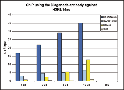
<p><small><strong>Figure 1. ChIP results obtained with the Diagenode antibody directed against H3K9/14ac</strong><br />ChIP assays were performed using human HeLa cells, the Diagenode antibody against H3K9/14ac (Cat. No. C15410200) and optimized PCR primer pairs for qPCR. ChIP was performed with the “Auto Histone ChIP-seq” kit on the IP-Star automated system, using sheared chromatin from 1,000,000 cells. A titration consisting of 1, 2, 5 and 10 μg of antibody per ChIP experiment was analyzed. IgG (2 μg/IP) was used as a negative IP control. Quantitative PCR was performed with primers specific for the promoter of the active genes GAPDH and EIF4A2, used as positive controls, and for the coding region of the inactive MB gene and the Sat2 satellite repeat, used as negative controls. Figure 1 shows the recovery, expressed as a % of input (the relative amount of immunoprecipitated DNA compared to input DNA after qPCR analysis).</small></p>
</div>
</div>
<div class="row">
<div class="small-12 columns"><center>A.<img src="https://www.diagenode.com/img/product/antibodies/C15410200_A1756D_ChIPseq-A.png" width="190" /></center><br />
<div class="extra-spaced"></div>
<center>B.<img src="https://www.diagenode.com/img/product/antibodies/C15410200_A1756D_ChIPseq-B.png" width="700" /></center><br /><center>C.<img src="https://www.diagenode.com/img/product/antibodies/C15410200_A1756D_ChIPseq-C.png" width="700" /></center><br /><center>D.<img src="https://www.diagenode.com/img/product/antibodies/C15410200_A1756D_ChIPseq-D.png" width="700" /></center><br /><center>E.<img src="https://www.diagenode.com/img/product/antibodies/C15410200_A1756D_ChIPseq-E.png" width="700" /></center></div>
</div>
<div class="row">
<div class="small-12 columns">
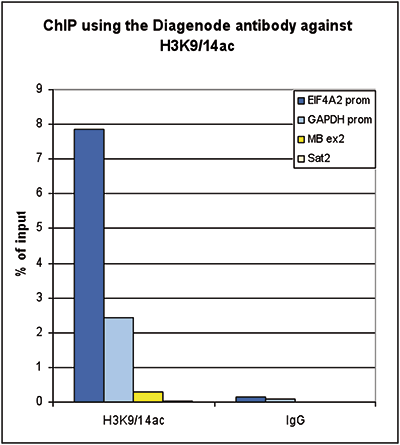
<p><small><strong>Figure 2. ChIP-seq results obtained with the Diagenode antibody directed against H3K9/14ac</strong><br />ChIP was performed on sheared chromatin from 100,000 K562 cells using 1 μg the Diagenode antibody against H3K9/14ac (Cat. No. C15410200) with the “iDeal ChIP-seq” kit (Cat. No. C01010051). IgG (1 μg/IP) was used as a negative IP control. The IP’d DNA was analysed by QPCR with optimized PCR primer pairs for the promoters of the active GAPDH and EIF4A2 genes, used as positive control targets, and the coding region of the inactive MB gene and the Sat2 satellite repeat, used as negative control targets (figure 2A). The IP’d DNA was subsequently analysed with an Illumina Genome Analyzer. Library preparation, cluster generation and sequencing were performed according to the manufacturer’s instructions. The 36 bp tags were aligned to the human genome using the ELAND algorithm. Figure 2 shows the peak distribution along the complete sequence and a 1.5 Mb region of the X-chromosome (figure 2B and C) and in two regions surrounding the GAPDH and EIF4A2 positive control genes, respectively (figure 2D and E). The position of the amplicon used for ChIP-qPCR is indicated by an arrow. These results clearly show an enrichment of the H3K9/14 acetylation at the promoters of active genes.</small></p>
</div>
</div>
<div class="row">
<div class="small-12 columns"><center>A.<img src="https://www.diagenode.com/img/product/antibodies/C15410200-cuttagA.png" width="700" /></center><br /><center>B.<img src="https://www.diagenode.com/img/product/antibodies/C15410200-cuttagB.png" width="700" /></center></div>
</div>
<div class="row">
<div class="small-12 columns">
<p><small><strong>Figure 3. Cut&Tag results obtained with the Diagenode antibody directed against H3K9/14ac</strong><br />CUT&TAG (Kaya-Okur, H.S., Nat Commun 10, 1930, 2019) was performed on 50,000 K562 cells using 1 µg of the Diagenode antibody against H3K9/14ac (cat. No. C15410200) and the Diagenode pA-Tn5 transposase (C01070001). The libraries were subsequently analysed on an Illumina NextSeq 500 sequencer (2x75 paired-end reads) according to the manufacturer's instructions. The tags were aligned to the human genome (hg19) using the BWA algorithm. Figure 3 shows the peak distribution along the complete sequence of chromosome 7 and in a 500 kb region surrounding the FOS gene on chromosome 14 (figure 3A and B, respectively).</small></p>
</div>
</div>
<div class="row">
<div class="small-6 columns"><center><img src="https://www.diagenode.com/img/product/antibodies/C15410200_A1756D_ELISA.png" width="278" /></center></div>
<div class="small-6 columns">
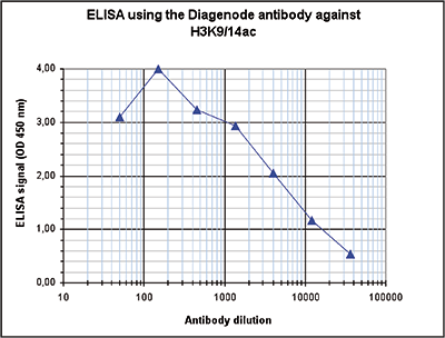
<p><small><strong>Figure 4. Determination of the antibody titer</strong><br />To determine the titer of the antibody, an ELISA was performed using a serial dilution of the Diagenode antibody against H3K9/14ac (Cat. No. C15410200). The antigen used was a peptide containing the histone modification of interest. By plotting the absorbance against the antibody dilution (Figure 4), the titer of the antibody was estimated to be 1:4,000.</small></p>
</div>
</div>
<div class="extra-spaced"></div>
<div class="row">
<div class="small-4 columns"><center>A.<img src="https://www.diagenode.com/img/product/antibodies/C15410200_A1756D_CrossReactivity-A.png" width="278" /></center><br /><center>B.<img src="https://www.diagenode.com/img/product/antibodies/C15410200_A1756D_CrossReactivity-B.png" width="278" /></center></div>
<div class="small-8 columns">
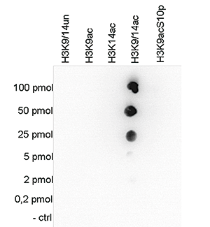
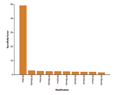
<p><small><strong>Figure 5. Cross reactivity tests using the Diagenode antibody directed against H3K9/14ac</strong><br />Figure 5A To test the cross reactivity of the Diagenode antibody against H3K9/14ac (Cat. No. C15410200), a Dot Blot analysis was performed with peptides containing other histone modifications and the unmodified H3K9. One hundred to 0.2 pmol of the respective peptides were spotted on a membrane. The antibody was used at a dilution of 1:20,000. Figure 5A shows a high specificity of the antibody for the modification of interest. Figure 5B The specificity of the antibody was further demonstrated by peptide array analyses on an array containing 384 peptides with different combinations of modifications from histone H3, H4, H2A and H2B. The antibody was used at a dilution of 1:2,000. Figure 5B shows the specificity factor, calculated as the ratio of the average intensity of all spots containing the mark, divided by the average intensity of all spots not containing the mark.</small></p>
</div>
</div>
<div class="row">
<div class="small-4 columns"><center><img src="https://www.diagenode.com/img/product/antibodies/C15410200_001_WB.png" /></center></div>
<div class="small-8 columns">
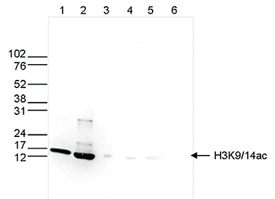
<p><small><strong>Figure 6. Western blot analysis using the Diagenode antibody directed against H3K9/14ac</strong><br />Western blot was performed on whole cell (25 μg, lane 1) and histone extracts (15 μg, lane 2) from HeLa cells, and on 1 μg of recombinant histone H2A, H2B, H3 and H4 (lane 3, 4, 5 and 6, respectively) using the Diagenode antibody against H3K9/14ac (Cat. No. C15410200). The antibody was diluted 1:500 in TBS-Tween containing 5% skimmed milk. The position of the protein of interest is indicated on the right, the marker (in kDa) is shown on the left.</small></p>
</div>
</div>
<div class="row">
<div class="small-6 columns"><center><img src="https://www.diagenode.com/img/product/antibodies/C15410200_A1756D_IF.png" /></center></div>
<div class="small-6 columns">
<p><small><strong>Figure 7. Immunofluorescence using the Diagenode antibody directed against H3K9/14ac</strong><br />HeLa cells were stained with the Diagenode antibody against H3K9/14ac (Cat. No. C15410200) and with DAPI. Cells were fixed with 4% formaldehyde for 10’ and blocked with PBS/TX-100 containing 5% normal goat serum and 1% BSA. The cells were immunofluorescently labeled with the H3K9/14ac antibody (left) diluted 1:200 in blocking solution followed by an anti-rabbit antibody conjugated to Alexa488. The middle panel shows staining of the nuclei with DAPI. A merge of the two stainings is shown on the right.</small></p>
</div>
</div>',
'label2' => '',
'info2' => '',
'label3' => '',
'info3' => '',
'format' => '50 μg',
'catalog_number' => 'C15410200',
'old_catalog_number' => '',
'sf_code' => 'C15410200-D001-000581',
'type' => 'FRE',
'search_order' => '03-Antibody',
'price_EUR' => '490',
'price_USD' => '485',
'price_GBP' => '440',
'price_JPY' => '80285',
'price_CNY' => '',
'price_AUD' => '1212',
'country' => 'ALL',
'except_countries' => 'None',
'quote' => false,
'in_stock' => false,
'featured' => false,
'no_promo' => false,
'online' => true,
'master' => true,
'last_datasheet_update' => '0000-00-00',
'slug' => 'h3k9-14ac-polyclonal-antibody-premium-50-mg-62-ml',
'meta_title' => 'H3K9/14ac polyclonal antibody - Premium',
'meta_keywords' => '',
'meta_description' => 'H3K9/14ac polyclonal antibody - Premium',
'modified' => '2024-01-17 19:59:29',
'created' => '2015-06-29 14:08:20',
'locale' => 'jpn'
),
'Antibody' => array(
'host' => '*****',
'id' => '565',
'name' => 'H3K9/14ac polyclonal antibody',
'description' => 'Histones are the main constituents of the protein part of chromosomes of eukaryotic cells. They are rich in the amino acids arginine and lysine and have been greatly conserved during evolution. Histones pack the DNA into tight masses of chromatin. Two core histones of each class H2A, H2B, H3 and H4 assemble and are wrapped by 146 base pairs of DNA to form one octameric nucleosome. Histone tails undergo numerous post-translational modifications, which either directly or indirectly alter chromatin structure to facilitate transcriptional activation or repression or other nuclear processes. In addition to the genetic code, combinations of the different histone modifications reveal the so-called “histone code”. Histone methylation and demethylation is dynamically regulated by respectively histone methyl transferases and histone demethylases. Acetylation of histone H3 at K9 and K14 is associated with active promoters.',
'clonality' => '',
'isotype' => '',
'lot' => 'A1756D',
'concentration' => '0.81 μg/μl',
'reactivity' => 'Human, mouse, P. falciparum, wide range expected.',
'type' => 'Polyclonal, <strong>ChIP grade, ChIP-seq grade</strong>',
'purity' => 'Affinity purified polyclonal antibody.',
'classification' => '',
'application_table' => '<table>
<thead>
<tr>
<th>Applications</th>
<th>Suggested dilution</th>
<th>References</th>
</tr>
</thead>
<tbody>
<tr>
<td>ChIP/ChIP-seq <sup>*</sup></td>
<td>1 µg per IP</td>
<td>Fig 1, 2</td>
</tr>
<tr>
<td>CUT&TAG</td>
<td>1 µg</td>
<td>Fig 3</td>
</tr>
<tr>
<td>ELISA</td>
<td>1:100</td>
<td>Fig 4</td>
</tr>
<tr>
<td>Dot Blotting/Peptide array</td>
<td>1:20,000/1:2,000</td>
<td>Fig 5</td>
</tr>
<tr>
<td>Western Blotting</td>
<td>1:500</td>
<td>Fig 6</td>
</tr>
<tr>
<td>Immunofluorescence</td>
<td>1:200</td>
<td>Fig 7</td>
</tr>
</tbody>
</table>
<p></p>
<p><small>* Please note that the optimal antibody amount per ChIP should be determined by the end-user. We recommend testing 1-5 μg per IP.</small></p>',
'storage_conditions' => 'Store at -20°C; for long storage, store at -80°C. Avoid multiple freeze-thaw cycles.',
'storage_buffer' => 'PBS containing 0.05% azide and 0.05% ProClin 300.',
'precautions' => 'This product is for research use only. Not for use in diagnostic or therapeutic procedures.',
'uniprot_acc' => '',
'slug' => 'h3k9-14ac-polyclonal-antibody',
'meta_keywords' => '',
'meta_description' => '',
'modified' => '2021-02-11 15:12:20',
'created' => '2020-06-30 11:22:26',
'select_label' => '565 - H3K9/14ac polyclonal antibody (A1756D - 0.81 μg/μl - Human, mouse, P. falciparum, wide range expected. - Affinity purified polyclonal antibody. - Rabbit)'
),
'Slave' => array(
(int) 0 => array(
[maximum depth reached]
)
),
'Group' => array(
'Group' => array(
[maximum depth reached]
),
'Master' => array(
[maximum depth reached]
),
'Product' => array(
[maximum depth reached]
)
),
'Related' => array(
(int) 0 => array(
[maximum depth reached]
)
),
'Application' => array(
(int) 0 => array(
[maximum depth reached]
),
(int) 1 => array(
[maximum depth reached]
),
(int) 2 => array(
[maximum depth reached]
),
(int) 3 => array(
[maximum depth reached]
),
(int) 4 => array(
[maximum depth reached]
),
(int) 5 => array(
[maximum depth reached]
),
(int) 6 => array(
[maximum depth reached]
),
(int) 7 => array(
[maximum depth reached]
)
),
'Category' => array(
(int) 0 => array(
[maximum depth reached]
),
(int) 1 => array(
[maximum depth reached]
),
(int) 2 => array(
[maximum depth reached]
),
(int) 3 => array(
[maximum depth reached]
)
),
'Document' => array(
(int) 0 => array(
[maximum depth reached]
),
(int) 1 => array(
[maximum depth reached]
),
(int) 2 => array(
[maximum depth reached]
)
),
'Feature' => array(),
'Image' => array(
(int) 0 => array(
[maximum depth reached]
)
),
'Promotion' => array(),
'Protocol' => array(),
'Publication' => array(
(int) 0 => array(
[maximum depth reached]
),
(int) 1 => array(
[maximum depth reached]
),
(int) 2 => array(
[maximum depth reached]
),
(int) 3 => array(
[maximum depth reached]
),
(int) 4 => array(
[maximum depth reached]
),
(int) 5 => array(
[maximum depth reached]
),
(int) 6 => array(
[maximum depth reached]
),
(int) 7 => array(
[maximum depth reached]
),
(int) 8 => array(
[maximum depth reached]
),
(int) 9 => array(
[maximum depth reached]
),
(int) 10 => array(
[maximum depth reached]
),
(int) 11 => array(
[maximum depth reached]
),
(int) 12 => array(
[maximum depth reached]
),
(int) 13 => array(
[maximum depth reached]
),
(int) 14 => array(
[maximum depth reached]
),
(int) 15 => array(
[maximum depth reached]
),
(int) 16 => array(
[maximum depth reached]
)
),
'Testimonial' => array(
(int) 0 => array(
[maximum depth reached]
)
),
'Area' => array(),
'SafetySheet' => array(
(int) 0 => array(
[maximum depth reached]
),
(int) 1 => array(
[maximum depth reached]
),
(int) 2 => array(
[maximum depth reached]
),
(int) 3 => array(
[maximum depth reached]
),
(int) 4 => array(
[maximum depth reached]
),
(int) 5 => array(
[maximum depth reached]
),
(int) 6 => array(
[maximum depth reached]
),
(int) 7 => array(
[maximum depth reached]
)
)
),
'meta_canonical' => 'https://www.diagenode.com/jp/p/h3k9-14ac-polyclonal-antibody-premium-50-mg-62-ml'
)
$language = 'jp'
$meta_keywords = ''
$meta_description = 'H3K9/14ac polyclonal antibody - Premium'
$meta_title = 'H3K9/14ac polyclonal antibody - Premium'
$product = array(
'Product' => array(
'id' => '2272',
'antibody_id' => '565',
'name' => 'H3K9/14ac polyclonal antibody ',
'description' => '<p><span>Polyclonal antibody raised in rabbit against the region of histone H3 containing the acetylated lysines 9 and 14 (H3K9/14ac), using a KLH-conjugated synthetic peptide.</span></p>',
'label1' => 'Validation Data',
'info1' => '<div class="row">
<div class="small-6 columns"><center><img src="https://www.diagenode.com/img/product/antibodies/C15410200_A1756D_ChIP.png" width="278" /></center></div>
<div class="small-6 columns">
<p><small><strong>Figure 1. ChIP results obtained with the Diagenode antibody directed against H3K9/14ac</strong><br />ChIP assays were performed using human HeLa cells, the Diagenode antibody against H3K9/14ac (Cat. No. C15410200) and optimized PCR primer pairs for qPCR. ChIP was performed with the “Auto Histone ChIP-seq” kit on the IP-Star automated system, using sheared chromatin from 1,000,000 cells. A titration consisting of 1, 2, 5 and 10 μg of antibody per ChIP experiment was analyzed. IgG (2 μg/IP) was used as a negative IP control. Quantitative PCR was performed with primers specific for the promoter of the active genes GAPDH and EIF4A2, used as positive controls, and for the coding region of the inactive MB gene and the Sat2 satellite repeat, used as negative controls. Figure 1 shows the recovery, expressed as a % of input (the relative amount of immunoprecipitated DNA compared to input DNA after qPCR analysis).</small></p>
</div>
</div>
<div class="row">
<div class="small-12 columns"><center>A.<img src="https://www.diagenode.com/img/product/antibodies/C15410200_A1756D_ChIPseq-A.png" width="190" /></center><br />
<div class="extra-spaced"></div>
<center>B.<img src="https://www.diagenode.com/img/product/antibodies/C15410200_A1756D_ChIPseq-B.png" width="700" /></center><br /><center>C.<img src="https://www.diagenode.com/img/product/antibodies/C15410200_A1756D_ChIPseq-C.png" width="700" /></center><br /><center>D.<img src="https://www.diagenode.com/img/product/antibodies/C15410200_A1756D_ChIPseq-D.png" width="700" /></center><br /><center>E.<img src="https://www.diagenode.com/img/product/antibodies/C15410200_A1756D_ChIPseq-E.png" width="700" /></center></div>
</div>
<div class="row">
<div class="small-12 columns">
<p><small><strong>Figure 2. ChIP-seq results obtained with the Diagenode antibody directed against H3K9/14ac</strong><br />ChIP was performed on sheared chromatin from 100,000 K562 cells using 1 μg the Diagenode antibody against H3K9/14ac (Cat. No. C15410200) with the “iDeal ChIP-seq” kit (Cat. No. C01010051). IgG (1 μg/IP) was used as a negative IP control. The IP’d DNA was analysed by QPCR with optimized PCR primer pairs for the promoters of the active GAPDH and EIF4A2 genes, used as positive control targets, and the coding region of the inactive MB gene and the Sat2 satellite repeat, used as negative control targets (figure 2A). The IP’d DNA was subsequently analysed with an Illumina Genome Analyzer. Library preparation, cluster generation and sequencing were performed according to the manufacturer’s instructions. The 36 bp tags were aligned to the human genome using the ELAND algorithm. Figure 2 shows the peak distribution along the complete sequence and a 1.5 Mb region of the X-chromosome (figure 2B and C) and in two regions surrounding the GAPDH and EIF4A2 positive control genes, respectively (figure 2D and E). The position of the amplicon used for ChIP-qPCR is indicated by an arrow. These results clearly show an enrichment of the H3K9/14 acetylation at the promoters of active genes.</small></p>
</div>
</div>
<div class="row">
<div class="small-12 columns"><center>A.<img src="https://www.diagenode.com/img/product/antibodies/C15410200-cuttagA.png" width="700" /></center><br /><center>B.<img src="https://www.diagenode.com/img/product/antibodies/C15410200-cuttagB.png" width="700" /></center></div>
</div>
<div class="row">
<div class="small-12 columns">
<p><small><strong>Figure 3. Cut&Tag results obtained with the Diagenode antibody directed against H3K9/14ac</strong><br />CUT&TAG (Kaya-Okur, H.S., Nat Commun 10, 1930, 2019) was performed on 50,000 K562 cells using 1 µg of the Diagenode antibody against H3K9/14ac (cat. No. C15410200) and the Diagenode pA-Tn5 transposase (C01070001). The libraries were subsequently analysed on an Illumina NextSeq 500 sequencer (2x75 paired-end reads) according to the manufacturer's instructions. The tags were aligned to the human genome (hg19) using the BWA algorithm. Figure 3 shows the peak distribution along the complete sequence of chromosome 7 and in a 500 kb region surrounding the FOS gene on chromosome 14 (figure 3A and B, respectively).</small></p>
</div>
</div>
<div class="row">
<div class="small-6 columns"><center><img src="https://www.diagenode.com/img/product/antibodies/C15410200_A1756D_ELISA.png" width="278" /></center></div>
<div class="small-6 columns">
<p><small><strong>Figure 4. Determination of the antibody titer</strong><br />To determine the titer of the antibody, an ELISA was performed using a serial dilution of the Diagenode antibody against H3K9/14ac (Cat. No. C15410200). The antigen used was a peptide containing the histone modification of interest. By plotting the absorbance against the antibody dilution (Figure 4), the titer of the antibody was estimated to be 1:4,000.</small></p>
</div>
</div>
<div class="extra-spaced"></div>
<div class="row">
<div class="small-4 columns"><center>A.<img src="https://www.diagenode.com/img/product/antibodies/C15410200_A1756D_CrossReactivity-A.png" width="278" /></center><br /><center>B.<img src="https://www.diagenode.com/img/product/antibodies/C15410200_A1756D_CrossReactivity-B.png" width="278" /></center></div>
<div class="small-8 columns">
<p><small><strong>Figure 5. Cross reactivity tests using the Diagenode antibody directed against H3K9/14ac</strong><br />Figure 5A To test the cross reactivity of the Diagenode antibody against H3K9/14ac (Cat. No. C15410200), a Dot Blot analysis was performed with peptides containing other histone modifications and the unmodified H3K9. One hundred to 0.2 pmol of the respective peptides were spotted on a membrane. The antibody was used at a dilution of 1:20,000. Figure 5A shows a high specificity of the antibody for the modification of interest. Figure 5B The specificity of the antibody was further demonstrated by peptide array analyses on an array containing 384 peptides with different combinations of modifications from histone H3, H4, H2A and H2B. The antibody was used at a dilution of 1:2,000. Figure 5B shows the specificity factor, calculated as the ratio of the average intensity of all spots containing the mark, divided by the average intensity of all spots not containing the mark.</small></p>
</div>
</div>
<div class="row">
<div class="small-4 columns"><center><img src="https://www.diagenode.com/img/product/antibodies/C15410200_001_WB.png" /></center></div>
<div class="small-8 columns">
<p><small><strong>Figure 6. Western blot analysis using the Diagenode antibody directed against H3K9/14ac</strong><br />Western blot was performed on whole cell (25 μg, lane 1) and histone extracts (15 μg, lane 2) from HeLa cells, and on 1 μg of recombinant histone H2A, H2B, H3 and H4 (lane 3, 4, 5 and 6, respectively) using the Diagenode antibody against H3K9/14ac (Cat. No. C15410200). The antibody was diluted 1:500 in TBS-Tween containing 5% skimmed milk. The position of the protein of interest is indicated on the right, the marker (in kDa) is shown on the left.</small></p>
</div>
</div>
<div class="row">
<div class="small-6 columns"><center><img src="https://www.diagenode.com/img/product/antibodies/C15410200_A1756D_IF.png" /></center></div>
<div class="small-6 columns">
<p><small><strong>Figure 7. Immunofluorescence using the Diagenode antibody directed against H3K9/14ac</strong><br />HeLa cells were stained with the Diagenode antibody against H3K9/14ac (Cat. No. C15410200) and with DAPI. Cells were fixed with 4% formaldehyde for 10’ and blocked with PBS/TX-100 containing 5% normal goat serum and 1% BSA. The cells were immunofluorescently labeled with the H3K9/14ac antibody (left) diluted 1:200 in blocking solution followed by an anti-rabbit antibody conjugated to Alexa488. The middle panel shows staining of the nuclei with DAPI. A merge of the two stainings is shown on the right.</small></p>
</div>
</div>',
'label2' => '',
'info2' => '',
'label3' => '',
'info3' => '',
'format' => '50 μg',
'catalog_number' => 'C15410200',
'old_catalog_number' => '',
'sf_code' => 'C15410200-D001-000581',
'type' => 'FRE',
'search_order' => '03-Antibody',
'price_EUR' => '490',
'price_USD' => '485',
'price_GBP' => '440',
'price_JPY' => '80285',
'price_CNY' => '',
'price_AUD' => '1212',
'country' => 'ALL',
'except_countries' => 'None',
'quote' => false,
'in_stock' => false,
'featured' => false,
'no_promo' => false,
'online' => true,
'master' => true,
'last_datasheet_update' => '0000-00-00',
'slug' => 'h3k9-14ac-polyclonal-antibody-premium-50-mg-62-ml',
'meta_title' => 'H3K9/14ac polyclonal antibody - Premium',
'meta_keywords' => '',
'meta_description' => 'H3K9/14ac polyclonal antibody - Premium',
'modified' => '2024-01-17 19:59:29',
'created' => '2015-06-29 14:08:20',
'locale' => 'jpn'
),
'Antibody' => array(
'host' => '*****',
'id' => '565',
'name' => 'H3K9/14ac polyclonal antibody',
'description' => 'Histones are the main constituents of the protein part of chromosomes of eukaryotic cells. They are rich in the amino acids arginine and lysine and have been greatly conserved during evolution. Histones pack the DNA into tight masses of chromatin. Two core histones of each class H2A, H2B, H3 and H4 assemble and are wrapped by 146 base pairs of DNA to form one octameric nucleosome. Histone tails undergo numerous post-translational modifications, which either directly or indirectly alter chromatin structure to facilitate transcriptional activation or repression or other nuclear processes. In addition to the genetic code, combinations of the different histone modifications reveal the so-called “histone code”. Histone methylation and demethylation is dynamically regulated by respectively histone methyl transferases and histone demethylases. Acetylation of histone H3 at K9 and K14 is associated with active promoters.',
'clonality' => '',
'isotype' => '',
'lot' => 'A1756D',
'concentration' => '0.81 μg/μl',
'reactivity' => 'Human, mouse, P. falciparum, wide range expected.',
'type' => 'Polyclonal, <strong>ChIP grade, ChIP-seq grade</strong>',
'purity' => 'Affinity purified polyclonal antibody.',
'classification' => '',
'application_table' => '<table>
<thead>
<tr>
<th>Applications</th>
<th>Suggested dilution</th>
<th>References</th>
</tr>
</thead>
<tbody>
<tr>
<td>ChIP/ChIP-seq <sup>*</sup></td>
<td>1 µg per IP</td>
<td>Fig 1, 2</td>
</tr>
<tr>
<td>CUT&TAG</td>
<td>1 µg</td>
<td>Fig 3</td>
</tr>
<tr>
<td>ELISA</td>
<td>1:100</td>
<td>Fig 4</td>
</tr>
<tr>
<td>Dot Blotting/Peptide array</td>
<td>1:20,000/1:2,000</td>
<td>Fig 5</td>
</tr>
<tr>
<td>Western Blotting</td>
<td>1:500</td>
<td>Fig 6</td>
</tr>
<tr>
<td>Immunofluorescence</td>
<td>1:200</td>
<td>Fig 7</td>
</tr>
</tbody>
</table>
<p></p>
<p><small>* Please note that the optimal antibody amount per ChIP should be determined by the end-user. We recommend testing 1-5 μg per IP.</small></p>',
'storage_conditions' => 'Store at -20°C; for long storage, store at -80°C. Avoid multiple freeze-thaw cycles.',
'storage_buffer' => 'PBS containing 0.05% azide and 0.05% ProClin 300.',
'precautions' => 'This product is for research use only. Not for use in diagnostic or therapeutic procedures.',
'uniprot_acc' => '',
'slug' => 'h3k9-14ac-polyclonal-antibody',
'meta_keywords' => '',
'meta_description' => '',
'modified' => '2021-02-11 15:12:20',
'created' => '2020-06-30 11:22:26',
'select_label' => '565 - H3K9/14ac polyclonal antibody (A1756D - 0.81 μg/μl - Human, mouse, P. falciparum, wide range expected. - Affinity purified polyclonal antibody. - Rabbit)'
),
'Slave' => array(
(int) 0 => array(
'id' => '51',
'name' => 'C15410200',
'product_id' => '2272',
'modified' => '2016-02-18 20:51:57',
'created' => '2016-02-18 20:51:57'
)
),
'Group' => array(
'Group' => array(
'id' => '51',
'name' => 'C15410200',
'product_id' => '2272',
'modified' => '2016-02-18 20:51:57',
'created' => '2016-02-18 20:51:57'
),
'Master' => array(
'id' => '2272',
'antibody_id' => '565',
'name' => 'H3K9/14ac Antibody',
'description' => '<p><span>Polyclonal antibody raised in rabbit against the region of histone <strong>H3 containing the acetylated lysines 9 and 14</strong> <strong>(H3K9/14ac</strong>), using a KLH-conjugated synthetic peptide.</span></p>',
'label1' => 'Validation data',
'info1' => '<div class="row">
<div class="small-6 columns"><center><img src="https://www.diagenode.com/img/product/antibodies/C15410200_A1756D_ChIP.png" width="278" /></center></div>
<div class="small-6 columns">
<p><small><strong>Figure 1. ChIP results obtained with the Diagenode antibody directed against H3K9/14ac</strong><br />ChIP assays were performed using human HeLa cells, the Diagenode antibody against H3K9/14ac (Cat. No. C15410200) and optimized PCR primer pairs for qPCR. ChIP was performed with the “Auto Histone ChIP-seq” kit on the IP-Star automated system, using sheared chromatin from 1,000,000 cells. A titration consisting of 1, 2, 5 and 10 μg of antibody per ChIP experiment was analyzed. IgG (2 μg/IP) was used as a negative IP control. Quantitative PCR was performed with primers specific for the promoter of the active genes GAPDH and EIF4A2, used as positive controls, and for the coding region of the inactive MB gene and the Sat2 satellite repeat, used as negative controls. Figure 1 shows the recovery, expressed as a % of input (the relative amount of immunoprecipitated DNA compared to input DNA after qPCR analysis).</small></p>
</div>
</div>
<div class="row">
<div class="small-12 columns"><center>A.<img src="https://www.diagenode.com/img/product/antibodies/C15410200_A1756D_ChIPseq-A.png" width="190" /></center><br />
<div class="extra-spaced"></div>
<center>B.<img src="https://www.diagenode.com/img/product/antibodies/C15410200_A1756D_ChIPseq-B.png" width="700" /></center><br /><center>C.<img src="https://www.diagenode.com/img/product/antibodies/C15410200_A1756D_ChIPseq-C.png" width="700" /></center><br /><center>D.<img src="https://www.diagenode.com/img/product/antibodies/C15410200_A1756D_ChIPseq-D.png" width="700" /></center><br /><center>E.<img src="https://www.diagenode.com/img/product/antibodies/C15410200_A1756D_ChIPseq-E.png" width="700" /></center></div>
</div>
<div class="row">
<div class="small-12 columns">
<p><small><strong>Figure 2. ChIP-seq results obtained with the Diagenode antibody directed against H3K9/14ac</strong><br />ChIP was performed on sheared chromatin from 100,000 K562 cells using 1 μg the Diagenode antibody against H3K9/14ac (Cat. No. C15410200) with the “iDeal ChIP-seq” kit (Cat. No. C01010051). IgG (1 μg/IP) was used as a negative IP control. The IP’d DNA was analysed by QPCR with optimized PCR primer pairs for the promoters of the active GAPDH and EIF4A2 genes, used as positive control targets, and the coding region of the inactive MB gene and the Sat2 satellite repeat, used as negative control targets (figure 2A). The IP’d DNA was subsequently analysed with an Illumina Genome Analyzer. Library preparation, cluster generation and sequencing were performed according to the manufacturer’s instructions. The 36 bp tags were aligned to the human genome using the ELAND algorithm. Figure 2 shows the peak distribution along the complete sequence and a 1.5 Mb region of the X-chromosome (figure 2B and C) and in two regions surrounding the GAPDH and EIF4A2 positive control genes, respectively (figure 2D and E). The position of the amplicon used for ChIP-qPCR is indicated by an arrow. These results clearly show an enrichment of the H3K9/14 acetylation at the promoters of active genes.</small></p>
</div>
</div>
<div class="row">
<div class="small-12 columns"><center>A.<img src="https://www.diagenode.com/img/product/antibodies/C15410200-cuttagA.png" width="700" /></center><br /><center>B.<img src="https://www.diagenode.com/img/product/antibodies/C15410200-cuttagB.png" width="700" /></center></div>
</div>
<div class="row">
<div class="small-12 columns">
<p><small><strong>Figure 3. Cut&Tag results obtained with the Diagenode antibody directed against H3K9/14ac</strong><br />CUT&TAG (Kaya-Okur, H.S., Nat Commun 10, 1930, 2019) was performed on 50,000 K562 cells using 1 µg of the Diagenode antibody against H3K9/14ac (cat. No. C15410200) and the Diagenode pA-Tn5 transposase (C01070001). The libraries were subsequently analysed on an Illumina NextSeq 500 sequencer (2x75 paired-end reads) according to the manufacturer's instructions. The tags were aligned to the human genome (hg19) using the BWA algorithm. Figure 3 shows the peak distribution along the complete sequence of chromosome 7 and in a 500 kb region surrounding the FOS gene on chromosome 14 (figure 3A and B, respectively).</small></p>
</div>
</div>
<div class="row">
<div class="small-6 columns"><center><img src="https://www.diagenode.com/img/product/antibodies/C15410200_A1756D_ELISA.png" width="278" /></center></div>
<div class="small-6 columns">
<p><small><strong>Figure 4. Determination of the antibody titer</strong><br />To determine the titer of the antibody, an ELISA was performed using a serial dilution of the Diagenode antibody against H3K9/14ac (Cat. No. C15410200). The antigen used was a peptide containing the histone modification of interest. By plotting the absorbance against the antibody dilution (Figure 4), the titer of the antibody was estimated to be 1:4,000.</small></p>
</div>
</div>
<div class="extra-spaced"></div>
<div class="row">
<div class="small-4 columns"><center>A.<img src="https://www.diagenode.com/img/product/antibodies/C15410200_A1756D_CrossReactivity-A.png" width="278" /></center><br /><center>B.<img src="https://www.diagenode.com/img/product/antibodies/C15410200_A1756D_CrossReactivity-B.png" width="278" /></center></div>
<div class="small-8 columns">
<p><small><strong>Figure 5. Cross reactivity tests using the Diagenode antibody directed against H3K9/14ac</strong><br />Figure 5A To test the cross reactivity of the Diagenode antibody against H3K9/14ac (Cat. No. C15410200), a Dot Blot analysis was performed with peptides containing other histone modifications and the unmodified H3K9. One hundred to 0.2 pmol of the respective peptides were spotted on a membrane. The antibody was used at a dilution of 1:20,000. Figure 5A shows a high specificity of the antibody for the modification of interest. Figure 5B The specificity of the antibody was further demonstrated by peptide array analyses on an array containing 384 peptides with different combinations of modifications from histone H3, H4, H2A and H2B. The antibody was used at a dilution of 1:2,000. Figure 5B shows the specificity factor, calculated as the ratio of the average intensity of all spots containing the mark, divided by the average intensity of all spots not containing the mark.</small></p>
</div>
</div>
<div class="row">
<div class="small-4 columns"><center><img src="https://www.diagenode.com/img/product/antibodies/C15410200_001_WB.png" /></center></div>
<div class="small-8 columns">
<p><small><strong>Figure 6. Western blot analysis using the Diagenode antibody directed against H3K9/14ac</strong><br />Western blot was performed on whole cell (25 μg, lane 1) and histone extracts (15 μg, lane 2) from HeLa cells, and on 1 μg of recombinant histone H2A, H2B, H3 and H4 (lane 3, 4, 5 and 6, respectively) using the Diagenode antibody against H3K9/14ac (Cat. No. C15410200). The antibody was diluted 1:500 in TBS-Tween containing 5% skimmed milk. The position of the protein of interest is indicated on the right, the marker (in kDa) is shown on the left.</small></p>
</div>
</div>
<div class="row">
<div class="small-6 columns"><center><img src="https://www.diagenode.com/img/product/antibodies/C15410200_A1756D_IF.png" /></center></div>
<div class="small-6 columns">
<p><small><strong>Figure 7. Immunofluorescence using the Diagenode antibody directed against H3K9/14ac</strong><br />HeLa cells were stained with the Diagenode antibody against H3K9/14ac (Cat. No. C15410200) and with DAPI. Cells were fixed with 4% formaldehyde for 10’ and blocked with PBS/TX-100 containing 5% normal goat serum and 1% BSA. The cells were immunofluorescently labeled with the H3K9/14ac antibody (left) diluted 1:200 in blocking solution followed by an anti-rabbit antibody conjugated to Alexa488. The middle panel shows staining of the nuclei with DAPI. A merge of the two stainings is shown on the right.</small></p>
</div>
</div>',
'label2' => '',
'info2' => '',
'label3' => '',
'info3' => '',
'format' => '50 μg',
'catalog_number' => 'C15410200',
'old_catalog_number' => '',
'sf_code' => 'C15410200-D001-000581',
'type' => 'FRE',
'search_order' => '03-Antibody',
'price_EUR' => '490',
'price_USD' => '485',
'price_GBP' => '440',
'price_JPY' => '80285',
'price_CNY' => '',
'price_AUD' => '1212',
'country' => 'ALL',
'except_countries' => 'None',
'quote' => false,
'in_stock' => false,
'featured' => false,
'no_promo' => false,
'online' => true,
'master' => true,
'last_datasheet_update' => '0000-00-00',
'slug' => 'h3k9-14ac-polyclonal-antibody-premium-50-mg-62-ml',
'meta_title' => 'H3K914ac Antibody - ChIP-seq Grade (C15410200) | Diagenode',
'meta_keywords' => '',
'meta_description' => 'H3K9/14ac (Histone H3 acetylated at lysines 9 and 14) Polyclonal Antibody validated in ChIP-seq, ChIP-qPCR, CUT&Tag, ELISA, DB, WB and IF. Specificity confirmed by Peptide array assay. Batch-specific data available on the website. Sample size available.',
'modified' => '2024-01-17 19:59:29',
'created' => '2015-06-29 14:08:20'
),
'Product' => array(
(int) 0 => array(
[maximum depth reached]
)
)
),
'Related' => array(
(int) 0 => array(
'id' => '2914',
'antibody_id' => null,
'name' => 'Auto ChIPmentation Kit for Histones - Replaced by C01011009',
'description' => '<div class="extra-spaced"><center></center></div>
<div class="row">
<div class="small-12 medium-8 large-8 columns"><!--<div class="extra-spaced"><center><a href="https://www.diagenode.com/en/pages/grant"><img src="https://www.diagenode.com/img/banners/banner-chipmentation-grant-580x120.jpg" /></a></center></div>-->
<div align="center"><video width="400" height="250" autoplay="autoplay" muted="" loop="loop" controls="controls" src="https://www.diagenode.com/videos/chipmentation-dgo.mp4" frameborder="0" allowfullscreen="allowfullscreen"></video></div>
<div align="center"><a href="https://www.diagenode.com/pages/form-chipmentation" class="center alert radius button"><i class="fa fa-info"></i> Contact us</a></div>
</div>
<div class="small-12 medium-4 large-4 columns"><center><a href="https://www.citeab.com/awards/2018/innovative-product-of-the-year" target="_blank"><img src="https://www.diagenode.com/emailing/images/citeab.png" width="224" height="200" /></a></center></div>
</div>
<p></p>
<p><strong>This product must be used with the <a href="https://www.diagenode.com/en/p/sx-8g-ip-star-compact-automated-system-1-unit">IP-Star Compact Automated System</a>.</strong></p>
<p>Diagenode’s latest technology for histone ChIP-seq, ChIPmentation, incorporates automation with an ligation-free protocol that integrates chromatin immunoprecipitation with NGS library preparation. Using histone ChIPmentation, you can avoid the multi-step ligation required for traditional histone ChIP-seq protocols, providing an easier protocol, faster time to results, and unmatched efficiency and reproducibility for histone ChIP-seq from challenging and low input samples. The ChIPmentaion Kit for Histones is a complete solution, including all buffers for chromatin preparation, immunoprecipitation, tagmentation and multiplexing using 24 single indexes.</p>',
'label1' => 'Characteristics',
'info1' => '<p>ChIPmentation is based on tagmentation that allows library preparation to be integrated during the ChIP itself using transposase and sequencing-compatible adaptors. ChIPmentation involves much easier and shorter protocol with high efficiency for low input samples using optimized ChIP and NGS sample preparation reagents on the IP-Star Compact Automated System. The IP-Star Compact Automated System ensures the highest reproducibility for challenging samples from limited input amounts.</p>
<h3>Benefits of the ChIPmentation system for histone ChIP-seq</h3>
<ul>
<li>Automates ChIP-seq of histones on the IP-Star® for highest reproducibility</li>
<li>Easier and faster than classical ChIP-seq</li>
<li>Validated for various histone marks</li>
<li>Ideal for analysis of large cohorts of samples</li>
<li>Ideal for analysis of large number of marks on a unique sample</li>
<li>High quality sequencing data</li>
</ul>
<p><img src="https://www.diagenode.com/img/product/chipmentation/chipmentation.jpg" style="display: block; margin-left: auto; margin-right: auto;" /></p>
<h3>ChIPmentation - Highest reliability for even the lowest inputs</h3>
<p>The new Auto ChIPmentation technology has been tested on histone marks and compared to available datasets from the ENCODE project (Figure 1). ChIPmentation generated high quality data with low background. In addition, more than 99% of the top 40% peaks obtained with auto-ChIPmentation overlap with ENCODE datasets, which shows that ChIP-seq data obtained with ChIPmentation are highly reliable.</p>
<div class="spaced"><center>A.<br /> <img src="https://www.diagenode.com/img/categories/kits_chromatin_function/auto-chipmentation.png" alt="Diagenode-Auto-ChIPmentation" /></center><br /><center>B. Match of the Top40 peaks of Auto-ChIPmentation<br /> <img src="https://www.diagenode.com/img/categories/kits_chromatin_function/match-of-the-top40-peaks-of-auto-chipmentation.png" /></center></div>
<ul class="accordion extra-spaced" data-accordion="">
<li class="accordion-navigation"><a href="#figure1"><i class="fa fa-caret-right"></i> Figure 1</a>
<div class="content" id="figure1">
<p><strong>Comparison of ChIPmentation sequencing results and reference ChIP-seq data.</strong><br /> Chromatin preparation has been performed on 7 M K562 cells using the Auto ChIPmentation Kit for Histones (Cat. no. C01011000). Diluted chromatin from 100.000 cells was used for the immunoprecipitation with the Diagenode antibody targeting H3K4me3 (Cat. no. C15410003). The experiment was performed on the IP-Star® Compact Automated System. A. Distribution of the ChIPmentation and ENCODE readsets in a representative region of the genome. B. Comparison of the top 40% peaks from ChIPmentation dataset to the ENCODE dataset.</p>
</div>
</li>
</ul>
<p class="extra-spaced">As ChIPmentation is a ligation-free protocol and requires fewer steps than a classical workflow, it allows the generation of excellent ChIP-seq data from reduced starting amounts. The sequencing data generated from 5,000 and 10,000 cells are very similar to those obtained with 100,000 cells, showing more than 98% overlap of the top 40% peaks (Figure 2).</p>
<center>A.<br /><img class="extra-spaced" style="margin-bottom: 20px;" src="https://www.diagenode.com/img/categories/kits_chromatin_function/chipmentation-sequencing-figure-a.jpg" alt="Diagenode-ChIPmentation-sequencing" /></center>
<div class="row">
<div class="small-6 columns" style="text-align: left;">B. Match of the Top40 peaks of ChIPmentation from 10.000 cells<br /><center><img src="https://www.diagenode.com/img/categories/kits_chromatin_function/match-of-the-top40-peaks-of-chipmentation-from-cent-cells.png" /></center></div>
<div class="small-6 columns">C. Match of the Top40 peaks of ChIPmentation from 5.000 cells<center><img src="https://www.diagenode.com/img/categories/kits_chromatin_function/match-of-the-top40-peaks-of-chipmentation-from-five-cells.png" /></center></div>
</div>
<ul class="accordion extra-spaced" data-accordion="">
<li class="accordion-navigation"><a href="#figure2"><i class="fa fa-caret-right"></i> Figure 2</a>
<div class="content" id="figure2">
<p><strong>ChIPmentation sequencing results obtained from decreasing starting amounts of cells.</strong><br /> Chromatin preparation has been performed on 7 M K562 cells using the Auto ChIPmentation Kit for Histones (Cat. no. C01011000). Diluted chromatin from 100.000, 10.000 and 5.000 cells was used for the immunoprecipitation with the Diagenode antibody targeting H3K4me3 (Cat. no. C15410003). The experiments were performed on the IP-Star® Compact Automated System. A. Distribution of the ChIPmentation readsets in a representative region of the genome. B. and C. Comparison of the top 40% peaks from 10.000 (B.) and 5.000 (C.) cells with dataset generated from 100.000 cells.</p>
</div>
</li>
</ul>
<p class="extra-spaced">Moreover, in order to check the reproducibility of data generated with low cell amounts, correlation between replicates for 10.000 and 5.000 starting cells was performed (Figure 3). The data show very high correlations with Pearson’s coefficients of 0.96 for both conditions.</p>
<div class="row">
<div class="small-6 columns" style="text-align: left;"><center>A. Auto ChIPmentation on 10.000 cells<br /> <img src="https://www.diagenode.com/img/categories/kits_chromatin_function/auto-chipmentation-on-10000cells.jpg" caption="false" width="480" height="420" /></center></div>
<div class="small-6 columns"><center>B. Auto ChIPmentation on 5.000 cells<br /> <img src="https://www.diagenode.com/img/categories/kits_chromatin_function/auto-chipmentation-on-5000cells.jpg" width="480" height="420" /></center></div>
</div>
<ul class="accordion extra-spaced" data-accordion="">
<li class="accordion-navigation"><a href="#figure3"><i class="fa fa-caret-right"></i> Figure 3</a>
<div class="content" id="figure3">
<p><strong>Correlation of replicates of auto-ChIPmentation experiments.</strong> Chromatin preparation has been performed on 7 M K562 cells using the Auto ChIPmentation Kit for Histones (Cat. no. C01011000). Diluted chromatin from 10.000 and 5.000 cells was used for the immunoprecipitation with the Diagenode antibody targeting H3K4me3 (Cat. no. C15410003). The experiments were performed on the IP-Star® Compact Automated System.The scatterplots show the correlation between the enrichments between two replicates. The green line represents an ideal case where the two replicates are identical, while the red curve fitted on the data points by a LOESS regression shows the actual trend. A. Correlation analysis between two replicates of auto-ChIPmentation experiments starting from 10.000 cells. B. Correlation analysis between two replicates of auto-ChIPmentation experiments starting from 5.000 cells.</p>
</div>
</li>
</ul>
<p>The Auto-ChIPmentation workflow is easy and fast, producing high-quality ChIP-seq data even on low starting amounts.</p>
<center><img src="https://www.diagenode.com/img/product/chipmentation/chipmentation-sequencing-results-for-histone.jpg" width="678" height="240" /></center>
<ul class="accordion extra-spaced" data-accordion="">
<li class="accordion-navigation"><a href="#figure4"><i class="fa fa-caret-right"></i> Figure 4</a>
<div class="content" id="figure4">
<p><strong>ChIPmentation sequencing results for four histone marks.</strong> Chromatin preparation has been performed on 7 M K562 cells using the Auto ChIPmentation Kit for Histones (Cat. no. C01011000). Diluted chromatin from 1,000,000 cells was used for the immunoprecipitation with the Diagenode antibodies targeting H3K4me1 (Cat. no. C15410194), H3K27me3 (Cat. no. C15410195), H3K27ac (Cat. no. C15410196) and IgG (Cat. no. C15410206). The experiment was performed on the IP-Star® Compact Automated System.</p>
</div>
</li>
</ul>',
'label2' => 'Additional solutions compatible with Auto ChIPmentation Kit for Histones',
'info2' => '<p><a href="https://www.diagenode.com/en/p/chromatin-shearing-optimization-kit-low-sds-100-million-cells">Chromatin shearing optimization kit - Low SDS (iDeal Kit for Histones)</a> optimizes chromatin shearing, a critical step for ChIP.</p>
<p><a href="https://www.diagenode.com/en/categories/chip-seq-grade-antibodies">ChIP-seq grade anti-histone antibodies</a> provide high yields with excellent specificity and sensitivity</p>',
'label3' => '',
'info3' => '',
'format' => '24 rxns',
'catalog_number' => 'C01011000',
'old_catalog_number' => '',
'sf_code' => '',
'type' => 'RFR',
'search_order' => '',
'price_EUR' => '',
'price_USD' => '',
'price_GBP' => '',
'price_JPY' => '',
'price_CNY' => '',
'price_AUD' => '',
'country' => 'ALL',
'except_countries' => 'Japan',
'quote' => false,
'in_stock' => true,
'featured' => false,
'no_promo' => false,
'online' => true,
'master' => true,
'last_datasheet_update' => '',
'slug' => 'chipmentation-for-histones-24-rxns',
'meta_title' => 'Auto ChIPmentation Kit for Histones',
'meta_keywords' => '',
'meta_description' => '',
'modified' => '2021-02-16 13:40:05',
'created' => '2017-12-11 16:34:56',
'ProductsRelated' => array(
[maximum depth reached]
),
'Image' => array([maximum depth reached])
)
),
'Application' => array(
(int) 0 => array(
'id' => '20',
'position' => '10',
'parent_id' => '40',
'name' => 'ELISA',
'description' => '<div class="row">
<div class="small-12 medium-12 large-12 columns">Enzyme-linked immunosorbent assay.</div>
</div>',
'in_footer' => false,
'in_menu' => false,
'online' => true,
'tabular' => true,
'slug' => 'elisa-antibodies',
'meta_keywords' => ' ELISA Antibodies,Monoclonal antibody, Polyclonal antibody',
'meta_description' => 'Diagenode offers Monoclonal & Polyclonal antibodies for ELISA applications',
'meta_title' => 'ELISA Antibodies - Monoclonal & Polyclonal antibody | Diagenode',
'modified' => '2016-01-13 12:21:41',
'created' => '2014-07-08 08:13:28',
'ProductsApplication' => array(
[maximum depth reached]
)
),
(int) 1 => array(
'id' => '28',
'position' => '10',
'parent_id' => '40',
'name' => 'DB',
'description' => '<p>Dot blotting</p>',
'in_footer' => false,
'in_menu' => false,
'online' => true,
'tabular' => true,
'slug' => 'dot-blotting',
'meta_keywords' => 'Dot blotting,Monoclonal & Polyclonal antibody,',
'meta_description' => 'Diagenode offers Monoclonal & Polyclonal antibodies for Dot blotting applications',
'meta_title' => 'Dot blotting Antibodies - Monoclonal & Polyclonal antibody | Diagenode',
'modified' => '2016-01-13 14:40:49',
'created' => '2015-07-08 13:45:05',
'ProductsApplication' => array(
[maximum depth reached]
)
),
(int) 2 => array(
'id' => '37',
'position' => '10',
'parent_id' => '40',
'name' => 'Peptide array',
'description' => '<p>Peptide array</p>',
'in_footer' => false,
'in_menu' => false,
'online' => true,
'tabular' => true,
'slug' => 'peptide-arry',
'meta_keywords' => 'Peptide array antibodies,Histone antibodies,policlonal antibodies',
'meta_description' => 'Diagenode offers a wide range of antibodies and technical support for peptide array applications',
'meta_title' => 'Peptide array antibodies | Diagenode',
'modified' => '2016-01-20 12:24:40',
'created' => '2015-07-08 13:55:25',
'ProductsApplication' => array(
[maximum depth reached]
)
),
(int) 3 => array(
'id' => '19',
'position' => '10',
'parent_id' => '40',
'name' => 'WB',
'description' => '<p><strong>Western blot</strong> : The quality of antibodies used in this technique is crucial for correct and specific protein identification. Diagenode offers huge selection of highly sensitive and specific western blot-validated antibodies.</p>
<p>Learn more about: <a href="https://www.diagenode.com/applications/western-blot">Loading control, MW marker visualization</a><em>. <br /></em></p>
<p><em></em>Check our selection of antibodies validated in Western blot.</p>',
'in_footer' => false,
'in_menu' => false,
'online' => true,
'tabular' => true,
'slug' => 'western-blot-antibodies',
'meta_keywords' => ' Western Blot Antibodies ,western blot protocol,Western Blotting Products,Polyclonal antibodies ,monoclonal antibodies ',
'meta_description' => 'Diagenode offers a wide range of antibodies and technical support for western blot applications',
'meta_title' => ' Western Blot - Monoclonal antibody - Polyclonal antibody | Diagenode',
'modified' => '2016-04-26 12:44:51',
'created' => '2015-01-07 09:20:00',
'ProductsApplication' => array(
[maximum depth reached]
)
),
(int) 4 => array(
'id' => '29',
'position' => '10',
'parent_id' => '40',
'name' => 'IF',
'description' => '<p><strong>Immunofluorescence</strong>:</p>
<p>Diagenode offers huge selection of highly sensitive antibodies validated in IF.</p>
<p><img src="https://www.diagenode.com/img/product/antibodies/C15200229-IF.jpg" alt="" height="245" width="256" /></p>
<p><sup><strong>Immunofluorescence using the Diagenode monoclonal antibody directed against CRISPR/Cas9</strong></sup></p>
<p><sup>HeLa cells transfected with a Cas9 expression vector (left) or untransfected cells (right) were fixed in methanol at -20°C, permeabilized with acetone at -20°C and blocked with PBS containing 2% BSA. The cells were stained with the Cas9 C-terminal antibody (Cat. No. C15200229) diluted 1:400, followed by incubation with an anti-mouse secondary antibody coupled to AF488. The bottom images show counter-staining of the nuclei with Hoechst 33342.</sup></p>
<h5><sup>Check our selection of antibodies validated in IF.</sup></h5>',
'in_footer' => false,
'in_menu' => false,
'online' => true,
'tabular' => true,
'slug' => 'immunofluorescence',
'meta_keywords' => 'Immunofluorescence,Monoclonal antibody,Polyclonal antibody',
'meta_description' => 'Diagenode offers a wide range of antibodies and technical support for Immunofluorescence applications',
'meta_title' => 'Immunofluorescence - Monoclonal antibody - Polyclonal antibody | Diagenode',
'modified' => '2016-04-27 16:23:10',
'created' => '2015-07-08 13:46:02',
'ProductsApplication' => array(
[maximum depth reached]
)
),
(int) 5 => array(
'id' => '42',
'position' => '10',
'parent_id' => '40',
'name' => 'ChIP-seq (ab)',
'description' => '',
'in_footer' => false,
'in_menu' => false,
'online' => true,
'tabular' => true,
'slug' => 'chip-seq-antibodies',
'meta_keywords' => 'Chromatin Immunoprecipitation Sequencing,ChIP-Seq,ChIP-seq grade antibodies,DNA purification,qPCR,Shearing of chromatin',
'meta_description' => 'Diagenode offers a wide range of antibodies and technical support for ChIP Sequencing applications',
'meta_title' => 'ChIP Sequencing Antibodies (ChIP-Seq) | Diagenode',
'modified' => '2016-01-20 11:06:19',
'created' => '2015-10-20 11:44:45',
'ProductsApplication' => array(
[maximum depth reached]
)
),
(int) 6 => array(
'id' => '43',
'position' => '10',
'parent_id' => '40',
'name' => 'ChIP-qPCR (ab)',
'description' => '',
'in_footer' => false,
'in_menu' => false,
'online' => true,
'tabular' => true,
'slug' => 'chip-qpcr-antibodies',
'meta_keywords' => 'Chromatin Immunoprecipitation Sequencing,ChIP-Seq,ChIP-seq grade antibodies,DNA purification,qPCR,Shearing of chromatin',
'meta_description' => 'Diagenode offers a wide range of antibodies and technical support for ChIP-qPCR applications',
'meta_title' => 'ChIP Quantitative PCR Antibodies (ChIP-qPCR) | Diagenode',
'modified' => '2016-01-20 11:30:24',
'created' => '2015-10-20 11:45:36',
'ProductsApplication' => array(
[maximum depth reached]
)
),
(int) 7 => array(
'id' => '55',
'position' => '10',
'parent_id' => '40',
'name' => 'CUT&Tag',
'description' => '<p>CUT&Tagアッセイを成功させるための重要な要素の1つは使用される抗体の品質です。 特異性高い抗体は、目的のタンパク質のみをターゲットとした確実な結果を可能にします。 CUT&Tagで検証済みの抗体のセレクションはこちらからご覧ください。</p>
<p>Read more:</p>
<p><a href="https://www.diagenode.com/en/categories/cutandtag">Products for CUT&Tag assay</a></p>
<p><a href="https://www.diagenode.com/en/pages/cut-and-tag">Performance of Diagenode's antibodies in CUT&Tag</a></p>
<script src="chrome-extension://hhojmcideegachlhfgfdhailpfhgknjm/web_accessible_resources/index.js"></script>',
'in_footer' => false,
'in_menu' => false,
'online' => true,
'tabular' => true,
'slug' => 'cut-and-tag',
'meta_keywords' => 'CUT&Tag',
'meta_description' => 'CUT&Tag',
'meta_title' => 'CUT&Tag',
'modified' => '2021-04-27 05:17:46',
'created' => '2020-08-20 10:13:47',
'ProductsApplication' => array(
[maximum depth reached]
)
)
),
'Category' => array(
(int) 0 => array(
'id' => '17',
'position' => '10',
'parent_id' => '4',
'name' => 'ChIP-seq grade antibodies',
'description' => '<p><b>Unparalleled ChIP-Seq results with the most rigorously validated antibodies</b></p>
<p><span style="font-weight: 400;">Diagenode provides leading solutions for epigenetic research. Because ChIP-seq is a widely-used technique, we validate our antibodies in ChIP and ChIP-seq experiments (in addition to conventional methods like Western blot, Dot blot, ELISA, and immunofluorescence) to provide the highest quality antibody. We standardize our validation and production to guarantee high product quality without technical bias. Diagenode guarantees ChIP-seq grade antibody performance under our suggested conditions.</span></p>
<div class="row">
<div class="small-12 medium-9 large-9 columns">
<p><strong>ChIP-seq profile</strong> of active (H3K4me3 and H3K36me3) and inactive (H3K27me3) marks using Diagenode antibodies.</p>
<img src="https://www.diagenode.com/img/categories/antibodies/chip-seq-grade-antibodies.png" /></div>
<div class="small-12 medium-3 large-3 columns">
<p><small> ChIP was performed on sheared chromatin from 100,000 K562 cells using iDeal ChIP-seq kit for Histones (cat. No. C01010051) with 1 µg of the Diagenode antibodies against H3K27me3 (cat. No. C15410195) and H3K4me3 (cat. No. C15410003), and 0.5 µg of the antibody against H3K36me3 (cat. No. C15410192). The IP'd DNA was subsequently analysed on an Illumina Genome Analyzer. Library preparation, cluster generation and sequencing were performed according to the manufacturer's instructions. The 36 bp tags were aligned to the human genome using the ELAND algorithm. The figure shows the signal distribution along the complete sequence of human chromosome 3, a zoomin to a 10 Mb region and a further zoomin to a 1.5 Mb region. </small></p>
</div>
</div>
<p>Diagenode’s highly validated antibodies:</p>
<ul>
<li>Highly sensitive and specific</li>
<li>Cost-effective (requires less antibody per reaction)</li>
<li>Batch-specific data is available on the website</li>
<li>Expert technical support</li>
<li>Sample sizes available</li>
<li>100% satisfaction guarantee</li>
</ul>',
'no_promo' => false,
'in_menu' => true,
'online' => true,
'tabular' => false,
'hide' => true,
'all_format' => false,
'is_antibody' => true,
'slug' => 'chip-seq-grade-antibodies',
'cookies_tag_id' => null,
'meta_keywords' => 'ChIP-seq grade antibodies,polyclonal antibody,WB, ELISA, ChIP-seq (ab), ChIP-qPCR (ab)',
'meta_description' => 'Diagenode Offers Wide Range of Validated ChIP-Seq Grade Antibodies for Unparalleled ChIP-Seq Results',
'meta_title' => 'Chromatin Immunoprecipitation ChIP-Seq Grade Antibodies | Diagenode',
'modified' => '2019-07-03 10:57:22',
'created' => '2015-02-16 02:24:01',
'ProductsCategory' => array(
[maximum depth reached]
),
'CookiesTag' => array([maximum depth reached])
),
(int) 1 => array(
'id' => '111',
'position' => '40',
'parent_id' => '4',
'name' => 'Histone antibodies',
'description' => '<p>Histones are the main protein components of chromatin involved in the compaction of DNA into nucleosomes, the basic units of chromatin. A <strong>nucleosome</strong> consists of one pair of each of the core histones (<strong>H2A</strong>, <strong>H2B</strong>, <strong>H3</strong> and <strong>H4</strong>) forming an octameric structure wrapped by 146 base pairs of DNA. The different nucleosomes are linked by the linker histone<strong> H1, </strong>allowing for further condensation of chromatin.</p>
<p>The core histones have a globular structure with large unstructured N-terminal tails protruding from the nucleosome. They can undergo to multiple post-translational modifications (PTM), mainly at the N-terminal tails. These <strong>post-translational modifications </strong>include methylation, acetylation, phosphorylation, ubiquitinylation, citrullination, sumoylation, deamination and crotonylation. The most well characterized PTMs are <strong>methylation,</strong> <strong>acetylation and phosphorylation</strong>. Histone methylation occurs mainly on lysine (K) residues, which can be mono-, di- or tri-methylated, and on arginines (R), which can be mono-methylated and symmetrically or asymmetrically di-methylated. Histone acetylation occurs on lysines and histone phosphorylation mainly on serines (S), threonines (T) and tyrosines (Y).</p>
<p>The PTMs of the different residues are involved in numerous processes such as DNA repair, DNA replication and chromosome condensation. They influence the chromatin organization and can be positively or negatively associated with gene expression. Trimethylation of H3K4, H3K36 and H3K79, and lysine acetylation generally result in an open chromatin configuration (figure below) and are therefore associated with <strong>euchromatin</strong> and gene activation. Trimethylation of H3K9, K3K27 and H4K20, on the other hand, is enriched in <strong>heterochromatin </strong>and associated with gene silencing. The combination of different histone modifications is called the "<strong>histone code</strong>”, analogous to the genetic code.</p>
<p><img src="https://www.diagenode.com/img/categories/antibodies/histone-marks-illustration.png" /></p>
<p>Diagenode is proud to offer a large range of antibodies against histones and histone modifications. Our antibodies are highly specific and have been validated in many applications, including <strong>ChIP</strong> and <strong>ChIP-seq</strong>.</p>
<p>Diagenode’s collection includes antibodies recognizing:</p>
<ul>
<li><strong>Histone H1 variants</strong></li>
<li><strong>Histone H2A, H2A variants and histone H2A</strong> <strong>modifications</strong> (serine phosphorylation, lysine acetylation, lysine ubiquitinylation)</li>
<li><strong>Histone H2B and H2B</strong> <strong>modifications </strong>(serine phosphorylation, lysine acetylation)</li>
<li><strong>Histone H3 and H3 modifications </strong>(lysine methylation (mono-, di- and tri-methylated), lysine acetylation, serine phosphorylation, threonine phosphorylation, arginine methylation (mono-methylated, symmetrically and asymmetrically di-methylated))</li>
<li><strong>Histone H4 and H4 modifications (</strong>lysine methylation (mono-, di- and tri-methylated), lysine acetylation, arginine methylation (mono-methylated and symmetrically di-methylated), serine phosphorylation )</li>
</ul>
<p><span style="font-weight: 400;"><strong>HDAC's HAT's, HMT's and other</strong> <strong>enzymes</strong> which modify histones can be found in the category <a href="../categories/chromatin-modifying-proteins-histone-transferase">Histone modifying enzymes</a><br /></span></p>
<p><span style="font-weight: 400;"> Diagenode’s highly validated antibodies:</span></p>
<ul>
<li><span style="font-weight: 400;"> Highly sensitive and specific</span></li>
<li><span style="font-weight: 400;"> Cost-effective (requires less antibody per reaction)</span></li>
<li><span style="font-weight: 400;"> Batch-specific data is available on the website</span></li>
<li><span style="font-weight: 400;"> Expert technical support</span></li>
<li><span style="font-weight: 400;"> Sample sizes available</span></li>
<li><span style="font-weight: 400;"> 100% satisfaction guarantee</span></li>
</ul>',
'no_promo' => false,
'in_menu' => false,
'online' => true,
'tabular' => false,
'hide' => true,
'all_format' => false,
'is_antibody' => true,
'slug' => 'histone-antibodies',
'cookies_tag_id' => null,
'meta_keywords' => 'Histone, antibody, histone h1, histone h2, histone h3, histone h4',
'meta_description' => 'Polyclonal and Monoclonal Antibodies against Histones and their modifications validated for many applications, including Chromatin Immunoprecipitation (ChIP) and ChIP-Sequencing (ChIP-seq)',
'meta_title' => 'Histone and Modified Histone Antibodies | Diagenode',
'modified' => '2020-09-17 13:34:56',
'created' => '2016-04-01 16:01:32',
'ProductsCategory' => array(
[maximum depth reached]
),
'CookiesTag' => array([maximum depth reached])
),
(int) 2 => array(
'id' => '103',
'position' => '0',
'parent_id' => '4',
'name' => 'All antibodies',
'description' => '<p><span style="font-weight: 400;">All Diagenode’s antibodies are listed below. Please, use our Quick search field to find the antibody of interest by target name, application, purity.</span></p>
<p><span style="font-weight: 400;">Diagenode’s highly validated antibodies:</span></p>
<ul>
<li>Highly sensitive and specific</li>
<li>Cost-effective (requires less antibody per reaction)</li>
<li>Batch-specific data is available on the website</li>
<li>Expert technical support</li>
<li>Sample sizes available</li>
<li>100% satisfaction guarantee</li>
</ul>',
'no_promo' => false,
'in_menu' => true,
'online' => true,
'tabular' => false,
'hide' => true,
'all_format' => false,
'is_antibody' => true,
'slug' => 'all-antibodies',
'cookies_tag_id' => null,
'meta_keywords' => 'Antibodies,Premium Antibodies,Classic,Pioneer',
'meta_description' => 'Diagenode Offers Strict quality standards with Rigorous QC and validated Antibodies. Classified based on level of validation for flexibility of Application. Comprehensive selection of histone and non-histone Antibodies',
'meta_title' => 'Diagenode's selection of Antibodies is exclusively dedicated for Epigenetic Research | Diagenode',
'modified' => '2019-07-03 10:55:44',
'created' => '2015-11-02 14:49:22',
'ProductsCategory' => array(
[maximum depth reached]
),
'CookiesTag' => array([maximum depth reached])
),
(int) 3 => array(
'id' => '127',
'position' => '10',
'parent_id' => '4',
'name' => 'ChIP-grade antibodies',
'description' => '<div class="row">
<div class="small-12 columns"><center></center>
<p><br />Chromatin immunoprecipitation (<b>ChIP</b>) is a technique to study the associations of proteins with the specific genomic regions in intact cells. One of the most important steps of this protocol is the immunoprecipitation of targeted protein using the antibody specifically recognizing it. The quality of antibodies used in ChIP is essential for the success of the experiment. Diagenode offers extensively validated ChIP-grade antibodies, confirmed for their specificity, and high level of performance in ChIP. Each batch is validated, and batch-specific data are available on the website.</p>
<p></p>
</div>
</div>
<p><strong>ChIP results</strong> obtained with the antibody directed against H3K4me3 (Cat. No. <a href="../p/h3k4me3-polyclonal-antibody-premium-50-ug-50-ul">C15410003</a>). </p>
<div class="row">
<div class="small-12 medium-6 large-6 columns"><img src="https://www.diagenode.com/img/product/antibodies/C15410003-fig1-ChIP.jpg" alt="" width="400" height="315" /> </div>
<div class="small-12 medium-6 large-6 columns">
<p></p>
<p></p>
<p></p>
</div>
</div>
<p></p>
<p>Our aim at Diagenode is to offer the largest collection of highly specific <strong>ChIP-grade antibodies</strong>. We add new antibodies monthly. Find your ChIP-grade antibody in the list below and check more information about tested applications, extensive validation data, and product information.</p>',
'no_promo' => false,
'in_menu' => true,
'online' => true,
'tabular' => false,
'hide' => true,
'all_format' => false,
'is_antibody' => true,
'slug' => 'chip-grade-antibodies',
'cookies_tag_id' => null,
'meta_keywords' => 'ChIP-grade antibodies, polyclonal antibody, monoclonal antibody, Diagenode',
'meta_description' => 'Diagenode Offers Extensively Validated ChIP-Grade Antibodies, Confirmed for their Specificity, and high level of Performance in Chromatin Immunoprecipitation ChIP',
'meta_title' => 'Chromatin immunoprecipitation ChIP-grade antibodies | Diagenode',
'modified' => '2024-11-19 17:27:07',
'created' => '2017-02-14 11:16:04',
'ProductsCategory' => array(
[maximum depth reached]
),
'CookiesTag' => array([maximum depth reached])
)
),
'Document' => array(
(int) 0 => array(
'id' => '367',
'name' => 'Datasheet H3K914ac C15410200',
'description' => 'Datasheet description',
'image_id' => null,
'type' => 'Datasheet',
'url' => 'files/products/antibodies/Datasheet_H3K914ac _C15410200.pdf',
'slug' => 'datasheet-h3k914ac-c15410200',
'meta_keywords' => null,
'meta_description' => null,
'modified' => '2015-07-07 11:47:44',
'created' => '2015-07-07 11:47:44',
'ProductsDocument' => array(
[maximum depth reached]
)
),
(int) 1 => array(
'id' => '11',
'name' => 'Antibodies you can trust',
'description' => '<p style="text-align: justify;"><span>Epigenetic research tools have evolved over time from endpoint PCR to qPCR to the analyses of large sets of genome-wide sequencing data. ChIP sequencing (ChIP-seq) has now become the gold standard method for chromatin studies, given the accuracy and coverage scale of the approach over other methods. Successful ChIP-seq, however, requires a higher level of experimental accuracy and consistency in all steps of ChIP than ever before. Particularly crucial is the quality of ChIP antibodies. </span></p>',
'image_id' => null,
'type' => 'Poster',
'url' => 'files/posters/Antibodies_you_can_trust_Poster.pdf',
'slug' => 'antibodies-you-can-trust-poster',
'meta_keywords' => '',
'meta_description' => '',
'modified' => '2015-10-01 20:18:31',
'created' => '2015-07-03 16:05:15',
'ProductsDocument' => array(
[maximum depth reached]
)
),
(int) 2 => array(
'id' => '38',
'name' => 'Epigenetic Antibodies Brochure',
'description' => '<p>More than in any other immuoprecipitation assays, quality antibodies are critical tools in many epigenetics experiments. Since 10 years, Diagenode has developed the most stringent quality production available on the market for antibodies exclusively focused on epigenetic uses. All our antibodies have been qualified to work in epigenetic applications.</p>',
'image_id' => null,
'type' => 'Brochure',
'url' => 'files/brochures/Epigenetic_Antibodies_Brochure.pdf',
'slug' => 'epigenetic-antibodies-brochure',
'meta_keywords' => '',
'meta_description' => '',
'modified' => '2016-06-15 11:24:06',
'created' => '2015-07-03 16:05:27',
'ProductsDocument' => array(
[maximum depth reached]
)
)
),
'Feature' => array(),
'Image' => array(
(int) 0 => array(
'id' => '1815',
'name' => 'product/antibodies/ab-cuttag-icon.png',
'alt' => 'cut and tag antibody icon',
'modified' => '2021-02-11 12:45:34',
'created' => '2021-02-11 12:45:34',
'ProductsImage' => array(
[maximum depth reached]
)
)
),
'Promotion' => array(),
'Protocol' => array(),
'Publication' => array(
(int) 0 => array(
'id' => '4799',
'name' => 'The circadian clock CRY1 regulates pluripotent stem cell identity andsomatic cell reprogramming.',
'authors' => 'Sato S. et al.',
'description' => '<p><span>Distinct metabolic conditions rewire circadian-clock-controlled signaling pathways leading to the de novo construction of signal transduction networks. However, it remains unclear whether metabolic hallmarks unique to pluripotent stem cells (PSCs) are connected to clock functions. Reprogramming somatic cells to a pluripotent state, here we highlighted non-canonical functions of the circadian repressor CRY1 specific to PSCs. Metabolic reprogramming, including AMPK inactivation and SREBP1 activation, was coupled with the accumulation of CRY1 in PSCs. Functional assays verified that CRY1 is required for the maintenance of self-renewal capacity, colony organization, and metabolic signatures. Genome-wide occupancy of CRY1 identified CRY1-regulatory genes enriched in development and differentiation in PSCs, albeit not somatic cells. Last, cells lacking CRY1 exhibit differential gene expression profiles during induced PSC (iPSC) reprogramming, resulting in impaired iPSC reprogramming efficiency. Collectively, these results suggest the functional implication of CRY1 in pluripotent reprogramming and ontogenesis, thereby dictating PSC identity.</span></p>',
'date' => '2023-05-01',
'pmid' => 'https://www.ncbi.nlm.nih.gov/pubmed/37261952',
'doi' => '10.1016/j.celrep.2023.112590',
'modified' => '2023-06-15 08:40:28',
'created' => '2023-06-13 21:11:31',
'ProductsPublication' => array(
[maximum depth reached]
)
),
(int) 1 => array(
'id' => '4836',
'name' => 'Caffeine intake exerts dual genome-wide effects on hippocampal metabolismand learning-dependent transcription.',
'authors' => 'Paiva I. et al.',
'description' => '<p>Caffeine is the most widely consumed psychoactive substance in the world. Strikingly, the molecular pathways engaged by its regular consumption remain unclear. We herein addressed the mechanisms associated with habitual (chronic) caffeine consumption in the mouse hippocampus using untargeted orthogonal omics techniques. Our results revealed that chronic caffeine exerts concerted pleiotropic effects in the hippocampus at the epigenomic, proteomic, and metabolomic levels. Caffeine lowered metabolism-related processes (e.g., at the level of metabolomics and gene expression) in bulk tissue, while it induced neuron-specific epigenetic changes at synaptic transmission/plasticity-related genes and increased experience-driven transcriptional activity. Altogether, these findings suggest that regular caffeine intake improves the signal-to-noise ratio during information encoding, in part through fine-tuning of metabolic genes, while boosting the salience of information processing during learning in neuronal circuits.</p>',
'date' => '2022-06-01',
'pmid' => 'https://www.ncbi.nlm.nih.gov/pubmed/35536645',
'doi' => '10.1172/JCI149371',
'modified' => '2023-08-01 13:52:29',
'created' => '2023-08-01 15:59:38',
'ProductsPublication' => array(
[maximum depth reached]
)
),
(int) 2 => array(
'id' => '4540',
'name' => 'Chemokine switch regulated by TGF-β1 in cancer-associated fibroblastsubsets determines the efficacy of chemo-immunotherapy.',
'authors' => 'Vienot A. et al.',
'description' => '<p>Combining immunogenic cell death-inducing chemotherapies and PD-1 blockade can generate remarkable tumor responses. It is now well established that TGF-β1 signaling is a major component of treatment resistance and contributes to the cancer-related immunosuppressive microenvironment. However, whether TGF-β1 remains an obstacle to immune checkpoint inhibitor efficacy when immunotherapy is combined with chemotherapy is still to be determined. Several syngeneic murine models were used to investigate the role of TGF-β1 neutralization on the combinations of immunogenic chemotherapy (FOLFOX: 5-fluorouracil and oxaliplatin) and anti-PD-1. Cancer-associated fibroblasts (CAF) and immune cells were isolated from CT26 and PancOH7 tumor-bearing mice treated with FOLFOX, anti-PD-1 ± anti-TGF-β1 for bulk and single cell RNA sequencing and characterization. We showed that TGF-β1 neutralization promotes the therapeutic efficacy of FOLFOX and anti-PD-1 combination and induces the recruitment of antigen-specific CD8 T cells into the tumor. TGF-β1 neutralization is required in addition to chemo-immunotherapy to promote inflammatory CAF infiltration, a chemokine production switch in CAF leading to decreased CXCL14 and increased CXCL9/10 production and subsequent antigen-specific T cell recruitment. The immune-suppressive effect of TGF-β1 involves an epigenetic mechanism with chromatin remodeling of CXCL9 and CXCL10 promoters within CAF DNA in a G9a and EZH2-dependent fashion. Our results strengthen the role of TGF-β1 in the organization of a tumor microenvironment enriched in myofibroblasts where chromatin remodeling prevents CXCL9/10 production and limits the efficacy of chemo-immunotherapy.</p>',
'date' => '2022-01-01',
'pmid' => 'https://doi.org/10.1080%2F2162402x.2022.2144669',
'doi' => '10.1080/2162402X.2022.2144669',
'modified' => '2022-11-25 09:01:57',
'created' => '2022-11-24 08:49:52',
'ProductsPublication' => array(
[maximum depth reached]
)
),
(int) 3 => array(
'id' => '4204',
'name' => 'S-adenosyl-l-homocysteine hydrolase links methionine metabolism to thecircadian clock and chromatin remodeling.',
'authors' => 'Greco C. M. et al. ',
'description' => '<p>Circadian gene expression driven by transcription activators CLOCK and BMAL1 is intimately associated with dynamic chromatin remodeling. However, how cellular metabolism directs circadian chromatin remodeling is virtually unexplored. We report that the S-adenosylhomocysteine (SAH) hydrolyzing enzyme adenosylhomocysteinase (AHCY) cyclically associates to CLOCK-BMAL1 at chromatin sites and promotes circadian transcriptional activity. SAH is a potent feedback inhibitor of S-adenosylmethionine (SAM)-dependent methyltransferases, and timely hydrolysis of SAH by AHCY is critical to sustain methylation reactions. We show that AHCY is essential for cyclic H3K4 trimethylation, genome-wide recruitment of BMAL1 to chromatin, and subsequent circadian transcription. Depletion or targeted pharmacological inhibition of AHCY in mammalian cells markedly decreases the amplitude of circadian gene expression. In mice, pharmacological inhibition of AHCY in the hypothalamus alters circadian locomotor activity and rhythmic transcription within the suprachiasmatic nucleus. These results reveal a previously unappreciated connection between cellular metabolism, chromatin dynamics, and circadian regulation.</p>',
'date' => '2020-12-01',
'pmid' => 'https://www.ncbi.nlm.nih.gov/pubmed/33328229',
'doi' => '10.1126/sciadv.abc5629',
'modified' => '2022-01-06 14:59:48',
'created' => '2021-12-06 15:53:19',
'ProductsPublication' => array(
[maximum depth reached]
)
),
(int) 4 => array(
'id' => '3976',
'name' => 'Homer1a Undergoes Bimodal Transcriptional Regulation by CREB and the Circadian Clock.',
'authors' => 'Sato S, Bunney BG, Vawter MP, Bunney WE, Sassone-Corsi P',
'description' => '<p>Accumulating evidence points to a significant link between disrupted circadian rhythms and neuronal disfunctions, though the molecular mechanisms underlying this connection are virtually unexplored. The transcript Homer1a, an immediate early gene related to postsynaptic signaling, has been demonstrated to exhibit robust circadian oscillation in the brain, which supports the hypothesis that Homer1a mediates the communication between circadian inputs and neuronal activity. Here, we determined how the circadian clock is implicated in Homer1a gene regulation by using circadian clock Bmal1-mutant mice either in the presence or absence of stress stimulation. The Homer1 gene generates multiple transcripts, but only the short variant Homer1a responds to acute stress with sleep deprivation (SD) in mice. Chromatin immunoprecipitation assays revealed that both transcription factor CREB and the circadian clock component BMAL1 bind to the Homer1 promoter in mouse brain. Importantly, circadian Homer1a gene expression is unaltered in the absence of BMAL1, while its immediate early response to SD relies on BMAL1. Deletion of Bmal1 results in attenuated CREB activity in mouse brain, which appears to contribute to decreased expression of Homer1a in response to SD. In conclusion, Homer1a undergoes bimodal control by the circadian clock and CREB.</p>',
'date' => '2020-05-10',
'pmid' => 'http://www.pubmed.gov/32222559',
'doi' => '10.1016/j.neuroscience.2020.03.031',
'modified' => '2020-08-12 09:22:01',
'created' => '2020-08-10 12:12:25',
'ProductsPublication' => array(
[maximum depth reached]
)
),
(int) 5 => array(
'id' => '3929',
'name' => 'The TGF-β profibrotic cascade targets ecto-5'-nucleotidase gene in proximal tubule epithelial cells and is a traceable marker of progressive diabetic kidney disease.',
'authors' => 'Cappelli C, Tellez A, Jara C, Alarcón S, Torres A, Mendoza P, Podestá L, Flores C, Quezada C, Oyarzún C, Martín RS',
'description' => '<p>Progressive diabetic nephropathy (DN) and loss of renal function correlate with kidney fibrosis. Crosstalk between TGF-β and adenosinergic signaling contributes to the phenotypic transition of cells and to renal fibrosis in DN models. We evaluated the role of TGF-β on NT5E gene expression coding for the ecto-5`-nucleotidase CD73, the limiting enzyme in extracellular adenosine production. We showed that high d-glucose may predispose HK-2 cells towards active transcription of the proximal promoter region of the NT5E gene while additional TGF-β results in full activation. The epigenetic landscape of the NT5E gene promoter was modified by concurrent TGF-β with occupancy by the p300 co-activator and the phosphorylated forms of the Smad2/3 complex and RNA Pol II. Transcriptional induction at NT5E in response to TGF-β was earlier compared to the classic responsiveness genes PAI-1 and Fn1. CD73 levels and AMPase activity were concomitantly increased by TGF-β in HK-2 cells. Interestingly, we found increased CD73 content in urinary extracellular vesicles only in diabetic patients with renal repercussions. Further, CD73-mediated AMPase activity was increased in the urinary sediment of DN patients. We conclude that the NT5E gene is a target of the profibrotic TGF-β cascade and is a traceable marker of progressive DN.</p>',
'date' => '2020-04-11',
'pmid' => 'http://www.pubmed.gov/32289379',
'doi' => '10.1016/j.bbadis.2020.165796',
'modified' => '2020-08-17 10:46:30',
'created' => '2020-08-10 12:12:25',
'ProductsPublication' => array(
[maximum depth reached]
)
),
(int) 6 => array(
'id' => '3883',
'name' => 'Targeting Macrophage Histone H3 Modification as a Leishmania Strategy to Dampen the NF-κB/NLRP3-Mediated Inflammatory Response.',
'authors' => 'Lecoeur H, Prina E, Rosazza T, Kokou K, N'Diaye P, Aulner N, Varet H, Bussotti G, Xing Y, Milon G, Weil R, Meng G, Späth GF',
'description' => '<p>Aberrant macrophage activation during intracellular infection generates immunopathologies that can cause severe human morbidity. A better understanding of immune subversion strategies and macrophage phenotypic and functional responses is necessary to design host-directed intervention strategies. Here, we uncover a fine-tuned transcriptional response that is induced in primary and lesional macrophages infected by the parasite Leishmania amazonensis and dampens NF-κB and NLRP3 inflammasome activation. Subversion is amastigote-specific and characterized by a decreased expression of activating and increased expression of de-activating components of these pro-inflammatory pathways, thus revealing a regulatory dichotomy that abrogates the anti-microbial response. Changes in transcript abundance correlate with histone H3K9/14 hypoacetylation and H3K4 hypo-trimethylation in infected primary and lesional macrophages at promoters of NF-κB-related, pro-inflammatory genes. Our results reveal a Leishmania immune subversion strategy targeting host cell epigenetic regulation to establish conditions beneficial for parasite survival and open avenues for host-directed, anti-microbial drug discovery.</p>',
'date' => '2020-02-11',
'pmid' => 'http://www.pubmed.gov/32049017',
'doi' => '10.1016/j.celrep.2020.01.030',
'modified' => '2020-03-20 17:29:47',
'created' => '2020-03-13 13:45:54',
'ProductsPublication' => array(
[maximum depth reached]
)
),
(int) 7 => array(
'id' => '3777',
'name' => 'Nucleome Dynamics during Retinal Development.',
'authors' => 'Norrie JL, Lupo MS, Xu B, Al Diri I, Valentine M, Putnam D, Griffiths L, Zhang J, Johnson D, Easton J, Shao Y, Honnell V, Frase S, Miller S, Stewart V, Zhou X, Chen X, Dyer MA',
'description' => '<p>More than 8,000 genes are turned on or off as progenitor cells produce the 7 classes of retinal cell types during development. Thousands of enhancers are also active in the developing retinae, many having features of cell- and developmental stage-specific activity. We studied dynamic changes in the 3D chromatin landscape important for precisely orchestrated changes in gene expression during retinal development by ultra-deep in situ Hi-C analysis on murine retinae. We identified developmental-stage-specific changes in chromatin compartments and enhancer-promoter interactions. We developed a machine learning-based algorithm to map euchromatin and heterochromatin domains genome-wide and overlaid it with chromatin compartments identified by Hi-C. Single-cell ATAC-seq and RNA-seq were integrated with our Hi-C and previous ChIP-seq data to identify cell- and developmental-stage-specific super-enhancers (SEs). We identified a bipolar neuron-specific core regulatory circuit SE upstream of Vsx2, whose deletion in mice led to the loss of bipolar neurons.</p>',
'date' => '2019-08-21',
'pmid' => 'http://www.pubmed.gov/31493975',
'doi' => '10.1016/j.neuron.2019.08.002',
'modified' => '2019-10-02 16:58:50',
'created' => '2019-10-02 16:16:55',
'ProductsPublication' => array(
[maximum depth reached]
)
),
(int) 8 => array(
'id' => '3663',
'name' => 'Acetate Promotes T Cell Effector Function during Glucose Restriction.',
'authors' => 'Qiu J, Villa M, Sanin DE, Buck MD, O'Sullivan D, Ching R, Matsushita M, Grzes KM, Winkler F, Chang CH, Curtis JD, Kyle RL, Van Teijlingen Bakker N, Corrado M, Haessler F, Alfei F, Edwards-Hicks J, Maggi LB, Zehn D, Egawa T, Bengsch B, Klein Geltink RI, Je',
'description' => '<p>Competition for nutrients like glucose can metabolically restrict T cells and contribute to their hyporesponsiveness during cancer. Metabolic adaptation to the surrounding microenvironment is therefore key for maintaining appropriate cell function. For instance, cancer cells use acetate as a substrate alternative to glucose to fuel metabolism and growth. Here, we show that acetate rescues effector function in glucose-restricted CD8 T cells. Mechanistically, acetate promotes histone acetylation and chromatin accessibility and enhances IFN-γ gene transcription and cytokine production in an acetyl-CoA synthetase (ACSS)-dependent manner. Ex vivo acetate treatment increases IFN-γ production by exhausted T cells, whereas reducing ACSS expression in T cells impairs IFN-γ production by tumor-infiltrating lymphocytes and tumor clearance. Thus, hyporesponsive T cells can be epigenetically remodeled and reactivated by acetate, suggesting that pathways regulating the use of substrates alternative to glucose could be therapeutically targeted to promote T cell function during cancer.</p>',
'date' => '2019-05-14',
'pmid' => 'http://www.pubmed.gov/31091446',
'doi' => '10.1016/j.celrep.2019.04.022',
'modified' => '2019-07-01 11:41:44',
'created' => '2019-06-21 14:55:31',
'ProductsPublication' => array(
[maximum depth reached]
)
),
(int) 9 => array(
'id' => '3704',
'name' => 'Dissecting the role of H3K27 acetylation and methylation in PRC2 mediated control of cellular identity.',
'authors' => 'Lavarone E, Barbieri CM, Pasini D',
'description' => '<p>The Polycomb repressive complexes PRC1 and PRC2 act non-redundantly at target genes to maintain transcriptional programs and ensure cellular identity. PRC2 methylates lysine 27 on histone H3 (H3K27me), while PRC1 mono-ubiquitinates histone H2A at lysine 119 (H2Aub1). Here we present engineered mouse embryonic stem cells (ESCs) targeting the PRC2 subunits EZH1 and EZH2 to discriminate between contributions of distinct H3K27 methylation states and the presence of PRC2/1 at chromatin. We generate catalytically inactive EZH2 mutant ESCs, demonstrating that H3K27 methylation, but not recruitment to the chromatin, is essential for proper ESC differentiation. We further show that EZH1 activity is sufficient to maintain repression of Polycomb targets by depositing H3K27me2/3 and preserving PRC1 recruitment. This occurs in the presence of altered H3K27me1 deposition at actively transcribed genes and by a diffused hyperacetylation of chromatin that compromises ESC developmental potential. Overall, this work provides insights for the contribution of diffuse chromatin invasion by acetyltransferases in PRC2-dependent loss of developmental control.</p>',
'date' => '2019-04-11',
'pmid' => 'http://www.pubmed.gov/30976011',
'doi' => '10.1038/s41467-019-09624-w',
'modified' => '2019-07-05 14:40:03',
'created' => '2019-07-04 10:42:34',
'ProductsPublication' => array(
[maximum depth reached]
)
),
(int) 10 => array(
'id' => '3284',
'name' => 'Distinct Circadian Signatures in Liver and Gut Clocks Revealed by Ketogenic Diet',
'authors' => 'Tognini P. et al.',
'description' => '<p>The circadian clock orchestrates rhythms in physiology and behavior, allowing organismal adaptation to daily environmental changes. While food intake profoundly influences diurnal rhythms in the liver, how nutritional challenges are differentially interpreted by distinct tissue-specific clocks remains poorly explored. Ketogenic diet (KD) is considered to have metabolic and therapeutic value, though its impact on circadian homeostasis is virtually unknown. We show that KD has profound and differential effects on liver and intestine clocks. Specifically, the amplitude of clock-controlled genes and BMAL1 chromatin recruitment are drastically altered by KD in the liver, but not in the intestine. KD induces nuclear accumulation of PPARα in both tissues but with different circadian phase. Also, gut and liver clocks respond differently to carbohydrate supplementation to KD. Importantly, KD induces serum and intestinal β-hydroxyl-butyrate levels to robustly oscillate in a circadian manner, an event coupled to tissue-specific cyclic histone deacetylase (HDAC) activity and histone acetylation.</p>',
'date' => '2017-09-05',
'pmid' => 'https://www.ncbi.nlm.nih.gov/pubmed/28877456',
'doi' => '',
'modified' => '2017-10-24 09:38:22',
'created' => '2017-10-24 09:38:22',
'ProductsPublication' => array(
[maximum depth reached]
)
),
(int) 11 => array(
'id' => '3274',
'name' => 'Circadian Reprogramming in the Liver Identifies Metabolic Pathways of Aging',
'authors' => 'Sato S. et al.',
'description' => '<p>The process of aging and circadian rhythms are intimately intertwined, but how peripheral clocks involved in metabolic homeostasis contribute to aging remains unknown. Importantly, caloric restriction (CR) extends lifespan in several organisms and rewires circadian metabolism. Using young versus old mice, fed ad libitum or under CR, we reveal reprogramming of the circadian transcriptome in the liver. These age-dependent changes occur in a highly tissue-specific manner, as demonstrated by comparing circadian gene expression in the liver versus epidermal and skeletal muscle stem cells. Moreover, de novo oscillating genes under CR show an enrichment in SIRT1 targets in the liver. This is accompanied by distinct circadian hepatic signatures in NAD<sup>+</sup>-related metabolites and cyclic global protein acetylation. Strikingly, this oscillation in acetylation is absent in old mice while CR robustly rescues global protein acetylation. Our findings indicate that the clock operates at the crossroad between protein acetylation, liver metabolism, and aging.</p>',
'date' => '2017-08-10',
'pmid' => 'https://www.ncbi.nlm.nih.gov/pubmed/28802039',
'doi' => '',
'modified' => '2017-10-16 10:01:10',
'created' => '2017-10-16 10:01:10',
'ProductsPublication' => array(
[maximum depth reached]
)
),
(int) 12 => array(
'id' => '3211',
'name' => 'The Dynamic Epigenetic Landscape of the Retina During Development, Reprogramming, and Tumorigenesis.',
'authors' => 'Aldiri I. et al.',
'description' => '<p>In the developing retina, multipotent neural progenitors undergo unidirectional differentiation in a precise spatiotemporal order. Here we profile the epigenetic and transcriptional changes that occur during retinogenesis in mice and humans. Although some progenitor genes and cell cycle genes were epigenetically silenced during retinogenesis, the most dramatic change was derepression of cell-type-specific differentiation programs. We identified developmental-stage-specific super-enhancers and showed that most epigenetic changes are conserved in humans and mice. To determine how the epigenome changes during tumorigenesis and reprogramming, we performed integrated epigenetic analysis of murine and human retinoblastomas and induced pluripotent stem cells (iPSCs) derived from murine rod photoreceptors. The retinoblastoma epigenome mapped to the developmental stage when retinal progenitors switch from neurogenic to terminal patterns of cell division. The epigenome of retinoblastomas was more similar to that of the normal retina than that of retina-derived iPSCs, and we identified retina-specific epigenetic memory.</p>',
'date' => '2017-05-03',
'pmid' => 'https://www.ncbi.nlm.nih.gov/pubmed/28472656',
'doi' => '',
'modified' => '2017-07-07 17:04:39',
'created' => '2017-07-07 17:04:39',
'ProductsPublication' => array(
[maximum depth reached]
)
),
(int) 13 => array(
'id' => '3099',
'name' => 'Nitric oxide modulates histone acetylation at stress genes by inhibition of histone deacetylases',
'authors' => 'Mengel A. et al.',
'description' => '<p>Histone acetylation, which is an important mechanism to regulate gene expression, is controlled by the opposing action of histone acetyltransferases (HATs) and histone deacetylases (HDACs). In animals, several HDACs are subjected to regulation by nitric oxide (NO), in plants however, it is unknown whether NO affects histone acetylation. We found that treatment with the physiological NO-donor S-nitroso-glutathione (GSNO) increased the abundance of several histone acetylation marks in Arabidopsis, which was strongly diminished in the presence of the NO scavenger 2-4-carboxyphenyl-4,4,5,5-tetramethylimidazoline-1-oxyl-3-oxide (cPTIO). This increase was likely triggered by NO-dependent inhibition of HDAC activity since GSNO and S-nitroso-N-acetyl-DL-penicillamine (SNAP) significantly and reversibly reduced total HDAC activity in vitro (in nuclear extracts) and in vivo (in protoplasts). Next, genome-wide H3K9/14ac profiles in Arabidopsis seedlings were generated by ChIP-sequencing and changes induced by GSNO, GSNO/cPTIO or trichostatin A (HDAC inhibitor) were quantified thereby identifying genes which display putative NO-regulated histone acetylation. Functional classification of these genes revealed that many of them are involved in the plant defense response and the abiotic stress response. Furthermore, salicylic acid (SA), which is the major plant defense hormone against biotrophic pathogens, inhibited HDAC activity and increased histone acetylation by inducing endogenous NO production. These data suggest, that NO affects histone acetylation by targeting and inhibiting HDAC complexes, resulting in the hyperacetylation of specific genes. This mechanism might operate in the plant stress response by facilitating stress-induced transcription of genes.</p>',
'date' => '2016-12-15',
'pmid' => 'https://www.ncbi.nlm.nih.gov/pubmed/27980017',
'doi' => '',
'modified' => '2017-06-20 10:24:53',
'created' => '2017-01-03 14:41:10',
'ProductsPublication' => array(
[maximum depth reached]
)
),
(int) 14 => array(
'id' => '3030',
'name' => 'H3.3 demarcates GC-rich coding and subtelomeric regions and serves as potential memory mark for virulence gene expression in Plasmodium falciparum',
'authors' => 'Fraschka SA et al.',
'description' => '<p>Histones, by packaging and organizing the DNA into chromatin, serve as essential building blocks for eukaryotic life. The basic structure of the chromatin is established by four canonical histones (H2A, H2B, H3 and H4), while histone variants are more commonly utilized to alter the properties of specific chromatin domains. H3.3, a variant of histone H3, was found to have diverse localization patterns and functions across species but has been rather poorly studied in protists. Here we present the first genome-wide analysis of H3.3 in the malaria-causing, apicomplexan parasite, P. falciparum, which revealed a complex occupancy profile consisting of conserved and parasite-specific features. In contrast to other histone variants, PfH3.3 primarily demarcates euchromatic coding and subtelomeric repetitive sequences. Stable occupancy of PfH3.3 in these regions is largely uncoupled from the transcriptional activity and appears to be primarily dependent on the GC-content of the underlying DNA. Importantly, PfH3.3 specifically marks the promoter region of an active and poised, but not inactive antigenic variation (var) gene, thereby potentially contributing to immune evasion. Collectively, our data suggest that PfH3.3, together with other histone variants, indexes the P. falciparum genome to functionally distinct domains and contribute to a key survival strategy of this deadly pathogen.</p>',
'date' => '2016-08-24',
'pmid' => 'http://www.ncbi.nlm.nih.gov/pubmed/27555062',
'doi' => '',
'modified' => '2016-09-08 16:22:41',
'created' => '2016-09-08 16:22:41',
'ProductsPublication' => array(
[maximum depth reached]
)
),
(int) 15 => array(
'id' => '2842',
'name' => 'Chromatin Immunoprecipitation Assay for the Identification of Arabidopsis Protein-DNA Interactions In Vivo',
'authors' => 'Komar DN, Mouriz A, Jarillo JA, Piñeiro M',
'description' => '<p>Intricate gene regulatory networks orchestrate biological processes and developmental transitions in plants. Selective transcriptional activation and silencing of genes mediate the response of plants to environmental signals and developmental cues. Therefore, insights into the mechanisms that control plant gene expression are essential to gain a deep understanding of how biological processes are regulated in plants. The chromatin immunoprecipitation (ChIP) technique described here is a procedure to identify the DNA-binding sites of proteins in genes or genomic regions of the model species Arabidopsis thaliana. The interactions with DNA of proteins of interest such as transcription factors, chromatin proteins or posttranslationally modified versions of histones can be efficiently analyzed with the ChIP protocol. This method is based on the fixation of protein-DNA interactions in vivo, random fragmentation of chromatin, immunoprecipitation of protein-DNA complexes with specific antibodies, and quantification of the DNA associated with the protein of interest by PCR techniques. The use of this methodology in Arabidopsis has contributed significantly to unveil transcriptional regulatory mechanisms that control a variety of plant biological processes. This approach allowed the identification of the binding sites of the Arabidopsis chromatin protein EBS to regulatory regions of the master gene of flowering FT. The impact of this protein in the accumulation of particular histone marks in the genomic region of FT was also revealed through ChIP analysis.</p>',
'date' => '2016-01-14',
'pmid' => 'http://www.ncbi.nlm.nih.gov/pubmed/26863263',
'doi' => '10.3791/53422',
'modified' => '2017-01-04 14:16:52',
'created' => '2016-03-09 17:05:45',
'ProductsPublication' => array(
[maximum depth reached]
)
),
(int) 16 => array(
'id' => '2634',
'name' => 'NAD(+)-SIRT1 control of H3K4 trimethylation through circadian deacetylation of MLL1.',
'authors' => 'Aguilar-Arnal L, Katada S, Orozco-Solis R, Sassone-Corsi P',
'description' => 'The circadian clock controls the transcription of hundreds of genes through specific chromatin-remodeling events. The histone methyltransferase mixed-lineage leukemia 1 (MLL1) coordinates recruitment of CLOCK-BMAL1 activator complexes to chromatin, an event associated with cyclic trimethylation of histone H3 Lys4 (H3K4) at circadian promoters. Remarkably, in mouse liver circadian H3K4 trimethylation is modulated by SIRT1, an NAD(+)-dependent deacetylase involved in clock control. We show that mammalian MLL1 is acetylated at two conserved residues, K1130 and K1133. Notably, MLL1 acetylation is cyclic, controlled by the clock and by SIRT1, and it affects the methyltransferase activity of MLL1. Moreover, H3K4 methylation at clock-controlled-gene promoters is influenced by pharmacological or genetic inactivation of SIRT1. Finally, levels of MLL1 acetylation and H3K4 trimethylation at circadian gene promoters depend on NAD(+) circadian levels. These findings reveal a previously unappreciated regulatory pathway between energy metabolism and histone methylation.',
'date' => '2015-04-01',
'pmid' => 'https://www.ncbi.nlm.nih.gov/pubmed/25751424',
'doi' => '',
'modified' => '2015-07-24 15:39:05',
'created' => '2015-07-24 15:39:05',
'ProductsPublication' => array(
[maximum depth reached]
)
)
),
'Testimonial' => array(
(int) 0 => array(
'id' => '33',
'name' => 'Plant ChIP-seq - Komar',
'description' => '<p>The Diagenode Plant ChIP-seq kit works very well. The kit gives higher enrichment over background for the positive samples compared to our previous method. Using both the Plant ChIP-seq Kit and Diagenode’s Premium H3K9/14 ac polyclonal antibody, we performed ChIP-qPCR of H3K9/14 ac in region 3 of the TOC1 promoter. TOC1 is a circadian clock gene involved in evening loop that inhibits circadian clock genes expressed during the light phase of the day. We observed higher acetylation around TSS of TOC1 at the end of light phase in short day conditions, results that correlate with previously published data.</p>',
'author' => 'Dorota Komar, Centre for Plant Biotechnology and Genomics, Madrid, Spain',
'featured' => false,
'slug' => '',
'meta_keywords' => '',
'meta_description' => '',
'modified' => '2015-08-11 23:36:19',
'created' => '2015-08-11 23:35:58',
'ProductsTestimonial' => array(
[maximum depth reached]
)
)
),
'Area' => array(),
'SafetySheet' => array(
(int) 0 => array(
'id' => '3829',
'name' => 'SDS C15410200 H3K9 14ac Antibody GB en',
'language' => 'en',
'url' => 'files/SDS/H3K9_14ac/SDS-C15410200-H3K9_14ac_Antibody-GB-en-GHS_2_0-1.pdf',
'countries' => 'GB',
'modified' => '2024-01-17 19:57:47',
'created' => '2024-01-17 19:57:47',
'ProductsSafetySheet' => array(
[maximum depth reached]
)
),
(int) 1 => array(
'id' => '3827',
'name' => 'SDS C15410200 H3K9 14ac Antibody US en',
'language' => 'en',
'url' => 'files/SDS/H3K9_14ac/SDS-C15410200-H3K9_14ac_Antibody-US-en-GHS_2_0-1.pdf',
'countries' => 'US',
'modified' => '2024-01-17 19:57:10',
'created' => '2024-01-17 19:57:10',
'ProductsSafetySheet' => array(
[maximum depth reached]
)
),
(int) 2 => array(
'id' => '3830',
'name' => 'SDS C15410200 H3K9 14ac Antibody FR fr',
'language' => 'fr',
'url' => 'files/SDS/H3K9_14ac/SDS-C15410200-H3K9_14ac_Antibody-FR-fr-GHS_2_0-1.pdf',
'countries' => 'FR',
'modified' => '2024-01-17 19:58:06',
'created' => '2024-01-17 19:58:06',
'ProductsSafetySheet' => array(
[maximum depth reached]
)
),
(int) 3 => array(
'id' => '3833',
'name' => 'SDS C15410200 H3K9 14ac Antibody BE nl',
'language' => 'nl',
'url' => 'files/SDS/H3K9_14ac/SDS-C15410200-H3K9_14ac_Antibody-BE-nl-GHS_2_0-1.pdf',
'countries' => 'BE',
'modified' => '2024-01-17 19:58:59',
'created' => '2024-01-17 19:58:59',
'ProductsSafetySheet' => array(
[maximum depth reached]
)
),
(int) 4 => array(
'id' => '3832',
'name' => 'SDS C15410200 H3K9 14ac Antibody DE de',
'language' => 'de',
'url' => 'files/SDS/H3K9_14ac/SDS-C15410200-H3K9_14ac_Antibody-DE-de-GHS_2_0-1.pdf',
'countries' => 'DE',
'modified' => '2024-01-17 19:58:41',
'created' => '2024-01-17 19:58:41',
'ProductsSafetySheet' => array(
[maximum depth reached]
)
),
(int) 5 => array(
'id' => '3831',
'name' => 'SDS C15410200 H3K9 14ac Antibody ES es',
'language' => 'es',
'url' => 'files/SDS/H3K9_14ac/SDS-C15410200-H3K9_14ac_Antibody-ES-es-GHS_2_0-1.pdf',
'countries' => 'ES',
'modified' => '2024-01-17 19:58:24',
'created' => '2024-01-17 19:58:24',
'ProductsSafetySheet' => array(
[maximum depth reached]
)
),
(int) 6 => array(
'id' => '3828',
'name' => 'SDS C15410200 H3K9 14ac Antibody JP ja',
'language' => 'ja',
'url' => 'files/SDS/H3K9_14ac/SDS-C15410200-H3K9_14ac_Antibody-JP-ja-GHS_3_0-1.pdf',
'countries' => 'JP',
'modified' => '2024-01-17 19:57:31',
'created' => '2024-01-17 19:57:31',
'ProductsSafetySheet' => array(
[maximum depth reached]
)
),
(int) 7 => array(
'id' => '3826',
'name' => 'SDS C15410200 H3K9 14ac Antibody BE fr',
'language' => 'fr',
'url' => 'files/SDS/H3K9_14ac/SDS-C15410200-H3K9_14ac_Antibody-BE-fr-GHS_2_0-2.pdf',
'countries' => 'BE',
'modified' => '2024-01-17 19:56:46',
'created' => '2024-01-17 19:56:46',
'ProductsSafetySheet' => array(
[maximum depth reached]
)
)
)
)
$meta_canonical = 'https://www.diagenode.com/jp/p/h3k9-14ac-polyclonal-antibody-premium-50-mg-62-ml'
$country = 'US'
$countries_allowed = array(
(int) 0 => 'CA',
(int) 1 => 'US',
(int) 2 => 'IE',
(int) 3 => 'GB',
(int) 4 => 'DK',
(int) 5 => 'NO',
(int) 6 => 'SE',
(int) 7 => 'FI',
(int) 8 => 'NL',
(int) 9 => 'BE',
(int) 10 => 'LU',
(int) 11 => 'FR',
(int) 12 => 'DE',
(int) 13 => 'CH',
(int) 14 => 'AT',
(int) 15 => 'ES',
(int) 16 => 'IT',
(int) 17 => 'PT'
)
$outsource = false
$other_formats = array(
(int) 0 => array(
'id' => '2273',
'antibody_id' => '119',
'name' => 'H3K9/14ac Antibody (sample size)',
'description' => '<p><span>Polyclonal antibody raised in rabbit against the region of histone <strong>H3 containing the acetylated lysines 9 and 14</strong> (<strong>H3K9/14ac</strong>), using a KLH-conjugated synthetic peptide. </span></p>',
'label1' => '',
'info1' => '',
'label2' => '',
'info2' => '',
'label3' => '',
'info3' => '',
'format' => '10 µg',
'catalog_number' => 'C15410200-10',
'old_catalog_number' => '',
'sf_code' => 'C15410200-D001-000582',
'type' => 'FRE',
'search_order' => '03-Antibody',
'price_EUR' => '130',
'price_USD' => '120',
'price_GBP' => '120',
'price_JPY' => '21300',
'price_CNY' => '',
'price_AUD' => '300',
'country' => 'ALL',
'except_countries' => 'None',
'quote' => false,
'in_stock' => false,
'featured' => false,
'no_promo' => false,
'online' => true,
'master' => false,
'last_datasheet_update' => '0000-00-00',
'slug' => 'h3k9-14ac-polyclonal-antibody-premium-sample-size-10-ug',
'meta_title' => 'H3K9/14ac Antibody ChIP-seq Grade (C15410200) | Diagenode',
'meta_keywords' => '',
'meta_description' => 'H3K9/14ac (Histone H3 acetylated at lysines 9 and 14) Polyclonal Antibody validated in ChIP-seq, ChIP-qPCR, DB, WB, ELISA and IF. Specificity confirmed by Peptide array. Batch-specific data available on the website. Sample size available',
'modified' => '2024-01-17 19:59:51',
'created' => '2015-06-29 14:08:20',
'ProductsGroup' => array(
'id' => '58',
'product_id' => '2273',
'group_id' => '51'
)
)
)
$pro = array(
'id' => '2273',
'antibody_id' => '119',
'name' => 'H3K9/14ac Antibody (sample size)',
'description' => '<p><span>Polyclonal antibody raised in rabbit against the region of histone <strong>H3 containing the acetylated lysines 9 and 14</strong> (<strong>H3K9/14ac</strong>), using a KLH-conjugated synthetic peptide. </span></p>',
'label1' => '',
'info1' => '',
'label2' => '',
'info2' => '',
'label3' => '',
'info3' => '',
'format' => '10 µg',
'catalog_number' => 'C15410200-10',
'old_catalog_number' => '',
'sf_code' => 'C15410200-D001-000582',
'type' => 'FRE',
'search_order' => '03-Antibody',
'price_EUR' => '130',
'price_USD' => '120',
'price_GBP' => '120',
'price_JPY' => '21300',
'price_CNY' => '',
'price_AUD' => '300',
'country' => 'ALL',
'except_countries' => 'None',
'quote' => false,
'in_stock' => false,
'featured' => false,
'no_promo' => false,
'online' => true,
'master' => false,
'last_datasheet_update' => '0000-00-00',
'slug' => 'h3k9-14ac-polyclonal-antibody-premium-sample-size-10-ug',
'meta_title' => 'H3K9/14ac Antibody ChIP-seq Grade (C15410200) | Diagenode',
'meta_keywords' => '',
'meta_description' => 'H3K9/14ac (Histone H3 acetylated at lysines 9 and 14) Polyclonal Antibody validated in ChIP-seq, ChIP-qPCR, DB, WB, ELISA and IF. Specificity confirmed by Peptide array. Batch-specific data available on the website. Sample size available',
'modified' => '2024-01-17 19:59:51',
'created' => '2015-06-29 14:08:20',
'ProductsGroup' => array(
'id' => '58',
'product_id' => '2273',
'group_id' => '51'
)
)
$edit = ''
$testimonials = '<blockquote><p>The Diagenode Plant ChIP-seq kit works very well. The kit gives higher enrichment over background for the positive samples compared to our previous method. Using both the Plant ChIP-seq Kit and Diagenode’s Premium H3K9/14 ac polyclonal antibody, we performed ChIP-qPCR of H3K9/14 ac in region 3 of the TOC1 promoter. TOC1 is a circadian clock gene involved in evening loop that inhibits circadian clock genes expressed during the light phase of the day. We observed higher acetylation around TSS of TOC1 at the end of light phase in short day conditions, results that correlate with previously published data.</p><cite>Dorota Komar, Centre for Plant Biotechnology and Genomics, Madrid, Spain</cite></blockquote>
'
$featured_testimonials = ''
$testimonial = array(
'id' => '33',
'name' => 'Plant ChIP-seq - Komar',
'description' => '<p>The Diagenode Plant ChIP-seq kit works very well. The kit gives higher enrichment over background for the positive samples compared to our previous method. Using both the Plant ChIP-seq Kit and Diagenode’s Premium H3K9/14 ac polyclonal antibody, we performed ChIP-qPCR of H3K9/14 ac in region 3 of the TOC1 promoter. TOC1 is a circadian clock gene involved in evening loop that inhibits circadian clock genes expressed during the light phase of the day. We observed higher acetylation around TSS of TOC1 at the end of light phase in short day conditions, results that correlate with previously published data.</p>',
'author' => 'Dorota Komar, Centre for Plant Biotechnology and Genomics, Madrid, Spain',
'featured' => false,
'slug' => '',
'meta_keywords' => '',
'meta_description' => '',
'modified' => '2015-08-11 23:36:19',
'created' => '2015-08-11 23:35:58',
'ProductsTestimonial' => array(
'id' => '102',
'product_id' => '2272',
'testimonial_id' => '33'
)
)
$related_products = '<li>
<div class="row">
<div class="small-12 columns">
<a href="/jp/p/chipmentation-for-histones-24-rxns"><img src="/img/grey-logo.jpg" alt="default alt" class="th"/></a> </div>
<div class="small-12 columns">
<div class="small-6 columns" style="padding-left:0px;padding-right:0px;margin-top:-6px;margin-left:-1px">
<span class="success label" style="">C01011000</span>
</div>
<div class="small-6 columns text-right" style="padding-left:0px;padding-right:0px;margin-top:-6px">
<!--a href="#" style="color:#B21329"><i class="fa fa-info-circle"></i></a-->
</div>
</div>
<div class="small-12 columns" >
<h6 style="height:60px">Auto ChIPmentation Kit for Histones</h6>
</div>
</div>
</li>
'
$related = array(
'id' => '2914',
'antibody_id' => null,
'name' => 'Auto ChIPmentation Kit for Histones - Replaced by C01011009',
'description' => '<div class="extra-spaced"><center></center></div>
<div class="row">
<div class="small-12 medium-8 large-8 columns"><!--<div class="extra-spaced"><center><a href="https://www.diagenode.com/en/pages/grant"><img src="https://www.diagenode.com/img/banners/banner-chipmentation-grant-580x120.jpg" /></a></center></div>-->
<div align="center"><video width="400" height="250" autoplay="autoplay" muted="" loop="loop" controls="controls" src="https://www.diagenode.com/videos/chipmentation-dgo.mp4" frameborder="0" allowfullscreen="allowfullscreen"></video></div>
<div align="center"><a href="https://www.diagenode.com/pages/form-chipmentation" class="center alert radius button"><i class="fa fa-info"></i> Contact us</a></div>
</div>
<div class="small-12 medium-4 large-4 columns"><center><a href="https://www.citeab.com/awards/2018/innovative-product-of-the-year" target="_blank"><img src="https://www.diagenode.com/emailing/images/citeab.png" width="224" height="200" /></a></center></div>
</div>
<p></p>
<p><strong>This product must be used with the <a href="https://www.diagenode.com/en/p/sx-8g-ip-star-compact-automated-system-1-unit">IP-Star Compact Automated System</a>.</strong></p>
<p>Diagenode’s latest technology for histone ChIP-seq, ChIPmentation, incorporates automation with an ligation-free protocol that integrates chromatin immunoprecipitation with NGS library preparation. Using histone ChIPmentation, you can avoid the multi-step ligation required for traditional histone ChIP-seq protocols, providing an easier protocol, faster time to results, and unmatched efficiency and reproducibility for histone ChIP-seq from challenging and low input samples. The ChIPmentaion Kit for Histones is a complete solution, including all buffers for chromatin preparation, immunoprecipitation, tagmentation and multiplexing using 24 single indexes.</p>',
'label1' => 'Characteristics',
'info1' => '<p>ChIPmentation is based on tagmentation that allows library preparation to be integrated during the ChIP itself using transposase and sequencing-compatible adaptors. ChIPmentation involves much easier and shorter protocol with high efficiency for low input samples using optimized ChIP and NGS sample preparation reagents on the IP-Star Compact Automated System. The IP-Star Compact Automated System ensures the highest reproducibility for challenging samples from limited input amounts.</p>
<h3>Benefits of the ChIPmentation system for histone ChIP-seq</h3>
<ul>
<li>Automates ChIP-seq of histones on the IP-Star® for highest reproducibility</li>
<li>Easier and faster than classical ChIP-seq</li>
<li>Validated for various histone marks</li>
<li>Ideal for analysis of large cohorts of samples</li>
<li>Ideal for analysis of large number of marks on a unique sample</li>
<li>High quality sequencing data</li>
</ul>
<p><img src="https://www.diagenode.com/img/product/chipmentation/chipmentation.jpg" style="display: block; margin-left: auto; margin-right: auto;" /></p>
<h3>ChIPmentation - Highest reliability for even the lowest inputs</h3>
<p>The new Auto ChIPmentation technology has been tested on histone marks and compared to available datasets from the ENCODE project (Figure 1). ChIPmentation generated high quality data with low background. In addition, more than 99% of the top 40% peaks obtained with auto-ChIPmentation overlap with ENCODE datasets, which shows that ChIP-seq data obtained with ChIPmentation are highly reliable.</p>
<div class="spaced"><center>A.<br /> <img src="https://www.diagenode.com/img/categories/kits_chromatin_function/auto-chipmentation.png" alt="Diagenode-Auto-ChIPmentation" /></center><br /><center>B. Match of the Top40 peaks of Auto-ChIPmentation<br /> <img src="https://www.diagenode.com/img/categories/kits_chromatin_function/match-of-the-top40-peaks-of-auto-chipmentation.png" /></center></div>
<ul class="accordion extra-spaced" data-accordion="">
<li class="accordion-navigation"><a href="#figure1"><i class="fa fa-caret-right"></i> Figure 1</a>
<div class="content" id="figure1">
<p><strong>Comparison of ChIPmentation sequencing results and reference ChIP-seq data.</strong><br /> Chromatin preparation has been performed on 7 M K562 cells using the Auto ChIPmentation Kit for Histones (Cat. no. C01011000). Diluted chromatin from 100.000 cells was used for the immunoprecipitation with the Diagenode antibody targeting H3K4me3 (Cat. no. C15410003). The experiment was performed on the IP-Star® Compact Automated System. A. Distribution of the ChIPmentation and ENCODE readsets in a representative region of the genome. B. Comparison of the top 40% peaks from ChIPmentation dataset to the ENCODE dataset.</p>
</div>
</li>
</ul>
<p class="extra-spaced">As ChIPmentation is a ligation-free protocol and requires fewer steps than a classical workflow, it allows the generation of excellent ChIP-seq data from reduced starting amounts. The sequencing data generated from 5,000 and 10,000 cells are very similar to those obtained with 100,000 cells, showing more than 98% overlap of the top 40% peaks (Figure 2).</p>
<center>A.<br /><img class="extra-spaced" style="margin-bottom: 20px;" src="https://www.diagenode.com/img/categories/kits_chromatin_function/chipmentation-sequencing-figure-a.jpg" alt="Diagenode-ChIPmentation-sequencing" /></center>
<div class="row">
<div class="small-6 columns" style="text-align: left;">B. Match of the Top40 peaks of ChIPmentation from 10.000 cells<br /><center><img src="https://www.diagenode.com/img/categories/kits_chromatin_function/match-of-the-top40-peaks-of-chipmentation-from-cent-cells.png" /></center></div>
<div class="small-6 columns">C. Match of the Top40 peaks of ChIPmentation from 5.000 cells<center><img src="https://www.diagenode.com/img/categories/kits_chromatin_function/match-of-the-top40-peaks-of-chipmentation-from-five-cells.png" /></center></div>
</div>
<ul class="accordion extra-spaced" data-accordion="">
<li class="accordion-navigation"><a href="#figure2"><i class="fa fa-caret-right"></i> Figure 2</a>
<div class="content" id="figure2">
<p><strong>ChIPmentation sequencing results obtained from decreasing starting amounts of cells.</strong><br /> Chromatin preparation has been performed on 7 M K562 cells using the Auto ChIPmentation Kit for Histones (Cat. no. C01011000). Diluted chromatin from 100.000, 10.000 and 5.000 cells was used for the immunoprecipitation with the Diagenode antibody targeting H3K4me3 (Cat. no. C15410003). The experiments were performed on the IP-Star® Compact Automated System. A. Distribution of the ChIPmentation readsets in a representative region of the genome. B. and C. Comparison of the top 40% peaks from 10.000 (B.) and 5.000 (C.) cells with dataset generated from 100.000 cells.</p>
</div>
</li>
</ul>
<p class="extra-spaced">Moreover, in order to check the reproducibility of data generated with low cell amounts, correlation between replicates for 10.000 and 5.000 starting cells was performed (Figure 3). The data show very high correlations with Pearson’s coefficients of 0.96 for both conditions.</p>
<div class="row">
<div class="small-6 columns" style="text-align: left;"><center>A. Auto ChIPmentation on 10.000 cells<br /> <img src="https://www.diagenode.com/img/categories/kits_chromatin_function/auto-chipmentation-on-10000cells.jpg" caption="false" width="480" height="420" /></center></div>
<div class="small-6 columns"><center>B. Auto ChIPmentation on 5.000 cells<br /> <img src="https://www.diagenode.com/img/categories/kits_chromatin_function/auto-chipmentation-on-5000cells.jpg" width="480" height="420" /></center></div>
</div>
<ul class="accordion extra-spaced" data-accordion="">
<li class="accordion-navigation"><a href="#figure3"><i class="fa fa-caret-right"></i> Figure 3</a>
<div class="content" id="figure3">
<p><strong>Correlation of replicates of auto-ChIPmentation experiments.</strong> Chromatin preparation has been performed on 7 M K562 cells using the Auto ChIPmentation Kit for Histones (Cat. no. C01011000). Diluted chromatin from 10.000 and 5.000 cells was used for the immunoprecipitation with the Diagenode antibody targeting H3K4me3 (Cat. no. C15410003). The experiments were performed on the IP-Star® Compact Automated System.The scatterplots show the correlation between the enrichments between two replicates. The green line represents an ideal case where the two replicates are identical, while the red curve fitted on the data points by a LOESS regression shows the actual trend. A. Correlation analysis between two replicates of auto-ChIPmentation experiments starting from 10.000 cells. B. Correlation analysis between two replicates of auto-ChIPmentation experiments starting from 5.000 cells.</p>
</div>
</li>
</ul>
<p>The Auto-ChIPmentation workflow is easy and fast, producing high-quality ChIP-seq data even on low starting amounts.</p>
<center><img src="https://www.diagenode.com/img/product/chipmentation/chipmentation-sequencing-results-for-histone.jpg" width="678" height="240" /></center>
<ul class="accordion extra-spaced" data-accordion="">
<li class="accordion-navigation"><a href="#figure4"><i class="fa fa-caret-right"></i> Figure 4</a>
<div class="content" id="figure4">
<p><strong>ChIPmentation sequencing results for four histone marks.</strong> Chromatin preparation has been performed on 7 M K562 cells using the Auto ChIPmentation Kit for Histones (Cat. no. C01011000). Diluted chromatin from 1,000,000 cells was used for the immunoprecipitation with the Diagenode antibodies targeting H3K4me1 (Cat. no. C15410194), H3K27me3 (Cat. no. C15410195), H3K27ac (Cat. no. C15410196) and IgG (Cat. no. C15410206). The experiment was performed on the IP-Star® Compact Automated System.</p>
</div>
</li>
</ul>',
'label2' => 'Additional solutions compatible with Auto ChIPmentation Kit for Histones',
'info2' => '<p><a href="https://www.diagenode.com/en/p/chromatin-shearing-optimization-kit-low-sds-100-million-cells">Chromatin shearing optimization kit - Low SDS (iDeal Kit for Histones)</a> optimizes chromatin shearing, a critical step for ChIP.</p>
<p><a href="https://www.diagenode.com/en/categories/chip-seq-grade-antibodies">ChIP-seq grade anti-histone antibodies</a> provide high yields with excellent specificity and sensitivity</p>',
'label3' => '',
'info3' => '',
'format' => '24 rxns',
'catalog_number' => 'C01011000',
'old_catalog_number' => '',
'sf_code' => '',
'type' => 'RFR',
'search_order' => '',
'price_EUR' => '',
'price_USD' => '',
'price_GBP' => '',
'price_JPY' => '',
'price_CNY' => '',
'price_AUD' => '',
'country' => 'ALL',
'except_countries' => 'Japan',
'quote' => false,
'in_stock' => true,
'featured' => false,
'no_promo' => false,
'online' => true,
'master' => true,
'last_datasheet_update' => '',
'slug' => 'chipmentation-for-histones-24-rxns',
'meta_title' => 'Auto ChIPmentation Kit for Histones',
'meta_keywords' => '',
'meta_description' => '',
'modified' => '2021-02-16 13:40:05',
'created' => '2017-12-11 16:34:56',
'ProductsRelated' => array(
'id' => '4386',
'product_id' => '2272',
'related_id' => '2914'
),
'Image' => array()
)
$rrbs_service = array(
(int) 0 => (int) 1894,
(int) 1 => (int) 1895
)
$chipseq_service = array(
(int) 0 => (int) 2683,
(int) 1 => (int) 1835,
(int) 2 => (int) 1836,
(int) 3 => (int) 2684,
(int) 4 => (int) 1838,
(int) 5 => (int) 1839,
(int) 6 => (int) 1856
)
$labelize = object(Closure) {
}
$old_catalog_number = ''
$country_code = 'US'
$other_format = array(
'id' => '2273',
'antibody_id' => '119',
'name' => 'H3K9/14ac Antibody (sample size)',
'description' => '<p><span>Polyclonal antibody raised in rabbit against the region of histone <strong>H3 containing the acetylated lysines 9 and 14</strong> (<strong>H3K9/14ac</strong>), using a KLH-conjugated synthetic peptide. </span></p>',
'label1' => '',
'info1' => '',
'label2' => '',
'info2' => '',
'label3' => '',
'info3' => '',
'format' => '10 µg',
'catalog_number' => 'C15410200-10',
'old_catalog_number' => '',
'sf_code' => 'C15410200-D001-000582',
'type' => 'FRE',
'search_order' => '03-Antibody',
'price_EUR' => '130',
'price_USD' => '120',
'price_GBP' => '120',
'price_JPY' => '21300',
'price_CNY' => '',
'price_AUD' => '300',
'country' => 'ALL',
'except_countries' => 'None',
'quote' => false,
'in_stock' => false,
'featured' => false,
'no_promo' => false,
'online' => true,
'master' => false,
'last_datasheet_update' => '0000-00-00',
'slug' => 'h3k9-14ac-polyclonal-antibody-premium-sample-size-10-ug',
'meta_title' => 'H3K9/14ac Antibody ChIP-seq Grade (C15410200) | Diagenode',
'meta_keywords' => '',
'meta_description' => 'H3K9/14ac (Histone H3 acetylated at lysines 9 and 14) Polyclonal Antibody validated in ChIP-seq, ChIP-qPCR, DB, WB, ELISA and IF. Specificity confirmed by Peptide array. Batch-specific data available on the website. Sample size available',
'modified' => '2024-01-17 19:59:51',
'created' => '2015-06-29 14:08:20',
'ProductsGroup' => array(
'id' => '58',
'product_id' => '2273',
'group_id' => '51'
)
)
$img = 'banners/banner-cut_tag-chipmentation-500.jpg'
$label = '<img src="/img/banners/banner-customizer-back.png" alt=""/>'
$application = array(
'id' => '55',
'position' => '10',
'parent_id' => '40',
'name' => 'CUT&Tag',
'description' => '<p>CUT&Tagアッセイを成功させるための重要な要素の1つは使用される抗体の品質です。 特異性高い抗体は、目的のタンパク質のみをターゲットとした確実な結果を可能にします。 CUT&Tagで検証済みの抗体のセレクションはこちらからご覧ください。</p>
<p>Read more:</p>
<p><a href="https://www.diagenode.com/en/categories/cutandtag">Products for CUT&Tag assay</a></p>
<p><a href="https://www.diagenode.com/en/pages/cut-and-tag">Performance of Diagenode's antibodies in CUT&Tag</a></p>
<script src="chrome-extension://hhojmcideegachlhfgfdhailpfhgknjm/web_accessible_resources/index.js"></script>',
'in_footer' => false,
'in_menu' => false,
'online' => true,
'tabular' => true,
'slug' => 'cut-and-tag',
'meta_keywords' => 'CUT&Tag',
'meta_description' => 'CUT&Tag',
'meta_title' => 'CUT&Tag',
'modified' => '2021-04-27 05:17:46',
'created' => '2020-08-20 10:13:47',
'ProductsApplication' => array(
'id' => '5436',
'product_id' => '2272',
'application_id' => '55'
)
)
$slugs = array(
(int) 0 => 'cut-and-tag'
)
$applications = array(
'id' => '55',
'position' => '10',
'parent_id' => '40',
'name' => 'CUT&Tag',
'description' => '<p>CUT&Tagアッセイを成功させるための重要な要素の1つは使用される抗体の品質です。 特異性高い抗体は、目的のタンパク質のみをターゲットとした確実な結果を可能にします。 CUT&Tagで検証済みの抗体のセレクションはこちらからご覧ください。</p>
<p>Read more:</p>
<p><a href="https://www.diagenode.com/en/categories/cutandtag">Products for CUT&Tag assay</a></p>
<p><a href="https://www.diagenode.com/en/pages/cut-and-tag">Performance of Diagenode's antibodies in CUT&Tag</a></p>
<script src="chrome-extension://hhojmcideegachlhfgfdhailpfhgknjm/web_accessible_resources/index.js"></script>',
'in_footer' => false,
'in_menu' => false,
'online' => true,
'tabular' => true,
'slug' => 'cut-and-tag',
'meta_keywords' => 'CUT&Tag',
'meta_description' => 'CUT&Tag',
'meta_title' => 'CUT&Tag',
'modified' => '2021-04-27 05:17:46',
'created' => '2020-08-20 10:13:47',
'locale' => 'jpn'
)
$description = '<p>CUT&Tagアッセイを成功させるための重要な要素の1つは使用される抗体の品質です。 特異性高い抗体は、目的のタンパク質のみをターゲットとした確実な結果を可能にします。 CUT&Tagで検証済みの抗体のセレクションはこちらからご覧ください。</p>
<p>Read more:</p>
<p><a href="https://www.diagenode.com/en/categories/cutandtag">Products for CUT&Tag assay</a></p>
<p><a href="https://www.diagenode.com/en/pages/cut-and-tag">Performance of Diagenode's antibodies in CUT&Tag</a></p>
<script src="chrome-extension://hhojmcideegachlhfgfdhailpfhgknjm/web_accessible_resources/index.js"></script>'
$name = 'CUT&Tag'
$document = array(
'id' => '38',
'name' => 'Epigenetic Antibodies Brochure',
'description' => '<p>More than in any other immuoprecipitation assays, quality antibodies are critical tools in many epigenetics experiments. Since 10 years, Diagenode has developed the most stringent quality production available on the market for antibodies exclusively focused on epigenetic uses. All our antibodies have been qualified to work in epigenetic applications.</p>',
'image_id' => null,
'type' => 'Brochure',
'url' => 'files/brochures/Epigenetic_Antibodies_Brochure.pdf',
'slug' => 'epigenetic-antibodies-brochure',
'meta_keywords' => '',
'meta_description' => '',
'modified' => '2016-06-15 11:24:06',
'created' => '2015-07-03 16:05:27',
'ProductsDocument' => array(
'id' => '1374',
'product_id' => '2272',
'document_id' => '38'
)
)
$sds = array(
'id' => '3826',
'name' => 'SDS C15410200 H3K9 14ac Antibody BE fr',
'language' => 'fr',
'url' => 'files/SDS/H3K9_14ac/SDS-C15410200-H3K9_14ac_Antibody-BE-fr-GHS_2_0-2.pdf',
'countries' => 'BE',
'modified' => '2024-01-17 19:56:46',
'created' => '2024-01-17 19:56:46',
'ProductsSafetySheet' => array(
'id' => '6270',
'product_id' => '2272',
'safety_sheet_id' => '3826'
)
)
$publication = array(
'id' => '2634',
'name' => 'NAD(+)-SIRT1 control of H3K4 trimethylation through circadian deacetylation of MLL1.',
'authors' => 'Aguilar-Arnal L, Katada S, Orozco-Solis R, Sassone-Corsi P',
'description' => 'The circadian clock controls the transcription of hundreds of genes through specific chromatin-remodeling events. The histone methyltransferase mixed-lineage leukemia 1 (MLL1) coordinates recruitment of CLOCK-BMAL1 activator complexes to chromatin, an event associated with cyclic trimethylation of histone H3 Lys4 (H3K4) at circadian promoters. Remarkably, in mouse liver circadian H3K4 trimethylation is modulated by SIRT1, an NAD(+)-dependent deacetylase involved in clock control. We show that mammalian MLL1 is acetylated at two conserved residues, K1130 and K1133. Notably, MLL1 acetylation is cyclic, controlled by the clock and by SIRT1, and it affects the methyltransferase activity of MLL1. Moreover, H3K4 methylation at clock-controlled-gene promoters is influenced by pharmacological or genetic inactivation of SIRT1. Finally, levels of MLL1 acetylation and H3K4 trimethylation at circadian gene promoters depend on NAD(+) circadian levels. These findings reveal a previously unappreciated regulatory pathway between energy metabolism and histone methylation.',
'date' => '2015-04-01',
'pmid' => 'https://www.ncbi.nlm.nih.gov/pubmed/25751424',
'doi' => '',
'modified' => '2015-07-24 15:39:05',
'created' => '2015-07-24 15:39:05',
'ProductsPublication' => array(
'id' => '326',
'product_id' => '2272',
'publication_id' => '2634'
)
)
$externalLink = ' <a href="https://www.ncbi.nlm.nih.gov/pubmed/25751424" target="_blank"><i class="fa fa-external-link"></i></a>'
include - APP/View/Products/view.ctp, line 755
View::_evaluate() - CORE/Cake/View/View.php, line 971
View::_render() - CORE/Cake/View/View.php, line 933
View::render() - CORE/Cake/View/View.php, line 473
Controller::render() - CORE/Cake/Controller/Controller.php, line 963
ProductsController::slug() - APP/Controller/ProductsController.php, line 1052
ReflectionMethod::invokeArgs() - [internal], line ??
Controller::invokeAction() - CORE/Cake/Controller/Controller.php, line 491
Dispatcher::_invoke() - CORE/Cake/Routing/Dispatcher.php, line 193
Dispatcher::dispatch() - CORE/Cake/Routing/Dispatcher.php, line 167
[main] - APP/webroot/index.php, line 118
Notice (8): Undefined variable: header [APP/View/Products/view.ctp, line 755]Code Context<!-- BEGIN: REQUEST_FORM MODAL -->
<div id="request_formModal" class="reveal-modal medium" data-reveal aria-labelledby="modalTitle" aria-hidden="true" role="dialog">
<?= $this->element('Forms/simple_form', array('solution_of_interest' => $solution_of_interest, 'header' => $header, 'message' => $message, 'campaign_id' => $campaign_id)) ?>
$viewFile = '/home/website-server/www/app/View/Products/view.ctp'
$dataForView = array(
'language' => 'jp',
'meta_keywords' => '',
'meta_description' => 'H3K9/14ac polyclonal antibody - Premium',
'meta_title' => 'H3K9/14ac polyclonal antibody - Premium',
'product' => array(
'Product' => array(
'id' => '2272',
'antibody_id' => '565',
'name' => 'H3K9/14ac polyclonal antibody ',
'description' => '<p><span>Polyclonal antibody raised in rabbit against the region of histone H3 containing the acetylated lysines 9 and 14 (H3K9/14ac), using a KLH-conjugated synthetic peptide.</span></p>',
'label1' => 'Validation Data',
'info1' => '<div class="row">
<div class="small-6 columns"><center><img src="https://www.diagenode.com/img/product/antibodies/C15410200_A1756D_ChIP.png" width="278" /></center></div>
<div class="small-6 columns">
<p><small><strong>Figure 1. ChIP results obtained with the Diagenode antibody directed against H3K9/14ac</strong><br />ChIP assays were performed using human HeLa cells, the Diagenode antibody against H3K9/14ac (Cat. No. C15410200) and optimized PCR primer pairs for qPCR. ChIP was performed with the “Auto Histone ChIP-seq” kit on the IP-Star automated system, using sheared chromatin from 1,000,000 cells. A titration consisting of 1, 2, 5 and 10 μg of antibody per ChIP experiment was analyzed. IgG (2 μg/IP) was used as a negative IP control. Quantitative PCR was performed with primers specific for the promoter of the active genes GAPDH and EIF4A2, used as positive controls, and for the coding region of the inactive MB gene and the Sat2 satellite repeat, used as negative controls. Figure 1 shows the recovery, expressed as a % of input (the relative amount of immunoprecipitated DNA compared to input DNA after qPCR analysis).</small></p>
</div>
</div>
<div class="row">
<div class="small-12 columns"><center>A.<img src="https://www.diagenode.com/img/product/antibodies/C15410200_A1756D_ChIPseq-A.png" width="190" /></center><br />
<div class="extra-spaced"></div>
<center>B.<img src="https://www.diagenode.com/img/product/antibodies/C15410200_A1756D_ChIPseq-B.png" width="700" /></center><br /><center>C.<img src="https://www.diagenode.com/img/product/antibodies/C15410200_A1756D_ChIPseq-C.png" width="700" /></center><br /><center>D.<img src="https://www.diagenode.com/img/product/antibodies/C15410200_A1756D_ChIPseq-D.png" width="700" /></center><br /><center>E.<img src="https://www.diagenode.com/img/product/antibodies/C15410200_A1756D_ChIPseq-E.png" width="700" /></center></div>
</div>
<div class="row">
<div class="small-12 columns">
<p><small><strong>Figure 2. ChIP-seq results obtained with the Diagenode antibody directed against H3K9/14ac</strong><br />ChIP was performed on sheared chromatin from 100,000 K562 cells using 1 μg the Diagenode antibody against H3K9/14ac (Cat. No. C15410200) with the “iDeal ChIP-seq” kit (Cat. No. C01010051). IgG (1 μg/IP) was used as a negative IP control. The IP’d DNA was analysed by QPCR with optimized PCR primer pairs for the promoters of the active GAPDH and EIF4A2 genes, used as positive control targets, and the coding region of the inactive MB gene and the Sat2 satellite repeat, used as negative control targets (figure 2A). The IP’d DNA was subsequently analysed with an Illumina Genome Analyzer. Library preparation, cluster generation and sequencing were performed according to the manufacturer’s instructions. The 36 bp tags were aligned to the human genome using the ELAND algorithm. Figure 2 shows the peak distribution along the complete sequence and a 1.5 Mb region of the X-chromosome (figure 2B and C) and in two regions surrounding the GAPDH and EIF4A2 positive control genes, respectively (figure 2D and E). The position of the amplicon used for ChIP-qPCR is indicated by an arrow. These results clearly show an enrichment of the H3K9/14 acetylation at the promoters of active genes.</small></p>
</div>
</div>
<div class="row">
<div class="small-12 columns"><center>A.<img src="https://www.diagenode.com/img/product/antibodies/C15410200-cuttagA.png" width="700" /></center><br /><center>B.<img src="https://www.diagenode.com/img/product/antibodies/C15410200-cuttagB.png" width="700" /></center></div>
</div>
<div class="row">
<div class="small-12 columns">
<p><small><strong>Figure 3. Cut&Tag results obtained with the Diagenode antibody directed against H3K9/14ac</strong><br />CUT&TAG (Kaya-Okur, H.S., Nat Commun 10, 1930, 2019) was performed on 50,000 K562 cells using 1 µg of the Diagenode antibody against H3K9/14ac (cat. No. C15410200) and the Diagenode pA-Tn5 transposase (C01070001). The libraries were subsequently analysed on an Illumina NextSeq 500 sequencer (2x75 paired-end reads) according to the manufacturer's instructions. The tags were aligned to the human genome (hg19) using the BWA algorithm. Figure 3 shows the peak distribution along the complete sequence of chromosome 7 and in a 500 kb region surrounding the FOS gene on chromosome 14 (figure 3A and B, respectively).</small></p>
</div>
</div>
<div class="row">
<div class="small-6 columns"><center><img src="https://www.diagenode.com/img/product/antibodies/C15410200_A1756D_ELISA.png" width="278" /></center></div>
<div class="small-6 columns">
<p><small><strong>Figure 4. Determination of the antibody titer</strong><br />To determine the titer of the antibody, an ELISA was performed using a serial dilution of the Diagenode antibody against H3K9/14ac (Cat. No. C15410200). The antigen used was a peptide containing the histone modification of interest. By plotting the absorbance against the antibody dilution (Figure 4), the titer of the antibody was estimated to be 1:4,000.</small></p>
</div>
</div>
<div class="extra-spaced"></div>
<div class="row">
<div class="small-4 columns"><center>A.<img src="https://www.diagenode.com/img/product/antibodies/C15410200_A1756D_CrossReactivity-A.png" width="278" /></center><br /><center>B.<img src="https://www.diagenode.com/img/product/antibodies/C15410200_A1756D_CrossReactivity-B.png" width="278" /></center></div>
<div class="small-8 columns">
<p><small><strong>Figure 5. Cross reactivity tests using the Diagenode antibody directed against H3K9/14ac</strong><br />Figure 5A To test the cross reactivity of the Diagenode antibody against H3K9/14ac (Cat. No. C15410200), a Dot Blot analysis was performed with peptides containing other histone modifications and the unmodified H3K9. One hundred to 0.2 pmol of the respective peptides were spotted on a membrane. The antibody was used at a dilution of 1:20,000. Figure 5A shows a high specificity of the antibody for the modification of interest. Figure 5B The specificity of the antibody was further demonstrated by peptide array analyses on an array containing 384 peptides with different combinations of modifications from histone H3, H4, H2A and H2B. The antibody was used at a dilution of 1:2,000. Figure 5B shows the specificity factor, calculated as the ratio of the average intensity of all spots containing the mark, divided by the average intensity of all spots not containing the mark.</small></p>
</div>
</div>
<div class="row">
<div class="small-4 columns"><center><img src="https://www.diagenode.com/img/product/antibodies/C15410200_001_WB.png" /></center></div>
<div class="small-8 columns">
<p><small><strong>Figure 6. Western blot analysis using the Diagenode antibody directed against H3K9/14ac</strong><br />Western blot was performed on whole cell (25 μg, lane 1) and histone extracts (15 μg, lane 2) from HeLa cells, and on 1 μg of recombinant histone H2A, H2B, H3 and H4 (lane 3, 4, 5 and 6, respectively) using the Diagenode antibody against H3K9/14ac (Cat. No. C15410200). The antibody was diluted 1:500 in TBS-Tween containing 5% skimmed milk. The position of the protein of interest is indicated on the right, the marker (in kDa) is shown on the left.</small></p>
</div>
</div>
<div class="row">
<div class="small-6 columns"><center><img src="https://www.diagenode.com/img/product/antibodies/C15410200_A1756D_IF.png" /></center></div>
<div class="small-6 columns">
<p><small><strong>Figure 7. Immunofluorescence using the Diagenode antibody directed against H3K9/14ac</strong><br />HeLa cells were stained with the Diagenode antibody against H3K9/14ac (Cat. No. C15410200) and with DAPI. Cells were fixed with 4% formaldehyde for 10’ and blocked with PBS/TX-100 containing 5% normal goat serum and 1% BSA. The cells were immunofluorescently labeled with the H3K9/14ac antibody (left) diluted 1:200 in blocking solution followed by an anti-rabbit antibody conjugated to Alexa488. The middle panel shows staining of the nuclei with DAPI. A merge of the two stainings is shown on the right.</small></p>
</div>
</div>',
'label2' => '',
'info2' => '',
'label3' => '',
'info3' => '',
'format' => '50 μg',
'catalog_number' => 'C15410200',
'old_catalog_number' => '',
'sf_code' => 'C15410200-D001-000581',
'type' => 'FRE',
'search_order' => '03-Antibody',
'price_EUR' => '490',
'price_USD' => '485',
'price_GBP' => '440',
'price_JPY' => '80285',
'price_CNY' => '',
'price_AUD' => '1212',
'country' => 'ALL',
'except_countries' => 'None',
'quote' => false,
'in_stock' => false,
'featured' => false,
'no_promo' => false,
'online' => true,
'master' => true,
'last_datasheet_update' => '0000-00-00',
'slug' => 'h3k9-14ac-polyclonal-antibody-premium-50-mg-62-ml',
'meta_title' => 'H3K9/14ac polyclonal antibody - Premium',
'meta_keywords' => '',
'meta_description' => 'H3K9/14ac polyclonal antibody - Premium',
'modified' => '2024-01-17 19:59:29',
'created' => '2015-06-29 14:08:20',
'locale' => 'jpn'
),
'Antibody' => array(
'host' => '*****',
'id' => '565',
'name' => 'H3K9/14ac polyclonal antibody',
'description' => 'Histones are the main constituents of the protein part of chromosomes of eukaryotic cells. They are rich in the amino acids arginine and lysine and have been greatly conserved during evolution. Histones pack the DNA into tight masses of chromatin. Two core histones of each class H2A, H2B, H3 and H4 assemble and are wrapped by 146 base pairs of DNA to form one octameric nucleosome. Histone tails undergo numerous post-translational modifications, which either directly or indirectly alter chromatin structure to facilitate transcriptional activation or repression or other nuclear processes. In addition to the genetic code, combinations of the different histone modifications reveal the so-called “histone code”. Histone methylation and demethylation is dynamically regulated by respectively histone methyl transferases and histone demethylases. Acetylation of histone H3 at K9 and K14 is associated with active promoters.',
'clonality' => '',
'isotype' => '',
'lot' => 'A1756D',
'concentration' => '0.81 μg/μl',
'reactivity' => 'Human, mouse, P. falciparum, wide range expected.',
'type' => 'Polyclonal, <strong>ChIP grade, ChIP-seq grade</strong>',
'purity' => 'Affinity purified polyclonal antibody.',
'classification' => '',
'application_table' => '<table>
<thead>
<tr>
<th>Applications</th>
<th>Suggested dilution</th>
<th>References</th>
</tr>
</thead>
<tbody>
<tr>
<td>ChIP/ChIP-seq <sup>*</sup></td>
<td>1 µg per IP</td>
<td>Fig 1, 2</td>
</tr>
<tr>
<td>CUT&TAG</td>
<td>1 µg</td>
<td>Fig 3</td>
</tr>
<tr>
<td>ELISA</td>
<td>1:100</td>
<td>Fig 4</td>
</tr>
<tr>
<td>Dot Blotting/Peptide array</td>
<td>1:20,000/1:2,000</td>
<td>Fig 5</td>
</tr>
<tr>
<td>Western Blotting</td>
<td>1:500</td>
<td>Fig 6</td>
</tr>
<tr>
<td>Immunofluorescence</td>
<td>1:200</td>
<td>Fig 7</td>
</tr>
</tbody>
</table>
<p></p>
<p><small>* Please note that the optimal antibody amount per ChIP should be determined by the end-user. We recommend testing 1-5 μg per IP.</small></p>',
'storage_conditions' => 'Store at -20°C; for long storage, store at -80°C. Avoid multiple freeze-thaw cycles.',
'storage_buffer' => 'PBS containing 0.05% azide and 0.05% ProClin 300.',
'precautions' => 'This product is for research use only. Not for use in diagnostic or therapeutic procedures.',
'uniprot_acc' => '',
'slug' => 'h3k9-14ac-polyclonal-antibody',
'meta_keywords' => '',
'meta_description' => '',
'modified' => '2021-02-11 15:12:20',
'created' => '2020-06-30 11:22:26',
'select_label' => '565 - H3K9/14ac polyclonal antibody (A1756D - 0.81 μg/μl - Human, mouse, P. falciparum, wide range expected. - Affinity purified polyclonal antibody. - Rabbit)'
),
'Slave' => array(
(int) 0 => array(
[maximum depth reached]
)
),
'Group' => array(
'Group' => array(
[maximum depth reached]
),
'Master' => array(
[maximum depth reached]
),
'Product' => array(
[maximum depth reached]
)
),
'Related' => array(
(int) 0 => array(
[maximum depth reached]
)
),
'Application' => array(
(int) 0 => array(
[maximum depth reached]
),
(int) 1 => array(
[maximum depth reached]
),
(int) 2 => array(
[maximum depth reached]
),
(int) 3 => array(
[maximum depth reached]
),
(int) 4 => array(
[maximum depth reached]
),
(int) 5 => array(
[maximum depth reached]
),
(int) 6 => array(
[maximum depth reached]
),
(int) 7 => array(
[maximum depth reached]
)
),
'Category' => array(
(int) 0 => array(
[maximum depth reached]
),
(int) 1 => array(
[maximum depth reached]
),
(int) 2 => array(
[maximum depth reached]
),
(int) 3 => array(
[maximum depth reached]
)
),
'Document' => array(
(int) 0 => array(
[maximum depth reached]
),
(int) 1 => array(
[maximum depth reached]
),
(int) 2 => array(
[maximum depth reached]
)
),
'Feature' => array(),
'Image' => array(
(int) 0 => array(
[maximum depth reached]
)
),
'Promotion' => array(),
'Protocol' => array(),
'Publication' => array(
(int) 0 => array(
[maximum depth reached]
),
(int) 1 => array(
[maximum depth reached]
),
(int) 2 => array(
[maximum depth reached]
),
(int) 3 => array(
[maximum depth reached]
),
(int) 4 => array(
[maximum depth reached]
),
(int) 5 => array(
[maximum depth reached]
),
(int) 6 => array(
[maximum depth reached]
),
(int) 7 => array(
[maximum depth reached]
),
(int) 8 => array(
[maximum depth reached]
),
(int) 9 => array(
[maximum depth reached]
),
(int) 10 => array(
[maximum depth reached]
),
(int) 11 => array(
[maximum depth reached]
),
(int) 12 => array(
[maximum depth reached]
),
(int) 13 => array(
[maximum depth reached]
),
(int) 14 => array(
[maximum depth reached]
),
(int) 15 => array(
[maximum depth reached]
),
(int) 16 => array(
[maximum depth reached]
)
),
'Testimonial' => array(
(int) 0 => array(
[maximum depth reached]
)
),
'Area' => array(),
'SafetySheet' => array(
(int) 0 => array(
[maximum depth reached]
),
(int) 1 => array(
[maximum depth reached]
),
(int) 2 => array(
[maximum depth reached]
),
(int) 3 => array(
[maximum depth reached]
),
(int) 4 => array(
[maximum depth reached]
),
(int) 5 => array(
[maximum depth reached]
),
(int) 6 => array(
[maximum depth reached]
),
(int) 7 => array(
[maximum depth reached]
)
)
),
'meta_canonical' => 'https://www.diagenode.com/jp/p/h3k9-14ac-polyclonal-antibody-premium-50-mg-62-ml'
)
$language = 'jp'
$meta_keywords = ''
$meta_description = 'H3K9/14ac polyclonal antibody - Premium'
$meta_title = 'H3K9/14ac polyclonal antibody - Premium'
$product = array(
'Product' => array(
'id' => '2272',
'antibody_id' => '565',
'name' => 'H3K9/14ac polyclonal antibody ',
'description' => '<p><span>Polyclonal antibody raised in rabbit against the region of histone H3 containing the acetylated lysines 9 and 14 (H3K9/14ac), using a KLH-conjugated synthetic peptide.</span></p>',
'label1' => 'Validation Data',
'info1' => '<div class="row">
<div class="small-6 columns"><center><img src="https://www.diagenode.com/img/product/antibodies/C15410200_A1756D_ChIP.png" width="278" /></center></div>
<div class="small-6 columns">
<p><small><strong>Figure 1. ChIP results obtained with the Diagenode antibody directed against H3K9/14ac</strong><br />ChIP assays were performed using human HeLa cells, the Diagenode antibody against H3K9/14ac (Cat. No. C15410200) and optimized PCR primer pairs for qPCR. ChIP was performed with the “Auto Histone ChIP-seq” kit on the IP-Star automated system, using sheared chromatin from 1,000,000 cells. A titration consisting of 1, 2, 5 and 10 μg of antibody per ChIP experiment was analyzed. IgG (2 μg/IP) was used as a negative IP control. Quantitative PCR was performed with primers specific for the promoter of the active genes GAPDH and EIF4A2, used as positive controls, and for the coding region of the inactive MB gene and the Sat2 satellite repeat, used as negative controls. Figure 1 shows the recovery, expressed as a % of input (the relative amount of immunoprecipitated DNA compared to input DNA after qPCR analysis).</small></p>
</div>
</div>
<div class="row">
<div class="small-12 columns"><center>A.<img src="https://www.diagenode.com/img/product/antibodies/C15410200_A1756D_ChIPseq-A.png" width="190" /></center><br />
<div class="extra-spaced"></div>
<center>B.<img src="https://www.diagenode.com/img/product/antibodies/C15410200_A1756D_ChIPseq-B.png" width="700" /></center><br /><center>C.<img src="https://www.diagenode.com/img/product/antibodies/C15410200_A1756D_ChIPseq-C.png" width="700" /></center><br /><center>D.<img src="https://www.diagenode.com/img/product/antibodies/C15410200_A1756D_ChIPseq-D.png" width="700" /></center><br /><center>E.<img src="https://www.diagenode.com/img/product/antibodies/C15410200_A1756D_ChIPseq-E.png" width="700" /></center></div>
</div>
<div class="row">
<div class="small-12 columns">
<p><small><strong>Figure 2. ChIP-seq results obtained with the Diagenode antibody directed against H3K9/14ac</strong><br />ChIP was performed on sheared chromatin from 100,000 K562 cells using 1 μg the Diagenode antibody against H3K9/14ac (Cat. No. C15410200) with the “iDeal ChIP-seq” kit (Cat. No. C01010051). IgG (1 μg/IP) was used as a negative IP control. The IP’d DNA was analysed by QPCR with optimized PCR primer pairs for the promoters of the active GAPDH and EIF4A2 genes, used as positive control targets, and the coding region of the inactive MB gene and the Sat2 satellite repeat, used as negative control targets (figure 2A). The IP’d DNA was subsequently analysed with an Illumina Genome Analyzer. Library preparation, cluster generation and sequencing were performed according to the manufacturer’s instructions. The 36 bp tags were aligned to the human genome using the ELAND algorithm. Figure 2 shows the peak distribution along the complete sequence and a 1.5 Mb region of the X-chromosome (figure 2B and C) and in two regions surrounding the GAPDH and EIF4A2 positive control genes, respectively (figure 2D and E). The position of the amplicon used for ChIP-qPCR is indicated by an arrow. These results clearly show an enrichment of the H3K9/14 acetylation at the promoters of active genes.</small></p>
</div>
</div>
<div class="row">
<div class="small-12 columns"><center>A.<img src="https://www.diagenode.com/img/product/antibodies/C15410200-cuttagA.png" width="700" /></center><br /><center>B.<img src="https://www.diagenode.com/img/product/antibodies/C15410200-cuttagB.png" width="700" /></center></div>
</div>
<div class="row">
<div class="small-12 columns">
<p><small><strong>Figure 3. Cut&Tag results obtained with the Diagenode antibody directed against H3K9/14ac</strong><br />CUT&TAG (Kaya-Okur, H.S., Nat Commun 10, 1930, 2019) was performed on 50,000 K562 cells using 1 µg of the Diagenode antibody against H3K9/14ac (cat. No. C15410200) and the Diagenode pA-Tn5 transposase (C01070001). The libraries were subsequently analysed on an Illumina NextSeq 500 sequencer (2x75 paired-end reads) according to the manufacturer's instructions. The tags were aligned to the human genome (hg19) using the BWA algorithm. Figure 3 shows the peak distribution along the complete sequence of chromosome 7 and in a 500 kb region surrounding the FOS gene on chromosome 14 (figure 3A and B, respectively).</small></p>
</div>
</div>
<div class="row">
<div class="small-6 columns"><center><img src="https://www.diagenode.com/img/product/antibodies/C15410200_A1756D_ELISA.png" width="278" /></center></div>
<div class="small-6 columns">
<p><small><strong>Figure 4. Determination of the antibody titer</strong><br />To determine the titer of the antibody, an ELISA was performed using a serial dilution of the Diagenode antibody against H3K9/14ac (Cat. No. C15410200). The antigen used was a peptide containing the histone modification of interest. By plotting the absorbance against the antibody dilution (Figure 4), the titer of the antibody was estimated to be 1:4,000.</small></p>
</div>
</div>
<div class="extra-spaced"></div>
<div class="row">
<div class="small-4 columns"><center>A.<img src="https://www.diagenode.com/img/product/antibodies/C15410200_A1756D_CrossReactivity-A.png" width="278" /></center><br /><center>B.<img src="https://www.diagenode.com/img/product/antibodies/C15410200_A1756D_CrossReactivity-B.png" width="278" /></center></div>
<div class="small-8 columns">
<p><small><strong>Figure 5. Cross reactivity tests using the Diagenode antibody directed against H3K9/14ac</strong><br />Figure 5A To test the cross reactivity of the Diagenode antibody against H3K9/14ac (Cat. No. C15410200), a Dot Blot analysis was performed with peptides containing other histone modifications and the unmodified H3K9. One hundred to 0.2 pmol of the respective peptides were spotted on a membrane. The antibody was used at a dilution of 1:20,000. Figure 5A shows a high specificity of the antibody for the modification of interest. Figure 5B The specificity of the antibody was further demonstrated by peptide array analyses on an array containing 384 peptides with different combinations of modifications from histone H3, H4, H2A and H2B. The antibody was used at a dilution of 1:2,000. Figure 5B shows the specificity factor, calculated as the ratio of the average intensity of all spots containing the mark, divided by the average intensity of all spots not containing the mark.</small></p>
</div>
</div>
<div class="row">
<div class="small-4 columns"><center><img src="https://www.diagenode.com/img/product/antibodies/C15410200_001_WB.png" /></center></div>
<div class="small-8 columns">
<p><small><strong>Figure 6. Western blot analysis using the Diagenode antibody directed against H3K9/14ac</strong><br />Western blot was performed on whole cell (25 μg, lane 1) and histone extracts (15 μg, lane 2) from HeLa cells, and on 1 μg of recombinant histone H2A, H2B, H3 and H4 (lane 3, 4, 5 and 6, respectively) using the Diagenode antibody against H3K9/14ac (Cat. No. C15410200). The antibody was diluted 1:500 in TBS-Tween containing 5% skimmed milk. The position of the protein of interest is indicated on the right, the marker (in kDa) is shown on the left.</small></p>
</div>
</div>
<div class="row">
<div class="small-6 columns"><center><img src="https://www.diagenode.com/img/product/antibodies/C15410200_A1756D_IF.png" /></center></div>
<div class="small-6 columns">
<p><small><strong>Figure 7. Immunofluorescence using the Diagenode antibody directed against H3K9/14ac</strong><br />HeLa cells were stained with the Diagenode antibody against H3K9/14ac (Cat. No. C15410200) and with DAPI. Cells were fixed with 4% formaldehyde for 10’ and blocked with PBS/TX-100 containing 5% normal goat serum and 1% BSA. The cells were immunofluorescently labeled with the H3K9/14ac antibody (left) diluted 1:200 in blocking solution followed by an anti-rabbit antibody conjugated to Alexa488. The middle panel shows staining of the nuclei with DAPI. A merge of the two stainings is shown on the right.</small></p>
</div>
</div>',
'label2' => '',
'info2' => '',
'label3' => '',
'info3' => '',
'format' => '50 μg',
'catalog_number' => 'C15410200',
'old_catalog_number' => '',
'sf_code' => 'C15410200-D001-000581',
'type' => 'FRE',
'search_order' => '03-Antibody',
'price_EUR' => '490',
'price_USD' => '485',
'price_GBP' => '440',
'price_JPY' => '80285',
'price_CNY' => '',
'price_AUD' => '1212',
'country' => 'ALL',
'except_countries' => 'None',
'quote' => false,
'in_stock' => false,
'featured' => false,
'no_promo' => false,
'online' => true,
'master' => true,
'last_datasheet_update' => '0000-00-00',
'slug' => 'h3k9-14ac-polyclonal-antibody-premium-50-mg-62-ml',
'meta_title' => 'H3K9/14ac polyclonal antibody - Premium',
'meta_keywords' => '',
'meta_description' => 'H3K9/14ac polyclonal antibody - Premium',
'modified' => '2024-01-17 19:59:29',
'created' => '2015-06-29 14:08:20',
'locale' => 'jpn'
),
'Antibody' => array(
'host' => '*****',
'id' => '565',
'name' => 'H3K9/14ac polyclonal antibody',
'description' => 'Histones are the main constituents of the protein part of chromosomes of eukaryotic cells. They are rich in the amino acids arginine and lysine and have been greatly conserved during evolution. Histones pack the DNA into tight masses of chromatin. Two core histones of each class H2A, H2B, H3 and H4 assemble and are wrapped by 146 base pairs of DNA to form one octameric nucleosome. Histone tails undergo numerous post-translational modifications, which either directly or indirectly alter chromatin structure to facilitate transcriptional activation or repression or other nuclear processes. In addition to the genetic code, combinations of the different histone modifications reveal the so-called “histone code”. Histone methylation and demethylation is dynamically regulated by respectively histone methyl transferases and histone demethylases. Acetylation of histone H3 at K9 and K14 is associated with active promoters.',
'clonality' => '',
'isotype' => '',
'lot' => 'A1756D',
'concentration' => '0.81 μg/μl',
'reactivity' => 'Human, mouse, P. falciparum, wide range expected.',
'type' => 'Polyclonal, <strong>ChIP grade, ChIP-seq grade</strong>',
'purity' => 'Affinity purified polyclonal antibody.',
'classification' => '',
'application_table' => '<table>
<thead>
<tr>
<th>Applications</th>
<th>Suggested dilution</th>
<th>References</th>
</tr>
</thead>
<tbody>
<tr>
<td>ChIP/ChIP-seq <sup>*</sup></td>
<td>1 µg per IP</td>
<td>Fig 1, 2</td>
</tr>
<tr>
<td>CUT&TAG</td>
<td>1 µg</td>
<td>Fig 3</td>
</tr>
<tr>
<td>ELISA</td>
<td>1:100</td>
<td>Fig 4</td>
</tr>
<tr>
<td>Dot Blotting/Peptide array</td>
<td>1:20,000/1:2,000</td>
<td>Fig 5</td>
</tr>
<tr>
<td>Western Blotting</td>
<td>1:500</td>
<td>Fig 6</td>
</tr>
<tr>
<td>Immunofluorescence</td>
<td>1:200</td>
<td>Fig 7</td>
</tr>
</tbody>
</table>
<p></p>
<p><small>* Please note that the optimal antibody amount per ChIP should be determined by the end-user. We recommend testing 1-5 μg per IP.</small></p>',
'storage_conditions' => 'Store at -20°C; for long storage, store at -80°C. Avoid multiple freeze-thaw cycles.',
'storage_buffer' => 'PBS containing 0.05% azide and 0.05% ProClin 300.',
'precautions' => 'This product is for research use only. Not for use in diagnostic or therapeutic procedures.',
'uniprot_acc' => '',
'slug' => 'h3k9-14ac-polyclonal-antibody',
'meta_keywords' => '',
'meta_description' => '',
'modified' => '2021-02-11 15:12:20',
'created' => '2020-06-30 11:22:26',
'select_label' => '565 - H3K9/14ac polyclonal antibody (A1756D - 0.81 μg/μl - Human, mouse, P. falciparum, wide range expected. - Affinity purified polyclonal antibody. - Rabbit)'
),
'Slave' => array(
(int) 0 => array(
'id' => '51',
'name' => 'C15410200',
'product_id' => '2272',
'modified' => '2016-02-18 20:51:57',
'created' => '2016-02-18 20:51:57'
)
),
'Group' => array(
'Group' => array(
'id' => '51',
'name' => 'C15410200',
'product_id' => '2272',
'modified' => '2016-02-18 20:51:57',
'created' => '2016-02-18 20:51:57'
),
'Master' => array(
'id' => '2272',
'antibody_id' => '565',
'name' => 'H3K9/14ac Antibody',
'description' => '<p><span>Polyclonal antibody raised in rabbit against the region of histone <strong>H3 containing the acetylated lysines 9 and 14</strong> <strong>(H3K9/14ac</strong>), using a KLH-conjugated synthetic peptide.</span></p>',
'label1' => 'Validation data',
'info1' => '<div class="row">
<div class="small-6 columns"><center><img src="https://www.diagenode.com/img/product/antibodies/C15410200_A1756D_ChIP.png" width="278" /></center></div>
<div class="small-6 columns">
<p><small><strong>Figure 1. ChIP results obtained with the Diagenode antibody directed against H3K9/14ac</strong><br />ChIP assays were performed using human HeLa cells, the Diagenode antibody against H3K9/14ac (Cat. No. C15410200) and optimized PCR primer pairs for qPCR. ChIP was performed with the “Auto Histone ChIP-seq” kit on the IP-Star automated system, using sheared chromatin from 1,000,000 cells. A titration consisting of 1, 2, 5 and 10 μg of antibody per ChIP experiment was analyzed. IgG (2 μg/IP) was used as a negative IP control. Quantitative PCR was performed with primers specific for the promoter of the active genes GAPDH and EIF4A2, used as positive controls, and for the coding region of the inactive MB gene and the Sat2 satellite repeat, used as negative controls. Figure 1 shows the recovery, expressed as a % of input (the relative amount of immunoprecipitated DNA compared to input DNA after qPCR analysis).</small></p>
</div>
</div>
<div class="row">
<div class="small-12 columns"><center>A.<img src="https://www.diagenode.com/img/product/antibodies/C15410200_A1756D_ChIPseq-A.png" width="190" /></center><br />
<div class="extra-spaced"></div>
<center>B.<img src="https://www.diagenode.com/img/product/antibodies/C15410200_A1756D_ChIPseq-B.png" width="700" /></center><br /><center>C.<img src="https://www.diagenode.com/img/product/antibodies/C15410200_A1756D_ChIPseq-C.png" width="700" /></center><br /><center>D.<img src="https://www.diagenode.com/img/product/antibodies/C15410200_A1756D_ChIPseq-D.png" width="700" /></center><br /><center>E.<img src="https://www.diagenode.com/img/product/antibodies/C15410200_A1756D_ChIPseq-E.png" width="700" /></center></div>
</div>
<div class="row">
<div class="small-12 columns">
<p><small><strong>Figure 2. ChIP-seq results obtained with the Diagenode antibody directed against H3K9/14ac</strong><br />ChIP was performed on sheared chromatin from 100,000 K562 cells using 1 μg the Diagenode antibody against H3K9/14ac (Cat. No. C15410200) with the “iDeal ChIP-seq” kit (Cat. No. C01010051). IgG (1 μg/IP) was used as a negative IP control. The IP’d DNA was analysed by QPCR with optimized PCR primer pairs for the promoters of the active GAPDH and EIF4A2 genes, used as positive control targets, and the coding region of the inactive MB gene and the Sat2 satellite repeat, used as negative control targets (figure 2A). The IP’d DNA was subsequently analysed with an Illumina Genome Analyzer. Library preparation, cluster generation and sequencing were performed according to the manufacturer’s instructions. The 36 bp tags were aligned to the human genome using the ELAND algorithm. Figure 2 shows the peak distribution along the complete sequence and a 1.5 Mb region of the X-chromosome (figure 2B and C) and in two regions surrounding the GAPDH and EIF4A2 positive control genes, respectively (figure 2D and E). The position of the amplicon used for ChIP-qPCR is indicated by an arrow. These results clearly show an enrichment of the H3K9/14 acetylation at the promoters of active genes.</small></p>
</div>
</div>
<div class="row">
<div class="small-12 columns"><center>A.<img src="https://www.diagenode.com/img/product/antibodies/C15410200-cuttagA.png" width="700" /></center><br /><center>B.<img src="https://www.diagenode.com/img/product/antibodies/C15410200-cuttagB.png" width="700" /></center></div>
</div>
<div class="row">
<div class="small-12 columns">
<p><small><strong>Figure 3. Cut&Tag results obtained with the Diagenode antibody directed against H3K9/14ac</strong><br />CUT&TAG (Kaya-Okur, H.S., Nat Commun 10, 1930, 2019) was performed on 50,000 K562 cells using 1 µg of the Diagenode antibody against H3K9/14ac (cat. No. C15410200) and the Diagenode pA-Tn5 transposase (C01070001). The libraries were subsequently analysed on an Illumina NextSeq 500 sequencer (2x75 paired-end reads) according to the manufacturer's instructions. The tags were aligned to the human genome (hg19) using the BWA algorithm. Figure 3 shows the peak distribution along the complete sequence of chromosome 7 and in a 500 kb region surrounding the FOS gene on chromosome 14 (figure 3A and B, respectively).</small></p>
</div>
</div>
<div class="row">
<div class="small-6 columns"><center><img src="https://www.diagenode.com/img/product/antibodies/C15410200_A1756D_ELISA.png" width="278" /></center></div>
<div class="small-6 columns">
<p><small><strong>Figure 4. Determination of the antibody titer</strong><br />To determine the titer of the antibody, an ELISA was performed using a serial dilution of the Diagenode antibody against H3K9/14ac (Cat. No. C15410200). The antigen used was a peptide containing the histone modification of interest. By plotting the absorbance against the antibody dilution (Figure 4), the titer of the antibody was estimated to be 1:4,000.</small></p>
</div>
</div>
<div class="extra-spaced"></div>
<div class="row">
<div class="small-4 columns"><center>A.<img src="https://www.diagenode.com/img/product/antibodies/C15410200_A1756D_CrossReactivity-A.png" width="278" /></center><br /><center>B.<img src="https://www.diagenode.com/img/product/antibodies/C15410200_A1756D_CrossReactivity-B.png" width="278" /></center></div>
<div class="small-8 columns">
<p><small><strong>Figure 5. Cross reactivity tests using the Diagenode antibody directed against H3K9/14ac</strong><br />Figure 5A To test the cross reactivity of the Diagenode antibody against H3K9/14ac (Cat. No. C15410200), a Dot Blot analysis was performed with peptides containing other histone modifications and the unmodified H3K9. One hundred to 0.2 pmol of the respective peptides were spotted on a membrane. The antibody was used at a dilution of 1:20,000. Figure 5A shows a high specificity of the antibody for the modification of interest. Figure 5B The specificity of the antibody was further demonstrated by peptide array analyses on an array containing 384 peptides with different combinations of modifications from histone H3, H4, H2A and H2B. The antibody was used at a dilution of 1:2,000. Figure 5B shows the specificity factor, calculated as the ratio of the average intensity of all spots containing the mark, divided by the average intensity of all spots not containing the mark.</small></p>
</div>
</div>
<div class="row">
<div class="small-4 columns"><center><img src="https://www.diagenode.com/img/product/antibodies/C15410200_001_WB.png" /></center></div>
<div class="small-8 columns">
<p><small><strong>Figure 6. Western blot analysis using the Diagenode antibody directed against H3K9/14ac</strong><br />Western blot was performed on whole cell (25 μg, lane 1) and histone extracts (15 μg, lane 2) from HeLa cells, and on 1 μg of recombinant histone H2A, H2B, H3 and H4 (lane 3, 4, 5 and 6, respectively) using the Diagenode antibody against H3K9/14ac (Cat. No. C15410200). The antibody was diluted 1:500 in TBS-Tween containing 5% skimmed milk. The position of the protein of interest is indicated on the right, the marker (in kDa) is shown on the left.</small></p>
</div>
</div>
<div class="row">
<div class="small-6 columns"><center><img src="https://www.diagenode.com/img/product/antibodies/C15410200_A1756D_IF.png" /></center></div>
<div class="small-6 columns">
<p><small><strong>Figure 7. Immunofluorescence using the Diagenode antibody directed against H3K9/14ac</strong><br />HeLa cells were stained with the Diagenode antibody against H3K9/14ac (Cat. No. C15410200) and with DAPI. Cells were fixed with 4% formaldehyde for 10’ and blocked with PBS/TX-100 containing 5% normal goat serum and 1% BSA. The cells were immunofluorescently labeled with the H3K9/14ac antibody (left) diluted 1:200 in blocking solution followed by an anti-rabbit antibody conjugated to Alexa488. The middle panel shows staining of the nuclei with DAPI. A merge of the two stainings is shown on the right.</small></p>
</div>
</div>',
'label2' => '',
'info2' => '',
'label3' => '',
'info3' => '',
'format' => '50 μg',
'catalog_number' => 'C15410200',
'old_catalog_number' => '',
'sf_code' => 'C15410200-D001-000581',
'type' => 'FRE',
'search_order' => '03-Antibody',
'price_EUR' => '490',
'price_USD' => '485',
'price_GBP' => '440',
'price_JPY' => '80285',
'price_CNY' => '',
'price_AUD' => '1212',
'country' => 'ALL',
'except_countries' => 'None',
'quote' => false,
'in_stock' => false,
'featured' => false,
'no_promo' => false,
'online' => true,
'master' => true,
'last_datasheet_update' => '0000-00-00',
'slug' => 'h3k9-14ac-polyclonal-antibody-premium-50-mg-62-ml',
'meta_title' => 'H3K914ac Antibody - ChIP-seq Grade (C15410200) | Diagenode',
'meta_keywords' => '',
'meta_description' => 'H3K9/14ac (Histone H3 acetylated at lysines 9 and 14) Polyclonal Antibody validated in ChIP-seq, ChIP-qPCR, CUT&Tag, ELISA, DB, WB and IF. Specificity confirmed by Peptide array assay. Batch-specific data available on the website. Sample size available.',
'modified' => '2024-01-17 19:59:29',
'created' => '2015-06-29 14:08:20'
),
'Product' => array(
(int) 0 => array(
[maximum depth reached]
)
)
),
'Related' => array(
(int) 0 => array(
'id' => '2914',
'antibody_id' => null,
'name' => 'Auto ChIPmentation Kit for Histones - Replaced by C01011009',
'description' => '<div class="extra-spaced"><center></center></div>
<div class="row">
<div class="small-12 medium-8 large-8 columns"><!--<div class="extra-spaced"><center><a href="https://www.diagenode.com/en/pages/grant"><img src="https://www.diagenode.com/img/banners/banner-chipmentation-grant-580x120.jpg" /></a></center></div>-->
<div align="center"><video width="400" height="250" autoplay="autoplay" muted="" loop="loop" controls="controls" src="https://www.diagenode.com/videos/chipmentation-dgo.mp4" frameborder="0" allowfullscreen="allowfullscreen"></video></div>
<div align="center"><a href="https://www.diagenode.com/pages/form-chipmentation" class="center alert radius button"><i class="fa fa-info"></i> Contact us</a></div>
</div>
<div class="small-12 medium-4 large-4 columns"><center><a href="https://www.citeab.com/awards/2018/innovative-product-of-the-year" target="_blank"><img src="https://www.diagenode.com/emailing/images/citeab.png" width="224" height="200" /></a></center></div>
</div>
<p></p>
<p><strong>This product must be used with the <a href="https://www.diagenode.com/en/p/sx-8g-ip-star-compact-automated-system-1-unit">IP-Star Compact Automated System</a>.</strong></p>
<p>Diagenode’s latest technology for histone ChIP-seq, ChIPmentation, incorporates automation with an ligation-free protocol that integrates chromatin immunoprecipitation with NGS library preparation. Using histone ChIPmentation, you can avoid the multi-step ligation required for traditional histone ChIP-seq protocols, providing an easier protocol, faster time to results, and unmatched efficiency and reproducibility for histone ChIP-seq from challenging and low input samples. The ChIPmentaion Kit for Histones is a complete solution, including all buffers for chromatin preparation, immunoprecipitation, tagmentation and multiplexing using 24 single indexes.</p>',
'label1' => 'Characteristics',
'info1' => '<p>ChIPmentation is based on tagmentation that allows library preparation to be integrated during the ChIP itself using transposase and sequencing-compatible adaptors. ChIPmentation involves much easier and shorter protocol with high efficiency for low input samples using optimized ChIP and NGS sample preparation reagents on the IP-Star Compact Automated System. The IP-Star Compact Automated System ensures the highest reproducibility for challenging samples from limited input amounts.</p>
<h3>Benefits of the ChIPmentation system for histone ChIP-seq</h3>
<ul>
<li>Automates ChIP-seq of histones on the IP-Star® for highest reproducibility</li>
<li>Easier and faster than classical ChIP-seq</li>
<li>Validated for various histone marks</li>
<li>Ideal for analysis of large cohorts of samples</li>
<li>Ideal for analysis of large number of marks on a unique sample</li>
<li>High quality sequencing data</li>
</ul>
<p><img src="https://www.diagenode.com/img/product/chipmentation/chipmentation.jpg" style="display: block; margin-left: auto; margin-right: auto;" /></p>
<h3>ChIPmentation - Highest reliability for even the lowest inputs</h3>
<p>The new Auto ChIPmentation technology has been tested on histone marks and compared to available datasets from the ENCODE project (Figure 1). ChIPmentation generated high quality data with low background. In addition, more than 99% of the top 40% peaks obtained with auto-ChIPmentation overlap with ENCODE datasets, which shows that ChIP-seq data obtained with ChIPmentation are highly reliable.</p>
<div class="spaced"><center>A.<br /> <img src="https://www.diagenode.com/img/categories/kits_chromatin_function/auto-chipmentation.png" alt="Diagenode-Auto-ChIPmentation" /></center><br /><center>B. Match of the Top40 peaks of Auto-ChIPmentation<br /> <img src="https://www.diagenode.com/img/categories/kits_chromatin_function/match-of-the-top40-peaks-of-auto-chipmentation.png" /></center></div>
<ul class="accordion extra-spaced" data-accordion="">
<li class="accordion-navigation"><a href="#figure1"><i class="fa fa-caret-right"></i> Figure 1</a>
<div class="content" id="figure1">
<p><strong>Comparison of ChIPmentation sequencing results and reference ChIP-seq data.</strong><br /> Chromatin preparation has been performed on 7 M K562 cells using the Auto ChIPmentation Kit for Histones (Cat. no. C01011000). Diluted chromatin from 100.000 cells was used for the immunoprecipitation with the Diagenode antibody targeting H3K4me3 (Cat. no. C15410003). The experiment was performed on the IP-Star® Compact Automated System. A. Distribution of the ChIPmentation and ENCODE readsets in a representative region of the genome. B. Comparison of the top 40% peaks from ChIPmentation dataset to the ENCODE dataset.</p>
</div>
</li>
</ul>
<p class="extra-spaced">As ChIPmentation is a ligation-free protocol and requires fewer steps than a classical workflow, it allows the generation of excellent ChIP-seq data from reduced starting amounts. The sequencing data generated from 5,000 and 10,000 cells are very similar to those obtained with 100,000 cells, showing more than 98% overlap of the top 40% peaks (Figure 2).</p>
<center>A.<br /><img class="extra-spaced" style="margin-bottom: 20px;" src="https://www.diagenode.com/img/categories/kits_chromatin_function/chipmentation-sequencing-figure-a.jpg" alt="Diagenode-ChIPmentation-sequencing" /></center>
<div class="row">
<div class="small-6 columns" style="text-align: left;">B. Match of the Top40 peaks of ChIPmentation from 10.000 cells<br /><center><img src="https://www.diagenode.com/img/categories/kits_chromatin_function/match-of-the-top40-peaks-of-chipmentation-from-cent-cells.png" /></center></div>
<div class="small-6 columns">C. Match of the Top40 peaks of ChIPmentation from 5.000 cells<center><img src="https://www.diagenode.com/img/categories/kits_chromatin_function/match-of-the-top40-peaks-of-chipmentation-from-five-cells.png" /></center></div>
</div>
<ul class="accordion extra-spaced" data-accordion="">
<li class="accordion-navigation"><a href="#figure2"><i class="fa fa-caret-right"></i> Figure 2</a>
<div class="content" id="figure2">
<p><strong>ChIPmentation sequencing results obtained from decreasing starting amounts of cells.</strong><br /> Chromatin preparation has been performed on 7 M K562 cells using the Auto ChIPmentation Kit for Histones (Cat. no. C01011000). Diluted chromatin from 100.000, 10.000 and 5.000 cells was used for the immunoprecipitation with the Diagenode antibody targeting H3K4me3 (Cat. no. C15410003). The experiments were performed on the IP-Star® Compact Automated System. A. Distribution of the ChIPmentation readsets in a representative region of the genome. B. and C. Comparison of the top 40% peaks from 10.000 (B.) and 5.000 (C.) cells with dataset generated from 100.000 cells.</p>
</div>
</li>
</ul>
<p class="extra-spaced">Moreover, in order to check the reproducibility of data generated with low cell amounts, correlation between replicates for 10.000 and 5.000 starting cells was performed (Figure 3). The data show very high correlations with Pearson’s coefficients of 0.96 for both conditions.</p>
<div class="row">
<div class="small-6 columns" style="text-align: left;"><center>A. Auto ChIPmentation on 10.000 cells<br /> <img src="https://www.diagenode.com/img/categories/kits_chromatin_function/auto-chipmentation-on-10000cells.jpg" caption="false" width="480" height="420" /></center></div>
<div class="small-6 columns"><center>B. Auto ChIPmentation on 5.000 cells<br /> <img src="https://www.diagenode.com/img/categories/kits_chromatin_function/auto-chipmentation-on-5000cells.jpg" width="480" height="420" /></center></div>
</div>
<ul class="accordion extra-spaced" data-accordion="">
<li class="accordion-navigation"><a href="#figure3"><i class="fa fa-caret-right"></i> Figure 3</a>
<div class="content" id="figure3">
<p><strong>Correlation of replicates of auto-ChIPmentation experiments.</strong> Chromatin preparation has been performed on 7 M K562 cells using the Auto ChIPmentation Kit for Histones (Cat. no. C01011000). Diluted chromatin from 10.000 and 5.000 cells was used for the immunoprecipitation with the Diagenode antibody targeting H3K4me3 (Cat. no. C15410003). The experiments were performed on the IP-Star® Compact Automated System.The scatterplots show the correlation between the enrichments between two replicates. The green line represents an ideal case where the two replicates are identical, while the red curve fitted on the data points by a LOESS regression shows the actual trend. A. Correlation analysis between two replicates of auto-ChIPmentation experiments starting from 10.000 cells. B. Correlation analysis between two replicates of auto-ChIPmentation experiments starting from 5.000 cells.</p>
</div>
</li>
</ul>
<p>The Auto-ChIPmentation workflow is easy and fast, producing high-quality ChIP-seq data even on low starting amounts.</p>
<center><img src="https://www.diagenode.com/img/product/chipmentation/chipmentation-sequencing-results-for-histone.jpg" width="678" height="240" /></center>
<ul class="accordion extra-spaced" data-accordion="">
<li class="accordion-navigation"><a href="#figure4"><i class="fa fa-caret-right"></i> Figure 4</a>
<div class="content" id="figure4">
<p><strong>ChIPmentation sequencing results for four histone marks.</strong> Chromatin preparation has been performed on 7 M K562 cells using the Auto ChIPmentation Kit for Histones (Cat. no. C01011000). Diluted chromatin from 1,000,000 cells was used for the immunoprecipitation with the Diagenode antibodies targeting H3K4me1 (Cat. no. C15410194), H3K27me3 (Cat. no. C15410195), H3K27ac (Cat. no. C15410196) and IgG (Cat. no. C15410206). The experiment was performed on the IP-Star® Compact Automated System.</p>
</div>
</li>
</ul>',
'label2' => 'Additional solutions compatible with Auto ChIPmentation Kit for Histones',
'info2' => '<p><a href="https://www.diagenode.com/en/p/chromatin-shearing-optimization-kit-low-sds-100-million-cells">Chromatin shearing optimization kit - Low SDS (iDeal Kit for Histones)</a> optimizes chromatin shearing, a critical step for ChIP.</p>
<p><a href="https://www.diagenode.com/en/categories/chip-seq-grade-antibodies">ChIP-seq grade anti-histone antibodies</a> provide high yields with excellent specificity and sensitivity</p>',
'label3' => '',
'info3' => '',
'format' => '24 rxns',
'catalog_number' => 'C01011000',
'old_catalog_number' => '',
'sf_code' => '',
'type' => 'RFR',
'search_order' => '',
'price_EUR' => '',
'price_USD' => '',
'price_GBP' => '',
'price_JPY' => '',
'price_CNY' => '',
'price_AUD' => '',
'country' => 'ALL',
'except_countries' => 'Japan',
'quote' => false,
'in_stock' => true,
'featured' => false,
'no_promo' => false,
'online' => true,
'master' => true,
'last_datasheet_update' => '',
'slug' => 'chipmentation-for-histones-24-rxns',
'meta_title' => 'Auto ChIPmentation Kit for Histones',
'meta_keywords' => '',
'meta_description' => '',
'modified' => '2021-02-16 13:40:05',
'created' => '2017-12-11 16:34:56',
'ProductsRelated' => array(
[maximum depth reached]
),
'Image' => array([maximum depth reached])
)
),
'Application' => array(
(int) 0 => array(
'id' => '20',
'position' => '10',
'parent_id' => '40',
'name' => 'ELISA',
'description' => '<div class="row">
<div class="small-12 medium-12 large-12 columns">Enzyme-linked immunosorbent assay.</div>
</div>',
'in_footer' => false,
'in_menu' => false,
'online' => true,
'tabular' => true,
'slug' => 'elisa-antibodies',
'meta_keywords' => ' ELISA Antibodies,Monoclonal antibody, Polyclonal antibody',
'meta_description' => 'Diagenode offers Monoclonal & Polyclonal antibodies for ELISA applications',
'meta_title' => 'ELISA Antibodies - Monoclonal & Polyclonal antibody | Diagenode',
'modified' => '2016-01-13 12:21:41',
'created' => '2014-07-08 08:13:28',
'ProductsApplication' => array(
[maximum depth reached]
)
),
(int) 1 => array(
'id' => '28',
'position' => '10',
'parent_id' => '40',
'name' => 'DB',
'description' => '<p>Dot blotting</p>',
'in_footer' => false,
'in_menu' => false,
'online' => true,
'tabular' => true,
'slug' => 'dot-blotting',
'meta_keywords' => 'Dot blotting,Monoclonal & Polyclonal antibody,',
'meta_description' => 'Diagenode offers Monoclonal & Polyclonal antibodies for Dot blotting applications',
'meta_title' => 'Dot blotting Antibodies - Monoclonal & Polyclonal antibody | Diagenode',
'modified' => '2016-01-13 14:40:49',
'created' => '2015-07-08 13:45:05',
'ProductsApplication' => array(
[maximum depth reached]
)
),
(int) 2 => array(
'id' => '37',
'position' => '10',
'parent_id' => '40',
'name' => 'Peptide array',
'description' => '<p>Peptide array</p>',
'in_footer' => false,
'in_menu' => false,
'online' => true,
'tabular' => true,
'slug' => 'peptide-arry',
'meta_keywords' => 'Peptide array antibodies,Histone antibodies,policlonal antibodies',
'meta_description' => 'Diagenode offers a wide range of antibodies and technical support for peptide array applications',
'meta_title' => 'Peptide array antibodies | Diagenode',
'modified' => '2016-01-20 12:24:40',
'created' => '2015-07-08 13:55:25',
'ProductsApplication' => array(
[maximum depth reached]
)
),
(int) 3 => array(
'id' => '19',
'position' => '10',
'parent_id' => '40',
'name' => 'WB',
'description' => '<p><strong>Western blot</strong> : The quality of antibodies used in this technique is crucial for correct and specific protein identification. Diagenode offers huge selection of highly sensitive and specific western blot-validated antibodies.</p>
<p>Learn more about: <a href="https://www.diagenode.com/applications/western-blot">Loading control, MW marker visualization</a><em>. <br /></em></p>
<p><em></em>Check our selection of antibodies validated in Western blot.</p>',
'in_footer' => false,
'in_menu' => false,
'online' => true,
'tabular' => true,
'slug' => 'western-blot-antibodies',
'meta_keywords' => ' Western Blot Antibodies ,western blot protocol,Western Blotting Products,Polyclonal antibodies ,monoclonal antibodies ',
'meta_description' => 'Diagenode offers a wide range of antibodies and technical support for western blot applications',
'meta_title' => ' Western Blot - Monoclonal antibody - Polyclonal antibody | Diagenode',
'modified' => '2016-04-26 12:44:51',
'created' => '2015-01-07 09:20:00',
'ProductsApplication' => array(
[maximum depth reached]
)
),
(int) 4 => array(
'id' => '29',
'position' => '10',
'parent_id' => '40',
'name' => 'IF',
'description' => '<p><strong>Immunofluorescence</strong>:</p>
<p>Diagenode offers huge selection of highly sensitive antibodies validated in IF.</p>
<p><img src="https://www.diagenode.com/img/product/antibodies/C15200229-IF.jpg" alt="" height="245" width="256" /></p>
<p><sup><strong>Immunofluorescence using the Diagenode monoclonal antibody directed against CRISPR/Cas9</strong></sup></p>
<p><sup>HeLa cells transfected with a Cas9 expression vector (left) or untransfected cells (right) were fixed in methanol at -20°C, permeabilized with acetone at -20°C and blocked with PBS containing 2% BSA. The cells were stained with the Cas9 C-terminal antibody (Cat. No. C15200229) diluted 1:400, followed by incubation with an anti-mouse secondary antibody coupled to AF488. The bottom images show counter-staining of the nuclei with Hoechst 33342.</sup></p>
<h5><sup>Check our selection of antibodies validated in IF.</sup></h5>',
'in_footer' => false,
'in_menu' => false,
'online' => true,
'tabular' => true,
'slug' => 'immunofluorescence',
'meta_keywords' => 'Immunofluorescence,Monoclonal antibody,Polyclonal antibody',
'meta_description' => 'Diagenode offers a wide range of antibodies and technical support for Immunofluorescence applications',
'meta_title' => 'Immunofluorescence - Monoclonal antibody - Polyclonal antibody | Diagenode',
'modified' => '2016-04-27 16:23:10',
'created' => '2015-07-08 13:46:02',
'ProductsApplication' => array(
[maximum depth reached]
)
),
(int) 5 => array(
'id' => '42',
'position' => '10',
'parent_id' => '40',
'name' => 'ChIP-seq (ab)',
'description' => '',
'in_footer' => false,
'in_menu' => false,
'online' => true,
'tabular' => true,
'slug' => 'chip-seq-antibodies',
'meta_keywords' => 'Chromatin Immunoprecipitation Sequencing,ChIP-Seq,ChIP-seq grade antibodies,DNA purification,qPCR,Shearing of chromatin',
'meta_description' => 'Diagenode offers a wide range of antibodies and technical support for ChIP Sequencing applications',
'meta_title' => 'ChIP Sequencing Antibodies (ChIP-Seq) | Diagenode',
'modified' => '2016-01-20 11:06:19',
'created' => '2015-10-20 11:44:45',
'ProductsApplication' => array(
[maximum depth reached]
)
),
(int) 6 => array(
'id' => '43',
'position' => '10',
'parent_id' => '40',
'name' => 'ChIP-qPCR (ab)',
'description' => '',
'in_footer' => false,
'in_menu' => false,
'online' => true,
'tabular' => true,
'slug' => 'chip-qpcr-antibodies',
'meta_keywords' => 'Chromatin Immunoprecipitation Sequencing,ChIP-Seq,ChIP-seq grade antibodies,DNA purification,qPCR,Shearing of chromatin',
'meta_description' => 'Diagenode offers a wide range of antibodies and technical support for ChIP-qPCR applications',
'meta_title' => 'ChIP Quantitative PCR Antibodies (ChIP-qPCR) | Diagenode',
'modified' => '2016-01-20 11:30:24',
'created' => '2015-10-20 11:45:36',
'ProductsApplication' => array(
[maximum depth reached]
)
),
(int) 7 => array(
'id' => '55',
'position' => '10',
'parent_id' => '40',
'name' => 'CUT&Tag',
'description' => '<p>CUT&Tagアッセイを成功させるための重要な要素の1つは使用される抗体の品質です。 特異性高い抗体は、目的のタンパク質のみをターゲットとした確実な結果を可能にします。 CUT&Tagで検証済みの抗体のセレクションはこちらからご覧ください。</p>
<p>Read more:</p>
<p><a href="https://www.diagenode.com/en/categories/cutandtag">Products for CUT&Tag assay</a></p>
<p><a href="https://www.diagenode.com/en/pages/cut-and-tag">Performance of Diagenode's antibodies in CUT&Tag</a></p>
<script src="chrome-extension://hhojmcideegachlhfgfdhailpfhgknjm/web_accessible_resources/index.js"></script>',
'in_footer' => false,
'in_menu' => false,
'online' => true,
'tabular' => true,
'slug' => 'cut-and-tag',
'meta_keywords' => 'CUT&Tag',
'meta_description' => 'CUT&Tag',
'meta_title' => 'CUT&Tag',
'modified' => '2021-04-27 05:17:46',
'created' => '2020-08-20 10:13:47',
'ProductsApplication' => array(
[maximum depth reached]
)
)
),
'Category' => array(
(int) 0 => array(
'id' => '17',
'position' => '10',
'parent_id' => '4',
'name' => 'ChIP-seq grade antibodies',
'description' => '<p><b>Unparalleled ChIP-Seq results with the most rigorously validated antibodies</b></p>
<p><span style="font-weight: 400;">Diagenode provides leading solutions for epigenetic research. Because ChIP-seq is a widely-used technique, we validate our antibodies in ChIP and ChIP-seq experiments (in addition to conventional methods like Western blot, Dot blot, ELISA, and immunofluorescence) to provide the highest quality antibody. We standardize our validation and production to guarantee high product quality without technical bias. Diagenode guarantees ChIP-seq grade antibody performance under our suggested conditions.</span></p>
<div class="row">
<div class="small-12 medium-9 large-9 columns">
<p><strong>ChIP-seq profile</strong> of active (H3K4me3 and H3K36me3) and inactive (H3K27me3) marks using Diagenode antibodies.</p>
<img src="https://www.diagenode.com/img/categories/antibodies/chip-seq-grade-antibodies.png" /></div>
<div class="small-12 medium-3 large-3 columns">
<p><small> ChIP was performed on sheared chromatin from 100,000 K562 cells using iDeal ChIP-seq kit for Histones (cat. No. C01010051) with 1 µg of the Diagenode antibodies against H3K27me3 (cat. No. C15410195) and H3K4me3 (cat. No. C15410003), and 0.5 µg of the antibody against H3K36me3 (cat. No. C15410192). The IP'd DNA was subsequently analysed on an Illumina Genome Analyzer. Library preparation, cluster generation and sequencing were performed according to the manufacturer's instructions. The 36 bp tags were aligned to the human genome using the ELAND algorithm. The figure shows the signal distribution along the complete sequence of human chromosome 3, a zoomin to a 10 Mb region and a further zoomin to a 1.5 Mb region. </small></p>
</div>
</div>
<p>Diagenode’s highly validated antibodies:</p>
<ul>
<li>Highly sensitive and specific</li>
<li>Cost-effective (requires less antibody per reaction)</li>
<li>Batch-specific data is available on the website</li>
<li>Expert technical support</li>
<li>Sample sizes available</li>
<li>100% satisfaction guarantee</li>
</ul>',
'no_promo' => false,
'in_menu' => true,
'online' => true,
'tabular' => false,
'hide' => true,
'all_format' => false,
'is_antibody' => true,
'slug' => 'chip-seq-grade-antibodies',
'cookies_tag_id' => null,
'meta_keywords' => 'ChIP-seq grade antibodies,polyclonal antibody,WB, ELISA, ChIP-seq (ab), ChIP-qPCR (ab)',
'meta_description' => 'Diagenode Offers Wide Range of Validated ChIP-Seq Grade Antibodies for Unparalleled ChIP-Seq Results',
'meta_title' => 'Chromatin Immunoprecipitation ChIP-Seq Grade Antibodies | Diagenode',
'modified' => '2019-07-03 10:57:22',
'created' => '2015-02-16 02:24:01',
'ProductsCategory' => array(
[maximum depth reached]
),
'CookiesTag' => array([maximum depth reached])
),
(int) 1 => array(
'id' => '111',
'position' => '40',
'parent_id' => '4',
'name' => 'Histone antibodies',
'description' => '<p>Histones are the main protein components of chromatin involved in the compaction of DNA into nucleosomes, the basic units of chromatin. A <strong>nucleosome</strong> consists of one pair of each of the core histones (<strong>H2A</strong>, <strong>H2B</strong>, <strong>H3</strong> and <strong>H4</strong>) forming an octameric structure wrapped by 146 base pairs of DNA. The different nucleosomes are linked by the linker histone<strong> H1, </strong>allowing for further condensation of chromatin.</p>
<p>The core histones have a globular structure with large unstructured N-terminal tails protruding from the nucleosome. They can undergo to multiple post-translational modifications (PTM), mainly at the N-terminal tails. These <strong>post-translational modifications </strong>include methylation, acetylation, phosphorylation, ubiquitinylation, citrullination, sumoylation, deamination and crotonylation. The most well characterized PTMs are <strong>methylation,</strong> <strong>acetylation and phosphorylation</strong>. Histone methylation occurs mainly on lysine (K) residues, which can be mono-, di- or tri-methylated, and on arginines (R), which can be mono-methylated and symmetrically or asymmetrically di-methylated. Histone acetylation occurs on lysines and histone phosphorylation mainly on serines (S), threonines (T) and tyrosines (Y).</p>
<p>The PTMs of the different residues are involved in numerous processes such as DNA repair, DNA replication and chromosome condensation. They influence the chromatin organization and can be positively or negatively associated with gene expression. Trimethylation of H3K4, H3K36 and H3K79, and lysine acetylation generally result in an open chromatin configuration (figure below) and are therefore associated with <strong>euchromatin</strong> and gene activation. Trimethylation of H3K9, K3K27 and H4K20, on the other hand, is enriched in <strong>heterochromatin </strong>and associated with gene silencing. The combination of different histone modifications is called the "<strong>histone code</strong>”, analogous to the genetic code.</p>
<p><img src="https://www.diagenode.com/img/categories/antibodies/histone-marks-illustration.png" /></p>
<p>Diagenode is proud to offer a large range of antibodies against histones and histone modifications. Our antibodies are highly specific and have been validated in many applications, including <strong>ChIP</strong> and <strong>ChIP-seq</strong>.</p>
<p>Diagenode’s collection includes antibodies recognizing:</p>
<ul>
<li><strong>Histone H1 variants</strong></li>
<li><strong>Histone H2A, H2A variants and histone H2A</strong> <strong>modifications</strong> (serine phosphorylation, lysine acetylation, lysine ubiquitinylation)</li>
<li><strong>Histone H2B and H2B</strong> <strong>modifications </strong>(serine phosphorylation, lysine acetylation)</li>
<li><strong>Histone H3 and H3 modifications </strong>(lysine methylation (mono-, di- and tri-methylated), lysine acetylation, serine phosphorylation, threonine phosphorylation, arginine methylation (mono-methylated, symmetrically and asymmetrically di-methylated))</li>
<li><strong>Histone H4 and H4 modifications (</strong>lysine methylation (mono-, di- and tri-methylated), lysine acetylation, arginine methylation (mono-methylated and symmetrically di-methylated), serine phosphorylation )</li>
</ul>
<p><span style="font-weight: 400;"><strong>HDAC's HAT's, HMT's and other</strong> <strong>enzymes</strong> which modify histones can be found in the category <a href="../categories/chromatin-modifying-proteins-histone-transferase">Histone modifying enzymes</a><br /></span></p>
<p><span style="font-weight: 400;"> Diagenode’s highly validated antibodies:</span></p>
<ul>
<li><span style="font-weight: 400;"> Highly sensitive and specific</span></li>
<li><span style="font-weight: 400;"> Cost-effective (requires less antibody per reaction)</span></li>
<li><span style="font-weight: 400;"> Batch-specific data is available on the website</span></li>
<li><span style="font-weight: 400;"> Expert technical support</span></li>
<li><span style="font-weight: 400;"> Sample sizes available</span></li>
<li><span style="font-weight: 400;"> 100% satisfaction guarantee</span></li>
</ul>',
'no_promo' => false,
'in_menu' => false,
'online' => true,
'tabular' => false,
'hide' => true,
'all_format' => false,
'is_antibody' => true,
'slug' => 'histone-antibodies',
'cookies_tag_id' => null,
'meta_keywords' => 'Histone, antibody, histone h1, histone h2, histone h3, histone h4',
'meta_description' => 'Polyclonal and Monoclonal Antibodies against Histones and their modifications validated for many applications, including Chromatin Immunoprecipitation (ChIP) and ChIP-Sequencing (ChIP-seq)',
'meta_title' => 'Histone and Modified Histone Antibodies | Diagenode',
'modified' => '2020-09-17 13:34:56',
'created' => '2016-04-01 16:01:32',
'ProductsCategory' => array(
[maximum depth reached]
),
'CookiesTag' => array([maximum depth reached])
),
(int) 2 => array(
'id' => '103',
'position' => '0',
'parent_id' => '4',
'name' => 'All antibodies',
'description' => '<p><span style="font-weight: 400;">All Diagenode’s antibodies are listed below. Please, use our Quick search field to find the antibody of interest by target name, application, purity.</span></p>
<p><span style="font-weight: 400;">Diagenode’s highly validated antibodies:</span></p>
<ul>
<li>Highly sensitive and specific</li>
<li>Cost-effective (requires less antibody per reaction)</li>
<li>Batch-specific data is available on the website</li>
<li>Expert technical support</li>
<li>Sample sizes available</li>
<li>100% satisfaction guarantee</li>
</ul>',
'no_promo' => false,
'in_menu' => true,
'online' => true,
'tabular' => false,
'hide' => true,
'all_format' => false,
'is_antibody' => true,
'slug' => 'all-antibodies',
'cookies_tag_id' => null,
'meta_keywords' => 'Antibodies,Premium Antibodies,Classic,Pioneer',
'meta_description' => 'Diagenode Offers Strict quality standards with Rigorous QC and validated Antibodies. Classified based on level of validation for flexibility of Application. Comprehensive selection of histone and non-histone Antibodies',
'meta_title' => 'Diagenode's selection of Antibodies is exclusively dedicated for Epigenetic Research | Diagenode',
'modified' => '2019-07-03 10:55:44',
'created' => '2015-11-02 14:49:22',
'ProductsCategory' => array(
[maximum depth reached]
),
'CookiesTag' => array([maximum depth reached])
),
(int) 3 => array(
'id' => '127',
'position' => '10',
'parent_id' => '4',
'name' => 'ChIP-grade antibodies',
'description' => '<div class="row">
<div class="small-12 columns"><center></center>
<p><br />Chromatin immunoprecipitation (<b>ChIP</b>) is a technique to study the associations of proteins with the specific genomic regions in intact cells. One of the most important steps of this protocol is the immunoprecipitation of targeted protein using the antibody specifically recognizing it. The quality of antibodies used in ChIP is essential for the success of the experiment. Diagenode offers extensively validated ChIP-grade antibodies, confirmed for their specificity, and high level of performance in ChIP. Each batch is validated, and batch-specific data are available on the website.</p>
<p></p>
</div>
</div>
<p><strong>ChIP results</strong> obtained with the antibody directed against H3K4me3 (Cat. No. <a href="../p/h3k4me3-polyclonal-antibody-premium-50-ug-50-ul">C15410003</a>). </p>
<div class="row">
<div class="small-12 medium-6 large-6 columns"><img src="https://www.diagenode.com/img/product/antibodies/C15410003-fig1-ChIP.jpg" alt="" width="400" height="315" /> </div>
<div class="small-12 medium-6 large-6 columns">
<p></p>
<p></p>
<p></p>
</div>
</div>
<p></p>
<p>Our aim at Diagenode is to offer the largest collection of highly specific <strong>ChIP-grade antibodies</strong>. We add new antibodies monthly. Find your ChIP-grade antibody in the list below and check more information about tested applications, extensive validation data, and product information.</p>',
'no_promo' => false,
'in_menu' => true,
'online' => true,
'tabular' => false,
'hide' => true,
'all_format' => false,
'is_antibody' => true,
'slug' => 'chip-grade-antibodies',
'cookies_tag_id' => null,
'meta_keywords' => 'ChIP-grade antibodies, polyclonal antibody, monoclonal antibody, Diagenode',
'meta_description' => 'Diagenode Offers Extensively Validated ChIP-Grade Antibodies, Confirmed for their Specificity, and high level of Performance in Chromatin Immunoprecipitation ChIP',
'meta_title' => 'Chromatin immunoprecipitation ChIP-grade antibodies | Diagenode',
'modified' => '2024-11-19 17:27:07',
'created' => '2017-02-14 11:16:04',
'ProductsCategory' => array(
[maximum depth reached]
),
'CookiesTag' => array([maximum depth reached])
)
),
'Document' => array(
(int) 0 => array(
'id' => '367',
'name' => 'Datasheet H3K914ac C15410200',
'description' => 'Datasheet description',
'image_id' => null,
'type' => 'Datasheet',
'url' => 'files/products/antibodies/Datasheet_H3K914ac _C15410200.pdf',
'slug' => 'datasheet-h3k914ac-c15410200',
'meta_keywords' => null,
'meta_description' => null,
'modified' => '2015-07-07 11:47:44',
'created' => '2015-07-07 11:47:44',
'ProductsDocument' => array(
[maximum depth reached]
)
),
(int) 1 => array(
'id' => '11',
'name' => 'Antibodies you can trust',
'description' => '<p style="text-align: justify;"><span>Epigenetic research tools have evolved over time from endpoint PCR to qPCR to the analyses of large sets of genome-wide sequencing data. ChIP sequencing (ChIP-seq) has now become the gold standard method for chromatin studies, given the accuracy and coverage scale of the approach over other methods. Successful ChIP-seq, however, requires a higher level of experimental accuracy and consistency in all steps of ChIP than ever before. Particularly crucial is the quality of ChIP antibodies. </span></p>',
'image_id' => null,
'type' => 'Poster',
'url' => 'files/posters/Antibodies_you_can_trust_Poster.pdf',
'slug' => 'antibodies-you-can-trust-poster',
'meta_keywords' => '',
'meta_description' => '',
'modified' => '2015-10-01 20:18:31',
'created' => '2015-07-03 16:05:15',
'ProductsDocument' => array(
[maximum depth reached]
)
),
(int) 2 => array(
'id' => '38',
'name' => 'Epigenetic Antibodies Brochure',
'description' => '<p>More than in any other immuoprecipitation assays, quality antibodies are critical tools in many epigenetics experiments. Since 10 years, Diagenode has developed the most stringent quality production available on the market for antibodies exclusively focused on epigenetic uses. All our antibodies have been qualified to work in epigenetic applications.</p>',
'image_id' => null,
'type' => 'Brochure',
'url' => 'files/brochures/Epigenetic_Antibodies_Brochure.pdf',
'slug' => 'epigenetic-antibodies-brochure',
'meta_keywords' => '',
'meta_description' => '',
'modified' => '2016-06-15 11:24:06',
'created' => '2015-07-03 16:05:27',
'ProductsDocument' => array(
[maximum depth reached]
)
)
),
'Feature' => array(),
'Image' => array(
(int) 0 => array(
'id' => '1815',
'name' => 'product/antibodies/ab-cuttag-icon.png',
'alt' => 'cut and tag antibody icon',
'modified' => '2021-02-11 12:45:34',
'created' => '2021-02-11 12:45:34',
'ProductsImage' => array(
[maximum depth reached]
)
)
),
'Promotion' => array(),
'Protocol' => array(),
'Publication' => array(
(int) 0 => array(
'id' => '4799',
'name' => 'The circadian clock CRY1 regulates pluripotent stem cell identity andsomatic cell reprogramming.',
'authors' => 'Sato S. et al.',
'description' => '<p><span>Distinct metabolic conditions rewire circadian-clock-controlled signaling pathways leading to the de novo construction of signal transduction networks. However, it remains unclear whether metabolic hallmarks unique to pluripotent stem cells (PSCs) are connected to clock functions. Reprogramming somatic cells to a pluripotent state, here we highlighted non-canonical functions of the circadian repressor CRY1 specific to PSCs. Metabolic reprogramming, including AMPK inactivation and SREBP1 activation, was coupled with the accumulation of CRY1 in PSCs. Functional assays verified that CRY1 is required for the maintenance of self-renewal capacity, colony organization, and metabolic signatures. Genome-wide occupancy of CRY1 identified CRY1-regulatory genes enriched in development and differentiation in PSCs, albeit not somatic cells. Last, cells lacking CRY1 exhibit differential gene expression profiles during induced PSC (iPSC) reprogramming, resulting in impaired iPSC reprogramming efficiency. Collectively, these results suggest the functional implication of CRY1 in pluripotent reprogramming and ontogenesis, thereby dictating PSC identity.</span></p>',
'date' => '2023-05-01',
'pmid' => 'https://www.ncbi.nlm.nih.gov/pubmed/37261952',
'doi' => '10.1016/j.celrep.2023.112590',
'modified' => '2023-06-15 08:40:28',
'created' => '2023-06-13 21:11:31',
'ProductsPublication' => array(
[maximum depth reached]
)
),
(int) 1 => array(
'id' => '4836',
'name' => 'Caffeine intake exerts dual genome-wide effects on hippocampal metabolismand learning-dependent transcription.',
'authors' => 'Paiva I. et al.',
'description' => '<p>Caffeine is the most widely consumed psychoactive substance in the world. Strikingly, the molecular pathways engaged by its regular consumption remain unclear. We herein addressed the mechanisms associated with habitual (chronic) caffeine consumption in the mouse hippocampus using untargeted orthogonal omics techniques. Our results revealed that chronic caffeine exerts concerted pleiotropic effects in the hippocampus at the epigenomic, proteomic, and metabolomic levels. Caffeine lowered metabolism-related processes (e.g., at the level of metabolomics and gene expression) in bulk tissue, while it induced neuron-specific epigenetic changes at synaptic transmission/plasticity-related genes and increased experience-driven transcriptional activity. Altogether, these findings suggest that regular caffeine intake improves the signal-to-noise ratio during information encoding, in part through fine-tuning of metabolic genes, while boosting the salience of information processing during learning in neuronal circuits.</p>',
'date' => '2022-06-01',
'pmid' => 'https://www.ncbi.nlm.nih.gov/pubmed/35536645',
'doi' => '10.1172/JCI149371',
'modified' => '2023-08-01 13:52:29',
'created' => '2023-08-01 15:59:38',
'ProductsPublication' => array(
[maximum depth reached]
)
),
(int) 2 => array(
'id' => '4540',
'name' => 'Chemokine switch regulated by TGF-β1 in cancer-associated fibroblastsubsets determines the efficacy of chemo-immunotherapy.',
'authors' => 'Vienot A. et al.',
'description' => '<p>Combining immunogenic cell death-inducing chemotherapies and PD-1 blockade can generate remarkable tumor responses. It is now well established that TGF-β1 signaling is a major component of treatment resistance and contributes to the cancer-related immunosuppressive microenvironment. However, whether TGF-β1 remains an obstacle to immune checkpoint inhibitor efficacy when immunotherapy is combined with chemotherapy is still to be determined. Several syngeneic murine models were used to investigate the role of TGF-β1 neutralization on the combinations of immunogenic chemotherapy (FOLFOX: 5-fluorouracil and oxaliplatin) and anti-PD-1. Cancer-associated fibroblasts (CAF) and immune cells were isolated from CT26 and PancOH7 tumor-bearing mice treated with FOLFOX, anti-PD-1 ± anti-TGF-β1 for bulk and single cell RNA sequencing and characterization. We showed that TGF-β1 neutralization promotes the therapeutic efficacy of FOLFOX and anti-PD-1 combination and induces the recruitment of antigen-specific CD8 T cells into the tumor. TGF-β1 neutralization is required in addition to chemo-immunotherapy to promote inflammatory CAF infiltration, a chemokine production switch in CAF leading to decreased CXCL14 and increased CXCL9/10 production and subsequent antigen-specific T cell recruitment. The immune-suppressive effect of TGF-β1 involves an epigenetic mechanism with chromatin remodeling of CXCL9 and CXCL10 promoters within CAF DNA in a G9a and EZH2-dependent fashion. Our results strengthen the role of TGF-β1 in the organization of a tumor microenvironment enriched in myofibroblasts where chromatin remodeling prevents CXCL9/10 production and limits the efficacy of chemo-immunotherapy.</p>',
'date' => '2022-01-01',
'pmid' => 'https://doi.org/10.1080%2F2162402x.2022.2144669',
'doi' => '10.1080/2162402X.2022.2144669',
'modified' => '2022-11-25 09:01:57',
'created' => '2022-11-24 08:49:52',
'ProductsPublication' => array(
[maximum depth reached]
)
),
(int) 3 => array(
'id' => '4204',
'name' => 'S-adenosyl-l-homocysteine hydrolase links methionine metabolism to thecircadian clock and chromatin remodeling.',
'authors' => 'Greco C. M. et al. ',
'description' => '<p>Circadian gene expression driven by transcription activators CLOCK and BMAL1 is intimately associated with dynamic chromatin remodeling. However, how cellular metabolism directs circadian chromatin remodeling is virtually unexplored. We report that the S-adenosylhomocysteine (SAH) hydrolyzing enzyme adenosylhomocysteinase (AHCY) cyclically associates to CLOCK-BMAL1 at chromatin sites and promotes circadian transcriptional activity. SAH is a potent feedback inhibitor of S-adenosylmethionine (SAM)-dependent methyltransferases, and timely hydrolysis of SAH by AHCY is critical to sustain methylation reactions. We show that AHCY is essential for cyclic H3K4 trimethylation, genome-wide recruitment of BMAL1 to chromatin, and subsequent circadian transcription. Depletion or targeted pharmacological inhibition of AHCY in mammalian cells markedly decreases the amplitude of circadian gene expression. In mice, pharmacological inhibition of AHCY in the hypothalamus alters circadian locomotor activity and rhythmic transcription within the suprachiasmatic nucleus. These results reveal a previously unappreciated connection between cellular metabolism, chromatin dynamics, and circadian regulation.</p>',
'date' => '2020-12-01',
'pmid' => 'https://www.ncbi.nlm.nih.gov/pubmed/33328229',
'doi' => '10.1126/sciadv.abc5629',
'modified' => '2022-01-06 14:59:48',
'created' => '2021-12-06 15:53:19',
'ProductsPublication' => array(
[maximum depth reached]
)
),
(int) 4 => array(
'id' => '3976',
'name' => 'Homer1a Undergoes Bimodal Transcriptional Regulation by CREB and the Circadian Clock.',
'authors' => 'Sato S, Bunney BG, Vawter MP, Bunney WE, Sassone-Corsi P',
'description' => '<p>Accumulating evidence points to a significant link between disrupted circadian rhythms and neuronal disfunctions, though the molecular mechanisms underlying this connection are virtually unexplored. The transcript Homer1a, an immediate early gene related to postsynaptic signaling, has been demonstrated to exhibit robust circadian oscillation in the brain, which supports the hypothesis that Homer1a mediates the communication between circadian inputs and neuronal activity. Here, we determined how the circadian clock is implicated in Homer1a gene regulation by using circadian clock Bmal1-mutant mice either in the presence or absence of stress stimulation. The Homer1 gene generates multiple transcripts, but only the short variant Homer1a responds to acute stress with sleep deprivation (SD) in mice. Chromatin immunoprecipitation assays revealed that both transcription factor CREB and the circadian clock component BMAL1 bind to the Homer1 promoter in mouse brain. Importantly, circadian Homer1a gene expression is unaltered in the absence of BMAL1, while its immediate early response to SD relies on BMAL1. Deletion of Bmal1 results in attenuated CREB activity in mouse brain, which appears to contribute to decreased expression of Homer1a in response to SD. In conclusion, Homer1a undergoes bimodal control by the circadian clock and CREB.</p>',
'date' => '2020-05-10',
'pmid' => 'http://www.pubmed.gov/32222559',
'doi' => '10.1016/j.neuroscience.2020.03.031',
'modified' => '2020-08-12 09:22:01',
'created' => '2020-08-10 12:12:25',
'ProductsPublication' => array(
[maximum depth reached]
)
),
(int) 5 => array(
'id' => '3929',
'name' => 'The TGF-β profibrotic cascade targets ecto-5'-nucleotidase gene in proximal tubule epithelial cells and is a traceable marker of progressive diabetic kidney disease.',
'authors' => 'Cappelli C, Tellez A, Jara C, Alarcón S, Torres A, Mendoza P, Podestá L, Flores C, Quezada C, Oyarzún C, Martín RS',
'description' => '<p>Progressive diabetic nephropathy (DN) and loss of renal function correlate with kidney fibrosis. Crosstalk between TGF-β and adenosinergic signaling contributes to the phenotypic transition of cells and to renal fibrosis in DN models. We evaluated the role of TGF-β on NT5E gene expression coding for the ecto-5`-nucleotidase CD73, the limiting enzyme in extracellular adenosine production. We showed that high d-glucose may predispose HK-2 cells towards active transcription of the proximal promoter region of the NT5E gene while additional TGF-β results in full activation. The epigenetic landscape of the NT5E gene promoter was modified by concurrent TGF-β with occupancy by the p300 co-activator and the phosphorylated forms of the Smad2/3 complex and RNA Pol II. Transcriptional induction at NT5E in response to TGF-β was earlier compared to the classic responsiveness genes PAI-1 and Fn1. CD73 levels and AMPase activity were concomitantly increased by TGF-β in HK-2 cells. Interestingly, we found increased CD73 content in urinary extracellular vesicles only in diabetic patients with renal repercussions. Further, CD73-mediated AMPase activity was increased in the urinary sediment of DN patients. We conclude that the NT5E gene is a target of the profibrotic TGF-β cascade and is a traceable marker of progressive DN.</p>',
'date' => '2020-04-11',
'pmid' => 'http://www.pubmed.gov/32289379',
'doi' => '10.1016/j.bbadis.2020.165796',
'modified' => '2020-08-17 10:46:30',
'created' => '2020-08-10 12:12:25',
'ProductsPublication' => array(
[maximum depth reached]
)
),
(int) 6 => array(
'id' => '3883',
'name' => 'Targeting Macrophage Histone H3 Modification as a Leishmania Strategy to Dampen the NF-κB/NLRP3-Mediated Inflammatory Response.',
'authors' => 'Lecoeur H, Prina E, Rosazza T, Kokou K, N'Diaye P, Aulner N, Varet H, Bussotti G, Xing Y, Milon G, Weil R, Meng G, Späth GF',
'description' => '<p>Aberrant macrophage activation during intracellular infection generates immunopathologies that can cause severe human morbidity. A better understanding of immune subversion strategies and macrophage phenotypic and functional responses is necessary to design host-directed intervention strategies. Here, we uncover a fine-tuned transcriptional response that is induced in primary and lesional macrophages infected by the parasite Leishmania amazonensis and dampens NF-κB and NLRP3 inflammasome activation. Subversion is amastigote-specific and characterized by a decreased expression of activating and increased expression of de-activating components of these pro-inflammatory pathways, thus revealing a regulatory dichotomy that abrogates the anti-microbial response. Changes in transcript abundance correlate with histone H3K9/14 hypoacetylation and H3K4 hypo-trimethylation in infected primary and lesional macrophages at promoters of NF-κB-related, pro-inflammatory genes. Our results reveal a Leishmania immune subversion strategy targeting host cell epigenetic regulation to establish conditions beneficial for parasite survival and open avenues for host-directed, anti-microbial drug discovery.</p>',
'date' => '2020-02-11',
'pmid' => 'http://www.pubmed.gov/32049017',
'doi' => '10.1016/j.celrep.2020.01.030',
'modified' => '2020-03-20 17:29:47',
'created' => '2020-03-13 13:45:54',
'ProductsPublication' => array(
[maximum depth reached]
)
),
(int) 7 => array(
'id' => '3777',
'name' => 'Nucleome Dynamics during Retinal Development.',
'authors' => 'Norrie JL, Lupo MS, Xu B, Al Diri I, Valentine M, Putnam D, Griffiths L, Zhang J, Johnson D, Easton J, Shao Y, Honnell V, Frase S, Miller S, Stewart V, Zhou X, Chen X, Dyer MA',
'description' => '<p>More than 8,000 genes are turned on or off as progenitor cells produce the 7 classes of retinal cell types during development. Thousands of enhancers are also active in the developing retinae, many having features of cell- and developmental stage-specific activity. We studied dynamic changes in the 3D chromatin landscape important for precisely orchestrated changes in gene expression during retinal development by ultra-deep in situ Hi-C analysis on murine retinae. We identified developmental-stage-specific changes in chromatin compartments and enhancer-promoter interactions. We developed a machine learning-based algorithm to map euchromatin and heterochromatin domains genome-wide and overlaid it with chromatin compartments identified by Hi-C. Single-cell ATAC-seq and RNA-seq were integrated with our Hi-C and previous ChIP-seq data to identify cell- and developmental-stage-specific super-enhancers (SEs). We identified a bipolar neuron-specific core regulatory circuit SE upstream of Vsx2, whose deletion in mice led to the loss of bipolar neurons.</p>',
'date' => '2019-08-21',
'pmid' => 'http://www.pubmed.gov/31493975',
'doi' => '10.1016/j.neuron.2019.08.002',
'modified' => '2019-10-02 16:58:50',
'created' => '2019-10-02 16:16:55',
'ProductsPublication' => array(
[maximum depth reached]
)
),
(int) 8 => array(
'id' => '3663',
'name' => 'Acetate Promotes T Cell Effector Function during Glucose Restriction.',
'authors' => 'Qiu J, Villa M, Sanin DE, Buck MD, O'Sullivan D, Ching R, Matsushita M, Grzes KM, Winkler F, Chang CH, Curtis JD, Kyle RL, Van Teijlingen Bakker N, Corrado M, Haessler F, Alfei F, Edwards-Hicks J, Maggi LB, Zehn D, Egawa T, Bengsch B, Klein Geltink RI, Je',
'description' => '<p>Competition for nutrients like glucose can metabolically restrict T cells and contribute to their hyporesponsiveness during cancer. Metabolic adaptation to the surrounding microenvironment is therefore key for maintaining appropriate cell function. For instance, cancer cells use acetate as a substrate alternative to glucose to fuel metabolism and growth. Here, we show that acetate rescues effector function in glucose-restricted CD8 T cells. Mechanistically, acetate promotes histone acetylation and chromatin accessibility and enhances IFN-γ gene transcription and cytokine production in an acetyl-CoA synthetase (ACSS)-dependent manner. Ex vivo acetate treatment increases IFN-γ production by exhausted T cells, whereas reducing ACSS expression in T cells impairs IFN-γ production by tumor-infiltrating lymphocytes and tumor clearance. Thus, hyporesponsive T cells can be epigenetically remodeled and reactivated by acetate, suggesting that pathways regulating the use of substrates alternative to glucose could be therapeutically targeted to promote T cell function during cancer.</p>',
'date' => '2019-05-14',
'pmid' => 'http://www.pubmed.gov/31091446',
'doi' => '10.1016/j.celrep.2019.04.022',
'modified' => '2019-07-01 11:41:44',
'created' => '2019-06-21 14:55:31',
'ProductsPublication' => array(
[maximum depth reached]
)
),
(int) 9 => array(
'id' => '3704',
'name' => 'Dissecting the role of H3K27 acetylation and methylation in PRC2 mediated control of cellular identity.',
'authors' => 'Lavarone E, Barbieri CM, Pasini D',
'description' => '<p>The Polycomb repressive complexes PRC1 and PRC2 act non-redundantly at target genes to maintain transcriptional programs and ensure cellular identity. PRC2 methylates lysine 27 on histone H3 (H3K27me), while PRC1 mono-ubiquitinates histone H2A at lysine 119 (H2Aub1). Here we present engineered mouse embryonic stem cells (ESCs) targeting the PRC2 subunits EZH1 and EZH2 to discriminate between contributions of distinct H3K27 methylation states and the presence of PRC2/1 at chromatin. We generate catalytically inactive EZH2 mutant ESCs, demonstrating that H3K27 methylation, but not recruitment to the chromatin, is essential for proper ESC differentiation. We further show that EZH1 activity is sufficient to maintain repression of Polycomb targets by depositing H3K27me2/3 and preserving PRC1 recruitment. This occurs in the presence of altered H3K27me1 deposition at actively transcribed genes and by a diffused hyperacetylation of chromatin that compromises ESC developmental potential. Overall, this work provides insights for the contribution of diffuse chromatin invasion by acetyltransferases in PRC2-dependent loss of developmental control.</p>',
'date' => '2019-04-11',
'pmid' => 'http://www.pubmed.gov/30976011',
'doi' => '10.1038/s41467-019-09624-w',
'modified' => '2019-07-05 14:40:03',
'created' => '2019-07-04 10:42:34',
'ProductsPublication' => array(
[maximum depth reached]
)
),
(int) 10 => array(
'id' => '3284',
'name' => 'Distinct Circadian Signatures in Liver and Gut Clocks Revealed by Ketogenic Diet',
'authors' => 'Tognini P. et al.',
'description' => '<p>The circadian clock orchestrates rhythms in physiology and behavior, allowing organismal adaptation to daily environmental changes. While food intake profoundly influences diurnal rhythms in the liver, how nutritional challenges are differentially interpreted by distinct tissue-specific clocks remains poorly explored. Ketogenic diet (KD) is considered to have metabolic and therapeutic value, though its impact on circadian homeostasis is virtually unknown. We show that KD has profound and differential effects on liver and intestine clocks. Specifically, the amplitude of clock-controlled genes and BMAL1 chromatin recruitment are drastically altered by KD in the liver, but not in the intestine. KD induces nuclear accumulation of PPARα in both tissues but with different circadian phase. Also, gut and liver clocks respond differently to carbohydrate supplementation to KD. Importantly, KD induces serum and intestinal β-hydroxyl-butyrate levels to robustly oscillate in a circadian manner, an event coupled to tissue-specific cyclic histone deacetylase (HDAC) activity and histone acetylation.</p>',
'date' => '2017-09-05',
'pmid' => 'https://www.ncbi.nlm.nih.gov/pubmed/28877456',
'doi' => '',
'modified' => '2017-10-24 09:38:22',
'created' => '2017-10-24 09:38:22',
'ProductsPublication' => array(
[maximum depth reached]
)
),
(int) 11 => array(
'id' => '3274',
'name' => 'Circadian Reprogramming in the Liver Identifies Metabolic Pathways of Aging',
'authors' => 'Sato S. et al.',
'description' => '<p>The process of aging and circadian rhythms are intimately intertwined, but how peripheral clocks involved in metabolic homeostasis contribute to aging remains unknown. Importantly, caloric restriction (CR) extends lifespan in several organisms and rewires circadian metabolism. Using young versus old mice, fed ad libitum or under CR, we reveal reprogramming of the circadian transcriptome in the liver. These age-dependent changes occur in a highly tissue-specific manner, as demonstrated by comparing circadian gene expression in the liver versus epidermal and skeletal muscle stem cells. Moreover, de novo oscillating genes under CR show an enrichment in SIRT1 targets in the liver. This is accompanied by distinct circadian hepatic signatures in NAD<sup>+</sup>-related metabolites and cyclic global protein acetylation. Strikingly, this oscillation in acetylation is absent in old mice while CR robustly rescues global protein acetylation. Our findings indicate that the clock operates at the crossroad between protein acetylation, liver metabolism, and aging.</p>',
'date' => '2017-08-10',
'pmid' => 'https://www.ncbi.nlm.nih.gov/pubmed/28802039',
'doi' => '',
'modified' => '2017-10-16 10:01:10',
'created' => '2017-10-16 10:01:10',
'ProductsPublication' => array(
[maximum depth reached]
)
),
(int) 12 => array(
'id' => '3211',
'name' => 'The Dynamic Epigenetic Landscape of the Retina During Development, Reprogramming, and Tumorigenesis.',
'authors' => 'Aldiri I. et al.',
'description' => '<p>In the developing retina, multipotent neural progenitors undergo unidirectional differentiation in a precise spatiotemporal order. Here we profile the epigenetic and transcriptional changes that occur during retinogenesis in mice and humans. Although some progenitor genes and cell cycle genes were epigenetically silenced during retinogenesis, the most dramatic change was derepression of cell-type-specific differentiation programs. We identified developmental-stage-specific super-enhancers and showed that most epigenetic changes are conserved in humans and mice. To determine how the epigenome changes during tumorigenesis and reprogramming, we performed integrated epigenetic analysis of murine and human retinoblastomas and induced pluripotent stem cells (iPSCs) derived from murine rod photoreceptors. The retinoblastoma epigenome mapped to the developmental stage when retinal progenitors switch from neurogenic to terminal patterns of cell division. The epigenome of retinoblastomas was more similar to that of the normal retina than that of retina-derived iPSCs, and we identified retina-specific epigenetic memory.</p>',
'date' => '2017-05-03',
'pmid' => 'https://www.ncbi.nlm.nih.gov/pubmed/28472656',
'doi' => '',
'modified' => '2017-07-07 17:04:39',
'created' => '2017-07-07 17:04:39',
'ProductsPublication' => array(
[maximum depth reached]
)
),
(int) 13 => array(
'id' => '3099',
'name' => 'Nitric oxide modulates histone acetylation at stress genes by inhibition of histone deacetylases',
'authors' => 'Mengel A. et al.',
'description' => '<p>Histone acetylation, which is an important mechanism to regulate gene expression, is controlled by the opposing action of histone acetyltransferases (HATs) and histone deacetylases (HDACs). In animals, several HDACs are subjected to regulation by nitric oxide (NO), in plants however, it is unknown whether NO affects histone acetylation. We found that treatment with the physiological NO-donor S-nitroso-glutathione (GSNO) increased the abundance of several histone acetylation marks in Arabidopsis, which was strongly diminished in the presence of the NO scavenger 2-4-carboxyphenyl-4,4,5,5-tetramethylimidazoline-1-oxyl-3-oxide (cPTIO). This increase was likely triggered by NO-dependent inhibition of HDAC activity since GSNO and S-nitroso-N-acetyl-DL-penicillamine (SNAP) significantly and reversibly reduced total HDAC activity in vitro (in nuclear extracts) and in vivo (in protoplasts). Next, genome-wide H3K9/14ac profiles in Arabidopsis seedlings were generated by ChIP-sequencing and changes induced by GSNO, GSNO/cPTIO or trichostatin A (HDAC inhibitor) were quantified thereby identifying genes which display putative NO-regulated histone acetylation. Functional classification of these genes revealed that many of them are involved in the plant defense response and the abiotic stress response. Furthermore, salicylic acid (SA), which is the major plant defense hormone against biotrophic pathogens, inhibited HDAC activity and increased histone acetylation by inducing endogenous NO production. These data suggest, that NO affects histone acetylation by targeting and inhibiting HDAC complexes, resulting in the hyperacetylation of specific genes. This mechanism might operate in the plant stress response by facilitating stress-induced transcription of genes.</p>',
'date' => '2016-12-15',
'pmid' => 'https://www.ncbi.nlm.nih.gov/pubmed/27980017',
'doi' => '',
'modified' => '2017-06-20 10:24:53',
'created' => '2017-01-03 14:41:10',
'ProductsPublication' => array(
[maximum depth reached]
)
),
(int) 14 => array(
'id' => '3030',
'name' => 'H3.3 demarcates GC-rich coding and subtelomeric regions and serves as potential memory mark for virulence gene expression in Plasmodium falciparum',
'authors' => 'Fraschka SA et al.',
'description' => '<p>Histones, by packaging and organizing the DNA into chromatin, serve as essential building blocks for eukaryotic life. The basic structure of the chromatin is established by four canonical histones (H2A, H2B, H3 and H4), while histone variants are more commonly utilized to alter the properties of specific chromatin domains. H3.3, a variant of histone H3, was found to have diverse localization patterns and functions across species but has been rather poorly studied in protists. Here we present the first genome-wide analysis of H3.3 in the malaria-causing, apicomplexan parasite, P. falciparum, which revealed a complex occupancy profile consisting of conserved and parasite-specific features. In contrast to other histone variants, PfH3.3 primarily demarcates euchromatic coding and subtelomeric repetitive sequences. Stable occupancy of PfH3.3 in these regions is largely uncoupled from the transcriptional activity and appears to be primarily dependent on the GC-content of the underlying DNA. Importantly, PfH3.3 specifically marks the promoter region of an active and poised, but not inactive antigenic variation (var) gene, thereby potentially contributing to immune evasion. Collectively, our data suggest that PfH3.3, together with other histone variants, indexes the P. falciparum genome to functionally distinct domains and contribute to a key survival strategy of this deadly pathogen.</p>',
'date' => '2016-08-24',
'pmid' => 'http://www.ncbi.nlm.nih.gov/pubmed/27555062',
'doi' => '',
'modified' => '2016-09-08 16:22:41',
'created' => '2016-09-08 16:22:41',
'ProductsPublication' => array(
[maximum depth reached]
)
),
(int) 15 => array(
'id' => '2842',
'name' => 'Chromatin Immunoprecipitation Assay for the Identification of Arabidopsis Protein-DNA Interactions In Vivo',
'authors' => 'Komar DN, Mouriz A, Jarillo JA, Piñeiro M',
'description' => '<p>Intricate gene regulatory networks orchestrate biological processes and developmental transitions in plants. Selective transcriptional activation and silencing of genes mediate the response of plants to environmental signals and developmental cues. Therefore, insights into the mechanisms that control plant gene expression are essential to gain a deep understanding of how biological processes are regulated in plants. The chromatin immunoprecipitation (ChIP) technique described here is a procedure to identify the DNA-binding sites of proteins in genes or genomic regions of the model species Arabidopsis thaliana. The interactions with DNA of proteins of interest such as transcription factors, chromatin proteins or posttranslationally modified versions of histones can be efficiently analyzed with the ChIP protocol. This method is based on the fixation of protein-DNA interactions in vivo, random fragmentation of chromatin, immunoprecipitation of protein-DNA complexes with specific antibodies, and quantification of the DNA associated with the protein of interest by PCR techniques. The use of this methodology in Arabidopsis has contributed significantly to unveil transcriptional regulatory mechanisms that control a variety of plant biological processes. This approach allowed the identification of the binding sites of the Arabidopsis chromatin protein EBS to regulatory regions of the master gene of flowering FT. The impact of this protein in the accumulation of particular histone marks in the genomic region of FT was also revealed through ChIP analysis.</p>',
'date' => '2016-01-14',
'pmid' => 'http://www.ncbi.nlm.nih.gov/pubmed/26863263',
'doi' => '10.3791/53422',
'modified' => '2017-01-04 14:16:52',
'created' => '2016-03-09 17:05:45',
'ProductsPublication' => array(
[maximum depth reached]
)
),
(int) 16 => array(
'id' => '2634',
'name' => 'NAD(+)-SIRT1 control of H3K4 trimethylation through circadian deacetylation of MLL1.',
'authors' => 'Aguilar-Arnal L, Katada S, Orozco-Solis R, Sassone-Corsi P',
'description' => 'The circadian clock controls the transcription of hundreds of genes through specific chromatin-remodeling events. The histone methyltransferase mixed-lineage leukemia 1 (MLL1) coordinates recruitment of CLOCK-BMAL1 activator complexes to chromatin, an event associated with cyclic trimethylation of histone H3 Lys4 (H3K4) at circadian promoters. Remarkably, in mouse liver circadian H3K4 trimethylation is modulated by SIRT1, an NAD(+)-dependent deacetylase involved in clock control. We show that mammalian MLL1 is acetylated at two conserved residues, K1130 and K1133. Notably, MLL1 acetylation is cyclic, controlled by the clock and by SIRT1, and it affects the methyltransferase activity of MLL1. Moreover, H3K4 methylation at clock-controlled-gene promoters is influenced by pharmacological or genetic inactivation of SIRT1. Finally, levels of MLL1 acetylation and H3K4 trimethylation at circadian gene promoters depend on NAD(+) circadian levels. These findings reveal a previously unappreciated regulatory pathway between energy metabolism and histone methylation.',
'date' => '2015-04-01',
'pmid' => 'https://www.ncbi.nlm.nih.gov/pubmed/25751424',
'doi' => '',
'modified' => '2015-07-24 15:39:05',
'created' => '2015-07-24 15:39:05',
'ProductsPublication' => array(
[maximum depth reached]
)
)
),
'Testimonial' => array(
(int) 0 => array(
'id' => '33',
'name' => 'Plant ChIP-seq - Komar',
'description' => '<p>The Diagenode Plant ChIP-seq kit works very well. The kit gives higher enrichment over background for the positive samples compared to our previous method. Using both the Plant ChIP-seq Kit and Diagenode’s Premium H3K9/14 ac polyclonal antibody, we performed ChIP-qPCR of H3K9/14 ac in region 3 of the TOC1 promoter. TOC1 is a circadian clock gene involved in evening loop that inhibits circadian clock genes expressed during the light phase of the day. We observed higher acetylation around TSS of TOC1 at the end of light phase in short day conditions, results that correlate with previously published data.</p>',
'author' => 'Dorota Komar, Centre for Plant Biotechnology and Genomics, Madrid, Spain',
'featured' => false,
'slug' => '',
'meta_keywords' => '',
'meta_description' => '',
'modified' => '2015-08-11 23:36:19',
'created' => '2015-08-11 23:35:58',
'ProductsTestimonial' => array(
[maximum depth reached]
)
)
),
'Area' => array(),
'SafetySheet' => array(
(int) 0 => array(
'id' => '3829',
'name' => 'SDS C15410200 H3K9 14ac Antibody GB en',
'language' => 'en',
'url' => 'files/SDS/H3K9_14ac/SDS-C15410200-H3K9_14ac_Antibody-GB-en-GHS_2_0-1.pdf',
'countries' => 'GB',
'modified' => '2024-01-17 19:57:47',
'created' => '2024-01-17 19:57:47',
'ProductsSafetySheet' => array(
[maximum depth reached]
)
),
(int) 1 => array(
'id' => '3827',
'name' => 'SDS C15410200 H3K9 14ac Antibody US en',
'language' => 'en',
'url' => 'files/SDS/H3K9_14ac/SDS-C15410200-H3K9_14ac_Antibody-US-en-GHS_2_0-1.pdf',
'countries' => 'US',
'modified' => '2024-01-17 19:57:10',
'created' => '2024-01-17 19:57:10',
'ProductsSafetySheet' => array(
[maximum depth reached]
)
),
(int) 2 => array(
'id' => '3830',
'name' => 'SDS C15410200 H3K9 14ac Antibody FR fr',
'language' => 'fr',
'url' => 'files/SDS/H3K9_14ac/SDS-C15410200-H3K9_14ac_Antibody-FR-fr-GHS_2_0-1.pdf',
'countries' => 'FR',
'modified' => '2024-01-17 19:58:06',
'created' => '2024-01-17 19:58:06',
'ProductsSafetySheet' => array(
[maximum depth reached]
)
),
(int) 3 => array(
'id' => '3833',
'name' => 'SDS C15410200 H3K9 14ac Antibody BE nl',
'language' => 'nl',
'url' => 'files/SDS/H3K9_14ac/SDS-C15410200-H3K9_14ac_Antibody-BE-nl-GHS_2_0-1.pdf',
'countries' => 'BE',
'modified' => '2024-01-17 19:58:59',
'created' => '2024-01-17 19:58:59',
'ProductsSafetySheet' => array(
[maximum depth reached]
)
),
(int) 4 => array(
'id' => '3832',
'name' => 'SDS C15410200 H3K9 14ac Antibody DE de',
'language' => 'de',
'url' => 'files/SDS/H3K9_14ac/SDS-C15410200-H3K9_14ac_Antibody-DE-de-GHS_2_0-1.pdf',
'countries' => 'DE',
'modified' => '2024-01-17 19:58:41',
'created' => '2024-01-17 19:58:41',
'ProductsSafetySheet' => array(
[maximum depth reached]
)
),
(int) 5 => array(
'id' => '3831',
'name' => 'SDS C15410200 H3K9 14ac Antibody ES es',
'language' => 'es',
'url' => 'files/SDS/H3K9_14ac/SDS-C15410200-H3K9_14ac_Antibody-ES-es-GHS_2_0-1.pdf',
'countries' => 'ES',
'modified' => '2024-01-17 19:58:24',
'created' => '2024-01-17 19:58:24',
'ProductsSafetySheet' => array(
[maximum depth reached]
)
),
(int) 6 => array(
'id' => '3828',
'name' => 'SDS C15410200 H3K9 14ac Antibody JP ja',
'language' => 'ja',
'url' => 'files/SDS/H3K9_14ac/SDS-C15410200-H3K9_14ac_Antibody-JP-ja-GHS_3_0-1.pdf',
'countries' => 'JP',
'modified' => '2024-01-17 19:57:31',
'created' => '2024-01-17 19:57:31',
'ProductsSafetySheet' => array(
[maximum depth reached]
)
),
(int) 7 => array(
'id' => '3826',
'name' => 'SDS C15410200 H3K9 14ac Antibody BE fr',
'language' => 'fr',
'url' => 'files/SDS/H3K9_14ac/SDS-C15410200-H3K9_14ac_Antibody-BE-fr-GHS_2_0-2.pdf',
'countries' => 'BE',
'modified' => '2024-01-17 19:56:46',
'created' => '2024-01-17 19:56:46',
'ProductsSafetySheet' => array(
[maximum depth reached]
)
)
)
)
$meta_canonical = 'https://www.diagenode.com/jp/p/h3k9-14ac-polyclonal-antibody-premium-50-mg-62-ml'
$country = 'US'
$countries_allowed = array(
(int) 0 => 'CA',
(int) 1 => 'US',
(int) 2 => 'IE',
(int) 3 => 'GB',
(int) 4 => 'DK',
(int) 5 => 'NO',
(int) 6 => 'SE',
(int) 7 => 'FI',
(int) 8 => 'NL',
(int) 9 => 'BE',
(int) 10 => 'LU',
(int) 11 => 'FR',
(int) 12 => 'DE',
(int) 13 => 'CH',
(int) 14 => 'AT',
(int) 15 => 'ES',
(int) 16 => 'IT',
(int) 17 => 'PT'
)
$outsource = false
$other_formats = array(
(int) 0 => array(
'id' => '2273',
'antibody_id' => '119',
'name' => 'H3K9/14ac Antibody (sample size)',
'description' => '<p><span>Polyclonal antibody raised in rabbit against the region of histone <strong>H3 containing the acetylated lysines 9 and 14</strong> (<strong>H3K9/14ac</strong>), using a KLH-conjugated synthetic peptide. </span></p>',
'label1' => '',
'info1' => '',
'label2' => '',
'info2' => '',
'label3' => '',
'info3' => '',
'format' => '10 µg',
'catalog_number' => 'C15410200-10',
'old_catalog_number' => '',
'sf_code' => 'C15410200-D001-000582',
'type' => 'FRE',
'search_order' => '03-Antibody',
'price_EUR' => '130',
'price_USD' => '120',
'price_GBP' => '120',
'price_JPY' => '21300',
'price_CNY' => '',
'price_AUD' => '300',
'country' => 'ALL',
'except_countries' => 'None',
'quote' => false,
'in_stock' => false,
'featured' => false,
'no_promo' => false,
'online' => true,
'master' => false,
'last_datasheet_update' => '0000-00-00',
'slug' => 'h3k9-14ac-polyclonal-antibody-premium-sample-size-10-ug',
'meta_title' => 'H3K9/14ac Antibody ChIP-seq Grade (C15410200) | Diagenode',
'meta_keywords' => '',
'meta_description' => 'H3K9/14ac (Histone H3 acetylated at lysines 9 and 14) Polyclonal Antibody validated in ChIP-seq, ChIP-qPCR, DB, WB, ELISA and IF. Specificity confirmed by Peptide array. Batch-specific data available on the website. Sample size available',
'modified' => '2024-01-17 19:59:51',
'created' => '2015-06-29 14:08:20',
'ProductsGroup' => array(
'id' => '58',
'product_id' => '2273',
'group_id' => '51'
)
)
)
$pro = array(
'id' => '2273',
'antibody_id' => '119',
'name' => 'H3K9/14ac Antibody (sample size)',
'description' => '<p><span>Polyclonal antibody raised in rabbit against the region of histone <strong>H3 containing the acetylated lysines 9 and 14</strong> (<strong>H3K9/14ac</strong>), using a KLH-conjugated synthetic peptide. </span></p>',
'label1' => '',
'info1' => '',
'label2' => '',
'info2' => '',
'label3' => '',
'info3' => '',
'format' => '10 µg',
'catalog_number' => 'C15410200-10',
'old_catalog_number' => '',
'sf_code' => 'C15410200-D001-000582',
'type' => 'FRE',
'search_order' => '03-Antibody',
'price_EUR' => '130',
'price_USD' => '120',
'price_GBP' => '120',
'price_JPY' => '21300',
'price_CNY' => '',
'price_AUD' => '300',
'country' => 'ALL',
'except_countries' => 'None',
'quote' => false,
'in_stock' => false,
'featured' => false,
'no_promo' => false,
'online' => true,
'master' => false,
'last_datasheet_update' => '0000-00-00',
'slug' => 'h3k9-14ac-polyclonal-antibody-premium-sample-size-10-ug',
'meta_title' => 'H3K9/14ac Antibody ChIP-seq Grade (C15410200) | Diagenode',
'meta_keywords' => '',
'meta_description' => 'H3K9/14ac (Histone H3 acetylated at lysines 9 and 14) Polyclonal Antibody validated in ChIP-seq, ChIP-qPCR, DB, WB, ELISA and IF. Specificity confirmed by Peptide array. Batch-specific data available on the website. Sample size available',
'modified' => '2024-01-17 19:59:51',
'created' => '2015-06-29 14:08:20',
'ProductsGroup' => array(
'id' => '58',
'product_id' => '2273',
'group_id' => '51'
)
)
$edit = ''
$testimonials = '<blockquote><p>The Diagenode Plant ChIP-seq kit works very well. The kit gives higher enrichment over background for the positive samples compared to our previous method. Using both the Plant ChIP-seq Kit and Diagenode’s Premium H3K9/14 ac polyclonal antibody, we performed ChIP-qPCR of H3K9/14 ac in region 3 of the TOC1 promoter. TOC1 is a circadian clock gene involved in evening loop that inhibits circadian clock genes expressed during the light phase of the day. We observed higher acetylation around TSS of TOC1 at the end of light phase in short day conditions, results that correlate with previously published data.</p><cite>Dorota Komar, Centre for Plant Biotechnology and Genomics, Madrid, Spain</cite></blockquote>
'
$featured_testimonials = ''
$testimonial = array(
'id' => '33',
'name' => 'Plant ChIP-seq - Komar',
'description' => '<p>The Diagenode Plant ChIP-seq kit works very well. The kit gives higher enrichment over background for the positive samples compared to our previous method. Using both the Plant ChIP-seq Kit and Diagenode’s Premium H3K9/14 ac polyclonal antibody, we performed ChIP-qPCR of H3K9/14 ac in region 3 of the TOC1 promoter. TOC1 is a circadian clock gene involved in evening loop that inhibits circadian clock genes expressed during the light phase of the day. We observed higher acetylation around TSS of TOC1 at the end of light phase in short day conditions, results that correlate with previously published data.</p>',
'author' => 'Dorota Komar, Centre for Plant Biotechnology and Genomics, Madrid, Spain',
'featured' => false,
'slug' => '',
'meta_keywords' => '',
'meta_description' => '',
'modified' => '2015-08-11 23:36:19',
'created' => '2015-08-11 23:35:58',
'ProductsTestimonial' => array(
'id' => '102',
'product_id' => '2272',
'testimonial_id' => '33'
)
)
$related_products = '<li>
<div class="row">
<div class="small-12 columns">
<a href="/jp/p/chipmentation-for-histones-24-rxns"><img src="/img/grey-logo.jpg" alt="default alt" class="th"/></a> </div>
<div class="small-12 columns">
<div class="small-6 columns" style="padding-left:0px;padding-right:0px;margin-top:-6px;margin-left:-1px">
<span class="success label" style="">C01011000</span>
</div>
<div class="small-6 columns text-right" style="padding-left:0px;padding-right:0px;margin-top:-6px">
<!--a href="#" style="color:#B21329"><i class="fa fa-info-circle"></i></a-->
</div>
</div>
<div class="small-12 columns" >
<h6 style="height:60px">Auto ChIPmentation Kit for Histones</h6>
</div>
</div>
</li>
'
$related = array(
'id' => '2914',
'antibody_id' => null,
'name' => 'Auto ChIPmentation Kit for Histones - Replaced by C01011009',
'description' => '<div class="extra-spaced"><center></center></div>
<div class="row">
<div class="small-12 medium-8 large-8 columns"><!--<div class="extra-spaced"><center><a href="https://www.diagenode.com/en/pages/grant"><img src="https://www.diagenode.com/img/banners/banner-chipmentation-grant-580x120.jpg" /></a></center></div>-->
<div align="center"><video width="400" height="250" autoplay="autoplay" muted="" loop="loop" controls="controls" src="https://www.diagenode.com/videos/chipmentation-dgo.mp4" frameborder="0" allowfullscreen="allowfullscreen"></video></div>
<div align="center"><a href="https://www.diagenode.com/pages/form-chipmentation" class="center alert radius button"><i class="fa fa-info"></i> Contact us</a></div>
</div>
<div class="small-12 medium-4 large-4 columns"><center><a href="https://www.citeab.com/awards/2018/innovative-product-of-the-year" target="_blank"><img src="https://www.diagenode.com/emailing/images/citeab.png" width="224" height="200" /></a></center></div>
</div>
<p></p>
<p><strong>This product must be used with the <a href="https://www.diagenode.com/en/p/sx-8g-ip-star-compact-automated-system-1-unit">IP-Star Compact Automated System</a>.</strong></p>
<p>Diagenode’s latest technology for histone ChIP-seq, ChIPmentation, incorporates automation with an ligation-free protocol that integrates chromatin immunoprecipitation with NGS library preparation. Using histone ChIPmentation, you can avoid the multi-step ligation required for traditional histone ChIP-seq protocols, providing an easier protocol, faster time to results, and unmatched efficiency and reproducibility for histone ChIP-seq from challenging and low input samples. The ChIPmentaion Kit for Histones is a complete solution, including all buffers for chromatin preparation, immunoprecipitation, tagmentation and multiplexing using 24 single indexes.</p>',
'label1' => 'Characteristics',
'info1' => '<p>ChIPmentation is based on tagmentation that allows library preparation to be integrated during the ChIP itself using transposase and sequencing-compatible adaptors. ChIPmentation involves much easier and shorter protocol with high efficiency for low input samples using optimized ChIP and NGS sample preparation reagents on the IP-Star Compact Automated System. The IP-Star Compact Automated System ensures the highest reproducibility for challenging samples from limited input amounts.</p>
<h3>Benefits of the ChIPmentation system for histone ChIP-seq</h3>
<ul>
<li>Automates ChIP-seq of histones on the IP-Star® for highest reproducibility</li>
<li>Easier and faster than classical ChIP-seq</li>
<li>Validated for various histone marks</li>
<li>Ideal for analysis of large cohorts of samples</li>
<li>Ideal for analysis of large number of marks on a unique sample</li>
<li>High quality sequencing data</li>
</ul>
<p><img src="https://www.diagenode.com/img/product/chipmentation/chipmentation.jpg" style="display: block; margin-left: auto; margin-right: auto;" /></p>
<h3>ChIPmentation - Highest reliability for even the lowest inputs</h3>
<p>The new Auto ChIPmentation technology has been tested on histone marks and compared to available datasets from the ENCODE project (Figure 1). ChIPmentation generated high quality data with low background. In addition, more than 99% of the top 40% peaks obtained with auto-ChIPmentation overlap with ENCODE datasets, which shows that ChIP-seq data obtained with ChIPmentation are highly reliable.</p>
<div class="spaced"><center>A.<br /> <img src="https://www.diagenode.com/img/categories/kits_chromatin_function/auto-chipmentation.png" alt="Diagenode-Auto-ChIPmentation" /></center><br /><center>B. Match of the Top40 peaks of Auto-ChIPmentation<br /> <img src="https://www.diagenode.com/img/categories/kits_chromatin_function/match-of-the-top40-peaks-of-auto-chipmentation.png" /></center></div>
<ul class="accordion extra-spaced" data-accordion="">
<li class="accordion-navigation"><a href="#figure1"><i class="fa fa-caret-right"></i> Figure 1</a>
<div class="content" id="figure1">
<p><strong>Comparison of ChIPmentation sequencing results and reference ChIP-seq data.</strong><br /> Chromatin preparation has been performed on 7 M K562 cells using the Auto ChIPmentation Kit for Histones (Cat. no. C01011000). Diluted chromatin from 100.000 cells was used for the immunoprecipitation with the Diagenode antibody targeting H3K4me3 (Cat. no. C15410003). The experiment was performed on the IP-Star® Compact Automated System. A. Distribution of the ChIPmentation and ENCODE readsets in a representative region of the genome. B. Comparison of the top 40% peaks from ChIPmentation dataset to the ENCODE dataset.</p>
</div>
</li>
</ul>
<p class="extra-spaced">As ChIPmentation is a ligation-free protocol and requires fewer steps than a classical workflow, it allows the generation of excellent ChIP-seq data from reduced starting amounts. The sequencing data generated from 5,000 and 10,000 cells are very similar to those obtained with 100,000 cells, showing more than 98% overlap of the top 40% peaks (Figure 2).</p>
<center>A.<br /><img class="extra-spaced" style="margin-bottom: 20px;" src="https://www.diagenode.com/img/categories/kits_chromatin_function/chipmentation-sequencing-figure-a.jpg" alt="Diagenode-ChIPmentation-sequencing" /></center>
<div class="row">
<div class="small-6 columns" style="text-align: left;">B. Match of the Top40 peaks of ChIPmentation from 10.000 cells<br /><center><img src="https://www.diagenode.com/img/categories/kits_chromatin_function/match-of-the-top40-peaks-of-chipmentation-from-cent-cells.png" /></center></div>
<div class="small-6 columns">C. Match of the Top40 peaks of ChIPmentation from 5.000 cells<center><img src="https://www.diagenode.com/img/categories/kits_chromatin_function/match-of-the-top40-peaks-of-chipmentation-from-five-cells.png" /></center></div>
</div>
<ul class="accordion extra-spaced" data-accordion="">
<li class="accordion-navigation"><a href="#figure2"><i class="fa fa-caret-right"></i> Figure 2</a>
<div class="content" id="figure2">
<p><strong>ChIPmentation sequencing results obtained from decreasing starting amounts of cells.</strong><br /> Chromatin preparation has been performed on 7 M K562 cells using the Auto ChIPmentation Kit for Histones (Cat. no. C01011000). Diluted chromatin from 100.000, 10.000 and 5.000 cells was used for the immunoprecipitation with the Diagenode antibody targeting H3K4me3 (Cat. no. C15410003). The experiments were performed on the IP-Star® Compact Automated System. A. Distribution of the ChIPmentation readsets in a representative region of the genome. B. and C. Comparison of the top 40% peaks from 10.000 (B.) and 5.000 (C.) cells with dataset generated from 100.000 cells.</p>
</div>
</li>
</ul>
<p class="extra-spaced">Moreover, in order to check the reproducibility of data generated with low cell amounts, correlation between replicates for 10.000 and 5.000 starting cells was performed (Figure 3). The data show very high correlations with Pearson’s coefficients of 0.96 for both conditions.</p>
<div class="row">
<div class="small-6 columns" style="text-align: left;"><center>A. Auto ChIPmentation on 10.000 cells<br /> <img src="https://www.diagenode.com/img/categories/kits_chromatin_function/auto-chipmentation-on-10000cells.jpg" caption="false" width="480" height="420" /></center></div>
<div class="small-6 columns"><center>B. Auto ChIPmentation on 5.000 cells<br /> <img src="https://www.diagenode.com/img/categories/kits_chromatin_function/auto-chipmentation-on-5000cells.jpg" width="480" height="420" /></center></div>
</div>
<ul class="accordion extra-spaced" data-accordion="">
<li class="accordion-navigation"><a href="#figure3"><i class="fa fa-caret-right"></i> Figure 3</a>
<div class="content" id="figure3">
<p><strong>Correlation of replicates of auto-ChIPmentation experiments.</strong> Chromatin preparation has been performed on 7 M K562 cells using the Auto ChIPmentation Kit for Histones (Cat. no. C01011000). Diluted chromatin from 10.000 and 5.000 cells was used for the immunoprecipitation with the Diagenode antibody targeting H3K4me3 (Cat. no. C15410003). The experiments were performed on the IP-Star® Compact Automated System.The scatterplots show the correlation between the enrichments between two replicates. The green line represents an ideal case where the two replicates are identical, while the red curve fitted on the data points by a LOESS regression shows the actual trend. A. Correlation analysis between two replicates of auto-ChIPmentation experiments starting from 10.000 cells. B. Correlation analysis between two replicates of auto-ChIPmentation experiments starting from 5.000 cells.</p>
</div>
</li>
</ul>
<p>The Auto-ChIPmentation workflow is easy and fast, producing high-quality ChIP-seq data even on low starting amounts.</p>
<center><img src="https://www.diagenode.com/img/product/chipmentation/chipmentation-sequencing-results-for-histone.jpg" width="678" height="240" /></center>
<ul class="accordion extra-spaced" data-accordion="">
<li class="accordion-navigation"><a href="#figure4"><i class="fa fa-caret-right"></i> Figure 4</a>
<div class="content" id="figure4">
<p><strong>ChIPmentation sequencing results for four histone marks.</strong> Chromatin preparation has been performed on 7 M K562 cells using the Auto ChIPmentation Kit for Histones (Cat. no. C01011000). Diluted chromatin from 1,000,000 cells was used for the immunoprecipitation with the Diagenode antibodies targeting H3K4me1 (Cat. no. C15410194), H3K27me3 (Cat. no. C15410195), H3K27ac (Cat. no. C15410196) and IgG (Cat. no. C15410206). The experiment was performed on the IP-Star® Compact Automated System.</p>
</div>
</li>
</ul>',
'label2' => 'Additional solutions compatible with Auto ChIPmentation Kit for Histones',
'info2' => '<p><a href="https://www.diagenode.com/en/p/chromatin-shearing-optimization-kit-low-sds-100-million-cells">Chromatin shearing optimization kit - Low SDS (iDeal Kit for Histones)</a> optimizes chromatin shearing, a critical step for ChIP.</p>
<p><a href="https://www.diagenode.com/en/categories/chip-seq-grade-antibodies">ChIP-seq grade anti-histone antibodies</a> provide high yields with excellent specificity and sensitivity</p>',
'label3' => '',
'info3' => '',
'format' => '24 rxns',
'catalog_number' => 'C01011000',
'old_catalog_number' => '',
'sf_code' => '',
'type' => 'RFR',
'search_order' => '',
'price_EUR' => '',
'price_USD' => '',
'price_GBP' => '',
'price_JPY' => '',
'price_CNY' => '',
'price_AUD' => '',
'country' => 'ALL',
'except_countries' => 'Japan',
'quote' => false,
'in_stock' => true,
'featured' => false,
'no_promo' => false,
'online' => true,
'master' => true,
'last_datasheet_update' => '',
'slug' => 'chipmentation-for-histones-24-rxns',
'meta_title' => 'Auto ChIPmentation Kit for Histones',
'meta_keywords' => '',
'meta_description' => '',
'modified' => '2021-02-16 13:40:05',
'created' => '2017-12-11 16:34:56',
'ProductsRelated' => array(
'id' => '4386',
'product_id' => '2272',
'related_id' => '2914'
),
'Image' => array()
)
$rrbs_service = array(
(int) 0 => (int) 1894,
(int) 1 => (int) 1895
)
$chipseq_service = array(
(int) 0 => (int) 2683,
(int) 1 => (int) 1835,
(int) 2 => (int) 1836,
(int) 3 => (int) 2684,
(int) 4 => (int) 1838,
(int) 5 => (int) 1839,
(int) 6 => (int) 1856
)
$labelize = object(Closure) {
}
$old_catalog_number = ''
$country_code = 'US'
$other_format = array(
'id' => '2273',
'antibody_id' => '119',
'name' => 'H3K9/14ac Antibody (sample size)',
'description' => '<p><span>Polyclonal antibody raised in rabbit against the region of histone <strong>H3 containing the acetylated lysines 9 and 14</strong> (<strong>H3K9/14ac</strong>), using a KLH-conjugated synthetic peptide. </span></p>',
'label1' => '',
'info1' => '',
'label2' => '',
'info2' => '',
'label3' => '',
'info3' => '',
'format' => '10 µg',
'catalog_number' => 'C15410200-10',
'old_catalog_number' => '',
'sf_code' => 'C15410200-D001-000582',
'type' => 'FRE',
'search_order' => '03-Antibody',
'price_EUR' => '130',
'price_USD' => '120',
'price_GBP' => '120',
'price_JPY' => '21300',
'price_CNY' => '',
'price_AUD' => '300',
'country' => 'ALL',
'except_countries' => 'None',
'quote' => false,
'in_stock' => false,
'featured' => false,
'no_promo' => false,
'online' => true,
'master' => false,
'last_datasheet_update' => '0000-00-00',
'slug' => 'h3k9-14ac-polyclonal-antibody-premium-sample-size-10-ug',
'meta_title' => 'H3K9/14ac Antibody ChIP-seq Grade (C15410200) | Diagenode',
'meta_keywords' => '',
'meta_description' => 'H3K9/14ac (Histone H3 acetylated at lysines 9 and 14) Polyclonal Antibody validated in ChIP-seq, ChIP-qPCR, DB, WB, ELISA and IF. Specificity confirmed by Peptide array. Batch-specific data available on the website. Sample size available',
'modified' => '2024-01-17 19:59:51',
'created' => '2015-06-29 14:08:20',
'ProductsGroup' => array(
'id' => '58',
'product_id' => '2273',
'group_id' => '51'
)
)
$img = 'banners/banner-cut_tag-chipmentation-500.jpg'
$label = '<img src="/img/banners/banner-customizer-back.png" alt=""/>'
$application = array(
'id' => '55',
'position' => '10',
'parent_id' => '40',
'name' => 'CUT&Tag',
'description' => '<p>CUT&Tagアッセイを成功させるための重要な要素の1つは使用される抗体の品質です。 特異性高い抗体は、目的のタンパク質のみをターゲットとした確実な結果を可能にします。 CUT&Tagで検証済みの抗体のセレクションはこちらからご覧ください。</p>
<p>Read more:</p>
<p><a href="https://www.diagenode.com/en/categories/cutandtag">Products for CUT&Tag assay</a></p>
<p><a href="https://www.diagenode.com/en/pages/cut-and-tag">Performance of Diagenode's antibodies in CUT&Tag</a></p>
<script src="chrome-extension://hhojmcideegachlhfgfdhailpfhgknjm/web_accessible_resources/index.js"></script>',
'in_footer' => false,
'in_menu' => false,
'online' => true,
'tabular' => true,
'slug' => 'cut-and-tag',
'meta_keywords' => 'CUT&Tag',
'meta_description' => 'CUT&Tag',
'meta_title' => 'CUT&Tag',
'modified' => '2021-04-27 05:17:46',
'created' => '2020-08-20 10:13:47',
'ProductsApplication' => array(
'id' => '5436',
'product_id' => '2272',
'application_id' => '55'
)
)
$slugs = array(
(int) 0 => 'cut-and-tag'
)
$applications = array(
'id' => '55',
'position' => '10',
'parent_id' => '40',
'name' => 'CUT&Tag',
'description' => '<p>CUT&Tagアッセイを成功させるための重要な要素の1つは使用される抗体の品質です。 特異性高い抗体は、目的のタンパク質のみをターゲットとした確実な結果を可能にします。 CUT&Tagで検証済みの抗体のセレクションはこちらからご覧ください。</p>
<p>Read more:</p>
<p><a href="https://www.diagenode.com/en/categories/cutandtag">Products for CUT&Tag assay</a></p>
<p><a href="https://www.diagenode.com/en/pages/cut-and-tag">Performance of Diagenode's antibodies in CUT&Tag</a></p>
<script src="chrome-extension://hhojmcideegachlhfgfdhailpfhgknjm/web_accessible_resources/index.js"></script>',
'in_footer' => false,
'in_menu' => false,
'online' => true,
'tabular' => true,
'slug' => 'cut-and-tag',
'meta_keywords' => 'CUT&Tag',
'meta_description' => 'CUT&Tag',
'meta_title' => 'CUT&Tag',
'modified' => '2021-04-27 05:17:46',
'created' => '2020-08-20 10:13:47',
'locale' => 'jpn'
)
$description = '<p>CUT&Tagアッセイを成功させるための重要な要素の1つは使用される抗体の品質です。 特異性高い抗体は、目的のタンパク質のみをターゲットとした確実な結果を可能にします。 CUT&Tagで検証済みの抗体のセレクションはこちらからご覧ください。</p>
<p>Read more:</p>
<p><a href="https://www.diagenode.com/en/categories/cutandtag">Products for CUT&Tag assay</a></p>
<p><a href="https://www.diagenode.com/en/pages/cut-and-tag">Performance of Diagenode's antibodies in CUT&Tag</a></p>
<script src="chrome-extension://hhojmcideegachlhfgfdhailpfhgknjm/web_accessible_resources/index.js"></script>'
$name = 'CUT&Tag'
$document = array(
'id' => '38',
'name' => 'Epigenetic Antibodies Brochure',
'description' => '<p>More than in any other immuoprecipitation assays, quality antibodies are critical tools in many epigenetics experiments. Since 10 years, Diagenode has developed the most stringent quality production available on the market for antibodies exclusively focused on epigenetic uses. All our antibodies have been qualified to work in epigenetic applications.</p>',
'image_id' => null,
'type' => 'Brochure',
'url' => 'files/brochures/Epigenetic_Antibodies_Brochure.pdf',
'slug' => 'epigenetic-antibodies-brochure',
'meta_keywords' => '',
'meta_description' => '',
'modified' => '2016-06-15 11:24:06',
'created' => '2015-07-03 16:05:27',
'ProductsDocument' => array(
'id' => '1374',
'product_id' => '2272',
'document_id' => '38'
)
)
$sds = array(
'id' => '3826',
'name' => 'SDS C15410200 H3K9 14ac Antibody BE fr',
'language' => 'fr',
'url' => 'files/SDS/H3K9_14ac/SDS-C15410200-H3K9_14ac_Antibody-BE-fr-GHS_2_0-2.pdf',
'countries' => 'BE',
'modified' => '2024-01-17 19:56:46',
'created' => '2024-01-17 19:56:46',
'ProductsSafetySheet' => array(
'id' => '6270',
'product_id' => '2272',
'safety_sheet_id' => '3826'
)
)
$publication = array(
'id' => '2634',
'name' => 'NAD(+)-SIRT1 control of H3K4 trimethylation through circadian deacetylation of MLL1.',
'authors' => 'Aguilar-Arnal L, Katada S, Orozco-Solis R, Sassone-Corsi P',
'description' => 'The circadian clock controls the transcription of hundreds of genes through specific chromatin-remodeling events. The histone methyltransferase mixed-lineage leukemia 1 (MLL1) coordinates recruitment of CLOCK-BMAL1 activator complexes to chromatin, an event associated with cyclic trimethylation of histone H3 Lys4 (H3K4) at circadian promoters. Remarkably, in mouse liver circadian H3K4 trimethylation is modulated by SIRT1, an NAD(+)-dependent deacetylase involved in clock control. We show that mammalian MLL1 is acetylated at two conserved residues, K1130 and K1133. Notably, MLL1 acetylation is cyclic, controlled by the clock and by SIRT1, and it affects the methyltransferase activity of MLL1. Moreover, H3K4 methylation at clock-controlled-gene promoters is influenced by pharmacological or genetic inactivation of SIRT1. Finally, levels of MLL1 acetylation and H3K4 trimethylation at circadian gene promoters depend on NAD(+) circadian levels. These findings reveal a previously unappreciated regulatory pathway between energy metabolism and histone methylation.',
'date' => '2015-04-01',
'pmid' => 'https://www.ncbi.nlm.nih.gov/pubmed/25751424',
'doi' => '',
'modified' => '2015-07-24 15:39:05',
'created' => '2015-07-24 15:39:05',
'ProductsPublication' => array(
'id' => '326',
'product_id' => '2272',
'publication_id' => '2634'
)
)
$externalLink = ' <a href="https://www.ncbi.nlm.nih.gov/pubmed/25751424" target="_blank"><i class="fa fa-external-link"></i></a>'
include - APP/View/Products/view.ctp, line 755
View::_evaluate() - CORE/Cake/View/View.php, line 971
View::_render() - CORE/Cake/View/View.php, line 933
View::render() - CORE/Cake/View/View.php, line 473
Controller::render() - CORE/Cake/Controller/Controller.php, line 963
ProductsController::slug() - APP/Controller/ProductsController.php, line 1052
ReflectionMethod::invokeArgs() - [internal], line ??
Controller::invokeAction() - CORE/Cake/Controller/Controller.php, line 491
Dispatcher::_invoke() - CORE/Cake/Routing/Dispatcher.php, line 193
Dispatcher::dispatch() - CORE/Cake/Routing/Dispatcher.php, line 167
[main] - APP/webroot/index.php, line 118
Notice (8): Undefined variable: message [APP/View/Products/view.ctp, line 755]Code Context<!-- BEGIN: REQUEST_FORM MODAL -->
<div id="request_formModal" class="reveal-modal medium" data-reveal aria-labelledby="modalTitle" aria-hidden="true" role="dialog">
<?= $this->element('Forms/simple_form', array('solution_of_interest' => $solution_of_interest, 'header' => $header, 'message' => $message, 'campaign_id' => $campaign_id)) ?>
$viewFile = '/home/website-server/www/app/View/Products/view.ctp'
$dataForView = array(
'language' => 'jp',
'meta_keywords' => '',
'meta_description' => 'H3K9/14ac polyclonal antibody - Premium',
'meta_title' => 'H3K9/14ac polyclonal antibody - Premium',
'product' => array(
'Product' => array(
'id' => '2272',
'antibody_id' => '565',
'name' => 'H3K9/14ac polyclonal antibody ',
'description' => '<p><span>Polyclonal antibody raised in rabbit against the region of histone H3 containing the acetylated lysines 9 and 14 (H3K9/14ac), using a KLH-conjugated synthetic peptide.</span></p>',
'label1' => 'Validation Data',
'info1' => '<div class="row">
<div class="small-6 columns"><center><img src="https://www.diagenode.com/img/product/antibodies/C15410200_A1756D_ChIP.png" width="278" /></center></div>
<div class="small-6 columns">
<p><small><strong>Figure 1. ChIP results obtained with the Diagenode antibody directed against H3K9/14ac</strong><br />ChIP assays were performed using human HeLa cells, the Diagenode antibody against H3K9/14ac (Cat. No. C15410200) and optimized PCR primer pairs for qPCR. ChIP was performed with the “Auto Histone ChIP-seq” kit on the IP-Star automated system, using sheared chromatin from 1,000,000 cells. A titration consisting of 1, 2, 5 and 10 μg of antibody per ChIP experiment was analyzed. IgG (2 μg/IP) was used as a negative IP control. Quantitative PCR was performed with primers specific for the promoter of the active genes GAPDH and EIF4A2, used as positive controls, and for the coding region of the inactive MB gene and the Sat2 satellite repeat, used as negative controls. Figure 1 shows the recovery, expressed as a % of input (the relative amount of immunoprecipitated DNA compared to input DNA after qPCR analysis).</small></p>
</div>
</div>
<div class="row">
<div class="small-12 columns"><center>A.<img src="https://www.diagenode.com/img/product/antibodies/C15410200_A1756D_ChIPseq-A.png" width="190" /></center><br />
<div class="extra-spaced"></div>
<center>B.<img src="https://www.diagenode.com/img/product/antibodies/C15410200_A1756D_ChIPseq-B.png" width="700" /></center><br /><center>C.<img src="https://www.diagenode.com/img/product/antibodies/C15410200_A1756D_ChIPseq-C.png" width="700" /></center><br /><center>D.<img src="https://www.diagenode.com/img/product/antibodies/C15410200_A1756D_ChIPseq-D.png" width="700" /></center><br /><center>E.<img src="https://www.diagenode.com/img/product/antibodies/C15410200_A1756D_ChIPseq-E.png" width="700" /></center></div>
</div>
<div class="row">
<div class="small-12 columns">
<p><small><strong>Figure 2. ChIP-seq results obtained with the Diagenode antibody directed against H3K9/14ac</strong><br />ChIP was performed on sheared chromatin from 100,000 K562 cells using 1 μg the Diagenode antibody against H3K9/14ac (Cat. No. C15410200) with the “iDeal ChIP-seq” kit (Cat. No. C01010051). IgG (1 μg/IP) was used as a negative IP control. The IP’d DNA was analysed by QPCR with optimized PCR primer pairs for the promoters of the active GAPDH and EIF4A2 genes, used as positive control targets, and the coding region of the inactive MB gene and the Sat2 satellite repeat, used as negative control targets (figure 2A). The IP’d DNA was subsequently analysed with an Illumina Genome Analyzer. Library preparation, cluster generation and sequencing were performed according to the manufacturer’s instructions. The 36 bp tags were aligned to the human genome using the ELAND algorithm. Figure 2 shows the peak distribution along the complete sequence and a 1.5 Mb region of the X-chromosome (figure 2B and C) and in two regions surrounding the GAPDH and EIF4A2 positive control genes, respectively (figure 2D and E). The position of the amplicon used for ChIP-qPCR is indicated by an arrow. These results clearly show an enrichment of the H3K9/14 acetylation at the promoters of active genes.</small></p>
</div>
</div>
<div class="row">
<div class="small-12 columns"><center>A.<img src="https://www.diagenode.com/img/product/antibodies/C15410200-cuttagA.png" width="700" /></center><br /><center>B.<img src="https://www.diagenode.com/img/product/antibodies/C15410200-cuttagB.png" width="700" /></center></div>
</div>
<div class="row">
<div class="small-12 columns">
<p><small><strong>Figure 3. Cut&Tag results obtained with the Diagenode antibody directed against H3K9/14ac</strong><br />CUT&TAG (Kaya-Okur, H.S., Nat Commun 10, 1930, 2019) was performed on 50,000 K562 cells using 1 µg of the Diagenode antibody against H3K9/14ac (cat. No. C15410200) and the Diagenode pA-Tn5 transposase (C01070001). The libraries were subsequently analysed on an Illumina NextSeq 500 sequencer (2x75 paired-end reads) according to the manufacturer's instructions. The tags were aligned to the human genome (hg19) using the BWA algorithm. Figure 3 shows the peak distribution along the complete sequence of chromosome 7 and in a 500 kb region surrounding the FOS gene on chromosome 14 (figure 3A and B, respectively).</small></p>
</div>
</div>
<div class="row">
<div class="small-6 columns"><center><img src="https://www.diagenode.com/img/product/antibodies/C15410200_A1756D_ELISA.png" width="278" /></center></div>
<div class="small-6 columns">
<p><small><strong>Figure 4. Determination of the antibody titer</strong><br />To determine the titer of the antibody, an ELISA was performed using a serial dilution of the Diagenode antibody against H3K9/14ac (Cat. No. C15410200). The antigen used was a peptide containing the histone modification of interest. By plotting the absorbance against the antibody dilution (Figure 4), the titer of the antibody was estimated to be 1:4,000.</small></p>
</div>
</div>
<div class="extra-spaced"></div>
<div class="row">
<div class="small-4 columns"><center>A.<img src="https://www.diagenode.com/img/product/antibodies/C15410200_A1756D_CrossReactivity-A.png" width="278" /></center><br /><center>B.<img src="https://www.diagenode.com/img/product/antibodies/C15410200_A1756D_CrossReactivity-B.png" width="278" /></center></div>
<div class="small-8 columns">
<p><small><strong>Figure 5. Cross reactivity tests using the Diagenode antibody directed against H3K9/14ac</strong><br />Figure 5A To test the cross reactivity of the Diagenode antibody against H3K9/14ac (Cat. No. C15410200), a Dot Blot analysis was performed with peptides containing other histone modifications and the unmodified H3K9. One hundred to 0.2 pmol of the respective peptides were spotted on a membrane. The antibody was used at a dilution of 1:20,000. Figure 5A shows a high specificity of the antibody for the modification of interest. Figure 5B The specificity of the antibody was further demonstrated by peptide array analyses on an array containing 384 peptides with different combinations of modifications from histone H3, H4, H2A and H2B. The antibody was used at a dilution of 1:2,000. Figure 5B shows the specificity factor, calculated as the ratio of the average intensity of all spots containing the mark, divided by the average intensity of all spots not containing the mark.</small></p>
</div>
</div>
<div class="row">
<div class="small-4 columns"><center><img src="https://www.diagenode.com/img/product/antibodies/C15410200_001_WB.png" /></center></div>
<div class="small-8 columns">
<p><small><strong>Figure 6. Western blot analysis using the Diagenode antibody directed against H3K9/14ac</strong><br />Western blot was performed on whole cell (25 μg, lane 1) and histone extracts (15 μg, lane 2) from HeLa cells, and on 1 μg of recombinant histone H2A, H2B, H3 and H4 (lane 3, 4, 5 and 6, respectively) using the Diagenode antibody against H3K9/14ac (Cat. No. C15410200). The antibody was diluted 1:500 in TBS-Tween containing 5% skimmed milk. The position of the protein of interest is indicated on the right, the marker (in kDa) is shown on the left.</small></p>
</div>
</div>
<div class="row">
<div class="small-6 columns"><center><img src="https://www.diagenode.com/img/product/antibodies/C15410200_A1756D_IF.png" /></center></div>
<div class="small-6 columns">
<p><small><strong>Figure 7. Immunofluorescence using the Diagenode antibody directed against H3K9/14ac</strong><br />HeLa cells were stained with the Diagenode antibody against H3K9/14ac (Cat. No. C15410200) and with DAPI. Cells were fixed with 4% formaldehyde for 10’ and blocked with PBS/TX-100 containing 5% normal goat serum and 1% BSA. The cells were immunofluorescently labeled with the H3K9/14ac antibody (left) diluted 1:200 in blocking solution followed by an anti-rabbit antibody conjugated to Alexa488. The middle panel shows staining of the nuclei with DAPI. A merge of the two stainings is shown on the right.</small></p>
</div>
</div>',
'label2' => '',
'info2' => '',
'label3' => '',
'info3' => '',
'format' => '50 μg',
'catalog_number' => 'C15410200',
'old_catalog_number' => '',
'sf_code' => 'C15410200-D001-000581',
'type' => 'FRE',
'search_order' => '03-Antibody',
'price_EUR' => '490',
'price_USD' => '485',
'price_GBP' => '440',
'price_JPY' => '80285',
'price_CNY' => '',
'price_AUD' => '1212',
'country' => 'ALL',
'except_countries' => 'None',
'quote' => false,
'in_stock' => false,
'featured' => false,
'no_promo' => false,
'online' => true,
'master' => true,
'last_datasheet_update' => '0000-00-00',
'slug' => 'h3k9-14ac-polyclonal-antibody-premium-50-mg-62-ml',
'meta_title' => 'H3K9/14ac polyclonal antibody - Premium',
'meta_keywords' => '',
'meta_description' => 'H3K9/14ac polyclonal antibody - Premium',
'modified' => '2024-01-17 19:59:29',
'created' => '2015-06-29 14:08:20',
'locale' => 'jpn'
),
'Antibody' => array(
'host' => '*****',
'id' => '565',
'name' => 'H3K9/14ac polyclonal antibody',
'description' => 'Histones are the main constituents of the protein part of chromosomes of eukaryotic cells. They are rich in the amino acids arginine and lysine and have been greatly conserved during evolution. Histones pack the DNA into tight masses of chromatin. Two core histones of each class H2A, H2B, H3 and H4 assemble and are wrapped by 146 base pairs of DNA to form one octameric nucleosome. Histone tails undergo numerous post-translational modifications, which either directly or indirectly alter chromatin structure to facilitate transcriptional activation or repression or other nuclear processes. In addition to the genetic code, combinations of the different histone modifications reveal the so-called “histone code”. Histone methylation and demethylation is dynamically regulated by respectively histone methyl transferases and histone demethylases. Acetylation of histone H3 at K9 and K14 is associated with active promoters.',
'clonality' => '',
'isotype' => '',
'lot' => 'A1756D',
'concentration' => '0.81 μg/μl',
'reactivity' => 'Human, mouse, P. falciparum, wide range expected.',
'type' => 'Polyclonal, <strong>ChIP grade, ChIP-seq grade</strong>',
'purity' => 'Affinity purified polyclonal antibody.',
'classification' => '',
'application_table' => '<table>
<thead>
<tr>
<th>Applications</th>
<th>Suggested dilution</th>
<th>References</th>
</tr>
</thead>
<tbody>
<tr>
<td>ChIP/ChIP-seq <sup>*</sup></td>
<td>1 µg per IP</td>
<td>Fig 1, 2</td>
</tr>
<tr>
<td>CUT&TAG</td>
<td>1 µg</td>
<td>Fig 3</td>
</tr>
<tr>
<td>ELISA</td>
<td>1:100</td>
<td>Fig 4</td>
</tr>
<tr>
<td>Dot Blotting/Peptide array</td>
<td>1:20,000/1:2,000</td>
<td>Fig 5</td>
</tr>
<tr>
<td>Western Blotting</td>
<td>1:500</td>
<td>Fig 6</td>
</tr>
<tr>
<td>Immunofluorescence</td>
<td>1:200</td>
<td>Fig 7</td>
</tr>
</tbody>
</table>
<p></p>
<p><small>* Please note that the optimal antibody amount per ChIP should be determined by the end-user. We recommend testing 1-5 μg per IP.</small></p>',
'storage_conditions' => 'Store at -20°C; for long storage, store at -80°C. Avoid multiple freeze-thaw cycles.',
'storage_buffer' => 'PBS containing 0.05% azide and 0.05% ProClin 300.',
'precautions' => 'This product is for research use only. Not for use in diagnostic or therapeutic procedures.',
'uniprot_acc' => '',
'slug' => 'h3k9-14ac-polyclonal-antibody',
'meta_keywords' => '',
'meta_description' => '',
'modified' => '2021-02-11 15:12:20',
'created' => '2020-06-30 11:22:26',
'select_label' => '565 - H3K9/14ac polyclonal antibody (A1756D - 0.81 μg/μl - Human, mouse, P. falciparum, wide range expected. - Affinity purified polyclonal antibody. - Rabbit)'
),
'Slave' => array(
(int) 0 => array(
[maximum depth reached]
)
),
'Group' => array(
'Group' => array(
[maximum depth reached]
),
'Master' => array(
[maximum depth reached]
),
'Product' => array(
[maximum depth reached]
)
),
'Related' => array(
(int) 0 => array(
[maximum depth reached]
)
),
'Application' => array(
(int) 0 => array(
[maximum depth reached]
),
(int) 1 => array(
[maximum depth reached]
),
(int) 2 => array(
[maximum depth reached]
),
(int) 3 => array(
[maximum depth reached]
),
(int) 4 => array(
[maximum depth reached]
),
(int) 5 => array(
[maximum depth reached]
),
(int) 6 => array(
[maximum depth reached]
),
(int) 7 => array(
[maximum depth reached]
)
),
'Category' => array(
(int) 0 => array(
[maximum depth reached]
),
(int) 1 => array(
[maximum depth reached]
),
(int) 2 => array(
[maximum depth reached]
),
(int) 3 => array(
[maximum depth reached]
)
),
'Document' => array(
(int) 0 => array(
[maximum depth reached]
),
(int) 1 => array(
[maximum depth reached]
),
(int) 2 => array(
[maximum depth reached]
)
),
'Feature' => array(),
'Image' => array(
(int) 0 => array(
[maximum depth reached]
)
),
'Promotion' => array(),
'Protocol' => array(),
'Publication' => array(
(int) 0 => array(
[maximum depth reached]
),
(int) 1 => array(
[maximum depth reached]
),
(int) 2 => array(
[maximum depth reached]
),
(int) 3 => array(
[maximum depth reached]
),
(int) 4 => array(
[maximum depth reached]
),
(int) 5 => array(
[maximum depth reached]
),
(int) 6 => array(
[maximum depth reached]
),
(int) 7 => array(
[maximum depth reached]
),
(int) 8 => array(
[maximum depth reached]
),
(int) 9 => array(
[maximum depth reached]
),
(int) 10 => array(
[maximum depth reached]
),
(int) 11 => array(
[maximum depth reached]
),
(int) 12 => array(
[maximum depth reached]
),
(int) 13 => array(
[maximum depth reached]
),
(int) 14 => array(
[maximum depth reached]
),
(int) 15 => array(
[maximum depth reached]
),
(int) 16 => array(
[maximum depth reached]
)
),
'Testimonial' => array(
(int) 0 => array(
[maximum depth reached]
)
),
'Area' => array(),
'SafetySheet' => array(
(int) 0 => array(
[maximum depth reached]
),
(int) 1 => array(
[maximum depth reached]
),
(int) 2 => array(
[maximum depth reached]
),
(int) 3 => array(
[maximum depth reached]
),
(int) 4 => array(
[maximum depth reached]
),
(int) 5 => array(
[maximum depth reached]
),
(int) 6 => array(
[maximum depth reached]
),
(int) 7 => array(
[maximum depth reached]
)
)
),
'meta_canonical' => 'https://www.diagenode.com/jp/p/h3k9-14ac-polyclonal-antibody-premium-50-mg-62-ml'
)
$language = 'jp'
$meta_keywords = ''
$meta_description = 'H3K9/14ac polyclonal antibody - Premium'
$meta_title = 'H3K9/14ac polyclonal antibody - Premium'
$product = array(
'Product' => array(
'id' => '2272',
'antibody_id' => '565',
'name' => 'H3K9/14ac polyclonal antibody ',
'description' => '<p><span>Polyclonal antibody raised in rabbit against the region of histone H3 containing the acetylated lysines 9 and 14 (H3K9/14ac), using a KLH-conjugated synthetic peptide.</span></p>',
'label1' => 'Validation Data',
'info1' => '<div class="row">
<div class="small-6 columns"><center><img src="https://www.diagenode.com/img/product/antibodies/C15410200_A1756D_ChIP.png" width="278" /></center></div>
<div class="small-6 columns">
<p><small><strong>Figure 1. ChIP results obtained with the Diagenode antibody directed against H3K9/14ac</strong><br />ChIP assays were performed using human HeLa cells, the Diagenode antibody against H3K9/14ac (Cat. No. C15410200) and optimized PCR primer pairs for qPCR. ChIP was performed with the “Auto Histone ChIP-seq” kit on the IP-Star automated system, using sheared chromatin from 1,000,000 cells. A titration consisting of 1, 2, 5 and 10 μg of antibody per ChIP experiment was analyzed. IgG (2 μg/IP) was used as a negative IP control. Quantitative PCR was performed with primers specific for the promoter of the active genes GAPDH and EIF4A2, used as positive controls, and for the coding region of the inactive MB gene and the Sat2 satellite repeat, used as negative controls. Figure 1 shows the recovery, expressed as a % of input (the relative amount of immunoprecipitated DNA compared to input DNA after qPCR analysis).</small></p>
</div>
</div>
<div class="row">
<div class="small-12 columns"><center>A.<img src="https://www.diagenode.com/img/product/antibodies/C15410200_A1756D_ChIPseq-A.png" width="190" /></center><br />
<div class="extra-spaced"></div>
<center>B.<img src="https://www.diagenode.com/img/product/antibodies/C15410200_A1756D_ChIPseq-B.png" width="700" /></center><br /><center>C.<img src="https://www.diagenode.com/img/product/antibodies/C15410200_A1756D_ChIPseq-C.png" width="700" /></center><br /><center>D.<img src="https://www.diagenode.com/img/product/antibodies/C15410200_A1756D_ChIPseq-D.png" width="700" /></center><br /><center>E.<img src="https://www.diagenode.com/img/product/antibodies/C15410200_A1756D_ChIPseq-E.png" width="700" /></center></div>
</div>
<div class="row">
<div class="small-12 columns">
<p><small><strong>Figure 2. ChIP-seq results obtained with the Diagenode antibody directed against H3K9/14ac</strong><br />ChIP was performed on sheared chromatin from 100,000 K562 cells using 1 μg the Diagenode antibody against H3K9/14ac (Cat. No. C15410200) with the “iDeal ChIP-seq” kit (Cat. No. C01010051). IgG (1 μg/IP) was used as a negative IP control. The IP’d DNA was analysed by QPCR with optimized PCR primer pairs for the promoters of the active GAPDH and EIF4A2 genes, used as positive control targets, and the coding region of the inactive MB gene and the Sat2 satellite repeat, used as negative control targets (figure 2A). The IP’d DNA was subsequently analysed with an Illumina Genome Analyzer. Library preparation, cluster generation and sequencing were performed according to the manufacturer’s instructions. The 36 bp tags were aligned to the human genome using the ELAND algorithm. Figure 2 shows the peak distribution along the complete sequence and a 1.5 Mb region of the X-chromosome (figure 2B and C) and in two regions surrounding the GAPDH and EIF4A2 positive control genes, respectively (figure 2D and E). The position of the amplicon used for ChIP-qPCR is indicated by an arrow. These results clearly show an enrichment of the H3K9/14 acetylation at the promoters of active genes.</small></p>
</div>
</div>
<div class="row">
<div class="small-12 columns"><center>A.<img src="https://www.diagenode.com/img/product/antibodies/C15410200-cuttagA.png" width="700" /></center><br /><center>B.<img src="https://www.diagenode.com/img/product/antibodies/C15410200-cuttagB.png" width="700" /></center></div>
</div>
<div class="row">
<div class="small-12 columns">
<p><small><strong>Figure 3. Cut&Tag results obtained with the Diagenode antibody directed against H3K9/14ac</strong><br />CUT&TAG (Kaya-Okur, H.S., Nat Commun 10, 1930, 2019) was performed on 50,000 K562 cells using 1 µg of the Diagenode antibody against H3K9/14ac (cat. No. C15410200) and the Diagenode pA-Tn5 transposase (C01070001). The libraries were subsequently analysed on an Illumina NextSeq 500 sequencer (2x75 paired-end reads) according to the manufacturer's instructions. The tags were aligned to the human genome (hg19) using the BWA algorithm. Figure 3 shows the peak distribution along the complete sequence of chromosome 7 and in a 500 kb region surrounding the FOS gene on chromosome 14 (figure 3A and B, respectively).</small></p>
</div>
</div>
<div class="row">
<div class="small-6 columns"><center><img src="https://www.diagenode.com/img/product/antibodies/C15410200_A1756D_ELISA.png" width="278" /></center></div>
<div class="small-6 columns">
<p><small><strong>Figure 4. Determination of the antibody titer</strong><br />To determine the titer of the antibody, an ELISA was performed using a serial dilution of the Diagenode antibody against H3K9/14ac (Cat. No. C15410200). The antigen used was a peptide containing the histone modification of interest. By plotting the absorbance against the antibody dilution (Figure 4), the titer of the antibody was estimated to be 1:4,000.</small></p>
</div>
</div>
<div class="extra-spaced"></div>
<div class="row">
<div class="small-4 columns"><center>A.<img src="https://www.diagenode.com/img/product/antibodies/C15410200_A1756D_CrossReactivity-A.png" width="278" /></center><br /><center>B.<img src="https://www.diagenode.com/img/product/antibodies/C15410200_A1756D_CrossReactivity-B.png" width="278" /></center></div>
<div class="small-8 columns">
<p><small><strong>Figure 5. Cross reactivity tests using the Diagenode antibody directed against H3K9/14ac</strong><br />Figure 5A To test the cross reactivity of the Diagenode antibody against H3K9/14ac (Cat. No. C15410200), a Dot Blot analysis was performed with peptides containing other histone modifications and the unmodified H3K9. One hundred to 0.2 pmol of the respective peptides were spotted on a membrane. The antibody was used at a dilution of 1:20,000. Figure 5A shows a high specificity of the antibody for the modification of interest. Figure 5B The specificity of the antibody was further demonstrated by peptide array analyses on an array containing 384 peptides with different combinations of modifications from histone H3, H4, H2A and H2B. The antibody was used at a dilution of 1:2,000. Figure 5B shows the specificity factor, calculated as the ratio of the average intensity of all spots containing the mark, divided by the average intensity of all spots not containing the mark.</small></p>
</div>
</div>
<div class="row">
<div class="small-4 columns"><center><img src="https://www.diagenode.com/img/product/antibodies/C15410200_001_WB.png" /></center></div>
<div class="small-8 columns">
<p><small><strong>Figure 6. Western blot analysis using the Diagenode antibody directed against H3K9/14ac</strong><br />Western blot was performed on whole cell (25 μg, lane 1) and histone extracts (15 μg, lane 2) from HeLa cells, and on 1 μg of recombinant histone H2A, H2B, H3 and H4 (lane 3, 4, 5 and 6, respectively) using the Diagenode antibody against H3K9/14ac (Cat. No. C15410200). The antibody was diluted 1:500 in TBS-Tween containing 5% skimmed milk. The position of the protein of interest is indicated on the right, the marker (in kDa) is shown on the left.</small></p>
</div>
</div>
<div class="row">
<div class="small-6 columns"><center><img src="https://www.diagenode.com/img/product/antibodies/C15410200_A1756D_IF.png" /></center></div>
<div class="small-6 columns">
<p><small><strong>Figure 7. Immunofluorescence using the Diagenode antibody directed against H3K9/14ac</strong><br />HeLa cells were stained with the Diagenode antibody against H3K9/14ac (Cat. No. C15410200) and with DAPI. Cells were fixed with 4% formaldehyde for 10’ and blocked with PBS/TX-100 containing 5% normal goat serum and 1% BSA. The cells were immunofluorescently labeled with the H3K9/14ac antibody (left) diluted 1:200 in blocking solution followed by an anti-rabbit antibody conjugated to Alexa488. The middle panel shows staining of the nuclei with DAPI. A merge of the two stainings is shown on the right.</small></p>
</div>
</div>',
'label2' => '',
'info2' => '',
'label3' => '',
'info3' => '',
'format' => '50 μg',
'catalog_number' => 'C15410200',
'old_catalog_number' => '',
'sf_code' => 'C15410200-D001-000581',
'type' => 'FRE',
'search_order' => '03-Antibody',
'price_EUR' => '490',
'price_USD' => '485',
'price_GBP' => '440',
'price_JPY' => '80285',
'price_CNY' => '',
'price_AUD' => '1212',
'country' => 'ALL',
'except_countries' => 'None',
'quote' => false,
'in_stock' => false,
'featured' => false,
'no_promo' => false,
'online' => true,
'master' => true,
'last_datasheet_update' => '0000-00-00',
'slug' => 'h3k9-14ac-polyclonal-antibody-premium-50-mg-62-ml',
'meta_title' => 'H3K9/14ac polyclonal antibody - Premium',
'meta_keywords' => '',
'meta_description' => 'H3K9/14ac polyclonal antibody - Premium',
'modified' => '2024-01-17 19:59:29',
'created' => '2015-06-29 14:08:20',
'locale' => 'jpn'
),
'Antibody' => array(
'host' => '*****',
'id' => '565',
'name' => 'H3K9/14ac polyclonal antibody',
'description' => 'Histones are the main constituents of the protein part of chromosomes of eukaryotic cells. They are rich in the amino acids arginine and lysine and have been greatly conserved during evolution. Histones pack the DNA into tight masses of chromatin. Two core histones of each class H2A, H2B, H3 and H4 assemble and are wrapped by 146 base pairs of DNA to form one octameric nucleosome. Histone tails undergo numerous post-translational modifications, which either directly or indirectly alter chromatin structure to facilitate transcriptional activation or repression or other nuclear processes. In addition to the genetic code, combinations of the different histone modifications reveal the so-called “histone code”. Histone methylation and demethylation is dynamically regulated by respectively histone methyl transferases and histone demethylases. Acetylation of histone H3 at K9 and K14 is associated with active promoters.',
'clonality' => '',
'isotype' => '',
'lot' => 'A1756D',
'concentration' => '0.81 μg/μl',
'reactivity' => 'Human, mouse, P. falciparum, wide range expected.',
'type' => 'Polyclonal, <strong>ChIP grade, ChIP-seq grade</strong>',
'purity' => 'Affinity purified polyclonal antibody.',
'classification' => '',
'application_table' => '<table>
<thead>
<tr>
<th>Applications</th>
<th>Suggested dilution</th>
<th>References</th>
</tr>
</thead>
<tbody>
<tr>
<td>ChIP/ChIP-seq <sup>*</sup></td>
<td>1 µg per IP</td>
<td>Fig 1, 2</td>
</tr>
<tr>
<td>CUT&TAG</td>
<td>1 µg</td>
<td>Fig 3</td>
</tr>
<tr>
<td>ELISA</td>
<td>1:100</td>
<td>Fig 4</td>
</tr>
<tr>
<td>Dot Blotting/Peptide array</td>
<td>1:20,000/1:2,000</td>
<td>Fig 5</td>
</tr>
<tr>
<td>Western Blotting</td>
<td>1:500</td>
<td>Fig 6</td>
</tr>
<tr>
<td>Immunofluorescence</td>
<td>1:200</td>
<td>Fig 7</td>
</tr>
</tbody>
</table>
<p></p>
<p><small>* Please note that the optimal antibody amount per ChIP should be determined by the end-user. We recommend testing 1-5 μg per IP.</small></p>',
'storage_conditions' => 'Store at -20°C; for long storage, store at -80°C. Avoid multiple freeze-thaw cycles.',
'storage_buffer' => 'PBS containing 0.05% azide and 0.05% ProClin 300.',
'precautions' => 'This product is for research use only. Not for use in diagnostic or therapeutic procedures.',
'uniprot_acc' => '',
'slug' => 'h3k9-14ac-polyclonal-antibody',
'meta_keywords' => '',
'meta_description' => '',
'modified' => '2021-02-11 15:12:20',
'created' => '2020-06-30 11:22:26',
'select_label' => '565 - H3K9/14ac polyclonal antibody (A1756D - 0.81 μg/μl - Human, mouse, P. falciparum, wide range expected. - Affinity purified polyclonal antibody. - Rabbit)'
),
'Slave' => array(
(int) 0 => array(
'id' => '51',
'name' => 'C15410200',
'product_id' => '2272',
'modified' => '2016-02-18 20:51:57',
'created' => '2016-02-18 20:51:57'
)
),
'Group' => array(
'Group' => array(
'id' => '51',
'name' => 'C15410200',
'product_id' => '2272',
'modified' => '2016-02-18 20:51:57',
'created' => '2016-02-18 20:51:57'
),
'Master' => array(
'id' => '2272',
'antibody_id' => '565',
'name' => 'H3K9/14ac Antibody',
'description' => '<p><span>Polyclonal antibody raised in rabbit against the region of histone <strong>H3 containing the acetylated lysines 9 and 14</strong> <strong>(H3K9/14ac</strong>), using a KLH-conjugated synthetic peptide.</span></p>',
'label1' => 'Validation data',
'info1' => '<div class="row">
<div class="small-6 columns"><center><img src="https://www.diagenode.com/img/product/antibodies/C15410200_A1756D_ChIP.png" width="278" /></center></div>
<div class="small-6 columns">
<p><small><strong>Figure 1. ChIP results obtained with the Diagenode antibody directed against H3K9/14ac</strong><br />ChIP assays were performed using human HeLa cells, the Diagenode antibody against H3K9/14ac (Cat. No. C15410200) and optimized PCR primer pairs for qPCR. ChIP was performed with the “Auto Histone ChIP-seq” kit on the IP-Star automated system, using sheared chromatin from 1,000,000 cells. A titration consisting of 1, 2, 5 and 10 μg of antibody per ChIP experiment was analyzed. IgG (2 μg/IP) was used as a negative IP control. Quantitative PCR was performed with primers specific for the promoter of the active genes GAPDH and EIF4A2, used as positive controls, and for the coding region of the inactive MB gene and the Sat2 satellite repeat, used as negative controls. Figure 1 shows the recovery, expressed as a % of input (the relative amount of immunoprecipitated DNA compared to input DNA after qPCR analysis).</small></p>
</div>
</div>
<div class="row">
<div class="small-12 columns"><center>A.<img src="https://www.diagenode.com/img/product/antibodies/C15410200_A1756D_ChIPseq-A.png" width="190" /></center><br />
<div class="extra-spaced"></div>
<center>B.<img src="https://www.diagenode.com/img/product/antibodies/C15410200_A1756D_ChIPseq-B.png" width="700" /></center><br /><center>C.<img src="https://www.diagenode.com/img/product/antibodies/C15410200_A1756D_ChIPseq-C.png" width="700" /></center><br /><center>D.<img src="https://www.diagenode.com/img/product/antibodies/C15410200_A1756D_ChIPseq-D.png" width="700" /></center><br /><center>E.<img src="https://www.diagenode.com/img/product/antibodies/C15410200_A1756D_ChIPseq-E.png" width="700" /></center></div>
</div>
<div class="row">
<div class="small-12 columns">
<p><small><strong>Figure 2. ChIP-seq results obtained with the Diagenode antibody directed against H3K9/14ac</strong><br />ChIP was performed on sheared chromatin from 100,000 K562 cells using 1 μg the Diagenode antibody against H3K9/14ac (Cat. No. C15410200) with the “iDeal ChIP-seq” kit (Cat. No. C01010051). IgG (1 μg/IP) was used as a negative IP control. The IP’d DNA was analysed by QPCR with optimized PCR primer pairs for the promoters of the active GAPDH and EIF4A2 genes, used as positive control targets, and the coding region of the inactive MB gene and the Sat2 satellite repeat, used as negative control targets (figure 2A). The IP’d DNA was subsequently analysed with an Illumina Genome Analyzer. Library preparation, cluster generation and sequencing were performed according to the manufacturer’s instructions. The 36 bp tags were aligned to the human genome using the ELAND algorithm. Figure 2 shows the peak distribution along the complete sequence and a 1.5 Mb region of the X-chromosome (figure 2B and C) and in two regions surrounding the GAPDH and EIF4A2 positive control genes, respectively (figure 2D and E). The position of the amplicon used for ChIP-qPCR is indicated by an arrow. These results clearly show an enrichment of the H3K9/14 acetylation at the promoters of active genes.</small></p>
</div>
</div>
<div class="row">
<div class="small-12 columns"><center>A.<img src="https://www.diagenode.com/img/product/antibodies/C15410200-cuttagA.png" width="700" /></center><br /><center>B.<img src="https://www.diagenode.com/img/product/antibodies/C15410200-cuttagB.png" width="700" /></center></div>
</div>
<div class="row">
<div class="small-12 columns">
<p><small><strong>Figure 3. Cut&Tag results obtained with the Diagenode antibody directed against H3K9/14ac</strong><br />CUT&TAG (Kaya-Okur, H.S., Nat Commun 10, 1930, 2019) was performed on 50,000 K562 cells using 1 µg of the Diagenode antibody against H3K9/14ac (cat. No. C15410200) and the Diagenode pA-Tn5 transposase (C01070001). The libraries were subsequently analysed on an Illumina NextSeq 500 sequencer (2x75 paired-end reads) according to the manufacturer's instructions. The tags were aligned to the human genome (hg19) using the BWA algorithm. Figure 3 shows the peak distribution along the complete sequence of chromosome 7 and in a 500 kb region surrounding the FOS gene on chromosome 14 (figure 3A and B, respectively).</small></p>
</div>
</div>
<div class="row">
<div class="small-6 columns"><center><img src="https://www.diagenode.com/img/product/antibodies/C15410200_A1756D_ELISA.png" width="278" /></center></div>
<div class="small-6 columns">
<p><small><strong>Figure 4. Determination of the antibody titer</strong><br />To determine the titer of the antibody, an ELISA was performed using a serial dilution of the Diagenode antibody against H3K9/14ac (Cat. No. C15410200). The antigen used was a peptide containing the histone modification of interest. By plotting the absorbance against the antibody dilution (Figure 4), the titer of the antibody was estimated to be 1:4,000.</small></p>
</div>
</div>
<div class="extra-spaced"></div>
<div class="row">
<div class="small-4 columns"><center>A.<img src="https://www.diagenode.com/img/product/antibodies/C15410200_A1756D_CrossReactivity-A.png" width="278" /></center><br /><center>B.<img src="https://www.diagenode.com/img/product/antibodies/C15410200_A1756D_CrossReactivity-B.png" width="278" /></center></div>
<div class="small-8 columns">
<p><small><strong>Figure 5. Cross reactivity tests using the Diagenode antibody directed against H3K9/14ac</strong><br />Figure 5A To test the cross reactivity of the Diagenode antibody against H3K9/14ac (Cat. No. C15410200), a Dot Blot analysis was performed with peptides containing other histone modifications and the unmodified H3K9. One hundred to 0.2 pmol of the respective peptides were spotted on a membrane. The antibody was used at a dilution of 1:20,000. Figure 5A shows a high specificity of the antibody for the modification of interest. Figure 5B The specificity of the antibody was further demonstrated by peptide array analyses on an array containing 384 peptides with different combinations of modifications from histone H3, H4, H2A and H2B. The antibody was used at a dilution of 1:2,000. Figure 5B shows the specificity factor, calculated as the ratio of the average intensity of all spots containing the mark, divided by the average intensity of all spots not containing the mark.</small></p>
</div>
</div>
<div class="row">
<div class="small-4 columns"><center><img src="https://www.diagenode.com/img/product/antibodies/C15410200_001_WB.png" /></center></div>
<div class="small-8 columns">
<p><small><strong>Figure 6. Western blot analysis using the Diagenode antibody directed against H3K9/14ac</strong><br />Western blot was performed on whole cell (25 μg, lane 1) and histone extracts (15 μg, lane 2) from HeLa cells, and on 1 μg of recombinant histone H2A, H2B, H3 and H4 (lane 3, 4, 5 and 6, respectively) using the Diagenode antibody against H3K9/14ac (Cat. No. C15410200). The antibody was diluted 1:500 in TBS-Tween containing 5% skimmed milk. The position of the protein of interest is indicated on the right, the marker (in kDa) is shown on the left.</small></p>
</div>
</div>
<div class="row">
<div class="small-6 columns"><center><img src="https://www.diagenode.com/img/product/antibodies/C15410200_A1756D_IF.png" /></center></div>
<div class="small-6 columns">
<p><small><strong>Figure 7. Immunofluorescence using the Diagenode antibody directed against H3K9/14ac</strong><br />HeLa cells were stained with the Diagenode antibody against H3K9/14ac (Cat. No. C15410200) and with DAPI. Cells were fixed with 4% formaldehyde for 10’ and blocked with PBS/TX-100 containing 5% normal goat serum and 1% BSA. The cells were immunofluorescently labeled with the H3K9/14ac antibody (left) diluted 1:200 in blocking solution followed by an anti-rabbit antibody conjugated to Alexa488. The middle panel shows staining of the nuclei with DAPI. A merge of the two stainings is shown on the right.</small></p>
</div>
</div>',
'label2' => '',
'info2' => '',
'label3' => '',
'info3' => '',
'format' => '50 μg',
'catalog_number' => 'C15410200',
'old_catalog_number' => '',
'sf_code' => 'C15410200-D001-000581',
'type' => 'FRE',
'search_order' => '03-Antibody',
'price_EUR' => '490',
'price_USD' => '485',
'price_GBP' => '440',
'price_JPY' => '80285',
'price_CNY' => '',
'price_AUD' => '1212',
'country' => 'ALL',
'except_countries' => 'None',
'quote' => false,
'in_stock' => false,
'featured' => false,
'no_promo' => false,
'online' => true,
'master' => true,
'last_datasheet_update' => '0000-00-00',
'slug' => 'h3k9-14ac-polyclonal-antibody-premium-50-mg-62-ml',
'meta_title' => 'H3K914ac Antibody - ChIP-seq Grade (C15410200) | Diagenode',
'meta_keywords' => '',
'meta_description' => 'H3K9/14ac (Histone H3 acetylated at lysines 9 and 14) Polyclonal Antibody validated in ChIP-seq, ChIP-qPCR, CUT&Tag, ELISA, DB, WB and IF. Specificity confirmed by Peptide array assay. Batch-specific data available on the website. Sample size available.',
'modified' => '2024-01-17 19:59:29',
'created' => '2015-06-29 14:08:20'
),
'Product' => array(
(int) 0 => array(
[maximum depth reached]
)
)
),
'Related' => array(
(int) 0 => array(
'id' => '2914',
'antibody_id' => null,
'name' => 'Auto ChIPmentation Kit for Histones - Replaced by C01011009',
'description' => '<div class="extra-spaced"><center></center></div>
<div class="row">
<div class="small-12 medium-8 large-8 columns"><!--<div class="extra-spaced"><center><a href="https://www.diagenode.com/en/pages/grant"><img src="https://www.diagenode.com/img/banners/banner-chipmentation-grant-580x120.jpg" /></a></center></div>-->
<div align="center"><video width="400" height="250" autoplay="autoplay" muted="" loop="loop" controls="controls" src="https://www.diagenode.com/videos/chipmentation-dgo.mp4" frameborder="0" allowfullscreen="allowfullscreen"></video></div>
<div align="center"><a href="https://www.diagenode.com/pages/form-chipmentation" class="center alert radius button"><i class="fa fa-info"></i> Contact us</a></div>
</div>
<div class="small-12 medium-4 large-4 columns"><center><a href="https://www.citeab.com/awards/2018/innovative-product-of-the-year" target="_blank"><img src="https://www.diagenode.com/emailing/images/citeab.png" width="224" height="200" /></a></center></div>
</div>
<p></p>
<p><strong>This product must be used with the <a href="https://www.diagenode.com/en/p/sx-8g-ip-star-compact-automated-system-1-unit">IP-Star Compact Automated System</a>.</strong></p>
<p>Diagenode’s latest technology for histone ChIP-seq, ChIPmentation, incorporates automation with an ligation-free protocol that integrates chromatin immunoprecipitation with NGS library preparation. Using histone ChIPmentation, you can avoid the multi-step ligation required for traditional histone ChIP-seq protocols, providing an easier protocol, faster time to results, and unmatched efficiency and reproducibility for histone ChIP-seq from challenging and low input samples. The ChIPmentaion Kit for Histones is a complete solution, including all buffers for chromatin preparation, immunoprecipitation, tagmentation and multiplexing using 24 single indexes.</p>',
'label1' => 'Characteristics',
'info1' => '<p>ChIPmentation is based on tagmentation that allows library preparation to be integrated during the ChIP itself using transposase and sequencing-compatible adaptors. ChIPmentation involves much easier and shorter protocol with high efficiency for low input samples using optimized ChIP and NGS sample preparation reagents on the IP-Star Compact Automated System. The IP-Star Compact Automated System ensures the highest reproducibility for challenging samples from limited input amounts.</p>
<h3>Benefits of the ChIPmentation system for histone ChIP-seq</h3>
<ul>
<li>Automates ChIP-seq of histones on the IP-Star® for highest reproducibility</li>
<li>Easier and faster than classical ChIP-seq</li>
<li>Validated for various histone marks</li>
<li>Ideal for analysis of large cohorts of samples</li>
<li>Ideal for analysis of large number of marks on a unique sample</li>
<li>High quality sequencing data</li>
</ul>
<p><img src="https://www.diagenode.com/img/product/chipmentation/chipmentation.jpg" style="display: block; margin-left: auto; margin-right: auto;" /></p>
<h3>ChIPmentation - Highest reliability for even the lowest inputs</h3>
<p>The new Auto ChIPmentation technology has been tested on histone marks and compared to available datasets from the ENCODE project (Figure 1). ChIPmentation generated high quality data with low background. In addition, more than 99% of the top 40% peaks obtained with auto-ChIPmentation overlap with ENCODE datasets, which shows that ChIP-seq data obtained with ChIPmentation are highly reliable.</p>
<div class="spaced"><center>A.<br /> <img src="https://www.diagenode.com/img/categories/kits_chromatin_function/auto-chipmentation.png" alt="Diagenode-Auto-ChIPmentation" /></center><br /><center>B. Match of the Top40 peaks of Auto-ChIPmentation<br /> <img src="https://www.diagenode.com/img/categories/kits_chromatin_function/match-of-the-top40-peaks-of-auto-chipmentation.png" /></center></div>
<ul class="accordion extra-spaced" data-accordion="">
<li class="accordion-navigation"><a href="#figure1"><i class="fa fa-caret-right"></i> Figure 1</a>
<div class="content" id="figure1">
<p><strong>Comparison of ChIPmentation sequencing results and reference ChIP-seq data.</strong><br /> Chromatin preparation has been performed on 7 M K562 cells using the Auto ChIPmentation Kit for Histones (Cat. no. C01011000). Diluted chromatin from 100.000 cells was used for the immunoprecipitation with the Diagenode antibody targeting H3K4me3 (Cat. no. C15410003). The experiment was performed on the IP-Star® Compact Automated System. A. Distribution of the ChIPmentation and ENCODE readsets in a representative region of the genome. B. Comparison of the top 40% peaks from ChIPmentation dataset to the ENCODE dataset.</p>
</div>
</li>
</ul>
<p class="extra-spaced">As ChIPmentation is a ligation-free protocol and requires fewer steps than a classical workflow, it allows the generation of excellent ChIP-seq data from reduced starting amounts. The sequencing data generated from 5,000 and 10,000 cells are very similar to those obtained with 100,000 cells, showing more than 98% overlap of the top 40% peaks (Figure 2).</p>
<center>A.<br /><img class="extra-spaced" style="margin-bottom: 20px;" src="https://www.diagenode.com/img/categories/kits_chromatin_function/chipmentation-sequencing-figure-a.jpg" alt="Diagenode-ChIPmentation-sequencing" /></center>
<div class="row">
<div class="small-6 columns" style="text-align: left;">B. Match of the Top40 peaks of ChIPmentation from 10.000 cells<br /><center><img src="https://www.diagenode.com/img/categories/kits_chromatin_function/match-of-the-top40-peaks-of-chipmentation-from-cent-cells.png" /></center></div>
<div class="small-6 columns">C. Match of the Top40 peaks of ChIPmentation from 5.000 cells<center><img src="https://www.diagenode.com/img/categories/kits_chromatin_function/match-of-the-top40-peaks-of-chipmentation-from-five-cells.png" /></center></div>
</div>
<ul class="accordion extra-spaced" data-accordion="">
<li class="accordion-navigation"><a href="#figure2"><i class="fa fa-caret-right"></i> Figure 2</a>
<div class="content" id="figure2">
<p><strong>ChIPmentation sequencing results obtained from decreasing starting amounts of cells.</strong><br /> Chromatin preparation has been performed on 7 M K562 cells using the Auto ChIPmentation Kit for Histones (Cat. no. C01011000). Diluted chromatin from 100.000, 10.000 and 5.000 cells was used for the immunoprecipitation with the Diagenode antibody targeting H3K4me3 (Cat. no. C15410003). The experiments were performed on the IP-Star® Compact Automated System. A. Distribution of the ChIPmentation readsets in a representative region of the genome. B. and C. Comparison of the top 40% peaks from 10.000 (B.) and 5.000 (C.) cells with dataset generated from 100.000 cells.</p>
</div>
</li>
</ul>
<p class="extra-spaced">Moreover, in order to check the reproducibility of data generated with low cell amounts, correlation between replicates for 10.000 and 5.000 starting cells was performed (Figure 3). The data show very high correlations with Pearson’s coefficients of 0.96 for both conditions.</p>
<div class="row">
<div class="small-6 columns" style="text-align: left;"><center>A. Auto ChIPmentation on 10.000 cells<br /> <img src="https://www.diagenode.com/img/categories/kits_chromatin_function/auto-chipmentation-on-10000cells.jpg" caption="false" width="480" height="420" /></center></div>
<div class="small-6 columns"><center>B. Auto ChIPmentation on 5.000 cells<br /> <img src="https://www.diagenode.com/img/categories/kits_chromatin_function/auto-chipmentation-on-5000cells.jpg" width="480" height="420" /></center></div>
</div>
<ul class="accordion extra-spaced" data-accordion="">
<li class="accordion-navigation"><a href="#figure3"><i class="fa fa-caret-right"></i> Figure 3</a>
<div class="content" id="figure3">
<p><strong>Correlation of replicates of auto-ChIPmentation experiments.</strong> Chromatin preparation has been performed on 7 M K562 cells using the Auto ChIPmentation Kit for Histones (Cat. no. C01011000). Diluted chromatin from 10.000 and 5.000 cells was used for the immunoprecipitation with the Diagenode antibody targeting H3K4me3 (Cat. no. C15410003). The experiments were performed on the IP-Star® Compact Automated System.The scatterplots show the correlation between the enrichments between two replicates. The green line represents an ideal case where the two replicates are identical, while the red curve fitted on the data points by a LOESS regression shows the actual trend. A. Correlation analysis between two replicates of auto-ChIPmentation experiments starting from 10.000 cells. B. Correlation analysis between two replicates of auto-ChIPmentation experiments starting from 5.000 cells.</p>
</div>
</li>
</ul>
<p>The Auto-ChIPmentation workflow is easy and fast, producing high-quality ChIP-seq data even on low starting amounts.</p>
<center><img src="https://www.diagenode.com/img/product/chipmentation/chipmentation-sequencing-results-for-histone.jpg" width="678" height="240" /></center>
<ul class="accordion extra-spaced" data-accordion="">
<li class="accordion-navigation"><a href="#figure4"><i class="fa fa-caret-right"></i> Figure 4</a>
<div class="content" id="figure4">
<p><strong>ChIPmentation sequencing results for four histone marks.</strong> Chromatin preparation has been performed on 7 M K562 cells using the Auto ChIPmentation Kit for Histones (Cat. no. C01011000). Diluted chromatin from 1,000,000 cells was used for the immunoprecipitation with the Diagenode antibodies targeting H3K4me1 (Cat. no. C15410194), H3K27me3 (Cat. no. C15410195), H3K27ac (Cat. no. C15410196) and IgG (Cat. no. C15410206). The experiment was performed on the IP-Star® Compact Automated System.</p>
</div>
</li>
</ul>',
'label2' => 'Additional solutions compatible with Auto ChIPmentation Kit for Histones',
'info2' => '<p><a href="https://www.diagenode.com/en/p/chromatin-shearing-optimization-kit-low-sds-100-million-cells">Chromatin shearing optimization kit - Low SDS (iDeal Kit for Histones)</a> optimizes chromatin shearing, a critical step for ChIP.</p>
<p><a href="https://www.diagenode.com/en/categories/chip-seq-grade-antibodies">ChIP-seq grade anti-histone antibodies</a> provide high yields with excellent specificity and sensitivity</p>',
'label3' => '',
'info3' => '',
'format' => '24 rxns',
'catalog_number' => 'C01011000',
'old_catalog_number' => '',
'sf_code' => '',
'type' => 'RFR',
'search_order' => '',
'price_EUR' => '',
'price_USD' => '',
'price_GBP' => '',
'price_JPY' => '',
'price_CNY' => '',
'price_AUD' => '',
'country' => 'ALL',
'except_countries' => 'Japan',
'quote' => false,
'in_stock' => true,
'featured' => false,
'no_promo' => false,
'online' => true,
'master' => true,
'last_datasheet_update' => '',
'slug' => 'chipmentation-for-histones-24-rxns',
'meta_title' => 'Auto ChIPmentation Kit for Histones',
'meta_keywords' => '',
'meta_description' => '',
'modified' => '2021-02-16 13:40:05',
'created' => '2017-12-11 16:34:56',
'ProductsRelated' => array(
[maximum depth reached]
),
'Image' => array([maximum depth reached])
)
),
'Application' => array(
(int) 0 => array(
'id' => '20',
'position' => '10',
'parent_id' => '40',
'name' => 'ELISA',
'description' => '<div class="row">
<div class="small-12 medium-12 large-12 columns">Enzyme-linked immunosorbent assay.</div>
</div>',
'in_footer' => false,
'in_menu' => false,
'online' => true,
'tabular' => true,
'slug' => 'elisa-antibodies',
'meta_keywords' => ' ELISA Antibodies,Monoclonal antibody, Polyclonal antibody',
'meta_description' => 'Diagenode offers Monoclonal & Polyclonal antibodies for ELISA applications',
'meta_title' => 'ELISA Antibodies - Monoclonal & Polyclonal antibody | Diagenode',
'modified' => '2016-01-13 12:21:41',
'created' => '2014-07-08 08:13:28',
'ProductsApplication' => array(
[maximum depth reached]
)
),
(int) 1 => array(
'id' => '28',
'position' => '10',
'parent_id' => '40',
'name' => 'DB',
'description' => '<p>Dot blotting</p>',
'in_footer' => false,
'in_menu' => false,
'online' => true,
'tabular' => true,
'slug' => 'dot-blotting',
'meta_keywords' => 'Dot blotting,Monoclonal & Polyclonal antibody,',
'meta_description' => 'Diagenode offers Monoclonal & Polyclonal antibodies for Dot blotting applications',
'meta_title' => 'Dot blotting Antibodies - Monoclonal & Polyclonal antibody | Diagenode',
'modified' => '2016-01-13 14:40:49',
'created' => '2015-07-08 13:45:05',
'ProductsApplication' => array(
[maximum depth reached]
)
),
(int) 2 => array(
'id' => '37',
'position' => '10',
'parent_id' => '40',
'name' => 'Peptide array',
'description' => '<p>Peptide array</p>',
'in_footer' => false,
'in_menu' => false,
'online' => true,
'tabular' => true,
'slug' => 'peptide-arry',
'meta_keywords' => 'Peptide array antibodies,Histone antibodies,policlonal antibodies',
'meta_description' => 'Diagenode offers a wide range of antibodies and technical support for peptide array applications',
'meta_title' => 'Peptide array antibodies | Diagenode',
'modified' => '2016-01-20 12:24:40',
'created' => '2015-07-08 13:55:25',
'ProductsApplication' => array(
[maximum depth reached]
)
),
(int) 3 => array(
'id' => '19',
'position' => '10',
'parent_id' => '40',
'name' => 'WB',
'description' => '<p><strong>Western blot</strong> : The quality of antibodies used in this technique is crucial for correct and specific protein identification. Diagenode offers huge selection of highly sensitive and specific western blot-validated antibodies.</p>
<p>Learn more about: <a href="https://www.diagenode.com/applications/western-blot">Loading control, MW marker visualization</a><em>. <br /></em></p>
<p><em></em>Check our selection of antibodies validated in Western blot.</p>',
'in_footer' => false,
'in_menu' => false,
'online' => true,
'tabular' => true,
'slug' => 'western-blot-antibodies',
'meta_keywords' => ' Western Blot Antibodies ,western blot protocol,Western Blotting Products,Polyclonal antibodies ,monoclonal antibodies ',
'meta_description' => 'Diagenode offers a wide range of antibodies and technical support for western blot applications',
'meta_title' => ' Western Blot - Monoclonal antibody - Polyclonal antibody | Diagenode',
'modified' => '2016-04-26 12:44:51',
'created' => '2015-01-07 09:20:00',
'ProductsApplication' => array(
[maximum depth reached]
)
),
(int) 4 => array(
'id' => '29',
'position' => '10',
'parent_id' => '40',
'name' => 'IF',
'description' => '<p><strong>Immunofluorescence</strong>:</p>
<p>Diagenode offers huge selection of highly sensitive antibodies validated in IF.</p>
<p><img src="https://www.diagenode.com/img/product/antibodies/C15200229-IF.jpg" alt="" height="245" width="256" /></p>
<p><sup><strong>Immunofluorescence using the Diagenode monoclonal antibody directed against CRISPR/Cas9</strong></sup></p>
<p><sup>HeLa cells transfected with a Cas9 expression vector (left) or untransfected cells (right) were fixed in methanol at -20°C, permeabilized with acetone at -20°C and blocked with PBS containing 2% BSA. The cells were stained with the Cas9 C-terminal antibody (Cat. No. C15200229) diluted 1:400, followed by incubation with an anti-mouse secondary antibody coupled to AF488. The bottom images show counter-staining of the nuclei with Hoechst 33342.</sup></p>
<h5><sup>Check our selection of antibodies validated in IF.</sup></h5>',
'in_footer' => false,
'in_menu' => false,
'online' => true,
'tabular' => true,
'slug' => 'immunofluorescence',
'meta_keywords' => 'Immunofluorescence,Monoclonal antibody,Polyclonal antibody',
'meta_description' => 'Diagenode offers a wide range of antibodies and technical support for Immunofluorescence applications',
'meta_title' => 'Immunofluorescence - Monoclonal antibody - Polyclonal antibody | Diagenode',
'modified' => '2016-04-27 16:23:10',
'created' => '2015-07-08 13:46:02',
'ProductsApplication' => array(
[maximum depth reached]
)
),
(int) 5 => array(
'id' => '42',
'position' => '10',
'parent_id' => '40',
'name' => 'ChIP-seq (ab)',
'description' => '',
'in_footer' => false,
'in_menu' => false,
'online' => true,
'tabular' => true,
'slug' => 'chip-seq-antibodies',
'meta_keywords' => 'Chromatin Immunoprecipitation Sequencing,ChIP-Seq,ChIP-seq grade antibodies,DNA purification,qPCR,Shearing of chromatin',
'meta_description' => 'Diagenode offers a wide range of antibodies and technical support for ChIP Sequencing applications',
'meta_title' => 'ChIP Sequencing Antibodies (ChIP-Seq) | Diagenode',
'modified' => '2016-01-20 11:06:19',
'created' => '2015-10-20 11:44:45',
'ProductsApplication' => array(
[maximum depth reached]
)
),
(int) 6 => array(
'id' => '43',
'position' => '10',
'parent_id' => '40',
'name' => 'ChIP-qPCR (ab)',
'description' => '',
'in_footer' => false,
'in_menu' => false,
'online' => true,
'tabular' => true,
'slug' => 'chip-qpcr-antibodies',
'meta_keywords' => 'Chromatin Immunoprecipitation Sequencing,ChIP-Seq,ChIP-seq grade antibodies,DNA purification,qPCR,Shearing of chromatin',
'meta_description' => 'Diagenode offers a wide range of antibodies and technical support for ChIP-qPCR applications',
'meta_title' => 'ChIP Quantitative PCR Antibodies (ChIP-qPCR) | Diagenode',
'modified' => '2016-01-20 11:30:24',
'created' => '2015-10-20 11:45:36',
'ProductsApplication' => array(
[maximum depth reached]
)
),
(int) 7 => array(
'id' => '55',
'position' => '10',
'parent_id' => '40',
'name' => 'CUT&Tag',
'description' => '<p>CUT&Tagアッセイを成功させるための重要な要素の1つは使用される抗体の品質です。 特異性高い抗体は、目的のタンパク質のみをターゲットとした確実な結果を可能にします。 CUT&Tagで検証済みの抗体のセレクションはこちらからご覧ください。</p>
<p>Read more:</p>
<p><a href="https://www.diagenode.com/en/categories/cutandtag">Products for CUT&Tag assay</a></p>
<p><a href="https://www.diagenode.com/en/pages/cut-and-tag">Performance of Diagenode's antibodies in CUT&Tag</a></p>
<script src="chrome-extension://hhojmcideegachlhfgfdhailpfhgknjm/web_accessible_resources/index.js"></script>',
'in_footer' => false,
'in_menu' => false,
'online' => true,
'tabular' => true,
'slug' => 'cut-and-tag',
'meta_keywords' => 'CUT&Tag',
'meta_description' => 'CUT&Tag',
'meta_title' => 'CUT&Tag',
'modified' => '2021-04-27 05:17:46',
'created' => '2020-08-20 10:13:47',
'ProductsApplication' => array(
[maximum depth reached]
)
)
),
'Category' => array(
(int) 0 => array(
'id' => '17',
'position' => '10',
'parent_id' => '4',
'name' => 'ChIP-seq grade antibodies',
'description' => '<p><b>Unparalleled ChIP-Seq results with the most rigorously validated antibodies</b></p>
<p><span style="font-weight: 400;">Diagenode provides leading solutions for epigenetic research. Because ChIP-seq is a widely-used technique, we validate our antibodies in ChIP and ChIP-seq experiments (in addition to conventional methods like Western blot, Dot blot, ELISA, and immunofluorescence) to provide the highest quality antibody. We standardize our validation and production to guarantee high product quality without technical bias. Diagenode guarantees ChIP-seq grade antibody performance under our suggested conditions.</span></p>
<div class="row">
<div class="small-12 medium-9 large-9 columns">
<p><strong>ChIP-seq profile</strong> of active (H3K4me3 and H3K36me3) and inactive (H3K27me3) marks using Diagenode antibodies.</p>
<img src="https://www.diagenode.com/img/categories/antibodies/chip-seq-grade-antibodies.png" /></div>
<div class="small-12 medium-3 large-3 columns">
<p><small> ChIP was performed on sheared chromatin from 100,000 K562 cells using iDeal ChIP-seq kit for Histones (cat. No. C01010051) with 1 µg of the Diagenode antibodies against H3K27me3 (cat. No. C15410195) and H3K4me3 (cat. No. C15410003), and 0.5 µg of the antibody against H3K36me3 (cat. No. C15410192). The IP'd DNA was subsequently analysed on an Illumina Genome Analyzer. Library preparation, cluster generation and sequencing were performed according to the manufacturer's instructions. The 36 bp tags were aligned to the human genome using the ELAND algorithm. The figure shows the signal distribution along the complete sequence of human chromosome 3, a zoomin to a 10 Mb region and a further zoomin to a 1.5 Mb region. </small></p>
</div>
</div>
<p>Diagenode’s highly validated antibodies:</p>
<ul>
<li>Highly sensitive and specific</li>
<li>Cost-effective (requires less antibody per reaction)</li>
<li>Batch-specific data is available on the website</li>
<li>Expert technical support</li>
<li>Sample sizes available</li>
<li>100% satisfaction guarantee</li>
</ul>',
'no_promo' => false,
'in_menu' => true,
'online' => true,
'tabular' => false,
'hide' => true,
'all_format' => false,
'is_antibody' => true,
'slug' => 'chip-seq-grade-antibodies',
'cookies_tag_id' => null,
'meta_keywords' => 'ChIP-seq grade antibodies,polyclonal antibody,WB, ELISA, ChIP-seq (ab), ChIP-qPCR (ab)',
'meta_description' => 'Diagenode Offers Wide Range of Validated ChIP-Seq Grade Antibodies for Unparalleled ChIP-Seq Results',
'meta_title' => 'Chromatin Immunoprecipitation ChIP-Seq Grade Antibodies | Diagenode',
'modified' => '2019-07-03 10:57:22',
'created' => '2015-02-16 02:24:01',
'ProductsCategory' => array(
[maximum depth reached]
),
'CookiesTag' => array([maximum depth reached])
),
(int) 1 => array(
'id' => '111',
'position' => '40',
'parent_id' => '4',
'name' => 'Histone antibodies',
'description' => '<p>Histones are the main protein components of chromatin involved in the compaction of DNA into nucleosomes, the basic units of chromatin. A <strong>nucleosome</strong> consists of one pair of each of the core histones (<strong>H2A</strong>, <strong>H2B</strong>, <strong>H3</strong> and <strong>H4</strong>) forming an octameric structure wrapped by 146 base pairs of DNA. The different nucleosomes are linked by the linker histone<strong> H1, </strong>allowing for further condensation of chromatin.</p>
<p>The core histones have a globular structure with large unstructured N-terminal tails protruding from the nucleosome. They can undergo to multiple post-translational modifications (PTM), mainly at the N-terminal tails. These <strong>post-translational modifications </strong>include methylation, acetylation, phosphorylation, ubiquitinylation, citrullination, sumoylation, deamination and crotonylation. The most well characterized PTMs are <strong>methylation,</strong> <strong>acetylation and phosphorylation</strong>. Histone methylation occurs mainly on lysine (K) residues, which can be mono-, di- or tri-methylated, and on arginines (R), which can be mono-methylated and symmetrically or asymmetrically di-methylated. Histone acetylation occurs on lysines and histone phosphorylation mainly on serines (S), threonines (T) and tyrosines (Y).</p>
<p>The PTMs of the different residues are involved in numerous processes such as DNA repair, DNA replication and chromosome condensation. They influence the chromatin organization and can be positively or negatively associated with gene expression. Trimethylation of H3K4, H3K36 and H3K79, and lysine acetylation generally result in an open chromatin configuration (figure below) and are therefore associated with <strong>euchromatin</strong> and gene activation. Trimethylation of H3K9, K3K27 and H4K20, on the other hand, is enriched in <strong>heterochromatin </strong>and associated with gene silencing. The combination of different histone modifications is called the "<strong>histone code</strong>”, analogous to the genetic code.</p>
<p><img src="https://www.diagenode.com/img/categories/antibodies/histone-marks-illustration.png" /></p>
<p>Diagenode is proud to offer a large range of antibodies against histones and histone modifications. Our antibodies are highly specific and have been validated in many applications, including <strong>ChIP</strong> and <strong>ChIP-seq</strong>.</p>
<p>Diagenode’s collection includes antibodies recognizing:</p>
<ul>
<li><strong>Histone H1 variants</strong></li>
<li><strong>Histone H2A, H2A variants and histone H2A</strong> <strong>modifications</strong> (serine phosphorylation, lysine acetylation, lysine ubiquitinylation)</li>
<li><strong>Histone H2B and H2B</strong> <strong>modifications </strong>(serine phosphorylation, lysine acetylation)</li>
<li><strong>Histone H3 and H3 modifications </strong>(lysine methylation (mono-, di- and tri-methylated), lysine acetylation, serine phosphorylation, threonine phosphorylation, arginine methylation (mono-methylated, symmetrically and asymmetrically di-methylated))</li>
<li><strong>Histone H4 and H4 modifications (</strong>lysine methylation (mono-, di- and tri-methylated), lysine acetylation, arginine methylation (mono-methylated and symmetrically di-methylated), serine phosphorylation )</li>
</ul>
<p><span style="font-weight: 400;"><strong>HDAC's HAT's, HMT's and other</strong> <strong>enzymes</strong> which modify histones can be found in the category <a href="../categories/chromatin-modifying-proteins-histone-transferase">Histone modifying enzymes</a><br /></span></p>
<p><span style="font-weight: 400;"> Diagenode’s highly validated antibodies:</span></p>
<ul>
<li><span style="font-weight: 400;"> Highly sensitive and specific</span></li>
<li><span style="font-weight: 400;"> Cost-effective (requires less antibody per reaction)</span></li>
<li><span style="font-weight: 400;"> Batch-specific data is available on the website</span></li>
<li><span style="font-weight: 400;"> Expert technical support</span></li>
<li><span style="font-weight: 400;"> Sample sizes available</span></li>
<li><span style="font-weight: 400;"> 100% satisfaction guarantee</span></li>
</ul>',
'no_promo' => false,
'in_menu' => false,
'online' => true,
'tabular' => false,
'hide' => true,
'all_format' => false,
'is_antibody' => true,
'slug' => 'histone-antibodies',
'cookies_tag_id' => null,
'meta_keywords' => 'Histone, antibody, histone h1, histone h2, histone h3, histone h4',
'meta_description' => 'Polyclonal and Monoclonal Antibodies against Histones and their modifications validated for many applications, including Chromatin Immunoprecipitation (ChIP) and ChIP-Sequencing (ChIP-seq)',
'meta_title' => 'Histone and Modified Histone Antibodies | Diagenode',
'modified' => '2020-09-17 13:34:56',
'created' => '2016-04-01 16:01:32',
'ProductsCategory' => array(
[maximum depth reached]
),
'CookiesTag' => array([maximum depth reached])
),
(int) 2 => array(
'id' => '103',
'position' => '0',
'parent_id' => '4',
'name' => 'All antibodies',
'description' => '<p><span style="font-weight: 400;">All Diagenode’s antibodies are listed below. Please, use our Quick search field to find the antibody of interest by target name, application, purity.</span></p>
<p><span style="font-weight: 400;">Diagenode’s highly validated antibodies:</span></p>
<ul>
<li>Highly sensitive and specific</li>
<li>Cost-effective (requires less antibody per reaction)</li>
<li>Batch-specific data is available on the website</li>
<li>Expert technical support</li>
<li>Sample sizes available</li>
<li>100% satisfaction guarantee</li>
</ul>',
'no_promo' => false,
'in_menu' => true,
'online' => true,
'tabular' => false,
'hide' => true,
'all_format' => false,
'is_antibody' => true,
'slug' => 'all-antibodies',
'cookies_tag_id' => null,
'meta_keywords' => 'Antibodies,Premium Antibodies,Classic,Pioneer',
'meta_description' => 'Diagenode Offers Strict quality standards with Rigorous QC and validated Antibodies. Classified based on level of validation for flexibility of Application. Comprehensive selection of histone and non-histone Antibodies',
'meta_title' => 'Diagenode's selection of Antibodies is exclusively dedicated for Epigenetic Research | Diagenode',
'modified' => '2019-07-03 10:55:44',
'created' => '2015-11-02 14:49:22',
'ProductsCategory' => array(
[maximum depth reached]
),
'CookiesTag' => array([maximum depth reached])
),
(int) 3 => array(
'id' => '127',
'position' => '10',
'parent_id' => '4',
'name' => 'ChIP-grade antibodies',
'description' => '<div class="row">
<div class="small-12 columns"><center></center>
<p><br />Chromatin immunoprecipitation (<b>ChIP</b>) is a technique to study the associations of proteins with the specific genomic regions in intact cells. One of the most important steps of this protocol is the immunoprecipitation of targeted protein using the antibody specifically recognizing it. The quality of antibodies used in ChIP is essential for the success of the experiment. Diagenode offers extensively validated ChIP-grade antibodies, confirmed for their specificity, and high level of performance in ChIP. Each batch is validated, and batch-specific data are available on the website.</p>
<p></p>
</div>
</div>
<p><strong>ChIP results</strong> obtained with the antibody directed against H3K4me3 (Cat. No. <a href="../p/h3k4me3-polyclonal-antibody-premium-50-ug-50-ul">C15410003</a>). </p>
<div class="row">
<div class="small-12 medium-6 large-6 columns"><img src="https://www.diagenode.com/img/product/antibodies/C15410003-fig1-ChIP.jpg" alt="" width="400" height="315" /> </div>
<div class="small-12 medium-6 large-6 columns">
<p></p>
<p></p>
<p></p>
</div>
</div>
<p></p>
<p>Our aim at Diagenode is to offer the largest collection of highly specific <strong>ChIP-grade antibodies</strong>. We add new antibodies monthly. Find your ChIP-grade antibody in the list below and check more information about tested applications, extensive validation data, and product information.</p>',
'no_promo' => false,
'in_menu' => true,
'online' => true,
'tabular' => false,
'hide' => true,
'all_format' => false,
'is_antibody' => true,
'slug' => 'chip-grade-antibodies',
'cookies_tag_id' => null,
'meta_keywords' => 'ChIP-grade antibodies, polyclonal antibody, monoclonal antibody, Diagenode',
'meta_description' => 'Diagenode Offers Extensively Validated ChIP-Grade Antibodies, Confirmed for their Specificity, and high level of Performance in Chromatin Immunoprecipitation ChIP',
'meta_title' => 'Chromatin immunoprecipitation ChIP-grade antibodies | Diagenode',
'modified' => '2024-11-19 17:27:07',
'created' => '2017-02-14 11:16:04',
'ProductsCategory' => array(
[maximum depth reached]
),
'CookiesTag' => array([maximum depth reached])
)
),
'Document' => array(
(int) 0 => array(
'id' => '367',
'name' => 'Datasheet H3K914ac C15410200',
'description' => 'Datasheet description',
'image_id' => null,
'type' => 'Datasheet',
'url' => 'files/products/antibodies/Datasheet_H3K914ac _C15410200.pdf',
'slug' => 'datasheet-h3k914ac-c15410200',
'meta_keywords' => null,
'meta_description' => null,
'modified' => '2015-07-07 11:47:44',
'created' => '2015-07-07 11:47:44',
'ProductsDocument' => array(
[maximum depth reached]
)
),
(int) 1 => array(
'id' => '11',
'name' => 'Antibodies you can trust',
'description' => '<p style="text-align: justify;"><span>Epigenetic research tools have evolved over time from endpoint PCR to qPCR to the analyses of large sets of genome-wide sequencing data. ChIP sequencing (ChIP-seq) has now become the gold standard method for chromatin studies, given the accuracy and coverage scale of the approach over other methods. Successful ChIP-seq, however, requires a higher level of experimental accuracy and consistency in all steps of ChIP than ever before. Particularly crucial is the quality of ChIP antibodies. </span></p>',
'image_id' => null,
'type' => 'Poster',
'url' => 'files/posters/Antibodies_you_can_trust_Poster.pdf',
'slug' => 'antibodies-you-can-trust-poster',
'meta_keywords' => '',
'meta_description' => '',
'modified' => '2015-10-01 20:18:31',
'created' => '2015-07-03 16:05:15',
'ProductsDocument' => array(
[maximum depth reached]
)
),
(int) 2 => array(
'id' => '38',
'name' => 'Epigenetic Antibodies Brochure',
'description' => '<p>More than in any other immuoprecipitation assays, quality antibodies are critical tools in many epigenetics experiments. Since 10 years, Diagenode has developed the most stringent quality production available on the market for antibodies exclusively focused on epigenetic uses. All our antibodies have been qualified to work in epigenetic applications.</p>',
'image_id' => null,
'type' => 'Brochure',
'url' => 'files/brochures/Epigenetic_Antibodies_Brochure.pdf',
'slug' => 'epigenetic-antibodies-brochure',
'meta_keywords' => '',
'meta_description' => '',
'modified' => '2016-06-15 11:24:06',
'created' => '2015-07-03 16:05:27',
'ProductsDocument' => array(
[maximum depth reached]
)
)
),
'Feature' => array(),
'Image' => array(
(int) 0 => array(
'id' => '1815',
'name' => 'product/antibodies/ab-cuttag-icon.png',
'alt' => 'cut and tag antibody icon',
'modified' => '2021-02-11 12:45:34',
'created' => '2021-02-11 12:45:34',
'ProductsImage' => array(
[maximum depth reached]
)
)
),
'Promotion' => array(),
'Protocol' => array(),
'Publication' => array(
(int) 0 => array(
'id' => '4799',
'name' => 'The circadian clock CRY1 regulates pluripotent stem cell identity andsomatic cell reprogramming.',
'authors' => 'Sato S. et al.',
'description' => '<p><span>Distinct metabolic conditions rewire circadian-clock-controlled signaling pathways leading to the de novo construction of signal transduction networks. However, it remains unclear whether metabolic hallmarks unique to pluripotent stem cells (PSCs) are connected to clock functions. Reprogramming somatic cells to a pluripotent state, here we highlighted non-canonical functions of the circadian repressor CRY1 specific to PSCs. Metabolic reprogramming, including AMPK inactivation and SREBP1 activation, was coupled with the accumulation of CRY1 in PSCs. Functional assays verified that CRY1 is required for the maintenance of self-renewal capacity, colony organization, and metabolic signatures. Genome-wide occupancy of CRY1 identified CRY1-regulatory genes enriched in development and differentiation in PSCs, albeit not somatic cells. Last, cells lacking CRY1 exhibit differential gene expression profiles during induced PSC (iPSC) reprogramming, resulting in impaired iPSC reprogramming efficiency. Collectively, these results suggest the functional implication of CRY1 in pluripotent reprogramming and ontogenesis, thereby dictating PSC identity.</span></p>',
'date' => '2023-05-01',
'pmid' => 'https://www.ncbi.nlm.nih.gov/pubmed/37261952',
'doi' => '10.1016/j.celrep.2023.112590',
'modified' => '2023-06-15 08:40:28',
'created' => '2023-06-13 21:11:31',
'ProductsPublication' => array(
[maximum depth reached]
)
),
(int) 1 => array(
'id' => '4836',
'name' => 'Caffeine intake exerts dual genome-wide effects on hippocampal metabolismand learning-dependent transcription.',
'authors' => 'Paiva I. et al.',
'description' => '<p>Caffeine is the most widely consumed psychoactive substance in the world. Strikingly, the molecular pathways engaged by its regular consumption remain unclear. We herein addressed the mechanisms associated with habitual (chronic) caffeine consumption in the mouse hippocampus using untargeted orthogonal omics techniques. Our results revealed that chronic caffeine exerts concerted pleiotropic effects in the hippocampus at the epigenomic, proteomic, and metabolomic levels. Caffeine lowered metabolism-related processes (e.g., at the level of metabolomics and gene expression) in bulk tissue, while it induced neuron-specific epigenetic changes at synaptic transmission/plasticity-related genes and increased experience-driven transcriptional activity. Altogether, these findings suggest that regular caffeine intake improves the signal-to-noise ratio during information encoding, in part through fine-tuning of metabolic genes, while boosting the salience of information processing during learning in neuronal circuits.</p>',
'date' => '2022-06-01',
'pmid' => 'https://www.ncbi.nlm.nih.gov/pubmed/35536645',
'doi' => '10.1172/JCI149371',
'modified' => '2023-08-01 13:52:29',
'created' => '2023-08-01 15:59:38',
'ProductsPublication' => array(
[maximum depth reached]
)
),
(int) 2 => array(
'id' => '4540',
'name' => 'Chemokine switch regulated by TGF-β1 in cancer-associated fibroblastsubsets determines the efficacy of chemo-immunotherapy.',
'authors' => 'Vienot A. et al.',
'description' => '<p>Combining immunogenic cell death-inducing chemotherapies and PD-1 blockade can generate remarkable tumor responses. It is now well established that TGF-β1 signaling is a major component of treatment resistance and contributes to the cancer-related immunosuppressive microenvironment. However, whether TGF-β1 remains an obstacle to immune checkpoint inhibitor efficacy when immunotherapy is combined with chemotherapy is still to be determined. Several syngeneic murine models were used to investigate the role of TGF-β1 neutralization on the combinations of immunogenic chemotherapy (FOLFOX: 5-fluorouracil and oxaliplatin) and anti-PD-1. Cancer-associated fibroblasts (CAF) and immune cells were isolated from CT26 and PancOH7 tumor-bearing mice treated with FOLFOX, anti-PD-1 ± anti-TGF-β1 for bulk and single cell RNA sequencing and characterization. We showed that TGF-β1 neutralization promotes the therapeutic efficacy of FOLFOX and anti-PD-1 combination and induces the recruitment of antigen-specific CD8 T cells into the tumor. TGF-β1 neutralization is required in addition to chemo-immunotherapy to promote inflammatory CAF infiltration, a chemokine production switch in CAF leading to decreased CXCL14 and increased CXCL9/10 production and subsequent antigen-specific T cell recruitment. The immune-suppressive effect of TGF-β1 involves an epigenetic mechanism with chromatin remodeling of CXCL9 and CXCL10 promoters within CAF DNA in a G9a and EZH2-dependent fashion. Our results strengthen the role of TGF-β1 in the organization of a tumor microenvironment enriched in myofibroblasts where chromatin remodeling prevents CXCL9/10 production and limits the efficacy of chemo-immunotherapy.</p>',
'date' => '2022-01-01',
'pmid' => 'https://doi.org/10.1080%2F2162402x.2022.2144669',
'doi' => '10.1080/2162402X.2022.2144669',
'modified' => '2022-11-25 09:01:57',
'created' => '2022-11-24 08:49:52',
'ProductsPublication' => array(
[maximum depth reached]
)
),
(int) 3 => array(
'id' => '4204',
'name' => 'S-adenosyl-l-homocysteine hydrolase links methionine metabolism to thecircadian clock and chromatin remodeling.',
'authors' => 'Greco C. M. et al. ',
'description' => '<p>Circadian gene expression driven by transcription activators CLOCK and BMAL1 is intimately associated with dynamic chromatin remodeling. However, how cellular metabolism directs circadian chromatin remodeling is virtually unexplored. We report that the S-adenosylhomocysteine (SAH) hydrolyzing enzyme adenosylhomocysteinase (AHCY) cyclically associates to CLOCK-BMAL1 at chromatin sites and promotes circadian transcriptional activity. SAH is a potent feedback inhibitor of S-adenosylmethionine (SAM)-dependent methyltransferases, and timely hydrolysis of SAH by AHCY is critical to sustain methylation reactions. We show that AHCY is essential for cyclic H3K4 trimethylation, genome-wide recruitment of BMAL1 to chromatin, and subsequent circadian transcription. Depletion or targeted pharmacological inhibition of AHCY in mammalian cells markedly decreases the amplitude of circadian gene expression. In mice, pharmacological inhibition of AHCY in the hypothalamus alters circadian locomotor activity and rhythmic transcription within the suprachiasmatic nucleus. These results reveal a previously unappreciated connection between cellular metabolism, chromatin dynamics, and circadian regulation.</p>',
'date' => '2020-12-01',
'pmid' => 'https://www.ncbi.nlm.nih.gov/pubmed/33328229',
'doi' => '10.1126/sciadv.abc5629',
'modified' => '2022-01-06 14:59:48',
'created' => '2021-12-06 15:53:19',
'ProductsPublication' => array(
[maximum depth reached]
)
),
(int) 4 => array(
'id' => '3976',
'name' => 'Homer1a Undergoes Bimodal Transcriptional Regulation by CREB and the Circadian Clock.',
'authors' => 'Sato S, Bunney BG, Vawter MP, Bunney WE, Sassone-Corsi P',
'description' => '<p>Accumulating evidence points to a significant link between disrupted circadian rhythms and neuronal disfunctions, though the molecular mechanisms underlying this connection are virtually unexplored. The transcript Homer1a, an immediate early gene related to postsynaptic signaling, has been demonstrated to exhibit robust circadian oscillation in the brain, which supports the hypothesis that Homer1a mediates the communication between circadian inputs and neuronal activity. Here, we determined how the circadian clock is implicated in Homer1a gene regulation by using circadian clock Bmal1-mutant mice either in the presence or absence of stress stimulation. The Homer1 gene generates multiple transcripts, but only the short variant Homer1a responds to acute stress with sleep deprivation (SD) in mice. Chromatin immunoprecipitation assays revealed that both transcription factor CREB and the circadian clock component BMAL1 bind to the Homer1 promoter in mouse brain. Importantly, circadian Homer1a gene expression is unaltered in the absence of BMAL1, while its immediate early response to SD relies on BMAL1. Deletion of Bmal1 results in attenuated CREB activity in mouse brain, which appears to contribute to decreased expression of Homer1a in response to SD. In conclusion, Homer1a undergoes bimodal control by the circadian clock and CREB.</p>',
'date' => '2020-05-10',
'pmid' => 'http://www.pubmed.gov/32222559',
'doi' => '10.1016/j.neuroscience.2020.03.031',
'modified' => '2020-08-12 09:22:01',
'created' => '2020-08-10 12:12:25',
'ProductsPublication' => array(
[maximum depth reached]
)
),
(int) 5 => array(
'id' => '3929',
'name' => 'The TGF-β profibrotic cascade targets ecto-5'-nucleotidase gene in proximal tubule epithelial cells and is a traceable marker of progressive diabetic kidney disease.',
'authors' => 'Cappelli C, Tellez A, Jara C, Alarcón S, Torres A, Mendoza P, Podestá L, Flores C, Quezada C, Oyarzún C, Martín RS',
'description' => '<p>Progressive diabetic nephropathy (DN) and loss of renal function correlate with kidney fibrosis. Crosstalk between TGF-β and adenosinergic signaling contributes to the phenotypic transition of cells and to renal fibrosis in DN models. We evaluated the role of TGF-β on NT5E gene expression coding for the ecto-5`-nucleotidase CD73, the limiting enzyme in extracellular adenosine production. We showed that high d-glucose may predispose HK-2 cells towards active transcription of the proximal promoter region of the NT5E gene while additional TGF-β results in full activation. The epigenetic landscape of the NT5E gene promoter was modified by concurrent TGF-β with occupancy by the p300 co-activator and the phosphorylated forms of the Smad2/3 complex and RNA Pol II. Transcriptional induction at NT5E in response to TGF-β was earlier compared to the classic responsiveness genes PAI-1 and Fn1. CD73 levels and AMPase activity were concomitantly increased by TGF-β in HK-2 cells. Interestingly, we found increased CD73 content in urinary extracellular vesicles only in diabetic patients with renal repercussions. Further, CD73-mediated AMPase activity was increased in the urinary sediment of DN patients. We conclude that the NT5E gene is a target of the profibrotic TGF-β cascade and is a traceable marker of progressive DN.</p>',
'date' => '2020-04-11',
'pmid' => 'http://www.pubmed.gov/32289379',
'doi' => '10.1016/j.bbadis.2020.165796',
'modified' => '2020-08-17 10:46:30',
'created' => '2020-08-10 12:12:25',
'ProductsPublication' => array(
[maximum depth reached]
)
),
(int) 6 => array(
'id' => '3883',
'name' => 'Targeting Macrophage Histone H3 Modification as a Leishmania Strategy to Dampen the NF-κB/NLRP3-Mediated Inflammatory Response.',
'authors' => 'Lecoeur H, Prina E, Rosazza T, Kokou K, N'Diaye P, Aulner N, Varet H, Bussotti G, Xing Y, Milon G, Weil R, Meng G, Späth GF',
'description' => '<p>Aberrant macrophage activation during intracellular infection generates immunopathologies that can cause severe human morbidity. A better understanding of immune subversion strategies and macrophage phenotypic and functional responses is necessary to design host-directed intervention strategies. Here, we uncover a fine-tuned transcriptional response that is induced in primary and lesional macrophages infected by the parasite Leishmania amazonensis and dampens NF-κB and NLRP3 inflammasome activation. Subversion is amastigote-specific and characterized by a decreased expression of activating and increased expression of de-activating components of these pro-inflammatory pathways, thus revealing a regulatory dichotomy that abrogates the anti-microbial response. Changes in transcript abundance correlate with histone H3K9/14 hypoacetylation and H3K4 hypo-trimethylation in infected primary and lesional macrophages at promoters of NF-κB-related, pro-inflammatory genes. Our results reveal a Leishmania immune subversion strategy targeting host cell epigenetic regulation to establish conditions beneficial for parasite survival and open avenues for host-directed, anti-microbial drug discovery.</p>',
'date' => '2020-02-11',
'pmid' => 'http://www.pubmed.gov/32049017',
'doi' => '10.1016/j.celrep.2020.01.030',
'modified' => '2020-03-20 17:29:47',
'created' => '2020-03-13 13:45:54',
'ProductsPublication' => array(
[maximum depth reached]
)
),
(int) 7 => array(
'id' => '3777',
'name' => 'Nucleome Dynamics during Retinal Development.',
'authors' => 'Norrie JL, Lupo MS, Xu B, Al Diri I, Valentine M, Putnam D, Griffiths L, Zhang J, Johnson D, Easton J, Shao Y, Honnell V, Frase S, Miller S, Stewart V, Zhou X, Chen X, Dyer MA',
'description' => '<p>More than 8,000 genes are turned on or off as progenitor cells produce the 7 classes of retinal cell types during development. Thousands of enhancers are also active in the developing retinae, many having features of cell- and developmental stage-specific activity. We studied dynamic changes in the 3D chromatin landscape important for precisely orchestrated changes in gene expression during retinal development by ultra-deep in situ Hi-C analysis on murine retinae. We identified developmental-stage-specific changes in chromatin compartments and enhancer-promoter interactions. We developed a machine learning-based algorithm to map euchromatin and heterochromatin domains genome-wide and overlaid it with chromatin compartments identified by Hi-C. Single-cell ATAC-seq and RNA-seq were integrated with our Hi-C and previous ChIP-seq data to identify cell- and developmental-stage-specific super-enhancers (SEs). We identified a bipolar neuron-specific core regulatory circuit SE upstream of Vsx2, whose deletion in mice led to the loss of bipolar neurons.</p>',
'date' => '2019-08-21',
'pmid' => 'http://www.pubmed.gov/31493975',
'doi' => '10.1016/j.neuron.2019.08.002',
'modified' => '2019-10-02 16:58:50',
'created' => '2019-10-02 16:16:55',
'ProductsPublication' => array(
[maximum depth reached]
)
),
(int) 8 => array(
'id' => '3663',
'name' => 'Acetate Promotes T Cell Effector Function during Glucose Restriction.',
'authors' => 'Qiu J, Villa M, Sanin DE, Buck MD, O'Sullivan D, Ching R, Matsushita M, Grzes KM, Winkler F, Chang CH, Curtis JD, Kyle RL, Van Teijlingen Bakker N, Corrado M, Haessler F, Alfei F, Edwards-Hicks J, Maggi LB, Zehn D, Egawa T, Bengsch B, Klein Geltink RI, Je',
'description' => '<p>Competition for nutrients like glucose can metabolically restrict T cells and contribute to their hyporesponsiveness during cancer. Metabolic adaptation to the surrounding microenvironment is therefore key for maintaining appropriate cell function. For instance, cancer cells use acetate as a substrate alternative to glucose to fuel metabolism and growth. Here, we show that acetate rescues effector function in glucose-restricted CD8 T cells. Mechanistically, acetate promotes histone acetylation and chromatin accessibility and enhances IFN-γ gene transcription and cytokine production in an acetyl-CoA synthetase (ACSS)-dependent manner. Ex vivo acetate treatment increases IFN-γ production by exhausted T cells, whereas reducing ACSS expression in T cells impairs IFN-γ production by tumor-infiltrating lymphocytes and tumor clearance. Thus, hyporesponsive T cells can be epigenetically remodeled and reactivated by acetate, suggesting that pathways regulating the use of substrates alternative to glucose could be therapeutically targeted to promote T cell function during cancer.</p>',
'date' => '2019-05-14',
'pmid' => 'http://www.pubmed.gov/31091446',
'doi' => '10.1016/j.celrep.2019.04.022',
'modified' => '2019-07-01 11:41:44',
'created' => '2019-06-21 14:55:31',
'ProductsPublication' => array(
[maximum depth reached]
)
),
(int) 9 => array(
'id' => '3704',
'name' => 'Dissecting the role of H3K27 acetylation and methylation in PRC2 mediated control of cellular identity.',
'authors' => 'Lavarone E, Barbieri CM, Pasini D',
'description' => '<p>The Polycomb repressive complexes PRC1 and PRC2 act non-redundantly at target genes to maintain transcriptional programs and ensure cellular identity. PRC2 methylates lysine 27 on histone H3 (H3K27me), while PRC1 mono-ubiquitinates histone H2A at lysine 119 (H2Aub1). Here we present engineered mouse embryonic stem cells (ESCs) targeting the PRC2 subunits EZH1 and EZH2 to discriminate between contributions of distinct H3K27 methylation states and the presence of PRC2/1 at chromatin. We generate catalytically inactive EZH2 mutant ESCs, demonstrating that H3K27 methylation, but not recruitment to the chromatin, is essential for proper ESC differentiation. We further show that EZH1 activity is sufficient to maintain repression of Polycomb targets by depositing H3K27me2/3 and preserving PRC1 recruitment. This occurs in the presence of altered H3K27me1 deposition at actively transcribed genes and by a diffused hyperacetylation of chromatin that compromises ESC developmental potential. Overall, this work provides insights for the contribution of diffuse chromatin invasion by acetyltransferases in PRC2-dependent loss of developmental control.</p>',
'date' => '2019-04-11',
'pmid' => 'http://www.pubmed.gov/30976011',
'doi' => '10.1038/s41467-019-09624-w',
'modified' => '2019-07-05 14:40:03',
'created' => '2019-07-04 10:42:34',
'ProductsPublication' => array(
[maximum depth reached]
)
),
(int) 10 => array(
'id' => '3284',
'name' => 'Distinct Circadian Signatures in Liver and Gut Clocks Revealed by Ketogenic Diet',
'authors' => 'Tognini P. et al.',
'description' => '<p>The circadian clock orchestrates rhythms in physiology and behavior, allowing organismal adaptation to daily environmental changes. While food intake profoundly influences diurnal rhythms in the liver, how nutritional challenges are differentially interpreted by distinct tissue-specific clocks remains poorly explored. Ketogenic diet (KD) is considered to have metabolic and therapeutic value, though its impact on circadian homeostasis is virtually unknown. We show that KD has profound and differential effects on liver and intestine clocks. Specifically, the amplitude of clock-controlled genes and BMAL1 chromatin recruitment are drastically altered by KD in the liver, but not in the intestine. KD induces nuclear accumulation of PPARα in both tissues but with different circadian phase. Also, gut and liver clocks respond differently to carbohydrate supplementation to KD. Importantly, KD induces serum and intestinal β-hydroxyl-butyrate levels to robustly oscillate in a circadian manner, an event coupled to tissue-specific cyclic histone deacetylase (HDAC) activity and histone acetylation.</p>',
'date' => '2017-09-05',
'pmid' => 'https://www.ncbi.nlm.nih.gov/pubmed/28877456',
'doi' => '',
'modified' => '2017-10-24 09:38:22',
'created' => '2017-10-24 09:38:22',
'ProductsPublication' => array(
[maximum depth reached]
)
),
(int) 11 => array(
'id' => '3274',
'name' => 'Circadian Reprogramming in the Liver Identifies Metabolic Pathways of Aging',
'authors' => 'Sato S. et al.',
'description' => '<p>The process of aging and circadian rhythms are intimately intertwined, but how peripheral clocks involved in metabolic homeostasis contribute to aging remains unknown. Importantly, caloric restriction (CR) extends lifespan in several organisms and rewires circadian metabolism. Using young versus old mice, fed ad libitum or under CR, we reveal reprogramming of the circadian transcriptome in the liver. These age-dependent changes occur in a highly tissue-specific manner, as demonstrated by comparing circadian gene expression in the liver versus epidermal and skeletal muscle stem cells. Moreover, de novo oscillating genes under CR show an enrichment in SIRT1 targets in the liver. This is accompanied by distinct circadian hepatic signatures in NAD<sup>+</sup>-related metabolites and cyclic global protein acetylation. Strikingly, this oscillation in acetylation is absent in old mice while CR robustly rescues global protein acetylation. Our findings indicate that the clock operates at the crossroad between protein acetylation, liver metabolism, and aging.</p>',
'date' => '2017-08-10',
'pmid' => 'https://www.ncbi.nlm.nih.gov/pubmed/28802039',
'doi' => '',
'modified' => '2017-10-16 10:01:10',
'created' => '2017-10-16 10:01:10',
'ProductsPublication' => array(
[maximum depth reached]
)
),
(int) 12 => array(
'id' => '3211',
'name' => 'The Dynamic Epigenetic Landscape of the Retina During Development, Reprogramming, and Tumorigenesis.',
'authors' => 'Aldiri I. et al.',
'description' => '<p>In the developing retina, multipotent neural progenitors undergo unidirectional differentiation in a precise spatiotemporal order. Here we profile the epigenetic and transcriptional changes that occur during retinogenesis in mice and humans. Although some progenitor genes and cell cycle genes were epigenetically silenced during retinogenesis, the most dramatic change was derepression of cell-type-specific differentiation programs. We identified developmental-stage-specific super-enhancers and showed that most epigenetic changes are conserved in humans and mice. To determine how the epigenome changes during tumorigenesis and reprogramming, we performed integrated epigenetic analysis of murine and human retinoblastomas and induced pluripotent stem cells (iPSCs) derived from murine rod photoreceptors. The retinoblastoma epigenome mapped to the developmental stage when retinal progenitors switch from neurogenic to terminal patterns of cell division. The epigenome of retinoblastomas was more similar to that of the normal retina than that of retina-derived iPSCs, and we identified retina-specific epigenetic memory.</p>',
'date' => '2017-05-03',
'pmid' => 'https://www.ncbi.nlm.nih.gov/pubmed/28472656',
'doi' => '',
'modified' => '2017-07-07 17:04:39',
'created' => '2017-07-07 17:04:39',
'ProductsPublication' => array(
[maximum depth reached]
)
),
(int) 13 => array(
'id' => '3099',
'name' => 'Nitric oxide modulates histone acetylation at stress genes by inhibition of histone deacetylases',
'authors' => 'Mengel A. et al.',
'description' => '<p>Histone acetylation, which is an important mechanism to regulate gene expression, is controlled by the opposing action of histone acetyltransferases (HATs) and histone deacetylases (HDACs). In animals, several HDACs are subjected to regulation by nitric oxide (NO), in plants however, it is unknown whether NO affects histone acetylation. We found that treatment with the physiological NO-donor S-nitroso-glutathione (GSNO) increased the abundance of several histone acetylation marks in Arabidopsis, which was strongly diminished in the presence of the NO scavenger 2-4-carboxyphenyl-4,4,5,5-tetramethylimidazoline-1-oxyl-3-oxide (cPTIO). This increase was likely triggered by NO-dependent inhibition of HDAC activity since GSNO and S-nitroso-N-acetyl-DL-penicillamine (SNAP) significantly and reversibly reduced total HDAC activity in vitro (in nuclear extracts) and in vivo (in protoplasts). Next, genome-wide H3K9/14ac profiles in Arabidopsis seedlings were generated by ChIP-sequencing and changes induced by GSNO, GSNO/cPTIO or trichostatin A (HDAC inhibitor) were quantified thereby identifying genes which display putative NO-regulated histone acetylation. Functional classification of these genes revealed that many of them are involved in the plant defense response and the abiotic stress response. Furthermore, salicylic acid (SA), which is the major plant defense hormone against biotrophic pathogens, inhibited HDAC activity and increased histone acetylation by inducing endogenous NO production. These data suggest, that NO affects histone acetylation by targeting and inhibiting HDAC complexes, resulting in the hyperacetylation of specific genes. This mechanism might operate in the plant stress response by facilitating stress-induced transcription of genes.</p>',
'date' => '2016-12-15',
'pmid' => 'https://www.ncbi.nlm.nih.gov/pubmed/27980017',
'doi' => '',
'modified' => '2017-06-20 10:24:53',
'created' => '2017-01-03 14:41:10',
'ProductsPublication' => array(
[maximum depth reached]
)
),
(int) 14 => array(
'id' => '3030',
'name' => 'H3.3 demarcates GC-rich coding and subtelomeric regions and serves as potential memory mark for virulence gene expression in Plasmodium falciparum',
'authors' => 'Fraschka SA et al.',
'description' => '<p>Histones, by packaging and organizing the DNA into chromatin, serve as essential building blocks for eukaryotic life. The basic structure of the chromatin is established by four canonical histones (H2A, H2B, H3 and H4), while histone variants are more commonly utilized to alter the properties of specific chromatin domains. H3.3, a variant of histone H3, was found to have diverse localization patterns and functions across species but has been rather poorly studied in protists. Here we present the first genome-wide analysis of H3.3 in the malaria-causing, apicomplexan parasite, P. falciparum, which revealed a complex occupancy profile consisting of conserved and parasite-specific features. In contrast to other histone variants, PfH3.3 primarily demarcates euchromatic coding and subtelomeric repetitive sequences. Stable occupancy of PfH3.3 in these regions is largely uncoupled from the transcriptional activity and appears to be primarily dependent on the GC-content of the underlying DNA. Importantly, PfH3.3 specifically marks the promoter region of an active and poised, but not inactive antigenic variation (var) gene, thereby potentially contributing to immune evasion. Collectively, our data suggest that PfH3.3, together with other histone variants, indexes the P. falciparum genome to functionally distinct domains and contribute to a key survival strategy of this deadly pathogen.</p>',
'date' => '2016-08-24',
'pmid' => 'http://www.ncbi.nlm.nih.gov/pubmed/27555062',
'doi' => '',
'modified' => '2016-09-08 16:22:41',
'created' => '2016-09-08 16:22:41',
'ProductsPublication' => array(
[maximum depth reached]
)
),
(int) 15 => array(
'id' => '2842',
'name' => 'Chromatin Immunoprecipitation Assay for the Identification of Arabidopsis Protein-DNA Interactions In Vivo',
'authors' => 'Komar DN, Mouriz A, Jarillo JA, Piñeiro M',
'description' => '<p>Intricate gene regulatory networks orchestrate biological processes and developmental transitions in plants. Selective transcriptional activation and silencing of genes mediate the response of plants to environmental signals and developmental cues. Therefore, insights into the mechanisms that control plant gene expression are essential to gain a deep understanding of how biological processes are regulated in plants. The chromatin immunoprecipitation (ChIP) technique described here is a procedure to identify the DNA-binding sites of proteins in genes or genomic regions of the model species Arabidopsis thaliana. The interactions with DNA of proteins of interest such as transcription factors, chromatin proteins or posttranslationally modified versions of histones can be efficiently analyzed with the ChIP protocol. This method is based on the fixation of protein-DNA interactions in vivo, random fragmentation of chromatin, immunoprecipitation of protein-DNA complexes with specific antibodies, and quantification of the DNA associated with the protein of interest by PCR techniques. The use of this methodology in Arabidopsis has contributed significantly to unveil transcriptional regulatory mechanisms that control a variety of plant biological processes. This approach allowed the identification of the binding sites of the Arabidopsis chromatin protein EBS to regulatory regions of the master gene of flowering FT. The impact of this protein in the accumulation of particular histone marks in the genomic region of FT was also revealed through ChIP analysis.</p>',
'date' => '2016-01-14',
'pmid' => 'http://www.ncbi.nlm.nih.gov/pubmed/26863263',
'doi' => '10.3791/53422',
'modified' => '2017-01-04 14:16:52',
'created' => '2016-03-09 17:05:45',
'ProductsPublication' => array(
[maximum depth reached]
)
),
(int) 16 => array(
'id' => '2634',
'name' => 'NAD(+)-SIRT1 control of H3K4 trimethylation through circadian deacetylation of MLL1.',
'authors' => 'Aguilar-Arnal L, Katada S, Orozco-Solis R, Sassone-Corsi P',
'description' => 'The circadian clock controls the transcription of hundreds of genes through specific chromatin-remodeling events. The histone methyltransferase mixed-lineage leukemia 1 (MLL1) coordinates recruitment of CLOCK-BMAL1 activator complexes to chromatin, an event associated with cyclic trimethylation of histone H3 Lys4 (H3K4) at circadian promoters. Remarkably, in mouse liver circadian H3K4 trimethylation is modulated by SIRT1, an NAD(+)-dependent deacetylase involved in clock control. We show that mammalian MLL1 is acetylated at two conserved residues, K1130 and K1133. Notably, MLL1 acetylation is cyclic, controlled by the clock and by SIRT1, and it affects the methyltransferase activity of MLL1. Moreover, H3K4 methylation at clock-controlled-gene promoters is influenced by pharmacological or genetic inactivation of SIRT1. Finally, levels of MLL1 acetylation and H3K4 trimethylation at circadian gene promoters depend on NAD(+) circadian levels. These findings reveal a previously unappreciated regulatory pathway between energy metabolism and histone methylation.',
'date' => '2015-04-01',
'pmid' => 'https://www.ncbi.nlm.nih.gov/pubmed/25751424',
'doi' => '',
'modified' => '2015-07-24 15:39:05',
'created' => '2015-07-24 15:39:05',
'ProductsPublication' => array(
[maximum depth reached]
)
)
),
'Testimonial' => array(
(int) 0 => array(
'id' => '33',
'name' => 'Plant ChIP-seq - Komar',
'description' => '<p>The Diagenode Plant ChIP-seq kit works very well. The kit gives higher enrichment over background for the positive samples compared to our previous method. Using both the Plant ChIP-seq Kit and Diagenode’s Premium H3K9/14 ac polyclonal antibody, we performed ChIP-qPCR of H3K9/14 ac in region 3 of the TOC1 promoter. TOC1 is a circadian clock gene involved in evening loop that inhibits circadian clock genes expressed during the light phase of the day. We observed higher acetylation around TSS of TOC1 at the end of light phase in short day conditions, results that correlate with previously published data.</p>',
'author' => 'Dorota Komar, Centre for Plant Biotechnology and Genomics, Madrid, Spain',
'featured' => false,
'slug' => '',
'meta_keywords' => '',
'meta_description' => '',
'modified' => '2015-08-11 23:36:19',
'created' => '2015-08-11 23:35:58',
'ProductsTestimonial' => array(
[maximum depth reached]
)
)
),
'Area' => array(),
'SafetySheet' => array(
(int) 0 => array(
'id' => '3829',
'name' => 'SDS C15410200 H3K9 14ac Antibody GB en',
'language' => 'en',
'url' => 'files/SDS/H3K9_14ac/SDS-C15410200-H3K9_14ac_Antibody-GB-en-GHS_2_0-1.pdf',
'countries' => 'GB',
'modified' => '2024-01-17 19:57:47',
'created' => '2024-01-17 19:57:47',
'ProductsSafetySheet' => array(
[maximum depth reached]
)
),
(int) 1 => array(
'id' => '3827',
'name' => 'SDS C15410200 H3K9 14ac Antibody US en',
'language' => 'en',
'url' => 'files/SDS/H3K9_14ac/SDS-C15410200-H3K9_14ac_Antibody-US-en-GHS_2_0-1.pdf',
'countries' => 'US',
'modified' => '2024-01-17 19:57:10',
'created' => '2024-01-17 19:57:10',
'ProductsSafetySheet' => array(
[maximum depth reached]
)
),
(int) 2 => array(
'id' => '3830',
'name' => 'SDS C15410200 H3K9 14ac Antibody FR fr',
'language' => 'fr',
'url' => 'files/SDS/H3K9_14ac/SDS-C15410200-H3K9_14ac_Antibody-FR-fr-GHS_2_0-1.pdf',
'countries' => 'FR',
'modified' => '2024-01-17 19:58:06',
'created' => '2024-01-17 19:58:06',
'ProductsSafetySheet' => array(
[maximum depth reached]
)
),
(int) 3 => array(
'id' => '3833',
'name' => 'SDS C15410200 H3K9 14ac Antibody BE nl',
'language' => 'nl',
'url' => 'files/SDS/H3K9_14ac/SDS-C15410200-H3K9_14ac_Antibody-BE-nl-GHS_2_0-1.pdf',
'countries' => 'BE',
'modified' => '2024-01-17 19:58:59',
'created' => '2024-01-17 19:58:59',
'ProductsSafetySheet' => array(
[maximum depth reached]
)
),
(int) 4 => array(
'id' => '3832',
'name' => 'SDS C15410200 H3K9 14ac Antibody DE de',
'language' => 'de',
'url' => 'files/SDS/H3K9_14ac/SDS-C15410200-H3K9_14ac_Antibody-DE-de-GHS_2_0-1.pdf',
'countries' => 'DE',
'modified' => '2024-01-17 19:58:41',
'created' => '2024-01-17 19:58:41',
'ProductsSafetySheet' => array(
[maximum depth reached]
)
),
(int) 5 => array(
'id' => '3831',
'name' => 'SDS C15410200 H3K9 14ac Antibody ES es',
'language' => 'es',
'url' => 'files/SDS/H3K9_14ac/SDS-C15410200-H3K9_14ac_Antibody-ES-es-GHS_2_0-1.pdf',
'countries' => 'ES',
'modified' => '2024-01-17 19:58:24',
'created' => '2024-01-17 19:58:24',
'ProductsSafetySheet' => array(
[maximum depth reached]
)
),
(int) 6 => array(
'id' => '3828',
'name' => 'SDS C15410200 H3K9 14ac Antibody JP ja',
'language' => 'ja',
'url' => 'files/SDS/H3K9_14ac/SDS-C15410200-H3K9_14ac_Antibody-JP-ja-GHS_3_0-1.pdf',
'countries' => 'JP',
'modified' => '2024-01-17 19:57:31',
'created' => '2024-01-17 19:57:31',
'ProductsSafetySheet' => array(
[maximum depth reached]
)
),
(int) 7 => array(
'id' => '3826',
'name' => 'SDS C15410200 H3K9 14ac Antibody BE fr',
'language' => 'fr',
'url' => 'files/SDS/H3K9_14ac/SDS-C15410200-H3K9_14ac_Antibody-BE-fr-GHS_2_0-2.pdf',
'countries' => 'BE',
'modified' => '2024-01-17 19:56:46',
'created' => '2024-01-17 19:56:46',
'ProductsSafetySheet' => array(
[maximum depth reached]
)
)
)
)
$meta_canonical = 'https://www.diagenode.com/jp/p/h3k9-14ac-polyclonal-antibody-premium-50-mg-62-ml'
$country = 'US'
$countries_allowed = array(
(int) 0 => 'CA',
(int) 1 => 'US',
(int) 2 => 'IE',
(int) 3 => 'GB',
(int) 4 => 'DK',
(int) 5 => 'NO',
(int) 6 => 'SE',
(int) 7 => 'FI',
(int) 8 => 'NL',
(int) 9 => 'BE',
(int) 10 => 'LU',
(int) 11 => 'FR',
(int) 12 => 'DE',
(int) 13 => 'CH',
(int) 14 => 'AT',
(int) 15 => 'ES',
(int) 16 => 'IT',
(int) 17 => 'PT'
)
$outsource = false
$other_formats = array(
(int) 0 => array(
'id' => '2273',
'antibody_id' => '119',
'name' => 'H3K9/14ac Antibody (sample size)',
'description' => '<p><span>Polyclonal antibody raised in rabbit against the region of histone <strong>H3 containing the acetylated lysines 9 and 14</strong> (<strong>H3K9/14ac</strong>), using a KLH-conjugated synthetic peptide. </span></p>',
'label1' => '',
'info1' => '',
'label2' => '',
'info2' => '',
'label3' => '',
'info3' => '',
'format' => '10 µg',
'catalog_number' => 'C15410200-10',
'old_catalog_number' => '',
'sf_code' => 'C15410200-D001-000582',
'type' => 'FRE',
'search_order' => '03-Antibody',
'price_EUR' => '130',
'price_USD' => '120',
'price_GBP' => '120',
'price_JPY' => '21300',
'price_CNY' => '',
'price_AUD' => '300',
'country' => 'ALL',
'except_countries' => 'None',
'quote' => false,
'in_stock' => false,
'featured' => false,
'no_promo' => false,
'online' => true,
'master' => false,
'last_datasheet_update' => '0000-00-00',
'slug' => 'h3k9-14ac-polyclonal-antibody-premium-sample-size-10-ug',
'meta_title' => 'H3K9/14ac Antibody ChIP-seq Grade (C15410200) | Diagenode',
'meta_keywords' => '',
'meta_description' => 'H3K9/14ac (Histone H3 acetylated at lysines 9 and 14) Polyclonal Antibody validated in ChIP-seq, ChIP-qPCR, DB, WB, ELISA and IF. Specificity confirmed by Peptide array. Batch-specific data available on the website. Sample size available',
'modified' => '2024-01-17 19:59:51',
'created' => '2015-06-29 14:08:20',
'ProductsGroup' => array(
'id' => '58',
'product_id' => '2273',
'group_id' => '51'
)
)
)
$pro = array(
'id' => '2273',
'antibody_id' => '119',
'name' => 'H3K9/14ac Antibody (sample size)',
'description' => '<p><span>Polyclonal antibody raised in rabbit against the region of histone <strong>H3 containing the acetylated lysines 9 and 14</strong> (<strong>H3K9/14ac</strong>), using a KLH-conjugated synthetic peptide. </span></p>',
'label1' => '',
'info1' => '',
'label2' => '',
'info2' => '',
'label3' => '',
'info3' => '',
'format' => '10 µg',
'catalog_number' => 'C15410200-10',
'old_catalog_number' => '',
'sf_code' => 'C15410200-D001-000582',
'type' => 'FRE',
'search_order' => '03-Antibody',
'price_EUR' => '130',
'price_USD' => '120',
'price_GBP' => '120',
'price_JPY' => '21300',
'price_CNY' => '',
'price_AUD' => '300',
'country' => 'ALL',
'except_countries' => 'None',
'quote' => false,
'in_stock' => false,
'featured' => false,
'no_promo' => false,
'online' => true,
'master' => false,
'last_datasheet_update' => '0000-00-00',
'slug' => 'h3k9-14ac-polyclonal-antibody-premium-sample-size-10-ug',
'meta_title' => 'H3K9/14ac Antibody ChIP-seq Grade (C15410200) | Diagenode',
'meta_keywords' => '',
'meta_description' => 'H3K9/14ac (Histone H3 acetylated at lysines 9 and 14) Polyclonal Antibody validated in ChIP-seq, ChIP-qPCR, DB, WB, ELISA and IF. Specificity confirmed by Peptide array. Batch-specific data available on the website. Sample size available',
'modified' => '2024-01-17 19:59:51',
'created' => '2015-06-29 14:08:20',
'ProductsGroup' => array(
'id' => '58',
'product_id' => '2273',
'group_id' => '51'
)
)
$edit = ''
$testimonials = '<blockquote><p>The Diagenode Plant ChIP-seq kit works very well. The kit gives higher enrichment over background for the positive samples compared to our previous method. Using both the Plant ChIP-seq Kit and Diagenode’s Premium H3K9/14 ac polyclonal antibody, we performed ChIP-qPCR of H3K9/14 ac in region 3 of the TOC1 promoter. TOC1 is a circadian clock gene involved in evening loop that inhibits circadian clock genes expressed during the light phase of the day. We observed higher acetylation around TSS of TOC1 at the end of light phase in short day conditions, results that correlate with previously published data.</p><cite>Dorota Komar, Centre for Plant Biotechnology and Genomics, Madrid, Spain</cite></blockquote>
'
$featured_testimonials = ''
$testimonial = array(
'id' => '33',
'name' => 'Plant ChIP-seq - Komar',
'description' => '<p>The Diagenode Plant ChIP-seq kit works very well. The kit gives higher enrichment over background for the positive samples compared to our previous method. Using both the Plant ChIP-seq Kit and Diagenode’s Premium H3K9/14 ac polyclonal antibody, we performed ChIP-qPCR of H3K9/14 ac in region 3 of the TOC1 promoter. TOC1 is a circadian clock gene involved in evening loop that inhibits circadian clock genes expressed during the light phase of the day. We observed higher acetylation around TSS of TOC1 at the end of light phase in short day conditions, results that correlate with previously published data.</p>',
'author' => 'Dorota Komar, Centre for Plant Biotechnology and Genomics, Madrid, Spain',
'featured' => false,
'slug' => '',
'meta_keywords' => '',
'meta_description' => '',
'modified' => '2015-08-11 23:36:19',
'created' => '2015-08-11 23:35:58',
'ProductsTestimonial' => array(
'id' => '102',
'product_id' => '2272',
'testimonial_id' => '33'
)
)
$related_products = '<li>
<div class="row">
<div class="small-12 columns">
<a href="/jp/p/chipmentation-for-histones-24-rxns"><img src="/img/grey-logo.jpg" alt="default alt" class="th"/></a> </div>
<div class="small-12 columns">
<div class="small-6 columns" style="padding-left:0px;padding-right:0px;margin-top:-6px;margin-left:-1px">
<span class="success label" style="">C01011000</span>
</div>
<div class="small-6 columns text-right" style="padding-left:0px;padding-right:0px;margin-top:-6px">
<!--a href="#" style="color:#B21329"><i class="fa fa-info-circle"></i></a-->
</div>
</div>
<div class="small-12 columns" >
<h6 style="height:60px">Auto ChIPmentation Kit for Histones</h6>
</div>
</div>
</li>
'
$related = array(
'id' => '2914',
'antibody_id' => null,
'name' => 'Auto ChIPmentation Kit for Histones - Replaced by C01011009',
'description' => '<div class="extra-spaced"><center></center></div>
<div class="row">
<div class="small-12 medium-8 large-8 columns"><!--<div class="extra-spaced"><center><a href="https://www.diagenode.com/en/pages/grant"><img src="https://www.diagenode.com/img/banners/banner-chipmentation-grant-580x120.jpg" /></a></center></div>-->
<div align="center"><video width="400" height="250" autoplay="autoplay" muted="" loop="loop" controls="controls" src="https://www.diagenode.com/videos/chipmentation-dgo.mp4" frameborder="0" allowfullscreen="allowfullscreen"></video></div>
<div align="center"><a href="https://www.diagenode.com/pages/form-chipmentation" class="center alert radius button"><i class="fa fa-info"></i> Contact us</a></div>
</div>
<div class="small-12 medium-4 large-4 columns"><center><a href="https://www.citeab.com/awards/2018/innovative-product-of-the-year" target="_blank"><img src="https://www.diagenode.com/emailing/images/citeab.png" width="224" height="200" /></a></center></div>
</div>
<p></p>
<p><strong>This product must be used with the <a href="https://www.diagenode.com/en/p/sx-8g-ip-star-compact-automated-system-1-unit">IP-Star Compact Automated System</a>.</strong></p>
<p>Diagenode’s latest technology for histone ChIP-seq, ChIPmentation, incorporates automation with an ligation-free protocol that integrates chromatin immunoprecipitation with NGS library preparation. Using histone ChIPmentation, you can avoid the multi-step ligation required for traditional histone ChIP-seq protocols, providing an easier protocol, faster time to results, and unmatched efficiency and reproducibility for histone ChIP-seq from challenging and low input samples. The ChIPmentaion Kit for Histones is a complete solution, including all buffers for chromatin preparation, immunoprecipitation, tagmentation and multiplexing using 24 single indexes.</p>',
'label1' => 'Characteristics',
'info1' => '<p>ChIPmentation is based on tagmentation that allows library preparation to be integrated during the ChIP itself using transposase and sequencing-compatible adaptors. ChIPmentation involves much easier and shorter protocol with high efficiency for low input samples using optimized ChIP and NGS sample preparation reagents on the IP-Star Compact Automated System. The IP-Star Compact Automated System ensures the highest reproducibility for challenging samples from limited input amounts.</p>
<h3>Benefits of the ChIPmentation system for histone ChIP-seq</h3>
<ul>
<li>Automates ChIP-seq of histones on the IP-Star® for highest reproducibility</li>
<li>Easier and faster than classical ChIP-seq</li>
<li>Validated for various histone marks</li>
<li>Ideal for analysis of large cohorts of samples</li>
<li>Ideal for analysis of large number of marks on a unique sample</li>
<li>High quality sequencing data</li>
</ul>
<p><img src="https://www.diagenode.com/img/product/chipmentation/chipmentation.jpg" style="display: block; margin-left: auto; margin-right: auto;" /></p>
<h3>ChIPmentation - Highest reliability for even the lowest inputs</h3>
<p>The new Auto ChIPmentation technology has been tested on histone marks and compared to available datasets from the ENCODE project (Figure 1). ChIPmentation generated high quality data with low background. In addition, more than 99% of the top 40% peaks obtained with auto-ChIPmentation overlap with ENCODE datasets, which shows that ChIP-seq data obtained with ChIPmentation are highly reliable.</p>
<div class="spaced"><center>A.<br /> <img src="https://www.diagenode.com/img/categories/kits_chromatin_function/auto-chipmentation.png" alt="Diagenode-Auto-ChIPmentation" /></center><br /><center>B. Match of the Top40 peaks of Auto-ChIPmentation<br /> <img src="https://www.diagenode.com/img/categories/kits_chromatin_function/match-of-the-top40-peaks-of-auto-chipmentation.png" /></center></div>
<ul class="accordion extra-spaced" data-accordion="">
<li class="accordion-navigation"><a href="#figure1"><i class="fa fa-caret-right"></i> Figure 1</a>
<div class="content" id="figure1">
<p><strong>Comparison of ChIPmentation sequencing results and reference ChIP-seq data.</strong><br /> Chromatin preparation has been performed on 7 M K562 cells using the Auto ChIPmentation Kit for Histones (Cat. no. C01011000). Diluted chromatin from 100.000 cells was used for the immunoprecipitation with the Diagenode antibody targeting H3K4me3 (Cat. no. C15410003). The experiment was performed on the IP-Star® Compact Automated System. A. Distribution of the ChIPmentation and ENCODE readsets in a representative region of the genome. B. Comparison of the top 40% peaks from ChIPmentation dataset to the ENCODE dataset.</p>
</div>
</li>
</ul>
<p class="extra-spaced">As ChIPmentation is a ligation-free protocol and requires fewer steps than a classical workflow, it allows the generation of excellent ChIP-seq data from reduced starting amounts. The sequencing data generated from 5,000 and 10,000 cells are very similar to those obtained with 100,000 cells, showing more than 98% overlap of the top 40% peaks (Figure 2).</p>
<center>A.<br /><img class="extra-spaced" style="margin-bottom: 20px;" src="https://www.diagenode.com/img/categories/kits_chromatin_function/chipmentation-sequencing-figure-a.jpg" alt="Diagenode-ChIPmentation-sequencing" /></center>
<div class="row">
<div class="small-6 columns" style="text-align: left;">B. Match of the Top40 peaks of ChIPmentation from 10.000 cells<br /><center><img src="https://www.diagenode.com/img/categories/kits_chromatin_function/match-of-the-top40-peaks-of-chipmentation-from-cent-cells.png" /></center></div>
<div class="small-6 columns">C. Match of the Top40 peaks of ChIPmentation from 5.000 cells<center><img src="https://www.diagenode.com/img/categories/kits_chromatin_function/match-of-the-top40-peaks-of-chipmentation-from-five-cells.png" /></center></div>
</div>
<ul class="accordion extra-spaced" data-accordion="">
<li class="accordion-navigation"><a href="#figure2"><i class="fa fa-caret-right"></i> Figure 2</a>
<div class="content" id="figure2">
<p><strong>ChIPmentation sequencing results obtained from decreasing starting amounts of cells.</strong><br /> Chromatin preparation has been performed on 7 M K562 cells using the Auto ChIPmentation Kit for Histones (Cat. no. C01011000). Diluted chromatin from 100.000, 10.000 and 5.000 cells was used for the immunoprecipitation with the Diagenode antibody targeting H3K4me3 (Cat. no. C15410003). The experiments were performed on the IP-Star® Compact Automated System. A. Distribution of the ChIPmentation readsets in a representative region of the genome. B. and C. Comparison of the top 40% peaks from 10.000 (B.) and 5.000 (C.) cells with dataset generated from 100.000 cells.</p>
</div>
</li>
</ul>
<p class="extra-spaced">Moreover, in order to check the reproducibility of data generated with low cell amounts, correlation between replicates for 10.000 and 5.000 starting cells was performed (Figure 3). The data show very high correlations with Pearson’s coefficients of 0.96 for both conditions.</p>
<div class="row">
<div class="small-6 columns" style="text-align: left;"><center>A. Auto ChIPmentation on 10.000 cells<br /> <img src="https://www.diagenode.com/img/categories/kits_chromatin_function/auto-chipmentation-on-10000cells.jpg" caption="false" width="480" height="420" /></center></div>
<div class="small-6 columns"><center>B. Auto ChIPmentation on 5.000 cells<br /> <img src="https://www.diagenode.com/img/categories/kits_chromatin_function/auto-chipmentation-on-5000cells.jpg" width="480" height="420" /></center></div>
</div>
<ul class="accordion extra-spaced" data-accordion="">
<li class="accordion-navigation"><a href="#figure3"><i class="fa fa-caret-right"></i> Figure 3</a>
<div class="content" id="figure3">
<p><strong>Correlation of replicates of auto-ChIPmentation experiments.</strong> Chromatin preparation has been performed on 7 M K562 cells using the Auto ChIPmentation Kit for Histones (Cat. no. C01011000). Diluted chromatin from 10.000 and 5.000 cells was used for the immunoprecipitation with the Diagenode antibody targeting H3K4me3 (Cat. no. C15410003). The experiments were performed on the IP-Star® Compact Automated System.The scatterplots show the correlation between the enrichments between two replicates. The green line represents an ideal case where the two replicates are identical, while the red curve fitted on the data points by a LOESS regression shows the actual trend. A. Correlation analysis between two replicates of auto-ChIPmentation experiments starting from 10.000 cells. B. Correlation analysis between two replicates of auto-ChIPmentation experiments starting from 5.000 cells.</p>
</div>
</li>
</ul>
<p>The Auto-ChIPmentation workflow is easy and fast, producing high-quality ChIP-seq data even on low starting amounts.</p>
<center><img src="https://www.diagenode.com/img/product/chipmentation/chipmentation-sequencing-results-for-histone.jpg" width="678" height="240" /></center>
<ul class="accordion extra-spaced" data-accordion="">
<li class="accordion-navigation"><a href="#figure4"><i class="fa fa-caret-right"></i> Figure 4</a>
<div class="content" id="figure4">
<p><strong>ChIPmentation sequencing results for four histone marks.</strong> Chromatin preparation has been performed on 7 M K562 cells using the Auto ChIPmentation Kit for Histones (Cat. no. C01011000). Diluted chromatin from 1,000,000 cells was used for the immunoprecipitation with the Diagenode antibodies targeting H3K4me1 (Cat. no. C15410194), H3K27me3 (Cat. no. C15410195), H3K27ac (Cat. no. C15410196) and IgG (Cat. no. C15410206). The experiment was performed on the IP-Star® Compact Automated System.</p>
</div>
</li>
</ul>',
'label2' => 'Additional solutions compatible with Auto ChIPmentation Kit for Histones',
'info2' => '<p><a href="https://www.diagenode.com/en/p/chromatin-shearing-optimization-kit-low-sds-100-million-cells">Chromatin shearing optimization kit - Low SDS (iDeal Kit for Histones)</a> optimizes chromatin shearing, a critical step for ChIP.</p>
<p><a href="https://www.diagenode.com/en/categories/chip-seq-grade-antibodies">ChIP-seq grade anti-histone antibodies</a> provide high yields with excellent specificity and sensitivity</p>',
'label3' => '',
'info3' => '',
'format' => '24 rxns',
'catalog_number' => 'C01011000',
'old_catalog_number' => '',
'sf_code' => '',
'type' => 'RFR',
'search_order' => '',
'price_EUR' => '',
'price_USD' => '',
'price_GBP' => '',
'price_JPY' => '',
'price_CNY' => '',
'price_AUD' => '',
'country' => 'ALL',
'except_countries' => 'Japan',
'quote' => false,
'in_stock' => true,
'featured' => false,
'no_promo' => false,
'online' => true,
'master' => true,
'last_datasheet_update' => '',
'slug' => 'chipmentation-for-histones-24-rxns',
'meta_title' => 'Auto ChIPmentation Kit for Histones',
'meta_keywords' => '',
'meta_description' => '',
'modified' => '2021-02-16 13:40:05',
'created' => '2017-12-11 16:34:56',
'ProductsRelated' => array(
'id' => '4386',
'product_id' => '2272',
'related_id' => '2914'
),
'Image' => array()
)
$rrbs_service = array(
(int) 0 => (int) 1894,
(int) 1 => (int) 1895
)
$chipseq_service = array(
(int) 0 => (int) 2683,
(int) 1 => (int) 1835,
(int) 2 => (int) 1836,
(int) 3 => (int) 2684,
(int) 4 => (int) 1838,
(int) 5 => (int) 1839,
(int) 6 => (int) 1856
)
$labelize = object(Closure) {
}
$old_catalog_number = ''
$country_code = 'US'
$other_format = array(
'id' => '2273',
'antibody_id' => '119',
'name' => 'H3K9/14ac Antibody (sample size)',
'description' => '<p><span>Polyclonal antibody raised in rabbit against the region of histone <strong>H3 containing the acetylated lysines 9 and 14</strong> (<strong>H3K9/14ac</strong>), using a KLH-conjugated synthetic peptide. </span></p>',
'label1' => '',
'info1' => '',
'label2' => '',
'info2' => '',
'label3' => '',
'info3' => '',
'format' => '10 µg',
'catalog_number' => 'C15410200-10',
'old_catalog_number' => '',
'sf_code' => 'C15410200-D001-000582',
'type' => 'FRE',
'search_order' => '03-Antibody',
'price_EUR' => '130',
'price_USD' => '120',
'price_GBP' => '120',
'price_JPY' => '21300',
'price_CNY' => '',
'price_AUD' => '300',
'country' => 'ALL',
'except_countries' => 'None',
'quote' => false,
'in_stock' => false,
'featured' => false,
'no_promo' => false,
'online' => true,
'master' => false,
'last_datasheet_update' => '0000-00-00',
'slug' => 'h3k9-14ac-polyclonal-antibody-premium-sample-size-10-ug',
'meta_title' => 'H3K9/14ac Antibody ChIP-seq Grade (C15410200) | Diagenode',
'meta_keywords' => '',
'meta_description' => 'H3K9/14ac (Histone H3 acetylated at lysines 9 and 14) Polyclonal Antibody validated in ChIP-seq, ChIP-qPCR, DB, WB, ELISA and IF. Specificity confirmed by Peptide array. Batch-specific data available on the website. Sample size available',
'modified' => '2024-01-17 19:59:51',
'created' => '2015-06-29 14:08:20',
'ProductsGroup' => array(
'id' => '58',
'product_id' => '2273',
'group_id' => '51'
)
)
$img = 'banners/banner-cut_tag-chipmentation-500.jpg'
$label = '<img src="/img/banners/banner-customizer-back.png" alt=""/>'
$application = array(
'id' => '55',
'position' => '10',
'parent_id' => '40',
'name' => 'CUT&Tag',
'description' => '<p>CUT&Tagアッセイを成功させるための重要な要素の1つは使用される抗体の品質です。 特異性高い抗体は、目的のタンパク質のみをターゲットとした確実な結果を可能にします。 CUT&Tagで検証済みの抗体のセレクションはこちらからご覧ください。</p>
<p>Read more:</p>
<p><a href="https://www.diagenode.com/en/categories/cutandtag">Products for CUT&Tag assay</a></p>
<p><a href="https://www.diagenode.com/en/pages/cut-and-tag">Performance of Diagenode's antibodies in CUT&Tag</a></p>
<script src="chrome-extension://hhojmcideegachlhfgfdhailpfhgknjm/web_accessible_resources/index.js"></script>',
'in_footer' => false,
'in_menu' => false,
'online' => true,
'tabular' => true,
'slug' => 'cut-and-tag',
'meta_keywords' => 'CUT&Tag',
'meta_description' => 'CUT&Tag',
'meta_title' => 'CUT&Tag',
'modified' => '2021-04-27 05:17:46',
'created' => '2020-08-20 10:13:47',
'ProductsApplication' => array(
'id' => '5436',
'product_id' => '2272',
'application_id' => '55'
)
)
$slugs = array(
(int) 0 => 'cut-and-tag'
)
$applications = array(
'id' => '55',
'position' => '10',
'parent_id' => '40',
'name' => 'CUT&Tag',
'description' => '<p>CUT&Tagアッセイを成功させるための重要な要素の1つは使用される抗体の品質です。 特異性高い抗体は、目的のタンパク質のみをターゲットとした確実な結果を可能にします。 CUT&Tagで検証済みの抗体のセレクションはこちらからご覧ください。</p>
<p>Read more:</p>
<p><a href="https://www.diagenode.com/en/categories/cutandtag">Products for CUT&Tag assay</a></p>
<p><a href="https://www.diagenode.com/en/pages/cut-and-tag">Performance of Diagenode's antibodies in CUT&Tag</a></p>
<script src="chrome-extension://hhojmcideegachlhfgfdhailpfhgknjm/web_accessible_resources/index.js"></script>',
'in_footer' => false,
'in_menu' => false,
'online' => true,
'tabular' => true,
'slug' => 'cut-and-tag',
'meta_keywords' => 'CUT&Tag',
'meta_description' => 'CUT&Tag',
'meta_title' => 'CUT&Tag',
'modified' => '2021-04-27 05:17:46',
'created' => '2020-08-20 10:13:47',
'locale' => 'jpn'
)
$description = '<p>CUT&Tagアッセイを成功させるための重要な要素の1つは使用される抗体の品質です。 特異性高い抗体は、目的のタンパク質のみをターゲットとした確実な結果を可能にします。 CUT&Tagで検証済みの抗体のセレクションはこちらからご覧ください。</p>
<p>Read more:</p>
<p><a href="https://www.diagenode.com/en/categories/cutandtag">Products for CUT&Tag assay</a></p>
<p><a href="https://www.diagenode.com/en/pages/cut-and-tag">Performance of Diagenode's antibodies in CUT&Tag</a></p>
<script src="chrome-extension://hhojmcideegachlhfgfdhailpfhgknjm/web_accessible_resources/index.js"></script>'
$name = 'CUT&Tag'
$document = array(
'id' => '38',
'name' => 'Epigenetic Antibodies Brochure',
'description' => '<p>More than in any other immuoprecipitation assays, quality antibodies are critical tools in many epigenetics experiments. Since 10 years, Diagenode has developed the most stringent quality production available on the market for antibodies exclusively focused on epigenetic uses. All our antibodies have been qualified to work in epigenetic applications.</p>',
'image_id' => null,
'type' => 'Brochure',
'url' => 'files/brochures/Epigenetic_Antibodies_Brochure.pdf',
'slug' => 'epigenetic-antibodies-brochure',
'meta_keywords' => '',
'meta_description' => '',
'modified' => '2016-06-15 11:24:06',
'created' => '2015-07-03 16:05:27',
'ProductsDocument' => array(
'id' => '1374',
'product_id' => '2272',
'document_id' => '38'
)
)
$sds = array(
'id' => '3826',
'name' => 'SDS C15410200 H3K9 14ac Antibody BE fr',
'language' => 'fr',
'url' => 'files/SDS/H3K9_14ac/SDS-C15410200-H3K9_14ac_Antibody-BE-fr-GHS_2_0-2.pdf',
'countries' => 'BE',
'modified' => '2024-01-17 19:56:46',
'created' => '2024-01-17 19:56:46',
'ProductsSafetySheet' => array(
'id' => '6270',
'product_id' => '2272',
'safety_sheet_id' => '3826'
)
)
$publication = array(
'id' => '2634',
'name' => 'NAD(+)-SIRT1 control of H3K4 trimethylation through circadian deacetylation of MLL1.',
'authors' => 'Aguilar-Arnal L, Katada S, Orozco-Solis R, Sassone-Corsi P',
'description' => 'The circadian clock controls the transcription of hundreds of genes through specific chromatin-remodeling events. The histone methyltransferase mixed-lineage leukemia 1 (MLL1) coordinates recruitment of CLOCK-BMAL1 activator complexes to chromatin, an event associated with cyclic trimethylation of histone H3 Lys4 (H3K4) at circadian promoters. Remarkably, in mouse liver circadian H3K4 trimethylation is modulated by SIRT1, an NAD(+)-dependent deacetylase involved in clock control. We show that mammalian MLL1 is acetylated at two conserved residues, K1130 and K1133. Notably, MLL1 acetylation is cyclic, controlled by the clock and by SIRT1, and it affects the methyltransferase activity of MLL1. Moreover, H3K4 methylation at clock-controlled-gene promoters is influenced by pharmacological or genetic inactivation of SIRT1. Finally, levels of MLL1 acetylation and H3K4 trimethylation at circadian gene promoters depend on NAD(+) circadian levels. These findings reveal a previously unappreciated regulatory pathway between energy metabolism and histone methylation.',
'date' => '2015-04-01',
'pmid' => 'https://www.ncbi.nlm.nih.gov/pubmed/25751424',
'doi' => '',
'modified' => '2015-07-24 15:39:05',
'created' => '2015-07-24 15:39:05',
'ProductsPublication' => array(
'id' => '326',
'product_id' => '2272',
'publication_id' => '2634'
)
)
$externalLink = ' <a href="https://www.ncbi.nlm.nih.gov/pubmed/25751424" target="_blank"><i class="fa fa-external-link"></i></a>'
include - APP/View/Products/view.ctp, line 755
View::_evaluate() - CORE/Cake/View/View.php, line 971
View::_render() - CORE/Cake/View/View.php, line 933
View::render() - CORE/Cake/View/View.php, line 473
Controller::render() - CORE/Cake/Controller/Controller.php, line 963
ProductsController::slug() - APP/Controller/ProductsController.php, line 1052
ReflectionMethod::invokeArgs() - [internal], line ??
Controller::invokeAction() - CORE/Cake/Controller/Controller.php, line 491
Dispatcher::_invoke() - CORE/Cake/Routing/Dispatcher.php, line 193
Dispatcher::dispatch() - CORE/Cake/Routing/Dispatcher.php, line 167
[main] - APP/webroot/index.php, line 118
Notice (8): Undefined variable: campaign_id [APP/View/Products/view.ctp, line 755]Code Context<!-- BEGIN: REQUEST_FORM MODAL -->
<div id="request_formModal" class="reveal-modal medium" data-reveal aria-labelledby="modalTitle" aria-hidden="true" role="dialog">
<?= $this->element('Forms/simple_form', array('solution_of_interest' => $solution_of_interest, 'header' => $header, 'message' => $message, 'campaign_id' => $campaign_id)) ?>
$viewFile = '/home/website-server/www/app/View/Products/view.ctp'
$dataForView = array(
'language' => 'jp',
'meta_keywords' => '',
'meta_description' => 'H3K9/14ac polyclonal antibody - Premium',
'meta_title' => 'H3K9/14ac polyclonal antibody - Premium',
'product' => array(
'Product' => array(
'id' => '2272',
'antibody_id' => '565',
'name' => 'H3K9/14ac polyclonal antibody ',
'description' => '<p><span>Polyclonal antibody raised in rabbit against the region of histone H3 containing the acetylated lysines 9 and 14 (H3K9/14ac), using a KLH-conjugated synthetic peptide.</span></p>',
'label1' => 'Validation Data',
'info1' => '<div class="row">
<div class="small-6 columns"><center><img src="https://www.diagenode.com/img/product/antibodies/C15410200_A1756D_ChIP.png" width="278" /></center></div>
<div class="small-6 columns">
<p><small><strong>Figure 1. ChIP results obtained with the Diagenode antibody directed against H3K9/14ac</strong><br />ChIP assays were performed using human HeLa cells, the Diagenode antibody against H3K9/14ac (Cat. No. C15410200) and optimized PCR primer pairs for qPCR. ChIP was performed with the “Auto Histone ChIP-seq” kit on the IP-Star automated system, using sheared chromatin from 1,000,000 cells. A titration consisting of 1, 2, 5 and 10 μg of antibody per ChIP experiment was analyzed. IgG (2 μg/IP) was used as a negative IP control. Quantitative PCR was performed with primers specific for the promoter of the active genes GAPDH and EIF4A2, used as positive controls, and for the coding region of the inactive MB gene and the Sat2 satellite repeat, used as negative controls. Figure 1 shows the recovery, expressed as a % of input (the relative amount of immunoprecipitated DNA compared to input DNA after qPCR analysis).</small></p>
</div>
</div>
<div class="row">
<div class="small-12 columns"><center>A.<img src="https://www.diagenode.com/img/product/antibodies/C15410200_A1756D_ChIPseq-A.png" width="190" /></center><br />
<div class="extra-spaced"></div>
<center>B.<img src="https://www.diagenode.com/img/product/antibodies/C15410200_A1756D_ChIPseq-B.png" width="700" /></center><br /><center>C.<img src="https://www.diagenode.com/img/product/antibodies/C15410200_A1756D_ChIPseq-C.png" width="700" /></center><br /><center>D.<img src="https://www.diagenode.com/img/product/antibodies/C15410200_A1756D_ChIPseq-D.png" width="700" /></center><br /><center>E.<img src="https://www.diagenode.com/img/product/antibodies/C15410200_A1756D_ChIPseq-E.png" width="700" /></center></div>
</div>
<div class="row">
<div class="small-12 columns">
<p><small><strong>Figure 2. ChIP-seq results obtained with the Diagenode antibody directed against H3K9/14ac</strong><br />ChIP was performed on sheared chromatin from 100,000 K562 cells using 1 μg the Diagenode antibody against H3K9/14ac (Cat. No. C15410200) with the “iDeal ChIP-seq” kit (Cat. No. C01010051). IgG (1 μg/IP) was used as a negative IP control. The IP’d DNA was analysed by QPCR with optimized PCR primer pairs for the promoters of the active GAPDH and EIF4A2 genes, used as positive control targets, and the coding region of the inactive MB gene and the Sat2 satellite repeat, used as negative control targets (figure 2A). The IP’d DNA was subsequently analysed with an Illumina Genome Analyzer. Library preparation, cluster generation and sequencing were performed according to the manufacturer’s instructions. The 36 bp tags were aligned to the human genome using the ELAND algorithm. Figure 2 shows the peak distribution along the complete sequence and a 1.5 Mb region of the X-chromosome (figure 2B and C) and in two regions surrounding the GAPDH and EIF4A2 positive control genes, respectively (figure 2D and E). The position of the amplicon used for ChIP-qPCR is indicated by an arrow. These results clearly show an enrichment of the H3K9/14 acetylation at the promoters of active genes.</small></p>
</div>
</div>
<div class="row">
<div class="small-12 columns"><center>A.<img src="https://www.diagenode.com/img/product/antibodies/C15410200-cuttagA.png" width="700" /></center><br /><center>B.<img src="https://www.diagenode.com/img/product/antibodies/C15410200-cuttagB.png" width="700" /></center></div>
</div>
<div class="row">
<div class="small-12 columns">
<p><small><strong>Figure 3. Cut&Tag results obtained with the Diagenode antibody directed against H3K9/14ac</strong><br />CUT&TAG (Kaya-Okur, H.S., Nat Commun 10, 1930, 2019) was performed on 50,000 K562 cells using 1 µg of the Diagenode antibody against H3K9/14ac (cat. No. C15410200) and the Diagenode pA-Tn5 transposase (C01070001). The libraries were subsequently analysed on an Illumina NextSeq 500 sequencer (2x75 paired-end reads) according to the manufacturer's instructions. The tags were aligned to the human genome (hg19) using the BWA algorithm. Figure 3 shows the peak distribution along the complete sequence of chromosome 7 and in a 500 kb region surrounding the FOS gene on chromosome 14 (figure 3A and B, respectively).</small></p>
</div>
</div>
<div class="row">
<div class="small-6 columns"><center><img src="https://www.diagenode.com/img/product/antibodies/C15410200_A1756D_ELISA.png" width="278" /></center></div>
<div class="small-6 columns">
<p><small><strong>Figure 4. Determination of the antibody titer</strong><br />To determine the titer of the antibody, an ELISA was performed using a serial dilution of the Diagenode antibody against H3K9/14ac (Cat. No. C15410200). The antigen used was a peptide containing the histone modification of interest. By plotting the absorbance against the antibody dilution (Figure 4), the titer of the antibody was estimated to be 1:4,000.</small></p>
</div>
</div>
<div class="extra-spaced"></div>
<div class="row">
<div class="small-4 columns"><center>A.<img src="https://www.diagenode.com/img/product/antibodies/C15410200_A1756D_CrossReactivity-A.png" width="278" /></center><br /><center>B.<img src="https://www.diagenode.com/img/product/antibodies/C15410200_A1756D_CrossReactivity-B.png" width="278" /></center></div>
<div class="small-8 columns">
<p><small><strong>Figure 5. Cross reactivity tests using the Diagenode antibody directed against H3K9/14ac</strong><br />Figure 5A To test the cross reactivity of the Diagenode antibody against H3K9/14ac (Cat. No. C15410200), a Dot Blot analysis was performed with peptides containing other histone modifications and the unmodified H3K9. One hundred to 0.2 pmol of the respective peptides were spotted on a membrane. The antibody was used at a dilution of 1:20,000. Figure 5A shows a high specificity of the antibody for the modification of interest. Figure 5B The specificity of the antibody was further demonstrated by peptide array analyses on an array containing 384 peptides with different combinations of modifications from histone H3, H4, H2A and H2B. The antibody was used at a dilution of 1:2,000. Figure 5B shows the specificity factor, calculated as the ratio of the average intensity of all spots containing the mark, divided by the average intensity of all spots not containing the mark.</small></p>
</div>
</div>
<div class="row">
<div class="small-4 columns"><center><img src="https://www.diagenode.com/img/product/antibodies/C15410200_001_WB.png" /></center></div>
<div class="small-8 columns">
<p><small><strong>Figure 6. Western blot analysis using the Diagenode antibody directed against H3K9/14ac</strong><br />Western blot was performed on whole cell (25 μg, lane 1) and histone extracts (15 μg, lane 2) from HeLa cells, and on 1 μg of recombinant histone H2A, H2B, H3 and H4 (lane 3, 4, 5 and 6, respectively) using the Diagenode antibody against H3K9/14ac (Cat. No. C15410200). The antibody was diluted 1:500 in TBS-Tween containing 5% skimmed milk. The position of the protein of interest is indicated on the right, the marker (in kDa) is shown on the left.</small></p>
</div>
</div>
<div class="row">
<div class="small-6 columns"><center><img src="https://www.diagenode.com/img/product/antibodies/C15410200_A1756D_IF.png" /></center></div>
<div class="small-6 columns">
<p><small><strong>Figure 7. Immunofluorescence using the Diagenode antibody directed against H3K9/14ac</strong><br />HeLa cells were stained with the Diagenode antibody against H3K9/14ac (Cat. No. C15410200) and with DAPI. Cells were fixed with 4% formaldehyde for 10’ and blocked with PBS/TX-100 containing 5% normal goat serum and 1% BSA. The cells were immunofluorescently labeled with the H3K9/14ac antibody (left) diluted 1:200 in blocking solution followed by an anti-rabbit antibody conjugated to Alexa488. The middle panel shows staining of the nuclei with DAPI. A merge of the two stainings is shown on the right.</small></p>
</div>
</div>',
'label2' => '',
'info2' => '',
'label3' => '',
'info3' => '',
'format' => '50 μg',
'catalog_number' => 'C15410200',
'old_catalog_number' => '',
'sf_code' => 'C15410200-D001-000581',
'type' => 'FRE',
'search_order' => '03-Antibody',
'price_EUR' => '490',
'price_USD' => '485',
'price_GBP' => '440',
'price_JPY' => '80285',
'price_CNY' => '',
'price_AUD' => '1212',
'country' => 'ALL',
'except_countries' => 'None',
'quote' => false,
'in_stock' => false,
'featured' => false,
'no_promo' => false,
'online' => true,
'master' => true,
'last_datasheet_update' => '0000-00-00',
'slug' => 'h3k9-14ac-polyclonal-antibody-premium-50-mg-62-ml',
'meta_title' => 'H3K9/14ac polyclonal antibody - Premium',
'meta_keywords' => '',
'meta_description' => 'H3K9/14ac polyclonal antibody - Premium',
'modified' => '2024-01-17 19:59:29',
'created' => '2015-06-29 14:08:20',
'locale' => 'jpn'
),
'Antibody' => array(
'host' => '*****',
'id' => '565',
'name' => 'H3K9/14ac polyclonal antibody',
'description' => 'Histones are the main constituents of the protein part of chromosomes of eukaryotic cells. They are rich in the amino acids arginine and lysine and have been greatly conserved during evolution. Histones pack the DNA into tight masses of chromatin. Two core histones of each class H2A, H2B, H3 and H4 assemble and are wrapped by 146 base pairs of DNA to form one octameric nucleosome. Histone tails undergo numerous post-translational modifications, which either directly or indirectly alter chromatin structure to facilitate transcriptional activation or repression or other nuclear processes. In addition to the genetic code, combinations of the different histone modifications reveal the so-called “histone code”. Histone methylation and demethylation is dynamically regulated by respectively histone methyl transferases and histone demethylases. Acetylation of histone H3 at K9 and K14 is associated with active promoters.',
'clonality' => '',
'isotype' => '',
'lot' => 'A1756D',
'concentration' => '0.81 μg/μl',
'reactivity' => 'Human, mouse, P. falciparum, wide range expected.',
'type' => 'Polyclonal, <strong>ChIP grade, ChIP-seq grade</strong>',
'purity' => 'Affinity purified polyclonal antibody.',
'classification' => '',
'application_table' => '<table>
<thead>
<tr>
<th>Applications</th>
<th>Suggested dilution</th>
<th>References</th>
</tr>
</thead>
<tbody>
<tr>
<td>ChIP/ChIP-seq <sup>*</sup></td>
<td>1 µg per IP</td>
<td>Fig 1, 2</td>
</tr>
<tr>
<td>CUT&TAG</td>
<td>1 µg</td>
<td>Fig 3</td>
</tr>
<tr>
<td>ELISA</td>
<td>1:100</td>
<td>Fig 4</td>
</tr>
<tr>
<td>Dot Blotting/Peptide array</td>
<td>1:20,000/1:2,000</td>
<td>Fig 5</td>
</tr>
<tr>
<td>Western Blotting</td>
<td>1:500</td>
<td>Fig 6</td>
</tr>
<tr>
<td>Immunofluorescence</td>
<td>1:200</td>
<td>Fig 7</td>
</tr>
</tbody>
</table>
<p></p>
<p><small>* Please note that the optimal antibody amount per ChIP should be determined by the end-user. We recommend testing 1-5 μg per IP.</small></p>',
'storage_conditions' => 'Store at -20°C; for long storage, store at -80°C. Avoid multiple freeze-thaw cycles.',
'storage_buffer' => 'PBS containing 0.05% azide and 0.05% ProClin 300.',
'precautions' => 'This product is for research use only. Not for use in diagnostic or therapeutic procedures.',
'uniprot_acc' => '',
'slug' => 'h3k9-14ac-polyclonal-antibody',
'meta_keywords' => '',
'meta_description' => '',
'modified' => '2021-02-11 15:12:20',
'created' => '2020-06-30 11:22:26',
'select_label' => '565 - H3K9/14ac polyclonal antibody (A1756D - 0.81 μg/μl - Human, mouse, P. falciparum, wide range expected. - Affinity purified polyclonal antibody. - Rabbit)'
),
'Slave' => array(
(int) 0 => array(
[maximum depth reached]
)
),
'Group' => array(
'Group' => array(
[maximum depth reached]
),
'Master' => array(
[maximum depth reached]
),
'Product' => array(
[maximum depth reached]
)
),
'Related' => array(
(int) 0 => array(
[maximum depth reached]
)
),
'Application' => array(
(int) 0 => array(
[maximum depth reached]
),
(int) 1 => array(
[maximum depth reached]
),
(int) 2 => array(
[maximum depth reached]
),
(int) 3 => array(
[maximum depth reached]
),
(int) 4 => array(
[maximum depth reached]
),
(int) 5 => array(
[maximum depth reached]
),
(int) 6 => array(
[maximum depth reached]
),
(int) 7 => array(
[maximum depth reached]
)
),
'Category' => array(
(int) 0 => array(
[maximum depth reached]
),
(int) 1 => array(
[maximum depth reached]
),
(int) 2 => array(
[maximum depth reached]
),
(int) 3 => array(
[maximum depth reached]
)
),
'Document' => array(
(int) 0 => array(
[maximum depth reached]
),
(int) 1 => array(
[maximum depth reached]
),
(int) 2 => array(
[maximum depth reached]
)
),
'Feature' => array(),
'Image' => array(
(int) 0 => array(
[maximum depth reached]
)
),
'Promotion' => array(),
'Protocol' => array(),
'Publication' => array(
(int) 0 => array(
[maximum depth reached]
),
(int) 1 => array(
[maximum depth reached]
),
(int) 2 => array(
[maximum depth reached]
),
(int) 3 => array(
[maximum depth reached]
),
(int) 4 => array(
[maximum depth reached]
),
(int) 5 => array(
[maximum depth reached]
),
(int) 6 => array(
[maximum depth reached]
),
(int) 7 => array(
[maximum depth reached]
),
(int) 8 => array(
[maximum depth reached]
),
(int) 9 => array(
[maximum depth reached]
),
(int) 10 => array(
[maximum depth reached]
),
(int) 11 => array(
[maximum depth reached]
),
(int) 12 => array(
[maximum depth reached]
),
(int) 13 => array(
[maximum depth reached]
),
(int) 14 => array(
[maximum depth reached]
),
(int) 15 => array(
[maximum depth reached]
),
(int) 16 => array(
[maximum depth reached]
)
),
'Testimonial' => array(
(int) 0 => array(
[maximum depth reached]
)
),
'Area' => array(),
'SafetySheet' => array(
(int) 0 => array(
[maximum depth reached]
),
(int) 1 => array(
[maximum depth reached]
),
(int) 2 => array(
[maximum depth reached]
),
(int) 3 => array(
[maximum depth reached]
),
(int) 4 => array(
[maximum depth reached]
),
(int) 5 => array(
[maximum depth reached]
),
(int) 6 => array(
[maximum depth reached]
),
(int) 7 => array(
[maximum depth reached]
)
)
),
'meta_canonical' => 'https://www.diagenode.com/jp/p/h3k9-14ac-polyclonal-antibody-premium-50-mg-62-ml'
)
$language = 'jp'
$meta_keywords = ''
$meta_description = 'H3K9/14ac polyclonal antibody - Premium'
$meta_title = 'H3K9/14ac polyclonal antibody - Premium'
$product = array(
'Product' => array(
'id' => '2272',
'antibody_id' => '565',
'name' => 'H3K9/14ac polyclonal antibody ',
'description' => '<p><span>Polyclonal antibody raised in rabbit against the region of histone H3 containing the acetylated lysines 9 and 14 (H3K9/14ac), using a KLH-conjugated synthetic peptide.</span></p>',
'label1' => 'Validation Data',
'info1' => '<div class="row">
<div class="small-6 columns"><center><img src="https://www.diagenode.com/img/product/antibodies/C15410200_A1756D_ChIP.png" width="278" /></center></div>
<div class="small-6 columns">
<p><small><strong>Figure 1. ChIP results obtained with the Diagenode antibody directed against H3K9/14ac</strong><br />ChIP assays were performed using human HeLa cells, the Diagenode antibody against H3K9/14ac (Cat. No. C15410200) and optimized PCR primer pairs for qPCR. ChIP was performed with the “Auto Histone ChIP-seq” kit on the IP-Star automated system, using sheared chromatin from 1,000,000 cells. A titration consisting of 1, 2, 5 and 10 μg of antibody per ChIP experiment was analyzed. IgG (2 μg/IP) was used as a negative IP control. Quantitative PCR was performed with primers specific for the promoter of the active genes GAPDH and EIF4A2, used as positive controls, and for the coding region of the inactive MB gene and the Sat2 satellite repeat, used as negative controls. Figure 1 shows the recovery, expressed as a % of input (the relative amount of immunoprecipitated DNA compared to input DNA after qPCR analysis).</small></p>
</div>
</div>
<div class="row">
<div class="small-12 columns"><center>A.<img src="https://www.diagenode.com/img/product/antibodies/C15410200_A1756D_ChIPseq-A.png" width="190" /></center><br />
<div class="extra-spaced"></div>
<center>B.<img src="https://www.diagenode.com/img/product/antibodies/C15410200_A1756D_ChIPseq-B.png" width="700" /></center><br /><center>C.<img src="https://www.diagenode.com/img/product/antibodies/C15410200_A1756D_ChIPseq-C.png" width="700" /></center><br /><center>D.<img src="https://www.diagenode.com/img/product/antibodies/C15410200_A1756D_ChIPseq-D.png" width="700" /></center><br /><center>E.<img src="https://www.diagenode.com/img/product/antibodies/C15410200_A1756D_ChIPseq-E.png" width="700" /></center></div>
</div>
<div class="row">
<div class="small-12 columns">
<p><small><strong>Figure 2. ChIP-seq results obtained with the Diagenode antibody directed against H3K9/14ac</strong><br />ChIP was performed on sheared chromatin from 100,000 K562 cells using 1 μg the Diagenode antibody against H3K9/14ac (Cat. No. C15410200) with the “iDeal ChIP-seq” kit (Cat. No. C01010051). IgG (1 μg/IP) was used as a negative IP control. The IP’d DNA was analysed by QPCR with optimized PCR primer pairs for the promoters of the active GAPDH and EIF4A2 genes, used as positive control targets, and the coding region of the inactive MB gene and the Sat2 satellite repeat, used as negative control targets (figure 2A). The IP’d DNA was subsequently analysed with an Illumina Genome Analyzer. Library preparation, cluster generation and sequencing were performed according to the manufacturer’s instructions. The 36 bp tags were aligned to the human genome using the ELAND algorithm. Figure 2 shows the peak distribution along the complete sequence and a 1.5 Mb region of the X-chromosome (figure 2B and C) and in two regions surrounding the GAPDH and EIF4A2 positive control genes, respectively (figure 2D and E). The position of the amplicon used for ChIP-qPCR is indicated by an arrow. These results clearly show an enrichment of the H3K9/14 acetylation at the promoters of active genes.</small></p>
</div>
</div>
<div class="row">
<div class="small-12 columns"><center>A.<img src="https://www.diagenode.com/img/product/antibodies/C15410200-cuttagA.png" width="700" /></center><br /><center>B.<img src="https://www.diagenode.com/img/product/antibodies/C15410200-cuttagB.png" width="700" /></center></div>
</div>
<div class="row">
<div class="small-12 columns">
<p><small><strong>Figure 3. Cut&Tag results obtained with the Diagenode antibody directed against H3K9/14ac</strong><br />CUT&TAG (Kaya-Okur, H.S., Nat Commun 10, 1930, 2019) was performed on 50,000 K562 cells using 1 µg of the Diagenode antibody against H3K9/14ac (cat. No. C15410200) and the Diagenode pA-Tn5 transposase (C01070001). The libraries were subsequently analysed on an Illumina NextSeq 500 sequencer (2x75 paired-end reads) according to the manufacturer's instructions. The tags were aligned to the human genome (hg19) using the BWA algorithm. Figure 3 shows the peak distribution along the complete sequence of chromosome 7 and in a 500 kb region surrounding the FOS gene on chromosome 14 (figure 3A and B, respectively).</small></p>
</div>
</div>
<div class="row">
<div class="small-6 columns"><center><img src="https://www.diagenode.com/img/product/antibodies/C15410200_A1756D_ELISA.png" width="278" /></center></div>
<div class="small-6 columns">
<p><small><strong>Figure 4. Determination of the antibody titer</strong><br />To determine the titer of the antibody, an ELISA was performed using a serial dilution of the Diagenode antibody against H3K9/14ac (Cat. No. C15410200). The antigen used was a peptide containing the histone modification of interest. By plotting the absorbance against the antibody dilution (Figure 4), the titer of the antibody was estimated to be 1:4,000.</small></p>
</div>
</div>
<div class="extra-spaced"></div>
<div class="row">
<div class="small-4 columns"><center>A.<img src="https://www.diagenode.com/img/product/antibodies/C15410200_A1756D_CrossReactivity-A.png" width="278" /></center><br /><center>B.<img src="https://www.diagenode.com/img/product/antibodies/C15410200_A1756D_CrossReactivity-B.png" width="278" /></center></div>
<div class="small-8 columns">
<p><small><strong>Figure 5. Cross reactivity tests using the Diagenode antibody directed against H3K9/14ac</strong><br />Figure 5A To test the cross reactivity of the Diagenode antibody against H3K9/14ac (Cat. No. C15410200), a Dot Blot analysis was performed with peptides containing other histone modifications and the unmodified H3K9. One hundred to 0.2 pmol of the respective peptides were spotted on a membrane. The antibody was used at a dilution of 1:20,000. Figure 5A shows a high specificity of the antibody for the modification of interest. Figure 5B The specificity of the antibody was further demonstrated by peptide array analyses on an array containing 384 peptides with different combinations of modifications from histone H3, H4, H2A and H2B. The antibody was used at a dilution of 1:2,000. Figure 5B shows the specificity factor, calculated as the ratio of the average intensity of all spots containing the mark, divided by the average intensity of all spots not containing the mark.</small></p>
</div>
</div>
<div class="row">
<div class="small-4 columns"><center><img src="https://www.diagenode.com/img/product/antibodies/C15410200_001_WB.png" /></center></div>
<div class="small-8 columns">
<p><small><strong>Figure 6. Western blot analysis using the Diagenode antibody directed against H3K9/14ac</strong><br />Western blot was performed on whole cell (25 μg, lane 1) and histone extracts (15 μg, lane 2) from HeLa cells, and on 1 μg of recombinant histone H2A, H2B, H3 and H4 (lane 3, 4, 5 and 6, respectively) using the Diagenode antibody against H3K9/14ac (Cat. No. C15410200). The antibody was diluted 1:500 in TBS-Tween containing 5% skimmed milk. The position of the protein of interest is indicated on the right, the marker (in kDa) is shown on the left.</small></p>
</div>
</div>
<div class="row">
<div class="small-6 columns"><center><img src="https://www.diagenode.com/img/product/antibodies/C15410200_A1756D_IF.png" /></center></div>
<div class="small-6 columns">
<p><small><strong>Figure 7. Immunofluorescence using the Diagenode antibody directed against H3K9/14ac</strong><br />HeLa cells were stained with the Diagenode antibody against H3K9/14ac (Cat. No. C15410200) and with DAPI. Cells were fixed with 4% formaldehyde for 10’ and blocked with PBS/TX-100 containing 5% normal goat serum and 1% BSA. The cells were immunofluorescently labeled with the H3K9/14ac antibody (left) diluted 1:200 in blocking solution followed by an anti-rabbit antibody conjugated to Alexa488. The middle panel shows staining of the nuclei with DAPI. A merge of the two stainings is shown on the right.</small></p>
</div>
</div>',
'label2' => '',
'info2' => '',
'label3' => '',
'info3' => '',
'format' => '50 μg',
'catalog_number' => 'C15410200',
'old_catalog_number' => '',
'sf_code' => 'C15410200-D001-000581',
'type' => 'FRE',
'search_order' => '03-Antibody',
'price_EUR' => '490',
'price_USD' => '485',
'price_GBP' => '440',
'price_JPY' => '80285',
'price_CNY' => '',
'price_AUD' => '1212',
'country' => 'ALL',
'except_countries' => 'None',
'quote' => false,
'in_stock' => false,
'featured' => false,
'no_promo' => false,
'online' => true,
'master' => true,
'last_datasheet_update' => '0000-00-00',
'slug' => 'h3k9-14ac-polyclonal-antibody-premium-50-mg-62-ml',
'meta_title' => 'H3K9/14ac polyclonal antibody - Premium',
'meta_keywords' => '',
'meta_description' => 'H3K9/14ac polyclonal antibody - Premium',
'modified' => '2024-01-17 19:59:29',
'created' => '2015-06-29 14:08:20',
'locale' => 'jpn'
),
'Antibody' => array(
'host' => '*****',
'id' => '565',
'name' => 'H3K9/14ac polyclonal antibody',
'description' => 'Histones are the main constituents of the protein part of chromosomes of eukaryotic cells. They are rich in the amino acids arginine and lysine and have been greatly conserved during evolution. Histones pack the DNA into tight masses of chromatin. Two core histones of each class H2A, H2B, H3 and H4 assemble and are wrapped by 146 base pairs of DNA to form one octameric nucleosome. Histone tails undergo numerous post-translational modifications, which either directly or indirectly alter chromatin structure to facilitate transcriptional activation or repression or other nuclear processes. In addition to the genetic code, combinations of the different histone modifications reveal the so-called “histone code”. Histone methylation and demethylation is dynamically regulated by respectively histone methyl transferases and histone demethylases. Acetylation of histone H3 at K9 and K14 is associated with active promoters.',
'clonality' => '',
'isotype' => '',
'lot' => 'A1756D',
'concentration' => '0.81 μg/μl',
'reactivity' => 'Human, mouse, P. falciparum, wide range expected.',
'type' => 'Polyclonal, <strong>ChIP grade, ChIP-seq grade</strong>',
'purity' => 'Affinity purified polyclonal antibody.',
'classification' => '',
'application_table' => '<table>
<thead>
<tr>
<th>Applications</th>
<th>Suggested dilution</th>
<th>References</th>
</tr>
</thead>
<tbody>
<tr>
<td>ChIP/ChIP-seq <sup>*</sup></td>
<td>1 µg per IP</td>
<td>Fig 1, 2</td>
</tr>
<tr>
<td>CUT&TAG</td>
<td>1 µg</td>
<td>Fig 3</td>
</tr>
<tr>
<td>ELISA</td>
<td>1:100</td>
<td>Fig 4</td>
</tr>
<tr>
<td>Dot Blotting/Peptide array</td>
<td>1:20,000/1:2,000</td>
<td>Fig 5</td>
</tr>
<tr>
<td>Western Blotting</td>
<td>1:500</td>
<td>Fig 6</td>
</tr>
<tr>
<td>Immunofluorescence</td>
<td>1:200</td>
<td>Fig 7</td>
</tr>
</tbody>
</table>
<p></p>
<p><small>* Please note that the optimal antibody amount per ChIP should be determined by the end-user. We recommend testing 1-5 μg per IP.</small></p>',
'storage_conditions' => 'Store at -20°C; for long storage, store at -80°C. Avoid multiple freeze-thaw cycles.',
'storage_buffer' => 'PBS containing 0.05% azide and 0.05% ProClin 300.',
'precautions' => 'This product is for research use only. Not for use in diagnostic or therapeutic procedures.',
'uniprot_acc' => '',
'slug' => 'h3k9-14ac-polyclonal-antibody',
'meta_keywords' => '',
'meta_description' => '',
'modified' => '2021-02-11 15:12:20',
'created' => '2020-06-30 11:22:26',
'select_label' => '565 - H3K9/14ac polyclonal antibody (A1756D - 0.81 μg/μl - Human, mouse, P. falciparum, wide range expected. - Affinity purified polyclonal antibody. - Rabbit)'
),
'Slave' => array(
(int) 0 => array(
'id' => '51',
'name' => 'C15410200',
'product_id' => '2272',
'modified' => '2016-02-18 20:51:57',
'created' => '2016-02-18 20:51:57'
)
),
'Group' => array(
'Group' => array(
'id' => '51',
'name' => 'C15410200',
'product_id' => '2272',
'modified' => '2016-02-18 20:51:57',
'created' => '2016-02-18 20:51:57'
),
'Master' => array(
'id' => '2272',
'antibody_id' => '565',
'name' => 'H3K9/14ac Antibody',
'description' => '<p><span>Polyclonal antibody raised in rabbit against the region of histone <strong>H3 containing the acetylated lysines 9 and 14</strong> <strong>(H3K9/14ac</strong>), using a KLH-conjugated synthetic peptide.</span></p>',
'label1' => 'Validation data',
'info1' => '<div class="row">
<div class="small-6 columns"><center><img src="https://www.diagenode.com/img/product/antibodies/C15410200_A1756D_ChIP.png" width="278" /></center></div>
<div class="small-6 columns">
<p><small><strong>Figure 1. ChIP results obtained with the Diagenode antibody directed against H3K9/14ac</strong><br />ChIP assays were performed using human HeLa cells, the Diagenode antibody against H3K9/14ac (Cat. No. C15410200) and optimized PCR primer pairs for qPCR. ChIP was performed with the “Auto Histone ChIP-seq” kit on the IP-Star automated system, using sheared chromatin from 1,000,000 cells. A titration consisting of 1, 2, 5 and 10 μg of antibody per ChIP experiment was analyzed. IgG (2 μg/IP) was used as a negative IP control. Quantitative PCR was performed with primers specific for the promoter of the active genes GAPDH and EIF4A2, used as positive controls, and for the coding region of the inactive MB gene and the Sat2 satellite repeat, used as negative controls. Figure 1 shows the recovery, expressed as a % of input (the relative amount of immunoprecipitated DNA compared to input DNA after qPCR analysis).</small></p>
</div>
</div>
<div class="row">
<div class="small-12 columns"><center>A.<img src="https://www.diagenode.com/img/product/antibodies/C15410200_A1756D_ChIPseq-A.png" width="190" /></center><br />
<div class="extra-spaced"></div>
<center>B.<img src="https://www.diagenode.com/img/product/antibodies/C15410200_A1756D_ChIPseq-B.png" width="700" /></center><br /><center>C.<img src="https://www.diagenode.com/img/product/antibodies/C15410200_A1756D_ChIPseq-C.png" width="700" /></center><br /><center>D.<img src="https://www.diagenode.com/img/product/antibodies/C15410200_A1756D_ChIPseq-D.png" width="700" /></center><br /><center>E.<img src="https://www.diagenode.com/img/product/antibodies/C15410200_A1756D_ChIPseq-E.png" width="700" /></center></div>
</div>
<div class="row">
<div class="small-12 columns">
<p><small><strong>Figure 2. ChIP-seq results obtained with the Diagenode antibody directed against H3K9/14ac</strong><br />ChIP was performed on sheared chromatin from 100,000 K562 cells using 1 μg the Diagenode antibody against H3K9/14ac (Cat. No. C15410200) with the “iDeal ChIP-seq” kit (Cat. No. C01010051). IgG (1 μg/IP) was used as a negative IP control. The IP’d DNA was analysed by QPCR with optimized PCR primer pairs for the promoters of the active GAPDH and EIF4A2 genes, used as positive control targets, and the coding region of the inactive MB gene and the Sat2 satellite repeat, used as negative control targets (figure 2A). The IP’d DNA was subsequently analysed with an Illumina Genome Analyzer. Library preparation, cluster generation and sequencing were performed according to the manufacturer’s instructions. The 36 bp tags were aligned to the human genome using the ELAND algorithm. Figure 2 shows the peak distribution along the complete sequence and a 1.5 Mb region of the X-chromosome (figure 2B and C) and in two regions surrounding the GAPDH and EIF4A2 positive control genes, respectively (figure 2D and E). The position of the amplicon used for ChIP-qPCR is indicated by an arrow. These results clearly show an enrichment of the H3K9/14 acetylation at the promoters of active genes.</small></p>
</div>
</div>
<div class="row">
<div class="small-12 columns"><center>A.<img src="https://www.diagenode.com/img/product/antibodies/C15410200-cuttagA.png" width="700" /></center><br /><center>B.<img src="https://www.diagenode.com/img/product/antibodies/C15410200-cuttagB.png" width="700" /></center></div>
</div>
<div class="row">
<div class="small-12 columns">
<p><small><strong>Figure 3. Cut&Tag results obtained with the Diagenode antibody directed against H3K9/14ac</strong><br />CUT&TAG (Kaya-Okur, H.S., Nat Commun 10, 1930, 2019) was performed on 50,000 K562 cells using 1 µg of the Diagenode antibody against H3K9/14ac (cat. No. C15410200) and the Diagenode pA-Tn5 transposase (C01070001). The libraries were subsequently analysed on an Illumina NextSeq 500 sequencer (2x75 paired-end reads) according to the manufacturer's instructions. The tags were aligned to the human genome (hg19) using the BWA algorithm. Figure 3 shows the peak distribution along the complete sequence of chromosome 7 and in a 500 kb region surrounding the FOS gene on chromosome 14 (figure 3A and B, respectively).</small></p>
</div>
</div>
<div class="row">
<div class="small-6 columns"><center><img src="https://www.diagenode.com/img/product/antibodies/C15410200_A1756D_ELISA.png" width="278" /></center></div>
<div class="small-6 columns">
<p><small><strong>Figure 4. Determination of the antibody titer</strong><br />To determine the titer of the antibody, an ELISA was performed using a serial dilution of the Diagenode antibody against H3K9/14ac (Cat. No. C15410200). The antigen used was a peptide containing the histone modification of interest. By plotting the absorbance against the antibody dilution (Figure 4), the titer of the antibody was estimated to be 1:4,000.</small></p>
</div>
</div>
<div class="extra-spaced"></div>
<div class="row">
<div class="small-4 columns"><center>A.<img src="https://www.diagenode.com/img/product/antibodies/C15410200_A1756D_CrossReactivity-A.png" width="278" /></center><br /><center>B.<img src="https://www.diagenode.com/img/product/antibodies/C15410200_A1756D_CrossReactivity-B.png" width="278" /></center></div>
<div class="small-8 columns">
<p><small><strong>Figure 5. Cross reactivity tests using the Diagenode antibody directed against H3K9/14ac</strong><br />Figure 5A To test the cross reactivity of the Diagenode antibody against H3K9/14ac (Cat. No. C15410200), a Dot Blot analysis was performed with peptides containing other histone modifications and the unmodified H3K9. One hundred to 0.2 pmol of the respective peptides were spotted on a membrane. The antibody was used at a dilution of 1:20,000. Figure 5A shows a high specificity of the antibody for the modification of interest. Figure 5B The specificity of the antibody was further demonstrated by peptide array analyses on an array containing 384 peptides with different combinations of modifications from histone H3, H4, H2A and H2B. The antibody was used at a dilution of 1:2,000. Figure 5B shows the specificity factor, calculated as the ratio of the average intensity of all spots containing the mark, divided by the average intensity of all spots not containing the mark.</small></p>
</div>
</div>
<div class="row">
<div class="small-4 columns"><center><img src="https://www.diagenode.com/img/product/antibodies/C15410200_001_WB.png" /></center></div>
<div class="small-8 columns">
<p><small><strong>Figure 6. Western blot analysis using the Diagenode antibody directed against H3K9/14ac</strong><br />Western blot was performed on whole cell (25 μg, lane 1) and histone extracts (15 μg, lane 2) from HeLa cells, and on 1 μg of recombinant histone H2A, H2B, H3 and H4 (lane 3, 4, 5 and 6, respectively) using the Diagenode antibody against H3K9/14ac (Cat. No. C15410200). The antibody was diluted 1:500 in TBS-Tween containing 5% skimmed milk. The position of the protein of interest is indicated on the right, the marker (in kDa) is shown on the left.</small></p>
</div>
</div>
<div class="row">
<div class="small-6 columns"><center><img src="https://www.diagenode.com/img/product/antibodies/C15410200_A1756D_IF.png" /></center></div>
<div class="small-6 columns">
<p><small><strong>Figure 7. Immunofluorescence using the Diagenode antibody directed against H3K9/14ac</strong><br />HeLa cells were stained with the Diagenode antibody against H3K9/14ac (Cat. No. C15410200) and with DAPI. Cells were fixed with 4% formaldehyde for 10’ and blocked with PBS/TX-100 containing 5% normal goat serum and 1% BSA. The cells were immunofluorescently labeled with the H3K9/14ac antibody (left) diluted 1:200 in blocking solution followed by an anti-rabbit antibody conjugated to Alexa488. The middle panel shows staining of the nuclei with DAPI. A merge of the two stainings is shown on the right.</small></p>
</div>
</div>',
'label2' => '',
'info2' => '',
'label3' => '',
'info3' => '',
'format' => '50 μg',
'catalog_number' => 'C15410200',
'old_catalog_number' => '',
'sf_code' => 'C15410200-D001-000581',
'type' => 'FRE',
'search_order' => '03-Antibody',
'price_EUR' => '490',
'price_USD' => '485',
'price_GBP' => '440',
'price_JPY' => '80285',
'price_CNY' => '',
'price_AUD' => '1212',
'country' => 'ALL',
'except_countries' => 'None',
'quote' => false,
'in_stock' => false,
'featured' => false,
'no_promo' => false,
'online' => true,
'master' => true,
'last_datasheet_update' => '0000-00-00',
'slug' => 'h3k9-14ac-polyclonal-antibody-premium-50-mg-62-ml',
'meta_title' => 'H3K914ac Antibody - ChIP-seq Grade (C15410200) | Diagenode',
'meta_keywords' => '',
'meta_description' => 'H3K9/14ac (Histone H3 acetylated at lysines 9 and 14) Polyclonal Antibody validated in ChIP-seq, ChIP-qPCR, CUT&Tag, ELISA, DB, WB and IF. Specificity confirmed by Peptide array assay. Batch-specific data available on the website. Sample size available.',
'modified' => '2024-01-17 19:59:29',
'created' => '2015-06-29 14:08:20'
),
'Product' => array(
(int) 0 => array(
[maximum depth reached]
)
)
),
'Related' => array(
(int) 0 => array(
'id' => '2914',
'antibody_id' => null,
'name' => 'Auto ChIPmentation Kit for Histones - Replaced by C01011009',
'description' => '<div class="extra-spaced"><center></center></div>
<div class="row">
<div class="small-12 medium-8 large-8 columns"><!--<div class="extra-spaced"><center><a href="https://www.diagenode.com/en/pages/grant"><img src="https://www.diagenode.com/img/banners/banner-chipmentation-grant-580x120.jpg" /></a></center></div>-->
<div align="center"><video width="400" height="250" autoplay="autoplay" muted="" loop="loop" controls="controls" src="https://www.diagenode.com/videos/chipmentation-dgo.mp4" frameborder="0" allowfullscreen="allowfullscreen"></video></div>
<div align="center"><a href="https://www.diagenode.com/pages/form-chipmentation" class="center alert radius button"><i class="fa fa-info"></i> Contact us</a></div>
</div>
<div class="small-12 medium-4 large-4 columns"><center><a href="https://www.citeab.com/awards/2018/innovative-product-of-the-year" target="_blank"><img src="https://www.diagenode.com/emailing/images/citeab.png" width="224" height="200" /></a></center></div>
</div>
<p></p>
<p><strong>This product must be used with the <a href="https://www.diagenode.com/en/p/sx-8g-ip-star-compact-automated-system-1-unit">IP-Star Compact Automated System</a>.</strong></p>
<p>Diagenode’s latest technology for histone ChIP-seq, ChIPmentation, incorporates automation with an ligation-free protocol that integrates chromatin immunoprecipitation with NGS library preparation. Using histone ChIPmentation, you can avoid the multi-step ligation required for traditional histone ChIP-seq protocols, providing an easier protocol, faster time to results, and unmatched efficiency and reproducibility for histone ChIP-seq from challenging and low input samples. The ChIPmentaion Kit for Histones is a complete solution, including all buffers for chromatin preparation, immunoprecipitation, tagmentation and multiplexing using 24 single indexes.</p>',
'label1' => 'Characteristics',
'info1' => '<p>ChIPmentation is based on tagmentation that allows library preparation to be integrated during the ChIP itself using transposase and sequencing-compatible adaptors. ChIPmentation involves much easier and shorter protocol with high efficiency for low input samples using optimized ChIP and NGS sample preparation reagents on the IP-Star Compact Automated System. The IP-Star Compact Automated System ensures the highest reproducibility for challenging samples from limited input amounts.</p>
<h3>Benefits of the ChIPmentation system for histone ChIP-seq</h3>
<ul>
<li>Automates ChIP-seq of histones on the IP-Star® for highest reproducibility</li>
<li>Easier and faster than classical ChIP-seq</li>
<li>Validated for various histone marks</li>
<li>Ideal for analysis of large cohorts of samples</li>
<li>Ideal for analysis of large number of marks on a unique sample</li>
<li>High quality sequencing data</li>
</ul>
<p><img src="https://www.diagenode.com/img/product/chipmentation/chipmentation.jpg" style="display: block; margin-left: auto; margin-right: auto;" /></p>
<h3>ChIPmentation - Highest reliability for even the lowest inputs</h3>
<p>The new Auto ChIPmentation technology has been tested on histone marks and compared to available datasets from the ENCODE project (Figure 1). ChIPmentation generated high quality data with low background. In addition, more than 99% of the top 40% peaks obtained with auto-ChIPmentation overlap with ENCODE datasets, which shows that ChIP-seq data obtained with ChIPmentation are highly reliable.</p>
<div class="spaced"><center>A.<br /> <img src="https://www.diagenode.com/img/categories/kits_chromatin_function/auto-chipmentation.png" alt="Diagenode-Auto-ChIPmentation" /></center><br /><center>B. Match of the Top40 peaks of Auto-ChIPmentation<br /> <img src="https://www.diagenode.com/img/categories/kits_chromatin_function/match-of-the-top40-peaks-of-auto-chipmentation.png" /></center></div>
<ul class="accordion extra-spaced" data-accordion="">
<li class="accordion-navigation"><a href="#figure1"><i class="fa fa-caret-right"></i> Figure 1</a>
<div class="content" id="figure1">
<p><strong>Comparison of ChIPmentation sequencing results and reference ChIP-seq data.</strong><br /> Chromatin preparation has been performed on 7 M K562 cells using the Auto ChIPmentation Kit for Histones (Cat. no. C01011000). Diluted chromatin from 100.000 cells was used for the immunoprecipitation with the Diagenode antibody targeting H3K4me3 (Cat. no. C15410003). The experiment was performed on the IP-Star® Compact Automated System. A. Distribution of the ChIPmentation and ENCODE readsets in a representative region of the genome. B. Comparison of the top 40% peaks from ChIPmentation dataset to the ENCODE dataset.</p>
</div>
</li>
</ul>
<p class="extra-spaced">As ChIPmentation is a ligation-free protocol and requires fewer steps than a classical workflow, it allows the generation of excellent ChIP-seq data from reduced starting amounts. The sequencing data generated from 5,000 and 10,000 cells are very similar to those obtained with 100,000 cells, showing more than 98% overlap of the top 40% peaks (Figure 2).</p>
<center>A.<br /><img class="extra-spaced" style="margin-bottom: 20px;" src="https://www.diagenode.com/img/categories/kits_chromatin_function/chipmentation-sequencing-figure-a.jpg" alt="Diagenode-ChIPmentation-sequencing" /></center>
<div class="row">
<div class="small-6 columns" style="text-align: left;">B. Match of the Top40 peaks of ChIPmentation from 10.000 cells<br /><center><img src="https://www.diagenode.com/img/categories/kits_chromatin_function/match-of-the-top40-peaks-of-chipmentation-from-cent-cells.png" /></center></div>
<div class="small-6 columns">C. Match of the Top40 peaks of ChIPmentation from 5.000 cells<center><img src="https://www.diagenode.com/img/categories/kits_chromatin_function/match-of-the-top40-peaks-of-chipmentation-from-five-cells.png" /></center></div>
</div>
<ul class="accordion extra-spaced" data-accordion="">
<li class="accordion-navigation"><a href="#figure2"><i class="fa fa-caret-right"></i> Figure 2</a>
<div class="content" id="figure2">
<p><strong>ChIPmentation sequencing results obtained from decreasing starting amounts of cells.</strong><br /> Chromatin preparation has been performed on 7 M K562 cells using the Auto ChIPmentation Kit for Histones (Cat. no. C01011000). Diluted chromatin from 100.000, 10.000 and 5.000 cells was used for the immunoprecipitation with the Diagenode antibody targeting H3K4me3 (Cat. no. C15410003). The experiments were performed on the IP-Star® Compact Automated System. A. Distribution of the ChIPmentation readsets in a representative region of the genome. B. and C. Comparison of the top 40% peaks from 10.000 (B.) and 5.000 (C.) cells with dataset generated from 100.000 cells.</p>
</div>
</li>
</ul>
<p class="extra-spaced">Moreover, in order to check the reproducibility of data generated with low cell amounts, correlation between replicates for 10.000 and 5.000 starting cells was performed (Figure 3). The data show very high correlations with Pearson’s coefficients of 0.96 for both conditions.</p>
<div class="row">
<div class="small-6 columns" style="text-align: left;"><center>A. Auto ChIPmentation on 10.000 cells<br /> <img src="https://www.diagenode.com/img/categories/kits_chromatin_function/auto-chipmentation-on-10000cells.jpg" caption="false" width="480" height="420" /></center></div>
<div class="small-6 columns"><center>B. Auto ChIPmentation on 5.000 cells<br /> <img src="https://www.diagenode.com/img/categories/kits_chromatin_function/auto-chipmentation-on-5000cells.jpg" width="480" height="420" /></center></div>
</div>
<ul class="accordion extra-spaced" data-accordion="">
<li class="accordion-navigation"><a href="#figure3"><i class="fa fa-caret-right"></i> Figure 3</a>
<div class="content" id="figure3">
<p><strong>Correlation of replicates of auto-ChIPmentation experiments.</strong> Chromatin preparation has been performed on 7 M K562 cells using the Auto ChIPmentation Kit for Histones (Cat. no. C01011000). Diluted chromatin from 10.000 and 5.000 cells was used for the immunoprecipitation with the Diagenode antibody targeting H3K4me3 (Cat. no. C15410003). The experiments were performed on the IP-Star® Compact Automated System.The scatterplots show the correlation between the enrichments between two replicates. The green line represents an ideal case where the two replicates are identical, while the red curve fitted on the data points by a LOESS regression shows the actual trend. A. Correlation analysis between two replicates of auto-ChIPmentation experiments starting from 10.000 cells. B. Correlation analysis between two replicates of auto-ChIPmentation experiments starting from 5.000 cells.</p>
</div>
</li>
</ul>
<p>The Auto-ChIPmentation workflow is easy and fast, producing high-quality ChIP-seq data even on low starting amounts.</p>
<center><img src="https://www.diagenode.com/img/product/chipmentation/chipmentation-sequencing-results-for-histone.jpg" width="678" height="240" /></center>
<ul class="accordion extra-spaced" data-accordion="">
<li class="accordion-navigation"><a href="#figure4"><i class="fa fa-caret-right"></i> Figure 4</a>
<div class="content" id="figure4">
<p><strong>ChIPmentation sequencing results for four histone marks.</strong> Chromatin preparation has been performed on 7 M K562 cells using the Auto ChIPmentation Kit for Histones (Cat. no. C01011000). Diluted chromatin from 1,000,000 cells was used for the immunoprecipitation with the Diagenode antibodies targeting H3K4me1 (Cat. no. C15410194), H3K27me3 (Cat. no. C15410195), H3K27ac (Cat. no. C15410196) and IgG (Cat. no. C15410206). The experiment was performed on the IP-Star® Compact Automated System.</p>
</div>
</li>
</ul>',
'label2' => 'Additional solutions compatible with Auto ChIPmentation Kit for Histones',
'info2' => '<p><a href="https://www.diagenode.com/en/p/chromatin-shearing-optimization-kit-low-sds-100-million-cells">Chromatin shearing optimization kit - Low SDS (iDeal Kit for Histones)</a> optimizes chromatin shearing, a critical step for ChIP.</p>
<p><a href="https://www.diagenode.com/en/categories/chip-seq-grade-antibodies">ChIP-seq grade anti-histone antibodies</a> provide high yields with excellent specificity and sensitivity</p>',
'label3' => '',
'info3' => '',
'format' => '24 rxns',
'catalog_number' => 'C01011000',
'old_catalog_number' => '',
'sf_code' => '',
'type' => 'RFR',
'search_order' => '',
'price_EUR' => '',
'price_USD' => '',
'price_GBP' => '',
'price_JPY' => '',
'price_CNY' => '',
'price_AUD' => '',
'country' => 'ALL',
'except_countries' => 'Japan',
'quote' => false,
'in_stock' => true,
'featured' => false,
'no_promo' => false,
'online' => true,
'master' => true,
'last_datasheet_update' => '',
'slug' => 'chipmentation-for-histones-24-rxns',
'meta_title' => 'Auto ChIPmentation Kit for Histones',
'meta_keywords' => '',
'meta_description' => '',
'modified' => '2021-02-16 13:40:05',
'created' => '2017-12-11 16:34:56',
'ProductsRelated' => array(
[maximum depth reached]
),
'Image' => array([maximum depth reached])
)
),
'Application' => array(
(int) 0 => array(
'id' => '20',
'position' => '10',
'parent_id' => '40',
'name' => 'ELISA',
'description' => '<div class="row">
<div class="small-12 medium-12 large-12 columns">Enzyme-linked immunosorbent assay.</div>
</div>',
'in_footer' => false,
'in_menu' => false,
'online' => true,
'tabular' => true,
'slug' => 'elisa-antibodies',
'meta_keywords' => ' ELISA Antibodies,Monoclonal antibody, Polyclonal antibody',
'meta_description' => 'Diagenode offers Monoclonal & Polyclonal antibodies for ELISA applications',
'meta_title' => 'ELISA Antibodies - Monoclonal & Polyclonal antibody | Diagenode',
'modified' => '2016-01-13 12:21:41',
'created' => '2014-07-08 08:13:28',
'ProductsApplication' => array(
[maximum depth reached]
)
),
(int) 1 => array(
'id' => '28',
'position' => '10',
'parent_id' => '40',
'name' => 'DB',
'description' => '<p>Dot blotting</p>',
'in_footer' => false,
'in_menu' => false,
'online' => true,
'tabular' => true,
'slug' => 'dot-blotting',
'meta_keywords' => 'Dot blotting,Monoclonal & Polyclonal antibody,',
'meta_description' => 'Diagenode offers Monoclonal & Polyclonal antibodies for Dot blotting applications',
'meta_title' => 'Dot blotting Antibodies - Monoclonal & Polyclonal antibody | Diagenode',
'modified' => '2016-01-13 14:40:49',
'created' => '2015-07-08 13:45:05',
'ProductsApplication' => array(
[maximum depth reached]
)
),
(int) 2 => array(
'id' => '37',
'position' => '10',
'parent_id' => '40',
'name' => 'Peptide array',
'description' => '<p>Peptide array</p>',
'in_footer' => false,
'in_menu' => false,
'online' => true,
'tabular' => true,
'slug' => 'peptide-arry',
'meta_keywords' => 'Peptide array antibodies,Histone antibodies,policlonal antibodies',
'meta_description' => 'Diagenode offers a wide range of antibodies and technical support for peptide array applications',
'meta_title' => 'Peptide array antibodies | Diagenode',
'modified' => '2016-01-20 12:24:40',
'created' => '2015-07-08 13:55:25',
'ProductsApplication' => array(
[maximum depth reached]
)
),
(int) 3 => array(
'id' => '19',
'position' => '10',
'parent_id' => '40',
'name' => 'WB',
'description' => '<p><strong>Western blot</strong> : The quality of antibodies used in this technique is crucial for correct and specific protein identification. Diagenode offers huge selection of highly sensitive and specific western blot-validated antibodies.</p>
<p>Learn more about: <a href="https://www.diagenode.com/applications/western-blot">Loading control, MW marker visualization</a><em>. <br /></em></p>
<p><em></em>Check our selection of antibodies validated in Western blot.</p>',
'in_footer' => false,
'in_menu' => false,
'online' => true,
'tabular' => true,
'slug' => 'western-blot-antibodies',
'meta_keywords' => ' Western Blot Antibodies ,western blot protocol,Western Blotting Products,Polyclonal antibodies ,monoclonal antibodies ',
'meta_description' => 'Diagenode offers a wide range of antibodies and technical support for western blot applications',
'meta_title' => ' Western Blot - Monoclonal antibody - Polyclonal antibody | Diagenode',
'modified' => '2016-04-26 12:44:51',
'created' => '2015-01-07 09:20:00',
'ProductsApplication' => array(
[maximum depth reached]
)
),
(int) 4 => array(
'id' => '29',
'position' => '10',
'parent_id' => '40',
'name' => 'IF',
'description' => '<p><strong>Immunofluorescence</strong>:</p>
<p>Diagenode offers huge selection of highly sensitive antibodies validated in IF.</p>
<p><img src="https://www.diagenode.com/img/product/antibodies/C15200229-IF.jpg" alt="" height="245" width="256" /></p>
<p><sup><strong>Immunofluorescence using the Diagenode monoclonal antibody directed against CRISPR/Cas9</strong></sup></p>
<p><sup>HeLa cells transfected with a Cas9 expression vector (left) or untransfected cells (right) were fixed in methanol at -20°C, permeabilized with acetone at -20°C and blocked with PBS containing 2% BSA. The cells were stained with the Cas9 C-terminal antibody (Cat. No. C15200229) diluted 1:400, followed by incubation with an anti-mouse secondary antibody coupled to AF488. The bottom images show counter-staining of the nuclei with Hoechst 33342.</sup></p>
<h5><sup>Check our selection of antibodies validated in IF.</sup></h5>',
'in_footer' => false,
'in_menu' => false,
'online' => true,
'tabular' => true,
'slug' => 'immunofluorescence',
'meta_keywords' => 'Immunofluorescence,Monoclonal antibody,Polyclonal antibody',
'meta_description' => 'Diagenode offers a wide range of antibodies and technical support for Immunofluorescence applications',
'meta_title' => 'Immunofluorescence - Monoclonal antibody - Polyclonal antibody | Diagenode',
'modified' => '2016-04-27 16:23:10',
'created' => '2015-07-08 13:46:02',
'ProductsApplication' => array(
[maximum depth reached]
)
),
(int) 5 => array(
'id' => '42',
'position' => '10',
'parent_id' => '40',
'name' => 'ChIP-seq (ab)',
'description' => '',
'in_footer' => false,
'in_menu' => false,
'online' => true,
'tabular' => true,
'slug' => 'chip-seq-antibodies',
'meta_keywords' => 'Chromatin Immunoprecipitation Sequencing,ChIP-Seq,ChIP-seq grade antibodies,DNA purification,qPCR,Shearing of chromatin',
'meta_description' => 'Diagenode offers a wide range of antibodies and technical support for ChIP Sequencing applications',
'meta_title' => 'ChIP Sequencing Antibodies (ChIP-Seq) | Diagenode',
'modified' => '2016-01-20 11:06:19',
'created' => '2015-10-20 11:44:45',
'ProductsApplication' => array(
[maximum depth reached]
)
),
(int) 6 => array(
'id' => '43',
'position' => '10',
'parent_id' => '40',
'name' => 'ChIP-qPCR (ab)',
'description' => '',
'in_footer' => false,
'in_menu' => false,
'online' => true,
'tabular' => true,
'slug' => 'chip-qpcr-antibodies',
'meta_keywords' => 'Chromatin Immunoprecipitation Sequencing,ChIP-Seq,ChIP-seq grade antibodies,DNA purification,qPCR,Shearing of chromatin',
'meta_description' => 'Diagenode offers a wide range of antibodies and technical support for ChIP-qPCR applications',
'meta_title' => 'ChIP Quantitative PCR Antibodies (ChIP-qPCR) | Diagenode',
'modified' => '2016-01-20 11:30:24',
'created' => '2015-10-20 11:45:36',
'ProductsApplication' => array(
[maximum depth reached]
)
),
(int) 7 => array(
'id' => '55',
'position' => '10',
'parent_id' => '40',
'name' => 'CUT&Tag',
'description' => '<p>CUT&Tagアッセイを成功させるための重要な要素の1つは使用される抗体の品質です。 特異性高い抗体は、目的のタンパク質のみをターゲットとした確実な結果を可能にします。 CUT&Tagで検証済みの抗体のセレクションはこちらからご覧ください。</p>
<p>Read more:</p>
<p><a href="https://www.diagenode.com/en/categories/cutandtag">Products for CUT&Tag assay</a></p>
<p><a href="https://www.diagenode.com/en/pages/cut-and-tag">Performance of Diagenode's antibodies in CUT&Tag</a></p>
<script src="chrome-extension://hhojmcideegachlhfgfdhailpfhgknjm/web_accessible_resources/index.js"></script>',
'in_footer' => false,
'in_menu' => false,
'online' => true,
'tabular' => true,
'slug' => 'cut-and-tag',
'meta_keywords' => 'CUT&Tag',
'meta_description' => 'CUT&Tag',
'meta_title' => 'CUT&Tag',
'modified' => '2021-04-27 05:17:46',
'created' => '2020-08-20 10:13:47',
'ProductsApplication' => array(
[maximum depth reached]
)
)
),
'Category' => array(
(int) 0 => array(
'id' => '17',
'position' => '10',
'parent_id' => '4',
'name' => 'ChIP-seq grade antibodies',
'description' => '<p><b>Unparalleled ChIP-Seq results with the most rigorously validated antibodies</b></p>
<p><span style="font-weight: 400;">Diagenode provides leading solutions for epigenetic research. Because ChIP-seq is a widely-used technique, we validate our antibodies in ChIP and ChIP-seq experiments (in addition to conventional methods like Western blot, Dot blot, ELISA, and immunofluorescence) to provide the highest quality antibody. We standardize our validation and production to guarantee high product quality without technical bias. Diagenode guarantees ChIP-seq grade antibody performance under our suggested conditions.</span></p>
<div class="row">
<div class="small-12 medium-9 large-9 columns">
<p><strong>ChIP-seq profile</strong> of active (H3K4me3 and H3K36me3) and inactive (H3K27me3) marks using Diagenode antibodies.</p>
<img src="https://www.diagenode.com/img/categories/antibodies/chip-seq-grade-antibodies.png" /></div>
<div class="small-12 medium-3 large-3 columns">
<p><small> ChIP was performed on sheared chromatin from 100,000 K562 cells using iDeal ChIP-seq kit for Histones (cat. No. C01010051) with 1 µg of the Diagenode antibodies against H3K27me3 (cat. No. C15410195) and H3K4me3 (cat. No. C15410003), and 0.5 µg of the antibody against H3K36me3 (cat. No. C15410192). The IP'd DNA was subsequently analysed on an Illumina Genome Analyzer. Library preparation, cluster generation and sequencing were performed according to the manufacturer's instructions. The 36 bp tags were aligned to the human genome using the ELAND algorithm. The figure shows the signal distribution along the complete sequence of human chromosome 3, a zoomin to a 10 Mb region and a further zoomin to a 1.5 Mb region. </small></p>
</div>
</div>
<p>Diagenode’s highly validated antibodies:</p>
<ul>
<li>Highly sensitive and specific</li>
<li>Cost-effective (requires less antibody per reaction)</li>
<li>Batch-specific data is available on the website</li>
<li>Expert technical support</li>
<li>Sample sizes available</li>
<li>100% satisfaction guarantee</li>
</ul>',
'no_promo' => false,
'in_menu' => true,
'online' => true,
'tabular' => false,
'hide' => true,
'all_format' => false,
'is_antibody' => true,
'slug' => 'chip-seq-grade-antibodies',
'cookies_tag_id' => null,
'meta_keywords' => 'ChIP-seq grade antibodies,polyclonal antibody,WB, ELISA, ChIP-seq (ab), ChIP-qPCR (ab)',
'meta_description' => 'Diagenode Offers Wide Range of Validated ChIP-Seq Grade Antibodies for Unparalleled ChIP-Seq Results',
'meta_title' => 'Chromatin Immunoprecipitation ChIP-Seq Grade Antibodies | Diagenode',
'modified' => '2019-07-03 10:57:22',
'created' => '2015-02-16 02:24:01',
'ProductsCategory' => array(
[maximum depth reached]
),
'CookiesTag' => array([maximum depth reached])
),
(int) 1 => array(
'id' => '111',
'position' => '40',
'parent_id' => '4',
'name' => 'Histone antibodies',
'description' => '<p>Histones are the main protein components of chromatin involved in the compaction of DNA into nucleosomes, the basic units of chromatin. A <strong>nucleosome</strong> consists of one pair of each of the core histones (<strong>H2A</strong>, <strong>H2B</strong>, <strong>H3</strong> and <strong>H4</strong>) forming an octameric structure wrapped by 146 base pairs of DNA. The different nucleosomes are linked by the linker histone<strong> H1, </strong>allowing for further condensation of chromatin.</p>
<p>The core histones have a globular structure with large unstructured N-terminal tails protruding from the nucleosome. They can undergo to multiple post-translational modifications (PTM), mainly at the N-terminal tails. These <strong>post-translational modifications </strong>include methylation, acetylation, phosphorylation, ubiquitinylation, citrullination, sumoylation, deamination and crotonylation. The most well characterized PTMs are <strong>methylation,</strong> <strong>acetylation and phosphorylation</strong>. Histone methylation occurs mainly on lysine (K) residues, which can be mono-, di- or tri-methylated, and on arginines (R), which can be mono-methylated and symmetrically or asymmetrically di-methylated. Histone acetylation occurs on lysines and histone phosphorylation mainly on serines (S), threonines (T) and tyrosines (Y).</p>
<p>The PTMs of the different residues are involved in numerous processes such as DNA repair, DNA replication and chromosome condensation. They influence the chromatin organization and can be positively or negatively associated with gene expression. Trimethylation of H3K4, H3K36 and H3K79, and lysine acetylation generally result in an open chromatin configuration (figure below) and are therefore associated with <strong>euchromatin</strong> and gene activation. Trimethylation of H3K9, K3K27 and H4K20, on the other hand, is enriched in <strong>heterochromatin </strong>and associated with gene silencing. The combination of different histone modifications is called the "<strong>histone code</strong>”, analogous to the genetic code.</p>
<p><img src="https://www.diagenode.com/img/categories/antibodies/histone-marks-illustration.png" /></p>
<p>Diagenode is proud to offer a large range of antibodies against histones and histone modifications. Our antibodies are highly specific and have been validated in many applications, including <strong>ChIP</strong> and <strong>ChIP-seq</strong>.</p>
<p>Diagenode’s collection includes antibodies recognizing:</p>
<ul>
<li><strong>Histone H1 variants</strong></li>
<li><strong>Histone H2A, H2A variants and histone H2A</strong> <strong>modifications</strong> (serine phosphorylation, lysine acetylation, lysine ubiquitinylation)</li>
<li><strong>Histone H2B and H2B</strong> <strong>modifications </strong>(serine phosphorylation, lysine acetylation)</li>
<li><strong>Histone H3 and H3 modifications </strong>(lysine methylation (mono-, di- and tri-methylated), lysine acetylation, serine phosphorylation, threonine phosphorylation, arginine methylation (mono-methylated, symmetrically and asymmetrically di-methylated))</li>
<li><strong>Histone H4 and H4 modifications (</strong>lysine methylation (mono-, di- and tri-methylated), lysine acetylation, arginine methylation (mono-methylated and symmetrically di-methylated), serine phosphorylation )</li>
</ul>
<p><span style="font-weight: 400;"><strong>HDAC's HAT's, HMT's and other</strong> <strong>enzymes</strong> which modify histones can be found in the category <a href="../categories/chromatin-modifying-proteins-histone-transferase">Histone modifying enzymes</a><br /></span></p>
<p><span style="font-weight: 400;"> Diagenode’s highly validated antibodies:</span></p>
<ul>
<li><span style="font-weight: 400;"> Highly sensitive and specific</span></li>
<li><span style="font-weight: 400;"> Cost-effective (requires less antibody per reaction)</span></li>
<li><span style="font-weight: 400;"> Batch-specific data is available on the website</span></li>
<li><span style="font-weight: 400;"> Expert technical support</span></li>
<li><span style="font-weight: 400;"> Sample sizes available</span></li>
<li><span style="font-weight: 400;"> 100% satisfaction guarantee</span></li>
</ul>',
'no_promo' => false,
'in_menu' => false,
'online' => true,
'tabular' => false,
'hide' => true,
'all_format' => false,
'is_antibody' => true,
'slug' => 'histone-antibodies',
'cookies_tag_id' => null,
'meta_keywords' => 'Histone, antibody, histone h1, histone h2, histone h3, histone h4',
'meta_description' => 'Polyclonal and Monoclonal Antibodies against Histones and their modifications validated for many applications, including Chromatin Immunoprecipitation (ChIP) and ChIP-Sequencing (ChIP-seq)',
'meta_title' => 'Histone and Modified Histone Antibodies | Diagenode',
'modified' => '2020-09-17 13:34:56',
'created' => '2016-04-01 16:01:32',
'ProductsCategory' => array(
[maximum depth reached]
),
'CookiesTag' => array([maximum depth reached])
),
(int) 2 => array(
'id' => '103',
'position' => '0',
'parent_id' => '4',
'name' => 'All antibodies',
'description' => '<p><span style="font-weight: 400;">All Diagenode’s antibodies are listed below. Please, use our Quick search field to find the antibody of interest by target name, application, purity.</span></p>
<p><span style="font-weight: 400;">Diagenode’s highly validated antibodies:</span></p>
<ul>
<li>Highly sensitive and specific</li>
<li>Cost-effective (requires less antibody per reaction)</li>
<li>Batch-specific data is available on the website</li>
<li>Expert technical support</li>
<li>Sample sizes available</li>
<li>100% satisfaction guarantee</li>
</ul>',
'no_promo' => false,
'in_menu' => true,
'online' => true,
'tabular' => false,
'hide' => true,
'all_format' => false,
'is_antibody' => true,
'slug' => 'all-antibodies',
'cookies_tag_id' => null,
'meta_keywords' => 'Antibodies,Premium Antibodies,Classic,Pioneer',
'meta_description' => 'Diagenode Offers Strict quality standards with Rigorous QC and validated Antibodies. Classified based on level of validation for flexibility of Application. Comprehensive selection of histone and non-histone Antibodies',
'meta_title' => 'Diagenode's selection of Antibodies is exclusively dedicated for Epigenetic Research | Diagenode',
'modified' => '2019-07-03 10:55:44',
'created' => '2015-11-02 14:49:22',
'ProductsCategory' => array(
[maximum depth reached]
),
'CookiesTag' => array([maximum depth reached])
),
(int) 3 => array(
'id' => '127',
'position' => '10',
'parent_id' => '4',
'name' => 'ChIP-grade antibodies',
'description' => '<div class="row">
<div class="small-12 columns"><center></center>
<p><br />Chromatin immunoprecipitation (<b>ChIP</b>) is a technique to study the associations of proteins with the specific genomic regions in intact cells. One of the most important steps of this protocol is the immunoprecipitation of targeted protein using the antibody specifically recognizing it. The quality of antibodies used in ChIP is essential for the success of the experiment. Diagenode offers extensively validated ChIP-grade antibodies, confirmed for their specificity, and high level of performance in ChIP. Each batch is validated, and batch-specific data are available on the website.</p>
<p></p>
</div>
</div>
<p><strong>ChIP results</strong> obtained with the antibody directed against H3K4me3 (Cat. No. <a href="../p/h3k4me3-polyclonal-antibody-premium-50-ug-50-ul">C15410003</a>). </p>
<div class="row">
<div class="small-12 medium-6 large-6 columns"><img src="https://www.diagenode.com/img/product/antibodies/C15410003-fig1-ChIP.jpg" alt="" width="400" height="315" /> </div>
<div class="small-12 medium-6 large-6 columns">
<p></p>
<p></p>
<p></p>
</div>
</div>
<p></p>
<p>Our aim at Diagenode is to offer the largest collection of highly specific <strong>ChIP-grade antibodies</strong>. We add new antibodies monthly. Find your ChIP-grade antibody in the list below and check more information about tested applications, extensive validation data, and product information.</p>',
'no_promo' => false,
'in_menu' => true,
'online' => true,
'tabular' => false,
'hide' => true,
'all_format' => false,
'is_antibody' => true,
'slug' => 'chip-grade-antibodies',
'cookies_tag_id' => null,
'meta_keywords' => 'ChIP-grade antibodies, polyclonal antibody, monoclonal antibody, Diagenode',
'meta_description' => 'Diagenode Offers Extensively Validated ChIP-Grade Antibodies, Confirmed for their Specificity, and high level of Performance in Chromatin Immunoprecipitation ChIP',
'meta_title' => 'Chromatin immunoprecipitation ChIP-grade antibodies | Diagenode',
'modified' => '2024-11-19 17:27:07',
'created' => '2017-02-14 11:16:04',
'ProductsCategory' => array(
[maximum depth reached]
),
'CookiesTag' => array([maximum depth reached])
)
),
'Document' => array(
(int) 0 => array(
'id' => '367',
'name' => 'Datasheet H3K914ac C15410200',
'description' => 'Datasheet description',
'image_id' => null,
'type' => 'Datasheet',
'url' => 'files/products/antibodies/Datasheet_H3K914ac _C15410200.pdf',
'slug' => 'datasheet-h3k914ac-c15410200',
'meta_keywords' => null,
'meta_description' => null,
'modified' => '2015-07-07 11:47:44',
'created' => '2015-07-07 11:47:44',
'ProductsDocument' => array(
[maximum depth reached]
)
),
(int) 1 => array(
'id' => '11',
'name' => 'Antibodies you can trust',
'description' => '<p style="text-align: justify;"><span>Epigenetic research tools have evolved over time from endpoint PCR to qPCR to the analyses of large sets of genome-wide sequencing data. ChIP sequencing (ChIP-seq) has now become the gold standard method for chromatin studies, given the accuracy and coverage scale of the approach over other methods. Successful ChIP-seq, however, requires a higher level of experimental accuracy and consistency in all steps of ChIP than ever before. Particularly crucial is the quality of ChIP antibodies. </span></p>',
'image_id' => null,
'type' => 'Poster',
'url' => 'files/posters/Antibodies_you_can_trust_Poster.pdf',
'slug' => 'antibodies-you-can-trust-poster',
'meta_keywords' => '',
'meta_description' => '',
'modified' => '2015-10-01 20:18:31',
'created' => '2015-07-03 16:05:15',
'ProductsDocument' => array(
[maximum depth reached]
)
),
(int) 2 => array(
'id' => '38',
'name' => 'Epigenetic Antibodies Brochure',
'description' => '<p>More than in any other immuoprecipitation assays, quality antibodies are critical tools in many epigenetics experiments. Since 10 years, Diagenode has developed the most stringent quality production available on the market for antibodies exclusively focused on epigenetic uses. All our antibodies have been qualified to work in epigenetic applications.</p>',
'image_id' => null,
'type' => 'Brochure',
'url' => 'files/brochures/Epigenetic_Antibodies_Brochure.pdf',
'slug' => 'epigenetic-antibodies-brochure',
'meta_keywords' => '',
'meta_description' => '',
'modified' => '2016-06-15 11:24:06',
'created' => '2015-07-03 16:05:27',
'ProductsDocument' => array(
[maximum depth reached]
)
)
),
'Feature' => array(),
'Image' => array(
(int) 0 => array(
'id' => '1815',
'name' => 'product/antibodies/ab-cuttag-icon.png',
'alt' => 'cut and tag antibody icon',
'modified' => '2021-02-11 12:45:34',
'created' => '2021-02-11 12:45:34',
'ProductsImage' => array(
[maximum depth reached]
)
)
),
'Promotion' => array(),
'Protocol' => array(),
'Publication' => array(
(int) 0 => array(
'id' => '4799',
'name' => 'The circadian clock CRY1 regulates pluripotent stem cell identity andsomatic cell reprogramming.',
'authors' => 'Sato S. et al.',
'description' => '<p><span>Distinct metabolic conditions rewire circadian-clock-controlled signaling pathways leading to the de novo construction of signal transduction networks. However, it remains unclear whether metabolic hallmarks unique to pluripotent stem cells (PSCs) are connected to clock functions. Reprogramming somatic cells to a pluripotent state, here we highlighted non-canonical functions of the circadian repressor CRY1 specific to PSCs. Metabolic reprogramming, including AMPK inactivation and SREBP1 activation, was coupled with the accumulation of CRY1 in PSCs. Functional assays verified that CRY1 is required for the maintenance of self-renewal capacity, colony organization, and metabolic signatures. Genome-wide occupancy of CRY1 identified CRY1-regulatory genes enriched in development and differentiation in PSCs, albeit not somatic cells. Last, cells lacking CRY1 exhibit differential gene expression profiles during induced PSC (iPSC) reprogramming, resulting in impaired iPSC reprogramming efficiency. Collectively, these results suggest the functional implication of CRY1 in pluripotent reprogramming and ontogenesis, thereby dictating PSC identity.</span></p>',
'date' => '2023-05-01',
'pmid' => 'https://www.ncbi.nlm.nih.gov/pubmed/37261952',
'doi' => '10.1016/j.celrep.2023.112590',
'modified' => '2023-06-15 08:40:28',
'created' => '2023-06-13 21:11:31',
'ProductsPublication' => array(
[maximum depth reached]
)
),
(int) 1 => array(
'id' => '4836',
'name' => 'Caffeine intake exerts dual genome-wide effects on hippocampal metabolismand learning-dependent transcription.',
'authors' => 'Paiva I. et al.',
'description' => '<p>Caffeine is the most widely consumed psychoactive substance in the world. Strikingly, the molecular pathways engaged by its regular consumption remain unclear. We herein addressed the mechanisms associated with habitual (chronic) caffeine consumption in the mouse hippocampus using untargeted orthogonal omics techniques. Our results revealed that chronic caffeine exerts concerted pleiotropic effects in the hippocampus at the epigenomic, proteomic, and metabolomic levels. Caffeine lowered metabolism-related processes (e.g., at the level of metabolomics and gene expression) in bulk tissue, while it induced neuron-specific epigenetic changes at synaptic transmission/plasticity-related genes and increased experience-driven transcriptional activity. Altogether, these findings suggest that regular caffeine intake improves the signal-to-noise ratio during information encoding, in part through fine-tuning of metabolic genes, while boosting the salience of information processing during learning in neuronal circuits.</p>',
'date' => '2022-06-01',
'pmid' => 'https://www.ncbi.nlm.nih.gov/pubmed/35536645',
'doi' => '10.1172/JCI149371',
'modified' => '2023-08-01 13:52:29',
'created' => '2023-08-01 15:59:38',
'ProductsPublication' => array(
[maximum depth reached]
)
),
(int) 2 => array(
'id' => '4540',
'name' => 'Chemokine switch regulated by TGF-β1 in cancer-associated fibroblastsubsets determines the efficacy of chemo-immunotherapy.',
'authors' => 'Vienot A. et al.',
'description' => '<p>Combining immunogenic cell death-inducing chemotherapies and PD-1 blockade can generate remarkable tumor responses. It is now well established that TGF-β1 signaling is a major component of treatment resistance and contributes to the cancer-related immunosuppressive microenvironment. However, whether TGF-β1 remains an obstacle to immune checkpoint inhibitor efficacy when immunotherapy is combined with chemotherapy is still to be determined. Several syngeneic murine models were used to investigate the role of TGF-β1 neutralization on the combinations of immunogenic chemotherapy (FOLFOX: 5-fluorouracil and oxaliplatin) and anti-PD-1. Cancer-associated fibroblasts (CAF) and immune cells were isolated from CT26 and PancOH7 tumor-bearing mice treated with FOLFOX, anti-PD-1 ± anti-TGF-β1 for bulk and single cell RNA sequencing and characterization. We showed that TGF-β1 neutralization promotes the therapeutic efficacy of FOLFOX and anti-PD-1 combination and induces the recruitment of antigen-specific CD8 T cells into the tumor. TGF-β1 neutralization is required in addition to chemo-immunotherapy to promote inflammatory CAF infiltration, a chemokine production switch in CAF leading to decreased CXCL14 and increased CXCL9/10 production and subsequent antigen-specific T cell recruitment. The immune-suppressive effect of TGF-β1 involves an epigenetic mechanism with chromatin remodeling of CXCL9 and CXCL10 promoters within CAF DNA in a G9a and EZH2-dependent fashion. Our results strengthen the role of TGF-β1 in the organization of a tumor microenvironment enriched in myofibroblasts where chromatin remodeling prevents CXCL9/10 production and limits the efficacy of chemo-immunotherapy.</p>',
'date' => '2022-01-01',
'pmid' => 'https://doi.org/10.1080%2F2162402x.2022.2144669',
'doi' => '10.1080/2162402X.2022.2144669',
'modified' => '2022-11-25 09:01:57',
'created' => '2022-11-24 08:49:52',
'ProductsPublication' => array(
[maximum depth reached]
)
),
(int) 3 => array(
'id' => '4204',
'name' => 'S-adenosyl-l-homocysteine hydrolase links methionine metabolism to thecircadian clock and chromatin remodeling.',
'authors' => 'Greco C. M. et al. ',
'description' => '<p>Circadian gene expression driven by transcription activators CLOCK and BMAL1 is intimately associated with dynamic chromatin remodeling. However, how cellular metabolism directs circadian chromatin remodeling is virtually unexplored. We report that the S-adenosylhomocysteine (SAH) hydrolyzing enzyme adenosylhomocysteinase (AHCY) cyclically associates to CLOCK-BMAL1 at chromatin sites and promotes circadian transcriptional activity. SAH is a potent feedback inhibitor of S-adenosylmethionine (SAM)-dependent methyltransferases, and timely hydrolysis of SAH by AHCY is critical to sustain methylation reactions. We show that AHCY is essential for cyclic H3K4 trimethylation, genome-wide recruitment of BMAL1 to chromatin, and subsequent circadian transcription. Depletion or targeted pharmacological inhibition of AHCY in mammalian cells markedly decreases the amplitude of circadian gene expression. In mice, pharmacological inhibition of AHCY in the hypothalamus alters circadian locomotor activity and rhythmic transcription within the suprachiasmatic nucleus. These results reveal a previously unappreciated connection between cellular metabolism, chromatin dynamics, and circadian regulation.</p>',
'date' => '2020-12-01',
'pmid' => 'https://www.ncbi.nlm.nih.gov/pubmed/33328229',
'doi' => '10.1126/sciadv.abc5629',
'modified' => '2022-01-06 14:59:48',
'created' => '2021-12-06 15:53:19',
'ProductsPublication' => array(
[maximum depth reached]
)
),
(int) 4 => array(
'id' => '3976',
'name' => 'Homer1a Undergoes Bimodal Transcriptional Regulation by CREB and the Circadian Clock.',
'authors' => 'Sato S, Bunney BG, Vawter MP, Bunney WE, Sassone-Corsi P',
'description' => '<p>Accumulating evidence points to a significant link between disrupted circadian rhythms and neuronal disfunctions, though the molecular mechanisms underlying this connection are virtually unexplored. The transcript Homer1a, an immediate early gene related to postsynaptic signaling, has been demonstrated to exhibit robust circadian oscillation in the brain, which supports the hypothesis that Homer1a mediates the communication between circadian inputs and neuronal activity. Here, we determined how the circadian clock is implicated in Homer1a gene regulation by using circadian clock Bmal1-mutant mice either in the presence or absence of stress stimulation. The Homer1 gene generates multiple transcripts, but only the short variant Homer1a responds to acute stress with sleep deprivation (SD) in mice. Chromatin immunoprecipitation assays revealed that both transcription factor CREB and the circadian clock component BMAL1 bind to the Homer1 promoter in mouse brain. Importantly, circadian Homer1a gene expression is unaltered in the absence of BMAL1, while its immediate early response to SD relies on BMAL1. Deletion of Bmal1 results in attenuated CREB activity in mouse brain, which appears to contribute to decreased expression of Homer1a in response to SD. In conclusion, Homer1a undergoes bimodal control by the circadian clock and CREB.</p>',
'date' => '2020-05-10',
'pmid' => 'http://www.pubmed.gov/32222559',
'doi' => '10.1016/j.neuroscience.2020.03.031',
'modified' => '2020-08-12 09:22:01',
'created' => '2020-08-10 12:12:25',
'ProductsPublication' => array(
[maximum depth reached]
)
),
(int) 5 => array(
'id' => '3929',
'name' => 'The TGF-β profibrotic cascade targets ecto-5'-nucleotidase gene in proximal tubule epithelial cells and is a traceable marker of progressive diabetic kidney disease.',
'authors' => 'Cappelli C, Tellez A, Jara C, Alarcón S, Torres A, Mendoza P, Podestá L, Flores C, Quezada C, Oyarzún C, Martín RS',
'description' => '<p>Progressive diabetic nephropathy (DN) and loss of renal function correlate with kidney fibrosis. Crosstalk between TGF-β and adenosinergic signaling contributes to the phenotypic transition of cells and to renal fibrosis in DN models. We evaluated the role of TGF-β on NT5E gene expression coding for the ecto-5`-nucleotidase CD73, the limiting enzyme in extracellular adenosine production. We showed that high d-glucose may predispose HK-2 cells towards active transcription of the proximal promoter region of the NT5E gene while additional TGF-β results in full activation. The epigenetic landscape of the NT5E gene promoter was modified by concurrent TGF-β with occupancy by the p300 co-activator and the phosphorylated forms of the Smad2/3 complex and RNA Pol II. Transcriptional induction at NT5E in response to TGF-β was earlier compared to the classic responsiveness genes PAI-1 and Fn1. CD73 levels and AMPase activity were concomitantly increased by TGF-β in HK-2 cells. Interestingly, we found increased CD73 content in urinary extracellular vesicles only in diabetic patients with renal repercussions. Further, CD73-mediated AMPase activity was increased in the urinary sediment of DN patients. We conclude that the NT5E gene is a target of the profibrotic TGF-β cascade and is a traceable marker of progressive DN.</p>',
'date' => '2020-04-11',
'pmid' => 'http://www.pubmed.gov/32289379',
'doi' => '10.1016/j.bbadis.2020.165796',
'modified' => '2020-08-17 10:46:30',
'created' => '2020-08-10 12:12:25',
'ProductsPublication' => array(
[maximum depth reached]
)
),
(int) 6 => array(
'id' => '3883',
'name' => 'Targeting Macrophage Histone H3 Modification as a Leishmania Strategy to Dampen the NF-κB/NLRP3-Mediated Inflammatory Response.',
'authors' => 'Lecoeur H, Prina E, Rosazza T, Kokou K, N'Diaye P, Aulner N, Varet H, Bussotti G, Xing Y, Milon G, Weil R, Meng G, Späth GF',
'description' => '<p>Aberrant macrophage activation during intracellular infection generates immunopathologies that can cause severe human morbidity. A better understanding of immune subversion strategies and macrophage phenotypic and functional responses is necessary to design host-directed intervention strategies. Here, we uncover a fine-tuned transcriptional response that is induced in primary and lesional macrophages infected by the parasite Leishmania amazonensis and dampens NF-κB and NLRP3 inflammasome activation. Subversion is amastigote-specific and characterized by a decreased expression of activating and increased expression of de-activating components of these pro-inflammatory pathways, thus revealing a regulatory dichotomy that abrogates the anti-microbial response. Changes in transcript abundance correlate with histone H3K9/14 hypoacetylation and H3K4 hypo-trimethylation in infected primary and lesional macrophages at promoters of NF-κB-related, pro-inflammatory genes. Our results reveal a Leishmania immune subversion strategy targeting host cell epigenetic regulation to establish conditions beneficial for parasite survival and open avenues for host-directed, anti-microbial drug discovery.</p>',
'date' => '2020-02-11',
'pmid' => 'http://www.pubmed.gov/32049017',
'doi' => '10.1016/j.celrep.2020.01.030',
'modified' => '2020-03-20 17:29:47',
'created' => '2020-03-13 13:45:54',
'ProductsPublication' => array(
[maximum depth reached]
)
),
(int) 7 => array(
'id' => '3777',
'name' => 'Nucleome Dynamics during Retinal Development.',
'authors' => 'Norrie JL, Lupo MS, Xu B, Al Diri I, Valentine M, Putnam D, Griffiths L, Zhang J, Johnson D, Easton J, Shao Y, Honnell V, Frase S, Miller S, Stewart V, Zhou X, Chen X, Dyer MA',
'description' => '<p>More than 8,000 genes are turned on or off as progenitor cells produce the 7 classes of retinal cell types during development. Thousands of enhancers are also active in the developing retinae, many having features of cell- and developmental stage-specific activity. We studied dynamic changes in the 3D chromatin landscape important for precisely orchestrated changes in gene expression during retinal development by ultra-deep in situ Hi-C analysis on murine retinae. We identified developmental-stage-specific changes in chromatin compartments and enhancer-promoter interactions. We developed a machine learning-based algorithm to map euchromatin and heterochromatin domains genome-wide and overlaid it with chromatin compartments identified by Hi-C. Single-cell ATAC-seq and RNA-seq were integrated with our Hi-C and previous ChIP-seq data to identify cell- and developmental-stage-specific super-enhancers (SEs). We identified a bipolar neuron-specific core regulatory circuit SE upstream of Vsx2, whose deletion in mice led to the loss of bipolar neurons.</p>',
'date' => '2019-08-21',
'pmid' => 'http://www.pubmed.gov/31493975',
'doi' => '10.1016/j.neuron.2019.08.002',
'modified' => '2019-10-02 16:58:50',
'created' => '2019-10-02 16:16:55',
'ProductsPublication' => array(
[maximum depth reached]
)
),
(int) 8 => array(
'id' => '3663',
'name' => 'Acetate Promotes T Cell Effector Function during Glucose Restriction.',
'authors' => 'Qiu J, Villa M, Sanin DE, Buck MD, O'Sullivan D, Ching R, Matsushita M, Grzes KM, Winkler F, Chang CH, Curtis JD, Kyle RL, Van Teijlingen Bakker N, Corrado M, Haessler F, Alfei F, Edwards-Hicks J, Maggi LB, Zehn D, Egawa T, Bengsch B, Klein Geltink RI, Je',
'description' => '<p>Competition for nutrients like glucose can metabolically restrict T cells and contribute to their hyporesponsiveness during cancer. Metabolic adaptation to the surrounding microenvironment is therefore key for maintaining appropriate cell function. For instance, cancer cells use acetate as a substrate alternative to glucose to fuel metabolism and growth. Here, we show that acetate rescues effector function in glucose-restricted CD8 T cells. Mechanistically, acetate promotes histone acetylation and chromatin accessibility and enhances IFN-γ gene transcription and cytokine production in an acetyl-CoA synthetase (ACSS)-dependent manner. Ex vivo acetate treatment increases IFN-γ production by exhausted T cells, whereas reducing ACSS expression in T cells impairs IFN-γ production by tumor-infiltrating lymphocytes and tumor clearance. Thus, hyporesponsive T cells can be epigenetically remodeled and reactivated by acetate, suggesting that pathways regulating the use of substrates alternative to glucose could be therapeutically targeted to promote T cell function during cancer.</p>',
'date' => '2019-05-14',
'pmid' => 'http://www.pubmed.gov/31091446',
'doi' => '10.1016/j.celrep.2019.04.022',
'modified' => '2019-07-01 11:41:44',
'created' => '2019-06-21 14:55:31',
'ProductsPublication' => array(
[maximum depth reached]
)
),
(int) 9 => array(
'id' => '3704',
'name' => 'Dissecting the role of H3K27 acetylation and methylation in PRC2 mediated control of cellular identity.',
'authors' => 'Lavarone E, Barbieri CM, Pasini D',
'description' => '<p>The Polycomb repressive complexes PRC1 and PRC2 act non-redundantly at target genes to maintain transcriptional programs and ensure cellular identity. PRC2 methylates lysine 27 on histone H3 (H3K27me), while PRC1 mono-ubiquitinates histone H2A at lysine 119 (H2Aub1). Here we present engineered mouse embryonic stem cells (ESCs) targeting the PRC2 subunits EZH1 and EZH2 to discriminate between contributions of distinct H3K27 methylation states and the presence of PRC2/1 at chromatin. We generate catalytically inactive EZH2 mutant ESCs, demonstrating that H3K27 methylation, but not recruitment to the chromatin, is essential for proper ESC differentiation. We further show that EZH1 activity is sufficient to maintain repression of Polycomb targets by depositing H3K27me2/3 and preserving PRC1 recruitment. This occurs in the presence of altered H3K27me1 deposition at actively transcribed genes and by a diffused hyperacetylation of chromatin that compromises ESC developmental potential. Overall, this work provides insights for the contribution of diffuse chromatin invasion by acetyltransferases in PRC2-dependent loss of developmental control.</p>',
'date' => '2019-04-11',
'pmid' => 'http://www.pubmed.gov/30976011',
'doi' => '10.1038/s41467-019-09624-w',
'modified' => '2019-07-05 14:40:03',
'created' => '2019-07-04 10:42:34',
'ProductsPublication' => array(
[maximum depth reached]
)
),
(int) 10 => array(
'id' => '3284',
'name' => 'Distinct Circadian Signatures in Liver and Gut Clocks Revealed by Ketogenic Diet',
'authors' => 'Tognini P. et al.',
'description' => '<p>The circadian clock orchestrates rhythms in physiology and behavior, allowing organismal adaptation to daily environmental changes. While food intake profoundly influences diurnal rhythms in the liver, how nutritional challenges are differentially interpreted by distinct tissue-specific clocks remains poorly explored. Ketogenic diet (KD) is considered to have metabolic and therapeutic value, though its impact on circadian homeostasis is virtually unknown. We show that KD has profound and differential effects on liver and intestine clocks. Specifically, the amplitude of clock-controlled genes and BMAL1 chromatin recruitment are drastically altered by KD in the liver, but not in the intestine. KD induces nuclear accumulation of PPARα in both tissues but with different circadian phase. Also, gut and liver clocks respond differently to carbohydrate supplementation to KD. Importantly, KD induces serum and intestinal β-hydroxyl-butyrate levels to robustly oscillate in a circadian manner, an event coupled to tissue-specific cyclic histone deacetylase (HDAC) activity and histone acetylation.</p>',
'date' => '2017-09-05',
'pmid' => 'https://www.ncbi.nlm.nih.gov/pubmed/28877456',
'doi' => '',
'modified' => '2017-10-24 09:38:22',
'created' => '2017-10-24 09:38:22',
'ProductsPublication' => array(
[maximum depth reached]
)
),
(int) 11 => array(
'id' => '3274',
'name' => 'Circadian Reprogramming in the Liver Identifies Metabolic Pathways of Aging',
'authors' => 'Sato S. et al.',
'description' => '<p>The process of aging and circadian rhythms are intimately intertwined, but how peripheral clocks involved in metabolic homeostasis contribute to aging remains unknown. Importantly, caloric restriction (CR) extends lifespan in several organisms and rewires circadian metabolism. Using young versus old mice, fed ad libitum or under CR, we reveal reprogramming of the circadian transcriptome in the liver. These age-dependent changes occur in a highly tissue-specific manner, as demonstrated by comparing circadian gene expression in the liver versus epidermal and skeletal muscle stem cells. Moreover, de novo oscillating genes under CR show an enrichment in SIRT1 targets in the liver. This is accompanied by distinct circadian hepatic signatures in NAD<sup>+</sup>-related metabolites and cyclic global protein acetylation. Strikingly, this oscillation in acetylation is absent in old mice while CR robustly rescues global protein acetylation. Our findings indicate that the clock operates at the crossroad between protein acetylation, liver metabolism, and aging.</p>',
'date' => '2017-08-10',
'pmid' => 'https://www.ncbi.nlm.nih.gov/pubmed/28802039',
'doi' => '',
'modified' => '2017-10-16 10:01:10',
'created' => '2017-10-16 10:01:10',
'ProductsPublication' => array(
[maximum depth reached]
)
),
(int) 12 => array(
'id' => '3211',
'name' => 'The Dynamic Epigenetic Landscape of the Retina During Development, Reprogramming, and Tumorigenesis.',
'authors' => 'Aldiri I. et al.',
'description' => '<p>In the developing retina, multipotent neural progenitors undergo unidirectional differentiation in a precise spatiotemporal order. Here we profile the epigenetic and transcriptional changes that occur during retinogenesis in mice and humans. Although some progenitor genes and cell cycle genes were epigenetically silenced during retinogenesis, the most dramatic change was derepression of cell-type-specific differentiation programs. We identified developmental-stage-specific super-enhancers and showed that most epigenetic changes are conserved in humans and mice. To determine how the epigenome changes during tumorigenesis and reprogramming, we performed integrated epigenetic analysis of murine and human retinoblastomas and induced pluripotent stem cells (iPSCs) derived from murine rod photoreceptors. The retinoblastoma epigenome mapped to the developmental stage when retinal progenitors switch from neurogenic to terminal patterns of cell division. The epigenome of retinoblastomas was more similar to that of the normal retina than that of retina-derived iPSCs, and we identified retina-specific epigenetic memory.</p>',
'date' => '2017-05-03',
'pmid' => 'https://www.ncbi.nlm.nih.gov/pubmed/28472656',
'doi' => '',
'modified' => '2017-07-07 17:04:39',
'created' => '2017-07-07 17:04:39',
'ProductsPublication' => array(
[maximum depth reached]
)
),
(int) 13 => array(
'id' => '3099',
'name' => 'Nitric oxide modulates histone acetylation at stress genes by inhibition of histone deacetylases',
'authors' => 'Mengel A. et al.',
'description' => '<p>Histone acetylation, which is an important mechanism to regulate gene expression, is controlled by the opposing action of histone acetyltransferases (HATs) and histone deacetylases (HDACs). In animals, several HDACs are subjected to regulation by nitric oxide (NO), in plants however, it is unknown whether NO affects histone acetylation. We found that treatment with the physiological NO-donor S-nitroso-glutathione (GSNO) increased the abundance of several histone acetylation marks in Arabidopsis, which was strongly diminished in the presence of the NO scavenger 2-4-carboxyphenyl-4,4,5,5-tetramethylimidazoline-1-oxyl-3-oxide (cPTIO). This increase was likely triggered by NO-dependent inhibition of HDAC activity since GSNO and S-nitroso-N-acetyl-DL-penicillamine (SNAP) significantly and reversibly reduced total HDAC activity in vitro (in nuclear extracts) and in vivo (in protoplasts). Next, genome-wide H3K9/14ac profiles in Arabidopsis seedlings were generated by ChIP-sequencing and changes induced by GSNO, GSNO/cPTIO or trichostatin A (HDAC inhibitor) were quantified thereby identifying genes which display putative NO-regulated histone acetylation. Functional classification of these genes revealed that many of them are involved in the plant defense response and the abiotic stress response. Furthermore, salicylic acid (SA), which is the major plant defense hormone against biotrophic pathogens, inhibited HDAC activity and increased histone acetylation by inducing endogenous NO production. These data suggest, that NO affects histone acetylation by targeting and inhibiting HDAC complexes, resulting in the hyperacetylation of specific genes. This mechanism might operate in the plant stress response by facilitating stress-induced transcription of genes.</p>',
'date' => '2016-12-15',
'pmid' => 'https://www.ncbi.nlm.nih.gov/pubmed/27980017',
'doi' => '',
'modified' => '2017-06-20 10:24:53',
'created' => '2017-01-03 14:41:10',
'ProductsPublication' => array(
[maximum depth reached]
)
),
(int) 14 => array(
'id' => '3030',
'name' => 'H3.3 demarcates GC-rich coding and subtelomeric regions and serves as potential memory mark for virulence gene expression in Plasmodium falciparum',
'authors' => 'Fraschka SA et al.',
'description' => '<p>Histones, by packaging and organizing the DNA into chromatin, serve as essential building blocks for eukaryotic life. The basic structure of the chromatin is established by four canonical histones (H2A, H2B, H3 and H4), while histone variants are more commonly utilized to alter the properties of specific chromatin domains. H3.3, a variant of histone H3, was found to have diverse localization patterns and functions across species but has been rather poorly studied in protists. Here we present the first genome-wide analysis of H3.3 in the malaria-causing, apicomplexan parasite, P. falciparum, which revealed a complex occupancy profile consisting of conserved and parasite-specific features. In contrast to other histone variants, PfH3.3 primarily demarcates euchromatic coding and subtelomeric repetitive sequences. Stable occupancy of PfH3.3 in these regions is largely uncoupled from the transcriptional activity and appears to be primarily dependent on the GC-content of the underlying DNA. Importantly, PfH3.3 specifically marks the promoter region of an active and poised, but not inactive antigenic variation (var) gene, thereby potentially contributing to immune evasion. Collectively, our data suggest that PfH3.3, together with other histone variants, indexes the P. falciparum genome to functionally distinct domains and contribute to a key survival strategy of this deadly pathogen.</p>',
'date' => '2016-08-24',
'pmid' => 'http://www.ncbi.nlm.nih.gov/pubmed/27555062',
'doi' => '',
'modified' => '2016-09-08 16:22:41',
'created' => '2016-09-08 16:22:41',
'ProductsPublication' => array(
[maximum depth reached]
)
),
(int) 15 => array(
'id' => '2842',
'name' => 'Chromatin Immunoprecipitation Assay for the Identification of Arabidopsis Protein-DNA Interactions In Vivo',
'authors' => 'Komar DN, Mouriz A, Jarillo JA, Piñeiro M',
'description' => '<p>Intricate gene regulatory networks orchestrate biological processes and developmental transitions in plants. Selective transcriptional activation and silencing of genes mediate the response of plants to environmental signals and developmental cues. Therefore, insights into the mechanisms that control plant gene expression are essential to gain a deep understanding of how biological processes are regulated in plants. The chromatin immunoprecipitation (ChIP) technique described here is a procedure to identify the DNA-binding sites of proteins in genes or genomic regions of the model species Arabidopsis thaliana. The interactions with DNA of proteins of interest such as transcription factors, chromatin proteins or posttranslationally modified versions of histones can be efficiently analyzed with the ChIP protocol. This method is based on the fixation of protein-DNA interactions in vivo, random fragmentation of chromatin, immunoprecipitation of protein-DNA complexes with specific antibodies, and quantification of the DNA associated with the protein of interest by PCR techniques. The use of this methodology in Arabidopsis has contributed significantly to unveil transcriptional regulatory mechanisms that control a variety of plant biological processes. This approach allowed the identification of the binding sites of the Arabidopsis chromatin protein EBS to regulatory regions of the master gene of flowering FT. The impact of this protein in the accumulation of particular histone marks in the genomic region of FT was also revealed through ChIP analysis.</p>',
'date' => '2016-01-14',
'pmid' => 'http://www.ncbi.nlm.nih.gov/pubmed/26863263',
'doi' => '10.3791/53422',
'modified' => '2017-01-04 14:16:52',
'created' => '2016-03-09 17:05:45',
'ProductsPublication' => array(
[maximum depth reached]
)
),
(int) 16 => array(
'id' => '2634',
'name' => 'NAD(+)-SIRT1 control of H3K4 trimethylation through circadian deacetylation of MLL1.',
'authors' => 'Aguilar-Arnal L, Katada S, Orozco-Solis R, Sassone-Corsi P',
'description' => 'The circadian clock controls the transcription of hundreds of genes through specific chromatin-remodeling events. The histone methyltransferase mixed-lineage leukemia 1 (MLL1) coordinates recruitment of CLOCK-BMAL1 activator complexes to chromatin, an event associated with cyclic trimethylation of histone H3 Lys4 (H3K4) at circadian promoters. Remarkably, in mouse liver circadian H3K4 trimethylation is modulated by SIRT1, an NAD(+)-dependent deacetylase involved in clock control. We show that mammalian MLL1 is acetylated at two conserved residues, K1130 and K1133. Notably, MLL1 acetylation is cyclic, controlled by the clock and by SIRT1, and it affects the methyltransferase activity of MLL1. Moreover, H3K4 methylation at clock-controlled-gene promoters is influenced by pharmacological or genetic inactivation of SIRT1. Finally, levels of MLL1 acetylation and H3K4 trimethylation at circadian gene promoters depend on NAD(+) circadian levels. These findings reveal a previously unappreciated regulatory pathway between energy metabolism and histone methylation.',
'date' => '2015-04-01',
'pmid' => 'https://www.ncbi.nlm.nih.gov/pubmed/25751424',
'doi' => '',
'modified' => '2015-07-24 15:39:05',
'created' => '2015-07-24 15:39:05',
'ProductsPublication' => array(
[maximum depth reached]
)
)
),
'Testimonial' => array(
(int) 0 => array(
'id' => '33',
'name' => 'Plant ChIP-seq - Komar',
'description' => '<p>The Diagenode Plant ChIP-seq kit works very well. The kit gives higher enrichment over background for the positive samples compared to our previous method. Using both the Plant ChIP-seq Kit and Diagenode’s Premium H3K9/14 ac polyclonal antibody, we performed ChIP-qPCR of H3K9/14 ac in region 3 of the TOC1 promoter. TOC1 is a circadian clock gene involved in evening loop that inhibits circadian clock genes expressed during the light phase of the day. We observed higher acetylation around TSS of TOC1 at the end of light phase in short day conditions, results that correlate with previously published data.</p>',
'author' => 'Dorota Komar, Centre for Plant Biotechnology and Genomics, Madrid, Spain',
'featured' => false,
'slug' => '',
'meta_keywords' => '',
'meta_description' => '',
'modified' => '2015-08-11 23:36:19',
'created' => '2015-08-11 23:35:58',
'ProductsTestimonial' => array(
[maximum depth reached]
)
)
),
'Area' => array(),
'SafetySheet' => array(
(int) 0 => array(
'id' => '3829',
'name' => 'SDS C15410200 H3K9 14ac Antibody GB en',
'language' => 'en',
'url' => 'files/SDS/H3K9_14ac/SDS-C15410200-H3K9_14ac_Antibody-GB-en-GHS_2_0-1.pdf',
'countries' => 'GB',
'modified' => '2024-01-17 19:57:47',
'created' => '2024-01-17 19:57:47',
'ProductsSafetySheet' => array(
[maximum depth reached]
)
),
(int) 1 => array(
'id' => '3827',
'name' => 'SDS C15410200 H3K9 14ac Antibody US en',
'language' => 'en',
'url' => 'files/SDS/H3K9_14ac/SDS-C15410200-H3K9_14ac_Antibody-US-en-GHS_2_0-1.pdf',
'countries' => 'US',
'modified' => '2024-01-17 19:57:10',
'created' => '2024-01-17 19:57:10',
'ProductsSafetySheet' => array(
[maximum depth reached]
)
),
(int) 2 => array(
'id' => '3830',
'name' => 'SDS C15410200 H3K9 14ac Antibody FR fr',
'language' => 'fr',
'url' => 'files/SDS/H3K9_14ac/SDS-C15410200-H3K9_14ac_Antibody-FR-fr-GHS_2_0-1.pdf',
'countries' => 'FR',
'modified' => '2024-01-17 19:58:06',
'created' => '2024-01-17 19:58:06',
'ProductsSafetySheet' => array(
[maximum depth reached]
)
),
(int) 3 => array(
'id' => '3833',
'name' => 'SDS C15410200 H3K9 14ac Antibody BE nl',
'language' => 'nl',
'url' => 'files/SDS/H3K9_14ac/SDS-C15410200-H3K9_14ac_Antibody-BE-nl-GHS_2_0-1.pdf',
'countries' => 'BE',
'modified' => '2024-01-17 19:58:59',
'created' => '2024-01-17 19:58:59',
'ProductsSafetySheet' => array(
[maximum depth reached]
)
),
(int) 4 => array(
'id' => '3832',
'name' => 'SDS C15410200 H3K9 14ac Antibody DE de',
'language' => 'de',
'url' => 'files/SDS/H3K9_14ac/SDS-C15410200-H3K9_14ac_Antibody-DE-de-GHS_2_0-1.pdf',
'countries' => 'DE',
'modified' => '2024-01-17 19:58:41',
'created' => '2024-01-17 19:58:41',
'ProductsSafetySheet' => array(
[maximum depth reached]
)
),
(int) 5 => array(
'id' => '3831',
'name' => 'SDS C15410200 H3K9 14ac Antibody ES es',
'language' => 'es',
'url' => 'files/SDS/H3K9_14ac/SDS-C15410200-H3K9_14ac_Antibody-ES-es-GHS_2_0-1.pdf',
'countries' => 'ES',
'modified' => '2024-01-17 19:58:24',
'created' => '2024-01-17 19:58:24',
'ProductsSafetySheet' => array(
[maximum depth reached]
)
),
(int) 6 => array(
'id' => '3828',
'name' => 'SDS C15410200 H3K9 14ac Antibody JP ja',
'language' => 'ja',
'url' => 'files/SDS/H3K9_14ac/SDS-C15410200-H3K9_14ac_Antibody-JP-ja-GHS_3_0-1.pdf',
'countries' => 'JP',
'modified' => '2024-01-17 19:57:31',
'created' => '2024-01-17 19:57:31',
'ProductsSafetySheet' => array(
[maximum depth reached]
)
),
(int) 7 => array(
'id' => '3826',
'name' => 'SDS C15410200 H3K9 14ac Antibody BE fr',
'language' => 'fr',
'url' => 'files/SDS/H3K9_14ac/SDS-C15410200-H3K9_14ac_Antibody-BE-fr-GHS_2_0-2.pdf',
'countries' => 'BE',
'modified' => '2024-01-17 19:56:46',
'created' => '2024-01-17 19:56:46',
'ProductsSafetySheet' => array(
[maximum depth reached]
)
)
)
)
$meta_canonical = 'https://www.diagenode.com/jp/p/h3k9-14ac-polyclonal-antibody-premium-50-mg-62-ml'
$country = 'US'
$countries_allowed = array(
(int) 0 => 'CA',
(int) 1 => 'US',
(int) 2 => 'IE',
(int) 3 => 'GB',
(int) 4 => 'DK',
(int) 5 => 'NO',
(int) 6 => 'SE',
(int) 7 => 'FI',
(int) 8 => 'NL',
(int) 9 => 'BE',
(int) 10 => 'LU',
(int) 11 => 'FR',
(int) 12 => 'DE',
(int) 13 => 'CH',
(int) 14 => 'AT',
(int) 15 => 'ES',
(int) 16 => 'IT',
(int) 17 => 'PT'
)
$outsource = false
$other_formats = array(
(int) 0 => array(
'id' => '2273',
'antibody_id' => '119',
'name' => 'H3K9/14ac Antibody (sample size)',
'description' => '<p><span>Polyclonal antibody raised in rabbit against the region of histone <strong>H3 containing the acetylated lysines 9 and 14</strong> (<strong>H3K9/14ac</strong>), using a KLH-conjugated synthetic peptide. </span></p>',
'label1' => '',
'info1' => '',
'label2' => '',
'info2' => '',
'label3' => '',
'info3' => '',
'format' => '10 µg',
'catalog_number' => 'C15410200-10',
'old_catalog_number' => '',
'sf_code' => 'C15410200-D001-000582',
'type' => 'FRE',
'search_order' => '03-Antibody',
'price_EUR' => '130',
'price_USD' => '120',
'price_GBP' => '120',
'price_JPY' => '21300',
'price_CNY' => '',
'price_AUD' => '300',
'country' => 'ALL',
'except_countries' => 'None',
'quote' => false,
'in_stock' => false,
'featured' => false,
'no_promo' => false,
'online' => true,
'master' => false,
'last_datasheet_update' => '0000-00-00',
'slug' => 'h3k9-14ac-polyclonal-antibody-premium-sample-size-10-ug',
'meta_title' => 'H3K9/14ac Antibody ChIP-seq Grade (C15410200) | Diagenode',
'meta_keywords' => '',
'meta_description' => 'H3K9/14ac (Histone H3 acetylated at lysines 9 and 14) Polyclonal Antibody validated in ChIP-seq, ChIP-qPCR, DB, WB, ELISA and IF. Specificity confirmed by Peptide array. Batch-specific data available on the website. Sample size available',
'modified' => '2024-01-17 19:59:51',
'created' => '2015-06-29 14:08:20',
'ProductsGroup' => array(
'id' => '58',
'product_id' => '2273',
'group_id' => '51'
)
)
)
$pro = array(
'id' => '2273',
'antibody_id' => '119',
'name' => 'H3K9/14ac Antibody (sample size)',
'description' => '<p><span>Polyclonal antibody raised in rabbit against the region of histone <strong>H3 containing the acetylated lysines 9 and 14</strong> (<strong>H3K9/14ac</strong>), using a KLH-conjugated synthetic peptide. </span></p>',
'label1' => '',
'info1' => '',
'label2' => '',
'info2' => '',
'label3' => '',
'info3' => '',
'format' => '10 µg',
'catalog_number' => 'C15410200-10',
'old_catalog_number' => '',
'sf_code' => 'C15410200-D001-000582',
'type' => 'FRE',
'search_order' => '03-Antibody',
'price_EUR' => '130',
'price_USD' => '120',
'price_GBP' => '120',
'price_JPY' => '21300',
'price_CNY' => '',
'price_AUD' => '300',
'country' => 'ALL',
'except_countries' => 'None',
'quote' => false,
'in_stock' => false,
'featured' => false,
'no_promo' => false,
'online' => true,
'master' => false,
'last_datasheet_update' => '0000-00-00',
'slug' => 'h3k9-14ac-polyclonal-antibody-premium-sample-size-10-ug',
'meta_title' => 'H3K9/14ac Antibody ChIP-seq Grade (C15410200) | Diagenode',
'meta_keywords' => '',
'meta_description' => 'H3K9/14ac (Histone H3 acetylated at lysines 9 and 14) Polyclonal Antibody validated in ChIP-seq, ChIP-qPCR, DB, WB, ELISA and IF. Specificity confirmed by Peptide array. Batch-specific data available on the website. Sample size available',
'modified' => '2024-01-17 19:59:51',
'created' => '2015-06-29 14:08:20',
'ProductsGroup' => array(
'id' => '58',
'product_id' => '2273',
'group_id' => '51'
)
)
$edit = ''
$testimonials = '<blockquote><p>The Diagenode Plant ChIP-seq kit works very well. The kit gives higher enrichment over background for the positive samples compared to our previous method. Using both the Plant ChIP-seq Kit and Diagenode’s Premium H3K9/14 ac polyclonal antibody, we performed ChIP-qPCR of H3K9/14 ac in region 3 of the TOC1 promoter. TOC1 is a circadian clock gene involved in evening loop that inhibits circadian clock genes expressed during the light phase of the day. We observed higher acetylation around TSS of TOC1 at the end of light phase in short day conditions, results that correlate with previously published data.</p><cite>Dorota Komar, Centre for Plant Biotechnology and Genomics, Madrid, Spain</cite></blockquote>
'
$featured_testimonials = ''
$testimonial = array(
'id' => '33',
'name' => 'Plant ChIP-seq - Komar',
'description' => '<p>The Diagenode Plant ChIP-seq kit works very well. The kit gives higher enrichment over background for the positive samples compared to our previous method. Using both the Plant ChIP-seq Kit and Diagenode’s Premium H3K9/14 ac polyclonal antibody, we performed ChIP-qPCR of H3K9/14 ac in region 3 of the TOC1 promoter. TOC1 is a circadian clock gene involved in evening loop that inhibits circadian clock genes expressed during the light phase of the day. We observed higher acetylation around TSS of TOC1 at the end of light phase in short day conditions, results that correlate with previously published data.</p>',
'author' => 'Dorota Komar, Centre for Plant Biotechnology and Genomics, Madrid, Spain',
'featured' => false,
'slug' => '',
'meta_keywords' => '',
'meta_description' => '',
'modified' => '2015-08-11 23:36:19',
'created' => '2015-08-11 23:35:58',
'ProductsTestimonial' => array(
'id' => '102',
'product_id' => '2272',
'testimonial_id' => '33'
)
)
$related_products = '<li>
<div class="row">
<div class="small-12 columns">
<a href="/jp/p/chipmentation-for-histones-24-rxns"><img src="/img/grey-logo.jpg" alt="default alt" class="th"/></a> </div>
<div class="small-12 columns">
<div class="small-6 columns" style="padding-left:0px;padding-right:0px;margin-top:-6px;margin-left:-1px">
<span class="success label" style="">C01011000</span>
</div>
<div class="small-6 columns text-right" style="padding-left:0px;padding-right:0px;margin-top:-6px">
<!--a href="#" style="color:#B21329"><i class="fa fa-info-circle"></i></a-->
</div>
</div>
<div class="small-12 columns" >
<h6 style="height:60px">Auto ChIPmentation Kit for Histones</h6>
</div>
</div>
</li>
'
$related = array(
'id' => '2914',
'antibody_id' => null,
'name' => 'Auto ChIPmentation Kit for Histones - Replaced by C01011009',
'description' => '<div class="extra-spaced"><center></center></div>
<div class="row">
<div class="small-12 medium-8 large-8 columns"><!--<div class="extra-spaced"><center><a href="https://www.diagenode.com/en/pages/grant"><img src="https://www.diagenode.com/img/banners/banner-chipmentation-grant-580x120.jpg" /></a></center></div>-->
<div align="center"><video width="400" height="250" autoplay="autoplay" muted="" loop="loop" controls="controls" src="https://www.diagenode.com/videos/chipmentation-dgo.mp4" frameborder="0" allowfullscreen="allowfullscreen"></video></div>
<div align="center"><a href="https://www.diagenode.com/pages/form-chipmentation" class="center alert radius button"><i class="fa fa-info"></i> Contact us</a></div>
</div>
<div class="small-12 medium-4 large-4 columns"><center><a href="https://www.citeab.com/awards/2018/innovative-product-of-the-year" target="_blank"><img src="https://www.diagenode.com/emailing/images/citeab.png" width="224" height="200" /></a></center></div>
</div>
<p></p>
<p><strong>This product must be used with the <a href="https://www.diagenode.com/en/p/sx-8g-ip-star-compact-automated-system-1-unit">IP-Star Compact Automated System</a>.</strong></p>
<p>Diagenode’s latest technology for histone ChIP-seq, ChIPmentation, incorporates automation with an ligation-free protocol that integrates chromatin immunoprecipitation with NGS library preparation. Using histone ChIPmentation, you can avoid the multi-step ligation required for traditional histone ChIP-seq protocols, providing an easier protocol, faster time to results, and unmatched efficiency and reproducibility for histone ChIP-seq from challenging and low input samples. The ChIPmentaion Kit for Histones is a complete solution, including all buffers for chromatin preparation, immunoprecipitation, tagmentation and multiplexing using 24 single indexes.</p>',
'label1' => 'Characteristics',
'info1' => '<p>ChIPmentation is based on tagmentation that allows library preparation to be integrated during the ChIP itself using transposase and sequencing-compatible adaptors. ChIPmentation involves much easier and shorter protocol with high efficiency for low input samples using optimized ChIP and NGS sample preparation reagents on the IP-Star Compact Automated System. The IP-Star Compact Automated System ensures the highest reproducibility for challenging samples from limited input amounts.</p>
<h3>Benefits of the ChIPmentation system for histone ChIP-seq</h3>
<ul>
<li>Automates ChIP-seq of histones on the IP-Star® for highest reproducibility</li>
<li>Easier and faster than classical ChIP-seq</li>
<li>Validated for various histone marks</li>
<li>Ideal for analysis of large cohorts of samples</li>
<li>Ideal for analysis of large number of marks on a unique sample</li>
<li>High quality sequencing data</li>
</ul>
<p><img src="https://www.diagenode.com/img/product/chipmentation/chipmentation.jpg" style="display: block; margin-left: auto; margin-right: auto;" /></p>
<h3>ChIPmentation - Highest reliability for even the lowest inputs</h3>
<p>The new Auto ChIPmentation technology has been tested on histone marks and compared to available datasets from the ENCODE project (Figure 1). ChIPmentation generated high quality data with low background. In addition, more than 99% of the top 40% peaks obtained with auto-ChIPmentation overlap with ENCODE datasets, which shows that ChIP-seq data obtained with ChIPmentation are highly reliable.</p>
<div class="spaced"><center>A.<br /> <img src="https://www.diagenode.com/img/categories/kits_chromatin_function/auto-chipmentation.png" alt="Diagenode-Auto-ChIPmentation" /></center><br /><center>B. Match of the Top40 peaks of Auto-ChIPmentation<br /> <img src="https://www.diagenode.com/img/categories/kits_chromatin_function/match-of-the-top40-peaks-of-auto-chipmentation.png" /></center></div>
<ul class="accordion extra-spaced" data-accordion="">
<li class="accordion-navigation"><a href="#figure1"><i class="fa fa-caret-right"></i> Figure 1</a>
<div class="content" id="figure1">
<p><strong>Comparison of ChIPmentation sequencing results and reference ChIP-seq data.</strong><br /> Chromatin preparation has been performed on 7 M K562 cells using the Auto ChIPmentation Kit for Histones (Cat. no. C01011000). Diluted chromatin from 100.000 cells was used for the immunoprecipitation with the Diagenode antibody targeting H3K4me3 (Cat. no. C15410003). The experiment was performed on the IP-Star® Compact Automated System. A. Distribution of the ChIPmentation and ENCODE readsets in a representative region of the genome. B. Comparison of the top 40% peaks from ChIPmentation dataset to the ENCODE dataset.</p>
</div>
</li>
</ul>
<p class="extra-spaced">As ChIPmentation is a ligation-free protocol and requires fewer steps than a classical workflow, it allows the generation of excellent ChIP-seq data from reduced starting amounts. The sequencing data generated from 5,000 and 10,000 cells are very similar to those obtained with 100,000 cells, showing more than 98% overlap of the top 40% peaks (Figure 2).</p>
<center>A.<br /><img class="extra-spaced" style="margin-bottom: 20px;" src="https://www.diagenode.com/img/categories/kits_chromatin_function/chipmentation-sequencing-figure-a.jpg" alt="Diagenode-ChIPmentation-sequencing" /></center>
<div class="row">
<div class="small-6 columns" style="text-align: left;">B. Match of the Top40 peaks of ChIPmentation from 10.000 cells<br /><center><img src="https://www.diagenode.com/img/categories/kits_chromatin_function/match-of-the-top40-peaks-of-chipmentation-from-cent-cells.png" /></center></div>
<div class="small-6 columns">C. Match of the Top40 peaks of ChIPmentation from 5.000 cells<center><img src="https://www.diagenode.com/img/categories/kits_chromatin_function/match-of-the-top40-peaks-of-chipmentation-from-five-cells.png" /></center></div>
</div>
<ul class="accordion extra-spaced" data-accordion="">
<li class="accordion-navigation"><a href="#figure2"><i class="fa fa-caret-right"></i> Figure 2</a>
<div class="content" id="figure2">
<p><strong>ChIPmentation sequencing results obtained from decreasing starting amounts of cells.</strong><br /> Chromatin preparation has been performed on 7 M K562 cells using the Auto ChIPmentation Kit for Histones (Cat. no. C01011000). Diluted chromatin from 100.000, 10.000 and 5.000 cells was used for the immunoprecipitation with the Diagenode antibody targeting H3K4me3 (Cat. no. C15410003). The experiments were performed on the IP-Star® Compact Automated System. A. Distribution of the ChIPmentation readsets in a representative region of the genome. B. and C. Comparison of the top 40% peaks from 10.000 (B.) and 5.000 (C.) cells with dataset generated from 100.000 cells.</p>
</div>
</li>
</ul>
<p class="extra-spaced">Moreover, in order to check the reproducibility of data generated with low cell amounts, correlation between replicates for 10.000 and 5.000 starting cells was performed (Figure 3). The data show very high correlations with Pearson’s coefficients of 0.96 for both conditions.</p>
<div class="row">
<div class="small-6 columns" style="text-align: left;"><center>A. Auto ChIPmentation on 10.000 cells<br /> <img src="https://www.diagenode.com/img/categories/kits_chromatin_function/auto-chipmentation-on-10000cells.jpg" caption="false" width="480" height="420" /></center></div>
<div class="small-6 columns"><center>B. Auto ChIPmentation on 5.000 cells<br /> <img src="https://www.diagenode.com/img/categories/kits_chromatin_function/auto-chipmentation-on-5000cells.jpg" width="480" height="420" /></center></div>
</div>
<ul class="accordion extra-spaced" data-accordion="">
<li class="accordion-navigation"><a href="#figure3"><i class="fa fa-caret-right"></i> Figure 3</a>
<div class="content" id="figure3">
<p><strong>Correlation of replicates of auto-ChIPmentation experiments.</strong> Chromatin preparation has been performed on 7 M K562 cells using the Auto ChIPmentation Kit for Histones (Cat. no. C01011000). Diluted chromatin from 10.000 and 5.000 cells was used for the immunoprecipitation with the Diagenode antibody targeting H3K4me3 (Cat. no. C15410003). The experiments were performed on the IP-Star® Compact Automated System.The scatterplots show the correlation between the enrichments between two replicates. The green line represents an ideal case where the two replicates are identical, while the red curve fitted on the data points by a LOESS regression shows the actual trend. A. Correlation analysis between two replicates of auto-ChIPmentation experiments starting from 10.000 cells. B. Correlation analysis between two replicates of auto-ChIPmentation experiments starting from 5.000 cells.</p>
</div>
</li>
</ul>
<p>The Auto-ChIPmentation workflow is easy and fast, producing high-quality ChIP-seq data even on low starting amounts.</p>
<center><img src="https://www.diagenode.com/img/product/chipmentation/chipmentation-sequencing-results-for-histone.jpg" width="678" height="240" /></center>
<ul class="accordion extra-spaced" data-accordion="">
<li class="accordion-navigation"><a href="#figure4"><i class="fa fa-caret-right"></i> Figure 4</a>
<div class="content" id="figure4">
<p><strong>ChIPmentation sequencing results for four histone marks.</strong> Chromatin preparation has been performed on 7 M K562 cells using the Auto ChIPmentation Kit for Histones (Cat. no. C01011000). Diluted chromatin from 1,000,000 cells was used for the immunoprecipitation with the Diagenode antibodies targeting H3K4me1 (Cat. no. C15410194), H3K27me3 (Cat. no. C15410195), H3K27ac (Cat. no. C15410196) and IgG (Cat. no. C15410206). The experiment was performed on the IP-Star® Compact Automated System.</p>
</div>
</li>
</ul>',
'label2' => 'Additional solutions compatible with Auto ChIPmentation Kit for Histones',
'info2' => '<p><a href="https://www.diagenode.com/en/p/chromatin-shearing-optimization-kit-low-sds-100-million-cells">Chromatin shearing optimization kit - Low SDS (iDeal Kit for Histones)</a> optimizes chromatin shearing, a critical step for ChIP.</p>
<p><a href="https://www.diagenode.com/en/categories/chip-seq-grade-antibodies">ChIP-seq grade anti-histone antibodies</a> provide high yields with excellent specificity and sensitivity</p>',
'label3' => '',
'info3' => '',
'format' => '24 rxns',
'catalog_number' => 'C01011000',
'old_catalog_number' => '',
'sf_code' => '',
'type' => 'RFR',
'search_order' => '',
'price_EUR' => '',
'price_USD' => '',
'price_GBP' => '',
'price_JPY' => '',
'price_CNY' => '',
'price_AUD' => '',
'country' => 'ALL',
'except_countries' => 'Japan',
'quote' => false,
'in_stock' => true,
'featured' => false,
'no_promo' => false,
'online' => true,
'master' => true,
'last_datasheet_update' => '',
'slug' => 'chipmentation-for-histones-24-rxns',
'meta_title' => 'Auto ChIPmentation Kit for Histones',
'meta_keywords' => '',
'meta_description' => '',
'modified' => '2021-02-16 13:40:05',
'created' => '2017-12-11 16:34:56',
'ProductsRelated' => array(
'id' => '4386',
'product_id' => '2272',
'related_id' => '2914'
),
'Image' => array()
)
$rrbs_service = array(
(int) 0 => (int) 1894,
(int) 1 => (int) 1895
)
$chipseq_service = array(
(int) 0 => (int) 2683,
(int) 1 => (int) 1835,
(int) 2 => (int) 1836,
(int) 3 => (int) 2684,
(int) 4 => (int) 1838,
(int) 5 => (int) 1839,
(int) 6 => (int) 1856
)
$labelize = object(Closure) {
}
$old_catalog_number = ''
$country_code = 'US'
$other_format = array(
'id' => '2273',
'antibody_id' => '119',
'name' => 'H3K9/14ac Antibody (sample size)',
'description' => '<p><span>Polyclonal antibody raised in rabbit against the region of histone <strong>H3 containing the acetylated lysines 9 and 14</strong> (<strong>H3K9/14ac</strong>), using a KLH-conjugated synthetic peptide. </span></p>',
'label1' => '',
'info1' => '',
'label2' => '',
'info2' => '',
'label3' => '',
'info3' => '',
'format' => '10 µg',
'catalog_number' => 'C15410200-10',
'old_catalog_number' => '',
'sf_code' => 'C15410200-D001-000582',
'type' => 'FRE',
'search_order' => '03-Antibody',
'price_EUR' => '130',
'price_USD' => '120',
'price_GBP' => '120',
'price_JPY' => '21300',
'price_CNY' => '',
'price_AUD' => '300',
'country' => 'ALL',
'except_countries' => 'None',
'quote' => false,
'in_stock' => false,
'featured' => false,
'no_promo' => false,
'online' => true,
'master' => false,
'last_datasheet_update' => '0000-00-00',
'slug' => 'h3k9-14ac-polyclonal-antibody-premium-sample-size-10-ug',
'meta_title' => 'H3K9/14ac Antibody ChIP-seq Grade (C15410200) | Diagenode',
'meta_keywords' => '',
'meta_description' => 'H3K9/14ac (Histone H3 acetylated at lysines 9 and 14) Polyclonal Antibody validated in ChIP-seq, ChIP-qPCR, DB, WB, ELISA and IF. Specificity confirmed by Peptide array. Batch-specific data available on the website. Sample size available',
'modified' => '2024-01-17 19:59:51',
'created' => '2015-06-29 14:08:20',
'ProductsGroup' => array(
'id' => '58',
'product_id' => '2273',
'group_id' => '51'
)
)
$img = 'banners/banner-cut_tag-chipmentation-500.jpg'
$label = '<img src="/img/banners/banner-customizer-back.png" alt=""/>'
$application = array(
'id' => '55',
'position' => '10',
'parent_id' => '40',
'name' => 'CUT&Tag',
'description' => '<p>CUT&Tagアッセイを成功させるための重要な要素の1つは使用される抗体の品質です。 特異性高い抗体は、目的のタンパク質のみをターゲットとした確実な結果を可能にします。 CUT&Tagで検証済みの抗体のセレクションはこちらからご覧ください。</p>
<p>Read more:</p>
<p><a href="https://www.diagenode.com/en/categories/cutandtag">Products for CUT&Tag assay</a></p>
<p><a href="https://www.diagenode.com/en/pages/cut-and-tag">Performance of Diagenode's antibodies in CUT&Tag</a></p>
<script src="chrome-extension://hhojmcideegachlhfgfdhailpfhgknjm/web_accessible_resources/index.js"></script>',
'in_footer' => false,
'in_menu' => false,
'online' => true,
'tabular' => true,
'slug' => 'cut-and-tag',
'meta_keywords' => 'CUT&Tag',
'meta_description' => 'CUT&Tag',
'meta_title' => 'CUT&Tag',
'modified' => '2021-04-27 05:17:46',
'created' => '2020-08-20 10:13:47',
'ProductsApplication' => array(
'id' => '5436',
'product_id' => '2272',
'application_id' => '55'
)
)
$slugs = array(
(int) 0 => 'cut-and-tag'
)
$applications = array(
'id' => '55',
'position' => '10',
'parent_id' => '40',
'name' => 'CUT&Tag',
'description' => '<p>CUT&Tagアッセイを成功させるための重要な要素の1つは使用される抗体の品質です。 特異性高い抗体は、目的のタンパク質のみをターゲットとした確実な結果を可能にします。 CUT&Tagで検証済みの抗体のセレクションはこちらからご覧ください。</p>
<p>Read more:</p>
<p><a href="https://www.diagenode.com/en/categories/cutandtag">Products for CUT&Tag assay</a></p>
<p><a href="https://www.diagenode.com/en/pages/cut-and-tag">Performance of Diagenode's antibodies in CUT&Tag</a></p>
<script src="chrome-extension://hhojmcideegachlhfgfdhailpfhgknjm/web_accessible_resources/index.js"></script>',
'in_footer' => false,
'in_menu' => false,
'online' => true,
'tabular' => true,
'slug' => 'cut-and-tag',
'meta_keywords' => 'CUT&Tag',
'meta_description' => 'CUT&Tag',
'meta_title' => 'CUT&Tag',
'modified' => '2021-04-27 05:17:46',
'created' => '2020-08-20 10:13:47',
'locale' => 'jpn'
)
$description = '<p>CUT&Tagアッセイを成功させるための重要な要素の1つは使用される抗体の品質です。 特異性高い抗体は、目的のタンパク質のみをターゲットとした確実な結果を可能にします。 CUT&Tagで検証済みの抗体のセレクションはこちらからご覧ください。</p>
<p>Read more:</p>
<p><a href="https://www.diagenode.com/en/categories/cutandtag">Products for CUT&Tag assay</a></p>
<p><a href="https://www.diagenode.com/en/pages/cut-and-tag">Performance of Diagenode's antibodies in CUT&Tag</a></p>
<script src="chrome-extension://hhojmcideegachlhfgfdhailpfhgknjm/web_accessible_resources/index.js"></script>'
$name = 'CUT&Tag'
$document = array(
'id' => '38',
'name' => 'Epigenetic Antibodies Brochure',
'description' => '<p>More than in any other immuoprecipitation assays, quality antibodies are critical tools in many epigenetics experiments. Since 10 years, Diagenode has developed the most stringent quality production available on the market for antibodies exclusively focused on epigenetic uses. All our antibodies have been qualified to work in epigenetic applications.</p>',
'image_id' => null,
'type' => 'Brochure',
'url' => 'files/brochures/Epigenetic_Antibodies_Brochure.pdf',
'slug' => 'epigenetic-antibodies-brochure',
'meta_keywords' => '',
'meta_description' => '',
'modified' => '2016-06-15 11:24:06',
'created' => '2015-07-03 16:05:27',
'ProductsDocument' => array(
'id' => '1374',
'product_id' => '2272',
'document_id' => '38'
)
)
$sds = array(
'id' => '3826',
'name' => 'SDS C15410200 H3K9 14ac Antibody BE fr',
'language' => 'fr',
'url' => 'files/SDS/H3K9_14ac/SDS-C15410200-H3K9_14ac_Antibody-BE-fr-GHS_2_0-2.pdf',
'countries' => 'BE',
'modified' => '2024-01-17 19:56:46',
'created' => '2024-01-17 19:56:46',
'ProductsSafetySheet' => array(
'id' => '6270',
'product_id' => '2272',
'safety_sheet_id' => '3826'
)
)
$publication = array(
'id' => '2634',
'name' => 'NAD(+)-SIRT1 control of H3K4 trimethylation through circadian deacetylation of MLL1.',
'authors' => 'Aguilar-Arnal L, Katada S, Orozco-Solis R, Sassone-Corsi P',
'description' => 'The circadian clock controls the transcription of hundreds of genes through specific chromatin-remodeling events. The histone methyltransferase mixed-lineage leukemia 1 (MLL1) coordinates recruitment of CLOCK-BMAL1 activator complexes to chromatin, an event associated with cyclic trimethylation of histone H3 Lys4 (H3K4) at circadian promoters. Remarkably, in mouse liver circadian H3K4 trimethylation is modulated by SIRT1, an NAD(+)-dependent deacetylase involved in clock control. We show that mammalian MLL1 is acetylated at two conserved residues, K1130 and K1133. Notably, MLL1 acetylation is cyclic, controlled by the clock and by SIRT1, and it affects the methyltransferase activity of MLL1. Moreover, H3K4 methylation at clock-controlled-gene promoters is influenced by pharmacological or genetic inactivation of SIRT1. Finally, levels of MLL1 acetylation and H3K4 trimethylation at circadian gene promoters depend on NAD(+) circadian levels. These findings reveal a previously unappreciated regulatory pathway between energy metabolism and histone methylation.',
'date' => '2015-04-01',
'pmid' => 'https://www.ncbi.nlm.nih.gov/pubmed/25751424',
'doi' => '',
'modified' => '2015-07-24 15:39:05',
'created' => '2015-07-24 15:39:05',
'ProductsPublication' => array(
'id' => '326',
'product_id' => '2272',
'publication_id' => '2634'
)
)
$externalLink = ' <a href="https://www.ncbi.nlm.nih.gov/pubmed/25751424" target="_blank"><i class="fa fa-external-link"></i></a>'
include - APP/View/Products/view.ctp, line 755
View::_evaluate() - CORE/Cake/View/View.php, line 971
View::_render() - CORE/Cake/View/View.php, line 933
View::render() - CORE/Cake/View/View.php, line 473
Controller::render() - CORE/Cake/Controller/Controller.php, line 963
ProductsController::slug() - APP/Controller/ProductsController.php, line 1052
ReflectionMethod::invokeArgs() - [internal], line ??
Controller::invokeAction() - CORE/Cake/Controller/Controller.php, line 491
Dispatcher::_invoke() - CORE/Cake/Routing/Dispatcher.php, line 193
Dispatcher::dispatch() - CORE/Cake/Routing/Dispatcher.php, line 167
[main] - APP/webroot/index.php, line 118
×