| IHC Immunohistochemistry Read more |
| IF Immunofluorescence: Diagenode offers huge selection of highly sensitive antibodies validated in IF. Immunofluorescence using the Diagenode monoclonal antibody directed against CRISPR/Cas9 HeLa cells transfected with a Cas9 expression vector (... Read more |
| WB Western blot : The quality of antibodies used in this technique is crucial for correct and specific protein identification. Diagenode offers huge selection of highly sensitive and specific western blot-validated antibodies. Learn more about: Load... Read more |
| DB Dot blotting Read more |
| ChIP-qPCR (ab) Read more |
H3T6pK9me1 polyclonal antibody
Catalog Number
Format
Price

Polyclonal antibody raised in rabbit against Histone H3 (p Thr6, monomethyl Lys9), using a KLH-conjugated synthetic peptide.
| Lot | 001 |
|---|---|
| Concentration | 0.85 μg/μl |
| Species reactivity | Human, mouse, C. elegans, rat, chicken, Xenopus, Drosophila, plant |
| Type | Polyclonal |
| Purity | Affinity purified |
| Host | Rabbit |
| Precautions | This product is for research use only. Not for use in diagnostic or therapeutic procedures. |
| Applications | Suggested dilution | References |
|---|---|---|
| ChIP | 2-5 μg/million cells | Fig 1 |
| Immunohistochemistry | 1:200 | |
| Immunofluorescence | 1:100 | Fig 2 |
| Western Blotting | 1:500 - 1:1,000 | Fig 3, 4, 5 |
| Dot Blotting | 1:1,000 | Fig 6 |
- Validation Data
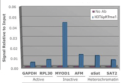
Figure 1. H3T6pK9me1 antibody ChIP results
Chromatin Immunoprecipitation of H3T6pK9me1 antibody. Chromatin from one million formaldehyde cross-linked Hela cells was used with 2ug of H3T6pK9me1 and 20ul of magnetic IgG beads per immunoprecipitation. A no antibody (No Ab) control was also used. Immunoprecipitated DNA was quantified using quantitative real-time PCR and normalized to the input chromatin.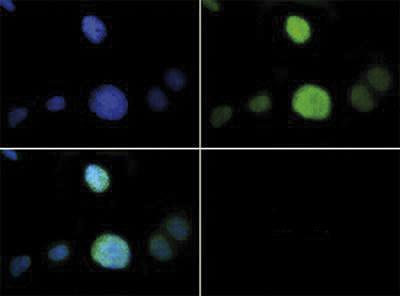
Figure 2. H3T6pK9me1 antibody Immunofluorescence results
Immunofluorescence of H3T6pK9me1 antibody. Tissue: HeLa cells. Fixation: 0.5% PFA. Primary antibody: incubated at a 1:100 dilution for 1 h at RT. Secondary antibody: FITC secondary antibody at 1:10,000 for 45 min at RT. Localization: H3T6pK9me1 is nuclear and chromosomal. Staining: H3T6pK9me1 is expressed in green and the nuclei are counterstained with DAPI (blue).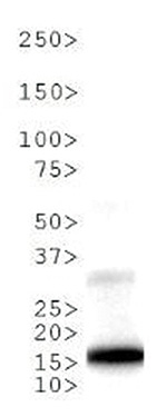
Figure 3. H3T6pK9me1 antibody Western blot results
Western Blot of H3T6pK9me1 antibody. 30 μg NIH-3T3 Histone extracts. Primary antibody diluted 1:1,000 overnight at 4°C. Secondary antibody: IRDye800TM rabbit secondary antibody at 1:10,000 for 45 min at RT. Predicted/Observed size: ~15 kDa. Other band(s): None.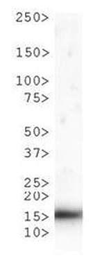
Figure 4. H3T6pK9me1 antibody Western blot results
Western Blot of H3T6pK9me1 antibody. 30 μg C. elegans embryo lysate. Primary antibody diluted 1:1,000 overnight at 4°C. Secondary antibody: IRDye800TM rabbit secondary antibody at 1:10,000 for 45 min at RT. Predicted/Observed size: ~15 kDa. Other band(s): None.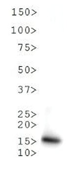
Figure 5. H3T6pK9me1 antibody Western blot results
Western Blot of H3T6pK9me1 antibody. 30 μg HeLa Histone extracts. Primary antibody diluted 1:1,000 overnight at 4°C. Secondary antibody: IRDye800TM rabbit secondary antibody at 1:10,000 for 45 min at RT. Predicted/Observed size: ~15 kDa. Other band(s): None.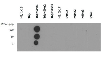
Figure 6. H3T6pK9me1 antibody Dot blot results
Dot Blot of H3T6pK9me1 antibody. Lane 1: Histone H3 1-13. Lane 2: T6p. Lane 3: T6pK9Me1. Lane 4: T6pK9Me2. Lane 5: T6pK9Me3. Lane 6: Histone H3 2-17. Lane 7: K9Me1. Lane 8: K9Me2. Lane 9: K9Me3. Lane 10: K9Ac. Load: 1, 10, and 100 picomoles of peptide. Primary antibody diluted 1:1,000 for 45 min at 4°C. Secondary antibody: DylightTM488 rabbit secondary antibody at 1:10,000 for 45 min at RT. - 出版物

How to properly cite our product/service in your work
We strongly recommend using this: H3T6pK9me1 polyclonal antibody (Hologic Diagenode Cat# C15410283 Lot# 001). Click here to copy to clipboard.
Using our products or services in your publication? Let us know!