
| ELISA Enzyme-linked immunosorbent assay. Read more |
| DB Dot blotting Read more |
| WB Western blot : The quality of antibodies used in this technique is crucial for correct and specific protein identification. Diagenode offers huge selection of highly sensitive and specific western blot-validated antibodies. Learn more about: Load... Read more |
| ChIP-qPCR (ab) Read more |
H4K8ac polyclonal antibody
Catalog Number
Format
Price

Polyclonal antibody raised in rabbit against histone H4 containing the acetylated lysine 8 (H4K8ac), using a KLH-conjugated synthetic peptide.
| Lot | A156-001 |
|---|---|
| Concentration | not determined |
| Species reactivity | Human: positive. Other species: not tested. |
| Type | Polyclonal, ChIP-grade |
| Purity | Whole antiserum from rabbit containing 0.05% azide. |
| Host | Rabbit |
| Precautions | This product is for research use only. Not for use in diagnostic or therapeutic procedures. |
| Applications | Suggested dilution | References |
|---|---|---|
| ChIP * | 5 μl/ChIP | Fig 1 |
| ELISA | 1:1,000 - 1:5,000 | Fig 2 |
| Dot Blotting | 1:20,000 | Fig 3 |
| Western Blotting | 1:250 | Fig 4 |
* Please note that of the optimal antibody amount per IP should be determined by the end-user. We recommend testing 1-10 μg per IP.
- Validation Data
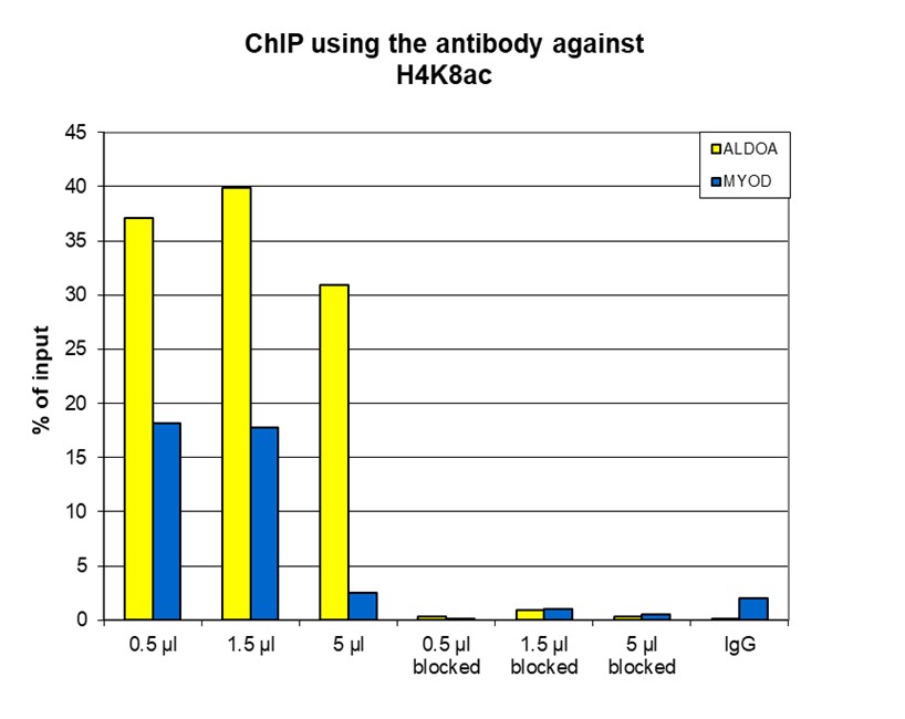
Figure 1. ChIP results obtained with the antibody directed against H4K8ac
ChIP assays were performed using human osteosarcoma (U2OS) cells, the antibody against H4K8ac (cat. No. C15310103) and optimized PCR primer sets for qPCR. ChIP was performed on sheared chromatin from 100,000 cells treated with the deacetylase inhibitor ATRA. A titration of the antibody consisting of 0.5, 1.5 and 5 µl per ChIP experiment was analysed. Additionally, ChIP was performed after incubation of the antibody with 5 nmol blocking peptide (cat. No. C16000103) for 1 hour at room temperature. IgG (5 µg/IP) was used as negative IP control. QPCR was performed with primers for the ALDOA promoter (fructose-bisphosphate aldolase A) and for the coding region of the myogenic differentiation gene (MYOD), a gene that is inactive under normal conditions. Figure 1 shows the recovery, expressed as a % of input (the relative amount of immunoprecipitated DNA compared to input DNA after qPCR analysis).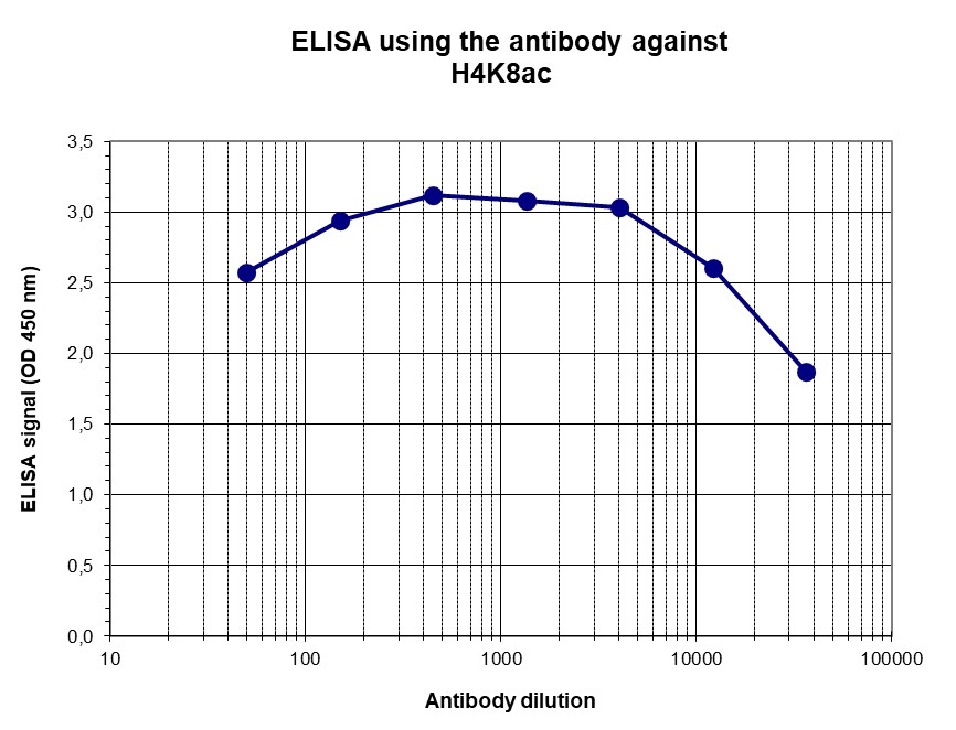
Figure 2. Determination of the titer
To determine the titer, an ELISA was performed using a serial dilution of the antibody directed against human H4K8ac (cat. No. C15310103). The antigen used was a peptide containing the histone modification of interest. By plotting the absorbance against the antibody dilution (Figure 2), we estimated the titer of the antibody to be 1:71,500.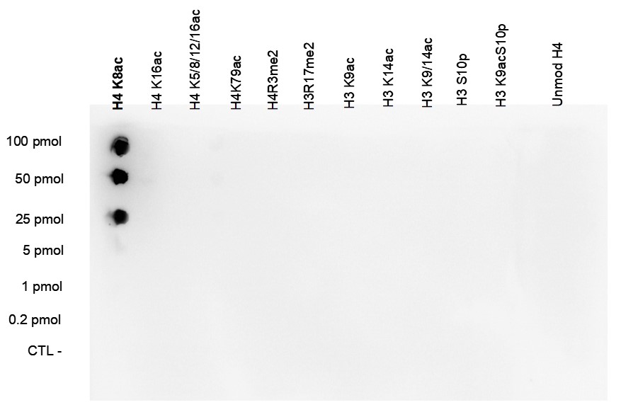
Figure 3. Cross reactivity test using the antibody directed against H4K8ac
A Dot Blot analysis was performed to test the cross-reactivity of the antibody against H4K8ac (cat. No. C15310103) with peptides containing other modifications of histone H4 and H3 and an unmodified histone H4 sequence. From 100 pmol down to 0.2 pmol of the peptide containing the respective histone modification were spotted on a membrane. The antibody was used at a dilution of 1:20,000. Figure 3 shows a high specificity of the antibody for the modification of interest.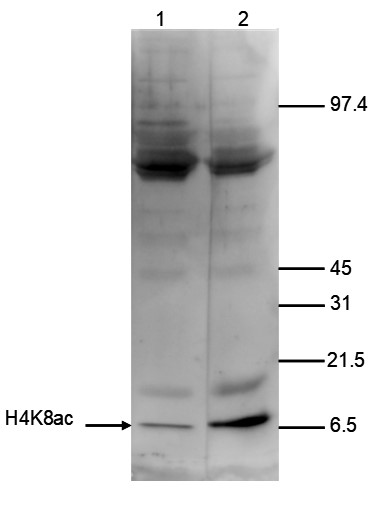
Figure 4. Western blot analysis using the antibody directed against H4K8ac
Histone extracts of HeLa cells (15 µg) were analysed by Western blot using the antibody against H4K8ac (cat. No. C15310103) diluted 1:250 in TBS-Tween containing 5% skimmed milk. The position of the protein of interest is indicated on the left; the marker (in kDa) is shown on the right. Lane 2 shows the result of the Western analysis with the antibody; lane 1 shows the same analysis after incubation of the antibody with 750 pmol blocking peptide (cat. No. sp-103-050) for 1 hour at room temperature. - 出版物

How to properly cite our product/service in your work
We strongly recommend using this: H4K8ac polyclonal antibody (Hologic Diagenode Cat# C15310103 Lot# A156-001). Click here to copy to clipboard.
Using our products or services in your publication? Let us know!
- 関連商品
-
 B01200041
0.1 ml tube holder & tube adaptors for Picoruptor
-