Notice (8): Undefined variable: solution_of_interest [APP/View/Products/view.ctp, line 755]Code Context<!-- BEGIN: REQUEST_FORM MODAL -->
<div id="request_formModal" class="reveal-modal medium" data-reveal aria-labelledby="modalTitle" aria-hidden="true" role="dialog">
<?= $this->element('Forms/simple_form', array('solution_of_interest' => $solution_of_interest, 'header' => $header, 'message' => $message, 'campaign_id' => $campaign_id)) ?>
$viewFile = '/home/website-server/www/app/View/Products/view.ctp'
$dataForView = array(
'language' => 'jp',
'meta_keywords' => '',
'meta_description' => 'IPure kit v2',
'meta_title' => 'IPure kit v2 | Diagenode',
'product' => array(
'Product' => array(
'id' => '2686',
'antibody_id' => null,
'name' => 'IPure kit v2',
'description' => '<p><span><strong>High DNA recovery</strong> after ChIP or MeDIP is critical for specific downstream applications (e.g. Next generation sequencing). Diagenode's IPure kit is the only DNA purification kit that is specifically <strong>optimized for extracting very low amounts</strong> of DNA after ChIP and MeDIP.</span></p>
<script src="chrome-extension://hhojmcideegachlhfgfdhailpfhgknjm/web_accessible_resources/index.js"></script>',
'label1' => '特徴',
'info1' => '<h2>IPure after ChIP</h2>
<p><img src="https://www.diagenode.com/img/product/kits/ideal-TF-chip-seq-A.png" alt="ChIP-seq figure A" style="display: block; margin-left: auto; margin-right: auto;" /></p>
<p><img src="https://www.diagenode.com/img/product/kits/ideal-TF-chip-seq-B.png" alt="ChIP-seq figure B" style="display: block; margin-left: auto; margin-right: auto;" /></p>
<p><img src="https://www.diagenode.com/img/product/kits/ideal-TF-chip-seq-C.png" alt="ChIP-seq figure C" style="display: block; margin-left: auto; margin-right: auto;" /></p>
<p><small><strong>Figure 1.</strong> Chromatin Immunoprecipitation has been performed using chromatin from HeLa cells, the iDeal ChIP-seq kit for Transcription Factors (containing the IPure module for DNA purification) and the Diagenode ChIP-seq-grade HDAC1 (A), LSD1 (B) and p53 antibody (C). The IP'd DNA was subsequently analysed on an Illumina® Genome Analyzer. Library preparation, cluster generation and sequencing were performed according to the manufacturer's instructions. This figure shows the peak distribution in regions of chromosome 3 (A), chromosome 12 (B) and chromosome 6 (C) respectively.</small></p>
<p></p>
<h2>IPure after CUT&Tag</h2>
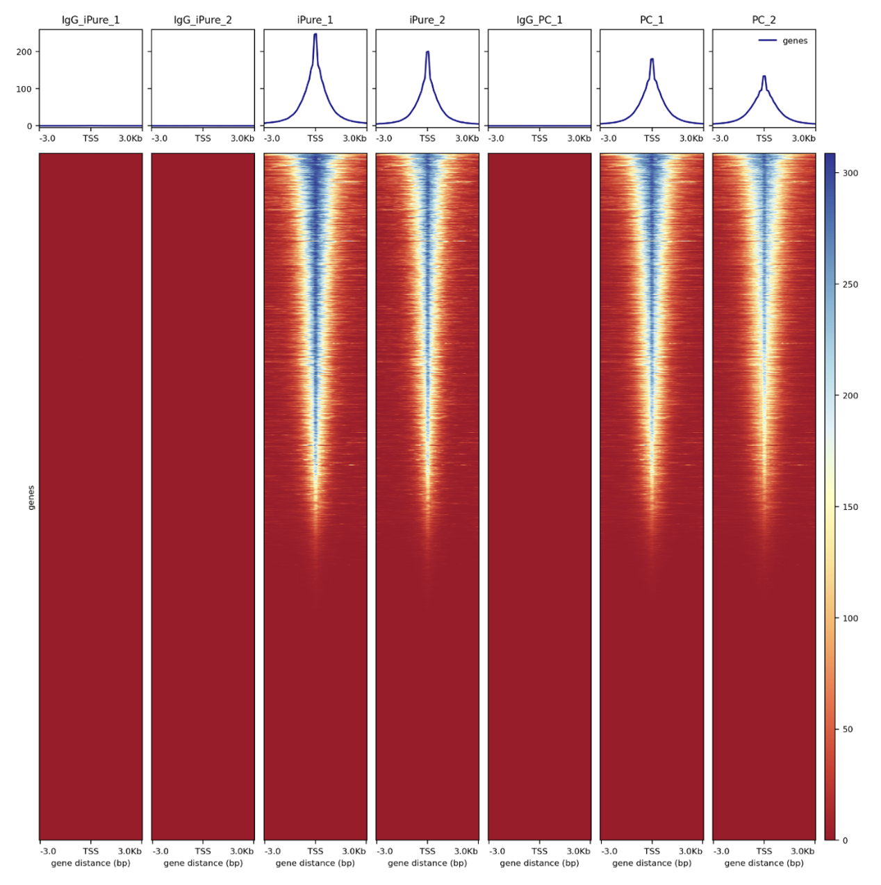
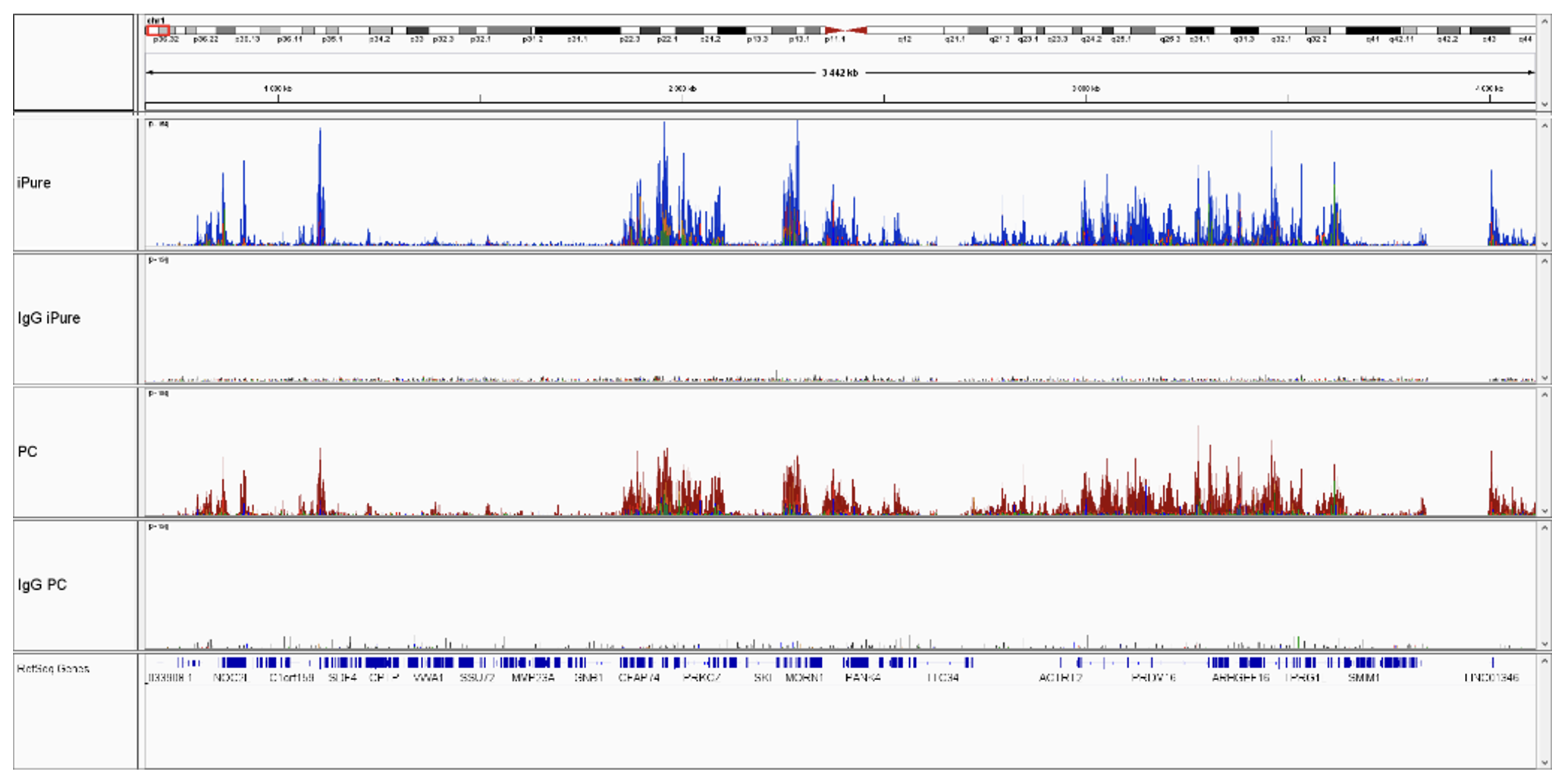
<p>Successful CUT&Tag results showing a low background with high region-specific enrichment has been generated using 50.000 of K562 cells, 1 µg of H3K4me3 or H3K27me3 antibody (Diagenode, C15410003 or C15410069, respectively) and proteinA-Tn5 (1:250) (Diagenode, C01070001). 1 µg of IgG (C15410206) was used as negative control. Samples were purified using the IPure kit v2 or phenol-chloroform purification. The below figures present the comparison of two purification methods.</p>
<center><img src="https://www.diagenode.com/img/product/kits/ipure-fig2.png" style="display: block; margin-left: auto; margin-right: auto;" width="400" /></center><center>
<p style="text-align: center;"><small><strong>Figure 2.</strong> Heatmap 3kb upstream and downstream of the TSS for H3K4me3</small></p>
</center>
<p></p>
<p><img src="https://www.diagenode.com/img/product/kits/ipure-fig3.png" style="display: block; margin-left: auto; margin-right: auto;" width="600" /></p>
<p></p>
<center><small><strong>Figure 3.</strong> Integrative genomics viewer (IGV) visualization of CUT&Tag experiments using Diagenode’s pA-Tn5 transposase (Cat. No. C01070002), H3K27me3 antibody (Cat. No. C15410069) and IPure kit v2 vs phenol chloroform purification (PC).</small></center>
<p></p>
<p></p>
<h2>IPure after MeDIP</h2>
<center><img src="https://www.diagenode.com/img/product/kits/magmedip-seq-figure_multi3.jpg" alt="medip sequencing coverage" width="600" /></center><center></center><center>
<p></p>
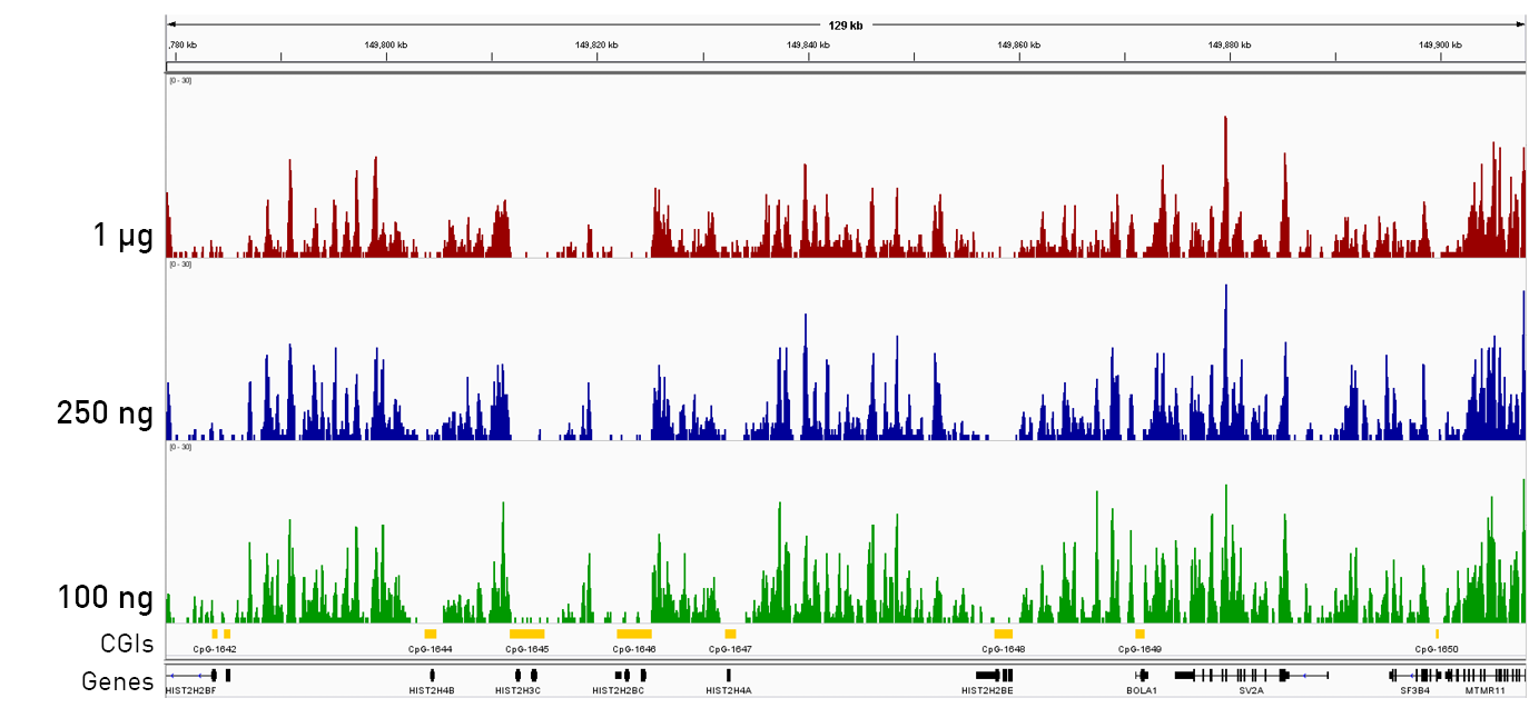
<small><strong>Figure 4.</strong> Consistent coverage and methylation detection from different starting amounts of DNA with the Diagenode MagMeDIP-seq Package (including the Ipure kit for DNA purification). Samples containing decreasing starting amounts of DNA (from the top down: 1000 ng (red), 250 ng (blue), 100 ng (green)) originating from human blood were prepared, revealing a consistent coverage profile for the three different starting amounts, which enables reproducible methylation detection. The CpG islands (CGIs) (marked by yellow boxes in the bottom track) are predominantly unmethylated in the human genome, and as expected, we see a depletion of reads at and around CGIs.</small></center>
<script src="chrome-extension://hhojmcideegachlhfgfdhailpfhgknjm/web_accessible_resources/index.js"></script>',
'label2' => '',
'info2' => '<h2 style="text-align: center;">Kit Method Overview & Time table</h2>
<p><img src="https://www.diagenode.com/img/product/kits/workflow-ipure-cuttag.png" style="display: block; margin-left: auto; margin-right: auto;" /></p>
<h3><strong>Workflow description</strong></h3>
<h5><strong>IPure after ChIP</strong></h5>
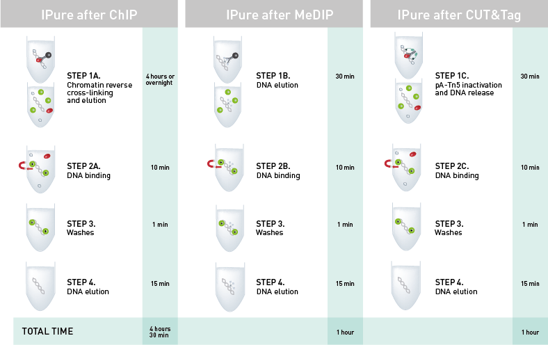
<p><strong>Step 1:</strong> Chromatin is decrosslinked and eluted from beads (magnetic or agarose) which are discarded. <strong>Magnetic beads</strong> <strong>for purification</strong> are added.<br /> <strong>Step 2:</strong> Magnetic beads acquire positive charge to bind the negatively charged phosphate backbone of DNA. DNA-bead complex is separated using a magnet.<br /> <strong>Step 3:</strong> Proteins and remaining buffer are washed away.<br /> <strong>Step 4:</strong> DNA is eluted from magnetic beads, which are discarded. Purified DNA is ready for any downstream application (NGS, qPCR, amplification, microarray).<br /><br /><br /></p>
<h5><strong>IPure after MeDIP</strong></h5>
<p><strong>Step 1:</strong> DNA is eluted from beads (magnetic or agarose) which are discarded. <strong>Magnetic beads</strong> <strong>for purification</strong> are added. <br /><strong>Step 2:</strong> Magnetic beads acquire positive charge to bind the negatively charged phosphate backbone of DNA. DNA-bead complex is separated using a magnet. <br /><strong>Step 3:</strong> Remaining buffer are washed away.<br /><strong>Step 4:</strong> DNA is eluted from magnetic beads, which are discarded. Purified DNA is ready for any downstream application (NGS, qPCR, amplification, microarray).<br /><br /><br /></p>
<h5><strong>IPure after CUT&Tag</strong></h5>
<p><strong>Step 1:</strong> pA-Tn5 is inactivated and DNA released from the cells. <strong>Magnetic beads</strong> <strong>for purification</strong> are added. <br /><strong>Step 2:</strong> Magnetic beads acquire positive charge to bind the negatively charged phosphate backbone of DNA. DNA-bead complex is separated using a magnet. <br /><strong>Step 3:</strong> Proteins and remaining buffer are washed away. <br /><strong>Step 4:</strong> DNA is eluted from magnetic beads, which are discarded. Purified DNA is ready for any downstream application (NGS, qPCR, amplification, microarray).</p>
<p></p>
<p></p>
<script async="" src="https://edge.fullstory.com/s/fs.js" crossorigin="anonymous"></script>
<script src="chrome-extension://hhojmcideegachlhfgfdhailpfhgknjm/web_accessible_resources/index.js"></script>
<script src="chrome-extension://hhojmcideegachlhfgfdhailpfhgknjm/web_accessible_resources/index.js"></script>',
'label3' => '',
'info3' => '<p></p>
<script src="chrome-extension://hhojmcideegachlhfgfdhailpfhgknjm/web_accessible_resources/index.js"></script>',
'format' => '100 rxns',
'catalog_number' => 'C03010015',
'old_catalog_number' => 'C03010012',
'sf_code' => 'C03010015-',
'type' => 'RFR',
'search_order' => '',
'price_EUR' => '385',
'price_USD' => '450',
'price_GBP' => '355',
'price_JPY' => '63080',
'price_CNY' => '',
'price_AUD' => '1125',
'country' => 'ALL',
'except_countries' => 'None',
'quote' => false,
'in_stock' => false,
'featured' => false,
'no_promo' => false,
'online' => true,
'master' => true,
'last_datasheet_update' => '0000-00-00',
'slug' => 'ipure-kit-v2-x100',
'meta_title' => 'IPure kit v2 | Diagenode',
'meta_keywords' => '',
'meta_description' => 'IPure kit v2',
'modified' => '2023-04-20 16:09:27',
'created' => '2015-09-09 10:52:23',
'locale' => 'jpn'
),
'Antibody' => array(
'host' => '*****',
'id' => null,
'name' => null,
'description' => null,
'clonality' => null,
'isotype' => null,
'lot' => null,
'concentration' => null,
'reactivity' => null,
'type' => null,
'purity' => null,
'classification' => null,
'application_table' => null,
'storage_conditions' => null,
'storage_buffer' => null,
'precautions' => null,
'uniprot_acc' => null,
'slug' => null,
'meta_keywords' => null,
'meta_description' => null,
'modified' => null,
'created' => null,
'select_label' => null
),
'Slave' => array(
(int) 0 => array(
[maximum depth reached]
)
),
'Group' => array(
'Group' => array(
[maximum depth reached]
),
'Master' => array(
[maximum depth reached]
),
'Product' => array(
[maximum depth reached]
)
),
'Related' => array(
(int) 0 => array(
[maximum depth reached]
),
(int) 1 => array(
[maximum depth reached]
),
(int) 2 => array(
[maximum depth reached]
),
(int) 3 => array(
[maximum depth reached]
)
),
'Application' => array(
(int) 0 => array(
[maximum depth reached]
),
(int) 1 => array(
[maximum depth reached]
)
),
'Category' => array(
(int) 0 => array(
[maximum depth reached]
)
),
'Document' => array(
(int) 0 => array(
[maximum depth reached]
)
),
'Feature' => array(),
'Image' => array(),
'Promotion' => array(),
'Protocol' => array(),
'Publication' => array(
(int) 0 => array(
[maximum depth reached]
),
(int) 1 => array(
[maximum depth reached]
),
(int) 2 => array(
[maximum depth reached]
),
(int) 3 => array(
[maximum depth reached]
),
(int) 4 => array(
[maximum depth reached]
),
(int) 5 => array(
[maximum depth reached]
),
(int) 6 => array(
[maximum depth reached]
),
(int) 7 => array(
[maximum depth reached]
),
(int) 8 => array(
[maximum depth reached]
),
(int) 9 => array(
[maximum depth reached]
),
(int) 10 => array(
[maximum depth reached]
),
(int) 11 => array(
[maximum depth reached]
),
(int) 12 => array(
[maximum depth reached]
),
(int) 13 => array(
[maximum depth reached]
),
(int) 14 => array(
[maximum depth reached]
),
(int) 15 => array(
[maximum depth reached]
),
(int) 16 => array(
[maximum depth reached]
),
(int) 17 => array(
[maximum depth reached]
),
(int) 18 => array(
[maximum depth reached]
),
(int) 19 => array(
[maximum depth reached]
),
(int) 20 => array(
[maximum depth reached]
),
(int) 21 => array(
[maximum depth reached]
),
(int) 22 => array(
[maximum depth reached]
),
(int) 23 => array(
[maximum depth reached]
),
(int) 24 => array(
[maximum depth reached]
),
(int) 25 => array(
[maximum depth reached]
),
(int) 26 => array(
[maximum depth reached]
),
(int) 27 => array(
[maximum depth reached]
),
(int) 28 => array(
[maximum depth reached]
),
(int) 29 => array(
[maximum depth reached]
),
(int) 30 => array(
[maximum depth reached]
),
(int) 31 => array(
[maximum depth reached]
),
(int) 32 => array(
[maximum depth reached]
),
(int) 33 => array(
[maximum depth reached]
),
(int) 34 => array(
[maximum depth reached]
),
(int) 35 => array(
[maximum depth reached]
),
(int) 36 => array(
[maximum depth reached]
),
(int) 37 => array(
[maximum depth reached]
),
(int) 38 => array(
[maximum depth reached]
),
(int) 39 => array(
[maximum depth reached]
),
(int) 40 => array(
[maximum depth reached]
),
(int) 41 => array(
[maximum depth reached]
),
(int) 42 => array(
[maximum depth reached]
),
(int) 43 => array(
[maximum depth reached]
),
(int) 44 => array(
[maximum depth reached]
),
(int) 45 => array(
[maximum depth reached]
),
(int) 46 => array(
[maximum depth reached]
),
(int) 47 => array(
[maximum depth reached]
),
(int) 48 => array(
[maximum depth reached]
),
(int) 49 => array(
[maximum depth reached]
),
(int) 50 => array(
[maximum depth reached]
),
(int) 51 => array(
[maximum depth reached]
),
(int) 52 => array(
[maximum depth reached]
),
(int) 53 => array(
[maximum depth reached]
),
(int) 54 => array(
[maximum depth reached]
),
(int) 55 => array(
[maximum depth reached]
),
(int) 56 => array(
[maximum depth reached]
),
(int) 57 => array(
[maximum depth reached]
),
(int) 58 => array(
[maximum depth reached]
),
(int) 59 => array(
[maximum depth reached]
),
(int) 60 => array(
[maximum depth reached]
),
(int) 61 => array(
[maximum depth reached]
),
(int) 62 => array(
[maximum depth reached]
),
(int) 63 => array(
[maximum depth reached]
),
(int) 64 => array(
[maximum depth reached]
),
(int) 65 => array(
[maximum depth reached]
),
(int) 66 => array(
[maximum depth reached]
),
(int) 67 => array(
[maximum depth reached]
),
(int) 68 => array(
[maximum depth reached]
),
(int) 69 => array(
[maximum depth reached]
),
(int) 70 => array(
[maximum depth reached]
),
(int) 71 => array(
[maximum depth reached]
),
(int) 72 => array(
[maximum depth reached]
),
(int) 73 => array(
[maximum depth reached]
),
(int) 74 => array(
[maximum depth reached]
),
(int) 75 => array(
[maximum depth reached]
),
(int) 76 => array(
[maximum depth reached]
),
(int) 77 => array(
[maximum depth reached]
),
(int) 78 => array(
[maximum depth reached]
),
(int) 79 => array(
[maximum depth reached]
),
(int) 80 => array(
[maximum depth reached]
)
),
'Testimonial' => array(
(int) 0 => array(
[maximum depth reached]
)
),
'Area' => array(),
'SafetySheet' => array(
(int) 0 => array(
[maximum depth reached]
),
(int) 1 => array(
[maximum depth reached]
),
(int) 2 => array(
[maximum depth reached]
),
(int) 3 => array(
[maximum depth reached]
),
(int) 4 => array(
[maximum depth reached]
),
(int) 5 => array(
[maximum depth reached]
),
(int) 6 => array(
[maximum depth reached]
),
(int) 7 => array(
[maximum depth reached]
)
)
),
'meta_canonical' => 'https://www.diagenode.com/jp/p/ipure-kit-v2-x100'
)
$language = 'jp'
$meta_keywords = ''
$meta_description = 'IPure kit v2'
$meta_title = 'IPure kit v2 | Diagenode'
$product = array(
'Product' => array(
'id' => '2686',
'antibody_id' => null,
'name' => 'IPure kit v2',
'description' => '<p><span><strong>High DNA recovery</strong> after ChIP or MeDIP is critical for specific downstream applications (e.g. Next generation sequencing). Diagenode's IPure kit is the only DNA purification kit that is specifically <strong>optimized for extracting very low amounts</strong> of DNA after ChIP and MeDIP.</span></p>
<script src="chrome-extension://hhojmcideegachlhfgfdhailpfhgknjm/web_accessible_resources/index.js"></script>',
'label1' => '特徴',
'info1' => '<h2>IPure after ChIP</h2>
<p><img src="https://www.diagenode.com/img/product/kits/ideal-TF-chip-seq-A.png" alt="ChIP-seq figure A" style="display: block; margin-left: auto; margin-right: auto;" /></p>
<p><img src="https://www.diagenode.com/img/product/kits/ideal-TF-chip-seq-B.png" alt="ChIP-seq figure B" style="display: block; margin-left: auto; margin-right: auto;" /></p>
<p><img src="https://www.diagenode.com/img/product/kits/ideal-TF-chip-seq-C.png" alt="ChIP-seq figure C" style="display: block; margin-left: auto; margin-right: auto;" /></p>
<p><small><strong>Figure 1.</strong> Chromatin Immunoprecipitation has been performed using chromatin from HeLa cells, the iDeal ChIP-seq kit for Transcription Factors (containing the IPure module for DNA purification) and the Diagenode ChIP-seq-grade HDAC1 (A), LSD1 (B) and p53 antibody (C). The IP'd DNA was subsequently analysed on an Illumina® Genome Analyzer. Library preparation, cluster generation and sequencing were performed according to the manufacturer's instructions. This figure shows the peak distribution in regions of chromosome 3 (A), chromosome 12 (B) and chromosome 6 (C) respectively.</small></p>
<p></p>
<h2>IPure after CUT&Tag</h2>
<p>Successful CUT&Tag results showing a low background with high region-specific enrichment has been generated using 50.000 of K562 cells, 1 µg of H3K4me3 or H3K27me3 antibody (Diagenode, C15410003 or C15410069, respectively) and proteinA-Tn5 (1:250) (Diagenode, C01070001). 1 µg of IgG (C15410206) was used as negative control. Samples were purified using the IPure kit v2 or phenol-chloroform purification. The below figures present the comparison of two purification methods.</p>
<center><img src="https://www.diagenode.com/img/product/kits/ipure-fig2.png" style="display: block; margin-left: auto; margin-right: auto;" width="400" /></center><center>
<p style="text-align: center;"><small><strong>Figure 2.</strong> Heatmap 3kb upstream and downstream of the TSS for H3K4me3</small></p>
</center>
<p></p>
<p><img src="https://www.diagenode.com/img/product/kits/ipure-fig3.png" style="display: block; margin-left: auto; margin-right: auto;" width="600" /></p>
<p></p>
<center><small><strong>Figure 3.</strong> Integrative genomics viewer (IGV) visualization of CUT&Tag experiments using Diagenode’s pA-Tn5 transposase (Cat. No. C01070002), H3K27me3 antibody (Cat. No. C15410069) and IPure kit v2 vs phenol chloroform purification (PC).</small></center>
<p></p>
<p></p>
<h2>IPure after MeDIP</h2>
<center><img src="https://www.diagenode.com/img/product/kits/magmedip-seq-figure_multi3.jpg" alt="medip sequencing coverage" width="600" /></center><center></center><center>
<p></p>
<small><strong>Figure 4.</strong> Consistent coverage and methylation detection from different starting amounts of DNA with the Diagenode MagMeDIP-seq Package (including the Ipure kit for DNA purification). Samples containing decreasing starting amounts of DNA (from the top down: 1000 ng (red), 250 ng (blue), 100 ng (green)) originating from human blood were prepared, revealing a consistent coverage profile for the three different starting amounts, which enables reproducible methylation detection. The CpG islands (CGIs) (marked by yellow boxes in the bottom track) are predominantly unmethylated in the human genome, and as expected, we see a depletion of reads at and around CGIs.</small></center>
<script src="chrome-extension://hhojmcideegachlhfgfdhailpfhgknjm/web_accessible_resources/index.js"></script>',
'label2' => 'iPure Workflow',
'info2' => '<h2 style="text-align: center;">Kit Method Overview & Time table</h2>
<p><img src="https://www.diagenode.com/img/product/kits/workflow-ipure-cuttag.png" style="display: block; margin-left: auto; margin-right: auto;" /></p>
<h3><strong>Workflow description</strong></h3>
<h5><strong>IPure after ChIP</strong></h5>
<p><strong>Step 1:</strong> Chromatin is decrosslinked and eluted from beads (magnetic or agarose) which are discarded. <strong>Magnetic beads</strong> <strong>for purification</strong> are added.<br /> <strong>Step 2:</strong> Magnetic beads acquire positive charge to bind the negatively charged phosphate backbone of DNA. DNA-bead complex is separated using a magnet.<br /> <strong>Step 3:</strong> Proteins and remaining buffer are washed away.<br /> <strong>Step 4:</strong> DNA is eluted from magnetic beads, which are discarded. Purified DNA is ready for any downstream application (NGS, qPCR, amplification, microarray).<br /><br /><br /></p>
<h5><strong>IPure after MeDIP</strong></h5>
<p><strong>Step 1:</strong> DNA is eluted from beads (magnetic or agarose) which are discarded. <strong>Magnetic beads</strong> <strong>for purification</strong> are added. <br /><strong>Step 2:</strong> Magnetic beads acquire positive charge to bind the negatively charged phosphate backbone of DNA. DNA-bead complex is separated using a magnet. <br /><strong>Step 3:</strong> Remaining buffer are washed away.<br /><strong>Step 4:</strong> DNA is eluted from magnetic beads, which are discarded. Purified DNA is ready for any downstream application (NGS, qPCR, amplification, microarray).<br /><br /><br /></p>
<h5><strong>IPure after CUT&Tag</strong></h5>
<p><strong>Step 1:</strong> pA-Tn5 is inactivated and DNA released from the cells. <strong>Magnetic beads</strong> <strong>for purification</strong> are added. <br /><strong>Step 2:</strong> Magnetic beads acquire positive charge to bind the negatively charged phosphate backbone of DNA. DNA-bead complex is separated using a magnet. <br /><strong>Step 3:</strong> Proteins and remaining buffer are washed away. <br /><strong>Step 4:</strong> DNA is eluted from magnetic beads, which are discarded. Purified DNA is ready for any downstream application (NGS, qPCR, amplification, microarray).</p>
<p></p>
<p></p>
<script async="" src="https://edge.fullstory.com/s/fs.js" crossorigin="anonymous"></script>
<script src="chrome-extension://hhojmcideegachlhfgfdhailpfhgknjm/web_accessible_resources/index.js"></script>
<script src="chrome-extension://hhojmcideegachlhfgfdhailpfhgknjm/web_accessible_resources/index.js"></script>',
'label3' => '',
'info3' => '<p></p>
<script src="chrome-extension://hhojmcideegachlhfgfdhailpfhgknjm/web_accessible_resources/index.js"></script>',
'format' => '100 rxns',
'catalog_number' => 'C03010015',
'old_catalog_number' => 'C03010012',
'sf_code' => 'C03010015-',
'type' => 'RFR',
'search_order' => '',
'price_EUR' => '385',
'price_USD' => '450',
'price_GBP' => '355',
'price_JPY' => '63080',
'price_CNY' => '',
'price_AUD' => '1125',
'country' => 'ALL',
'except_countries' => 'None',
'quote' => false,
'in_stock' => false,
'featured' => false,
'no_promo' => false,
'online' => true,
'master' => true,
'last_datasheet_update' => '0000-00-00',
'slug' => 'ipure-kit-v2-x100',
'meta_title' => 'IPure kit v2 | Diagenode',
'meta_keywords' => '',
'meta_description' => 'IPure kit v2',
'modified' => '2023-04-20 16:09:27',
'created' => '2015-09-09 10:52:23',
'locale' => 'jpn'
),
'Antibody' => array(
'host' => '*****',
'id' => null,
'name' => null,
'description' => null,
'clonality' => null,
'isotype' => null,
'lot' => null,
'concentration' => null,
'reactivity' => null,
'type' => null,
'purity' => null,
'classification' => null,
'application_table' => null,
'storage_conditions' => null,
'storage_buffer' => null,
'precautions' => null,
'uniprot_acc' => null,
'slug' => null,
'meta_keywords' => null,
'meta_description' => null,
'modified' => null,
'created' => null,
'select_label' => null
),
'Slave' => array(
(int) 0 => array(
'id' => '102',
'name' => 'C03010015',
'product_id' => '2686',
'modified' => '2016-02-19 15:55:20',
'created' => '2016-02-19 15:55:20'
)
),
'Group' => array(
'Group' => array(
'id' => '102',
'name' => 'C03010015',
'product_id' => '2686',
'modified' => '2016-02-19 15:55:20',
'created' => '2016-02-19 15:55:20'
),
'Master' => array(
'id' => '2686',
'antibody_id' => null,
'name' => 'IPure kit v2',
'description' => '<p><a href="https://www.diagenode.com/files/products/kits/ipure_kit_v2_manual.pdf"><img src="https://www.diagenode.com/img/buttons/bt-manual.png" /></a></p>
<p>Diagenode’s<span> </span><b>IPure</b><b><span> </span>kit<span> </span></b>is the only DNA purification kit using magnetic beads, that is specifically optimized for extracting DNA from<span> </span><b>ChIP</b><b>,<span> </span></b><b>MeDIP</b><span> </span>and<span> </span><b>CUT&Tag</b>. The use of the magnetic beads allows for a clear separation of DNA and increases therefore the reproducibility of your DNA purification. This simple and straightforward protocol delivers pure DNA ready for any downstream application (e.g. next generation sequencing). Comparing to phenol-chloroform extraction, the IPure technology has the advantage of being nontoxic and much easier to be carried out on multiple samples.</p>
<center>
<h4>High DNA recovery after purification of ChIP samples using IPure technology</h4>
<center><img src="https://www.diagenode.com/img/product/kits/ipure-chromatin-function.png" width="500" /></center>
<p></p>
<p><small>ChIP assays were performed using different amounts of U2OS cells and the H3K9me3 antibody (Cat. No.<span> </span><span>C15410056</span>; 2 g/IP). <span>The purified DNA was eluted in 50 µl of water and quantified with a Nanodrop.</span></small></p>
<p></p>
<p><strong>Benefits of the IPure kit:</strong></p>
<ul>
<li style="text-align: left;">Provides pure DNA for any downstream application (e. g. Next generation sequencing)</li>
<li style="text-align: left;">Non-toxic</li>
<li style="text-align: left;">Fast & easy to use</li>
<li style="text-align: left;">Optimized for DNA purification after ChIP, MeDIP and CUT&Tag</li>
<li style="text-align: left;">Compatible with automation</li>
<li style="text-align: left;">Validated on the IP-Star Compact</li>
</ul>
</center>',
'label1' => 'Examples of results',
'info1' => '<h2>IPure after ChIP</h2>
<p><img src="https://www.diagenode.com/img/product/kits/ideal-TF-chip-seq-A.png" alt="ChIP-seq figure A" style="display: block; margin-left: auto; margin-right: auto;" /></p>
<p><img src="https://www.diagenode.com/img/product/kits/ideal-TF-chip-seq-B.png" alt="ChIP-seq figure B" style="display: block; margin-left: auto; margin-right: auto;" /></p>
<p><img src="https://www.diagenode.com/img/product/kits/ideal-TF-chip-seq-C.png" alt="ChIP-seq figure C" style="display: block; margin-left: auto; margin-right: auto;" /></p>
<p><small><strong>Figure 1.</strong> Chromatin Immunoprecipitation has been performed using chromatin from HeLa cells, the iDeal ChIP-seq kit for Transcription Factors (containing the IPure module for DNA purification) and the Diagenode ChIP-seq-grade HDAC1 (A), LSD1 (B) and p53 antibody (C). The IP'd DNA was subsequently analysed on an Illumina® Genome Analyzer. Library preparation, cluster generation and sequencing were performed according to the manufacturer's instructions. This figure shows the peak distribution in regions of chromosome 3 (A), chromosome 12 (B) and chromosome 6 (C) respectively.</small></p>
<p></p>
<h2>IPure after CUT&Tag</h2>
<p>Successful CUT&Tag results showing a low background with high region-specific enrichment has been generated using 50.000 of K562 cells, 1 µg of H3K4me3 or H3K27me3 antibody (Diagenode, C15410003 or C15410069, respectively) and proteinA-Tn5 (1:250) (Diagenode, C01070001). 1 µg of IgG (C15410206) was used as negative control. Samples were purified using the IPure kit v2 or phenol-chloroform purification. The below figures present the comparison of two purification methods.</p>
<center><img src="https://www.diagenode.com/img/product/kits/ipure-fig2.png" style="display: block; margin-left: auto; margin-right: auto;" width="400" /></center><center>
<p style="text-align: center;"><small><strong>Figure 2.</strong> Heatmap 3kb upstream and downstream of the TSS for H3K4me3</small></p>
</center>
<p></p>
<p><img src="https://www.diagenode.com/img/product/kits/ipure-fig3.png" style="display: block; margin-left: auto; margin-right: auto;" width="600" /></p>
<p></p>
<center><small><strong>Figure 3.</strong> Integrative genomics viewer (IGV) visualization of CUT&Tag experiments using Diagenode’s pA-Tn5 transposase (Cat. No. C01070002), H3K27me3 antibody (Cat. No. C15410069) and IPure kit v2 vs phenol chloroform purification (PC).</small></center>
<p></p>
<p></p>
<h2>IPure after MeDIP</h2>
<center><img src="https://www.diagenode.com/img/product/kits/magmedip-seq-figure_multi3.jpg" alt="medip sequencing coverage" width="600" /></center><center></center><center>
<p></p>
<small><strong>Figure 4.</strong> Consistent coverage and methylation detection from different starting amounts of DNA with the Diagenode MagMeDIP-seq Package (including the Ipure kit for DNA purification). Samples containing decreasing starting amounts of DNA (from the top down: 1000 ng (red), 250 ng (blue), 100 ng (green)) originating from human blood were prepared, revealing a consistent coverage profile for the three different starting amounts, which enables reproducible methylation detection. The CpG islands (CGIs) (marked by yellow boxes in the bottom track) are predominantly unmethylated in the human genome, and as expected, we see a depletion of reads at and around CGIs.</small></center>
<script src="chrome-extension://hhojmcideegachlhfgfdhailpfhgknjm/web_accessible_resources/index.js"></script>',
'label2' => 'iPure Workflow',
'info2' => '<h2 style="text-align: center;">Kit Method Overview & Time table</h2>
<p><img src="https://www.diagenode.com/img/product/kits/workflow-ipure-cuttag.png" style="display: block; margin-left: auto; margin-right: auto;" /></p>
<h3><strong>Workflow description</strong></h3>
<h5><strong>IPure after ChIP</strong></h5>
<p><strong>Step 1:</strong> Chromatin is decrosslinked and eluted from beads (magnetic or agarose) which are discarded. <strong>Magnetic beads</strong> <strong>for purification</strong> are added.<br /> <strong>Step 2:</strong> Magnetic beads acquire positive charge to bind the negatively charged phosphate backbone of DNA. DNA-bead complex is separated using a magnet.<br /> <strong>Step 3:</strong> Proteins and remaining buffer are washed away.<br /> <strong>Step 4:</strong> DNA is eluted from magnetic beads, which are discarded. Purified DNA is ready for any downstream application (NGS, qPCR, amplification, microarray).<br /><br /><br /></p>
<h5><strong>IPure after MeDIP</strong></h5>
<p><strong>Step 1:</strong> DNA is eluted from beads (magnetic or agarose) which are discarded. <strong>Magnetic beads</strong> <strong>for purification</strong> are added. <br /><strong>Step 2:</strong> Magnetic beads acquire positive charge to bind the negatively charged phosphate backbone of DNA. DNA-bead complex is separated using a magnet. <br /><strong>Step 3:</strong> Remaining buffer are washed away.<br /><strong>Step 4:</strong> DNA is eluted from magnetic beads, which are discarded. Purified DNA is ready for any downstream application (NGS, qPCR, amplification, microarray).<br /><br /><br /></p>
<h5><strong>IPure after CUT&Tag</strong></h5>
<p><strong>Step 1:</strong> pA-Tn5 is inactivated and DNA released from the cells. <strong>Magnetic beads</strong> <strong>for purification</strong> are added. <br /><strong>Step 2:</strong> Magnetic beads acquire positive charge to bind the negatively charged phosphate backbone of DNA. DNA-bead complex is separated using a magnet. <br /><strong>Step 3:</strong> Proteins and remaining buffer are washed away. <br /><strong>Step 4:</strong> DNA is eluted from magnetic beads, which are discarded. Purified DNA is ready for any downstream application (NGS, qPCR, amplification, microarray).</p>
<p></p>
<p></p>
<script async="" src="https://edge.fullstory.com/s/fs.js" crossorigin="anonymous"></script>
<script src="chrome-extension://hhojmcideegachlhfgfdhailpfhgknjm/web_accessible_resources/index.js"></script>
<script src="chrome-extension://hhojmcideegachlhfgfdhailpfhgknjm/web_accessible_resources/index.js"></script>',
'label3' => '',
'info3' => '<p></p>
<script src="chrome-extension://hhojmcideegachlhfgfdhailpfhgknjm/web_accessible_resources/index.js"></script>',
'format' => '100 rxns',
'catalog_number' => 'C03010015',
'old_catalog_number' => 'C03010012',
'sf_code' => 'C03010015-',
'type' => 'RFR',
'search_order' => '',
'price_EUR' => '385',
'price_USD' => '450',
'price_GBP' => '355',
'price_JPY' => '63080',
'price_CNY' => '',
'price_AUD' => '1125',
'country' => 'ALL',
'except_countries' => 'None',
'quote' => false,
'in_stock' => false,
'featured' => false,
'no_promo' => false,
'online' => true,
'master' => true,
'last_datasheet_update' => '0000-00-00',
'slug' => 'ipure-kit-v2-x100',
'meta_title' => 'IPure kit v2 | Diagenode',
'meta_keywords' => '',
'meta_description' => 'IPure kit v2',
'modified' => '2023-04-20 16:09:27',
'created' => '2015-09-09 10:52:23'
),
'Product' => array(
(int) 0 => array(
[maximum depth reached]
)
)
),
'Related' => array(
(int) 0 => array(
'id' => '1836',
'antibody_id' => null,
'name' => 'iDeal ChIP-seq kit for Histones',
'description' => '<p><a href="https://www.diagenode.com/files/products/kits/ideal-chipseq-for-histones-complete-manual.pdf"><img src="https://www.diagenode.com/img/buttons/bt-manual.png" /></a></p>
<p>Don’t risk wasting your precious sequencing samples. Diagenode’s validated <strong>iDeal ChIP-seq kit for Histones</strong> has everything you need for a successful start-to-finish <strong>ChIP of histones prior to Next-Generation Sequencing</strong>. The complete kit contains all buffers and reagents for cell lysis, chromatin shearing, immunoprecipitation and DNA purification. In addition, unlike competing solutions, the kit contains positive and negative control antibodies (H3K4me3 and IgG, respectively) as well as positive and negative control PCR primers pairs (GAPDH TSS and Myoglobin exon 2, respectively) for your convenience and a guarantee of optimal results. The kit has been validated on multiple histone marks.</p>
<p> The iDeal ChIP-seq kit for Histones<strong> </strong>is perfect for <strong>cells</strong> (<strong>100,000 cells</strong> to <strong>1,000,000 cells</strong> per IP) and has been validated for <strong>tissues</strong> (<strong>1.5 mg</strong> to <strong>5 mg</strong> of tissue per IP).</p>
<p> The iDeal ChIP-seq kit is the only kit on the market validated for the major sequencing systems. Our expertise in ChIP-seq tools allows reproducible and efficient results every time.</p>
<p></p>
<p> <strong></strong></p>
<p></p>',
'label1' => 'Characteristics',
'info1' => '<ul style="list-style-type: disc;">
<li>Highly <strong>optimized</strong> protocol for ChIP-seq from cells and tissues</li>
<li><strong>Validated</strong> for ChIP-seq with multiple histones marks</li>
<li>Most <strong>complete</strong> kit available (covers all steps, including the control antibodies and primers)</li>
<li>Optimized chromatin preparation in combination with the Bioruptor ensuring the best <strong>epitope integrity</strong></li>
<li>Magnetic beads make ChIP easy, fast and more <strong>reproducible</strong></li>
<li>Combination with Diagenode ChIP-seq antibodies provides high yields with excellent <strong>specificity</strong> and <strong>sensitivity</strong></li>
<li>Purified DNA suitable for any downstream application</li>
<li>Easy-to-follow protocol</li>
</ul>
<p>Note: to obtain optimal results, this kit should be used in combination with the DiaMag1.5 - magnetic rack.</p>
<h3>ChIP-seq on cells</h3>
<p><img src="https://www.diagenode.com/img/product/kits/iDeal-kit-C01010053-figure-1.jpg" alt="Figure 1A" style="display: block; margin-left: auto; margin-right: auto;" /></p>
<p><strong>Figure 1A. The high consistency of the iDeal ChIP-seq kit on the Ion Torrent™ PGM™ (Life Technologies) and GAIIx (Illumina<sup>®</sup>)</strong><br /> ChIP was performed on sheared chromatin from 1 million HelaS3 cells using the iDeal ChIP-seq kit and 1 µg of H3K4me3 positive control antibody. Two different biological samples have been analyzed using two different sequencers - GAIIx (Illumina<sup>®</sup>) and PGM™ (Ion Torrent™). The expected ChIP-seq profile for H3K4me3 on the GAPDH promoter region has been obtained.<br /> Image A shows a several hundred bp along chr12 with high similarity of read distribution despite the radically different sequencers. Image B is a close capture focusing on the GAPDH that shows that even the peak structure is similar.</p>
<p class="text-center"><strong>Perfect match between ChIP-seq data obtained with the iDeal ChIP-seq workflow and reference dataset</strong></p>
<p><img src="https://www.diagenode.com/img/product/kits/perfect-match-between-chipseq-data.png" alt="Figure 1B" style="display: block; margin-left: auto; margin-right: auto;" /></p>
<p><strong>Figure 1B.</strong> The ChIP-seq dataset from this experiment has been compared with a reference dataset from the Broad Institute. We observed a perfect match between the top 40% of Diagenode peaks and the reference dataset. Based on the NIH Encode project criterion, ChIP-seq results are considered reproducible between an original and reproduced dataset if the top 40% of peaks have at least an 80% overlap ratio with the compared dataset.</p>
<p><img src="https://www.diagenode.com/img/product/kits/iDeal-kit-C01010053-figure-2.jpg" alt="Figure 2" style="display: block; margin-left: auto; margin-right: auto;" /></p>
<p><strong>Figure 2. Efficient and easy chromatin shearing using the Bioruptor<sup>®</sup> and Shearing buffer iS1 from the iDeal ChIP-seq kit</strong><br /> Chromatin from 1 million of Hela cells was sheared using the Bioruptor<sup>®</sup> combined with the Bioruptor<sup>®</sup> Water cooler (Cat No. BioAcc-cool) during 3 rounds of 10 cycles of 30 seconds “ON” / 30 seconds “OFF” at HIGH power setting (position H). Diagenode 1.5 ml TPX tubes (Cat No. M-50001) were used for chromatin shearing. Samples were gently vortexed before and after performing each sonication round (rounds of 10 cycles), followed by a short centrifugation at 4°C to recover the sample volume at the bottom of the tube. The sheared chromatin was then decross-linked as described in the kit manual and analyzed by agarose gel electrophoresis.</p>
<p><img src="https://www.diagenode.com/img/product/kits/iDeal-kit-C01010053-figure-3.jpg" alt="Figure 3" style="display: block; margin-left: auto; margin-right: auto;" width="264" height="320" /></p>
<p><strong>Figure 3. Validation of ChIP by qPCR: reliable results using Diagenode’s ChIP-seq grade H3K4me3 antibody, isotype control and sets of validated primers</strong><br /> Specific enrichment on positive loci (GAPDH, EIF4A2, c-fos promoter regions) comparing to no enrichment on negative loci (TSH2B promoter region and Myoglobin exon 2) was detected by qPCR. Samples were prepared using the Diagenode iDeal ChIP-seq kit. Diagenode ChIP-seq grade antibody against H3K4me3 and the corresponding isotype control IgG were used for immunoprecipitation. qPCR amplification was performed with sets of validated primers.</p>
<h3>ChIP-seq on tissue</h3>
<p><img src="https://www.diagenode.com/img/product/kits/ideal-figure-h3k4me3.jpg" alt="Figure 4A" style="display: block; margin-left: auto; margin-right: auto;" /></p>
<p><strong>Figure 4A.</strong> Chromatin Immunoprecipitation has been performed using chromatin from mouse liver tissue, the iDeal ChIP-seq kit for Histones and the Diagenode ChIP-seq-grade H3K4me3 (Cat. No. C15410003) antibody. The IP'd DNA was subsequently analysed on an Illumina® HiSeq. Library preparation, cluster generation and sequencing were performed according to the manufacturer's instructions. This figure shows the peak distribution in a region surrounding the GAPDH positive control gene.</p>
<p><img src="https://www.diagenode.com/img/product/kits/match-of-the-top40-peaks-2.png" alt="Figure 4B" caption="false" style="display: block; margin-left: auto; margin-right: auto;" width="700" height="280" /></p>
<p><strong>Figure 4B.</strong> The ChIP-seq dataset from this experiment has been compared with a reference dataset from the Broad Institute. We observed a perfect match between the top 40% of Diagenode peaks and the reference dataset. Based on the NIH Encode project criterion, ChIP-seq results are considered reproducible between an original and reproduced dataset if the top 40% of peaks have at least an 80% overlap ratio with the compared dataset.</p>',
'label2' => 'Species, cell lines, tissues tested',
'info2' => '<p>The iDeal ChIP-seq Kit for Histones is compatible with a broad variety of cell lines, tissues and species - some examples are shown below. Other species / cell lines / tissues can be used with this kit.</p>
<p><u>Cell lines:</u></p>
<p>Human: A549, A673, CD8+ T, Blood vascular endothelial cells, Lymphatic endothelial cells, fibroblasts, K562, MDA-MB231</p>
<p>Pig: Alveolar macrophages</p>
<p>Mouse: C2C12, primary HSPC, synovial fibroblasts, HeLa-S3, FACS sorted cells from embryonic kidneys, macrophages, mesodermal cells, myoblasts, NPC, salivary glands, spermatids, spermatocytes, skeletal muscle stem cells, stem cells, Th2</p>
<p>Hamster: CHO</p>
<p>Other cell lines / species: compatible, not tested</p>
<p><u>Tissues</u></p>
<p>Bee – brain</p>
<p>Daphnia – whole animal</p>
<p>Horse – brain, heart, lamina, liver, lung, skeletal muscles, ovary</p>
<p>Human – Erwing sarcoma tumor samples</p>
<p>Other tissues: compatible, not tested</p>
<p>Did you use the iDeal ChIP-seq for Histones Kit on other cell line / tissue / species? <a href="mailto:agnieszka.zelisko@diagenode.com?subject=Species, cell lines, tissues tested with the iDeal ChIP-seq Kit for TF&body=Dear Customer,%0D%0A%0D%0APlease, leave below your feedback about the iDeal ChIP-seq for Transcription Factors (cell / tissue type, species, other information...).%0D%0A%0D%0AThank you for sharing with us your experience !%0D%0A%0D%0ABest regards,%0D%0A%0D%0AAgnieszka Zelisko-Schmidt, PhD">Let us know!</a></p>',
'label3' => ' Additional solutions compatible with iDeal ChIP-seq Kit for Histones',
'info3' => '<p><a href="../p/chromatin-shearing-optimization-kit-low-sds-100-million-cells">Chromatin EasyShear Kit - Ultra Low SDS </a>optimizes chromatin shearing, a critical step for ChIP.</p>
<p> The <a href="https://www.diagenode.com/en/categories/library-preparation-for-ChIP-seq">MicroPlex Library Preparation Kit </a>provides easy and optimal library preparation of ChIPed samples.</p>
<p><a href="../categories/chip-seq-grade-antibodies">ChIP-seq grade anti-histone antibodies</a> provide high yields with excellent specificity and sensitivity.</p>
<p> Plus, for our IP-Star Automation users for automated ChIP, check out our <a href="../p/auto-ideal-chip-seq-kit-for-histones-x24-24-rxns">automated</a> version of this kit.</p>',
'format' => '4 chrom. prep./24 IPs',
'catalog_number' => 'C01010051',
'old_catalog_number' => 'AB-001-0024',
'sf_code' => 'C01010051-',
'type' => 'RFR',
'search_order' => '04-undefined',
'price_EUR' => '915',
'price_USD' => '1130',
'price_GBP' => '840',
'price_JPY' => '149925',
'price_CNY' => '',
'price_AUD' => '2825',
'country' => 'ALL',
'except_countries' => 'None',
'quote' => false,
'in_stock' => false,
'featured' => true,
'no_promo' => false,
'online' => true,
'master' => true,
'last_datasheet_update' => '0000-00-00',
'slug' => 'ideal-chip-seq-kit-x24-24-rxns',
'meta_title' => 'iDeal ChIP-seq kit x24',
'meta_keywords' => '',
'meta_description' => 'iDeal ChIP-seq kit x24',
'modified' => '2023-04-20 16:00:20',
'created' => '2015-06-29 14:08:20',
'ProductsRelated' => array(
[maximum depth reached]
),
'Image' => array(
[maximum depth reached]
)
),
(int) 1 => array(
'id' => '1839',
'antibody_id' => null,
'name' => 'iDeal ChIP-seq kit for Transcription Factors',
'description' => '<p><a href="https://www.diagenode.com/files/products/kits/ideal-chipseq-transcription-factors-x10-manual.pdf"><img src="https://www.diagenode.com/img/buttons/bt-manual.png" /></a></p>
<div class="row">
<div class="small-12 medium-8 large-8 columns"><br />
<p><span style="font-weight: 400;">Diagenode’s <strong>iDeal ChIP-seq Kit for Transcription Factors</strong> is a highly validated solution for robust transcription factor and other non-histone proteins ChIP-seq results and contains everything you need for start-to-finish </span><b>ChIP </b><span style="font-weight: 400;">prior to </span><b>Next-Generation Sequencing</b><span style="font-weight: 400;">. This complete solution contains all buffers and reagents for cell lysis, chromatin shearing, immunoprecipitation, and DNA purification. In addition, unlike competing solutions, the kit contains positive and negative control antibodies (CTCF and IgG, respectively) as well as positive and negative control PCR primers pairs (H19 and Myoglobin exon 2, respectively) for your convenience and a guarantee of optimal results. <br /></span></p>
</div>
<div class="small-12 medium-4 large-4 columns"><center><br /><br />
<script>// <![CDATA[
var date = new Date(); var heure = date.getHours(); var jour = date.getDay(); var semaine = Math.floor(date.getDate() / 7) + 1; if (jour === 2 && ( (heure >= 9 && heure < 9.5) || (heure >= 18 && heure < 18.5) )) { document.write('<a href="https://us02web.zoom.us/j/85467619762"><img src="https://www.diagenode.com/img/epicafe-ON.gif"></a>'); } else { document.write('<a href="https://go.diagenode.com/l/928883/2023-04-26/3kq1v"><img src="https://www.diagenode.com/img/epicafe-OFF.png"></a>'); }
// ]]></script>
</center></div>
</div>
<p><span style="font-weight: 400;">The </span><b> iDeal ChIP-seq kit for Transcription Factors </b><span style="font-weight: 400;">is compatible for cells or tissues:</span></p>
<table style="width: 419px; margin-left: auto; margin-right: auto;">
<tbody>
<tr>
<td style="width: 144px;"></td>
<td style="width: 267px; text-align: center;"><span style="font-weight: 400;">Amount per IP</span></td>
</tr>
<tr>
<td style="width: 144px;">Cells</td>
<td style="width: 267px; text-align: center;"><strong>4,000,000</strong></td>
</tr>
<tr>
<td style="width: 144px;">Tissues</td>
<td style="width: 267px; text-align: center;"><strong>30 mg</strong></td>
</tr>
</tbody>
</table>
<p><span style="font-weight: 400;">The iDeal ChIP-seq kit is the only kit on the market validated for major sequencing systems. Our expertise in ChIP-seq tools allows reproducible and efficient results every time. </span></p>
<p></p>
<p></p>',
'label1' => 'Characteristics',
'info1' => '<ul>
<li><span style="font-weight: 400;"><strong>Highly optimized protocol</strong> for ChIP-seq from cells and tissues</span></li>
<li><span style="font-weight: 400;"><strong>Validated</strong> for <strong>ChIP-seq</strong> with multiple transcription factors and non-histone targets<br /></span></li>
<li><span style="font-weight: 400;"><strong>Most complete kit</strong> available (covers all steps, including the control antibodies and primers)<br /></span></li>
<li><span style="font-weight: 400;"><strong>Magnetic beads</strong> make ChIP <strong>easy</strong>, <strong>fast</strong> and more <strong>reproducible</strong></span></li>
<li><span style="font-weight: 400;">Combination with Diagenode ChIP-seq antibodies provides <strong>high yields</strong> with excellent <strong>specificity</strong> and <strong>sensitivity</strong><br /></span></li>
<li><span style="font-weight: 400;">Purified DNA suitable for any downstream application</span></li>
<li><span style="font-weight: 400;">Easy-to-follow protocol</span></li>
</ul>
<p><span style="font-weight: 400;"></span></p>
<p> </p>
<h3>ChIP-seq on cells</h3>
<p><img src="https://www.diagenode.com/img/product/kits/ideal-ctcf-diagenode.jpg" alt="CTCF Diagenode" style="display: block; margin-left: auto; margin-right: auto;" /></p>
<p><strong>Figure 1.</strong> (A) Chromatin Immunoprecipitation has been performed using chromatin from HeLa cells, the iDeal ChIP-seq kit for Transcription Factors and the Diagenode ChIP-seq-grade CTCF antibody. The IP'd DNA was subsequently analysed on an Illumina<sup>®</sup> HiSeq. Library preparation, cluster generation and sequencing were performed according to the manufacturer's instructions. This figure shows the peak distribution in a region surrounding the GAPDH positive control gene.</p>
<p><img src="https://www.diagenode.com/img/product/kits/ideal-figure-b-total-diagendoe-peaks.png" alt="CTCF Diagenode" style="display: block; margin-left: auto; margin-right: auto;" /></p>
<p><strong>Figure 1B.</strong> The ChIP-seq dataset from this experiment has been compared with a reference dataset from the Broad Institute. We observed a perfect match between the top 40% of Diagenode peaks and the reference dataset. Based on the NIH Encode project criterion, ChIP-seq results are considered reproducible between an original and reproduced dataset if the top 40% of peaks have at least an 80% overlap ratio with the compared dataset.</p>
<p> </p>
<p><img src="https://www.diagenode.com/img/product/kits/ideal-TF-chip-seq-A.png" alt="ChIP-seq figure A" style="display: block; margin-left: auto; margin-right: auto;" /></p>
<p><img src="https://www.diagenode.com/img/product/kits/ideal-TF-chip-seq-B.png" alt="ChIP-seq figure B" style="display: block; margin-left: auto; margin-right: auto;" /></p>
<p><img src="https://www.diagenode.com/img/product/kits/ideal-TF-chip-seq-C.png" alt="ChIP-seq figure C" style="display: block; margin-left: auto; margin-right: auto;" /></p>
<p><strong>Figure 2.</strong> Chromatin Immunoprecipitation has been performed using chromatin from HeLa cells, the iDeal ChIP-seq kit for Transcription Factors and the Diagenode ChIP-seq-grade HDAC1 (A), LSD1 (B) and p53 antibody (C). The IP'd DNA was subsequently analysed on an Illumina<sup>®</sup> Genome Analyzer. Library preparation, cluster generation and sequencing were performed according to the manufacturer's instructions. This figure shows the peak distribution in regions of chromosome 3 (A), chromosome 12 (B) and chromosome 6 (C) respectively.</p>
<p> </p>
<h3>ChIP-seq on tissue</h3>
<p><img src="https://www.diagenode.com/img/product/kits/ideal-figure-3a.jpg" alt="ChIP-seq figure A" style="display: block; margin-left: auto; margin-right: auto;" /></p>
<p><strong>Figure 3A.</strong> Chromatin Immunoprecipitation has been performed using chromatin from mouse liver tissue, the iDeal ChIP-seq kit for Transcription Factors and the Diagenode ChIP-seq-grade CTCF antibody. The IP'd DNA was subsequently analysed on an Illumina® HiSeq. Library preparation, cluster generation and sequencing were performed according to the manufacturer's instructions. This figure shows the peak distribution in a region surrounding the Vwf positive control gene.</p>
<p><img src="https://www.diagenode.com/img/product/kits/match-of-the-top40-peaks.png" alt="Match of the Top40 peaks" style="display: block; margin-left: auto; margin-right: auto;" /></p>
<p><strong>Figure 3B.</strong> The ChIP-seq dataset from this experiment has been compared with a reference dataset from the Broad Institute. We observed a perfect match between the top 40% of Diagenode peaks and the reference dataset. Based on the NIH Encode project criterion, ChIP-seq results are considered reproducible between an original and reproduced dataset if the top 40% of peaks have at least an 80% overlap ratio with the compared dataset.</p>',
'label2' => 'Species, cell lines, tissues tested',
'info2' => '<p>The iDeal ChIP-seq Kit for Transcription Factors is compatible with a broad variety of cell lines, tissues and species - some examples are shown below. Other species / cell lines / tissues can be used with this kit.</p>
<p><span style="text-decoration: underline;">Cell lines:</span></p>
<p>Human: A549, A673, BT-549, CD4 T, HCC1806, HeLa, HepG2, HFF, HK-GFP-MR, ILC, K562, KYSE-180, LapC4, M14, MCF7, MDA-MB-231, MDA-MB-436, RDES, SKNO1, VCaP, U2-OS, ZR-75-1 </p>
<p>Mouse: ESC, NPCs, BZ, GT1-7, acinar cells, HSPCs, Th2 cells, keratinocytes</p>
<p>Cattle: pbMEC, <span>MAC-T</span></p>
<p><span>Other cell lines / species: compatible, not tested</span></p>
<p><span style="text-decoration: underline;">Tissues:</span></p>
<p>Mouse: kidney, heart, brain, iris, liver, limbs from E10.5 embryos</p>
<p><span>Horse: l</span>iver, brain, heart, lung, skeletal muscle, lamina, ovary</p>
<p>Other tissues: compatible, not tested</p>
<p><span style="text-decoration: underline;">ChIP on yeast</span></p>
<p>The iDeal ChIP-seq kit for TF is compatible with yeast samples. Check out our <strong><a href="https://www.diagenode.com/files/products/kits/Application_Note-ChIP_on_Yeast.pdf">Application Note</a></strong> presenting an optimized detailed protocol for ChIP on yeast.</p>
<p></p>
<p>Did you use the iDeal ChIP-seq for Transcription Factors Kit on other cell line / tissue / species? <a href="mailto:agnieszka.zelisko@diagenode.com?subject=Species, cell lines, tissues tested with the iDeal ChIP-seq Kit for TF&body=Dear Customer,%0D%0A%0D%0APlease, leave below your feedback about the iDeal ChIP-seq for Transcription Factors (cell / tissue type, species, other information...).%0D%0A%0D%0AThank you for sharing with us your experience !%0D%0A%0D%0ABest regards,%0D%0A%0D%0AAgnieszka Zelisko-Schmidt, PhD">Let us know!</a></p>',
'label3' => 'Additional solutions compatible with iDeal ChIP-seq kit for Transcription Factors',
'info3' => '<p><span style="font-weight: 400;">The</span> <a href="https://www.diagenode.com/en/p/chromatin-shearing-optimization-kit-low-sds-for-tfs-25-rxns"><span style="font-weight: 400;">Chromatin EasyShear Kit – Low SDS </span></a><span style="font-weight: 400;">is the kit compatible with the iDeal ChIP-seq kit for TF, recommended for the optimization of chromatin shearing, a critical step for ChIP.</span></p>
<p><a href="https://www.diagenode.com/en/p/chip-cross-link-gold-600-ul"><span style="font-weight: 400;">ChIP Cross-link Gold</span></a> <span style="font-weight: 400;">should be used in combination with formaldehyde when working with higher order and/or dynamic interactions, for efficient protein-protein fixation.</span></p>
<p><span style="font-weight: 400;">For library preparation of immunoprecipitated samples we recommend to use the </span><b> </b><a href="https://www.diagenode.com/en/categories/library-preparation-for-ChIP-seq"><span style="font-weight: 400;">MicroPlex Library Preparation Kit</span></a><span style="font-weight: 400;"> - validated for library preparation from picogram inputs.</span></p>
<p><a href="https://www.diagenode.com/en/categories/chip-seq-grade-antibodies"><span style="font-weight: 400;">ChIP-seq grade antibodies</span></a><span style="font-weight: 400;"> provide high yields with excellent specificity and sensitivity.</span></p>
<p><span style="font-weight: 400;">Check the list of available </span><a href="https://www.diagenode.com/en/categories/primer-pairs"><span style="font-weight: 400;">Primer pairs</span></a><span style="font-weight: 400;"> designed for high specificity to specific genomic regions.</span></p>
<p><span style="font-weight: 400;">Plus, for our <a href="https://www.diagenode.com/en/categories/ip-star">IP-Star Automation</a> users for automated ChIP, check out our <a href="https://www.diagenode.com/en/p/auto-ideal-chip-seq-kit-for-transcription-factors-x24-24-rxns">automated version</a> of this kit.</span></p>',
'format' => '4 chrom. prep./24 IPs',
'catalog_number' => 'C01010055',
'old_catalog_number' => '',
'sf_code' => 'C01010055-',
'type' => 'RFR',
'search_order' => '04-undefined',
'price_EUR' => '915',
'price_USD' => '1130',
'price_GBP' => '840',
'price_JPY' => '149925',
'price_CNY' => '',
'price_AUD' => '2825',
'country' => 'ALL',
'except_countries' => 'None',
'quote' => false,
'in_stock' => false,
'featured' => true,
'no_promo' => false,
'online' => true,
'master' => true,
'last_datasheet_update' => '0000-00-00',
'slug' => 'ideal-chip-seq-kit-for-transcription-factors-x24-24-rxns',
'meta_title' => 'iDeal ChIP-seq kit for Transcription Factors x24',
'meta_keywords' => '',
'meta_description' => 'iDeal ChIP-seq kit for Transcription Factors x24',
'modified' => '2023-06-20 18:27:37',
'created' => '2015-06-29 14:08:20',
'ProductsRelated' => array(
[maximum depth reached]
),
'Image' => array(
[maximum depth reached]
)
),
(int) 2 => array(
'id' => '1842',
'antibody_id' => null,
'name' => 'Auto iDeal ChIP-seq Kit for Transcription Factors',
'description' => '<p><span><strong>This product must be used with the <a href="https://www.diagenode.com/en/p/sx-8g-ip-star-compact-automated-system-1-unit">IP-Star Compact Automated System</a>.</strong></span></p>
<p><span>Diagenode’s </span><strong>Auto iDeal ChIP-seq Kit for Transcription Factors</strong><span> is a highly specialized solution for robust Transcription Factor ChIP-seq results. Unlike competing solutions, our kit utilizes a highly optimized protocol and is backed by validation with a broad number and range of transcription factors. The kit provides high yields with excellent specificity and sensitivity.</span></p>',
'label1' => 'Characteristics',
'info1' => '<ul>
<li><strong>Confidence in results:</strong> Validated for ChIP-seq with multiple transcription factors</li>
<li><strong>Proven:</strong> Validated by the epigenetics community, including the BLUEPRINT consortium</li>
<li><strong>Most complete kit available</strong> for highest quality data - includes control antibodies and primers</li>
<li>Validated with Diagenode's <a href="https://www.diagenode.com/en/p/microplex-library-preparation-kit-v2-x12-12-indices-12-rxns"><span>MicroPlex Library Preparation™ kit</span></a> and <a href="https://www.diagenode.com/categories/ip-star" title="IP-Star Automated System">IP-Star<sup>®</sup></a> Automation System</li>
</ul>
<p> </p>
<h3>ChIP-seq on cells</h3>
<p><img src="https://www.diagenode.com/img/product/kits/ideal-ctcf-diagenode.jpg" alt="CTCF Diagenode" style="display: block; margin-left: auto; margin-right: auto;" /></p>
<p><strong>Figure 1.</strong> (A) Chromatin Immunoprecipitation has been performed using chromatin from HeLa cells, the iDeal ChIP-seq kit for Transcription Factors and the Diagenode ChIP-seq-grade CTCF antibody. The IP'd DNA was subsequently analysed on an Illumina<sup>®</sup> HiSeq. Library preparation, cluster generation and sequencing were performed according to the manufacturer's instructions. This figure shows the peak distribution in a region surrounding the GAPDH positive control gene.</p>
<p><img src="https://www.diagenode.com/img/product/kits/ideal-figure-b-total-diagendoe-peaks.png" alt="CTCF Diagenode" style="display: block; margin-left: auto; margin-right: auto;" /></p>
<p><strong>Figure 1B.</strong> The ChIP-seq dataset from this experiment has been compared with a reference dataset from the Broad Institute. We observed a perfect match between the top 40% of Diagenode peaks and the reference dataset. Based on the NIH Encode project criterion, ChIP-seq results are considered reproducible between an original and reproduced dataset if the top 40% of peaks have at least an 80% overlap ratio with the compared dataset.</p>
<p> </p>
<p><img src="https://www.diagenode.com/img/product/kits/ideal-TF-chip-seq-A.png" alt="ChIP-seq figure A" style="display: block; margin-left: auto; margin-right: auto;" /></p>
<p><img src="https://www.diagenode.com/img/product/kits/ideal-TF-chip-seq-B.png" alt="ChIP-seq figure B" style="display: block; margin-left: auto; margin-right: auto;" /></p>
<p><img src="https://www.diagenode.com/img/product/kits/ideal-TF-chip-seq-C.png" alt="ChIP-seq figure C" style="display: block; margin-left: auto; margin-right: auto;" /></p>
<p><strong>Figure 2.</strong> Chromatin Immunoprecipitation has been performed using chromatin from HeLa cells, the iDeal ChIP-seq kit for Transcription Factors and the Diagenode ChIP-seq-grade HDAC1 (A), LSD1 (B) and p53 antibody (C). The IP'd DNA was subsequently analysed on an Illumina<sup>®</sup> Genome Analyzer. Library preparation, cluster generation and sequencing were performed according to the manufacturer's instructions. This figure shows the peak distribution in regions of chromosome 3 (A), chromosome 12 (B) and chromosome 6 (C) respectively.</p>
<p> </p>
<h3>ChIP-seq on tissue</h3>
<p><img src="https://www.diagenode.com/img/product/kits/ideal-figure-3a.jpg" alt="ChIP-seq figure A" style="display: block; margin-left: auto; margin-right: auto;" /></p>
<p><strong>Figure 3A.</strong> Chromatin Immunoprecipitation has been performed using chromatin from mouse liver tissue, the iDeal ChIP-seq kit for Transcription Factors and the Diagenode ChIP-seq-grade CTCF antibody. The IP'd DNA was subsequently analysed on an Illumina® HiSeq. Library preparation, cluster generation and sequencing were performed according to the manufacturer's instructions. This figure shows the peak distribution in a region surrounding the Vwf positive control gene.</p>
<p><img src="https://www.diagenode.com/img/product/kits/match-of-the-top40-peaks.png" alt="Match of the Top40 peaks" style="display: block; margin-left: auto; margin-right: auto;" /></p>
<p><strong>Figure 3B.</strong> The ChIP-seq dataset from this experiment has been compared with a reference dataset from the Broad Institute. We observed a perfect match between the top 40% of Diagenode peaks and the reference dataset. Based on the NIH Encode project criterion, ChIP-seq results are considered reproducible between an original and reproduced dataset if the top 40% of peaks have at least an 80% overlap ratio with the compared dataset.</p>',
'label2' => 'Species, cell lines, tissues tested',
'info2' => '<p>The iDeal ChIP-seq Kit for Transcription Factors is compatible with a broad variety of cell lines, tissues and species, as shown below. Other species / cell lines / tissues can be used with this kit.</p>
<p><span style="text-decoration: underline;">Cell lines:</span></p>
<p>Human: A549, A673, BT-549, CD4 T, HCC1806, HeLa, HepG2, HFF, HK-GFP-MR, ILC, K562, KYSE-180, LapC4, M14, MCF7, MDA-MB-231, MDA-MB-436, RDES, SKNO1, VCaP, U2-OS, ZR-75-1 </p>
<p>Mouse: ESC, NPCs, BZ, GT1-7, acinar cells, HSPCs, Th2 cells, keratinocytes</p>
<p>Cattle: pbMEC, <span>MAC-T</span></p>
<p><span style="text-decoration: underline;">Tissues:</span></p>
<p>Mouse: kidney, heart, brain, iris, liver, limbs from E10.5 embryos</p>
<p><span>Horse: l</span>iver, brain, heart, lung, skeletal muscle, lamina, ovary</p>
<p><span style="text-decoration: underline;">ChIP on yeast</span></p>
<p>The iDeal ChIP-seq kit for TF is compatible with yeast samples. Check out our <strong><a href="https://www.diagenode.com/files/products/kits/Application_Note-ChIP_on_Yeast.pdf">Application Note</a></strong> presenting an optimized detailed protocol for ChIP on yeast.</p>
<p></p>
<p>Did you use the iDeal ChIP-seq for Transcription Factors Kit on other cell line / tissue / species? <a href="mailto:agnieszka.zelisko@diagenode.com?subject=Species, cell lines, tissues tested with the iDeal ChIP-seq Kit for TF&body=Dear Customer,%0D%0A%0D%0APlease, leave below your feedback about the iDeal ChIP-seq for Transcription Factors (cell / tissue type, species, other information...).%0D%0A%0D%0AThank you for sharing with us your experience !%0D%0A%0D%0ABest regards,%0D%0A%0D%0AAgnieszka Zelisko-Schmidt, PhD">Let us know!</a></p>',
'label3' => 'Additional solutions compatible with Auto iDeal ChIP-seq kit for Transcription Factors',
'info3' => '<p><span style="font-weight: 400;">The</span> <a href="https://www.diagenode.com/en/p/chromatin-shearing-optimization-kit-low-sds-for-tfs-25-rxns"><span style="font-weight: 400;">Chromatin shearing optimization kit – Low SDS (iDeal Kit for TFs)</span></a><span style="font-weight: 400;"> is the kit compatible with the iDeal ChIP-seq kit for TF, recommended for the optimization of chromatin shearing, a critical step for ChIP.</span></p>
<p><a href="https://www.diagenode.com/en/p/chip-cross-link-gold-600-ul"><span style="font-weight: 400;">ChIP Cross-link Gold</span></a> <span style="font-weight: 400;">should be used in combination with formaldehyde when working with higher order and/or dynamic interactions, for efficient protein-protein fixation.</span></p>
<p><span style="font-weight: 400;">For library preparation of immunoprecipitated samples we recommend to use the </span><b> </b><a href="https://www.diagenode.com/en/categories/library-preparation-for-ChIP-seq"><span style="font-weight: 400;">MicroPlex Library Preparation Kit</span></a><span style="font-weight: 400;"> - validated for library preparation from picogram inputs.</span></p>
<p><a href="https://www.diagenode.com/en/categories/chip-seq-grade-antibodies"><span style="font-weight: 400;">ChIP-seq grade antibodies</span></a><span style="font-weight: 400;"> provide high yields with excellent specificity and sensitivity.</span></p>
<p><span style="font-weight: 400;">Check the list of available </span><a href="https://www.diagenode.com/en/categories/primer-pairs"><span style="font-weight: 400;">Primer pairs</span></a><span style="font-weight: 400;"> designed for high specificity to specific genomic regions.</span></p>',
'format' => '24 rxns',
'catalog_number' => 'C01010058',
'old_catalog_number' => '',
'sf_code' => 'C01010058-',
'type' => 'RFR',
'search_order' => '01-Accessory',
'price_EUR' => '915',
'price_USD' => '1130',
'price_GBP' => '840',
'price_JPY' => '149925',
'price_CNY' => '',
'price_AUD' => '2825',
'country' => 'ALL',
'except_countries' => 'Japan',
'quote' => false,
'in_stock' => false,
'featured' => false,
'no_promo' => false,
'online' => true,
'master' => true,
'last_datasheet_update' => '0000-00-00',
'slug' => 'auto-ideal-chip-seq-kit-for-transcription-factors-x24-24-rxns',
'meta_title' => 'Auto iDeal ChIP-seq Kit for Transcription Factors x24',
'meta_keywords' => '',
'meta_description' => 'Auto iDeal ChIP-seq Kit for Transcription Factors x24',
'modified' => '2021-11-23 10:51:46',
'created' => '2015-06-29 14:08:20',
'ProductsRelated' => array(
[maximum depth reached]
),
'Image' => array(
[maximum depth reached]
)
),
(int) 3 => array(
'id' => '1927',
'antibody_id' => null,
'name' => 'MicroPlex Library Preparation Kit v2 (12 indexes)',
'description' => '<p><a href="https://www.diagenode.com/files/products/kits/MicroPlex-Libary-Prep-Kit-v2-manual.pdf"><img src="https://www.diagenode.com/img/buttons/bt-manual.png" /></a></p>
<p><span><strong>Specifically optimized for ChIP-seq</strong></span><br /><br /><span>The MicroPlex Library Preparation™ kit is the only kit on the market which is validated for ChIP-seq and which allows the preparation of indexed libraries from just picogram inputs. In combination with the </span><a href="./true-microchip-kit-x16-16-rxns">True MicroChIP kit</a><span>, it allows for performing ChIP-seq on as few as 10,000 cells. Less input, fewer steps, fewer supplies, faster time to results! </span></p>
<p>The MicroPlex v2 kit (Cat. No. C05010012) contains all necessary reagents including single indexes for multiplexing up to 12 samples using single barcoding. For higher multiplexing (using dual indexes) check <a href="https://www.diagenode.com/en/p/microplex-lib-prep-kit-v3-48-rxns">MicroPlex Library Preparation Kits v3</a>.</p>',
'label1' => 'Characteristics',
'info1' => '<ul>
<li><strong>1 tube, 2 hours, 3 steps</strong> protocol</li>
<li><strong>Input: </strong>50 pg – 50 ng</li>
<li><strong>Reduce potential bias</strong> - few PCR amplification cycles needed</li>
<li><strong>High sensitivity ChIP-seq</strong> - low PCR duplication rate</li>
<li><strong>Great multiplexing flexibility</strong> with 12 barcodes (8 nt) included</li>
<li><strong>Validated with the <a href="https://www.diagenode.com/p/sx-8g-ip-star-compact-automated-system-1-unit" title="IP-Star Automated System">IP-Star<sup>®</sup> Automated Platform</a></strong></li>
</ul>
<h3>How it works</h3>
<center><img src="https://www.diagenode.com/img/product/kits/microplex-method-overview-v2.png" /></center>
<p style="margin-bottom: 0;"><small><strong>Microplex workflow - protocol with single indexes</strong><br />An input of 50 pg to 50 ng of fragmented dsDNA is converted into sequencing-ready libraries for Illumina® NGS platforms using a fast and simple 3-step protocol</small></p>
<ul class="accordion" data-accordion="" id="readmore" style="margin-left: 0;">
<li class="accordion-navigation"><a href="#first" style="background: #ffffff; padding: 0rem; margin: 0rem; color: #13b2a2;"><small>Read more about MicroPlex workflow</small></a>
<div id="first" class="content">
<p><small><strong>Step 1. Template Preparation</strong> provides efficient repair of the fragmented double-stranded DNA input.</small></p>
<p><small>In this step, the DNA is repaired and yields molecules with blunt ends.</small></p>
<p><small><strong>Step 2. Library Synthesis.</strong> enables ligation of MicroPlex patented stem- loop adapters.</small></p>
<p><small>In the next step, stem-loop adaptors with blocked 5’ ends are ligated with high efficiency to the 5’ end of the genomic DNA, leaving a nick at the 3’ end. The adaptors cannot ligate to each other and do not have single- strand tails, both of which contribute to non-specific background found with many other NGS preparations.</small></p>
<p><small><strong>Step 3. Library Amplification</strong> enables extension of the template, cleavage of the stem-loop adaptors, and amplification of the library. Illumina- compatible indexes are also introduced using a high-fidelity, highly- processive, low-bias DNA polymerase.</small></p>
<p><small>In the final step, the 3’ ends of the genomic DNA are extended to complete library synthesis and Illumina-compatible indexes are added through a high-fidelity amplification. Any remaining free adaptors are destroyed. Hands-on time and the risk of contamination are minimized by using a single tube and eliminating intermediate purifications.</small></p>
<p><small>Obtained libraries are purified, quantified and sized. The libraries pooling can be performed as well before sequencing.</small></p>
</div>
</li>
</ul>
<p></p>
<h3>Reliable detection of enrichments in ChIP-seq</h3>
<p><img src="https://www.diagenode.com/img/product/kits/microplex-library-prep-kit-figure-a.png" alt="Reliable detection of enrichments in ChIP-seq figure 1" style="display: block; margin-left: auto; margin-right: auto;" /></p>
<p><strong>Figure A.</strong> ChIP has been peformed with H3K4me3 antibody, amplification of 17 pg of DNA ChIP'd from 10.000 cells and amplification of 35 pg of DNA ChIP'd from 100.000 cells (control experiment). The IP'd DNA was amplified and transformed into a sequencing-ready preparation for the Illumina plateform with the MicroPlex Library Preparation kit. The library was then analysed on an Illumina<sup>®</sup> Genome Analyzer. Cluster generation and sequencing were performed according to the manufacturer's instructions.</p>
<p><img src="https://www.diagenode.com/img/product/kits/microplex-library-prep-kit-figure-b.png" alt="Reliable detection of enrichments in ChIP-seq figure 2" style="display: block; margin-left: auto; margin-right: auto;" /></p>
<p><strong>Figure B.</strong> We observed a perfect match between the top 40% of True MicroChIP peaks and the reference dataset. Based on the NIH Encode project criterion, ChIP-seq results are considered reproducible between an original and reproduced dataset if the top 40% of peaks have at least an 80% overlap ratio with the compared dataset.</p>',
'label2' => '',
'info2' => '',
'label3' => '',
'info3' => '',
'format' => '12 rxns',
'catalog_number' => 'C05010012',
'old_catalog_number' => 'C05010010',
'sf_code' => 'C05010012-',
'type' => 'FRE',
'search_order' => '04-undefined',
'price_EUR' => '955',
'price_USD' => '1250',
'price_GBP' => '855',
'price_JPY' => '156475',
'price_CNY' => '',
'price_AUD' => '3125',
'country' => 'ALL',
'except_countries' => 'None',
'quote' => false,
'in_stock' => false,
'featured' => false,
'no_promo' => false,
'online' => true,
'master' => true,
'last_datasheet_update' => '0000-00-00',
'slug' => 'microplex-library-preparation-kit-v2-x12-12-indices-12-rxns',
'meta_title' => 'MicroPlex Library Preparation Kit v2 x12 (12 indices)',
'meta_keywords' => '',
'meta_description' => 'MicroPlex Library Preparation Kit v2 x12 (12 indices)',
'modified' => '2023-04-20 15:01:16',
'created' => '2015-06-29 14:08:20',
'ProductsRelated' => array(
[maximum depth reached]
),
'Image' => array(
[maximum depth reached]
)
)
),
'Application' => array(
(int) 0 => array(
'id' => '9',
'position' => '10',
'parent_id' => '2',
'name' => 'ChIP-seq',
'description' => '<div class="row">
<div class="large-12 columns">Chromatin Immunoprecipitation (ChIP) coupled with high-throughput massively parallel sequencing as a detection method (ChIP-seq) has become one of the primary methods for epigenomics researchers, namely to investigate protein-DNA interaction on a genome-wide scale. This technique is now used in a variety of life science disciplines including cellular differentiation, tumor suppressor gene silencing, and the effect of histone modifications on gene expression.</div>
<div class="large-12 columns"></div>
<h5 class="large-12 columns"><strong></strong></h5>
<h5 class="large-12 columns"><strong>The ChIP-seq workflow</strong></h5>
<div class="small-12 medium-12 large-12 columns text-center"><br /><img src="https://www.diagenode.com/img/chip-seq-diagram.png" /></div>
<div class="large-12 columns"><br />
<ol>
<li class="large-12 columns"><strong>Chromatin preparation: </strong>Crosslink chromatin-bound proteins (histones or transcription factors) to DNA followed by cell lysis.</li>
<li class="large-12 columns"><strong>Chromatin shearing:</strong> Fragment chromatin by sonication to desired fragment size (100-500 bp)</li>
<li class="large-12 columns"><strong>Chromatin IP</strong>: Capture protein-DNA complexes with <strong><a href="../categories/chip-seq-grade-antibodies">specific ChIP-seq grade antibodies</a></strong> against the histone or transcription factor of interest</li>
<li class="large-12 columns"><strong>DNA purification</strong>: Reverse cross-links, elute, and purify </li>
<li class="large-12 columns"><strong>NGS Library Preparation</strong>: Ligate adapters and amplify IP'd material</li>
<li class="large-12 columns"><strong>Bioinformatic analysis</strong>: Perform r<span style="font-weight: 400;">ead filtering and trimming</span>, r<span style="font-weight: 400;">ead specific alignment, enrichment specific peak calling, QC metrics, multi-sample cross-comparison etc. </span></li>
</ol>
</div>
</div>
<div class="row" style="margin-top: 32px;">
<div class="small-12 medium-10 large-9 small-centered columns">
<div class="radius panel" style="background-color: #fff;">
<h3 class="text-center" style="color: #b21329;">Need guidance?</h3>
<p class="text-justify">Choose our full ChIP kits or simply choose what you need from antibodies, buffers, beads, chromatin shearing and purification reagents. With the ChIP Kit Customizer, you have complete flexibility on which components you want from our validated ChIP kits.</p>
<div class="row">
<div class="small-6 medium-6 large-6 columns"><a href="../pages/which-kit-to-choose"><img alt="" src="https://www.diagenode.com/img/banners/banner-decide.png" /></a></div>
<div class="small-6 medium-6 large-6 columns"><a href="../pages/chip-kit-customizer-1"><img alt="" src="https://www.diagenode.com/img/banners/banner-customizer.png" /></a></div>
</div>
</div>
</div>
</div>',
'in_footer' => false,
'in_menu' => true,
'online' => true,
'tabular' => true,
'slug' => 'chromatin-immunoprecipitation-sequencing',
'meta_keywords' => 'Chromatin Immunoprecipitation Sequencing,ChIP-Seq,ChIP-seq grade antibodies,DNA purification,qPCR,Shearing of chromatin',
'meta_description' => 'Diagenode offers wide range of kits and antibodies for Chromatin Immunoprecipitation Sequencing (ChIP-Seq) and also provides Bioruptor for chromatin shearing',
'meta_title' => 'Chromatin Immunoprecipitation - ChIP-seq Kits - Dna methylation | Diagenode',
'modified' => '2017-11-14 09:57:16',
'created' => '2015-04-12 18:08:46',
'ProductsApplication' => array(
[maximum depth reached]
)
),
(int) 1 => array(
'id' => '2',
'position' => '10',
'parent_id' => null,
'name' => 'エピジェネティクス・クロマチン解析',
'description' => '<div class="row">
<div class="large-12 columns">エピジェネティクス研究は、異なる転写パターン、遺伝子発現およびサイレンシングを引き起こすクロマチンの変化に対処します。<br /><br />クロマチンの主成分はDNA<span>およびヒストン蛋白質です。<span> </span></span>各ヒストンコア蛋白質(H2A<span>、</span>H2B<span>、</span>H3<span>および</span>H4<span>)の</span>2<span>つのコピーを</span>8<span>量体に組み込み、</span>DNA<span>で包んでヌクレオソームコアを形成させます。<span> </span></span>ヌクレオソームは、転写機械のDNA<span>への接近可能性および</span>クロマチン再構成因子を制御します。</div>
<div class="large-12 columns">
<p></p>
<p>クロマチン免疫沈降(ChIP<span>)は、関心対象の特定の蛋白質に対するゲノム結合部位の位置を解明するために使用される方法であり、遺伝子発現の制御に関する非常に貴重な洞察を提供します。<span> </span></span>ChIPは特定の抗原を含むクロマチン断片の選択的富化に関与します。 特定の蛋白質または蛋白質修飾を認識する抗体を使用して、特定の遺伝子座における抗原の相対存在量を決定します。</p>
<p>ChIP-seq<span>および</span>ChIP-qPCR<span>は、蛋白質</span>-DNA<span>結合部位の同定を可能にする技術です。</span></p>
<p> </p>
</div>
</div>',
'in_footer' => true,
'in_menu' => true,
'online' => true,
'tabular' => true,
'slug' => 'epigenetics-chromatin-study',
'meta_keywords' => 'Chromatin Immunoprecipitation Sequencing,ChIP-Seq,ChIP-seq grade antibodies,DNA purification,qPCR,Shearing of chromatin',
'meta_description' => 'Diagenodeは、ChIP-Seqおよびクロマチン免疫沈降(ChIP)専用キットと抗体を定量PCRアプリケーションと組み合わせて幅広く提供しています。',
'meta_title' => 'エピジェネティクス - クロマチン免疫沈降(ChIP)| Diagenode',
'modified' => '2018-01-11 09:58:47',
'created' => '2015-02-20 16:10:25',
'ProductsApplication' => array(
[maximum depth reached]
)
)
),
'Category' => array(
(int) 0 => array(
'id' => '60',
'position' => '1',
'parent_id' => '14',
'name' => 'Magnetic bead-based DNA purification',
'description' => '',
'no_promo' => false,
'in_menu' => false,
'online' => true,
'tabular' => true,
'hide' => false,
'all_format' => false,
'is_antibody' => false,
'slug' => 'magnetic-bead-based-dna-purification',
'cookies_tag_id' => null,
'meta_keywords' => 'Magnetic bead-based DNA purification,ChIP,MeDIP,Diagenode',
'meta_description' => 'Diagenode offers Magnetic bead-based DNA purification kit for extracting very low amounts of DNA after ChIP & MeDIP',
'meta_title' => 'Magnetic bead-based DNA purification | Diagenode',
'modified' => '2016-02-19 15:53:38',
'created' => '2015-07-08 10:27:13',
'ProductsCategory' => array(
[maximum depth reached]
),
'CookiesTag' => array([maximum depth reached])
)
),
'Document' => array(
(int) 0 => array(
'id' => '66',
'name' => 'IPure kit v2',
'description' => '<div class="page" title="Page 4">
<div class="section">
<div class="layoutArea">
<div class="column">
<p><span>Diagenode’s IPure kit is the only DNA purification kit using magnetic beads, that is specifically optimized for extracting DNA from ChIP and MeDIP (Chromatin IP and Methylated DNA IP). </span></p>
<p><span>It’s a simple and straightforward protocol that delivers pure DNA ready for any downstream application (e.g. next generation sequencing). This approach guarantees a minimal loss of DNA and reaches significantly higher yields than a column purification (see results page). Comparing to phenol-chloroform extraction, the IPure technology has the advantage of being nontoxic and much easier to be carried out on multiple samples. The use of the magnetic beads allows for a clear separation of DNA and increases therefore the reproducibility of your DNA purification. </span></p>
</div>
</div>
</div>
</div>',
'image_id' => null,
'type' => 'Manual',
'url' => 'files/products/kits/ipure_kit_v2_manual.pdf',
'slug' => 'ipure-kit-v2-manual',
'meta_keywords' => '',
'meta_description' => '',
'modified' => '2019-03-05 09:05:35',
'created' => '2015-07-07 11:47:43',
'ProductsDocument' => array(
[maximum depth reached]
)
)
),
'Feature' => array(),
'Image' => array(),
'Promotion' => array(),
'Protocol' => array(),
'Publication' => array(
(int) 0 => array(
'id' => '5022',
'name' => 'Dysregulation of Myelination in Focal Cortical Dysplasia Type II of the Human Frontal Lobe',
'authors' => 'Catharina Donkels et al.',
'description' => '<p><span>Focal cortical dysplasias (FCDs) are local malformations of the human neocortex and a leading cause of intractable epilepsy. FCDs are classified into different subtypes including FCD IIa and IIb, characterized by a blurred gray-white matter boundary or a transmantle sign indicating abnormal white matter myelination. Recently, we have shown that myelination is also compromised in the gray matter of FCD IIa of the temporal lobe. Since myelination is key for brain function, which is imbalanced in epilepsy, in the current study, we investigated myelination in the gray matter of FCD IIa and IIb from the frontal lobe on the morphological, ultrastructural, and transcriptional level. We found that FCD IIa presents with an ordinary radial myelin fiber pattern, but with a reduced thickness of myelin sheaths of 500-1000 nm thick axons in comparison to FCD IIb and with an attenuation of the myelin synthesis machinery. In contrast, FCD IIb showed an irregular and disorganized myelination pattern covering an enlarged area in comparison to FCD IIa and controls and with increased numbers of myelinating oligodendrocytes (OLs). FCD IIb had significantly thicker myelin sheaths of large caliber axons (above 1000 nm) when compared to FCD IIa. Accordingly, FCD IIb showed a significant up-regulation of myelin-associated mRNAs in comparison to FCD IIa and enhanced binding capacities of the transcription factor MYRF to target sites in myelin-associated genes. These data indicate that FCD IIa and IIb are characterized by a differential dysregulation of myelination in the gray matter of the frontal lobe.</span></p>',
'date' => '2024-12-24',
'pmid' => 'https://pubmed.ncbi.nlm.nih.gov/39719691/',
'doi' => '10.1002/glia.24662',
'modified' => '2025-01-06 12:06:18',
'created' => '2025-01-06 12:05:44',
'ProductsPublication' => array(
[maximum depth reached]
)
),
(int) 1 => array(
'id' => '5008',
'name' => 'Interferon-gamma rescues FK506 dampened dendritic cell calcineurin-dependent responses to Aspergillus fumigatus via Stat3 to Stat1 switching',
'authors' => 'Amit Adlakha et al.',
'description' => '<section id="author-highlights-abstract" property="abstract" typeof="Text" role="doc-abstract">
<h2 property="name">IScience Highlights</h2>
<div id="abspara0020" role="paragraph">
<div id="ulist0010" role="list">
<div id="u0010" role="listitem">
<div class="content">
<div id="p0010" role="paragraph">Calcineurin inhibitors block DC maturation in response to<span> </span><i>A. fumigatus</i></div>
</div>
</div>
<div id="u0015" role="listitem">
<div class="content">
<div id="p0015" role="paragraph">Lack of DC maturation impairs Th1 polarization in response to<span> </span><i>A. fumigatus</i></div>
</div>
</div>
<div id="u0020" role="listitem">
<div class="content">
<div id="p0020" role="paragraph">Interferon-γ restores maturation, promotes Th1 polarization and fungal killing</div>
</div>
</div>
<div id="u0025" role="listitem">
<div class="content">
<div id="p0025" role="paragraph">ChIPseq reveals interferon-γ induces a regulatory switch from STAT3 to STAT1</div>
</div>
</div>
</div>
</div>
</section>
<section id="author-abstract" property="abstract" typeof="Text" role="doc-abstract">
<h2 property="name">Summary</h2>
<div id="abspara0010" role="paragraph">Invasive pulmonary aspergillosis is a lethal opportunistic fungal infection in transplant recipients receiving calcineurin inhibitors. We previously identified a role for the calcineurin pathway in innate immune responses to<span> </span><i>A. fumigatus</i><span> </span>and have used exogenous interferon-gamma successfully to treat aspergillosis in this setting. Here we show that calcineurin inhibitors block dendritic cell maturation in response to<span> </span><i>A. fumigatus,</i><span> </span>impairing Th1 polarization of CD4 cells. Interferon gamma, an immunotherapeutic option for invasive aspergillosis, restored maturation and promoted Th1 polarization via a dendritic cell dependent effect that was co-dependent on T cell interaction. We find that interferon gamma activates alternative transcriptional pathways to calcineurin-NFAT for augmentation of pathogen handling. Histone modification ChIP-Seq analysis revealed dominant control by an interferon gamma induced regulatory switch from STAT3 to STAT1 transcription factor binding underpinning these observations. These findings provide key insight into the mechanisms of immunotherapy in organ transplant recipients with invasive fungal diseases.</div>
</section>',
'date' => '2024-12-05',
'pmid' => 'https://www.cell.com/iscience/fulltext/S2589-0042(24)02762-7',
'doi' => '10.1016/j.isci.2024.111535',
'modified' => '2024-12-09 10:03:32',
'created' => '2024-12-09 10:03:32',
'ProductsPublication' => array(
[maximum depth reached]
)
),
(int) 2 => array(
'id' => '4981',
'name' => 'Prediction of brain metastasis development with DNA methylation signatures',
'authors' => 'Jeffrey A. Zuccato et al.',
'description' => '<p><span>Brain metastases (BMs) are the most common and among the deadliest brain tumors. Currently, there are no reliable predictors of BM development from primary cancer, which limits early intervention. Lung adenocarcinoma (LUAD) is the most common BM source and here we obtained 402 tumor and plasma samples from a large cohort of patients with LUAD with or without BM (</span><i>n</i><span> = 346). LUAD DNA methylation signatures were evaluated to build and validate an accurate model predicting BM development from LUAD, which was integrated with clinical factors to provide comprehensive patient-specific BM risk probabilities in a nomogram. Additionally, immune and cell interaction gene sets were differentially methylated at promoters in BM versus paired primary LUAD and had aligning dysregulation in the proteome. Immune cells were differentially abundant in BM versus LUAD. Finally, liquid biomarkers identified from methylated cell-free DNA sequenced in plasma were used to generate and validate accurate classifiers for early BM detection. Overall, LUAD methylomes can be leveraged to predict and noninvasively identify BM, moving toward improved patient outcomes with personalized treatment.</span></p>',
'date' => '2024-10-08',
'pmid' => 'https://www.nature.com/articles/s41591-024-03286-y',
'doi' => 'https://doi.org/10.1038/s41591-024-03286-y',
'modified' => '2024-10-11 09:58:45',
'created' => '2024-10-11 09:58:45',
'ProductsPublication' => array(
[maximum depth reached]
)
),
(int) 3 => array(
'id' => '4985',
'name' => 'HNF1β bookmarking involves Topoisomerase 1 activation and DNA topology relaxation in mitotic chromatin',
'authors' => 'Alessia Bagattin et al.',
'description' => '<section id="author-highlights-abstract" property="abstract" typeof="Text" role="doc-abstract">
<h2 property="name">Highlights</h2>
<div id="abspara0020" role="paragraph">
<div id="ulist0010" role="list">
<div id="u0010" role="listitem">
<div class="content">
<div id="p0010" role="paragraph">HNF1β mitotic site binding is preserved with a specific methanol/formaldehyde ChIP</div>
</div>
</div>
<div id="u0015" role="listitem">
<div class="content">
<div id="p0015" role="paragraph">BTBD2, an HNF1β partner, mediates mitosis-specific interaction with TOP1</div>
</div>
</div>
<div id="u0020" role="listitem">
<div class="content">
<div id="p0020" role="paragraph">HNF1β recruits TOP1 and induces DNA relaxation around bookmarked HNF1β sites</div>
</div>
</div>
<div id="u0025" role="listitem">
<div class="content">
<div id="p0025" role="paragraph">An HNF1β mutation, found in MODY patients, disrupts the interaction with TOP1</div>
</div>
</div>
</div>
</div>
</section>
<section id="author-abstract" property="abstract" typeof="Text" role="doc-abstract">
<h2 property="name">Summary</h2>
<div id="abspara0010" role="paragraph">HNF1β (<i>HNF1B</i>) is a transcription factor frequently mutated in patients with developmental renal disease. It binds to mitotic chromatin and reactivates gene expression after mitosis, a phenomenon referred to as bookmarking. Using a crosslinking method that circumvents the artifacts of formaldehyde, we demonstrate that HNF1β remains associated with chromatin in a sequence-specific way in both interphase and mitosis. We identify an HNF1β-interacting protein, BTBD2, that enables the interaction and activation of Topoisomerase 1 (TOP1) exclusively during mitosis. Our study identifies a shared microhomology domain between HNF1β and TOP1, where a mutation, found in “maturity onset diabetes of the young” patients, disrupts their interaction. Importantly, HNF1β recruits TOP1 and induces DNA relaxation around HNF1β mitotic chromatin sites, elucidating its crucial role in chromatin remodeling and gene reactivation after mitotic exit. These findings shed light on how HNF1β reactivates target gene expression after mitosis, providing insights into its crucial role in maintenance of cellular identity.</div>
</section>',
'date' => '2024-10-08',
'pmid' => 'https://www.cell.com/cell-reports/fulltext/S2211-1247(24)01156-2',
'doi' => '10.1016/j.celrep.2024.114805',
'modified' => '2024-10-14 09:04:44',
'created' => '2024-10-14 09:04:44',
'ProductsPublication' => array(
[maximum depth reached]
)
),
(int) 4 => array(
'id' => '4942',
'name' => 'Epigenomic signatures of sarcomatoid differentiation to guide the treatment of renal cell carcinoma',
'authors' => 'Talal El Zarif et al.',
'description' => '<p><span>Renal cell carcinoma with sarcomatoid differentiation (sRCC) is associated with poor survival and a heightened response to immune checkpoint inhibitors (ICIs). Two major barriers to improving outcomes for sRCC are the limited understanding of its gene regulatory programs and the low diagnostic yield of tumor biopsies due to spatial heterogeneity. Herein, we characterized the epigenomic landscape of sRCC by profiling 107 epigenomic libraries from tissue and plasma samples from 50 patients with RCC and healthy volunteers. By profiling histone modifications and DNA methylation, we identified highly recurrent epigenomic reprogramming enriched in sRCC. Furthermore, CRISPRa experiments implicated the transcription factor FOSL1 in activating sRCC-associated gene regulatory programs, and </span><em>FOSL1</em><span><span> </span>expression was associated with the response to ICIs in RCC in two randomized clinical trials. Finally, we established a blood-based diagnostic approach using detectable sRCC epigenomic signatures in patient plasma, providing a framework for discovering epigenomic correlates of tumor histology via liquid biopsy.</span></p>',
'date' => '2024-06-25',
'pmid' => 'https://www.cell.com/cell-reports/fulltext/S2211-1247(24)00678-8',
'doi' => 'https://doi.org/10.1016/j.celrep.2024.114350',
'modified' => '2024-06-24 10:33:29',
'created' => '2024-06-24 10:33:29',
'ProductsPublication' => array(
[maximum depth reached]
)
),
(int) 5 => array(
'id' => '4947',
'name' => 'Detecting small cell transformation in patients with advanced EGFR mutant lung adenocarcinoma through epigenomic cfDNA profiling',
'authors' => 'Talal El Zarif et al.',
'description' => '<p><span>Purpose: Histologic transformation to small cell lung cancer (SCLC) is a mechanism of treatment resistance in patients with advanced oncogene-driven lung adenocarcinoma (LUAD) that currently requires histologic review for diagnosis. Herein, we sought to develop an epigenomic cell-free (cf)DNA-based approach to non-invasively detect small cell transformation in patients with EGFR mutant (EGFRm) LUAD. Experimental Design: To characterize the epigenomic landscape of transformed (t)SCLC relative to LUAD and de novo SCLC, we performed chromatin immunoprecipitation sequencing (ChIP-seq) to profile the histone modifications H3K27ac, H3K4me3, and H3K27me3, methylated DNA immunoprecipitation sequencing (MeDIP-seq), assay for transposase-accessible chromatin sequencing (ATAC-seq), and RNA sequencing on 26 lung cancer patient-derived xenograft (PDX) tumors. We then generated and analyzed H3K27ac ChIP-seq, MeDIP-seq, and whole genome sequencing cfDNA data from 1 ml aliquots of plasma from patients with EGFRm LUAD with or without tSCLC. Results: Analysis of 126 epigenomic libraries from the lung cancer PDXs revealed widespread epigenomic reprogramming between LUAD and tSCLC, with a large number of differential H3K27ac (n=24,424), DNA methylation (n=3,298), and chromatin accessibility (n=16,352) sites between the two histologies. Tumor-informed analysis of each of these three epigenomic features in cfDNA resulted in accurate non-invasive discrimination between patients with EGFRm LUAD versus tSCLC (AUROC=0.82-0.87). A multi-analyte cfDNA-based classifier integrating these three epigenomic features discriminated between EGFRm LUAD versus tSCLC with an AUROC of 0.94. Conclusions: These data demonstrate the feasibility of detecting small cell transformation in patients with EGFRm LUAD through epigenomic cfDNA profiling of 1 ml of patient plasma.</span></p>',
'date' => '2024-06-24',
'pmid' => 'https://aacrjournals.org/clincancerres/article/doi/10.1158/1078-0432.CCR-24-0466/746147/Detecting-small-cell-transformation-in-patients',
'doi' => 'https://doi.org/10.1158/1078-0432.CCR-24-0466',
'modified' => '2024-07-04 14:50:38',
'created' => '2024-07-04 14:50:38',
'ProductsPublication' => array(
[maximum depth reached]
)
),
(int) 6 => array(
'id' => '4949',
'name' => 'Prostate cancer detection through unbiased capture of methylated cell-free DNA',
'authors' => 'Ermira Lleshi et al.',
'description' => '<p><span>Prostate cancer screening using prostate-specific antigen (PSA) has been shown to reduce mortality but with substantial overdiagnosis, leading to unnecessary biopsies. The identification of a highly specific biomarker using liquid biopsies, represents an unmet need in the diagnostic pathway for prostate cancer. In this study, we employed a method that enriches for methylated cell-free DNA fragments coupled with a machine learning algorithm which enabled the detection of metastatic and localised cancers with AUCs of 0.96 and 0.74, respectively. The model also detected 51.8% (14/27) of localised and 88.7% (79/89) of metastatic cancer patients in an external dataset. Furthermore, we show that the differentially methylated regions reflect epigenetic and transcriptomic changes at the tissue level. Notably, these regions are significantly enriched for biologically relevant pathways associated with the regulation of cellular proliferation and TGF-beta signalling. This demonstrates the potential of circulating tumour DNA methylation for prostate cancer detection and prognostication.</span></p>',
'date' => '2024-06-20',
'pmid' => 'https://www.sciencedirect.com/science/article/pii/S2589004224015554',
'doi' => 'https://doi.org/10.1016/j.isci.2024.110330',
'modified' => '2024-07-04 15:29:13',
'created' => '2024-07-04 15:29:13',
'ProductsPublication' => array(
[maximum depth reached]
)
),
(int) 7 => array(
'id' => '4944',
'name' => 'The ETO2 transcriptional cofactor maintains acute leukemia by driving a MYB/EP300-dependent stemness program',
'authors' => 'Fagnan A. et al. ',
'description' => '<p><span>Transcriptional cofactors of the ETO family are recurrent fusion partners in acute leukemia. We characterized the ETO2 regulome by integrating transcriptomic and chromatin binding analyses in human erythroleukemia xenografts and controlled ETO2 depletion models. We demonstrate that beyond its well-established repressive activity, ETO2 directly activates transcription of MYB, among other genes. The ETO2-activated signature is associated with a poorer prognosis in erythroleukemia but also in other acute myeloid and lymphoid leukemia subtypes. Mechanistically, ETO2 colocalizes with EP300 and MYB at enhancers supporting the existence of an ETO2/MYB feedforward transcription activation loop (e.g., on MYB itself). Both small-molecule and PROTAC-mediated inhibition of EP300 acetyltransferases strongly reduced ETO2 protein, chromatin binding, and ETO2-activated transcripts. Taken together, our data show that ETO2 positively enforces a leukemia maintenance program that is mediated in part by the MYB transcription factor and that relies on acetyltransferase cofactors to stabilize ETO2 scaffolding activity.</span></p>',
'date' => '2024-06-19',
'pmid' => 'https://pubmed.ncbi.nlm.nih.gov/38903535/',
'doi' => '10.1002/hem3.90',
'modified' => '2024-06-24 17:09:03',
'created' => '2024-06-24 17:09:03',
'ProductsPublication' => array(
[maximum depth reached]
)
),
(int) 8 => array(
'id' => '4920',
'name' => 'Focal cortical dysplasia type II-dependent maladaptive myelination in the human frontal lobe',
'authors' => 'Donkels C. et al.',
'description' => '<p><span>Focal cortical dysplasias (FCDs) are local malformations of the human neocortex and a leading cause of intractable epilepsy. FCDs are classified into different subtypes including FCD IIa and IIb, characterized by a blurred gray-white matter boundary or a transmantle sign indicating abnormal white matter myelination. Recently, we have shown that myelination is also compromised in the gray matter of FCD IIa of the temporal lobe. Since myelination is key for brain function, we investigated whether deficient myelination is a feature affecting also other FCD subtypes and brain areas. Here, we focused on the gray matter of FCD IIa and IIb from the frontal lobe. We applied </span><em>in situ</em><span><span> </span>hybridization, immunohistochemistry and electron microscopy to quantify oligodendrocytes, to visualize the myelination pattern and to determine ultrastructurally the axon diameter and the myelin sheath thickness. In addition, we analyzed the transcriptional regulation of myelin-associated transcripts by real-time RT-qPCR and chromatin immunoprecipitation (ChIP). We show that densities of myelinating oligodendrocytes and the extension of myelinated fibers up to layer II were unaltered in both FCD types but myelinated fibers appeared fractured mainly in FCD IIa. Interestingly, both FCD types presented with larger axon diameters when compared to controls. A significant correlation of axon diameter and myelin sheath thickness was found for FCD IIb and controls, whereas in FCD IIa large caliber axons were less myelinated. This was mirrored by a down-regulation of myelin-associated mRNAs and by reduced binding-capacities of the transcription factor MYRF to promoters of myelin-associated genes. FCD IIb, however, had significantly elevated transcript levels and MYRF-binding capacities reflecting the need for more myelin due to increased axon diameters. These data show that FCD IIa and IIb are characterized by divergent signs of maladaptive myelination which may contribute to the epileptic phenotype and underline the view of separate disease entities.</span></p>',
'date' => '2024-03-06',
'pmid' => 'https://www.biorxiv.org/content/10.1101/2024.03.02.582894v1',
'doi' => 'https://doi.org/10.1101/2024.03.02.582894',
'modified' => '2024-03-12 11:24:48',
'created' => '2024-03-12 11:24:48',
'ProductsPublication' => array(
[maximum depth reached]
)
),
(int) 9 => array(
'id' => '4887',
'name' => 'In vitro production of cat-restricted Toxoplasma pre-sexual stages',
'authors' => 'Antunes, A.V. et al.',
'description' => '<p><span>Sexual reproduction of </span><i>Toxoplasma gondii</i><span>, confined to the felid gut, remains largely uncharted owing to ethical concerns regarding the use of cats as model organisms. Chromatin modifiers dictate the developmental fate of the parasite during its multistage life cycle, but their targeting to stage-specific cistromes is poorly described</span><sup><a data-track="click" data-track-action="reference anchor" data-track-label="link" data-test="citation-ref" aria-label="Reference 1" title="Farhat, D. C. et al. A MORC-driven transcriptional switch controls Toxoplasma developmental trajectories and sexual commitment. Nat. Microbiol. 5, 570–583 (2020)." href="https://www.nature.com/articles/s41586-023-06821-y#ref-CR1" id="ref-link-section-d277698175e527">1</a>,<a data-track="click" data-track-action="reference anchor" data-track-label="link" data-test="citation-ref" aria-label="Reference 2" title="Bougdour, A. et al. Drug inhibition of HDAC3 and epigenetic control of differentiation in Apicomplexa parasites. J. Exp. Med. 206, 953–966 (2009)." href="https://www.nature.com/articles/s41586-023-06821-y#ref-CR2" id="ref-link-section-d277698175e530">2</a></sup><span>. Here we found that the transcription factors AP2XII-1 and AP2XI-2 operate during the tachyzoite stage, a hallmark of acute toxoplasmosis, to silence genes necessary for merozoites, a developmental stage critical for subsequent sexual commitment and transmission to the next host, including humans. Their conditional and simultaneous depletion leads to a marked change in the transcriptional program, promoting a full transition from tachyzoites to merozoites. These in vitro-cultured pre-gametes have unique protein markers and undergo typical asexual endopolygenic division cycles. In tachyzoites, AP2XII-1 and AP2XI-2 bind DNA as heterodimers at merozoite promoters and recruit MORC and HDAC3 (ref. </span><sup><a data-track="click" data-track-action="reference anchor" data-track-label="link" data-test="citation-ref" aria-label="Reference 1" title="Farhat, D. C. et al. A MORC-driven transcriptional switch controls Toxoplasma developmental trajectories and sexual commitment. Nat. Microbiol. 5, 570–583 (2020)." href="https://www.nature.com/articles/s41586-023-06821-y#ref-CR1" id="ref-link-section-d277698175e534">1</a></sup><span>), thereby limiting chromatin accessibility and transcription. Consequently, the commitment to merogony stems from a profound epigenetic rewiring orchestrated by AP2XII-1 and AP2XI-2. Successful production of merozoites in vitro paves the way for future studies on<span> </span></span><i>Toxoplasma</i><span><span> </span>sexual development without the need for cat infections and holds promise for the development of therapies to prevent parasite transmission.</span></p>',
'date' => '2023-12-13',
'pmid' => 'https://www.nature.com/articles/s41586-023-06821-y',
'doi' => 'https://doi.org/10.1038/s41586-023-06821-y',
'modified' => '2023-12-18 10:40:50',
'created' => '2023-12-18 10:40:50',
'ProductsPublication' => array(
[maximum depth reached]
)
),
(int) 10 => array(
'id' => '4732',
'name' => 'Cerebrospinal fluid methylome-based liquid biopsies for accuratemalignant brain neoplasm classification.',
'authors' => 'Zuccato Jeffrey A et al.',
'description' => '<p>BACKGROUND: Resolving the differential diagnosis between brain metastases (BM), glioblastomas (GBM), and central nervous system lymphomas (CNSL) is an important dilemma for the clinical management of the main three intra-axial brain tumor types. Currently, treatment decisions require invasive diagnostic surgical biopsies that carry risks and morbidity. This study aimed to utilize methylomes from cerebrospinal fluid (CSF), a biofluid proximal to brain tumors, for reliable non-invasive classification that addresses limitations associated with low target abundance in existing approaches. METHODS: Binomial GLMnet classifiers of tumor type were built, in fifty iterations of 80\% discovery sets, using CSF methylomes obtained from 57 BM, GBM, CNSL, and non-neoplastic control patients. Publicly-available tissue methylation profiles (N=197) on these entities and normal brain parenchyma were used for validation and model optimization. RESULTS: Models reliably distinguished between BM (area under receiver operating characteristic curve [AUROC]=0.93, 95\% confidence interval [CI]: 0.71-1.0), GBM (AUROC=0.83, 95\% CI: 0.63-1.0), and CNSL (AUROC=0.91, 95\% CI: 0.66-1.0) in independent 20\% validation sets. For validation, CSF-based methylome signatures reliably distinguished between tumor types within external tissue samples and tumors from non-neoplastic controls in CSF and tissue. CSF methylome signals were observed to align closely with tissue signatures for each entity. An additional set of optimized CSF-based models, built using tumor-specific features present in tissue data, showed enhanced classification accuracy. CONCLUSIONS: CSF methylomes are reliable for liquid biopsy-based classification of the major three malignant brain tumor types. We discuss how liquid biopsies may impact brain cancer management in the future by avoiding surgical risks, classifying unbiopsiable tumors, and guiding surgical planning when resection is indicated.</p>',
'date' => '2023-08-03',
'pmid' => 'https://pubmed.ncbi.nlm.nih.gov/36455236/',
'doi' => '10.1093/neuonc/noac264',
'modified' => '2023-10-13 08:50:06',
'created' => '2023-02-28 12:19:11',
'ProductsPublication' => array(
[maximum depth reached]
)
),
(int) 11 => array(
'id' => '4843',
'name' => 'Differentiation block in acute myeloid leukemia regulated by intronicsequences of FTO',
'authors' => 'Camera F. et al.',
'description' => '<p>Iroquois transcription factor gene IRX3 is highly expressed in 20–30\% of acute myeloid leukemia (AML) and contributes to the pathognomonic differentiation block. Intron 8 FTO sequences ∼220kB downstream of IRX3 exhibit histone acetylation, DNA methylation, and contacts with the IRX3 promoter, which correlate with IRX3 expression. Deletion of these intronic elements confirms a role in positively regulating IRX3. RNAseq revealed long non-coding (lnc) transcripts arising from this locus. FTO-lncAML knockdown (KD) induced differentiation of AML cells, loss of clonogenic activity, and reduced FTO intron 8:IRX3 promoter contacts. While both FTO-lncAML KD and IRX3 KD induced differentiation, FTO-lncAML but not IRX3 KD led to HOXA downregulation suggesting transcript activity in trans. FTO-lncAMLhigh AML samples expressed higher levels of HOXA and lower levels of differentiation genes. Thus, a regulatory module in FTO intron 8 consisting of clustered enhancer elements and a long non-coding RNA is active in human AML, impeding myeloid differentiation.</p>',
'date' => '2023-08-01',
'pmid' => 'https://www.sciencedirect.com/science/article/pii/S2589004223013962',
'doi' => '10.1016/j.isci.2023.107319',
'modified' => '2023-08-01 14:14:01',
'created' => '2023-08-01 15:59:38',
'ProductsPublication' => array(
[maximum depth reached]
)
),
(int) 12 => array(
'id' => '4826',
'name' => 'Mediator 1 ablation induces enamel-to-hair lineage conversion in micethrough enhancer dynamics.',
'authors' => 'Thaler R. et al.',
'description' => '<p>Postnatal cell fate is postulated to be primarily determined by the local tissue microenvironment. Here, we find that Mediator 1 (Med1) dependent epigenetic mechanisms dictate tissue-specific lineage commitment and progression of dental epithelia. Deletion of Med1, a key component of the Mediator complex linking enhancer activities to gene transcription, provokes a tissue extrinsic lineage shift, causing hair generation in incisors. Med1 deficiency gives rise to unusual hair growth via primitive cellular aggregates. Mechanistically, we find that MED1 establishes super-enhancers that control enamel lineage transcription factors in dental stem cells and their progenies. However, Med1 deficiency reshapes the enhancer landscape and causes a switch from the dental transcriptional program towards hair and epidermis on incisors in vivo, and in dental epithelial stem cells in vitro. Med1 loss also provokes an increase in the number and size of enhancers. Interestingly, control dental epithelia already exhibit enhancers for hair and epidermal key transcription factors; these transform into super-enhancers upon Med1 loss suggesting that these epigenetic mechanisms cause the shift towards epidermal and hair lineages. Thus, we propose a role for Med1 in safeguarding lineage specific enhancers, highlight the central role of enhancer accessibility in lineage reprogramming and provide insights into ectodermal regeneration.</p>',
'date' => '2023-07-01',
'pmid' => 'https://www.ncbi.nlm.nih.gov/pubmed/37479880',
'doi' => '10.1038/s42003-023-05105-5',
'modified' => '2023-08-01 13:33:45',
'created' => '2023-08-01 15:59:38',
'ProductsPublication' => array(
[maximum depth reached]
)
),
(int) 13 => array(
'id' => '4855',
'name' => 'Vitamin D Receptor Cross-talk with p63 Signaling PromotesEpidermal Cell Fate.',
'authors' => 'Oda Y. et al.',
'description' => '<p>The vitamin D receptor with its ligand 1,25 dihydroxy vitamin D (1,25D) regulates epidermal stem cell fate, such that VDR removal from Krt14 expressing keratinocytes delays re-epithelialization of epidermis after wound injury in mice. In this study we deleted Vdr from Lrig1 expressing stem cells in the isthmus of the hair follicle then used lineage tracing to evaluate the impact on re-epithelialization following injury. We showed that Vdr deletion from these cells prevents their migration to and regeneration of the interfollicular epidermis without impairing their ability to repopulate the sebaceous gland. To pursue the molecular basis for these effects of VDR, we performed genome wide transcriptional analysis of keratinocytes from Vdr cKO and control littermate mice. Ingenuity Pathway analysis (IPA) pointed us to the TP53 family including p63 as a partner with VDR, a transcriptional factor that is essential for proliferation and differentiation of epidermal keratinocytes. Epigenetic studies on epidermal keratinocytes derived from interfollicular epidermis showed that VDR is colocalized with p63 within the specific regulatory region of MED1 containing super-enhancers of epidermal fate driven transcription factor genes such as Fos and Jun. Gene ontology analysis further implicated that Vdr and p63 associated genomic regions regulate genes involving stem cell fate and epidermal differentiation. To demonstrate the functional interaction between VDR and p63, we evaluated the response to 1,25(OH)D of keratinocytes lacking p63 and noted a reduction in epidermal cell fate determining transcription factors such as Fos, Jun. We conclude that VDR is required for the epidermal stem cell fate orientation towards interfollicular epidermis. We propose that this role of VDR involves cross-talk with the epidermal master regulator p63 through super-enhancer mediated epigenetic dynamics.</p>',
'date' => '2023-06-01',
'pmid' => 'https://www.ncbi.nlm.nih.gov/pubmed/37330071',
'doi' => '10.1016/j.jsbmb.2023.106352',
'modified' => '2023-08-01 14:41:49',
'created' => '2023-08-01 15:59:38',
'ProductsPublication' => array(
[maximum depth reached]
)
),
(int) 14 => array(
'id' => '4611',
'name' => 'Pre-diagnosis plasma cell-free DNA methylome profiling up to sevenyears prior to clinical detection reveals early signatures of breast cancer',
'authors' => 'Cheng N. et al.',
'description' => '<p>Profiling of cell-free DNA (cfDNA) has been well demonstrated to be a potential non-invasive screening tool for early cancer detection. However, limited studies have investigated the detectability of cfDNA methylation markers that are predictive of cancers in asymptomatic individuals. We performed cfDNA methylation profiling using cell-free DNA methylation immunoprecipitation sequencing (cfMeDIP-Seq) in blood collected from individuals up to seven years before a breast cancer diagnosis in addition to matched cancer-free controls. We identified differentially methylated cfDNA signatures that discriminated cancer-free controls from pre-diagnosis breast cancer cases in a discovery cohort that is used to build a classification model. We show that predictive models built from pre-diagnosis cfDNA hypermethylated regions can accurately predict early breast cancers in an independent test set (AUC=0.930) and are generalizable to late-stage breast cancers cases at the time of diagnosis (AUC=0.912). Characterizing the top hypermethylated cfDNA regions revealed significant enrichment for hypermethylation in external bulk breast cancer tissues compared to peripheral blood leukocytes and breast normal tissues. Our findings demonstrate that cfDNA methylation markers predictive of breast cancers can be detected in blood among asymptomatic individuals up to six years prior to clinical detection.</p>',
'date' => '2023-02-01',
'pmid' => 'https://doi.org/10.1101%2F2023.01.30.23285027',
'doi' => '10.1101/2023.01.30.23285027',
'modified' => '2023-04-04 08:34:20',
'created' => '2023-02-21 09:59:46',
'ProductsPublication' => array(
[maximum depth reached]
)
),
(int) 15 => array(
'id' => '4653',
'name' => 'Longitudinal monitoring of cell-free DNA methylation in ALK-positivenon-small cell lung cancer patients.',
'authors' => 'Janke Florian et al.',
'description' => '<p>BACKGROUND: DNA methylation (5-mC) signals in cell-free DNA (cfDNA) of cancer patients represent promising biomarkers for minimally invasive tumor detection. The high abundance of cancer-associated 5-mC alterations permits parallel and highly sensitive assessment of multiple 5-mC biomarkers. Here, we performed genome-wide 5-mC profiling in the plasma of metastatic ALK-rearranged non-small cell lung cancer (NSCLC) patients receiving tyrosine kinase inhibitor therapy. We established a strategy to identify ALK-specific 5-mC changes from cfDNA and demonstrated the suitability of the identified markers for cancer detection, prognosis, and therapy monitoring. METHODS: Longitudinal plasma samples (n = 79) of 21 ALK-positive NSCLC patients and 13 healthy donors were collected alongside 15 ALK-positive tumor tissue and 10 healthy lung tissue specimens. All plasma and tissue samples were analyzed by cell-free DNA methylation immunoprecipitation sequencing to generate genome-wide 5-mC profiles. Information on genomic alterations (i.e., somatic mutations/fusions and copy number alterations) determined in matched plasma samples was available from previous studies. RESULTS: We devised a strategy that identified tumor-specific 5-mC biomarkers by reducing 5-mC background signals derived from hematopoietic cells. This was followed by differential methylation analysis (cases vs. controls) and biomarker validation using 5-mC profiles of ALK-positive tumor tissues. The resulting 245 differentially methylated regions were enriched for lung adenocarcinoma-specific 5-mC patterns in TCGA data and indicated transcriptional repression of several genes described to be silenced in NSCLC (e.g., PCDH10, TBX2, CDO1, and HOXA9). Additionally, 5-mC-based tumor DNA (5-mC score) was highly correlated with other genomic alterations in cell-free DNA (Spearman, ρ > 0.6), while samples with high 5-mC scores showed significantly shorter overall survival (log-rank p = 0.025). Longitudinal 5-mC scores reflected radiologic disease assessments and were significantly elevated at disease progression compared to the therapy start (p = 0.0023). In 7 out of 8 instances, rising 5-mC scores preceded imaging-based evaluation of disease progression. CONCLUSION: We demonstrated a strategy to identify 5-mC biomarkers from the plasma of cancer patients and integrated them into a quantitative measure of cancer-associated 5-mC alterations. Using longitudinal plasma samples of ALK-positive NSCLC patients, we highlighted the suitability of cfDNA methylation for prognosis and therapy monitoring.</p>',
'date' => '2022-12-01',
'pmid' => 'https://doi.org/10.1186%2Fs13148-022-01387-4',
'doi' => '10.1186/s13148-022-01387-4',
'modified' => '2023-03-07 08:44:00',
'created' => '2023-02-21 09:59:46',
'ProductsPublication' => array(
[maximum depth reached]
)
),
(int) 16 => array(
'id' => '4488',
'name' => 'Cell-free DNA methylation-defined prognostic subgroups in small celllung cancer identified by leukocyte methylation subtraction',
'authors' => 'Ul Haq Sami et al.',
'description' => '<p>Small cell lung cancer (SCLC) methylome is understudied. Here, we comprehensively profile SCLC using cell-free methylated DNA immunoprecipitation followed by sequencing (cfMeDIP-seq). Cell-free DNA (cfDNA) from plasma of 74 SCLC patients pre-treatment and from 20 non-cancer participants, genomic DNA (gDNA) from peripheral blood leukocytes from the same 74 patients and 7 accompanying circulating-tumour-cell patient-derived xenografts (CDX) underwent cfMeDIP-seq. PeRIpheral blood leukocyte MEthylation (PRIME) subtraction to improve tumour specificity. SCLC cfDNA methylation is distinct from non-cancer but correlates with CDX tumor methylation. PRIME and k-means consensus identified two methylome clusters with prognostic associations that related to axon guidance, neuroactive ligand−receptor interaction, pluripotency of stem cells, and differentially methylated at long noncoding RNA and other repeats features. We comprehensively profiled the SCLC methylome in a large patient cohort and identified methylome clusters with prognostic associations. Our work demonstrates the potential of liquid biopsies in examining SCLC biology encoded in the methylome.</p>',
'date' => '2022-11-01',
'pmid' => 'https://doi.org/10.1016%2Fj.isci.2022.105487',
'doi' => '10.1016/j.isci.2022.105487',
'modified' => '2022-11-18 12:35:39',
'created' => '2022-11-15 09:26:20',
'ProductsPublication' => array(
[maximum depth reached]
)
),
(int) 17 => array(
'id' => '4535',
'name' => 'Identification of genomic binding sites and direct target genes for thetranscription factor DDIT3/CHOP.',
'authors' => 'Osman A. et al.',
'description' => '<p>DDIT3 is a tightly regulated basic leucine zipper (bZIP) transcription factor and key regulator in cellular stress responses. It is involved in a variety of pathological conditions and may cause cell cycle block and apoptosis. It is also implicated in differentiation of some specialized cell types and as an oncogene in several types of cancer. DDIT3 is believed to act as a dominant-negative inhibitor by forming heterodimers with other bZIP transcription factors, preventing their DNA binding and transactivating functions. DDIT3 has, however, been reported to bind DNA and regulate target genes. Here, we employed ChIP sequencing combined with microarray-based expression analysis to identify direct binding motifs and target genes of DDIT3. The results reveal DDIT3 binding to motifs similar to other bZIP transcription factors, known to form heterodimers with DDIT3. Binding to a class III satellite DNA repeat sequence was also detected. DDIT3 acted as a DNA-binding transcription factor and bound mainly to the promotor region of regulated genes. ChIP sequencing analysis of histone H3K27 methylation and acetylation showed a strong overlap between H3K27-acetylated marks and DDIT3 binding. These results support a role for DDIT3 as a transcriptional regulator of H3K27ac-marked genes in transcriptionally active chromatin.</p>',
'date' => '2022-11-01',
'pmid' => 'https://www.ncbi.nlm.nih.gov/pubmed/36402425',
'doi' => '10.1016/j.yexcr.2022.113418',
'modified' => '2022-11-25 08:47:49',
'created' => '2022-11-24 08:49:52',
'ProductsPublication' => array(
[maximum depth reached]
)
),
(int) 18 => array(
'id' => '4659',
'name' => 'DosR Regulates the Transcription of the Arginine BiosynthesisGene Cluster by Binding to the Regulatory Sequences inMycobacterium bovis Bacille Calmette-Guerin.',
'authors' => 'Cui Yingying et al.',
'description' => '<p>l-Arginine serves as a carbon and nitrogen source and is critical for (Mtb) survival in the host. Generally, ArgR acts as a repressor regulating arginine biosynthesis by binding to the promoter of the gene cluster. In this study, we report that the dormancy regulator DosR is a novel arginine regulator binding to the promoter region of (), which regulates arginine synthesis. Phosphorylation modification promoted DosR binding to a region upstream of the promoter. Cofactors, including arginine and metal ions, had an inhibitory effect on this association. Furthermore, DosR regulatory function relies on the interaction of the 167, 181, 182, and 197 amino acid residues with an inverse complementary sequence. Arginine also binds to DosR and directly affects its DNA-binding ability. Together, the results demonstrate that DosR acts as a novel transcriptional regulator of arginine synthesis in bacille Calmette-Guerin.</p>',
'date' => '2022-11-01',
'pmid' => 'https://www.ncbi.nlm.nih.gov/pubmed/36394437',
'doi' => '10.1089/dna.2022.0282',
'modified' => '2023-03-07 09:01:00',
'created' => '2023-02-21 09:59:46',
'ProductsPublication' => array(
[maximum depth reached]
)
),
(int) 19 => array(
'id' => '4482',
'name' => 'Vitamin C enhances NF-κB-driven epigenomic reprogramming andboosts the immunogenic properties of dendritic cells.',
'authors' => 'Morante-Palacios O. et al.',
'description' => '<p>Dendritic cells (DCs), the most potent antigen-presenting cells, are necessary for effective activation of naïve T cells. DCs' immunological properties are modulated in response to various stimuli. Active DNA demethylation is crucial for DC differentiation and function. Vitamin C, a known cofactor of ten-eleven translocation (TET) enzymes, drives active demethylation. Vitamin C has recently emerged as a promising adjuvant for several types of cancer; however, its effects on human immune cells are poorly understood. In this study, we investigate the epigenomic and transcriptomic reprogramming orchestrated by vitamin C in monocyte-derived DC differentiation and maturation. Vitamin C triggers extensive demethylation at NF-κB/p65 binding sites, together with concordant upregulation of antigen-presentation and immune response-related genes during DC maturation. p65 interacts with TET2 and mediates the aforementioned vitamin C-mediated changes, as demonstrated by pharmacological inhibition. Moreover, vitamin C increases TNFβ production in DCs through NF-κB, in concordance with the upregulation of its coding gene and the demethylation of adjacent CpGs. Finally, vitamin C enhances DC's ability to stimulate the proliferation of autologous antigen-specific T cells. We propose that vitamin C could potentially improve monocyte-derived DC-based cell therapies.</p>',
'date' => '2022-10-01',
'pmid' => 'https://www.ncbi.nlm.nih.gov/pubmed/36305821',
'doi' => '10.1093/nar/gkac941',
'modified' => '2022-11-18 12:30:06',
'created' => '2022-11-15 09:26:20',
'ProductsPublication' => array(
[maximum depth reached]
)
),
(int) 20 => array(
'id' => '4547',
'name' => 'The cell-free DNA methylome captures distinctions between localized andmetastatic prostate tumors.',
'authors' => 'Chen Sujun et al.',
'description' => '<p>Metastatic prostate cancer remains a major clinical challenge and metastatic lesions are highly heterogeneous and difficult to biopsy. Liquid biopsy provides opportunities to gain insights into the underlying biology. Here, using the highly sensitive enrichment-based sequencing technology, we provide analysis of 60 and 175 plasma DNA methylomes from patients with localized and metastatic prostate cancer, respectively. We show that the cell-free DNA methylome can capture variations beyond the tumor. A global hypermethylation in metastatic samples is observed, coupled with hypomethylation in the pericentromeric regions. Hypermethylation at the promoter of a glucocorticoid receptor gene NR3C1 is associated with a decreased immune signature. The cell-free DNA methylome is reflective of clinical outcomes and can distinguish different disease types with 0.989 prediction accuracy. Finally, we show the ability of predicting copy number alterations from the data, providing opportunities for joint genetic and epigenetic analysis on limited biological samples.</p>',
'date' => '2022-10-01',
'pmid' => 'https://www.ncbi.nlm.nih.gov/pubmed/36309516',
'doi' => '10.1038/s41467-022-34012-2',
'modified' => '2022-11-24 10:30:03',
'created' => '2022-11-24 08:49:52',
'ProductsPublication' => array(
[maximum depth reached]
)
),
(int) 21 => array(
'id' => '4376',
'name' => 'Cell-wall damage activates DOF transcription factors to promote woundhealing and tissue regeneration in Arabidopsis thaliana.',
'authors' => 'Zhang Ai et al.',
'description' => '<p>Wound healing is a fundamental property of plants and animals that requires recognition of cellular damage to initiate regeneration. In plants, wounding activates a defense response via the production of jasmonic acid and a regeneration response via the hormone auxin and several ethylene response factor (ERF) and NAC domain-containing protein (ANAC) transcription factors. To better understand how plants recognize damage and initiate healing, we searched for factors upregulated during the horticulturally relevant process of plant grafting and found four related DNA binding with one finger (DOF) transcription factors, HIGH CAMBIAL ACTIVITY2 (HCA2), TARGET OF MONOPTEROS6 (TMO6), DOF2.1, and DOF6, whose expression rapidly activated at the Arabidopsis graft junction. Grafting or wounding a quadruple hca2, tmo6, dof2.1, dof6 mutant inhibited vascular and cell-wall-related gene expression. Furthermore, the quadruple dof mutant reduced callus formation, tissue attachment, vascular regeneration, and pectin methylesterification in response to wounding. We also found that activation of DOF gene expression after wounding required auxin, but hormone treatment alone was insufficient for their induction. However, modifying cell walls by enzymatic digestion of cellulose or pectin greatly enhanced TMO6 and HCA2 expression, whereas genetic modifications to the pectin or cellulose matrix using the PECTIN METHYLESTERASE INHIBITOR5 overexpression line or korrigan1 mutant altered TMO6 and HCA2 expression. Changes to the cellulose or pectin matrix were also sufficient to activate the wound-associated ERF115 and ANAC096 transcription factors, suggesting that cell-wall damage represents a common mechanism for wound perception and the promotion of tissue regeneration.</p>',
'date' => '2022-05-01',
'pmid' => 'https://www.ncbi.nlm.nih.gov/pubmed/35320706',
'doi' => '10.1016/j.cub.2022.02.069',
'modified' => '2022-08-04 15:55:18',
'created' => '2022-08-04 14:55:36',
'ProductsPublication' => array(
[maximum depth reached]
)
),
(int) 22 => array(
'id' => '4225',
'name' => 'Comprehensive characterization of the epigenetic landscape in Multiple
Myeloma',
'authors' => 'Alaterre, Elina and Ovejero, Sara and Herviou, Laurie and de
Boussac, Hugues and Papadopoulos, Giorgio and Kulis, Marta and
Boireau, Stéphanie and Robert, Nicolas and Requirand, Guilhem
and Bruyer, Angélique and Cartron, Guillaume and Vincent,
Laure and M',
'description' => 'Background: Human multiple myeloma (MM) cell lines (HMCLs) have
been widely used to understand the molecular processes that drive MM
biology. Epigenetic modifications are involved in MM development,
progression, and drug resistance. A comprehensive characterization of the
epigenetic landscape of MM would advance our understanding of MM
pathophysiology and may attempt to identify new therapeutic
targets.
Methods: We performed chromatin immunoprecipitation
sequencing to analyze histone mark changes (H3K4me1, H3K4me3,
H3K9me3, H3K27ac, H3K27me3 and H3K36me3) on 16
HMCLs.
Results: Differential analysis of histone modification
profiles highlighted links between histone modifications and cytogenetic
abnormalities or recurrent mutations. Using histone modifications
associated to enhancer regions, we identified super-enhancers (SE)
associated with genes involved in MM biology. We also identified
promoters of genes enriched in H3K9me3 and H3K27me3 repressive
marks associated to potential tumor suppressor functions. The prognostic
value of genes associated with repressive domains and SE was used to
build two distinct scores identifying high-risk MM patients in two
independent cohorts (CoMMpass cohort; n = 674 and Montpellier cohort;
n = 69). Finally, we explored H3K4me3 marks comparing drug-resistant
and -sensitive HMCLs to identify regions involved in drug resistance.
From these data, we developed epigenetic biomarkers based on the
H3K4me3 modification predicting MM cell response to lenalidomide and
histone deacetylase inhibitors (HDACi).
Conclusions: The epigenetic
landscape of MM cells represents a unique resource for future biological
studies. Furthermore, risk-scores based on SE and repressive regions
together with epigenetic biomarkers of drug response could represent new
tools for precision medicine in MM.',
'date' => '2022-01-01',
'pmid' => 'https://www.thno.org/v12p1715.htm',
'doi' => '10.7150/thno.54453',
'modified' => '2022-05-19 10:41:50',
'created' => '2022-05-19 10:41:50',
'ProductsPublication' => array(
[maximum depth reached]
)
),
(int) 23 => array(
'id' => '4253',
'name' => 'Coordinated glucocorticoid receptor and MAFB action inducestolerogenesis and epigenome remodeling in dendritic cells',
'authors' => 'Morante-Palacios Octavio et al.',
'description' => '<p>Abstract Glucocorticoids (GCs) exert potent anti-inflammatory effects in immune cells through the glucocorticoid receptor (GR). Dendritic cells (DCs), central actors for coordinating immune responses, acquire tolerogenic properties in response to GCs. Tolerogenic DCs (tolDCs) have emerged as a potential treatment for various inflammatory diseases. To date, the underlying cell type-specific regulatory mechanisms orchestrating GC-mediated acquisition of immunosuppressive properties remain poorly understood. In this study, we investigated the transcriptomic and epigenomic remodeling associated with differentiation to DCs in the presence of GCs. Our analysis demonstrates a major role of MAFB in this process, in synergy with GR. GR and MAFB both interact with methylcytosine dioxygenase TET2 and bind to genomic loci that undergo specific demethylation in tolDCs. We also show that the role of MAFB is more extensive, binding to thousands of genomic loci in tolDCs. Finally, MAFB knockdown erases the tolerogenic properties of tolDCs and reverts the specific DNA demethylation and gene upregulation. The preeminent role of MAFB is also demonstrated in vivo for myeloid cells from synovium in rheumatoid arthritis following GC treatment. Our results imply that, once directly activated by GR, MAFB plays a critical role in orchestrating the epigenomic and transcriptomic remodeling that define the tolerogenic phenotype.</p>',
'date' => '2022-01-01',
'pmid' => 'https://www.ncbi.nlm.nih.gov/pubmed/34893889',
'doi' => '10.1093/nar/gkab1182',
'modified' => '2022-05-20 09:44:29',
'created' => '2022-05-19 10:41:50',
'ProductsPublication' => array(
[maximum depth reached]
)
),
(int) 24 => array(
'id' => '4281',
'name' => 'Integrating SNPs-based genetic risk factor with blood epigenomicresponse of differentially arsenic-exposed rural subjects revealsdisease-associated signaling pathways.',
'authors' => 'Rehman Muhammad Yasir Abdur et al.',
'description' => '<p>Arsenic (As) contamination in groundwater is responsible for numerous adverse health outcomes among millions of people. Epigenetic alterations are among the most widely studied mechanisms of As toxicity. To understand how As exposure alters gene expression through epigenetic modifications, a systematic genome-wide study was designed to address the impact of multiple important single nucleotide polymorphisms (SNPs) related to As exposure on the methylome of drinking water As-exposed rural subjects from Pakistan. Urinary As levels were used to stratify subjects into low, medium and high exposure groups. Genome-wide DNA methylation was investigated using MeDIP in combination with NimbleGen 2.1 M Deluxe Promotor arrays. Transcriptome levels were measured using Agilent 8 × 60 K expression arrays. Genotyping of selected SNPs (As3MT, DNMT1a, ERCC2, EGFR and MTHFR) was measured and an integrated genetic risk factor for each respondent was calculated by assigning a specific value to the measured genotypes based on known risk allele numbers. To select a representative model related to As exposure we compared 9 linear mixed models comprising of model 1 (including the genetic risk factor), model 2 (without the genetic risk factor) and models with individual SNPs incorporated into the methylome data. Pathway analysis was performed using ConsensusPathDB. Model 1 comprising the integrated genetic risk factor disclosed biochemical pathways including muscle contraction, cardio-vascular diseases, ATR signaling, GPCR signaling, methionine metabolism and chromatin modification in association with hypo- and hyper-methylated gene targets. A unique pathway (direct P53 effector) was found associated with the individual DNMT1a polymorphism due to hyper-methylation of CSE1L and TRRAP. Most importantly, we provide here the first evidence of As-associated DNA methylation in relation with gene expression of ATR, ATF7IP, TPM3, UBE2J2. We report the first evidence that integrating SNPs data with methylome data generates a more representative epigenome profile and discloses a better insight in disease risks of As-exposed individuals.</p>',
'date' => '2022-01-01',
'pmid' => 'https://doi.org/10.1016%2Fj.envpol.2021.118279',
'doi' => '10.1016/j.envpol.2021.118279',
'modified' => '2022-05-23 10:04:20',
'created' => '2022-05-19 10:41:50',
'ProductsPublication' => array(
[maximum depth reached]
)
),
(int) 25 => array(
'id' => '4346',
'name' => 'Expression of in the Stem Cell Domain Is Required for ItsFunction in the Control of Floral Meristem Activity in Arabidopsis',
'authors' => 'Kwaśniewska K. et al. ',
'description' => '<p>In the model plant Arabidopsis thaliana, the zinc-finger transcription factor KNUCKLES (KNU) plays an important role in the termination of floral meristem activity, a process that is crucial for preventing the overgrowth of flowers. The KNU gene is activated in floral meristems by the floral organ identity factor AGAMOUS (AG), and it has been shown that both AG and KNU act in floral meristem control by directly repressing the stem cell regulator WUSCHEL (WUS), which leads to a loss of stem cell activity. When we re-examined the expression pattern of KNU in floral meristems, we found that KNU is expressed throughout the center of floral meristems, which includes, but is considerably broader than the WUS expression domain. We therefore hypothesized that KNU may have additional functions in the control of floral meristem activity. To test this, we employed a gene perturbation approach and knocked down KNU activity at different times and in different domains of the floral meristem. In these experiments we found that early expression in the stem cell domain, which is characterized by the expression of the key meristem regulatory gene CLAVATA3 (CLV3), is crucial for the establishment of KNU expression. The results of additional genetic and molecular analyses suggest that KNU represses floral meristem activity to a large extent by acting on CLV3. Thus, KNU might need to suppress the expression of several meristem regulators to terminate floral meristem activity efficiently.</p>',
'date' => '2021-07-01',
'pmid' => 'https://www.ncbi.nlm.nih.gov/pubmed/34367223',
'doi' => '10.3389/fpls.2021.704351',
'modified' => '2022-08-03 16:54:07',
'created' => '2022-05-19 10:41:50',
'ProductsPublication' => array(
[maximum depth reached]
)
),
(int) 26 => array(
'id' => '4317',
'name' => 'Contrasting epigenetic control of transgenes and endogenous genespromotes post-transcriptional transgene silencing in',
'authors' => 'Butel N. et al. ',
'description' => '<p>Transgenes that are stably expressed in plant genomes over many generations could be assumed to behave epigenetically the same as endogenous genes. Here, we report that whereas the histone H3K9me2 demethylase IBM1, but not the histone H3K4me3 demethylase JMJ14, counteracts DNA methylation of Arabidopsis endogenous genes, JMJ14, but not IBM1, counteracts DNA methylation of expressed transgenes. Additionally, JMJ14-mediated specific attenuation of transgene DNA methylation enhances the production of aberrant RNAs that readily induce systemic post-transcriptional transgene silencing (PTGS). Thus, the JMJ14 chromatin modifying complex maintains expressed transgenes in a probationary state of susceptibility to PTGS, suggesting that the host plant genome does not immediately accept expressed transgenes as being epigenetically the same as endogenous genes.</p>',
'date' => '2021-05-01',
'pmid' => 'https://www.ncbi.nlm.nih.gov/pubmed/33986281',
'doi' => '10.1038/s41467-021-22995-3',
'modified' => '2022-08-02 16:49:37',
'created' => '2022-05-19 10:41:50',
'ProductsPublication' => array(
[maximum depth reached]
)
),
(int) 27 => array(
'id' => '4119',
'name' => 'Coordinated changes in gene expression, H1 variant distribution and genome3D conformation in response to H1 depletion',
'authors' => 'Serna-Pujol, Nuria and Salinas-Pena, Monica and Mugianesi, Francesca and LeDily, François and Marti-Renom, Marc A. and Jordan, Albert',
'description' => '<p>Up to seven members of the histone H1 family may contribute to chromatin compaction and its regulation in human somatic cells. In breast cancer cells, knock-down of multiple H1 variants deregulates many genes, promotes the appearance of genome-wide accessibility sites and triggers an interferon response via activation of heterochromatic repeats. However, how these changes in the expression profile relate to the re-distribution of H1 variants as well as to genome conformational changes have not been yet studied. Here, we combined ChIP-seq of five endogenous H1 variants with Chromosome Conformation Capture analysis in wild-type and H1 knock-down T47D cells. The results indicate that H1 variants coexist in the genome in two large groups depending on the local DNA GC content and that their distribution is robust with respect to multiple H1 depletion. Despite the small changes in H1 variants distribution, knock-down of H1 translated into more isolated but de-compacted chromatin structures at the scale of Topologically Associating Domains or TADs. Such changes in TAD structure correlated with a coordinated gene expression response of their resident genes. This is the first report describing simultaneous profiling of five endogenous H1 variants within a cell line and giving functional evidence of genome topology alterations upon H1 depletion in human cancer cells.</p>',
'date' => '2021-02-01',
'pmid' => 'https://doi.org/10.1101%2F2021.02.12.429879',
'doi' => '10.1101/2021.02.12.429879',
'modified' => '2021-12-07 09:43:11',
'created' => '2021-12-06 15:53:19',
'ProductsPublication' => array(
[maximum depth reached]
)
),
(int) 28 => array(
'id' => '4121',
'name' => 'Histone modification dynamics at H3K27 are associated with alteredtranscription of in planta induced genes in Magnaporthe oryzae.',
'authors' => 'Zhang, Wei and Huang, Jun and Cook, David E',
'description' => '<p>Transcriptional dynamic in response to environmental and developmental cues are fundamental to biology, yet many mechanistic aspects are poorly understood. One such example is fungal plant pathogens, which use secreted proteins and small molecules, termed effectors, to suppress host immunity and promote colonization. Effectors are highly expressed in planta but remain transcriptionally repressed ex planta, but our mechanistic understanding of these transcriptional dynamics remains limited. We tested the hypothesis that repressive histone modification at H3-Lys27 underlies transcriptional silencing ex planta, and that exchange for an active chemical modification contributes to transcription of in planta induced genes. Using genetics, chromatin immunoprecipitation and sequencing and RNA-sequencing, we determined that H3K27me3 provides significant local transcriptional repression. We detail how regions that lose H3K27me3 gain H3K27ac, and these changes are associated with increased transcription. Importantly, we observed that many in planta induced genes were marked by H3K27me3 during axenic growth, and detail how altered H3K27 modification influences transcription. ChIP-qPCR during in planta growth suggests that H3K27 modifications are generally stable, but can undergo dynamics at specific genomic locations. Our results support the hypothesis that dynamic histone modifications at H3K27 contributes to fungal genome regulation and specifically contributes to regulation of genes important during host infection.</p>',
'date' => '2021-02-01',
'pmid' => 'https://pubmed.ncbi.nlm.nih.gov/33534835/',
'doi' => '10.1371/journal.pgen.1009376',
'modified' => '2021-12-07 09:55:47',
'created' => '2021-12-06 15:53:19',
'ProductsPublication' => array(
[maximum depth reached]
)
),
(int) 29 => array(
'id' => '4187',
'name' => 'A brain cyst load-associated antigen is a Toxoplasma gondii biomarker forserodetection of persistent parasites and chronic infection.',
'authors' => 'Dard C. et al.',
'description' => '<p>BACKGROUND: Biomarker discovery remains a major challenge for predictive medicine, in particular, in the context of chronic diseases. This is true for the widespread protozoan Toxoplasma gondii which establishes long-lasting parasitism in metazoans, humans included. This microbe successively unfolds distinct genetic programs that direct the transition from high to low replicative potential inside host cells. As a slow-replicating cell, the T. gondii bradyzoite developmental stage persists enclosed in a cyst compartment within tissues including the nervous system, being held by a sustained immune equilibrium which accounts for the prolonged clinically silent phase of parasitism. Serological surveys indicate that nearly one third of the human population has been exposed to T. gondii and possibly host bradyzoites. Because any disruption of the immune balance drives the reverse transition from bradyzoite to fast replicating tachyzoite and uncontrolled growth of the latter, these people are at risk for life-threatening disease. While serological tests for discriminating recent from past infection are available, there is yet no immunogenic biomarker used in the serological test to allow ascertaining the presence of persistent bradyzoites. RESULTS: Capitalizing on genetically engineered parasites induced to produce mature bradyzoites in vitro, we have identified the BCLA/MAG2 protein being restricted to the bradyzoite and the cyst envelope. Using laboratory mice as relevant T. gondii host models, we demonstrated that BCLA/MAG2 drives the generation of antibodies that recognize bradyzoite and the enveloping cyst structure. We have designed an ELISA assay based on a bacterially produced BCLA recombinant polypeptide, which was validated using a large collection of sera from mice of different genetic backgrounds and infected with bcla+ or bcla-null cystogenic and non-cystogenic T. gondii strains. To refine the design of the ELISA assay, we applied high-resolution BCLA epitope mapping and identified a specific combination of peptides and accordingly set up a selective and sensitive ELISA assay which allowed the detection of anti-BCLA/MAG2 antibodies in the sera of human patients with various forms of toxoplasmosis. CONCLUSIONS: We brought proof of principle that anti-BCLA/MAG2 antibodies serve as specific and sensitive serological markers in the perspective of a combinatorial strategy for detection of persistent T. gondii parasitism.</p>',
'date' => '2021-02-01',
'pmid' => 'https://www.ncbi.nlm.nih.gov/pubmed/33557824',
'doi' => '10.1186/s12915-021-00959-9',
'modified' => '2022-01-05 15:04:11',
'created' => '2021-12-06 15:53:19',
'ProductsPublication' => array(
[maximum depth reached]
)
),
(int) 30 => array(
'id' => '3998',
'name' => 'Integrated epigenetic biomarkers in circulating cell-free DNA as a robust classifier for pancreatic cancer.',
'authors' => 'Cao F, Wei A, Hu X, He Y, Zhang J, Xia L, Tu K, Yuan J, Guo Z, Liu H, Xie D, Li A',
'description' => '<p>BACKGROUND: The high lethal rate of pancreatic cancer is partly due to a lack of efficient biomarkers for screening and early diagnosis. We attempted to develop effective and noninvasive methods using 5-methylcytosine (5mC) and 5-hydroxymethylcytosine (5hmC) markers from circulating cell-free DNA (cfDNA) for the detection of pancreatic ductal adenocarcinoma (PDAC). RESULTS: A 24-feature 5mC model that can accurately discriminate PDAC from healthy controls (area under the curve (AUC) = 0.977, sensitivity = 0.824, specificity = 1) and a 5hmC prediction model with 27 features demonstrated excellent detection power in two distinct validation sets (AUC = 0.992 and 0.960, sensitivity = 0.786 and 0.857, specificity = 1 and 0.993). The 51-feature model combining 5mC and 5hmC markers outperformed both of the individual models, with an AUC of 0.997 (sensitivity = 0.938, specificity = 0.955) and particularly an improvement in the prediction sensitivity of PDAC. In addition, the weighted diagnosis score (wd-score) calculated with the 5hmC model can distinguish stage I patients from stage II-IV patients. CONCLUSIONS: Both 5mC and 5hmC biomarkers in cfDNA are effective in PDAC detection, and the 5mC-5hmC integrated model significantly improve the detection sensitivity.</p>',
'date' => '2020-07-23',
'pmid' => 'http://www.pubmed.gov/32703318',
'doi' => '10.1186/s13148-020-00898-2',
'modified' => '2020-09-01 14:43:06',
'created' => '2020-08-21 16:41:39',
'ProductsPublication' => array(
[maximum depth reached]
)
),
(int) 31 => array(
'id' => '3985',
'name' => 'Detection of renal cell carcinoma using plasma and urine cell-free DNA methylomes.',
'authors' => 'Nuzzo PV, Berchuck JE, Korthauer K, Spisak S, Nassar AH, Abou Alaiwi S, Chakravarthy A, Shen SY, Bakouny Z, Boccardo F, Steinharter J, Bouchard G, Curran CR, Pan W, Baca SC, Seo JH, Lee GM, Michaelson MD, Chang SL, Waikar SS, Sonpavde G, Irizarry RA, Pome',
'description' => '<p>Improving early cancer detection has the potential to substantially reduce cancer-related mortality. Cell-free methylated DNA immunoprecipitation and high-throughput sequencing (cfMeDIP-seq) is a highly sensitive assay capable of detecting early-stage tumors. We report accurate classification of patients across all stages of renal cell carcinoma (RCC) in plasma (area under the receiver operating characteristic (AUROC) curve of 0.99) and demonstrate the validity of this assay to identify patients with RCC using urine cell-free DNA (cfDNA; AUROC of 0.86).</p>',
'date' => '2020-06-22',
'pmid' => 'http://www.pubmed.gov/32572266',
'doi' => '10.1038/s41591-020-0933-1',
'modified' => '2020-09-01 15:13:49',
'created' => '2020-08-21 16:41:39',
'ProductsPublication' => array(
[maximum depth reached]
)
),
(int) 32 => array(
'id' => '3975',
'name' => 'Removal of H2Aub1 by ubiquitin-specific proteases 12 and 13 is required for stable Polycomb-mediated gene repression in Arabidopsis.',
'authors' => 'Kralemann LEM, Liu S, Trejo-Arellano MS, Muñoz-Viana R, Köhler C, Hennig L',
'description' => '<p>BACKGROUND: Stable gene repression is essential for normal growth and development. Polycomb repressive complexes 1 and 2 (PRC1&2) are involved in this process by establishing monoubiquitination of histone 2A (H2Aub1) and subsequent trimethylation of lysine 27 of histone 3 (H3K27me3). Previous work proposed that H2Aub1 removal by the ubiquitin-specific proteases 12 and 13 (UBP12 and UBP13) is part of the repressive PRC1&2 system, but its functional role remains elusive. RESULTS: We show that UBP12 and UBP13 work together with PRC1, PRC2, and EMF1 to repress genes involved in stimulus response. We find that PRC1-mediated H2Aub1 is associated with gene responsiveness, and its repressive function requires PRC2 recruitment. We further show that the requirement of PRC1 for PRC2 recruitment depends on the initial expression status of genes. Lastly, we demonstrate that removal of H2Aub1 by UBP12/13 prevents loss of H3K27me3, consistent with our finding that the H3K27me3 demethylase REF6 is positively associated with H2Aub1. CONCLUSIONS: Our data allow us to propose a model in which deposition of H2Aub1 permits genes to switch between repression and activation by H3K27me3 deposition and removal. Removal of H2Aub1 by UBP12/13 is required to achieve stable PRC2-mediated repression.</p>',
'date' => '2020-06-16',
'pmid' => 'http://www.pubmed.gov/32546254',
'doi' => '10.1186/s13059-020-02062-8',
'modified' => '2020-08-12 09:23:32',
'created' => '2020-08-10 12:12:25',
'ProductsPublication' => array(
[maximum depth reached]
)
),
(int) 33 => array(
'id' => '3963',
'name' => 'A Germline Mutation in the Gene Is a Candidate for Familial Non-Medullary Thyroid Cancer.',
'authors' => 'Srivastava A, Miao B, Skopelitou D, Kumar V, Kumar A, Paramasivam N, Bonora E, Hemminki K, Försti A, Bandapalli OR',
'description' => '<p>Non-medullary thyroid cancer (NMTC) is a common endocrine malignancy with a genetic basis that has yet to be unequivocally established. In a recent whole-genome sequencing study of five families with occurrence of NMTCs, we shortlisted promising variants with the help of bioinformatics tools. Here, we report in silico analyses and in vitro experiments on a novel germline variant (p.V29L) in the highly conserved oligonucleotide/oligosaccharide binding domain of the () gene in one of the families. The results showed a reduction in telomere-bound POT1 levels in the mutant protein as compared to its wild-type counterpart. HEK293T cells carrying showed increased telomere length in comparison to wild-type cells, suggesting that the mutation causes telomere dysfunction and may play a role in predisposition to NMTC in this family. While one germline mutation in has already been reported in a melanoma-prone family with prevalence of thyroid cancers, we report the first of such mutations in a family affected solely by NMTCs, thus expanding current knowledge on shelterin complex-associated cancers.</p>',
'date' => '2020-06-01',
'pmid' => 'http://www.pubmed.gov/32492864',
'doi' => '10.3390/cancers12061441',
'modified' => '2020-08-12 09:45:07',
'created' => '2020-08-10 12:12:25',
'ProductsPublication' => array(
[maximum depth reached]
)
),
(int) 34 => array(
'id' => '3956',
'name' => 'AP-1 controls the p11-dependent antidepressant response.',
'authors' => 'Chottekalapanda RU, Kalik S, Gresack J, Ayala A, Gao M, Wang W, Meller S, Aly A, Schaefer A, Greengard P',
'description' => '<p>Selective serotonin reuptake inhibitors (SSRIs) are the most widely prescribed drugs for mood disorders. While the mechanism of SSRI action is still unknown, SSRIs are thought to exert therapeutic effects by elevating extracellular serotonin levels in the brain, and remodel the structural and functional alterations dysregulated during depression. To determine their precise mode of action, we tested whether such neuroadaptive processes are modulated by regulation of specific gene expression programs. Here we identify a transcriptional program regulated by activator protein-1 (AP-1) complex, formed by c-Fos and c-Jun that is selectively activated prior to the onset of the chronic SSRI response. The AP-1 transcriptional program modulates the expression of key neuronal remodeling genes, including S100a10 (p11), linking neuronal plasticity to the antidepressant response. We find that AP-1 function is required for the antidepressant effect in vivo. Furthermore, we demonstrate how neurochemical pathways of BDNF and FGF2, through the MAPK, PI3K, and JNK cascades, regulate AP-1 function to mediate the beneficial effects of the antidepressant response. Here we put forth a sequential molecular network to track the antidepressant response and provide a new avenue that could be used to accelerate or potentiate antidepressant responses by triggering neuroplasticity.</p>',
'date' => '2020-05-21',
'pmid' => 'http://www.pubmed.gov/32439846',
'doi' => '10.1038/s41380-020-0767-8',
'modified' => '2020-08-17 09:17:39',
'created' => '2020-08-10 12:12:25',
'ProductsPublication' => array(
[maximum depth reached]
)
),
(int) 35 => array(
'id' => '3932',
'name' => 'UNBRANCHED3 Expression and Inflorescence Development is Mediated by UNBRANCHED2 and the Distal Enhancer, KRN4, in Maize.',
'authors' => 'Yanfang Du, Lei Liu, Yong Peng, Manfei Li, Yunfu Li, Dan Liu, Xingwang Li, Zuxin Zhang',
'description' => '<p>Enhancers are cis-acting DNA segments with the ability to increase target gene expression. They show high sensitivity to DNase and contain specific DNA elements in an open chromatin state that allows the binding of transcription factors (TFs). While numerous enhancers are annotated in the maize genome, few have been characterized genetically. KERNEL ROW NUMBER4 (KRN4), an intergenic quantitative trait locus for kernel row number, is assumed to be a cis-regulatory element of UNBRANCHED3 (UB3), a key inflorescence gene. However, the mechanism by which KRN4 controls UB3 expression remains unclear. Here, we found that KRN4 exhibits an open chromatin state, harboring sequences that showed high enhancer activity toward the 35S and UB3 promoters. KRN4 is bound by UB2-centered transcription complexes and interacts with the UB3 promoter by three duplex interactions to affect UB3 expression. Sequence variation at KRN4 enhances ub2 and ub3 mutant ear fasciation. Therefore, we suggest that KRN4 functions as a distal enhancer of the UB3 promoter via chromatin interactions and recruitment of UB2-centered transcription complexes for the fine-tuning of UB3 expression in meristems of ear inflorescences. These results provide evidence that an intergenic region helps to finely tune gene expression, providing a new perspective on the genetic control of quantitative traits.</p>',
'date' => '2020-04-24',
'pmid' => 'http://www.pubmed.gov/32330129',
'doi' => '10.1371/journal.pgen.1008764',
'modified' => '2020-08-17 10:40:28',
'created' => '2020-08-10 12:12:25',
'ProductsPublication' => array(
[maximum depth reached]
)
),
(int) 36 => array(
'id' => '3923',
'name' => 'Differential modulation of the androgen receptor for prostate cancer therapy depends on the DNA response element.',
'authors' => 'Kregel S, Bagamasbad P, He S, LaPensee E, Raji Y, Brogley M, Chinnaiyan A, Cieslik M, Robins DM',
'description' => '<p>Androgen receptor (AR) action is a hallmark of prostate cancer (PCa) with androgen deprivation being standard therapy. Yet, resistance arises and aberrant AR signaling promotes disease. We sought compounds that inhibited genes driving cancer but not normal growth and hypothesized that genes with consensus androgen response elements (cAREs) drive proliferation but genes with selective elements (sAREs) promote differentiation. In a high-throughput promoter-dependent drug screen, doxorubicin (dox) exhibited this ability, acting on DNA rather than AR. This dox effect was observed at low doses for multiple AR target genes in multiple PCa cell lines and also occurred in vivo. Transcriptomic analyses revealed that low dox downregulated cell cycle genes while high dox upregulated DNA damage response genes. In chromatin immunoprecipitation (ChIP) assays with low dox, AR binding to sARE-containing enhancers increased, whereas AR was lost from cAREs. Further, ChIP-seq analysis revealed a subset of genes for which AR binding in low dox increased at pre-existing sites that included sites for prostate-specific factors such as FOXA1. AR dependence on cofactors at sAREs may be the basis for differential modulation by dox that preserves expression of genes for survival but not cancer progression. Repurposing of dox may provide unique opportunities for PCa treatment.</p>',
'date' => '2020-03-21',
'pmid' => 'http://www.pubmed.gov/32198885',
'doi' => '10.1093/nar/gkaa178',
'modified' => '2020-08-17 10:54:27',
'created' => '2020-08-10 12:12:25',
'ProductsPublication' => array(
[maximum depth reached]
)
),
(int) 37 => array(
'id' => '3884',
'name' => 'A MORC-driven transcriptional switch controls Toxoplasma developmental trajectories and sexual commitment.',
'authors' => 'Farhat DC, Swale C, Dard C, Cannella D, Ortet P, Barakat M, Sindikubwabo F, Belmudes L, De Bock PJ, Couté Y, Bougdour A, Hakimi MA',
'description' => '<p>Toxoplasma gondii has a complex life cycle that is typified by asexual development that takes place in vertebrates, and sexual reproduction, which occurs exclusively in felids and is therefore less studied. The developmental transitions rely on changes in the patterns of gene expression, and recent studies have assigned roles for chromatin shapers, including histone modifications, in establishing specific epigenetic programs for each given stage. Here, we identified the T. gondii microrchidia (MORC) protein as an upstream transcriptional repressor of sexual commitment. MORC, in a complex with Apetala 2 (AP2) transcription factors, was shown to recruit the histone deacetylase HDAC3, thereby impeding the accessibility of chromatin at the genes that are exclusively expressed during sexual stages. We found that MORC-depleted cells underwent marked transcriptional changes, resulting in the expression of a specific repertoire of genes, and revealing a shift from asexual proliferation to sexual differentiation. MORC acts as a master regulator that directs the hierarchical expression of secondary AP2 transcription factors, and these transcription factors potentially contribute to the unidirectionality of the life cycle. Thus, MORC plays a cardinal role in the T. gondii life cycle, and its conditional depletion offers a method to study the sexual development of the parasite in vitro, and is proposed as an alternative to the requirement of T. gondii infections in cats.</p>',
'date' => '2020-02-24',
'pmid' => 'http://www.pubmed.gov/32094587',
'doi' => '10.1038/s41564-020-0674-4',
'modified' => '2020-03-20 17:27:25',
'created' => '2020-03-13 13:45:54',
'ProductsPublication' => array(
[maximum depth reached]
)
),
(int) 38 => array(
'id' => '3879',
'name' => 'Seviteronel, a Novel CYP17 Lyase Inhibitor and Androgen Receptor Antagonist, Radiosensitizes AR-Positive Triple Negative Breast Cancer Cells',
'authors' => 'Anna R. Michmerhuizen, Benjamin Chandler, Eric Olsen, Kari Wilder-Romans, Leah Moubadder, Meilan Liu, Andrea M. Pesch, Amanda Zhang, Cassandra Ritter, S. Tanner Ward, Alyssa Santola, Shyam Nyati, James M. Rae, Daniel Hayes, Felix Y. Feng, Daniel Spratt, D',
'description' => '<p>Increased rates of locoregional recurrence (LR) have been observed in triple negative breast cancer (TNBC) despite multimodality therapy, including radiation (RT). Recent data suggest inhibiting the androgen receptor (AR) may be an effective radiosensitizing strategy, and AR is expressed in 15–35% of TNBC tumors. The aim of this study was to determine whether seviteronel (INO-464), a novel CYP17 lyase inhibitor and AR antagonist, is able to radiosensitize AR-positive (AR+) TNBC models. In cell viability assays, seviteronel and enzalutamide exhibited limited effect as a single agent (IC50 > 10 μM). Using clonogenic survival assays, however, AR knockdown and AR inhibition with seviteronel were effective at radiosensitizing cells with radiation enhancement ratios of 1.20–1.89 in models of TNBC with high AR expression. AR-negative (AR−) models, regardless of their estrogen receptor expression, were not radiosensitized with seviteronel treatment at concentrations up to 5 μM. Radiosensitization of AR+ TNBC models was at least partially dependent on impaired dsDNA break repair with significant delays in repair at 6, 16, and 24 h as measured by immunofluorescent staining of γH2AX foci. Similar effects were observed in an in vivo AR+ TNBC xenograft model where there was a significant reduction in tumor volume and a delay to tumor doubling and tripling times in mice treated with seviteronel and radiation. Following combination treatment with seviteronel and radiation, increased binding of AR occurred at DNA damage response genes, including genes involved both in homologous recombination and non-homologous end joining. This trend was not observed with combination treatment of enzalutamide and RT, suggesting that seviteronel may have a different mechanism of radiosensitization compared to other AR inhibitors. Enzalutamide and seviteronel treatment also had different effects on AR and AR target genes as measured by immunoblot and qPCR. These results implicate AR as a mediator of radioresistance in AR+ TNBC models and support the use of seviteronel as a radiosensitizing agent in AR+ TNBC.</p>',
'date' => '2020-02-14',
'pmid' => 'https://www.frontiersin.org/articles/10.3389/fendo.2020.00035/full',
'doi' => 'https://doi.org/10.3389/fendo.2020.00035',
'modified' => '2020-03-20 17:34:22',
'created' => '2020-03-13 13:45:54',
'ProductsPublication' => array(
[maximum depth reached]
)
),
(int) 39 => array(
'id' => '4058',
'name' => 'Ikaros antagonizes DNA binding by STAT5 in pre-B cells.',
'authors' => 'Heizmann, Beate and Le Gras, Stéphanie and Simand, Célestine and Marchal,Patricia and Chan, Susan and Kastner, Philippe',
'description' => '<p>The IKZF1 gene, which encodes the Ikaros transcription factor, is frequently deleted or mutated in patients with B-cell precursor acute lymphoblastic leukemias that express oncogenes, like BCR-ABL, which activate the JAK-STAT5 pathway. Ikaros functionally antagonizes the transcriptional programs downstream of IL-7/STAT5 during B cell development, as well as STAT5 activity in leukemic cells. However, the mechanisms by which Ikaros interferes with STAT5 function is unknown. We studied the genomic distribution of Ikaros and STAT5 on chromatin in a murine pre-B cell line, and found that both proteins colocalize on >60\% of STAT5 target regions. Strikingly, Ikaros activity leads to widespread loss of STAT5 binding at most of its genomic targets within two hours of Ikaros induction, suggesting a direct mechanism. Ikaros did not alter the level of total or phosphorylated STAT5 proteins, nor did it associate with STAT5. Using sequences from the Cish, Socs2 and Bcl6 genes that Ikaros and STAT5 target, we show that both proteins bind overlapping sequences at GGAA motifs. Our results demonstrate that Ikaros antagonizes STAT5 DNA binding, in part by competing for common target sequences. Our study has implications for understanding the functions of Ikaros and STAT5 in B cell development and transformation.</p>',
'date' => '2020-01-01',
'pmid' => 'https://www.ncbi.nlm.nih.gov/pubmed/33180866',
'doi' => '10.1371/journal.pone.0242211',
'modified' => '2021-02-19 17:24:58',
'created' => '2021-02-18 10:21:53',
'ProductsPublication' => array(
[maximum depth reached]
)
),
(int) 40 => array(
'id' => '3796',
'name' => 'Identification and Massively Parallel Characterization of Regulatory Elements Driving Neural Induction',
'authors' => 'Inoue Fumitaka, Kreimer Anat, Ashuach Tal, Ahituv Nadav, Yosef Nir',
'description' => '<p>Epigenomic regulation and lineage-specific gene expression act in concert to drive cellular differentiation, but the temporal interplay between these processes is largely unknown. Using neural induction from human pluripotent stem cells (hPSCs) as a paradigm, we interrogated these dynamics by performing RNA sequencing (RNA-seq), chromatin immunoprecipitation sequencing (ChIP-seq), and assay for transposase accessible chromatin using sequencing (ATAC-seq) at seven time points during early neural differentiation. We found that changes in DNA accessibility precede H3K27ac, which is followed by gene expression changes. Using massively parallel reporter assays (MPRAs) to test the activity of 2,464 candidate regulatory sequences at all seven time points, we show that many of these sequences have temporal activity patterns that correlate with their respective cell-endogenous gene expression and chromatin changes. A prioritization method incorporating all genomic and MPRA data further identified key transcription factors involved in driving neural fate. These results provide a comprehensive resource of genes and regulatory elements that orchestrate neural induction and illuminate temporal frameworks during differentiation.</p>',
'date' => '2019-11-07',
'pmid' => 'https://www.ncbi.nlm.nih.gov/pubmed/31631012',
'doi' => '10.1016/j.stem.2019.09.010',
'modified' => '2019-12-05 11:36:36',
'created' => '2019-12-02 15:25:44',
'ProductsPublication' => array(
[maximum depth reached]
)
),
(int) 41 => array(
'id' => '3807',
'name' => 'Epigenetic remodelling licences adult cholangiocytes for organoid formation and liver regeneration.',
'authors' => 'Aloia L, McKie MA, Vernaz G, Cordero-Espinoza L, Aleksieva N, van den Ameele J, Antonica F, Font-Cunill B, Raven A, Aiese Cigliano R, Belenguer G, Mort RL, Brand AH, Zernicka-Goetz M, Forbes SJ, Miska EA, Huch M',
'description' => '<p>Following severe or chronic liver injury, adult ductal cells (cholangiocytes) contribute to regeneration by restoring both hepatocytes and cholangiocytes. We recently showed that ductal cells clonally expand as self-renewing liver organoids that retain their differentiation capacity into both hepatocytes and ductal cells. However, the molecular mechanisms by which adult ductal-committed cells acquire cellular plasticity, initiate organoids and regenerate the damaged tissue remain largely unknown. Here, we describe that ductal cells undergo a transient, genome-wide, remodelling of their transcriptome and epigenome during organoid initiation and in vivo following tissue damage. TET1-mediated hydroxymethylation licences differentiated ductal cells to initiate organoids and activate the regenerative programme through the transcriptional regulation of stem-cell genes and regenerative pathways including the YAP-Hippo signalling. Our results argue in favour of the remodelling of genomic methylome/hydroxymethylome landscapes as a general mechanism by which differentiated cells exit a committed state in response to tissue damage.</p>',
'date' => '2019-11-04',
'pmid' => 'http://www.pubmed.gov/31685987',
'doi' => '10.1038/s41556-019-0402-6',
'modified' => '2019-12-05 11:19:34',
'created' => '2019-12-02 15:25:44',
'ProductsPublication' => array(
[maximum depth reached]
)
),
(int) 42 => array(
'id' => '3798',
'name' => 'Epigenetic down-regulation of the HIST1 locus predicts better prognosis in acute myeloid leukemia with NPM1 mutation.',
'authors' => 'Garciaz S, N'guyen Dasi L, Finetti P, Chevalier C, Vernerey J, Poplineau M, Platet N, Audebert S, Pophillat M, Camoin L, Bertucci F, Calmels B, Récher C, Birnbaum D, Chabannon C, Vey N, Duprez E',
'description' => '<p>BACKGROUND: The epigenetic machinery is frequently altered in acute myeloid leukemia. Focusing on cytogenetically normal (CN) AML, we previously described an abnormal H3K27me3 enrichment covering 70 kb on the HIST1 cluster (6.p22) in CN-AML patient blasts. Here, we further investigate the molecular, functional, and prognosis significance of this epigenetic alteration named H3K27me3 HIST1 in NPM1-mutated (NPM1mut) CN-AML. RESULTS: We found that three quarter of the NPM1mut CN-AML patients were H3K27me3 HIST1. H3K27me3 HIST1 group of patients was associated with a favorable outcome independently of known molecular risk factors. In gene expression profiling, the H3K27me3 HIST1 mark was associated with lower expression of the histone genes HIST1H1D, HIST1H2BG, HIST1H2AE, and HIST1H3F and an upregulation of genes involved in myelomonocytic differentiation. Mass spectrometry analyses confirmed that the linker histone protein H1d, but not the other histone H1 subtypes, was downregulated in the H3K27me3 HIST1 group of patients. H1d knockdown primed ATRA-mediated differentiation of OCI-AML3 and U937 AML cell lines, as assessed on CD11b/CD11c markers, morphological and gene expression analyses. CONCLUSIONS: Our data suggest that NPM1mut AML prognosis depends on the epigenetic silencing of the HIST1 cluster and that, among the H3K27me3 silenced histone genes, HIST1H1D plays a role in AML blast differentiation.</p>',
'date' => '2019-10-12',
'pmid' => 'http://www.pubmed.gov/31606046',
'doi' => '10.1186/s13148-019-0738-6',
'modified' => '2019-12-05 11:31:44',
'created' => '2019-12-02 15:25:44',
'ProductsPublication' => array(
[maximum depth reached]
)
),
(int) 43 => array(
'id' => '3773',
'name' => 'Preparation of cfMeDIP-seq libraries for methylome profiling of plasma cell-free DNA.',
'authors' => 'Shen SY, Burgener JM, Bratman SV, De Carvalho DD',
'description' => '<p>Circulating cell-free DNA (cfDNA) comprises small DNA fragments derived from normal and tumor tissue that are released into the bloodstream. Recently, methylation profiling of cfDNA as a liquid biopsy tool has been gaining prominence due to the presence of tissue-specific markers in cfDNA. We have previously reported cell-free methylated DNA immunoprecipitation and high-throughput sequencing (cfMeDIP-seq) as a sensitive, low-input, cost-efficient and bisulfite-free approach to profiling DNA methylomes of plasma cfDNA. cfMeDIP-seq is an extension of a previously published MeDIP-seq protocol and is adapted to allow for methylome profiling of samples with low input (ranging from 1 to 10 ng) of DNA, which is enabled by the addition of 'filler DNA' before immunoprecipitation. This protocol is not limited to plasma cfDNA; it can also be applied to other samples that are naturally sheared and at low availability (e.g., urinary cfDNA and cerebrospinal fluid cfDNA), and is potentially applicable to other applications beyond cancer detection, including prenatal diagnostics, cardiology and monitoring of immune response. The protocol presented here should enable any standard molecular laboratory to generate cfMeDIP-seq libraries from plasma cfDNA in ~3-4 d.</p>',
'date' => '2019-08-30',
'pmid' => 'http://www.pubmed.gov/31471598',
'doi' => '10.1038/s41596-019-0202-2',
'modified' => '2019-10-02 17:07:45',
'created' => '2019-10-02 16:16:55',
'ProductsPublication' => array(
[maximum depth reached]
)
),
(int) 44 => array(
'id' => '3771',
'name' => 'EZH2 as a novel therapeutic target for atrial fibrosis and atrial fibrillation.',
'authors' => 'Song S, Zhang R, Mo B, Chen L, Liu L, Yu Y, Cao W, Fang G, Wan Y, Gu Y, Wang Y, Li Y, Yu Y, Wang Q',
'description' => '<p>Angiotensin II (Ang-II)-induced fibroblast differentiation plays an important role in the development of atrial fibrosis and atrial fibrillation (AF). Here, we show that the expression of the histone methyltransferase enhancer of zeste homolog 2 (EZH2) is increased in atrial muscle and atrial fibroblasts in patients with AF, accompanied by significant atrial fibrosis and atrial fibroblast differentiation. In addition, EZH2 is induced in murine models of atrial fibrosis. Furthermore, either pharmacological GSK126 inhibition or molecular silencing of EZH2 can inhibit the differentiation of atrial fibroblasts and the ability to produce ECM induced by Ang-II. Simultaneously, inhibition of EZH2 can block the Ang-II-induced migration of atrial fibroblasts. We found that EZH2 promotes fibroblast differentiation mainly through the Smad signaling pathway and can form a transcription complex with Smad2 to bind to the promoter region of the ACTA2 gene. Finally, our in vivo experiments demonstrated that the EZH2 inhibitor GSK126 significantly inhibited Ang-II-induced atrial enlargement and fibrosis and reduced AF vulnerability. Our results demonstrate that targeting EZH2 or EZH2-regulated genes might present therapeutic potential in AF.</p>',
'date' => '2019-08-10',
'pmid' => 'http://www.pubmed.gov/31408621',
'doi' => '10.1016/j.yjmcc.2019.08.003',
'modified' => '2019-10-02 17:09:57',
'created' => '2019-10-02 16:16:55',
'ProductsPublication' => array(
[maximum depth reached]
)
),
(int) 45 => array(
'id' => '3765',
'name' => 'Clinicopathological evaluation of PD-L1 expression and cytotoxic T-lymphocyte infiltrates across intracranial molecular subgroups of ependymomas: are these tumors potential candidates for immune check-point blockade?',
'authors' => 'Nambirajan A, Malgulwar PB, Sharma A, Boorgula MT, Doddamani R, Singh M, Suri V, Sarkar C, Sharma MC',
'description' => '<p>Immune check-point blockade (ICB) targeting programmed cell death ligand-1 (PD-L1)/programmed death-1 (PD-1) axis has created paradigm shift in cancer treatment. 'ST-RELA' and 'PF-A' molecular subgroups of ependymomas (EPN) show poor outcomes. We aimed to understand the potential candidature of EPNs for ICB. Supratentorial (ST) Grade II/III EPNs were classified into ST-RELA, ST-YAP, and ST-not otherwise specified (NOS), based on RELA/YAP1 fusion transcripts and/or L1CAM and p65 protein expression. Posterior fossa (PF) EPNs were classified into PF-A and PF-B based on H3K27me3 expression. Immunohistochemistry for PD-L1 and CD8 was performed. RelA protein enrichment at PDL1 promoter site was analysed by chromatin immunoprecipitation-qPCR (ChIP-qPCR). Eighty-three intracranial EPNs were studied. Median tumor infiltrating CD8 + cytotoxic T-lymphocyte (CTL) density was 6/mm, and was higher in ST-EPNs (median 10/mm) as compared to PF-EPNs (median 3/mm). PD-L1 expression was noted in 17/83 (20%) EPNs, including 12/31 ST-RELA and rare ST-NOS (2/12), PF-A (2/25) and PF-B (1/13) EPNs. Twelve EPNs (14%) showed high CTL density and concurrent PD-L1 positivity, of which majority (10/12) were ST-RELA EPNs. Enrichment of RelA protein was seen at PDL1 promoter. Increased CTL densities and upregulation of PD-L1 in ST-RELA ependymomas suggests potential candidature for immunotherapy.</p>',
'date' => '2019-08-06',
'pmid' => 'http://www.pubmed.gov/31388782',
'doi' => '10.1007/s10014-019-00350-1',
'modified' => '2019-10-03 09:56:09',
'created' => '2019-10-02 16:16:55',
'ProductsPublication' => array(
[maximum depth reached]
)
),
(int) 46 => array(
'id' => '3718',
'name' => 'The Toxoplasma effector TEEGR promotes parasite persistence by modulating NF-κB signalling via EZH2.',
'authors' => 'Braun L, Brenier-Pinchart MP, Hammoudi PM, Cannella D, Kieffer-Jaquinod S, Vollaire J, Josserand V, Touquet B, Couté Y, Tardieux I, Bougdour A, Hakimi MA',
'description' => '<p>The protozoan parasite Toxoplasma gondii has co-evolved with its homeothermic hosts (humans included) strategies that drive its quasi-asymptomatic persistence in hosts, hence optimizing the chance of transmission to new hosts. Persistence, which starts with a small subset of parasites that escape host immune killing and colonize the so-called immune privileged tissues where they differentiate into a low replicating stage, is driven by the interleukin 12 (IL-12)-interferon-γ (IFN-γ) axis. Recent characterization of a family of Toxoplasma effectors that are delivered into the host cell, in which they rewire the host cell gene expression, has allowed the identification of regulators of the IL-12-IFN-γ axis, including repressors. We now report on the dense granule-resident effector, called TEEGR (Toxoplasma E2F4-associated EZH2-inducing gene regulator) that counteracts the nuclear factor-κB (NF-κB) signalling pathway. Once exported into the host cell, TEEGR ends up in the nucleus where it not only complexes with the E2F3 and E2F4 host transcription factors to induce gene expression, but also promotes shaping of a non-permissive chromatin through its capacity to switch on EZH2. Remarkably, EZH2 fosters the epigenetic silencing of a subset of NF-κB-regulated cytokines, thereby strongly contributing to the host immune equilibrium that influences the host immune response and promotes parasite persistence in mice.</p>',
'date' => '2019-07-01',
'pmid' => 'http://www.pubmed.gov/31036909',
'doi' => '10.1038/s41564-019-0431-8',
'modified' => '2019-07-04 18:09:37',
'created' => '2019-07-04 10:42:34',
'ProductsPublication' => array(
[maximum depth reached]
)
),
(int) 47 => array(
'id' => '3703',
'name' => 'A TetR-family transcription factor regulates fatty acid metabolism in the archaeal model organism Sulfolobus acidocaldarius.',
'authors' => 'Wang K, Sybers D, Maklad HR, Lemmens L, Lewyllie C, Zhou X, Schult F, Bräsen C, Siebers B, Valegård K, Lindås AC, Peeters E',
'description' => '<p>Fatty acid metabolism and its regulation are known to play important roles in bacteria and eukaryotes. By contrast, although certain archaea appear to metabolize fatty acids, the regulation of the underlying pathways in these organisms remains unclear. Here, we show that a TetR-family transcriptional regulator (FadR) is involved in regulation of fatty acid metabolism in the crenarchaeon Sulfolobus acidocaldarius. Functional and structural analyses show that FadR binds to DNA at semi-palindromic recognition sites in two distinct stoichiometric binding modes depending on the operator sequence. Genome-wide transcriptomic and chromatin immunoprecipitation analyses demonstrate that the protein binds to only four genomic sites, acting as a repressor of a 30-kb gene cluster comprising 23 open reading frames encoding lipases and β-oxidation enzymes. Fatty acyl-CoA molecules cause dissociation of FadR binding by inducing conformational changes in the protein. Our results indicate that, despite its similarity in overall structure to bacterial TetR-family FadR regulators, FadR displays a different acyl-CoA binding mode and a distinct regulatory mechanism.</p>',
'date' => '2019-04-04',
'pmid' => 'http://www.pubmed.gov/30948713',
'doi' => '10.1038/s41467-019-09479-1',
'modified' => '2019-07-05 14:40:57',
'created' => '2019-07-04 10:42:34',
'ProductsPublication' => array(
[maximum depth reached]
)
),
(int) 48 => array(
'id' => '3430',
'name' => 'Sensitive tumour detection and classification using plasma cell-free DNA methylomes.',
'authors' => 'Shen SY, Singhania R, Fehringer G, Chakravarthy A, Roehrl MHA, Chadwick D, Zuzarte PC, Borgida A, Wang TT, Li T, Kis O, Zhao Z, Spreafico A, Medina TDS, Wang Y, Roulois D, Ettayebi I, Chen Z, Chow S, Murphy T, Arruda A, O'Kane GM, Liu J, Mansour M, McPher',
'description' => '<p>The use of liquid biopsies for cancer detection and management is rapidly gaining prominence. Current methods for the detection of circulating tumour DNA involve sequencing somatic mutations using cell-free DNA, but the sensitivity of these methods may be low among patients with early-stage cancer given the limited number of recurrent mutations. By contrast, large-scale epigenetic alterations-which are tissue- and cancer-type specific-are not similarly constrained and therefore potentially have greater ability to detect and classify cancers in patients with early-stage disease. Here we develop a sensitive, immunoprecipitation-based protocol to analyse the methylome of small quantities of circulating cell-free DNA, and demonstrate the ability to detect large-scale DNA methylation changes that are enriched for tumour-specific patterns. We also demonstrate robust performance in cancer detection and classification across an extensive collection of plasma samples from several tumour types. This work sets the stage to establish biomarkers for the minimally invasive detection, interception and classification of early-stage cancers based on plasma cell-free DNA methylation patterns.</p>',
'date' => '2018-11-14',
'pmid' => 'http://www.pubmed.gov/30429608',
'doi' => '10.1038/s41586-018-0703-0',
'modified' => '2019-06-11 16:22:54',
'created' => '2018-12-04 09:51:07',
'ProductsPublication' => array(
[maximum depth reached]
)
),
(int) 49 => array(
'id' => '3558',
'name' => 'RbAp48 Protein Is a Critical Component of GPR158/OCN Signaling and Ameliorates Age-Related Memory Loss.',
'authors' => 'Kosmidis S, Polyzos A, Harvey L, Youssef M, Denny CA, Dranovsky A, Kandel ER',
'description' => '<p>Precisely deciphering the molecular mechanisms of age-related memory loss is crucial to create appropriate therapeutic interventions. We have previously shown that the histone-binding protein RbAp48/Rbbp4 is a molecular determinant of Age-Related Memory Loss. By exploring how this protein regulates the genomic landscape of the hippocampal circuit, we find that RbAp48 controls the expression of BDNF and GPR158 proteins, both critical components of osteocalcin (OCN) signaling in the mouse hippocampus. We show that inhibition of RbAp48 in the hippocampal formation inhibits OCN's beneficial functions in cognition and causes deficits in discrimination memory. In turn, disruption of OCN/GPR158 signaling leads to the downregulation of RbAp48 protein, mimicking the discrimination memory deficits observed in the aged hippocampus. We also show that activation of the OCN/GPR158 pathway increases the expression of RbAp48 in the aged dentate gyrus and rescues age-related memory loss.</p>',
'date' => '2018-10-23',
'pmid' => 'http://www.pubmed.gov/30355501',
'doi' => '10.1016/j.celrep.2018.09.077',
'modified' => '2019-03-21 17:23:49',
'created' => '2019-03-21 14:12:08',
'ProductsPublication' => array(
[maximum depth reached]
)
),
(int) 50 => array(
'id' => '3498',
'name' => 'Convergent evolution of complex genomic rearrangements in two fungal meiotic drive elements.',
'authors' => 'Svedberg J, Hosseini S, Chen J, Vogan AA, Mozgova I, Hennig L, Manitchotpisit P, Abusharekh A, Hammond TM, Lascoux M, Johannesson H',
'description' => '<p>Meiotic drive is widespread in nature. The conflict it generates is expected to be an important motor for evolutionary change and innovation. In this study, we investigated the genomic consequences of two large multi-gene meiotic drive elements, Sk-2 and Sk-3, found in the filamentous ascomycete Neurospora intermedia. Using long-read sequencing, we generated the first complete and well-annotated genome assemblies of large, highly diverged, non-recombining regions associated with meiotic drive elements. Phylogenetic analysis shows that, even though Sk-2 and Sk-3 are located in the same chromosomal region, they do not form sister clades, suggesting independent origins or at least a long evolutionary separation. We conclude that they have in a convergent manner accumulated similar patterns of tandem inversions and dense repeat clusters, presumably in response to similar needs to create linkage between genes causing drive and resistance.</p>',
'date' => '2018-10-12',
'pmid' => 'http://www.pubmed.gov/30315196',
'doi' => '10.1038/s41467-018-06562-x',
'modified' => '2019-07-22 09:20:24',
'created' => '2019-02-27 12:54:44',
'ProductsPublication' => array(
[maximum depth reached]
)
),
(int) 51 => array(
'id' => '3497',
'name' => 'IFN-γ immune priming of macrophages in vivo induces prolonged STAT1 binding and protection against Cryptococcus neoformans.',
'authors' => 'Leopold Wager CM, Hole CR, Campuzano A, Castro-Lopez N, Cai H, Caballero Van Dyke MC, Wozniak KL, Wang Y, Wormley FL',
'description' => '<p>Development of vaccines against opportunistic infections is difficult as patients most at risk of developing disease are deficient in aspects of the adaptive immune system. Here, we utilized an experimental immunization strategy to induce innate memory in macrophages in vivo. Unlike current trained immunity models, we present an innate memory-like phenotype in macrophages that is maintained for at least 70 days post-immunization and results in complete protection against secondary challenge in the absence of adaptive immune cells. RNA-seq analysis of in vivo IFN-γ primed macrophages revealed a rapid up-regulation of IFN-γ and STAT1 signaling pathways following secondary challenge. The enhanced cytokine recall responses appeared to be pathogen-specific, dependent on changes in histone methylation and acetylation, and correlated with increased STAT1 binding to promoter regions of genes associated with protective anti-fungal immunity. Thus, we demonstrate an alternative mechanism to induce macrophage innate memory in vivo that facilitates pathogen-specific vaccine-mediated immune responses.</p>',
'date' => '2018-10-10',
'pmid' => 'http://www.pubmed.org/30304063',
'doi' => '10.1371/journal.ppat.1007358',
'modified' => '2019-02-27 16:23:47',
'created' => '2019-02-27 12:54:44',
'ProductsPublication' => array(
[maximum depth reached]
)
),
(int) 52 => array(
'id' => '3411',
'name' => 'Histone deacetylase (HDAC) 1 and 2 complexes regulate both histone acetylation and crotonylation in vivo.',
'authors' => 'Kelly RDW, Chandru A, Watson PJ, Song Y, Blades M, Robertson NS, Jamieson AG, Schwabe JWR, Cowley SM',
'description' => '<p>Proteomic analysis of histones has shown that they are subject to a superabundance of acylations, which extend far beyond acetylation, to include: crotonylation, propionylation, butyrylation, malonylation, succinylation, β-hydroxybutyrylation and 2-hydroxyisobutyrylation. To date, much of the functional data has focussed on histone crotonylation which, similar to acetylation, has been associated with positive gene regulation and is added by the acyltransferase, p300. Although Sirtuins 1-3, along with HDAC3, have been shown to possess decrotonylase activity in vitro, there is relatively little known about the regulation of histone crotonylation in vivo. Here we show that Histone Deacetylase 1 and 2 (HDAC1/2), the catalytic core of numerous co-repressor complexes, are important histone decrotonylase enzymes. A ternary complex of HDAC1/CoREST1/LSD1 is able to hydrolyse both histone H3 Lys18-acetyl (H3K18ac) and H3 Lys18-crotonyl (H3K18cr) peptide substrates. Genetic deletion of HDAC1/2 in ES cells increases global levels of histone crotonylation and causes an 85% reduction in total decrotonylase activity. Furthermore, we mapped H3K18cr in cells using ChIP-seq, with and without HDAC1/2, and observed increased levels of crotonylation, which largely overlaps with H3K18ac in the vicinity of transcriptional start sites. Collectively, our data indicate that HDAC1/2 containing complexes are critical regulators of histone crotonylation in vivo.</p>',
'date' => '2018-10-02',
'pmid' => 'http://www.pubmed.gov/30279482',
'doi' => '10.1038/s41598-018-32927-9',
'modified' => '2018-11-09 11:03:56',
'created' => '2018-11-08 12:59:45',
'ProductsPublication' => array(
[maximum depth reached]
)
),
(int) 53 => array(
'id' => '3617',
'name' => 'Identification of miR-379/miR-656 (C14MC) cluster downregulation and associated epigenetic and transcription regulatory mechanism in oligodendrogliomas.',
'authors' => 'Kumar A, Nayak S, Pathak P, Purkait S, Malgulawar PB, Sharma MC, Suri V, Mukhopadhyay A, Suri A, Sarkar C',
'description' => '<p>INTRODUCTION: Although role of individual microRNAs (miRNAs) in the pathogenesis of gliomas has been well studied, their role as a clustered remains unexplored in gliomas. METHODS: In this study, we performed the expression analysis of miR-379/miR-656 miRNA-cluster (C14MC) in oligodendrogliomas (ODGs) and also investigated the mechanism underlying modulation of this cluster. RESULTS: We identified significant downregulation of majority of the miRNAs from this cluster in ODGs. Further data from The Cancer Genome Atlas (TCGA) also confirmed the global downregulation of C14MC. Furthermore, we observed that its regulation is maintained by transcription factor MEF2. In addition, epigenetic machinery involving DNA and histone-methylation are also involved in its regulation, which is acting independently or in synergy. The post- transcriptionally regulatory network of this cluster showed enrichment of key cancer-related biological processes such as cell adhesion and migration. Also, there was enrichment of several cancer related pathways viz PIK3 signaling pathway and glioma pathways. Survival analysis demonstrated association of C14MC (miR-487b and miR-409-3p) with poor progression free survival in ODGs. CONCLUSION: Our work demonstrates tumor-suppressive role of C14MC and its role in pathogenesis of ODGs and therefore could be relevant for the development of new therapeutic strategies.</p>',
'date' => '2018-08-01',
'pmid' => 'http://www.pubmed.gov/29931616',
'doi' => '10.1007/s11060-018-2840-6',
'modified' => '2019-04-17 15:30:13',
'created' => '2019-04-16 13:01:51',
'ProductsPublication' => array(
[maximum depth reached]
)
),
(int) 54 => array(
'id' => '3632',
'name' => 'Epigenetic regulation of brain region-specific microglia clearance activity.',
'authors' => 'Ayata P, Badimon A, Strasburger HJ, Duff MK, Montgomery SE, Loh YE, Ebert A, Pimenova AA, Ramirez BR, Chan AT, Sullivan JM, Purushothaman I, Scarpa JR, Goate AM, Busslinger M, Shen L, Losic B, Schaefer A',
'description' => '<p>The rapid elimination of dying neurons and nonfunctional synapses in the brain is carried out by microglia, the resident myeloid cells of the brain. Here we show that microglia clearance activity in the adult brain is regionally regulated and depends on the rate of neuronal attrition. Cerebellar, but not striatal or cortical, microglia exhibited high levels of basal clearance activity, which correlated with an elevated degree of cerebellar neuronal attrition. Exposing forebrain microglia to apoptotic cells activated gene-expression programs supporting clearance activity. We provide evidence that the polycomb repressive complex 2 (PRC2) epigenetically restricts the expression of genes that support clearance activity in striatal and cortical microglia. Loss of PRC2 leads to aberrant activation of a microglia clearance phenotype, which triggers changes in neuronal morphology and behavior. Our data highlight a key role of epigenetic mechanisms in preventing microglia-induced neuronal alterations that are frequently associated with neurodegenerative and psychiatric diseases.</p>',
'date' => '2018-08-01',
'pmid' => 'http://www.pubmed.gov/30038282',
'doi' => '10.1038/s41593-018-0192-3',
'modified' => '2019-06-07 10:34:03',
'created' => '2019-06-06 12:11:18',
'ProductsPublication' => array(
[maximum depth reached]
)
),
(int) 55 => array(
'id' => '3579',
'name' => 'Polycomb Repressive Complex 2 attenuates the very high expression of the Arabidopsis gene NRT2.1.',
'authors' => 'Bellegarde F, Herbert L, Séré D, Caillieux E, Boucherez J, Fizames C, Roudier F, Gojon A, Martin A',
'description' => '<p>PRC2 is a major regulator of gene expression in eukaryotes. It catalyzes the repressive chromatin mark H3K27me3, which leads to very low expression of target genes. NRT2.1, which encodes a key root nitrate transporter in Arabidopsis, is targeted by H3K27me3, but the function of PRC2 on NRT2.1 remains unclear. Here, we demonstrate that PRC2 directly targets and down-regulates NRT2.1, but in a context of very high transcription, in nutritional conditions where this gene is one of the most highly expressed genes in the transcriptome. Indeed, the mutation of CLF, which encodes a PRC2 subunit, leads to a loss of H3K27me3 at NRT2.1 and results, exclusively under permissive conditions for NRT2.1, in a further increase in NRT2.1 expression, and specifically in tissues where NRT2.1 is normally expressed. Therefore, our data indicates that PRC2 tempers the hyperactivity of NRT2.1 in a context of very strong transcription. This reveals an original function of PRC2 in the control of the expression of a highly expressed gene in Arabidopsis.</p>',
'date' => '2018-05-21',
'pmid' => 'http://www.pubmed.gov/29784958',
'doi' => '10.1038/s41598-018-26349-w',
'modified' => '2019-04-17 15:55:09',
'created' => '2019-04-16 12:25:30',
'ProductsPublication' => array(
[maximum depth reached]
)
),
(int) 56 => array(
'id' => '3481',
'name' => 'p27 regulates alpha-synuclein expression.',
'authors' => 'Gallastegui E, Domuro C, Serratosa J, Larrieux A, Sin L, Martinez J, Besson A, Morante-Redolat JM, Orlando S, Aligue R, Fariñas I, Pujol MJ, Bachs O',
'description' => '<p>Alpha-synuclein (α-SYN) is the main component of anomalous protein aggregates (Lewy bodies) that play a crucial role in several neurodegenerative diseases (synucleinopathies) like Parkinson's disease and multiple system atrophy. However, the mechanisms involved in its transcriptional regulation are poorly understood. We investigated here the role of the cyclin-dependent kinase (Cdk) inhibitor and transcriptional regulator p27 (p27) in the regulation of α-SYN expression. We observed that selective deletion of p27 by CRISPR/Cas9 technology in neural cells resulted in increased levels of α-SYN. Knock-down of the member of the same family p21 (p21) also led to increased α-SYN levels, indicating that p27 and p21 collaborate in the repression of α-SYN transcription. We demonstrated that this repression is mediated by the transcription factor E2F4 and the member of the retinoblastoma protein family p130 and that it is dependent of Cdk activity. Chromatin immunoprecipitation analysis revealed specific binding sites for p27, p21 and E2F4 in the proximal α-SYN gene promoter. Finally, luciferase assays revealed a direct action of p27, p21 and E2F4 in α-SYN gene expression. Our findings reveal for the first time a negative regulatory mechanism of α-SYN expression, suggesting a putative role for cell cycle regulators in the etiology of synucleinopathies.</p>',
'date' => '2018-03-27',
'pmid' => 'http://www.pubmed.gov/29662651',
'doi' => '10.18632/oncotarget.24687',
'modified' => '2019-02-14 17:11:19',
'created' => '2019-02-14 15:01:22',
'ProductsPublication' => array(
[maximum depth reached]
)
),
(int) 57 => array(
'id' => '3335',
'name' => 'Chromatin Immunoprecipitation Assay in the Hyperthermoacidophilic Crenarchaeon, Sulfolobus acidocaldarius.',
'authors' => 'Wang K. et al.',
'description' => '<p>Chromatin immunoprecipitation (ChIP) is a powerful method used for identifying genome-wide DNA-protein interactions in vivo. A large number of essential intracellular processes such as DNA replication, transcription regulation, chromatin stability, and others are all dependent on protein interactions with DNA. The DNA fragments enriched from the ChIP assay are analyzed by downstream applications, for example, microarray hybridization (ChIP-chip), quantitative PCR (ChIP-qPCR), or deep sequencing (ChIP-seq). This chapter presents a stepwise protocol for ChIP performed in hyperthermophilic archaea that we have successfully used in the hyperthermoacidophilic crenarchaeon Sulfolobus acidocaldarius.</p>',
'date' => '2018-01-01',
'pmid' => 'https://www.ncbi.nlm.nih.gov/pubmed/29027171',
'doi' => '',
'modified' => '2018-02-08 17:21:04',
'created' => '2018-02-08 17:21:04',
'ProductsPublication' => array(
[maximum depth reached]
)
),
(int) 58 => array(
'id' => '3332',
'name' => 'ChIP-Seq analysis identifies p27(Kip1)-target genes involved in cell adhesion and cell signalling in mouse embryonic fibroblasts',
'authors' => 'Biçer A. et al.',
'description' => '<p>The protein p27Kip1 (p27), a member of the Cip-Kip family of cyclin-dependent kinase inhibitors, is involved in tumorigenesis and a correlation between reduced levels of this protein in human tumours and a worse prognosis has been established. Recent reports revealed that p27 also behaves as a transcriptional regulator. Thus, it has been postulated that the development of tumours with low amounts of p27 could be propitiated by deregulation of transcriptional programs under the control of p27. However, these programs still remain mostly unknown. The aim of this study has been to define the transcriptional programs regulated by p27 by first identifying the p27-binding sites (p27-BSs) on the whole chromatin of quiescent mouse embryonic fibroblasts. The chromatin regions associated to p27 have been annotated to the most proximal genes and it has been considered that the expression of these genes could by regulated by p27. The identification of the chromatin p27-BSs has been performed by Chromatin Immunoprecipitation Sequencing (ChIP-seq). Results revealed that p27 associated with 1839 sites that were annotated to 1417 different genes being 852 of them protein coding genes. Interestingly, most of the p27-BSs were in distal intergenic regions and introns whereas, in contrast, its association with promoter regions was very low. Gene ontology analysis of the protein coding genes revealed a number of relevant transcriptional programs regulated by p27 as cell adhesion, intracellular signalling and neuron differentiation among others. We validated the interaction of p27 with different chromatin regions by ChIP followed by qPCR and demonstrated that the expressions of several genes belonging to these programs are actually regulated by p27. Finally, cell adhesion assays revealed that the adhesion of p27-/- cells to the plates was much higher that controls, revealing a role of p27 in the regulation of a transcriptional program involved in cell adhesion.</p>',
'date' => '2017-11-20',
'pmid' => 'https://www.ncbi.nlm.nih.gov/pubmed/29155860',
'doi' => '',
'modified' => '2018-02-08 10:21:08',
'created' => '2018-02-08 10:21:08',
'ProductsPublication' => array(
[maximum depth reached]
)
),
(int) 59 => array(
'id' => '3321',
'name' => 'PDGFR-modulated miR-23b cluster and miR-125a-5p suppress lung tumorigenesis by targeting multiple components of KRAS and NF-kB pathways',
'authors' => 'Naidu S. et al.',
'description' => '<p>In NSCLC alterations in PDGF receptors are markers of worst prognosis and efficient targeting of these receptors is yet to be achieved. In this study, we explored PDGFR-regulated microRNAs demonstrating that miR-23b cluster and miR-125a-5p are downregulated by increased expression of PDGFR-α or PDGFR-β in NSCLC cells. Mechanistically, the expression of these microRNAs is positively regulated by p53 and negatively modulated by NF-kB p65. Forced expression of miR-23b cluster or miR-125a-5p enhanced drug sensitivity and suppressed invasiveness of NSCLC cells by silencing several genes involved in oncogenic KRAS and NF-kB pathways, including SOS1, GRB2, IQGAP1, RALA, RAF-1, IKKβ, AKT2, ERK2 and KRAS itself. Of note, an inverse correlation between miR-23b cluster, miR-125a-5p and respective target genes was also found in vivo in a large dataset of lung adenocarcinoma samples. Furthermore, in vivo delivery of miR-23b cluster or miR-125a-5p significantly repressed tumour growth in a highly aggressive NSCLC circulating tumour cell (CTC) patient derived explant (CDX) mouse model. In conclusion, our finding sheds light on the PDGFR signaling and endorses the possibility to employ miR-23b cluster and miR-125a-5p as therapeutic tools to silence simultaneously a range of redundant pathways and main effectors of tumorigenesis in NSCLC.</p>',
'date' => '2017-11-13',
'pmid' => 'https://www.ncbi.nlm.nih.gov/pubmed/29133857',
'doi' => '',
'modified' => '2018-02-02 16:28:13',
'created' => '2018-02-02 16:28:13',
'ProductsPublication' => array(
[maximum depth reached]
)
),
(int) 60 => array(
'id' => '3334',
'name' => 'Data on novel DNA methylation changes induced by valproic acid in human hepatocytes',
'authors' => 'Wolters J. et al.',
'description' => '<p>Valproic acid (VPA) is a widely prescribed antiepileptic drug in the world. Despite its pharmacological importance, it may cause liver toxicity and steatosis. However the exact mechanism of the steatosis formation is unknown. The data presented in this DIB publication is used to further investigate the VPA-induced mechanisms of steatosis by analyzing changes in patterns of methylation. Therefore, primary human hepatocytes (PHHs) were exposed to VPA at a concentration which was shown to cause steatosis without inducing overt cytotoxicity. VPA was administered for 5 days daily to PHHs. Furthermore, after 5 days VPA-treatment parts of the PHHs were followed for a 3 days washout. Differentially methylated DNA regions (DMRs) were identified by using the 'Methylated DNA Immuno-Precipitation - sequencing' (MeDIP-seq) method. The data presented in this DIB demonstrate induced steatosis pathways by all DMRs during VPA-treatment, covering interesting drug-induced steatosis genes (persistent DMRs upon terminating VPA treatment and the <i>EP300</i> network). This was illustrated in our associated article (Wolters et al., 2017) [1]. MeDIP-seq raw data are available on ArrayExpress (accession number: E-MTAB-4437).</p>',
'date' => '2017-11-10',
'pmid' => 'https://www.ncbi.nlm.nih.gov/pubmed/29201983',
'doi' => '',
'modified' => '2018-02-08 17:16:22',
'created' => '2018-02-08 17:16:22',
'ProductsPublication' => array(
[maximum depth reached]
)
),
(int) 61 => array(
'id' => '3283',
'name' => 'Nuclear and Mitochondrial DNA Methylation Patterns Induced by Valproic Acid in Human Hepatocytes',
'authors' => 'Wolters J.E.J. et al.',
'description' => '<p>Valproic acid (VPA) is one of the most widely prescribed antiepileptic drugs in the world. Despite its pharmacological importance, it may cause liver toxicity and steatosis through mitochondrial dysfunction. The aim of this study is to further investigate VPA-induced mechanisms of steatosis by analyzing changes in patterns of methylation in nuclear DNA (nDNA) and mitochondrial DNA (mtDNA). Therefore, primary human hepatocytes (PHHs) were exposed to an incubation concentration of VPA that was shown to cause steatosis without inducing overt cytotoxicity. VPA was administered daily for 5 days, and this was followed by a 3 day washout (WO). Methylated DNA regions (DMRs) were identified by using the methylated DNA immunoprecipitation-sequencing (MeDIP-seq) method. The nDNA DMRs after VPA treatment could indeed be classified into oxidative stress- and steatosis-related pathways. In particular, networks of the steatosis-related gene EP300 provided novel insight into the mechanisms of toxicity induced by VPA treatment. Furthermore, we suggest that VPA induces a crosstalk between nDNA hypermethylation and mtDNA hypomethylation that plays a role in oxidative stress and steatosis development. Although most VPA-induced methylation patterns appeared reversible upon terminating VPA treatment, 31 nDNA DMRs (including 5 zinc finger protein genes) remained persistent after the WO period. Overall, we have shown that MeDIP-seq analysis is highly informative in disclosing novel mechanisms of VPA-induced toxicity in PHHs. Our results thus provide a prototype for the novel generation of interesting methylation biomarkers for repeated dose liver toxicity in vitro.</p>',
'date' => '2017-10-16',
'pmid' => 'https://www.ncbi.nlm.nih.gov/pubmed/28853863',
'doi' => '',
'modified' => '2017-10-24 09:33:19',
'created' => '2017-10-24 09:33:19',
'ProductsPublication' => array(
[maximum depth reached]
)
),
(int) 62 => array(
'id' => '3292',
'name' => 'Distinguishing States of Arrest: Genome-Wide Descriptions of Cellular Quiescence Using ChIP-Seq and RNA-Seq Analysis.',
'authors' => 'Srivastava S. et al.',
'description' => '<p>Regenerative potential in adult stem cells is closely associated with the establishment of-and exit from-a temporary state of quiescence. Emerging evidence not only provides a rationale for the link between lineage determination programs and cell cycle regulation but also highlights the understanding of quiescence as an actively maintained cellular program, encompassing networks and mechanisms beyond mitotic inactivity or metabolic restriction. Interrogating the quiescent genome and transcriptome using deep-sequencing technologies offers an unprecedented view of the global mechanisms governing this reversibly arrested cellular state and its importance for cell identity. While many efforts have identified and isolated pure target stem cell populations from a variety of adult tissues, there is a growing appreciation that their isolation from the stem cell niche in vivo leads to activation and loss of hallmarks of quiescence. Thus, in vitro models that recapitulate the dynamic reversibly arrested stem cell state in culture and lend themselves to comparison with the activated or differentiated state are useful templates for genome-wide analysis of the quiescence network.In this chapter, we describe the methods that can be adopted for whole genome epigenomic and transcriptomic analysis of cells derived from one such established culture model where mouse myoblasts are triggered to enter or exit quiescence as homogeneous populations. The ability to synchronize myoblasts in G<sub>0</sub> permits insights into the genome in "deep quiescence." The culture methods for generating large populations of quiescent myoblasts in either 2D or 3D culture formats are described in detail in a previous chapter in this series (Arora et al. Methods Mol Biol 1556:283-302, 2017). Among the attractive features of this model are that genes isolated from quiescent myoblasts in culture mark satellite cells in vivo (Sachidanandan et al., J Cell Sci 115:2701-2712, 2002) providing a validation of its approximation of the molecular state of true stem cells. Here, we provide our working protocols for ChIP-seq and RNA-seq analysis, focusing on those experimental elements that require standardization for optimal analysis of chromatin and RNA from quiescent myoblasts, and permitting useful and revealing comparisons with proliferating myoblasts or differentiated myotubes.</p>',
'date' => '2017-10-13',
'pmid' => 'https://www.ncbi.nlm.nih.gov/pubmed/29030824',
'doi' => '',
'modified' => '2017-12-05 09:14:02',
'created' => '2017-12-04 10:43:02',
'ProductsPublication' => array(
[maximum depth reached]
)
),
(int) 63 => array(
'id' => '3280',
'name' => 'High-Resolution Chromatin Immunoprecipitation: ChIP-Sequencing',
'authors' => 'Diaz R.E. et al.',
'description' => '<p>Chromatin immunoprecipitation (ChIP) coupled with next-generation sequencing (NGS) is widely used for studying the nucleoprotein components that are involved in the various cellular processes required for shaping the bacterial nucleoid. This methodology, termed ChIP-sequencing (ChIP-seq), enables the identification of the DNA targets of DNA binding proteins across genome-wide maps. Here, we describe the steps necessary to obtain short, specific, high-quality immunoprecipitated DNA prior to DNA library construction for NGS and high-resolution ChIP-seq data.</p>',
'date' => '2017-09-01',
'pmid' => 'https://www.ncbi.nlm.nih.gov/pubmed/28842876',
'doi' => '',
'modified' => '2017-10-17 10:13:11',
'created' => '2017-10-17 10:13:11',
'ProductsPublication' => array(
[maximum depth reached]
)
),
(int) 64 => array(
'id' => '3281',
'name' => 'Epigenome profiling and editing of neocortical progenitor cells during development',
'authors' => 'Albert M. et al.',
'description' => '<p>The generation of neocortical neurons from neural progenitor cells (NPCs) is primarily controlled by transcription factors binding to DNA in the context of chromatin. To understand the complex layer of regulation that orchestrates different NPC types from the same DNA sequence, epigenome maps with cell type resolution are required. Here, we present genomewide histone methylation maps for distinct neural cell populations in the developing mouse neocortex. Using different chromatin features, we identify potential novel regulators of cortical NPCs. Moreover, we identify extensive H3K27me3 changes between NPC subtypes coinciding with major developmental and cell biological transitions. Interestingly, we detect dynamic H3K27me3 changes on promoters of several crucial transcription factors, including the basal progenitor regulator <i>Eomes</i> We use catalytically inactive Cas9 fused with the histone methyltransferase Ezh2 to edit H3K27me3 at the <i>Eomes</i> locus <i>in vivo</i>, which results in reduced Tbr2 expression and lower basal progenitor abundance, underscoring the relevance of dynamic H3K27me3 changes during neocortex development. Taken together, we provide a rich resource of neocortical histone methylation data and outline an approach to investigate its contribution to the regulation of selected genes during neocortical development.</p>',
'date' => '2017-09-01',
'pmid' => 'https://www.ncbi.nlm.nih.gov/pubmed/28765163',
'doi' => '',
'modified' => '2017-10-17 10:25:58',
'created' => '2017-10-17 10:25:58',
'ProductsPublication' => array(
[maximum depth reached]
)
),
(int) 65 => array(
'id' => '3310',
'name' => 'Plant-Specific Histone Deacetylases HDT1/2 Regulate GIBBERELLIN 2-OXIDASE2 Expression to Control Arabidopsis Root Meristem Cell Number',
'authors' => 'Li H. et al.',
'description' => '<p>Root growth is modulated by environmental factors and depends on cell production in the root meristem (RM). New cells in the meristem are generated by stem cells and transit-amplifying cells, which together determine RM cell number. Transcription factors and chromatin-remodeling factors have been implicated in regulating the switch from stem cells to transit-amplifying cells. Here, we show that two <i>Arabidopsis thaliana</i> paralogs encoding plant-specific histone deacetylases, HDT1 and HDT2, regulate a second switch from transit-amplifying cells to expanding cells. Knockdown of <i>HDT1/2</i> (<i>hdt1,2i</i>) results in an earlier switch and causes a reduced RM cell number. Our data show that HDT1/2 negatively regulate the acetylation level of the <i>C<sub>19</sub>-GIBBERELLIN 2-OXIDASE2</i> (<i>GA2ox2</i>) locus and repress the expression of <i>GA2ox2</i> in the RM and elongation zone. Overexpression of <i>GA2ox2</i> in the RM phenocopies the <i>hdt1,2i</i> phenotype. Conversely, knockout of <i>GA2ox2</i> partially rescues the root growth defect of <i>hdt1,2i</i> These results suggest that by repressing the expression of <i>GA2ox2</i>, HDT1/2 likely fine-tune gibberellin metabolism and they are crucial for regulating the switch from cell division to expansion to determine RM cell number. We propose that HDT1/2 function as part of a mechanism that modulates root growth in response to environmental factors.</p>',
'date' => '2017-09-01',
'pmid' => 'https://www.ncbi.nlm.nih.gov/pubmed/28855334',
'doi' => '',
'modified' => '2018-01-08 09:53:43',
'created' => '2018-01-08 09:53:43',
'ProductsPublication' => array(
[maximum depth reached]
)
),
(int) 66 => array(
'id' => '3256',
'name' => 'MAPK-triggered chromatin reprogramming by histone deacetylase in plant innate immunity',
'authors' => 'Latrasse D. et al.',
'description' => '<div class="js-CollapseSection">
<div xmlns:func="http://oscar.fig.bmc.com" xmlns="http://www.w3.org/1999/xhtml" class="AbstractSection" id="ASec1">
<h3 xmlns="" class="Heading">Background</h3>
<p id="Par1" class="Para">Microbial-associated molecular patterns activate several MAP kinases, which are major regulators of the innate immune response in <em xmlns="" class="EmphasisTypeItalic">Arabidopsis thaliana</em> that induce large-scale changes in gene expression. Here, we determine whether microbial-associated molecular pattern-triggered gene expression involves modifications at the chromatin level.</p>
</div>
<div xmlns:func="http://oscar.fig.bmc.com" xmlns="http://www.w3.org/1999/xhtml" class="AbstractSection" id="ASec2">
<h3 xmlns="" class="Heading">Results</h3>
<p id="Par2" class="Para">Histone acetylation and deacetylation are major regulators of microbial-associated molecular pattern-triggered gene expression and implicate the histone deacetylase HD2B in the reprogramming of defence gene expression and innate immunity. The MAP kinase MPK3 directly interacts with and phosphorylates HD2B, thereby regulating the intra-nuclear compartmentalization and function of the histone deacetylase.</p>
</div>
<div xmlns:func="http://oscar.fig.bmc.com" xmlns="http://www.w3.org/1999/xhtml" class="AbstractSection" id="ASec3">
<h3 xmlns="" class="Heading">Conclusions</h3>
<p id="Par3" class="Para">By studying a number of gene loci that undergo microbial-associated molecular pattern-dependent activation or repression, our data reveal a mechanistic model for how protein kinase signaling directly impacts chromatin reprogramming in plant defense.</p>
</div>
</div>',
'date' => '2017-07-06',
'pmid' => 'https://genomebiology.biomedcentral.com/articles/10.1186/s13059-017-1261-8',
'doi' => '',
'modified' => '2017-10-02 15:16:17',
'created' => '2017-10-02 15:16:17',
'ProductsPublication' => array(
[maximum depth reached]
)
),
(int) 67 => array(
'id' => '3231',
'name' => 'The Arabidopsis SWI/SNF protein BAF60 mediates seedling growth control by modulating DNA accessibility',
'authors' => 'Jégu T. et al.',
'description' => '<div class="js-CollapseSection">
<div xmlns:func="http://oscar.fig.bmc.com" xmlns="http://www.w3.org/1999/xhtml" class="AbstractSection" id="ASec1">
<h3 xmlns="" class="Heading">Background</h3>
<p id="Par1" class="Para">Plant adaptive responses to changing environments involve complex molecular interplays between intrinsic and external signals. Whilst much is known on the signaling components mediating diurnal, light, and temperature controls on plant development, their influence on chromatin-based transcriptional controls remains poorly explored.</p>
</div>
<div xmlns:func="http://oscar.fig.bmc.com" xmlns="http://www.w3.org/1999/xhtml" class="AbstractSection" id="ASec2">
<h3 xmlns="" class="Heading">Results</h3>
<p id="Par2" class="Para">In this study we show that a SWI/SNF chromatin remodeler subunit, BAF60, represses seedling growth by modulating DNA accessibility of hypocotyl cell size regulatory genes. BAF60 binds nucleosome-free regions of multiple G box-containing genes, opposing in <em xmlns="" class="EmphasisTypeItalic">cis</em> the promoting effect of the photomorphogenic and thermomorphogenic regulator Phytochrome Interacting Factor 4 (PIF4) on hypocotyl elongation. Furthermore, <em xmlns="" class="EmphasisTypeItalic">BAF60</em> expression level is regulated in response to light and daily rhythms.</p>
</div>
<div xmlns:func="http://oscar.fig.bmc.com" xmlns="http://www.w3.org/1999/xhtml" class="AbstractSection" id="ASec3">
<h3 xmlns="" class="Heading">Conclusions</h3>
<p id="Par3" class="Para">These results unveil a short path between a chromatin remodeler and a signaling component to fine-tune plant morphogenesis in response to environmental conditions.</p>
</div>
</div>',
'date' => '2017-06-15',
'pmid' => 'https://genomebiology.biomedcentral.com/articles/10.1186/s13059-017-1246-7',
'doi' => '',
'modified' => '2017-08-24 09:41:06',
'created' => '2017-08-24 09:41:06',
'ProductsPublication' => array(
[maximum depth reached]
)
),
(int) 68 => array(
'id' => '3273',
'name' => 'LRP8-Reelin-Regulated Neuronal Enhancer Signature Underlying Learning and Memory Formation',
'authors' => 'Telese F. et al.',
'description' => '<p>One of the exceptional properties of the brain is its ability to acquire new knowledge through learning and to store that information through memory. The epigenetic mechanisms linking changes in neuronal transcriptional programs to behavioral plasticity remain largely unknown. Here, we identify the epigenetic signature of the neuronal enhancers required for transcriptional regulation of synaptic plasticity genes during memory formation, linking this to Reelin signaling. The binding of Reelin to its receptor, LRP8, triggers activation of this cohort of LRP8-Reelin-regulated neuronal (LRN) enhancers that serve as the ultimate convergence point of a novel synapse-to-nucleus pathway. Reelin simultaneously regulates NMDA-receptor transmission, which reciprocally permits the required γ-secretase-dependent cleavage of LRP8, revealing an unprecedented role for its intracellular domain in the regulation of synaptically generated signals. These results uncover an in vivo enhancer code serving as a critical molecular component of cognition and relevant to psychiatric disorders linked to defects in Reelin signaling.</p>',
'date' => '2017-05-06',
'pmid' => 'https://www.ncbi.nlm.nih.gov/pubmed/25892301',
'doi' => '',
'modified' => '2017-10-16 09:53:22',
'created' => '2017-10-16 09:53:22',
'ProductsPublication' => array(
[maximum depth reached]
)
),
(int) 69 => array(
'id' => '3169',
'name' => 'PPARγ Links BMP2 and TGFβ1 Pathways in Vascular Smooth Muscle Cells, Regulating Cell Proliferation and Glucose Metabolism',
'authors' => 'Laurent Calvier, Philippe Chouvarine, Ekaterina Legchenko, Nadine Hoffmann, Jonas Geldner, Paul Borchert, Danny Jonigk, Miklos M. Mozes, Georg Hansmann',
'description' => '<p><span>BMP2 and TGFβ1 are functional antagonists of pathological remodeling in the arteries, heart, and lung; however, the mechanisms in VSMCs, and their disturbance in pulmonary arterial hypertension (PAH), are unclear. We found a pro-proliferative TGFβ1-Stat3-FoxO1 axis in VSMCs, and PPARγ as inhibitory regulator of TGFβ1-Stat3-FoxO1 and TGFβ1-Smad3/4, by physically interacting with Stat3 and Smad3. TGFβ1 induces fibrosis-related genes and miR-130a/301b, suppressing PPARγ. Conversely, PPARγ inhibits TGFβ1-induced mitochondrial activation and VSMC proliferation, and regulates two glucose metabolism-related enzymes, platelet isoform of phosphofructokinase (PFKP, a PPARγ target, via miR-331-5p) and protein phosphatase 1 regulatory subunit 3G (PPP1R3G, a Smad3 target). PPARγ knockdown/deletion in VSMCs activates TGFβ1 signaling. The PPARγ agonist pioglitazone reverses PAH and inhibits the TGFβ1-Stat3-FoxO1 axis in TGFβ1-overexpressing mice. We identified PPARγ as a missing link between BMP2 and TGFβ1 pathways in VSMCs. PPARγ activation can be beneficial in TGFβ1-associated diseases, such as PAH, parenchymal lung diseases, and Marfan’s syndrome.</span></p>',
'date' => '2017-05-02',
'pmid' => 'http://www.cell.com/cell-metabolism/abstract/S1550-4131(17)30163-8',
'doi' => 'http://dx.doi.org/10.1016/j.cmet.2017.03.011',
'modified' => '2017-05-11 11:30:23',
'created' => '2017-05-09 19:10:49',
'ProductsPublication' => array(
[maximum depth reached]
)
),
(int) 70 => array(
'id' => '3167',
'name' => 'sgs1: a neomorphic nac52 allele impairing PTGS through SGS3 down-regulation',
'authors' => 'Butel N. et al.',
'description' => '<p>Post-transcriptional gene silencing (PTGS) is a defense mechanism that targets invading nucleic acids from endogenous (transposons) or exogenous (pathogens, transgenes) sources. Genetic screens based on the reactivation of silenced transgenes have long been used to identify cellular components and regulators of PTGS. Here we show that the first isolated PTGS-deficient mutant, sgs1, is impaired in the transcription factor NAC52. This mutant exhibits striking similarities to a mutant impaired in the H3K4me3 demethylase JMJ14 isolated from the same genetic screen. These similarities include increased transgene promoter DNA methylation, reduced H3K4me3 and H3K36me3 levels, reduced PolII occupancy and reduced transgene mRNA accumulation. It is likely that increased DNA methylation is the cause of reduced transcription because the effect of jmj14 and sgs1 on transgene transcription is suppressed by drm2, a mutation that compromises de novo DNA methylation, suggesting that the JMJ14-NAC52 module promotes transgene transcription by preventing DNA methylation. Remarkably, sgs1 has a stronger effect than jmj14 and nac52 null alleles on PTGS systems requiring siRNA amplification, and this is due to reduced SGS3 mRNA levels in sgs1. Given that the sgs1 mutation changes a conserved amino acid of the NAC proteins involved in homodimerization, we propose that sgs1 corresponds to a neomorphic nac52 allele encoding a mutant protein that lacks wild-type NAC52 activity but promotes SGS3 downregulation. Together, these results indicate that impairment of PTGS in sgs1 is due to its dual effect on transgene transcription and SGS3 transcription, thus compromising siRNA amplification.</p>',
'date' => '2017-05-01',
'pmid' => 'https://www.ncbi.nlm.nih.gov/pubmed/28207953',
'doi' => '',
'modified' => '2017-05-09 10:10:16',
'created' => '2017-05-09 10:10:16',
'ProductsPublication' => array(
[maximum depth reached]
)
),
(int) 71 => array(
'id' => '3190',
'name' => 'Liver receptor homolog-1 (NR5a2) regulates CD95/Fas ligand transcription and associated T-cell effector functions.',
'authors' => 'Schwaderer J. et al.',
'description' => '<p>CD95/Fas ligand (FasL) is a cell death-promoting member of the tumor necrosis factor family with important functions in the regulation of T-cell homeostasis and cytotoxicity. In T cells, FasL expression is tightly regulated on a transcriptional level involving a complex set of different transcription factors. The orphan nuclear receptor liver receptor homolog-1 (LRH-1/NR5a2) is involved in the regulation of development, lipid metabolism and proliferation and is predominantly expressed in epithelial tissues. However, its expression in T lymphocytes has never been reported so far. Based on in silico analysis, we identified potential LRH-1 binding sites within the FASLG promoter. Here, we report that LRH-1 is expressed in primary and secondary lymphatic tissues, as well as in CD4<sup>+</sup> and CD8<sup>+</sup> T cells. LRH-1 directly binds to its binding sites in the FASLG promoter, and thereby drives FASLG promoter activity. Mutations in the LRH-1 binding sites reduce FASLG promoter activity. Pharmacological inhibition of LRH-1 decreases activation-induced FasL mRNA expression, as well as FasL-mediated activation-induced T-cell apoptosis and T-cell cytotoxicity. In a mouse model of Concanavalin A-induced and FasL-mediated hepatitis pharmacological inhibition of LRH-1 resulted in decreased hepatic FasL expression and a significant reduction of liver damage. In summary, these data show for the first time LRH-1 expression in T cells, its role in FASLG transcription and the potential of pharmacological inhibition of LRH-1 in the treatment of FasL-mediated immunopathologies.</p>',
'date' => '2017-04-13',
'pmid' => 'https://www.ncbi.nlm.nih.gov/pubmed/28406481',
'doi' => '',
'modified' => '2017-06-15 10:16:30',
'created' => '2017-06-15 10:16:30',
'ProductsPublication' => array(
[maximum depth reached]
)
),
(int) 72 => array(
'id' => '3182',
'name' => 'Development of Peptidomimetic Inhibitors of the ERG Gene Fusion Product in Prostate Cancer',
'authors' => 'Wang W. et al.',
'description' => '<p>Transcription factors play a key role in the development of diverse cancers, and therapeutically targeting them has remained a challenge. In prostate cancer, the gene encoding the transcription factor ERG is recurrently rearranged and plays a critical role in prostate oncogenesis. Here, we identified a series of peptides that interact specifically with the DNA binding domain of ERG. ERG inhibitory peptides (EIPs) and derived peptidomimetics bound ERG with high affinity and specificity, leading to proteolytic degradation of the ERG protein. The EIPs attenuated ERG-mediated transcription, chromatin recruitment, protein-protein interactions, cell invasion and proliferation, and tumor growth. Thus, peptidomimetic targeting of transcription factor fusion products may provide a promising therapeutic strategy for prostate cancer as well as other malignancies.</p>',
'date' => '2017-04-10',
'pmid' => 'https://www.ncbi.nlm.nih.gov/pubmed/28344039',
'doi' => '',
'modified' => '2017-05-22 09:40:36',
'created' => '2017-05-22 09:40:36',
'ProductsPublication' => array(
[maximum depth reached]
)
),
(int) 73 => array(
'id' => '3194',
'name' => 'Hoxa9 and Meis1 Cooperatively Induce Addiction to Syk Signaling by Suppressing miR-146a in Acute Myeloid Leukemia',
'authors' => 'Mohr S. et al.',
'description' => '<p>The transcription factor Meis1 drives myeloid leukemogenesis in the context of Hox gene overexpression but is currently considered undruggable. We therefore investigated whether myeloid progenitor cells transformed by Hoxa9 and Meis1 become addicted to targetable signaling pathways. A comprehensive (phospho)proteomic analysis revealed that Meis1 increased Syk protein expression and activity. Syk upregulation occurs through a Meis1-dependent feedback loop. By dissecting this loop, we show that Syk is a direct target of miR-146a, whose expression is indirectly regulated by Meis1 through the transcription factor PU.1. In the context of Hoxa9 overexpression, Syk signaling induces Meis1, recapitulating several leukemogenic features of Hoxa9/Meis1-driven leukemia. Finally, Syk inhibition disrupts the identified regulatory loop, prolonging survival of mice with Hoxa9/Meis1-driven leukemia.</p>',
'date' => '2017-04-10',
'pmid' => 'https://www.ncbi.nlm.nih.gov/pubmed/28399410',
'doi' => '',
'modified' => '2017-06-19 14:13:26',
'created' => '2017-06-19 14:13:26',
'ProductsPublication' => array(
[maximum depth reached]
)
),
(int) 74 => array(
'id' => '3163',
'name' => 'Type I interferon-enhanced IL-10 expression in human CD4 T cells is regulated by STAT3, STAT2, and BATF transcription factors',
'authors' => 'Govender U. et al.',
'description' => '<p>Type I IFN can exert pro- and anti-inflammatory activities in the immune system. Here, we have investigated the mechanism by which IFN-α enhances early expression of the anti-inflammatory cytokine IL-10 in human CD45RA<sup>+</sup>CD4<sup>+</sup> T cells. With the use of transcriptomic and biochemical approaches, we found distinct and combined contributions of the IFN and the TCR signaling pathways to the induction of <i>STAT1/2/3</i> and the basic leucine zipper activating transcription factor-like (<i>BATF</i>) family members. Moreover, IFN-induced STAT3 phosphorylation was prolonged by the TCR response, whereas IFN-induced STAT2 phosphorylation was of long duration. With the use of RNA interference (RNAi), we identified STAT3 as the major actor and STAT2 as a contributor of the IFN action on <i>IL-10</i> Upon TCR/IFN costimulation, STAT3 directly bound at the <i>IL-10</i> conserved noncoding sequence (CNS)- 9, an enhancer element known to recruit BATF in CD4 T cells. The cosilencing of the 3 <i>BATFs</i> resulted in an overall reduction of <i>IL-10</i> expression, but the promoting activity of IFN-α was retained. These results support the notion that the IFN action is indexed on BATF function and provide evidence for a cooperation between BATFs and STAT3, the latter being activated via early IFN and delayed TCR effects.</p>',
'date' => '2017-02-27',
'pmid' => 'https://www.ncbi.nlm.nih.gov/pubmed/28242623',
'doi' => '',
'modified' => '2017-04-27 16:07:53',
'created' => '2017-04-27 16:07:53',
'ProductsPublication' => array(
[maximum depth reached]
)
),
(int) 75 => array(
'id' => '3137',
'name' => 'H3K23me1 is an evolutionarily conserved histone modification associated with CG DNA methylation in Arabidopsis',
'authors' => 'Trejo-Arellano M.S. et al.',
'description' => '<p>Amino-terminal tails of histones are targets for diverse post-translational modifications whose combinatorial action may constitute a code that will be read and interpreted by cellular proteins to define particular transcriptional states. Here, we describe monomethylation of histone H3 lysine 23 (H3K23me1) as a histone modification not previously described in plants. H3K23me1 is an evolutionarily conserved mark in diverse species of flowering plants. Chromatin immunoprecipitation followed by high-throughput sequencing in Arabidopsis thaliana showed that H3K23me1 was highly enriched in pericentromeric regions and depleted from chromosome arms. In transposable elements it co-localized with CG, CHG and CHH DNA methylation as well as with the heterochromatic histone mark H3K9me2. Transposable elements are often rich in H3K23me1 but different families vary in their enrichment: LTR-Gypsy elements are most enriched and RC/Helitron elements are least enriched. The histone methyltransferase KRYPTONITE and normal DNA methylation were required for normal levels of H3K23me1 on transposable elements. Immunostaining experiments confirmed the pericentromeric localization and also showed mild enrichment in less condensed regions. Accordingly, gene bodies of protein-coding genes had intermediate H3K23me1 levels, which coexisted with CG DNA methylation. Enrichment of H3K23me1 along gene bodies did not correlate with transcription levels. Together, this work establishes H3K23me1 as a so far undescribed component of the plant histone code.</p>',
'date' => '2017-02-09',
'pmid' => 'https://www.ncbi.nlm.nih.gov/pubmed/28182313',
'doi' => '',
'modified' => '2017-08-29 09:18:57',
'created' => '2017-03-21 17:44:15',
'ProductsPublication' => array(
[maximum depth reached]
)
),
(int) 76 => array(
'id' => '3357',
'name' => 'Applying the INTACT method to purify endosperm nuclei and to generate parental-specific epigenome profiles.',
'authors' => 'Moreno-Romero J. et al.',
'description' => '<p>The early endosperm tissue of dicot species is very difficult to isolate by manual dissection. This protocol details how to apply the INTACT (isolation of nuclei tagged in specific cell types) system for isolating early endosperm nuclei of Arabidopsis at high purity and how to generate parental-specific epigenome profiles. As a Protocol Extension, this article describes an adaptation of an existing Nature Protocol that details the use of the INTACT method for purification of root nuclei. We address how to obtain the INTACT lines, generate the starting material and purify the nuclei. We describe a method that allows purity assessment, which has not been previously addressed. The purified nuclei can be used for ChIP and DNA bisulfite treatment followed by next-generation sequencing (seq) to study histone modifications and DNA methylation profiles, respectively. By using two different Arabidopsis accessions as parents that differ by a large number of single-nucleotide polymorphisms (SNPs), we were able to distinguish the parental origin of epigenetic modifications. Our protocol describes the only working method to our knowledge for generating parental-specific epigenome profiles of the early Arabidopsis endosperm. The complete protocol, from silique collection to finished libraries, can be completed in 2 d for bisulfite-seq (BS-seq) and 3 to 4 d for ChIP-seq experiments.This protocol is an extension to: Nat. Protoc. 6, 56-68 (2011); doi:10.1038/nprot.2010.175; published online 16 December 2010.</p>',
'date' => '2017-02-01',
'pmid' => 'https://www.ncbi.nlm.nih.gov/pubmed/28055034',
'doi' => '',
'modified' => '2018-04-05 12:52:20',
'created' => '2018-04-05 12:52:20',
'ProductsPublication' => array(
[maximum depth reached]
)
),
(int) 77 => array(
'id' => '3081',
'name' => 'Transcriptional Activation of Pericentromeric Satellite Repeats and Disruption of Centromeric Clustering upon Proteasome Inhibition',
'authors' => 'Natisvili T. et al.',
'description' => '<p>Heterochromatinisation of pericentromeres, which in mice consist of arrays of major satellite repeats, are important for centromere formation and maintenance of genome stability. The dysregulation of this process has been linked to genomic stress and various cancers. Here we show in mice that the proteasome binds to major satellite repeats and proteasome inhibition by MG132 results in their transcriptional de-repression; this de-repression is independent of cell-cycle perturbation. The transcriptional activation of major satellite repeats upon proteasome inhibition is accompanied by delocalisation of heterochromatin protein 1 alpha (HP1α) from chromocentres, without detectable change in the levels of histone H3K9me3, H3K4me3, H3K36me3 and H3 acetylation on the major satellite repeats. Moreover, inhibition of the proteasome was found to increase the number of chromocentres per cell, reflecting destabilisation of the chromocentre structures. Our findings suggest that the proteasome plays a role in maintaining heterochromatin integrity of pericentromeres.</p>',
'date' => '2016-11-02',
'pmid' => 'https://www.ncbi.nlm.nih.gov/pubmed/27806100',
'doi' => '',
'modified' => '2016-12-19 10:05:34',
'created' => '2016-12-19 10:05:34',
'ProductsPublication' => array(
[maximum depth reached]
)
),
(int) 78 => array(
'id' => '3056',
'name' => 'The lncRNA landscape of breast cancer reveals a role for DSCAM-AS1 in breast cancer progression',
'authors' => 'Niknafs YS et al.',
'description' => '<p>Molecular classification of cancers into subtypes has resulted in an advance in our understanding of tumour biology and treatment response across multiple tumour types. However, to date, cancer profiling has largely focused on protein-coding genes, which comprise <1% of the genome. Here we leverage a compendium of 58,648 long noncoding RNAs (lncRNAs) to subtype 947 breast cancer samples. We show that lncRNA-based profiling categorizes breast tumours by their known molecular subtypes in breast cancer. We identify a cohort of breast cancer-associated and oestrogen-regulated lncRNAs, and investigate the role of the top prioritized oestrogen receptor (ER)-regulated lncRNA, DSCAM-AS1. We demonstrate that DSCAM-AS1 mediates tumour progression and tamoxifen resistance and identify hnRNPL as an interacting protein involved in the mechanism of DSCAM-AS1 action. By highlighting the role of DSCAM-AS1 in breast cancer biology and treatment resistance, this study provides insight into the potential clinical implications of lncRNAs in breast cancer.</p>',
'date' => '2016-09-26',
'pmid' => 'https://www.ncbi.nlm.nih.gov/pubmed/27666543',
'doi' => '',
'modified' => '2016-10-25 12:25:50',
'created' => '2016-10-25 12:25:13',
'ProductsPublication' => array(
[maximum depth reached]
)
),
(int) 79 => array(
'id' => '3001',
'name' => 'Dynamic Interplay between the Transcriptome and Methylome in Response to Oxidative and Alkylating Stress',
'authors' => 'Deferme L et al.',
'description' => '<p>In recent years, it has been shown that free radicals not only react directly with DNA but also regulate epigenetic processes such as DNA methylation, which may be relevant within the context of, for example, tumorigenesis. However, how these free radicals impact the epigenome remains unclear. We therefore investigated whether methyl and hydroxyl radicals, formed by tert-butyl hydroperoxide (TBH), change temporal DNA methylation patterns and how this interferes with genome-wide gene expression. At three time points, TBH-induced radicals in HepG2 cells were identified by electron spin resonance spectroscopy. Total 5-methylcytosine (5mC) levels were determined by liquid chromatography and tandem mass spectrometry and genome-wide changes in 5mC and gene expression by microarrays. Induced methylome changes rather represent an adaptive response to the oxidative stress-related reactions observed in the transcriptome. More specifically, we found that methyl radicals did not induce DNA methylation directly. An initial oxidative and alkylating stress-related response of the transcriptome during the early phase of TBH treatment was followed by an epigenetic response associated with cell survival signaling. Also, we identified genes of which the expression seems directly regulated by DNA methylation. This work suggests an important role of the methylome in counter-regulating primary oxidative and alkylating stress responses in the transcriptome to restore normal cell function. Altogether, the methylome may play an important role in counter-regulating primary oxidative and alkylating stress responses in the transcriptome presumably to restore normal cell function.</p>',
'date' => '2016-08-24',
'pmid' => 'http://www.ncbi.nlm.nih.gov/pubmed/27509014',
'doi' => '',
'modified' => '2016-08-25 17:17:48',
'created' => '2016-08-25 17:17:48',
'ProductsPublication' => array(
[maximum depth reached]
)
),
(int) 80 => array(
'id' => '2918',
'name' => 'Parental epigenetic asymmetry of PRC2-mediated histone modifications in the Arabidopsis endosperm',
'authors' => 'Moreno-Romero J et al.',
'description' => '<p>Parental genomes in the endosperm are marked by differential DNA methylation and are therefore epigenetically distinct. This epigenetic asymmetry is established in the gametes and maintained after fertilization by unknown mechanisms. In this manuscript, we have addressed the key question whether parentally inherited differential DNA methylation affects <em>de novo</em> targeting of chromatin modifiers in the early endosperm. Our data reveal that polycomb-mediated H3 lysine 27 trimethylation (H3K27me3) is preferentially localized to regions that are targeted by the DNA glycosylase DEMETER (DME), mechanistically linking DNA hypomethylation to imprinted gene expression. Our data furthermore suggest an absence of <em>de novo </em>DNA methylation in the early endosperm, providing an explanation how DME-mediated hypomethylation of the maternal genome is maintained after fertilization. Lastly, we show that paternal-specific H3K27me3-marked regions are located at pericentromeric regions, suggesting that H3K27me3 and DNA methylation are not necessarily exclusive marks at pericentromeric regions in the endosperm.</p>',
'date' => '2016-04-25',
'pmid' => 'http://onlinelibrary.wiley.com/doi/10.15252/embj.201593534/abstract',
'doi' => '10.15252/embj.201593534',
'modified' => '2016-05-14 00:49:53',
'created' => '2016-05-13 11:30:16',
'ProductsPublication' => array(
[maximum depth reached]
)
)
),
'Testimonial' => array(
(int) 0 => array(
'id' => '62',
'name' => 'Nicklas Aarhus',
'description' => '<p>Neonatal screening cards provide a valuable bio-bank for retrospective studies. We have used the <a href="../p/bioruptor-pico-sonication-device">Bioruptor Pico</a> and <a href="../p/sx-8g-ip-star-compact-automated-system-1-unit">IP-Star Compact</a> instrument along with the <a href="../p/auto-medip-kit-x16-16-rxns">Auto MeDIP</a> and <a href="../p/ipure-kit-v2-x100">IPure v2</a> kit to generate genome-wide DNA methylation profiles using only minute DNA inputs. Using the Diagenode instruments and products has generated reproducible results and saved us hands-on time. We are satisfied with the level of service and technical support and would recommend Diagenode to all others working in the field of epigenetics.</p>
<p><a href="../publications/view/2991">Read the full abstract for Genome-wide DNA methylation profiling with MeDIP-seq using archived dried blood spots</a></p>',
'author' => 'Dr. Nicklas Heine Staunstrup, Faculty of Health, Institute of Biomedicine, Aarhus University, Denmark',
'featured' => false,
'slug' => '',
'meta_keywords' => '',
'meta_description' => '',
'modified' => '2018-01-16 09:52:55',
'created' => '2016-12-22 11:40:23',
'ProductsTestimonial' => array(
[maximum depth reached]
)
)
),
'Area' => array(),
'SafetySheet' => array(
(int) 0 => array(
'id' => '712',
'name' => 'IPure kit v2 SDS US en',
'language' => 'en',
'url' => 'files/SDS/IPure/SDS-C03010014-C03010015-IPure_kit_v2-US-en-1_0.pdf',
'countries' => 'US',
'modified' => '2020-07-02 10:43:21',
'created' => '2020-07-02 10:43:21',
'ProductsSafetySheet' => array(
[maximum depth reached]
)
),
(int) 1 => array(
'id' => '710',
'name' => 'IPure kit v2 SDS GB en',
'language' => 'en',
'url' => 'files/SDS/IPure/SDS-C03010014-C03010015-IPure_kit_v2-GB-en-1_0.pdf',
'countries' => 'GB',
'modified' => '2020-07-02 10:42:22',
'created' => '2020-07-02 10:42:22',
'ProductsSafetySheet' => array(
[maximum depth reached]
)
),
(int) 2 => array(
'id' => '708',
'name' => 'IPure kit v2 SDS ES es',
'language' => 'es',
'url' => 'files/SDS/IPure/SDS-C03010014-C03010015-IPure_kit_v2-ES-es-1_0.pdf',
'countries' => 'ES',
'modified' => '2020-07-02 10:41:17',
'created' => '2020-07-02 10:40:18',
'ProductsSafetySheet' => array(
[maximum depth reached]
)
),
(int) 3 => array(
'id' => '711',
'name' => 'IPure kit v2 SDS JP ja',
'language' => 'ja',
'url' => 'files/SDS/IPure/SDS-C03010014-IPure_kit_v2-JP-ja-1_1.pdf',
'countries' => 'JP',
'modified' => '2020-07-02 10:42:49',
'created' => '2020-07-02 10:42:49',
'ProductsSafetySheet' => array(
[maximum depth reached]
)
),
(int) 4 => array(
'id' => '707',
'name' => 'IPure kit v2 SDS DE de',
'language' => 'de',
'url' => 'files/SDS/IPure/SDS-C03010014-C03010015-IPure_kit_v2-DE-de-1_0.pdf',
'countries' => 'DE',
'modified' => '2020-07-02 10:41:03',
'created' => '2020-07-02 10:39:47',
'ProductsSafetySheet' => array(
[maximum depth reached]
)
),
(int) 5 => array(
'id' => '706',
'name' => 'IPure kit v2 SDS BE nl',
'language' => 'nl',
'url' => 'files/SDS/IPure/SDS-C03010014-C03010015-IPure_kit_v2-BE-nl-1_0.pdf',
'countries' => 'BE',
'modified' => '2020-07-02 10:40:51',
'created' => '2020-07-02 10:39:12',
'ProductsSafetySheet' => array(
[maximum depth reached]
)
),
(int) 6 => array(
'id' => '709',
'name' => 'IPure kit v2 SDS FR fr',
'language' => 'fr',
'url' => 'files/SDS/IPure/SDS-C03010014-C03010015-IPure_kit_v2-FR-fr-1_0.pdf',
'countries' => 'FR',
'modified' => '2020-07-02 10:41:42',
'created' => '2020-07-02 10:41:42',
'ProductsSafetySheet' => array(
[maximum depth reached]
)
),
(int) 7 => array(
'id' => '705',
'name' => 'IPure kit v2 SDS BE fr',
'language' => 'fr',
'url' => 'files/SDS/IPure/SDS-C03010014-C03010015-IPure_kit_v2-BE-fr-1_0.pdf',
'countries' => 'BE',
'modified' => '2020-07-02 10:38:31',
'created' => '2020-07-02 10:38:31',
'ProductsSafetySheet' => array(
[maximum depth reached]
)
)
)
)
$meta_canonical = 'https://www.diagenode.com/jp/p/ipure-kit-v2-x100'
$country = 'US'
$countries_allowed = array(
(int) 0 => 'CA',
(int) 1 => 'US',
(int) 2 => 'IE',
(int) 3 => 'GB',
(int) 4 => 'DK',
(int) 5 => 'NO',
(int) 6 => 'SE',
(int) 7 => 'FI',
(int) 8 => 'NL',
(int) 9 => 'BE',
(int) 10 => 'LU',
(int) 11 => 'FR',
(int) 12 => 'DE',
(int) 13 => 'CH',
(int) 14 => 'AT',
(int) 15 => 'ES',
(int) 16 => 'IT',
(int) 17 => 'PT'
)
$outsource = true
$other_formats = array(
(int) 0 => array(
'id' => '2778',
'antibody_id' => null,
'name' => 'IPure kit v2',
'description' => '<p><a href="https://www.diagenode.com/files/products/kits/ipure_kit_v2_manual.pdf"><img src="https://www.diagenode.com/img/buttons/bt-manual.png" /></a></p>
<p>Diagenode’s <b>IPure</b><b> kit </b>is the only DNA purification kit using magnetic beads, that is specifically optimized for extracting DNA from <b>ChIP</b><b>, </b><b>MeDIP</b> and <b>CUT&Tag</b>. The use of the magnetic beads allows for a clear separation of DNA and increases therefore the reproducibility of your DNA purification. This simple and straightforward protocol delivers pure DNA ready for any downstream application (e.g. next generation sequencing). Comparing to phenol-chloroform extraction, the IPure technology has the advantage of being nontoxic and much easier to be carried out on multiple samples.</p>
<center>
<h4>High DNA recovery after purification of ChIP samples using IPure technology</h4>
<center><img src="https://www.diagenode.com/img/product/kits/ipure-chromatin-function.png" width="500" /></center>
<p></p>
<p><small>ChIP assays were performed using different amounts of U2OS cells and the H3K9me3 antibody (Cat. No. <span>C15410056</span>; 2 g/IP). <span>The purified DNA was eluted in 50 µl of water and quantified with a Nanodrop.</span></small></p>
<p></p>
<p><strong>Benefits of the IPure kit:</strong></p>
<ul>
<li style="text-align: left;">Provides pure DNA for any downstream application (e. g. Next generation sequencing)</li>
<li style="text-align: left;">Non-toxic</li>
<li style="text-align: left;">Fast & easy to use</li>
<li style="text-align: left;">Optimized for DNA purification after ChIP, MeDIP and CUT&Tag</li>
<li style="text-align: left;">Compatible with automation</li>
<li style="text-align: left;">Validated on the IP-Star Compact (<a href="https://www.diagenode.com/en/p/auto-ipure-kit-v2-x100-100-rxns">Auto IPure kit v2, Cat. No. </a><span><a href="https://www.diagenode.com/en/p/auto-ipure-kit-v2-x100-100-rxns">C03010010</a>)</span></li>
</ul>
<script async="" src="https://edge.fullstory.com/s/fs.js" crossorigin="anonymous"></script>
<script src="chrome-extension://hhojmcideegachlhfgfdhailpfhgknjm/web_accessible_resources/index.js"></script>
</center>',
'label1' => 'Examples of results',
'info1' => '<h2>IPure after ChIP</h2>
<p><img src="https://www.diagenode.com/img/product/kits/ideal-TF-chip-seq-A.png" alt="ChIP-seq figure A" style="display: block; margin-left: auto; margin-right: auto;" /></p>
<p><img src="https://www.diagenode.com/img/product/kits/ideal-TF-chip-seq-B.png" alt="ChIP-seq figure B" style="display: block; margin-left: auto; margin-right: auto;" /></p>
<p><img src="https://www.diagenode.com/img/product/kits/ideal-TF-chip-seq-C.png" alt="ChIP-seq figure C" style="display: block; margin-left: auto; margin-right: auto;" /></p>
<p><small><strong>Figure 1.</strong> Chromatin Immunoprecipitation has been performed using chromatin from HeLa cells, the iDeal ChIP-seq kit for Transcription Factors (containing the IPure module for DNA purification) and the Diagenode ChIP-seq-grade HDAC1 (A), LSD1 (B) and p53 antibody (C). The IP'd DNA was subsequently analysed on an Illumina® Genome Analyzer. Library preparation, cluster generation and sequencing were performed according to the manufacturer's instructions. This figure shows the peak distribution in regions of chromosome 3 (A), chromosome 12 (B) and chromosome 6 (C) respectively.</small></p>
<p></p>
<h2>IPure after CUT&Tag</h2>
<p>Successful CUT&Tag results showing a low background with high region-specific enrichment has been generated using 50.000 of K562 cells, 1 µg of H3K4me3 or H3K27me3 antibody (Diagenode, C15410003 or C15410069, respectively) and proteinA-Tn5 (1:250) (Diagenode, C01070001). 1 µg of IgG (C15410206) was used as negative control. Samples were purified using the IPure kit v2 or phenol-chloroform purification. The below figures present the comparison of two purification methods.</p>
<center><img src="https://www.diagenode.com/img/product/kits/ipure-fig2.png" style="display: block; margin-left: auto; margin-right: auto;" width="400" /></center><center>
<p style="text-align: center;"><small><strong>Figure 2.</strong> Heatmap 3kb upstream and downstream of the TSS for H3K4me3</small></p>
</center>
<p></p>
<p><img src="https://www.diagenode.com/img/product/kits/ipure-fig3.png" style="display: block; margin-left: auto; margin-right: auto;" width="600" /></p>
<p></p>
<center><small><strong>Figure 3.</strong> Integrative genomics viewer (IGV) visualization of CUT&Tag experiments using Diagenode’s pA-Tn5 transposase (Cat. No. C01070002), H3K27me3 antibody (Cat. No. C15410069) and IPure kit v2 vs phenol chloroform purification (PC).</small></center>
<p></p>
<p></p>
<h2>IPure after MeDIP</h2>
<center><img src="https://www.diagenode.com/img/product/kits/magmedip-seq-figure_multi3.jpg" alt="medip sequencing coverage" width="600" /></center><center></center><center>
<p></p>
<small><strong>Figure 4.</strong> Consistent coverage and methylation detection from different starting amounts of DNA with the Diagenode MagMeDIP-seq Package (including the Ipure kit for DNA purification). Samples containing decreasing starting amounts of DNA (from the top down: 1000 ng (red), 250 ng (blue), 100 ng (green)) originating from human blood were prepared, revealing a consistent coverage profile for the three different starting amounts, which enables reproducible methylation detection. The CpG islands (CGIs) (marked by yellow boxes in the bottom track) are predominantly unmethylated in the human genome, and as expected, we see a depletion of reads at and around CGIs.</small></center>',
'label2' => 'iPure Workflow',
'info2' => '<h2 style="text-align: center;">Kit Method Overview & Time table</h2>
<p><img src="https://www.diagenode.com/img/product/kits/workflow-ipure-cuttag.png" style="display: block; margin-left: auto; margin-right: auto;" /></p>
<h3><strong>Workflow description</strong></h3>
<h5><strong>IPure after ChIP</strong></h5>
<p><strong>Step 1:</strong> Chromatin is decrosslinked and eluted from beads (magnetic or agarose) which are discarded. <strong>Magnetic beads</strong> <strong>for purification</strong> are added.<br /> <strong>Step 2:</strong> Magnetic beads acquire positive charge to bind the negatively charged phosphate backbone of DNA. DNA-bead complex is separated using a magnet.<br /> <strong>Step 3:</strong> Proteins and remaining buffer are washed away.<br /> <strong>Step 4:</strong> DNA is eluted from magnetic beads, which are discarded. Purified DNA is ready for any downstream application (NGS, qPCR, amplification, microarray).<br /><br /><br /></p>
<h5><strong>IPure after MeDIP</strong></h5>
<p><strong>Step 1:</strong> DNA is eluted from beads (magnetic or agarose) which are discarded. <strong>Magnetic beads</strong> <strong>for purification</strong> are added. <br /><strong>Step 2:</strong> Magnetic beads acquire positive charge to bind the negatively charged phosphate backbone of DNA. DNA-bead complex is separated using a magnet. <br /><strong>Step 3:</strong> Remaining buffer are washed away.<br /><strong>Step 4:</strong> DNA is eluted from magnetic beads, which are discarded. Purified DNA is ready for any downstream application (NGS, qPCR, amplification, microarray).<br /><br /><br /></p>
<h5><strong>IPure after CUT&Tag</strong></h5>
<p><strong>Step 1:</strong> pA-Tn5 is inactivated and DNA released from the cells. <strong>Magnetic beads</strong> <strong>for purification</strong> are added. <br /><strong>Step 2:</strong> Magnetic beads acquire positive charge to bind the negatively charged phosphate backbone of DNA. DNA-bead complex is separated using a magnet. <br /><strong>Step 3:</strong> Proteins and remaining buffer are washed away. <br /><strong>Step 4:</strong> DNA is eluted from magnetic beads, which are discarded. Purified DNA is ready for any downstream application (NGS, qPCR, amplification, microarray).</p>
<p></p>
<p></p>
<script async="" src="https://edge.fullstory.com/s/fs.js" crossorigin="anonymous"></script>
<script src="chrome-extension://hhojmcideegachlhfgfdhailpfhgknjm/web_accessible_resources/index.js"></script>',
'label3' => '',
'info3' => '<p></p>
<script async="" src="https://edge.fullstory.com/s/fs.js" crossorigin="anonymous"></script>
<script src="chrome-extension://hhojmcideegachlhfgfdhailpfhgknjm/web_accessible_resources/index.js"></script>',
'format' => '24 rxns',
'catalog_number' => 'C03010014',
'old_catalog_number' => '',
'sf_code' => 'C03010014-',
'type' => 'RFR',
'search_order' => '',
'price_EUR' => '205',
'price_USD' => '260',
'price_GBP' => '190',
'price_JPY' => '33590',
'price_CNY' => '',
'price_AUD' => '650',
'country' => 'ALL',
'except_countries' => 'None',
'quote' => false,
'in_stock' => false,
'featured' => false,
'no_promo' => false,
'online' => true,
'master' => false,
'last_datasheet_update' => '0000-00-00',
'slug' => 'ipure-kit-v2-x24',
'meta_title' => 'IPure kit v2 | Diagenode',
'meta_keywords' => 'IPure kit x24,Next generation sequencing,ChIP,MeDIP.',
'meta_description' => 'Diagenode's IPure kit is the only DNA purification kit that is specifically optimized for extracting very low amounts of DNA after ChIP and MeDIP.',
'modified' => '2023-04-20 16:09:45',
'created' => '2015-12-22 16:48:09',
'ProductsGroup' => array(
'id' => '112',
'product_id' => '2778',
'group_id' => '102'
)
)
)
$pro = array(
'id' => '2778',
'antibody_id' => null,
'name' => 'IPure kit v2',
'description' => '<p><a href="https://www.diagenode.com/files/products/kits/ipure_kit_v2_manual.pdf"><img src="https://www.diagenode.com/img/buttons/bt-manual.png" /></a></p>
<p>Diagenode’s <b>IPure</b><b> kit </b>is the only DNA purification kit using magnetic beads, that is specifically optimized for extracting DNA from <b>ChIP</b><b>, </b><b>MeDIP</b> and <b>CUT&Tag</b>. The use of the magnetic beads allows for a clear separation of DNA and increases therefore the reproducibility of your DNA purification. This simple and straightforward protocol delivers pure DNA ready for any downstream application (e.g. next generation sequencing). Comparing to phenol-chloroform extraction, the IPure technology has the advantage of being nontoxic and much easier to be carried out on multiple samples.</p>
<center>
<h4>High DNA recovery after purification of ChIP samples using IPure technology</h4>
<center><img src="https://www.diagenode.com/img/product/kits/ipure-chromatin-function.png" width="500" /></center>
<p></p>
<p><small>ChIP assays were performed using different amounts of U2OS cells and the H3K9me3 antibody (Cat. No. <span>C15410056</span>; 2 g/IP). <span>The purified DNA was eluted in 50 µl of water and quantified with a Nanodrop.</span></small></p>
<p></p>
<p><strong>Benefits of the IPure kit:</strong></p>
<ul>
<li style="text-align: left;">Provides pure DNA for any downstream application (e. g. Next generation sequencing)</li>
<li style="text-align: left;">Non-toxic</li>
<li style="text-align: left;">Fast & easy to use</li>
<li style="text-align: left;">Optimized for DNA purification after ChIP, MeDIP and CUT&Tag</li>
<li style="text-align: left;">Compatible with automation</li>
<li style="text-align: left;">Validated on the IP-Star Compact (<a href="https://www.diagenode.com/en/p/auto-ipure-kit-v2-x100-100-rxns">Auto IPure kit v2, Cat. No. </a><span><a href="https://www.diagenode.com/en/p/auto-ipure-kit-v2-x100-100-rxns">C03010010</a>)</span></li>
</ul>
<script async="" src="https://edge.fullstory.com/s/fs.js" crossorigin="anonymous"></script>
<script src="chrome-extension://hhojmcideegachlhfgfdhailpfhgknjm/web_accessible_resources/index.js"></script>
</center>',
'label1' => 'Examples of results',
'info1' => '<h2>IPure after ChIP</h2>
<p><img src="https://www.diagenode.com/img/product/kits/ideal-TF-chip-seq-A.png" alt="ChIP-seq figure A" style="display: block; margin-left: auto; margin-right: auto;" /></p>
<p><img src="https://www.diagenode.com/img/product/kits/ideal-TF-chip-seq-B.png" alt="ChIP-seq figure B" style="display: block; margin-left: auto; margin-right: auto;" /></p>
<p><img src="https://www.diagenode.com/img/product/kits/ideal-TF-chip-seq-C.png" alt="ChIP-seq figure C" style="display: block; margin-left: auto; margin-right: auto;" /></p>
<p><small><strong>Figure 1.</strong> Chromatin Immunoprecipitation has been performed using chromatin from HeLa cells, the iDeal ChIP-seq kit for Transcription Factors (containing the IPure module for DNA purification) and the Diagenode ChIP-seq-grade HDAC1 (A), LSD1 (B) and p53 antibody (C). The IP'd DNA was subsequently analysed on an Illumina® Genome Analyzer. Library preparation, cluster generation and sequencing were performed according to the manufacturer's instructions. This figure shows the peak distribution in regions of chromosome 3 (A), chromosome 12 (B) and chromosome 6 (C) respectively.</small></p>
<p></p>
<h2>IPure after CUT&Tag</h2>
<p>Successful CUT&Tag results showing a low background with high region-specific enrichment has been generated using 50.000 of K562 cells, 1 µg of H3K4me3 or H3K27me3 antibody (Diagenode, C15410003 or C15410069, respectively) and proteinA-Tn5 (1:250) (Diagenode, C01070001). 1 µg of IgG (C15410206) was used as negative control. Samples were purified using the IPure kit v2 or phenol-chloroform purification. The below figures present the comparison of two purification methods.</p>
<center><img src="https://www.diagenode.com/img/product/kits/ipure-fig2.png" style="display: block; margin-left: auto; margin-right: auto;" width="400" /></center><center>
<p style="text-align: center;"><small><strong>Figure 2.</strong> Heatmap 3kb upstream and downstream of the TSS for H3K4me3</small></p>
</center>
<p></p>
<p><img src="https://www.diagenode.com/img/product/kits/ipure-fig3.png" style="display: block; margin-left: auto; margin-right: auto;" width="600" /></p>
<p></p>
<center><small><strong>Figure 3.</strong> Integrative genomics viewer (IGV) visualization of CUT&Tag experiments using Diagenode’s pA-Tn5 transposase (Cat. No. C01070002), H3K27me3 antibody (Cat. No. C15410069) and IPure kit v2 vs phenol chloroform purification (PC).</small></center>
<p></p>
<p></p>
<h2>IPure after MeDIP</h2>
<center><img src="https://www.diagenode.com/img/product/kits/magmedip-seq-figure_multi3.jpg" alt="medip sequencing coverage" width="600" /></center><center></center><center>
<p></p>
<small><strong>Figure 4.</strong> Consistent coverage and methylation detection from different starting amounts of DNA with the Diagenode MagMeDIP-seq Package (including the Ipure kit for DNA purification). Samples containing decreasing starting amounts of DNA (from the top down: 1000 ng (red), 250 ng (blue), 100 ng (green)) originating from human blood were prepared, revealing a consistent coverage profile for the three different starting amounts, which enables reproducible methylation detection. The CpG islands (CGIs) (marked by yellow boxes in the bottom track) are predominantly unmethylated in the human genome, and as expected, we see a depletion of reads at and around CGIs.</small></center>',
'label2' => 'iPure Workflow',
'info2' => '<h2 style="text-align: center;">Kit Method Overview & Time table</h2>
<p><img src="https://www.diagenode.com/img/product/kits/workflow-ipure-cuttag.png" style="display: block; margin-left: auto; margin-right: auto;" /></p>
<h3><strong>Workflow description</strong></h3>
<h5><strong>IPure after ChIP</strong></h5>
<p><strong>Step 1:</strong> Chromatin is decrosslinked and eluted from beads (magnetic or agarose) which are discarded. <strong>Magnetic beads</strong> <strong>for purification</strong> are added.<br /> <strong>Step 2:</strong> Magnetic beads acquire positive charge to bind the negatively charged phosphate backbone of DNA. DNA-bead complex is separated using a magnet.<br /> <strong>Step 3:</strong> Proteins and remaining buffer are washed away.<br /> <strong>Step 4:</strong> DNA is eluted from magnetic beads, which are discarded. Purified DNA is ready for any downstream application (NGS, qPCR, amplification, microarray).<br /><br /><br /></p>
<h5><strong>IPure after MeDIP</strong></h5>
<p><strong>Step 1:</strong> DNA is eluted from beads (magnetic or agarose) which are discarded. <strong>Magnetic beads</strong> <strong>for purification</strong> are added. <br /><strong>Step 2:</strong> Magnetic beads acquire positive charge to bind the negatively charged phosphate backbone of DNA. DNA-bead complex is separated using a magnet. <br /><strong>Step 3:</strong> Remaining buffer are washed away.<br /><strong>Step 4:</strong> DNA is eluted from magnetic beads, which are discarded. Purified DNA is ready for any downstream application (NGS, qPCR, amplification, microarray).<br /><br /><br /></p>
<h5><strong>IPure after CUT&Tag</strong></h5>
<p><strong>Step 1:</strong> pA-Tn5 is inactivated and DNA released from the cells. <strong>Magnetic beads</strong> <strong>for purification</strong> are added. <br /><strong>Step 2:</strong> Magnetic beads acquire positive charge to bind the negatively charged phosphate backbone of DNA. DNA-bead complex is separated using a magnet. <br /><strong>Step 3:</strong> Proteins and remaining buffer are washed away. <br /><strong>Step 4:</strong> DNA is eluted from magnetic beads, which are discarded. Purified DNA is ready for any downstream application (NGS, qPCR, amplification, microarray).</p>
<p></p>
<p></p>
<script async="" src="https://edge.fullstory.com/s/fs.js" crossorigin="anonymous"></script>
<script src="chrome-extension://hhojmcideegachlhfgfdhailpfhgknjm/web_accessible_resources/index.js"></script>',
'label3' => '',
'info3' => '<p></p>
<script async="" src="https://edge.fullstory.com/s/fs.js" crossorigin="anonymous"></script>
<script src="chrome-extension://hhojmcideegachlhfgfdhailpfhgknjm/web_accessible_resources/index.js"></script>',
'format' => '24 rxns',
'catalog_number' => 'C03010014',
'old_catalog_number' => '',
'sf_code' => 'C03010014-',
'type' => 'RFR',
'search_order' => '',
'price_EUR' => '205',
'price_USD' => '260',
'price_GBP' => '190',
'price_JPY' => '33590',
'price_CNY' => '',
'price_AUD' => '650',
'country' => 'ALL',
'except_countries' => 'None',
'quote' => false,
'in_stock' => false,
'featured' => false,
'no_promo' => false,
'online' => true,
'master' => false,
'last_datasheet_update' => '0000-00-00',
'slug' => 'ipure-kit-v2-x24',
'meta_title' => 'IPure kit v2 | Diagenode',
'meta_keywords' => 'IPure kit x24,Next generation sequencing,ChIP,MeDIP.',
'meta_description' => 'Diagenode's IPure kit is the only DNA purification kit that is specifically optimized for extracting very low amounts of DNA after ChIP and MeDIP.',
'modified' => '2023-04-20 16:09:45',
'created' => '2015-12-22 16:48:09',
'ProductsGroup' => array(
'id' => '112',
'product_id' => '2778',
'group_id' => '102'
)
)
$edit = ''
$testimonials = '<blockquote><p>Neonatal screening cards provide a valuable bio-bank for retrospective studies. We have used the <a href="../p/bioruptor-pico-sonication-device">Bioruptor Pico</a> and <a href="../p/sx-8g-ip-star-compact-automated-system-1-unit">IP-Star Compact</a> instrument along with the <a href="../p/auto-medip-kit-x16-16-rxns">Auto MeDIP</a> and <a href="../p/ipure-kit-v2-x100">IPure v2</a> kit to generate genome-wide DNA methylation profiles using only minute DNA inputs. Using the Diagenode instruments and products has generated reproducible results and saved us hands-on time. We are satisfied with the level of service and technical support and would recommend Diagenode to all others working in the field of epigenetics.</p>
<p><a href="../publications/view/2991">Read the full abstract for Genome-wide DNA methylation profiling with MeDIP-seq using archived dried blood spots</a></p><cite>Dr. Nicklas Heine Staunstrup, Faculty of Health, Institute of Biomedicine, Aarhus University, Denmark</cite></blockquote>
'
$featured_testimonials = ''
$testimonial = array(
'id' => '62',
'name' => 'Nicklas Aarhus',
'description' => '<p>Neonatal screening cards provide a valuable bio-bank for retrospective studies. We have used the <a href="../p/bioruptor-pico-sonication-device">Bioruptor Pico</a> and <a href="../p/sx-8g-ip-star-compact-automated-system-1-unit">IP-Star Compact</a> instrument along with the <a href="../p/auto-medip-kit-x16-16-rxns">Auto MeDIP</a> and <a href="../p/ipure-kit-v2-x100">IPure v2</a> kit to generate genome-wide DNA methylation profiles using only minute DNA inputs. Using the Diagenode instruments and products has generated reproducible results and saved us hands-on time. We are satisfied with the level of service and technical support and would recommend Diagenode to all others working in the field of epigenetics.</p>
<p><a href="../publications/view/2991">Read the full abstract for Genome-wide DNA methylation profiling with MeDIP-seq using archived dried blood spots</a></p>',
'author' => 'Dr. Nicklas Heine Staunstrup, Faculty of Health, Institute of Biomedicine, Aarhus University, Denmark',
'featured' => false,
'slug' => '',
'meta_keywords' => '',
'meta_description' => '',
'modified' => '2018-01-16 09:52:55',
'created' => '2016-12-22 11:40:23',
'ProductsTestimonial' => array(
'id' => '146',
'product_id' => '2686',
'testimonial_id' => '62'
)
)
$related_products = '<li>
<div class="row">
<div class="small-12 columns">
<a href="/jp/p/ideal-chip-seq-kit-x24-24-rxns"><img src="/img/product/kits/chip-kit-icon.png" alt="ChIP kit icon" class="th"/></a> </div>
<div class="small-12 columns">
<div class="small-6 columns" style="padding-left:0px;padding-right:0px;margin-top:-6px;margin-left:-1px">
<span class="success label" style="">C01010051</span>
</div>
<div class="small-6 columns text-right" style="padding-left:0px;padding-right:0px;margin-top:-6px">
<!--a href="#" style="color:#B21329"><i class="fa fa-info-circle"></i></a-->
<!-- BEGIN: ADD TO CART MODAL --><div id="cartModal-1836" class="reveal-modal small" data-reveal aria-labelledby="modalTitle" aria-hidden="true" role="dialog">
<form action="/jp/carts/add/1836" id="CartAdd/1836Form" method="post" accept-charset="utf-8"><div style="display:none;"><input type="hidden" name="_method" value="POST"/></div><input type="hidden" name="data[Cart][product_id]" value="1836" id="CartProductId"/>
<div class="row">
<div class="small-12 medium-12 large-12 columns">
<p><strong><input name="data[Cart][quantity]" placeholder="1" value="1" min="1" style="width:60px;display:inline" type="number" id="CartQuantity" required="required"/></strong>iDeal ChIP-seq kit for Histones個カートに追加。</p>
<div class="row">
<div class="small-6 medium-6 large-6 columns">
<button class="alert small button expand" onclick="$(this).addToCart('iDeal ChIP-seq kit for Histones',
'C01010051',
'1130',
$('#CartQuantity').val());" name="checkout" id="checkout" value="checkout" type="submit">お会計</button> </div>
<div class="small-6 medium-6 large-6 columns">
<button class="alert small button expand" onclick="$(this).addToCart('iDeal ChIP-seq kit for Histones',
'C01010051',
'1130',
$('#CartQuantity').val());" name="keepshop" id="keepshop" type="submit">お買い物を続ける</button> </div>
</div>
</div>
</div>
</form><a class="close-reveal-modal" aria-label="Close">×</a></div><!-- END: ADD TO CART MODAL --><a href="#" id="ideal-chip-seq-kit-x24-24-rxns" data-reveal-id="cartModal-1836" class="" style="color:#B21329"><i class="fa fa-cart-plus"></i></a>
</div>
</div>
<div class="small-12 columns" >
<h6 style="height:60px">iDeal ChIP-seq kit for Histones</h6>
</div>
</div>
</li>
<li>
<div class="row">
<div class="small-12 columns">
<a href="/jp/p/ideal-chip-seq-kit-for-transcription-factors-x24-24-rxns"><img src="/img/product/kits/chip-kit-icon.png" alt="ChIP kit icon" class="th"/></a> </div>
<div class="small-12 columns">
<div class="small-6 columns" style="padding-left:0px;padding-right:0px;margin-top:-6px;margin-left:-1px">
<span class="success label" style="">C01010055</span>
</div>
<div class="small-6 columns text-right" style="padding-left:0px;padding-right:0px;margin-top:-6px">
<!--a href="#" style="color:#B21329"><i class="fa fa-info-circle"></i></a-->
<!-- BEGIN: ADD TO CART MODAL --><div id="cartModal-1839" class="reveal-modal small" data-reveal aria-labelledby="modalTitle" aria-hidden="true" role="dialog">
<form action="/jp/carts/add/1839" id="CartAdd/1839Form" method="post" accept-charset="utf-8"><div style="display:none;"><input type="hidden" name="_method" value="POST"/></div><input type="hidden" name="data[Cart][product_id]" value="1839" id="CartProductId"/>
<div class="row">
<div class="small-12 medium-12 large-12 columns">
<p><strong><input name="data[Cart][quantity]" placeholder="1" value="1" min="1" style="width:60px;display:inline" type="number" id="CartQuantity" required="required"/></strong>iDeal ChIP-seq kit for Transcription Factors個カートに追加。</p>
<div class="row">
<div class="small-6 medium-6 large-6 columns">
<button class="alert small button expand" onclick="$(this).addToCart('iDeal ChIP-seq kit for Transcription Factors',
'C01010055',
'1130',
$('#CartQuantity').val());" name="checkout" id="checkout" value="checkout" type="submit">お会計</button> </div>
<div class="small-6 medium-6 large-6 columns">
<button class="alert small button expand" onclick="$(this).addToCart('iDeal ChIP-seq kit for Transcription Factors',
'C01010055',
'1130',
$('#CartQuantity').val());" name="keepshop" id="keepshop" type="submit">お買い物を続ける</button> </div>
</div>
</div>
</div>
</form><a class="close-reveal-modal" aria-label="Close">×</a></div><!-- END: ADD TO CART MODAL --><a href="#" id="ideal-chip-seq-kit-for-transcription-factors-x24-24-rxns" data-reveal-id="cartModal-1839" class="" style="color:#B21329"><i class="fa fa-cart-plus"></i></a>
</div>
</div>
<div class="small-12 columns" >
<h6 style="height:60px">iDeal ChIP-seq kit for Transcription Factors</h6>
</div>
</div>
</li>
<li>
<div class="row">
<div class="small-12 columns">
<a href="/jp/p/auto-ideal-chip-seq-kit-for-transcription-factors-x24-24-rxns"><img src="/img/product/kits/chip-kit-icon.png" alt="ChIP kit icon" class="th"/></a> </div>
<div class="small-12 columns">
<div class="small-6 columns" style="padding-left:0px;padding-right:0px;margin-top:-6px;margin-left:-1px">
<span class="success label" style="">C01010058</span>
</div>
<div class="small-6 columns text-right" style="padding-left:0px;padding-right:0px;margin-top:-6px">
<!--a href="#" style="color:#B21329"><i class="fa fa-info-circle"></i></a-->
<!-- BEGIN: ADD TO CART MODAL --><div id="cartModal-1842" class="reveal-modal small" data-reveal aria-labelledby="modalTitle" aria-hidden="true" role="dialog">
<form action="/jp/carts/add/1842" id="CartAdd/1842Form" method="post" accept-charset="utf-8"><div style="display:none;"><input type="hidden" name="_method" value="POST"/></div><input type="hidden" name="data[Cart][product_id]" value="1842" id="CartProductId"/>
<div class="row">
<div class="small-12 medium-12 large-12 columns">
<p><strong><input name="data[Cart][quantity]" placeholder="1" value="1" min="1" style="width:60px;display:inline" type="number" id="CartQuantity" required="required"/></strong>Auto iDeal ChIP-seq Kit for Transcription Factors個カートに追加。</p>
<div class="row">
<div class="small-6 medium-6 large-6 columns">
<button class="alert small button expand" onclick="$(this).addToCart('Auto iDeal ChIP-seq Kit for Transcription Factors',
'C01010058',
'1130',
$('#CartQuantity').val());" name="checkout" id="checkout" value="checkout" type="submit">お会計</button> </div>
<div class="small-6 medium-6 large-6 columns">
<button class="alert small button expand" onclick="$(this).addToCart('Auto iDeal ChIP-seq Kit for Transcription Factors',
'C01010058',
'1130',
$('#CartQuantity').val());" name="keepshop" id="keepshop" type="submit">お買い物を続ける</button> </div>
</div>
</div>
</div>
</form><a class="close-reveal-modal" aria-label="Close">×</a></div><!-- END: ADD TO CART MODAL --><a href="#" id="auto-ideal-chip-seq-kit-for-transcription-factors-x24-24-rxns" data-reveal-id="cartModal-1842" class="" style="color:#B21329"><i class="fa fa-cart-plus"></i></a>
</div>
</div>
<div class="small-12 columns" >
<h6 style="height:60px">Auto iDeal ChIP-seq Kit for Transcription Factors</h6>
</div>
</div>
</li>
<li>
<div class="row">
<div class="small-12 columns">
<a href="/jp/p/microplex-library-preparation-kit-v2-x12-12-indices-12-rxns"><img src="/img/product/kits/chip-kit-icon.png" alt="ChIP kit icon" class="th"/></a> </div>
<div class="small-12 columns">
<div class="small-6 columns" style="padding-left:0px;padding-right:0px;margin-top:-6px;margin-left:-1px">
<span class="success label" style="">C05010012</span>
</div>
<div class="small-6 columns text-right" style="padding-left:0px;padding-right:0px;margin-top:-6px">
<!--a href="#" style="color:#B21329"><i class="fa fa-info-circle"></i></a-->
<!-- BEGIN: ADD TO CART MODAL --><div id="cartModal-1927" class="reveal-modal small" data-reveal aria-labelledby="modalTitle" aria-hidden="true" role="dialog">
<form action="/jp/carts/add/1927" id="CartAdd/1927Form" method="post" accept-charset="utf-8"><div style="display:none;"><input type="hidden" name="_method" value="POST"/></div><input type="hidden" name="data[Cart][product_id]" value="1927" id="CartProductId"/>
<div class="row">
<div class="small-12 medium-12 large-12 columns">
<p><strong><input name="data[Cart][quantity]" placeholder="1" value="1" min="1" style="width:60px;display:inline" type="number" id="CartQuantity" required="required"/></strong>MicroPlex Library Preparation Kit v2 (12 indexes)個カートに追加。</p>
<div class="row">
<div class="small-6 medium-6 large-6 columns">
<button class="alert small button expand" onclick="$(this).addToCart('MicroPlex Library Preparation Kit v2 (12 indexes)',
'C05010012',
'1250',
$('#CartQuantity').val());" name="checkout" id="checkout" value="checkout" type="submit">お会計</button> </div>
<div class="small-6 medium-6 large-6 columns">
<button class="alert small button expand" onclick="$(this).addToCart('MicroPlex Library Preparation Kit v2 (12 indexes)',
'C05010012',
'1250',
$('#CartQuantity').val());" name="keepshop" id="keepshop" type="submit">お買い物を続ける</button> </div>
</div>
</div>
</div>
</form><a class="close-reveal-modal" aria-label="Close">×</a></div><!-- END: ADD TO CART MODAL --><a href="#" id="microplex-library-preparation-kit-v2-x12-12-indices-12-rxns" data-reveal-id="cartModal-1927" class="" style="color:#B21329"><i class="fa fa-cart-plus"></i></a>
</div>
</div>
<div class="small-12 columns" >
<h6 style="height:60px">MicroPlex Library Preparation Kit v2 (12 indices)</h6>
</div>
</div>
</li>
'
$related = array(
'id' => '1927',
'antibody_id' => null,
'name' => 'MicroPlex Library Preparation Kit v2 (12 indexes)',
'description' => '<p><a href="https://www.diagenode.com/files/products/kits/MicroPlex-Libary-Prep-Kit-v2-manual.pdf"><img src="https://www.diagenode.com/img/buttons/bt-manual.png" /></a></p>
<p><span><strong>Specifically optimized for ChIP-seq</strong></span><br /><br /><span>The MicroPlex Library Preparation™ kit is the only kit on the market which is validated for ChIP-seq and which allows the preparation of indexed libraries from just picogram inputs. In combination with the </span><a href="./true-microchip-kit-x16-16-rxns">True MicroChIP kit</a><span>, it allows for performing ChIP-seq on as few as 10,000 cells. Less input, fewer steps, fewer supplies, faster time to results! </span></p>
<p>The MicroPlex v2 kit (Cat. No. C05010012) contains all necessary reagents including single indexes for multiplexing up to 12 samples using single barcoding. For higher multiplexing (using dual indexes) check <a href="https://www.diagenode.com/en/p/microplex-lib-prep-kit-v3-48-rxns">MicroPlex Library Preparation Kits v3</a>.</p>',
'label1' => 'Characteristics',
'info1' => '<ul>
<li><strong>1 tube, 2 hours, 3 steps</strong> protocol</li>
<li><strong>Input: </strong>50 pg – 50 ng</li>
<li><strong>Reduce potential bias</strong> - few PCR amplification cycles needed</li>
<li><strong>High sensitivity ChIP-seq</strong> - low PCR duplication rate</li>
<li><strong>Great multiplexing flexibility</strong> with 12 barcodes (8 nt) included</li>
<li><strong>Validated with the <a href="https://www.diagenode.com/p/sx-8g-ip-star-compact-automated-system-1-unit" title="IP-Star Automated System">IP-Star<sup>®</sup> Automated Platform</a></strong></li>
</ul>
<h3>How it works</h3>
<center><img src="https://www.diagenode.com/img/product/kits/microplex-method-overview-v2.png" /></center>
<p style="margin-bottom: 0;"><small><strong>Microplex workflow - protocol with single indexes</strong><br />An input of 50 pg to 50 ng of fragmented dsDNA is converted into sequencing-ready libraries for Illumina® NGS platforms using a fast and simple 3-step protocol</small></p>
<ul class="accordion" data-accordion="" id="readmore" style="margin-left: 0;">
<li class="accordion-navigation"><a href="#first" style="background: #ffffff; padding: 0rem; margin: 0rem; color: #13b2a2;"><small>Read more about MicroPlex workflow</small></a>
<div id="first" class="content">
<p><small><strong>Step 1. Template Preparation</strong> provides efficient repair of the fragmented double-stranded DNA input.</small></p>
<p><small>In this step, the DNA is repaired and yields molecules with blunt ends.</small></p>
<p><small><strong>Step 2. Library Synthesis.</strong> enables ligation of MicroPlex patented stem- loop adapters.</small></p>
<p><small>In the next step, stem-loop adaptors with blocked 5’ ends are ligated with high efficiency to the 5’ end of the genomic DNA, leaving a nick at the 3’ end. The adaptors cannot ligate to each other and do not have single- strand tails, both of which contribute to non-specific background found with many other NGS preparations.</small></p>
<p><small><strong>Step 3. Library Amplification</strong> enables extension of the template, cleavage of the stem-loop adaptors, and amplification of the library. Illumina- compatible indexes are also introduced using a high-fidelity, highly- processive, low-bias DNA polymerase.</small></p>
<p><small>In the final step, the 3’ ends of the genomic DNA are extended to complete library synthesis and Illumina-compatible indexes are added through a high-fidelity amplification. Any remaining free adaptors are destroyed. Hands-on time and the risk of contamination are minimized by using a single tube and eliminating intermediate purifications.</small></p>
<p><small>Obtained libraries are purified, quantified and sized. The libraries pooling can be performed as well before sequencing.</small></p>
</div>
</li>
</ul>
<p></p>
<h3>Reliable detection of enrichments in ChIP-seq</h3>
<p><img src="https://www.diagenode.com/img/product/kits/microplex-library-prep-kit-figure-a.png" alt="Reliable detection of enrichments in ChIP-seq figure 1" style="display: block; margin-left: auto; margin-right: auto;" /></p>
<p><strong>Figure A.</strong> ChIP has been peformed with H3K4me3 antibody, amplification of 17 pg of DNA ChIP'd from 10.000 cells and amplification of 35 pg of DNA ChIP'd from 100.000 cells (control experiment). The IP'd DNA was amplified and transformed into a sequencing-ready preparation for the Illumina plateform with the MicroPlex Library Preparation kit. The library was then analysed on an Illumina<sup>®</sup> Genome Analyzer. Cluster generation and sequencing were performed according to the manufacturer's instructions.</p>
<p><img src="https://www.diagenode.com/img/product/kits/microplex-library-prep-kit-figure-b.png" alt="Reliable detection of enrichments in ChIP-seq figure 2" style="display: block; margin-left: auto; margin-right: auto;" /></p>
<p><strong>Figure B.</strong> We observed a perfect match between the top 40% of True MicroChIP peaks and the reference dataset. Based on the NIH Encode project criterion, ChIP-seq results are considered reproducible between an original and reproduced dataset if the top 40% of peaks have at least an 80% overlap ratio with the compared dataset.</p>',
'label2' => '',
'info2' => '',
'label3' => '',
'info3' => '',
'format' => '12 rxns',
'catalog_number' => 'C05010012',
'old_catalog_number' => 'C05010010',
'sf_code' => 'C05010012-',
'type' => 'FRE',
'search_order' => '04-undefined',
'price_EUR' => '955',
'price_USD' => '1250',
'price_GBP' => '855',
'price_JPY' => '156475',
'price_CNY' => '',
'price_AUD' => '3125',
'country' => 'ALL',
'except_countries' => 'None',
'quote' => false,
'in_stock' => false,
'featured' => false,
'no_promo' => false,
'online' => true,
'master' => true,
'last_datasheet_update' => '0000-00-00',
'slug' => 'microplex-library-preparation-kit-v2-x12-12-indices-12-rxns',
'meta_title' => 'MicroPlex Library Preparation Kit v2 x12 (12 indices)',
'meta_keywords' => '',
'meta_description' => 'MicroPlex Library Preparation Kit v2 x12 (12 indices)',
'modified' => '2023-04-20 15:01:16',
'created' => '2015-06-29 14:08:20',
'ProductsRelated' => array(
'id' => '2814',
'product_id' => '2686',
'related_id' => '1927'
),
'Image' => array(
(int) 0 => array(
'id' => '1775',
'name' => 'product/kits/chip-kit-icon.png',
'alt' => 'ChIP kit icon',
'modified' => '2018-04-17 11:52:29',
'created' => '2018-03-15 15:50:34',
'ProductsImage' => array(
[maximum depth reached]
)
)
)
)
$rrbs_service = array(
(int) 0 => (int) 1894,
(int) 1 => (int) 1895
)
$chipseq_service = array(
(int) 0 => (int) 2683,
(int) 1 => (int) 1835,
(int) 2 => (int) 1836,
(int) 3 => (int) 2684,
(int) 4 => (int) 1838,
(int) 5 => (int) 1839,
(int) 6 => (int) 1856
)
$labelize = object(Closure) {
}
$old_catalog_number = ''
$country_code = 'US'
$other_format = array(
'id' => '2778',
'antibody_id' => null,
'name' => 'IPure kit v2',
'description' => '<p><a href="https://www.diagenode.com/files/products/kits/ipure_kit_v2_manual.pdf"><img src="https://www.diagenode.com/img/buttons/bt-manual.png" /></a></p>
<p>Diagenode’s <b>IPure</b><b> kit </b>is the only DNA purification kit using magnetic beads, that is specifically optimized for extracting DNA from <b>ChIP</b><b>, </b><b>MeDIP</b> and <b>CUT&Tag</b>. The use of the magnetic beads allows for a clear separation of DNA and increases therefore the reproducibility of your DNA purification. This simple and straightforward protocol delivers pure DNA ready for any downstream application (e.g. next generation sequencing). Comparing to phenol-chloroform extraction, the IPure technology has the advantage of being nontoxic and much easier to be carried out on multiple samples.</p>
<center>
<h4>High DNA recovery after purification of ChIP samples using IPure technology</h4>
<center><img src="https://www.diagenode.com/img/product/kits/ipure-chromatin-function.png" width="500" /></center>
<p></p>
<p><small>ChIP assays were performed using different amounts of U2OS cells and the H3K9me3 antibody (Cat. No. <span>C15410056</span>; 2 g/IP). <span>The purified DNA was eluted in 50 µl of water and quantified with a Nanodrop.</span></small></p>
<p></p>
<p><strong>Benefits of the IPure kit:</strong></p>
<ul>
<li style="text-align: left;">Provides pure DNA for any downstream application (e. g. Next generation sequencing)</li>
<li style="text-align: left;">Non-toxic</li>
<li style="text-align: left;">Fast & easy to use</li>
<li style="text-align: left;">Optimized for DNA purification after ChIP, MeDIP and CUT&Tag</li>
<li style="text-align: left;">Compatible with automation</li>
<li style="text-align: left;">Validated on the IP-Star Compact (<a href="https://www.diagenode.com/en/p/auto-ipure-kit-v2-x100-100-rxns">Auto IPure kit v2, Cat. No. </a><span><a href="https://www.diagenode.com/en/p/auto-ipure-kit-v2-x100-100-rxns">C03010010</a>)</span></li>
</ul>
<script async="" src="https://edge.fullstory.com/s/fs.js" crossorigin="anonymous"></script>
<script src="chrome-extension://hhojmcideegachlhfgfdhailpfhgknjm/web_accessible_resources/index.js"></script>
</center>',
'label1' => 'Examples of results',
'info1' => '<h2>IPure after ChIP</h2>
<p><img src="https://www.diagenode.com/img/product/kits/ideal-TF-chip-seq-A.png" alt="ChIP-seq figure A" style="display: block; margin-left: auto; margin-right: auto;" /></p>
<p><img src="https://www.diagenode.com/img/product/kits/ideal-TF-chip-seq-B.png" alt="ChIP-seq figure B" style="display: block; margin-left: auto; margin-right: auto;" /></p>
<p><img src="https://www.diagenode.com/img/product/kits/ideal-TF-chip-seq-C.png" alt="ChIP-seq figure C" style="display: block; margin-left: auto; margin-right: auto;" /></p>
<p><small><strong>Figure 1.</strong> Chromatin Immunoprecipitation has been performed using chromatin from HeLa cells, the iDeal ChIP-seq kit for Transcription Factors (containing the IPure module for DNA purification) and the Diagenode ChIP-seq-grade HDAC1 (A), LSD1 (B) and p53 antibody (C). The IP'd DNA was subsequently analysed on an Illumina® Genome Analyzer. Library preparation, cluster generation and sequencing were performed according to the manufacturer's instructions. This figure shows the peak distribution in regions of chromosome 3 (A), chromosome 12 (B) and chromosome 6 (C) respectively.</small></p>
<p></p>
<h2>IPure after CUT&Tag</h2>
<p>Successful CUT&Tag results showing a low background with high region-specific enrichment has been generated using 50.000 of K562 cells, 1 µg of H3K4me3 or H3K27me3 antibody (Diagenode, C15410003 or C15410069, respectively) and proteinA-Tn5 (1:250) (Diagenode, C01070001). 1 µg of IgG (C15410206) was used as negative control. Samples were purified using the IPure kit v2 or phenol-chloroform purification. The below figures present the comparison of two purification methods.</p>
<center><img src="https://www.diagenode.com/img/product/kits/ipure-fig2.png" style="display: block; margin-left: auto; margin-right: auto;" width="400" /></center><center>
<p style="text-align: center;"><small><strong>Figure 2.</strong> Heatmap 3kb upstream and downstream of the TSS for H3K4me3</small></p>
</center>
<p></p>
<p><img src="https://www.diagenode.com/img/product/kits/ipure-fig3.png" style="display: block; margin-left: auto; margin-right: auto;" width="600" /></p>
<p></p>
<center><small><strong>Figure 3.</strong> Integrative genomics viewer (IGV) visualization of CUT&Tag experiments using Diagenode’s pA-Tn5 transposase (Cat. No. C01070002), H3K27me3 antibody (Cat. No. C15410069) and IPure kit v2 vs phenol chloroform purification (PC).</small></center>
<p></p>
<p></p>
<h2>IPure after MeDIP</h2>
<center><img src="https://www.diagenode.com/img/product/kits/magmedip-seq-figure_multi3.jpg" alt="medip sequencing coverage" width="600" /></center><center></center><center>
<p></p>
<small><strong>Figure 4.</strong> Consistent coverage and methylation detection from different starting amounts of DNA with the Diagenode MagMeDIP-seq Package (including the Ipure kit for DNA purification). Samples containing decreasing starting amounts of DNA (from the top down: 1000 ng (red), 250 ng (blue), 100 ng (green)) originating from human blood were prepared, revealing a consistent coverage profile for the three different starting amounts, which enables reproducible methylation detection. The CpG islands (CGIs) (marked by yellow boxes in the bottom track) are predominantly unmethylated in the human genome, and as expected, we see a depletion of reads at and around CGIs.</small></center>',
'label2' => 'iPure Workflow',
'info2' => '<h2 style="text-align: center;">Kit Method Overview & Time table</h2>
<p><img src="https://www.diagenode.com/img/product/kits/workflow-ipure-cuttag.png" style="display: block; margin-left: auto; margin-right: auto;" /></p>
<h3><strong>Workflow description</strong></h3>
<h5><strong>IPure after ChIP</strong></h5>
<p><strong>Step 1:</strong> Chromatin is decrosslinked and eluted from beads (magnetic or agarose) which are discarded. <strong>Magnetic beads</strong> <strong>for purification</strong> are added.<br /> <strong>Step 2:</strong> Magnetic beads acquire positive charge to bind the negatively charged phosphate backbone of DNA. DNA-bead complex is separated using a magnet.<br /> <strong>Step 3:</strong> Proteins and remaining buffer are washed away.<br /> <strong>Step 4:</strong> DNA is eluted from magnetic beads, which are discarded. Purified DNA is ready for any downstream application (NGS, qPCR, amplification, microarray).<br /><br /><br /></p>
<h5><strong>IPure after MeDIP</strong></h5>
<p><strong>Step 1:</strong> DNA is eluted from beads (magnetic or agarose) which are discarded. <strong>Magnetic beads</strong> <strong>for purification</strong> are added. <br /><strong>Step 2:</strong> Magnetic beads acquire positive charge to bind the negatively charged phosphate backbone of DNA. DNA-bead complex is separated using a magnet. <br /><strong>Step 3:</strong> Remaining buffer are washed away.<br /><strong>Step 4:</strong> DNA is eluted from magnetic beads, which are discarded. Purified DNA is ready for any downstream application (NGS, qPCR, amplification, microarray).<br /><br /><br /></p>
<h5><strong>IPure after CUT&Tag</strong></h5>
<p><strong>Step 1:</strong> pA-Tn5 is inactivated and DNA released from the cells. <strong>Magnetic beads</strong> <strong>for purification</strong> are added. <br /><strong>Step 2:</strong> Magnetic beads acquire positive charge to bind the negatively charged phosphate backbone of DNA. DNA-bead complex is separated using a magnet. <br /><strong>Step 3:</strong> Proteins and remaining buffer are washed away. <br /><strong>Step 4:</strong> DNA is eluted from magnetic beads, which are discarded. Purified DNA is ready for any downstream application (NGS, qPCR, amplification, microarray).</p>
<p></p>
<p></p>
<script async="" src="https://edge.fullstory.com/s/fs.js" crossorigin="anonymous"></script>
<script src="chrome-extension://hhojmcideegachlhfgfdhailpfhgknjm/web_accessible_resources/index.js"></script>',
'label3' => '',
'info3' => '<p></p>
<script async="" src="https://edge.fullstory.com/s/fs.js" crossorigin="anonymous"></script>
<script src="chrome-extension://hhojmcideegachlhfgfdhailpfhgknjm/web_accessible_resources/index.js"></script>',
'format' => '24 rxns',
'catalog_number' => 'C03010014',
'old_catalog_number' => '',
'sf_code' => 'C03010014-',
'type' => 'RFR',
'search_order' => '',
'price_EUR' => '205',
'price_USD' => '260',
'price_GBP' => '190',
'price_JPY' => '33590',
'price_CNY' => '',
'price_AUD' => '650',
'country' => 'ALL',
'except_countries' => 'None',
'quote' => false,
'in_stock' => false,
'featured' => false,
'no_promo' => false,
'online' => true,
'master' => false,
'last_datasheet_update' => '0000-00-00',
'slug' => 'ipure-kit-v2-x24',
'meta_title' => 'IPure kit v2 | Diagenode',
'meta_keywords' => 'IPure kit x24,Next generation sequencing,ChIP,MeDIP.',
'meta_description' => 'Diagenode's IPure kit is the only DNA purification kit that is specifically optimized for extracting very low amounts of DNA after ChIP and MeDIP.',
'modified' => '2023-04-20 16:09:45',
'created' => '2015-12-22 16:48:09',
'ProductsGroup' => array(
'id' => '112',
'product_id' => '2778',
'group_id' => '102'
)
)
$label = '<img src="/img/banners/banner-customizer-back.png" alt=""/>'
$document = array(
'id' => '66',
'name' => 'IPure kit v2',
'description' => '<div class="page" title="Page 4">
<div class="section">
<div class="layoutArea">
<div class="column">
<p><span>Diagenode’s IPure kit is the only DNA purification kit using magnetic beads, that is specifically optimized for extracting DNA from ChIP and MeDIP (Chromatin IP and Methylated DNA IP). </span></p>
<p><span>It’s a simple and straightforward protocol that delivers pure DNA ready for any downstream application (e.g. next generation sequencing). This approach guarantees a minimal loss of DNA and reaches significantly higher yields than a column purification (see results page). Comparing to phenol-chloroform extraction, the IPure technology has the advantage of being nontoxic and much easier to be carried out on multiple samples. The use of the magnetic beads allows for a clear separation of DNA and increases therefore the reproducibility of your DNA purification. </span></p>
</div>
</div>
</div>
</div>',
'image_id' => null,
'type' => 'Manual',
'url' => 'files/products/kits/ipure_kit_v2_manual.pdf',
'slug' => 'ipure-kit-v2-manual',
'meta_keywords' => '',
'meta_description' => '',
'modified' => '2019-03-05 09:05:35',
'created' => '2015-07-07 11:47:43',
'ProductsDocument' => array(
'id' => '1123',
'product_id' => '2686',
'document_id' => '66'
)
)
$sds = array(
'id' => '705',
'name' => 'IPure kit v2 SDS BE fr',
'language' => 'fr',
'url' => 'files/SDS/IPure/SDS-C03010014-C03010015-IPure_kit_v2-BE-fr-1_0.pdf',
'countries' => 'BE',
'modified' => '2020-07-02 10:38:31',
'created' => '2020-07-02 10:38:31',
'ProductsSafetySheet' => array(
'id' => '1305',
'product_id' => '2686',
'safety_sheet_id' => '705'
)
)
$publication = array(
'id' => '2918',
'name' => 'Parental epigenetic asymmetry of PRC2-mediated histone modifications in the Arabidopsis endosperm',
'authors' => 'Moreno-Romero J et al.',
'description' => '<p>Parental genomes in the endosperm are marked by differential DNA methylation and are therefore epigenetically distinct. This epigenetic asymmetry is established in the gametes and maintained after fertilization by unknown mechanisms. In this manuscript, we have addressed the key question whether parentally inherited differential DNA methylation affects <em>de novo</em> targeting of chromatin modifiers in the early endosperm. Our data reveal that polycomb-mediated H3 lysine 27 trimethylation (H3K27me3) is preferentially localized to regions that are targeted by the DNA glycosylase DEMETER (DME), mechanistically linking DNA hypomethylation to imprinted gene expression. Our data furthermore suggest an absence of <em>de novo </em>DNA methylation in the early endosperm, providing an explanation how DME-mediated hypomethylation of the maternal genome is maintained after fertilization. Lastly, we show that paternal-specific H3K27me3-marked regions are located at pericentromeric regions, suggesting that H3K27me3 and DNA methylation are not necessarily exclusive marks at pericentromeric regions in the endosperm.</p>',
'date' => '2016-04-25',
'pmid' => 'http://onlinelibrary.wiley.com/doi/10.15252/embj.201593534/abstract',
'doi' => '10.15252/embj.201593534',
'modified' => '2016-05-14 00:49:53',
'created' => '2016-05-13 11:30:16',
'ProductsPublication' => array(
'id' => '1254',
'product_id' => '2686',
'publication_id' => '2918'
)
)
$externalLink = ' <a href="http://onlinelibrary.wiley.com/doi/10.15252/embj.201593534/abstract" target="_blank"><i class="fa fa-external-link"></i></a>'
include - APP/View/Products/view.ctp, line 755
View::_evaluate() - CORE/Cake/View/View.php, line 971
View::_render() - CORE/Cake/View/View.php, line 933
View::render() - CORE/Cake/View/View.php, line 473
Controller::render() - CORE/Cake/Controller/Controller.php, line 963
ProductsController::slug() - APP/Controller/ProductsController.php, line 1052
ReflectionMethod::invokeArgs() - [internal], line ??
Controller::invokeAction() - CORE/Cake/Controller/Controller.php, line 491
Dispatcher::_invoke() - CORE/Cake/Routing/Dispatcher.php, line 193
Dispatcher::dispatch() - CORE/Cake/Routing/Dispatcher.php, line 167
[main] - APP/webroot/index.php, line 118
Notice (8): Undefined variable: header [APP/View/Products/view.ctp, line 755]Code Context<!-- BEGIN: REQUEST_FORM MODAL -->
<div id="request_formModal" class="reveal-modal medium" data-reveal aria-labelledby="modalTitle" aria-hidden="true" role="dialog">
<?= $this->element('Forms/simple_form', array('solution_of_interest' => $solution_of_interest, 'header' => $header, 'message' => $message, 'campaign_id' => $campaign_id)) ?>
$viewFile = '/home/website-server/www/app/View/Products/view.ctp'
$dataForView = array(
'language' => 'jp',
'meta_keywords' => '',
'meta_description' => 'IPure kit v2',
'meta_title' => 'IPure kit v2 | Diagenode',
'product' => array(
'Product' => array(
'id' => '2686',
'antibody_id' => null,
'name' => 'IPure kit v2',
'description' => '<p><span><strong>High DNA recovery</strong> after ChIP or MeDIP is critical for specific downstream applications (e.g. Next generation sequencing). Diagenode's IPure kit is the only DNA purification kit that is specifically <strong>optimized for extracting very low amounts</strong> of DNA after ChIP and MeDIP.</span></p>
<script src="chrome-extension://hhojmcideegachlhfgfdhailpfhgknjm/web_accessible_resources/index.js"></script>',
'label1' => '特徴',
'info1' => '<h2>IPure after ChIP</h2>
<p><img src="https://www.diagenode.com/img/product/kits/ideal-TF-chip-seq-A.png" alt="ChIP-seq figure A" style="display: block; margin-left: auto; margin-right: auto;" /></p>
<p><img src="https://www.diagenode.com/img/product/kits/ideal-TF-chip-seq-B.png" alt="ChIP-seq figure B" style="display: block; margin-left: auto; margin-right: auto;" /></p>
<p><img src="https://www.diagenode.com/img/product/kits/ideal-TF-chip-seq-C.png" alt="ChIP-seq figure C" style="display: block; margin-left: auto; margin-right: auto;" /></p>
<p><small><strong>Figure 1.</strong> Chromatin Immunoprecipitation has been performed using chromatin from HeLa cells, the iDeal ChIP-seq kit for Transcription Factors (containing the IPure module for DNA purification) and the Diagenode ChIP-seq-grade HDAC1 (A), LSD1 (B) and p53 antibody (C). The IP'd DNA was subsequently analysed on an Illumina® Genome Analyzer. Library preparation, cluster generation and sequencing were performed according to the manufacturer's instructions. This figure shows the peak distribution in regions of chromosome 3 (A), chromosome 12 (B) and chromosome 6 (C) respectively.</small></p>
<p></p>
<h2>IPure after CUT&Tag</h2>
<p>Successful CUT&Tag results showing a low background with high region-specific enrichment has been generated using 50.000 of K562 cells, 1 µg of H3K4me3 or H3K27me3 antibody (Diagenode, C15410003 or C15410069, respectively) and proteinA-Tn5 (1:250) (Diagenode, C01070001). 1 µg of IgG (C15410206) was used as negative control. Samples were purified using the IPure kit v2 or phenol-chloroform purification. The below figures present the comparison of two purification methods.</p>
<center><img src="https://www.diagenode.com/img/product/kits/ipure-fig2.png" style="display: block; margin-left: auto; margin-right: auto;" width="400" /></center><center>
<p style="text-align: center;"><small><strong>Figure 2.</strong> Heatmap 3kb upstream and downstream of the TSS for H3K4me3</small></p>
</center>
<p></p>
<p><img src="https://www.diagenode.com/img/product/kits/ipure-fig3.png" style="display: block; margin-left: auto; margin-right: auto;" width="600" /></p>
<p></p>
<center><small><strong>Figure 3.</strong> Integrative genomics viewer (IGV) visualization of CUT&Tag experiments using Diagenode’s pA-Tn5 transposase (Cat. No. C01070002), H3K27me3 antibody (Cat. No. C15410069) and IPure kit v2 vs phenol chloroform purification (PC).</small></center>
<p></p>
<p></p>
<h2>IPure after MeDIP</h2>
<center><img src="https://www.diagenode.com/img/product/kits/magmedip-seq-figure_multi3.jpg" alt="medip sequencing coverage" width="600" /></center><center></center><center>
<p></p>
<small><strong>Figure 4.</strong> Consistent coverage and methylation detection from different starting amounts of DNA with the Diagenode MagMeDIP-seq Package (including the Ipure kit for DNA purification). Samples containing decreasing starting amounts of DNA (from the top down: 1000 ng (red), 250 ng (blue), 100 ng (green)) originating from human blood were prepared, revealing a consistent coverage profile for the three different starting amounts, which enables reproducible methylation detection. The CpG islands (CGIs) (marked by yellow boxes in the bottom track) are predominantly unmethylated in the human genome, and as expected, we see a depletion of reads at and around CGIs.</small></center>
<script src="chrome-extension://hhojmcideegachlhfgfdhailpfhgknjm/web_accessible_resources/index.js"></script>',
'label2' => '',
'info2' => '<h2 style="text-align: center;">Kit Method Overview & Time table</h2>
<p><img src="https://www.diagenode.com/img/product/kits/workflow-ipure-cuttag.png" style="display: block; margin-left: auto; margin-right: auto;" /></p>
<h3><strong>Workflow description</strong></h3>
<h5><strong>IPure after ChIP</strong></h5>
<p><strong>Step 1:</strong> Chromatin is decrosslinked and eluted from beads (magnetic or agarose) which are discarded. <strong>Magnetic beads</strong> <strong>for purification</strong> are added.<br /> <strong>Step 2:</strong> Magnetic beads acquire positive charge to bind the negatively charged phosphate backbone of DNA. DNA-bead complex is separated using a magnet.<br /> <strong>Step 3:</strong> Proteins and remaining buffer are washed away.<br /> <strong>Step 4:</strong> DNA is eluted from magnetic beads, which are discarded. Purified DNA is ready for any downstream application (NGS, qPCR, amplification, microarray).<br /><br /><br /></p>
<h5><strong>IPure after MeDIP</strong></h5>
<p><strong>Step 1:</strong> DNA is eluted from beads (magnetic or agarose) which are discarded. <strong>Magnetic beads</strong> <strong>for purification</strong> are added. <br /><strong>Step 2:</strong> Magnetic beads acquire positive charge to bind the negatively charged phosphate backbone of DNA. DNA-bead complex is separated using a magnet. <br /><strong>Step 3:</strong> Remaining buffer are washed away.<br /><strong>Step 4:</strong> DNA is eluted from magnetic beads, which are discarded. Purified DNA is ready for any downstream application (NGS, qPCR, amplification, microarray).<br /><br /><br /></p>
<h5><strong>IPure after CUT&Tag</strong></h5>
<p><strong>Step 1:</strong> pA-Tn5 is inactivated and DNA released from the cells. <strong>Magnetic beads</strong> <strong>for purification</strong> are added. <br /><strong>Step 2:</strong> Magnetic beads acquire positive charge to bind the negatively charged phosphate backbone of DNA. DNA-bead complex is separated using a magnet. <br /><strong>Step 3:</strong> Proteins and remaining buffer are washed away. <br /><strong>Step 4:</strong> DNA is eluted from magnetic beads, which are discarded. Purified DNA is ready for any downstream application (NGS, qPCR, amplification, microarray).</p>
<p></p>
<p></p>
<script async="" src="https://edge.fullstory.com/s/fs.js" crossorigin="anonymous"></script>
<script src="chrome-extension://hhojmcideegachlhfgfdhailpfhgknjm/web_accessible_resources/index.js"></script>
<script src="chrome-extension://hhojmcideegachlhfgfdhailpfhgknjm/web_accessible_resources/index.js"></script>',
'label3' => '',
'info3' => '<p></p>
<script src="chrome-extension://hhojmcideegachlhfgfdhailpfhgknjm/web_accessible_resources/index.js"></script>',
'format' => '100 rxns',
'catalog_number' => 'C03010015',
'old_catalog_number' => 'C03010012',
'sf_code' => 'C03010015-',
'type' => 'RFR',
'search_order' => '',
'price_EUR' => '385',
'price_USD' => '450',
'price_GBP' => '355',
'price_JPY' => '63080',
'price_CNY' => '',
'price_AUD' => '1125',
'country' => 'ALL',
'except_countries' => 'None',
'quote' => false,
'in_stock' => false,
'featured' => false,
'no_promo' => false,
'online' => true,
'master' => true,
'last_datasheet_update' => '0000-00-00',
'slug' => 'ipure-kit-v2-x100',
'meta_title' => 'IPure kit v2 | Diagenode',
'meta_keywords' => '',
'meta_description' => 'IPure kit v2',
'modified' => '2023-04-20 16:09:27',
'created' => '2015-09-09 10:52:23',
'locale' => 'jpn'
),
'Antibody' => array(
'host' => '*****',
'id' => null,
'name' => null,
'description' => null,
'clonality' => null,
'isotype' => null,
'lot' => null,
'concentration' => null,
'reactivity' => null,
'type' => null,
'purity' => null,
'classification' => null,
'application_table' => null,
'storage_conditions' => null,
'storage_buffer' => null,
'precautions' => null,
'uniprot_acc' => null,
'slug' => null,
'meta_keywords' => null,
'meta_description' => null,
'modified' => null,
'created' => null,
'select_label' => null
),
'Slave' => array(
(int) 0 => array(
[maximum depth reached]
)
),
'Group' => array(
'Group' => array(
[maximum depth reached]
),
'Master' => array(
[maximum depth reached]
),
'Product' => array(
[maximum depth reached]
)
),
'Related' => array(
(int) 0 => array(
[maximum depth reached]
),
(int) 1 => array(
[maximum depth reached]
),
(int) 2 => array(
[maximum depth reached]
),
(int) 3 => array(
[maximum depth reached]
)
),
'Application' => array(
(int) 0 => array(
[maximum depth reached]
),
(int) 1 => array(
[maximum depth reached]
)
),
'Category' => array(
(int) 0 => array(
[maximum depth reached]
)
),
'Document' => array(
(int) 0 => array(
[maximum depth reached]
)
),
'Feature' => array(),
'Image' => array(),
'Promotion' => array(),
'Protocol' => array(),
'Publication' => array(
(int) 0 => array(
[maximum depth reached]
),
(int) 1 => array(
[maximum depth reached]
),
(int) 2 => array(
[maximum depth reached]
),
(int) 3 => array(
[maximum depth reached]
),
(int) 4 => array(
[maximum depth reached]
),
(int) 5 => array(
[maximum depth reached]
),
(int) 6 => array(
[maximum depth reached]
),
(int) 7 => array(
[maximum depth reached]
),
(int) 8 => array(
[maximum depth reached]
),
(int) 9 => array(
[maximum depth reached]
),
(int) 10 => array(
[maximum depth reached]
),
(int) 11 => array(
[maximum depth reached]
),
(int) 12 => array(
[maximum depth reached]
),
(int) 13 => array(
[maximum depth reached]
),
(int) 14 => array(
[maximum depth reached]
),
(int) 15 => array(
[maximum depth reached]
),
(int) 16 => array(
[maximum depth reached]
),
(int) 17 => array(
[maximum depth reached]
),
(int) 18 => array(
[maximum depth reached]
),
(int) 19 => array(
[maximum depth reached]
),
(int) 20 => array(
[maximum depth reached]
),
(int) 21 => array(
[maximum depth reached]
),
(int) 22 => array(
[maximum depth reached]
),
(int) 23 => array(
[maximum depth reached]
),
(int) 24 => array(
[maximum depth reached]
),
(int) 25 => array(
[maximum depth reached]
),
(int) 26 => array(
[maximum depth reached]
),
(int) 27 => array(
[maximum depth reached]
),
(int) 28 => array(
[maximum depth reached]
),
(int) 29 => array(
[maximum depth reached]
),
(int) 30 => array(
[maximum depth reached]
),
(int) 31 => array(
[maximum depth reached]
),
(int) 32 => array(
[maximum depth reached]
),
(int) 33 => array(
[maximum depth reached]
),
(int) 34 => array(
[maximum depth reached]
),
(int) 35 => array(
[maximum depth reached]
),
(int) 36 => array(
[maximum depth reached]
),
(int) 37 => array(
[maximum depth reached]
),
(int) 38 => array(
[maximum depth reached]
),
(int) 39 => array(
[maximum depth reached]
),
(int) 40 => array(
[maximum depth reached]
),
(int) 41 => array(
[maximum depth reached]
),
(int) 42 => array(
[maximum depth reached]
),
(int) 43 => array(
[maximum depth reached]
),
(int) 44 => array(
[maximum depth reached]
),
(int) 45 => array(
[maximum depth reached]
),
(int) 46 => array(
[maximum depth reached]
),
(int) 47 => array(
[maximum depth reached]
),
(int) 48 => array(
[maximum depth reached]
),
(int) 49 => array(
[maximum depth reached]
),
(int) 50 => array(
[maximum depth reached]
),
(int) 51 => array(
[maximum depth reached]
),
(int) 52 => array(
[maximum depth reached]
),
(int) 53 => array(
[maximum depth reached]
),
(int) 54 => array(
[maximum depth reached]
),
(int) 55 => array(
[maximum depth reached]
),
(int) 56 => array(
[maximum depth reached]
),
(int) 57 => array(
[maximum depth reached]
),
(int) 58 => array(
[maximum depth reached]
),
(int) 59 => array(
[maximum depth reached]
),
(int) 60 => array(
[maximum depth reached]
),
(int) 61 => array(
[maximum depth reached]
),
(int) 62 => array(
[maximum depth reached]
),
(int) 63 => array(
[maximum depth reached]
),
(int) 64 => array(
[maximum depth reached]
),
(int) 65 => array(
[maximum depth reached]
),
(int) 66 => array(
[maximum depth reached]
),
(int) 67 => array(
[maximum depth reached]
),
(int) 68 => array(
[maximum depth reached]
),
(int) 69 => array(
[maximum depth reached]
),
(int) 70 => array(
[maximum depth reached]
),
(int) 71 => array(
[maximum depth reached]
),
(int) 72 => array(
[maximum depth reached]
),
(int) 73 => array(
[maximum depth reached]
),
(int) 74 => array(
[maximum depth reached]
),
(int) 75 => array(
[maximum depth reached]
),
(int) 76 => array(
[maximum depth reached]
),
(int) 77 => array(
[maximum depth reached]
),
(int) 78 => array(
[maximum depth reached]
),
(int) 79 => array(
[maximum depth reached]
),
(int) 80 => array(
[maximum depth reached]
)
),
'Testimonial' => array(
(int) 0 => array(
[maximum depth reached]
)
),
'Area' => array(),
'SafetySheet' => array(
(int) 0 => array(
[maximum depth reached]
),
(int) 1 => array(
[maximum depth reached]
),
(int) 2 => array(
[maximum depth reached]
),
(int) 3 => array(
[maximum depth reached]
),
(int) 4 => array(
[maximum depth reached]
),
(int) 5 => array(
[maximum depth reached]
),
(int) 6 => array(
[maximum depth reached]
),
(int) 7 => array(
[maximum depth reached]
)
)
),
'meta_canonical' => 'https://www.diagenode.com/jp/p/ipure-kit-v2-x100'
)
$language = 'jp'
$meta_keywords = ''
$meta_description = 'IPure kit v2'
$meta_title = 'IPure kit v2 | Diagenode'
$product = array(
'Product' => array(
'id' => '2686',
'antibody_id' => null,
'name' => 'IPure kit v2',
'description' => '<p><span><strong>High DNA recovery</strong> after ChIP or MeDIP is critical for specific downstream applications (e.g. Next generation sequencing). Diagenode's IPure kit is the only DNA purification kit that is specifically <strong>optimized for extracting very low amounts</strong> of DNA after ChIP and MeDIP.</span></p>
<script src="chrome-extension://hhojmcideegachlhfgfdhailpfhgknjm/web_accessible_resources/index.js"></script>',
'label1' => '特徴',
'info1' => '<h2>IPure after ChIP</h2>
<p><img src="https://www.diagenode.com/img/product/kits/ideal-TF-chip-seq-A.png" alt="ChIP-seq figure A" style="display: block; margin-left: auto; margin-right: auto;" /></p>
<p><img src="https://www.diagenode.com/img/product/kits/ideal-TF-chip-seq-B.png" alt="ChIP-seq figure B" style="display: block; margin-left: auto; margin-right: auto;" /></p>
<p><img src="https://www.diagenode.com/img/product/kits/ideal-TF-chip-seq-C.png" alt="ChIP-seq figure C" style="display: block; margin-left: auto; margin-right: auto;" /></p>
<p><small><strong>Figure 1.</strong> Chromatin Immunoprecipitation has been performed using chromatin from HeLa cells, the iDeal ChIP-seq kit for Transcription Factors (containing the IPure module for DNA purification) and the Diagenode ChIP-seq-grade HDAC1 (A), LSD1 (B) and p53 antibody (C). The IP'd DNA was subsequently analysed on an Illumina® Genome Analyzer. Library preparation, cluster generation and sequencing were performed according to the manufacturer's instructions. This figure shows the peak distribution in regions of chromosome 3 (A), chromosome 12 (B) and chromosome 6 (C) respectively.</small></p>
<p></p>
<h2>IPure after CUT&Tag</h2>
<p>Successful CUT&Tag results showing a low background with high region-specific enrichment has been generated using 50.000 of K562 cells, 1 µg of H3K4me3 or H3K27me3 antibody (Diagenode, C15410003 or C15410069, respectively) and proteinA-Tn5 (1:250) (Diagenode, C01070001). 1 µg of IgG (C15410206) was used as negative control. Samples were purified using the IPure kit v2 or phenol-chloroform purification. The below figures present the comparison of two purification methods.</p>
<center><img src="https://www.diagenode.com/img/product/kits/ipure-fig2.png" style="display: block; margin-left: auto; margin-right: auto;" width="400" /></center><center>
<p style="text-align: center;"><small><strong>Figure 2.</strong> Heatmap 3kb upstream and downstream of the TSS for H3K4me3</small></p>
</center>
<p></p>
<p><img src="https://www.diagenode.com/img/product/kits/ipure-fig3.png" style="display: block; margin-left: auto; margin-right: auto;" width="600" /></p>
<p></p>
<center><small><strong>Figure 3.</strong> Integrative genomics viewer (IGV) visualization of CUT&Tag experiments using Diagenode’s pA-Tn5 transposase (Cat. No. C01070002), H3K27me3 antibody (Cat. No. C15410069) and IPure kit v2 vs phenol chloroform purification (PC).</small></center>
<p></p>
<p></p>
<h2>IPure after MeDIP</h2>
<center><img src="https://www.diagenode.com/img/product/kits/magmedip-seq-figure_multi3.jpg" alt="medip sequencing coverage" width="600" /></center><center></center><center>
<p></p>
<small><strong>Figure 4.</strong> Consistent coverage and methylation detection from different starting amounts of DNA with the Diagenode MagMeDIP-seq Package (including the Ipure kit for DNA purification). Samples containing decreasing starting amounts of DNA (from the top down: 1000 ng (red), 250 ng (blue), 100 ng (green)) originating from human blood were prepared, revealing a consistent coverage profile for the three different starting amounts, which enables reproducible methylation detection. The CpG islands (CGIs) (marked by yellow boxes in the bottom track) are predominantly unmethylated in the human genome, and as expected, we see a depletion of reads at and around CGIs.</small></center>
<script src="chrome-extension://hhojmcideegachlhfgfdhailpfhgknjm/web_accessible_resources/index.js"></script>',
'label2' => 'iPure Workflow',
'info2' => '<h2 style="text-align: center;">Kit Method Overview & Time table</h2>
<p><img src="https://www.diagenode.com/img/product/kits/workflow-ipure-cuttag.png" style="display: block; margin-left: auto; margin-right: auto;" /></p>
<h3><strong>Workflow description</strong></h3>
<h5><strong>IPure after ChIP</strong></h5>
<p><strong>Step 1:</strong> Chromatin is decrosslinked and eluted from beads (magnetic or agarose) which are discarded. <strong>Magnetic beads</strong> <strong>for purification</strong> are added.<br /> <strong>Step 2:</strong> Magnetic beads acquire positive charge to bind the negatively charged phosphate backbone of DNA. DNA-bead complex is separated using a magnet.<br /> <strong>Step 3:</strong> Proteins and remaining buffer are washed away.<br /> <strong>Step 4:</strong> DNA is eluted from magnetic beads, which are discarded. Purified DNA is ready for any downstream application (NGS, qPCR, amplification, microarray).<br /><br /><br /></p>
<h5><strong>IPure after MeDIP</strong></h5>
<p><strong>Step 1:</strong> DNA is eluted from beads (magnetic or agarose) which are discarded. <strong>Magnetic beads</strong> <strong>for purification</strong> are added. <br /><strong>Step 2:</strong> Magnetic beads acquire positive charge to bind the negatively charged phosphate backbone of DNA. DNA-bead complex is separated using a magnet. <br /><strong>Step 3:</strong> Remaining buffer are washed away.<br /><strong>Step 4:</strong> DNA is eluted from magnetic beads, which are discarded. Purified DNA is ready for any downstream application (NGS, qPCR, amplification, microarray).<br /><br /><br /></p>
<h5><strong>IPure after CUT&Tag</strong></h5>
<p><strong>Step 1:</strong> pA-Tn5 is inactivated and DNA released from the cells. <strong>Magnetic beads</strong> <strong>for purification</strong> are added. <br /><strong>Step 2:</strong> Magnetic beads acquire positive charge to bind the negatively charged phosphate backbone of DNA. DNA-bead complex is separated using a magnet. <br /><strong>Step 3:</strong> Proteins and remaining buffer are washed away. <br /><strong>Step 4:</strong> DNA is eluted from magnetic beads, which are discarded. Purified DNA is ready for any downstream application (NGS, qPCR, amplification, microarray).</p>
<p></p>
<p></p>
<script async="" src="https://edge.fullstory.com/s/fs.js" crossorigin="anonymous"></script>
<script src="chrome-extension://hhojmcideegachlhfgfdhailpfhgknjm/web_accessible_resources/index.js"></script>
<script src="chrome-extension://hhojmcideegachlhfgfdhailpfhgknjm/web_accessible_resources/index.js"></script>',
'label3' => '',
'info3' => '<p></p>
<script src="chrome-extension://hhojmcideegachlhfgfdhailpfhgknjm/web_accessible_resources/index.js"></script>',
'format' => '100 rxns',
'catalog_number' => 'C03010015',
'old_catalog_number' => 'C03010012',
'sf_code' => 'C03010015-',
'type' => 'RFR',
'search_order' => '',
'price_EUR' => '385',
'price_USD' => '450',
'price_GBP' => '355',
'price_JPY' => '63080',
'price_CNY' => '',
'price_AUD' => '1125',
'country' => 'ALL',
'except_countries' => 'None',
'quote' => false,
'in_stock' => false,
'featured' => false,
'no_promo' => false,
'online' => true,
'master' => true,
'last_datasheet_update' => '0000-00-00',
'slug' => 'ipure-kit-v2-x100',
'meta_title' => 'IPure kit v2 | Diagenode',
'meta_keywords' => '',
'meta_description' => 'IPure kit v2',
'modified' => '2023-04-20 16:09:27',
'created' => '2015-09-09 10:52:23',
'locale' => 'jpn'
),
'Antibody' => array(
'host' => '*****',
'id' => null,
'name' => null,
'description' => null,
'clonality' => null,
'isotype' => null,
'lot' => null,
'concentration' => null,
'reactivity' => null,
'type' => null,
'purity' => null,
'classification' => null,
'application_table' => null,
'storage_conditions' => null,
'storage_buffer' => null,
'precautions' => null,
'uniprot_acc' => null,
'slug' => null,
'meta_keywords' => null,
'meta_description' => null,
'modified' => null,
'created' => null,
'select_label' => null
),
'Slave' => array(
(int) 0 => array(
'id' => '102',
'name' => 'C03010015',
'product_id' => '2686',
'modified' => '2016-02-19 15:55:20',
'created' => '2016-02-19 15:55:20'
)
),
'Group' => array(
'Group' => array(
'id' => '102',
'name' => 'C03010015',
'product_id' => '2686',
'modified' => '2016-02-19 15:55:20',
'created' => '2016-02-19 15:55:20'
),
'Master' => array(
'id' => '2686',
'antibody_id' => null,
'name' => 'IPure kit v2',
'description' => '<p><a href="https://www.diagenode.com/files/products/kits/ipure_kit_v2_manual.pdf"><img src="https://www.diagenode.com/img/buttons/bt-manual.png" /></a></p>
<p>Diagenode’s<span> </span><b>IPure</b><b><span> </span>kit<span> </span></b>is the only DNA purification kit using magnetic beads, that is specifically optimized for extracting DNA from<span> </span><b>ChIP</b><b>,<span> </span></b><b>MeDIP</b><span> </span>and<span> </span><b>CUT&Tag</b>. The use of the magnetic beads allows for a clear separation of DNA and increases therefore the reproducibility of your DNA purification. This simple and straightforward protocol delivers pure DNA ready for any downstream application (e.g. next generation sequencing). Comparing to phenol-chloroform extraction, the IPure technology has the advantage of being nontoxic and much easier to be carried out on multiple samples.</p>
<center>
<h4>High DNA recovery after purification of ChIP samples using IPure technology</h4>
<center><img src="https://www.diagenode.com/img/product/kits/ipure-chromatin-function.png" width="500" /></center>
<p></p>
<p><small>ChIP assays were performed using different amounts of U2OS cells and the H3K9me3 antibody (Cat. No.<span> </span><span>C15410056</span>; 2 g/IP). <span>The purified DNA was eluted in 50 µl of water and quantified with a Nanodrop.</span></small></p>
<p></p>
<p><strong>Benefits of the IPure kit:</strong></p>
<ul>
<li style="text-align: left;">Provides pure DNA for any downstream application (e. g. Next generation sequencing)</li>
<li style="text-align: left;">Non-toxic</li>
<li style="text-align: left;">Fast & easy to use</li>
<li style="text-align: left;">Optimized for DNA purification after ChIP, MeDIP and CUT&Tag</li>
<li style="text-align: left;">Compatible with automation</li>
<li style="text-align: left;">Validated on the IP-Star Compact</li>
</ul>
</center>',
'label1' => 'Examples of results',
'info1' => '<h2>IPure after ChIP</h2>
<p><img src="https://www.diagenode.com/img/product/kits/ideal-TF-chip-seq-A.png" alt="ChIP-seq figure A" style="display: block; margin-left: auto; margin-right: auto;" /></p>
<p><img src="https://www.diagenode.com/img/product/kits/ideal-TF-chip-seq-B.png" alt="ChIP-seq figure B" style="display: block; margin-left: auto; margin-right: auto;" /></p>
<p><img src="https://www.diagenode.com/img/product/kits/ideal-TF-chip-seq-C.png" alt="ChIP-seq figure C" style="display: block; margin-left: auto; margin-right: auto;" /></p>
<p><small><strong>Figure 1.</strong> Chromatin Immunoprecipitation has been performed using chromatin from HeLa cells, the iDeal ChIP-seq kit for Transcription Factors (containing the IPure module for DNA purification) and the Diagenode ChIP-seq-grade HDAC1 (A), LSD1 (B) and p53 antibody (C). The IP'd DNA was subsequently analysed on an Illumina® Genome Analyzer. Library preparation, cluster generation and sequencing were performed according to the manufacturer's instructions. This figure shows the peak distribution in regions of chromosome 3 (A), chromosome 12 (B) and chromosome 6 (C) respectively.</small></p>
<p></p>
<h2>IPure after CUT&Tag</h2>
<p>Successful CUT&Tag results showing a low background with high region-specific enrichment has been generated using 50.000 of K562 cells, 1 µg of H3K4me3 or H3K27me3 antibody (Diagenode, C15410003 or C15410069, respectively) and proteinA-Tn5 (1:250) (Diagenode, C01070001). 1 µg of IgG (C15410206) was used as negative control. Samples were purified using the IPure kit v2 or phenol-chloroform purification. The below figures present the comparison of two purification methods.</p>
<center><img src="https://www.diagenode.com/img/product/kits/ipure-fig2.png" style="display: block; margin-left: auto; margin-right: auto;" width="400" /></center><center>
<p style="text-align: center;"><small><strong>Figure 2.</strong> Heatmap 3kb upstream and downstream of the TSS for H3K4me3</small></p>
</center>
<p></p>
<p><img src="https://www.diagenode.com/img/product/kits/ipure-fig3.png" style="display: block; margin-left: auto; margin-right: auto;" width="600" /></p>
<p></p>
<center><small><strong>Figure 3.</strong> Integrative genomics viewer (IGV) visualization of CUT&Tag experiments using Diagenode’s pA-Tn5 transposase (Cat. No. C01070002), H3K27me3 antibody (Cat. No. C15410069) and IPure kit v2 vs phenol chloroform purification (PC).</small></center>
<p></p>
<p></p>
<h2>IPure after MeDIP</h2>
<center><img src="https://www.diagenode.com/img/product/kits/magmedip-seq-figure_multi3.jpg" alt="medip sequencing coverage" width="600" /></center><center></center><center>
<p></p>
<small><strong>Figure 4.</strong> Consistent coverage and methylation detection from different starting amounts of DNA with the Diagenode MagMeDIP-seq Package (including the Ipure kit for DNA purification). Samples containing decreasing starting amounts of DNA (from the top down: 1000 ng (red), 250 ng (blue), 100 ng (green)) originating from human blood were prepared, revealing a consistent coverage profile for the three different starting amounts, which enables reproducible methylation detection. The CpG islands (CGIs) (marked by yellow boxes in the bottom track) are predominantly unmethylated in the human genome, and as expected, we see a depletion of reads at and around CGIs.</small></center>
<script src="chrome-extension://hhojmcideegachlhfgfdhailpfhgknjm/web_accessible_resources/index.js"></script>',
'label2' => 'iPure Workflow',
'info2' => '<h2 style="text-align: center;">Kit Method Overview & Time table</h2>
<p><img src="https://www.diagenode.com/img/product/kits/workflow-ipure-cuttag.png" style="display: block; margin-left: auto; margin-right: auto;" /></p>
<h3><strong>Workflow description</strong></h3>
<h5><strong>IPure after ChIP</strong></h5>
<p><strong>Step 1:</strong> Chromatin is decrosslinked and eluted from beads (magnetic or agarose) which are discarded. <strong>Magnetic beads</strong> <strong>for purification</strong> are added.<br /> <strong>Step 2:</strong> Magnetic beads acquire positive charge to bind the negatively charged phosphate backbone of DNA. DNA-bead complex is separated using a magnet.<br /> <strong>Step 3:</strong> Proteins and remaining buffer are washed away.<br /> <strong>Step 4:</strong> DNA is eluted from magnetic beads, which are discarded. Purified DNA is ready for any downstream application (NGS, qPCR, amplification, microarray).<br /><br /><br /></p>
<h5><strong>IPure after MeDIP</strong></h5>
<p><strong>Step 1:</strong> DNA is eluted from beads (magnetic or agarose) which are discarded. <strong>Magnetic beads</strong> <strong>for purification</strong> are added. <br /><strong>Step 2:</strong> Magnetic beads acquire positive charge to bind the negatively charged phosphate backbone of DNA. DNA-bead complex is separated using a magnet. <br /><strong>Step 3:</strong> Remaining buffer are washed away.<br /><strong>Step 4:</strong> DNA is eluted from magnetic beads, which are discarded. Purified DNA is ready for any downstream application (NGS, qPCR, amplification, microarray).<br /><br /><br /></p>
<h5><strong>IPure after CUT&Tag</strong></h5>
<p><strong>Step 1:</strong> pA-Tn5 is inactivated and DNA released from the cells. <strong>Magnetic beads</strong> <strong>for purification</strong> are added. <br /><strong>Step 2:</strong> Magnetic beads acquire positive charge to bind the negatively charged phosphate backbone of DNA. DNA-bead complex is separated using a magnet. <br /><strong>Step 3:</strong> Proteins and remaining buffer are washed away. <br /><strong>Step 4:</strong> DNA is eluted from magnetic beads, which are discarded. Purified DNA is ready for any downstream application (NGS, qPCR, amplification, microarray).</p>
<p></p>
<p></p>
<script async="" src="https://edge.fullstory.com/s/fs.js" crossorigin="anonymous"></script>
<script src="chrome-extension://hhojmcideegachlhfgfdhailpfhgknjm/web_accessible_resources/index.js"></script>
<script src="chrome-extension://hhojmcideegachlhfgfdhailpfhgknjm/web_accessible_resources/index.js"></script>',
'label3' => '',
'info3' => '<p></p>
<script src="chrome-extension://hhojmcideegachlhfgfdhailpfhgknjm/web_accessible_resources/index.js"></script>',
'format' => '100 rxns',
'catalog_number' => 'C03010015',
'old_catalog_number' => 'C03010012',
'sf_code' => 'C03010015-',
'type' => 'RFR',
'search_order' => '',
'price_EUR' => '385',
'price_USD' => '450',
'price_GBP' => '355',
'price_JPY' => '63080',
'price_CNY' => '',
'price_AUD' => '1125',
'country' => 'ALL',
'except_countries' => 'None',
'quote' => false,
'in_stock' => false,
'featured' => false,
'no_promo' => false,
'online' => true,
'master' => true,
'last_datasheet_update' => '0000-00-00',
'slug' => 'ipure-kit-v2-x100',
'meta_title' => 'IPure kit v2 | Diagenode',
'meta_keywords' => '',
'meta_description' => 'IPure kit v2',
'modified' => '2023-04-20 16:09:27',
'created' => '2015-09-09 10:52:23'
),
'Product' => array(
(int) 0 => array(
[maximum depth reached]
)
)
),
'Related' => array(
(int) 0 => array(
'id' => '1836',
'antibody_id' => null,
'name' => 'iDeal ChIP-seq kit for Histones',
'description' => '<p><a href="https://www.diagenode.com/files/products/kits/ideal-chipseq-for-histones-complete-manual.pdf"><img src="https://www.diagenode.com/img/buttons/bt-manual.png" /></a></p>
<p>Don’t risk wasting your precious sequencing samples. Diagenode’s validated <strong>iDeal ChIP-seq kit for Histones</strong> has everything you need for a successful start-to-finish <strong>ChIP of histones prior to Next-Generation Sequencing</strong>. The complete kit contains all buffers and reagents for cell lysis, chromatin shearing, immunoprecipitation and DNA purification. In addition, unlike competing solutions, the kit contains positive and negative control antibodies (H3K4me3 and IgG, respectively) as well as positive and negative control PCR primers pairs (GAPDH TSS and Myoglobin exon 2, respectively) for your convenience and a guarantee of optimal results. The kit has been validated on multiple histone marks.</p>
<p> The iDeal ChIP-seq kit for Histones<strong> </strong>is perfect for <strong>cells</strong> (<strong>100,000 cells</strong> to <strong>1,000,000 cells</strong> per IP) and has been validated for <strong>tissues</strong> (<strong>1.5 mg</strong> to <strong>5 mg</strong> of tissue per IP).</p>
<p> The iDeal ChIP-seq kit is the only kit on the market validated for the major sequencing systems. Our expertise in ChIP-seq tools allows reproducible and efficient results every time.</p>
<p></p>
<p> <strong></strong></p>
<p></p>',
'label1' => 'Characteristics',
'info1' => '<ul style="list-style-type: disc;">
<li>Highly <strong>optimized</strong> protocol for ChIP-seq from cells and tissues</li>
<li><strong>Validated</strong> for ChIP-seq with multiple histones marks</li>
<li>Most <strong>complete</strong> kit available (covers all steps, including the control antibodies and primers)</li>
<li>Optimized chromatin preparation in combination with the Bioruptor ensuring the best <strong>epitope integrity</strong></li>
<li>Magnetic beads make ChIP easy, fast and more <strong>reproducible</strong></li>
<li>Combination with Diagenode ChIP-seq antibodies provides high yields with excellent <strong>specificity</strong> and <strong>sensitivity</strong></li>
<li>Purified DNA suitable for any downstream application</li>
<li>Easy-to-follow protocol</li>
</ul>
<p>Note: to obtain optimal results, this kit should be used in combination with the DiaMag1.5 - magnetic rack.</p>
<h3>ChIP-seq on cells</h3>
<p><img src="https://www.diagenode.com/img/product/kits/iDeal-kit-C01010053-figure-1.jpg" alt="Figure 1A" style="display: block; margin-left: auto; margin-right: auto;" /></p>
<p><strong>Figure 1A. The high consistency of the iDeal ChIP-seq kit on the Ion Torrent™ PGM™ (Life Technologies) and GAIIx (Illumina<sup>®</sup>)</strong><br /> ChIP was performed on sheared chromatin from 1 million HelaS3 cells using the iDeal ChIP-seq kit and 1 µg of H3K4me3 positive control antibody. Two different biological samples have been analyzed using two different sequencers - GAIIx (Illumina<sup>®</sup>) and PGM™ (Ion Torrent™). The expected ChIP-seq profile for H3K4me3 on the GAPDH promoter region has been obtained.<br /> Image A shows a several hundred bp along chr12 with high similarity of read distribution despite the radically different sequencers. Image B is a close capture focusing on the GAPDH that shows that even the peak structure is similar.</p>
<p class="text-center"><strong>Perfect match between ChIP-seq data obtained with the iDeal ChIP-seq workflow and reference dataset</strong></p>
<p><img src="https://www.diagenode.com/img/product/kits/perfect-match-between-chipseq-data.png" alt="Figure 1B" style="display: block; margin-left: auto; margin-right: auto;" /></p>
<p><strong>Figure 1B.</strong> The ChIP-seq dataset from this experiment has been compared with a reference dataset from the Broad Institute. We observed a perfect match between the top 40% of Diagenode peaks and the reference dataset. Based on the NIH Encode project criterion, ChIP-seq results are considered reproducible between an original and reproduced dataset if the top 40% of peaks have at least an 80% overlap ratio with the compared dataset.</p>
<p><img src="https://www.diagenode.com/img/product/kits/iDeal-kit-C01010053-figure-2.jpg" alt="Figure 2" style="display: block; margin-left: auto; margin-right: auto;" /></p>
<p><strong>Figure 2. Efficient and easy chromatin shearing using the Bioruptor<sup>®</sup> and Shearing buffer iS1 from the iDeal ChIP-seq kit</strong><br /> Chromatin from 1 million of Hela cells was sheared using the Bioruptor<sup>®</sup> combined with the Bioruptor<sup>®</sup> Water cooler (Cat No. BioAcc-cool) during 3 rounds of 10 cycles of 30 seconds “ON” / 30 seconds “OFF” at HIGH power setting (position H). Diagenode 1.5 ml TPX tubes (Cat No. M-50001) were used for chromatin shearing. Samples were gently vortexed before and after performing each sonication round (rounds of 10 cycles), followed by a short centrifugation at 4°C to recover the sample volume at the bottom of the tube. The sheared chromatin was then decross-linked as described in the kit manual and analyzed by agarose gel electrophoresis.</p>
<p><img src="https://www.diagenode.com/img/product/kits/iDeal-kit-C01010053-figure-3.jpg" alt="Figure 3" style="display: block; margin-left: auto; margin-right: auto;" width="264" height="320" /></p>
<p><strong>Figure 3. Validation of ChIP by qPCR: reliable results using Diagenode’s ChIP-seq grade H3K4me3 antibody, isotype control and sets of validated primers</strong><br /> Specific enrichment on positive loci (GAPDH, EIF4A2, c-fos promoter regions) comparing to no enrichment on negative loci (TSH2B promoter region and Myoglobin exon 2) was detected by qPCR. Samples were prepared using the Diagenode iDeal ChIP-seq kit. Diagenode ChIP-seq grade antibody against H3K4me3 and the corresponding isotype control IgG were used for immunoprecipitation. qPCR amplification was performed with sets of validated primers.</p>
<h3>ChIP-seq on tissue</h3>
<p><img src="https://www.diagenode.com/img/product/kits/ideal-figure-h3k4me3.jpg" alt="Figure 4A" style="display: block; margin-left: auto; margin-right: auto;" /></p>
<p><strong>Figure 4A.</strong> Chromatin Immunoprecipitation has been performed using chromatin from mouse liver tissue, the iDeal ChIP-seq kit for Histones and the Diagenode ChIP-seq-grade H3K4me3 (Cat. No. C15410003) antibody. The IP'd DNA was subsequently analysed on an Illumina® HiSeq. Library preparation, cluster generation and sequencing were performed according to the manufacturer's instructions. This figure shows the peak distribution in a region surrounding the GAPDH positive control gene.</p>
<p><img src="https://www.diagenode.com/img/product/kits/match-of-the-top40-peaks-2.png" alt="Figure 4B" caption="false" style="display: block; margin-left: auto; margin-right: auto;" width="700" height="280" /></p>
<p><strong>Figure 4B.</strong> The ChIP-seq dataset from this experiment has been compared with a reference dataset from the Broad Institute. We observed a perfect match between the top 40% of Diagenode peaks and the reference dataset. Based on the NIH Encode project criterion, ChIP-seq results are considered reproducible between an original and reproduced dataset if the top 40% of peaks have at least an 80% overlap ratio with the compared dataset.</p>',
'label2' => 'Species, cell lines, tissues tested',
'info2' => '<p>The iDeal ChIP-seq Kit for Histones is compatible with a broad variety of cell lines, tissues and species - some examples are shown below. Other species / cell lines / tissues can be used with this kit.</p>
<p><u>Cell lines:</u></p>
<p>Human: A549, A673, CD8+ T, Blood vascular endothelial cells, Lymphatic endothelial cells, fibroblasts, K562, MDA-MB231</p>
<p>Pig: Alveolar macrophages</p>
<p>Mouse: C2C12, primary HSPC, synovial fibroblasts, HeLa-S3, FACS sorted cells from embryonic kidneys, macrophages, mesodermal cells, myoblasts, NPC, salivary glands, spermatids, spermatocytes, skeletal muscle stem cells, stem cells, Th2</p>
<p>Hamster: CHO</p>
<p>Other cell lines / species: compatible, not tested</p>
<p><u>Tissues</u></p>
<p>Bee – brain</p>
<p>Daphnia – whole animal</p>
<p>Horse – brain, heart, lamina, liver, lung, skeletal muscles, ovary</p>
<p>Human – Erwing sarcoma tumor samples</p>
<p>Other tissues: compatible, not tested</p>
<p>Did you use the iDeal ChIP-seq for Histones Kit on other cell line / tissue / species? <a href="mailto:agnieszka.zelisko@diagenode.com?subject=Species, cell lines, tissues tested with the iDeal ChIP-seq Kit for TF&body=Dear Customer,%0D%0A%0D%0APlease, leave below your feedback about the iDeal ChIP-seq for Transcription Factors (cell / tissue type, species, other information...).%0D%0A%0D%0AThank you for sharing with us your experience !%0D%0A%0D%0ABest regards,%0D%0A%0D%0AAgnieszka Zelisko-Schmidt, PhD">Let us know!</a></p>',
'label3' => ' Additional solutions compatible with iDeal ChIP-seq Kit for Histones',
'info3' => '<p><a href="../p/chromatin-shearing-optimization-kit-low-sds-100-million-cells">Chromatin EasyShear Kit - Ultra Low SDS </a>optimizes chromatin shearing, a critical step for ChIP.</p>
<p> The <a href="https://www.diagenode.com/en/categories/library-preparation-for-ChIP-seq">MicroPlex Library Preparation Kit </a>provides easy and optimal library preparation of ChIPed samples.</p>
<p><a href="../categories/chip-seq-grade-antibodies">ChIP-seq grade anti-histone antibodies</a> provide high yields with excellent specificity and sensitivity.</p>
<p> Plus, for our IP-Star Automation users for automated ChIP, check out our <a href="../p/auto-ideal-chip-seq-kit-for-histones-x24-24-rxns">automated</a> version of this kit.</p>',
'format' => '4 chrom. prep./24 IPs',
'catalog_number' => 'C01010051',
'old_catalog_number' => 'AB-001-0024',
'sf_code' => 'C01010051-',
'type' => 'RFR',
'search_order' => '04-undefined',
'price_EUR' => '915',
'price_USD' => '1130',
'price_GBP' => '840',
'price_JPY' => '149925',
'price_CNY' => '',
'price_AUD' => '2825',
'country' => 'ALL',
'except_countries' => 'None',
'quote' => false,
'in_stock' => false,
'featured' => true,
'no_promo' => false,
'online' => true,
'master' => true,
'last_datasheet_update' => '0000-00-00',
'slug' => 'ideal-chip-seq-kit-x24-24-rxns',
'meta_title' => 'iDeal ChIP-seq kit x24',
'meta_keywords' => '',
'meta_description' => 'iDeal ChIP-seq kit x24',
'modified' => '2023-04-20 16:00:20',
'created' => '2015-06-29 14:08:20',
'ProductsRelated' => array(
[maximum depth reached]
),
'Image' => array(
[maximum depth reached]
)
),
(int) 1 => array(
'id' => '1839',
'antibody_id' => null,
'name' => 'iDeal ChIP-seq kit for Transcription Factors',
'description' => '<p><a href="https://www.diagenode.com/files/products/kits/ideal-chipseq-transcription-factors-x10-manual.pdf"><img src="https://www.diagenode.com/img/buttons/bt-manual.png" /></a></p>
<div class="row">
<div class="small-12 medium-8 large-8 columns"><br />
<p><span style="font-weight: 400;">Diagenode’s <strong>iDeal ChIP-seq Kit for Transcription Factors</strong> is a highly validated solution for robust transcription factor and other non-histone proteins ChIP-seq results and contains everything you need for start-to-finish </span><b>ChIP </b><span style="font-weight: 400;">prior to </span><b>Next-Generation Sequencing</b><span style="font-weight: 400;">. This complete solution contains all buffers and reagents for cell lysis, chromatin shearing, immunoprecipitation, and DNA purification. In addition, unlike competing solutions, the kit contains positive and negative control antibodies (CTCF and IgG, respectively) as well as positive and negative control PCR primers pairs (H19 and Myoglobin exon 2, respectively) for your convenience and a guarantee of optimal results. <br /></span></p>
</div>
<div class="small-12 medium-4 large-4 columns"><center><br /><br />
<script>// <![CDATA[
var date = new Date(); var heure = date.getHours(); var jour = date.getDay(); var semaine = Math.floor(date.getDate() / 7) + 1; if (jour === 2 && ( (heure >= 9 && heure < 9.5) || (heure >= 18 && heure < 18.5) )) { document.write('<a href="https://us02web.zoom.us/j/85467619762"><img src="https://www.diagenode.com/img/epicafe-ON.gif"></a>'); } else { document.write('<a href="https://go.diagenode.com/l/928883/2023-04-26/3kq1v"><img src="https://www.diagenode.com/img/epicafe-OFF.png"></a>'); }
// ]]></script>
</center></div>
</div>
<p><span style="font-weight: 400;">The </span><b> iDeal ChIP-seq kit for Transcription Factors </b><span style="font-weight: 400;">is compatible for cells or tissues:</span></p>
<table style="width: 419px; margin-left: auto; margin-right: auto;">
<tbody>
<tr>
<td style="width: 144px;"></td>
<td style="width: 267px; text-align: center;"><span style="font-weight: 400;">Amount per IP</span></td>
</tr>
<tr>
<td style="width: 144px;">Cells</td>
<td style="width: 267px; text-align: center;"><strong>4,000,000</strong></td>
</tr>
<tr>
<td style="width: 144px;">Tissues</td>
<td style="width: 267px; text-align: center;"><strong>30 mg</strong></td>
</tr>
</tbody>
</table>
<p><span style="font-weight: 400;">The iDeal ChIP-seq kit is the only kit on the market validated for major sequencing systems. Our expertise in ChIP-seq tools allows reproducible and efficient results every time. </span></p>
<p></p>
<p></p>',
'label1' => 'Characteristics',
'info1' => '<ul>
<li><span style="font-weight: 400;"><strong>Highly optimized protocol</strong> for ChIP-seq from cells and tissues</span></li>
<li><span style="font-weight: 400;"><strong>Validated</strong> for <strong>ChIP-seq</strong> with multiple transcription factors and non-histone targets<br /></span></li>
<li><span style="font-weight: 400;"><strong>Most complete kit</strong> available (covers all steps, including the control antibodies and primers)<br /></span></li>
<li><span style="font-weight: 400;"><strong>Magnetic beads</strong> make ChIP <strong>easy</strong>, <strong>fast</strong> and more <strong>reproducible</strong></span></li>
<li><span style="font-weight: 400;">Combination with Diagenode ChIP-seq antibodies provides <strong>high yields</strong> with excellent <strong>specificity</strong> and <strong>sensitivity</strong><br /></span></li>
<li><span style="font-weight: 400;">Purified DNA suitable for any downstream application</span></li>
<li><span style="font-weight: 400;">Easy-to-follow protocol</span></li>
</ul>
<p><span style="font-weight: 400;"></span></p>
<p> </p>
<h3>ChIP-seq on cells</h3>
<p><img src="https://www.diagenode.com/img/product/kits/ideal-ctcf-diagenode.jpg" alt="CTCF Diagenode" style="display: block; margin-left: auto; margin-right: auto;" /></p>
<p><strong>Figure 1.</strong> (A) Chromatin Immunoprecipitation has been performed using chromatin from HeLa cells, the iDeal ChIP-seq kit for Transcription Factors and the Diagenode ChIP-seq-grade CTCF antibody. The IP'd DNA was subsequently analysed on an Illumina<sup>®</sup> HiSeq. Library preparation, cluster generation and sequencing were performed according to the manufacturer's instructions. This figure shows the peak distribution in a region surrounding the GAPDH positive control gene.</p>
<p><img src="https://www.diagenode.com/img/product/kits/ideal-figure-b-total-diagendoe-peaks.png" alt="CTCF Diagenode" style="display: block; margin-left: auto; margin-right: auto;" /></p>
<p><strong>Figure 1B.</strong> The ChIP-seq dataset from this experiment has been compared with a reference dataset from the Broad Institute. We observed a perfect match between the top 40% of Diagenode peaks and the reference dataset. Based on the NIH Encode project criterion, ChIP-seq results are considered reproducible between an original and reproduced dataset if the top 40% of peaks have at least an 80% overlap ratio with the compared dataset.</p>
<p> </p>
<p><img src="https://www.diagenode.com/img/product/kits/ideal-TF-chip-seq-A.png" alt="ChIP-seq figure A" style="display: block; margin-left: auto; margin-right: auto;" /></p>
<p><img src="https://www.diagenode.com/img/product/kits/ideal-TF-chip-seq-B.png" alt="ChIP-seq figure B" style="display: block; margin-left: auto; margin-right: auto;" /></p>
<p><img src="https://www.diagenode.com/img/product/kits/ideal-TF-chip-seq-C.png" alt="ChIP-seq figure C" style="display: block; margin-left: auto; margin-right: auto;" /></p>
<p><strong>Figure 2.</strong> Chromatin Immunoprecipitation has been performed using chromatin from HeLa cells, the iDeal ChIP-seq kit for Transcription Factors and the Diagenode ChIP-seq-grade HDAC1 (A), LSD1 (B) and p53 antibody (C). The IP'd DNA was subsequently analysed on an Illumina<sup>®</sup> Genome Analyzer. Library preparation, cluster generation and sequencing were performed according to the manufacturer's instructions. This figure shows the peak distribution in regions of chromosome 3 (A), chromosome 12 (B) and chromosome 6 (C) respectively.</p>
<p> </p>
<h3>ChIP-seq on tissue</h3>
<p><img src="https://www.diagenode.com/img/product/kits/ideal-figure-3a.jpg" alt="ChIP-seq figure A" style="display: block; margin-left: auto; margin-right: auto;" /></p>
<p><strong>Figure 3A.</strong> Chromatin Immunoprecipitation has been performed using chromatin from mouse liver tissue, the iDeal ChIP-seq kit for Transcription Factors and the Diagenode ChIP-seq-grade CTCF antibody. The IP'd DNA was subsequently analysed on an Illumina® HiSeq. Library preparation, cluster generation and sequencing were performed according to the manufacturer's instructions. This figure shows the peak distribution in a region surrounding the Vwf positive control gene.</p>
<p><img src="https://www.diagenode.com/img/product/kits/match-of-the-top40-peaks.png" alt="Match of the Top40 peaks" style="display: block; margin-left: auto; margin-right: auto;" /></p>
<p><strong>Figure 3B.</strong> The ChIP-seq dataset from this experiment has been compared with a reference dataset from the Broad Institute. We observed a perfect match between the top 40% of Diagenode peaks and the reference dataset. Based on the NIH Encode project criterion, ChIP-seq results are considered reproducible between an original and reproduced dataset if the top 40% of peaks have at least an 80% overlap ratio with the compared dataset.</p>',
'label2' => 'Species, cell lines, tissues tested',
'info2' => '<p>The iDeal ChIP-seq Kit for Transcription Factors is compatible with a broad variety of cell lines, tissues and species - some examples are shown below. Other species / cell lines / tissues can be used with this kit.</p>
<p><span style="text-decoration: underline;">Cell lines:</span></p>
<p>Human: A549, A673, BT-549, CD4 T, HCC1806, HeLa, HepG2, HFF, HK-GFP-MR, ILC, K562, KYSE-180, LapC4, M14, MCF7, MDA-MB-231, MDA-MB-436, RDES, SKNO1, VCaP, U2-OS, ZR-75-1 </p>
<p>Mouse: ESC, NPCs, BZ, GT1-7, acinar cells, HSPCs, Th2 cells, keratinocytes</p>
<p>Cattle: pbMEC, <span>MAC-T</span></p>
<p><span>Other cell lines / species: compatible, not tested</span></p>
<p><span style="text-decoration: underline;">Tissues:</span></p>
<p>Mouse: kidney, heart, brain, iris, liver, limbs from E10.5 embryos</p>
<p><span>Horse: l</span>iver, brain, heart, lung, skeletal muscle, lamina, ovary</p>
<p>Other tissues: compatible, not tested</p>
<p><span style="text-decoration: underline;">ChIP on yeast</span></p>
<p>The iDeal ChIP-seq kit for TF is compatible with yeast samples. Check out our <strong><a href="https://www.diagenode.com/files/products/kits/Application_Note-ChIP_on_Yeast.pdf">Application Note</a></strong> presenting an optimized detailed protocol for ChIP on yeast.</p>
<p></p>
<p>Did you use the iDeal ChIP-seq for Transcription Factors Kit on other cell line / tissue / species? <a href="mailto:agnieszka.zelisko@diagenode.com?subject=Species, cell lines, tissues tested with the iDeal ChIP-seq Kit for TF&body=Dear Customer,%0D%0A%0D%0APlease, leave below your feedback about the iDeal ChIP-seq for Transcription Factors (cell / tissue type, species, other information...).%0D%0A%0D%0AThank you for sharing with us your experience !%0D%0A%0D%0ABest regards,%0D%0A%0D%0AAgnieszka Zelisko-Schmidt, PhD">Let us know!</a></p>',
'label3' => 'Additional solutions compatible with iDeal ChIP-seq kit for Transcription Factors',
'info3' => '<p><span style="font-weight: 400;">The</span> <a href="https://www.diagenode.com/en/p/chromatin-shearing-optimization-kit-low-sds-for-tfs-25-rxns"><span style="font-weight: 400;">Chromatin EasyShear Kit – Low SDS </span></a><span style="font-weight: 400;">is the kit compatible with the iDeal ChIP-seq kit for TF, recommended for the optimization of chromatin shearing, a critical step for ChIP.</span></p>
<p><a href="https://www.diagenode.com/en/p/chip-cross-link-gold-600-ul"><span style="font-weight: 400;">ChIP Cross-link Gold</span></a> <span style="font-weight: 400;">should be used in combination with formaldehyde when working with higher order and/or dynamic interactions, for efficient protein-protein fixation.</span></p>
<p><span style="font-weight: 400;">For library preparation of immunoprecipitated samples we recommend to use the </span><b> </b><a href="https://www.diagenode.com/en/categories/library-preparation-for-ChIP-seq"><span style="font-weight: 400;">MicroPlex Library Preparation Kit</span></a><span style="font-weight: 400;"> - validated for library preparation from picogram inputs.</span></p>
<p><a href="https://www.diagenode.com/en/categories/chip-seq-grade-antibodies"><span style="font-weight: 400;">ChIP-seq grade antibodies</span></a><span style="font-weight: 400;"> provide high yields with excellent specificity and sensitivity.</span></p>
<p><span style="font-weight: 400;">Check the list of available </span><a href="https://www.diagenode.com/en/categories/primer-pairs"><span style="font-weight: 400;">Primer pairs</span></a><span style="font-weight: 400;"> designed for high specificity to specific genomic regions.</span></p>
<p><span style="font-weight: 400;">Plus, for our <a href="https://www.diagenode.com/en/categories/ip-star">IP-Star Automation</a> users for automated ChIP, check out our <a href="https://www.diagenode.com/en/p/auto-ideal-chip-seq-kit-for-transcription-factors-x24-24-rxns">automated version</a> of this kit.</span></p>',
'format' => '4 chrom. prep./24 IPs',
'catalog_number' => 'C01010055',
'old_catalog_number' => '',
'sf_code' => 'C01010055-',
'type' => 'RFR',
'search_order' => '04-undefined',
'price_EUR' => '915',
'price_USD' => '1130',
'price_GBP' => '840',
'price_JPY' => '149925',
'price_CNY' => '',
'price_AUD' => '2825',
'country' => 'ALL',
'except_countries' => 'None',
'quote' => false,
'in_stock' => false,
'featured' => true,
'no_promo' => false,
'online' => true,
'master' => true,
'last_datasheet_update' => '0000-00-00',
'slug' => 'ideal-chip-seq-kit-for-transcription-factors-x24-24-rxns',
'meta_title' => 'iDeal ChIP-seq kit for Transcription Factors x24',
'meta_keywords' => '',
'meta_description' => 'iDeal ChIP-seq kit for Transcription Factors x24',
'modified' => '2023-06-20 18:27:37',
'created' => '2015-06-29 14:08:20',
'ProductsRelated' => array(
[maximum depth reached]
),
'Image' => array(
[maximum depth reached]
)
),
(int) 2 => array(
'id' => '1842',
'antibody_id' => null,
'name' => 'Auto iDeal ChIP-seq Kit for Transcription Factors',
'description' => '<p><span><strong>This product must be used with the <a href="https://www.diagenode.com/en/p/sx-8g-ip-star-compact-automated-system-1-unit">IP-Star Compact Automated System</a>.</strong></span></p>
<p><span>Diagenode’s </span><strong>Auto iDeal ChIP-seq Kit for Transcription Factors</strong><span> is a highly specialized solution for robust Transcription Factor ChIP-seq results. Unlike competing solutions, our kit utilizes a highly optimized protocol and is backed by validation with a broad number and range of transcription factors. The kit provides high yields with excellent specificity and sensitivity.</span></p>',
'label1' => 'Characteristics',
'info1' => '<ul>
<li><strong>Confidence in results:</strong> Validated for ChIP-seq with multiple transcription factors</li>
<li><strong>Proven:</strong> Validated by the epigenetics community, including the BLUEPRINT consortium</li>
<li><strong>Most complete kit available</strong> for highest quality data - includes control antibodies and primers</li>
<li>Validated with Diagenode's <a href="https://www.diagenode.com/en/p/microplex-library-preparation-kit-v2-x12-12-indices-12-rxns"><span>MicroPlex Library Preparation™ kit</span></a> and <a href="https://www.diagenode.com/categories/ip-star" title="IP-Star Automated System">IP-Star<sup>®</sup></a> Automation System</li>
</ul>
<p> </p>
<h3>ChIP-seq on cells</h3>
<p><img src="https://www.diagenode.com/img/product/kits/ideal-ctcf-diagenode.jpg" alt="CTCF Diagenode" style="display: block; margin-left: auto; margin-right: auto;" /></p>
<p><strong>Figure 1.</strong> (A) Chromatin Immunoprecipitation has been performed using chromatin from HeLa cells, the iDeal ChIP-seq kit for Transcription Factors and the Diagenode ChIP-seq-grade CTCF antibody. The IP'd DNA was subsequently analysed on an Illumina<sup>®</sup> HiSeq. Library preparation, cluster generation and sequencing were performed according to the manufacturer's instructions. This figure shows the peak distribution in a region surrounding the GAPDH positive control gene.</p>
<p><img src="https://www.diagenode.com/img/product/kits/ideal-figure-b-total-diagendoe-peaks.png" alt="CTCF Diagenode" style="display: block; margin-left: auto; margin-right: auto;" /></p>
<p><strong>Figure 1B.</strong> The ChIP-seq dataset from this experiment has been compared with a reference dataset from the Broad Institute. We observed a perfect match between the top 40% of Diagenode peaks and the reference dataset. Based on the NIH Encode project criterion, ChIP-seq results are considered reproducible between an original and reproduced dataset if the top 40% of peaks have at least an 80% overlap ratio with the compared dataset.</p>
<p> </p>
<p><img src="https://www.diagenode.com/img/product/kits/ideal-TF-chip-seq-A.png" alt="ChIP-seq figure A" style="display: block; margin-left: auto; margin-right: auto;" /></p>
<p><img src="https://www.diagenode.com/img/product/kits/ideal-TF-chip-seq-B.png" alt="ChIP-seq figure B" style="display: block; margin-left: auto; margin-right: auto;" /></p>
<p><img src="https://www.diagenode.com/img/product/kits/ideal-TF-chip-seq-C.png" alt="ChIP-seq figure C" style="display: block; margin-left: auto; margin-right: auto;" /></p>
<p><strong>Figure 2.</strong> Chromatin Immunoprecipitation has been performed using chromatin from HeLa cells, the iDeal ChIP-seq kit for Transcription Factors and the Diagenode ChIP-seq-grade HDAC1 (A), LSD1 (B) and p53 antibody (C). The IP'd DNA was subsequently analysed on an Illumina<sup>®</sup> Genome Analyzer. Library preparation, cluster generation and sequencing were performed according to the manufacturer's instructions. This figure shows the peak distribution in regions of chromosome 3 (A), chromosome 12 (B) and chromosome 6 (C) respectively.</p>
<p> </p>
<h3>ChIP-seq on tissue</h3>
<p><img src="https://www.diagenode.com/img/product/kits/ideal-figure-3a.jpg" alt="ChIP-seq figure A" style="display: block; margin-left: auto; margin-right: auto;" /></p>
<p><strong>Figure 3A.</strong> Chromatin Immunoprecipitation has been performed using chromatin from mouse liver tissue, the iDeal ChIP-seq kit for Transcription Factors and the Diagenode ChIP-seq-grade CTCF antibody. The IP'd DNA was subsequently analysed on an Illumina® HiSeq. Library preparation, cluster generation and sequencing were performed according to the manufacturer's instructions. This figure shows the peak distribution in a region surrounding the Vwf positive control gene.</p>
<p><img src="https://www.diagenode.com/img/product/kits/match-of-the-top40-peaks.png" alt="Match of the Top40 peaks" style="display: block; margin-left: auto; margin-right: auto;" /></p>
<p><strong>Figure 3B.</strong> The ChIP-seq dataset from this experiment has been compared with a reference dataset from the Broad Institute. We observed a perfect match between the top 40% of Diagenode peaks and the reference dataset. Based on the NIH Encode project criterion, ChIP-seq results are considered reproducible between an original and reproduced dataset if the top 40% of peaks have at least an 80% overlap ratio with the compared dataset.</p>',
'label2' => 'Species, cell lines, tissues tested',
'info2' => '<p>The iDeal ChIP-seq Kit for Transcription Factors is compatible with a broad variety of cell lines, tissues and species, as shown below. Other species / cell lines / tissues can be used with this kit.</p>
<p><span style="text-decoration: underline;">Cell lines:</span></p>
<p>Human: A549, A673, BT-549, CD4 T, HCC1806, HeLa, HepG2, HFF, HK-GFP-MR, ILC, K562, KYSE-180, LapC4, M14, MCF7, MDA-MB-231, MDA-MB-436, RDES, SKNO1, VCaP, U2-OS, ZR-75-1 </p>
<p>Mouse: ESC, NPCs, BZ, GT1-7, acinar cells, HSPCs, Th2 cells, keratinocytes</p>
<p>Cattle: pbMEC, <span>MAC-T</span></p>
<p><span style="text-decoration: underline;">Tissues:</span></p>
<p>Mouse: kidney, heart, brain, iris, liver, limbs from E10.5 embryos</p>
<p><span>Horse: l</span>iver, brain, heart, lung, skeletal muscle, lamina, ovary</p>
<p><span style="text-decoration: underline;">ChIP on yeast</span></p>
<p>The iDeal ChIP-seq kit for TF is compatible with yeast samples. Check out our <strong><a href="https://www.diagenode.com/files/products/kits/Application_Note-ChIP_on_Yeast.pdf">Application Note</a></strong> presenting an optimized detailed protocol for ChIP on yeast.</p>
<p></p>
<p>Did you use the iDeal ChIP-seq for Transcription Factors Kit on other cell line / tissue / species? <a href="mailto:agnieszka.zelisko@diagenode.com?subject=Species, cell lines, tissues tested with the iDeal ChIP-seq Kit for TF&body=Dear Customer,%0D%0A%0D%0APlease, leave below your feedback about the iDeal ChIP-seq for Transcription Factors (cell / tissue type, species, other information...).%0D%0A%0D%0AThank you for sharing with us your experience !%0D%0A%0D%0ABest regards,%0D%0A%0D%0AAgnieszka Zelisko-Schmidt, PhD">Let us know!</a></p>',
'label3' => 'Additional solutions compatible with Auto iDeal ChIP-seq kit for Transcription Factors',
'info3' => '<p><span style="font-weight: 400;">The</span> <a href="https://www.diagenode.com/en/p/chromatin-shearing-optimization-kit-low-sds-for-tfs-25-rxns"><span style="font-weight: 400;">Chromatin shearing optimization kit – Low SDS (iDeal Kit for TFs)</span></a><span style="font-weight: 400;"> is the kit compatible with the iDeal ChIP-seq kit for TF, recommended for the optimization of chromatin shearing, a critical step for ChIP.</span></p>
<p><a href="https://www.diagenode.com/en/p/chip-cross-link-gold-600-ul"><span style="font-weight: 400;">ChIP Cross-link Gold</span></a> <span style="font-weight: 400;">should be used in combination with formaldehyde when working with higher order and/or dynamic interactions, for efficient protein-protein fixation.</span></p>
<p><span style="font-weight: 400;">For library preparation of immunoprecipitated samples we recommend to use the </span><b> </b><a href="https://www.diagenode.com/en/categories/library-preparation-for-ChIP-seq"><span style="font-weight: 400;">MicroPlex Library Preparation Kit</span></a><span style="font-weight: 400;"> - validated for library preparation from picogram inputs.</span></p>
<p><a href="https://www.diagenode.com/en/categories/chip-seq-grade-antibodies"><span style="font-weight: 400;">ChIP-seq grade antibodies</span></a><span style="font-weight: 400;"> provide high yields with excellent specificity and sensitivity.</span></p>
<p><span style="font-weight: 400;">Check the list of available </span><a href="https://www.diagenode.com/en/categories/primer-pairs"><span style="font-weight: 400;">Primer pairs</span></a><span style="font-weight: 400;"> designed for high specificity to specific genomic regions.</span></p>',
'format' => '24 rxns',
'catalog_number' => 'C01010058',
'old_catalog_number' => '',
'sf_code' => 'C01010058-',
'type' => 'RFR',
'search_order' => '01-Accessory',
'price_EUR' => '915',
'price_USD' => '1130',
'price_GBP' => '840',
'price_JPY' => '149925',
'price_CNY' => '',
'price_AUD' => '2825',
'country' => 'ALL',
'except_countries' => 'Japan',
'quote' => false,
'in_stock' => false,
'featured' => false,
'no_promo' => false,
'online' => true,
'master' => true,
'last_datasheet_update' => '0000-00-00',
'slug' => 'auto-ideal-chip-seq-kit-for-transcription-factors-x24-24-rxns',
'meta_title' => 'Auto iDeal ChIP-seq Kit for Transcription Factors x24',
'meta_keywords' => '',
'meta_description' => 'Auto iDeal ChIP-seq Kit for Transcription Factors x24',
'modified' => '2021-11-23 10:51:46',
'created' => '2015-06-29 14:08:20',
'ProductsRelated' => array(
[maximum depth reached]
),
'Image' => array(
[maximum depth reached]
)
),
(int) 3 => array(
'id' => '1927',
'antibody_id' => null,
'name' => 'MicroPlex Library Preparation Kit v2 (12 indexes)',
'description' => '<p><a href="https://www.diagenode.com/files/products/kits/MicroPlex-Libary-Prep-Kit-v2-manual.pdf"><img src="https://www.diagenode.com/img/buttons/bt-manual.png" /></a></p>
<p><span><strong>Specifically optimized for ChIP-seq</strong></span><br /><br /><span>The MicroPlex Library Preparation™ kit is the only kit on the market which is validated for ChIP-seq and which allows the preparation of indexed libraries from just picogram inputs. In combination with the </span><a href="./true-microchip-kit-x16-16-rxns">True MicroChIP kit</a><span>, it allows for performing ChIP-seq on as few as 10,000 cells. Less input, fewer steps, fewer supplies, faster time to results! </span></p>
<p>The MicroPlex v2 kit (Cat. No. C05010012) contains all necessary reagents including single indexes for multiplexing up to 12 samples using single barcoding. For higher multiplexing (using dual indexes) check <a href="https://www.diagenode.com/en/p/microplex-lib-prep-kit-v3-48-rxns">MicroPlex Library Preparation Kits v3</a>.</p>',
'label1' => 'Characteristics',
'info1' => '<ul>
<li><strong>1 tube, 2 hours, 3 steps</strong> protocol</li>
<li><strong>Input: </strong>50 pg – 50 ng</li>
<li><strong>Reduce potential bias</strong> - few PCR amplification cycles needed</li>
<li><strong>High sensitivity ChIP-seq</strong> - low PCR duplication rate</li>
<li><strong>Great multiplexing flexibility</strong> with 12 barcodes (8 nt) included</li>
<li><strong>Validated with the <a href="https://www.diagenode.com/p/sx-8g-ip-star-compact-automated-system-1-unit" title="IP-Star Automated System">IP-Star<sup>®</sup> Automated Platform</a></strong></li>
</ul>
<h3>How it works</h3>
<center><img src="https://www.diagenode.com/img/product/kits/microplex-method-overview-v2.png" /></center>
<p style="margin-bottom: 0;"><small><strong>Microplex workflow - protocol with single indexes</strong><br />An input of 50 pg to 50 ng of fragmented dsDNA is converted into sequencing-ready libraries for Illumina® NGS platforms using a fast and simple 3-step protocol</small></p>
<ul class="accordion" data-accordion="" id="readmore" style="margin-left: 0;">
<li class="accordion-navigation"><a href="#first" style="background: #ffffff; padding: 0rem; margin: 0rem; color: #13b2a2;"><small>Read more about MicroPlex workflow</small></a>
<div id="first" class="content">
<p><small><strong>Step 1. Template Preparation</strong> provides efficient repair of the fragmented double-stranded DNA input.</small></p>
<p><small>In this step, the DNA is repaired and yields molecules with blunt ends.</small></p>
<p><small><strong>Step 2. Library Synthesis.</strong> enables ligation of MicroPlex patented stem- loop adapters.</small></p>
<p><small>In the next step, stem-loop adaptors with blocked 5’ ends are ligated with high efficiency to the 5’ end of the genomic DNA, leaving a nick at the 3’ end. The adaptors cannot ligate to each other and do not have single- strand tails, both of which contribute to non-specific background found with many other NGS preparations.</small></p>
<p><small><strong>Step 3. Library Amplification</strong> enables extension of the template, cleavage of the stem-loop adaptors, and amplification of the library. Illumina- compatible indexes are also introduced using a high-fidelity, highly- processive, low-bias DNA polymerase.</small></p>
<p><small>In the final step, the 3’ ends of the genomic DNA are extended to complete library synthesis and Illumina-compatible indexes are added through a high-fidelity amplification. Any remaining free adaptors are destroyed. Hands-on time and the risk of contamination are minimized by using a single tube and eliminating intermediate purifications.</small></p>
<p><small>Obtained libraries are purified, quantified and sized. The libraries pooling can be performed as well before sequencing.</small></p>
</div>
</li>
</ul>
<p></p>
<h3>Reliable detection of enrichments in ChIP-seq</h3>
<p><img src="https://www.diagenode.com/img/product/kits/microplex-library-prep-kit-figure-a.png" alt="Reliable detection of enrichments in ChIP-seq figure 1" style="display: block; margin-left: auto; margin-right: auto;" /></p>
<p><strong>Figure A.</strong> ChIP has been peformed with H3K4me3 antibody, amplification of 17 pg of DNA ChIP'd from 10.000 cells and amplification of 35 pg of DNA ChIP'd from 100.000 cells (control experiment). The IP'd DNA was amplified and transformed into a sequencing-ready preparation for the Illumina plateform with the MicroPlex Library Preparation kit. The library was then analysed on an Illumina<sup>®</sup> Genome Analyzer. Cluster generation and sequencing were performed according to the manufacturer's instructions.</p>
<p><img src="https://www.diagenode.com/img/product/kits/microplex-library-prep-kit-figure-b.png" alt="Reliable detection of enrichments in ChIP-seq figure 2" style="display: block; margin-left: auto; margin-right: auto;" /></p>
<p><strong>Figure B.</strong> We observed a perfect match between the top 40% of True MicroChIP peaks and the reference dataset. Based on the NIH Encode project criterion, ChIP-seq results are considered reproducible between an original and reproduced dataset if the top 40% of peaks have at least an 80% overlap ratio with the compared dataset.</p>',
'label2' => '',
'info2' => '',
'label3' => '',
'info3' => '',
'format' => '12 rxns',
'catalog_number' => 'C05010012',
'old_catalog_number' => 'C05010010',
'sf_code' => 'C05010012-',
'type' => 'FRE',
'search_order' => '04-undefined',
'price_EUR' => '955',
'price_USD' => '1250',
'price_GBP' => '855',
'price_JPY' => '156475',
'price_CNY' => '',
'price_AUD' => '3125',
'country' => 'ALL',
'except_countries' => 'None',
'quote' => false,
'in_stock' => false,
'featured' => false,
'no_promo' => false,
'online' => true,
'master' => true,
'last_datasheet_update' => '0000-00-00',
'slug' => 'microplex-library-preparation-kit-v2-x12-12-indices-12-rxns',
'meta_title' => 'MicroPlex Library Preparation Kit v2 x12 (12 indices)',
'meta_keywords' => '',
'meta_description' => 'MicroPlex Library Preparation Kit v2 x12 (12 indices)',
'modified' => '2023-04-20 15:01:16',
'created' => '2015-06-29 14:08:20',
'ProductsRelated' => array(
[maximum depth reached]
),
'Image' => array(
[maximum depth reached]
)
)
),
'Application' => array(
(int) 0 => array(
'id' => '9',
'position' => '10',
'parent_id' => '2',
'name' => 'ChIP-seq',
'description' => '<div class="row">
<div class="large-12 columns">Chromatin Immunoprecipitation (ChIP) coupled with high-throughput massively parallel sequencing as a detection method (ChIP-seq) has become one of the primary methods for epigenomics researchers, namely to investigate protein-DNA interaction on a genome-wide scale. This technique is now used in a variety of life science disciplines including cellular differentiation, tumor suppressor gene silencing, and the effect of histone modifications on gene expression.</div>
<div class="large-12 columns"></div>
<h5 class="large-12 columns"><strong></strong></h5>
<h5 class="large-12 columns"><strong>The ChIP-seq workflow</strong></h5>
<div class="small-12 medium-12 large-12 columns text-center"><br /><img src="https://www.diagenode.com/img/chip-seq-diagram.png" /></div>
<div class="large-12 columns"><br />
<ol>
<li class="large-12 columns"><strong>Chromatin preparation: </strong>Crosslink chromatin-bound proteins (histones or transcription factors) to DNA followed by cell lysis.</li>
<li class="large-12 columns"><strong>Chromatin shearing:</strong> Fragment chromatin by sonication to desired fragment size (100-500 bp)</li>
<li class="large-12 columns"><strong>Chromatin IP</strong>: Capture protein-DNA complexes with <strong><a href="../categories/chip-seq-grade-antibodies">specific ChIP-seq grade antibodies</a></strong> against the histone or transcription factor of interest</li>
<li class="large-12 columns"><strong>DNA purification</strong>: Reverse cross-links, elute, and purify </li>
<li class="large-12 columns"><strong>NGS Library Preparation</strong>: Ligate adapters and amplify IP'd material</li>
<li class="large-12 columns"><strong>Bioinformatic analysis</strong>: Perform r<span style="font-weight: 400;">ead filtering and trimming</span>, r<span style="font-weight: 400;">ead specific alignment, enrichment specific peak calling, QC metrics, multi-sample cross-comparison etc. </span></li>
</ol>
</div>
</div>
<div class="row" style="margin-top: 32px;">
<div class="small-12 medium-10 large-9 small-centered columns">
<div class="radius panel" style="background-color: #fff;">
<h3 class="text-center" style="color: #b21329;">Need guidance?</h3>
<p class="text-justify">Choose our full ChIP kits or simply choose what you need from antibodies, buffers, beads, chromatin shearing and purification reagents. With the ChIP Kit Customizer, you have complete flexibility on which components you want from our validated ChIP kits.</p>
<div class="row">
<div class="small-6 medium-6 large-6 columns"><a href="../pages/which-kit-to-choose"><img alt="" src="https://www.diagenode.com/img/banners/banner-decide.png" /></a></div>
<div class="small-6 medium-6 large-6 columns"><a href="../pages/chip-kit-customizer-1"><img alt="" src="https://www.diagenode.com/img/banners/banner-customizer.png" /></a></div>
</div>
</div>
</div>
</div>',
'in_footer' => false,
'in_menu' => true,
'online' => true,
'tabular' => true,
'slug' => 'chromatin-immunoprecipitation-sequencing',
'meta_keywords' => 'Chromatin Immunoprecipitation Sequencing,ChIP-Seq,ChIP-seq grade antibodies,DNA purification,qPCR,Shearing of chromatin',
'meta_description' => 'Diagenode offers wide range of kits and antibodies for Chromatin Immunoprecipitation Sequencing (ChIP-Seq) and also provides Bioruptor for chromatin shearing',
'meta_title' => 'Chromatin Immunoprecipitation - ChIP-seq Kits - Dna methylation | Diagenode',
'modified' => '2017-11-14 09:57:16',
'created' => '2015-04-12 18:08:46',
'ProductsApplication' => array(
[maximum depth reached]
)
),
(int) 1 => array(
'id' => '2',
'position' => '10',
'parent_id' => null,
'name' => 'エピジェネティクス・クロマチン解析',
'description' => '<div class="row">
<div class="large-12 columns">エピジェネティクス研究は、異なる転写パターン、遺伝子発現およびサイレンシングを引き起こすクロマチンの変化に対処します。<br /><br />クロマチンの主成分はDNA<span>およびヒストン蛋白質です。<span> </span></span>各ヒストンコア蛋白質(H2A<span>、</span>H2B<span>、</span>H3<span>および</span>H4<span>)の</span>2<span>つのコピーを</span>8<span>量体に組み込み、</span>DNA<span>で包んでヌクレオソームコアを形成させます。<span> </span></span>ヌクレオソームは、転写機械のDNA<span>への接近可能性および</span>クロマチン再構成因子を制御します。</div>
<div class="large-12 columns">
<p></p>
<p>クロマチン免疫沈降(ChIP<span>)は、関心対象の特定の蛋白質に対するゲノム結合部位の位置を解明するために使用される方法であり、遺伝子発現の制御に関する非常に貴重な洞察を提供します。<span> </span></span>ChIPは特定の抗原を含むクロマチン断片の選択的富化に関与します。 特定の蛋白質または蛋白質修飾を認識する抗体を使用して、特定の遺伝子座における抗原の相対存在量を決定します。</p>
<p>ChIP-seq<span>および</span>ChIP-qPCR<span>は、蛋白質</span>-DNA<span>結合部位の同定を可能にする技術です。</span></p>
<p> </p>
</div>
</div>',
'in_footer' => true,
'in_menu' => true,
'online' => true,
'tabular' => true,
'slug' => 'epigenetics-chromatin-study',
'meta_keywords' => 'Chromatin Immunoprecipitation Sequencing,ChIP-Seq,ChIP-seq grade antibodies,DNA purification,qPCR,Shearing of chromatin',
'meta_description' => 'Diagenodeは、ChIP-Seqおよびクロマチン免疫沈降(ChIP)専用キットと抗体を定量PCRアプリケーションと組み合わせて幅広く提供しています。',
'meta_title' => 'エピジェネティクス - クロマチン免疫沈降(ChIP)| Diagenode',
'modified' => '2018-01-11 09:58:47',
'created' => '2015-02-20 16:10:25',
'ProductsApplication' => array(
[maximum depth reached]
)
)
),
'Category' => array(
(int) 0 => array(
'id' => '60',
'position' => '1',
'parent_id' => '14',
'name' => 'Magnetic bead-based DNA purification',
'description' => '',
'no_promo' => false,
'in_menu' => false,
'online' => true,
'tabular' => true,
'hide' => false,
'all_format' => false,
'is_antibody' => false,
'slug' => 'magnetic-bead-based-dna-purification',
'cookies_tag_id' => null,
'meta_keywords' => 'Magnetic bead-based DNA purification,ChIP,MeDIP,Diagenode',
'meta_description' => 'Diagenode offers Magnetic bead-based DNA purification kit for extracting very low amounts of DNA after ChIP & MeDIP',
'meta_title' => 'Magnetic bead-based DNA purification | Diagenode',
'modified' => '2016-02-19 15:53:38',
'created' => '2015-07-08 10:27:13',
'ProductsCategory' => array(
[maximum depth reached]
),
'CookiesTag' => array([maximum depth reached])
)
),
'Document' => array(
(int) 0 => array(
'id' => '66',
'name' => 'IPure kit v2',
'description' => '<div class="page" title="Page 4">
<div class="section">
<div class="layoutArea">
<div class="column">
<p><span>Diagenode’s IPure kit is the only DNA purification kit using magnetic beads, that is specifically optimized for extracting DNA from ChIP and MeDIP (Chromatin IP and Methylated DNA IP). </span></p>
<p><span>It’s a simple and straightforward protocol that delivers pure DNA ready for any downstream application (e.g. next generation sequencing). This approach guarantees a minimal loss of DNA and reaches significantly higher yields than a column purification (see results page). Comparing to phenol-chloroform extraction, the IPure technology has the advantage of being nontoxic and much easier to be carried out on multiple samples. The use of the magnetic beads allows for a clear separation of DNA and increases therefore the reproducibility of your DNA purification. </span></p>
</div>
</div>
</div>
</div>',
'image_id' => null,
'type' => 'Manual',
'url' => 'files/products/kits/ipure_kit_v2_manual.pdf',
'slug' => 'ipure-kit-v2-manual',
'meta_keywords' => '',
'meta_description' => '',
'modified' => '2019-03-05 09:05:35',
'created' => '2015-07-07 11:47:43',
'ProductsDocument' => array(
[maximum depth reached]
)
)
),
'Feature' => array(),
'Image' => array(),
'Promotion' => array(),
'Protocol' => array(),
'Publication' => array(
(int) 0 => array(
'id' => '5022',
'name' => 'Dysregulation of Myelination in Focal Cortical Dysplasia Type II of the Human Frontal Lobe',
'authors' => 'Catharina Donkels et al.',
'description' => '<p><span>Focal cortical dysplasias (FCDs) are local malformations of the human neocortex and a leading cause of intractable epilepsy. FCDs are classified into different subtypes including FCD IIa and IIb, characterized by a blurred gray-white matter boundary or a transmantle sign indicating abnormal white matter myelination. Recently, we have shown that myelination is also compromised in the gray matter of FCD IIa of the temporal lobe. Since myelination is key for brain function, which is imbalanced in epilepsy, in the current study, we investigated myelination in the gray matter of FCD IIa and IIb from the frontal lobe on the morphological, ultrastructural, and transcriptional level. We found that FCD IIa presents with an ordinary radial myelin fiber pattern, but with a reduced thickness of myelin sheaths of 500-1000 nm thick axons in comparison to FCD IIb and with an attenuation of the myelin synthesis machinery. In contrast, FCD IIb showed an irregular and disorganized myelination pattern covering an enlarged area in comparison to FCD IIa and controls and with increased numbers of myelinating oligodendrocytes (OLs). FCD IIb had significantly thicker myelin sheaths of large caliber axons (above 1000 nm) when compared to FCD IIa. Accordingly, FCD IIb showed a significant up-regulation of myelin-associated mRNAs in comparison to FCD IIa and enhanced binding capacities of the transcription factor MYRF to target sites in myelin-associated genes. These data indicate that FCD IIa and IIb are characterized by a differential dysregulation of myelination in the gray matter of the frontal lobe.</span></p>',
'date' => '2024-12-24',
'pmid' => 'https://pubmed.ncbi.nlm.nih.gov/39719691/',
'doi' => '10.1002/glia.24662',
'modified' => '2025-01-06 12:06:18',
'created' => '2025-01-06 12:05:44',
'ProductsPublication' => array(
[maximum depth reached]
)
),
(int) 1 => array(
'id' => '5008',
'name' => 'Interferon-gamma rescues FK506 dampened dendritic cell calcineurin-dependent responses to Aspergillus fumigatus via Stat3 to Stat1 switching',
'authors' => 'Amit Adlakha et al.',
'description' => '<section id="author-highlights-abstract" property="abstract" typeof="Text" role="doc-abstract">
<h2 property="name">IScience Highlights</h2>
<div id="abspara0020" role="paragraph">
<div id="ulist0010" role="list">
<div id="u0010" role="listitem">
<div class="content">
<div id="p0010" role="paragraph">Calcineurin inhibitors block DC maturation in response to<span> </span><i>A. fumigatus</i></div>
</div>
</div>
<div id="u0015" role="listitem">
<div class="content">
<div id="p0015" role="paragraph">Lack of DC maturation impairs Th1 polarization in response to<span> </span><i>A. fumigatus</i></div>
</div>
</div>
<div id="u0020" role="listitem">
<div class="content">
<div id="p0020" role="paragraph">Interferon-γ restores maturation, promotes Th1 polarization and fungal killing</div>
</div>
</div>
<div id="u0025" role="listitem">
<div class="content">
<div id="p0025" role="paragraph">ChIPseq reveals interferon-γ induces a regulatory switch from STAT3 to STAT1</div>
</div>
</div>
</div>
</div>
</section>
<section id="author-abstract" property="abstract" typeof="Text" role="doc-abstract">
<h2 property="name">Summary</h2>
<div id="abspara0010" role="paragraph">Invasive pulmonary aspergillosis is a lethal opportunistic fungal infection in transplant recipients receiving calcineurin inhibitors. We previously identified a role for the calcineurin pathway in innate immune responses to<span> </span><i>A. fumigatus</i><span> </span>and have used exogenous interferon-gamma successfully to treat aspergillosis in this setting. Here we show that calcineurin inhibitors block dendritic cell maturation in response to<span> </span><i>A. fumigatus,</i><span> </span>impairing Th1 polarization of CD4 cells. Interferon gamma, an immunotherapeutic option for invasive aspergillosis, restored maturation and promoted Th1 polarization via a dendritic cell dependent effect that was co-dependent on T cell interaction. We find that interferon gamma activates alternative transcriptional pathways to calcineurin-NFAT for augmentation of pathogen handling. Histone modification ChIP-Seq analysis revealed dominant control by an interferon gamma induced regulatory switch from STAT3 to STAT1 transcription factor binding underpinning these observations. These findings provide key insight into the mechanisms of immunotherapy in organ transplant recipients with invasive fungal diseases.</div>
</section>',
'date' => '2024-12-05',
'pmid' => 'https://www.cell.com/iscience/fulltext/S2589-0042(24)02762-7',
'doi' => '10.1016/j.isci.2024.111535',
'modified' => '2024-12-09 10:03:32',
'created' => '2024-12-09 10:03:32',
'ProductsPublication' => array(
[maximum depth reached]
)
),
(int) 2 => array(
'id' => '4981',
'name' => 'Prediction of brain metastasis development with DNA methylation signatures',
'authors' => 'Jeffrey A. Zuccato et al.',
'description' => '<p><span>Brain metastases (BMs) are the most common and among the deadliest brain tumors. Currently, there are no reliable predictors of BM development from primary cancer, which limits early intervention. Lung adenocarcinoma (LUAD) is the most common BM source and here we obtained 402 tumor and plasma samples from a large cohort of patients with LUAD with or without BM (</span><i>n</i><span> = 346). LUAD DNA methylation signatures were evaluated to build and validate an accurate model predicting BM development from LUAD, which was integrated with clinical factors to provide comprehensive patient-specific BM risk probabilities in a nomogram. Additionally, immune and cell interaction gene sets were differentially methylated at promoters in BM versus paired primary LUAD and had aligning dysregulation in the proteome. Immune cells were differentially abundant in BM versus LUAD. Finally, liquid biomarkers identified from methylated cell-free DNA sequenced in plasma were used to generate and validate accurate classifiers for early BM detection. Overall, LUAD methylomes can be leveraged to predict and noninvasively identify BM, moving toward improved patient outcomes with personalized treatment.</span></p>',
'date' => '2024-10-08',
'pmid' => 'https://www.nature.com/articles/s41591-024-03286-y',
'doi' => 'https://doi.org/10.1038/s41591-024-03286-y',
'modified' => '2024-10-11 09:58:45',
'created' => '2024-10-11 09:58:45',
'ProductsPublication' => array(
[maximum depth reached]
)
),
(int) 3 => array(
'id' => '4985',
'name' => 'HNF1β bookmarking involves Topoisomerase 1 activation and DNA topology relaxation in mitotic chromatin',
'authors' => 'Alessia Bagattin et al.',
'description' => '<section id="author-highlights-abstract" property="abstract" typeof="Text" role="doc-abstract">
<h2 property="name">Highlights</h2>
<div id="abspara0020" role="paragraph">
<div id="ulist0010" role="list">
<div id="u0010" role="listitem">
<div class="content">
<div id="p0010" role="paragraph">HNF1β mitotic site binding is preserved with a specific methanol/formaldehyde ChIP</div>
</div>
</div>
<div id="u0015" role="listitem">
<div class="content">
<div id="p0015" role="paragraph">BTBD2, an HNF1β partner, mediates mitosis-specific interaction with TOP1</div>
</div>
</div>
<div id="u0020" role="listitem">
<div class="content">
<div id="p0020" role="paragraph">HNF1β recruits TOP1 and induces DNA relaxation around bookmarked HNF1β sites</div>
</div>
</div>
<div id="u0025" role="listitem">
<div class="content">
<div id="p0025" role="paragraph">An HNF1β mutation, found in MODY patients, disrupts the interaction with TOP1</div>
</div>
</div>
</div>
</div>
</section>
<section id="author-abstract" property="abstract" typeof="Text" role="doc-abstract">
<h2 property="name">Summary</h2>
<div id="abspara0010" role="paragraph">HNF1β (<i>HNF1B</i>) is a transcription factor frequently mutated in patients with developmental renal disease. It binds to mitotic chromatin and reactivates gene expression after mitosis, a phenomenon referred to as bookmarking. Using a crosslinking method that circumvents the artifacts of formaldehyde, we demonstrate that HNF1β remains associated with chromatin in a sequence-specific way in both interphase and mitosis. We identify an HNF1β-interacting protein, BTBD2, that enables the interaction and activation of Topoisomerase 1 (TOP1) exclusively during mitosis. Our study identifies a shared microhomology domain between HNF1β and TOP1, where a mutation, found in “maturity onset diabetes of the young” patients, disrupts their interaction. Importantly, HNF1β recruits TOP1 and induces DNA relaxation around HNF1β mitotic chromatin sites, elucidating its crucial role in chromatin remodeling and gene reactivation after mitotic exit. These findings shed light on how HNF1β reactivates target gene expression after mitosis, providing insights into its crucial role in maintenance of cellular identity.</div>
</section>',
'date' => '2024-10-08',
'pmid' => 'https://www.cell.com/cell-reports/fulltext/S2211-1247(24)01156-2',
'doi' => '10.1016/j.celrep.2024.114805',
'modified' => '2024-10-14 09:04:44',
'created' => '2024-10-14 09:04:44',
'ProductsPublication' => array(
[maximum depth reached]
)
),
(int) 4 => array(
'id' => '4942',
'name' => 'Epigenomic signatures of sarcomatoid differentiation to guide the treatment of renal cell carcinoma',
'authors' => 'Talal El Zarif et al.',
'description' => '<p><span>Renal cell carcinoma with sarcomatoid differentiation (sRCC) is associated with poor survival and a heightened response to immune checkpoint inhibitors (ICIs). Two major barriers to improving outcomes for sRCC are the limited understanding of its gene regulatory programs and the low diagnostic yield of tumor biopsies due to spatial heterogeneity. Herein, we characterized the epigenomic landscape of sRCC by profiling 107 epigenomic libraries from tissue and plasma samples from 50 patients with RCC and healthy volunteers. By profiling histone modifications and DNA methylation, we identified highly recurrent epigenomic reprogramming enriched in sRCC. Furthermore, CRISPRa experiments implicated the transcription factor FOSL1 in activating sRCC-associated gene regulatory programs, and </span><em>FOSL1</em><span><span> </span>expression was associated with the response to ICIs in RCC in two randomized clinical trials. Finally, we established a blood-based diagnostic approach using detectable sRCC epigenomic signatures in patient plasma, providing a framework for discovering epigenomic correlates of tumor histology via liquid biopsy.</span></p>',
'date' => '2024-06-25',
'pmid' => 'https://www.cell.com/cell-reports/fulltext/S2211-1247(24)00678-8',
'doi' => 'https://doi.org/10.1016/j.celrep.2024.114350',
'modified' => '2024-06-24 10:33:29',
'created' => '2024-06-24 10:33:29',
'ProductsPublication' => array(
[maximum depth reached]
)
),
(int) 5 => array(
'id' => '4947',
'name' => 'Detecting small cell transformation in patients with advanced EGFR mutant lung adenocarcinoma through epigenomic cfDNA profiling',
'authors' => 'Talal El Zarif et al.',
'description' => '<p><span>Purpose: Histologic transformation to small cell lung cancer (SCLC) is a mechanism of treatment resistance in patients with advanced oncogene-driven lung adenocarcinoma (LUAD) that currently requires histologic review for diagnosis. Herein, we sought to develop an epigenomic cell-free (cf)DNA-based approach to non-invasively detect small cell transformation in patients with EGFR mutant (EGFRm) LUAD. Experimental Design: To characterize the epigenomic landscape of transformed (t)SCLC relative to LUAD and de novo SCLC, we performed chromatin immunoprecipitation sequencing (ChIP-seq) to profile the histone modifications H3K27ac, H3K4me3, and H3K27me3, methylated DNA immunoprecipitation sequencing (MeDIP-seq), assay for transposase-accessible chromatin sequencing (ATAC-seq), and RNA sequencing on 26 lung cancer patient-derived xenograft (PDX) tumors. We then generated and analyzed H3K27ac ChIP-seq, MeDIP-seq, and whole genome sequencing cfDNA data from 1 ml aliquots of plasma from patients with EGFRm LUAD with or without tSCLC. Results: Analysis of 126 epigenomic libraries from the lung cancer PDXs revealed widespread epigenomic reprogramming between LUAD and tSCLC, with a large number of differential H3K27ac (n=24,424), DNA methylation (n=3,298), and chromatin accessibility (n=16,352) sites between the two histologies. Tumor-informed analysis of each of these three epigenomic features in cfDNA resulted in accurate non-invasive discrimination between patients with EGFRm LUAD versus tSCLC (AUROC=0.82-0.87). A multi-analyte cfDNA-based classifier integrating these three epigenomic features discriminated between EGFRm LUAD versus tSCLC with an AUROC of 0.94. Conclusions: These data demonstrate the feasibility of detecting small cell transformation in patients with EGFRm LUAD through epigenomic cfDNA profiling of 1 ml of patient plasma.</span></p>',
'date' => '2024-06-24',
'pmid' => 'https://aacrjournals.org/clincancerres/article/doi/10.1158/1078-0432.CCR-24-0466/746147/Detecting-small-cell-transformation-in-patients',
'doi' => 'https://doi.org/10.1158/1078-0432.CCR-24-0466',
'modified' => '2024-07-04 14:50:38',
'created' => '2024-07-04 14:50:38',
'ProductsPublication' => array(
[maximum depth reached]
)
),
(int) 6 => array(
'id' => '4949',
'name' => 'Prostate cancer detection through unbiased capture of methylated cell-free DNA',
'authors' => 'Ermira Lleshi et al.',
'description' => '<p><span>Prostate cancer screening using prostate-specific antigen (PSA) has been shown to reduce mortality but with substantial overdiagnosis, leading to unnecessary biopsies. The identification of a highly specific biomarker using liquid biopsies, represents an unmet need in the diagnostic pathway for prostate cancer. In this study, we employed a method that enriches for methylated cell-free DNA fragments coupled with a machine learning algorithm which enabled the detection of metastatic and localised cancers with AUCs of 0.96 and 0.74, respectively. The model also detected 51.8% (14/27) of localised and 88.7% (79/89) of metastatic cancer patients in an external dataset. Furthermore, we show that the differentially methylated regions reflect epigenetic and transcriptomic changes at the tissue level. Notably, these regions are significantly enriched for biologically relevant pathways associated with the regulation of cellular proliferation and TGF-beta signalling. This demonstrates the potential of circulating tumour DNA methylation for prostate cancer detection and prognostication.</span></p>',
'date' => '2024-06-20',
'pmid' => 'https://www.sciencedirect.com/science/article/pii/S2589004224015554',
'doi' => 'https://doi.org/10.1016/j.isci.2024.110330',
'modified' => '2024-07-04 15:29:13',
'created' => '2024-07-04 15:29:13',
'ProductsPublication' => array(
[maximum depth reached]
)
),
(int) 7 => array(
'id' => '4944',
'name' => 'The ETO2 transcriptional cofactor maintains acute leukemia by driving a MYB/EP300-dependent stemness program',
'authors' => 'Fagnan A. et al. ',
'description' => '<p><span>Transcriptional cofactors of the ETO family are recurrent fusion partners in acute leukemia. We characterized the ETO2 regulome by integrating transcriptomic and chromatin binding analyses in human erythroleukemia xenografts and controlled ETO2 depletion models. We demonstrate that beyond its well-established repressive activity, ETO2 directly activates transcription of MYB, among other genes. The ETO2-activated signature is associated with a poorer prognosis in erythroleukemia but also in other acute myeloid and lymphoid leukemia subtypes. Mechanistically, ETO2 colocalizes with EP300 and MYB at enhancers supporting the existence of an ETO2/MYB feedforward transcription activation loop (e.g., on MYB itself). Both small-molecule and PROTAC-mediated inhibition of EP300 acetyltransferases strongly reduced ETO2 protein, chromatin binding, and ETO2-activated transcripts. Taken together, our data show that ETO2 positively enforces a leukemia maintenance program that is mediated in part by the MYB transcription factor and that relies on acetyltransferase cofactors to stabilize ETO2 scaffolding activity.</span></p>',
'date' => '2024-06-19',
'pmid' => 'https://pubmed.ncbi.nlm.nih.gov/38903535/',
'doi' => '10.1002/hem3.90',
'modified' => '2024-06-24 17:09:03',
'created' => '2024-06-24 17:09:03',
'ProductsPublication' => array(
[maximum depth reached]
)
),
(int) 8 => array(
'id' => '4920',
'name' => 'Focal cortical dysplasia type II-dependent maladaptive myelination in the human frontal lobe',
'authors' => 'Donkels C. et al.',
'description' => '<p><span>Focal cortical dysplasias (FCDs) are local malformations of the human neocortex and a leading cause of intractable epilepsy. FCDs are classified into different subtypes including FCD IIa and IIb, characterized by a blurred gray-white matter boundary or a transmantle sign indicating abnormal white matter myelination. Recently, we have shown that myelination is also compromised in the gray matter of FCD IIa of the temporal lobe. Since myelination is key for brain function, we investigated whether deficient myelination is a feature affecting also other FCD subtypes and brain areas. Here, we focused on the gray matter of FCD IIa and IIb from the frontal lobe. We applied </span><em>in situ</em><span><span> </span>hybridization, immunohistochemistry and electron microscopy to quantify oligodendrocytes, to visualize the myelination pattern and to determine ultrastructurally the axon diameter and the myelin sheath thickness. In addition, we analyzed the transcriptional regulation of myelin-associated transcripts by real-time RT-qPCR and chromatin immunoprecipitation (ChIP). We show that densities of myelinating oligodendrocytes and the extension of myelinated fibers up to layer II were unaltered in both FCD types but myelinated fibers appeared fractured mainly in FCD IIa. Interestingly, both FCD types presented with larger axon diameters when compared to controls. A significant correlation of axon diameter and myelin sheath thickness was found for FCD IIb and controls, whereas in FCD IIa large caliber axons were less myelinated. This was mirrored by a down-regulation of myelin-associated mRNAs and by reduced binding-capacities of the transcription factor MYRF to promoters of myelin-associated genes. FCD IIb, however, had significantly elevated transcript levels and MYRF-binding capacities reflecting the need for more myelin due to increased axon diameters. These data show that FCD IIa and IIb are characterized by divergent signs of maladaptive myelination which may contribute to the epileptic phenotype and underline the view of separate disease entities.</span></p>',
'date' => '2024-03-06',
'pmid' => 'https://www.biorxiv.org/content/10.1101/2024.03.02.582894v1',
'doi' => 'https://doi.org/10.1101/2024.03.02.582894',
'modified' => '2024-03-12 11:24:48',
'created' => '2024-03-12 11:24:48',
'ProductsPublication' => array(
[maximum depth reached]
)
),
(int) 9 => array(
'id' => '4887',
'name' => 'In vitro production of cat-restricted Toxoplasma pre-sexual stages',
'authors' => 'Antunes, A.V. et al.',
'description' => '<p><span>Sexual reproduction of </span><i>Toxoplasma gondii</i><span>, confined to the felid gut, remains largely uncharted owing to ethical concerns regarding the use of cats as model organisms. Chromatin modifiers dictate the developmental fate of the parasite during its multistage life cycle, but their targeting to stage-specific cistromes is poorly described</span><sup><a data-track="click" data-track-action="reference anchor" data-track-label="link" data-test="citation-ref" aria-label="Reference 1" title="Farhat, D. C. et al. A MORC-driven transcriptional switch controls Toxoplasma developmental trajectories and sexual commitment. Nat. Microbiol. 5, 570–583 (2020)." href="https://www.nature.com/articles/s41586-023-06821-y#ref-CR1" id="ref-link-section-d277698175e527">1</a>,<a data-track="click" data-track-action="reference anchor" data-track-label="link" data-test="citation-ref" aria-label="Reference 2" title="Bougdour, A. et al. Drug inhibition of HDAC3 and epigenetic control of differentiation in Apicomplexa parasites. J. Exp. Med. 206, 953–966 (2009)." href="https://www.nature.com/articles/s41586-023-06821-y#ref-CR2" id="ref-link-section-d277698175e530">2</a></sup><span>. Here we found that the transcription factors AP2XII-1 and AP2XI-2 operate during the tachyzoite stage, a hallmark of acute toxoplasmosis, to silence genes necessary for merozoites, a developmental stage critical for subsequent sexual commitment and transmission to the next host, including humans. Their conditional and simultaneous depletion leads to a marked change in the transcriptional program, promoting a full transition from tachyzoites to merozoites. These in vitro-cultured pre-gametes have unique protein markers and undergo typical asexual endopolygenic division cycles. In tachyzoites, AP2XII-1 and AP2XI-2 bind DNA as heterodimers at merozoite promoters and recruit MORC and HDAC3 (ref. </span><sup><a data-track="click" data-track-action="reference anchor" data-track-label="link" data-test="citation-ref" aria-label="Reference 1" title="Farhat, D. C. et al. A MORC-driven transcriptional switch controls Toxoplasma developmental trajectories and sexual commitment. Nat. Microbiol. 5, 570–583 (2020)." href="https://www.nature.com/articles/s41586-023-06821-y#ref-CR1" id="ref-link-section-d277698175e534">1</a></sup><span>), thereby limiting chromatin accessibility and transcription. Consequently, the commitment to merogony stems from a profound epigenetic rewiring orchestrated by AP2XII-1 and AP2XI-2. Successful production of merozoites in vitro paves the way for future studies on<span> </span></span><i>Toxoplasma</i><span><span> </span>sexual development without the need for cat infections and holds promise for the development of therapies to prevent parasite transmission.</span></p>',
'date' => '2023-12-13',
'pmid' => 'https://www.nature.com/articles/s41586-023-06821-y',
'doi' => 'https://doi.org/10.1038/s41586-023-06821-y',
'modified' => '2023-12-18 10:40:50',
'created' => '2023-12-18 10:40:50',
'ProductsPublication' => array(
[maximum depth reached]
)
),
(int) 10 => array(
'id' => '4732',
'name' => 'Cerebrospinal fluid methylome-based liquid biopsies for accuratemalignant brain neoplasm classification.',
'authors' => 'Zuccato Jeffrey A et al.',
'description' => '<p>BACKGROUND: Resolving the differential diagnosis between brain metastases (BM), glioblastomas (GBM), and central nervous system lymphomas (CNSL) is an important dilemma for the clinical management of the main three intra-axial brain tumor types. Currently, treatment decisions require invasive diagnostic surgical biopsies that carry risks and morbidity. This study aimed to utilize methylomes from cerebrospinal fluid (CSF), a biofluid proximal to brain tumors, for reliable non-invasive classification that addresses limitations associated with low target abundance in existing approaches. METHODS: Binomial GLMnet classifiers of tumor type were built, in fifty iterations of 80\% discovery sets, using CSF methylomes obtained from 57 BM, GBM, CNSL, and non-neoplastic control patients. Publicly-available tissue methylation profiles (N=197) on these entities and normal brain parenchyma were used for validation and model optimization. RESULTS: Models reliably distinguished between BM (area under receiver operating characteristic curve [AUROC]=0.93, 95\% confidence interval [CI]: 0.71-1.0), GBM (AUROC=0.83, 95\% CI: 0.63-1.0), and CNSL (AUROC=0.91, 95\% CI: 0.66-1.0) in independent 20\% validation sets. For validation, CSF-based methylome signatures reliably distinguished between tumor types within external tissue samples and tumors from non-neoplastic controls in CSF and tissue. CSF methylome signals were observed to align closely with tissue signatures for each entity. An additional set of optimized CSF-based models, built using tumor-specific features present in tissue data, showed enhanced classification accuracy. CONCLUSIONS: CSF methylomes are reliable for liquid biopsy-based classification of the major three malignant brain tumor types. We discuss how liquid biopsies may impact brain cancer management in the future by avoiding surgical risks, classifying unbiopsiable tumors, and guiding surgical planning when resection is indicated.</p>',
'date' => '2023-08-03',
'pmid' => 'https://pubmed.ncbi.nlm.nih.gov/36455236/',
'doi' => '10.1093/neuonc/noac264',
'modified' => '2023-10-13 08:50:06',
'created' => '2023-02-28 12:19:11',
'ProductsPublication' => array(
[maximum depth reached]
)
),
(int) 11 => array(
'id' => '4843',
'name' => 'Differentiation block in acute myeloid leukemia regulated by intronicsequences of FTO',
'authors' => 'Camera F. et al.',
'description' => '<p>Iroquois transcription factor gene IRX3 is highly expressed in 20–30\% of acute myeloid leukemia (AML) and contributes to the pathognomonic differentiation block. Intron 8 FTO sequences ∼220kB downstream of IRX3 exhibit histone acetylation, DNA methylation, and contacts with the IRX3 promoter, which correlate with IRX3 expression. Deletion of these intronic elements confirms a role in positively regulating IRX3. RNAseq revealed long non-coding (lnc) transcripts arising from this locus. FTO-lncAML knockdown (KD) induced differentiation of AML cells, loss of clonogenic activity, and reduced FTO intron 8:IRX3 promoter contacts. While both FTO-lncAML KD and IRX3 KD induced differentiation, FTO-lncAML but not IRX3 KD led to HOXA downregulation suggesting transcript activity in trans. FTO-lncAMLhigh AML samples expressed higher levels of HOXA and lower levels of differentiation genes. Thus, a regulatory module in FTO intron 8 consisting of clustered enhancer elements and a long non-coding RNA is active in human AML, impeding myeloid differentiation.</p>',
'date' => '2023-08-01',
'pmid' => 'https://www.sciencedirect.com/science/article/pii/S2589004223013962',
'doi' => '10.1016/j.isci.2023.107319',
'modified' => '2023-08-01 14:14:01',
'created' => '2023-08-01 15:59:38',
'ProductsPublication' => array(
[maximum depth reached]
)
),
(int) 12 => array(
'id' => '4826',
'name' => 'Mediator 1 ablation induces enamel-to-hair lineage conversion in micethrough enhancer dynamics.',
'authors' => 'Thaler R. et al.',
'description' => '<p>Postnatal cell fate is postulated to be primarily determined by the local tissue microenvironment. Here, we find that Mediator 1 (Med1) dependent epigenetic mechanisms dictate tissue-specific lineage commitment and progression of dental epithelia. Deletion of Med1, a key component of the Mediator complex linking enhancer activities to gene transcription, provokes a tissue extrinsic lineage shift, causing hair generation in incisors. Med1 deficiency gives rise to unusual hair growth via primitive cellular aggregates. Mechanistically, we find that MED1 establishes super-enhancers that control enamel lineage transcription factors in dental stem cells and their progenies. However, Med1 deficiency reshapes the enhancer landscape and causes a switch from the dental transcriptional program towards hair and epidermis on incisors in vivo, and in dental epithelial stem cells in vitro. Med1 loss also provokes an increase in the number and size of enhancers. Interestingly, control dental epithelia already exhibit enhancers for hair and epidermal key transcription factors; these transform into super-enhancers upon Med1 loss suggesting that these epigenetic mechanisms cause the shift towards epidermal and hair lineages. Thus, we propose a role for Med1 in safeguarding lineage specific enhancers, highlight the central role of enhancer accessibility in lineage reprogramming and provide insights into ectodermal regeneration.</p>',
'date' => '2023-07-01',
'pmid' => 'https://www.ncbi.nlm.nih.gov/pubmed/37479880',
'doi' => '10.1038/s42003-023-05105-5',
'modified' => '2023-08-01 13:33:45',
'created' => '2023-08-01 15:59:38',
'ProductsPublication' => array(
[maximum depth reached]
)
),
(int) 13 => array(
'id' => '4855',
'name' => 'Vitamin D Receptor Cross-talk with p63 Signaling PromotesEpidermal Cell Fate.',
'authors' => 'Oda Y. et al.',
'description' => '<p>The vitamin D receptor with its ligand 1,25 dihydroxy vitamin D (1,25D) regulates epidermal stem cell fate, such that VDR removal from Krt14 expressing keratinocytes delays re-epithelialization of epidermis after wound injury in mice. In this study we deleted Vdr from Lrig1 expressing stem cells in the isthmus of the hair follicle then used lineage tracing to evaluate the impact on re-epithelialization following injury. We showed that Vdr deletion from these cells prevents their migration to and regeneration of the interfollicular epidermis without impairing their ability to repopulate the sebaceous gland. To pursue the molecular basis for these effects of VDR, we performed genome wide transcriptional analysis of keratinocytes from Vdr cKO and control littermate mice. Ingenuity Pathway analysis (IPA) pointed us to the TP53 family including p63 as a partner with VDR, a transcriptional factor that is essential for proliferation and differentiation of epidermal keratinocytes. Epigenetic studies on epidermal keratinocytes derived from interfollicular epidermis showed that VDR is colocalized with p63 within the specific regulatory region of MED1 containing super-enhancers of epidermal fate driven transcription factor genes such as Fos and Jun. Gene ontology analysis further implicated that Vdr and p63 associated genomic regions regulate genes involving stem cell fate and epidermal differentiation. To demonstrate the functional interaction between VDR and p63, we evaluated the response to 1,25(OH)D of keratinocytes lacking p63 and noted a reduction in epidermal cell fate determining transcription factors such as Fos, Jun. We conclude that VDR is required for the epidermal stem cell fate orientation towards interfollicular epidermis. We propose that this role of VDR involves cross-talk with the epidermal master regulator p63 through super-enhancer mediated epigenetic dynamics.</p>',
'date' => '2023-06-01',
'pmid' => 'https://www.ncbi.nlm.nih.gov/pubmed/37330071',
'doi' => '10.1016/j.jsbmb.2023.106352',
'modified' => '2023-08-01 14:41:49',
'created' => '2023-08-01 15:59:38',
'ProductsPublication' => array(
[maximum depth reached]
)
),
(int) 14 => array(
'id' => '4611',
'name' => 'Pre-diagnosis plasma cell-free DNA methylome profiling up to sevenyears prior to clinical detection reveals early signatures of breast cancer',
'authors' => 'Cheng N. et al.',
'description' => '<p>Profiling of cell-free DNA (cfDNA) has been well demonstrated to be a potential non-invasive screening tool for early cancer detection. However, limited studies have investigated the detectability of cfDNA methylation markers that are predictive of cancers in asymptomatic individuals. We performed cfDNA methylation profiling using cell-free DNA methylation immunoprecipitation sequencing (cfMeDIP-Seq) in blood collected from individuals up to seven years before a breast cancer diagnosis in addition to matched cancer-free controls. We identified differentially methylated cfDNA signatures that discriminated cancer-free controls from pre-diagnosis breast cancer cases in a discovery cohort that is used to build a classification model. We show that predictive models built from pre-diagnosis cfDNA hypermethylated regions can accurately predict early breast cancers in an independent test set (AUC=0.930) and are generalizable to late-stage breast cancers cases at the time of diagnosis (AUC=0.912). Characterizing the top hypermethylated cfDNA regions revealed significant enrichment for hypermethylation in external bulk breast cancer tissues compared to peripheral blood leukocytes and breast normal tissues. Our findings demonstrate that cfDNA methylation markers predictive of breast cancers can be detected in blood among asymptomatic individuals up to six years prior to clinical detection.</p>',
'date' => '2023-02-01',
'pmid' => 'https://doi.org/10.1101%2F2023.01.30.23285027',
'doi' => '10.1101/2023.01.30.23285027',
'modified' => '2023-04-04 08:34:20',
'created' => '2023-02-21 09:59:46',
'ProductsPublication' => array(
[maximum depth reached]
)
),
(int) 15 => array(
'id' => '4653',
'name' => 'Longitudinal monitoring of cell-free DNA methylation in ALK-positivenon-small cell lung cancer patients.',
'authors' => 'Janke Florian et al.',
'description' => '<p>BACKGROUND: DNA methylation (5-mC) signals in cell-free DNA (cfDNA) of cancer patients represent promising biomarkers for minimally invasive tumor detection. The high abundance of cancer-associated 5-mC alterations permits parallel and highly sensitive assessment of multiple 5-mC biomarkers. Here, we performed genome-wide 5-mC profiling in the plasma of metastatic ALK-rearranged non-small cell lung cancer (NSCLC) patients receiving tyrosine kinase inhibitor therapy. We established a strategy to identify ALK-specific 5-mC changes from cfDNA and demonstrated the suitability of the identified markers for cancer detection, prognosis, and therapy monitoring. METHODS: Longitudinal plasma samples (n = 79) of 21 ALK-positive NSCLC patients and 13 healthy donors were collected alongside 15 ALK-positive tumor tissue and 10 healthy lung tissue specimens. All plasma and tissue samples were analyzed by cell-free DNA methylation immunoprecipitation sequencing to generate genome-wide 5-mC profiles. Information on genomic alterations (i.e., somatic mutations/fusions and copy number alterations) determined in matched plasma samples was available from previous studies. RESULTS: We devised a strategy that identified tumor-specific 5-mC biomarkers by reducing 5-mC background signals derived from hematopoietic cells. This was followed by differential methylation analysis (cases vs. controls) and biomarker validation using 5-mC profiles of ALK-positive tumor tissues. The resulting 245 differentially methylated regions were enriched for lung adenocarcinoma-specific 5-mC patterns in TCGA data and indicated transcriptional repression of several genes described to be silenced in NSCLC (e.g., PCDH10, TBX2, CDO1, and HOXA9). Additionally, 5-mC-based tumor DNA (5-mC score) was highly correlated with other genomic alterations in cell-free DNA (Spearman, ρ > 0.6), while samples with high 5-mC scores showed significantly shorter overall survival (log-rank p = 0.025). Longitudinal 5-mC scores reflected radiologic disease assessments and were significantly elevated at disease progression compared to the therapy start (p = 0.0023). In 7 out of 8 instances, rising 5-mC scores preceded imaging-based evaluation of disease progression. CONCLUSION: We demonstrated a strategy to identify 5-mC biomarkers from the plasma of cancer patients and integrated them into a quantitative measure of cancer-associated 5-mC alterations. Using longitudinal plasma samples of ALK-positive NSCLC patients, we highlighted the suitability of cfDNA methylation for prognosis and therapy monitoring.</p>',
'date' => '2022-12-01',
'pmid' => 'https://doi.org/10.1186%2Fs13148-022-01387-4',
'doi' => '10.1186/s13148-022-01387-4',
'modified' => '2023-03-07 08:44:00',
'created' => '2023-02-21 09:59:46',
'ProductsPublication' => array(
[maximum depth reached]
)
),
(int) 16 => array(
'id' => '4488',
'name' => 'Cell-free DNA methylation-defined prognostic subgroups in small celllung cancer identified by leukocyte methylation subtraction',
'authors' => 'Ul Haq Sami et al.',
'description' => '<p>Small cell lung cancer (SCLC) methylome is understudied. Here, we comprehensively profile SCLC using cell-free methylated DNA immunoprecipitation followed by sequencing (cfMeDIP-seq). Cell-free DNA (cfDNA) from plasma of 74 SCLC patients pre-treatment and from 20 non-cancer participants, genomic DNA (gDNA) from peripheral blood leukocytes from the same 74 patients and 7 accompanying circulating-tumour-cell patient-derived xenografts (CDX) underwent cfMeDIP-seq. PeRIpheral blood leukocyte MEthylation (PRIME) subtraction to improve tumour specificity. SCLC cfDNA methylation is distinct from non-cancer but correlates with CDX tumor methylation. PRIME and k-means consensus identified two methylome clusters with prognostic associations that related to axon guidance, neuroactive ligand−receptor interaction, pluripotency of stem cells, and differentially methylated at long noncoding RNA and other repeats features. We comprehensively profiled the SCLC methylome in a large patient cohort and identified methylome clusters with prognostic associations. Our work demonstrates the potential of liquid biopsies in examining SCLC biology encoded in the methylome.</p>',
'date' => '2022-11-01',
'pmid' => 'https://doi.org/10.1016%2Fj.isci.2022.105487',
'doi' => '10.1016/j.isci.2022.105487',
'modified' => '2022-11-18 12:35:39',
'created' => '2022-11-15 09:26:20',
'ProductsPublication' => array(
[maximum depth reached]
)
),
(int) 17 => array(
'id' => '4535',
'name' => 'Identification of genomic binding sites and direct target genes for thetranscription factor DDIT3/CHOP.',
'authors' => 'Osman A. et al.',
'description' => '<p>DDIT3 is a tightly regulated basic leucine zipper (bZIP) transcription factor and key regulator in cellular stress responses. It is involved in a variety of pathological conditions and may cause cell cycle block and apoptosis. It is also implicated in differentiation of some specialized cell types and as an oncogene in several types of cancer. DDIT3 is believed to act as a dominant-negative inhibitor by forming heterodimers with other bZIP transcription factors, preventing their DNA binding and transactivating functions. DDIT3 has, however, been reported to bind DNA and regulate target genes. Here, we employed ChIP sequencing combined with microarray-based expression analysis to identify direct binding motifs and target genes of DDIT3. The results reveal DDIT3 binding to motifs similar to other bZIP transcription factors, known to form heterodimers with DDIT3. Binding to a class III satellite DNA repeat sequence was also detected. DDIT3 acted as a DNA-binding transcription factor and bound mainly to the promotor region of regulated genes. ChIP sequencing analysis of histone H3K27 methylation and acetylation showed a strong overlap between H3K27-acetylated marks and DDIT3 binding. These results support a role for DDIT3 as a transcriptional regulator of H3K27ac-marked genes in transcriptionally active chromatin.</p>',
'date' => '2022-11-01',
'pmid' => 'https://www.ncbi.nlm.nih.gov/pubmed/36402425',
'doi' => '10.1016/j.yexcr.2022.113418',
'modified' => '2022-11-25 08:47:49',
'created' => '2022-11-24 08:49:52',
'ProductsPublication' => array(
[maximum depth reached]
)
),
(int) 18 => array(
'id' => '4659',
'name' => 'DosR Regulates the Transcription of the Arginine BiosynthesisGene Cluster by Binding to the Regulatory Sequences inMycobacterium bovis Bacille Calmette-Guerin.',
'authors' => 'Cui Yingying et al.',
'description' => '<p>l-Arginine serves as a carbon and nitrogen source and is critical for (Mtb) survival in the host. Generally, ArgR acts as a repressor regulating arginine biosynthesis by binding to the promoter of the gene cluster. In this study, we report that the dormancy regulator DosR is a novel arginine regulator binding to the promoter region of (), which regulates arginine synthesis. Phosphorylation modification promoted DosR binding to a region upstream of the promoter. Cofactors, including arginine and metal ions, had an inhibitory effect on this association. Furthermore, DosR regulatory function relies on the interaction of the 167, 181, 182, and 197 amino acid residues with an inverse complementary sequence. Arginine also binds to DosR and directly affects its DNA-binding ability. Together, the results demonstrate that DosR acts as a novel transcriptional regulator of arginine synthesis in bacille Calmette-Guerin.</p>',
'date' => '2022-11-01',
'pmid' => 'https://www.ncbi.nlm.nih.gov/pubmed/36394437',
'doi' => '10.1089/dna.2022.0282',
'modified' => '2023-03-07 09:01:00',
'created' => '2023-02-21 09:59:46',
'ProductsPublication' => array(
[maximum depth reached]
)
),
(int) 19 => array(
'id' => '4482',
'name' => 'Vitamin C enhances NF-κB-driven epigenomic reprogramming andboosts the immunogenic properties of dendritic cells.',
'authors' => 'Morante-Palacios O. et al.',
'description' => '<p>Dendritic cells (DCs), the most potent antigen-presenting cells, are necessary for effective activation of naïve T cells. DCs' immunological properties are modulated in response to various stimuli. Active DNA demethylation is crucial for DC differentiation and function. Vitamin C, a known cofactor of ten-eleven translocation (TET) enzymes, drives active demethylation. Vitamin C has recently emerged as a promising adjuvant for several types of cancer; however, its effects on human immune cells are poorly understood. In this study, we investigate the epigenomic and transcriptomic reprogramming orchestrated by vitamin C in monocyte-derived DC differentiation and maturation. Vitamin C triggers extensive demethylation at NF-κB/p65 binding sites, together with concordant upregulation of antigen-presentation and immune response-related genes during DC maturation. p65 interacts with TET2 and mediates the aforementioned vitamin C-mediated changes, as demonstrated by pharmacological inhibition. Moreover, vitamin C increases TNFβ production in DCs through NF-κB, in concordance with the upregulation of its coding gene and the demethylation of adjacent CpGs. Finally, vitamin C enhances DC's ability to stimulate the proliferation of autologous antigen-specific T cells. We propose that vitamin C could potentially improve monocyte-derived DC-based cell therapies.</p>',
'date' => '2022-10-01',
'pmid' => 'https://www.ncbi.nlm.nih.gov/pubmed/36305821',
'doi' => '10.1093/nar/gkac941',
'modified' => '2022-11-18 12:30:06',
'created' => '2022-11-15 09:26:20',
'ProductsPublication' => array(
[maximum depth reached]
)
),
(int) 20 => array(
'id' => '4547',
'name' => 'The cell-free DNA methylome captures distinctions between localized andmetastatic prostate tumors.',
'authors' => 'Chen Sujun et al.',
'description' => '<p>Metastatic prostate cancer remains a major clinical challenge and metastatic lesions are highly heterogeneous and difficult to biopsy. Liquid biopsy provides opportunities to gain insights into the underlying biology. Here, using the highly sensitive enrichment-based sequencing technology, we provide analysis of 60 and 175 plasma DNA methylomes from patients with localized and metastatic prostate cancer, respectively. We show that the cell-free DNA methylome can capture variations beyond the tumor. A global hypermethylation in metastatic samples is observed, coupled with hypomethylation in the pericentromeric regions. Hypermethylation at the promoter of a glucocorticoid receptor gene NR3C1 is associated with a decreased immune signature. The cell-free DNA methylome is reflective of clinical outcomes and can distinguish different disease types with 0.989 prediction accuracy. Finally, we show the ability of predicting copy number alterations from the data, providing opportunities for joint genetic and epigenetic analysis on limited biological samples.</p>',
'date' => '2022-10-01',
'pmid' => 'https://www.ncbi.nlm.nih.gov/pubmed/36309516',
'doi' => '10.1038/s41467-022-34012-2',
'modified' => '2022-11-24 10:30:03',
'created' => '2022-11-24 08:49:52',
'ProductsPublication' => array(
[maximum depth reached]
)
),
(int) 21 => array(
'id' => '4376',
'name' => 'Cell-wall damage activates DOF transcription factors to promote woundhealing and tissue regeneration in Arabidopsis thaliana.',
'authors' => 'Zhang Ai et al.',
'description' => '<p>Wound healing is a fundamental property of plants and animals that requires recognition of cellular damage to initiate regeneration. In plants, wounding activates a defense response via the production of jasmonic acid and a regeneration response via the hormone auxin and several ethylene response factor (ERF) and NAC domain-containing protein (ANAC) transcription factors. To better understand how plants recognize damage and initiate healing, we searched for factors upregulated during the horticulturally relevant process of plant grafting and found four related DNA binding with one finger (DOF) transcription factors, HIGH CAMBIAL ACTIVITY2 (HCA2), TARGET OF MONOPTEROS6 (TMO6), DOF2.1, and DOF6, whose expression rapidly activated at the Arabidopsis graft junction. Grafting or wounding a quadruple hca2, tmo6, dof2.1, dof6 mutant inhibited vascular and cell-wall-related gene expression. Furthermore, the quadruple dof mutant reduced callus formation, tissue attachment, vascular regeneration, and pectin methylesterification in response to wounding. We also found that activation of DOF gene expression after wounding required auxin, but hormone treatment alone was insufficient for their induction. However, modifying cell walls by enzymatic digestion of cellulose or pectin greatly enhanced TMO6 and HCA2 expression, whereas genetic modifications to the pectin or cellulose matrix using the PECTIN METHYLESTERASE INHIBITOR5 overexpression line or korrigan1 mutant altered TMO6 and HCA2 expression. Changes to the cellulose or pectin matrix were also sufficient to activate the wound-associated ERF115 and ANAC096 transcription factors, suggesting that cell-wall damage represents a common mechanism for wound perception and the promotion of tissue regeneration.</p>',
'date' => '2022-05-01',
'pmid' => 'https://www.ncbi.nlm.nih.gov/pubmed/35320706',
'doi' => '10.1016/j.cub.2022.02.069',
'modified' => '2022-08-04 15:55:18',
'created' => '2022-08-04 14:55:36',
'ProductsPublication' => array(
[maximum depth reached]
)
),
(int) 22 => array(
'id' => '4225',
'name' => 'Comprehensive characterization of the epigenetic landscape in Multiple
Myeloma',
'authors' => 'Alaterre, Elina and Ovejero, Sara and Herviou, Laurie and de
Boussac, Hugues and Papadopoulos, Giorgio and Kulis, Marta and
Boireau, Stéphanie and Robert, Nicolas and Requirand, Guilhem
and Bruyer, Angélique and Cartron, Guillaume and Vincent,
Laure and M',
'description' => 'Background: Human multiple myeloma (MM) cell lines (HMCLs) have
been widely used to understand the molecular processes that drive MM
biology. Epigenetic modifications are involved in MM development,
progression, and drug resistance. A comprehensive characterization of the
epigenetic landscape of MM would advance our understanding of MM
pathophysiology and may attempt to identify new therapeutic
targets.
Methods: We performed chromatin immunoprecipitation
sequencing to analyze histone mark changes (H3K4me1, H3K4me3,
H3K9me3, H3K27ac, H3K27me3 and H3K36me3) on 16
HMCLs.
Results: Differential analysis of histone modification
profiles highlighted links between histone modifications and cytogenetic
abnormalities or recurrent mutations. Using histone modifications
associated to enhancer regions, we identified super-enhancers (SE)
associated with genes involved in MM biology. We also identified
promoters of genes enriched in H3K9me3 and H3K27me3 repressive
marks associated to potential tumor suppressor functions. The prognostic
value of genes associated with repressive domains and SE was used to
build two distinct scores identifying high-risk MM patients in two
independent cohorts (CoMMpass cohort; n = 674 and Montpellier cohort;
n = 69). Finally, we explored H3K4me3 marks comparing drug-resistant
and -sensitive HMCLs to identify regions involved in drug resistance.
From these data, we developed epigenetic biomarkers based on the
H3K4me3 modification predicting MM cell response to lenalidomide and
histone deacetylase inhibitors (HDACi).
Conclusions: The epigenetic
landscape of MM cells represents a unique resource for future biological
studies. Furthermore, risk-scores based on SE and repressive regions
together with epigenetic biomarkers of drug response could represent new
tools for precision medicine in MM.',
'date' => '2022-01-01',
'pmid' => 'https://www.thno.org/v12p1715.htm',
'doi' => '10.7150/thno.54453',
'modified' => '2022-05-19 10:41:50',
'created' => '2022-05-19 10:41:50',
'ProductsPublication' => array(
[maximum depth reached]
)
),
(int) 23 => array(
'id' => '4253',
'name' => 'Coordinated glucocorticoid receptor and MAFB action inducestolerogenesis and epigenome remodeling in dendritic cells',
'authors' => 'Morante-Palacios Octavio et al.',
'description' => '<p>Abstract Glucocorticoids (GCs) exert potent anti-inflammatory effects in immune cells through the glucocorticoid receptor (GR). Dendritic cells (DCs), central actors for coordinating immune responses, acquire tolerogenic properties in response to GCs. Tolerogenic DCs (tolDCs) have emerged as a potential treatment for various inflammatory diseases. To date, the underlying cell type-specific regulatory mechanisms orchestrating GC-mediated acquisition of immunosuppressive properties remain poorly understood. In this study, we investigated the transcriptomic and epigenomic remodeling associated with differentiation to DCs in the presence of GCs. Our analysis demonstrates a major role of MAFB in this process, in synergy with GR. GR and MAFB both interact with methylcytosine dioxygenase TET2 and bind to genomic loci that undergo specific demethylation in tolDCs. We also show that the role of MAFB is more extensive, binding to thousands of genomic loci in tolDCs. Finally, MAFB knockdown erases the tolerogenic properties of tolDCs and reverts the specific DNA demethylation and gene upregulation. The preeminent role of MAFB is also demonstrated in vivo for myeloid cells from synovium in rheumatoid arthritis following GC treatment. Our results imply that, once directly activated by GR, MAFB plays a critical role in orchestrating the epigenomic and transcriptomic remodeling that define the tolerogenic phenotype.</p>',
'date' => '2022-01-01',
'pmid' => 'https://www.ncbi.nlm.nih.gov/pubmed/34893889',
'doi' => '10.1093/nar/gkab1182',
'modified' => '2022-05-20 09:44:29',
'created' => '2022-05-19 10:41:50',
'ProductsPublication' => array(
[maximum depth reached]
)
),
(int) 24 => array(
'id' => '4281',
'name' => 'Integrating SNPs-based genetic risk factor with blood epigenomicresponse of differentially arsenic-exposed rural subjects revealsdisease-associated signaling pathways.',
'authors' => 'Rehman Muhammad Yasir Abdur et al.',
'description' => '<p>Arsenic (As) contamination in groundwater is responsible for numerous adverse health outcomes among millions of people. Epigenetic alterations are among the most widely studied mechanisms of As toxicity. To understand how As exposure alters gene expression through epigenetic modifications, a systematic genome-wide study was designed to address the impact of multiple important single nucleotide polymorphisms (SNPs) related to As exposure on the methylome of drinking water As-exposed rural subjects from Pakistan. Urinary As levels were used to stratify subjects into low, medium and high exposure groups. Genome-wide DNA methylation was investigated using MeDIP in combination with NimbleGen 2.1 M Deluxe Promotor arrays. Transcriptome levels were measured using Agilent 8 × 60 K expression arrays. Genotyping of selected SNPs (As3MT, DNMT1a, ERCC2, EGFR and MTHFR) was measured and an integrated genetic risk factor for each respondent was calculated by assigning a specific value to the measured genotypes based on known risk allele numbers. To select a representative model related to As exposure we compared 9 linear mixed models comprising of model 1 (including the genetic risk factor), model 2 (without the genetic risk factor) and models with individual SNPs incorporated into the methylome data. Pathway analysis was performed using ConsensusPathDB. Model 1 comprising the integrated genetic risk factor disclosed biochemical pathways including muscle contraction, cardio-vascular diseases, ATR signaling, GPCR signaling, methionine metabolism and chromatin modification in association with hypo- and hyper-methylated gene targets. A unique pathway (direct P53 effector) was found associated with the individual DNMT1a polymorphism due to hyper-methylation of CSE1L and TRRAP. Most importantly, we provide here the first evidence of As-associated DNA methylation in relation with gene expression of ATR, ATF7IP, TPM3, UBE2J2. We report the first evidence that integrating SNPs data with methylome data generates a more representative epigenome profile and discloses a better insight in disease risks of As-exposed individuals.</p>',
'date' => '2022-01-01',
'pmid' => 'https://doi.org/10.1016%2Fj.envpol.2021.118279',
'doi' => '10.1016/j.envpol.2021.118279',
'modified' => '2022-05-23 10:04:20',
'created' => '2022-05-19 10:41:50',
'ProductsPublication' => array(
[maximum depth reached]
)
),
(int) 25 => array(
'id' => '4346',
'name' => 'Expression of in the Stem Cell Domain Is Required for ItsFunction in the Control of Floral Meristem Activity in Arabidopsis',
'authors' => 'Kwaśniewska K. et al. ',
'description' => '<p>In the model plant Arabidopsis thaliana, the zinc-finger transcription factor KNUCKLES (KNU) plays an important role in the termination of floral meristem activity, a process that is crucial for preventing the overgrowth of flowers. The KNU gene is activated in floral meristems by the floral organ identity factor AGAMOUS (AG), and it has been shown that both AG and KNU act in floral meristem control by directly repressing the stem cell regulator WUSCHEL (WUS), which leads to a loss of stem cell activity. When we re-examined the expression pattern of KNU in floral meristems, we found that KNU is expressed throughout the center of floral meristems, which includes, but is considerably broader than the WUS expression domain. We therefore hypothesized that KNU may have additional functions in the control of floral meristem activity. To test this, we employed a gene perturbation approach and knocked down KNU activity at different times and in different domains of the floral meristem. In these experiments we found that early expression in the stem cell domain, which is characterized by the expression of the key meristem regulatory gene CLAVATA3 (CLV3), is crucial for the establishment of KNU expression. The results of additional genetic and molecular analyses suggest that KNU represses floral meristem activity to a large extent by acting on CLV3. Thus, KNU might need to suppress the expression of several meristem regulators to terminate floral meristem activity efficiently.</p>',
'date' => '2021-07-01',
'pmid' => 'https://www.ncbi.nlm.nih.gov/pubmed/34367223',
'doi' => '10.3389/fpls.2021.704351',
'modified' => '2022-08-03 16:54:07',
'created' => '2022-05-19 10:41:50',
'ProductsPublication' => array(
[maximum depth reached]
)
),
(int) 26 => array(
'id' => '4317',
'name' => 'Contrasting epigenetic control of transgenes and endogenous genespromotes post-transcriptional transgene silencing in',
'authors' => 'Butel N. et al. ',
'description' => '<p>Transgenes that are stably expressed in plant genomes over many generations could be assumed to behave epigenetically the same as endogenous genes. Here, we report that whereas the histone H3K9me2 demethylase IBM1, but not the histone H3K4me3 demethylase JMJ14, counteracts DNA methylation of Arabidopsis endogenous genes, JMJ14, but not IBM1, counteracts DNA methylation of expressed transgenes. Additionally, JMJ14-mediated specific attenuation of transgene DNA methylation enhances the production of aberrant RNAs that readily induce systemic post-transcriptional transgene silencing (PTGS). Thus, the JMJ14 chromatin modifying complex maintains expressed transgenes in a probationary state of susceptibility to PTGS, suggesting that the host plant genome does not immediately accept expressed transgenes as being epigenetically the same as endogenous genes.</p>',
'date' => '2021-05-01',
'pmid' => 'https://www.ncbi.nlm.nih.gov/pubmed/33986281',
'doi' => '10.1038/s41467-021-22995-3',
'modified' => '2022-08-02 16:49:37',
'created' => '2022-05-19 10:41:50',
'ProductsPublication' => array(
[maximum depth reached]
)
),
(int) 27 => array(
'id' => '4119',
'name' => 'Coordinated changes in gene expression, H1 variant distribution and genome3D conformation in response to H1 depletion',
'authors' => 'Serna-Pujol, Nuria and Salinas-Pena, Monica and Mugianesi, Francesca and LeDily, François and Marti-Renom, Marc A. and Jordan, Albert',
'description' => '<p>Up to seven members of the histone H1 family may contribute to chromatin compaction and its regulation in human somatic cells. In breast cancer cells, knock-down of multiple H1 variants deregulates many genes, promotes the appearance of genome-wide accessibility sites and triggers an interferon response via activation of heterochromatic repeats. However, how these changes in the expression profile relate to the re-distribution of H1 variants as well as to genome conformational changes have not been yet studied. Here, we combined ChIP-seq of five endogenous H1 variants with Chromosome Conformation Capture analysis in wild-type and H1 knock-down T47D cells. The results indicate that H1 variants coexist in the genome in two large groups depending on the local DNA GC content and that their distribution is robust with respect to multiple H1 depletion. Despite the small changes in H1 variants distribution, knock-down of H1 translated into more isolated but de-compacted chromatin structures at the scale of Topologically Associating Domains or TADs. Such changes in TAD structure correlated with a coordinated gene expression response of their resident genes. This is the first report describing simultaneous profiling of five endogenous H1 variants within a cell line and giving functional evidence of genome topology alterations upon H1 depletion in human cancer cells.</p>',
'date' => '2021-02-01',
'pmid' => 'https://doi.org/10.1101%2F2021.02.12.429879',
'doi' => '10.1101/2021.02.12.429879',
'modified' => '2021-12-07 09:43:11',
'created' => '2021-12-06 15:53:19',
'ProductsPublication' => array(
[maximum depth reached]
)
),
(int) 28 => array(
'id' => '4121',
'name' => 'Histone modification dynamics at H3K27 are associated with alteredtranscription of in planta induced genes in Magnaporthe oryzae.',
'authors' => 'Zhang, Wei and Huang, Jun and Cook, David E',
'description' => '<p>Transcriptional dynamic in response to environmental and developmental cues are fundamental to biology, yet many mechanistic aspects are poorly understood. One such example is fungal plant pathogens, which use secreted proteins and small molecules, termed effectors, to suppress host immunity and promote colonization. Effectors are highly expressed in planta but remain transcriptionally repressed ex planta, but our mechanistic understanding of these transcriptional dynamics remains limited. We tested the hypothesis that repressive histone modification at H3-Lys27 underlies transcriptional silencing ex planta, and that exchange for an active chemical modification contributes to transcription of in planta induced genes. Using genetics, chromatin immunoprecipitation and sequencing and RNA-sequencing, we determined that H3K27me3 provides significant local transcriptional repression. We detail how regions that lose H3K27me3 gain H3K27ac, and these changes are associated with increased transcription. Importantly, we observed that many in planta induced genes were marked by H3K27me3 during axenic growth, and detail how altered H3K27 modification influences transcription. ChIP-qPCR during in planta growth suggests that H3K27 modifications are generally stable, but can undergo dynamics at specific genomic locations. Our results support the hypothesis that dynamic histone modifications at H3K27 contributes to fungal genome regulation and specifically contributes to regulation of genes important during host infection.</p>',
'date' => '2021-02-01',
'pmid' => 'https://pubmed.ncbi.nlm.nih.gov/33534835/',
'doi' => '10.1371/journal.pgen.1009376',
'modified' => '2021-12-07 09:55:47',
'created' => '2021-12-06 15:53:19',
'ProductsPublication' => array(
[maximum depth reached]
)
),
(int) 29 => array(
'id' => '4187',
'name' => 'A brain cyst load-associated antigen is a Toxoplasma gondii biomarker forserodetection of persistent parasites and chronic infection.',
'authors' => 'Dard C. et al.',
'description' => '<p>BACKGROUND: Biomarker discovery remains a major challenge for predictive medicine, in particular, in the context of chronic diseases. This is true for the widespread protozoan Toxoplasma gondii which establishes long-lasting parasitism in metazoans, humans included. This microbe successively unfolds distinct genetic programs that direct the transition from high to low replicative potential inside host cells. As a slow-replicating cell, the T. gondii bradyzoite developmental stage persists enclosed in a cyst compartment within tissues including the nervous system, being held by a sustained immune equilibrium which accounts for the prolonged clinically silent phase of parasitism. Serological surveys indicate that nearly one third of the human population has been exposed to T. gondii and possibly host bradyzoites. Because any disruption of the immune balance drives the reverse transition from bradyzoite to fast replicating tachyzoite and uncontrolled growth of the latter, these people are at risk for life-threatening disease. While serological tests for discriminating recent from past infection are available, there is yet no immunogenic biomarker used in the serological test to allow ascertaining the presence of persistent bradyzoites. RESULTS: Capitalizing on genetically engineered parasites induced to produce mature bradyzoites in vitro, we have identified the BCLA/MAG2 protein being restricted to the bradyzoite and the cyst envelope. Using laboratory mice as relevant T. gondii host models, we demonstrated that BCLA/MAG2 drives the generation of antibodies that recognize bradyzoite and the enveloping cyst structure. We have designed an ELISA assay based on a bacterially produced BCLA recombinant polypeptide, which was validated using a large collection of sera from mice of different genetic backgrounds and infected with bcla+ or bcla-null cystogenic and non-cystogenic T. gondii strains. To refine the design of the ELISA assay, we applied high-resolution BCLA epitope mapping and identified a specific combination of peptides and accordingly set up a selective and sensitive ELISA assay which allowed the detection of anti-BCLA/MAG2 antibodies in the sera of human patients with various forms of toxoplasmosis. CONCLUSIONS: We brought proof of principle that anti-BCLA/MAG2 antibodies serve as specific and sensitive serological markers in the perspective of a combinatorial strategy for detection of persistent T. gondii parasitism.</p>',
'date' => '2021-02-01',
'pmid' => 'https://www.ncbi.nlm.nih.gov/pubmed/33557824',
'doi' => '10.1186/s12915-021-00959-9',
'modified' => '2022-01-05 15:04:11',
'created' => '2021-12-06 15:53:19',
'ProductsPublication' => array(
[maximum depth reached]
)
),
(int) 30 => array(
'id' => '3998',
'name' => 'Integrated epigenetic biomarkers in circulating cell-free DNA as a robust classifier for pancreatic cancer.',
'authors' => 'Cao F, Wei A, Hu X, He Y, Zhang J, Xia L, Tu K, Yuan J, Guo Z, Liu H, Xie D, Li A',
'description' => '<p>BACKGROUND: The high lethal rate of pancreatic cancer is partly due to a lack of efficient biomarkers for screening and early diagnosis. We attempted to develop effective and noninvasive methods using 5-methylcytosine (5mC) and 5-hydroxymethylcytosine (5hmC) markers from circulating cell-free DNA (cfDNA) for the detection of pancreatic ductal adenocarcinoma (PDAC). RESULTS: A 24-feature 5mC model that can accurately discriminate PDAC from healthy controls (area under the curve (AUC) = 0.977, sensitivity = 0.824, specificity = 1) and a 5hmC prediction model with 27 features demonstrated excellent detection power in two distinct validation sets (AUC = 0.992 and 0.960, sensitivity = 0.786 and 0.857, specificity = 1 and 0.993). The 51-feature model combining 5mC and 5hmC markers outperformed both of the individual models, with an AUC of 0.997 (sensitivity = 0.938, specificity = 0.955) and particularly an improvement in the prediction sensitivity of PDAC. In addition, the weighted diagnosis score (wd-score) calculated with the 5hmC model can distinguish stage I patients from stage II-IV patients. CONCLUSIONS: Both 5mC and 5hmC biomarkers in cfDNA are effective in PDAC detection, and the 5mC-5hmC integrated model significantly improve the detection sensitivity.</p>',
'date' => '2020-07-23',
'pmid' => 'http://www.pubmed.gov/32703318',
'doi' => '10.1186/s13148-020-00898-2',
'modified' => '2020-09-01 14:43:06',
'created' => '2020-08-21 16:41:39',
'ProductsPublication' => array(
[maximum depth reached]
)
),
(int) 31 => array(
'id' => '3985',
'name' => 'Detection of renal cell carcinoma using plasma and urine cell-free DNA methylomes.',
'authors' => 'Nuzzo PV, Berchuck JE, Korthauer K, Spisak S, Nassar AH, Abou Alaiwi S, Chakravarthy A, Shen SY, Bakouny Z, Boccardo F, Steinharter J, Bouchard G, Curran CR, Pan W, Baca SC, Seo JH, Lee GM, Michaelson MD, Chang SL, Waikar SS, Sonpavde G, Irizarry RA, Pome',
'description' => '<p>Improving early cancer detection has the potential to substantially reduce cancer-related mortality. Cell-free methylated DNA immunoprecipitation and high-throughput sequencing (cfMeDIP-seq) is a highly sensitive assay capable of detecting early-stage tumors. We report accurate classification of patients across all stages of renal cell carcinoma (RCC) in plasma (area under the receiver operating characteristic (AUROC) curve of 0.99) and demonstrate the validity of this assay to identify patients with RCC using urine cell-free DNA (cfDNA; AUROC of 0.86).</p>',
'date' => '2020-06-22',
'pmid' => 'http://www.pubmed.gov/32572266',
'doi' => '10.1038/s41591-020-0933-1',
'modified' => '2020-09-01 15:13:49',
'created' => '2020-08-21 16:41:39',
'ProductsPublication' => array(
[maximum depth reached]
)
),
(int) 32 => array(
'id' => '3975',
'name' => 'Removal of H2Aub1 by ubiquitin-specific proteases 12 and 13 is required for stable Polycomb-mediated gene repression in Arabidopsis.',
'authors' => 'Kralemann LEM, Liu S, Trejo-Arellano MS, Muñoz-Viana R, Köhler C, Hennig L',
'description' => '<p>BACKGROUND: Stable gene repression is essential for normal growth and development. Polycomb repressive complexes 1 and 2 (PRC1&2) are involved in this process by establishing monoubiquitination of histone 2A (H2Aub1) and subsequent trimethylation of lysine 27 of histone 3 (H3K27me3). Previous work proposed that H2Aub1 removal by the ubiquitin-specific proteases 12 and 13 (UBP12 and UBP13) is part of the repressive PRC1&2 system, but its functional role remains elusive. RESULTS: We show that UBP12 and UBP13 work together with PRC1, PRC2, and EMF1 to repress genes involved in stimulus response. We find that PRC1-mediated H2Aub1 is associated with gene responsiveness, and its repressive function requires PRC2 recruitment. We further show that the requirement of PRC1 for PRC2 recruitment depends on the initial expression status of genes. Lastly, we demonstrate that removal of H2Aub1 by UBP12/13 prevents loss of H3K27me3, consistent with our finding that the H3K27me3 demethylase REF6 is positively associated with H2Aub1. CONCLUSIONS: Our data allow us to propose a model in which deposition of H2Aub1 permits genes to switch between repression and activation by H3K27me3 deposition and removal. Removal of H2Aub1 by UBP12/13 is required to achieve stable PRC2-mediated repression.</p>',
'date' => '2020-06-16',
'pmid' => 'http://www.pubmed.gov/32546254',
'doi' => '10.1186/s13059-020-02062-8',
'modified' => '2020-08-12 09:23:32',
'created' => '2020-08-10 12:12:25',
'ProductsPublication' => array(
[maximum depth reached]
)
),
(int) 33 => array(
'id' => '3963',
'name' => 'A Germline Mutation in the Gene Is a Candidate for Familial Non-Medullary Thyroid Cancer.',
'authors' => 'Srivastava A, Miao B, Skopelitou D, Kumar V, Kumar A, Paramasivam N, Bonora E, Hemminki K, Försti A, Bandapalli OR',
'description' => '<p>Non-medullary thyroid cancer (NMTC) is a common endocrine malignancy with a genetic basis that has yet to be unequivocally established. In a recent whole-genome sequencing study of five families with occurrence of NMTCs, we shortlisted promising variants with the help of bioinformatics tools. Here, we report in silico analyses and in vitro experiments on a novel germline variant (p.V29L) in the highly conserved oligonucleotide/oligosaccharide binding domain of the () gene in one of the families. The results showed a reduction in telomere-bound POT1 levels in the mutant protein as compared to its wild-type counterpart. HEK293T cells carrying showed increased telomere length in comparison to wild-type cells, suggesting that the mutation causes telomere dysfunction and may play a role in predisposition to NMTC in this family. While one germline mutation in has already been reported in a melanoma-prone family with prevalence of thyroid cancers, we report the first of such mutations in a family affected solely by NMTCs, thus expanding current knowledge on shelterin complex-associated cancers.</p>',
'date' => '2020-06-01',
'pmid' => 'http://www.pubmed.gov/32492864',
'doi' => '10.3390/cancers12061441',
'modified' => '2020-08-12 09:45:07',
'created' => '2020-08-10 12:12:25',
'ProductsPublication' => array(
[maximum depth reached]
)
),
(int) 34 => array(
'id' => '3956',
'name' => 'AP-1 controls the p11-dependent antidepressant response.',
'authors' => 'Chottekalapanda RU, Kalik S, Gresack J, Ayala A, Gao M, Wang W, Meller S, Aly A, Schaefer A, Greengard P',
'description' => '<p>Selective serotonin reuptake inhibitors (SSRIs) are the most widely prescribed drugs for mood disorders. While the mechanism of SSRI action is still unknown, SSRIs are thought to exert therapeutic effects by elevating extracellular serotonin levels in the brain, and remodel the structural and functional alterations dysregulated during depression. To determine their precise mode of action, we tested whether such neuroadaptive processes are modulated by regulation of specific gene expression programs. Here we identify a transcriptional program regulated by activator protein-1 (AP-1) complex, formed by c-Fos and c-Jun that is selectively activated prior to the onset of the chronic SSRI response. The AP-1 transcriptional program modulates the expression of key neuronal remodeling genes, including S100a10 (p11), linking neuronal plasticity to the antidepressant response. We find that AP-1 function is required for the antidepressant effect in vivo. Furthermore, we demonstrate how neurochemical pathways of BDNF and FGF2, through the MAPK, PI3K, and JNK cascades, regulate AP-1 function to mediate the beneficial effects of the antidepressant response. Here we put forth a sequential molecular network to track the antidepressant response and provide a new avenue that could be used to accelerate or potentiate antidepressant responses by triggering neuroplasticity.</p>',
'date' => '2020-05-21',
'pmid' => 'http://www.pubmed.gov/32439846',
'doi' => '10.1038/s41380-020-0767-8',
'modified' => '2020-08-17 09:17:39',
'created' => '2020-08-10 12:12:25',
'ProductsPublication' => array(
[maximum depth reached]
)
),
(int) 35 => array(
'id' => '3932',
'name' => 'UNBRANCHED3 Expression and Inflorescence Development is Mediated by UNBRANCHED2 and the Distal Enhancer, KRN4, in Maize.',
'authors' => 'Yanfang Du, Lei Liu, Yong Peng, Manfei Li, Yunfu Li, Dan Liu, Xingwang Li, Zuxin Zhang',
'description' => '<p>Enhancers are cis-acting DNA segments with the ability to increase target gene expression. They show high sensitivity to DNase and contain specific DNA elements in an open chromatin state that allows the binding of transcription factors (TFs). While numerous enhancers are annotated in the maize genome, few have been characterized genetically. KERNEL ROW NUMBER4 (KRN4), an intergenic quantitative trait locus for kernel row number, is assumed to be a cis-regulatory element of UNBRANCHED3 (UB3), a key inflorescence gene. However, the mechanism by which KRN4 controls UB3 expression remains unclear. Here, we found that KRN4 exhibits an open chromatin state, harboring sequences that showed high enhancer activity toward the 35S and UB3 promoters. KRN4 is bound by UB2-centered transcription complexes and interacts with the UB3 promoter by three duplex interactions to affect UB3 expression. Sequence variation at KRN4 enhances ub2 and ub3 mutant ear fasciation. Therefore, we suggest that KRN4 functions as a distal enhancer of the UB3 promoter via chromatin interactions and recruitment of UB2-centered transcription complexes for the fine-tuning of UB3 expression in meristems of ear inflorescences. These results provide evidence that an intergenic region helps to finely tune gene expression, providing a new perspective on the genetic control of quantitative traits.</p>',
'date' => '2020-04-24',
'pmid' => 'http://www.pubmed.gov/32330129',
'doi' => '10.1371/journal.pgen.1008764',
'modified' => '2020-08-17 10:40:28',
'created' => '2020-08-10 12:12:25',
'ProductsPublication' => array(
[maximum depth reached]
)
),
(int) 36 => array(
'id' => '3923',
'name' => 'Differential modulation of the androgen receptor for prostate cancer therapy depends on the DNA response element.',
'authors' => 'Kregel S, Bagamasbad P, He S, LaPensee E, Raji Y, Brogley M, Chinnaiyan A, Cieslik M, Robins DM',
'description' => '<p>Androgen receptor (AR) action is a hallmark of prostate cancer (PCa) with androgen deprivation being standard therapy. Yet, resistance arises and aberrant AR signaling promotes disease. We sought compounds that inhibited genes driving cancer but not normal growth and hypothesized that genes with consensus androgen response elements (cAREs) drive proliferation but genes with selective elements (sAREs) promote differentiation. In a high-throughput promoter-dependent drug screen, doxorubicin (dox) exhibited this ability, acting on DNA rather than AR. This dox effect was observed at low doses for multiple AR target genes in multiple PCa cell lines and also occurred in vivo. Transcriptomic analyses revealed that low dox downregulated cell cycle genes while high dox upregulated DNA damage response genes. In chromatin immunoprecipitation (ChIP) assays with low dox, AR binding to sARE-containing enhancers increased, whereas AR was lost from cAREs. Further, ChIP-seq analysis revealed a subset of genes for which AR binding in low dox increased at pre-existing sites that included sites for prostate-specific factors such as FOXA1. AR dependence on cofactors at sAREs may be the basis for differential modulation by dox that preserves expression of genes for survival but not cancer progression. Repurposing of dox may provide unique opportunities for PCa treatment.</p>',
'date' => '2020-03-21',
'pmid' => 'http://www.pubmed.gov/32198885',
'doi' => '10.1093/nar/gkaa178',
'modified' => '2020-08-17 10:54:27',
'created' => '2020-08-10 12:12:25',
'ProductsPublication' => array(
[maximum depth reached]
)
),
(int) 37 => array(
'id' => '3884',
'name' => 'A MORC-driven transcriptional switch controls Toxoplasma developmental trajectories and sexual commitment.',
'authors' => 'Farhat DC, Swale C, Dard C, Cannella D, Ortet P, Barakat M, Sindikubwabo F, Belmudes L, De Bock PJ, Couté Y, Bougdour A, Hakimi MA',
'description' => '<p>Toxoplasma gondii has a complex life cycle that is typified by asexual development that takes place in vertebrates, and sexual reproduction, which occurs exclusively in felids and is therefore less studied. The developmental transitions rely on changes in the patterns of gene expression, and recent studies have assigned roles for chromatin shapers, including histone modifications, in establishing specific epigenetic programs for each given stage. Here, we identified the T. gondii microrchidia (MORC) protein as an upstream transcriptional repressor of sexual commitment. MORC, in a complex with Apetala 2 (AP2) transcription factors, was shown to recruit the histone deacetylase HDAC3, thereby impeding the accessibility of chromatin at the genes that are exclusively expressed during sexual stages. We found that MORC-depleted cells underwent marked transcriptional changes, resulting in the expression of a specific repertoire of genes, and revealing a shift from asexual proliferation to sexual differentiation. MORC acts as a master regulator that directs the hierarchical expression of secondary AP2 transcription factors, and these transcription factors potentially contribute to the unidirectionality of the life cycle. Thus, MORC plays a cardinal role in the T. gondii life cycle, and its conditional depletion offers a method to study the sexual development of the parasite in vitro, and is proposed as an alternative to the requirement of T. gondii infections in cats.</p>',
'date' => '2020-02-24',
'pmid' => 'http://www.pubmed.gov/32094587',
'doi' => '10.1038/s41564-020-0674-4',
'modified' => '2020-03-20 17:27:25',
'created' => '2020-03-13 13:45:54',
'ProductsPublication' => array(
[maximum depth reached]
)
),
(int) 38 => array(
'id' => '3879',
'name' => 'Seviteronel, a Novel CYP17 Lyase Inhibitor and Androgen Receptor Antagonist, Radiosensitizes AR-Positive Triple Negative Breast Cancer Cells',
'authors' => 'Anna R. Michmerhuizen, Benjamin Chandler, Eric Olsen, Kari Wilder-Romans, Leah Moubadder, Meilan Liu, Andrea M. Pesch, Amanda Zhang, Cassandra Ritter, S. Tanner Ward, Alyssa Santola, Shyam Nyati, James M. Rae, Daniel Hayes, Felix Y. Feng, Daniel Spratt, D',
'description' => '<p>Increased rates of locoregional recurrence (LR) have been observed in triple negative breast cancer (TNBC) despite multimodality therapy, including radiation (RT). Recent data suggest inhibiting the androgen receptor (AR) may be an effective radiosensitizing strategy, and AR is expressed in 15–35% of TNBC tumors. The aim of this study was to determine whether seviteronel (INO-464), a novel CYP17 lyase inhibitor and AR antagonist, is able to radiosensitize AR-positive (AR+) TNBC models. In cell viability assays, seviteronel and enzalutamide exhibited limited effect as a single agent (IC50 > 10 μM). Using clonogenic survival assays, however, AR knockdown and AR inhibition with seviteronel were effective at radiosensitizing cells with radiation enhancement ratios of 1.20–1.89 in models of TNBC with high AR expression. AR-negative (AR−) models, regardless of their estrogen receptor expression, were not radiosensitized with seviteronel treatment at concentrations up to 5 μM. Radiosensitization of AR+ TNBC models was at least partially dependent on impaired dsDNA break repair with significant delays in repair at 6, 16, and 24 h as measured by immunofluorescent staining of γH2AX foci. Similar effects were observed in an in vivo AR+ TNBC xenograft model where there was a significant reduction in tumor volume and a delay to tumor doubling and tripling times in mice treated with seviteronel and radiation. Following combination treatment with seviteronel and radiation, increased binding of AR occurred at DNA damage response genes, including genes involved both in homologous recombination and non-homologous end joining. This trend was not observed with combination treatment of enzalutamide and RT, suggesting that seviteronel may have a different mechanism of radiosensitization compared to other AR inhibitors. Enzalutamide and seviteronel treatment also had different effects on AR and AR target genes as measured by immunoblot and qPCR. These results implicate AR as a mediator of radioresistance in AR+ TNBC models and support the use of seviteronel as a radiosensitizing agent in AR+ TNBC.</p>',
'date' => '2020-02-14',
'pmid' => 'https://www.frontiersin.org/articles/10.3389/fendo.2020.00035/full',
'doi' => 'https://doi.org/10.3389/fendo.2020.00035',
'modified' => '2020-03-20 17:34:22',
'created' => '2020-03-13 13:45:54',
'ProductsPublication' => array(
[maximum depth reached]
)
),
(int) 39 => array(
'id' => '4058',
'name' => 'Ikaros antagonizes DNA binding by STAT5 in pre-B cells.',
'authors' => 'Heizmann, Beate and Le Gras, Stéphanie and Simand, Célestine and Marchal,Patricia and Chan, Susan and Kastner, Philippe',
'description' => '<p>The IKZF1 gene, which encodes the Ikaros transcription factor, is frequently deleted or mutated in patients with B-cell precursor acute lymphoblastic leukemias that express oncogenes, like BCR-ABL, which activate the JAK-STAT5 pathway. Ikaros functionally antagonizes the transcriptional programs downstream of IL-7/STAT5 during B cell development, as well as STAT5 activity in leukemic cells. However, the mechanisms by which Ikaros interferes with STAT5 function is unknown. We studied the genomic distribution of Ikaros and STAT5 on chromatin in a murine pre-B cell line, and found that both proteins colocalize on >60\% of STAT5 target regions. Strikingly, Ikaros activity leads to widespread loss of STAT5 binding at most of its genomic targets within two hours of Ikaros induction, suggesting a direct mechanism. Ikaros did not alter the level of total or phosphorylated STAT5 proteins, nor did it associate with STAT5. Using sequences from the Cish, Socs2 and Bcl6 genes that Ikaros and STAT5 target, we show that both proteins bind overlapping sequences at GGAA motifs. Our results demonstrate that Ikaros antagonizes STAT5 DNA binding, in part by competing for common target sequences. Our study has implications for understanding the functions of Ikaros and STAT5 in B cell development and transformation.</p>',
'date' => '2020-01-01',
'pmid' => 'https://www.ncbi.nlm.nih.gov/pubmed/33180866',
'doi' => '10.1371/journal.pone.0242211',
'modified' => '2021-02-19 17:24:58',
'created' => '2021-02-18 10:21:53',
'ProductsPublication' => array(
[maximum depth reached]
)
),
(int) 40 => array(
'id' => '3796',
'name' => 'Identification and Massively Parallel Characterization of Regulatory Elements Driving Neural Induction',
'authors' => 'Inoue Fumitaka, Kreimer Anat, Ashuach Tal, Ahituv Nadav, Yosef Nir',
'description' => '<p>Epigenomic regulation and lineage-specific gene expression act in concert to drive cellular differentiation, but the temporal interplay between these processes is largely unknown. Using neural induction from human pluripotent stem cells (hPSCs) as a paradigm, we interrogated these dynamics by performing RNA sequencing (RNA-seq), chromatin immunoprecipitation sequencing (ChIP-seq), and assay for transposase accessible chromatin using sequencing (ATAC-seq) at seven time points during early neural differentiation. We found that changes in DNA accessibility precede H3K27ac, which is followed by gene expression changes. Using massively parallel reporter assays (MPRAs) to test the activity of 2,464 candidate regulatory sequences at all seven time points, we show that many of these sequences have temporal activity patterns that correlate with their respective cell-endogenous gene expression and chromatin changes. A prioritization method incorporating all genomic and MPRA data further identified key transcription factors involved in driving neural fate. These results provide a comprehensive resource of genes and regulatory elements that orchestrate neural induction and illuminate temporal frameworks during differentiation.</p>',
'date' => '2019-11-07',
'pmid' => 'https://www.ncbi.nlm.nih.gov/pubmed/31631012',
'doi' => '10.1016/j.stem.2019.09.010',
'modified' => '2019-12-05 11:36:36',
'created' => '2019-12-02 15:25:44',
'ProductsPublication' => array(
[maximum depth reached]
)
),
(int) 41 => array(
'id' => '3807',
'name' => 'Epigenetic remodelling licences adult cholangiocytes for organoid formation and liver regeneration.',
'authors' => 'Aloia L, McKie MA, Vernaz G, Cordero-Espinoza L, Aleksieva N, van den Ameele J, Antonica F, Font-Cunill B, Raven A, Aiese Cigliano R, Belenguer G, Mort RL, Brand AH, Zernicka-Goetz M, Forbes SJ, Miska EA, Huch M',
'description' => '<p>Following severe or chronic liver injury, adult ductal cells (cholangiocytes) contribute to regeneration by restoring both hepatocytes and cholangiocytes. We recently showed that ductal cells clonally expand as self-renewing liver organoids that retain their differentiation capacity into both hepatocytes and ductal cells. However, the molecular mechanisms by which adult ductal-committed cells acquire cellular plasticity, initiate organoids and regenerate the damaged tissue remain largely unknown. Here, we describe that ductal cells undergo a transient, genome-wide, remodelling of their transcriptome and epigenome during organoid initiation and in vivo following tissue damage. TET1-mediated hydroxymethylation licences differentiated ductal cells to initiate organoids and activate the regenerative programme through the transcriptional regulation of stem-cell genes and regenerative pathways including the YAP-Hippo signalling. Our results argue in favour of the remodelling of genomic methylome/hydroxymethylome landscapes as a general mechanism by which differentiated cells exit a committed state in response to tissue damage.</p>',
'date' => '2019-11-04',
'pmid' => 'http://www.pubmed.gov/31685987',
'doi' => '10.1038/s41556-019-0402-6',
'modified' => '2019-12-05 11:19:34',
'created' => '2019-12-02 15:25:44',
'ProductsPublication' => array(
[maximum depth reached]
)
),
(int) 42 => array(
'id' => '3798',
'name' => 'Epigenetic down-regulation of the HIST1 locus predicts better prognosis in acute myeloid leukemia with NPM1 mutation.',
'authors' => 'Garciaz S, N'guyen Dasi L, Finetti P, Chevalier C, Vernerey J, Poplineau M, Platet N, Audebert S, Pophillat M, Camoin L, Bertucci F, Calmels B, Récher C, Birnbaum D, Chabannon C, Vey N, Duprez E',
'description' => '<p>BACKGROUND: The epigenetic machinery is frequently altered in acute myeloid leukemia. Focusing on cytogenetically normal (CN) AML, we previously described an abnormal H3K27me3 enrichment covering 70 kb on the HIST1 cluster (6.p22) in CN-AML patient blasts. Here, we further investigate the molecular, functional, and prognosis significance of this epigenetic alteration named H3K27me3 HIST1 in NPM1-mutated (NPM1mut) CN-AML. RESULTS: We found that three quarter of the NPM1mut CN-AML patients were H3K27me3 HIST1. H3K27me3 HIST1 group of patients was associated with a favorable outcome independently of known molecular risk factors. In gene expression profiling, the H3K27me3 HIST1 mark was associated with lower expression of the histone genes HIST1H1D, HIST1H2BG, HIST1H2AE, and HIST1H3F and an upregulation of genes involved in myelomonocytic differentiation. Mass spectrometry analyses confirmed that the linker histone protein H1d, but not the other histone H1 subtypes, was downregulated in the H3K27me3 HIST1 group of patients. H1d knockdown primed ATRA-mediated differentiation of OCI-AML3 and U937 AML cell lines, as assessed on CD11b/CD11c markers, morphological and gene expression analyses. CONCLUSIONS: Our data suggest that NPM1mut AML prognosis depends on the epigenetic silencing of the HIST1 cluster and that, among the H3K27me3 silenced histone genes, HIST1H1D plays a role in AML blast differentiation.</p>',
'date' => '2019-10-12',
'pmid' => 'http://www.pubmed.gov/31606046',
'doi' => '10.1186/s13148-019-0738-6',
'modified' => '2019-12-05 11:31:44',
'created' => '2019-12-02 15:25:44',
'ProductsPublication' => array(
[maximum depth reached]
)
),
(int) 43 => array(
'id' => '3773',
'name' => 'Preparation of cfMeDIP-seq libraries for methylome profiling of plasma cell-free DNA.',
'authors' => 'Shen SY, Burgener JM, Bratman SV, De Carvalho DD',
'description' => '<p>Circulating cell-free DNA (cfDNA) comprises small DNA fragments derived from normal and tumor tissue that are released into the bloodstream. Recently, methylation profiling of cfDNA as a liquid biopsy tool has been gaining prominence due to the presence of tissue-specific markers in cfDNA. We have previously reported cell-free methylated DNA immunoprecipitation and high-throughput sequencing (cfMeDIP-seq) as a sensitive, low-input, cost-efficient and bisulfite-free approach to profiling DNA methylomes of plasma cfDNA. cfMeDIP-seq is an extension of a previously published MeDIP-seq protocol and is adapted to allow for methylome profiling of samples with low input (ranging from 1 to 10 ng) of DNA, which is enabled by the addition of 'filler DNA' before immunoprecipitation. This protocol is not limited to plasma cfDNA; it can also be applied to other samples that are naturally sheared and at low availability (e.g., urinary cfDNA and cerebrospinal fluid cfDNA), and is potentially applicable to other applications beyond cancer detection, including prenatal diagnostics, cardiology and monitoring of immune response. The protocol presented here should enable any standard molecular laboratory to generate cfMeDIP-seq libraries from plasma cfDNA in ~3-4 d.</p>',
'date' => '2019-08-30',
'pmid' => 'http://www.pubmed.gov/31471598',
'doi' => '10.1038/s41596-019-0202-2',
'modified' => '2019-10-02 17:07:45',
'created' => '2019-10-02 16:16:55',
'ProductsPublication' => array(
[maximum depth reached]
)
),
(int) 44 => array(
'id' => '3771',
'name' => 'EZH2 as a novel therapeutic target for atrial fibrosis and atrial fibrillation.',
'authors' => 'Song S, Zhang R, Mo B, Chen L, Liu L, Yu Y, Cao W, Fang G, Wan Y, Gu Y, Wang Y, Li Y, Yu Y, Wang Q',
'description' => '<p>Angiotensin II (Ang-II)-induced fibroblast differentiation plays an important role in the development of atrial fibrosis and atrial fibrillation (AF). Here, we show that the expression of the histone methyltransferase enhancer of zeste homolog 2 (EZH2) is increased in atrial muscle and atrial fibroblasts in patients with AF, accompanied by significant atrial fibrosis and atrial fibroblast differentiation. In addition, EZH2 is induced in murine models of atrial fibrosis. Furthermore, either pharmacological GSK126 inhibition or molecular silencing of EZH2 can inhibit the differentiation of atrial fibroblasts and the ability to produce ECM induced by Ang-II. Simultaneously, inhibition of EZH2 can block the Ang-II-induced migration of atrial fibroblasts. We found that EZH2 promotes fibroblast differentiation mainly through the Smad signaling pathway and can form a transcription complex with Smad2 to bind to the promoter region of the ACTA2 gene. Finally, our in vivo experiments demonstrated that the EZH2 inhibitor GSK126 significantly inhibited Ang-II-induced atrial enlargement and fibrosis and reduced AF vulnerability. Our results demonstrate that targeting EZH2 or EZH2-regulated genes might present therapeutic potential in AF.</p>',
'date' => '2019-08-10',
'pmid' => 'http://www.pubmed.gov/31408621',
'doi' => '10.1016/j.yjmcc.2019.08.003',
'modified' => '2019-10-02 17:09:57',
'created' => '2019-10-02 16:16:55',
'ProductsPublication' => array(
[maximum depth reached]
)
),
(int) 45 => array(
'id' => '3765',
'name' => 'Clinicopathological evaluation of PD-L1 expression and cytotoxic T-lymphocyte infiltrates across intracranial molecular subgroups of ependymomas: are these tumors potential candidates for immune check-point blockade?',
'authors' => 'Nambirajan A, Malgulwar PB, Sharma A, Boorgula MT, Doddamani R, Singh M, Suri V, Sarkar C, Sharma MC',
'description' => '<p>Immune check-point blockade (ICB) targeting programmed cell death ligand-1 (PD-L1)/programmed death-1 (PD-1) axis has created paradigm shift in cancer treatment. 'ST-RELA' and 'PF-A' molecular subgroups of ependymomas (EPN) show poor outcomes. We aimed to understand the potential candidature of EPNs for ICB. Supratentorial (ST) Grade II/III EPNs were classified into ST-RELA, ST-YAP, and ST-not otherwise specified (NOS), based on RELA/YAP1 fusion transcripts and/or L1CAM and p65 protein expression. Posterior fossa (PF) EPNs were classified into PF-A and PF-B based on H3K27me3 expression. Immunohistochemistry for PD-L1 and CD8 was performed. RelA protein enrichment at PDL1 promoter site was analysed by chromatin immunoprecipitation-qPCR (ChIP-qPCR). Eighty-three intracranial EPNs were studied. Median tumor infiltrating CD8 + cytotoxic T-lymphocyte (CTL) density was 6/mm, and was higher in ST-EPNs (median 10/mm) as compared to PF-EPNs (median 3/mm). PD-L1 expression was noted in 17/83 (20%) EPNs, including 12/31 ST-RELA and rare ST-NOS (2/12), PF-A (2/25) and PF-B (1/13) EPNs. Twelve EPNs (14%) showed high CTL density and concurrent PD-L1 positivity, of which majority (10/12) were ST-RELA EPNs. Enrichment of RelA protein was seen at PDL1 promoter. Increased CTL densities and upregulation of PD-L1 in ST-RELA ependymomas suggests potential candidature for immunotherapy.</p>',
'date' => '2019-08-06',
'pmid' => 'http://www.pubmed.gov/31388782',
'doi' => '10.1007/s10014-019-00350-1',
'modified' => '2019-10-03 09:56:09',
'created' => '2019-10-02 16:16:55',
'ProductsPublication' => array(
[maximum depth reached]
)
),
(int) 46 => array(
'id' => '3718',
'name' => 'The Toxoplasma effector TEEGR promotes parasite persistence by modulating NF-κB signalling via EZH2.',
'authors' => 'Braun L, Brenier-Pinchart MP, Hammoudi PM, Cannella D, Kieffer-Jaquinod S, Vollaire J, Josserand V, Touquet B, Couté Y, Tardieux I, Bougdour A, Hakimi MA',
'description' => '<p>The protozoan parasite Toxoplasma gondii has co-evolved with its homeothermic hosts (humans included) strategies that drive its quasi-asymptomatic persistence in hosts, hence optimizing the chance of transmission to new hosts. Persistence, which starts with a small subset of parasites that escape host immune killing and colonize the so-called immune privileged tissues where they differentiate into a low replicating stage, is driven by the interleukin 12 (IL-12)-interferon-γ (IFN-γ) axis. Recent characterization of a family of Toxoplasma effectors that are delivered into the host cell, in which they rewire the host cell gene expression, has allowed the identification of regulators of the IL-12-IFN-γ axis, including repressors. We now report on the dense granule-resident effector, called TEEGR (Toxoplasma E2F4-associated EZH2-inducing gene regulator) that counteracts the nuclear factor-κB (NF-κB) signalling pathway. Once exported into the host cell, TEEGR ends up in the nucleus where it not only complexes with the E2F3 and E2F4 host transcription factors to induce gene expression, but also promotes shaping of a non-permissive chromatin through its capacity to switch on EZH2. Remarkably, EZH2 fosters the epigenetic silencing of a subset of NF-κB-regulated cytokines, thereby strongly contributing to the host immune equilibrium that influences the host immune response and promotes parasite persistence in mice.</p>',
'date' => '2019-07-01',
'pmid' => 'http://www.pubmed.gov/31036909',
'doi' => '10.1038/s41564-019-0431-8',
'modified' => '2019-07-04 18:09:37',
'created' => '2019-07-04 10:42:34',
'ProductsPublication' => array(
[maximum depth reached]
)
),
(int) 47 => array(
'id' => '3703',
'name' => 'A TetR-family transcription factor regulates fatty acid metabolism in the archaeal model organism Sulfolobus acidocaldarius.',
'authors' => 'Wang K, Sybers D, Maklad HR, Lemmens L, Lewyllie C, Zhou X, Schult F, Bräsen C, Siebers B, Valegård K, Lindås AC, Peeters E',
'description' => '<p>Fatty acid metabolism and its regulation are known to play important roles in bacteria and eukaryotes. By contrast, although certain archaea appear to metabolize fatty acids, the regulation of the underlying pathways in these organisms remains unclear. Here, we show that a TetR-family transcriptional regulator (FadR) is involved in regulation of fatty acid metabolism in the crenarchaeon Sulfolobus acidocaldarius. Functional and structural analyses show that FadR binds to DNA at semi-palindromic recognition sites in two distinct stoichiometric binding modes depending on the operator sequence. Genome-wide transcriptomic and chromatin immunoprecipitation analyses demonstrate that the protein binds to only four genomic sites, acting as a repressor of a 30-kb gene cluster comprising 23 open reading frames encoding lipases and β-oxidation enzymes. Fatty acyl-CoA molecules cause dissociation of FadR binding by inducing conformational changes in the protein. Our results indicate that, despite its similarity in overall structure to bacterial TetR-family FadR regulators, FadR displays a different acyl-CoA binding mode and a distinct regulatory mechanism.</p>',
'date' => '2019-04-04',
'pmid' => 'http://www.pubmed.gov/30948713',
'doi' => '10.1038/s41467-019-09479-1',
'modified' => '2019-07-05 14:40:57',
'created' => '2019-07-04 10:42:34',
'ProductsPublication' => array(
[maximum depth reached]
)
),
(int) 48 => array(
'id' => '3430',
'name' => 'Sensitive tumour detection and classification using plasma cell-free DNA methylomes.',
'authors' => 'Shen SY, Singhania R, Fehringer G, Chakravarthy A, Roehrl MHA, Chadwick D, Zuzarte PC, Borgida A, Wang TT, Li T, Kis O, Zhao Z, Spreafico A, Medina TDS, Wang Y, Roulois D, Ettayebi I, Chen Z, Chow S, Murphy T, Arruda A, O'Kane GM, Liu J, Mansour M, McPher',
'description' => '<p>The use of liquid biopsies for cancer detection and management is rapidly gaining prominence. Current methods for the detection of circulating tumour DNA involve sequencing somatic mutations using cell-free DNA, but the sensitivity of these methods may be low among patients with early-stage cancer given the limited number of recurrent mutations. By contrast, large-scale epigenetic alterations-which are tissue- and cancer-type specific-are not similarly constrained and therefore potentially have greater ability to detect and classify cancers in patients with early-stage disease. Here we develop a sensitive, immunoprecipitation-based protocol to analyse the methylome of small quantities of circulating cell-free DNA, and demonstrate the ability to detect large-scale DNA methylation changes that are enriched for tumour-specific patterns. We also demonstrate robust performance in cancer detection and classification across an extensive collection of plasma samples from several tumour types. This work sets the stage to establish biomarkers for the minimally invasive detection, interception and classification of early-stage cancers based on plasma cell-free DNA methylation patterns.</p>',
'date' => '2018-11-14',
'pmid' => 'http://www.pubmed.gov/30429608',
'doi' => '10.1038/s41586-018-0703-0',
'modified' => '2019-06-11 16:22:54',
'created' => '2018-12-04 09:51:07',
'ProductsPublication' => array(
[maximum depth reached]
)
),
(int) 49 => array(
'id' => '3558',
'name' => 'RbAp48 Protein Is a Critical Component of GPR158/OCN Signaling and Ameliorates Age-Related Memory Loss.',
'authors' => 'Kosmidis S, Polyzos A, Harvey L, Youssef M, Denny CA, Dranovsky A, Kandel ER',
'description' => '<p>Precisely deciphering the molecular mechanisms of age-related memory loss is crucial to create appropriate therapeutic interventions. We have previously shown that the histone-binding protein RbAp48/Rbbp4 is a molecular determinant of Age-Related Memory Loss. By exploring how this protein regulates the genomic landscape of the hippocampal circuit, we find that RbAp48 controls the expression of BDNF and GPR158 proteins, both critical components of osteocalcin (OCN) signaling in the mouse hippocampus. We show that inhibition of RbAp48 in the hippocampal formation inhibits OCN's beneficial functions in cognition and causes deficits in discrimination memory. In turn, disruption of OCN/GPR158 signaling leads to the downregulation of RbAp48 protein, mimicking the discrimination memory deficits observed in the aged hippocampus. We also show that activation of the OCN/GPR158 pathway increases the expression of RbAp48 in the aged dentate gyrus and rescues age-related memory loss.</p>',
'date' => '2018-10-23',
'pmid' => 'http://www.pubmed.gov/30355501',
'doi' => '10.1016/j.celrep.2018.09.077',
'modified' => '2019-03-21 17:23:49',
'created' => '2019-03-21 14:12:08',
'ProductsPublication' => array(
[maximum depth reached]
)
),
(int) 50 => array(
'id' => '3498',
'name' => 'Convergent evolution of complex genomic rearrangements in two fungal meiotic drive elements.',
'authors' => 'Svedberg J, Hosseini S, Chen J, Vogan AA, Mozgova I, Hennig L, Manitchotpisit P, Abusharekh A, Hammond TM, Lascoux M, Johannesson H',
'description' => '<p>Meiotic drive is widespread in nature. The conflict it generates is expected to be an important motor for evolutionary change and innovation. In this study, we investigated the genomic consequences of two large multi-gene meiotic drive elements, Sk-2 and Sk-3, found in the filamentous ascomycete Neurospora intermedia. Using long-read sequencing, we generated the first complete and well-annotated genome assemblies of large, highly diverged, non-recombining regions associated with meiotic drive elements. Phylogenetic analysis shows that, even though Sk-2 and Sk-3 are located in the same chromosomal region, they do not form sister clades, suggesting independent origins or at least a long evolutionary separation. We conclude that they have in a convergent manner accumulated similar patterns of tandem inversions and dense repeat clusters, presumably in response to similar needs to create linkage between genes causing drive and resistance.</p>',
'date' => '2018-10-12',
'pmid' => 'http://www.pubmed.gov/30315196',
'doi' => '10.1038/s41467-018-06562-x',
'modified' => '2019-07-22 09:20:24',
'created' => '2019-02-27 12:54:44',
'ProductsPublication' => array(
[maximum depth reached]
)
),
(int) 51 => array(
'id' => '3497',
'name' => 'IFN-γ immune priming of macrophages in vivo induces prolonged STAT1 binding and protection against Cryptococcus neoformans.',
'authors' => 'Leopold Wager CM, Hole CR, Campuzano A, Castro-Lopez N, Cai H, Caballero Van Dyke MC, Wozniak KL, Wang Y, Wormley FL',
'description' => '<p>Development of vaccines against opportunistic infections is difficult as patients most at risk of developing disease are deficient in aspects of the adaptive immune system. Here, we utilized an experimental immunization strategy to induce innate memory in macrophages in vivo. Unlike current trained immunity models, we present an innate memory-like phenotype in macrophages that is maintained for at least 70 days post-immunization and results in complete protection against secondary challenge in the absence of adaptive immune cells. RNA-seq analysis of in vivo IFN-γ primed macrophages revealed a rapid up-regulation of IFN-γ and STAT1 signaling pathways following secondary challenge. The enhanced cytokine recall responses appeared to be pathogen-specific, dependent on changes in histone methylation and acetylation, and correlated with increased STAT1 binding to promoter regions of genes associated with protective anti-fungal immunity. Thus, we demonstrate an alternative mechanism to induce macrophage innate memory in vivo that facilitates pathogen-specific vaccine-mediated immune responses.</p>',
'date' => '2018-10-10',
'pmid' => 'http://www.pubmed.org/30304063',
'doi' => '10.1371/journal.ppat.1007358',
'modified' => '2019-02-27 16:23:47',
'created' => '2019-02-27 12:54:44',
'ProductsPublication' => array(
[maximum depth reached]
)
),
(int) 52 => array(
'id' => '3411',
'name' => 'Histone deacetylase (HDAC) 1 and 2 complexes regulate both histone acetylation and crotonylation in vivo.',
'authors' => 'Kelly RDW, Chandru A, Watson PJ, Song Y, Blades M, Robertson NS, Jamieson AG, Schwabe JWR, Cowley SM',
'description' => '<p>Proteomic analysis of histones has shown that they are subject to a superabundance of acylations, which extend far beyond acetylation, to include: crotonylation, propionylation, butyrylation, malonylation, succinylation, β-hydroxybutyrylation and 2-hydroxyisobutyrylation. To date, much of the functional data has focussed on histone crotonylation which, similar to acetylation, has been associated with positive gene regulation and is added by the acyltransferase, p300. Although Sirtuins 1-3, along with HDAC3, have been shown to possess decrotonylase activity in vitro, there is relatively little known about the regulation of histone crotonylation in vivo. Here we show that Histone Deacetylase 1 and 2 (HDAC1/2), the catalytic core of numerous co-repressor complexes, are important histone decrotonylase enzymes. A ternary complex of HDAC1/CoREST1/LSD1 is able to hydrolyse both histone H3 Lys18-acetyl (H3K18ac) and H3 Lys18-crotonyl (H3K18cr) peptide substrates. Genetic deletion of HDAC1/2 in ES cells increases global levels of histone crotonylation and causes an 85% reduction in total decrotonylase activity. Furthermore, we mapped H3K18cr in cells using ChIP-seq, with and without HDAC1/2, and observed increased levels of crotonylation, which largely overlaps with H3K18ac in the vicinity of transcriptional start sites. Collectively, our data indicate that HDAC1/2 containing complexes are critical regulators of histone crotonylation in vivo.</p>',
'date' => '2018-10-02',
'pmid' => 'http://www.pubmed.gov/30279482',
'doi' => '10.1038/s41598-018-32927-9',
'modified' => '2018-11-09 11:03:56',
'created' => '2018-11-08 12:59:45',
'ProductsPublication' => array(
[maximum depth reached]
)
),
(int) 53 => array(
'id' => '3617',
'name' => 'Identification of miR-379/miR-656 (C14MC) cluster downregulation and associated epigenetic and transcription regulatory mechanism in oligodendrogliomas.',
'authors' => 'Kumar A, Nayak S, Pathak P, Purkait S, Malgulawar PB, Sharma MC, Suri V, Mukhopadhyay A, Suri A, Sarkar C',
'description' => '<p>INTRODUCTION: Although role of individual microRNAs (miRNAs) in the pathogenesis of gliomas has been well studied, their role as a clustered remains unexplored in gliomas. METHODS: In this study, we performed the expression analysis of miR-379/miR-656 miRNA-cluster (C14MC) in oligodendrogliomas (ODGs) and also investigated the mechanism underlying modulation of this cluster. RESULTS: We identified significant downregulation of majority of the miRNAs from this cluster in ODGs. Further data from The Cancer Genome Atlas (TCGA) also confirmed the global downregulation of C14MC. Furthermore, we observed that its regulation is maintained by transcription factor MEF2. In addition, epigenetic machinery involving DNA and histone-methylation are also involved in its regulation, which is acting independently or in synergy. The post- transcriptionally regulatory network of this cluster showed enrichment of key cancer-related biological processes such as cell adhesion and migration. Also, there was enrichment of several cancer related pathways viz PIK3 signaling pathway and glioma pathways. Survival analysis demonstrated association of C14MC (miR-487b and miR-409-3p) with poor progression free survival in ODGs. CONCLUSION: Our work demonstrates tumor-suppressive role of C14MC and its role in pathogenesis of ODGs and therefore could be relevant for the development of new therapeutic strategies.</p>',
'date' => '2018-08-01',
'pmid' => 'http://www.pubmed.gov/29931616',
'doi' => '10.1007/s11060-018-2840-6',
'modified' => '2019-04-17 15:30:13',
'created' => '2019-04-16 13:01:51',
'ProductsPublication' => array(
[maximum depth reached]
)
),
(int) 54 => array(
'id' => '3632',
'name' => 'Epigenetic regulation of brain region-specific microglia clearance activity.',
'authors' => 'Ayata P, Badimon A, Strasburger HJ, Duff MK, Montgomery SE, Loh YE, Ebert A, Pimenova AA, Ramirez BR, Chan AT, Sullivan JM, Purushothaman I, Scarpa JR, Goate AM, Busslinger M, Shen L, Losic B, Schaefer A',
'description' => '<p>The rapid elimination of dying neurons and nonfunctional synapses in the brain is carried out by microglia, the resident myeloid cells of the brain. Here we show that microglia clearance activity in the adult brain is regionally regulated and depends on the rate of neuronal attrition. Cerebellar, but not striatal or cortical, microglia exhibited high levels of basal clearance activity, which correlated with an elevated degree of cerebellar neuronal attrition. Exposing forebrain microglia to apoptotic cells activated gene-expression programs supporting clearance activity. We provide evidence that the polycomb repressive complex 2 (PRC2) epigenetically restricts the expression of genes that support clearance activity in striatal and cortical microglia. Loss of PRC2 leads to aberrant activation of a microglia clearance phenotype, which triggers changes in neuronal morphology and behavior. Our data highlight a key role of epigenetic mechanisms in preventing microglia-induced neuronal alterations that are frequently associated with neurodegenerative and psychiatric diseases.</p>',
'date' => '2018-08-01',
'pmid' => 'http://www.pubmed.gov/30038282',
'doi' => '10.1038/s41593-018-0192-3',
'modified' => '2019-06-07 10:34:03',
'created' => '2019-06-06 12:11:18',
'ProductsPublication' => array(
[maximum depth reached]
)
),
(int) 55 => array(
'id' => '3579',
'name' => 'Polycomb Repressive Complex 2 attenuates the very high expression of the Arabidopsis gene NRT2.1.',
'authors' => 'Bellegarde F, Herbert L, Séré D, Caillieux E, Boucherez J, Fizames C, Roudier F, Gojon A, Martin A',
'description' => '<p>PRC2 is a major regulator of gene expression in eukaryotes. It catalyzes the repressive chromatin mark H3K27me3, which leads to very low expression of target genes. NRT2.1, which encodes a key root nitrate transporter in Arabidopsis, is targeted by H3K27me3, but the function of PRC2 on NRT2.1 remains unclear. Here, we demonstrate that PRC2 directly targets and down-regulates NRT2.1, but in a context of very high transcription, in nutritional conditions where this gene is one of the most highly expressed genes in the transcriptome. Indeed, the mutation of CLF, which encodes a PRC2 subunit, leads to a loss of H3K27me3 at NRT2.1 and results, exclusively under permissive conditions for NRT2.1, in a further increase in NRT2.1 expression, and specifically in tissues where NRT2.1 is normally expressed. Therefore, our data indicates that PRC2 tempers the hyperactivity of NRT2.1 in a context of very strong transcription. This reveals an original function of PRC2 in the control of the expression of a highly expressed gene in Arabidopsis.</p>',
'date' => '2018-05-21',
'pmid' => 'http://www.pubmed.gov/29784958',
'doi' => '10.1038/s41598-018-26349-w',
'modified' => '2019-04-17 15:55:09',
'created' => '2019-04-16 12:25:30',
'ProductsPublication' => array(
[maximum depth reached]
)
),
(int) 56 => array(
'id' => '3481',
'name' => 'p27 regulates alpha-synuclein expression.',
'authors' => 'Gallastegui E, Domuro C, Serratosa J, Larrieux A, Sin L, Martinez J, Besson A, Morante-Redolat JM, Orlando S, Aligue R, Fariñas I, Pujol MJ, Bachs O',
'description' => '<p>Alpha-synuclein (α-SYN) is the main component of anomalous protein aggregates (Lewy bodies) that play a crucial role in several neurodegenerative diseases (synucleinopathies) like Parkinson's disease and multiple system atrophy. However, the mechanisms involved in its transcriptional regulation are poorly understood. We investigated here the role of the cyclin-dependent kinase (Cdk) inhibitor and transcriptional regulator p27 (p27) in the regulation of α-SYN expression. We observed that selective deletion of p27 by CRISPR/Cas9 technology in neural cells resulted in increased levels of α-SYN. Knock-down of the member of the same family p21 (p21) also led to increased α-SYN levels, indicating that p27 and p21 collaborate in the repression of α-SYN transcription. We demonstrated that this repression is mediated by the transcription factor E2F4 and the member of the retinoblastoma protein family p130 and that it is dependent of Cdk activity. Chromatin immunoprecipitation analysis revealed specific binding sites for p27, p21 and E2F4 in the proximal α-SYN gene promoter. Finally, luciferase assays revealed a direct action of p27, p21 and E2F4 in α-SYN gene expression. Our findings reveal for the first time a negative regulatory mechanism of α-SYN expression, suggesting a putative role for cell cycle regulators in the etiology of synucleinopathies.</p>',
'date' => '2018-03-27',
'pmid' => 'http://www.pubmed.gov/29662651',
'doi' => '10.18632/oncotarget.24687',
'modified' => '2019-02-14 17:11:19',
'created' => '2019-02-14 15:01:22',
'ProductsPublication' => array(
[maximum depth reached]
)
),
(int) 57 => array(
'id' => '3335',
'name' => 'Chromatin Immunoprecipitation Assay in the Hyperthermoacidophilic Crenarchaeon, Sulfolobus acidocaldarius.',
'authors' => 'Wang K. et al.',
'description' => '<p>Chromatin immunoprecipitation (ChIP) is a powerful method used for identifying genome-wide DNA-protein interactions in vivo. A large number of essential intracellular processes such as DNA replication, transcription regulation, chromatin stability, and others are all dependent on protein interactions with DNA. The DNA fragments enriched from the ChIP assay are analyzed by downstream applications, for example, microarray hybridization (ChIP-chip), quantitative PCR (ChIP-qPCR), or deep sequencing (ChIP-seq). This chapter presents a stepwise protocol for ChIP performed in hyperthermophilic archaea that we have successfully used in the hyperthermoacidophilic crenarchaeon Sulfolobus acidocaldarius.</p>',
'date' => '2018-01-01',
'pmid' => 'https://www.ncbi.nlm.nih.gov/pubmed/29027171',
'doi' => '',
'modified' => '2018-02-08 17:21:04',
'created' => '2018-02-08 17:21:04',
'ProductsPublication' => array(
[maximum depth reached]
)
),
(int) 58 => array(
'id' => '3332',
'name' => 'ChIP-Seq analysis identifies p27(Kip1)-target genes involved in cell adhesion and cell signalling in mouse embryonic fibroblasts',
'authors' => 'Biçer A. et al.',
'description' => '<p>The protein p27Kip1 (p27), a member of the Cip-Kip family of cyclin-dependent kinase inhibitors, is involved in tumorigenesis and a correlation between reduced levels of this protein in human tumours and a worse prognosis has been established. Recent reports revealed that p27 also behaves as a transcriptional regulator. Thus, it has been postulated that the development of tumours with low amounts of p27 could be propitiated by deregulation of transcriptional programs under the control of p27. However, these programs still remain mostly unknown. The aim of this study has been to define the transcriptional programs regulated by p27 by first identifying the p27-binding sites (p27-BSs) on the whole chromatin of quiescent mouse embryonic fibroblasts. The chromatin regions associated to p27 have been annotated to the most proximal genes and it has been considered that the expression of these genes could by regulated by p27. The identification of the chromatin p27-BSs has been performed by Chromatin Immunoprecipitation Sequencing (ChIP-seq). Results revealed that p27 associated with 1839 sites that were annotated to 1417 different genes being 852 of them protein coding genes. Interestingly, most of the p27-BSs were in distal intergenic regions and introns whereas, in contrast, its association with promoter regions was very low. Gene ontology analysis of the protein coding genes revealed a number of relevant transcriptional programs regulated by p27 as cell adhesion, intracellular signalling and neuron differentiation among others. We validated the interaction of p27 with different chromatin regions by ChIP followed by qPCR and demonstrated that the expressions of several genes belonging to these programs are actually regulated by p27. Finally, cell adhesion assays revealed that the adhesion of p27-/- cells to the plates was much higher that controls, revealing a role of p27 in the regulation of a transcriptional program involved in cell adhesion.</p>',
'date' => '2017-11-20',
'pmid' => 'https://www.ncbi.nlm.nih.gov/pubmed/29155860',
'doi' => '',
'modified' => '2018-02-08 10:21:08',
'created' => '2018-02-08 10:21:08',
'ProductsPublication' => array(
[maximum depth reached]
)
),
(int) 59 => array(
'id' => '3321',
'name' => 'PDGFR-modulated miR-23b cluster and miR-125a-5p suppress lung tumorigenesis by targeting multiple components of KRAS and NF-kB pathways',
'authors' => 'Naidu S. et al.',
'description' => '<p>In NSCLC alterations in PDGF receptors are markers of worst prognosis and efficient targeting of these receptors is yet to be achieved. In this study, we explored PDGFR-regulated microRNAs demonstrating that miR-23b cluster and miR-125a-5p are downregulated by increased expression of PDGFR-α or PDGFR-β in NSCLC cells. Mechanistically, the expression of these microRNAs is positively regulated by p53 and negatively modulated by NF-kB p65. Forced expression of miR-23b cluster or miR-125a-5p enhanced drug sensitivity and suppressed invasiveness of NSCLC cells by silencing several genes involved in oncogenic KRAS and NF-kB pathways, including SOS1, GRB2, IQGAP1, RALA, RAF-1, IKKβ, AKT2, ERK2 and KRAS itself. Of note, an inverse correlation between miR-23b cluster, miR-125a-5p and respective target genes was also found in vivo in a large dataset of lung adenocarcinoma samples. Furthermore, in vivo delivery of miR-23b cluster or miR-125a-5p significantly repressed tumour growth in a highly aggressive NSCLC circulating tumour cell (CTC) patient derived explant (CDX) mouse model. In conclusion, our finding sheds light on the PDGFR signaling and endorses the possibility to employ miR-23b cluster and miR-125a-5p as therapeutic tools to silence simultaneously a range of redundant pathways and main effectors of tumorigenesis in NSCLC.</p>',
'date' => '2017-11-13',
'pmid' => 'https://www.ncbi.nlm.nih.gov/pubmed/29133857',
'doi' => '',
'modified' => '2018-02-02 16:28:13',
'created' => '2018-02-02 16:28:13',
'ProductsPublication' => array(
[maximum depth reached]
)
),
(int) 60 => array(
'id' => '3334',
'name' => 'Data on novel DNA methylation changes induced by valproic acid in human hepatocytes',
'authors' => 'Wolters J. et al.',
'description' => '<p>Valproic acid (VPA) is a widely prescribed antiepileptic drug in the world. Despite its pharmacological importance, it may cause liver toxicity and steatosis. However the exact mechanism of the steatosis formation is unknown. The data presented in this DIB publication is used to further investigate the VPA-induced mechanisms of steatosis by analyzing changes in patterns of methylation. Therefore, primary human hepatocytes (PHHs) were exposed to VPA at a concentration which was shown to cause steatosis without inducing overt cytotoxicity. VPA was administered for 5 days daily to PHHs. Furthermore, after 5 days VPA-treatment parts of the PHHs were followed for a 3 days washout. Differentially methylated DNA regions (DMRs) were identified by using the 'Methylated DNA Immuno-Precipitation - sequencing' (MeDIP-seq) method. The data presented in this DIB demonstrate induced steatosis pathways by all DMRs during VPA-treatment, covering interesting drug-induced steatosis genes (persistent DMRs upon terminating VPA treatment and the <i>EP300</i> network). This was illustrated in our associated article (Wolters et al., 2017) [1]. MeDIP-seq raw data are available on ArrayExpress (accession number: E-MTAB-4437).</p>',
'date' => '2017-11-10',
'pmid' => 'https://www.ncbi.nlm.nih.gov/pubmed/29201983',
'doi' => '',
'modified' => '2018-02-08 17:16:22',
'created' => '2018-02-08 17:16:22',
'ProductsPublication' => array(
[maximum depth reached]
)
),
(int) 61 => array(
'id' => '3283',
'name' => 'Nuclear and Mitochondrial DNA Methylation Patterns Induced by Valproic Acid in Human Hepatocytes',
'authors' => 'Wolters J.E.J. et al.',
'description' => '<p>Valproic acid (VPA) is one of the most widely prescribed antiepileptic drugs in the world. Despite its pharmacological importance, it may cause liver toxicity and steatosis through mitochondrial dysfunction. The aim of this study is to further investigate VPA-induced mechanisms of steatosis by analyzing changes in patterns of methylation in nuclear DNA (nDNA) and mitochondrial DNA (mtDNA). Therefore, primary human hepatocytes (PHHs) were exposed to an incubation concentration of VPA that was shown to cause steatosis without inducing overt cytotoxicity. VPA was administered daily for 5 days, and this was followed by a 3 day washout (WO). Methylated DNA regions (DMRs) were identified by using the methylated DNA immunoprecipitation-sequencing (MeDIP-seq) method. The nDNA DMRs after VPA treatment could indeed be classified into oxidative stress- and steatosis-related pathways. In particular, networks of the steatosis-related gene EP300 provided novel insight into the mechanisms of toxicity induced by VPA treatment. Furthermore, we suggest that VPA induces a crosstalk between nDNA hypermethylation and mtDNA hypomethylation that plays a role in oxidative stress and steatosis development. Although most VPA-induced methylation patterns appeared reversible upon terminating VPA treatment, 31 nDNA DMRs (including 5 zinc finger protein genes) remained persistent after the WO period. Overall, we have shown that MeDIP-seq analysis is highly informative in disclosing novel mechanisms of VPA-induced toxicity in PHHs. Our results thus provide a prototype for the novel generation of interesting methylation biomarkers for repeated dose liver toxicity in vitro.</p>',
'date' => '2017-10-16',
'pmid' => 'https://www.ncbi.nlm.nih.gov/pubmed/28853863',
'doi' => '',
'modified' => '2017-10-24 09:33:19',
'created' => '2017-10-24 09:33:19',
'ProductsPublication' => array(
[maximum depth reached]
)
),
(int) 62 => array(
'id' => '3292',
'name' => 'Distinguishing States of Arrest: Genome-Wide Descriptions of Cellular Quiescence Using ChIP-Seq and RNA-Seq Analysis.',
'authors' => 'Srivastava S. et al.',
'description' => '<p>Regenerative potential in adult stem cells is closely associated with the establishment of-and exit from-a temporary state of quiescence. Emerging evidence not only provides a rationale for the link between lineage determination programs and cell cycle regulation but also highlights the understanding of quiescence as an actively maintained cellular program, encompassing networks and mechanisms beyond mitotic inactivity or metabolic restriction. Interrogating the quiescent genome and transcriptome using deep-sequencing technologies offers an unprecedented view of the global mechanisms governing this reversibly arrested cellular state and its importance for cell identity. While many efforts have identified and isolated pure target stem cell populations from a variety of adult tissues, there is a growing appreciation that their isolation from the stem cell niche in vivo leads to activation and loss of hallmarks of quiescence. Thus, in vitro models that recapitulate the dynamic reversibly arrested stem cell state in culture and lend themselves to comparison with the activated or differentiated state are useful templates for genome-wide analysis of the quiescence network.In this chapter, we describe the methods that can be adopted for whole genome epigenomic and transcriptomic analysis of cells derived from one such established culture model where mouse myoblasts are triggered to enter or exit quiescence as homogeneous populations. The ability to synchronize myoblasts in G<sub>0</sub> permits insights into the genome in "deep quiescence." The culture methods for generating large populations of quiescent myoblasts in either 2D or 3D culture formats are described in detail in a previous chapter in this series (Arora et al. Methods Mol Biol 1556:283-302, 2017). Among the attractive features of this model are that genes isolated from quiescent myoblasts in culture mark satellite cells in vivo (Sachidanandan et al., J Cell Sci 115:2701-2712, 2002) providing a validation of its approximation of the molecular state of true stem cells. Here, we provide our working protocols for ChIP-seq and RNA-seq analysis, focusing on those experimental elements that require standardization for optimal analysis of chromatin and RNA from quiescent myoblasts, and permitting useful and revealing comparisons with proliferating myoblasts or differentiated myotubes.</p>',
'date' => '2017-10-13',
'pmid' => 'https://www.ncbi.nlm.nih.gov/pubmed/29030824',
'doi' => '',
'modified' => '2017-12-05 09:14:02',
'created' => '2017-12-04 10:43:02',
'ProductsPublication' => array(
[maximum depth reached]
)
),
(int) 63 => array(
'id' => '3280',
'name' => 'High-Resolution Chromatin Immunoprecipitation: ChIP-Sequencing',
'authors' => 'Diaz R.E. et al.',
'description' => '<p>Chromatin immunoprecipitation (ChIP) coupled with next-generation sequencing (NGS) is widely used for studying the nucleoprotein components that are involved in the various cellular processes required for shaping the bacterial nucleoid. This methodology, termed ChIP-sequencing (ChIP-seq), enables the identification of the DNA targets of DNA binding proteins across genome-wide maps. Here, we describe the steps necessary to obtain short, specific, high-quality immunoprecipitated DNA prior to DNA library construction for NGS and high-resolution ChIP-seq data.</p>',
'date' => '2017-09-01',
'pmid' => 'https://www.ncbi.nlm.nih.gov/pubmed/28842876',
'doi' => '',
'modified' => '2017-10-17 10:13:11',
'created' => '2017-10-17 10:13:11',
'ProductsPublication' => array(
[maximum depth reached]
)
),
(int) 64 => array(
'id' => '3281',
'name' => 'Epigenome profiling and editing of neocortical progenitor cells during development',
'authors' => 'Albert M. et al.',
'description' => '<p>The generation of neocortical neurons from neural progenitor cells (NPCs) is primarily controlled by transcription factors binding to DNA in the context of chromatin. To understand the complex layer of regulation that orchestrates different NPC types from the same DNA sequence, epigenome maps with cell type resolution are required. Here, we present genomewide histone methylation maps for distinct neural cell populations in the developing mouse neocortex. Using different chromatin features, we identify potential novel regulators of cortical NPCs. Moreover, we identify extensive H3K27me3 changes between NPC subtypes coinciding with major developmental and cell biological transitions. Interestingly, we detect dynamic H3K27me3 changes on promoters of several crucial transcription factors, including the basal progenitor regulator <i>Eomes</i> We use catalytically inactive Cas9 fused with the histone methyltransferase Ezh2 to edit H3K27me3 at the <i>Eomes</i> locus <i>in vivo</i>, which results in reduced Tbr2 expression and lower basal progenitor abundance, underscoring the relevance of dynamic H3K27me3 changes during neocortex development. Taken together, we provide a rich resource of neocortical histone methylation data and outline an approach to investigate its contribution to the regulation of selected genes during neocortical development.</p>',
'date' => '2017-09-01',
'pmid' => 'https://www.ncbi.nlm.nih.gov/pubmed/28765163',
'doi' => '',
'modified' => '2017-10-17 10:25:58',
'created' => '2017-10-17 10:25:58',
'ProductsPublication' => array(
[maximum depth reached]
)
),
(int) 65 => array(
'id' => '3310',
'name' => 'Plant-Specific Histone Deacetylases HDT1/2 Regulate GIBBERELLIN 2-OXIDASE2 Expression to Control Arabidopsis Root Meristem Cell Number',
'authors' => 'Li H. et al.',
'description' => '<p>Root growth is modulated by environmental factors and depends on cell production in the root meristem (RM). New cells in the meristem are generated by stem cells and transit-amplifying cells, which together determine RM cell number. Transcription factors and chromatin-remodeling factors have been implicated in regulating the switch from stem cells to transit-amplifying cells. Here, we show that two <i>Arabidopsis thaliana</i> paralogs encoding plant-specific histone deacetylases, HDT1 and HDT2, regulate a second switch from transit-amplifying cells to expanding cells. Knockdown of <i>HDT1/2</i> (<i>hdt1,2i</i>) results in an earlier switch and causes a reduced RM cell number. Our data show that HDT1/2 negatively regulate the acetylation level of the <i>C<sub>19</sub>-GIBBERELLIN 2-OXIDASE2</i> (<i>GA2ox2</i>) locus and repress the expression of <i>GA2ox2</i> in the RM and elongation zone. Overexpression of <i>GA2ox2</i> in the RM phenocopies the <i>hdt1,2i</i> phenotype. Conversely, knockout of <i>GA2ox2</i> partially rescues the root growth defect of <i>hdt1,2i</i> These results suggest that by repressing the expression of <i>GA2ox2</i>, HDT1/2 likely fine-tune gibberellin metabolism and they are crucial for regulating the switch from cell division to expansion to determine RM cell number. We propose that HDT1/2 function as part of a mechanism that modulates root growth in response to environmental factors.</p>',
'date' => '2017-09-01',
'pmid' => 'https://www.ncbi.nlm.nih.gov/pubmed/28855334',
'doi' => '',
'modified' => '2018-01-08 09:53:43',
'created' => '2018-01-08 09:53:43',
'ProductsPublication' => array(
[maximum depth reached]
)
),
(int) 66 => array(
'id' => '3256',
'name' => 'MAPK-triggered chromatin reprogramming by histone deacetylase in plant innate immunity',
'authors' => 'Latrasse D. et al.',
'description' => '<div class="js-CollapseSection">
<div xmlns:func="http://oscar.fig.bmc.com" xmlns="http://www.w3.org/1999/xhtml" class="AbstractSection" id="ASec1">
<h3 xmlns="" class="Heading">Background</h3>
<p id="Par1" class="Para">Microbial-associated molecular patterns activate several MAP kinases, which are major regulators of the innate immune response in <em xmlns="" class="EmphasisTypeItalic">Arabidopsis thaliana</em> that induce large-scale changes in gene expression. Here, we determine whether microbial-associated molecular pattern-triggered gene expression involves modifications at the chromatin level.</p>
</div>
<div xmlns:func="http://oscar.fig.bmc.com" xmlns="http://www.w3.org/1999/xhtml" class="AbstractSection" id="ASec2">
<h3 xmlns="" class="Heading">Results</h3>
<p id="Par2" class="Para">Histone acetylation and deacetylation are major regulators of microbial-associated molecular pattern-triggered gene expression and implicate the histone deacetylase HD2B in the reprogramming of defence gene expression and innate immunity. The MAP kinase MPK3 directly interacts with and phosphorylates HD2B, thereby regulating the intra-nuclear compartmentalization and function of the histone deacetylase.</p>
</div>
<div xmlns:func="http://oscar.fig.bmc.com" xmlns="http://www.w3.org/1999/xhtml" class="AbstractSection" id="ASec3">
<h3 xmlns="" class="Heading">Conclusions</h3>
<p id="Par3" class="Para">By studying a number of gene loci that undergo microbial-associated molecular pattern-dependent activation or repression, our data reveal a mechanistic model for how protein kinase signaling directly impacts chromatin reprogramming in plant defense.</p>
</div>
</div>',
'date' => '2017-07-06',
'pmid' => 'https://genomebiology.biomedcentral.com/articles/10.1186/s13059-017-1261-8',
'doi' => '',
'modified' => '2017-10-02 15:16:17',
'created' => '2017-10-02 15:16:17',
'ProductsPublication' => array(
[maximum depth reached]
)
),
(int) 67 => array(
'id' => '3231',
'name' => 'The Arabidopsis SWI/SNF protein BAF60 mediates seedling growth control by modulating DNA accessibility',
'authors' => 'Jégu T. et al.',
'description' => '<div class="js-CollapseSection">
<div xmlns:func="http://oscar.fig.bmc.com" xmlns="http://www.w3.org/1999/xhtml" class="AbstractSection" id="ASec1">
<h3 xmlns="" class="Heading">Background</h3>
<p id="Par1" class="Para">Plant adaptive responses to changing environments involve complex molecular interplays between intrinsic and external signals. Whilst much is known on the signaling components mediating diurnal, light, and temperature controls on plant development, their influence on chromatin-based transcriptional controls remains poorly explored.</p>
</div>
<div xmlns:func="http://oscar.fig.bmc.com" xmlns="http://www.w3.org/1999/xhtml" class="AbstractSection" id="ASec2">
<h3 xmlns="" class="Heading">Results</h3>
<p id="Par2" class="Para">In this study we show that a SWI/SNF chromatin remodeler subunit, BAF60, represses seedling growth by modulating DNA accessibility of hypocotyl cell size regulatory genes. BAF60 binds nucleosome-free regions of multiple G box-containing genes, opposing in <em xmlns="" class="EmphasisTypeItalic">cis</em> the promoting effect of the photomorphogenic and thermomorphogenic regulator Phytochrome Interacting Factor 4 (PIF4) on hypocotyl elongation. Furthermore, <em xmlns="" class="EmphasisTypeItalic">BAF60</em> expression level is regulated in response to light and daily rhythms.</p>
</div>
<div xmlns:func="http://oscar.fig.bmc.com" xmlns="http://www.w3.org/1999/xhtml" class="AbstractSection" id="ASec3">
<h3 xmlns="" class="Heading">Conclusions</h3>
<p id="Par3" class="Para">These results unveil a short path between a chromatin remodeler and a signaling component to fine-tune plant morphogenesis in response to environmental conditions.</p>
</div>
</div>',
'date' => '2017-06-15',
'pmid' => 'https://genomebiology.biomedcentral.com/articles/10.1186/s13059-017-1246-7',
'doi' => '',
'modified' => '2017-08-24 09:41:06',
'created' => '2017-08-24 09:41:06',
'ProductsPublication' => array(
[maximum depth reached]
)
),
(int) 68 => array(
'id' => '3273',
'name' => 'LRP8-Reelin-Regulated Neuronal Enhancer Signature Underlying Learning and Memory Formation',
'authors' => 'Telese F. et al.',
'description' => '<p>One of the exceptional properties of the brain is its ability to acquire new knowledge through learning and to store that information through memory. The epigenetic mechanisms linking changes in neuronal transcriptional programs to behavioral plasticity remain largely unknown. Here, we identify the epigenetic signature of the neuronal enhancers required for transcriptional regulation of synaptic plasticity genes during memory formation, linking this to Reelin signaling. The binding of Reelin to its receptor, LRP8, triggers activation of this cohort of LRP8-Reelin-regulated neuronal (LRN) enhancers that serve as the ultimate convergence point of a novel synapse-to-nucleus pathway. Reelin simultaneously regulates NMDA-receptor transmission, which reciprocally permits the required γ-secretase-dependent cleavage of LRP8, revealing an unprecedented role for its intracellular domain in the regulation of synaptically generated signals. These results uncover an in vivo enhancer code serving as a critical molecular component of cognition and relevant to psychiatric disorders linked to defects in Reelin signaling.</p>',
'date' => '2017-05-06',
'pmid' => 'https://www.ncbi.nlm.nih.gov/pubmed/25892301',
'doi' => '',
'modified' => '2017-10-16 09:53:22',
'created' => '2017-10-16 09:53:22',
'ProductsPublication' => array(
[maximum depth reached]
)
),
(int) 69 => array(
'id' => '3169',
'name' => 'PPARγ Links BMP2 and TGFβ1 Pathways in Vascular Smooth Muscle Cells, Regulating Cell Proliferation and Glucose Metabolism',
'authors' => 'Laurent Calvier, Philippe Chouvarine, Ekaterina Legchenko, Nadine Hoffmann, Jonas Geldner, Paul Borchert, Danny Jonigk, Miklos M. Mozes, Georg Hansmann',
'description' => '<p><span>BMP2 and TGFβ1 are functional antagonists of pathological remodeling in the arteries, heart, and lung; however, the mechanisms in VSMCs, and their disturbance in pulmonary arterial hypertension (PAH), are unclear. We found a pro-proliferative TGFβ1-Stat3-FoxO1 axis in VSMCs, and PPARγ as inhibitory regulator of TGFβ1-Stat3-FoxO1 and TGFβ1-Smad3/4, by physically interacting with Stat3 and Smad3. TGFβ1 induces fibrosis-related genes and miR-130a/301b, suppressing PPARγ. Conversely, PPARγ inhibits TGFβ1-induced mitochondrial activation and VSMC proliferation, and regulates two glucose metabolism-related enzymes, platelet isoform of phosphofructokinase (PFKP, a PPARγ target, via miR-331-5p) and protein phosphatase 1 regulatory subunit 3G (PPP1R3G, a Smad3 target). PPARγ knockdown/deletion in VSMCs activates TGFβ1 signaling. The PPARγ agonist pioglitazone reverses PAH and inhibits the TGFβ1-Stat3-FoxO1 axis in TGFβ1-overexpressing mice. We identified PPARγ as a missing link between BMP2 and TGFβ1 pathways in VSMCs. PPARγ activation can be beneficial in TGFβ1-associated diseases, such as PAH, parenchymal lung diseases, and Marfan’s syndrome.</span></p>',
'date' => '2017-05-02',
'pmid' => 'http://www.cell.com/cell-metabolism/abstract/S1550-4131(17)30163-8',
'doi' => 'http://dx.doi.org/10.1016/j.cmet.2017.03.011',
'modified' => '2017-05-11 11:30:23',
'created' => '2017-05-09 19:10:49',
'ProductsPublication' => array(
[maximum depth reached]
)
),
(int) 70 => array(
'id' => '3167',
'name' => 'sgs1: a neomorphic nac52 allele impairing PTGS through SGS3 down-regulation',
'authors' => 'Butel N. et al.',
'description' => '<p>Post-transcriptional gene silencing (PTGS) is a defense mechanism that targets invading nucleic acids from endogenous (transposons) or exogenous (pathogens, transgenes) sources. Genetic screens based on the reactivation of silenced transgenes have long been used to identify cellular components and regulators of PTGS. Here we show that the first isolated PTGS-deficient mutant, sgs1, is impaired in the transcription factor NAC52. This mutant exhibits striking similarities to a mutant impaired in the H3K4me3 demethylase JMJ14 isolated from the same genetic screen. These similarities include increased transgene promoter DNA methylation, reduced H3K4me3 and H3K36me3 levels, reduced PolII occupancy and reduced transgene mRNA accumulation. It is likely that increased DNA methylation is the cause of reduced transcription because the effect of jmj14 and sgs1 on transgene transcription is suppressed by drm2, a mutation that compromises de novo DNA methylation, suggesting that the JMJ14-NAC52 module promotes transgene transcription by preventing DNA methylation. Remarkably, sgs1 has a stronger effect than jmj14 and nac52 null alleles on PTGS systems requiring siRNA amplification, and this is due to reduced SGS3 mRNA levels in sgs1. Given that the sgs1 mutation changes a conserved amino acid of the NAC proteins involved in homodimerization, we propose that sgs1 corresponds to a neomorphic nac52 allele encoding a mutant protein that lacks wild-type NAC52 activity but promotes SGS3 downregulation. Together, these results indicate that impairment of PTGS in sgs1 is due to its dual effect on transgene transcription and SGS3 transcription, thus compromising siRNA amplification.</p>',
'date' => '2017-05-01',
'pmid' => 'https://www.ncbi.nlm.nih.gov/pubmed/28207953',
'doi' => '',
'modified' => '2017-05-09 10:10:16',
'created' => '2017-05-09 10:10:16',
'ProductsPublication' => array(
[maximum depth reached]
)
),
(int) 71 => array(
'id' => '3190',
'name' => 'Liver receptor homolog-1 (NR5a2) regulates CD95/Fas ligand transcription and associated T-cell effector functions.',
'authors' => 'Schwaderer J. et al.',
'description' => '<p>CD95/Fas ligand (FasL) is a cell death-promoting member of the tumor necrosis factor family with important functions in the regulation of T-cell homeostasis and cytotoxicity. In T cells, FasL expression is tightly regulated on a transcriptional level involving a complex set of different transcription factors. The orphan nuclear receptor liver receptor homolog-1 (LRH-1/NR5a2) is involved in the regulation of development, lipid metabolism and proliferation and is predominantly expressed in epithelial tissues. However, its expression in T lymphocytes has never been reported so far. Based on in silico analysis, we identified potential LRH-1 binding sites within the FASLG promoter. Here, we report that LRH-1 is expressed in primary and secondary lymphatic tissues, as well as in CD4<sup>+</sup> and CD8<sup>+</sup> T cells. LRH-1 directly binds to its binding sites in the FASLG promoter, and thereby drives FASLG promoter activity. Mutations in the LRH-1 binding sites reduce FASLG promoter activity. Pharmacological inhibition of LRH-1 decreases activation-induced FasL mRNA expression, as well as FasL-mediated activation-induced T-cell apoptosis and T-cell cytotoxicity. In a mouse model of Concanavalin A-induced and FasL-mediated hepatitis pharmacological inhibition of LRH-1 resulted in decreased hepatic FasL expression and a significant reduction of liver damage. In summary, these data show for the first time LRH-1 expression in T cells, its role in FASLG transcription and the potential of pharmacological inhibition of LRH-1 in the treatment of FasL-mediated immunopathologies.</p>',
'date' => '2017-04-13',
'pmid' => 'https://www.ncbi.nlm.nih.gov/pubmed/28406481',
'doi' => '',
'modified' => '2017-06-15 10:16:30',
'created' => '2017-06-15 10:16:30',
'ProductsPublication' => array(
[maximum depth reached]
)
),
(int) 72 => array(
'id' => '3182',
'name' => 'Development of Peptidomimetic Inhibitors of the ERG Gene Fusion Product in Prostate Cancer',
'authors' => 'Wang W. et al.',
'description' => '<p>Transcription factors play a key role in the development of diverse cancers, and therapeutically targeting them has remained a challenge. In prostate cancer, the gene encoding the transcription factor ERG is recurrently rearranged and plays a critical role in prostate oncogenesis. Here, we identified a series of peptides that interact specifically with the DNA binding domain of ERG. ERG inhibitory peptides (EIPs) and derived peptidomimetics bound ERG with high affinity and specificity, leading to proteolytic degradation of the ERG protein. The EIPs attenuated ERG-mediated transcription, chromatin recruitment, protein-protein interactions, cell invasion and proliferation, and tumor growth. Thus, peptidomimetic targeting of transcription factor fusion products may provide a promising therapeutic strategy for prostate cancer as well as other malignancies.</p>',
'date' => '2017-04-10',
'pmid' => 'https://www.ncbi.nlm.nih.gov/pubmed/28344039',
'doi' => '',
'modified' => '2017-05-22 09:40:36',
'created' => '2017-05-22 09:40:36',
'ProductsPublication' => array(
[maximum depth reached]
)
),
(int) 73 => array(
'id' => '3194',
'name' => 'Hoxa9 and Meis1 Cooperatively Induce Addiction to Syk Signaling by Suppressing miR-146a in Acute Myeloid Leukemia',
'authors' => 'Mohr S. et al.',
'description' => '<p>The transcription factor Meis1 drives myeloid leukemogenesis in the context of Hox gene overexpression but is currently considered undruggable. We therefore investigated whether myeloid progenitor cells transformed by Hoxa9 and Meis1 become addicted to targetable signaling pathways. A comprehensive (phospho)proteomic analysis revealed that Meis1 increased Syk protein expression and activity. Syk upregulation occurs through a Meis1-dependent feedback loop. By dissecting this loop, we show that Syk is a direct target of miR-146a, whose expression is indirectly regulated by Meis1 through the transcription factor PU.1. In the context of Hoxa9 overexpression, Syk signaling induces Meis1, recapitulating several leukemogenic features of Hoxa9/Meis1-driven leukemia. Finally, Syk inhibition disrupts the identified regulatory loop, prolonging survival of mice with Hoxa9/Meis1-driven leukemia.</p>',
'date' => '2017-04-10',
'pmid' => 'https://www.ncbi.nlm.nih.gov/pubmed/28399410',
'doi' => '',
'modified' => '2017-06-19 14:13:26',
'created' => '2017-06-19 14:13:26',
'ProductsPublication' => array(
[maximum depth reached]
)
),
(int) 74 => array(
'id' => '3163',
'name' => 'Type I interferon-enhanced IL-10 expression in human CD4 T cells is regulated by STAT3, STAT2, and BATF transcription factors',
'authors' => 'Govender U. et al.',
'description' => '<p>Type I IFN can exert pro- and anti-inflammatory activities in the immune system. Here, we have investigated the mechanism by which IFN-α enhances early expression of the anti-inflammatory cytokine IL-10 in human CD45RA<sup>+</sup>CD4<sup>+</sup> T cells. With the use of transcriptomic and biochemical approaches, we found distinct and combined contributions of the IFN and the TCR signaling pathways to the induction of <i>STAT1/2/3</i> and the basic leucine zipper activating transcription factor-like (<i>BATF</i>) family members. Moreover, IFN-induced STAT3 phosphorylation was prolonged by the TCR response, whereas IFN-induced STAT2 phosphorylation was of long duration. With the use of RNA interference (RNAi), we identified STAT3 as the major actor and STAT2 as a contributor of the IFN action on <i>IL-10</i> Upon TCR/IFN costimulation, STAT3 directly bound at the <i>IL-10</i> conserved noncoding sequence (CNS)- 9, an enhancer element known to recruit BATF in CD4 T cells. The cosilencing of the 3 <i>BATFs</i> resulted in an overall reduction of <i>IL-10</i> expression, but the promoting activity of IFN-α was retained. These results support the notion that the IFN action is indexed on BATF function and provide evidence for a cooperation between BATFs and STAT3, the latter being activated via early IFN and delayed TCR effects.</p>',
'date' => '2017-02-27',
'pmid' => 'https://www.ncbi.nlm.nih.gov/pubmed/28242623',
'doi' => '',
'modified' => '2017-04-27 16:07:53',
'created' => '2017-04-27 16:07:53',
'ProductsPublication' => array(
[maximum depth reached]
)
),
(int) 75 => array(
'id' => '3137',
'name' => 'H3K23me1 is an evolutionarily conserved histone modification associated with CG DNA methylation in Arabidopsis',
'authors' => 'Trejo-Arellano M.S. et al.',
'description' => '<p>Amino-terminal tails of histones are targets for diverse post-translational modifications whose combinatorial action may constitute a code that will be read and interpreted by cellular proteins to define particular transcriptional states. Here, we describe monomethylation of histone H3 lysine 23 (H3K23me1) as a histone modification not previously described in plants. H3K23me1 is an evolutionarily conserved mark in diverse species of flowering plants. Chromatin immunoprecipitation followed by high-throughput sequencing in Arabidopsis thaliana showed that H3K23me1 was highly enriched in pericentromeric regions and depleted from chromosome arms. In transposable elements it co-localized with CG, CHG and CHH DNA methylation as well as with the heterochromatic histone mark H3K9me2. Transposable elements are often rich in H3K23me1 but different families vary in their enrichment: LTR-Gypsy elements are most enriched and RC/Helitron elements are least enriched. The histone methyltransferase KRYPTONITE and normal DNA methylation were required for normal levels of H3K23me1 on transposable elements. Immunostaining experiments confirmed the pericentromeric localization and also showed mild enrichment in less condensed regions. Accordingly, gene bodies of protein-coding genes had intermediate H3K23me1 levels, which coexisted with CG DNA methylation. Enrichment of H3K23me1 along gene bodies did not correlate with transcription levels. Together, this work establishes H3K23me1 as a so far undescribed component of the plant histone code.</p>',
'date' => '2017-02-09',
'pmid' => 'https://www.ncbi.nlm.nih.gov/pubmed/28182313',
'doi' => '',
'modified' => '2017-08-29 09:18:57',
'created' => '2017-03-21 17:44:15',
'ProductsPublication' => array(
[maximum depth reached]
)
),
(int) 76 => array(
'id' => '3357',
'name' => 'Applying the INTACT method to purify endosperm nuclei and to generate parental-specific epigenome profiles.',
'authors' => 'Moreno-Romero J. et al.',
'description' => '<p>The early endosperm tissue of dicot species is very difficult to isolate by manual dissection. This protocol details how to apply the INTACT (isolation of nuclei tagged in specific cell types) system for isolating early endosperm nuclei of Arabidopsis at high purity and how to generate parental-specific epigenome profiles. As a Protocol Extension, this article describes an adaptation of an existing Nature Protocol that details the use of the INTACT method for purification of root nuclei. We address how to obtain the INTACT lines, generate the starting material and purify the nuclei. We describe a method that allows purity assessment, which has not been previously addressed. The purified nuclei can be used for ChIP and DNA bisulfite treatment followed by next-generation sequencing (seq) to study histone modifications and DNA methylation profiles, respectively. By using two different Arabidopsis accessions as parents that differ by a large number of single-nucleotide polymorphisms (SNPs), we were able to distinguish the parental origin of epigenetic modifications. Our protocol describes the only working method to our knowledge for generating parental-specific epigenome profiles of the early Arabidopsis endosperm. The complete protocol, from silique collection to finished libraries, can be completed in 2 d for bisulfite-seq (BS-seq) and 3 to 4 d for ChIP-seq experiments.This protocol is an extension to: Nat. Protoc. 6, 56-68 (2011); doi:10.1038/nprot.2010.175; published online 16 December 2010.</p>',
'date' => '2017-02-01',
'pmid' => 'https://www.ncbi.nlm.nih.gov/pubmed/28055034',
'doi' => '',
'modified' => '2018-04-05 12:52:20',
'created' => '2018-04-05 12:52:20',
'ProductsPublication' => array(
[maximum depth reached]
)
),
(int) 77 => array(
'id' => '3081',
'name' => 'Transcriptional Activation of Pericentromeric Satellite Repeats and Disruption of Centromeric Clustering upon Proteasome Inhibition',
'authors' => 'Natisvili T. et al.',
'description' => '<p>Heterochromatinisation of pericentromeres, which in mice consist of arrays of major satellite repeats, are important for centromere formation and maintenance of genome stability. The dysregulation of this process has been linked to genomic stress and various cancers. Here we show in mice that the proteasome binds to major satellite repeats and proteasome inhibition by MG132 results in their transcriptional de-repression; this de-repression is independent of cell-cycle perturbation. The transcriptional activation of major satellite repeats upon proteasome inhibition is accompanied by delocalisation of heterochromatin protein 1 alpha (HP1α) from chromocentres, without detectable change in the levels of histone H3K9me3, H3K4me3, H3K36me3 and H3 acetylation on the major satellite repeats. Moreover, inhibition of the proteasome was found to increase the number of chromocentres per cell, reflecting destabilisation of the chromocentre structures. Our findings suggest that the proteasome plays a role in maintaining heterochromatin integrity of pericentromeres.</p>',
'date' => '2016-11-02',
'pmid' => 'https://www.ncbi.nlm.nih.gov/pubmed/27806100',
'doi' => '',
'modified' => '2016-12-19 10:05:34',
'created' => '2016-12-19 10:05:34',
'ProductsPublication' => array(
[maximum depth reached]
)
),
(int) 78 => array(
'id' => '3056',
'name' => 'The lncRNA landscape of breast cancer reveals a role for DSCAM-AS1 in breast cancer progression',
'authors' => 'Niknafs YS et al.',
'description' => '<p>Molecular classification of cancers into subtypes has resulted in an advance in our understanding of tumour biology and treatment response across multiple tumour types. However, to date, cancer profiling has largely focused on protein-coding genes, which comprise <1% of the genome. Here we leverage a compendium of 58,648 long noncoding RNAs (lncRNAs) to subtype 947 breast cancer samples. We show that lncRNA-based profiling categorizes breast tumours by their known molecular subtypes in breast cancer. We identify a cohort of breast cancer-associated and oestrogen-regulated lncRNAs, and investigate the role of the top prioritized oestrogen receptor (ER)-regulated lncRNA, DSCAM-AS1. We demonstrate that DSCAM-AS1 mediates tumour progression and tamoxifen resistance and identify hnRNPL as an interacting protein involved in the mechanism of DSCAM-AS1 action. By highlighting the role of DSCAM-AS1 in breast cancer biology and treatment resistance, this study provides insight into the potential clinical implications of lncRNAs in breast cancer.</p>',
'date' => '2016-09-26',
'pmid' => 'https://www.ncbi.nlm.nih.gov/pubmed/27666543',
'doi' => '',
'modified' => '2016-10-25 12:25:50',
'created' => '2016-10-25 12:25:13',
'ProductsPublication' => array(
[maximum depth reached]
)
),
(int) 79 => array(
'id' => '3001',
'name' => 'Dynamic Interplay between the Transcriptome and Methylome in Response to Oxidative and Alkylating Stress',
'authors' => 'Deferme L et al.',
'description' => '<p>In recent years, it has been shown that free radicals not only react directly with DNA but also regulate epigenetic processes such as DNA methylation, which may be relevant within the context of, for example, tumorigenesis. However, how these free radicals impact the epigenome remains unclear. We therefore investigated whether methyl and hydroxyl radicals, formed by tert-butyl hydroperoxide (TBH), change temporal DNA methylation patterns and how this interferes with genome-wide gene expression. At three time points, TBH-induced radicals in HepG2 cells were identified by electron spin resonance spectroscopy. Total 5-methylcytosine (5mC) levels were determined by liquid chromatography and tandem mass spectrometry and genome-wide changes in 5mC and gene expression by microarrays. Induced methylome changes rather represent an adaptive response to the oxidative stress-related reactions observed in the transcriptome. More specifically, we found that methyl radicals did not induce DNA methylation directly. An initial oxidative and alkylating stress-related response of the transcriptome during the early phase of TBH treatment was followed by an epigenetic response associated with cell survival signaling. Also, we identified genes of which the expression seems directly regulated by DNA methylation. This work suggests an important role of the methylome in counter-regulating primary oxidative and alkylating stress responses in the transcriptome to restore normal cell function. Altogether, the methylome may play an important role in counter-regulating primary oxidative and alkylating stress responses in the transcriptome presumably to restore normal cell function.</p>',
'date' => '2016-08-24',
'pmid' => 'http://www.ncbi.nlm.nih.gov/pubmed/27509014',
'doi' => '',
'modified' => '2016-08-25 17:17:48',
'created' => '2016-08-25 17:17:48',
'ProductsPublication' => array(
[maximum depth reached]
)
),
(int) 80 => array(
'id' => '2918',
'name' => 'Parental epigenetic asymmetry of PRC2-mediated histone modifications in the Arabidopsis endosperm',
'authors' => 'Moreno-Romero J et al.',
'description' => '<p>Parental genomes in the endosperm are marked by differential DNA methylation and are therefore epigenetically distinct. This epigenetic asymmetry is established in the gametes and maintained after fertilization by unknown mechanisms. In this manuscript, we have addressed the key question whether parentally inherited differential DNA methylation affects <em>de novo</em> targeting of chromatin modifiers in the early endosperm. Our data reveal that polycomb-mediated H3 lysine 27 trimethylation (H3K27me3) is preferentially localized to regions that are targeted by the DNA glycosylase DEMETER (DME), mechanistically linking DNA hypomethylation to imprinted gene expression. Our data furthermore suggest an absence of <em>de novo </em>DNA methylation in the early endosperm, providing an explanation how DME-mediated hypomethylation of the maternal genome is maintained after fertilization. Lastly, we show that paternal-specific H3K27me3-marked regions are located at pericentromeric regions, suggesting that H3K27me3 and DNA methylation are not necessarily exclusive marks at pericentromeric regions in the endosperm.</p>',
'date' => '2016-04-25',
'pmid' => 'http://onlinelibrary.wiley.com/doi/10.15252/embj.201593534/abstract',
'doi' => '10.15252/embj.201593534',
'modified' => '2016-05-14 00:49:53',
'created' => '2016-05-13 11:30:16',
'ProductsPublication' => array(
[maximum depth reached]
)
)
),
'Testimonial' => array(
(int) 0 => array(
'id' => '62',
'name' => 'Nicklas Aarhus',
'description' => '<p>Neonatal screening cards provide a valuable bio-bank for retrospective studies. We have used the <a href="../p/bioruptor-pico-sonication-device">Bioruptor Pico</a> and <a href="../p/sx-8g-ip-star-compact-automated-system-1-unit">IP-Star Compact</a> instrument along with the <a href="../p/auto-medip-kit-x16-16-rxns">Auto MeDIP</a> and <a href="../p/ipure-kit-v2-x100">IPure v2</a> kit to generate genome-wide DNA methylation profiles using only minute DNA inputs. Using the Diagenode instruments and products has generated reproducible results and saved us hands-on time. We are satisfied with the level of service and technical support and would recommend Diagenode to all others working in the field of epigenetics.</p>
<p><a href="../publications/view/2991">Read the full abstract for Genome-wide DNA methylation profiling with MeDIP-seq using archived dried blood spots</a></p>',
'author' => 'Dr. Nicklas Heine Staunstrup, Faculty of Health, Institute of Biomedicine, Aarhus University, Denmark',
'featured' => false,
'slug' => '',
'meta_keywords' => '',
'meta_description' => '',
'modified' => '2018-01-16 09:52:55',
'created' => '2016-12-22 11:40:23',
'ProductsTestimonial' => array(
[maximum depth reached]
)
)
),
'Area' => array(),
'SafetySheet' => array(
(int) 0 => array(
'id' => '712',
'name' => 'IPure kit v2 SDS US en',
'language' => 'en',
'url' => 'files/SDS/IPure/SDS-C03010014-C03010015-IPure_kit_v2-US-en-1_0.pdf',
'countries' => 'US',
'modified' => '2020-07-02 10:43:21',
'created' => '2020-07-02 10:43:21',
'ProductsSafetySheet' => array(
[maximum depth reached]
)
),
(int) 1 => array(
'id' => '710',
'name' => 'IPure kit v2 SDS GB en',
'language' => 'en',
'url' => 'files/SDS/IPure/SDS-C03010014-C03010015-IPure_kit_v2-GB-en-1_0.pdf',
'countries' => 'GB',
'modified' => '2020-07-02 10:42:22',
'created' => '2020-07-02 10:42:22',
'ProductsSafetySheet' => array(
[maximum depth reached]
)
),
(int) 2 => array(
'id' => '708',
'name' => 'IPure kit v2 SDS ES es',
'language' => 'es',
'url' => 'files/SDS/IPure/SDS-C03010014-C03010015-IPure_kit_v2-ES-es-1_0.pdf',
'countries' => 'ES',
'modified' => '2020-07-02 10:41:17',
'created' => '2020-07-02 10:40:18',
'ProductsSafetySheet' => array(
[maximum depth reached]
)
),
(int) 3 => array(
'id' => '711',
'name' => 'IPure kit v2 SDS JP ja',
'language' => 'ja',
'url' => 'files/SDS/IPure/SDS-C03010014-IPure_kit_v2-JP-ja-1_1.pdf',
'countries' => 'JP',
'modified' => '2020-07-02 10:42:49',
'created' => '2020-07-02 10:42:49',
'ProductsSafetySheet' => array(
[maximum depth reached]
)
),
(int) 4 => array(
'id' => '707',
'name' => 'IPure kit v2 SDS DE de',
'language' => 'de',
'url' => 'files/SDS/IPure/SDS-C03010014-C03010015-IPure_kit_v2-DE-de-1_0.pdf',
'countries' => 'DE',
'modified' => '2020-07-02 10:41:03',
'created' => '2020-07-02 10:39:47',
'ProductsSafetySheet' => array(
[maximum depth reached]
)
),
(int) 5 => array(
'id' => '706',
'name' => 'IPure kit v2 SDS BE nl',
'language' => 'nl',
'url' => 'files/SDS/IPure/SDS-C03010014-C03010015-IPure_kit_v2-BE-nl-1_0.pdf',
'countries' => 'BE',
'modified' => '2020-07-02 10:40:51',
'created' => '2020-07-02 10:39:12',
'ProductsSafetySheet' => array(
[maximum depth reached]
)
),
(int) 6 => array(
'id' => '709',
'name' => 'IPure kit v2 SDS FR fr',
'language' => 'fr',
'url' => 'files/SDS/IPure/SDS-C03010014-C03010015-IPure_kit_v2-FR-fr-1_0.pdf',
'countries' => 'FR',
'modified' => '2020-07-02 10:41:42',
'created' => '2020-07-02 10:41:42',
'ProductsSafetySheet' => array(
[maximum depth reached]
)
),
(int) 7 => array(
'id' => '705',
'name' => 'IPure kit v2 SDS BE fr',
'language' => 'fr',
'url' => 'files/SDS/IPure/SDS-C03010014-C03010015-IPure_kit_v2-BE-fr-1_0.pdf',
'countries' => 'BE',
'modified' => '2020-07-02 10:38:31',
'created' => '2020-07-02 10:38:31',
'ProductsSafetySheet' => array(
[maximum depth reached]
)
)
)
)
$meta_canonical = 'https://www.diagenode.com/jp/p/ipure-kit-v2-x100'
$country = 'US'
$countries_allowed = array(
(int) 0 => 'CA',
(int) 1 => 'US',
(int) 2 => 'IE',
(int) 3 => 'GB',
(int) 4 => 'DK',
(int) 5 => 'NO',
(int) 6 => 'SE',
(int) 7 => 'FI',
(int) 8 => 'NL',
(int) 9 => 'BE',
(int) 10 => 'LU',
(int) 11 => 'FR',
(int) 12 => 'DE',
(int) 13 => 'CH',
(int) 14 => 'AT',
(int) 15 => 'ES',
(int) 16 => 'IT',
(int) 17 => 'PT'
)
$outsource = true
$other_formats = array(
(int) 0 => array(
'id' => '2778',
'antibody_id' => null,
'name' => 'IPure kit v2',
'description' => '<p><a href="https://www.diagenode.com/files/products/kits/ipure_kit_v2_manual.pdf"><img src="https://www.diagenode.com/img/buttons/bt-manual.png" /></a></p>
<p>Diagenode’s <b>IPure</b><b> kit </b>is the only DNA purification kit using magnetic beads, that is specifically optimized for extracting DNA from <b>ChIP</b><b>, </b><b>MeDIP</b> and <b>CUT&Tag</b>. The use of the magnetic beads allows for a clear separation of DNA and increases therefore the reproducibility of your DNA purification. This simple and straightforward protocol delivers pure DNA ready for any downstream application (e.g. next generation sequencing). Comparing to phenol-chloroform extraction, the IPure technology has the advantage of being nontoxic and much easier to be carried out on multiple samples.</p>
<center>
<h4>High DNA recovery after purification of ChIP samples using IPure technology</h4>
<center><img src="https://www.diagenode.com/img/product/kits/ipure-chromatin-function.png" width="500" /></center>
<p></p>
<p><small>ChIP assays were performed using different amounts of U2OS cells and the H3K9me3 antibody (Cat. No. <span>C15410056</span>; 2 g/IP). <span>The purified DNA was eluted in 50 µl of water and quantified with a Nanodrop.</span></small></p>
<p></p>
<p><strong>Benefits of the IPure kit:</strong></p>
<ul>
<li style="text-align: left;">Provides pure DNA for any downstream application (e. g. Next generation sequencing)</li>
<li style="text-align: left;">Non-toxic</li>
<li style="text-align: left;">Fast & easy to use</li>
<li style="text-align: left;">Optimized for DNA purification after ChIP, MeDIP and CUT&Tag</li>
<li style="text-align: left;">Compatible with automation</li>
<li style="text-align: left;">Validated on the IP-Star Compact (<a href="https://www.diagenode.com/en/p/auto-ipure-kit-v2-x100-100-rxns">Auto IPure kit v2, Cat. No. </a><span><a href="https://www.diagenode.com/en/p/auto-ipure-kit-v2-x100-100-rxns">C03010010</a>)</span></li>
</ul>
<script async="" src="https://edge.fullstory.com/s/fs.js" crossorigin="anonymous"></script>
<script src="chrome-extension://hhojmcideegachlhfgfdhailpfhgknjm/web_accessible_resources/index.js"></script>
</center>',
'label1' => 'Examples of results',
'info1' => '<h2>IPure after ChIP</h2>
<p><img src="https://www.diagenode.com/img/product/kits/ideal-TF-chip-seq-A.png" alt="ChIP-seq figure A" style="display: block; margin-left: auto; margin-right: auto;" /></p>
<p><img src="https://www.diagenode.com/img/product/kits/ideal-TF-chip-seq-B.png" alt="ChIP-seq figure B" style="display: block; margin-left: auto; margin-right: auto;" /></p>
<p><img src="https://www.diagenode.com/img/product/kits/ideal-TF-chip-seq-C.png" alt="ChIP-seq figure C" style="display: block; margin-left: auto; margin-right: auto;" /></p>
<p><small><strong>Figure 1.</strong> Chromatin Immunoprecipitation has been performed using chromatin from HeLa cells, the iDeal ChIP-seq kit for Transcription Factors (containing the IPure module for DNA purification) and the Diagenode ChIP-seq-grade HDAC1 (A), LSD1 (B) and p53 antibody (C). The IP'd DNA was subsequently analysed on an Illumina® Genome Analyzer. Library preparation, cluster generation and sequencing were performed according to the manufacturer's instructions. This figure shows the peak distribution in regions of chromosome 3 (A), chromosome 12 (B) and chromosome 6 (C) respectively.</small></p>
<p></p>
<h2>IPure after CUT&Tag</h2>
<p>Successful CUT&Tag results showing a low background with high region-specific enrichment has been generated using 50.000 of K562 cells, 1 µg of H3K4me3 or H3K27me3 antibody (Diagenode, C15410003 or C15410069, respectively) and proteinA-Tn5 (1:250) (Diagenode, C01070001). 1 µg of IgG (C15410206) was used as negative control. Samples were purified using the IPure kit v2 or phenol-chloroform purification. The below figures present the comparison of two purification methods.</p>
<center><img src="https://www.diagenode.com/img/product/kits/ipure-fig2.png" style="display: block; margin-left: auto; margin-right: auto;" width="400" /></center><center>
<p style="text-align: center;"><small><strong>Figure 2.</strong> Heatmap 3kb upstream and downstream of the TSS for H3K4me3</small></p>
</center>
<p></p>
<p><img src="https://www.diagenode.com/img/product/kits/ipure-fig3.png" style="display: block; margin-left: auto; margin-right: auto;" width="600" /></p>
<p></p>
<center><small><strong>Figure 3.</strong> Integrative genomics viewer (IGV) visualization of CUT&Tag experiments using Diagenode’s pA-Tn5 transposase (Cat. No. C01070002), H3K27me3 antibody (Cat. No. C15410069) and IPure kit v2 vs phenol chloroform purification (PC).</small></center>
<p></p>
<p></p>
<h2>IPure after MeDIP</h2>
<center><img src="https://www.diagenode.com/img/product/kits/magmedip-seq-figure_multi3.jpg" alt="medip sequencing coverage" width="600" /></center><center></center><center>
<p></p>
<small><strong>Figure 4.</strong> Consistent coverage and methylation detection from different starting amounts of DNA with the Diagenode MagMeDIP-seq Package (including the Ipure kit for DNA purification). Samples containing decreasing starting amounts of DNA (from the top down: 1000 ng (red), 250 ng (blue), 100 ng (green)) originating from human blood were prepared, revealing a consistent coverage profile for the three different starting amounts, which enables reproducible methylation detection. The CpG islands (CGIs) (marked by yellow boxes in the bottom track) are predominantly unmethylated in the human genome, and as expected, we see a depletion of reads at and around CGIs.</small></center>',
'label2' => 'iPure Workflow',
'info2' => '<h2 style="text-align: center;">Kit Method Overview & Time table</h2>
<p><img src="https://www.diagenode.com/img/product/kits/workflow-ipure-cuttag.png" style="display: block; margin-left: auto; margin-right: auto;" /></p>
<h3><strong>Workflow description</strong></h3>
<h5><strong>IPure after ChIP</strong></h5>
<p><strong>Step 1:</strong> Chromatin is decrosslinked and eluted from beads (magnetic or agarose) which are discarded. <strong>Magnetic beads</strong> <strong>for purification</strong> are added.<br /> <strong>Step 2:</strong> Magnetic beads acquire positive charge to bind the negatively charged phosphate backbone of DNA. DNA-bead complex is separated using a magnet.<br /> <strong>Step 3:</strong> Proteins and remaining buffer are washed away.<br /> <strong>Step 4:</strong> DNA is eluted from magnetic beads, which are discarded. Purified DNA is ready for any downstream application (NGS, qPCR, amplification, microarray).<br /><br /><br /></p>
<h5><strong>IPure after MeDIP</strong></h5>
<p><strong>Step 1:</strong> DNA is eluted from beads (magnetic or agarose) which are discarded. <strong>Magnetic beads</strong> <strong>for purification</strong> are added. <br /><strong>Step 2:</strong> Magnetic beads acquire positive charge to bind the negatively charged phosphate backbone of DNA. DNA-bead complex is separated using a magnet. <br /><strong>Step 3:</strong> Remaining buffer are washed away.<br /><strong>Step 4:</strong> DNA is eluted from magnetic beads, which are discarded. Purified DNA is ready for any downstream application (NGS, qPCR, amplification, microarray).<br /><br /><br /></p>
<h5><strong>IPure after CUT&Tag</strong></h5>
<p><strong>Step 1:</strong> pA-Tn5 is inactivated and DNA released from the cells. <strong>Magnetic beads</strong> <strong>for purification</strong> are added. <br /><strong>Step 2:</strong> Magnetic beads acquire positive charge to bind the negatively charged phosphate backbone of DNA. DNA-bead complex is separated using a magnet. <br /><strong>Step 3:</strong> Proteins and remaining buffer are washed away. <br /><strong>Step 4:</strong> DNA is eluted from magnetic beads, which are discarded. Purified DNA is ready for any downstream application (NGS, qPCR, amplification, microarray).</p>
<p></p>
<p></p>
<script async="" src="https://edge.fullstory.com/s/fs.js" crossorigin="anonymous"></script>
<script src="chrome-extension://hhojmcideegachlhfgfdhailpfhgknjm/web_accessible_resources/index.js"></script>',
'label3' => '',
'info3' => '<p></p>
<script async="" src="https://edge.fullstory.com/s/fs.js" crossorigin="anonymous"></script>
<script src="chrome-extension://hhojmcideegachlhfgfdhailpfhgknjm/web_accessible_resources/index.js"></script>',
'format' => '24 rxns',
'catalog_number' => 'C03010014',
'old_catalog_number' => '',
'sf_code' => 'C03010014-',
'type' => 'RFR',
'search_order' => '',
'price_EUR' => '205',
'price_USD' => '260',
'price_GBP' => '190',
'price_JPY' => '33590',
'price_CNY' => '',
'price_AUD' => '650',
'country' => 'ALL',
'except_countries' => 'None',
'quote' => false,
'in_stock' => false,
'featured' => false,
'no_promo' => false,
'online' => true,
'master' => false,
'last_datasheet_update' => '0000-00-00',
'slug' => 'ipure-kit-v2-x24',
'meta_title' => 'IPure kit v2 | Diagenode',
'meta_keywords' => 'IPure kit x24,Next generation sequencing,ChIP,MeDIP.',
'meta_description' => 'Diagenode's IPure kit is the only DNA purification kit that is specifically optimized for extracting very low amounts of DNA after ChIP and MeDIP.',
'modified' => '2023-04-20 16:09:45',
'created' => '2015-12-22 16:48:09',
'ProductsGroup' => array(
'id' => '112',
'product_id' => '2778',
'group_id' => '102'
)
)
)
$pro = array(
'id' => '2778',
'antibody_id' => null,
'name' => 'IPure kit v2',
'description' => '<p><a href="https://www.diagenode.com/files/products/kits/ipure_kit_v2_manual.pdf"><img src="https://www.diagenode.com/img/buttons/bt-manual.png" /></a></p>
<p>Diagenode’s <b>IPure</b><b> kit </b>is the only DNA purification kit using magnetic beads, that is specifically optimized for extracting DNA from <b>ChIP</b><b>, </b><b>MeDIP</b> and <b>CUT&Tag</b>. The use of the magnetic beads allows for a clear separation of DNA and increases therefore the reproducibility of your DNA purification. This simple and straightforward protocol delivers pure DNA ready for any downstream application (e.g. next generation sequencing). Comparing to phenol-chloroform extraction, the IPure technology has the advantage of being nontoxic and much easier to be carried out on multiple samples.</p>
<center>
<h4>High DNA recovery after purification of ChIP samples using IPure technology</h4>
<center><img src="https://www.diagenode.com/img/product/kits/ipure-chromatin-function.png" width="500" /></center>
<p></p>
<p><small>ChIP assays were performed using different amounts of U2OS cells and the H3K9me3 antibody (Cat. No. <span>C15410056</span>; 2 g/IP). <span>The purified DNA was eluted in 50 µl of water and quantified with a Nanodrop.</span></small></p>
<p></p>
<p><strong>Benefits of the IPure kit:</strong></p>
<ul>
<li style="text-align: left;">Provides pure DNA for any downstream application (e. g. Next generation sequencing)</li>
<li style="text-align: left;">Non-toxic</li>
<li style="text-align: left;">Fast & easy to use</li>
<li style="text-align: left;">Optimized for DNA purification after ChIP, MeDIP and CUT&Tag</li>
<li style="text-align: left;">Compatible with automation</li>
<li style="text-align: left;">Validated on the IP-Star Compact (<a href="https://www.diagenode.com/en/p/auto-ipure-kit-v2-x100-100-rxns">Auto IPure kit v2, Cat. No. </a><span><a href="https://www.diagenode.com/en/p/auto-ipure-kit-v2-x100-100-rxns">C03010010</a>)</span></li>
</ul>
<script async="" src="https://edge.fullstory.com/s/fs.js" crossorigin="anonymous"></script>
<script src="chrome-extension://hhojmcideegachlhfgfdhailpfhgknjm/web_accessible_resources/index.js"></script>
</center>',
'label1' => 'Examples of results',
'info1' => '<h2>IPure after ChIP</h2>
<p><img src="https://www.diagenode.com/img/product/kits/ideal-TF-chip-seq-A.png" alt="ChIP-seq figure A" style="display: block; margin-left: auto; margin-right: auto;" /></p>
<p><img src="https://www.diagenode.com/img/product/kits/ideal-TF-chip-seq-B.png" alt="ChIP-seq figure B" style="display: block; margin-left: auto; margin-right: auto;" /></p>
<p><img src="https://www.diagenode.com/img/product/kits/ideal-TF-chip-seq-C.png" alt="ChIP-seq figure C" style="display: block; margin-left: auto; margin-right: auto;" /></p>
<p><small><strong>Figure 1.</strong> Chromatin Immunoprecipitation has been performed using chromatin from HeLa cells, the iDeal ChIP-seq kit for Transcription Factors (containing the IPure module for DNA purification) and the Diagenode ChIP-seq-grade HDAC1 (A), LSD1 (B) and p53 antibody (C). The IP'd DNA was subsequently analysed on an Illumina® Genome Analyzer. Library preparation, cluster generation and sequencing were performed according to the manufacturer's instructions. This figure shows the peak distribution in regions of chromosome 3 (A), chromosome 12 (B) and chromosome 6 (C) respectively.</small></p>
<p></p>
<h2>IPure after CUT&Tag</h2>
<p>Successful CUT&Tag results showing a low background with high region-specific enrichment has been generated using 50.000 of K562 cells, 1 µg of H3K4me3 or H3K27me3 antibody (Diagenode, C15410003 or C15410069, respectively) and proteinA-Tn5 (1:250) (Diagenode, C01070001). 1 µg of IgG (C15410206) was used as negative control. Samples were purified using the IPure kit v2 or phenol-chloroform purification. The below figures present the comparison of two purification methods.</p>
<center><img src="https://www.diagenode.com/img/product/kits/ipure-fig2.png" style="display: block; margin-left: auto; margin-right: auto;" width="400" /></center><center>
<p style="text-align: center;"><small><strong>Figure 2.</strong> Heatmap 3kb upstream and downstream of the TSS for H3K4me3</small></p>
</center>
<p></p>
<p><img src="https://www.diagenode.com/img/product/kits/ipure-fig3.png" style="display: block; margin-left: auto; margin-right: auto;" width="600" /></p>
<p></p>
<center><small><strong>Figure 3.</strong> Integrative genomics viewer (IGV) visualization of CUT&Tag experiments using Diagenode’s pA-Tn5 transposase (Cat. No. C01070002), H3K27me3 antibody (Cat. No. C15410069) and IPure kit v2 vs phenol chloroform purification (PC).</small></center>
<p></p>
<p></p>
<h2>IPure after MeDIP</h2>
<center><img src="https://www.diagenode.com/img/product/kits/magmedip-seq-figure_multi3.jpg" alt="medip sequencing coverage" width="600" /></center><center></center><center>
<p></p>
<small><strong>Figure 4.</strong> Consistent coverage and methylation detection from different starting amounts of DNA with the Diagenode MagMeDIP-seq Package (including the Ipure kit for DNA purification). Samples containing decreasing starting amounts of DNA (from the top down: 1000 ng (red), 250 ng (blue), 100 ng (green)) originating from human blood were prepared, revealing a consistent coverage profile for the three different starting amounts, which enables reproducible methylation detection. The CpG islands (CGIs) (marked by yellow boxes in the bottom track) are predominantly unmethylated in the human genome, and as expected, we see a depletion of reads at and around CGIs.</small></center>',
'label2' => 'iPure Workflow',
'info2' => '<h2 style="text-align: center;">Kit Method Overview & Time table</h2>
<p><img src="https://www.diagenode.com/img/product/kits/workflow-ipure-cuttag.png" style="display: block; margin-left: auto; margin-right: auto;" /></p>
<h3><strong>Workflow description</strong></h3>
<h5><strong>IPure after ChIP</strong></h5>
<p><strong>Step 1:</strong> Chromatin is decrosslinked and eluted from beads (magnetic or agarose) which are discarded. <strong>Magnetic beads</strong> <strong>for purification</strong> are added.<br /> <strong>Step 2:</strong> Magnetic beads acquire positive charge to bind the negatively charged phosphate backbone of DNA. DNA-bead complex is separated using a magnet.<br /> <strong>Step 3:</strong> Proteins and remaining buffer are washed away.<br /> <strong>Step 4:</strong> DNA is eluted from magnetic beads, which are discarded. Purified DNA is ready for any downstream application (NGS, qPCR, amplification, microarray).<br /><br /><br /></p>
<h5><strong>IPure after MeDIP</strong></h5>
<p><strong>Step 1:</strong> DNA is eluted from beads (magnetic or agarose) which are discarded. <strong>Magnetic beads</strong> <strong>for purification</strong> are added. <br /><strong>Step 2:</strong> Magnetic beads acquire positive charge to bind the negatively charged phosphate backbone of DNA. DNA-bead complex is separated using a magnet. <br /><strong>Step 3:</strong> Remaining buffer are washed away.<br /><strong>Step 4:</strong> DNA is eluted from magnetic beads, which are discarded. Purified DNA is ready for any downstream application (NGS, qPCR, amplification, microarray).<br /><br /><br /></p>
<h5><strong>IPure after CUT&Tag</strong></h5>
<p><strong>Step 1:</strong> pA-Tn5 is inactivated and DNA released from the cells. <strong>Magnetic beads</strong> <strong>for purification</strong> are added. <br /><strong>Step 2:</strong> Magnetic beads acquire positive charge to bind the negatively charged phosphate backbone of DNA. DNA-bead complex is separated using a magnet. <br /><strong>Step 3:</strong> Proteins and remaining buffer are washed away. <br /><strong>Step 4:</strong> DNA is eluted from magnetic beads, which are discarded. Purified DNA is ready for any downstream application (NGS, qPCR, amplification, microarray).</p>
<p></p>
<p></p>
<script async="" src="https://edge.fullstory.com/s/fs.js" crossorigin="anonymous"></script>
<script src="chrome-extension://hhojmcideegachlhfgfdhailpfhgknjm/web_accessible_resources/index.js"></script>',
'label3' => '',
'info3' => '<p></p>
<script async="" src="https://edge.fullstory.com/s/fs.js" crossorigin="anonymous"></script>
<script src="chrome-extension://hhojmcideegachlhfgfdhailpfhgknjm/web_accessible_resources/index.js"></script>',
'format' => '24 rxns',
'catalog_number' => 'C03010014',
'old_catalog_number' => '',
'sf_code' => 'C03010014-',
'type' => 'RFR',
'search_order' => '',
'price_EUR' => '205',
'price_USD' => '260',
'price_GBP' => '190',
'price_JPY' => '33590',
'price_CNY' => '',
'price_AUD' => '650',
'country' => 'ALL',
'except_countries' => 'None',
'quote' => false,
'in_stock' => false,
'featured' => false,
'no_promo' => false,
'online' => true,
'master' => false,
'last_datasheet_update' => '0000-00-00',
'slug' => 'ipure-kit-v2-x24',
'meta_title' => 'IPure kit v2 | Diagenode',
'meta_keywords' => 'IPure kit x24,Next generation sequencing,ChIP,MeDIP.',
'meta_description' => 'Diagenode's IPure kit is the only DNA purification kit that is specifically optimized for extracting very low amounts of DNA after ChIP and MeDIP.',
'modified' => '2023-04-20 16:09:45',
'created' => '2015-12-22 16:48:09',
'ProductsGroup' => array(
'id' => '112',
'product_id' => '2778',
'group_id' => '102'
)
)
$edit = ''
$testimonials = '<blockquote><p>Neonatal screening cards provide a valuable bio-bank for retrospective studies. We have used the <a href="../p/bioruptor-pico-sonication-device">Bioruptor Pico</a> and <a href="../p/sx-8g-ip-star-compact-automated-system-1-unit">IP-Star Compact</a> instrument along with the <a href="../p/auto-medip-kit-x16-16-rxns">Auto MeDIP</a> and <a href="../p/ipure-kit-v2-x100">IPure v2</a> kit to generate genome-wide DNA methylation profiles using only minute DNA inputs. Using the Diagenode instruments and products has generated reproducible results and saved us hands-on time. We are satisfied with the level of service and technical support and would recommend Diagenode to all others working in the field of epigenetics.</p>
<p><a href="../publications/view/2991">Read the full abstract for Genome-wide DNA methylation profiling with MeDIP-seq using archived dried blood spots</a></p><cite>Dr. Nicklas Heine Staunstrup, Faculty of Health, Institute of Biomedicine, Aarhus University, Denmark</cite></blockquote>
'
$featured_testimonials = ''
$testimonial = array(
'id' => '62',
'name' => 'Nicklas Aarhus',
'description' => '<p>Neonatal screening cards provide a valuable bio-bank for retrospective studies. We have used the <a href="../p/bioruptor-pico-sonication-device">Bioruptor Pico</a> and <a href="../p/sx-8g-ip-star-compact-automated-system-1-unit">IP-Star Compact</a> instrument along with the <a href="../p/auto-medip-kit-x16-16-rxns">Auto MeDIP</a> and <a href="../p/ipure-kit-v2-x100">IPure v2</a> kit to generate genome-wide DNA methylation profiles using only minute DNA inputs. Using the Diagenode instruments and products has generated reproducible results and saved us hands-on time. We are satisfied with the level of service and technical support and would recommend Diagenode to all others working in the field of epigenetics.</p>
<p><a href="../publications/view/2991">Read the full abstract for Genome-wide DNA methylation profiling with MeDIP-seq using archived dried blood spots</a></p>',
'author' => 'Dr. Nicklas Heine Staunstrup, Faculty of Health, Institute of Biomedicine, Aarhus University, Denmark',
'featured' => false,
'slug' => '',
'meta_keywords' => '',
'meta_description' => '',
'modified' => '2018-01-16 09:52:55',
'created' => '2016-12-22 11:40:23',
'ProductsTestimonial' => array(
'id' => '146',
'product_id' => '2686',
'testimonial_id' => '62'
)
)
$related_products = '<li>
<div class="row">
<div class="small-12 columns">
<a href="/jp/p/ideal-chip-seq-kit-x24-24-rxns"><img src="/img/product/kits/chip-kit-icon.png" alt="ChIP kit icon" class="th"/></a> </div>
<div class="small-12 columns">
<div class="small-6 columns" style="padding-left:0px;padding-right:0px;margin-top:-6px;margin-left:-1px">
<span class="success label" style="">C01010051</span>
</div>
<div class="small-6 columns text-right" style="padding-left:0px;padding-right:0px;margin-top:-6px">
<!--a href="#" style="color:#B21329"><i class="fa fa-info-circle"></i></a-->
<!-- BEGIN: ADD TO CART MODAL --><div id="cartModal-1836" class="reveal-modal small" data-reveal aria-labelledby="modalTitle" aria-hidden="true" role="dialog">
<form action="/jp/carts/add/1836" id="CartAdd/1836Form" method="post" accept-charset="utf-8"><div style="display:none;"><input type="hidden" name="_method" value="POST"/></div><input type="hidden" name="data[Cart][product_id]" value="1836" id="CartProductId"/>
<div class="row">
<div class="small-12 medium-12 large-12 columns">
<p><strong><input name="data[Cart][quantity]" placeholder="1" value="1" min="1" style="width:60px;display:inline" type="number" id="CartQuantity" required="required"/></strong>iDeal ChIP-seq kit for Histones個カートに追加。</p>
<div class="row">
<div class="small-6 medium-6 large-6 columns">
<button class="alert small button expand" onclick="$(this).addToCart('iDeal ChIP-seq kit for Histones',
'C01010051',
'1130',
$('#CartQuantity').val());" name="checkout" id="checkout" value="checkout" type="submit">お会計</button> </div>
<div class="small-6 medium-6 large-6 columns">
<button class="alert small button expand" onclick="$(this).addToCart('iDeal ChIP-seq kit for Histones',
'C01010051',
'1130',
$('#CartQuantity').val());" name="keepshop" id="keepshop" type="submit">お買い物を続ける</button> </div>
</div>
</div>
</div>
</form><a class="close-reveal-modal" aria-label="Close">×</a></div><!-- END: ADD TO CART MODAL --><a href="#" id="ideal-chip-seq-kit-x24-24-rxns" data-reveal-id="cartModal-1836" class="" style="color:#B21329"><i class="fa fa-cart-plus"></i></a>
</div>
</div>
<div class="small-12 columns" >
<h6 style="height:60px">iDeal ChIP-seq kit for Histones</h6>
</div>
</div>
</li>
<li>
<div class="row">
<div class="small-12 columns">
<a href="/jp/p/ideal-chip-seq-kit-for-transcription-factors-x24-24-rxns"><img src="/img/product/kits/chip-kit-icon.png" alt="ChIP kit icon" class="th"/></a> </div>
<div class="small-12 columns">
<div class="small-6 columns" style="padding-left:0px;padding-right:0px;margin-top:-6px;margin-left:-1px">
<span class="success label" style="">C01010055</span>
</div>
<div class="small-6 columns text-right" style="padding-left:0px;padding-right:0px;margin-top:-6px">
<!--a href="#" style="color:#B21329"><i class="fa fa-info-circle"></i></a-->
<!-- BEGIN: ADD TO CART MODAL --><div id="cartModal-1839" class="reveal-modal small" data-reveal aria-labelledby="modalTitle" aria-hidden="true" role="dialog">
<form action="/jp/carts/add/1839" id="CartAdd/1839Form" method="post" accept-charset="utf-8"><div style="display:none;"><input type="hidden" name="_method" value="POST"/></div><input type="hidden" name="data[Cart][product_id]" value="1839" id="CartProductId"/>
<div class="row">
<div class="small-12 medium-12 large-12 columns">
<p><strong><input name="data[Cart][quantity]" placeholder="1" value="1" min="1" style="width:60px;display:inline" type="number" id="CartQuantity" required="required"/></strong>iDeal ChIP-seq kit for Transcription Factors個カートに追加。</p>
<div class="row">
<div class="small-6 medium-6 large-6 columns">
<button class="alert small button expand" onclick="$(this).addToCart('iDeal ChIP-seq kit for Transcription Factors',
'C01010055',
'1130',
$('#CartQuantity').val());" name="checkout" id="checkout" value="checkout" type="submit">お会計</button> </div>
<div class="small-6 medium-6 large-6 columns">
<button class="alert small button expand" onclick="$(this).addToCart('iDeal ChIP-seq kit for Transcription Factors',
'C01010055',
'1130',
$('#CartQuantity').val());" name="keepshop" id="keepshop" type="submit">お買い物を続ける</button> </div>
</div>
</div>
</div>
</form><a class="close-reveal-modal" aria-label="Close">×</a></div><!-- END: ADD TO CART MODAL --><a href="#" id="ideal-chip-seq-kit-for-transcription-factors-x24-24-rxns" data-reveal-id="cartModal-1839" class="" style="color:#B21329"><i class="fa fa-cart-plus"></i></a>
</div>
</div>
<div class="small-12 columns" >
<h6 style="height:60px">iDeal ChIP-seq kit for Transcription Factors</h6>
</div>
</div>
</li>
<li>
<div class="row">
<div class="small-12 columns">
<a href="/jp/p/auto-ideal-chip-seq-kit-for-transcription-factors-x24-24-rxns"><img src="/img/product/kits/chip-kit-icon.png" alt="ChIP kit icon" class="th"/></a> </div>
<div class="small-12 columns">
<div class="small-6 columns" style="padding-left:0px;padding-right:0px;margin-top:-6px;margin-left:-1px">
<span class="success label" style="">C01010058</span>
</div>
<div class="small-6 columns text-right" style="padding-left:0px;padding-right:0px;margin-top:-6px">
<!--a href="#" style="color:#B21329"><i class="fa fa-info-circle"></i></a-->
<!-- BEGIN: ADD TO CART MODAL --><div id="cartModal-1842" class="reveal-modal small" data-reveal aria-labelledby="modalTitle" aria-hidden="true" role="dialog">
<form action="/jp/carts/add/1842" id="CartAdd/1842Form" method="post" accept-charset="utf-8"><div style="display:none;"><input type="hidden" name="_method" value="POST"/></div><input type="hidden" name="data[Cart][product_id]" value="1842" id="CartProductId"/>
<div class="row">
<div class="small-12 medium-12 large-12 columns">
<p><strong><input name="data[Cart][quantity]" placeholder="1" value="1" min="1" style="width:60px;display:inline" type="number" id="CartQuantity" required="required"/></strong>Auto iDeal ChIP-seq Kit for Transcription Factors個カートに追加。</p>
<div class="row">
<div class="small-6 medium-6 large-6 columns">
<button class="alert small button expand" onclick="$(this).addToCart('Auto iDeal ChIP-seq Kit for Transcription Factors',
'C01010058',
'1130',
$('#CartQuantity').val());" name="checkout" id="checkout" value="checkout" type="submit">お会計</button> </div>
<div class="small-6 medium-6 large-6 columns">
<button class="alert small button expand" onclick="$(this).addToCart('Auto iDeal ChIP-seq Kit for Transcription Factors',
'C01010058',
'1130',
$('#CartQuantity').val());" name="keepshop" id="keepshop" type="submit">お買い物を続ける</button> </div>
</div>
</div>
</div>
</form><a class="close-reveal-modal" aria-label="Close">×</a></div><!-- END: ADD TO CART MODAL --><a href="#" id="auto-ideal-chip-seq-kit-for-transcription-factors-x24-24-rxns" data-reveal-id="cartModal-1842" class="" style="color:#B21329"><i class="fa fa-cart-plus"></i></a>
</div>
</div>
<div class="small-12 columns" >
<h6 style="height:60px">Auto iDeal ChIP-seq Kit for Transcription Factors</h6>
</div>
</div>
</li>
<li>
<div class="row">
<div class="small-12 columns">
<a href="/jp/p/microplex-library-preparation-kit-v2-x12-12-indices-12-rxns"><img src="/img/product/kits/chip-kit-icon.png" alt="ChIP kit icon" class="th"/></a> </div>
<div class="small-12 columns">
<div class="small-6 columns" style="padding-left:0px;padding-right:0px;margin-top:-6px;margin-left:-1px">
<span class="success label" style="">C05010012</span>
</div>
<div class="small-6 columns text-right" style="padding-left:0px;padding-right:0px;margin-top:-6px">
<!--a href="#" style="color:#B21329"><i class="fa fa-info-circle"></i></a-->
<!-- BEGIN: ADD TO CART MODAL --><div id="cartModal-1927" class="reveal-modal small" data-reveal aria-labelledby="modalTitle" aria-hidden="true" role="dialog">
<form action="/jp/carts/add/1927" id="CartAdd/1927Form" method="post" accept-charset="utf-8"><div style="display:none;"><input type="hidden" name="_method" value="POST"/></div><input type="hidden" name="data[Cart][product_id]" value="1927" id="CartProductId"/>
<div class="row">
<div class="small-12 medium-12 large-12 columns">
<p><strong><input name="data[Cart][quantity]" placeholder="1" value="1" min="1" style="width:60px;display:inline" type="number" id="CartQuantity" required="required"/></strong>MicroPlex Library Preparation Kit v2 (12 indexes)個カートに追加。</p>
<div class="row">
<div class="small-6 medium-6 large-6 columns">
<button class="alert small button expand" onclick="$(this).addToCart('MicroPlex Library Preparation Kit v2 (12 indexes)',
'C05010012',
'1250',
$('#CartQuantity').val());" name="checkout" id="checkout" value="checkout" type="submit">お会計</button> </div>
<div class="small-6 medium-6 large-6 columns">
<button class="alert small button expand" onclick="$(this).addToCart('MicroPlex Library Preparation Kit v2 (12 indexes)',
'C05010012',
'1250',
$('#CartQuantity').val());" name="keepshop" id="keepshop" type="submit">お買い物を続ける</button> </div>
</div>
</div>
</div>
</form><a class="close-reveal-modal" aria-label="Close">×</a></div><!-- END: ADD TO CART MODAL --><a href="#" id="microplex-library-preparation-kit-v2-x12-12-indices-12-rxns" data-reveal-id="cartModal-1927" class="" style="color:#B21329"><i class="fa fa-cart-plus"></i></a>
</div>
</div>
<div class="small-12 columns" >
<h6 style="height:60px">MicroPlex Library Preparation Kit v2 (12 indices)</h6>
</div>
</div>
</li>
'
$related = array(
'id' => '1927',
'antibody_id' => null,
'name' => 'MicroPlex Library Preparation Kit v2 (12 indexes)',
'description' => '<p><a href="https://www.diagenode.com/files/products/kits/MicroPlex-Libary-Prep-Kit-v2-manual.pdf"><img src="https://www.diagenode.com/img/buttons/bt-manual.png" /></a></p>
<p><span><strong>Specifically optimized for ChIP-seq</strong></span><br /><br /><span>The MicroPlex Library Preparation™ kit is the only kit on the market which is validated for ChIP-seq and which allows the preparation of indexed libraries from just picogram inputs. In combination with the </span><a href="./true-microchip-kit-x16-16-rxns">True MicroChIP kit</a><span>, it allows for performing ChIP-seq on as few as 10,000 cells. Less input, fewer steps, fewer supplies, faster time to results! </span></p>
<p>The MicroPlex v2 kit (Cat. No. C05010012) contains all necessary reagents including single indexes for multiplexing up to 12 samples using single barcoding. For higher multiplexing (using dual indexes) check <a href="https://www.diagenode.com/en/p/microplex-lib-prep-kit-v3-48-rxns">MicroPlex Library Preparation Kits v3</a>.</p>',
'label1' => 'Characteristics',
'info1' => '<ul>
<li><strong>1 tube, 2 hours, 3 steps</strong> protocol</li>
<li><strong>Input: </strong>50 pg – 50 ng</li>
<li><strong>Reduce potential bias</strong> - few PCR amplification cycles needed</li>
<li><strong>High sensitivity ChIP-seq</strong> - low PCR duplication rate</li>
<li><strong>Great multiplexing flexibility</strong> with 12 barcodes (8 nt) included</li>
<li><strong>Validated with the <a href="https://www.diagenode.com/p/sx-8g-ip-star-compact-automated-system-1-unit" title="IP-Star Automated System">IP-Star<sup>®</sup> Automated Platform</a></strong></li>
</ul>
<h3>How it works</h3>
<center><img src="https://www.diagenode.com/img/product/kits/microplex-method-overview-v2.png" /></center>
<p style="margin-bottom: 0;"><small><strong>Microplex workflow - protocol with single indexes</strong><br />An input of 50 pg to 50 ng of fragmented dsDNA is converted into sequencing-ready libraries for Illumina® NGS platforms using a fast and simple 3-step protocol</small></p>
<ul class="accordion" data-accordion="" id="readmore" style="margin-left: 0;">
<li class="accordion-navigation"><a href="#first" style="background: #ffffff; padding: 0rem; margin: 0rem; color: #13b2a2;"><small>Read more about MicroPlex workflow</small></a>
<div id="first" class="content">
<p><small><strong>Step 1. Template Preparation</strong> provides efficient repair of the fragmented double-stranded DNA input.</small></p>
<p><small>In this step, the DNA is repaired and yields molecules with blunt ends.</small></p>
<p><small><strong>Step 2. Library Synthesis.</strong> enables ligation of MicroPlex patented stem- loop adapters.</small></p>
<p><small>In the next step, stem-loop adaptors with blocked 5’ ends are ligated with high efficiency to the 5’ end of the genomic DNA, leaving a nick at the 3’ end. The adaptors cannot ligate to each other and do not have single- strand tails, both of which contribute to non-specific background found with many other NGS preparations.</small></p>
<p><small><strong>Step 3. Library Amplification</strong> enables extension of the template, cleavage of the stem-loop adaptors, and amplification of the library. Illumina- compatible indexes are also introduced using a high-fidelity, highly- processive, low-bias DNA polymerase.</small></p>
<p><small>In the final step, the 3’ ends of the genomic DNA are extended to complete library synthesis and Illumina-compatible indexes are added through a high-fidelity amplification. Any remaining free adaptors are destroyed. Hands-on time and the risk of contamination are minimized by using a single tube and eliminating intermediate purifications.</small></p>
<p><small>Obtained libraries are purified, quantified and sized. The libraries pooling can be performed as well before sequencing.</small></p>
</div>
</li>
</ul>
<p></p>
<h3>Reliable detection of enrichments in ChIP-seq</h3>
<p><img src="https://www.diagenode.com/img/product/kits/microplex-library-prep-kit-figure-a.png" alt="Reliable detection of enrichments in ChIP-seq figure 1" style="display: block; margin-left: auto; margin-right: auto;" /></p>
<p><strong>Figure A.</strong> ChIP has been peformed with H3K4me3 antibody, amplification of 17 pg of DNA ChIP'd from 10.000 cells and amplification of 35 pg of DNA ChIP'd from 100.000 cells (control experiment). The IP'd DNA was amplified and transformed into a sequencing-ready preparation for the Illumina plateform with the MicroPlex Library Preparation kit. The library was then analysed on an Illumina<sup>®</sup> Genome Analyzer. Cluster generation and sequencing were performed according to the manufacturer's instructions.</p>
<p><img src="https://www.diagenode.com/img/product/kits/microplex-library-prep-kit-figure-b.png" alt="Reliable detection of enrichments in ChIP-seq figure 2" style="display: block; margin-left: auto; margin-right: auto;" /></p>
<p><strong>Figure B.</strong> We observed a perfect match between the top 40% of True MicroChIP peaks and the reference dataset. Based on the NIH Encode project criterion, ChIP-seq results are considered reproducible between an original and reproduced dataset if the top 40% of peaks have at least an 80% overlap ratio with the compared dataset.</p>',
'label2' => '',
'info2' => '',
'label3' => '',
'info3' => '',
'format' => '12 rxns',
'catalog_number' => 'C05010012',
'old_catalog_number' => 'C05010010',
'sf_code' => 'C05010012-',
'type' => 'FRE',
'search_order' => '04-undefined',
'price_EUR' => '955',
'price_USD' => '1250',
'price_GBP' => '855',
'price_JPY' => '156475',
'price_CNY' => '',
'price_AUD' => '3125',
'country' => 'ALL',
'except_countries' => 'None',
'quote' => false,
'in_stock' => false,
'featured' => false,
'no_promo' => false,
'online' => true,
'master' => true,
'last_datasheet_update' => '0000-00-00',
'slug' => 'microplex-library-preparation-kit-v2-x12-12-indices-12-rxns',
'meta_title' => 'MicroPlex Library Preparation Kit v2 x12 (12 indices)',
'meta_keywords' => '',
'meta_description' => 'MicroPlex Library Preparation Kit v2 x12 (12 indices)',
'modified' => '2023-04-20 15:01:16',
'created' => '2015-06-29 14:08:20',
'ProductsRelated' => array(
'id' => '2814',
'product_id' => '2686',
'related_id' => '1927'
),
'Image' => array(
(int) 0 => array(
'id' => '1775',
'name' => 'product/kits/chip-kit-icon.png',
'alt' => 'ChIP kit icon',
'modified' => '2018-04-17 11:52:29',
'created' => '2018-03-15 15:50:34',
'ProductsImage' => array(
[maximum depth reached]
)
)
)
)
$rrbs_service = array(
(int) 0 => (int) 1894,
(int) 1 => (int) 1895
)
$chipseq_service = array(
(int) 0 => (int) 2683,
(int) 1 => (int) 1835,
(int) 2 => (int) 1836,
(int) 3 => (int) 2684,
(int) 4 => (int) 1838,
(int) 5 => (int) 1839,
(int) 6 => (int) 1856
)
$labelize = object(Closure) {
}
$old_catalog_number = ''
$country_code = 'US'
$other_format = array(
'id' => '2778',
'antibody_id' => null,
'name' => 'IPure kit v2',
'description' => '<p><a href="https://www.diagenode.com/files/products/kits/ipure_kit_v2_manual.pdf"><img src="https://www.diagenode.com/img/buttons/bt-manual.png" /></a></p>
<p>Diagenode’s <b>IPure</b><b> kit </b>is the only DNA purification kit using magnetic beads, that is specifically optimized for extracting DNA from <b>ChIP</b><b>, </b><b>MeDIP</b> and <b>CUT&Tag</b>. The use of the magnetic beads allows for a clear separation of DNA and increases therefore the reproducibility of your DNA purification. This simple and straightforward protocol delivers pure DNA ready for any downstream application (e.g. next generation sequencing). Comparing to phenol-chloroform extraction, the IPure technology has the advantage of being nontoxic and much easier to be carried out on multiple samples.</p>
<center>
<h4>High DNA recovery after purification of ChIP samples using IPure technology</h4>
<center><img src="https://www.diagenode.com/img/product/kits/ipure-chromatin-function.png" width="500" /></center>
<p></p>
<p><small>ChIP assays were performed using different amounts of U2OS cells and the H3K9me3 antibody (Cat. No. <span>C15410056</span>; 2 g/IP). <span>The purified DNA was eluted in 50 µl of water and quantified with a Nanodrop.</span></small></p>
<p></p>
<p><strong>Benefits of the IPure kit:</strong></p>
<ul>
<li style="text-align: left;">Provides pure DNA for any downstream application (e. g. Next generation sequencing)</li>
<li style="text-align: left;">Non-toxic</li>
<li style="text-align: left;">Fast & easy to use</li>
<li style="text-align: left;">Optimized for DNA purification after ChIP, MeDIP and CUT&Tag</li>
<li style="text-align: left;">Compatible with automation</li>
<li style="text-align: left;">Validated on the IP-Star Compact (<a href="https://www.diagenode.com/en/p/auto-ipure-kit-v2-x100-100-rxns">Auto IPure kit v2, Cat. No. </a><span><a href="https://www.diagenode.com/en/p/auto-ipure-kit-v2-x100-100-rxns">C03010010</a>)</span></li>
</ul>
<script async="" src="https://edge.fullstory.com/s/fs.js" crossorigin="anonymous"></script>
<script src="chrome-extension://hhojmcideegachlhfgfdhailpfhgknjm/web_accessible_resources/index.js"></script>
</center>',
'label1' => 'Examples of results',
'info1' => '<h2>IPure after ChIP</h2>
<p><img src="https://www.diagenode.com/img/product/kits/ideal-TF-chip-seq-A.png" alt="ChIP-seq figure A" style="display: block; margin-left: auto; margin-right: auto;" /></p>
<p><img src="https://www.diagenode.com/img/product/kits/ideal-TF-chip-seq-B.png" alt="ChIP-seq figure B" style="display: block; margin-left: auto; margin-right: auto;" /></p>
<p><img src="https://www.diagenode.com/img/product/kits/ideal-TF-chip-seq-C.png" alt="ChIP-seq figure C" style="display: block; margin-left: auto; margin-right: auto;" /></p>
<p><small><strong>Figure 1.</strong> Chromatin Immunoprecipitation has been performed using chromatin from HeLa cells, the iDeal ChIP-seq kit for Transcription Factors (containing the IPure module for DNA purification) and the Diagenode ChIP-seq-grade HDAC1 (A), LSD1 (B) and p53 antibody (C). The IP'd DNA was subsequently analysed on an Illumina® Genome Analyzer. Library preparation, cluster generation and sequencing were performed according to the manufacturer's instructions. This figure shows the peak distribution in regions of chromosome 3 (A), chromosome 12 (B) and chromosome 6 (C) respectively.</small></p>
<p></p>
<h2>IPure after CUT&Tag</h2>
<p>Successful CUT&Tag results showing a low background with high region-specific enrichment has been generated using 50.000 of K562 cells, 1 µg of H3K4me3 or H3K27me3 antibody (Diagenode, C15410003 or C15410069, respectively) and proteinA-Tn5 (1:250) (Diagenode, C01070001). 1 µg of IgG (C15410206) was used as negative control. Samples were purified using the IPure kit v2 or phenol-chloroform purification. The below figures present the comparison of two purification methods.</p>
<center><img src="https://www.diagenode.com/img/product/kits/ipure-fig2.png" style="display: block; margin-left: auto; margin-right: auto;" width="400" /></center><center>
<p style="text-align: center;"><small><strong>Figure 2.</strong> Heatmap 3kb upstream and downstream of the TSS for H3K4me3</small></p>
</center>
<p></p>
<p><img src="https://www.diagenode.com/img/product/kits/ipure-fig3.png" style="display: block; margin-left: auto; margin-right: auto;" width="600" /></p>
<p></p>
<center><small><strong>Figure 3.</strong> Integrative genomics viewer (IGV) visualization of CUT&Tag experiments using Diagenode’s pA-Tn5 transposase (Cat. No. C01070002), H3K27me3 antibody (Cat. No. C15410069) and IPure kit v2 vs phenol chloroform purification (PC).</small></center>
<p></p>
<p></p>
<h2>IPure after MeDIP</h2>
<center><img src="https://www.diagenode.com/img/product/kits/magmedip-seq-figure_multi3.jpg" alt="medip sequencing coverage" width="600" /></center><center></center><center>
<p></p>
<small><strong>Figure 4.</strong> Consistent coverage and methylation detection from different starting amounts of DNA with the Diagenode MagMeDIP-seq Package (including the Ipure kit for DNA purification). Samples containing decreasing starting amounts of DNA (from the top down: 1000 ng (red), 250 ng (blue), 100 ng (green)) originating from human blood were prepared, revealing a consistent coverage profile for the three different starting amounts, which enables reproducible methylation detection. The CpG islands (CGIs) (marked by yellow boxes in the bottom track) are predominantly unmethylated in the human genome, and as expected, we see a depletion of reads at and around CGIs.</small></center>',
'label2' => 'iPure Workflow',
'info2' => '<h2 style="text-align: center;">Kit Method Overview & Time table</h2>
<p><img src="https://www.diagenode.com/img/product/kits/workflow-ipure-cuttag.png" style="display: block; margin-left: auto; margin-right: auto;" /></p>
<h3><strong>Workflow description</strong></h3>
<h5><strong>IPure after ChIP</strong></h5>
<p><strong>Step 1:</strong> Chromatin is decrosslinked and eluted from beads (magnetic or agarose) which are discarded. <strong>Magnetic beads</strong> <strong>for purification</strong> are added.<br /> <strong>Step 2:</strong> Magnetic beads acquire positive charge to bind the negatively charged phosphate backbone of DNA. DNA-bead complex is separated using a magnet.<br /> <strong>Step 3:</strong> Proteins and remaining buffer are washed away.<br /> <strong>Step 4:</strong> DNA is eluted from magnetic beads, which are discarded. Purified DNA is ready for any downstream application (NGS, qPCR, amplification, microarray).<br /><br /><br /></p>
<h5><strong>IPure after MeDIP</strong></h5>
<p><strong>Step 1:</strong> DNA is eluted from beads (magnetic or agarose) which are discarded. <strong>Magnetic beads</strong> <strong>for purification</strong> are added. <br /><strong>Step 2:</strong> Magnetic beads acquire positive charge to bind the negatively charged phosphate backbone of DNA. DNA-bead complex is separated using a magnet. <br /><strong>Step 3:</strong> Remaining buffer are washed away.<br /><strong>Step 4:</strong> DNA is eluted from magnetic beads, which are discarded. Purified DNA is ready for any downstream application (NGS, qPCR, amplification, microarray).<br /><br /><br /></p>
<h5><strong>IPure after CUT&Tag</strong></h5>
<p><strong>Step 1:</strong> pA-Tn5 is inactivated and DNA released from the cells. <strong>Magnetic beads</strong> <strong>for purification</strong> are added. <br /><strong>Step 2:</strong> Magnetic beads acquire positive charge to bind the negatively charged phosphate backbone of DNA. DNA-bead complex is separated using a magnet. <br /><strong>Step 3:</strong> Proteins and remaining buffer are washed away. <br /><strong>Step 4:</strong> DNA is eluted from magnetic beads, which are discarded. Purified DNA is ready for any downstream application (NGS, qPCR, amplification, microarray).</p>
<p></p>
<p></p>
<script async="" src="https://edge.fullstory.com/s/fs.js" crossorigin="anonymous"></script>
<script src="chrome-extension://hhojmcideegachlhfgfdhailpfhgknjm/web_accessible_resources/index.js"></script>',
'label3' => '',
'info3' => '<p></p>
<script async="" src="https://edge.fullstory.com/s/fs.js" crossorigin="anonymous"></script>
<script src="chrome-extension://hhojmcideegachlhfgfdhailpfhgknjm/web_accessible_resources/index.js"></script>',
'format' => '24 rxns',
'catalog_number' => 'C03010014',
'old_catalog_number' => '',
'sf_code' => 'C03010014-',
'type' => 'RFR',
'search_order' => '',
'price_EUR' => '205',
'price_USD' => '260',
'price_GBP' => '190',
'price_JPY' => '33590',
'price_CNY' => '',
'price_AUD' => '650',
'country' => 'ALL',
'except_countries' => 'None',
'quote' => false,
'in_stock' => false,
'featured' => false,
'no_promo' => false,
'online' => true,
'master' => false,
'last_datasheet_update' => '0000-00-00',
'slug' => 'ipure-kit-v2-x24',
'meta_title' => 'IPure kit v2 | Diagenode',
'meta_keywords' => 'IPure kit x24,Next generation sequencing,ChIP,MeDIP.',
'meta_description' => 'Diagenode's IPure kit is the only DNA purification kit that is specifically optimized for extracting very low amounts of DNA after ChIP and MeDIP.',
'modified' => '2023-04-20 16:09:45',
'created' => '2015-12-22 16:48:09',
'ProductsGroup' => array(
'id' => '112',
'product_id' => '2778',
'group_id' => '102'
)
)
$label = '<img src="/img/banners/banner-customizer-back.png" alt=""/>'
$document = array(
'id' => '66',
'name' => 'IPure kit v2',
'description' => '<div class="page" title="Page 4">
<div class="section">
<div class="layoutArea">
<div class="column">
<p><span>Diagenode’s IPure kit is the only DNA purification kit using magnetic beads, that is specifically optimized for extracting DNA from ChIP and MeDIP (Chromatin IP and Methylated DNA IP). </span></p>
<p><span>It’s a simple and straightforward protocol that delivers pure DNA ready for any downstream application (e.g. next generation sequencing). This approach guarantees a minimal loss of DNA and reaches significantly higher yields than a column purification (see results page). Comparing to phenol-chloroform extraction, the IPure technology has the advantage of being nontoxic and much easier to be carried out on multiple samples. The use of the magnetic beads allows for a clear separation of DNA and increases therefore the reproducibility of your DNA purification. </span></p>
</div>
</div>
</div>
</div>',
'image_id' => null,
'type' => 'Manual',
'url' => 'files/products/kits/ipure_kit_v2_manual.pdf',
'slug' => 'ipure-kit-v2-manual',
'meta_keywords' => '',
'meta_description' => '',
'modified' => '2019-03-05 09:05:35',
'created' => '2015-07-07 11:47:43',
'ProductsDocument' => array(
'id' => '1123',
'product_id' => '2686',
'document_id' => '66'
)
)
$sds = array(
'id' => '705',
'name' => 'IPure kit v2 SDS BE fr',
'language' => 'fr',
'url' => 'files/SDS/IPure/SDS-C03010014-C03010015-IPure_kit_v2-BE-fr-1_0.pdf',
'countries' => 'BE',
'modified' => '2020-07-02 10:38:31',
'created' => '2020-07-02 10:38:31',
'ProductsSafetySheet' => array(
'id' => '1305',
'product_id' => '2686',
'safety_sheet_id' => '705'
)
)
$publication = array(
'id' => '2918',
'name' => 'Parental epigenetic asymmetry of PRC2-mediated histone modifications in the Arabidopsis endosperm',
'authors' => 'Moreno-Romero J et al.',
'description' => '<p>Parental genomes in the endosperm are marked by differential DNA methylation and are therefore epigenetically distinct. This epigenetic asymmetry is established in the gametes and maintained after fertilization by unknown mechanisms. In this manuscript, we have addressed the key question whether parentally inherited differential DNA methylation affects <em>de novo</em> targeting of chromatin modifiers in the early endosperm. Our data reveal that polycomb-mediated H3 lysine 27 trimethylation (H3K27me3) is preferentially localized to regions that are targeted by the DNA glycosylase DEMETER (DME), mechanistically linking DNA hypomethylation to imprinted gene expression. Our data furthermore suggest an absence of <em>de novo </em>DNA methylation in the early endosperm, providing an explanation how DME-mediated hypomethylation of the maternal genome is maintained after fertilization. Lastly, we show that paternal-specific H3K27me3-marked regions are located at pericentromeric regions, suggesting that H3K27me3 and DNA methylation are not necessarily exclusive marks at pericentromeric regions in the endosperm.</p>',
'date' => '2016-04-25',
'pmid' => 'http://onlinelibrary.wiley.com/doi/10.15252/embj.201593534/abstract',
'doi' => '10.15252/embj.201593534',
'modified' => '2016-05-14 00:49:53',
'created' => '2016-05-13 11:30:16',
'ProductsPublication' => array(
'id' => '1254',
'product_id' => '2686',
'publication_id' => '2918'
)
)
$externalLink = ' <a href="http://onlinelibrary.wiley.com/doi/10.15252/embj.201593534/abstract" target="_blank"><i class="fa fa-external-link"></i></a>'
include - APP/View/Products/view.ctp, line 755
View::_evaluate() - CORE/Cake/View/View.php, line 971
View::_render() - CORE/Cake/View/View.php, line 933
View::render() - CORE/Cake/View/View.php, line 473
Controller::render() - CORE/Cake/Controller/Controller.php, line 963
ProductsController::slug() - APP/Controller/ProductsController.php, line 1052
ReflectionMethod::invokeArgs() - [internal], line ??
Controller::invokeAction() - CORE/Cake/Controller/Controller.php, line 491
Dispatcher::_invoke() - CORE/Cake/Routing/Dispatcher.php, line 193
Dispatcher::dispatch() - CORE/Cake/Routing/Dispatcher.php, line 167
[main] - APP/webroot/index.php, line 118
Notice (8): Undefined variable: message [APP/View/Products/view.ctp, line 755]Code Context<!-- BEGIN: REQUEST_FORM MODAL -->
<div id="request_formModal" class="reveal-modal medium" data-reveal aria-labelledby="modalTitle" aria-hidden="true" role="dialog">
<?= $this->element('Forms/simple_form', array('solution_of_interest' => $solution_of_interest, 'header' => $header, 'message' => $message, 'campaign_id' => $campaign_id)) ?>
$viewFile = '/home/website-server/www/app/View/Products/view.ctp'
$dataForView = array(
'language' => 'jp',
'meta_keywords' => '',
'meta_description' => 'IPure kit v2',
'meta_title' => 'IPure kit v2 | Diagenode',
'product' => array(
'Product' => array(
'id' => '2686',
'antibody_id' => null,
'name' => 'IPure kit v2',
'description' => '<p><span><strong>High DNA recovery</strong> after ChIP or MeDIP is critical for specific downstream applications (e.g. Next generation sequencing). Diagenode's IPure kit is the only DNA purification kit that is specifically <strong>optimized for extracting very low amounts</strong> of DNA after ChIP and MeDIP.</span></p>
<script src="chrome-extension://hhojmcideegachlhfgfdhailpfhgknjm/web_accessible_resources/index.js"></script>',
'label1' => '特徴',
'info1' => '<h2>IPure after ChIP</h2>
<p><img src="https://www.diagenode.com/img/product/kits/ideal-TF-chip-seq-A.png" alt="ChIP-seq figure A" style="display: block; margin-left: auto; margin-right: auto;" /></p>
<p><img src="https://www.diagenode.com/img/product/kits/ideal-TF-chip-seq-B.png" alt="ChIP-seq figure B" style="display: block; margin-left: auto; margin-right: auto;" /></p>
<p><img src="https://www.diagenode.com/img/product/kits/ideal-TF-chip-seq-C.png" alt="ChIP-seq figure C" style="display: block; margin-left: auto; margin-right: auto;" /></p>
<p><small><strong>Figure 1.</strong> Chromatin Immunoprecipitation has been performed using chromatin from HeLa cells, the iDeal ChIP-seq kit for Transcription Factors (containing the IPure module for DNA purification) and the Diagenode ChIP-seq-grade HDAC1 (A), LSD1 (B) and p53 antibody (C). The IP'd DNA was subsequently analysed on an Illumina® Genome Analyzer. Library preparation, cluster generation and sequencing were performed according to the manufacturer's instructions. This figure shows the peak distribution in regions of chromosome 3 (A), chromosome 12 (B) and chromosome 6 (C) respectively.</small></p>
<p></p>
<h2>IPure after CUT&Tag</h2>
<p>Successful CUT&Tag results showing a low background with high region-specific enrichment has been generated using 50.000 of K562 cells, 1 µg of H3K4me3 or H3K27me3 antibody (Diagenode, C15410003 or C15410069, respectively) and proteinA-Tn5 (1:250) (Diagenode, C01070001). 1 µg of IgG (C15410206) was used as negative control. Samples were purified using the IPure kit v2 or phenol-chloroform purification. The below figures present the comparison of two purification methods.</p>
<center><img src="https://www.diagenode.com/img/product/kits/ipure-fig2.png" style="display: block; margin-left: auto; margin-right: auto;" width="400" /></center><center>
<p style="text-align: center;"><small><strong>Figure 2.</strong> Heatmap 3kb upstream and downstream of the TSS for H3K4me3</small></p>
</center>
<p></p>
<p><img src="https://www.diagenode.com/img/product/kits/ipure-fig3.png" style="display: block; margin-left: auto; margin-right: auto;" width="600" /></p>
<p></p>
<center><small><strong>Figure 3.</strong> Integrative genomics viewer (IGV) visualization of CUT&Tag experiments using Diagenode’s pA-Tn5 transposase (Cat. No. C01070002), H3K27me3 antibody (Cat. No. C15410069) and IPure kit v2 vs phenol chloroform purification (PC).</small></center>
<p></p>
<p></p>
<h2>IPure after MeDIP</h2>
<center><img src="https://www.diagenode.com/img/product/kits/magmedip-seq-figure_multi3.jpg" alt="medip sequencing coverage" width="600" /></center><center></center><center>
<p></p>
<small><strong>Figure 4.</strong> Consistent coverage and methylation detection from different starting amounts of DNA with the Diagenode MagMeDIP-seq Package (including the Ipure kit for DNA purification). Samples containing decreasing starting amounts of DNA (from the top down: 1000 ng (red), 250 ng (blue), 100 ng (green)) originating from human blood were prepared, revealing a consistent coverage profile for the three different starting amounts, which enables reproducible methylation detection. The CpG islands (CGIs) (marked by yellow boxes in the bottom track) are predominantly unmethylated in the human genome, and as expected, we see a depletion of reads at and around CGIs.</small></center>
<script src="chrome-extension://hhojmcideegachlhfgfdhailpfhgknjm/web_accessible_resources/index.js"></script>',
'label2' => '',
'info2' => '<h2 style="text-align: center;">Kit Method Overview & Time table</h2>
<p><img src="https://www.diagenode.com/img/product/kits/workflow-ipure-cuttag.png" style="display: block; margin-left: auto; margin-right: auto;" /></p>
<h3><strong>Workflow description</strong></h3>
<h5><strong>IPure after ChIP</strong></h5>
<p><strong>Step 1:</strong> Chromatin is decrosslinked and eluted from beads (magnetic or agarose) which are discarded. <strong>Magnetic beads</strong> <strong>for purification</strong> are added.<br /> <strong>Step 2:</strong> Magnetic beads acquire positive charge to bind the negatively charged phosphate backbone of DNA. DNA-bead complex is separated using a magnet.<br /> <strong>Step 3:</strong> Proteins and remaining buffer are washed away.<br /> <strong>Step 4:</strong> DNA is eluted from magnetic beads, which are discarded. Purified DNA is ready for any downstream application (NGS, qPCR, amplification, microarray).<br /><br /><br /></p>
<h5><strong>IPure after MeDIP</strong></h5>
<p><strong>Step 1:</strong> DNA is eluted from beads (magnetic or agarose) which are discarded. <strong>Magnetic beads</strong> <strong>for purification</strong> are added. <br /><strong>Step 2:</strong> Magnetic beads acquire positive charge to bind the negatively charged phosphate backbone of DNA. DNA-bead complex is separated using a magnet. <br /><strong>Step 3:</strong> Remaining buffer are washed away.<br /><strong>Step 4:</strong> DNA is eluted from magnetic beads, which are discarded. Purified DNA is ready for any downstream application (NGS, qPCR, amplification, microarray).<br /><br /><br /></p>
<h5><strong>IPure after CUT&Tag</strong></h5>
<p><strong>Step 1:</strong> pA-Tn5 is inactivated and DNA released from the cells. <strong>Magnetic beads</strong> <strong>for purification</strong> are added. <br /><strong>Step 2:</strong> Magnetic beads acquire positive charge to bind the negatively charged phosphate backbone of DNA. DNA-bead complex is separated using a magnet. <br /><strong>Step 3:</strong> Proteins and remaining buffer are washed away. <br /><strong>Step 4:</strong> DNA is eluted from magnetic beads, which are discarded. Purified DNA is ready for any downstream application (NGS, qPCR, amplification, microarray).</p>
<p></p>
<p></p>
<script async="" src="https://edge.fullstory.com/s/fs.js" crossorigin="anonymous"></script>
<script src="chrome-extension://hhojmcideegachlhfgfdhailpfhgknjm/web_accessible_resources/index.js"></script>
<script src="chrome-extension://hhojmcideegachlhfgfdhailpfhgknjm/web_accessible_resources/index.js"></script>',
'label3' => '',
'info3' => '<p></p>
<script src="chrome-extension://hhojmcideegachlhfgfdhailpfhgknjm/web_accessible_resources/index.js"></script>',
'format' => '100 rxns',
'catalog_number' => 'C03010015',
'old_catalog_number' => 'C03010012',
'sf_code' => 'C03010015-',
'type' => 'RFR',
'search_order' => '',
'price_EUR' => '385',
'price_USD' => '450',
'price_GBP' => '355',
'price_JPY' => '63080',
'price_CNY' => '',
'price_AUD' => '1125',
'country' => 'ALL',
'except_countries' => 'None',
'quote' => false,
'in_stock' => false,
'featured' => false,
'no_promo' => false,
'online' => true,
'master' => true,
'last_datasheet_update' => '0000-00-00',
'slug' => 'ipure-kit-v2-x100',
'meta_title' => 'IPure kit v2 | Diagenode',
'meta_keywords' => '',
'meta_description' => 'IPure kit v2',
'modified' => '2023-04-20 16:09:27',
'created' => '2015-09-09 10:52:23',
'locale' => 'jpn'
),
'Antibody' => array(
'host' => '*****',
'id' => null,
'name' => null,
'description' => null,
'clonality' => null,
'isotype' => null,
'lot' => null,
'concentration' => null,
'reactivity' => null,
'type' => null,
'purity' => null,
'classification' => null,
'application_table' => null,
'storage_conditions' => null,
'storage_buffer' => null,
'precautions' => null,
'uniprot_acc' => null,
'slug' => null,
'meta_keywords' => null,
'meta_description' => null,
'modified' => null,
'created' => null,
'select_label' => null
),
'Slave' => array(
(int) 0 => array(
[maximum depth reached]
)
),
'Group' => array(
'Group' => array(
[maximum depth reached]
),
'Master' => array(
[maximum depth reached]
),
'Product' => array(
[maximum depth reached]
)
),
'Related' => array(
(int) 0 => array(
[maximum depth reached]
),
(int) 1 => array(
[maximum depth reached]
),
(int) 2 => array(
[maximum depth reached]
),
(int) 3 => array(
[maximum depth reached]
)
),
'Application' => array(
(int) 0 => array(
[maximum depth reached]
),
(int) 1 => array(
[maximum depth reached]
)
),
'Category' => array(
(int) 0 => array(
[maximum depth reached]
)
),
'Document' => array(
(int) 0 => array(
[maximum depth reached]
)
),
'Feature' => array(),
'Image' => array(),
'Promotion' => array(),
'Protocol' => array(),
'Publication' => array(
(int) 0 => array(
[maximum depth reached]
),
(int) 1 => array(
[maximum depth reached]
),
(int) 2 => array(
[maximum depth reached]
),
(int) 3 => array(
[maximum depth reached]
),
(int) 4 => array(
[maximum depth reached]
),
(int) 5 => array(
[maximum depth reached]
),
(int) 6 => array(
[maximum depth reached]
),
(int) 7 => array(
[maximum depth reached]
),
(int) 8 => array(
[maximum depth reached]
),
(int) 9 => array(
[maximum depth reached]
),
(int) 10 => array(
[maximum depth reached]
),
(int) 11 => array(
[maximum depth reached]
),
(int) 12 => array(
[maximum depth reached]
),
(int) 13 => array(
[maximum depth reached]
),
(int) 14 => array(
[maximum depth reached]
),
(int) 15 => array(
[maximum depth reached]
),
(int) 16 => array(
[maximum depth reached]
),
(int) 17 => array(
[maximum depth reached]
),
(int) 18 => array(
[maximum depth reached]
),
(int) 19 => array(
[maximum depth reached]
),
(int) 20 => array(
[maximum depth reached]
),
(int) 21 => array(
[maximum depth reached]
),
(int) 22 => array(
[maximum depth reached]
),
(int) 23 => array(
[maximum depth reached]
),
(int) 24 => array(
[maximum depth reached]
),
(int) 25 => array(
[maximum depth reached]
),
(int) 26 => array(
[maximum depth reached]
),
(int) 27 => array(
[maximum depth reached]
),
(int) 28 => array(
[maximum depth reached]
),
(int) 29 => array(
[maximum depth reached]
),
(int) 30 => array(
[maximum depth reached]
),
(int) 31 => array(
[maximum depth reached]
),
(int) 32 => array(
[maximum depth reached]
),
(int) 33 => array(
[maximum depth reached]
),
(int) 34 => array(
[maximum depth reached]
),
(int) 35 => array(
[maximum depth reached]
),
(int) 36 => array(
[maximum depth reached]
),
(int) 37 => array(
[maximum depth reached]
),
(int) 38 => array(
[maximum depth reached]
),
(int) 39 => array(
[maximum depth reached]
),
(int) 40 => array(
[maximum depth reached]
),
(int) 41 => array(
[maximum depth reached]
),
(int) 42 => array(
[maximum depth reached]
),
(int) 43 => array(
[maximum depth reached]
),
(int) 44 => array(
[maximum depth reached]
),
(int) 45 => array(
[maximum depth reached]
),
(int) 46 => array(
[maximum depth reached]
),
(int) 47 => array(
[maximum depth reached]
),
(int) 48 => array(
[maximum depth reached]
),
(int) 49 => array(
[maximum depth reached]
),
(int) 50 => array(
[maximum depth reached]
),
(int) 51 => array(
[maximum depth reached]
),
(int) 52 => array(
[maximum depth reached]
),
(int) 53 => array(
[maximum depth reached]
),
(int) 54 => array(
[maximum depth reached]
),
(int) 55 => array(
[maximum depth reached]
),
(int) 56 => array(
[maximum depth reached]
),
(int) 57 => array(
[maximum depth reached]
),
(int) 58 => array(
[maximum depth reached]
),
(int) 59 => array(
[maximum depth reached]
),
(int) 60 => array(
[maximum depth reached]
),
(int) 61 => array(
[maximum depth reached]
),
(int) 62 => array(
[maximum depth reached]
),
(int) 63 => array(
[maximum depth reached]
),
(int) 64 => array(
[maximum depth reached]
),
(int) 65 => array(
[maximum depth reached]
),
(int) 66 => array(
[maximum depth reached]
),
(int) 67 => array(
[maximum depth reached]
),
(int) 68 => array(
[maximum depth reached]
),
(int) 69 => array(
[maximum depth reached]
),
(int) 70 => array(
[maximum depth reached]
),
(int) 71 => array(
[maximum depth reached]
),
(int) 72 => array(
[maximum depth reached]
),
(int) 73 => array(
[maximum depth reached]
),
(int) 74 => array(
[maximum depth reached]
),
(int) 75 => array(
[maximum depth reached]
),
(int) 76 => array(
[maximum depth reached]
),
(int) 77 => array(
[maximum depth reached]
),
(int) 78 => array(
[maximum depth reached]
),
(int) 79 => array(
[maximum depth reached]
),
(int) 80 => array(
[maximum depth reached]
)
),
'Testimonial' => array(
(int) 0 => array(
[maximum depth reached]
)
),
'Area' => array(),
'SafetySheet' => array(
(int) 0 => array(
[maximum depth reached]
),
(int) 1 => array(
[maximum depth reached]
),
(int) 2 => array(
[maximum depth reached]
),
(int) 3 => array(
[maximum depth reached]
),
(int) 4 => array(
[maximum depth reached]
),
(int) 5 => array(
[maximum depth reached]
),
(int) 6 => array(
[maximum depth reached]
),
(int) 7 => array(
[maximum depth reached]
)
)
),
'meta_canonical' => 'https://www.diagenode.com/jp/p/ipure-kit-v2-x100'
)
$language = 'jp'
$meta_keywords = ''
$meta_description = 'IPure kit v2'
$meta_title = 'IPure kit v2 | Diagenode'
$product = array(
'Product' => array(
'id' => '2686',
'antibody_id' => null,
'name' => 'IPure kit v2',
'description' => '<p><span><strong>High DNA recovery</strong> after ChIP or MeDIP is critical for specific downstream applications (e.g. Next generation sequencing). Diagenode's IPure kit is the only DNA purification kit that is specifically <strong>optimized for extracting very low amounts</strong> of DNA after ChIP and MeDIP.</span></p>
<script src="chrome-extension://hhojmcideegachlhfgfdhailpfhgknjm/web_accessible_resources/index.js"></script>',
'label1' => '特徴',
'info1' => '<h2>IPure after ChIP</h2>
<p><img src="https://www.diagenode.com/img/product/kits/ideal-TF-chip-seq-A.png" alt="ChIP-seq figure A" style="display: block; margin-left: auto; margin-right: auto;" /></p>
<p><img src="https://www.diagenode.com/img/product/kits/ideal-TF-chip-seq-B.png" alt="ChIP-seq figure B" style="display: block; margin-left: auto; margin-right: auto;" /></p>
<p><img src="https://www.diagenode.com/img/product/kits/ideal-TF-chip-seq-C.png" alt="ChIP-seq figure C" style="display: block; margin-left: auto; margin-right: auto;" /></p>
<p><small><strong>Figure 1.</strong> Chromatin Immunoprecipitation has been performed using chromatin from HeLa cells, the iDeal ChIP-seq kit for Transcription Factors (containing the IPure module for DNA purification) and the Diagenode ChIP-seq-grade HDAC1 (A), LSD1 (B) and p53 antibody (C). The IP'd DNA was subsequently analysed on an Illumina® Genome Analyzer. Library preparation, cluster generation and sequencing were performed according to the manufacturer's instructions. This figure shows the peak distribution in regions of chromosome 3 (A), chromosome 12 (B) and chromosome 6 (C) respectively.</small></p>
<p></p>
<h2>IPure after CUT&Tag</h2>
<p>Successful CUT&Tag results showing a low background with high region-specific enrichment has been generated using 50.000 of K562 cells, 1 µg of H3K4me3 or H3K27me3 antibody (Diagenode, C15410003 or C15410069, respectively) and proteinA-Tn5 (1:250) (Diagenode, C01070001). 1 µg of IgG (C15410206) was used as negative control. Samples were purified using the IPure kit v2 or phenol-chloroform purification. The below figures present the comparison of two purification methods.</p>
<center><img src="https://www.diagenode.com/img/product/kits/ipure-fig2.png" style="display: block; margin-left: auto; margin-right: auto;" width="400" /></center><center>
<p style="text-align: center;"><small><strong>Figure 2.</strong> Heatmap 3kb upstream and downstream of the TSS for H3K4me3</small></p>
</center>
<p></p>
<p><img src="https://www.diagenode.com/img/product/kits/ipure-fig3.png" style="display: block; margin-left: auto; margin-right: auto;" width="600" /></p>
<p></p>
<center><small><strong>Figure 3.</strong> Integrative genomics viewer (IGV) visualization of CUT&Tag experiments using Diagenode’s pA-Tn5 transposase (Cat. No. C01070002), H3K27me3 antibody (Cat. No. C15410069) and IPure kit v2 vs phenol chloroform purification (PC).</small></center>
<p></p>
<p></p>
<h2>IPure after MeDIP</h2>
<center><img src="https://www.diagenode.com/img/product/kits/magmedip-seq-figure_multi3.jpg" alt="medip sequencing coverage" width="600" /></center><center></center><center>
<p></p>
<small><strong>Figure 4.</strong> Consistent coverage and methylation detection from different starting amounts of DNA with the Diagenode MagMeDIP-seq Package (including the Ipure kit for DNA purification). Samples containing decreasing starting amounts of DNA (from the top down: 1000 ng (red), 250 ng (blue), 100 ng (green)) originating from human blood were prepared, revealing a consistent coverage profile for the three different starting amounts, which enables reproducible methylation detection. The CpG islands (CGIs) (marked by yellow boxes in the bottom track) are predominantly unmethylated in the human genome, and as expected, we see a depletion of reads at and around CGIs.</small></center>
<script src="chrome-extension://hhojmcideegachlhfgfdhailpfhgknjm/web_accessible_resources/index.js"></script>',
'label2' => 'iPure Workflow',
'info2' => '<h2 style="text-align: center;">Kit Method Overview & Time table</h2>
<p><img src="https://www.diagenode.com/img/product/kits/workflow-ipure-cuttag.png" style="display: block; margin-left: auto; margin-right: auto;" /></p>
<h3><strong>Workflow description</strong></h3>
<h5><strong>IPure after ChIP</strong></h5>
<p><strong>Step 1:</strong> Chromatin is decrosslinked and eluted from beads (magnetic or agarose) which are discarded. <strong>Magnetic beads</strong> <strong>for purification</strong> are added.<br /> <strong>Step 2:</strong> Magnetic beads acquire positive charge to bind the negatively charged phosphate backbone of DNA. DNA-bead complex is separated using a magnet.<br /> <strong>Step 3:</strong> Proteins and remaining buffer are washed away.<br /> <strong>Step 4:</strong> DNA is eluted from magnetic beads, which are discarded. Purified DNA is ready for any downstream application (NGS, qPCR, amplification, microarray).<br /><br /><br /></p>
<h5><strong>IPure after MeDIP</strong></h5>
<p><strong>Step 1:</strong> DNA is eluted from beads (magnetic or agarose) which are discarded. <strong>Magnetic beads</strong> <strong>for purification</strong> are added. <br /><strong>Step 2:</strong> Magnetic beads acquire positive charge to bind the negatively charged phosphate backbone of DNA. DNA-bead complex is separated using a magnet. <br /><strong>Step 3:</strong> Remaining buffer are washed away.<br /><strong>Step 4:</strong> DNA is eluted from magnetic beads, which are discarded. Purified DNA is ready for any downstream application (NGS, qPCR, amplification, microarray).<br /><br /><br /></p>
<h5><strong>IPure after CUT&Tag</strong></h5>
<p><strong>Step 1:</strong> pA-Tn5 is inactivated and DNA released from the cells. <strong>Magnetic beads</strong> <strong>for purification</strong> are added. <br /><strong>Step 2:</strong> Magnetic beads acquire positive charge to bind the negatively charged phosphate backbone of DNA. DNA-bead complex is separated using a magnet. <br /><strong>Step 3:</strong> Proteins and remaining buffer are washed away. <br /><strong>Step 4:</strong> DNA is eluted from magnetic beads, which are discarded. Purified DNA is ready for any downstream application (NGS, qPCR, amplification, microarray).</p>
<p></p>
<p></p>
<script async="" src="https://edge.fullstory.com/s/fs.js" crossorigin="anonymous"></script>
<script src="chrome-extension://hhojmcideegachlhfgfdhailpfhgknjm/web_accessible_resources/index.js"></script>
<script src="chrome-extension://hhojmcideegachlhfgfdhailpfhgknjm/web_accessible_resources/index.js"></script>',
'label3' => '',
'info3' => '<p></p>
<script src="chrome-extension://hhojmcideegachlhfgfdhailpfhgknjm/web_accessible_resources/index.js"></script>',
'format' => '100 rxns',
'catalog_number' => 'C03010015',
'old_catalog_number' => 'C03010012',
'sf_code' => 'C03010015-',
'type' => 'RFR',
'search_order' => '',
'price_EUR' => '385',
'price_USD' => '450',
'price_GBP' => '355',
'price_JPY' => '63080',
'price_CNY' => '',
'price_AUD' => '1125',
'country' => 'ALL',
'except_countries' => 'None',
'quote' => false,
'in_stock' => false,
'featured' => false,
'no_promo' => false,
'online' => true,
'master' => true,
'last_datasheet_update' => '0000-00-00',
'slug' => 'ipure-kit-v2-x100',
'meta_title' => 'IPure kit v2 | Diagenode',
'meta_keywords' => '',
'meta_description' => 'IPure kit v2',
'modified' => '2023-04-20 16:09:27',
'created' => '2015-09-09 10:52:23',
'locale' => 'jpn'
),
'Antibody' => array(
'host' => '*****',
'id' => null,
'name' => null,
'description' => null,
'clonality' => null,
'isotype' => null,
'lot' => null,
'concentration' => null,
'reactivity' => null,
'type' => null,
'purity' => null,
'classification' => null,
'application_table' => null,
'storage_conditions' => null,
'storage_buffer' => null,
'precautions' => null,
'uniprot_acc' => null,
'slug' => null,
'meta_keywords' => null,
'meta_description' => null,
'modified' => null,
'created' => null,
'select_label' => null
),
'Slave' => array(
(int) 0 => array(
'id' => '102',
'name' => 'C03010015',
'product_id' => '2686',
'modified' => '2016-02-19 15:55:20',
'created' => '2016-02-19 15:55:20'
)
),
'Group' => array(
'Group' => array(
'id' => '102',
'name' => 'C03010015',
'product_id' => '2686',
'modified' => '2016-02-19 15:55:20',
'created' => '2016-02-19 15:55:20'
),
'Master' => array(
'id' => '2686',
'antibody_id' => null,
'name' => 'IPure kit v2',
'description' => '<p><a href="https://www.diagenode.com/files/products/kits/ipure_kit_v2_manual.pdf"><img src="https://www.diagenode.com/img/buttons/bt-manual.png" /></a></p>
<p>Diagenode’s<span> </span><b>IPure</b><b><span> </span>kit<span> </span></b>is the only DNA purification kit using magnetic beads, that is specifically optimized for extracting DNA from<span> </span><b>ChIP</b><b>,<span> </span></b><b>MeDIP</b><span> </span>and<span> </span><b>CUT&Tag</b>. The use of the magnetic beads allows for a clear separation of DNA and increases therefore the reproducibility of your DNA purification. This simple and straightforward protocol delivers pure DNA ready for any downstream application (e.g. next generation sequencing). Comparing to phenol-chloroform extraction, the IPure technology has the advantage of being nontoxic and much easier to be carried out on multiple samples.</p>
<center>
<h4>High DNA recovery after purification of ChIP samples using IPure technology</h4>
<center><img src="https://www.diagenode.com/img/product/kits/ipure-chromatin-function.png" width="500" /></center>
<p></p>
<p><small>ChIP assays were performed using different amounts of U2OS cells and the H3K9me3 antibody (Cat. No.<span> </span><span>C15410056</span>; 2 g/IP). <span>The purified DNA was eluted in 50 µl of water and quantified with a Nanodrop.</span></small></p>
<p></p>
<p><strong>Benefits of the IPure kit:</strong></p>
<ul>
<li style="text-align: left;">Provides pure DNA for any downstream application (e. g. Next generation sequencing)</li>
<li style="text-align: left;">Non-toxic</li>
<li style="text-align: left;">Fast & easy to use</li>
<li style="text-align: left;">Optimized for DNA purification after ChIP, MeDIP and CUT&Tag</li>
<li style="text-align: left;">Compatible with automation</li>
<li style="text-align: left;">Validated on the IP-Star Compact</li>
</ul>
</center>',
'label1' => 'Examples of results',
'info1' => '<h2>IPure after ChIP</h2>
<p><img src="https://www.diagenode.com/img/product/kits/ideal-TF-chip-seq-A.png" alt="ChIP-seq figure A" style="display: block; margin-left: auto; margin-right: auto;" /></p>
<p><img src="https://www.diagenode.com/img/product/kits/ideal-TF-chip-seq-B.png" alt="ChIP-seq figure B" style="display: block; margin-left: auto; margin-right: auto;" /></p>
<p><img src="https://www.diagenode.com/img/product/kits/ideal-TF-chip-seq-C.png" alt="ChIP-seq figure C" style="display: block; margin-left: auto; margin-right: auto;" /></p>
<p><small><strong>Figure 1.</strong> Chromatin Immunoprecipitation has been performed using chromatin from HeLa cells, the iDeal ChIP-seq kit for Transcription Factors (containing the IPure module for DNA purification) and the Diagenode ChIP-seq-grade HDAC1 (A), LSD1 (B) and p53 antibody (C). The IP'd DNA was subsequently analysed on an Illumina® Genome Analyzer. Library preparation, cluster generation and sequencing were performed according to the manufacturer's instructions. This figure shows the peak distribution in regions of chromosome 3 (A), chromosome 12 (B) and chromosome 6 (C) respectively.</small></p>
<p></p>
<h2>IPure after CUT&Tag</h2>
<p>Successful CUT&Tag results showing a low background with high region-specific enrichment has been generated using 50.000 of K562 cells, 1 µg of H3K4me3 or H3K27me3 antibody (Diagenode, C15410003 or C15410069, respectively) and proteinA-Tn5 (1:250) (Diagenode, C01070001). 1 µg of IgG (C15410206) was used as negative control. Samples were purified using the IPure kit v2 or phenol-chloroform purification. The below figures present the comparison of two purification methods.</p>
<center><img src="https://www.diagenode.com/img/product/kits/ipure-fig2.png" style="display: block; margin-left: auto; margin-right: auto;" width="400" /></center><center>
<p style="text-align: center;"><small><strong>Figure 2.</strong> Heatmap 3kb upstream and downstream of the TSS for H3K4me3</small></p>
</center>
<p></p>
<p><img src="https://www.diagenode.com/img/product/kits/ipure-fig3.png" style="display: block; margin-left: auto; margin-right: auto;" width="600" /></p>
<p></p>
<center><small><strong>Figure 3.</strong> Integrative genomics viewer (IGV) visualization of CUT&Tag experiments using Diagenode’s pA-Tn5 transposase (Cat. No. C01070002), H3K27me3 antibody (Cat. No. C15410069) and IPure kit v2 vs phenol chloroform purification (PC).</small></center>
<p></p>
<p></p>
<h2>IPure after MeDIP</h2>
<center><img src="https://www.diagenode.com/img/product/kits/magmedip-seq-figure_multi3.jpg" alt="medip sequencing coverage" width="600" /></center><center></center><center>
<p></p>
<small><strong>Figure 4.</strong> Consistent coverage and methylation detection from different starting amounts of DNA with the Diagenode MagMeDIP-seq Package (including the Ipure kit for DNA purification). Samples containing decreasing starting amounts of DNA (from the top down: 1000 ng (red), 250 ng (blue), 100 ng (green)) originating from human blood were prepared, revealing a consistent coverage profile for the three different starting amounts, which enables reproducible methylation detection. The CpG islands (CGIs) (marked by yellow boxes in the bottom track) are predominantly unmethylated in the human genome, and as expected, we see a depletion of reads at and around CGIs.</small></center>
<script src="chrome-extension://hhojmcideegachlhfgfdhailpfhgknjm/web_accessible_resources/index.js"></script>',
'label2' => 'iPure Workflow',
'info2' => '<h2 style="text-align: center;">Kit Method Overview & Time table</h2>
<p><img src="https://www.diagenode.com/img/product/kits/workflow-ipure-cuttag.png" style="display: block; margin-left: auto; margin-right: auto;" /></p>
<h3><strong>Workflow description</strong></h3>
<h5><strong>IPure after ChIP</strong></h5>
<p><strong>Step 1:</strong> Chromatin is decrosslinked and eluted from beads (magnetic or agarose) which are discarded. <strong>Magnetic beads</strong> <strong>for purification</strong> are added.<br /> <strong>Step 2:</strong> Magnetic beads acquire positive charge to bind the negatively charged phosphate backbone of DNA. DNA-bead complex is separated using a magnet.<br /> <strong>Step 3:</strong> Proteins and remaining buffer are washed away.<br /> <strong>Step 4:</strong> DNA is eluted from magnetic beads, which are discarded. Purified DNA is ready for any downstream application (NGS, qPCR, amplification, microarray).<br /><br /><br /></p>
<h5><strong>IPure after MeDIP</strong></h5>
<p><strong>Step 1:</strong> DNA is eluted from beads (magnetic or agarose) which are discarded. <strong>Magnetic beads</strong> <strong>for purification</strong> are added. <br /><strong>Step 2:</strong> Magnetic beads acquire positive charge to bind the negatively charged phosphate backbone of DNA. DNA-bead complex is separated using a magnet. <br /><strong>Step 3:</strong> Remaining buffer are washed away.<br /><strong>Step 4:</strong> DNA is eluted from magnetic beads, which are discarded. Purified DNA is ready for any downstream application (NGS, qPCR, amplification, microarray).<br /><br /><br /></p>
<h5><strong>IPure after CUT&Tag</strong></h5>
<p><strong>Step 1:</strong> pA-Tn5 is inactivated and DNA released from the cells. <strong>Magnetic beads</strong> <strong>for purification</strong> are added. <br /><strong>Step 2:</strong> Magnetic beads acquire positive charge to bind the negatively charged phosphate backbone of DNA. DNA-bead complex is separated using a magnet. <br /><strong>Step 3:</strong> Proteins and remaining buffer are washed away. <br /><strong>Step 4:</strong> DNA is eluted from magnetic beads, which are discarded. Purified DNA is ready for any downstream application (NGS, qPCR, amplification, microarray).</p>
<p></p>
<p></p>
<script async="" src="https://edge.fullstory.com/s/fs.js" crossorigin="anonymous"></script>
<script src="chrome-extension://hhojmcideegachlhfgfdhailpfhgknjm/web_accessible_resources/index.js"></script>
<script src="chrome-extension://hhojmcideegachlhfgfdhailpfhgknjm/web_accessible_resources/index.js"></script>',
'label3' => '',
'info3' => '<p></p>
<script src="chrome-extension://hhojmcideegachlhfgfdhailpfhgknjm/web_accessible_resources/index.js"></script>',
'format' => '100 rxns',
'catalog_number' => 'C03010015',
'old_catalog_number' => 'C03010012',
'sf_code' => 'C03010015-',
'type' => 'RFR',
'search_order' => '',
'price_EUR' => '385',
'price_USD' => '450',
'price_GBP' => '355',
'price_JPY' => '63080',
'price_CNY' => '',
'price_AUD' => '1125',
'country' => 'ALL',
'except_countries' => 'None',
'quote' => false,
'in_stock' => false,
'featured' => false,
'no_promo' => false,
'online' => true,
'master' => true,
'last_datasheet_update' => '0000-00-00',
'slug' => 'ipure-kit-v2-x100',
'meta_title' => 'IPure kit v2 | Diagenode',
'meta_keywords' => '',
'meta_description' => 'IPure kit v2',
'modified' => '2023-04-20 16:09:27',
'created' => '2015-09-09 10:52:23'
),
'Product' => array(
(int) 0 => array(
[maximum depth reached]
)
)
),
'Related' => array(
(int) 0 => array(
'id' => '1836',
'antibody_id' => null,
'name' => 'iDeal ChIP-seq kit for Histones',
'description' => '<p><a href="https://www.diagenode.com/files/products/kits/ideal-chipseq-for-histones-complete-manual.pdf"><img src="https://www.diagenode.com/img/buttons/bt-manual.png" /></a></p>
<p>Don’t risk wasting your precious sequencing samples. Diagenode’s validated <strong>iDeal ChIP-seq kit for Histones</strong> has everything you need for a successful start-to-finish <strong>ChIP of histones prior to Next-Generation Sequencing</strong>. The complete kit contains all buffers and reagents for cell lysis, chromatin shearing, immunoprecipitation and DNA purification. In addition, unlike competing solutions, the kit contains positive and negative control antibodies (H3K4me3 and IgG, respectively) as well as positive and negative control PCR primers pairs (GAPDH TSS and Myoglobin exon 2, respectively) for your convenience and a guarantee of optimal results. The kit has been validated on multiple histone marks.</p>
<p> The iDeal ChIP-seq kit for Histones<strong> </strong>is perfect for <strong>cells</strong> (<strong>100,000 cells</strong> to <strong>1,000,000 cells</strong> per IP) and has been validated for <strong>tissues</strong> (<strong>1.5 mg</strong> to <strong>5 mg</strong> of tissue per IP).</p>
<p> The iDeal ChIP-seq kit is the only kit on the market validated for the major sequencing systems. Our expertise in ChIP-seq tools allows reproducible and efficient results every time.</p>
<p></p>
<p> <strong></strong></p>
<p></p>',
'label1' => 'Characteristics',
'info1' => '<ul style="list-style-type: disc;">
<li>Highly <strong>optimized</strong> protocol for ChIP-seq from cells and tissues</li>
<li><strong>Validated</strong> for ChIP-seq with multiple histones marks</li>
<li>Most <strong>complete</strong> kit available (covers all steps, including the control antibodies and primers)</li>
<li>Optimized chromatin preparation in combination with the Bioruptor ensuring the best <strong>epitope integrity</strong></li>
<li>Magnetic beads make ChIP easy, fast and more <strong>reproducible</strong></li>
<li>Combination with Diagenode ChIP-seq antibodies provides high yields with excellent <strong>specificity</strong> and <strong>sensitivity</strong></li>
<li>Purified DNA suitable for any downstream application</li>
<li>Easy-to-follow protocol</li>
</ul>
<p>Note: to obtain optimal results, this kit should be used in combination with the DiaMag1.5 - magnetic rack.</p>
<h3>ChIP-seq on cells</h3>
<p><img src="https://www.diagenode.com/img/product/kits/iDeal-kit-C01010053-figure-1.jpg" alt="Figure 1A" style="display: block; margin-left: auto; margin-right: auto;" /></p>
<p><strong>Figure 1A. The high consistency of the iDeal ChIP-seq kit on the Ion Torrent™ PGM™ (Life Technologies) and GAIIx (Illumina<sup>®</sup>)</strong><br /> ChIP was performed on sheared chromatin from 1 million HelaS3 cells using the iDeal ChIP-seq kit and 1 µg of H3K4me3 positive control antibody. Two different biological samples have been analyzed using two different sequencers - GAIIx (Illumina<sup>®</sup>) and PGM™ (Ion Torrent™). The expected ChIP-seq profile for H3K4me3 on the GAPDH promoter region has been obtained.<br /> Image A shows a several hundred bp along chr12 with high similarity of read distribution despite the radically different sequencers. Image B is a close capture focusing on the GAPDH that shows that even the peak structure is similar.</p>
<p class="text-center"><strong>Perfect match between ChIP-seq data obtained with the iDeal ChIP-seq workflow and reference dataset</strong></p>
<p><img src="https://www.diagenode.com/img/product/kits/perfect-match-between-chipseq-data.png" alt="Figure 1B" style="display: block; margin-left: auto; margin-right: auto;" /></p>
<p><strong>Figure 1B.</strong> The ChIP-seq dataset from this experiment has been compared with a reference dataset from the Broad Institute. We observed a perfect match between the top 40% of Diagenode peaks and the reference dataset. Based on the NIH Encode project criterion, ChIP-seq results are considered reproducible between an original and reproduced dataset if the top 40% of peaks have at least an 80% overlap ratio with the compared dataset.</p>
<p><img src="https://www.diagenode.com/img/product/kits/iDeal-kit-C01010053-figure-2.jpg" alt="Figure 2" style="display: block; margin-left: auto; margin-right: auto;" /></p>
<p><strong>Figure 2. Efficient and easy chromatin shearing using the Bioruptor<sup>®</sup> and Shearing buffer iS1 from the iDeal ChIP-seq kit</strong><br /> Chromatin from 1 million of Hela cells was sheared using the Bioruptor<sup>®</sup> combined with the Bioruptor<sup>®</sup> Water cooler (Cat No. BioAcc-cool) during 3 rounds of 10 cycles of 30 seconds “ON” / 30 seconds “OFF” at HIGH power setting (position H). Diagenode 1.5 ml TPX tubes (Cat No. M-50001) were used for chromatin shearing. Samples were gently vortexed before and after performing each sonication round (rounds of 10 cycles), followed by a short centrifugation at 4°C to recover the sample volume at the bottom of the tube. The sheared chromatin was then decross-linked as described in the kit manual and analyzed by agarose gel electrophoresis.</p>
<p><img src="https://www.diagenode.com/img/product/kits/iDeal-kit-C01010053-figure-3.jpg" alt="Figure 3" style="display: block; margin-left: auto; margin-right: auto;" width="264" height="320" /></p>
<p><strong>Figure 3. Validation of ChIP by qPCR: reliable results using Diagenode’s ChIP-seq grade H3K4me3 antibody, isotype control and sets of validated primers</strong><br /> Specific enrichment on positive loci (GAPDH, EIF4A2, c-fos promoter regions) comparing to no enrichment on negative loci (TSH2B promoter region and Myoglobin exon 2) was detected by qPCR. Samples were prepared using the Diagenode iDeal ChIP-seq kit. Diagenode ChIP-seq grade antibody against H3K4me3 and the corresponding isotype control IgG were used for immunoprecipitation. qPCR amplification was performed with sets of validated primers.</p>
<h3>ChIP-seq on tissue</h3>
<p><img src="https://www.diagenode.com/img/product/kits/ideal-figure-h3k4me3.jpg" alt="Figure 4A" style="display: block; margin-left: auto; margin-right: auto;" /></p>
<p><strong>Figure 4A.</strong> Chromatin Immunoprecipitation has been performed using chromatin from mouse liver tissue, the iDeal ChIP-seq kit for Histones and the Diagenode ChIP-seq-grade H3K4me3 (Cat. No. C15410003) antibody. The IP'd DNA was subsequently analysed on an Illumina® HiSeq. Library preparation, cluster generation and sequencing were performed according to the manufacturer's instructions. This figure shows the peak distribution in a region surrounding the GAPDH positive control gene.</p>
<p><img src="https://www.diagenode.com/img/product/kits/match-of-the-top40-peaks-2.png" alt="Figure 4B" caption="false" style="display: block; margin-left: auto; margin-right: auto;" width="700" height="280" /></p>
<p><strong>Figure 4B.</strong> The ChIP-seq dataset from this experiment has been compared with a reference dataset from the Broad Institute. We observed a perfect match between the top 40% of Diagenode peaks and the reference dataset. Based on the NIH Encode project criterion, ChIP-seq results are considered reproducible between an original and reproduced dataset if the top 40% of peaks have at least an 80% overlap ratio with the compared dataset.</p>',
'label2' => 'Species, cell lines, tissues tested',
'info2' => '<p>The iDeal ChIP-seq Kit for Histones is compatible with a broad variety of cell lines, tissues and species - some examples are shown below. Other species / cell lines / tissues can be used with this kit.</p>
<p><u>Cell lines:</u></p>
<p>Human: A549, A673, CD8+ T, Blood vascular endothelial cells, Lymphatic endothelial cells, fibroblasts, K562, MDA-MB231</p>
<p>Pig: Alveolar macrophages</p>
<p>Mouse: C2C12, primary HSPC, synovial fibroblasts, HeLa-S3, FACS sorted cells from embryonic kidneys, macrophages, mesodermal cells, myoblasts, NPC, salivary glands, spermatids, spermatocytes, skeletal muscle stem cells, stem cells, Th2</p>
<p>Hamster: CHO</p>
<p>Other cell lines / species: compatible, not tested</p>
<p><u>Tissues</u></p>
<p>Bee – brain</p>
<p>Daphnia – whole animal</p>
<p>Horse – brain, heart, lamina, liver, lung, skeletal muscles, ovary</p>
<p>Human – Erwing sarcoma tumor samples</p>
<p>Other tissues: compatible, not tested</p>
<p>Did you use the iDeal ChIP-seq for Histones Kit on other cell line / tissue / species? <a href="mailto:agnieszka.zelisko@diagenode.com?subject=Species, cell lines, tissues tested with the iDeal ChIP-seq Kit for TF&body=Dear Customer,%0D%0A%0D%0APlease, leave below your feedback about the iDeal ChIP-seq for Transcription Factors (cell / tissue type, species, other information...).%0D%0A%0D%0AThank you for sharing with us your experience !%0D%0A%0D%0ABest regards,%0D%0A%0D%0AAgnieszka Zelisko-Schmidt, PhD">Let us know!</a></p>',
'label3' => ' Additional solutions compatible with iDeal ChIP-seq Kit for Histones',
'info3' => '<p><a href="../p/chromatin-shearing-optimization-kit-low-sds-100-million-cells">Chromatin EasyShear Kit - Ultra Low SDS </a>optimizes chromatin shearing, a critical step for ChIP.</p>
<p> The <a href="https://www.diagenode.com/en/categories/library-preparation-for-ChIP-seq">MicroPlex Library Preparation Kit </a>provides easy and optimal library preparation of ChIPed samples.</p>
<p><a href="../categories/chip-seq-grade-antibodies">ChIP-seq grade anti-histone antibodies</a> provide high yields with excellent specificity and sensitivity.</p>
<p> Plus, for our IP-Star Automation users for automated ChIP, check out our <a href="../p/auto-ideal-chip-seq-kit-for-histones-x24-24-rxns">automated</a> version of this kit.</p>',
'format' => '4 chrom. prep./24 IPs',
'catalog_number' => 'C01010051',
'old_catalog_number' => 'AB-001-0024',
'sf_code' => 'C01010051-',
'type' => 'RFR',
'search_order' => '04-undefined',
'price_EUR' => '915',
'price_USD' => '1130',
'price_GBP' => '840',
'price_JPY' => '149925',
'price_CNY' => '',
'price_AUD' => '2825',
'country' => 'ALL',
'except_countries' => 'None',
'quote' => false,
'in_stock' => false,
'featured' => true,
'no_promo' => false,
'online' => true,
'master' => true,
'last_datasheet_update' => '0000-00-00',
'slug' => 'ideal-chip-seq-kit-x24-24-rxns',
'meta_title' => 'iDeal ChIP-seq kit x24',
'meta_keywords' => '',
'meta_description' => 'iDeal ChIP-seq kit x24',
'modified' => '2023-04-20 16:00:20',
'created' => '2015-06-29 14:08:20',
'ProductsRelated' => array(
[maximum depth reached]
),
'Image' => array(
[maximum depth reached]
)
),
(int) 1 => array(
'id' => '1839',
'antibody_id' => null,
'name' => 'iDeal ChIP-seq kit for Transcription Factors',
'description' => '<p><a href="https://www.diagenode.com/files/products/kits/ideal-chipseq-transcription-factors-x10-manual.pdf"><img src="https://www.diagenode.com/img/buttons/bt-manual.png" /></a></p>
<div class="row">
<div class="small-12 medium-8 large-8 columns"><br />
<p><span style="font-weight: 400;">Diagenode’s <strong>iDeal ChIP-seq Kit for Transcription Factors</strong> is a highly validated solution for robust transcription factor and other non-histone proteins ChIP-seq results and contains everything you need for start-to-finish </span><b>ChIP </b><span style="font-weight: 400;">prior to </span><b>Next-Generation Sequencing</b><span style="font-weight: 400;">. This complete solution contains all buffers and reagents for cell lysis, chromatin shearing, immunoprecipitation, and DNA purification. In addition, unlike competing solutions, the kit contains positive and negative control antibodies (CTCF and IgG, respectively) as well as positive and negative control PCR primers pairs (H19 and Myoglobin exon 2, respectively) for your convenience and a guarantee of optimal results. <br /></span></p>
</div>
<div class="small-12 medium-4 large-4 columns"><center><br /><br />
<script>// <![CDATA[
var date = new Date(); var heure = date.getHours(); var jour = date.getDay(); var semaine = Math.floor(date.getDate() / 7) + 1; if (jour === 2 && ( (heure >= 9 && heure < 9.5) || (heure >= 18 && heure < 18.5) )) { document.write('<a href="https://us02web.zoom.us/j/85467619762"><img src="https://www.diagenode.com/img/epicafe-ON.gif"></a>'); } else { document.write('<a href="https://go.diagenode.com/l/928883/2023-04-26/3kq1v"><img src="https://www.diagenode.com/img/epicafe-OFF.png"></a>'); }
// ]]></script>
</center></div>
</div>
<p><span style="font-weight: 400;">The </span><b> iDeal ChIP-seq kit for Transcription Factors </b><span style="font-weight: 400;">is compatible for cells or tissues:</span></p>
<table style="width: 419px; margin-left: auto; margin-right: auto;">
<tbody>
<tr>
<td style="width: 144px;"></td>
<td style="width: 267px; text-align: center;"><span style="font-weight: 400;">Amount per IP</span></td>
</tr>
<tr>
<td style="width: 144px;">Cells</td>
<td style="width: 267px; text-align: center;"><strong>4,000,000</strong></td>
</tr>
<tr>
<td style="width: 144px;">Tissues</td>
<td style="width: 267px; text-align: center;"><strong>30 mg</strong></td>
</tr>
</tbody>
</table>
<p><span style="font-weight: 400;">The iDeal ChIP-seq kit is the only kit on the market validated for major sequencing systems. Our expertise in ChIP-seq tools allows reproducible and efficient results every time. </span></p>
<p></p>
<p></p>',
'label1' => 'Characteristics',
'info1' => '<ul>
<li><span style="font-weight: 400;"><strong>Highly optimized protocol</strong> for ChIP-seq from cells and tissues</span></li>
<li><span style="font-weight: 400;"><strong>Validated</strong> for <strong>ChIP-seq</strong> with multiple transcription factors and non-histone targets<br /></span></li>
<li><span style="font-weight: 400;"><strong>Most complete kit</strong> available (covers all steps, including the control antibodies and primers)<br /></span></li>
<li><span style="font-weight: 400;"><strong>Magnetic beads</strong> make ChIP <strong>easy</strong>, <strong>fast</strong> and more <strong>reproducible</strong></span></li>
<li><span style="font-weight: 400;">Combination with Diagenode ChIP-seq antibodies provides <strong>high yields</strong> with excellent <strong>specificity</strong> and <strong>sensitivity</strong><br /></span></li>
<li><span style="font-weight: 400;">Purified DNA suitable for any downstream application</span></li>
<li><span style="font-weight: 400;">Easy-to-follow protocol</span></li>
</ul>
<p><span style="font-weight: 400;"></span></p>
<p> </p>
<h3>ChIP-seq on cells</h3>
<p><img src="https://www.diagenode.com/img/product/kits/ideal-ctcf-diagenode.jpg" alt="CTCF Diagenode" style="display: block; margin-left: auto; margin-right: auto;" /></p>
<p><strong>Figure 1.</strong> (A) Chromatin Immunoprecipitation has been performed using chromatin from HeLa cells, the iDeal ChIP-seq kit for Transcription Factors and the Diagenode ChIP-seq-grade CTCF antibody. The IP'd DNA was subsequently analysed on an Illumina<sup>®</sup> HiSeq. Library preparation, cluster generation and sequencing were performed according to the manufacturer's instructions. This figure shows the peak distribution in a region surrounding the GAPDH positive control gene.</p>
<p><img src="https://www.diagenode.com/img/product/kits/ideal-figure-b-total-diagendoe-peaks.png" alt="CTCF Diagenode" style="display: block; margin-left: auto; margin-right: auto;" /></p>
<p><strong>Figure 1B.</strong> The ChIP-seq dataset from this experiment has been compared with a reference dataset from the Broad Institute. We observed a perfect match between the top 40% of Diagenode peaks and the reference dataset. Based on the NIH Encode project criterion, ChIP-seq results are considered reproducible between an original and reproduced dataset if the top 40% of peaks have at least an 80% overlap ratio with the compared dataset.</p>
<p> </p>
<p><img src="https://www.diagenode.com/img/product/kits/ideal-TF-chip-seq-A.png" alt="ChIP-seq figure A" style="display: block; margin-left: auto; margin-right: auto;" /></p>
<p><img src="https://www.diagenode.com/img/product/kits/ideal-TF-chip-seq-B.png" alt="ChIP-seq figure B" style="display: block; margin-left: auto; margin-right: auto;" /></p>
<p><img src="https://www.diagenode.com/img/product/kits/ideal-TF-chip-seq-C.png" alt="ChIP-seq figure C" style="display: block; margin-left: auto; margin-right: auto;" /></p>
<p><strong>Figure 2.</strong> Chromatin Immunoprecipitation has been performed using chromatin from HeLa cells, the iDeal ChIP-seq kit for Transcription Factors and the Diagenode ChIP-seq-grade HDAC1 (A), LSD1 (B) and p53 antibody (C). The IP'd DNA was subsequently analysed on an Illumina<sup>®</sup> Genome Analyzer. Library preparation, cluster generation and sequencing were performed according to the manufacturer's instructions. This figure shows the peak distribution in regions of chromosome 3 (A), chromosome 12 (B) and chromosome 6 (C) respectively.</p>
<p> </p>
<h3>ChIP-seq on tissue</h3>
<p><img src="https://www.diagenode.com/img/product/kits/ideal-figure-3a.jpg" alt="ChIP-seq figure A" style="display: block; margin-left: auto; margin-right: auto;" /></p>
<p><strong>Figure 3A.</strong> Chromatin Immunoprecipitation has been performed using chromatin from mouse liver tissue, the iDeal ChIP-seq kit for Transcription Factors and the Diagenode ChIP-seq-grade CTCF antibody. The IP'd DNA was subsequently analysed on an Illumina® HiSeq. Library preparation, cluster generation and sequencing were performed according to the manufacturer's instructions. This figure shows the peak distribution in a region surrounding the Vwf positive control gene.</p>
<p><img src="https://www.diagenode.com/img/product/kits/match-of-the-top40-peaks.png" alt="Match of the Top40 peaks" style="display: block; margin-left: auto; margin-right: auto;" /></p>
<p><strong>Figure 3B.</strong> The ChIP-seq dataset from this experiment has been compared with a reference dataset from the Broad Institute. We observed a perfect match between the top 40% of Diagenode peaks and the reference dataset. Based on the NIH Encode project criterion, ChIP-seq results are considered reproducible between an original and reproduced dataset if the top 40% of peaks have at least an 80% overlap ratio with the compared dataset.</p>',
'label2' => 'Species, cell lines, tissues tested',
'info2' => '<p>The iDeal ChIP-seq Kit for Transcription Factors is compatible with a broad variety of cell lines, tissues and species - some examples are shown below. Other species / cell lines / tissues can be used with this kit.</p>
<p><span style="text-decoration: underline;">Cell lines:</span></p>
<p>Human: A549, A673, BT-549, CD4 T, HCC1806, HeLa, HepG2, HFF, HK-GFP-MR, ILC, K562, KYSE-180, LapC4, M14, MCF7, MDA-MB-231, MDA-MB-436, RDES, SKNO1, VCaP, U2-OS, ZR-75-1 </p>
<p>Mouse: ESC, NPCs, BZ, GT1-7, acinar cells, HSPCs, Th2 cells, keratinocytes</p>
<p>Cattle: pbMEC, <span>MAC-T</span></p>
<p><span>Other cell lines / species: compatible, not tested</span></p>
<p><span style="text-decoration: underline;">Tissues:</span></p>
<p>Mouse: kidney, heart, brain, iris, liver, limbs from E10.5 embryos</p>
<p><span>Horse: l</span>iver, brain, heart, lung, skeletal muscle, lamina, ovary</p>
<p>Other tissues: compatible, not tested</p>
<p><span style="text-decoration: underline;">ChIP on yeast</span></p>
<p>The iDeal ChIP-seq kit for TF is compatible with yeast samples. Check out our <strong><a href="https://www.diagenode.com/files/products/kits/Application_Note-ChIP_on_Yeast.pdf">Application Note</a></strong> presenting an optimized detailed protocol for ChIP on yeast.</p>
<p></p>
<p>Did you use the iDeal ChIP-seq for Transcription Factors Kit on other cell line / tissue / species? <a href="mailto:agnieszka.zelisko@diagenode.com?subject=Species, cell lines, tissues tested with the iDeal ChIP-seq Kit for TF&body=Dear Customer,%0D%0A%0D%0APlease, leave below your feedback about the iDeal ChIP-seq for Transcription Factors (cell / tissue type, species, other information...).%0D%0A%0D%0AThank you for sharing with us your experience !%0D%0A%0D%0ABest regards,%0D%0A%0D%0AAgnieszka Zelisko-Schmidt, PhD">Let us know!</a></p>',
'label3' => 'Additional solutions compatible with iDeal ChIP-seq kit for Transcription Factors',
'info3' => '<p><span style="font-weight: 400;">The</span> <a href="https://www.diagenode.com/en/p/chromatin-shearing-optimization-kit-low-sds-for-tfs-25-rxns"><span style="font-weight: 400;">Chromatin EasyShear Kit – Low SDS </span></a><span style="font-weight: 400;">is the kit compatible with the iDeal ChIP-seq kit for TF, recommended for the optimization of chromatin shearing, a critical step for ChIP.</span></p>
<p><a href="https://www.diagenode.com/en/p/chip-cross-link-gold-600-ul"><span style="font-weight: 400;">ChIP Cross-link Gold</span></a> <span style="font-weight: 400;">should be used in combination with formaldehyde when working with higher order and/or dynamic interactions, for efficient protein-protein fixation.</span></p>
<p><span style="font-weight: 400;">For library preparation of immunoprecipitated samples we recommend to use the </span><b> </b><a href="https://www.diagenode.com/en/categories/library-preparation-for-ChIP-seq"><span style="font-weight: 400;">MicroPlex Library Preparation Kit</span></a><span style="font-weight: 400;"> - validated for library preparation from picogram inputs.</span></p>
<p><a href="https://www.diagenode.com/en/categories/chip-seq-grade-antibodies"><span style="font-weight: 400;">ChIP-seq grade antibodies</span></a><span style="font-weight: 400;"> provide high yields with excellent specificity and sensitivity.</span></p>
<p><span style="font-weight: 400;">Check the list of available </span><a href="https://www.diagenode.com/en/categories/primer-pairs"><span style="font-weight: 400;">Primer pairs</span></a><span style="font-weight: 400;"> designed for high specificity to specific genomic regions.</span></p>
<p><span style="font-weight: 400;">Plus, for our <a href="https://www.diagenode.com/en/categories/ip-star">IP-Star Automation</a> users for automated ChIP, check out our <a href="https://www.diagenode.com/en/p/auto-ideal-chip-seq-kit-for-transcription-factors-x24-24-rxns">automated version</a> of this kit.</span></p>',
'format' => '4 chrom. prep./24 IPs',
'catalog_number' => 'C01010055',
'old_catalog_number' => '',
'sf_code' => 'C01010055-',
'type' => 'RFR',
'search_order' => '04-undefined',
'price_EUR' => '915',
'price_USD' => '1130',
'price_GBP' => '840',
'price_JPY' => '149925',
'price_CNY' => '',
'price_AUD' => '2825',
'country' => 'ALL',
'except_countries' => 'None',
'quote' => false,
'in_stock' => false,
'featured' => true,
'no_promo' => false,
'online' => true,
'master' => true,
'last_datasheet_update' => '0000-00-00',
'slug' => 'ideal-chip-seq-kit-for-transcription-factors-x24-24-rxns',
'meta_title' => 'iDeal ChIP-seq kit for Transcription Factors x24',
'meta_keywords' => '',
'meta_description' => 'iDeal ChIP-seq kit for Transcription Factors x24',
'modified' => '2023-06-20 18:27:37',
'created' => '2015-06-29 14:08:20',
'ProductsRelated' => array(
[maximum depth reached]
),
'Image' => array(
[maximum depth reached]
)
),
(int) 2 => array(
'id' => '1842',
'antibody_id' => null,
'name' => 'Auto iDeal ChIP-seq Kit for Transcription Factors',
'description' => '<p><span><strong>This product must be used with the <a href="https://www.diagenode.com/en/p/sx-8g-ip-star-compact-automated-system-1-unit">IP-Star Compact Automated System</a>.</strong></span></p>
<p><span>Diagenode’s </span><strong>Auto iDeal ChIP-seq Kit for Transcription Factors</strong><span> is a highly specialized solution for robust Transcription Factor ChIP-seq results. Unlike competing solutions, our kit utilizes a highly optimized protocol and is backed by validation with a broad number and range of transcription factors. The kit provides high yields with excellent specificity and sensitivity.</span></p>',
'label1' => 'Characteristics',
'info1' => '<ul>
<li><strong>Confidence in results:</strong> Validated for ChIP-seq with multiple transcription factors</li>
<li><strong>Proven:</strong> Validated by the epigenetics community, including the BLUEPRINT consortium</li>
<li><strong>Most complete kit available</strong> for highest quality data - includes control antibodies and primers</li>
<li>Validated with Diagenode's <a href="https://www.diagenode.com/en/p/microplex-library-preparation-kit-v2-x12-12-indices-12-rxns"><span>MicroPlex Library Preparation™ kit</span></a> and <a href="https://www.diagenode.com/categories/ip-star" title="IP-Star Automated System">IP-Star<sup>®</sup></a> Automation System</li>
</ul>
<p> </p>
<h3>ChIP-seq on cells</h3>
<p><img src="https://www.diagenode.com/img/product/kits/ideal-ctcf-diagenode.jpg" alt="CTCF Diagenode" style="display: block; margin-left: auto; margin-right: auto;" /></p>
<p><strong>Figure 1.</strong> (A) Chromatin Immunoprecipitation has been performed using chromatin from HeLa cells, the iDeal ChIP-seq kit for Transcription Factors and the Diagenode ChIP-seq-grade CTCF antibody. The IP'd DNA was subsequently analysed on an Illumina<sup>®</sup> HiSeq. Library preparation, cluster generation and sequencing were performed according to the manufacturer's instructions. This figure shows the peak distribution in a region surrounding the GAPDH positive control gene.</p>
<p><img src="https://www.diagenode.com/img/product/kits/ideal-figure-b-total-diagendoe-peaks.png" alt="CTCF Diagenode" style="display: block; margin-left: auto; margin-right: auto;" /></p>
<p><strong>Figure 1B.</strong> The ChIP-seq dataset from this experiment has been compared with a reference dataset from the Broad Institute. We observed a perfect match between the top 40% of Diagenode peaks and the reference dataset. Based on the NIH Encode project criterion, ChIP-seq results are considered reproducible between an original and reproduced dataset if the top 40% of peaks have at least an 80% overlap ratio with the compared dataset.</p>
<p> </p>
<p><img src="https://www.diagenode.com/img/product/kits/ideal-TF-chip-seq-A.png" alt="ChIP-seq figure A" style="display: block; margin-left: auto; margin-right: auto;" /></p>
<p><img src="https://www.diagenode.com/img/product/kits/ideal-TF-chip-seq-B.png" alt="ChIP-seq figure B" style="display: block; margin-left: auto; margin-right: auto;" /></p>
<p><img src="https://www.diagenode.com/img/product/kits/ideal-TF-chip-seq-C.png" alt="ChIP-seq figure C" style="display: block; margin-left: auto; margin-right: auto;" /></p>
<p><strong>Figure 2.</strong> Chromatin Immunoprecipitation has been performed using chromatin from HeLa cells, the iDeal ChIP-seq kit for Transcription Factors and the Diagenode ChIP-seq-grade HDAC1 (A), LSD1 (B) and p53 antibody (C). The IP'd DNA was subsequently analysed on an Illumina<sup>®</sup> Genome Analyzer. Library preparation, cluster generation and sequencing were performed according to the manufacturer's instructions. This figure shows the peak distribution in regions of chromosome 3 (A), chromosome 12 (B) and chromosome 6 (C) respectively.</p>
<p> </p>
<h3>ChIP-seq on tissue</h3>
<p><img src="https://www.diagenode.com/img/product/kits/ideal-figure-3a.jpg" alt="ChIP-seq figure A" style="display: block; margin-left: auto; margin-right: auto;" /></p>
<p><strong>Figure 3A.</strong> Chromatin Immunoprecipitation has been performed using chromatin from mouse liver tissue, the iDeal ChIP-seq kit for Transcription Factors and the Diagenode ChIP-seq-grade CTCF antibody. The IP'd DNA was subsequently analysed on an Illumina® HiSeq. Library preparation, cluster generation and sequencing were performed according to the manufacturer's instructions. This figure shows the peak distribution in a region surrounding the Vwf positive control gene.</p>
<p><img src="https://www.diagenode.com/img/product/kits/match-of-the-top40-peaks.png" alt="Match of the Top40 peaks" style="display: block; margin-left: auto; margin-right: auto;" /></p>
<p><strong>Figure 3B.</strong> The ChIP-seq dataset from this experiment has been compared with a reference dataset from the Broad Institute. We observed a perfect match between the top 40% of Diagenode peaks and the reference dataset. Based on the NIH Encode project criterion, ChIP-seq results are considered reproducible between an original and reproduced dataset if the top 40% of peaks have at least an 80% overlap ratio with the compared dataset.</p>',
'label2' => 'Species, cell lines, tissues tested',
'info2' => '<p>The iDeal ChIP-seq Kit for Transcription Factors is compatible with a broad variety of cell lines, tissues and species, as shown below. Other species / cell lines / tissues can be used with this kit.</p>
<p><span style="text-decoration: underline;">Cell lines:</span></p>
<p>Human: A549, A673, BT-549, CD4 T, HCC1806, HeLa, HepG2, HFF, HK-GFP-MR, ILC, K562, KYSE-180, LapC4, M14, MCF7, MDA-MB-231, MDA-MB-436, RDES, SKNO1, VCaP, U2-OS, ZR-75-1 </p>
<p>Mouse: ESC, NPCs, BZ, GT1-7, acinar cells, HSPCs, Th2 cells, keratinocytes</p>
<p>Cattle: pbMEC, <span>MAC-T</span></p>
<p><span style="text-decoration: underline;">Tissues:</span></p>
<p>Mouse: kidney, heart, brain, iris, liver, limbs from E10.5 embryos</p>
<p><span>Horse: l</span>iver, brain, heart, lung, skeletal muscle, lamina, ovary</p>
<p><span style="text-decoration: underline;">ChIP on yeast</span></p>
<p>The iDeal ChIP-seq kit for TF is compatible with yeast samples. Check out our <strong><a href="https://www.diagenode.com/files/products/kits/Application_Note-ChIP_on_Yeast.pdf">Application Note</a></strong> presenting an optimized detailed protocol for ChIP on yeast.</p>
<p></p>
<p>Did you use the iDeal ChIP-seq for Transcription Factors Kit on other cell line / tissue / species? <a href="mailto:agnieszka.zelisko@diagenode.com?subject=Species, cell lines, tissues tested with the iDeal ChIP-seq Kit for TF&body=Dear Customer,%0D%0A%0D%0APlease, leave below your feedback about the iDeal ChIP-seq for Transcription Factors (cell / tissue type, species, other information...).%0D%0A%0D%0AThank you for sharing with us your experience !%0D%0A%0D%0ABest regards,%0D%0A%0D%0AAgnieszka Zelisko-Schmidt, PhD">Let us know!</a></p>',
'label3' => 'Additional solutions compatible with Auto iDeal ChIP-seq kit for Transcription Factors',
'info3' => '<p><span style="font-weight: 400;">The</span> <a href="https://www.diagenode.com/en/p/chromatin-shearing-optimization-kit-low-sds-for-tfs-25-rxns"><span style="font-weight: 400;">Chromatin shearing optimization kit – Low SDS (iDeal Kit for TFs)</span></a><span style="font-weight: 400;"> is the kit compatible with the iDeal ChIP-seq kit for TF, recommended for the optimization of chromatin shearing, a critical step for ChIP.</span></p>
<p><a href="https://www.diagenode.com/en/p/chip-cross-link-gold-600-ul"><span style="font-weight: 400;">ChIP Cross-link Gold</span></a> <span style="font-weight: 400;">should be used in combination with formaldehyde when working with higher order and/or dynamic interactions, for efficient protein-protein fixation.</span></p>
<p><span style="font-weight: 400;">For library preparation of immunoprecipitated samples we recommend to use the </span><b> </b><a href="https://www.diagenode.com/en/categories/library-preparation-for-ChIP-seq"><span style="font-weight: 400;">MicroPlex Library Preparation Kit</span></a><span style="font-weight: 400;"> - validated for library preparation from picogram inputs.</span></p>
<p><a href="https://www.diagenode.com/en/categories/chip-seq-grade-antibodies"><span style="font-weight: 400;">ChIP-seq grade antibodies</span></a><span style="font-weight: 400;"> provide high yields with excellent specificity and sensitivity.</span></p>
<p><span style="font-weight: 400;">Check the list of available </span><a href="https://www.diagenode.com/en/categories/primer-pairs"><span style="font-weight: 400;">Primer pairs</span></a><span style="font-weight: 400;"> designed for high specificity to specific genomic regions.</span></p>',
'format' => '24 rxns',
'catalog_number' => 'C01010058',
'old_catalog_number' => '',
'sf_code' => 'C01010058-',
'type' => 'RFR',
'search_order' => '01-Accessory',
'price_EUR' => '915',
'price_USD' => '1130',
'price_GBP' => '840',
'price_JPY' => '149925',
'price_CNY' => '',
'price_AUD' => '2825',
'country' => 'ALL',
'except_countries' => 'Japan',
'quote' => false,
'in_stock' => false,
'featured' => false,
'no_promo' => false,
'online' => true,
'master' => true,
'last_datasheet_update' => '0000-00-00',
'slug' => 'auto-ideal-chip-seq-kit-for-transcription-factors-x24-24-rxns',
'meta_title' => 'Auto iDeal ChIP-seq Kit for Transcription Factors x24',
'meta_keywords' => '',
'meta_description' => 'Auto iDeal ChIP-seq Kit for Transcription Factors x24',
'modified' => '2021-11-23 10:51:46',
'created' => '2015-06-29 14:08:20',
'ProductsRelated' => array(
[maximum depth reached]
),
'Image' => array(
[maximum depth reached]
)
),
(int) 3 => array(
'id' => '1927',
'antibody_id' => null,
'name' => 'MicroPlex Library Preparation Kit v2 (12 indexes)',
'description' => '<p><a href="https://www.diagenode.com/files/products/kits/MicroPlex-Libary-Prep-Kit-v2-manual.pdf"><img src="https://www.diagenode.com/img/buttons/bt-manual.png" /></a></p>
<p><span><strong>Specifically optimized for ChIP-seq</strong></span><br /><br /><span>The MicroPlex Library Preparation™ kit is the only kit on the market which is validated for ChIP-seq and which allows the preparation of indexed libraries from just picogram inputs. In combination with the </span><a href="./true-microchip-kit-x16-16-rxns">True MicroChIP kit</a><span>, it allows for performing ChIP-seq on as few as 10,000 cells. Less input, fewer steps, fewer supplies, faster time to results! </span></p>
<p>The MicroPlex v2 kit (Cat. No. C05010012) contains all necessary reagents including single indexes for multiplexing up to 12 samples using single barcoding. For higher multiplexing (using dual indexes) check <a href="https://www.diagenode.com/en/p/microplex-lib-prep-kit-v3-48-rxns">MicroPlex Library Preparation Kits v3</a>.</p>',
'label1' => 'Characteristics',
'info1' => '<ul>
<li><strong>1 tube, 2 hours, 3 steps</strong> protocol</li>
<li><strong>Input: </strong>50 pg – 50 ng</li>
<li><strong>Reduce potential bias</strong> - few PCR amplification cycles needed</li>
<li><strong>High sensitivity ChIP-seq</strong> - low PCR duplication rate</li>
<li><strong>Great multiplexing flexibility</strong> with 12 barcodes (8 nt) included</li>
<li><strong>Validated with the <a href="https://www.diagenode.com/p/sx-8g-ip-star-compact-automated-system-1-unit" title="IP-Star Automated System">IP-Star<sup>®</sup> Automated Platform</a></strong></li>
</ul>
<h3>How it works</h3>
<center><img src="https://www.diagenode.com/img/product/kits/microplex-method-overview-v2.png" /></center>
<p style="margin-bottom: 0;"><small><strong>Microplex workflow - protocol with single indexes</strong><br />An input of 50 pg to 50 ng of fragmented dsDNA is converted into sequencing-ready libraries for Illumina® NGS platforms using a fast and simple 3-step protocol</small></p>
<ul class="accordion" data-accordion="" id="readmore" style="margin-left: 0;">
<li class="accordion-navigation"><a href="#first" style="background: #ffffff; padding: 0rem; margin: 0rem; color: #13b2a2;"><small>Read more about MicroPlex workflow</small></a>
<div id="first" class="content">
<p><small><strong>Step 1. Template Preparation</strong> provides efficient repair of the fragmented double-stranded DNA input.</small></p>
<p><small>In this step, the DNA is repaired and yields molecules with blunt ends.</small></p>
<p><small><strong>Step 2. Library Synthesis.</strong> enables ligation of MicroPlex patented stem- loop adapters.</small></p>
<p><small>In the next step, stem-loop adaptors with blocked 5’ ends are ligated with high efficiency to the 5’ end of the genomic DNA, leaving a nick at the 3’ end. The adaptors cannot ligate to each other and do not have single- strand tails, both of which contribute to non-specific background found with many other NGS preparations.</small></p>
<p><small><strong>Step 3. Library Amplification</strong> enables extension of the template, cleavage of the stem-loop adaptors, and amplification of the library. Illumina- compatible indexes are also introduced using a high-fidelity, highly- processive, low-bias DNA polymerase.</small></p>
<p><small>In the final step, the 3’ ends of the genomic DNA are extended to complete library synthesis and Illumina-compatible indexes are added through a high-fidelity amplification. Any remaining free adaptors are destroyed. Hands-on time and the risk of contamination are minimized by using a single tube and eliminating intermediate purifications.</small></p>
<p><small>Obtained libraries are purified, quantified and sized. The libraries pooling can be performed as well before sequencing.</small></p>
</div>
</li>
</ul>
<p></p>
<h3>Reliable detection of enrichments in ChIP-seq</h3>
<p><img src="https://www.diagenode.com/img/product/kits/microplex-library-prep-kit-figure-a.png" alt="Reliable detection of enrichments in ChIP-seq figure 1" style="display: block; margin-left: auto; margin-right: auto;" /></p>
<p><strong>Figure A.</strong> ChIP has been peformed with H3K4me3 antibody, amplification of 17 pg of DNA ChIP'd from 10.000 cells and amplification of 35 pg of DNA ChIP'd from 100.000 cells (control experiment). The IP'd DNA was amplified and transformed into a sequencing-ready preparation for the Illumina plateform with the MicroPlex Library Preparation kit. The library was then analysed on an Illumina<sup>®</sup> Genome Analyzer. Cluster generation and sequencing were performed according to the manufacturer's instructions.</p>
<p><img src="https://www.diagenode.com/img/product/kits/microplex-library-prep-kit-figure-b.png" alt="Reliable detection of enrichments in ChIP-seq figure 2" style="display: block; margin-left: auto; margin-right: auto;" /></p>
<p><strong>Figure B.</strong> We observed a perfect match between the top 40% of True MicroChIP peaks and the reference dataset. Based on the NIH Encode project criterion, ChIP-seq results are considered reproducible between an original and reproduced dataset if the top 40% of peaks have at least an 80% overlap ratio with the compared dataset.</p>',
'label2' => '',
'info2' => '',
'label3' => '',
'info3' => '',
'format' => '12 rxns',
'catalog_number' => 'C05010012',
'old_catalog_number' => 'C05010010',
'sf_code' => 'C05010012-',
'type' => 'FRE',
'search_order' => '04-undefined',
'price_EUR' => '955',
'price_USD' => '1250',
'price_GBP' => '855',
'price_JPY' => '156475',
'price_CNY' => '',
'price_AUD' => '3125',
'country' => 'ALL',
'except_countries' => 'None',
'quote' => false,
'in_stock' => false,
'featured' => false,
'no_promo' => false,
'online' => true,
'master' => true,
'last_datasheet_update' => '0000-00-00',
'slug' => 'microplex-library-preparation-kit-v2-x12-12-indices-12-rxns',
'meta_title' => 'MicroPlex Library Preparation Kit v2 x12 (12 indices)',
'meta_keywords' => '',
'meta_description' => 'MicroPlex Library Preparation Kit v2 x12 (12 indices)',
'modified' => '2023-04-20 15:01:16',
'created' => '2015-06-29 14:08:20',
'ProductsRelated' => array(
[maximum depth reached]
),
'Image' => array(
[maximum depth reached]
)
)
),
'Application' => array(
(int) 0 => array(
'id' => '9',
'position' => '10',
'parent_id' => '2',
'name' => 'ChIP-seq',
'description' => '<div class="row">
<div class="large-12 columns">Chromatin Immunoprecipitation (ChIP) coupled with high-throughput massively parallel sequencing as a detection method (ChIP-seq) has become one of the primary methods for epigenomics researchers, namely to investigate protein-DNA interaction on a genome-wide scale. This technique is now used in a variety of life science disciplines including cellular differentiation, tumor suppressor gene silencing, and the effect of histone modifications on gene expression.</div>
<div class="large-12 columns"></div>
<h5 class="large-12 columns"><strong></strong></h5>
<h5 class="large-12 columns"><strong>The ChIP-seq workflow</strong></h5>
<div class="small-12 medium-12 large-12 columns text-center"><br /><img src="https://www.diagenode.com/img/chip-seq-diagram.png" /></div>
<div class="large-12 columns"><br />
<ol>
<li class="large-12 columns"><strong>Chromatin preparation: </strong>Crosslink chromatin-bound proteins (histones or transcription factors) to DNA followed by cell lysis.</li>
<li class="large-12 columns"><strong>Chromatin shearing:</strong> Fragment chromatin by sonication to desired fragment size (100-500 bp)</li>
<li class="large-12 columns"><strong>Chromatin IP</strong>: Capture protein-DNA complexes with <strong><a href="../categories/chip-seq-grade-antibodies">specific ChIP-seq grade antibodies</a></strong> against the histone or transcription factor of interest</li>
<li class="large-12 columns"><strong>DNA purification</strong>: Reverse cross-links, elute, and purify </li>
<li class="large-12 columns"><strong>NGS Library Preparation</strong>: Ligate adapters and amplify IP'd material</li>
<li class="large-12 columns"><strong>Bioinformatic analysis</strong>: Perform r<span style="font-weight: 400;">ead filtering and trimming</span>, r<span style="font-weight: 400;">ead specific alignment, enrichment specific peak calling, QC metrics, multi-sample cross-comparison etc. </span></li>
</ol>
</div>
</div>
<div class="row" style="margin-top: 32px;">
<div class="small-12 medium-10 large-9 small-centered columns">
<div class="radius panel" style="background-color: #fff;">
<h3 class="text-center" style="color: #b21329;">Need guidance?</h3>
<p class="text-justify">Choose our full ChIP kits or simply choose what you need from antibodies, buffers, beads, chromatin shearing and purification reagents. With the ChIP Kit Customizer, you have complete flexibility on which components you want from our validated ChIP kits.</p>
<div class="row">
<div class="small-6 medium-6 large-6 columns"><a href="../pages/which-kit-to-choose"><img alt="" src="https://www.diagenode.com/img/banners/banner-decide.png" /></a></div>
<div class="small-6 medium-6 large-6 columns"><a href="../pages/chip-kit-customizer-1"><img alt="" src="https://www.diagenode.com/img/banners/banner-customizer.png" /></a></div>
</div>
</div>
</div>
</div>',
'in_footer' => false,
'in_menu' => true,
'online' => true,
'tabular' => true,
'slug' => 'chromatin-immunoprecipitation-sequencing',
'meta_keywords' => 'Chromatin Immunoprecipitation Sequencing,ChIP-Seq,ChIP-seq grade antibodies,DNA purification,qPCR,Shearing of chromatin',
'meta_description' => 'Diagenode offers wide range of kits and antibodies for Chromatin Immunoprecipitation Sequencing (ChIP-Seq) and also provides Bioruptor for chromatin shearing',
'meta_title' => 'Chromatin Immunoprecipitation - ChIP-seq Kits - Dna methylation | Diagenode',
'modified' => '2017-11-14 09:57:16',
'created' => '2015-04-12 18:08:46',
'ProductsApplication' => array(
[maximum depth reached]
)
),
(int) 1 => array(
'id' => '2',
'position' => '10',
'parent_id' => null,
'name' => 'エピジェネティクス・クロマチン解析',
'description' => '<div class="row">
<div class="large-12 columns">エピジェネティクス研究は、異なる転写パターン、遺伝子発現およびサイレンシングを引き起こすクロマチンの変化に対処します。<br /><br />クロマチンの主成分はDNA<span>およびヒストン蛋白質です。<span> </span></span>各ヒストンコア蛋白質(H2A<span>、</span>H2B<span>、</span>H3<span>および</span>H4<span>)の</span>2<span>つのコピーを</span>8<span>量体に組み込み、</span>DNA<span>で包んでヌクレオソームコアを形成させます。<span> </span></span>ヌクレオソームは、転写機械のDNA<span>への接近可能性および</span>クロマチン再構成因子を制御します。</div>
<div class="large-12 columns">
<p></p>
<p>クロマチン免疫沈降(ChIP<span>)は、関心対象の特定の蛋白質に対するゲノム結合部位の位置を解明するために使用される方法であり、遺伝子発現の制御に関する非常に貴重な洞察を提供します。<span> </span></span>ChIPは特定の抗原を含むクロマチン断片の選択的富化に関与します。 特定の蛋白質または蛋白質修飾を認識する抗体を使用して、特定の遺伝子座における抗原の相対存在量を決定します。</p>
<p>ChIP-seq<span>および</span>ChIP-qPCR<span>は、蛋白質</span>-DNA<span>結合部位の同定を可能にする技術です。</span></p>
<p> </p>
</div>
</div>',
'in_footer' => true,
'in_menu' => true,
'online' => true,
'tabular' => true,
'slug' => 'epigenetics-chromatin-study',
'meta_keywords' => 'Chromatin Immunoprecipitation Sequencing,ChIP-Seq,ChIP-seq grade antibodies,DNA purification,qPCR,Shearing of chromatin',
'meta_description' => 'Diagenodeは、ChIP-Seqおよびクロマチン免疫沈降(ChIP)専用キットと抗体を定量PCRアプリケーションと組み合わせて幅広く提供しています。',
'meta_title' => 'エピジェネティクス - クロマチン免疫沈降(ChIP)| Diagenode',
'modified' => '2018-01-11 09:58:47',
'created' => '2015-02-20 16:10:25',
'ProductsApplication' => array(
[maximum depth reached]
)
)
),
'Category' => array(
(int) 0 => array(
'id' => '60',
'position' => '1',
'parent_id' => '14',
'name' => 'Magnetic bead-based DNA purification',
'description' => '',
'no_promo' => false,
'in_menu' => false,
'online' => true,
'tabular' => true,
'hide' => false,
'all_format' => false,
'is_antibody' => false,
'slug' => 'magnetic-bead-based-dna-purification',
'cookies_tag_id' => null,
'meta_keywords' => 'Magnetic bead-based DNA purification,ChIP,MeDIP,Diagenode',
'meta_description' => 'Diagenode offers Magnetic bead-based DNA purification kit for extracting very low amounts of DNA after ChIP & MeDIP',
'meta_title' => 'Magnetic bead-based DNA purification | Diagenode',
'modified' => '2016-02-19 15:53:38',
'created' => '2015-07-08 10:27:13',
'ProductsCategory' => array(
[maximum depth reached]
),
'CookiesTag' => array([maximum depth reached])
)
),
'Document' => array(
(int) 0 => array(
'id' => '66',
'name' => 'IPure kit v2',
'description' => '<div class="page" title="Page 4">
<div class="section">
<div class="layoutArea">
<div class="column">
<p><span>Diagenode’s IPure kit is the only DNA purification kit using magnetic beads, that is specifically optimized for extracting DNA from ChIP and MeDIP (Chromatin IP and Methylated DNA IP). </span></p>
<p><span>It’s a simple and straightforward protocol that delivers pure DNA ready for any downstream application (e.g. next generation sequencing). This approach guarantees a minimal loss of DNA and reaches significantly higher yields than a column purification (see results page). Comparing to phenol-chloroform extraction, the IPure technology has the advantage of being nontoxic and much easier to be carried out on multiple samples. The use of the magnetic beads allows for a clear separation of DNA and increases therefore the reproducibility of your DNA purification. </span></p>
</div>
</div>
</div>
</div>',
'image_id' => null,
'type' => 'Manual',
'url' => 'files/products/kits/ipure_kit_v2_manual.pdf',
'slug' => 'ipure-kit-v2-manual',
'meta_keywords' => '',
'meta_description' => '',
'modified' => '2019-03-05 09:05:35',
'created' => '2015-07-07 11:47:43',
'ProductsDocument' => array(
[maximum depth reached]
)
)
),
'Feature' => array(),
'Image' => array(),
'Promotion' => array(),
'Protocol' => array(),
'Publication' => array(
(int) 0 => array(
'id' => '5022',
'name' => 'Dysregulation of Myelination in Focal Cortical Dysplasia Type II of the Human Frontal Lobe',
'authors' => 'Catharina Donkels et al.',
'description' => '<p><span>Focal cortical dysplasias (FCDs) are local malformations of the human neocortex and a leading cause of intractable epilepsy. FCDs are classified into different subtypes including FCD IIa and IIb, characterized by a blurred gray-white matter boundary or a transmantle sign indicating abnormal white matter myelination. Recently, we have shown that myelination is also compromised in the gray matter of FCD IIa of the temporal lobe. Since myelination is key for brain function, which is imbalanced in epilepsy, in the current study, we investigated myelination in the gray matter of FCD IIa and IIb from the frontal lobe on the morphological, ultrastructural, and transcriptional level. We found that FCD IIa presents with an ordinary radial myelin fiber pattern, but with a reduced thickness of myelin sheaths of 500-1000 nm thick axons in comparison to FCD IIb and with an attenuation of the myelin synthesis machinery. In contrast, FCD IIb showed an irregular and disorganized myelination pattern covering an enlarged area in comparison to FCD IIa and controls and with increased numbers of myelinating oligodendrocytes (OLs). FCD IIb had significantly thicker myelin sheaths of large caliber axons (above 1000 nm) when compared to FCD IIa. Accordingly, FCD IIb showed a significant up-regulation of myelin-associated mRNAs in comparison to FCD IIa and enhanced binding capacities of the transcription factor MYRF to target sites in myelin-associated genes. These data indicate that FCD IIa and IIb are characterized by a differential dysregulation of myelination in the gray matter of the frontal lobe.</span></p>',
'date' => '2024-12-24',
'pmid' => 'https://pubmed.ncbi.nlm.nih.gov/39719691/',
'doi' => '10.1002/glia.24662',
'modified' => '2025-01-06 12:06:18',
'created' => '2025-01-06 12:05:44',
'ProductsPublication' => array(
[maximum depth reached]
)
),
(int) 1 => array(
'id' => '5008',
'name' => 'Interferon-gamma rescues FK506 dampened dendritic cell calcineurin-dependent responses to Aspergillus fumigatus via Stat3 to Stat1 switching',
'authors' => 'Amit Adlakha et al.',
'description' => '<section id="author-highlights-abstract" property="abstract" typeof="Text" role="doc-abstract">
<h2 property="name">IScience Highlights</h2>
<div id="abspara0020" role="paragraph">
<div id="ulist0010" role="list">
<div id="u0010" role="listitem">
<div class="content">
<div id="p0010" role="paragraph">Calcineurin inhibitors block DC maturation in response to<span> </span><i>A. fumigatus</i></div>
</div>
</div>
<div id="u0015" role="listitem">
<div class="content">
<div id="p0015" role="paragraph">Lack of DC maturation impairs Th1 polarization in response to<span> </span><i>A. fumigatus</i></div>
</div>
</div>
<div id="u0020" role="listitem">
<div class="content">
<div id="p0020" role="paragraph">Interferon-γ restores maturation, promotes Th1 polarization and fungal killing</div>
</div>
</div>
<div id="u0025" role="listitem">
<div class="content">
<div id="p0025" role="paragraph">ChIPseq reveals interferon-γ induces a regulatory switch from STAT3 to STAT1</div>
</div>
</div>
</div>
</div>
</section>
<section id="author-abstract" property="abstract" typeof="Text" role="doc-abstract">
<h2 property="name">Summary</h2>
<div id="abspara0010" role="paragraph">Invasive pulmonary aspergillosis is a lethal opportunistic fungal infection in transplant recipients receiving calcineurin inhibitors. We previously identified a role for the calcineurin pathway in innate immune responses to<span> </span><i>A. fumigatus</i><span> </span>and have used exogenous interferon-gamma successfully to treat aspergillosis in this setting. Here we show that calcineurin inhibitors block dendritic cell maturation in response to<span> </span><i>A. fumigatus,</i><span> </span>impairing Th1 polarization of CD4 cells. Interferon gamma, an immunotherapeutic option for invasive aspergillosis, restored maturation and promoted Th1 polarization via a dendritic cell dependent effect that was co-dependent on T cell interaction. We find that interferon gamma activates alternative transcriptional pathways to calcineurin-NFAT for augmentation of pathogen handling. Histone modification ChIP-Seq analysis revealed dominant control by an interferon gamma induced regulatory switch from STAT3 to STAT1 transcription factor binding underpinning these observations. These findings provide key insight into the mechanisms of immunotherapy in organ transplant recipients with invasive fungal diseases.</div>
</section>',
'date' => '2024-12-05',
'pmid' => 'https://www.cell.com/iscience/fulltext/S2589-0042(24)02762-7',
'doi' => '10.1016/j.isci.2024.111535',
'modified' => '2024-12-09 10:03:32',
'created' => '2024-12-09 10:03:32',
'ProductsPublication' => array(
[maximum depth reached]
)
),
(int) 2 => array(
'id' => '4981',
'name' => 'Prediction of brain metastasis development with DNA methylation signatures',
'authors' => 'Jeffrey A. Zuccato et al.',
'description' => '<p><span>Brain metastases (BMs) are the most common and among the deadliest brain tumors. Currently, there are no reliable predictors of BM development from primary cancer, which limits early intervention. Lung adenocarcinoma (LUAD) is the most common BM source and here we obtained 402 tumor and plasma samples from a large cohort of patients with LUAD with or without BM (</span><i>n</i><span> = 346). LUAD DNA methylation signatures were evaluated to build and validate an accurate model predicting BM development from LUAD, which was integrated with clinical factors to provide comprehensive patient-specific BM risk probabilities in a nomogram. Additionally, immune and cell interaction gene sets were differentially methylated at promoters in BM versus paired primary LUAD and had aligning dysregulation in the proteome. Immune cells were differentially abundant in BM versus LUAD. Finally, liquid biomarkers identified from methylated cell-free DNA sequenced in plasma were used to generate and validate accurate classifiers for early BM detection. Overall, LUAD methylomes can be leveraged to predict and noninvasively identify BM, moving toward improved patient outcomes with personalized treatment.</span></p>',
'date' => '2024-10-08',
'pmid' => 'https://www.nature.com/articles/s41591-024-03286-y',
'doi' => 'https://doi.org/10.1038/s41591-024-03286-y',
'modified' => '2024-10-11 09:58:45',
'created' => '2024-10-11 09:58:45',
'ProductsPublication' => array(
[maximum depth reached]
)
),
(int) 3 => array(
'id' => '4985',
'name' => 'HNF1β bookmarking involves Topoisomerase 1 activation and DNA topology relaxation in mitotic chromatin',
'authors' => 'Alessia Bagattin et al.',
'description' => '<section id="author-highlights-abstract" property="abstract" typeof="Text" role="doc-abstract">
<h2 property="name">Highlights</h2>
<div id="abspara0020" role="paragraph">
<div id="ulist0010" role="list">
<div id="u0010" role="listitem">
<div class="content">
<div id="p0010" role="paragraph">HNF1β mitotic site binding is preserved with a specific methanol/formaldehyde ChIP</div>
</div>
</div>
<div id="u0015" role="listitem">
<div class="content">
<div id="p0015" role="paragraph">BTBD2, an HNF1β partner, mediates mitosis-specific interaction with TOP1</div>
</div>
</div>
<div id="u0020" role="listitem">
<div class="content">
<div id="p0020" role="paragraph">HNF1β recruits TOP1 and induces DNA relaxation around bookmarked HNF1β sites</div>
</div>
</div>
<div id="u0025" role="listitem">
<div class="content">
<div id="p0025" role="paragraph">An HNF1β mutation, found in MODY patients, disrupts the interaction with TOP1</div>
</div>
</div>
</div>
</div>
</section>
<section id="author-abstract" property="abstract" typeof="Text" role="doc-abstract">
<h2 property="name">Summary</h2>
<div id="abspara0010" role="paragraph">HNF1β (<i>HNF1B</i>) is a transcription factor frequently mutated in patients with developmental renal disease. It binds to mitotic chromatin and reactivates gene expression after mitosis, a phenomenon referred to as bookmarking. Using a crosslinking method that circumvents the artifacts of formaldehyde, we demonstrate that HNF1β remains associated with chromatin in a sequence-specific way in both interphase and mitosis. We identify an HNF1β-interacting protein, BTBD2, that enables the interaction and activation of Topoisomerase 1 (TOP1) exclusively during mitosis. Our study identifies a shared microhomology domain between HNF1β and TOP1, where a mutation, found in “maturity onset diabetes of the young” patients, disrupts their interaction. Importantly, HNF1β recruits TOP1 and induces DNA relaxation around HNF1β mitotic chromatin sites, elucidating its crucial role in chromatin remodeling and gene reactivation after mitotic exit. These findings shed light on how HNF1β reactivates target gene expression after mitosis, providing insights into its crucial role in maintenance of cellular identity.</div>
</section>',
'date' => '2024-10-08',
'pmid' => 'https://www.cell.com/cell-reports/fulltext/S2211-1247(24)01156-2',
'doi' => '10.1016/j.celrep.2024.114805',
'modified' => '2024-10-14 09:04:44',
'created' => '2024-10-14 09:04:44',
'ProductsPublication' => array(
[maximum depth reached]
)
),
(int) 4 => array(
'id' => '4942',
'name' => 'Epigenomic signatures of sarcomatoid differentiation to guide the treatment of renal cell carcinoma',
'authors' => 'Talal El Zarif et al.',
'description' => '<p><span>Renal cell carcinoma with sarcomatoid differentiation (sRCC) is associated with poor survival and a heightened response to immune checkpoint inhibitors (ICIs). Two major barriers to improving outcomes for sRCC are the limited understanding of its gene regulatory programs and the low diagnostic yield of tumor biopsies due to spatial heterogeneity. Herein, we characterized the epigenomic landscape of sRCC by profiling 107 epigenomic libraries from tissue and plasma samples from 50 patients with RCC and healthy volunteers. By profiling histone modifications and DNA methylation, we identified highly recurrent epigenomic reprogramming enriched in sRCC. Furthermore, CRISPRa experiments implicated the transcription factor FOSL1 in activating sRCC-associated gene regulatory programs, and </span><em>FOSL1</em><span><span> </span>expression was associated with the response to ICIs in RCC in two randomized clinical trials. Finally, we established a blood-based diagnostic approach using detectable sRCC epigenomic signatures in patient plasma, providing a framework for discovering epigenomic correlates of tumor histology via liquid biopsy.</span></p>',
'date' => '2024-06-25',
'pmid' => 'https://www.cell.com/cell-reports/fulltext/S2211-1247(24)00678-8',
'doi' => 'https://doi.org/10.1016/j.celrep.2024.114350',
'modified' => '2024-06-24 10:33:29',
'created' => '2024-06-24 10:33:29',
'ProductsPublication' => array(
[maximum depth reached]
)
),
(int) 5 => array(
'id' => '4947',
'name' => 'Detecting small cell transformation in patients with advanced EGFR mutant lung adenocarcinoma through epigenomic cfDNA profiling',
'authors' => 'Talal El Zarif et al.',
'description' => '<p><span>Purpose: Histologic transformation to small cell lung cancer (SCLC) is a mechanism of treatment resistance in patients with advanced oncogene-driven lung adenocarcinoma (LUAD) that currently requires histologic review for diagnosis. Herein, we sought to develop an epigenomic cell-free (cf)DNA-based approach to non-invasively detect small cell transformation in patients with EGFR mutant (EGFRm) LUAD. Experimental Design: To characterize the epigenomic landscape of transformed (t)SCLC relative to LUAD and de novo SCLC, we performed chromatin immunoprecipitation sequencing (ChIP-seq) to profile the histone modifications H3K27ac, H3K4me3, and H3K27me3, methylated DNA immunoprecipitation sequencing (MeDIP-seq), assay for transposase-accessible chromatin sequencing (ATAC-seq), and RNA sequencing on 26 lung cancer patient-derived xenograft (PDX) tumors. We then generated and analyzed H3K27ac ChIP-seq, MeDIP-seq, and whole genome sequencing cfDNA data from 1 ml aliquots of plasma from patients with EGFRm LUAD with or without tSCLC. Results: Analysis of 126 epigenomic libraries from the lung cancer PDXs revealed widespread epigenomic reprogramming between LUAD and tSCLC, with a large number of differential H3K27ac (n=24,424), DNA methylation (n=3,298), and chromatin accessibility (n=16,352) sites between the two histologies. Tumor-informed analysis of each of these three epigenomic features in cfDNA resulted in accurate non-invasive discrimination between patients with EGFRm LUAD versus tSCLC (AUROC=0.82-0.87). A multi-analyte cfDNA-based classifier integrating these three epigenomic features discriminated between EGFRm LUAD versus tSCLC with an AUROC of 0.94. Conclusions: These data demonstrate the feasibility of detecting small cell transformation in patients with EGFRm LUAD through epigenomic cfDNA profiling of 1 ml of patient plasma.</span></p>',
'date' => '2024-06-24',
'pmid' => 'https://aacrjournals.org/clincancerres/article/doi/10.1158/1078-0432.CCR-24-0466/746147/Detecting-small-cell-transformation-in-patients',
'doi' => 'https://doi.org/10.1158/1078-0432.CCR-24-0466',
'modified' => '2024-07-04 14:50:38',
'created' => '2024-07-04 14:50:38',
'ProductsPublication' => array(
[maximum depth reached]
)
),
(int) 6 => array(
'id' => '4949',
'name' => 'Prostate cancer detection through unbiased capture of methylated cell-free DNA',
'authors' => 'Ermira Lleshi et al.',
'description' => '<p><span>Prostate cancer screening using prostate-specific antigen (PSA) has been shown to reduce mortality but with substantial overdiagnosis, leading to unnecessary biopsies. The identification of a highly specific biomarker using liquid biopsies, represents an unmet need in the diagnostic pathway for prostate cancer. In this study, we employed a method that enriches for methylated cell-free DNA fragments coupled with a machine learning algorithm which enabled the detection of metastatic and localised cancers with AUCs of 0.96 and 0.74, respectively. The model also detected 51.8% (14/27) of localised and 88.7% (79/89) of metastatic cancer patients in an external dataset. Furthermore, we show that the differentially methylated regions reflect epigenetic and transcriptomic changes at the tissue level. Notably, these regions are significantly enriched for biologically relevant pathways associated with the regulation of cellular proliferation and TGF-beta signalling. This demonstrates the potential of circulating tumour DNA methylation for prostate cancer detection and prognostication.</span></p>',
'date' => '2024-06-20',
'pmid' => 'https://www.sciencedirect.com/science/article/pii/S2589004224015554',
'doi' => 'https://doi.org/10.1016/j.isci.2024.110330',
'modified' => '2024-07-04 15:29:13',
'created' => '2024-07-04 15:29:13',
'ProductsPublication' => array(
[maximum depth reached]
)
),
(int) 7 => array(
'id' => '4944',
'name' => 'The ETO2 transcriptional cofactor maintains acute leukemia by driving a MYB/EP300-dependent stemness program',
'authors' => 'Fagnan A. et al. ',
'description' => '<p><span>Transcriptional cofactors of the ETO family are recurrent fusion partners in acute leukemia. We characterized the ETO2 regulome by integrating transcriptomic and chromatin binding analyses in human erythroleukemia xenografts and controlled ETO2 depletion models. We demonstrate that beyond its well-established repressive activity, ETO2 directly activates transcription of MYB, among other genes. The ETO2-activated signature is associated with a poorer prognosis in erythroleukemia but also in other acute myeloid and lymphoid leukemia subtypes. Mechanistically, ETO2 colocalizes with EP300 and MYB at enhancers supporting the existence of an ETO2/MYB feedforward transcription activation loop (e.g., on MYB itself). Both small-molecule and PROTAC-mediated inhibition of EP300 acetyltransferases strongly reduced ETO2 protein, chromatin binding, and ETO2-activated transcripts. Taken together, our data show that ETO2 positively enforces a leukemia maintenance program that is mediated in part by the MYB transcription factor and that relies on acetyltransferase cofactors to stabilize ETO2 scaffolding activity.</span></p>',
'date' => '2024-06-19',
'pmid' => 'https://pubmed.ncbi.nlm.nih.gov/38903535/',
'doi' => '10.1002/hem3.90',
'modified' => '2024-06-24 17:09:03',
'created' => '2024-06-24 17:09:03',
'ProductsPublication' => array(
[maximum depth reached]
)
),
(int) 8 => array(
'id' => '4920',
'name' => 'Focal cortical dysplasia type II-dependent maladaptive myelination in the human frontal lobe',
'authors' => 'Donkels C. et al.',
'description' => '<p><span>Focal cortical dysplasias (FCDs) are local malformations of the human neocortex and a leading cause of intractable epilepsy. FCDs are classified into different subtypes including FCD IIa and IIb, characterized by a blurred gray-white matter boundary or a transmantle sign indicating abnormal white matter myelination. Recently, we have shown that myelination is also compromised in the gray matter of FCD IIa of the temporal lobe. Since myelination is key for brain function, we investigated whether deficient myelination is a feature affecting also other FCD subtypes and brain areas. Here, we focused on the gray matter of FCD IIa and IIb from the frontal lobe. We applied </span><em>in situ</em><span><span> </span>hybridization, immunohistochemistry and electron microscopy to quantify oligodendrocytes, to visualize the myelination pattern and to determine ultrastructurally the axon diameter and the myelin sheath thickness. In addition, we analyzed the transcriptional regulation of myelin-associated transcripts by real-time RT-qPCR and chromatin immunoprecipitation (ChIP). We show that densities of myelinating oligodendrocytes and the extension of myelinated fibers up to layer II were unaltered in both FCD types but myelinated fibers appeared fractured mainly in FCD IIa. Interestingly, both FCD types presented with larger axon diameters when compared to controls. A significant correlation of axon diameter and myelin sheath thickness was found for FCD IIb and controls, whereas in FCD IIa large caliber axons were less myelinated. This was mirrored by a down-regulation of myelin-associated mRNAs and by reduced binding-capacities of the transcription factor MYRF to promoters of myelin-associated genes. FCD IIb, however, had significantly elevated transcript levels and MYRF-binding capacities reflecting the need for more myelin due to increased axon diameters. These data show that FCD IIa and IIb are characterized by divergent signs of maladaptive myelination which may contribute to the epileptic phenotype and underline the view of separate disease entities.</span></p>',
'date' => '2024-03-06',
'pmid' => 'https://www.biorxiv.org/content/10.1101/2024.03.02.582894v1',
'doi' => 'https://doi.org/10.1101/2024.03.02.582894',
'modified' => '2024-03-12 11:24:48',
'created' => '2024-03-12 11:24:48',
'ProductsPublication' => array(
[maximum depth reached]
)
),
(int) 9 => array(
'id' => '4887',
'name' => 'In vitro production of cat-restricted Toxoplasma pre-sexual stages',
'authors' => 'Antunes, A.V. et al.',
'description' => '<p><span>Sexual reproduction of </span><i>Toxoplasma gondii</i><span>, confined to the felid gut, remains largely uncharted owing to ethical concerns regarding the use of cats as model organisms. Chromatin modifiers dictate the developmental fate of the parasite during its multistage life cycle, but their targeting to stage-specific cistromes is poorly described</span><sup><a data-track="click" data-track-action="reference anchor" data-track-label="link" data-test="citation-ref" aria-label="Reference 1" title="Farhat, D. C. et al. A MORC-driven transcriptional switch controls Toxoplasma developmental trajectories and sexual commitment. Nat. Microbiol. 5, 570–583 (2020)." href="https://www.nature.com/articles/s41586-023-06821-y#ref-CR1" id="ref-link-section-d277698175e527">1</a>,<a data-track="click" data-track-action="reference anchor" data-track-label="link" data-test="citation-ref" aria-label="Reference 2" title="Bougdour, A. et al. Drug inhibition of HDAC3 and epigenetic control of differentiation in Apicomplexa parasites. J. Exp. Med. 206, 953–966 (2009)." href="https://www.nature.com/articles/s41586-023-06821-y#ref-CR2" id="ref-link-section-d277698175e530">2</a></sup><span>. Here we found that the transcription factors AP2XII-1 and AP2XI-2 operate during the tachyzoite stage, a hallmark of acute toxoplasmosis, to silence genes necessary for merozoites, a developmental stage critical for subsequent sexual commitment and transmission to the next host, including humans. Their conditional and simultaneous depletion leads to a marked change in the transcriptional program, promoting a full transition from tachyzoites to merozoites. These in vitro-cultured pre-gametes have unique protein markers and undergo typical asexual endopolygenic division cycles. In tachyzoites, AP2XII-1 and AP2XI-2 bind DNA as heterodimers at merozoite promoters and recruit MORC and HDAC3 (ref. </span><sup><a data-track="click" data-track-action="reference anchor" data-track-label="link" data-test="citation-ref" aria-label="Reference 1" title="Farhat, D. C. et al. A MORC-driven transcriptional switch controls Toxoplasma developmental trajectories and sexual commitment. Nat. Microbiol. 5, 570–583 (2020)." href="https://www.nature.com/articles/s41586-023-06821-y#ref-CR1" id="ref-link-section-d277698175e534">1</a></sup><span>), thereby limiting chromatin accessibility and transcription. Consequently, the commitment to merogony stems from a profound epigenetic rewiring orchestrated by AP2XII-1 and AP2XI-2. Successful production of merozoites in vitro paves the way for future studies on<span> </span></span><i>Toxoplasma</i><span><span> </span>sexual development without the need for cat infections and holds promise for the development of therapies to prevent parasite transmission.</span></p>',
'date' => '2023-12-13',
'pmid' => 'https://www.nature.com/articles/s41586-023-06821-y',
'doi' => 'https://doi.org/10.1038/s41586-023-06821-y',
'modified' => '2023-12-18 10:40:50',
'created' => '2023-12-18 10:40:50',
'ProductsPublication' => array(
[maximum depth reached]
)
),
(int) 10 => array(
'id' => '4732',
'name' => 'Cerebrospinal fluid methylome-based liquid biopsies for accuratemalignant brain neoplasm classification.',
'authors' => 'Zuccato Jeffrey A et al.',
'description' => '<p>BACKGROUND: Resolving the differential diagnosis between brain metastases (BM), glioblastomas (GBM), and central nervous system lymphomas (CNSL) is an important dilemma for the clinical management of the main three intra-axial brain tumor types. Currently, treatment decisions require invasive diagnostic surgical biopsies that carry risks and morbidity. This study aimed to utilize methylomes from cerebrospinal fluid (CSF), a biofluid proximal to brain tumors, for reliable non-invasive classification that addresses limitations associated with low target abundance in existing approaches. METHODS: Binomial GLMnet classifiers of tumor type were built, in fifty iterations of 80\% discovery sets, using CSF methylomes obtained from 57 BM, GBM, CNSL, and non-neoplastic control patients. Publicly-available tissue methylation profiles (N=197) on these entities and normal brain parenchyma were used for validation and model optimization. RESULTS: Models reliably distinguished between BM (area under receiver operating characteristic curve [AUROC]=0.93, 95\% confidence interval [CI]: 0.71-1.0), GBM (AUROC=0.83, 95\% CI: 0.63-1.0), and CNSL (AUROC=0.91, 95\% CI: 0.66-1.0) in independent 20\% validation sets. For validation, CSF-based methylome signatures reliably distinguished between tumor types within external tissue samples and tumors from non-neoplastic controls in CSF and tissue. CSF methylome signals were observed to align closely with tissue signatures for each entity. An additional set of optimized CSF-based models, built using tumor-specific features present in tissue data, showed enhanced classification accuracy. CONCLUSIONS: CSF methylomes are reliable for liquid biopsy-based classification of the major three malignant brain tumor types. We discuss how liquid biopsies may impact brain cancer management in the future by avoiding surgical risks, classifying unbiopsiable tumors, and guiding surgical planning when resection is indicated.</p>',
'date' => '2023-08-03',
'pmid' => 'https://pubmed.ncbi.nlm.nih.gov/36455236/',
'doi' => '10.1093/neuonc/noac264',
'modified' => '2023-10-13 08:50:06',
'created' => '2023-02-28 12:19:11',
'ProductsPublication' => array(
[maximum depth reached]
)
),
(int) 11 => array(
'id' => '4843',
'name' => 'Differentiation block in acute myeloid leukemia regulated by intronicsequences of FTO',
'authors' => 'Camera F. et al.',
'description' => '<p>Iroquois transcription factor gene IRX3 is highly expressed in 20–30\% of acute myeloid leukemia (AML) and contributes to the pathognomonic differentiation block. Intron 8 FTO sequences ∼220kB downstream of IRX3 exhibit histone acetylation, DNA methylation, and contacts with the IRX3 promoter, which correlate with IRX3 expression. Deletion of these intronic elements confirms a role in positively regulating IRX3. RNAseq revealed long non-coding (lnc) transcripts arising from this locus. FTO-lncAML knockdown (KD) induced differentiation of AML cells, loss of clonogenic activity, and reduced FTO intron 8:IRX3 promoter contacts. While both FTO-lncAML KD and IRX3 KD induced differentiation, FTO-lncAML but not IRX3 KD led to HOXA downregulation suggesting transcript activity in trans. FTO-lncAMLhigh AML samples expressed higher levels of HOXA and lower levels of differentiation genes. Thus, a regulatory module in FTO intron 8 consisting of clustered enhancer elements and a long non-coding RNA is active in human AML, impeding myeloid differentiation.</p>',
'date' => '2023-08-01',
'pmid' => 'https://www.sciencedirect.com/science/article/pii/S2589004223013962',
'doi' => '10.1016/j.isci.2023.107319',
'modified' => '2023-08-01 14:14:01',
'created' => '2023-08-01 15:59:38',
'ProductsPublication' => array(
[maximum depth reached]
)
),
(int) 12 => array(
'id' => '4826',
'name' => 'Mediator 1 ablation induces enamel-to-hair lineage conversion in micethrough enhancer dynamics.',
'authors' => 'Thaler R. et al.',
'description' => '<p>Postnatal cell fate is postulated to be primarily determined by the local tissue microenvironment. Here, we find that Mediator 1 (Med1) dependent epigenetic mechanisms dictate tissue-specific lineage commitment and progression of dental epithelia. Deletion of Med1, a key component of the Mediator complex linking enhancer activities to gene transcription, provokes a tissue extrinsic lineage shift, causing hair generation in incisors. Med1 deficiency gives rise to unusual hair growth via primitive cellular aggregates. Mechanistically, we find that MED1 establishes super-enhancers that control enamel lineage transcription factors in dental stem cells and their progenies. However, Med1 deficiency reshapes the enhancer landscape and causes a switch from the dental transcriptional program towards hair and epidermis on incisors in vivo, and in dental epithelial stem cells in vitro. Med1 loss also provokes an increase in the number and size of enhancers. Interestingly, control dental epithelia already exhibit enhancers for hair and epidermal key transcription factors; these transform into super-enhancers upon Med1 loss suggesting that these epigenetic mechanisms cause the shift towards epidermal and hair lineages. Thus, we propose a role for Med1 in safeguarding lineage specific enhancers, highlight the central role of enhancer accessibility in lineage reprogramming and provide insights into ectodermal regeneration.</p>',
'date' => '2023-07-01',
'pmid' => 'https://www.ncbi.nlm.nih.gov/pubmed/37479880',
'doi' => '10.1038/s42003-023-05105-5',
'modified' => '2023-08-01 13:33:45',
'created' => '2023-08-01 15:59:38',
'ProductsPublication' => array(
[maximum depth reached]
)
),
(int) 13 => array(
'id' => '4855',
'name' => 'Vitamin D Receptor Cross-talk with p63 Signaling PromotesEpidermal Cell Fate.',
'authors' => 'Oda Y. et al.',
'description' => '<p>The vitamin D receptor with its ligand 1,25 dihydroxy vitamin D (1,25D) regulates epidermal stem cell fate, such that VDR removal from Krt14 expressing keratinocytes delays re-epithelialization of epidermis after wound injury in mice. In this study we deleted Vdr from Lrig1 expressing stem cells in the isthmus of the hair follicle then used lineage tracing to evaluate the impact on re-epithelialization following injury. We showed that Vdr deletion from these cells prevents their migration to and regeneration of the interfollicular epidermis without impairing their ability to repopulate the sebaceous gland. To pursue the molecular basis for these effects of VDR, we performed genome wide transcriptional analysis of keratinocytes from Vdr cKO and control littermate mice. Ingenuity Pathway analysis (IPA) pointed us to the TP53 family including p63 as a partner with VDR, a transcriptional factor that is essential for proliferation and differentiation of epidermal keratinocytes. Epigenetic studies on epidermal keratinocytes derived from interfollicular epidermis showed that VDR is colocalized with p63 within the specific regulatory region of MED1 containing super-enhancers of epidermal fate driven transcription factor genes such as Fos and Jun. Gene ontology analysis further implicated that Vdr and p63 associated genomic regions regulate genes involving stem cell fate and epidermal differentiation. To demonstrate the functional interaction between VDR and p63, we evaluated the response to 1,25(OH)D of keratinocytes lacking p63 and noted a reduction in epidermal cell fate determining transcription factors such as Fos, Jun. We conclude that VDR is required for the epidermal stem cell fate orientation towards interfollicular epidermis. We propose that this role of VDR involves cross-talk with the epidermal master regulator p63 through super-enhancer mediated epigenetic dynamics.</p>',
'date' => '2023-06-01',
'pmid' => 'https://www.ncbi.nlm.nih.gov/pubmed/37330071',
'doi' => '10.1016/j.jsbmb.2023.106352',
'modified' => '2023-08-01 14:41:49',
'created' => '2023-08-01 15:59:38',
'ProductsPublication' => array(
[maximum depth reached]
)
),
(int) 14 => array(
'id' => '4611',
'name' => 'Pre-diagnosis plasma cell-free DNA methylome profiling up to sevenyears prior to clinical detection reveals early signatures of breast cancer',
'authors' => 'Cheng N. et al.',
'description' => '<p>Profiling of cell-free DNA (cfDNA) has been well demonstrated to be a potential non-invasive screening tool for early cancer detection. However, limited studies have investigated the detectability of cfDNA methylation markers that are predictive of cancers in asymptomatic individuals. We performed cfDNA methylation profiling using cell-free DNA methylation immunoprecipitation sequencing (cfMeDIP-Seq) in blood collected from individuals up to seven years before a breast cancer diagnosis in addition to matched cancer-free controls. We identified differentially methylated cfDNA signatures that discriminated cancer-free controls from pre-diagnosis breast cancer cases in a discovery cohort that is used to build a classification model. We show that predictive models built from pre-diagnosis cfDNA hypermethylated regions can accurately predict early breast cancers in an independent test set (AUC=0.930) and are generalizable to late-stage breast cancers cases at the time of diagnosis (AUC=0.912). Characterizing the top hypermethylated cfDNA regions revealed significant enrichment for hypermethylation in external bulk breast cancer tissues compared to peripheral blood leukocytes and breast normal tissues. Our findings demonstrate that cfDNA methylation markers predictive of breast cancers can be detected in blood among asymptomatic individuals up to six years prior to clinical detection.</p>',
'date' => '2023-02-01',
'pmid' => 'https://doi.org/10.1101%2F2023.01.30.23285027',
'doi' => '10.1101/2023.01.30.23285027',
'modified' => '2023-04-04 08:34:20',
'created' => '2023-02-21 09:59:46',
'ProductsPublication' => array(
[maximum depth reached]
)
),
(int) 15 => array(
'id' => '4653',
'name' => 'Longitudinal monitoring of cell-free DNA methylation in ALK-positivenon-small cell lung cancer patients.',
'authors' => 'Janke Florian et al.',
'description' => '<p>BACKGROUND: DNA methylation (5-mC) signals in cell-free DNA (cfDNA) of cancer patients represent promising biomarkers for minimally invasive tumor detection. The high abundance of cancer-associated 5-mC alterations permits parallel and highly sensitive assessment of multiple 5-mC biomarkers. Here, we performed genome-wide 5-mC profiling in the plasma of metastatic ALK-rearranged non-small cell lung cancer (NSCLC) patients receiving tyrosine kinase inhibitor therapy. We established a strategy to identify ALK-specific 5-mC changes from cfDNA and demonstrated the suitability of the identified markers for cancer detection, prognosis, and therapy monitoring. METHODS: Longitudinal plasma samples (n = 79) of 21 ALK-positive NSCLC patients and 13 healthy donors were collected alongside 15 ALK-positive tumor tissue and 10 healthy lung tissue specimens. All plasma and tissue samples were analyzed by cell-free DNA methylation immunoprecipitation sequencing to generate genome-wide 5-mC profiles. Information on genomic alterations (i.e., somatic mutations/fusions and copy number alterations) determined in matched plasma samples was available from previous studies. RESULTS: We devised a strategy that identified tumor-specific 5-mC biomarkers by reducing 5-mC background signals derived from hematopoietic cells. This was followed by differential methylation analysis (cases vs. controls) and biomarker validation using 5-mC profiles of ALK-positive tumor tissues. The resulting 245 differentially methylated regions were enriched for lung adenocarcinoma-specific 5-mC patterns in TCGA data and indicated transcriptional repression of several genes described to be silenced in NSCLC (e.g., PCDH10, TBX2, CDO1, and HOXA9). Additionally, 5-mC-based tumor DNA (5-mC score) was highly correlated with other genomic alterations in cell-free DNA (Spearman, ρ > 0.6), while samples with high 5-mC scores showed significantly shorter overall survival (log-rank p = 0.025). Longitudinal 5-mC scores reflected radiologic disease assessments and were significantly elevated at disease progression compared to the therapy start (p = 0.0023). In 7 out of 8 instances, rising 5-mC scores preceded imaging-based evaluation of disease progression. CONCLUSION: We demonstrated a strategy to identify 5-mC biomarkers from the plasma of cancer patients and integrated them into a quantitative measure of cancer-associated 5-mC alterations. Using longitudinal plasma samples of ALK-positive NSCLC patients, we highlighted the suitability of cfDNA methylation for prognosis and therapy monitoring.</p>',
'date' => '2022-12-01',
'pmid' => 'https://doi.org/10.1186%2Fs13148-022-01387-4',
'doi' => '10.1186/s13148-022-01387-4',
'modified' => '2023-03-07 08:44:00',
'created' => '2023-02-21 09:59:46',
'ProductsPublication' => array(
[maximum depth reached]
)
),
(int) 16 => array(
'id' => '4488',
'name' => 'Cell-free DNA methylation-defined prognostic subgroups in small celllung cancer identified by leukocyte methylation subtraction',
'authors' => 'Ul Haq Sami et al.',
'description' => '<p>Small cell lung cancer (SCLC) methylome is understudied. Here, we comprehensively profile SCLC using cell-free methylated DNA immunoprecipitation followed by sequencing (cfMeDIP-seq). Cell-free DNA (cfDNA) from plasma of 74 SCLC patients pre-treatment and from 20 non-cancer participants, genomic DNA (gDNA) from peripheral blood leukocytes from the same 74 patients and 7 accompanying circulating-tumour-cell patient-derived xenografts (CDX) underwent cfMeDIP-seq. PeRIpheral blood leukocyte MEthylation (PRIME) subtraction to improve tumour specificity. SCLC cfDNA methylation is distinct from non-cancer but correlates with CDX tumor methylation. PRIME and k-means consensus identified two methylome clusters with prognostic associations that related to axon guidance, neuroactive ligand−receptor interaction, pluripotency of stem cells, and differentially methylated at long noncoding RNA and other repeats features. We comprehensively profiled the SCLC methylome in a large patient cohort and identified methylome clusters with prognostic associations. Our work demonstrates the potential of liquid biopsies in examining SCLC biology encoded in the methylome.</p>',
'date' => '2022-11-01',
'pmid' => 'https://doi.org/10.1016%2Fj.isci.2022.105487',
'doi' => '10.1016/j.isci.2022.105487',
'modified' => '2022-11-18 12:35:39',
'created' => '2022-11-15 09:26:20',
'ProductsPublication' => array(
[maximum depth reached]
)
),
(int) 17 => array(
'id' => '4535',
'name' => 'Identification of genomic binding sites and direct target genes for thetranscription factor DDIT3/CHOP.',
'authors' => 'Osman A. et al.',
'description' => '<p>DDIT3 is a tightly regulated basic leucine zipper (bZIP) transcription factor and key regulator in cellular stress responses. It is involved in a variety of pathological conditions and may cause cell cycle block and apoptosis. It is also implicated in differentiation of some specialized cell types and as an oncogene in several types of cancer. DDIT3 is believed to act as a dominant-negative inhibitor by forming heterodimers with other bZIP transcription factors, preventing their DNA binding and transactivating functions. DDIT3 has, however, been reported to bind DNA and regulate target genes. Here, we employed ChIP sequencing combined with microarray-based expression analysis to identify direct binding motifs and target genes of DDIT3. The results reveal DDIT3 binding to motifs similar to other bZIP transcription factors, known to form heterodimers with DDIT3. Binding to a class III satellite DNA repeat sequence was also detected. DDIT3 acted as a DNA-binding transcription factor and bound mainly to the promotor region of regulated genes. ChIP sequencing analysis of histone H3K27 methylation and acetylation showed a strong overlap between H3K27-acetylated marks and DDIT3 binding. These results support a role for DDIT3 as a transcriptional regulator of H3K27ac-marked genes in transcriptionally active chromatin.</p>',
'date' => '2022-11-01',
'pmid' => 'https://www.ncbi.nlm.nih.gov/pubmed/36402425',
'doi' => '10.1016/j.yexcr.2022.113418',
'modified' => '2022-11-25 08:47:49',
'created' => '2022-11-24 08:49:52',
'ProductsPublication' => array(
[maximum depth reached]
)
),
(int) 18 => array(
'id' => '4659',
'name' => 'DosR Regulates the Transcription of the Arginine BiosynthesisGene Cluster by Binding to the Regulatory Sequences inMycobacterium bovis Bacille Calmette-Guerin.',
'authors' => 'Cui Yingying et al.',
'description' => '<p>l-Arginine serves as a carbon and nitrogen source and is critical for (Mtb) survival in the host. Generally, ArgR acts as a repressor regulating arginine biosynthesis by binding to the promoter of the gene cluster. In this study, we report that the dormancy regulator DosR is a novel arginine regulator binding to the promoter region of (), which regulates arginine synthesis. Phosphorylation modification promoted DosR binding to a region upstream of the promoter. Cofactors, including arginine and metal ions, had an inhibitory effect on this association. Furthermore, DosR regulatory function relies on the interaction of the 167, 181, 182, and 197 amino acid residues with an inverse complementary sequence. Arginine also binds to DosR and directly affects its DNA-binding ability. Together, the results demonstrate that DosR acts as a novel transcriptional regulator of arginine synthesis in bacille Calmette-Guerin.</p>',
'date' => '2022-11-01',
'pmid' => 'https://www.ncbi.nlm.nih.gov/pubmed/36394437',
'doi' => '10.1089/dna.2022.0282',
'modified' => '2023-03-07 09:01:00',
'created' => '2023-02-21 09:59:46',
'ProductsPublication' => array(
[maximum depth reached]
)
),
(int) 19 => array(
'id' => '4482',
'name' => 'Vitamin C enhances NF-κB-driven epigenomic reprogramming andboosts the immunogenic properties of dendritic cells.',
'authors' => 'Morante-Palacios O. et al.',
'description' => '<p>Dendritic cells (DCs), the most potent antigen-presenting cells, are necessary for effective activation of naïve T cells. DCs' immunological properties are modulated in response to various stimuli. Active DNA demethylation is crucial for DC differentiation and function. Vitamin C, a known cofactor of ten-eleven translocation (TET) enzymes, drives active demethylation. Vitamin C has recently emerged as a promising adjuvant for several types of cancer; however, its effects on human immune cells are poorly understood. In this study, we investigate the epigenomic and transcriptomic reprogramming orchestrated by vitamin C in monocyte-derived DC differentiation and maturation. Vitamin C triggers extensive demethylation at NF-κB/p65 binding sites, together with concordant upregulation of antigen-presentation and immune response-related genes during DC maturation. p65 interacts with TET2 and mediates the aforementioned vitamin C-mediated changes, as demonstrated by pharmacological inhibition. Moreover, vitamin C increases TNFβ production in DCs through NF-κB, in concordance with the upregulation of its coding gene and the demethylation of adjacent CpGs. Finally, vitamin C enhances DC's ability to stimulate the proliferation of autologous antigen-specific T cells. We propose that vitamin C could potentially improve monocyte-derived DC-based cell therapies.</p>',
'date' => '2022-10-01',
'pmid' => 'https://www.ncbi.nlm.nih.gov/pubmed/36305821',
'doi' => '10.1093/nar/gkac941',
'modified' => '2022-11-18 12:30:06',
'created' => '2022-11-15 09:26:20',
'ProductsPublication' => array(
[maximum depth reached]
)
),
(int) 20 => array(
'id' => '4547',
'name' => 'The cell-free DNA methylome captures distinctions between localized andmetastatic prostate tumors.',
'authors' => 'Chen Sujun et al.',
'description' => '<p>Metastatic prostate cancer remains a major clinical challenge and metastatic lesions are highly heterogeneous and difficult to biopsy. Liquid biopsy provides opportunities to gain insights into the underlying biology. Here, using the highly sensitive enrichment-based sequencing technology, we provide analysis of 60 and 175 plasma DNA methylomes from patients with localized and metastatic prostate cancer, respectively. We show that the cell-free DNA methylome can capture variations beyond the tumor. A global hypermethylation in metastatic samples is observed, coupled with hypomethylation in the pericentromeric regions. Hypermethylation at the promoter of a glucocorticoid receptor gene NR3C1 is associated with a decreased immune signature. The cell-free DNA methylome is reflective of clinical outcomes and can distinguish different disease types with 0.989 prediction accuracy. Finally, we show the ability of predicting copy number alterations from the data, providing opportunities for joint genetic and epigenetic analysis on limited biological samples.</p>',
'date' => '2022-10-01',
'pmid' => 'https://www.ncbi.nlm.nih.gov/pubmed/36309516',
'doi' => '10.1038/s41467-022-34012-2',
'modified' => '2022-11-24 10:30:03',
'created' => '2022-11-24 08:49:52',
'ProductsPublication' => array(
[maximum depth reached]
)
),
(int) 21 => array(
'id' => '4376',
'name' => 'Cell-wall damage activates DOF transcription factors to promote woundhealing and tissue regeneration in Arabidopsis thaliana.',
'authors' => 'Zhang Ai et al.',
'description' => '<p>Wound healing is a fundamental property of plants and animals that requires recognition of cellular damage to initiate regeneration. In plants, wounding activates a defense response via the production of jasmonic acid and a regeneration response via the hormone auxin and several ethylene response factor (ERF) and NAC domain-containing protein (ANAC) transcription factors. To better understand how plants recognize damage and initiate healing, we searched for factors upregulated during the horticulturally relevant process of plant grafting and found four related DNA binding with one finger (DOF) transcription factors, HIGH CAMBIAL ACTIVITY2 (HCA2), TARGET OF MONOPTEROS6 (TMO6), DOF2.1, and DOF6, whose expression rapidly activated at the Arabidopsis graft junction. Grafting or wounding a quadruple hca2, tmo6, dof2.1, dof6 mutant inhibited vascular and cell-wall-related gene expression. Furthermore, the quadruple dof mutant reduced callus formation, tissue attachment, vascular regeneration, and pectin methylesterification in response to wounding. We also found that activation of DOF gene expression after wounding required auxin, but hormone treatment alone was insufficient for their induction. However, modifying cell walls by enzymatic digestion of cellulose or pectin greatly enhanced TMO6 and HCA2 expression, whereas genetic modifications to the pectin or cellulose matrix using the PECTIN METHYLESTERASE INHIBITOR5 overexpression line or korrigan1 mutant altered TMO6 and HCA2 expression. Changes to the cellulose or pectin matrix were also sufficient to activate the wound-associated ERF115 and ANAC096 transcription factors, suggesting that cell-wall damage represents a common mechanism for wound perception and the promotion of tissue regeneration.</p>',
'date' => '2022-05-01',
'pmid' => 'https://www.ncbi.nlm.nih.gov/pubmed/35320706',
'doi' => '10.1016/j.cub.2022.02.069',
'modified' => '2022-08-04 15:55:18',
'created' => '2022-08-04 14:55:36',
'ProductsPublication' => array(
[maximum depth reached]
)
),
(int) 22 => array(
'id' => '4225',
'name' => 'Comprehensive characterization of the epigenetic landscape in Multiple
Myeloma',
'authors' => 'Alaterre, Elina and Ovejero, Sara and Herviou, Laurie and de
Boussac, Hugues and Papadopoulos, Giorgio and Kulis, Marta and
Boireau, Stéphanie and Robert, Nicolas and Requirand, Guilhem
and Bruyer, Angélique and Cartron, Guillaume and Vincent,
Laure and M',
'description' => 'Background: Human multiple myeloma (MM) cell lines (HMCLs) have
been widely used to understand the molecular processes that drive MM
biology. Epigenetic modifications are involved in MM development,
progression, and drug resistance. A comprehensive characterization of the
epigenetic landscape of MM would advance our understanding of MM
pathophysiology and may attempt to identify new therapeutic
targets.
Methods: We performed chromatin immunoprecipitation
sequencing to analyze histone mark changes (H3K4me1, H3K4me3,
H3K9me3, H3K27ac, H3K27me3 and H3K36me3) on 16
HMCLs.
Results: Differential analysis of histone modification
profiles highlighted links between histone modifications and cytogenetic
abnormalities or recurrent mutations. Using histone modifications
associated to enhancer regions, we identified super-enhancers (SE)
associated with genes involved in MM biology. We also identified
promoters of genes enriched in H3K9me3 and H3K27me3 repressive
marks associated to potential tumor suppressor functions. The prognostic
value of genes associated with repressive domains and SE was used to
build two distinct scores identifying high-risk MM patients in two
independent cohorts (CoMMpass cohort; n = 674 and Montpellier cohort;
n = 69). Finally, we explored H3K4me3 marks comparing drug-resistant
and -sensitive HMCLs to identify regions involved in drug resistance.
From these data, we developed epigenetic biomarkers based on the
H3K4me3 modification predicting MM cell response to lenalidomide and
histone deacetylase inhibitors (HDACi).
Conclusions: The epigenetic
landscape of MM cells represents a unique resource for future biological
studies. Furthermore, risk-scores based on SE and repressive regions
together with epigenetic biomarkers of drug response could represent new
tools for precision medicine in MM.',
'date' => '2022-01-01',
'pmid' => 'https://www.thno.org/v12p1715.htm',
'doi' => '10.7150/thno.54453',
'modified' => '2022-05-19 10:41:50',
'created' => '2022-05-19 10:41:50',
'ProductsPublication' => array(
[maximum depth reached]
)
),
(int) 23 => array(
'id' => '4253',
'name' => 'Coordinated glucocorticoid receptor and MAFB action inducestolerogenesis and epigenome remodeling in dendritic cells',
'authors' => 'Morante-Palacios Octavio et al.',
'description' => '<p>Abstract Glucocorticoids (GCs) exert potent anti-inflammatory effects in immune cells through the glucocorticoid receptor (GR). Dendritic cells (DCs), central actors for coordinating immune responses, acquire tolerogenic properties in response to GCs. Tolerogenic DCs (tolDCs) have emerged as a potential treatment for various inflammatory diseases. To date, the underlying cell type-specific regulatory mechanisms orchestrating GC-mediated acquisition of immunosuppressive properties remain poorly understood. In this study, we investigated the transcriptomic and epigenomic remodeling associated with differentiation to DCs in the presence of GCs. Our analysis demonstrates a major role of MAFB in this process, in synergy with GR. GR and MAFB both interact with methylcytosine dioxygenase TET2 and bind to genomic loci that undergo specific demethylation in tolDCs. We also show that the role of MAFB is more extensive, binding to thousands of genomic loci in tolDCs. Finally, MAFB knockdown erases the tolerogenic properties of tolDCs and reverts the specific DNA demethylation and gene upregulation. The preeminent role of MAFB is also demonstrated in vivo for myeloid cells from synovium in rheumatoid arthritis following GC treatment. Our results imply that, once directly activated by GR, MAFB plays a critical role in orchestrating the epigenomic and transcriptomic remodeling that define the tolerogenic phenotype.</p>',
'date' => '2022-01-01',
'pmid' => 'https://www.ncbi.nlm.nih.gov/pubmed/34893889',
'doi' => '10.1093/nar/gkab1182',
'modified' => '2022-05-20 09:44:29',
'created' => '2022-05-19 10:41:50',
'ProductsPublication' => array(
[maximum depth reached]
)
),
(int) 24 => array(
'id' => '4281',
'name' => 'Integrating SNPs-based genetic risk factor with blood epigenomicresponse of differentially arsenic-exposed rural subjects revealsdisease-associated signaling pathways.',
'authors' => 'Rehman Muhammad Yasir Abdur et al.',
'description' => '<p>Arsenic (As) contamination in groundwater is responsible for numerous adverse health outcomes among millions of people. Epigenetic alterations are among the most widely studied mechanisms of As toxicity. To understand how As exposure alters gene expression through epigenetic modifications, a systematic genome-wide study was designed to address the impact of multiple important single nucleotide polymorphisms (SNPs) related to As exposure on the methylome of drinking water As-exposed rural subjects from Pakistan. Urinary As levels were used to stratify subjects into low, medium and high exposure groups. Genome-wide DNA methylation was investigated using MeDIP in combination with NimbleGen 2.1 M Deluxe Promotor arrays. Transcriptome levels were measured using Agilent 8 × 60 K expression arrays. Genotyping of selected SNPs (As3MT, DNMT1a, ERCC2, EGFR and MTHFR) was measured and an integrated genetic risk factor for each respondent was calculated by assigning a specific value to the measured genotypes based on known risk allele numbers. To select a representative model related to As exposure we compared 9 linear mixed models comprising of model 1 (including the genetic risk factor), model 2 (without the genetic risk factor) and models with individual SNPs incorporated into the methylome data. Pathway analysis was performed using ConsensusPathDB. Model 1 comprising the integrated genetic risk factor disclosed biochemical pathways including muscle contraction, cardio-vascular diseases, ATR signaling, GPCR signaling, methionine metabolism and chromatin modification in association with hypo- and hyper-methylated gene targets. A unique pathway (direct P53 effector) was found associated with the individual DNMT1a polymorphism due to hyper-methylation of CSE1L and TRRAP. Most importantly, we provide here the first evidence of As-associated DNA methylation in relation with gene expression of ATR, ATF7IP, TPM3, UBE2J2. We report the first evidence that integrating SNPs data with methylome data generates a more representative epigenome profile and discloses a better insight in disease risks of As-exposed individuals.</p>',
'date' => '2022-01-01',
'pmid' => 'https://doi.org/10.1016%2Fj.envpol.2021.118279',
'doi' => '10.1016/j.envpol.2021.118279',
'modified' => '2022-05-23 10:04:20',
'created' => '2022-05-19 10:41:50',
'ProductsPublication' => array(
[maximum depth reached]
)
),
(int) 25 => array(
'id' => '4346',
'name' => 'Expression of in the Stem Cell Domain Is Required for ItsFunction in the Control of Floral Meristem Activity in Arabidopsis',
'authors' => 'Kwaśniewska K. et al. ',
'description' => '<p>In the model plant Arabidopsis thaliana, the zinc-finger transcription factor KNUCKLES (KNU) plays an important role in the termination of floral meristem activity, a process that is crucial for preventing the overgrowth of flowers. The KNU gene is activated in floral meristems by the floral organ identity factor AGAMOUS (AG), and it has been shown that both AG and KNU act in floral meristem control by directly repressing the stem cell regulator WUSCHEL (WUS), which leads to a loss of stem cell activity. When we re-examined the expression pattern of KNU in floral meristems, we found that KNU is expressed throughout the center of floral meristems, which includes, but is considerably broader than the WUS expression domain. We therefore hypothesized that KNU may have additional functions in the control of floral meristem activity. To test this, we employed a gene perturbation approach and knocked down KNU activity at different times and in different domains of the floral meristem. In these experiments we found that early expression in the stem cell domain, which is characterized by the expression of the key meristem regulatory gene CLAVATA3 (CLV3), is crucial for the establishment of KNU expression. The results of additional genetic and molecular analyses suggest that KNU represses floral meristem activity to a large extent by acting on CLV3. Thus, KNU might need to suppress the expression of several meristem regulators to terminate floral meristem activity efficiently.</p>',
'date' => '2021-07-01',
'pmid' => 'https://www.ncbi.nlm.nih.gov/pubmed/34367223',
'doi' => '10.3389/fpls.2021.704351',
'modified' => '2022-08-03 16:54:07',
'created' => '2022-05-19 10:41:50',
'ProductsPublication' => array(
[maximum depth reached]
)
),
(int) 26 => array(
'id' => '4317',
'name' => 'Contrasting epigenetic control of transgenes and endogenous genespromotes post-transcriptional transgene silencing in',
'authors' => 'Butel N. et al. ',
'description' => '<p>Transgenes that are stably expressed in plant genomes over many generations could be assumed to behave epigenetically the same as endogenous genes. Here, we report that whereas the histone H3K9me2 demethylase IBM1, but not the histone H3K4me3 demethylase JMJ14, counteracts DNA methylation of Arabidopsis endogenous genes, JMJ14, but not IBM1, counteracts DNA methylation of expressed transgenes. Additionally, JMJ14-mediated specific attenuation of transgene DNA methylation enhances the production of aberrant RNAs that readily induce systemic post-transcriptional transgene silencing (PTGS). Thus, the JMJ14 chromatin modifying complex maintains expressed transgenes in a probationary state of susceptibility to PTGS, suggesting that the host plant genome does not immediately accept expressed transgenes as being epigenetically the same as endogenous genes.</p>',
'date' => '2021-05-01',
'pmid' => 'https://www.ncbi.nlm.nih.gov/pubmed/33986281',
'doi' => '10.1038/s41467-021-22995-3',
'modified' => '2022-08-02 16:49:37',
'created' => '2022-05-19 10:41:50',
'ProductsPublication' => array(
[maximum depth reached]
)
),
(int) 27 => array(
'id' => '4119',
'name' => 'Coordinated changes in gene expression, H1 variant distribution and genome3D conformation in response to H1 depletion',
'authors' => 'Serna-Pujol, Nuria and Salinas-Pena, Monica and Mugianesi, Francesca and LeDily, François and Marti-Renom, Marc A. and Jordan, Albert',
'description' => '<p>Up to seven members of the histone H1 family may contribute to chromatin compaction and its regulation in human somatic cells. In breast cancer cells, knock-down of multiple H1 variants deregulates many genes, promotes the appearance of genome-wide accessibility sites and triggers an interferon response via activation of heterochromatic repeats. However, how these changes in the expression profile relate to the re-distribution of H1 variants as well as to genome conformational changes have not been yet studied. Here, we combined ChIP-seq of five endogenous H1 variants with Chromosome Conformation Capture analysis in wild-type and H1 knock-down T47D cells. The results indicate that H1 variants coexist in the genome in two large groups depending on the local DNA GC content and that their distribution is robust with respect to multiple H1 depletion. Despite the small changes in H1 variants distribution, knock-down of H1 translated into more isolated but de-compacted chromatin structures at the scale of Topologically Associating Domains or TADs. Such changes in TAD structure correlated with a coordinated gene expression response of their resident genes. This is the first report describing simultaneous profiling of five endogenous H1 variants within a cell line and giving functional evidence of genome topology alterations upon H1 depletion in human cancer cells.</p>',
'date' => '2021-02-01',
'pmid' => 'https://doi.org/10.1101%2F2021.02.12.429879',
'doi' => '10.1101/2021.02.12.429879',
'modified' => '2021-12-07 09:43:11',
'created' => '2021-12-06 15:53:19',
'ProductsPublication' => array(
[maximum depth reached]
)
),
(int) 28 => array(
'id' => '4121',
'name' => 'Histone modification dynamics at H3K27 are associated with alteredtranscription of in planta induced genes in Magnaporthe oryzae.',
'authors' => 'Zhang, Wei and Huang, Jun and Cook, David E',
'description' => '<p>Transcriptional dynamic in response to environmental and developmental cues are fundamental to biology, yet many mechanistic aspects are poorly understood. One such example is fungal plant pathogens, which use secreted proteins and small molecules, termed effectors, to suppress host immunity and promote colonization. Effectors are highly expressed in planta but remain transcriptionally repressed ex planta, but our mechanistic understanding of these transcriptional dynamics remains limited. We tested the hypothesis that repressive histone modification at H3-Lys27 underlies transcriptional silencing ex planta, and that exchange for an active chemical modification contributes to transcription of in planta induced genes. Using genetics, chromatin immunoprecipitation and sequencing and RNA-sequencing, we determined that H3K27me3 provides significant local transcriptional repression. We detail how regions that lose H3K27me3 gain H3K27ac, and these changes are associated with increased transcription. Importantly, we observed that many in planta induced genes were marked by H3K27me3 during axenic growth, and detail how altered H3K27 modification influences transcription. ChIP-qPCR during in planta growth suggests that H3K27 modifications are generally stable, but can undergo dynamics at specific genomic locations. Our results support the hypothesis that dynamic histone modifications at H3K27 contributes to fungal genome regulation and specifically contributes to regulation of genes important during host infection.</p>',
'date' => '2021-02-01',
'pmid' => 'https://pubmed.ncbi.nlm.nih.gov/33534835/',
'doi' => '10.1371/journal.pgen.1009376',
'modified' => '2021-12-07 09:55:47',
'created' => '2021-12-06 15:53:19',
'ProductsPublication' => array(
[maximum depth reached]
)
),
(int) 29 => array(
'id' => '4187',
'name' => 'A brain cyst load-associated antigen is a Toxoplasma gondii biomarker forserodetection of persistent parasites and chronic infection.',
'authors' => 'Dard C. et al.',
'description' => '<p>BACKGROUND: Biomarker discovery remains a major challenge for predictive medicine, in particular, in the context of chronic diseases. This is true for the widespread protozoan Toxoplasma gondii which establishes long-lasting parasitism in metazoans, humans included. This microbe successively unfolds distinct genetic programs that direct the transition from high to low replicative potential inside host cells. As a slow-replicating cell, the T. gondii bradyzoite developmental stage persists enclosed in a cyst compartment within tissues including the nervous system, being held by a sustained immune equilibrium which accounts for the prolonged clinically silent phase of parasitism. Serological surveys indicate that nearly one third of the human population has been exposed to T. gondii and possibly host bradyzoites. Because any disruption of the immune balance drives the reverse transition from bradyzoite to fast replicating tachyzoite and uncontrolled growth of the latter, these people are at risk for life-threatening disease. While serological tests for discriminating recent from past infection are available, there is yet no immunogenic biomarker used in the serological test to allow ascertaining the presence of persistent bradyzoites. RESULTS: Capitalizing on genetically engineered parasites induced to produce mature bradyzoites in vitro, we have identified the BCLA/MAG2 protein being restricted to the bradyzoite and the cyst envelope. Using laboratory mice as relevant T. gondii host models, we demonstrated that BCLA/MAG2 drives the generation of antibodies that recognize bradyzoite and the enveloping cyst structure. We have designed an ELISA assay based on a bacterially produced BCLA recombinant polypeptide, which was validated using a large collection of sera from mice of different genetic backgrounds and infected with bcla+ or bcla-null cystogenic and non-cystogenic T. gondii strains. To refine the design of the ELISA assay, we applied high-resolution BCLA epitope mapping and identified a specific combination of peptides and accordingly set up a selective and sensitive ELISA assay which allowed the detection of anti-BCLA/MAG2 antibodies in the sera of human patients with various forms of toxoplasmosis. CONCLUSIONS: We brought proof of principle that anti-BCLA/MAG2 antibodies serve as specific and sensitive serological markers in the perspective of a combinatorial strategy for detection of persistent T. gondii parasitism.</p>',
'date' => '2021-02-01',
'pmid' => 'https://www.ncbi.nlm.nih.gov/pubmed/33557824',
'doi' => '10.1186/s12915-021-00959-9',
'modified' => '2022-01-05 15:04:11',
'created' => '2021-12-06 15:53:19',
'ProductsPublication' => array(
[maximum depth reached]
)
),
(int) 30 => array(
'id' => '3998',
'name' => 'Integrated epigenetic biomarkers in circulating cell-free DNA as a robust classifier for pancreatic cancer.',
'authors' => 'Cao F, Wei A, Hu X, He Y, Zhang J, Xia L, Tu K, Yuan J, Guo Z, Liu H, Xie D, Li A',
'description' => '<p>BACKGROUND: The high lethal rate of pancreatic cancer is partly due to a lack of efficient biomarkers for screening and early diagnosis. We attempted to develop effective and noninvasive methods using 5-methylcytosine (5mC) and 5-hydroxymethylcytosine (5hmC) markers from circulating cell-free DNA (cfDNA) for the detection of pancreatic ductal adenocarcinoma (PDAC). RESULTS: A 24-feature 5mC model that can accurately discriminate PDAC from healthy controls (area under the curve (AUC) = 0.977, sensitivity = 0.824, specificity = 1) and a 5hmC prediction model with 27 features demonstrated excellent detection power in two distinct validation sets (AUC = 0.992 and 0.960, sensitivity = 0.786 and 0.857, specificity = 1 and 0.993). The 51-feature model combining 5mC and 5hmC markers outperformed both of the individual models, with an AUC of 0.997 (sensitivity = 0.938, specificity = 0.955) and particularly an improvement in the prediction sensitivity of PDAC. In addition, the weighted diagnosis score (wd-score) calculated with the 5hmC model can distinguish stage I patients from stage II-IV patients. CONCLUSIONS: Both 5mC and 5hmC biomarkers in cfDNA are effective in PDAC detection, and the 5mC-5hmC integrated model significantly improve the detection sensitivity.</p>',
'date' => '2020-07-23',
'pmid' => 'http://www.pubmed.gov/32703318',
'doi' => '10.1186/s13148-020-00898-2',
'modified' => '2020-09-01 14:43:06',
'created' => '2020-08-21 16:41:39',
'ProductsPublication' => array(
[maximum depth reached]
)
),
(int) 31 => array(
'id' => '3985',
'name' => 'Detection of renal cell carcinoma using plasma and urine cell-free DNA methylomes.',
'authors' => 'Nuzzo PV, Berchuck JE, Korthauer K, Spisak S, Nassar AH, Abou Alaiwi S, Chakravarthy A, Shen SY, Bakouny Z, Boccardo F, Steinharter J, Bouchard G, Curran CR, Pan W, Baca SC, Seo JH, Lee GM, Michaelson MD, Chang SL, Waikar SS, Sonpavde G, Irizarry RA, Pome',
'description' => '<p>Improving early cancer detection has the potential to substantially reduce cancer-related mortality. Cell-free methylated DNA immunoprecipitation and high-throughput sequencing (cfMeDIP-seq) is a highly sensitive assay capable of detecting early-stage tumors. We report accurate classification of patients across all stages of renal cell carcinoma (RCC) in plasma (area under the receiver operating characteristic (AUROC) curve of 0.99) and demonstrate the validity of this assay to identify patients with RCC using urine cell-free DNA (cfDNA; AUROC of 0.86).</p>',
'date' => '2020-06-22',
'pmid' => 'http://www.pubmed.gov/32572266',
'doi' => '10.1038/s41591-020-0933-1',
'modified' => '2020-09-01 15:13:49',
'created' => '2020-08-21 16:41:39',
'ProductsPublication' => array(
[maximum depth reached]
)
),
(int) 32 => array(
'id' => '3975',
'name' => 'Removal of H2Aub1 by ubiquitin-specific proteases 12 and 13 is required for stable Polycomb-mediated gene repression in Arabidopsis.',
'authors' => 'Kralemann LEM, Liu S, Trejo-Arellano MS, Muñoz-Viana R, Köhler C, Hennig L',
'description' => '<p>BACKGROUND: Stable gene repression is essential for normal growth and development. Polycomb repressive complexes 1 and 2 (PRC1&2) are involved in this process by establishing monoubiquitination of histone 2A (H2Aub1) and subsequent trimethylation of lysine 27 of histone 3 (H3K27me3). Previous work proposed that H2Aub1 removal by the ubiquitin-specific proteases 12 and 13 (UBP12 and UBP13) is part of the repressive PRC1&2 system, but its functional role remains elusive. RESULTS: We show that UBP12 and UBP13 work together with PRC1, PRC2, and EMF1 to repress genes involved in stimulus response. We find that PRC1-mediated H2Aub1 is associated with gene responsiveness, and its repressive function requires PRC2 recruitment. We further show that the requirement of PRC1 for PRC2 recruitment depends on the initial expression status of genes. Lastly, we demonstrate that removal of H2Aub1 by UBP12/13 prevents loss of H3K27me3, consistent with our finding that the H3K27me3 demethylase REF6 is positively associated with H2Aub1. CONCLUSIONS: Our data allow us to propose a model in which deposition of H2Aub1 permits genes to switch between repression and activation by H3K27me3 deposition and removal. Removal of H2Aub1 by UBP12/13 is required to achieve stable PRC2-mediated repression.</p>',
'date' => '2020-06-16',
'pmid' => 'http://www.pubmed.gov/32546254',
'doi' => '10.1186/s13059-020-02062-8',
'modified' => '2020-08-12 09:23:32',
'created' => '2020-08-10 12:12:25',
'ProductsPublication' => array(
[maximum depth reached]
)
),
(int) 33 => array(
'id' => '3963',
'name' => 'A Germline Mutation in the Gene Is a Candidate for Familial Non-Medullary Thyroid Cancer.',
'authors' => 'Srivastava A, Miao B, Skopelitou D, Kumar V, Kumar A, Paramasivam N, Bonora E, Hemminki K, Försti A, Bandapalli OR',
'description' => '<p>Non-medullary thyroid cancer (NMTC) is a common endocrine malignancy with a genetic basis that has yet to be unequivocally established. In a recent whole-genome sequencing study of five families with occurrence of NMTCs, we shortlisted promising variants with the help of bioinformatics tools. Here, we report in silico analyses and in vitro experiments on a novel germline variant (p.V29L) in the highly conserved oligonucleotide/oligosaccharide binding domain of the () gene in one of the families. The results showed a reduction in telomere-bound POT1 levels in the mutant protein as compared to its wild-type counterpart. HEK293T cells carrying showed increased telomere length in comparison to wild-type cells, suggesting that the mutation causes telomere dysfunction and may play a role in predisposition to NMTC in this family. While one germline mutation in has already been reported in a melanoma-prone family with prevalence of thyroid cancers, we report the first of such mutations in a family affected solely by NMTCs, thus expanding current knowledge on shelterin complex-associated cancers.</p>',
'date' => '2020-06-01',
'pmid' => 'http://www.pubmed.gov/32492864',
'doi' => '10.3390/cancers12061441',
'modified' => '2020-08-12 09:45:07',
'created' => '2020-08-10 12:12:25',
'ProductsPublication' => array(
[maximum depth reached]
)
),
(int) 34 => array(
'id' => '3956',
'name' => 'AP-1 controls the p11-dependent antidepressant response.',
'authors' => 'Chottekalapanda RU, Kalik S, Gresack J, Ayala A, Gao M, Wang W, Meller S, Aly A, Schaefer A, Greengard P',
'description' => '<p>Selective serotonin reuptake inhibitors (SSRIs) are the most widely prescribed drugs for mood disorders. While the mechanism of SSRI action is still unknown, SSRIs are thought to exert therapeutic effects by elevating extracellular serotonin levels in the brain, and remodel the structural and functional alterations dysregulated during depression. To determine their precise mode of action, we tested whether such neuroadaptive processes are modulated by regulation of specific gene expression programs. Here we identify a transcriptional program regulated by activator protein-1 (AP-1) complex, formed by c-Fos and c-Jun that is selectively activated prior to the onset of the chronic SSRI response. The AP-1 transcriptional program modulates the expression of key neuronal remodeling genes, including S100a10 (p11), linking neuronal plasticity to the antidepressant response. We find that AP-1 function is required for the antidepressant effect in vivo. Furthermore, we demonstrate how neurochemical pathways of BDNF and FGF2, through the MAPK, PI3K, and JNK cascades, regulate AP-1 function to mediate the beneficial effects of the antidepressant response. Here we put forth a sequential molecular network to track the antidepressant response and provide a new avenue that could be used to accelerate or potentiate antidepressant responses by triggering neuroplasticity.</p>',
'date' => '2020-05-21',
'pmid' => 'http://www.pubmed.gov/32439846',
'doi' => '10.1038/s41380-020-0767-8',
'modified' => '2020-08-17 09:17:39',
'created' => '2020-08-10 12:12:25',
'ProductsPublication' => array(
[maximum depth reached]
)
),
(int) 35 => array(
'id' => '3932',
'name' => 'UNBRANCHED3 Expression and Inflorescence Development is Mediated by UNBRANCHED2 and the Distal Enhancer, KRN4, in Maize.',
'authors' => 'Yanfang Du, Lei Liu, Yong Peng, Manfei Li, Yunfu Li, Dan Liu, Xingwang Li, Zuxin Zhang',
'description' => '<p>Enhancers are cis-acting DNA segments with the ability to increase target gene expression. They show high sensitivity to DNase and contain specific DNA elements in an open chromatin state that allows the binding of transcription factors (TFs). While numerous enhancers are annotated in the maize genome, few have been characterized genetically. KERNEL ROW NUMBER4 (KRN4), an intergenic quantitative trait locus for kernel row number, is assumed to be a cis-regulatory element of UNBRANCHED3 (UB3), a key inflorescence gene. However, the mechanism by which KRN4 controls UB3 expression remains unclear. Here, we found that KRN4 exhibits an open chromatin state, harboring sequences that showed high enhancer activity toward the 35S and UB3 promoters. KRN4 is bound by UB2-centered transcription complexes and interacts with the UB3 promoter by three duplex interactions to affect UB3 expression. Sequence variation at KRN4 enhances ub2 and ub3 mutant ear fasciation. Therefore, we suggest that KRN4 functions as a distal enhancer of the UB3 promoter via chromatin interactions and recruitment of UB2-centered transcription complexes for the fine-tuning of UB3 expression in meristems of ear inflorescences. These results provide evidence that an intergenic region helps to finely tune gene expression, providing a new perspective on the genetic control of quantitative traits.</p>',
'date' => '2020-04-24',
'pmid' => 'http://www.pubmed.gov/32330129',
'doi' => '10.1371/journal.pgen.1008764',
'modified' => '2020-08-17 10:40:28',
'created' => '2020-08-10 12:12:25',
'ProductsPublication' => array(
[maximum depth reached]
)
),
(int) 36 => array(
'id' => '3923',
'name' => 'Differential modulation of the androgen receptor for prostate cancer therapy depends on the DNA response element.',
'authors' => 'Kregel S, Bagamasbad P, He S, LaPensee E, Raji Y, Brogley M, Chinnaiyan A, Cieslik M, Robins DM',
'description' => '<p>Androgen receptor (AR) action is a hallmark of prostate cancer (PCa) with androgen deprivation being standard therapy. Yet, resistance arises and aberrant AR signaling promotes disease. We sought compounds that inhibited genes driving cancer but not normal growth and hypothesized that genes with consensus androgen response elements (cAREs) drive proliferation but genes with selective elements (sAREs) promote differentiation. In a high-throughput promoter-dependent drug screen, doxorubicin (dox) exhibited this ability, acting on DNA rather than AR. This dox effect was observed at low doses for multiple AR target genes in multiple PCa cell lines and also occurred in vivo. Transcriptomic analyses revealed that low dox downregulated cell cycle genes while high dox upregulated DNA damage response genes. In chromatin immunoprecipitation (ChIP) assays with low dox, AR binding to sARE-containing enhancers increased, whereas AR was lost from cAREs. Further, ChIP-seq analysis revealed a subset of genes for which AR binding in low dox increased at pre-existing sites that included sites for prostate-specific factors such as FOXA1. AR dependence on cofactors at sAREs may be the basis for differential modulation by dox that preserves expression of genes for survival but not cancer progression. Repurposing of dox may provide unique opportunities for PCa treatment.</p>',
'date' => '2020-03-21',
'pmid' => 'http://www.pubmed.gov/32198885',
'doi' => '10.1093/nar/gkaa178',
'modified' => '2020-08-17 10:54:27',
'created' => '2020-08-10 12:12:25',
'ProductsPublication' => array(
[maximum depth reached]
)
),
(int) 37 => array(
'id' => '3884',
'name' => 'A MORC-driven transcriptional switch controls Toxoplasma developmental trajectories and sexual commitment.',
'authors' => 'Farhat DC, Swale C, Dard C, Cannella D, Ortet P, Barakat M, Sindikubwabo F, Belmudes L, De Bock PJ, Couté Y, Bougdour A, Hakimi MA',
'description' => '<p>Toxoplasma gondii has a complex life cycle that is typified by asexual development that takes place in vertebrates, and sexual reproduction, which occurs exclusively in felids and is therefore less studied. The developmental transitions rely on changes in the patterns of gene expression, and recent studies have assigned roles for chromatin shapers, including histone modifications, in establishing specific epigenetic programs for each given stage. Here, we identified the T. gondii microrchidia (MORC) protein as an upstream transcriptional repressor of sexual commitment. MORC, in a complex with Apetala 2 (AP2) transcription factors, was shown to recruit the histone deacetylase HDAC3, thereby impeding the accessibility of chromatin at the genes that are exclusively expressed during sexual stages. We found that MORC-depleted cells underwent marked transcriptional changes, resulting in the expression of a specific repertoire of genes, and revealing a shift from asexual proliferation to sexual differentiation. MORC acts as a master regulator that directs the hierarchical expression of secondary AP2 transcription factors, and these transcription factors potentially contribute to the unidirectionality of the life cycle. Thus, MORC plays a cardinal role in the T. gondii life cycle, and its conditional depletion offers a method to study the sexual development of the parasite in vitro, and is proposed as an alternative to the requirement of T. gondii infections in cats.</p>',
'date' => '2020-02-24',
'pmid' => 'http://www.pubmed.gov/32094587',
'doi' => '10.1038/s41564-020-0674-4',
'modified' => '2020-03-20 17:27:25',
'created' => '2020-03-13 13:45:54',
'ProductsPublication' => array(
[maximum depth reached]
)
),
(int) 38 => array(
'id' => '3879',
'name' => 'Seviteronel, a Novel CYP17 Lyase Inhibitor and Androgen Receptor Antagonist, Radiosensitizes AR-Positive Triple Negative Breast Cancer Cells',
'authors' => 'Anna R. Michmerhuizen, Benjamin Chandler, Eric Olsen, Kari Wilder-Romans, Leah Moubadder, Meilan Liu, Andrea M. Pesch, Amanda Zhang, Cassandra Ritter, S. Tanner Ward, Alyssa Santola, Shyam Nyati, James M. Rae, Daniel Hayes, Felix Y. Feng, Daniel Spratt, D',
'description' => '<p>Increased rates of locoregional recurrence (LR) have been observed in triple negative breast cancer (TNBC) despite multimodality therapy, including radiation (RT). Recent data suggest inhibiting the androgen receptor (AR) may be an effective radiosensitizing strategy, and AR is expressed in 15–35% of TNBC tumors. The aim of this study was to determine whether seviteronel (INO-464), a novel CYP17 lyase inhibitor and AR antagonist, is able to radiosensitize AR-positive (AR+) TNBC models. In cell viability assays, seviteronel and enzalutamide exhibited limited effect as a single agent (IC50 > 10 μM). Using clonogenic survival assays, however, AR knockdown and AR inhibition with seviteronel were effective at radiosensitizing cells with radiation enhancement ratios of 1.20–1.89 in models of TNBC with high AR expression. AR-negative (AR−) models, regardless of their estrogen receptor expression, were not radiosensitized with seviteronel treatment at concentrations up to 5 μM. Radiosensitization of AR+ TNBC models was at least partially dependent on impaired dsDNA break repair with significant delays in repair at 6, 16, and 24 h as measured by immunofluorescent staining of γH2AX foci. Similar effects were observed in an in vivo AR+ TNBC xenograft model where there was a significant reduction in tumor volume and a delay to tumor doubling and tripling times in mice treated with seviteronel and radiation. Following combination treatment with seviteronel and radiation, increased binding of AR occurred at DNA damage response genes, including genes involved both in homologous recombination and non-homologous end joining. This trend was not observed with combination treatment of enzalutamide and RT, suggesting that seviteronel may have a different mechanism of radiosensitization compared to other AR inhibitors. Enzalutamide and seviteronel treatment also had different effects on AR and AR target genes as measured by immunoblot and qPCR. These results implicate AR as a mediator of radioresistance in AR+ TNBC models and support the use of seviteronel as a radiosensitizing agent in AR+ TNBC.</p>',
'date' => '2020-02-14',
'pmid' => 'https://www.frontiersin.org/articles/10.3389/fendo.2020.00035/full',
'doi' => 'https://doi.org/10.3389/fendo.2020.00035',
'modified' => '2020-03-20 17:34:22',
'created' => '2020-03-13 13:45:54',
'ProductsPublication' => array(
[maximum depth reached]
)
),
(int) 39 => array(
'id' => '4058',
'name' => 'Ikaros antagonizes DNA binding by STAT5 in pre-B cells.',
'authors' => 'Heizmann, Beate and Le Gras, Stéphanie and Simand, Célestine and Marchal,Patricia and Chan, Susan and Kastner, Philippe',
'description' => '<p>The IKZF1 gene, which encodes the Ikaros transcription factor, is frequently deleted or mutated in patients with B-cell precursor acute lymphoblastic leukemias that express oncogenes, like BCR-ABL, which activate the JAK-STAT5 pathway. Ikaros functionally antagonizes the transcriptional programs downstream of IL-7/STAT5 during B cell development, as well as STAT5 activity in leukemic cells. However, the mechanisms by which Ikaros interferes with STAT5 function is unknown. We studied the genomic distribution of Ikaros and STAT5 on chromatin in a murine pre-B cell line, and found that both proteins colocalize on >60\% of STAT5 target regions. Strikingly, Ikaros activity leads to widespread loss of STAT5 binding at most of its genomic targets within two hours of Ikaros induction, suggesting a direct mechanism. Ikaros did not alter the level of total or phosphorylated STAT5 proteins, nor did it associate with STAT5. Using sequences from the Cish, Socs2 and Bcl6 genes that Ikaros and STAT5 target, we show that both proteins bind overlapping sequences at GGAA motifs. Our results demonstrate that Ikaros antagonizes STAT5 DNA binding, in part by competing for common target sequences. Our study has implications for understanding the functions of Ikaros and STAT5 in B cell development and transformation.</p>',
'date' => '2020-01-01',
'pmid' => 'https://www.ncbi.nlm.nih.gov/pubmed/33180866',
'doi' => '10.1371/journal.pone.0242211',
'modified' => '2021-02-19 17:24:58',
'created' => '2021-02-18 10:21:53',
'ProductsPublication' => array(
[maximum depth reached]
)
),
(int) 40 => array(
'id' => '3796',
'name' => 'Identification and Massively Parallel Characterization of Regulatory Elements Driving Neural Induction',
'authors' => 'Inoue Fumitaka, Kreimer Anat, Ashuach Tal, Ahituv Nadav, Yosef Nir',
'description' => '<p>Epigenomic regulation and lineage-specific gene expression act in concert to drive cellular differentiation, but the temporal interplay between these processes is largely unknown. Using neural induction from human pluripotent stem cells (hPSCs) as a paradigm, we interrogated these dynamics by performing RNA sequencing (RNA-seq), chromatin immunoprecipitation sequencing (ChIP-seq), and assay for transposase accessible chromatin using sequencing (ATAC-seq) at seven time points during early neural differentiation. We found that changes in DNA accessibility precede H3K27ac, which is followed by gene expression changes. Using massively parallel reporter assays (MPRAs) to test the activity of 2,464 candidate regulatory sequences at all seven time points, we show that many of these sequences have temporal activity patterns that correlate with their respective cell-endogenous gene expression and chromatin changes. A prioritization method incorporating all genomic and MPRA data further identified key transcription factors involved in driving neural fate. These results provide a comprehensive resource of genes and regulatory elements that orchestrate neural induction and illuminate temporal frameworks during differentiation.</p>',
'date' => '2019-11-07',
'pmid' => 'https://www.ncbi.nlm.nih.gov/pubmed/31631012',
'doi' => '10.1016/j.stem.2019.09.010',
'modified' => '2019-12-05 11:36:36',
'created' => '2019-12-02 15:25:44',
'ProductsPublication' => array(
[maximum depth reached]
)
),
(int) 41 => array(
'id' => '3807',
'name' => 'Epigenetic remodelling licences adult cholangiocytes for organoid formation and liver regeneration.',
'authors' => 'Aloia L, McKie MA, Vernaz G, Cordero-Espinoza L, Aleksieva N, van den Ameele J, Antonica F, Font-Cunill B, Raven A, Aiese Cigliano R, Belenguer G, Mort RL, Brand AH, Zernicka-Goetz M, Forbes SJ, Miska EA, Huch M',
'description' => '<p>Following severe or chronic liver injury, adult ductal cells (cholangiocytes) contribute to regeneration by restoring both hepatocytes and cholangiocytes. We recently showed that ductal cells clonally expand as self-renewing liver organoids that retain their differentiation capacity into both hepatocytes and ductal cells. However, the molecular mechanisms by which adult ductal-committed cells acquire cellular plasticity, initiate organoids and regenerate the damaged tissue remain largely unknown. Here, we describe that ductal cells undergo a transient, genome-wide, remodelling of their transcriptome and epigenome during organoid initiation and in vivo following tissue damage. TET1-mediated hydroxymethylation licences differentiated ductal cells to initiate organoids and activate the regenerative programme through the transcriptional regulation of stem-cell genes and regenerative pathways including the YAP-Hippo signalling. Our results argue in favour of the remodelling of genomic methylome/hydroxymethylome landscapes as a general mechanism by which differentiated cells exit a committed state in response to tissue damage.</p>',
'date' => '2019-11-04',
'pmid' => 'http://www.pubmed.gov/31685987',
'doi' => '10.1038/s41556-019-0402-6',
'modified' => '2019-12-05 11:19:34',
'created' => '2019-12-02 15:25:44',
'ProductsPublication' => array(
[maximum depth reached]
)
),
(int) 42 => array(
'id' => '3798',
'name' => 'Epigenetic down-regulation of the HIST1 locus predicts better prognosis in acute myeloid leukemia with NPM1 mutation.',
'authors' => 'Garciaz S, N'guyen Dasi L, Finetti P, Chevalier C, Vernerey J, Poplineau M, Platet N, Audebert S, Pophillat M, Camoin L, Bertucci F, Calmels B, Récher C, Birnbaum D, Chabannon C, Vey N, Duprez E',
'description' => '<p>BACKGROUND: The epigenetic machinery is frequently altered in acute myeloid leukemia. Focusing on cytogenetically normal (CN) AML, we previously described an abnormal H3K27me3 enrichment covering 70 kb on the HIST1 cluster (6.p22) in CN-AML patient blasts. Here, we further investigate the molecular, functional, and prognosis significance of this epigenetic alteration named H3K27me3 HIST1 in NPM1-mutated (NPM1mut) CN-AML. RESULTS: We found that three quarter of the NPM1mut CN-AML patients were H3K27me3 HIST1. H3K27me3 HIST1 group of patients was associated with a favorable outcome independently of known molecular risk factors. In gene expression profiling, the H3K27me3 HIST1 mark was associated with lower expression of the histone genes HIST1H1D, HIST1H2BG, HIST1H2AE, and HIST1H3F and an upregulation of genes involved in myelomonocytic differentiation. Mass spectrometry analyses confirmed that the linker histone protein H1d, but not the other histone H1 subtypes, was downregulated in the H3K27me3 HIST1 group of patients. H1d knockdown primed ATRA-mediated differentiation of OCI-AML3 and U937 AML cell lines, as assessed on CD11b/CD11c markers, morphological and gene expression analyses. CONCLUSIONS: Our data suggest that NPM1mut AML prognosis depends on the epigenetic silencing of the HIST1 cluster and that, among the H3K27me3 silenced histone genes, HIST1H1D plays a role in AML blast differentiation.</p>',
'date' => '2019-10-12',
'pmid' => 'http://www.pubmed.gov/31606046',
'doi' => '10.1186/s13148-019-0738-6',
'modified' => '2019-12-05 11:31:44',
'created' => '2019-12-02 15:25:44',
'ProductsPublication' => array(
[maximum depth reached]
)
),
(int) 43 => array(
'id' => '3773',
'name' => 'Preparation of cfMeDIP-seq libraries for methylome profiling of plasma cell-free DNA.',
'authors' => 'Shen SY, Burgener JM, Bratman SV, De Carvalho DD',
'description' => '<p>Circulating cell-free DNA (cfDNA) comprises small DNA fragments derived from normal and tumor tissue that are released into the bloodstream. Recently, methylation profiling of cfDNA as a liquid biopsy tool has been gaining prominence due to the presence of tissue-specific markers in cfDNA. We have previously reported cell-free methylated DNA immunoprecipitation and high-throughput sequencing (cfMeDIP-seq) as a sensitive, low-input, cost-efficient and bisulfite-free approach to profiling DNA methylomes of plasma cfDNA. cfMeDIP-seq is an extension of a previously published MeDIP-seq protocol and is adapted to allow for methylome profiling of samples with low input (ranging from 1 to 10 ng) of DNA, which is enabled by the addition of 'filler DNA' before immunoprecipitation. This protocol is not limited to plasma cfDNA; it can also be applied to other samples that are naturally sheared and at low availability (e.g., urinary cfDNA and cerebrospinal fluid cfDNA), and is potentially applicable to other applications beyond cancer detection, including prenatal diagnostics, cardiology and monitoring of immune response. The protocol presented here should enable any standard molecular laboratory to generate cfMeDIP-seq libraries from plasma cfDNA in ~3-4 d.</p>',
'date' => '2019-08-30',
'pmid' => 'http://www.pubmed.gov/31471598',
'doi' => '10.1038/s41596-019-0202-2',
'modified' => '2019-10-02 17:07:45',
'created' => '2019-10-02 16:16:55',
'ProductsPublication' => array(
[maximum depth reached]
)
),
(int) 44 => array(
'id' => '3771',
'name' => 'EZH2 as a novel therapeutic target for atrial fibrosis and atrial fibrillation.',
'authors' => 'Song S, Zhang R, Mo B, Chen L, Liu L, Yu Y, Cao W, Fang G, Wan Y, Gu Y, Wang Y, Li Y, Yu Y, Wang Q',
'description' => '<p>Angiotensin II (Ang-II)-induced fibroblast differentiation plays an important role in the development of atrial fibrosis and atrial fibrillation (AF). Here, we show that the expression of the histone methyltransferase enhancer of zeste homolog 2 (EZH2) is increased in atrial muscle and atrial fibroblasts in patients with AF, accompanied by significant atrial fibrosis and atrial fibroblast differentiation. In addition, EZH2 is induced in murine models of atrial fibrosis. Furthermore, either pharmacological GSK126 inhibition or molecular silencing of EZH2 can inhibit the differentiation of atrial fibroblasts and the ability to produce ECM induced by Ang-II. Simultaneously, inhibition of EZH2 can block the Ang-II-induced migration of atrial fibroblasts. We found that EZH2 promotes fibroblast differentiation mainly through the Smad signaling pathway and can form a transcription complex with Smad2 to bind to the promoter region of the ACTA2 gene. Finally, our in vivo experiments demonstrated that the EZH2 inhibitor GSK126 significantly inhibited Ang-II-induced atrial enlargement and fibrosis and reduced AF vulnerability. Our results demonstrate that targeting EZH2 or EZH2-regulated genes might present therapeutic potential in AF.</p>',
'date' => '2019-08-10',
'pmid' => 'http://www.pubmed.gov/31408621',
'doi' => '10.1016/j.yjmcc.2019.08.003',
'modified' => '2019-10-02 17:09:57',
'created' => '2019-10-02 16:16:55',
'ProductsPublication' => array(
[maximum depth reached]
)
),
(int) 45 => array(
'id' => '3765',
'name' => 'Clinicopathological evaluation of PD-L1 expression and cytotoxic T-lymphocyte infiltrates across intracranial molecular subgroups of ependymomas: are these tumors potential candidates for immune check-point blockade?',
'authors' => 'Nambirajan A, Malgulwar PB, Sharma A, Boorgula MT, Doddamani R, Singh M, Suri V, Sarkar C, Sharma MC',
'description' => '<p>Immune check-point blockade (ICB) targeting programmed cell death ligand-1 (PD-L1)/programmed death-1 (PD-1) axis has created paradigm shift in cancer treatment. 'ST-RELA' and 'PF-A' molecular subgroups of ependymomas (EPN) show poor outcomes. We aimed to understand the potential candidature of EPNs for ICB. Supratentorial (ST) Grade II/III EPNs were classified into ST-RELA, ST-YAP, and ST-not otherwise specified (NOS), based on RELA/YAP1 fusion transcripts and/or L1CAM and p65 protein expression. Posterior fossa (PF) EPNs were classified into PF-A and PF-B based on H3K27me3 expression. Immunohistochemistry for PD-L1 and CD8 was performed. RelA protein enrichment at PDL1 promoter site was analysed by chromatin immunoprecipitation-qPCR (ChIP-qPCR). Eighty-three intracranial EPNs were studied. Median tumor infiltrating CD8 + cytotoxic T-lymphocyte (CTL) density was 6/mm, and was higher in ST-EPNs (median 10/mm) as compared to PF-EPNs (median 3/mm). PD-L1 expression was noted in 17/83 (20%) EPNs, including 12/31 ST-RELA and rare ST-NOS (2/12), PF-A (2/25) and PF-B (1/13) EPNs. Twelve EPNs (14%) showed high CTL density and concurrent PD-L1 positivity, of which majority (10/12) were ST-RELA EPNs. Enrichment of RelA protein was seen at PDL1 promoter. Increased CTL densities and upregulation of PD-L1 in ST-RELA ependymomas suggests potential candidature for immunotherapy.</p>',
'date' => '2019-08-06',
'pmid' => 'http://www.pubmed.gov/31388782',
'doi' => '10.1007/s10014-019-00350-1',
'modified' => '2019-10-03 09:56:09',
'created' => '2019-10-02 16:16:55',
'ProductsPublication' => array(
[maximum depth reached]
)
),
(int) 46 => array(
'id' => '3718',
'name' => 'The Toxoplasma effector TEEGR promotes parasite persistence by modulating NF-κB signalling via EZH2.',
'authors' => 'Braun L, Brenier-Pinchart MP, Hammoudi PM, Cannella D, Kieffer-Jaquinod S, Vollaire J, Josserand V, Touquet B, Couté Y, Tardieux I, Bougdour A, Hakimi MA',
'description' => '<p>The protozoan parasite Toxoplasma gondii has co-evolved with its homeothermic hosts (humans included) strategies that drive its quasi-asymptomatic persistence in hosts, hence optimizing the chance of transmission to new hosts. Persistence, which starts with a small subset of parasites that escape host immune killing and colonize the so-called immune privileged tissues where they differentiate into a low replicating stage, is driven by the interleukin 12 (IL-12)-interferon-γ (IFN-γ) axis. Recent characterization of a family of Toxoplasma effectors that are delivered into the host cell, in which they rewire the host cell gene expression, has allowed the identification of regulators of the IL-12-IFN-γ axis, including repressors. We now report on the dense granule-resident effector, called TEEGR (Toxoplasma E2F4-associated EZH2-inducing gene regulator) that counteracts the nuclear factor-κB (NF-κB) signalling pathway. Once exported into the host cell, TEEGR ends up in the nucleus where it not only complexes with the E2F3 and E2F4 host transcription factors to induce gene expression, but also promotes shaping of a non-permissive chromatin through its capacity to switch on EZH2. Remarkably, EZH2 fosters the epigenetic silencing of a subset of NF-κB-regulated cytokines, thereby strongly contributing to the host immune equilibrium that influences the host immune response and promotes parasite persistence in mice.</p>',
'date' => '2019-07-01',
'pmid' => 'http://www.pubmed.gov/31036909',
'doi' => '10.1038/s41564-019-0431-8',
'modified' => '2019-07-04 18:09:37',
'created' => '2019-07-04 10:42:34',
'ProductsPublication' => array(
[maximum depth reached]
)
),
(int) 47 => array(
'id' => '3703',
'name' => 'A TetR-family transcription factor regulates fatty acid metabolism in the archaeal model organism Sulfolobus acidocaldarius.',
'authors' => 'Wang K, Sybers D, Maklad HR, Lemmens L, Lewyllie C, Zhou X, Schult F, Bräsen C, Siebers B, Valegård K, Lindås AC, Peeters E',
'description' => '<p>Fatty acid metabolism and its regulation are known to play important roles in bacteria and eukaryotes. By contrast, although certain archaea appear to metabolize fatty acids, the regulation of the underlying pathways in these organisms remains unclear. Here, we show that a TetR-family transcriptional regulator (FadR) is involved in regulation of fatty acid metabolism in the crenarchaeon Sulfolobus acidocaldarius. Functional and structural analyses show that FadR binds to DNA at semi-palindromic recognition sites in two distinct stoichiometric binding modes depending on the operator sequence. Genome-wide transcriptomic and chromatin immunoprecipitation analyses demonstrate that the protein binds to only four genomic sites, acting as a repressor of a 30-kb gene cluster comprising 23 open reading frames encoding lipases and β-oxidation enzymes. Fatty acyl-CoA molecules cause dissociation of FadR binding by inducing conformational changes in the protein. Our results indicate that, despite its similarity in overall structure to bacterial TetR-family FadR regulators, FadR displays a different acyl-CoA binding mode and a distinct regulatory mechanism.</p>',
'date' => '2019-04-04',
'pmid' => 'http://www.pubmed.gov/30948713',
'doi' => '10.1038/s41467-019-09479-1',
'modified' => '2019-07-05 14:40:57',
'created' => '2019-07-04 10:42:34',
'ProductsPublication' => array(
[maximum depth reached]
)
),
(int) 48 => array(
'id' => '3430',
'name' => 'Sensitive tumour detection and classification using plasma cell-free DNA methylomes.',
'authors' => 'Shen SY, Singhania R, Fehringer G, Chakravarthy A, Roehrl MHA, Chadwick D, Zuzarte PC, Borgida A, Wang TT, Li T, Kis O, Zhao Z, Spreafico A, Medina TDS, Wang Y, Roulois D, Ettayebi I, Chen Z, Chow S, Murphy T, Arruda A, O'Kane GM, Liu J, Mansour M, McPher',
'description' => '<p>The use of liquid biopsies for cancer detection and management is rapidly gaining prominence. Current methods for the detection of circulating tumour DNA involve sequencing somatic mutations using cell-free DNA, but the sensitivity of these methods may be low among patients with early-stage cancer given the limited number of recurrent mutations. By contrast, large-scale epigenetic alterations-which are tissue- and cancer-type specific-are not similarly constrained and therefore potentially have greater ability to detect and classify cancers in patients with early-stage disease. Here we develop a sensitive, immunoprecipitation-based protocol to analyse the methylome of small quantities of circulating cell-free DNA, and demonstrate the ability to detect large-scale DNA methylation changes that are enriched for tumour-specific patterns. We also demonstrate robust performance in cancer detection and classification across an extensive collection of plasma samples from several tumour types. This work sets the stage to establish biomarkers for the minimally invasive detection, interception and classification of early-stage cancers based on plasma cell-free DNA methylation patterns.</p>',
'date' => '2018-11-14',
'pmid' => 'http://www.pubmed.gov/30429608',
'doi' => '10.1038/s41586-018-0703-0',
'modified' => '2019-06-11 16:22:54',
'created' => '2018-12-04 09:51:07',
'ProductsPublication' => array(
[maximum depth reached]
)
),
(int) 49 => array(
'id' => '3558',
'name' => 'RbAp48 Protein Is a Critical Component of GPR158/OCN Signaling and Ameliorates Age-Related Memory Loss.',
'authors' => 'Kosmidis S, Polyzos A, Harvey L, Youssef M, Denny CA, Dranovsky A, Kandel ER',
'description' => '<p>Precisely deciphering the molecular mechanisms of age-related memory loss is crucial to create appropriate therapeutic interventions. We have previously shown that the histone-binding protein RbAp48/Rbbp4 is a molecular determinant of Age-Related Memory Loss. By exploring how this protein regulates the genomic landscape of the hippocampal circuit, we find that RbAp48 controls the expression of BDNF and GPR158 proteins, both critical components of osteocalcin (OCN) signaling in the mouse hippocampus. We show that inhibition of RbAp48 in the hippocampal formation inhibits OCN's beneficial functions in cognition and causes deficits in discrimination memory. In turn, disruption of OCN/GPR158 signaling leads to the downregulation of RbAp48 protein, mimicking the discrimination memory deficits observed in the aged hippocampus. We also show that activation of the OCN/GPR158 pathway increases the expression of RbAp48 in the aged dentate gyrus and rescues age-related memory loss.</p>',
'date' => '2018-10-23',
'pmid' => 'http://www.pubmed.gov/30355501',
'doi' => '10.1016/j.celrep.2018.09.077',
'modified' => '2019-03-21 17:23:49',
'created' => '2019-03-21 14:12:08',
'ProductsPublication' => array(
[maximum depth reached]
)
),
(int) 50 => array(
'id' => '3498',
'name' => 'Convergent evolution of complex genomic rearrangements in two fungal meiotic drive elements.',
'authors' => 'Svedberg J, Hosseini S, Chen J, Vogan AA, Mozgova I, Hennig L, Manitchotpisit P, Abusharekh A, Hammond TM, Lascoux M, Johannesson H',
'description' => '<p>Meiotic drive is widespread in nature. The conflict it generates is expected to be an important motor for evolutionary change and innovation. In this study, we investigated the genomic consequences of two large multi-gene meiotic drive elements, Sk-2 and Sk-3, found in the filamentous ascomycete Neurospora intermedia. Using long-read sequencing, we generated the first complete and well-annotated genome assemblies of large, highly diverged, non-recombining regions associated with meiotic drive elements. Phylogenetic analysis shows that, even though Sk-2 and Sk-3 are located in the same chromosomal region, they do not form sister clades, suggesting independent origins or at least a long evolutionary separation. We conclude that they have in a convergent manner accumulated similar patterns of tandem inversions and dense repeat clusters, presumably in response to similar needs to create linkage between genes causing drive and resistance.</p>',
'date' => '2018-10-12',
'pmid' => 'http://www.pubmed.gov/30315196',
'doi' => '10.1038/s41467-018-06562-x',
'modified' => '2019-07-22 09:20:24',
'created' => '2019-02-27 12:54:44',
'ProductsPublication' => array(
[maximum depth reached]
)
),
(int) 51 => array(
'id' => '3497',
'name' => 'IFN-γ immune priming of macrophages in vivo induces prolonged STAT1 binding and protection against Cryptococcus neoformans.',
'authors' => 'Leopold Wager CM, Hole CR, Campuzano A, Castro-Lopez N, Cai H, Caballero Van Dyke MC, Wozniak KL, Wang Y, Wormley FL',
'description' => '<p>Development of vaccines against opportunistic infections is difficult as patients most at risk of developing disease are deficient in aspects of the adaptive immune system. Here, we utilized an experimental immunization strategy to induce innate memory in macrophages in vivo. Unlike current trained immunity models, we present an innate memory-like phenotype in macrophages that is maintained for at least 70 days post-immunization and results in complete protection against secondary challenge in the absence of adaptive immune cells. RNA-seq analysis of in vivo IFN-γ primed macrophages revealed a rapid up-regulation of IFN-γ and STAT1 signaling pathways following secondary challenge. The enhanced cytokine recall responses appeared to be pathogen-specific, dependent on changes in histone methylation and acetylation, and correlated with increased STAT1 binding to promoter regions of genes associated with protective anti-fungal immunity. Thus, we demonstrate an alternative mechanism to induce macrophage innate memory in vivo that facilitates pathogen-specific vaccine-mediated immune responses.</p>',
'date' => '2018-10-10',
'pmid' => 'http://www.pubmed.org/30304063',
'doi' => '10.1371/journal.ppat.1007358',
'modified' => '2019-02-27 16:23:47',
'created' => '2019-02-27 12:54:44',
'ProductsPublication' => array(
[maximum depth reached]
)
),
(int) 52 => array(
'id' => '3411',
'name' => 'Histone deacetylase (HDAC) 1 and 2 complexes regulate both histone acetylation and crotonylation in vivo.',
'authors' => 'Kelly RDW, Chandru A, Watson PJ, Song Y, Blades M, Robertson NS, Jamieson AG, Schwabe JWR, Cowley SM',
'description' => '<p>Proteomic analysis of histones has shown that they are subject to a superabundance of acylations, which extend far beyond acetylation, to include: crotonylation, propionylation, butyrylation, malonylation, succinylation, β-hydroxybutyrylation and 2-hydroxyisobutyrylation. To date, much of the functional data has focussed on histone crotonylation which, similar to acetylation, has been associated with positive gene regulation and is added by the acyltransferase, p300. Although Sirtuins 1-3, along with HDAC3, have been shown to possess decrotonylase activity in vitro, there is relatively little known about the regulation of histone crotonylation in vivo. Here we show that Histone Deacetylase 1 and 2 (HDAC1/2), the catalytic core of numerous co-repressor complexes, are important histone decrotonylase enzymes. A ternary complex of HDAC1/CoREST1/LSD1 is able to hydrolyse both histone H3 Lys18-acetyl (H3K18ac) and H3 Lys18-crotonyl (H3K18cr) peptide substrates. Genetic deletion of HDAC1/2 in ES cells increases global levels of histone crotonylation and causes an 85% reduction in total decrotonylase activity. Furthermore, we mapped H3K18cr in cells using ChIP-seq, with and without HDAC1/2, and observed increased levels of crotonylation, which largely overlaps with H3K18ac in the vicinity of transcriptional start sites. Collectively, our data indicate that HDAC1/2 containing complexes are critical regulators of histone crotonylation in vivo.</p>',
'date' => '2018-10-02',
'pmid' => 'http://www.pubmed.gov/30279482',
'doi' => '10.1038/s41598-018-32927-9',
'modified' => '2018-11-09 11:03:56',
'created' => '2018-11-08 12:59:45',
'ProductsPublication' => array(
[maximum depth reached]
)
),
(int) 53 => array(
'id' => '3617',
'name' => 'Identification of miR-379/miR-656 (C14MC) cluster downregulation and associated epigenetic and transcription regulatory mechanism in oligodendrogliomas.',
'authors' => 'Kumar A, Nayak S, Pathak P, Purkait S, Malgulawar PB, Sharma MC, Suri V, Mukhopadhyay A, Suri A, Sarkar C',
'description' => '<p>INTRODUCTION: Although role of individual microRNAs (miRNAs) in the pathogenesis of gliomas has been well studied, their role as a clustered remains unexplored in gliomas. METHODS: In this study, we performed the expression analysis of miR-379/miR-656 miRNA-cluster (C14MC) in oligodendrogliomas (ODGs) and also investigated the mechanism underlying modulation of this cluster. RESULTS: We identified significant downregulation of majority of the miRNAs from this cluster in ODGs. Further data from The Cancer Genome Atlas (TCGA) also confirmed the global downregulation of C14MC. Furthermore, we observed that its regulation is maintained by transcription factor MEF2. In addition, epigenetic machinery involving DNA and histone-methylation are also involved in its regulation, which is acting independently or in synergy. The post- transcriptionally regulatory network of this cluster showed enrichment of key cancer-related biological processes such as cell adhesion and migration. Also, there was enrichment of several cancer related pathways viz PIK3 signaling pathway and glioma pathways. Survival analysis demonstrated association of C14MC (miR-487b and miR-409-3p) with poor progression free survival in ODGs. CONCLUSION: Our work demonstrates tumor-suppressive role of C14MC and its role in pathogenesis of ODGs and therefore could be relevant for the development of new therapeutic strategies.</p>',
'date' => '2018-08-01',
'pmid' => 'http://www.pubmed.gov/29931616',
'doi' => '10.1007/s11060-018-2840-6',
'modified' => '2019-04-17 15:30:13',
'created' => '2019-04-16 13:01:51',
'ProductsPublication' => array(
[maximum depth reached]
)
),
(int) 54 => array(
'id' => '3632',
'name' => 'Epigenetic regulation of brain region-specific microglia clearance activity.',
'authors' => 'Ayata P, Badimon A, Strasburger HJ, Duff MK, Montgomery SE, Loh YE, Ebert A, Pimenova AA, Ramirez BR, Chan AT, Sullivan JM, Purushothaman I, Scarpa JR, Goate AM, Busslinger M, Shen L, Losic B, Schaefer A',
'description' => '<p>The rapid elimination of dying neurons and nonfunctional synapses in the brain is carried out by microglia, the resident myeloid cells of the brain. Here we show that microglia clearance activity in the adult brain is regionally regulated and depends on the rate of neuronal attrition. Cerebellar, but not striatal or cortical, microglia exhibited high levels of basal clearance activity, which correlated with an elevated degree of cerebellar neuronal attrition. Exposing forebrain microglia to apoptotic cells activated gene-expression programs supporting clearance activity. We provide evidence that the polycomb repressive complex 2 (PRC2) epigenetically restricts the expression of genes that support clearance activity in striatal and cortical microglia. Loss of PRC2 leads to aberrant activation of a microglia clearance phenotype, which triggers changes in neuronal morphology and behavior. Our data highlight a key role of epigenetic mechanisms in preventing microglia-induced neuronal alterations that are frequently associated with neurodegenerative and psychiatric diseases.</p>',
'date' => '2018-08-01',
'pmid' => 'http://www.pubmed.gov/30038282',
'doi' => '10.1038/s41593-018-0192-3',
'modified' => '2019-06-07 10:34:03',
'created' => '2019-06-06 12:11:18',
'ProductsPublication' => array(
[maximum depth reached]
)
),
(int) 55 => array(
'id' => '3579',
'name' => 'Polycomb Repressive Complex 2 attenuates the very high expression of the Arabidopsis gene NRT2.1.',
'authors' => 'Bellegarde F, Herbert L, Séré D, Caillieux E, Boucherez J, Fizames C, Roudier F, Gojon A, Martin A',
'description' => '<p>PRC2 is a major regulator of gene expression in eukaryotes. It catalyzes the repressive chromatin mark H3K27me3, which leads to very low expression of target genes. NRT2.1, which encodes a key root nitrate transporter in Arabidopsis, is targeted by H3K27me3, but the function of PRC2 on NRT2.1 remains unclear. Here, we demonstrate that PRC2 directly targets and down-regulates NRT2.1, but in a context of very high transcription, in nutritional conditions where this gene is one of the most highly expressed genes in the transcriptome. Indeed, the mutation of CLF, which encodes a PRC2 subunit, leads to a loss of H3K27me3 at NRT2.1 and results, exclusively under permissive conditions for NRT2.1, in a further increase in NRT2.1 expression, and specifically in tissues where NRT2.1 is normally expressed. Therefore, our data indicates that PRC2 tempers the hyperactivity of NRT2.1 in a context of very strong transcription. This reveals an original function of PRC2 in the control of the expression of a highly expressed gene in Arabidopsis.</p>',
'date' => '2018-05-21',
'pmid' => 'http://www.pubmed.gov/29784958',
'doi' => '10.1038/s41598-018-26349-w',
'modified' => '2019-04-17 15:55:09',
'created' => '2019-04-16 12:25:30',
'ProductsPublication' => array(
[maximum depth reached]
)
),
(int) 56 => array(
'id' => '3481',
'name' => 'p27 regulates alpha-synuclein expression.',
'authors' => 'Gallastegui E, Domuro C, Serratosa J, Larrieux A, Sin L, Martinez J, Besson A, Morante-Redolat JM, Orlando S, Aligue R, Fariñas I, Pujol MJ, Bachs O',
'description' => '<p>Alpha-synuclein (α-SYN) is the main component of anomalous protein aggregates (Lewy bodies) that play a crucial role in several neurodegenerative diseases (synucleinopathies) like Parkinson's disease and multiple system atrophy. However, the mechanisms involved in its transcriptional regulation are poorly understood. We investigated here the role of the cyclin-dependent kinase (Cdk) inhibitor and transcriptional regulator p27 (p27) in the regulation of α-SYN expression. We observed that selective deletion of p27 by CRISPR/Cas9 technology in neural cells resulted in increased levels of α-SYN. Knock-down of the member of the same family p21 (p21) also led to increased α-SYN levels, indicating that p27 and p21 collaborate in the repression of α-SYN transcription. We demonstrated that this repression is mediated by the transcription factor E2F4 and the member of the retinoblastoma protein family p130 and that it is dependent of Cdk activity. Chromatin immunoprecipitation analysis revealed specific binding sites for p27, p21 and E2F4 in the proximal α-SYN gene promoter. Finally, luciferase assays revealed a direct action of p27, p21 and E2F4 in α-SYN gene expression. Our findings reveal for the first time a negative regulatory mechanism of α-SYN expression, suggesting a putative role for cell cycle regulators in the etiology of synucleinopathies.</p>',
'date' => '2018-03-27',
'pmid' => 'http://www.pubmed.gov/29662651',
'doi' => '10.18632/oncotarget.24687',
'modified' => '2019-02-14 17:11:19',
'created' => '2019-02-14 15:01:22',
'ProductsPublication' => array(
[maximum depth reached]
)
),
(int) 57 => array(
'id' => '3335',
'name' => 'Chromatin Immunoprecipitation Assay in the Hyperthermoacidophilic Crenarchaeon, Sulfolobus acidocaldarius.',
'authors' => 'Wang K. et al.',
'description' => '<p>Chromatin immunoprecipitation (ChIP) is a powerful method used for identifying genome-wide DNA-protein interactions in vivo. A large number of essential intracellular processes such as DNA replication, transcription regulation, chromatin stability, and others are all dependent on protein interactions with DNA. The DNA fragments enriched from the ChIP assay are analyzed by downstream applications, for example, microarray hybridization (ChIP-chip), quantitative PCR (ChIP-qPCR), or deep sequencing (ChIP-seq). This chapter presents a stepwise protocol for ChIP performed in hyperthermophilic archaea that we have successfully used in the hyperthermoacidophilic crenarchaeon Sulfolobus acidocaldarius.</p>',
'date' => '2018-01-01',
'pmid' => 'https://www.ncbi.nlm.nih.gov/pubmed/29027171',
'doi' => '',
'modified' => '2018-02-08 17:21:04',
'created' => '2018-02-08 17:21:04',
'ProductsPublication' => array(
[maximum depth reached]
)
),
(int) 58 => array(
'id' => '3332',
'name' => 'ChIP-Seq analysis identifies p27(Kip1)-target genes involved in cell adhesion and cell signalling in mouse embryonic fibroblasts',
'authors' => 'Biçer A. et al.',
'description' => '<p>The protein p27Kip1 (p27), a member of the Cip-Kip family of cyclin-dependent kinase inhibitors, is involved in tumorigenesis and a correlation between reduced levels of this protein in human tumours and a worse prognosis has been established. Recent reports revealed that p27 also behaves as a transcriptional regulator. Thus, it has been postulated that the development of tumours with low amounts of p27 could be propitiated by deregulation of transcriptional programs under the control of p27. However, these programs still remain mostly unknown. The aim of this study has been to define the transcriptional programs regulated by p27 by first identifying the p27-binding sites (p27-BSs) on the whole chromatin of quiescent mouse embryonic fibroblasts. The chromatin regions associated to p27 have been annotated to the most proximal genes and it has been considered that the expression of these genes could by regulated by p27. The identification of the chromatin p27-BSs has been performed by Chromatin Immunoprecipitation Sequencing (ChIP-seq). Results revealed that p27 associated with 1839 sites that were annotated to 1417 different genes being 852 of them protein coding genes. Interestingly, most of the p27-BSs were in distal intergenic regions and introns whereas, in contrast, its association with promoter regions was very low. Gene ontology analysis of the protein coding genes revealed a number of relevant transcriptional programs regulated by p27 as cell adhesion, intracellular signalling and neuron differentiation among others. We validated the interaction of p27 with different chromatin regions by ChIP followed by qPCR and demonstrated that the expressions of several genes belonging to these programs are actually regulated by p27. Finally, cell adhesion assays revealed that the adhesion of p27-/- cells to the plates was much higher that controls, revealing a role of p27 in the regulation of a transcriptional program involved in cell adhesion.</p>',
'date' => '2017-11-20',
'pmid' => 'https://www.ncbi.nlm.nih.gov/pubmed/29155860',
'doi' => '',
'modified' => '2018-02-08 10:21:08',
'created' => '2018-02-08 10:21:08',
'ProductsPublication' => array(
[maximum depth reached]
)
),
(int) 59 => array(
'id' => '3321',
'name' => 'PDGFR-modulated miR-23b cluster and miR-125a-5p suppress lung tumorigenesis by targeting multiple components of KRAS and NF-kB pathways',
'authors' => 'Naidu S. et al.',
'description' => '<p>In NSCLC alterations in PDGF receptors are markers of worst prognosis and efficient targeting of these receptors is yet to be achieved. In this study, we explored PDGFR-regulated microRNAs demonstrating that miR-23b cluster and miR-125a-5p are downregulated by increased expression of PDGFR-α or PDGFR-β in NSCLC cells. Mechanistically, the expression of these microRNAs is positively regulated by p53 and negatively modulated by NF-kB p65. Forced expression of miR-23b cluster or miR-125a-5p enhanced drug sensitivity and suppressed invasiveness of NSCLC cells by silencing several genes involved in oncogenic KRAS and NF-kB pathways, including SOS1, GRB2, IQGAP1, RALA, RAF-1, IKKβ, AKT2, ERK2 and KRAS itself. Of note, an inverse correlation between miR-23b cluster, miR-125a-5p and respective target genes was also found in vivo in a large dataset of lung adenocarcinoma samples. Furthermore, in vivo delivery of miR-23b cluster or miR-125a-5p significantly repressed tumour growth in a highly aggressive NSCLC circulating tumour cell (CTC) patient derived explant (CDX) mouse model. In conclusion, our finding sheds light on the PDGFR signaling and endorses the possibility to employ miR-23b cluster and miR-125a-5p as therapeutic tools to silence simultaneously a range of redundant pathways and main effectors of tumorigenesis in NSCLC.</p>',
'date' => '2017-11-13',
'pmid' => 'https://www.ncbi.nlm.nih.gov/pubmed/29133857',
'doi' => '',
'modified' => '2018-02-02 16:28:13',
'created' => '2018-02-02 16:28:13',
'ProductsPublication' => array(
[maximum depth reached]
)
),
(int) 60 => array(
'id' => '3334',
'name' => 'Data on novel DNA methylation changes induced by valproic acid in human hepatocytes',
'authors' => 'Wolters J. et al.',
'description' => '<p>Valproic acid (VPA) is a widely prescribed antiepileptic drug in the world. Despite its pharmacological importance, it may cause liver toxicity and steatosis. However the exact mechanism of the steatosis formation is unknown. The data presented in this DIB publication is used to further investigate the VPA-induced mechanisms of steatosis by analyzing changes in patterns of methylation. Therefore, primary human hepatocytes (PHHs) were exposed to VPA at a concentration which was shown to cause steatosis without inducing overt cytotoxicity. VPA was administered for 5 days daily to PHHs. Furthermore, after 5 days VPA-treatment parts of the PHHs were followed for a 3 days washout. Differentially methylated DNA regions (DMRs) were identified by using the 'Methylated DNA Immuno-Precipitation - sequencing' (MeDIP-seq) method. The data presented in this DIB demonstrate induced steatosis pathways by all DMRs during VPA-treatment, covering interesting drug-induced steatosis genes (persistent DMRs upon terminating VPA treatment and the <i>EP300</i> network). This was illustrated in our associated article (Wolters et al., 2017) [1]. MeDIP-seq raw data are available on ArrayExpress (accession number: E-MTAB-4437).</p>',
'date' => '2017-11-10',
'pmid' => 'https://www.ncbi.nlm.nih.gov/pubmed/29201983',
'doi' => '',
'modified' => '2018-02-08 17:16:22',
'created' => '2018-02-08 17:16:22',
'ProductsPublication' => array(
[maximum depth reached]
)
),
(int) 61 => array(
'id' => '3283',
'name' => 'Nuclear and Mitochondrial DNA Methylation Patterns Induced by Valproic Acid in Human Hepatocytes',
'authors' => 'Wolters J.E.J. et al.',
'description' => '<p>Valproic acid (VPA) is one of the most widely prescribed antiepileptic drugs in the world. Despite its pharmacological importance, it may cause liver toxicity and steatosis through mitochondrial dysfunction. The aim of this study is to further investigate VPA-induced mechanisms of steatosis by analyzing changes in patterns of methylation in nuclear DNA (nDNA) and mitochondrial DNA (mtDNA). Therefore, primary human hepatocytes (PHHs) were exposed to an incubation concentration of VPA that was shown to cause steatosis without inducing overt cytotoxicity. VPA was administered daily for 5 days, and this was followed by a 3 day washout (WO). Methylated DNA regions (DMRs) were identified by using the methylated DNA immunoprecipitation-sequencing (MeDIP-seq) method. The nDNA DMRs after VPA treatment could indeed be classified into oxidative stress- and steatosis-related pathways. In particular, networks of the steatosis-related gene EP300 provided novel insight into the mechanisms of toxicity induced by VPA treatment. Furthermore, we suggest that VPA induces a crosstalk between nDNA hypermethylation and mtDNA hypomethylation that plays a role in oxidative stress and steatosis development. Although most VPA-induced methylation patterns appeared reversible upon terminating VPA treatment, 31 nDNA DMRs (including 5 zinc finger protein genes) remained persistent after the WO period. Overall, we have shown that MeDIP-seq analysis is highly informative in disclosing novel mechanisms of VPA-induced toxicity in PHHs. Our results thus provide a prototype for the novel generation of interesting methylation biomarkers for repeated dose liver toxicity in vitro.</p>',
'date' => '2017-10-16',
'pmid' => 'https://www.ncbi.nlm.nih.gov/pubmed/28853863',
'doi' => '',
'modified' => '2017-10-24 09:33:19',
'created' => '2017-10-24 09:33:19',
'ProductsPublication' => array(
[maximum depth reached]
)
),
(int) 62 => array(
'id' => '3292',
'name' => 'Distinguishing States of Arrest: Genome-Wide Descriptions of Cellular Quiescence Using ChIP-Seq and RNA-Seq Analysis.',
'authors' => 'Srivastava S. et al.',
'description' => '<p>Regenerative potential in adult stem cells is closely associated with the establishment of-and exit from-a temporary state of quiescence. Emerging evidence not only provides a rationale for the link between lineage determination programs and cell cycle regulation but also highlights the understanding of quiescence as an actively maintained cellular program, encompassing networks and mechanisms beyond mitotic inactivity or metabolic restriction. Interrogating the quiescent genome and transcriptome using deep-sequencing technologies offers an unprecedented view of the global mechanisms governing this reversibly arrested cellular state and its importance for cell identity. While many efforts have identified and isolated pure target stem cell populations from a variety of adult tissues, there is a growing appreciation that their isolation from the stem cell niche in vivo leads to activation and loss of hallmarks of quiescence. Thus, in vitro models that recapitulate the dynamic reversibly arrested stem cell state in culture and lend themselves to comparison with the activated or differentiated state are useful templates for genome-wide analysis of the quiescence network.In this chapter, we describe the methods that can be adopted for whole genome epigenomic and transcriptomic analysis of cells derived from one such established culture model where mouse myoblasts are triggered to enter or exit quiescence as homogeneous populations. The ability to synchronize myoblasts in G<sub>0</sub> permits insights into the genome in "deep quiescence." The culture methods for generating large populations of quiescent myoblasts in either 2D or 3D culture formats are described in detail in a previous chapter in this series (Arora et al. Methods Mol Biol 1556:283-302, 2017). Among the attractive features of this model are that genes isolated from quiescent myoblasts in culture mark satellite cells in vivo (Sachidanandan et al., J Cell Sci 115:2701-2712, 2002) providing a validation of its approximation of the molecular state of true stem cells. Here, we provide our working protocols for ChIP-seq and RNA-seq analysis, focusing on those experimental elements that require standardization for optimal analysis of chromatin and RNA from quiescent myoblasts, and permitting useful and revealing comparisons with proliferating myoblasts or differentiated myotubes.</p>',
'date' => '2017-10-13',
'pmid' => 'https://www.ncbi.nlm.nih.gov/pubmed/29030824',
'doi' => '',
'modified' => '2017-12-05 09:14:02',
'created' => '2017-12-04 10:43:02',
'ProductsPublication' => array(
[maximum depth reached]
)
),
(int) 63 => array(
'id' => '3280',
'name' => 'High-Resolution Chromatin Immunoprecipitation: ChIP-Sequencing',
'authors' => 'Diaz R.E. et al.',
'description' => '<p>Chromatin immunoprecipitation (ChIP) coupled with next-generation sequencing (NGS) is widely used for studying the nucleoprotein components that are involved in the various cellular processes required for shaping the bacterial nucleoid. This methodology, termed ChIP-sequencing (ChIP-seq), enables the identification of the DNA targets of DNA binding proteins across genome-wide maps. Here, we describe the steps necessary to obtain short, specific, high-quality immunoprecipitated DNA prior to DNA library construction for NGS and high-resolution ChIP-seq data.</p>',
'date' => '2017-09-01',
'pmid' => 'https://www.ncbi.nlm.nih.gov/pubmed/28842876',
'doi' => '',
'modified' => '2017-10-17 10:13:11',
'created' => '2017-10-17 10:13:11',
'ProductsPublication' => array(
[maximum depth reached]
)
),
(int) 64 => array(
'id' => '3281',
'name' => 'Epigenome profiling and editing of neocortical progenitor cells during development',
'authors' => 'Albert M. et al.',
'description' => '<p>The generation of neocortical neurons from neural progenitor cells (NPCs) is primarily controlled by transcription factors binding to DNA in the context of chromatin. To understand the complex layer of regulation that orchestrates different NPC types from the same DNA sequence, epigenome maps with cell type resolution are required. Here, we present genomewide histone methylation maps for distinct neural cell populations in the developing mouse neocortex. Using different chromatin features, we identify potential novel regulators of cortical NPCs. Moreover, we identify extensive H3K27me3 changes between NPC subtypes coinciding with major developmental and cell biological transitions. Interestingly, we detect dynamic H3K27me3 changes on promoters of several crucial transcription factors, including the basal progenitor regulator <i>Eomes</i> We use catalytically inactive Cas9 fused with the histone methyltransferase Ezh2 to edit H3K27me3 at the <i>Eomes</i> locus <i>in vivo</i>, which results in reduced Tbr2 expression and lower basal progenitor abundance, underscoring the relevance of dynamic H3K27me3 changes during neocortex development. Taken together, we provide a rich resource of neocortical histone methylation data and outline an approach to investigate its contribution to the regulation of selected genes during neocortical development.</p>',
'date' => '2017-09-01',
'pmid' => 'https://www.ncbi.nlm.nih.gov/pubmed/28765163',
'doi' => '',
'modified' => '2017-10-17 10:25:58',
'created' => '2017-10-17 10:25:58',
'ProductsPublication' => array(
[maximum depth reached]
)
),
(int) 65 => array(
'id' => '3310',
'name' => 'Plant-Specific Histone Deacetylases HDT1/2 Regulate GIBBERELLIN 2-OXIDASE2 Expression to Control Arabidopsis Root Meristem Cell Number',
'authors' => 'Li H. et al.',
'description' => '<p>Root growth is modulated by environmental factors and depends on cell production in the root meristem (RM). New cells in the meristem are generated by stem cells and transit-amplifying cells, which together determine RM cell number. Transcription factors and chromatin-remodeling factors have been implicated in regulating the switch from stem cells to transit-amplifying cells. Here, we show that two <i>Arabidopsis thaliana</i> paralogs encoding plant-specific histone deacetylases, HDT1 and HDT2, regulate a second switch from transit-amplifying cells to expanding cells. Knockdown of <i>HDT1/2</i> (<i>hdt1,2i</i>) results in an earlier switch and causes a reduced RM cell number. Our data show that HDT1/2 negatively regulate the acetylation level of the <i>C<sub>19</sub>-GIBBERELLIN 2-OXIDASE2</i> (<i>GA2ox2</i>) locus and repress the expression of <i>GA2ox2</i> in the RM and elongation zone. Overexpression of <i>GA2ox2</i> in the RM phenocopies the <i>hdt1,2i</i> phenotype. Conversely, knockout of <i>GA2ox2</i> partially rescues the root growth defect of <i>hdt1,2i</i> These results suggest that by repressing the expression of <i>GA2ox2</i>, HDT1/2 likely fine-tune gibberellin metabolism and they are crucial for regulating the switch from cell division to expansion to determine RM cell number. We propose that HDT1/2 function as part of a mechanism that modulates root growth in response to environmental factors.</p>',
'date' => '2017-09-01',
'pmid' => 'https://www.ncbi.nlm.nih.gov/pubmed/28855334',
'doi' => '',
'modified' => '2018-01-08 09:53:43',
'created' => '2018-01-08 09:53:43',
'ProductsPublication' => array(
[maximum depth reached]
)
),
(int) 66 => array(
'id' => '3256',
'name' => 'MAPK-triggered chromatin reprogramming by histone deacetylase in plant innate immunity',
'authors' => 'Latrasse D. et al.',
'description' => '<div class="js-CollapseSection">
<div xmlns:func="http://oscar.fig.bmc.com" xmlns="http://www.w3.org/1999/xhtml" class="AbstractSection" id="ASec1">
<h3 xmlns="" class="Heading">Background</h3>
<p id="Par1" class="Para">Microbial-associated molecular patterns activate several MAP kinases, which are major regulators of the innate immune response in <em xmlns="" class="EmphasisTypeItalic">Arabidopsis thaliana</em> that induce large-scale changes in gene expression. Here, we determine whether microbial-associated molecular pattern-triggered gene expression involves modifications at the chromatin level.</p>
</div>
<div xmlns:func="http://oscar.fig.bmc.com" xmlns="http://www.w3.org/1999/xhtml" class="AbstractSection" id="ASec2">
<h3 xmlns="" class="Heading">Results</h3>
<p id="Par2" class="Para">Histone acetylation and deacetylation are major regulators of microbial-associated molecular pattern-triggered gene expression and implicate the histone deacetylase HD2B in the reprogramming of defence gene expression and innate immunity. The MAP kinase MPK3 directly interacts with and phosphorylates HD2B, thereby regulating the intra-nuclear compartmentalization and function of the histone deacetylase.</p>
</div>
<div xmlns:func="http://oscar.fig.bmc.com" xmlns="http://www.w3.org/1999/xhtml" class="AbstractSection" id="ASec3">
<h3 xmlns="" class="Heading">Conclusions</h3>
<p id="Par3" class="Para">By studying a number of gene loci that undergo microbial-associated molecular pattern-dependent activation or repression, our data reveal a mechanistic model for how protein kinase signaling directly impacts chromatin reprogramming in plant defense.</p>
</div>
</div>',
'date' => '2017-07-06',
'pmid' => 'https://genomebiology.biomedcentral.com/articles/10.1186/s13059-017-1261-8',
'doi' => '',
'modified' => '2017-10-02 15:16:17',
'created' => '2017-10-02 15:16:17',
'ProductsPublication' => array(
[maximum depth reached]
)
),
(int) 67 => array(
'id' => '3231',
'name' => 'The Arabidopsis SWI/SNF protein BAF60 mediates seedling growth control by modulating DNA accessibility',
'authors' => 'Jégu T. et al.',
'description' => '<div class="js-CollapseSection">
<div xmlns:func="http://oscar.fig.bmc.com" xmlns="http://www.w3.org/1999/xhtml" class="AbstractSection" id="ASec1">
<h3 xmlns="" class="Heading">Background</h3>
<p id="Par1" class="Para">Plant adaptive responses to changing environments involve complex molecular interplays between intrinsic and external signals. Whilst much is known on the signaling components mediating diurnal, light, and temperature controls on plant development, their influence on chromatin-based transcriptional controls remains poorly explored.</p>
</div>
<div xmlns:func="http://oscar.fig.bmc.com" xmlns="http://www.w3.org/1999/xhtml" class="AbstractSection" id="ASec2">
<h3 xmlns="" class="Heading">Results</h3>
<p id="Par2" class="Para">In this study we show that a SWI/SNF chromatin remodeler subunit, BAF60, represses seedling growth by modulating DNA accessibility of hypocotyl cell size regulatory genes. BAF60 binds nucleosome-free regions of multiple G box-containing genes, opposing in <em xmlns="" class="EmphasisTypeItalic">cis</em> the promoting effect of the photomorphogenic and thermomorphogenic regulator Phytochrome Interacting Factor 4 (PIF4) on hypocotyl elongation. Furthermore, <em xmlns="" class="EmphasisTypeItalic">BAF60</em> expression level is regulated in response to light and daily rhythms.</p>
</div>
<div xmlns:func="http://oscar.fig.bmc.com" xmlns="http://www.w3.org/1999/xhtml" class="AbstractSection" id="ASec3">
<h3 xmlns="" class="Heading">Conclusions</h3>
<p id="Par3" class="Para">These results unveil a short path between a chromatin remodeler and a signaling component to fine-tune plant morphogenesis in response to environmental conditions.</p>
</div>
</div>',
'date' => '2017-06-15',
'pmid' => 'https://genomebiology.biomedcentral.com/articles/10.1186/s13059-017-1246-7',
'doi' => '',
'modified' => '2017-08-24 09:41:06',
'created' => '2017-08-24 09:41:06',
'ProductsPublication' => array(
[maximum depth reached]
)
),
(int) 68 => array(
'id' => '3273',
'name' => 'LRP8-Reelin-Regulated Neuronal Enhancer Signature Underlying Learning and Memory Formation',
'authors' => 'Telese F. et al.',
'description' => '<p>One of the exceptional properties of the brain is its ability to acquire new knowledge through learning and to store that information through memory. The epigenetic mechanisms linking changes in neuronal transcriptional programs to behavioral plasticity remain largely unknown. Here, we identify the epigenetic signature of the neuronal enhancers required for transcriptional regulation of synaptic plasticity genes during memory formation, linking this to Reelin signaling. The binding of Reelin to its receptor, LRP8, triggers activation of this cohort of LRP8-Reelin-regulated neuronal (LRN) enhancers that serve as the ultimate convergence point of a novel synapse-to-nucleus pathway. Reelin simultaneously regulates NMDA-receptor transmission, which reciprocally permits the required γ-secretase-dependent cleavage of LRP8, revealing an unprecedented role for its intracellular domain in the regulation of synaptically generated signals. These results uncover an in vivo enhancer code serving as a critical molecular component of cognition and relevant to psychiatric disorders linked to defects in Reelin signaling.</p>',
'date' => '2017-05-06',
'pmid' => 'https://www.ncbi.nlm.nih.gov/pubmed/25892301',
'doi' => '',
'modified' => '2017-10-16 09:53:22',
'created' => '2017-10-16 09:53:22',
'ProductsPublication' => array(
[maximum depth reached]
)
),
(int) 69 => array(
'id' => '3169',
'name' => 'PPARγ Links BMP2 and TGFβ1 Pathways in Vascular Smooth Muscle Cells, Regulating Cell Proliferation and Glucose Metabolism',
'authors' => 'Laurent Calvier, Philippe Chouvarine, Ekaterina Legchenko, Nadine Hoffmann, Jonas Geldner, Paul Borchert, Danny Jonigk, Miklos M. Mozes, Georg Hansmann',
'description' => '<p><span>BMP2 and TGFβ1 are functional antagonists of pathological remodeling in the arteries, heart, and lung; however, the mechanisms in VSMCs, and their disturbance in pulmonary arterial hypertension (PAH), are unclear. We found a pro-proliferative TGFβ1-Stat3-FoxO1 axis in VSMCs, and PPARγ as inhibitory regulator of TGFβ1-Stat3-FoxO1 and TGFβ1-Smad3/4, by physically interacting with Stat3 and Smad3. TGFβ1 induces fibrosis-related genes and miR-130a/301b, suppressing PPARγ. Conversely, PPARγ inhibits TGFβ1-induced mitochondrial activation and VSMC proliferation, and regulates two glucose metabolism-related enzymes, platelet isoform of phosphofructokinase (PFKP, a PPARγ target, via miR-331-5p) and protein phosphatase 1 regulatory subunit 3G (PPP1R3G, a Smad3 target). PPARγ knockdown/deletion in VSMCs activates TGFβ1 signaling. The PPARγ agonist pioglitazone reverses PAH and inhibits the TGFβ1-Stat3-FoxO1 axis in TGFβ1-overexpressing mice. We identified PPARγ as a missing link between BMP2 and TGFβ1 pathways in VSMCs. PPARγ activation can be beneficial in TGFβ1-associated diseases, such as PAH, parenchymal lung diseases, and Marfan’s syndrome.</span></p>',
'date' => '2017-05-02',
'pmid' => 'http://www.cell.com/cell-metabolism/abstract/S1550-4131(17)30163-8',
'doi' => 'http://dx.doi.org/10.1016/j.cmet.2017.03.011',
'modified' => '2017-05-11 11:30:23',
'created' => '2017-05-09 19:10:49',
'ProductsPublication' => array(
[maximum depth reached]
)
),
(int) 70 => array(
'id' => '3167',
'name' => 'sgs1: a neomorphic nac52 allele impairing PTGS through SGS3 down-regulation',
'authors' => 'Butel N. et al.',
'description' => '<p>Post-transcriptional gene silencing (PTGS) is a defense mechanism that targets invading nucleic acids from endogenous (transposons) or exogenous (pathogens, transgenes) sources. Genetic screens based on the reactivation of silenced transgenes have long been used to identify cellular components and regulators of PTGS. Here we show that the first isolated PTGS-deficient mutant, sgs1, is impaired in the transcription factor NAC52. This mutant exhibits striking similarities to a mutant impaired in the H3K4me3 demethylase JMJ14 isolated from the same genetic screen. These similarities include increased transgene promoter DNA methylation, reduced H3K4me3 and H3K36me3 levels, reduced PolII occupancy and reduced transgene mRNA accumulation. It is likely that increased DNA methylation is the cause of reduced transcription because the effect of jmj14 and sgs1 on transgene transcription is suppressed by drm2, a mutation that compromises de novo DNA methylation, suggesting that the JMJ14-NAC52 module promotes transgene transcription by preventing DNA methylation. Remarkably, sgs1 has a stronger effect than jmj14 and nac52 null alleles on PTGS systems requiring siRNA amplification, and this is due to reduced SGS3 mRNA levels in sgs1. Given that the sgs1 mutation changes a conserved amino acid of the NAC proteins involved in homodimerization, we propose that sgs1 corresponds to a neomorphic nac52 allele encoding a mutant protein that lacks wild-type NAC52 activity but promotes SGS3 downregulation. Together, these results indicate that impairment of PTGS in sgs1 is due to its dual effect on transgene transcription and SGS3 transcription, thus compromising siRNA amplification.</p>',
'date' => '2017-05-01',
'pmid' => 'https://www.ncbi.nlm.nih.gov/pubmed/28207953',
'doi' => '',
'modified' => '2017-05-09 10:10:16',
'created' => '2017-05-09 10:10:16',
'ProductsPublication' => array(
[maximum depth reached]
)
),
(int) 71 => array(
'id' => '3190',
'name' => 'Liver receptor homolog-1 (NR5a2) regulates CD95/Fas ligand transcription and associated T-cell effector functions.',
'authors' => 'Schwaderer J. et al.',
'description' => '<p>CD95/Fas ligand (FasL) is a cell death-promoting member of the tumor necrosis factor family with important functions in the regulation of T-cell homeostasis and cytotoxicity. In T cells, FasL expression is tightly regulated on a transcriptional level involving a complex set of different transcription factors. The orphan nuclear receptor liver receptor homolog-1 (LRH-1/NR5a2) is involved in the regulation of development, lipid metabolism and proliferation and is predominantly expressed in epithelial tissues. However, its expression in T lymphocytes has never been reported so far. Based on in silico analysis, we identified potential LRH-1 binding sites within the FASLG promoter. Here, we report that LRH-1 is expressed in primary and secondary lymphatic tissues, as well as in CD4<sup>+</sup> and CD8<sup>+</sup> T cells. LRH-1 directly binds to its binding sites in the FASLG promoter, and thereby drives FASLG promoter activity. Mutations in the LRH-1 binding sites reduce FASLG promoter activity. Pharmacological inhibition of LRH-1 decreases activation-induced FasL mRNA expression, as well as FasL-mediated activation-induced T-cell apoptosis and T-cell cytotoxicity. In a mouse model of Concanavalin A-induced and FasL-mediated hepatitis pharmacological inhibition of LRH-1 resulted in decreased hepatic FasL expression and a significant reduction of liver damage. In summary, these data show for the first time LRH-1 expression in T cells, its role in FASLG transcription and the potential of pharmacological inhibition of LRH-1 in the treatment of FasL-mediated immunopathologies.</p>',
'date' => '2017-04-13',
'pmid' => 'https://www.ncbi.nlm.nih.gov/pubmed/28406481',
'doi' => '',
'modified' => '2017-06-15 10:16:30',
'created' => '2017-06-15 10:16:30',
'ProductsPublication' => array(
[maximum depth reached]
)
),
(int) 72 => array(
'id' => '3182',
'name' => 'Development of Peptidomimetic Inhibitors of the ERG Gene Fusion Product in Prostate Cancer',
'authors' => 'Wang W. et al.',
'description' => '<p>Transcription factors play a key role in the development of diverse cancers, and therapeutically targeting them has remained a challenge. In prostate cancer, the gene encoding the transcription factor ERG is recurrently rearranged and plays a critical role in prostate oncogenesis. Here, we identified a series of peptides that interact specifically with the DNA binding domain of ERG. ERG inhibitory peptides (EIPs) and derived peptidomimetics bound ERG with high affinity and specificity, leading to proteolytic degradation of the ERG protein. The EIPs attenuated ERG-mediated transcription, chromatin recruitment, protein-protein interactions, cell invasion and proliferation, and tumor growth. Thus, peptidomimetic targeting of transcription factor fusion products may provide a promising therapeutic strategy for prostate cancer as well as other malignancies.</p>',
'date' => '2017-04-10',
'pmid' => 'https://www.ncbi.nlm.nih.gov/pubmed/28344039',
'doi' => '',
'modified' => '2017-05-22 09:40:36',
'created' => '2017-05-22 09:40:36',
'ProductsPublication' => array(
[maximum depth reached]
)
),
(int) 73 => array(
'id' => '3194',
'name' => 'Hoxa9 and Meis1 Cooperatively Induce Addiction to Syk Signaling by Suppressing miR-146a in Acute Myeloid Leukemia',
'authors' => 'Mohr S. et al.',
'description' => '<p>The transcription factor Meis1 drives myeloid leukemogenesis in the context of Hox gene overexpression but is currently considered undruggable. We therefore investigated whether myeloid progenitor cells transformed by Hoxa9 and Meis1 become addicted to targetable signaling pathways. A comprehensive (phospho)proteomic analysis revealed that Meis1 increased Syk protein expression and activity. Syk upregulation occurs through a Meis1-dependent feedback loop. By dissecting this loop, we show that Syk is a direct target of miR-146a, whose expression is indirectly regulated by Meis1 through the transcription factor PU.1. In the context of Hoxa9 overexpression, Syk signaling induces Meis1, recapitulating several leukemogenic features of Hoxa9/Meis1-driven leukemia. Finally, Syk inhibition disrupts the identified regulatory loop, prolonging survival of mice with Hoxa9/Meis1-driven leukemia.</p>',
'date' => '2017-04-10',
'pmid' => 'https://www.ncbi.nlm.nih.gov/pubmed/28399410',
'doi' => '',
'modified' => '2017-06-19 14:13:26',
'created' => '2017-06-19 14:13:26',
'ProductsPublication' => array(
[maximum depth reached]
)
),
(int) 74 => array(
'id' => '3163',
'name' => 'Type I interferon-enhanced IL-10 expression in human CD4 T cells is regulated by STAT3, STAT2, and BATF transcription factors',
'authors' => 'Govender U. et al.',
'description' => '<p>Type I IFN can exert pro- and anti-inflammatory activities in the immune system. Here, we have investigated the mechanism by which IFN-α enhances early expression of the anti-inflammatory cytokine IL-10 in human CD45RA<sup>+</sup>CD4<sup>+</sup> T cells. With the use of transcriptomic and biochemical approaches, we found distinct and combined contributions of the IFN and the TCR signaling pathways to the induction of <i>STAT1/2/3</i> and the basic leucine zipper activating transcription factor-like (<i>BATF</i>) family members. Moreover, IFN-induced STAT3 phosphorylation was prolonged by the TCR response, whereas IFN-induced STAT2 phosphorylation was of long duration. With the use of RNA interference (RNAi), we identified STAT3 as the major actor and STAT2 as a contributor of the IFN action on <i>IL-10</i> Upon TCR/IFN costimulation, STAT3 directly bound at the <i>IL-10</i> conserved noncoding sequence (CNS)- 9, an enhancer element known to recruit BATF in CD4 T cells. The cosilencing of the 3 <i>BATFs</i> resulted in an overall reduction of <i>IL-10</i> expression, but the promoting activity of IFN-α was retained. These results support the notion that the IFN action is indexed on BATF function and provide evidence for a cooperation between BATFs and STAT3, the latter being activated via early IFN and delayed TCR effects.</p>',
'date' => '2017-02-27',
'pmid' => 'https://www.ncbi.nlm.nih.gov/pubmed/28242623',
'doi' => '',
'modified' => '2017-04-27 16:07:53',
'created' => '2017-04-27 16:07:53',
'ProductsPublication' => array(
[maximum depth reached]
)
),
(int) 75 => array(
'id' => '3137',
'name' => 'H3K23me1 is an evolutionarily conserved histone modification associated with CG DNA methylation in Arabidopsis',
'authors' => 'Trejo-Arellano M.S. et al.',
'description' => '<p>Amino-terminal tails of histones are targets for diverse post-translational modifications whose combinatorial action may constitute a code that will be read and interpreted by cellular proteins to define particular transcriptional states. Here, we describe monomethylation of histone H3 lysine 23 (H3K23me1) as a histone modification not previously described in plants. H3K23me1 is an evolutionarily conserved mark in diverse species of flowering plants. Chromatin immunoprecipitation followed by high-throughput sequencing in Arabidopsis thaliana showed that H3K23me1 was highly enriched in pericentromeric regions and depleted from chromosome arms. In transposable elements it co-localized with CG, CHG and CHH DNA methylation as well as with the heterochromatic histone mark H3K9me2. Transposable elements are often rich in H3K23me1 but different families vary in their enrichment: LTR-Gypsy elements are most enriched and RC/Helitron elements are least enriched. The histone methyltransferase KRYPTONITE and normal DNA methylation were required for normal levels of H3K23me1 on transposable elements. Immunostaining experiments confirmed the pericentromeric localization and also showed mild enrichment in less condensed regions. Accordingly, gene bodies of protein-coding genes had intermediate H3K23me1 levels, which coexisted with CG DNA methylation. Enrichment of H3K23me1 along gene bodies did not correlate with transcription levels. Together, this work establishes H3K23me1 as a so far undescribed component of the plant histone code.</p>',
'date' => '2017-02-09',
'pmid' => 'https://www.ncbi.nlm.nih.gov/pubmed/28182313',
'doi' => '',
'modified' => '2017-08-29 09:18:57',
'created' => '2017-03-21 17:44:15',
'ProductsPublication' => array(
[maximum depth reached]
)
),
(int) 76 => array(
'id' => '3357',
'name' => 'Applying the INTACT method to purify endosperm nuclei and to generate parental-specific epigenome profiles.',
'authors' => 'Moreno-Romero J. et al.',
'description' => '<p>The early endosperm tissue of dicot species is very difficult to isolate by manual dissection. This protocol details how to apply the INTACT (isolation of nuclei tagged in specific cell types) system for isolating early endosperm nuclei of Arabidopsis at high purity and how to generate parental-specific epigenome profiles. As a Protocol Extension, this article describes an adaptation of an existing Nature Protocol that details the use of the INTACT method for purification of root nuclei. We address how to obtain the INTACT lines, generate the starting material and purify the nuclei. We describe a method that allows purity assessment, which has not been previously addressed. The purified nuclei can be used for ChIP and DNA bisulfite treatment followed by next-generation sequencing (seq) to study histone modifications and DNA methylation profiles, respectively. By using two different Arabidopsis accessions as parents that differ by a large number of single-nucleotide polymorphisms (SNPs), we were able to distinguish the parental origin of epigenetic modifications. Our protocol describes the only working method to our knowledge for generating parental-specific epigenome profiles of the early Arabidopsis endosperm. The complete protocol, from silique collection to finished libraries, can be completed in 2 d for bisulfite-seq (BS-seq) and 3 to 4 d for ChIP-seq experiments.This protocol is an extension to: Nat. Protoc. 6, 56-68 (2011); doi:10.1038/nprot.2010.175; published online 16 December 2010.</p>',
'date' => '2017-02-01',
'pmid' => 'https://www.ncbi.nlm.nih.gov/pubmed/28055034',
'doi' => '',
'modified' => '2018-04-05 12:52:20',
'created' => '2018-04-05 12:52:20',
'ProductsPublication' => array(
[maximum depth reached]
)
),
(int) 77 => array(
'id' => '3081',
'name' => 'Transcriptional Activation of Pericentromeric Satellite Repeats and Disruption of Centromeric Clustering upon Proteasome Inhibition',
'authors' => 'Natisvili T. et al.',
'description' => '<p>Heterochromatinisation of pericentromeres, which in mice consist of arrays of major satellite repeats, are important for centromere formation and maintenance of genome stability. The dysregulation of this process has been linked to genomic stress and various cancers. Here we show in mice that the proteasome binds to major satellite repeats and proteasome inhibition by MG132 results in their transcriptional de-repression; this de-repression is independent of cell-cycle perturbation. The transcriptional activation of major satellite repeats upon proteasome inhibition is accompanied by delocalisation of heterochromatin protein 1 alpha (HP1α) from chromocentres, without detectable change in the levels of histone H3K9me3, H3K4me3, H3K36me3 and H3 acetylation on the major satellite repeats. Moreover, inhibition of the proteasome was found to increase the number of chromocentres per cell, reflecting destabilisation of the chromocentre structures. Our findings suggest that the proteasome plays a role in maintaining heterochromatin integrity of pericentromeres.</p>',
'date' => '2016-11-02',
'pmid' => 'https://www.ncbi.nlm.nih.gov/pubmed/27806100',
'doi' => '',
'modified' => '2016-12-19 10:05:34',
'created' => '2016-12-19 10:05:34',
'ProductsPublication' => array(
[maximum depth reached]
)
),
(int) 78 => array(
'id' => '3056',
'name' => 'The lncRNA landscape of breast cancer reveals a role for DSCAM-AS1 in breast cancer progression',
'authors' => 'Niknafs YS et al.',
'description' => '<p>Molecular classification of cancers into subtypes has resulted in an advance in our understanding of tumour biology and treatment response across multiple tumour types. However, to date, cancer profiling has largely focused on protein-coding genes, which comprise <1% of the genome. Here we leverage a compendium of 58,648 long noncoding RNAs (lncRNAs) to subtype 947 breast cancer samples. We show that lncRNA-based profiling categorizes breast tumours by their known molecular subtypes in breast cancer. We identify a cohort of breast cancer-associated and oestrogen-regulated lncRNAs, and investigate the role of the top prioritized oestrogen receptor (ER)-regulated lncRNA, DSCAM-AS1. We demonstrate that DSCAM-AS1 mediates tumour progression and tamoxifen resistance and identify hnRNPL as an interacting protein involved in the mechanism of DSCAM-AS1 action. By highlighting the role of DSCAM-AS1 in breast cancer biology and treatment resistance, this study provides insight into the potential clinical implications of lncRNAs in breast cancer.</p>',
'date' => '2016-09-26',
'pmid' => 'https://www.ncbi.nlm.nih.gov/pubmed/27666543',
'doi' => '',
'modified' => '2016-10-25 12:25:50',
'created' => '2016-10-25 12:25:13',
'ProductsPublication' => array(
[maximum depth reached]
)
),
(int) 79 => array(
'id' => '3001',
'name' => 'Dynamic Interplay between the Transcriptome and Methylome in Response to Oxidative and Alkylating Stress',
'authors' => 'Deferme L et al.',
'description' => '<p>In recent years, it has been shown that free radicals not only react directly with DNA but also regulate epigenetic processes such as DNA methylation, which may be relevant within the context of, for example, tumorigenesis. However, how these free radicals impact the epigenome remains unclear. We therefore investigated whether methyl and hydroxyl radicals, formed by tert-butyl hydroperoxide (TBH), change temporal DNA methylation patterns and how this interferes with genome-wide gene expression. At three time points, TBH-induced radicals in HepG2 cells were identified by electron spin resonance spectroscopy. Total 5-methylcytosine (5mC) levels were determined by liquid chromatography and tandem mass spectrometry and genome-wide changes in 5mC and gene expression by microarrays. Induced methylome changes rather represent an adaptive response to the oxidative stress-related reactions observed in the transcriptome. More specifically, we found that methyl radicals did not induce DNA methylation directly. An initial oxidative and alkylating stress-related response of the transcriptome during the early phase of TBH treatment was followed by an epigenetic response associated with cell survival signaling. Also, we identified genes of which the expression seems directly regulated by DNA methylation. This work suggests an important role of the methylome in counter-regulating primary oxidative and alkylating stress responses in the transcriptome to restore normal cell function. Altogether, the methylome may play an important role in counter-regulating primary oxidative and alkylating stress responses in the transcriptome presumably to restore normal cell function.</p>',
'date' => '2016-08-24',
'pmid' => 'http://www.ncbi.nlm.nih.gov/pubmed/27509014',
'doi' => '',
'modified' => '2016-08-25 17:17:48',
'created' => '2016-08-25 17:17:48',
'ProductsPublication' => array(
[maximum depth reached]
)
),
(int) 80 => array(
'id' => '2918',
'name' => 'Parental epigenetic asymmetry of PRC2-mediated histone modifications in the Arabidopsis endosperm',
'authors' => 'Moreno-Romero J et al.',
'description' => '<p>Parental genomes in the endosperm are marked by differential DNA methylation and are therefore epigenetically distinct. This epigenetic asymmetry is established in the gametes and maintained after fertilization by unknown mechanisms. In this manuscript, we have addressed the key question whether parentally inherited differential DNA methylation affects <em>de novo</em> targeting of chromatin modifiers in the early endosperm. Our data reveal that polycomb-mediated H3 lysine 27 trimethylation (H3K27me3) is preferentially localized to regions that are targeted by the DNA glycosylase DEMETER (DME), mechanistically linking DNA hypomethylation to imprinted gene expression. Our data furthermore suggest an absence of <em>de novo </em>DNA methylation in the early endosperm, providing an explanation how DME-mediated hypomethylation of the maternal genome is maintained after fertilization. Lastly, we show that paternal-specific H3K27me3-marked regions are located at pericentromeric regions, suggesting that H3K27me3 and DNA methylation are not necessarily exclusive marks at pericentromeric regions in the endosperm.</p>',
'date' => '2016-04-25',
'pmid' => 'http://onlinelibrary.wiley.com/doi/10.15252/embj.201593534/abstract',
'doi' => '10.15252/embj.201593534',
'modified' => '2016-05-14 00:49:53',
'created' => '2016-05-13 11:30:16',
'ProductsPublication' => array(
[maximum depth reached]
)
)
),
'Testimonial' => array(
(int) 0 => array(
'id' => '62',
'name' => 'Nicklas Aarhus',
'description' => '<p>Neonatal screening cards provide a valuable bio-bank for retrospective studies. We have used the <a href="../p/bioruptor-pico-sonication-device">Bioruptor Pico</a> and <a href="../p/sx-8g-ip-star-compact-automated-system-1-unit">IP-Star Compact</a> instrument along with the <a href="../p/auto-medip-kit-x16-16-rxns">Auto MeDIP</a> and <a href="../p/ipure-kit-v2-x100">IPure v2</a> kit to generate genome-wide DNA methylation profiles using only minute DNA inputs. Using the Diagenode instruments and products has generated reproducible results and saved us hands-on time. We are satisfied with the level of service and technical support and would recommend Diagenode to all others working in the field of epigenetics.</p>
<p><a href="../publications/view/2991">Read the full abstract for Genome-wide DNA methylation profiling with MeDIP-seq using archived dried blood spots</a></p>',
'author' => 'Dr. Nicklas Heine Staunstrup, Faculty of Health, Institute of Biomedicine, Aarhus University, Denmark',
'featured' => false,
'slug' => '',
'meta_keywords' => '',
'meta_description' => '',
'modified' => '2018-01-16 09:52:55',
'created' => '2016-12-22 11:40:23',
'ProductsTestimonial' => array(
[maximum depth reached]
)
)
),
'Area' => array(),
'SafetySheet' => array(
(int) 0 => array(
'id' => '712',
'name' => 'IPure kit v2 SDS US en',
'language' => 'en',
'url' => 'files/SDS/IPure/SDS-C03010014-C03010015-IPure_kit_v2-US-en-1_0.pdf',
'countries' => 'US',
'modified' => '2020-07-02 10:43:21',
'created' => '2020-07-02 10:43:21',
'ProductsSafetySheet' => array(
[maximum depth reached]
)
),
(int) 1 => array(
'id' => '710',
'name' => 'IPure kit v2 SDS GB en',
'language' => 'en',
'url' => 'files/SDS/IPure/SDS-C03010014-C03010015-IPure_kit_v2-GB-en-1_0.pdf',
'countries' => 'GB',
'modified' => '2020-07-02 10:42:22',
'created' => '2020-07-02 10:42:22',
'ProductsSafetySheet' => array(
[maximum depth reached]
)
),
(int) 2 => array(
'id' => '708',
'name' => 'IPure kit v2 SDS ES es',
'language' => 'es',
'url' => 'files/SDS/IPure/SDS-C03010014-C03010015-IPure_kit_v2-ES-es-1_0.pdf',
'countries' => 'ES',
'modified' => '2020-07-02 10:41:17',
'created' => '2020-07-02 10:40:18',
'ProductsSafetySheet' => array(
[maximum depth reached]
)
),
(int) 3 => array(
'id' => '711',
'name' => 'IPure kit v2 SDS JP ja',
'language' => 'ja',
'url' => 'files/SDS/IPure/SDS-C03010014-IPure_kit_v2-JP-ja-1_1.pdf',
'countries' => 'JP',
'modified' => '2020-07-02 10:42:49',
'created' => '2020-07-02 10:42:49',
'ProductsSafetySheet' => array(
[maximum depth reached]
)
),
(int) 4 => array(
'id' => '707',
'name' => 'IPure kit v2 SDS DE de',
'language' => 'de',
'url' => 'files/SDS/IPure/SDS-C03010014-C03010015-IPure_kit_v2-DE-de-1_0.pdf',
'countries' => 'DE',
'modified' => '2020-07-02 10:41:03',
'created' => '2020-07-02 10:39:47',
'ProductsSafetySheet' => array(
[maximum depth reached]
)
),
(int) 5 => array(
'id' => '706',
'name' => 'IPure kit v2 SDS BE nl',
'language' => 'nl',
'url' => 'files/SDS/IPure/SDS-C03010014-C03010015-IPure_kit_v2-BE-nl-1_0.pdf',
'countries' => 'BE',
'modified' => '2020-07-02 10:40:51',
'created' => '2020-07-02 10:39:12',
'ProductsSafetySheet' => array(
[maximum depth reached]
)
),
(int) 6 => array(
'id' => '709',
'name' => 'IPure kit v2 SDS FR fr',
'language' => 'fr',
'url' => 'files/SDS/IPure/SDS-C03010014-C03010015-IPure_kit_v2-FR-fr-1_0.pdf',
'countries' => 'FR',
'modified' => '2020-07-02 10:41:42',
'created' => '2020-07-02 10:41:42',
'ProductsSafetySheet' => array(
[maximum depth reached]
)
),
(int) 7 => array(
'id' => '705',
'name' => 'IPure kit v2 SDS BE fr',
'language' => 'fr',
'url' => 'files/SDS/IPure/SDS-C03010014-C03010015-IPure_kit_v2-BE-fr-1_0.pdf',
'countries' => 'BE',
'modified' => '2020-07-02 10:38:31',
'created' => '2020-07-02 10:38:31',
'ProductsSafetySheet' => array(
[maximum depth reached]
)
)
)
)
$meta_canonical = 'https://www.diagenode.com/jp/p/ipure-kit-v2-x100'
$country = 'US'
$countries_allowed = array(
(int) 0 => 'CA',
(int) 1 => 'US',
(int) 2 => 'IE',
(int) 3 => 'GB',
(int) 4 => 'DK',
(int) 5 => 'NO',
(int) 6 => 'SE',
(int) 7 => 'FI',
(int) 8 => 'NL',
(int) 9 => 'BE',
(int) 10 => 'LU',
(int) 11 => 'FR',
(int) 12 => 'DE',
(int) 13 => 'CH',
(int) 14 => 'AT',
(int) 15 => 'ES',
(int) 16 => 'IT',
(int) 17 => 'PT'
)
$outsource = true
$other_formats = array(
(int) 0 => array(
'id' => '2778',
'antibody_id' => null,
'name' => 'IPure kit v2',
'description' => '<p><a href="https://www.diagenode.com/files/products/kits/ipure_kit_v2_manual.pdf"><img src="https://www.diagenode.com/img/buttons/bt-manual.png" /></a></p>
<p>Diagenode’s <b>IPure</b><b> kit </b>is the only DNA purification kit using magnetic beads, that is specifically optimized for extracting DNA from <b>ChIP</b><b>, </b><b>MeDIP</b> and <b>CUT&Tag</b>. The use of the magnetic beads allows for a clear separation of DNA and increases therefore the reproducibility of your DNA purification. This simple and straightforward protocol delivers pure DNA ready for any downstream application (e.g. next generation sequencing). Comparing to phenol-chloroform extraction, the IPure technology has the advantage of being nontoxic and much easier to be carried out on multiple samples.</p>
<center>
<h4>High DNA recovery after purification of ChIP samples using IPure technology</h4>
<center><img src="https://www.diagenode.com/img/product/kits/ipure-chromatin-function.png" width="500" /></center>
<p></p>
<p><small>ChIP assays were performed using different amounts of U2OS cells and the H3K9me3 antibody (Cat. No. <span>C15410056</span>; 2 g/IP). <span>The purified DNA was eluted in 50 µl of water and quantified with a Nanodrop.</span></small></p>
<p></p>
<p><strong>Benefits of the IPure kit:</strong></p>
<ul>
<li style="text-align: left;">Provides pure DNA for any downstream application (e. g. Next generation sequencing)</li>
<li style="text-align: left;">Non-toxic</li>
<li style="text-align: left;">Fast & easy to use</li>
<li style="text-align: left;">Optimized for DNA purification after ChIP, MeDIP and CUT&Tag</li>
<li style="text-align: left;">Compatible with automation</li>
<li style="text-align: left;">Validated on the IP-Star Compact (<a href="https://www.diagenode.com/en/p/auto-ipure-kit-v2-x100-100-rxns">Auto IPure kit v2, Cat. No. </a><span><a href="https://www.diagenode.com/en/p/auto-ipure-kit-v2-x100-100-rxns">C03010010</a>)</span></li>
</ul>
<script async="" src="https://edge.fullstory.com/s/fs.js" crossorigin="anonymous"></script>
<script src="chrome-extension://hhojmcideegachlhfgfdhailpfhgknjm/web_accessible_resources/index.js"></script>
</center>',
'label1' => 'Examples of results',
'info1' => '<h2>IPure after ChIP</h2>
<p><img src="https://www.diagenode.com/img/product/kits/ideal-TF-chip-seq-A.png" alt="ChIP-seq figure A" style="display: block; margin-left: auto; margin-right: auto;" /></p>
<p><img src="https://www.diagenode.com/img/product/kits/ideal-TF-chip-seq-B.png" alt="ChIP-seq figure B" style="display: block; margin-left: auto; margin-right: auto;" /></p>
<p><img src="https://www.diagenode.com/img/product/kits/ideal-TF-chip-seq-C.png" alt="ChIP-seq figure C" style="display: block; margin-left: auto; margin-right: auto;" /></p>
<p><small><strong>Figure 1.</strong> Chromatin Immunoprecipitation has been performed using chromatin from HeLa cells, the iDeal ChIP-seq kit for Transcription Factors (containing the IPure module for DNA purification) and the Diagenode ChIP-seq-grade HDAC1 (A), LSD1 (B) and p53 antibody (C). The IP'd DNA was subsequently analysed on an Illumina® Genome Analyzer. Library preparation, cluster generation and sequencing were performed according to the manufacturer's instructions. This figure shows the peak distribution in regions of chromosome 3 (A), chromosome 12 (B) and chromosome 6 (C) respectively.</small></p>
<p></p>
<h2>IPure after CUT&Tag</h2>
<p>Successful CUT&Tag results showing a low background with high region-specific enrichment has been generated using 50.000 of K562 cells, 1 µg of H3K4me3 or H3K27me3 antibody (Diagenode, C15410003 or C15410069, respectively) and proteinA-Tn5 (1:250) (Diagenode, C01070001). 1 µg of IgG (C15410206) was used as negative control. Samples were purified using the IPure kit v2 or phenol-chloroform purification. The below figures present the comparison of two purification methods.</p>
<center><img src="https://www.diagenode.com/img/product/kits/ipure-fig2.png" style="display: block; margin-left: auto; margin-right: auto;" width="400" /></center><center>
<p style="text-align: center;"><small><strong>Figure 2.</strong> Heatmap 3kb upstream and downstream of the TSS for H3K4me3</small></p>
</center>
<p></p>
<p><img src="https://www.diagenode.com/img/product/kits/ipure-fig3.png" style="display: block; margin-left: auto; margin-right: auto;" width="600" /></p>
<p></p>
<center><small><strong>Figure 3.</strong> Integrative genomics viewer (IGV) visualization of CUT&Tag experiments using Diagenode’s pA-Tn5 transposase (Cat. No. C01070002), H3K27me3 antibody (Cat. No. C15410069) and IPure kit v2 vs phenol chloroform purification (PC).</small></center>
<p></p>
<p></p>
<h2>IPure after MeDIP</h2>
<center><img src="https://www.diagenode.com/img/product/kits/magmedip-seq-figure_multi3.jpg" alt="medip sequencing coverage" width="600" /></center><center></center><center>
<p></p>
<small><strong>Figure 4.</strong> Consistent coverage and methylation detection from different starting amounts of DNA with the Diagenode MagMeDIP-seq Package (including the Ipure kit for DNA purification). Samples containing decreasing starting amounts of DNA (from the top down: 1000 ng (red), 250 ng (blue), 100 ng (green)) originating from human blood were prepared, revealing a consistent coverage profile for the three different starting amounts, which enables reproducible methylation detection. The CpG islands (CGIs) (marked by yellow boxes in the bottom track) are predominantly unmethylated in the human genome, and as expected, we see a depletion of reads at and around CGIs.</small></center>',
'label2' => 'iPure Workflow',
'info2' => '<h2 style="text-align: center;">Kit Method Overview & Time table</h2>
<p><img src="https://www.diagenode.com/img/product/kits/workflow-ipure-cuttag.png" style="display: block; margin-left: auto; margin-right: auto;" /></p>
<h3><strong>Workflow description</strong></h3>
<h5><strong>IPure after ChIP</strong></h5>
<p><strong>Step 1:</strong> Chromatin is decrosslinked and eluted from beads (magnetic or agarose) which are discarded. <strong>Magnetic beads</strong> <strong>for purification</strong> are added.<br /> <strong>Step 2:</strong> Magnetic beads acquire positive charge to bind the negatively charged phosphate backbone of DNA. DNA-bead complex is separated using a magnet.<br /> <strong>Step 3:</strong> Proteins and remaining buffer are washed away.<br /> <strong>Step 4:</strong> DNA is eluted from magnetic beads, which are discarded. Purified DNA is ready for any downstream application (NGS, qPCR, amplification, microarray).<br /><br /><br /></p>
<h5><strong>IPure after MeDIP</strong></h5>
<p><strong>Step 1:</strong> DNA is eluted from beads (magnetic or agarose) which are discarded. <strong>Magnetic beads</strong> <strong>for purification</strong> are added. <br /><strong>Step 2:</strong> Magnetic beads acquire positive charge to bind the negatively charged phosphate backbone of DNA. DNA-bead complex is separated using a magnet. <br /><strong>Step 3:</strong> Remaining buffer are washed away.<br /><strong>Step 4:</strong> DNA is eluted from magnetic beads, which are discarded. Purified DNA is ready for any downstream application (NGS, qPCR, amplification, microarray).<br /><br /><br /></p>
<h5><strong>IPure after CUT&Tag</strong></h5>
<p><strong>Step 1:</strong> pA-Tn5 is inactivated and DNA released from the cells. <strong>Magnetic beads</strong> <strong>for purification</strong> are added. <br /><strong>Step 2:</strong> Magnetic beads acquire positive charge to bind the negatively charged phosphate backbone of DNA. DNA-bead complex is separated using a magnet. <br /><strong>Step 3:</strong> Proteins and remaining buffer are washed away. <br /><strong>Step 4:</strong> DNA is eluted from magnetic beads, which are discarded. Purified DNA is ready for any downstream application (NGS, qPCR, amplification, microarray).</p>
<p></p>
<p></p>
<script async="" src="https://edge.fullstory.com/s/fs.js" crossorigin="anonymous"></script>
<script src="chrome-extension://hhojmcideegachlhfgfdhailpfhgknjm/web_accessible_resources/index.js"></script>',
'label3' => '',
'info3' => '<p></p>
<script async="" src="https://edge.fullstory.com/s/fs.js" crossorigin="anonymous"></script>
<script src="chrome-extension://hhojmcideegachlhfgfdhailpfhgknjm/web_accessible_resources/index.js"></script>',
'format' => '24 rxns',
'catalog_number' => 'C03010014',
'old_catalog_number' => '',
'sf_code' => 'C03010014-',
'type' => 'RFR',
'search_order' => '',
'price_EUR' => '205',
'price_USD' => '260',
'price_GBP' => '190',
'price_JPY' => '33590',
'price_CNY' => '',
'price_AUD' => '650',
'country' => 'ALL',
'except_countries' => 'None',
'quote' => false,
'in_stock' => false,
'featured' => false,
'no_promo' => false,
'online' => true,
'master' => false,
'last_datasheet_update' => '0000-00-00',
'slug' => 'ipure-kit-v2-x24',
'meta_title' => 'IPure kit v2 | Diagenode',
'meta_keywords' => 'IPure kit x24,Next generation sequencing,ChIP,MeDIP.',
'meta_description' => 'Diagenode's IPure kit is the only DNA purification kit that is specifically optimized for extracting very low amounts of DNA after ChIP and MeDIP.',
'modified' => '2023-04-20 16:09:45',
'created' => '2015-12-22 16:48:09',
'ProductsGroup' => array(
'id' => '112',
'product_id' => '2778',
'group_id' => '102'
)
)
)
$pro = array(
'id' => '2778',
'antibody_id' => null,
'name' => 'IPure kit v2',
'description' => '<p><a href="https://www.diagenode.com/files/products/kits/ipure_kit_v2_manual.pdf"><img src="https://www.diagenode.com/img/buttons/bt-manual.png" /></a></p>
<p>Diagenode’s <b>IPure</b><b> kit </b>is the only DNA purification kit using magnetic beads, that is specifically optimized for extracting DNA from <b>ChIP</b><b>, </b><b>MeDIP</b> and <b>CUT&Tag</b>. The use of the magnetic beads allows for a clear separation of DNA and increases therefore the reproducibility of your DNA purification. This simple and straightforward protocol delivers pure DNA ready for any downstream application (e.g. next generation sequencing). Comparing to phenol-chloroform extraction, the IPure technology has the advantage of being nontoxic and much easier to be carried out on multiple samples.</p>
<center>
<h4>High DNA recovery after purification of ChIP samples using IPure technology</h4>
<center><img src="https://www.diagenode.com/img/product/kits/ipure-chromatin-function.png" width="500" /></center>
<p></p>
<p><small>ChIP assays were performed using different amounts of U2OS cells and the H3K9me3 antibody (Cat. No. <span>C15410056</span>; 2 g/IP). <span>The purified DNA was eluted in 50 µl of water and quantified with a Nanodrop.</span></small></p>
<p></p>
<p><strong>Benefits of the IPure kit:</strong></p>
<ul>
<li style="text-align: left;">Provides pure DNA for any downstream application (e. g. Next generation sequencing)</li>
<li style="text-align: left;">Non-toxic</li>
<li style="text-align: left;">Fast & easy to use</li>
<li style="text-align: left;">Optimized for DNA purification after ChIP, MeDIP and CUT&Tag</li>
<li style="text-align: left;">Compatible with automation</li>
<li style="text-align: left;">Validated on the IP-Star Compact (<a href="https://www.diagenode.com/en/p/auto-ipure-kit-v2-x100-100-rxns">Auto IPure kit v2, Cat. No. </a><span><a href="https://www.diagenode.com/en/p/auto-ipure-kit-v2-x100-100-rxns">C03010010</a>)</span></li>
</ul>
<script async="" src="https://edge.fullstory.com/s/fs.js" crossorigin="anonymous"></script>
<script src="chrome-extension://hhojmcideegachlhfgfdhailpfhgknjm/web_accessible_resources/index.js"></script>
</center>',
'label1' => 'Examples of results',
'info1' => '<h2>IPure after ChIP</h2>
<p><img src="https://www.diagenode.com/img/product/kits/ideal-TF-chip-seq-A.png" alt="ChIP-seq figure A" style="display: block; margin-left: auto; margin-right: auto;" /></p>
<p><img src="https://www.diagenode.com/img/product/kits/ideal-TF-chip-seq-B.png" alt="ChIP-seq figure B" style="display: block; margin-left: auto; margin-right: auto;" /></p>
<p><img src="https://www.diagenode.com/img/product/kits/ideal-TF-chip-seq-C.png" alt="ChIP-seq figure C" style="display: block; margin-left: auto; margin-right: auto;" /></p>
<p><small><strong>Figure 1.</strong> Chromatin Immunoprecipitation has been performed using chromatin from HeLa cells, the iDeal ChIP-seq kit for Transcription Factors (containing the IPure module for DNA purification) and the Diagenode ChIP-seq-grade HDAC1 (A), LSD1 (B) and p53 antibody (C). The IP'd DNA was subsequently analysed on an Illumina® Genome Analyzer. Library preparation, cluster generation and sequencing were performed according to the manufacturer's instructions. This figure shows the peak distribution in regions of chromosome 3 (A), chromosome 12 (B) and chromosome 6 (C) respectively.</small></p>
<p></p>
<h2>IPure after CUT&Tag</h2>
<p>Successful CUT&Tag results showing a low background with high region-specific enrichment has been generated using 50.000 of K562 cells, 1 µg of H3K4me3 or H3K27me3 antibody (Diagenode, C15410003 or C15410069, respectively) and proteinA-Tn5 (1:250) (Diagenode, C01070001). 1 µg of IgG (C15410206) was used as negative control. Samples were purified using the IPure kit v2 or phenol-chloroform purification. The below figures present the comparison of two purification methods.</p>
<center><img src="https://www.diagenode.com/img/product/kits/ipure-fig2.png" style="display: block; margin-left: auto; margin-right: auto;" width="400" /></center><center>
<p style="text-align: center;"><small><strong>Figure 2.</strong> Heatmap 3kb upstream and downstream of the TSS for H3K4me3</small></p>
</center>
<p></p>
<p><img src="https://www.diagenode.com/img/product/kits/ipure-fig3.png" style="display: block; margin-left: auto; margin-right: auto;" width="600" /></p>
<p></p>
<center><small><strong>Figure 3.</strong> Integrative genomics viewer (IGV) visualization of CUT&Tag experiments using Diagenode’s pA-Tn5 transposase (Cat. No. C01070002), H3K27me3 antibody (Cat. No. C15410069) and IPure kit v2 vs phenol chloroform purification (PC).</small></center>
<p></p>
<p></p>
<h2>IPure after MeDIP</h2>
<center><img src="https://www.diagenode.com/img/product/kits/magmedip-seq-figure_multi3.jpg" alt="medip sequencing coverage" width="600" /></center><center></center><center>
<p></p>
<small><strong>Figure 4.</strong> Consistent coverage and methylation detection from different starting amounts of DNA with the Diagenode MagMeDIP-seq Package (including the Ipure kit for DNA purification). Samples containing decreasing starting amounts of DNA (from the top down: 1000 ng (red), 250 ng (blue), 100 ng (green)) originating from human blood were prepared, revealing a consistent coverage profile for the three different starting amounts, which enables reproducible methylation detection. The CpG islands (CGIs) (marked by yellow boxes in the bottom track) are predominantly unmethylated in the human genome, and as expected, we see a depletion of reads at and around CGIs.</small></center>',
'label2' => 'iPure Workflow',
'info2' => '<h2 style="text-align: center;">Kit Method Overview & Time table</h2>
<p><img src="https://www.diagenode.com/img/product/kits/workflow-ipure-cuttag.png" style="display: block; margin-left: auto; margin-right: auto;" /></p>
<h3><strong>Workflow description</strong></h3>
<h5><strong>IPure after ChIP</strong></h5>
<p><strong>Step 1:</strong> Chromatin is decrosslinked and eluted from beads (magnetic or agarose) which are discarded. <strong>Magnetic beads</strong> <strong>for purification</strong> are added.<br /> <strong>Step 2:</strong> Magnetic beads acquire positive charge to bind the negatively charged phosphate backbone of DNA. DNA-bead complex is separated using a magnet.<br /> <strong>Step 3:</strong> Proteins and remaining buffer are washed away.<br /> <strong>Step 4:</strong> DNA is eluted from magnetic beads, which are discarded. Purified DNA is ready for any downstream application (NGS, qPCR, amplification, microarray).<br /><br /><br /></p>
<h5><strong>IPure after MeDIP</strong></h5>
<p><strong>Step 1:</strong> DNA is eluted from beads (magnetic or agarose) which are discarded. <strong>Magnetic beads</strong> <strong>for purification</strong> are added. <br /><strong>Step 2:</strong> Magnetic beads acquire positive charge to bind the negatively charged phosphate backbone of DNA. DNA-bead complex is separated using a magnet. <br /><strong>Step 3:</strong> Remaining buffer are washed away.<br /><strong>Step 4:</strong> DNA is eluted from magnetic beads, which are discarded. Purified DNA is ready for any downstream application (NGS, qPCR, amplification, microarray).<br /><br /><br /></p>
<h5><strong>IPure after CUT&Tag</strong></h5>
<p><strong>Step 1:</strong> pA-Tn5 is inactivated and DNA released from the cells. <strong>Magnetic beads</strong> <strong>for purification</strong> are added. <br /><strong>Step 2:</strong> Magnetic beads acquire positive charge to bind the negatively charged phosphate backbone of DNA. DNA-bead complex is separated using a magnet. <br /><strong>Step 3:</strong> Proteins and remaining buffer are washed away. <br /><strong>Step 4:</strong> DNA is eluted from magnetic beads, which are discarded. Purified DNA is ready for any downstream application (NGS, qPCR, amplification, microarray).</p>
<p></p>
<p></p>
<script async="" src="https://edge.fullstory.com/s/fs.js" crossorigin="anonymous"></script>
<script src="chrome-extension://hhojmcideegachlhfgfdhailpfhgknjm/web_accessible_resources/index.js"></script>',
'label3' => '',
'info3' => '<p></p>
<script async="" src="https://edge.fullstory.com/s/fs.js" crossorigin="anonymous"></script>
<script src="chrome-extension://hhojmcideegachlhfgfdhailpfhgknjm/web_accessible_resources/index.js"></script>',
'format' => '24 rxns',
'catalog_number' => 'C03010014',
'old_catalog_number' => '',
'sf_code' => 'C03010014-',
'type' => 'RFR',
'search_order' => '',
'price_EUR' => '205',
'price_USD' => '260',
'price_GBP' => '190',
'price_JPY' => '33590',
'price_CNY' => '',
'price_AUD' => '650',
'country' => 'ALL',
'except_countries' => 'None',
'quote' => false,
'in_stock' => false,
'featured' => false,
'no_promo' => false,
'online' => true,
'master' => false,
'last_datasheet_update' => '0000-00-00',
'slug' => 'ipure-kit-v2-x24',
'meta_title' => 'IPure kit v2 | Diagenode',
'meta_keywords' => 'IPure kit x24,Next generation sequencing,ChIP,MeDIP.',
'meta_description' => 'Diagenode's IPure kit is the only DNA purification kit that is specifically optimized for extracting very low amounts of DNA after ChIP and MeDIP.',
'modified' => '2023-04-20 16:09:45',
'created' => '2015-12-22 16:48:09',
'ProductsGroup' => array(
'id' => '112',
'product_id' => '2778',
'group_id' => '102'
)
)
$edit = ''
$testimonials = '<blockquote><p>Neonatal screening cards provide a valuable bio-bank for retrospective studies. We have used the <a href="../p/bioruptor-pico-sonication-device">Bioruptor Pico</a> and <a href="../p/sx-8g-ip-star-compact-automated-system-1-unit">IP-Star Compact</a> instrument along with the <a href="../p/auto-medip-kit-x16-16-rxns">Auto MeDIP</a> and <a href="../p/ipure-kit-v2-x100">IPure v2</a> kit to generate genome-wide DNA methylation profiles using only minute DNA inputs. Using the Diagenode instruments and products has generated reproducible results and saved us hands-on time. We are satisfied with the level of service and technical support and would recommend Diagenode to all others working in the field of epigenetics.</p>
<p><a href="../publications/view/2991">Read the full abstract for Genome-wide DNA methylation profiling with MeDIP-seq using archived dried blood spots</a></p><cite>Dr. Nicklas Heine Staunstrup, Faculty of Health, Institute of Biomedicine, Aarhus University, Denmark</cite></blockquote>
'
$featured_testimonials = ''
$testimonial = array(
'id' => '62',
'name' => 'Nicklas Aarhus',
'description' => '<p>Neonatal screening cards provide a valuable bio-bank for retrospective studies. We have used the <a href="../p/bioruptor-pico-sonication-device">Bioruptor Pico</a> and <a href="../p/sx-8g-ip-star-compact-automated-system-1-unit">IP-Star Compact</a> instrument along with the <a href="../p/auto-medip-kit-x16-16-rxns">Auto MeDIP</a> and <a href="../p/ipure-kit-v2-x100">IPure v2</a> kit to generate genome-wide DNA methylation profiles using only minute DNA inputs. Using the Diagenode instruments and products has generated reproducible results and saved us hands-on time. We are satisfied with the level of service and technical support and would recommend Diagenode to all others working in the field of epigenetics.</p>
<p><a href="../publications/view/2991">Read the full abstract for Genome-wide DNA methylation profiling with MeDIP-seq using archived dried blood spots</a></p>',
'author' => 'Dr. Nicklas Heine Staunstrup, Faculty of Health, Institute of Biomedicine, Aarhus University, Denmark',
'featured' => false,
'slug' => '',
'meta_keywords' => '',
'meta_description' => '',
'modified' => '2018-01-16 09:52:55',
'created' => '2016-12-22 11:40:23',
'ProductsTestimonial' => array(
'id' => '146',
'product_id' => '2686',
'testimonial_id' => '62'
)
)
$related_products = '<li>
<div class="row">
<div class="small-12 columns">
<a href="/jp/p/ideal-chip-seq-kit-x24-24-rxns"><img src="/img/product/kits/chip-kit-icon.png" alt="ChIP kit icon" class="th"/></a> </div>
<div class="small-12 columns">
<div class="small-6 columns" style="padding-left:0px;padding-right:0px;margin-top:-6px;margin-left:-1px">
<span class="success label" style="">C01010051</span>
</div>
<div class="small-6 columns text-right" style="padding-left:0px;padding-right:0px;margin-top:-6px">
<!--a href="#" style="color:#B21329"><i class="fa fa-info-circle"></i></a-->
<!-- BEGIN: ADD TO CART MODAL --><div id="cartModal-1836" class="reveal-modal small" data-reveal aria-labelledby="modalTitle" aria-hidden="true" role="dialog">
<form action="/jp/carts/add/1836" id="CartAdd/1836Form" method="post" accept-charset="utf-8"><div style="display:none;"><input type="hidden" name="_method" value="POST"/></div><input type="hidden" name="data[Cart][product_id]" value="1836" id="CartProductId"/>
<div class="row">
<div class="small-12 medium-12 large-12 columns">
<p><strong><input name="data[Cart][quantity]" placeholder="1" value="1" min="1" style="width:60px;display:inline" type="number" id="CartQuantity" required="required"/></strong>iDeal ChIP-seq kit for Histones個カートに追加。</p>
<div class="row">
<div class="small-6 medium-6 large-6 columns">
<button class="alert small button expand" onclick="$(this).addToCart('iDeal ChIP-seq kit for Histones',
'C01010051',
'1130',
$('#CartQuantity').val());" name="checkout" id="checkout" value="checkout" type="submit">お会計</button> </div>
<div class="small-6 medium-6 large-6 columns">
<button class="alert small button expand" onclick="$(this).addToCart('iDeal ChIP-seq kit for Histones',
'C01010051',
'1130',
$('#CartQuantity').val());" name="keepshop" id="keepshop" type="submit">お買い物を続ける</button> </div>
</div>
</div>
</div>
</form><a class="close-reveal-modal" aria-label="Close">×</a></div><!-- END: ADD TO CART MODAL --><a href="#" id="ideal-chip-seq-kit-x24-24-rxns" data-reveal-id="cartModal-1836" class="" style="color:#B21329"><i class="fa fa-cart-plus"></i></a>
</div>
</div>
<div class="small-12 columns" >
<h6 style="height:60px">iDeal ChIP-seq kit for Histones</h6>
</div>
</div>
</li>
<li>
<div class="row">
<div class="small-12 columns">
<a href="/jp/p/ideal-chip-seq-kit-for-transcription-factors-x24-24-rxns"><img src="/img/product/kits/chip-kit-icon.png" alt="ChIP kit icon" class="th"/></a> </div>
<div class="small-12 columns">
<div class="small-6 columns" style="padding-left:0px;padding-right:0px;margin-top:-6px;margin-left:-1px">
<span class="success label" style="">C01010055</span>
</div>
<div class="small-6 columns text-right" style="padding-left:0px;padding-right:0px;margin-top:-6px">
<!--a href="#" style="color:#B21329"><i class="fa fa-info-circle"></i></a-->
<!-- BEGIN: ADD TO CART MODAL --><div id="cartModal-1839" class="reveal-modal small" data-reveal aria-labelledby="modalTitle" aria-hidden="true" role="dialog">
<form action="/jp/carts/add/1839" id="CartAdd/1839Form" method="post" accept-charset="utf-8"><div style="display:none;"><input type="hidden" name="_method" value="POST"/></div><input type="hidden" name="data[Cart][product_id]" value="1839" id="CartProductId"/>
<div class="row">
<div class="small-12 medium-12 large-12 columns">
<p><strong><input name="data[Cart][quantity]" placeholder="1" value="1" min="1" style="width:60px;display:inline" type="number" id="CartQuantity" required="required"/></strong>iDeal ChIP-seq kit for Transcription Factors個カートに追加。</p>
<div class="row">
<div class="small-6 medium-6 large-6 columns">
<button class="alert small button expand" onclick="$(this).addToCart('iDeal ChIP-seq kit for Transcription Factors',
'C01010055',
'1130',
$('#CartQuantity').val());" name="checkout" id="checkout" value="checkout" type="submit">お会計</button> </div>
<div class="small-6 medium-6 large-6 columns">
<button class="alert small button expand" onclick="$(this).addToCart('iDeal ChIP-seq kit for Transcription Factors',
'C01010055',
'1130',
$('#CartQuantity').val());" name="keepshop" id="keepshop" type="submit">お買い物を続ける</button> </div>
</div>
</div>
</div>
</form><a class="close-reveal-modal" aria-label="Close">×</a></div><!-- END: ADD TO CART MODAL --><a href="#" id="ideal-chip-seq-kit-for-transcription-factors-x24-24-rxns" data-reveal-id="cartModal-1839" class="" style="color:#B21329"><i class="fa fa-cart-plus"></i></a>
</div>
</div>
<div class="small-12 columns" >
<h6 style="height:60px">iDeal ChIP-seq kit for Transcription Factors</h6>
</div>
</div>
</li>
<li>
<div class="row">
<div class="small-12 columns">
<a href="/jp/p/auto-ideal-chip-seq-kit-for-transcription-factors-x24-24-rxns"><img src="/img/product/kits/chip-kit-icon.png" alt="ChIP kit icon" class="th"/></a> </div>
<div class="small-12 columns">
<div class="small-6 columns" style="padding-left:0px;padding-right:0px;margin-top:-6px;margin-left:-1px">
<span class="success label" style="">C01010058</span>
</div>
<div class="small-6 columns text-right" style="padding-left:0px;padding-right:0px;margin-top:-6px">
<!--a href="#" style="color:#B21329"><i class="fa fa-info-circle"></i></a-->
<!-- BEGIN: ADD TO CART MODAL --><div id="cartModal-1842" class="reveal-modal small" data-reveal aria-labelledby="modalTitle" aria-hidden="true" role="dialog">
<form action="/jp/carts/add/1842" id="CartAdd/1842Form" method="post" accept-charset="utf-8"><div style="display:none;"><input type="hidden" name="_method" value="POST"/></div><input type="hidden" name="data[Cart][product_id]" value="1842" id="CartProductId"/>
<div class="row">
<div class="small-12 medium-12 large-12 columns">
<p><strong><input name="data[Cart][quantity]" placeholder="1" value="1" min="1" style="width:60px;display:inline" type="number" id="CartQuantity" required="required"/></strong>Auto iDeal ChIP-seq Kit for Transcription Factors個カートに追加。</p>
<div class="row">
<div class="small-6 medium-6 large-6 columns">
<button class="alert small button expand" onclick="$(this).addToCart('Auto iDeal ChIP-seq Kit for Transcription Factors',
'C01010058',
'1130',
$('#CartQuantity').val());" name="checkout" id="checkout" value="checkout" type="submit">お会計</button> </div>
<div class="small-6 medium-6 large-6 columns">
<button class="alert small button expand" onclick="$(this).addToCart('Auto iDeal ChIP-seq Kit for Transcription Factors',
'C01010058',
'1130',
$('#CartQuantity').val());" name="keepshop" id="keepshop" type="submit">お買い物を続ける</button> </div>
</div>
</div>
</div>
</form><a class="close-reveal-modal" aria-label="Close">×</a></div><!-- END: ADD TO CART MODAL --><a href="#" id="auto-ideal-chip-seq-kit-for-transcription-factors-x24-24-rxns" data-reveal-id="cartModal-1842" class="" style="color:#B21329"><i class="fa fa-cart-plus"></i></a>
</div>
</div>
<div class="small-12 columns" >
<h6 style="height:60px">Auto iDeal ChIP-seq Kit for Transcription Factors</h6>
</div>
</div>
</li>
<li>
<div class="row">
<div class="small-12 columns">
<a href="/jp/p/microplex-library-preparation-kit-v2-x12-12-indices-12-rxns"><img src="/img/product/kits/chip-kit-icon.png" alt="ChIP kit icon" class="th"/></a> </div>
<div class="small-12 columns">
<div class="small-6 columns" style="padding-left:0px;padding-right:0px;margin-top:-6px;margin-left:-1px">
<span class="success label" style="">C05010012</span>
</div>
<div class="small-6 columns text-right" style="padding-left:0px;padding-right:0px;margin-top:-6px">
<!--a href="#" style="color:#B21329"><i class="fa fa-info-circle"></i></a-->
<!-- BEGIN: ADD TO CART MODAL --><div id="cartModal-1927" class="reveal-modal small" data-reveal aria-labelledby="modalTitle" aria-hidden="true" role="dialog">
<form action="/jp/carts/add/1927" id="CartAdd/1927Form" method="post" accept-charset="utf-8"><div style="display:none;"><input type="hidden" name="_method" value="POST"/></div><input type="hidden" name="data[Cart][product_id]" value="1927" id="CartProductId"/>
<div class="row">
<div class="small-12 medium-12 large-12 columns">
<p><strong><input name="data[Cart][quantity]" placeholder="1" value="1" min="1" style="width:60px;display:inline" type="number" id="CartQuantity" required="required"/></strong>MicroPlex Library Preparation Kit v2 (12 indexes)個カートに追加。</p>
<div class="row">
<div class="small-6 medium-6 large-6 columns">
<button class="alert small button expand" onclick="$(this).addToCart('MicroPlex Library Preparation Kit v2 (12 indexes)',
'C05010012',
'1250',
$('#CartQuantity').val());" name="checkout" id="checkout" value="checkout" type="submit">お会計</button> </div>
<div class="small-6 medium-6 large-6 columns">
<button class="alert small button expand" onclick="$(this).addToCart('MicroPlex Library Preparation Kit v2 (12 indexes)',
'C05010012',
'1250',
$('#CartQuantity').val());" name="keepshop" id="keepshop" type="submit">お買い物を続ける</button> </div>
</div>
</div>
</div>
</form><a class="close-reveal-modal" aria-label="Close">×</a></div><!-- END: ADD TO CART MODAL --><a href="#" id="microplex-library-preparation-kit-v2-x12-12-indices-12-rxns" data-reveal-id="cartModal-1927" class="" style="color:#B21329"><i class="fa fa-cart-plus"></i></a>
</div>
</div>
<div class="small-12 columns" >
<h6 style="height:60px">MicroPlex Library Preparation Kit v2 (12 indices)</h6>
</div>
</div>
</li>
'
$related = array(
'id' => '1927',
'antibody_id' => null,
'name' => 'MicroPlex Library Preparation Kit v2 (12 indexes)',
'description' => '<p><a href="https://www.diagenode.com/files/products/kits/MicroPlex-Libary-Prep-Kit-v2-manual.pdf"><img src="https://www.diagenode.com/img/buttons/bt-manual.png" /></a></p>
<p><span><strong>Specifically optimized for ChIP-seq</strong></span><br /><br /><span>The MicroPlex Library Preparation™ kit is the only kit on the market which is validated for ChIP-seq and which allows the preparation of indexed libraries from just picogram inputs. In combination with the </span><a href="./true-microchip-kit-x16-16-rxns">True MicroChIP kit</a><span>, it allows for performing ChIP-seq on as few as 10,000 cells. Less input, fewer steps, fewer supplies, faster time to results! </span></p>
<p>The MicroPlex v2 kit (Cat. No. C05010012) contains all necessary reagents including single indexes for multiplexing up to 12 samples using single barcoding. For higher multiplexing (using dual indexes) check <a href="https://www.diagenode.com/en/p/microplex-lib-prep-kit-v3-48-rxns">MicroPlex Library Preparation Kits v3</a>.</p>',
'label1' => 'Characteristics',
'info1' => '<ul>
<li><strong>1 tube, 2 hours, 3 steps</strong> protocol</li>
<li><strong>Input: </strong>50 pg – 50 ng</li>
<li><strong>Reduce potential bias</strong> - few PCR amplification cycles needed</li>
<li><strong>High sensitivity ChIP-seq</strong> - low PCR duplication rate</li>
<li><strong>Great multiplexing flexibility</strong> with 12 barcodes (8 nt) included</li>
<li><strong>Validated with the <a href="https://www.diagenode.com/p/sx-8g-ip-star-compact-automated-system-1-unit" title="IP-Star Automated System">IP-Star<sup>®</sup> Automated Platform</a></strong></li>
</ul>
<h3>How it works</h3>
<center><img src="https://www.diagenode.com/img/product/kits/microplex-method-overview-v2.png" /></center>
<p style="margin-bottom: 0;"><small><strong>Microplex workflow - protocol with single indexes</strong><br />An input of 50 pg to 50 ng of fragmented dsDNA is converted into sequencing-ready libraries for Illumina® NGS platforms using a fast and simple 3-step protocol</small></p>
<ul class="accordion" data-accordion="" id="readmore" style="margin-left: 0;">
<li class="accordion-navigation"><a href="#first" style="background: #ffffff; padding: 0rem; margin: 0rem; color: #13b2a2;"><small>Read more about MicroPlex workflow</small></a>
<div id="first" class="content">
<p><small><strong>Step 1. Template Preparation</strong> provides efficient repair of the fragmented double-stranded DNA input.</small></p>
<p><small>In this step, the DNA is repaired and yields molecules with blunt ends.</small></p>
<p><small><strong>Step 2. Library Synthesis.</strong> enables ligation of MicroPlex patented stem- loop adapters.</small></p>
<p><small>In the next step, stem-loop adaptors with blocked 5’ ends are ligated with high efficiency to the 5’ end of the genomic DNA, leaving a nick at the 3’ end. The adaptors cannot ligate to each other and do not have single- strand tails, both of which contribute to non-specific background found with many other NGS preparations.</small></p>
<p><small><strong>Step 3. Library Amplification</strong> enables extension of the template, cleavage of the stem-loop adaptors, and amplification of the library. Illumina- compatible indexes are also introduced using a high-fidelity, highly- processive, low-bias DNA polymerase.</small></p>
<p><small>In the final step, the 3’ ends of the genomic DNA are extended to complete library synthesis and Illumina-compatible indexes are added through a high-fidelity amplification. Any remaining free adaptors are destroyed. Hands-on time and the risk of contamination are minimized by using a single tube and eliminating intermediate purifications.</small></p>
<p><small>Obtained libraries are purified, quantified and sized. The libraries pooling can be performed as well before sequencing.</small></p>
</div>
</li>
</ul>
<p></p>
<h3>Reliable detection of enrichments in ChIP-seq</h3>
<p><img src="https://www.diagenode.com/img/product/kits/microplex-library-prep-kit-figure-a.png" alt="Reliable detection of enrichments in ChIP-seq figure 1" style="display: block; margin-left: auto; margin-right: auto;" /></p>
<p><strong>Figure A.</strong> ChIP has been peformed with H3K4me3 antibody, amplification of 17 pg of DNA ChIP'd from 10.000 cells and amplification of 35 pg of DNA ChIP'd from 100.000 cells (control experiment). The IP'd DNA was amplified and transformed into a sequencing-ready preparation for the Illumina plateform with the MicroPlex Library Preparation kit. The library was then analysed on an Illumina<sup>®</sup> Genome Analyzer. Cluster generation and sequencing were performed according to the manufacturer's instructions.</p>
<p><img src="https://www.diagenode.com/img/product/kits/microplex-library-prep-kit-figure-b.png" alt="Reliable detection of enrichments in ChIP-seq figure 2" style="display: block; margin-left: auto; margin-right: auto;" /></p>
<p><strong>Figure B.</strong> We observed a perfect match between the top 40% of True MicroChIP peaks and the reference dataset. Based on the NIH Encode project criterion, ChIP-seq results are considered reproducible between an original and reproduced dataset if the top 40% of peaks have at least an 80% overlap ratio with the compared dataset.</p>',
'label2' => '',
'info2' => '',
'label3' => '',
'info3' => '',
'format' => '12 rxns',
'catalog_number' => 'C05010012',
'old_catalog_number' => 'C05010010',
'sf_code' => 'C05010012-',
'type' => 'FRE',
'search_order' => '04-undefined',
'price_EUR' => '955',
'price_USD' => '1250',
'price_GBP' => '855',
'price_JPY' => '156475',
'price_CNY' => '',
'price_AUD' => '3125',
'country' => 'ALL',
'except_countries' => 'None',
'quote' => false,
'in_stock' => false,
'featured' => false,
'no_promo' => false,
'online' => true,
'master' => true,
'last_datasheet_update' => '0000-00-00',
'slug' => 'microplex-library-preparation-kit-v2-x12-12-indices-12-rxns',
'meta_title' => 'MicroPlex Library Preparation Kit v2 x12 (12 indices)',
'meta_keywords' => '',
'meta_description' => 'MicroPlex Library Preparation Kit v2 x12 (12 indices)',
'modified' => '2023-04-20 15:01:16',
'created' => '2015-06-29 14:08:20',
'ProductsRelated' => array(
'id' => '2814',
'product_id' => '2686',
'related_id' => '1927'
),
'Image' => array(
(int) 0 => array(
'id' => '1775',
'name' => 'product/kits/chip-kit-icon.png',
'alt' => 'ChIP kit icon',
'modified' => '2018-04-17 11:52:29',
'created' => '2018-03-15 15:50:34',
'ProductsImage' => array(
[maximum depth reached]
)
)
)
)
$rrbs_service = array(
(int) 0 => (int) 1894,
(int) 1 => (int) 1895
)
$chipseq_service = array(
(int) 0 => (int) 2683,
(int) 1 => (int) 1835,
(int) 2 => (int) 1836,
(int) 3 => (int) 2684,
(int) 4 => (int) 1838,
(int) 5 => (int) 1839,
(int) 6 => (int) 1856
)
$labelize = object(Closure) {
}
$old_catalog_number = ''
$country_code = 'US'
$other_format = array(
'id' => '2778',
'antibody_id' => null,
'name' => 'IPure kit v2',
'description' => '<p><a href="https://www.diagenode.com/files/products/kits/ipure_kit_v2_manual.pdf"><img src="https://www.diagenode.com/img/buttons/bt-manual.png" /></a></p>
<p>Diagenode’s <b>IPure</b><b> kit </b>is the only DNA purification kit using magnetic beads, that is specifically optimized for extracting DNA from <b>ChIP</b><b>, </b><b>MeDIP</b> and <b>CUT&Tag</b>. The use of the magnetic beads allows for a clear separation of DNA and increases therefore the reproducibility of your DNA purification. This simple and straightforward protocol delivers pure DNA ready for any downstream application (e.g. next generation sequencing). Comparing to phenol-chloroform extraction, the IPure technology has the advantage of being nontoxic and much easier to be carried out on multiple samples.</p>
<center>
<h4>High DNA recovery after purification of ChIP samples using IPure technology</h4>
<center><img src="https://www.diagenode.com/img/product/kits/ipure-chromatin-function.png" width="500" /></center>
<p></p>
<p><small>ChIP assays were performed using different amounts of U2OS cells and the H3K9me3 antibody (Cat. No. <span>C15410056</span>; 2 g/IP). <span>The purified DNA was eluted in 50 µl of water and quantified with a Nanodrop.</span></small></p>
<p></p>
<p><strong>Benefits of the IPure kit:</strong></p>
<ul>
<li style="text-align: left;">Provides pure DNA for any downstream application (e. g. Next generation sequencing)</li>
<li style="text-align: left;">Non-toxic</li>
<li style="text-align: left;">Fast & easy to use</li>
<li style="text-align: left;">Optimized for DNA purification after ChIP, MeDIP and CUT&Tag</li>
<li style="text-align: left;">Compatible with automation</li>
<li style="text-align: left;">Validated on the IP-Star Compact (<a href="https://www.diagenode.com/en/p/auto-ipure-kit-v2-x100-100-rxns">Auto IPure kit v2, Cat. No. </a><span><a href="https://www.diagenode.com/en/p/auto-ipure-kit-v2-x100-100-rxns">C03010010</a>)</span></li>
</ul>
<script async="" src="https://edge.fullstory.com/s/fs.js" crossorigin="anonymous"></script>
<script src="chrome-extension://hhojmcideegachlhfgfdhailpfhgknjm/web_accessible_resources/index.js"></script>
</center>',
'label1' => 'Examples of results',
'info1' => '<h2>IPure after ChIP</h2>
<p><img src="https://www.diagenode.com/img/product/kits/ideal-TF-chip-seq-A.png" alt="ChIP-seq figure A" style="display: block; margin-left: auto; margin-right: auto;" /></p>
<p><img src="https://www.diagenode.com/img/product/kits/ideal-TF-chip-seq-B.png" alt="ChIP-seq figure B" style="display: block; margin-left: auto; margin-right: auto;" /></p>
<p><img src="https://www.diagenode.com/img/product/kits/ideal-TF-chip-seq-C.png" alt="ChIP-seq figure C" style="display: block; margin-left: auto; margin-right: auto;" /></p>
<p><small><strong>Figure 1.</strong> Chromatin Immunoprecipitation has been performed using chromatin from HeLa cells, the iDeal ChIP-seq kit for Transcription Factors (containing the IPure module for DNA purification) and the Diagenode ChIP-seq-grade HDAC1 (A), LSD1 (B) and p53 antibody (C). The IP'd DNA was subsequently analysed on an Illumina® Genome Analyzer. Library preparation, cluster generation and sequencing were performed according to the manufacturer's instructions. This figure shows the peak distribution in regions of chromosome 3 (A), chromosome 12 (B) and chromosome 6 (C) respectively.</small></p>
<p></p>
<h2>IPure after CUT&Tag</h2>
<p>Successful CUT&Tag results showing a low background with high region-specific enrichment has been generated using 50.000 of K562 cells, 1 µg of H3K4me3 or H3K27me3 antibody (Diagenode, C15410003 or C15410069, respectively) and proteinA-Tn5 (1:250) (Diagenode, C01070001). 1 µg of IgG (C15410206) was used as negative control. Samples were purified using the IPure kit v2 or phenol-chloroform purification. The below figures present the comparison of two purification methods.</p>
<center><img src="https://www.diagenode.com/img/product/kits/ipure-fig2.png" style="display: block; margin-left: auto; margin-right: auto;" width="400" /></center><center>
<p style="text-align: center;"><small><strong>Figure 2.</strong> Heatmap 3kb upstream and downstream of the TSS for H3K4me3</small></p>
</center>
<p></p>
<p><img src="https://www.diagenode.com/img/product/kits/ipure-fig3.png" style="display: block; margin-left: auto; margin-right: auto;" width="600" /></p>
<p></p>
<center><small><strong>Figure 3.</strong> Integrative genomics viewer (IGV) visualization of CUT&Tag experiments using Diagenode’s pA-Tn5 transposase (Cat. No. C01070002), H3K27me3 antibody (Cat. No. C15410069) and IPure kit v2 vs phenol chloroform purification (PC).</small></center>
<p></p>
<p></p>
<h2>IPure after MeDIP</h2>
<center><img src="https://www.diagenode.com/img/product/kits/magmedip-seq-figure_multi3.jpg" alt="medip sequencing coverage" width="600" /></center><center></center><center>
<p></p>
<small><strong>Figure 4.</strong> Consistent coverage and methylation detection from different starting amounts of DNA with the Diagenode MagMeDIP-seq Package (including the Ipure kit for DNA purification). Samples containing decreasing starting amounts of DNA (from the top down: 1000 ng (red), 250 ng (blue), 100 ng (green)) originating from human blood were prepared, revealing a consistent coverage profile for the three different starting amounts, which enables reproducible methylation detection. The CpG islands (CGIs) (marked by yellow boxes in the bottom track) are predominantly unmethylated in the human genome, and as expected, we see a depletion of reads at and around CGIs.</small></center>',
'label2' => 'iPure Workflow',
'info2' => '<h2 style="text-align: center;">Kit Method Overview & Time table</h2>
<p><img src="https://www.diagenode.com/img/product/kits/workflow-ipure-cuttag.png" style="display: block; margin-left: auto; margin-right: auto;" /></p>
<h3><strong>Workflow description</strong></h3>
<h5><strong>IPure after ChIP</strong></h5>
<p><strong>Step 1:</strong> Chromatin is decrosslinked and eluted from beads (magnetic or agarose) which are discarded. <strong>Magnetic beads</strong> <strong>for purification</strong> are added.<br /> <strong>Step 2:</strong> Magnetic beads acquire positive charge to bind the negatively charged phosphate backbone of DNA. DNA-bead complex is separated using a magnet.<br /> <strong>Step 3:</strong> Proteins and remaining buffer are washed away.<br /> <strong>Step 4:</strong> DNA is eluted from magnetic beads, which are discarded. Purified DNA is ready for any downstream application (NGS, qPCR, amplification, microarray).<br /><br /><br /></p>
<h5><strong>IPure after MeDIP</strong></h5>
<p><strong>Step 1:</strong> DNA is eluted from beads (magnetic or agarose) which are discarded. <strong>Magnetic beads</strong> <strong>for purification</strong> are added. <br /><strong>Step 2:</strong> Magnetic beads acquire positive charge to bind the negatively charged phosphate backbone of DNA. DNA-bead complex is separated using a magnet. <br /><strong>Step 3:</strong> Remaining buffer are washed away.<br /><strong>Step 4:</strong> DNA is eluted from magnetic beads, which are discarded. Purified DNA is ready for any downstream application (NGS, qPCR, amplification, microarray).<br /><br /><br /></p>
<h5><strong>IPure after CUT&Tag</strong></h5>
<p><strong>Step 1:</strong> pA-Tn5 is inactivated and DNA released from the cells. <strong>Magnetic beads</strong> <strong>for purification</strong> are added. <br /><strong>Step 2:</strong> Magnetic beads acquire positive charge to bind the negatively charged phosphate backbone of DNA. DNA-bead complex is separated using a magnet. <br /><strong>Step 3:</strong> Proteins and remaining buffer are washed away. <br /><strong>Step 4:</strong> DNA is eluted from magnetic beads, which are discarded. Purified DNA is ready for any downstream application (NGS, qPCR, amplification, microarray).</p>
<p></p>
<p></p>
<script async="" src="https://edge.fullstory.com/s/fs.js" crossorigin="anonymous"></script>
<script src="chrome-extension://hhojmcideegachlhfgfdhailpfhgknjm/web_accessible_resources/index.js"></script>',
'label3' => '',
'info3' => '<p></p>
<script async="" src="https://edge.fullstory.com/s/fs.js" crossorigin="anonymous"></script>
<script src="chrome-extension://hhojmcideegachlhfgfdhailpfhgknjm/web_accessible_resources/index.js"></script>',
'format' => '24 rxns',
'catalog_number' => 'C03010014',
'old_catalog_number' => '',
'sf_code' => 'C03010014-',
'type' => 'RFR',
'search_order' => '',
'price_EUR' => '205',
'price_USD' => '260',
'price_GBP' => '190',
'price_JPY' => '33590',
'price_CNY' => '',
'price_AUD' => '650',
'country' => 'ALL',
'except_countries' => 'None',
'quote' => false,
'in_stock' => false,
'featured' => false,
'no_promo' => false,
'online' => true,
'master' => false,
'last_datasheet_update' => '0000-00-00',
'slug' => 'ipure-kit-v2-x24',
'meta_title' => 'IPure kit v2 | Diagenode',
'meta_keywords' => 'IPure kit x24,Next generation sequencing,ChIP,MeDIP.',
'meta_description' => 'Diagenode's IPure kit is the only DNA purification kit that is specifically optimized for extracting very low amounts of DNA after ChIP and MeDIP.',
'modified' => '2023-04-20 16:09:45',
'created' => '2015-12-22 16:48:09',
'ProductsGroup' => array(
'id' => '112',
'product_id' => '2778',
'group_id' => '102'
)
)
$label = '<img src="/img/banners/banner-customizer-back.png" alt=""/>'
$document = array(
'id' => '66',
'name' => 'IPure kit v2',
'description' => '<div class="page" title="Page 4">
<div class="section">
<div class="layoutArea">
<div class="column">
<p><span>Diagenode’s IPure kit is the only DNA purification kit using magnetic beads, that is specifically optimized for extracting DNA from ChIP and MeDIP (Chromatin IP and Methylated DNA IP). </span></p>
<p><span>It’s a simple and straightforward protocol that delivers pure DNA ready for any downstream application (e.g. next generation sequencing). This approach guarantees a minimal loss of DNA and reaches significantly higher yields than a column purification (see results page). Comparing to phenol-chloroform extraction, the IPure technology has the advantage of being nontoxic and much easier to be carried out on multiple samples. The use of the magnetic beads allows for a clear separation of DNA and increases therefore the reproducibility of your DNA purification. </span></p>
</div>
</div>
</div>
</div>',
'image_id' => null,
'type' => 'Manual',
'url' => 'files/products/kits/ipure_kit_v2_manual.pdf',
'slug' => 'ipure-kit-v2-manual',
'meta_keywords' => '',
'meta_description' => '',
'modified' => '2019-03-05 09:05:35',
'created' => '2015-07-07 11:47:43',
'ProductsDocument' => array(
'id' => '1123',
'product_id' => '2686',
'document_id' => '66'
)
)
$sds = array(
'id' => '705',
'name' => 'IPure kit v2 SDS BE fr',
'language' => 'fr',
'url' => 'files/SDS/IPure/SDS-C03010014-C03010015-IPure_kit_v2-BE-fr-1_0.pdf',
'countries' => 'BE',
'modified' => '2020-07-02 10:38:31',
'created' => '2020-07-02 10:38:31',
'ProductsSafetySheet' => array(
'id' => '1305',
'product_id' => '2686',
'safety_sheet_id' => '705'
)
)
$publication = array(
'id' => '2918',
'name' => 'Parental epigenetic asymmetry of PRC2-mediated histone modifications in the Arabidopsis endosperm',
'authors' => 'Moreno-Romero J et al.',
'description' => '<p>Parental genomes in the endosperm are marked by differential DNA methylation and are therefore epigenetically distinct. This epigenetic asymmetry is established in the gametes and maintained after fertilization by unknown mechanisms. In this manuscript, we have addressed the key question whether parentally inherited differential DNA methylation affects <em>de novo</em> targeting of chromatin modifiers in the early endosperm. Our data reveal that polycomb-mediated H3 lysine 27 trimethylation (H3K27me3) is preferentially localized to regions that are targeted by the DNA glycosylase DEMETER (DME), mechanistically linking DNA hypomethylation to imprinted gene expression. Our data furthermore suggest an absence of <em>de novo </em>DNA methylation in the early endosperm, providing an explanation how DME-mediated hypomethylation of the maternal genome is maintained after fertilization. Lastly, we show that paternal-specific H3K27me3-marked regions are located at pericentromeric regions, suggesting that H3K27me3 and DNA methylation are not necessarily exclusive marks at pericentromeric regions in the endosperm.</p>',
'date' => '2016-04-25',
'pmid' => 'http://onlinelibrary.wiley.com/doi/10.15252/embj.201593534/abstract',
'doi' => '10.15252/embj.201593534',
'modified' => '2016-05-14 00:49:53',
'created' => '2016-05-13 11:30:16',
'ProductsPublication' => array(
'id' => '1254',
'product_id' => '2686',
'publication_id' => '2918'
)
)
$externalLink = ' <a href="http://onlinelibrary.wiley.com/doi/10.15252/embj.201593534/abstract" target="_blank"><i class="fa fa-external-link"></i></a>'
include - APP/View/Products/view.ctp, line 755
View::_evaluate() - CORE/Cake/View/View.php, line 971
View::_render() - CORE/Cake/View/View.php, line 933
View::render() - CORE/Cake/View/View.php, line 473
Controller::render() - CORE/Cake/Controller/Controller.php, line 963
ProductsController::slug() - APP/Controller/ProductsController.php, line 1052
ReflectionMethod::invokeArgs() - [internal], line ??
Controller::invokeAction() - CORE/Cake/Controller/Controller.php, line 491
Dispatcher::_invoke() - CORE/Cake/Routing/Dispatcher.php, line 193
Dispatcher::dispatch() - CORE/Cake/Routing/Dispatcher.php, line 167
[main] - APP/webroot/index.php, line 118
Notice (8): Undefined variable: campaign_id [APP/View/Products/view.ctp, line 755]Code Context<!-- BEGIN: REQUEST_FORM MODAL -->
<div id="request_formModal" class="reveal-modal medium" data-reveal aria-labelledby="modalTitle" aria-hidden="true" role="dialog">
<?= $this->element('Forms/simple_form', array('solution_of_interest' => $solution_of_interest, 'header' => $header, 'message' => $message, 'campaign_id' => $campaign_id)) ?>
$viewFile = '/home/website-server/www/app/View/Products/view.ctp'
$dataForView = array(
'language' => 'jp',
'meta_keywords' => '',
'meta_description' => 'IPure kit v2',
'meta_title' => 'IPure kit v2 | Diagenode',
'product' => array(
'Product' => array(
'id' => '2686',
'antibody_id' => null,
'name' => 'IPure kit v2',
'description' => '<p><span><strong>High DNA recovery</strong> after ChIP or MeDIP is critical for specific downstream applications (e.g. Next generation sequencing). Diagenode's IPure kit is the only DNA purification kit that is specifically <strong>optimized for extracting very low amounts</strong> of DNA after ChIP and MeDIP.</span></p>
<script src="chrome-extension://hhojmcideegachlhfgfdhailpfhgknjm/web_accessible_resources/index.js"></script>',
'label1' => '特徴',
'info1' => '<h2>IPure after ChIP</h2>
<p><img src="https://www.diagenode.com/img/product/kits/ideal-TF-chip-seq-A.png" alt="ChIP-seq figure A" style="display: block; margin-left: auto; margin-right: auto;" /></p>
<p><img src="https://www.diagenode.com/img/product/kits/ideal-TF-chip-seq-B.png" alt="ChIP-seq figure B" style="display: block; margin-left: auto; margin-right: auto;" /></p>
<p><img src="https://www.diagenode.com/img/product/kits/ideal-TF-chip-seq-C.png" alt="ChIP-seq figure C" style="display: block; margin-left: auto; margin-right: auto;" /></p>
<p><small><strong>Figure 1.</strong> Chromatin Immunoprecipitation has been performed using chromatin from HeLa cells, the iDeal ChIP-seq kit for Transcription Factors (containing the IPure module for DNA purification) and the Diagenode ChIP-seq-grade HDAC1 (A), LSD1 (B) and p53 antibody (C). The IP'd DNA was subsequently analysed on an Illumina® Genome Analyzer. Library preparation, cluster generation and sequencing were performed according to the manufacturer's instructions. This figure shows the peak distribution in regions of chromosome 3 (A), chromosome 12 (B) and chromosome 6 (C) respectively.</small></p>
<p></p>
<h2>IPure after CUT&Tag</h2>
<p>Successful CUT&Tag results showing a low background with high region-specific enrichment has been generated using 50.000 of K562 cells, 1 µg of H3K4me3 or H3K27me3 antibody (Diagenode, C15410003 or C15410069, respectively) and proteinA-Tn5 (1:250) (Diagenode, C01070001). 1 µg of IgG (C15410206) was used as negative control. Samples were purified using the IPure kit v2 or phenol-chloroform purification. The below figures present the comparison of two purification methods.</p>
<center><img src="https://www.diagenode.com/img/product/kits/ipure-fig2.png" style="display: block; margin-left: auto; margin-right: auto;" width="400" /></center><center>
<p style="text-align: center;"><small><strong>Figure 2.</strong> Heatmap 3kb upstream and downstream of the TSS for H3K4me3</small></p>
</center>
<p></p>
<p><img src="https://www.diagenode.com/img/product/kits/ipure-fig3.png" style="display: block; margin-left: auto; margin-right: auto;" width="600" /></p>
<p></p>
<center><small><strong>Figure 3.</strong> Integrative genomics viewer (IGV) visualization of CUT&Tag experiments using Diagenode’s pA-Tn5 transposase (Cat. No. C01070002), H3K27me3 antibody (Cat. No. C15410069) and IPure kit v2 vs phenol chloroform purification (PC).</small></center>
<p></p>
<p></p>
<h2>IPure after MeDIP</h2>
<center><img src="https://www.diagenode.com/img/product/kits/magmedip-seq-figure_multi3.jpg" alt="medip sequencing coverage" width="600" /></center><center></center><center>
<p></p>
<small><strong>Figure 4.</strong> Consistent coverage and methylation detection from different starting amounts of DNA with the Diagenode MagMeDIP-seq Package (including the Ipure kit for DNA purification). Samples containing decreasing starting amounts of DNA (from the top down: 1000 ng (red), 250 ng (blue), 100 ng (green)) originating from human blood were prepared, revealing a consistent coverage profile for the three different starting amounts, which enables reproducible methylation detection. The CpG islands (CGIs) (marked by yellow boxes in the bottom track) are predominantly unmethylated in the human genome, and as expected, we see a depletion of reads at and around CGIs.</small></center>
<script src="chrome-extension://hhojmcideegachlhfgfdhailpfhgknjm/web_accessible_resources/index.js"></script>',
'label2' => '',
'info2' => '<h2 style="text-align: center;">Kit Method Overview & Time table</h2>
<p><img src="https://www.diagenode.com/img/product/kits/workflow-ipure-cuttag.png" style="display: block; margin-left: auto; margin-right: auto;" /></p>
<h3><strong>Workflow description</strong></h3>
<h5><strong>IPure after ChIP</strong></h5>
<p><strong>Step 1:</strong> Chromatin is decrosslinked and eluted from beads (magnetic or agarose) which are discarded. <strong>Magnetic beads</strong> <strong>for purification</strong> are added.<br /> <strong>Step 2:</strong> Magnetic beads acquire positive charge to bind the negatively charged phosphate backbone of DNA. DNA-bead complex is separated using a magnet.<br /> <strong>Step 3:</strong> Proteins and remaining buffer are washed away.<br /> <strong>Step 4:</strong> DNA is eluted from magnetic beads, which are discarded. Purified DNA is ready for any downstream application (NGS, qPCR, amplification, microarray).<br /><br /><br /></p>
<h5><strong>IPure after MeDIP</strong></h5>
<p><strong>Step 1:</strong> DNA is eluted from beads (magnetic or agarose) which are discarded. <strong>Magnetic beads</strong> <strong>for purification</strong> are added. <br /><strong>Step 2:</strong> Magnetic beads acquire positive charge to bind the negatively charged phosphate backbone of DNA. DNA-bead complex is separated using a magnet. <br /><strong>Step 3:</strong> Remaining buffer are washed away.<br /><strong>Step 4:</strong> DNA is eluted from magnetic beads, which are discarded. Purified DNA is ready for any downstream application (NGS, qPCR, amplification, microarray).<br /><br /><br /></p>
<h5><strong>IPure after CUT&Tag</strong></h5>
<p><strong>Step 1:</strong> pA-Tn5 is inactivated and DNA released from the cells. <strong>Magnetic beads</strong> <strong>for purification</strong> are added. <br /><strong>Step 2:</strong> Magnetic beads acquire positive charge to bind the negatively charged phosphate backbone of DNA. DNA-bead complex is separated using a magnet. <br /><strong>Step 3:</strong> Proteins and remaining buffer are washed away. <br /><strong>Step 4:</strong> DNA is eluted from magnetic beads, which are discarded. Purified DNA is ready for any downstream application (NGS, qPCR, amplification, microarray).</p>
<p></p>
<p></p>
<script async="" src="https://edge.fullstory.com/s/fs.js" crossorigin="anonymous"></script>
<script src="chrome-extension://hhojmcideegachlhfgfdhailpfhgknjm/web_accessible_resources/index.js"></script>
<script src="chrome-extension://hhojmcideegachlhfgfdhailpfhgknjm/web_accessible_resources/index.js"></script>',
'label3' => '',
'info3' => '<p></p>
<script src="chrome-extension://hhojmcideegachlhfgfdhailpfhgknjm/web_accessible_resources/index.js"></script>',
'format' => '100 rxns',
'catalog_number' => 'C03010015',
'old_catalog_number' => 'C03010012',
'sf_code' => 'C03010015-',
'type' => 'RFR',
'search_order' => '',
'price_EUR' => '385',
'price_USD' => '450',
'price_GBP' => '355',
'price_JPY' => '63080',
'price_CNY' => '',
'price_AUD' => '1125',
'country' => 'ALL',
'except_countries' => 'None',
'quote' => false,
'in_stock' => false,
'featured' => false,
'no_promo' => false,
'online' => true,
'master' => true,
'last_datasheet_update' => '0000-00-00',
'slug' => 'ipure-kit-v2-x100',
'meta_title' => 'IPure kit v2 | Diagenode',
'meta_keywords' => '',
'meta_description' => 'IPure kit v2',
'modified' => '2023-04-20 16:09:27',
'created' => '2015-09-09 10:52:23',
'locale' => 'jpn'
),
'Antibody' => array(
'host' => '*****',
'id' => null,
'name' => null,
'description' => null,
'clonality' => null,
'isotype' => null,
'lot' => null,
'concentration' => null,
'reactivity' => null,
'type' => null,
'purity' => null,
'classification' => null,
'application_table' => null,
'storage_conditions' => null,
'storage_buffer' => null,
'precautions' => null,
'uniprot_acc' => null,
'slug' => null,
'meta_keywords' => null,
'meta_description' => null,
'modified' => null,
'created' => null,
'select_label' => null
),
'Slave' => array(
(int) 0 => array(
[maximum depth reached]
)
),
'Group' => array(
'Group' => array(
[maximum depth reached]
),
'Master' => array(
[maximum depth reached]
),
'Product' => array(
[maximum depth reached]
)
),
'Related' => array(
(int) 0 => array(
[maximum depth reached]
),
(int) 1 => array(
[maximum depth reached]
),
(int) 2 => array(
[maximum depth reached]
),
(int) 3 => array(
[maximum depth reached]
)
),
'Application' => array(
(int) 0 => array(
[maximum depth reached]
),
(int) 1 => array(
[maximum depth reached]
)
),
'Category' => array(
(int) 0 => array(
[maximum depth reached]
)
),
'Document' => array(
(int) 0 => array(
[maximum depth reached]
)
),
'Feature' => array(),
'Image' => array(),
'Promotion' => array(),
'Protocol' => array(),
'Publication' => array(
(int) 0 => array(
[maximum depth reached]
),
(int) 1 => array(
[maximum depth reached]
),
(int) 2 => array(
[maximum depth reached]
),
(int) 3 => array(
[maximum depth reached]
),
(int) 4 => array(
[maximum depth reached]
),
(int) 5 => array(
[maximum depth reached]
),
(int) 6 => array(
[maximum depth reached]
),
(int) 7 => array(
[maximum depth reached]
),
(int) 8 => array(
[maximum depth reached]
),
(int) 9 => array(
[maximum depth reached]
),
(int) 10 => array(
[maximum depth reached]
),
(int) 11 => array(
[maximum depth reached]
),
(int) 12 => array(
[maximum depth reached]
),
(int) 13 => array(
[maximum depth reached]
),
(int) 14 => array(
[maximum depth reached]
),
(int) 15 => array(
[maximum depth reached]
),
(int) 16 => array(
[maximum depth reached]
),
(int) 17 => array(
[maximum depth reached]
),
(int) 18 => array(
[maximum depth reached]
),
(int) 19 => array(
[maximum depth reached]
),
(int) 20 => array(
[maximum depth reached]
),
(int) 21 => array(
[maximum depth reached]
),
(int) 22 => array(
[maximum depth reached]
),
(int) 23 => array(
[maximum depth reached]
),
(int) 24 => array(
[maximum depth reached]
),
(int) 25 => array(
[maximum depth reached]
),
(int) 26 => array(
[maximum depth reached]
),
(int) 27 => array(
[maximum depth reached]
),
(int) 28 => array(
[maximum depth reached]
),
(int) 29 => array(
[maximum depth reached]
),
(int) 30 => array(
[maximum depth reached]
),
(int) 31 => array(
[maximum depth reached]
),
(int) 32 => array(
[maximum depth reached]
),
(int) 33 => array(
[maximum depth reached]
),
(int) 34 => array(
[maximum depth reached]
),
(int) 35 => array(
[maximum depth reached]
),
(int) 36 => array(
[maximum depth reached]
),
(int) 37 => array(
[maximum depth reached]
),
(int) 38 => array(
[maximum depth reached]
),
(int) 39 => array(
[maximum depth reached]
),
(int) 40 => array(
[maximum depth reached]
),
(int) 41 => array(
[maximum depth reached]
),
(int) 42 => array(
[maximum depth reached]
),
(int) 43 => array(
[maximum depth reached]
),
(int) 44 => array(
[maximum depth reached]
),
(int) 45 => array(
[maximum depth reached]
),
(int) 46 => array(
[maximum depth reached]
),
(int) 47 => array(
[maximum depth reached]
),
(int) 48 => array(
[maximum depth reached]
),
(int) 49 => array(
[maximum depth reached]
),
(int) 50 => array(
[maximum depth reached]
),
(int) 51 => array(
[maximum depth reached]
),
(int) 52 => array(
[maximum depth reached]
),
(int) 53 => array(
[maximum depth reached]
),
(int) 54 => array(
[maximum depth reached]
),
(int) 55 => array(
[maximum depth reached]
),
(int) 56 => array(
[maximum depth reached]
),
(int) 57 => array(
[maximum depth reached]
),
(int) 58 => array(
[maximum depth reached]
),
(int) 59 => array(
[maximum depth reached]
),
(int) 60 => array(
[maximum depth reached]
),
(int) 61 => array(
[maximum depth reached]
),
(int) 62 => array(
[maximum depth reached]
),
(int) 63 => array(
[maximum depth reached]
),
(int) 64 => array(
[maximum depth reached]
),
(int) 65 => array(
[maximum depth reached]
),
(int) 66 => array(
[maximum depth reached]
),
(int) 67 => array(
[maximum depth reached]
),
(int) 68 => array(
[maximum depth reached]
),
(int) 69 => array(
[maximum depth reached]
),
(int) 70 => array(
[maximum depth reached]
),
(int) 71 => array(
[maximum depth reached]
),
(int) 72 => array(
[maximum depth reached]
),
(int) 73 => array(
[maximum depth reached]
),
(int) 74 => array(
[maximum depth reached]
),
(int) 75 => array(
[maximum depth reached]
),
(int) 76 => array(
[maximum depth reached]
),
(int) 77 => array(
[maximum depth reached]
),
(int) 78 => array(
[maximum depth reached]
),
(int) 79 => array(
[maximum depth reached]
),
(int) 80 => array(
[maximum depth reached]
)
),
'Testimonial' => array(
(int) 0 => array(
[maximum depth reached]
)
),
'Area' => array(),
'SafetySheet' => array(
(int) 0 => array(
[maximum depth reached]
),
(int) 1 => array(
[maximum depth reached]
),
(int) 2 => array(
[maximum depth reached]
),
(int) 3 => array(
[maximum depth reached]
),
(int) 4 => array(
[maximum depth reached]
),
(int) 5 => array(
[maximum depth reached]
),
(int) 6 => array(
[maximum depth reached]
),
(int) 7 => array(
[maximum depth reached]
)
)
),
'meta_canonical' => 'https://www.diagenode.com/jp/p/ipure-kit-v2-x100'
)
$language = 'jp'
$meta_keywords = ''
$meta_description = 'IPure kit v2'
$meta_title = 'IPure kit v2 | Diagenode'
$product = array(
'Product' => array(
'id' => '2686',
'antibody_id' => null,
'name' => 'IPure kit v2',
'description' => '<p><span><strong>High DNA recovery</strong> after ChIP or MeDIP is critical for specific downstream applications (e.g. Next generation sequencing). Diagenode's IPure kit is the only DNA purification kit that is specifically <strong>optimized for extracting very low amounts</strong> of DNA after ChIP and MeDIP.</span></p>
<script src="chrome-extension://hhojmcideegachlhfgfdhailpfhgknjm/web_accessible_resources/index.js"></script>',
'label1' => '特徴',
'info1' => '<h2>IPure after ChIP</h2>
<p><img src="https://www.diagenode.com/img/product/kits/ideal-TF-chip-seq-A.png" alt="ChIP-seq figure A" style="display: block; margin-left: auto; margin-right: auto;" /></p>
<p><img src="https://www.diagenode.com/img/product/kits/ideal-TF-chip-seq-B.png" alt="ChIP-seq figure B" style="display: block; margin-left: auto; margin-right: auto;" /></p>
<p><img src="https://www.diagenode.com/img/product/kits/ideal-TF-chip-seq-C.png" alt="ChIP-seq figure C" style="display: block; margin-left: auto; margin-right: auto;" /></p>
<p><small><strong>Figure 1.</strong> Chromatin Immunoprecipitation has been performed using chromatin from HeLa cells, the iDeal ChIP-seq kit for Transcription Factors (containing the IPure module for DNA purification) and the Diagenode ChIP-seq-grade HDAC1 (A), LSD1 (B) and p53 antibody (C). The IP'd DNA was subsequently analysed on an Illumina® Genome Analyzer. Library preparation, cluster generation and sequencing were performed according to the manufacturer's instructions. This figure shows the peak distribution in regions of chromosome 3 (A), chromosome 12 (B) and chromosome 6 (C) respectively.</small></p>
<p></p>
<h2>IPure after CUT&Tag</h2>
<p>Successful CUT&Tag results showing a low background with high region-specific enrichment has been generated using 50.000 of K562 cells, 1 µg of H3K4me3 or H3K27me3 antibody (Diagenode, C15410003 or C15410069, respectively) and proteinA-Tn5 (1:250) (Diagenode, C01070001). 1 µg of IgG (C15410206) was used as negative control. Samples were purified using the IPure kit v2 or phenol-chloroform purification. The below figures present the comparison of two purification methods.</p>
<center><img src="https://www.diagenode.com/img/product/kits/ipure-fig2.png" style="display: block; margin-left: auto; margin-right: auto;" width="400" /></center><center>
<p style="text-align: center;"><small><strong>Figure 2.</strong> Heatmap 3kb upstream and downstream of the TSS for H3K4me3</small></p>
</center>
<p></p>
<p><img src="https://www.diagenode.com/img/product/kits/ipure-fig3.png" style="display: block; margin-left: auto; margin-right: auto;" width="600" /></p>
<p></p>
<center><small><strong>Figure 3.</strong> Integrative genomics viewer (IGV) visualization of CUT&Tag experiments using Diagenode’s pA-Tn5 transposase (Cat. No. C01070002), H3K27me3 antibody (Cat. No. C15410069) and IPure kit v2 vs phenol chloroform purification (PC).</small></center>
<p></p>
<p></p>
<h2>IPure after MeDIP</h2>
<center><img src="https://www.diagenode.com/img/product/kits/magmedip-seq-figure_multi3.jpg" alt="medip sequencing coverage" width="600" /></center><center></center><center>
<p></p>
<small><strong>Figure 4.</strong> Consistent coverage and methylation detection from different starting amounts of DNA with the Diagenode MagMeDIP-seq Package (including the Ipure kit for DNA purification). Samples containing decreasing starting amounts of DNA (from the top down: 1000 ng (red), 250 ng (blue), 100 ng (green)) originating from human blood were prepared, revealing a consistent coverage profile for the three different starting amounts, which enables reproducible methylation detection. The CpG islands (CGIs) (marked by yellow boxes in the bottom track) are predominantly unmethylated in the human genome, and as expected, we see a depletion of reads at and around CGIs.</small></center>
<script src="chrome-extension://hhojmcideegachlhfgfdhailpfhgknjm/web_accessible_resources/index.js"></script>',
'label2' => 'iPure Workflow',
'info2' => '<h2 style="text-align: center;">Kit Method Overview & Time table</h2>
<p><img src="https://www.diagenode.com/img/product/kits/workflow-ipure-cuttag.png" style="display: block; margin-left: auto; margin-right: auto;" /></p>
<h3><strong>Workflow description</strong></h3>
<h5><strong>IPure after ChIP</strong></h5>
<p><strong>Step 1:</strong> Chromatin is decrosslinked and eluted from beads (magnetic or agarose) which are discarded. <strong>Magnetic beads</strong> <strong>for purification</strong> are added.<br /> <strong>Step 2:</strong> Magnetic beads acquire positive charge to bind the negatively charged phosphate backbone of DNA. DNA-bead complex is separated using a magnet.<br /> <strong>Step 3:</strong> Proteins and remaining buffer are washed away.<br /> <strong>Step 4:</strong> DNA is eluted from magnetic beads, which are discarded. Purified DNA is ready for any downstream application (NGS, qPCR, amplification, microarray).<br /><br /><br /></p>
<h5><strong>IPure after MeDIP</strong></h5>
<p><strong>Step 1:</strong> DNA is eluted from beads (magnetic or agarose) which are discarded. <strong>Magnetic beads</strong> <strong>for purification</strong> are added. <br /><strong>Step 2:</strong> Magnetic beads acquire positive charge to bind the negatively charged phosphate backbone of DNA. DNA-bead complex is separated using a magnet. <br /><strong>Step 3:</strong> Remaining buffer are washed away.<br /><strong>Step 4:</strong> DNA is eluted from magnetic beads, which are discarded. Purified DNA is ready for any downstream application (NGS, qPCR, amplification, microarray).<br /><br /><br /></p>
<h5><strong>IPure after CUT&Tag</strong></h5>
<p><strong>Step 1:</strong> pA-Tn5 is inactivated and DNA released from the cells. <strong>Magnetic beads</strong> <strong>for purification</strong> are added. <br /><strong>Step 2:</strong> Magnetic beads acquire positive charge to bind the negatively charged phosphate backbone of DNA. DNA-bead complex is separated using a magnet. <br /><strong>Step 3:</strong> Proteins and remaining buffer are washed away. <br /><strong>Step 4:</strong> DNA is eluted from magnetic beads, which are discarded. Purified DNA is ready for any downstream application (NGS, qPCR, amplification, microarray).</p>
<p></p>
<p></p>
<script async="" src="https://edge.fullstory.com/s/fs.js" crossorigin="anonymous"></script>
<script src="chrome-extension://hhojmcideegachlhfgfdhailpfhgknjm/web_accessible_resources/index.js"></script>
<script src="chrome-extension://hhojmcideegachlhfgfdhailpfhgknjm/web_accessible_resources/index.js"></script>',
'label3' => '',
'info3' => '<p></p>
<script src="chrome-extension://hhojmcideegachlhfgfdhailpfhgknjm/web_accessible_resources/index.js"></script>',
'format' => '100 rxns',
'catalog_number' => 'C03010015',
'old_catalog_number' => 'C03010012',
'sf_code' => 'C03010015-',
'type' => 'RFR',
'search_order' => '',
'price_EUR' => '385',
'price_USD' => '450',
'price_GBP' => '355',
'price_JPY' => '63080',
'price_CNY' => '',
'price_AUD' => '1125',
'country' => 'ALL',
'except_countries' => 'None',
'quote' => false,
'in_stock' => false,
'featured' => false,
'no_promo' => false,
'online' => true,
'master' => true,
'last_datasheet_update' => '0000-00-00',
'slug' => 'ipure-kit-v2-x100',
'meta_title' => 'IPure kit v2 | Diagenode',
'meta_keywords' => '',
'meta_description' => 'IPure kit v2',
'modified' => '2023-04-20 16:09:27',
'created' => '2015-09-09 10:52:23',
'locale' => 'jpn'
),
'Antibody' => array(
'host' => '*****',
'id' => null,
'name' => null,
'description' => null,
'clonality' => null,
'isotype' => null,
'lot' => null,
'concentration' => null,
'reactivity' => null,
'type' => null,
'purity' => null,
'classification' => null,
'application_table' => null,
'storage_conditions' => null,
'storage_buffer' => null,
'precautions' => null,
'uniprot_acc' => null,
'slug' => null,
'meta_keywords' => null,
'meta_description' => null,
'modified' => null,
'created' => null,
'select_label' => null
),
'Slave' => array(
(int) 0 => array(
'id' => '102',
'name' => 'C03010015',
'product_id' => '2686',
'modified' => '2016-02-19 15:55:20',
'created' => '2016-02-19 15:55:20'
)
),
'Group' => array(
'Group' => array(
'id' => '102',
'name' => 'C03010015',
'product_id' => '2686',
'modified' => '2016-02-19 15:55:20',
'created' => '2016-02-19 15:55:20'
),
'Master' => array(
'id' => '2686',
'antibody_id' => null,
'name' => 'IPure kit v2',
'description' => '<p><a href="https://www.diagenode.com/files/products/kits/ipure_kit_v2_manual.pdf"><img src="https://www.diagenode.com/img/buttons/bt-manual.png" /></a></p>
<p>Diagenode’s<span> </span><b>IPure</b><b><span> </span>kit<span> </span></b>is the only DNA purification kit using magnetic beads, that is specifically optimized for extracting DNA from<span> </span><b>ChIP</b><b>,<span> </span></b><b>MeDIP</b><span> </span>and<span> </span><b>CUT&Tag</b>. The use of the magnetic beads allows for a clear separation of DNA and increases therefore the reproducibility of your DNA purification. This simple and straightforward protocol delivers pure DNA ready for any downstream application (e.g. next generation sequencing). Comparing to phenol-chloroform extraction, the IPure technology has the advantage of being nontoxic and much easier to be carried out on multiple samples.</p>
<center>
<h4>High DNA recovery after purification of ChIP samples using IPure technology</h4>
<center><img src="https://www.diagenode.com/img/product/kits/ipure-chromatin-function.png" width="500" /></center>
<p></p>
<p><small>ChIP assays were performed using different amounts of U2OS cells and the H3K9me3 antibody (Cat. No.<span> </span><span>C15410056</span>; 2 g/IP). <span>The purified DNA was eluted in 50 µl of water and quantified with a Nanodrop.</span></small></p>
<p></p>
<p><strong>Benefits of the IPure kit:</strong></p>
<ul>
<li style="text-align: left;">Provides pure DNA for any downstream application (e. g. Next generation sequencing)</li>
<li style="text-align: left;">Non-toxic</li>
<li style="text-align: left;">Fast & easy to use</li>
<li style="text-align: left;">Optimized for DNA purification after ChIP, MeDIP and CUT&Tag</li>
<li style="text-align: left;">Compatible with automation</li>
<li style="text-align: left;">Validated on the IP-Star Compact</li>
</ul>
</center>',
'label1' => 'Examples of results',
'info1' => '<h2>IPure after ChIP</h2>
<p><img src="https://www.diagenode.com/img/product/kits/ideal-TF-chip-seq-A.png" alt="ChIP-seq figure A" style="display: block; margin-left: auto; margin-right: auto;" /></p>
<p><img src="https://www.diagenode.com/img/product/kits/ideal-TF-chip-seq-B.png" alt="ChIP-seq figure B" style="display: block; margin-left: auto; margin-right: auto;" /></p>
<p><img src="https://www.diagenode.com/img/product/kits/ideal-TF-chip-seq-C.png" alt="ChIP-seq figure C" style="display: block; margin-left: auto; margin-right: auto;" /></p>
<p><small><strong>Figure 1.</strong> Chromatin Immunoprecipitation has been performed using chromatin from HeLa cells, the iDeal ChIP-seq kit for Transcription Factors (containing the IPure module for DNA purification) and the Diagenode ChIP-seq-grade HDAC1 (A), LSD1 (B) and p53 antibody (C). The IP'd DNA was subsequently analysed on an Illumina® Genome Analyzer. Library preparation, cluster generation and sequencing were performed according to the manufacturer's instructions. This figure shows the peak distribution in regions of chromosome 3 (A), chromosome 12 (B) and chromosome 6 (C) respectively.</small></p>
<p></p>
<h2>IPure after CUT&Tag</h2>
<p>Successful CUT&Tag results showing a low background with high region-specific enrichment has been generated using 50.000 of K562 cells, 1 µg of H3K4me3 or H3K27me3 antibody (Diagenode, C15410003 or C15410069, respectively) and proteinA-Tn5 (1:250) (Diagenode, C01070001). 1 µg of IgG (C15410206) was used as negative control. Samples were purified using the IPure kit v2 or phenol-chloroform purification. The below figures present the comparison of two purification methods.</p>
<center><img src="https://www.diagenode.com/img/product/kits/ipure-fig2.png" style="display: block; margin-left: auto; margin-right: auto;" width="400" /></center><center>
<p style="text-align: center;"><small><strong>Figure 2.</strong> Heatmap 3kb upstream and downstream of the TSS for H3K4me3</small></p>
</center>
<p></p>
<p><img src="https://www.diagenode.com/img/product/kits/ipure-fig3.png" style="display: block; margin-left: auto; margin-right: auto;" width="600" /></p>
<p></p>
<center><small><strong>Figure 3.</strong> Integrative genomics viewer (IGV) visualization of CUT&Tag experiments using Diagenode’s pA-Tn5 transposase (Cat. No. C01070002), H3K27me3 antibody (Cat. No. C15410069) and IPure kit v2 vs phenol chloroform purification (PC).</small></center>
<p></p>
<p></p>
<h2>IPure after MeDIP</h2>
<center><img src="https://www.diagenode.com/img/product/kits/magmedip-seq-figure_multi3.jpg" alt="medip sequencing coverage" width="600" /></center><center></center><center>
<p></p>
<small><strong>Figure 4.</strong> Consistent coverage and methylation detection from different starting amounts of DNA with the Diagenode MagMeDIP-seq Package (including the Ipure kit for DNA purification). Samples containing decreasing starting amounts of DNA (from the top down: 1000 ng (red), 250 ng (blue), 100 ng (green)) originating from human blood were prepared, revealing a consistent coverage profile for the three different starting amounts, which enables reproducible methylation detection. The CpG islands (CGIs) (marked by yellow boxes in the bottom track) are predominantly unmethylated in the human genome, and as expected, we see a depletion of reads at and around CGIs.</small></center>
<script src="chrome-extension://hhojmcideegachlhfgfdhailpfhgknjm/web_accessible_resources/index.js"></script>',
'label2' => 'iPure Workflow',
'info2' => '<h2 style="text-align: center;">Kit Method Overview & Time table</h2>
<p><img src="https://www.diagenode.com/img/product/kits/workflow-ipure-cuttag.png" style="display: block; margin-left: auto; margin-right: auto;" /></p>
<h3><strong>Workflow description</strong></h3>
<h5><strong>IPure after ChIP</strong></h5>
<p><strong>Step 1:</strong> Chromatin is decrosslinked and eluted from beads (magnetic or agarose) which are discarded. <strong>Magnetic beads</strong> <strong>for purification</strong> are added.<br /> <strong>Step 2:</strong> Magnetic beads acquire positive charge to bind the negatively charged phosphate backbone of DNA. DNA-bead complex is separated using a magnet.<br /> <strong>Step 3:</strong> Proteins and remaining buffer are washed away.<br /> <strong>Step 4:</strong> DNA is eluted from magnetic beads, which are discarded. Purified DNA is ready for any downstream application (NGS, qPCR, amplification, microarray).<br /><br /><br /></p>
<h5><strong>IPure after MeDIP</strong></h5>
<p><strong>Step 1:</strong> DNA is eluted from beads (magnetic or agarose) which are discarded. <strong>Magnetic beads</strong> <strong>for purification</strong> are added. <br /><strong>Step 2:</strong> Magnetic beads acquire positive charge to bind the negatively charged phosphate backbone of DNA. DNA-bead complex is separated using a magnet. <br /><strong>Step 3:</strong> Remaining buffer are washed away.<br /><strong>Step 4:</strong> DNA is eluted from magnetic beads, which are discarded. Purified DNA is ready for any downstream application (NGS, qPCR, amplification, microarray).<br /><br /><br /></p>
<h5><strong>IPure after CUT&Tag</strong></h5>
<p><strong>Step 1:</strong> pA-Tn5 is inactivated and DNA released from the cells. <strong>Magnetic beads</strong> <strong>for purification</strong> are added. <br /><strong>Step 2:</strong> Magnetic beads acquire positive charge to bind the negatively charged phosphate backbone of DNA. DNA-bead complex is separated using a magnet. <br /><strong>Step 3:</strong> Proteins and remaining buffer are washed away. <br /><strong>Step 4:</strong> DNA is eluted from magnetic beads, which are discarded. Purified DNA is ready for any downstream application (NGS, qPCR, amplification, microarray).</p>
<p></p>
<p></p>
<script async="" src="https://edge.fullstory.com/s/fs.js" crossorigin="anonymous"></script>
<script src="chrome-extension://hhojmcideegachlhfgfdhailpfhgknjm/web_accessible_resources/index.js"></script>
<script src="chrome-extension://hhojmcideegachlhfgfdhailpfhgknjm/web_accessible_resources/index.js"></script>',
'label3' => '',
'info3' => '<p></p>
<script src="chrome-extension://hhojmcideegachlhfgfdhailpfhgknjm/web_accessible_resources/index.js"></script>',
'format' => '100 rxns',
'catalog_number' => 'C03010015',
'old_catalog_number' => 'C03010012',
'sf_code' => 'C03010015-',
'type' => 'RFR',
'search_order' => '',
'price_EUR' => '385',
'price_USD' => '450',
'price_GBP' => '355',
'price_JPY' => '63080',
'price_CNY' => '',
'price_AUD' => '1125',
'country' => 'ALL',
'except_countries' => 'None',
'quote' => false,
'in_stock' => false,
'featured' => false,
'no_promo' => false,
'online' => true,
'master' => true,
'last_datasheet_update' => '0000-00-00',
'slug' => 'ipure-kit-v2-x100',
'meta_title' => 'IPure kit v2 | Diagenode',
'meta_keywords' => '',
'meta_description' => 'IPure kit v2',
'modified' => '2023-04-20 16:09:27',
'created' => '2015-09-09 10:52:23'
),
'Product' => array(
(int) 0 => array(
[maximum depth reached]
)
)
),
'Related' => array(
(int) 0 => array(
'id' => '1836',
'antibody_id' => null,
'name' => 'iDeal ChIP-seq kit for Histones',
'description' => '<p><a href="https://www.diagenode.com/files/products/kits/ideal-chipseq-for-histones-complete-manual.pdf"><img src="https://www.diagenode.com/img/buttons/bt-manual.png" /></a></p>
<p>Don’t risk wasting your precious sequencing samples. Diagenode’s validated <strong>iDeal ChIP-seq kit for Histones</strong> has everything you need for a successful start-to-finish <strong>ChIP of histones prior to Next-Generation Sequencing</strong>. The complete kit contains all buffers and reagents for cell lysis, chromatin shearing, immunoprecipitation and DNA purification. In addition, unlike competing solutions, the kit contains positive and negative control antibodies (H3K4me3 and IgG, respectively) as well as positive and negative control PCR primers pairs (GAPDH TSS and Myoglobin exon 2, respectively) for your convenience and a guarantee of optimal results. The kit has been validated on multiple histone marks.</p>
<p> The iDeal ChIP-seq kit for Histones<strong> </strong>is perfect for <strong>cells</strong> (<strong>100,000 cells</strong> to <strong>1,000,000 cells</strong> per IP) and has been validated for <strong>tissues</strong> (<strong>1.5 mg</strong> to <strong>5 mg</strong> of tissue per IP).</p>
<p> The iDeal ChIP-seq kit is the only kit on the market validated for the major sequencing systems. Our expertise in ChIP-seq tools allows reproducible and efficient results every time.</p>
<p></p>
<p> <strong></strong></p>
<p></p>',
'label1' => 'Characteristics',
'info1' => '<ul style="list-style-type: disc;">
<li>Highly <strong>optimized</strong> protocol for ChIP-seq from cells and tissues</li>
<li><strong>Validated</strong> for ChIP-seq with multiple histones marks</li>
<li>Most <strong>complete</strong> kit available (covers all steps, including the control antibodies and primers)</li>
<li>Optimized chromatin preparation in combination with the Bioruptor ensuring the best <strong>epitope integrity</strong></li>
<li>Magnetic beads make ChIP easy, fast and more <strong>reproducible</strong></li>
<li>Combination with Diagenode ChIP-seq antibodies provides high yields with excellent <strong>specificity</strong> and <strong>sensitivity</strong></li>
<li>Purified DNA suitable for any downstream application</li>
<li>Easy-to-follow protocol</li>
</ul>
<p>Note: to obtain optimal results, this kit should be used in combination with the DiaMag1.5 - magnetic rack.</p>
<h3>ChIP-seq on cells</h3>
<p><img src="https://www.diagenode.com/img/product/kits/iDeal-kit-C01010053-figure-1.jpg" alt="Figure 1A" style="display: block; margin-left: auto; margin-right: auto;" /></p>
<p><strong>Figure 1A. The high consistency of the iDeal ChIP-seq kit on the Ion Torrent™ PGM™ (Life Technologies) and GAIIx (Illumina<sup>®</sup>)</strong><br /> ChIP was performed on sheared chromatin from 1 million HelaS3 cells using the iDeal ChIP-seq kit and 1 µg of H3K4me3 positive control antibody. Two different biological samples have been analyzed using two different sequencers - GAIIx (Illumina<sup>®</sup>) and PGM™ (Ion Torrent™). The expected ChIP-seq profile for H3K4me3 on the GAPDH promoter region has been obtained.<br /> Image A shows a several hundred bp along chr12 with high similarity of read distribution despite the radically different sequencers. Image B is a close capture focusing on the GAPDH that shows that even the peak structure is similar.</p>
<p class="text-center"><strong>Perfect match between ChIP-seq data obtained with the iDeal ChIP-seq workflow and reference dataset</strong></p>
<p><img src="https://www.diagenode.com/img/product/kits/perfect-match-between-chipseq-data.png" alt="Figure 1B" style="display: block; margin-left: auto; margin-right: auto;" /></p>
<p><strong>Figure 1B.</strong> The ChIP-seq dataset from this experiment has been compared with a reference dataset from the Broad Institute. We observed a perfect match between the top 40% of Diagenode peaks and the reference dataset. Based on the NIH Encode project criterion, ChIP-seq results are considered reproducible between an original and reproduced dataset if the top 40% of peaks have at least an 80% overlap ratio with the compared dataset.</p>
<p><img src="https://www.diagenode.com/img/product/kits/iDeal-kit-C01010053-figure-2.jpg" alt="Figure 2" style="display: block; margin-left: auto; margin-right: auto;" /></p>
<p><strong>Figure 2. Efficient and easy chromatin shearing using the Bioruptor<sup>®</sup> and Shearing buffer iS1 from the iDeal ChIP-seq kit</strong><br /> Chromatin from 1 million of Hela cells was sheared using the Bioruptor<sup>®</sup> combined with the Bioruptor<sup>®</sup> Water cooler (Cat No. BioAcc-cool) during 3 rounds of 10 cycles of 30 seconds “ON” / 30 seconds “OFF” at HIGH power setting (position H). Diagenode 1.5 ml TPX tubes (Cat No. M-50001) were used for chromatin shearing. Samples were gently vortexed before and after performing each sonication round (rounds of 10 cycles), followed by a short centrifugation at 4°C to recover the sample volume at the bottom of the tube. The sheared chromatin was then decross-linked as described in the kit manual and analyzed by agarose gel electrophoresis.</p>
<p><img src="https://www.diagenode.com/img/product/kits/iDeal-kit-C01010053-figure-3.jpg" alt="Figure 3" style="display: block; margin-left: auto; margin-right: auto;" width="264" height="320" /></p>
<p><strong>Figure 3. Validation of ChIP by qPCR: reliable results using Diagenode’s ChIP-seq grade H3K4me3 antibody, isotype control and sets of validated primers</strong><br /> Specific enrichment on positive loci (GAPDH, EIF4A2, c-fos promoter regions) comparing to no enrichment on negative loci (TSH2B promoter region and Myoglobin exon 2) was detected by qPCR. Samples were prepared using the Diagenode iDeal ChIP-seq kit. Diagenode ChIP-seq grade antibody against H3K4me3 and the corresponding isotype control IgG were used for immunoprecipitation. qPCR amplification was performed with sets of validated primers.</p>
<h3>ChIP-seq on tissue</h3>
<p><img src="https://www.diagenode.com/img/product/kits/ideal-figure-h3k4me3.jpg" alt="Figure 4A" style="display: block; margin-left: auto; margin-right: auto;" /></p>
<p><strong>Figure 4A.</strong> Chromatin Immunoprecipitation has been performed using chromatin from mouse liver tissue, the iDeal ChIP-seq kit for Histones and the Diagenode ChIP-seq-grade H3K4me3 (Cat. No. C15410003) antibody. The IP'd DNA was subsequently analysed on an Illumina® HiSeq. Library preparation, cluster generation and sequencing were performed according to the manufacturer's instructions. This figure shows the peak distribution in a region surrounding the GAPDH positive control gene.</p>
<p><img src="https://www.diagenode.com/img/product/kits/match-of-the-top40-peaks-2.png" alt="Figure 4B" caption="false" style="display: block; margin-left: auto; margin-right: auto;" width="700" height="280" /></p>
<p><strong>Figure 4B.</strong> The ChIP-seq dataset from this experiment has been compared with a reference dataset from the Broad Institute. We observed a perfect match between the top 40% of Diagenode peaks and the reference dataset. Based on the NIH Encode project criterion, ChIP-seq results are considered reproducible between an original and reproduced dataset if the top 40% of peaks have at least an 80% overlap ratio with the compared dataset.</p>',
'label2' => 'Species, cell lines, tissues tested',
'info2' => '<p>The iDeal ChIP-seq Kit for Histones is compatible with a broad variety of cell lines, tissues and species - some examples are shown below. Other species / cell lines / tissues can be used with this kit.</p>
<p><u>Cell lines:</u></p>
<p>Human: A549, A673, CD8+ T, Blood vascular endothelial cells, Lymphatic endothelial cells, fibroblasts, K562, MDA-MB231</p>
<p>Pig: Alveolar macrophages</p>
<p>Mouse: C2C12, primary HSPC, synovial fibroblasts, HeLa-S3, FACS sorted cells from embryonic kidneys, macrophages, mesodermal cells, myoblasts, NPC, salivary glands, spermatids, spermatocytes, skeletal muscle stem cells, stem cells, Th2</p>
<p>Hamster: CHO</p>
<p>Other cell lines / species: compatible, not tested</p>
<p><u>Tissues</u></p>
<p>Bee – brain</p>
<p>Daphnia – whole animal</p>
<p>Horse – brain, heart, lamina, liver, lung, skeletal muscles, ovary</p>
<p>Human – Erwing sarcoma tumor samples</p>
<p>Other tissues: compatible, not tested</p>
<p>Did you use the iDeal ChIP-seq for Histones Kit on other cell line / tissue / species? <a href="mailto:agnieszka.zelisko@diagenode.com?subject=Species, cell lines, tissues tested with the iDeal ChIP-seq Kit for TF&body=Dear Customer,%0D%0A%0D%0APlease, leave below your feedback about the iDeal ChIP-seq for Transcription Factors (cell / tissue type, species, other information...).%0D%0A%0D%0AThank you for sharing with us your experience !%0D%0A%0D%0ABest regards,%0D%0A%0D%0AAgnieszka Zelisko-Schmidt, PhD">Let us know!</a></p>',
'label3' => ' Additional solutions compatible with iDeal ChIP-seq Kit for Histones',
'info3' => '<p><a href="../p/chromatin-shearing-optimization-kit-low-sds-100-million-cells">Chromatin EasyShear Kit - Ultra Low SDS </a>optimizes chromatin shearing, a critical step for ChIP.</p>
<p> The <a href="https://www.diagenode.com/en/categories/library-preparation-for-ChIP-seq">MicroPlex Library Preparation Kit </a>provides easy and optimal library preparation of ChIPed samples.</p>
<p><a href="../categories/chip-seq-grade-antibodies">ChIP-seq grade anti-histone antibodies</a> provide high yields with excellent specificity and sensitivity.</p>
<p> Plus, for our IP-Star Automation users for automated ChIP, check out our <a href="../p/auto-ideal-chip-seq-kit-for-histones-x24-24-rxns">automated</a> version of this kit.</p>',
'format' => '4 chrom. prep./24 IPs',
'catalog_number' => 'C01010051',
'old_catalog_number' => 'AB-001-0024',
'sf_code' => 'C01010051-',
'type' => 'RFR',
'search_order' => '04-undefined',
'price_EUR' => '915',
'price_USD' => '1130',
'price_GBP' => '840',
'price_JPY' => '149925',
'price_CNY' => '',
'price_AUD' => '2825',
'country' => 'ALL',
'except_countries' => 'None',
'quote' => false,
'in_stock' => false,
'featured' => true,
'no_promo' => false,
'online' => true,
'master' => true,
'last_datasheet_update' => '0000-00-00',
'slug' => 'ideal-chip-seq-kit-x24-24-rxns',
'meta_title' => 'iDeal ChIP-seq kit x24',
'meta_keywords' => '',
'meta_description' => 'iDeal ChIP-seq kit x24',
'modified' => '2023-04-20 16:00:20',
'created' => '2015-06-29 14:08:20',
'ProductsRelated' => array(
[maximum depth reached]
),
'Image' => array(
[maximum depth reached]
)
),
(int) 1 => array(
'id' => '1839',
'antibody_id' => null,
'name' => 'iDeal ChIP-seq kit for Transcription Factors',
'description' => '<p><a href="https://www.diagenode.com/files/products/kits/ideal-chipseq-transcription-factors-x10-manual.pdf"><img src="https://www.diagenode.com/img/buttons/bt-manual.png" /></a></p>
<div class="row">
<div class="small-12 medium-8 large-8 columns"><br />
<p><span style="font-weight: 400;">Diagenode’s <strong>iDeal ChIP-seq Kit for Transcription Factors</strong> is a highly validated solution for robust transcription factor and other non-histone proteins ChIP-seq results and contains everything you need for start-to-finish </span><b>ChIP </b><span style="font-weight: 400;">prior to </span><b>Next-Generation Sequencing</b><span style="font-weight: 400;">. This complete solution contains all buffers and reagents for cell lysis, chromatin shearing, immunoprecipitation, and DNA purification. In addition, unlike competing solutions, the kit contains positive and negative control antibodies (CTCF and IgG, respectively) as well as positive and negative control PCR primers pairs (H19 and Myoglobin exon 2, respectively) for your convenience and a guarantee of optimal results. <br /></span></p>
</div>
<div class="small-12 medium-4 large-4 columns"><center><br /><br />
<script>// <![CDATA[
var date = new Date(); var heure = date.getHours(); var jour = date.getDay(); var semaine = Math.floor(date.getDate() / 7) + 1; if (jour === 2 && ( (heure >= 9 && heure < 9.5) || (heure >= 18 && heure < 18.5) )) { document.write('<a href="https://us02web.zoom.us/j/85467619762"><img src="https://www.diagenode.com/img/epicafe-ON.gif"></a>'); } else { document.write('<a href="https://go.diagenode.com/l/928883/2023-04-26/3kq1v"><img src="https://www.diagenode.com/img/epicafe-OFF.png"></a>'); }
// ]]></script>
</center></div>
</div>
<p><span style="font-weight: 400;">The </span><b> iDeal ChIP-seq kit for Transcription Factors </b><span style="font-weight: 400;">is compatible for cells or tissues:</span></p>
<table style="width: 419px; margin-left: auto; margin-right: auto;">
<tbody>
<tr>
<td style="width: 144px;"></td>
<td style="width: 267px; text-align: center;"><span style="font-weight: 400;">Amount per IP</span></td>
</tr>
<tr>
<td style="width: 144px;">Cells</td>
<td style="width: 267px; text-align: center;"><strong>4,000,000</strong></td>
</tr>
<tr>
<td style="width: 144px;">Tissues</td>
<td style="width: 267px; text-align: center;"><strong>30 mg</strong></td>
</tr>
</tbody>
</table>
<p><span style="font-weight: 400;">The iDeal ChIP-seq kit is the only kit on the market validated for major sequencing systems. Our expertise in ChIP-seq tools allows reproducible and efficient results every time. </span></p>
<p></p>
<p></p>',
'label1' => 'Characteristics',
'info1' => '<ul>
<li><span style="font-weight: 400;"><strong>Highly optimized protocol</strong> for ChIP-seq from cells and tissues</span></li>
<li><span style="font-weight: 400;"><strong>Validated</strong> for <strong>ChIP-seq</strong> with multiple transcription factors and non-histone targets<br /></span></li>
<li><span style="font-weight: 400;"><strong>Most complete kit</strong> available (covers all steps, including the control antibodies and primers)<br /></span></li>
<li><span style="font-weight: 400;"><strong>Magnetic beads</strong> make ChIP <strong>easy</strong>, <strong>fast</strong> and more <strong>reproducible</strong></span></li>
<li><span style="font-weight: 400;">Combination with Diagenode ChIP-seq antibodies provides <strong>high yields</strong> with excellent <strong>specificity</strong> and <strong>sensitivity</strong><br /></span></li>
<li><span style="font-weight: 400;">Purified DNA suitable for any downstream application</span></li>
<li><span style="font-weight: 400;">Easy-to-follow protocol</span></li>
</ul>
<p><span style="font-weight: 400;"></span></p>
<p> </p>
<h3>ChIP-seq on cells</h3>
<p><img src="https://www.diagenode.com/img/product/kits/ideal-ctcf-diagenode.jpg" alt="CTCF Diagenode" style="display: block; margin-left: auto; margin-right: auto;" /></p>
<p><strong>Figure 1.</strong> (A) Chromatin Immunoprecipitation has been performed using chromatin from HeLa cells, the iDeal ChIP-seq kit for Transcription Factors and the Diagenode ChIP-seq-grade CTCF antibody. The IP'd DNA was subsequently analysed on an Illumina<sup>®</sup> HiSeq. Library preparation, cluster generation and sequencing were performed according to the manufacturer's instructions. This figure shows the peak distribution in a region surrounding the GAPDH positive control gene.</p>
<p><img src="https://www.diagenode.com/img/product/kits/ideal-figure-b-total-diagendoe-peaks.png" alt="CTCF Diagenode" style="display: block; margin-left: auto; margin-right: auto;" /></p>
<p><strong>Figure 1B.</strong> The ChIP-seq dataset from this experiment has been compared with a reference dataset from the Broad Institute. We observed a perfect match between the top 40% of Diagenode peaks and the reference dataset. Based on the NIH Encode project criterion, ChIP-seq results are considered reproducible between an original and reproduced dataset if the top 40% of peaks have at least an 80% overlap ratio with the compared dataset.</p>
<p> </p>
<p><img src="https://www.diagenode.com/img/product/kits/ideal-TF-chip-seq-A.png" alt="ChIP-seq figure A" style="display: block; margin-left: auto; margin-right: auto;" /></p>
<p><img src="https://www.diagenode.com/img/product/kits/ideal-TF-chip-seq-B.png" alt="ChIP-seq figure B" style="display: block; margin-left: auto; margin-right: auto;" /></p>
<p><img src="https://www.diagenode.com/img/product/kits/ideal-TF-chip-seq-C.png" alt="ChIP-seq figure C" style="display: block; margin-left: auto; margin-right: auto;" /></p>
<p><strong>Figure 2.</strong> Chromatin Immunoprecipitation has been performed using chromatin from HeLa cells, the iDeal ChIP-seq kit for Transcription Factors and the Diagenode ChIP-seq-grade HDAC1 (A), LSD1 (B) and p53 antibody (C). The IP'd DNA was subsequently analysed on an Illumina<sup>®</sup> Genome Analyzer. Library preparation, cluster generation and sequencing were performed according to the manufacturer's instructions. This figure shows the peak distribution in regions of chromosome 3 (A), chromosome 12 (B) and chromosome 6 (C) respectively.</p>
<p> </p>
<h3>ChIP-seq on tissue</h3>
<p><img src="https://www.diagenode.com/img/product/kits/ideal-figure-3a.jpg" alt="ChIP-seq figure A" style="display: block; margin-left: auto; margin-right: auto;" /></p>
<p><strong>Figure 3A.</strong> Chromatin Immunoprecipitation has been performed using chromatin from mouse liver tissue, the iDeal ChIP-seq kit for Transcription Factors and the Diagenode ChIP-seq-grade CTCF antibody. The IP'd DNA was subsequently analysed on an Illumina® HiSeq. Library preparation, cluster generation and sequencing were performed according to the manufacturer's instructions. This figure shows the peak distribution in a region surrounding the Vwf positive control gene.</p>
<p><img src="https://www.diagenode.com/img/product/kits/match-of-the-top40-peaks.png" alt="Match of the Top40 peaks" style="display: block; margin-left: auto; margin-right: auto;" /></p>
<p><strong>Figure 3B.</strong> The ChIP-seq dataset from this experiment has been compared with a reference dataset from the Broad Institute. We observed a perfect match between the top 40% of Diagenode peaks and the reference dataset. Based on the NIH Encode project criterion, ChIP-seq results are considered reproducible between an original and reproduced dataset if the top 40% of peaks have at least an 80% overlap ratio with the compared dataset.</p>',
'label2' => 'Species, cell lines, tissues tested',
'info2' => '<p>The iDeal ChIP-seq Kit for Transcription Factors is compatible with a broad variety of cell lines, tissues and species - some examples are shown below. Other species / cell lines / tissues can be used with this kit.</p>
<p><span style="text-decoration: underline;">Cell lines:</span></p>
<p>Human: A549, A673, BT-549, CD4 T, HCC1806, HeLa, HepG2, HFF, HK-GFP-MR, ILC, K562, KYSE-180, LapC4, M14, MCF7, MDA-MB-231, MDA-MB-436, RDES, SKNO1, VCaP, U2-OS, ZR-75-1 </p>
<p>Mouse: ESC, NPCs, BZ, GT1-7, acinar cells, HSPCs, Th2 cells, keratinocytes</p>
<p>Cattle: pbMEC, <span>MAC-T</span></p>
<p><span>Other cell lines / species: compatible, not tested</span></p>
<p><span style="text-decoration: underline;">Tissues:</span></p>
<p>Mouse: kidney, heart, brain, iris, liver, limbs from E10.5 embryos</p>
<p><span>Horse: l</span>iver, brain, heart, lung, skeletal muscle, lamina, ovary</p>
<p>Other tissues: compatible, not tested</p>
<p><span style="text-decoration: underline;">ChIP on yeast</span></p>
<p>The iDeal ChIP-seq kit for TF is compatible with yeast samples. Check out our <strong><a href="https://www.diagenode.com/files/products/kits/Application_Note-ChIP_on_Yeast.pdf">Application Note</a></strong> presenting an optimized detailed protocol for ChIP on yeast.</p>
<p></p>
<p>Did you use the iDeal ChIP-seq for Transcription Factors Kit on other cell line / tissue / species? <a href="mailto:agnieszka.zelisko@diagenode.com?subject=Species, cell lines, tissues tested with the iDeal ChIP-seq Kit for TF&body=Dear Customer,%0D%0A%0D%0APlease, leave below your feedback about the iDeal ChIP-seq for Transcription Factors (cell / tissue type, species, other information...).%0D%0A%0D%0AThank you for sharing with us your experience !%0D%0A%0D%0ABest regards,%0D%0A%0D%0AAgnieszka Zelisko-Schmidt, PhD">Let us know!</a></p>',
'label3' => 'Additional solutions compatible with iDeal ChIP-seq kit for Transcription Factors',
'info3' => '<p><span style="font-weight: 400;">The</span> <a href="https://www.diagenode.com/en/p/chromatin-shearing-optimization-kit-low-sds-for-tfs-25-rxns"><span style="font-weight: 400;">Chromatin EasyShear Kit – Low SDS </span></a><span style="font-weight: 400;">is the kit compatible with the iDeal ChIP-seq kit for TF, recommended for the optimization of chromatin shearing, a critical step for ChIP.</span></p>
<p><a href="https://www.diagenode.com/en/p/chip-cross-link-gold-600-ul"><span style="font-weight: 400;">ChIP Cross-link Gold</span></a> <span style="font-weight: 400;">should be used in combination with formaldehyde when working with higher order and/or dynamic interactions, for efficient protein-protein fixation.</span></p>
<p><span style="font-weight: 400;">For library preparation of immunoprecipitated samples we recommend to use the </span><b> </b><a href="https://www.diagenode.com/en/categories/library-preparation-for-ChIP-seq"><span style="font-weight: 400;">MicroPlex Library Preparation Kit</span></a><span style="font-weight: 400;"> - validated for library preparation from picogram inputs.</span></p>
<p><a href="https://www.diagenode.com/en/categories/chip-seq-grade-antibodies"><span style="font-weight: 400;">ChIP-seq grade antibodies</span></a><span style="font-weight: 400;"> provide high yields with excellent specificity and sensitivity.</span></p>
<p><span style="font-weight: 400;">Check the list of available </span><a href="https://www.diagenode.com/en/categories/primer-pairs"><span style="font-weight: 400;">Primer pairs</span></a><span style="font-weight: 400;"> designed for high specificity to specific genomic regions.</span></p>
<p><span style="font-weight: 400;">Plus, for our <a href="https://www.diagenode.com/en/categories/ip-star">IP-Star Automation</a> users for automated ChIP, check out our <a href="https://www.diagenode.com/en/p/auto-ideal-chip-seq-kit-for-transcription-factors-x24-24-rxns">automated version</a> of this kit.</span></p>',
'format' => '4 chrom. prep./24 IPs',
'catalog_number' => 'C01010055',
'old_catalog_number' => '',
'sf_code' => 'C01010055-',
'type' => 'RFR',
'search_order' => '04-undefined',
'price_EUR' => '915',
'price_USD' => '1130',
'price_GBP' => '840',
'price_JPY' => '149925',
'price_CNY' => '',
'price_AUD' => '2825',
'country' => 'ALL',
'except_countries' => 'None',
'quote' => false,
'in_stock' => false,
'featured' => true,
'no_promo' => false,
'online' => true,
'master' => true,
'last_datasheet_update' => '0000-00-00',
'slug' => 'ideal-chip-seq-kit-for-transcription-factors-x24-24-rxns',
'meta_title' => 'iDeal ChIP-seq kit for Transcription Factors x24',
'meta_keywords' => '',
'meta_description' => 'iDeal ChIP-seq kit for Transcription Factors x24',
'modified' => '2023-06-20 18:27:37',
'created' => '2015-06-29 14:08:20',
'ProductsRelated' => array(
[maximum depth reached]
),
'Image' => array(
[maximum depth reached]
)
),
(int) 2 => array(
'id' => '1842',
'antibody_id' => null,
'name' => 'Auto iDeal ChIP-seq Kit for Transcription Factors',
'description' => '<p><span><strong>This product must be used with the <a href="https://www.diagenode.com/en/p/sx-8g-ip-star-compact-automated-system-1-unit">IP-Star Compact Automated System</a>.</strong></span></p>
<p><span>Diagenode’s </span><strong>Auto iDeal ChIP-seq Kit for Transcription Factors</strong><span> is a highly specialized solution for robust Transcription Factor ChIP-seq results. Unlike competing solutions, our kit utilizes a highly optimized protocol and is backed by validation with a broad number and range of transcription factors. The kit provides high yields with excellent specificity and sensitivity.</span></p>',
'label1' => 'Characteristics',
'info1' => '<ul>
<li><strong>Confidence in results:</strong> Validated for ChIP-seq with multiple transcription factors</li>
<li><strong>Proven:</strong> Validated by the epigenetics community, including the BLUEPRINT consortium</li>
<li><strong>Most complete kit available</strong> for highest quality data - includes control antibodies and primers</li>
<li>Validated with Diagenode's <a href="https://www.diagenode.com/en/p/microplex-library-preparation-kit-v2-x12-12-indices-12-rxns"><span>MicroPlex Library Preparation™ kit</span></a> and <a href="https://www.diagenode.com/categories/ip-star" title="IP-Star Automated System">IP-Star<sup>®</sup></a> Automation System</li>
</ul>
<p> </p>
<h3>ChIP-seq on cells</h3>
<p><img src="https://www.diagenode.com/img/product/kits/ideal-ctcf-diagenode.jpg" alt="CTCF Diagenode" style="display: block; margin-left: auto; margin-right: auto;" /></p>
<p><strong>Figure 1.</strong> (A) Chromatin Immunoprecipitation has been performed using chromatin from HeLa cells, the iDeal ChIP-seq kit for Transcription Factors and the Diagenode ChIP-seq-grade CTCF antibody. The IP'd DNA was subsequently analysed on an Illumina<sup>®</sup> HiSeq. Library preparation, cluster generation and sequencing were performed according to the manufacturer's instructions. This figure shows the peak distribution in a region surrounding the GAPDH positive control gene.</p>
<p><img src="https://www.diagenode.com/img/product/kits/ideal-figure-b-total-diagendoe-peaks.png" alt="CTCF Diagenode" style="display: block; margin-left: auto; margin-right: auto;" /></p>
<p><strong>Figure 1B.</strong> The ChIP-seq dataset from this experiment has been compared with a reference dataset from the Broad Institute. We observed a perfect match between the top 40% of Diagenode peaks and the reference dataset. Based on the NIH Encode project criterion, ChIP-seq results are considered reproducible between an original and reproduced dataset if the top 40% of peaks have at least an 80% overlap ratio with the compared dataset.</p>
<p> </p>
<p><img src="https://www.diagenode.com/img/product/kits/ideal-TF-chip-seq-A.png" alt="ChIP-seq figure A" style="display: block; margin-left: auto; margin-right: auto;" /></p>
<p><img src="https://www.diagenode.com/img/product/kits/ideal-TF-chip-seq-B.png" alt="ChIP-seq figure B" style="display: block; margin-left: auto; margin-right: auto;" /></p>
<p><img src="https://www.diagenode.com/img/product/kits/ideal-TF-chip-seq-C.png" alt="ChIP-seq figure C" style="display: block; margin-left: auto; margin-right: auto;" /></p>
<p><strong>Figure 2.</strong> Chromatin Immunoprecipitation has been performed using chromatin from HeLa cells, the iDeal ChIP-seq kit for Transcription Factors and the Diagenode ChIP-seq-grade HDAC1 (A), LSD1 (B) and p53 antibody (C). The IP'd DNA was subsequently analysed on an Illumina<sup>®</sup> Genome Analyzer. Library preparation, cluster generation and sequencing were performed according to the manufacturer's instructions. This figure shows the peak distribution in regions of chromosome 3 (A), chromosome 12 (B) and chromosome 6 (C) respectively.</p>
<p> </p>
<h3>ChIP-seq on tissue</h3>
<p><img src="https://www.diagenode.com/img/product/kits/ideal-figure-3a.jpg" alt="ChIP-seq figure A" style="display: block; margin-left: auto; margin-right: auto;" /></p>
<p><strong>Figure 3A.</strong> Chromatin Immunoprecipitation has been performed using chromatin from mouse liver tissue, the iDeal ChIP-seq kit for Transcription Factors and the Diagenode ChIP-seq-grade CTCF antibody. The IP'd DNA was subsequently analysed on an Illumina® HiSeq. Library preparation, cluster generation and sequencing were performed according to the manufacturer's instructions. This figure shows the peak distribution in a region surrounding the Vwf positive control gene.</p>
<p><img src="https://www.diagenode.com/img/product/kits/match-of-the-top40-peaks.png" alt="Match of the Top40 peaks" style="display: block; margin-left: auto; margin-right: auto;" /></p>
<p><strong>Figure 3B.</strong> The ChIP-seq dataset from this experiment has been compared with a reference dataset from the Broad Institute. We observed a perfect match between the top 40% of Diagenode peaks and the reference dataset. Based on the NIH Encode project criterion, ChIP-seq results are considered reproducible between an original and reproduced dataset if the top 40% of peaks have at least an 80% overlap ratio with the compared dataset.</p>',
'label2' => 'Species, cell lines, tissues tested',
'info2' => '<p>The iDeal ChIP-seq Kit for Transcription Factors is compatible with a broad variety of cell lines, tissues and species, as shown below. Other species / cell lines / tissues can be used with this kit.</p>
<p><span style="text-decoration: underline;">Cell lines:</span></p>
<p>Human: A549, A673, BT-549, CD4 T, HCC1806, HeLa, HepG2, HFF, HK-GFP-MR, ILC, K562, KYSE-180, LapC4, M14, MCF7, MDA-MB-231, MDA-MB-436, RDES, SKNO1, VCaP, U2-OS, ZR-75-1 </p>
<p>Mouse: ESC, NPCs, BZ, GT1-7, acinar cells, HSPCs, Th2 cells, keratinocytes</p>
<p>Cattle: pbMEC, <span>MAC-T</span></p>
<p><span style="text-decoration: underline;">Tissues:</span></p>
<p>Mouse: kidney, heart, brain, iris, liver, limbs from E10.5 embryos</p>
<p><span>Horse: l</span>iver, brain, heart, lung, skeletal muscle, lamina, ovary</p>
<p><span style="text-decoration: underline;">ChIP on yeast</span></p>
<p>The iDeal ChIP-seq kit for TF is compatible with yeast samples. Check out our <strong><a href="https://www.diagenode.com/files/products/kits/Application_Note-ChIP_on_Yeast.pdf">Application Note</a></strong> presenting an optimized detailed protocol for ChIP on yeast.</p>
<p></p>
<p>Did you use the iDeal ChIP-seq for Transcription Factors Kit on other cell line / tissue / species? <a href="mailto:agnieszka.zelisko@diagenode.com?subject=Species, cell lines, tissues tested with the iDeal ChIP-seq Kit for TF&body=Dear Customer,%0D%0A%0D%0APlease, leave below your feedback about the iDeal ChIP-seq for Transcription Factors (cell / tissue type, species, other information...).%0D%0A%0D%0AThank you for sharing with us your experience !%0D%0A%0D%0ABest regards,%0D%0A%0D%0AAgnieszka Zelisko-Schmidt, PhD">Let us know!</a></p>',
'label3' => 'Additional solutions compatible with Auto iDeal ChIP-seq kit for Transcription Factors',
'info3' => '<p><span style="font-weight: 400;">The</span> <a href="https://www.diagenode.com/en/p/chromatin-shearing-optimization-kit-low-sds-for-tfs-25-rxns"><span style="font-weight: 400;">Chromatin shearing optimization kit – Low SDS (iDeal Kit for TFs)</span></a><span style="font-weight: 400;"> is the kit compatible with the iDeal ChIP-seq kit for TF, recommended for the optimization of chromatin shearing, a critical step for ChIP.</span></p>
<p><a href="https://www.diagenode.com/en/p/chip-cross-link-gold-600-ul"><span style="font-weight: 400;">ChIP Cross-link Gold</span></a> <span style="font-weight: 400;">should be used in combination with formaldehyde when working with higher order and/or dynamic interactions, for efficient protein-protein fixation.</span></p>
<p><span style="font-weight: 400;">For library preparation of immunoprecipitated samples we recommend to use the </span><b> </b><a href="https://www.diagenode.com/en/categories/library-preparation-for-ChIP-seq"><span style="font-weight: 400;">MicroPlex Library Preparation Kit</span></a><span style="font-weight: 400;"> - validated for library preparation from picogram inputs.</span></p>
<p><a href="https://www.diagenode.com/en/categories/chip-seq-grade-antibodies"><span style="font-weight: 400;">ChIP-seq grade antibodies</span></a><span style="font-weight: 400;"> provide high yields with excellent specificity and sensitivity.</span></p>
<p><span style="font-weight: 400;">Check the list of available </span><a href="https://www.diagenode.com/en/categories/primer-pairs"><span style="font-weight: 400;">Primer pairs</span></a><span style="font-weight: 400;"> designed for high specificity to specific genomic regions.</span></p>',
'format' => '24 rxns',
'catalog_number' => 'C01010058',
'old_catalog_number' => '',
'sf_code' => 'C01010058-',
'type' => 'RFR',
'search_order' => '01-Accessory',
'price_EUR' => '915',
'price_USD' => '1130',
'price_GBP' => '840',
'price_JPY' => '149925',
'price_CNY' => '',
'price_AUD' => '2825',
'country' => 'ALL',
'except_countries' => 'Japan',
'quote' => false,
'in_stock' => false,
'featured' => false,
'no_promo' => false,
'online' => true,
'master' => true,
'last_datasheet_update' => '0000-00-00',
'slug' => 'auto-ideal-chip-seq-kit-for-transcription-factors-x24-24-rxns',
'meta_title' => 'Auto iDeal ChIP-seq Kit for Transcription Factors x24',
'meta_keywords' => '',
'meta_description' => 'Auto iDeal ChIP-seq Kit for Transcription Factors x24',
'modified' => '2021-11-23 10:51:46',
'created' => '2015-06-29 14:08:20',
'ProductsRelated' => array(
[maximum depth reached]
),
'Image' => array(
[maximum depth reached]
)
),
(int) 3 => array(
'id' => '1927',
'antibody_id' => null,
'name' => 'MicroPlex Library Preparation Kit v2 (12 indexes)',
'description' => '<p><a href="https://www.diagenode.com/files/products/kits/MicroPlex-Libary-Prep-Kit-v2-manual.pdf"><img src="https://www.diagenode.com/img/buttons/bt-manual.png" /></a></p>
<p><span><strong>Specifically optimized for ChIP-seq</strong></span><br /><br /><span>The MicroPlex Library Preparation™ kit is the only kit on the market which is validated for ChIP-seq and which allows the preparation of indexed libraries from just picogram inputs. In combination with the </span><a href="./true-microchip-kit-x16-16-rxns">True MicroChIP kit</a><span>, it allows for performing ChIP-seq on as few as 10,000 cells. Less input, fewer steps, fewer supplies, faster time to results! </span></p>
<p>The MicroPlex v2 kit (Cat. No. C05010012) contains all necessary reagents including single indexes for multiplexing up to 12 samples using single barcoding. For higher multiplexing (using dual indexes) check <a href="https://www.diagenode.com/en/p/microplex-lib-prep-kit-v3-48-rxns">MicroPlex Library Preparation Kits v3</a>.</p>',
'label1' => 'Characteristics',
'info1' => '<ul>
<li><strong>1 tube, 2 hours, 3 steps</strong> protocol</li>
<li><strong>Input: </strong>50 pg – 50 ng</li>
<li><strong>Reduce potential bias</strong> - few PCR amplification cycles needed</li>
<li><strong>High sensitivity ChIP-seq</strong> - low PCR duplication rate</li>
<li><strong>Great multiplexing flexibility</strong> with 12 barcodes (8 nt) included</li>
<li><strong>Validated with the <a href="https://www.diagenode.com/p/sx-8g-ip-star-compact-automated-system-1-unit" title="IP-Star Automated System">IP-Star<sup>®</sup> Automated Platform</a></strong></li>
</ul>
<h3>How it works</h3>
<center><img src="https://www.diagenode.com/img/product/kits/microplex-method-overview-v2.png" /></center>
<p style="margin-bottom: 0;"><small><strong>Microplex workflow - protocol with single indexes</strong><br />An input of 50 pg to 50 ng of fragmented dsDNA is converted into sequencing-ready libraries for Illumina® NGS platforms using a fast and simple 3-step protocol</small></p>
<ul class="accordion" data-accordion="" id="readmore" style="margin-left: 0;">
<li class="accordion-navigation"><a href="#first" style="background: #ffffff; padding: 0rem; margin: 0rem; color: #13b2a2;"><small>Read more about MicroPlex workflow</small></a>
<div id="first" class="content">
<p><small><strong>Step 1. Template Preparation</strong> provides efficient repair of the fragmented double-stranded DNA input.</small></p>
<p><small>In this step, the DNA is repaired and yields molecules with blunt ends.</small></p>
<p><small><strong>Step 2. Library Synthesis.</strong> enables ligation of MicroPlex patented stem- loop adapters.</small></p>
<p><small>In the next step, stem-loop adaptors with blocked 5’ ends are ligated with high efficiency to the 5’ end of the genomic DNA, leaving a nick at the 3’ end. The adaptors cannot ligate to each other and do not have single- strand tails, both of which contribute to non-specific background found with many other NGS preparations.</small></p>
<p><small><strong>Step 3. Library Amplification</strong> enables extension of the template, cleavage of the stem-loop adaptors, and amplification of the library. Illumina- compatible indexes are also introduced using a high-fidelity, highly- processive, low-bias DNA polymerase.</small></p>
<p><small>In the final step, the 3’ ends of the genomic DNA are extended to complete library synthesis and Illumina-compatible indexes are added through a high-fidelity amplification. Any remaining free adaptors are destroyed. Hands-on time and the risk of contamination are minimized by using a single tube and eliminating intermediate purifications.</small></p>
<p><small>Obtained libraries are purified, quantified and sized. The libraries pooling can be performed as well before sequencing.</small></p>
</div>
</li>
</ul>
<p></p>
<h3>Reliable detection of enrichments in ChIP-seq</h3>
<p><img src="https://www.diagenode.com/img/product/kits/microplex-library-prep-kit-figure-a.png" alt="Reliable detection of enrichments in ChIP-seq figure 1" style="display: block; margin-left: auto; margin-right: auto;" /></p>
<p><strong>Figure A.</strong> ChIP has been peformed with H3K4me3 antibody, amplification of 17 pg of DNA ChIP'd from 10.000 cells and amplification of 35 pg of DNA ChIP'd from 100.000 cells (control experiment). The IP'd DNA was amplified and transformed into a sequencing-ready preparation for the Illumina plateform with the MicroPlex Library Preparation kit. The library was then analysed on an Illumina<sup>®</sup> Genome Analyzer. Cluster generation and sequencing were performed according to the manufacturer's instructions.</p>
<p><img src="https://www.diagenode.com/img/product/kits/microplex-library-prep-kit-figure-b.png" alt="Reliable detection of enrichments in ChIP-seq figure 2" style="display: block; margin-left: auto; margin-right: auto;" /></p>
<p><strong>Figure B.</strong> We observed a perfect match between the top 40% of True MicroChIP peaks and the reference dataset. Based on the NIH Encode project criterion, ChIP-seq results are considered reproducible between an original and reproduced dataset if the top 40% of peaks have at least an 80% overlap ratio with the compared dataset.</p>',
'label2' => '',
'info2' => '',
'label3' => '',
'info3' => '',
'format' => '12 rxns',
'catalog_number' => 'C05010012',
'old_catalog_number' => 'C05010010',
'sf_code' => 'C05010012-',
'type' => 'FRE',
'search_order' => '04-undefined',
'price_EUR' => '955',
'price_USD' => '1250',
'price_GBP' => '855',
'price_JPY' => '156475',
'price_CNY' => '',
'price_AUD' => '3125',
'country' => 'ALL',
'except_countries' => 'None',
'quote' => false,
'in_stock' => false,
'featured' => false,
'no_promo' => false,
'online' => true,
'master' => true,
'last_datasheet_update' => '0000-00-00',
'slug' => 'microplex-library-preparation-kit-v2-x12-12-indices-12-rxns',
'meta_title' => 'MicroPlex Library Preparation Kit v2 x12 (12 indices)',
'meta_keywords' => '',
'meta_description' => 'MicroPlex Library Preparation Kit v2 x12 (12 indices)',
'modified' => '2023-04-20 15:01:16',
'created' => '2015-06-29 14:08:20',
'ProductsRelated' => array(
[maximum depth reached]
),
'Image' => array(
[maximum depth reached]
)
)
),
'Application' => array(
(int) 0 => array(
'id' => '9',
'position' => '10',
'parent_id' => '2',
'name' => 'ChIP-seq',
'description' => '<div class="row">
<div class="large-12 columns">Chromatin Immunoprecipitation (ChIP) coupled with high-throughput massively parallel sequencing as a detection method (ChIP-seq) has become one of the primary methods for epigenomics researchers, namely to investigate protein-DNA interaction on a genome-wide scale. This technique is now used in a variety of life science disciplines including cellular differentiation, tumor suppressor gene silencing, and the effect of histone modifications on gene expression.</div>
<div class="large-12 columns"></div>
<h5 class="large-12 columns"><strong></strong></h5>
<h5 class="large-12 columns"><strong>The ChIP-seq workflow</strong></h5>
<div class="small-12 medium-12 large-12 columns text-center"><br /><img src="https://www.diagenode.com/img/chip-seq-diagram.png" /></div>
<div class="large-12 columns"><br />
<ol>
<li class="large-12 columns"><strong>Chromatin preparation: </strong>Crosslink chromatin-bound proteins (histones or transcription factors) to DNA followed by cell lysis.</li>
<li class="large-12 columns"><strong>Chromatin shearing:</strong> Fragment chromatin by sonication to desired fragment size (100-500 bp)</li>
<li class="large-12 columns"><strong>Chromatin IP</strong>: Capture protein-DNA complexes with <strong><a href="../categories/chip-seq-grade-antibodies">specific ChIP-seq grade antibodies</a></strong> against the histone or transcription factor of interest</li>
<li class="large-12 columns"><strong>DNA purification</strong>: Reverse cross-links, elute, and purify </li>
<li class="large-12 columns"><strong>NGS Library Preparation</strong>: Ligate adapters and amplify IP'd material</li>
<li class="large-12 columns"><strong>Bioinformatic analysis</strong>: Perform r<span style="font-weight: 400;">ead filtering and trimming</span>, r<span style="font-weight: 400;">ead specific alignment, enrichment specific peak calling, QC metrics, multi-sample cross-comparison etc. </span></li>
</ol>
</div>
</div>
<div class="row" style="margin-top: 32px;">
<div class="small-12 medium-10 large-9 small-centered columns">
<div class="radius panel" style="background-color: #fff;">
<h3 class="text-center" style="color: #b21329;">Need guidance?</h3>
<p class="text-justify">Choose our full ChIP kits or simply choose what you need from antibodies, buffers, beads, chromatin shearing and purification reagents. With the ChIP Kit Customizer, you have complete flexibility on which components you want from our validated ChIP kits.</p>
<div class="row">
<div class="small-6 medium-6 large-6 columns"><a href="../pages/which-kit-to-choose"><img alt="" src="https://www.diagenode.com/img/banners/banner-decide.png" /></a></div>
<div class="small-6 medium-6 large-6 columns"><a href="../pages/chip-kit-customizer-1"><img alt="" src="https://www.diagenode.com/img/banners/banner-customizer.png" /></a></div>
</div>
</div>
</div>
</div>',
'in_footer' => false,
'in_menu' => true,
'online' => true,
'tabular' => true,
'slug' => 'chromatin-immunoprecipitation-sequencing',
'meta_keywords' => 'Chromatin Immunoprecipitation Sequencing,ChIP-Seq,ChIP-seq grade antibodies,DNA purification,qPCR,Shearing of chromatin',
'meta_description' => 'Diagenode offers wide range of kits and antibodies for Chromatin Immunoprecipitation Sequencing (ChIP-Seq) and also provides Bioruptor for chromatin shearing',
'meta_title' => 'Chromatin Immunoprecipitation - ChIP-seq Kits - Dna methylation | Diagenode',
'modified' => '2017-11-14 09:57:16',
'created' => '2015-04-12 18:08:46',
'ProductsApplication' => array(
[maximum depth reached]
)
),
(int) 1 => array(
'id' => '2',
'position' => '10',
'parent_id' => null,
'name' => 'エピジェネティクス・クロマチン解析',
'description' => '<div class="row">
<div class="large-12 columns">エピジェネティクス研究は、異なる転写パターン、遺伝子発現およびサイレンシングを引き起こすクロマチンの変化に対処します。<br /><br />クロマチンの主成分はDNA<span>およびヒストン蛋白質です。<span> </span></span>各ヒストンコア蛋白質(H2A<span>、</span>H2B<span>、</span>H3<span>および</span>H4<span>)の</span>2<span>つのコピーを</span>8<span>量体に組み込み、</span>DNA<span>で包んでヌクレオソームコアを形成させます。<span> </span></span>ヌクレオソームは、転写機械のDNA<span>への接近可能性および</span>クロマチン再構成因子を制御します。</div>
<div class="large-12 columns">
<p></p>
<p>クロマチン免疫沈降(ChIP<span>)は、関心対象の特定の蛋白質に対するゲノム結合部位の位置を解明するために使用される方法であり、遺伝子発現の制御に関する非常に貴重な洞察を提供します。<span> </span></span>ChIPは特定の抗原を含むクロマチン断片の選択的富化に関与します。 特定の蛋白質または蛋白質修飾を認識する抗体を使用して、特定の遺伝子座における抗原の相対存在量を決定します。</p>
<p>ChIP-seq<span>および</span>ChIP-qPCR<span>は、蛋白質</span>-DNA<span>結合部位の同定を可能にする技術です。</span></p>
<p> </p>
</div>
</div>',
'in_footer' => true,
'in_menu' => true,
'online' => true,
'tabular' => true,
'slug' => 'epigenetics-chromatin-study',
'meta_keywords' => 'Chromatin Immunoprecipitation Sequencing,ChIP-Seq,ChIP-seq grade antibodies,DNA purification,qPCR,Shearing of chromatin',
'meta_description' => 'Diagenodeは、ChIP-Seqおよびクロマチン免疫沈降(ChIP)専用キットと抗体を定量PCRアプリケーションと組み合わせて幅広く提供しています。',
'meta_title' => 'エピジェネティクス - クロマチン免疫沈降(ChIP)| Diagenode',
'modified' => '2018-01-11 09:58:47',
'created' => '2015-02-20 16:10:25',
'ProductsApplication' => array(
[maximum depth reached]
)
)
),
'Category' => array(
(int) 0 => array(
'id' => '60',
'position' => '1',
'parent_id' => '14',
'name' => 'Magnetic bead-based DNA purification',
'description' => '',
'no_promo' => false,
'in_menu' => false,
'online' => true,
'tabular' => true,
'hide' => false,
'all_format' => false,
'is_antibody' => false,
'slug' => 'magnetic-bead-based-dna-purification',
'cookies_tag_id' => null,
'meta_keywords' => 'Magnetic bead-based DNA purification,ChIP,MeDIP,Diagenode',
'meta_description' => 'Diagenode offers Magnetic bead-based DNA purification kit for extracting very low amounts of DNA after ChIP & MeDIP',
'meta_title' => 'Magnetic bead-based DNA purification | Diagenode',
'modified' => '2016-02-19 15:53:38',
'created' => '2015-07-08 10:27:13',
'ProductsCategory' => array(
[maximum depth reached]
),
'CookiesTag' => array([maximum depth reached])
)
),
'Document' => array(
(int) 0 => array(
'id' => '66',
'name' => 'IPure kit v2',
'description' => '<div class="page" title="Page 4">
<div class="section">
<div class="layoutArea">
<div class="column">
<p><span>Diagenode’s IPure kit is the only DNA purification kit using magnetic beads, that is specifically optimized for extracting DNA from ChIP and MeDIP (Chromatin IP and Methylated DNA IP). </span></p>
<p><span>It’s a simple and straightforward protocol that delivers pure DNA ready for any downstream application (e.g. next generation sequencing). This approach guarantees a minimal loss of DNA and reaches significantly higher yields than a column purification (see results page). Comparing to phenol-chloroform extraction, the IPure technology has the advantage of being nontoxic and much easier to be carried out on multiple samples. The use of the magnetic beads allows for a clear separation of DNA and increases therefore the reproducibility of your DNA purification. </span></p>
</div>
</div>
</div>
</div>',
'image_id' => null,
'type' => 'Manual',
'url' => 'files/products/kits/ipure_kit_v2_manual.pdf',
'slug' => 'ipure-kit-v2-manual',
'meta_keywords' => '',
'meta_description' => '',
'modified' => '2019-03-05 09:05:35',
'created' => '2015-07-07 11:47:43',
'ProductsDocument' => array(
[maximum depth reached]
)
)
),
'Feature' => array(),
'Image' => array(),
'Promotion' => array(),
'Protocol' => array(),
'Publication' => array(
(int) 0 => array(
'id' => '5022',
'name' => 'Dysregulation of Myelination in Focal Cortical Dysplasia Type II of the Human Frontal Lobe',
'authors' => 'Catharina Donkels et al.',
'description' => '<p><span>Focal cortical dysplasias (FCDs) are local malformations of the human neocortex and a leading cause of intractable epilepsy. FCDs are classified into different subtypes including FCD IIa and IIb, characterized by a blurred gray-white matter boundary or a transmantle sign indicating abnormal white matter myelination. Recently, we have shown that myelination is also compromised in the gray matter of FCD IIa of the temporal lobe. Since myelination is key for brain function, which is imbalanced in epilepsy, in the current study, we investigated myelination in the gray matter of FCD IIa and IIb from the frontal lobe on the morphological, ultrastructural, and transcriptional level. We found that FCD IIa presents with an ordinary radial myelin fiber pattern, but with a reduced thickness of myelin sheaths of 500-1000 nm thick axons in comparison to FCD IIb and with an attenuation of the myelin synthesis machinery. In contrast, FCD IIb showed an irregular and disorganized myelination pattern covering an enlarged area in comparison to FCD IIa and controls and with increased numbers of myelinating oligodendrocytes (OLs). FCD IIb had significantly thicker myelin sheaths of large caliber axons (above 1000 nm) when compared to FCD IIa. Accordingly, FCD IIb showed a significant up-regulation of myelin-associated mRNAs in comparison to FCD IIa and enhanced binding capacities of the transcription factor MYRF to target sites in myelin-associated genes. These data indicate that FCD IIa and IIb are characterized by a differential dysregulation of myelination in the gray matter of the frontal lobe.</span></p>',
'date' => '2024-12-24',
'pmid' => 'https://pubmed.ncbi.nlm.nih.gov/39719691/',
'doi' => '10.1002/glia.24662',
'modified' => '2025-01-06 12:06:18',
'created' => '2025-01-06 12:05:44',
'ProductsPublication' => array(
[maximum depth reached]
)
),
(int) 1 => array(
'id' => '5008',
'name' => 'Interferon-gamma rescues FK506 dampened dendritic cell calcineurin-dependent responses to Aspergillus fumigatus via Stat3 to Stat1 switching',
'authors' => 'Amit Adlakha et al.',
'description' => '<section id="author-highlights-abstract" property="abstract" typeof="Text" role="doc-abstract">
<h2 property="name">IScience Highlights</h2>
<div id="abspara0020" role="paragraph">
<div id="ulist0010" role="list">
<div id="u0010" role="listitem">
<div class="content">
<div id="p0010" role="paragraph">Calcineurin inhibitors block DC maturation in response to<span> </span><i>A. fumigatus</i></div>
</div>
</div>
<div id="u0015" role="listitem">
<div class="content">
<div id="p0015" role="paragraph">Lack of DC maturation impairs Th1 polarization in response to<span> </span><i>A. fumigatus</i></div>
</div>
</div>
<div id="u0020" role="listitem">
<div class="content">
<div id="p0020" role="paragraph">Interferon-γ restores maturation, promotes Th1 polarization and fungal killing</div>
</div>
</div>
<div id="u0025" role="listitem">
<div class="content">
<div id="p0025" role="paragraph">ChIPseq reveals interferon-γ induces a regulatory switch from STAT3 to STAT1</div>
</div>
</div>
</div>
</div>
</section>
<section id="author-abstract" property="abstract" typeof="Text" role="doc-abstract">
<h2 property="name">Summary</h2>
<div id="abspara0010" role="paragraph">Invasive pulmonary aspergillosis is a lethal opportunistic fungal infection in transplant recipients receiving calcineurin inhibitors. We previously identified a role for the calcineurin pathway in innate immune responses to<span> </span><i>A. fumigatus</i><span> </span>and have used exogenous interferon-gamma successfully to treat aspergillosis in this setting. Here we show that calcineurin inhibitors block dendritic cell maturation in response to<span> </span><i>A. fumigatus,</i><span> </span>impairing Th1 polarization of CD4 cells. Interferon gamma, an immunotherapeutic option for invasive aspergillosis, restored maturation and promoted Th1 polarization via a dendritic cell dependent effect that was co-dependent on T cell interaction. We find that interferon gamma activates alternative transcriptional pathways to calcineurin-NFAT for augmentation of pathogen handling. Histone modification ChIP-Seq analysis revealed dominant control by an interferon gamma induced regulatory switch from STAT3 to STAT1 transcription factor binding underpinning these observations. These findings provide key insight into the mechanisms of immunotherapy in organ transplant recipients with invasive fungal diseases.</div>
</section>',
'date' => '2024-12-05',
'pmid' => 'https://www.cell.com/iscience/fulltext/S2589-0042(24)02762-7',
'doi' => '10.1016/j.isci.2024.111535',
'modified' => '2024-12-09 10:03:32',
'created' => '2024-12-09 10:03:32',
'ProductsPublication' => array(
[maximum depth reached]
)
),
(int) 2 => array(
'id' => '4981',
'name' => 'Prediction of brain metastasis development with DNA methylation signatures',
'authors' => 'Jeffrey A. Zuccato et al.',
'description' => '<p><span>Brain metastases (BMs) are the most common and among the deadliest brain tumors. Currently, there are no reliable predictors of BM development from primary cancer, which limits early intervention. Lung adenocarcinoma (LUAD) is the most common BM source and here we obtained 402 tumor and plasma samples from a large cohort of patients with LUAD with or without BM (</span><i>n</i><span> = 346). LUAD DNA methylation signatures were evaluated to build and validate an accurate model predicting BM development from LUAD, which was integrated with clinical factors to provide comprehensive patient-specific BM risk probabilities in a nomogram. Additionally, immune and cell interaction gene sets were differentially methylated at promoters in BM versus paired primary LUAD and had aligning dysregulation in the proteome. Immune cells were differentially abundant in BM versus LUAD. Finally, liquid biomarkers identified from methylated cell-free DNA sequenced in plasma were used to generate and validate accurate classifiers for early BM detection. Overall, LUAD methylomes can be leveraged to predict and noninvasively identify BM, moving toward improved patient outcomes with personalized treatment.</span></p>',
'date' => '2024-10-08',
'pmid' => 'https://www.nature.com/articles/s41591-024-03286-y',
'doi' => 'https://doi.org/10.1038/s41591-024-03286-y',
'modified' => '2024-10-11 09:58:45',
'created' => '2024-10-11 09:58:45',
'ProductsPublication' => array(
[maximum depth reached]
)
),
(int) 3 => array(
'id' => '4985',
'name' => 'HNF1β bookmarking involves Topoisomerase 1 activation and DNA topology relaxation in mitotic chromatin',
'authors' => 'Alessia Bagattin et al.',
'description' => '<section id="author-highlights-abstract" property="abstract" typeof="Text" role="doc-abstract">
<h2 property="name">Highlights</h2>
<div id="abspara0020" role="paragraph">
<div id="ulist0010" role="list">
<div id="u0010" role="listitem">
<div class="content">
<div id="p0010" role="paragraph">HNF1β mitotic site binding is preserved with a specific methanol/formaldehyde ChIP</div>
</div>
</div>
<div id="u0015" role="listitem">
<div class="content">
<div id="p0015" role="paragraph">BTBD2, an HNF1β partner, mediates mitosis-specific interaction with TOP1</div>
</div>
</div>
<div id="u0020" role="listitem">
<div class="content">
<div id="p0020" role="paragraph">HNF1β recruits TOP1 and induces DNA relaxation around bookmarked HNF1β sites</div>
</div>
</div>
<div id="u0025" role="listitem">
<div class="content">
<div id="p0025" role="paragraph">An HNF1β mutation, found in MODY patients, disrupts the interaction with TOP1</div>
</div>
</div>
</div>
</div>
</section>
<section id="author-abstract" property="abstract" typeof="Text" role="doc-abstract">
<h2 property="name">Summary</h2>
<div id="abspara0010" role="paragraph">HNF1β (<i>HNF1B</i>) is a transcription factor frequently mutated in patients with developmental renal disease. It binds to mitotic chromatin and reactivates gene expression after mitosis, a phenomenon referred to as bookmarking. Using a crosslinking method that circumvents the artifacts of formaldehyde, we demonstrate that HNF1β remains associated with chromatin in a sequence-specific way in both interphase and mitosis. We identify an HNF1β-interacting protein, BTBD2, that enables the interaction and activation of Topoisomerase 1 (TOP1) exclusively during mitosis. Our study identifies a shared microhomology domain between HNF1β and TOP1, where a mutation, found in “maturity onset diabetes of the young” patients, disrupts their interaction. Importantly, HNF1β recruits TOP1 and induces DNA relaxation around HNF1β mitotic chromatin sites, elucidating its crucial role in chromatin remodeling and gene reactivation after mitotic exit. These findings shed light on how HNF1β reactivates target gene expression after mitosis, providing insights into its crucial role in maintenance of cellular identity.</div>
</section>',
'date' => '2024-10-08',
'pmid' => 'https://www.cell.com/cell-reports/fulltext/S2211-1247(24)01156-2',
'doi' => '10.1016/j.celrep.2024.114805',
'modified' => '2024-10-14 09:04:44',
'created' => '2024-10-14 09:04:44',
'ProductsPublication' => array(
[maximum depth reached]
)
),
(int) 4 => array(
'id' => '4942',
'name' => 'Epigenomic signatures of sarcomatoid differentiation to guide the treatment of renal cell carcinoma',
'authors' => 'Talal El Zarif et al.',
'description' => '<p><span>Renal cell carcinoma with sarcomatoid differentiation (sRCC) is associated with poor survival and a heightened response to immune checkpoint inhibitors (ICIs). Two major barriers to improving outcomes for sRCC are the limited understanding of its gene regulatory programs and the low diagnostic yield of tumor biopsies due to spatial heterogeneity. Herein, we characterized the epigenomic landscape of sRCC by profiling 107 epigenomic libraries from tissue and plasma samples from 50 patients with RCC and healthy volunteers. By profiling histone modifications and DNA methylation, we identified highly recurrent epigenomic reprogramming enriched in sRCC. Furthermore, CRISPRa experiments implicated the transcription factor FOSL1 in activating sRCC-associated gene regulatory programs, and </span><em>FOSL1</em><span><span> </span>expression was associated with the response to ICIs in RCC in two randomized clinical trials. Finally, we established a blood-based diagnostic approach using detectable sRCC epigenomic signatures in patient plasma, providing a framework for discovering epigenomic correlates of tumor histology via liquid biopsy.</span></p>',
'date' => '2024-06-25',
'pmid' => 'https://www.cell.com/cell-reports/fulltext/S2211-1247(24)00678-8',
'doi' => 'https://doi.org/10.1016/j.celrep.2024.114350',
'modified' => '2024-06-24 10:33:29',
'created' => '2024-06-24 10:33:29',
'ProductsPublication' => array(
[maximum depth reached]
)
),
(int) 5 => array(
'id' => '4947',
'name' => 'Detecting small cell transformation in patients with advanced EGFR mutant lung adenocarcinoma through epigenomic cfDNA profiling',
'authors' => 'Talal El Zarif et al.',
'description' => '<p><span>Purpose: Histologic transformation to small cell lung cancer (SCLC) is a mechanism of treatment resistance in patients with advanced oncogene-driven lung adenocarcinoma (LUAD) that currently requires histologic review for diagnosis. Herein, we sought to develop an epigenomic cell-free (cf)DNA-based approach to non-invasively detect small cell transformation in patients with EGFR mutant (EGFRm) LUAD. Experimental Design: To characterize the epigenomic landscape of transformed (t)SCLC relative to LUAD and de novo SCLC, we performed chromatin immunoprecipitation sequencing (ChIP-seq) to profile the histone modifications H3K27ac, H3K4me3, and H3K27me3, methylated DNA immunoprecipitation sequencing (MeDIP-seq), assay for transposase-accessible chromatin sequencing (ATAC-seq), and RNA sequencing on 26 lung cancer patient-derived xenograft (PDX) tumors. We then generated and analyzed H3K27ac ChIP-seq, MeDIP-seq, and whole genome sequencing cfDNA data from 1 ml aliquots of plasma from patients with EGFRm LUAD with or without tSCLC. Results: Analysis of 126 epigenomic libraries from the lung cancer PDXs revealed widespread epigenomic reprogramming between LUAD and tSCLC, with a large number of differential H3K27ac (n=24,424), DNA methylation (n=3,298), and chromatin accessibility (n=16,352) sites between the two histologies. Tumor-informed analysis of each of these three epigenomic features in cfDNA resulted in accurate non-invasive discrimination between patients with EGFRm LUAD versus tSCLC (AUROC=0.82-0.87). A multi-analyte cfDNA-based classifier integrating these three epigenomic features discriminated between EGFRm LUAD versus tSCLC with an AUROC of 0.94. Conclusions: These data demonstrate the feasibility of detecting small cell transformation in patients with EGFRm LUAD through epigenomic cfDNA profiling of 1 ml of patient plasma.</span></p>',
'date' => '2024-06-24',
'pmid' => 'https://aacrjournals.org/clincancerres/article/doi/10.1158/1078-0432.CCR-24-0466/746147/Detecting-small-cell-transformation-in-patients',
'doi' => 'https://doi.org/10.1158/1078-0432.CCR-24-0466',
'modified' => '2024-07-04 14:50:38',
'created' => '2024-07-04 14:50:38',
'ProductsPublication' => array(
[maximum depth reached]
)
),
(int) 6 => array(
'id' => '4949',
'name' => 'Prostate cancer detection through unbiased capture of methylated cell-free DNA',
'authors' => 'Ermira Lleshi et al.',
'description' => '<p><span>Prostate cancer screening using prostate-specific antigen (PSA) has been shown to reduce mortality but with substantial overdiagnosis, leading to unnecessary biopsies. The identification of a highly specific biomarker using liquid biopsies, represents an unmet need in the diagnostic pathway for prostate cancer. In this study, we employed a method that enriches for methylated cell-free DNA fragments coupled with a machine learning algorithm which enabled the detection of metastatic and localised cancers with AUCs of 0.96 and 0.74, respectively. The model also detected 51.8% (14/27) of localised and 88.7% (79/89) of metastatic cancer patients in an external dataset. Furthermore, we show that the differentially methylated regions reflect epigenetic and transcriptomic changes at the tissue level. Notably, these regions are significantly enriched for biologically relevant pathways associated with the regulation of cellular proliferation and TGF-beta signalling. This demonstrates the potential of circulating tumour DNA methylation for prostate cancer detection and prognostication.</span></p>',
'date' => '2024-06-20',
'pmid' => 'https://www.sciencedirect.com/science/article/pii/S2589004224015554',
'doi' => 'https://doi.org/10.1016/j.isci.2024.110330',
'modified' => '2024-07-04 15:29:13',
'created' => '2024-07-04 15:29:13',
'ProductsPublication' => array(
[maximum depth reached]
)
),
(int) 7 => array(
'id' => '4944',
'name' => 'The ETO2 transcriptional cofactor maintains acute leukemia by driving a MYB/EP300-dependent stemness program',
'authors' => 'Fagnan A. et al. ',
'description' => '<p><span>Transcriptional cofactors of the ETO family are recurrent fusion partners in acute leukemia. We characterized the ETO2 regulome by integrating transcriptomic and chromatin binding analyses in human erythroleukemia xenografts and controlled ETO2 depletion models. We demonstrate that beyond its well-established repressive activity, ETO2 directly activates transcription of MYB, among other genes. The ETO2-activated signature is associated with a poorer prognosis in erythroleukemia but also in other acute myeloid and lymphoid leukemia subtypes. Mechanistically, ETO2 colocalizes with EP300 and MYB at enhancers supporting the existence of an ETO2/MYB feedforward transcription activation loop (e.g., on MYB itself). Both small-molecule and PROTAC-mediated inhibition of EP300 acetyltransferases strongly reduced ETO2 protein, chromatin binding, and ETO2-activated transcripts. Taken together, our data show that ETO2 positively enforces a leukemia maintenance program that is mediated in part by the MYB transcription factor and that relies on acetyltransferase cofactors to stabilize ETO2 scaffolding activity.</span></p>',
'date' => '2024-06-19',
'pmid' => 'https://pubmed.ncbi.nlm.nih.gov/38903535/',
'doi' => '10.1002/hem3.90',
'modified' => '2024-06-24 17:09:03',
'created' => '2024-06-24 17:09:03',
'ProductsPublication' => array(
[maximum depth reached]
)
),
(int) 8 => array(
'id' => '4920',
'name' => 'Focal cortical dysplasia type II-dependent maladaptive myelination in the human frontal lobe',
'authors' => 'Donkels C. et al.',
'description' => '<p><span>Focal cortical dysplasias (FCDs) are local malformations of the human neocortex and a leading cause of intractable epilepsy. FCDs are classified into different subtypes including FCD IIa and IIb, characterized by a blurred gray-white matter boundary or a transmantle sign indicating abnormal white matter myelination. Recently, we have shown that myelination is also compromised in the gray matter of FCD IIa of the temporal lobe. Since myelination is key for brain function, we investigated whether deficient myelination is a feature affecting also other FCD subtypes and brain areas. Here, we focused on the gray matter of FCD IIa and IIb from the frontal lobe. We applied </span><em>in situ</em><span><span> </span>hybridization, immunohistochemistry and electron microscopy to quantify oligodendrocytes, to visualize the myelination pattern and to determine ultrastructurally the axon diameter and the myelin sheath thickness. In addition, we analyzed the transcriptional regulation of myelin-associated transcripts by real-time RT-qPCR and chromatin immunoprecipitation (ChIP). We show that densities of myelinating oligodendrocytes and the extension of myelinated fibers up to layer II were unaltered in both FCD types but myelinated fibers appeared fractured mainly in FCD IIa. Interestingly, both FCD types presented with larger axon diameters when compared to controls. A significant correlation of axon diameter and myelin sheath thickness was found for FCD IIb and controls, whereas in FCD IIa large caliber axons were less myelinated. This was mirrored by a down-regulation of myelin-associated mRNAs and by reduced binding-capacities of the transcription factor MYRF to promoters of myelin-associated genes. FCD IIb, however, had significantly elevated transcript levels and MYRF-binding capacities reflecting the need for more myelin due to increased axon diameters. These data show that FCD IIa and IIb are characterized by divergent signs of maladaptive myelination which may contribute to the epileptic phenotype and underline the view of separate disease entities.</span></p>',
'date' => '2024-03-06',
'pmid' => 'https://www.biorxiv.org/content/10.1101/2024.03.02.582894v1',
'doi' => 'https://doi.org/10.1101/2024.03.02.582894',
'modified' => '2024-03-12 11:24:48',
'created' => '2024-03-12 11:24:48',
'ProductsPublication' => array(
[maximum depth reached]
)
),
(int) 9 => array(
'id' => '4887',
'name' => 'In vitro production of cat-restricted Toxoplasma pre-sexual stages',
'authors' => 'Antunes, A.V. et al.',
'description' => '<p><span>Sexual reproduction of </span><i>Toxoplasma gondii</i><span>, confined to the felid gut, remains largely uncharted owing to ethical concerns regarding the use of cats as model organisms. Chromatin modifiers dictate the developmental fate of the parasite during its multistage life cycle, but their targeting to stage-specific cistromes is poorly described</span><sup><a data-track="click" data-track-action="reference anchor" data-track-label="link" data-test="citation-ref" aria-label="Reference 1" title="Farhat, D. C. et al. A MORC-driven transcriptional switch controls Toxoplasma developmental trajectories and sexual commitment. Nat. Microbiol. 5, 570–583 (2020)." href="https://www.nature.com/articles/s41586-023-06821-y#ref-CR1" id="ref-link-section-d277698175e527">1</a>,<a data-track="click" data-track-action="reference anchor" data-track-label="link" data-test="citation-ref" aria-label="Reference 2" title="Bougdour, A. et al. Drug inhibition of HDAC3 and epigenetic control of differentiation in Apicomplexa parasites. J. Exp. Med. 206, 953–966 (2009)." href="https://www.nature.com/articles/s41586-023-06821-y#ref-CR2" id="ref-link-section-d277698175e530">2</a></sup><span>. Here we found that the transcription factors AP2XII-1 and AP2XI-2 operate during the tachyzoite stage, a hallmark of acute toxoplasmosis, to silence genes necessary for merozoites, a developmental stage critical for subsequent sexual commitment and transmission to the next host, including humans. Their conditional and simultaneous depletion leads to a marked change in the transcriptional program, promoting a full transition from tachyzoites to merozoites. These in vitro-cultured pre-gametes have unique protein markers and undergo typical asexual endopolygenic division cycles. In tachyzoites, AP2XII-1 and AP2XI-2 bind DNA as heterodimers at merozoite promoters and recruit MORC and HDAC3 (ref. </span><sup><a data-track="click" data-track-action="reference anchor" data-track-label="link" data-test="citation-ref" aria-label="Reference 1" title="Farhat, D. C. et al. A MORC-driven transcriptional switch controls Toxoplasma developmental trajectories and sexual commitment. Nat. Microbiol. 5, 570–583 (2020)." href="https://www.nature.com/articles/s41586-023-06821-y#ref-CR1" id="ref-link-section-d277698175e534">1</a></sup><span>), thereby limiting chromatin accessibility and transcription. Consequently, the commitment to merogony stems from a profound epigenetic rewiring orchestrated by AP2XII-1 and AP2XI-2. Successful production of merozoites in vitro paves the way for future studies on<span> </span></span><i>Toxoplasma</i><span><span> </span>sexual development without the need for cat infections and holds promise for the development of therapies to prevent parasite transmission.</span></p>',
'date' => '2023-12-13',
'pmid' => 'https://www.nature.com/articles/s41586-023-06821-y',
'doi' => 'https://doi.org/10.1038/s41586-023-06821-y',
'modified' => '2023-12-18 10:40:50',
'created' => '2023-12-18 10:40:50',
'ProductsPublication' => array(
[maximum depth reached]
)
),
(int) 10 => array(
'id' => '4732',
'name' => 'Cerebrospinal fluid methylome-based liquid biopsies for accuratemalignant brain neoplasm classification.',
'authors' => 'Zuccato Jeffrey A et al.',
'description' => '<p>BACKGROUND: Resolving the differential diagnosis between brain metastases (BM), glioblastomas (GBM), and central nervous system lymphomas (CNSL) is an important dilemma for the clinical management of the main three intra-axial brain tumor types. Currently, treatment decisions require invasive diagnostic surgical biopsies that carry risks and morbidity. This study aimed to utilize methylomes from cerebrospinal fluid (CSF), a biofluid proximal to brain tumors, for reliable non-invasive classification that addresses limitations associated with low target abundance in existing approaches. METHODS: Binomial GLMnet classifiers of tumor type were built, in fifty iterations of 80\% discovery sets, using CSF methylomes obtained from 57 BM, GBM, CNSL, and non-neoplastic control patients. Publicly-available tissue methylation profiles (N=197) on these entities and normal brain parenchyma were used for validation and model optimization. RESULTS: Models reliably distinguished between BM (area under receiver operating characteristic curve [AUROC]=0.93, 95\% confidence interval [CI]: 0.71-1.0), GBM (AUROC=0.83, 95\% CI: 0.63-1.0), and CNSL (AUROC=0.91, 95\% CI: 0.66-1.0) in independent 20\% validation sets. For validation, CSF-based methylome signatures reliably distinguished between tumor types within external tissue samples and tumors from non-neoplastic controls in CSF and tissue. CSF methylome signals were observed to align closely with tissue signatures for each entity. An additional set of optimized CSF-based models, built using tumor-specific features present in tissue data, showed enhanced classification accuracy. CONCLUSIONS: CSF methylomes are reliable for liquid biopsy-based classification of the major three malignant brain tumor types. We discuss how liquid biopsies may impact brain cancer management in the future by avoiding surgical risks, classifying unbiopsiable tumors, and guiding surgical planning when resection is indicated.</p>',
'date' => '2023-08-03',
'pmid' => 'https://pubmed.ncbi.nlm.nih.gov/36455236/',
'doi' => '10.1093/neuonc/noac264',
'modified' => '2023-10-13 08:50:06',
'created' => '2023-02-28 12:19:11',
'ProductsPublication' => array(
[maximum depth reached]
)
),
(int) 11 => array(
'id' => '4843',
'name' => 'Differentiation block in acute myeloid leukemia regulated by intronicsequences of FTO',
'authors' => 'Camera F. et al.',
'description' => '<p>Iroquois transcription factor gene IRX3 is highly expressed in 20–30\% of acute myeloid leukemia (AML) and contributes to the pathognomonic differentiation block. Intron 8 FTO sequences ∼220kB downstream of IRX3 exhibit histone acetylation, DNA methylation, and contacts with the IRX3 promoter, which correlate with IRX3 expression. Deletion of these intronic elements confirms a role in positively regulating IRX3. RNAseq revealed long non-coding (lnc) transcripts arising from this locus. FTO-lncAML knockdown (KD) induced differentiation of AML cells, loss of clonogenic activity, and reduced FTO intron 8:IRX3 promoter contacts. While both FTO-lncAML KD and IRX3 KD induced differentiation, FTO-lncAML but not IRX3 KD led to HOXA downregulation suggesting transcript activity in trans. FTO-lncAMLhigh AML samples expressed higher levels of HOXA and lower levels of differentiation genes. Thus, a regulatory module in FTO intron 8 consisting of clustered enhancer elements and a long non-coding RNA is active in human AML, impeding myeloid differentiation.</p>',
'date' => '2023-08-01',
'pmid' => 'https://www.sciencedirect.com/science/article/pii/S2589004223013962',
'doi' => '10.1016/j.isci.2023.107319',
'modified' => '2023-08-01 14:14:01',
'created' => '2023-08-01 15:59:38',
'ProductsPublication' => array(
[maximum depth reached]
)
),
(int) 12 => array(
'id' => '4826',
'name' => 'Mediator 1 ablation induces enamel-to-hair lineage conversion in micethrough enhancer dynamics.',
'authors' => 'Thaler R. et al.',
'description' => '<p>Postnatal cell fate is postulated to be primarily determined by the local tissue microenvironment. Here, we find that Mediator 1 (Med1) dependent epigenetic mechanisms dictate tissue-specific lineage commitment and progression of dental epithelia. Deletion of Med1, a key component of the Mediator complex linking enhancer activities to gene transcription, provokes a tissue extrinsic lineage shift, causing hair generation in incisors. Med1 deficiency gives rise to unusual hair growth via primitive cellular aggregates. Mechanistically, we find that MED1 establishes super-enhancers that control enamel lineage transcription factors in dental stem cells and their progenies. However, Med1 deficiency reshapes the enhancer landscape and causes a switch from the dental transcriptional program towards hair and epidermis on incisors in vivo, and in dental epithelial stem cells in vitro. Med1 loss also provokes an increase in the number and size of enhancers. Interestingly, control dental epithelia already exhibit enhancers for hair and epidermal key transcription factors; these transform into super-enhancers upon Med1 loss suggesting that these epigenetic mechanisms cause the shift towards epidermal and hair lineages. Thus, we propose a role for Med1 in safeguarding lineage specific enhancers, highlight the central role of enhancer accessibility in lineage reprogramming and provide insights into ectodermal regeneration.</p>',
'date' => '2023-07-01',
'pmid' => 'https://www.ncbi.nlm.nih.gov/pubmed/37479880',
'doi' => '10.1038/s42003-023-05105-5',
'modified' => '2023-08-01 13:33:45',
'created' => '2023-08-01 15:59:38',
'ProductsPublication' => array(
[maximum depth reached]
)
),
(int) 13 => array(
'id' => '4855',
'name' => 'Vitamin D Receptor Cross-talk with p63 Signaling PromotesEpidermal Cell Fate.',
'authors' => 'Oda Y. et al.',
'description' => '<p>The vitamin D receptor with its ligand 1,25 dihydroxy vitamin D (1,25D) regulates epidermal stem cell fate, such that VDR removal from Krt14 expressing keratinocytes delays re-epithelialization of epidermis after wound injury in mice. In this study we deleted Vdr from Lrig1 expressing stem cells in the isthmus of the hair follicle then used lineage tracing to evaluate the impact on re-epithelialization following injury. We showed that Vdr deletion from these cells prevents their migration to and regeneration of the interfollicular epidermis without impairing their ability to repopulate the sebaceous gland. To pursue the molecular basis for these effects of VDR, we performed genome wide transcriptional analysis of keratinocytes from Vdr cKO and control littermate mice. Ingenuity Pathway analysis (IPA) pointed us to the TP53 family including p63 as a partner with VDR, a transcriptional factor that is essential for proliferation and differentiation of epidermal keratinocytes. Epigenetic studies on epidermal keratinocytes derived from interfollicular epidermis showed that VDR is colocalized with p63 within the specific regulatory region of MED1 containing super-enhancers of epidermal fate driven transcription factor genes such as Fos and Jun. Gene ontology analysis further implicated that Vdr and p63 associated genomic regions regulate genes involving stem cell fate and epidermal differentiation. To demonstrate the functional interaction between VDR and p63, we evaluated the response to 1,25(OH)D of keratinocytes lacking p63 and noted a reduction in epidermal cell fate determining transcription factors such as Fos, Jun. We conclude that VDR is required for the epidermal stem cell fate orientation towards interfollicular epidermis. We propose that this role of VDR involves cross-talk with the epidermal master regulator p63 through super-enhancer mediated epigenetic dynamics.</p>',
'date' => '2023-06-01',
'pmid' => 'https://www.ncbi.nlm.nih.gov/pubmed/37330071',
'doi' => '10.1016/j.jsbmb.2023.106352',
'modified' => '2023-08-01 14:41:49',
'created' => '2023-08-01 15:59:38',
'ProductsPublication' => array(
[maximum depth reached]
)
),
(int) 14 => array(
'id' => '4611',
'name' => 'Pre-diagnosis plasma cell-free DNA methylome profiling up to sevenyears prior to clinical detection reveals early signatures of breast cancer',
'authors' => 'Cheng N. et al.',
'description' => '<p>Profiling of cell-free DNA (cfDNA) has been well demonstrated to be a potential non-invasive screening tool for early cancer detection. However, limited studies have investigated the detectability of cfDNA methylation markers that are predictive of cancers in asymptomatic individuals. We performed cfDNA methylation profiling using cell-free DNA methylation immunoprecipitation sequencing (cfMeDIP-Seq) in blood collected from individuals up to seven years before a breast cancer diagnosis in addition to matched cancer-free controls. We identified differentially methylated cfDNA signatures that discriminated cancer-free controls from pre-diagnosis breast cancer cases in a discovery cohort that is used to build a classification model. We show that predictive models built from pre-diagnosis cfDNA hypermethylated regions can accurately predict early breast cancers in an independent test set (AUC=0.930) and are generalizable to late-stage breast cancers cases at the time of diagnosis (AUC=0.912). Characterizing the top hypermethylated cfDNA regions revealed significant enrichment for hypermethylation in external bulk breast cancer tissues compared to peripheral blood leukocytes and breast normal tissues. Our findings demonstrate that cfDNA methylation markers predictive of breast cancers can be detected in blood among asymptomatic individuals up to six years prior to clinical detection.</p>',
'date' => '2023-02-01',
'pmid' => 'https://doi.org/10.1101%2F2023.01.30.23285027',
'doi' => '10.1101/2023.01.30.23285027',
'modified' => '2023-04-04 08:34:20',
'created' => '2023-02-21 09:59:46',
'ProductsPublication' => array(
[maximum depth reached]
)
),
(int) 15 => array(
'id' => '4653',
'name' => 'Longitudinal monitoring of cell-free DNA methylation in ALK-positivenon-small cell lung cancer patients.',
'authors' => 'Janke Florian et al.',
'description' => '<p>BACKGROUND: DNA methylation (5-mC) signals in cell-free DNA (cfDNA) of cancer patients represent promising biomarkers for minimally invasive tumor detection. The high abundance of cancer-associated 5-mC alterations permits parallel and highly sensitive assessment of multiple 5-mC biomarkers. Here, we performed genome-wide 5-mC profiling in the plasma of metastatic ALK-rearranged non-small cell lung cancer (NSCLC) patients receiving tyrosine kinase inhibitor therapy. We established a strategy to identify ALK-specific 5-mC changes from cfDNA and demonstrated the suitability of the identified markers for cancer detection, prognosis, and therapy monitoring. METHODS: Longitudinal plasma samples (n = 79) of 21 ALK-positive NSCLC patients and 13 healthy donors were collected alongside 15 ALK-positive tumor tissue and 10 healthy lung tissue specimens. All plasma and tissue samples were analyzed by cell-free DNA methylation immunoprecipitation sequencing to generate genome-wide 5-mC profiles. Information on genomic alterations (i.e., somatic mutations/fusions and copy number alterations) determined in matched plasma samples was available from previous studies. RESULTS: We devised a strategy that identified tumor-specific 5-mC biomarkers by reducing 5-mC background signals derived from hematopoietic cells. This was followed by differential methylation analysis (cases vs. controls) and biomarker validation using 5-mC profiles of ALK-positive tumor tissues. The resulting 245 differentially methylated regions were enriched for lung adenocarcinoma-specific 5-mC patterns in TCGA data and indicated transcriptional repression of several genes described to be silenced in NSCLC (e.g., PCDH10, TBX2, CDO1, and HOXA9). Additionally, 5-mC-based tumor DNA (5-mC score) was highly correlated with other genomic alterations in cell-free DNA (Spearman, ρ > 0.6), while samples with high 5-mC scores showed significantly shorter overall survival (log-rank p = 0.025). Longitudinal 5-mC scores reflected radiologic disease assessments and were significantly elevated at disease progression compared to the therapy start (p = 0.0023). In 7 out of 8 instances, rising 5-mC scores preceded imaging-based evaluation of disease progression. CONCLUSION: We demonstrated a strategy to identify 5-mC biomarkers from the plasma of cancer patients and integrated them into a quantitative measure of cancer-associated 5-mC alterations. Using longitudinal plasma samples of ALK-positive NSCLC patients, we highlighted the suitability of cfDNA methylation for prognosis and therapy monitoring.</p>',
'date' => '2022-12-01',
'pmid' => 'https://doi.org/10.1186%2Fs13148-022-01387-4',
'doi' => '10.1186/s13148-022-01387-4',
'modified' => '2023-03-07 08:44:00',
'created' => '2023-02-21 09:59:46',
'ProductsPublication' => array(
[maximum depth reached]
)
),
(int) 16 => array(
'id' => '4488',
'name' => 'Cell-free DNA methylation-defined prognostic subgroups in small celllung cancer identified by leukocyte methylation subtraction',
'authors' => 'Ul Haq Sami et al.',
'description' => '<p>Small cell lung cancer (SCLC) methylome is understudied. Here, we comprehensively profile SCLC using cell-free methylated DNA immunoprecipitation followed by sequencing (cfMeDIP-seq). Cell-free DNA (cfDNA) from plasma of 74 SCLC patients pre-treatment and from 20 non-cancer participants, genomic DNA (gDNA) from peripheral blood leukocytes from the same 74 patients and 7 accompanying circulating-tumour-cell patient-derived xenografts (CDX) underwent cfMeDIP-seq. PeRIpheral blood leukocyte MEthylation (PRIME) subtraction to improve tumour specificity. SCLC cfDNA methylation is distinct from non-cancer but correlates with CDX tumor methylation. PRIME and k-means consensus identified two methylome clusters with prognostic associations that related to axon guidance, neuroactive ligand−receptor interaction, pluripotency of stem cells, and differentially methylated at long noncoding RNA and other repeats features. We comprehensively profiled the SCLC methylome in a large patient cohort and identified methylome clusters with prognostic associations. Our work demonstrates the potential of liquid biopsies in examining SCLC biology encoded in the methylome.</p>',
'date' => '2022-11-01',
'pmid' => 'https://doi.org/10.1016%2Fj.isci.2022.105487',
'doi' => '10.1016/j.isci.2022.105487',
'modified' => '2022-11-18 12:35:39',
'created' => '2022-11-15 09:26:20',
'ProductsPublication' => array(
[maximum depth reached]
)
),
(int) 17 => array(
'id' => '4535',
'name' => 'Identification of genomic binding sites and direct target genes for thetranscription factor DDIT3/CHOP.',
'authors' => 'Osman A. et al.',
'description' => '<p>DDIT3 is a tightly regulated basic leucine zipper (bZIP) transcription factor and key regulator in cellular stress responses. It is involved in a variety of pathological conditions and may cause cell cycle block and apoptosis. It is also implicated in differentiation of some specialized cell types and as an oncogene in several types of cancer. DDIT3 is believed to act as a dominant-negative inhibitor by forming heterodimers with other bZIP transcription factors, preventing their DNA binding and transactivating functions. DDIT3 has, however, been reported to bind DNA and regulate target genes. Here, we employed ChIP sequencing combined with microarray-based expression analysis to identify direct binding motifs and target genes of DDIT3. The results reveal DDIT3 binding to motifs similar to other bZIP transcription factors, known to form heterodimers with DDIT3. Binding to a class III satellite DNA repeat sequence was also detected. DDIT3 acted as a DNA-binding transcription factor and bound mainly to the promotor region of regulated genes. ChIP sequencing analysis of histone H3K27 methylation and acetylation showed a strong overlap between H3K27-acetylated marks and DDIT3 binding. These results support a role for DDIT3 as a transcriptional regulator of H3K27ac-marked genes in transcriptionally active chromatin.</p>',
'date' => '2022-11-01',
'pmid' => 'https://www.ncbi.nlm.nih.gov/pubmed/36402425',
'doi' => '10.1016/j.yexcr.2022.113418',
'modified' => '2022-11-25 08:47:49',
'created' => '2022-11-24 08:49:52',
'ProductsPublication' => array(
[maximum depth reached]
)
),
(int) 18 => array(
'id' => '4659',
'name' => 'DosR Regulates the Transcription of the Arginine BiosynthesisGene Cluster by Binding to the Regulatory Sequences inMycobacterium bovis Bacille Calmette-Guerin.',
'authors' => 'Cui Yingying et al.',
'description' => '<p>l-Arginine serves as a carbon and nitrogen source and is critical for (Mtb) survival in the host. Generally, ArgR acts as a repressor regulating arginine biosynthesis by binding to the promoter of the gene cluster. In this study, we report that the dormancy regulator DosR is a novel arginine regulator binding to the promoter region of (), which regulates arginine synthesis. Phosphorylation modification promoted DosR binding to a region upstream of the promoter. Cofactors, including arginine and metal ions, had an inhibitory effect on this association. Furthermore, DosR regulatory function relies on the interaction of the 167, 181, 182, and 197 amino acid residues with an inverse complementary sequence. Arginine also binds to DosR and directly affects its DNA-binding ability. Together, the results demonstrate that DosR acts as a novel transcriptional regulator of arginine synthesis in bacille Calmette-Guerin.</p>',
'date' => '2022-11-01',
'pmid' => 'https://www.ncbi.nlm.nih.gov/pubmed/36394437',
'doi' => '10.1089/dna.2022.0282',
'modified' => '2023-03-07 09:01:00',
'created' => '2023-02-21 09:59:46',
'ProductsPublication' => array(
[maximum depth reached]
)
),
(int) 19 => array(
'id' => '4482',
'name' => 'Vitamin C enhances NF-κB-driven epigenomic reprogramming andboosts the immunogenic properties of dendritic cells.',
'authors' => 'Morante-Palacios O. et al.',
'description' => '<p>Dendritic cells (DCs), the most potent antigen-presenting cells, are necessary for effective activation of naïve T cells. DCs' immunological properties are modulated in response to various stimuli. Active DNA demethylation is crucial for DC differentiation and function. Vitamin C, a known cofactor of ten-eleven translocation (TET) enzymes, drives active demethylation. Vitamin C has recently emerged as a promising adjuvant for several types of cancer; however, its effects on human immune cells are poorly understood. In this study, we investigate the epigenomic and transcriptomic reprogramming orchestrated by vitamin C in monocyte-derived DC differentiation and maturation. Vitamin C triggers extensive demethylation at NF-κB/p65 binding sites, together with concordant upregulation of antigen-presentation and immune response-related genes during DC maturation. p65 interacts with TET2 and mediates the aforementioned vitamin C-mediated changes, as demonstrated by pharmacological inhibition. Moreover, vitamin C increases TNFβ production in DCs through NF-κB, in concordance with the upregulation of its coding gene and the demethylation of adjacent CpGs. Finally, vitamin C enhances DC's ability to stimulate the proliferation of autologous antigen-specific T cells. We propose that vitamin C could potentially improve monocyte-derived DC-based cell therapies.</p>',
'date' => '2022-10-01',
'pmid' => 'https://www.ncbi.nlm.nih.gov/pubmed/36305821',
'doi' => '10.1093/nar/gkac941',
'modified' => '2022-11-18 12:30:06',
'created' => '2022-11-15 09:26:20',
'ProductsPublication' => array(
[maximum depth reached]
)
),
(int) 20 => array(
'id' => '4547',
'name' => 'The cell-free DNA methylome captures distinctions between localized andmetastatic prostate tumors.',
'authors' => 'Chen Sujun et al.',
'description' => '<p>Metastatic prostate cancer remains a major clinical challenge and metastatic lesions are highly heterogeneous and difficult to biopsy. Liquid biopsy provides opportunities to gain insights into the underlying biology. Here, using the highly sensitive enrichment-based sequencing technology, we provide analysis of 60 and 175 plasma DNA methylomes from patients with localized and metastatic prostate cancer, respectively. We show that the cell-free DNA methylome can capture variations beyond the tumor. A global hypermethylation in metastatic samples is observed, coupled with hypomethylation in the pericentromeric regions. Hypermethylation at the promoter of a glucocorticoid receptor gene NR3C1 is associated with a decreased immune signature. The cell-free DNA methylome is reflective of clinical outcomes and can distinguish different disease types with 0.989 prediction accuracy. Finally, we show the ability of predicting copy number alterations from the data, providing opportunities for joint genetic and epigenetic analysis on limited biological samples.</p>',
'date' => '2022-10-01',
'pmid' => 'https://www.ncbi.nlm.nih.gov/pubmed/36309516',
'doi' => '10.1038/s41467-022-34012-2',
'modified' => '2022-11-24 10:30:03',
'created' => '2022-11-24 08:49:52',
'ProductsPublication' => array(
[maximum depth reached]
)
),
(int) 21 => array(
'id' => '4376',
'name' => 'Cell-wall damage activates DOF transcription factors to promote woundhealing and tissue regeneration in Arabidopsis thaliana.',
'authors' => 'Zhang Ai et al.',
'description' => '<p>Wound healing is a fundamental property of plants and animals that requires recognition of cellular damage to initiate regeneration. In plants, wounding activates a defense response via the production of jasmonic acid and a regeneration response via the hormone auxin and several ethylene response factor (ERF) and NAC domain-containing protein (ANAC) transcription factors. To better understand how plants recognize damage and initiate healing, we searched for factors upregulated during the horticulturally relevant process of plant grafting and found four related DNA binding with one finger (DOF) transcription factors, HIGH CAMBIAL ACTIVITY2 (HCA2), TARGET OF MONOPTEROS6 (TMO6), DOF2.1, and DOF6, whose expression rapidly activated at the Arabidopsis graft junction. Grafting or wounding a quadruple hca2, tmo6, dof2.1, dof6 mutant inhibited vascular and cell-wall-related gene expression. Furthermore, the quadruple dof mutant reduced callus formation, tissue attachment, vascular regeneration, and pectin methylesterification in response to wounding. We also found that activation of DOF gene expression after wounding required auxin, but hormone treatment alone was insufficient for their induction. However, modifying cell walls by enzymatic digestion of cellulose or pectin greatly enhanced TMO6 and HCA2 expression, whereas genetic modifications to the pectin or cellulose matrix using the PECTIN METHYLESTERASE INHIBITOR5 overexpression line or korrigan1 mutant altered TMO6 and HCA2 expression. Changes to the cellulose or pectin matrix were also sufficient to activate the wound-associated ERF115 and ANAC096 transcription factors, suggesting that cell-wall damage represents a common mechanism for wound perception and the promotion of tissue regeneration.</p>',
'date' => '2022-05-01',
'pmid' => 'https://www.ncbi.nlm.nih.gov/pubmed/35320706',
'doi' => '10.1016/j.cub.2022.02.069',
'modified' => '2022-08-04 15:55:18',
'created' => '2022-08-04 14:55:36',
'ProductsPublication' => array(
[maximum depth reached]
)
),
(int) 22 => array(
'id' => '4225',
'name' => 'Comprehensive characterization of the epigenetic landscape in Multiple
Myeloma',
'authors' => 'Alaterre, Elina and Ovejero, Sara and Herviou, Laurie and de
Boussac, Hugues and Papadopoulos, Giorgio and Kulis, Marta and
Boireau, Stéphanie and Robert, Nicolas and Requirand, Guilhem
and Bruyer, Angélique and Cartron, Guillaume and Vincent,
Laure and M',
'description' => 'Background: Human multiple myeloma (MM) cell lines (HMCLs) have
been widely used to understand the molecular processes that drive MM
biology. Epigenetic modifications are involved in MM development,
progression, and drug resistance. A comprehensive characterization of the
epigenetic landscape of MM would advance our understanding of MM
pathophysiology and may attempt to identify new therapeutic
targets.
Methods: We performed chromatin immunoprecipitation
sequencing to analyze histone mark changes (H3K4me1, H3K4me3,
H3K9me3, H3K27ac, H3K27me3 and H3K36me3) on 16
HMCLs.
Results: Differential analysis of histone modification
profiles highlighted links between histone modifications and cytogenetic
abnormalities or recurrent mutations. Using histone modifications
associated to enhancer regions, we identified super-enhancers (SE)
associated with genes involved in MM biology. We also identified
promoters of genes enriched in H3K9me3 and H3K27me3 repressive
marks associated to potential tumor suppressor functions. The prognostic
value of genes associated with repressive domains and SE was used to
build two distinct scores identifying high-risk MM patients in two
independent cohorts (CoMMpass cohort; n = 674 and Montpellier cohort;
n = 69). Finally, we explored H3K4me3 marks comparing drug-resistant
and -sensitive HMCLs to identify regions involved in drug resistance.
From these data, we developed epigenetic biomarkers based on the
H3K4me3 modification predicting MM cell response to lenalidomide and
histone deacetylase inhibitors (HDACi).
Conclusions: The epigenetic
landscape of MM cells represents a unique resource for future biological
studies. Furthermore, risk-scores based on SE and repressive regions
together with epigenetic biomarkers of drug response could represent new
tools for precision medicine in MM.',
'date' => '2022-01-01',
'pmid' => 'https://www.thno.org/v12p1715.htm',
'doi' => '10.7150/thno.54453',
'modified' => '2022-05-19 10:41:50',
'created' => '2022-05-19 10:41:50',
'ProductsPublication' => array(
[maximum depth reached]
)
),
(int) 23 => array(
'id' => '4253',
'name' => 'Coordinated glucocorticoid receptor and MAFB action inducestolerogenesis and epigenome remodeling in dendritic cells',
'authors' => 'Morante-Palacios Octavio et al.',
'description' => '<p>Abstract Glucocorticoids (GCs) exert potent anti-inflammatory effects in immune cells through the glucocorticoid receptor (GR). Dendritic cells (DCs), central actors for coordinating immune responses, acquire tolerogenic properties in response to GCs. Tolerogenic DCs (tolDCs) have emerged as a potential treatment for various inflammatory diseases. To date, the underlying cell type-specific regulatory mechanisms orchestrating GC-mediated acquisition of immunosuppressive properties remain poorly understood. In this study, we investigated the transcriptomic and epigenomic remodeling associated with differentiation to DCs in the presence of GCs. Our analysis demonstrates a major role of MAFB in this process, in synergy with GR. GR and MAFB both interact with methylcytosine dioxygenase TET2 and bind to genomic loci that undergo specific demethylation in tolDCs. We also show that the role of MAFB is more extensive, binding to thousands of genomic loci in tolDCs. Finally, MAFB knockdown erases the tolerogenic properties of tolDCs and reverts the specific DNA demethylation and gene upregulation. The preeminent role of MAFB is also demonstrated in vivo for myeloid cells from synovium in rheumatoid arthritis following GC treatment. Our results imply that, once directly activated by GR, MAFB plays a critical role in orchestrating the epigenomic and transcriptomic remodeling that define the tolerogenic phenotype.</p>',
'date' => '2022-01-01',
'pmid' => 'https://www.ncbi.nlm.nih.gov/pubmed/34893889',
'doi' => '10.1093/nar/gkab1182',
'modified' => '2022-05-20 09:44:29',
'created' => '2022-05-19 10:41:50',
'ProductsPublication' => array(
[maximum depth reached]
)
),
(int) 24 => array(
'id' => '4281',
'name' => 'Integrating SNPs-based genetic risk factor with blood epigenomicresponse of differentially arsenic-exposed rural subjects revealsdisease-associated signaling pathways.',
'authors' => 'Rehman Muhammad Yasir Abdur et al.',
'description' => '<p>Arsenic (As) contamination in groundwater is responsible for numerous adverse health outcomes among millions of people. Epigenetic alterations are among the most widely studied mechanisms of As toxicity. To understand how As exposure alters gene expression through epigenetic modifications, a systematic genome-wide study was designed to address the impact of multiple important single nucleotide polymorphisms (SNPs) related to As exposure on the methylome of drinking water As-exposed rural subjects from Pakistan. Urinary As levels were used to stratify subjects into low, medium and high exposure groups. Genome-wide DNA methylation was investigated using MeDIP in combination with NimbleGen 2.1 M Deluxe Promotor arrays. Transcriptome levels were measured using Agilent 8 × 60 K expression arrays. Genotyping of selected SNPs (As3MT, DNMT1a, ERCC2, EGFR and MTHFR) was measured and an integrated genetic risk factor for each respondent was calculated by assigning a specific value to the measured genotypes based on known risk allele numbers. To select a representative model related to As exposure we compared 9 linear mixed models comprising of model 1 (including the genetic risk factor), model 2 (without the genetic risk factor) and models with individual SNPs incorporated into the methylome data. Pathway analysis was performed using ConsensusPathDB. Model 1 comprising the integrated genetic risk factor disclosed biochemical pathways including muscle contraction, cardio-vascular diseases, ATR signaling, GPCR signaling, methionine metabolism and chromatin modification in association with hypo- and hyper-methylated gene targets. A unique pathway (direct P53 effector) was found associated with the individual DNMT1a polymorphism due to hyper-methylation of CSE1L and TRRAP. Most importantly, we provide here the first evidence of As-associated DNA methylation in relation with gene expression of ATR, ATF7IP, TPM3, UBE2J2. We report the first evidence that integrating SNPs data with methylome data generates a more representative epigenome profile and discloses a better insight in disease risks of As-exposed individuals.</p>',
'date' => '2022-01-01',
'pmid' => 'https://doi.org/10.1016%2Fj.envpol.2021.118279',
'doi' => '10.1016/j.envpol.2021.118279',
'modified' => '2022-05-23 10:04:20',
'created' => '2022-05-19 10:41:50',
'ProductsPublication' => array(
[maximum depth reached]
)
),
(int) 25 => array(
'id' => '4346',
'name' => 'Expression of in the Stem Cell Domain Is Required for ItsFunction in the Control of Floral Meristem Activity in Arabidopsis',
'authors' => 'Kwaśniewska K. et al. ',
'description' => '<p>In the model plant Arabidopsis thaliana, the zinc-finger transcription factor KNUCKLES (KNU) plays an important role in the termination of floral meristem activity, a process that is crucial for preventing the overgrowth of flowers. The KNU gene is activated in floral meristems by the floral organ identity factor AGAMOUS (AG), and it has been shown that both AG and KNU act in floral meristem control by directly repressing the stem cell regulator WUSCHEL (WUS), which leads to a loss of stem cell activity. When we re-examined the expression pattern of KNU in floral meristems, we found that KNU is expressed throughout the center of floral meristems, which includes, but is considerably broader than the WUS expression domain. We therefore hypothesized that KNU may have additional functions in the control of floral meristem activity. To test this, we employed a gene perturbation approach and knocked down KNU activity at different times and in different domains of the floral meristem. In these experiments we found that early expression in the stem cell domain, which is characterized by the expression of the key meristem regulatory gene CLAVATA3 (CLV3), is crucial for the establishment of KNU expression. The results of additional genetic and molecular analyses suggest that KNU represses floral meristem activity to a large extent by acting on CLV3. Thus, KNU might need to suppress the expression of several meristem regulators to terminate floral meristem activity efficiently.</p>',
'date' => '2021-07-01',
'pmid' => 'https://www.ncbi.nlm.nih.gov/pubmed/34367223',
'doi' => '10.3389/fpls.2021.704351',
'modified' => '2022-08-03 16:54:07',
'created' => '2022-05-19 10:41:50',
'ProductsPublication' => array(
[maximum depth reached]
)
),
(int) 26 => array(
'id' => '4317',
'name' => 'Contrasting epigenetic control of transgenes and endogenous genespromotes post-transcriptional transgene silencing in',
'authors' => 'Butel N. et al. ',
'description' => '<p>Transgenes that are stably expressed in plant genomes over many generations could be assumed to behave epigenetically the same as endogenous genes. Here, we report that whereas the histone H3K9me2 demethylase IBM1, but not the histone H3K4me3 demethylase JMJ14, counteracts DNA methylation of Arabidopsis endogenous genes, JMJ14, but not IBM1, counteracts DNA methylation of expressed transgenes. Additionally, JMJ14-mediated specific attenuation of transgene DNA methylation enhances the production of aberrant RNAs that readily induce systemic post-transcriptional transgene silencing (PTGS). Thus, the JMJ14 chromatin modifying complex maintains expressed transgenes in a probationary state of susceptibility to PTGS, suggesting that the host plant genome does not immediately accept expressed transgenes as being epigenetically the same as endogenous genes.</p>',
'date' => '2021-05-01',
'pmid' => 'https://www.ncbi.nlm.nih.gov/pubmed/33986281',
'doi' => '10.1038/s41467-021-22995-3',
'modified' => '2022-08-02 16:49:37',
'created' => '2022-05-19 10:41:50',
'ProductsPublication' => array(
[maximum depth reached]
)
),
(int) 27 => array(
'id' => '4119',
'name' => 'Coordinated changes in gene expression, H1 variant distribution and genome3D conformation in response to H1 depletion',
'authors' => 'Serna-Pujol, Nuria and Salinas-Pena, Monica and Mugianesi, Francesca and LeDily, François and Marti-Renom, Marc A. and Jordan, Albert',
'description' => '<p>Up to seven members of the histone H1 family may contribute to chromatin compaction and its regulation in human somatic cells. In breast cancer cells, knock-down of multiple H1 variants deregulates many genes, promotes the appearance of genome-wide accessibility sites and triggers an interferon response via activation of heterochromatic repeats. However, how these changes in the expression profile relate to the re-distribution of H1 variants as well as to genome conformational changes have not been yet studied. Here, we combined ChIP-seq of five endogenous H1 variants with Chromosome Conformation Capture analysis in wild-type and H1 knock-down T47D cells. The results indicate that H1 variants coexist in the genome in two large groups depending on the local DNA GC content and that their distribution is robust with respect to multiple H1 depletion. Despite the small changes in H1 variants distribution, knock-down of H1 translated into more isolated but de-compacted chromatin structures at the scale of Topologically Associating Domains or TADs. Such changes in TAD structure correlated with a coordinated gene expression response of their resident genes. This is the first report describing simultaneous profiling of five endogenous H1 variants within a cell line and giving functional evidence of genome topology alterations upon H1 depletion in human cancer cells.</p>',
'date' => '2021-02-01',
'pmid' => 'https://doi.org/10.1101%2F2021.02.12.429879',
'doi' => '10.1101/2021.02.12.429879',
'modified' => '2021-12-07 09:43:11',
'created' => '2021-12-06 15:53:19',
'ProductsPublication' => array(
[maximum depth reached]
)
),
(int) 28 => array(
'id' => '4121',
'name' => 'Histone modification dynamics at H3K27 are associated with alteredtranscription of in planta induced genes in Magnaporthe oryzae.',
'authors' => 'Zhang, Wei and Huang, Jun and Cook, David E',
'description' => '<p>Transcriptional dynamic in response to environmental and developmental cues are fundamental to biology, yet many mechanistic aspects are poorly understood. One such example is fungal plant pathogens, which use secreted proteins and small molecules, termed effectors, to suppress host immunity and promote colonization. Effectors are highly expressed in planta but remain transcriptionally repressed ex planta, but our mechanistic understanding of these transcriptional dynamics remains limited. We tested the hypothesis that repressive histone modification at H3-Lys27 underlies transcriptional silencing ex planta, and that exchange for an active chemical modification contributes to transcription of in planta induced genes. Using genetics, chromatin immunoprecipitation and sequencing and RNA-sequencing, we determined that H3K27me3 provides significant local transcriptional repression. We detail how regions that lose H3K27me3 gain H3K27ac, and these changes are associated with increased transcription. Importantly, we observed that many in planta induced genes were marked by H3K27me3 during axenic growth, and detail how altered H3K27 modification influences transcription. ChIP-qPCR during in planta growth suggests that H3K27 modifications are generally stable, but can undergo dynamics at specific genomic locations. Our results support the hypothesis that dynamic histone modifications at H3K27 contributes to fungal genome regulation and specifically contributes to regulation of genes important during host infection.</p>',
'date' => '2021-02-01',
'pmid' => 'https://pubmed.ncbi.nlm.nih.gov/33534835/',
'doi' => '10.1371/journal.pgen.1009376',
'modified' => '2021-12-07 09:55:47',
'created' => '2021-12-06 15:53:19',
'ProductsPublication' => array(
[maximum depth reached]
)
),
(int) 29 => array(
'id' => '4187',
'name' => 'A brain cyst load-associated antigen is a Toxoplasma gondii biomarker forserodetection of persistent parasites and chronic infection.',
'authors' => 'Dard C. et al.',
'description' => '<p>BACKGROUND: Biomarker discovery remains a major challenge for predictive medicine, in particular, in the context of chronic diseases. This is true for the widespread protozoan Toxoplasma gondii which establishes long-lasting parasitism in metazoans, humans included. This microbe successively unfolds distinct genetic programs that direct the transition from high to low replicative potential inside host cells. As a slow-replicating cell, the T. gondii bradyzoite developmental stage persists enclosed in a cyst compartment within tissues including the nervous system, being held by a sustained immune equilibrium which accounts for the prolonged clinically silent phase of parasitism. Serological surveys indicate that nearly one third of the human population has been exposed to T. gondii and possibly host bradyzoites. Because any disruption of the immune balance drives the reverse transition from bradyzoite to fast replicating tachyzoite and uncontrolled growth of the latter, these people are at risk for life-threatening disease. While serological tests for discriminating recent from past infection are available, there is yet no immunogenic biomarker used in the serological test to allow ascertaining the presence of persistent bradyzoites. RESULTS: Capitalizing on genetically engineered parasites induced to produce mature bradyzoites in vitro, we have identified the BCLA/MAG2 protein being restricted to the bradyzoite and the cyst envelope. Using laboratory mice as relevant T. gondii host models, we demonstrated that BCLA/MAG2 drives the generation of antibodies that recognize bradyzoite and the enveloping cyst structure. We have designed an ELISA assay based on a bacterially produced BCLA recombinant polypeptide, which was validated using a large collection of sera from mice of different genetic backgrounds and infected with bcla+ or bcla-null cystogenic and non-cystogenic T. gondii strains. To refine the design of the ELISA assay, we applied high-resolution BCLA epitope mapping and identified a specific combination of peptides and accordingly set up a selective and sensitive ELISA assay which allowed the detection of anti-BCLA/MAG2 antibodies in the sera of human patients with various forms of toxoplasmosis. CONCLUSIONS: We brought proof of principle that anti-BCLA/MAG2 antibodies serve as specific and sensitive serological markers in the perspective of a combinatorial strategy for detection of persistent T. gondii parasitism.</p>',
'date' => '2021-02-01',
'pmid' => 'https://www.ncbi.nlm.nih.gov/pubmed/33557824',
'doi' => '10.1186/s12915-021-00959-9',
'modified' => '2022-01-05 15:04:11',
'created' => '2021-12-06 15:53:19',
'ProductsPublication' => array(
[maximum depth reached]
)
),
(int) 30 => array(
'id' => '3998',
'name' => 'Integrated epigenetic biomarkers in circulating cell-free DNA as a robust classifier for pancreatic cancer.',
'authors' => 'Cao F, Wei A, Hu X, He Y, Zhang J, Xia L, Tu K, Yuan J, Guo Z, Liu H, Xie D, Li A',
'description' => '<p>BACKGROUND: The high lethal rate of pancreatic cancer is partly due to a lack of efficient biomarkers for screening and early diagnosis. We attempted to develop effective and noninvasive methods using 5-methylcytosine (5mC) and 5-hydroxymethylcytosine (5hmC) markers from circulating cell-free DNA (cfDNA) for the detection of pancreatic ductal adenocarcinoma (PDAC). RESULTS: A 24-feature 5mC model that can accurately discriminate PDAC from healthy controls (area under the curve (AUC) = 0.977, sensitivity = 0.824, specificity = 1) and a 5hmC prediction model with 27 features demonstrated excellent detection power in two distinct validation sets (AUC = 0.992 and 0.960, sensitivity = 0.786 and 0.857, specificity = 1 and 0.993). The 51-feature model combining 5mC and 5hmC markers outperformed both of the individual models, with an AUC of 0.997 (sensitivity = 0.938, specificity = 0.955) and particularly an improvement in the prediction sensitivity of PDAC. In addition, the weighted diagnosis score (wd-score) calculated with the 5hmC model can distinguish stage I patients from stage II-IV patients. CONCLUSIONS: Both 5mC and 5hmC biomarkers in cfDNA are effective in PDAC detection, and the 5mC-5hmC integrated model significantly improve the detection sensitivity.</p>',
'date' => '2020-07-23',
'pmid' => 'http://www.pubmed.gov/32703318',
'doi' => '10.1186/s13148-020-00898-2',
'modified' => '2020-09-01 14:43:06',
'created' => '2020-08-21 16:41:39',
'ProductsPublication' => array(
[maximum depth reached]
)
),
(int) 31 => array(
'id' => '3985',
'name' => 'Detection of renal cell carcinoma using plasma and urine cell-free DNA methylomes.',
'authors' => 'Nuzzo PV, Berchuck JE, Korthauer K, Spisak S, Nassar AH, Abou Alaiwi S, Chakravarthy A, Shen SY, Bakouny Z, Boccardo F, Steinharter J, Bouchard G, Curran CR, Pan W, Baca SC, Seo JH, Lee GM, Michaelson MD, Chang SL, Waikar SS, Sonpavde G, Irizarry RA, Pome',
'description' => '<p>Improving early cancer detection has the potential to substantially reduce cancer-related mortality. Cell-free methylated DNA immunoprecipitation and high-throughput sequencing (cfMeDIP-seq) is a highly sensitive assay capable of detecting early-stage tumors. We report accurate classification of patients across all stages of renal cell carcinoma (RCC) in plasma (area under the receiver operating characteristic (AUROC) curve of 0.99) and demonstrate the validity of this assay to identify patients with RCC using urine cell-free DNA (cfDNA; AUROC of 0.86).</p>',
'date' => '2020-06-22',
'pmid' => 'http://www.pubmed.gov/32572266',
'doi' => '10.1038/s41591-020-0933-1',
'modified' => '2020-09-01 15:13:49',
'created' => '2020-08-21 16:41:39',
'ProductsPublication' => array(
[maximum depth reached]
)
),
(int) 32 => array(
'id' => '3975',
'name' => 'Removal of H2Aub1 by ubiquitin-specific proteases 12 and 13 is required for stable Polycomb-mediated gene repression in Arabidopsis.',
'authors' => 'Kralemann LEM, Liu S, Trejo-Arellano MS, Muñoz-Viana R, Köhler C, Hennig L',
'description' => '<p>BACKGROUND: Stable gene repression is essential for normal growth and development. Polycomb repressive complexes 1 and 2 (PRC1&2) are involved in this process by establishing monoubiquitination of histone 2A (H2Aub1) and subsequent trimethylation of lysine 27 of histone 3 (H3K27me3). Previous work proposed that H2Aub1 removal by the ubiquitin-specific proteases 12 and 13 (UBP12 and UBP13) is part of the repressive PRC1&2 system, but its functional role remains elusive. RESULTS: We show that UBP12 and UBP13 work together with PRC1, PRC2, and EMF1 to repress genes involved in stimulus response. We find that PRC1-mediated H2Aub1 is associated with gene responsiveness, and its repressive function requires PRC2 recruitment. We further show that the requirement of PRC1 for PRC2 recruitment depends on the initial expression status of genes. Lastly, we demonstrate that removal of H2Aub1 by UBP12/13 prevents loss of H3K27me3, consistent with our finding that the H3K27me3 demethylase REF6 is positively associated with H2Aub1. CONCLUSIONS: Our data allow us to propose a model in which deposition of H2Aub1 permits genes to switch between repression and activation by H3K27me3 deposition and removal. Removal of H2Aub1 by UBP12/13 is required to achieve stable PRC2-mediated repression.</p>',
'date' => '2020-06-16',
'pmid' => 'http://www.pubmed.gov/32546254',
'doi' => '10.1186/s13059-020-02062-8',
'modified' => '2020-08-12 09:23:32',
'created' => '2020-08-10 12:12:25',
'ProductsPublication' => array(
[maximum depth reached]
)
),
(int) 33 => array(
'id' => '3963',
'name' => 'A Germline Mutation in the Gene Is a Candidate for Familial Non-Medullary Thyroid Cancer.',
'authors' => 'Srivastava A, Miao B, Skopelitou D, Kumar V, Kumar A, Paramasivam N, Bonora E, Hemminki K, Försti A, Bandapalli OR',
'description' => '<p>Non-medullary thyroid cancer (NMTC) is a common endocrine malignancy with a genetic basis that has yet to be unequivocally established. In a recent whole-genome sequencing study of five families with occurrence of NMTCs, we shortlisted promising variants with the help of bioinformatics tools. Here, we report in silico analyses and in vitro experiments on a novel germline variant (p.V29L) in the highly conserved oligonucleotide/oligosaccharide binding domain of the () gene in one of the families. The results showed a reduction in telomere-bound POT1 levels in the mutant protein as compared to its wild-type counterpart. HEK293T cells carrying showed increased telomere length in comparison to wild-type cells, suggesting that the mutation causes telomere dysfunction and may play a role in predisposition to NMTC in this family. While one germline mutation in has already been reported in a melanoma-prone family with prevalence of thyroid cancers, we report the first of such mutations in a family affected solely by NMTCs, thus expanding current knowledge on shelterin complex-associated cancers.</p>',
'date' => '2020-06-01',
'pmid' => 'http://www.pubmed.gov/32492864',
'doi' => '10.3390/cancers12061441',
'modified' => '2020-08-12 09:45:07',
'created' => '2020-08-10 12:12:25',
'ProductsPublication' => array(
[maximum depth reached]
)
),
(int) 34 => array(
'id' => '3956',
'name' => 'AP-1 controls the p11-dependent antidepressant response.',
'authors' => 'Chottekalapanda RU, Kalik S, Gresack J, Ayala A, Gao M, Wang W, Meller S, Aly A, Schaefer A, Greengard P',
'description' => '<p>Selective serotonin reuptake inhibitors (SSRIs) are the most widely prescribed drugs for mood disorders. While the mechanism of SSRI action is still unknown, SSRIs are thought to exert therapeutic effects by elevating extracellular serotonin levels in the brain, and remodel the structural and functional alterations dysregulated during depression. To determine their precise mode of action, we tested whether such neuroadaptive processes are modulated by regulation of specific gene expression programs. Here we identify a transcriptional program regulated by activator protein-1 (AP-1) complex, formed by c-Fos and c-Jun that is selectively activated prior to the onset of the chronic SSRI response. The AP-1 transcriptional program modulates the expression of key neuronal remodeling genes, including S100a10 (p11), linking neuronal plasticity to the antidepressant response. We find that AP-1 function is required for the antidepressant effect in vivo. Furthermore, we demonstrate how neurochemical pathways of BDNF and FGF2, through the MAPK, PI3K, and JNK cascades, regulate AP-1 function to mediate the beneficial effects of the antidepressant response. Here we put forth a sequential molecular network to track the antidepressant response and provide a new avenue that could be used to accelerate or potentiate antidepressant responses by triggering neuroplasticity.</p>',
'date' => '2020-05-21',
'pmid' => 'http://www.pubmed.gov/32439846',
'doi' => '10.1038/s41380-020-0767-8',
'modified' => '2020-08-17 09:17:39',
'created' => '2020-08-10 12:12:25',
'ProductsPublication' => array(
[maximum depth reached]
)
),
(int) 35 => array(
'id' => '3932',
'name' => 'UNBRANCHED3 Expression and Inflorescence Development is Mediated by UNBRANCHED2 and the Distal Enhancer, KRN4, in Maize.',
'authors' => 'Yanfang Du, Lei Liu, Yong Peng, Manfei Li, Yunfu Li, Dan Liu, Xingwang Li, Zuxin Zhang',
'description' => '<p>Enhancers are cis-acting DNA segments with the ability to increase target gene expression. They show high sensitivity to DNase and contain specific DNA elements in an open chromatin state that allows the binding of transcription factors (TFs). While numerous enhancers are annotated in the maize genome, few have been characterized genetically. KERNEL ROW NUMBER4 (KRN4), an intergenic quantitative trait locus for kernel row number, is assumed to be a cis-regulatory element of UNBRANCHED3 (UB3), a key inflorescence gene. However, the mechanism by which KRN4 controls UB3 expression remains unclear. Here, we found that KRN4 exhibits an open chromatin state, harboring sequences that showed high enhancer activity toward the 35S and UB3 promoters. KRN4 is bound by UB2-centered transcription complexes and interacts with the UB3 promoter by three duplex interactions to affect UB3 expression. Sequence variation at KRN4 enhances ub2 and ub3 mutant ear fasciation. Therefore, we suggest that KRN4 functions as a distal enhancer of the UB3 promoter via chromatin interactions and recruitment of UB2-centered transcription complexes for the fine-tuning of UB3 expression in meristems of ear inflorescences. These results provide evidence that an intergenic region helps to finely tune gene expression, providing a new perspective on the genetic control of quantitative traits.</p>',
'date' => '2020-04-24',
'pmid' => 'http://www.pubmed.gov/32330129',
'doi' => '10.1371/journal.pgen.1008764',
'modified' => '2020-08-17 10:40:28',
'created' => '2020-08-10 12:12:25',
'ProductsPublication' => array(
[maximum depth reached]
)
),
(int) 36 => array(
'id' => '3923',
'name' => 'Differential modulation of the androgen receptor for prostate cancer therapy depends on the DNA response element.',
'authors' => 'Kregel S, Bagamasbad P, He S, LaPensee E, Raji Y, Brogley M, Chinnaiyan A, Cieslik M, Robins DM',
'description' => '<p>Androgen receptor (AR) action is a hallmark of prostate cancer (PCa) with androgen deprivation being standard therapy. Yet, resistance arises and aberrant AR signaling promotes disease. We sought compounds that inhibited genes driving cancer but not normal growth and hypothesized that genes with consensus androgen response elements (cAREs) drive proliferation but genes with selective elements (sAREs) promote differentiation. In a high-throughput promoter-dependent drug screen, doxorubicin (dox) exhibited this ability, acting on DNA rather than AR. This dox effect was observed at low doses for multiple AR target genes in multiple PCa cell lines and also occurred in vivo. Transcriptomic analyses revealed that low dox downregulated cell cycle genes while high dox upregulated DNA damage response genes. In chromatin immunoprecipitation (ChIP) assays with low dox, AR binding to sARE-containing enhancers increased, whereas AR was lost from cAREs. Further, ChIP-seq analysis revealed a subset of genes for which AR binding in low dox increased at pre-existing sites that included sites for prostate-specific factors such as FOXA1. AR dependence on cofactors at sAREs may be the basis for differential modulation by dox that preserves expression of genes for survival but not cancer progression. Repurposing of dox may provide unique opportunities for PCa treatment.</p>',
'date' => '2020-03-21',
'pmid' => 'http://www.pubmed.gov/32198885',
'doi' => '10.1093/nar/gkaa178',
'modified' => '2020-08-17 10:54:27',
'created' => '2020-08-10 12:12:25',
'ProductsPublication' => array(
[maximum depth reached]
)
),
(int) 37 => array(
'id' => '3884',
'name' => 'A MORC-driven transcriptional switch controls Toxoplasma developmental trajectories and sexual commitment.',
'authors' => 'Farhat DC, Swale C, Dard C, Cannella D, Ortet P, Barakat M, Sindikubwabo F, Belmudes L, De Bock PJ, Couté Y, Bougdour A, Hakimi MA',
'description' => '<p>Toxoplasma gondii has a complex life cycle that is typified by asexual development that takes place in vertebrates, and sexual reproduction, which occurs exclusively in felids and is therefore less studied. The developmental transitions rely on changes in the patterns of gene expression, and recent studies have assigned roles for chromatin shapers, including histone modifications, in establishing specific epigenetic programs for each given stage. Here, we identified the T. gondii microrchidia (MORC) protein as an upstream transcriptional repressor of sexual commitment. MORC, in a complex with Apetala 2 (AP2) transcription factors, was shown to recruit the histone deacetylase HDAC3, thereby impeding the accessibility of chromatin at the genes that are exclusively expressed during sexual stages. We found that MORC-depleted cells underwent marked transcriptional changes, resulting in the expression of a specific repertoire of genes, and revealing a shift from asexual proliferation to sexual differentiation. MORC acts as a master regulator that directs the hierarchical expression of secondary AP2 transcription factors, and these transcription factors potentially contribute to the unidirectionality of the life cycle. Thus, MORC plays a cardinal role in the T. gondii life cycle, and its conditional depletion offers a method to study the sexual development of the parasite in vitro, and is proposed as an alternative to the requirement of T. gondii infections in cats.</p>',
'date' => '2020-02-24',
'pmid' => 'http://www.pubmed.gov/32094587',
'doi' => '10.1038/s41564-020-0674-4',
'modified' => '2020-03-20 17:27:25',
'created' => '2020-03-13 13:45:54',
'ProductsPublication' => array(
[maximum depth reached]
)
),
(int) 38 => array(
'id' => '3879',
'name' => 'Seviteronel, a Novel CYP17 Lyase Inhibitor and Androgen Receptor Antagonist, Radiosensitizes AR-Positive Triple Negative Breast Cancer Cells',
'authors' => 'Anna R. Michmerhuizen, Benjamin Chandler, Eric Olsen, Kari Wilder-Romans, Leah Moubadder, Meilan Liu, Andrea M. Pesch, Amanda Zhang, Cassandra Ritter, S. Tanner Ward, Alyssa Santola, Shyam Nyati, James M. Rae, Daniel Hayes, Felix Y. Feng, Daniel Spratt, D',
'description' => '<p>Increased rates of locoregional recurrence (LR) have been observed in triple negative breast cancer (TNBC) despite multimodality therapy, including radiation (RT). Recent data suggest inhibiting the androgen receptor (AR) may be an effective radiosensitizing strategy, and AR is expressed in 15–35% of TNBC tumors. The aim of this study was to determine whether seviteronel (INO-464), a novel CYP17 lyase inhibitor and AR antagonist, is able to radiosensitize AR-positive (AR+) TNBC models. In cell viability assays, seviteronel and enzalutamide exhibited limited effect as a single agent (IC50 > 10 μM). Using clonogenic survival assays, however, AR knockdown and AR inhibition with seviteronel were effective at radiosensitizing cells with radiation enhancement ratios of 1.20–1.89 in models of TNBC with high AR expression. AR-negative (AR−) models, regardless of their estrogen receptor expression, were not radiosensitized with seviteronel treatment at concentrations up to 5 μM. Radiosensitization of AR+ TNBC models was at least partially dependent on impaired dsDNA break repair with significant delays in repair at 6, 16, and 24 h as measured by immunofluorescent staining of γH2AX foci. Similar effects were observed in an in vivo AR+ TNBC xenograft model where there was a significant reduction in tumor volume and a delay to tumor doubling and tripling times in mice treated with seviteronel and radiation. Following combination treatment with seviteronel and radiation, increased binding of AR occurred at DNA damage response genes, including genes involved both in homologous recombination and non-homologous end joining. This trend was not observed with combination treatment of enzalutamide and RT, suggesting that seviteronel may have a different mechanism of radiosensitization compared to other AR inhibitors. Enzalutamide and seviteronel treatment also had different effects on AR and AR target genes as measured by immunoblot and qPCR. These results implicate AR as a mediator of radioresistance in AR+ TNBC models and support the use of seviteronel as a radiosensitizing agent in AR+ TNBC.</p>',
'date' => '2020-02-14',
'pmid' => 'https://www.frontiersin.org/articles/10.3389/fendo.2020.00035/full',
'doi' => 'https://doi.org/10.3389/fendo.2020.00035',
'modified' => '2020-03-20 17:34:22',
'created' => '2020-03-13 13:45:54',
'ProductsPublication' => array(
[maximum depth reached]
)
),
(int) 39 => array(
'id' => '4058',
'name' => 'Ikaros antagonizes DNA binding by STAT5 in pre-B cells.',
'authors' => 'Heizmann, Beate and Le Gras, Stéphanie and Simand, Célestine and Marchal,Patricia and Chan, Susan and Kastner, Philippe',
'description' => '<p>The IKZF1 gene, which encodes the Ikaros transcription factor, is frequently deleted or mutated in patients with B-cell precursor acute lymphoblastic leukemias that express oncogenes, like BCR-ABL, which activate the JAK-STAT5 pathway. Ikaros functionally antagonizes the transcriptional programs downstream of IL-7/STAT5 during B cell development, as well as STAT5 activity in leukemic cells. However, the mechanisms by which Ikaros interferes with STAT5 function is unknown. We studied the genomic distribution of Ikaros and STAT5 on chromatin in a murine pre-B cell line, and found that both proteins colocalize on >60\% of STAT5 target regions. Strikingly, Ikaros activity leads to widespread loss of STAT5 binding at most of its genomic targets within two hours of Ikaros induction, suggesting a direct mechanism. Ikaros did not alter the level of total or phosphorylated STAT5 proteins, nor did it associate with STAT5. Using sequences from the Cish, Socs2 and Bcl6 genes that Ikaros and STAT5 target, we show that both proteins bind overlapping sequences at GGAA motifs. Our results demonstrate that Ikaros antagonizes STAT5 DNA binding, in part by competing for common target sequences. Our study has implications for understanding the functions of Ikaros and STAT5 in B cell development and transformation.</p>',
'date' => '2020-01-01',
'pmid' => 'https://www.ncbi.nlm.nih.gov/pubmed/33180866',
'doi' => '10.1371/journal.pone.0242211',
'modified' => '2021-02-19 17:24:58',
'created' => '2021-02-18 10:21:53',
'ProductsPublication' => array(
[maximum depth reached]
)
),
(int) 40 => array(
'id' => '3796',
'name' => 'Identification and Massively Parallel Characterization of Regulatory Elements Driving Neural Induction',
'authors' => 'Inoue Fumitaka, Kreimer Anat, Ashuach Tal, Ahituv Nadav, Yosef Nir',
'description' => '<p>Epigenomic regulation and lineage-specific gene expression act in concert to drive cellular differentiation, but the temporal interplay between these processes is largely unknown. Using neural induction from human pluripotent stem cells (hPSCs) as a paradigm, we interrogated these dynamics by performing RNA sequencing (RNA-seq), chromatin immunoprecipitation sequencing (ChIP-seq), and assay for transposase accessible chromatin using sequencing (ATAC-seq) at seven time points during early neural differentiation. We found that changes in DNA accessibility precede H3K27ac, which is followed by gene expression changes. Using massively parallel reporter assays (MPRAs) to test the activity of 2,464 candidate regulatory sequences at all seven time points, we show that many of these sequences have temporal activity patterns that correlate with their respective cell-endogenous gene expression and chromatin changes. A prioritization method incorporating all genomic and MPRA data further identified key transcription factors involved in driving neural fate. These results provide a comprehensive resource of genes and regulatory elements that orchestrate neural induction and illuminate temporal frameworks during differentiation.</p>',
'date' => '2019-11-07',
'pmid' => 'https://www.ncbi.nlm.nih.gov/pubmed/31631012',
'doi' => '10.1016/j.stem.2019.09.010',
'modified' => '2019-12-05 11:36:36',
'created' => '2019-12-02 15:25:44',
'ProductsPublication' => array(
[maximum depth reached]
)
),
(int) 41 => array(
'id' => '3807',
'name' => 'Epigenetic remodelling licences adult cholangiocytes for organoid formation and liver regeneration.',
'authors' => 'Aloia L, McKie MA, Vernaz G, Cordero-Espinoza L, Aleksieva N, van den Ameele J, Antonica F, Font-Cunill B, Raven A, Aiese Cigliano R, Belenguer G, Mort RL, Brand AH, Zernicka-Goetz M, Forbes SJ, Miska EA, Huch M',
'description' => '<p>Following severe or chronic liver injury, adult ductal cells (cholangiocytes) contribute to regeneration by restoring both hepatocytes and cholangiocytes. We recently showed that ductal cells clonally expand as self-renewing liver organoids that retain their differentiation capacity into both hepatocytes and ductal cells. However, the molecular mechanisms by which adult ductal-committed cells acquire cellular plasticity, initiate organoids and regenerate the damaged tissue remain largely unknown. Here, we describe that ductal cells undergo a transient, genome-wide, remodelling of their transcriptome and epigenome during organoid initiation and in vivo following tissue damage. TET1-mediated hydroxymethylation licences differentiated ductal cells to initiate organoids and activate the regenerative programme through the transcriptional regulation of stem-cell genes and regenerative pathways including the YAP-Hippo signalling. Our results argue in favour of the remodelling of genomic methylome/hydroxymethylome landscapes as a general mechanism by which differentiated cells exit a committed state in response to tissue damage.</p>',
'date' => '2019-11-04',
'pmid' => 'http://www.pubmed.gov/31685987',
'doi' => '10.1038/s41556-019-0402-6',
'modified' => '2019-12-05 11:19:34',
'created' => '2019-12-02 15:25:44',
'ProductsPublication' => array(
[maximum depth reached]
)
),
(int) 42 => array(
'id' => '3798',
'name' => 'Epigenetic down-regulation of the HIST1 locus predicts better prognosis in acute myeloid leukemia with NPM1 mutation.',
'authors' => 'Garciaz S, N'guyen Dasi L, Finetti P, Chevalier C, Vernerey J, Poplineau M, Platet N, Audebert S, Pophillat M, Camoin L, Bertucci F, Calmels B, Récher C, Birnbaum D, Chabannon C, Vey N, Duprez E',
'description' => '<p>BACKGROUND: The epigenetic machinery is frequently altered in acute myeloid leukemia. Focusing on cytogenetically normal (CN) AML, we previously described an abnormal H3K27me3 enrichment covering 70 kb on the HIST1 cluster (6.p22) in CN-AML patient blasts. Here, we further investigate the molecular, functional, and prognosis significance of this epigenetic alteration named H3K27me3 HIST1 in NPM1-mutated (NPM1mut) CN-AML. RESULTS: We found that three quarter of the NPM1mut CN-AML patients were H3K27me3 HIST1. H3K27me3 HIST1 group of patients was associated with a favorable outcome independently of known molecular risk factors. In gene expression profiling, the H3K27me3 HIST1 mark was associated with lower expression of the histone genes HIST1H1D, HIST1H2BG, HIST1H2AE, and HIST1H3F and an upregulation of genes involved in myelomonocytic differentiation. Mass spectrometry analyses confirmed that the linker histone protein H1d, but not the other histone H1 subtypes, was downregulated in the H3K27me3 HIST1 group of patients. H1d knockdown primed ATRA-mediated differentiation of OCI-AML3 and U937 AML cell lines, as assessed on CD11b/CD11c markers, morphological and gene expression analyses. CONCLUSIONS: Our data suggest that NPM1mut AML prognosis depends on the epigenetic silencing of the HIST1 cluster and that, among the H3K27me3 silenced histone genes, HIST1H1D plays a role in AML blast differentiation.</p>',
'date' => '2019-10-12',
'pmid' => 'http://www.pubmed.gov/31606046',
'doi' => '10.1186/s13148-019-0738-6',
'modified' => '2019-12-05 11:31:44',
'created' => '2019-12-02 15:25:44',
'ProductsPublication' => array(
[maximum depth reached]
)
),
(int) 43 => array(
'id' => '3773',
'name' => 'Preparation of cfMeDIP-seq libraries for methylome profiling of plasma cell-free DNA.',
'authors' => 'Shen SY, Burgener JM, Bratman SV, De Carvalho DD',
'description' => '<p>Circulating cell-free DNA (cfDNA) comprises small DNA fragments derived from normal and tumor tissue that are released into the bloodstream. Recently, methylation profiling of cfDNA as a liquid biopsy tool has been gaining prominence due to the presence of tissue-specific markers in cfDNA. We have previously reported cell-free methylated DNA immunoprecipitation and high-throughput sequencing (cfMeDIP-seq) as a sensitive, low-input, cost-efficient and bisulfite-free approach to profiling DNA methylomes of plasma cfDNA. cfMeDIP-seq is an extension of a previously published MeDIP-seq protocol and is adapted to allow for methylome profiling of samples with low input (ranging from 1 to 10 ng) of DNA, which is enabled by the addition of 'filler DNA' before immunoprecipitation. This protocol is not limited to plasma cfDNA; it can also be applied to other samples that are naturally sheared and at low availability (e.g., urinary cfDNA and cerebrospinal fluid cfDNA), and is potentially applicable to other applications beyond cancer detection, including prenatal diagnostics, cardiology and monitoring of immune response. The protocol presented here should enable any standard molecular laboratory to generate cfMeDIP-seq libraries from plasma cfDNA in ~3-4 d.</p>',
'date' => '2019-08-30',
'pmid' => 'http://www.pubmed.gov/31471598',
'doi' => '10.1038/s41596-019-0202-2',
'modified' => '2019-10-02 17:07:45',
'created' => '2019-10-02 16:16:55',
'ProductsPublication' => array(
[maximum depth reached]
)
),
(int) 44 => array(
'id' => '3771',
'name' => 'EZH2 as a novel therapeutic target for atrial fibrosis and atrial fibrillation.',
'authors' => 'Song S, Zhang R, Mo B, Chen L, Liu L, Yu Y, Cao W, Fang G, Wan Y, Gu Y, Wang Y, Li Y, Yu Y, Wang Q',
'description' => '<p>Angiotensin II (Ang-II)-induced fibroblast differentiation plays an important role in the development of atrial fibrosis and atrial fibrillation (AF). Here, we show that the expression of the histone methyltransferase enhancer of zeste homolog 2 (EZH2) is increased in atrial muscle and atrial fibroblasts in patients with AF, accompanied by significant atrial fibrosis and atrial fibroblast differentiation. In addition, EZH2 is induced in murine models of atrial fibrosis. Furthermore, either pharmacological GSK126 inhibition or molecular silencing of EZH2 can inhibit the differentiation of atrial fibroblasts and the ability to produce ECM induced by Ang-II. Simultaneously, inhibition of EZH2 can block the Ang-II-induced migration of atrial fibroblasts. We found that EZH2 promotes fibroblast differentiation mainly through the Smad signaling pathway and can form a transcription complex with Smad2 to bind to the promoter region of the ACTA2 gene. Finally, our in vivo experiments demonstrated that the EZH2 inhibitor GSK126 significantly inhibited Ang-II-induced atrial enlargement and fibrosis and reduced AF vulnerability. Our results demonstrate that targeting EZH2 or EZH2-regulated genes might present therapeutic potential in AF.</p>',
'date' => '2019-08-10',
'pmid' => 'http://www.pubmed.gov/31408621',
'doi' => '10.1016/j.yjmcc.2019.08.003',
'modified' => '2019-10-02 17:09:57',
'created' => '2019-10-02 16:16:55',
'ProductsPublication' => array(
[maximum depth reached]
)
),
(int) 45 => array(
'id' => '3765',
'name' => 'Clinicopathological evaluation of PD-L1 expression and cytotoxic T-lymphocyte infiltrates across intracranial molecular subgroups of ependymomas: are these tumors potential candidates for immune check-point blockade?',
'authors' => 'Nambirajan A, Malgulwar PB, Sharma A, Boorgula MT, Doddamani R, Singh M, Suri V, Sarkar C, Sharma MC',
'description' => '<p>Immune check-point blockade (ICB) targeting programmed cell death ligand-1 (PD-L1)/programmed death-1 (PD-1) axis has created paradigm shift in cancer treatment. 'ST-RELA' and 'PF-A' molecular subgroups of ependymomas (EPN) show poor outcomes. We aimed to understand the potential candidature of EPNs for ICB. Supratentorial (ST) Grade II/III EPNs were classified into ST-RELA, ST-YAP, and ST-not otherwise specified (NOS), based on RELA/YAP1 fusion transcripts and/or L1CAM and p65 protein expression. Posterior fossa (PF) EPNs were classified into PF-A and PF-B based on H3K27me3 expression. Immunohistochemistry for PD-L1 and CD8 was performed. RelA protein enrichment at PDL1 promoter site was analysed by chromatin immunoprecipitation-qPCR (ChIP-qPCR). Eighty-three intracranial EPNs were studied. Median tumor infiltrating CD8 + cytotoxic T-lymphocyte (CTL) density was 6/mm, and was higher in ST-EPNs (median 10/mm) as compared to PF-EPNs (median 3/mm). PD-L1 expression was noted in 17/83 (20%) EPNs, including 12/31 ST-RELA and rare ST-NOS (2/12), PF-A (2/25) and PF-B (1/13) EPNs. Twelve EPNs (14%) showed high CTL density and concurrent PD-L1 positivity, of which majority (10/12) were ST-RELA EPNs. Enrichment of RelA protein was seen at PDL1 promoter. Increased CTL densities and upregulation of PD-L1 in ST-RELA ependymomas suggests potential candidature for immunotherapy.</p>',
'date' => '2019-08-06',
'pmid' => 'http://www.pubmed.gov/31388782',
'doi' => '10.1007/s10014-019-00350-1',
'modified' => '2019-10-03 09:56:09',
'created' => '2019-10-02 16:16:55',
'ProductsPublication' => array(
[maximum depth reached]
)
),
(int) 46 => array(
'id' => '3718',
'name' => 'The Toxoplasma effector TEEGR promotes parasite persistence by modulating NF-κB signalling via EZH2.',
'authors' => 'Braun L, Brenier-Pinchart MP, Hammoudi PM, Cannella D, Kieffer-Jaquinod S, Vollaire J, Josserand V, Touquet B, Couté Y, Tardieux I, Bougdour A, Hakimi MA',
'description' => '<p>The protozoan parasite Toxoplasma gondii has co-evolved with its homeothermic hosts (humans included) strategies that drive its quasi-asymptomatic persistence in hosts, hence optimizing the chance of transmission to new hosts. Persistence, which starts with a small subset of parasites that escape host immune killing and colonize the so-called immune privileged tissues where they differentiate into a low replicating stage, is driven by the interleukin 12 (IL-12)-interferon-γ (IFN-γ) axis. Recent characterization of a family of Toxoplasma effectors that are delivered into the host cell, in which they rewire the host cell gene expression, has allowed the identification of regulators of the IL-12-IFN-γ axis, including repressors. We now report on the dense granule-resident effector, called TEEGR (Toxoplasma E2F4-associated EZH2-inducing gene regulator) that counteracts the nuclear factor-κB (NF-κB) signalling pathway. Once exported into the host cell, TEEGR ends up in the nucleus where it not only complexes with the E2F3 and E2F4 host transcription factors to induce gene expression, but also promotes shaping of a non-permissive chromatin through its capacity to switch on EZH2. Remarkably, EZH2 fosters the epigenetic silencing of a subset of NF-κB-regulated cytokines, thereby strongly contributing to the host immune equilibrium that influences the host immune response and promotes parasite persistence in mice.</p>',
'date' => '2019-07-01',
'pmid' => 'http://www.pubmed.gov/31036909',
'doi' => '10.1038/s41564-019-0431-8',
'modified' => '2019-07-04 18:09:37',
'created' => '2019-07-04 10:42:34',
'ProductsPublication' => array(
[maximum depth reached]
)
),
(int) 47 => array(
'id' => '3703',
'name' => 'A TetR-family transcription factor regulates fatty acid metabolism in the archaeal model organism Sulfolobus acidocaldarius.',
'authors' => 'Wang K, Sybers D, Maklad HR, Lemmens L, Lewyllie C, Zhou X, Schult F, Bräsen C, Siebers B, Valegård K, Lindås AC, Peeters E',
'description' => '<p>Fatty acid metabolism and its regulation are known to play important roles in bacteria and eukaryotes. By contrast, although certain archaea appear to metabolize fatty acids, the regulation of the underlying pathways in these organisms remains unclear. Here, we show that a TetR-family transcriptional regulator (FadR) is involved in regulation of fatty acid metabolism in the crenarchaeon Sulfolobus acidocaldarius. Functional and structural analyses show that FadR binds to DNA at semi-palindromic recognition sites in two distinct stoichiometric binding modes depending on the operator sequence. Genome-wide transcriptomic and chromatin immunoprecipitation analyses demonstrate that the protein binds to only four genomic sites, acting as a repressor of a 30-kb gene cluster comprising 23 open reading frames encoding lipases and β-oxidation enzymes. Fatty acyl-CoA molecules cause dissociation of FadR binding by inducing conformational changes in the protein. Our results indicate that, despite its similarity in overall structure to bacterial TetR-family FadR regulators, FadR displays a different acyl-CoA binding mode and a distinct regulatory mechanism.</p>',
'date' => '2019-04-04',
'pmid' => 'http://www.pubmed.gov/30948713',
'doi' => '10.1038/s41467-019-09479-1',
'modified' => '2019-07-05 14:40:57',
'created' => '2019-07-04 10:42:34',
'ProductsPublication' => array(
[maximum depth reached]
)
),
(int) 48 => array(
'id' => '3430',
'name' => 'Sensitive tumour detection and classification using plasma cell-free DNA methylomes.',
'authors' => 'Shen SY, Singhania R, Fehringer G, Chakravarthy A, Roehrl MHA, Chadwick D, Zuzarte PC, Borgida A, Wang TT, Li T, Kis O, Zhao Z, Spreafico A, Medina TDS, Wang Y, Roulois D, Ettayebi I, Chen Z, Chow S, Murphy T, Arruda A, O'Kane GM, Liu J, Mansour M, McPher',
'description' => '<p>The use of liquid biopsies for cancer detection and management is rapidly gaining prominence. Current methods for the detection of circulating tumour DNA involve sequencing somatic mutations using cell-free DNA, but the sensitivity of these methods may be low among patients with early-stage cancer given the limited number of recurrent mutations. By contrast, large-scale epigenetic alterations-which are tissue- and cancer-type specific-are not similarly constrained and therefore potentially have greater ability to detect and classify cancers in patients with early-stage disease. Here we develop a sensitive, immunoprecipitation-based protocol to analyse the methylome of small quantities of circulating cell-free DNA, and demonstrate the ability to detect large-scale DNA methylation changes that are enriched for tumour-specific patterns. We also demonstrate robust performance in cancer detection and classification across an extensive collection of plasma samples from several tumour types. This work sets the stage to establish biomarkers for the minimally invasive detection, interception and classification of early-stage cancers based on plasma cell-free DNA methylation patterns.</p>',
'date' => '2018-11-14',
'pmid' => 'http://www.pubmed.gov/30429608',
'doi' => '10.1038/s41586-018-0703-0',
'modified' => '2019-06-11 16:22:54',
'created' => '2018-12-04 09:51:07',
'ProductsPublication' => array(
[maximum depth reached]
)
),
(int) 49 => array(
'id' => '3558',
'name' => 'RbAp48 Protein Is a Critical Component of GPR158/OCN Signaling and Ameliorates Age-Related Memory Loss.',
'authors' => 'Kosmidis S, Polyzos A, Harvey L, Youssef M, Denny CA, Dranovsky A, Kandel ER',
'description' => '<p>Precisely deciphering the molecular mechanisms of age-related memory loss is crucial to create appropriate therapeutic interventions. We have previously shown that the histone-binding protein RbAp48/Rbbp4 is a molecular determinant of Age-Related Memory Loss. By exploring how this protein regulates the genomic landscape of the hippocampal circuit, we find that RbAp48 controls the expression of BDNF and GPR158 proteins, both critical components of osteocalcin (OCN) signaling in the mouse hippocampus. We show that inhibition of RbAp48 in the hippocampal formation inhibits OCN's beneficial functions in cognition and causes deficits in discrimination memory. In turn, disruption of OCN/GPR158 signaling leads to the downregulation of RbAp48 protein, mimicking the discrimination memory deficits observed in the aged hippocampus. We also show that activation of the OCN/GPR158 pathway increases the expression of RbAp48 in the aged dentate gyrus and rescues age-related memory loss.</p>',
'date' => '2018-10-23',
'pmid' => 'http://www.pubmed.gov/30355501',
'doi' => '10.1016/j.celrep.2018.09.077',
'modified' => '2019-03-21 17:23:49',
'created' => '2019-03-21 14:12:08',
'ProductsPublication' => array(
[maximum depth reached]
)
),
(int) 50 => array(
'id' => '3498',
'name' => 'Convergent evolution of complex genomic rearrangements in two fungal meiotic drive elements.',
'authors' => 'Svedberg J, Hosseini S, Chen J, Vogan AA, Mozgova I, Hennig L, Manitchotpisit P, Abusharekh A, Hammond TM, Lascoux M, Johannesson H',
'description' => '<p>Meiotic drive is widespread in nature. The conflict it generates is expected to be an important motor for evolutionary change and innovation. In this study, we investigated the genomic consequences of two large multi-gene meiotic drive elements, Sk-2 and Sk-3, found in the filamentous ascomycete Neurospora intermedia. Using long-read sequencing, we generated the first complete and well-annotated genome assemblies of large, highly diverged, non-recombining regions associated with meiotic drive elements. Phylogenetic analysis shows that, even though Sk-2 and Sk-3 are located in the same chromosomal region, they do not form sister clades, suggesting independent origins or at least a long evolutionary separation. We conclude that they have in a convergent manner accumulated similar patterns of tandem inversions and dense repeat clusters, presumably in response to similar needs to create linkage between genes causing drive and resistance.</p>',
'date' => '2018-10-12',
'pmid' => 'http://www.pubmed.gov/30315196',
'doi' => '10.1038/s41467-018-06562-x',
'modified' => '2019-07-22 09:20:24',
'created' => '2019-02-27 12:54:44',
'ProductsPublication' => array(
[maximum depth reached]
)
),
(int) 51 => array(
'id' => '3497',
'name' => 'IFN-γ immune priming of macrophages in vivo induces prolonged STAT1 binding and protection against Cryptococcus neoformans.',
'authors' => 'Leopold Wager CM, Hole CR, Campuzano A, Castro-Lopez N, Cai H, Caballero Van Dyke MC, Wozniak KL, Wang Y, Wormley FL',
'description' => '<p>Development of vaccines against opportunistic infections is difficult as patients most at risk of developing disease are deficient in aspects of the adaptive immune system. Here, we utilized an experimental immunization strategy to induce innate memory in macrophages in vivo. Unlike current trained immunity models, we present an innate memory-like phenotype in macrophages that is maintained for at least 70 days post-immunization and results in complete protection against secondary challenge in the absence of adaptive immune cells. RNA-seq analysis of in vivo IFN-γ primed macrophages revealed a rapid up-regulation of IFN-γ and STAT1 signaling pathways following secondary challenge. The enhanced cytokine recall responses appeared to be pathogen-specific, dependent on changes in histone methylation and acetylation, and correlated with increased STAT1 binding to promoter regions of genes associated with protective anti-fungal immunity. Thus, we demonstrate an alternative mechanism to induce macrophage innate memory in vivo that facilitates pathogen-specific vaccine-mediated immune responses.</p>',
'date' => '2018-10-10',
'pmid' => 'http://www.pubmed.org/30304063',
'doi' => '10.1371/journal.ppat.1007358',
'modified' => '2019-02-27 16:23:47',
'created' => '2019-02-27 12:54:44',
'ProductsPublication' => array(
[maximum depth reached]
)
),
(int) 52 => array(
'id' => '3411',
'name' => 'Histone deacetylase (HDAC) 1 and 2 complexes regulate both histone acetylation and crotonylation in vivo.',
'authors' => 'Kelly RDW, Chandru A, Watson PJ, Song Y, Blades M, Robertson NS, Jamieson AG, Schwabe JWR, Cowley SM',
'description' => '<p>Proteomic analysis of histones has shown that they are subject to a superabundance of acylations, which extend far beyond acetylation, to include: crotonylation, propionylation, butyrylation, malonylation, succinylation, β-hydroxybutyrylation and 2-hydroxyisobutyrylation. To date, much of the functional data has focussed on histone crotonylation which, similar to acetylation, has been associated with positive gene regulation and is added by the acyltransferase, p300. Although Sirtuins 1-3, along with HDAC3, have been shown to possess decrotonylase activity in vitro, there is relatively little known about the regulation of histone crotonylation in vivo. Here we show that Histone Deacetylase 1 and 2 (HDAC1/2), the catalytic core of numerous co-repressor complexes, are important histone decrotonylase enzymes. A ternary complex of HDAC1/CoREST1/LSD1 is able to hydrolyse both histone H3 Lys18-acetyl (H3K18ac) and H3 Lys18-crotonyl (H3K18cr) peptide substrates. Genetic deletion of HDAC1/2 in ES cells increases global levels of histone crotonylation and causes an 85% reduction in total decrotonylase activity. Furthermore, we mapped H3K18cr in cells using ChIP-seq, with and without HDAC1/2, and observed increased levels of crotonylation, which largely overlaps with H3K18ac in the vicinity of transcriptional start sites. Collectively, our data indicate that HDAC1/2 containing complexes are critical regulators of histone crotonylation in vivo.</p>',
'date' => '2018-10-02',
'pmid' => 'http://www.pubmed.gov/30279482',
'doi' => '10.1038/s41598-018-32927-9',
'modified' => '2018-11-09 11:03:56',
'created' => '2018-11-08 12:59:45',
'ProductsPublication' => array(
[maximum depth reached]
)
),
(int) 53 => array(
'id' => '3617',
'name' => 'Identification of miR-379/miR-656 (C14MC) cluster downregulation and associated epigenetic and transcription regulatory mechanism in oligodendrogliomas.',
'authors' => 'Kumar A, Nayak S, Pathak P, Purkait S, Malgulawar PB, Sharma MC, Suri V, Mukhopadhyay A, Suri A, Sarkar C',
'description' => '<p>INTRODUCTION: Although role of individual microRNAs (miRNAs) in the pathogenesis of gliomas has been well studied, their role as a clustered remains unexplored in gliomas. METHODS: In this study, we performed the expression analysis of miR-379/miR-656 miRNA-cluster (C14MC) in oligodendrogliomas (ODGs) and also investigated the mechanism underlying modulation of this cluster. RESULTS: We identified significant downregulation of majority of the miRNAs from this cluster in ODGs. Further data from The Cancer Genome Atlas (TCGA) also confirmed the global downregulation of C14MC. Furthermore, we observed that its regulation is maintained by transcription factor MEF2. In addition, epigenetic machinery involving DNA and histone-methylation are also involved in its regulation, which is acting independently or in synergy. The post- transcriptionally regulatory network of this cluster showed enrichment of key cancer-related biological processes such as cell adhesion and migration. Also, there was enrichment of several cancer related pathways viz PIK3 signaling pathway and glioma pathways. Survival analysis demonstrated association of C14MC (miR-487b and miR-409-3p) with poor progression free survival in ODGs. CONCLUSION: Our work demonstrates tumor-suppressive role of C14MC and its role in pathogenesis of ODGs and therefore could be relevant for the development of new therapeutic strategies.</p>',
'date' => '2018-08-01',
'pmid' => 'http://www.pubmed.gov/29931616',
'doi' => '10.1007/s11060-018-2840-6',
'modified' => '2019-04-17 15:30:13',
'created' => '2019-04-16 13:01:51',
'ProductsPublication' => array(
[maximum depth reached]
)
),
(int) 54 => array(
'id' => '3632',
'name' => 'Epigenetic regulation of brain region-specific microglia clearance activity.',
'authors' => 'Ayata P, Badimon A, Strasburger HJ, Duff MK, Montgomery SE, Loh YE, Ebert A, Pimenova AA, Ramirez BR, Chan AT, Sullivan JM, Purushothaman I, Scarpa JR, Goate AM, Busslinger M, Shen L, Losic B, Schaefer A',
'description' => '<p>The rapid elimination of dying neurons and nonfunctional synapses in the brain is carried out by microglia, the resident myeloid cells of the brain. Here we show that microglia clearance activity in the adult brain is regionally regulated and depends on the rate of neuronal attrition. Cerebellar, but not striatal or cortical, microglia exhibited high levels of basal clearance activity, which correlated with an elevated degree of cerebellar neuronal attrition. Exposing forebrain microglia to apoptotic cells activated gene-expression programs supporting clearance activity. We provide evidence that the polycomb repressive complex 2 (PRC2) epigenetically restricts the expression of genes that support clearance activity in striatal and cortical microglia. Loss of PRC2 leads to aberrant activation of a microglia clearance phenotype, which triggers changes in neuronal morphology and behavior. Our data highlight a key role of epigenetic mechanisms in preventing microglia-induced neuronal alterations that are frequently associated with neurodegenerative and psychiatric diseases.</p>',
'date' => '2018-08-01',
'pmid' => 'http://www.pubmed.gov/30038282',
'doi' => '10.1038/s41593-018-0192-3',
'modified' => '2019-06-07 10:34:03',
'created' => '2019-06-06 12:11:18',
'ProductsPublication' => array(
[maximum depth reached]
)
),
(int) 55 => array(
'id' => '3579',
'name' => 'Polycomb Repressive Complex 2 attenuates the very high expression of the Arabidopsis gene NRT2.1.',
'authors' => 'Bellegarde F, Herbert L, Séré D, Caillieux E, Boucherez J, Fizames C, Roudier F, Gojon A, Martin A',
'description' => '<p>PRC2 is a major regulator of gene expression in eukaryotes. It catalyzes the repressive chromatin mark H3K27me3, which leads to very low expression of target genes. NRT2.1, which encodes a key root nitrate transporter in Arabidopsis, is targeted by H3K27me3, but the function of PRC2 on NRT2.1 remains unclear. Here, we demonstrate that PRC2 directly targets and down-regulates NRT2.1, but in a context of very high transcription, in nutritional conditions where this gene is one of the most highly expressed genes in the transcriptome. Indeed, the mutation of CLF, which encodes a PRC2 subunit, leads to a loss of H3K27me3 at NRT2.1 and results, exclusively under permissive conditions for NRT2.1, in a further increase in NRT2.1 expression, and specifically in tissues where NRT2.1 is normally expressed. Therefore, our data indicates that PRC2 tempers the hyperactivity of NRT2.1 in a context of very strong transcription. This reveals an original function of PRC2 in the control of the expression of a highly expressed gene in Arabidopsis.</p>',
'date' => '2018-05-21',
'pmid' => 'http://www.pubmed.gov/29784958',
'doi' => '10.1038/s41598-018-26349-w',
'modified' => '2019-04-17 15:55:09',
'created' => '2019-04-16 12:25:30',
'ProductsPublication' => array(
[maximum depth reached]
)
),
(int) 56 => array(
'id' => '3481',
'name' => 'p27 regulates alpha-synuclein expression.',
'authors' => 'Gallastegui E, Domuro C, Serratosa J, Larrieux A, Sin L, Martinez J, Besson A, Morante-Redolat JM, Orlando S, Aligue R, Fariñas I, Pujol MJ, Bachs O',
'description' => '<p>Alpha-synuclein (α-SYN) is the main component of anomalous protein aggregates (Lewy bodies) that play a crucial role in several neurodegenerative diseases (synucleinopathies) like Parkinson's disease and multiple system atrophy. However, the mechanisms involved in its transcriptional regulation are poorly understood. We investigated here the role of the cyclin-dependent kinase (Cdk) inhibitor and transcriptional regulator p27 (p27) in the regulation of α-SYN expression. We observed that selective deletion of p27 by CRISPR/Cas9 technology in neural cells resulted in increased levels of α-SYN. Knock-down of the member of the same family p21 (p21) also led to increased α-SYN levels, indicating that p27 and p21 collaborate in the repression of α-SYN transcription. We demonstrated that this repression is mediated by the transcription factor E2F4 and the member of the retinoblastoma protein family p130 and that it is dependent of Cdk activity. Chromatin immunoprecipitation analysis revealed specific binding sites for p27, p21 and E2F4 in the proximal α-SYN gene promoter. Finally, luciferase assays revealed a direct action of p27, p21 and E2F4 in α-SYN gene expression. Our findings reveal for the first time a negative regulatory mechanism of α-SYN expression, suggesting a putative role for cell cycle regulators in the etiology of synucleinopathies.</p>',
'date' => '2018-03-27',
'pmid' => 'http://www.pubmed.gov/29662651',
'doi' => '10.18632/oncotarget.24687',
'modified' => '2019-02-14 17:11:19',
'created' => '2019-02-14 15:01:22',
'ProductsPublication' => array(
[maximum depth reached]
)
),
(int) 57 => array(
'id' => '3335',
'name' => 'Chromatin Immunoprecipitation Assay in the Hyperthermoacidophilic Crenarchaeon, Sulfolobus acidocaldarius.',
'authors' => 'Wang K. et al.',
'description' => '<p>Chromatin immunoprecipitation (ChIP) is a powerful method used for identifying genome-wide DNA-protein interactions in vivo. A large number of essential intracellular processes such as DNA replication, transcription regulation, chromatin stability, and others are all dependent on protein interactions with DNA. The DNA fragments enriched from the ChIP assay are analyzed by downstream applications, for example, microarray hybridization (ChIP-chip), quantitative PCR (ChIP-qPCR), or deep sequencing (ChIP-seq). This chapter presents a stepwise protocol for ChIP performed in hyperthermophilic archaea that we have successfully used in the hyperthermoacidophilic crenarchaeon Sulfolobus acidocaldarius.</p>',
'date' => '2018-01-01',
'pmid' => 'https://www.ncbi.nlm.nih.gov/pubmed/29027171',
'doi' => '',
'modified' => '2018-02-08 17:21:04',
'created' => '2018-02-08 17:21:04',
'ProductsPublication' => array(
[maximum depth reached]
)
),
(int) 58 => array(
'id' => '3332',
'name' => 'ChIP-Seq analysis identifies p27(Kip1)-target genes involved in cell adhesion and cell signalling in mouse embryonic fibroblasts',
'authors' => 'Biçer A. et al.',
'description' => '<p>The protein p27Kip1 (p27), a member of the Cip-Kip family of cyclin-dependent kinase inhibitors, is involved in tumorigenesis and a correlation between reduced levels of this protein in human tumours and a worse prognosis has been established. Recent reports revealed that p27 also behaves as a transcriptional regulator. Thus, it has been postulated that the development of tumours with low amounts of p27 could be propitiated by deregulation of transcriptional programs under the control of p27. However, these programs still remain mostly unknown. The aim of this study has been to define the transcriptional programs regulated by p27 by first identifying the p27-binding sites (p27-BSs) on the whole chromatin of quiescent mouse embryonic fibroblasts. The chromatin regions associated to p27 have been annotated to the most proximal genes and it has been considered that the expression of these genes could by regulated by p27. The identification of the chromatin p27-BSs has been performed by Chromatin Immunoprecipitation Sequencing (ChIP-seq). Results revealed that p27 associated with 1839 sites that were annotated to 1417 different genes being 852 of them protein coding genes. Interestingly, most of the p27-BSs were in distal intergenic regions and introns whereas, in contrast, its association with promoter regions was very low. Gene ontology analysis of the protein coding genes revealed a number of relevant transcriptional programs regulated by p27 as cell adhesion, intracellular signalling and neuron differentiation among others. We validated the interaction of p27 with different chromatin regions by ChIP followed by qPCR and demonstrated that the expressions of several genes belonging to these programs are actually regulated by p27. Finally, cell adhesion assays revealed that the adhesion of p27-/- cells to the plates was much higher that controls, revealing a role of p27 in the regulation of a transcriptional program involved in cell adhesion.</p>',
'date' => '2017-11-20',
'pmid' => 'https://www.ncbi.nlm.nih.gov/pubmed/29155860',
'doi' => '',
'modified' => '2018-02-08 10:21:08',
'created' => '2018-02-08 10:21:08',
'ProductsPublication' => array(
[maximum depth reached]
)
),
(int) 59 => array(
'id' => '3321',
'name' => 'PDGFR-modulated miR-23b cluster and miR-125a-5p suppress lung tumorigenesis by targeting multiple components of KRAS and NF-kB pathways',
'authors' => 'Naidu S. et al.',
'description' => '<p>In NSCLC alterations in PDGF receptors are markers of worst prognosis and efficient targeting of these receptors is yet to be achieved. In this study, we explored PDGFR-regulated microRNAs demonstrating that miR-23b cluster and miR-125a-5p are downregulated by increased expression of PDGFR-α or PDGFR-β in NSCLC cells. Mechanistically, the expression of these microRNAs is positively regulated by p53 and negatively modulated by NF-kB p65. Forced expression of miR-23b cluster or miR-125a-5p enhanced drug sensitivity and suppressed invasiveness of NSCLC cells by silencing several genes involved in oncogenic KRAS and NF-kB pathways, including SOS1, GRB2, IQGAP1, RALA, RAF-1, IKKβ, AKT2, ERK2 and KRAS itself. Of note, an inverse correlation between miR-23b cluster, miR-125a-5p and respective target genes was also found in vivo in a large dataset of lung adenocarcinoma samples. Furthermore, in vivo delivery of miR-23b cluster or miR-125a-5p significantly repressed tumour growth in a highly aggressive NSCLC circulating tumour cell (CTC) patient derived explant (CDX) mouse model. In conclusion, our finding sheds light on the PDGFR signaling and endorses the possibility to employ miR-23b cluster and miR-125a-5p as therapeutic tools to silence simultaneously a range of redundant pathways and main effectors of tumorigenesis in NSCLC.</p>',
'date' => '2017-11-13',
'pmid' => 'https://www.ncbi.nlm.nih.gov/pubmed/29133857',
'doi' => '',
'modified' => '2018-02-02 16:28:13',
'created' => '2018-02-02 16:28:13',
'ProductsPublication' => array(
[maximum depth reached]
)
),
(int) 60 => array(
'id' => '3334',
'name' => 'Data on novel DNA methylation changes induced by valproic acid in human hepatocytes',
'authors' => 'Wolters J. et al.',
'description' => '<p>Valproic acid (VPA) is a widely prescribed antiepileptic drug in the world. Despite its pharmacological importance, it may cause liver toxicity and steatosis. However the exact mechanism of the steatosis formation is unknown. The data presented in this DIB publication is used to further investigate the VPA-induced mechanisms of steatosis by analyzing changes in patterns of methylation. Therefore, primary human hepatocytes (PHHs) were exposed to VPA at a concentration which was shown to cause steatosis without inducing overt cytotoxicity. VPA was administered for 5 days daily to PHHs. Furthermore, after 5 days VPA-treatment parts of the PHHs were followed for a 3 days washout. Differentially methylated DNA regions (DMRs) were identified by using the 'Methylated DNA Immuno-Precipitation - sequencing' (MeDIP-seq) method. The data presented in this DIB demonstrate induced steatosis pathways by all DMRs during VPA-treatment, covering interesting drug-induced steatosis genes (persistent DMRs upon terminating VPA treatment and the <i>EP300</i> network). This was illustrated in our associated article (Wolters et al., 2017) [1]. MeDIP-seq raw data are available on ArrayExpress (accession number: E-MTAB-4437).</p>',
'date' => '2017-11-10',
'pmid' => 'https://www.ncbi.nlm.nih.gov/pubmed/29201983',
'doi' => '',
'modified' => '2018-02-08 17:16:22',
'created' => '2018-02-08 17:16:22',
'ProductsPublication' => array(
[maximum depth reached]
)
),
(int) 61 => array(
'id' => '3283',
'name' => 'Nuclear and Mitochondrial DNA Methylation Patterns Induced by Valproic Acid in Human Hepatocytes',
'authors' => 'Wolters J.E.J. et al.',
'description' => '<p>Valproic acid (VPA) is one of the most widely prescribed antiepileptic drugs in the world. Despite its pharmacological importance, it may cause liver toxicity and steatosis through mitochondrial dysfunction. The aim of this study is to further investigate VPA-induced mechanisms of steatosis by analyzing changes in patterns of methylation in nuclear DNA (nDNA) and mitochondrial DNA (mtDNA). Therefore, primary human hepatocytes (PHHs) were exposed to an incubation concentration of VPA that was shown to cause steatosis without inducing overt cytotoxicity. VPA was administered daily for 5 days, and this was followed by a 3 day washout (WO). Methylated DNA regions (DMRs) were identified by using the methylated DNA immunoprecipitation-sequencing (MeDIP-seq) method. The nDNA DMRs after VPA treatment could indeed be classified into oxidative stress- and steatosis-related pathways. In particular, networks of the steatosis-related gene EP300 provided novel insight into the mechanisms of toxicity induced by VPA treatment. Furthermore, we suggest that VPA induces a crosstalk between nDNA hypermethylation and mtDNA hypomethylation that plays a role in oxidative stress and steatosis development. Although most VPA-induced methylation patterns appeared reversible upon terminating VPA treatment, 31 nDNA DMRs (including 5 zinc finger protein genes) remained persistent after the WO period. Overall, we have shown that MeDIP-seq analysis is highly informative in disclosing novel mechanisms of VPA-induced toxicity in PHHs. Our results thus provide a prototype for the novel generation of interesting methylation biomarkers for repeated dose liver toxicity in vitro.</p>',
'date' => '2017-10-16',
'pmid' => 'https://www.ncbi.nlm.nih.gov/pubmed/28853863',
'doi' => '',
'modified' => '2017-10-24 09:33:19',
'created' => '2017-10-24 09:33:19',
'ProductsPublication' => array(
[maximum depth reached]
)
),
(int) 62 => array(
'id' => '3292',
'name' => 'Distinguishing States of Arrest: Genome-Wide Descriptions of Cellular Quiescence Using ChIP-Seq and RNA-Seq Analysis.',
'authors' => 'Srivastava S. et al.',
'description' => '<p>Regenerative potential in adult stem cells is closely associated with the establishment of-and exit from-a temporary state of quiescence. Emerging evidence not only provides a rationale for the link between lineage determination programs and cell cycle regulation but also highlights the understanding of quiescence as an actively maintained cellular program, encompassing networks and mechanisms beyond mitotic inactivity or metabolic restriction. Interrogating the quiescent genome and transcriptome using deep-sequencing technologies offers an unprecedented view of the global mechanisms governing this reversibly arrested cellular state and its importance for cell identity. While many efforts have identified and isolated pure target stem cell populations from a variety of adult tissues, there is a growing appreciation that their isolation from the stem cell niche in vivo leads to activation and loss of hallmarks of quiescence. Thus, in vitro models that recapitulate the dynamic reversibly arrested stem cell state in culture and lend themselves to comparison with the activated or differentiated state are useful templates for genome-wide analysis of the quiescence network.In this chapter, we describe the methods that can be adopted for whole genome epigenomic and transcriptomic analysis of cells derived from one such established culture model where mouse myoblasts are triggered to enter or exit quiescence as homogeneous populations. The ability to synchronize myoblasts in G<sub>0</sub> permits insights into the genome in "deep quiescence." The culture methods for generating large populations of quiescent myoblasts in either 2D or 3D culture formats are described in detail in a previous chapter in this series (Arora et al. Methods Mol Biol 1556:283-302, 2017). Among the attractive features of this model are that genes isolated from quiescent myoblasts in culture mark satellite cells in vivo (Sachidanandan et al., J Cell Sci 115:2701-2712, 2002) providing a validation of its approximation of the molecular state of true stem cells. Here, we provide our working protocols for ChIP-seq and RNA-seq analysis, focusing on those experimental elements that require standardization for optimal analysis of chromatin and RNA from quiescent myoblasts, and permitting useful and revealing comparisons with proliferating myoblasts or differentiated myotubes.</p>',
'date' => '2017-10-13',
'pmid' => 'https://www.ncbi.nlm.nih.gov/pubmed/29030824',
'doi' => '',
'modified' => '2017-12-05 09:14:02',
'created' => '2017-12-04 10:43:02',
'ProductsPublication' => array(
[maximum depth reached]
)
),
(int) 63 => array(
'id' => '3280',
'name' => 'High-Resolution Chromatin Immunoprecipitation: ChIP-Sequencing',
'authors' => 'Diaz R.E. et al.',
'description' => '<p>Chromatin immunoprecipitation (ChIP) coupled with next-generation sequencing (NGS) is widely used for studying the nucleoprotein components that are involved in the various cellular processes required for shaping the bacterial nucleoid. This methodology, termed ChIP-sequencing (ChIP-seq), enables the identification of the DNA targets of DNA binding proteins across genome-wide maps. Here, we describe the steps necessary to obtain short, specific, high-quality immunoprecipitated DNA prior to DNA library construction for NGS and high-resolution ChIP-seq data.</p>',
'date' => '2017-09-01',
'pmid' => 'https://www.ncbi.nlm.nih.gov/pubmed/28842876',
'doi' => '',
'modified' => '2017-10-17 10:13:11',
'created' => '2017-10-17 10:13:11',
'ProductsPublication' => array(
[maximum depth reached]
)
),
(int) 64 => array(
'id' => '3281',
'name' => 'Epigenome profiling and editing of neocortical progenitor cells during development',
'authors' => 'Albert M. et al.',
'description' => '<p>The generation of neocortical neurons from neural progenitor cells (NPCs) is primarily controlled by transcription factors binding to DNA in the context of chromatin. To understand the complex layer of regulation that orchestrates different NPC types from the same DNA sequence, epigenome maps with cell type resolution are required. Here, we present genomewide histone methylation maps for distinct neural cell populations in the developing mouse neocortex. Using different chromatin features, we identify potential novel regulators of cortical NPCs. Moreover, we identify extensive H3K27me3 changes between NPC subtypes coinciding with major developmental and cell biological transitions. Interestingly, we detect dynamic H3K27me3 changes on promoters of several crucial transcription factors, including the basal progenitor regulator <i>Eomes</i> We use catalytically inactive Cas9 fused with the histone methyltransferase Ezh2 to edit H3K27me3 at the <i>Eomes</i> locus <i>in vivo</i>, which results in reduced Tbr2 expression and lower basal progenitor abundance, underscoring the relevance of dynamic H3K27me3 changes during neocortex development. Taken together, we provide a rich resource of neocortical histone methylation data and outline an approach to investigate its contribution to the regulation of selected genes during neocortical development.</p>',
'date' => '2017-09-01',
'pmid' => 'https://www.ncbi.nlm.nih.gov/pubmed/28765163',
'doi' => '',
'modified' => '2017-10-17 10:25:58',
'created' => '2017-10-17 10:25:58',
'ProductsPublication' => array(
[maximum depth reached]
)
),
(int) 65 => array(
'id' => '3310',
'name' => 'Plant-Specific Histone Deacetylases HDT1/2 Regulate GIBBERELLIN 2-OXIDASE2 Expression to Control Arabidopsis Root Meristem Cell Number',
'authors' => 'Li H. et al.',
'description' => '<p>Root growth is modulated by environmental factors and depends on cell production in the root meristem (RM). New cells in the meristem are generated by stem cells and transit-amplifying cells, which together determine RM cell number. Transcription factors and chromatin-remodeling factors have been implicated in regulating the switch from stem cells to transit-amplifying cells. Here, we show that two <i>Arabidopsis thaliana</i> paralogs encoding plant-specific histone deacetylases, HDT1 and HDT2, regulate a second switch from transit-amplifying cells to expanding cells. Knockdown of <i>HDT1/2</i> (<i>hdt1,2i</i>) results in an earlier switch and causes a reduced RM cell number. Our data show that HDT1/2 negatively regulate the acetylation level of the <i>C<sub>19</sub>-GIBBERELLIN 2-OXIDASE2</i> (<i>GA2ox2</i>) locus and repress the expression of <i>GA2ox2</i> in the RM and elongation zone. Overexpression of <i>GA2ox2</i> in the RM phenocopies the <i>hdt1,2i</i> phenotype. Conversely, knockout of <i>GA2ox2</i> partially rescues the root growth defect of <i>hdt1,2i</i> These results suggest that by repressing the expression of <i>GA2ox2</i>, HDT1/2 likely fine-tune gibberellin metabolism and they are crucial for regulating the switch from cell division to expansion to determine RM cell number. We propose that HDT1/2 function as part of a mechanism that modulates root growth in response to environmental factors.</p>',
'date' => '2017-09-01',
'pmid' => 'https://www.ncbi.nlm.nih.gov/pubmed/28855334',
'doi' => '',
'modified' => '2018-01-08 09:53:43',
'created' => '2018-01-08 09:53:43',
'ProductsPublication' => array(
[maximum depth reached]
)
),
(int) 66 => array(
'id' => '3256',
'name' => 'MAPK-triggered chromatin reprogramming by histone deacetylase in plant innate immunity',
'authors' => 'Latrasse D. et al.',
'description' => '<div class="js-CollapseSection">
<div xmlns:func="http://oscar.fig.bmc.com" xmlns="http://www.w3.org/1999/xhtml" class="AbstractSection" id="ASec1">
<h3 xmlns="" class="Heading">Background</h3>
<p id="Par1" class="Para">Microbial-associated molecular patterns activate several MAP kinases, which are major regulators of the innate immune response in <em xmlns="" class="EmphasisTypeItalic">Arabidopsis thaliana</em> that induce large-scale changes in gene expression. Here, we determine whether microbial-associated molecular pattern-triggered gene expression involves modifications at the chromatin level.</p>
</div>
<div xmlns:func="http://oscar.fig.bmc.com" xmlns="http://www.w3.org/1999/xhtml" class="AbstractSection" id="ASec2">
<h3 xmlns="" class="Heading">Results</h3>
<p id="Par2" class="Para">Histone acetylation and deacetylation are major regulators of microbial-associated molecular pattern-triggered gene expression and implicate the histone deacetylase HD2B in the reprogramming of defence gene expression and innate immunity. The MAP kinase MPK3 directly interacts with and phosphorylates HD2B, thereby regulating the intra-nuclear compartmentalization and function of the histone deacetylase.</p>
</div>
<div xmlns:func="http://oscar.fig.bmc.com" xmlns="http://www.w3.org/1999/xhtml" class="AbstractSection" id="ASec3">
<h3 xmlns="" class="Heading">Conclusions</h3>
<p id="Par3" class="Para">By studying a number of gene loci that undergo microbial-associated molecular pattern-dependent activation or repression, our data reveal a mechanistic model for how protein kinase signaling directly impacts chromatin reprogramming in plant defense.</p>
</div>
</div>',
'date' => '2017-07-06',
'pmid' => 'https://genomebiology.biomedcentral.com/articles/10.1186/s13059-017-1261-8',
'doi' => '',
'modified' => '2017-10-02 15:16:17',
'created' => '2017-10-02 15:16:17',
'ProductsPublication' => array(
[maximum depth reached]
)
),
(int) 67 => array(
'id' => '3231',
'name' => 'The Arabidopsis SWI/SNF protein BAF60 mediates seedling growth control by modulating DNA accessibility',
'authors' => 'Jégu T. et al.',
'description' => '<div class="js-CollapseSection">
<div xmlns:func="http://oscar.fig.bmc.com" xmlns="http://www.w3.org/1999/xhtml" class="AbstractSection" id="ASec1">
<h3 xmlns="" class="Heading">Background</h3>
<p id="Par1" class="Para">Plant adaptive responses to changing environments involve complex molecular interplays between intrinsic and external signals. Whilst much is known on the signaling components mediating diurnal, light, and temperature controls on plant development, their influence on chromatin-based transcriptional controls remains poorly explored.</p>
</div>
<div xmlns:func="http://oscar.fig.bmc.com" xmlns="http://www.w3.org/1999/xhtml" class="AbstractSection" id="ASec2">
<h3 xmlns="" class="Heading">Results</h3>
<p id="Par2" class="Para">In this study we show that a SWI/SNF chromatin remodeler subunit, BAF60, represses seedling growth by modulating DNA accessibility of hypocotyl cell size regulatory genes. BAF60 binds nucleosome-free regions of multiple G box-containing genes, opposing in <em xmlns="" class="EmphasisTypeItalic">cis</em> the promoting effect of the photomorphogenic and thermomorphogenic regulator Phytochrome Interacting Factor 4 (PIF4) on hypocotyl elongation. Furthermore, <em xmlns="" class="EmphasisTypeItalic">BAF60</em> expression level is regulated in response to light and daily rhythms.</p>
</div>
<div xmlns:func="http://oscar.fig.bmc.com" xmlns="http://www.w3.org/1999/xhtml" class="AbstractSection" id="ASec3">
<h3 xmlns="" class="Heading">Conclusions</h3>
<p id="Par3" class="Para">These results unveil a short path between a chromatin remodeler and a signaling component to fine-tune plant morphogenesis in response to environmental conditions.</p>
</div>
</div>',
'date' => '2017-06-15',
'pmid' => 'https://genomebiology.biomedcentral.com/articles/10.1186/s13059-017-1246-7',
'doi' => '',
'modified' => '2017-08-24 09:41:06',
'created' => '2017-08-24 09:41:06',
'ProductsPublication' => array(
[maximum depth reached]
)
),
(int) 68 => array(
'id' => '3273',
'name' => 'LRP8-Reelin-Regulated Neuronal Enhancer Signature Underlying Learning and Memory Formation',
'authors' => 'Telese F. et al.',
'description' => '<p>One of the exceptional properties of the brain is its ability to acquire new knowledge through learning and to store that information through memory. The epigenetic mechanisms linking changes in neuronal transcriptional programs to behavioral plasticity remain largely unknown. Here, we identify the epigenetic signature of the neuronal enhancers required for transcriptional regulation of synaptic plasticity genes during memory formation, linking this to Reelin signaling. The binding of Reelin to its receptor, LRP8, triggers activation of this cohort of LRP8-Reelin-regulated neuronal (LRN) enhancers that serve as the ultimate convergence point of a novel synapse-to-nucleus pathway. Reelin simultaneously regulates NMDA-receptor transmission, which reciprocally permits the required γ-secretase-dependent cleavage of LRP8, revealing an unprecedented role for its intracellular domain in the regulation of synaptically generated signals. These results uncover an in vivo enhancer code serving as a critical molecular component of cognition and relevant to psychiatric disorders linked to defects in Reelin signaling.</p>',
'date' => '2017-05-06',
'pmid' => 'https://www.ncbi.nlm.nih.gov/pubmed/25892301',
'doi' => '',
'modified' => '2017-10-16 09:53:22',
'created' => '2017-10-16 09:53:22',
'ProductsPublication' => array(
[maximum depth reached]
)
),
(int) 69 => array(
'id' => '3169',
'name' => 'PPARγ Links BMP2 and TGFβ1 Pathways in Vascular Smooth Muscle Cells, Regulating Cell Proliferation and Glucose Metabolism',
'authors' => 'Laurent Calvier, Philippe Chouvarine, Ekaterina Legchenko, Nadine Hoffmann, Jonas Geldner, Paul Borchert, Danny Jonigk, Miklos M. Mozes, Georg Hansmann',
'description' => '<p><span>BMP2 and TGFβ1 are functional antagonists of pathological remodeling in the arteries, heart, and lung; however, the mechanisms in VSMCs, and their disturbance in pulmonary arterial hypertension (PAH), are unclear. We found a pro-proliferative TGFβ1-Stat3-FoxO1 axis in VSMCs, and PPARγ as inhibitory regulator of TGFβ1-Stat3-FoxO1 and TGFβ1-Smad3/4, by physically interacting with Stat3 and Smad3. TGFβ1 induces fibrosis-related genes and miR-130a/301b, suppressing PPARγ. Conversely, PPARγ inhibits TGFβ1-induced mitochondrial activation and VSMC proliferation, and regulates two glucose metabolism-related enzymes, platelet isoform of phosphofructokinase (PFKP, a PPARγ target, via miR-331-5p) and protein phosphatase 1 regulatory subunit 3G (PPP1R3G, a Smad3 target). PPARγ knockdown/deletion in VSMCs activates TGFβ1 signaling. The PPARγ agonist pioglitazone reverses PAH and inhibits the TGFβ1-Stat3-FoxO1 axis in TGFβ1-overexpressing mice. We identified PPARγ as a missing link between BMP2 and TGFβ1 pathways in VSMCs. PPARγ activation can be beneficial in TGFβ1-associated diseases, such as PAH, parenchymal lung diseases, and Marfan’s syndrome.</span></p>',
'date' => '2017-05-02',
'pmid' => 'http://www.cell.com/cell-metabolism/abstract/S1550-4131(17)30163-8',
'doi' => 'http://dx.doi.org/10.1016/j.cmet.2017.03.011',
'modified' => '2017-05-11 11:30:23',
'created' => '2017-05-09 19:10:49',
'ProductsPublication' => array(
[maximum depth reached]
)
),
(int) 70 => array(
'id' => '3167',
'name' => 'sgs1: a neomorphic nac52 allele impairing PTGS through SGS3 down-regulation',
'authors' => 'Butel N. et al.',
'description' => '<p>Post-transcriptional gene silencing (PTGS) is a defense mechanism that targets invading nucleic acids from endogenous (transposons) or exogenous (pathogens, transgenes) sources. Genetic screens based on the reactivation of silenced transgenes have long been used to identify cellular components and regulators of PTGS. Here we show that the first isolated PTGS-deficient mutant, sgs1, is impaired in the transcription factor NAC52. This mutant exhibits striking similarities to a mutant impaired in the H3K4me3 demethylase JMJ14 isolated from the same genetic screen. These similarities include increased transgene promoter DNA methylation, reduced H3K4me3 and H3K36me3 levels, reduced PolII occupancy and reduced transgene mRNA accumulation. It is likely that increased DNA methylation is the cause of reduced transcription because the effect of jmj14 and sgs1 on transgene transcription is suppressed by drm2, a mutation that compromises de novo DNA methylation, suggesting that the JMJ14-NAC52 module promotes transgene transcription by preventing DNA methylation. Remarkably, sgs1 has a stronger effect than jmj14 and nac52 null alleles on PTGS systems requiring siRNA amplification, and this is due to reduced SGS3 mRNA levels in sgs1. Given that the sgs1 mutation changes a conserved amino acid of the NAC proteins involved in homodimerization, we propose that sgs1 corresponds to a neomorphic nac52 allele encoding a mutant protein that lacks wild-type NAC52 activity but promotes SGS3 downregulation. Together, these results indicate that impairment of PTGS in sgs1 is due to its dual effect on transgene transcription and SGS3 transcription, thus compromising siRNA amplification.</p>',
'date' => '2017-05-01',
'pmid' => 'https://www.ncbi.nlm.nih.gov/pubmed/28207953',
'doi' => '',
'modified' => '2017-05-09 10:10:16',
'created' => '2017-05-09 10:10:16',
'ProductsPublication' => array(
[maximum depth reached]
)
),
(int) 71 => array(
'id' => '3190',
'name' => 'Liver receptor homolog-1 (NR5a2) regulates CD95/Fas ligand transcription and associated T-cell effector functions.',
'authors' => 'Schwaderer J. et al.',
'description' => '<p>CD95/Fas ligand (FasL) is a cell death-promoting member of the tumor necrosis factor family with important functions in the regulation of T-cell homeostasis and cytotoxicity. In T cells, FasL expression is tightly regulated on a transcriptional level involving a complex set of different transcription factors. The orphan nuclear receptor liver receptor homolog-1 (LRH-1/NR5a2) is involved in the regulation of development, lipid metabolism and proliferation and is predominantly expressed in epithelial tissues. However, its expression in T lymphocytes has never been reported so far. Based on in silico analysis, we identified potential LRH-1 binding sites within the FASLG promoter. Here, we report that LRH-1 is expressed in primary and secondary lymphatic tissues, as well as in CD4<sup>+</sup> and CD8<sup>+</sup> T cells. LRH-1 directly binds to its binding sites in the FASLG promoter, and thereby drives FASLG promoter activity. Mutations in the LRH-1 binding sites reduce FASLG promoter activity. Pharmacological inhibition of LRH-1 decreases activation-induced FasL mRNA expression, as well as FasL-mediated activation-induced T-cell apoptosis and T-cell cytotoxicity. In a mouse model of Concanavalin A-induced and FasL-mediated hepatitis pharmacological inhibition of LRH-1 resulted in decreased hepatic FasL expression and a significant reduction of liver damage. In summary, these data show for the first time LRH-1 expression in T cells, its role in FASLG transcription and the potential of pharmacological inhibition of LRH-1 in the treatment of FasL-mediated immunopathologies.</p>',
'date' => '2017-04-13',
'pmid' => 'https://www.ncbi.nlm.nih.gov/pubmed/28406481',
'doi' => '',
'modified' => '2017-06-15 10:16:30',
'created' => '2017-06-15 10:16:30',
'ProductsPublication' => array(
[maximum depth reached]
)
),
(int) 72 => array(
'id' => '3182',
'name' => 'Development of Peptidomimetic Inhibitors of the ERG Gene Fusion Product in Prostate Cancer',
'authors' => 'Wang W. et al.',
'description' => '<p>Transcription factors play a key role in the development of diverse cancers, and therapeutically targeting them has remained a challenge. In prostate cancer, the gene encoding the transcription factor ERG is recurrently rearranged and plays a critical role in prostate oncogenesis. Here, we identified a series of peptides that interact specifically with the DNA binding domain of ERG. ERG inhibitory peptides (EIPs) and derived peptidomimetics bound ERG with high affinity and specificity, leading to proteolytic degradation of the ERG protein. The EIPs attenuated ERG-mediated transcription, chromatin recruitment, protein-protein interactions, cell invasion and proliferation, and tumor growth. Thus, peptidomimetic targeting of transcription factor fusion products may provide a promising therapeutic strategy for prostate cancer as well as other malignancies.</p>',
'date' => '2017-04-10',
'pmid' => 'https://www.ncbi.nlm.nih.gov/pubmed/28344039',
'doi' => '',
'modified' => '2017-05-22 09:40:36',
'created' => '2017-05-22 09:40:36',
'ProductsPublication' => array(
[maximum depth reached]
)
),
(int) 73 => array(
'id' => '3194',
'name' => 'Hoxa9 and Meis1 Cooperatively Induce Addiction to Syk Signaling by Suppressing miR-146a in Acute Myeloid Leukemia',
'authors' => 'Mohr S. et al.',
'description' => '<p>The transcription factor Meis1 drives myeloid leukemogenesis in the context of Hox gene overexpression but is currently considered undruggable. We therefore investigated whether myeloid progenitor cells transformed by Hoxa9 and Meis1 become addicted to targetable signaling pathways. A comprehensive (phospho)proteomic analysis revealed that Meis1 increased Syk protein expression and activity. Syk upregulation occurs through a Meis1-dependent feedback loop. By dissecting this loop, we show that Syk is a direct target of miR-146a, whose expression is indirectly regulated by Meis1 through the transcription factor PU.1. In the context of Hoxa9 overexpression, Syk signaling induces Meis1, recapitulating several leukemogenic features of Hoxa9/Meis1-driven leukemia. Finally, Syk inhibition disrupts the identified regulatory loop, prolonging survival of mice with Hoxa9/Meis1-driven leukemia.</p>',
'date' => '2017-04-10',
'pmid' => 'https://www.ncbi.nlm.nih.gov/pubmed/28399410',
'doi' => '',
'modified' => '2017-06-19 14:13:26',
'created' => '2017-06-19 14:13:26',
'ProductsPublication' => array(
[maximum depth reached]
)
),
(int) 74 => array(
'id' => '3163',
'name' => 'Type I interferon-enhanced IL-10 expression in human CD4 T cells is regulated by STAT3, STAT2, and BATF transcription factors',
'authors' => 'Govender U. et al.',
'description' => '<p>Type I IFN can exert pro- and anti-inflammatory activities in the immune system. Here, we have investigated the mechanism by which IFN-α enhances early expression of the anti-inflammatory cytokine IL-10 in human CD45RA<sup>+</sup>CD4<sup>+</sup> T cells. With the use of transcriptomic and biochemical approaches, we found distinct and combined contributions of the IFN and the TCR signaling pathways to the induction of <i>STAT1/2/3</i> and the basic leucine zipper activating transcription factor-like (<i>BATF</i>) family members. Moreover, IFN-induced STAT3 phosphorylation was prolonged by the TCR response, whereas IFN-induced STAT2 phosphorylation was of long duration. With the use of RNA interference (RNAi), we identified STAT3 as the major actor and STAT2 as a contributor of the IFN action on <i>IL-10</i> Upon TCR/IFN costimulation, STAT3 directly bound at the <i>IL-10</i> conserved noncoding sequence (CNS)- 9, an enhancer element known to recruit BATF in CD4 T cells. The cosilencing of the 3 <i>BATFs</i> resulted in an overall reduction of <i>IL-10</i> expression, but the promoting activity of IFN-α was retained. These results support the notion that the IFN action is indexed on BATF function and provide evidence for a cooperation between BATFs and STAT3, the latter being activated via early IFN and delayed TCR effects.</p>',
'date' => '2017-02-27',
'pmid' => 'https://www.ncbi.nlm.nih.gov/pubmed/28242623',
'doi' => '',
'modified' => '2017-04-27 16:07:53',
'created' => '2017-04-27 16:07:53',
'ProductsPublication' => array(
[maximum depth reached]
)
),
(int) 75 => array(
'id' => '3137',
'name' => 'H3K23me1 is an evolutionarily conserved histone modification associated with CG DNA methylation in Arabidopsis',
'authors' => 'Trejo-Arellano M.S. et al.',
'description' => '<p>Amino-terminal tails of histones are targets for diverse post-translational modifications whose combinatorial action may constitute a code that will be read and interpreted by cellular proteins to define particular transcriptional states. Here, we describe monomethylation of histone H3 lysine 23 (H3K23me1) as a histone modification not previously described in plants. H3K23me1 is an evolutionarily conserved mark in diverse species of flowering plants. Chromatin immunoprecipitation followed by high-throughput sequencing in Arabidopsis thaliana showed that H3K23me1 was highly enriched in pericentromeric regions and depleted from chromosome arms. In transposable elements it co-localized with CG, CHG and CHH DNA methylation as well as with the heterochromatic histone mark H3K9me2. Transposable elements are often rich in H3K23me1 but different families vary in their enrichment: LTR-Gypsy elements are most enriched and RC/Helitron elements are least enriched. The histone methyltransferase KRYPTONITE and normal DNA methylation were required for normal levels of H3K23me1 on transposable elements. Immunostaining experiments confirmed the pericentromeric localization and also showed mild enrichment in less condensed regions. Accordingly, gene bodies of protein-coding genes had intermediate H3K23me1 levels, which coexisted with CG DNA methylation. Enrichment of H3K23me1 along gene bodies did not correlate with transcription levels. Together, this work establishes H3K23me1 as a so far undescribed component of the plant histone code.</p>',
'date' => '2017-02-09',
'pmid' => 'https://www.ncbi.nlm.nih.gov/pubmed/28182313',
'doi' => '',
'modified' => '2017-08-29 09:18:57',
'created' => '2017-03-21 17:44:15',
'ProductsPublication' => array(
[maximum depth reached]
)
),
(int) 76 => array(
'id' => '3357',
'name' => 'Applying the INTACT method to purify endosperm nuclei and to generate parental-specific epigenome profiles.',
'authors' => 'Moreno-Romero J. et al.',
'description' => '<p>The early endosperm tissue of dicot species is very difficult to isolate by manual dissection. This protocol details how to apply the INTACT (isolation of nuclei tagged in specific cell types) system for isolating early endosperm nuclei of Arabidopsis at high purity and how to generate parental-specific epigenome profiles. As a Protocol Extension, this article describes an adaptation of an existing Nature Protocol that details the use of the INTACT method for purification of root nuclei. We address how to obtain the INTACT lines, generate the starting material and purify the nuclei. We describe a method that allows purity assessment, which has not been previously addressed. The purified nuclei can be used for ChIP and DNA bisulfite treatment followed by next-generation sequencing (seq) to study histone modifications and DNA methylation profiles, respectively. By using two different Arabidopsis accessions as parents that differ by a large number of single-nucleotide polymorphisms (SNPs), we were able to distinguish the parental origin of epigenetic modifications. Our protocol describes the only working method to our knowledge for generating parental-specific epigenome profiles of the early Arabidopsis endosperm. The complete protocol, from silique collection to finished libraries, can be completed in 2 d for bisulfite-seq (BS-seq) and 3 to 4 d for ChIP-seq experiments.This protocol is an extension to: Nat. Protoc. 6, 56-68 (2011); doi:10.1038/nprot.2010.175; published online 16 December 2010.</p>',
'date' => '2017-02-01',
'pmid' => 'https://www.ncbi.nlm.nih.gov/pubmed/28055034',
'doi' => '',
'modified' => '2018-04-05 12:52:20',
'created' => '2018-04-05 12:52:20',
'ProductsPublication' => array(
[maximum depth reached]
)
),
(int) 77 => array(
'id' => '3081',
'name' => 'Transcriptional Activation of Pericentromeric Satellite Repeats and Disruption of Centromeric Clustering upon Proteasome Inhibition',
'authors' => 'Natisvili T. et al.',
'description' => '<p>Heterochromatinisation of pericentromeres, which in mice consist of arrays of major satellite repeats, are important for centromere formation and maintenance of genome stability. The dysregulation of this process has been linked to genomic stress and various cancers. Here we show in mice that the proteasome binds to major satellite repeats and proteasome inhibition by MG132 results in their transcriptional de-repression; this de-repression is independent of cell-cycle perturbation. The transcriptional activation of major satellite repeats upon proteasome inhibition is accompanied by delocalisation of heterochromatin protein 1 alpha (HP1α) from chromocentres, without detectable change in the levels of histone H3K9me3, H3K4me3, H3K36me3 and H3 acetylation on the major satellite repeats. Moreover, inhibition of the proteasome was found to increase the number of chromocentres per cell, reflecting destabilisation of the chromocentre structures. Our findings suggest that the proteasome plays a role in maintaining heterochromatin integrity of pericentromeres.</p>',
'date' => '2016-11-02',
'pmid' => 'https://www.ncbi.nlm.nih.gov/pubmed/27806100',
'doi' => '',
'modified' => '2016-12-19 10:05:34',
'created' => '2016-12-19 10:05:34',
'ProductsPublication' => array(
[maximum depth reached]
)
),
(int) 78 => array(
'id' => '3056',
'name' => 'The lncRNA landscape of breast cancer reveals a role for DSCAM-AS1 in breast cancer progression',
'authors' => 'Niknafs YS et al.',
'description' => '<p>Molecular classification of cancers into subtypes has resulted in an advance in our understanding of tumour biology and treatment response across multiple tumour types. However, to date, cancer profiling has largely focused on protein-coding genes, which comprise <1% of the genome. Here we leverage a compendium of 58,648 long noncoding RNAs (lncRNAs) to subtype 947 breast cancer samples. We show that lncRNA-based profiling categorizes breast tumours by their known molecular subtypes in breast cancer. We identify a cohort of breast cancer-associated and oestrogen-regulated lncRNAs, and investigate the role of the top prioritized oestrogen receptor (ER)-regulated lncRNA, DSCAM-AS1. We demonstrate that DSCAM-AS1 mediates tumour progression and tamoxifen resistance and identify hnRNPL as an interacting protein involved in the mechanism of DSCAM-AS1 action. By highlighting the role of DSCAM-AS1 in breast cancer biology and treatment resistance, this study provides insight into the potential clinical implications of lncRNAs in breast cancer.</p>',
'date' => '2016-09-26',
'pmid' => 'https://www.ncbi.nlm.nih.gov/pubmed/27666543',
'doi' => '',
'modified' => '2016-10-25 12:25:50',
'created' => '2016-10-25 12:25:13',
'ProductsPublication' => array(
[maximum depth reached]
)
),
(int) 79 => array(
'id' => '3001',
'name' => 'Dynamic Interplay between the Transcriptome and Methylome in Response to Oxidative and Alkylating Stress',
'authors' => 'Deferme L et al.',
'description' => '<p>In recent years, it has been shown that free radicals not only react directly with DNA but also regulate epigenetic processes such as DNA methylation, which may be relevant within the context of, for example, tumorigenesis. However, how these free radicals impact the epigenome remains unclear. We therefore investigated whether methyl and hydroxyl radicals, formed by tert-butyl hydroperoxide (TBH), change temporal DNA methylation patterns and how this interferes with genome-wide gene expression. At three time points, TBH-induced radicals in HepG2 cells were identified by electron spin resonance spectroscopy. Total 5-methylcytosine (5mC) levels were determined by liquid chromatography and tandem mass spectrometry and genome-wide changes in 5mC and gene expression by microarrays. Induced methylome changes rather represent an adaptive response to the oxidative stress-related reactions observed in the transcriptome. More specifically, we found that methyl radicals did not induce DNA methylation directly. An initial oxidative and alkylating stress-related response of the transcriptome during the early phase of TBH treatment was followed by an epigenetic response associated with cell survival signaling. Also, we identified genes of which the expression seems directly regulated by DNA methylation. This work suggests an important role of the methylome in counter-regulating primary oxidative and alkylating stress responses in the transcriptome to restore normal cell function. Altogether, the methylome may play an important role in counter-regulating primary oxidative and alkylating stress responses in the transcriptome presumably to restore normal cell function.</p>',
'date' => '2016-08-24',
'pmid' => 'http://www.ncbi.nlm.nih.gov/pubmed/27509014',
'doi' => '',
'modified' => '2016-08-25 17:17:48',
'created' => '2016-08-25 17:17:48',
'ProductsPublication' => array(
[maximum depth reached]
)
),
(int) 80 => array(
'id' => '2918',
'name' => 'Parental epigenetic asymmetry of PRC2-mediated histone modifications in the Arabidopsis endosperm',
'authors' => 'Moreno-Romero J et al.',
'description' => '<p>Parental genomes in the endosperm are marked by differential DNA methylation and are therefore epigenetically distinct. This epigenetic asymmetry is established in the gametes and maintained after fertilization by unknown mechanisms. In this manuscript, we have addressed the key question whether parentally inherited differential DNA methylation affects <em>de novo</em> targeting of chromatin modifiers in the early endosperm. Our data reveal that polycomb-mediated H3 lysine 27 trimethylation (H3K27me3) is preferentially localized to regions that are targeted by the DNA glycosylase DEMETER (DME), mechanistically linking DNA hypomethylation to imprinted gene expression. Our data furthermore suggest an absence of <em>de novo </em>DNA methylation in the early endosperm, providing an explanation how DME-mediated hypomethylation of the maternal genome is maintained after fertilization. Lastly, we show that paternal-specific H3K27me3-marked regions are located at pericentromeric regions, suggesting that H3K27me3 and DNA methylation are not necessarily exclusive marks at pericentromeric regions in the endosperm.</p>',
'date' => '2016-04-25',
'pmid' => 'http://onlinelibrary.wiley.com/doi/10.15252/embj.201593534/abstract',
'doi' => '10.15252/embj.201593534',
'modified' => '2016-05-14 00:49:53',
'created' => '2016-05-13 11:30:16',
'ProductsPublication' => array(
[maximum depth reached]
)
)
),
'Testimonial' => array(
(int) 0 => array(
'id' => '62',
'name' => 'Nicklas Aarhus',
'description' => '<p>Neonatal screening cards provide a valuable bio-bank for retrospective studies. We have used the <a href="../p/bioruptor-pico-sonication-device">Bioruptor Pico</a> and <a href="../p/sx-8g-ip-star-compact-automated-system-1-unit">IP-Star Compact</a> instrument along with the <a href="../p/auto-medip-kit-x16-16-rxns">Auto MeDIP</a> and <a href="../p/ipure-kit-v2-x100">IPure v2</a> kit to generate genome-wide DNA methylation profiles using only minute DNA inputs. Using the Diagenode instruments and products has generated reproducible results and saved us hands-on time. We are satisfied with the level of service and technical support and would recommend Diagenode to all others working in the field of epigenetics.</p>
<p><a href="../publications/view/2991">Read the full abstract for Genome-wide DNA methylation profiling with MeDIP-seq using archived dried blood spots</a></p>',
'author' => 'Dr. Nicklas Heine Staunstrup, Faculty of Health, Institute of Biomedicine, Aarhus University, Denmark',
'featured' => false,
'slug' => '',
'meta_keywords' => '',
'meta_description' => '',
'modified' => '2018-01-16 09:52:55',
'created' => '2016-12-22 11:40:23',
'ProductsTestimonial' => array(
[maximum depth reached]
)
)
),
'Area' => array(),
'SafetySheet' => array(
(int) 0 => array(
'id' => '712',
'name' => 'IPure kit v2 SDS US en',
'language' => 'en',
'url' => 'files/SDS/IPure/SDS-C03010014-C03010015-IPure_kit_v2-US-en-1_0.pdf',
'countries' => 'US',
'modified' => '2020-07-02 10:43:21',
'created' => '2020-07-02 10:43:21',
'ProductsSafetySheet' => array(
[maximum depth reached]
)
),
(int) 1 => array(
'id' => '710',
'name' => 'IPure kit v2 SDS GB en',
'language' => 'en',
'url' => 'files/SDS/IPure/SDS-C03010014-C03010015-IPure_kit_v2-GB-en-1_0.pdf',
'countries' => 'GB',
'modified' => '2020-07-02 10:42:22',
'created' => '2020-07-02 10:42:22',
'ProductsSafetySheet' => array(
[maximum depth reached]
)
),
(int) 2 => array(
'id' => '708',
'name' => 'IPure kit v2 SDS ES es',
'language' => 'es',
'url' => 'files/SDS/IPure/SDS-C03010014-C03010015-IPure_kit_v2-ES-es-1_0.pdf',
'countries' => 'ES',
'modified' => '2020-07-02 10:41:17',
'created' => '2020-07-02 10:40:18',
'ProductsSafetySheet' => array(
[maximum depth reached]
)
),
(int) 3 => array(
'id' => '711',
'name' => 'IPure kit v2 SDS JP ja',
'language' => 'ja',
'url' => 'files/SDS/IPure/SDS-C03010014-IPure_kit_v2-JP-ja-1_1.pdf',
'countries' => 'JP',
'modified' => '2020-07-02 10:42:49',
'created' => '2020-07-02 10:42:49',
'ProductsSafetySheet' => array(
[maximum depth reached]
)
),
(int) 4 => array(
'id' => '707',
'name' => 'IPure kit v2 SDS DE de',
'language' => 'de',
'url' => 'files/SDS/IPure/SDS-C03010014-C03010015-IPure_kit_v2-DE-de-1_0.pdf',
'countries' => 'DE',
'modified' => '2020-07-02 10:41:03',
'created' => '2020-07-02 10:39:47',
'ProductsSafetySheet' => array(
[maximum depth reached]
)
),
(int) 5 => array(
'id' => '706',
'name' => 'IPure kit v2 SDS BE nl',
'language' => 'nl',
'url' => 'files/SDS/IPure/SDS-C03010014-C03010015-IPure_kit_v2-BE-nl-1_0.pdf',
'countries' => 'BE',
'modified' => '2020-07-02 10:40:51',
'created' => '2020-07-02 10:39:12',
'ProductsSafetySheet' => array(
[maximum depth reached]
)
),
(int) 6 => array(
'id' => '709',
'name' => 'IPure kit v2 SDS FR fr',
'language' => 'fr',
'url' => 'files/SDS/IPure/SDS-C03010014-C03010015-IPure_kit_v2-FR-fr-1_0.pdf',
'countries' => 'FR',
'modified' => '2020-07-02 10:41:42',
'created' => '2020-07-02 10:41:42',
'ProductsSafetySheet' => array(
[maximum depth reached]
)
),
(int) 7 => array(
'id' => '705',
'name' => 'IPure kit v2 SDS BE fr',
'language' => 'fr',
'url' => 'files/SDS/IPure/SDS-C03010014-C03010015-IPure_kit_v2-BE-fr-1_0.pdf',
'countries' => 'BE',
'modified' => '2020-07-02 10:38:31',
'created' => '2020-07-02 10:38:31',
'ProductsSafetySheet' => array(
[maximum depth reached]
)
)
)
)
$meta_canonical = 'https://www.diagenode.com/jp/p/ipure-kit-v2-x100'
$country = 'US'
$countries_allowed = array(
(int) 0 => 'CA',
(int) 1 => 'US',
(int) 2 => 'IE',
(int) 3 => 'GB',
(int) 4 => 'DK',
(int) 5 => 'NO',
(int) 6 => 'SE',
(int) 7 => 'FI',
(int) 8 => 'NL',
(int) 9 => 'BE',
(int) 10 => 'LU',
(int) 11 => 'FR',
(int) 12 => 'DE',
(int) 13 => 'CH',
(int) 14 => 'AT',
(int) 15 => 'ES',
(int) 16 => 'IT',
(int) 17 => 'PT'
)
$outsource = true
$other_formats = array(
(int) 0 => array(
'id' => '2778',
'antibody_id' => null,
'name' => 'IPure kit v2',
'description' => '<p><a href="https://www.diagenode.com/files/products/kits/ipure_kit_v2_manual.pdf"><img src="https://www.diagenode.com/img/buttons/bt-manual.png" /></a></p>
<p>Diagenode’s <b>IPure</b><b> kit </b>is the only DNA purification kit using magnetic beads, that is specifically optimized for extracting DNA from <b>ChIP</b><b>, </b><b>MeDIP</b> and <b>CUT&Tag</b>. The use of the magnetic beads allows for a clear separation of DNA and increases therefore the reproducibility of your DNA purification. This simple and straightforward protocol delivers pure DNA ready for any downstream application (e.g. next generation sequencing). Comparing to phenol-chloroform extraction, the IPure technology has the advantage of being nontoxic and much easier to be carried out on multiple samples.</p>
<center>
<h4>High DNA recovery after purification of ChIP samples using IPure technology</h4>
<center><img src="https://www.diagenode.com/img/product/kits/ipure-chromatin-function.png" width="500" /></center>
<p></p>
<p><small>ChIP assays were performed using different amounts of U2OS cells and the H3K9me3 antibody (Cat. No. <span>C15410056</span>; 2 g/IP). <span>The purified DNA was eluted in 50 µl of water and quantified with a Nanodrop.</span></small></p>
<p></p>
<p><strong>Benefits of the IPure kit:</strong></p>
<ul>
<li style="text-align: left;">Provides pure DNA for any downstream application (e. g. Next generation sequencing)</li>
<li style="text-align: left;">Non-toxic</li>
<li style="text-align: left;">Fast & easy to use</li>
<li style="text-align: left;">Optimized for DNA purification after ChIP, MeDIP and CUT&Tag</li>
<li style="text-align: left;">Compatible with automation</li>
<li style="text-align: left;">Validated on the IP-Star Compact (<a href="https://www.diagenode.com/en/p/auto-ipure-kit-v2-x100-100-rxns">Auto IPure kit v2, Cat. No. </a><span><a href="https://www.diagenode.com/en/p/auto-ipure-kit-v2-x100-100-rxns">C03010010</a>)</span></li>
</ul>
<script async="" src="https://edge.fullstory.com/s/fs.js" crossorigin="anonymous"></script>
<script src="chrome-extension://hhojmcideegachlhfgfdhailpfhgknjm/web_accessible_resources/index.js"></script>
</center>',
'label1' => 'Examples of results',
'info1' => '<h2>IPure after ChIP</h2>
<p><img src="https://www.diagenode.com/img/product/kits/ideal-TF-chip-seq-A.png" alt="ChIP-seq figure A" style="display: block; margin-left: auto; margin-right: auto;" /></p>
<p><img src="https://www.diagenode.com/img/product/kits/ideal-TF-chip-seq-B.png" alt="ChIP-seq figure B" style="display: block; margin-left: auto; margin-right: auto;" /></p>
<p><img src="https://www.diagenode.com/img/product/kits/ideal-TF-chip-seq-C.png" alt="ChIP-seq figure C" style="display: block; margin-left: auto; margin-right: auto;" /></p>
<p><small><strong>Figure 1.</strong> Chromatin Immunoprecipitation has been performed using chromatin from HeLa cells, the iDeal ChIP-seq kit for Transcription Factors (containing the IPure module for DNA purification) and the Diagenode ChIP-seq-grade HDAC1 (A), LSD1 (B) and p53 antibody (C). The IP'd DNA was subsequently analysed on an Illumina® Genome Analyzer. Library preparation, cluster generation and sequencing were performed according to the manufacturer's instructions. This figure shows the peak distribution in regions of chromosome 3 (A), chromosome 12 (B) and chromosome 6 (C) respectively.</small></p>
<p></p>
<h2>IPure after CUT&Tag</h2>
<p>Successful CUT&Tag results showing a low background with high region-specific enrichment has been generated using 50.000 of K562 cells, 1 µg of H3K4me3 or H3K27me3 antibody (Diagenode, C15410003 or C15410069, respectively) and proteinA-Tn5 (1:250) (Diagenode, C01070001). 1 µg of IgG (C15410206) was used as negative control. Samples were purified using the IPure kit v2 or phenol-chloroform purification. The below figures present the comparison of two purification methods.</p>
<center><img src="https://www.diagenode.com/img/product/kits/ipure-fig2.png" style="display: block; margin-left: auto; margin-right: auto;" width="400" /></center><center>
<p style="text-align: center;"><small><strong>Figure 2.</strong> Heatmap 3kb upstream and downstream of the TSS for H3K4me3</small></p>
</center>
<p></p>
<p><img src="https://www.diagenode.com/img/product/kits/ipure-fig3.png" style="display: block; margin-left: auto; margin-right: auto;" width="600" /></p>
<p></p>
<center><small><strong>Figure 3.</strong> Integrative genomics viewer (IGV) visualization of CUT&Tag experiments using Diagenode’s pA-Tn5 transposase (Cat. No. C01070002), H3K27me3 antibody (Cat. No. C15410069) and IPure kit v2 vs phenol chloroform purification (PC).</small></center>
<p></p>
<p></p>
<h2>IPure after MeDIP</h2>
<center><img src="https://www.diagenode.com/img/product/kits/magmedip-seq-figure_multi3.jpg" alt="medip sequencing coverage" width="600" /></center><center></center><center>
<p></p>
<small><strong>Figure 4.</strong> Consistent coverage and methylation detection from different starting amounts of DNA with the Diagenode MagMeDIP-seq Package (including the Ipure kit for DNA purification). Samples containing decreasing starting amounts of DNA (from the top down: 1000 ng (red), 250 ng (blue), 100 ng (green)) originating from human blood were prepared, revealing a consistent coverage profile for the three different starting amounts, which enables reproducible methylation detection. The CpG islands (CGIs) (marked by yellow boxes in the bottom track) are predominantly unmethylated in the human genome, and as expected, we see a depletion of reads at and around CGIs.</small></center>',
'label2' => 'iPure Workflow',
'info2' => '<h2 style="text-align: center;">Kit Method Overview & Time table</h2>
<p><img src="https://www.diagenode.com/img/product/kits/workflow-ipure-cuttag.png" style="display: block; margin-left: auto; margin-right: auto;" /></p>
<h3><strong>Workflow description</strong></h3>
<h5><strong>IPure after ChIP</strong></h5>
<p><strong>Step 1:</strong> Chromatin is decrosslinked and eluted from beads (magnetic or agarose) which are discarded. <strong>Magnetic beads</strong> <strong>for purification</strong> are added.<br /> <strong>Step 2:</strong> Magnetic beads acquire positive charge to bind the negatively charged phosphate backbone of DNA. DNA-bead complex is separated using a magnet.<br /> <strong>Step 3:</strong> Proteins and remaining buffer are washed away.<br /> <strong>Step 4:</strong> DNA is eluted from magnetic beads, which are discarded. Purified DNA is ready for any downstream application (NGS, qPCR, amplification, microarray).<br /><br /><br /></p>
<h5><strong>IPure after MeDIP</strong></h5>
<p><strong>Step 1:</strong> DNA is eluted from beads (magnetic or agarose) which are discarded. <strong>Magnetic beads</strong> <strong>for purification</strong> are added. <br /><strong>Step 2:</strong> Magnetic beads acquire positive charge to bind the negatively charged phosphate backbone of DNA. DNA-bead complex is separated using a magnet. <br /><strong>Step 3:</strong> Remaining buffer are washed away.<br /><strong>Step 4:</strong> DNA is eluted from magnetic beads, which are discarded. Purified DNA is ready for any downstream application (NGS, qPCR, amplification, microarray).<br /><br /><br /></p>
<h5><strong>IPure after CUT&Tag</strong></h5>
<p><strong>Step 1:</strong> pA-Tn5 is inactivated and DNA released from the cells. <strong>Magnetic beads</strong> <strong>for purification</strong> are added. <br /><strong>Step 2:</strong> Magnetic beads acquire positive charge to bind the negatively charged phosphate backbone of DNA. DNA-bead complex is separated using a magnet. <br /><strong>Step 3:</strong> Proteins and remaining buffer are washed away. <br /><strong>Step 4:</strong> DNA is eluted from magnetic beads, which are discarded. Purified DNA is ready for any downstream application (NGS, qPCR, amplification, microarray).</p>
<p></p>
<p></p>
<script async="" src="https://edge.fullstory.com/s/fs.js" crossorigin="anonymous"></script>
<script src="chrome-extension://hhojmcideegachlhfgfdhailpfhgknjm/web_accessible_resources/index.js"></script>',
'label3' => '',
'info3' => '<p></p>
<script async="" src="https://edge.fullstory.com/s/fs.js" crossorigin="anonymous"></script>
<script src="chrome-extension://hhojmcideegachlhfgfdhailpfhgknjm/web_accessible_resources/index.js"></script>',
'format' => '24 rxns',
'catalog_number' => 'C03010014',
'old_catalog_number' => '',
'sf_code' => 'C03010014-',
'type' => 'RFR',
'search_order' => '',
'price_EUR' => '205',
'price_USD' => '260',
'price_GBP' => '190',
'price_JPY' => '33590',
'price_CNY' => '',
'price_AUD' => '650',
'country' => 'ALL',
'except_countries' => 'None',
'quote' => false,
'in_stock' => false,
'featured' => false,
'no_promo' => false,
'online' => true,
'master' => false,
'last_datasheet_update' => '0000-00-00',
'slug' => 'ipure-kit-v2-x24',
'meta_title' => 'IPure kit v2 | Diagenode',
'meta_keywords' => 'IPure kit x24,Next generation sequencing,ChIP,MeDIP.',
'meta_description' => 'Diagenode's IPure kit is the only DNA purification kit that is specifically optimized for extracting very low amounts of DNA after ChIP and MeDIP.',
'modified' => '2023-04-20 16:09:45',
'created' => '2015-12-22 16:48:09',
'ProductsGroup' => array(
'id' => '112',
'product_id' => '2778',
'group_id' => '102'
)
)
)
$pro = array(
'id' => '2778',
'antibody_id' => null,
'name' => 'IPure kit v2',
'description' => '<p><a href="https://www.diagenode.com/files/products/kits/ipure_kit_v2_manual.pdf"><img src="https://www.diagenode.com/img/buttons/bt-manual.png" /></a></p>
<p>Diagenode’s <b>IPure</b><b> kit </b>is the only DNA purification kit using magnetic beads, that is specifically optimized for extracting DNA from <b>ChIP</b><b>, </b><b>MeDIP</b> and <b>CUT&Tag</b>. The use of the magnetic beads allows for a clear separation of DNA and increases therefore the reproducibility of your DNA purification. This simple and straightforward protocol delivers pure DNA ready for any downstream application (e.g. next generation sequencing). Comparing to phenol-chloroform extraction, the IPure technology has the advantage of being nontoxic and much easier to be carried out on multiple samples.</p>
<center>
<h4>High DNA recovery after purification of ChIP samples using IPure technology</h4>
<center><img src="https://www.diagenode.com/img/product/kits/ipure-chromatin-function.png" width="500" /></center>
<p></p>
<p><small>ChIP assays were performed using different amounts of U2OS cells and the H3K9me3 antibody (Cat. No. <span>C15410056</span>; 2 g/IP). <span>The purified DNA was eluted in 50 µl of water and quantified with a Nanodrop.</span></small></p>
<p></p>
<p><strong>Benefits of the IPure kit:</strong></p>
<ul>
<li style="text-align: left;">Provides pure DNA for any downstream application (e. g. Next generation sequencing)</li>
<li style="text-align: left;">Non-toxic</li>
<li style="text-align: left;">Fast & easy to use</li>
<li style="text-align: left;">Optimized for DNA purification after ChIP, MeDIP and CUT&Tag</li>
<li style="text-align: left;">Compatible with automation</li>
<li style="text-align: left;">Validated on the IP-Star Compact (<a href="https://www.diagenode.com/en/p/auto-ipure-kit-v2-x100-100-rxns">Auto IPure kit v2, Cat. No. </a><span><a href="https://www.diagenode.com/en/p/auto-ipure-kit-v2-x100-100-rxns">C03010010</a>)</span></li>
</ul>
<script async="" src="https://edge.fullstory.com/s/fs.js" crossorigin="anonymous"></script>
<script src="chrome-extension://hhojmcideegachlhfgfdhailpfhgknjm/web_accessible_resources/index.js"></script>
</center>',
'label1' => 'Examples of results',
'info1' => '<h2>IPure after ChIP</h2>
<p><img src="https://www.diagenode.com/img/product/kits/ideal-TF-chip-seq-A.png" alt="ChIP-seq figure A" style="display: block; margin-left: auto; margin-right: auto;" /></p>
<p><img src="https://www.diagenode.com/img/product/kits/ideal-TF-chip-seq-B.png" alt="ChIP-seq figure B" style="display: block; margin-left: auto; margin-right: auto;" /></p>
<p><img src="https://www.diagenode.com/img/product/kits/ideal-TF-chip-seq-C.png" alt="ChIP-seq figure C" style="display: block; margin-left: auto; margin-right: auto;" /></p>
<p><small><strong>Figure 1.</strong> Chromatin Immunoprecipitation has been performed using chromatin from HeLa cells, the iDeal ChIP-seq kit for Transcription Factors (containing the IPure module for DNA purification) and the Diagenode ChIP-seq-grade HDAC1 (A), LSD1 (B) and p53 antibody (C). The IP'd DNA was subsequently analysed on an Illumina® Genome Analyzer. Library preparation, cluster generation and sequencing were performed according to the manufacturer's instructions. This figure shows the peak distribution in regions of chromosome 3 (A), chromosome 12 (B) and chromosome 6 (C) respectively.</small></p>
<p></p>
<h2>IPure after CUT&Tag</h2>
<p>Successful CUT&Tag results showing a low background with high region-specific enrichment has been generated using 50.000 of K562 cells, 1 µg of H3K4me3 or H3K27me3 antibody (Diagenode, C15410003 or C15410069, respectively) and proteinA-Tn5 (1:250) (Diagenode, C01070001). 1 µg of IgG (C15410206) was used as negative control. Samples were purified using the IPure kit v2 or phenol-chloroform purification. The below figures present the comparison of two purification methods.</p>
<center><img src="https://www.diagenode.com/img/product/kits/ipure-fig2.png" style="display: block; margin-left: auto; margin-right: auto;" width="400" /></center><center>
<p style="text-align: center;"><small><strong>Figure 2.</strong> Heatmap 3kb upstream and downstream of the TSS for H3K4me3</small></p>
</center>
<p></p>
<p><img src="https://www.diagenode.com/img/product/kits/ipure-fig3.png" style="display: block; margin-left: auto; margin-right: auto;" width="600" /></p>
<p></p>
<center><small><strong>Figure 3.</strong> Integrative genomics viewer (IGV) visualization of CUT&Tag experiments using Diagenode’s pA-Tn5 transposase (Cat. No. C01070002), H3K27me3 antibody (Cat. No. C15410069) and IPure kit v2 vs phenol chloroform purification (PC).</small></center>
<p></p>
<p></p>
<h2>IPure after MeDIP</h2>
<center><img src="https://www.diagenode.com/img/product/kits/magmedip-seq-figure_multi3.jpg" alt="medip sequencing coverage" width="600" /></center><center></center><center>
<p></p>
<small><strong>Figure 4.</strong> Consistent coverage and methylation detection from different starting amounts of DNA with the Diagenode MagMeDIP-seq Package (including the Ipure kit for DNA purification). Samples containing decreasing starting amounts of DNA (from the top down: 1000 ng (red), 250 ng (blue), 100 ng (green)) originating from human blood were prepared, revealing a consistent coverage profile for the three different starting amounts, which enables reproducible methylation detection. The CpG islands (CGIs) (marked by yellow boxes in the bottom track) are predominantly unmethylated in the human genome, and as expected, we see a depletion of reads at and around CGIs.</small></center>',
'label2' => 'iPure Workflow',
'info2' => '<h2 style="text-align: center;">Kit Method Overview & Time table</h2>
<p><img src="https://www.diagenode.com/img/product/kits/workflow-ipure-cuttag.png" style="display: block; margin-left: auto; margin-right: auto;" /></p>
<h3><strong>Workflow description</strong></h3>
<h5><strong>IPure after ChIP</strong></h5>
<p><strong>Step 1:</strong> Chromatin is decrosslinked and eluted from beads (magnetic or agarose) which are discarded. <strong>Magnetic beads</strong> <strong>for purification</strong> are added.<br /> <strong>Step 2:</strong> Magnetic beads acquire positive charge to bind the negatively charged phosphate backbone of DNA. DNA-bead complex is separated using a magnet.<br /> <strong>Step 3:</strong> Proteins and remaining buffer are washed away.<br /> <strong>Step 4:</strong> DNA is eluted from magnetic beads, which are discarded. Purified DNA is ready for any downstream application (NGS, qPCR, amplification, microarray).<br /><br /><br /></p>
<h5><strong>IPure after MeDIP</strong></h5>
<p><strong>Step 1:</strong> DNA is eluted from beads (magnetic or agarose) which are discarded. <strong>Magnetic beads</strong> <strong>for purification</strong> are added. <br /><strong>Step 2:</strong> Magnetic beads acquire positive charge to bind the negatively charged phosphate backbone of DNA. DNA-bead complex is separated using a magnet. <br /><strong>Step 3:</strong> Remaining buffer are washed away.<br /><strong>Step 4:</strong> DNA is eluted from magnetic beads, which are discarded. Purified DNA is ready for any downstream application (NGS, qPCR, amplification, microarray).<br /><br /><br /></p>
<h5><strong>IPure after CUT&Tag</strong></h5>
<p><strong>Step 1:</strong> pA-Tn5 is inactivated and DNA released from the cells. <strong>Magnetic beads</strong> <strong>for purification</strong> are added. <br /><strong>Step 2:</strong> Magnetic beads acquire positive charge to bind the negatively charged phosphate backbone of DNA. DNA-bead complex is separated using a magnet. <br /><strong>Step 3:</strong> Proteins and remaining buffer are washed away. <br /><strong>Step 4:</strong> DNA is eluted from magnetic beads, which are discarded. Purified DNA is ready for any downstream application (NGS, qPCR, amplification, microarray).</p>
<p></p>
<p></p>
<script async="" src="https://edge.fullstory.com/s/fs.js" crossorigin="anonymous"></script>
<script src="chrome-extension://hhojmcideegachlhfgfdhailpfhgknjm/web_accessible_resources/index.js"></script>',
'label3' => '',
'info3' => '<p></p>
<script async="" src="https://edge.fullstory.com/s/fs.js" crossorigin="anonymous"></script>
<script src="chrome-extension://hhojmcideegachlhfgfdhailpfhgknjm/web_accessible_resources/index.js"></script>',
'format' => '24 rxns',
'catalog_number' => 'C03010014',
'old_catalog_number' => '',
'sf_code' => 'C03010014-',
'type' => 'RFR',
'search_order' => '',
'price_EUR' => '205',
'price_USD' => '260',
'price_GBP' => '190',
'price_JPY' => '33590',
'price_CNY' => '',
'price_AUD' => '650',
'country' => 'ALL',
'except_countries' => 'None',
'quote' => false,
'in_stock' => false,
'featured' => false,
'no_promo' => false,
'online' => true,
'master' => false,
'last_datasheet_update' => '0000-00-00',
'slug' => 'ipure-kit-v2-x24',
'meta_title' => 'IPure kit v2 | Diagenode',
'meta_keywords' => 'IPure kit x24,Next generation sequencing,ChIP,MeDIP.',
'meta_description' => 'Diagenode's IPure kit is the only DNA purification kit that is specifically optimized for extracting very low amounts of DNA after ChIP and MeDIP.',
'modified' => '2023-04-20 16:09:45',
'created' => '2015-12-22 16:48:09',
'ProductsGroup' => array(
'id' => '112',
'product_id' => '2778',
'group_id' => '102'
)
)
$edit = ''
$testimonials = '<blockquote><p>Neonatal screening cards provide a valuable bio-bank for retrospective studies. We have used the <a href="../p/bioruptor-pico-sonication-device">Bioruptor Pico</a> and <a href="../p/sx-8g-ip-star-compact-automated-system-1-unit">IP-Star Compact</a> instrument along with the <a href="../p/auto-medip-kit-x16-16-rxns">Auto MeDIP</a> and <a href="../p/ipure-kit-v2-x100">IPure v2</a> kit to generate genome-wide DNA methylation profiles using only minute DNA inputs. Using the Diagenode instruments and products has generated reproducible results and saved us hands-on time. We are satisfied with the level of service and technical support and would recommend Diagenode to all others working in the field of epigenetics.</p>
<p><a href="../publications/view/2991">Read the full abstract for Genome-wide DNA methylation profiling with MeDIP-seq using archived dried blood spots</a></p><cite>Dr. Nicklas Heine Staunstrup, Faculty of Health, Institute of Biomedicine, Aarhus University, Denmark</cite></blockquote>
'
$featured_testimonials = ''
$testimonial = array(
'id' => '62',
'name' => 'Nicklas Aarhus',
'description' => '<p>Neonatal screening cards provide a valuable bio-bank for retrospective studies. We have used the <a href="../p/bioruptor-pico-sonication-device">Bioruptor Pico</a> and <a href="../p/sx-8g-ip-star-compact-automated-system-1-unit">IP-Star Compact</a> instrument along with the <a href="../p/auto-medip-kit-x16-16-rxns">Auto MeDIP</a> and <a href="../p/ipure-kit-v2-x100">IPure v2</a> kit to generate genome-wide DNA methylation profiles using only minute DNA inputs. Using the Diagenode instruments and products has generated reproducible results and saved us hands-on time. We are satisfied with the level of service and technical support and would recommend Diagenode to all others working in the field of epigenetics.</p>
<p><a href="../publications/view/2991">Read the full abstract for Genome-wide DNA methylation profiling with MeDIP-seq using archived dried blood spots</a></p>',
'author' => 'Dr. Nicklas Heine Staunstrup, Faculty of Health, Institute of Biomedicine, Aarhus University, Denmark',
'featured' => false,
'slug' => '',
'meta_keywords' => '',
'meta_description' => '',
'modified' => '2018-01-16 09:52:55',
'created' => '2016-12-22 11:40:23',
'ProductsTestimonial' => array(
'id' => '146',
'product_id' => '2686',
'testimonial_id' => '62'
)
)
$related_products = '<li>
<div class="row">
<div class="small-12 columns">
<a href="/jp/p/ideal-chip-seq-kit-x24-24-rxns"><img src="/img/product/kits/chip-kit-icon.png" alt="ChIP kit icon" class="th"/></a> </div>
<div class="small-12 columns">
<div class="small-6 columns" style="padding-left:0px;padding-right:0px;margin-top:-6px;margin-left:-1px">
<span class="success label" style="">C01010051</span>
</div>
<div class="small-6 columns text-right" style="padding-left:0px;padding-right:0px;margin-top:-6px">
<!--a href="#" style="color:#B21329"><i class="fa fa-info-circle"></i></a-->
<!-- BEGIN: ADD TO CART MODAL --><div id="cartModal-1836" class="reveal-modal small" data-reveal aria-labelledby="modalTitle" aria-hidden="true" role="dialog">
<form action="/jp/carts/add/1836" id="CartAdd/1836Form" method="post" accept-charset="utf-8"><div style="display:none;"><input type="hidden" name="_method" value="POST"/></div><input type="hidden" name="data[Cart][product_id]" value="1836" id="CartProductId"/>
<div class="row">
<div class="small-12 medium-12 large-12 columns">
<p><strong><input name="data[Cart][quantity]" placeholder="1" value="1" min="1" style="width:60px;display:inline" type="number" id="CartQuantity" required="required"/></strong>iDeal ChIP-seq kit for Histones個カートに追加。</p>
<div class="row">
<div class="small-6 medium-6 large-6 columns">
<button class="alert small button expand" onclick="$(this).addToCart('iDeal ChIP-seq kit for Histones',
'C01010051',
'1130',
$('#CartQuantity').val());" name="checkout" id="checkout" value="checkout" type="submit">お会計</button> </div>
<div class="small-6 medium-6 large-6 columns">
<button class="alert small button expand" onclick="$(this).addToCart('iDeal ChIP-seq kit for Histones',
'C01010051',
'1130',
$('#CartQuantity').val());" name="keepshop" id="keepshop" type="submit">お買い物を続ける</button> </div>
</div>
</div>
</div>
</form><a class="close-reveal-modal" aria-label="Close">×</a></div><!-- END: ADD TO CART MODAL --><a href="#" id="ideal-chip-seq-kit-x24-24-rxns" data-reveal-id="cartModal-1836" class="" style="color:#B21329"><i class="fa fa-cart-plus"></i></a>
</div>
</div>
<div class="small-12 columns" >
<h6 style="height:60px">iDeal ChIP-seq kit for Histones</h6>
</div>
</div>
</li>
<li>
<div class="row">
<div class="small-12 columns">
<a href="/jp/p/ideal-chip-seq-kit-for-transcription-factors-x24-24-rxns"><img src="/img/product/kits/chip-kit-icon.png" alt="ChIP kit icon" class="th"/></a> </div>
<div class="small-12 columns">
<div class="small-6 columns" style="padding-left:0px;padding-right:0px;margin-top:-6px;margin-left:-1px">
<span class="success label" style="">C01010055</span>
</div>
<div class="small-6 columns text-right" style="padding-left:0px;padding-right:0px;margin-top:-6px">
<!--a href="#" style="color:#B21329"><i class="fa fa-info-circle"></i></a-->
<!-- BEGIN: ADD TO CART MODAL --><div id="cartModal-1839" class="reveal-modal small" data-reveal aria-labelledby="modalTitle" aria-hidden="true" role="dialog">
<form action="/jp/carts/add/1839" id="CartAdd/1839Form" method="post" accept-charset="utf-8"><div style="display:none;"><input type="hidden" name="_method" value="POST"/></div><input type="hidden" name="data[Cart][product_id]" value="1839" id="CartProductId"/>
<div class="row">
<div class="small-12 medium-12 large-12 columns">
<p><strong><input name="data[Cart][quantity]" placeholder="1" value="1" min="1" style="width:60px;display:inline" type="number" id="CartQuantity" required="required"/></strong>iDeal ChIP-seq kit for Transcription Factors個カートに追加。</p>
<div class="row">
<div class="small-6 medium-6 large-6 columns">
<button class="alert small button expand" onclick="$(this).addToCart('iDeal ChIP-seq kit for Transcription Factors',
'C01010055',
'1130',
$('#CartQuantity').val());" name="checkout" id="checkout" value="checkout" type="submit">お会計</button> </div>
<div class="small-6 medium-6 large-6 columns">
<button class="alert small button expand" onclick="$(this).addToCart('iDeal ChIP-seq kit for Transcription Factors',
'C01010055',
'1130',
$('#CartQuantity').val());" name="keepshop" id="keepshop" type="submit">お買い物を続ける</button> </div>
</div>
</div>
</div>
</form><a class="close-reveal-modal" aria-label="Close">×</a></div><!-- END: ADD TO CART MODAL --><a href="#" id="ideal-chip-seq-kit-for-transcription-factors-x24-24-rxns" data-reveal-id="cartModal-1839" class="" style="color:#B21329"><i class="fa fa-cart-plus"></i></a>
</div>
</div>
<div class="small-12 columns" >
<h6 style="height:60px">iDeal ChIP-seq kit for Transcription Factors</h6>
</div>
</div>
</li>
<li>
<div class="row">
<div class="small-12 columns">
<a href="/jp/p/auto-ideal-chip-seq-kit-for-transcription-factors-x24-24-rxns"><img src="/img/product/kits/chip-kit-icon.png" alt="ChIP kit icon" class="th"/></a> </div>
<div class="small-12 columns">
<div class="small-6 columns" style="padding-left:0px;padding-right:0px;margin-top:-6px;margin-left:-1px">
<span class="success label" style="">C01010058</span>
</div>
<div class="small-6 columns text-right" style="padding-left:0px;padding-right:0px;margin-top:-6px">
<!--a href="#" style="color:#B21329"><i class="fa fa-info-circle"></i></a-->
<!-- BEGIN: ADD TO CART MODAL --><div id="cartModal-1842" class="reveal-modal small" data-reveal aria-labelledby="modalTitle" aria-hidden="true" role="dialog">
<form action="/jp/carts/add/1842" id="CartAdd/1842Form" method="post" accept-charset="utf-8"><div style="display:none;"><input type="hidden" name="_method" value="POST"/></div><input type="hidden" name="data[Cart][product_id]" value="1842" id="CartProductId"/>
<div class="row">
<div class="small-12 medium-12 large-12 columns">
<p><strong><input name="data[Cart][quantity]" placeholder="1" value="1" min="1" style="width:60px;display:inline" type="number" id="CartQuantity" required="required"/></strong>Auto iDeal ChIP-seq Kit for Transcription Factors個カートに追加。</p>
<div class="row">
<div class="small-6 medium-6 large-6 columns">
<button class="alert small button expand" onclick="$(this).addToCart('Auto iDeal ChIP-seq Kit for Transcription Factors',
'C01010058',
'1130',
$('#CartQuantity').val());" name="checkout" id="checkout" value="checkout" type="submit">お会計</button> </div>
<div class="small-6 medium-6 large-6 columns">
<button class="alert small button expand" onclick="$(this).addToCart('Auto iDeal ChIP-seq Kit for Transcription Factors',
'C01010058',
'1130',
$('#CartQuantity').val());" name="keepshop" id="keepshop" type="submit">お買い物を続ける</button> </div>
</div>
</div>
</div>
</form><a class="close-reveal-modal" aria-label="Close">×</a></div><!-- END: ADD TO CART MODAL --><a href="#" id="auto-ideal-chip-seq-kit-for-transcription-factors-x24-24-rxns" data-reveal-id="cartModal-1842" class="" style="color:#B21329"><i class="fa fa-cart-plus"></i></a>
</div>
</div>
<div class="small-12 columns" >
<h6 style="height:60px">Auto iDeal ChIP-seq Kit for Transcription Factors</h6>
</div>
</div>
</li>
<li>
<div class="row">
<div class="small-12 columns">
<a href="/jp/p/microplex-library-preparation-kit-v2-x12-12-indices-12-rxns"><img src="/img/product/kits/chip-kit-icon.png" alt="ChIP kit icon" class="th"/></a> </div>
<div class="small-12 columns">
<div class="small-6 columns" style="padding-left:0px;padding-right:0px;margin-top:-6px;margin-left:-1px">
<span class="success label" style="">C05010012</span>
</div>
<div class="small-6 columns text-right" style="padding-left:0px;padding-right:0px;margin-top:-6px">
<!--a href="#" style="color:#B21329"><i class="fa fa-info-circle"></i></a-->
<!-- BEGIN: ADD TO CART MODAL --><div id="cartModal-1927" class="reveal-modal small" data-reveal aria-labelledby="modalTitle" aria-hidden="true" role="dialog">
<form action="/jp/carts/add/1927" id="CartAdd/1927Form" method="post" accept-charset="utf-8"><div style="display:none;"><input type="hidden" name="_method" value="POST"/></div><input type="hidden" name="data[Cart][product_id]" value="1927" id="CartProductId"/>
<div class="row">
<div class="small-12 medium-12 large-12 columns">
<p><strong><input name="data[Cart][quantity]" placeholder="1" value="1" min="1" style="width:60px;display:inline" type="number" id="CartQuantity" required="required"/></strong>MicroPlex Library Preparation Kit v2 (12 indexes)個カートに追加。</p>
<div class="row">
<div class="small-6 medium-6 large-6 columns">
<button class="alert small button expand" onclick="$(this).addToCart('MicroPlex Library Preparation Kit v2 (12 indexes)',
'C05010012',
'1250',
$('#CartQuantity').val());" name="checkout" id="checkout" value="checkout" type="submit">お会計</button> </div>
<div class="small-6 medium-6 large-6 columns">
<button class="alert small button expand" onclick="$(this).addToCart('MicroPlex Library Preparation Kit v2 (12 indexes)',
'C05010012',
'1250',
$('#CartQuantity').val());" name="keepshop" id="keepshop" type="submit">お買い物を続ける</button> </div>
</div>
</div>
</div>
</form><a class="close-reveal-modal" aria-label="Close">×</a></div><!-- END: ADD TO CART MODAL --><a href="#" id="microplex-library-preparation-kit-v2-x12-12-indices-12-rxns" data-reveal-id="cartModal-1927" class="" style="color:#B21329"><i class="fa fa-cart-plus"></i></a>
</div>
</div>
<div class="small-12 columns" >
<h6 style="height:60px">MicroPlex Library Preparation Kit v2 (12 indices)</h6>
</div>
</div>
</li>
'
$related = array(
'id' => '1927',
'antibody_id' => null,
'name' => 'MicroPlex Library Preparation Kit v2 (12 indexes)',
'description' => '<p><a href="https://www.diagenode.com/files/products/kits/MicroPlex-Libary-Prep-Kit-v2-manual.pdf"><img src="https://www.diagenode.com/img/buttons/bt-manual.png" /></a></p>
<p><span><strong>Specifically optimized for ChIP-seq</strong></span><br /><br /><span>The MicroPlex Library Preparation™ kit is the only kit on the market which is validated for ChIP-seq and which allows the preparation of indexed libraries from just picogram inputs. In combination with the </span><a href="./true-microchip-kit-x16-16-rxns">True MicroChIP kit</a><span>, it allows for performing ChIP-seq on as few as 10,000 cells. Less input, fewer steps, fewer supplies, faster time to results! </span></p>
<p>The MicroPlex v2 kit (Cat. No. C05010012) contains all necessary reagents including single indexes for multiplexing up to 12 samples using single barcoding. For higher multiplexing (using dual indexes) check <a href="https://www.diagenode.com/en/p/microplex-lib-prep-kit-v3-48-rxns">MicroPlex Library Preparation Kits v3</a>.</p>',
'label1' => 'Characteristics',
'info1' => '<ul>
<li><strong>1 tube, 2 hours, 3 steps</strong> protocol</li>
<li><strong>Input: </strong>50 pg – 50 ng</li>
<li><strong>Reduce potential bias</strong> - few PCR amplification cycles needed</li>
<li><strong>High sensitivity ChIP-seq</strong> - low PCR duplication rate</li>
<li><strong>Great multiplexing flexibility</strong> with 12 barcodes (8 nt) included</li>
<li><strong>Validated with the <a href="https://www.diagenode.com/p/sx-8g-ip-star-compact-automated-system-1-unit" title="IP-Star Automated System">IP-Star<sup>®</sup> Automated Platform</a></strong></li>
</ul>
<h3>How it works</h3>
<center><img src="https://www.diagenode.com/img/product/kits/microplex-method-overview-v2.png" /></center>
<p style="margin-bottom: 0;"><small><strong>Microplex workflow - protocol with single indexes</strong><br />An input of 50 pg to 50 ng of fragmented dsDNA is converted into sequencing-ready libraries for Illumina® NGS platforms using a fast and simple 3-step protocol</small></p>
<ul class="accordion" data-accordion="" id="readmore" style="margin-left: 0;">
<li class="accordion-navigation"><a href="#first" style="background: #ffffff; padding: 0rem; margin: 0rem; color: #13b2a2;"><small>Read more about MicroPlex workflow</small></a>
<div id="first" class="content">
<p><small><strong>Step 1. Template Preparation</strong> provides efficient repair of the fragmented double-stranded DNA input.</small></p>
<p><small>In this step, the DNA is repaired and yields molecules with blunt ends.</small></p>
<p><small><strong>Step 2. Library Synthesis.</strong> enables ligation of MicroPlex patented stem- loop adapters.</small></p>
<p><small>In the next step, stem-loop adaptors with blocked 5’ ends are ligated with high efficiency to the 5’ end of the genomic DNA, leaving a nick at the 3’ end. The adaptors cannot ligate to each other and do not have single- strand tails, both of which contribute to non-specific background found with many other NGS preparations.</small></p>
<p><small><strong>Step 3. Library Amplification</strong> enables extension of the template, cleavage of the stem-loop adaptors, and amplification of the library. Illumina- compatible indexes are also introduced using a high-fidelity, highly- processive, low-bias DNA polymerase.</small></p>
<p><small>In the final step, the 3’ ends of the genomic DNA are extended to complete library synthesis and Illumina-compatible indexes are added through a high-fidelity amplification. Any remaining free adaptors are destroyed. Hands-on time and the risk of contamination are minimized by using a single tube and eliminating intermediate purifications.</small></p>
<p><small>Obtained libraries are purified, quantified and sized. The libraries pooling can be performed as well before sequencing.</small></p>
</div>
</li>
</ul>
<p></p>
<h3>Reliable detection of enrichments in ChIP-seq</h3>
<p><img src="https://www.diagenode.com/img/product/kits/microplex-library-prep-kit-figure-a.png" alt="Reliable detection of enrichments in ChIP-seq figure 1" style="display: block; margin-left: auto; margin-right: auto;" /></p>
<p><strong>Figure A.</strong> ChIP has been peformed with H3K4me3 antibody, amplification of 17 pg of DNA ChIP'd from 10.000 cells and amplification of 35 pg of DNA ChIP'd from 100.000 cells (control experiment). The IP'd DNA was amplified and transformed into a sequencing-ready preparation for the Illumina plateform with the MicroPlex Library Preparation kit. The library was then analysed on an Illumina<sup>®</sup> Genome Analyzer. Cluster generation and sequencing were performed according to the manufacturer's instructions.</p>
<p><img src="https://www.diagenode.com/img/product/kits/microplex-library-prep-kit-figure-b.png" alt="Reliable detection of enrichments in ChIP-seq figure 2" style="display: block; margin-left: auto; margin-right: auto;" /></p>
<p><strong>Figure B.</strong> We observed a perfect match between the top 40% of True MicroChIP peaks and the reference dataset. Based on the NIH Encode project criterion, ChIP-seq results are considered reproducible between an original and reproduced dataset if the top 40% of peaks have at least an 80% overlap ratio with the compared dataset.</p>',
'label2' => '',
'info2' => '',
'label3' => '',
'info3' => '',
'format' => '12 rxns',
'catalog_number' => 'C05010012',
'old_catalog_number' => 'C05010010',
'sf_code' => 'C05010012-',
'type' => 'FRE',
'search_order' => '04-undefined',
'price_EUR' => '955',
'price_USD' => '1250',
'price_GBP' => '855',
'price_JPY' => '156475',
'price_CNY' => '',
'price_AUD' => '3125',
'country' => 'ALL',
'except_countries' => 'None',
'quote' => false,
'in_stock' => false,
'featured' => false,
'no_promo' => false,
'online' => true,
'master' => true,
'last_datasheet_update' => '0000-00-00',
'slug' => 'microplex-library-preparation-kit-v2-x12-12-indices-12-rxns',
'meta_title' => 'MicroPlex Library Preparation Kit v2 x12 (12 indices)',
'meta_keywords' => '',
'meta_description' => 'MicroPlex Library Preparation Kit v2 x12 (12 indices)',
'modified' => '2023-04-20 15:01:16',
'created' => '2015-06-29 14:08:20',
'ProductsRelated' => array(
'id' => '2814',
'product_id' => '2686',
'related_id' => '1927'
),
'Image' => array(
(int) 0 => array(
'id' => '1775',
'name' => 'product/kits/chip-kit-icon.png',
'alt' => 'ChIP kit icon',
'modified' => '2018-04-17 11:52:29',
'created' => '2018-03-15 15:50:34',
'ProductsImage' => array(
[maximum depth reached]
)
)
)
)
$rrbs_service = array(
(int) 0 => (int) 1894,
(int) 1 => (int) 1895
)
$chipseq_service = array(
(int) 0 => (int) 2683,
(int) 1 => (int) 1835,
(int) 2 => (int) 1836,
(int) 3 => (int) 2684,
(int) 4 => (int) 1838,
(int) 5 => (int) 1839,
(int) 6 => (int) 1856
)
$labelize = object(Closure) {
}
$old_catalog_number = ''
$country_code = 'US'
$other_format = array(
'id' => '2778',
'antibody_id' => null,
'name' => 'IPure kit v2',
'description' => '<p><a href="https://www.diagenode.com/files/products/kits/ipure_kit_v2_manual.pdf"><img src="https://www.diagenode.com/img/buttons/bt-manual.png" /></a></p>
<p>Diagenode’s <b>IPure</b><b> kit </b>is the only DNA purification kit using magnetic beads, that is specifically optimized for extracting DNA from <b>ChIP</b><b>, </b><b>MeDIP</b> and <b>CUT&Tag</b>. The use of the magnetic beads allows for a clear separation of DNA and increases therefore the reproducibility of your DNA purification. This simple and straightforward protocol delivers pure DNA ready for any downstream application (e.g. next generation sequencing). Comparing to phenol-chloroform extraction, the IPure technology has the advantage of being nontoxic and much easier to be carried out on multiple samples.</p>
<center>
<h4>High DNA recovery after purification of ChIP samples using IPure technology</h4>
<center><img src="https://www.diagenode.com/img/product/kits/ipure-chromatin-function.png" width="500" /></center>
<p></p>
<p><small>ChIP assays were performed using different amounts of U2OS cells and the H3K9me3 antibody (Cat. No. <span>C15410056</span>; 2 g/IP). <span>The purified DNA was eluted in 50 µl of water and quantified with a Nanodrop.</span></small></p>
<p></p>
<p><strong>Benefits of the IPure kit:</strong></p>
<ul>
<li style="text-align: left;">Provides pure DNA for any downstream application (e. g. Next generation sequencing)</li>
<li style="text-align: left;">Non-toxic</li>
<li style="text-align: left;">Fast & easy to use</li>
<li style="text-align: left;">Optimized for DNA purification after ChIP, MeDIP and CUT&Tag</li>
<li style="text-align: left;">Compatible with automation</li>
<li style="text-align: left;">Validated on the IP-Star Compact (<a href="https://www.diagenode.com/en/p/auto-ipure-kit-v2-x100-100-rxns">Auto IPure kit v2, Cat. No. </a><span><a href="https://www.diagenode.com/en/p/auto-ipure-kit-v2-x100-100-rxns">C03010010</a>)</span></li>
</ul>
<script async="" src="https://edge.fullstory.com/s/fs.js" crossorigin="anonymous"></script>
<script src="chrome-extension://hhojmcideegachlhfgfdhailpfhgknjm/web_accessible_resources/index.js"></script>
</center>',
'label1' => 'Examples of results',
'info1' => '<h2>IPure after ChIP</h2>
<p><img src="https://www.diagenode.com/img/product/kits/ideal-TF-chip-seq-A.png" alt="ChIP-seq figure A" style="display: block; margin-left: auto; margin-right: auto;" /></p>
<p><img src="https://www.diagenode.com/img/product/kits/ideal-TF-chip-seq-B.png" alt="ChIP-seq figure B" style="display: block; margin-left: auto; margin-right: auto;" /></p>
<p><img src="https://www.diagenode.com/img/product/kits/ideal-TF-chip-seq-C.png" alt="ChIP-seq figure C" style="display: block; margin-left: auto; margin-right: auto;" /></p>
<p><small><strong>Figure 1.</strong> Chromatin Immunoprecipitation has been performed using chromatin from HeLa cells, the iDeal ChIP-seq kit for Transcription Factors (containing the IPure module for DNA purification) and the Diagenode ChIP-seq-grade HDAC1 (A), LSD1 (B) and p53 antibody (C). The IP'd DNA was subsequently analysed on an Illumina® Genome Analyzer. Library preparation, cluster generation and sequencing were performed according to the manufacturer's instructions. This figure shows the peak distribution in regions of chromosome 3 (A), chromosome 12 (B) and chromosome 6 (C) respectively.</small></p>
<p></p>
<h2>IPure after CUT&Tag</h2>
<p>Successful CUT&Tag results showing a low background with high region-specific enrichment has been generated using 50.000 of K562 cells, 1 µg of H3K4me3 or H3K27me3 antibody (Diagenode, C15410003 or C15410069, respectively) and proteinA-Tn5 (1:250) (Diagenode, C01070001). 1 µg of IgG (C15410206) was used as negative control. Samples were purified using the IPure kit v2 or phenol-chloroform purification. The below figures present the comparison of two purification methods.</p>
<center><img src="https://www.diagenode.com/img/product/kits/ipure-fig2.png" style="display: block; margin-left: auto; margin-right: auto;" width="400" /></center><center>
<p style="text-align: center;"><small><strong>Figure 2.</strong> Heatmap 3kb upstream and downstream of the TSS for H3K4me3</small></p>
</center>
<p></p>
<p><img src="https://www.diagenode.com/img/product/kits/ipure-fig3.png" style="display: block; margin-left: auto; margin-right: auto;" width="600" /></p>
<p></p>
<center><small><strong>Figure 3.</strong> Integrative genomics viewer (IGV) visualization of CUT&Tag experiments using Diagenode’s pA-Tn5 transposase (Cat. No. C01070002), H3K27me3 antibody (Cat. No. C15410069) and IPure kit v2 vs phenol chloroform purification (PC).</small></center>
<p></p>
<p></p>
<h2>IPure after MeDIP</h2>
<center><img src="https://www.diagenode.com/img/product/kits/magmedip-seq-figure_multi3.jpg" alt="medip sequencing coverage" width="600" /></center><center></center><center>
<p></p>
<small><strong>Figure 4.</strong> Consistent coverage and methylation detection from different starting amounts of DNA with the Diagenode MagMeDIP-seq Package (including the Ipure kit for DNA purification). Samples containing decreasing starting amounts of DNA (from the top down: 1000 ng (red), 250 ng (blue), 100 ng (green)) originating from human blood were prepared, revealing a consistent coverage profile for the three different starting amounts, which enables reproducible methylation detection. The CpG islands (CGIs) (marked by yellow boxes in the bottom track) are predominantly unmethylated in the human genome, and as expected, we see a depletion of reads at and around CGIs.</small></center>',
'label2' => 'iPure Workflow',
'info2' => '<h2 style="text-align: center;">Kit Method Overview & Time table</h2>
<p><img src="https://www.diagenode.com/img/product/kits/workflow-ipure-cuttag.png" style="display: block; margin-left: auto; margin-right: auto;" /></p>
<h3><strong>Workflow description</strong></h3>
<h5><strong>IPure after ChIP</strong></h5>
<p><strong>Step 1:</strong> Chromatin is decrosslinked and eluted from beads (magnetic or agarose) which are discarded. <strong>Magnetic beads</strong> <strong>for purification</strong> are added.<br /> <strong>Step 2:</strong> Magnetic beads acquire positive charge to bind the negatively charged phosphate backbone of DNA. DNA-bead complex is separated using a magnet.<br /> <strong>Step 3:</strong> Proteins and remaining buffer are washed away.<br /> <strong>Step 4:</strong> DNA is eluted from magnetic beads, which are discarded. Purified DNA is ready for any downstream application (NGS, qPCR, amplification, microarray).<br /><br /><br /></p>
<h5><strong>IPure after MeDIP</strong></h5>
<p><strong>Step 1:</strong> DNA is eluted from beads (magnetic or agarose) which are discarded. <strong>Magnetic beads</strong> <strong>for purification</strong> are added. <br /><strong>Step 2:</strong> Magnetic beads acquire positive charge to bind the negatively charged phosphate backbone of DNA. DNA-bead complex is separated using a magnet. <br /><strong>Step 3:</strong> Remaining buffer are washed away.<br /><strong>Step 4:</strong> DNA is eluted from magnetic beads, which are discarded. Purified DNA is ready for any downstream application (NGS, qPCR, amplification, microarray).<br /><br /><br /></p>
<h5><strong>IPure after CUT&Tag</strong></h5>
<p><strong>Step 1:</strong> pA-Tn5 is inactivated and DNA released from the cells. <strong>Magnetic beads</strong> <strong>for purification</strong> are added. <br /><strong>Step 2:</strong> Magnetic beads acquire positive charge to bind the negatively charged phosphate backbone of DNA. DNA-bead complex is separated using a magnet. <br /><strong>Step 3:</strong> Proteins and remaining buffer are washed away. <br /><strong>Step 4:</strong> DNA is eluted from magnetic beads, which are discarded. Purified DNA is ready for any downstream application (NGS, qPCR, amplification, microarray).</p>
<p></p>
<p></p>
<script async="" src="https://edge.fullstory.com/s/fs.js" crossorigin="anonymous"></script>
<script src="chrome-extension://hhojmcideegachlhfgfdhailpfhgknjm/web_accessible_resources/index.js"></script>',
'label3' => '',
'info3' => '<p></p>
<script async="" src="https://edge.fullstory.com/s/fs.js" crossorigin="anonymous"></script>
<script src="chrome-extension://hhojmcideegachlhfgfdhailpfhgknjm/web_accessible_resources/index.js"></script>',
'format' => '24 rxns',
'catalog_number' => 'C03010014',
'old_catalog_number' => '',
'sf_code' => 'C03010014-',
'type' => 'RFR',
'search_order' => '',
'price_EUR' => '205',
'price_USD' => '260',
'price_GBP' => '190',
'price_JPY' => '33590',
'price_CNY' => '',
'price_AUD' => '650',
'country' => 'ALL',
'except_countries' => 'None',
'quote' => false,
'in_stock' => false,
'featured' => false,
'no_promo' => false,
'online' => true,
'master' => false,
'last_datasheet_update' => '0000-00-00',
'slug' => 'ipure-kit-v2-x24',
'meta_title' => 'IPure kit v2 | Diagenode',
'meta_keywords' => 'IPure kit x24,Next generation sequencing,ChIP,MeDIP.',
'meta_description' => 'Diagenode's IPure kit is the only DNA purification kit that is specifically optimized for extracting very low amounts of DNA after ChIP and MeDIP.',
'modified' => '2023-04-20 16:09:45',
'created' => '2015-12-22 16:48:09',
'ProductsGroup' => array(
'id' => '112',
'product_id' => '2778',
'group_id' => '102'
)
)
$label = '<img src="/img/banners/banner-customizer-back.png" alt=""/>'
$document = array(
'id' => '66',
'name' => 'IPure kit v2',
'description' => '<div class="page" title="Page 4">
<div class="section">
<div class="layoutArea">
<div class="column">
<p><span>Diagenode’s IPure kit is the only DNA purification kit using magnetic beads, that is specifically optimized for extracting DNA from ChIP and MeDIP (Chromatin IP and Methylated DNA IP). </span></p>
<p><span>It’s a simple and straightforward protocol that delivers pure DNA ready for any downstream application (e.g. next generation sequencing). This approach guarantees a minimal loss of DNA and reaches significantly higher yields than a column purification (see results page). Comparing to phenol-chloroform extraction, the IPure technology has the advantage of being nontoxic and much easier to be carried out on multiple samples. The use of the magnetic beads allows for a clear separation of DNA and increases therefore the reproducibility of your DNA purification. </span></p>
</div>
</div>
</div>
</div>',
'image_id' => null,
'type' => 'Manual',
'url' => 'files/products/kits/ipure_kit_v2_manual.pdf',
'slug' => 'ipure-kit-v2-manual',
'meta_keywords' => '',
'meta_description' => '',
'modified' => '2019-03-05 09:05:35',
'created' => '2015-07-07 11:47:43',
'ProductsDocument' => array(
'id' => '1123',
'product_id' => '2686',
'document_id' => '66'
)
)
$sds = array(
'id' => '705',
'name' => 'IPure kit v2 SDS BE fr',
'language' => 'fr',
'url' => 'files/SDS/IPure/SDS-C03010014-C03010015-IPure_kit_v2-BE-fr-1_0.pdf',
'countries' => 'BE',
'modified' => '2020-07-02 10:38:31',
'created' => '2020-07-02 10:38:31',
'ProductsSafetySheet' => array(
'id' => '1305',
'product_id' => '2686',
'safety_sheet_id' => '705'
)
)
$publication = array(
'id' => '2918',
'name' => 'Parental epigenetic asymmetry of PRC2-mediated histone modifications in the Arabidopsis endosperm',
'authors' => 'Moreno-Romero J et al.',
'description' => '<p>Parental genomes in the endosperm are marked by differential DNA methylation and are therefore epigenetically distinct. This epigenetic asymmetry is established in the gametes and maintained after fertilization by unknown mechanisms. In this manuscript, we have addressed the key question whether parentally inherited differential DNA methylation affects <em>de novo</em> targeting of chromatin modifiers in the early endosperm. Our data reveal that polycomb-mediated H3 lysine 27 trimethylation (H3K27me3) is preferentially localized to regions that are targeted by the DNA glycosylase DEMETER (DME), mechanistically linking DNA hypomethylation to imprinted gene expression. Our data furthermore suggest an absence of <em>de novo </em>DNA methylation in the early endosperm, providing an explanation how DME-mediated hypomethylation of the maternal genome is maintained after fertilization. Lastly, we show that paternal-specific H3K27me3-marked regions are located at pericentromeric regions, suggesting that H3K27me3 and DNA methylation are not necessarily exclusive marks at pericentromeric regions in the endosperm.</p>',
'date' => '2016-04-25',
'pmid' => 'http://onlinelibrary.wiley.com/doi/10.15252/embj.201593534/abstract',
'doi' => '10.15252/embj.201593534',
'modified' => '2016-05-14 00:49:53',
'created' => '2016-05-13 11:30:16',
'ProductsPublication' => array(
'id' => '1254',
'product_id' => '2686',
'publication_id' => '2918'
)
)
$externalLink = ' <a href="http://onlinelibrary.wiley.com/doi/10.15252/embj.201593534/abstract" target="_blank"><i class="fa fa-external-link"></i></a>'
include - APP/View/Products/view.ctp, line 755
View::_evaluate() - CORE/Cake/View/View.php, line 971
View::_render() - CORE/Cake/View/View.php, line 933
View::render() - CORE/Cake/View/View.php, line 473
Controller::render() - CORE/Cake/Controller/Controller.php, line 963
ProductsController::slug() - APP/Controller/ProductsController.php, line 1052
ReflectionMethod::invokeArgs() - [internal], line ??
Controller::invokeAction() - CORE/Cake/Controller/Controller.php, line 491
Dispatcher::_invoke() - CORE/Cake/Routing/Dispatcher.php, line 193
Dispatcher::dispatch() - CORE/Cake/Routing/Dispatcher.php, line 167
[main] - APP/webroot/index.php, line 118
×