Notice (8): Undefined variable: solution_of_interest [APP/View/Products/view.ctp, line 755]Code Context<!-- BEGIN: REQUEST_FORM MODAL -->
<div id="request_formModal" class="reveal-modal medium" data-reveal aria-labelledby="modalTitle" aria-hidden="true" role="dialog">
<?= $this->element('Forms/simple_form', array('solution_of_interest' => $solution_of_interest, 'header' => $header, 'message' => $message, 'campaign_id' => $campaign_id)) ?>
$viewFile = '/home/website-server/www/app/View/Products/view.ctp'
$dataForView = array(
'language' => 'en',
'meta_keywords' => '',
'meta_description' => 'H3K9ac (Histone H3 acetylated at lysine 9) Polyclonal Antibody validated in ChIP-seq, ChIP-qPCR, DB, WB, IF and ELISA. Batch-specific data available on the website. Sample size available',
'meta_title' => 'H3K9ac Antibody - ChIP-seq Grade (C15410004) | Diagenode',
'product' => array(
'Product' => array(
'id' => '2658',
'antibody_id' => '147',
'name' => 'H3K9ac Antibody (sample size)',
'description' => '<p><span>This antibody has been raised in rabbit against histone<strong> H3, acetylated at lysine 9</strong> (<strong>H3K9ac</strong>), using a KLH-conjugated synthetic peptide.</span></p>',
'label1' => '',
'info1' => '',
'label2' => '',
'info2' => '',
'label3' => '',
'info3' => '',
'format' => '10 µg',
'catalog_number' => 'C15410004-10',
'old_catalog_number' => 'pAb-004-010',
'sf_code' => 'C15410004-D001-000582',
'type' => 'FRE',
'search_order' => '03-Antibody',
'price_EUR' => '105',
'price_USD' => '115',
'price_GBP' => '100',
'price_JPY' => '16450',
'price_CNY' => '',
'price_AUD' => '288',
'country' => 'ALL',
'except_countries' => 'None',
'quote' => false,
'in_stock' => false,
'featured' => false,
'no_promo' => false,
'online' => true,
'master' => false,
'last_datasheet_update' => '0000-00-00',
'slug' => 'h3k9ac-polyclonal-antibody-classic-sample-size-10-ug',
'meta_title' => 'H3K9ac Antibody - ChIP-seq Grade (C15410004) | Diagenode',
'meta_keywords' => '',
'meta_description' => 'H3K9ac (Histone H3 acetylated at lysine 9) Polyclonal Antibody validated in ChIP-seq, ChIP-qPCR, DB, WB, IF and ELISA. Batch-specific data available on the website. Sample size available',
'modified' => '2021-10-20 09:30:18',
'created' => '2015-07-30 11:27:29',
'locale' => 'eng'
),
'Antibody' => array(
'host' => '*****',
'id' => '147',
'name' => 'H3K9ac polyclonal antibody',
'description' => 'Histones are the main constituents of the protein part of chromosomes of eukaryotic cells. They are rich in the amino acids arginine and lysine and have been greatly conserved during evolution. Histones pack the DNA into tight masses of chromatin. Two core histones of each class H2A, H2B, H3 and H4 assemble and are wrapped by 146 base pairs of DNA to form one octameric nucleosome. Histone tails undergo numerous post-translational modifications, which either directly or indirectly alter chromatin structure to facilitate transcriptional activation or repression or other nuclear processes. In addition to the genetic code, combinations of the different histone modifications reveal the so-called “histone code”. Histone methylation and demethylation is dynamically regulated by respectively histone methyl transferases and histone demethylases.',
'clonality' => '',
'isotype' => '',
'lot' => 'A1435-0012D',
'concentration' => '1.35 µg/µl',
'reactivity' => 'Human, mouse, pig, zebrafish, Poplar, Daphnia, P. Falciparum: positive.',
'type' => 'Polyclonal',
'purity' => 'Affinity purified',
'classification' => 'Classic',
'application_table' => '<table>
<thead>
<tr>
<th>Applications</th>
<th>Suggested dilution</th>
<th>References</th>
</tr>
</thead>
<tbody>
<tr>
<td>ChIP/ChIP-seq <sup>*</sup></td>
<td>1-2 μg/ChIP</td>
<td>Fig 1, 2</td>
</tr>
<tr>
<td>ELISA</td>
<td>1:1,000</td>
<td>Fig 3</td>
</tr>
<tr>
<td>Dot Blotting</td>
<td>1:20,000</td>
<td>Fig 4</td>
</tr>
<tr>
<td>Western Blotting</td>
<td>1:1,000</td>
<td>Fig 5</td>
</tr>
<tr>
<td>Immunofluorescence</td>
<td>1:500</td>
<td>Fig 6</td>
</tr>
</tbody>
</table>
<p><small><sup>*</sup> Please note that the optimal antibody amount per IP should be determined by the end-user. We recommend testing 1-5 μg per IP.</small></p>',
'storage_conditions' => '',
'storage_buffer' => '',
'precautions' => 'This product is for research use only. Not for use in diagnostic or therapeutic procedures.',
'uniprot_acc' => '',
'slug' => '',
'meta_keywords' => '',
'meta_description' => '',
'modified' => '2020-10-02 12:13:00',
'created' => '0000-00-00 00:00:00',
'select_label' => '147 - H3K9ac polyclonal antibody (A1435-0012D - 1.35 µg/µl - Human, mouse, pig, zebrafish, Poplar, Daphnia, P. Falciparum: positive. - Affinity purified - Rabbit)'
),
'Slave' => array(),
'Group' => array(
'Group' => array(
[maximum depth reached]
),
'Master' => array(
[maximum depth reached]
),
'Product' => array(
[maximum depth reached]
)
),
'Related' => array(
(int) 0 => array(
[maximum depth reached]
)
),
'Application' => array(
(int) 0 => array(
[maximum depth reached]
),
(int) 1 => array(
[maximum depth reached]
),
(int) 2 => array(
[maximum depth reached]
),
(int) 3 => array(
[maximum depth reached]
),
(int) 4 => array(
[maximum depth reached]
),
(int) 5 => array(
[maximum depth reached]
)
),
'Category' => array(
(int) 0 => array(
[maximum depth reached]
),
(int) 1 => array(
[maximum depth reached]
),
(int) 2 => array(
[maximum depth reached]
),
(int) 3 => array(
[maximum depth reached]
),
(int) 4 => array(
[maximum depth reached]
)
),
'Document' => array(
(int) 0 => array(
[maximum depth reached]
),
(int) 1 => array(
[maximum depth reached]
),
(int) 2 => array(
[maximum depth reached]
)
),
'Feature' => array(),
'Image' => array(
(int) 0 => array(
[maximum depth reached]
)
),
'Promotion' => array(),
'Protocol' => array(),
'Publication' => array(
(int) 0 => array(
[maximum depth reached]
),
(int) 1 => array(
[maximum depth reached]
),
(int) 2 => array(
[maximum depth reached]
),
(int) 3 => array(
[maximum depth reached]
),
(int) 4 => array(
[maximum depth reached]
),
(int) 5 => array(
[maximum depth reached]
),
(int) 6 => array(
[maximum depth reached]
),
(int) 7 => array(
[maximum depth reached]
),
(int) 8 => array(
[maximum depth reached]
),
(int) 9 => array(
[maximum depth reached]
),
(int) 10 => array(
[maximum depth reached]
),
(int) 11 => array(
[maximum depth reached]
),
(int) 12 => array(
[maximum depth reached]
),
(int) 13 => array(
[maximum depth reached]
),
(int) 14 => array(
[maximum depth reached]
),
(int) 15 => array(
[maximum depth reached]
),
(int) 16 => array(
[maximum depth reached]
),
(int) 17 => array(
[maximum depth reached]
),
(int) 18 => array(
[maximum depth reached]
),
(int) 19 => array(
[maximum depth reached]
),
(int) 20 => array(
[maximum depth reached]
),
(int) 21 => array(
[maximum depth reached]
),
(int) 22 => array(
[maximum depth reached]
),
(int) 23 => array(
[maximum depth reached]
),
(int) 24 => array(
[maximum depth reached]
),
(int) 25 => array(
[maximum depth reached]
),
(int) 26 => array(
[maximum depth reached]
),
(int) 27 => array(
[maximum depth reached]
),
(int) 28 => array(
[maximum depth reached]
),
(int) 29 => array(
[maximum depth reached]
),
(int) 30 => array(
[maximum depth reached]
),
(int) 31 => array(
[maximum depth reached]
),
(int) 32 => array(
[maximum depth reached]
),
(int) 33 => array(
[maximum depth reached]
),
(int) 34 => array(
[maximum depth reached]
)
),
'Testimonial' => array(),
'Area' => array(),
'SafetySheet' => array(
(int) 0 => array(
[maximum depth reached]
),
(int) 1 => array(
[maximum depth reached]
),
(int) 2 => array(
[maximum depth reached]
),
(int) 3 => array(
[maximum depth reached]
),
(int) 4 => array(
[maximum depth reached]
),
(int) 5 => array(
[maximum depth reached]
),
(int) 6 => array(
[maximum depth reached]
),
(int) 7 => array(
[maximum depth reached]
)
)
),
'meta_canonical' => 'https://www.diagenode.com/en/p/h3k9ac-polyclonal-antibody-classic-50-ug-37-ul'
)
$language = 'en'
$meta_keywords = ''
$meta_description = 'H3K9ac (Histone H3 acetylated at lysine 9) Polyclonal Antibody validated in ChIP-seq, ChIP-qPCR, DB, WB, IF and ELISA. Batch-specific data available on the website. Sample size available'
$meta_title = 'H3K9ac Antibody - ChIP-seq Grade (C15410004) | Diagenode'
$product = array(
'Product' => array(
'id' => '2658',
'antibody_id' => '147',
'name' => 'H3K9ac Antibody (sample size)',
'description' => '<p><span>This antibody has been raised in rabbit against histone<strong> H3, acetylated at lysine 9</strong> (<strong>H3K9ac</strong>), using a KLH-conjugated synthetic peptide.</span></p>',
'label1' => 'Validation data',
'info1' => '<div class="row">
<div class="small-4 columns">
<p><img src="https://www.diagenode.com/img/product/antibodies/C15410004_chip.png" alt="H3K9ac Antibody ChIP Grade" caption="false" width="278" height="210" /></p>
</div>
<div class="small-8 columns">
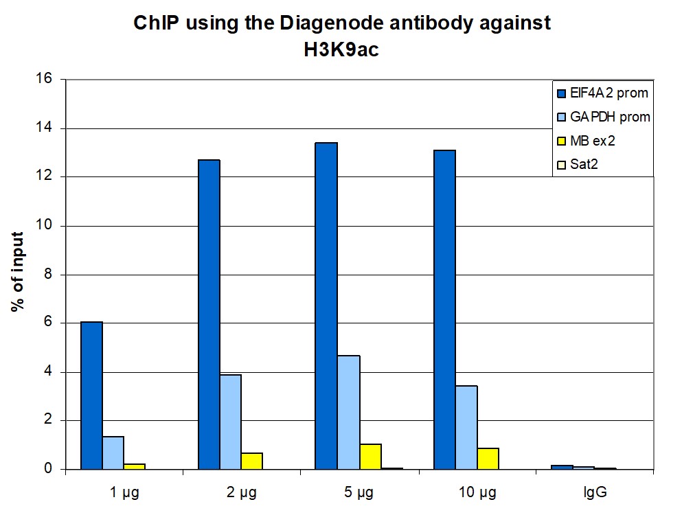
<p><small><strong>Figure 1. ChIP results obtained with the Diagenode antibody directed against H3K9ac </strong><br />ChIP assays were performed using human HeLa cells, the Diagenode antibody against H3K9ac (Cat. No. C15410004) and optimized PCR primer pairs for qPCR. ChIP was performed with the “iDeal ChIP-seq” kit (Cat. No. C01010051), using sheared chromatin from 1 million cells. A titration consisting of 1, 2, 5 and 10 μg of antibody per ChIP experiment was analyzed. IgG (2 μg/IP) was used as a negative IP control. Quantitative PCR was performed with primers speci c for the promoter of the active genes GAPDH and EIF4A2, used as positive controls, and for exon 2 of the inactive myoglobin (MB) gene and the Sat2 satellite repeat, used as negative controls. Figure 1 shows the recovery, expressed as a % of input (the relative amount of immunoprecipitated DNA compared to input DNA after qPCR analysis). These results are in accordance with the observation that acetylation of K9 at histone H3 is associated with the promoters of active genes.</small></p>
</div>
</div>
<div class="row">
<div class="small-6 columns">
<p><img src="https://www.diagenode.com/img/product/antibodies/C15410004_chipseq-figA.png" alt="H3K9ac Antibody ChIP-seq Grade " caption="false" width="432" height="60" /></p>
<p><img src="https://www.diagenode.com/img/product/antibodies/C15410004_chipseq-figB.png" alt="H3K9ac Antibody for ChIP-seq" caption="false" width="432" height="78" /></p>
<p><img src="https://www.diagenode.com/img/product/antibodies/C15410004_chipseq-figC.png" alt="H3K9ac Antibody for ChIP-seq assay" caption="false" width="432" height="93" /></p>
<p><img src="https://www.diagenode.com/img/product/antibodies/C15410004_chipseq-figD.png" alt="H3K9ac Antibody validated in ChIP-seq " caption="false" width="432" height="83" /></p>
</div>
<div class="small-6 columns">
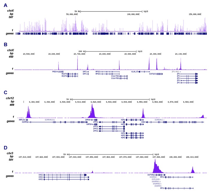
<p><small><strong>Figure 2. ChIP-seq results obtained with the Diagenode antibody directed against H3K9ac </strong><br />ChIP was performed with 1 μg of the Diagenode antibody against H3K9ac (Cat. No. C15410004) as described above and the IP’d DNA was subsequently analysed on an Illumina Genome Analyzer. Library preparation, cluster generation and sequencing were performed according to the manufacturer’s instructions. The 36 bp tags were aligned to the human genome using the ELAND algorithm. Figure 2 shows the peak distribution along the complete sequence and an 800 kb region of the X-chromosome ( gure 2A and B) and in 100 kb regions surrounding the GAPDH and EIF4A2 positive control genes ( gure 2C and D). These results clearly show an enrichment of H3K9ac at the promoters of active genes.</small></p>
</div>
</div>
<div class="row">
<div class="small-4 columns">
<p><img src="https:///www.diagenode.com/img/product/antibodies/C15410004_elisa.png" alt="H3K9ac Antibody ELISA validation" caption="false" width="278" height="246" /></p>
</div>
<div class="small-8 columns">
<p><small><strong>Figure 3. Determination of the antibody titer </strong><br />To determine the titer of the antibody, an ELISA was performed using a serial dilution of the Diagenode antibody directed against H3K9ac (Cat. No. C15410004) in antigen coated wells. The antigen used was a peptide containing the histone modi cation of interest. By plotting the absorbance against the antibody dilution (Figure 3), the titer of the antibody was estimated to be 1:31,700.</small></p>
</div>
</div>
<div class="row">
<div class="small-4 columns">
<p><img src="https://www.diagenode.com/img/product/antibodies/C15410004_dotblot.png" alt="H3K9ac Antibody validated in Dot Blot " caption="false" width="278" height="181" /></p>
</div>
<div class="small-8 columns">
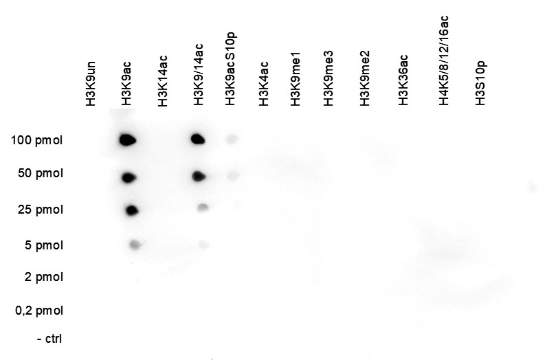
<p><small><strong>Figure 4. Cross reactivity test using the Diagenode antibody directed against H3K9ac </strong><br />A Dot Blot analysis was performed to test the cross reactivity of the Diagenode antibody against H3K9ac (Cat. No. C15410004) with peptides containing other histone modi cations and the unmodi ed H3K9 sequence. One hundred to 0.2 pmol of the respective peptides were spotted on a membrane. The antibody was used at a dilution of 1:20,000. Figure 4 shows a high speci city of the antibody for the modi cation of interest. Please note that this antibody recognizes the H3K9 acetylation, both in the presence and the absence of the K14 acetylation.</small></p>
</div>
</div>
<div class="row">
<div class="small-4 columns">
<p><img src="https://www.diagenode.com/img/product/antibodies/C15410004_wb.png" alt="H3K9ac Antibody validated in Western Blot" caption="false" width="116" height="142" /></p>
</div>
<div class="small-8 columns">
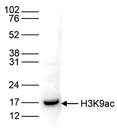
<p><small><strong>Figure 5. Western blot analysis using the Diagenode antibody directed against H3K9ac </strong><br />Histone extracts of HeLa cells (15 μg) were analysed by Western blot using the Diagenode antibody against H3K9ac (Cat. No. C15410004) diluted 1:1,000 in TBS-Tween containing 5% skimmed milk. The position of the protein of interest is indicated on the right; the marker (in kDa) is shown on the left. </small></p>
</div>
</div>
<div class="row">
<div class="small-5 columns">
<p><img src="https://www.diagenode.com/img/product/antibodies/C15410004_IF.png" alt="H3K9ac Antibody validated in Immunofluorescence " caption="false" width="354" height="86" /></p>
</div>
<div class="small-7 columns">
<p><small><strong>Figure 6. Immunofluorescence using the Diagenode antibody directed against H3K9ac </strong><br />HeLa cells were stained with the Diagenode antibody against H3K9ac (Cat. No. C15410004) and with DAPI. Cells were xed with 4% formaldehyde for 10’ and blocked with PBS/TX-100 containing 5% normal goat serum and 1% BSA. The cells were immuno uorescently labelled with the H3K9ac antibody (left) diluted 1:500 in blocking solution followed by an anti-rabbit antibody conjugated to Alexa488. The middle panel shows staining of the nuclei with DAPI. A merge of the two stainings is shown on the right.<small></small></small></p>
</div>
</div>',
'label2' => 'Target Description',
'info2' => '<p>Histones are the main constituents of the protein part of chromosomes of eukaryotic cells. They are rich in the amino acids arginine and lysine and have been greatly conserved during evolution. Histones pack the DNA into tight masses of chromatin. Two core histones of each class H2A, H2B, H3 and H4 assemble and are wrapped by 146 base pairs of DNA to form one octameric nucleosome. Histone tails undergo numerous post-translational modifications, which either directly or indirectly alter chromatin structure to facilitate transcriptional activation or repression or other nuclear processes. In addition to the genetic code, combinations of the different histone modifications reveal the so-called “histone code”. Histone methylation and demethylation is dynamically regulated by respectively histone methyl transferases and histone demethylases.</p>',
'label3' => '',
'info3' => '',
'format' => '10 µg',
'catalog_number' => 'C15410004-10',
'old_catalog_number' => 'pAb-004-010',
'sf_code' => 'C15410004-D001-000582',
'type' => 'FRE',
'search_order' => '03-Antibody',
'price_EUR' => '105',
'price_USD' => '115',
'price_GBP' => '100',
'price_JPY' => '16450',
'price_CNY' => '',
'price_AUD' => '288',
'country' => 'ALL',
'except_countries' => 'None',
'quote' => false,
'in_stock' => false,
'featured' => false,
'no_promo' => false,
'online' => true,
'master' => false,
'last_datasheet_update' => '0000-00-00',
'slug' => 'h3k9ac-polyclonal-antibody-classic-sample-size-10-ug',
'meta_title' => 'H3K9ac Antibody - ChIP-seq Grade (C15410004) | Diagenode',
'meta_keywords' => '',
'meta_description' => 'H3K9ac (Histone H3 acetylated at lysine 9) Polyclonal Antibody validated in ChIP-seq, ChIP-qPCR, DB, WB, IF and ELISA. Batch-specific data available on the website. Sample size available',
'modified' => '2021-10-20 09:30:18',
'created' => '2015-07-30 11:27:29',
'locale' => 'eng'
),
'Antibody' => array(
'host' => '*****',
'id' => '147',
'name' => 'H3K9ac polyclonal antibody',
'description' => 'Histones are the main constituents of the protein part of chromosomes of eukaryotic cells. They are rich in the amino acids arginine and lysine and have been greatly conserved during evolution. Histones pack the DNA into tight masses of chromatin. Two core histones of each class H2A, H2B, H3 and H4 assemble and are wrapped by 146 base pairs of DNA to form one octameric nucleosome. Histone tails undergo numerous post-translational modifications, which either directly or indirectly alter chromatin structure to facilitate transcriptional activation or repression or other nuclear processes. In addition to the genetic code, combinations of the different histone modifications reveal the so-called “histone code”. Histone methylation and demethylation is dynamically regulated by respectively histone methyl transferases and histone demethylases.',
'clonality' => '',
'isotype' => '',
'lot' => 'A1435-0012D',
'concentration' => '1.35 µg/µl',
'reactivity' => 'Human, mouse, pig, zebrafish, Poplar, Daphnia, P. Falciparum: positive.',
'type' => 'Polyclonal',
'purity' => 'Affinity purified',
'classification' => 'Classic',
'application_table' => '<table>
<thead>
<tr>
<th>Applications</th>
<th>Suggested dilution</th>
<th>References</th>
</tr>
</thead>
<tbody>
<tr>
<td>ChIP/ChIP-seq <sup>*</sup></td>
<td>1-2 μg/ChIP</td>
<td>Fig 1, 2</td>
</tr>
<tr>
<td>ELISA</td>
<td>1:1,000</td>
<td>Fig 3</td>
</tr>
<tr>
<td>Dot Blotting</td>
<td>1:20,000</td>
<td>Fig 4</td>
</tr>
<tr>
<td>Western Blotting</td>
<td>1:1,000</td>
<td>Fig 5</td>
</tr>
<tr>
<td>Immunofluorescence</td>
<td>1:500</td>
<td>Fig 6</td>
</tr>
</tbody>
</table>
<p><small><sup>*</sup> Please note that the optimal antibody amount per IP should be determined by the end-user. We recommend testing 1-5 μg per IP.</small></p>',
'storage_conditions' => '',
'storage_buffer' => '',
'precautions' => 'This product is for research use only. Not for use in diagnostic or therapeutic procedures.',
'uniprot_acc' => '',
'slug' => '',
'meta_keywords' => '',
'meta_description' => '',
'modified' => '2020-10-02 12:13:00',
'created' => '0000-00-00 00:00:00',
'select_label' => '147 - H3K9ac polyclonal antibody (A1435-0012D - 1.35 µg/µl - Human, mouse, pig, zebrafish, Poplar, Daphnia, P. Falciparum: positive. - Affinity purified - Rabbit)'
),
'Slave' => array(),
'Group' => array(
'Group' => array(
'id' => '52',
'name' => 'C15410004',
'product_id' => '2174',
'modified' => '2016-02-18 20:52:33',
'created' => '2016-02-18 20:52:33'
),
'Master' => array(
'id' => '2174',
'antibody_id' => '147',
'name' => 'H3K9ac Antibody',
'description' => '<p>Polyclonal antibody raised in rabbit against histone <strong>H3, acetylated at lysine 9</strong> (<strong>H3K9ac</strong>), using a KLH-conjugated synthetic peptide.</p>',
'label1' => 'Validation data',
'info1' => '<div class="row">
<div class="small-4 columns">
<p><img src="https://www.diagenode.com/img/product/antibodies/C15410004_chip.png" alt="H3K9ac Antibody ChIP Grade" caption="false" width="278" height="210" /></p>
</div>
<div class="small-8 columns">
<p><small><strong>Figure 1. ChIP results obtained with the Diagenode antibody directed against H3K9ac </strong><br />ChIP assays were performed using human HeLa cells, the Diagenode antibody against H3K9ac (Cat. No. C15410004) and optimized PCR primer pairs for qPCR. ChIP was performed with the “iDeal ChIP-seq” kit (Cat. No. C01010051), using sheared chromatin from 1 million cells. A titration consisting of 1, 2, 5 and 10 μg of antibody per ChIP experiment was analyzed. IgG (2 μg/IP) was used as a negative IP control. Quantitative PCR was performed with primers speci c for the promoter of the active genes GAPDH and EIF4A2, used as positive controls, and for exon 2 of the inactive myoglobin (MB) gene and the Sat2 satellite repeat, used as negative controls. Figure 1 shows the recovery, expressed as a % of input (the relative amount of immunoprecipitated DNA compared to input DNA after qPCR analysis). These results are in accordance with the observation that acetylation of K9 at histone H3 is associated with the promoters of active genes.</small></p>
</div>
</div>
<div class="row">
<div class="small-6 columns">
<p><img src="https://www.diagenode.com/img/product/antibodies/C15410004_chipseq-figA.png" alt="H3K9ac Antibody ChIP-seq Grade " caption="false" width="432" height="60" /></p>
<p><img src="https://www.diagenode.com/img/product/antibodies/C15410004_chipseq-figB.png" alt="H3K9ac Antibody for ChIP-seq" caption="false" width="432" height="78" /></p>
<p><img src="https://www.diagenode.com/img/product/antibodies/C15410004_chipseq-figC.png" alt="H3K9ac Antibody for ChIP-seq assay" caption="false" width="432" height="93" /></p>
<p><img src="https://www.diagenode.com/img/product/antibodies/C15410004_chipseq-figD.png" alt="H3K9ac Antibody validated in ChIP-seq " caption="false" width="432" height="83" /></p>
</div>
<div class="small-6 columns">
<p><small><strong>Figure 2. ChIP-seq results obtained with the Diagenode antibody directed against H3K9ac </strong><br />ChIP was performed with 1 μg of the Diagenode antibody against H3K9ac (Cat. No. C15410004) as described above and the IP’d DNA was subsequently analysed on an Illumina Genome Analyzer. Library preparation, cluster generation and sequencing were performed according to the manufacturer’s instructions. The 36 bp tags were aligned to the human genome using the ELAND algorithm. Figure 2 shows the peak distribution along the complete sequence and an 800 kb region of the X-chromosome ( gure 2A and B) and in 100 kb regions surrounding the GAPDH and EIF4A2 positive control genes ( gure 2C and D). These results clearly show an enrichment of H3K9ac at the promoters of active genes.</small></p>
</div>
</div>
<div class="row">
<div class="small-4 columns">
<p><img src="https:///www.diagenode.com/img/product/antibodies/C15410004_elisa.png" alt="H3K9ac Antibody ELISA validation" caption="false" width="278" height="246" /></p>
</div>
<div class="small-8 columns">
<p><small><strong>Figure 3. Determination of the antibody titer </strong><br />To determine the titer of the antibody, an ELISA was performed using a serial dilution of the Diagenode antibody directed against H3K9ac (Cat. No. C15410004) in antigen coated wells. The antigen used was a peptide containing the histone modi cation of interest. By plotting the absorbance against the antibody dilution (Figure 3), the titer of the antibody was estimated to be 1:31,700.</small></p>
</div>
</div>
<div class="row">
<div class="small-4 columns">
<p><img src="https://www.diagenode.com/img/product/antibodies/C15410004_dotblot.png" alt="H3K9ac Antibody validated in Dot Blot " caption="false" width="278" height="181" /></p>
</div>
<div class="small-8 columns">
<p><small><strong>Figure 4. Cross reactivity test using the Diagenode antibody directed against H3K9ac </strong><br />A Dot Blot analysis was performed to test the cross reactivity of the Diagenode antibody against H3K9ac (Cat. No. C15410004) with peptides containing other histone modi cations and the unmodi ed H3K9 sequence. One hundred to 0.2 pmol of the respective peptides were spotted on a membrane. The antibody was used at a dilution of 1:20,000. Figure 4 shows a high speci city of the antibody for the modi cation of interest. Please note that this antibody recognizes the H3K9 acetylation, both in the presence and the absence of the K14 acetylation.</small></p>
</div>
</div>
<div class="row">
<div class="small-4 columns">
<p><img src="https://www.diagenode.com/img/product/antibodies/C15410004_wb.png" alt="H3K9ac Antibody validated in Western Blot" caption="false" width="116" height="142" /></p>
</div>
<div class="small-8 columns">
<p><small><strong>Figure 5. Western blot analysis using the Diagenode antibody directed against H3K9ac </strong><br />Histone extracts of HeLa cells (15 μg) were analysed by Western blot using the Diagenode antibody against H3K9ac (Cat. No. C15410004) diluted 1:1,000 in TBS-Tween containing 5% skimmed milk. The position of the protein of interest is indicated on the right; the marker (in kDa) is shown on the left. </small></p>
</div>
</div>
<div class="row">
<div class="small-5 columns">
<p><img src="https://www.diagenode.com/img/product/antibodies/C15410004_IF.png" alt="H3K9ac Antibody validated in Immunofluorescence " caption="false" width="354" height="86" /></p>
</div>
<div class="small-7 columns">
<p><small><strong>Figure 6. Immunofluorescence using the Diagenode antibody directed against H3K9ac </strong><br />HeLa cells were stained with the Diagenode antibody against H3K9ac (Cat. No. C15410004) and with DAPI. Cells were xed with 4% formaldehyde for 10’ and blocked with PBS/TX-100 containing 5% normal goat serum and 1% BSA. The cells were immuno uorescently labelled with the H3K9ac antibody (left) diluted 1:500 in blocking solution followed by an anti-rabbit antibody conjugated to Alexa488. The middle panel shows staining of the nuclei with DAPI. A merge of the two stainings is shown on the right.<small></small></small></p>
</div>
</div>',
'label2' => 'Target Description',
'info2' => '<p>Histones are the main constituents of the protein part of chromosomes of eukaryotic cells. They are rich in the amino acids arginine and lysine and have been greatly conserved during evolution. Histones pack the DNA into tight masses of chromatin. Two core histones of each class H2A, H2B, H3 and H4 assemble and are wrapped by 146 base pairs of DNA to form one octameric nucleosome. Histone tails undergo numerous post-translational modifications, which either directly or indirectly alter chromatin structure to facilitate transcriptional activation or repression or other nuclear processes. In addition to the genetic code, combinations of the different histone modifications reveal the so-called “histone code”. Histone methylation and demethylation is dynamically regulated by respectively histone methyl transferases and histone demethylases.</p>',
'label3' => '',
'info3' => '',
'format' => '50 µg',
'catalog_number' => 'C15410004',
'old_catalog_number' => 'pAb-004-050',
'sf_code' => 'C15410004-D001-000581',
'type' => 'FRE',
'search_order' => '03-Antibody',
'price_EUR' => '380',
'price_USD' => '380',
'price_GBP' => '340',
'price_JPY' => '59525',
'price_CNY' => '',
'price_AUD' => '950',
'country' => 'ALL',
'except_countries' => 'None',
'quote' => false,
'in_stock' => false,
'featured' => false,
'no_promo' => false,
'online' => true,
'master' => true,
'last_datasheet_update' => '0000-00-00',
'slug' => 'h3k9ac-polyclonal-antibody-classic-50-ug-37-ul',
'meta_title' => 'H3K9ac Antibody - ChIP-seq Grade (C15410004) | Diagenode',
'meta_keywords' => '',
'meta_description' => 'H3K9ac (Histone H3 acetylated at lysine 9) Polyclonal Antibody validated in ChIP-seq, ChIP-qPCR, ELISA, WB, DB and IF. Batch-specific data available on the website. Sample size available.',
'modified' => '2021-10-20 09:30:00',
'created' => '2015-06-29 14:08:20'
),
'Product' => array(
(int) 0 => array(
[maximum depth reached]
)
)
),
'Related' => array(
(int) 0 => array(
'id' => '2174',
'antibody_id' => '147',
'name' => 'H3K9ac Antibody',
'description' => '<p>Polyclonal antibody raised in rabbit against histone <strong>H3, acetylated at lysine 9</strong> (<strong>H3K9ac</strong>), using a KLH-conjugated synthetic peptide.</p>',
'label1' => 'Validation data',
'info1' => '<div class="row">
<div class="small-4 columns">
<p><img src="https://www.diagenode.com/img/product/antibodies/C15410004_chip.png" alt="H3K9ac Antibody ChIP Grade" caption="false" width="278" height="210" /></p>
</div>
<div class="small-8 columns">
<p><small><strong>Figure 1. ChIP results obtained with the Diagenode antibody directed against H3K9ac </strong><br />ChIP assays were performed using human HeLa cells, the Diagenode antibody against H3K9ac (Cat. No. C15410004) and optimized PCR primer pairs for qPCR. ChIP was performed with the “iDeal ChIP-seq” kit (Cat. No. C01010051), using sheared chromatin from 1 million cells. A titration consisting of 1, 2, 5 and 10 μg of antibody per ChIP experiment was analyzed. IgG (2 μg/IP) was used as a negative IP control. Quantitative PCR was performed with primers speci c for the promoter of the active genes GAPDH and EIF4A2, used as positive controls, and for exon 2 of the inactive myoglobin (MB) gene and the Sat2 satellite repeat, used as negative controls. Figure 1 shows the recovery, expressed as a % of input (the relative amount of immunoprecipitated DNA compared to input DNA after qPCR analysis). These results are in accordance with the observation that acetylation of K9 at histone H3 is associated with the promoters of active genes.</small></p>
</div>
</div>
<div class="row">
<div class="small-6 columns">
<p><img src="https://www.diagenode.com/img/product/antibodies/C15410004_chipseq-figA.png" alt="H3K9ac Antibody ChIP-seq Grade " caption="false" width="432" height="60" /></p>
<p><img src="https://www.diagenode.com/img/product/antibodies/C15410004_chipseq-figB.png" alt="H3K9ac Antibody for ChIP-seq" caption="false" width="432" height="78" /></p>
<p><img src="https://www.diagenode.com/img/product/antibodies/C15410004_chipseq-figC.png" alt="H3K9ac Antibody for ChIP-seq assay" caption="false" width="432" height="93" /></p>
<p><img src="https://www.diagenode.com/img/product/antibodies/C15410004_chipseq-figD.png" alt="H3K9ac Antibody validated in ChIP-seq " caption="false" width="432" height="83" /></p>
</div>
<div class="small-6 columns">
<p><small><strong>Figure 2. ChIP-seq results obtained with the Diagenode antibody directed against H3K9ac </strong><br />ChIP was performed with 1 μg of the Diagenode antibody against H3K9ac (Cat. No. C15410004) as described above and the IP’d DNA was subsequently analysed on an Illumina Genome Analyzer. Library preparation, cluster generation and sequencing were performed according to the manufacturer’s instructions. The 36 bp tags were aligned to the human genome using the ELAND algorithm. Figure 2 shows the peak distribution along the complete sequence and an 800 kb region of the X-chromosome ( gure 2A and B) and in 100 kb regions surrounding the GAPDH and EIF4A2 positive control genes ( gure 2C and D). These results clearly show an enrichment of H3K9ac at the promoters of active genes.</small></p>
</div>
</div>
<div class="row">
<div class="small-4 columns">
<p><img src="https:///www.diagenode.com/img/product/antibodies/C15410004_elisa.png" alt="H3K9ac Antibody ELISA validation" caption="false" width="278" height="246" /></p>
</div>
<div class="small-8 columns">
<p><small><strong>Figure 3. Determination of the antibody titer </strong><br />To determine the titer of the antibody, an ELISA was performed using a serial dilution of the Diagenode antibody directed against H3K9ac (Cat. No. C15410004) in antigen coated wells. The antigen used was a peptide containing the histone modi cation of interest. By plotting the absorbance against the antibody dilution (Figure 3), the titer of the antibody was estimated to be 1:31,700.</small></p>
</div>
</div>
<div class="row">
<div class="small-4 columns">
<p><img src="https://www.diagenode.com/img/product/antibodies/C15410004_dotblot.png" alt="H3K9ac Antibody validated in Dot Blot " caption="false" width="278" height="181" /></p>
</div>
<div class="small-8 columns">
<p><small><strong>Figure 4. Cross reactivity test using the Diagenode antibody directed against H3K9ac </strong><br />A Dot Blot analysis was performed to test the cross reactivity of the Diagenode antibody against H3K9ac (Cat. No. C15410004) with peptides containing other histone modi cations and the unmodi ed H3K9 sequence. One hundred to 0.2 pmol of the respective peptides were spotted on a membrane. The antibody was used at a dilution of 1:20,000. Figure 4 shows a high speci city of the antibody for the modi cation of interest. Please note that this antibody recognizes the H3K9 acetylation, both in the presence and the absence of the K14 acetylation.</small></p>
</div>
</div>
<div class="row">
<div class="small-4 columns">
<p><img src="https://www.diagenode.com/img/product/antibodies/C15410004_wb.png" alt="H3K9ac Antibody validated in Western Blot" caption="false" width="116" height="142" /></p>
</div>
<div class="small-8 columns">
<p><small><strong>Figure 5. Western blot analysis using the Diagenode antibody directed against H3K9ac </strong><br />Histone extracts of HeLa cells (15 μg) were analysed by Western blot using the Diagenode antibody against H3K9ac (Cat. No. C15410004) diluted 1:1,000 in TBS-Tween containing 5% skimmed milk. The position of the protein of interest is indicated on the right; the marker (in kDa) is shown on the left. </small></p>
</div>
</div>
<div class="row">
<div class="small-5 columns">
<p><img src="https://www.diagenode.com/img/product/antibodies/C15410004_IF.png" alt="H3K9ac Antibody validated in Immunofluorescence " caption="false" width="354" height="86" /></p>
</div>
<div class="small-7 columns">
<p><small><strong>Figure 6. Immunofluorescence using the Diagenode antibody directed against H3K9ac </strong><br />HeLa cells were stained with the Diagenode antibody against H3K9ac (Cat. No. C15410004) and with DAPI. Cells were xed with 4% formaldehyde for 10’ and blocked with PBS/TX-100 containing 5% normal goat serum and 1% BSA. The cells were immuno uorescently labelled with the H3K9ac antibody (left) diluted 1:500 in blocking solution followed by an anti-rabbit antibody conjugated to Alexa488. The middle panel shows staining of the nuclei with DAPI. A merge of the two stainings is shown on the right.<small></small></small></p>
</div>
</div>',
'label2' => 'Target Description',
'info2' => '<p>Histones are the main constituents of the protein part of chromosomes of eukaryotic cells. They are rich in the amino acids arginine and lysine and have been greatly conserved during evolution. Histones pack the DNA into tight masses of chromatin. Two core histones of each class H2A, H2B, H3 and H4 assemble and are wrapped by 146 base pairs of DNA to form one octameric nucleosome. Histone tails undergo numerous post-translational modifications, which either directly or indirectly alter chromatin structure to facilitate transcriptional activation or repression or other nuclear processes. In addition to the genetic code, combinations of the different histone modifications reveal the so-called “histone code”. Histone methylation and demethylation is dynamically regulated by respectively histone methyl transferases and histone demethylases.</p>',
'label3' => '',
'info3' => '',
'format' => '50 µg',
'catalog_number' => 'C15410004',
'old_catalog_number' => 'pAb-004-050',
'sf_code' => 'C15410004-D001-000581',
'type' => 'FRE',
'search_order' => '03-Antibody',
'price_EUR' => '380',
'price_USD' => '380',
'price_GBP' => '340',
'price_JPY' => '59525',
'price_CNY' => '',
'price_AUD' => '950',
'country' => 'ALL',
'except_countries' => 'None',
'quote' => false,
'in_stock' => false,
'featured' => false,
'no_promo' => false,
'online' => true,
'master' => true,
'last_datasheet_update' => '0000-00-00',
'slug' => 'h3k9ac-polyclonal-antibody-classic-50-ug-37-ul',
'meta_title' => 'H3K9ac Antibody - ChIP-seq Grade (C15410004) | Diagenode',
'meta_keywords' => '',
'meta_description' => 'H3K9ac (Histone H3 acetylated at lysine 9) Polyclonal Antibody validated in ChIP-seq, ChIP-qPCR, ELISA, WB, DB and IF. Batch-specific data available on the website. Sample size available.',
'modified' => '2021-10-20 09:30:00',
'created' => '2015-06-29 14:08:20',
'ProductsRelated' => array(
[maximum depth reached]
),
'Image' => array(
[maximum depth reached]
)
)
),
'Application' => array(
(int) 0 => array(
'id' => '20',
'position' => '10',
'parent_id' => '40',
'name' => 'ELISA',
'description' => '<div class="row">
<div class="small-12 medium-12 large-12 columns">Enzyme-linked immunosorbent assay.</div>
</div>',
'in_footer' => false,
'in_menu' => false,
'online' => true,
'tabular' => true,
'slug' => 'elisa-antibodies',
'meta_keywords' => ' ELISA Antibodies,Monoclonal antibody, Polyclonal antibody',
'meta_description' => 'Diagenode offers Monoclonal & Polyclonal antibodies for ELISA applications',
'meta_title' => 'ELISA Antibodies - Monoclonal & Polyclonal antibody | Diagenode',
'modified' => '2016-01-13 12:21:41',
'created' => '2014-07-08 08:13:28',
'ProductsApplication' => array(
[maximum depth reached]
)
),
(int) 1 => array(
'id' => '28',
'position' => '10',
'parent_id' => '40',
'name' => 'DB',
'description' => '<p>Dot blotting</p>',
'in_footer' => false,
'in_menu' => false,
'online' => true,
'tabular' => true,
'slug' => 'dot-blotting',
'meta_keywords' => 'Dot blotting,Monoclonal & Polyclonal antibody,',
'meta_description' => 'Diagenode offers Monoclonal & Polyclonal antibodies for Dot blotting applications',
'meta_title' => 'Dot blotting Antibodies - Monoclonal & Polyclonal antibody | Diagenode',
'modified' => '2016-01-13 14:40:49',
'created' => '2015-07-08 13:45:05',
'ProductsApplication' => array(
[maximum depth reached]
)
),
(int) 2 => array(
'id' => '42',
'position' => '10',
'parent_id' => '40',
'name' => 'ChIP-seq (ab)',
'description' => '',
'in_footer' => false,
'in_menu' => false,
'online' => true,
'tabular' => true,
'slug' => 'chip-seq-antibodies',
'meta_keywords' => 'Chromatin Immunoprecipitation Sequencing,ChIP-Seq,ChIP-seq grade antibodies,DNA purification,qPCR,Shearing of chromatin',
'meta_description' => 'Diagenode offers a wide range of antibodies and technical support for ChIP Sequencing applications',
'meta_title' => 'ChIP Sequencing Antibodies (ChIP-Seq) | Diagenode',
'modified' => '2016-01-20 11:06:19',
'created' => '2015-10-20 11:44:45',
'ProductsApplication' => array(
[maximum depth reached]
)
),
(int) 3 => array(
'id' => '43',
'position' => '10',
'parent_id' => '40',
'name' => 'ChIP-qPCR (ab)',
'description' => '',
'in_footer' => false,
'in_menu' => false,
'online' => true,
'tabular' => true,
'slug' => 'chip-qpcr-antibodies',
'meta_keywords' => 'Chromatin Immunoprecipitation Sequencing,ChIP-Seq,ChIP-seq grade antibodies,DNA purification,qPCR,Shearing of chromatin',
'meta_description' => 'Diagenode offers a wide range of antibodies and technical support for ChIP-qPCR applications',
'meta_title' => 'ChIP Quantitative PCR Antibodies (ChIP-qPCR) | Diagenode',
'modified' => '2016-01-20 11:30:24',
'created' => '2015-10-20 11:45:36',
'ProductsApplication' => array(
[maximum depth reached]
)
),
(int) 4 => array(
'id' => '19',
'position' => '10',
'parent_id' => '40',
'name' => 'WB',
'description' => '<p><strong>Western blot</strong> : The quality of antibodies used in this technique is crucial for correct and specific protein identification. Diagenode offers huge selection of highly sensitive and specific western blot-validated antibodies.</p>
<p>Learn more about: <a href="https://www.diagenode.com/applications/western-blot">Loading control, MW marker visualization</a><em>. <br /></em></p>
<p><em></em>Check our selection of antibodies validated in Western blot.</p>',
'in_footer' => false,
'in_menu' => false,
'online' => true,
'tabular' => true,
'slug' => 'western-blot-antibodies',
'meta_keywords' => ' Western Blot Antibodies ,western blot protocol,Western Blotting Products,Polyclonal antibodies ,monoclonal antibodies ',
'meta_description' => 'Diagenode offers a wide range of antibodies and technical support for western blot applications',
'meta_title' => ' Western Blot - Monoclonal antibody - Polyclonal antibody | Diagenode',
'modified' => '2016-04-26 12:44:51',
'created' => '2015-01-07 09:20:00',
'ProductsApplication' => array(
[maximum depth reached]
)
),
(int) 5 => array(
'id' => '29',
'position' => '10',
'parent_id' => '40',
'name' => 'IF',
'description' => '<p><strong>Immunofluorescence</strong>:</p>
<p>Diagenode offers huge selection of highly sensitive antibodies validated in IF.</p>
<p><img src="https://www.diagenode.com/img/product/antibodies/C15200229-IF.jpg" alt="" height="245" width="256" /></p>
<p><sup><strong>Immunofluorescence using the Diagenode monoclonal antibody directed against CRISPR/Cas9</strong></sup></p>
<p><sup>HeLa cells transfected with a Cas9 expression vector (left) or untransfected cells (right) were fixed in methanol at -20°C, permeabilized with acetone at -20°C and blocked with PBS containing 2% BSA. The cells were stained with the Cas9 C-terminal antibody (Cat. No. C15200229) diluted 1:400, followed by incubation with an anti-mouse secondary antibody coupled to AF488. The bottom images show counter-staining of the nuclei with Hoechst 33342.</sup></p>
<h5><sup>Check our selection of antibodies validated in IF.</sup></h5>',
'in_footer' => false,
'in_menu' => false,
'online' => true,
'tabular' => true,
'slug' => 'immunofluorescence',
'meta_keywords' => 'Immunofluorescence,Monoclonal antibody,Polyclonal antibody',
'meta_description' => 'Diagenode offers a wide range of antibodies and technical support for Immunofluorescence applications',
'meta_title' => 'Immunofluorescence - Monoclonal antibody - Polyclonal antibody | Diagenode',
'modified' => '2016-04-27 16:23:10',
'created' => '2015-07-08 13:46:02',
'ProductsApplication' => array(
[maximum depth reached]
)
)
),
'Category' => array(
(int) 0 => array(
'id' => '111',
'position' => '40',
'parent_id' => '4',
'name' => 'Histone antibodies',
'description' => '<p>Histones are the main protein components of chromatin involved in the compaction of DNA into nucleosomes, the basic units of chromatin. A <strong>nucleosome</strong> consists of one pair of each of the core histones (<strong>H2A</strong>, <strong>H2B</strong>, <strong>H3</strong> and <strong>H4</strong>) forming an octameric structure wrapped by 146 base pairs of DNA. The different nucleosomes are linked by the linker histone<strong> H1, </strong>allowing for further condensation of chromatin.</p>
<p>The core histones have a globular structure with large unstructured N-terminal tails protruding from the nucleosome. They can undergo to multiple post-translational modifications (PTM), mainly at the N-terminal tails. These <strong>post-translational modifications </strong>include methylation, acetylation, phosphorylation, ubiquitinylation, citrullination, sumoylation, deamination and crotonylation. The most well characterized PTMs are <strong>methylation,</strong> <strong>acetylation and phosphorylation</strong>. Histone methylation occurs mainly on lysine (K) residues, which can be mono-, di- or tri-methylated, and on arginines (R), which can be mono-methylated and symmetrically or asymmetrically di-methylated. Histone acetylation occurs on lysines and histone phosphorylation mainly on serines (S), threonines (T) and tyrosines (Y).</p>
<p>The PTMs of the different residues are involved in numerous processes such as DNA repair, DNA replication and chromosome condensation. They influence the chromatin organization and can be positively or negatively associated with gene expression. Trimethylation of H3K4, H3K36 and H3K79, and lysine acetylation generally result in an open chromatin configuration (figure below) and are therefore associated with <strong>euchromatin</strong> and gene activation. Trimethylation of H3K9, K3K27 and H4K20, on the other hand, is enriched in <strong>heterochromatin </strong>and associated with gene silencing. The combination of different histone modifications is called the "<strong>histone code</strong>”, analogous to the genetic code.</p>
<p><img src="https://www.diagenode.com/img/categories/antibodies/histone-marks-illustration.png" /></p>
<p>Diagenode is proud to offer a large range of antibodies against histones and histone modifications. Our antibodies are highly specific and have been validated in many applications, including <strong>ChIP</strong> and <strong>ChIP-seq</strong>.</p>
<p>Diagenode’s collection includes antibodies recognizing:</p>
<ul>
<li><strong>Histone H1 variants</strong></li>
<li><strong>Histone H2A, H2A variants and histone H2A</strong> <strong>modifications</strong> (serine phosphorylation, lysine acetylation, lysine ubiquitinylation)</li>
<li><strong>Histone H2B and H2B</strong> <strong>modifications </strong>(serine phosphorylation, lysine acetylation)</li>
<li><strong>Histone H3 and H3 modifications </strong>(lysine methylation (mono-, di- and tri-methylated), lysine acetylation, serine phosphorylation, threonine phosphorylation, arginine methylation (mono-methylated, symmetrically and asymmetrically di-methylated))</li>
<li><strong>Histone H4 and H4 modifications (</strong>lysine methylation (mono-, di- and tri-methylated), lysine acetylation, arginine methylation (mono-methylated and symmetrically di-methylated), serine phosphorylation )</li>
</ul>
<p><span style="font-weight: 400;"><strong>HDAC's HAT's, HMT's and other</strong> <strong>enzymes</strong> which modify histones can be found in the category <a href="../categories/chromatin-modifying-proteins-histone-transferase">Histone modifying enzymes</a><br /></span></p>
<p><span style="font-weight: 400;"> Diagenode’s highly validated antibodies:</span></p>
<ul>
<li><span style="font-weight: 400;"> Highly sensitive and specific</span></li>
<li><span style="font-weight: 400;"> Cost-effective (requires less antibody per reaction)</span></li>
<li><span style="font-weight: 400;"> Batch-specific data is available on the website</span></li>
<li><span style="font-weight: 400;"> Expert technical support</span></li>
<li><span style="font-weight: 400;"> Sample sizes available</span></li>
<li><span style="font-weight: 400;"> 100% satisfaction guarantee</span></li>
</ul>',
'no_promo' => false,
'in_menu' => false,
'online' => true,
'tabular' => false,
'hide' => true,
'all_format' => false,
'is_antibody' => true,
'slug' => 'histone-antibodies',
'cookies_tag_id' => null,
'meta_keywords' => 'Histone, antibody, histone h1, histone h2, histone h3, histone h4',
'meta_description' => 'Polyclonal and Monoclonal Antibodies against Histones and their modifications validated for many applications, including Chromatin Immunoprecipitation (ChIP) and ChIP-Sequencing (ChIP-seq)',
'meta_title' => 'Histone and Modified Histone Antibodies | Diagenode',
'modified' => '2020-09-17 13:34:56',
'created' => '2016-04-01 16:01:32',
'ProductsCategory' => array(
[maximum depth reached]
),
'CookiesTag' => array([maximum depth reached])
),
(int) 1 => array(
'id' => '17',
'position' => '10',
'parent_id' => '4',
'name' => 'ChIP-seq grade antibodies',
'description' => '<p><b>Unparalleled ChIP-Seq results with the most rigorously validated antibodies</b></p>
<p><span style="font-weight: 400;">Diagenode provides leading solutions for epigenetic research. Because ChIP-seq is a widely-used technique, we validate our antibodies in ChIP and ChIP-seq experiments (in addition to conventional methods like Western blot, Dot blot, ELISA, and immunofluorescence) to provide the highest quality antibody. We standardize our validation and production to guarantee high product quality without technical bias. Diagenode guarantees ChIP-seq grade antibody performance under our suggested conditions.</span></p>
<div class="row">
<div class="small-12 medium-9 large-9 columns">
<p><strong>ChIP-seq profile</strong> of active (H3K4me3 and H3K36me3) and inactive (H3K27me3) marks using Diagenode antibodies.</p>
<img src="https://www.diagenode.com/img/categories/antibodies/chip-seq-grade-antibodies.png" /></div>
<div class="small-12 medium-3 large-3 columns">
<p><small> ChIP was performed on sheared chromatin from 100,000 K562 cells using iDeal ChIP-seq kit for Histones (cat. No. C01010051) with 1 µg of the Diagenode antibodies against H3K27me3 (cat. No. C15410195) and H3K4me3 (cat. No. C15410003), and 0.5 µg of the antibody against H3K36me3 (cat. No. C15410192). The IP'd DNA was subsequently analysed on an Illumina Genome Analyzer. Library preparation, cluster generation and sequencing were performed according to the manufacturer's instructions. The 36 bp tags were aligned to the human genome using the ELAND algorithm. The figure shows the signal distribution along the complete sequence of human chromosome 3, a zoomin to a 10 Mb region and a further zoomin to a 1.5 Mb region. </small></p>
</div>
</div>
<p>Diagenode’s highly validated antibodies:</p>
<ul>
<li>Highly sensitive and specific</li>
<li>Cost-effective (requires less antibody per reaction)</li>
<li>Batch-specific data is available on the website</li>
<li>Expert technical support</li>
<li>Sample sizes available</li>
<li>100% satisfaction guarantee</li>
</ul>',
'no_promo' => false,
'in_menu' => true,
'online' => true,
'tabular' => false,
'hide' => true,
'all_format' => false,
'is_antibody' => true,
'slug' => 'chip-seq-grade-antibodies',
'cookies_tag_id' => null,
'meta_keywords' => 'ChIP-seq grade antibodies,polyclonal antibody,WB, ELISA, ChIP-seq (ab), ChIP-qPCR (ab)',
'meta_description' => 'Diagenode Offers Wide Range of Validated ChIP-Seq Grade Antibodies for Unparalleled ChIP-Seq Results',
'meta_title' => 'Chromatin Immunoprecipitation ChIP-Seq Grade Antibodies | Diagenode',
'modified' => '2019-07-03 10:57:22',
'created' => '2015-02-16 02:24:01',
'ProductsCategory' => array(
[maximum depth reached]
),
'CookiesTag' => array([maximum depth reached])
),
(int) 2 => array(
'id' => '102',
'position' => '1',
'parent_id' => '4',
'name' => 'Sample size antibodies',
'description' => '<h1><strong>Validated epigenetics antibodies</strong> – care for a sample?<br /> </h1>
<p>Diagenode has partnered with leading epigenetics experts and numerous epigenetics consortiums to bring to you a validated and comprehensive collection of epigenetic antibodies. As an expert in epigenetics, we are committed to offering highly-specific antibodies validated for ChIP/ChIP-seq and many other applications. All batch-specific validation data is available on our website.<br /><a href="../categories/antibodies">Read about our expertise in antibody production</a>.</p>
<ul>
<li><strong>Focused</strong> - Diagenode's selection of antibodies is exclusively dedicated for epigenetic research. <a title="See the full collection." href="../categories/all-antibodies">See the full collection.</a></li>
<li><strong>Strict quality standards</strong> with rigorous QC and validation</li>
<li><strong>Classified</strong> based on level of validation for flexibility of application</li>
</ul>
<p>Existing sample sizes are listed below. We will soon expand our collection. Are you looking for a sample size of another antibody? Just <a href="mailto:agnieszka.zelisko@diagenode.com?Subject=Sample%20Size%20Request" target="_top">Contact us</a>.</p>',
'no_promo' => false,
'in_menu' => true,
'online' => true,
'tabular' => false,
'hide' => true,
'all_format' => true,
'is_antibody' => true,
'slug' => 'sample-size-antibodies',
'cookies_tag_id' => null,
'meta_keywords' => '5-hmC monoclonal antibody,CRISPR/Cas9 polyclonal antibody ,H3K36me3 polyclonal antibody,diagenode',
'meta_description' => 'Diagenode offers sample volume on selected antibodies for researchers to test, validate and provide confidence and flexibility in choosing from our wide range of antibodies ',
'meta_title' => 'Sample-size Antibodies | Diagenode',
'modified' => '2019-07-03 10:57:05',
'created' => '2015-10-27 12:13:34',
'ProductsCategory' => array(
[maximum depth reached]
),
'CookiesTag' => array([maximum depth reached])
),
(int) 3 => array(
'id' => '103',
'position' => '0',
'parent_id' => '4',
'name' => 'All antibodies',
'description' => '<p><span style="font-weight: 400;">All Diagenode’s antibodies are listed below. Please, use our Quick search field to find the antibody of interest by target name, application, purity.</span></p>
<p><span style="font-weight: 400;">Diagenode’s highly validated antibodies:</span></p>
<ul>
<li>Highly sensitive and specific</li>
<li>Cost-effective (requires less antibody per reaction)</li>
<li>Batch-specific data is available on the website</li>
<li>Expert technical support</li>
<li>Sample sizes available</li>
<li>100% satisfaction guarantee</li>
</ul>',
'no_promo' => false,
'in_menu' => true,
'online' => true,
'tabular' => false,
'hide' => true,
'all_format' => false,
'is_antibody' => true,
'slug' => 'all-antibodies',
'cookies_tag_id' => null,
'meta_keywords' => 'Antibodies,Premium Antibodies,Classic,Pioneer',
'meta_description' => 'Diagenode Offers Strict quality standards with Rigorous QC and validated Antibodies. Classified based on level of validation for flexibility of Application. Comprehensive selection of histone and non-histone Antibodies',
'meta_title' => 'Diagenode's selection of Antibodies is exclusively dedicated for Epigenetic Research | Diagenode',
'modified' => '2019-07-03 10:55:44',
'created' => '2015-11-02 14:49:22',
'ProductsCategory' => array(
[maximum depth reached]
),
'CookiesTag' => array([maximum depth reached])
),
(int) 4 => array(
'id' => '127',
'position' => '10',
'parent_id' => '4',
'name' => 'ChIP-grade antibodies',
'description' => '<div class="row">
<div class="small-12 columns"><center></center>
<p><br />Chromatin immunoprecipitation (<b>ChIP</b>) is a technique to study the associations of proteins with the specific genomic regions in intact cells. One of the most important steps of this protocol is the immunoprecipitation of targeted protein using the antibody specifically recognizing it. The quality of antibodies used in ChIP is essential for the success of the experiment. Diagenode offers extensively validated ChIP-grade antibodies, confirmed for their specificity, and high level of performance in ChIP. Each batch is validated, and batch-specific data are available on the website.</p>
<p></p>
</div>
</div>
<p><strong>ChIP results</strong> obtained with the antibody directed against H3K4me3 (Cat. No. <a href="../p/h3k4me3-polyclonal-antibody-premium-50-ug-50-ul">C15410003</a>). </p>
<div class="row">
<div class="small-12 medium-6 large-6 columns"><img src="https://www.diagenode.com/img/product/antibodies/C15410003-fig1-ChIP.jpg" alt="" width="400" height="315" /> </div>
<div class="small-12 medium-6 large-6 columns">
<p></p>
<p></p>
<p></p>
</div>
</div>
<p></p>
<p>Our aim at Diagenode is to offer the largest collection of highly specific <strong>ChIP-grade antibodies</strong>. We add new antibodies monthly. Find your ChIP-grade antibody in the list below and check more information about tested applications, extensive validation data, and product information.</p>',
'no_promo' => false,
'in_menu' => true,
'online' => true,
'tabular' => false,
'hide' => true,
'all_format' => false,
'is_antibody' => true,
'slug' => 'chip-grade-antibodies',
'cookies_tag_id' => null,
'meta_keywords' => 'ChIP-grade antibodies, polyclonal antibody, monoclonal antibody, Diagenode',
'meta_description' => 'Diagenode Offers Extensively Validated ChIP-Grade Antibodies, Confirmed for their Specificity, and high level of Performance in Chromatin Immunoprecipitation ChIP',
'meta_title' => 'Chromatin immunoprecipitation ChIP-grade antibodies | Diagenode',
'modified' => '2024-11-19 17:27:07',
'created' => '2017-02-14 11:16:04',
'ProductsCategory' => array(
[maximum depth reached]
),
'CookiesTag' => array([maximum depth reached])
)
),
'Document' => array(
(int) 0 => array(
'id' => '669',
'name' => 'Datasheet H3K9ac C15410004',
'description' => '<p>Datasheet description</p>',
'image_id' => null,
'type' => 'Datasheet',
'url' => 'files/products/antibodies/Datasheet_H3K9ac_C15410004.pdf',
'slug' => 'datasheet-h3k9ac-C15410004',
'meta_keywords' => '',
'meta_description' => '',
'modified' => '2015-11-20 17:40:40',
'created' => '2015-07-07 11:47:44',
'ProductsDocument' => array(
[maximum depth reached]
)
),
(int) 1 => array(
'id' => '11',
'name' => 'Antibodies you can trust',
'description' => '<p style="text-align: justify;"><span>Epigenetic research tools have evolved over time from endpoint PCR to qPCR to the analyses of large sets of genome-wide sequencing data. ChIP sequencing (ChIP-seq) has now become the gold standard method for chromatin studies, given the accuracy and coverage scale of the approach over other methods. Successful ChIP-seq, however, requires a higher level of experimental accuracy and consistency in all steps of ChIP than ever before. Particularly crucial is the quality of ChIP antibodies. </span></p>',
'image_id' => null,
'type' => 'Poster',
'url' => 'files/posters/Antibodies_you_can_trust_Poster.pdf',
'slug' => 'antibodies-you-can-trust-poster',
'meta_keywords' => '',
'meta_description' => '',
'modified' => '2015-10-01 20:18:31',
'created' => '2015-07-03 16:05:15',
'ProductsDocument' => array(
[maximum depth reached]
)
),
(int) 2 => array(
'id' => '38',
'name' => 'Epigenetic Antibodies Brochure',
'description' => '<p>More than in any other immuoprecipitation assays, quality antibodies are critical tools in many epigenetics experiments. Since 10 years, Diagenode has developed the most stringent quality production available on the market for antibodies exclusively focused on epigenetic uses. All our antibodies have been qualified to work in epigenetic applications.</p>',
'image_id' => null,
'type' => 'Brochure',
'url' => 'files/brochures/Epigenetic_Antibodies_Brochure.pdf',
'slug' => 'epigenetic-antibodies-brochure',
'meta_keywords' => '',
'meta_description' => '',
'modified' => '2016-06-15 11:24:06',
'created' => '2015-07-03 16:05:27',
'ProductsDocument' => array(
[maximum depth reached]
)
)
),
'Feature' => array(),
'Image' => array(
(int) 0 => array(
'id' => '1783',
'name' => 'product/antibodies/chipseq-grade-ab-icon.png',
'alt' => 'ChIP-seq Grade',
'modified' => '2020-11-27 07:04:40',
'created' => '2018-03-15 15:54:09',
'ProductsImage' => array(
[maximum depth reached]
)
)
),
'Promotion' => array(),
'Protocol' => array(),
'Publication' => array(
(int) 0 => array(
'id' => '4618',
'name' => 'Noncanonical regulation of imprinted gene Igf2 by amyloid-beta 1-42 inAlzheimer's disease.',
'authors' => 'Fertan E. et al.',
'description' => '<p>Reduced insulin-like growth factor 2 (IGF2) levels in Alzheimer's disease (AD) may be the mechanism relating age-related metabolic disorders to dementia. Since Igf2 is an imprinted gene, we examined age and sex differences in the relationship between amyloid-beta 1-42 (Aβ) accumulation and epigenetic regulation of the Igf2/H19 gene cluster in cerebrum, liver, and plasma of young and old male and female 5xFAD mice, in frontal cortex of male and female AD and non-AD patients, and in HEK293 cell cultures. We show IGF2 levels, Igf2 expression, histone acetylation, and H19 ICR methylation are lower in females than males. However, elevated Aβ levels are associated with Aβ binding to Igf2 DMR2, increased DNA and histone methylation, and a reduction in Igf2 expression and IGF2 levels in 5xFAD mice and AD patients, independent of H19 ICR methylation. Cell culture results confirmed the binding of Aβ to Igf2 DMR2 increased DNA and histone methylation, and reduced Igf2 expression. These results indicate an age- and sex-related causal relationship among Aβ levels, epigenomic state, and Igf2 expression in AD and provide a potential mechanism for Igf2 regulation in normal and pathological conditions, suggesting IGF2 levels may be a useful diagnostic biomarker for Aβ targeted AD therapies.</p>',
'date' => '2023-02-01',
'pmid' => 'https://www.ncbi.nlm.nih.gov/pubmed/36739453',
'doi' => '10.1038/s41598-023-29248-x',
'modified' => '2023-04-04 08:51:25',
'created' => '2023-02-21 09:59:46',
'ProductsPublication' => array(
[maximum depth reached]
)
),
(int) 1 => array(
'id' => '4802',
'name' => 'Analyzing the Genome-Wide Distribution of Histone Marks byCUT\&Tag in Drosophila Embryos.',
'authors' => 'Zenk F. et al.',
'description' => '<p><span>CUT&Tag is a method to map the genome-wide distribution of histone modifications and some chromatin-associated proteins. CUT&Tag relies on antibody-targeted chromatin tagmentation and can easily be scaled up or automatized. This protocol provides clear experimental guidelines and helpful considerations when planning and executing CUT&Tag experiments.</span></p>',
'date' => '2023-01-01',
'pmid' => 'https://www.ncbi.nlm.nih.gov/pubmed/37212984',
'doi' => '10.1007/978-1-0716-3143-0_1',
'modified' => '2023-06-15 08:43:40',
'created' => '2023-06-13 21:11:31',
'ProductsPublication' => array(
[maximum depth reached]
)
),
(int) 2 => array(
'id' => '4632',
'name' => 'The histone acetyltransferase KAT6A is recruited to unmethylatedCpG islands via a DNA binding winged helix domain.',
'authors' => 'Weber L.M. et al.',
'description' => '<p>The lysine acetyltransferase KAT6A (MOZ, MYST3) belongs to the MYST family of chromatin regulators, facilitating histone acetylation. Dysregulation of KAT6A has been implicated in developmental syndromes and the onset of acute myeloid leukemia (AML). Previous work suggests that KAT6A is recruited to its genomic targets by a combinatorial function of histone binding PHD fingers, transcription factors and chromatin binding interaction partners. Here, we demonstrate that a winged helix (WH) domain at the very N-terminus of KAT6A specifically interacts with unmethylated CpG motifs. This DNA binding function leads to the association of KAT6A with unmethylated CpG islands (CGIs) genome-wide. Mutation of the essential amino acids for DNA binding completely abrogates the enrichment of KAT6A at CGIs. In contrast, deletion of a second WH domain or the histone tail binding PHD fingers only subtly influences the binding of KAT6A to CGIs. Overexpression of a KAT6A WH1 mutant has a dominant negative effect on H3K9 histone acetylation, which is comparable to the effects upon overexpression of a KAT6A HAT domain mutant. Taken together, our work revealed a previously unrecognized chromatin recruitment mechanism of KAT6A, offering a new perspective on the role of KAT6A in gene regulation and human diseases.</p>',
'date' => '2022-12-01',
'pmid' => 'https://www.ncbi.nlm.nih.gov/pubmed/36537216',
'doi' => '10.1093/nar/gkac1188',
'modified' => '2023-03-28 09:01:38',
'created' => '2023-02-21 09:59:46',
'ProductsPublication' => array(
[maximum depth reached]
)
),
(int) 3 => array(
'id' => '4392',
'name' => 'HISTONE DEACETYLASE 15 and MOS4-Associated Complex subunits3A/3B coregulate intron retention of ABA-responsive genes.',
'authors' => 'Tu Yi-Tsung et al. ',
'description' => '<p>Histone deacetylases (HDAs) play an important role in transcriptional regulation of multiple biological processes. In this study, we investigated the function of HDA15 in abscisic acid (ABA) responses. We used immunopurification coupled with mass spectrometry-based proteomics to identify proteins interacting with HDA15 in Arabidopsis (Arabidopsis thaliana). HDA15 interacted with the core subunits of the MOS4-Associated Complex (MAC), MAC3A and MAC3B, with interaction between HDA15 and MAC3B enhanced by ABA. hda15 and mac3a/mac3b mutants were ABA-insensitive during seed germination and hyposensitive to salinity. RNA sequencing (RNA-seq) analysis demonstrated that HDA15 and MAC3A/MAC3B co-regulate ABA-responsive intron retention (IR). Furthermore, HDA15 reduced the histone acetylation level of genomic regions near ABA-responsive IR sites, and the association of MAC3B with ABA-responsive pre-mRNA was dependent on HDA15. Our results indicate that HDA15 is involved in ABA responses by interacting with MAC3A/MAC3B to mediate splicing of introns.</p>',
'date' => '2022-06-01',
'pmid' => 'https://www.ncbi.nlm.nih.gov/pubmed/35670741',
'doi' => '10.1093/plphys/kiac271',
'modified' => '2022-08-11 14:21:50',
'created' => '2022-08-11 12:14:50',
'ProductsPublication' => array(
[maximum depth reached]
)
),
(int) 4 => array(
'id' => '4857',
'name' => 'Broad domains of histone marks in the highly compact macronucleargenome.',
'authors' => 'Drews F. et al.',
'description' => '<p>The unicellular ciliate contains a large vegetative macronucleus with several unusual characteristics, including an extremely high coding density and high polyploidy. As macronculear chromatin is devoid of heterochromatin, our study characterizes the functional epigenomic organization necessary for gene regulation and proper Pol II activity. Histone marks (H3K4me3, H3K9ac, H3K27me3) reveal no narrow peaks but broad domains along gene bodies, whereas intergenic regions are devoid of nucleosomes. Our data implicate H3K4me3 levels inside ORFs to be the main factor associated with gene expression, and H3K27me3 appears in association with H3K4me3 in plastic genes. Silent and lowly expressed genes show low nucleosome occupancy, suggesting that gene inactivation does not involve increased nucleosome occupancy and chromatin condensation. Because of a high occupancy of Pol II along highly expressed ORFs, transcriptional elongation appears to be quite different from that of other species. This is supported by missing heptameric repeats in the C-terminal domain of Pol II and a divergent elongation system. Our data imply that unoccupied DNA is the default state, whereas gene activation requires nucleosome recruitment together with broad domains of H3K4me3. In summary, gene activation and silencing in run counter to the current understanding of chromatin biology.</p>',
'date' => '2022-04-01',
'pmid' => 'https://www.ncbi.nlm.nih.gov/pubmed/35264449',
'doi' => '10.1101/gr.276126.121',
'modified' => '2023-08-01 14:45:37',
'created' => '2023-08-01 15:59:38',
'ProductsPublication' => array(
[maximum depth reached]
)
),
(int) 5 => array(
'id' => '4217',
'name' => 'CREBBP/EP300 acetyltransferase inhibition disrupts FOXA1-bound enhancers to inhibit the proliferation of ER+ breast cancer cells.',
'authors' => 'Bommi-Reddy A. et al.',
'description' => '<p><span>Therapeutic targeting of the estrogen receptor (ER) is a clinically validated approach for estrogen receptor positive breast cancer (ER+ BC), but sustained response is limited by acquired resistance. Targeting the transcriptional coactivators required for estrogen receptor activity represents an alternative approach that is not subject to the same limitations as targeting estrogen receptor itself. In this report we demonstrate that the acetyltransferase activity of coactivator paralogs CREBBP/EP300 represents a promising therapeutic target in ER+ BC. Using the potent and selective inhibitor CPI-1612, we show that CREBBP/EP300 acetyltransferase inhibition potently suppresses in vitro and in vivo growth of breast cancer cell line models and acts in a manner orthogonal to directly targeting ER. CREBBP/EP300 acetyltransferase inhibition suppresses ER-dependent transcription by targeting lineage-specific enhancers defined by the pioneer transcription factor FOXA1. These results validate CREBBP/EP300 acetyltransferase activity as a viable target for clinical development in ER+ breast cancer.</span></p>',
'date' => '2022-03-30',
'pmid' => 'https://pubmed.ncbi.nlm.nih.gov/35353838/',
'doi' => '10.1371/journal.pone.0262378',
'modified' => '2022-04-12 10:56:54',
'created' => '2022-04-12 10:56:54',
'ProductsPublication' => array(
[maximum depth reached]
)
),
(int) 6 => array(
'id' => '3995',
'name' => 'Epigenetic, transcriptional and phenotypic responses in Daphnia magna exposed to low-level ionizing radiation',
'authors' => 'Thaulow Jens, Song You, Lindeman Leif C., Kamstra Jorke H., Lee YeonKyeong, Xie Li, Aleström Peter, Salbu Brit, Tollefsen Knut Erik',
'description' => '<p>Ionizing radiation is known to induce oxidative stress and DNA damage as well as epigenetic effects in aquatic organisms. Epigenetic changes can be part of the adaptive responses to protect organisms from radiation-induced damage, or act as drivers of toxicity pathways leading to adverse effects. To investigate the potential roles of epigenetic mechanisms in low-dose ionizing radiation-induced stress responses, an ecologically relevant crustacean, adult Daphnia magna were chronically exposed to low and medium level external 60Co gamma radiation ranging from 0.4, 1, 4, 10, and 40 mGy/h for seven days. Biological effects at the molecular (global DNA methylation, histone modification, gene expression), cellular (reactive oxygen species formation), tissue/organ (ovary, gut and epidermal histology) and organismal (fecundity) levels were investigated using a suite of effect assessment tools. The results showed an increase in global DNA methylation associated with loci-specific alterations of histone H3K9 methylation and acetylation, and downregulation of genes involved in DNA methylation, one-carbon metabolism, antioxidant defense, DNA repair, apoptosis, calcium signaling and endocrine regulation of development and reproduction. Temporal changes of reactive oxygen species (ROS) formation were also observed with an apparent transition from ROS suppression to induction from 2-7 days after gamma exposure. The cumulative fecundity, however, was not significantly changed by the gamma exposure. On the basis of the new experimental evidence and existing knowledge, a hypothetical model was proposed to provide in-depth mechanistic understanding of the roles of epigenetic mechanisms in low dose ionizing radiation induced stress responses in D. magna.</p>',
'date' => '2020-07-18',
'pmid' => 'https://www.sciencedirect.com/science/article/pii/S0013935120308252',
'doi' => '10.1016/j.envres.2020.109930',
'modified' => '2020-09-01 14:51:16',
'created' => '2020-08-21 16:41:39',
'ProductsPublication' => array(
[maximum depth reached]
)
),
(int) 7 => array(
'id' => '3934',
'name' => 'An epigenetic map of malaria parasite development from host to vector.',
'authors' => 'Witmer K, Fraschka SA, Vlachou D, Bártfai R, Christophides GK',
'description' => '<p>The malaria parasite replicates asexually in the red blood cells of its vertebrate host employing epigenetic mechanisms to regulate gene expression in response to changes in its environment. We used chromatin immunoprecipitation followed by sequencing in conjunction with RNA sequencing to create an epigenomic and transcriptomic map of the developmental transition from asexual blood stages to male and female gametocytes and to ookinetes in the rodent malaria parasite Plasmodium berghei. Across the developmental stages examined, heterochromatin protein 1 associates with variantly expressed gene families localised at subtelomeric regions and variant gene expression based on heterochromatic silencing is observed only in some genes. Conversely, the euchromatin mark histone 3 lysine 9 acetylation (H3K9ac) is abundant in non-heterochromatic regions across all developmental stages. H3K9ac presents a distinct pattern of enrichment around the start codon of ribosomal protein genes in all stages but male gametocytes. Additionally, H3K9ac occupancy positively correlates with transcript abundance in all stages but female gametocytes suggesting that transcription in this stage is independent of H3K9ac levels. This finding together with known mRNA repression in female gametocytes suggests a multilayered mechanism operating in female gametocytes in preparation for fertilization and zygote development, coinciding with parasite transition from host to vector.</p>',
'date' => '2020-04-14',
'pmid' => 'http://www.pubmed.gov/32286373',
'doi' => '10.1038/s41598-020-63121-5',
'modified' => '2020-08-17 10:38:05',
'created' => '2020-08-10 12:12:25',
'ProductsPublication' => array(
[maximum depth reached]
)
),
(int) 8 => array(
'id' => '3831',
'name' => 'USP22-dependent HSP90AB1 expression promotes resistance to HSP90 inhibition in mammary and colorectal cancer.',
'authors' => 'Kosinsky RL, Helms M, Zerche M, Wohn L, Dyas A, Prokakis E, Kazerouni ZB, Bedi U, Wegwitz F, Johnsen SA',
'description' => '<p>As a member of the 11-gene "death-from-cancer" gene expression signature, overexpression of the Ubiquitin-Specific Protease 22 (USP22) was associated with poor prognosis in various human malignancies. To investigate the function of USP22 in cancer development and progression, we sought to detect common USP22-dependent molecular mechanisms in human colorectal and breast cancer cell lines. We performed mRNA-seq to compare gene expression profiles of various colorectal (SW837, SW480, HCT116) and mammary (HCC1954 and MCF10A) cell lines upon siRNA-mediated knockdown of USP22. Intriguingly, while USP22 depletion had highly heterogeneous effects across the cell lines, all cell lines displayed a common reduction in the expression of Heat Shock Protein 90 Alpha Family Class B Member 1 (HSP90AB1). The downregulation of HSP90AB1 was confirmed at the protein level in these cell lines as well as in colorectal and mammary tumors in mice with tissue-specific Usp22 deletions. Mechanistically, we detected a significant reduction of H3K9ac on the HSP90AB1 gene in USP22-deficient cells. Interestingly, USP22-deficient cells displayed a high dependence on HSP90AB1 expression and diminishing HSP90 activity further using the HSP90 inhibitor Ganetespib resulted in increased therapeutic vulnerability in both colorectal and breast cancer cells in vitro. Accordingly, subcutaneously transplanted CRC cells deficient in USP22 expression displayed increased sensitivity towards Ganetespib treatment in vivo. Together, we discovered that HSP90AB1 is USP22-dependent and that cooperative targeting of USP22 and HSP90 may provide an effective approach to the treatment of colorectal and breast cancer.</p>',
'date' => '2019-12-04',
'pmid' => 'http://www.pubmed.gov/31801945',
'doi' => '10.1038/s41419-019-2141-9',
'modified' => '2020-02-25 13:30:21',
'created' => '2020-02-13 10:02:44',
'ProductsPublication' => array(
[maximum depth reached]
)
),
(int) 9 => array(
'id' => '3394',
'name' => 'Impact of human sepsis on CCCTC-binding factor associated monocyte transcriptional response of Major Histocompatibility Complex II components.',
'authors' => 'Siegler BH, Uhle F, Lichtenstern C, Arens C, Bartkuhn M, Weigand MA, Weiterer S',
'description' => '<p>BACKGROUND: Antigen presentation on monocyte surface to T-cells by Major Histocompatibility Complex, Class II (MHC-II) molecules is fundamental for pathogen recognition and efficient host response. Accordingly, loss of Major Histocompatibility Complex, Class II, DR (HLA-DR) surface expression indicates impaired monocyte functionality in patients suffering from sepsis-induced immunosuppression. Besides the impact of Class II Major Histocompatibility Complex Transactivator (CIITA) on MHC-II gene expression, X box-like (XL) sequences have been proposed as further regulatory elements. These elements are bound by the DNA-binding protein CCCTC-Binding Factor (CTCF), a superordinate modulator of gene transcription. Here, we hypothesized a differential interaction of CTCF with the MHC-II locus contributing to an altered monocyte response in immunocompromised septic patients. METHODS: We collected blood from six patients diagnosed with sepsis and six healthy controls. Flow cytometric analysis was used to identify sepsis-induced immune suppression, while inflammatory cytokine levels in blood were determined via ELISA. Isolation of CD14++ CD16-monocytes was followed by (i) RNA extraction for gene expression analysis and (ii) chromatin immunoprecipitation to assess the distribution of CTCF and chromatin modifications in selected MHC-II regions. RESULTS: Compared to healthy controls, CD14++ CD16-monocytes from septic patients with immune suppression displayed an increased binding of CTCF within the MHC-II locus combined with decreased transcription of CIITA gene. In detail, enhanced CTCF enrichment was detected on the intergenic sequence XL9 separating two subregions coding for MHC-II genes. Depending on the relative localisation to XL9, gene expression of both regions was differentially affected in patients with sepsis. CONCLUSION: Our experiments demonstrate for the first time that differential CTCF binding at XL9 is accompanied by uncoupled MHC-II expression as well as transcriptional and epigenetic alterations of the MHC-II regulator CIITA in septic patients. Overall, our findings indicate a sepsis-induced enhancer blockade mediated by variation of CTCF at the intergenic sequence XL9 in altered monocytes during immunosuppression.</p>',
'date' => '2018-09-14',
'pmid' => 'http://www.pubmed.gov/30212590',
'doi' => '10.1371/journal.pone.0204168',
'modified' => '2018-11-09 12:14:52',
'created' => '2018-11-08 12:59:45',
'ProductsPublication' => array(
[maximum depth reached]
)
),
(int) 10 => array(
'id' => '3379',
'name' => 'SIRT1-dependent epigenetic regulation of H3 and H4 histone acetylation in human breast cancer',
'authors' => 'Khaldoun Rifaï et al.',
'description' => '<p>Breast cancer is the most frequently diagnosed malignancy in women worldwide. It is well established that the complexity of carcinogenesis involves profound epigenetic deregulations that contribute to the tumorigenesis process. Deregulated H3 and H4 acetylated histone marks are amongst those alterations. Sirtuin-1 (SIRT1) is a class-III histone deacetylase deeply involved in apoptosis, genomic stability, gene expression regulation and breast tumorigenesis. However, the underlying molecular mechanism by which SIRT1 regulates H3 and H4 acetylated marks, and consequently cancer-related gene expression in breast cancer, remains uncharacterized. In this study, we elucidated SIRT1 epigenetic role and analyzed the link between the latter and histones H3 and H4 epigenetic marks in all 5 molecular subtypes of breast cancer. Using a cohort of 135 human breast tumors and their matched normal tissues, as well as 5 human-derived cell lines, we identified H3k4ac as a new prime target of SIRT1 in breast cancer. We also uncovered an inverse correlation between SIRT1 and the 3 epigenetic marks H3k4ac, H3k9ac and H4k16ac expression patterns. We showed that SIRT1 modulates the acetylation patterns of histones H3 and H4 in breast cancer. Moreover, SIRT1 regulates its H3 acetylated targets in a subtype-specific manner. Furthermore, SIRT1 siRNA-mediated knockdown increases histone acetylation levels at 6 breast cancer-related gene promoters: <em>AR</em>, <em>BRCA1</em>, <em>ERS1</em>, <em>ERS2</em>, <em>EZH2</em> and <em>EP300</em>. In summary, this report characterizes for the first time the epigenetic behavior of SIRT1 in human breast carcinoma. These novel findings point to a potential use of SIRT1 as an epigenetic therapeutic target in breast cancer.</p>',
'date' => '2018-07-17',
'pmid' => 'http://www.oncotarget.com/index.php?journal=oncotarget&page=article&op=view&path[]=25771&path[]=80619',
'doi' => '',
'modified' => '2018-08-09 10:47:58',
'created' => '2018-07-26 12:02:12',
'ProductsPublication' => array(
[maximum depth reached]
)
),
(int) 11 => array(
'id' => '3428',
'name' => 'Epigenetic regulation of vascular NADPH oxidase expression and reactive oxygen species production by histone deacetylase-dependent mechanisms in experimental diabetes.',
'authors' => 'Manea SA, Antonescu ML, Fenyo IM, Raicu M, Simionescu M, Manea A',
'description' => '<p>Reactive oxygen species (ROS) generated by up-regulated NADPH oxidase (Nox) contribute to structural-functional alterations of the vascular wall in diabetes. Epigenetic mechanisms, such as histone acetylation, emerged as important regulators of gene expression in cardiovascular disorders. Since their role in diabetes is still elusive we hypothesized that histone deacetylase (HDAC)-dependent mechanisms could mediate vascular Nox overexpression in diabetic conditions. Non-diabetic and streptozotocin-induced diabetic C57BL/6J mice were randomized to receive vehicle or suberoylanilide hydroxamic acid (SAHA), a pan-HDAC inhibitor. In vitro studies were performed on a human aortic smooth muscle cell (SMC) line. Aortic SMCs typically express Nox1, Nox4, and Nox5 subtypes. HDAC1 and HDAC2 proteins along with Nox1, Nox2, and Nox4 levels were found significantly elevated in the aortas of diabetic mice compared to non-diabetic animals. Treatment of diabetic mice with SAHA mitigated the aortic expression of Nox1, Nox2, and Nox4 subtypes and NADPH-stimulated ROS production. High concentrations of glucose increased HDAC1 and HDAC2 protein levels in cultured SMCs. SAHA significantly reduced the high glucose-induced Nox1/4/5 expression, ROS production, and the formation malondialdehyde-protein adducts in SMCs. Overexpression of HDAC2 up-regulated the Nox1/4/5 gene promoter activities in SMCs. Physical interactions of HDAC1/2 and p300 proteins with Nox1/4/5 promoters were detected at the sites of active transcription. High glucose induced histone H3K27 acetylation enrichment at the promoters of Nox1/4/5 genes in SMCs. The novel data of this study indicate that HDACs mediate vascular Nox up-regulation in diabetes. HDAC inhibition reduces vascular ROS production in experimental diabetes, possibly by a mechanism involving negative regulation of Nox expression.</p>',
'date' => '2018-06-01',
'pmid' => 'http://www.pubmed.gov/29587244',
'doi' => '10.1016/j.redox.2018.03.011',
'modified' => '2018-12-31 11:46:31',
'created' => '2018-12-04 09:51:07',
'ProductsPublication' => array(
[maximum depth reached]
)
),
(int) 12 => array(
'id' => '3299',
'name' => 'Rapid Communication: The correlation between histone modifications and expression of key genes involved in accumulation of adipose tissue in the pig.',
'authors' => 'Kociucka B. et al.',
'description' => '<p>Histone modification is a well-known epigenetic mechanism involved in regulation of gene expression; however, it has been poorly studied in adipose tissues of the pig. Understanding the molecular background of adipose tissue development and function is essential for improving production efficiency and meat quality. The objective of this study was to identify the association between histone modification and the transcript level of genes important for lipid droplet formation and metabolism. Histone modifications at the promoter regions of 6 genes (, , , , , and ) were analyzed using a chromatin immunoprecipitation assay. Two modifications involved in activation of gene expression (acetylation of H3 histone at lysine 9 and methylation of H3 histone at lysine 4) as well as methylation of H3 histone at lysine 27, which is known to be related to gene repression, were examined. The level of histone modification was compared with transcript abundance determined using real-time PCR in tissue samples (subcutaneous fat, visceral fat, and longissimus dorsi muscle) derived from 3 pig breeds significantly differing in fatness traits (Polish Large White, Duroc, and Pietrain). Transcript levels were found to be correlated with histone modifications characteristic to active loci in 4 of 6 genes. A positive correlation between histone H3 lysine 9 acetylation modification and the transcript level of ( = 0.53, < 4.8 × 10), ( = 0.34, < 0.02), and ( = 0.43, < 1.0 × 10) genes was observed. The histone H3 lysine 4 trimethylation modification correlated with transcripts of ( = 0.64, < 4.6 × 10) and ( = 0.37, < 0.01) genes. No correlation was found between transcript level of all studied genes and histone H3 lysine 27 trimethylation level. This is the first study on histone modifications in porcine adipose tissues. We confirmed the relationship between histone modifications and expression of key genes for adipose tissue accumulation in the pig. Epigenetic modulation of the transcriptional profile of these genes (e.g., through nutritional factors) may improve porcine fatness traits in future.</p>',
'date' => '2017-10-01',
'pmid' => 'https://www.ncbi.nlm.nih.gov/pubmed/29108067',
'doi' => '',
'modified' => '2017-12-05 10:39:56',
'created' => '2017-12-05 09:31:02',
'ProductsPublication' => array(
[maximum depth reached]
)
),
(int) 13 => array(
'id' => '3161',
'name' => 'Krüppel-like transcription factor KLF10 suppresses TGFβ-induced epithelial-to-mesenchymal transition via a negative feedback mechanism',
'authors' => 'Mishra V.K. et al.',
'description' => '<p>TGFβ-SMAD signaling exerts a contextual effect that suppresses malignant growth early in epithelial tumorigenesis but promotes metastasis at later stages. Longstanding challenges in resolving this functional dichotomy may uncover new strategies to treat advanced carcinomas. The Krüppel-like transcription factor, KLF10, is a pivotal effector of TGFβ/SMAD signaling that mediates antiproliferative effects of TGFβ. In this study, we show how KLF10 opposes the prometastatic effects of TGFβ by limiting its ability to induce epithelial-to-mesenchymal transition (EMT). KLF10 depletion accentuated induction of EMT as assessed by multiple metrics. KLF10 occupied GC-rich sequences in the promoter region of the EMT-promoting transcription factor SLUG/SNAI2, repressing its transcription by recruiting HDAC1 and licensing the removal of activating histone acetylation marks. In clinical specimens of lung adenocarcinoma, low KLF10 expression associated with decreased patient survival, consistent with a pivotal role for KLF10 in distinguishing the antiproliferative versus prometastatic functions of TGFβ. Our results establish that KLF10 functions to suppress TGFβ-induced EMT, establishing a molecular basis for the dichotomy of TGFβ function during tumor progression.</p>',
'date' => '2017-03-01',
'pmid' => 'https://www.ncbi.nlm.nih.gov/pubmed/28249899',
'doi' => '',
'modified' => '2017-04-27 15:47:38',
'created' => '2017-04-27 15:47:38',
'ProductsPublication' => array(
[maximum depth reached]
)
),
(int) 14 => array(
'id' => '3083',
'name' => 'Lhx2 interacts with the NuRD complex and regulates cortical neuron subtype determinants Fezf2 and Sox11',
'authors' => 'Muralidharan B. et al.',
'description' => '<p>n the developing cerebral cortex, sequential transcriptional programs take neuroepithelial cells from proliferating progenitors to differentiated neurons with unique molecular identities. The regulatory changes that occur in the chromatin of the progenitors are not well understood. During deep layer neurogenesis, we show that transcription factor Lhx2 binds to distal regulatory elements of Fezf2 and Sox11, critical determinants of neuron subtype identity in the mouse neocortex. We demonstrate that Lhx2 binds to the NuRD histone remodeling complex subunits LSD1, HDAC2, and RBBP4, which are proximal regulators of the epigenetic state of chromatin. When Lhx2 is absent, active histone marks at the Fezf2 and Sox11 loci are increased. Loss of Lhx2 produces an increase, and overexpression of Lhx2 causes a decrease, in layer 5 Fezf2 and Ctip2 expressing neurons. Our results provide mechanistic insight into how Lhx2 acts as a necessary and sufficient regulator of genes that control cortical neuronal subtype identity.</p>',
'date' => '2016-12-01',
'pmid' => 'https://www.ncbi.nlm.nih.gov/pubmed/27909100',
'doi' => '',
'modified' => '2016-12-20 10:28:02',
'created' => '2016-12-20 10:28:02',
'ProductsPublication' => array(
[maximum depth reached]
)
),
(int) 15 => array(
'id' => '3067',
'name' => 'Chronic stress leads to epigenetic dysregulation in the neuropeptide-Y and cannabinoid CB1 receptor genes in the mouse cingulate cortex',
'authors' => 'Lomazzo E. et al.',
'description' => '<p>Persistent stress triggers a variety of mechanisms, which may ultimately lead to the occurrence of anxiety- and depression-related disorders. Epigenetic modifications represent a mechanism by which chronic stress mediates long-term effects. Here, we analyzed brain tissue from mice exposed to chronic unpredictable stress (CUS), which induced impaired emotional and nociceptive behaviors. As endocannabinoid (eCB) and neuropeptide-Y (Npy) systems modulate emotional processes, we hypothesized that CUS may affect these systems through epigenetic mechanisms. We found reduced Npy expression and Npy type 1 receptor (Npy1r) signaling, and decreased expression of the cannabinoid type 1 receptor (CB1) in the cingulate cortex of CUS mice specifically in low CB1-expressing neurons. Epigenetic investigations revealed reduced levels of histone H3K9 acetylation (H3K9ac) associated to Npy and CB1 genes, which may represent a factor determining the dysregulation occurring at expression and signaling level. CUS mice also showed increased nuclear protein levels and activity of the histone deacetylase type 2 (HDAC2) in the cingulate cortex as compared to controls. Chronic administration of URB597, an inhibitor of anandamide degradation, which is known to induce anxiolysis in CUS mice, reversed the epigenetic changes found in the Npy gene, but was ineffective in alleviating the dysregulation of Npy at transcriptional and signaling level. Our findings suggest that epigenetic alterations in the Npy and CB1 genes represent one of the potential mechanisms contributing to the emotional imbalance induced by CUS in mice, and that the Npy and eCB systems may represent therapeutic targets for the treatment of psychopathologies associated with or triggered by chronic stress states.</p>',
'date' => '2016-10-18',
'pmid' => 'https://www.ncbi.nlm.nih.gov/pubmed/27737789',
'doi' => '',
'modified' => '2016-11-08 10:19:55',
'created' => '2016-11-08 10:19:55',
'ProductsPublication' => array(
[maximum depth reached]
)
),
(int) 16 => array(
'id' => '2988',
'name' => 'H3K4 acetylation, H3K9 acetylation and H3K27 methylation in breast tumor molecular subtypes',
'authors' => 'Judes G et al.',
'description' => '<div class="">
<h4>AIM:</h4>
<p><abstracttext label="AIM" nlmcategory="OBJECTIVE">Here, we investigated how the St Gallen breast molecular subtypes displayed distinct histone H3 profiles.</abstracttext></p>
<h4>PATIENTS & METHODS:</h4>
<p><abstracttext label="PATIENTS & METHODS" nlmcategory="METHODS">192 breast tumors divided into five St Gallen molecular subtypes (luminal A, luminal B HER2-, luminal B HER2+, HER2+ and basal-like) were evaluated for their histone H3 modifications on gene promoters.</abstracttext></p>
<h4>RESULTS:</h4>
<p><abstracttext label="RESULTS" nlmcategory="RESULTS">ANOVA analysis allowed to identify specific H3 signatures according to three groups of genes: hormonal receptor genes (ERS1, ERS2, PGR), genes modifying histones (EZH2, P300, SRC3) and tumor suppressor gene (BRCA1). A similar profile inside high-risk cancers (luminal B [HER2+], HER2+ and basal-like) compared with low-risk cancers including luminal A and luminal B (HER2-) were demonstrated.</abstracttext></p>
<h4>CONCLUSION:</h4>
<p><abstracttext label="CONCLUSION" nlmcategory="CONCLUSIONS">The H3 modifications might contribute to clarify the differences between breast cancer subtypes.</abstracttext></p>
</div>',
'date' => '2016-07-18',
'pmid' => 'http://www.ncbi.nlm.nih.gov/pubmed/27424567',
'doi' => '10.2217/epi-2016-0015',
'modified' => '2016-07-28 10:36:20',
'created' => '2016-07-28 10:36:20',
'ProductsPublication' => array(
[maximum depth reached]
)
),
(int) 17 => array(
'id' => '2980',
'name' => 'Epigenetic Modifications with DZNep, NaBu and SAHA in Luminal and Mesenchymal-like Breast Cancer Subtype Cells',
'authors' => 'Dagdemir A et al.',
'description' => '<h4>BACKGROUND/AIM:</h4>
<p><abstracttext label="BACKGROUND/AIM" nlmcategory="OBJECTIVE">Numerous studies have shown that breast cancer and epigenetic mechanisms have a very powerful interactive relation. The MCF7 cell line, representative of luminal subtype and the MDA-MB 231 cell line representative of mesenchymal-like subtype were treated respectively with a Histone Methyl Transferase Inhibitors (HMTi), 3-Deazaneplanocin hydrochloride (DZNep), two histone deacetylase inhibitors (HDACi), sodium butyrate (NaBu), and suberoylanilide hydroxamic acid (SAHA) for 48 h.</abstracttext></p>
<h4>MATERIALS AND METHODS:</h4>
<p><abstracttext label="MATERIALS AND METHODS" nlmcategory="METHODS">Chromatin immunoprecipitation (ChIP) was used to observe HDACis (SAHA and NaBu) and HMTi (DZNep) impact on histones and more specifically on H3K27me3, H3K9ac and H3K4ac marks with Q-PCR analysis of BRCA1, SRC3 and P300 genes. Furthermore, the HDACi and HMTi effects on mRNA and protein expression of BRCA1, SRC3 and P300 genes were checked. In addition, statistical analyses were used.</abstracttext></p>
<h4>RESULTS:</h4>
<p><abstracttext label="RESULTS" nlmcategory="RESULTS">In the MCF7 luminal subtype with positive ER, H3k4ac was significantly increased on BRCA1 with SAHA. On the contrary, in the MDA-MB 231 breast cancer cell line, representative of mesenchymal-like subtype with negative estrogen receptor, HDACis had no effect. Also, DZNEP decreased significantly H3K27me3 on BRCA1 in MDA-MB 231. Besides, on SRC3, a significant increase for H3K4ac was obtained in MCF7 treated with SAHA. And DZNEP had no effect in MCF7. Also, in MDA-MB 231 treated with DZNEP, H3K27me3 significantly decreased on SRC3 while H3K4ac was significantly increased in MDA-MB-231 treated with SAHA or NaBu for P300.</abstracttext></p>
<h4>CONCLUSION:</h4>
<p><abstracttext label="CONCLUSION" nlmcategory="CONCLUSIONS">Luminal and mesenchymal-like breast cancer subtype cell lines seemed to act differently to HDACis (SAHA and NaBu) or HMTi (DZNEP) treatments.</abstracttext></p>',
'date' => '2016-07-01',
'pmid' => 'http://www.ncbi.nlm.nih.gov/pubmed/27365379',
'doi' => '',
'modified' => '2016-07-12 12:50:21',
'created' => '2016-07-12 12:46:04',
'ProductsPublication' => array(
[maximum depth reached]
)
),
(int) 18 => array(
'id' => '2982',
'name' => 'Molecular and Epigenetic Biomarkers in Luminal Androgen Receptor: A Triple Negative Breast Cancer Subtype',
'authors' => 'Judes G et al.',
'description' => '',
'date' => '2016-06-21',
'pmid' => 'http://online.liebertpub.com/doi/10.1089/omi.2016.0029',
'doi' => '10.1089/omi.2016.0029',
'modified' => '2016-07-13 10:02:46',
'created' => '2016-07-13 10:02:46',
'ProductsPublication' => array(
[maximum depth reached]
)
),
(int) 19 => array(
'id' => '2886',
'name' => 'Role of Annexin gene and its regulation during zebrafish caudal fin regeneration',
'authors' => 'Saxena S, Purushothaman S, Meghah V, Bhatti B, Poruri A, Meena Lakshmi MG, Sarath Babu N, Murthy CL, Mandal KK, Kumar A, Idris MM',
'description' => '<p>The molecular mechanism of epimorphic regeneration is elusive due to its complexity and limitation in mammals. Epigenetic regulatory mechanisms play a crucial role in development and regeneration. This investigation attempted to reveal the role of epigenetic regulatory mechanisms, such as histone H3 and H4 lysine acetylation and methylation during zebrafish caudal fin regeneration. It was intriguing to observe that H3K9,14 acetylation, H4K20 trimethylation, H3K4 trimethylation and H3K9 dimethylation along with their respective regulatory genes, such as <em>GCN5, SETd8b, SETD7/9</em> and <em>SUV39h1</em>, were differentially regulated in the regenerating fin at various time points of post-amputation. Annexin genes have been associated with regeneration; this study reveals the significant upregulation of <em>ANXA2a</em> and <em>ANXA2b</em> transcripts and their protein products during the regeneration process. Chromatin Immunoprecipitation (ChIP) and PCR analysis of the regulatory regions of the <em>ANXA2a</em> and <em>ANXA2b</em> genes demonstrated the ability to repress two histone methylations, H3K27me3 and H4K20me3, in transcriptional regulation during regeneration. It is hypothesized that this novel insight into the diverse epigenetic mechanisms that play a critical role during the regeneration process may help to strategize the translational efforts, in addition to identifying the molecules involved in vertebrate regeneration.</p>',
'date' => '2016-03-12',
'pmid' => 'http://onlinelibrary.wiley.com/doi/10.1111/wrr.12429/abstract',
'doi' => '10.1111/wrr.12429',
'modified' => '2016-04-08 17:24:06',
'created' => '2016-04-08 17:24:06',
'ProductsPublication' => array(
[maximum depth reached]
)
),
(int) 20 => array(
'id' => '2962',
'name' => 'VEGF-mediated cell survival in non-small-cell lung cancer: implications for epigenetic targeting of VEGF receptors as a therapeutic approach',
'authors' => 'Barr MP et al.',
'description' => '<div class="">
<h4>AIMS:</h4>
<p><abstracttext label="AIMS" nlmcategory="OBJECTIVE">To evaluate the potential therapeutic utility of histone deacetylase inhibitors (HDACi) in targeting VEGF receptors in non-small-cell lung cancer.</abstracttext></p>
<h4>MATERIALS & METHODS:</h4>
<p><abstracttext label="MATERIALS & METHODS" nlmcategory="METHODS">Non-small-cell lung cancer cells were screened for the VEGF receptors at the mRNA and protein levels, while cellular responses to various HDACi were examined.</abstracttext></p>
<h4>RESULTS:</h4>
<p><abstracttext label="RESULTS" nlmcategory="RESULTS">Significant effects on the regulation of the VEGF receptors were observed in response to HDACi. These were associated with decreased secretion of VEGF, decreased cellular proliferation and increased apoptosis which could not be rescued by addition of exogenous recombinant VEGF. Direct remodeling of the VEGFR1 and VEGFR2 promoters was observed. In contrast, HDACi treatments resulted in significant downregulation of the Neuropilin receptors.</abstracttext></p>
<h4>CONCLUSION:</h4>
<p><abstracttext label="CONCLUSION" nlmcategory="CONCLUSIONS">Epigenetic targeting of the Neuropilin receptors may offer an effective treatment for lung cancer patients in the clinical setting.</abstracttext></p>
</div>',
'date' => '2015-10-07',
'pmid' => 'http://www.ncbi.nlm.nih.gov/pubmed/26479311',
'doi' => '10.2217/epi.15.51',
'modified' => '2016-06-23 15:24:41',
'created' => '2016-06-23 15:24:41',
'ProductsPublication' => array(
[maximum depth reached]
)
),
(int) 21 => array(
'id' => '2817',
'name' => 'Spatiotemporal control of estrogen-responsive transcription in ERα-positive breast cancer cells.',
'authors' => 'P-Y Hsu, H-K Hsu, T-H Hsiao, Z Ye, E Wang, A L Profit, I Jatoi, Y Chen, N B Kirma, V X Jin, Z D Sharp and T H-M Huang',
'description' => '<p><span>Recruitment of transcription machinery to target promoters for aberrant gene expression has been well studied, but underlying control directed by distant-acting enhancers remains unclear in cancer development. Our previous study demonstrated that distant estrogen response elements (DEREs) located on chromosome 20q13 are frequently amplified and translocated to other chromosomes in ERα-positive breast cancer cells. In this study, we used three-dimensional interphase fluorescence in situ hybridization to decipher spatiotemporal gathering of multiple DEREs in the nucleus. Upon estrogen stimulation, scattered 20q13 DEREs were mobilized to form regulatory depots for synchronized gene expression of target loci. A chromosome conformation capture assay coupled with chromatin immunoprecipitation further uncovered that ERα-bound regulatory depots are tethered to heterochromatin protein 1 (HP1) for coordinated chromatin movement and histone modifications of target loci, resulting in transcription repression. Neutralizing HP1 function dysregulated the formation of DERE-involved regulatory depots and transcription inactivation of candidate tumor-suppressor genes. Deletion of amplified DEREs using the CRISPR/Cas9 genomic-editing system profoundly altered transcriptional profiles of proliferation-associated signaling networks, resulting in reduction of cancer cell growth. These findings reveal a formerly uncharacterized feature wherein multiple copies of the amplicon congregate as transcriptional units in the nucleus for synchronous regulation of function-related loci in tumorigenesis. Disruption of their assembly can be a new strategy for treating breast cancers and other malignancies</span></p>',
'date' => '2015-08-24',
'pmid' => 'http://www.ncbi.nlm.nih.gov/pubmed/26300005',
'doi' => '10.1038/onc.2015.298',
'modified' => '2016-02-10 16:20:01',
'created' => '2016-02-10 16:20:01',
'ProductsPublication' => array(
[maximum depth reached]
)
),
(int) 22 => array(
'id' => '2865',
'name' => 'Deciphering the principles that govern mutually exclusive expression of Plasmodium falciparum clag3 genes ',
'authors' => 'Rovira-Graells N, Crowley VM, Bancells C, Mira-Martínez S, de Pouplana LR, Cortés A',
'description' => '<p>The product of the <em>Plasmodium falciparum</em> genes <em>clag3.1</em> and <em>clag3.2</em> plays a fundamental role in malaria parasite biology by determining solute transport into infected erythrocytes. Expression of the two <em>clag3</em> genes is mutually exclusive, such that a single parasite expresses only one of the two genes at a time. Here we investigated the properties and mechanisms of <em>clag3</em> mutual exclusion using transgenic parasite lines with extra copies of <em>clag3</em> promoters located either in stable episomes or integrated in the parasite genome. We found that the additional <em>clag3</em> promoters in these transgenic lines are silenced by default, but under strong selective pressure parasites with more than one <em>clag3</em> promoter simultaneously active are observed, demonstrating that <em>clag3</em> mutual exclusion is strongly favored but it is not strict. We show that silencing of <em>clag3</em> genes is associated with the repressive histone mark H3K9me3 even in parasites with unusual <em>clag3</em> expression patterns, and we provide direct evidence for heterochromatin spreading in <em>P. falciparum</em>. We also found that expression of a neighbor ncRNA correlates with <em>clag3.1</em> expression. Altogether, our results reveal a scenario where fitness costs and non-deterministic molecular processes that favor mutual exclusion shape the expression patterns of this important gene family.</p>',
'date' => '2015-07-21',
'pmid' => 'http://nar.oxfordjournals.org/content/early/2015/07/21/nar.gkv730.short',
'doi' => '10.1093/nar/gkv730',
'modified' => '2016-03-22 10:30:36',
'created' => '2016-03-22 10:30:36',
'ProductsPublication' => array(
[maximum depth reached]
)
),
(int) 23 => array(
'id' => '2031',
'name' => 'Dendritic cell development requires histone deacetylase activity.',
'authors' => 'Chauvistré H, Küstermann C, Rehage N, Klisch T, Mitzka S, Felker P, Rose-John S, Zenke M, Seré KM',
'description' => 'DCs develop from multipotent progenitors (MPPs), which commit into DC-restricted common dendritic cell progenitors (CDPs). CDPs further differentiate into classical DCs (cDCs) and plasmacytoid DCs (pDCs). Here, we studied the impact of histone acetylation on DC development in C57BL/6 mice by interfering with histone acetylation and deacetylation, employing histone deacetylase (HDAC) inhibitors. We observed that commitment of MPPs into CDPs was attenuated by HDAC inhibition and that pDC development was specifically blocked. Gene expression profiling revealed that HDAC inhibition prevents establishment of a DC-specific gene expression repertoire. Importantly, protein levels of the core DC transcription factor PU.1 were reduced in HDAC inhibitor-treated cells and consequently PU.1 recruitment at PU.1 target genes Fms-like tyrosine kinase 3 (Flt3), interferon regulatory factor 8 (IRF8), and PU.1 itself was impaired. Thus, our results demonstrate that attenuation of PU.1 expression by HDAC inhibition causes reduced expression of key DC regulators, which results in attenuation of DC development. We propose that chromatin modifiers, such as HDACs, are required for establishing a DC gene network, where Flt3/STAT3 signaling drives PU.1 and IRF8 expression and DC development. Taken together, our study identifies HDACs as critical regulators of DC lineage commitment and development.',
'date' => '2014-05-08',
'pmid' => 'https://www.ncbi.nlm.nih.gov/pubmed/24810486',
'doi' => '',
'modified' => '2015-07-24 15:39:02',
'created' => '2015-07-24 15:39:02',
'ProductsPublication' => array(
[maximum depth reached]
)
),
(int) 24 => array(
'id' => '1867',
'name' => 'Lysine-specific demethylase 1 regulates differentiation onset and migration of trophoblast stem cells.',
'authors' => 'Zhu D, Hölz S, Metzger E, Pavlovic M, Jandausch A, Jilg C, Galgoczy P, Herz C, Moser M, Metzger D, Günther T, Arnold SJ, Schüle R',
'description' => 'Propagation and differentiation of stem cell populations are tightly regulated to provide sufficient cell numbers for tissue formation while maintaining the stem cell pool. Embryonic parts of the mammalian placenta are generated from differentiating trophoblast stem cells (TSCs) invading the maternal decidua. Here we demonstrate that lysine-specific demethylase 1 (Lsd1) regulates differentiation onset of TSCs. Deletion of Lsd1 in mice results in the reduction of TSC number, diminished formation of trophectoderm tissues and early embryonic lethality. Lsd1-deficient TSCs display features of differentiation initiation, including alterations of cell morphology, and increased migration and invasion. We show that increased TSC motility is mediated by the premature expression of the transcription factor Ovol2 that is directly repressed by Lsd1 in undifferentiated cells. In summary, our data demonstrate that the epigenetic modifier Lsd1 functions as a gatekeeper for the differentiation onset of TSCs, whereby differentiation-associated cell migration is controlled by the transcription factor Ovol2.',
'date' => '2014-01-22',
'pmid' => 'https://www.ncbi.nlm.nih.gov/pubmed/24448552',
'doi' => '',
'modified' => '2015-07-24 15:39:02',
'created' => '2015-07-24 15:39:02',
'ProductsPublication' => array(
[maximum depth reached]
)
),
(int) 25 => array(
'id' => '1137',
'name' => 'IL-23 is pro-proliferative, epigenetically regulated and modulated by chemotherapy in non-small cell lung cancer.',
'authors' => 'Baird AM, Leonard J, Naicker KM, Kilmartin L, O'Byrne KJ, Gray SG',
'description' => 'BACKGROUND: IL-23 is a member of the IL-6 super-family and plays key roles in cancer. Very little is currently known about the role of IL-23 in non-small cell lung cancer (NSCLC). METHODS: RT-PCR and chromatin immunopreciptiation (ChIP) were used to examine the levels, epigenetic regulation and effects of various drugs (DNA methyltransferase inhibitors, Histone Deacetylase inhibitors and Gemcitabine) on IL-23 expression in NSCLC cells and macrophages. The effects of recombinant IL-23 protein on cellular proliferation were examined by MTT assay. Statistical analysis consisted of Student's t-test or one way analysis of variance (ANOVA) where groups in the experiment were three or more. RESULTS: In a cohort of primary non-small cell lung cancer (NSCLC) tumours, IL-23A expression was significantly elevated in patient tumour samples (p<0.05). IL-23A expression is epigenetically regulated through histone post-translational modifications and DNA CpG methylation. Gemcitabine, a chemotherapy drug indicated for first-line treatment of NSCLC also induced IL-23A expression. Recombinant IL-23 significantly increased cellular proliferation in NSCLC cell lines. CONCLUSIONS: These results may therefore have important implications for treating NSCLC patients with either epigenetic targeted therapies or Gemcitabine.',
'date' => '2012-10-29',
'pmid' => 'https://www.ncbi.nlm.nih.gov/pubmed/23116756',
'doi' => '',
'modified' => '2015-07-24 15:38:59',
'created' => '2015-07-24 15:38:59',
'ProductsPublication' => array(
[maximum depth reached]
)
),
(int) 26 => array(
'id' => '382',
'name' => 'Histone tail acetylation in brain occurs in an unpredictable fashion after death.',
'authors' => 'Barrachina M, Moreno J, Villar-Menéndez I, Juvés S, Ferrer I',
'description' => 'Histone acetylation plays a role in the regulation of gene transcription. Yet it is not known whether post-mortem brain tissue is suitable for the analysis of histone acetylation. To examine this question, nucleosomes were isolated from frontal cortex of nine subjects which were obtained at short times after death and immediately frozen at -80°C or maintained at room temperature from 3 h up to 50 h after death and then frozen at -80°C to mimic variable post-mortem delay in tissue processing as currently occurs in normal practice. Chromatin immunoprecipitation assays were performed for two lysine residues, H3K9ac and H3K27ac. Four gene loci were amplified by SyBrGreen PCR: Adenosine A(2A) receptor, UCHL1, α-synuclein and β-globin. Results showed variability in the histone acetylation level along the post-mortem times and an increase in the acetylation level at an unpredictable time from one case to another and from one gene to another within the first 24 h of post-mortem delay. Similar results were found with three rat brains used to exclude the effects of agonal state and to normalize the start-point as real time zero. Therefore, the present observations show that human post-mortem brain is probably not suitable for comparative studies of histone acetylation.',
'date' => '2011-09-16',
'pmid' => 'https://www.ncbi.nlm.nih.gov/pubmed/21922206',
'doi' => '',
'modified' => '2015-07-24 15:38:57',
'created' => '2015-07-24 15:38:57',
'ProductsPublication' => array(
[maximum depth reached]
)
),
(int) 27 => array(
'id' => '913',
'name' => 'IL-20 is epigenetically regulated in NSCLC and down regulates the expression of VEGF.',
'authors' => 'Baird AM, Gray SG, O'Byrne KJ',
'description' => 'BACKGROUND: IL-20 is a pleiotrophic member of the IL-10 family and plays a role in skin biology and the development of haematopoietic cells. Recently, IL-20 has been demonstrated to have potential anti-angiogenic effects in non-small cell lung cancer (NSCLC) by down regulating COX-2. METHODS: The expression of IL-20 and its cognate receptors (IL-20RA/B and IL-22R1) was examined in a series of resected fresh frozen NSCLC tumours. Additionally, the expression and epigenetic regulation of this family was examined in normal bronchial epithelial and NSCLC cell lines. Furthermore, the effect of IL-20 on VEGF family members was examined. RESULTS: The expression of IL-20 and its receptors are frequently dysregulated in NSCLC. IL-20RB mRNA was significantly elevated in NSCLC tumours (p<0.01). Protein levels of the receptors, IL-20RB and IL-22R1, were significantly increased (p<0.01) in the tumours of NSCLC patients. IL-20 and its receptors were found to be epigenetically regulated through histone post-translational modifications and DNA CpG residue methylation. In addition, treatment with recombinant IL-20 resulted in decreased expression of the VEGF family members at the mRNA level. CONCLUSIONS: This family of genes are dysregulated in NSCLC and are subject to epigenetic regulation. Whilst the anti-angiogenic properties of IL-20 require further clarification, targeting this family via epigenetic means may be a viable therapeutic option in lung cancer treatment.',
'date' => '2011-08-01',
'pmid' => 'https://www.ncbi.nlm.nih.gov/pubmed/21565488',
'doi' => '',
'modified' => '2015-07-24 15:38:58',
'created' => '2015-07-24 15:38:58',
'ProductsPublication' => array(
[maximum depth reached]
)
),
(int) 28 => array(
'id' => '637',
'name' => 'H3.5 is a novel hominid-specific histone H3 variant that is specifically expressed in the seminiferous tubules of human testes.',
'authors' => 'Schenk R, Jenke A, Zilbauer M, Wirth S, Postberg J',
'description' => 'The incorporation of histone variants into chromatin plays an important role for the establishment of particular chromatin states. Six human histone H3 variants are known to date, not counting CenH3 variants: H3.1, H3.2, H3.3 and the testis-specific H3.1t as well as the recently described variants H3.X and H3.Y. We report the discovery of H3.5, a novel non-CenH3 histone H3 variant. H3.5 is encoded on human chromosome 12p11.21 and probably evolved in a common ancestor of all recent great apes (Hominidae) as a consequence of H3F3B gene duplication by retrotransposition. H3.5 mRNA is specifically expressed in seminiferous tubules of human testis. Interestingly, H3.5 has two exact copies of ARKST motifs adjacent to lysine-9 or lysine-27, and lysine-79 is replaced by asparagine. In the Hek293 cell line, ectopically expressed H3.5 is assembled into chromatin and targeted by PTM. H3.5 preferentially colocalizes with euchromatin, and it is associated with actively transcribed genes and can replace an essential function of RNAi-depleted H3.3 in cell growth.',
'date' => '2011-06-01',
'pmid' => 'https://www.ncbi.nlm.nih.gov/pubmed/21274551',
'doi' => '',
'modified' => '2015-07-24 15:38:58',
'created' => '2015-07-24 15:38:58',
'ProductsPublication' => array(
[maximum depth reached]
)
),
(int) 29 => array(
'id' => '256',
'name' => 'Epigenetic Regulation of Glucose Transporters in Non-Small Cell Lung Cancer',
'authors' => 'O'Byrne KJ, Baird AM, Kilmartin L, Leonard J, Sacevich C, Gray SG.',
'description' => 'Due to their inherently hypoxic environment, cancer cells often resort to glycolysis, or the anaerobic breakdown of glucose to form ATP to provide for their energy needs, known as the Warburg effect. At the same time, overexpression of the insulin receptor in non-small cell lung cancer (NSCLC) is associated with an increased risk of metastasis and decreased survival. The uptake of glucose into cells is carried out via glucose transporters or GLUTs. Of these, GLUT-4 is essential for insulin-stimulated glucose uptake. Following treatment with the epigenetic targeting agents histone deacetylase inhibitors (HDACi), GLUT-3 and GLUT-4 expression were found to be induced in NSCLC cell lines, with minimal responses in transformed normal human bronchial epithelial cells (HBECs). Similar results for GLUT-4 were observed in cells derived from liver, muscle, kidney and pre-adipocytes. Bioinformatic analysis of the promoter for GLUT-4 indicates that it may also be regulated by several chromatin binding factors or complexes including CTCF, SP1 and SMYD3. Chromatin immunoprecipitation studies demonstrate that the promoter for GLUT-4 is dynamically remodeled in response to HDACi. Overall, these results may have value within the clinical setting as (a) it may be possible to use this to enhance fluorodeoxyglucose (18F) positron emission tomography (FDG-PET) imaging sensitivity; (b) it may be possible to target NSCLC through the use of HDACi and insulin mediated uptake of the metabolic targeting drugs such as 2-deoxyglucose (2-DG); or (c) enhance or sensitize NSCLC to chemotherapy. ',
'date' => '2011-03-25',
'pmid' => 'http://www.ncbi.nlm.nih.gov/pubmed/24212773',
'doi' => '',
'modified' => '2015-07-24 15:38:57',
'created' => '2015-07-24 15:38:57',
'ProductsPublication' => array(
[maximum depth reached]
)
),
(int) 30 => array(
'id' => '588',
'name' => 'H2A.Z demarcates intergenic regions of the plasmodium falciparum epigenome that are dynamically marked by H3K9ac and H3K4me3.',
'authors' => 'Bártfai R, Hoeijmakers WA, Salcedo-Amaya AM, Smits AH, Janssen-Megens E, Kaan A, Treeck M, Gilberger TW, Françoijs KJ, Stunnenberg HG',
'description' => 'Epigenetic regulatory mechanisms and their enzymes are promising targets for malaria therapeutic intervention; however, the epigenetic component of gene expression in P. falciparum is poorly understood. Dynamic or stable association of epigenetic marks with genomic features provides important clues about their function and helps to understand how histone variants/modifications are used for indexing the Plasmodium epigenome. We describe a novel, linear amplification method for next-generation sequencing (NGS) that allows unbiased analysis of the extremely AT-rich Plasmodium genome. We used this method for high resolution, genome-wide analysis of a histone H2A variant, H2A.Z and two histone H3 marks throughout parasite intraerythrocytic development. Unlike in other organisms, H2A.Z is a constant, ubiquitous feature of euchromatic intergenic regions throughout the intraerythrocytic cycle. The almost perfect colocalisation of H2A.Z with H3K9ac and H3K4me3 suggests that these marks are preferentially deposited on H2A.Z-containing nucleosomes. By performing RNA-seq on 8 time-points, we show that acetylation of H3K9 at promoter regions correlates very well with the transcriptional status whereas H3K4me3 appears to have stage-specific regulation, being low at early stages, peaking at trophozoite stage, but does not closely follow changes in gene expression. Our improved NGS library preparation procedure provides a foundation to exploit the malaria epigenome in detail. Furthermore, our findings place H2A.Z at the cradle of P. falciparum epigenetic regulation by stably defining intergenic regions and providing a platform for dynamic assembly of epigenetic and other transcription related complexes.',
'date' => '2010-12-16',
'pmid' => 'https://www.ncbi.nlm.nih.gov/pubmed/21187892',
'doi' => '',
'modified' => '2015-07-24 15:38:58',
'created' => '2015-07-24 15:38:58',
'ProductsPublication' => array(
[maximum depth reached]
)
),
(int) 31 => array(
'id' => '91',
'name' => 'Histone modifications at the blastocyst Axin1(Fu) locus mark the heritability of in vitro culture-induced epigenetic alterations in mice.',
'authors' => 'Fernandez-Gonzalez R, Ramirez MA, Pericuesta E, Calle A, Gutierrez-Adan A',
'description' => 'For epigenetic phenotypes to be passed on from one generation to the next, it is required that epigenetic marks between generations are not cleared during the two stages of epigenetic reprogramming: mammalian gametogenesis and preimplantation development. The molecular nature of the chromatin marks involved in these events is unknown. Using the epigenetically inherited allele Axin1(Fu) (the result of a retrotransposon insertion upstream of the Axin1 gene) we sought to establish the heritable mark during early embryonic development that determines transgenerational epigenetic inheritance and to examine a possible shift in the expression of this epiallele in future progeny induced by in vitro culture (IVC). To identify the heritable mark we analyzed 1) the level of DNA methylation shown by the Axin1(Fu) allele in sperm and embryos at blastocysts stage and 2) the histone marks (H3K4 me2, H3K9 me3, H3K9 ac, and H4K20 me3) present at the blastocyst stage at the specific Axin1(Fu) locus. According to our data, histone H3K4 me2 and H3K9 ac mark the differences between the Axin1(Fu) penetrant and the silent locus during the first period of demethylation of the preimplantation development. Moreover, suboptimal IVC (reported to produce epigenetic alterations in embryos) and the histone deacetylase inhibitor trichostatin A affect the postnatal expression of this epigenetically sensitive allele through histone modifications during early development. This finding indicates that altered histone modifications during preimplantation can drive altered gene expression later on in development.',
'date' => '2010-11-01',
'pmid' => 'https://www.ncbi.nlm.nih.gov/pubmed/20650886',
'doi' => '',
'modified' => '2015-07-24 15:38:56',
'created' => '2015-07-24 15:38:56',
'ProductsPublication' => array(
[maximum depth reached]
)
),
(int) 32 => array(
'id' => '70',
'name' => 'Plasmodium falciparum heterochromatin protein 1 marks genomic loci linked to phenotypic variation of exported virulence factors.',
'authors' => 'Flueck C, Bartfai R, Volz J, Niederwieser I, Salcedo-Amaya AM, Alako BT, Ehlgen F, Ralph SA, Cowman AF, Bozdech Z, Stunnenberg HG, Voss TS',
'description' => 'Epigenetic processes are the main conductors of phenotypic variation in eukaryotes. The malaria parasite Plasmodium falciparum employs antigenic variation of the major surface antigen PfEMP1, encoded by 60 var genes, to evade acquired immune responses. Antigenic variation of PfEMP1 occurs through in situ switches in mono-allelic var gene transcription, which is PfSIR2-dependent and associated with the presence of repressive H3K9me3 marks at silenced loci. Here, we show that P. falciparum heterochromatin protein 1 (PfHP1) binds specifically to H3K9me3 but not to other repressive histone methyl marks. Based on nuclear fractionation and detailed immuno-localization assays, PfHP1 constitutes a major component of heterochromatin in perinuclear chromosome end clusters. High-resolution genome-wide chromatin immuno-precipitation demonstrates the striking association of PfHP1 with virulence gene arrays in subtelomeric and chromosome-internal islands and a high correlation with previously mapped H3K9me3 marks. These include not only var genes, but also the majority of P. falciparum lineage-specific gene families coding for exported proteins involved in host-parasite interactions. In addition, we identified a number of PfHP1-bound genes that were not enriched in H3K9me3, many of which code for proteins expressed during invasion or at different life cycle stages. Interestingly, PfHP1 is absent from centromeric regions, implying important differences in centromere biology between P. falciparum and its human host. Over-expression of PfHP1 results in an enhancement of variegated expression and highlights the presence of well-defined heterochromatic boundaries. In summary, we identify PfHP1 as a major effector of virulence gene silencing and phenotypic variation. Our results are instrumental for our understanding of this widely used survival strategy in unicellular pathogens.',
'date' => '2009-09-01',
'pmid' => 'https://www.ncbi.nlm.nih.gov/pubmed/19730695',
'doi' => '',
'modified' => '2015-07-24 15:38:56',
'created' => '2015-07-24 15:38:56',
'ProductsPublication' => array(
[maximum depth reached]
)
),
(int) 33 => array(
'id' => '76',
'name' => 'A rapid micro chromatin immunoprecipitation assay (microChIP).',
'authors' => 'Dahl JA, Collas P',
'description' => 'Interactions of proteins with DNA mediate many critical nuclear functions. Chromatin immunoprecipitation (ChIP) is a robust technique for studying protein-DNA interactions. Current ChIP assays, however, either require large cell numbers, which prevent their application to rare cell samples or small-tissue biopsies, or involve lengthy procedures. We describe here a 1-day micro ChIP (microChIP) protocol suitable for up to eight parallel histone and/or transcription factor immunoprecipitations from a single batch of 1,000 cells. MicroChIP technique is also suitable for monitoring the association of one protein with multiple genomic sites in 100 cells. Alterations in cross-linking and chromatin preparation steps also make microChIP applicable to approximately 1-mm(3) fresh- or frozen-tissue biopsies. From cell fixation to PCR-ready DNA, the procedure takes approximately 8 h for 16 ChIPs.',
'date' => '2008-01-01',
'pmid' => 'https://www.ncbi.nlm.nih.gov/pubmed/18536650',
'doi' => '',
'modified' => '2015-07-24 15:38:56',
'created' => '2015-07-24 15:38:56',
'ProductsPublication' => array(
[maximum depth reached]
)
),
(int) 34 => array(
'id' => '823',
'name' => 'Regulation of EP receptors in non-small cell lung cancer by epigenetic modifications.',
'authors' => 'Gray SG, Al-Sarraf N, Baird AM, Cathcart MC, McGovern E, O'Byrne KJ.',
'description' => 'BACKGROUND: Cyclooxygenase (COX)-2 is frequently overexpressed in non-small cell lung cancer (NSCLC) and results in increased levels of prostaglandin E2 (PGE(2)), an important signalling molecule implicated in tumourigenesis. PGE(2) exerts its effects through the E prostanoid (EP) receptors (EPs1-4). METHODS: The expression and epigenetic regulation of the EPs were evaluated in a series of resected fresh frozen NSCLC tumours and cell lines. RESULTS: EP expression was dysregulated in NSCLC being up and downregulated compared to matched control samples. For EPs1, 3 and 4 no discernible pattern emerged. EP2 mRNA however was frequently downregulated, with low levels being observed in 13/20 samples as compared to upregulation in 5/20 samples examined. In NSCLC cell lines DNA CpG methylation was found to be important for the regulation of EP3 expression, the demethylating agent decitabine upregulating expression. Histone acetylation was also found to be a critical regulator of EP expression, with the histone deacteylase inhibitors trichostatin A, phenylbutyrate and suberoylanilide hydroxamic acid inducing increased expression of EPs2-4. Direct chromatin remodelling was demonstrated at the promoters for EPs2-4. CONCLUSIONS: These results indicate that EP expression is variably altered from tumour to tumour in NSCLC. EP2 expression appears to be predominantly downregulated and may have an important role in the pathogenesis of the disease. Epigenetic regulation of the EPs may be central to the precise role COX-2 may play in the evolution of individual tumours.',
'date' => '0000-00-00',
'pmid' => 'http://www.ncbi.nlm.nih.gov/pubmed/19818596',
'doi' => '',
'modified' => '2015-07-24 15:38:58',
'created' => '2015-07-24 15:38:58',
'ProductsPublication' => array(
[maximum depth reached]
)
)
),
'Testimonial' => array(),
'Area' => array(),
'SafetySheet' => array(
(int) 0 => array(
'id' => '14',
'name' => 'H3K9ac antibody SDS GB en',
'language' => 'en',
'url' => 'files/SDS/H3K9ac/SDS-C15410004-H3K9ac_antibody-GB-en-GHS_2_0.pdf',
'countries' => 'GB',
'modified' => '2020-02-12 10:37:15',
'created' => '2020-02-12 10:34:57',
'ProductsSafetySheet' => array(
[maximum depth reached]
)
),
(int) 1 => array(
'id' => '16',
'name' => 'H3K9ac antibody SDS US en',
'language' => 'en',
'url' => 'files/SDS/H3K9ac/SDS-C15410004-H3K9ac_antibody-US-en-GHS_2_0.pdf',
'countries' => 'US',
'modified' => '2020-02-12 10:36:16',
'created' => '2020-02-12 10:36:16',
'ProductsSafetySheet' => array(
[maximum depth reached]
)
),
(int) 2 => array(
'id' => '11',
'name' => 'H3K9ac antibody SDS DE de',
'language' => 'de',
'url' => 'files/SDS/H3K9ac/SDS-C15410004-H3K9ac_antibody-DE-de-GHS_2_0.pdf',
'countries' => 'DE',
'modified' => '2020-02-12 10:33:06',
'created' => '2020-02-12 10:33:06',
'ProductsSafetySheet' => array(
[maximum depth reached]
)
),
(int) 3 => array(
'id' => '15',
'name' => 'H3K9ac antibody SDS JP ja',
'language' => 'ja',
'url' => 'files/SDS/H3K9ac/SDS-C15410004-H3K9ac_antibody-JP-ja-GHS_2_0.pdf',
'countries' => 'JP',
'modified' => '2020-02-12 10:35:39',
'created' => '2020-02-12 10:35:39',
'ProductsSafetySheet' => array(
[maximum depth reached]
)
),
(int) 4 => array(
'id' => '10',
'name' => 'H3K9ac antibody SDS BE nl',
'language' => 'nl',
'url' => 'files/SDS/H3K9ac/SDS-C15410004-H3K9ac_antibody-BE-nl-GHS_2_0.pdf',
'countries' => 'BE',
'modified' => '2020-02-12 10:32:31',
'created' => '2020-02-12 10:32:31',
'ProductsSafetySheet' => array(
[maximum depth reached]
)
),
(int) 5 => array(
'id' => '13',
'name' => 'H3K9ac antibody SDS FR fr',
'language' => 'fr',
'url' => 'files/SDS/H3K9ac/SDS-C15410004-H3K9ac_antibody-FR-fr-GHS_2_0.pdf',
'countries' => 'FR',
'modified' => '2020-02-12 10:34:23',
'created' => '2020-02-12 10:34:23',
'ProductsSafetySheet' => array(
[maximum depth reached]
)
),
(int) 6 => array(
'id' => '9',
'name' => 'H3K9ac antibody SDS BE fr',
'language' => 'fr',
'url' => 'files/SDS/H3K9ac/SDS-C15410004-H3K9ac_antibody-BE-fr-GHS_2_0.pdf',
'countries' => 'BE',
'modified' => '2020-02-12 10:31:50',
'created' => '2020-02-12 10:31:50',
'ProductsSafetySheet' => array(
[maximum depth reached]
)
),
(int) 7 => array(
'id' => '12',
'name' => 'H3K9ac antibody SDS ES es',
'language' => 'es',
'url' => 'files/SDS/H3K9ac/SDS-C15410004-H3K9ac_antibody-ES-es-GHS_2_0.pdf',
'countries' => 'ES',
'modified' => '2020-02-12 10:33:47',
'created' => '2020-02-12 10:33:47',
'ProductsSafetySheet' => array(
[maximum depth reached]
)
)
)
)
$meta_canonical = 'https://www.diagenode.com/en/p/h3k9ac-polyclonal-antibody-classic-50-ug-37-ul'
$country = 'US'
$countries_allowed = array(
(int) 0 => 'CA',
(int) 1 => 'US',
(int) 2 => 'IE',
(int) 3 => 'GB',
(int) 4 => 'DK',
(int) 5 => 'NO',
(int) 6 => 'SE',
(int) 7 => 'FI',
(int) 8 => 'NL',
(int) 9 => 'BE',
(int) 10 => 'LU',
(int) 11 => 'FR',
(int) 12 => 'DE',
(int) 13 => 'CH',
(int) 14 => 'AT',
(int) 15 => 'ES',
(int) 16 => 'IT',
(int) 17 => 'PT'
)
$outsource = true
$other_formats = array(
(int) 0 => array(
'id' => '2174',
'antibody_id' => '147',
'name' => 'H3K9ac Antibody',
'description' => '<p>Polyclonal antibody raised in rabbit against histone <strong>H3, acetylated at lysine 9</strong> (<strong>H3K9ac</strong>), using a KLH-conjugated synthetic peptide.</p>',
'label1' => 'Validation data',
'info1' => '<div class="row">
<div class="small-4 columns">
<p><img src="https://www.diagenode.com/img/product/antibodies/C15410004_chip.png" alt="H3K9ac Antibody ChIP Grade" caption="false" width="278" height="210" /></p>
</div>
<div class="small-8 columns">
<p><small><strong>Figure 1. ChIP results obtained with the Diagenode antibody directed against H3K9ac </strong><br />ChIP assays were performed using human HeLa cells, the Diagenode antibody against H3K9ac (Cat. No. C15410004) and optimized PCR primer pairs for qPCR. ChIP was performed with the “iDeal ChIP-seq” kit (Cat. No. C01010051), using sheared chromatin from 1 million cells. A titration consisting of 1, 2, 5 and 10 μg of antibody per ChIP experiment was analyzed. IgG (2 μg/IP) was used as a negative IP control. Quantitative PCR was performed with primers speci c for the promoter of the active genes GAPDH and EIF4A2, used as positive controls, and for exon 2 of the inactive myoglobin (MB) gene and the Sat2 satellite repeat, used as negative controls. Figure 1 shows the recovery, expressed as a % of input (the relative amount of immunoprecipitated DNA compared to input DNA after qPCR analysis). These results are in accordance with the observation that acetylation of K9 at histone H3 is associated with the promoters of active genes.</small></p>
</div>
</div>
<div class="row">
<div class="small-6 columns">
<p><img src="https://www.diagenode.com/img/product/antibodies/C15410004_chipseq-figA.png" alt="H3K9ac Antibody ChIP-seq Grade " caption="false" width="432" height="60" /></p>
<p><img src="https://www.diagenode.com/img/product/antibodies/C15410004_chipseq-figB.png" alt="H3K9ac Antibody for ChIP-seq" caption="false" width="432" height="78" /></p>
<p><img src="https://www.diagenode.com/img/product/antibodies/C15410004_chipseq-figC.png" alt="H3K9ac Antibody for ChIP-seq assay" caption="false" width="432" height="93" /></p>
<p><img src="https://www.diagenode.com/img/product/antibodies/C15410004_chipseq-figD.png" alt="H3K9ac Antibody validated in ChIP-seq " caption="false" width="432" height="83" /></p>
</div>
<div class="small-6 columns">
<p><small><strong>Figure 2. ChIP-seq results obtained with the Diagenode antibody directed against H3K9ac </strong><br />ChIP was performed with 1 μg of the Diagenode antibody against H3K9ac (Cat. No. C15410004) as described above and the IP’d DNA was subsequently analysed on an Illumina Genome Analyzer. Library preparation, cluster generation and sequencing were performed according to the manufacturer’s instructions. The 36 bp tags were aligned to the human genome using the ELAND algorithm. Figure 2 shows the peak distribution along the complete sequence and an 800 kb region of the X-chromosome ( gure 2A and B) and in 100 kb regions surrounding the GAPDH and EIF4A2 positive control genes ( gure 2C and D). These results clearly show an enrichment of H3K9ac at the promoters of active genes.</small></p>
</div>
</div>
<div class="row">
<div class="small-4 columns">
<p><img src="https:///www.diagenode.com/img/product/antibodies/C15410004_elisa.png" alt="H3K9ac Antibody ELISA validation" caption="false" width="278" height="246" /></p>
</div>
<div class="small-8 columns">
<p><small><strong>Figure 3. Determination of the antibody titer </strong><br />To determine the titer of the antibody, an ELISA was performed using a serial dilution of the Diagenode antibody directed against H3K9ac (Cat. No. C15410004) in antigen coated wells. The antigen used was a peptide containing the histone modi cation of interest. By plotting the absorbance against the antibody dilution (Figure 3), the titer of the antibody was estimated to be 1:31,700.</small></p>
</div>
</div>
<div class="row">
<div class="small-4 columns">
<p><img src="https://www.diagenode.com/img/product/antibodies/C15410004_dotblot.png" alt="H3K9ac Antibody validated in Dot Blot " caption="false" width="278" height="181" /></p>
</div>
<div class="small-8 columns">
<p><small><strong>Figure 4. Cross reactivity test using the Diagenode antibody directed against H3K9ac </strong><br />A Dot Blot analysis was performed to test the cross reactivity of the Diagenode antibody against H3K9ac (Cat. No. C15410004) with peptides containing other histone modi cations and the unmodi ed H3K9 sequence. One hundred to 0.2 pmol of the respective peptides were spotted on a membrane. The antibody was used at a dilution of 1:20,000. Figure 4 shows a high speci city of the antibody for the modi cation of interest. Please note that this antibody recognizes the H3K9 acetylation, both in the presence and the absence of the K14 acetylation.</small></p>
</div>
</div>
<div class="row">
<div class="small-4 columns">
<p><img src="https://www.diagenode.com/img/product/antibodies/C15410004_wb.png" alt="H3K9ac Antibody validated in Western Blot" caption="false" width="116" height="142" /></p>
</div>
<div class="small-8 columns">
<p><small><strong>Figure 5. Western blot analysis using the Diagenode antibody directed against H3K9ac </strong><br />Histone extracts of HeLa cells (15 μg) were analysed by Western blot using the Diagenode antibody against H3K9ac (Cat. No. C15410004) diluted 1:1,000 in TBS-Tween containing 5% skimmed milk. The position of the protein of interest is indicated on the right; the marker (in kDa) is shown on the left. </small></p>
</div>
</div>
<div class="row">
<div class="small-5 columns">
<p><img src="https://www.diagenode.com/img/product/antibodies/C15410004_IF.png" alt="H3K9ac Antibody validated in Immunofluorescence " caption="false" width="354" height="86" /></p>
</div>
<div class="small-7 columns">
<p><small><strong>Figure 6. Immunofluorescence using the Diagenode antibody directed against H3K9ac </strong><br />HeLa cells were stained with the Diagenode antibody against H3K9ac (Cat. No. C15410004) and with DAPI. Cells were xed with 4% formaldehyde for 10’ and blocked with PBS/TX-100 containing 5% normal goat serum and 1% BSA. The cells were immuno uorescently labelled with the H3K9ac antibody (left) diluted 1:500 in blocking solution followed by an anti-rabbit antibody conjugated to Alexa488. The middle panel shows staining of the nuclei with DAPI. A merge of the two stainings is shown on the right.<small></small></small></p>
</div>
</div>',
'label2' => 'Target Description',
'info2' => '<p>Histones are the main constituents of the protein part of chromosomes of eukaryotic cells. They are rich in the amino acids arginine and lysine and have been greatly conserved during evolution. Histones pack the DNA into tight masses of chromatin. Two core histones of each class H2A, H2B, H3 and H4 assemble and are wrapped by 146 base pairs of DNA to form one octameric nucleosome. Histone tails undergo numerous post-translational modifications, which either directly or indirectly alter chromatin structure to facilitate transcriptional activation or repression or other nuclear processes. In addition to the genetic code, combinations of the different histone modifications reveal the so-called “histone code”. Histone methylation and demethylation is dynamically regulated by respectively histone methyl transferases and histone demethylases.</p>',
'label3' => '',
'info3' => '',
'format' => '50 µg',
'catalog_number' => 'C15410004',
'old_catalog_number' => 'pAb-004-050',
'sf_code' => 'C15410004-D001-000581',
'type' => 'FRE',
'search_order' => '03-Antibody',
'price_EUR' => '380',
'price_USD' => '380',
'price_GBP' => '340',
'price_JPY' => '59525',
'price_CNY' => '',
'price_AUD' => '950',
'country' => 'ALL',
'except_countries' => 'None',
'quote' => false,
'in_stock' => false,
'featured' => false,
'no_promo' => false,
'online' => true,
'master' => true,
'last_datasheet_update' => '0000-00-00',
'slug' => 'h3k9ac-polyclonal-antibody-classic-50-ug-37-ul',
'meta_title' => 'H3K9ac Antibody - ChIP-seq Grade (C15410004) | Diagenode',
'meta_keywords' => '',
'meta_description' => 'H3K9ac (Histone H3 acetylated at lysine 9) Polyclonal Antibody validated in ChIP-seq, ChIP-qPCR, ELISA, WB, DB and IF. Batch-specific data available on the website. Sample size available.',
'modified' => '2021-10-20 09:30:00',
'created' => '2015-06-29 14:08:20'
)
)
$pro = array(
'id' => '2658',
'antibody_id' => '147',
'name' => 'H3K9ac Antibody (sample size)',
'description' => '<p><span>This antibody has been raised in rabbit against histone<strong> H3, acetylated at lysine 9</strong> (<strong>H3K9ac</strong>), using a KLH-conjugated synthetic peptide.</span></p>',
'label1' => '',
'info1' => '',
'label2' => '',
'info2' => '',
'label3' => '',
'info3' => '',
'format' => '10 µg',
'catalog_number' => 'C15410004-10',
'old_catalog_number' => 'pAb-004-010',
'sf_code' => 'C15410004-D001-000582',
'type' => 'FRE',
'search_order' => '03-Antibody',
'price_EUR' => '105',
'price_USD' => '115',
'price_GBP' => '100',
'price_JPY' => '16450',
'price_CNY' => '',
'price_AUD' => '288',
'country' => 'ALL',
'except_countries' => 'None',
'quote' => false,
'in_stock' => false,
'featured' => false,
'no_promo' => false,
'online' => true,
'master' => false,
'last_datasheet_update' => '0000-00-00',
'slug' => 'h3k9ac-polyclonal-antibody-classic-sample-size-10-ug',
'meta_title' => 'H3K9ac Antibody - ChIP-seq Grade (C15410004) | Diagenode',
'meta_keywords' => '',
'meta_description' => 'H3K9ac (Histone H3 acetylated at lysine 9) Polyclonal Antibody validated in ChIP-seq, ChIP-qPCR, DB, WB, IF and ELISA. Batch-specific data available on the website. Sample size available',
'modified' => '2021-10-20 09:30:18',
'created' => '2015-07-30 11:27:29',
'ProductsGroup' => array(
'id' => '59',
'product_id' => '2658',
'group_id' => '52'
)
)
$edit = ''
$testimonials = ''
$featured_testimonials = ''
$related_products = '<li>
<div class="row">
<div class="small-12 columns">
<a href="/en/p/h3k9ac-polyclonal-antibody-classic-50-ug-37-ul"><img src="/img/product/antibodies/chipseq-grade-ab-icon.png" alt="ChIP-seq Grade" class="th"/></a> </div>
<div class="small-12 columns">
<div class="small-6 columns" style="padding-left:0px;padding-right:0px;margin-top:-6px;margin-left:-1px">
<span class="success label" style="">C15410004</span>
</div>
<div class="small-6 columns text-right" style="padding-left:0px;padding-right:0px;margin-top:-6px">
<!--a href="#" style="color:#B21329"><i class="fa fa-info-circle"></i></a-->
<!-- BEGIN: ADD TO CART MODAL --><div id="cartModal-2174" class="reveal-modal small" data-reveal aria-labelledby="modalTitle" aria-hidden="true" role="dialog">
<form action="/en/carts/add/2174" id="CartAdd/2174Form" method="post" accept-charset="utf-8"><div style="display:none;"><input type="hidden" name="_method" value="POST"/></div><input type="hidden" name="data[Cart][product_id]" value="2174" id="CartProductId"/>
<div class="row">
<div class="small-12 medium-12 large-12 columns">
<p>Add <input name="data[Cart][quantity]" placeholder="1" value="1" min="1" style="width:60px;display:inline" type="number" id="CartQuantity" required="required"/> <strong> H3K9ac Antibody</strong> to my shopping cart.</p>
<div class="row">
<div class="small-6 medium-6 large-6 columns">
<button class="alert small button expand" onclick="$(this).addToCart('H3K9ac Antibody',
'C15410004',
'380',
$('#CartQuantity').val());" name="checkout" id="checkout" value="checkout" type="submit">Checkout</button> </div>
<div class="small-6 medium-6 large-6 columns">
<button class="alert small button expand" onclick="$(this).addToCart('H3K9ac Antibody',
'C15410004',
'380',
$('#CartQuantity').val());" name="keepshop" id="keepshop" type="submit">Keep shopping</button> </div>
</div>
</div>
</div>
</form><a class="close-reveal-modal" aria-label="Close">×</a></div><!-- END: ADD TO CART MODAL --><a href="#" id="h3k9ac-polyclonal-antibody-classic-50-ug-37-ul" data-reveal-id="cartModal-2174" class="" style="color:#B21329"><i class="fa fa-cart-plus"></i></a>
</div>
</div>
<div class="small-12 columns" >
<h6 style="height:60px">H3K9ac Antibody</h6>
</div>
</div>
</li>
'
$related = array(
'id' => '2174',
'antibody_id' => '147',
'name' => 'H3K9ac Antibody',
'description' => '<p>Polyclonal antibody raised in rabbit against histone <strong>H3, acetylated at lysine 9</strong> (<strong>H3K9ac</strong>), using a KLH-conjugated synthetic peptide.</p>',
'label1' => 'Validation data',
'info1' => '<div class="row">
<div class="small-4 columns">
<p><img src="https://www.diagenode.com/img/product/antibodies/C15410004_chip.png" alt="H3K9ac Antibody ChIP Grade" caption="false" width="278" height="210" /></p>
</div>
<div class="small-8 columns">
<p><small><strong>Figure 1. ChIP results obtained with the Diagenode antibody directed against H3K9ac </strong><br />ChIP assays were performed using human HeLa cells, the Diagenode antibody against H3K9ac (Cat. No. C15410004) and optimized PCR primer pairs for qPCR. ChIP was performed with the “iDeal ChIP-seq” kit (Cat. No. C01010051), using sheared chromatin from 1 million cells. A titration consisting of 1, 2, 5 and 10 μg of antibody per ChIP experiment was analyzed. IgG (2 μg/IP) was used as a negative IP control. Quantitative PCR was performed with primers speci c for the promoter of the active genes GAPDH and EIF4A2, used as positive controls, and for exon 2 of the inactive myoglobin (MB) gene and the Sat2 satellite repeat, used as negative controls. Figure 1 shows the recovery, expressed as a % of input (the relative amount of immunoprecipitated DNA compared to input DNA after qPCR analysis). These results are in accordance with the observation that acetylation of K9 at histone H3 is associated with the promoters of active genes.</small></p>
</div>
</div>
<div class="row">
<div class="small-6 columns">
<p><img src="https://www.diagenode.com/img/product/antibodies/C15410004_chipseq-figA.png" alt="H3K9ac Antibody ChIP-seq Grade " caption="false" width="432" height="60" /></p>
<p><img src="https://www.diagenode.com/img/product/antibodies/C15410004_chipseq-figB.png" alt="H3K9ac Antibody for ChIP-seq" caption="false" width="432" height="78" /></p>
<p><img src="https://www.diagenode.com/img/product/antibodies/C15410004_chipseq-figC.png" alt="H3K9ac Antibody for ChIP-seq assay" caption="false" width="432" height="93" /></p>
<p><img src="https://www.diagenode.com/img/product/antibodies/C15410004_chipseq-figD.png" alt="H3K9ac Antibody validated in ChIP-seq " caption="false" width="432" height="83" /></p>
</div>
<div class="small-6 columns">
<p><small><strong>Figure 2. ChIP-seq results obtained with the Diagenode antibody directed against H3K9ac </strong><br />ChIP was performed with 1 μg of the Diagenode antibody against H3K9ac (Cat. No. C15410004) as described above and the IP’d DNA was subsequently analysed on an Illumina Genome Analyzer. Library preparation, cluster generation and sequencing were performed according to the manufacturer’s instructions. The 36 bp tags were aligned to the human genome using the ELAND algorithm. Figure 2 shows the peak distribution along the complete sequence and an 800 kb region of the X-chromosome ( gure 2A and B) and in 100 kb regions surrounding the GAPDH and EIF4A2 positive control genes ( gure 2C and D). These results clearly show an enrichment of H3K9ac at the promoters of active genes.</small></p>
</div>
</div>
<div class="row">
<div class="small-4 columns">
<p><img src="https:///www.diagenode.com/img/product/antibodies/C15410004_elisa.png" alt="H3K9ac Antibody ELISA validation" caption="false" width="278" height="246" /></p>
</div>
<div class="small-8 columns">
<p><small><strong>Figure 3. Determination of the antibody titer </strong><br />To determine the titer of the antibody, an ELISA was performed using a serial dilution of the Diagenode antibody directed against H3K9ac (Cat. No. C15410004) in antigen coated wells. The antigen used was a peptide containing the histone modi cation of interest. By plotting the absorbance against the antibody dilution (Figure 3), the titer of the antibody was estimated to be 1:31,700.</small></p>
</div>
</div>
<div class="row">
<div class="small-4 columns">
<p><img src="https://www.diagenode.com/img/product/antibodies/C15410004_dotblot.png" alt="H3K9ac Antibody validated in Dot Blot " caption="false" width="278" height="181" /></p>
</div>
<div class="small-8 columns">
<p><small><strong>Figure 4. Cross reactivity test using the Diagenode antibody directed against H3K9ac </strong><br />A Dot Blot analysis was performed to test the cross reactivity of the Diagenode antibody against H3K9ac (Cat. No. C15410004) with peptides containing other histone modi cations and the unmodi ed H3K9 sequence. One hundred to 0.2 pmol of the respective peptides were spotted on a membrane. The antibody was used at a dilution of 1:20,000. Figure 4 shows a high speci city of the antibody for the modi cation of interest. Please note that this antibody recognizes the H3K9 acetylation, both in the presence and the absence of the K14 acetylation.</small></p>
</div>
</div>
<div class="row">
<div class="small-4 columns">
<p><img src="https://www.diagenode.com/img/product/antibodies/C15410004_wb.png" alt="H3K9ac Antibody validated in Western Blot" caption="false" width="116" height="142" /></p>
</div>
<div class="small-8 columns">
<p><small><strong>Figure 5. Western blot analysis using the Diagenode antibody directed against H3K9ac </strong><br />Histone extracts of HeLa cells (15 μg) were analysed by Western blot using the Diagenode antibody against H3K9ac (Cat. No. C15410004) diluted 1:1,000 in TBS-Tween containing 5% skimmed milk. The position of the protein of interest is indicated on the right; the marker (in kDa) is shown on the left. </small></p>
</div>
</div>
<div class="row">
<div class="small-5 columns">
<p><img src="https://www.diagenode.com/img/product/antibodies/C15410004_IF.png" alt="H3K9ac Antibody validated in Immunofluorescence " caption="false" width="354" height="86" /></p>
</div>
<div class="small-7 columns">
<p><small><strong>Figure 6. Immunofluorescence using the Diagenode antibody directed against H3K9ac </strong><br />HeLa cells were stained with the Diagenode antibody against H3K9ac (Cat. No. C15410004) and with DAPI. Cells were xed with 4% formaldehyde for 10’ and blocked with PBS/TX-100 containing 5% normal goat serum and 1% BSA. The cells were immuno uorescently labelled with the H3K9ac antibody (left) diluted 1:500 in blocking solution followed by an anti-rabbit antibody conjugated to Alexa488. The middle panel shows staining of the nuclei with DAPI. A merge of the two stainings is shown on the right.<small></small></small></p>
</div>
</div>',
'label2' => 'Target Description',
'info2' => '<p>Histones are the main constituents of the protein part of chromosomes of eukaryotic cells. They are rich in the amino acids arginine and lysine and have been greatly conserved during evolution. Histones pack the DNA into tight masses of chromatin. Two core histones of each class H2A, H2B, H3 and H4 assemble and are wrapped by 146 base pairs of DNA to form one octameric nucleosome. Histone tails undergo numerous post-translational modifications, which either directly or indirectly alter chromatin structure to facilitate transcriptional activation or repression or other nuclear processes. In addition to the genetic code, combinations of the different histone modifications reveal the so-called “histone code”. Histone methylation and demethylation is dynamically regulated by respectively histone methyl transferases and histone demethylases.</p>',
'label3' => '',
'info3' => '',
'format' => '50 µg',
'catalog_number' => 'C15410004',
'old_catalog_number' => 'pAb-004-050',
'sf_code' => 'C15410004-D001-000581',
'type' => 'FRE',
'search_order' => '03-Antibody',
'price_EUR' => '380',
'price_USD' => '380',
'price_GBP' => '340',
'price_JPY' => '59525',
'price_CNY' => '',
'price_AUD' => '950',
'country' => 'ALL',
'except_countries' => 'None',
'quote' => false,
'in_stock' => false,
'featured' => false,
'no_promo' => false,
'online' => true,
'master' => true,
'last_datasheet_update' => '0000-00-00',
'slug' => 'h3k9ac-polyclonal-antibody-classic-50-ug-37-ul',
'meta_title' => 'H3K9ac Antibody - ChIP-seq Grade (C15410004) | Diagenode',
'meta_keywords' => '',
'meta_description' => 'H3K9ac (Histone H3 acetylated at lysine 9) Polyclonal Antibody validated in ChIP-seq, ChIP-qPCR, ELISA, WB, DB and IF. Batch-specific data available on the website. Sample size available.',
'modified' => '2021-10-20 09:30:00',
'created' => '2015-06-29 14:08:20',
'ProductsRelated' => array(
'id' => '3205',
'product_id' => '2658',
'related_id' => '2174'
),
'Image' => array(
(int) 0 => array(
'id' => '1783',
'name' => 'product/antibodies/chipseq-grade-ab-icon.png',
'alt' => 'ChIP-seq Grade',
'modified' => '2020-11-27 07:04:40',
'created' => '2018-03-15 15:54:09',
'ProductsImage' => array(
[maximum depth reached]
)
)
)
)
$rrbs_service = array(
(int) 0 => (int) 1894,
(int) 1 => (int) 1895
)
$chipseq_service = array(
(int) 0 => (int) 2683,
(int) 1 => (int) 1835,
(int) 2 => (int) 1836,
(int) 3 => (int) 2684,
(int) 4 => (int) 1838,
(int) 5 => (int) 1839,
(int) 6 => (int) 1856
)
$labelize = object(Closure) {
}
$old_catalog_number = ' <span style="color:#CCC">(pAb-004-050)</span>'
$country_code = 'US'
$other_format = array(
'id' => '2174',
'antibody_id' => '147',
'name' => 'H3K9ac Antibody',
'description' => '<p>Polyclonal antibody raised in rabbit against histone <strong>H3, acetylated at lysine 9</strong> (<strong>H3K9ac</strong>), using a KLH-conjugated synthetic peptide.</p>',
'label1' => 'Validation data',
'info1' => '<div class="row">
<div class="small-4 columns">
<p><img src="https://www.diagenode.com/img/product/antibodies/C15410004_chip.png" alt="H3K9ac Antibody ChIP Grade" caption="false" width="278" height="210" /></p>
</div>
<div class="small-8 columns">
<p><small><strong>Figure 1. ChIP results obtained with the Diagenode antibody directed against H3K9ac </strong><br />ChIP assays were performed using human HeLa cells, the Diagenode antibody against H3K9ac (Cat. No. C15410004) and optimized PCR primer pairs for qPCR. ChIP was performed with the “iDeal ChIP-seq” kit (Cat. No. C01010051), using sheared chromatin from 1 million cells. A titration consisting of 1, 2, 5 and 10 μg of antibody per ChIP experiment was analyzed. IgG (2 μg/IP) was used as a negative IP control. Quantitative PCR was performed with primers speci c for the promoter of the active genes GAPDH and EIF4A2, used as positive controls, and for exon 2 of the inactive myoglobin (MB) gene and the Sat2 satellite repeat, used as negative controls. Figure 1 shows the recovery, expressed as a % of input (the relative amount of immunoprecipitated DNA compared to input DNA after qPCR analysis). These results are in accordance with the observation that acetylation of K9 at histone H3 is associated with the promoters of active genes.</small></p>
</div>
</div>
<div class="row">
<div class="small-6 columns">
<p><img src="https://www.diagenode.com/img/product/antibodies/C15410004_chipseq-figA.png" alt="H3K9ac Antibody ChIP-seq Grade " caption="false" width="432" height="60" /></p>
<p><img src="https://www.diagenode.com/img/product/antibodies/C15410004_chipseq-figB.png" alt="H3K9ac Antibody for ChIP-seq" caption="false" width="432" height="78" /></p>
<p><img src="https://www.diagenode.com/img/product/antibodies/C15410004_chipseq-figC.png" alt="H3K9ac Antibody for ChIP-seq assay" caption="false" width="432" height="93" /></p>
<p><img src="https://www.diagenode.com/img/product/antibodies/C15410004_chipseq-figD.png" alt="H3K9ac Antibody validated in ChIP-seq " caption="false" width="432" height="83" /></p>
</div>
<div class="small-6 columns">
<p><small><strong>Figure 2. ChIP-seq results obtained with the Diagenode antibody directed against H3K9ac </strong><br />ChIP was performed with 1 μg of the Diagenode antibody against H3K9ac (Cat. No. C15410004) as described above and the IP’d DNA was subsequently analysed on an Illumina Genome Analyzer. Library preparation, cluster generation and sequencing were performed according to the manufacturer’s instructions. The 36 bp tags were aligned to the human genome using the ELAND algorithm. Figure 2 shows the peak distribution along the complete sequence and an 800 kb region of the X-chromosome ( gure 2A and B) and in 100 kb regions surrounding the GAPDH and EIF4A2 positive control genes ( gure 2C and D). These results clearly show an enrichment of H3K9ac at the promoters of active genes.</small></p>
</div>
</div>
<div class="row">
<div class="small-4 columns">
<p><img src="https:///www.diagenode.com/img/product/antibodies/C15410004_elisa.png" alt="H3K9ac Antibody ELISA validation" caption="false" width="278" height="246" /></p>
</div>
<div class="small-8 columns">
<p><small><strong>Figure 3. Determination of the antibody titer </strong><br />To determine the titer of the antibody, an ELISA was performed using a serial dilution of the Diagenode antibody directed against H3K9ac (Cat. No. C15410004) in antigen coated wells. The antigen used was a peptide containing the histone modi cation of interest. By plotting the absorbance against the antibody dilution (Figure 3), the titer of the antibody was estimated to be 1:31,700.</small></p>
</div>
</div>
<div class="row">
<div class="small-4 columns">
<p><img src="https://www.diagenode.com/img/product/antibodies/C15410004_dotblot.png" alt="H3K9ac Antibody validated in Dot Blot " caption="false" width="278" height="181" /></p>
</div>
<div class="small-8 columns">
<p><small><strong>Figure 4. Cross reactivity test using the Diagenode antibody directed against H3K9ac </strong><br />A Dot Blot analysis was performed to test the cross reactivity of the Diagenode antibody against H3K9ac (Cat. No. C15410004) with peptides containing other histone modi cations and the unmodi ed H3K9 sequence. One hundred to 0.2 pmol of the respective peptides were spotted on a membrane. The antibody was used at a dilution of 1:20,000. Figure 4 shows a high speci city of the antibody for the modi cation of interest. Please note that this antibody recognizes the H3K9 acetylation, both in the presence and the absence of the K14 acetylation.</small></p>
</div>
</div>
<div class="row">
<div class="small-4 columns">
<p><img src="https://www.diagenode.com/img/product/antibodies/C15410004_wb.png" alt="H3K9ac Antibody validated in Western Blot" caption="false" width="116" height="142" /></p>
</div>
<div class="small-8 columns">
<p><small><strong>Figure 5. Western blot analysis using the Diagenode antibody directed against H3K9ac </strong><br />Histone extracts of HeLa cells (15 μg) were analysed by Western blot using the Diagenode antibody against H3K9ac (Cat. No. C15410004) diluted 1:1,000 in TBS-Tween containing 5% skimmed milk. The position of the protein of interest is indicated on the right; the marker (in kDa) is shown on the left. </small></p>
</div>
</div>
<div class="row">
<div class="small-5 columns">
<p><img src="https://www.diagenode.com/img/product/antibodies/C15410004_IF.png" alt="H3K9ac Antibody validated in Immunofluorescence " caption="false" width="354" height="86" /></p>
</div>
<div class="small-7 columns">
<p><small><strong>Figure 6. Immunofluorescence using the Diagenode antibody directed against H3K9ac </strong><br />HeLa cells were stained with the Diagenode antibody against H3K9ac (Cat. No. C15410004) and with DAPI. Cells were xed with 4% formaldehyde for 10’ and blocked with PBS/TX-100 containing 5% normal goat serum and 1% BSA. The cells were immuno uorescently labelled with the H3K9ac antibody (left) diluted 1:500 in blocking solution followed by an anti-rabbit antibody conjugated to Alexa488. The middle panel shows staining of the nuclei with DAPI. A merge of the two stainings is shown on the right.<small></small></small></p>
</div>
</div>',
'label2' => 'Target Description',
'info2' => '<p>Histones are the main constituents of the protein part of chromosomes of eukaryotic cells. They are rich in the amino acids arginine and lysine and have been greatly conserved during evolution. Histones pack the DNA into tight masses of chromatin. Two core histones of each class H2A, H2B, H3 and H4 assemble and are wrapped by 146 base pairs of DNA to form one octameric nucleosome. Histone tails undergo numerous post-translational modifications, which either directly or indirectly alter chromatin structure to facilitate transcriptional activation or repression or other nuclear processes. In addition to the genetic code, combinations of the different histone modifications reveal the so-called “histone code”. Histone methylation and demethylation is dynamically regulated by respectively histone methyl transferases and histone demethylases.</p>',
'label3' => '',
'info3' => '',
'format' => '50 µg',
'catalog_number' => 'C15410004',
'old_catalog_number' => 'pAb-004-050',
'sf_code' => 'C15410004-D001-000581',
'type' => 'FRE',
'search_order' => '03-Antibody',
'price_EUR' => '380',
'price_USD' => '380',
'price_GBP' => '340',
'price_JPY' => '59525',
'price_CNY' => '',
'price_AUD' => '950',
'country' => 'ALL',
'except_countries' => 'None',
'quote' => false,
'in_stock' => false,
'featured' => false,
'no_promo' => false,
'online' => true,
'master' => true,
'last_datasheet_update' => '0000-00-00',
'slug' => 'h3k9ac-polyclonal-antibody-classic-50-ug-37-ul',
'meta_title' => 'H3K9ac Antibody - ChIP-seq Grade (C15410004) | Diagenode',
'meta_keywords' => '',
'meta_description' => 'H3K9ac (Histone H3 acetylated at lysine 9) Polyclonal Antibody validated in ChIP-seq, ChIP-qPCR, ELISA, WB, DB and IF. Batch-specific data available on the website. Sample size available.',
'modified' => '2021-10-20 09:30:00',
'created' => '2015-06-29 14:08:20'
)
$img = 'banners/banner-cut_tag-chipmentation-500.jpg'
$label = '<img src="/img/banners/banner-customizer-back.png" alt=""/>'
$application = array(
'id' => '29',
'position' => '10',
'parent_id' => '40',
'name' => 'IF',
'description' => '<p><strong>Immunofluorescence</strong>:</p>
<p>Diagenode offers huge selection of highly sensitive antibodies validated in IF.</p>
<p><img src="https://www.diagenode.com/img/product/antibodies/C15200229-IF.jpg" alt="" height="245" width="256" /></p>
<p><sup><strong>Immunofluorescence using the Diagenode monoclonal antibody directed against CRISPR/Cas9</strong></sup></p>
<p><sup>HeLa cells transfected with a Cas9 expression vector (left) or untransfected cells (right) were fixed in methanol at -20°C, permeabilized with acetone at -20°C and blocked with PBS containing 2% BSA. The cells were stained with the Cas9 C-terminal antibody (Cat. No. C15200229) diluted 1:400, followed by incubation with an anti-mouse secondary antibody coupled to AF488. The bottom images show counter-staining of the nuclei with Hoechst 33342.</sup></p>
<h5><sup>Check our selection of antibodies validated in IF.</sup></h5>',
'in_footer' => false,
'in_menu' => false,
'online' => true,
'tabular' => true,
'slug' => 'immunofluorescence',
'meta_keywords' => 'Immunofluorescence,Monoclonal antibody,Polyclonal antibody',
'meta_description' => 'Diagenode offers a wide range of antibodies and technical support for Immunofluorescence applications',
'meta_title' => 'Immunofluorescence - Monoclonal antibody - Polyclonal antibody | Diagenode',
'modified' => '2016-04-27 16:23:10',
'created' => '2015-07-08 13:46:02',
'ProductsApplication' => array(
'id' => '5072',
'product_id' => '2658',
'application_id' => '29'
)
)
$slugs = array(
(int) 0 => 'immunofluorescence'
)
$applications = array(
'id' => '29',
'position' => '10',
'parent_id' => '40',
'name' => 'IF',
'description' => '<p><strong>Immunofluorescence</strong>:</p>
<p>Diagenode offers huge selection of highly sensitive antibodies validated in IF.</p>
<p><img src="https://www.diagenode.com/img/product/antibodies/C15200229-IF.jpg" alt="" height="245" width="256" /></p>
<p><sup><strong>Immunofluorescence using the Diagenode monoclonal antibody directed against CRISPR/Cas9</strong></sup></p>
<p><sup>HeLa cells transfected with a Cas9 expression vector (left) or untransfected cells (right) were fixed in methanol at -20°C, permeabilized with acetone at -20°C and blocked with PBS containing 2% BSA. The cells were stained with the Cas9 C-terminal antibody (Cat. No. C15200229) diluted 1:400, followed by incubation with an anti-mouse secondary antibody coupled to AF488. The bottom images show counter-staining of the nuclei with Hoechst 33342.</sup></p>
<h5><sup>Check our selection of antibodies validated in IF.</sup></h5>',
'in_footer' => false,
'in_menu' => false,
'online' => true,
'tabular' => true,
'slug' => 'immunofluorescence',
'meta_keywords' => 'Immunofluorescence,Monoclonal antibody,Polyclonal antibody',
'meta_description' => 'Diagenode offers a wide range of antibodies and technical support for Immunofluorescence applications',
'meta_title' => 'Immunofluorescence - Monoclonal antibody - Polyclonal antibody | Diagenode',
'modified' => '2016-04-27 16:23:10',
'created' => '2015-07-08 13:46:02',
'locale' => 'eng'
)
$description = '<p><strong>Immunofluorescence</strong>:</p>
<p>Diagenode offers huge selection of highly sensitive antibodies validated in IF.</p>
<p><img src="https://www.diagenode.com/img/product/antibodies/C15200229-IF.jpg" alt="" height="245" width="256" /></p>
<p><sup><strong>Immunofluorescence using the Diagenode monoclonal antibody directed against CRISPR/Cas9</strong></sup></p>
<p><sup>HeLa cells transfected with a Cas9 expression vector (left) or untransfected cells (right) were fixed in methanol at -20°C, permeabilized with acetone at -20°C and blocked with PBS containing 2% BSA. The cells were stained with the Cas9 C-terminal antibody (Cat. No. C15200229) diluted 1:400, followed by incubation with an anti-mouse secondary antibody coupled to AF488. The bottom images show counter-staining of the nuclei with Hoechst 33342.</sup></p>
<h5><sup>Check our selection of antibodies validated in IF.</sup></h5>'
$name = 'IF'
$document = array(
'id' => '38',
'name' => 'Epigenetic Antibodies Brochure',
'description' => '<p>More than in any other immuoprecipitation assays, quality antibodies are critical tools in many epigenetics experiments. Since 10 years, Diagenode has developed the most stringent quality production available on the market for antibodies exclusively focused on epigenetic uses. All our antibodies have been qualified to work in epigenetic applications.</p>',
'image_id' => null,
'type' => 'Brochure',
'url' => 'files/brochures/Epigenetic_Antibodies_Brochure.pdf',
'slug' => 'epigenetic-antibodies-brochure',
'meta_keywords' => '',
'meta_description' => '',
'modified' => '2016-06-15 11:24:06',
'created' => '2015-07-03 16:05:27',
'ProductsDocument' => array(
'id' => '1384',
'product_id' => '2658',
'document_id' => '38'
)
)
$sds = array(
'id' => '12',
'name' => 'H3K9ac antibody SDS ES es',
'language' => 'es',
'url' => 'files/SDS/H3K9ac/SDS-C15410004-H3K9ac_antibody-ES-es-GHS_2_0.pdf',
'countries' => 'ES',
'modified' => '2020-02-12 10:33:47',
'created' => '2020-02-12 10:33:47',
'ProductsSafetySheet' => array(
'id' => '24',
'product_id' => '2658',
'safety_sheet_id' => '12'
)
)
$publication = array(
'id' => '823',
'name' => 'Regulation of EP receptors in non-small cell lung cancer by epigenetic modifications.',
'authors' => 'Gray SG, Al-Sarraf N, Baird AM, Cathcart MC, McGovern E, O'Byrne KJ.',
'description' => 'BACKGROUND: Cyclooxygenase (COX)-2 is frequently overexpressed in non-small cell lung cancer (NSCLC) and results in increased levels of prostaglandin E2 (PGE(2)), an important signalling molecule implicated in tumourigenesis. PGE(2) exerts its effects through the E prostanoid (EP) receptors (EPs1-4). METHODS: The expression and epigenetic regulation of the EPs were evaluated in a series of resected fresh frozen NSCLC tumours and cell lines. RESULTS: EP expression was dysregulated in NSCLC being up and downregulated compared to matched control samples. For EPs1, 3 and 4 no discernible pattern emerged. EP2 mRNA however was frequently downregulated, with low levels being observed in 13/20 samples as compared to upregulation in 5/20 samples examined. In NSCLC cell lines DNA CpG methylation was found to be important for the regulation of EP3 expression, the demethylating agent decitabine upregulating expression. Histone acetylation was also found to be a critical regulator of EP expression, with the histone deacteylase inhibitors trichostatin A, phenylbutyrate and suberoylanilide hydroxamic acid inducing increased expression of EPs2-4. Direct chromatin remodelling was demonstrated at the promoters for EPs2-4. CONCLUSIONS: These results indicate that EP expression is variably altered from tumour to tumour in NSCLC. EP2 expression appears to be predominantly downregulated and may have an important role in the pathogenesis of the disease. Epigenetic regulation of the EPs may be central to the precise role COX-2 may play in the evolution of individual tumours.',
'date' => '0000-00-00',
'pmid' => 'http://www.ncbi.nlm.nih.gov/pubmed/19818596',
'doi' => '',
'modified' => '2015-07-24 15:38:58',
'created' => '2015-07-24 15:38:58',
'ProductsPublication' => array(
'id' => '332',
'product_id' => '2658',
'publication_id' => '823'
)
)
$externalLink = ' <a href="http://www.ncbi.nlm.nih.gov/pubmed/19818596" target="_blank"><i class="fa fa-external-link"></i></a>'
include - APP/View/Products/view.ctp, line 755
View::_evaluate() - CORE/Cake/View/View.php, line 971
View::_render() - CORE/Cake/View/View.php, line 933
View::render() - CORE/Cake/View/View.php, line 473
Controller::render() - CORE/Cake/Controller/Controller.php, line 963
ProductsController::slug() - APP/Controller/ProductsController.php, line 1052
ReflectionMethod::invokeArgs() - [internal], line ??
Controller::invokeAction() - CORE/Cake/Controller/Controller.php, line 491
Dispatcher::_invoke() - CORE/Cake/Routing/Dispatcher.php, line 193
Dispatcher::dispatch() - CORE/Cake/Routing/Dispatcher.php, line 167
[main] - APP/webroot/index.php, line 118
Notice (8): Undefined variable: header [APP/View/Products/view.ctp, line 755]Code Context<!-- BEGIN: REQUEST_FORM MODAL -->
<div id="request_formModal" class="reveal-modal medium" data-reveal aria-labelledby="modalTitle" aria-hidden="true" role="dialog">
<?= $this->element('Forms/simple_form', array('solution_of_interest' => $solution_of_interest, 'header' => $header, 'message' => $message, 'campaign_id' => $campaign_id)) ?>
$viewFile = '/home/website-server/www/app/View/Products/view.ctp'
$dataForView = array(
'language' => 'en',
'meta_keywords' => '',
'meta_description' => 'H3K9ac (Histone H3 acetylated at lysine 9) Polyclonal Antibody validated in ChIP-seq, ChIP-qPCR, DB, WB, IF and ELISA. Batch-specific data available on the website. Sample size available',
'meta_title' => 'H3K9ac Antibody - ChIP-seq Grade (C15410004) | Diagenode',
'product' => array(
'Product' => array(
'id' => '2658',
'antibody_id' => '147',
'name' => 'H3K9ac Antibody (sample size)',
'description' => '<p><span>This antibody has been raised in rabbit against histone<strong> H3, acetylated at lysine 9</strong> (<strong>H3K9ac</strong>), using a KLH-conjugated synthetic peptide.</span></p>',
'label1' => '',
'info1' => '',
'label2' => '',
'info2' => '',
'label3' => '',
'info3' => '',
'format' => '10 µg',
'catalog_number' => 'C15410004-10',
'old_catalog_number' => 'pAb-004-010',
'sf_code' => 'C15410004-D001-000582',
'type' => 'FRE',
'search_order' => '03-Antibody',
'price_EUR' => '105',
'price_USD' => '115',
'price_GBP' => '100',
'price_JPY' => '16450',
'price_CNY' => '',
'price_AUD' => '288',
'country' => 'ALL',
'except_countries' => 'None',
'quote' => false,
'in_stock' => false,
'featured' => false,
'no_promo' => false,
'online' => true,
'master' => false,
'last_datasheet_update' => '0000-00-00',
'slug' => 'h3k9ac-polyclonal-antibody-classic-sample-size-10-ug',
'meta_title' => 'H3K9ac Antibody - ChIP-seq Grade (C15410004) | Diagenode',
'meta_keywords' => '',
'meta_description' => 'H3K9ac (Histone H3 acetylated at lysine 9) Polyclonal Antibody validated in ChIP-seq, ChIP-qPCR, DB, WB, IF and ELISA. Batch-specific data available on the website. Sample size available',
'modified' => '2021-10-20 09:30:18',
'created' => '2015-07-30 11:27:29',
'locale' => 'eng'
),
'Antibody' => array(
'host' => '*****',
'id' => '147',
'name' => 'H3K9ac polyclonal antibody',
'description' => 'Histones are the main constituents of the protein part of chromosomes of eukaryotic cells. They are rich in the amino acids arginine and lysine and have been greatly conserved during evolution. Histones pack the DNA into tight masses of chromatin. Two core histones of each class H2A, H2B, H3 and H4 assemble and are wrapped by 146 base pairs of DNA to form one octameric nucleosome. Histone tails undergo numerous post-translational modifications, which either directly or indirectly alter chromatin structure to facilitate transcriptional activation or repression or other nuclear processes. In addition to the genetic code, combinations of the different histone modifications reveal the so-called “histone code”. Histone methylation and demethylation is dynamically regulated by respectively histone methyl transferases and histone demethylases.',
'clonality' => '',
'isotype' => '',
'lot' => 'A1435-0012D',
'concentration' => '1.35 µg/µl',
'reactivity' => 'Human, mouse, pig, zebrafish, Poplar, Daphnia, P. Falciparum: positive.',
'type' => 'Polyclonal',
'purity' => 'Affinity purified',
'classification' => 'Classic',
'application_table' => '<table>
<thead>
<tr>
<th>Applications</th>
<th>Suggested dilution</th>
<th>References</th>
</tr>
</thead>
<tbody>
<tr>
<td>ChIP/ChIP-seq <sup>*</sup></td>
<td>1-2 μg/ChIP</td>
<td>Fig 1, 2</td>
</tr>
<tr>
<td>ELISA</td>
<td>1:1,000</td>
<td>Fig 3</td>
</tr>
<tr>
<td>Dot Blotting</td>
<td>1:20,000</td>
<td>Fig 4</td>
</tr>
<tr>
<td>Western Blotting</td>
<td>1:1,000</td>
<td>Fig 5</td>
</tr>
<tr>
<td>Immunofluorescence</td>
<td>1:500</td>
<td>Fig 6</td>
</tr>
</tbody>
</table>
<p><small><sup>*</sup> Please note that the optimal antibody amount per IP should be determined by the end-user. We recommend testing 1-5 μg per IP.</small></p>',
'storage_conditions' => '',
'storage_buffer' => '',
'precautions' => 'This product is for research use only. Not for use in diagnostic or therapeutic procedures.',
'uniprot_acc' => '',
'slug' => '',
'meta_keywords' => '',
'meta_description' => '',
'modified' => '2020-10-02 12:13:00',
'created' => '0000-00-00 00:00:00',
'select_label' => '147 - H3K9ac polyclonal antibody (A1435-0012D - 1.35 µg/µl - Human, mouse, pig, zebrafish, Poplar, Daphnia, P. Falciparum: positive. - Affinity purified - Rabbit)'
),
'Slave' => array(),
'Group' => array(
'Group' => array(
[maximum depth reached]
),
'Master' => array(
[maximum depth reached]
),
'Product' => array(
[maximum depth reached]
)
),
'Related' => array(
(int) 0 => array(
[maximum depth reached]
)
),
'Application' => array(
(int) 0 => array(
[maximum depth reached]
),
(int) 1 => array(
[maximum depth reached]
),
(int) 2 => array(
[maximum depth reached]
),
(int) 3 => array(
[maximum depth reached]
),
(int) 4 => array(
[maximum depth reached]
),
(int) 5 => array(
[maximum depth reached]
)
),
'Category' => array(
(int) 0 => array(
[maximum depth reached]
),
(int) 1 => array(
[maximum depth reached]
),
(int) 2 => array(
[maximum depth reached]
),
(int) 3 => array(
[maximum depth reached]
),
(int) 4 => array(
[maximum depth reached]
)
),
'Document' => array(
(int) 0 => array(
[maximum depth reached]
),
(int) 1 => array(
[maximum depth reached]
),
(int) 2 => array(
[maximum depth reached]
)
),
'Feature' => array(),
'Image' => array(
(int) 0 => array(
[maximum depth reached]
)
),
'Promotion' => array(),
'Protocol' => array(),
'Publication' => array(
(int) 0 => array(
[maximum depth reached]
),
(int) 1 => array(
[maximum depth reached]
),
(int) 2 => array(
[maximum depth reached]
),
(int) 3 => array(
[maximum depth reached]
),
(int) 4 => array(
[maximum depth reached]
),
(int) 5 => array(
[maximum depth reached]
),
(int) 6 => array(
[maximum depth reached]
),
(int) 7 => array(
[maximum depth reached]
),
(int) 8 => array(
[maximum depth reached]
),
(int) 9 => array(
[maximum depth reached]
),
(int) 10 => array(
[maximum depth reached]
),
(int) 11 => array(
[maximum depth reached]
),
(int) 12 => array(
[maximum depth reached]
),
(int) 13 => array(
[maximum depth reached]
),
(int) 14 => array(
[maximum depth reached]
),
(int) 15 => array(
[maximum depth reached]
),
(int) 16 => array(
[maximum depth reached]
),
(int) 17 => array(
[maximum depth reached]
),
(int) 18 => array(
[maximum depth reached]
),
(int) 19 => array(
[maximum depth reached]
),
(int) 20 => array(
[maximum depth reached]
),
(int) 21 => array(
[maximum depth reached]
),
(int) 22 => array(
[maximum depth reached]
),
(int) 23 => array(
[maximum depth reached]
),
(int) 24 => array(
[maximum depth reached]
),
(int) 25 => array(
[maximum depth reached]
),
(int) 26 => array(
[maximum depth reached]
),
(int) 27 => array(
[maximum depth reached]
),
(int) 28 => array(
[maximum depth reached]
),
(int) 29 => array(
[maximum depth reached]
),
(int) 30 => array(
[maximum depth reached]
),
(int) 31 => array(
[maximum depth reached]
),
(int) 32 => array(
[maximum depth reached]
),
(int) 33 => array(
[maximum depth reached]
),
(int) 34 => array(
[maximum depth reached]
)
),
'Testimonial' => array(),
'Area' => array(),
'SafetySheet' => array(
(int) 0 => array(
[maximum depth reached]
),
(int) 1 => array(
[maximum depth reached]
),
(int) 2 => array(
[maximum depth reached]
),
(int) 3 => array(
[maximum depth reached]
),
(int) 4 => array(
[maximum depth reached]
),
(int) 5 => array(
[maximum depth reached]
),
(int) 6 => array(
[maximum depth reached]
),
(int) 7 => array(
[maximum depth reached]
)
)
),
'meta_canonical' => 'https://www.diagenode.com/en/p/h3k9ac-polyclonal-antibody-classic-50-ug-37-ul'
)
$language = 'en'
$meta_keywords = ''
$meta_description = 'H3K9ac (Histone H3 acetylated at lysine 9) Polyclonal Antibody validated in ChIP-seq, ChIP-qPCR, DB, WB, IF and ELISA. Batch-specific data available on the website. Sample size available'
$meta_title = 'H3K9ac Antibody - ChIP-seq Grade (C15410004) | Diagenode'
$product = array(
'Product' => array(
'id' => '2658',
'antibody_id' => '147',
'name' => 'H3K9ac Antibody (sample size)',
'description' => '<p><span>This antibody has been raised in rabbit against histone<strong> H3, acetylated at lysine 9</strong> (<strong>H3K9ac</strong>), using a KLH-conjugated synthetic peptide.</span></p>',
'label1' => 'Validation data',
'info1' => '<div class="row">
<div class="small-4 columns">
<p><img src="https://www.diagenode.com/img/product/antibodies/C15410004_chip.png" alt="H3K9ac Antibody ChIP Grade" caption="false" width="278" height="210" /></p>
</div>
<div class="small-8 columns">
<p><small><strong>Figure 1. ChIP results obtained with the Diagenode antibody directed against H3K9ac </strong><br />ChIP assays were performed using human HeLa cells, the Diagenode antibody against H3K9ac (Cat. No. C15410004) and optimized PCR primer pairs for qPCR. ChIP was performed with the “iDeal ChIP-seq” kit (Cat. No. C01010051), using sheared chromatin from 1 million cells. A titration consisting of 1, 2, 5 and 10 μg of antibody per ChIP experiment was analyzed. IgG (2 μg/IP) was used as a negative IP control. Quantitative PCR was performed with primers speci c for the promoter of the active genes GAPDH and EIF4A2, used as positive controls, and for exon 2 of the inactive myoglobin (MB) gene and the Sat2 satellite repeat, used as negative controls. Figure 1 shows the recovery, expressed as a % of input (the relative amount of immunoprecipitated DNA compared to input DNA after qPCR analysis). These results are in accordance with the observation that acetylation of K9 at histone H3 is associated with the promoters of active genes.</small></p>
</div>
</div>
<div class="row">
<div class="small-6 columns">
<p><img src="https://www.diagenode.com/img/product/antibodies/C15410004_chipseq-figA.png" alt="H3K9ac Antibody ChIP-seq Grade " caption="false" width="432" height="60" /></p>
<p><img src="https://www.diagenode.com/img/product/antibodies/C15410004_chipseq-figB.png" alt="H3K9ac Antibody for ChIP-seq" caption="false" width="432" height="78" /></p>
<p><img src="https://www.diagenode.com/img/product/antibodies/C15410004_chipseq-figC.png" alt="H3K9ac Antibody for ChIP-seq assay" caption="false" width="432" height="93" /></p>
<p><img src="https://www.diagenode.com/img/product/antibodies/C15410004_chipseq-figD.png" alt="H3K9ac Antibody validated in ChIP-seq " caption="false" width="432" height="83" /></p>
</div>
<div class="small-6 columns">
<p><small><strong>Figure 2. ChIP-seq results obtained with the Diagenode antibody directed against H3K9ac </strong><br />ChIP was performed with 1 μg of the Diagenode antibody against H3K9ac (Cat. No. C15410004) as described above and the IP’d DNA was subsequently analysed on an Illumina Genome Analyzer. Library preparation, cluster generation and sequencing were performed according to the manufacturer’s instructions. The 36 bp tags were aligned to the human genome using the ELAND algorithm. Figure 2 shows the peak distribution along the complete sequence and an 800 kb region of the X-chromosome ( gure 2A and B) and in 100 kb regions surrounding the GAPDH and EIF4A2 positive control genes ( gure 2C and D). These results clearly show an enrichment of H3K9ac at the promoters of active genes.</small></p>
</div>
</div>
<div class="row">
<div class="small-4 columns">
<p><img src="https:///www.diagenode.com/img/product/antibodies/C15410004_elisa.png" alt="H3K9ac Antibody ELISA validation" caption="false" width="278" height="246" /></p>
</div>
<div class="small-8 columns">
<p><small><strong>Figure 3. Determination of the antibody titer </strong><br />To determine the titer of the antibody, an ELISA was performed using a serial dilution of the Diagenode antibody directed against H3K9ac (Cat. No. C15410004) in antigen coated wells. The antigen used was a peptide containing the histone modi cation of interest. By plotting the absorbance against the antibody dilution (Figure 3), the titer of the antibody was estimated to be 1:31,700.</small></p>
</div>
</div>
<div class="row">
<div class="small-4 columns">
<p><img src="https://www.diagenode.com/img/product/antibodies/C15410004_dotblot.png" alt="H3K9ac Antibody validated in Dot Blot " caption="false" width="278" height="181" /></p>
</div>
<div class="small-8 columns">
<p><small><strong>Figure 4. Cross reactivity test using the Diagenode antibody directed against H3K9ac </strong><br />A Dot Blot analysis was performed to test the cross reactivity of the Diagenode antibody against H3K9ac (Cat. No. C15410004) with peptides containing other histone modi cations and the unmodi ed H3K9 sequence. One hundred to 0.2 pmol of the respective peptides were spotted on a membrane. The antibody was used at a dilution of 1:20,000. Figure 4 shows a high speci city of the antibody for the modi cation of interest. Please note that this antibody recognizes the H3K9 acetylation, both in the presence and the absence of the K14 acetylation.</small></p>
</div>
</div>
<div class="row">
<div class="small-4 columns">
<p><img src="https://www.diagenode.com/img/product/antibodies/C15410004_wb.png" alt="H3K9ac Antibody validated in Western Blot" caption="false" width="116" height="142" /></p>
</div>
<div class="small-8 columns">
<p><small><strong>Figure 5. Western blot analysis using the Diagenode antibody directed against H3K9ac </strong><br />Histone extracts of HeLa cells (15 μg) were analysed by Western blot using the Diagenode antibody against H3K9ac (Cat. No. C15410004) diluted 1:1,000 in TBS-Tween containing 5% skimmed milk. The position of the protein of interest is indicated on the right; the marker (in kDa) is shown on the left. </small></p>
</div>
</div>
<div class="row">
<div class="small-5 columns">
<p><img src="https://www.diagenode.com/img/product/antibodies/C15410004_IF.png" alt="H3K9ac Antibody validated in Immunofluorescence " caption="false" width="354" height="86" /></p>
</div>
<div class="small-7 columns">
<p><small><strong>Figure 6. Immunofluorescence using the Diagenode antibody directed against H3K9ac </strong><br />HeLa cells were stained with the Diagenode antibody against H3K9ac (Cat. No. C15410004) and with DAPI. Cells were xed with 4% formaldehyde for 10’ and blocked with PBS/TX-100 containing 5% normal goat serum and 1% BSA. The cells were immuno uorescently labelled with the H3K9ac antibody (left) diluted 1:500 in blocking solution followed by an anti-rabbit antibody conjugated to Alexa488. The middle panel shows staining of the nuclei with DAPI. A merge of the two stainings is shown on the right.<small></small></small></p>
</div>
</div>',
'label2' => 'Target Description',
'info2' => '<p>Histones are the main constituents of the protein part of chromosomes of eukaryotic cells. They are rich in the amino acids arginine and lysine and have been greatly conserved during evolution. Histones pack the DNA into tight masses of chromatin. Two core histones of each class H2A, H2B, H3 and H4 assemble and are wrapped by 146 base pairs of DNA to form one octameric nucleosome. Histone tails undergo numerous post-translational modifications, which either directly or indirectly alter chromatin structure to facilitate transcriptional activation or repression or other nuclear processes. In addition to the genetic code, combinations of the different histone modifications reveal the so-called “histone code”. Histone methylation and demethylation is dynamically regulated by respectively histone methyl transferases and histone demethylases.</p>',
'label3' => '',
'info3' => '',
'format' => '10 µg',
'catalog_number' => 'C15410004-10',
'old_catalog_number' => 'pAb-004-010',
'sf_code' => 'C15410004-D001-000582',
'type' => 'FRE',
'search_order' => '03-Antibody',
'price_EUR' => '105',
'price_USD' => '115',
'price_GBP' => '100',
'price_JPY' => '16450',
'price_CNY' => '',
'price_AUD' => '288',
'country' => 'ALL',
'except_countries' => 'None',
'quote' => false,
'in_stock' => false,
'featured' => false,
'no_promo' => false,
'online' => true,
'master' => false,
'last_datasheet_update' => '0000-00-00',
'slug' => 'h3k9ac-polyclonal-antibody-classic-sample-size-10-ug',
'meta_title' => 'H3K9ac Antibody - ChIP-seq Grade (C15410004) | Diagenode',
'meta_keywords' => '',
'meta_description' => 'H3K9ac (Histone H3 acetylated at lysine 9) Polyclonal Antibody validated in ChIP-seq, ChIP-qPCR, DB, WB, IF and ELISA. Batch-specific data available on the website. Sample size available',
'modified' => '2021-10-20 09:30:18',
'created' => '2015-07-30 11:27:29',
'locale' => 'eng'
),
'Antibody' => array(
'host' => '*****',
'id' => '147',
'name' => 'H3K9ac polyclonal antibody',
'description' => 'Histones are the main constituents of the protein part of chromosomes of eukaryotic cells. They are rich in the amino acids arginine and lysine and have been greatly conserved during evolution. Histones pack the DNA into tight masses of chromatin. Two core histones of each class H2A, H2B, H3 and H4 assemble and are wrapped by 146 base pairs of DNA to form one octameric nucleosome. Histone tails undergo numerous post-translational modifications, which either directly or indirectly alter chromatin structure to facilitate transcriptional activation or repression or other nuclear processes. In addition to the genetic code, combinations of the different histone modifications reveal the so-called “histone code”. Histone methylation and demethylation is dynamically regulated by respectively histone methyl transferases and histone demethylases.',
'clonality' => '',
'isotype' => '',
'lot' => 'A1435-0012D',
'concentration' => '1.35 µg/µl',
'reactivity' => 'Human, mouse, pig, zebrafish, Poplar, Daphnia, P. Falciparum: positive.',
'type' => 'Polyclonal',
'purity' => 'Affinity purified',
'classification' => 'Classic',
'application_table' => '<table>
<thead>
<tr>
<th>Applications</th>
<th>Suggested dilution</th>
<th>References</th>
</tr>
</thead>
<tbody>
<tr>
<td>ChIP/ChIP-seq <sup>*</sup></td>
<td>1-2 μg/ChIP</td>
<td>Fig 1, 2</td>
</tr>
<tr>
<td>ELISA</td>
<td>1:1,000</td>
<td>Fig 3</td>
</tr>
<tr>
<td>Dot Blotting</td>
<td>1:20,000</td>
<td>Fig 4</td>
</tr>
<tr>
<td>Western Blotting</td>
<td>1:1,000</td>
<td>Fig 5</td>
</tr>
<tr>
<td>Immunofluorescence</td>
<td>1:500</td>
<td>Fig 6</td>
</tr>
</tbody>
</table>
<p><small><sup>*</sup> Please note that the optimal antibody amount per IP should be determined by the end-user. We recommend testing 1-5 μg per IP.</small></p>',
'storage_conditions' => '',
'storage_buffer' => '',
'precautions' => 'This product is for research use only. Not for use in diagnostic or therapeutic procedures.',
'uniprot_acc' => '',
'slug' => '',
'meta_keywords' => '',
'meta_description' => '',
'modified' => '2020-10-02 12:13:00',
'created' => '0000-00-00 00:00:00',
'select_label' => '147 - H3K9ac polyclonal antibody (A1435-0012D - 1.35 µg/µl - Human, mouse, pig, zebrafish, Poplar, Daphnia, P. Falciparum: positive. - Affinity purified - Rabbit)'
),
'Slave' => array(),
'Group' => array(
'Group' => array(
'id' => '52',
'name' => 'C15410004',
'product_id' => '2174',
'modified' => '2016-02-18 20:52:33',
'created' => '2016-02-18 20:52:33'
),
'Master' => array(
'id' => '2174',
'antibody_id' => '147',
'name' => 'H3K9ac Antibody',
'description' => '<p>Polyclonal antibody raised in rabbit against histone <strong>H3, acetylated at lysine 9</strong> (<strong>H3K9ac</strong>), using a KLH-conjugated synthetic peptide.</p>',
'label1' => 'Validation data',
'info1' => '<div class="row">
<div class="small-4 columns">
<p><img src="https://www.diagenode.com/img/product/antibodies/C15410004_chip.png" alt="H3K9ac Antibody ChIP Grade" caption="false" width="278" height="210" /></p>
</div>
<div class="small-8 columns">
<p><small><strong>Figure 1. ChIP results obtained with the Diagenode antibody directed against H3K9ac </strong><br />ChIP assays were performed using human HeLa cells, the Diagenode antibody against H3K9ac (Cat. No. C15410004) and optimized PCR primer pairs for qPCR. ChIP was performed with the “iDeal ChIP-seq” kit (Cat. No. C01010051), using sheared chromatin from 1 million cells. A titration consisting of 1, 2, 5 and 10 μg of antibody per ChIP experiment was analyzed. IgG (2 μg/IP) was used as a negative IP control. Quantitative PCR was performed with primers speci c for the promoter of the active genes GAPDH and EIF4A2, used as positive controls, and for exon 2 of the inactive myoglobin (MB) gene and the Sat2 satellite repeat, used as negative controls. Figure 1 shows the recovery, expressed as a % of input (the relative amount of immunoprecipitated DNA compared to input DNA after qPCR analysis). These results are in accordance with the observation that acetylation of K9 at histone H3 is associated with the promoters of active genes.</small></p>
</div>
</div>
<div class="row">
<div class="small-6 columns">
<p><img src="https://www.diagenode.com/img/product/antibodies/C15410004_chipseq-figA.png" alt="H3K9ac Antibody ChIP-seq Grade " caption="false" width="432" height="60" /></p>
<p><img src="https://www.diagenode.com/img/product/antibodies/C15410004_chipseq-figB.png" alt="H3K9ac Antibody for ChIP-seq" caption="false" width="432" height="78" /></p>
<p><img src="https://www.diagenode.com/img/product/antibodies/C15410004_chipseq-figC.png" alt="H3K9ac Antibody for ChIP-seq assay" caption="false" width="432" height="93" /></p>
<p><img src="https://www.diagenode.com/img/product/antibodies/C15410004_chipseq-figD.png" alt="H3K9ac Antibody validated in ChIP-seq " caption="false" width="432" height="83" /></p>
</div>
<div class="small-6 columns">
<p><small><strong>Figure 2. ChIP-seq results obtained with the Diagenode antibody directed against H3K9ac </strong><br />ChIP was performed with 1 μg of the Diagenode antibody against H3K9ac (Cat. No. C15410004) as described above and the IP’d DNA was subsequently analysed on an Illumina Genome Analyzer. Library preparation, cluster generation and sequencing were performed according to the manufacturer’s instructions. The 36 bp tags were aligned to the human genome using the ELAND algorithm. Figure 2 shows the peak distribution along the complete sequence and an 800 kb region of the X-chromosome ( gure 2A and B) and in 100 kb regions surrounding the GAPDH and EIF4A2 positive control genes ( gure 2C and D). These results clearly show an enrichment of H3K9ac at the promoters of active genes.</small></p>
</div>
</div>
<div class="row">
<div class="small-4 columns">
<p><img src="https:///www.diagenode.com/img/product/antibodies/C15410004_elisa.png" alt="H3K9ac Antibody ELISA validation" caption="false" width="278" height="246" /></p>
</div>
<div class="small-8 columns">
<p><small><strong>Figure 3. Determination of the antibody titer </strong><br />To determine the titer of the antibody, an ELISA was performed using a serial dilution of the Diagenode antibody directed against H3K9ac (Cat. No. C15410004) in antigen coated wells. The antigen used was a peptide containing the histone modi cation of interest. By plotting the absorbance against the antibody dilution (Figure 3), the titer of the antibody was estimated to be 1:31,700.</small></p>
</div>
</div>
<div class="row">
<div class="small-4 columns">
<p><img src="https://www.diagenode.com/img/product/antibodies/C15410004_dotblot.png" alt="H3K9ac Antibody validated in Dot Blot " caption="false" width="278" height="181" /></p>
</div>
<div class="small-8 columns">
<p><small><strong>Figure 4. Cross reactivity test using the Diagenode antibody directed against H3K9ac </strong><br />A Dot Blot analysis was performed to test the cross reactivity of the Diagenode antibody against H3K9ac (Cat. No. C15410004) with peptides containing other histone modi cations and the unmodi ed H3K9 sequence. One hundred to 0.2 pmol of the respective peptides were spotted on a membrane. The antibody was used at a dilution of 1:20,000. Figure 4 shows a high speci city of the antibody for the modi cation of interest. Please note that this antibody recognizes the H3K9 acetylation, both in the presence and the absence of the K14 acetylation.</small></p>
</div>
</div>
<div class="row">
<div class="small-4 columns">
<p><img src="https://www.diagenode.com/img/product/antibodies/C15410004_wb.png" alt="H3K9ac Antibody validated in Western Blot" caption="false" width="116" height="142" /></p>
</div>
<div class="small-8 columns">
<p><small><strong>Figure 5. Western blot analysis using the Diagenode antibody directed against H3K9ac </strong><br />Histone extracts of HeLa cells (15 μg) were analysed by Western blot using the Diagenode antibody against H3K9ac (Cat. No. C15410004) diluted 1:1,000 in TBS-Tween containing 5% skimmed milk. The position of the protein of interest is indicated on the right; the marker (in kDa) is shown on the left. </small></p>
</div>
</div>
<div class="row">
<div class="small-5 columns">
<p><img src="https://www.diagenode.com/img/product/antibodies/C15410004_IF.png" alt="H3K9ac Antibody validated in Immunofluorescence " caption="false" width="354" height="86" /></p>
</div>
<div class="small-7 columns">
<p><small><strong>Figure 6. Immunofluorescence using the Diagenode antibody directed against H3K9ac </strong><br />HeLa cells were stained with the Diagenode antibody against H3K9ac (Cat. No. C15410004) and with DAPI. Cells were xed with 4% formaldehyde for 10’ and blocked with PBS/TX-100 containing 5% normal goat serum and 1% BSA. The cells were immuno uorescently labelled with the H3K9ac antibody (left) diluted 1:500 in blocking solution followed by an anti-rabbit antibody conjugated to Alexa488. The middle panel shows staining of the nuclei with DAPI. A merge of the two stainings is shown on the right.<small></small></small></p>
</div>
</div>',
'label2' => 'Target Description',
'info2' => '<p>Histones are the main constituents of the protein part of chromosomes of eukaryotic cells. They are rich in the amino acids arginine and lysine and have been greatly conserved during evolution. Histones pack the DNA into tight masses of chromatin. Two core histones of each class H2A, H2B, H3 and H4 assemble and are wrapped by 146 base pairs of DNA to form one octameric nucleosome. Histone tails undergo numerous post-translational modifications, which either directly or indirectly alter chromatin structure to facilitate transcriptional activation or repression or other nuclear processes. In addition to the genetic code, combinations of the different histone modifications reveal the so-called “histone code”. Histone methylation and demethylation is dynamically regulated by respectively histone methyl transferases and histone demethylases.</p>',
'label3' => '',
'info3' => '',
'format' => '50 µg',
'catalog_number' => 'C15410004',
'old_catalog_number' => 'pAb-004-050',
'sf_code' => 'C15410004-D001-000581',
'type' => 'FRE',
'search_order' => '03-Antibody',
'price_EUR' => '380',
'price_USD' => '380',
'price_GBP' => '340',
'price_JPY' => '59525',
'price_CNY' => '',
'price_AUD' => '950',
'country' => 'ALL',
'except_countries' => 'None',
'quote' => false,
'in_stock' => false,
'featured' => false,
'no_promo' => false,
'online' => true,
'master' => true,
'last_datasheet_update' => '0000-00-00',
'slug' => 'h3k9ac-polyclonal-antibody-classic-50-ug-37-ul',
'meta_title' => 'H3K9ac Antibody - ChIP-seq Grade (C15410004) | Diagenode',
'meta_keywords' => '',
'meta_description' => 'H3K9ac (Histone H3 acetylated at lysine 9) Polyclonal Antibody validated in ChIP-seq, ChIP-qPCR, ELISA, WB, DB and IF. Batch-specific data available on the website. Sample size available.',
'modified' => '2021-10-20 09:30:00',
'created' => '2015-06-29 14:08:20'
),
'Product' => array(
(int) 0 => array(
[maximum depth reached]
)
)
),
'Related' => array(
(int) 0 => array(
'id' => '2174',
'antibody_id' => '147',
'name' => 'H3K9ac Antibody',
'description' => '<p>Polyclonal antibody raised in rabbit against histone <strong>H3, acetylated at lysine 9</strong> (<strong>H3K9ac</strong>), using a KLH-conjugated synthetic peptide.</p>',
'label1' => 'Validation data',
'info1' => '<div class="row">
<div class="small-4 columns">
<p><img src="https://www.diagenode.com/img/product/antibodies/C15410004_chip.png" alt="H3K9ac Antibody ChIP Grade" caption="false" width="278" height="210" /></p>
</div>
<div class="small-8 columns">
<p><small><strong>Figure 1. ChIP results obtained with the Diagenode antibody directed against H3K9ac </strong><br />ChIP assays were performed using human HeLa cells, the Diagenode antibody against H3K9ac (Cat. No. C15410004) and optimized PCR primer pairs for qPCR. ChIP was performed with the “iDeal ChIP-seq” kit (Cat. No. C01010051), using sheared chromatin from 1 million cells. A titration consisting of 1, 2, 5 and 10 μg of antibody per ChIP experiment was analyzed. IgG (2 μg/IP) was used as a negative IP control. Quantitative PCR was performed with primers speci c for the promoter of the active genes GAPDH and EIF4A2, used as positive controls, and for exon 2 of the inactive myoglobin (MB) gene and the Sat2 satellite repeat, used as negative controls. Figure 1 shows the recovery, expressed as a % of input (the relative amount of immunoprecipitated DNA compared to input DNA after qPCR analysis). These results are in accordance with the observation that acetylation of K9 at histone H3 is associated with the promoters of active genes.</small></p>
</div>
</div>
<div class="row">
<div class="small-6 columns">
<p><img src="https://www.diagenode.com/img/product/antibodies/C15410004_chipseq-figA.png" alt="H3K9ac Antibody ChIP-seq Grade " caption="false" width="432" height="60" /></p>
<p><img src="https://www.diagenode.com/img/product/antibodies/C15410004_chipseq-figB.png" alt="H3K9ac Antibody for ChIP-seq" caption="false" width="432" height="78" /></p>
<p><img src="https://www.diagenode.com/img/product/antibodies/C15410004_chipseq-figC.png" alt="H3K9ac Antibody for ChIP-seq assay" caption="false" width="432" height="93" /></p>
<p><img src="https://www.diagenode.com/img/product/antibodies/C15410004_chipseq-figD.png" alt="H3K9ac Antibody validated in ChIP-seq " caption="false" width="432" height="83" /></p>
</div>
<div class="small-6 columns">
<p><small><strong>Figure 2. ChIP-seq results obtained with the Diagenode antibody directed against H3K9ac </strong><br />ChIP was performed with 1 μg of the Diagenode antibody against H3K9ac (Cat. No. C15410004) as described above and the IP’d DNA was subsequently analysed on an Illumina Genome Analyzer. Library preparation, cluster generation and sequencing were performed according to the manufacturer’s instructions. The 36 bp tags were aligned to the human genome using the ELAND algorithm. Figure 2 shows the peak distribution along the complete sequence and an 800 kb region of the X-chromosome ( gure 2A and B) and in 100 kb regions surrounding the GAPDH and EIF4A2 positive control genes ( gure 2C and D). These results clearly show an enrichment of H3K9ac at the promoters of active genes.</small></p>
</div>
</div>
<div class="row">
<div class="small-4 columns">
<p><img src="https:///www.diagenode.com/img/product/antibodies/C15410004_elisa.png" alt="H3K9ac Antibody ELISA validation" caption="false" width="278" height="246" /></p>
</div>
<div class="small-8 columns">
<p><small><strong>Figure 3. Determination of the antibody titer </strong><br />To determine the titer of the antibody, an ELISA was performed using a serial dilution of the Diagenode antibody directed against H3K9ac (Cat. No. C15410004) in antigen coated wells. The antigen used was a peptide containing the histone modi cation of interest. By plotting the absorbance against the antibody dilution (Figure 3), the titer of the antibody was estimated to be 1:31,700.</small></p>
</div>
</div>
<div class="row">
<div class="small-4 columns">
<p><img src="https://www.diagenode.com/img/product/antibodies/C15410004_dotblot.png" alt="H3K9ac Antibody validated in Dot Blot " caption="false" width="278" height="181" /></p>
</div>
<div class="small-8 columns">
<p><small><strong>Figure 4. Cross reactivity test using the Diagenode antibody directed against H3K9ac </strong><br />A Dot Blot analysis was performed to test the cross reactivity of the Diagenode antibody against H3K9ac (Cat. No. C15410004) with peptides containing other histone modi cations and the unmodi ed H3K9 sequence. One hundred to 0.2 pmol of the respective peptides were spotted on a membrane. The antibody was used at a dilution of 1:20,000. Figure 4 shows a high speci city of the antibody for the modi cation of interest. Please note that this antibody recognizes the H3K9 acetylation, both in the presence and the absence of the K14 acetylation.</small></p>
</div>
</div>
<div class="row">
<div class="small-4 columns">
<p><img src="https://www.diagenode.com/img/product/antibodies/C15410004_wb.png" alt="H3K9ac Antibody validated in Western Blot" caption="false" width="116" height="142" /></p>
</div>
<div class="small-8 columns">
<p><small><strong>Figure 5. Western blot analysis using the Diagenode antibody directed against H3K9ac </strong><br />Histone extracts of HeLa cells (15 μg) were analysed by Western blot using the Diagenode antibody against H3K9ac (Cat. No. C15410004) diluted 1:1,000 in TBS-Tween containing 5% skimmed milk. The position of the protein of interest is indicated on the right; the marker (in kDa) is shown on the left. </small></p>
</div>
</div>
<div class="row">
<div class="small-5 columns">
<p><img src="https://www.diagenode.com/img/product/antibodies/C15410004_IF.png" alt="H3K9ac Antibody validated in Immunofluorescence " caption="false" width="354" height="86" /></p>
</div>
<div class="small-7 columns">
<p><small><strong>Figure 6. Immunofluorescence using the Diagenode antibody directed against H3K9ac </strong><br />HeLa cells were stained with the Diagenode antibody against H3K9ac (Cat. No. C15410004) and with DAPI. Cells were xed with 4% formaldehyde for 10’ and blocked with PBS/TX-100 containing 5% normal goat serum and 1% BSA. The cells were immuno uorescently labelled with the H3K9ac antibody (left) diluted 1:500 in blocking solution followed by an anti-rabbit antibody conjugated to Alexa488. The middle panel shows staining of the nuclei with DAPI. A merge of the two stainings is shown on the right.<small></small></small></p>
</div>
</div>',
'label2' => 'Target Description',
'info2' => '<p>Histones are the main constituents of the protein part of chromosomes of eukaryotic cells. They are rich in the amino acids arginine and lysine and have been greatly conserved during evolution. Histones pack the DNA into tight masses of chromatin. Two core histones of each class H2A, H2B, H3 and H4 assemble and are wrapped by 146 base pairs of DNA to form one octameric nucleosome. Histone tails undergo numerous post-translational modifications, which either directly or indirectly alter chromatin structure to facilitate transcriptional activation or repression or other nuclear processes. In addition to the genetic code, combinations of the different histone modifications reveal the so-called “histone code”. Histone methylation and demethylation is dynamically regulated by respectively histone methyl transferases and histone demethylases.</p>',
'label3' => '',
'info3' => '',
'format' => '50 µg',
'catalog_number' => 'C15410004',
'old_catalog_number' => 'pAb-004-050',
'sf_code' => 'C15410004-D001-000581',
'type' => 'FRE',
'search_order' => '03-Antibody',
'price_EUR' => '380',
'price_USD' => '380',
'price_GBP' => '340',
'price_JPY' => '59525',
'price_CNY' => '',
'price_AUD' => '950',
'country' => 'ALL',
'except_countries' => 'None',
'quote' => false,
'in_stock' => false,
'featured' => false,
'no_promo' => false,
'online' => true,
'master' => true,
'last_datasheet_update' => '0000-00-00',
'slug' => 'h3k9ac-polyclonal-antibody-classic-50-ug-37-ul',
'meta_title' => 'H3K9ac Antibody - ChIP-seq Grade (C15410004) | Diagenode',
'meta_keywords' => '',
'meta_description' => 'H3K9ac (Histone H3 acetylated at lysine 9) Polyclonal Antibody validated in ChIP-seq, ChIP-qPCR, ELISA, WB, DB and IF. Batch-specific data available on the website. Sample size available.',
'modified' => '2021-10-20 09:30:00',
'created' => '2015-06-29 14:08:20',
'ProductsRelated' => array(
[maximum depth reached]
),
'Image' => array(
[maximum depth reached]
)
)
),
'Application' => array(
(int) 0 => array(
'id' => '20',
'position' => '10',
'parent_id' => '40',
'name' => 'ELISA',
'description' => '<div class="row">
<div class="small-12 medium-12 large-12 columns">Enzyme-linked immunosorbent assay.</div>
</div>',
'in_footer' => false,
'in_menu' => false,
'online' => true,
'tabular' => true,
'slug' => 'elisa-antibodies',
'meta_keywords' => ' ELISA Antibodies,Monoclonal antibody, Polyclonal antibody',
'meta_description' => 'Diagenode offers Monoclonal & Polyclonal antibodies for ELISA applications',
'meta_title' => 'ELISA Antibodies - Monoclonal & Polyclonal antibody | Diagenode',
'modified' => '2016-01-13 12:21:41',
'created' => '2014-07-08 08:13:28',
'ProductsApplication' => array(
[maximum depth reached]
)
),
(int) 1 => array(
'id' => '28',
'position' => '10',
'parent_id' => '40',
'name' => 'DB',
'description' => '<p>Dot blotting</p>',
'in_footer' => false,
'in_menu' => false,
'online' => true,
'tabular' => true,
'slug' => 'dot-blotting',
'meta_keywords' => 'Dot blotting,Monoclonal & Polyclonal antibody,',
'meta_description' => 'Diagenode offers Monoclonal & Polyclonal antibodies for Dot blotting applications',
'meta_title' => 'Dot blotting Antibodies - Monoclonal & Polyclonal antibody | Diagenode',
'modified' => '2016-01-13 14:40:49',
'created' => '2015-07-08 13:45:05',
'ProductsApplication' => array(
[maximum depth reached]
)
),
(int) 2 => array(
'id' => '42',
'position' => '10',
'parent_id' => '40',
'name' => 'ChIP-seq (ab)',
'description' => '',
'in_footer' => false,
'in_menu' => false,
'online' => true,
'tabular' => true,
'slug' => 'chip-seq-antibodies',
'meta_keywords' => 'Chromatin Immunoprecipitation Sequencing,ChIP-Seq,ChIP-seq grade antibodies,DNA purification,qPCR,Shearing of chromatin',
'meta_description' => 'Diagenode offers a wide range of antibodies and technical support for ChIP Sequencing applications',
'meta_title' => 'ChIP Sequencing Antibodies (ChIP-Seq) | Diagenode',
'modified' => '2016-01-20 11:06:19',
'created' => '2015-10-20 11:44:45',
'ProductsApplication' => array(
[maximum depth reached]
)
),
(int) 3 => array(
'id' => '43',
'position' => '10',
'parent_id' => '40',
'name' => 'ChIP-qPCR (ab)',
'description' => '',
'in_footer' => false,
'in_menu' => false,
'online' => true,
'tabular' => true,
'slug' => 'chip-qpcr-antibodies',
'meta_keywords' => 'Chromatin Immunoprecipitation Sequencing,ChIP-Seq,ChIP-seq grade antibodies,DNA purification,qPCR,Shearing of chromatin',
'meta_description' => 'Diagenode offers a wide range of antibodies and technical support for ChIP-qPCR applications',
'meta_title' => 'ChIP Quantitative PCR Antibodies (ChIP-qPCR) | Diagenode',
'modified' => '2016-01-20 11:30:24',
'created' => '2015-10-20 11:45:36',
'ProductsApplication' => array(
[maximum depth reached]
)
),
(int) 4 => array(
'id' => '19',
'position' => '10',
'parent_id' => '40',
'name' => 'WB',
'description' => '<p><strong>Western blot</strong> : The quality of antibodies used in this technique is crucial for correct and specific protein identification. Diagenode offers huge selection of highly sensitive and specific western blot-validated antibodies.</p>
<p>Learn more about: <a href="https://www.diagenode.com/applications/western-blot">Loading control, MW marker visualization</a><em>. <br /></em></p>
<p><em></em>Check our selection of antibodies validated in Western blot.</p>',
'in_footer' => false,
'in_menu' => false,
'online' => true,
'tabular' => true,
'slug' => 'western-blot-antibodies',
'meta_keywords' => ' Western Blot Antibodies ,western blot protocol,Western Blotting Products,Polyclonal antibodies ,monoclonal antibodies ',
'meta_description' => 'Diagenode offers a wide range of antibodies and technical support for western blot applications',
'meta_title' => ' Western Blot - Monoclonal antibody - Polyclonal antibody | Diagenode',
'modified' => '2016-04-26 12:44:51',
'created' => '2015-01-07 09:20:00',
'ProductsApplication' => array(
[maximum depth reached]
)
),
(int) 5 => array(
'id' => '29',
'position' => '10',
'parent_id' => '40',
'name' => 'IF',
'description' => '<p><strong>Immunofluorescence</strong>:</p>
<p>Diagenode offers huge selection of highly sensitive antibodies validated in IF.</p>
<p><img src="https://www.diagenode.com/img/product/antibodies/C15200229-IF.jpg" alt="" height="245" width="256" /></p>
<p><sup><strong>Immunofluorescence using the Diagenode monoclonal antibody directed against CRISPR/Cas9</strong></sup></p>
<p><sup>HeLa cells transfected with a Cas9 expression vector (left) or untransfected cells (right) were fixed in methanol at -20°C, permeabilized with acetone at -20°C and blocked with PBS containing 2% BSA. The cells were stained with the Cas9 C-terminal antibody (Cat. No. C15200229) diluted 1:400, followed by incubation with an anti-mouse secondary antibody coupled to AF488. The bottom images show counter-staining of the nuclei with Hoechst 33342.</sup></p>
<h5><sup>Check our selection of antibodies validated in IF.</sup></h5>',
'in_footer' => false,
'in_menu' => false,
'online' => true,
'tabular' => true,
'slug' => 'immunofluorescence',
'meta_keywords' => 'Immunofluorescence,Monoclonal antibody,Polyclonal antibody',
'meta_description' => 'Diagenode offers a wide range of antibodies and technical support for Immunofluorescence applications',
'meta_title' => 'Immunofluorescence - Monoclonal antibody - Polyclonal antibody | Diagenode',
'modified' => '2016-04-27 16:23:10',
'created' => '2015-07-08 13:46:02',
'ProductsApplication' => array(
[maximum depth reached]
)
)
),
'Category' => array(
(int) 0 => array(
'id' => '111',
'position' => '40',
'parent_id' => '4',
'name' => 'Histone antibodies',
'description' => '<p>Histones are the main protein components of chromatin involved in the compaction of DNA into nucleosomes, the basic units of chromatin. A <strong>nucleosome</strong> consists of one pair of each of the core histones (<strong>H2A</strong>, <strong>H2B</strong>, <strong>H3</strong> and <strong>H4</strong>) forming an octameric structure wrapped by 146 base pairs of DNA. The different nucleosomes are linked by the linker histone<strong> H1, </strong>allowing for further condensation of chromatin.</p>
<p>The core histones have a globular structure with large unstructured N-terminal tails protruding from the nucleosome. They can undergo to multiple post-translational modifications (PTM), mainly at the N-terminal tails. These <strong>post-translational modifications </strong>include methylation, acetylation, phosphorylation, ubiquitinylation, citrullination, sumoylation, deamination and crotonylation. The most well characterized PTMs are <strong>methylation,</strong> <strong>acetylation and phosphorylation</strong>. Histone methylation occurs mainly on lysine (K) residues, which can be mono-, di- or tri-methylated, and on arginines (R), which can be mono-methylated and symmetrically or asymmetrically di-methylated. Histone acetylation occurs on lysines and histone phosphorylation mainly on serines (S), threonines (T) and tyrosines (Y).</p>
<p>The PTMs of the different residues are involved in numerous processes such as DNA repair, DNA replication and chromosome condensation. They influence the chromatin organization and can be positively or negatively associated with gene expression. Trimethylation of H3K4, H3K36 and H3K79, and lysine acetylation generally result in an open chromatin configuration (figure below) and are therefore associated with <strong>euchromatin</strong> and gene activation. Trimethylation of H3K9, K3K27 and H4K20, on the other hand, is enriched in <strong>heterochromatin </strong>and associated with gene silencing. The combination of different histone modifications is called the "<strong>histone code</strong>”, analogous to the genetic code.</p>
<p><img src="https://www.diagenode.com/img/categories/antibodies/histone-marks-illustration.png" /></p>
<p>Diagenode is proud to offer a large range of antibodies against histones and histone modifications. Our antibodies are highly specific and have been validated in many applications, including <strong>ChIP</strong> and <strong>ChIP-seq</strong>.</p>
<p>Diagenode’s collection includes antibodies recognizing:</p>
<ul>
<li><strong>Histone H1 variants</strong></li>
<li><strong>Histone H2A, H2A variants and histone H2A</strong> <strong>modifications</strong> (serine phosphorylation, lysine acetylation, lysine ubiquitinylation)</li>
<li><strong>Histone H2B and H2B</strong> <strong>modifications </strong>(serine phosphorylation, lysine acetylation)</li>
<li><strong>Histone H3 and H3 modifications </strong>(lysine methylation (mono-, di- and tri-methylated), lysine acetylation, serine phosphorylation, threonine phosphorylation, arginine methylation (mono-methylated, symmetrically and asymmetrically di-methylated))</li>
<li><strong>Histone H4 and H4 modifications (</strong>lysine methylation (mono-, di- and tri-methylated), lysine acetylation, arginine methylation (mono-methylated and symmetrically di-methylated), serine phosphorylation )</li>
</ul>
<p><span style="font-weight: 400;"><strong>HDAC's HAT's, HMT's and other</strong> <strong>enzymes</strong> which modify histones can be found in the category <a href="../categories/chromatin-modifying-proteins-histone-transferase">Histone modifying enzymes</a><br /></span></p>
<p><span style="font-weight: 400;"> Diagenode’s highly validated antibodies:</span></p>
<ul>
<li><span style="font-weight: 400;"> Highly sensitive and specific</span></li>
<li><span style="font-weight: 400;"> Cost-effective (requires less antibody per reaction)</span></li>
<li><span style="font-weight: 400;"> Batch-specific data is available on the website</span></li>
<li><span style="font-weight: 400;"> Expert technical support</span></li>
<li><span style="font-weight: 400;"> Sample sizes available</span></li>
<li><span style="font-weight: 400;"> 100% satisfaction guarantee</span></li>
</ul>',
'no_promo' => false,
'in_menu' => false,
'online' => true,
'tabular' => false,
'hide' => true,
'all_format' => false,
'is_antibody' => true,
'slug' => 'histone-antibodies',
'cookies_tag_id' => null,
'meta_keywords' => 'Histone, antibody, histone h1, histone h2, histone h3, histone h4',
'meta_description' => 'Polyclonal and Monoclonal Antibodies against Histones and their modifications validated for many applications, including Chromatin Immunoprecipitation (ChIP) and ChIP-Sequencing (ChIP-seq)',
'meta_title' => 'Histone and Modified Histone Antibodies | Diagenode',
'modified' => '2020-09-17 13:34:56',
'created' => '2016-04-01 16:01:32',
'ProductsCategory' => array(
[maximum depth reached]
),
'CookiesTag' => array([maximum depth reached])
),
(int) 1 => array(
'id' => '17',
'position' => '10',
'parent_id' => '4',
'name' => 'ChIP-seq grade antibodies',
'description' => '<p><b>Unparalleled ChIP-Seq results with the most rigorously validated antibodies</b></p>
<p><span style="font-weight: 400;">Diagenode provides leading solutions for epigenetic research. Because ChIP-seq is a widely-used technique, we validate our antibodies in ChIP and ChIP-seq experiments (in addition to conventional methods like Western blot, Dot blot, ELISA, and immunofluorescence) to provide the highest quality antibody. We standardize our validation and production to guarantee high product quality without technical bias. Diagenode guarantees ChIP-seq grade antibody performance under our suggested conditions.</span></p>
<div class="row">
<div class="small-12 medium-9 large-9 columns">
<p><strong>ChIP-seq profile</strong> of active (H3K4me3 and H3K36me3) and inactive (H3K27me3) marks using Diagenode antibodies.</p>
<img src="https://www.diagenode.com/img/categories/antibodies/chip-seq-grade-antibodies.png" /></div>
<div class="small-12 medium-3 large-3 columns">
<p><small> ChIP was performed on sheared chromatin from 100,000 K562 cells using iDeal ChIP-seq kit for Histones (cat. No. C01010051) with 1 µg of the Diagenode antibodies against H3K27me3 (cat. No. C15410195) and H3K4me3 (cat. No. C15410003), and 0.5 µg of the antibody against H3K36me3 (cat. No. C15410192). The IP'd DNA was subsequently analysed on an Illumina Genome Analyzer. Library preparation, cluster generation and sequencing were performed according to the manufacturer's instructions. The 36 bp tags were aligned to the human genome using the ELAND algorithm. The figure shows the signal distribution along the complete sequence of human chromosome 3, a zoomin to a 10 Mb region and a further zoomin to a 1.5 Mb region. </small></p>
</div>
</div>
<p>Diagenode’s highly validated antibodies:</p>
<ul>
<li>Highly sensitive and specific</li>
<li>Cost-effective (requires less antibody per reaction)</li>
<li>Batch-specific data is available on the website</li>
<li>Expert technical support</li>
<li>Sample sizes available</li>
<li>100% satisfaction guarantee</li>
</ul>',
'no_promo' => false,
'in_menu' => true,
'online' => true,
'tabular' => false,
'hide' => true,
'all_format' => false,
'is_antibody' => true,
'slug' => 'chip-seq-grade-antibodies',
'cookies_tag_id' => null,
'meta_keywords' => 'ChIP-seq grade antibodies,polyclonal antibody,WB, ELISA, ChIP-seq (ab), ChIP-qPCR (ab)',
'meta_description' => 'Diagenode Offers Wide Range of Validated ChIP-Seq Grade Antibodies for Unparalleled ChIP-Seq Results',
'meta_title' => 'Chromatin Immunoprecipitation ChIP-Seq Grade Antibodies | Diagenode',
'modified' => '2019-07-03 10:57:22',
'created' => '2015-02-16 02:24:01',
'ProductsCategory' => array(
[maximum depth reached]
),
'CookiesTag' => array([maximum depth reached])
),
(int) 2 => array(
'id' => '102',
'position' => '1',
'parent_id' => '4',
'name' => 'Sample size antibodies',
'description' => '<h1><strong>Validated epigenetics antibodies</strong> – care for a sample?<br /> </h1>
<p>Diagenode has partnered with leading epigenetics experts and numerous epigenetics consortiums to bring to you a validated and comprehensive collection of epigenetic antibodies. As an expert in epigenetics, we are committed to offering highly-specific antibodies validated for ChIP/ChIP-seq and many other applications. All batch-specific validation data is available on our website.<br /><a href="../categories/antibodies">Read about our expertise in antibody production</a>.</p>
<ul>
<li><strong>Focused</strong> - Diagenode's selection of antibodies is exclusively dedicated for epigenetic research. <a title="See the full collection." href="../categories/all-antibodies">See the full collection.</a></li>
<li><strong>Strict quality standards</strong> with rigorous QC and validation</li>
<li><strong>Classified</strong> based on level of validation for flexibility of application</li>
</ul>
<p>Existing sample sizes are listed below. We will soon expand our collection. Are you looking for a sample size of another antibody? Just <a href="mailto:agnieszka.zelisko@diagenode.com?Subject=Sample%20Size%20Request" target="_top">Contact us</a>.</p>',
'no_promo' => false,
'in_menu' => true,
'online' => true,
'tabular' => false,
'hide' => true,
'all_format' => true,
'is_antibody' => true,
'slug' => 'sample-size-antibodies',
'cookies_tag_id' => null,
'meta_keywords' => '5-hmC monoclonal antibody,CRISPR/Cas9 polyclonal antibody ,H3K36me3 polyclonal antibody,diagenode',
'meta_description' => 'Diagenode offers sample volume on selected antibodies for researchers to test, validate and provide confidence and flexibility in choosing from our wide range of antibodies ',
'meta_title' => 'Sample-size Antibodies | Diagenode',
'modified' => '2019-07-03 10:57:05',
'created' => '2015-10-27 12:13:34',
'ProductsCategory' => array(
[maximum depth reached]
),
'CookiesTag' => array([maximum depth reached])
),
(int) 3 => array(
'id' => '103',
'position' => '0',
'parent_id' => '4',
'name' => 'All antibodies',
'description' => '<p><span style="font-weight: 400;">All Diagenode’s antibodies are listed below. Please, use our Quick search field to find the antibody of interest by target name, application, purity.</span></p>
<p><span style="font-weight: 400;">Diagenode’s highly validated antibodies:</span></p>
<ul>
<li>Highly sensitive and specific</li>
<li>Cost-effective (requires less antibody per reaction)</li>
<li>Batch-specific data is available on the website</li>
<li>Expert technical support</li>
<li>Sample sizes available</li>
<li>100% satisfaction guarantee</li>
</ul>',
'no_promo' => false,
'in_menu' => true,
'online' => true,
'tabular' => false,
'hide' => true,
'all_format' => false,
'is_antibody' => true,
'slug' => 'all-antibodies',
'cookies_tag_id' => null,
'meta_keywords' => 'Antibodies,Premium Antibodies,Classic,Pioneer',
'meta_description' => 'Diagenode Offers Strict quality standards with Rigorous QC and validated Antibodies. Classified based on level of validation for flexibility of Application. Comprehensive selection of histone and non-histone Antibodies',
'meta_title' => 'Diagenode's selection of Antibodies is exclusively dedicated for Epigenetic Research | Diagenode',
'modified' => '2019-07-03 10:55:44',
'created' => '2015-11-02 14:49:22',
'ProductsCategory' => array(
[maximum depth reached]
),
'CookiesTag' => array([maximum depth reached])
),
(int) 4 => array(
'id' => '127',
'position' => '10',
'parent_id' => '4',
'name' => 'ChIP-grade antibodies',
'description' => '<div class="row">
<div class="small-12 columns"><center></center>
<p><br />Chromatin immunoprecipitation (<b>ChIP</b>) is a technique to study the associations of proteins with the specific genomic regions in intact cells. One of the most important steps of this protocol is the immunoprecipitation of targeted protein using the antibody specifically recognizing it. The quality of antibodies used in ChIP is essential for the success of the experiment. Diagenode offers extensively validated ChIP-grade antibodies, confirmed for their specificity, and high level of performance in ChIP. Each batch is validated, and batch-specific data are available on the website.</p>
<p></p>
</div>
</div>
<p><strong>ChIP results</strong> obtained with the antibody directed against H3K4me3 (Cat. No. <a href="../p/h3k4me3-polyclonal-antibody-premium-50-ug-50-ul">C15410003</a>). </p>
<div class="row">
<div class="small-12 medium-6 large-6 columns"><img src="https://www.diagenode.com/img/product/antibodies/C15410003-fig1-ChIP.jpg" alt="" width="400" height="315" /> </div>
<div class="small-12 medium-6 large-6 columns">
<p></p>
<p></p>
<p></p>
</div>
</div>
<p></p>
<p>Our aim at Diagenode is to offer the largest collection of highly specific <strong>ChIP-grade antibodies</strong>. We add new antibodies monthly. Find your ChIP-grade antibody in the list below and check more information about tested applications, extensive validation data, and product information.</p>',
'no_promo' => false,
'in_menu' => true,
'online' => true,
'tabular' => false,
'hide' => true,
'all_format' => false,
'is_antibody' => true,
'slug' => 'chip-grade-antibodies',
'cookies_tag_id' => null,
'meta_keywords' => 'ChIP-grade antibodies, polyclonal antibody, monoclonal antibody, Diagenode',
'meta_description' => 'Diagenode Offers Extensively Validated ChIP-Grade Antibodies, Confirmed for their Specificity, and high level of Performance in Chromatin Immunoprecipitation ChIP',
'meta_title' => 'Chromatin immunoprecipitation ChIP-grade antibodies | Diagenode',
'modified' => '2024-11-19 17:27:07',
'created' => '2017-02-14 11:16:04',
'ProductsCategory' => array(
[maximum depth reached]
),
'CookiesTag' => array([maximum depth reached])
)
),
'Document' => array(
(int) 0 => array(
'id' => '669',
'name' => 'Datasheet H3K9ac C15410004',
'description' => '<p>Datasheet description</p>',
'image_id' => null,
'type' => 'Datasheet',
'url' => 'files/products/antibodies/Datasheet_H3K9ac_C15410004.pdf',
'slug' => 'datasheet-h3k9ac-C15410004',
'meta_keywords' => '',
'meta_description' => '',
'modified' => '2015-11-20 17:40:40',
'created' => '2015-07-07 11:47:44',
'ProductsDocument' => array(
[maximum depth reached]
)
),
(int) 1 => array(
'id' => '11',
'name' => 'Antibodies you can trust',
'description' => '<p style="text-align: justify;"><span>Epigenetic research tools have evolved over time from endpoint PCR to qPCR to the analyses of large sets of genome-wide sequencing data. ChIP sequencing (ChIP-seq) has now become the gold standard method for chromatin studies, given the accuracy and coverage scale of the approach over other methods. Successful ChIP-seq, however, requires a higher level of experimental accuracy and consistency in all steps of ChIP than ever before. Particularly crucial is the quality of ChIP antibodies. </span></p>',
'image_id' => null,
'type' => 'Poster',
'url' => 'files/posters/Antibodies_you_can_trust_Poster.pdf',
'slug' => 'antibodies-you-can-trust-poster',
'meta_keywords' => '',
'meta_description' => '',
'modified' => '2015-10-01 20:18:31',
'created' => '2015-07-03 16:05:15',
'ProductsDocument' => array(
[maximum depth reached]
)
),
(int) 2 => array(
'id' => '38',
'name' => 'Epigenetic Antibodies Brochure',
'description' => '<p>More than in any other immuoprecipitation assays, quality antibodies are critical tools in many epigenetics experiments. Since 10 years, Diagenode has developed the most stringent quality production available on the market for antibodies exclusively focused on epigenetic uses. All our antibodies have been qualified to work in epigenetic applications.</p>',
'image_id' => null,
'type' => 'Brochure',
'url' => 'files/brochures/Epigenetic_Antibodies_Brochure.pdf',
'slug' => 'epigenetic-antibodies-brochure',
'meta_keywords' => '',
'meta_description' => '',
'modified' => '2016-06-15 11:24:06',
'created' => '2015-07-03 16:05:27',
'ProductsDocument' => array(
[maximum depth reached]
)
)
),
'Feature' => array(),
'Image' => array(
(int) 0 => array(
'id' => '1783',
'name' => 'product/antibodies/chipseq-grade-ab-icon.png',
'alt' => 'ChIP-seq Grade',
'modified' => '2020-11-27 07:04:40',
'created' => '2018-03-15 15:54:09',
'ProductsImage' => array(
[maximum depth reached]
)
)
),
'Promotion' => array(),
'Protocol' => array(),
'Publication' => array(
(int) 0 => array(
'id' => '4618',
'name' => 'Noncanonical regulation of imprinted gene Igf2 by amyloid-beta 1-42 inAlzheimer's disease.',
'authors' => 'Fertan E. et al.',
'description' => '<p>Reduced insulin-like growth factor 2 (IGF2) levels in Alzheimer's disease (AD) may be the mechanism relating age-related metabolic disorders to dementia. Since Igf2 is an imprinted gene, we examined age and sex differences in the relationship between amyloid-beta 1-42 (Aβ) accumulation and epigenetic regulation of the Igf2/H19 gene cluster in cerebrum, liver, and plasma of young and old male and female 5xFAD mice, in frontal cortex of male and female AD and non-AD patients, and in HEK293 cell cultures. We show IGF2 levels, Igf2 expression, histone acetylation, and H19 ICR methylation are lower in females than males. However, elevated Aβ levels are associated with Aβ binding to Igf2 DMR2, increased DNA and histone methylation, and a reduction in Igf2 expression and IGF2 levels in 5xFAD mice and AD patients, independent of H19 ICR methylation. Cell culture results confirmed the binding of Aβ to Igf2 DMR2 increased DNA and histone methylation, and reduced Igf2 expression. These results indicate an age- and sex-related causal relationship among Aβ levels, epigenomic state, and Igf2 expression in AD and provide a potential mechanism for Igf2 regulation in normal and pathological conditions, suggesting IGF2 levels may be a useful diagnostic biomarker for Aβ targeted AD therapies.</p>',
'date' => '2023-02-01',
'pmid' => 'https://www.ncbi.nlm.nih.gov/pubmed/36739453',
'doi' => '10.1038/s41598-023-29248-x',
'modified' => '2023-04-04 08:51:25',
'created' => '2023-02-21 09:59:46',
'ProductsPublication' => array(
[maximum depth reached]
)
),
(int) 1 => array(
'id' => '4802',
'name' => 'Analyzing the Genome-Wide Distribution of Histone Marks byCUT\&Tag in Drosophila Embryos.',
'authors' => 'Zenk F. et al.',
'description' => '<p><span>CUT&Tag is a method to map the genome-wide distribution of histone modifications and some chromatin-associated proteins. CUT&Tag relies on antibody-targeted chromatin tagmentation and can easily be scaled up or automatized. This protocol provides clear experimental guidelines and helpful considerations when planning and executing CUT&Tag experiments.</span></p>',
'date' => '2023-01-01',
'pmid' => 'https://www.ncbi.nlm.nih.gov/pubmed/37212984',
'doi' => '10.1007/978-1-0716-3143-0_1',
'modified' => '2023-06-15 08:43:40',
'created' => '2023-06-13 21:11:31',
'ProductsPublication' => array(
[maximum depth reached]
)
),
(int) 2 => array(
'id' => '4632',
'name' => 'The histone acetyltransferase KAT6A is recruited to unmethylatedCpG islands via a DNA binding winged helix domain.',
'authors' => 'Weber L.M. et al.',
'description' => '<p>The lysine acetyltransferase KAT6A (MOZ, MYST3) belongs to the MYST family of chromatin regulators, facilitating histone acetylation. Dysregulation of KAT6A has been implicated in developmental syndromes and the onset of acute myeloid leukemia (AML). Previous work suggests that KAT6A is recruited to its genomic targets by a combinatorial function of histone binding PHD fingers, transcription factors and chromatin binding interaction partners. Here, we demonstrate that a winged helix (WH) domain at the very N-terminus of KAT6A specifically interacts with unmethylated CpG motifs. This DNA binding function leads to the association of KAT6A with unmethylated CpG islands (CGIs) genome-wide. Mutation of the essential amino acids for DNA binding completely abrogates the enrichment of KAT6A at CGIs. In contrast, deletion of a second WH domain or the histone tail binding PHD fingers only subtly influences the binding of KAT6A to CGIs. Overexpression of a KAT6A WH1 mutant has a dominant negative effect on H3K9 histone acetylation, which is comparable to the effects upon overexpression of a KAT6A HAT domain mutant. Taken together, our work revealed a previously unrecognized chromatin recruitment mechanism of KAT6A, offering a new perspective on the role of KAT6A in gene regulation and human diseases.</p>',
'date' => '2022-12-01',
'pmid' => 'https://www.ncbi.nlm.nih.gov/pubmed/36537216',
'doi' => '10.1093/nar/gkac1188',
'modified' => '2023-03-28 09:01:38',
'created' => '2023-02-21 09:59:46',
'ProductsPublication' => array(
[maximum depth reached]
)
),
(int) 3 => array(
'id' => '4392',
'name' => 'HISTONE DEACETYLASE 15 and MOS4-Associated Complex subunits3A/3B coregulate intron retention of ABA-responsive genes.',
'authors' => 'Tu Yi-Tsung et al. ',
'description' => '<p>Histone deacetylases (HDAs) play an important role in transcriptional regulation of multiple biological processes. In this study, we investigated the function of HDA15 in abscisic acid (ABA) responses. We used immunopurification coupled with mass spectrometry-based proteomics to identify proteins interacting with HDA15 in Arabidopsis (Arabidopsis thaliana). HDA15 interacted with the core subunits of the MOS4-Associated Complex (MAC), MAC3A and MAC3B, with interaction between HDA15 and MAC3B enhanced by ABA. hda15 and mac3a/mac3b mutants were ABA-insensitive during seed germination and hyposensitive to salinity. RNA sequencing (RNA-seq) analysis demonstrated that HDA15 and MAC3A/MAC3B co-regulate ABA-responsive intron retention (IR). Furthermore, HDA15 reduced the histone acetylation level of genomic regions near ABA-responsive IR sites, and the association of MAC3B with ABA-responsive pre-mRNA was dependent on HDA15. Our results indicate that HDA15 is involved in ABA responses by interacting with MAC3A/MAC3B to mediate splicing of introns.</p>',
'date' => '2022-06-01',
'pmid' => 'https://www.ncbi.nlm.nih.gov/pubmed/35670741',
'doi' => '10.1093/plphys/kiac271',
'modified' => '2022-08-11 14:21:50',
'created' => '2022-08-11 12:14:50',
'ProductsPublication' => array(
[maximum depth reached]
)
),
(int) 4 => array(
'id' => '4857',
'name' => 'Broad domains of histone marks in the highly compact macronucleargenome.',
'authors' => 'Drews F. et al.',
'description' => '<p>The unicellular ciliate contains a large vegetative macronucleus with several unusual characteristics, including an extremely high coding density and high polyploidy. As macronculear chromatin is devoid of heterochromatin, our study characterizes the functional epigenomic organization necessary for gene regulation and proper Pol II activity. Histone marks (H3K4me3, H3K9ac, H3K27me3) reveal no narrow peaks but broad domains along gene bodies, whereas intergenic regions are devoid of nucleosomes. Our data implicate H3K4me3 levels inside ORFs to be the main factor associated with gene expression, and H3K27me3 appears in association with H3K4me3 in plastic genes. Silent and lowly expressed genes show low nucleosome occupancy, suggesting that gene inactivation does not involve increased nucleosome occupancy and chromatin condensation. Because of a high occupancy of Pol II along highly expressed ORFs, transcriptional elongation appears to be quite different from that of other species. This is supported by missing heptameric repeats in the C-terminal domain of Pol II and a divergent elongation system. Our data imply that unoccupied DNA is the default state, whereas gene activation requires nucleosome recruitment together with broad domains of H3K4me3. In summary, gene activation and silencing in run counter to the current understanding of chromatin biology.</p>',
'date' => '2022-04-01',
'pmid' => 'https://www.ncbi.nlm.nih.gov/pubmed/35264449',
'doi' => '10.1101/gr.276126.121',
'modified' => '2023-08-01 14:45:37',
'created' => '2023-08-01 15:59:38',
'ProductsPublication' => array(
[maximum depth reached]
)
),
(int) 5 => array(
'id' => '4217',
'name' => 'CREBBP/EP300 acetyltransferase inhibition disrupts FOXA1-bound enhancers to inhibit the proliferation of ER+ breast cancer cells.',
'authors' => 'Bommi-Reddy A. et al.',
'description' => '<p><span>Therapeutic targeting of the estrogen receptor (ER) is a clinically validated approach for estrogen receptor positive breast cancer (ER+ BC), but sustained response is limited by acquired resistance. Targeting the transcriptional coactivators required for estrogen receptor activity represents an alternative approach that is not subject to the same limitations as targeting estrogen receptor itself. In this report we demonstrate that the acetyltransferase activity of coactivator paralogs CREBBP/EP300 represents a promising therapeutic target in ER+ BC. Using the potent and selective inhibitor CPI-1612, we show that CREBBP/EP300 acetyltransferase inhibition potently suppresses in vitro and in vivo growth of breast cancer cell line models and acts in a manner orthogonal to directly targeting ER. CREBBP/EP300 acetyltransferase inhibition suppresses ER-dependent transcription by targeting lineage-specific enhancers defined by the pioneer transcription factor FOXA1. These results validate CREBBP/EP300 acetyltransferase activity as a viable target for clinical development in ER+ breast cancer.</span></p>',
'date' => '2022-03-30',
'pmid' => 'https://pubmed.ncbi.nlm.nih.gov/35353838/',
'doi' => '10.1371/journal.pone.0262378',
'modified' => '2022-04-12 10:56:54',
'created' => '2022-04-12 10:56:54',
'ProductsPublication' => array(
[maximum depth reached]
)
),
(int) 6 => array(
'id' => '3995',
'name' => 'Epigenetic, transcriptional and phenotypic responses in Daphnia magna exposed to low-level ionizing radiation',
'authors' => 'Thaulow Jens, Song You, Lindeman Leif C., Kamstra Jorke H., Lee YeonKyeong, Xie Li, Aleström Peter, Salbu Brit, Tollefsen Knut Erik',
'description' => '<p>Ionizing radiation is known to induce oxidative stress and DNA damage as well as epigenetic effects in aquatic organisms. Epigenetic changes can be part of the adaptive responses to protect organisms from radiation-induced damage, or act as drivers of toxicity pathways leading to adverse effects. To investigate the potential roles of epigenetic mechanisms in low-dose ionizing radiation-induced stress responses, an ecologically relevant crustacean, adult Daphnia magna were chronically exposed to low and medium level external 60Co gamma radiation ranging from 0.4, 1, 4, 10, and 40 mGy/h for seven days. Biological effects at the molecular (global DNA methylation, histone modification, gene expression), cellular (reactive oxygen species formation), tissue/organ (ovary, gut and epidermal histology) and organismal (fecundity) levels were investigated using a suite of effect assessment tools. The results showed an increase in global DNA methylation associated with loci-specific alterations of histone H3K9 methylation and acetylation, and downregulation of genes involved in DNA methylation, one-carbon metabolism, antioxidant defense, DNA repair, apoptosis, calcium signaling and endocrine regulation of development and reproduction. Temporal changes of reactive oxygen species (ROS) formation were also observed with an apparent transition from ROS suppression to induction from 2-7 days after gamma exposure. The cumulative fecundity, however, was not significantly changed by the gamma exposure. On the basis of the new experimental evidence and existing knowledge, a hypothetical model was proposed to provide in-depth mechanistic understanding of the roles of epigenetic mechanisms in low dose ionizing radiation induced stress responses in D. magna.</p>',
'date' => '2020-07-18',
'pmid' => 'https://www.sciencedirect.com/science/article/pii/S0013935120308252',
'doi' => '10.1016/j.envres.2020.109930',
'modified' => '2020-09-01 14:51:16',
'created' => '2020-08-21 16:41:39',
'ProductsPublication' => array(
[maximum depth reached]
)
),
(int) 7 => array(
'id' => '3934',
'name' => 'An epigenetic map of malaria parasite development from host to vector.',
'authors' => 'Witmer K, Fraschka SA, Vlachou D, Bártfai R, Christophides GK',
'description' => '<p>The malaria parasite replicates asexually in the red blood cells of its vertebrate host employing epigenetic mechanisms to regulate gene expression in response to changes in its environment. We used chromatin immunoprecipitation followed by sequencing in conjunction with RNA sequencing to create an epigenomic and transcriptomic map of the developmental transition from asexual blood stages to male and female gametocytes and to ookinetes in the rodent malaria parasite Plasmodium berghei. Across the developmental stages examined, heterochromatin protein 1 associates with variantly expressed gene families localised at subtelomeric regions and variant gene expression based on heterochromatic silencing is observed only in some genes. Conversely, the euchromatin mark histone 3 lysine 9 acetylation (H3K9ac) is abundant in non-heterochromatic regions across all developmental stages. H3K9ac presents a distinct pattern of enrichment around the start codon of ribosomal protein genes in all stages but male gametocytes. Additionally, H3K9ac occupancy positively correlates with transcript abundance in all stages but female gametocytes suggesting that transcription in this stage is independent of H3K9ac levels. This finding together with known mRNA repression in female gametocytes suggests a multilayered mechanism operating in female gametocytes in preparation for fertilization and zygote development, coinciding with parasite transition from host to vector.</p>',
'date' => '2020-04-14',
'pmid' => 'http://www.pubmed.gov/32286373',
'doi' => '10.1038/s41598-020-63121-5',
'modified' => '2020-08-17 10:38:05',
'created' => '2020-08-10 12:12:25',
'ProductsPublication' => array(
[maximum depth reached]
)
),
(int) 8 => array(
'id' => '3831',
'name' => 'USP22-dependent HSP90AB1 expression promotes resistance to HSP90 inhibition in mammary and colorectal cancer.',
'authors' => 'Kosinsky RL, Helms M, Zerche M, Wohn L, Dyas A, Prokakis E, Kazerouni ZB, Bedi U, Wegwitz F, Johnsen SA',
'description' => '<p>As a member of the 11-gene "death-from-cancer" gene expression signature, overexpression of the Ubiquitin-Specific Protease 22 (USP22) was associated with poor prognosis in various human malignancies. To investigate the function of USP22 in cancer development and progression, we sought to detect common USP22-dependent molecular mechanisms in human colorectal and breast cancer cell lines. We performed mRNA-seq to compare gene expression profiles of various colorectal (SW837, SW480, HCT116) and mammary (HCC1954 and MCF10A) cell lines upon siRNA-mediated knockdown of USP22. Intriguingly, while USP22 depletion had highly heterogeneous effects across the cell lines, all cell lines displayed a common reduction in the expression of Heat Shock Protein 90 Alpha Family Class B Member 1 (HSP90AB1). The downregulation of HSP90AB1 was confirmed at the protein level in these cell lines as well as in colorectal and mammary tumors in mice with tissue-specific Usp22 deletions. Mechanistically, we detected a significant reduction of H3K9ac on the HSP90AB1 gene in USP22-deficient cells. Interestingly, USP22-deficient cells displayed a high dependence on HSP90AB1 expression and diminishing HSP90 activity further using the HSP90 inhibitor Ganetespib resulted in increased therapeutic vulnerability in both colorectal and breast cancer cells in vitro. Accordingly, subcutaneously transplanted CRC cells deficient in USP22 expression displayed increased sensitivity towards Ganetespib treatment in vivo. Together, we discovered that HSP90AB1 is USP22-dependent and that cooperative targeting of USP22 and HSP90 may provide an effective approach to the treatment of colorectal and breast cancer.</p>',
'date' => '2019-12-04',
'pmid' => 'http://www.pubmed.gov/31801945',
'doi' => '10.1038/s41419-019-2141-9',
'modified' => '2020-02-25 13:30:21',
'created' => '2020-02-13 10:02:44',
'ProductsPublication' => array(
[maximum depth reached]
)
),
(int) 9 => array(
'id' => '3394',
'name' => 'Impact of human sepsis on CCCTC-binding factor associated monocyte transcriptional response of Major Histocompatibility Complex II components.',
'authors' => 'Siegler BH, Uhle F, Lichtenstern C, Arens C, Bartkuhn M, Weigand MA, Weiterer S',
'description' => '<p>BACKGROUND: Antigen presentation on monocyte surface to T-cells by Major Histocompatibility Complex, Class II (MHC-II) molecules is fundamental for pathogen recognition and efficient host response. Accordingly, loss of Major Histocompatibility Complex, Class II, DR (HLA-DR) surface expression indicates impaired monocyte functionality in patients suffering from sepsis-induced immunosuppression. Besides the impact of Class II Major Histocompatibility Complex Transactivator (CIITA) on MHC-II gene expression, X box-like (XL) sequences have been proposed as further regulatory elements. These elements are bound by the DNA-binding protein CCCTC-Binding Factor (CTCF), a superordinate modulator of gene transcription. Here, we hypothesized a differential interaction of CTCF with the MHC-II locus contributing to an altered monocyte response in immunocompromised septic patients. METHODS: We collected blood from six patients diagnosed with sepsis and six healthy controls. Flow cytometric analysis was used to identify sepsis-induced immune suppression, while inflammatory cytokine levels in blood were determined via ELISA. Isolation of CD14++ CD16-monocytes was followed by (i) RNA extraction for gene expression analysis and (ii) chromatin immunoprecipitation to assess the distribution of CTCF and chromatin modifications in selected MHC-II regions. RESULTS: Compared to healthy controls, CD14++ CD16-monocytes from septic patients with immune suppression displayed an increased binding of CTCF within the MHC-II locus combined with decreased transcription of CIITA gene. In detail, enhanced CTCF enrichment was detected on the intergenic sequence XL9 separating two subregions coding for MHC-II genes. Depending on the relative localisation to XL9, gene expression of both regions was differentially affected in patients with sepsis. CONCLUSION: Our experiments demonstrate for the first time that differential CTCF binding at XL9 is accompanied by uncoupled MHC-II expression as well as transcriptional and epigenetic alterations of the MHC-II regulator CIITA in septic patients. Overall, our findings indicate a sepsis-induced enhancer blockade mediated by variation of CTCF at the intergenic sequence XL9 in altered monocytes during immunosuppression.</p>',
'date' => '2018-09-14',
'pmid' => 'http://www.pubmed.gov/30212590',
'doi' => '10.1371/journal.pone.0204168',
'modified' => '2018-11-09 12:14:52',
'created' => '2018-11-08 12:59:45',
'ProductsPublication' => array(
[maximum depth reached]
)
),
(int) 10 => array(
'id' => '3379',
'name' => 'SIRT1-dependent epigenetic regulation of H3 and H4 histone acetylation in human breast cancer',
'authors' => 'Khaldoun Rifaï et al.',
'description' => '<p>Breast cancer is the most frequently diagnosed malignancy in women worldwide. It is well established that the complexity of carcinogenesis involves profound epigenetic deregulations that contribute to the tumorigenesis process. Deregulated H3 and H4 acetylated histone marks are amongst those alterations. Sirtuin-1 (SIRT1) is a class-III histone deacetylase deeply involved in apoptosis, genomic stability, gene expression regulation and breast tumorigenesis. However, the underlying molecular mechanism by which SIRT1 regulates H3 and H4 acetylated marks, and consequently cancer-related gene expression in breast cancer, remains uncharacterized. In this study, we elucidated SIRT1 epigenetic role and analyzed the link between the latter and histones H3 and H4 epigenetic marks in all 5 molecular subtypes of breast cancer. Using a cohort of 135 human breast tumors and their matched normal tissues, as well as 5 human-derived cell lines, we identified H3k4ac as a new prime target of SIRT1 in breast cancer. We also uncovered an inverse correlation between SIRT1 and the 3 epigenetic marks H3k4ac, H3k9ac and H4k16ac expression patterns. We showed that SIRT1 modulates the acetylation patterns of histones H3 and H4 in breast cancer. Moreover, SIRT1 regulates its H3 acetylated targets in a subtype-specific manner. Furthermore, SIRT1 siRNA-mediated knockdown increases histone acetylation levels at 6 breast cancer-related gene promoters: <em>AR</em>, <em>BRCA1</em>, <em>ERS1</em>, <em>ERS2</em>, <em>EZH2</em> and <em>EP300</em>. In summary, this report characterizes for the first time the epigenetic behavior of SIRT1 in human breast carcinoma. These novel findings point to a potential use of SIRT1 as an epigenetic therapeutic target in breast cancer.</p>',
'date' => '2018-07-17',
'pmid' => 'http://www.oncotarget.com/index.php?journal=oncotarget&page=article&op=view&path[]=25771&path[]=80619',
'doi' => '',
'modified' => '2018-08-09 10:47:58',
'created' => '2018-07-26 12:02:12',
'ProductsPublication' => array(
[maximum depth reached]
)
),
(int) 11 => array(
'id' => '3428',
'name' => 'Epigenetic regulation of vascular NADPH oxidase expression and reactive oxygen species production by histone deacetylase-dependent mechanisms in experimental diabetes.',
'authors' => 'Manea SA, Antonescu ML, Fenyo IM, Raicu M, Simionescu M, Manea A',
'description' => '<p>Reactive oxygen species (ROS) generated by up-regulated NADPH oxidase (Nox) contribute to structural-functional alterations of the vascular wall in diabetes. Epigenetic mechanisms, such as histone acetylation, emerged as important regulators of gene expression in cardiovascular disorders. Since their role in diabetes is still elusive we hypothesized that histone deacetylase (HDAC)-dependent mechanisms could mediate vascular Nox overexpression in diabetic conditions. Non-diabetic and streptozotocin-induced diabetic C57BL/6J mice were randomized to receive vehicle or suberoylanilide hydroxamic acid (SAHA), a pan-HDAC inhibitor. In vitro studies were performed on a human aortic smooth muscle cell (SMC) line. Aortic SMCs typically express Nox1, Nox4, and Nox5 subtypes. HDAC1 and HDAC2 proteins along with Nox1, Nox2, and Nox4 levels were found significantly elevated in the aortas of diabetic mice compared to non-diabetic animals. Treatment of diabetic mice with SAHA mitigated the aortic expression of Nox1, Nox2, and Nox4 subtypes and NADPH-stimulated ROS production. High concentrations of glucose increased HDAC1 and HDAC2 protein levels in cultured SMCs. SAHA significantly reduced the high glucose-induced Nox1/4/5 expression, ROS production, and the formation malondialdehyde-protein adducts in SMCs. Overexpression of HDAC2 up-regulated the Nox1/4/5 gene promoter activities in SMCs. Physical interactions of HDAC1/2 and p300 proteins with Nox1/4/5 promoters were detected at the sites of active transcription. High glucose induced histone H3K27 acetylation enrichment at the promoters of Nox1/4/5 genes in SMCs. The novel data of this study indicate that HDACs mediate vascular Nox up-regulation in diabetes. HDAC inhibition reduces vascular ROS production in experimental diabetes, possibly by a mechanism involving negative regulation of Nox expression.</p>',
'date' => '2018-06-01',
'pmid' => 'http://www.pubmed.gov/29587244',
'doi' => '10.1016/j.redox.2018.03.011',
'modified' => '2018-12-31 11:46:31',
'created' => '2018-12-04 09:51:07',
'ProductsPublication' => array(
[maximum depth reached]
)
),
(int) 12 => array(
'id' => '3299',
'name' => 'Rapid Communication: The correlation between histone modifications and expression of key genes involved in accumulation of adipose tissue in the pig.',
'authors' => 'Kociucka B. et al.',
'description' => '<p>Histone modification is a well-known epigenetic mechanism involved in regulation of gene expression; however, it has been poorly studied in adipose tissues of the pig. Understanding the molecular background of adipose tissue development and function is essential for improving production efficiency and meat quality. The objective of this study was to identify the association between histone modification and the transcript level of genes important for lipid droplet formation and metabolism. Histone modifications at the promoter regions of 6 genes (, , , , , and ) were analyzed using a chromatin immunoprecipitation assay. Two modifications involved in activation of gene expression (acetylation of H3 histone at lysine 9 and methylation of H3 histone at lysine 4) as well as methylation of H3 histone at lysine 27, which is known to be related to gene repression, were examined. The level of histone modification was compared with transcript abundance determined using real-time PCR in tissue samples (subcutaneous fat, visceral fat, and longissimus dorsi muscle) derived from 3 pig breeds significantly differing in fatness traits (Polish Large White, Duroc, and Pietrain). Transcript levels were found to be correlated with histone modifications characteristic to active loci in 4 of 6 genes. A positive correlation between histone H3 lysine 9 acetylation modification and the transcript level of ( = 0.53, < 4.8 × 10), ( = 0.34, < 0.02), and ( = 0.43, < 1.0 × 10) genes was observed. The histone H3 lysine 4 trimethylation modification correlated with transcripts of ( = 0.64, < 4.6 × 10) and ( = 0.37, < 0.01) genes. No correlation was found between transcript level of all studied genes and histone H3 lysine 27 trimethylation level. This is the first study on histone modifications in porcine adipose tissues. We confirmed the relationship between histone modifications and expression of key genes for adipose tissue accumulation in the pig. Epigenetic modulation of the transcriptional profile of these genes (e.g., through nutritional factors) may improve porcine fatness traits in future.</p>',
'date' => '2017-10-01',
'pmid' => 'https://www.ncbi.nlm.nih.gov/pubmed/29108067',
'doi' => '',
'modified' => '2017-12-05 10:39:56',
'created' => '2017-12-05 09:31:02',
'ProductsPublication' => array(
[maximum depth reached]
)
),
(int) 13 => array(
'id' => '3161',
'name' => 'Krüppel-like transcription factor KLF10 suppresses TGFβ-induced epithelial-to-mesenchymal transition via a negative feedback mechanism',
'authors' => 'Mishra V.K. et al.',
'description' => '<p>TGFβ-SMAD signaling exerts a contextual effect that suppresses malignant growth early in epithelial tumorigenesis but promotes metastasis at later stages. Longstanding challenges in resolving this functional dichotomy may uncover new strategies to treat advanced carcinomas. The Krüppel-like transcription factor, KLF10, is a pivotal effector of TGFβ/SMAD signaling that mediates antiproliferative effects of TGFβ. In this study, we show how KLF10 opposes the prometastatic effects of TGFβ by limiting its ability to induce epithelial-to-mesenchymal transition (EMT). KLF10 depletion accentuated induction of EMT as assessed by multiple metrics. KLF10 occupied GC-rich sequences in the promoter region of the EMT-promoting transcription factor SLUG/SNAI2, repressing its transcription by recruiting HDAC1 and licensing the removal of activating histone acetylation marks. In clinical specimens of lung adenocarcinoma, low KLF10 expression associated with decreased patient survival, consistent with a pivotal role for KLF10 in distinguishing the antiproliferative versus prometastatic functions of TGFβ. Our results establish that KLF10 functions to suppress TGFβ-induced EMT, establishing a molecular basis for the dichotomy of TGFβ function during tumor progression.</p>',
'date' => '2017-03-01',
'pmid' => 'https://www.ncbi.nlm.nih.gov/pubmed/28249899',
'doi' => '',
'modified' => '2017-04-27 15:47:38',
'created' => '2017-04-27 15:47:38',
'ProductsPublication' => array(
[maximum depth reached]
)
),
(int) 14 => array(
'id' => '3083',
'name' => 'Lhx2 interacts with the NuRD complex and regulates cortical neuron subtype determinants Fezf2 and Sox11',
'authors' => 'Muralidharan B. et al.',
'description' => '<p>n the developing cerebral cortex, sequential transcriptional programs take neuroepithelial cells from proliferating progenitors to differentiated neurons with unique molecular identities. The regulatory changes that occur in the chromatin of the progenitors are not well understood. During deep layer neurogenesis, we show that transcription factor Lhx2 binds to distal regulatory elements of Fezf2 and Sox11, critical determinants of neuron subtype identity in the mouse neocortex. We demonstrate that Lhx2 binds to the NuRD histone remodeling complex subunits LSD1, HDAC2, and RBBP4, which are proximal regulators of the epigenetic state of chromatin. When Lhx2 is absent, active histone marks at the Fezf2 and Sox11 loci are increased. Loss of Lhx2 produces an increase, and overexpression of Lhx2 causes a decrease, in layer 5 Fezf2 and Ctip2 expressing neurons. Our results provide mechanistic insight into how Lhx2 acts as a necessary and sufficient regulator of genes that control cortical neuronal subtype identity.</p>',
'date' => '2016-12-01',
'pmid' => 'https://www.ncbi.nlm.nih.gov/pubmed/27909100',
'doi' => '',
'modified' => '2016-12-20 10:28:02',
'created' => '2016-12-20 10:28:02',
'ProductsPublication' => array(
[maximum depth reached]
)
),
(int) 15 => array(
'id' => '3067',
'name' => 'Chronic stress leads to epigenetic dysregulation in the neuropeptide-Y and cannabinoid CB1 receptor genes in the mouse cingulate cortex',
'authors' => 'Lomazzo E. et al.',
'description' => '<p>Persistent stress triggers a variety of mechanisms, which may ultimately lead to the occurrence of anxiety- and depression-related disorders. Epigenetic modifications represent a mechanism by which chronic stress mediates long-term effects. Here, we analyzed brain tissue from mice exposed to chronic unpredictable stress (CUS), which induced impaired emotional and nociceptive behaviors. As endocannabinoid (eCB) and neuropeptide-Y (Npy) systems modulate emotional processes, we hypothesized that CUS may affect these systems through epigenetic mechanisms. We found reduced Npy expression and Npy type 1 receptor (Npy1r) signaling, and decreased expression of the cannabinoid type 1 receptor (CB1) in the cingulate cortex of CUS mice specifically in low CB1-expressing neurons. Epigenetic investigations revealed reduced levels of histone H3K9 acetylation (H3K9ac) associated to Npy and CB1 genes, which may represent a factor determining the dysregulation occurring at expression and signaling level. CUS mice also showed increased nuclear protein levels and activity of the histone deacetylase type 2 (HDAC2) in the cingulate cortex as compared to controls. Chronic administration of URB597, an inhibitor of anandamide degradation, which is known to induce anxiolysis in CUS mice, reversed the epigenetic changes found in the Npy gene, but was ineffective in alleviating the dysregulation of Npy at transcriptional and signaling level. Our findings suggest that epigenetic alterations in the Npy and CB1 genes represent one of the potential mechanisms contributing to the emotional imbalance induced by CUS in mice, and that the Npy and eCB systems may represent therapeutic targets for the treatment of psychopathologies associated with or triggered by chronic stress states.</p>',
'date' => '2016-10-18',
'pmid' => 'https://www.ncbi.nlm.nih.gov/pubmed/27737789',
'doi' => '',
'modified' => '2016-11-08 10:19:55',
'created' => '2016-11-08 10:19:55',
'ProductsPublication' => array(
[maximum depth reached]
)
),
(int) 16 => array(
'id' => '2988',
'name' => 'H3K4 acetylation, H3K9 acetylation and H3K27 methylation in breast tumor molecular subtypes',
'authors' => 'Judes G et al.',
'description' => '<div class="">
<h4>AIM:</h4>
<p><abstracttext label="AIM" nlmcategory="OBJECTIVE">Here, we investigated how the St Gallen breast molecular subtypes displayed distinct histone H3 profiles.</abstracttext></p>
<h4>PATIENTS & METHODS:</h4>
<p><abstracttext label="PATIENTS & METHODS" nlmcategory="METHODS">192 breast tumors divided into five St Gallen molecular subtypes (luminal A, luminal B HER2-, luminal B HER2+, HER2+ and basal-like) were evaluated for their histone H3 modifications on gene promoters.</abstracttext></p>
<h4>RESULTS:</h4>
<p><abstracttext label="RESULTS" nlmcategory="RESULTS">ANOVA analysis allowed to identify specific H3 signatures according to three groups of genes: hormonal receptor genes (ERS1, ERS2, PGR), genes modifying histones (EZH2, P300, SRC3) and tumor suppressor gene (BRCA1). A similar profile inside high-risk cancers (luminal B [HER2+], HER2+ and basal-like) compared with low-risk cancers including luminal A and luminal B (HER2-) were demonstrated.</abstracttext></p>
<h4>CONCLUSION:</h4>
<p><abstracttext label="CONCLUSION" nlmcategory="CONCLUSIONS">The H3 modifications might contribute to clarify the differences between breast cancer subtypes.</abstracttext></p>
</div>',
'date' => '2016-07-18',
'pmid' => 'http://www.ncbi.nlm.nih.gov/pubmed/27424567',
'doi' => '10.2217/epi-2016-0015',
'modified' => '2016-07-28 10:36:20',
'created' => '2016-07-28 10:36:20',
'ProductsPublication' => array(
[maximum depth reached]
)
),
(int) 17 => array(
'id' => '2980',
'name' => 'Epigenetic Modifications with DZNep, NaBu and SAHA in Luminal and Mesenchymal-like Breast Cancer Subtype Cells',
'authors' => 'Dagdemir A et al.',
'description' => '<h4>BACKGROUND/AIM:</h4>
<p><abstracttext label="BACKGROUND/AIM" nlmcategory="OBJECTIVE">Numerous studies have shown that breast cancer and epigenetic mechanisms have a very powerful interactive relation. The MCF7 cell line, representative of luminal subtype and the MDA-MB 231 cell line representative of mesenchymal-like subtype were treated respectively with a Histone Methyl Transferase Inhibitors (HMTi), 3-Deazaneplanocin hydrochloride (DZNep), two histone deacetylase inhibitors (HDACi), sodium butyrate (NaBu), and suberoylanilide hydroxamic acid (SAHA) for 48 h.</abstracttext></p>
<h4>MATERIALS AND METHODS:</h4>
<p><abstracttext label="MATERIALS AND METHODS" nlmcategory="METHODS">Chromatin immunoprecipitation (ChIP) was used to observe HDACis (SAHA and NaBu) and HMTi (DZNep) impact on histones and more specifically on H3K27me3, H3K9ac and H3K4ac marks with Q-PCR analysis of BRCA1, SRC3 and P300 genes. Furthermore, the HDACi and HMTi effects on mRNA and protein expression of BRCA1, SRC3 and P300 genes were checked. In addition, statistical analyses were used.</abstracttext></p>
<h4>RESULTS:</h4>
<p><abstracttext label="RESULTS" nlmcategory="RESULTS">In the MCF7 luminal subtype with positive ER, H3k4ac was significantly increased on BRCA1 with SAHA. On the contrary, in the MDA-MB 231 breast cancer cell line, representative of mesenchymal-like subtype with negative estrogen receptor, HDACis had no effect. Also, DZNEP decreased significantly H3K27me3 on BRCA1 in MDA-MB 231. Besides, on SRC3, a significant increase for H3K4ac was obtained in MCF7 treated with SAHA. And DZNEP had no effect in MCF7. Also, in MDA-MB 231 treated with DZNEP, H3K27me3 significantly decreased on SRC3 while H3K4ac was significantly increased in MDA-MB-231 treated with SAHA or NaBu for P300.</abstracttext></p>
<h4>CONCLUSION:</h4>
<p><abstracttext label="CONCLUSION" nlmcategory="CONCLUSIONS">Luminal and mesenchymal-like breast cancer subtype cell lines seemed to act differently to HDACis (SAHA and NaBu) or HMTi (DZNEP) treatments.</abstracttext></p>',
'date' => '2016-07-01',
'pmid' => 'http://www.ncbi.nlm.nih.gov/pubmed/27365379',
'doi' => '',
'modified' => '2016-07-12 12:50:21',
'created' => '2016-07-12 12:46:04',
'ProductsPublication' => array(
[maximum depth reached]
)
),
(int) 18 => array(
'id' => '2982',
'name' => 'Molecular and Epigenetic Biomarkers in Luminal Androgen Receptor: A Triple Negative Breast Cancer Subtype',
'authors' => 'Judes G et al.',
'description' => '',
'date' => '2016-06-21',
'pmid' => 'http://online.liebertpub.com/doi/10.1089/omi.2016.0029',
'doi' => '10.1089/omi.2016.0029',
'modified' => '2016-07-13 10:02:46',
'created' => '2016-07-13 10:02:46',
'ProductsPublication' => array(
[maximum depth reached]
)
),
(int) 19 => array(
'id' => '2886',
'name' => 'Role of Annexin gene and its regulation during zebrafish caudal fin regeneration',
'authors' => 'Saxena S, Purushothaman S, Meghah V, Bhatti B, Poruri A, Meena Lakshmi MG, Sarath Babu N, Murthy CL, Mandal KK, Kumar A, Idris MM',
'description' => '<p>The molecular mechanism of epimorphic regeneration is elusive due to its complexity and limitation in mammals. Epigenetic regulatory mechanisms play a crucial role in development and regeneration. This investigation attempted to reveal the role of epigenetic regulatory mechanisms, such as histone H3 and H4 lysine acetylation and methylation during zebrafish caudal fin regeneration. It was intriguing to observe that H3K9,14 acetylation, H4K20 trimethylation, H3K4 trimethylation and H3K9 dimethylation along with their respective regulatory genes, such as <em>GCN5, SETd8b, SETD7/9</em> and <em>SUV39h1</em>, were differentially regulated in the regenerating fin at various time points of post-amputation. Annexin genes have been associated with regeneration; this study reveals the significant upregulation of <em>ANXA2a</em> and <em>ANXA2b</em> transcripts and their protein products during the regeneration process. Chromatin Immunoprecipitation (ChIP) and PCR analysis of the regulatory regions of the <em>ANXA2a</em> and <em>ANXA2b</em> genes demonstrated the ability to repress two histone methylations, H3K27me3 and H4K20me3, in transcriptional regulation during regeneration. It is hypothesized that this novel insight into the diverse epigenetic mechanisms that play a critical role during the regeneration process may help to strategize the translational efforts, in addition to identifying the molecules involved in vertebrate regeneration.</p>',
'date' => '2016-03-12',
'pmid' => 'http://onlinelibrary.wiley.com/doi/10.1111/wrr.12429/abstract',
'doi' => '10.1111/wrr.12429',
'modified' => '2016-04-08 17:24:06',
'created' => '2016-04-08 17:24:06',
'ProductsPublication' => array(
[maximum depth reached]
)
),
(int) 20 => array(
'id' => '2962',
'name' => 'VEGF-mediated cell survival in non-small-cell lung cancer: implications for epigenetic targeting of VEGF receptors as a therapeutic approach',
'authors' => 'Barr MP et al.',
'description' => '<div class="">
<h4>AIMS:</h4>
<p><abstracttext label="AIMS" nlmcategory="OBJECTIVE">To evaluate the potential therapeutic utility of histone deacetylase inhibitors (HDACi) in targeting VEGF receptors in non-small-cell lung cancer.</abstracttext></p>
<h4>MATERIALS & METHODS:</h4>
<p><abstracttext label="MATERIALS & METHODS" nlmcategory="METHODS">Non-small-cell lung cancer cells were screened for the VEGF receptors at the mRNA and protein levels, while cellular responses to various HDACi were examined.</abstracttext></p>
<h4>RESULTS:</h4>
<p><abstracttext label="RESULTS" nlmcategory="RESULTS">Significant effects on the regulation of the VEGF receptors were observed in response to HDACi. These were associated with decreased secretion of VEGF, decreased cellular proliferation and increased apoptosis which could not be rescued by addition of exogenous recombinant VEGF. Direct remodeling of the VEGFR1 and VEGFR2 promoters was observed. In contrast, HDACi treatments resulted in significant downregulation of the Neuropilin receptors.</abstracttext></p>
<h4>CONCLUSION:</h4>
<p><abstracttext label="CONCLUSION" nlmcategory="CONCLUSIONS">Epigenetic targeting of the Neuropilin receptors may offer an effective treatment for lung cancer patients in the clinical setting.</abstracttext></p>
</div>',
'date' => '2015-10-07',
'pmid' => 'http://www.ncbi.nlm.nih.gov/pubmed/26479311',
'doi' => '10.2217/epi.15.51',
'modified' => '2016-06-23 15:24:41',
'created' => '2016-06-23 15:24:41',
'ProductsPublication' => array(
[maximum depth reached]
)
),
(int) 21 => array(
'id' => '2817',
'name' => 'Spatiotemporal control of estrogen-responsive transcription in ERα-positive breast cancer cells.',
'authors' => 'P-Y Hsu, H-K Hsu, T-H Hsiao, Z Ye, E Wang, A L Profit, I Jatoi, Y Chen, N B Kirma, V X Jin, Z D Sharp and T H-M Huang',
'description' => '<p><span>Recruitment of transcription machinery to target promoters for aberrant gene expression has been well studied, but underlying control directed by distant-acting enhancers remains unclear in cancer development. Our previous study demonstrated that distant estrogen response elements (DEREs) located on chromosome 20q13 are frequently amplified and translocated to other chromosomes in ERα-positive breast cancer cells. In this study, we used three-dimensional interphase fluorescence in situ hybridization to decipher spatiotemporal gathering of multiple DEREs in the nucleus. Upon estrogen stimulation, scattered 20q13 DEREs were mobilized to form regulatory depots for synchronized gene expression of target loci. A chromosome conformation capture assay coupled with chromatin immunoprecipitation further uncovered that ERα-bound regulatory depots are tethered to heterochromatin protein 1 (HP1) for coordinated chromatin movement and histone modifications of target loci, resulting in transcription repression. Neutralizing HP1 function dysregulated the formation of DERE-involved regulatory depots and transcription inactivation of candidate tumor-suppressor genes. Deletion of amplified DEREs using the CRISPR/Cas9 genomic-editing system profoundly altered transcriptional profiles of proliferation-associated signaling networks, resulting in reduction of cancer cell growth. These findings reveal a formerly uncharacterized feature wherein multiple copies of the amplicon congregate as transcriptional units in the nucleus for synchronous regulation of function-related loci in tumorigenesis. Disruption of their assembly can be a new strategy for treating breast cancers and other malignancies</span></p>',
'date' => '2015-08-24',
'pmid' => 'http://www.ncbi.nlm.nih.gov/pubmed/26300005',
'doi' => '10.1038/onc.2015.298',
'modified' => '2016-02-10 16:20:01',
'created' => '2016-02-10 16:20:01',
'ProductsPublication' => array(
[maximum depth reached]
)
),
(int) 22 => array(
'id' => '2865',
'name' => 'Deciphering the principles that govern mutually exclusive expression of Plasmodium falciparum clag3 genes ',
'authors' => 'Rovira-Graells N, Crowley VM, Bancells C, Mira-Martínez S, de Pouplana LR, Cortés A',
'description' => '<p>The product of the <em>Plasmodium falciparum</em> genes <em>clag3.1</em> and <em>clag3.2</em> plays a fundamental role in malaria parasite biology by determining solute transport into infected erythrocytes. Expression of the two <em>clag3</em> genes is mutually exclusive, such that a single parasite expresses only one of the two genes at a time. Here we investigated the properties and mechanisms of <em>clag3</em> mutual exclusion using transgenic parasite lines with extra copies of <em>clag3</em> promoters located either in stable episomes or integrated in the parasite genome. We found that the additional <em>clag3</em> promoters in these transgenic lines are silenced by default, but under strong selective pressure parasites with more than one <em>clag3</em> promoter simultaneously active are observed, demonstrating that <em>clag3</em> mutual exclusion is strongly favored but it is not strict. We show that silencing of <em>clag3</em> genes is associated with the repressive histone mark H3K9me3 even in parasites with unusual <em>clag3</em> expression patterns, and we provide direct evidence for heterochromatin spreading in <em>P. falciparum</em>. We also found that expression of a neighbor ncRNA correlates with <em>clag3.1</em> expression. Altogether, our results reveal a scenario where fitness costs and non-deterministic molecular processes that favor mutual exclusion shape the expression patterns of this important gene family.</p>',
'date' => '2015-07-21',
'pmid' => 'http://nar.oxfordjournals.org/content/early/2015/07/21/nar.gkv730.short',
'doi' => '10.1093/nar/gkv730',
'modified' => '2016-03-22 10:30:36',
'created' => '2016-03-22 10:30:36',
'ProductsPublication' => array(
[maximum depth reached]
)
),
(int) 23 => array(
'id' => '2031',
'name' => 'Dendritic cell development requires histone deacetylase activity.',
'authors' => 'Chauvistré H, Küstermann C, Rehage N, Klisch T, Mitzka S, Felker P, Rose-John S, Zenke M, Seré KM',
'description' => 'DCs develop from multipotent progenitors (MPPs), which commit into DC-restricted common dendritic cell progenitors (CDPs). CDPs further differentiate into classical DCs (cDCs) and plasmacytoid DCs (pDCs). Here, we studied the impact of histone acetylation on DC development in C57BL/6 mice by interfering with histone acetylation and deacetylation, employing histone deacetylase (HDAC) inhibitors. We observed that commitment of MPPs into CDPs was attenuated by HDAC inhibition and that pDC development was specifically blocked. Gene expression profiling revealed that HDAC inhibition prevents establishment of a DC-specific gene expression repertoire. Importantly, protein levels of the core DC transcription factor PU.1 were reduced in HDAC inhibitor-treated cells and consequently PU.1 recruitment at PU.1 target genes Fms-like tyrosine kinase 3 (Flt3), interferon regulatory factor 8 (IRF8), and PU.1 itself was impaired. Thus, our results demonstrate that attenuation of PU.1 expression by HDAC inhibition causes reduced expression of key DC regulators, which results in attenuation of DC development. We propose that chromatin modifiers, such as HDACs, are required for establishing a DC gene network, where Flt3/STAT3 signaling drives PU.1 and IRF8 expression and DC development. Taken together, our study identifies HDACs as critical regulators of DC lineage commitment and development.',
'date' => '2014-05-08',
'pmid' => 'https://www.ncbi.nlm.nih.gov/pubmed/24810486',
'doi' => '',
'modified' => '2015-07-24 15:39:02',
'created' => '2015-07-24 15:39:02',
'ProductsPublication' => array(
[maximum depth reached]
)
),
(int) 24 => array(
'id' => '1867',
'name' => 'Lysine-specific demethylase 1 regulates differentiation onset and migration of trophoblast stem cells.',
'authors' => 'Zhu D, Hölz S, Metzger E, Pavlovic M, Jandausch A, Jilg C, Galgoczy P, Herz C, Moser M, Metzger D, Günther T, Arnold SJ, Schüle R',
'description' => 'Propagation and differentiation of stem cell populations are tightly regulated to provide sufficient cell numbers for tissue formation while maintaining the stem cell pool. Embryonic parts of the mammalian placenta are generated from differentiating trophoblast stem cells (TSCs) invading the maternal decidua. Here we demonstrate that lysine-specific demethylase 1 (Lsd1) regulates differentiation onset of TSCs. Deletion of Lsd1 in mice results in the reduction of TSC number, diminished formation of trophectoderm tissues and early embryonic lethality. Lsd1-deficient TSCs display features of differentiation initiation, including alterations of cell morphology, and increased migration and invasion. We show that increased TSC motility is mediated by the premature expression of the transcription factor Ovol2 that is directly repressed by Lsd1 in undifferentiated cells. In summary, our data demonstrate that the epigenetic modifier Lsd1 functions as a gatekeeper for the differentiation onset of TSCs, whereby differentiation-associated cell migration is controlled by the transcription factor Ovol2.',
'date' => '2014-01-22',
'pmid' => 'https://www.ncbi.nlm.nih.gov/pubmed/24448552',
'doi' => '',
'modified' => '2015-07-24 15:39:02',
'created' => '2015-07-24 15:39:02',
'ProductsPublication' => array(
[maximum depth reached]
)
),
(int) 25 => array(
'id' => '1137',
'name' => 'IL-23 is pro-proliferative, epigenetically regulated and modulated by chemotherapy in non-small cell lung cancer.',
'authors' => 'Baird AM, Leonard J, Naicker KM, Kilmartin L, O'Byrne KJ, Gray SG',
'description' => 'BACKGROUND: IL-23 is a member of the IL-6 super-family and plays key roles in cancer. Very little is currently known about the role of IL-23 in non-small cell lung cancer (NSCLC). METHODS: RT-PCR and chromatin immunopreciptiation (ChIP) were used to examine the levels, epigenetic regulation and effects of various drugs (DNA methyltransferase inhibitors, Histone Deacetylase inhibitors and Gemcitabine) on IL-23 expression in NSCLC cells and macrophages. The effects of recombinant IL-23 protein on cellular proliferation were examined by MTT assay. Statistical analysis consisted of Student's t-test or one way analysis of variance (ANOVA) where groups in the experiment were three or more. RESULTS: In a cohort of primary non-small cell lung cancer (NSCLC) tumours, IL-23A expression was significantly elevated in patient tumour samples (p<0.05). IL-23A expression is epigenetically regulated through histone post-translational modifications and DNA CpG methylation. Gemcitabine, a chemotherapy drug indicated for first-line treatment of NSCLC also induced IL-23A expression. Recombinant IL-23 significantly increased cellular proliferation in NSCLC cell lines. CONCLUSIONS: These results may therefore have important implications for treating NSCLC patients with either epigenetic targeted therapies or Gemcitabine.',
'date' => '2012-10-29',
'pmid' => 'https://www.ncbi.nlm.nih.gov/pubmed/23116756',
'doi' => '',
'modified' => '2015-07-24 15:38:59',
'created' => '2015-07-24 15:38:59',
'ProductsPublication' => array(
[maximum depth reached]
)
),
(int) 26 => array(
'id' => '382',
'name' => 'Histone tail acetylation in brain occurs in an unpredictable fashion after death.',
'authors' => 'Barrachina M, Moreno J, Villar-Menéndez I, Juvés S, Ferrer I',
'description' => 'Histone acetylation plays a role in the regulation of gene transcription. Yet it is not known whether post-mortem brain tissue is suitable for the analysis of histone acetylation. To examine this question, nucleosomes were isolated from frontal cortex of nine subjects which were obtained at short times after death and immediately frozen at -80°C or maintained at room temperature from 3 h up to 50 h after death and then frozen at -80°C to mimic variable post-mortem delay in tissue processing as currently occurs in normal practice. Chromatin immunoprecipitation assays were performed for two lysine residues, H3K9ac and H3K27ac. Four gene loci were amplified by SyBrGreen PCR: Adenosine A(2A) receptor, UCHL1, α-synuclein and β-globin. Results showed variability in the histone acetylation level along the post-mortem times and an increase in the acetylation level at an unpredictable time from one case to another and from one gene to another within the first 24 h of post-mortem delay. Similar results were found with three rat brains used to exclude the effects of agonal state and to normalize the start-point as real time zero. Therefore, the present observations show that human post-mortem brain is probably not suitable for comparative studies of histone acetylation.',
'date' => '2011-09-16',
'pmid' => 'https://www.ncbi.nlm.nih.gov/pubmed/21922206',
'doi' => '',
'modified' => '2015-07-24 15:38:57',
'created' => '2015-07-24 15:38:57',
'ProductsPublication' => array(
[maximum depth reached]
)
),
(int) 27 => array(
'id' => '913',
'name' => 'IL-20 is epigenetically regulated in NSCLC and down regulates the expression of VEGF.',
'authors' => 'Baird AM, Gray SG, O'Byrne KJ',
'description' => 'BACKGROUND: IL-20 is a pleiotrophic member of the IL-10 family and plays a role in skin biology and the development of haematopoietic cells. Recently, IL-20 has been demonstrated to have potential anti-angiogenic effects in non-small cell lung cancer (NSCLC) by down regulating COX-2. METHODS: The expression of IL-20 and its cognate receptors (IL-20RA/B and IL-22R1) was examined in a series of resected fresh frozen NSCLC tumours. Additionally, the expression and epigenetic regulation of this family was examined in normal bronchial epithelial and NSCLC cell lines. Furthermore, the effect of IL-20 on VEGF family members was examined. RESULTS: The expression of IL-20 and its receptors are frequently dysregulated in NSCLC. IL-20RB mRNA was significantly elevated in NSCLC tumours (p<0.01). Protein levels of the receptors, IL-20RB and IL-22R1, were significantly increased (p<0.01) in the tumours of NSCLC patients. IL-20 and its receptors were found to be epigenetically regulated through histone post-translational modifications and DNA CpG residue methylation. In addition, treatment with recombinant IL-20 resulted in decreased expression of the VEGF family members at the mRNA level. CONCLUSIONS: This family of genes are dysregulated in NSCLC and are subject to epigenetic regulation. Whilst the anti-angiogenic properties of IL-20 require further clarification, targeting this family via epigenetic means may be a viable therapeutic option in lung cancer treatment.',
'date' => '2011-08-01',
'pmid' => 'https://www.ncbi.nlm.nih.gov/pubmed/21565488',
'doi' => '',
'modified' => '2015-07-24 15:38:58',
'created' => '2015-07-24 15:38:58',
'ProductsPublication' => array(
[maximum depth reached]
)
),
(int) 28 => array(
'id' => '637',
'name' => 'H3.5 is a novel hominid-specific histone H3 variant that is specifically expressed in the seminiferous tubules of human testes.',
'authors' => 'Schenk R, Jenke A, Zilbauer M, Wirth S, Postberg J',
'description' => 'The incorporation of histone variants into chromatin plays an important role for the establishment of particular chromatin states. Six human histone H3 variants are known to date, not counting CenH3 variants: H3.1, H3.2, H3.3 and the testis-specific H3.1t as well as the recently described variants H3.X and H3.Y. We report the discovery of H3.5, a novel non-CenH3 histone H3 variant. H3.5 is encoded on human chromosome 12p11.21 and probably evolved in a common ancestor of all recent great apes (Hominidae) as a consequence of H3F3B gene duplication by retrotransposition. H3.5 mRNA is specifically expressed in seminiferous tubules of human testis. Interestingly, H3.5 has two exact copies of ARKST motifs adjacent to lysine-9 or lysine-27, and lysine-79 is replaced by asparagine. In the Hek293 cell line, ectopically expressed H3.5 is assembled into chromatin and targeted by PTM. H3.5 preferentially colocalizes with euchromatin, and it is associated with actively transcribed genes and can replace an essential function of RNAi-depleted H3.3 in cell growth.',
'date' => '2011-06-01',
'pmid' => 'https://www.ncbi.nlm.nih.gov/pubmed/21274551',
'doi' => '',
'modified' => '2015-07-24 15:38:58',
'created' => '2015-07-24 15:38:58',
'ProductsPublication' => array(
[maximum depth reached]
)
),
(int) 29 => array(
'id' => '256',
'name' => 'Epigenetic Regulation of Glucose Transporters in Non-Small Cell Lung Cancer',
'authors' => 'O'Byrne KJ, Baird AM, Kilmartin L, Leonard J, Sacevich C, Gray SG.',
'description' => 'Due to their inherently hypoxic environment, cancer cells often resort to glycolysis, or the anaerobic breakdown of glucose to form ATP to provide for their energy needs, known as the Warburg effect. At the same time, overexpression of the insulin receptor in non-small cell lung cancer (NSCLC) is associated with an increased risk of metastasis and decreased survival. The uptake of glucose into cells is carried out via glucose transporters or GLUTs. Of these, GLUT-4 is essential for insulin-stimulated glucose uptake. Following treatment with the epigenetic targeting agents histone deacetylase inhibitors (HDACi), GLUT-3 and GLUT-4 expression were found to be induced in NSCLC cell lines, with minimal responses in transformed normal human bronchial epithelial cells (HBECs). Similar results for GLUT-4 were observed in cells derived from liver, muscle, kidney and pre-adipocytes. Bioinformatic analysis of the promoter for GLUT-4 indicates that it may also be regulated by several chromatin binding factors or complexes including CTCF, SP1 and SMYD3. Chromatin immunoprecipitation studies demonstrate that the promoter for GLUT-4 is dynamically remodeled in response to HDACi. Overall, these results may have value within the clinical setting as (a) it may be possible to use this to enhance fluorodeoxyglucose (18F) positron emission tomography (FDG-PET) imaging sensitivity; (b) it may be possible to target NSCLC through the use of HDACi and insulin mediated uptake of the metabolic targeting drugs such as 2-deoxyglucose (2-DG); or (c) enhance or sensitize NSCLC to chemotherapy. ',
'date' => '2011-03-25',
'pmid' => 'http://www.ncbi.nlm.nih.gov/pubmed/24212773',
'doi' => '',
'modified' => '2015-07-24 15:38:57',
'created' => '2015-07-24 15:38:57',
'ProductsPublication' => array(
[maximum depth reached]
)
),
(int) 30 => array(
'id' => '588',
'name' => 'H2A.Z demarcates intergenic regions of the plasmodium falciparum epigenome that are dynamically marked by H3K9ac and H3K4me3.',
'authors' => 'Bártfai R, Hoeijmakers WA, Salcedo-Amaya AM, Smits AH, Janssen-Megens E, Kaan A, Treeck M, Gilberger TW, Françoijs KJ, Stunnenberg HG',
'description' => 'Epigenetic regulatory mechanisms and their enzymes are promising targets for malaria therapeutic intervention; however, the epigenetic component of gene expression in P. falciparum is poorly understood. Dynamic or stable association of epigenetic marks with genomic features provides important clues about their function and helps to understand how histone variants/modifications are used for indexing the Plasmodium epigenome. We describe a novel, linear amplification method for next-generation sequencing (NGS) that allows unbiased analysis of the extremely AT-rich Plasmodium genome. We used this method for high resolution, genome-wide analysis of a histone H2A variant, H2A.Z and two histone H3 marks throughout parasite intraerythrocytic development. Unlike in other organisms, H2A.Z is a constant, ubiquitous feature of euchromatic intergenic regions throughout the intraerythrocytic cycle. The almost perfect colocalisation of H2A.Z with H3K9ac and H3K4me3 suggests that these marks are preferentially deposited on H2A.Z-containing nucleosomes. By performing RNA-seq on 8 time-points, we show that acetylation of H3K9 at promoter regions correlates very well with the transcriptional status whereas H3K4me3 appears to have stage-specific regulation, being low at early stages, peaking at trophozoite stage, but does not closely follow changes in gene expression. Our improved NGS library preparation procedure provides a foundation to exploit the malaria epigenome in detail. Furthermore, our findings place H2A.Z at the cradle of P. falciparum epigenetic regulation by stably defining intergenic regions and providing a platform for dynamic assembly of epigenetic and other transcription related complexes.',
'date' => '2010-12-16',
'pmid' => 'https://www.ncbi.nlm.nih.gov/pubmed/21187892',
'doi' => '',
'modified' => '2015-07-24 15:38:58',
'created' => '2015-07-24 15:38:58',
'ProductsPublication' => array(
[maximum depth reached]
)
),
(int) 31 => array(
'id' => '91',
'name' => 'Histone modifications at the blastocyst Axin1(Fu) locus mark the heritability of in vitro culture-induced epigenetic alterations in mice.',
'authors' => 'Fernandez-Gonzalez R, Ramirez MA, Pericuesta E, Calle A, Gutierrez-Adan A',
'description' => 'For epigenetic phenotypes to be passed on from one generation to the next, it is required that epigenetic marks between generations are not cleared during the two stages of epigenetic reprogramming: mammalian gametogenesis and preimplantation development. The molecular nature of the chromatin marks involved in these events is unknown. Using the epigenetically inherited allele Axin1(Fu) (the result of a retrotransposon insertion upstream of the Axin1 gene) we sought to establish the heritable mark during early embryonic development that determines transgenerational epigenetic inheritance and to examine a possible shift in the expression of this epiallele in future progeny induced by in vitro culture (IVC). To identify the heritable mark we analyzed 1) the level of DNA methylation shown by the Axin1(Fu) allele in sperm and embryos at blastocysts stage and 2) the histone marks (H3K4 me2, H3K9 me3, H3K9 ac, and H4K20 me3) present at the blastocyst stage at the specific Axin1(Fu) locus. According to our data, histone H3K4 me2 and H3K9 ac mark the differences between the Axin1(Fu) penetrant and the silent locus during the first period of demethylation of the preimplantation development. Moreover, suboptimal IVC (reported to produce epigenetic alterations in embryos) and the histone deacetylase inhibitor trichostatin A affect the postnatal expression of this epigenetically sensitive allele through histone modifications during early development. This finding indicates that altered histone modifications during preimplantation can drive altered gene expression later on in development.',
'date' => '2010-11-01',
'pmid' => 'https://www.ncbi.nlm.nih.gov/pubmed/20650886',
'doi' => '',
'modified' => '2015-07-24 15:38:56',
'created' => '2015-07-24 15:38:56',
'ProductsPublication' => array(
[maximum depth reached]
)
),
(int) 32 => array(
'id' => '70',
'name' => 'Plasmodium falciparum heterochromatin protein 1 marks genomic loci linked to phenotypic variation of exported virulence factors.',
'authors' => 'Flueck C, Bartfai R, Volz J, Niederwieser I, Salcedo-Amaya AM, Alako BT, Ehlgen F, Ralph SA, Cowman AF, Bozdech Z, Stunnenberg HG, Voss TS',
'description' => 'Epigenetic processes are the main conductors of phenotypic variation in eukaryotes. The malaria parasite Plasmodium falciparum employs antigenic variation of the major surface antigen PfEMP1, encoded by 60 var genes, to evade acquired immune responses. Antigenic variation of PfEMP1 occurs through in situ switches in mono-allelic var gene transcription, which is PfSIR2-dependent and associated with the presence of repressive H3K9me3 marks at silenced loci. Here, we show that P. falciparum heterochromatin protein 1 (PfHP1) binds specifically to H3K9me3 but not to other repressive histone methyl marks. Based on nuclear fractionation and detailed immuno-localization assays, PfHP1 constitutes a major component of heterochromatin in perinuclear chromosome end clusters. High-resolution genome-wide chromatin immuno-precipitation demonstrates the striking association of PfHP1 with virulence gene arrays in subtelomeric and chromosome-internal islands and a high correlation with previously mapped H3K9me3 marks. These include not only var genes, but also the majority of P. falciparum lineage-specific gene families coding for exported proteins involved in host-parasite interactions. In addition, we identified a number of PfHP1-bound genes that were not enriched in H3K9me3, many of which code for proteins expressed during invasion or at different life cycle stages. Interestingly, PfHP1 is absent from centromeric regions, implying important differences in centromere biology between P. falciparum and its human host. Over-expression of PfHP1 results in an enhancement of variegated expression and highlights the presence of well-defined heterochromatic boundaries. In summary, we identify PfHP1 as a major effector of virulence gene silencing and phenotypic variation. Our results are instrumental for our understanding of this widely used survival strategy in unicellular pathogens.',
'date' => '2009-09-01',
'pmid' => 'https://www.ncbi.nlm.nih.gov/pubmed/19730695',
'doi' => '',
'modified' => '2015-07-24 15:38:56',
'created' => '2015-07-24 15:38:56',
'ProductsPublication' => array(
[maximum depth reached]
)
),
(int) 33 => array(
'id' => '76',
'name' => 'A rapid micro chromatin immunoprecipitation assay (microChIP).',
'authors' => 'Dahl JA, Collas P',
'description' => 'Interactions of proteins with DNA mediate many critical nuclear functions. Chromatin immunoprecipitation (ChIP) is a robust technique for studying protein-DNA interactions. Current ChIP assays, however, either require large cell numbers, which prevent their application to rare cell samples or small-tissue biopsies, or involve lengthy procedures. We describe here a 1-day micro ChIP (microChIP) protocol suitable for up to eight parallel histone and/or transcription factor immunoprecipitations from a single batch of 1,000 cells. MicroChIP technique is also suitable for monitoring the association of one protein with multiple genomic sites in 100 cells. Alterations in cross-linking and chromatin preparation steps also make microChIP applicable to approximately 1-mm(3) fresh- or frozen-tissue biopsies. From cell fixation to PCR-ready DNA, the procedure takes approximately 8 h for 16 ChIPs.',
'date' => '2008-01-01',
'pmid' => 'https://www.ncbi.nlm.nih.gov/pubmed/18536650',
'doi' => '',
'modified' => '2015-07-24 15:38:56',
'created' => '2015-07-24 15:38:56',
'ProductsPublication' => array(
[maximum depth reached]
)
),
(int) 34 => array(
'id' => '823',
'name' => 'Regulation of EP receptors in non-small cell lung cancer by epigenetic modifications.',
'authors' => 'Gray SG, Al-Sarraf N, Baird AM, Cathcart MC, McGovern E, O'Byrne KJ.',
'description' => 'BACKGROUND: Cyclooxygenase (COX)-2 is frequently overexpressed in non-small cell lung cancer (NSCLC) and results in increased levels of prostaglandin E2 (PGE(2)), an important signalling molecule implicated in tumourigenesis. PGE(2) exerts its effects through the E prostanoid (EP) receptors (EPs1-4). METHODS: The expression and epigenetic regulation of the EPs were evaluated in a series of resected fresh frozen NSCLC tumours and cell lines. RESULTS: EP expression was dysregulated in NSCLC being up and downregulated compared to matched control samples. For EPs1, 3 and 4 no discernible pattern emerged. EP2 mRNA however was frequently downregulated, with low levels being observed in 13/20 samples as compared to upregulation in 5/20 samples examined. In NSCLC cell lines DNA CpG methylation was found to be important for the regulation of EP3 expression, the demethylating agent decitabine upregulating expression. Histone acetylation was also found to be a critical regulator of EP expression, with the histone deacteylase inhibitors trichostatin A, phenylbutyrate and suberoylanilide hydroxamic acid inducing increased expression of EPs2-4. Direct chromatin remodelling was demonstrated at the promoters for EPs2-4. CONCLUSIONS: These results indicate that EP expression is variably altered from tumour to tumour in NSCLC. EP2 expression appears to be predominantly downregulated and may have an important role in the pathogenesis of the disease. Epigenetic regulation of the EPs may be central to the precise role COX-2 may play in the evolution of individual tumours.',
'date' => '0000-00-00',
'pmid' => 'http://www.ncbi.nlm.nih.gov/pubmed/19818596',
'doi' => '',
'modified' => '2015-07-24 15:38:58',
'created' => '2015-07-24 15:38:58',
'ProductsPublication' => array(
[maximum depth reached]
)
)
),
'Testimonial' => array(),
'Area' => array(),
'SafetySheet' => array(
(int) 0 => array(
'id' => '14',
'name' => 'H3K9ac antibody SDS GB en',
'language' => 'en',
'url' => 'files/SDS/H3K9ac/SDS-C15410004-H3K9ac_antibody-GB-en-GHS_2_0.pdf',
'countries' => 'GB',
'modified' => '2020-02-12 10:37:15',
'created' => '2020-02-12 10:34:57',
'ProductsSafetySheet' => array(
[maximum depth reached]
)
),
(int) 1 => array(
'id' => '16',
'name' => 'H3K9ac antibody SDS US en',
'language' => 'en',
'url' => 'files/SDS/H3K9ac/SDS-C15410004-H3K9ac_antibody-US-en-GHS_2_0.pdf',
'countries' => 'US',
'modified' => '2020-02-12 10:36:16',
'created' => '2020-02-12 10:36:16',
'ProductsSafetySheet' => array(
[maximum depth reached]
)
),
(int) 2 => array(
'id' => '11',
'name' => 'H3K9ac antibody SDS DE de',
'language' => 'de',
'url' => 'files/SDS/H3K9ac/SDS-C15410004-H3K9ac_antibody-DE-de-GHS_2_0.pdf',
'countries' => 'DE',
'modified' => '2020-02-12 10:33:06',
'created' => '2020-02-12 10:33:06',
'ProductsSafetySheet' => array(
[maximum depth reached]
)
),
(int) 3 => array(
'id' => '15',
'name' => 'H3K9ac antibody SDS JP ja',
'language' => 'ja',
'url' => 'files/SDS/H3K9ac/SDS-C15410004-H3K9ac_antibody-JP-ja-GHS_2_0.pdf',
'countries' => 'JP',
'modified' => '2020-02-12 10:35:39',
'created' => '2020-02-12 10:35:39',
'ProductsSafetySheet' => array(
[maximum depth reached]
)
),
(int) 4 => array(
'id' => '10',
'name' => 'H3K9ac antibody SDS BE nl',
'language' => 'nl',
'url' => 'files/SDS/H3K9ac/SDS-C15410004-H3K9ac_antibody-BE-nl-GHS_2_0.pdf',
'countries' => 'BE',
'modified' => '2020-02-12 10:32:31',
'created' => '2020-02-12 10:32:31',
'ProductsSafetySheet' => array(
[maximum depth reached]
)
),
(int) 5 => array(
'id' => '13',
'name' => 'H3K9ac antibody SDS FR fr',
'language' => 'fr',
'url' => 'files/SDS/H3K9ac/SDS-C15410004-H3K9ac_antibody-FR-fr-GHS_2_0.pdf',
'countries' => 'FR',
'modified' => '2020-02-12 10:34:23',
'created' => '2020-02-12 10:34:23',
'ProductsSafetySheet' => array(
[maximum depth reached]
)
),
(int) 6 => array(
'id' => '9',
'name' => 'H3K9ac antibody SDS BE fr',
'language' => 'fr',
'url' => 'files/SDS/H3K9ac/SDS-C15410004-H3K9ac_antibody-BE-fr-GHS_2_0.pdf',
'countries' => 'BE',
'modified' => '2020-02-12 10:31:50',
'created' => '2020-02-12 10:31:50',
'ProductsSafetySheet' => array(
[maximum depth reached]
)
),
(int) 7 => array(
'id' => '12',
'name' => 'H3K9ac antibody SDS ES es',
'language' => 'es',
'url' => 'files/SDS/H3K9ac/SDS-C15410004-H3K9ac_antibody-ES-es-GHS_2_0.pdf',
'countries' => 'ES',
'modified' => '2020-02-12 10:33:47',
'created' => '2020-02-12 10:33:47',
'ProductsSafetySheet' => array(
[maximum depth reached]
)
)
)
)
$meta_canonical = 'https://www.diagenode.com/en/p/h3k9ac-polyclonal-antibody-classic-50-ug-37-ul'
$country = 'US'
$countries_allowed = array(
(int) 0 => 'CA',
(int) 1 => 'US',
(int) 2 => 'IE',
(int) 3 => 'GB',
(int) 4 => 'DK',
(int) 5 => 'NO',
(int) 6 => 'SE',
(int) 7 => 'FI',
(int) 8 => 'NL',
(int) 9 => 'BE',
(int) 10 => 'LU',
(int) 11 => 'FR',
(int) 12 => 'DE',
(int) 13 => 'CH',
(int) 14 => 'AT',
(int) 15 => 'ES',
(int) 16 => 'IT',
(int) 17 => 'PT'
)
$outsource = true
$other_formats = array(
(int) 0 => array(
'id' => '2174',
'antibody_id' => '147',
'name' => 'H3K9ac Antibody',
'description' => '<p>Polyclonal antibody raised in rabbit against histone <strong>H3, acetylated at lysine 9</strong> (<strong>H3K9ac</strong>), using a KLH-conjugated synthetic peptide.</p>',
'label1' => 'Validation data',
'info1' => '<div class="row">
<div class="small-4 columns">
<p><img src="https://www.diagenode.com/img/product/antibodies/C15410004_chip.png" alt="H3K9ac Antibody ChIP Grade" caption="false" width="278" height="210" /></p>
</div>
<div class="small-8 columns">
<p><small><strong>Figure 1. ChIP results obtained with the Diagenode antibody directed against H3K9ac </strong><br />ChIP assays were performed using human HeLa cells, the Diagenode antibody against H3K9ac (Cat. No. C15410004) and optimized PCR primer pairs for qPCR. ChIP was performed with the “iDeal ChIP-seq” kit (Cat. No. C01010051), using sheared chromatin from 1 million cells. A titration consisting of 1, 2, 5 and 10 μg of antibody per ChIP experiment was analyzed. IgG (2 μg/IP) was used as a negative IP control. Quantitative PCR was performed with primers speci c for the promoter of the active genes GAPDH and EIF4A2, used as positive controls, and for exon 2 of the inactive myoglobin (MB) gene and the Sat2 satellite repeat, used as negative controls. Figure 1 shows the recovery, expressed as a % of input (the relative amount of immunoprecipitated DNA compared to input DNA after qPCR analysis). These results are in accordance with the observation that acetylation of K9 at histone H3 is associated with the promoters of active genes.</small></p>
</div>
</div>
<div class="row">
<div class="small-6 columns">
<p><img src="https://www.diagenode.com/img/product/antibodies/C15410004_chipseq-figA.png" alt="H3K9ac Antibody ChIP-seq Grade " caption="false" width="432" height="60" /></p>
<p><img src="https://www.diagenode.com/img/product/antibodies/C15410004_chipseq-figB.png" alt="H3K9ac Antibody for ChIP-seq" caption="false" width="432" height="78" /></p>
<p><img src="https://www.diagenode.com/img/product/antibodies/C15410004_chipseq-figC.png" alt="H3K9ac Antibody for ChIP-seq assay" caption="false" width="432" height="93" /></p>
<p><img src="https://www.diagenode.com/img/product/antibodies/C15410004_chipseq-figD.png" alt="H3K9ac Antibody validated in ChIP-seq " caption="false" width="432" height="83" /></p>
</div>
<div class="small-6 columns">
<p><small><strong>Figure 2. ChIP-seq results obtained with the Diagenode antibody directed against H3K9ac </strong><br />ChIP was performed with 1 μg of the Diagenode antibody against H3K9ac (Cat. No. C15410004) as described above and the IP’d DNA was subsequently analysed on an Illumina Genome Analyzer. Library preparation, cluster generation and sequencing were performed according to the manufacturer’s instructions. The 36 bp tags were aligned to the human genome using the ELAND algorithm. Figure 2 shows the peak distribution along the complete sequence and an 800 kb region of the X-chromosome ( gure 2A and B) and in 100 kb regions surrounding the GAPDH and EIF4A2 positive control genes ( gure 2C and D). These results clearly show an enrichment of H3K9ac at the promoters of active genes.</small></p>
</div>
</div>
<div class="row">
<div class="small-4 columns">
<p><img src="https:///www.diagenode.com/img/product/antibodies/C15410004_elisa.png" alt="H3K9ac Antibody ELISA validation" caption="false" width="278" height="246" /></p>
</div>
<div class="small-8 columns">
<p><small><strong>Figure 3. Determination of the antibody titer </strong><br />To determine the titer of the antibody, an ELISA was performed using a serial dilution of the Diagenode antibody directed against H3K9ac (Cat. No. C15410004) in antigen coated wells. The antigen used was a peptide containing the histone modi cation of interest. By plotting the absorbance against the antibody dilution (Figure 3), the titer of the antibody was estimated to be 1:31,700.</small></p>
</div>
</div>
<div class="row">
<div class="small-4 columns">
<p><img src="https://www.diagenode.com/img/product/antibodies/C15410004_dotblot.png" alt="H3K9ac Antibody validated in Dot Blot " caption="false" width="278" height="181" /></p>
</div>
<div class="small-8 columns">
<p><small><strong>Figure 4. Cross reactivity test using the Diagenode antibody directed against H3K9ac </strong><br />A Dot Blot analysis was performed to test the cross reactivity of the Diagenode antibody against H3K9ac (Cat. No. C15410004) with peptides containing other histone modi cations and the unmodi ed H3K9 sequence. One hundred to 0.2 pmol of the respective peptides were spotted on a membrane. The antibody was used at a dilution of 1:20,000. Figure 4 shows a high speci city of the antibody for the modi cation of interest. Please note that this antibody recognizes the H3K9 acetylation, both in the presence and the absence of the K14 acetylation.</small></p>
</div>
</div>
<div class="row">
<div class="small-4 columns">
<p><img src="https://www.diagenode.com/img/product/antibodies/C15410004_wb.png" alt="H3K9ac Antibody validated in Western Blot" caption="false" width="116" height="142" /></p>
</div>
<div class="small-8 columns">
<p><small><strong>Figure 5. Western blot analysis using the Diagenode antibody directed against H3K9ac </strong><br />Histone extracts of HeLa cells (15 μg) were analysed by Western blot using the Diagenode antibody against H3K9ac (Cat. No. C15410004) diluted 1:1,000 in TBS-Tween containing 5% skimmed milk. The position of the protein of interest is indicated on the right; the marker (in kDa) is shown on the left. </small></p>
</div>
</div>
<div class="row">
<div class="small-5 columns">
<p><img src="https://www.diagenode.com/img/product/antibodies/C15410004_IF.png" alt="H3K9ac Antibody validated in Immunofluorescence " caption="false" width="354" height="86" /></p>
</div>
<div class="small-7 columns">
<p><small><strong>Figure 6. Immunofluorescence using the Diagenode antibody directed against H3K9ac </strong><br />HeLa cells were stained with the Diagenode antibody against H3K9ac (Cat. No. C15410004) and with DAPI. Cells were xed with 4% formaldehyde for 10’ and blocked with PBS/TX-100 containing 5% normal goat serum and 1% BSA. The cells were immuno uorescently labelled with the H3K9ac antibody (left) diluted 1:500 in blocking solution followed by an anti-rabbit antibody conjugated to Alexa488. The middle panel shows staining of the nuclei with DAPI. A merge of the two stainings is shown on the right.<small></small></small></p>
</div>
</div>',
'label2' => 'Target Description',
'info2' => '<p>Histones are the main constituents of the protein part of chromosomes of eukaryotic cells. They are rich in the amino acids arginine and lysine and have been greatly conserved during evolution. Histones pack the DNA into tight masses of chromatin. Two core histones of each class H2A, H2B, H3 and H4 assemble and are wrapped by 146 base pairs of DNA to form one octameric nucleosome. Histone tails undergo numerous post-translational modifications, which either directly or indirectly alter chromatin structure to facilitate transcriptional activation or repression or other nuclear processes. In addition to the genetic code, combinations of the different histone modifications reveal the so-called “histone code”. Histone methylation and demethylation is dynamically regulated by respectively histone methyl transferases and histone demethylases.</p>',
'label3' => '',
'info3' => '',
'format' => '50 µg',
'catalog_number' => 'C15410004',
'old_catalog_number' => 'pAb-004-050',
'sf_code' => 'C15410004-D001-000581',
'type' => 'FRE',
'search_order' => '03-Antibody',
'price_EUR' => '380',
'price_USD' => '380',
'price_GBP' => '340',
'price_JPY' => '59525',
'price_CNY' => '',
'price_AUD' => '950',
'country' => 'ALL',
'except_countries' => 'None',
'quote' => false,
'in_stock' => false,
'featured' => false,
'no_promo' => false,
'online' => true,
'master' => true,
'last_datasheet_update' => '0000-00-00',
'slug' => 'h3k9ac-polyclonal-antibody-classic-50-ug-37-ul',
'meta_title' => 'H3K9ac Antibody - ChIP-seq Grade (C15410004) | Diagenode',
'meta_keywords' => '',
'meta_description' => 'H3K9ac (Histone H3 acetylated at lysine 9) Polyclonal Antibody validated in ChIP-seq, ChIP-qPCR, ELISA, WB, DB and IF. Batch-specific data available on the website. Sample size available.',
'modified' => '2021-10-20 09:30:00',
'created' => '2015-06-29 14:08:20'
)
)
$pro = array(
'id' => '2658',
'antibody_id' => '147',
'name' => 'H3K9ac Antibody (sample size)',
'description' => '<p><span>This antibody has been raised in rabbit against histone<strong> H3, acetylated at lysine 9</strong> (<strong>H3K9ac</strong>), using a KLH-conjugated synthetic peptide.</span></p>',
'label1' => '',
'info1' => '',
'label2' => '',
'info2' => '',
'label3' => '',
'info3' => '',
'format' => '10 µg',
'catalog_number' => 'C15410004-10',
'old_catalog_number' => 'pAb-004-010',
'sf_code' => 'C15410004-D001-000582',
'type' => 'FRE',
'search_order' => '03-Antibody',
'price_EUR' => '105',
'price_USD' => '115',
'price_GBP' => '100',
'price_JPY' => '16450',
'price_CNY' => '',
'price_AUD' => '288',
'country' => 'ALL',
'except_countries' => 'None',
'quote' => false,
'in_stock' => false,
'featured' => false,
'no_promo' => false,
'online' => true,
'master' => false,
'last_datasheet_update' => '0000-00-00',
'slug' => 'h3k9ac-polyclonal-antibody-classic-sample-size-10-ug',
'meta_title' => 'H3K9ac Antibody - ChIP-seq Grade (C15410004) | Diagenode',
'meta_keywords' => '',
'meta_description' => 'H3K9ac (Histone H3 acetylated at lysine 9) Polyclonal Antibody validated in ChIP-seq, ChIP-qPCR, DB, WB, IF and ELISA. Batch-specific data available on the website. Sample size available',
'modified' => '2021-10-20 09:30:18',
'created' => '2015-07-30 11:27:29',
'ProductsGroup' => array(
'id' => '59',
'product_id' => '2658',
'group_id' => '52'
)
)
$edit = ''
$testimonials = ''
$featured_testimonials = ''
$related_products = '<li>
<div class="row">
<div class="small-12 columns">
<a href="/en/p/h3k9ac-polyclonal-antibody-classic-50-ug-37-ul"><img src="/img/product/antibodies/chipseq-grade-ab-icon.png" alt="ChIP-seq Grade" class="th"/></a> </div>
<div class="small-12 columns">
<div class="small-6 columns" style="padding-left:0px;padding-right:0px;margin-top:-6px;margin-left:-1px">
<span class="success label" style="">C15410004</span>
</div>
<div class="small-6 columns text-right" style="padding-left:0px;padding-right:0px;margin-top:-6px">
<!--a href="#" style="color:#B21329"><i class="fa fa-info-circle"></i></a-->
<!-- BEGIN: ADD TO CART MODAL --><div id="cartModal-2174" class="reveal-modal small" data-reveal aria-labelledby="modalTitle" aria-hidden="true" role="dialog">
<form action="/en/carts/add/2174" id="CartAdd/2174Form" method="post" accept-charset="utf-8"><div style="display:none;"><input type="hidden" name="_method" value="POST"/></div><input type="hidden" name="data[Cart][product_id]" value="2174" id="CartProductId"/>
<div class="row">
<div class="small-12 medium-12 large-12 columns">
<p>Add <input name="data[Cart][quantity]" placeholder="1" value="1" min="1" style="width:60px;display:inline" type="number" id="CartQuantity" required="required"/> <strong> H3K9ac Antibody</strong> to my shopping cart.</p>
<div class="row">
<div class="small-6 medium-6 large-6 columns">
<button class="alert small button expand" onclick="$(this).addToCart('H3K9ac Antibody',
'C15410004',
'380',
$('#CartQuantity').val());" name="checkout" id="checkout" value="checkout" type="submit">Checkout</button> </div>
<div class="small-6 medium-6 large-6 columns">
<button class="alert small button expand" onclick="$(this).addToCart('H3K9ac Antibody',
'C15410004',
'380',
$('#CartQuantity').val());" name="keepshop" id="keepshop" type="submit">Keep shopping</button> </div>
</div>
</div>
</div>
</form><a class="close-reveal-modal" aria-label="Close">×</a></div><!-- END: ADD TO CART MODAL --><a href="#" id="h3k9ac-polyclonal-antibody-classic-50-ug-37-ul" data-reveal-id="cartModal-2174" class="" style="color:#B21329"><i class="fa fa-cart-plus"></i></a>
</div>
</div>
<div class="small-12 columns" >
<h6 style="height:60px">H3K9ac Antibody</h6>
</div>
</div>
</li>
'
$related = array(
'id' => '2174',
'antibody_id' => '147',
'name' => 'H3K9ac Antibody',
'description' => '<p>Polyclonal antibody raised in rabbit against histone <strong>H3, acetylated at lysine 9</strong> (<strong>H3K9ac</strong>), using a KLH-conjugated synthetic peptide.</p>',
'label1' => 'Validation data',
'info1' => '<div class="row">
<div class="small-4 columns">
<p><img src="https://www.diagenode.com/img/product/antibodies/C15410004_chip.png" alt="H3K9ac Antibody ChIP Grade" caption="false" width="278" height="210" /></p>
</div>
<div class="small-8 columns">
<p><small><strong>Figure 1. ChIP results obtained with the Diagenode antibody directed against H3K9ac </strong><br />ChIP assays were performed using human HeLa cells, the Diagenode antibody against H3K9ac (Cat. No. C15410004) and optimized PCR primer pairs for qPCR. ChIP was performed with the “iDeal ChIP-seq” kit (Cat. No. C01010051), using sheared chromatin from 1 million cells. A titration consisting of 1, 2, 5 and 10 μg of antibody per ChIP experiment was analyzed. IgG (2 μg/IP) was used as a negative IP control. Quantitative PCR was performed with primers speci c for the promoter of the active genes GAPDH and EIF4A2, used as positive controls, and for exon 2 of the inactive myoglobin (MB) gene and the Sat2 satellite repeat, used as negative controls. Figure 1 shows the recovery, expressed as a % of input (the relative amount of immunoprecipitated DNA compared to input DNA after qPCR analysis). These results are in accordance with the observation that acetylation of K9 at histone H3 is associated with the promoters of active genes.</small></p>
</div>
</div>
<div class="row">
<div class="small-6 columns">
<p><img src="https://www.diagenode.com/img/product/antibodies/C15410004_chipseq-figA.png" alt="H3K9ac Antibody ChIP-seq Grade " caption="false" width="432" height="60" /></p>
<p><img src="https://www.diagenode.com/img/product/antibodies/C15410004_chipseq-figB.png" alt="H3K9ac Antibody for ChIP-seq" caption="false" width="432" height="78" /></p>
<p><img src="https://www.diagenode.com/img/product/antibodies/C15410004_chipseq-figC.png" alt="H3K9ac Antibody for ChIP-seq assay" caption="false" width="432" height="93" /></p>
<p><img src="https://www.diagenode.com/img/product/antibodies/C15410004_chipseq-figD.png" alt="H3K9ac Antibody validated in ChIP-seq " caption="false" width="432" height="83" /></p>
</div>
<div class="small-6 columns">
<p><small><strong>Figure 2. ChIP-seq results obtained with the Diagenode antibody directed against H3K9ac </strong><br />ChIP was performed with 1 μg of the Diagenode antibody against H3K9ac (Cat. No. C15410004) as described above and the IP’d DNA was subsequently analysed on an Illumina Genome Analyzer. Library preparation, cluster generation and sequencing were performed according to the manufacturer’s instructions. The 36 bp tags were aligned to the human genome using the ELAND algorithm. Figure 2 shows the peak distribution along the complete sequence and an 800 kb region of the X-chromosome ( gure 2A and B) and in 100 kb regions surrounding the GAPDH and EIF4A2 positive control genes ( gure 2C and D). These results clearly show an enrichment of H3K9ac at the promoters of active genes.</small></p>
</div>
</div>
<div class="row">
<div class="small-4 columns">
<p><img src="https:///www.diagenode.com/img/product/antibodies/C15410004_elisa.png" alt="H3K9ac Antibody ELISA validation" caption="false" width="278" height="246" /></p>
</div>
<div class="small-8 columns">
<p><small><strong>Figure 3. Determination of the antibody titer </strong><br />To determine the titer of the antibody, an ELISA was performed using a serial dilution of the Diagenode antibody directed against H3K9ac (Cat. No. C15410004) in antigen coated wells. The antigen used was a peptide containing the histone modi cation of interest. By plotting the absorbance against the antibody dilution (Figure 3), the titer of the antibody was estimated to be 1:31,700.</small></p>
</div>
</div>
<div class="row">
<div class="small-4 columns">
<p><img src="https://www.diagenode.com/img/product/antibodies/C15410004_dotblot.png" alt="H3K9ac Antibody validated in Dot Blot " caption="false" width="278" height="181" /></p>
</div>
<div class="small-8 columns">
<p><small><strong>Figure 4. Cross reactivity test using the Diagenode antibody directed against H3K9ac </strong><br />A Dot Blot analysis was performed to test the cross reactivity of the Diagenode antibody against H3K9ac (Cat. No. C15410004) with peptides containing other histone modi cations and the unmodi ed H3K9 sequence. One hundred to 0.2 pmol of the respective peptides were spotted on a membrane. The antibody was used at a dilution of 1:20,000. Figure 4 shows a high speci city of the antibody for the modi cation of interest. Please note that this antibody recognizes the H3K9 acetylation, both in the presence and the absence of the K14 acetylation.</small></p>
</div>
</div>
<div class="row">
<div class="small-4 columns">
<p><img src="https://www.diagenode.com/img/product/antibodies/C15410004_wb.png" alt="H3K9ac Antibody validated in Western Blot" caption="false" width="116" height="142" /></p>
</div>
<div class="small-8 columns">
<p><small><strong>Figure 5. Western blot analysis using the Diagenode antibody directed against H3K9ac </strong><br />Histone extracts of HeLa cells (15 μg) were analysed by Western blot using the Diagenode antibody against H3K9ac (Cat. No. C15410004) diluted 1:1,000 in TBS-Tween containing 5% skimmed milk. The position of the protein of interest is indicated on the right; the marker (in kDa) is shown on the left. </small></p>
</div>
</div>
<div class="row">
<div class="small-5 columns">
<p><img src="https://www.diagenode.com/img/product/antibodies/C15410004_IF.png" alt="H3K9ac Antibody validated in Immunofluorescence " caption="false" width="354" height="86" /></p>
</div>
<div class="small-7 columns">
<p><small><strong>Figure 6. Immunofluorescence using the Diagenode antibody directed against H3K9ac </strong><br />HeLa cells were stained with the Diagenode antibody against H3K9ac (Cat. No. C15410004) and with DAPI. Cells were xed with 4% formaldehyde for 10’ and blocked with PBS/TX-100 containing 5% normal goat serum and 1% BSA. The cells were immuno uorescently labelled with the H3K9ac antibody (left) diluted 1:500 in blocking solution followed by an anti-rabbit antibody conjugated to Alexa488. The middle panel shows staining of the nuclei with DAPI. A merge of the two stainings is shown on the right.<small></small></small></p>
</div>
</div>',
'label2' => 'Target Description',
'info2' => '<p>Histones are the main constituents of the protein part of chromosomes of eukaryotic cells. They are rich in the amino acids arginine and lysine and have been greatly conserved during evolution. Histones pack the DNA into tight masses of chromatin. Two core histones of each class H2A, H2B, H3 and H4 assemble and are wrapped by 146 base pairs of DNA to form one octameric nucleosome. Histone tails undergo numerous post-translational modifications, which either directly or indirectly alter chromatin structure to facilitate transcriptional activation or repression or other nuclear processes. In addition to the genetic code, combinations of the different histone modifications reveal the so-called “histone code”. Histone methylation and demethylation is dynamically regulated by respectively histone methyl transferases and histone demethylases.</p>',
'label3' => '',
'info3' => '',
'format' => '50 µg',
'catalog_number' => 'C15410004',
'old_catalog_number' => 'pAb-004-050',
'sf_code' => 'C15410004-D001-000581',
'type' => 'FRE',
'search_order' => '03-Antibody',
'price_EUR' => '380',
'price_USD' => '380',
'price_GBP' => '340',
'price_JPY' => '59525',
'price_CNY' => '',
'price_AUD' => '950',
'country' => 'ALL',
'except_countries' => 'None',
'quote' => false,
'in_stock' => false,
'featured' => false,
'no_promo' => false,
'online' => true,
'master' => true,
'last_datasheet_update' => '0000-00-00',
'slug' => 'h3k9ac-polyclonal-antibody-classic-50-ug-37-ul',
'meta_title' => 'H3K9ac Antibody - ChIP-seq Grade (C15410004) | Diagenode',
'meta_keywords' => '',
'meta_description' => 'H3K9ac (Histone H3 acetylated at lysine 9) Polyclonal Antibody validated in ChIP-seq, ChIP-qPCR, ELISA, WB, DB and IF. Batch-specific data available on the website. Sample size available.',
'modified' => '2021-10-20 09:30:00',
'created' => '2015-06-29 14:08:20',
'ProductsRelated' => array(
'id' => '3205',
'product_id' => '2658',
'related_id' => '2174'
),
'Image' => array(
(int) 0 => array(
'id' => '1783',
'name' => 'product/antibodies/chipseq-grade-ab-icon.png',
'alt' => 'ChIP-seq Grade',
'modified' => '2020-11-27 07:04:40',
'created' => '2018-03-15 15:54:09',
'ProductsImage' => array(
[maximum depth reached]
)
)
)
)
$rrbs_service = array(
(int) 0 => (int) 1894,
(int) 1 => (int) 1895
)
$chipseq_service = array(
(int) 0 => (int) 2683,
(int) 1 => (int) 1835,
(int) 2 => (int) 1836,
(int) 3 => (int) 2684,
(int) 4 => (int) 1838,
(int) 5 => (int) 1839,
(int) 6 => (int) 1856
)
$labelize = object(Closure) {
}
$old_catalog_number = ' <span style="color:#CCC">(pAb-004-050)</span>'
$country_code = 'US'
$other_format = array(
'id' => '2174',
'antibody_id' => '147',
'name' => 'H3K9ac Antibody',
'description' => '<p>Polyclonal antibody raised in rabbit against histone <strong>H3, acetylated at lysine 9</strong> (<strong>H3K9ac</strong>), using a KLH-conjugated synthetic peptide.</p>',
'label1' => 'Validation data',
'info1' => '<div class="row">
<div class="small-4 columns">
<p><img src="https://www.diagenode.com/img/product/antibodies/C15410004_chip.png" alt="H3K9ac Antibody ChIP Grade" caption="false" width="278" height="210" /></p>
</div>
<div class="small-8 columns">
<p><small><strong>Figure 1. ChIP results obtained with the Diagenode antibody directed against H3K9ac </strong><br />ChIP assays were performed using human HeLa cells, the Diagenode antibody against H3K9ac (Cat. No. C15410004) and optimized PCR primer pairs for qPCR. ChIP was performed with the “iDeal ChIP-seq” kit (Cat. No. C01010051), using sheared chromatin from 1 million cells. A titration consisting of 1, 2, 5 and 10 μg of antibody per ChIP experiment was analyzed. IgG (2 μg/IP) was used as a negative IP control. Quantitative PCR was performed with primers speci c for the promoter of the active genes GAPDH and EIF4A2, used as positive controls, and for exon 2 of the inactive myoglobin (MB) gene and the Sat2 satellite repeat, used as negative controls. Figure 1 shows the recovery, expressed as a % of input (the relative amount of immunoprecipitated DNA compared to input DNA after qPCR analysis). These results are in accordance with the observation that acetylation of K9 at histone H3 is associated with the promoters of active genes.</small></p>
</div>
</div>
<div class="row">
<div class="small-6 columns">
<p><img src="https://www.diagenode.com/img/product/antibodies/C15410004_chipseq-figA.png" alt="H3K9ac Antibody ChIP-seq Grade " caption="false" width="432" height="60" /></p>
<p><img src="https://www.diagenode.com/img/product/antibodies/C15410004_chipseq-figB.png" alt="H3K9ac Antibody for ChIP-seq" caption="false" width="432" height="78" /></p>
<p><img src="https://www.diagenode.com/img/product/antibodies/C15410004_chipseq-figC.png" alt="H3K9ac Antibody for ChIP-seq assay" caption="false" width="432" height="93" /></p>
<p><img src="https://www.diagenode.com/img/product/antibodies/C15410004_chipseq-figD.png" alt="H3K9ac Antibody validated in ChIP-seq " caption="false" width="432" height="83" /></p>
</div>
<div class="small-6 columns">
<p><small><strong>Figure 2. ChIP-seq results obtained with the Diagenode antibody directed against H3K9ac </strong><br />ChIP was performed with 1 μg of the Diagenode antibody against H3K9ac (Cat. No. C15410004) as described above and the IP’d DNA was subsequently analysed on an Illumina Genome Analyzer. Library preparation, cluster generation and sequencing were performed according to the manufacturer’s instructions. The 36 bp tags were aligned to the human genome using the ELAND algorithm. Figure 2 shows the peak distribution along the complete sequence and an 800 kb region of the X-chromosome ( gure 2A and B) and in 100 kb regions surrounding the GAPDH and EIF4A2 positive control genes ( gure 2C and D). These results clearly show an enrichment of H3K9ac at the promoters of active genes.</small></p>
</div>
</div>
<div class="row">
<div class="small-4 columns">
<p><img src="https:///www.diagenode.com/img/product/antibodies/C15410004_elisa.png" alt="H3K9ac Antibody ELISA validation" caption="false" width="278" height="246" /></p>
</div>
<div class="small-8 columns">
<p><small><strong>Figure 3. Determination of the antibody titer </strong><br />To determine the titer of the antibody, an ELISA was performed using a serial dilution of the Diagenode antibody directed against H3K9ac (Cat. No. C15410004) in antigen coated wells. The antigen used was a peptide containing the histone modi cation of interest. By plotting the absorbance against the antibody dilution (Figure 3), the titer of the antibody was estimated to be 1:31,700.</small></p>
</div>
</div>
<div class="row">
<div class="small-4 columns">
<p><img src="https://www.diagenode.com/img/product/antibodies/C15410004_dotblot.png" alt="H3K9ac Antibody validated in Dot Blot " caption="false" width="278" height="181" /></p>
</div>
<div class="small-8 columns">
<p><small><strong>Figure 4. Cross reactivity test using the Diagenode antibody directed against H3K9ac </strong><br />A Dot Blot analysis was performed to test the cross reactivity of the Diagenode antibody against H3K9ac (Cat. No. C15410004) with peptides containing other histone modi cations and the unmodi ed H3K9 sequence. One hundred to 0.2 pmol of the respective peptides were spotted on a membrane. The antibody was used at a dilution of 1:20,000. Figure 4 shows a high speci city of the antibody for the modi cation of interest. Please note that this antibody recognizes the H3K9 acetylation, both in the presence and the absence of the K14 acetylation.</small></p>
</div>
</div>
<div class="row">
<div class="small-4 columns">
<p><img src="https://www.diagenode.com/img/product/antibodies/C15410004_wb.png" alt="H3K9ac Antibody validated in Western Blot" caption="false" width="116" height="142" /></p>
</div>
<div class="small-8 columns">
<p><small><strong>Figure 5. Western blot analysis using the Diagenode antibody directed against H3K9ac </strong><br />Histone extracts of HeLa cells (15 μg) were analysed by Western blot using the Diagenode antibody against H3K9ac (Cat. No. C15410004) diluted 1:1,000 in TBS-Tween containing 5% skimmed milk. The position of the protein of interest is indicated on the right; the marker (in kDa) is shown on the left. </small></p>
</div>
</div>
<div class="row">
<div class="small-5 columns">
<p><img src="https://www.diagenode.com/img/product/antibodies/C15410004_IF.png" alt="H3K9ac Antibody validated in Immunofluorescence " caption="false" width="354" height="86" /></p>
</div>
<div class="small-7 columns">
<p><small><strong>Figure 6. Immunofluorescence using the Diagenode antibody directed against H3K9ac </strong><br />HeLa cells were stained with the Diagenode antibody against H3K9ac (Cat. No. C15410004) and with DAPI. Cells were xed with 4% formaldehyde for 10’ and blocked with PBS/TX-100 containing 5% normal goat serum and 1% BSA. The cells were immuno uorescently labelled with the H3K9ac antibody (left) diluted 1:500 in blocking solution followed by an anti-rabbit antibody conjugated to Alexa488. The middle panel shows staining of the nuclei with DAPI. A merge of the two stainings is shown on the right.<small></small></small></p>
</div>
</div>',
'label2' => 'Target Description',
'info2' => '<p>Histones are the main constituents of the protein part of chromosomes of eukaryotic cells. They are rich in the amino acids arginine and lysine and have been greatly conserved during evolution. Histones pack the DNA into tight masses of chromatin. Two core histones of each class H2A, H2B, H3 and H4 assemble and are wrapped by 146 base pairs of DNA to form one octameric nucleosome. Histone tails undergo numerous post-translational modifications, which either directly or indirectly alter chromatin structure to facilitate transcriptional activation or repression or other nuclear processes. In addition to the genetic code, combinations of the different histone modifications reveal the so-called “histone code”. Histone methylation and demethylation is dynamically regulated by respectively histone methyl transferases and histone demethylases.</p>',
'label3' => '',
'info3' => '',
'format' => '50 µg',
'catalog_number' => 'C15410004',
'old_catalog_number' => 'pAb-004-050',
'sf_code' => 'C15410004-D001-000581',
'type' => 'FRE',
'search_order' => '03-Antibody',
'price_EUR' => '380',
'price_USD' => '380',
'price_GBP' => '340',
'price_JPY' => '59525',
'price_CNY' => '',
'price_AUD' => '950',
'country' => 'ALL',
'except_countries' => 'None',
'quote' => false,
'in_stock' => false,
'featured' => false,
'no_promo' => false,
'online' => true,
'master' => true,
'last_datasheet_update' => '0000-00-00',
'slug' => 'h3k9ac-polyclonal-antibody-classic-50-ug-37-ul',
'meta_title' => 'H3K9ac Antibody - ChIP-seq Grade (C15410004) | Diagenode',
'meta_keywords' => '',
'meta_description' => 'H3K9ac (Histone H3 acetylated at lysine 9) Polyclonal Antibody validated in ChIP-seq, ChIP-qPCR, ELISA, WB, DB and IF. Batch-specific data available on the website. Sample size available.',
'modified' => '2021-10-20 09:30:00',
'created' => '2015-06-29 14:08:20'
)
$img = 'banners/banner-cut_tag-chipmentation-500.jpg'
$label = '<img src="/img/banners/banner-customizer-back.png" alt=""/>'
$application = array(
'id' => '29',
'position' => '10',
'parent_id' => '40',
'name' => 'IF',
'description' => '<p><strong>Immunofluorescence</strong>:</p>
<p>Diagenode offers huge selection of highly sensitive antibodies validated in IF.</p>
<p><img src="https://www.diagenode.com/img/product/antibodies/C15200229-IF.jpg" alt="" height="245" width="256" /></p>
<p><sup><strong>Immunofluorescence using the Diagenode monoclonal antibody directed against CRISPR/Cas9</strong></sup></p>
<p><sup>HeLa cells transfected with a Cas9 expression vector (left) or untransfected cells (right) were fixed in methanol at -20°C, permeabilized with acetone at -20°C and blocked with PBS containing 2% BSA. The cells were stained with the Cas9 C-terminal antibody (Cat. No. C15200229) diluted 1:400, followed by incubation with an anti-mouse secondary antibody coupled to AF488. The bottom images show counter-staining of the nuclei with Hoechst 33342.</sup></p>
<h5><sup>Check our selection of antibodies validated in IF.</sup></h5>',
'in_footer' => false,
'in_menu' => false,
'online' => true,
'tabular' => true,
'slug' => 'immunofluorescence',
'meta_keywords' => 'Immunofluorescence,Monoclonal antibody,Polyclonal antibody',
'meta_description' => 'Diagenode offers a wide range of antibodies and technical support for Immunofluorescence applications',
'meta_title' => 'Immunofluorescence - Monoclonal antibody - Polyclonal antibody | Diagenode',
'modified' => '2016-04-27 16:23:10',
'created' => '2015-07-08 13:46:02',
'ProductsApplication' => array(
'id' => '5072',
'product_id' => '2658',
'application_id' => '29'
)
)
$slugs = array(
(int) 0 => 'immunofluorescence'
)
$applications = array(
'id' => '29',
'position' => '10',
'parent_id' => '40',
'name' => 'IF',
'description' => '<p><strong>Immunofluorescence</strong>:</p>
<p>Diagenode offers huge selection of highly sensitive antibodies validated in IF.</p>
<p><img src="https://www.diagenode.com/img/product/antibodies/C15200229-IF.jpg" alt="" height="245" width="256" /></p>
<p><sup><strong>Immunofluorescence using the Diagenode monoclonal antibody directed against CRISPR/Cas9</strong></sup></p>
<p><sup>HeLa cells transfected with a Cas9 expression vector (left) or untransfected cells (right) were fixed in methanol at -20°C, permeabilized with acetone at -20°C and blocked with PBS containing 2% BSA. The cells were stained with the Cas9 C-terminal antibody (Cat. No. C15200229) diluted 1:400, followed by incubation with an anti-mouse secondary antibody coupled to AF488. The bottom images show counter-staining of the nuclei with Hoechst 33342.</sup></p>
<h5><sup>Check our selection of antibodies validated in IF.</sup></h5>',
'in_footer' => false,
'in_menu' => false,
'online' => true,
'tabular' => true,
'slug' => 'immunofluorescence',
'meta_keywords' => 'Immunofluorescence,Monoclonal antibody,Polyclonal antibody',
'meta_description' => 'Diagenode offers a wide range of antibodies and technical support for Immunofluorescence applications',
'meta_title' => 'Immunofluorescence - Monoclonal antibody - Polyclonal antibody | Diagenode',
'modified' => '2016-04-27 16:23:10',
'created' => '2015-07-08 13:46:02',
'locale' => 'eng'
)
$description = '<p><strong>Immunofluorescence</strong>:</p>
<p>Diagenode offers huge selection of highly sensitive antibodies validated in IF.</p>
<p><img src="https://www.diagenode.com/img/product/antibodies/C15200229-IF.jpg" alt="" height="245" width="256" /></p>
<p><sup><strong>Immunofluorescence using the Diagenode monoclonal antibody directed against CRISPR/Cas9</strong></sup></p>
<p><sup>HeLa cells transfected with a Cas9 expression vector (left) or untransfected cells (right) were fixed in methanol at -20°C, permeabilized with acetone at -20°C and blocked with PBS containing 2% BSA. The cells were stained with the Cas9 C-terminal antibody (Cat. No. C15200229) diluted 1:400, followed by incubation with an anti-mouse secondary antibody coupled to AF488. The bottom images show counter-staining of the nuclei with Hoechst 33342.</sup></p>
<h5><sup>Check our selection of antibodies validated in IF.</sup></h5>'
$name = 'IF'
$document = array(
'id' => '38',
'name' => 'Epigenetic Antibodies Brochure',
'description' => '<p>More than in any other immuoprecipitation assays, quality antibodies are critical tools in many epigenetics experiments. Since 10 years, Diagenode has developed the most stringent quality production available on the market for antibodies exclusively focused on epigenetic uses. All our antibodies have been qualified to work in epigenetic applications.</p>',
'image_id' => null,
'type' => 'Brochure',
'url' => 'files/brochures/Epigenetic_Antibodies_Brochure.pdf',
'slug' => 'epigenetic-antibodies-brochure',
'meta_keywords' => '',
'meta_description' => '',
'modified' => '2016-06-15 11:24:06',
'created' => '2015-07-03 16:05:27',
'ProductsDocument' => array(
'id' => '1384',
'product_id' => '2658',
'document_id' => '38'
)
)
$sds = array(
'id' => '12',
'name' => 'H3K9ac antibody SDS ES es',
'language' => 'es',
'url' => 'files/SDS/H3K9ac/SDS-C15410004-H3K9ac_antibody-ES-es-GHS_2_0.pdf',
'countries' => 'ES',
'modified' => '2020-02-12 10:33:47',
'created' => '2020-02-12 10:33:47',
'ProductsSafetySheet' => array(
'id' => '24',
'product_id' => '2658',
'safety_sheet_id' => '12'
)
)
$publication = array(
'id' => '823',
'name' => 'Regulation of EP receptors in non-small cell lung cancer by epigenetic modifications.',
'authors' => 'Gray SG, Al-Sarraf N, Baird AM, Cathcart MC, McGovern E, O'Byrne KJ.',
'description' => 'BACKGROUND: Cyclooxygenase (COX)-2 is frequently overexpressed in non-small cell lung cancer (NSCLC) and results in increased levels of prostaglandin E2 (PGE(2)), an important signalling molecule implicated in tumourigenesis. PGE(2) exerts its effects through the E prostanoid (EP) receptors (EPs1-4). METHODS: The expression and epigenetic regulation of the EPs were evaluated in a series of resected fresh frozen NSCLC tumours and cell lines. RESULTS: EP expression was dysregulated in NSCLC being up and downregulated compared to matched control samples. For EPs1, 3 and 4 no discernible pattern emerged. EP2 mRNA however was frequently downregulated, with low levels being observed in 13/20 samples as compared to upregulation in 5/20 samples examined. In NSCLC cell lines DNA CpG methylation was found to be important for the regulation of EP3 expression, the demethylating agent decitabine upregulating expression. Histone acetylation was also found to be a critical regulator of EP expression, with the histone deacteylase inhibitors trichostatin A, phenylbutyrate and suberoylanilide hydroxamic acid inducing increased expression of EPs2-4. Direct chromatin remodelling was demonstrated at the promoters for EPs2-4. CONCLUSIONS: These results indicate that EP expression is variably altered from tumour to tumour in NSCLC. EP2 expression appears to be predominantly downregulated and may have an important role in the pathogenesis of the disease. Epigenetic regulation of the EPs may be central to the precise role COX-2 may play in the evolution of individual tumours.',
'date' => '0000-00-00',
'pmid' => 'http://www.ncbi.nlm.nih.gov/pubmed/19818596',
'doi' => '',
'modified' => '2015-07-24 15:38:58',
'created' => '2015-07-24 15:38:58',
'ProductsPublication' => array(
'id' => '332',
'product_id' => '2658',
'publication_id' => '823'
)
)
$externalLink = ' <a href="http://www.ncbi.nlm.nih.gov/pubmed/19818596" target="_blank"><i class="fa fa-external-link"></i></a>'
include - APP/View/Products/view.ctp, line 755
View::_evaluate() - CORE/Cake/View/View.php, line 971
View::_render() - CORE/Cake/View/View.php, line 933
View::render() - CORE/Cake/View/View.php, line 473
Controller::render() - CORE/Cake/Controller/Controller.php, line 963
ProductsController::slug() - APP/Controller/ProductsController.php, line 1052
ReflectionMethod::invokeArgs() - [internal], line ??
Controller::invokeAction() - CORE/Cake/Controller/Controller.php, line 491
Dispatcher::_invoke() - CORE/Cake/Routing/Dispatcher.php, line 193
Dispatcher::dispatch() - CORE/Cake/Routing/Dispatcher.php, line 167
[main] - APP/webroot/index.php, line 118
Notice (8): Undefined variable: message [APP/View/Products/view.ctp, line 755]Code Context<!-- BEGIN: REQUEST_FORM MODAL -->
<div id="request_formModal" class="reveal-modal medium" data-reveal aria-labelledby="modalTitle" aria-hidden="true" role="dialog">
<?= $this->element('Forms/simple_form', array('solution_of_interest' => $solution_of_interest, 'header' => $header, 'message' => $message, 'campaign_id' => $campaign_id)) ?>
$viewFile = '/home/website-server/www/app/View/Products/view.ctp'
$dataForView = array(
'language' => 'en',
'meta_keywords' => '',
'meta_description' => 'H3K9ac (Histone H3 acetylated at lysine 9) Polyclonal Antibody validated in ChIP-seq, ChIP-qPCR, DB, WB, IF and ELISA. Batch-specific data available on the website. Sample size available',
'meta_title' => 'H3K9ac Antibody - ChIP-seq Grade (C15410004) | Diagenode',
'product' => array(
'Product' => array(
'id' => '2658',
'antibody_id' => '147',
'name' => 'H3K9ac Antibody (sample size)',
'description' => '<p><span>This antibody has been raised in rabbit against histone<strong> H3, acetylated at lysine 9</strong> (<strong>H3K9ac</strong>), using a KLH-conjugated synthetic peptide.</span></p>',
'label1' => '',
'info1' => '',
'label2' => '',
'info2' => '',
'label3' => '',
'info3' => '',
'format' => '10 µg',
'catalog_number' => 'C15410004-10',
'old_catalog_number' => 'pAb-004-010',
'sf_code' => 'C15410004-D001-000582',
'type' => 'FRE',
'search_order' => '03-Antibody',
'price_EUR' => '105',
'price_USD' => '115',
'price_GBP' => '100',
'price_JPY' => '16450',
'price_CNY' => '',
'price_AUD' => '288',
'country' => 'ALL',
'except_countries' => 'None',
'quote' => false,
'in_stock' => false,
'featured' => false,
'no_promo' => false,
'online' => true,
'master' => false,
'last_datasheet_update' => '0000-00-00',
'slug' => 'h3k9ac-polyclonal-antibody-classic-sample-size-10-ug',
'meta_title' => 'H3K9ac Antibody - ChIP-seq Grade (C15410004) | Diagenode',
'meta_keywords' => '',
'meta_description' => 'H3K9ac (Histone H3 acetylated at lysine 9) Polyclonal Antibody validated in ChIP-seq, ChIP-qPCR, DB, WB, IF and ELISA. Batch-specific data available on the website. Sample size available',
'modified' => '2021-10-20 09:30:18',
'created' => '2015-07-30 11:27:29',
'locale' => 'eng'
),
'Antibody' => array(
'host' => '*****',
'id' => '147',
'name' => 'H3K9ac polyclonal antibody',
'description' => 'Histones are the main constituents of the protein part of chromosomes of eukaryotic cells. They are rich in the amino acids arginine and lysine and have been greatly conserved during evolution. Histones pack the DNA into tight masses of chromatin. Two core histones of each class H2A, H2B, H3 and H4 assemble and are wrapped by 146 base pairs of DNA to form one octameric nucleosome. Histone tails undergo numerous post-translational modifications, which either directly or indirectly alter chromatin structure to facilitate transcriptional activation or repression or other nuclear processes. In addition to the genetic code, combinations of the different histone modifications reveal the so-called “histone code”. Histone methylation and demethylation is dynamically regulated by respectively histone methyl transferases and histone demethylases.',
'clonality' => '',
'isotype' => '',
'lot' => 'A1435-0012D',
'concentration' => '1.35 µg/µl',
'reactivity' => 'Human, mouse, pig, zebrafish, Poplar, Daphnia, P. Falciparum: positive.',
'type' => 'Polyclonal',
'purity' => 'Affinity purified',
'classification' => 'Classic',
'application_table' => '<table>
<thead>
<tr>
<th>Applications</th>
<th>Suggested dilution</th>
<th>References</th>
</tr>
</thead>
<tbody>
<tr>
<td>ChIP/ChIP-seq <sup>*</sup></td>
<td>1-2 μg/ChIP</td>
<td>Fig 1, 2</td>
</tr>
<tr>
<td>ELISA</td>
<td>1:1,000</td>
<td>Fig 3</td>
</tr>
<tr>
<td>Dot Blotting</td>
<td>1:20,000</td>
<td>Fig 4</td>
</tr>
<tr>
<td>Western Blotting</td>
<td>1:1,000</td>
<td>Fig 5</td>
</tr>
<tr>
<td>Immunofluorescence</td>
<td>1:500</td>
<td>Fig 6</td>
</tr>
</tbody>
</table>
<p><small><sup>*</sup> Please note that the optimal antibody amount per IP should be determined by the end-user. We recommend testing 1-5 μg per IP.</small></p>',
'storage_conditions' => '',
'storage_buffer' => '',
'precautions' => 'This product is for research use only. Not for use in diagnostic or therapeutic procedures.',
'uniprot_acc' => '',
'slug' => '',
'meta_keywords' => '',
'meta_description' => '',
'modified' => '2020-10-02 12:13:00',
'created' => '0000-00-00 00:00:00',
'select_label' => '147 - H3K9ac polyclonal antibody (A1435-0012D - 1.35 µg/µl - Human, mouse, pig, zebrafish, Poplar, Daphnia, P. Falciparum: positive. - Affinity purified - Rabbit)'
),
'Slave' => array(),
'Group' => array(
'Group' => array(
[maximum depth reached]
),
'Master' => array(
[maximum depth reached]
),
'Product' => array(
[maximum depth reached]
)
),
'Related' => array(
(int) 0 => array(
[maximum depth reached]
)
),
'Application' => array(
(int) 0 => array(
[maximum depth reached]
),
(int) 1 => array(
[maximum depth reached]
),
(int) 2 => array(
[maximum depth reached]
),
(int) 3 => array(
[maximum depth reached]
),
(int) 4 => array(
[maximum depth reached]
),
(int) 5 => array(
[maximum depth reached]
)
),
'Category' => array(
(int) 0 => array(
[maximum depth reached]
),
(int) 1 => array(
[maximum depth reached]
),
(int) 2 => array(
[maximum depth reached]
),
(int) 3 => array(
[maximum depth reached]
),
(int) 4 => array(
[maximum depth reached]
)
),
'Document' => array(
(int) 0 => array(
[maximum depth reached]
),
(int) 1 => array(
[maximum depth reached]
),
(int) 2 => array(
[maximum depth reached]
)
),
'Feature' => array(),
'Image' => array(
(int) 0 => array(
[maximum depth reached]
)
),
'Promotion' => array(),
'Protocol' => array(),
'Publication' => array(
(int) 0 => array(
[maximum depth reached]
),
(int) 1 => array(
[maximum depth reached]
),
(int) 2 => array(
[maximum depth reached]
),
(int) 3 => array(
[maximum depth reached]
),
(int) 4 => array(
[maximum depth reached]
),
(int) 5 => array(
[maximum depth reached]
),
(int) 6 => array(
[maximum depth reached]
),
(int) 7 => array(
[maximum depth reached]
),
(int) 8 => array(
[maximum depth reached]
),
(int) 9 => array(
[maximum depth reached]
),
(int) 10 => array(
[maximum depth reached]
),
(int) 11 => array(
[maximum depth reached]
),
(int) 12 => array(
[maximum depth reached]
),
(int) 13 => array(
[maximum depth reached]
),
(int) 14 => array(
[maximum depth reached]
),
(int) 15 => array(
[maximum depth reached]
),
(int) 16 => array(
[maximum depth reached]
),
(int) 17 => array(
[maximum depth reached]
),
(int) 18 => array(
[maximum depth reached]
),
(int) 19 => array(
[maximum depth reached]
),
(int) 20 => array(
[maximum depth reached]
),
(int) 21 => array(
[maximum depth reached]
),
(int) 22 => array(
[maximum depth reached]
),
(int) 23 => array(
[maximum depth reached]
),
(int) 24 => array(
[maximum depth reached]
),
(int) 25 => array(
[maximum depth reached]
),
(int) 26 => array(
[maximum depth reached]
),
(int) 27 => array(
[maximum depth reached]
),
(int) 28 => array(
[maximum depth reached]
),
(int) 29 => array(
[maximum depth reached]
),
(int) 30 => array(
[maximum depth reached]
),
(int) 31 => array(
[maximum depth reached]
),
(int) 32 => array(
[maximum depth reached]
),
(int) 33 => array(
[maximum depth reached]
),
(int) 34 => array(
[maximum depth reached]
)
),
'Testimonial' => array(),
'Area' => array(),
'SafetySheet' => array(
(int) 0 => array(
[maximum depth reached]
),
(int) 1 => array(
[maximum depth reached]
),
(int) 2 => array(
[maximum depth reached]
),
(int) 3 => array(
[maximum depth reached]
),
(int) 4 => array(
[maximum depth reached]
),
(int) 5 => array(
[maximum depth reached]
),
(int) 6 => array(
[maximum depth reached]
),
(int) 7 => array(
[maximum depth reached]
)
)
),
'meta_canonical' => 'https://www.diagenode.com/en/p/h3k9ac-polyclonal-antibody-classic-50-ug-37-ul'
)
$language = 'en'
$meta_keywords = ''
$meta_description = 'H3K9ac (Histone H3 acetylated at lysine 9) Polyclonal Antibody validated in ChIP-seq, ChIP-qPCR, DB, WB, IF and ELISA. Batch-specific data available on the website. Sample size available'
$meta_title = 'H3K9ac Antibody - ChIP-seq Grade (C15410004) | Diagenode'
$product = array(
'Product' => array(
'id' => '2658',
'antibody_id' => '147',
'name' => 'H3K9ac Antibody (sample size)',
'description' => '<p><span>This antibody has been raised in rabbit against histone<strong> H3, acetylated at lysine 9</strong> (<strong>H3K9ac</strong>), using a KLH-conjugated synthetic peptide.</span></p>',
'label1' => 'Validation data',
'info1' => '<div class="row">
<div class="small-4 columns">
<p><img src="https://www.diagenode.com/img/product/antibodies/C15410004_chip.png" alt="H3K9ac Antibody ChIP Grade" caption="false" width="278" height="210" /></p>
</div>
<div class="small-8 columns">
<p><small><strong>Figure 1. ChIP results obtained with the Diagenode antibody directed against H3K9ac </strong><br />ChIP assays were performed using human HeLa cells, the Diagenode antibody against H3K9ac (Cat. No. C15410004) and optimized PCR primer pairs for qPCR. ChIP was performed with the “iDeal ChIP-seq” kit (Cat. No. C01010051), using sheared chromatin from 1 million cells. A titration consisting of 1, 2, 5 and 10 μg of antibody per ChIP experiment was analyzed. IgG (2 μg/IP) was used as a negative IP control. Quantitative PCR was performed with primers speci c for the promoter of the active genes GAPDH and EIF4A2, used as positive controls, and for exon 2 of the inactive myoglobin (MB) gene and the Sat2 satellite repeat, used as negative controls. Figure 1 shows the recovery, expressed as a % of input (the relative amount of immunoprecipitated DNA compared to input DNA after qPCR analysis). These results are in accordance with the observation that acetylation of K9 at histone H3 is associated with the promoters of active genes.</small></p>
</div>
</div>
<div class="row">
<div class="small-6 columns">
<p><img src="https://www.diagenode.com/img/product/antibodies/C15410004_chipseq-figA.png" alt="H3K9ac Antibody ChIP-seq Grade " caption="false" width="432" height="60" /></p>
<p><img src="https://www.diagenode.com/img/product/antibodies/C15410004_chipseq-figB.png" alt="H3K9ac Antibody for ChIP-seq" caption="false" width="432" height="78" /></p>
<p><img src="https://www.diagenode.com/img/product/antibodies/C15410004_chipseq-figC.png" alt="H3K9ac Antibody for ChIP-seq assay" caption="false" width="432" height="93" /></p>
<p><img src="https://www.diagenode.com/img/product/antibodies/C15410004_chipseq-figD.png" alt="H3K9ac Antibody validated in ChIP-seq " caption="false" width="432" height="83" /></p>
</div>
<div class="small-6 columns">
<p><small><strong>Figure 2. ChIP-seq results obtained with the Diagenode antibody directed against H3K9ac </strong><br />ChIP was performed with 1 μg of the Diagenode antibody against H3K9ac (Cat. No. C15410004) as described above and the IP’d DNA was subsequently analysed on an Illumina Genome Analyzer. Library preparation, cluster generation and sequencing were performed according to the manufacturer’s instructions. The 36 bp tags were aligned to the human genome using the ELAND algorithm. Figure 2 shows the peak distribution along the complete sequence and an 800 kb region of the X-chromosome ( gure 2A and B) and in 100 kb regions surrounding the GAPDH and EIF4A2 positive control genes ( gure 2C and D). These results clearly show an enrichment of H3K9ac at the promoters of active genes.</small></p>
</div>
</div>
<div class="row">
<div class="small-4 columns">
<p><img src="https:///www.diagenode.com/img/product/antibodies/C15410004_elisa.png" alt="H3K9ac Antibody ELISA validation" caption="false" width="278" height="246" /></p>
</div>
<div class="small-8 columns">
<p><small><strong>Figure 3. Determination of the antibody titer </strong><br />To determine the titer of the antibody, an ELISA was performed using a serial dilution of the Diagenode antibody directed against H3K9ac (Cat. No. C15410004) in antigen coated wells. The antigen used was a peptide containing the histone modi cation of interest. By plotting the absorbance against the antibody dilution (Figure 3), the titer of the antibody was estimated to be 1:31,700.</small></p>
</div>
</div>
<div class="row">
<div class="small-4 columns">
<p><img src="https://www.diagenode.com/img/product/antibodies/C15410004_dotblot.png" alt="H3K9ac Antibody validated in Dot Blot " caption="false" width="278" height="181" /></p>
</div>
<div class="small-8 columns">
<p><small><strong>Figure 4. Cross reactivity test using the Diagenode antibody directed against H3K9ac </strong><br />A Dot Blot analysis was performed to test the cross reactivity of the Diagenode antibody against H3K9ac (Cat. No. C15410004) with peptides containing other histone modi cations and the unmodi ed H3K9 sequence. One hundred to 0.2 pmol of the respective peptides were spotted on a membrane. The antibody was used at a dilution of 1:20,000. Figure 4 shows a high speci city of the antibody for the modi cation of interest. Please note that this antibody recognizes the H3K9 acetylation, both in the presence and the absence of the K14 acetylation.</small></p>
</div>
</div>
<div class="row">
<div class="small-4 columns">
<p><img src="https://www.diagenode.com/img/product/antibodies/C15410004_wb.png" alt="H3K9ac Antibody validated in Western Blot" caption="false" width="116" height="142" /></p>
</div>
<div class="small-8 columns">
<p><small><strong>Figure 5. Western blot analysis using the Diagenode antibody directed against H3K9ac </strong><br />Histone extracts of HeLa cells (15 μg) were analysed by Western blot using the Diagenode antibody against H3K9ac (Cat. No. C15410004) diluted 1:1,000 in TBS-Tween containing 5% skimmed milk. The position of the protein of interest is indicated on the right; the marker (in kDa) is shown on the left. </small></p>
</div>
</div>
<div class="row">
<div class="small-5 columns">
<p><img src="https://www.diagenode.com/img/product/antibodies/C15410004_IF.png" alt="H3K9ac Antibody validated in Immunofluorescence " caption="false" width="354" height="86" /></p>
</div>
<div class="small-7 columns">
<p><small><strong>Figure 6. Immunofluorescence using the Diagenode antibody directed against H3K9ac </strong><br />HeLa cells were stained with the Diagenode antibody against H3K9ac (Cat. No. C15410004) and with DAPI. Cells were xed with 4% formaldehyde for 10’ and blocked with PBS/TX-100 containing 5% normal goat serum and 1% BSA. The cells were immuno uorescently labelled with the H3K9ac antibody (left) diluted 1:500 in blocking solution followed by an anti-rabbit antibody conjugated to Alexa488. The middle panel shows staining of the nuclei with DAPI. A merge of the two stainings is shown on the right.<small></small></small></p>
</div>
</div>',
'label2' => 'Target Description',
'info2' => '<p>Histones are the main constituents of the protein part of chromosomes of eukaryotic cells. They are rich in the amino acids arginine and lysine and have been greatly conserved during evolution. Histones pack the DNA into tight masses of chromatin. Two core histones of each class H2A, H2B, H3 and H4 assemble and are wrapped by 146 base pairs of DNA to form one octameric nucleosome. Histone tails undergo numerous post-translational modifications, which either directly or indirectly alter chromatin structure to facilitate transcriptional activation or repression or other nuclear processes. In addition to the genetic code, combinations of the different histone modifications reveal the so-called “histone code”. Histone methylation and demethylation is dynamically regulated by respectively histone methyl transferases and histone demethylases.</p>',
'label3' => '',
'info3' => '',
'format' => '10 µg',
'catalog_number' => 'C15410004-10',
'old_catalog_number' => 'pAb-004-010',
'sf_code' => 'C15410004-D001-000582',
'type' => 'FRE',
'search_order' => '03-Antibody',
'price_EUR' => '105',
'price_USD' => '115',
'price_GBP' => '100',
'price_JPY' => '16450',
'price_CNY' => '',
'price_AUD' => '288',
'country' => 'ALL',
'except_countries' => 'None',
'quote' => false,
'in_stock' => false,
'featured' => false,
'no_promo' => false,
'online' => true,
'master' => false,
'last_datasheet_update' => '0000-00-00',
'slug' => 'h3k9ac-polyclonal-antibody-classic-sample-size-10-ug',
'meta_title' => 'H3K9ac Antibody - ChIP-seq Grade (C15410004) | Diagenode',
'meta_keywords' => '',
'meta_description' => 'H3K9ac (Histone H3 acetylated at lysine 9) Polyclonal Antibody validated in ChIP-seq, ChIP-qPCR, DB, WB, IF and ELISA. Batch-specific data available on the website. Sample size available',
'modified' => '2021-10-20 09:30:18',
'created' => '2015-07-30 11:27:29',
'locale' => 'eng'
),
'Antibody' => array(
'host' => '*****',
'id' => '147',
'name' => 'H3K9ac polyclonal antibody',
'description' => 'Histones are the main constituents of the protein part of chromosomes of eukaryotic cells. They are rich in the amino acids arginine and lysine and have been greatly conserved during evolution. Histones pack the DNA into tight masses of chromatin. Two core histones of each class H2A, H2B, H3 and H4 assemble and are wrapped by 146 base pairs of DNA to form one octameric nucleosome. Histone tails undergo numerous post-translational modifications, which either directly or indirectly alter chromatin structure to facilitate transcriptional activation or repression or other nuclear processes. In addition to the genetic code, combinations of the different histone modifications reveal the so-called “histone code”. Histone methylation and demethylation is dynamically regulated by respectively histone methyl transferases and histone demethylases.',
'clonality' => '',
'isotype' => '',
'lot' => 'A1435-0012D',
'concentration' => '1.35 µg/µl',
'reactivity' => 'Human, mouse, pig, zebrafish, Poplar, Daphnia, P. Falciparum: positive.',
'type' => 'Polyclonal',
'purity' => 'Affinity purified',
'classification' => 'Classic',
'application_table' => '<table>
<thead>
<tr>
<th>Applications</th>
<th>Suggested dilution</th>
<th>References</th>
</tr>
</thead>
<tbody>
<tr>
<td>ChIP/ChIP-seq <sup>*</sup></td>
<td>1-2 μg/ChIP</td>
<td>Fig 1, 2</td>
</tr>
<tr>
<td>ELISA</td>
<td>1:1,000</td>
<td>Fig 3</td>
</tr>
<tr>
<td>Dot Blotting</td>
<td>1:20,000</td>
<td>Fig 4</td>
</tr>
<tr>
<td>Western Blotting</td>
<td>1:1,000</td>
<td>Fig 5</td>
</tr>
<tr>
<td>Immunofluorescence</td>
<td>1:500</td>
<td>Fig 6</td>
</tr>
</tbody>
</table>
<p><small><sup>*</sup> Please note that the optimal antibody amount per IP should be determined by the end-user. We recommend testing 1-5 μg per IP.</small></p>',
'storage_conditions' => '',
'storage_buffer' => '',
'precautions' => 'This product is for research use only. Not for use in diagnostic or therapeutic procedures.',
'uniprot_acc' => '',
'slug' => '',
'meta_keywords' => '',
'meta_description' => '',
'modified' => '2020-10-02 12:13:00',
'created' => '0000-00-00 00:00:00',
'select_label' => '147 - H3K9ac polyclonal antibody (A1435-0012D - 1.35 µg/µl - Human, mouse, pig, zebrafish, Poplar, Daphnia, P. Falciparum: positive. - Affinity purified - Rabbit)'
),
'Slave' => array(),
'Group' => array(
'Group' => array(
'id' => '52',
'name' => 'C15410004',
'product_id' => '2174',
'modified' => '2016-02-18 20:52:33',
'created' => '2016-02-18 20:52:33'
),
'Master' => array(
'id' => '2174',
'antibody_id' => '147',
'name' => 'H3K9ac Antibody',
'description' => '<p>Polyclonal antibody raised in rabbit against histone <strong>H3, acetylated at lysine 9</strong> (<strong>H3K9ac</strong>), using a KLH-conjugated synthetic peptide.</p>',
'label1' => 'Validation data',
'info1' => '<div class="row">
<div class="small-4 columns">
<p><img src="https://www.diagenode.com/img/product/antibodies/C15410004_chip.png" alt="H3K9ac Antibody ChIP Grade" caption="false" width="278" height="210" /></p>
</div>
<div class="small-8 columns">
<p><small><strong>Figure 1. ChIP results obtained with the Diagenode antibody directed against H3K9ac </strong><br />ChIP assays were performed using human HeLa cells, the Diagenode antibody against H3K9ac (Cat. No. C15410004) and optimized PCR primer pairs for qPCR. ChIP was performed with the “iDeal ChIP-seq” kit (Cat. No. C01010051), using sheared chromatin from 1 million cells. A titration consisting of 1, 2, 5 and 10 μg of antibody per ChIP experiment was analyzed. IgG (2 μg/IP) was used as a negative IP control. Quantitative PCR was performed with primers speci c for the promoter of the active genes GAPDH and EIF4A2, used as positive controls, and for exon 2 of the inactive myoglobin (MB) gene and the Sat2 satellite repeat, used as negative controls. Figure 1 shows the recovery, expressed as a % of input (the relative amount of immunoprecipitated DNA compared to input DNA after qPCR analysis). These results are in accordance with the observation that acetylation of K9 at histone H3 is associated with the promoters of active genes.</small></p>
</div>
</div>
<div class="row">
<div class="small-6 columns">
<p><img src="https://www.diagenode.com/img/product/antibodies/C15410004_chipseq-figA.png" alt="H3K9ac Antibody ChIP-seq Grade " caption="false" width="432" height="60" /></p>
<p><img src="https://www.diagenode.com/img/product/antibodies/C15410004_chipseq-figB.png" alt="H3K9ac Antibody for ChIP-seq" caption="false" width="432" height="78" /></p>
<p><img src="https://www.diagenode.com/img/product/antibodies/C15410004_chipseq-figC.png" alt="H3K9ac Antibody for ChIP-seq assay" caption="false" width="432" height="93" /></p>
<p><img src="https://www.diagenode.com/img/product/antibodies/C15410004_chipseq-figD.png" alt="H3K9ac Antibody validated in ChIP-seq " caption="false" width="432" height="83" /></p>
</div>
<div class="small-6 columns">
<p><small><strong>Figure 2. ChIP-seq results obtained with the Diagenode antibody directed against H3K9ac </strong><br />ChIP was performed with 1 μg of the Diagenode antibody against H3K9ac (Cat. No. C15410004) as described above and the IP’d DNA was subsequently analysed on an Illumina Genome Analyzer. Library preparation, cluster generation and sequencing were performed according to the manufacturer’s instructions. The 36 bp tags were aligned to the human genome using the ELAND algorithm. Figure 2 shows the peak distribution along the complete sequence and an 800 kb region of the X-chromosome ( gure 2A and B) and in 100 kb regions surrounding the GAPDH and EIF4A2 positive control genes ( gure 2C and D). These results clearly show an enrichment of H3K9ac at the promoters of active genes.</small></p>
</div>
</div>
<div class="row">
<div class="small-4 columns">
<p><img src="https:///www.diagenode.com/img/product/antibodies/C15410004_elisa.png" alt="H3K9ac Antibody ELISA validation" caption="false" width="278" height="246" /></p>
</div>
<div class="small-8 columns">
<p><small><strong>Figure 3. Determination of the antibody titer </strong><br />To determine the titer of the antibody, an ELISA was performed using a serial dilution of the Diagenode antibody directed against H3K9ac (Cat. No. C15410004) in antigen coated wells. The antigen used was a peptide containing the histone modi cation of interest. By plotting the absorbance against the antibody dilution (Figure 3), the titer of the antibody was estimated to be 1:31,700.</small></p>
</div>
</div>
<div class="row">
<div class="small-4 columns">
<p><img src="https://www.diagenode.com/img/product/antibodies/C15410004_dotblot.png" alt="H3K9ac Antibody validated in Dot Blot " caption="false" width="278" height="181" /></p>
</div>
<div class="small-8 columns">
<p><small><strong>Figure 4. Cross reactivity test using the Diagenode antibody directed against H3K9ac </strong><br />A Dot Blot analysis was performed to test the cross reactivity of the Diagenode antibody against H3K9ac (Cat. No. C15410004) with peptides containing other histone modi cations and the unmodi ed H3K9 sequence. One hundred to 0.2 pmol of the respective peptides were spotted on a membrane. The antibody was used at a dilution of 1:20,000. Figure 4 shows a high speci city of the antibody for the modi cation of interest. Please note that this antibody recognizes the H3K9 acetylation, both in the presence and the absence of the K14 acetylation.</small></p>
</div>
</div>
<div class="row">
<div class="small-4 columns">
<p><img src="https://www.diagenode.com/img/product/antibodies/C15410004_wb.png" alt="H3K9ac Antibody validated in Western Blot" caption="false" width="116" height="142" /></p>
</div>
<div class="small-8 columns">
<p><small><strong>Figure 5. Western blot analysis using the Diagenode antibody directed against H3K9ac </strong><br />Histone extracts of HeLa cells (15 μg) were analysed by Western blot using the Diagenode antibody against H3K9ac (Cat. No. C15410004) diluted 1:1,000 in TBS-Tween containing 5% skimmed milk. The position of the protein of interest is indicated on the right; the marker (in kDa) is shown on the left. </small></p>
</div>
</div>
<div class="row">
<div class="small-5 columns">
<p><img src="https://www.diagenode.com/img/product/antibodies/C15410004_IF.png" alt="H3K9ac Antibody validated in Immunofluorescence " caption="false" width="354" height="86" /></p>
</div>
<div class="small-7 columns">
<p><small><strong>Figure 6. Immunofluorescence using the Diagenode antibody directed against H3K9ac </strong><br />HeLa cells were stained with the Diagenode antibody against H3K9ac (Cat. No. C15410004) and with DAPI. Cells were xed with 4% formaldehyde for 10’ and blocked with PBS/TX-100 containing 5% normal goat serum and 1% BSA. The cells were immuno uorescently labelled with the H3K9ac antibody (left) diluted 1:500 in blocking solution followed by an anti-rabbit antibody conjugated to Alexa488. The middle panel shows staining of the nuclei with DAPI. A merge of the two stainings is shown on the right.<small></small></small></p>
</div>
</div>',
'label2' => 'Target Description',
'info2' => '<p>Histones are the main constituents of the protein part of chromosomes of eukaryotic cells. They are rich in the amino acids arginine and lysine and have been greatly conserved during evolution. Histones pack the DNA into tight masses of chromatin. Two core histones of each class H2A, H2B, H3 and H4 assemble and are wrapped by 146 base pairs of DNA to form one octameric nucleosome. Histone tails undergo numerous post-translational modifications, which either directly or indirectly alter chromatin structure to facilitate transcriptional activation or repression or other nuclear processes. In addition to the genetic code, combinations of the different histone modifications reveal the so-called “histone code”. Histone methylation and demethylation is dynamically regulated by respectively histone methyl transferases and histone demethylases.</p>',
'label3' => '',
'info3' => '',
'format' => '50 µg',
'catalog_number' => 'C15410004',
'old_catalog_number' => 'pAb-004-050',
'sf_code' => 'C15410004-D001-000581',
'type' => 'FRE',
'search_order' => '03-Antibody',
'price_EUR' => '380',
'price_USD' => '380',
'price_GBP' => '340',
'price_JPY' => '59525',
'price_CNY' => '',
'price_AUD' => '950',
'country' => 'ALL',
'except_countries' => 'None',
'quote' => false,
'in_stock' => false,
'featured' => false,
'no_promo' => false,
'online' => true,
'master' => true,
'last_datasheet_update' => '0000-00-00',
'slug' => 'h3k9ac-polyclonal-antibody-classic-50-ug-37-ul',
'meta_title' => 'H3K9ac Antibody - ChIP-seq Grade (C15410004) | Diagenode',
'meta_keywords' => '',
'meta_description' => 'H3K9ac (Histone H3 acetylated at lysine 9) Polyclonal Antibody validated in ChIP-seq, ChIP-qPCR, ELISA, WB, DB and IF. Batch-specific data available on the website. Sample size available.',
'modified' => '2021-10-20 09:30:00',
'created' => '2015-06-29 14:08:20'
),
'Product' => array(
(int) 0 => array(
[maximum depth reached]
)
)
),
'Related' => array(
(int) 0 => array(
'id' => '2174',
'antibody_id' => '147',
'name' => 'H3K9ac Antibody',
'description' => '<p>Polyclonal antibody raised in rabbit against histone <strong>H3, acetylated at lysine 9</strong> (<strong>H3K9ac</strong>), using a KLH-conjugated synthetic peptide.</p>',
'label1' => 'Validation data',
'info1' => '<div class="row">
<div class="small-4 columns">
<p><img src="https://www.diagenode.com/img/product/antibodies/C15410004_chip.png" alt="H3K9ac Antibody ChIP Grade" caption="false" width="278" height="210" /></p>
</div>
<div class="small-8 columns">
<p><small><strong>Figure 1. ChIP results obtained with the Diagenode antibody directed against H3K9ac </strong><br />ChIP assays were performed using human HeLa cells, the Diagenode antibody against H3K9ac (Cat. No. C15410004) and optimized PCR primer pairs for qPCR. ChIP was performed with the “iDeal ChIP-seq” kit (Cat. No. C01010051), using sheared chromatin from 1 million cells. A titration consisting of 1, 2, 5 and 10 μg of antibody per ChIP experiment was analyzed. IgG (2 μg/IP) was used as a negative IP control. Quantitative PCR was performed with primers speci c for the promoter of the active genes GAPDH and EIF4A2, used as positive controls, and for exon 2 of the inactive myoglobin (MB) gene and the Sat2 satellite repeat, used as negative controls. Figure 1 shows the recovery, expressed as a % of input (the relative amount of immunoprecipitated DNA compared to input DNA after qPCR analysis). These results are in accordance with the observation that acetylation of K9 at histone H3 is associated with the promoters of active genes.</small></p>
</div>
</div>
<div class="row">
<div class="small-6 columns">
<p><img src="https://www.diagenode.com/img/product/antibodies/C15410004_chipseq-figA.png" alt="H3K9ac Antibody ChIP-seq Grade " caption="false" width="432" height="60" /></p>
<p><img src="https://www.diagenode.com/img/product/antibodies/C15410004_chipseq-figB.png" alt="H3K9ac Antibody for ChIP-seq" caption="false" width="432" height="78" /></p>
<p><img src="https://www.diagenode.com/img/product/antibodies/C15410004_chipseq-figC.png" alt="H3K9ac Antibody for ChIP-seq assay" caption="false" width="432" height="93" /></p>
<p><img src="https://www.diagenode.com/img/product/antibodies/C15410004_chipseq-figD.png" alt="H3K9ac Antibody validated in ChIP-seq " caption="false" width="432" height="83" /></p>
</div>
<div class="small-6 columns">
<p><small><strong>Figure 2. ChIP-seq results obtained with the Diagenode antibody directed against H3K9ac </strong><br />ChIP was performed with 1 μg of the Diagenode antibody against H3K9ac (Cat. No. C15410004) as described above and the IP’d DNA was subsequently analysed on an Illumina Genome Analyzer. Library preparation, cluster generation and sequencing were performed according to the manufacturer’s instructions. The 36 bp tags were aligned to the human genome using the ELAND algorithm. Figure 2 shows the peak distribution along the complete sequence and an 800 kb region of the X-chromosome ( gure 2A and B) and in 100 kb regions surrounding the GAPDH and EIF4A2 positive control genes ( gure 2C and D). These results clearly show an enrichment of H3K9ac at the promoters of active genes.</small></p>
</div>
</div>
<div class="row">
<div class="small-4 columns">
<p><img src="https:///www.diagenode.com/img/product/antibodies/C15410004_elisa.png" alt="H3K9ac Antibody ELISA validation" caption="false" width="278" height="246" /></p>
</div>
<div class="small-8 columns">
<p><small><strong>Figure 3. Determination of the antibody titer </strong><br />To determine the titer of the antibody, an ELISA was performed using a serial dilution of the Diagenode antibody directed against H3K9ac (Cat. No. C15410004) in antigen coated wells. The antigen used was a peptide containing the histone modi cation of interest. By plotting the absorbance against the antibody dilution (Figure 3), the titer of the antibody was estimated to be 1:31,700.</small></p>
</div>
</div>
<div class="row">
<div class="small-4 columns">
<p><img src="https://www.diagenode.com/img/product/antibodies/C15410004_dotblot.png" alt="H3K9ac Antibody validated in Dot Blot " caption="false" width="278" height="181" /></p>
</div>
<div class="small-8 columns">
<p><small><strong>Figure 4. Cross reactivity test using the Diagenode antibody directed against H3K9ac </strong><br />A Dot Blot analysis was performed to test the cross reactivity of the Diagenode antibody against H3K9ac (Cat. No. C15410004) with peptides containing other histone modi cations and the unmodi ed H3K9 sequence. One hundred to 0.2 pmol of the respective peptides were spotted on a membrane. The antibody was used at a dilution of 1:20,000. Figure 4 shows a high speci city of the antibody for the modi cation of interest. Please note that this antibody recognizes the H3K9 acetylation, both in the presence and the absence of the K14 acetylation.</small></p>
</div>
</div>
<div class="row">
<div class="small-4 columns">
<p><img src="https://www.diagenode.com/img/product/antibodies/C15410004_wb.png" alt="H3K9ac Antibody validated in Western Blot" caption="false" width="116" height="142" /></p>
</div>
<div class="small-8 columns">
<p><small><strong>Figure 5. Western blot analysis using the Diagenode antibody directed against H3K9ac </strong><br />Histone extracts of HeLa cells (15 μg) were analysed by Western blot using the Diagenode antibody against H3K9ac (Cat. No. C15410004) diluted 1:1,000 in TBS-Tween containing 5% skimmed milk. The position of the protein of interest is indicated on the right; the marker (in kDa) is shown on the left. </small></p>
</div>
</div>
<div class="row">
<div class="small-5 columns">
<p><img src="https://www.diagenode.com/img/product/antibodies/C15410004_IF.png" alt="H3K9ac Antibody validated in Immunofluorescence " caption="false" width="354" height="86" /></p>
</div>
<div class="small-7 columns">
<p><small><strong>Figure 6. Immunofluorescence using the Diagenode antibody directed against H3K9ac </strong><br />HeLa cells were stained with the Diagenode antibody against H3K9ac (Cat. No. C15410004) and with DAPI. Cells were xed with 4% formaldehyde for 10’ and blocked with PBS/TX-100 containing 5% normal goat serum and 1% BSA. The cells were immuno uorescently labelled with the H3K9ac antibody (left) diluted 1:500 in blocking solution followed by an anti-rabbit antibody conjugated to Alexa488. The middle panel shows staining of the nuclei with DAPI. A merge of the two stainings is shown on the right.<small></small></small></p>
</div>
</div>',
'label2' => 'Target Description',
'info2' => '<p>Histones are the main constituents of the protein part of chromosomes of eukaryotic cells. They are rich in the amino acids arginine and lysine and have been greatly conserved during evolution. Histones pack the DNA into tight masses of chromatin. Two core histones of each class H2A, H2B, H3 and H4 assemble and are wrapped by 146 base pairs of DNA to form one octameric nucleosome. Histone tails undergo numerous post-translational modifications, which either directly or indirectly alter chromatin structure to facilitate transcriptional activation or repression or other nuclear processes. In addition to the genetic code, combinations of the different histone modifications reveal the so-called “histone code”. Histone methylation and demethylation is dynamically regulated by respectively histone methyl transferases and histone demethylases.</p>',
'label3' => '',
'info3' => '',
'format' => '50 µg',
'catalog_number' => 'C15410004',
'old_catalog_number' => 'pAb-004-050',
'sf_code' => 'C15410004-D001-000581',
'type' => 'FRE',
'search_order' => '03-Antibody',
'price_EUR' => '380',
'price_USD' => '380',
'price_GBP' => '340',
'price_JPY' => '59525',
'price_CNY' => '',
'price_AUD' => '950',
'country' => 'ALL',
'except_countries' => 'None',
'quote' => false,
'in_stock' => false,
'featured' => false,
'no_promo' => false,
'online' => true,
'master' => true,
'last_datasheet_update' => '0000-00-00',
'slug' => 'h3k9ac-polyclonal-antibody-classic-50-ug-37-ul',
'meta_title' => 'H3K9ac Antibody - ChIP-seq Grade (C15410004) | Diagenode',
'meta_keywords' => '',
'meta_description' => 'H3K9ac (Histone H3 acetylated at lysine 9) Polyclonal Antibody validated in ChIP-seq, ChIP-qPCR, ELISA, WB, DB and IF. Batch-specific data available on the website. Sample size available.',
'modified' => '2021-10-20 09:30:00',
'created' => '2015-06-29 14:08:20',
'ProductsRelated' => array(
[maximum depth reached]
),
'Image' => array(
[maximum depth reached]
)
)
),
'Application' => array(
(int) 0 => array(
'id' => '20',
'position' => '10',
'parent_id' => '40',
'name' => 'ELISA',
'description' => '<div class="row">
<div class="small-12 medium-12 large-12 columns">Enzyme-linked immunosorbent assay.</div>
</div>',
'in_footer' => false,
'in_menu' => false,
'online' => true,
'tabular' => true,
'slug' => 'elisa-antibodies',
'meta_keywords' => ' ELISA Antibodies,Monoclonal antibody, Polyclonal antibody',
'meta_description' => 'Diagenode offers Monoclonal & Polyclonal antibodies for ELISA applications',
'meta_title' => 'ELISA Antibodies - Monoclonal & Polyclonal antibody | Diagenode',
'modified' => '2016-01-13 12:21:41',
'created' => '2014-07-08 08:13:28',
'ProductsApplication' => array(
[maximum depth reached]
)
),
(int) 1 => array(
'id' => '28',
'position' => '10',
'parent_id' => '40',
'name' => 'DB',
'description' => '<p>Dot blotting</p>',
'in_footer' => false,
'in_menu' => false,
'online' => true,
'tabular' => true,
'slug' => 'dot-blotting',
'meta_keywords' => 'Dot blotting,Monoclonal & Polyclonal antibody,',
'meta_description' => 'Diagenode offers Monoclonal & Polyclonal antibodies for Dot blotting applications',
'meta_title' => 'Dot blotting Antibodies - Monoclonal & Polyclonal antibody | Diagenode',
'modified' => '2016-01-13 14:40:49',
'created' => '2015-07-08 13:45:05',
'ProductsApplication' => array(
[maximum depth reached]
)
),
(int) 2 => array(
'id' => '42',
'position' => '10',
'parent_id' => '40',
'name' => 'ChIP-seq (ab)',
'description' => '',
'in_footer' => false,
'in_menu' => false,
'online' => true,
'tabular' => true,
'slug' => 'chip-seq-antibodies',
'meta_keywords' => 'Chromatin Immunoprecipitation Sequencing,ChIP-Seq,ChIP-seq grade antibodies,DNA purification,qPCR,Shearing of chromatin',
'meta_description' => 'Diagenode offers a wide range of antibodies and technical support for ChIP Sequencing applications',
'meta_title' => 'ChIP Sequencing Antibodies (ChIP-Seq) | Diagenode',
'modified' => '2016-01-20 11:06:19',
'created' => '2015-10-20 11:44:45',
'ProductsApplication' => array(
[maximum depth reached]
)
),
(int) 3 => array(
'id' => '43',
'position' => '10',
'parent_id' => '40',
'name' => 'ChIP-qPCR (ab)',
'description' => '',
'in_footer' => false,
'in_menu' => false,
'online' => true,
'tabular' => true,
'slug' => 'chip-qpcr-antibodies',
'meta_keywords' => 'Chromatin Immunoprecipitation Sequencing,ChIP-Seq,ChIP-seq grade antibodies,DNA purification,qPCR,Shearing of chromatin',
'meta_description' => 'Diagenode offers a wide range of antibodies and technical support for ChIP-qPCR applications',
'meta_title' => 'ChIP Quantitative PCR Antibodies (ChIP-qPCR) | Diagenode',
'modified' => '2016-01-20 11:30:24',
'created' => '2015-10-20 11:45:36',
'ProductsApplication' => array(
[maximum depth reached]
)
),
(int) 4 => array(
'id' => '19',
'position' => '10',
'parent_id' => '40',
'name' => 'WB',
'description' => '<p><strong>Western blot</strong> : The quality of antibodies used in this technique is crucial for correct and specific protein identification. Diagenode offers huge selection of highly sensitive and specific western blot-validated antibodies.</p>
<p>Learn more about: <a href="https://www.diagenode.com/applications/western-blot">Loading control, MW marker visualization</a><em>. <br /></em></p>
<p><em></em>Check our selection of antibodies validated in Western blot.</p>',
'in_footer' => false,
'in_menu' => false,
'online' => true,
'tabular' => true,
'slug' => 'western-blot-antibodies',
'meta_keywords' => ' Western Blot Antibodies ,western blot protocol,Western Blotting Products,Polyclonal antibodies ,monoclonal antibodies ',
'meta_description' => 'Diagenode offers a wide range of antibodies and technical support for western blot applications',
'meta_title' => ' Western Blot - Monoclonal antibody - Polyclonal antibody | Diagenode',
'modified' => '2016-04-26 12:44:51',
'created' => '2015-01-07 09:20:00',
'ProductsApplication' => array(
[maximum depth reached]
)
),
(int) 5 => array(
'id' => '29',
'position' => '10',
'parent_id' => '40',
'name' => 'IF',
'description' => '<p><strong>Immunofluorescence</strong>:</p>
<p>Diagenode offers huge selection of highly sensitive antibodies validated in IF.</p>
<p><img src="https://www.diagenode.com/img/product/antibodies/C15200229-IF.jpg" alt="" height="245" width="256" /></p>
<p><sup><strong>Immunofluorescence using the Diagenode monoclonal antibody directed against CRISPR/Cas9</strong></sup></p>
<p><sup>HeLa cells transfected with a Cas9 expression vector (left) or untransfected cells (right) were fixed in methanol at -20°C, permeabilized with acetone at -20°C and blocked with PBS containing 2% BSA. The cells were stained with the Cas9 C-terminal antibody (Cat. No. C15200229) diluted 1:400, followed by incubation with an anti-mouse secondary antibody coupled to AF488. The bottom images show counter-staining of the nuclei with Hoechst 33342.</sup></p>
<h5><sup>Check our selection of antibodies validated in IF.</sup></h5>',
'in_footer' => false,
'in_menu' => false,
'online' => true,
'tabular' => true,
'slug' => 'immunofluorescence',
'meta_keywords' => 'Immunofluorescence,Monoclonal antibody,Polyclonal antibody',
'meta_description' => 'Diagenode offers a wide range of antibodies and technical support for Immunofluorescence applications',
'meta_title' => 'Immunofluorescence - Monoclonal antibody - Polyclonal antibody | Diagenode',
'modified' => '2016-04-27 16:23:10',
'created' => '2015-07-08 13:46:02',
'ProductsApplication' => array(
[maximum depth reached]
)
)
),
'Category' => array(
(int) 0 => array(
'id' => '111',
'position' => '40',
'parent_id' => '4',
'name' => 'Histone antibodies',
'description' => '<p>Histones are the main protein components of chromatin involved in the compaction of DNA into nucleosomes, the basic units of chromatin. A <strong>nucleosome</strong> consists of one pair of each of the core histones (<strong>H2A</strong>, <strong>H2B</strong>, <strong>H3</strong> and <strong>H4</strong>) forming an octameric structure wrapped by 146 base pairs of DNA. The different nucleosomes are linked by the linker histone<strong> H1, </strong>allowing for further condensation of chromatin.</p>
<p>The core histones have a globular structure with large unstructured N-terminal tails protruding from the nucleosome. They can undergo to multiple post-translational modifications (PTM), mainly at the N-terminal tails. These <strong>post-translational modifications </strong>include methylation, acetylation, phosphorylation, ubiquitinylation, citrullination, sumoylation, deamination and crotonylation. The most well characterized PTMs are <strong>methylation,</strong> <strong>acetylation and phosphorylation</strong>. Histone methylation occurs mainly on lysine (K) residues, which can be mono-, di- or tri-methylated, and on arginines (R), which can be mono-methylated and symmetrically or asymmetrically di-methylated. Histone acetylation occurs on lysines and histone phosphorylation mainly on serines (S), threonines (T) and tyrosines (Y).</p>
<p>The PTMs of the different residues are involved in numerous processes such as DNA repair, DNA replication and chromosome condensation. They influence the chromatin organization and can be positively or negatively associated with gene expression. Trimethylation of H3K4, H3K36 and H3K79, and lysine acetylation generally result in an open chromatin configuration (figure below) and are therefore associated with <strong>euchromatin</strong> and gene activation. Trimethylation of H3K9, K3K27 and H4K20, on the other hand, is enriched in <strong>heterochromatin </strong>and associated with gene silencing. The combination of different histone modifications is called the "<strong>histone code</strong>”, analogous to the genetic code.</p>
<p><img src="https://www.diagenode.com/img/categories/antibodies/histone-marks-illustration.png" /></p>
<p>Diagenode is proud to offer a large range of antibodies against histones and histone modifications. Our antibodies are highly specific and have been validated in many applications, including <strong>ChIP</strong> and <strong>ChIP-seq</strong>.</p>
<p>Diagenode’s collection includes antibodies recognizing:</p>
<ul>
<li><strong>Histone H1 variants</strong></li>
<li><strong>Histone H2A, H2A variants and histone H2A</strong> <strong>modifications</strong> (serine phosphorylation, lysine acetylation, lysine ubiquitinylation)</li>
<li><strong>Histone H2B and H2B</strong> <strong>modifications </strong>(serine phosphorylation, lysine acetylation)</li>
<li><strong>Histone H3 and H3 modifications </strong>(lysine methylation (mono-, di- and tri-methylated), lysine acetylation, serine phosphorylation, threonine phosphorylation, arginine methylation (mono-methylated, symmetrically and asymmetrically di-methylated))</li>
<li><strong>Histone H4 and H4 modifications (</strong>lysine methylation (mono-, di- and tri-methylated), lysine acetylation, arginine methylation (mono-methylated and symmetrically di-methylated), serine phosphorylation )</li>
</ul>
<p><span style="font-weight: 400;"><strong>HDAC's HAT's, HMT's and other</strong> <strong>enzymes</strong> which modify histones can be found in the category <a href="../categories/chromatin-modifying-proteins-histone-transferase">Histone modifying enzymes</a><br /></span></p>
<p><span style="font-weight: 400;"> Diagenode’s highly validated antibodies:</span></p>
<ul>
<li><span style="font-weight: 400;"> Highly sensitive and specific</span></li>
<li><span style="font-weight: 400;"> Cost-effective (requires less antibody per reaction)</span></li>
<li><span style="font-weight: 400;"> Batch-specific data is available on the website</span></li>
<li><span style="font-weight: 400;"> Expert technical support</span></li>
<li><span style="font-weight: 400;"> Sample sizes available</span></li>
<li><span style="font-weight: 400;"> 100% satisfaction guarantee</span></li>
</ul>',
'no_promo' => false,
'in_menu' => false,
'online' => true,
'tabular' => false,
'hide' => true,
'all_format' => false,
'is_antibody' => true,
'slug' => 'histone-antibodies',
'cookies_tag_id' => null,
'meta_keywords' => 'Histone, antibody, histone h1, histone h2, histone h3, histone h4',
'meta_description' => 'Polyclonal and Monoclonal Antibodies against Histones and their modifications validated for many applications, including Chromatin Immunoprecipitation (ChIP) and ChIP-Sequencing (ChIP-seq)',
'meta_title' => 'Histone and Modified Histone Antibodies | Diagenode',
'modified' => '2020-09-17 13:34:56',
'created' => '2016-04-01 16:01:32',
'ProductsCategory' => array(
[maximum depth reached]
),
'CookiesTag' => array([maximum depth reached])
),
(int) 1 => array(
'id' => '17',
'position' => '10',
'parent_id' => '4',
'name' => 'ChIP-seq grade antibodies',
'description' => '<p><b>Unparalleled ChIP-Seq results with the most rigorously validated antibodies</b></p>
<p><span style="font-weight: 400;">Diagenode provides leading solutions for epigenetic research. Because ChIP-seq is a widely-used technique, we validate our antibodies in ChIP and ChIP-seq experiments (in addition to conventional methods like Western blot, Dot blot, ELISA, and immunofluorescence) to provide the highest quality antibody. We standardize our validation and production to guarantee high product quality without technical bias. Diagenode guarantees ChIP-seq grade antibody performance under our suggested conditions.</span></p>
<div class="row">
<div class="small-12 medium-9 large-9 columns">
<p><strong>ChIP-seq profile</strong> of active (H3K4me3 and H3K36me3) and inactive (H3K27me3) marks using Diagenode antibodies.</p>
<img src="https://www.diagenode.com/img/categories/antibodies/chip-seq-grade-antibodies.png" /></div>
<div class="small-12 medium-3 large-3 columns">
<p><small> ChIP was performed on sheared chromatin from 100,000 K562 cells using iDeal ChIP-seq kit for Histones (cat. No. C01010051) with 1 µg of the Diagenode antibodies against H3K27me3 (cat. No. C15410195) and H3K4me3 (cat. No. C15410003), and 0.5 µg of the antibody against H3K36me3 (cat. No. C15410192). The IP'd DNA was subsequently analysed on an Illumina Genome Analyzer. Library preparation, cluster generation and sequencing were performed according to the manufacturer's instructions. The 36 bp tags were aligned to the human genome using the ELAND algorithm. The figure shows the signal distribution along the complete sequence of human chromosome 3, a zoomin to a 10 Mb region and a further zoomin to a 1.5 Mb region. </small></p>
</div>
</div>
<p>Diagenode’s highly validated antibodies:</p>
<ul>
<li>Highly sensitive and specific</li>
<li>Cost-effective (requires less antibody per reaction)</li>
<li>Batch-specific data is available on the website</li>
<li>Expert technical support</li>
<li>Sample sizes available</li>
<li>100% satisfaction guarantee</li>
</ul>',
'no_promo' => false,
'in_menu' => true,
'online' => true,
'tabular' => false,
'hide' => true,
'all_format' => false,
'is_antibody' => true,
'slug' => 'chip-seq-grade-antibodies',
'cookies_tag_id' => null,
'meta_keywords' => 'ChIP-seq grade antibodies,polyclonal antibody,WB, ELISA, ChIP-seq (ab), ChIP-qPCR (ab)',
'meta_description' => 'Diagenode Offers Wide Range of Validated ChIP-Seq Grade Antibodies for Unparalleled ChIP-Seq Results',
'meta_title' => 'Chromatin Immunoprecipitation ChIP-Seq Grade Antibodies | Diagenode',
'modified' => '2019-07-03 10:57:22',
'created' => '2015-02-16 02:24:01',
'ProductsCategory' => array(
[maximum depth reached]
),
'CookiesTag' => array([maximum depth reached])
),
(int) 2 => array(
'id' => '102',
'position' => '1',
'parent_id' => '4',
'name' => 'Sample size antibodies',
'description' => '<h1><strong>Validated epigenetics antibodies</strong> – care for a sample?<br /> </h1>
<p>Diagenode has partnered with leading epigenetics experts and numerous epigenetics consortiums to bring to you a validated and comprehensive collection of epigenetic antibodies. As an expert in epigenetics, we are committed to offering highly-specific antibodies validated for ChIP/ChIP-seq and many other applications. All batch-specific validation data is available on our website.<br /><a href="../categories/antibodies">Read about our expertise in antibody production</a>.</p>
<ul>
<li><strong>Focused</strong> - Diagenode's selection of antibodies is exclusively dedicated for epigenetic research. <a title="See the full collection." href="../categories/all-antibodies">See the full collection.</a></li>
<li><strong>Strict quality standards</strong> with rigorous QC and validation</li>
<li><strong>Classified</strong> based on level of validation for flexibility of application</li>
</ul>
<p>Existing sample sizes are listed below. We will soon expand our collection. Are you looking for a sample size of another antibody? Just <a href="mailto:agnieszka.zelisko@diagenode.com?Subject=Sample%20Size%20Request" target="_top">Contact us</a>.</p>',
'no_promo' => false,
'in_menu' => true,
'online' => true,
'tabular' => false,
'hide' => true,
'all_format' => true,
'is_antibody' => true,
'slug' => 'sample-size-antibodies',
'cookies_tag_id' => null,
'meta_keywords' => '5-hmC monoclonal antibody,CRISPR/Cas9 polyclonal antibody ,H3K36me3 polyclonal antibody,diagenode',
'meta_description' => 'Diagenode offers sample volume on selected antibodies for researchers to test, validate and provide confidence and flexibility in choosing from our wide range of antibodies ',
'meta_title' => 'Sample-size Antibodies | Diagenode',
'modified' => '2019-07-03 10:57:05',
'created' => '2015-10-27 12:13:34',
'ProductsCategory' => array(
[maximum depth reached]
),
'CookiesTag' => array([maximum depth reached])
),
(int) 3 => array(
'id' => '103',
'position' => '0',
'parent_id' => '4',
'name' => 'All antibodies',
'description' => '<p><span style="font-weight: 400;">All Diagenode’s antibodies are listed below. Please, use our Quick search field to find the antibody of interest by target name, application, purity.</span></p>
<p><span style="font-weight: 400;">Diagenode’s highly validated antibodies:</span></p>
<ul>
<li>Highly sensitive and specific</li>
<li>Cost-effective (requires less antibody per reaction)</li>
<li>Batch-specific data is available on the website</li>
<li>Expert technical support</li>
<li>Sample sizes available</li>
<li>100% satisfaction guarantee</li>
</ul>',
'no_promo' => false,
'in_menu' => true,
'online' => true,
'tabular' => false,
'hide' => true,
'all_format' => false,
'is_antibody' => true,
'slug' => 'all-antibodies',
'cookies_tag_id' => null,
'meta_keywords' => 'Antibodies,Premium Antibodies,Classic,Pioneer',
'meta_description' => 'Diagenode Offers Strict quality standards with Rigorous QC and validated Antibodies. Classified based on level of validation for flexibility of Application. Comprehensive selection of histone and non-histone Antibodies',
'meta_title' => 'Diagenode's selection of Antibodies is exclusively dedicated for Epigenetic Research | Diagenode',
'modified' => '2019-07-03 10:55:44',
'created' => '2015-11-02 14:49:22',
'ProductsCategory' => array(
[maximum depth reached]
),
'CookiesTag' => array([maximum depth reached])
),
(int) 4 => array(
'id' => '127',
'position' => '10',
'parent_id' => '4',
'name' => 'ChIP-grade antibodies',
'description' => '<div class="row">
<div class="small-12 columns"><center></center>
<p><br />Chromatin immunoprecipitation (<b>ChIP</b>) is a technique to study the associations of proteins with the specific genomic regions in intact cells. One of the most important steps of this protocol is the immunoprecipitation of targeted protein using the antibody specifically recognizing it. The quality of antibodies used in ChIP is essential for the success of the experiment. Diagenode offers extensively validated ChIP-grade antibodies, confirmed for their specificity, and high level of performance in ChIP. Each batch is validated, and batch-specific data are available on the website.</p>
<p></p>
</div>
</div>
<p><strong>ChIP results</strong> obtained with the antibody directed against H3K4me3 (Cat. No. <a href="../p/h3k4me3-polyclonal-antibody-premium-50-ug-50-ul">C15410003</a>). </p>
<div class="row">
<div class="small-12 medium-6 large-6 columns"><img src="https://www.diagenode.com/img/product/antibodies/C15410003-fig1-ChIP.jpg" alt="" width="400" height="315" /> </div>
<div class="small-12 medium-6 large-6 columns">
<p></p>
<p></p>
<p></p>
</div>
</div>
<p></p>
<p>Our aim at Diagenode is to offer the largest collection of highly specific <strong>ChIP-grade antibodies</strong>. We add new antibodies monthly. Find your ChIP-grade antibody in the list below and check more information about tested applications, extensive validation data, and product information.</p>',
'no_promo' => false,
'in_menu' => true,
'online' => true,
'tabular' => false,
'hide' => true,
'all_format' => false,
'is_antibody' => true,
'slug' => 'chip-grade-antibodies',
'cookies_tag_id' => null,
'meta_keywords' => 'ChIP-grade antibodies, polyclonal antibody, monoclonal antibody, Diagenode',
'meta_description' => 'Diagenode Offers Extensively Validated ChIP-Grade Antibodies, Confirmed for their Specificity, and high level of Performance in Chromatin Immunoprecipitation ChIP',
'meta_title' => 'Chromatin immunoprecipitation ChIP-grade antibodies | Diagenode',
'modified' => '2024-11-19 17:27:07',
'created' => '2017-02-14 11:16:04',
'ProductsCategory' => array(
[maximum depth reached]
),
'CookiesTag' => array([maximum depth reached])
)
),
'Document' => array(
(int) 0 => array(
'id' => '669',
'name' => 'Datasheet H3K9ac C15410004',
'description' => '<p>Datasheet description</p>',
'image_id' => null,
'type' => 'Datasheet',
'url' => 'files/products/antibodies/Datasheet_H3K9ac_C15410004.pdf',
'slug' => 'datasheet-h3k9ac-C15410004',
'meta_keywords' => '',
'meta_description' => '',
'modified' => '2015-11-20 17:40:40',
'created' => '2015-07-07 11:47:44',
'ProductsDocument' => array(
[maximum depth reached]
)
),
(int) 1 => array(
'id' => '11',
'name' => 'Antibodies you can trust',
'description' => '<p style="text-align: justify;"><span>Epigenetic research tools have evolved over time from endpoint PCR to qPCR to the analyses of large sets of genome-wide sequencing data. ChIP sequencing (ChIP-seq) has now become the gold standard method for chromatin studies, given the accuracy and coverage scale of the approach over other methods. Successful ChIP-seq, however, requires a higher level of experimental accuracy and consistency in all steps of ChIP than ever before. Particularly crucial is the quality of ChIP antibodies. </span></p>',
'image_id' => null,
'type' => 'Poster',
'url' => 'files/posters/Antibodies_you_can_trust_Poster.pdf',
'slug' => 'antibodies-you-can-trust-poster',
'meta_keywords' => '',
'meta_description' => '',
'modified' => '2015-10-01 20:18:31',
'created' => '2015-07-03 16:05:15',
'ProductsDocument' => array(
[maximum depth reached]
)
),
(int) 2 => array(
'id' => '38',
'name' => 'Epigenetic Antibodies Brochure',
'description' => '<p>More than in any other immuoprecipitation assays, quality antibodies are critical tools in many epigenetics experiments. Since 10 years, Diagenode has developed the most stringent quality production available on the market for antibodies exclusively focused on epigenetic uses. All our antibodies have been qualified to work in epigenetic applications.</p>',
'image_id' => null,
'type' => 'Brochure',
'url' => 'files/brochures/Epigenetic_Antibodies_Brochure.pdf',
'slug' => 'epigenetic-antibodies-brochure',
'meta_keywords' => '',
'meta_description' => '',
'modified' => '2016-06-15 11:24:06',
'created' => '2015-07-03 16:05:27',
'ProductsDocument' => array(
[maximum depth reached]
)
)
),
'Feature' => array(),
'Image' => array(
(int) 0 => array(
'id' => '1783',
'name' => 'product/antibodies/chipseq-grade-ab-icon.png',
'alt' => 'ChIP-seq Grade',
'modified' => '2020-11-27 07:04:40',
'created' => '2018-03-15 15:54:09',
'ProductsImage' => array(
[maximum depth reached]
)
)
),
'Promotion' => array(),
'Protocol' => array(),
'Publication' => array(
(int) 0 => array(
'id' => '4618',
'name' => 'Noncanonical regulation of imprinted gene Igf2 by amyloid-beta 1-42 inAlzheimer's disease.',
'authors' => 'Fertan E. et al.',
'description' => '<p>Reduced insulin-like growth factor 2 (IGF2) levels in Alzheimer's disease (AD) may be the mechanism relating age-related metabolic disorders to dementia. Since Igf2 is an imprinted gene, we examined age and sex differences in the relationship between amyloid-beta 1-42 (Aβ) accumulation and epigenetic regulation of the Igf2/H19 gene cluster in cerebrum, liver, and plasma of young and old male and female 5xFAD mice, in frontal cortex of male and female AD and non-AD patients, and in HEK293 cell cultures. We show IGF2 levels, Igf2 expression, histone acetylation, and H19 ICR methylation are lower in females than males. However, elevated Aβ levels are associated with Aβ binding to Igf2 DMR2, increased DNA and histone methylation, and a reduction in Igf2 expression and IGF2 levels in 5xFAD mice and AD patients, independent of H19 ICR methylation. Cell culture results confirmed the binding of Aβ to Igf2 DMR2 increased DNA and histone methylation, and reduced Igf2 expression. These results indicate an age- and sex-related causal relationship among Aβ levels, epigenomic state, and Igf2 expression in AD and provide a potential mechanism for Igf2 regulation in normal and pathological conditions, suggesting IGF2 levels may be a useful diagnostic biomarker for Aβ targeted AD therapies.</p>',
'date' => '2023-02-01',
'pmid' => 'https://www.ncbi.nlm.nih.gov/pubmed/36739453',
'doi' => '10.1038/s41598-023-29248-x',
'modified' => '2023-04-04 08:51:25',
'created' => '2023-02-21 09:59:46',
'ProductsPublication' => array(
[maximum depth reached]
)
),
(int) 1 => array(
'id' => '4802',
'name' => 'Analyzing the Genome-Wide Distribution of Histone Marks byCUT\&Tag in Drosophila Embryos.',
'authors' => 'Zenk F. et al.',
'description' => '<p><span>CUT&Tag is a method to map the genome-wide distribution of histone modifications and some chromatin-associated proteins. CUT&Tag relies on antibody-targeted chromatin tagmentation and can easily be scaled up or automatized. This protocol provides clear experimental guidelines and helpful considerations when planning and executing CUT&Tag experiments.</span></p>',
'date' => '2023-01-01',
'pmid' => 'https://www.ncbi.nlm.nih.gov/pubmed/37212984',
'doi' => '10.1007/978-1-0716-3143-0_1',
'modified' => '2023-06-15 08:43:40',
'created' => '2023-06-13 21:11:31',
'ProductsPublication' => array(
[maximum depth reached]
)
),
(int) 2 => array(
'id' => '4632',
'name' => 'The histone acetyltransferase KAT6A is recruited to unmethylatedCpG islands via a DNA binding winged helix domain.',
'authors' => 'Weber L.M. et al.',
'description' => '<p>The lysine acetyltransferase KAT6A (MOZ, MYST3) belongs to the MYST family of chromatin regulators, facilitating histone acetylation. Dysregulation of KAT6A has been implicated in developmental syndromes and the onset of acute myeloid leukemia (AML). Previous work suggests that KAT6A is recruited to its genomic targets by a combinatorial function of histone binding PHD fingers, transcription factors and chromatin binding interaction partners. Here, we demonstrate that a winged helix (WH) domain at the very N-terminus of KAT6A specifically interacts with unmethylated CpG motifs. This DNA binding function leads to the association of KAT6A with unmethylated CpG islands (CGIs) genome-wide. Mutation of the essential amino acids for DNA binding completely abrogates the enrichment of KAT6A at CGIs. In contrast, deletion of a second WH domain or the histone tail binding PHD fingers only subtly influences the binding of KAT6A to CGIs. Overexpression of a KAT6A WH1 mutant has a dominant negative effect on H3K9 histone acetylation, which is comparable to the effects upon overexpression of a KAT6A HAT domain mutant. Taken together, our work revealed a previously unrecognized chromatin recruitment mechanism of KAT6A, offering a new perspective on the role of KAT6A in gene regulation and human diseases.</p>',
'date' => '2022-12-01',
'pmid' => 'https://www.ncbi.nlm.nih.gov/pubmed/36537216',
'doi' => '10.1093/nar/gkac1188',
'modified' => '2023-03-28 09:01:38',
'created' => '2023-02-21 09:59:46',
'ProductsPublication' => array(
[maximum depth reached]
)
),
(int) 3 => array(
'id' => '4392',
'name' => 'HISTONE DEACETYLASE 15 and MOS4-Associated Complex subunits3A/3B coregulate intron retention of ABA-responsive genes.',
'authors' => 'Tu Yi-Tsung et al. ',
'description' => '<p>Histone deacetylases (HDAs) play an important role in transcriptional regulation of multiple biological processes. In this study, we investigated the function of HDA15 in abscisic acid (ABA) responses. We used immunopurification coupled with mass spectrometry-based proteomics to identify proteins interacting with HDA15 in Arabidopsis (Arabidopsis thaliana). HDA15 interacted with the core subunits of the MOS4-Associated Complex (MAC), MAC3A and MAC3B, with interaction between HDA15 and MAC3B enhanced by ABA. hda15 and mac3a/mac3b mutants were ABA-insensitive during seed germination and hyposensitive to salinity. RNA sequencing (RNA-seq) analysis demonstrated that HDA15 and MAC3A/MAC3B co-regulate ABA-responsive intron retention (IR). Furthermore, HDA15 reduced the histone acetylation level of genomic regions near ABA-responsive IR sites, and the association of MAC3B with ABA-responsive pre-mRNA was dependent on HDA15. Our results indicate that HDA15 is involved in ABA responses by interacting with MAC3A/MAC3B to mediate splicing of introns.</p>',
'date' => '2022-06-01',
'pmid' => 'https://www.ncbi.nlm.nih.gov/pubmed/35670741',
'doi' => '10.1093/plphys/kiac271',
'modified' => '2022-08-11 14:21:50',
'created' => '2022-08-11 12:14:50',
'ProductsPublication' => array(
[maximum depth reached]
)
),
(int) 4 => array(
'id' => '4857',
'name' => 'Broad domains of histone marks in the highly compact macronucleargenome.',
'authors' => 'Drews F. et al.',
'description' => '<p>The unicellular ciliate contains a large vegetative macronucleus with several unusual characteristics, including an extremely high coding density and high polyploidy. As macronculear chromatin is devoid of heterochromatin, our study characterizes the functional epigenomic organization necessary for gene regulation and proper Pol II activity. Histone marks (H3K4me3, H3K9ac, H3K27me3) reveal no narrow peaks but broad domains along gene bodies, whereas intergenic regions are devoid of nucleosomes. Our data implicate H3K4me3 levels inside ORFs to be the main factor associated with gene expression, and H3K27me3 appears in association with H3K4me3 in plastic genes. Silent and lowly expressed genes show low nucleosome occupancy, suggesting that gene inactivation does not involve increased nucleosome occupancy and chromatin condensation. Because of a high occupancy of Pol II along highly expressed ORFs, transcriptional elongation appears to be quite different from that of other species. This is supported by missing heptameric repeats in the C-terminal domain of Pol II and a divergent elongation system. Our data imply that unoccupied DNA is the default state, whereas gene activation requires nucleosome recruitment together with broad domains of H3K4me3. In summary, gene activation and silencing in run counter to the current understanding of chromatin biology.</p>',
'date' => '2022-04-01',
'pmid' => 'https://www.ncbi.nlm.nih.gov/pubmed/35264449',
'doi' => '10.1101/gr.276126.121',
'modified' => '2023-08-01 14:45:37',
'created' => '2023-08-01 15:59:38',
'ProductsPublication' => array(
[maximum depth reached]
)
),
(int) 5 => array(
'id' => '4217',
'name' => 'CREBBP/EP300 acetyltransferase inhibition disrupts FOXA1-bound enhancers to inhibit the proliferation of ER+ breast cancer cells.',
'authors' => 'Bommi-Reddy A. et al.',
'description' => '<p><span>Therapeutic targeting of the estrogen receptor (ER) is a clinically validated approach for estrogen receptor positive breast cancer (ER+ BC), but sustained response is limited by acquired resistance. Targeting the transcriptional coactivators required for estrogen receptor activity represents an alternative approach that is not subject to the same limitations as targeting estrogen receptor itself. In this report we demonstrate that the acetyltransferase activity of coactivator paralogs CREBBP/EP300 represents a promising therapeutic target in ER+ BC. Using the potent and selective inhibitor CPI-1612, we show that CREBBP/EP300 acetyltransferase inhibition potently suppresses in vitro and in vivo growth of breast cancer cell line models and acts in a manner orthogonal to directly targeting ER. CREBBP/EP300 acetyltransferase inhibition suppresses ER-dependent transcription by targeting lineage-specific enhancers defined by the pioneer transcription factor FOXA1. These results validate CREBBP/EP300 acetyltransferase activity as a viable target for clinical development in ER+ breast cancer.</span></p>',
'date' => '2022-03-30',
'pmid' => 'https://pubmed.ncbi.nlm.nih.gov/35353838/',
'doi' => '10.1371/journal.pone.0262378',
'modified' => '2022-04-12 10:56:54',
'created' => '2022-04-12 10:56:54',
'ProductsPublication' => array(
[maximum depth reached]
)
),
(int) 6 => array(
'id' => '3995',
'name' => 'Epigenetic, transcriptional and phenotypic responses in Daphnia magna exposed to low-level ionizing radiation',
'authors' => 'Thaulow Jens, Song You, Lindeman Leif C., Kamstra Jorke H., Lee YeonKyeong, Xie Li, Aleström Peter, Salbu Brit, Tollefsen Knut Erik',
'description' => '<p>Ionizing radiation is known to induce oxidative stress and DNA damage as well as epigenetic effects in aquatic organisms. Epigenetic changes can be part of the adaptive responses to protect organisms from radiation-induced damage, or act as drivers of toxicity pathways leading to adverse effects. To investigate the potential roles of epigenetic mechanisms in low-dose ionizing radiation-induced stress responses, an ecologically relevant crustacean, adult Daphnia magna were chronically exposed to low and medium level external 60Co gamma radiation ranging from 0.4, 1, 4, 10, and 40 mGy/h for seven days. Biological effects at the molecular (global DNA methylation, histone modification, gene expression), cellular (reactive oxygen species formation), tissue/organ (ovary, gut and epidermal histology) and organismal (fecundity) levels were investigated using a suite of effect assessment tools. The results showed an increase in global DNA methylation associated with loci-specific alterations of histone H3K9 methylation and acetylation, and downregulation of genes involved in DNA methylation, one-carbon metabolism, antioxidant defense, DNA repair, apoptosis, calcium signaling and endocrine regulation of development and reproduction. Temporal changes of reactive oxygen species (ROS) formation were also observed with an apparent transition from ROS suppression to induction from 2-7 days after gamma exposure. The cumulative fecundity, however, was not significantly changed by the gamma exposure. On the basis of the new experimental evidence and existing knowledge, a hypothetical model was proposed to provide in-depth mechanistic understanding of the roles of epigenetic mechanisms in low dose ionizing radiation induced stress responses in D. magna.</p>',
'date' => '2020-07-18',
'pmid' => 'https://www.sciencedirect.com/science/article/pii/S0013935120308252',
'doi' => '10.1016/j.envres.2020.109930',
'modified' => '2020-09-01 14:51:16',
'created' => '2020-08-21 16:41:39',
'ProductsPublication' => array(
[maximum depth reached]
)
),
(int) 7 => array(
'id' => '3934',
'name' => 'An epigenetic map of malaria parasite development from host to vector.',
'authors' => 'Witmer K, Fraschka SA, Vlachou D, Bártfai R, Christophides GK',
'description' => '<p>The malaria parasite replicates asexually in the red blood cells of its vertebrate host employing epigenetic mechanisms to regulate gene expression in response to changes in its environment. We used chromatin immunoprecipitation followed by sequencing in conjunction with RNA sequencing to create an epigenomic and transcriptomic map of the developmental transition from asexual blood stages to male and female gametocytes and to ookinetes in the rodent malaria parasite Plasmodium berghei. Across the developmental stages examined, heterochromatin protein 1 associates with variantly expressed gene families localised at subtelomeric regions and variant gene expression based on heterochromatic silencing is observed only in some genes. Conversely, the euchromatin mark histone 3 lysine 9 acetylation (H3K9ac) is abundant in non-heterochromatic regions across all developmental stages. H3K9ac presents a distinct pattern of enrichment around the start codon of ribosomal protein genes in all stages but male gametocytes. Additionally, H3K9ac occupancy positively correlates with transcript abundance in all stages but female gametocytes suggesting that transcription in this stage is independent of H3K9ac levels. This finding together with known mRNA repression in female gametocytes suggests a multilayered mechanism operating in female gametocytes in preparation for fertilization and zygote development, coinciding with parasite transition from host to vector.</p>',
'date' => '2020-04-14',
'pmid' => 'http://www.pubmed.gov/32286373',
'doi' => '10.1038/s41598-020-63121-5',
'modified' => '2020-08-17 10:38:05',
'created' => '2020-08-10 12:12:25',
'ProductsPublication' => array(
[maximum depth reached]
)
),
(int) 8 => array(
'id' => '3831',
'name' => 'USP22-dependent HSP90AB1 expression promotes resistance to HSP90 inhibition in mammary and colorectal cancer.',
'authors' => 'Kosinsky RL, Helms M, Zerche M, Wohn L, Dyas A, Prokakis E, Kazerouni ZB, Bedi U, Wegwitz F, Johnsen SA',
'description' => '<p>As a member of the 11-gene "death-from-cancer" gene expression signature, overexpression of the Ubiquitin-Specific Protease 22 (USP22) was associated with poor prognosis in various human malignancies. To investigate the function of USP22 in cancer development and progression, we sought to detect common USP22-dependent molecular mechanisms in human colorectal and breast cancer cell lines. We performed mRNA-seq to compare gene expression profiles of various colorectal (SW837, SW480, HCT116) and mammary (HCC1954 and MCF10A) cell lines upon siRNA-mediated knockdown of USP22. Intriguingly, while USP22 depletion had highly heterogeneous effects across the cell lines, all cell lines displayed a common reduction in the expression of Heat Shock Protein 90 Alpha Family Class B Member 1 (HSP90AB1). The downregulation of HSP90AB1 was confirmed at the protein level in these cell lines as well as in colorectal and mammary tumors in mice with tissue-specific Usp22 deletions. Mechanistically, we detected a significant reduction of H3K9ac on the HSP90AB1 gene in USP22-deficient cells. Interestingly, USP22-deficient cells displayed a high dependence on HSP90AB1 expression and diminishing HSP90 activity further using the HSP90 inhibitor Ganetespib resulted in increased therapeutic vulnerability in both colorectal and breast cancer cells in vitro. Accordingly, subcutaneously transplanted CRC cells deficient in USP22 expression displayed increased sensitivity towards Ganetespib treatment in vivo. Together, we discovered that HSP90AB1 is USP22-dependent and that cooperative targeting of USP22 and HSP90 may provide an effective approach to the treatment of colorectal and breast cancer.</p>',
'date' => '2019-12-04',
'pmid' => 'http://www.pubmed.gov/31801945',
'doi' => '10.1038/s41419-019-2141-9',
'modified' => '2020-02-25 13:30:21',
'created' => '2020-02-13 10:02:44',
'ProductsPublication' => array(
[maximum depth reached]
)
),
(int) 9 => array(
'id' => '3394',
'name' => 'Impact of human sepsis on CCCTC-binding factor associated monocyte transcriptional response of Major Histocompatibility Complex II components.',
'authors' => 'Siegler BH, Uhle F, Lichtenstern C, Arens C, Bartkuhn M, Weigand MA, Weiterer S',
'description' => '<p>BACKGROUND: Antigen presentation on monocyte surface to T-cells by Major Histocompatibility Complex, Class II (MHC-II) molecules is fundamental for pathogen recognition and efficient host response. Accordingly, loss of Major Histocompatibility Complex, Class II, DR (HLA-DR) surface expression indicates impaired monocyte functionality in patients suffering from sepsis-induced immunosuppression. Besides the impact of Class II Major Histocompatibility Complex Transactivator (CIITA) on MHC-II gene expression, X box-like (XL) sequences have been proposed as further regulatory elements. These elements are bound by the DNA-binding protein CCCTC-Binding Factor (CTCF), a superordinate modulator of gene transcription. Here, we hypothesized a differential interaction of CTCF with the MHC-II locus contributing to an altered monocyte response in immunocompromised septic patients. METHODS: We collected blood from six patients diagnosed with sepsis and six healthy controls. Flow cytometric analysis was used to identify sepsis-induced immune suppression, while inflammatory cytokine levels in blood were determined via ELISA. Isolation of CD14++ CD16-monocytes was followed by (i) RNA extraction for gene expression analysis and (ii) chromatin immunoprecipitation to assess the distribution of CTCF and chromatin modifications in selected MHC-II regions. RESULTS: Compared to healthy controls, CD14++ CD16-monocytes from septic patients with immune suppression displayed an increased binding of CTCF within the MHC-II locus combined with decreased transcription of CIITA gene. In detail, enhanced CTCF enrichment was detected on the intergenic sequence XL9 separating two subregions coding for MHC-II genes. Depending on the relative localisation to XL9, gene expression of both regions was differentially affected in patients with sepsis. CONCLUSION: Our experiments demonstrate for the first time that differential CTCF binding at XL9 is accompanied by uncoupled MHC-II expression as well as transcriptional and epigenetic alterations of the MHC-II regulator CIITA in septic patients. Overall, our findings indicate a sepsis-induced enhancer blockade mediated by variation of CTCF at the intergenic sequence XL9 in altered monocytes during immunosuppression.</p>',
'date' => '2018-09-14',
'pmid' => 'http://www.pubmed.gov/30212590',
'doi' => '10.1371/journal.pone.0204168',
'modified' => '2018-11-09 12:14:52',
'created' => '2018-11-08 12:59:45',
'ProductsPublication' => array(
[maximum depth reached]
)
),
(int) 10 => array(
'id' => '3379',
'name' => 'SIRT1-dependent epigenetic regulation of H3 and H4 histone acetylation in human breast cancer',
'authors' => 'Khaldoun Rifaï et al.',
'description' => '<p>Breast cancer is the most frequently diagnosed malignancy in women worldwide. It is well established that the complexity of carcinogenesis involves profound epigenetic deregulations that contribute to the tumorigenesis process. Deregulated H3 and H4 acetylated histone marks are amongst those alterations. Sirtuin-1 (SIRT1) is a class-III histone deacetylase deeply involved in apoptosis, genomic stability, gene expression regulation and breast tumorigenesis. However, the underlying molecular mechanism by which SIRT1 regulates H3 and H4 acetylated marks, and consequently cancer-related gene expression in breast cancer, remains uncharacterized. In this study, we elucidated SIRT1 epigenetic role and analyzed the link between the latter and histones H3 and H4 epigenetic marks in all 5 molecular subtypes of breast cancer. Using a cohort of 135 human breast tumors and their matched normal tissues, as well as 5 human-derived cell lines, we identified H3k4ac as a new prime target of SIRT1 in breast cancer. We also uncovered an inverse correlation between SIRT1 and the 3 epigenetic marks H3k4ac, H3k9ac and H4k16ac expression patterns. We showed that SIRT1 modulates the acetylation patterns of histones H3 and H4 in breast cancer. Moreover, SIRT1 regulates its H3 acetylated targets in a subtype-specific manner. Furthermore, SIRT1 siRNA-mediated knockdown increases histone acetylation levels at 6 breast cancer-related gene promoters: <em>AR</em>, <em>BRCA1</em>, <em>ERS1</em>, <em>ERS2</em>, <em>EZH2</em> and <em>EP300</em>. In summary, this report characterizes for the first time the epigenetic behavior of SIRT1 in human breast carcinoma. These novel findings point to a potential use of SIRT1 as an epigenetic therapeutic target in breast cancer.</p>',
'date' => '2018-07-17',
'pmid' => 'http://www.oncotarget.com/index.php?journal=oncotarget&page=article&op=view&path[]=25771&path[]=80619',
'doi' => '',
'modified' => '2018-08-09 10:47:58',
'created' => '2018-07-26 12:02:12',
'ProductsPublication' => array(
[maximum depth reached]
)
),
(int) 11 => array(
'id' => '3428',
'name' => 'Epigenetic regulation of vascular NADPH oxidase expression and reactive oxygen species production by histone deacetylase-dependent mechanisms in experimental diabetes.',
'authors' => 'Manea SA, Antonescu ML, Fenyo IM, Raicu M, Simionescu M, Manea A',
'description' => '<p>Reactive oxygen species (ROS) generated by up-regulated NADPH oxidase (Nox) contribute to structural-functional alterations of the vascular wall in diabetes. Epigenetic mechanisms, such as histone acetylation, emerged as important regulators of gene expression in cardiovascular disorders. Since their role in diabetes is still elusive we hypothesized that histone deacetylase (HDAC)-dependent mechanisms could mediate vascular Nox overexpression in diabetic conditions. Non-diabetic and streptozotocin-induced diabetic C57BL/6J mice were randomized to receive vehicle or suberoylanilide hydroxamic acid (SAHA), a pan-HDAC inhibitor. In vitro studies were performed on a human aortic smooth muscle cell (SMC) line. Aortic SMCs typically express Nox1, Nox4, and Nox5 subtypes. HDAC1 and HDAC2 proteins along with Nox1, Nox2, and Nox4 levels were found significantly elevated in the aortas of diabetic mice compared to non-diabetic animals. Treatment of diabetic mice with SAHA mitigated the aortic expression of Nox1, Nox2, and Nox4 subtypes and NADPH-stimulated ROS production. High concentrations of glucose increased HDAC1 and HDAC2 protein levels in cultured SMCs. SAHA significantly reduced the high glucose-induced Nox1/4/5 expression, ROS production, and the formation malondialdehyde-protein adducts in SMCs. Overexpression of HDAC2 up-regulated the Nox1/4/5 gene promoter activities in SMCs. Physical interactions of HDAC1/2 and p300 proteins with Nox1/4/5 promoters were detected at the sites of active transcription. High glucose induced histone H3K27 acetylation enrichment at the promoters of Nox1/4/5 genes in SMCs. The novel data of this study indicate that HDACs mediate vascular Nox up-regulation in diabetes. HDAC inhibition reduces vascular ROS production in experimental diabetes, possibly by a mechanism involving negative regulation of Nox expression.</p>',
'date' => '2018-06-01',
'pmid' => 'http://www.pubmed.gov/29587244',
'doi' => '10.1016/j.redox.2018.03.011',
'modified' => '2018-12-31 11:46:31',
'created' => '2018-12-04 09:51:07',
'ProductsPublication' => array(
[maximum depth reached]
)
),
(int) 12 => array(
'id' => '3299',
'name' => 'Rapid Communication: The correlation between histone modifications and expression of key genes involved in accumulation of adipose tissue in the pig.',
'authors' => 'Kociucka B. et al.',
'description' => '<p>Histone modification is a well-known epigenetic mechanism involved in regulation of gene expression; however, it has been poorly studied in adipose tissues of the pig. Understanding the molecular background of adipose tissue development and function is essential for improving production efficiency and meat quality. The objective of this study was to identify the association between histone modification and the transcript level of genes important for lipid droplet formation and metabolism. Histone modifications at the promoter regions of 6 genes (, , , , , and ) were analyzed using a chromatin immunoprecipitation assay. Two modifications involved in activation of gene expression (acetylation of H3 histone at lysine 9 and methylation of H3 histone at lysine 4) as well as methylation of H3 histone at lysine 27, which is known to be related to gene repression, were examined. The level of histone modification was compared with transcript abundance determined using real-time PCR in tissue samples (subcutaneous fat, visceral fat, and longissimus dorsi muscle) derived from 3 pig breeds significantly differing in fatness traits (Polish Large White, Duroc, and Pietrain). Transcript levels were found to be correlated with histone modifications characteristic to active loci in 4 of 6 genes. A positive correlation between histone H3 lysine 9 acetylation modification and the transcript level of ( = 0.53, < 4.8 × 10), ( = 0.34, < 0.02), and ( = 0.43, < 1.0 × 10) genes was observed. The histone H3 lysine 4 trimethylation modification correlated with transcripts of ( = 0.64, < 4.6 × 10) and ( = 0.37, < 0.01) genes. No correlation was found between transcript level of all studied genes and histone H3 lysine 27 trimethylation level. This is the first study on histone modifications in porcine adipose tissues. We confirmed the relationship between histone modifications and expression of key genes for adipose tissue accumulation in the pig. Epigenetic modulation of the transcriptional profile of these genes (e.g., through nutritional factors) may improve porcine fatness traits in future.</p>',
'date' => '2017-10-01',
'pmid' => 'https://www.ncbi.nlm.nih.gov/pubmed/29108067',
'doi' => '',
'modified' => '2017-12-05 10:39:56',
'created' => '2017-12-05 09:31:02',
'ProductsPublication' => array(
[maximum depth reached]
)
),
(int) 13 => array(
'id' => '3161',
'name' => 'Krüppel-like transcription factor KLF10 suppresses TGFβ-induced epithelial-to-mesenchymal transition via a negative feedback mechanism',
'authors' => 'Mishra V.K. et al.',
'description' => '<p>TGFβ-SMAD signaling exerts a contextual effect that suppresses malignant growth early in epithelial tumorigenesis but promotes metastasis at later stages. Longstanding challenges in resolving this functional dichotomy may uncover new strategies to treat advanced carcinomas. The Krüppel-like transcription factor, KLF10, is a pivotal effector of TGFβ/SMAD signaling that mediates antiproliferative effects of TGFβ. In this study, we show how KLF10 opposes the prometastatic effects of TGFβ by limiting its ability to induce epithelial-to-mesenchymal transition (EMT). KLF10 depletion accentuated induction of EMT as assessed by multiple metrics. KLF10 occupied GC-rich sequences in the promoter region of the EMT-promoting transcription factor SLUG/SNAI2, repressing its transcription by recruiting HDAC1 and licensing the removal of activating histone acetylation marks. In clinical specimens of lung adenocarcinoma, low KLF10 expression associated with decreased patient survival, consistent with a pivotal role for KLF10 in distinguishing the antiproliferative versus prometastatic functions of TGFβ. Our results establish that KLF10 functions to suppress TGFβ-induced EMT, establishing a molecular basis for the dichotomy of TGFβ function during tumor progression.</p>',
'date' => '2017-03-01',
'pmid' => 'https://www.ncbi.nlm.nih.gov/pubmed/28249899',
'doi' => '',
'modified' => '2017-04-27 15:47:38',
'created' => '2017-04-27 15:47:38',
'ProductsPublication' => array(
[maximum depth reached]
)
),
(int) 14 => array(
'id' => '3083',
'name' => 'Lhx2 interacts with the NuRD complex and regulates cortical neuron subtype determinants Fezf2 and Sox11',
'authors' => 'Muralidharan B. et al.',
'description' => '<p>n the developing cerebral cortex, sequential transcriptional programs take neuroepithelial cells from proliferating progenitors to differentiated neurons with unique molecular identities. The regulatory changes that occur in the chromatin of the progenitors are not well understood. During deep layer neurogenesis, we show that transcription factor Lhx2 binds to distal regulatory elements of Fezf2 and Sox11, critical determinants of neuron subtype identity in the mouse neocortex. We demonstrate that Lhx2 binds to the NuRD histone remodeling complex subunits LSD1, HDAC2, and RBBP4, which are proximal regulators of the epigenetic state of chromatin. When Lhx2 is absent, active histone marks at the Fezf2 and Sox11 loci are increased. Loss of Lhx2 produces an increase, and overexpression of Lhx2 causes a decrease, in layer 5 Fezf2 and Ctip2 expressing neurons. Our results provide mechanistic insight into how Lhx2 acts as a necessary and sufficient regulator of genes that control cortical neuronal subtype identity.</p>',
'date' => '2016-12-01',
'pmid' => 'https://www.ncbi.nlm.nih.gov/pubmed/27909100',
'doi' => '',
'modified' => '2016-12-20 10:28:02',
'created' => '2016-12-20 10:28:02',
'ProductsPublication' => array(
[maximum depth reached]
)
),
(int) 15 => array(
'id' => '3067',
'name' => 'Chronic stress leads to epigenetic dysregulation in the neuropeptide-Y and cannabinoid CB1 receptor genes in the mouse cingulate cortex',
'authors' => 'Lomazzo E. et al.',
'description' => '<p>Persistent stress triggers a variety of mechanisms, which may ultimately lead to the occurrence of anxiety- and depression-related disorders. Epigenetic modifications represent a mechanism by which chronic stress mediates long-term effects. Here, we analyzed brain tissue from mice exposed to chronic unpredictable stress (CUS), which induced impaired emotional and nociceptive behaviors. As endocannabinoid (eCB) and neuropeptide-Y (Npy) systems modulate emotional processes, we hypothesized that CUS may affect these systems through epigenetic mechanisms. We found reduced Npy expression and Npy type 1 receptor (Npy1r) signaling, and decreased expression of the cannabinoid type 1 receptor (CB1) in the cingulate cortex of CUS mice specifically in low CB1-expressing neurons. Epigenetic investigations revealed reduced levels of histone H3K9 acetylation (H3K9ac) associated to Npy and CB1 genes, which may represent a factor determining the dysregulation occurring at expression and signaling level. CUS mice also showed increased nuclear protein levels and activity of the histone deacetylase type 2 (HDAC2) in the cingulate cortex as compared to controls. Chronic administration of URB597, an inhibitor of anandamide degradation, which is known to induce anxiolysis in CUS mice, reversed the epigenetic changes found in the Npy gene, but was ineffective in alleviating the dysregulation of Npy at transcriptional and signaling level. Our findings suggest that epigenetic alterations in the Npy and CB1 genes represent one of the potential mechanisms contributing to the emotional imbalance induced by CUS in mice, and that the Npy and eCB systems may represent therapeutic targets for the treatment of psychopathologies associated with or triggered by chronic stress states.</p>',
'date' => '2016-10-18',
'pmid' => 'https://www.ncbi.nlm.nih.gov/pubmed/27737789',
'doi' => '',
'modified' => '2016-11-08 10:19:55',
'created' => '2016-11-08 10:19:55',
'ProductsPublication' => array(
[maximum depth reached]
)
),
(int) 16 => array(
'id' => '2988',
'name' => 'H3K4 acetylation, H3K9 acetylation and H3K27 methylation in breast tumor molecular subtypes',
'authors' => 'Judes G et al.',
'description' => '<div class="">
<h4>AIM:</h4>
<p><abstracttext label="AIM" nlmcategory="OBJECTIVE">Here, we investigated how the St Gallen breast molecular subtypes displayed distinct histone H3 profiles.</abstracttext></p>
<h4>PATIENTS & METHODS:</h4>
<p><abstracttext label="PATIENTS & METHODS" nlmcategory="METHODS">192 breast tumors divided into five St Gallen molecular subtypes (luminal A, luminal B HER2-, luminal B HER2+, HER2+ and basal-like) were evaluated for their histone H3 modifications on gene promoters.</abstracttext></p>
<h4>RESULTS:</h4>
<p><abstracttext label="RESULTS" nlmcategory="RESULTS">ANOVA analysis allowed to identify specific H3 signatures according to three groups of genes: hormonal receptor genes (ERS1, ERS2, PGR), genes modifying histones (EZH2, P300, SRC3) and tumor suppressor gene (BRCA1). A similar profile inside high-risk cancers (luminal B [HER2+], HER2+ and basal-like) compared with low-risk cancers including luminal A and luminal B (HER2-) were demonstrated.</abstracttext></p>
<h4>CONCLUSION:</h4>
<p><abstracttext label="CONCLUSION" nlmcategory="CONCLUSIONS">The H3 modifications might contribute to clarify the differences between breast cancer subtypes.</abstracttext></p>
</div>',
'date' => '2016-07-18',
'pmid' => 'http://www.ncbi.nlm.nih.gov/pubmed/27424567',
'doi' => '10.2217/epi-2016-0015',
'modified' => '2016-07-28 10:36:20',
'created' => '2016-07-28 10:36:20',
'ProductsPublication' => array(
[maximum depth reached]
)
),
(int) 17 => array(
'id' => '2980',
'name' => 'Epigenetic Modifications with DZNep, NaBu and SAHA in Luminal and Mesenchymal-like Breast Cancer Subtype Cells',
'authors' => 'Dagdemir A et al.',
'description' => '<h4>BACKGROUND/AIM:</h4>
<p><abstracttext label="BACKGROUND/AIM" nlmcategory="OBJECTIVE">Numerous studies have shown that breast cancer and epigenetic mechanisms have a very powerful interactive relation. The MCF7 cell line, representative of luminal subtype and the MDA-MB 231 cell line representative of mesenchymal-like subtype were treated respectively with a Histone Methyl Transferase Inhibitors (HMTi), 3-Deazaneplanocin hydrochloride (DZNep), two histone deacetylase inhibitors (HDACi), sodium butyrate (NaBu), and suberoylanilide hydroxamic acid (SAHA) for 48 h.</abstracttext></p>
<h4>MATERIALS AND METHODS:</h4>
<p><abstracttext label="MATERIALS AND METHODS" nlmcategory="METHODS">Chromatin immunoprecipitation (ChIP) was used to observe HDACis (SAHA and NaBu) and HMTi (DZNep) impact on histones and more specifically on H3K27me3, H3K9ac and H3K4ac marks with Q-PCR analysis of BRCA1, SRC3 and P300 genes. Furthermore, the HDACi and HMTi effects on mRNA and protein expression of BRCA1, SRC3 and P300 genes were checked. In addition, statistical analyses were used.</abstracttext></p>
<h4>RESULTS:</h4>
<p><abstracttext label="RESULTS" nlmcategory="RESULTS">In the MCF7 luminal subtype with positive ER, H3k4ac was significantly increased on BRCA1 with SAHA. On the contrary, in the MDA-MB 231 breast cancer cell line, representative of mesenchymal-like subtype with negative estrogen receptor, HDACis had no effect. Also, DZNEP decreased significantly H3K27me3 on BRCA1 in MDA-MB 231. Besides, on SRC3, a significant increase for H3K4ac was obtained in MCF7 treated with SAHA. And DZNEP had no effect in MCF7. Also, in MDA-MB 231 treated with DZNEP, H3K27me3 significantly decreased on SRC3 while H3K4ac was significantly increased in MDA-MB-231 treated with SAHA or NaBu for P300.</abstracttext></p>
<h4>CONCLUSION:</h4>
<p><abstracttext label="CONCLUSION" nlmcategory="CONCLUSIONS">Luminal and mesenchymal-like breast cancer subtype cell lines seemed to act differently to HDACis (SAHA and NaBu) or HMTi (DZNEP) treatments.</abstracttext></p>',
'date' => '2016-07-01',
'pmid' => 'http://www.ncbi.nlm.nih.gov/pubmed/27365379',
'doi' => '',
'modified' => '2016-07-12 12:50:21',
'created' => '2016-07-12 12:46:04',
'ProductsPublication' => array(
[maximum depth reached]
)
),
(int) 18 => array(
'id' => '2982',
'name' => 'Molecular and Epigenetic Biomarkers in Luminal Androgen Receptor: A Triple Negative Breast Cancer Subtype',
'authors' => 'Judes G et al.',
'description' => '',
'date' => '2016-06-21',
'pmid' => 'http://online.liebertpub.com/doi/10.1089/omi.2016.0029',
'doi' => '10.1089/omi.2016.0029',
'modified' => '2016-07-13 10:02:46',
'created' => '2016-07-13 10:02:46',
'ProductsPublication' => array(
[maximum depth reached]
)
),
(int) 19 => array(
'id' => '2886',
'name' => 'Role of Annexin gene and its regulation during zebrafish caudal fin regeneration',
'authors' => 'Saxena S, Purushothaman S, Meghah V, Bhatti B, Poruri A, Meena Lakshmi MG, Sarath Babu N, Murthy CL, Mandal KK, Kumar A, Idris MM',
'description' => '<p>The molecular mechanism of epimorphic regeneration is elusive due to its complexity and limitation in mammals. Epigenetic regulatory mechanisms play a crucial role in development and regeneration. This investigation attempted to reveal the role of epigenetic regulatory mechanisms, such as histone H3 and H4 lysine acetylation and methylation during zebrafish caudal fin regeneration. It was intriguing to observe that H3K9,14 acetylation, H4K20 trimethylation, H3K4 trimethylation and H3K9 dimethylation along with their respective regulatory genes, such as <em>GCN5, SETd8b, SETD7/9</em> and <em>SUV39h1</em>, were differentially regulated in the regenerating fin at various time points of post-amputation. Annexin genes have been associated with regeneration; this study reveals the significant upregulation of <em>ANXA2a</em> and <em>ANXA2b</em> transcripts and their protein products during the regeneration process. Chromatin Immunoprecipitation (ChIP) and PCR analysis of the regulatory regions of the <em>ANXA2a</em> and <em>ANXA2b</em> genes demonstrated the ability to repress two histone methylations, H3K27me3 and H4K20me3, in transcriptional regulation during regeneration. It is hypothesized that this novel insight into the diverse epigenetic mechanisms that play a critical role during the regeneration process may help to strategize the translational efforts, in addition to identifying the molecules involved in vertebrate regeneration.</p>',
'date' => '2016-03-12',
'pmid' => 'http://onlinelibrary.wiley.com/doi/10.1111/wrr.12429/abstract',
'doi' => '10.1111/wrr.12429',
'modified' => '2016-04-08 17:24:06',
'created' => '2016-04-08 17:24:06',
'ProductsPublication' => array(
[maximum depth reached]
)
),
(int) 20 => array(
'id' => '2962',
'name' => 'VEGF-mediated cell survival in non-small-cell lung cancer: implications for epigenetic targeting of VEGF receptors as a therapeutic approach',
'authors' => 'Barr MP et al.',
'description' => '<div class="">
<h4>AIMS:</h4>
<p><abstracttext label="AIMS" nlmcategory="OBJECTIVE">To evaluate the potential therapeutic utility of histone deacetylase inhibitors (HDACi) in targeting VEGF receptors in non-small-cell lung cancer.</abstracttext></p>
<h4>MATERIALS & METHODS:</h4>
<p><abstracttext label="MATERIALS & METHODS" nlmcategory="METHODS">Non-small-cell lung cancer cells were screened for the VEGF receptors at the mRNA and protein levels, while cellular responses to various HDACi were examined.</abstracttext></p>
<h4>RESULTS:</h4>
<p><abstracttext label="RESULTS" nlmcategory="RESULTS">Significant effects on the regulation of the VEGF receptors were observed in response to HDACi. These were associated with decreased secretion of VEGF, decreased cellular proliferation and increased apoptosis which could not be rescued by addition of exogenous recombinant VEGF. Direct remodeling of the VEGFR1 and VEGFR2 promoters was observed. In contrast, HDACi treatments resulted in significant downregulation of the Neuropilin receptors.</abstracttext></p>
<h4>CONCLUSION:</h4>
<p><abstracttext label="CONCLUSION" nlmcategory="CONCLUSIONS">Epigenetic targeting of the Neuropilin receptors may offer an effective treatment for lung cancer patients in the clinical setting.</abstracttext></p>
</div>',
'date' => '2015-10-07',
'pmid' => 'http://www.ncbi.nlm.nih.gov/pubmed/26479311',
'doi' => '10.2217/epi.15.51',
'modified' => '2016-06-23 15:24:41',
'created' => '2016-06-23 15:24:41',
'ProductsPublication' => array(
[maximum depth reached]
)
),
(int) 21 => array(
'id' => '2817',
'name' => 'Spatiotemporal control of estrogen-responsive transcription in ERα-positive breast cancer cells.',
'authors' => 'P-Y Hsu, H-K Hsu, T-H Hsiao, Z Ye, E Wang, A L Profit, I Jatoi, Y Chen, N B Kirma, V X Jin, Z D Sharp and T H-M Huang',
'description' => '<p><span>Recruitment of transcription machinery to target promoters for aberrant gene expression has been well studied, but underlying control directed by distant-acting enhancers remains unclear in cancer development. Our previous study demonstrated that distant estrogen response elements (DEREs) located on chromosome 20q13 are frequently amplified and translocated to other chromosomes in ERα-positive breast cancer cells. In this study, we used three-dimensional interphase fluorescence in situ hybridization to decipher spatiotemporal gathering of multiple DEREs in the nucleus. Upon estrogen stimulation, scattered 20q13 DEREs were mobilized to form regulatory depots for synchronized gene expression of target loci. A chromosome conformation capture assay coupled with chromatin immunoprecipitation further uncovered that ERα-bound regulatory depots are tethered to heterochromatin protein 1 (HP1) for coordinated chromatin movement and histone modifications of target loci, resulting in transcription repression. Neutralizing HP1 function dysregulated the formation of DERE-involved regulatory depots and transcription inactivation of candidate tumor-suppressor genes. Deletion of amplified DEREs using the CRISPR/Cas9 genomic-editing system profoundly altered transcriptional profiles of proliferation-associated signaling networks, resulting in reduction of cancer cell growth. These findings reveal a formerly uncharacterized feature wherein multiple copies of the amplicon congregate as transcriptional units in the nucleus for synchronous regulation of function-related loci in tumorigenesis. Disruption of their assembly can be a new strategy for treating breast cancers and other malignancies</span></p>',
'date' => '2015-08-24',
'pmid' => 'http://www.ncbi.nlm.nih.gov/pubmed/26300005',
'doi' => '10.1038/onc.2015.298',
'modified' => '2016-02-10 16:20:01',
'created' => '2016-02-10 16:20:01',
'ProductsPublication' => array(
[maximum depth reached]
)
),
(int) 22 => array(
'id' => '2865',
'name' => 'Deciphering the principles that govern mutually exclusive expression of Plasmodium falciparum clag3 genes ',
'authors' => 'Rovira-Graells N, Crowley VM, Bancells C, Mira-Martínez S, de Pouplana LR, Cortés A',
'description' => '<p>The product of the <em>Plasmodium falciparum</em> genes <em>clag3.1</em> and <em>clag3.2</em> plays a fundamental role in malaria parasite biology by determining solute transport into infected erythrocytes. Expression of the two <em>clag3</em> genes is mutually exclusive, such that a single parasite expresses only one of the two genes at a time. Here we investigated the properties and mechanisms of <em>clag3</em> mutual exclusion using transgenic parasite lines with extra copies of <em>clag3</em> promoters located either in stable episomes or integrated in the parasite genome. We found that the additional <em>clag3</em> promoters in these transgenic lines are silenced by default, but under strong selective pressure parasites with more than one <em>clag3</em> promoter simultaneously active are observed, demonstrating that <em>clag3</em> mutual exclusion is strongly favored but it is not strict. We show that silencing of <em>clag3</em> genes is associated with the repressive histone mark H3K9me3 even in parasites with unusual <em>clag3</em> expression patterns, and we provide direct evidence for heterochromatin spreading in <em>P. falciparum</em>. We also found that expression of a neighbor ncRNA correlates with <em>clag3.1</em> expression. Altogether, our results reveal a scenario where fitness costs and non-deterministic molecular processes that favor mutual exclusion shape the expression patterns of this important gene family.</p>',
'date' => '2015-07-21',
'pmid' => 'http://nar.oxfordjournals.org/content/early/2015/07/21/nar.gkv730.short',
'doi' => '10.1093/nar/gkv730',
'modified' => '2016-03-22 10:30:36',
'created' => '2016-03-22 10:30:36',
'ProductsPublication' => array(
[maximum depth reached]
)
),
(int) 23 => array(
'id' => '2031',
'name' => 'Dendritic cell development requires histone deacetylase activity.',
'authors' => 'Chauvistré H, Küstermann C, Rehage N, Klisch T, Mitzka S, Felker P, Rose-John S, Zenke M, Seré KM',
'description' => 'DCs develop from multipotent progenitors (MPPs), which commit into DC-restricted common dendritic cell progenitors (CDPs). CDPs further differentiate into classical DCs (cDCs) and plasmacytoid DCs (pDCs). Here, we studied the impact of histone acetylation on DC development in C57BL/6 mice by interfering with histone acetylation and deacetylation, employing histone deacetylase (HDAC) inhibitors. We observed that commitment of MPPs into CDPs was attenuated by HDAC inhibition and that pDC development was specifically blocked. Gene expression profiling revealed that HDAC inhibition prevents establishment of a DC-specific gene expression repertoire. Importantly, protein levels of the core DC transcription factor PU.1 were reduced in HDAC inhibitor-treated cells and consequently PU.1 recruitment at PU.1 target genes Fms-like tyrosine kinase 3 (Flt3), interferon regulatory factor 8 (IRF8), and PU.1 itself was impaired. Thus, our results demonstrate that attenuation of PU.1 expression by HDAC inhibition causes reduced expression of key DC regulators, which results in attenuation of DC development. We propose that chromatin modifiers, such as HDACs, are required for establishing a DC gene network, where Flt3/STAT3 signaling drives PU.1 and IRF8 expression and DC development. Taken together, our study identifies HDACs as critical regulators of DC lineage commitment and development.',
'date' => '2014-05-08',
'pmid' => 'https://www.ncbi.nlm.nih.gov/pubmed/24810486',
'doi' => '',
'modified' => '2015-07-24 15:39:02',
'created' => '2015-07-24 15:39:02',
'ProductsPublication' => array(
[maximum depth reached]
)
),
(int) 24 => array(
'id' => '1867',
'name' => 'Lysine-specific demethylase 1 regulates differentiation onset and migration of trophoblast stem cells.',
'authors' => 'Zhu D, Hölz S, Metzger E, Pavlovic M, Jandausch A, Jilg C, Galgoczy P, Herz C, Moser M, Metzger D, Günther T, Arnold SJ, Schüle R',
'description' => 'Propagation and differentiation of stem cell populations are tightly regulated to provide sufficient cell numbers for tissue formation while maintaining the stem cell pool. Embryonic parts of the mammalian placenta are generated from differentiating trophoblast stem cells (TSCs) invading the maternal decidua. Here we demonstrate that lysine-specific demethylase 1 (Lsd1) regulates differentiation onset of TSCs. Deletion of Lsd1 in mice results in the reduction of TSC number, diminished formation of trophectoderm tissues and early embryonic lethality. Lsd1-deficient TSCs display features of differentiation initiation, including alterations of cell morphology, and increased migration and invasion. We show that increased TSC motility is mediated by the premature expression of the transcription factor Ovol2 that is directly repressed by Lsd1 in undifferentiated cells. In summary, our data demonstrate that the epigenetic modifier Lsd1 functions as a gatekeeper for the differentiation onset of TSCs, whereby differentiation-associated cell migration is controlled by the transcription factor Ovol2.',
'date' => '2014-01-22',
'pmid' => 'https://www.ncbi.nlm.nih.gov/pubmed/24448552',
'doi' => '',
'modified' => '2015-07-24 15:39:02',
'created' => '2015-07-24 15:39:02',
'ProductsPublication' => array(
[maximum depth reached]
)
),
(int) 25 => array(
'id' => '1137',
'name' => 'IL-23 is pro-proliferative, epigenetically regulated and modulated by chemotherapy in non-small cell lung cancer.',
'authors' => 'Baird AM, Leonard J, Naicker KM, Kilmartin L, O'Byrne KJ, Gray SG',
'description' => 'BACKGROUND: IL-23 is a member of the IL-6 super-family and plays key roles in cancer. Very little is currently known about the role of IL-23 in non-small cell lung cancer (NSCLC). METHODS: RT-PCR and chromatin immunopreciptiation (ChIP) were used to examine the levels, epigenetic regulation and effects of various drugs (DNA methyltransferase inhibitors, Histone Deacetylase inhibitors and Gemcitabine) on IL-23 expression in NSCLC cells and macrophages. The effects of recombinant IL-23 protein on cellular proliferation were examined by MTT assay. Statistical analysis consisted of Student's t-test or one way analysis of variance (ANOVA) where groups in the experiment were three or more. RESULTS: In a cohort of primary non-small cell lung cancer (NSCLC) tumours, IL-23A expression was significantly elevated in patient tumour samples (p<0.05). IL-23A expression is epigenetically regulated through histone post-translational modifications and DNA CpG methylation. Gemcitabine, a chemotherapy drug indicated for first-line treatment of NSCLC also induced IL-23A expression. Recombinant IL-23 significantly increased cellular proliferation in NSCLC cell lines. CONCLUSIONS: These results may therefore have important implications for treating NSCLC patients with either epigenetic targeted therapies or Gemcitabine.',
'date' => '2012-10-29',
'pmid' => 'https://www.ncbi.nlm.nih.gov/pubmed/23116756',
'doi' => '',
'modified' => '2015-07-24 15:38:59',
'created' => '2015-07-24 15:38:59',
'ProductsPublication' => array(
[maximum depth reached]
)
),
(int) 26 => array(
'id' => '382',
'name' => 'Histone tail acetylation in brain occurs in an unpredictable fashion after death.',
'authors' => 'Barrachina M, Moreno J, Villar-Menéndez I, Juvés S, Ferrer I',
'description' => 'Histone acetylation plays a role in the regulation of gene transcription. Yet it is not known whether post-mortem brain tissue is suitable for the analysis of histone acetylation. To examine this question, nucleosomes were isolated from frontal cortex of nine subjects which were obtained at short times after death and immediately frozen at -80°C or maintained at room temperature from 3 h up to 50 h after death and then frozen at -80°C to mimic variable post-mortem delay in tissue processing as currently occurs in normal practice. Chromatin immunoprecipitation assays were performed for two lysine residues, H3K9ac and H3K27ac. Four gene loci were amplified by SyBrGreen PCR: Adenosine A(2A) receptor, UCHL1, α-synuclein and β-globin. Results showed variability in the histone acetylation level along the post-mortem times and an increase in the acetylation level at an unpredictable time from one case to another and from one gene to another within the first 24 h of post-mortem delay. Similar results were found with three rat brains used to exclude the effects of agonal state and to normalize the start-point as real time zero. Therefore, the present observations show that human post-mortem brain is probably not suitable for comparative studies of histone acetylation.',
'date' => '2011-09-16',
'pmid' => 'https://www.ncbi.nlm.nih.gov/pubmed/21922206',
'doi' => '',
'modified' => '2015-07-24 15:38:57',
'created' => '2015-07-24 15:38:57',
'ProductsPublication' => array(
[maximum depth reached]
)
),
(int) 27 => array(
'id' => '913',
'name' => 'IL-20 is epigenetically regulated in NSCLC and down regulates the expression of VEGF.',
'authors' => 'Baird AM, Gray SG, O'Byrne KJ',
'description' => 'BACKGROUND: IL-20 is a pleiotrophic member of the IL-10 family and plays a role in skin biology and the development of haematopoietic cells. Recently, IL-20 has been demonstrated to have potential anti-angiogenic effects in non-small cell lung cancer (NSCLC) by down regulating COX-2. METHODS: The expression of IL-20 and its cognate receptors (IL-20RA/B and IL-22R1) was examined in a series of resected fresh frozen NSCLC tumours. Additionally, the expression and epigenetic regulation of this family was examined in normal bronchial epithelial and NSCLC cell lines. Furthermore, the effect of IL-20 on VEGF family members was examined. RESULTS: The expression of IL-20 and its receptors are frequently dysregulated in NSCLC. IL-20RB mRNA was significantly elevated in NSCLC tumours (p<0.01). Protein levels of the receptors, IL-20RB and IL-22R1, were significantly increased (p<0.01) in the tumours of NSCLC patients. IL-20 and its receptors were found to be epigenetically regulated through histone post-translational modifications and DNA CpG residue methylation. In addition, treatment with recombinant IL-20 resulted in decreased expression of the VEGF family members at the mRNA level. CONCLUSIONS: This family of genes are dysregulated in NSCLC and are subject to epigenetic regulation. Whilst the anti-angiogenic properties of IL-20 require further clarification, targeting this family via epigenetic means may be a viable therapeutic option in lung cancer treatment.',
'date' => '2011-08-01',
'pmid' => 'https://www.ncbi.nlm.nih.gov/pubmed/21565488',
'doi' => '',
'modified' => '2015-07-24 15:38:58',
'created' => '2015-07-24 15:38:58',
'ProductsPublication' => array(
[maximum depth reached]
)
),
(int) 28 => array(
'id' => '637',
'name' => 'H3.5 is a novel hominid-specific histone H3 variant that is specifically expressed in the seminiferous tubules of human testes.',
'authors' => 'Schenk R, Jenke A, Zilbauer M, Wirth S, Postberg J',
'description' => 'The incorporation of histone variants into chromatin plays an important role for the establishment of particular chromatin states. Six human histone H3 variants are known to date, not counting CenH3 variants: H3.1, H3.2, H3.3 and the testis-specific H3.1t as well as the recently described variants H3.X and H3.Y. We report the discovery of H3.5, a novel non-CenH3 histone H3 variant. H3.5 is encoded on human chromosome 12p11.21 and probably evolved in a common ancestor of all recent great apes (Hominidae) as a consequence of H3F3B gene duplication by retrotransposition. H3.5 mRNA is specifically expressed in seminiferous tubules of human testis. Interestingly, H3.5 has two exact copies of ARKST motifs adjacent to lysine-9 or lysine-27, and lysine-79 is replaced by asparagine. In the Hek293 cell line, ectopically expressed H3.5 is assembled into chromatin and targeted by PTM. H3.5 preferentially colocalizes with euchromatin, and it is associated with actively transcribed genes and can replace an essential function of RNAi-depleted H3.3 in cell growth.',
'date' => '2011-06-01',
'pmid' => 'https://www.ncbi.nlm.nih.gov/pubmed/21274551',
'doi' => '',
'modified' => '2015-07-24 15:38:58',
'created' => '2015-07-24 15:38:58',
'ProductsPublication' => array(
[maximum depth reached]
)
),
(int) 29 => array(
'id' => '256',
'name' => 'Epigenetic Regulation of Glucose Transporters in Non-Small Cell Lung Cancer',
'authors' => 'O'Byrne KJ, Baird AM, Kilmartin L, Leonard J, Sacevich C, Gray SG.',
'description' => 'Due to their inherently hypoxic environment, cancer cells often resort to glycolysis, or the anaerobic breakdown of glucose to form ATP to provide for their energy needs, known as the Warburg effect. At the same time, overexpression of the insulin receptor in non-small cell lung cancer (NSCLC) is associated with an increased risk of metastasis and decreased survival. The uptake of glucose into cells is carried out via glucose transporters or GLUTs. Of these, GLUT-4 is essential for insulin-stimulated glucose uptake. Following treatment with the epigenetic targeting agents histone deacetylase inhibitors (HDACi), GLUT-3 and GLUT-4 expression were found to be induced in NSCLC cell lines, with minimal responses in transformed normal human bronchial epithelial cells (HBECs). Similar results for GLUT-4 were observed in cells derived from liver, muscle, kidney and pre-adipocytes. Bioinformatic analysis of the promoter for GLUT-4 indicates that it may also be regulated by several chromatin binding factors or complexes including CTCF, SP1 and SMYD3. Chromatin immunoprecipitation studies demonstrate that the promoter for GLUT-4 is dynamically remodeled in response to HDACi. Overall, these results may have value within the clinical setting as (a) it may be possible to use this to enhance fluorodeoxyglucose (18F) positron emission tomography (FDG-PET) imaging sensitivity; (b) it may be possible to target NSCLC through the use of HDACi and insulin mediated uptake of the metabolic targeting drugs such as 2-deoxyglucose (2-DG); or (c) enhance or sensitize NSCLC to chemotherapy. ',
'date' => '2011-03-25',
'pmid' => 'http://www.ncbi.nlm.nih.gov/pubmed/24212773',
'doi' => '',
'modified' => '2015-07-24 15:38:57',
'created' => '2015-07-24 15:38:57',
'ProductsPublication' => array(
[maximum depth reached]
)
),
(int) 30 => array(
'id' => '588',
'name' => 'H2A.Z demarcates intergenic regions of the plasmodium falciparum epigenome that are dynamically marked by H3K9ac and H3K4me3.',
'authors' => 'Bártfai R, Hoeijmakers WA, Salcedo-Amaya AM, Smits AH, Janssen-Megens E, Kaan A, Treeck M, Gilberger TW, Françoijs KJ, Stunnenberg HG',
'description' => 'Epigenetic regulatory mechanisms and their enzymes are promising targets for malaria therapeutic intervention; however, the epigenetic component of gene expression in P. falciparum is poorly understood. Dynamic or stable association of epigenetic marks with genomic features provides important clues about their function and helps to understand how histone variants/modifications are used for indexing the Plasmodium epigenome. We describe a novel, linear amplification method for next-generation sequencing (NGS) that allows unbiased analysis of the extremely AT-rich Plasmodium genome. We used this method for high resolution, genome-wide analysis of a histone H2A variant, H2A.Z and two histone H3 marks throughout parasite intraerythrocytic development. Unlike in other organisms, H2A.Z is a constant, ubiquitous feature of euchromatic intergenic regions throughout the intraerythrocytic cycle. The almost perfect colocalisation of H2A.Z with H3K9ac and H3K4me3 suggests that these marks are preferentially deposited on H2A.Z-containing nucleosomes. By performing RNA-seq on 8 time-points, we show that acetylation of H3K9 at promoter regions correlates very well with the transcriptional status whereas H3K4me3 appears to have stage-specific regulation, being low at early stages, peaking at trophozoite stage, but does not closely follow changes in gene expression. Our improved NGS library preparation procedure provides a foundation to exploit the malaria epigenome in detail. Furthermore, our findings place H2A.Z at the cradle of P. falciparum epigenetic regulation by stably defining intergenic regions and providing a platform for dynamic assembly of epigenetic and other transcription related complexes.',
'date' => '2010-12-16',
'pmid' => 'https://www.ncbi.nlm.nih.gov/pubmed/21187892',
'doi' => '',
'modified' => '2015-07-24 15:38:58',
'created' => '2015-07-24 15:38:58',
'ProductsPublication' => array(
[maximum depth reached]
)
),
(int) 31 => array(
'id' => '91',
'name' => 'Histone modifications at the blastocyst Axin1(Fu) locus mark the heritability of in vitro culture-induced epigenetic alterations in mice.',
'authors' => 'Fernandez-Gonzalez R, Ramirez MA, Pericuesta E, Calle A, Gutierrez-Adan A',
'description' => 'For epigenetic phenotypes to be passed on from one generation to the next, it is required that epigenetic marks between generations are not cleared during the two stages of epigenetic reprogramming: mammalian gametogenesis and preimplantation development. The molecular nature of the chromatin marks involved in these events is unknown. Using the epigenetically inherited allele Axin1(Fu) (the result of a retrotransposon insertion upstream of the Axin1 gene) we sought to establish the heritable mark during early embryonic development that determines transgenerational epigenetic inheritance and to examine a possible shift in the expression of this epiallele in future progeny induced by in vitro culture (IVC). To identify the heritable mark we analyzed 1) the level of DNA methylation shown by the Axin1(Fu) allele in sperm and embryos at blastocysts stage and 2) the histone marks (H3K4 me2, H3K9 me3, H3K9 ac, and H4K20 me3) present at the blastocyst stage at the specific Axin1(Fu) locus. According to our data, histone H3K4 me2 and H3K9 ac mark the differences between the Axin1(Fu) penetrant and the silent locus during the first period of demethylation of the preimplantation development. Moreover, suboptimal IVC (reported to produce epigenetic alterations in embryos) and the histone deacetylase inhibitor trichostatin A affect the postnatal expression of this epigenetically sensitive allele through histone modifications during early development. This finding indicates that altered histone modifications during preimplantation can drive altered gene expression later on in development.',
'date' => '2010-11-01',
'pmid' => 'https://www.ncbi.nlm.nih.gov/pubmed/20650886',
'doi' => '',
'modified' => '2015-07-24 15:38:56',
'created' => '2015-07-24 15:38:56',
'ProductsPublication' => array(
[maximum depth reached]
)
),
(int) 32 => array(
'id' => '70',
'name' => 'Plasmodium falciparum heterochromatin protein 1 marks genomic loci linked to phenotypic variation of exported virulence factors.',
'authors' => 'Flueck C, Bartfai R, Volz J, Niederwieser I, Salcedo-Amaya AM, Alako BT, Ehlgen F, Ralph SA, Cowman AF, Bozdech Z, Stunnenberg HG, Voss TS',
'description' => 'Epigenetic processes are the main conductors of phenotypic variation in eukaryotes. The malaria parasite Plasmodium falciparum employs antigenic variation of the major surface antigen PfEMP1, encoded by 60 var genes, to evade acquired immune responses. Antigenic variation of PfEMP1 occurs through in situ switches in mono-allelic var gene transcription, which is PfSIR2-dependent and associated with the presence of repressive H3K9me3 marks at silenced loci. Here, we show that P. falciparum heterochromatin protein 1 (PfHP1) binds specifically to H3K9me3 but not to other repressive histone methyl marks. Based on nuclear fractionation and detailed immuno-localization assays, PfHP1 constitutes a major component of heterochromatin in perinuclear chromosome end clusters. High-resolution genome-wide chromatin immuno-precipitation demonstrates the striking association of PfHP1 with virulence gene arrays in subtelomeric and chromosome-internal islands and a high correlation with previously mapped H3K9me3 marks. These include not only var genes, but also the majority of P. falciparum lineage-specific gene families coding for exported proteins involved in host-parasite interactions. In addition, we identified a number of PfHP1-bound genes that were not enriched in H3K9me3, many of which code for proteins expressed during invasion or at different life cycle stages. Interestingly, PfHP1 is absent from centromeric regions, implying important differences in centromere biology between P. falciparum and its human host. Over-expression of PfHP1 results in an enhancement of variegated expression and highlights the presence of well-defined heterochromatic boundaries. In summary, we identify PfHP1 as a major effector of virulence gene silencing and phenotypic variation. Our results are instrumental for our understanding of this widely used survival strategy in unicellular pathogens.',
'date' => '2009-09-01',
'pmid' => 'https://www.ncbi.nlm.nih.gov/pubmed/19730695',
'doi' => '',
'modified' => '2015-07-24 15:38:56',
'created' => '2015-07-24 15:38:56',
'ProductsPublication' => array(
[maximum depth reached]
)
),
(int) 33 => array(
'id' => '76',
'name' => 'A rapid micro chromatin immunoprecipitation assay (microChIP).',
'authors' => 'Dahl JA, Collas P',
'description' => 'Interactions of proteins with DNA mediate many critical nuclear functions. Chromatin immunoprecipitation (ChIP) is a robust technique for studying protein-DNA interactions. Current ChIP assays, however, either require large cell numbers, which prevent their application to rare cell samples or small-tissue biopsies, or involve lengthy procedures. We describe here a 1-day micro ChIP (microChIP) protocol suitable for up to eight parallel histone and/or transcription factor immunoprecipitations from a single batch of 1,000 cells. MicroChIP technique is also suitable for monitoring the association of one protein with multiple genomic sites in 100 cells. Alterations in cross-linking and chromatin preparation steps also make microChIP applicable to approximately 1-mm(3) fresh- or frozen-tissue biopsies. From cell fixation to PCR-ready DNA, the procedure takes approximately 8 h for 16 ChIPs.',
'date' => '2008-01-01',
'pmid' => 'https://www.ncbi.nlm.nih.gov/pubmed/18536650',
'doi' => '',
'modified' => '2015-07-24 15:38:56',
'created' => '2015-07-24 15:38:56',
'ProductsPublication' => array(
[maximum depth reached]
)
),
(int) 34 => array(
'id' => '823',
'name' => 'Regulation of EP receptors in non-small cell lung cancer by epigenetic modifications.',
'authors' => 'Gray SG, Al-Sarraf N, Baird AM, Cathcart MC, McGovern E, O'Byrne KJ.',
'description' => 'BACKGROUND: Cyclooxygenase (COX)-2 is frequently overexpressed in non-small cell lung cancer (NSCLC) and results in increased levels of prostaglandin E2 (PGE(2)), an important signalling molecule implicated in tumourigenesis. PGE(2) exerts its effects through the E prostanoid (EP) receptors (EPs1-4). METHODS: The expression and epigenetic regulation of the EPs were evaluated in a series of resected fresh frozen NSCLC tumours and cell lines. RESULTS: EP expression was dysregulated in NSCLC being up and downregulated compared to matched control samples. For EPs1, 3 and 4 no discernible pattern emerged. EP2 mRNA however was frequently downregulated, with low levels being observed in 13/20 samples as compared to upregulation in 5/20 samples examined. In NSCLC cell lines DNA CpG methylation was found to be important for the regulation of EP3 expression, the demethylating agent decitabine upregulating expression. Histone acetylation was also found to be a critical regulator of EP expression, with the histone deacteylase inhibitors trichostatin A, phenylbutyrate and suberoylanilide hydroxamic acid inducing increased expression of EPs2-4. Direct chromatin remodelling was demonstrated at the promoters for EPs2-4. CONCLUSIONS: These results indicate that EP expression is variably altered from tumour to tumour in NSCLC. EP2 expression appears to be predominantly downregulated and may have an important role in the pathogenesis of the disease. Epigenetic regulation of the EPs may be central to the precise role COX-2 may play in the evolution of individual tumours.',
'date' => '0000-00-00',
'pmid' => 'http://www.ncbi.nlm.nih.gov/pubmed/19818596',
'doi' => '',
'modified' => '2015-07-24 15:38:58',
'created' => '2015-07-24 15:38:58',
'ProductsPublication' => array(
[maximum depth reached]
)
)
),
'Testimonial' => array(),
'Area' => array(),
'SafetySheet' => array(
(int) 0 => array(
'id' => '14',
'name' => 'H3K9ac antibody SDS GB en',
'language' => 'en',
'url' => 'files/SDS/H3K9ac/SDS-C15410004-H3K9ac_antibody-GB-en-GHS_2_0.pdf',
'countries' => 'GB',
'modified' => '2020-02-12 10:37:15',
'created' => '2020-02-12 10:34:57',
'ProductsSafetySheet' => array(
[maximum depth reached]
)
),
(int) 1 => array(
'id' => '16',
'name' => 'H3K9ac antibody SDS US en',
'language' => 'en',
'url' => 'files/SDS/H3K9ac/SDS-C15410004-H3K9ac_antibody-US-en-GHS_2_0.pdf',
'countries' => 'US',
'modified' => '2020-02-12 10:36:16',
'created' => '2020-02-12 10:36:16',
'ProductsSafetySheet' => array(
[maximum depth reached]
)
),
(int) 2 => array(
'id' => '11',
'name' => 'H3K9ac antibody SDS DE de',
'language' => 'de',
'url' => 'files/SDS/H3K9ac/SDS-C15410004-H3K9ac_antibody-DE-de-GHS_2_0.pdf',
'countries' => 'DE',
'modified' => '2020-02-12 10:33:06',
'created' => '2020-02-12 10:33:06',
'ProductsSafetySheet' => array(
[maximum depth reached]
)
),
(int) 3 => array(
'id' => '15',
'name' => 'H3K9ac antibody SDS JP ja',
'language' => 'ja',
'url' => 'files/SDS/H3K9ac/SDS-C15410004-H3K9ac_antibody-JP-ja-GHS_2_0.pdf',
'countries' => 'JP',
'modified' => '2020-02-12 10:35:39',
'created' => '2020-02-12 10:35:39',
'ProductsSafetySheet' => array(
[maximum depth reached]
)
),
(int) 4 => array(
'id' => '10',
'name' => 'H3K9ac antibody SDS BE nl',
'language' => 'nl',
'url' => 'files/SDS/H3K9ac/SDS-C15410004-H3K9ac_antibody-BE-nl-GHS_2_0.pdf',
'countries' => 'BE',
'modified' => '2020-02-12 10:32:31',
'created' => '2020-02-12 10:32:31',
'ProductsSafetySheet' => array(
[maximum depth reached]
)
),
(int) 5 => array(
'id' => '13',
'name' => 'H3K9ac antibody SDS FR fr',
'language' => 'fr',
'url' => 'files/SDS/H3K9ac/SDS-C15410004-H3K9ac_antibody-FR-fr-GHS_2_0.pdf',
'countries' => 'FR',
'modified' => '2020-02-12 10:34:23',
'created' => '2020-02-12 10:34:23',
'ProductsSafetySheet' => array(
[maximum depth reached]
)
),
(int) 6 => array(
'id' => '9',
'name' => 'H3K9ac antibody SDS BE fr',
'language' => 'fr',
'url' => 'files/SDS/H3K9ac/SDS-C15410004-H3K9ac_antibody-BE-fr-GHS_2_0.pdf',
'countries' => 'BE',
'modified' => '2020-02-12 10:31:50',
'created' => '2020-02-12 10:31:50',
'ProductsSafetySheet' => array(
[maximum depth reached]
)
),
(int) 7 => array(
'id' => '12',
'name' => 'H3K9ac antibody SDS ES es',
'language' => 'es',
'url' => 'files/SDS/H3K9ac/SDS-C15410004-H3K9ac_antibody-ES-es-GHS_2_0.pdf',
'countries' => 'ES',
'modified' => '2020-02-12 10:33:47',
'created' => '2020-02-12 10:33:47',
'ProductsSafetySheet' => array(
[maximum depth reached]
)
)
)
)
$meta_canonical = 'https://www.diagenode.com/en/p/h3k9ac-polyclonal-antibody-classic-50-ug-37-ul'
$country = 'US'
$countries_allowed = array(
(int) 0 => 'CA',
(int) 1 => 'US',
(int) 2 => 'IE',
(int) 3 => 'GB',
(int) 4 => 'DK',
(int) 5 => 'NO',
(int) 6 => 'SE',
(int) 7 => 'FI',
(int) 8 => 'NL',
(int) 9 => 'BE',
(int) 10 => 'LU',
(int) 11 => 'FR',
(int) 12 => 'DE',
(int) 13 => 'CH',
(int) 14 => 'AT',
(int) 15 => 'ES',
(int) 16 => 'IT',
(int) 17 => 'PT'
)
$outsource = true
$other_formats = array(
(int) 0 => array(
'id' => '2174',
'antibody_id' => '147',
'name' => 'H3K9ac Antibody',
'description' => '<p>Polyclonal antibody raised in rabbit against histone <strong>H3, acetylated at lysine 9</strong> (<strong>H3K9ac</strong>), using a KLH-conjugated synthetic peptide.</p>',
'label1' => 'Validation data',
'info1' => '<div class="row">
<div class="small-4 columns">
<p><img src="https://www.diagenode.com/img/product/antibodies/C15410004_chip.png" alt="H3K9ac Antibody ChIP Grade" caption="false" width="278" height="210" /></p>
</div>
<div class="small-8 columns">
<p><small><strong>Figure 1. ChIP results obtained with the Diagenode antibody directed against H3K9ac </strong><br />ChIP assays were performed using human HeLa cells, the Diagenode antibody against H3K9ac (Cat. No. C15410004) and optimized PCR primer pairs for qPCR. ChIP was performed with the “iDeal ChIP-seq” kit (Cat. No. C01010051), using sheared chromatin from 1 million cells. A titration consisting of 1, 2, 5 and 10 μg of antibody per ChIP experiment was analyzed. IgG (2 μg/IP) was used as a negative IP control. Quantitative PCR was performed with primers speci c for the promoter of the active genes GAPDH and EIF4A2, used as positive controls, and for exon 2 of the inactive myoglobin (MB) gene and the Sat2 satellite repeat, used as negative controls. Figure 1 shows the recovery, expressed as a % of input (the relative amount of immunoprecipitated DNA compared to input DNA after qPCR analysis). These results are in accordance with the observation that acetylation of K9 at histone H3 is associated with the promoters of active genes.</small></p>
</div>
</div>
<div class="row">
<div class="small-6 columns">
<p><img src="https://www.diagenode.com/img/product/antibodies/C15410004_chipseq-figA.png" alt="H3K9ac Antibody ChIP-seq Grade " caption="false" width="432" height="60" /></p>
<p><img src="https://www.diagenode.com/img/product/antibodies/C15410004_chipseq-figB.png" alt="H3K9ac Antibody for ChIP-seq" caption="false" width="432" height="78" /></p>
<p><img src="https://www.diagenode.com/img/product/antibodies/C15410004_chipseq-figC.png" alt="H3K9ac Antibody for ChIP-seq assay" caption="false" width="432" height="93" /></p>
<p><img src="https://www.diagenode.com/img/product/antibodies/C15410004_chipseq-figD.png" alt="H3K9ac Antibody validated in ChIP-seq " caption="false" width="432" height="83" /></p>
</div>
<div class="small-6 columns">
<p><small><strong>Figure 2. ChIP-seq results obtained with the Diagenode antibody directed against H3K9ac </strong><br />ChIP was performed with 1 μg of the Diagenode antibody against H3K9ac (Cat. No. C15410004) as described above and the IP’d DNA was subsequently analysed on an Illumina Genome Analyzer. Library preparation, cluster generation and sequencing were performed according to the manufacturer’s instructions. The 36 bp tags were aligned to the human genome using the ELAND algorithm. Figure 2 shows the peak distribution along the complete sequence and an 800 kb region of the X-chromosome ( gure 2A and B) and in 100 kb regions surrounding the GAPDH and EIF4A2 positive control genes ( gure 2C and D). These results clearly show an enrichment of H3K9ac at the promoters of active genes.</small></p>
</div>
</div>
<div class="row">
<div class="small-4 columns">
<p><img src="https:///www.diagenode.com/img/product/antibodies/C15410004_elisa.png" alt="H3K9ac Antibody ELISA validation" caption="false" width="278" height="246" /></p>
</div>
<div class="small-8 columns">
<p><small><strong>Figure 3. Determination of the antibody titer </strong><br />To determine the titer of the antibody, an ELISA was performed using a serial dilution of the Diagenode antibody directed against H3K9ac (Cat. No. C15410004) in antigen coated wells. The antigen used was a peptide containing the histone modi cation of interest. By plotting the absorbance against the antibody dilution (Figure 3), the titer of the antibody was estimated to be 1:31,700.</small></p>
</div>
</div>
<div class="row">
<div class="small-4 columns">
<p><img src="https://www.diagenode.com/img/product/antibodies/C15410004_dotblot.png" alt="H3K9ac Antibody validated in Dot Blot " caption="false" width="278" height="181" /></p>
</div>
<div class="small-8 columns">
<p><small><strong>Figure 4. Cross reactivity test using the Diagenode antibody directed against H3K9ac </strong><br />A Dot Blot analysis was performed to test the cross reactivity of the Diagenode antibody against H3K9ac (Cat. No. C15410004) with peptides containing other histone modi cations and the unmodi ed H3K9 sequence. One hundred to 0.2 pmol of the respective peptides were spotted on a membrane. The antibody was used at a dilution of 1:20,000. Figure 4 shows a high speci city of the antibody for the modi cation of interest. Please note that this antibody recognizes the H3K9 acetylation, both in the presence and the absence of the K14 acetylation.</small></p>
</div>
</div>
<div class="row">
<div class="small-4 columns">
<p><img src="https://www.diagenode.com/img/product/antibodies/C15410004_wb.png" alt="H3K9ac Antibody validated in Western Blot" caption="false" width="116" height="142" /></p>
</div>
<div class="small-8 columns">
<p><small><strong>Figure 5. Western blot analysis using the Diagenode antibody directed against H3K9ac </strong><br />Histone extracts of HeLa cells (15 μg) were analysed by Western blot using the Diagenode antibody against H3K9ac (Cat. No. C15410004) diluted 1:1,000 in TBS-Tween containing 5% skimmed milk. The position of the protein of interest is indicated on the right; the marker (in kDa) is shown on the left. </small></p>
</div>
</div>
<div class="row">
<div class="small-5 columns">
<p><img src="https://www.diagenode.com/img/product/antibodies/C15410004_IF.png" alt="H3K9ac Antibody validated in Immunofluorescence " caption="false" width="354" height="86" /></p>
</div>
<div class="small-7 columns">
<p><small><strong>Figure 6. Immunofluorescence using the Diagenode antibody directed against H3K9ac </strong><br />HeLa cells were stained with the Diagenode antibody against H3K9ac (Cat. No. C15410004) and with DAPI. Cells were xed with 4% formaldehyde for 10’ and blocked with PBS/TX-100 containing 5% normal goat serum and 1% BSA. The cells were immuno uorescently labelled with the H3K9ac antibody (left) diluted 1:500 in blocking solution followed by an anti-rabbit antibody conjugated to Alexa488. The middle panel shows staining of the nuclei with DAPI. A merge of the two stainings is shown on the right.<small></small></small></p>
</div>
</div>',
'label2' => 'Target Description',
'info2' => '<p>Histones are the main constituents of the protein part of chromosomes of eukaryotic cells. They are rich in the amino acids arginine and lysine and have been greatly conserved during evolution. Histones pack the DNA into tight masses of chromatin. Two core histones of each class H2A, H2B, H3 and H4 assemble and are wrapped by 146 base pairs of DNA to form one octameric nucleosome. Histone tails undergo numerous post-translational modifications, which either directly or indirectly alter chromatin structure to facilitate transcriptional activation or repression or other nuclear processes. In addition to the genetic code, combinations of the different histone modifications reveal the so-called “histone code”. Histone methylation and demethylation is dynamically regulated by respectively histone methyl transferases and histone demethylases.</p>',
'label3' => '',
'info3' => '',
'format' => '50 µg',
'catalog_number' => 'C15410004',
'old_catalog_number' => 'pAb-004-050',
'sf_code' => 'C15410004-D001-000581',
'type' => 'FRE',
'search_order' => '03-Antibody',
'price_EUR' => '380',
'price_USD' => '380',
'price_GBP' => '340',
'price_JPY' => '59525',
'price_CNY' => '',
'price_AUD' => '950',
'country' => 'ALL',
'except_countries' => 'None',
'quote' => false,
'in_stock' => false,
'featured' => false,
'no_promo' => false,
'online' => true,
'master' => true,
'last_datasheet_update' => '0000-00-00',
'slug' => 'h3k9ac-polyclonal-antibody-classic-50-ug-37-ul',
'meta_title' => 'H3K9ac Antibody - ChIP-seq Grade (C15410004) | Diagenode',
'meta_keywords' => '',
'meta_description' => 'H3K9ac (Histone H3 acetylated at lysine 9) Polyclonal Antibody validated in ChIP-seq, ChIP-qPCR, ELISA, WB, DB and IF. Batch-specific data available on the website. Sample size available.',
'modified' => '2021-10-20 09:30:00',
'created' => '2015-06-29 14:08:20'
)
)
$pro = array(
'id' => '2658',
'antibody_id' => '147',
'name' => 'H3K9ac Antibody (sample size)',
'description' => '<p><span>This antibody has been raised in rabbit against histone<strong> H3, acetylated at lysine 9</strong> (<strong>H3K9ac</strong>), using a KLH-conjugated synthetic peptide.</span></p>',
'label1' => '',
'info1' => '',
'label2' => '',
'info2' => '',
'label3' => '',
'info3' => '',
'format' => '10 µg',
'catalog_number' => 'C15410004-10',
'old_catalog_number' => 'pAb-004-010',
'sf_code' => 'C15410004-D001-000582',
'type' => 'FRE',
'search_order' => '03-Antibody',
'price_EUR' => '105',
'price_USD' => '115',
'price_GBP' => '100',
'price_JPY' => '16450',
'price_CNY' => '',
'price_AUD' => '288',
'country' => 'ALL',
'except_countries' => 'None',
'quote' => false,
'in_stock' => false,
'featured' => false,
'no_promo' => false,
'online' => true,
'master' => false,
'last_datasheet_update' => '0000-00-00',
'slug' => 'h3k9ac-polyclonal-antibody-classic-sample-size-10-ug',
'meta_title' => 'H3K9ac Antibody - ChIP-seq Grade (C15410004) | Diagenode',
'meta_keywords' => '',
'meta_description' => 'H3K9ac (Histone H3 acetylated at lysine 9) Polyclonal Antibody validated in ChIP-seq, ChIP-qPCR, DB, WB, IF and ELISA. Batch-specific data available on the website. Sample size available',
'modified' => '2021-10-20 09:30:18',
'created' => '2015-07-30 11:27:29',
'ProductsGroup' => array(
'id' => '59',
'product_id' => '2658',
'group_id' => '52'
)
)
$edit = ''
$testimonials = ''
$featured_testimonials = ''
$related_products = '<li>
<div class="row">
<div class="small-12 columns">
<a href="/en/p/h3k9ac-polyclonal-antibody-classic-50-ug-37-ul"><img src="/img/product/antibodies/chipseq-grade-ab-icon.png" alt="ChIP-seq Grade" class="th"/></a> </div>
<div class="small-12 columns">
<div class="small-6 columns" style="padding-left:0px;padding-right:0px;margin-top:-6px;margin-left:-1px">
<span class="success label" style="">C15410004</span>
</div>
<div class="small-6 columns text-right" style="padding-left:0px;padding-right:0px;margin-top:-6px">
<!--a href="#" style="color:#B21329"><i class="fa fa-info-circle"></i></a-->
<!-- BEGIN: ADD TO CART MODAL --><div id="cartModal-2174" class="reveal-modal small" data-reveal aria-labelledby="modalTitle" aria-hidden="true" role="dialog">
<form action="/en/carts/add/2174" id="CartAdd/2174Form" method="post" accept-charset="utf-8"><div style="display:none;"><input type="hidden" name="_method" value="POST"/></div><input type="hidden" name="data[Cart][product_id]" value="2174" id="CartProductId"/>
<div class="row">
<div class="small-12 medium-12 large-12 columns">
<p>Add <input name="data[Cart][quantity]" placeholder="1" value="1" min="1" style="width:60px;display:inline" type="number" id="CartQuantity" required="required"/> <strong> H3K9ac Antibody</strong> to my shopping cart.</p>
<div class="row">
<div class="small-6 medium-6 large-6 columns">
<button class="alert small button expand" onclick="$(this).addToCart('H3K9ac Antibody',
'C15410004',
'380',
$('#CartQuantity').val());" name="checkout" id="checkout" value="checkout" type="submit">Checkout</button> </div>
<div class="small-6 medium-6 large-6 columns">
<button class="alert small button expand" onclick="$(this).addToCart('H3K9ac Antibody',
'C15410004',
'380',
$('#CartQuantity').val());" name="keepshop" id="keepshop" type="submit">Keep shopping</button> </div>
</div>
</div>
</div>
</form><a class="close-reveal-modal" aria-label="Close">×</a></div><!-- END: ADD TO CART MODAL --><a href="#" id="h3k9ac-polyclonal-antibody-classic-50-ug-37-ul" data-reveal-id="cartModal-2174" class="" style="color:#B21329"><i class="fa fa-cart-plus"></i></a>
</div>
</div>
<div class="small-12 columns" >
<h6 style="height:60px">H3K9ac Antibody</h6>
</div>
</div>
</li>
'
$related = array(
'id' => '2174',
'antibody_id' => '147',
'name' => 'H3K9ac Antibody',
'description' => '<p>Polyclonal antibody raised in rabbit against histone <strong>H3, acetylated at lysine 9</strong> (<strong>H3K9ac</strong>), using a KLH-conjugated synthetic peptide.</p>',
'label1' => 'Validation data',
'info1' => '<div class="row">
<div class="small-4 columns">
<p><img src="https://www.diagenode.com/img/product/antibodies/C15410004_chip.png" alt="H3K9ac Antibody ChIP Grade" caption="false" width="278" height="210" /></p>
</div>
<div class="small-8 columns">
<p><small><strong>Figure 1. ChIP results obtained with the Diagenode antibody directed against H3K9ac </strong><br />ChIP assays were performed using human HeLa cells, the Diagenode antibody against H3K9ac (Cat. No. C15410004) and optimized PCR primer pairs for qPCR. ChIP was performed with the “iDeal ChIP-seq” kit (Cat. No. C01010051), using sheared chromatin from 1 million cells. A titration consisting of 1, 2, 5 and 10 μg of antibody per ChIP experiment was analyzed. IgG (2 μg/IP) was used as a negative IP control. Quantitative PCR was performed with primers speci c for the promoter of the active genes GAPDH and EIF4A2, used as positive controls, and for exon 2 of the inactive myoglobin (MB) gene and the Sat2 satellite repeat, used as negative controls. Figure 1 shows the recovery, expressed as a % of input (the relative amount of immunoprecipitated DNA compared to input DNA after qPCR analysis). These results are in accordance with the observation that acetylation of K9 at histone H3 is associated with the promoters of active genes.</small></p>
</div>
</div>
<div class="row">
<div class="small-6 columns">
<p><img src="https://www.diagenode.com/img/product/antibodies/C15410004_chipseq-figA.png" alt="H3K9ac Antibody ChIP-seq Grade " caption="false" width="432" height="60" /></p>
<p><img src="https://www.diagenode.com/img/product/antibodies/C15410004_chipseq-figB.png" alt="H3K9ac Antibody for ChIP-seq" caption="false" width="432" height="78" /></p>
<p><img src="https://www.diagenode.com/img/product/antibodies/C15410004_chipseq-figC.png" alt="H3K9ac Antibody for ChIP-seq assay" caption="false" width="432" height="93" /></p>
<p><img src="https://www.diagenode.com/img/product/antibodies/C15410004_chipseq-figD.png" alt="H3K9ac Antibody validated in ChIP-seq " caption="false" width="432" height="83" /></p>
</div>
<div class="small-6 columns">
<p><small><strong>Figure 2. ChIP-seq results obtained with the Diagenode antibody directed against H3K9ac </strong><br />ChIP was performed with 1 μg of the Diagenode antibody against H3K9ac (Cat. No. C15410004) as described above and the IP’d DNA was subsequently analysed on an Illumina Genome Analyzer. Library preparation, cluster generation and sequencing were performed according to the manufacturer’s instructions. The 36 bp tags were aligned to the human genome using the ELAND algorithm. Figure 2 shows the peak distribution along the complete sequence and an 800 kb region of the X-chromosome ( gure 2A and B) and in 100 kb regions surrounding the GAPDH and EIF4A2 positive control genes ( gure 2C and D). These results clearly show an enrichment of H3K9ac at the promoters of active genes.</small></p>
</div>
</div>
<div class="row">
<div class="small-4 columns">
<p><img src="https:///www.diagenode.com/img/product/antibodies/C15410004_elisa.png" alt="H3K9ac Antibody ELISA validation" caption="false" width="278" height="246" /></p>
</div>
<div class="small-8 columns">
<p><small><strong>Figure 3. Determination of the antibody titer </strong><br />To determine the titer of the antibody, an ELISA was performed using a serial dilution of the Diagenode antibody directed against H3K9ac (Cat. No. C15410004) in antigen coated wells. The antigen used was a peptide containing the histone modi cation of interest. By plotting the absorbance against the antibody dilution (Figure 3), the titer of the antibody was estimated to be 1:31,700.</small></p>
</div>
</div>
<div class="row">
<div class="small-4 columns">
<p><img src="https://www.diagenode.com/img/product/antibodies/C15410004_dotblot.png" alt="H3K9ac Antibody validated in Dot Blot " caption="false" width="278" height="181" /></p>
</div>
<div class="small-8 columns">
<p><small><strong>Figure 4. Cross reactivity test using the Diagenode antibody directed against H3K9ac </strong><br />A Dot Blot analysis was performed to test the cross reactivity of the Diagenode antibody against H3K9ac (Cat. No. C15410004) with peptides containing other histone modi cations and the unmodi ed H3K9 sequence. One hundred to 0.2 pmol of the respective peptides were spotted on a membrane. The antibody was used at a dilution of 1:20,000. Figure 4 shows a high speci city of the antibody for the modi cation of interest. Please note that this antibody recognizes the H3K9 acetylation, both in the presence and the absence of the K14 acetylation.</small></p>
</div>
</div>
<div class="row">
<div class="small-4 columns">
<p><img src="https://www.diagenode.com/img/product/antibodies/C15410004_wb.png" alt="H3K9ac Antibody validated in Western Blot" caption="false" width="116" height="142" /></p>
</div>
<div class="small-8 columns">
<p><small><strong>Figure 5. Western blot analysis using the Diagenode antibody directed against H3K9ac </strong><br />Histone extracts of HeLa cells (15 μg) were analysed by Western blot using the Diagenode antibody against H3K9ac (Cat. No. C15410004) diluted 1:1,000 in TBS-Tween containing 5% skimmed milk. The position of the protein of interest is indicated on the right; the marker (in kDa) is shown on the left. </small></p>
</div>
</div>
<div class="row">
<div class="small-5 columns">
<p><img src="https://www.diagenode.com/img/product/antibodies/C15410004_IF.png" alt="H3K9ac Antibody validated in Immunofluorescence " caption="false" width="354" height="86" /></p>
</div>
<div class="small-7 columns">
<p><small><strong>Figure 6. Immunofluorescence using the Diagenode antibody directed against H3K9ac </strong><br />HeLa cells were stained with the Diagenode antibody against H3K9ac (Cat. No. C15410004) and with DAPI. Cells were xed with 4% formaldehyde for 10’ and blocked with PBS/TX-100 containing 5% normal goat serum and 1% BSA. The cells were immuno uorescently labelled with the H3K9ac antibody (left) diluted 1:500 in blocking solution followed by an anti-rabbit antibody conjugated to Alexa488. The middle panel shows staining of the nuclei with DAPI. A merge of the two stainings is shown on the right.<small></small></small></p>
</div>
</div>',
'label2' => 'Target Description',
'info2' => '<p>Histones are the main constituents of the protein part of chromosomes of eukaryotic cells. They are rich in the amino acids arginine and lysine and have been greatly conserved during evolution. Histones pack the DNA into tight masses of chromatin. Two core histones of each class H2A, H2B, H3 and H4 assemble and are wrapped by 146 base pairs of DNA to form one octameric nucleosome. Histone tails undergo numerous post-translational modifications, which either directly or indirectly alter chromatin structure to facilitate transcriptional activation or repression or other nuclear processes. In addition to the genetic code, combinations of the different histone modifications reveal the so-called “histone code”. Histone methylation and demethylation is dynamically regulated by respectively histone methyl transferases and histone demethylases.</p>',
'label3' => '',
'info3' => '',
'format' => '50 µg',
'catalog_number' => 'C15410004',
'old_catalog_number' => 'pAb-004-050',
'sf_code' => 'C15410004-D001-000581',
'type' => 'FRE',
'search_order' => '03-Antibody',
'price_EUR' => '380',
'price_USD' => '380',
'price_GBP' => '340',
'price_JPY' => '59525',
'price_CNY' => '',
'price_AUD' => '950',
'country' => 'ALL',
'except_countries' => 'None',
'quote' => false,
'in_stock' => false,
'featured' => false,
'no_promo' => false,
'online' => true,
'master' => true,
'last_datasheet_update' => '0000-00-00',
'slug' => 'h3k9ac-polyclonal-antibody-classic-50-ug-37-ul',
'meta_title' => 'H3K9ac Antibody - ChIP-seq Grade (C15410004) | Diagenode',
'meta_keywords' => '',
'meta_description' => 'H3K9ac (Histone H3 acetylated at lysine 9) Polyclonal Antibody validated in ChIP-seq, ChIP-qPCR, ELISA, WB, DB and IF. Batch-specific data available on the website. Sample size available.',
'modified' => '2021-10-20 09:30:00',
'created' => '2015-06-29 14:08:20',
'ProductsRelated' => array(
'id' => '3205',
'product_id' => '2658',
'related_id' => '2174'
),
'Image' => array(
(int) 0 => array(
'id' => '1783',
'name' => 'product/antibodies/chipseq-grade-ab-icon.png',
'alt' => 'ChIP-seq Grade',
'modified' => '2020-11-27 07:04:40',
'created' => '2018-03-15 15:54:09',
'ProductsImage' => array(
[maximum depth reached]
)
)
)
)
$rrbs_service = array(
(int) 0 => (int) 1894,
(int) 1 => (int) 1895
)
$chipseq_service = array(
(int) 0 => (int) 2683,
(int) 1 => (int) 1835,
(int) 2 => (int) 1836,
(int) 3 => (int) 2684,
(int) 4 => (int) 1838,
(int) 5 => (int) 1839,
(int) 6 => (int) 1856
)
$labelize = object(Closure) {
}
$old_catalog_number = ' <span style="color:#CCC">(pAb-004-050)</span>'
$country_code = 'US'
$other_format = array(
'id' => '2174',
'antibody_id' => '147',
'name' => 'H3K9ac Antibody',
'description' => '<p>Polyclonal antibody raised in rabbit against histone <strong>H3, acetylated at lysine 9</strong> (<strong>H3K9ac</strong>), using a KLH-conjugated synthetic peptide.</p>',
'label1' => 'Validation data',
'info1' => '<div class="row">
<div class="small-4 columns">
<p><img src="https://www.diagenode.com/img/product/antibodies/C15410004_chip.png" alt="H3K9ac Antibody ChIP Grade" caption="false" width="278" height="210" /></p>
</div>
<div class="small-8 columns">
<p><small><strong>Figure 1. ChIP results obtained with the Diagenode antibody directed against H3K9ac </strong><br />ChIP assays were performed using human HeLa cells, the Diagenode antibody against H3K9ac (Cat. No. C15410004) and optimized PCR primer pairs for qPCR. ChIP was performed with the “iDeal ChIP-seq” kit (Cat. No. C01010051), using sheared chromatin from 1 million cells. A titration consisting of 1, 2, 5 and 10 μg of antibody per ChIP experiment was analyzed. IgG (2 μg/IP) was used as a negative IP control. Quantitative PCR was performed with primers speci c for the promoter of the active genes GAPDH and EIF4A2, used as positive controls, and for exon 2 of the inactive myoglobin (MB) gene and the Sat2 satellite repeat, used as negative controls. Figure 1 shows the recovery, expressed as a % of input (the relative amount of immunoprecipitated DNA compared to input DNA after qPCR analysis). These results are in accordance with the observation that acetylation of K9 at histone H3 is associated with the promoters of active genes.</small></p>
</div>
</div>
<div class="row">
<div class="small-6 columns">
<p><img src="https://www.diagenode.com/img/product/antibodies/C15410004_chipseq-figA.png" alt="H3K9ac Antibody ChIP-seq Grade " caption="false" width="432" height="60" /></p>
<p><img src="https://www.diagenode.com/img/product/antibodies/C15410004_chipseq-figB.png" alt="H3K9ac Antibody for ChIP-seq" caption="false" width="432" height="78" /></p>
<p><img src="https://www.diagenode.com/img/product/antibodies/C15410004_chipseq-figC.png" alt="H3K9ac Antibody for ChIP-seq assay" caption="false" width="432" height="93" /></p>
<p><img src="https://www.diagenode.com/img/product/antibodies/C15410004_chipseq-figD.png" alt="H3K9ac Antibody validated in ChIP-seq " caption="false" width="432" height="83" /></p>
</div>
<div class="small-6 columns">
<p><small><strong>Figure 2. ChIP-seq results obtained with the Diagenode antibody directed against H3K9ac </strong><br />ChIP was performed with 1 μg of the Diagenode antibody against H3K9ac (Cat. No. C15410004) as described above and the IP’d DNA was subsequently analysed on an Illumina Genome Analyzer. Library preparation, cluster generation and sequencing were performed according to the manufacturer’s instructions. The 36 bp tags were aligned to the human genome using the ELAND algorithm. Figure 2 shows the peak distribution along the complete sequence and an 800 kb region of the X-chromosome ( gure 2A and B) and in 100 kb regions surrounding the GAPDH and EIF4A2 positive control genes ( gure 2C and D). These results clearly show an enrichment of H3K9ac at the promoters of active genes.</small></p>
</div>
</div>
<div class="row">
<div class="small-4 columns">
<p><img src="https:///www.diagenode.com/img/product/antibodies/C15410004_elisa.png" alt="H3K9ac Antibody ELISA validation" caption="false" width="278" height="246" /></p>
</div>
<div class="small-8 columns">
<p><small><strong>Figure 3. Determination of the antibody titer </strong><br />To determine the titer of the antibody, an ELISA was performed using a serial dilution of the Diagenode antibody directed against H3K9ac (Cat. No. C15410004) in antigen coated wells. The antigen used was a peptide containing the histone modi cation of interest. By plotting the absorbance against the antibody dilution (Figure 3), the titer of the antibody was estimated to be 1:31,700.</small></p>
</div>
</div>
<div class="row">
<div class="small-4 columns">
<p><img src="https://www.diagenode.com/img/product/antibodies/C15410004_dotblot.png" alt="H3K9ac Antibody validated in Dot Blot " caption="false" width="278" height="181" /></p>
</div>
<div class="small-8 columns">
<p><small><strong>Figure 4. Cross reactivity test using the Diagenode antibody directed against H3K9ac </strong><br />A Dot Blot analysis was performed to test the cross reactivity of the Diagenode antibody against H3K9ac (Cat. No. C15410004) with peptides containing other histone modi cations and the unmodi ed H3K9 sequence. One hundred to 0.2 pmol of the respective peptides were spotted on a membrane. The antibody was used at a dilution of 1:20,000. Figure 4 shows a high speci city of the antibody for the modi cation of interest. Please note that this antibody recognizes the H3K9 acetylation, both in the presence and the absence of the K14 acetylation.</small></p>
</div>
</div>
<div class="row">
<div class="small-4 columns">
<p><img src="https://www.diagenode.com/img/product/antibodies/C15410004_wb.png" alt="H3K9ac Antibody validated in Western Blot" caption="false" width="116" height="142" /></p>
</div>
<div class="small-8 columns">
<p><small><strong>Figure 5. Western blot analysis using the Diagenode antibody directed against H3K9ac </strong><br />Histone extracts of HeLa cells (15 μg) were analysed by Western blot using the Diagenode antibody against H3K9ac (Cat. No. C15410004) diluted 1:1,000 in TBS-Tween containing 5% skimmed milk. The position of the protein of interest is indicated on the right; the marker (in kDa) is shown on the left. </small></p>
</div>
</div>
<div class="row">
<div class="small-5 columns">
<p><img src="https://www.diagenode.com/img/product/antibodies/C15410004_IF.png" alt="H3K9ac Antibody validated in Immunofluorescence " caption="false" width="354" height="86" /></p>
</div>
<div class="small-7 columns">
<p><small><strong>Figure 6. Immunofluorescence using the Diagenode antibody directed against H3K9ac </strong><br />HeLa cells were stained with the Diagenode antibody against H3K9ac (Cat. No. C15410004) and with DAPI. Cells were xed with 4% formaldehyde for 10’ and blocked with PBS/TX-100 containing 5% normal goat serum and 1% BSA. The cells were immuno uorescently labelled with the H3K9ac antibody (left) diluted 1:500 in blocking solution followed by an anti-rabbit antibody conjugated to Alexa488. The middle panel shows staining of the nuclei with DAPI. A merge of the two stainings is shown on the right.<small></small></small></p>
</div>
</div>',
'label2' => 'Target Description',
'info2' => '<p>Histones are the main constituents of the protein part of chromosomes of eukaryotic cells. They are rich in the amino acids arginine and lysine and have been greatly conserved during evolution. Histones pack the DNA into tight masses of chromatin. Two core histones of each class H2A, H2B, H3 and H4 assemble and are wrapped by 146 base pairs of DNA to form one octameric nucleosome. Histone tails undergo numerous post-translational modifications, which either directly or indirectly alter chromatin structure to facilitate transcriptional activation or repression or other nuclear processes. In addition to the genetic code, combinations of the different histone modifications reveal the so-called “histone code”. Histone methylation and demethylation is dynamically regulated by respectively histone methyl transferases and histone demethylases.</p>',
'label3' => '',
'info3' => '',
'format' => '50 µg',
'catalog_number' => 'C15410004',
'old_catalog_number' => 'pAb-004-050',
'sf_code' => 'C15410004-D001-000581',
'type' => 'FRE',
'search_order' => '03-Antibody',
'price_EUR' => '380',
'price_USD' => '380',
'price_GBP' => '340',
'price_JPY' => '59525',
'price_CNY' => '',
'price_AUD' => '950',
'country' => 'ALL',
'except_countries' => 'None',
'quote' => false,
'in_stock' => false,
'featured' => false,
'no_promo' => false,
'online' => true,
'master' => true,
'last_datasheet_update' => '0000-00-00',
'slug' => 'h3k9ac-polyclonal-antibody-classic-50-ug-37-ul',
'meta_title' => 'H3K9ac Antibody - ChIP-seq Grade (C15410004) | Diagenode',
'meta_keywords' => '',
'meta_description' => 'H3K9ac (Histone H3 acetylated at lysine 9) Polyclonal Antibody validated in ChIP-seq, ChIP-qPCR, ELISA, WB, DB and IF. Batch-specific data available on the website. Sample size available.',
'modified' => '2021-10-20 09:30:00',
'created' => '2015-06-29 14:08:20'
)
$img = 'banners/banner-cut_tag-chipmentation-500.jpg'
$label = '<img src="/img/banners/banner-customizer-back.png" alt=""/>'
$application = array(
'id' => '29',
'position' => '10',
'parent_id' => '40',
'name' => 'IF',
'description' => '<p><strong>Immunofluorescence</strong>:</p>
<p>Diagenode offers huge selection of highly sensitive antibodies validated in IF.</p>
<p><img src="https://www.diagenode.com/img/product/antibodies/C15200229-IF.jpg" alt="" height="245" width="256" /></p>
<p><sup><strong>Immunofluorescence using the Diagenode monoclonal antibody directed against CRISPR/Cas9</strong></sup></p>
<p><sup>HeLa cells transfected with a Cas9 expression vector (left) or untransfected cells (right) were fixed in methanol at -20°C, permeabilized with acetone at -20°C and blocked with PBS containing 2% BSA. The cells were stained with the Cas9 C-terminal antibody (Cat. No. C15200229) diluted 1:400, followed by incubation with an anti-mouse secondary antibody coupled to AF488. The bottom images show counter-staining of the nuclei with Hoechst 33342.</sup></p>
<h5><sup>Check our selection of antibodies validated in IF.</sup></h5>',
'in_footer' => false,
'in_menu' => false,
'online' => true,
'tabular' => true,
'slug' => 'immunofluorescence',
'meta_keywords' => 'Immunofluorescence,Monoclonal antibody,Polyclonal antibody',
'meta_description' => 'Diagenode offers a wide range of antibodies and technical support for Immunofluorescence applications',
'meta_title' => 'Immunofluorescence - Monoclonal antibody - Polyclonal antibody | Diagenode',
'modified' => '2016-04-27 16:23:10',
'created' => '2015-07-08 13:46:02',
'ProductsApplication' => array(
'id' => '5072',
'product_id' => '2658',
'application_id' => '29'
)
)
$slugs = array(
(int) 0 => 'immunofluorescence'
)
$applications = array(
'id' => '29',
'position' => '10',
'parent_id' => '40',
'name' => 'IF',
'description' => '<p><strong>Immunofluorescence</strong>:</p>
<p>Diagenode offers huge selection of highly sensitive antibodies validated in IF.</p>
<p><img src="https://www.diagenode.com/img/product/antibodies/C15200229-IF.jpg" alt="" height="245" width="256" /></p>
<p><sup><strong>Immunofluorescence using the Diagenode monoclonal antibody directed against CRISPR/Cas9</strong></sup></p>
<p><sup>HeLa cells transfected with a Cas9 expression vector (left) or untransfected cells (right) were fixed in methanol at -20°C, permeabilized with acetone at -20°C and blocked with PBS containing 2% BSA. The cells were stained with the Cas9 C-terminal antibody (Cat. No. C15200229) diluted 1:400, followed by incubation with an anti-mouse secondary antibody coupled to AF488. The bottom images show counter-staining of the nuclei with Hoechst 33342.</sup></p>
<h5><sup>Check our selection of antibodies validated in IF.</sup></h5>',
'in_footer' => false,
'in_menu' => false,
'online' => true,
'tabular' => true,
'slug' => 'immunofluorescence',
'meta_keywords' => 'Immunofluorescence,Monoclonal antibody,Polyclonal antibody',
'meta_description' => 'Diagenode offers a wide range of antibodies and technical support for Immunofluorescence applications',
'meta_title' => 'Immunofluorescence - Monoclonal antibody - Polyclonal antibody | Diagenode',
'modified' => '2016-04-27 16:23:10',
'created' => '2015-07-08 13:46:02',
'locale' => 'eng'
)
$description = '<p><strong>Immunofluorescence</strong>:</p>
<p>Diagenode offers huge selection of highly sensitive antibodies validated in IF.</p>
<p><img src="https://www.diagenode.com/img/product/antibodies/C15200229-IF.jpg" alt="" height="245" width="256" /></p>
<p><sup><strong>Immunofluorescence using the Diagenode monoclonal antibody directed against CRISPR/Cas9</strong></sup></p>
<p><sup>HeLa cells transfected with a Cas9 expression vector (left) or untransfected cells (right) were fixed in methanol at -20°C, permeabilized with acetone at -20°C and blocked with PBS containing 2% BSA. The cells were stained with the Cas9 C-terminal antibody (Cat. No. C15200229) diluted 1:400, followed by incubation with an anti-mouse secondary antibody coupled to AF488. The bottom images show counter-staining of the nuclei with Hoechst 33342.</sup></p>
<h5><sup>Check our selection of antibodies validated in IF.</sup></h5>'
$name = 'IF'
$document = array(
'id' => '38',
'name' => 'Epigenetic Antibodies Brochure',
'description' => '<p>More than in any other immuoprecipitation assays, quality antibodies are critical tools in many epigenetics experiments. Since 10 years, Diagenode has developed the most stringent quality production available on the market for antibodies exclusively focused on epigenetic uses. All our antibodies have been qualified to work in epigenetic applications.</p>',
'image_id' => null,
'type' => 'Brochure',
'url' => 'files/brochures/Epigenetic_Antibodies_Brochure.pdf',
'slug' => 'epigenetic-antibodies-brochure',
'meta_keywords' => '',
'meta_description' => '',
'modified' => '2016-06-15 11:24:06',
'created' => '2015-07-03 16:05:27',
'ProductsDocument' => array(
'id' => '1384',
'product_id' => '2658',
'document_id' => '38'
)
)
$sds = array(
'id' => '12',
'name' => 'H3K9ac antibody SDS ES es',
'language' => 'es',
'url' => 'files/SDS/H3K9ac/SDS-C15410004-H3K9ac_antibody-ES-es-GHS_2_0.pdf',
'countries' => 'ES',
'modified' => '2020-02-12 10:33:47',
'created' => '2020-02-12 10:33:47',
'ProductsSafetySheet' => array(
'id' => '24',
'product_id' => '2658',
'safety_sheet_id' => '12'
)
)
$publication = array(
'id' => '823',
'name' => 'Regulation of EP receptors in non-small cell lung cancer by epigenetic modifications.',
'authors' => 'Gray SG, Al-Sarraf N, Baird AM, Cathcart MC, McGovern E, O'Byrne KJ.',
'description' => 'BACKGROUND: Cyclooxygenase (COX)-2 is frequently overexpressed in non-small cell lung cancer (NSCLC) and results in increased levels of prostaglandin E2 (PGE(2)), an important signalling molecule implicated in tumourigenesis. PGE(2) exerts its effects through the E prostanoid (EP) receptors (EPs1-4). METHODS: The expression and epigenetic regulation of the EPs were evaluated in a series of resected fresh frozen NSCLC tumours and cell lines. RESULTS: EP expression was dysregulated in NSCLC being up and downregulated compared to matched control samples. For EPs1, 3 and 4 no discernible pattern emerged. EP2 mRNA however was frequently downregulated, with low levels being observed in 13/20 samples as compared to upregulation in 5/20 samples examined. In NSCLC cell lines DNA CpG methylation was found to be important for the regulation of EP3 expression, the demethylating agent decitabine upregulating expression. Histone acetylation was also found to be a critical regulator of EP expression, with the histone deacteylase inhibitors trichostatin A, phenylbutyrate and suberoylanilide hydroxamic acid inducing increased expression of EPs2-4. Direct chromatin remodelling was demonstrated at the promoters for EPs2-4. CONCLUSIONS: These results indicate that EP expression is variably altered from tumour to tumour in NSCLC. EP2 expression appears to be predominantly downregulated and may have an important role in the pathogenesis of the disease. Epigenetic regulation of the EPs may be central to the precise role COX-2 may play in the evolution of individual tumours.',
'date' => '0000-00-00',
'pmid' => 'http://www.ncbi.nlm.nih.gov/pubmed/19818596',
'doi' => '',
'modified' => '2015-07-24 15:38:58',
'created' => '2015-07-24 15:38:58',
'ProductsPublication' => array(
'id' => '332',
'product_id' => '2658',
'publication_id' => '823'
)
)
$externalLink = ' <a href="http://www.ncbi.nlm.nih.gov/pubmed/19818596" target="_blank"><i class="fa fa-external-link"></i></a>'
include - APP/View/Products/view.ctp, line 755
View::_evaluate() - CORE/Cake/View/View.php, line 971
View::_render() - CORE/Cake/View/View.php, line 933
View::render() - CORE/Cake/View/View.php, line 473
Controller::render() - CORE/Cake/Controller/Controller.php, line 963
ProductsController::slug() - APP/Controller/ProductsController.php, line 1052
ReflectionMethod::invokeArgs() - [internal], line ??
Controller::invokeAction() - CORE/Cake/Controller/Controller.php, line 491
Dispatcher::_invoke() - CORE/Cake/Routing/Dispatcher.php, line 193
Dispatcher::dispatch() - CORE/Cake/Routing/Dispatcher.php, line 167
[main] - APP/webroot/index.php, line 118
Notice (8): Undefined variable: campaign_id [APP/View/Products/view.ctp, line 755]Code Context<!-- BEGIN: REQUEST_FORM MODAL -->
<div id="request_formModal" class="reveal-modal medium" data-reveal aria-labelledby="modalTitle" aria-hidden="true" role="dialog">
<?= $this->element('Forms/simple_form', array('solution_of_interest' => $solution_of_interest, 'header' => $header, 'message' => $message, 'campaign_id' => $campaign_id)) ?>
$viewFile = '/home/website-server/www/app/View/Products/view.ctp'
$dataForView = array(
'language' => 'en',
'meta_keywords' => '',
'meta_description' => 'H3K9ac (Histone H3 acetylated at lysine 9) Polyclonal Antibody validated in ChIP-seq, ChIP-qPCR, DB, WB, IF and ELISA. Batch-specific data available on the website. Sample size available',
'meta_title' => 'H3K9ac Antibody - ChIP-seq Grade (C15410004) | Diagenode',
'product' => array(
'Product' => array(
'id' => '2658',
'antibody_id' => '147',
'name' => 'H3K9ac Antibody (sample size)',
'description' => '<p><span>This antibody has been raised in rabbit against histone<strong> H3, acetylated at lysine 9</strong> (<strong>H3K9ac</strong>), using a KLH-conjugated synthetic peptide.</span></p>',
'label1' => '',
'info1' => '',
'label2' => '',
'info2' => '',
'label3' => '',
'info3' => '',
'format' => '10 µg',
'catalog_number' => 'C15410004-10',
'old_catalog_number' => 'pAb-004-010',
'sf_code' => 'C15410004-D001-000582',
'type' => 'FRE',
'search_order' => '03-Antibody',
'price_EUR' => '105',
'price_USD' => '115',
'price_GBP' => '100',
'price_JPY' => '16450',
'price_CNY' => '',
'price_AUD' => '288',
'country' => 'ALL',
'except_countries' => 'None',
'quote' => false,
'in_stock' => false,
'featured' => false,
'no_promo' => false,
'online' => true,
'master' => false,
'last_datasheet_update' => '0000-00-00',
'slug' => 'h3k9ac-polyclonal-antibody-classic-sample-size-10-ug',
'meta_title' => 'H3K9ac Antibody - ChIP-seq Grade (C15410004) | Diagenode',
'meta_keywords' => '',
'meta_description' => 'H3K9ac (Histone H3 acetylated at lysine 9) Polyclonal Antibody validated in ChIP-seq, ChIP-qPCR, DB, WB, IF and ELISA. Batch-specific data available on the website. Sample size available',
'modified' => '2021-10-20 09:30:18',
'created' => '2015-07-30 11:27:29',
'locale' => 'eng'
),
'Antibody' => array(
'host' => '*****',
'id' => '147',
'name' => 'H3K9ac polyclonal antibody',
'description' => 'Histones are the main constituents of the protein part of chromosomes of eukaryotic cells. They are rich in the amino acids arginine and lysine and have been greatly conserved during evolution. Histones pack the DNA into tight masses of chromatin. Two core histones of each class H2A, H2B, H3 and H4 assemble and are wrapped by 146 base pairs of DNA to form one octameric nucleosome. Histone tails undergo numerous post-translational modifications, which either directly or indirectly alter chromatin structure to facilitate transcriptional activation or repression or other nuclear processes. In addition to the genetic code, combinations of the different histone modifications reveal the so-called “histone code”. Histone methylation and demethylation is dynamically regulated by respectively histone methyl transferases and histone demethylases.',
'clonality' => '',
'isotype' => '',
'lot' => 'A1435-0012D',
'concentration' => '1.35 µg/µl',
'reactivity' => 'Human, mouse, pig, zebrafish, Poplar, Daphnia, P. Falciparum: positive.',
'type' => 'Polyclonal',
'purity' => 'Affinity purified',
'classification' => 'Classic',
'application_table' => '<table>
<thead>
<tr>
<th>Applications</th>
<th>Suggested dilution</th>
<th>References</th>
</tr>
</thead>
<tbody>
<tr>
<td>ChIP/ChIP-seq <sup>*</sup></td>
<td>1-2 μg/ChIP</td>
<td>Fig 1, 2</td>
</tr>
<tr>
<td>ELISA</td>
<td>1:1,000</td>
<td>Fig 3</td>
</tr>
<tr>
<td>Dot Blotting</td>
<td>1:20,000</td>
<td>Fig 4</td>
</tr>
<tr>
<td>Western Blotting</td>
<td>1:1,000</td>
<td>Fig 5</td>
</tr>
<tr>
<td>Immunofluorescence</td>
<td>1:500</td>
<td>Fig 6</td>
</tr>
</tbody>
</table>
<p><small><sup>*</sup> Please note that the optimal antibody amount per IP should be determined by the end-user. We recommend testing 1-5 μg per IP.</small></p>',
'storage_conditions' => '',
'storage_buffer' => '',
'precautions' => 'This product is for research use only. Not for use in diagnostic or therapeutic procedures.',
'uniprot_acc' => '',
'slug' => '',
'meta_keywords' => '',
'meta_description' => '',
'modified' => '2020-10-02 12:13:00',
'created' => '0000-00-00 00:00:00',
'select_label' => '147 - H3K9ac polyclonal antibody (A1435-0012D - 1.35 µg/µl - Human, mouse, pig, zebrafish, Poplar, Daphnia, P. Falciparum: positive. - Affinity purified - Rabbit)'
),
'Slave' => array(),
'Group' => array(
'Group' => array(
[maximum depth reached]
),
'Master' => array(
[maximum depth reached]
),
'Product' => array(
[maximum depth reached]
)
),
'Related' => array(
(int) 0 => array(
[maximum depth reached]
)
),
'Application' => array(
(int) 0 => array(
[maximum depth reached]
),
(int) 1 => array(
[maximum depth reached]
),
(int) 2 => array(
[maximum depth reached]
),
(int) 3 => array(
[maximum depth reached]
),
(int) 4 => array(
[maximum depth reached]
),
(int) 5 => array(
[maximum depth reached]
)
),
'Category' => array(
(int) 0 => array(
[maximum depth reached]
),
(int) 1 => array(
[maximum depth reached]
),
(int) 2 => array(
[maximum depth reached]
),
(int) 3 => array(
[maximum depth reached]
),
(int) 4 => array(
[maximum depth reached]
)
),
'Document' => array(
(int) 0 => array(
[maximum depth reached]
),
(int) 1 => array(
[maximum depth reached]
),
(int) 2 => array(
[maximum depth reached]
)
),
'Feature' => array(),
'Image' => array(
(int) 0 => array(
[maximum depth reached]
)
),
'Promotion' => array(),
'Protocol' => array(),
'Publication' => array(
(int) 0 => array(
[maximum depth reached]
),
(int) 1 => array(
[maximum depth reached]
),
(int) 2 => array(
[maximum depth reached]
),
(int) 3 => array(
[maximum depth reached]
),
(int) 4 => array(
[maximum depth reached]
),
(int) 5 => array(
[maximum depth reached]
),
(int) 6 => array(
[maximum depth reached]
),
(int) 7 => array(
[maximum depth reached]
),
(int) 8 => array(
[maximum depth reached]
),
(int) 9 => array(
[maximum depth reached]
),
(int) 10 => array(
[maximum depth reached]
),
(int) 11 => array(
[maximum depth reached]
),
(int) 12 => array(
[maximum depth reached]
),
(int) 13 => array(
[maximum depth reached]
),
(int) 14 => array(
[maximum depth reached]
),
(int) 15 => array(
[maximum depth reached]
),
(int) 16 => array(
[maximum depth reached]
),
(int) 17 => array(
[maximum depth reached]
),
(int) 18 => array(
[maximum depth reached]
),
(int) 19 => array(
[maximum depth reached]
),
(int) 20 => array(
[maximum depth reached]
),
(int) 21 => array(
[maximum depth reached]
),
(int) 22 => array(
[maximum depth reached]
),
(int) 23 => array(
[maximum depth reached]
),
(int) 24 => array(
[maximum depth reached]
),
(int) 25 => array(
[maximum depth reached]
),
(int) 26 => array(
[maximum depth reached]
),
(int) 27 => array(
[maximum depth reached]
),
(int) 28 => array(
[maximum depth reached]
),
(int) 29 => array(
[maximum depth reached]
),
(int) 30 => array(
[maximum depth reached]
),
(int) 31 => array(
[maximum depth reached]
),
(int) 32 => array(
[maximum depth reached]
),
(int) 33 => array(
[maximum depth reached]
),
(int) 34 => array(
[maximum depth reached]
)
),
'Testimonial' => array(),
'Area' => array(),
'SafetySheet' => array(
(int) 0 => array(
[maximum depth reached]
),
(int) 1 => array(
[maximum depth reached]
),
(int) 2 => array(
[maximum depth reached]
),
(int) 3 => array(
[maximum depth reached]
),
(int) 4 => array(
[maximum depth reached]
),
(int) 5 => array(
[maximum depth reached]
),
(int) 6 => array(
[maximum depth reached]
),
(int) 7 => array(
[maximum depth reached]
)
)
),
'meta_canonical' => 'https://www.diagenode.com/en/p/h3k9ac-polyclonal-antibody-classic-50-ug-37-ul'
)
$language = 'en'
$meta_keywords = ''
$meta_description = 'H3K9ac (Histone H3 acetylated at lysine 9) Polyclonal Antibody validated in ChIP-seq, ChIP-qPCR, DB, WB, IF and ELISA. Batch-specific data available on the website. Sample size available'
$meta_title = 'H3K9ac Antibody - ChIP-seq Grade (C15410004) | Diagenode'
$product = array(
'Product' => array(
'id' => '2658',
'antibody_id' => '147',
'name' => 'H3K9ac Antibody (sample size)',
'description' => '<p><span>This antibody has been raised in rabbit against histone<strong> H3, acetylated at lysine 9</strong> (<strong>H3K9ac</strong>), using a KLH-conjugated synthetic peptide.</span></p>',
'label1' => 'Validation data',
'info1' => '<div class="row">
<div class="small-4 columns">
<p><img src="https://www.diagenode.com/img/product/antibodies/C15410004_chip.png" alt="H3K9ac Antibody ChIP Grade" caption="false" width="278" height="210" /></p>
</div>
<div class="small-8 columns">
<p><small><strong>Figure 1. ChIP results obtained with the Diagenode antibody directed against H3K9ac </strong><br />ChIP assays were performed using human HeLa cells, the Diagenode antibody against H3K9ac (Cat. No. C15410004) and optimized PCR primer pairs for qPCR. ChIP was performed with the “iDeal ChIP-seq” kit (Cat. No. C01010051), using sheared chromatin from 1 million cells. A titration consisting of 1, 2, 5 and 10 μg of antibody per ChIP experiment was analyzed. IgG (2 μg/IP) was used as a negative IP control. Quantitative PCR was performed with primers speci c for the promoter of the active genes GAPDH and EIF4A2, used as positive controls, and for exon 2 of the inactive myoglobin (MB) gene and the Sat2 satellite repeat, used as negative controls. Figure 1 shows the recovery, expressed as a % of input (the relative amount of immunoprecipitated DNA compared to input DNA after qPCR analysis). These results are in accordance with the observation that acetylation of K9 at histone H3 is associated with the promoters of active genes.</small></p>
</div>
</div>
<div class="row">
<div class="small-6 columns">
<p><img src="https://www.diagenode.com/img/product/antibodies/C15410004_chipseq-figA.png" alt="H3K9ac Antibody ChIP-seq Grade " caption="false" width="432" height="60" /></p>
<p><img src="https://www.diagenode.com/img/product/antibodies/C15410004_chipseq-figB.png" alt="H3K9ac Antibody for ChIP-seq" caption="false" width="432" height="78" /></p>
<p><img src="https://www.diagenode.com/img/product/antibodies/C15410004_chipseq-figC.png" alt="H3K9ac Antibody for ChIP-seq assay" caption="false" width="432" height="93" /></p>
<p><img src="https://www.diagenode.com/img/product/antibodies/C15410004_chipseq-figD.png" alt="H3K9ac Antibody validated in ChIP-seq " caption="false" width="432" height="83" /></p>
</div>
<div class="small-6 columns">
<p><small><strong>Figure 2. ChIP-seq results obtained with the Diagenode antibody directed against H3K9ac </strong><br />ChIP was performed with 1 μg of the Diagenode antibody against H3K9ac (Cat. No. C15410004) as described above and the IP’d DNA was subsequently analysed on an Illumina Genome Analyzer. Library preparation, cluster generation and sequencing were performed according to the manufacturer’s instructions. The 36 bp tags were aligned to the human genome using the ELAND algorithm. Figure 2 shows the peak distribution along the complete sequence and an 800 kb region of the X-chromosome ( gure 2A and B) and in 100 kb regions surrounding the GAPDH and EIF4A2 positive control genes ( gure 2C and D). These results clearly show an enrichment of H3K9ac at the promoters of active genes.</small></p>
</div>
</div>
<div class="row">
<div class="small-4 columns">
<p><img src="https:///www.diagenode.com/img/product/antibodies/C15410004_elisa.png" alt="H3K9ac Antibody ELISA validation" caption="false" width="278" height="246" /></p>
</div>
<div class="small-8 columns">
<p><small><strong>Figure 3. Determination of the antibody titer </strong><br />To determine the titer of the antibody, an ELISA was performed using a serial dilution of the Diagenode antibody directed against H3K9ac (Cat. No. C15410004) in antigen coated wells. The antigen used was a peptide containing the histone modi cation of interest. By plotting the absorbance against the antibody dilution (Figure 3), the titer of the antibody was estimated to be 1:31,700.</small></p>
</div>
</div>
<div class="row">
<div class="small-4 columns">
<p><img src="https://www.diagenode.com/img/product/antibodies/C15410004_dotblot.png" alt="H3K9ac Antibody validated in Dot Blot " caption="false" width="278" height="181" /></p>
</div>
<div class="small-8 columns">
<p><small><strong>Figure 4. Cross reactivity test using the Diagenode antibody directed against H3K9ac </strong><br />A Dot Blot analysis was performed to test the cross reactivity of the Diagenode antibody against H3K9ac (Cat. No. C15410004) with peptides containing other histone modi cations and the unmodi ed H3K9 sequence. One hundred to 0.2 pmol of the respective peptides were spotted on a membrane. The antibody was used at a dilution of 1:20,000. Figure 4 shows a high speci city of the antibody for the modi cation of interest. Please note that this antibody recognizes the H3K9 acetylation, both in the presence and the absence of the K14 acetylation.</small></p>
</div>
</div>
<div class="row">
<div class="small-4 columns">
<p><img src="https://www.diagenode.com/img/product/antibodies/C15410004_wb.png" alt="H3K9ac Antibody validated in Western Blot" caption="false" width="116" height="142" /></p>
</div>
<div class="small-8 columns">
<p><small><strong>Figure 5. Western blot analysis using the Diagenode antibody directed against H3K9ac </strong><br />Histone extracts of HeLa cells (15 μg) were analysed by Western blot using the Diagenode antibody against H3K9ac (Cat. No. C15410004) diluted 1:1,000 in TBS-Tween containing 5% skimmed milk. The position of the protein of interest is indicated on the right; the marker (in kDa) is shown on the left. </small></p>
</div>
</div>
<div class="row">
<div class="small-5 columns">
<p><img src="https://www.diagenode.com/img/product/antibodies/C15410004_IF.png" alt="H3K9ac Antibody validated in Immunofluorescence " caption="false" width="354" height="86" /></p>
</div>
<div class="small-7 columns">
<p><small><strong>Figure 6. Immunofluorescence using the Diagenode antibody directed against H3K9ac </strong><br />HeLa cells were stained with the Diagenode antibody against H3K9ac (Cat. No. C15410004) and with DAPI. Cells were xed with 4% formaldehyde for 10’ and blocked with PBS/TX-100 containing 5% normal goat serum and 1% BSA. The cells were immuno uorescently labelled with the H3K9ac antibody (left) diluted 1:500 in blocking solution followed by an anti-rabbit antibody conjugated to Alexa488. The middle panel shows staining of the nuclei with DAPI. A merge of the two stainings is shown on the right.<small></small></small></p>
</div>
</div>',
'label2' => 'Target Description',
'info2' => '<p>Histones are the main constituents of the protein part of chromosomes of eukaryotic cells. They are rich in the amino acids arginine and lysine and have been greatly conserved during evolution. Histones pack the DNA into tight masses of chromatin. Two core histones of each class H2A, H2B, H3 and H4 assemble and are wrapped by 146 base pairs of DNA to form one octameric nucleosome. Histone tails undergo numerous post-translational modifications, which either directly or indirectly alter chromatin structure to facilitate transcriptional activation or repression or other nuclear processes. In addition to the genetic code, combinations of the different histone modifications reveal the so-called “histone code”. Histone methylation and demethylation is dynamically regulated by respectively histone methyl transferases and histone demethylases.</p>',
'label3' => '',
'info3' => '',
'format' => '10 µg',
'catalog_number' => 'C15410004-10',
'old_catalog_number' => 'pAb-004-010',
'sf_code' => 'C15410004-D001-000582',
'type' => 'FRE',
'search_order' => '03-Antibody',
'price_EUR' => '105',
'price_USD' => '115',
'price_GBP' => '100',
'price_JPY' => '16450',
'price_CNY' => '',
'price_AUD' => '288',
'country' => 'ALL',
'except_countries' => 'None',
'quote' => false,
'in_stock' => false,
'featured' => false,
'no_promo' => false,
'online' => true,
'master' => false,
'last_datasheet_update' => '0000-00-00',
'slug' => 'h3k9ac-polyclonal-antibody-classic-sample-size-10-ug',
'meta_title' => 'H3K9ac Antibody - ChIP-seq Grade (C15410004) | Diagenode',
'meta_keywords' => '',
'meta_description' => 'H3K9ac (Histone H3 acetylated at lysine 9) Polyclonal Antibody validated in ChIP-seq, ChIP-qPCR, DB, WB, IF and ELISA. Batch-specific data available on the website. Sample size available',
'modified' => '2021-10-20 09:30:18',
'created' => '2015-07-30 11:27:29',
'locale' => 'eng'
),
'Antibody' => array(
'host' => '*****',
'id' => '147',
'name' => 'H3K9ac polyclonal antibody',
'description' => 'Histones are the main constituents of the protein part of chromosomes of eukaryotic cells. They are rich in the amino acids arginine and lysine and have been greatly conserved during evolution. Histones pack the DNA into tight masses of chromatin. Two core histones of each class H2A, H2B, H3 and H4 assemble and are wrapped by 146 base pairs of DNA to form one octameric nucleosome. Histone tails undergo numerous post-translational modifications, which either directly or indirectly alter chromatin structure to facilitate transcriptional activation or repression or other nuclear processes. In addition to the genetic code, combinations of the different histone modifications reveal the so-called “histone code”. Histone methylation and demethylation is dynamically regulated by respectively histone methyl transferases and histone demethylases.',
'clonality' => '',
'isotype' => '',
'lot' => 'A1435-0012D',
'concentration' => '1.35 µg/µl',
'reactivity' => 'Human, mouse, pig, zebrafish, Poplar, Daphnia, P. Falciparum: positive.',
'type' => 'Polyclonal',
'purity' => 'Affinity purified',
'classification' => 'Classic',
'application_table' => '<table>
<thead>
<tr>
<th>Applications</th>
<th>Suggested dilution</th>
<th>References</th>
</tr>
</thead>
<tbody>
<tr>
<td>ChIP/ChIP-seq <sup>*</sup></td>
<td>1-2 μg/ChIP</td>
<td>Fig 1, 2</td>
</tr>
<tr>
<td>ELISA</td>
<td>1:1,000</td>
<td>Fig 3</td>
</tr>
<tr>
<td>Dot Blotting</td>
<td>1:20,000</td>
<td>Fig 4</td>
</tr>
<tr>
<td>Western Blotting</td>
<td>1:1,000</td>
<td>Fig 5</td>
</tr>
<tr>
<td>Immunofluorescence</td>
<td>1:500</td>
<td>Fig 6</td>
</tr>
</tbody>
</table>
<p><small><sup>*</sup> Please note that the optimal antibody amount per IP should be determined by the end-user. We recommend testing 1-5 μg per IP.</small></p>',
'storage_conditions' => '',
'storage_buffer' => '',
'precautions' => 'This product is for research use only. Not for use in diagnostic or therapeutic procedures.',
'uniprot_acc' => '',
'slug' => '',
'meta_keywords' => '',
'meta_description' => '',
'modified' => '2020-10-02 12:13:00',
'created' => '0000-00-00 00:00:00',
'select_label' => '147 - H3K9ac polyclonal antibody (A1435-0012D - 1.35 µg/µl - Human, mouse, pig, zebrafish, Poplar, Daphnia, P. Falciparum: positive. - Affinity purified - Rabbit)'
),
'Slave' => array(),
'Group' => array(
'Group' => array(
'id' => '52',
'name' => 'C15410004',
'product_id' => '2174',
'modified' => '2016-02-18 20:52:33',
'created' => '2016-02-18 20:52:33'
),
'Master' => array(
'id' => '2174',
'antibody_id' => '147',
'name' => 'H3K9ac Antibody',
'description' => '<p>Polyclonal antibody raised in rabbit against histone <strong>H3, acetylated at lysine 9</strong> (<strong>H3K9ac</strong>), using a KLH-conjugated synthetic peptide.</p>',
'label1' => 'Validation data',
'info1' => '<div class="row">
<div class="small-4 columns">
<p><img src="https://www.diagenode.com/img/product/antibodies/C15410004_chip.png" alt="H3K9ac Antibody ChIP Grade" caption="false" width="278" height="210" /></p>
</div>
<div class="small-8 columns">
<p><small><strong>Figure 1. ChIP results obtained with the Diagenode antibody directed against H3K9ac </strong><br />ChIP assays were performed using human HeLa cells, the Diagenode antibody against H3K9ac (Cat. No. C15410004) and optimized PCR primer pairs for qPCR. ChIP was performed with the “iDeal ChIP-seq” kit (Cat. No. C01010051), using sheared chromatin from 1 million cells. A titration consisting of 1, 2, 5 and 10 μg of antibody per ChIP experiment was analyzed. IgG (2 μg/IP) was used as a negative IP control. Quantitative PCR was performed with primers speci c for the promoter of the active genes GAPDH and EIF4A2, used as positive controls, and for exon 2 of the inactive myoglobin (MB) gene and the Sat2 satellite repeat, used as negative controls. Figure 1 shows the recovery, expressed as a % of input (the relative amount of immunoprecipitated DNA compared to input DNA after qPCR analysis). These results are in accordance with the observation that acetylation of K9 at histone H3 is associated with the promoters of active genes.</small></p>
</div>
</div>
<div class="row">
<div class="small-6 columns">
<p><img src="https://www.diagenode.com/img/product/antibodies/C15410004_chipseq-figA.png" alt="H3K9ac Antibody ChIP-seq Grade " caption="false" width="432" height="60" /></p>
<p><img src="https://www.diagenode.com/img/product/antibodies/C15410004_chipseq-figB.png" alt="H3K9ac Antibody for ChIP-seq" caption="false" width="432" height="78" /></p>
<p><img src="https://www.diagenode.com/img/product/antibodies/C15410004_chipseq-figC.png" alt="H3K9ac Antibody for ChIP-seq assay" caption="false" width="432" height="93" /></p>
<p><img src="https://www.diagenode.com/img/product/antibodies/C15410004_chipseq-figD.png" alt="H3K9ac Antibody validated in ChIP-seq " caption="false" width="432" height="83" /></p>
</div>
<div class="small-6 columns">
<p><small><strong>Figure 2. ChIP-seq results obtained with the Diagenode antibody directed against H3K9ac </strong><br />ChIP was performed with 1 μg of the Diagenode antibody against H3K9ac (Cat. No. C15410004) as described above and the IP’d DNA was subsequently analysed on an Illumina Genome Analyzer. Library preparation, cluster generation and sequencing were performed according to the manufacturer’s instructions. The 36 bp tags were aligned to the human genome using the ELAND algorithm. Figure 2 shows the peak distribution along the complete sequence and an 800 kb region of the X-chromosome ( gure 2A and B) and in 100 kb regions surrounding the GAPDH and EIF4A2 positive control genes ( gure 2C and D). These results clearly show an enrichment of H3K9ac at the promoters of active genes.</small></p>
</div>
</div>
<div class="row">
<div class="small-4 columns">
<p><img src="https:///www.diagenode.com/img/product/antibodies/C15410004_elisa.png" alt="H3K9ac Antibody ELISA validation" caption="false" width="278" height="246" /></p>
</div>
<div class="small-8 columns">
<p><small><strong>Figure 3. Determination of the antibody titer </strong><br />To determine the titer of the antibody, an ELISA was performed using a serial dilution of the Diagenode antibody directed against H3K9ac (Cat. No. C15410004) in antigen coated wells. The antigen used was a peptide containing the histone modi cation of interest. By plotting the absorbance against the antibody dilution (Figure 3), the titer of the antibody was estimated to be 1:31,700.</small></p>
</div>
</div>
<div class="row">
<div class="small-4 columns">
<p><img src="https://www.diagenode.com/img/product/antibodies/C15410004_dotblot.png" alt="H3K9ac Antibody validated in Dot Blot " caption="false" width="278" height="181" /></p>
</div>
<div class="small-8 columns">
<p><small><strong>Figure 4. Cross reactivity test using the Diagenode antibody directed against H3K9ac </strong><br />A Dot Blot analysis was performed to test the cross reactivity of the Diagenode antibody against H3K9ac (Cat. No. C15410004) with peptides containing other histone modi cations and the unmodi ed H3K9 sequence. One hundred to 0.2 pmol of the respective peptides were spotted on a membrane. The antibody was used at a dilution of 1:20,000. Figure 4 shows a high speci city of the antibody for the modi cation of interest. Please note that this antibody recognizes the H3K9 acetylation, both in the presence and the absence of the K14 acetylation.</small></p>
</div>
</div>
<div class="row">
<div class="small-4 columns">
<p><img src="https://www.diagenode.com/img/product/antibodies/C15410004_wb.png" alt="H3K9ac Antibody validated in Western Blot" caption="false" width="116" height="142" /></p>
</div>
<div class="small-8 columns">
<p><small><strong>Figure 5. Western blot analysis using the Diagenode antibody directed against H3K9ac </strong><br />Histone extracts of HeLa cells (15 μg) were analysed by Western blot using the Diagenode antibody against H3K9ac (Cat. No. C15410004) diluted 1:1,000 in TBS-Tween containing 5% skimmed milk. The position of the protein of interest is indicated on the right; the marker (in kDa) is shown on the left. </small></p>
</div>
</div>
<div class="row">
<div class="small-5 columns">
<p><img src="https://www.diagenode.com/img/product/antibodies/C15410004_IF.png" alt="H3K9ac Antibody validated in Immunofluorescence " caption="false" width="354" height="86" /></p>
</div>
<div class="small-7 columns">
<p><small><strong>Figure 6. Immunofluorescence using the Diagenode antibody directed against H3K9ac </strong><br />HeLa cells were stained with the Diagenode antibody against H3K9ac (Cat. No. C15410004) and with DAPI. Cells were xed with 4% formaldehyde for 10’ and blocked with PBS/TX-100 containing 5% normal goat serum and 1% BSA. The cells were immuno uorescently labelled with the H3K9ac antibody (left) diluted 1:500 in blocking solution followed by an anti-rabbit antibody conjugated to Alexa488. The middle panel shows staining of the nuclei with DAPI. A merge of the two stainings is shown on the right.<small></small></small></p>
</div>
</div>',
'label2' => 'Target Description',
'info2' => '<p>Histones are the main constituents of the protein part of chromosomes of eukaryotic cells. They are rich in the amino acids arginine and lysine and have been greatly conserved during evolution. Histones pack the DNA into tight masses of chromatin. Two core histones of each class H2A, H2B, H3 and H4 assemble and are wrapped by 146 base pairs of DNA to form one octameric nucleosome. Histone tails undergo numerous post-translational modifications, which either directly or indirectly alter chromatin structure to facilitate transcriptional activation or repression or other nuclear processes. In addition to the genetic code, combinations of the different histone modifications reveal the so-called “histone code”. Histone methylation and demethylation is dynamically regulated by respectively histone methyl transferases and histone demethylases.</p>',
'label3' => '',
'info3' => '',
'format' => '50 µg',
'catalog_number' => 'C15410004',
'old_catalog_number' => 'pAb-004-050',
'sf_code' => 'C15410004-D001-000581',
'type' => 'FRE',
'search_order' => '03-Antibody',
'price_EUR' => '380',
'price_USD' => '380',
'price_GBP' => '340',
'price_JPY' => '59525',
'price_CNY' => '',
'price_AUD' => '950',
'country' => 'ALL',
'except_countries' => 'None',
'quote' => false,
'in_stock' => false,
'featured' => false,
'no_promo' => false,
'online' => true,
'master' => true,
'last_datasheet_update' => '0000-00-00',
'slug' => 'h3k9ac-polyclonal-antibody-classic-50-ug-37-ul',
'meta_title' => 'H3K9ac Antibody - ChIP-seq Grade (C15410004) | Diagenode',
'meta_keywords' => '',
'meta_description' => 'H3K9ac (Histone H3 acetylated at lysine 9) Polyclonal Antibody validated in ChIP-seq, ChIP-qPCR, ELISA, WB, DB and IF. Batch-specific data available on the website. Sample size available.',
'modified' => '2021-10-20 09:30:00',
'created' => '2015-06-29 14:08:20'
),
'Product' => array(
(int) 0 => array(
[maximum depth reached]
)
)
),
'Related' => array(
(int) 0 => array(
'id' => '2174',
'antibody_id' => '147',
'name' => 'H3K9ac Antibody',
'description' => '<p>Polyclonal antibody raised in rabbit against histone <strong>H3, acetylated at lysine 9</strong> (<strong>H3K9ac</strong>), using a KLH-conjugated synthetic peptide.</p>',
'label1' => 'Validation data',
'info1' => '<div class="row">
<div class="small-4 columns">
<p><img src="https://www.diagenode.com/img/product/antibodies/C15410004_chip.png" alt="H3K9ac Antibody ChIP Grade" caption="false" width="278" height="210" /></p>
</div>
<div class="small-8 columns">
<p><small><strong>Figure 1. ChIP results obtained with the Diagenode antibody directed against H3K9ac </strong><br />ChIP assays were performed using human HeLa cells, the Diagenode antibody against H3K9ac (Cat. No. C15410004) and optimized PCR primer pairs for qPCR. ChIP was performed with the “iDeal ChIP-seq” kit (Cat. No. C01010051), using sheared chromatin from 1 million cells. A titration consisting of 1, 2, 5 and 10 μg of antibody per ChIP experiment was analyzed. IgG (2 μg/IP) was used as a negative IP control. Quantitative PCR was performed with primers speci c for the promoter of the active genes GAPDH and EIF4A2, used as positive controls, and for exon 2 of the inactive myoglobin (MB) gene and the Sat2 satellite repeat, used as negative controls. Figure 1 shows the recovery, expressed as a % of input (the relative amount of immunoprecipitated DNA compared to input DNA after qPCR analysis). These results are in accordance with the observation that acetylation of K9 at histone H3 is associated with the promoters of active genes.</small></p>
</div>
</div>
<div class="row">
<div class="small-6 columns">
<p><img src="https://www.diagenode.com/img/product/antibodies/C15410004_chipseq-figA.png" alt="H3K9ac Antibody ChIP-seq Grade " caption="false" width="432" height="60" /></p>
<p><img src="https://www.diagenode.com/img/product/antibodies/C15410004_chipseq-figB.png" alt="H3K9ac Antibody for ChIP-seq" caption="false" width="432" height="78" /></p>
<p><img src="https://www.diagenode.com/img/product/antibodies/C15410004_chipseq-figC.png" alt="H3K9ac Antibody for ChIP-seq assay" caption="false" width="432" height="93" /></p>
<p><img src="https://www.diagenode.com/img/product/antibodies/C15410004_chipseq-figD.png" alt="H3K9ac Antibody validated in ChIP-seq " caption="false" width="432" height="83" /></p>
</div>
<div class="small-6 columns">
<p><small><strong>Figure 2. ChIP-seq results obtained with the Diagenode antibody directed against H3K9ac </strong><br />ChIP was performed with 1 μg of the Diagenode antibody against H3K9ac (Cat. No. C15410004) as described above and the IP’d DNA was subsequently analysed on an Illumina Genome Analyzer. Library preparation, cluster generation and sequencing were performed according to the manufacturer’s instructions. The 36 bp tags were aligned to the human genome using the ELAND algorithm. Figure 2 shows the peak distribution along the complete sequence and an 800 kb region of the X-chromosome ( gure 2A and B) and in 100 kb regions surrounding the GAPDH and EIF4A2 positive control genes ( gure 2C and D). These results clearly show an enrichment of H3K9ac at the promoters of active genes.</small></p>
</div>
</div>
<div class="row">
<div class="small-4 columns">
<p><img src="https:///www.diagenode.com/img/product/antibodies/C15410004_elisa.png" alt="H3K9ac Antibody ELISA validation" caption="false" width="278" height="246" /></p>
</div>
<div class="small-8 columns">
<p><small><strong>Figure 3. Determination of the antibody titer </strong><br />To determine the titer of the antibody, an ELISA was performed using a serial dilution of the Diagenode antibody directed against H3K9ac (Cat. No. C15410004) in antigen coated wells. The antigen used was a peptide containing the histone modi cation of interest. By plotting the absorbance against the antibody dilution (Figure 3), the titer of the antibody was estimated to be 1:31,700.</small></p>
</div>
</div>
<div class="row">
<div class="small-4 columns">
<p><img src="https://www.diagenode.com/img/product/antibodies/C15410004_dotblot.png" alt="H3K9ac Antibody validated in Dot Blot " caption="false" width="278" height="181" /></p>
</div>
<div class="small-8 columns">
<p><small><strong>Figure 4. Cross reactivity test using the Diagenode antibody directed against H3K9ac </strong><br />A Dot Blot analysis was performed to test the cross reactivity of the Diagenode antibody against H3K9ac (Cat. No. C15410004) with peptides containing other histone modi cations and the unmodi ed H3K9 sequence. One hundred to 0.2 pmol of the respective peptides were spotted on a membrane. The antibody was used at a dilution of 1:20,000. Figure 4 shows a high speci city of the antibody for the modi cation of interest. Please note that this antibody recognizes the H3K9 acetylation, both in the presence and the absence of the K14 acetylation.</small></p>
</div>
</div>
<div class="row">
<div class="small-4 columns">
<p><img src="https://www.diagenode.com/img/product/antibodies/C15410004_wb.png" alt="H3K9ac Antibody validated in Western Blot" caption="false" width="116" height="142" /></p>
</div>
<div class="small-8 columns">
<p><small><strong>Figure 5. Western blot analysis using the Diagenode antibody directed against H3K9ac </strong><br />Histone extracts of HeLa cells (15 μg) were analysed by Western blot using the Diagenode antibody against H3K9ac (Cat. No. C15410004) diluted 1:1,000 in TBS-Tween containing 5% skimmed milk. The position of the protein of interest is indicated on the right; the marker (in kDa) is shown on the left. </small></p>
</div>
</div>
<div class="row">
<div class="small-5 columns">
<p><img src="https://www.diagenode.com/img/product/antibodies/C15410004_IF.png" alt="H3K9ac Antibody validated in Immunofluorescence " caption="false" width="354" height="86" /></p>
</div>
<div class="small-7 columns">
<p><small><strong>Figure 6. Immunofluorescence using the Diagenode antibody directed against H3K9ac </strong><br />HeLa cells were stained with the Diagenode antibody against H3K9ac (Cat. No. C15410004) and with DAPI. Cells were xed with 4% formaldehyde for 10’ and blocked with PBS/TX-100 containing 5% normal goat serum and 1% BSA. The cells were immuno uorescently labelled with the H3K9ac antibody (left) diluted 1:500 in blocking solution followed by an anti-rabbit antibody conjugated to Alexa488. The middle panel shows staining of the nuclei with DAPI. A merge of the two stainings is shown on the right.<small></small></small></p>
</div>
</div>',
'label2' => 'Target Description',
'info2' => '<p>Histones are the main constituents of the protein part of chromosomes of eukaryotic cells. They are rich in the amino acids arginine and lysine and have been greatly conserved during evolution. Histones pack the DNA into tight masses of chromatin. Two core histones of each class H2A, H2B, H3 and H4 assemble and are wrapped by 146 base pairs of DNA to form one octameric nucleosome. Histone tails undergo numerous post-translational modifications, which either directly or indirectly alter chromatin structure to facilitate transcriptional activation or repression or other nuclear processes. In addition to the genetic code, combinations of the different histone modifications reveal the so-called “histone code”. Histone methylation and demethylation is dynamically regulated by respectively histone methyl transferases and histone demethylases.</p>',
'label3' => '',
'info3' => '',
'format' => '50 µg',
'catalog_number' => 'C15410004',
'old_catalog_number' => 'pAb-004-050',
'sf_code' => 'C15410004-D001-000581',
'type' => 'FRE',
'search_order' => '03-Antibody',
'price_EUR' => '380',
'price_USD' => '380',
'price_GBP' => '340',
'price_JPY' => '59525',
'price_CNY' => '',
'price_AUD' => '950',
'country' => 'ALL',
'except_countries' => 'None',
'quote' => false,
'in_stock' => false,
'featured' => false,
'no_promo' => false,
'online' => true,
'master' => true,
'last_datasheet_update' => '0000-00-00',
'slug' => 'h3k9ac-polyclonal-antibody-classic-50-ug-37-ul',
'meta_title' => 'H3K9ac Antibody - ChIP-seq Grade (C15410004) | Diagenode',
'meta_keywords' => '',
'meta_description' => 'H3K9ac (Histone H3 acetylated at lysine 9) Polyclonal Antibody validated in ChIP-seq, ChIP-qPCR, ELISA, WB, DB and IF. Batch-specific data available on the website. Sample size available.',
'modified' => '2021-10-20 09:30:00',
'created' => '2015-06-29 14:08:20',
'ProductsRelated' => array(
[maximum depth reached]
),
'Image' => array(
[maximum depth reached]
)
)
),
'Application' => array(
(int) 0 => array(
'id' => '20',
'position' => '10',
'parent_id' => '40',
'name' => 'ELISA',
'description' => '<div class="row">
<div class="small-12 medium-12 large-12 columns">Enzyme-linked immunosorbent assay.</div>
</div>',
'in_footer' => false,
'in_menu' => false,
'online' => true,
'tabular' => true,
'slug' => 'elisa-antibodies',
'meta_keywords' => ' ELISA Antibodies,Monoclonal antibody, Polyclonal antibody',
'meta_description' => 'Diagenode offers Monoclonal & Polyclonal antibodies for ELISA applications',
'meta_title' => 'ELISA Antibodies - Monoclonal & Polyclonal antibody | Diagenode',
'modified' => '2016-01-13 12:21:41',
'created' => '2014-07-08 08:13:28',
'ProductsApplication' => array(
[maximum depth reached]
)
),
(int) 1 => array(
'id' => '28',
'position' => '10',
'parent_id' => '40',
'name' => 'DB',
'description' => '<p>Dot blotting</p>',
'in_footer' => false,
'in_menu' => false,
'online' => true,
'tabular' => true,
'slug' => 'dot-blotting',
'meta_keywords' => 'Dot blotting,Monoclonal & Polyclonal antibody,',
'meta_description' => 'Diagenode offers Monoclonal & Polyclonal antibodies for Dot blotting applications',
'meta_title' => 'Dot blotting Antibodies - Monoclonal & Polyclonal antibody | Diagenode',
'modified' => '2016-01-13 14:40:49',
'created' => '2015-07-08 13:45:05',
'ProductsApplication' => array(
[maximum depth reached]
)
),
(int) 2 => array(
'id' => '42',
'position' => '10',
'parent_id' => '40',
'name' => 'ChIP-seq (ab)',
'description' => '',
'in_footer' => false,
'in_menu' => false,
'online' => true,
'tabular' => true,
'slug' => 'chip-seq-antibodies',
'meta_keywords' => 'Chromatin Immunoprecipitation Sequencing,ChIP-Seq,ChIP-seq grade antibodies,DNA purification,qPCR,Shearing of chromatin',
'meta_description' => 'Diagenode offers a wide range of antibodies and technical support for ChIP Sequencing applications',
'meta_title' => 'ChIP Sequencing Antibodies (ChIP-Seq) | Diagenode',
'modified' => '2016-01-20 11:06:19',
'created' => '2015-10-20 11:44:45',
'ProductsApplication' => array(
[maximum depth reached]
)
),
(int) 3 => array(
'id' => '43',
'position' => '10',
'parent_id' => '40',
'name' => 'ChIP-qPCR (ab)',
'description' => '',
'in_footer' => false,
'in_menu' => false,
'online' => true,
'tabular' => true,
'slug' => 'chip-qpcr-antibodies',
'meta_keywords' => 'Chromatin Immunoprecipitation Sequencing,ChIP-Seq,ChIP-seq grade antibodies,DNA purification,qPCR,Shearing of chromatin',
'meta_description' => 'Diagenode offers a wide range of antibodies and technical support for ChIP-qPCR applications',
'meta_title' => 'ChIP Quantitative PCR Antibodies (ChIP-qPCR) | Diagenode',
'modified' => '2016-01-20 11:30:24',
'created' => '2015-10-20 11:45:36',
'ProductsApplication' => array(
[maximum depth reached]
)
),
(int) 4 => array(
'id' => '19',
'position' => '10',
'parent_id' => '40',
'name' => 'WB',
'description' => '<p><strong>Western blot</strong> : The quality of antibodies used in this technique is crucial for correct and specific protein identification. Diagenode offers huge selection of highly sensitive and specific western blot-validated antibodies.</p>
<p>Learn more about: <a href="https://www.diagenode.com/applications/western-blot">Loading control, MW marker visualization</a><em>. <br /></em></p>
<p><em></em>Check our selection of antibodies validated in Western blot.</p>',
'in_footer' => false,
'in_menu' => false,
'online' => true,
'tabular' => true,
'slug' => 'western-blot-antibodies',
'meta_keywords' => ' Western Blot Antibodies ,western blot protocol,Western Blotting Products,Polyclonal antibodies ,monoclonal antibodies ',
'meta_description' => 'Diagenode offers a wide range of antibodies and technical support for western blot applications',
'meta_title' => ' Western Blot - Monoclonal antibody - Polyclonal antibody | Diagenode',
'modified' => '2016-04-26 12:44:51',
'created' => '2015-01-07 09:20:00',
'ProductsApplication' => array(
[maximum depth reached]
)
),
(int) 5 => array(
'id' => '29',
'position' => '10',
'parent_id' => '40',
'name' => 'IF',
'description' => '<p><strong>Immunofluorescence</strong>:</p>
<p>Diagenode offers huge selection of highly sensitive antibodies validated in IF.</p>
<p><img src="https://www.diagenode.com/img/product/antibodies/C15200229-IF.jpg" alt="" height="245" width="256" /></p>
<p><sup><strong>Immunofluorescence using the Diagenode monoclonal antibody directed against CRISPR/Cas9</strong></sup></p>
<p><sup>HeLa cells transfected with a Cas9 expression vector (left) or untransfected cells (right) were fixed in methanol at -20°C, permeabilized with acetone at -20°C and blocked with PBS containing 2% BSA. The cells were stained with the Cas9 C-terminal antibody (Cat. No. C15200229) diluted 1:400, followed by incubation with an anti-mouse secondary antibody coupled to AF488. The bottom images show counter-staining of the nuclei with Hoechst 33342.</sup></p>
<h5><sup>Check our selection of antibodies validated in IF.</sup></h5>',
'in_footer' => false,
'in_menu' => false,
'online' => true,
'tabular' => true,
'slug' => 'immunofluorescence',
'meta_keywords' => 'Immunofluorescence,Monoclonal antibody,Polyclonal antibody',
'meta_description' => 'Diagenode offers a wide range of antibodies and technical support for Immunofluorescence applications',
'meta_title' => 'Immunofluorescence - Monoclonal antibody - Polyclonal antibody | Diagenode',
'modified' => '2016-04-27 16:23:10',
'created' => '2015-07-08 13:46:02',
'ProductsApplication' => array(
[maximum depth reached]
)
)
),
'Category' => array(
(int) 0 => array(
'id' => '111',
'position' => '40',
'parent_id' => '4',
'name' => 'Histone antibodies',
'description' => '<p>Histones are the main protein components of chromatin involved in the compaction of DNA into nucleosomes, the basic units of chromatin. A <strong>nucleosome</strong> consists of one pair of each of the core histones (<strong>H2A</strong>, <strong>H2B</strong>, <strong>H3</strong> and <strong>H4</strong>) forming an octameric structure wrapped by 146 base pairs of DNA. The different nucleosomes are linked by the linker histone<strong> H1, </strong>allowing for further condensation of chromatin.</p>
<p>The core histones have a globular structure with large unstructured N-terminal tails protruding from the nucleosome. They can undergo to multiple post-translational modifications (PTM), mainly at the N-terminal tails. These <strong>post-translational modifications </strong>include methylation, acetylation, phosphorylation, ubiquitinylation, citrullination, sumoylation, deamination and crotonylation. The most well characterized PTMs are <strong>methylation,</strong> <strong>acetylation and phosphorylation</strong>. Histone methylation occurs mainly on lysine (K) residues, which can be mono-, di- or tri-methylated, and on arginines (R), which can be mono-methylated and symmetrically or asymmetrically di-methylated. Histone acetylation occurs on lysines and histone phosphorylation mainly on serines (S), threonines (T) and tyrosines (Y).</p>
<p>The PTMs of the different residues are involved in numerous processes such as DNA repair, DNA replication and chromosome condensation. They influence the chromatin organization and can be positively or negatively associated with gene expression. Trimethylation of H3K4, H3K36 and H3K79, and lysine acetylation generally result in an open chromatin configuration (figure below) and are therefore associated with <strong>euchromatin</strong> and gene activation. Trimethylation of H3K9, K3K27 and H4K20, on the other hand, is enriched in <strong>heterochromatin </strong>and associated with gene silencing. The combination of different histone modifications is called the "<strong>histone code</strong>”, analogous to the genetic code.</p>
<p><img src="https://www.diagenode.com/img/categories/antibodies/histone-marks-illustration.png" /></p>
<p>Diagenode is proud to offer a large range of antibodies against histones and histone modifications. Our antibodies are highly specific and have been validated in many applications, including <strong>ChIP</strong> and <strong>ChIP-seq</strong>.</p>
<p>Diagenode’s collection includes antibodies recognizing:</p>
<ul>
<li><strong>Histone H1 variants</strong></li>
<li><strong>Histone H2A, H2A variants and histone H2A</strong> <strong>modifications</strong> (serine phosphorylation, lysine acetylation, lysine ubiquitinylation)</li>
<li><strong>Histone H2B and H2B</strong> <strong>modifications </strong>(serine phosphorylation, lysine acetylation)</li>
<li><strong>Histone H3 and H3 modifications </strong>(lysine methylation (mono-, di- and tri-methylated), lysine acetylation, serine phosphorylation, threonine phosphorylation, arginine methylation (mono-methylated, symmetrically and asymmetrically di-methylated))</li>
<li><strong>Histone H4 and H4 modifications (</strong>lysine methylation (mono-, di- and tri-methylated), lysine acetylation, arginine methylation (mono-methylated and symmetrically di-methylated), serine phosphorylation )</li>
</ul>
<p><span style="font-weight: 400;"><strong>HDAC's HAT's, HMT's and other</strong> <strong>enzymes</strong> which modify histones can be found in the category <a href="../categories/chromatin-modifying-proteins-histone-transferase">Histone modifying enzymes</a><br /></span></p>
<p><span style="font-weight: 400;"> Diagenode’s highly validated antibodies:</span></p>
<ul>
<li><span style="font-weight: 400;"> Highly sensitive and specific</span></li>
<li><span style="font-weight: 400;"> Cost-effective (requires less antibody per reaction)</span></li>
<li><span style="font-weight: 400;"> Batch-specific data is available on the website</span></li>
<li><span style="font-weight: 400;"> Expert technical support</span></li>
<li><span style="font-weight: 400;"> Sample sizes available</span></li>
<li><span style="font-weight: 400;"> 100% satisfaction guarantee</span></li>
</ul>',
'no_promo' => false,
'in_menu' => false,
'online' => true,
'tabular' => false,
'hide' => true,
'all_format' => false,
'is_antibody' => true,
'slug' => 'histone-antibodies',
'cookies_tag_id' => null,
'meta_keywords' => 'Histone, antibody, histone h1, histone h2, histone h3, histone h4',
'meta_description' => 'Polyclonal and Monoclonal Antibodies against Histones and their modifications validated for many applications, including Chromatin Immunoprecipitation (ChIP) and ChIP-Sequencing (ChIP-seq)',
'meta_title' => 'Histone and Modified Histone Antibodies | Diagenode',
'modified' => '2020-09-17 13:34:56',
'created' => '2016-04-01 16:01:32',
'ProductsCategory' => array(
[maximum depth reached]
),
'CookiesTag' => array([maximum depth reached])
),
(int) 1 => array(
'id' => '17',
'position' => '10',
'parent_id' => '4',
'name' => 'ChIP-seq grade antibodies',
'description' => '<p><b>Unparalleled ChIP-Seq results with the most rigorously validated antibodies</b></p>
<p><span style="font-weight: 400;">Diagenode provides leading solutions for epigenetic research. Because ChIP-seq is a widely-used technique, we validate our antibodies in ChIP and ChIP-seq experiments (in addition to conventional methods like Western blot, Dot blot, ELISA, and immunofluorescence) to provide the highest quality antibody. We standardize our validation and production to guarantee high product quality without technical bias. Diagenode guarantees ChIP-seq grade antibody performance under our suggested conditions.</span></p>
<div class="row">
<div class="small-12 medium-9 large-9 columns">
<p><strong>ChIP-seq profile</strong> of active (H3K4me3 and H3K36me3) and inactive (H3K27me3) marks using Diagenode antibodies.</p>
<img src="https://www.diagenode.com/img/categories/antibodies/chip-seq-grade-antibodies.png" /></div>
<div class="small-12 medium-3 large-3 columns">
<p><small> ChIP was performed on sheared chromatin from 100,000 K562 cells using iDeal ChIP-seq kit for Histones (cat. No. C01010051) with 1 µg of the Diagenode antibodies against H3K27me3 (cat. No. C15410195) and H3K4me3 (cat. No. C15410003), and 0.5 µg of the antibody against H3K36me3 (cat. No. C15410192). The IP'd DNA was subsequently analysed on an Illumina Genome Analyzer. Library preparation, cluster generation and sequencing were performed according to the manufacturer's instructions. The 36 bp tags were aligned to the human genome using the ELAND algorithm. The figure shows the signal distribution along the complete sequence of human chromosome 3, a zoomin to a 10 Mb region and a further zoomin to a 1.5 Mb region. </small></p>
</div>
</div>
<p>Diagenode’s highly validated antibodies:</p>
<ul>
<li>Highly sensitive and specific</li>
<li>Cost-effective (requires less antibody per reaction)</li>
<li>Batch-specific data is available on the website</li>
<li>Expert technical support</li>
<li>Sample sizes available</li>
<li>100% satisfaction guarantee</li>
</ul>',
'no_promo' => false,
'in_menu' => true,
'online' => true,
'tabular' => false,
'hide' => true,
'all_format' => false,
'is_antibody' => true,
'slug' => 'chip-seq-grade-antibodies',
'cookies_tag_id' => null,
'meta_keywords' => 'ChIP-seq grade antibodies,polyclonal antibody,WB, ELISA, ChIP-seq (ab), ChIP-qPCR (ab)',
'meta_description' => 'Diagenode Offers Wide Range of Validated ChIP-Seq Grade Antibodies for Unparalleled ChIP-Seq Results',
'meta_title' => 'Chromatin Immunoprecipitation ChIP-Seq Grade Antibodies | Diagenode',
'modified' => '2019-07-03 10:57:22',
'created' => '2015-02-16 02:24:01',
'ProductsCategory' => array(
[maximum depth reached]
),
'CookiesTag' => array([maximum depth reached])
),
(int) 2 => array(
'id' => '102',
'position' => '1',
'parent_id' => '4',
'name' => 'Sample size antibodies',
'description' => '<h1><strong>Validated epigenetics antibodies</strong> – care for a sample?<br /> </h1>
<p>Diagenode has partnered with leading epigenetics experts and numerous epigenetics consortiums to bring to you a validated and comprehensive collection of epigenetic antibodies. As an expert in epigenetics, we are committed to offering highly-specific antibodies validated for ChIP/ChIP-seq and many other applications. All batch-specific validation data is available on our website.<br /><a href="../categories/antibodies">Read about our expertise in antibody production</a>.</p>
<ul>
<li><strong>Focused</strong> - Diagenode's selection of antibodies is exclusively dedicated for epigenetic research. <a title="See the full collection." href="../categories/all-antibodies">See the full collection.</a></li>
<li><strong>Strict quality standards</strong> with rigorous QC and validation</li>
<li><strong>Classified</strong> based on level of validation for flexibility of application</li>
</ul>
<p>Existing sample sizes are listed below. We will soon expand our collection. Are you looking for a sample size of another antibody? Just <a href="mailto:agnieszka.zelisko@diagenode.com?Subject=Sample%20Size%20Request" target="_top">Contact us</a>.</p>',
'no_promo' => false,
'in_menu' => true,
'online' => true,
'tabular' => false,
'hide' => true,
'all_format' => true,
'is_antibody' => true,
'slug' => 'sample-size-antibodies',
'cookies_tag_id' => null,
'meta_keywords' => '5-hmC monoclonal antibody,CRISPR/Cas9 polyclonal antibody ,H3K36me3 polyclonal antibody,diagenode',
'meta_description' => 'Diagenode offers sample volume on selected antibodies for researchers to test, validate and provide confidence and flexibility in choosing from our wide range of antibodies ',
'meta_title' => 'Sample-size Antibodies | Diagenode',
'modified' => '2019-07-03 10:57:05',
'created' => '2015-10-27 12:13:34',
'ProductsCategory' => array(
[maximum depth reached]
),
'CookiesTag' => array([maximum depth reached])
),
(int) 3 => array(
'id' => '103',
'position' => '0',
'parent_id' => '4',
'name' => 'All antibodies',
'description' => '<p><span style="font-weight: 400;">All Diagenode’s antibodies are listed below. Please, use our Quick search field to find the antibody of interest by target name, application, purity.</span></p>
<p><span style="font-weight: 400;">Diagenode’s highly validated antibodies:</span></p>
<ul>
<li>Highly sensitive and specific</li>
<li>Cost-effective (requires less antibody per reaction)</li>
<li>Batch-specific data is available on the website</li>
<li>Expert technical support</li>
<li>Sample sizes available</li>
<li>100% satisfaction guarantee</li>
</ul>',
'no_promo' => false,
'in_menu' => true,
'online' => true,
'tabular' => false,
'hide' => true,
'all_format' => false,
'is_antibody' => true,
'slug' => 'all-antibodies',
'cookies_tag_id' => null,
'meta_keywords' => 'Antibodies,Premium Antibodies,Classic,Pioneer',
'meta_description' => 'Diagenode Offers Strict quality standards with Rigorous QC and validated Antibodies. Classified based on level of validation for flexibility of Application. Comprehensive selection of histone and non-histone Antibodies',
'meta_title' => 'Diagenode's selection of Antibodies is exclusively dedicated for Epigenetic Research | Diagenode',
'modified' => '2019-07-03 10:55:44',
'created' => '2015-11-02 14:49:22',
'ProductsCategory' => array(
[maximum depth reached]
),
'CookiesTag' => array([maximum depth reached])
),
(int) 4 => array(
'id' => '127',
'position' => '10',
'parent_id' => '4',
'name' => 'ChIP-grade antibodies',
'description' => '<div class="row">
<div class="small-12 columns"><center></center>
<p><br />Chromatin immunoprecipitation (<b>ChIP</b>) is a technique to study the associations of proteins with the specific genomic regions in intact cells. One of the most important steps of this protocol is the immunoprecipitation of targeted protein using the antibody specifically recognizing it. The quality of antibodies used in ChIP is essential for the success of the experiment. Diagenode offers extensively validated ChIP-grade antibodies, confirmed for their specificity, and high level of performance in ChIP. Each batch is validated, and batch-specific data are available on the website.</p>
<p></p>
</div>
</div>
<p><strong>ChIP results</strong> obtained with the antibody directed against H3K4me3 (Cat. No. <a href="../p/h3k4me3-polyclonal-antibody-premium-50-ug-50-ul">C15410003</a>). </p>
<div class="row">
<div class="small-12 medium-6 large-6 columns"><img src="https://www.diagenode.com/img/product/antibodies/C15410003-fig1-ChIP.jpg" alt="" width="400" height="315" /> </div>
<div class="small-12 medium-6 large-6 columns">
<p></p>
<p></p>
<p></p>
</div>
</div>
<p></p>
<p>Our aim at Diagenode is to offer the largest collection of highly specific <strong>ChIP-grade antibodies</strong>. We add new antibodies monthly. Find your ChIP-grade antibody in the list below and check more information about tested applications, extensive validation data, and product information.</p>',
'no_promo' => false,
'in_menu' => true,
'online' => true,
'tabular' => false,
'hide' => true,
'all_format' => false,
'is_antibody' => true,
'slug' => 'chip-grade-antibodies',
'cookies_tag_id' => null,
'meta_keywords' => 'ChIP-grade antibodies, polyclonal antibody, monoclonal antibody, Diagenode',
'meta_description' => 'Diagenode Offers Extensively Validated ChIP-Grade Antibodies, Confirmed for their Specificity, and high level of Performance in Chromatin Immunoprecipitation ChIP',
'meta_title' => 'Chromatin immunoprecipitation ChIP-grade antibodies | Diagenode',
'modified' => '2024-11-19 17:27:07',
'created' => '2017-02-14 11:16:04',
'ProductsCategory' => array(
[maximum depth reached]
),
'CookiesTag' => array([maximum depth reached])
)
),
'Document' => array(
(int) 0 => array(
'id' => '669',
'name' => 'Datasheet H3K9ac C15410004',
'description' => '<p>Datasheet description</p>',
'image_id' => null,
'type' => 'Datasheet',
'url' => 'files/products/antibodies/Datasheet_H3K9ac_C15410004.pdf',
'slug' => 'datasheet-h3k9ac-C15410004',
'meta_keywords' => '',
'meta_description' => '',
'modified' => '2015-11-20 17:40:40',
'created' => '2015-07-07 11:47:44',
'ProductsDocument' => array(
[maximum depth reached]
)
),
(int) 1 => array(
'id' => '11',
'name' => 'Antibodies you can trust',
'description' => '<p style="text-align: justify;"><span>Epigenetic research tools have evolved over time from endpoint PCR to qPCR to the analyses of large sets of genome-wide sequencing data. ChIP sequencing (ChIP-seq) has now become the gold standard method for chromatin studies, given the accuracy and coverage scale of the approach over other methods. Successful ChIP-seq, however, requires a higher level of experimental accuracy and consistency in all steps of ChIP than ever before. Particularly crucial is the quality of ChIP antibodies. </span></p>',
'image_id' => null,
'type' => 'Poster',
'url' => 'files/posters/Antibodies_you_can_trust_Poster.pdf',
'slug' => 'antibodies-you-can-trust-poster',
'meta_keywords' => '',
'meta_description' => '',
'modified' => '2015-10-01 20:18:31',
'created' => '2015-07-03 16:05:15',
'ProductsDocument' => array(
[maximum depth reached]
)
),
(int) 2 => array(
'id' => '38',
'name' => 'Epigenetic Antibodies Brochure',
'description' => '<p>More than in any other immuoprecipitation assays, quality antibodies are critical tools in many epigenetics experiments. Since 10 years, Diagenode has developed the most stringent quality production available on the market for antibodies exclusively focused on epigenetic uses. All our antibodies have been qualified to work in epigenetic applications.</p>',
'image_id' => null,
'type' => 'Brochure',
'url' => 'files/brochures/Epigenetic_Antibodies_Brochure.pdf',
'slug' => 'epigenetic-antibodies-brochure',
'meta_keywords' => '',
'meta_description' => '',
'modified' => '2016-06-15 11:24:06',
'created' => '2015-07-03 16:05:27',
'ProductsDocument' => array(
[maximum depth reached]
)
)
),
'Feature' => array(),
'Image' => array(
(int) 0 => array(
'id' => '1783',
'name' => 'product/antibodies/chipseq-grade-ab-icon.png',
'alt' => 'ChIP-seq Grade',
'modified' => '2020-11-27 07:04:40',
'created' => '2018-03-15 15:54:09',
'ProductsImage' => array(
[maximum depth reached]
)
)
),
'Promotion' => array(),
'Protocol' => array(),
'Publication' => array(
(int) 0 => array(
'id' => '4618',
'name' => 'Noncanonical regulation of imprinted gene Igf2 by amyloid-beta 1-42 inAlzheimer's disease.',
'authors' => 'Fertan E. et al.',
'description' => '<p>Reduced insulin-like growth factor 2 (IGF2) levels in Alzheimer's disease (AD) may be the mechanism relating age-related metabolic disorders to dementia. Since Igf2 is an imprinted gene, we examined age and sex differences in the relationship between amyloid-beta 1-42 (Aβ) accumulation and epigenetic regulation of the Igf2/H19 gene cluster in cerebrum, liver, and plasma of young and old male and female 5xFAD mice, in frontal cortex of male and female AD and non-AD patients, and in HEK293 cell cultures. We show IGF2 levels, Igf2 expression, histone acetylation, and H19 ICR methylation are lower in females than males. However, elevated Aβ levels are associated with Aβ binding to Igf2 DMR2, increased DNA and histone methylation, and a reduction in Igf2 expression and IGF2 levels in 5xFAD mice and AD patients, independent of H19 ICR methylation. Cell culture results confirmed the binding of Aβ to Igf2 DMR2 increased DNA and histone methylation, and reduced Igf2 expression. These results indicate an age- and sex-related causal relationship among Aβ levels, epigenomic state, and Igf2 expression in AD and provide a potential mechanism for Igf2 regulation in normal and pathological conditions, suggesting IGF2 levels may be a useful diagnostic biomarker for Aβ targeted AD therapies.</p>',
'date' => '2023-02-01',
'pmid' => 'https://www.ncbi.nlm.nih.gov/pubmed/36739453',
'doi' => '10.1038/s41598-023-29248-x',
'modified' => '2023-04-04 08:51:25',
'created' => '2023-02-21 09:59:46',
'ProductsPublication' => array(
[maximum depth reached]
)
),
(int) 1 => array(
'id' => '4802',
'name' => 'Analyzing the Genome-Wide Distribution of Histone Marks byCUT\&Tag in Drosophila Embryos.',
'authors' => 'Zenk F. et al.',
'description' => '<p><span>CUT&Tag is a method to map the genome-wide distribution of histone modifications and some chromatin-associated proteins. CUT&Tag relies on antibody-targeted chromatin tagmentation and can easily be scaled up or automatized. This protocol provides clear experimental guidelines and helpful considerations when planning and executing CUT&Tag experiments.</span></p>',
'date' => '2023-01-01',
'pmid' => 'https://www.ncbi.nlm.nih.gov/pubmed/37212984',
'doi' => '10.1007/978-1-0716-3143-0_1',
'modified' => '2023-06-15 08:43:40',
'created' => '2023-06-13 21:11:31',
'ProductsPublication' => array(
[maximum depth reached]
)
),
(int) 2 => array(
'id' => '4632',
'name' => 'The histone acetyltransferase KAT6A is recruited to unmethylatedCpG islands via a DNA binding winged helix domain.',
'authors' => 'Weber L.M. et al.',
'description' => '<p>The lysine acetyltransferase KAT6A (MOZ, MYST3) belongs to the MYST family of chromatin regulators, facilitating histone acetylation. Dysregulation of KAT6A has been implicated in developmental syndromes and the onset of acute myeloid leukemia (AML). Previous work suggests that KAT6A is recruited to its genomic targets by a combinatorial function of histone binding PHD fingers, transcription factors and chromatin binding interaction partners. Here, we demonstrate that a winged helix (WH) domain at the very N-terminus of KAT6A specifically interacts with unmethylated CpG motifs. This DNA binding function leads to the association of KAT6A with unmethylated CpG islands (CGIs) genome-wide. Mutation of the essential amino acids for DNA binding completely abrogates the enrichment of KAT6A at CGIs. In contrast, deletion of a second WH domain or the histone tail binding PHD fingers only subtly influences the binding of KAT6A to CGIs. Overexpression of a KAT6A WH1 mutant has a dominant negative effect on H3K9 histone acetylation, which is comparable to the effects upon overexpression of a KAT6A HAT domain mutant. Taken together, our work revealed a previously unrecognized chromatin recruitment mechanism of KAT6A, offering a new perspective on the role of KAT6A in gene regulation and human diseases.</p>',
'date' => '2022-12-01',
'pmid' => 'https://www.ncbi.nlm.nih.gov/pubmed/36537216',
'doi' => '10.1093/nar/gkac1188',
'modified' => '2023-03-28 09:01:38',
'created' => '2023-02-21 09:59:46',
'ProductsPublication' => array(
[maximum depth reached]
)
),
(int) 3 => array(
'id' => '4392',
'name' => 'HISTONE DEACETYLASE 15 and MOS4-Associated Complex subunits3A/3B coregulate intron retention of ABA-responsive genes.',
'authors' => 'Tu Yi-Tsung et al. ',
'description' => '<p>Histone deacetylases (HDAs) play an important role in transcriptional regulation of multiple biological processes. In this study, we investigated the function of HDA15 in abscisic acid (ABA) responses. We used immunopurification coupled with mass spectrometry-based proteomics to identify proteins interacting with HDA15 in Arabidopsis (Arabidopsis thaliana). HDA15 interacted with the core subunits of the MOS4-Associated Complex (MAC), MAC3A and MAC3B, with interaction between HDA15 and MAC3B enhanced by ABA. hda15 and mac3a/mac3b mutants were ABA-insensitive during seed germination and hyposensitive to salinity. RNA sequencing (RNA-seq) analysis demonstrated that HDA15 and MAC3A/MAC3B co-regulate ABA-responsive intron retention (IR). Furthermore, HDA15 reduced the histone acetylation level of genomic regions near ABA-responsive IR sites, and the association of MAC3B with ABA-responsive pre-mRNA was dependent on HDA15. Our results indicate that HDA15 is involved in ABA responses by interacting with MAC3A/MAC3B to mediate splicing of introns.</p>',
'date' => '2022-06-01',
'pmid' => 'https://www.ncbi.nlm.nih.gov/pubmed/35670741',
'doi' => '10.1093/plphys/kiac271',
'modified' => '2022-08-11 14:21:50',
'created' => '2022-08-11 12:14:50',
'ProductsPublication' => array(
[maximum depth reached]
)
),
(int) 4 => array(
'id' => '4857',
'name' => 'Broad domains of histone marks in the highly compact macronucleargenome.',
'authors' => 'Drews F. et al.',
'description' => '<p>The unicellular ciliate contains a large vegetative macronucleus with several unusual characteristics, including an extremely high coding density and high polyploidy. As macronculear chromatin is devoid of heterochromatin, our study characterizes the functional epigenomic organization necessary for gene regulation and proper Pol II activity. Histone marks (H3K4me3, H3K9ac, H3K27me3) reveal no narrow peaks but broad domains along gene bodies, whereas intergenic regions are devoid of nucleosomes. Our data implicate H3K4me3 levels inside ORFs to be the main factor associated with gene expression, and H3K27me3 appears in association with H3K4me3 in plastic genes. Silent and lowly expressed genes show low nucleosome occupancy, suggesting that gene inactivation does not involve increased nucleosome occupancy and chromatin condensation. Because of a high occupancy of Pol II along highly expressed ORFs, transcriptional elongation appears to be quite different from that of other species. This is supported by missing heptameric repeats in the C-terminal domain of Pol II and a divergent elongation system. Our data imply that unoccupied DNA is the default state, whereas gene activation requires nucleosome recruitment together with broad domains of H3K4me3. In summary, gene activation and silencing in run counter to the current understanding of chromatin biology.</p>',
'date' => '2022-04-01',
'pmid' => 'https://www.ncbi.nlm.nih.gov/pubmed/35264449',
'doi' => '10.1101/gr.276126.121',
'modified' => '2023-08-01 14:45:37',
'created' => '2023-08-01 15:59:38',
'ProductsPublication' => array(
[maximum depth reached]
)
),
(int) 5 => array(
'id' => '4217',
'name' => 'CREBBP/EP300 acetyltransferase inhibition disrupts FOXA1-bound enhancers to inhibit the proliferation of ER+ breast cancer cells.',
'authors' => 'Bommi-Reddy A. et al.',
'description' => '<p><span>Therapeutic targeting of the estrogen receptor (ER) is a clinically validated approach for estrogen receptor positive breast cancer (ER+ BC), but sustained response is limited by acquired resistance. Targeting the transcriptional coactivators required for estrogen receptor activity represents an alternative approach that is not subject to the same limitations as targeting estrogen receptor itself. In this report we demonstrate that the acetyltransferase activity of coactivator paralogs CREBBP/EP300 represents a promising therapeutic target in ER+ BC. Using the potent and selective inhibitor CPI-1612, we show that CREBBP/EP300 acetyltransferase inhibition potently suppresses in vitro and in vivo growth of breast cancer cell line models and acts in a manner orthogonal to directly targeting ER. CREBBP/EP300 acetyltransferase inhibition suppresses ER-dependent transcription by targeting lineage-specific enhancers defined by the pioneer transcription factor FOXA1. These results validate CREBBP/EP300 acetyltransferase activity as a viable target for clinical development in ER+ breast cancer.</span></p>',
'date' => '2022-03-30',
'pmid' => 'https://pubmed.ncbi.nlm.nih.gov/35353838/',
'doi' => '10.1371/journal.pone.0262378',
'modified' => '2022-04-12 10:56:54',
'created' => '2022-04-12 10:56:54',
'ProductsPublication' => array(
[maximum depth reached]
)
),
(int) 6 => array(
'id' => '3995',
'name' => 'Epigenetic, transcriptional and phenotypic responses in Daphnia magna exposed to low-level ionizing radiation',
'authors' => 'Thaulow Jens, Song You, Lindeman Leif C., Kamstra Jorke H., Lee YeonKyeong, Xie Li, Aleström Peter, Salbu Brit, Tollefsen Knut Erik',
'description' => '<p>Ionizing radiation is known to induce oxidative stress and DNA damage as well as epigenetic effects in aquatic organisms. Epigenetic changes can be part of the adaptive responses to protect organisms from radiation-induced damage, or act as drivers of toxicity pathways leading to adverse effects. To investigate the potential roles of epigenetic mechanisms in low-dose ionizing radiation-induced stress responses, an ecologically relevant crustacean, adult Daphnia magna were chronically exposed to low and medium level external 60Co gamma radiation ranging from 0.4, 1, 4, 10, and 40 mGy/h for seven days. Biological effects at the molecular (global DNA methylation, histone modification, gene expression), cellular (reactive oxygen species formation), tissue/organ (ovary, gut and epidermal histology) and organismal (fecundity) levels were investigated using a suite of effect assessment tools. The results showed an increase in global DNA methylation associated with loci-specific alterations of histone H3K9 methylation and acetylation, and downregulation of genes involved in DNA methylation, one-carbon metabolism, antioxidant defense, DNA repair, apoptosis, calcium signaling and endocrine regulation of development and reproduction. Temporal changes of reactive oxygen species (ROS) formation were also observed with an apparent transition from ROS suppression to induction from 2-7 days after gamma exposure. The cumulative fecundity, however, was not significantly changed by the gamma exposure. On the basis of the new experimental evidence and existing knowledge, a hypothetical model was proposed to provide in-depth mechanistic understanding of the roles of epigenetic mechanisms in low dose ionizing radiation induced stress responses in D. magna.</p>',
'date' => '2020-07-18',
'pmid' => 'https://www.sciencedirect.com/science/article/pii/S0013935120308252',
'doi' => '10.1016/j.envres.2020.109930',
'modified' => '2020-09-01 14:51:16',
'created' => '2020-08-21 16:41:39',
'ProductsPublication' => array(
[maximum depth reached]
)
),
(int) 7 => array(
'id' => '3934',
'name' => 'An epigenetic map of malaria parasite development from host to vector.',
'authors' => 'Witmer K, Fraschka SA, Vlachou D, Bártfai R, Christophides GK',
'description' => '<p>The malaria parasite replicates asexually in the red blood cells of its vertebrate host employing epigenetic mechanisms to regulate gene expression in response to changes in its environment. We used chromatin immunoprecipitation followed by sequencing in conjunction with RNA sequencing to create an epigenomic and transcriptomic map of the developmental transition from asexual blood stages to male and female gametocytes and to ookinetes in the rodent malaria parasite Plasmodium berghei. Across the developmental stages examined, heterochromatin protein 1 associates with variantly expressed gene families localised at subtelomeric regions and variant gene expression based on heterochromatic silencing is observed only in some genes. Conversely, the euchromatin mark histone 3 lysine 9 acetylation (H3K9ac) is abundant in non-heterochromatic regions across all developmental stages. H3K9ac presents a distinct pattern of enrichment around the start codon of ribosomal protein genes in all stages but male gametocytes. Additionally, H3K9ac occupancy positively correlates with transcript abundance in all stages but female gametocytes suggesting that transcription in this stage is independent of H3K9ac levels. This finding together with known mRNA repression in female gametocytes suggests a multilayered mechanism operating in female gametocytes in preparation for fertilization and zygote development, coinciding with parasite transition from host to vector.</p>',
'date' => '2020-04-14',
'pmid' => 'http://www.pubmed.gov/32286373',
'doi' => '10.1038/s41598-020-63121-5',
'modified' => '2020-08-17 10:38:05',
'created' => '2020-08-10 12:12:25',
'ProductsPublication' => array(
[maximum depth reached]
)
),
(int) 8 => array(
'id' => '3831',
'name' => 'USP22-dependent HSP90AB1 expression promotes resistance to HSP90 inhibition in mammary and colorectal cancer.',
'authors' => 'Kosinsky RL, Helms M, Zerche M, Wohn L, Dyas A, Prokakis E, Kazerouni ZB, Bedi U, Wegwitz F, Johnsen SA',
'description' => '<p>As a member of the 11-gene "death-from-cancer" gene expression signature, overexpression of the Ubiquitin-Specific Protease 22 (USP22) was associated with poor prognosis in various human malignancies. To investigate the function of USP22 in cancer development and progression, we sought to detect common USP22-dependent molecular mechanisms in human colorectal and breast cancer cell lines. We performed mRNA-seq to compare gene expression profiles of various colorectal (SW837, SW480, HCT116) and mammary (HCC1954 and MCF10A) cell lines upon siRNA-mediated knockdown of USP22. Intriguingly, while USP22 depletion had highly heterogeneous effects across the cell lines, all cell lines displayed a common reduction in the expression of Heat Shock Protein 90 Alpha Family Class B Member 1 (HSP90AB1). The downregulation of HSP90AB1 was confirmed at the protein level in these cell lines as well as in colorectal and mammary tumors in mice with tissue-specific Usp22 deletions. Mechanistically, we detected a significant reduction of H3K9ac on the HSP90AB1 gene in USP22-deficient cells. Interestingly, USP22-deficient cells displayed a high dependence on HSP90AB1 expression and diminishing HSP90 activity further using the HSP90 inhibitor Ganetespib resulted in increased therapeutic vulnerability in both colorectal and breast cancer cells in vitro. Accordingly, subcutaneously transplanted CRC cells deficient in USP22 expression displayed increased sensitivity towards Ganetespib treatment in vivo. Together, we discovered that HSP90AB1 is USP22-dependent and that cooperative targeting of USP22 and HSP90 may provide an effective approach to the treatment of colorectal and breast cancer.</p>',
'date' => '2019-12-04',
'pmid' => 'http://www.pubmed.gov/31801945',
'doi' => '10.1038/s41419-019-2141-9',
'modified' => '2020-02-25 13:30:21',
'created' => '2020-02-13 10:02:44',
'ProductsPublication' => array(
[maximum depth reached]
)
),
(int) 9 => array(
'id' => '3394',
'name' => 'Impact of human sepsis on CCCTC-binding factor associated monocyte transcriptional response of Major Histocompatibility Complex II components.',
'authors' => 'Siegler BH, Uhle F, Lichtenstern C, Arens C, Bartkuhn M, Weigand MA, Weiterer S',
'description' => '<p>BACKGROUND: Antigen presentation on monocyte surface to T-cells by Major Histocompatibility Complex, Class II (MHC-II) molecules is fundamental for pathogen recognition and efficient host response. Accordingly, loss of Major Histocompatibility Complex, Class II, DR (HLA-DR) surface expression indicates impaired monocyte functionality in patients suffering from sepsis-induced immunosuppression. Besides the impact of Class II Major Histocompatibility Complex Transactivator (CIITA) on MHC-II gene expression, X box-like (XL) sequences have been proposed as further regulatory elements. These elements are bound by the DNA-binding protein CCCTC-Binding Factor (CTCF), a superordinate modulator of gene transcription. Here, we hypothesized a differential interaction of CTCF with the MHC-II locus contributing to an altered monocyte response in immunocompromised septic patients. METHODS: We collected blood from six patients diagnosed with sepsis and six healthy controls. Flow cytometric analysis was used to identify sepsis-induced immune suppression, while inflammatory cytokine levels in blood were determined via ELISA. Isolation of CD14++ CD16-monocytes was followed by (i) RNA extraction for gene expression analysis and (ii) chromatin immunoprecipitation to assess the distribution of CTCF and chromatin modifications in selected MHC-II regions. RESULTS: Compared to healthy controls, CD14++ CD16-monocytes from septic patients with immune suppression displayed an increased binding of CTCF within the MHC-II locus combined with decreased transcription of CIITA gene. In detail, enhanced CTCF enrichment was detected on the intergenic sequence XL9 separating two subregions coding for MHC-II genes. Depending on the relative localisation to XL9, gene expression of both regions was differentially affected in patients with sepsis. CONCLUSION: Our experiments demonstrate for the first time that differential CTCF binding at XL9 is accompanied by uncoupled MHC-II expression as well as transcriptional and epigenetic alterations of the MHC-II regulator CIITA in septic patients. Overall, our findings indicate a sepsis-induced enhancer blockade mediated by variation of CTCF at the intergenic sequence XL9 in altered monocytes during immunosuppression.</p>',
'date' => '2018-09-14',
'pmid' => 'http://www.pubmed.gov/30212590',
'doi' => '10.1371/journal.pone.0204168',
'modified' => '2018-11-09 12:14:52',
'created' => '2018-11-08 12:59:45',
'ProductsPublication' => array(
[maximum depth reached]
)
),
(int) 10 => array(
'id' => '3379',
'name' => 'SIRT1-dependent epigenetic regulation of H3 and H4 histone acetylation in human breast cancer',
'authors' => 'Khaldoun Rifaï et al.',
'description' => '<p>Breast cancer is the most frequently diagnosed malignancy in women worldwide. It is well established that the complexity of carcinogenesis involves profound epigenetic deregulations that contribute to the tumorigenesis process. Deregulated H3 and H4 acetylated histone marks are amongst those alterations. Sirtuin-1 (SIRT1) is a class-III histone deacetylase deeply involved in apoptosis, genomic stability, gene expression regulation and breast tumorigenesis. However, the underlying molecular mechanism by which SIRT1 regulates H3 and H4 acetylated marks, and consequently cancer-related gene expression in breast cancer, remains uncharacterized. In this study, we elucidated SIRT1 epigenetic role and analyzed the link between the latter and histones H3 and H4 epigenetic marks in all 5 molecular subtypes of breast cancer. Using a cohort of 135 human breast tumors and their matched normal tissues, as well as 5 human-derived cell lines, we identified H3k4ac as a new prime target of SIRT1 in breast cancer. We also uncovered an inverse correlation between SIRT1 and the 3 epigenetic marks H3k4ac, H3k9ac and H4k16ac expression patterns. We showed that SIRT1 modulates the acetylation patterns of histones H3 and H4 in breast cancer. Moreover, SIRT1 regulates its H3 acetylated targets in a subtype-specific manner. Furthermore, SIRT1 siRNA-mediated knockdown increases histone acetylation levels at 6 breast cancer-related gene promoters: <em>AR</em>, <em>BRCA1</em>, <em>ERS1</em>, <em>ERS2</em>, <em>EZH2</em> and <em>EP300</em>. In summary, this report characterizes for the first time the epigenetic behavior of SIRT1 in human breast carcinoma. These novel findings point to a potential use of SIRT1 as an epigenetic therapeutic target in breast cancer.</p>',
'date' => '2018-07-17',
'pmid' => 'http://www.oncotarget.com/index.php?journal=oncotarget&page=article&op=view&path[]=25771&path[]=80619',
'doi' => '',
'modified' => '2018-08-09 10:47:58',
'created' => '2018-07-26 12:02:12',
'ProductsPublication' => array(
[maximum depth reached]
)
),
(int) 11 => array(
'id' => '3428',
'name' => 'Epigenetic regulation of vascular NADPH oxidase expression and reactive oxygen species production by histone deacetylase-dependent mechanisms in experimental diabetes.',
'authors' => 'Manea SA, Antonescu ML, Fenyo IM, Raicu M, Simionescu M, Manea A',
'description' => '<p>Reactive oxygen species (ROS) generated by up-regulated NADPH oxidase (Nox) contribute to structural-functional alterations of the vascular wall in diabetes. Epigenetic mechanisms, such as histone acetylation, emerged as important regulators of gene expression in cardiovascular disorders. Since their role in diabetes is still elusive we hypothesized that histone deacetylase (HDAC)-dependent mechanisms could mediate vascular Nox overexpression in diabetic conditions. Non-diabetic and streptozotocin-induced diabetic C57BL/6J mice were randomized to receive vehicle or suberoylanilide hydroxamic acid (SAHA), a pan-HDAC inhibitor. In vitro studies were performed on a human aortic smooth muscle cell (SMC) line. Aortic SMCs typically express Nox1, Nox4, and Nox5 subtypes. HDAC1 and HDAC2 proteins along with Nox1, Nox2, and Nox4 levels were found significantly elevated in the aortas of diabetic mice compared to non-diabetic animals. Treatment of diabetic mice with SAHA mitigated the aortic expression of Nox1, Nox2, and Nox4 subtypes and NADPH-stimulated ROS production. High concentrations of glucose increased HDAC1 and HDAC2 protein levels in cultured SMCs. SAHA significantly reduced the high glucose-induced Nox1/4/5 expression, ROS production, and the formation malondialdehyde-protein adducts in SMCs. Overexpression of HDAC2 up-regulated the Nox1/4/5 gene promoter activities in SMCs. Physical interactions of HDAC1/2 and p300 proteins with Nox1/4/5 promoters were detected at the sites of active transcription. High glucose induced histone H3K27 acetylation enrichment at the promoters of Nox1/4/5 genes in SMCs. The novel data of this study indicate that HDACs mediate vascular Nox up-regulation in diabetes. HDAC inhibition reduces vascular ROS production in experimental diabetes, possibly by a mechanism involving negative regulation of Nox expression.</p>',
'date' => '2018-06-01',
'pmid' => 'http://www.pubmed.gov/29587244',
'doi' => '10.1016/j.redox.2018.03.011',
'modified' => '2018-12-31 11:46:31',
'created' => '2018-12-04 09:51:07',
'ProductsPublication' => array(
[maximum depth reached]
)
),
(int) 12 => array(
'id' => '3299',
'name' => 'Rapid Communication: The correlation between histone modifications and expression of key genes involved in accumulation of adipose tissue in the pig.',
'authors' => 'Kociucka B. et al.',
'description' => '<p>Histone modification is a well-known epigenetic mechanism involved in regulation of gene expression; however, it has been poorly studied in adipose tissues of the pig. Understanding the molecular background of adipose tissue development and function is essential for improving production efficiency and meat quality. The objective of this study was to identify the association between histone modification and the transcript level of genes important for lipid droplet formation and metabolism. Histone modifications at the promoter regions of 6 genes (, , , , , and ) were analyzed using a chromatin immunoprecipitation assay. Two modifications involved in activation of gene expression (acetylation of H3 histone at lysine 9 and methylation of H3 histone at lysine 4) as well as methylation of H3 histone at lysine 27, which is known to be related to gene repression, were examined. The level of histone modification was compared with transcript abundance determined using real-time PCR in tissue samples (subcutaneous fat, visceral fat, and longissimus dorsi muscle) derived from 3 pig breeds significantly differing in fatness traits (Polish Large White, Duroc, and Pietrain). Transcript levels were found to be correlated with histone modifications characteristic to active loci in 4 of 6 genes. A positive correlation between histone H3 lysine 9 acetylation modification and the transcript level of ( = 0.53, < 4.8 × 10), ( = 0.34, < 0.02), and ( = 0.43, < 1.0 × 10) genes was observed. The histone H3 lysine 4 trimethylation modification correlated with transcripts of ( = 0.64, < 4.6 × 10) and ( = 0.37, < 0.01) genes. No correlation was found between transcript level of all studied genes and histone H3 lysine 27 trimethylation level. This is the first study on histone modifications in porcine adipose tissues. We confirmed the relationship between histone modifications and expression of key genes for adipose tissue accumulation in the pig. Epigenetic modulation of the transcriptional profile of these genes (e.g., through nutritional factors) may improve porcine fatness traits in future.</p>',
'date' => '2017-10-01',
'pmid' => 'https://www.ncbi.nlm.nih.gov/pubmed/29108067',
'doi' => '',
'modified' => '2017-12-05 10:39:56',
'created' => '2017-12-05 09:31:02',
'ProductsPublication' => array(
[maximum depth reached]
)
),
(int) 13 => array(
'id' => '3161',
'name' => 'Krüppel-like transcription factor KLF10 suppresses TGFβ-induced epithelial-to-mesenchymal transition via a negative feedback mechanism',
'authors' => 'Mishra V.K. et al.',
'description' => '<p>TGFβ-SMAD signaling exerts a contextual effect that suppresses malignant growth early in epithelial tumorigenesis but promotes metastasis at later stages. Longstanding challenges in resolving this functional dichotomy may uncover new strategies to treat advanced carcinomas. The Krüppel-like transcription factor, KLF10, is a pivotal effector of TGFβ/SMAD signaling that mediates antiproliferative effects of TGFβ. In this study, we show how KLF10 opposes the prometastatic effects of TGFβ by limiting its ability to induce epithelial-to-mesenchymal transition (EMT). KLF10 depletion accentuated induction of EMT as assessed by multiple metrics. KLF10 occupied GC-rich sequences in the promoter region of the EMT-promoting transcription factor SLUG/SNAI2, repressing its transcription by recruiting HDAC1 and licensing the removal of activating histone acetylation marks. In clinical specimens of lung adenocarcinoma, low KLF10 expression associated with decreased patient survival, consistent with a pivotal role for KLF10 in distinguishing the antiproliferative versus prometastatic functions of TGFβ. Our results establish that KLF10 functions to suppress TGFβ-induced EMT, establishing a molecular basis for the dichotomy of TGFβ function during tumor progression.</p>',
'date' => '2017-03-01',
'pmid' => 'https://www.ncbi.nlm.nih.gov/pubmed/28249899',
'doi' => '',
'modified' => '2017-04-27 15:47:38',
'created' => '2017-04-27 15:47:38',
'ProductsPublication' => array(
[maximum depth reached]
)
),
(int) 14 => array(
'id' => '3083',
'name' => 'Lhx2 interacts with the NuRD complex and regulates cortical neuron subtype determinants Fezf2 and Sox11',
'authors' => 'Muralidharan B. et al.',
'description' => '<p>n the developing cerebral cortex, sequential transcriptional programs take neuroepithelial cells from proliferating progenitors to differentiated neurons with unique molecular identities. The regulatory changes that occur in the chromatin of the progenitors are not well understood. During deep layer neurogenesis, we show that transcription factor Lhx2 binds to distal regulatory elements of Fezf2 and Sox11, critical determinants of neuron subtype identity in the mouse neocortex. We demonstrate that Lhx2 binds to the NuRD histone remodeling complex subunits LSD1, HDAC2, and RBBP4, which are proximal regulators of the epigenetic state of chromatin. When Lhx2 is absent, active histone marks at the Fezf2 and Sox11 loci are increased. Loss of Lhx2 produces an increase, and overexpression of Lhx2 causes a decrease, in layer 5 Fezf2 and Ctip2 expressing neurons. Our results provide mechanistic insight into how Lhx2 acts as a necessary and sufficient regulator of genes that control cortical neuronal subtype identity.</p>',
'date' => '2016-12-01',
'pmid' => 'https://www.ncbi.nlm.nih.gov/pubmed/27909100',
'doi' => '',
'modified' => '2016-12-20 10:28:02',
'created' => '2016-12-20 10:28:02',
'ProductsPublication' => array(
[maximum depth reached]
)
),
(int) 15 => array(
'id' => '3067',
'name' => 'Chronic stress leads to epigenetic dysregulation in the neuropeptide-Y and cannabinoid CB1 receptor genes in the mouse cingulate cortex',
'authors' => 'Lomazzo E. et al.',
'description' => '<p>Persistent stress triggers a variety of mechanisms, which may ultimately lead to the occurrence of anxiety- and depression-related disorders. Epigenetic modifications represent a mechanism by which chronic stress mediates long-term effects. Here, we analyzed brain tissue from mice exposed to chronic unpredictable stress (CUS), which induced impaired emotional and nociceptive behaviors. As endocannabinoid (eCB) and neuropeptide-Y (Npy) systems modulate emotional processes, we hypothesized that CUS may affect these systems through epigenetic mechanisms. We found reduced Npy expression and Npy type 1 receptor (Npy1r) signaling, and decreased expression of the cannabinoid type 1 receptor (CB1) in the cingulate cortex of CUS mice specifically in low CB1-expressing neurons. Epigenetic investigations revealed reduced levels of histone H3K9 acetylation (H3K9ac) associated to Npy and CB1 genes, which may represent a factor determining the dysregulation occurring at expression and signaling level. CUS mice also showed increased nuclear protein levels and activity of the histone deacetylase type 2 (HDAC2) in the cingulate cortex as compared to controls. Chronic administration of URB597, an inhibitor of anandamide degradation, which is known to induce anxiolysis in CUS mice, reversed the epigenetic changes found in the Npy gene, but was ineffective in alleviating the dysregulation of Npy at transcriptional and signaling level. Our findings suggest that epigenetic alterations in the Npy and CB1 genes represent one of the potential mechanisms contributing to the emotional imbalance induced by CUS in mice, and that the Npy and eCB systems may represent therapeutic targets for the treatment of psychopathologies associated with or triggered by chronic stress states.</p>',
'date' => '2016-10-18',
'pmid' => 'https://www.ncbi.nlm.nih.gov/pubmed/27737789',
'doi' => '',
'modified' => '2016-11-08 10:19:55',
'created' => '2016-11-08 10:19:55',
'ProductsPublication' => array(
[maximum depth reached]
)
),
(int) 16 => array(
'id' => '2988',
'name' => 'H3K4 acetylation, H3K9 acetylation and H3K27 methylation in breast tumor molecular subtypes',
'authors' => 'Judes G et al.',
'description' => '<div class="">
<h4>AIM:</h4>
<p><abstracttext label="AIM" nlmcategory="OBJECTIVE">Here, we investigated how the St Gallen breast molecular subtypes displayed distinct histone H3 profiles.</abstracttext></p>
<h4>PATIENTS & METHODS:</h4>
<p><abstracttext label="PATIENTS & METHODS" nlmcategory="METHODS">192 breast tumors divided into five St Gallen molecular subtypes (luminal A, luminal B HER2-, luminal B HER2+, HER2+ and basal-like) were evaluated for their histone H3 modifications on gene promoters.</abstracttext></p>
<h4>RESULTS:</h4>
<p><abstracttext label="RESULTS" nlmcategory="RESULTS">ANOVA analysis allowed to identify specific H3 signatures according to three groups of genes: hormonal receptor genes (ERS1, ERS2, PGR), genes modifying histones (EZH2, P300, SRC3) and tumor suppressor gene (BRCA1). A similar profile inside high-risk cancers (luminal B [HER2+], HER2+ and basal-like) compared with low-risk cancers including luminal A and luminal B (HER2-) were demonstrated.</abstracttext></p>
<h4>CONCLUSION:</h4>
<p><abstracttext label="CONCLUSION" nlmcategory="CONCLUSIONS">The H3 modifications might contribute to clarify the differences between breast cancer subtypes.</abstracttext></p>
</div>',
'date' => '2016-07-18',
'pmid' => 'http://www.ncbi.nlm.nih.gov/pubmed/27424567',
'doi' => '10.2217/epi-2016-0015',
'modified' => '2016-07-28 10:36:20',
'created' => '2016-07-28 10:36:20',
'ProductsPublication' => array(
[maximum depth reached]
)
),
(int) 17 => array(
'id' => '2980',
'name' => 'Epigenetic Modifications with DZNep, NaBu and SAHA in Luminal and Mesenchymal-like Breast Cancer Subtype Cells',
'authors' => 'Dagdemir A et al.',
'description' => '<h4>BACKGROUND/AIM:</h4>
<p><abstracttext label="BACKGROUND/AIM" nlmcategory="OBJECTIVE">Numerous studies have shown that breast cancer and epigenetic mechanisms have a very powerful interactive relation. The MCF7 cell line, representative of luminal subtype and the MDA-MB 231 cell line representative of mesenchymal-like subtype were treated respectively with a Histone Methyl Transferase Inhibitors (HMTi), 3-Deazaneplanocin hydrochloride (DZNep), two histone deacetylase inhibitors (HDACi), sodium butyrate (NaBu), and suberoylanilide hydroxamic acid (SAHA) for 48 h.</abstracttext></p>
<h4>MATERIALS AND METHODS:</h4>
<p><abstracttext label="MATERIALS AND METHODS" nlmcategory="METHODS">Chromatin immunoprecipitation (ChIP) was used to observe HDACis (SAHA and NaBu) and HMTi (DZNep) impact on histones and more specifically on H3K27me3, H3K9ac and H3K4ac marks with Q-PCR analysis of BRCA1, SRC3 and P300 genes. Furthermore, the HDACi and HMTi effects on mRNA and protein expression of BRCA1, SRC3 and P300 genes were checked. In addition, statistical analyses were used.</abstracttext></p>
<h4>RESULTS:</h4>
<p><abstracttext label="RESULTS" nlmcategory="RESULTS">In the MCF7 luminal subtype with positive ER, H3k4ac was significantly increased on BRCA1 with SAHA. On the contrary, in the MDA-MB 231 breast cancer cell line, representative of mesenchymal-like subtype with negative estrogen receptor, HDACis had no effect. Also, DZNEP decreased significantly H3K27me3 on BRCA1 in MDA-MB 231. Besides, on SRC3, a significant increase for H3K4ac was obtained in MCF7 treated with SAHA. And DZNEP had no effect in MCF7. Also, in MDA-MB 231 treated with DZNEP, H3K27me3 significantly decreased on SRC3 while H3K4ac was significantly increased in MDA-MB-231 treated with SAHA or NaBu for P300.</abstracttext></p>
<h4>CONCLUSION:</h4>
<p><abstracttext label="CONCLUSION" nlmcategory="CONCLUSIONS">Luminal and mesenchymal-like breast cancer subtype cell lines seemed to act differently to HDACis (SAHA and NaBu) or HMTi (DZNEP) treatments.</abstracttext></p>',
'date' => '2016-07-01',
'pmid' => 'http://www.ncbi.nlm.nih.gov/pubmed/27365379',
'doi' => '',
'modified' => '2016-07-12 12:50:21',
'created' => '2016-07-12 12:46:04',
'ProductsPublication' => array(
[maximum depth reached]
)
),
(int) 18 => array(
'id' => '2982',
'name' => 'Molecular and Epigenetic Biomarkers in Luminal Androgen Receptor: A Triple Negative Breast Cancer Subtype',
'authors' => 'Judes G et al.',
'description' => '',
'date' => '2016-06-21',
'pmid' => 'http://online.liebertpub.com/doi/10.1089/omi.2016.0029',
'doi' => '10.1089/omi.2016.0029',
'modified' => '2016-07-13 10:02:46',
'created' => '2016-07-13 10:02:46',
'ProductsPublication' => array(
[maximum depth reached]
)
),
(int) 19 => array(
'id' => '2886',
'name' => 'Role of Annexin gene and its regulation during zebrafish caudal fin regeneration',
'authors' => 'Saxena S, Purushothaman S, Meghah V, Bhatti B, Poruri A, Meena Lakshmi MG, Sarath Babu N, Murthy CL, Mandal KK, Kumar A, Idris MM',
'description' => '<p>The molecular mechanism of epimorphic regeneration is elusive due to its complexity and limitation in mammals. Epigenetic regulatory mechanisms play a crucial role in development and regeneration. This investigation attempted to reveal the role of epigenetic regulatory mechanisms, such as histone H3 and H4 lysine acetylation and methylation during zebrafish caudal fin regeneration. It was intriguing to observe that H3K9,14 acetylation, H4K20 trimethylation, H3K4 trimethylation and H3K9 dimethylation along with their respective regulatory genes, such as <em>GCN5, SETd8b, SETD7/9</em> and <em>SUV39h1</em>, were differentially regulated in the regenerating fin at various time points of post-amputation. Annexin genes have been associated with regeneration; this study reveals the significant upregulation of <em>ANXA2a</em> and <em>ANXA2b</em> transcripts and their protein products during the regeneration process. Chromatin Immunoprecipitation (ChIP) and PCR analysis of the regulatory regions of the <em>ANXA2a</em> and <em>ANXA2b</em> genes demonstrated the ability to repress two histone methylations, H3K27me3 and H4K20me3, in transcriptional regulation during regeneration. It is hypothesized that this novel insight into the diverse epigenetic mechanisms that play a critical role during the regeneration process may help to strategize the translational efforts, in addition to identifying the molecules involved in vertebrate regeneration.</p>',
'date' => '2016-03-12',
'pmid' => 'http://onlinelibrary.wiley.com/doi/10.1111/wrr.12429/abstract',
'doi' => '10.1111/wrr.12429',
'modified' => '2016-04-08 17:24:06',
'created' => '2016-04-08 17:24:06',
'ProductsPublication' => array(
[maximum depth reached]
)
),
(int) 20 => array(
'id' => '2962',
'name' => 'VEGF-mediated cell survival in non-small-cell lung cancer: implications for epigenetic targeting of VEGF receptors as a therapeutic approach',
'authors' => 'Barr MP et al.',
'description' => '<div class="">
<h4>AIMS:</h4>
<p><abstracttext label="AIMS" nlmcategory="OBJECTIVE">To evaluate the potential therapeutic utility of histone deacetylase inhibitors (HDACi) in targeting VEGF receptors in non-small-cell lung cancer.</abstracttext></p>
<h4>MATERIALS & METHODS:</h4>
<p><abstracttext label="MATERIALS & METHODS" nlmcategory="METHODS">Non-small-cell lung cancer cells were screened for the VEGF receptors at the mRNA and protein levels, while cellular responses to various HDACi were examined.</abstracttext></p>
<h4>RESULTS:</h4>
<p><abstracttext label="RESULTS" nlmcategory="RESULTS">Significant effects on the regulation of the VEGF receptors were observed in response to HDACi. These were associated with decreased secretion of VEGF, decreased cellular proliferation and increased apoptosis which could not be rescued by addition of exogenous recombinant VEGF. Direct remodeling of the VEGFR1 and VEGFR2 promoters was observed. In contrast, HDACi treatments resulted in significant downregulation of the Neuropilin receptors.</abstracttext></p>
<h4>CONCLUSION:</h4>
<p><abstracttext label="CONCLUSION" nlmcategory="CONCLUSIONS">Epigenetic targeting of the Neuropilin receptors may offer an effective treatment for lung cancer patients in the clinical setting.</abstracttext></p>
</div>',
'date' => '2015-10-07',
'pmid' => 'http://www.ncbi.nlm.nih.gov/pubmed/26479311',
'doi' => '10.2217/epi.15.51',
'modified' => '2016-06-23 15:24:41',
'created' => '2016-06-23 15:24:41',
'ProductsPublication' => array(
[maximum depth reached]
)
),
(int) 21 => array(
'id' => '2817',
'name' => 'Spatiotemporal control of estrogen-responsive transcription in ERα-positive breast cancer cells.',
'authors' => 'P-Y Hsu, H-K Hsu, T-H Hsiao, Z Ye, E Wang, A L Profit, I Jatoi, Y Chen, N B Kirma, V X Jin, Z D Sharp and T H-M Huang',
'description' => '<p><span>Recruitment of transcription machinery to target promoters for aberrant gene expression has been well studied, but underlying control directed by distant-acting enhancers remains unclear in cancer development. Our previous study demonstrated that distant estrogen response elements (DEREs) located on chromosome 20q13 are frequently amplified and translocated to other chromosomes in ERα-positive breast cancer cells. In this study, we used three-dimensional interphase fluorescence in situ hybridization to decipher spatiotemporal gathering of multiple DEREs in the nucleus. Upon estrogen stimulation, scattered 20q13 DEREs were mobilized to form regulatory depots for synchronized gene expression of target loci. A chromosome conformation capture assay coupled with chromatin immunoprecipitation further uncovered that ERα-bound regulatory depots are tethered to heterochromatin protein 1 (HP1) for coordinated chromatin movement and histone modifications of target loci, resulting in transcription repression. Neutralizing HP1 function dysregulated the formation of DERE-involved regulatory depots and transcription inactivation of candidate tumor-suppressor genes. Deletion of amplified DEREs using the CRISPR/Cas9 genomic-editing system profoundly altered transcriptional profiles of proliferation-associated signaling networks, resulting in reduction of cancer cell growth. These findings reveal a formerly uncharacterized feature wherein multiple copies of the amplicon congregate as transcriptional units in the nucleus for synchronous regulation of function-related loci in tumorigenesis. Disruption of their assembly can be a new strategy for treating breast cancers and other malignancies</span></p>',
'date' => '2015-08-24',
'pmid' => 'http://www.ncbi.nlm.nih.gov/pubmed/26300005',
'doi' => '10.1038/onc.2015.298',
'modified' => '2016-02-10 16:20:01',
'created' => '2016-02-10 16:20:01',
'ProductsPublication' => array(
[maximum depth reached]
)
),
(int) 22 => array(
'id' => '2865',
'name' => 'Deciphering the principles that govern mutually exclusive expression of Plasmodium falciparum clag3 genes ',
'authors' => 'Rovira-Graells N, Crowley VM, Bancells C, Mira-Martínez S, de Pouplana LR, Cortés A',
'description' => '<p>The product of the <em>Plasmodium falciparum</em> genes <em>clag3.1</em> and <em>clag3.2</em> plays a fundamental role in malaria parasite biology by determining solute transport into infected erythrocytes. Expression of the two <em>clag3</em> genes is mutually exclusive, such that a single parasite expresses only one of the two genes at a time. Here we investigated the properties and mechanisms of <em>clag3</em> mutual exclusion using transgenic parasite lines with extra copies of <em>clag3</em> promoters located either in stable episomes or integrated in the parasite genome. We found that the additional <em>clag3</em> promoters in these transgenic lines are silenced by default, but under strong selective pressure parasites with more than one <em>clag3</em> promoter simultaneously active are observed, demonstrating that <em>clag3</em> mutual exclusion is strongly favored but it is not strict. We show that silencing of <em>clag3</em> genes is associated with the repressive histone mark H3K9me3 even in parasites with unusual <em>clag3</em> expression patterns, and we provide direct evidence for heterochromatin spreading in <em>P. falciparum</em>. We also found that expression of a neighbor ncRNA correlates with <em>clag3.1</em> expression. Altogether, our results reveal a scenario where fitness costs and non-deterministic molecular processes that favor mutual exclusion shape the expression patterns of this important gene family.</p>',
'date' => '2015-07-21',
'pmid' => 'http://nar.oxfordjournals.org/content/early/2015/07/21/nar.gkv730.short',
'doi' => '10.1093/nar/gkv730',
'modified' => '2016-03-22 10:30:36',
'created' => '2016-03-22 10:30:36',
'ProductsPublication' => array(
[maximum depth reached]
)
),
(int) 23 => array(
'id' => '2031',
'name' => 'Dendritic cell development requires histone deacetylase activity.',
'authors' => 'Chauvistré H, Küstermann C, Rehage N, Klisch T, Mitzka S, Felker P, Rose-John S, Zenke M, Seré KM',
'description' => 'DCs develop from multipotent progenitors (MPPs), which commit into DC-restricted common dendritic cell progenitors (CDPs). CDPs further differentiate into classical DCs (cDCs) and plasmacytoid DCs (pDCs). Here, we studied the impact of histone acetylation on DC development in C57BL/6 mice by interfering with histone acetylation and deacetylation, employing histone deacetylase (HDAC) inhibitors. We observed that commitment of MPPs into CDPs was attenuated by HDAC inhibition and that pDC development was specifically blocked. Gene expression profiling revealed that HDAC inhibition prevents establishment of a DC-specific gene expression repertoire. Importantly, protein levels of the core DC transcription factor PU.1 were reduced in HDAC inhibitor-treated cells and consequently PU.1 recruitment at PU.1 target genes Fms-like tyrosine kinase 3 (Flt3), interferon regulatory factor 8 (IRF8), and PU.1 itself was impaired. Thus, our results demonstrate that attenuation of PU.1 expression by HDAC inhibition causes reduced expression of key DC regulators, which results in attenuation of DC development. We propose that chromatin modifiers, such as HDACs, are required for establishing a DC gene network, where Flt3/STAT3 signaling drives PU.1 and IRF8 expression and DC development. Taken together, our study identifies HDACs as critical regulators of DC lineage commitment and development.',
'date' => '2014-05-08',
'pmid' => 'https://www.ncbi.nlm.nih.gov/pubmed/24810486',
'doi' => '',
'modified' => '2015-07-24 15:39:02',
'created' => '2015-07-24 15:39:02',
'ProductsPublication' => array(
[maximum depth reached]
)
),
(int) 24 => array(
'id' => '1867',
'name' => 'Lysine-specific demethylase 1 regulates differentiation onset and migration of trophoblast stem cells.',
'authors' => 'Zhu D, Hölz S, Metzger E, Pavlovic M, Jandausch A, Jilg C, Galgoczy P, Herz C, Moser M, Metzger D, Günther T, Arnold SJ, Schüle R',
'description' => 'Propagation and differentiation of stem cell populations are tightly regulated to provide sufficient cell numbers for tissue formation while maintaining the stem cell pool. Embryonic parts of the mammalian placenta are generated from differentiating trophoblast stem cells (TSCs) invading the maternal decidua. Here we demonstrate that lysine-specific demethylase 1 (Lsd1) regulates differentiation onset of TSCs. Deletion of Lsd1 in mice results in the reduction of TSC number, diminished formation of trophectoderm tissues and early embryonic lethality. Lsd1-deficient TSCs display features of differentiation initiation, including alterations of cell morphology, and increased migration and invasion. We show that increased TSC motility is mediated by the premature expression of the transcription factor Ovol2 that is directly repressed by Lsd1 in undifferentiated cells. In summary, our data demonstrate that the epigenetic modifier Lsd1 functions as a gatekeeper for the differentiation onset of TSCs, whereby differentiation-associated cell migration is controlled by the transcription factor Ovol2.',
'date' => '2014-01-22',
'pmid' => 'https://www.ncbi.nlm.nih.gov/pubmed/24448552',
'doi' => '',
'modified' => '2015-07-24 15:39:02',
'created' => '2015-07-24 15:39:02',
'ProductsPublication' => array(
[maximum depth reached]
)
),
(int) 25 => array(
'id' => '1137',
'name' => 'IL-23 is pro-proliferative, epigenetically regulated and modulated by chemotherapy in non-small cell lung cancer.',
'authors' => 'Baird AM, Leonard J, Naicker KM, Kilmartin L, O'Byrne KJ, Gray SG',
'description' => 'BACKGROUND: IL-23 is a member of the IL-6 super-family and plays key roles in cancer. Very little is currently known about the role of IL-23 in non-small cell lung cancer (NSCLC). METHODS: RT-PCR and chromatin immunopreciptiation (ChIP) were used to examine the levels, epigenetic regulation and effects of various drugs (DNA methyltransferase inhibitors, Histone Deacetylase inhibitors and Gemcitabine) on IL-23 expression in NSCLC cells and macrophages. The effects of recombinant IL-23 protein on cellular proliferation were examined by MTT assay. Statistical analysis consisted of Student's t-test or one way analysis of variance (ANOVA) where groups in the experiment were three or more. RESULTS: In a cohort of primary non-small cell lung cancer (NSCLC) tumours, IL-23A expression was significantly elevated in patient tumour samples (p<0.05). IL-23A expression is epigenetically regulated through histone post-translational modifications and DNA CpG methylation. Gemcitabine, a chemotherapy drug indicated for first-line treatment of NSCLC also induced IL-23A expression. Recombinant IL-23 significantly increased cellular proliferation in NSCLC cell lines. CONCLUSIONS: These results may therefore have important implications for treating NSCLC patients with either epigenetic targeted therapies or Gemcitabine.',
'date' => '2012-10-29',
'pmid' => 'https://www.ncbi.nlm.nih.gov/pubmed/23116756',
'doi' => '',
'modified' => '2015-07-24 15:38:59',
'created' => '2015-07-24 15:38:59',
'ProductsPublication' => array(
[maximum depth reached]
)
),
(int) 26 => array(
'id' => '382',
'name' => 'Histone tail acetylation in brain occurs in an unpredictable fashion after death.',
'authors' => 'Barrachina M, Moreno J, Villar-Menéndez I, Juvés S, Ferrer I',
'description' => 'Histone acetylation plays a role in the regulation of gene transcription. Yet it is not known whether post-mortem brain tissue is suitable for the analysis of histone acetylation. To examine this question, nucleosomes were isolated from frontal cortex of nine subjects which were obtained at short times after death and immediately frozen at -80°C or maintained at room temperature from 3 h up to 50 h after death and then frozen at -80°C to mimic variable post-mortem delay in tissue processing as currently occurs in normal practice. Chromatin immunoprecipitation assays were performed for two lysine residues, H3K9ac and H3K27ac. Four gene loci were amplified by SyBrGreen PCR: Adenosine A(2A) receptor, UCHL1, α-synuclein and β-globin. Results showed variability in the histone acetylation level along the post-mortem times and an increase in the acetylation level at an unpredictable time from one case to another and from one gene to another within the first 24 h of post-mortem delay. Similar results were found with three rat brains used to exclude the effects of agonal state and to normalize the start-point as real time zero. Therefore, the present observations show that human post-mortem brain is probably not suitable for comparative studies of histone acetylation.',
'date' => '2011-09-16',
'pmid' => 'https://www.ncbi.nlm.nih.gov/pubmed/21922206',
'doi' => '',
'modified' => '2015-07-24 15:38:57',
'created' => '2015-07-24 15:38:57',
'ProductsPublication' => array(
[maximum depth reached]
)
),
(int) 27 => array(
'id' => '913',
'name' => 'IL-20 is epigenetically regulated in NSCLC and down regulates the expression of VEGF.',
'authors' => 'Baird AM, Gray SG, O'Byrne KJ',
'description' => 'BACKGROUND: IL-20 is a pleiotrophic member of the IL-10 family and plays a role in skin biology and the development of haematopoietic cells. Recently, IL-20 has been demonstrated to have potential anti-angiogenic effects in non-small cell lung cancer (NSCLC) by down regulating COX-2. METHODS: The expression of IL-20 and its cognate receptors (IL-20RA/B and IL-22R1) was examined in a series of resected fresh frozen NSCLC tumours. Additionally, the expression and epigenetic regulation of this family was examined in normal bronchial epithelial and NSCLC cell lines. Furthermore, the effect of IL-20 on VEGF family members was examined. RESULTS: The expression of IL-20 and its receptors are frequently dysregulated in NSCLC. IL-20RB mRNA was significantly elevated in NSCLC tumours (p<0.01). Protein levels of the receptors, IL-20RB and IL-22R1, were significantly increased (p<0.01) in the tumours of NSCLC patients. IL-20 and its receptors were found to be epigenetically regulated through histone post-translational modifications and DNA CpG residue methylation. In addition, treatment with recombinant IL-20 resulted in decreased expression of the VEGF family members at the mRNA level. CONCLUSIONS: This family of genes are dysregulated in NSCLC and are subject to epigenetic regulation. Whilst the anti-angiogenic properties of IL-20 require further clarification, targeting this family via epigenetic means may be a viable therapeutic option in lung cancer treatment.',
'date' => '2011-08-01',
'pmid' => 'https://www.ncbi.nlm.nih.gov/pubmed/21565488',
'doi' => '',
'modified' => '2015-07-24 15:38:58',
'created' => '2015-07-24 15:38:58',
'ProductsPublication' => array(
[maximum depth reached]
)
),
(int) 28 => array(
'id' => '637',
'name' => 'H3.5 is a novel hominid-specific histone H3 variant that is specifically expressed in the seminiferous tubules of human testes.',
'authors' => 'Schenk R, Jenke A, Zilbauer M, Wirth S, Postberg J',
'description' => 'The incorporation of histone variants into chromatin plays an important role for the establishment of particular chromatin states. Six human histone H3 variants are known to date, not counting CenH3 variants: H3.1, H3.2, H3.3 and the testis-specific H3.1t as well as the recently described variants H3.X and H3.Y. We report the discovery of H3.5, a novel non-CenH3 histone H3 variant. H3.5 is encoded on human chromosome 12p11.21 and probably evolved in a common ancestor of all recent great apes (Hominidae) as a consequence of H3F3B gene duplication by retrotransposition. H3.5 mRNA is specifically expressed in seminiferous tubules of human testis. Interestingly, H3.5 has two exact copies of ARKST motifs adjacent to lysine-9 or lysine-27, and lysine-79 is replaced by asparagine. In the Hek293 cell line, ectopically expressed H3.5 is assembled into chromatin and targeted by PTM. H3.5 preferentially colocalizes with euchromatin, and it is associated with actively transcribed genes and can replace an essential function of RNAi-depleted H3.3 in cell growth.',
'date' => '2011-06-01',
'pmid' => 'https://www.ncbi.nlm.nih.gov/pubmed/21274551',
'doi' => '',
'modified' => '2015-07-24 15:38:58',
'created' => '2015-07-24 15:38:58',
'ProductsPublication' => array(
[maximum depth reached]
)
),
(int) 29 => array(
'id' => '256',
'name' => 'Epigenetic Regulation of Glucose Transporters in Non-Small Cell Lung Cancer',
'authors' => 'O'Byrne KJ, Baird AM, Kilmartin L, Leonard J, Sacevich C, Gray SG.',
'description' => 'Due to their inherently hypoxic environment, cancer cells often resort to glycolysis, or the anaerobic breakdown of glucose to form ATP to provide for their energy needs, known as the Warburg effect. At the same time, overexpression of the insulin receptor in non-small cell lung cancer (NSCLC) is associated with an increased risk of metastasis and decreased survival. The uptake of glucose into cells is carried out via glucose transporters or GLUTs. Of these, GLUT-4 is essential for insulin-stimulated glucose uptake. Following treatment with the epigenetic targeting agents histone deacetylase inhibitors (HDACi), GLUT-3 and GLUT-4 expression were found to be induced in NSCLC cell lines, with minimal responses in transformed normal human bronchial epithelial cells (HBECs). Similar results for GLUT-4 were observed in cells derived from liver, muscle, kidney and pre-adipocytes. Bioinformatic analysis of the promoter for GLUT-4 indicates that it may also be regulated by several chromatin binding factors or complexes including CTCF, SP1 and SMYD3. Chromatin immunoprecipitation studies demonstrate that the promoter for GLUT-4 is dynamically remodeled in response to HDACi. Overall, these results may have value within the clinical setting as (a) it may be possible to use this to enhance fluorodeoxyglucose (18F) positron emission tomography (FDG-PET) imaging sensitivity; (b) it may be possible to target NSCLC through the use of HDACi and insulin mediated uptake of the metabolic targeting drugs such as 2-deoxyglucose (2-DG); or (c) enhance or sensitize NSCLC to chemotherapy. ',
'date' => '2011-03-25',
'pmid' => 'http://www.ncbi.nlm.nih.gov/pubmed/24212773',
'doi' => '',
'modified' => '2015-07-24 15:38:57',
'created' => '2015-07-24 15:38:57',
'ProductsPublication' => array(
[maximum depth reached]
)
),
(int) 30 => array(
'id' => '588',
'name' => 'H2A.Z demarcates intergenic regions of the plasmodium falciparum epigenome that are dynamically marked by H3K9ac and H3K4me3.',
'authors' => 'Bártfai R, Hoeijmakers WA, Salcedo-Amaya AM, Smits AH, Janssen-Megens E, Kaan A, Treeck M, Gilberger TW, Françoijs KJ, Stunnenberg HG',
'description' => 'Epigenetic regulatory mechanisms and their enzymes are promising targets for malaria therapeutic intervention; however, the epigenetic component of gene expression in P. falciparum is poorly understood. Dynamic or stable association of epigenetic marks with genomic features provides important clues about their function and helps to understand how histone variants/modifications are used for indexing the Plasmodium epigenome. We describe a novel, linear amplification method for next-generation sequencing (NGS) that allows unbiased analysis of the extremely AT-rich Plasmodium genome. We used this method for high resolution, genome-wide analysis of a histone H2A variant, H2A.Z and two histone H3 marks throughout parasite intraerythrocytic development. Unlike in other organisms, H2A.Z is a constant, ubiquitous feature of euchromatic intergenic regions throughout the intraerythrocytic cycle. The almost perfect colocalisation of H2A.Z with H3K9ac and H3K4me3 suggests that these marks are preferentially deposited on H2A.Z-containing nucleosomes. By performing RNA-seq on 8 time-points, we show that acetylation of H3K9 at promoter regions correlates very well with the transcriptional status whereas H3K4me3 appears to have stage-specific regulation, being low at early stages, peaking at trophozoite stage, but does not closely follow changes in gene expression. Our improved NGS library preparation procedure provides a foundation to exploit the malaria epigenome in detail. Furthermore, our findings place H2A.Z at the cradle of P. falciparum epigenetic regulation by stably defining intergenic regions and providing a platform for dynamic assembly of epigenetic and other transcription related complexes.',
'date' => '2010-12-16',
'pmid' => 'https://www.ncbi.nlm.nih.gov/pubmed/21187892',
'doi' => '',
'modified' => '2015-07-24 15:38:58',
'created' => '2015-07-24 15:38:58',
'ProductsPublication' => array(
[maximum depth reached]
)
),
(int) 31 => array(
'id' => '91',
'name' => 'Histone modifications at the blastocyst Axin1(Fu) locus mark the heritability of in vitro culture-induced epigenetic alterations in mice.',
'authors' => 'Fernandez-Gonzalez R, Ramirez MA, Pericuesta E, Calle A, Gutierrez-Adan A',
'description' => 'For epigenetic phenotypes to be passed on from one generation to the next, it is required that epigenetic marks between generations are not cleared during the two stages of epigenetic reprogramming: mammalian gametogenesis and preimplantation development. The molecular nature of the chromatin marks involved in these events is unknown. Using the epigenetically inherited allele Axin1(Fu) (the result of a retrotransposon insertion upstream of the Axin1 gene) we sought to establish the heritable mark during early embryonic development that determines transgenerational epigenetic inheritance and to examine a possible shift in the expression of this epiallele in future progeny induced by in vitro culture (IVC). To identify the heritable mark we analyzed 1) the level of DNA methylation shown by the Axin1(Fu) allele in sperm and embryos at blastocysts stage and 2) the histone marks (H3K4 me2, H3K9 me3, H3K9 ac, and H4K20 me3) present at the blastocyst stage at the specific Axin1(Fu) locus. According to our data, histone H3K4 me2 and H3K9 ac mark the differences between the Axin1(Fu) penetrant and the silent locus during the first period of demethylation of the preimplantation development. Moreover, suboptimal IVC (reported to produce epigenetic alterations in embryos) and the histone deacetylase inhibitor trichostatin A affect the postnatal expression of this epigenetically sensitive allele through histone modifications during early development. This finding indicates that altered histone modifications during preimplantation can drive altered gene expression later on in development.',
'date' => '2010-11-01',
'pmid' => 'https://www.ncbi.nlm.nih.gov/pubmed/20650886',
'doi' => '',
'modified' => '2015-07-24 15:38:56',
'created' => '2015-07-24 15:38:56',
'ProductsPublication' => array(
[maximum depth reached]
)
),
(int) 32 => array(
'id' => '70',
'name' => 'Plasmodium falciparum heterochromatin protein 1 marks genomic loci linked to phenotypic variation of exported virulence factors.',
'authors' => 'Flueck C, Bartfai R, Volz J, Niederwieser I, Salcedo-Amaya AM, Alako BT, Ehlgen F, Ralph SA, Cowman AF, Bozdech Z, Stunnenberg HG, Voss TS',
'description' => 'Epigenetic processes are the main conductors of phenotypic variation in eukaryotes. The malaria parasite Plasmodium falciparum employs antigenic variation of the major surface antigen PfEMP1, encoded by 60 var genes, to evade acquired immune responses. Antigenic variation of PfEMP1 occurs through in situ switches in mono-allelic var gene transcription, which is PfSIR2-dependent and associated with the presence of repressive H3K9me3 marks at silenced loci. Here, we show that P. falciparum heterochromatin protein 1 (PfHP1) binds specifically to H3K9me3 but not to other repressive histone methyl marks. Based on nuclear fractionation and detailed immuno-localization assays, PfHP1 constitutes a major component of heterochromatin in perinuclear chromosome end clusters. High-resolution genome-wide chromatin immuno-precipitation demonstrates the striking association of PfHP1 with virulence gene arrays in subtelomeric and chromosome-internal islands and a high correlation with previously mapped H3K9me3 marks. These include not only var genes, but also the majority of P. falciparum lineage-specific gene families coding for exported proteins involved in host-parasite interactions. In addition, we identified a number of PfHP1-bound genes that were not enriched in H3K9me3, many of which code for proteins expressed during invasion or at different life cycle stages. Interestingly, PfHP1 is absent from centromeric regions, implying important differences in centromere biology between P. falciparum and its human host. Over-expression of PfHP1 results in an enhancement of variegated expression and highlights the presence of well-defined heterochromatic boundaries. In summary, we identify PfHP1 as a major effector of virulence gene silencing and phenotypic variation. Our results are instrumental for our understanding of this widely used survival strategy in unicellular pathogens.',
'date' => '2009-09-01',
'pmid' => 'https://www.ncbi.nlm.nih.gov/pubmed/19730695',
'doi' => '',
'modified' => '2015-07-24 15:38:56',
'created' => '2015-07-24 15:38:56',
'ProductsPublication' => array(
[maximum depth reached]
)
),
(int) 33 => array(
'id' => '76',
'name' => 'A rapid micro chromatin immunoprecipitation assay (microChIP).',
'authors' => 'Dahl JA, Collas P',
'description' => 'Interactions of proteins with DNA mediate many critical nuclear functions. Chromatin immunoprecipitation (ChIP) is a robust technique for studying protein-DNA interactions. Current ChIP assays, however, either require large cell numbers, which prevent their application to rare cell samples or small-tissue biopsies, or involve lengthy procedures. We describe here a 1-day micro ChIP (microChIP) protocol suitable for up to eight parallel histone and/or transcription factor immunoprecipitations from a single batch of 1,000 cells. MicroChIP technique is also suitable for monitoring the association of one protein with multiple genomic sites in 100 cells. Alterations in cross-linking and chromatin preparation steps also make microChIP applicable to approximately 1-mm(3) fresh- or frozen-tissue biopsies. From cell fixation to PCR-ready DNA, the procedure takes approximately 8 h for 16 ChIPs.',
'date' => '2008-01-01',
'pmid' => 'https://www.ncbi.nlm.nih.gov/pubmed/18536650',
'doi' => '',
'modified' => '2015-07-24 15:38:56',
'created' => '2015-07-24 15:38:56',
'ProductsPublication' => array(
[maximum depth reached]
)
),
(int) 34 => array(
'id' => '823',
'name' => 'Regulation of EP receptors in non-small cell lung cancer by epigenetic modifications.',
'authors' => 'Gray SG, Al-Sarraf N, Baird AM, Cathcart MC, McGovern E, O'Byrne KJ.',
'description' => 'BACKGROUND: Cyclooxygenase (COX)-2 is frequently overexpressed in non-small cell lung cancer (NSCLC) and results in increased levels of prostaglandin E2 (PGE(2)), an important signalling molecule implicated in tumourigenesis. PGE(2) exerts its effects through the E prostanoid (EP) receptors (EPs1-4). METHODS: The expression and epigenetic regulation of the EPs were evaluated in a series of resected fresh frozen NSCLC tumours and cell lines. RESULTS: EP expression was dysregulated in NSCLC being up and downregulated compared to matched control samples. For EPs1, 3 and 4 no discernible pattern emerged. EP2 mRNA however was frequently downregulated, with low levels being observed in 13/20 samples as compared to upregulation in 5/20 samples examined. In NSCLC cell lines DNA CpG methylation was found to be important for the regulation of EP3 expression, the demethylating agent decitabine upregulating expression. Histone acetylation was also found to be a critical regulator of EP expression, with the histone deacteylase inhibitors trichostatin A, phenylbutyrate and suberoylanilide hydroxamic acid inducing increased expression of EPs2-4. Direct chromatin remodelling was demonstrated at the promoters for EPs2-4. CONCLUSIONS: These results indicate that EP expression is variably altered from tumour to tumour in NSCLC. EP2 expression appears to be predominantly downregulated and may have an important role in the pathogenesis of the disease. Epigenetic regulation of the EPs may be central to the precise role COX-2 may play in the evolution of individual tumours.',
'date' => '0000-00-00',
'pmid' => 'http://www.ncbi.nlm.nih.gov/pubmed/19818596',
'doi' => '',
'modified' => '2015-07-24 15:38:58',
'created' => '2015-07-24 15:38:58',
'ProductsPublication' => array(
[maximum depth reached]
)
)
),
'Testimonial' => array(),
'Area' => array(),
'SafetySheet' => array(
(int) 0 => array(
'id' => '14',
'name' => 'H3K9ac antibody SDS GB en',
'language' => 'en',
'url' => 'files/SDS/H3K9ac/SDS-C15410004-H3K9ac_antibody-GB-en-GHS_2_0.pdf',
'countries' => 'GB',
'modified' => '2020-02-12 10:37:15',
'created' => '2020-02-12 10:34:57',
'ProductsSafetySheet' => array(
[maximum depth reached]
)
),
(int) 1 => array(
'id' => '16',
'name' => 'H3K9ac antibody SDS US en',
'language' => 'en',
'url' => 'files/SDS/H3K9ac/SDS-C15410004-H3K9ac_antibody-US-en-GHS_2_0.pdf',
'countries' => 'US',
'modified' => '2020-02-12 10:36:16',
'created' => '2020-02-12 10:36:16',
'ProductsSafetySheet' => array(
[maximum depth reached]
)
),
(int) 2 => array(
'id' => '11',
'name' => 'H3K9ac antibody SDS DE de',
'language' => 'de',
'url' => 'files/SDS/H3K9ac/SDS-C15410004-H3K9ac_antibody-DE-de-GHS_2_0.pdf',
'countries' => 'DE',
'modified' => '2020-02-12 10:33:06',
'created' => '2020-02-12 10:33:06',
'ProductsSafetySheet' => array(
[maximum depth reached]
)
),
(int) 3 => array(
'id' => '15',
'name' => 'H3K9ac antibody SDS JP ja',
'language' => 'ja',
'url' => 'files/SDS/H3K9ac/SDS-C15410004-H3K9ac_antibody-JP-ja-GHS_2_0.pdf',
'countries' => 'JP',
'modified' => '2020-02-12 10:35:39',
'created' => '2020-02-12 10:35:39',
'ProductsSafetySheet' => array(
[maximum depth reached]
)
),
(int) 4 => array(
'id' => '10',
'name' => 'H3K9ac antibody SDS BE nl',
'language' => 'nl',
'url' => 'files/SDS/H3K9ac/SDS-C15410004-H3K9ac_antibody-BE-nl-GHS_2_0.pdf',
'countries' => 'BE',
'modified' => '2020-02-12 10:32:31',
'created' => '2020-02-12 10:32:31',
'ProductsSafetySheet' => array(
[maximum depth reached]
)
),
(int) 5 => array(
'id' => '13',
'name' => 'H3K9ac antibody SDS FR fr',
'language' => 'fr',
'url' => 'files/SDS/H3K9ac/SDS-C15410004-H3K9ac_antibody-FR-fr-GHS_2_0.pdf',
'countries' => 'FR',
'modified' => '2020-02-12 10:34:23',
'created' => '2020-02-12 10:34:23',
'ProductsSafetySheet' => array(
[maximum depth reached]
)
),
(int) 6 => array(
'id' => '9',
'name' => 'H3K9ac antibody SDS BE fr',
'language' => 'fr',
'url' => 'files/SDS/H3K9ac/SDS-C15410004-H3K9ac_antibody-BE-fr-GHS_2_0.pdf',
'countries' => 'BE',
'modified' => '2020-02-12 10:31:50',
'created' => '2020-02-12 10:31:50',
'ProductsSafetySheet' => array(
[maximum depth reached]
)
),
(int) 7 => array(
'id' => '12',
'name' => 'H3K9ac antibody SDS ES es',
'language' => 'es',
'url' => 'files/SDS/H3K9ac/SDS-C15410004-H3K9ac_antibody-ES-es-GHS_2_0.pdf',
'countries' => 'ES',
'modified' => '2020-02-12 10:33:47',
'created' => '2020-02-12 10:33:47',
'ProductsSafetySheet' => array(
[maximum depth reached]
)
)
)
)
$meta_canonical = 'https://www.diagenode.com/en/p/h3k9ac-polyclonal-antibody-classic-50-ug-37-ul'
$country = 'US'
$countries_allowed = array(
(int) 0 => 'CA',
(int) 1 => 'US',
(int) 2 => 'IE',
(int) 3 => 'GB',
(int) 4 => 'DK',
(int) 5 => 'NO',
(int) 6 => 'SE',
(int) 7 => 'FI',
(int) 8 => 'NL',
(int) 9 => 'BE',
(int) 10 => 'LU',
(int) 11 => 'FR',
(int) 12 => 'DE',
(int) 13 => 'CH',
(int) 14 => 'AT',
(int) 15 => 'ES',
(int) 16 => 'IT',
(int) 17 => 'PT'
)
$outsource = true
$other_formats = array(
(int) 0 => array(
'id' => '2174',
'antibody_id' => '147',
'name' => 'H3K9ac Antibody',
'description' => '<p>Polyclonal antibody raised in rabbit against histone <strong>H3, acetylated at lysine 9</strong> (<strong>H3K9ac</strong>), using a KLH-conjugated synthetic peptide.</p>',
'label1' => 'Validation data',
'info1' => '<div class="row">
<div class="small-4 columns">
<p><img src="https://www.diagenode.com/img/product/antibodies/C15410004_chip.png" alt="H3K9ac Antibody ChIP Grade" caption="false" width="278" height="210" /></p>
</div>
<div class="small-8 columns">
<p><small><strong>Figure 1. ChIP results obtained with the Diagenode antibody directed against H3K9ac </strong><br />ChIP assays were performed using human HeLa cells, the Diagenode antibody against H3K9ac (Cat. No. C15410004) and optimized PCR primer pairs for qPCR. ChIP was performed with the “iDeal ChIP-seq” kit (Cat. No. C01010051), using sheared chromatin from 1 million cells. A titration consisting of 1, 2, 5 and 10 μg of antibody per ChIP experiment was analyzed. IgG (2 μg/IP) was used as a negative IP control. Quantitative PCR was performed with primers speci c for the promoter of the active genes GAPDH and EIF4A2, used as positive controls, and for exon 2 of the inactive myoglobin (MB) gene and the Sat2 satellite repeat, used as negative controls. Figure 1 shows the recovery, expressed as a % of input (the relative amount of immunoprecipitated DNA compared to input DNA after qPCR analysis). These results are in accordance with the observation that acetylation of K9 at histone H3 is associated with the promoters of active genes.</small></p>
</div>
</div>
<div class="row">
<div class="small-6 columns">
<p><img src="https://www.diagenode.com/img/product/antibodies/C15410004_chipseq-figA.png" alt="H3K9ac Antibody ChIP-seq Grade " caption="false" width="432" height="60" /></p>
<p><img src="https://www.diagenode.com/img/product/antibodies/C15410004_chipseq-figB.png" alt="H3K9ac Antibody for ChIP-seq" caption="false" width="432" height="78" /></p>
<p><img src="https://www.diagenode.com/img/product/antibodies/C15410004_chipseq-figC.png" alt="H3K9ac Antibody for ChIP-seq assay" caption="false" width="432" height="93" /></p>
<p><img src="https://www.diagenode.com/img/product/antibodies/C15410004_chipseq-figD.png" alt="H3K9ac Antibody validated in ChIP-seq " caption="false" width="432" height="83" /></p>
</div>
<div class="small-6 columns">
<p><small><strong>Figure 2. ChIP-seq results obtained with the Diagenode antibody directed against H3K9ac </strong><br />ChIP was performed with 1 μg of the Diagenode antibody against H3K9ac (Cat. No. C15410004) as described above and the IP’d DNA was subsequently analysed on an Illumina Genome Analyzer. Library preparation, cluster generation and sequencing were performed according to the manufacturer’s instructions. The 36 bp tags were aligned to the human genome using the ELAND algorithm. Figure 2 shows the peak distribution along the complete sequence and an 800 kb region of the X-chromosome ( gure 2A and B) and in 100 kb regions surrounding the GAPDH and EIF4A2 positive control genes ( gure 2C and D). These results clearly show an enrichment of H3K9ac at the promoters of active genes.</small></p>
</div>
</div>
<div class="row">
<div class="small-4 columns">
<p><img src="https:///www.diagenode.com/img/product/antibodies/C15410004_elisa.png" alt="H3K9ac Antibody ELISA validation" caption="false" width="278" height="246" /></p>
</div>
<div class="small-8 columns">
<p><small><strong>Figure 3. Determination of the antibody titer </strong><br />To determine the titer of the antibody, an ELISA was performed using a serial dilution of the Diagenode antibody directed against H3K9ac (Cat. No. C15410004) in antigen coated wells. The antigen used was a peptide containing the histone modi cation of interest. By plotting the absorbance against the antibody dilution (Figure 3), the titer of the antibody was estimated to be 1:31,700.</small></p>
</div>
</div>
<div class="row">
<div class="small-4 columns">
<p><img src="https://www.diagenode.com/img/product/antibodies/C15410004_dotblot.png" alt="H3K9ac Antibody validated in Dot Blot " caption="false" width="278" height="181" /></p>
</div>
<div class="small-8 columns">
<p><small><strong>Figure 4. Cross reactivity test using the Diagenode antibody directed against H3K9ac </strong><br />A Dot Blot analysis was performed to test the cross reactivity of the Diagenode antibody against H3K9ac (Cat. No. C15410004) with peptides containing other histone modi cations and the unmodi ed H3K9 sequence. One hundred to 0.2 pmol of the respective peptides were spotted on a membrane. The antibody was used at a dilution of 1:20,000. Figure 4 shows a high speci city of the antibody for the modi cation of interest. Please note that this antibody recognizes the H3K9 acetylation, both in the presence and the absence of the K14 acetylation.</small></p>
</div>
</div>
<div class="row">
<div class="small-4 columns">
<p><img src="https://www.diagenode.com/img/product/antibodies/C15410004_wb.png" alt="H3K9ac Antibody validated in Western Blot" caption="false" width="116" height="142" /></p>
</div>
<div class="small-8 columns">
<p><small><strong>Figure 5. Western blot analysis using the Diagenode antibody directed against H3K9ac </strong><br />Histone extracts of HeLa cells (15 μg) were analysed by Western blot using the Diagenode antibody against H3K9ac (Cat. No. C15410004) diluted 1:1,000 in TBS-Tween containing 5% skimmed milk. The position of the protein of interest is indicated on the right; the marker (in kDa) is shown on the left. </small></p>
</div>
</div>
<div class="row">
<div class="small-5 columns">
<p><img src="https://www.diagenode.com/img/product/antibodies/C15410004_IF.png" alt="H3K9ac Antibody validated in Immunofluorescence " caption="false" width="354" height="86" /></p>
</div>
<div class="small-7 columns">
<p><small><strong>Figure 6. Immunofluorescence using the Diagenode antibody directed against H3K9ac </strong><br />HeLa cells were stained with the Diagenode antibody against H3K9ac (Cat. No. C15410004) and with DAPI. Cells were xed with 4% formaldehyde for 10’ and blocked with PBS/TX-100 containing 5% normal goat serum and 1% BSA. The cells were immuno uorescently labelled with the H3K9ac antibody (left) diluted 1:500 in blocking solution followed by an anti-rabbit antibody conjugated to Alexa488. The middle panel shows staining of the nuclei with DAPI. A merge of the two stainings is shown on the right.<small></small></small></p>
</div>
</div>',
'label2' => 'Target Description',
'info2' => '<p>Histones are the main constituents of the protein part of chromosomes of eukaryotic cells. They are rich in the amino acids arginine and lysine and have been greatly conserved during evolution. Histones pack the DNA into tight masses of chromatin. Two core histones of each class H2A, H2B, H3 and H4 assemble and are wrapped by 146 base pairs of DNA to form one octameric nucleosome. Histone tails undergo numerous post-translational modifications, which either directly or indirectly alter chromatin structure to facilitate transcriptional activation or repression or other nuclear processes. In addition to the genetic code, combinations of the different histone modifications reveal the so-called “histone code”. Histone methylation and demethylation is dynamically regulated by respectively histone methyl transferases and histone demethylases.</p>',
'label3' => '',
'info3' => '',
'format' => '50 µg',
'catalog_number' => 'C15410004',
'old_catalog_number' => 'pAb-004-050',
'sf_code' => 'C15410004-D001-000581',
'type' => 'FRE',
'search_order' => '03-Antibody',
'price_EUR' => '380',
'price_USD' => '380',
'price_GBP' => '340',
'price_JPY' => '59525',
'price_CNY' => '',
'price_AUD' => '950',
'country' => 'ALL',
'except_countries' => 'None',
'quote' => false,
'in_stock' => false,
'featured' => false,
'no_promo' => false,
'online' => true,
'master' => true,
'last_datasheet_update' => '0000-00-00',
'slug' => 'h3k9ac-polyclonal-antibody-classic-50-ug-37-ul',
'meta_title' => 'H3K9ac Antibody - ChIP-seq Grade (C15410004) | Diagenode',
'meta_keywords' => '',
'meta_description' => 'H3K9ac (Histone H3 acetylated at lysine 9) Polyclonal Antibody validated in ChIP-seq, ChIP-qPCR, ELISA, WB, DB and IF. Batch-specific data available on the website. Sample size available.',
'modified' => '2021-10-20 09:30:00',
'created' => '2015-06-29 14:08:20'
)
)
$pro = array(
'id' => '2658',
'antibody_id' => '147',
'name' => 'H3K9ac Antibody (sample size)',
'description' => '<p><span>This antibody has been raised in rabbit against histone<strong> H3, acetylated at lysine 9</strong> (<strong>H3K9ac</strong>), using a KLH-conjugated synthetic peptide.</span></p>',
'label1' => '',
'info1' => '',
'label2' => '',
'info2' => '',
'label3' => '',
'info3' => '',
'format' => '10 µg',
'catalog_number' => 'C15410004-10',
'old_catalog_number' => 'pAb-004-010',
'sf_code' => 'C15410004-D001-000582',
'type' => 'FRE',
'search_order' => '03-Antibody',
'price_EUR' => '105',
'price_USD' => '115',
'price_GBP' => '100',
'price_JPY' => '16450',
'price_CNY' => '',
'price_AUD' => '288',
'country' => 'ALL',
'except_countries' => 'None',
'quote' => false,
'in_stock' => false,
'featured' => false,
'no_promo' => false,
'online' => true,
'master' => false,
'last_datasheet_update' => '0000-00-00',
'slug' => 'h3k9ac-polyclonal-antibody-classic-sample-size-10-ug',
'meta_title' => 'H3K9ac Antibody - ChIP-seq Grade (C15410004) | Diagenode',
'meta_keywords' => '',
'meta_description' => 'H3K9ac (Histone H3 acetylated at lysine 9) Polyclonal Antibody validated in ChIP-seq, ChIP-qPCR, DB, WB, IF and ELISA. Batch-specific data available on the website. Sample size available',
'modified' => '2021-10-20 09:30:18',
'created' => '2015-07-30 11:27:29',
'ProductsGroup' => array(
'id' => '59',
'product_id' => '2658',
'group_id' => '52'
)
)
$edit = ''
$testimonials = ''
$featured_testimonials = ''
$related_products = '<li>
<div class="row">
<div class="small-12 columns">
<a href="/en/p/h3k9ac-polyclonal-antibody-classic-50-ug-37-ul"><img src="/img/product/antibodies/chipseq-grade-ab-icon.png" alt="ChIP-seq Grade" class="th"/></a> </div>
<div class="small-12 columns">
<div class="small-6 columns" style="padding-left:0px;padding-right:0px;margin-top:-6px;margin-left:-1px">
<span class="success label" style="">C15410004</span>
</div>
<div class="small-6 columns text-right" style="padding-left:0px;padding-right:0px;margin-top:-6px">
<!--a href="#" style="color:#B21329"><i class="fa fa-info-circle"></i></a-->
<!-- BEGIN: ADD TO CART MODAL --><div id="cartModal-2174" class="reveal-modal small" data-reveal aria-labelledby="modalTitle" aria-hidden="true" role="dialog">
<form action="/en/carts/add/2174" id="CartAdd/2174Form" method="post" accept-charset="utf-8"><div style="display:none;"><input type="hidden" name="_method" value="POST"/></div><input type="hidden" name="data[Cart][product_id]" value="2174" id="CartProductId"/>
<div class="row">
<div class="small-12 medium-12 large-12 columns">
<p>Add <input name="data[Cart][quantity]" placeholder="1" value="1" min="1" style="width:60px;display:inline" type="number" id="CartQuantity" required="required"/> <strong> H3K9ac Antibody</strong> to my shopping cart.</p>
<div class="row">
<div class="small-6 medium-6 large-6 columns">
<button class="alert small button expand" onclick="$(this).addToCart('H3K9ac Antibody',
'C15410004',
'380',
$('#CartQuantity').val());" name="checkout" id="checkout" value="checkout" type="submit">Checkout</button> </div>
<div class="small-6 medium-6 large-6 columns">
<button class="alert small button expand" onclick="$(this).addToCart('H3K9ac Antibody',
'C15410004',
'380',
$('#CartQuantity').val());" name="keepshop" id="keepshop" type="submit">Keep shopping</button> </div>
</div>
</div>
</div>
</form><a class="close-reveal-modal" aria-label="Close">×</a></div><!-- END: ADD TO CART MODAL --><a href="#" id="h3k9ac-polyclonal-antibody-classic-50-ug-37-ul" data-reveal-id="cartModal-2174" class="" style="color:#B21329"><i class="fa fa-cart-plus"></i></a>
</div>
</div>
<div class="small-12 columns" >
<h6 style="height:60px">H3K9ac Antibody</h6>
</div>
</div>
</li>
'
$related = array(
'id' => '2174',
'antibody_id' => '147',
'name' => 'H3K9ac Antibody',
'description' => '<p>Polyclonal antibody raised in rabbit against histone <strong>H3, acetylated at lysine 9</strong> (<strong>H3K9ac</strong>), using a KLH-conjugated synthetic peptide.</p>',
'label1' => 'Validation data',
'info1' => '<div class="row">
<div class="small-4 columns">
<p><img src="https://www.diagenode.com/img/product/antibodies/C15410004_chip.png" alt="H3K9ac Antibody ChIP Grade" caption="false" width="278" height="210" /></p>
</div>
<div class="small-8 columns">
<p><small><strong>Figure 1. ChIP results obtained with the Diagenode antibody directed against H3K9ac </strong><br />ChIP assays were performed using human HeLa cells, the Diagenode antibody against H3K9ac (Cat. No. C15410004) and optimized PCR primer pairs for qPCR. ChIP was performed with the “iDeal ChIP-seq” kit (Cat. No. C01010051), using sheared chromatin from 1 million cells. A titration consisting of 1, 2, 5 and 10 μg of antibody per ChIP experiment was analyzed. IgG (2 μg/IP) was used as a negative IP control. Quantitative PCR was performed with primers speci c for the promoter of the active genes GAPDH and EIF4A2, used as positive controls, and for exon 2 of the inactive myoglobin (MB) gene and the Sat2 satellite repeat, used as negative controls. Figure 1 shows the recovery, expressed as a % of input (the relative amount of immunoprecipitated DNA compared to input DNA after qPCR analysis). These results are in accordance with the observation that acetylation of K9 at histone H3 is associated with the promoters of active genes.</small></p>
</div>
</div>
<div class="row">
<div class="small-6 columns">
<p><img src="https://www.diagenode.com/img/product/antibodies/C15410004_chipseq-figA.png" alt="H3K9ac Antibody ChIP-seq Grade " caption="false" width="432" height="60" /></p>
<p><img src="https://www.diagenode.com/img/product/antibodies/C15410004_chipseq-figB.png" alt="H3K9ac Antibody for ChIP-seq" caption="false" width="432" height="78" /></p>
<p><img src="https://www.diagenode.com/img/product/antibodies/C15410004_chipseq-figC.png" alt="H3K9ac Antibody for ChIP-seq assay" caption="false" width="432" height="93" /></p>
<p><img src="https://www.diagenode.com/img/product/antibodies/C15410004_chipseq-figD.png" alt="H3K9ac Antibody validated in ChIP-seq " caption="false" width="432" height="83" /></p>
</div>
<div class="small-6 columns">
<p><small><strong>Figure 2. ChIP-seq results obtained with the Diagenode antibody directed against H3K9ac </strong><br />ChIP was performed with 1 μg of the Diagenode antibody against H3K9ac (Cat. No. C15410004) as described above and the IP’d DNA was subsequently analysed on an Illumina Genome Analyzer. Library preparation, cluster generation and sequencing were performed according to the manufacturer’s instructions. The 36 bp tags were aligned to the human genome using the ELAND algorithm. Figure 2 shows the peak distribution along the complete sequence and an 800 kb region of the X-chromosome ( gure 2A and B) and in 100 kb regions surrounding the GAPDH and EIF4A2 positive control genes ( gure 2C and D). These results clearly show an enrichment of H3K9ac at the promoters of active genes.</small></p>
</div>
</div>
<div class="row">
<div class="small-4 columns">
<p><img src="https:///www.diagenode.com/img/product/antibodies/C15410004_elisa.png" alt="H3K9ac Antibody ELISA validation" caption="false" width="278" height="246" /></p>
</div>
<div class="small-8 columns">
<p><small><strong>Figure 3. Determination of the antibody titer </strong><br />To determine the titer of the antibody, an ELISA was performed using a serial dilution of the Diagenode antibody directed against H3K9ac (Cat. No. C15410004) in antigen coated wells. The antigen used was a peptide containing the histone modi cation of interest. By plotting the absorbance against the antibody dilution (Figure 3), the titer of the antibody was estimated to be 1:31,700.</small></p>
</div>
</div>
<div class="row">
<div class="small-4 columns">
<p><img src="https://www.diagenode.com/img/product/antibodies/C15410004_dotblot.png" alt="H3K9ac Antibody validated in Dot Blot " caption="false" width="278" height="181" /></p>
</div>
<div class="small-8 columns">
<p><small><strong>Figure 4. Cross reactivity test using the Diagenode antibody directed against H3K9ac </strong><br />A Dot Blot analysis was performed to test the cross reactivity of the Diagenode antibody against H3K9ac (Cat. No. C15410004) with peptides containing other histone modi cations and the unmodi ed H3K9 sequence. One hundred to 0.2 pmol of the respective peptides were spotted on a membrane. The antibody was used at a dilution of 1:20,000. Figure 4 shows a high speci city of the antibody for the modi cation of interest. Please note that this antibody recognizes the H3K9 acetylation, both in the presence and the absence of the K14 acetylation.</small></p>
</div>
</div>
<div class="row">
<div class="small-4 columns">
<p><img src="https://www.diagenode.com/img/product/antibodies/C15410004_wb.png" alt="H3K9ac Antibody validated in Western Blot" caption="false" width="116" height="142" /></p>
</div>
<div class="small-8 columns">
<p><small><strong>Figure 5. Western blot analysis using the Diagenode antibody directed against H3K9ac </strong><br />Histone extracts of HeLa cells (15 μg) were analysed by Western blot using the Diagenode antibody against H3K9ac (Cat. No. C15410004) diluted 1:1,000 in TBS-Tween containing 5% skimmed milk. The position of the protein of interest is indicated on the right; the marker (in kDa) is shown on the left. </small></p>
</div>
</div>
<div class="row">
<div class="small-5 columns">
<p><img src="https://www.diagenode.com/img/product/antibodies/C15410004_IF.png" alt="H3K9ac Antibody validated in Immunofluorescence " caption="false" width="354" height="86" /></p>
</div>
<div class="small-7 columns">
<p><small><strong>Figure 6. Immunofluorescence using the Diagenode antibody directed against H3K9ac </strong><br />HeLa cells were stained with the Diagenode antibody against H3K9ac (Cat. No. C15410004) and with DAPI. Cells were xed with 4% formaldehyde for 10’ and blocked with PBS/TX-100 containing 5% normal goat serum and 1% BSA. The cells were immuno uorescently labelled with the H3K9ac antibody (left) diluted 1:500 in blocking solution followed by an anti-rabbit antibody conjugated to Alexa488. The middle panel shows staining of the nuclei with DAPI. A merge of the two stainings is shown on the right.<small></small></small></p>
</div>
</div>',
'label2' => 'Target Description',
'info2' => '<p>Histones are the main constituents of the protein part of chromosomes of eukaryotic cells. They are rich in the amino acids arginine and lysine and have been greatly conserved during evolution. Histones pack the DNA into tight masses of chromatin. Two core histones of each class H2A, H2B, H3 and H4 assemble and are wrapped by 146 base pairs of DNA to form one octameric nucleosome. Histone tails undergo numerous post-translational modifications, which either directly or indirectly alter chromatin structure to facilitate transcriptional activation or repression or other nuclear processes. In addition to the genetic code, combinations of the different histone modifications reveal the so-called “histone code”. Histone methylation and demethylation is dynamically regulated by respectively histone methyl transferases and histone demethylases.</p>',
'label3' => '',
'info3' => '',
'format' => '50 µg',
'catalog_number' => 'C15410004',
'old_catalog_number' => 'pAb-004-050',
'sf_code' => 'C15410004-D001-000581',
'type' => 'FRE',
'search_order' => '03-Antibody',
'price_EUR' => '380',
'price_USD' => '380',
'price_GBP' => '340',
'price_JPY' => '59525',
'price_CNY' => '',
'price_AUD' => '950',
'country' => 'ALL',
'except_countries' => 'None',
'quote' => false,
'in_stock' => false,
'featured' => false,
'no_promo' => false,
'online' => true,
'master' => true,
'last_datasheet_update' => '0000-00-00',
'slug' => 'h3k9ac-polyclonal-antibody-classic-50-ug-37-ul',
'meta_title' => 'H3K9ac Antibody - ChIP-seq Grade (C15410004) | Diagenode',
'meta_keywords' => '',
'meta_description' => 'H3K9ac (Histone H3 acetylated at lysine 9) Polyclonal Antibody validated in ChIP-seq, ChIP-qPCR, ELISA, WB, DB and IF. Batch-specific data available on the website. Sample size available.',
'modified' => '2021-10-20 09:30:00',
'created' => '2015-06-29 14:08:20',
'ProductsRelated' => array(
'id' => '3205',
'product_id' => '2658',
'related_id' => '2174'
),
'Image' => array(
(int) 0 => array(
'id' => '1783',
'name' => 'product/antibodies/chipseq-grade-ab-icon.png',
'alt' => 'ChIP-seq Grade',
'modified' => '2020-11-27 07:04:40',
'created' => '2018-03-15 15:54:09',
'ProductsImage' => array(
[maximum depth reached]
)
)
)
)
$rrbs_service = array(
(int) 0 => (int) 1894,
(int) 1 => (int) 1895
)
$chipseq_service = array(
(int) 0 => (int) 2683,
(int) 1 => (int) 1835,
(int) 2 => (int) 1836,
(int) 3 => (int) 2684,
(int) 4 => (int) 1838,
(int) 5 => (int) 1839,
(int) 6 => (int) 1856
)
$labelize = object(Closure) {
}
$old_catalog_number = ' <span style="color:#CCC">(pAb-004-050)</span>'
$country_code = 'US'
$other_format = array(
'id' => '2174',
'antibody_id' => '147',
'name' => 'H3K9ac Antibody',
'description' => '<p>Polyclonal antibody raised in rabbit against histone <strong>H3, acetylated at lysine 9</strong> (<strong>H3K9ac</strong>), using a KLH-conjugated synthetic peptide.</p>',
'label1' => 'Validation data',
'info1' => '<div class="row">
<div class="small-4 columns">
<p><img src="https://www.diagenode.com/img/product/antibodies/C15410004_chip.png" alt="H3K9ac Antibody ChIP Grade" caption="false" width="278" height="210" /></p>
</div>
<div class="small-8 columns">
<p><small><strong>Figure 1. ChIP results obtained with the Diagenode antibody directed against H3K9ac </strong><br />ChIP assays were performed using human HeLa cells, the Diagenode antibody against H3K9ac (Cat. No. C15410004) and optimized PCR primer pairs for qPCR. ChIP was performed with the “iDeal ChIP-seq” kit (Cat. No. C01010051), using sheared chromatin from 1 million cells. A titration consisting of 1, 2, 5 and 10 μg of antibody per ChIP experiment was analyzed. IgG (2 μg/IP) was used as a negative IP control. Quantitative PCR was performed with primers speci c for the promoter of the active genes GAPDH and EIF4A2, used as positive controls, and for exon 2 of the inactive myoglobin (MB) gene and the Sat2 satellite repeat, used as negative controls. Figure 1 shows the recovery, expressed as a % of input (the relative amount of immunoprecipitated DNA compared to input DNA after qPCR analysis). These results are in accordance with the observation that acetylation of K9 at histone H3 is associated with the promoters of active genes.</small></p>
</div>
</div>
<div class="row">
<div class="small-6 columns">
<p><img src="https://www.diagenode.com/img/product/antibodies/C15410004_chipseq-figA.png" alt="H3K9ac Antibody ChIP-seq Grade " caption="false" width="432" height="60" /></p>
<p><img src="https://www.diagenode.com/img/product/antibodies/C15410004_chipseq-figB.png" alt="H3K9ac Antibody for ChIP-seq" caption="false" width="432" height="78" /></p>
<p><img src="https://www.diagenode.com/img/product/antibodies/C15410004_chipseq-figC.png" alt="H3K9ac Antibody for ChIP-seq assay" caption="false" width="432" height="93" /></p>
<p><img src="https://www.diagenode.com/img/product/antibodies/C15410004_chipseq-figD.png" alt="H3K9ac Antibody validated in ChIP-seq " caption="false" width="432" height="83" /></p>
</div>
<div class="small-6 columns">
<p><small><strong>Figure 2. ChIP-seq results obtained with the Diagenode antibody directed against H3K9ac </strong><br />ChIP was performed with 1 μg of the Diagenode antibody against H3K9ac (Cat. No. C15410004) as described above and the IP’d DNA was subsequently analysed on an Illumina Genome Analyzer. Library preparation, cluster generation and sequencing were performed according to the manufacturer’s instructions. The 36 bp tags were aligned to the human genome using the ELAND algorithm. Figure 2 shows the peak distribution along the complete sequence and an 800 kb region of the X-chromosome ( gure 2A and B) and in 100 kb regions surrounding the GAPDH and EIF4A2 positive control genes ( gure 2C and D). These results clearly show an enrichment of H3K9ac at the promoters of active genes.</small></p>
</div>
</div>
<div class="row">
<div class="small-4 columns">
<p><img src="https:///www.diagenode.com/img/product/antibodies/C15410004_elisa.png" alt="H3K9ac Antibody ELISA validation" caption="false" width="278" height="246" /></p>
</div>
<div class="small-8 columns">
<p><small><strong>Figure 3. Determination of the antibody titer </strong><br />To determine the titer of the antibody, an ELISA was performed using a serial dilution of the Diagenode antibody directed against H3K9ac (Cat. No. C15410004) in antigen coated wells. The antigen used was a peptide containing the histone modi cation of interest. By plotting the absorbance against the antibody dilution (Figure 3), the titer of the antibody was estimated to be 1:31,700.</small></p>
</div>
</div>
<div class="row">
<div class="small-4 columns">
<p><img src="https://www.diagenode.com/img/product/antibodies/C15410004_dotblot.png" alt="H3K9ac Antibody validated in Dot Blot " caption="false" width="278" height="181" /></p>
</div>
<div class="small-8 columns">
<p><small><strong>Figure 4. Cross reactivity test using the Diagenode antibody directed against H3K9ac </strong><br />A Dot Blot analysis was performed to test the cross reactivity of the Diagenode antibody against H3K9ac (Cat. No. C15410004) with peptides containing other histone modi cations and the unmodi ed H3K9 sequence. One hundred to 0.2 pmol of the respective peptides were spotted on a membrane. The antibody was used at a dilution of 1:20,000. Figure 4 shows a high speci city of the antibody for the modi cation of interest. Please note that this antibody recognizes the H3K9 acetylation, both in the presence and the absence of the K14 acetylation.</small></p>
</div>
</div>
<div class="row">
<div class="small-4 columns">
<p><img src="https://www.diagenode.com/img/product/antibodies/C15410004_wb.png" alt="H3K9ac Antibody validated in Western Blot" caption="false" width="116" height="142" /></p>
</div>
<div class="small-8 columns">
<p><small><strong>Figure 5. Western blot analysis using the Diagenode antibody directed against H3K9ac </strong><br />Histone extracts of HeLa cells (15 μg) were analysed by Western blot using the Diagenode antibody against H3K9ac (Cat. No. C15410004) diluted 1:1,000 in TBS-Tween containing 5% skimmed milk. The position of the protein of interest is indicated on the right; the marker (in kDa) is shown on the left. </small></p>
</div>
</div>
<div class="row">
<div class="small-5 columns">
<p><img src="https://www.diagenode.com/img/product/antibodies/C15410004_IF.png" alt="H3K9ac Antibody validated in Immunofluorescence " caption="false" width="354" height="86" /></p>
</div>
<div class="small-7 columns">
<p><small><strong>Figure 6. Immunofluorescence using the Diagenode antibody directed against H3K9ac </strong><br />HeLa cells were stained with the Diagenode antibody against H3K9ac (Cat. No. C15410004) and with DAPI. Cells were xed with 4% formaldehyde for 10’ and blocked with PBS/TX-100 containing 5% normal goat serum and 1% BSA. The cells were immuno uorescently labelled with the H3K9ac antibody (left) diluted 1:500 in blocking solution followed by an anti-rabbit antibody conjugated to Alexa488. The middle panel shows staining of the nuclei with DAPI. A merge of the two stainings is shown on the right.<small></small></small></p>
</div>
</div>',
'label2' => 'Target Description',
'info2' => '<p>Histones are the main constituents of the protein part of chromosomes of eukaryotic cells. They are rich in the amino acids arginine and lysine and have been greatly conserved during evolution. Histones pack the DNA into tight masses of chromatin. Two core histones of each class H2A, H2B, H3 and H4 assemble and are wrapped by 146 base pairs of DNA to form one octameric nucleosome. Histone tails undergo numerous post-translational modifications, which either directly or indirectly alter chromatin structure to facilitate transcriptional activation or repression or other nuclear processes. In addition to the genetic code, combinations of the different histone modifications reveal the so-called “histone code”. Histone methylation and demethylation is dynamically regulated by respectively histone methyl transferases and histone demethylases.</p>',
'label3' => '',
'info3' => '',
'format' => '50 µg',
'catalog_number' => 'C15410004',
'old_catalog_number' => 'pAb-004-050',
'sf_code' => 'C15410004-D001-000581',
'type' => 'FRE',
'search_order' => '03-Antibody',
'price_EUR' => '380',
'price_USD' => '380',
'price_GBP' => '340',
'price_JPY' => '59525',
'price_CNY' => '',
'price_AUD' => '950',
'country' => 'ALL',
'except_countries' => 'None',
'quote' => false,
'in_stock' => false,
'featured' => false,
'no_promo' => false,
'online' => true,
'master' => true,
'last_datasheet_update' => '0000-00-00',
'slug' => 'h3k9ac-polyclonal-antibody-classic-50-ug-37-ul',
'meta_title' => 'H3K9ac Antibody - ChIP-seq Grade (C15410004) | Diagenode',
'meta_keywords' => '',
'meta_description' => 'H3K9ac (Histone H3 acetylated at lysine 9) Polyclonal Antibody validated in ChIP-seq, ChIP-qPCR, ELISA, WB, DB and IF. Batch-specific data available on the website. Sample size available.',
'modified' => '2021-10-20 09:30:00',
'created' => '2015-06-29 14:08:20'
)
$img = 'banners/banner-cut_tag-chipmentation-500.jpg'
$label = '<img src="/img/banners/banner-customizer-back.png" alt=""/>'
$application = array(
'id' => '29',
'position' => '10',
'parent_id' => '40',
'name' => 'IF',
'description' => '<p><strong>Immunofluorescence</strong>:</p>
<p>Diagenode offers huge selection of highly sensitive antibodies validated in IF.</p>
<p><img src="https://www.diagenode.com/img/product/antibodies/C15200229-IF.jpg" alt="" height="245" width="256" /></p>
<p><sup><strong>Immunofluorescence using the Diagenode monoclonal antibody directed against CRISPR/Cas9</strong></sup></p>
<p><sup>HeLa cells transfected with a Cas9 expression vector (left) or untransfected cells (right) were fixed in methanol at -20°C, permeabilized with acetone at -20°C and blocked with PBS containing 2% BSA. The cells were stained with the Cas9 C-terminal antibody (Cat. No. C15200229) diluted 1:400, followed by incubation with an anti-mouse secondary antibody coupled to AF488. The bottom images show counter-staining of the nuclei with Hoechst 33342.</sup></p>
<h5><sup>Check our selection of antibodies validated in IF.</sup></h5>',
'in_footer' => false,
'in_menu' => false,
'online' => true,
'tabular' => true,
'slug' => 'immunofluorescence',
'meta_keywords' => 'Immunofluorescence,Monoclonal antibody,Polyclonal antibody',
'meta_description' => 'Diagenode offers a wide range of antibodies and technical support for Immunofluorescence applications',
'meta_title' => 'Immunofluorescence - Monoclonal antibody - Polyclonal antibody | Diagenode',
'modified' => '2016-04-27 16:23:10',
'created' => '2015-07-08 13:46:02',
'ProductsApplication' => array(
'id' => '5072',
'product_id' => '2658',
'application_id' => '29'
)
)
$slugs = array(
(int) 0 => 'immunofluorescence'
)
$applications = array(
'id' => '29',
'position' => '10',
'parent_id' => '40',
'name' => 'IF',
'description' => '<p><strong>Immunofluorescence</strong>:</p>
<p>Diagenode offers huge selection of highly sensitive antibodies validated in IF.</p>
<p><img src="https://www.diagenode.com/img/product/antibodies/C15200229-IF.jpg" alt="" height="245" width="256" /></p>
<p><sup><strong>Immunofluorescence using the Diagenode monoclonal antibody directed against CRISPR/Cas9</strong></sup></p>
<p><sup>HeLa cells transfected with a Cas9 expression vector (left) or untransfected cells (right) were fixed in methanol at -20°C, permeabilized with acetone at -20°C and blocked with PBS containing 2% BSA. The cells were stained with the Cas9 C-terminal antibody (Cat. No. C15200229) diluted 1:400, followed by incubation with an anti-mouse secondary antibody coupled to AF488. The bottom images show counter-staining of the nuclei with Hoechst 33342.</sup></p>
<h5><sup>Check our selection of antibodies validated in IF.</sup></h5>',
'in_footer' => false,
'in_menu' => false,
'online' => true,
'tabular' => true,
'slug' => 'immunofluorescence',
'meta_keywords' => 'Immunofluorescence,Monoclonal antibody,Polyclonal antibody',
'meta_description' => 'Diagenode offers a wide range of antibodies and technical support for Immunofluorescence applications',
'meta_title' => 'Immunofluorescence - Monoclonal antibody - Polyclonal antibody | Diagenode',
'modified' => '2016-04-27 16:23:10',
'created' => '2015-07-08 13:46:02',
'locale' => 'eng'
)
$description = '<p><strong>Immunofluorescence</strong>:</p>
<p>Diagenode offers huge selection of highly sensitive antibodies validated in IF.</p>
<p><img src="https://www.diagenode.com/img/product/antibodies/C15200229-IF.jpg" alt="" height="245" width="256" /></p>
<p><sup><strong>Immunofluorescence using the Diagenode monoclonal antibody directed against CRISPR/Cas9</strong></sup></p>
<p><sup>HeLa cells transfected with a Cas9 expression vector (left) or untransfected cells (right) were fixed in methanol at -20°C, permeabilized with acetone at -20°C and blocked with PBS containing 2% BSA. The cells were stained with the Cas9 C-terminal antibody (Cat. No. C15200229) diluted 1:400, followed by incubation with an anti-mouse secondary antibody coupled to AF488. The bottom images show counter-staining of the nuclei with Hoechst 33342.</sup></p>
<h5><sup>Check our selection of antibodies validated in IF.</sup></h5>'
$name = 'IF'
$document = array(
'id' => '38',
'name' => 'Epigenetic Antibodies Brochure',
'description' => '<p>More than in any other immuoprecipitation assays, quality antibodies are critical tools in many epigenetics experiments. Since 10 years, Diagenode has developed the most stringent quality production available on the market for antibodies exclusively focused on epigenetic uses. All our antibodies have been qualified to work in epigenetic applications.</p>',
'image_id' => null,
'type' => 'Brochure',
'url' => 'files/brochures/Epigenetic_Antibodies_Brochure.pdf',
'slug' => 'epigenetic-antibodies-brochure',
'meta_keywords' => '',
'meta_description' => '',
'modified' => '2016-06-15 11:24:06',
'created' => '2015-07-03 16:05:27',
'ProductsDocument' => array(
'id' => '1384',
'product_id' => '2658',
'document_id' => '38'
)
)
$sds = array(
'id' => '12',
'name' => 'H3K9ac antibody SDS ES es',
'language' => 'es',
'url' => 'files/SDS/H3K9ac/SDS-C15410004-H3K9ac_antibody-ES-es-GHS_2_0.pdf',
'countries' => 'ES',
'modified' => '2020-02-12 10:33:47',
'created' => '2020-02-12 10:33:47',
'ProductsSafetySheet' => array(
'id' => '24',
'product_id' => '2658',
'safety_sheet_id' => '12'
)
)
$publication = array(
'id' => '823',
'name' => 'Regulation of EP receptors in non-small cell lung cancer by epigenetic modifications.',
'authors' => 'Gray SG, Al-Sarraf N, Baird AM, Cathcart MC, McGovern E, O'Byrne KJ.',
'description' => 'BACKGROUND: Cyclooxygenase (COX)-2 is frequently overexpressed in non-small cell lung cancer (NSCLC) and results in increased levels of prostaglandin E2 (PGE(2)), an important signalling molecule implicated in tumourigenesis. PGE(2) exerts its effects through the E prostanoid (EP) receptors (EPs1-4). METHODS: The expression and epigenetic regulation of the EPs were evaluated in a series of resected fresh frozen NSCLC tumours and cell lines. RESULTS: EP expression was dysregulated in NSCLC being up and downregulated compared to matched control samples. For EPs1, 3 and 4 no discernible pattern emerged. EP2 mRNA however was frequently downregulated, with low levels being observed in 13/20 samples as compared to upregulation in 5/20 samples examined. In NSCLC cell lines DNA CpG methylation was found to be important for the regulation of EP3 expression, the demethylating agent decitabine upregulating expression. Histone acetylation was also found to be a critical regulator of EP expression, with the histone deacteylase inhibitors trichostatin A, phenylbutyrate and suberoylanilide hydroxamic acid inducing increased expression of EPs2-4. Direct chromatin remodelling was demonstrated at the promoters for EPs2-4. CONCLUSIONS: These results indicate that EP expression is variably altered from tumour to tumour in NSCLC. EP2 expression appears to be predominantly downregulated and may have an important role in the pathogenesis of the disease. Epigenetic regulation of the EPs may be central to the precise role COX-2 may play in the evolution of individual tumours.',
'date' => '0000-00-00',
'pmid' => 'http://www.ncbi.nlm.nih.gov/pubmed/19818596',
'doi' => '',
'modified' => '2015-07-24 15:38:58',
'created' => '2015-07-24 15:38:58',
'ProductsPublication' => array(
'id' => '332',
'product_id' => '2658',
'publication_id' => '823'
)
)
$externalLink = ' <a href="http://www.ncbi.nlm.nih.gov/pubmed/19818596" target="_blank"><i class="fa fa-external-link"></i></a>'
include - APP/View/Products/view.ctp, line 755
View::_evaluate() - CORE/Cake/View/View.php, line 971
View::_render() - CORE/Cake/View/View.php, line 933
View::render() - CORE/Cake/View/View.php, line 473
Controller::render() - CORE/Cake/Controller/Controller.php, line 963
ProductsController::slug() - APP/Controller/ProductsController.php, line 1052
ReflectionMethod::invokeArgs() - [internal], line ??
Controller::invokeAction() - CORE/Cake/Controller/Controller.php, line 491
Dispatcher::_invoke() - CORE/Cake/Routing/Dispatcher.php, line 193
Dispatcher::dispatch() - CORE/Cake/Routing/Dispatcher.php, line 167
[main] - APP/webroot/index.php, line 118
×