
| ELISA Enzyme-linked immunosorbent assay. Read more |
| WB Western blot : The quality of antibodies used in this technique is crucial for correct and specific protein identification. Diagenode offers huge selection of highly sensitive and specific western blot-validated antibodies. Learn more about: Load... Read more |
| IF Immunofluorescence: Diagenode offers huge selection of highly sensitive antibodies validated in IF. Immunofluorescence using the Diagenode monoclonal antibody directed against CRISPR/Cas9 HeLa cells transfected with a Cas9 expression vector (... Read more |
ASH2 Antibody
Catalog Number
Format
Price

Alternative names: ASH2L, ASH2L1, ASH2L2, Bre2
Polyclonal antibody raised in rabbit against mouse Ash2 (absent, small, or homeotic 2), using 3 different KLH-conjugated synthetic peptides, 2 containing an amino acid sequence from the central and 1 containing an amino acid sequence from the C-terminal part of the protein.
| Lot | A260-004 |
|---|---|
| Concentration | not determined |
| Species reactivity | Mouse |
| Type | Polyclonal |
| Purity | Whole antiserum |
| Host | Rabbit |
| Precautions | This product is for research use only. Not for use in diagnostic or therapeutic procedures. |
| Applications | Suggested dilution | References |
|---|---|---|
| ELISA | 1:100 - 1:500 | Fig 1 |
| Western Blotting | 1:500 - 1:1,000 | Fig 2 |
| Immunofluorescence | 1:200 | Fig 3 |
- Validation Data
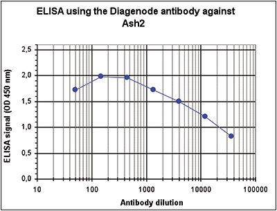
Figure 1. Determination of the antibody titer
To determine the titer, an ELISA was performed using a serial dilution of the Diagenode antibody directed against mouse Ash2 (Cat. No. C15310093). The wells were coated with the peptides used for immunisation of the rabbit. By plotting the absorbance against the antibody dilution (Figure 1), the titer of the antibody was estimated to be 1:24,000.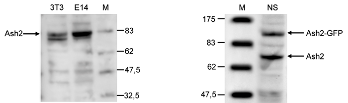
Figure 2. Western blot analysis using the Diagenode antibody directed against Ash2
A. Western blot was performed on whole cell lysates from mouse fibroblastst (NIH3T3) and embryonic stem cells (E14Tg2a) with the Diagenode antibody against mouse Ash2 (Cat. No. C15310093), diluted 1:1,000 in BSA/PBS-Tween. The molecular weight marker (in kDa) is shown on the right; the location of the protein of interest (predicted size: 68 kDa) is indicated on the left. B. Western blot was performed on whole cell lysates from mouse neural stem cells (NS), transfected with GFP tagged Ash2, with the Diagenode antibody against mouse Ash2 (Cat. No. C15310093), diluted 1:500 in BSA/PBS-Tween. The molecular weight marker (in kDa) is shown on the left; the location of the endogenous Ash2 (68 kDa) and of the GFP tagged Ash2 (106 kDa) are indicated on the right.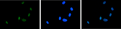
Figure 3. Immunofluorescence using the Diagenode antibody directed against Ash2
NIH3T3 cells were stained with the Diagenode antibody against Ash2 (Cat. No. C15310093) and with DAPI. Cells were fixed with 4% formaldehyde for 10’ and blocked with PBS/TX-100 containing 5% normal goat serum and 1% BSA. The cells were immunofluorescently labelled with the Ash2 antibody (left) diluted 1:200 in blocking solution followed by an anti-rabbit antibody conjugated to Alexa488. The middle panel shows staining of the nuclei with DAPI. A merge of the two stainings is shown on the right. - Publications

How to properly cite our product/service in your work
We strongly recommend using this: ASH2 Antibody (Hologic Diagenode Cat# C15310093 Lot# A260-004). Click here to copy to clipboard.
Using our products or services in your publication? Let us know!