Notice (8): Undefined variable: solution_of_interest [APP/View/Products/view.ctp, line 755]Code Context<!-- BEGIN: REQUEST_FORM MODAL -->
<div id="request_formModal" class="reveal-modal medium" data-reveal aria-labelledby="modalTitle" aria-hidden="true" role="dialog">
<?= $this->element('Forms/simple_form', array('solution_of_interest' => $solution_of_interest, 'header' => $header, 'message' => $message, 'campaign_id' => $campaign_id)) ?>
$viewFile = '/home/website-server/www/app/View/Products/view.ctp'
$dataForView = array(
'language' => 'en',
'meta_keywords' => '',
'meta_description' => 'H3K27me3 (Histone H3 trimethylated at lysine 27) Monoclonal Antibody validated in ChIP-qPCR, ELISA, WB and IF. Batch-specific data available on the website. Sample size available.',
'meta_title' => 'H3K27me3 Monoclonal Antibody | Diagenode',
'product' => array(
'Product' => array(
'id' => '2825',
'antibody_id' => '87',
'name' => 'H3K27me3 Antibody (sample size)',
'description' => '',
'label1' => '',
'info1' => '',
'label2' => '',
'info2' => '',
'label3' => '',
'info3' => '',
'format' => '10 µg',
'catalog_number' => 'C15200181-10',
'old_catalog_number' => '',
'sf_code' => 'C15200181-D001-000582',
'type' => 'FRE',
'search_order' => '',
'price_EUR' => '105',
'price_USD' => '115',
'price_GBP' => '100',
'price_JPY' => '16450',
'price_CNY' => '',
'price_AUD' => '288',
'country' => 'ALL',
'except_countries' => 'None',
'quote' => false,
'in_stock' => false,
'featured' => false,
'no_promo' => false,
'online' => true,
'master' => false,
'last_datasheet_update' => '0000-00-00',
'slug' => 'h3k27me3-monoclonal-antibody-classic-10-mg-10-ml',
'meta_title' => 'H3K27me3 Monoclonal Antibody | Diagenode',
'meta_keywords' => '',
'meta_description' => 'H3K27me3 (Histone H3 trimethylated at lysine 27) Monoclonal Antibody validated in ChIP-qPCR, ELISA, WB and IF. Batch-specific data available on the website. Sample size available.',
'modified' => '2021-12-22 14:33:37',
'created' => '2016-07-27 09:56:01',
'locale' => 'eng'
),
'Antibody' => array(
'host' => '*****',
'id' => '87',
'name' => 'H3K27me3 monoclonal antibody',
'description' => 'Histones are the main constituents of the protein part of chromosomes of eukaryotic cells. They are rich in the amino acids arginine and lysine and have been greatly conserved during evolution. Histones pack the DNA into tight masses of chromatin. Two core histones of each class H2A, H2B, H3 and H4 assemble and are wrapped by 146 base pairs of DNA to form one octameric nucleosome. Histone tails undergo numerous post-translational modifications, which either directly or indirectly alter chromatin structure to facilitate transcriptional activation or repression or other nuclear processes. In addition to the genetic code, combinations of the different histone modifications reveal the so-called “histone code”. Histone methylation and demethylation is dynamically regulated by respectively histone methyl transferases and histone demethylases.',
'clonality' => '',
'isotype' => 'IgG1κ',
'lot' => '001-14',
'concentration' => '1 µg/µl',
'reactivity' => 'Human, Nematodes, Magnaporthe oryzae: positive. Other species: not tested.',
'type' => 'Monoclonal',
'purity' => 'Protein A purified monoclonal antibody.',
'classification' => '',
'application_table' => '<table>
<thead>
<tr>
<th>Applications</th>
<th>Suggested dilution</th>
<th>References</th>
</tr>
</thead>
<tbody>
<tr>
<td>ChIP <sup>*</sup></td>
<td>1-2 µg/ChIP</td>
<td>Fig 1</td>
</tr>
<tr>
<td>CUT&TAG</td>
<td>1 µg</td>
<td>Fig 2</td>
</tr>
<tr>
<td>ELISA</td>
<td>1:3,000</td>
<td>Fig 3</td>
</tr>
<tr>
<td>Western Blotting</td>
<td>1:1,000</td>
<td>Fig 4</td>
</tr>
<tr>
<td>Immunofluorescence</td>
<td>1:500</td>
<td>Fig 5</td>
</tr>
</tbody>
</table>
<p><small><sup>*</sup> Please note that the optimal antibody amount per IP should be determined by the end-user. We recommend testing 1-5 µg per IP.</small></p>',
'storage_conditions' => 'Store at -20°C; for long storage, store at -80°C. Avoid multiple freeze-thaw cycles.',
'storage_buffer' => 'PBS containing 0.05% azide.',
'precautions' => 'This product is for research use only. Not for use in diagnostic or therapeutic procedures.',
'uniprot_acc' => '',
'slug' => '',
'meta_keywords' => '',
'meta_description' => '',
'modified' => '2023-02-16 14:59:27',
'created' => '0000-00-00 00:00:00',
'select_label' => '87 - H3K27me3 monoclonal antibody (001-14 - 1 µg/µl - Human, Nematodes, Magnaporthe oryzae: positive. Other species: not tested. - Protein A purified monoclonal antibody. - Mouse)'
),
'Slave' => array(),
'Group' => array(
'Group' => array(
[maximum depth reached]
),
'Master' => array(
[maximum depth reached]
),
'Product' => array(
[maximum depth reached]
)
),
'Related' => array(),
'Application' => array(
(int) 0 => array(
[maximum depth reached]
),
(int) 1 => array(
[maximum depth reached]
),
(int) 2 => array(
[maximum depth reached]
),
(int) 3 => array(
[maximum depth reached]
),
(int) 4 => array(
[maximum depth reached]
)
),
'Category' => array(
(int) 0 => array(
[maximum depth reached]
),
(int) 1 => array(
[maximum depth reached]
),
(int) 2 => array(
[maximum depth reached]
)
),
'Document' => array(
(int) 0 => array(
[maximum depth reached]
),
(int) 1 => array(
[maximum depth reached]
),
(int) 2 => array(
[maximum depth reached]
)
),
'Feature' => array(),
'Image' => array(
(int) 0 => array(
[maximum depth reached]
)
),
'Promotion' => array(),
'Protocol' => array(),
'Publication' => array(
(int) 0 => array(
[maximum depth reached]
),
(int) 1 => array(
[maximum depth reached]
),
(int) 2 => array(
[maximum depth reached]
),
(int) 3 => array(
[maximum depth reached]
),
(int) 4 => array(
[maximum depth reached]
),
(int) 5 => array(
[maximum depth reached]
),
(int) 6 => array(
[maximum depth reached]
),
(int) 7 => array(
[maximum depth reached]
)
),
'Testimonial' => array(),
'Area' => array(),
'SafetySheet' => array(
(int) 0 => array(
[maximum depth reached]
),
(int) 1 => array(
[maximum depth reached]
),
(int) 2 => array(
[maximum depth reached]
),
(int) 3 => array(
[maximum depth reached]
),
(int) 4 => array(
[maximum depth reached]
),
(int) 5 => array(
[maximum depth reached]
),
(int) 6 => array(
[maximum depth reached]
),
(int) 7 => array(
[maximum depth reached]
)
)
),
'meta_canonical' => 'https://www.diagenode.com/en/p/h3k27me3-monoclonal-antibody-classic-50-mg-50-ml'
)
$language = 'en'
$meta_keywords = ''
$meta_description = 'H3K27me3 (Histone H3 trimethylated at lysine 27) Monoclonal Antibody validated in ChIP-qPCR, ELISA, WB and IF. Batch-specific data available on the website. Sample size available.'
$meta_title = 'H3K27me3 Monoclonal Antibody | Diagenode'
$product = array(
'Product' => array(
'id' => '2825',
'antibody_id' => '87',
'name' => 'H3K27me3 Antibody (sample size)',
'description' => '<p><span>Monoclonal antibody raised in mouse against histone H3 trimethylated at lysine 27 (<strong>H3K27me3</strong>), using a KLH-conjugated synthetic peptide.</span></p>',
'label1' => 'Validation Data',
'info1' => '<div class="row">
<div class="small-4 columns">
<p><img src="https://www.diagenode.com/img/product/antibodies/C15200181_chip.png" alt="H3K27me3 Antibody ChIP Grade" style="display: block; margin-left: auto; margin-right: auto;" /></p>
</div>
<div class="small-8 columns">
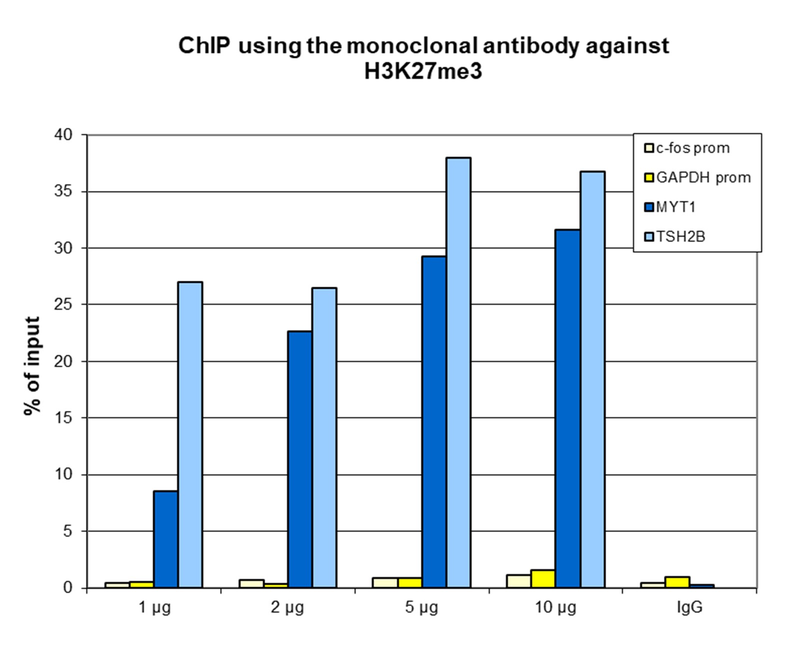
<p><small><strong> Figure 1. ChIP results obtained with the Diagenode monoclonal antibody directed against H3K27me3 </strong><br />ChIP assays were performed using human HeLa cells, the Diagenode monoclonal antibody against H3K27me3 (cat. No. C15200181) and optimized PCR primer sets for qPCR. ChIP was performed with the “AutoHistone ChIP-seq” kit (cat. No. C01010022) on sheared chromatin from 1 million cells. A titration of the antibody consisting of 1, 2, 5, and 10 µg per ChIP experiment was analysed. IgG (2 µg/IP) was used as negative IP control. QPCR was performed with primers for the promoters of the active genes c-fos (cat. No. C17011004) and GAPDH as negative controls, and for the coding regions of the inactive genes MYT1 and TSH2B (cat. No. C17011041) as positive controls. Figure 1 shows the recovery, expressed as a % of input (the relative amount of immunoprecipitated DNA compared to input DNA after qPCR analysis). These results are in accordance with the observation that H3K27me3 is preferably present at inactive genes. </small></p>
</div>
</div>
<div class="row">
<div class="small-12 columns">
<p>A.<img src="https://www.diagenode.com/img/product/antibodies/C15200184-cuttagA.png" width="700" style="display: block; margin-left: auto; margin-right: auto;" /></p>
<p>B.<img src="https://www.diagenode.com/img/product/antibodies/C15200184-cuttagB.png" width="700" style="display: block; margin-left: auto; margin-right: auto;" /></p>
</div>
<div class="small-12 columns">
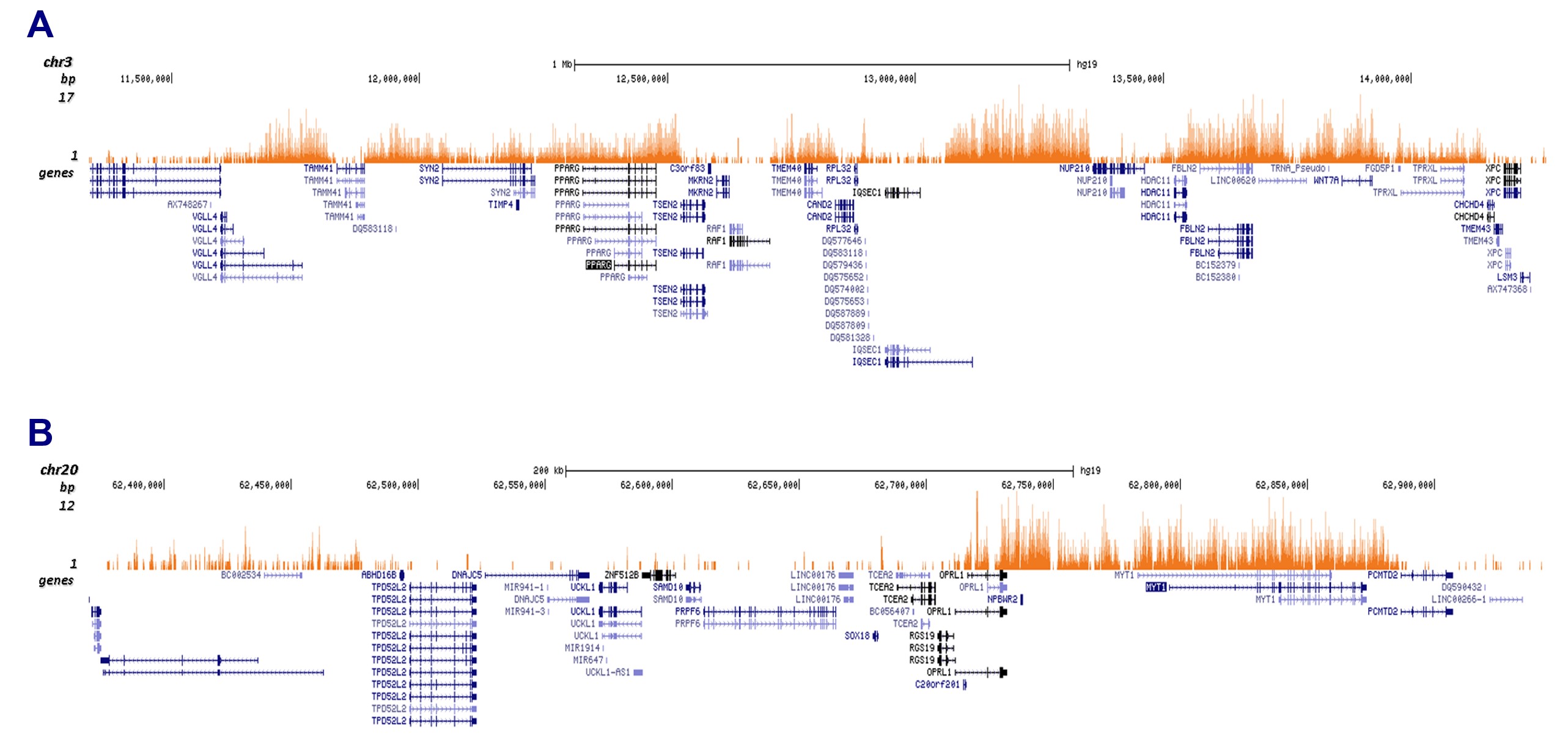
<p><small><strong> Figure 2. Cut&Tag results obtained with the Diagenode monoclonal antibody directed against H3K27me3 </strong><br />CUT&TAG (Kaya-Okur, H.S., Nat Commun 10, 1930, 2019) was performed on 50,000 K562 cells using 1 µg of the Diagenode monoclonal antibody against H3K27me3 (cat. No. C15200181) and the Diagenode pA-Tn5 transposase (C01070001). The libraries were subsequently analysed on an Illumina NextSeq 500 sequencer (2x75 paired-end reads) according to the manufacturer's instructions. The tags were aligned to the human genome (hg19) using the BWA algorithm. Figure 3 shows the peak distribution in 2 genomic regions on chromosome 3 and 20 (figure 2A and B, respectively).</small></p>
</div>
</div>
<div class="row">
<div class="small-4 columns">
<p><img src="https://www.diagenode.com/img/product/antibodies/C15200181_ELISA.png" alt="H3K27me3 Antibody ELISA validation" caption="false" width="288" height="237" /></p>
</div>
<div class="small-8 columns">
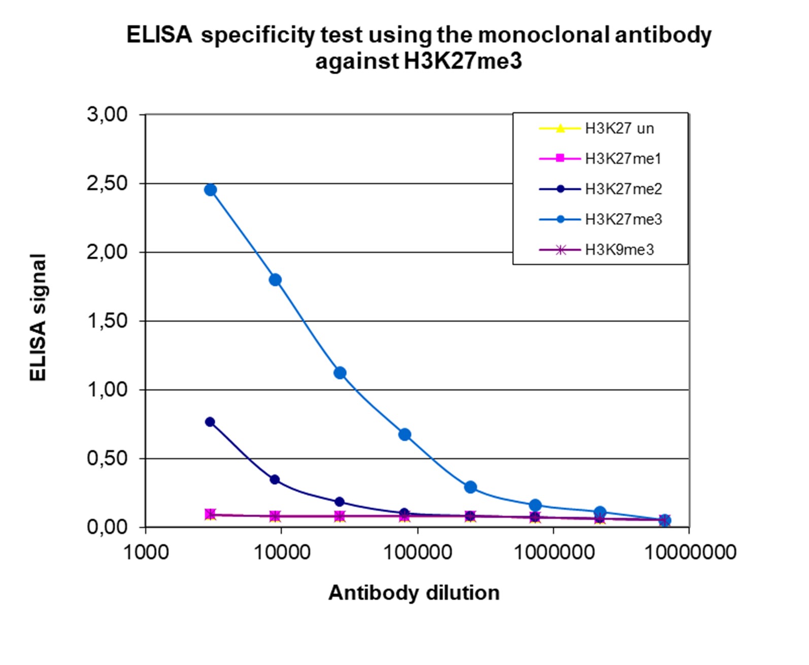
<p><small><strong> Figure 3. Cross reactivity of the Diagenode monoclonal antibody directed against H3K27me3 </strong><br />To test the specificity an ELISA was performed using a serial dilution of the Diagenode monoclonal antibody against H3K27me3 (Cat. No. C15200181). The wells were coated with peptides containing the unmodified H3K27 region as well as the mono-, di- and trimethylated H3K27 and the trimethylated H3K9. Figure 3 shows a high specificity of the antibody for the peptide containing the modification of interest. </small></p>
</div>
</div>
<div class="row">
<div class="small-4 columns">
<p><img src="https://www.diagenode.com/img/product/antibodies/C15200181_WB.png" alt="H3K27me3 Antibody validated in Western Blot" style="display: block; margin-left: auto; margin-right: auto;" /></p>
</div>
<div class="small-8 columns">
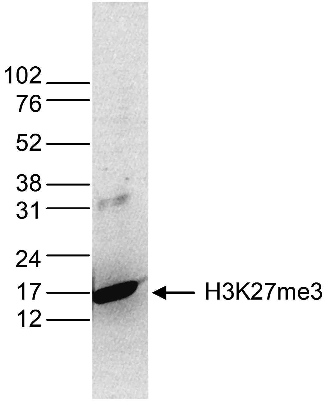
<p><small><strong> Figure 4. Western blot analysis using the Diagenode monoclonal antibody directed against H3K27me3 </strong><br />Histone extracts (15 µg) from HeLa cells were analysed by Western blot using the Diagenode monoclonal antibody against H3K27me3 (Cat. No. C15200181) diluted 1:1,000 in TBS-Tween containing 5% skimmed milk. The position of the protein of interest is indicated on the right; the marker (in kDa) is shown on the left. </small></p>
</div>
</div>
<div class="extra-spaced"></div>
<div class="row">
<div class="small-5 columns">
<p><img src="https://www.diagenode.com/img/product/antibodies/C15200181_IF.png" alt="H3K27me3 Antibody validated in Immunofluorescence" style="display: block; margin-left: auto; margin-right: auto;" /></p>
</div>
<div class="small-7 columns">
<p><small><strong> Figure 5. Immunofluorescence using the Diagenode monoclonal antibody directed against H3K27me3 </strong><br />HeLa cells were stained with the Diagenode antibody against H3K27me3 (Cat. No. C15200181) and with DAPI. Cells were fixed with 4% formaldehyde for 10’ and blocked with PBS/TX-100 containing 5% normal goat serum and 1% BSA. The cells were immunofluorescently labelled with the H3K27me3 antibody (left) diluted 1:500 in blocking solution followed by an anti-mouse antibody conjugated to Alexa594. The middle panel shows staining of the nuclei with DAPI. A merge of the two stainings is shown on the right. </small></p>
</div>
</div>',
'label2' => 'Target Description',
'info2' => '<p>Histones are the main constituents of the protein part of chromosomes of eukaryotic cells. They are rich in the amino acids arginine and lysine and have been greatly conserved during evolution. Histones pack the DNA into tight masses of chromatin. Two core histones of each class H2A, H2B, H3 and H4 assemble and are wrapped by 146 base pairs of DNA to form one octameric nucleosome. Histone tails undergo numerous post-translational modifications, which either directly or indirectly alter chromatin structure to facilitate transcriptional activation or repression or other nuclear processes. In addition to the genetic code, combinations of the different histone modifications reveal the so-called “histone code”. Histone methylation and demethylation is dynamically regulated by respectively histone methyl transferases and histone demethylases.</p>',
'label3' => '',
'info3' => '',
'format' => '10 µg',
'catalog_number' => 'C15200181-10',
'old_catalog_number' => '',
'sf_code' => 'C15200181-D001-000582',
'type' => 'FRE',
'search_order' => '',
'price_EUR' => '105',
'price_USD' => '115',
'price_GBP' => '100',
'price_JPY' => '16450',
'price_CNY' => '',
'price_AUD' => '288',
'country' => 'ALL',
'except_countries' => 'None',
'quote' => false,
'in_stock' => false,
'featured' => false,
'no_promo' => false,
'online' => true,
'master' => false,
'last_datasheet_update' => '0000-00-00',
'slug' => 'h3k27me3-monoclonal-antibody-classic-10-mg-10-ml',
'meta_title' => 'H3K27me3 Monoclonal Antibody | Diagenode',
'meta_keywords' => '',
'meta_description' => 'H3K27me3 (Histone H3 trimethylated at lysine 27) Monoclonal Antibody validated in ChIP-qPCR, ELISA, WB and IF. Batch-specific data available on the website. Sample size available.',
'modified' => '2021-12-22 14:33:37',
'created' => '2016-07-27 09:56:01',
'locale' => 'eng'
),
'Antibody' => array(
'host' => '*****',
'id' => '87',
'name' => 'H3K27me3 monoclonal antibody',
'description' => 'Histones are the main constituents of the protein part of chromosomes of eukaryotic cells. They are rich in the amino acids arginine and lysine and have been greatly conserved during evolution. Histones pack the DNA into tight masses of chromatin. Two core histones of each class H2A, H2B, H3 and H4 assemble and are wrapped by 146 base pairs of DNA to form one octameric nucleosome. Histone tails undergo numerous post-translational modifications, which either directly or indirectly alter chromatin structure to facilitate transcriptional activation or repression or other nuclear processes. In addition to the genetic code, combinations of the different histone modifications reveal the so-called “histone code”. Histone methylation and demethylation is dynamically regulated by respectively histone methyl transferases and histone demethylases.',
'clonality' => '',
'isotype' => 'IgG1κ',
'lot' => '001-14',
'concentration' => '1 µg/µl',
'reactivity' => 'Human, Nematodes, Magnaporthe oryzae: positive. Other species: not tested.',
'type' => 'Monoclonal',
'purity' => 'Protein A purified monoclonal antibody.',
'classification' => '',
'application_table' => '<table>
<thead>
<tr>
<th>Applications</th>
<th>Suggested dilution</th>
<th>References</th>
</tr>
</thead>
<tbody>
<tr>
<td>ChIP <sup>*</sup></td>
<td>1-2 µg/ChIP</td>
<td>Fig 1</td>
</tr>
<tr>
<td>CUT&TAG</td>
<td>1 µg</td>
<td>Fig 2</td>
</tr>
<tr>
<td>ELISA</td>
<td>1:3,000</td>
<td>Fig 3</td>
</tr>
<tr>
<td>Western Blotting</td>
<td>1:1,000</td>
<td>Fig 4</td>
</tr>
<tr>
<td>Immunofluorescence</td>
<td>1:500</td>
<td>Fig 5</td>
</tr>
</tbody>
</table>
<p><small><sup>*</sup> Please note that the optimal antibody amount per IP should be determined by the end-user. We recommend testing 1-5 µg per IP.</small></p>',
'storage_conditions' => 'Store at -20°C; for long storage, store at -80°C. Avoid multiple freeze-thaw cycles.',
'storage_buffer' => 'PBS containing 0.05% azide.',
'precautions' => 'This product is for research use only. Not for use in diagnostic or therapeutic procedures.',
'uniprot_acc' => '',
'slug' => '',
'meta_keywords' => '',
'meta_description' => '',
'modified' => '2023-02-16 14:59:27',
'created' => '0000-00-00 00:00:00',
'select_label' => '87 - H3K27me3 monoclonal antibody (001-14 - 1 µg/µl - Human, Nematodes, Magnaporthe oryzae: positive. Other species: not tested. - Protein A purified monoclonal antibody. - Mouse)'
),
'Slave' => array(),
'Group' => array(
'Group' => array(
'id' => '188',
'name' => 'C15200181',
'product_id' => '1998',
'modified' => '2016-07-27 09:56:41',
'created' => '2016-07-27 09:56:41'
),
'Master' => array(
'id' => '1998',
'antibody_id' => '87',
'name' => 'H3K27me3 Antibody ',
'description' => '<p><span>Monoclonal antibody raised in mouse against histone H3 trimethylated at lysine 27 (<strong>H3K27me3</strong>), using a KLH-conjugated synthetic peptide.</span></p>',
'label1' => 'Validation Data',
'info1' => '<div class="row">
<div class="small-4 columns">
<p><img src="https://www.diagenode.com/img/product/antibodies/C15200181_chip.png" alt="H3K27me3 Antibody ChIP Grade" style="display: block; margin-left: auto; margin-right: auto;" /></p>
</div>
<div class="small-8 columns">
<p><small><strong> Figure 1. ChIP results obtained with the Diagenode monoclonal antibody directed against H3K27me3 </strong><br />ChIP assays were performed using human HeLa cells, the Diagenode monoclonal antibody against H3K27me3 (cat. No. C15200181) and optimized PCR primer sets for qPCR. ChIP was performed with the “AutoHistone ChIP-seq” kit (cat. No. C01010022) on sheared chromatin from 1 million cells. A titration of the antibody consisting of 1, 2, 5, and 10 µg per ChIP experiment was analysed. IgG (2 µg/IP) was used as negative IP control. QPCR was performed with primers for the promoters of the active genes c-fos (cat. No. C17011004) and GAPDH as negative controls, and for the coding regions of the inactive genes MYT1 and TSH2B (cat. No. C17011041) as positive controls. Figure 1 shows the recovery, expressed as a % of input (the relative amount of immunoprecipitated DNA compared to input DNA after qPCR analysis). These results are in accordance with the observation that H3K27me3 is preferably present at inactive genes. </small></p>
</div>
</div>
<div class="row">
<div class="small-12 columns">
<p>A.<img src="https://www.diagenode.com/img/product/antibodies/C15200184-cuttagA.png" width="700" style="display: block; margin-left: auto; margin-right: auto;" /></p>
<p>B.<img src="https://www.diagenode.com/img/product/antibodies/C15200184-cuttagB.png" width="700" style="display: block; margin-left: auto; margin-right: auto;" /></p>
</div>
<div class="small-12 columns">
<p><small><strong> Figure 2. Cut&Tag results obtained with the Diagenode monoclonal antibody directed against H3K27me3 </strong><br />CUT&TAG (Kaya-Okur, H.S., Nat Commun 10, 1930, 2019) was performed on 50,000 K562 cells using 1 µg of the Diagenode monoclonal antibody against H3K27me3 (cat. No. C15200181) and the Diagenode pA-Tn5 transposase (C01070001). The libraries were subsequently analysed on an Illumina NextSeq 500 sequencer (2x75 paired-end reads) according to the manufacturer's instructions. The tags were aligned to the human genome (hg19) using the BWA algorithm. Figure 3 shows the peak distribution in 2 genomic regions on chromosome 3 and 20 (figure 2A and B, respectively).</small></p>
</div>
</div>
<div class="row">
<div class="small-4 columns">
<p><img src="https://www.diagenode.com/img/product/antibodies/C15200181_ELISA.png" alt="H3K27me3 Antibody ELISA validation" caption="false" width="288" height="237" /></p>
</div>
<div class="small-8 columns">
<p><small><strong> Figure 3. Cross reactivity of the Diagenode monoclonal antibody directed against H3K27me3 </strong><br />To test the specificity an ELISA was performed using a serial dilution of the Diagenode monoclonal antibody against H3K27me3 (Cat. No. C15200181). The wells were coated with peptides containing the unmodified H3K27 region as well as the mono-, di- and trimethylated H3K27 and the trimethylated H3K9. Figure 3 shows a high specificity of the antibody for the peptide containing the modification of interest. </small></p>
</div>
</div>
<div class="row">
<div class="small-4 columns">
<p><img src="https://www.diagenode.com/img/product/antibodies/C15200181_WB.png" alt="H3K27me3 Antibody validated in Western Blot" style="display: block; margin-left: auto; margin-right: auto;" /></p>
</div>
<div class="small-8 columns">
<p><small><strong> Figure 4. Western blot analysis using the Diagenode monoclonal antibody directed against H3K27me3 </strong><br />Histone extracts (15 µg) from HeLa cells were analysed by Western blot using the Diagenode monoclonal antibody against H3K27me3 (Cat. No. C15200181) diluted 1:1,000 in TBS-Tween containing 5% skimmed milk. The position of the protein of interest is indicated on the right; the marker (in kDa) is shown on the left. </small></p>
</div>
</div>
<div class="extra-spaced"></div>
<div class="row">
<div class="small-5 columns">
<p><img src="https://www.diagenode.com/img/product/antibodies/C15200181_IF.png" alt="H3K27me3 Antibody validated in Immunofluorescence" style="display: block; margin-left: auto; margin-right: auto;" /></p>
</div>
<div class="small-7 columns">
<p><small><strong> Figure 5. Immunofluorescence using the Diagenode monoclonal antibody directed against H3K27me3 </strong><br />HeLa cells were stained with the Diagenode antibody against H3K27me3 (Cat. No. C15200181) and with DAPI. Cells were fixed with 4% formaldehyde for 10’ and blocked with PBS/TX-100 containing 5% normal goat serum and 1% BSA. The cells were immunofluorescently labelled with the H3K27me3 antibody (left) diluted 1:500 in blocking solution followed by an anti-mouse antibody conjugated to Alexa594. The middle panel shows staining of the nuclei with DAPI. A merge of the two stainings is shown on the right. </small></p>
</div>
</div>',
'label2' => 'Target Description',
'info2' => '<p>Histones are the main constituents of the protein part of chromosomes of eukaryotic cells. They are rich in the amino acids arginine and lysine and have been greatly conserved during evolution. Histones pack the DNA into tight masses of chromatin. Two core histones of each class H2A, H2B, H3 and H4 assemble and are wrapped by 146 base pairs of DNA to form one octameric nucleosome. Histone tails undergo numerous post-translational modifications, which either directly or indirectly alter chromatin structure to facilitate transcriptional activation or repression or other nuclear processes. In addition to the genetic code, combinations of the different histone modifications reveal the so-called “histone code”. Histone methylation and demethylation is dynamically regulated by respectively histone methyl transferases and histone demethylases.</p>',
'label3' => '',
'info3' => '',
'format' => '50 μg',
'catalog_number' => 'C15200181',
'old_catalog_number' => 'MAb-181-050',
'sf_code' => 'C15200181-D001-000581',
'type' => 'FRE',
'search_order' => '03-Antibody',
'price_EUR' => '380',
'price_USD' => '380',
'price_GBP' => '340',
'price_JPY' => '59525',
'price_CNY' => '',
'price_AUD' => '950',
'country' => 'ALL',
'except_countries' => 'None',
'quote' => false,
'in_stock' => false,
'featured' => false,
'no_promo' => false,
'online' => true,
'master' => true,
'last_datasheet_update' => 'July 6, 2018',
'slug' => 'h3k27me3-monoclonal-antibody-classic-50-mg-50-ml',
'meta_title' => 'H3K27me3 Antibody - ChIP Grade (C15200181) | Diagenode',
'meta_keywords' => '',
'meta_description' => 'H3K27me3 (Histone H3 trimethylated at lysine 27) Monoclonal Antibody validated in ChIP-qPCR, ELISA, WB and IF. Batch-specific data available on the website. Sample size available.',
'modified' => '2024-11-19 16:50:20',
'created' => '2015-06-29 14:08:20'
),
'Product' => array(
(int) 0 => array(
[maximum depth reached]
)
)
),
'Related' => array(),
'Application' => array(
(int) 0 => array(
'id' => '19',
'position' => '10',
'parent_id' => '40',
'name' => 'WB',
'description' => '<p><strong>Western blot</strong> : The quality of antibodies used in this technique is crucial for correct and specific protein identification. Diagenode offers huge selection of highly sensitive and specific western blot-validated antibodies.</p>
<p>Learn more about: <a href="https://www.diagenode.com/applications/western-blot">Loading control, MW marker visualization</a><em>. <br /></em></p>
<p><em></em>Check our selection of antibodies validated in Western blot.</p>',
'in_footer' => false,
'in_menu' => false,
'online' => true,
'tabular' => true,
'slug' => 'western-blot-antibodies',
'meta_keywords' => ' Western Blot Antibodies ,western blot protocol,Western Blotting Products,Polyclonal antibodies ,monoclonal antibodies ',
'meta_description' => 'Diagenode offers a wide range of antibodies and technical support for western blot applications',
'meta_title' => ' Western Blot - Monoclonal antibody - Polyclonal antibody | Diagenode',
'modified' => '2016-04-26 12:44:51',
'created' => '2015-01-07 09:20:00',
'ProductsApplication' => array(
[maximum depth reached]
)
),
(int) 1 => array(
'id' => '20',
'position' => '10',
'parent_id' => '40',
'name' => 'ELISA',
'description' => '<div class="row">
<div class="small-12 medium-12 large-12 columns">Enzyme-linked immunosorbent assay.</div>
</div>',
'in_footer' => false,
'in_menu' => false,
'online' => true,
'tabular' => true,
'slug' => 'elisa-antibodies',
'meta_keywords' => ' ELISA Antibodies,Monoclonal antibody, Polyclonal antibody',
'meta_description' => 'Diagenode offers Monoclonal & Polyclonal antibodies for ELISA applications',
'meta_title' => 'ELISA Antibodies - Monoclonal & Polyclonal antibody | Diagenode',
'modified' => '2016-01-13 12:21:41',
'created' => '2014-07-08 08:13:28',
'ProductsApplication' => array(
[maximum depth reached]
)
),
(int) 2 => array(
'id' => '29',
'position' => '10',
'parent_id' => '40',
'name' => 'IF',
'description' => '<p><strong>Immunofluorescence</strong>:</p>
<p>Diagenode offers huge selection of highly sensitive antibodies validated in IF.</p>
<p><img src="https://www.diagenode.com/img/product/antibodies/C15200229-IF.jpg" alt="" height="245" width="256" /></p>
<p><sup><strong>Immunofluorescence using the Diagenode monoclonal antibody directed against CRISPR/Cas9</strong></sup></p>
<p><sup>HeLa cells transfected with a Cas9 expression vector (left) or untransfected cells (right) were fixed in methanol at -20°C, permeabilized with acetone at -20°C and blocked with PBS containing 2% BSA. The cells were stained with the Cas9 C-terminal antibody (Cat. No. C15200229) diluted 1:400, followed by incubation with an anti-mouse secondary antibody coupled to AF488. The bottom images show counter-staining of the nuclei with Hoechst 33342.</sup></p>
<h5><sup>Check our selection of antibodies validated in IF.</sup></h5>',
'in_footer' => false,
'in_menu' => false,
'online' => true,
'tabular' => true,
'slug' => 'immunofluorescence',
'meta_keywords' => 'Immunofluorescence,Monoclonal antibody,Polyclonal antibody',
'meta_description' => 'Diagenode offers a wide range of antibodies and technical support for Immunofluorescence applications',
'meta_title' => 'Immunofluorescence - Monoclonal antibody - Polyclonal antibody | Diagenode',
'modified' => '2016-04-27 16:23:10',
'created' => '2015-07-08 13:46:02',
'ProductsApplication' => array(
[maximum depth reached]
)
),
(int) 3 => array(
'id' => '43',
'position' => '10',
'parent_id' => '40',
'name' => 'ChIP-qPCR (ab)',
'description' => '',
'in_footer' => false,
'in_menu' => false,
'online' => true,
'tabular' => true,
'slug' => 'chip-qpcr-antibodies',
'meta_keywords' => 'Chromatin Immunoprecipitation Sequencing,ChIP-Seq,ChIP-seq grade antibodies,DNA purification,qPCR,Shearing of chromatin',
'meta_description' => 'Diagenode offers a wide range of antibodies and technical support for ChIP-qPCR applications',
'meta_title' => 'ChIP Quantitative PCR Antibodies (ChIP-qPCR) | Diagenode',
'modified' => '2016-01-20 11:30:24',
'created' => '2015-10-20 11:45:36',
'ProductsApplication' => array(
[maximum depth reached]
)
),
(int) 4 => array(
'id' => '55',
'position' => '10',
'parent_id' => '40',
'name' => 'CUT&Tag',
'description' => '<p>CUT&Tagアッセイを成功させるための重要な要素の1つは使用される抗体の品質です。 特異性高い抗体は、目的のタンパク質のみをターゲットとした確実な結果を可能にします。 CUT&Tagで検証済みの抗体のセレクションはこちらからご覧ください。</p>
<p>Read more:</p>
<p><a href="https://www.diagenode.com/en/categories/cutandtag">Products for CUT&Tag assay</a></p>
<p><a href="https://www.diagenode.com/en/pages/cut-and-tag">Performance of Diagenode's antibodies in CUT&Tag</a></p>
<script src="chrome-extension://hhojmcideegachlhfgfdhailpfhgknjm/web_accessible_resources/index.js"></script>',
'in_footer' => false,
'in_menu' => false,
'online' => true,
'tabular' => true,
'slug' => 'cut-and-tag',
'meta_keywords' => 'CUT&Tag',
'meta_description' => 'CUT&Tag',
'meta_title' => 'CUT&Tag',
'modified' => '2021-04-27 05:17:46',
'created' => '2020-08-20 10:13:47',
'ProductsApplication' => array(
[maximum depth reached]
)
)
),
'Category' => array(
(int) 0 => array(
'id' => '103',
'position' => '0',
'parent_id' => '4',
'name' => 'All antibodies',
'description' => '<p><span style="font-weight: 400;">All Diagenode’s antibodies are listed below. Please, use our Quick search field to find the antibody of interest by target name, application, purity.</span></p>
<p><span style="font-weight: 400;">Diagenode’s highly validated antibodies:</span></p>
<ul>
<li>Highly sensitive and specific</li>
<li>Cost-effective (requires less antibody per reaction)</li>
<li>Batch-specific data is available on the website</li>
<li>Expert technical support</li>
<li>Sample sizes available</li>
<li>100% satisfaction guarantee</li>
</ul>',
'no_promo' => false,
'in_menu' => true,
'online' => true,
'tabular' => false,
'hide' => true,
'all_format' => false,
'is_antibody' => true,
'slug' => 'all-antibodies',
'cookies_tag_id' => null,
'meta_keywords' => 'Antibodies,Premium Antibodies,Classic,Pioneer',
'meta_description' => 'Diagenode Offers Strict quality standards with Rigorous QC and validated Antibodies. Classified based on level of validation for flexibility of Application. Comprehensive selection of histone and non-histone Antibodies',
'meta_title' => 'Diagenode's selection of Antibodies is exclusively dedicated for Epigenetic Research | Diagenode',
'modified' => '2019-07-03 10:55:44',
'created' => '2015-11-02 14:49:22',
'ProductsCategory' => array(
[maximum depth reached]
),
'CookiesTag' => array([maximum depth reached])
),
(int) 1 => array(
'id' => '111',
'position' => '40',
'parent_id' => '4',
'name' => 'Histone antibodies',
'description' => '<p>Histones are the main protein components of chromatin involved in the compaction of DNA into nucleosomes, the basic units of chromatin. A <strong>nucleosome</strong> consists of one pair of each of the core histones (<strong>H2A</strong>, <strong>H2B</strong>, <strong>H3</strong> and <strong>H4</strong>) forming an octameric structure wrapped by 146 base pairs of DNA. The different nucleosomes are linked by the linker histone<strong> H1, </strong>allowing for further condensation of chromatin.</p>
<p>The core histones have a globular structure with large unstructured N-terminal tails protruding from the nucleosome. They can undergo to multiple post-translational modifications (PTM), mainly at the N-terminal tails. These <strong>post-translational modifications </strong>include methylation, acetylation, phosphorylation, ubiquitinylation, citrullination, sumoylation, deamination and crotonylation. The most well characterized PTMs are <strong>methylation,</strong> <strong>acetylation and phosphorylation</strong>. Histone methylation occurs mainly on lysine (K) residues, which can be mono-, di- or tri-methylated, and on arginines (R), which can be mono-methylated and symmetrically or asymmetrically di-methylated. Histone acetylation occurs on lysines and histone phosphorylation mainly on serines (S), threonines (T) and tyrosines (Y).</p>
<p>The PTMs of the different residues are involved in numerous processes such as DNA repair, DNA replication and chromosome condensation. They influence the chromatin organization and can be positively or negatively associated with gene expression. Trimethylation of H3K4, H3K36 and H3K79, and lysine acetylation generally result in an open chromatin configuration (figure below) and are therefore associated with <strong>euchromatin</strong> and gene activation. Trimethylation of H3K9, K3K27 and H4K20, on the other hand, is enriched in <strong>heterochromatin </strong>and associated with gene silencing. The combination of different histone modifications is called the "<strong>histone code</strong>”, analogous to the genetic code.</p>
<p><img src="https://www.diagenode.com/img/categories/antibodies/histone-marks-illustration.png" /></p>
<p>Diagenode is proud to offer a large range of antibodies against histones and histone modifications. Our antibodies are highly specific and have been validated in many applications, including <strong>ChIP</strong> and <strong>ChIP-seq</strong>.</p>
<p>Diagenode’s collection includes antibodies recognizing:</p>
<ul>
<li><strong>Histone H1 variants</strong></li>
<li><strong>Histone H2A, H2A variants and histone H2A</strong> <strong>modifications</strong> (serine phosphorylation, lysine acetylation, lysine ubiquitinylation)</li>
<li><strong>Histone H2B and H2B</strong> <strong>modifications </strong>(serine phosphorylation, lysine acetylation)</li>
<li><strong>Histone H3 and H3 modifications </strong>(lysine methylation (mono-, di- and tri-methylated), lysine acetylation, serine phosphorylation, threonine phosphorylation, arginine methylation (mono-methylated, symmetrically and asymmetrically di-methylated))</li>
<li><strong>Histone H4 and H4 modifications (</strong>lysine methylation (mono-, di- and tri-methylated), lysine acetylation, arginine methylation (mono-methylated and symmetrically di-methylated), serine phosphorylation )</li>
</ul>
<p><span style="font-weight: 400;"><strong>HDAC's HAT's, HMT's and other</strong> <strong>enzymes</strong> which modify histones can be found in the category <a href="../categories/chromatin-modifying-proteins-histone-transferase">Histone modifying enzymes</a><br /></span></p>
<p><span style="font-weight: 400;"> Diagenode’s highly validated antibodies:</span></p>
<ul>
<li><span style="font-weight: 400;"> Highly sensitive and specific</span></li>
<li><span style="font-weight: 400;"> Cost-effective (requires less antibody per reaction)</span></li>
<li><span style="font-weight: 400;"> Batch-specific data is available on the website</span></li>
<li><span style="font-weight: 400;"> Expert technical support</span></li>
<li><span style="font-weight: 400;"> Sample sizes available</span></li>
<li><span style="font-weight: 400;"> 100% satisfaction guarantee</span></li>
</ul>',
'no_promo' => false,
'in_menu' => false,
'online' => true,
'tabular' => false,
'hide' => true,
'all_format' => false,
'is_antibody' => true,
'slug' => 'histone-antibodies',
'cookies_tag_id' => null,
'meta_keywords' => 'Histone, antibody, histone h1, histone h2, histone h3, histone h4',
'meta_description' => 'Polyclonal and Monoclonal Antibodies against Histones and their modifications validated for many applications, including Chromatin Immunoprecipitation (ChIP) and ChIP-Sequencing (ChIP-seq)',
'meta_title' => 'Histone and Modified Histone Antibodies | Diagenode',
'modified' => '2020-09-17 13:34:56',
'created' => '2016-04-01 16:01:32',
'ProductsCategory' => array(
[maximum depth reached]
),
'CookiesTag' => array([maximum depth reached])
),
(int) 2 => array(
'id' => '127',
'position' => '10',
'parent_id' => '4',
'name' => 'ChIP-grade antibodies',
'description' => '<div class="row">
<div class="small-12 columns"><center></center>
<p><br />Chromatin immunoprecipitation (<b>ChIP</b>) is a technique to study the associations of proteins with the specific genomic regions in intact cells. One of the most important steps of this protocol is the immunoprecipitation of targeted protein using the antibody specifically recognizing it. The quality of antibodies used in ChIP is essential for the success of the experiment. Diagenode offers extensively validated ChIP-grade antibodies, confirmed for their specificity, and high level of performance in ChIP. Each batch is validated, and batch-specific data are available on the website.</p>
<p></p>
</div>
</div>
<p><strong>ChIP results</strong> obtained with the antibody directed against H3K4me3 (Cat. No. <a href="../p/h3k4me3-polyclonal-antibody-premium-50-ug-50-ul">C15410003</a>). </p>
<div class="row">
<div class="small-12 medium-6 large-6 columns"><img src="https://www.diagenode.com/img/product/antibodies/C15410003-fig1-ChIP.jpg" alt="" width="400" height="315" /> </div>
<div class="small-12 medium-6 large-6 columns">
<p></p>
<p></p>
<p></p>
</div>
</div>
<p></p>
<p>Our aim at Diagenode is to offer the largest collection of highly specific <strong>ChIP-grade antibodies</strong>. We add new antibodies monthly. Find your ChIP-grade antibody in the list below and check more information about tested applications, extensive validation data, and product information.</p>',
'no_promo' => false,
'in_menu' => true,
'online' => true,
'tabular' => false,
'hide' => true,
'all_format' => false,
'is_antibody' => true,
'slug' => 'chip-grade-antibodies',
'cookies_tag_id' => null,
'meta_keywords' => 'ChIP-grade antibodies, polyclonal antibody, monoclonal antibody, Diagenode',
'meta_description' => 'Diagenode Offers Extensively Validated ChIP-Grade Antibodies, Confirmed for their Specificity, and high level of Performance in Chromatin Immunoprecipitation ChIP',
'meta_title' => 'Chromatin immunoprecipitation ChIP-grade antibodies | Diagenode',
'modified' => '2024-11-19 17:27:07',
'created' => '2017-02-14 11:16:04',
'ProductsCategory' => array(
[maximum depth reached]
),
'CookiesTag' => array([maximum depth reached])
)
),
'Document' => array(
(int) 0 => array(
'id' => '11',
'name' => 'Antibodies you can trust',
'description' => '<p style="text-align: justify;"><span>Epigenetic research tools have evolved over time from endpoint PCR to qPCR to the analyses of large sets of genome-wide sequencing data. ChIP sequencing (ChIP-seq) has now become the gold standard method for chromatin studies, given the accuracy and coverage scale of the approach over other methods. Successful ChIP-seq, however, requires a higher level of experimental accuracy and consistency in all steps of ChIP than ever before. Particularly crucial is the quality of ChIP antibodies. </span></p>',
'image_id' => null,
'type' => 'Poster',
'url' => 'files/posters/Antibodies_you_can_trust_Poster.pdf',
'slug' => 'antibodies-you-can-trust-poster',
'meta_keywords' => '',
'meta_description' => '',
'modified' => '2015-10-01 20:18:31',
'created' => '2015-07-03 16:05:15',
'ProductsDocument' => array(
[maximum depth reached]
)
),
(int) 1 => array(
'id' => '38',
'name' => 'Epigenetic Antibodies Brochure',
'description' => '<p>More than in any other immuoprecipitation assays, quality antibodies are critical tools in many epigenetics experiments. Since 10 years, Diagenode has developed the most stringent quality production available on the market for antibodies exclusively focused on epigenetic uses. All our antibodies have been qualified to work in epigenetic applications.</p>',
'image_id' => null,
'type' => 'Brochure',
'url' => 'files/brochures/Epigenetic_Antibodies_Brochure.pdf',
'slug' => 'epigenetic-antibodies-brochure',
'meta_keywords' => '',
'meta_description' => '',
'modified' => '2016-06-15 11:24:06',
'created' => '2015-07-03 16:05:27',
'ProductsDocument' => array(
[maximum depth reached]
)
),
(int) 2 => array(
'id' => '721',
'name' => 'Datasheet H3K27me3 MAb-181-050',
'description' => 'Datasheet description',
'image_id' => null,
'type' => 'Datasheet',
'url' => 'files/products/antibodies/Datasheet_H3K27me3_MAb-181-050.pdf',
'slug' => 'datasheet-h3k27me3-mab-181-050',
'meta_keywords' => null,
'meta_description' => null,
'modified' => '2015-07-07 11:47:44',
'created' => '2015-07-07 11:47:44',
'ProductsDocument' => array(
[maximum depth reached]
)
)
),
'Feature' => array(),
'Image' => array(
(int) 0 => array(
'id' => '1815',
'name' => 'product/antibodies/ab-cuttag-icon.png',
'alt' => 'cut and tag antibody icon',
'modified' => '2021-02-11 12:45:34',
'created' => '2021-02-11 12:45:34',
'ProductsImage' => array(
[maximum depth reached]
)
)
),
'Promotion' => array(),
'Protocol' => array(),
'Publication' => array(
(int) 0 => array(
'id' => '4925',
'name' => 'Chromatin profiling reveals TFAP4 as a critical transcriptional regulator of bovine satellite cell differentiation',
'authors' => 'Pengcheng Lyu et al.',
'description' => '<h3 class="c-article__sub-heading" data-test="abstract-sub-heading">Background</h3>
<p>Satellite cells are myogenic precursor cells in adult skeletal muscle and play a crucial role in skeletal muscle regeneration, maintenance, and growth. Like embryonic myoblasts, satellite cells have the ability to proliferate, differentiate, and fuse to form multinucleated myofibers. In this study, we aimed to identify additional transcription factors that control gene expression during bovine satellite cell proliferation and differentiation.</p>
<h3 class="c-article__sub-heading" data-test="abstract-sub-heading">Results</h3>
<p>Using chromatin immunoprecipitation followed by sequencing, we identified 56,973 and 54,470 genomic regions marked with both the histone modifications H3K4me1 and H3K27ac, which were considered active enhancers, and 50,956 and 59,174 genomic regions marked with H3K27me3, which were considered repressed enhancers, in proliferating and differentiating bovine satellite cells, respectively. In addition, we identified 1,216 and 1,171 super-enhancers in proliferating and differentiating bovine satellite cells, respectively. Analyzing these enhancers showed that in proliferating bovine satellite cells, active enhancers were associated with genes stimulating cell proliferation or inhibiting myoblast differentiation whereas repressed enhancers were associated with genes essential for myoblast differentiation, and that in differentiating satellite cells, active enhancers were associated with genes essential for myoblast differentiation or muscle contraction whereas repressed enhancers were associated with genes stimulating cell proliferation or inhibiting myoblast differentiation. Active enhancers in proliferating bovine satellite cells were enriched with binding sites for many transcription factors such as MYF5 and the AP-1 family transcription factors; active enhancers in differentiating bovine satellite cells were enriched with binding sites for many transcription factors such as MYOG and TFAP4; and repressed enhancers in both proliferating and differentiating bovine satellite cells were enriched with binding sites for NF-kB, ZEB-1, and several other transcription factors. The role of TFAP4 in satellite cell or myoblast differentiation was previously unknown, and through gene knockdown and overexpression, we experimentally validated a critical role for TFAP4 in the differentiation and fusion of bovine satellite cells into myofibers.</p>
<h3 class="c-article__sub-heading" data-test="abstract-sub-heading">Conclusions</h3>
<p>Satellite cell proliferation and differentiation are controlled by many transcription factors such as AP-1, TFAP4, NF-kB, and ZEB-1 whose roles in these processes were previously unknown in addition to those transcription factors such as MYF5 and MYOG whose roles in these processes are widely known.</p>',
'date' => '2024-03-12',
'pmid' => 'https://bmcgenomics.biomedcentral.com/articles/10.1186/s12864-024-10189-2',
'doi' => 'https://doi.org/10.1186/s12864-024-10189-2',
'modified' => '2024-03-15 15:06:06',
'created' => '2024-03-15 15:06:06',
'ProductsPublication' => array(
[maximum depth reached]
)
),
(int) 1 => array(
'id' => '4277',
'name' => 'Tracing the emergence of primordial germ cells from bilaminar disc rabbitembryos and pluripotent stem cells.',
'authors' => 'Kobayashi Toshihiro et al.',
'description' => '<p>Rabbit embryos develop as bilaminar discs at gastrulation as in humans and most other mammals, whereas rodents develop as egg cylinders. Primordial germ cells (PGCs) appear to originate during gastrulation according to many systematic studies on mammalian embryos. Here, we show that rabbit PGC (rbPGC) specification occurs at the posterior epiblast at the onset of gastrulation. Using newly derived rabbit pluripotent stem cells, we show robust and rapid induction of rbPGC-like cells in vitro with WNT and BMP morphogens, which reveals SOX17 as the critical regulator of rbPGC fate as in several non-rodent mammals. We posit that development as a bilaminar disc is a crucial determinant of the PGC regulators, regardless of the highly diverse development of extraembryonic tissues, including the amnion. We propose that investigations on rabbits with short gestation, large litters, and where gastrulation precedes implantation can contribute significantly to advances in early mammalian development.</p>',
'date' => '2021-10-01',
'pmid' => 'https://doi.org/10.1016%2Fj.celrep.2021.109812',
'doi' => '10.1016/j.celrep.2021.109812',
'modified' => '2022-05-23 09:58:19',
'created' => '2022-05-19 10:41:50',
'ProductsPublication' => array(
[maximum depth reached]
)
),
(int) 2 => array(
'id' => '4121',
'name' => 'Histone modification dynamics at H3K27 are associated with alteredtranscription of in planta induced genes in Magnaporthe oryzae.',
'authors' => 'Zhang, Wei and Huang, Jun and Cook, David E',
'description' => '<p>Transcriptional dynamic in response to environmental and developmental cues are fundamental to biology, yet many mechanistic aspects are poorly understood. One such example is fungal plant pathogens, which use secreted proteins and small molecules, termed effectors, to suppress host immunity and promote colonization. Effectors are highly expressed in planta but remain transcriptionally repressed ex planta, but our mechanistic understanding of these transcriptional dynamics remains limited. We tested the hypothesis that repressive histone modification at H3-Lys27 underlies transcriptional silencing ex planta, and that exchange for an active chemical modification contributes to transcription of in planta induced genes. Using genetics, chromatin immunoprecipitation and sequencing and RNA-sequencing, we determined that H3K27me3 provides significant local transcriptional repression. We detail how regions that lose H3K27me3 gain H3K27ac, and these changes are associated with increased transcription. Importantly, we observed that many in planta induced genes were marked by H3K27me3 during axenic growth, and detail how altered H3K27 modification influences transcription. ChIP-qPCR during in planta growth suggests that H3K27 modifications are generally stable, but can undergo dynamics at specific genomic locations. Our results support the hypothesis that dynamic histone modifications at H3K27 contributes to fungal genome regulation and specifically contributes to regulation of genes important during host infection.</p>',
'date' => '2021-02-01',
'pmid' => 'https://pubmed.ncbi.nlm.nih.gov/33534835/',
'doi' => '10.1371/journal.pgen.1009376',
'modified' => '2021-12-07 09:55:47',
'created' => '2021-12-06 15:53:19',
'ProductsPublication' => array(
[maximum depth reached]
)
),
(int) 3 => array(
'id' => '3966',
'name' => 'Intergenerationally Maintained Histone H4 Lysine 16 Acetylation Is Instructive for Future Gene Activation.',
'authors' => 'Samata M, Alexiadis A, Richard G, Georgiev P, Nuebler J, Kulkarni T, Renschler G, Basilicata MF, Zenk FL, Shvedunova M, Semplicio G, Mirny L, Iovino N, Akhtar A',
'description' => '<p>Before zygotic genome activation (ZGA), the quiescent genome undergoes reprogramming to transition into the transcriptionally active state. However, the mechanisms underlying euchromatin establishment during early embryogenesis remain poorly understood. Here, we show that histone H4 lysine 16 acetylation (H4K16ac) is maintained from oocytes to fertilized embryos in Drosophila and mammals. H4K16ac forms large domains that control nucleosome accessibility of promoters prior to ZGA in flies. Maternal depletion of MOF acetyltransferase leading to H4K16ac loss causes aberrant RNA Pol II recruitment, compromises the 3D organization of the active genomic compartments during ZGA, and causes downregulation of post-zygotically expressed genes. Germline depletion of histone deacetylases revealed that other acetyl marks cannot compensate for H4K16ac loss in the oocyte. Moreover, zygotic re-expression of MOF was neither able to restore embryonic viability nor onset of X chromosome dosage compensation. Thus, maternal H4K16ac provides an instructive function to the offspring, priming future gene activation.</p>',
'date' => '2020-06-03',
'pmid' => 'http://www.pubmed.gov/32502394',
'doi' => '10.1016/j.cell.2020.05.026',
'modified' => '2020-08-12 09:37:51',
'created' => '2020-08-10 12:12:25',
'ProductsPublication' => array(
[maximum depth reached]
)
),
(int) 4 => array(
'id' => '3478',
'name' => 'Pro-inflammatory cytokines activate hypoxia-inducible factor 3α via epigenetic changes in mesenchymal stromal/stem cells.',
'authors' => 'Cuomo F, Coppola A, Botti C, Maione C, Forte A, Scisciola L, Liguori G, Caiafa I, Ursini MV, Galderisi U, Cipollaro M, Altucci L, Cobellis G',
'description' => '<p>Human mesenchymal stromal/stem cells (hMSCs) emerged as a promising therapeutic tool for ischemic disorders, due to their ability to regenerate damaged tissues, promote angiogenesis and reduce inflammation, leading to encouraging, but still limited results. The outcomes in clinical trials exploring hMSC therapy are influenced by low cell retention and survival in affected tissues, partially influenced by lesion's microenvironment, where low oxygen conditions (i.e. hypoxia) and inflammation coexist. Hypoxia and inflammation are pathophysiological stresses, sharing common activators, such as hypoxia-inducible factors (HIFs) and NF-κB. HIF1α and HIF2α respond essentially to hypoxia, activating pathways involved in tissue repair. Little is known about the regulation of HIF3α. Here we investigated the role of HIF3α in vitro and in vivo. Human MSCs expressed HIF3α, differentially regulated by pro-inflammatory cytokines in an oxygen-independent manner, a novel and still uncharacterized mechanism, where NF-κB is critical for its expression. We investigated if epigenetic modifications are involved in HIF3α expression by methylation-specific PCR and histone modifications. Robust hypermethylation of histone H3 was observed across HIF3A locus driven by pro-inflammatory cytokines. Experiments in a murine model of arteriotomy highlighted the activation of Hif3α expression in infiltrated inflammatory cells, suggesting a new role for Hif3α in inflammation in vivo.</p>',
'date' => '2018-04-11',
'pmid' => 'http://www.pubmed.gov/29643458',
'doi' => '10.1038/s41598-018-24221-5',
'modified' => '2019-02-15 20:21:28',
'created' => '2019-02-14 15:01:22',
'ProductsPublication' => array(
[maximum depth reached]
)
),
(int) 5 => array(
'id' => '2841',
'name' => 'Chemical Biology Approaches for Characterization of Epigenetic Regulators',
'authors' => 'Barsyte-Lovejoy D, Szewczyk MM, Prinos P, Lima-Fernandes E, Ackloo S, Arrowsmith CH',
'description' => '<p>Chemical biology approaches are a powerful means to functionally characterize epigenetic regulators such as histone modifying enzymes. We outline experimental protocols and best practices for the cellular characterization and use of “chemical probes” that selectively inhibit protein methyltransferases, many of which methylate histones to regulate heritable gene expression patterns. We describe biomarker assays to validate the probes in specific cellular systems, and provide guidelines for their use in functional characterization of methyltransferases including detailed protocols, examples, and controls. Together these techniques enable precision manipulation of cellular epigenomes and the exploration of the therapeutic potential of epigenetic targets in human disease.</p>',
'date' => '2016-02-16',
'pmid' => 'http://www.sciencedirect.com/science/article/pii/S0076687916000240',
'doi' => '10.1016/bs.mie.2016.01.011',
'modified' => '2016-03-09 12:31:59',
'created' => '2016-03-09 12:30:54',
'ProductsPublication' => array(
[maximum depth reached]
)
),
(int) 6 => array(
'id' => '2960',
'name' => 'Germline organization in Strongyloides nematodes reveals alternative differentiation and regulation mechanisms.',
'authors' => 'Kulkarni A et al.',
'description' => '<p>Nematodes of the genus Strongyloides are important parasites of vertebrates including man. Currently, little is known about their germline organization or reproductive biology and how this influences their parasitic life strategies. Here, we analyze the structure of the germline in several Strongyloides and closely related species and uncover striking differences in the development, germline organization, and fluid dynamics compared to the model organism Caenorhabditis elegans. With a focus on Strongyloides ratti, we reveal that the proliferation of germ cells is restricted to early and mid-larval development, thus limiting the number of progeny. In order to understand key germline events (specifically germ cell progression and the transcriptional status of the germline), we monitored conserved histone modifications, in particular H3Pser10 and H3K4me3. The evolutionary significance of these events is subsequently highlighted through comparisons with six other nematode species, revealing underlying complexities and variations in the development of the germline among nematodes</p>',
'date' => '2015-12-12',
'pmid' => 'http://www.ncbi.nlm.nih.gov/pubmed/26661737',
'doi' => '',
'modified' => '2016-06-23 10:57:22',
'created' => '2016-06-23 10:57:22',
'ProductsPublication' => array(
[maximum depth reached]
)
),
(int) 7 => array(
'id' => '1360',
'name' => 'An Orally Bioavailable Chemical Probe of the Lysine Methyltransferases EZH2 and EZH1.',
'authors' => 'Konze KD, Ma A, Li F, Barsyte-Lovejoy D, Parton T, Macnevin CJ, Liu F, Gao C, Huang XP, Kuznetsova E, Rougie M, Jiang A, Pattenden SG, Norris JL, James LI, Roth BL, Brown PJ, Frye SV, Arrowsmith CH, Hahn KM, Wang GG, Vedadi M, Jin J',
'description' => '<p>EZH2 or EZH1 is the catalytic subunit of the polycomb repressive complex 2 that catalyzes methylation of histone H3 lysine 27 (H3K27). The trimethylation of H3K27 (H3K27me3) is a transcriptionally repressive post-translational modification. Overexpression of EZH2 and hypertrimethylation of H3K27 have been implicated in a number of cancers. Several selective inhibitors of EZH2 have been reported recently. Herein we disclose UNC1999, the first orally bioavailable inhibitor that has high in vitro potency for wild-type and mutant EZH2 as well as EZH1, a closely related H3K27 methyltransferase that shares 96% sequence identity with EZH2 in their respective catalytic domains. UNC1999 was highly selective for EZH2 and EZH1 over a broad range of epigenetic and non-epigenetic targets, competitive with the cofactor SAM and non-competitive with the peptide substrate. This inhibitor potently reduced H3K27me3 levels in cells and selectively killed diffused large B cell lymphoma cell lines harboring the EZH2(Y641N) mutant. Importantly, UNC1999 was orally bioavailable in mice, making this inhibitor a valuable tool for investigating the role of EZH2 and EZH1 in chronic animal studies. We also designed and synthesized UNC2400, a close analogue of UNC1999 with potency >1,000-fold lower than that of UNC1999 as a negative control for cell-based studies. Finally, we created a biotin-tagged UNC1999 (UNC2399), which enriched EZH2 in pull-down studies, and a UNC1999-dye conjugate (UNC2239) for co-localization studies with EZH2 in live cells. Taken together, these compounds represent a set of useful tools for the biomedical community to investigate the role of EZH2 and EZH1 in health and disease.</p>',
'date' => '2013-04-24',
'pmid' => 'https://www.ncbi.nlm.nih.gov/pubmed/23614352',
'doi' => '',
'modified' => '2016-04-12 09:57:04',
'created' => '2015-07-24 15:39:00',
'ProductsPublication' => array(
[maximum depth reached]
)
)
),
'Testimonial' => array(),
'Area' => array(),
'SafetySheet' => array(
(int) 0 => array(
'id' => '822',
'name' => 'H3K27me3 antibody SDS GB en',
'language' => 'en',
'url' => 'files/SDS/H3K27me3/SDS-C15200181-H3K27me3_Antibody-GB-en-GHS_2_0.pdf',
'countries' => 'GB',
'modified' => '2020-09-22 13:40:42',
'created' => '2020-09-22 13:40:42',
'ProductsSafetySheet' => array(
[maximum depth reached]
)
),
(int) 1 => array(
'id' => '824',
'name' => 'H3K27me3 antibody SDS US en',
'language' => 'en',
'url' => 'files/SDS/H3K27me3/SDS-C15200181-H3K27me3_Antibody-US-en-GHS_2_0.pdf',
'countries' => 'US',
'modified' => '2020-09-22 13:42:19',
'created' => '2020-09-22 13:41:45',
'ProductsSafetySheet' => array(
[maximum depth reached]
)
),
(int) 2 => array(
'id' => '819',
'name' => 'H3K27me3 antibody SDS DE de',
'language' => 'de',
'url' => 'files/SDS/H3K27me3/SDS-C15200181-H3K27me3_Antibody-DE-de-GHS_2_0.pdf',
'countries' => 'DE',
'modified' => '2020-09-22 13:39:17',
'created' => '2020-09-22 13:39:17',
'ProductsSafetySheet' => array(
[maximum depth reached]
)
),
(int) 3 => array(
'id' => '823',
'name' => 'H3K27me3 antibody SDS JP ja',
'language' => 'ja',
'url' => 'files/SDS/H3K27me3/SDS-C15200181-H3K27me3_Antibody-JP-ja-GHS_3_0.pdf',
'countries' => 'JP',
'modified' => '2020-09-22 13:41:13',
'created' => '2020-09-22 13:41:13',
'ProductsSafetySheet' => array(
[maximum depth reached]
)
),
(int) 4 => array(
'id' => '818',
'name' => 'H3K27me3 antibody SDS BE nl',
'language' => 'nl',
'url' => 'files/SDS/H3K27me3/SDS-C15200181-H3K27me3_Antibody-BE-nl-GHS_2_0.pdf',
'countries' => 'BE',
'modified' => '2020-09-22 13:38:44',
'created' => '2020-09-22 13:38:44',
'ProductsSafetySheet' => array(
[maximum depth reached]
)
),
(int) 5 => array(
'id' => '817',
'name' => 'H3K27me3 antibody SDS BE fr',
'language' => 'fr',
'url' => 'files/SDS/H3K27me3/SDS-C15200181-H3K27me3_Antibody-BE-fr-GHS_2_0.pdf',
'countries' => 'BE',
'modified' => '2020-09-22 13:38:15',
'created' => '2020-09-22 13:38:15',
'ProductsSafetySheet' => array(
[maximum depth reached]
)
),
(int) 6 => array(
'id' => '821',
'name' => 'H3K27me3 antibody SDS FR fr',
'language' => 'fr',
'url' => 'files/SDS/H3K27me3/SDS-C15200181-H3K27me3_Antibody-FR-fr-GHS_2_0.pdf',
'countries' => 'FR',
'modified' => '2020-09-22 13:40:14',
'created' => '2020-09-22 13:40:14',
'ProductsSafetySheet' => array(
[maximum depth reached]
)
),
(int) 7 => array(
'id' => '820',
'name' => 'H3K27me3 antibody SDS ES es',
'language' => 'es',
'url' => 'files/SDS/H3K27me3/SDS-C15200181-H3K27me3_Antibody-ES-es-GHS_2_0.pdf',
'countries' => 'ES',
'modified' => '2020-09-22 13:39:47',
'created' => '2020-09-22 13:39:47',
'ProductsSafetySheet' => array(
[maximum depth reached]
)
)
)
)
$meta_canonical = 'https://www.diagenode.com/en/p/h3k27me3-monoclonal-antibody-classic-50-mg-50-ml'
$country = 'US'
$countries_allowed = array(
(int) 0 => 'CA',
(int) 1 => 'US',
(int) 2 => 'IE',
(int) 3 => 'GB',
(int) 4 => 'DK',
(int) 5 => 'NO',
(int) 6 => 'SE',
(int) 7 => 'FI',
(int) 8 => 'NL',
(int) 9 => 'BE',
(int) 10 => 'LU',
(int) 11 => 'FR',
(int) 12 => 'DE',
(int) 13 => 'CH',
(int) 14 => 'AT',
(int) 15 => 'ES',
(int) 16 => 'IT',
(int) 17 => 'PT'
)
$outsource = true
$other_formats = array(
(int) 0 => array(
'id' => '1998',
'antibody_id' => '87',
'name' => 'H3K27me3 Antibody ',
'description' => '<p><span>Monoclonal antibody raised in mouse against histone H3 trimethylated at lysine 27 (<strong>H3K27me3</strong>), using a KLH-conjugated synthetic peptide.</span></p>',
'label1' => 'Validation Data',
'info1' => '<div class="row">
<div class="small-4 columns">
<p><img src="https://www.diagenode.com/img/product/antibodies/C15200181_chip.png" alt="H3K27me3 Antibody ChIP Grade" style="display: block; margin-left: auto; margin-right: auto;" /></p>
</div>
<div class="small-8 columns">
<p><small><strong> Figure 1. ChIP results obtained with the Diagenode monoclonal antibody directed against H3K27me3 </strong><br />ChIP assays were performed using human HeLa cells, the Diagenode monoclonal antibody against H3K27me3 (cat. No. C15200181) and optimized PCR primer sets for qPCR. ChIP was performed with the “AutoHistone ChIP-seq” kit (cat. No. C01010022) on sheared chromatin from 1 million cells. A titration of the antibody consisting of 1, 2, 5, and 10 µg per ChIP experiment was analysed. IgG (2 µg/IP) was used as negative IP control. QPCR was performed with primers for the promoters of the active genes c-fos (cat. No. C17011004) and GAPDH as negative controls, and for the coding regions of the inactive genes MYT1 and TSH2B (cat. No. C17011041) as positive controls. Figure 1 shows the recovery, expressed as a % of input (the relative amount of immunoprecipitated DNA compared to input DNA after qPCR analysis). These results are in accordance with the observation that H3K27me3 is preferably present at inactive genes. </small></p>
</div>
</div>
<div class="row">
<div class="small-12 columns">
<p>A.<img src="https://www.diagenode.com/img/product/antibodies/C15200184-cuttagA.png" width="700" style="display: block; margin-left: auto; margin-right: auto;" /></p>
<p>B.<img src="https://www.diagenode.com/img/product/antibodies/C15200184-cuttagB.png" width="700" style="display: block; margin-left: auto; margin-right: auto;" /></p>
</div>
<div class="small-12 columns">
<p><small><strong> Figure 2. Cut&Tag results obtained with the Diagenode monoclonal antibody directed against H3K27me3 </strong><br />CUT&TAG (Kaya-Okur, H.S., Nat Commun 10, 1930, 2019) was performed on 50,000 K562 cells using 1 µg of the Diagenode monoclonal antibody against H3K27me3 (cat. No. C15200181) and the Diagenode pA-Tn5 transposase (C01070001). The libraries were subsequently analysed on an Illumina NextSeq 500 sequencer (2x75 paired-end reads) according to the manufacturer's instructions. The tags were aligned to the human genome (hg19) using the BWA algorithm. Figure 3 shows the peak distribution in 2 genomic regions on chromosome 3 and 20 (figure 2A and B, respectively).</small></p>
</div>
</div>
<div class="row">
<div class="small-4 columns">
<p><img src="https://www.diagenode.com/img/product/antibodies/C15200181_ELISA.png" alt="H3K27me3 Antibody ELISA validation" caption="false" width="288" height="237" /></p>
</div>
<div class="small-8 columns">
<p><small><strong> Figure 3. Cross reactivity of the Diagenode monoclonal antibody directed against H3K27me3 </strong><br />To test the specificity an ELISA was performed using a serial dilution of the Diagenode monoclonal antibody against H3K27me3 (Cat. No. C15200181). The wells were coated with peptides containing the unmodified H3K27 region as well as the mono-, di- and trimethylated H3K27 and the trimethylated H3K9. Figure 3 shows a high specificity of the antibody for the peptide containing the modification of interest. </small></p>
</div>
</div>
<div class="row">
<div class="small-4 columns">
<p><img src="https://www.diagenode.com/img/product/antibodies/C15200181_WB.png" alt="H3K27me3 Antibody validated in Western Blot" style="display: block; margin-left: auto; margin-right: auto;" /></p>
</div>
<div class="small-8 columns">
<p><small><strong> Figure 4. Western blot analysis using the Diagenode monoclonal antibody directed against H3K27me3 </strong><br />Histone extracts (15 µg) from HeLa cells were analysed by Western blot using the Diagenode monoclonal antibody against H3K27me3 (Cat. No. C15200181) diluted 1:1,000 in TBS-Tween containing 5% skimmed milk. The position of the protein of interest is indicated on the right; the marker (in kDa) is shown on the left. </small></p>
</div>
</div>
<div class="extra-spaced"></div>
<div class="row">
<div class="small-5 columns">
<p><img src="https://www.diagenode.com/img/product/antibodies/C15200181_IF.png" alt="H3K27me3 Antibody validated in Immunofluorescence" style="display: block; margin-left: auto; margin-right: auto;" /></p>
</div>
<div class="small-7 columns">
<p><small><strong> Figure 5. Immunofluorescence using the Diagenode monoclonal antibody directed against H3K27me3 </strong><br />HeLa cells were stained with the Diagenode antibody against H3K27me3 (Cat. No. C15200181) and with DAPI. Cells were fixed with 4% formaldehyde for 10’ and blocked with PBS/TX-100 containing 5% normal goat serum and 1% BSA. The cells were immunofluorescently labelled with the H3K27me3 antibody (left) diluted 1:500 in blocking solution followed by an anti-mouse antibody conjugated to Alexa594. The middle panel shows staining of the nuclei with DAPI. A merge of the two stainings is shown on the right. </small></p>
</div>
</div>',
'label2' => 'Target Description',
'info2' => '<p>Histones are the main constituents of the protein part of chromosomes of eukaryotic cells. They are rich in the amino acids arginine and lysine and have been greatly conserved during evolution. Histones pack the DNA into tight masses of chromatin. Two core histones of each class H2A, H2B, H3 and H4 assemble and are wrapped by 146 base pairs of DNA to form one octameric nucleosome. Histone tails undergo numerous post-translational modifications, which either directly or indirectly alter chromatin structure to facilitate transcriptional activation or repression or other nuclear processes. In addition to the genetic code, combinations of the different histone modifications reveal the so-called “histone code”. Histone methylation and demethylation is dynamically regulated by respectively histone methyl transferases and histone demethylases.</p>',
'label3' => '',
'info3' => '',
'format' => '50 μg',
'catalog_number' => 'C15200181',
'old_catalog_number' => 'MAb-181-050',
'sf_code' => 'C15200181-D001-000581',
'type' => 'FRE',
'search_order' => '03-Antibody',
'price_EUR' => '380',
'price_USD' => '380',
'price_GBP' => '340',
'price_JPY' => '59525',
'price_CNY' => '',
'price_AUD' => '950',
'country' => 'ALL',
'except_countries' => 'None',
'quote' => false,
'in_stock' => false,
'featured' => false,
'no_promo' => false,
'online' => true,
'master' => true,
'last_datasheet_update' => 'July 6, 2018',
'slug' => 'h3k27me3-monoclonal-antibody-classic-50-mg-50-ml',
'meta_title' => 'H3K27me3 Antibody - ChIP Grade (C15200181) | Diagenode',
'meta_keywords' => '',
'meta_description' => 'H3K27me3 (Histone H3 trimethylated at lysine 27) Monoclonal Antibody validated in ChIP-qPCR, ELISA, WB and IF. Batch-specific data available on the website. Sample size available.',
'modified' => '2024-11-19 16:50:20',
'created' => '2015-06-29 14:08:20'
)
)
$pro = array(
'id' => '2825',
'antibody_id' => '87',
'name' => 'H3K27me3 Antibody (sample size)',
'description' => '',
'label1' => '',
'info1' => '',
'label2' => '',
'info2' => '',
'label3' => '',
'info3' => '',
'format' => '10 µg',
'catalog_number' => 'C15200181-10',
'old_catalog_number' => '',
'sf_code' => 'C15200181-D001-000582',
'type' => 'FRE',
'search_order' => '',
'price_EUR' => '105',
'price_USD' => '115',
'price_GBP' => '100',
'price_JPY' => '16450',
'price_CNY' => '',
'price_AUD' => '288',
'country' => 'ALL',
'except_countries' => 'None',
'quote' => false,
'in_stock' => false,
'featured' => false,
'no_promo' => false,
'online' => true,
'master' => false,
'last_datasheet_update' => '0000-00-00',
'slug' => 'h3k27me3-monoclonal-antibody-classic-10-mg-10-ml',
'meta_title' => 'H3K27me3 Monoclonal Antibody | Diagenode',
'meta_keywords' => '',
'meta_description' => 'H3K27me3 (Histone H3 trimethylated at lysine 27) Monoclonal Antibody validated in ChIP-qPCR, ELISA, WB and IF. Batch-specific data available on the website. Sample size available.',
'modified' => '2021-12-22 14:33:37',
'created' => '2016-07-27 09:56:01',
'ProductsGroup' => array(
'id' => '204',
'product_id' => '2825',
'group_id' => '188'
)
)
$edit = ''
$testimonials = ''
$featured_testimonials = ''
$related_products = ''
$rrbs_service = array(
(int) 0 => (int) 1894,
(int) 1 => (int) 1895
)
$chipseq_service = array(
(int) 0 => (int) 2683,
(int) 1 => (int) 1835,
(int) 2 => (int) 1836,
(int) 3 => (int) 2684,
(int) 4 => (int) 1838,
(int) 5 => (int) 1839,
(int) 6 => (int) 1856
)
$labelize = object(Closure) {
}
$old_catalog_number = ' <span style="color:#CCC">(MAb-181-050)</span>'
$country_code = 'US'
$other_format = array(
'id' => '1998',
'antibody_id' => '87',
'name' => 'H3K27me3 Antibody ',
'description' => '<p><span>Monoclonal antibody raised in mouse against histone H3 trimethylated at lysine 27 (<strong>H3K27me3</strong>), using a KLH-conjugated synthetic peptide.</span></p>',
'label1' => 'Validation Data',
'info1' => '<div class="row">
<div class="small-4 columns">
<p><img src="https://www.diagenode.com/img/product/antibodies/C15200181_chip.png" alt="H3K27me3 Antibody ChIP Grade" style="display: block; margin-left: auto; margin-right: auto;" /></p>
</div>
<div class="small-8 columns">
<p><small><strong> Figure 1. ChIP results obtained with the Diagenode monoclonal antibody directed against H3K27me3 </strong><br />ChIP assays were performed using human HeLa cells, the Diagenode monoclonal antibody against H3K27me3 (cat. No. C15200181) and optimized PCR primer sets for qPCR. ChIP was performed with the “AutoHistone ChIP-seq” kit (cat. No. C01010022) on sheared chromatin from 1 million cells. A titration of the antibody consisting of 1, 2, 5, and 10 µg per ChIP experiment was analysed. IgG (2 µg/IP) was used as negative IP control. QPCR was performed with primers for the promoters of the active genes c-fos (cat. No. C17011004) and GAPDH as negative controls, and for the coding regions of the inactive genes MYT1 and TSH2B (cat. No. C17011041) as positive controls. Figure 1 shows the recovery, expressed as a % of input (the relative amount of immunoprecipitated DNA compared to input DNA after qPCR analysis). These results are in accordance with the observation that H3K27me3 is preferably present at inactive genes. </small></p>
</div>
</div>
<div class="row">
<div class="small-12 columns">
<p>A.<img src="https://www.diagenode.com/img/product/antibodies/C15200184-cuttagA.png" width="700" style="display: block; margin-left: auto; margin-right: auto;" /></p>
<p>B.<img src="https://www.diagenode.com/img/product/antibodies/C15200184-cuttagB.png" width="700" style="display: block; margin-left: auto; margin-right: auto;" /></p>
</div>
<div class="small-12 columns">
<p><small><strong> Figure 2. Cut&Tag results obtained with the Diagenode monoclonal antibody directed against H3K27me3 </strong><br />CUT&TAG (Kaya-Okur, H.S., Nat Commun 10, 1930, 2019) was performed on 50,000 K562 cells using 1 µg of the Diagenode monoclonal antibody against H3K27me3 (cat. No. C15200181) and the Diagenode pA-Tn5 transposase (C01070001). The libraries were subsequently analysed on an Illumina NextSeq 500 sequencer (2x75 paired-end reads) according to the manufacturer's instructions. The tags were aligned to the human genome (hg19) using the BWA algorithm. Figure 3 shows the peak distribution in 2 genomic regions on chromosome 3 and 20 (figure 2A and B, respectively).</small></p>
</div>
</div>
<div class="row">
<div class="small-4 columns">
<p><img src="https://www.diagenode.com/img/product/antibodies/C15200181_ELISA.png" alt="H3K27me3 Antibody ELISA validation" caption="false" width="288" height="237" /></p>
</div>
<div class="small-8 columns">
<p><small><strong> Figure 3. Cross reactivity of the Diagenode monoclonal antibody directed against H3K27me3 </strong><br />To test the specificity an ELISA was performed using a serial dilution of the Diagenode monoclonal antibody against H3K27me3 (Cat. No. C15200181). The wells were coated with peptides containing the unmodified H3K27 region as well as the mono-, di- and trimethylated H3K27 and the trimethylated H3K9. Figure 3 shows a high specificity of the antibody for the peptide containing the modification of interest. </small></p>
</div>
</div>
<div class="row">
<div class="small-4 columns">
<p><img src="https://www.diagenode.com/img/product/antibodies/C15200181_WB.png" alt="H3K27me3 Antibody validated in Western Blot" style="display: block; margin-left: auto; margin-right: auto;" /></p>
</div>
<div class="small-8 columns">
<p><small><strong> Figure 4. Western blot analysis using the Diagenode monoclonal antibody directed against H3K27me3 </strong><br />Histone extracts (15 µg) from HeLa cells were analysed by Western blot using the Diagenode monoclonal antibody against H3K27me3 (Cat. No. C15200181) diluted 1:1,000 in TBS-Tween containing 5% skimmed milk. The position of the protein of interest is indicated on the right; the marker (in kDa) is shown on the left. </small></p>
</div>
</div>
<div class="extra-spaced"></div>
<div class="row">
<div class="small-5 columns">
<p><img src="https://www.diagenode.com/img/product/antibodies/C15200181_IF.png" alt="H3K27me3 Antibody validated in Immunofluorescence" style="display: block; margin-left: auto; margin-right: auto;" /></p>
</div>
<div class="small-7 columns">
<p><small><strong> Figure 5. Immunofluorescence using the Diagenode monoclonal antibody directed against H3K27me3 </strong><br />HeLa cells were stained with the Diagenode antibody against H3K27me3 (Cat. No. C15200181) and with DAPI. Cells were fixed with 4% formaldehyde for 10’ and blocked with PBS/TX-100 containing 5% normal goat serum and 1% BSA. The cells were immunofluorescently labelled with the H3K27me3 antibody (left) diluted 1:500 in blocking solution followed by an anti-mouse antibody conjugated to Alexa594. The middle panel shows staining of the nuclei with DAPI. A merge of the two stainings is shown on the right. </small></p>
</div>
</div>',
'label2' => 'Target Description',
'info2' => '<p>Histones are the main constituents of the protein part of chromosomes of eukaryotic cells. They are rich in the amino acids arginine and lysine and have been greatly conserved during evolution. Histones pack the DNA into tight masses of chromatin. Two core histones of each class H2A, H2B, H3 and H4 assemble and are wrapped by 146 base pairs of DNA to form one octameric nucleosome. Histone tails undergo numerous post-translational modifications, which either directly or indirectly alter chromatin structure to facilitate transcriptional activation or repression or other nuclear processes. In addition to the genetic code, combinations of the different histone modifications reveal the so-called “histone code”. Histone methylation and demethylation is dynamically regulated by respectively histone methyl transferases and histone demethylases.</p>',
'label3' => '',
'info3' => '',
'format' => '50 μg',
'catalog_number' => 'C15200181',
'old_catalog_number' => 'MAb-181-050',
'sf_code' => 'C15200181-D001-000581',
'type' => 'FRE',
'search_order' => '03-Antibody',
'price_EUR' => '380',
'price_USD' => '380',
'price_GBP' => '340',
'price_JPY' => '59525',
'price_CNY' => '',
'price_AUD' => '950',
'country' => 'ALL',
'except_countries' => 'None',
'quote' => false,
'in_stock' => false,
'featured' => false,
'no_promo' => false,
'online' => true,
'master' => true,
'last_datasheet_update' => 'July 6, 2018',
'slug' => 'h3k27me3-monoclonal-antibody-classic-50-mg-50-ml',
'meta_title' => 'H3K27me3 Antibody - ChIP Grade (C15200181) | Diagenode',
'meta_keywords' => '',
'meta_description' => 'H3K27me3 (Histone H3 trimethylated at lysine 27) Monoclonal Antibody validated in ChIP-qPCR, ELISA, WB and IF. Batch-specific data available on the website. Sample size available.',
'modified' => '2024-11-19 16:50:20',
'created' => '2015-06-29 14:08:20'
)
$img = 'banners/banner-cut_tag-chipmentation-500.jpg'
$label = '<img src="/img/banners/banner-customizer-back.png" alt=""/>'
$application = array(
'id' => '55',
'position' => '10',
'parent_id' => '40',
'name' => 'CUT&Tag',
'description' => '<p>CUT&Tagアッセイを成功させるための重要な要素の1つは使用される抗体の品質です。 特異性高い抗体は、目的のタンパク質のみをターゲットとした確実な結果を可能にします。 CUT&Tagで検証済みの抗体のセレクションはこちらからご覧ください。</p>
<p>Read more:</p>
<p><a href="https://www.diagenode.com/en/categories/cutandtag">Products for CUT&Tag assay</a></p>
<p><a href="https://www.diagenode.com/en/pages/cut-and-tag">Performance of Diagenode's antibodies in CUT&Tag</a></p>
<script src="chrome-extension://hhojmcideegachlhfgfdhailpfhgknjm/web_accessible_resources/index.js"></script>',
'in_footer' => false,
'in_menu' => false,
'online' => true,
'tabular' => true,
'slug' => 'cut-and-tag',
'meta_keywords' => 'CUT&Tag',
'meta_description' => 'CUT&Tag',
'meta_title' => 'CUT&Tag',
'modified' => '2021-04-27 05:17:46',
'created' => '2020-08-20 10:13:47',
'ProductsApplication' => array(
'id' => '5488',
'product_id' => '2825',
'application_id' => '55'
)
)
$slugs = array(
(int) 0 => 'cut-and-tag'
)
$applications = array(
'id' => '55',
'position' => '10',
'parent_id' => '40',
'name' => 'CUT&Tag',
'description' => '<p>The quality of antibody used in CUT&Tag is one of the crucial factors for assay success. The antibodies with confirmed high specificity will target only the protein of interest, enabling real results. Check out our selection of antibodies validated in CUT&Tag.</p>
<p>Read more:</p>
<p><a href="https://www.diagenode.com/en/categories/cutandtag">Products for CUT&Tag assay</a></p>
<p><a href="https://www.diagenode.com/en/pages/cut-and-tag">Performance of Diagenode's antibodies in CUT&Tag</a></p>',
'in_footer' => false,
'in_menu' => false,
'online' => true,
'tabular' => true,
'slug' => 'cut-and-tag',
'meta_keywords' => 'CUT&Tag',
'meta_description' => 'CUT&Tag',
'meta_title' => 'CUT&Tag',
'modified' => '2021-04-27 05:17:46',
'created' => '2020-08-20 10:13:47',
'locale' => 'eng'
)
$description = '<p>The quality of antibody used in CUT&Tag is one of the crucial factors for assay success. The antibodies with confirmed high specificity will target only the protein of interest, enabling real results. Check out our selection of antibodies validated in CUT&Tag.</p>
<p>Read more:</p>
<p><a href="https://www.diagenode.com/en/categories/cutandtag">Products for CUT&Tag assay</a></p>
<p><a href="https://www.diagenode.com/en/pages/cut-and-tag">Performance of Diagenode's antibodies in CUT&Tag</a></p>'
$name = 'CUT&Tag'
$document = array(
'id' => '721',
'name' => 'Datasheet H3K27me3 MAb-181-050',
'description' => 'Datasheet description',
'image_id' => null,
'type' => 'Datasheet',
'url' => 'files/products/antibodies/Datasheet_H3K27me3_MAb-181-050.pdf',
'slug' => 'datasheet-h3k27me3-mab-181-050',
'meta_keywords' => null,
'meta_description' => null,
'modified' => '2015-07-07 11:47:44',
'created' => '2015-07-07 11:47:44',
'ProductsDocument' => array(
'id' => '2183',
'product_id' => '2825',
'document_id' => '721'
)
)
$sds = array(
'id' => '820',
'name' => 'H3K27me3 antibody SDS ES es',
'language' => 'es',
'url' => 'files/SDS/H3K27me3/SDS-C15200181-H3K27me3_Antibody-ES-es-GHS_2_0.pdf',
'countries' => 'ES',
'modified' => '2020-09-22 13:39:47',
'created' => '2020-09-22 13:39:47',
'ProductsSafetySheet' => array(
'id' => '1479',
'product_id' => '2825',
'safety_sheet_id' => '820'
)
)
$publication = array(
'id' => '1360',
'name' => 'An Orally Bioavailable Chemical Probe of the Lysine Methyltransferases EZH2 and EZH1.',
'authors' => 'Konze KD, Ma A, Li F, Barsyte-Lovejoy D, Parton T, Macnevin CJ, Liu F, Gao C, Huang XP, Kuznetsova E, Rougie M, Jiang A, Pattenden SG, Norris JL, James LI, Roth BL, Brown PJ, Frye SV, Arrowsmith CH, Hahn KM, Wang GG, Vedadi M, Jin J',
'description' => '<p>EZH2 or EZH1 is the catalytic subunit of the polycomb repressive complex 2 that catalyzes methylation of histone H3 lysine 27 (H3K27). The trimethylation of H3K27 (H3K27me3) is a transcriptionally repressive post-translational modification. Overexpression of EZH2 and hypertrimethylation of H3K27 have been implicated in a number of cancers. Several selective inhibitors of EZH2 have been reported recently. Herein we disclose UNC1999, the first orally bioavailable inhibitor that has high in vitro potency for wild-type and mutant EZH2 as well as EZH1, a closely related H3K27 methyltransferase that shares 96% sequence identity with EZH2 in their respective catalytic domains. UNC1999 was highly selective for EZH2 and EZH1 over a broad range of epigenetic and non-epigenetic targets, competitive with the cofactor SAM and non-competitive with the peptide substrate. This inhibitor potently reduced H3K27me3 levels in cells and selectively killed diffused large B cell lymphoma cell lines harboring the EZH2(Y641N) mutant. Importantly, UNC1999 was orally bioavailable in mice, making this inhibitor a valuable tool for investigating the role of EZH2 and EZH1 in chronic animal studies. We also designed and synthesized UNC2400, a close analogue of UNC1999 with potency >1,000-fold lower than that of UNC1999 as a negative control for cell-based studies. Finally, we created a biotin-tagged UNC1999 (UNC2399), which enriched EZH2 in pull-down studies, and a UNC1999-dye conjugate (UNC2239) for co-localization studies with EZH2 in live cells. Taken together, these compounds represent a set of useful tools for the biomedical community to investigate the role of EZH2 and EZH1 in health and disease.</p>',
'date' => '2013-04-24',
'pmid' => 'https://www.ncbi.nlm.nih.gov/pubmed/23614352',
'doi' => '',
'modified' => '2016-04-12 09:57:04',
'created' => '2015-07-24 15:39:00',
'ProductsPublication' => array(
'id' => '1453',
'product_id' => '2825',
'publication_id' => '1360'
)
)
$externalLink = ' <a href="https://www.ncbi.nlm.nih.gov/pubmed/23614352" target="_blank"><i class="fa fa-external-link"></i></a>'
include - APP/View/Products/view.ctp, line 755
View::_evaluate() - CORE/Cake/View/View.php, line 971
View::_render() - CORE/Cake/View/View.php, line 933
View::render() - CORE/Cake/View/View.php, line 473
Controller::render() - CORE/Cake/Controller/Controller.php, line 963
ProductsController::slug() - APP/Controller/ProductsController.php, line 1052
ReflectionMethod::invokeArgs() - [internal], line ??
Controller::invokeAction() - CORE/Cake/Controller/Controller.php, line 491
Dispatcher::_invoke() - CORE/Cake/Routing/Dispatcher.php, line 193
Dispatcher::dispatch() - CORE/Cake/Routing/Dispatcher.php, line 167
[main] - APP/webroot/index.php, line 118
Notice (8): Undefined variable: header [APP/View/Products/view.ctp, line 755]Code Context<!-- BEGIN: REQUEST_FORM MODAL -->
<div id="request_formModal" class="reveal-modal medium" data-reveal aria-labelledby="modalTitle" aria-hidden="true" role="dialog">
<?= $this->element('Forms/simple_form', array('solution_of_interest' => $solution_of_interest, 'header' => $header, 'message' => $message, 'campaign_id' => $campaign_id)) ?>
$viewFile = '/home/website-server/www/app/View/Products/view.ctp'
$dataForView = array(
'language' => 'en',
'meta_keywords' => '',
'meta_description' => 'H3K27me3 (Histone H3 trimethylated at lysine 27) Monoclonal Antibody validated in ChIP-qPCR, ELISA, WB and IF. Batch-specific data available on the website. Sample size available.',
'meta_title' => 'H3K27me3 Monoclonal Antibody | Diagenode',
'product' => array(
'Product' => array(
'id' => '2825',
'antibody_id' => '87',
'name' => 'H3K27me3 Antibody (sample size)',
'description' => '',
'label1' => '',
'info1' => '',
'label2' => '',
'info2' => '',
'label3' => '',
'info3' => '',
'format' => '10 µg',
'catalog_number' => 'C15200181-10',
'old_catalog_number' => '',
'sf_code' => 'C15200181-D001-000582',
'type' => 'FRE',
'search_order' => '',
'price_EUR' => '105',
'price_USD' => '115',
'price_GBP' => '100',
'price_JPY' => '16450',
'price_CNY' => '',
'price_AUD' => '288',
'country' => 'ALL',
'except_countries' => 'None',
'quote' => false,
'in_stock' => false,
'featured' => false,
'no_promo' => false,
'online' => true,
'master' => false,
'last_datasheet_update' => '0000-00-00',
'slug' => 'h3k27me3-monoclonal-antibody-classic-10-mg-10-ml',
'meta_title' => 'H3K27me3 Monoclonal Antibody | Diagenode',
'meta_keywords' => '',
'meta_description' => 'H3K27me3 (Histone H3 trimethylated at lysine 27) Monoclonal Antibody validated in ChIP-qPCR, ELISA, WB and IF. Batch-specific data available on the website. Sample size available.',
'modified' => '2021-12-22 14:33:37',
'created' => '2016-07-27 09:56:01',
'locale' => 'eng'
),
'Antibody' => array(
'host' => '*****',
'id' => '87',
'name' => 'H3K27me3 monoclonal antibody',
'description' => 'Histones are the main constituents of the protein part of chromosomes of eukaryotic cells. They are rich in the amino acids arginine and lysine and have been greatly conserved during evolution. Histones pack the DNA into tight masses of chromatin. Two core histones of each class H2A, H2B, H3 and H4 assemble and are wrapped by 146 base pairs of DNA to form one octameric nucleosome. Histone tails undergo numerous post-translational modifications, which either directly or indirectly alter chromatin structure to facilitate transcriptional activation or repression or other nuclear processes. In addition to the genetic code, combinations of the different histone modifications reveal the so-called “histone code”. Histone methylation and demethylation is dynamically regulated by respectively histone methyl transferases and histone demethylases.',
'clonality' => '',
'isotype' => 'IgG1κ',
'lot' => '001-14',
'concentration' => '1 µg/µl',
'reactivity' => 'Human, Nematodes, Magnaporthe oryzae: positive. Other species: not tested.',
'type' => 'Monoclonal',
'purity' => 'Protein A purified monoclonal antibody.',
'classification' => '',
'application_table' => '<table>
<thead>
<tr>
<th>Applications</th>
<th>Suggested dilution</th>
<th>References</th>
</tr>
</thead>
<tbody>
<tr>
<td>ChIP <sup>*</sup></td>
<td>1-2 µg/ChIP</td>
<td>Fig 1</td>
</tr>
<tr>
<td>CUT&TAG</td>
<td>1 µg</td>
<td>Fig 2</td>
</tr>
<tr>
<td>ELISA</td>
<td>1:3,000</td>
<td>Fig 3</td>
</tr>
<tr>
<td>Western Blotting</td>
<td>1:1,000</td>
<td>Fig 4</td>
</tr>
<tr>
<td>Immunofluorescence</td>
<td>1:500</td>
<td>Fig 5</td>
</tr>
</tbody>
</table>
<p><small><sup>*</sup> Please note that the optimal antibody amount per IP should be determined by the end-user. We recommend testing 1-5 µg per IP.</small></p>',
'storage_conditions' => 'Store at -20°C; for long storage, store at -80°C. Avoid multiple freeze-thaw cycles.',
'storage_buffer' => 'PBS containing 0.05% azide.',
'precautions' => 'This product is for research use only. Not for use in diagnostic or therapeutic procedures.',
'uniprot_acc' => '',
'slug' => '',
'meta_keywords' => '',
'meta_description' => '',
'modified' => '2023-02-16 14:59:27',
'created' => '0000-00-00 00:00:00',
'select_label' => '87 - H3K27me3 monoclonal antibody (001-14 - 1 µg/µl - Human, Nematodes, Magnaporthe oryzae: positive. Other species: not tested. - Protein A purified monoclonal antibody. - Mouse)'
),
'Slave' => array(),
'Group' => array(
'Group' => array(
[maximum depth reached]
),
'Master' => array(
[maximum depth reached]
),
'Product' => array(
[maximum depth reached]
)
),
'Related' => array(),
'Application' => array(
(int) 0 => array(
[maximum depth reached]
),
(int) 1 => array(
[maximum depth reached]
),
(int) 2 => array(
[maximum depth reached]
),
(int) 3 => array(
[maximum depth reached]
),
(int) 4 => array(
[maximum depth reached]
)
),
'Category' => array(
(int) 0 => array(
[maximum depth reached]
),
(int) 1 => array(
[maximum depth reached]
),
(int) 2 => array(
[maximum depth reached]
)
),
'Document' => array(
(int) 0 => array(
[maximum depth reached]
),
(int) 1 => array(
[maximum depth reached]
),
(int) 2 => array(
[maximum depth reached]
)
),
'Feature' => array(),
'Image' => array(
(int) 0 => array(
[maximum depth reached]
)
),
'Promotion' => array(),
'Protocol' => array(),
'Publication' => array(
(int) 0 => array(
[maximum depth reached]
),
(int) 1 => array(
[maximum depth reached]
),
(int) 2 => array(
[maximum depth reached]
),
(int) 3 => array(
[maximum depth reached]
),
(int) 4 => array(
[maximum depth reached]
),
(int) 5 => array(
[maximum depth reached]
),
(int) 6 => array(
[maximum depth reached]
),
(int) 7 => array(
[maximum depth reached]
)
),
'Testimonial' => array(),
'Area' => array(),
'SafetySheet' => array(
(int) 0 => array(
[maximum depth reached]
),
(int) 1 => array(
[maximum depth reached]
),
(int) 2 => array(
[maximum depth reached]
),
(int) 3 => array(
[maximum depth reached]
),
(int) 4 => array(
[maximum depth reached]
),
(int) 5 => array(
[maximum depth reached]
),
(int) 6 => array(
[maximum depth reached]
),
(int) 7 => array(
[maximum depth reached]
)
)
),
'meta_canonical' => 'https://www.diagenode.com/en/p/h3k27me3-monoclonal-antibody-classic-50-mg-50-ml'
)
$language = 'en'
$meta_keywords = ''
$meta_description = 'H3K27me3 (Histone H3 trimethylated at lysine 27) Monoclonal Antibody validated in ChIP-qPCR, ELISA, WB and IF. Batch-specific data available on the website. Sample size available.'
$meta_title = 'H3K27me3 Monoclonal Antibody | Diagenode'
$product = array(
'Product' => array(
'id' => '2825',
'antibody_id' => '87',
'name' => 'H3K27me3 Antibody (sample size)',
'description' => '<p><span>Monoclonal antibody raised in mouse against histone H3 trimethylated at lysine 27 (<strong>H3K27me3</strong>), using a KLH-conjugated synthetic peptide.</span></p>',
'label1' => 'Validation Data',
'info1' => '<div class="row">
<div class="small-4 columns">
<p><img src="https://www.diagenode.com/img/product/antibodies/C15200181_chip.png" alt="H3K27me3 Antibody ChIP Grade" style="display: block; margin-left: auto; margin-right: auto;" /></p>
</div>
<div class="small-8 columns">
<p><small><strong> Figure 1. ChIP results obtained with the Diagenode monoclonal antibody directed against H3K27me3 </strong><br />ChIP assays were performed using human HeLa cells, the Diagenode monoclonal antibody against H3K27me3 (cat. No. C15200181) and optimized PCR primer sets for qPCR. ChIP was performed with the “AutoHistone ChIP-seq” kit (cat. No. C01010022) on sheared chromatin from 1 million cells. A titration of the antibody consisting of 1, 2, 5, and 10 µg per ChIP experiment was analysed. IgG (2 µg/IP) was used as negative IP control. QPCR was performed with primers for the promoters of the active genes c-fos (cat. No. C17011004) and GAPDH as negative controls, and for the coding regions of the inactive genes MYT1 and TSH2B (cat. No. C17011041) as positive controls. Figure 1 shows the recovery, expressed as a % of input (the relative amount of immunoprecipitated DNA compared to input DNA after qPCR analysis). These results are in accordance with the observation that H3K27me3 is preferably present at inactive genes. </small></p>
</div>
</div>
<div class="row">
<div class="small-12 columns">
<p>A.<img src="https://www.diagenode.com/img/product/antibodies/C15200184-cuttagA.png" width="700" style="display: block; margin-left: auto; margin-right: auto;" /></p>
<p>B.<img src="https://www.diagenode.com/img/product/antibodies/C15200184-cuttagB.png" width="700" style="display: block; margin-left: auto; margin-right: auto;" /></p>
</div>
<div class="small-12 columns">
<p><small><strong> Figure 2. Cut&Tag results obtained with the Diagenode monoclonal antibody directed against H3K27me3 </strong><br />CUT&TAG (Kaya-Okur, H.S., Nat Commun 10, 1930, 2019) was performed on 50,000 K562 cells using 1 µg of the Diagenode monoclonal antibody against H3K27me3 (cat. No. C15200181) and the Diagenode pA-Tn5 transposase (C01070001). The libraries were subsequently analysed on an Illumina NextSeq 500 sequencer (2x75 paired-end reads) according to the manufacturer's instructions. The tags were aligned to the human genome (hg19) using the BWA algorithm. Figure 3 shows the peak distribution in 2 genomic regions on chromosome 3 and 20 (figure 2A and B, respectively).</small></p>
</div>
</div>
<div class="row">
<div class="small-4 columns">
<p><img src="https://www.diagenode.com/img/product/antibodies/C15200181_ELISA.png" alt="H3K27me3 Antibody ELISA validation" caption="false" width="288" height="237" /></p>
</div>
<div class="small-8 columns">
<p><small><strong> Figure 3. Cross reactivity of the Diagenode monoclonal antibody directed against H3K27me3 </strong><br />To test the specificity an ELISA was performed using a serial dilution of the Diagenode monoclonal antibody against H3K27me3 (Cat. No. C15200181). The wells were coated with peptides containing the unmodified H3K27 region as well as the mono-, di- and trimethylated H3K27 and the trimethylated H3K9. Figure 3 shows a high specificity of the antibody for the peptide containing the modification of interest. </small></p>
</div>
</div>
<div class="row">
<div class="small-4 columns">
<p><img src="https://www.diagenode.com/img/product/antibodies/C15200181_WB.png" alt="H3K27me3 Antibody validated in Western Blot" style="display: block; margin-left: auto; margin-right: auto;" /></p>
</div>
<div class="small-8 columns">
<p><small><strong> Figure 4. Western blot analysis using the Diagenode monoclonal antibody directed against H3K27me3 </strong><br />Histone extracts (15 µg) from HeLa cells were analysed by Western blot using the Diagenode monoclonal antibody against H3K27me3 (Cat. No. C15200181) diluted 1:1,000 in TBS-Tween containing 5% skimmed milk. The position of the protein of interest is indicated on the right; the marker (in kDa) is shown on the left. </small></p>
</div>
</div>
<div class="extra-spaced"></div>
<div class="row">
<div class="small-5 columns">
<p><img src="https://www.diagenode.com/img/product/antibodies/C15200181_IF.png" alt="H3K27me3 Antibody validated in Immunofluorescence" style="display: block; margin-left: auto; margin-right: auto;" /></p>
</div>
<div class="small-7 columns">
<p><small><strong> Figure 5. Immunofluorescence using the Diagenode monoclonal antibody directed against H3K27me3 </strong><br />HeLa cells were stained with the Diagenode antibody against H3K27me3 (Cat. No. C15200181) and with DAPI. Cells were fixed with 4% formaldehyde for 10’ and blocked with PBS/TX-100 containing 5% normal goat serum and 1% BSA. The cells were immunofluorescently labelled with the H3K27me3 antibody (left) diluted 1:500 in blocking solution followed by an anti-mouse antibody conjugated to Alexa594. The middle panel shows staining of the nuclei with DAPI. A merge of the two stainings is shown on the right. </small></p>
</div>
</div>',
'label2' => 'Target Description',
'info2' => '<p>Histones are the main constituents of the protein part of chromosomes of eukaryotic cells. They are rich in the amino acids arginine and lysine and have been greatly conserved during evolution. Histones pack the DNA into tight masses of chromatin. Two core histones of each class H2A, H2B, H3 and H4 assemble and are wrapped by 146 base pairs of DNA to form one octameric nucleosome. Histone tails undergo numerous post-translational modifications, which either directly or indirectly alter chromatin structure to facilitate transcriptional activation or repression or other nuclear processes. In addition to the genetic code, combinations of the different histone modifications reveal the so-called “histone code”. Histone methylation and demethylation is dynamically regulated by respectively histone methyl transferases and histone demethylases.</p>',
'label3' => '',
'info3' => '',
'format' => '10 µg',
'catalog_number' => 'C15200181-10',
'old_catalog_number' => '',
'sf_code' => 'C15200181-D001-000582',
'type' => 'FRE',
'search_order' => '',
'price_EUR' => '105',
'price_USD' => '115',
'price_GBP' => '100',
'price_JPY' => '16450',
'price_CNY' => '',
'price_AUD' => '288',
'country' => 'ALL',
'except_countries' => 'None',
'quote' => false,
'in_stock' => false,
'featured' => false,
'no_promo' => false,
'online' => true,
'master' => false,
'last_datasheet_update' => '0000-00-00',
'slug' => 'h3k27me3-monoclonal-antibody-classic-10-mg-10-ml',
'meta_title' => 'H3K27me3 Monoclonal Antibody | Diagenode',
'meta_keywords' => '',
'meta_description' => 'H3K27me3 (Histone H3 trimethylated at lysine 27) Monoclonal Antibody validated in ChIP-qPCR, ELISA, WB and IF. Batch-specific data available on the website. Sample size available.',
'modified' => '2021-12-22 14:33:37',
'created' => '2016-07-27 09:56:01',
'locale' => 'eng'
),
'Antibody' => array(
'host' => '*****',
'id' => '87',
'name' => 'H3K27me3 monoclonal antibody',
'description' => 'Histones are the main constituents of the protein part of chromosomes of eukaryotic cells. They are rich in the amino acids arginine and lysine and have been greatly conserved during evolution. Histones pack the DNA into tight masses of chromatin. Two core histones of each class H2A, H2B, H3 and H4 assemble and are wrapped by 146 base pairs of DNA to form one octameric nucleosome. Histone tails undergo numerous post-translational modifications, which either directly or indirectly alter chromatin structure to facilitate transcriptional activation or repression or other nuclear processes. In addition to the genetic code, combinations of the different histone modifications reveal the so-called “histone code”. Histone methylation and demethylation is dynamically regulated by respectively histone methyl transferases and histone demethylases.',
'clonality' => '',
'isotype' => 'IgG1κ',
'lot' => '001-14',
'concentration' => '1 µg/µl',
'reactivity' => 'Human, Nematodes, Magnaporthe oryzae: positive. Other species: not tested.',
'type' => 'Monoclonal',
'purity' => 'Protein A purified monoclonal antibody.',
'classification' => '',
'application_table' => '<table>
<thead>
<tr>
<th>Applications</th>
<th>Suggested dilution</th>
<th>References</th>
</tr>
</thead>
<tbody>
<tr>
<td>ChIP <sup>*</sup></td>
<td>1-2 µg/ChIP</td>
<td>Fig 1</td>
</tr>
<tr>
<td>CUT&TAG</td>
<td>1 µg</td>
<td>Fig 2</td>
</tr>
<tr>
<td>ELISA</td>
<td>1:3,000</td>
<td>Fig 3</td>
</tr>
<tr>
<td>Western Blotting</td>
<td>1:1,000</td>
<td>Fig 4</td>
</tr>
<tr>
<td>Immunofluorescence</td>
<td>1:500</td>
<td>Fig 5</td>
</tr>
</tbody>
</table>
<p><small><sup>*</sup> Please note that the optimal antibody amount per IP should be determined by the end-user. We recommend testing 1-5 µg per IP.</small></p>',
'storage_conditions' => 'Store at -20°C; for long storage, store at -80°C. Avoid multiple freeze-thaw cycles.',
'storage_buffer' => 'PBS containing 0.05% azide.',
'precautions' => 'This product is for research use only. Not for use in diagnostic or therapeutic procedures.',
'uniprot_acc' => '',
'slug' => '',
'meta_keywords' => '',
'meta_description' => '',
'modified' => '2023-02-16 14:59:27',
'created' => '0000-00-00 00:00:00',
'select_label' => '87 - H3K27me3 monoclonal antibody (001-14 - 1 µg/µl - Human, Nematodes, Magnaporthe oryzae: positive. Other species: not tested. - Protein A purified monoclonal antibody. - Mouse)'
),
'Slave' => array(),
'Group' => array(
'Group' => array(
'id' => '188',
'name' => 'C15200181',
'product_id' => '1998',
'modified' => '2016-07-27 09:56:41',
'created' => '2016-07-27 09:56:41'
),
'Master' => array(
'id' => '1998',
'antibody_id' => '87',
'name' => 'H3K27me3 Antibody ',
'description' => '<p><span>Monoclonal antibody raised in mouse against histone H3 trimethylated at lysine 27 (<strong>H3K27me3</strong>), using a KLH-conjugated synthetic peptide.</span></p>',
'label1' => 'Validation Data',
'info1' => '<div class="row">
<div class="small-4 columns">
<p><img src="https://www.diagenode.com/img/product/antibodies/C15200181_chip.png" alt="H3K27me3 Antibody ChIP Grade" style="display: block; margin-left: auto; margin-right: auto;" /></p>
</div>
<div class="small-8 columns">
<p><small><strong> Figure 1. ChIP results obtained with the Diagenode monoclonal antibody directed against H3K27me3 </strong><br />ChIP assays were performed using human HeLa cells, the Diagenode monoclonal antibody against H3K27me3 (cat. No. C15200181) and optimized PCR primer sets for qPCR. ChIP was performed with the “AutoHistone ChIP-seq” kit (cat. No. C01010022) on sheared chromatin from 1 million cells. A titration of the antibody consisting of 1, 2, 5, and 10 µg per ChIP experiment was analysed. IgG (2 µg/IP) was used as negative IP control. QPCR was performed with primers for the promoters of the active genes c-fos (cat. No. C17011004) and GAPDH as negative controls, and for the coding regions of the inactive genes MYT1 and TSH2B (cat. No. C17011041) as positive controls. Figure 1 shows the recovery, expressed as a % of input (the relative amount of immunoprecipitated DNA compared to input DNA after qPCR analysis). These results are in accordance with the observation that H3K27me3 is preferably present at inactive genes. </small></p>
</div>
</div>
<div class="row">
<div class="small-12 columns">
<p>A.<img src="https://www.diagenode.com/img/product/antibodies/C15200184-cuttagA.png" width="700" style="display: block; margin-left: auto; margin-right: auto;" /></p>
<p>B.<img src="https://www.diagenode.com/img/product/antibodies/C15200184-cuttagB.png" width="700" style="display: block; margin-left: auto; margin-right: auto;" /></p>
</div>
<div class="small-12 columns">
<p><small><strong> Figure 2. Cut&Tag results obtained with the Diagenode monoclonal antibody directed against H3K27me3 </strong><br />CUT&TAG (Kaya-Okur, H.S., Nat Commun 10, 1930, 2019) was performed on 50,000 K562 cells using 1 µg of the Diagenode monoclonal antibody against H3K27me3 (cat. No. C15200181) and the Diagenode pA-Tn5 transposase (C01070001). The libraries were subsequently analysed on an Illumina NextSeq 500 sequencer (2x75 paired-end reads) according to the manufacturer's instructions. The tags were aligned to the human genome (hg19) using the BWA algorithm. Figure 3 shows the peak distribution in 2 genomic regions on chromosome 3 and 20 (figure 2A and B, respectively).</small></p>
</div>
</div>
<div class="row">
<div class="small-4 columns">
<p><img src="https://www.diagenode.com/img/product/antibodies/C15200181_ELISA.png" alt="H3K27me3 Antibody ELISA validation" caption="false" width="288" height="237" /></p>
</div>
<div class="small-8 columns">
<p><small><strong> Figure 3. Cross reactivity of the Diagenode monoclonal antibody directed against H3K27me3 </strong><br />To test the specificity an ELISA was performed using a serial dilution of the Diagenode monoclonal antibody against H3K27me3 (Cat. No. C15200181). The wells were coated with peptides containing the unmodified H3K27 region as well as the mono-, di- and trimethylated H3K27 and the trimethylated H3K9. Figure 3 shows a high specificity of the antibody for the peptide containing the modification of interest. </small></p>
</div>
</div>
<div class="row">
<div class="small-4 columns">
<p><img src="https://www.diagenode.com/img/product/antibodies/C15200181_WB.png" alt="H3K27me3 Antibody validated in Western Blot" style="display: block; margin-left: auto; margin-right: auto;" /></p>
</div>
<div class="small-8 columns">
<p><small><strong> Figure 4. Western blot analysis using the Diagenode monoclonal antibody directed against H3K27me3 </strong><br />Histone extracts (15 µg) from HeLa cells were analysed by Western blot using the Diagenode monoclonal antibody against H3K27me3 (Cat. No. C15200181) diluted 1:1,000 in TBS-Tween containing 5% skimmed milk. The position of the protein of interest is indicated on the right; the marker (in kDa) is shown on the left. </small></p>
</div>
</div>
<div class="extra-spaced"></div>
<div class="row">
<div class="small-5 columns">
<p><img src="https://www.diagenode.com/img/product/antibodies/C15200181_IF.png" alt="H3K27me3 Antibody validated in Immunofluorescence" style="display: block; margin-left: auto; margin-right: auto;" /></p>
</div>
<div class="small-7 columns">
<p><small><strong> Figure 5. Immunofluorescence using the Diagenode monoclonal antibody directed against H3K27me3 </strong><br />HeLa cells were stained with the Diagenode antibody against H3K27me3 (Cat. No. C15200181) and with DAPI. Cells were fixed with 4% formaldehyde for 10’ and blocked with PBS/TX-100 containing 5% normal goat serum and 1% BSA. The cells were immunofluorescently labelled with the H3K27me3 antibody (left) diluted 1:500 in blocking solution followed by an anti-mouse antibody conjugated to Alexa594. The middle panel shows staining of the nuclei with DAPI. A merge of the two stainings is shown on the right. </small></p>
</div>
</div>',
'label2' => 'Target Description',
'info2' => '<p>Histones are the main constituents of the protein part of chromosomes of eukaryotic cells. They are rich in the amino acids arginine and lysine and have been greatly conserved during evolution. Histones pack the DNA into tight masses of chromatin. Two core histones of each class H2A, H2B, H3 and H4 assemble and are wrapped by 146 base pairs of DNA to form one octameric nucleosome. Histone tails undergo numerous post-translational modifications, which either directly or indirectly alter chromatin structure to facilitate transcriptional activation or repression or other nuclear processes. In addition to the genetic code, combinations of the different histone modifications reveal the so-called “histone code”. Histone methylation and demethylation is dynamically regulated by respectively histone methyl transferases and histone demethylases.</p>',
'label3' => '',
'info3' => '',
'format' => '50 μg',
'catalog_number' => 'C15200181',
'old_catalog_number' => 'MAb-181-050',
'sf_code' => 'C15200181-D001-000581',
'type' => 'FRE',
'search_order' => '03-Antibody',
'price_EUR' => '380',
'price_USD' => '380',
'price_GBP' => '340',
'price_JPY' => '59525',
'price_CNY' => '',
'price_AUD' => '950',
'country' => 'ALL',
'except_countries' => 'None',
'quote' => false,
'in_stock' => false,
'featured' => false,
'no_promo' => false,
'online' => true,
'master' => true,
'last_datasheet_update' => 'July 6, 2018',
'slug' => 'h3k27me3-monoclonal-antibody-classic-50-mg-50-ml',
'meta_title' => 'H3K27me3 Antibody - ChIP Grade (C15200181) | Diagenode',
'meta_keywords' => '',
'meta_description' => 'H3K27me3 (Histone H3 trimethylated at lysine 27) Monoclonal Antibody validated in ChIP-qPCR, ELISA, WB and IF. Batch-specific data available on the website. Sample size available.',
'modified' => '2024-11-19 16:50:20',
'created' => '2015-06-29 14:08:20'
),
'Product' => array(
(int) 0 => array(
[maximum depth reached]
)
)
),
'Related' => array(),
'Application' => array(
(int) 0 => array(
'id' => '19',
'position' => '10',
'parent_id' => '40',
'name' => 'WB',
'description' => '<p><strong>Western blot</strong> : The quality of antibodies used in this technique is crucial for correct and specific protein identification. Diagenode offers huge selection of highly sensitive and specific western blot-validated antibodies.</p>
<p>Learn more about: <a href="https://www.diagenode.com/applications/western-blot">Loading control, MW marker visualization</a><em>. <br /></em></p>
<p><em></em>Check our selection of antibodies validated in Western blot.</p>',
'in_footer' => false,
'in_menu' => false,
'online' => true,
'tabular' => true,
'slug' => 'western-blot-antibodies',
'meta_keywords' => ' Western Blot Antibodies ,western blot protocol,Western Blotting Products,Polyclonal antibodies ,monoclonal antibodies ',
'meta_description' => 'Diagenode offers a wide range of antibodies and technical support for western blot applications',
'meta_title' => ' Western Blot - Monoclonal antibody - Polyclonal antibody | Diagenode',
'modified' => '2016-04-26 12:44:51',
'created' => '2015-01-07 09:20:00',
'ProductsApplication' => array(
[maximum depth reached]
)
),
(int) 1 => array(
'id' => '20',
'position' => '10',
'parent_id' => '40',
'name' => 'ELISA',
'description' => '<div class="row">
<div class="small-12 medium-12 large-12 columns">Enzyme-linked immunosorbent assay.</div>
</div>',
'in_footer' => false,
'in_menu' => false,
'online' => true,
'tabular' => true,
'slug' => 'elisa-antibodies',
'meta_keywords' => ' ELISA Antibodies,Monoclonal antibody, Polyclonal antibody',
'meta_description' => 'Diagenode offers Monoclonal & Polyclonal antibodies for ELISA applications',
'meta_title' => 'ELISA Antibodies - Monoclonal & Polyclonal antibody | Diagenode',
'modified' => '2016-01-13 12:21:41',
'created' => '2014-07-08 08:13:28',
'ProductsApplication' => array(
[maximum depth reached]
)
),
(int) 2 => array(
'id' => '29',
'position' => '10',
'parent_id' => '40',
'name' => 'IF',
'description' => '<p><strong>Immunofluorescence</strong>:</p>
<p>Diagenode offers huge selection of highly sensitive antibodies validated in IF.</p>
<p><img src="https://www.diagenode.com/img/product/antibodies/C15200229-IF.jpg" alt="" height="245" width="256" /></p>
<p><sup><strong>Immunofluorescence using the Diagenode monoclonal antibody directed against CRISPR/Cas9</strong></sup></p>
<p><sup>HeLa cells transfected with a Cas9 expression vector (left) or untransfected cells (right) were fixed in methanol at -20°C, permeabilized with acetone at -20°C and blocked with PBS containing 2% BSA. The cells were stained with the Cas9 C-terminal antibody (Cat. No. C15200229) diluted 1:400, followed by incubation with an anti-mouse secondary antibody coupled to AF488. The bottom images show counter-staining of the nuclei with Hoechst 33342.</sup></p>
<h5><sup>Check our selection of antibodies validated in IF.</sup></h5>',
'in_footer' => false,
'in_menu' => false,
'online' => true,
'tabular' => true,
'slug' => 'immunofluorescence',
'meta_keywords' => 'Immunofluorescence,Monoclonal antibody,Polyclonal antibody',
'meta_description' => 'Diagenode offers a wide range of antibodies and technical support for Immunofluorescence applications',
'meta_title' => 'Immunofluorescence - Monoclonal antibody - Polyclonal antibody | Diagenode',
'modified' => '2016-04-27 16:23:10',
'created' => '2015-07-08 13:46:02',
'ProductsApplication' => array(
[maximum depth reached]
)
),
(int) 3 => array(
'id' => '43',
'position' => '10',
'parent_id' => '40',
'name' => 'ChIP-qPCR (ab)',
'description' => '',
'in_footer' => false,
'in_menu' => false,
'online' => true,
'tabular' => true,
'slug' => 'chip-qpcr-antibodies',
'meta_keywords' => 'Chromatin Immunoprecipitation Sequencing,ChIP-Seq,ChIP-seq grade antibodies,DNA purification,qPCR,Shearing of chromatin',
'meta_description' => 'Diagenode offers a wide range of antibodies and technical support for ChIP-qPCR applications',
'meta_title' => 'ChIP Quantitative PCR Antibodies (ChIP-qPCR) | Diagenode',
'modified' => '2016-01-20 11:30:24',
'created' => '2015-10-20 11:45:36',
'ProductsApplication' => array(
[maximum depth reached]
)
),
(int) 4 => array(
'id' => '55',
'position' => '10',
'parent_id' => '40',
'name' => 'CUT&Tag',
'description' => '<p>CUT&Tagアッセイを成功させるための重要な要素の1つは使用される抗体の品質です。 特異性高い抗体は、目的のタンパク質のみをターゲットとした確実な結果を可能にします。 CUT&Tagで検証済みの抗体のセレクションはこちらからご覧ください。</p>
<p>Read more:</p>
<p><a href="https://www.diagenode.com/en/categories/cutandtag">Products for CUT&Tag assay</a></p>
<p><a href="https://www.diagenode.com/en/pages/cut-and-tag">Performance of Diagenode's antibodies in CUT&Tag</a></p>
<script src="chrome-extension://hhojmcideegachlhfgfdhailpfhgknjm/web_accessible_resources/index.js"></script>',
'in_footer' => false,
'in_menu' => false,
'online' => true,
'tabular' => true,
'slug' => 'cut-and-tag',
'meta_keywords' => 'CUT&Tag',
'meta_description' => 'CUT&Tag',
'meta_title' => 'CUT&Tag',
'modified' => '2021-04-27 05:17:46',
'created' => '2020-08-20 10:13:47',
'ProductsApplication' => array(
[maximum depth reached]
)
)
),
'Category' => array(
(int) 0 => array(
'id' => '103',
'position' => '0',
'parent_id' => '4',
'name' => 'All antibodies',
'description' => '<p><span style="font-weight: 400;">All Diagenode’s antibodies are listed below. Please, use our Quick search field to find the antibody of interest by target name, application, purity.</span></p>
<p><span style="font-weight: 400;">Diagenode’s highly validated antibodies:</span></p>
<ul>
<li>Highly sensitive and specific</li>
<li>Cost-effective (requires less antibody per reaction)</li>
<li>Batch-specific data is available on the website</li>
<li>Expert technical support</li>
<li>Sample sizes available</li>
<li>100% satisfaction guarantee</li>
</ul>',
'no_promo' => false,
'in_menu' => true,
'online' => true,
'tabular' => false,
'hide' => true,
'all_format' => false,
'is_antibody' => true,
'slug' => 'all-antibodies',
'cookies_tag_id' => null,
'meta_keywords' => 'Antibodies,Premium Antibodies,Classic,Pioneer',
'meta_description' => 'Diagenode Offers Strict quality standards with Rigorous QC and validated Antibodies. Classified based on level of validation for flexibility of Application. Comprehensive selection of histone and non-histone Antibodies',
'meta_title' => 'Diagenode's selection of Antibodies is exclusively dedicated for Epigenetic Research | Diagenode',
'modified' => '2019-07-03 10:55:44',
'created' => '2015-11-02 14:49:22',
'ProductsCategory' => array(
[maximum depth reached]
),
'CookiesTag' => array([maximum depth reached])
),
(int) 1 => array(
'id' => '111',
'position' => '40',
'parent_id' => '4',
'name' => 'Histone antibodies',
'description' => '<p>Histones are the main protein components of chromatin involved in the compaction of DNA into nucleosomes, the basic units of chromatin. A <strong>nucleosome</strong> consists of one pair of each of the core histones (<strong>H2A</strong>, <strong>H2B</strong>, <strong>H3</strong> and <strong>H4</strong>) forming an octameric structure wrapped by 146 base pairs of DNA. The different nucleosomes are linked by the linker histone<strong> H1, </strong>allowing for further condensation of chromatin.</p>
<p>The core histones have a globular structure with large unstructured N-terminal tails protruding from the nucleosome. They can undergo to multiple post-translational modifications (PTM), mainly at the N-terminal tails. These <strong>post-translational modifications </strong>include methylation, acetylation, phosphorylation, ubiquitinylation, citrullination, sumoylation, deamination and crotonylation. The most well characterized PTMs are <strong>methylation,</strong> <strong>acetylation and phosphorylation</strong>. Histone methylation occurs mainly on lysine (K) residues, which can be mono-, di- or tri-methylated, and on arginines (R), which can be mono-methylated and symmetrically or asymmetrically di-methylated. Histone acetylation occurs on lysines and histone phosphorylation mainly on serines (S), threonines (T) and tyrosines (Y).</p>
<p>The PTMs of the different residues are involved in numerous processes such as DNA repair, DNA replication and chromosome condensation. They influence the chromatin organization and can be positively or negatively associated with gene expression. Trimethylation of H3K4, H3K36 and H3K79, and lysine acetylation generally result in an open chromatin configuration (figure below) and are therefore associated with <strong>euchromatin</strong> and gene activation. Trimethylation of H3K9, K3K27 and H4K20, on the other hand, is enriched in <strong>heterochromatin </strong>and associated with gene silencing. The combination of different histone modifications is called the "<strong>histone code</strong>”, analogous to the genetic code.</p>
<p><img src="https://www.diagenode.com/img/categories/antibodies/histone-marks-illustration.png" /></p>
<p>Diagenode is proud to offer a large range of antibodies against histones and histone modifications. Our antibodies are highly specific and have been validated in many applications, including <strong>ChIP</strong> and <strong>ChIP-seq</strong>.</p>
<p>Diagenode’s collection includes antibodies recognizing:</p>
<ul>
<li><strong>Histone H1 variants</strong></li>
<li><strong>Histone H2A, H2A variants and histone H2A</strong> <strong>modifications</strong> (serine phosphorylation, lysine acetylation, lysine ubiquitinylation)</li>
<li><strong>Histone H2B and H2B</strong> <strong>modifications </strong>(serine phosphorylation, lysine acetylation)</li>
<li><strong>Histone H3 and H3 modifications </strong>(lysine methylation (mono-, di- and tri-methylated), lysine acetylation, serine phosphorylation, threonine phosphorylation, arginine methylation (mono-methylated, symmetrically and asymmetrically di-methylated))</li>
<li><strong>Histone H4 and H4 modifications (</strong>lysine methylation (mono-, di- and tri-methylated), lysine acetylation, arginine methylation (mono-methylated and symmetrically di-methylated), serine phosphorylation )</li>
</ul>
<p><span style="font-weight: 400;"><strong>HDAC's HAT's, HMT's and other</strong> <strong>enzymes</strong> which modify histones can be found in the category <a href="../categories/chromatin-modifying-proteins-histone-transferase">Histone modifying enzymes</a><br /></span></p>
<p><span style="font-weight: 400;"> Diagenode’s highly validated antibodies:</span></p>
<ul>
<li><span style="font-weight: 400;"> Highly sensitive and specific</span></li>
<li><span style="font-weight: 400;"> Cost-effective (requires less antibody per reaction)</span></li>
<li><span style="font-weight: 400;"> Batch-specific data is available on the website</span></li>
<li><span style="font-weight: 400;"> Expert technical support</span></li>
<li><span style="font-weight: 400;"> Sample sizes available</span></li>
<li><span style="font-weight: 400;"> 100% satisfaction guarantee</span></li>
</ul>',
'no_promo' => false,
'in_menu' => false,
'online' => true,
'tabular' => false,
'hide' => true,
'all_format' => false,
'is_antibody' => true,
'slug' => 'histone-antibodies',
'cookies_tag_id' => null,
'meta_keywords' => 'Histone, antibody, histone h1, histone h2, histone h3, histone h4',
'meta_description' => 'Polyclonal and Monoclonal Antibodies against Histones and their modifications validated for many applications, including Chromatin Immunoprecipitation (ChIP) and ChIP-Sequencing (ChIP-seq)',
'meta_title' => 'Histone and Modified Histone Antibodies | Diagenode',
'modified' => '2020-09-17 13:34:56',
'created' => '2016-04-01 16:01:32',
'ProductsCategory' => array(
[maximum depth reached]
),
'CookiesTag' => array([maximum depth reached])
),
(int) 2 => array(
'id' => '127',
'position' => '10',
'parent_id' => '4',
'name' => 'ChIP-grade antibodies',
'description' => '<div class="row">
<div class="small-12 columns"><center></center>
<p><br />Chromatin immunoprecipitation (<b>ChIP</b>) is a technique to study the associations of proteins with the specific genomic regions in intact cells. One of the most important steps of this protocol is the immunoprecipitation of targeted protein using the antibody specifically recognizing it. The quality of antibodies used in ChIP is essential for the success of the experiment. Diagenode offers extensively validated ChIP-grade antibodies, confirmed for their specificity, and high level of performance in ChIP. Each batch is validated, and batch-specific data are available on the website.</p>
<p></p>
</div>
</div>
<p><strong>ChIP results</strong> obtained with the antibody directed against H3K4me3 (Cat. No. <a href="../p/h3k4me3-polyclonal-antibody-premium-50-ug-50-ul">C15410003</a>). </p>
<div class="row">
<div class="small-12 medium-6 large-6 columns"><img src="https://www.diagenode.com/img/product/antibodies/C15410003-fig1-ChIP.jpg" alt="" width="400" height="315" /> </div>
<div class="small-12 medium-6 large-6 columns">
<p></p>
<p></p>
<p></p>
</div>
</div>
<p></p>
<p>Our aim at Diagenode is to offer the largest collection of highly specific <strong>ChIP-grade antibodies</strong>. We add new antibodies monthly. Find your ChIP-grade antibody in the list below and check more information about tested applications, extensive validation data, and product information.</p>',
'no_promo' => false,
'in_menu' => true,
'online' => true,
'tabular' => false,
'hide' => true,
'all_format' => false,
'is_antibody' => true,
'slug' => 'chip-grade-antibodies',
'cookies_tag_id' => null,
'meta_keywords' => 'ChIP-grade antibodies, polyclonal antibody, monoclonal antibody, Diagenode',
'meta_description' => 'Diagenode Offers Extensively Validated ChIP-Grade Antibodies, Confirmed for their Specificity, and high level of Performance in Chromatin Immunoprecipitation ChIP',
'meta_title' => 'Chromatin immunoprecipitation ChIP-grade antibodies | Diagenode',
'modified' => '2024-11-19 17:27:07',
'created' => '2017-02-14 11:16:04',
'ProductsCategory' => array(
[maximum depth reached]
),
'CookiesTag' => array([maximum depth reached])
)
),
'Document' => array(
(int) 0 => array(
'id' => '11',
'name' => 'Antibodies you can trust',
'description' => '<p style="text-align: justify;"><span>Epigenetic research tools have evolved over time from endpoint PCR to qPCR to the analyses of large sets of genome-wide sequencing data. ChIP sequencing (ChIP-seq) has now become the gold standard method for chromatin studies, given the accuracy and coverage scale of the approach over other methods. Successful ChIP-seq, however, requires a higher level of experimental accuracy and consistency in all steps of ChIP than ever before. Particularly crucial is the quality of ChIP antibodies. </span></p>',
'image_id' => null,
'type' => 'Poster',
'url' => 'files/posters/Antibodies_you_can_trust_Poster.pdf',
'slug' => 'antibodies-you-can-trust-poster',
'meta_keywords' => '',
'meta_description' => '',
'modified' => '2015-10-01 20:18:31',
'created' => '2015-07-03 16:05:15',
'ProductsDocument' => array(
[maximum depth reached]
)
),
(int) 1 => array(
'id' => '38',
'name' => 'Epigenetic Antibodies Brochure',
'description' => '<p>More than in any other immuoprecipitation assays, quality antibodies are critical tools in many epigenetics experiments. Since 10 years, Diagenode has developed the most stringent quality production available on the market for antibodies exclusively focused on epigenetic uses. All our antibodies have been qualified to work in epigenetic applications.</p>',
'image_id' => null,
'type' => 'Brochure',
'url' => 'files/brochures/Epigenetic_Antibodies_Brochure.pdf',
'slug' => 'epigenetic-antibodies-brochure',
'meta_keywords' => '',
'meta_description' => '',
'modified' => '2016-06-15 11:24:06',
'created' => '2015-07-03 16:05:27',
'ProductsDocument' => array(
[maximum depth reached]
)
),
(int) 2 => array(
'id' => '721',
'name' => 'Datasheet H3K27me3 MAb-181-050',
'description' => 'Datasheet description',
'image_id' => null,
'type' => 'Datasheet',
'url' => 'files/products/antibodies/Datasheet_H3K27me3_MAb-181-050.pdf',
'slug' => 'datasheet-h3k27me3-mab-181-050',
'meta_keywords' => null,
'meta_description' => null,
'modified' => '2015-07-07 11:47:44',
'created' => '2015-07-07 11:47:44',
'ProductsDocument' => array(
[maximum depth reached]
)
)
),
'Feature' => array(),
'Image' => array(
(int) 0 => array(
'id' => '1815',
'name' => 'product/antibodies/ab-cuttag-icon.png',
'alt' => 'cut and tag antibody icon',
'modified' => '2021-02-11 12:45:34',
'created' => '2021-02-11 12:45:34',
'ProductsImage' => array(
[maximum depth reached]
)
)
),
'Promotion' => array(),
'Protocol' => array(),
'Publication' => array(
(int) 0 => array(
'id' => '4925',
'name' => 'Chromatin profiling reveals TFAP4 as a critical transcriptional regulator of bovine satellite cell differentiation',
'authors' => 'Pengcheng Lyu et al.',
'description' => '<h3 class="c-article__sub-heading" data-test="abstract-sub-heading">Background</h3>
<p>Satellite cells are myogenic precursor cells in adult skeletal muscle and play a crucial role in skeletal muscle regeneration, maintenance, and growth. Like embryonic myoblasts, satellite cells have the ability to proliferate, differentiate, and fuse to form multinucleated myofibers. In this study, we aimed to identify additional transcription factors that control gene expression during bovine satellite cell proliferation and differentiation.</p>
<h3 class="c-article__sub-heading" data-test="abstract-sub-heading">Results</h3>
<p>Using chromatin immunoprecipitation followed by sequencing, we identified 56,973 and 54,470 genomic regions marked with both the histone modifications H3K4me1 and H3K27ac, which were considered active enhancers, and 50,956 and 59,174 genomic regions marked with H3K27me3, which were considered repressed enhancers, in proliferating and differentiating bovine satellite cells, respectively. In addition, we identified 1,216 and 1,171 super-enhancers in proliferating and differentiating bovine satellite cells, respectively. Analyzing these enhancers showed that in proliferating bovine satellite cells, active enhancers were associated with genes stimulating cell proliferation or inhibiting myoblast differentiation whereas repressed enhancers were associated with genes essential for myoblast differentiation, and that in differentiating satellite cells, active enhancers were associated with genes essential for myoblast differentiation or muscle contraction whereas repressed enhancers were associated with genes stimulating cell proliferation or inhibiting myoblast differentiation. Active enhancers in proliferating bovine satellite cells were enriched with binding sites for many transcription factors such as MYF5 and the AP-1 family transcription factors; active enhancers in differentiating bovine satellite cells were enriched with binding sites for many transcription factors such as MYOG and TFAP4; and repressed enhancers in both proliferating and differentiating bovine satellite cells were enriched with binding sites for NF-kB, ZEB-1, and several other transcription factors. The role of TFAP4 in satellite cell or myoblast differentiation was previously unknown, and through gene knockdown and overexpression, we experimentally validated a critical role for TFAP4 in the differentiation and fusion of bovine satellite cells into myofibers.</p>
<h3 class="c-article__sub-heading" data-test="abstract-sub-heading">Conclusions</h3>
<p>Satellite cell proliferation and differentiation are controlled by many transcription factors such as AP-1, TFAP4, NF-kB, and ZEB-1 whose roles in these processes were previously unknown in addition to those transcription factors such as MYF5 and MYOG whose roles in these processes are widely known.</p>',
'date' => '2024-03-12',
'pmid' => 'https://bmcgenomics.biomedcentral.com/articles/10.1186/s12864-024-10189-2',
'doi' => 'https://doi.org/10.1186/s12864-024-10189-2',
'modified' => '2024-03-15 15:06:06',
'created' => '2024-03-15 15:06:06',
'ProductsPublication' => array(
[maximum depth reached]
)
),
(int) 1 => array(
'id' => '4277',
'name' => 'Tracing the emergence of primordial germ cells from bilaminar disc rabbitembryos and pluripotent stem cells.',
'authors' => 'Kobayashi Toshihiro et al.',
'description' => '<p>Rabbit embryos develop as bilaminar discs at gastrulation as in humans and most other mammals, whereas rodents develop as egg cylinders. Primordial germ cells (PGCs) appear to originate during gastrulation according to many systematic studies on mammalian embryos. Here, we show that rabbit PGC (rbPGC) specification occurs at the posterior epiblast at the onset of gastrulation. Using newly derived rabbit pluripotent stem cells, we show robust and rapid induction of rbPGC-like cells in vitro with WNT and BMP morphogens, which reveals SOX17 as the critical regulator of rbPGC fate as in several non-rodent mammals. We posit that development as a bilaminar disc is a crucial determinant of the PGC regulators, regardless of the highly diverse development of extraembryonic tissues, including the amnion. We propose that investigations on rabbits with short gestation, large litters, and where gastrulation precedes implantation can contribute significantly to advances in early mammalian development.</p>',
'date' => '2021-10-01',
'pmid' => 'https://doi.org/10.1016%2Fj.celrep.2021.109812',
'doi' => '10.1016/j.celrep.2021.109812',
'modified' => '2022-05-23 09:58:19',
'created' => '2022-05-19 10:41:50',
'ProductsPublication' => array(
[maximum depth reached]
)
),
(int) 2 => array(
'id' => '4121',
'name' => 'Histone modification dynamics at H3K27 are associated with alteredtranscription of in planta induced genes in Magnaporthe oryzae.',
'authors' => 'Zhang, Wei and Huang, Jun and Cook, David E',
'description' => '<p>Transcriptional dynamic in response to environmental and developmental cues are fundamental to biology, yet many mechanistic aspects are poorly understood. One such example is fungal plant pathogens, which use secreted proteins and small molecules, termed effectors, to suppress host immunity and promote colonization. Effectors are highly expressed in planta but remain transcriptionally repressed ex planta, but our mechanistic understanding of these transcriptional dynamics remains limited. We tested the hypothesis that repressive histone modification at H3-Lys27 underlies transcriptional silencing ex planta, and that exchange for an active chemical modification contributes to transcription of in planta induced genes. Using genetics, chromatin immunoprecipitation and sequencing and RNA-sequencing, we determined that H3K27me3 provides significant local transcriptional repression. We detail how regions that lose H3K27me3 gain H3K27ac, and these changes are associated with increased transcription. Importantly, we observed that many in planta induced genes were marked by H3K27me3 during axenic growth, and detail how altered H3K27 modification influences transcription. ChIP-qPCR during in planta growth suggests that H3K27 modifications are generally stable, but can undergo dynamics at specific genomic locations. Our results support the hypothesis that dynamic histone modifications at H3K27 contributes to fungal genome regulation and specifically contributes to regulation of genes important during host infection.</p>',
'date' => '2021-02-01',
'pmid' => 'https://pubmed.ncbi.nlm.nih.gov/33534835/',
'doi' => '10.1371/journal.pgen.1009376',
'modified' => '2021-12-07 09:55:47',
'created' => '2021-12-06 15:53:19',
'ProductsPublication' => array(
[maximum depth reached]
)
),
(int) 3 => array(
'id' => '3966',
'name' => 'Intergenerationally Maintained Histone H4 Lysine 16 Acetylation Is Instructive for Future Gene Activation.',
'authors' => 'Samata M, Alexiadis A, Richard G, Georgiev P, Nuebler J, Kulkarni T, Renschler G, Basilicata MF, Zenk FL, Shvedunova M, Semplicio G, Mirny L, Iovino N, Akhtar A',
'description' => '<p>Before zygotic genome activation (ZGA), the quiescent genome undergoes reprogramming to transition into the transcriptionally active state. However, the mechanisms underlying euchromatin establishment during early embryogenesis remain poorly understood. Here, we show that histone H4 lysine 16 acetylation (H4K16ac) is maintained from oocytes to fertilized embryos in Drosophila and mammals. H4K16ac forms large domains that control nucleosome accessibility of promoters prior to ZGA in flies. Maternal depletion of MOF acetyltransferase leading to H4K16ac loss causes aberrant RNA Pol II recruitment, compromises the 3D organization of the active genomic compartments during ZGA, and causes downregulation of post-zygotically expressed genes. Germline depletion of histone deacetylases revealed that other acetyl marks cannot compensate for H4K16ac loss in the oocyte. Moreover, zygotic re-expression of MOF was neither able to restore embryonic viability nor onset of X chromosome dosage compensation. Thus, maternal H4K16ac provides an instructive function to the offspring, priming future gene activation.</p>',
'date' => '2020-06-03',
'pmid' => 'http://www.pubmed.gov/32502394',
'doi' => '10.1016/j.cell.2020.05.026',
'modified' => '2020-08-12 09:37:51',
'created' => '2020-08-10 12:12:25',
'ProductsPublication' => array(
[maximum depth reached]
)
),
(int) 4 => array(
'id' => '3478',
'name' => 'Pro-inflammatory cytokines activate hypoxia-inducible factor 3α via epigenetic changes in mesenchymal stromal/stem cells.',
'authors' => 'Cuomo F, Coppola A, Botti C, Maione C, Forte A, Scisciola L, Liguori G, Caiafa I, Ursini MV, Galderisi U, Cipollaro M, Altucci L, Cobellis G',
'description' => '<p>Human mesenchymal stromal/stem cells (hMSCs) emerged as a promising therapeutic tool for ischemic disorders, due to their ability to regenerate damaged tissues, promote angiogenesis and reduce inflammation, leading to encouraging, but still limited results. The outcomes in clinical trials exploring hMSC therapy are influenced by low cell retention and survival in affected tissues, partially influenced by lesion's microenvironment, where low oxygen conditions (i.e. hypoxia) and inflammation coexist. Hypoxia and inflammation are pathophysiological stresses, sharing common activators, such as hypoxia-inducible factors (HIFs) and NF-κB. HIF1α and HIF2α respond essentially to hypoxia, activating pathways involved in tissue repair. Little is known about the regulation of HIF3α. Here we investigated the role of HIF3α in vitro and in vivo. Human MSCs expressed HIF3α, differentially regulated by pro-inflammatory cytokines in an oxygen-independent manner, a novel and still uncharacterized mechanism, where NF-κB is critical for its expression. We investigated if epigenetic modifications are involved in HIF3α expression by methylation-specific PCR and histone modifications. Robust hypermethylation of histone H3 was observed across HIF3A locus driven by pro-inflammatory cytokines. Experiments in a murine model of arteriotomy highlighted the activation of Hif3α expression in infiltrated inflammatory cells, suggesting a new role for Hif3α in inflammation in vivo.</p>',
'date' => '2018-04-11',
'pmid' => 'http://www.pubmed.gov/29643458',
'doi' => '10.1038/s41598-018-24221-5',
'modified' => '2019-02-15 20:21:28',
'created' => '2019-02-14 15:01:22',
'ProductsPublication' => array(
[maximum depth reached]
)
),
(int) 5 => array(
'id' => '2841',
'name' => 'Chemical Biology Approaches for Characterization of Epigenetic Regulators',
'authors' => 'Barsyte-Lovejoy D, Szewczyk MM, Prinos P, Lima-Fernandes E, Ackloo S, Arrowsmith CH',
'description' => '<p>Chemical biology approaches are a powerful means to functionally characterize epigenetic regulators such as histone modifying enzymes. We outline experimental protocols and best practices for the cellular characterization and use of “chemical probes” that selectively inhibit protein methyltransferases, many of which methylate histones to regulate heritable gene expression patterns. We describe biomarker assays to validate the probes in specific cellular systems, and provide guidelines for their use in functional characterization of methyltransferases including detailed protocols, examples, and controls. Together these techniques enable precision manipulation of cellular epigenomes and the exploration of the therapeutic potential of epigenetic targets in human disease.</p>',
'date' => '2016-02-16',
'pmid' => 'http://www.sciencedirect.com/science/article/pii/S0076687916000240',
'doi' => '10.1016/bs.mie.2016.01.011',
'modified' => '2016-03-09 12:31:59',
'created' => '2016-03-09 12:30:54',
'ProductsPublication' => array(
[maximum depth reached]
)
),
(int) 6 => array(
'id' => '2960',
'name' => 'Germline organization in Strongyloides nematodes reveals alternative differentiation and regulation mechanisms.',
'authors' => 'Kulkarni A et al.',
'description' => '<p>Nematodes of the genus Strongyloides are important parasites of vertebrates including man. Currently, little is known about their germline organization or reproductive biology and how this influences their parasitic life strategies. Here, we analyze the structure of the germline in several Strongyloides and closely related species and uncover striking differences in the development, germline organization, and fluid dynamics compared to the model organism Caenorhabditis elegans. With a focus on Strongyloides ratti, we reveal that the proliferation of germ cells is restricted to early and mid-larval development, thus limiting the number of progeny. In order to understand key germline events (specifically germ cell progression and the transcriptional status of the germline), we monitored conserved histone modifications, in particular H3Pser10 and H3K4me3. The evolutionary significance of these events is subsequently highlighted through comparisons with six other nematode species, revealing underlying complexities and variations in the development of the germline among nematodes</p>',
'date' => '2015-12-12',
'pmid' => 'http://www.ncbi.nlm.nih.gov/pubmed/26661737',
'doi' => '',
'modified' => '2016-06-23 10:57:22',
'created' => '2016-06-23 10:57:22',
'ProductsPublication' => array(
[maximum depth reached]
)
),
(int) 7 => array(
'id' => '1360',
'name' => 'An Orally Bioavailable Chemical Probe of the Lysine Methyltransferases EZH2 and EZH1.',
'authors' => 'Konze KD, Ma A, Li F, Barsyte-Lovejoy D, Parton T, Macnevin CJ, Liu F, Gao C, Huang XP, Kuznetsova E, Rougie M, Jiang A, Pattenden SG, Norris JL, James LI, Roth BL, Brown PJ, Frye SV, Arrowsmith CH, Hahn KM, Wang GG, Vedadi M, Jin J',
'description' => '<p>EZH2 or EZH1 is the catalytic subunit of the polycomb repressive complex 2 that catalyzes methylation of histone H3 lysine 27 (H3K27). The trimethylation of H3K27 (H3K27me3) is a transcriptionally repressive post-translational modification. Overexpression of EZH2 and hypertrimethylation of H3K27 have been implicated in a number of cancers. Several selective inhibitors of EZH2 have been reported recently. Herein we disclose UNC1999, the first orally bioavailable inhibitor that has high in vitro potency for wild-type and mutant EZH2 as well as EZH1, a closely related H3K27 methyltransferase that shares 96% sequence identity with EZH2 in their respective catalytic domains. UNC1999 was highly selective for EZH2 and EZH1 over a broad range of epigenetic and non-epigenetic targets, competitive with the cofactor SAM and non-competitive with the peptide substrate. This inhibitor potently reduced H3K27me3 levels in cells and selectively killed diffused large B cell lymphoma cell lines harboring the EZH2(Y641N) mutant. Importantly, UNC1999 was orally bioavailable in mice, making this inhibitor a valuable tool for investigating the role of EZH2 and EZH1 in chronic animal studies. We also designed and synthesized UNC2400, a close analogue of UNC1999 with potency >1,000-fold lower than that of UNC1999 as a negative control for cell-based studies. Finally, we created a biotin-tagged UNC1999 (UNC2399), which enriched EZH2 in pull-down studies, and a UNC1999-dye conjugate (UNC2239) for co-localization studies with EZH2 in live cells. Taken together, these compounds represent a set of useful tools for the biomedical community to investigate the role of EZH2 and EZH1 in health and disease.</p>',
'date' => '2013-04-24',
'pmid' => 'https://www.ncbi.nlm.nih.gov/pubmed/23614352',
'doi' => '',
'modified' => '2016-04-12 09:57:04',
'created' => '2015-07-24 15:39:00',
'ProductsPublication' => array(
[maximum depth reached]
)
)
),
'Testimonial' => array(),
'Area' => array(),
'SafetySheet' => array(
(int) 0 => array(
'id' => '822',
'name' => 'H3K27me3 antibody SDS GB en',
'language' => 'en',
'url' => 'files/SDS/H3K27me3/SDS-C15200181-H3K27me3_Antibody-GB-en-GHS_2_0.pdf',
'countries' => 'GB',
'modified' => '2020-09-22 13:40:42',
'created' => '2020-09-22 13:40:42',
'ProductsSafetySheet' => array(
[maximum depth reached]
)
),
(int) 1 => array(
'id' => '824',
'name' => 'H3K27me3 antibody SDS US en',
'language' => 'en',
'url' => 'files/SDS/H3K27me3/SDS-C15200181-H3K27me3_Antibody-US-en-GHS_2_0.pdf',
'countries' => 'US',
'modified' => '2020-09-22 13:42:19',
'created' => '2020-09-22 13:41:45',
'ProductsSafetySheet' => array(
[maximum depth reached]
)
),
(int) 2 => array(
'id' => '819',
'name' => 'H3K27me3 antibody SDS DE de',
'language' => 'de',
'url' => 'files/SDS/H3K27me3/SDS-C15200181-H3K27me3_Antibody-DE-de-GHS_2_0.pdf',
'countries' => 'DE',
'modified' => '2020-09-22 13:39:17',
'created' => '2020-09-22 13:39:17',
'ProductsSafetySheet' => array(
[maximum depth reached]
)
),
(int) 3 => array(
'id' => '823',
'name' => 'H3K27me3 antibody SDS JP ja',
'language' => 'ja',
'url' => 'files/SDS/H3K27me3/SDS-C15200181-H3K27me3_Antibody-JP-ja-GHS_3_0.pdf',
'countries' => 'JP',
'modified' => '2020-09-22 13:41:13',
'created' => '2020-09-22 13:41:13',
'ProductsSafetySheet' => array(
[maximum depth reached]
)
),
(int) 4 => array(
'id' => '818',
'name' => 'H3K27me3 antibody SDS BE nl',
'language' => 'nl',
'url' => 'files/SDS/H3K27me3/SDS-C15200181-H3K27me3_Antibody-BE-nl-GHS_2_0.pdf',
'countries' => 'BE',
'modified' => '2020-09-22 13:38:44',
'created' => '2020-09-22 13:38:44',
'ProductsSafetySheet' => array(
[maximum depth reached]
)
),
(int) 5 => array(
'id' => '817',
'name' => 'H3K27me3 antibody SDS BE fr',
'language' => 'fr',
'url' => 'files/SDS/H3K27me3/SDS-C15200181-H3K27me3_Antibody-BE-fr-GHS_2_0.pdf',
'countries' => 'BE',
'modified' => '2020-09-22 13:38:15',
'created' => '2020-09-22 13:38:15',
'ProductsSafetySheet' => array(
[maximum depth reached]
)
),
(int) 6 => array(
'id' => '821',
'name' => 'H3K27me3 antibody SDS FR fr',
'language' => 'fr',
'url' => 'files/SDS/H3K27me3/SDS-C15200181-H3K27me3_Antibody-FR-fr-GHS_2_0.pdf',
'countries' => 'FR',
'modified' => '2020-09-22 13:40:14',
'created' => '2020-09-22 13:40:14',
'ProductsSafetySheet' => array(
[maximum depth reached]
)
),
(int) 7 => array(
'id' => '820',
'name' => 'H3K27me3 antibody SDS ES es',
'language' => 'es',
'url' => 'files/SDS/H3K27me3/SDS-C15200181-H3K27me3_Antibody-ES-es-GHS_2_0.pdf',
'countries' => 'ES',
'modified' => '2020-09-22 13:39:47',
'created' => '2020-09-22 13:39:47',
'ProductsSafetySheet' => array(
[maximum depth reached]
)
)
)
)
$meta_canonical = 'https://www.diagenode.com/en/p/h3k27me3-monoclonal-antibody-classic-50-mg-50-ml'
$country = 'US'
$countries_allowed = array(
(int) 0 => 'CA',
(int) 1 => 'US',
(int) 2 => 'IE',
(int) 3 => 'GB',
(int) 4 => 'DK',
(int) 5 => 'NO',
(int) 6 => 'SE',
(int) 7 => 'FI',
(int) 8 => 'NL',
(int) 9 => 'BE',
(int) 10 => 'LU',
(int) 11 => 'FR',
(int) 12 => 'DE',
(int) 13 => 'CH',
(int) 14 => 'AT',
(int) 15 => 'ES',
(int) 16 => 'IT',
(int) 17 => 'PT'
)
$outsource = true
$other_formats = array(
(int) 0 => array(
'id' => '1998',
'antibody_id' => '87',
'name' => 'H3K27me3 Antibody ',
'description' => '<p><span>Monoclonal antibody raised in mouse against histone H3 trimethylated at lysine 27 (<strong>H3K27me3</strong>), using a KLH-conjugated synthetic peptide.</span></p>',
'label1' => 'Validation Data',
'info1' => '<div class="row">
<div class="small-4 columns">
<p><img src="https://www.diagenode.com/img/product/antibodies/C15200181_chip.png" alt="H3K27me3 Antibody ChIP Grade" style="display: block; margin-left: auto; margin-right: auto;" /></p>
</div>
<div class="small-8 columns">
<p><small><strong> Figure 1. ChIP results obtained with the Diagenode monoclonal antibody directed against H3K27me3 </strong><br />ChIP assays were performed using human HeLa cells, the Diagenode monoclonal antibody against H3K27me3 (cat. No. C15200181) and optimized PCR primer sets for qPCR. ChIP was performed with the “AutoHistone ChIP-seq” kit (cat. No. C01010022) on sheared chromatin from 1 million cells. A titration of the antibody consisting of 1, 2, 5, and 10 µg per ChIP experiment was analysed. IgG (2 µg/IP) was used as negative IP control. QPCR was performed with primers for the promoters of the active genes c-fos (cat. No. C17011004) and GAPDH as negative controls, and for the coding regions of the inactive genes MYT1 and TSH2B (cat. No. C17011041) as positive controls. Figure 1 shows the recovery, expressed as a % of input (the relative amount of immunoprecipitated DNA compared to input DNA after qPCR analysis). These results are in accordance with the observation that H3K27me3 is preferably present at inactive genes. </small></p>
</div>
</div>
<div class="row">
<div class="small-12 columns">
<p>A.<img src="https://www.diagenode.com/img/product/antibodies/C15200184-cuttagA.png" width="700" style="display: block; margin-left: auto; margin-right: auto;" /></p>
<p>B.<img src="https://www.diagenode.com/img/product/antibodies/C15200184-cuttagB.png" width="700" style="display: block; margin-left: auto; margin-right: auto;" /></p>
</div>
<div class="small-12 columns">
<p><small><strong> Figure 2. Cut&Tag results obtained with the Diagenode monoclonal antibody directed against H3K27me3 </strong><br />CUT&TAG (Kaya-Okur, H.S., Nat Commun 10, 1930, 2019) was performed on 50,000 K562 cells using 1 µg of the Diagenode monoclonal antibody against H3K27me3 (cat. No. C15200181) and the Diagenode pA-Tn5 transposase (C01070001). The libraries were subsequently analysed on an Illumina NextSeq 500 sequencer (2x75 paired-end reads) according to the manufacturer's instructions. The tags were aligned to the human genome (hg19) using the BWA algorithm. Figure 3 shows the peak distribution in 2 genomic regions on chromosome 3 and 20 (figure 2A and B, respectively).</small></p>
</div>
</div>
<div class="row">
<div class="small-4 columns">
<p><img src="https://www.diagenode.com/img/product/antibodies/C15200181_ELISA.png" alt="H3K27me3 Antibody ELISA validation" caption="false" width="288" height="237" /></p>
</div>
<div class="small-8 columns">
<p><small><strong> Figure 3. Cross reactivity of the Diagenode monoclonal antibody directed against H3K27me3 </strong><br />To test the specificity an ELISA was performed using a serial dilution of the Diagenode monoclonal antibody against H3K27me3 (Cat. No. C15200181). The wells were coated with peptides containing the unmodified H3K27 region as well as the mono-, di- and trimethylated H3K27 and the trimethylated H3K9. Figure 3 shows a high specificity of the antibody for the peptide containing the modification of interest. </small></p>
</div>
</div>
<div class="row">
<div class="small-4 columns">
<p><img src="https://www.diagenode.com/img/product/antibodies/C15200181_WB.png" alt="H3K27me3 Antibody validated in Western Blot" style="display: block; margin-left: auto; margin-right: auto;" /></p>
</div>
<div class="small-8 columns">
<p><small><strong> Figure 4. Western blot analysis using the Diagenode monoclonal antibody directed against H3K27me3 </strong><br />Histone extracts (15 µg) from HeLa cells were analysed by Western blot using the Diagenode monoclonal antibody against H3K27me3 (Cat. No. C15200181) diluted 1:1,000 in TBS-Tween containing 5% skimmed milk. The position of the protein of interest is indicated on the right; the marker (in kDa) is shown on the left. </small></p>
</div>
</div>
<div class="extra-spaced"></div>
<div class="row">
<div class="small-5 columns">
<p><img src="https://www.diagenode.com/img/product/antibodies/C15200181_IF.png" alt="H3K27me3 Antibody validated in Immunofluorescence" style="display: block; margin-left: auto; margin-right: auto;" /></p>
</div>
<div class="small-7 columns">
<p><small><strong> Figure 5. Immunofluorescence using the Diagenode monoclonal antibody directed against H3K27me3 </strong><br />HeLa cells were stained with the Diagenode antibody against H3K27me3 (Cat. No. C15200181) and with DAPI. Cells were fixed with 4% formaldehyde for 10’ and blocked with PBS/TX-100 containing 5% normal goat serum and 1% BSA. The cells were immunofluorescently labelled with the H3K27me3 antibody (left) diluted 1:500 in blocking solution followed by an anti-mouse antibody conjugated to Alexa594. The middle panel shows staining of the nuclei with DAPI. A merge of the two stainings is shown on the right. </small></p>
</div>
</div>',
'label2' => 'Target Description',
'info2' => '<p>Histones are the main constituents of the protein part of chromosomes of eukaryotic cells. They are rich in the amino acids arginine and lysine and have been greatly conserved during evolution. Histones pack the DNA into tight masses of chromatin. Two core histones of each class H2A, H2B, H3 and H4 assemble and are wrapped by 146 base pairs of DNA to form one octameric nucleosome. Histone tails undergo numerous post-translational modifications, which either directly or indirectly alter chromatin structure to facilitate transcriptional activation or repression or other nuclear processes. In addition to the genetic code, combinations of the different histone modifications reveal the so-called “histone code”. Histone methylation and demethylation is dynamically regulated by respectively histone methyl transferases and histone demethylases.</p>',
'label3' => '',
'info3' => '',
'format' => '50 μg',
'catalog_number' => 'C15200181',
'old_catalog_number' => 'MAb-181-050',
'sf_code' => 'C15200181-D001-000581',
'type' => 'FRE',
'search_order' => '03-Antibody',
'price_EUR' => '380',
'price_USD' => '380',
'price_GBP' => '340',
'price_JPY' => '59525',
'price_CNY' => '',
'price_AUD' => '950',
'country' => 'ALL',
'except_countries' => 'None',
'quote' => false,
'in_stock' => false,
'featured' => false,
'no_promo' => false,
'online' => true,
'master' => true,
'last_datasheet_update' => 'July 6, 2018',
'slug' => 'h3k27me3-monoclonal-antibody-classic-50-mg-50-ml',
'meta_title' => 'H3K27me3 Antibody - ChIP Grade (C15200181) | Diagenode',
'meta_keywords' => '',
'meta_description' => 'H3K27me3 (Histone H3 trimethylated at lysine 27) Monoclonal Antibody validated in ChIP-qPCR, ELISA, WB and IF. Batch-specific data available on the website. Sample size available.',
'modified' => '2024-11-19 16:50:20',
'created' => '2015-06-29 14:08:20'
)
)
$pro = array(
'id' => '2825',
'antibody_id' => '87',
'name' => 'H3K27me3 Antibody (sample size)',
'description' => '',
'label1' => '',
'info1' => '',
'label2' => '',
'info2' => '',
'label3' => '',
'info3' => '',
'format' => '10 µg',
'catalog_number' => 'C15200181-10',
'old_catalog_number' => '',
'sf_code' => 'C15200181-D001-000582',
'type' => 'FRE',
'search_order' => '',
'price_EUR' => '105',
'price_USD' => '115',
'price_GBP' => '100',
'price_JPY' => '16450',
'price_CNY' => '',
'price_AUD' => '288',
'country' => 'ALL',
'except_countries' => 'None',
'quote' => false,
'in_stock' => false,
'featured' => false,
'no_promo' => false,
'online' => true,
'master' => false,
'last_datasheet_update' => '0000-00-00',
'slug' => 'h3k27me3-monoclonal-antibody-classic-10-mg-10-ml',
'meta_title' => 'H3K27me3 Monoclonal Antibody | Diagenode',
'meta_keywords' => '',
'meta_description' => 'H3K27me3 (Histone H3 trimethylated at lysine 27) Monoclonal Antibody validated in ChIP-qPCR, ELISA, WB and IF. Batch-specific data available on the website. Sample size available.',
'modified' => '2021-12-22 14:33:37',
'created' => '2016-07-27 09:56:01',
'ProductsGroup' => array(
'id' => '204',
'product_id' => '2825',
'group_id' => '188'
)
)
$edit = ''
$testimonials = ''
$featured_testimonials = ''
$related_products = ''
$rrbs_service = array(
(int) 0 => (int) 1894,
(int) 1 => (int) 1895
)
$chipseq_service = array(
(int) 0 => (int) 2683,
(int) 1 => (int) 1835,
(int) 2 => (int) 1836,
(int) 3 => (int) 2684,
(int) 4 => (int) 1838,
(int) 5 => (int) 1839,
(int) 6 => (int) 1856
)
$labelize = object(Closure) {
}
$old_catalog_number = ' <span style="color:#CCC">(MAb-181-050)</span>'
$country_code = 'US'
$other_format = array(
'id' => '1998',
'antibody_id' => '87',
'name' => 'H3K27me3 Antibody ',
'description' => '<p><span>Monoclonal antibody raised in mouse against histone H3 trimethylated at lysine 27 (<strong>H3K27me3</strong>), using a KLH-conjugated synthetic peptide.</span></p>',
'label1' => 'Validation Data',
'info1' => '<div class="row">
<div class="small-4 columns">
<p><img src="https://www.diagenode.com/img/product/antibodies/C15200181_chip.png" alt="H3K27me3 Antibody ChIP Grade" style="display: block; margin-left: auto; margin-right: auto;" /></p>
</div>
<div class="small-8 columns">
<p><small><strong> Figure 1. ChIP results obtained with the Diagenode monoclonal antibody directed against H3K27me3 </strong><br />ChIP assays were performed using human HeLa cells, the Diagenode monoclonal antibody against H3K27me3 (cat. No. C15200181) and optimized PCR primer sets for qPCR. ChIP was performed with the “AutoHistone ChIP-seq” kit (cat. No. C01010022) on sheared chromatin from 1 million cells. A titration of the antibody consisting of 1, 2, 5, and 10 µg per ChIP experiment was analysed. IgG (2 µg/IP) was used as negative IP control. QPCR was performed with primers for the promoters of the active genes c-fos (cat. No. C17011004) and GAPDH as negative controls, and for the coding regions of the inactive genes MYT1 and TSH2B (cat. No. C17011041) as positive controls. Figure 1 shows the recovery, expressed as a % of input (the relative amount of immunoprecipitated DNA compared to input DNA after qPCR analysis). These results are in accordance with the observation that H3K27me3 is preferably present at inactive genes. </small></p>
</div>
</div>
<div class="row">
<div class="small-12 columns">
<p>A.<img src="https://www.diagenode.com/img/product/antibodies/C15200184-cuttagA.png" width="700" style="display: block; margin-left: auto; margin-right: auto;" /></p>
<p>B.<img src="https://www.diagenode.com/img/product/antibodies/C15200184-cuttagB.png" width="700" style="display: block; margin-left: auto; margin-right: auto;" /></p>
</div>
<div class="small-12 columns">
<p><small><strong> Figure 2. Cut&Tag results obtained with the Diagenode monoclonal antibody directed against H3K27me3 </strong><br />CUT&TAG (Kaya-Okur, H.S., Nat Commun 10, 1930, 2019) was performed on 50,000 K562 cells using 1 µg of the Diagenode monoclonal antibody against H3K27me3 (cat. No. C15200181) and the Diagenode pA-Tn5 transposase (C01070001). The libraries were subsequently analysed on an Illumina NextSeq 500 sequencer (2x75 paired-end reads) according to the manufacturer's instructions. The tags were aligned to the human genome (hg19) using the BWA algorithm. Figure 3 shows the peak distribution in 2 genomic regions on chromosome 3 and 20 (figure 2A and B, respectively).</small></p>
</div>
</div>
<div class="row">
<div class="small-4 columns">
<p><img src="https://www.diagenode.com/img/product/antibodies/C15200181_ELISA.png" alt="H3K27me3 Antibody ELISA validation" caption="false" width="288" height="237" /></p>
</div>
<div class="small-8 columns">
<p><small><strong> Figure 3. Cross reactivity of the Diagenode monoclonal antibody directed against H3K27me3 </strong><br />To test the specificity an ELISA was performed using a serial dilution of the Diagenode monoclonal antibody against H3K27me3 (Cat. No. C15200181). The wells were coated with peptides containing the unmodified H3K27 region as well as the mono-, di- and trimethylated H3K27 and the trimethylated H3K9. Figure 3 shows a high specificity of the antibody for the peptide containing the modification of interest. </small></p>
</div>
</div>
<div class="row">
<div class="small-4 columns">
<p><img src="https://www.diagenode.com/img/product/antibodies/C15200181_WB.png" alt="H3K27me3 Antibody validated in Western Blot" style="display: block; margin-left: auto; margin-right: auto;" /></p>
</div>
<div class="small-8 columns">
<p><small><strong> Figure 4. Western blot analysis using the Diagenode monoclonal antibody directed against H3K27me3 </strong><br />Histone extracts (15 µg) from HeLa cells were analysed by Western blot using the Diagenode monoclonal antibody against H3K27me3 (Cat. No. C15200181) diluted 1:1,000 in TBS-Tween containing 5% skimmed milk. The position of the protein of interest is indicated on the right; the marker (in kDa) is shown on the left. </small></p>
</div>
</div>
<div class="extra-spaced"></div>
<div class="row">
<div class="small-5 columns">
<p><img src="https://www.diagenode.com/img/product/antibodies/C15200181_IF.png" alt="H3K27me3 Antibody validated in Immunofluorescence" style="display: block; margin-left: auto; margin-right: auto;" /></p>
</div>
<div class="small-7 columns">
<p><small><strong> Figure 5. Immunofluorescence using the Diagenode monoclonal antibody directed against H3K27me3 </strong><br />HeLa cells were stained with the Diagenode antibody against H3K27me3 (Cat. No. C15200181) and with DAPI. Cells were fixed with 4% formaldehyde for 10’ and blocked with PBS/TX-100 containing 5% normal goat serum and 1% BSA. The cells were immunofluorescently labelled with the H3K27me3 antibody (left) diluted 1:500 in blocking solution followed by an anti-mouse antibody conjugated to Alexa594. The middle panel shows staining of the nuclei with DAPI. A merge of the two stainings is shown on the right. </small></p>
</div>
</div>',
'label2' => 'Target Description',
'info2' => '<p>Histones are the main constituents of the protein part of chromosomes of eukaryotic cells. They are rich in the amino acids arginine and lysine and have been greatly conserved during evolution. Histones pack the DNA into tight masses of chromatin. Two core histones of each class H2A, H2B, H3 and H4 assemble and are wrapped by 146 base pairs of DNA to form one octameric nucleosome. Histone tails undergo numerous post-translational modifications, which either directly or indirectly alter chromatin structure to facilitate transcriptional activation or repression or other nuclear processes. In addition to the genetic code, combinations of the different histone modifications reveal the so-called “histone code”. Histone methylation and demethylation is dynamically regulated by respectively histone methyl transferases and histone demethylases.</p>',
'label3' => '',
'info3' => '',
'format' => '50 μg',
'catalog_number' => 'C15200181',
'old_catalog_number' => 'MAb-181-050',
'sf_code' => 'C15200181-D001-000581',
'type' => 'FRE',
'search_order' => '03-Antibody',
'price_EUR' => '380',
'price_USD' => '380',
'price_GBP' => '340',
'price_JPY' => '59525',
'price_CNY' => '',
'price_AUD' => '950',
'country' => 'ALL',
'except_countries' => 'None',
'quote' => false,
'in_stock' => false,
'featured' => false,
'no_promo' => false,
'online' => true,
'master' => true,
'last_datasheet_update' => 'July 6, 2018',
'slug' => 'h3k27me3-monoclonal-antibody-classic-50-mg-50-ml',
'meta_title' => 'H3K27me3 Antibody - ChIP Grade (C15200181) | Diagenode',
'meta_keywords' => '',
'meta_description' => 'H3K27me3 (Histone H3 trimethylated at lysine 27) Monoclonal Antibody validated in ChIP-qPCR, ELISA, WB and IF. Batch-specific data available on the website. Sample size available.',
'modified' => '2024-11-19 16:50:20',
'created' => '2015-06-29 14:08:20'
)
$img = 'banners/banner-cut_tag-chipmentation-500.jpg'
$label = '<img src="/img/banners/banner-customizer-back.png" alt=""/>'
$application = array(
'id' => '55',
'position' => '10',
'parent_id' => '40',
'name' => 'CUT&Tag',
'description' => '<p>CUT&Tagアッセイを成功させるための重要な要素の1つは使用される抗体の品質です。 特異性高い抗体は、目的のタンパク質のみをターゲットとした確実な結果を可能にします。 CUT&Tagで検証済みの抗体のセレクションはこちらからご覧ください。</p>
<p>Read more:</p>
<p><a href="https://www.diagenode.com/en/categories/cutandtag">Products for CUT&Tag assay</a></p>
<p><a href="https://www.diagenode.com/en/pages/cut-and-tag">Performance of Diagenode's antibodies in CUT&Tag</a></p>
<script src="chrome-extension://hhojmcideegachlhfgfdhailpfhgknjm/web_accessible_resources/index.js"></script>',
'in_footer' => false,
'in_menu' => false,
'online' => true,
'tabular' => true,
'slug' => 'cut-and-tag',
'meta_keywords' => 'CUT&Tag',
'meta_description' => 'CUT&Tag',
'meta_title' => 'CUT&Tag',
'modified' => '2021-04-27 05:17:46',
'created' => '2020-08-20 10:13:47',
'ProductsApplication' => array(
'id' => '5488',
'product_id' => '2825',
'application_id' => '55'
)
)
$slugs = array(
(int) 0 => 'cut-and-tag'
)
$applications = array(
'id' => '55',
'position' => '10',
'parent_id' => '40',
'name' => 'CUT&Tag',
'description' => '<p>The quality of antibody used in CUT&Tag is one of the crucial factors for assay success. The antibodies with confirmed high specificity will target only the protein of interest, enabling real results. Check out our selection of antibodies validated in CUT&Tag.</p>
<p>Read more:</p>
<p><a href="https://www.diagenode.com/en/categories/cutandtag">Products for CUT&Tag assay</a></p>
<p><a href="https://www.diagenode.com/en/pages/cut-and-tag">Performance of Diagenode's antibodies in CUT&Tag</a></p>',
'in_footer' => false,
'in_menu' => false,
'online' => true,
'tabular' => true,
'slug' => 'cut-and-tag',
'meta_keywords' => 'CUT&Tag',
'meta_description' => 'CUT&Tag',
'meta_title' => 'CUT&Tag',
'modified' => '2021-04-27 05:17:46',
'created' => '2020-08-20 10:13:47',
'locale' => 'eng'
)
$description = '<p>The quality of antibody used in CUT&Tag is one of the crucial factors for assay success. The antibodies with confirmed high specificity will target only the protein of interest, enabling real results. Check out our selection of antibodies validated in CUT&Tag.</p>
<p>Read more:</p>
<p><a href="https://www.diagenode.com/en/categories/cutandtag">Products for CUT&Tag assay</a></p>
<p><a href="https://www.diagenode.com/en/pages/cut-and-tag">Performance of Diagenode's antibodies in CUT&Tag</a></p>'
$name = 'CUT&Tag'
$document = array(
'id' => '721',
'name' => 'Datasheet H3K27me3 MAb-181-050',
'description' => 'Datasheet description',
'image_id' => null,
'type' => 'Datasheet',
'url' => 'files/products/antibodies/Datasheet_H3K27me3_MAb-181-050.pdf',
'slug' => 'datasheet-h3k27me3-mab-181-050',
'meta_keywords' => null,
'meta_description' => null,
'modified' => '2015-07-07 11:47:44',
'created' => '2015-07-07 11:47:44',
'ProductsDocument' => array(
'id' => '2183',
'product_id' => '2825',
'document_id' => '721'
)
)
$sds = array(
'id' => '820',
'name' => 'H3K27me3 antibody SDS ES es',
'language' => 'es',
'url' => 'files/SDS/H3K27me3/SDS-C15200181-H3K27me3_Antibody-ES-es-GHS_2_0.pdf',
'countries' => 'ES',
'modified' => '2020-09-22 13:39:47',
'created' => '2020-09-22 13:39:47',
'ProductsSafetySheet' => array(
'id' => '1479',
'product_id' => '2825',
'safety_sheet_id' => '820'
)
)
$publication = array(
'id' => '1360',
'name' => 'An Orally Bioavailable Chemical Probe of the Lysine Methyltransferases EZH2 and EZH1.',
'authors' => 'Konze KD, Ma A, Li F, Barsyte-Lovejoy D, Parton T, Macnevin CJ, Liu F, Gao C, Huang XP, Kuznetsova E, Rougie M, Jiang A, Pattenden SG, Norris JL, James LI, Roth BL, Brown PJ, Frye SV, Arrowsmith CH, Hahn KM, Wang GG, Vedadi M, Jin J',
'description' => '<p>EZH2 or EZH1 is the catalytic subunit of the polycomb repressive complex 2 that catalyzes methylation of histone H3 lysine 27 (H3K27). The trimethylation of H3K27 (H3K27me3) is a transcriptionally repressive post-translational modification. Overexpression of EZH2 and hypertrimethylation of H3K27 have been implicated in a number of cancers. Several selective inhibitors of EZH2 have been reported recently. Herein we disclose UNC1999, the first orally bioavailable inhibitor that has high in vitro potency for wild-type and mutant EZH2 as well as EZH1, a closely related H3K27 methyltransferase that shares 96% sequence identity with EZH2 in their respective catalytic domains. UNC1999 was highly selective for EZH2 and EZH1 over a broad range of epigenetic and non-epigenetic targets, competitive with the cofactor SAM and non-competitive with the peptide substrate. This inhibitor potently reduced H3K27me3 levels in cells and selectively killed diffused large B cell lymphoma cell lines harboring the EZH2(Y641N) mutant. Importantly, UNC1999 was orally bioavailable in mice, making this inhibitor a valuable tool for investigating the role of EZH2 and EZH1 in chronic animal studies. We also designed and synthesized UNC2400, a close analogue of UNC1999 with potency >1,000-fold lower than that of UNC1999 as a negative control for cell-based studies. Finally, we created a biotin-tagged UNC1999 (UNC2399), which enriched EZH2 in pull-down studies, and a UNC1999-dye conjugate (UNC2239) for co-localization studies with EZH2 in live cells. Taken together, these compounds represent a set of useful tools for the biomedical community to investigate the role of EZH2 and EZH1 in health and disease.</p>',
'date' => '2013-04-24',
'pmid' => 'https://www.ncbi.nlm.nih.gov/pubmed/23614352',
'doi' => '',
'modified' => '2016-04-12 09:57:04',
'created' => '2015-07-24 15:39:00',
'ProductsPublication' => array(
'id' => '1453',
'product_id' => '2825',
'publication_id' => '1360'
)
)
$externalLink = ' <a href="https://www.ncbi.nlm.nih.gov/pubmed/23614352" target="_blank"><i class="fa fa-external-link"></i></a>'
include - APP/View/Products/view.ctp, line 755
View::_evaluate() - CORE/Cake/View/View.php, line 971
View::_render() - CORE/Cake/View/View.php, line 933
View::render() - CORE/Cake/View/View.php, line 473
Controller::render() - CORE/Cake/Controller/Controller.php, line 963
ProductsController::slug() - APP/Controller/ProductsController.php, line 1052
ReflectionMethod::invokeArgs() - [internal], line ??
Controller::invokeAction() - CORE/Cake/Controller/Controller.php, line 491
Dispatcher::_invoke() - CORE/Cake/Routing/Dispatcher.php, line 193
Dispatcher::dispatch() - CORE/Cake/Routing/Dispatcher.php, line 167
[main] - APP/webroot/index.php, line 118
Notice (8): Undefined variable: message [APP/View/Products/view.ctp, line 755]Code Context<!-- BEGIN: REQUEST_FORM MODAL -->
<div id="request_formModal" class="reveal-modal medium" data-reveal aria-labelledby="modalTitle" aria-hidden="true" role="dialog">
<?= $this->element('Forms/simple_form', array('solution_of_interest' => $solution_of_interest, 'header' => $header, 'message' => $message, 'campaign_id' => $campaign_id)) ?>
$viewFile = '/home/website-server/www/app/View/Products/view.ctp'
$dataForView = array(
'language' => 'en',
'meta_keywords' => '',
'meta_description' => 'H3K27me3 (Histone H3 trimethylated at lysine 27) Monoclonal Antibody validated in ChIP-qPCR, ELISA, WB and IF. Batch-specific data available on the website. Sample size available.',
'meta_title' => 'H3K27me3 Monoclonal Antibody | Diagenode',
'product' => array(
'Product' => array(
'id' => '2825',
'antibody_id' => '87',
'name' => 'H3K27me3 Antibody (sample size)',
'description' => '',
'label1' => '',
'info1' => '',
'label2' => '',
'info2' => '',
'label3' => '',
'info3' => '',
'format' => '10 µg',
'catalog_number' => 'C15200181-10',
'old_catalog_number' => '',
'sf_code' => 'C15200181-D001-000582',
'type' => 'FRE',
'search_order' => '',
'price_EUR' => '105',
'price_USD' => '115',
'price_GBP' => '100',
'price_JPY' => '16450',
'price_CNY' => '',
'price_AUD' => '288',
'country' => 'ALL',
'except_countries' => 'None',
'quote' => false,
'in_stock' => false,
'featured' => false,
'no_promo' => false,
'online' => true,
'master' => false,
'last_datasheet_update' => '0000-00-00',
'slug' => 'h3k27me3-monoclonal-antibody-classic-10-mg-10-ml',
'meta_title' => 'H3K27me3 Monoclonal Antibody | Diagenode',
'meta_keywords' => '',
'meta_description' => 'H3K27me3 (Histone H3 trimethylated at lysine 27) Monoclonal Antibody validated in ChIP-qPCR, ELISA, WB and IF. Batch-specific data available on the website. Sample size available.',
'modified' => '2021-12-22 14:33:37',
'created' => '2016-07-27 09:56:01',
'locale' => 'eng'
),
'Antibody' => array(
'host' => '*****',
'id' => '87',
'name' => 'H3K27me3 monoclonal antibody',
'description' => 'Histones are the main constituents of the protein part of chromosomes of eukaryotic cells. They are rich in the amino acids arginine and lysine and have been greatly conserved during evolution. Histones pack the DNA into tight masses of chromatin. Two core histones of each class H2A, H2B, H3 and H4 assemble and are wrapped by 146 base pairs of DNA to form one octameric nucleosome. Histone tails undergo numerous post-translational modifications, which either directly or indirectly alter chromatin structure to facilitate transcriptional activation or repression or other nuclear processes. In addition to the genetic code, combinations of the different histone modifications reveal the so-called “histone code”. Histone methylation and demethylation is dynamically regulated by respectively histone methyl transferases and histone demethylases.',
'clonality' => '',
'isotype' => 'IgG1κ',
'lot' => '001-14',
'concentration' => '1 µg/µl',
'reactivity' => 'Human, Nematodes, Magnaporthe oryzae: positive. Other species: not tested.',
'type' => 'Monoclonal',
'purity' => 'Protein A purified monoclonal antibody.',
'classification' => '',
'application_table' => '<table>
<thead>
<tr>
<th>Applications</th>
<th>Suggested dilution</th>
<th>References</th>
</tr>
</thead>
<tbody>
<tr>
<td>ChIP <sup>*</sup></td>
<td>1-2 µg/ChIP</td>
<td>Fig 1</td>
</tr>
<tr>
<td>CUT&TAG</td>
<td>1 µg</td>
<td>Fig 2</td>
</tr>
<tr>
<td>ELISA</td>
<td>1:3,000</td>
<td>Fig 3</td>
</tr>
<tr>
<td>Western Blotting</td>
<td>1:1,000</td>
<td>Fig 4</td>
</tr>
<tr>
<td>Immunofluorescence</td>
<td>1:500</td>
<td>Fig 5</td>
</tr>
</tbody>
</table>
<p><small><sup>*</sup> Please note that the optimal antibody amount per IP should be determined by the end-user. We recommend testing 1-5 µg per IP.</small></p>',
'storage_conditions' => 'Store at -20°C; for long storage, store at -80°C. Avoid multiple freeze-thaw cycles.',
'storage_buffer' => 'PBS containing 0.05% azide.',
'precautions' => 'This product is for research use only. Not for use in diagnostic or therapeutic procedures.',
'uniprot_acc' => '',
'slug' => '',
'meta_keywords' => '',
'meta_description' => '',
'modified' => '2023-02-16 14:59:27',
'created' => '0000-00-00 00:00:00',
'select_label' => '87 - H3K27me3 monoclonal antibody (001-14 - 1 µg/µl - Human, Nematodes, Magnaporthe oryzae: positive. Other species: not tested. - Protein A purified monoclonal antibody. - Mouse)'
),
'Slave' => array(),
'Group' => array(
'Group' => array(
[maximum depth reached]
),
'Master' => array(
[maximum depth reached]
),
'Product' => array(
[maximum depth reached]
)
),
'Related' => array(),
'Application' => array(
(int) 0 => array(
[maximum depth reached]
),
(int) 1 => array(
[maximum depth reached]
),
(int) 2 => array(
[maximum depth reached]
),
(int) 3 => array(
[maximum depth reached]
),
(int) 4 => array(
[maximum depth reached]
)
),
'Category' => array(
(int) 0 => array(
[maximum depth reached]
),
(int) 1 => array(
[maximum depth reached]
),
(int) 2 => array(
[maximum depth reached]
)
),
'Document' => array(
(int) 0 => array(
[maximum depth reached]
),
(int) 1 => array(
[maximum depth reached]
),
(int) 2 => array(
[maximum depth reached]
)
),
'Feature' => array(),
'Image' => array(
(int) 0 => array(
[maximum depth reached]
)
),
'Promotion' => array(),
'Protocol' => array(),
'Publication' => array(
(int) 0 => array(
[maximum depth reached]
),
(int) 1 => array(
[maximum depth reached]
),
(int) 2 => array(
[maximum depth reached]
),
(int) 3 => array(
[maximum depth reached]
),
(int) 4 => array(
[maximum depth reached]
),
(int) 5 => array(
[maximum depth reached]
),
(int) 6 => array(
[maximum depth reached]
),
(int) 7 => array(
[maximum depth reached]
)
),
'Testimonial' => array(),
'Area' => array(),
'SafetySheet' => array(
(int) 0 => array(
[maximum depth reached]
),
(int) 1 => array(
[maximum depth reached]
),
(int) 2 => array(
[maximum depth reached]
),
(int) 3 => array(
[maximum depth reached]
),
(int) 4 => array(
[maximum depth reached]
),
(int) 5 => array(
[maximum depth reached]
),
(int) 6 => array(
[maximum depth reached]
),
(int) 7 => array(
[maximum depth reached]
)
)
),
'meta_canonical' => 'https://www.diagenode.com/en/p/h3k27me3-monoclonal-antibody-classic-50-mg-50-ml'
)
$language = 'en'
$meta_keywords = ''
$meta_description = 'H3K27me3 (Histone H3 trimethylated at lysine 27) Monoclonal Antibody validated in ChIP-qPCR, ELISA, WB and IF. Batch-specific data available on the website. Sample size available.'
$meta_title = 'H3K27me3 Monoclonal Antibody | Diagenode'
$product = array(
'Product' => array(
'id' => '2825',
'antibody_id' => '87',
'name' => 'H3K27me3 Antibody (sample size)',
'description' => '<p><span>Monoclonal antibody raised in mouse against histone H3 trimethylated at lysine 27 (<strong>H3K27me3</strong>), using a KLH-conjugated synthetic peptide.</span></p>',
'label1' => 'Validation Data',
'info1' => '<div class="row">
<div class="small-4 columns">
<p><img src="https://www.diagenode.com/img/product/antibodies/C15200181_chip.png" alt="H3K27me3 Antibody ChIP Grade" style="display: block; margin-left: auto; margin-right: auto;" /></p>
</div>
<div class="small-8 columns">
<p><small><strong> Figure 1. ChIP results obtained with the Diagenode monoclonal antibody directed against H3K27me3 </strong><br />ChIP assays were performed using human HeLa cells, the Diagenode monoclonal antibody against H3K27me3 (cat. No. C15200181) and optimized PCR primer sets for qPCR. ChIP was performed with the “AutoHistone ChIP-seq” kit (cat. No. C01010022) on sheared chromatin from 1 million cells. A titration of the antibody consisting of 1, 2, 5, and 10 µg per ChIP experiment was analysed. IgG (2 µg/IP) was used as negative IP control. QPCR was performed with primers for the promoters of the active genes c-fos (cat. No. C17011004) and GAPDH as negative controls, and for the coding regions of the inactive genes MYT1 and TSH2B (cat. No. C17011041) as positive controls. Figure 1 shows the recovery, expressed as a % of input (the relative amount of immunoprecipitated DNA compared to input DNA after qPCR analysis). These results are in accordance with the observation that H3K27me3 is preferably present at inactive genes. </small></p>
</div>
</div>
<div class="row">
<div class="small-12 columns">
<p>A.<img src="https://www.diagenode.com/img/product/antibodies/C15200184-cuttagA.png" width="700" style="display: block; margin-left: auto; margin-right: auto;" /></p>
<p>B.<img src="https://www.diagenode.com/img/product/antibodies/C15200184-cuttagB.png" width="700" style="display: block; margin-left: auto; margin-right: auto;" /></p>
</div>
<div class="small-12 columns">
<p><small><strong> Figure 2. Cut&Tag results obtained with the Diagenode monoclonal antibody directed against H3K27me3 </strong><br />CUT&TAG (Kaya-Okur, H.S., Nat Commun 10, 1930, 2019) was performed on 50,000 K562 cells using 1 µg of the Diagenode monoclonal antibody against H3K27me3 (cat. No. C15200181) and the Diagenode pA-Tn5 transposase (C01070001). The libraries were subsequently analysed on an Illumina NextSeq 500 sequencer (2x75 paired-end reads) according to the manufacturer's instructions. The tags were aligned to the human genome (hg19) using the BWA algorithm. Figure 3 shows the peak distribution in 2 genomic regions on chromosome 3 and 20 (figure 2A and B, respectively).</small></p>
</div>
</div>
<div class="row">
<div class="small-4 columns">
<p><img src="https://www.diagenode.com/img/product/antibodies/C15200181_ELISA.png" alt="H3K27me3 Antibody ELISA validation" caption="false" width="288" height="237" /></p>
</div>
<div class="small-8 columns">
<p><small><strong> Figure 3. Cross reactivity of the Diagenode monoclonal antibody directed against H3K27me3 </strong><br />To test the specificity an ELISA was performed using a serial dilution of the Diagenode monoclonal antibody against H3K27me3 (Cat. No. C15200181). The wells were coated with peptides containing the unmodified H3K27 region as well as the mono-, di- and trimethylated H3K27 and the trimethylated H3K9. Figure 3 shows a high specificity of the antibody for the peptide containing the modification of interest. </small></p>
</div>
</div>
<div class="row">
<div class="small-4 columns">
<p><img src="https://www.diagenode.com/img/product/antibodies/C15200181_WB.png" alt="H3K27me3 Antibody validated in Western Blot" style="display: block; margin-left: auto; margin-right: auto;" /></p>
</div>
<div class="small-8 columns">
<p><small><strong> Figure 4. Western blot analysis using the Diagenode monoclonal antibody directed against H3K27me3 </strong><br />Histone extracts (15 µg) from HeLa cells were analysed by Western blot using the Diagenode monoclonal antibody against H3K27me3 (Cat. No. C15200181) diluted 1:1,000 in TBS-Tween containing 5% skimmed milk. The position of the protein of interest is indicated on the right; the marker (in kDa) is shown on the left. </small></p>
</div>
</div>
<div class="extra-spaced"></div>
<div class="row">
<div class="small-5 columns">
<p><img src="https://www.diagenode.com/img/product/antibodies/C15200181_IF.png" alt="H3K27me3 Antibody validated in Immunofluorescence" style="display: block; margin-left: auto; margin-right: auto;" /></p>
</div>
<div class="small-7 columns">
<p><small><strong> Figure 5. Immunofluorescence using the Diagenode monoclonal antibody directed against H3K27me3 </strong><br />HeLa cells were stained with the Diagenode antibody against H3K27me3 (Cat. No. C15200181) and with DAPI. Cells were fixed with 4% formaldehyde for 10’ and blocked with PBS/TX-100 containing 5% normal goat serum and 1% BSA. The cells were immunofluorescently labelled with the H3K27me3 antibody (left) diluted 1:500 in blocking solution followed by an anti-mouse antibody conjugated to Alexa594. The middle panel shows staining of the nuclei with DAPI. A merge of the two stainings is shown on the right. </small></p>
</div>
</div>',
'label2' => 'Target Description',
'info2' => '<p>Histones are the main constituents of the protein part of chromosomes of eukaryotic cells. They are rich in the amino acids arginine and lysine and have been greatly conserved during evolution. Histones pack the DNA into tight masses of chromatin. Two core histones of each class H2A, H2B, H3 and H4 assemble and are wrapped by 146 base pairs of DNA to form one octameric nucleosome. Histone tails undergo numerous post-translational modifications, which either directly or indirectly alter chromatin structure to facilitate transcriptional activation or repression or other nuclear processes. In addition to the genetic code, combinations of the different histone modifications reveal the so-called “histone code”. Histone methylation and demethylation is dynamically regulated by respectively histone methyl transferases and histone demethylases.</p>',
'label3' => '',
'info3' => '',
'format' => '10 µg',
'catalog_number' => 'C15200181-10',
'old_catalog_number' => '',
'sf_code' => 'C15200181-D001-000582',
'type' => 'FRE',
'search_order' => '',
'price_EUR' => '105',
'price_USD' => '115',
'price_GBP' => '100',
'price_JPY' => '16450',
'price_CNY' => '',
'price_AUD' => '288',
'country' => 'ALL',
'except_countries' => 'None',
'quote' => false,
'in_stock' => false,
'featured' => false,
'no_promo' => false,
'online' => true,
'master' => false,
'last_datasheet_update' => '0000-00-00',
'slug' => 'h3k27me3-monoclonal-antibody-classic-10-mg-10-ml',
'meta_title' => 'H3K27me3 Monoclonal Antibody | Diagenode',
'meta_keywords' => '',
'meta_description' => 'H3K27me3 (Histone H3 trimethylated at lysine 27) Monoclonal Antibody validated in ChIP-qPCR, ELISA, WB and IF. Batch-specific data available on the website. Sample size available.',
'modified' => '2021-12-22 14:33:37',
'created' => '2016-07-27 09:56:01',
'locale' => 'eng'
),
'Antibody' => array(
'host' => '*****',
'id' => '87',
'name' => 'H3K27me3 monoclonal antibody',
'description' => 'Histones are the main constituents of the protein part of chromosomes of eukaryotic cells. They are rich in the amino acids arginine and lysine and have been greatly conserved during evolution. Histones pack the DNA into tight masses of chromatin. Two core histones of each class H2A, H2B, H3 and H4 assemble and are wrapped by 146 base pairs of DNA to form one octameric nucleosome. Histone tails undergo numerous post-translational modifications, which either directly or indirectly alter chromatin structure to facilitate transcriptional activation or repression or other nuclear processes. In addition to the genetic code, combinations of the different histone modifications reveal the so-called “histone code”. Histone methylation and demethylation is dynamically regulated by respectively histone methyl transferases and histone demethylases.',
'clonality' => '',
'isotype' => 'IgG1κ',
'lot' => '001-14',
'concentration' => '1 µg/µl',
'reactivity' => 'Human, Nematodes, Magnaporthe oryzae: positive. Other species: not tested.',
'type' => 'Monoclonal',
'purity' => 'Protein A purified monoclonal antibody.',
'classification' => '',
'application_table' => '<table>
<thead>
<tr>
<th>Applications</th>
<th>Suggested dilution</th>
<th>References</th>
</tr>
</thead>
<tbody>
<tr>
<td>ChIP <sup>*</sup></td>
<td>1-2 µg/ChIP</td>
<td>Fig 1</td>
</tr>
<tr>
<td>CUT&TAG</td>
<td>1 µg</td>
<td>Fig 2</td>
</tr>
<tr>
<td>ELISA</td>
<td>1:3,000</td>
<td>Fig 3</td>
</tr>
<tr>
<td>Western Blotting</td>
<td>1:1,000</td>
<td>Fig 4</td>
</tr>
<tr>
<td>Immunofluorescence</td>
<td>1:500</td>
<td>Fig 5</td>
</tr>
</tbody>
</table>
<p><small><sup>*</sup> Please note that the optimal antibody amount per IP should be determined by the end-user. We recommend testing 1-5 µg per IP.</small></p>',
'storage_conditions' => 'Store at -20°C; for long storage, store at -80°C. Avoid multiple freeze-thaw cycles.',
'storage_buffer' => 'PBS containing 0.05% azide.',
'precautions' => 'This product is for research use only. Not for use in diagnostic or therapeutic procedures.',
'uniprot_acc' => '',
'slug' => '',
'meta_keywords' => '',
'meta_description' => '',
'modified' => '2023-02-16 14:59:27',
'created' => '0000-00-00 00:00:00',
'select_label' => '87 - H3K27me3 monoclonal antibody (001-14 - 1 µg/µl - Human, Nematodes, Magnaporthe oryzae: positive. Other species: not tested. - Protein A purified monoclonal antibody. - Mouse)'
),
'Slave' => array(),
'Group' => array(
'Group' => array(
'id' => '188',
'name' => 'C15200181',
'product_id' => '1998',
'modified' => '2016-07-27 09:56:41',
'created' => '2016-07-27 09:56:41'
),
'Master' => array(
'id' => '1998',
'antibody_id' => '87',
'name' => 'H3K27me3 Antibody ',
'description' => '<p><span>Monoclonal antibody raised in mouse against histone H3 trimethylated at lysine 27 (<strong>H3K27me3</strong>), using a KLH-conjugated synthetic peptide.</span></p>',
'label1' => 'Validation Data',
'info1' => '<div class="row">
<div class="small-4 columns">
<p><img src="https://www.diagenode.com/img/product/antibodies/C15200181_chip.png" alt="H3K27me3 Antibody ChIP Grade" style="display: block; margin-left: auto; margin-right: auto;" /></p>
</div>
<div class="small-8 columns">
<p><small><strong> Figure 1. ChIP results obtained with the Diagenode monoclonal antibody directed against H3K27me3 </strong><br />ChIP assays were performed using human HeLa cells, the Diagenode monoclonal antibody against H3K27me3 (cat. No. C15200181) and optimized PCR primer sets for qPCR. ChIP was performed with the “AutoHistone ChIP-seq” kit (cat. No. C01010022) on sheared chromatin from 1 million cells. A titration of the antibody consisting of 1, 2, 5, and 10 µg per ChIP experiment was analysed. IgG (2 µg/IP) was used as negative IP control. QPCR was performed with primers for the promoters of the active genes c-fos (cat. No. C17011004) and GAPDH as negative controls, and for the coding regions of the inactive genes MYT1 and TSH2B (cat. No. C17011041) as positive controls. Figure 1 shows the recovery, expressed as a % of input (the relative amount of immunoprecipitated DNA compared to input DNA after qPCR analysis). These results are in accordance with the observation that H3K27me3 is preferably present at inactive genes. </small></p>
</div>
</div>
<div class="row">
<div class="small-12 columns">
<p>A.<img src="https://www.diagenode.com/img/product/antibodies/C15200184-cuttagA.png" width="700" style="display: block; margin-left: auto; margin-right: auto;" /></p>
<p>B.<img src="https://www.diagenode.com/img/product/antibodies/C15200184-cuttagB.png" width="700" style="display: block; margin-left: auto; margin-right: auto;" /></p>
</div>
<div class="small-12 columns">
<p><small><strong> Figure 2. Cut&Tag results obtained with the Diagenode monoclonal antibody directed against H3K27me3 </strong><br />CUT&TAG (Kaya-Okur, H.S., Nat Commun 10, 1930, 2019) was performed on 50,000 K562 cells using 1 µg of the Diagenode monoclonal antibody against H3K27me3 (cat. No. C15200181) and the Diagenode pA-Tn5 transposase (C01070001). The libraries were subsequently analysed on an Illumina NextSeq 500 sequencer (2x75 paired-end reads) according to the manufacturer's instructions. The tags were aligned to the human genome (hg19) using the BWA algorithm. Figure 3 shows the peak distribution in 2 genomic regions on chromosome 3 and 20 (figure 2A and B, respectively).</small></p>
</div>
</div>
<div class="row">
<div class="small-4 columns">
<p><img src="https://www.diagenode.com/img/product/antibodies/C15200181_ELISA.png" alt="H3K27me3 Antibody ELISA validation" caption="false" width="288" height="237" /></p>
</div>
<div class="small-8 columns">
<p><small><strong> Figure 3. Cross reactivity of the Diagenode monoclonal antibody directed against H3K27me3 </strong><br />To test the specificity an ELISA was performed using a serial dilution of the Diagenode monoclonal antibody against H3K27me3 (Cat. No. C15200181). The wells were coated with peptides containing the unmodified H3K27 region as well as the mono-, di- and trimethylated H3K27 and the trimethylated H3K9. Figure 3 shows a high specificity of the antibody for the peptide containing the modification of interest. </small></p>
</div>
</div>
<div class="row">
<div class="small-4 columns">
<p><img src="https://www.diagenode.com/img/product/antibodies/C15200181_WB.png" alt="H3K27me3 Antibody validated in Western Blot" style="display: block; margin-left: auto; margin-right: auto;" /></p>
</div>
<div class="small-8 columns">
<p><small><strong> Figure 4. Western blot analysis using the Diagenode monoclonal antibody directed against H3K27me3 </strong><br />Histone extracts (15 µg) from HeLa cells were analysed by Western blot using the Diagenode monoclonal antibody against H3K27me3 (Cat. No. C15200181) diluted 1:1,000 in TBS-Tween containing 5% skimmed milk. The position of the protein of interest is indicated on the right; the marker (in kDa) is shown on the left. </small></p>
</div>
</div>
<div class="extra-spaced"></div>
<div class="row">
<div class="small-5 columns">
<p><img src="https://www.diagenode.com/img/product/antibodies/C15200181_IF.png" alt="H3K27me3 Antibody validated in Immunofluorescence" style="display: block; margin-left: auto; margin-right: auto;" /></p>
</div>
<div class="small-7 columns">
<p><small><strong> Figure 5. Immunofluorescence using the Diagenode monoclonal antibody directed against H3K27me3 </strong><br />HeLa cells were stained with the Diagenode antibody against H3K27me3 (Cat. No. C15200181) and with DAPI. Cells were fixed with 4% formaldehyde for 10’ and blocked with PBS/TX-100 containing 5% normal goat serum and 1% BSA. The cells were immunofluorescently labelled with the H3K27me3 antibody (left) diluted 1:500 in blocking solution followed by an anti-mouse antibody conjugated to Alexa594. The middle panel shows staining of the nuclei with DAPI. A merge of the two stainings is shown on the right. </small></p>
</div>
</div>',
'label2' => 'Target Description',
'info2' => '<p>Histones are the main constituents of the protein part of chromosomes of eukaryotic cells. They are rich in the amino acids arginine and lysine and have been greatly conserved during evolution. Histones pack the DNA into tight masses of chromatin. Two core histones of each class H2A, H2B, H3 and H4 assemble and are wrapped by 146 base pairs of DNA to form one octameric nucleosome. Histone tails undergo numerous post-translational modifications, which either directly or indirectly alter chromatin structure to facilitate transcriptional activation or repression or other nuclear processes. In addition to the genetic code, combinations of the different histone modifications reveal the so-called “histone code”. Histone methylation and demethylation is dynamically regulated by respectively histone methyl transferases and histone demethylases.</p>',
'label3' => '',
'info3' => '',
'format' => '50 μg',
'catalog_number' => 'C15200181',
'old_catalog_number' => 'MAb-181-050',
'sf_code' => 'C15200181-D001-000581',
'type' => 'FRE',
'search_order' => '03-Antibody',
'price_EUR' => '380',
'price_USD' => '380',
'price_GBP' => '340',
'price_JPY' => '59525',
'price_CNY' => '',
'price_AUD' => '950',
'country' => 'ALL',
'except_countries' => 'None',
'quote' => false,
'in_stock' => false,
'featured' => false,
'no_promo' => false,
'online' => true,
'master' => true,
'last_datasheet_update' => 'July 6, 2018',
'slug' => 'h3k27me3-monoclonal-antibody-classic-50-mg-50-ml',
'meta_title' => 'H3K27me3 Antibody - ChIP Grade (C15200181) | Diagenode',
'meta_keywords' => '',
'meta_description' => 'H3K27me3 (Histone H3 trimethylated at lysine 27) Monoclonal Antibody validated in ChIP-qPCR, ELISA, WB and IF. Batch-specific data available on the website. Sample size available.',
'modified' => '2024-11-19 16:50:20',
'created' => '2015-06-29 14:08:20'
),
'Product' => array(
(int) 0 => array(
[maximum depth reached]
)
)
),
'Related' => array(),
'Application' => array(
(int) 0 => array(
'id' => '19',
'position' => '10',
'parent_id' => '40',
'name' => 'WB',
'description' => '<p><strong>Western blot</strong> : The quality of antibodies used in this technique is crucial for correct and specific protein identification. Diagenode offers huge selection of highly sensitive and specific western blot-validated antibodies.</p>
<p>Learn more about: <a href="https://www.diagenode.com/applications/western-blot">Loading control, MW marker visualization</a><em>. <br /></em></p>
<p><em></em>Check our selection of antibodies validated in Western blot.</p>',
'in_footer' => false,
'in_menu' => false,
'online' => true,
'tabular' => true,
'slug' => 'western-blot-antibodies',
'meta_keywords' => ' Western Blot Antibodies ,western blot protocol,Western Blotting Products,Polyclonal antibodies ,monoclonal antibodies ',
'meta_description' => 'Diagenode offers a wide range of antibodies and technical support for western blot applications',
'meta_title' => ' Western Blot - Monoclonal antibody - Polyclonal antibody | Diagenode',
'modified' => '2016-04-26 12:44:51',
'created' => '2015-01-07 09:20:00',
'ProductsApplication' => array(
[maximum depth reached]
)
),
(int) 1 => array(
'id' => '20',
'position' => '10',
'parent_id' => '40',
'name' => 'ELISA',
'description' => '<div class="row">
<div class="small-12 medium-12 large-12 columns">Enzyme-linked immunosorbent assay.</div>
</div>',
'in_footer' => false,
'in_menu' => false,
'online' => true,
'tabular' => true,
'slug' => 'elisa-antibodies',
'meta_keywords' => ' ELISA Antibodies,Monoclonal antibody, Polyclonal antibody',
'meta_description' => 'Diagenode offers Monoclonal & Polyclonal antibodies for ELISA applications',
'meta_title' => 'ELISA Antibodies - Monoclonal & Polyclonal antibody | Diagenode',
'modified' => '2016-01-13 12:21:41',
'created' => '2014-07-08 08:13:28',
'ProductsApplication' => array(
[maximum depth reached]
)
),
(int) 2 => array(
'id' => '29',
'position' => '10',
'parent_id' => '40',
'name' => 'IF',
'description' => '<p><strong>Immunofluorescence</strong>:</p>
<p>Diagenode offers huge selection of highly sensitive antibodies validated in IF.</p>
<p><img src="https://www.diagenode.com/img/product/antibodies/C15200229-IF.jpg" alt="" height="245" width="256" /></p>
<p><sup><strong>Immunofluorescence using the Diagenode monoclonal antibody directed against CRISPR/Cas9</strong></sup></p>
<p><sup>HeLa cells transfected with a Cas9 expression vector (left) or untransfected cells (right) were fixed in methanol at -20°C, permeabilized with acetone at -20°C and blocked with PBS containing 2% BSA. The cells were stained with the Cas9 C-terminal antibody (Cat. No. C15200229) diluted 1:400, followed by incubation with an anti-mouse secondary antibody coupled to AF488. The bottom images show counter-staining of the nuclei with Hoechst 33342.</sup></p>
<h5><sup>Check our selection of antibodies validated in IF.</sup></h5>',
'in_footer' => false,
'in_menu' => false,
'online' => true,
'tabular' => true,
'slug' => 'immunofluorescence',
'meta_keywords' => 'Immunofluorescence,Monoclonal antibody,Polyclonal antibody',
'meta_description' => 'Diagenode offers a wide range of antibodies and technical support for Immunofluorescence applications',
'meta_title' => 'Immunofluorescence - Monoclonal antibody - Polyclonal antibody | Diagenode',
'modified' => '2016-04-27 16:23:10',
'created' => '2015-07-08 13:46:02',
'ProductsApplication' => array(
[maximum depth reached]
)
),
(int) 3 => array(
'id' => '43',
'position' => '10',
'parent_id' => '40',
'name' => 'ChIP-qPCR (ab)',
'description' => '',
'in_footer' => false,
'in_menu' => false,
'online' => true,
'tabular' => true,
'slug' => 'chip-qpcr-antibodies',
'meta_keywords' => 'Chromatin Immunoprecipitation Sequencing,ChIP-Seq,ChIP-seq grade antibodies,DNA purification,qPCR,Shearing of chromatin',
'meta_description' => 'Diagenode offers a wide range of antibodies and technical support for ChIP-qPCR applications',
'meta_title' => 'ChIP Quantitative PCR Antibodies (ChIP-qPCR) | Diagenode',
'modified' => '2016-01-20 11:30:24',
'created' => '2015-10-20 11:45:36',
'ProductsApplication' => array(
[maximum depth reached]
)
),
(int) 4 => array(
'id' => '55',
'position' => '10',
'parent_id' => '40',
'name' => 'CUT&Tag',
'description' => '<p>CUT&Tagアッセイを成功させるための重要な要素の1つは使用される抗体の品質です。 特異性高い抗体は、目的のタンパク質のみをターゲットとした確実な結果を可能にします。 CUT&Tagで検証済みの抗体のセレクションはこちらからご覧ください。</p>
<p>Read more:</p>
<p><a href="https://www.diagenode.com/en/categories/cutandtag">Products for CUT&Tag assay</a></p>
<p><a href="https://www.diagenode.com/en/pages/cut-and-tag">Performance of Diagenode's antibodies in CUT&Tag</a></p>
<script src="chrome-extension://hhojmcideegachlhfgfdhailpfhgknjm/web_accessible_resources/index.js"></script>',
'in_footer' => false,
'in_menu' => false,
'online' => true,
'tabular' => true,
'slug' => 'cut-and-tag',
'meta_keywords' => 'CUT&Tag',
'meta_description' => 'CUT&Tag',
'meta_title' => 'CUT&Tag',
'modified' => '2021-04-27 05:17:46',
'created' => '2020-08-20 10:13:47',
'ProductsApplication' => array(
[maximum depth reached]
)
)
),
'Category' => array(
(int) 0 => array(
'id' => '103',
'position' => '0',
'parent_id' => '4',
'name' => 'All antibodies',
'description' => '<p><span style="font-weight: 400;">All Diagenode’s antibodies are listed below. Please, use our Quick search field to find the antibody of interest by target name, application, purity.</span></p>
<p><span style="font-weight: 400;">Diagenode’s highly validated antibodies:</span></p>
<ul>
<li>Highly sensitive and specific</li>
<li>Cost-effective (requires less antibody per reaction)</li>
<li>Batch-specific data is available on the website</li>
<li>Expert technical support</li>
<li>Sample sizes available</li>
<li>100% satisfaction guarantee</li>
</ul>',
'no_promo' => false,
'in_menu' => true,
'online' => true,
'tabular' => false,
'hide' => true,
'all_format' => false,
'is_antibody' => true,
'slug' => 'all-antibodies',
'cookies_tag_id' => null,
'meta_keywords' => 'Antibodies,Premium Antibodies,Classic,Pioneer',
'meta_description' => 'Diagenode Offers Strict quality standards with Rigorous QC and validated Antibodies. Classified based on level of validation for flexibility of Application. Comprehensive selection of histone and non-histone Antibodies',
'meta_title' => 'Diagenode's selection of Antibodies is exclusively dedicated for Epigenetic Research | Diagenode',
'modified' => '2019-07-03 10:55:44',
'created' => '2015-11-02 14:49:22',
'ProductsCategory' => array(
[maximum depth reached]
),
'CookiesTag' => array([maximum depth reached])
),
(int) 1 => array(
'id' => '111',
'position' => '40',
'parent_id' => '4',
'name' => 'Histone antibodies',
'description' => '<p>Histones are the main protein components of chromatin involved in the compaction of DNA into nucleosomes, the basic units of chromatin. A <strong>nucleosome</strong> consists of one pair of each of the core histones (<strong>H2A</strong>, <strong>H2B</strong>, <strong>H3</strong> and <strong>H4</strong>) forming an octameric structure wrapped by 146 base pairs of DNA. The different nucleosomes are linked by the linker histone<strong> H1, </strong>allowing for further condensation of chromatin.</p>
<p>The core histones have a globular structure with large unstructured N-terminal tails protruding from the nucleosome. They can undergo to multiple post-translational modifications (PTM), mainly at the N-terminal tails. These <strong>post-translational modifications </strong>include methylation, acetylation, phosphorylation, ubiquitinylation, citrullination, sumoylation, deamination and crotonylation. The most well characterized PTMs are <strong>methylation,</strong> <strong>acetylation and phosphorylation</strong>. Histone methylation occurs mainly on lysine (K) residues, which can be mono-, di- or tri-methylated, and on arginines (R), which can be mono-methylated and symmetrically or asymmetrically di-methylated. Histone acetylation occurs on lysines and histone phosphorylation mainly on serines (S), threonines (T) and tyrosines (Y).</p>
<p>The PTMs of the different residues are involved in numerous processes such as DNA repair, DNA replication and chromosome condensation. They influence the chromatin organization and can be positively or negatively associated with gene expression. Trimethylation of H3K4, H3K36 and H3K79, and lysine acetylation generally result in an open chromatin configuration (figure below) and are therefore associated with <strong>euchromatin</strong> and gene activation. Trimethylation of H3K9, K3K27 and H4K20, on the other hand, is enriched in <strong>heterochromatin </strong>and associated with gene silencing. The combination of different histone modifications is called the "<strong>histone code</strong>”, analogous to the genetic code.</p>
<p><img src="https://www.diagenode.com/img/categories/antibodies/histone-marks-illustration.png" /></p>
<p>Diagenode is proud to offer a large range of antibodies against histones and histone modifications. Our antibodies are highly specific and have been validated in many applications, including <strong>ChIP</strong> and <strong>ChIP-seq</strong>.</p>
<p>Diagenode’s collection includes antibodies recognizing:</p>
<ul>
<li><strong>Histone H1 variants</strong></li>
<li><strong>Histone H2A, H2A variants and histone H2A</strong> <strong>modifications</strong> (serine phosphorylation, lysine acetylation, lysine ubiquitinylation)</li>
<li><strong>Histone H2B and H2B</strong> <strong>modifications </strong>(serine phosphorylation, lysine acetylation)</li>
<li><strong>Histone H3 and H3 modifications </strong>(lysine methylation (mono-, di- and tri-methylated), lysine acetylation, serine phosphorylation, threonine phosphorylation, arginine methylation (mono-methylated, symmetrically and asymmetrically di-methylated))</li>
<li><strong>Histone H4 and H4 modifications (</strong>lysine methylation (mono-, di- and tri-methylated), lysine acetylation, arginine methylation (mono-methylated and symmetrically di-methylated), serine phosphorylation )</li>
</ul>
<p><span style="font-weight: 400;"><strong>HDAC's HAT's, HMT's and other</strong> <strong>enzymes</strong> which modify histones can be found in the category <a href="../categories/chromatin-modifying-proteins-histone-transferase">Histone modifying enzymes</a><br /></span></p>
<p><span style="font-weight: 400;"> Diagenode’s highly validated antibodies:</span></p>
<ul>
<li><span style="font-weight: 400;"> Highly sensitive and specific</span></li>
<li><span style="font-weight: 400;"> Cost-effective (requires less antibody per reaction)</span></li>
<li><span style="font-weight: 400;"> Batch-specific data is available on the website</span></li>
<li><span style="font-weight: 400;"> Expert technical support</span></li>
<li><span style="font-weight: 400;"> Sample sizes available</span></li>
<li><span style="font-weight: 400;"> 100% satisfaction guarantee</span></li>
</ul>',
'no_promo' => false,
'in_menu' => false,
'online' => true,
'tabular' => false,
'hide' => true,
'all_format' => false,
'is_antibody' => true,
'slug' => 'histone-antibodies',
'cookies_tag_id' => null,
'meta_keywords' => 'Histone, antibody, histone h1, histone h2, histone h3, histone h4',
'meta_description' => 'Polyclonal and Monoclonal Antibodies against Histones and their modifications validated for many applications, including Chromatin Immunoprecipitation (ChIP) and ChIP-Sequencing (ChIP-seq)',
'meta_title' => 'Histone and Modified Histone Antibodies | Diagenode',
'modified' => '2020-09-17 13:34:56',
'created' => '2016-04-01 16:01:32',
'ProductsCategory' => array(
[maximum depth reached]
),
'CookiesTag' => array([maximum depth reached])
),
(int) 2 => array(
'id' => '127',
'position' => '10',
'parent_id' => '4',
'name' => 'ChIP-grade antibodies',
'description' => '<div class="row">
<div class="small-12 columns"><center></center>
<p><br />Chromatin immunoprecipitation (<b>ChIP</b>) is a technique to study the associations of proteins with the specific genomic regions in intact cells. One of the most important steps of this protocol is the immunoprecipitation of targeted protein using the antibody specifically recognizing it. The quality of antibodies used in ChIP is essential for the success of the experiment. Diagenode offers extensively validated ChIP-grade antibodies, confirmed for their specificity, and high level of performance in ChIP. Each batch is validated, and batch-specific data are available on the website.</p>
<p></p>
</div>
</div>
<p><strong>ChIP results</strong> obtained with the antibody directed against H3K4me3 (Cat. No. <a href="../p/h3k4me3-polyclonal-antibody-premium-50-ug-50-ul">C15410003</a>). </p>
<div class="row">
<div class="small-12 medium-6 large-6 columns"><img src="https://www.diagenode.com/img/product/antibodies/C15410003-fig1-ChIP.jpg" alt="" width="400" height="315" /> </div>
<div class="small-12 medium-6 large-6 columns">
<p></p>
<p></p>
<p></p>
</div>
</div>
<p></p>
<p>Our aim at Diagenode is to offer the largest collection of highly specific <strong>ChIP-grade antibodies</strong>. We add new antibodies monthly. Find your ChIP-grade antibody in the list below and check more information about tested applications, extensive validation data, and product information.</p>',
'no_promo' => false,
'in_menu' => true,
'online' => true,
'tabular' => false,
'hide' => true,
'all_format' => false,
'is_antibody' => true,
'slug' => 'chip-grade-antibodies',
'cookies_tag_id' => null,
'meta_keywords' => 'ChIP-grade antibodies, polyclonal antibody, monoclonal antibody, Diagenode',
'meta_description' => 'Diagenode Offers Extensively Validated ChIP-Grade Antibodies, Confirmed for their Specificity, and high level of Performance in Chromatin Immunoprecipitation ChIP',
'meta_title' => 'Chromatin immunoprecipitation ChIP-grade antibodies | Diagenode',
'modified' => '2024-11-19 17:27:07',
'created' => '2017-02-14 11:16:04',
'ProductsCategory' => array(
[maximum depth reached]
),
'CookiesTag' => array([maximum depth reached])
)
),
'Document' => array(
(int) 0 => array(
'id' => '11',
'name' => 'Antibodies you can trust',
'description' => '<p style="text-align: justify;"><span>Epigenetic research tools have evolved over time from endpoint PCR to qPCR to the analyses of large sets of genome-wide sequencing data. ChIP sequencing (ChIP-seq) has now become the gold standard method for chromatin studies, given the accuracy and coverage scale of the approach over other methods. Successful ChIP-seq, however, requires a higher level of experimental accuracy and consistency in all steps of ChIP than ever before. Particularly crucial is the quality of ChIP antibodies. </span></p>',
'image_id' => null,
'type' => 'Poster',
'url' => 'files/posters/Antibodies_you_can_trust_Poster.pdf',
'slug' => 'antibodies-you-can-trust-poster',
'meta_keywords' => '',
'meta_description' => '',
'modified' => '2015-10-01 20:18:31',
'created' => '2015-07-03 16:05:15',
'ProductsDocument' => array(
[maximum depth reached]
)
),
(int) 1 => array(
'id' => '38',
'name' => 'Epigenetic Antibodies Brochure',
'description' => '<p>More than in any other immuoprecipitation assays, quality antibodies are critical tools in many epigenetics experiments. Since 10 years, Diagenode has developed the most stringent quality production available on the market for antibodies exclusively focused on epigenetic uses. All our antibodies have been qualified to work in epigenetic applications.</p>',
'image_id' => null,
'type' => 'Brochure',
'url' => 'files/brochures/Epigenetic_Antibodies_Brochure.pdf',
'slug' => 'epigenetic-antibodies-brochure',
'meta_keywords' => '',
'meta_description' => '',
'modified' => '2016-06-15 11:24:06',
'created' => '2015-07-03 16:05:27',
'ProductsDocument' => array(
[maximum depth reached]
)
),
(int) 2 => array(
'id' => '721',
'name' => 'Datasheet H3K27me3 MAb-181-050',
'description' => 'Datasheet description',
'image_id' => null,
'type' => 'Datasheet',
'url' => 'files/products/antibodies/Datasheet_H3K27me3_MAb-181-050.pdf',
'slug' => 'datasheet-h3k27me3-mab-181-050',
'meta_keywords' => null,
'meta_description' => null,
'modified' => '2015-07-07 11:47:44',
'created' => '2015-07-07 11:47:44',
'ProductsDocument' => array(
[maximum depth reached]
)
)
),
'Feature' => array(),
'Image' => array(
(int) 0 => array(
'id' => '1815',
'name' => 'product/antibodies/ab-cuttag-icon.png',
'alt' => 'cut and tag antibody icon',
'modified' => '2021-02-11 12:45:34',
'created' => '2021-02-11 12:45:34',
'ProductsImage' => array(
[maximum depth reached]
)
)
),
'Promotion' => array(),
'Protocol' => array(),
'Publication' => array(
(int) 0 => array(
'id' => '4925',
'name' => 'Chromatin profiling reveals TFAP4 as a critical transcriptional regulator of bovine satellite cell differentiation',
'authors' => 'Pengcheng Lyu et al.',
'description' => '<h3 class="c-article__sub-heading" data-test="abstract-sub-heading">Background</h3>
<p>Satellite cells are myogenic precursor cells in adult skeletal muscle and play a crucial role in skeletal muscle regeneration, maintenance, and growth. Like embryonic myoblasts, satellite cells have the ability to proliferate, differentiate, and fuse to form multinucleated myofibers. In this study, we aimed to identify additional transcription factors that control gene expression during bovine satellite cell proliferation and differentiation.</p>
<h3 class="c-article__sub-heading" data-test="abstract-sub-heading">Results</h3>
<p>Using chromatin immunoprecipitation followed by sequencing, we identified 56,973 and 54,470 genomic regions marked with both the histone modifications H3K4me1 and H3K27ac, which were considered active enhancers, and 50,956 and 59,174 genomic regions marked with H3K27me3, which were considered repressed enhancers, in proliferating and differentiating bovine satellite cells, respectively. In addition, we identified 1,216 and 1,171 super-enhancers in proliferating and differentiating bovine satellite cells, respectively. Analyzing these enhancers showed that in proliferating bovine satellite cells, active enhancers were associated with genes stimulating cell proliferation or inhibiting myoblast differentiation whereas repressed enhancers were associated with genes essential for myoblast differentiation, and that in differentiating satellite cells, active enhancers were associated with genes essential for myoblast differentiation or muscle contraction whereas repressed enhancers were associated with genes stimulating cell proliferation or inhibiting myoblast differentiation. Active enhancers in proliferating bovine satellite cells were enriched with binding sites for many transcription factors such as MYF5 and the AP-1 family transcription factors; active enhancers in differentiating bovine satellite cells were enriched with binding sites for many transcription factors such as MYOG and TFAP4; and repressed enhancers in both proliferating and differentiating bovine satellite cells were enriched with binding sites for NF-kB, ZEB-1, and several other transcription factors. The role of TFAP4 in satellite cell or myoblast differentiation was previously unknown, and through gene knockdown and overexpression, we experimentally validated a critical role for TFAP4 in the differentiation and fusion of bovine satellite cells into myofibers.</p>
<h3 class="c-article__sub-heading" data-test="abstract-sub-heading">Conclusions</h3>
<p>Satellite cell proliferation and differentiation are controlled by many transcription factors such as AP-1, TFAP4, NF-kB, and ZEB-1 whose roles in these processes were previously unknown in addition to those transcription factors such as MYF5 and MYOG whose roles in these processes are widely known.</p>',
'date' => '2024-03-12',
'pmid' => 'https://bmcgenomics.biomedcentral.com/articles/10.1186/s12864-024-10189-2',
'doi' => 'https://doi.org/10.1186/s12864-024-10189-2',
'modified' => '2024-03-15 15:06:06',
'created' => '2024-03-15 15:06:06',
'ProductsPublication' => array(
[maximum depth reached]
)
),
(int) 1 => array(
'id' => '4277',
'name' => 'Tracing the emergence of primordial germ cells from bilaminar disc rabbitembryos and pluripotent stem cells.',
'authors' => 'Kobayashi Toshihiro et al.',
'description' => '<p>Rabbit embryos develop as bilaminar discs at gastrulation as in humans and most other mammals, whereas rodents develop as egg cylinders. Primordial germ cells (PGCs) appear to originate during gastrulation according to many systematic studies on mammalian embryos. Here, we show that rabbit PGC (rbPGC) specification occurs at the posterior epiblast at the onset of gastrulation. Using newly derived rabbit pluripotent stem cells, we show robust and rapid induction of rbPGC-like cells in vitro with WNT and BMP morphogens, which reveals SOX17 as the critical regulator of rbPGC fate as in several non-rodent mammals. We posit that development as a bilaminar disc is a crucial determinant of the PGC regulators, regardless of the highly diverse development of extraembryonic tissues, including the amnion. We propose that investigations on rabbits with short gestation, large litters, and where gastrulation precedes implantation can contribute significantly to advances in early mammalian development.</p>',
'date' => '2021-10-01',
'pmid' => 'https://doi.org/10.1016%2Fj.celrep.2021.109812',
'doi' => '10.1016/j.celrep.2021.109812',
'modified' => '2022-05-23 09:58:19',
'created' => '2022-05-19 10:41:50',
'ProductsPublication' => array(
[maximum depth reached]
)
),
(int) 2 => array(
'id' => '4121',
'name' => 'Histone modification dynamics at H3K27 are associated with alteredtranscription of in planta induced genes in Magnaporthe oryzae.',
'authors' => 'Zhang, Wei and Huang, Jun and Cook, David E',
'description' => '<p>Transcriptional dynamic in response to environmental and developmental cues are fundamental to biology, yet many mechanistic aspects are poorly understood. One such example is fungal plant pathogens, which use secreted proteins and small molecules, termed effectors, to suppress host immunity and promote colonization. Effectors are highly expressed in planta but remain transcriptionally repressed ex planta, but our mechanistic understanding of these transcriptional dynamics remains limited. We tested the hypothesis that repressive histone modification at H3-Lys27 underlies transcriptional silencing ex planta, and that exchange for an active chemical modification contributes to transcription of in planta induced genes. Using genetics, chromatin immunoprecipitation and sequencing and RNA-sequencing, we determined that H3K27me3 provides significant local transcriptional repression. We detail how regions that lose H3K27me3 gain H3K27ac, and these changes are associated with increased transcription. Importantly, we observed that many in planta induced genes were marked by H3K27me3 during axenic growth, and detail how altered H3K27 modification influences transcription. ChIP-qPCR during in planta growth suggests that H3K27 modifications are generally stable, but can undergo dynamics at specific genomic locations. Our results support the hypothesis that dynamic histone modifications at H3K27 contributes to fungal genome regulation and specifically contributes to regulation of genes important during host infection.</p>',
'date' => '2021-02-01',
'pmid' => 'https://pubmed.ncbi.nlm.nih.gov/33534835/',
'doi' => '10.1371/journal.pgen.1009376',
'modified' => '2021-12-07 09:55:47',
'created' => '2021-12-06 15:53:19',
'ProductsPublication' => array(
[maximum depth reached]
)
),
(int) 3 => array(
'id' => '3966',
'name' => 'Intergenerationally Maintained Histone H4 Lysine 16 Acetylation Is Instructive for Future Gene Activation.',
'authors' => 'Samata M, Alexiadis A, Richard G, Georgiev P, Nuebler J, Kulkarni T, Renschler G, Basilicata MF, Zenk FL, Shvedunova M, Semplicio G, Mirny L, Iovino N, Akhtar A',
'description' => '<p>Before zygotic genome activation (ZGA), the quiescent genome undergoes reprogramming to transition into the transcriptionally active state. However, the mechanisms underlying euchromatin establishment during early embryogenesis remain poorly understood. Here, we show that histone H4 lysine 16 acetylation (H4K16ac) is maintained from oocytes to fertilized embryos in Drosophila and mammals. H4K16ac forms large domains that control nucleosome accessibility of promoters prior to ZGA in flies. Maternal depletion of MOF acetyltransferase leading to H4K16ac loss causes aberrant RNA Pol II recruitment, compromises the 3D organization of the active genomic compartments during ZGA, and causes downregulation of post-zygotically expressed genes. Germline depletion of histone deacetylases revealed that other acetyl marks cannot compensate for H4K16ac loss in the oocyte. Moreover, zygotic re-expression of MOF was neither able to restore embryonic viability nor onset of X chromosome dosage compensation. Thus, maternal H4K16ac provides an instructive function to the offspring, priming future gene activation.</p>',
'date' => '2020-06-03',
'pmid' => 'http://www.pubmed.gov/32502394',
'doi' => '10.1016/j.cell.2020.05.026',
'modified' => '2020-08-12 09:37:51',
'created' => '2020-08-10 12:12:25',
'ProductsPublication' => array(
[maximum depth reached]
)
),
(int) 4 => array(
'id' => '3478',
'name' => 'Pro-inflammatory cytokines activate hypoxia-inducible factor 3α via epigenetic changes in mesenchymal stromal/stem cells.',
'authors' => 'Cuomo F, Coppola A, Botti C, Maione C, Forte A, Scisciola L, Liguori G, Caiafa I, Ursini MV, Galderisi U, Cipollaro M, Altucci L, Cobellis G',
'description' => '<p>Human mesenchymal stromal/stem cells (hMSCs) emerged as a promising therapeutic tool for ischemic disorders, due to their ability to regenerate damaged tissues, promote angiogenesis and reduce inflammation, leading to encouraging, but still limited results. The outcomes in clinical trials exploring hMSC therapy are influenced by low cell retention and survival in affected tissues, partially influenced by lesion's microenvironment, where low oxygen conditions (i.e. hypoxia) and inflammation coexist. Hypoxia and inflammation are pathophysiological stresses, sharing common activators, such as hypoxia-inducible factors (HIFs) and NF-κB. HIF1α and HIF2α respond essentially to hypoxia, activating pathways involved in tissue repair. Little is known about the regulation of HIF3α. Here we investigated the role of HIF3α in vitro and in vivo. Human MSCs expressed HIF3α, differentially regulated by pro-inflammatory cytokines in an oxygen-independent manner, a novel and still uncharacterized mechanism, where NF-κB is critical for its expression. We investigated if epigenetic modifications are involved in HIF3α expression by methylation-specific PCR and histone modifications. Robust hypermethylation of histone H3 was observed across HIF3A locus driven by pro-inflammatory cytokines. Experiments in a murine model of arteriotomy highlighted the activation of Hif3α expression in infiltrated inflammatory cells, suggesting a new role for Hif3α in inflammation in vivo.</p>',
'date' => '2018-04-11',
'pmid' => 'http://www.pubmed.gov/29643458',
'doi' => '10.1038/s41598-018-24221-5',
'modified' => '2019-02-15 20:21:28',
'created' => '2019-02-14 15:01:22',
'ProductsPublication' => array(
[maximum depth reached]
)
),
(int) 5 => array(
'id' => '2841',
'name' => 'Chemical Biology Approaches for Characterization of Epigenetic Regulators',
'authors' => 'Barsyte-Lovejoy D, Szewczyk MM, Prinos P, Lima-Fernandes E, Ackloo S, Arrowsmith CH',
'description' => '<p>Chemical biology approaches are a powerful means to functionally characterize epigenetic regulators such as histone modifying enzymes. We outline experimental protocols and best practices for the cellular characterization and use of “chemical probes” that selectively inhibit protein methyltransferases, many of which methylate histones to regulate heritable gene expression patterns. We describe biomarker assays to validate the probes in specific cellular systems, and provide guidelines for their use in functional characterization of methyltransferases including detailed protocols, examples, and controls. Together these techniques enable precision manipulation of cellular epigenomes and the exploration of the therapeutic potential of epigenetic targets in human disease.</p>',
'date' => '2016-02-16',
'pmid' => 'http://www.sciencedirect.com/science/article/pii/S0076687916000240',
'doi' => '10.1016/bs.mie.2016.01.011',
'modified' => '2016-03-09 12:31:59',
'created' => '2016-03-09 12:30:54',
'ProductsPublication' => array(
[maximum depth reached]
)
),
(int) 6 => array(
'id' => '2960',
'name' => 'Germline organization in Strongyloides nematodes reveals alternative differentiation and regulation mechanisms.',
'authors' => 'Kulkarni A et al.',
'description' => '<p>Nematodes of the genus Strongyloides are important parasites of vertebrates including man. Currently, little is known about their germline organization or reproductive biology and how this influences their parasitic life strategies. Here, we analyze the structure of the germline in several Strongyloides and closely related species and uncover striking differences in the development, germline organization, and fluid dynamics compared to the model organism Caenorhabditis elegans. With a focus on Strongyloides ratti, we reveal that the proliferation of germ cells is restricted to early and mid-larval development, thus limiting the number of progeny. In order to understand key germline events (specifically germ cell progression and the transcriptional status of the germline), we monitored conserved histone modifications, in particular H3Pser10 and H3K4me3. The evolutionary significance of these events is subsequently highlighted through comparisons with six other nematode species, revealing underlying complexities and variations in the development of the germline among nematodes</p>',
'date' => '2015-12-12',
'pmid' => 'http://www.ncbi.nlm.nih.gov/pubmed/26661737',
'doi' => '',
'modified' => '2016-06-23 10:57:22',
'created' => '2016-06-23 10:57:22',
'ProductsPublication' => array(
[maximum depth reached]
)
),
(int) 7 => array(
'id' => '1360',
'name' => 'An Orally Bioavailable Chemical Probe of the Lysine Methyltransferases EZH2 and EZH1.',
'authors' => 'Konze KD, Ma A, Li F, Barsyte-Lovejoy D, Parton T, Macnevin CJ, Liu F, Gao C, Huang XP, Kuznetsova E, Rougie M, Jiang A, Pattenden SG, Norris JL, James LI, Roth BL, Brown PJ, Frye SV, Arrowsmith CH, Hahn KM, Wang GG, Vedadi M, Jin J',
'description' => '<p>EZH2 or EZH1 is the catalytic subunit of the polycomb repressive complex 2 that catalyzes methylation of histone H3 lysine 27 (H3K27). The trimethylation of H3K27 (H3K27me3) is a transcriptionally repressive post-translational modification. Overexpression of EZH2 and hypertrimethylation of H3K27 have been implicated in a number of cancers. Several selective inhibitors of EZH2 have been reported recently. Herein we disclose UNC1999, the first orally bioavailable inhibitor that has high in vitro potency for wild-type and mutant EZH2 as well as EZH1, a closely related H3K27 methyltransferase that shares 96% sequence identity with EZH2 in their respective catalytic domains. UNC1999 was highly selective for EZH2 and EZH1 over a broad range of epigenetic and non-epigenetic targets, competitive with the cofactor SAM and non-competitive with the peptide substrate. This inhibitor potently reduced H3K27me3 levels in cells and selectively killed diffused large B cell lymphoma cell lines harboring the EZH2(Y641N) mutant. Importantly, UNC1999 was orally bioavailable in mice, making this inhibitor a valuable tool for investigating the role of EZH2 and EZH1 in chronic animal studies. We also designed and synthesized UNC2400, a close analogue of UNC1999 with potency >1,000-fold lower than that of UNC1999 as a negative control for cell-based studies. Finally, we created a biotin-tagged UNC1999 (UNC2399), which enriched EZH2 in pull-down studies, and a UNC1999-dye conjugate (UNC2239) for co-localization studies with EZH2 in live cells. Taken together, these compounds represent a set of useful tools for the biomedical community to investigate the role of EZH2 and EZH1 in health and disease.</p>',
'date' => '2013-04-24',
'pmid' => 'https://www.ncbi.nlm.nih.gov/pubmed/23614352',
'doi' => '',
'modified' => '2016-04-12 09:57:04',
'created' => '2015-07-24 15:39:00',
'ProductsPublication' => array(
[maximum depth reached]
)
)
),
'Testimonial' => array(),
'Area' => array(),
'SafetySheet' => array(
(int) 0 => array(
'id' => '822',
'name' => 'H3K27me3 antibody SDS GB en',
'language' => 'en',
'url' => 'files/SDS/H3K27me3/SDS-C15200181-H3K27me3_Antibody-GB-en-GHS_2_0.pdf',
'countries' => 'GB',
'modified' => '2020-09-22 13:40:42',
'created' => '2020-09-22 13:40:42',
'ProductsSafetySheet' => array(
[maximum depth reached]
)
),
(int) 1 => array(
'id' => '824',
'name' => 'H3K27me3 antibody SDS US en',
'language' => 'en',
'url' => 'files/SDS/H3K27me3/SDS-C15200181-H3K27me3_Antibody-US-en-GHS_2_0.pdf',
'countries' => 'US',
'modified' => '2020-09-22 13:42:19',
'created' => '2020-09-22 13:41:45',
'ProductsSafetySheet' => array(
[maximum depth reached]
)
),
(int) 2 => array(
'id' => '819',
'name' => 'H3K27me3 antibody SDS DE de',
'language' => 'de',
'url' => 'files/SDS/H3K27me3/SDS-C15200181-H3K27me3_Antibody-DE-de-GHS_2_0.pdf',
'countries' => 'DE',
'modified' => '2020-09-22 13:39:17',
'created' => '2020-09-22 13:39:17',
'ProductsSafetySheet' => array(
[maximum depth reached]
)
),
(int) 3 => array(
'id' => '823',
'name' => 'H3K27me3 antibody SDS JP ja',
'language' => 'ja',
'url' => 'files/SDS/H3K27me3/SDS-C15200181-H3K27me3_Antibody-JP-ja-GHS_3_0.pdf',
'countries' => 'JP',
'modified' => '2020-09-22 13:41:13',
'created' => '2020-09-22 13:41:13',
'ProductsSafetySheet' => array(
[maximum depth reached]
)
),
(int) 4 => array(
'id' => '818',
'name' => 'H3K27me3 antibody SDS BE nl',
'language' => 'nl',
'url' => 'files/SDS/H3K27me3/SDS-C15200181-H3K27me3_Antibody-BE-nl-GHS_2_0.pdf',
'countries' => 'BE',
'modified' => '2020-09-22 13:38:44',
'created' => '2020-09-22 13:38:44',
'ProductsSafetySheet' => array(
[maximum depth reached]
)
),
(int) 5 => array(
'id' => '817',
'name' => 'H3K27me3 antibody SDS BE fr',
'language' => 'fr',
'url' => 'files/SDS/H3K27me3/SDS-C15200181-H3K27me3_Antibody-BE-fr-GHS_2_0.pdf',
'countries' => 'BE',
'modified' => '2020-09-22 13:38:15',
'created' => '2020-09-22 13:38:15',
'ProductsSafetySheet' => array(
[maximum depth reached]
)
),
(int) 6 => array(
'id' => '821',
'name' => 'H3K27me3 antibody SDS FR fr',
'language' => 'fr',
'url' => 'files/SDS/H3K27me3/SDS-C15200181-H3K27me3_Antibody-FR-fr-GHS_2_0.pdf',
'countries' => 'FR',
'modified' => '2020-09-22 13:40:14',
'created' => '2020-09-22 13:40:14',
'ProductsSafetySheet' => array(
[maximum depth reached]
)
),
(int) 7 => array(
'id' => '820',
'name' => 'H3K27me3 antibody SDS ES es',
'language' => 'es',
'url' => 'files/SDS/H3K27me3/SDS-C15200181-H3K27me3_Antibody-ES-es-GHS_2_0.pdf',
'countries' => 'ES',
'modified' => '2020-09-22 13:39:47',
'created' => '2020-09-22 13:39:47',
'ProductsSafetySheet' => array(
[maximum depth reached]
)
)
)
)
$meta_canonical = 'https://www.diagenode.com/en/p/h3k27me3-monoclonal-antibody-classic-50-mg-50-ml'
$country = 'US'
$countries_allowed = array(
(int) 0 => 'CA',
(int) 1 => 'US',
(int) 2 => 'IE',
(int) 3 => 'GB',
(int) 4 => 'DK',
(int) 5 => 'NO',
(int) 6 => 'SE',
(int) 7 => 'FI',
(int) 8 => 'NL',
(int) 9 => 'BE',
(int) 10 => 'LU',
(int) 11 => 'FR',
(int) 12 => 'DE',
(int) 13 => 'CH',
(int) 14 => 'AT',
(int) 15 => 'ES',
(int) 16 => 'IT',
(int) 17 => 'PT'
)
$outsource = true
$other_formats = array(
(int) 0 => array(
'id' => '1998',
'antibody_id' => '87',
'name' => 'H3K27me3 Antibody ',
'description' => '<p><span>Monoclonal antibody raised in mouse against histone H3 trimethylated at lysine 27 (<strong>H3K27me3</strong>), using a KLH-conjugated synthetic peptide.</span></p>',
'label1' => 'Validation Data',
'info1' => '<div class="row">
<div class="small-4 columns">
<p><img src="https://www.diagenode.com/img/product/antibodies/C15200181_chip.png" alt="H3K27me3 Antibody ChIP Grade" style="display: block; margin-left: auto; margin-right: auto;" /></p>
</div>
<div class="small-8 columns">
<p><small><strong> Figure 1. ChIP results obtained with the Diagenode monoclonal antibody directed against H3K27me3 </strong><br />ChIP assays were performed using human HeLa cells, the Diagenode monoclonal antibody against H3K27me3 (cat. No. C15200181) and optimized PCR primer sets for qPCR. ChIP was performed with the “AutoHistone ChIP-seq” kit (cat. No. C01010022) on sheared chromatin from 1 million cells. A titration of the antibody consisting of 1, 2, 5, and 10 µg per ChIP experiment was analysed. IgG (2 µg/IP) was used as negative IP control. QPCR was performed with primers for the promoters of the active genes c-fos (cat. No. C17011004) and GAPDH as negative controls, and for the coding regions of the inactive genes MYT1 and TSH2B (cat. No. C17011041) as positive controls. Figure 1 shows the recovery, expressed as a % of input (the relative amount of immunoprecipitated DNA compared to input DNA after qPCR analysis). These results are in accordance with the observation that H3K27me3 is preferably present at inactive genes. </small></p>
</div>
</div>
<div class="row">
<div class="small-12 columns">
<p>A.<img src="https://www.diagenode.com/img/product/antibodies/C15200184-cuttagA.png" width="700" style="display: block; margin-left: auto; margin-right: auto;" /></p>
<p>B.<img src="https://www.diagenode.com/img/product/antibodies/C15200184-cuttagB.png" width="700" style="display: block; margin-left: auto; margin-right: auto;" /></p>
</div>
<div class="small-12 columns">
<p><small><strong> Figure 2. Cut&Tag results obtained with the Diagenode monoclonal antibody directed against H3K27me3 </strong><br />CUT&TAG (Kaya-Okur, H.S., Nat Commun 10, 1930, 2019) was performed on 50,000 K562 cells using 1 µg of the Diagenode monoclonal antibody against H3K27me3 (cat. No. C15200181) and the Diagenode pA-Tn5 transposase (C01070001). The libraries were subsequently analysed on an Illumina NextSeq 500 sequencer (2x75 paired-end reads) according to the manufacturer's instructions. The tags were aligned to the human genome (hg19) using the BWA algorithm. Figure 3 shows the peak distribution in 2 genomic regions on chromosome 3 and 20 (figure 2A and B, respectively).</small></p>
</div>
</div>
<div class="row">
<div class="small-4 columns">
<p><img src="https://www.diagenode.com/img/product/antibodies/C15200181_ELISA.png" alt="H3K27me3 Antibody ELISA validation" caption="false" width="288" height="237" /></p>
</div>
<div class="small-8 columns">
<p><small><strong> Figure 3. Cross reactivity of the Diagenode monoclonal antibody directed against H3K27me3 </strong><br />To test the specificity an ELISA was performed using a serial dilution of the Diagenode monoclonal antibody against H3K27me3 (Cat. No. C15200181). The wells were coated with peptides containing the unmodified H3K27 region as well as the mono-, di- and trimethylated H3K27 and the trimethylated H3K9. Figure 3 shows a high specificity of the antibody for the peptide containing the modification of interest. </small></p>
</div>
</div>
<div class="row">
<div class="small-4 columns">
<p><img src="https://www.diagenode.com/img/product/antibodies/C15200181_WB.png" alt="H3K27me3 Antibody validated in Western Blot" style="display: block; margin-left: auto; margin-right: auto;" /></p>
</div>
<div class="small-8 columns">
<p><small><strong> Figure 4. Western blot analysis using the Diagenode monoclonal antibody directed against H3K27me3 </strong><br />Histone extracts (15 µg) from HeLa cells were analysed by Western blot using the Diagenode monoclonal antibody against H3K27me3 (Cat. No. C15200181) diluted 1:1,000 in TBS-Tween containing 5% skimmed milk. The position of the protein of interest is indicated on the right; the marker (in kDa) is shown on the left. </small></p>
</div>
</div>
<div class="extra-spaced"></div>
<div class="row">
<div class="small-5 columns">
<p><img src="https://www.diagenode.com/img/product/antibodies/C15200181_IF.png" alt="H3K27me3 Antibody validated in Immunofluorescence" style="display: block; margin-left: auto; margin-right: auto;" /></p>
</div>
<div class="small-7 columns">
<p><small><strong> Figure 5. Immunofluorescence using the Diagenode monoclonal antibody directed against H3K27me3 </strong><br />HeLa cells were stained with the Diagenode antibody against H3K27me3 (Cat. No. C15200181) and with DAPI. Cells were fixed with 4% formaldehyde for 10’ and blocked with PBS/TX-100 containing 5% normal goat serum and 1% BSA. The cells were immunofluorescently labelled with the H3K27me3 antibody (left) diluted 1:500 in blocking solution followed by an anti-mouse antibody conjugated to Alexa594. The middle panel shows staining of the nuclei with DAPI. A merge of the two stainings is shown on the right. </small></p>
</div>
</div>',
'label2' => 'Target Description',
'info2' => '<p>Histones are the main constituents of the protein part of chromosomes of eukaryotic cells. They are rich in the amino acids arginine and lysine and have been greatly conserved during evolution. Histones pack the DNA into tight masses of chromatin. Two core histones of each class H2A, H2B, H3 and H4 assemble and are wrapped by 146 base pairs of DNA to form one octameric nucleosome. Histone tails undergo numerous post-translational modifications, which either directly or indirectly alter chromatin structure to facilitate transcriptional activation or repression or other nuclear processes. In addition to the genetic code, combinations of the different histone modifications reveal the so-called “histone code”. Histone methylation and demethylation is dynamically regulated by respectively histone methyl transferases and histone demethylases.</p>',
'label3' => '',
'info3' => '',
'format' => '50 μg',
'catalog_number' => 'C15200181',
'old_catalog_number' => 'MAb-181-050',
'sf_code' => 'C15200181-D001-000581',
'type' => 'FRE',
'search_order' => '03-Antibody',
'price_EUR' => '380',
'price_USD' => '380',
'price_GBP' => '340',
'price_JPY' => '59525',
'price_CNY' => '',
'price_AUD' => '950',
'country' => 'ALL',
'except_countries' => 'None',
'quote' => false,
'in_stock' => false,
'featured' => false,
'no_promo' => false,
'online' => true,
'master' => true,
'last_datasheet_update' => 'July 6, 2018',
'slug' => 'h3k27me3-monoclonal-antibody-classic-50-mg-50-ml',
'meta_title' => 'H3K27me3 Antibody - ChIP Grade (C15200181) | Diagenode',
'meta_keywords' => '',
'meta_description' => 'H3K27me3 (Histone H3 trimethylated at lysine 27) Monoclonal Antibody validated in ChIP-qPCR, ELISA, WB and IF. Batch-specific data available on the website. Sample size available.',
'modified' => '2024-11-19 16:50:20',
'created' => '2015-06-29 14:08:20'
)
)
$pro = array(
'id' => '2825',
'antibody_id' => '87',
'name' => 'H3K27me3 Antibody (sample size)',
'description' => '',
'label1' => '',
'info1' => '',
'label2' => '',
'info2' => '',
'label3' => '',
'info3' => '',
'format' => '10 µg',
'catalog_number' => 'C15200181-10',
'old_catalog_number' => '',
'sf_code' => 'C15200181-D001-000582',
'type' => 'FRE',
'search_order' => '',
'price_EUR' => '105',
'price_USD' => '115',
'price_GBP' => '100',
'price_JPY' => '16450',
'price_CNY' => '',
'price_AUD' => '288',
'country' => 'ALL',
'except_countries' => 'None',
'quote' => false,
'in_stock' => false,
'featured' => false,
'no_promo' => false,
'online' => true,
'master' => false,
'last_datasheet_update' => '0000-00-00',
'slug' => 'h3k27me3-monoclonal-antibody-classic-10-mg-10-ml',
'meta_title' => 'H3K27me3 Monoclonal Antibody | Diagenode',
'meta_keywords' => '',
'meta_description' => 'H3K27me3 (Histone H3 trimethylated at lysine 27) Monoclonal Antibody validated in ChIP-qPCR, ELISA, WB and IF. Batch-specific data available on the website. Sample size available.',
'modified' => '2021-12-22 14:33:37',
'created' => '2016-07-27 09:56:01',
'ProductsGroup' => array(
'id' => '204',
'product_id' => '2825',
'group_id' => '188'
)
)
$edit = ''
$testimonials = ''
$featured_testimonials = ''
$related_products = ''
$rrbs_service = array(
(int) 0 => (int) 1894,
(int) 1 => (int) 1895
)
$chipseq_service = array(
(int) 0 => (int) 2683,
(int) 1 => (int) 1835,
(int) 2 => (int) 1836,
(int) 3 => (int) 2684,
(int) 4 => (int) 1838,
(int) 5 => (int) 1839,
(int) 6 => (int) 1856
)
$labelize = object(Closure) {
}
$old_catalog_number = ' <span style="color:#CCC">(MAb-181-050)</span>'
$country_code = 'US'
$other_format = array(
'id' => '1998',
'antibody_id' => '87',
'name' => 'H3K27me3 Antibody ',
'description' => '<p><span>Monoclonal antibody raised in mouse against histone H3 trimethylated at lysine 27 (<strong>H3K27me3</strong>), using a KLH-conjugated synthetic peptide.</span></p>',
'label1' => 'Validation Data',
'info1' => '<div class="row">
<div class="small-4 columns">
<p><img src="https://www.diagenode.com/img/product/antibodies/C15200181_chip.png" alt="H3K27me3 Antibody ChIP Grade" style="display: block; margin-left: auto; margin-right: auto;" /></p>
</div>
<div class="small-8 columns">
<p><small><strong> Figure 1. ChIP results obtained with the Diagenode monoclonal antibody directed against H3K27me3 </strong><br />ChIP assays were performed using human HeLa cells, the Diagenode monoclonal antibody against H3K27me3 (cat. No. C15200181) and optimized PCR primer sets for qPCR. ChIP was performed with the “AutoHistone ChIP-seq” kit (cat. No. C01010022) on sheared chromatin from 1 million cells. A titration of the antibody consisting of 1, 2, 5, and 10 µg per ChIP experiment was analysed. IgG (2 µg/IP) was used as negative IP control. QPCR was performed with primers for the promoters of the active genes c-fos (cat. No. C17011004) and GAPDH as negative controls, and for the coding regions of the inactive genes MYT1 and TSH2B (cat. No. C17011041) as positive controls. Figure 1 shows the recovery, expressed as a % of input (the relative amount of immunoprecipitated DNA compared to input DNA after qPCR analysis). These results are in accordance with the observation that H3K27me3 is preferably present at inactive genes. </small></p>
</div>
</div>
<div class="row">
<div class="small-12 columns">
<p>A.<img src="https://www.diagenode.com/img/product/antibodies/C15200184-cuttagA.png" width="700" style="display: block; margin-left: auto; margin-right: auto;" /></p>
<p>B.<img src="https://www.diagenode.com/img/product/antibodies/C15200184-cuttagB.png" width="700" style="display: block; margin-left: auto; margin-right: auto;" /></p>
</div>
<div class="small-12 columns">
<p><small><strong> Figure 2. Cut&Tag results obtained with the Diagenode monoclonal antibody directed against H3K27me3 </strong><br />CUT&TAG (Kaya-Okur, H.S., Nat Commun 10, 1930, 2019) was performed on 50,000 K562 cells using 1 µg of the Diagenode monoclonal antibody against H3K27me3 (cat. No. C15200181) and the Diagenode pA-Tn5 transposase (C01070001). The libraries were subsequently analysed on an Illumina NextSeq 500 sequencer (2x75 paired-end reads) according to the manufacturer's instructions. The tags were aligned to the human genome (hg19) using the BWA algorithm. Figure 3 shows the peak distribution in 2 genomic regions on chromosome 3 and 20 (figure 2A and B, respectively).</small></p>
</div>
</div>
<div class="row">
<div class="small-4 columns">
<p><img src="https://www.diagenode.com/img/product/antibodies/C15200181_ELISA.png" alt="H3K27me3 Antibody ELISA validation" caption="false" width="288" height="237" /></p>
</div>
<div class="small-8 columns">
<p><small><strong> Figure 3. Cross reactivity of the Diagenode monoclonal antibody directed against H3K27me3 </strong><br />To test the specificity an ELISA was performed using a serial dilution of the Diagenode monoclonal antibody against H3K27me3 (Cat. No. C15200181). The wells were coated with peptides containing the unmodified H3K27 region as well as the mono-, di- and trimethylated H3K27 and the trimethylated H3K9. Figure 3 shows a high specificity of the antibody for the peptide containing the modification of interest. </small></p>
</div>
</div>
<div class="row">
<div class="small-4 columns">
<p><img src="https://www.diagenode.com/img/product/antibodies/C15200181_WB.png" alt="H3K27me3 Antibody validated in Western Blot" style="display: block; margin-left: auto; margin-right: auto;" /></p>
</div>
<div class="small-8 columns">
<p><small><strong> Figure 4. Western blot analysis using the Diagenode monoclonal antibody directed against H3K27me3 </strong><br />Histone extracts (15 µg) from HeLa cells were analysed by Western blot using the Diagenode monoclonal antibody against H3K27me3 (Cat. No. C15200181) diluted 1:1,000 in TBS-Tween containing 5% skimmed milk. The position of the protein of interest is indicated on the right; the marker (in kDa) is shown on the left. </small></p>
</div>
</div>
<div class="extra-spaced"></div>
<div class="row">
<div class="small-5 columns">
<p><img src="https://www.diagenode.com/img/product/antibodies/C15200181_IF.png" alt="H3K27me3 Antibody validated in Immunofluorescence" style="display: block; margin-left: auto; margin-right: auto;" /></p>
</div>
<div class="small-7 columns">
<p><small><strong> Figure 5. Immunofluorescence using the Diagenode monoclonal antibody directed against H3K27me3 </strong><br />HeLa cells were stained with the Diagenode antibody against H3K27me3 (Cat. No. C15200181) and with DAPI. Cells were fixed with 4% formaldehyde for 10’ and blocked with PBS/TX-100 containing 5% normal goat serum and 1% BSA. The cells were immunofluorescently labelled with the H3K27me3 antibody (left) diluted 1:500 in blocking solution followed by an anti-mouse antibody conjugated to Alexa594. The middle panel shows staining of the nuclei with DAPI. A merge of the two stainings is shown on the right. </small></p>
</div>
</div>',
'label2' => 'Target Description',
'info2' => '<p>Histones are the main constituents of the protein part of chromosomes of eukaryotic cells. They are rich in the amino acids arginine and lysine and have been greatly conserved during evolution. Histones pack the DNA into tight masses of chromatin. Two core histones of each class H2A, H2B, H3 and H4 assemble and are wrapped by 146 base pairs of DNA to form one octameric nucleosome. Histone tails undergo numerous post-translational modifications, which either directly or indirectly alter chromatin structure to facilitate transcriptional activation or repression or other nuclear processes. In addition to the genetic code, combinations of the different histone modifications reveal the so-called “histone code”. Histone methylation and demethylation is dynamically regulated by respectively histone methyl transferases and histone demethylases.</p>',
'label3' => '',
'info3' => '',
'format' => '50 μg',
'catalog_number' => 'C15200181',
'old_catalog_number' => 'MAb-181-050',
'sf_code' => 'C15200181-D001-000581',
'type' => 'FRE',
'search_order' => '03-Antibody',
'price_EUR' => '380',
'price_USD' => '380',
'price_GBP' => '340',
'price_JPY' => '59525',
'price_CNY' => '',
'price_AUD' => '950',
'country' => 'ALL',
'except_countries' => 'None',
'quote' => false,
'in_stock' => false,
'featured' => false,
'no_promo' => false,
'online' => true,
'master' => true,
'last_datasheet_update' => 'July 6, 2018',
'slug' => 'h3k27me3-monoclonal-antibody-classic-50-mg-50-ml',
'meta_title' => 'H3K27me3 Antibody - ChIP Grade (C15200181) | Diagenode',
'meta_keywords' => '',
'meta_description' => 'H3K27me3 (Histone H3 trimethylated at lysine 27) Monoclonal Antibody validated in ChIP-qPCR, ELISA, WB and IF. Batch-specific data available on the website. Sample size available.',
'modified' => '2024-11-19 16:50:20',
'created' => '2015-06-29 14:08:20'
)
$img = 'banners/banner-cut_tag-chipmentation-500.jpg'
$label = '<img src="/img/banners/banner-customizer-back.png" alt=""/>'
$application = array(
'id' => '55',
'position' => '10',
'parent_id' => '40',
'name' => 'CUT&Tag',
'description' => '<p>CUT&Tagアッセイを成功させるための重要な要素の1つは使用される抗体の品質です。 特異性高い抗体は、目的のタンパク質のみをターゲットとした確実な結果を可能にします。 CUT&Tagで検証済みの抗体のセレクションはこちらからご覧ください。</p>
<p>Read more:</p>
<p><a href="https://www.diagenode.com/en/categories/cutandtag">Products for CUT&Tag assay</a></p>
<p><a href="https://www.diagenode.com/en/pages/cut-and-tag">Performance of Diagenode's antibodies in CUT&Tag</a></p>
<script src="chrome-extension://hhojmcideegachlhfgfdhailpfhgknjm/web_accessible_resources/index.js"></script>',
'in_footer' => false,
'in_menu' => false,
'online' => true,
'tabular' => true,
'slug' => 'cut-and-tag',
'meta_keywords' => 'CUT&Tag',
'meta_description' => 'CUT&Tag',
'meta_title' => 'CUT&Tag',
'modified' => '2021-04-27 05:17:46',
'created' => '2020-08-20 10:13:47',
'ProductsApplication' => array(
'id' => '5488',
'product_id' => '2825',
'application_id' => '55'
)
)
$slugs = array(
(int) 0 => 'cut-and-tag'
)
$applications = array(
'id' => '55',
'position' => '10',
'parent_id' => '40',
'name' => 'CUT&Tag',
'description' => '<p>The quality of antibody used in CUT&Tag is one of the crucial factors for assay success. The antibodies with confirmed high specificity will target only the protein of interest, enabling real results. Check out our selection of antibodies validated in CUT&Tag.</p>
<p>Read more:</p>
<p><a href="https://www.diagenode.com/en/categories/cutandtag">Products for CUT&Tag assay</a></p>
<p><a href="https://www.diagenode.com/en/pages/cut-and-tag">Performance of Diagenode's antibodies in CUT&Tag</a></p>',
'in_footer' => false,
'in_menu' => false,
'online' => true,
'tabular' => true,
'slug' => 'cut-and-tag',
'meta_keywords' => 'CUT&Tag',
'meta_description' => 'CUT&Tag',
'meta_title' => 'CUT&Tag',
'modified' => '2021-04-27 05:17:46',
'created' => '2020-08-20 10:13:47',
'locale' => 'eng'
)
$description = '<p>The quality of antibody used in CUT&Tag is one of the crucial factors for assay success. The antibodies with confirmed high specificity will target only the protein of interest, enabling real results. Check out our selection of antibodies validated in CUT&Tag.</p>
<p>Read more:</p>
<p><a href="https://www.diagenode.com/en/categories/cutandtag">Products for CUT&Tag assay</a></p>
<p><a href="https://www.diagenode.com/en/pages/cut-and-tag">Performance of Diagenode's antibodies in CUT&Tag</a></p>'
$name = 'CUT&Tag'
$document = array(
'id' => '721',
'name' => 'Datasheet H3K27me3 MAb-181-050',
'description' => 'Datasheet description',
'image_id' => null,
'type' => 'Datasheet',
'url' => 'files/products/antibodies/Datasheet_H3K27me3_MAb-181-050.pdf',
'slug' => 'datasheet-h3k27me3-mab-181-050',
'meta_keywords' => null,
'meta_description' => null,
'modified' => '2015-07-07 11:47:44',
'created' => '2015-07-07 11:47:44',
'ProductsDocument' => array(
'id' => '2183',
'product_id' => '2825',
'document_id' => '721'
)
)
$sds = array(
'id' => '820',
'name' => 'H3K27me3 antibody SDS ES es',
'language' => 'es',
'url' => 'files/SDS/H3K27me3/SDS-C15200181-H3K27me3_Antibody-ES-es-GHS_2_0.pdf',
'countries' => 'ES',
'modified' => '2020-09-22 13:39:47',
'created' => '2020-09-22 13:39:47',
'ProductsSafetySheet' => array(
'id' => '1479',
'product_id' => '2825',
'safety_sheet_id' => '820'
)
)
$publication = array(
'id' => '1360',
'name' => 'An Orally Bioavailable Chemical Probe of the Lysine Methyltransferases EZH2 and EZH1.',
'authors' => 'Konze KD, Ma A, Li F, Barsyte-Lovejoy D, Parton T, Macnevin CJ, Liu F, Gao C, Huang XP, Kuznetsova E, Rougie M, Jiang A, Pattenden SG, Norris JL, James LI, Roth BL, Brown PJ, Frye SV, Arrowsmith CH, Hahn KM, Wang GG, Vedadi M, Jin J',
'description' => '<p>EZH2 or EZH1 is the catalytic subunit of the polycomb repressive complex 2 that catalyzes methylation of histone H3 lysine 27 (H3K27). The trimethylation of H3K27 (H3K27me3) is a transcriptionally repressive post-translational modification. Overexpression of EZH2 and hypertrimethylation of H3K27 have been implicated in a number of cancers. Several selective inhibitors of EZH2 have been reported recently. Herein we disclose UNC1999, the first orally bioavailable inhibitor that has high in vitro potency for wild-type and mutant EZH2 as well as EZH1, a closely related H3K27 methyltransferase that shares 96% sequence identity with EZH2 in their respective catalytic domains. UNC1999 was highly selective for EZH2 and EZH1 over a broad range of epigenetic and non-epigenetic targets, competitive with the cofactor SAM and non-competitive with the peptide substrate. This inhibitor potently reduced H3K27me3 levels in cells and selectively killed diffused large B cell lymphoma cell lines harboring the EZH2(Y641N) mutant. Importantly, UNC1999 was orally bioavailable in mice, making this inhibitor a valuable tool for investigating the role of EZH2 and EZH1 in chronic animal studies. We also designed and synthesized UNC2400, a close analogue of UNC1999 with potency >1,000-fold lower than that of UNC1999 as a negative control for cell-based studies. Finally, we created a biotin-tagged UNC1999 (UNC2399), which enriched EZH2 in pull-down studies, and a UNC1999-dye conjugate (UNC2239) for co-localization studies with EZH2 in live cells. Taken together, these compounds represent a set of useful tools for the biomedical community to investigate the role of EZH2 and EZH1 in health and disease.</p>',
'date' => '2013-04-24',
'pmid' => 'https://www.ncbi.nlm.nih.gov/pubmed/23614352',
'doi' => '',
'modified' => '2016-04-12 09:57:04',
'created' => '2015-07-24 15:39:00',
'ProductsPublication' => array(
'id' => '1453',
'product_id' => '2825',
'publication_id' => '1360'
)
)
$externalLink = ' <a href="https://www.ncbi.nlm.nih.gov/pubmed/23614352" target="_blank"><i class="fa fa-external-link"></i></a>'
include - APP/View/Products/view.ctp, line 755
View::_evaluate() - CORE/Cake/View/View.php, line 971
View::_render() - CORE/Cake/View/View.php, line 933
View::render() - CORE/Cake/View/View.php, line 473
Controller::render() - CORE/Cake/Controller/Controller.php, line 963
ProductsController::slug() - APP/Controller/ProductsController.php, line 1052
ReflectionMethod::invokeArgs() - [internal], line ??
Controller::invokeAction() - CORE/Cake/Controller/Controller.php, line 491
Dispatcher::_invoke() - CORE/Cake/Routing/Dispatcher.php, line 193
Dispatcher::dispatch() - CORE/Cake/Routing/Dispatcher.php, line 167
[main] - APP/webroot/index.php, line 118
Notice (8): Undefined variable: campaign_id [APP/View/Products/view.ctp, line 755]Code Context<!-- BEGIN: REQUEST_FORM MODAL -->
<div id="request_formModal" class="reveal-modal medium" data-reveal aria-labelledby="modalTitle" aria-hidden="true" role="dialog">
<?= $this->element('Forms/simple_form', array('solution_of_interest' => $solution_of_interest, 'header' => $header, 'message' => $message, 'campaign_id' => $campaign_id)) ?>
$viewFile = '/home/website-server/www/app/View/Products/view.ctp'
$dataForView = array(
'language' => 'en',
'meta_keywords' => '',
'meta_description' => 'H3K27me3 (Histone H3 trimethylated at lysine 27) Monoclonal Antibody validated in ChIP-qPCR, ELISA, WB and IF. Batch-specific data available on the website. Sample size available.',
'meta_title' => 'H3K27me3 Monoclonal Antibody | Diagenode',
'product' => array(
'Product' => array(
'id' => '2825',
'antibody_id' => '87',
'name' => 'H3K27me3 Antibody (sample size)',
'description' => '',
'label1' => '',
'info1' => '',
'label2' => '',
'info2' => '',
'label3' => '',
'info3' => '',
'format' => '10 µg',
'catalog_number' => 'C15200181-10',
'old_catalog_number' => '',
'sf_code' => 'C15200181-D001-000582',
'type' => 'FRE',
'search_order' => '',
'price_EUR' => '105',
'price_USD' => '115',
'price_GBP' => '100',
'price_JPY' => '16450',
'price_CNY' => '',
'price_AUD' => '288',
'country' => 'ALL',
'except_countries' => 'None',
'quote' => false,
'in_stock' => false,
'featured' => false,
'no_promo' => false,
'online' => true,
'master' => false,
'last_datasheet_update' => '0000-00-00',
'slug' => 'h3k27me3-monoclonal-antibody-classic-10-mg-10-ml',
'meta_title' => 'H3K27me3 Monoclonal Antibody | Diagenode',
'meta_keywords' => '',
'meta_description' => 'H3K27me3 (Histone H3 trimethylated at lysine 27) Monoclonal Antibody validated in ChIP-qPCR, ELISA, WB and IF. Batch-specific data available on the website. Sample size available.',
'modified' => '2021-12-22 14:33:37',
'created' => '2016-07-27 09:56:01',
'locale' => 'eng'
),
'Antibody' => array(
'host' => '*****',
'id' => '87',
'name' => 'H3K27me3 monoclonal antibody',
'description' => 'Histones are the main constituents of the protein part of chromosomes of eukaryotic cells. They are rich in the amino acids arginine and lysine and have been greatly conserved during evolution. Histones pack the DNA into tight masses of chromatin. Two core histones of each class H2A, H2B, H3 and H4 assemble and are wrapped by 146 base pairs of DNA to form one octameric nucleosome. Histone tails undergo numerous post-translational modifications, which either directly or indirectly alter chromatin structure to facilitate transcriptional activation or repression or other nuclear processes. In addition to the genetic code, combinations of the different histone modifications reveal the so-called “histone code”. Histone methylation and demethylation is dynamically regulated by respectively histone methyl transferases and histone demethylases.',
'clonality' => '',
'isotype' => 'IgG1κ',
'lot' => '001-14',
'concentration' => '1 µg/µl',
'reactivity' => 'Human, Nematodes, Magnaporthe oryzae: positive. Other species: not tested.',
'type' => 'Monoclonal',
'purity' => 'Protein A purified monoclonal antibody.',
'classification' => '',
'application_table' => '<table>
<thead>
<tr>
<th>Applications</th>
<th>Suggested dilution</th>
<th>References</th>
</tr>
</thead>
<tbody>
<tr>
<td>ChIP <sup>*</sup></td>
<td>1-2 µg/ChIP</td>
<td>Fig 1</td>
</tr>
<tr>
<td>CUT&TAG</td>
<td>1 µg</td>
<td>Fig 2</td>
</tr>
<tr>
<td>ELISA</td>
<td>1:3,000</td>
<td>Fig 3</td>
</tr>
<tr>
<td>Western Blotting</td>
<td>1:1,000</td>
<td>Fig 4</td>
</tr>
<tr>
<td>Immunofluorescence</td>
<td>1:500</td>
<td>Fig 5</td>
</tr>
</tbody>
</table>
<p><small><sup>*</sup> Please note that the optimal antibody amount per IP should be determined by the end-user. We recommend testing 1-5 µg per IP.</small></p>',
'storage_conditions' => 'Store at -20°C; for long storage, store at -80°C. Avoid multiple freeze-thaw cycles.',
'storage_buffer' => 'PBS containing 0.05% azide.',
'precautions' => 'This product is for research use only. Not for use in diagnostic or therapeutic procedures.',
'uniprot_acc' => '',
'slug' => '',
'meta_keywords' => '',
'meta_description' => '',
'modified' => '2023-02-16 14:59:27',
'created' => '0000-00-00 00:00:00',
'select_label' => '87 - H3K27me3 monoclonal antibody (001-14 - 1 µg/µl - Human, Nematodes, Magnaporthe oryzae: positive. Other species: not tested. - Protein A purified monoclonal antibody. - Mouse)'
),
'Slave' => array(),
'Group' => array(
'Group' => array(
[maximum depth reached]
),
'Master' => array(
[maximum depth reached]
),
'Product' => array(
[maximum depth reached]
)
),
'Related' => array(),
'Application' => array(
(int) 0 => array(
[maximum depth reached]
),
(int) 1 => array(
[maximum depth reached]
),
(int) 2 => array(
[maximum depth reached]
),
(int) 3 => array(
[maximum depth reached]
),
(int) 4 => array(
[maximum depth reached]
)
),
'Category' => array(
(int) 0 => array(
[maximum depth reached]
),
(int) 1 => array(
[maximum depth reached]
),
(int) 2 => array(
[maximum depth reached]
)
),
'Document' => array(
(int) 0 => array(
[maximum depth reached]
),
(int) 1 => array(
[maximum depth reached]
),
(int) 2 => array(
[maximum depth reached]
)
),
'Feature' => array(),
'Image' => array(
(int) 0 => array(
[maximum depth reached]
)
),
'Promotion' => array(),
'Protocol' => array(),
'Publication' => array(
(int) 0 => array(
[maximum depth reached]
),
(int) 1 => array(
[maximum depth reached]
),
(int) 2 => array(
[maximum depth reached]
),
(int) 3 => array(
[maximum depth reached]
),
(int) 4 => array(
[maximum depth reached]
),
(int) 5 => array(
[maximum depth reached]
),
(int) 6 => array(
[maximum depth reached]
),
(int) 7 => array(
[maximum depth reached]
)
),
'Testimonial' => array(),
'Area' => array(),
'SafetySheet' => array(
(int) 0 => array(
[maximum depth reached]
),
(int) 1 => array(
[maximum depth reached]
),
(int) 2 => array(
[maximum depth reached]
),
(int) 3 => array(
[maximum depth reached]
),
(int) 4 => array(
[maximum depth reached]
),
(int) 5 => array(
[maximum depth reached]
),
(int) 6 => array(
[maximum depth reached]
),
(int) 7 => array(
[maximum depth reached]
)
)
),
'meta_canonical' => 'https://www.diagenode.com/en/p/h3k27me3-monoclonal-antibody-classic-50-mg-50-ml'
)
$language = 'en'
$meta_keywords = ''
$meta_description = 'H3K27me3 (Histone H3 trimethylated at lysine 27) Monoclonal Antibody validated in ChIP-qPCR, ELISA, WB and IF. Batch-specific data available on the website. Sample size available.'
$meta_title = 'H3K27me3 Monoclonal Antibody | Diagenode'
$product = array(
'Product' => array(
'id' => '2825',
'antibody_id' => '87',
'name' => 'H3K27me3 Antibody (sample size)',
'description' => '<p><span>Monoclonal antibody raised in mouse against histone H3 trimethylated at lysine 27 (<strong>H3K27me3</strong>), using a KLH-conjugated synthetic peptide.</span></p>',
'label1' => 'Validation Data',
'info1' => '<div class="row">
<div class="small-4 columns">
<p><img src="https://www.diagenode.com/img/product/antibodies/C15200181_chip.png" alt="H3K27me3 Antibody ChIP Grade" style="display: block; margin-left: auto; margin-right: auto;" /></p>
</div>
<div class="small-8 columns">
<p><small><strong> Figure 1. ChIP results obtained with the Diagenode monoclonal antibody directed against H3K27me3 </strong><br />ChIP assays were performed using human HeLa cells, the Diagenode monoclonal antibody against H3K27me3 (cat. No. C15200181) and optimized PCR primer sets for qPCR. ChIP was performed with the “AutoHistone ChIP-seq” kit (cat. No. C01010022) on sheared chromatin from 1 million cells. A titration of the antibody consisting of 1, 2, 5, and 10 µg per ChIP experiment was analysed. IgG (2 µg/IP) was used as negative IP control. QPCR was performed with primers for the promoters of the active genes c-fos (cat. No. C17011004) and GAPDH as negative controls, and for the coding regions of the inactive genes MYT1 and TSH2B (cat. No. C17011041) as positive controls. Figure 1 shows the recovery, expressed as a % of input (the relative amount of immunoprecipitated DNA compared to input DNA after qPCR analysis). These results are in accordance with the observation that H3K27me3 is preferably present at inactive genes. </small></p>
</div>
</div>
<div class="row">
<div class="small-12 columns">
<p>A.<img src="https://www.diagenode.com/img/product/antibodies/C15200184-cuttagA.png" width="700" style="display: block; margin-left: auto; margin-right: auto;" /></p>
<p>B.<img src="https://www.diagenode.com/img/product/antibodies/C15200184-cuttagB.png" width="700" style="display: block; margin-left: auto; margin-right: auto;" /></p>
</div>
<div class="small-12 columns">
<p><small><strong> Figure 2. Cut&Tag results obtained with the Diagenode monoclonal antibody directed against H3K27me3 </strong><br />CUT&TAG (Kaya-Okur, H.S., Nat Commun 10, 1930, 2019) was performed on 50,000 K562 cells using 1 µg of the Diagenode monoclonal antibody against H3K27me3 (cat. No. C15200181) and the Diagenode pA-Tn5 transposase (C01070001). The libraries were subsequently analysed on an Illumina NextSeq 500 sequencer (2x75 paired-end reads) according to the manufacturer's instructions. The tags were aligned to the human genome (hg19) using the BWA algorithm. Figure 3 shows the peak distribution in 2 genomic regions on chromosome 3 and 20 (figure 2A and B, respectively).</small></p>
</div>
</div>
<div class="row">
<div class="small-4 columns">
<p><img src="https://www.diagenode.com/img/product/antibodies/C15200181_ELISA.png" alt="H3K27me3 Antibody ELISA validation" caption="false" width="288" height="237" /></p>
</div>
<div class="small-8 columns">
<p><small><strong> Figure 3. Cross reactivity of the Diagenode monoclonal antibody directed against H3K27me3 </strong><br />To test the specificity an ELISA was performed using a serial dilution of the Diagenode monoclonal antibody against H3K27me3 (Cat. No. C15200181). The wells were coated with peptides containing the unmodified H3K27 region as well as the mono-, di- and trimethylated H3K27 and the trimethylated H3K9. Figure 3 shows a high specificity of the antibody for the peptide containing the modification of interest. </small></p>
</div>
</div>
<div class="row">
<div class="small-4 columns">
<p><img src="https://www.diagenode.com/img/product/antibodies/C15200181_WB.png" alt="H3K27me3 Antibody validated in Western Blot" style="display: block; margin-left: auto; margin-right: auto;" /></p>
</div>
<div class="small-8 columns">
<p><small><strong> Figure 4. Western blot analysis using the Diagenode monoclonal antibody directed against H3K27me3 </strong><br />Histone extracts (15 µg) from HeLa cells were analysed by Western blot using the Diagenode monoclonal antibody against H3K27me3 (Cat. No. C15200181) diluted 1:1,000 in TBS-Tween containing 5% skimmed milk. The position of the protein of interest is indicated on the right; the marker (in kDa) is shown on the left. </small></p>
</div>
</div>
<div class="extra-spaced"></div>
<div class="row">
<div class="small-5 columns">
<p><img src="https://www.diagenode.com/img/product/antibodies/C15200181_IF.png" alt="H3K27me3 Antibody validated in Immunofluorescence" style="display: block; margin-left: auto; margin-right: auto;" /></p>
</div>
<div class="small-7 columns">
<p><small><strong> Figure 5. Immunofluorescence using the Diagenode monoclonal antibody directed against H3K27me3 </strong><br />HeLa cells were stained with the Diagenode antibody against H3K27me3 (Cat. No. C15200181) and with DAPI. Cells were fixed with 4% formaldehyde for 10’ and blocked with PBS/TX-100 containing 5% normal goat serum and 1% BSA. The cells were immunofluorescently labelled with the H3K27me3 antibody (left) diluted 1:500 in blocking solution followed by an anti-mouse antibody conjugated to Alexa594. The middle panel shows staining of the nuclei with DAPI. A merge of the two stainings is shown on the right. </small></p>
</div>
</div>',
'label2' => 'Target Description',
'info2' => '<p>Histones are the main constituents of the protein part of chromosomes of eukaryotic cells. They are rich in the amino acids arginine and lysine and have been greatly conserved during evolution. Histones pack the DNA into tight masses of chromatin. Two core histones of each class H2A, H2B, H3 and H4 assemble and are wrapped by 146 base pairs of DNA to form one octameric nucleosome. Histone tails undergo numerous post-translational modifications, which either directly or indirectly alter chromatin structure to facilitate transcriptional activation or repression or other nuclear processes. In addition to the genetic code, combinations of the different histone modifications reveal the so-called “histone code”. Histone methylation and demethylation is dynamically regulated by respectively histone methyl transferases and histone demethylases.</p>',
'label3' => '',
'info3' => '',
'format' => '10 µg',
'catalog_number' => 'C15200181-10',
'old_catalog_number' => '',
'sf_code' => 'C15200181-D001-000582',
'type' => 'FRE',
'search_order' => '',
'price_EUR' => '105',
'price_USD' => '115',
'price_GBP' => '100',
'price_JPY' => '16450',
'price_CNY' => '',
'price_AUD' => '288',
'country' => 'ALL',
'except_countries' => 'None',
'quote' => false,
'in_stock' => false,
'featured' => false,
'no_promo' => false,
'online' => true,
'master' => false,
'last_datasheet_update' => '0000-00-00',
'slug' => 'h3k27me3-monoclonal-antibody-classic-10-mg-10-ml',
'meta_title' => 'H3K27me3 Monoclonal Antibody | Diagenode',
'meta_keywords' => '',
'meta_description' => 'H3K27me3 (Histone H3 trimethylated at lysine 27) Monoclonal Antibody validated in ChIP-qPCR, ELISA, WB and IF. Batch-specific data available on the website. Sample size available.',
'modified' => '2021-12-22 14:33:37',
'created' => '2016-07-27 09:56:01',
'locale' => 'eng'
),
'Antibody' => array(
'host' => '*****',
'id' => '87',
'name' => 'H3K27me3 monoclonal antibody',
'description' => 'Histones are the main constituents of the protein part of chromosomes of eukaryotic cells. They are rich in the amino acids arginine and lysine and have been greatly conserved during evolution. Histones pack the DNA into tight masses of chromatin. Two core histones of each class H2A, H2B, H3 and H4 assemble and are wrapped by 146 base pairs of DNA to form one octameric nucleosome. Histone tails undergo numerous post-translational modifications, which either directly or indirectly alter chromatin structure to facilitate transcriptional activation or repression or other nuclear processes. In addition to the genetic code, combinations of the different histone modifications reveal the so-called “histone code”. Histone methylation and demethylation is dynamically regulated by respectively histone methyl transferases and histone demethylases.',
'clonality' => '',
'isotype' => 'IgG1κ',
'lot' => '001-14',
'concentration' => '1 µg/µl',
'reactivity' => 'Human, Nematodes, Magnaporthe oryzae: positive. Other species: not tested.',
'type' => 'Monoclonal',
'purity' => 'Protein A purified monoclonal antibody.',
'classification' => '',
'application_table' => '<table>
<thead>
<tr>
<th>Applications</th>
<th>Suggested dilution</th>
<th>References</th>
</tr>
</thead>
<tbody>
<tr>
<td>ChIP <sup>*</sup></td>
<td>1-2 µg/ChIP</td>
<td>Fig 1</td>
</tr>
<tr>
<td>CUT&TAG</td>
<td>1 µg</td>
<td>Fig 2</td>
</tr>
<tr>
<td>ELISA</td>
<td>1:3,000</td>
<td>Fig 3</td>
</tr>
<tr>
<td>Western Blotting</td>
<td>1:1,000</td>
<td>Fig 4</td>
</tr>
<tr>
<td>Immunofluorescence</td>
<td>1:500</td>
<td>Fig 5</td>
</tr>
</tbody>
</table>
<p><small><sup>*</sup> Please note that the optimal antibody amount per IP should be determined by the end-user. We recommend testing 1-5 µg per IP.</small></p>',
'storage_conditions' => 'Store at -20°C; for long storage, store at -80°C. Avoid multiple freeze-thaw cycles.',
'storage_buffer' => 'PBS containing 0.05% azide.',
'precautions' => 'This product is for research use only. Not for use in diagnostic or therapeutic procedures.',
'uniprot_acc' => '',
'slug' => '',
'meta_keywords' => '',
'meta_description' => '',
'modified' => '2023-02-16 14:59:27',
'created' => '0000-00-00 00:00:00',
'select_label' => '87 - H3K27me3 monoclonal antibody (001-14 - 1 µg/µl - Human, Nematodes, Magnaporthe oryzae: positive. Other species: not tested. - Protein A purified monoclonal antibody. - Mouse)'
),
'Slave' => array(),
'Group' => array(
'Group' => array(
'id' => '188',
'name' => 'C15200181',
'product_id' => '1998',
'modified' => '2016-07-27 09:56:41',
'created' => '2016-07-27 09:56:41'
),
'Master' => array(
'id' => '1998',
'antibody_id' => '87',
'name' => 'H3K27me3 Antibody ',
'description' => '<p><span>Monoclonal antibody raised in mouse against histone H3 trimethylated at lysine 27 (<strong>H3K27me3</strong>), using a KLH-conjugated synthetic peptide.</span></p>',
'label1' => 'Validation Data',
'info1' => '<div class="row">
<div class="small-4 columns">
<p><img src="https://www.diagenode.com/img/product/antibodies/C15200181_chip.png" alt="H3K27me3 Antibody ChIP Grade" style="display: block; margin-left: auto; margin-right: auto;" /></p>
</div>
<div class="small-8 columns">
<p><small><strong> Figure 1. ChIP results obtained with the Diagenode monoclonal antibody directed against H3K27me3 </strong><br />ChIP assays were performed using human HeLa cells, the Diagenode monoclonal antibody against H3K27me3 (cat. No. C15200181) and optimized PCR primer sets for qPCR. ChIP was performed with the “AutoHistone ChIP-seq” kit (cat. No. C01010022) on sheared chromatin from 1 million cells. A titration of the antibody consisting of 1, 2, 5, and 10 µg per ChIP experiment was analysed. IgG (2 µg/IP) was used as negative IP control. QPCR was performed with primers for the promoters of the active genes c-fos (cat. No. C17011004) and GAPDH as negative controls, and for the coding regions of the inactive genes MYT1 and TSH2B (cat. No. C17011041) as positive controls. Figure 1 shows the recovery, expressed as a % of input (the relative amount of immunoprecipitated DNA compared to input DNA after qPCR analysis). These results are in accordance with the observation that H3K27me3 is preferably present at inactive genes. </small></p>
</div>
</div>
<div class="row">
<div class="small-12 columns">
<p>A.<img src="https://www.diagenode.com/img/product/antibodies/C15200184-cuttagA.png" width="700" style="display: block; margin-left: auto; margin-right: auto;" /></p>
<p>B.<img src="https://www.diagenode.com/img/product/antibodies/C15200184-cuttagB.png" width="700" style="display: block; margin-left: auto; margin-right: auto;" /></p>
</div>
<div class="small-12 columns">
<p><small><strong> Figure 2. Cut&Tag results obtained with the Diagenode monoclonal antibody directed against H3K27me3 </strong><br />CUT&TAG (Kaya-Okur, H.S., Nat Commun 10, 1930, 2019) was performed on 50,000 K562 cells using 1 µg of the Diagenode monoclonal antibody against H3K27me3 (cat. No. C15200181) and the Diagenode pA-Tn5 transposase (C01070001). The libraries were subsequently analysed on an Illumina NextSeq 500 sequencer (2x75 paired-end reads) according to the manufacturer's instructions. The tags were aligned to the human genome (hg19) using the BWA algorithm. Figure 3 shows the peak distribution in 2 genomic regions on chromosome 3 and 20 (figure 2A and B, respectively).</small></p>
</div>
</div>
<div class="row">
<div class="small-4 columns">
<p><img src="https://www.diagenode.com/img/product/antibodies/C15200181_ELISA.png" alt="H3K27me3 Antibody ELISA validation" caption="false" width="288" height="237" /></p>
</div>
<div class="small-8 columns">
<p><small><strong> Figure 3. Cross reactivity of the Diagenode monoclonal antibody directed against H3K27me3 </strong><br />To test the specificity an ELISA was performed using a serial dilution of the Diagenode monoclonal antibody against H3K27me3 (Cat. No. C15200181). The wells were coated with peptides containing the unmodified H3K27 region as well as the mono-, di- and trimethylated H3K27 and the trimethylated H3K9. Figure 3 shows a high specificity of the antibody for the peptide containing the modification of interest. </small></p>
</div>
</div>
<div class="row">
<div class="small-4 columns">
<p><img src="https://www.diagenode.com/img/product/antibodies/C15200181_WB.png" alt="H3K27me3 Antibody validated in Western Blot" style="display: block; margin-left: auto; margin-right: auto;" /></p>
</div>
<div class="small-8 columns">
<p><small><strong> Figure 4. Western blot analysis using the Diagenode monoclonal antibody directed against H3K27me3 </strong><br />Histone extracts (15 µg) from HeLa cells were analysed by Western blot using the Diagenode monoclonal antibody against H3K27me3 (Cat. No. C15200181) diluted 1:1,000 in TBS-Tween containing 5% skimmed milk. The position of the protein of interest is indicated on the right; the marker (in kDa) is shown on the left. </small></p>
</div>
</div>
<div class="extra-spaced"></div>
<div class="row">
<div class="small-5 columns">
<p><img src="https://www.diagenode.com/img/product/antibodies/C15200181_IF.png" alt="H3K27me3 Antibody validated in Immunofluorescence" style="display: block; margin-left: auto; margin-right: auto;" /></p>
</div>
<div class="small-7 columns">
<p><small><strong> Figure 5. Immunofluorescence using the Diagenode monoclonal antibody directed against H3K27me3 </strong><br />HeLa cells were stained with the Diagenode antibody against H3K27me3 (Cat. No. C15200181) and with DAPI. Cells were fixed with 4% formaldehyde for 10’ and blocked with PBS/TX-100 containing 5% normal goat serum and 1% BSA. The cells were immunofluorescently labelled with the H3K27me3 antibody (left) diluted 1:500 in blocking solution followed by an anti-mouse antibody conjugated to Alexa594. The middle panel shows staining of the nuclei with DAPI. A merge of the two stainings is shown on the right. </small></p>
</div>
</div>',
'label2' => 'Target Description',
'info2' => '<p>Histones are the main constituents of the protein part of chromosomes of eukaryotic cells. They are rich in the amino acids arginine and lysine and have been greatly conserved during evolution. Histones pack the DNA into tight masses of chromatin. Two core histones of each class H2A, H2B, H3 and H4 assemble and are wrapped by 146 base pairs of DNA to form one octameric nucleosome. Histone tails undergo numerous post-translational modifications, which either directly or indirectly alter chromatin structure to facilitate transcriptional activation or repression or other nuclear processes. In addition to the genetic code, combinations of the different histone modifications reveal the so-called “histone code”. Histone methylation and demethylation is dynamically regulated by respectively histone methyl transferases and histone demethylases.</p>',
'label3' => '',
'info3' => '',
'format' => '50 μg',
'catalog_number' => 'C15200181',
'old_catalog_number' => 'MAb-181-050',
'sf_code' => 'C15200181-D001-000581',
'type' => 'FRE',
'search_order' => '03-Antibody',
'price_EUR' => '380',
'price_USD' => '380',
'price_GBP' => '340',
'price_JPY' => '59525',
'price_CNY' => '',
'price_AUD' => '950',
'country' => 'ALL',
'except_countries' => 'None',
'quote' => false,
'in_stock' => false,
'featured' => false,
'no_promo' => false,
'online' => true,
'master' => true,
'last_datasheet_update' => 'July 6, 2018',
'slug' => 'h3k27me3-monoclonal-antibody-classic-50-mg-50-ml',
'meta_title' => 'H3K27me3 Antibody - ChIP Grade (C15200181) | Diagenode',
'meta_keywords' => '',
'meta_description' => 'H3K27me3 (Histone H3 trimethylated at lysine 27) Monoclonal Antibody validated in ChIP-qPCR, ELISA, WB and IF. Batch-specific data available on the website. Sample size available.',
'modified' => '2024-11-19 16:50:20',
'created' => '2015-06-29 14:08:20'
),
'Product' => array(
(int) 0 => array(
[maximum depth reached]
)
)
),
'Related' => array(),
'Application' => array(
(int) 0 => array(
'id' => '19',
'position' => '10',
'parent_id' => '40',
'name' => 'WB',
'description' => '<p><strong>Western blot</strong> : The quality of antibodies used in this technique is crucial for correct and specific protein identification. Diagenode offers huge selection of highly sensitive and specific western blot-validated antibodies.</p>
<p>Learn more about: <a href="https://www.diagenode.com/applications/western-blot">Loading control, MW marker visualization</a><em>. <br /></em></p>
<p><em></em>Check our selection of antibodies validated in Western blot.</p>',
'in_footer' => false,
'in_menu' => false,
'online' => true,
'tabular' => true,
'slug' => 'western-blot-antibodies',
'meta_keywords' => ' Western Blot Antibodies ,western blot protocol,Western Blotting Products,Polyclonal antibodies ,monoclonal antibodies ',
'meta_description' => 'Diagenode offers a wide range of antibodies and technical support for western blot applications',
'meta_title' => ' Western Blot - Monoclonal antibody - Polyclonal antibody | Diagenode',
'modified' => '2016-04-26 12:44:51',
'created' => '2015-01-07 09:20:00',
'ProductsApplication' => array(
[maximum depth reached]
)
),
(int) 1 => array(
'id' => '20',
'position' => '10',
'parent_id' => '40',
'name' => 'ELISA',
'description' => '<div class="row">
<div class="small-12 medium-12 large-12 columns">Enzyme-linked immunosorbent assay.</div>
</div>',
'in_footer' => false,
'in_menu' => false,
'online' => true,
'tabular' => true,
'slug' => 'elisa-antibodies',
'meta_keywords' => ' ELISA Antibodies,Monoclonal antibody, Polyclonal antibody',
'meta_description' => 'Diagenode offers Monoclonal & Polyclonal antibodies for ELISA applications',
'meta_title' => 'ELISA Antibodies - Monoclonal & Polyclonal antibody | Diagenode',
'modified' => '2016-01-13 12:21:41',
'created' => '2014-07-08 08:13:28',
'ProductsApplication' => array(
[maximum depth reached]
)
),
(int) 2 => array(
'id' => '29',
'position' => '10',
'parent_id' => '40',
'name' => 'IF',
'description' => '<p><strong>Immunofluorescence</strong>:</p>
<p>Diagenode offers huge selection of highly sensitive antibodies validated in IF.</p>
<p><img src="https://www.diagenode.com/img/product/antibodies/C15200229-IF.jpg" alt="" height="245" width="256" /></p>
<p><sup><strong>Immunofluorescence using the Diagenode monoclonal antibody directed against CRISPR/Cas9</strong></sup></p>
<p><sup>HeLa cells transfected with a Cas9 expression vector (left) or untransfected cells (right) were fixed in methanol at -20°C, permeabilized with acetone at -20°C and blocked with PBS containing 2% BSA. The cells were stained with the Cas9 C-terminal antibody (Cat. No. C15200229) diluted 1:400, followed by incubation with an anti-mouse secondary antibody coupled to AF488. The bottom images show counter-staining of the nuclei with Hoechst 33342.</sup></p>
<h5><sup>Check our selection of antibodies validated in IF.</sup></h5>',
'in_footer' => false,
'in_menu' => false,
'online' => true,
'tabular' => true,
'slug' => 'immunofluorescence',
'meta_keywords' => 'Immunofluorescence,Monoclonal antibody,Polyclonal antibody',
'meta_description' => 'Diagenode offers a wide range of antibodies and technical support for Immunofluorescence applications',
'meta_title' => 'Immunofluorescence - Monoclonal antibody - Polyclonal antibody | Diagenode',
'modified' => '2016-04-27 16:23:10',
'created' => '2015-07-08 13:46:02',
'ProductsApplication' => array(
[maximum depth reached]
)
),
(int) 3 => array(
'id' => '43',
'position' => '10',
'parent_id' => '40',
'name' => 'ChIP-qPCR (ab)',
'description' => '',
'in_footer' => false,
'in_menu' => false,
'online' => true,
'tabular' => true,
'slug' => 'chip-qpcr-antibodies',
'meta_keywords' => 'Chromatin Immunoprecipitation Sequencing,ChIP-Seq,ChIP-seq grade antibodies,DNA purification,qPCR,Shearing of chromatin',
'meta_description' => 'Diagenode offers a wide range of antibodies and technical support for ChIP-qPCR applications',
'meta_title' => 'ChIP Quantitative PCR Antibodies (ChIP-qPCR) | Diagenode',
'modified' => '2016-01-20 11:30:24',
'created' => '2015-10-20 11:45:36',
'ProductsApplication' => array(
[maximum depth reached]
)
),
(int) 4 => array(
'id' => '55',
'position' => '10',
'parent_id' => '40',
'name' => 'CUT&Tag',
'description' => '<p>CUT&Tagアッセイを成功させるための重要な要素の1つは使用される抗体の品質です。 特異性高い抗体は、目的のタンパク質のみをターゲットとした確実な結果を可能にします。 CUT&Tagで検証済みの抗体のセレクションはこちらからご覧ください。</p>
<p>Read more:</p>
<p><a href="https://www.diagenode.com/en/categories/cutandtag">Products for CUT&Tag assay</a></p>
<p><a href="https://www.diagenode.com/en/pages/cut-and-tag">Performance of Diagenode's antibodies in CUT&Tag</a></p>
<script src="chrome-extension://hhojmcideegachlhfgfdhailpfhgknjm/web_accessible_resources/index.js"></script>',
'in_footer' => false,
'in_menu' => false,
'online' => true,
'tabular' => true,
'slug' => 'cut-and-tag',
'meta_keywords' => 'CUT&Tag',
'meta_description' => 'CUT&Tag',
'meta_title' => 'CUT&Tag',
'modified' => '2021-04-27 05:17:46',
'created' => '2020-08-20 10:13:47',
'ProductsApplication' => array(
[maximum depth reached]
)
)
),
'Category' => array(
(int) 0 => array(
'id' => '103',
'position' => '0',
'parent_id' => '4',
'name' => 'All antibodies',
'description' => '<p><span style="font-weight: 400;">All Diagenode’s antibodies are listed below. Please, use our Quick search field to find the antibody of interest by target name, application, purity.</span></p>
<p><span style="font-weight: 400;">Diagenode’s highly validated antibodies:</span></p>
<ul>
<li>Highly sensitive and specific</li>
<li>Cost-effective (requires less antibody per reaction)</li>
<li>Batch-specific data is available on the website</li>
<li>Expert technical support</li>
<li>Sample sizes available</li>
<li>100% satisfaction guarantee</li>
</ul>',
'no_promo' => false,
'in_menu' => true,
'online' => true,
'tabular' => false,
'hide' => true,
'all_format' => false,
'is_antibody' => true,
'slug' => 'all-antibodies',
'cookies_tag_id' => null,
'meta_keywords' => 'Antibodies,Premium Antibodies,Classic,Pioneer',
'meta_description' => 'Diagenode Offers Strict quality standards with Rigorous QC and validated Antibodies. Classified based on level of validation for flexibility of Application. Comprehensive selection of histone and non-histone Antibodies',
'meta_title' => 'Diagenode's selection of Antibodies is exclusively dedicated for Epigenetic Research | Diagenode',
'modified' => '2019-07-03 10:55:44',
'created' => '2015-11-02 14:49:22',
'ProductsCategory' => array(
[maximum depth reached]
),
'CookiesTag' => array([maximum depth reached])
),
(int) 1 => array(
'id' => '111',
'position' => '40',
'parent_id' => '4',
'name' => 'Histone antibodies',
'description' => '<p>Histones are the main protein components of chromatin involved in the compaction of DNA into nucleosomes, the basic units of chromatin. A <strong>nucleosome</strong> consists of one pair of each of the core histones (<strong>H2A</strong>, <strong>H2B</strong>, <strong>H3</strong> and <strong>H4</strong>) forming an octameric structure wrapped by 146 base pairs of DNA. The different nucleosomes are linked by the linker histone<strong> H1, </strong>allowing for further condensation of chromatin.</p>
<p>The core histones have a globular structure with large unstructured N-terminal tails protruding from the nucleosome. They can undergo to multiple post-translational modifications (PTM), mainly at the N-terminal tails. These <strong>post-translational modifications </strong>include methylation, acetylation, phosphorylation, ubiquitinylation, citrullination, sumoylation, deamination and crotonylation. The most well characterized PTMs are <strong>methylation,</strong> <strong>acetylation and phosphorylation</strong>. Histone methylation occurs mainly on lysine (K) residues, which can be mono-, di- or tri-methylated, and on arginines (R), which can be mono-methylated and symmetrically or asymmetrically di-methylated. Histone acetylation occurs on lysines and histone phosphorylation mainly on serines (S), threonines (T) and tyrosines (Y).</p>
<p>The PTMs of the different residues are involved in numerous processes such as DNA repair, DNA replication and chromosome condensation. They influence the chromatin organization and can be positively or negatively associated with gene expression. Trimethylation of H3K4, H3K36 and H3K79, and lysine acetylation generally result in an open chromatin configuration (figure below) and are therefore associated with <strong>euchromatin</strong> and gene activation. Trimethylation of H3K9, K3K27 and H4K20, on the other hand, is enriched in <strong>heterochromatin </strong>and associated with gene silencing. The combination of different histone modifications is called the "<strong>histone code</strong>”, analogous to the genetic code.</p>
<p><img src="https://www.diagenode.com/img/categories/antibodies/histone-marks-illustration.png" /></p>
<p>Diagenode is proud to offer a large range of antibodies against histones and histone modifications. Our antibodies are highly specific and have been validated in many applications, including <strong>ChIP</strong> and <strong>ChIP-seq</strong>.</p>
<p>Diagenode’s collection includes antibodies recognizing:</p>
<ul>
<li><strong>Histone H1 variants</strong></li>
<li><strong>Histone H2A, H2A variants and histone H2A</strong> <strong>modifications</strong> (serine phosphorylation, lysine acetylation, lysine ubiquitinylation)</li>
<li><strong>Histone H2B and H2B</strong> <strong>modifications </strong>(serine phosphorylation, lysine acetylation)</li>
<li><strong>Histone H3 and H3 modifications </strong>(lysine methylation (mono-, di- and tri-methylated), lysine acetylation, serine phosphorylation, threonine phosphorylation, arginine methylation (mono-methylated, symmetrically and asymmetrically di-methylated))</li>
<li><strong>Histone H4 and H4 modifications (</strong>lysine methylation (mono-, di- and tri-methylated), lysine acetylation, arginine methylation (mono-methylated and symmetrically di-methylated), serine phosphorylation )</li>
</ul>
<p><span style="font-weight: 400;"><strong>HDAC's HAT's, HMT's and other</strong> <strong>enzymes</strong> which modify histones can be found in the category <a href="../categories/chromatin-modifying-proteins-histone-transferase">Histone modifying enzymes</a><br /></span></p>
<p><span style="font-weight: 400;"> Diagenode’s highly validated antibodies:</span></p>
<ul>
<li><span style="font-weight: 400;"> Highly sensitive and specific</span></li>
<li><span style="font-weight: 400;"> Cost-effective (requires less antibody per reaction)</span></li>
<li><span style="font-weight: 400;"> Batch-specific data is available on the website</span></li>
<li><span style="font-weight: 400;"> Expert technical support</span></li>
<li><span style="font-weight: 400;"> Sample sizes available</span></li>
<li><span style="font-weight: 400;"> 100% satisfaction guarantee</span></li>
</ul>',
'no_promo' => false,
'in_menu' => false,
'online' => true,
'tabular' => false,
'hide' => true,
'all_format' => false,
'is_antibody' => true,
'slug' => 'histone-antibodies',
'cookies_tag_id' => null,
'meta_keywords' => 'Histone, antibody, histone h1, histone h2, histone h3, histone h4',
'meta_description' => 'Polyclonal and Monoclonal Antibodies against Histones and their modifications validated for many applications, including Chromatin Immunoprecipitation (ChIP) and ChIP-Sequencing (ChIP-seq)',
'meta_title' => 'Histone and Modified Histone Antibodies | Diagenode',
'modified' => '2020-09-17 13:34:56',
'created' => '2016-04-01 16:01:32',
'ProductsCategory' => array(
[maximum depth reached]
),
'CookiesTag' => array([maximum depth reached])
),
(int) 2 => array(
'id' => '127',
'position' => '10',
'parent_id' => '4',
'name' => 'ChIP-grade antibodies',
'description' => '<div class="row">
<div class="small-12 columns"><center></center>
<p><br />Chromatin immunoprecipitation (<b>ChIP</b>) is a technique to study the associations of proteins with the specific genomic regions in intact cells. One of the most important steps of this protocol is the immunoprecipitation of targeted protein using the antibody specifically recognizing it. The quality of antibodies used in ChIP is essential for the success of the experiment. Diagenode offers extensively validated ChIP-grade antibodies, confirmed for their specificity, and high level of performance in ChIP. Each batch is validated, and batch-specific data are available on the website.</p>
<p></p>
</div>
</div>
<p><strong>ChIP results</strong> obtained with the antibody directed against H3K4me3 (Cat. No. <a href="../p/h3k4me3-polyclonal-antibody-premium-50-ug-50-ul">C15410003</a>). </p>
<div class="row">
<div class="small-12 medium-6 large-6 columns"><img src="https://www.diagenode.com/img/product/antibodies/C15410003-fig1-ChIP.jpg" alt="" width="400" height="315" /> </div>
<div class="small-12 medium-6 large-6 columns">
<p></p>
<p></p>
<p></p>
</div>
</div>
<p></p>
<p>Our aim at Diagenode is to offer the largest collection of highly specific <strong>ChIP-grade antibodies</strong>. We add new antibodies monthly. Find your ChIP-grade antibody in the list below and check more information about tested applications, extensive validation data, and product information.</p>',
'no_promo' => false,
'in_menu' => true,
'online' => true,
'tabular' => false,
'hide' => true,
'all_format' => false,
'is_antibody' => true,
'slug' => 'chip-grade-antibodies',
'cookies_tag_id' => null,
'meta_keywords' => 'ChIP-grade antibodies, polyclonal antibody, monoclonal antibody, Diagenode',
'meta_description' => 'Diagenode Offers Extensively Validated ChIP-Grade Antibodies, Confirmed for their Specificity, and high level of Performance in Chromatin Immunoprecipitation ChIP',
'meta_title' => 'Chromatin immunoprecipitation ChIP-grade antibodies | Diagenode',
'modified' => '2024-11-19 17:27:07',
'created' => '2017-02-14 11:16:04',
'ProductsCategory' => array(
[maximum depth reached]
),
'CookiesTag' => array([maximum depth reached])
)
),
'Document' => array(
(int) 0 => array(
'id' => '11',
'name' => 'Antibodies you can trust',
'description' => '<p style="text-align: justify;"><span>Epigenetic research tools have evolved over time from endpoint PCR to qPCR to the analyses of large sets of genome-wide sequencing data. ChIP sequencing (ChIP-seq) has now become the gold standard method for chromatin studies, given the accuracy and coverage scale of the approach over other methods. Successful ChIP-seq, however, requires a higher level of experimental accuracy and consistency in all steps of ChIP than ever before. Particularly crucial is the quality of ChIP antibodies. </span></p>',
'image_id' => null,
'type' => 'Poster',
'url' => 'files/posters/Antibodies_you_can_trust_Poster.pdf',
'slug' => 'antibodies-you-can-trust-poster',
'meta_keywords' => '',
'meta_description' => '',
'modified' => '2015-10-01 20:18:31',
'created' => '2015-07-03 16:05:15',
'ProductsDocument' => array(
[maximum depth reached]
)
),
(int) 1 => array(
'id' => '38',
'name' => 'Epigenetic Antibodies Brochure',
'description' => '<p>More than in any other immuoprecipitation assays, quality antibodies are critical tools in many epigenetics experiments. Since 10 years, Diagenode has developed the most stringent quality production available on the market for antibodies exclusively focused on epigenetic uses. All our antibodies have been qualified to work in epigenetic applications.</p>',
'image_id' => null,
'type' => 'Brochure',
'url' => 'files/brochures/Epigenetic_Antibodies_Brochure.pdf',
'slug' => 'epigenetic-antibodies-brochure',
'meta_keywords' => '',
'meta_description' => '',
'modified' => '2016-06-15 11:24:06',
'created' => '2015-07-03 16:05:27',
'ProductsDocument' => array(
[maximum depth reached]
)
),
(int) 2 => array(
'id' => '721',
'name' => 'Datasheet H3K27me3 MAb-181-050',
'description' => 'Datasheet description',
'image_id' => null,
'type' => 'Datasheet',
'url' => 'files/products/antibodies/Datasheet_H3K27me3_MAb-181-050.pdf',
'slug' => 'datasheet-h3k27me3-mab-181-050',
'meta_keywords' => null,
'meta_description' => null,
'modified' => '2015-07-07 11:47:44',
'created' => '2015-07-07 11:47:44',
'ProductsDocument' => array(
[maximum depth reached]
)
)
),
'Feature' => array(),
'Image' => array(
(int) 0 => array(
'id' => '1815',
'name' => 'product/antibodies/ab-cuttag-icon.png',
'alt' => 'cut and tag antibody icon',
'modified' => '2021-02-11 12:45:34',
'created' => '2021-02-11 12:45:34',
'ProductsImage' => array(
[maximum depth reached]
)
)
),
'Promotion' => array(),
'Protocol' => array(),
'Publication' => array(
(int) 0 => array(
'id' => '4925',
'name' => 'Chromatin profiling reveals TFAP4 as a critical transcriptional regulator of bovine satellite cell differentiation',
'authors' => 'Pengcheng Lyu et al.',
'description' => '<h3 class="c-article__sub-heading" data-test="abstract-sub-heading">Background</h3>
<p>Satellite cells are myogenic precursor cells in adult skeletal muscle and play a crucial role in skeletal muscle regeneration, maintenance, and growth. Like embryonic myoblasts, satellite cells have the ability to proliferate, differentiate, and fuse to form multinucleated myofibers. In this study, we aimed to identify additional transcription factors that control gene expression during bovine satellite cell proliferation and differentiation.</p>
<h3 class="c-article__sub-heading" data-test="abstract-sub-heading">Results</h3>
<p>Using chromatin immunoprecipitation followed by sequencing, we identified 56,973 and 54,470 genomic regions marked with both the histone modifications H3K4me1 and H3K27ac, which were considered active enhancers, and 50,956 and 59,174 genomic regions marked with H3K27me3, which were considered repressed enhancers, in proliferating and differentiating bovine satellite cells, respectively. In addition, we identified 1,216 and 1,171 super-enhancers in proliferating and differentiating bovine satellite cells, respectively. Analyzing these enhancers showed that in proliferating bovine satellite cells, active enhancers were associated with genes stimulating cell proliferation or inhibiting myoblast differentiation whereas repressed enhancers were associated with genes essential for myoblast differentiation, and that in differentiating satellite cells, active enhancers were associated with genes essential for myoblast differentiation or muscle contraction whereas repressed enhancers were associated with genes stimulating cell proliferation or inhibiting myoblast differentiation. Active enhancers in proliferating bovine satellite cells were enriched with binding sites for many transcription factors such as MYF5 and the AP-1 family transcription factors; active enhancers in differentiating bovine satellite cells were enriched with binding sites for many transcription factors such as MYOG and TFAP4; and repressed enhancers in both proliferating and differentiating bovine satellite cells were enriched with binding sites for NF-kB, ZEB-1, and several other transcription factors. The role of TFAP4 in satellite cell or myoblast differentiation was previously unknown, and through gene knockdown and overexpression, we experimentally validated a critical role for TFAP4 in the differentiation and fusion of bovine satellite cells into myofibers.</p>
<h3 class="c-article__sub-heading" data-test="abstract-sub-heading">Conclusions</h3>
<p>Satellite cell proliferation and differentiation are controlled by many transcription factors such as AP-1, TFAP4, NF-kB, and ZEB-1 whose roles in these processes were previously unknown in addition to those transcription factors such as MYF5 and MYOG whose roles in these processes are widely known.</p>',
'date' => '2024-03-12',
'pmid' => 'https://bmcgenomics.biomedcentral.com/articles/10.1186/s12864-024-10189-2',
'doi' => 'https://doi.org/10.1186/s12864-024-10189-2',
'modified' => '2024-03-15 15:06:06',
'created' => '2024-03-15 15:06:06',
'ProductsPublication' => array(
[maximum depth reached]
)
),
(int) 1 => array(
'id' => '4277',
'name' => 'Tracing the emergence of primordial germ cells from bilaminar disc rabbitembryos and pluripotent stem cells.',
'authors' => 'Kobayashi Toshihiro et al.',
'description' => '<p>Rabbit embryos develop as bilaminar discs at gastrulation as in humans and most other mammals, whereas rodents develop as egg cylinders. Primordial germ cells (PGCs) appear to originate during gastrulation according to many systematic studies on mammalian embryos. Here, we show that rabbit PGC (rbPGC) specification occurs at the posterior epiblast at the onset of gastrulation. Using newly derived rabbit pluripotent stem cells, we show robust and rapid induction of rbPGC-like cells in vitro with WNT and BMP morphogens, which reveals SOX17 as the critical regulator of rbPGC fate as in several non-rodent mammals. We posit that development as a bilaminar disc is a crucial determinant of the PGC regulators, regardless of the highly diverse development of extraembryonic tissues, including the amnion. We propose that investigations on rabbits with short gestation, large litters, and where gastrulation precedes implantation can contribute significantly to advances in early mammalian development.</p>',
'date' => '2021-10-01',
'pmid' => 'https://doi.org/10.1016%2Fj.celrep.2021.109812',
'doi' => '10.1016/j.celrep.2021.109812',
'modified' => '2022-05-23 09:58:19',
'created' => '2022-05-19 10:41:50',
'ProductsPublication' => array(
[maximum depth reached]
)
),
(int) 2 => array(
'id' => '4121',
'name' => 'Histone modification dynamics at H3K27 are associated with alteredtranscription of in planta induced genes in Magnaporthe oryzae.',
'authors' => 'Zhang, Wei and Huang, Jun and Cook, David E',
'description' => '<p>Transcriptional dynamic in response to environmental and developmental cues are fundamental to biology, yet many mechanistic aspects are poorly understood. One such example is fungal plant pathogens, which use secreted proteins and small molecules, termed effectors, to suppress host immunity and promote colonization. Effectors are highly expressed in planta but remain transcriptionally repressed ex planta, but our mechanistic understanding of these transcriptional dynamics remains limited. We tested the hypothesis that repressive histone modification at H3-Lys27 underlies transcriptional silencing ex planta, and that exchange for an active chemical modification contributes to transcription of in planta induced genes. Using genetics, chromatin immunoprecipitation and sequencing and RNA-sequencing, we determined that H3K27me3 provides significant local transcriptional repression. We detail how regions that lose H3K27me3 gain H3K27ac, and these changes are associated with increased transcription. Importantly, we observed that many in planta induced genes were marked by H3K27me3 during axenic growth, and detail how altered H3K27 modification influences transcription. ChIP-qPCR during in planta growth suggests that H3K27 modifications are generally stable, but can undergo dynamics at specific genomic locations. Our results support the hypothesis that dynamic histone modifications at H3K27 contributes to fungal genome regulation and specifically contributes to regulation of genes important during host infection.</p>',
'date' => '2021-02-01',
'pmid' => 'https://pubmed.ncbi.nlm.nih.gov/33534835/',
'doi' => '10.1371/journal.pgen.1009376',
'modified' => '2021-12-07 09:55:47',
'created' => '2021-12-06 15:53:19',
'ProductsPublication' => array(
[maximum depth reached]
)
),
(int) 3 => array(
'id' => '3966',
'name' => 'Intergenerationally Maintained Histone H4 Lysine 16 Acetylation Is Instructive for Future Gene Activation.',
'authors' => 'Samata M, Alexiadis A, Richard G, Georgiev P, Nuebler J, Kulkarni T, Renschler G, Basilicata MF, Zenk FL, Shvedunova M, Semplicio G, Mirny L, Iovino N, Akhtar A',
'description' => '<p>Before zygotic genome activation (ZGA), the quiescent genome undergoes reprogramming to transition into the transcriptionally active state. However, the mechanisms underlying euchromatin establishment during early embryogenesis remain poorly understood. Here, we show that histone H4 lysine 16 acetylation (H4K16ac) is maintained from oocytes to fertilized embryos in Drosophila and mammals. H4K16ac forms large domains that control nucleosome accessibility of promoters prior to ZGA in flies. Maternal depletion of MOF acetyltransferase leading to H4K16ac loss causes aberrant RNA Pol II recruitment, compromises the 3D organization of the active genomic compartments during ZGA, and causes downregulation of post-zygotically expressed genes. Germline depletion of histone deacetylases revealed that other acetyl marks cannot compensate for H4K16ac loss in the oocyte. Moreover, zygotic re-expression of MOF was neither able to restore embryonic viability nor onset of X chromosome dosage compensation. Thus, maternal H4K16ac provides an instructive function to the offspring, priming future gene activation.</p>',
'date' => '2020-06-03',
'pmid' => 'http://www.pubmed.gov/32502394',
'doi' => '10.1016/j.cell.2020.05.026',
'modified' => '2020-08-12 09:37:51',
'created' => '2020-08-10 12:12:25',
'ProductsPublication' => array(
[maximum depth reached]
)
),
(int) 4 => array(
'id' => '3478',
'name' => 'Pro-inflammatory cytokines activate hypoxia-inducible factor 3α via epigenetic changes in mesenchymal stromal/stem cells.',
'authors' => 'Cuomo F, Coppola A, Botti C, Maione C, Forte A, Scisciola L, Liguori G, Caiafa I, Ursini MV, Galderisi U, Cipollaro M, Altucci L, Cobellis G',
'description' => '<p>Human mesenchymal stromal/stem cells (hMSCs) emerged as a promising therapeutic tool for ischemic disorders, due to their ability to regenerate damaged tissues, promote angiogenesis and reduce inflammation, leading to encouraging, but still limited results. The outcomes in clinical trials exploring hMSC therapy are influenced by low cell retention and survival in affected tissues, partially influenced by lesion's microenvironment, where low oxygen conditions (i.e. hypoxia) and inflammation coexist. Hypoxia and inflammation are pathophysiological stresses, sharing common activators, such as hypoxia-inducible factors (HIFs) and NF-κB. HIF1α and HIF2α respond essentially to hypoxia, activating pathways involved in tissue repair. Little is known about the regulation of HIF3α. Here we investigated the role of HIF3α in vitro and in vivo. Human MSCs expressed HIF3α, differentially regulated by pro-inflammatory cytokines in an oxygen-independent manner, a novel and still uncharacterized mechanism, where NF-κB is critical for its expression. We investigated if epigenetic modifications are involved in HIF3α expression by methylation-specific PCR and histone modifications. Robust hypermethylation of histone H3 was observed across HIF3A locus driven by pro-inflammatory cytokines. Experiments in a murine model of arteriotomy highlighted the activation of Hif3α expression in infiltrated inflammatory cells, suggesting a new role for Hif3α in inflammation in vivo.</p>',
'date' => '2018-04-11',
'pmid' => 'http://www.pubmed.gov/29643458',
'doi' => '10.1038/s41598-018-24221-5',
'modified' => '2019-02-15 20:21:28',
'created' => '2019-02-14 15:01:22',
'ProductsPublication' => array(
[maximum depth reached]
)
),
(int) 5 => array(
'id' => '2841',
'name' => 'Chemical Biology Approaches for Characterization of Epigenetic Regulators',
'authors' => 'Barsyte-Lovejoy D, Szewczyk MM, Prinos P, Lima-Fernandes E, Ackloo S, Arrowsmith CH',
'description' => '<p>Chemical biology approaches are a powerful means to functionally characterize epigenetic regulators such as histone modifying enzymes. We outline experimental protocols and best practices for the cellular characterization and use of “chemical probes” that selectively inhibit protein methyltransferases, many of which methylate histones to regulate heritable gene expression patterns. We describe biomarker assays to validate the probes in specific cellular systems, and provide guidelines for their use in functional characterization of methyltransferases including detailed protocols, examples, and controls. Together these techniques enable precision manipulation of cellular epigenomes and the exploration of the therapeutic potential of epigenetic targets in human disease.</p>',
'date' => '2016-02-16',
'pmid' => 'http://www.sciencedirect.com/science/article/pii/S0076687916000240',
'doi' => '10.1016/bs.mie.2016.01.011',
'modified' => '2016-03-09 12:31:59',
'created' => '2016-03-09 12:30:54',
'ProductsPublication' => array(
[maximum depth reached]
)
),
(int) 6 => array(
'id' => '2960',
'name' => 'Germline organization in Strongyloides nematodes reveals alternative differentiation and regulation mechanisms.',
'authors' => 'Kulkarni A et al.',
'description' => '<p>Nematodes of the genus Strongyloides are important parasites of vertebrates including man. Currently, little is known about their germline organization or reproductive biology and how this influences their parasitic life strategies. Here, we analyze the structure of the germline in several Strongyloides and closely related species and uncover striking differences in the development, germline organization, and fluid dynamics compared to the model organism Caenorhabditis elegans. With a focus on Strongyloides ratti, we reveal that the proliferation of germ cells is restricted to early and mid-larval development, thus limiting the number of progeny. In order to understand key germline events (specifically germ cell progression and the transcriptional status of the germline), we monitored conserved histone modifications, in particular H3Pser10 and H3K4me3. The evolutionary significance of these events is subsequently highlighted through comparisons with six other nematode species, revealing underlying complexities and variations in the development of the germline among nematodes</p>',
'date' => '2015-12-12',
'pmid' => 'http://www.ncbi.nlm.nih.gov/pubmed/26661737',
'doi' => '',
'modified' => '2016-06-23 10:57:22',
'created' => '2016-06-23 10:57:22',
'ProductsPublication' => array(
[maximum depth reached]
)
),
(int) 7 => array(
'id' => '1360',
'name' => 'An Orally Bioavailable Chemical Probe of the Lysine Methyltransferases EZH2 and EZH1.',
'authors' => 'Konze KD, Ma A, Li F, Barsyte-Lovejoy D, Parton T, Macnevin CJ, Liu F, Gao C, Huang XP, Kuznetsova E, Rougie M, Jiang A, Pattenden SG, Norris JL, James LI, Roth BL, Brown PJ, Frye SV, Arrowsmith CH, Hahn KM, Wang GG, Vedadi M, Jin J',
'description' => '<p>EZH2 or EZH1 is the catalytic subunit of the polycomb repressive complex 2 that catalyzes methylation of histone H3 lysine 27 (H3K27). The trimethylation of H3K27 (H3K27me3) is a transcriptionally repressive post-translational modification. Overexpression of EZH2 and hypertrimethylation of H3K27 have been implicated in a number of cancers. Several selective inhibitors of EZH2 have been reported recently. Herein we disclose UNC1999, the first orally bioavailable inhibitor that has high in vitro potency for wild-type and mutant EZH2 as well as EZH1, a closely related H3K27 methyltransferase that shares 96% sequence identity with EZH2 in their respective catalytic domains. UNC1999 was highly selective for EZH2 and EZH1 over a broad range of epigenetic and non-epigenetic targets, competitive with the cofactor SAM and non-competitive with the peptide substrate. This inhibitor potently reduced H3K27me3 levels in cells and selectively killed diffused large B cell lymphoma cell lines harboring the EZH2(Y641N) mutant. Importantly, UNC1999 was orally bioavailable in mice, making this inhibitor a valuable tool for investigating the role of EZH2 and EZH1 in chronic animal studies. We also designed and synthesized UNC2400, a close analogue of UNC1999 with potency >1,000-fold lower than that of UNC1999 as a negative control for cell-based studies. Finally, we created a biotin-tagged UNC1999 (UNC2399), which enriched EZH2 in pull-down studies, and a UNC1999-dye conjugate (UNC2239) for co-localization studies with EZH2 in live cells. Taken together, these compounds represent a set of useful tools for the biomedical community to investigate the role of EZH2 and EZH1 in health and disease.</p>',
'date' => '2013-04-24',
'pmid' => 'https://www.ncbi.nlm.nih.gov/pubmed/23614352',
'doi' => '',
'modified' => '2016-04-12 09:57:04',
'created' => '2015-07-24 15:39:00',
'ProductsPublication' => array(
[maximum depth reached]
)
)
),
'Testimonial' => array(),
'Area' => array(),
'SafetySheet' => array(
(int) 0 => array(
'id' => '822',
'name' => 'H3K27me3 antibody SDS GB en',
'language' => 'en',
'url' => 'files/SDS/H3K27me3/SDS-C15200181-H3K27me3_Antibody-GB-en-GHS_2_0.pdf',
'countries' => 'GB',
'modified' => '2020-09-22 13:40:42',
'created' => '2020-09-22 13:40:42',
'ProductsSafetySheet' => array(
[maximum depth reached]
)
),
(int) 1 => array(
'id' => '824',
'name' => 'H3K27me3 antibody SDS US en',
'language' => 'en',
'url' => 'files/SDS/H3K27me3/SDS-C15200181-H3K27me3_Antibody-US-en-GHS_2_0.pdf',
'countries' => 'US',
'modified' => '2020-09-22 13:42:19',
'created' => '2020-09-22 13:41:45',
'ProductsSafetySheet' => array(
[maximum depth reached]
)
),
(int) 2 => array(
'id' => '819',
'name' => 'H3K27me3 antibody SDS DE de',
'language' => 'de',
'url' => 'files/SDS/H3K27me3/SDS-C15200181-H3K27me3_Antibody-DE-de-GHS_2_0.pdf',
'countries' => 'DE',
'modified' => '2020-09-22 13:39:17',
'created' => '2020-09-22 13:39:17',
'ProductsSafetySheet' => array(
[maximum depth reached]
)
),
(int) 3 => array(
'id' => '823',
'name' => 'H3K27me3 antibody SDS JP ja',
'language' => 'ja',
'url' => 'files/SDS/H3K27me3/SDS-C15200181-H3K27me3_Antibody-JP-ja-GHS_3_0.pdf',
'countries' => 'JP',
'modified' => '2020-09-22 13:41:13',
'created' => '2020-09-22 13:41:13',
'ProductsSafetySheet' => array(
[maximum depth reached]
)
),
(int) 4 => array(
'id' => '818',
'name' => 'H3K27me3 antibody SDS BE nl',
'language' => 'nl',
'url' => 'files/SDS/H3K27me3/SDS-C15200181-H3K27me3_Antibody-BE-nl-GHS_2_0.pdf',
'countries' => 'BE',
'modified' => '2020-09-22 13:38:44',
'created' => '2020-09-22 13:38:44',
'ProductsSafetySheet' => array(
[maximum depth reached]
)
),
(int) 5 => array(
'id' => '817',
'name' => 'H3K27me3 antibody SDS BE fr',
'language' => 'fr',
'url' => 'files/SDS/H3K27me3/SDS-C15200181-H3K27me3_Antibody-BE-fr-GHS_2_0.pdf',
'countries' => 'BE',
'modified' => '2020-09-22 13:38:15',
'created' => '2020-09-22 13:38:15',
'ProductsSafetySheet' => array(
[maximum depth reached]
)
),
(int) 6 => array(
'id' => '821',
'name' => 'H3K27me3 antibody SDS FR fr',
'language' => 'fr',
'url' => 'files/SDS/H3K27me3/SDS-C15200181-H3K27me3_Antibody-FR-fr-GHS_2_0.pdf',
'countries' => 'FR',
'modified' => '2020-09-22 13:40:14',
'created' => '2020-09-22 13:40:14',
'ProductsSafetySheet' => array(
[maximum depth reached]
)
),
(int) 7 => array(
'id' => '820',
'name' => 'H3K27me3 antibody SDS ES es',
'language' => 'es',
'url' => 'files/SDS/H3K27me3/SDS-C15200181-H3K27me3_Antibody-ES-es-GHS_2_0.pdf',
'countries' => 'ES',
'modified' => '2020-09-22 13:39:47',
'created' => '2020-09-22 13:39:47',
'ProductsSafetySheet' => array(
[maximum depth reached]
)
)
)
)
$meta_canonical = 'https://www.diagenode.com/en/p/h3k27me3-monoclonal-antibody-classic-50-mg-50-ml'
$country = 'US'
$countries_allowed = array(
(int) 0 => 'CA',
(int) 1 => 'US',
(int) 2 => 'IE',
(int) 3 => 'GB',
(int) 4 => 'DK',
(int) 5 => 'NO',
(int) 6 => 'SE',
(int) 7 => 'FI',
(int) 8 => 'NL',
(int) 9 => 'BE',
(int) 10 => 'LU',
(int) 11 => 'FR',
(int) 12 => 'DE',
(int) 13 => 'CH',
(int) 14 => 'AT',
(int) 15 => 'ES',
(int) 16 => 'IT',
(int) 17 => 'PT'
)
$outsource = true
$other_formats = array(
(int) 0 => array(
'id' => '1998',
'antibody_id' => '87',
'name' => 'H3K27me3 Antibody ',
'description' => '<p><span>Monoclonal antibody raised in mouse against histone H3 trimethylated at lysine 27 (<strong>H3K27me3</strong>), using a KLH-conjugated synthetic peptide.</span></p>',
'label1' => 'Validation Data',
'info1' => '<div class="row">
<div class="small-4 columns">
<p><img src="https://www.diagenode.com/img/product/antibodies/C15200181_chip.png" alt="H3K27me3 Antibody ChIP Grade" style="display: block; margin-left: auto; margin-right: auto;" /></p>
</div>
<div class="small-8 columns">
<p><small><strong> Figure 1. ChIP results obtained with the Diagenode monoclonal antibody directed against H3K27me3 </strong><br />ChIP assays were performed using human HeLa cells, the Diagenode monoclonal antibody against H3K27me3 (cat. No. C15200181) and optimized PCR primer sets for qPCR. ChIP was performed with the “AutoHistone ChIP-seq” kit (cat. No. C01010022) on sheared chromatin from 1 million cells. A titration of the antibody consisting of 1, 2, 5, and 10 µg per ChIP experiment was analysed. IgG (2 µg/IP) was used as negative IP control. QPCR was performed with primers for the promoters of the active genes c-fos (cat. No. C17011004) and GAPDH as negative controls, and for the coding regions of the inactive genes MYT1 and TSH2B (cat. No. C17011041) as positive controls. Figure 1 shows the recovery, expressed as a % of input (the relative amount of immunoprecipitated DNA compared to input DNA after qPCR analysis). These results are in accordance with the observation that H3K27me3 is preferably present at inactive genes. </small></p>
</div>
</div>
<div class="row">
<div class="small-12 columns">
<p>A.<img src="https://www.diagenode.com/img/product/antibodies/C15200184-cuttagA.png" width="700" style="display: block; margin-left: auto; margin-right: auto;" /></p>
<p>B.<img src="https://www.diagenode.com/img/product/antibodies/C15200184-cuttagB.png" width="700" style="display: block; margin-left: auto; margin-right: auto;" /></p>
</div>
<div class="small-12 columns">
<p><small><strong> Figure 2. Cut&Tag results obtained with the Diagenode monoclonal antibody directed against H3K27me3 </strong><br />CUT&TAG (Kaya-Okur, H.S., Nat Commun 10, 1930, 2019) was performed on 50,000 K562 cells using 1 µg of the Diagenode monoclonal antibody against H3K27me3 (cat. No. C15200181) and the Diagenode pA-Tn5 transposase (C01070001). The libraries were subsequently analysed on an Illumina NextSeq 500 sequencer (2x75 paired-end reads) according to the manufacturer's instructions. The tags were aligned to the human genome (hg19) using the BWA algorithm. Figure 3 shows the peak distribution in 2 genomic regions on chromosome 3 and 20 (figure 2A and B, respectively).</small></p>
</div>
</div>
<div class="row">
<div class="small-4 columns">
<p><img src="https://www.diagenode.com/img/product/antibodies/C15200181_ELISA.png" alt="H3K27me3 Antibody ELISA validation" caption="false" width="288" height="237" /></p>
</div>
<div class="small-8 columns">
<p><small><strong> Figure 3. Cross reactivity of the Diagenode monoclonal antibody directed against H3K27me3 </strong><br />To test the specificity an ELISA was performed using a serial dilution of the Diagenode monoclonal antibody against H3K27me3 (Cat. No. C15200181). The wells were coated with peptides containing the unmodified H3K27 region as well as the mono-, di- and trimethylated H3K27 and the trimethylated H3K9. Figure 3 shows a high specificity of the antibody for the peptide containing the modification of interest. </small></p>
</div>
</div>
<div class="row">
<div class="small-4 columns">
<p><img src="https://www.diagenode.com/img/product/antibodies/C15200181_WB.png" alt="H3K27me3 Antibody validated in Western Blot" style="display: block; margin-left: auto; margin-right: auto;" /></p>
</div>
<div class="small-8 columns">
<p><small><strong> Figure 4. Western blot analysis using the Diagenode monoclonal antibody directed against H3K27me3 </strong><br />Histone extracts (15 µg) from HeLa cells were analysed by Western blot using the Diagenode monoclonal antibody against H3K27me3 (Cat. No. C15200181) diluted 1:1,000 in TBS-Tween containing 5% skimmed milk. The position of the protein of interest is indicated on the right; the marker (in kDa) is shown on the left. </small></p>
</div>
</div>
<div class="extra-spaced"></div>
<div class="row">
<div class="small-5 columns">
<p><img src="https://www.diagenode.com/img/product/antibodies/C15200181_IF.png" alt="H3K27me3 Antibody validated in Immunofluorescence" style="display: block; margin-left: auto; margin-right: auto;" /></p>
</div>
<div class="small-7 columns">
<p><small><strong> Figure 5. Immunofluorescence using the Diagenode monoclonal antibody directed against H3K27me3 </strong><br />HeLa cells were stained with the Diagenode antibody against H3K27me3 (Cat. No. C15200181) and with DAPI. Cells were fixed with 4% formaldehyde for 10’ and blocked with PBS/TX-100 containing 5% normal goat serum and 1% BSA. The cells were immunofluorescently labelled with the H3K27me3 antibody (left) diluted 1:500 in blocking solution followed by an anti-mouse antibody conjugated to Alexa594. The middle panel shows staining of the nuclei with DAPI. A merge of the two stainings is shown on the right. </small></p>
</div>
</div>',
'label2' => 'Target Description',
'info2' => '<p>Histones are the main constituents of the protein part of chromosomes of eukaryotic cells. They are rich in the amino acids arginine and lysine and have been greatly conserved during evolution. Histones pack the DNA into tight masses of chromatin. Two core histones of each class H2A, H2B, H3 and H4 assemble and are wrapped by 146 base pairs of DNA to form one octameric nucleosome. Histone tails undergo numerous post-translational modifications, which either directly or indirectly alter chromatin structure to facilitate transcriptional activation or repression or other nuclear processes. In addition to the genetic code, combinations of the different histone modifications reveal the so-called “histone code”. Histone methylation and demethylation is dynamically regulated by respectively histone methyl transferases and histone demethylases.</p>',
'label3' => '',
'info3' => '',
'format' => '50 μg',
'catalog_number' => 'C15200181',
'old_catalog_number' => 'MAb-181-050',
'sf_code' => 'C15200181-D001-000581',
'type' => 'FRE',
'search_order' => '03-Antibody',
'price_EUR' => '380',
'price_USD' => '380',
'price_GBP' => '340',
'price_JPY' => '59525',
'price_CNY' => '',
'price_AUD' => '950',
'country' => 'ALL',
'except_countries' => 'None',
'quote' => false,
'in_stock' => false,
'featured' => false,
'no_promo' => false,
'online' => true,
'master' => true,
'last_datasheet_update' => 'July 6, 2018',
'slug' => 'h3k27me3-monoclonal-antibody-classic-50-mg-50-ml',
'meta_title' => 'H3K27me3 Antibody - ChIP Grade (C15200181) | Diagenode',
'meta_keywords' => '',
'meta_description' => 'H3K27me3 (Histone H3 trimethylated at lysine 27) Monoclonal Antibody validated in ChIP-qPCR, ELISA, WB and IF. Batch-specific data available on the website. Sample size available.',
'modified' => '2024-11-19 16:50:20',
'created' => '2015-06-29 14:08:20'
)
)
$pro = array(
'id' => '2825',
'antibody_id' => '87',
'name' => 'H3K27me3 Antibody (sample size)',
'description' => '',
'label1' => '',
'info1' => '',
'label2' => '',
'info2' => '',
'label3' => '',
'info3' => '',
'format' => '10 µg',
'catalog_number' => 'C15200181-10',
'old_catalog_number' => '',
'sf_code' => 'C15200181-D001-000582',
'type' => 'FRE',
'search_order' => '',
'price_EUR' => '105',
'price_USD' => '115',
'price_GBP' => '100',
'price_JPY' => '16450',
'price_CNY' => '',
'price_AUD' => '288',
'country' => 'ALL',
'except_countries' => 'None',
'quote' => false,
'in_stock' => false,
'featured' => false,
'no_promo' => false,
'online' => true,
'master' => false,
'last_datasheet_update' => '0000-00-00',
'slug' => 'h3k27me3-monoclonal-antibody-classic-10-mg-10-ml',
'meta_title' => 'H3K27me3 Monoclonal Antibody | Diagenode',
'meta_keywords' => '',
'meta_description' => 'H3K27me3 (Histone H3 trimethylated at lysine 27) Monoclonal Antibody validated in ChIP-qPCR, ELISA, WB and IF. Batch-specific data available on the website. Sample size available.',
'modified' => '2021-12-22 14:33:37',
'created' => '2016-07-27 09:56:01',
'ProductsGroup' => array(
'id' => '204',
'product_id' => '2825',
'group_id' => '188'
)
)
$edit = ''
$testimonials = ''
$featured_testimonials = ''
$related_products = ''
$rrbs_service = array(
(int) 0 => (int) 1894,
(int) 1 => (int) 1895
)
$chipseq_service = array(
(int) 0 => (int) 2683,
(int) 1 => (int) 1835,
(int) 2 => (int) 1836,
(int) 3 => (int) 2684,
(int) 4 => (int) 1838,
(int) 5 => (int) 1839,
(int) 6 => (int) 1856
)
$labelize = object(Closure) {
}
$old_catalog_number = ' <span style="color:#CCC">(MAb-181-050)</span>'
$country_code = 'US'
$other_format = array(
'id' => '1998',
'antibody_id' => '87',
'name' => 'H3K27me3 Antibody ',
'description' => '<p><span>Monoclonal antibody raised in mouse against histone H3 trimethylated at lysine 27 (<strong>H3K27me3</strong>), using a KLH-conjugated synthetic peptide.</span></p>',
'label1' => 'Validation Data',
'info1' => '<div class="row">
<div class="small-4 columns">
<p><img src="https://www.diagenode.com/img/product/antibodies/C15200181_chip.png" alt="H3K27me3 Antibody ChIP Grade" style="display: block; margin-left: auto; margin-right: auto;" /></p>
</div>
<div class="small-8 columns">
<p><small><strong> Figure 1. ChIP results obtained with the Diagenode monoclonal antibody directed against H3K27me3 </strong><br />ChIP assays were performed using human HeLa cells, the Diagenode monoclonal antibody against H3K27me3 (cat. No. C15200181) and optimized PCR primer sets for qPCR. ChIP was performed with the “AutoHistone ChIP-seq” kit (cat. No. C01010022) on sheared chromatin from 1 million cells. A titration of the antibody consisting of 1, 2, 5, and 10 µg per ChIP experiment was analysed. IgG (2 µg/IP) was used as negative IP control. QPCR was performed with primers for the promoters of the active genes c-fos (cat. No. C17011004) and GAPDH as negative controls, and for the coding regions of the inactive genes MYT1 and TSH2B (cat. No. C17011041) as positive controls. Figure 1 shows the recovery, expressed as a % of input (the relative amount of immunoprecipitated DNA compared to input DNA after qPCR analysis). These results are in accordance with the observation that H3K27me3 is preferably present at inactive genes. </small></p>
</div>
</div>
<div class="row">
<div class="small-12 columns">
<p>A.<img src="https://www.diagenode.com/img/product/antibodies/C15200184-cuttagA.png" width="700" style="display: block; margin-left: auto; margin-right: auto;" /></p>
<p>B.<img src="https://www.diagenode.com/img/product/antibodies/C15200184-cuttagB.png" width="700" style="display: block; margin-left: auto; margin-right: auto;" /></p>
</div>
<div class="small-12 columns">
<p><small><strong> Figure 2. Cut&Tag results obtained with the Diagenode monoclonal antibody directed against H3K27me3 </strong><br />CUT&TAG (Kaya-Okur, H.S., Nat Commun 10, 1930, 2019) was performed on 50,000 K562 cells using 1 µg of the Diagenode monoclonal antibody against H3K27me3 (cat. No. C15200181) and the Diagenode pA-Tn5 transposase (C01070001). The libraries were subsequently analysed on an Illumina NextSeq 500 sequencer (2x75 paired-end reads) according to the manufacturer's instructions. The tags were aligned to the human genome (hg19) using the BWA algorithm. Figure 3 shows the peak distribution in 2 genomic regions on chromosome 3 and 20 (figure 2A and B, respectively).</small></p>
</div>
</div>
<div class="row">
<div class="small-4 columns">
<p><img src="https://www.diagenode.com/img/product/antibodies/C15200181_ELISA.png" alt="H3K27me3 Antibody ELISA validation" caption="false" width="288" height="237" /></p>
</div>
<div class="small-8 columns">
<p><small><strong> Figure 3. Cross reactivity of the Diagenode monoclonal antibody directed against H3K27me3 </strong><br />To test the specificity an ELISA was performed using a serial dilution of the Diagenode monoclonal antibody against H3K27me3 (Cat. No. C15200181). The wells were coated with peptides containing the unmodified H3K27 region as well as the mono-, di- and trimethylated H3K27 and the trimethylated H3K9. Figure 3 shows a high specificity of the antibody for the peptide containing the modification of interest. </small></p>
</div>
</div>
<div class="row">
<div class="small-4 columns">
<p><img src="https://www.diagenode.com/img/product/antibodies/C15200181_WB.png" alt="H3K27me3 Antibody validated in Western Blot" style="display: block; margin-left: auto; margin-right: auto;" /></p>
</div>
<div class="small-8 columns">
<p><small><strong> Figure 4. Western blot analysis using the Diagenode monoclonal antibody directed against H3K27me3 </strong><br />Histone extracts (15 µg) from HeLa cells were analysed by Western blot using the Diagenode monoclonal antibody against H3K27me3 (Cat. No. C15200181) diluted 1:1,000 in TBS-Tween containing 5% skimmed milk. The position of the protein of interest is indicated on the right; the marker (in kDa) is shown on the left. </small></p>
</div>
</div>
<div class="extra-spaced"></div>
<div class="row">
<div class="small-5 columns">
<p><img src="https://www.diagenode.com/img/product/antibodies/C15200181_IF.png" alt="H3K27me3 Antibody validated in Immunofluorescence" style="display: block; margin-left: auto; margin-right: auto;" /></p>
</div>
<div class="small-7 columns">
<p><small><strong> Figure 5. Immunofluorescence using the Diagenode monoclonal antibody directed against H3K27me3 </strong><br />HeLa cells were stained with the Diagenode antibody against H3K27me3 (Cat. No. C15200181) and with DAPI. Cells were fixed with 4% formaldehyde for 10’ and blocked with PBS/TX-100 containing 5% normal goat serum and 1% BSA. The cells were immunofluorescently labelled with the H3K27me3 antibody (left) diluted 1:500 in blocking solution followed by an anti-mouse antibody conjugated to Alexa594. The middle panel shows staining of the nuclei with DAPI. A merge of the two stainings is shown on the right. </small></p>
</div>
</div>',
'label2' => 'Target Description',
'info2' => '<p>Histones are the main constituents of the protein part of chromosomes of eukaryotic cells. They are rich in the amino acids arginine and lysine and have been greatly conserved during evolution. Histones pack the DNA into tight masses of chromatin. Two core histones of each class H2A, H2B, H3 and H4 assemble and are wrapped by 146 base pairs of DNA to form one octameric nucleosome. Histone tails undergo numerous post-translational modifications, which either directly or indirectly alter chromatin structure to facilitate transcriptional activation or repression or other nuclear processes. In addition to the genetic code, combinations of the different histone modifications reveal the so-called “histone code”. Histone methylation and demethylation is dynamically regulated by respectively histone methyl transferases and histone demethylases.</p>',
'label3' => '',
'info3' => '',
'format' => '50 μg',
'catalog_number' => 'C15200181',
'old_catalog_number' => 'MAb-181-050',
'sf_code' => 'C15200181-D001-000581',
'type' => 'FRE',
'search_order' => '03-Antibody',
'price_EUR' => '380',
'price_USD' => '380',
'price_GBP' => '340',
'price_JPY' => '59525',
'price_CNY' => '',
'price_AUD' => '950',
'country' => 'ALL',
'except_countries' => 'None',
'quote' => false,
'in_stock' => false,
'featured' => false,
'no_promo' => false,
'online' => true,
'master' => true,
'last_datasheet_update' => 'July 6, 2018',
'slug' => 'h3k27me3-monoclonal-antibody-classic-50-mg-50-ml',
'meta_title' => 'H3K27me3 Antibody - ChIP Grade (C15200181) | Diagenode',
'meta_keywords' => '',
'meta_description' => 'H3K27me3 (Histone H3 trimethylated at lysine 27) Monoclonal Antibody validated in ChIP-qPCR, ELISA, WB and IF. Batch-specific data available on the website. Sample size available.',
'modified' => '2024-11-19 16:50:20',
'created' => '2015-06-29 14:08:20'
)
$img = 'banners/banner-cut_tag-chipmentation-500.jpg'
$label = '<img src="/img/banners/banner-customizer-back.png" alt=""/>'
$application = array(
'id' => '55',
'position' => '10',
'parent_id' => '40',
'name' => 'CUT&Tag',
'description' => '<p>CUT&Tagアッセイを成功させるための重要な要素の1つは使用される抗体の品質です。 特異性高い抗体は、目的のタンパク質のみをターゲットとした確実な結果を可能にします。 CUT&Tagで検証済みの抗体のセレクションはこちらからご覧ください。</p>
<p>Read more:</p>
<p><a href="https://www.diagenode.com/en/categories/cutandtag">Products for CUT&Tag assay</a></p>
<p><a href="https://www.diagenode.com/en/pages/cut-and-tag">Performance of Diagenode's antibodies in CUT&Tag</a></p>
<script src="chrome-extension://hhojmcideegachlhfgfdhailpfhgknjm/web_accessible_resources/index.js"></script>',
'in_footer' => false,
'in_menu' => false,
'online' => true,
'tabular' => true,
'slug' => 'cut-and-tag',
'meta_keywords' => 'CUT&Tag',
'meta_description' => 'CUT&Tag',
'meta_title' => 'CUT&Tag',
'modified' => '2021-04-27 05:17:46',
'created' => '2020-08-20 10:13:47',
'ProductsApplication' => array(
'id' => '5488',
'product_id' => '2825',
'application_id' => '55'
)
)
$slugs = array(
(int) 0 => 'cut-and-tag'
)
$applications = array(
'id' => '55',
'position' => '10',
'parent_id' => '40',
'name' => 'CUT&Tag',
'description' => '<p>The quality of antibody used in CUT&Tag is one of the crucial factors for assay success. The antibodies with confirmed high specificity will target only the protein of interest, enabling real results. Check out our selection of antibodies validated in CUT&Tag.</p>
<p>Read more:</p>
<p><a href="https://www.diagenode.com/en/categories/cutandtag">Products for CUT&Tag assay</a></p>
<p><a href="https://www.diagenode.com/en/pages/cut-and-tag">Performance of Diagenode's antibodies in CUT&Tag</a></p>',
'in_footer' => false,
'in_menu' => false,
'online' => true,
'tabular' => true,
'slug' => 'cut-and-tag',
'meta_keywords' => 'CUT&Tag',
'meta_description' => 'CUT&Tag',
'meta_title' => 'CUT&Tag',
'modified' => '2021-04-27 05:17:46',
'created' => '2020-08-20 10:13:47',
'locale' => 'eng'
)
$description = '<p>The quality of antibody used in CUT&Tag is one of the crucial factors for assay success. The antibodies with confirmed high specificity will target only the protein of interest, enabling real results. Check out our selection of antibodies validated in CUT&Tag.</p>
<p>Read more:</p>
<p><a href="https://www.diagenode.com/en/categories/cutandtag">Products for CUT&Tag assay</a></p>
<p><a href="https://www.diagenode.com/en/pages/cut-and-tag">Performance of Diagenode's antibodies in CUT&Tag</a></p>'
$name = 'CUT&Tag'
$document = array(
'id' => '721',
'name' => 'Datasheet H3K27me3 MAb-181-050',
'description' => 'Datasheet description',
'image_id' => null,
'type' => 'Datasheet',
'url' => 'files/products/antibodies/Datasheet_H3K27me3_MAb-181-050.pdf',
'slug' => 'datasheet-h3k27me3-mab-181-050',
'meta_keywords' => null,
'meta_description' => null,
'modified' => '2015-07-07 11:47:44',
'created' => '2015-07-07 11:47:44',
'ProductsDocument' => array(
'id' => '2183',
'product_id' => '2825',
'document_id' => '721'
)
)
$sds = array(
'id' => '820',
'name' => 'H3K27me3 antibody SDS ES es',
'language' => 'es',
'url' => 'files/SDS/H3K27me3/SDS-C15200181-H3K27me3_Antibody-ES-es-GHS_2_0.pdf',
'countries' => 'ES',
'modified' => '2020-09-22 13:39:47',
'created' => '2020-09-22 13:39:47',
'ProductsSafetySheet' => array(
'id' => '1479',
'product_id' => '2825',
'safety_sheet_id' => '820'
)
)
$publication = array(
'id' => '1360',
'name' => 'An Orally Bioavailable Chemical Probe of the Lysine Methyltransferases EZH2 and EZH1.',
'authors' => 'Konze KD, Ma A, Li F, Barsyte-Lovejoy D, Parton T, Macnevin CJ, Liu F, Gao C, Huang XP, Kuznetsova E, Rougie M, Jiang A, Pattenden SG, Norris JL, James LI, Roth BL, Brown PJ, Frye SV, Arrowsmith CH, Hahn KM, Wang GG, Vedadi M, Jin J',
'description' => '<p>EZH2 or EZH1 is the catalytic subunit of the polycomb repressive complex 2 that catalyzes methylation of histone H3 lysine 27 (H3K27). The trimethylation of H3K27 (H3K27me3) is a transcriptionally repressive post-translational modification. Overexpression of EZH2 and hypertrimethylation of H3K27 have been implicated in a number of cancers. Several selective inhibitors of EZH2 have been reported recently. Herein we disclose UNC1999, the first orally bioavailable inhibitor that has high in vitro potency for wild-type and mutant EZH2 as well as EZH1, a closely related H3K27 methyltransferase that shares 96% sequence identity with EZH2 in their respective catalytic domains. UNC1999 was highly selective for EZH2 and EZH1 over a broad range of epigenetic and non-epigenetic targets, competitive with the cofactor SAM and non-competitive with the peptide substrate. This inhibitor potently reduced H3K27me3 levels in cells and selectively killed diffused large B cell lymphoma cell lines harboring the EZH2(Y641N) mutant. Importantly, UNC1999 was orally bioavailable in mice, making this inhibitor a valuable tool for investigating the role of EZH2 and EZH1 in chronic animal studies. We also designed and synthesized UNC2400, a close analogue of UNC1999 with potency >1,000-fold lower than that of UNC1999 as a negative control for cell-based studies. Finally, we created a biotin-tagged UNC1999 (UNC2399), which enriched EZH2 in pull-down studies, and a UNC1999-dye conjugate (UNC2239) for co-localization studies with EZH2 in live cells. Taken together, these compounds represent a set of useful tools for the biomedical community to investigate the role of EZH2 and EZH1 in health and disease.</p>',
'date' => '2013-04-24',
'pmid' => 'https://www.ncbi.nlm.nih.gov/pubmed/23614352',
'doi' => '',
'modified' => '2016-04-12 09:57:04',
'created' => '2015-07-24 15:39:00',
'ProductsPublication' => array(
'id' => '1453',
'product_id' => '2825',
'publication_id' => '1360'
)
)
$externalLink = ' <a href="https://www.ncbi.nlm.nih.gov/pubmed/23614352" target="_blank"><i class="fa fa-external-link"></i></a>'
include - APP/View/Products/view.ctp, line 755
View::_evaluate() - CORE/Cake/View/View.php, line 971
View::_render() - CORE/Cake/View/View.php, line 933
View::render() - CORE/Cake/View/View.php, line 473
Controller::render() - CORE/Cake/Controller/Controller.php, line 963
ProductsController::slug() - APP/Controller/ProductsController.php, line 1052
ReflectionMethod::invokeArgs() - [internal], line ??
Controller::invokeAction() - CORE/Cake/Controller/Controller.php, line 491
Dispatcher::_invoke() - CORE/Cake/Routing/Dispatcher.php, line 193
Dispatcher::dispatch() - CORE/Cake/Routing/Dispatcher.php, line 167
[main] - APP/webroot/index.php, line 118
×