Notice (8): Undefined variable: solution_of_interest [APP/View/Products/view.ctp, line 755]Code Context<!-- BEGIN: REQUEST_FORM MODAL -->
<div id="request_formModal" class="reveal-modal medium" data-reveal aria-labelledby="modalTitle" aria-hidden="true" role="dialog">
<?= $this->element('Forms/simple_form', array('solution_of_interest' => $solution_of_interest, 'header' => $header, 'message' => $message, 'campaign_id' => $campaign_id)) ?>
$viewFile = '/home/website-server/www/app/View/Products/view.ctp'
$dataForView = array(
'language' => 'en',
'meta_keywords' => '',
'meta_description' => 'H3K36me3 (Histone H3 trimethylated at lysine 36) Polyclonal Antibody validated in ChIP-seq, ChIP-garde, ELISA, DB and WB. Batch-specific data available on the website. Sample size available.',
'meta_title' => 'H3K36me3 Antibody - ChIP-seq Grade (C15410058) | Diagenode',
'product' => array(
'Product' => array(
'id' => '2220',
'antibody_id' => '72',
'name' => 'H3K36me3 Antibody',
'description' => '<p><span>Polyclonal antibody raised in rabbit against histone <strong>H3, trimethylated at lysine 36 (H3K36me3)</strong>, using a KLH-conjugated synthetic peptide.</span></p>',
'label1' => 'Validation Data',
'info1' => '<div class="row">
<div class="small-6 columns"><center><img src="https://www.diagenode.com/img/product/antibodies/C15410058_ChIP.jpg" width="350" /></center></div>
<div class="small-6 columns">
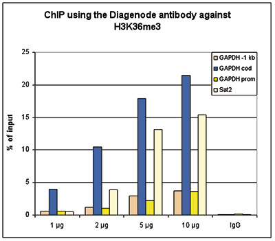
<p><strong>Figure 1. ChIP results obtained with the Diagenode antibody directed against H3K36me3</strong><br />ChIP assays were performed using HeLa cells, the Diagenode antibody against H3K36me3 (cat. No. C15410058) and optimized PCR primer sets for qPCR. ChIP was performed with the “Auto Histone ChIP-seq” kit (cat. No. C01010022), using sheared chromatin from 1 million cells on the SX-8G IP-Star automated system. A titration consisting of 1, 2, 5 and 10 µg of antibody per ChIP experiment was analyzed. IgG (2 µg/IP) was used as a negative IP control. QPCR was performed with primers for the promoter and coding region of the active GAPDH, for a region located 1 kb upstream of the GAPDH promoter (cat. No. C17011003) and for the Sat2 satellite repeat. Figure 1 shows the recovery, expressed as a % of input (the relative amount of immunoprecipitated DNA compared to input DNA after qPCR analysis).</p>
</div>
</div>
<div class="extra-spaced"></div>
<div class="row">
<div class="small-6 columns">A.<center><img src="https://www.diagenode.com/img/product/antibodies/C15410058_ChIPSeq.jpg" width="350" /></center></div>
<div class="small-6 columns">
<p><strong></strong></p>
</div>
</div>
<div class="row">
<div class="small-12 columns">B.<center><img src="https://www.diagenode.com/img/product/antibodies/C15410058_ChIPSeqB.jpg" /></center><br />C.<center><img src="https://www.diagenode.com/img/product/antibodies/C15410058_ChIPSeqC.jpg" /></center><br />D.<center><img src="https://www.diagenode.com/img/product/antibodies/C15410058_ChIPSeqD.jpg" /></center><br />E.<center><img src="https://www.diagenode.com/img/product/antibodies/C15410058_ChIPSeqE.jpg" /></center></div>
</div>
<div class="row">
<div class="small-12 columns">
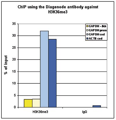
<p><strong>Figure 2. ChIP-seq results obtained with the Diagenode antibody directed against H3K36me3</strong><br />ChIP was performed with 2 µg of the Diagenode antibody against H3K36me3 (cat. No. C15410058) on sheared chromatin from 1 million HeLaS3 cells using the “iDeal ChIP-seq” kit (cat. No. C01010051). IgG (2 µg/IP) was used as a negative IP control. The IP'd DNA was analysed by QPCR with optimized PCR primer pairs for the promoter and coding region of the active GAPDH, for a region located 1 kb upstream of the GAPDH promoter and for the coding region of the active ACTB gene (figure 2A). The IP'd DNA was subsequently analysed on an Illumina Genome Analyzer. Library preparation, cluster generation and sequencing were performed according to the manufacturer's instructions. The 36 bp tags were aligned to the human genome using the ELAND algorithm. Figure 2B shows the obtained profiles in genomic regions of chromosome 12 (including the GAPDH positive control), 7 (including the ACTB positive control), 14 and 3, respectively. These results clearly show an enrichment of the H3K36me3 at active genes.</p>
</div>
</div>
<div class="row">
<div class="small-4 columns"><center><img src="https://www.diagenode.com/img/product/antibodies/C15410058_ELISA.jpg" /></center></div>
<div class="small-8 columns">
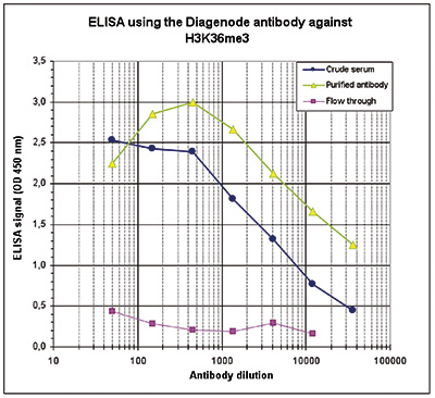
<p><strong>Figure 3. Determination of the antibody titer </strong><br />To determine the titer of the antibody, an ELISA was performed using a serial dilution of the Diagenode antibody directed against H3K36me3 (cat. No. C15410058) and the crude serum. The antigen used was a peptide containing the histone modification of interest. By plotting the absorbance against the antibody dilution (Figure 3), the titer of the purified antibody was estimated to be 1:19,300.</p>
</div>
</div>
<div class="row">
<div class="small-4 columns"><center><img src="https://www.diagenode.com/img/product/antibodies/C15410058_DotBlot.jpg" /></center></div>
<div class="small-8 columns">
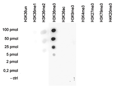
<p><strong>Figure 4. Cross reactivity tests using the Diagenode antibody directed against H3K36me3</strong><br />A Dot Blot analysis was performed to test the cross reactivity of the Diagenode antibody against H3K36me3 (cat. No. C15410058) with peptides containing other H3 and H4 modifications and the unmodified sequence. One hundred to 0.2 pmol of peptide containing the respective histone modification were spotted on a membrane. The antibody was used at a dilution of 1:20,000. Figure 4 shows a high specificity of the antibody for the modification of interest.</p>
</div>
</div>
<div class="row">
<div class="small-4 columns"><center><img src="https://www.diagenode.com/img/product/antibodies/C15410058_WB.jpg" /></center></div>
<div class="small-8 columns">
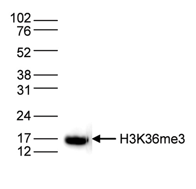
<p><strong>Figure 5. Western blot analysis using the Diagenode antibody directed against H3K36me3</strong><br />Histone extracts (15 µg) from HeLa cells were analysed by Western blot using the Diagenode antibody directed against H3K36me3 (cat. No. C15410058) diluted 1:1,000 in TBS-Tween containing 5% skimmed milk. The position of the protein of interest is indicated on the right; the marker (in kDa) is shown on the left.</p>
</div>
</div>',
'label2' => 'Target Description',
'info2' => '<p>Histones are the main constituents of the protein part of chromosomes of eukaryotic cells. They are rich in the amino acids arginine and lysine and have been greatly conserved during evolution. Histones pack the DNA into tight masses of chromatin. Two core histones of each class H2A, H2B, H3 and H4 assemble and are wrapped by 146 base pairs of DNA to form one octameric nucleosome. Histone tails undergo numerous post-translational modifications, which either directly or indirectly alter chromatin structure to facilitate transcriptional activation or repression or other nuclear processes. In addition to the genetic code, combinations of the different histone modifications reveal the so-called “histone code”. Histone methylation and demethylation is dynamically regulated by respectively histone methyl transferases and histone demethylases. Trimethylation of H3K36 is associated with actively transcribed regions.</p>',
'label3' => '',
'info3' => '',
'format' => '50 µg',
'catalog_number' => 'C15410058',
'old_catalog_number' => 'pAb-058-050',
'sf_code' => 'C15410058-D001-000581',
'type' => 'FRE',
'search_order' => '03-Antibody',
'price_EUR' => '380',
'price_USD' => '380',
'price_GBP' => '340',
'price_JPY' => '59525',
'price_CNY' => '',
'price_AUD' => '950',
'country' => 'ALL',
'except_countries' => 'None',
'quote' => false,
'in_stock' => false,
'featured' => false,
'no_promo' => false,
'online' => true,
'master' => true,
'last_datasheet_update' => '0000-00-00',
'slug' => 'h3k36me3-polyclonal-antibody-classic-50-ug-56-ul',
'meta_title' => 'H3K36me3 Antibody - ChIP-seq Grade (C15410058) | Diagenode',
'meta_keywords' => '',
'meta_description' => 'H3K36me3 (Histone H3 trimethylated at lysine 36) Polyclonal Antibody validated in ChIP-seq, ChIP-garde, ELISA, DB and WB. Batch-specific data available on the website. Sample size available.',
'modified' => '2021-12-23 11:28:57',
'created' => '2015-06-29 14:08:20',
'locale' => 'eng'
),
'Antibody' => array(
'host' => '*****',
'id' => '72',
'name' => 'H3K36me3 polyclonal antibody',
'description' => 'Histones are the main constituents of the protein part of chromosomes of eukaryotic cells. They are rich in the amino acids arginine and lysine and have been greatly conserved during evolution. Histones pack the DNA into tight masses of chromatin. Two core histones of each class H2A, H2B, H3 and H4 assemble and are wrapped by 146 base pairs of DNA to form one octameric nucleosome. Histone tails undergo numerous post-translational modifications, which either directly or indirectly alter chromatin structure to facilitate transcriptional activation or repression or other nuclear processes. In addition to the genetic code, combinations of the different histone modifications reveal the so-called “histone code”. Histone methylation and demethylation is dynamically regulated by respectively histone methyl transferases and histone demethylases. Trimethylation of H3K36 is associated with actively transcribed regions.',
'clonality' => '',
'isotype' => '',
'lot' => 'A8889-001P',
'concentration' => '0.9 µg/µl',
'reactivity' => 'Human, mouse, zebrafish, Drosophila: positive. Other species: not tested. ',
'type' => 'Polyclonal',
'purity' => 'Affinity purified',
'classification' => '',
'application_table' => '<table>
<thead>
<tr>
<th>Applications</th>
<th>Suggested dilution</th>
<th>References</th>
</tr>
</thead>
<tbody>
<tr>
<td>ChIP/ChIP-seq <sup>*</sup></td>
<td>1-2 μg/ChIP</td>
<td>Fig 1, 2</td>
</tr>
<tr>
<td>ELISA</td>
<td>1:500</td>
<td>Fig 3</td>
</tr>
<tr>
<td>Dot Blotting</td>
<td>1:20,000</td>
<td>Fig 4</td>
</tr>
<tr>
<td>Western Blotting</td>
<td>1:1,000</td>
<td>Fig 5</td>
</tr>
</tbody>
</table>
<p><small><sup>*</sup> Please note that the optimal antibody amount per IP should be determined by the end-user. We recommend testing 1-5 μg per IP.</small></p>',
'storage_conditions' => 'Store at -20°C; for long storage, store at -80°C. Avoid multiple freeze-thaw cycles.',
'storage_buffer' => 'PBS containing 0.05% azide and 0.05% ProClin 300.',
'precautions' => 'This product is for research use only. Not for use in diagnostic or therapeutic procedures.',
'uniprot_acc' => '',
'slug' => '',
'meta_keywords' => '',
'meta_description' => '',
'modified' => '2020-12-04 12:56:18',
'created' => '0000-00-00 00:00:00',
'select_label' => '72 - H3K36me3 polyclonal antibody (A8889-001P - 0.9 µg/µl - Human, mouse, zebrafish, Drosophila: positive. Other species: not tested. - Affinity purified - Rabbit)'
),
'Slave' => array(
(int) 0 => array(
[maximum depth reached]
)
),
'Group' => array(
'Group' => array(
[maximum depth reached]
),
'Master' => array(
[maximum depth reached]
),
'Product' => array(
[maximum depth reached]
)
),
'Related' => array(),
'Application' => array(
(int) 0 => array(
[maximum depth reached]
),
(int) 1 => array(
[maximum depth reached]
),
(int) 2 => array(
[maximum depth reached]
),
(int) 3 => array(
[maximum depth reached]
),
(int) 4 => array(
[maximum depth reached]
)
),
'Category' => array(
(int) 0 => array(
[maximum depth reached]
),
(int) 1 => array(
[maximum depth reached]
),
(int) 2 => array(
[maximum depth reached]
),
(int) 3 => array(
[maximum depth reached]
)
),
'Document' => array(
(int) 0 => array(
[maximum depth reached]
),
(int) 1 => array(
[maximum depth reached]
),
(int) 2 => array(
[maximum depth reached]
)
),
'Feature' => array(),
'Image' => array(
(int) 0 => array(
[maximum depth reached]
)
),
'Promotion' => array(),
'Protocol' => array(),
'Publication' => array(
(int) 0 => array(
[maximum depth reached]
),
(int) 1 => array(
[maximum depth reached]
),
(int) 2 => array(
[maximum depth reached]
),
(int) 3 => array(
[maximum depth reached]
),
(int) 4 => array(
[maximum depth reached]
),
(int) 5 => array(
[maximum depth reached]
),
(int) 6 => array(
[maximum depth reached]
),
(int) 7 => array(
[maximum depth reached]
),
(int) 8 => array(
[maximum depth reached]
),
(int) 9 => array(
[maximum depth reached]
),
(int) 10 => array(
[maximum depth reached]
),
(int) 11 => array(
[maximum depth reached]
)
),
'Testimonial' => array(),
'Area' => array(),
'SafetySheet' => array(
(int) 0 => array(
[maximum depth reached]
),
(int) 1 => array(
[maximum depth reached]
),
(int) 2 => array(
[maximum depth reached]
),
(int) 3 => array(
[maximum depth reached]
),
(int) 4 => array(
[maximum depth reached]
),
(int) 5 => array(
[maximum depth reached]
),
(int) 6 => array(
[maximum depth reached]
),
(int) 7 => array(
[maximum depth reached]
)
)
),
'meta_canonical' => 'https://www.diagenode.com/en/p/h3k36me3-polyclonal-antibody-classic-50-ug-56-ul'
)
$language = 'en'
$meta_keywords = ''
$meta_description = 'H3K36me3 (Histone H3 trimethylated at lysine 36) Polyclonal Antibody validated in ChIP-seq, ChIP-garde, ELISA, DB and WB. Batch-specific data available on the website. Sample size available.'
$meta_title = 'H3K36me3 Antibody - ChIP-seq Grade (C15410058) | Diagenode'
$product = array(
'Product' => array(
'id' => '2220',
'antibody_id' => '72',
'name' => 'H3K36me3 Antibody',
'description' => '<p><span>Polyclonal antibody raised in rabbit against histone <strong>H3, trimethylated at lysine 36 (H3K36me3)</strong>, using a KLH-conjugated synthetic peptide.</span></p>',
'label1' => 'Validation Data',
'info1' => '<div class="row">
<div class="small-6 columns"><center><img src="https://www.diagenode.com/img/product/antibodies/C15410058_ChIP.jpg" width="350" /></center></div>
<div class="small-6 columns">
<p><strong>Figure 1. ChIP results obtained with the Diagenode antibody directed against H3K36me3</strong><br />ChIP assays were performed using HeLa cells, the Diagenode antibody against H3K36me3 (cat. No. C15410058) and optimized PCR primer sets for qPCR. ChIP was performed with the “Auto Histone ChIP-seq” kit (cat. No. C01010022), using sheared chromatin from 1 million cells on the SX-8G IP-Star automated system. A titration consisting of 1, 2, 5 and 10 µg of antibody per ChIP experiment was analyzed. IgG (2 µg/IP) was used as a negative IP control. QPCR was performed with primers for the promoter and coding region of the active GAPDH, for a region located 1 kb upstream of the GAPDH promoter (cat. No. C17011003) and for the Sat2 satellite repeat. Figure 1 shows the recovery, expressed as a % of input (the relative amount of immunoprecipitated DNA compared to input DNA after qPCR analysis).</p>
</div>
</div>
<div class="extra-spaced"></div>
<div class="row">
<div class="small-6 columns">A.<center><img src="https://www.diagenode.com/img/product/antibodies/C15410058_ChIPSeq.jpg" width="350" /></center></div>
<div class="small-6 columns">
<p><strong></strong></p>
</div>
</div>
<div class="row">
<div class="small-12 columns">B.<center><img src="https://www.diagenode.com/img/product/antibodies/C15410058_ChIPSeqB.jpg" /></center><br />C.<center><img src="https://www.diagenode.com/img/product/antibodies/C15410058_ChIPSeqC.jpg" /></center><br />D.<center><img src="https://www.diagenode.com/img/product/antibodies/C15410058_ChIPSeqD.jpg" /></center><br />E.<center><img src="https://www.diagenode.com/img/product/antibodies/C15410058_ChIPSeqE.jpg" /></center></div>
</div>
<div class="row">
<div class="small-12 columns">
<p><strong>Figure 2. ChIP-seq results obtained with the Diagenode antibody directed against H3K36me3</strong><br />ChIP was performed with 2 µg of the Diagenode antibody against H3K36me3 (cat. No. C15410058) on sheared chromatin from 1 million HeLaS3 cells using the “iDeal ChIP-seq” kit (cat. No. C01010051). IgG (2 µg/IP) was used as a negative IP control. The IP'd DNA was analysed by QPCR with optimized PCR primer pairs for the promoter and coding region of the active GAPDH, for a region located 1 kb upstream of the GAPDH promoter and for the coding region of the active ACTB gene (figure 2A). The IP'd DNA was subsequently analysed on an Illumina Genome Analyzer. Library preparation, cluster generation and sequencing were performed according to the manufacturer's instructions. The 36 bp tags were aligned to the human genome using the ELAND algorithm. Figure 2B shows the obtained profiles in genomic regions of chromosome 12 (including the GAPDH positive control), 7 (including the ACTB positive control), 14 and 3, respectively. These results clearly show an enrichment of the H3K36me3 at active genes.</p>
</div>
</div>
<div class="row">
<div class="small-4 columns"><center><img src="https://www.diagenode.com/img/product/antibodies/C15410058_ELISA.jpg" /></center></div>
<div class="small-8 columns">
<p><strong>Figure 3. Determination of the antibody titer </strong><br />To determine the titer of the antibody, an ELISA was performed using a serial dilution of the Diagenode antibody directed against H3K36me3 (cat. No. C15410058) and the crude serum. The antigen used was a peptide containing the histone modification of interest. By plotting the absorbance against the antibody dilution (Figure 3), the titer of the purified antibody was estimated to be 1:19,300.</p>
</div>
</div>
<div class="row">
<div class="small-4 columns"><center><img src="https://www.diagenode.com/img/product/antibodies/C15410058_DotBlot.jpg" /></center></div>
<div class="small-8 columns">
<p><strong>Figure 4. Cross reactivity tests using the Diagenode antibody directed against H3K36me3</strong><br />A Dot Blot analysis was performed to test the cross reactivity of the Diagenode antibody against H3K36me3 (cat. No. C15410058) with peptides containing other H3 and H4 modifications and the unmodified sequence. One hundred to 0.2 pmol of peptide containing the respective histone modification were spotted on a membrane. The antibody was used at a dilution of 1:20,000. Figure 4 shows a high specificity of the antibody for the modification of interest.</p>
</div>
</div>
<div class="row">
<div class="small-4 columns"><center><img src="https://www.diagenode.com/img/product/antibodies/C15410058_WB.jpg" /></center></div>
<div class="small-8 columns">
<p><strong>Figure 5. Western blot analysis using the Diagenode antibody directed against H3K36me3</strong><br />Histone extracts (15 µg) from HeLa cells were analysed by Western blot using the Diagenode antibody directed against H3K36me3 (cat. No. C15410058) diluted 1:1,000 in TBS-Tween containing 5% skimmed milk. The position of the protein of interest is indicated on the right; the marker (in kDa) is shown on the left.</p>
</div>
</div>',
'label2' => 'Target Description',
'info2' => '<p>Histones are the main constituents of the protein part of chromosomes of eukaryotic cells. They are rich in the amino acids arginine and lysine and have been greatly conserved during evolution. Histones pack the DNA into tight masses of chromatin. Two core histones of each class H2A, H2B, H3 and H4 assemble and are wrapped by 146 base pairs of DNA to form one octameric nucleosome. Histone tails undergo numerous post-translational modifications, which either directly or indirectly alter chromatin structure to facilitate transcriptional activation or repression or other nuclear processes. In addition to the genetic code, combinations of the different histone modifications reveal the so-called “histone code”. Histone methylation and demethylation is dynamically regulated by respectively histone methyl transferases and histone demethylases. Trimethylation of H3K36 is associated with actively transcribed regions.</p>',
'label3' => '',
'info3' => '',
'format' => '50 µg',
'catalog_number' => 'C15410058',
'old_catalog_number' => 'pAb-058-050',
'sf_code' => 'C15410058-D001-000581',
'type' => 'FRE',
'search_order' => '03-Antibody',
'price_EUR' => '380',
'price_USD' => '380',
'price_GBP' => '340',
'price_JPY' => '59525',
'price_CNY' => '',
'price_AUD' => '950',
'country' => 'ALL',
'except_countries' => 'None',
'quote' => false,
'in_stock' => false,
'featured' => false,
'no_promo' => false,
'online' => true,
'master' => true,
'last_datasheet_update' => '0000-00-00',
'slug' => 'h3k36me3-polyclonal-antibody-classic-50-ug-56-ul',
'meta_title' => 'H3K36me3 Antibody - ChIP-seq Grade (C15410058) | Diagenode',
'meta_keywords' => '',
'meta_description' => 'H3K36me3 (Histone H3 trimethylated at lysine 36) Polyclonal Antibody validated in ChIP-seq, ChIP-garde, ELISA, DB and WB. Batch-specific data available on the website. Sample size available.',
'modified' => '2021-12-23 11:28:57',
'created' => '2015-06-29 14:08:20',
'locale' => 'eng'
),
'Antibody' => array(
'host' => '*****',
'id' => '72',
'name' => 'H3K36me3 polyclonal antibody',
'description' => 'Histones are the main constituents of the protein part of chromosomes of eukaryotic cells. They are rich in the amino acids arginine and lysine and have been greatly conserved during evolution. Histones pack the DNA into tight masses of chromatin. Two core histones of each class H2A, H2B, H3 and H4 assemble and are wrapped by 146 base pairs of DNA to form one octameric nucleosome. Histone tails undergo numerous post-translational modifications, which either directly or indirectly alter chromatin structure to facilitate transcriptional activation or repression or other nuclear processes. In addition to the genetic code, combinations of the different histone modifications reveal the so-called “histone code”. Histone methylation and demethylation is dynamically regulated by respectively histone methyl transferases and histone demethylases. Trimethylation of H3K36 is associated with actively transcribed regions.',
'clonality' => '',
'isotype' => '',
'lot' => 'A8889-001P',
'concentration' => '0.9 µg/µl',
'reactivity' => 'Human, mouse, zebrafish, Drosophila: positive. Other species: not tested. ',
'type' => 'Polyclonal',
'purity' => 'Affinity purified',
'classification' => '',
'application_table' => '<table>
<thead>
<tr>
<th>Applications</th>
<th>Suggested dilution</th>
<th>References</th>
</tr>
</thead>
<tbody>
<tr>
<td>ChIP/ChIP-seq <sup>*</sup></td>
<td>1-2 μg/ChIP</td>
<td>Fig 1, 2</td>
</tr>
<tr>
<td>ELISA</td>
<td>1:500</td>
<td>Fig 3</td>
</tr>
<tr>
<td>Dot Blotting</td>
<td>1:20,000</td>
<td>Fig 4</td>
</tr>
<tr>
<td>Western Blotting</td>
<td>1:1,000</td>
<td>Fig 5</td>
</tr>
</tbody>
</table>
<p><small><sup>*</sup> Please note that the optimal antibody amount per IP should be determined by the end-user. We recommend testing 1-5 μg per IP.</small></p>',
'storage_conditions' => 'Store at -20°C; for long storage, store at -80°C. Avoid multiple freeze-thaw cycles.',
'storage_buffer' => 'PBS containing 0.05% azide and 0.05% ProClin 300.',
'precautions' => 'This product is for research use only. Not for use in diagnostic or therapeutic procedures.',
'uniprot_acc' => '',
'slug' => '',
'meta_keywords' => '',
'meta_description' => '',
'modified' => '2020-12-04 12:56:18',
'created' => '0000-00-00 00:00:00',
'select_label' => '72 - H3K36me3 polyclonal antibody (A8889-001P - 0.9 µg/µl - Human, mouse, zebrafish, Drosophila: positive. Other species: not tested. - Affinity purified - Rabbit)'
),
'Slave' => array(
(int) 0 => array(
'id' => '43',
'name' => 'C15410058',
'product_id' => '2220',
'modified' => '2016-02-18 20:49:07',
'created' => '2016-02-18 20:49:07'
)
),
'Group' => array(
'Group' => array(
'id' => '43',
'name' => 'C15410058',
'product_id' => '2220',
'modified' => '2016-02-18 20:49:07',
'created' => '2016-02-18 20:49:07'
),
'Master' => array(
'id' => '2220',
'antibody_id' => '72',
'name' => 'H3K36me3 Antibody',
'description' => '<p><span>Polyclonal antibody raised in rabbit against histone <strong>H3, trimethylated at lysine 36 (H3K36me3)</strong>, using a KLH-conjugated synthetic peptide.</span></p>',
'label1' => 'Validation Data',
'info1' => '<div class="row">
<div class="small-6 columns"><center><img src="https://www.diagenode.com/img/product/antibodies/C15410058_ChIP.jpg" width="350" /></center></div>
<div class="small-6 columns">
<p><strong>Figure 1. ChIP results obtained with the Diagenode antibody directed against H3K36me3</strong><br />ChIP assays were performed using HeLa cells, the Diagenode antibody against H3K36me3 (cat. No. C15410058) and optimized PCR primer sets for qPCR. ChIP was performed with the “Auto Histone ChIP-seq” kit (cat. No. C01010022), using sheared chromatin from 1 million cells on the SX-8G IP-Star automated system. A titration consisting of 1, 2, 5 and 10 µg of antibody per ChIP experiment was analyzed. IgG (2 µg/IP) was used as a negative IP control. QPCR was performed with primers for the promoter and coding region of the active GAPDH, for a region located 1 kb upstream of the GAPDH promoter (cat. No. C17011003) and for the Sat2 satellite repeat. Figure 1 shows the recovery, expressed as a % of input (the relative amount of immunoprecipitated DNA compared to input DNA after qPCR analysis).</p>
</div>
</div>
<div class="extra-spaced"></div>
<div class="row">
<div class="small-6 columns">A.<center><img src="https://www.diagenode.com/img/product/antibodies/C15410058_ChIPSeq.jpg" width="350" /></center></div>
<div class="small-6 columns">
<p><strong></strong></p>
</div>
</div>
<div class="row">
<div class="small-12 columns">B.<center><img src="https://www.diagenode.com/img/product/antibodies/C15410058_ChIPSeqB.jpg" /></center><br />C.<center><img src="https://www.diagenode.com/img/product/antibodies/C15410058_ChIPSeqC.jpg" /></center><br />D.<center><img src="https://www.diagenode.com/img/product/antibodies/C15410058_ChIPSeqD.jpg" /></center><br />E.<center><img src="https://www.diagenode.com/img/product/antibodies/C15410058_ChIPSeqE.jpg" /></center></div>
</div>
<div class="row">
<div class="small-12 columns">
<p><strong>Figure 2. ChIP-seq results obtained with the Diagenode antibody directed against H3K36me3</strong><br />ChIP was performed with 2 µg of the Diagenode antibody against H3K36me3 (cat. No. C15410058) on sheared chromatin from 1 million HeLaS3 cells using the “iDeal ChIP-seq” kit (cat. No. C01010051). IgG (2 µg/IP) was used as a negative IP control. The IP'd DNA was analysed by QPCR with optimized PCR primer pairs for the promoter and coding region of the active GAPDH, for a region located 1 kb upstream of the GAPDH promoter and for the coding region of the active ACTB gene (figure 2A). The IP'd DNA was subsequently analysed on an Illumina Genome Analyzer. Library preparation, cluster generation and sequencing were performed according to the manufacturer's instructions. The 36 bp tags were aligned to the human genome using the ELAND algorithm. Figure 2B shows the obtained profiles in genomic regions of chromosome 12 (including the GAPDH positive control), 7 (including the ACTB positive control), 14 and 3, respectively. These results clearly show an enrichment of the H3K36me3 at active genes.</p>
</div>
</div>
<div class="row">
<div class="small-4 columns"><center><img src="https://www.diagenode.com/img/product/antibodies/C15410058_ELISA.jpg" /></center></div>
<div class="small-8 columns">
<p><strong>Figure 3. Determination of the antibody titer </strong><br />To determine the titer of the antibody, an ELISA was performed using a serial dilution of the Diagenode antibody directed against H3K36me3 (cat. No. C15410058) and the crude serum. The antigen used was a peptide containing the histone modification of interest. By plotting the absorbance against the antibody dilution (Figure 3), the titer of the purified antibody was estimated to be 1:19,300.</p>
</div>
</div>
<div class="row">
<div class="small-4 columns"><center><img src="https://www.diagenode.com/img/product/antibodies/C15410058_DotBlot.jpg" /></center></div>
<div class="small-8 columns">
<p><strong>Figure 4. Cross reactivity tests using the Diagenode antibody directed against H3K36me3</strong><br />A Dot Blot analysis was performed to test the cross reactivity of the Diagenode antibody against H3K36me3 (cat. No. C15410058) with peptides containing other H3 and H4 modifications and the unmodified sequence. One hundred to 0.2 pmol of peptide containing the respective histone modification were spotted on a membrane. The antibody was used at a dilution of 1:20,000. Figure 4 shows a high specificity of the antibody for the modification of interest.</p>
</div>
</div>
<div class="row">
<div class="small-4 columns"><center><img src="https://www.diagenode.com/img/product/antibodies/C15410058_WB.jpg" /></center></div>
<div class="small-8 columns">
<p><strong>Figure 5. Western blot analysis using the Diagenode antibody directed against H3K36me3</strong><br />Histone extracts (15 µg) from HeLa cells were analysed by Western blot using the Diagenode antibody directed against H3K36me3 (cat. No. C15410058) diluted 1:1,000 in TBS-Tween containing 5% skimmed milk. The position of the protein of interest is indicated on the right; the marker (in kDa) is shown on the left.</p>
</div>
</div>',
'label2' => 'Target Description',
'info2' => '<p>Histones are the main constituents of the protein part of chromosomes of eukaryotic cells. They are rich in the amino acids arginine and lysine and have been greatly conserved during evolution. Histones pack the DNA into tight masses of chromatin. Two core histones of each class H2A, H2B, H3 and H4 assemble and are wrapped by 146 base pairs of DNA to form one octameric nucleosome. Histone tails undergo numerous post-translational modifications, which either directly or indirectly alter chromatin structure to facilitate transcriptional activation or repression or other nuclear processes. In addition to the genetic code, combinations of the different histone modifications reveal the so-called “histone code”. Histone methylation and demethylation is dynamically regulated by respectively histone methyl transferases and histone demethylases. Trimethylation of H3K36 is associated with actively transcribed regions.</p>',
'label3' => '',
'info3' => '',
'format' => '50 µg',
'catalog_number' => 'C15410058',
'old_catalog_number' => 'pAb-058-050',
'sf_code' => 'C15410058-D001-000581',
'type' => 'FRE',
'search_order' => '03-Antibody',
'price_EUR' => '380',
'price_USD' => '380',
'price_GBP' => '340',
'price_JPY' => '59525',
'price_CNY' => '',
'price_AUD' => '950',
'country' => 'ALL',
'except_countries' => 'None',
'quote' => false,
'in_stock' => false,
'featured' => false,
'no_promo' => false,
'online' => true,
'master' => true,
'last_datasheet_update' => '0000-00-00',
'slug' => 'h3k36me3-polyclonal-antibody-classic-50-ug-56-ul',
'meta_title' => 'H3K36me3 Antibody - ChIP-seq Grade (C15410058) | Diagenode',
'meta_keywords' => '',
'meta_description' => 'H3K36me3 (Histone H3 trimethylated at lysine 36) Polyclonal Antibody validated in ChIP-seq, ChIP-garde, ELISA, DB and WB. Batch-specific data available on the website. Sample size available.',
'modified' => '2021-12-23 11:28:57',
'created' => '2015-06-29 14:08:20'
),
'Product' => array(
(int) 0 => array(
[maximum depth reached]
)
)
),
'Related' => array(),
'Application' => array(
(int) 0 => array(
'id' => '20',
'position' => '10',
'parent_id' => '40',
'name' => 'ELISA',
'description' => '<div class="row">
<div class="small-12 medium-12 large-12 columns">Enzyme-linked immunosorbent assay.</div>
</div>',
'in_footer' => false,
'in_menu' => false,
'online' => true,
'tabular' => true,
'slug' => 'elisa-antibodies',
'meta_keywords' => ' ELISA Antibodies,Monoclonal antibody, Polyclonal antibody',
'meta_description' => 'Diagenode offers Monoclonal & Polyclonal antibodies for ELISA applications',
'meta_title' => 'ELISA Antibodies - Monoclonal & Polyclonal antibody | Diagenode',
'modified' => '2016-01-13 12:21:41',
'created' => '2014-07-08 08:13:28',
'ProductsApplication' => array(
[maximum depth reached]
)
),
(int) 1 => array(
'id' => '28',
'position' => '10',
'parent_id' => '40',
'name' => 'DB',
'description' => '<p>Dot blotting</p>',
'in_footer' => false,
'in_menu' => false,
'online' => true,
'tabular' => true,
'slug' => 'dot-blotting',
'meta_keywords' => 'Dot blotting,Monoclonal & Polyclonal antibody,',
'meta_description' => 'Diagenode offers Monoclonal & Polyclonal antibodies for Dot blotting applications',
'meta_title' => 'Dot blotting Antibodies - Monoclonal & Polyclonal antibody | Diagenode',
'modified' => '2016-01-13 14:40:49',
'created' => '2015-07-08 13:45:05',
'ProductsApplication' => array(
[maximum depth reached]
)
),
(int) 2 => array(
'id' => '19',
'position' => '10',
'parent_id' => '40',
'name' => 'WB',
'description' => '<p><strong>Western blot</strong> : The quality of antibodies used in this technique is crucial for correct and specific protein identification. Diagenode offers huge selection of highly sensitive and specific western blot-validated antibodies.</p>
<p>Learn more about: <a href="https://www.diagenode.com/applications/western-blot">Loading control, MW marker visualization</a><em>. <br /></em></p>
<p><em></em>Check our selection of antibodies validated in Western blot.</p>',
'in_footer' => false,
'in_menu' => false,
'online' => true,
'tabular' => true,
'slug' => 'western-blot-antibodies',
'meta_keywords' => ' Western Blot Antibodies ,western blot protocol,Western Blotting Products,Polyclonal antibodies ,monoclonal antibodies ',
'meta_description' => 'Diagenode offers a wide range of antibodies and technical support for western blot applications',
'meta_title' => ' Western Blot - Monoclonal antibody - Polyclonal antibody | Diagenode',
'modified' => '2016-04-26 12:44:51',
'created' => '2015-01-07 09:20:00',
'ProductsApplication' => array(
[maximum depth reached]
)
),
(int) 3 => array(
'id' => '42',
'position' => '10',
'parent_id' => '40',
'name' => 'ChIP-seq (ab)',
'description' => '',
'in_footer' => false,
'in_menu' => false,
'online' => true,
'tabular' => true,
'slug' => 'chip-seq-antibodies',
'meta_keywords' => 'Chromatin Immunoprecipitation Sequencing,ChIP-Seq,ChIP-seq grade antibodies,DNA purification,qPCR,Shearing of chromatin',
'meta_description' => 'Diagenode offers a wide range of antibodies and technical support for ChIP Sequencing applications',
'meta_title' => 'ChIP Sequencing Antibodies (ChIP-Seq) | Diagenode',
'modified' => '2016-01-20 11:06:19',
'created' => '2015-10-20 11:44:45',
'ProductsApplication' => array(
[maximum depth reached]
)
),
(int) 4 => array(
'id' => '43',
'position' => '10',
'parent_id' => '40',
'name' => 'ChIP-qPCR (ab)',
'description' => '',
'in_footer' => false,
'in_menu' => false,
'online' => true,
'tabular' => true,
'slug' => 'chip-qpcr-antibodies',
'meta_keywords' => 'Chromatin Immunoprecipitation Sequencing,ChIP-Seq,ChIP-seq grade antibodies,DNA purification,qPCR,Shearing of chromatin',
'meta_description' => 'Diagenode offers a wide range of antibodies and technical support for ChIP-qPCR applications',
'meta_title' => 'ChIP Quantitative PCR Antibodies (ChIP-qPCR) | Diagenode',
'modified' => '2016-01-20 11:30:24',
'created' => '2015-10-20 11:45:36',
'ProductsApplication' => array(
[maximum depth reached]
)
)
),
'Category' => array(
(int) 0 => array(
'id' => '17',
'position' => '10',
'parent_id' => '4',
'name' => 'ChIP-seq grade antibodies',
'description' => '<p><b>Unparalleled ChIP-Seq results with the most rigorously validated antibodies</b></p>
<p><span style="font-weight: 400;">Diagenode provides leading solutions for epigenetic research. Because ChIP-seq is a widely-used technique, we validate our antibodies in ChIP and ChIP-seq experiments (in addition to conventional methods like Western blot, Dot blot, ELISA, and immunofluorescence) to provide the highest quality antibody. We standardize our validation and production to guarantee high product quality without technical bias. Diagenode guarantees ChIP-seq grade antibody performance under our suggested conditions.</span></p>
<div class="row">
<div class="small-12 medium-9 large-9 columns">
<p><strong>ChIP-seq profile</strong> of active (H3K4me3 and H3K36me3) and inactive (H3K27me3) marks using Diagenode antibodies.</p>
<img src="https://www.diagenode.com/img/categories/antibodies/chip-seq-grade-antibodies.png" /></div>
<div class="small-12 medium-3 large-3 columns">
<p><small> ChIP was performed on sheared chromatin from 100,000 K562 cells using iDeal ChIP-seq kit for Histones (cat. No. C01010051) with 1 µg of the Diagenode antibodies against H3K27me3 (cat. No. C15410195) and H3K4me3 (cat. No. C15410003), and 0.5 µg of the antibody against H3K36me3 (cat. No. C15410192). The IP'd DNA was subsequently analysed on an Illumina Genome Analyzer. Library preparation, cluster generation and sequencing were performed according to the manufacturer's instructions. The 36 bp tags were aligned to the human genome using the ELAND algorithm. The figure shows the signal distribution along the complete sequence of human chromosome 3, a zoomin to a 10 Mb region and a further zoomin to a 1.5 Mb region. </small></p>
</div>
</div>
<p>Diagenode’s highly validated antibodies:</p>
<ul>
<li>Highly sensitive and specific</li>
<li>Cost-effective (requires less antibody per reaction)</li>
<li>Batch-specific data is available on the website</li>
<li>Expert technical support</li>
<li>Sample sizes available</li>
<li>100% satisfaction guarantee</li>
</ul>',
'no_promo' => false,
'in_menu' => true,
'online' => true,
'tabular' => false,
'hide' => true,
'all_format' => false,
'is_antibody' => true,
'slug' => 'chip-seq-grade-antibodies',
'cookies_tag_id' => null,
'meta_keywords' => 'ChIP-seq grade antibodies,polyclonal antibody,WB, ELISA, ChIP-seq (ab), ChIP-qPCR (ab)',
'meta_description' => 'Diagenode Offers Wide Range of Validated ChIP-Seq Grade Antibodies for Unparalleled ChIP-Seq Results',
'meta_title' => 'Chromatin Immunoprecipitation ChIP-Seq Grade Antibodies | Diagenode',
'modified' => '2019-07-03 10:57:22',
'created' => '2015-02-16 02:24:01',
'ProductsCategory' => array(
[maximum depth reached]
),
'CookiesTag' => array([maximum depth reached])
),
(int) 1 => array(
'id' => '111',
'position' => '40',
'parent_id' => '4',
'name' => 'Histone antibodies',
'description' => '<p>Histones are the main protein components of chromatin involved in the compaction of DNA into nucleosomes, the basic units of chromatin. A <strong>nucleosome</strong> consists of one pair of each of the core histones (<strong>H2A</strong>, <strong>H2B</strong>, <strong>H3</strong> and <strong>H4</strong>) forming an octameric structure wrapped by 146 base pairs of DNA. The different nucleosomes are linked by the linker histone<strong> H1, </strong>allowing for further condensation of chromatin.</p>
<p>The core histones have a globular structure with large unstructured N-terminal tails protruding from the nucleosome. They can undergo to multiple post-translational modifications (PTM), mainly at the N-terminal tails. These <strong>post-translational modifications </strong>include methylation, acetylation, phosphorylation, ubiquitinylation, citrullination, sumoylation, deamination and crotonylation. The most well characterized PTMs are <strong>methylation,</strong> <strong>acetylation and phosphorylation</strong>. Histone methylation occurs mainly on lysine (K) residues, which can be mono-, di- or tri-methylated, and on arginines (R), which can be mono-methylated and symmetrically or asymmetrically di-methylated. Histone acetylation occurs on lysines and histone phosphorylation mainly on serines (S), threonines (T) and tyrosines (Y).</p>
<p>The PTMs of the different residues are involved in numerous processes such as DNA repair, DNA replication and chromosome condensation. They influence the chromatin organization and can be positively or negatively associated with gene expression. Trimethylation of H3K4, H3K36 and H3K79, and lysine acetylation generally result in an open chromatin configuration (figure below) and are therefore associated with <strong>euchromatin</strong> and gene activation. Trimethylation of H3K9, K3K27 and H4K20, on the other hand, is enriched in <strong>heterochromatin </strong>and associated with gene silencing. The combination of different histone modifications is called the "<strong>histone code</strong>”, analogous to the genetic code.</p>
<p><img src="https://www.diagenode.com/img/categories/antibodies/histone-marks-illustration.png" /></p>
<p>Diagenode is proud to offer a large range of antibodies against histones and histone modifications. Our antibodies are highly specific and have been validated in many applications, including <strong>ChIP</strong> and <strong>ChIP-seq</strong>.</p>
<p>Diagenode’s collection includes antibodies recognizing:</p>
<ul>
<li><strong>Histone H1 variants</strong></li>
<li><strong>Histone H2A, H2A variants and histone H2A</strong> <strong>modifications</strong> (serine phosphorylation, lysine acetylation, lysine ubiquitinylation)</li>
<li><strong>Histone H2B and H2B</strong> <strong>modifications </strong>(serine phosphorylation, lysine acetylation)</li>
<li><strong>Histone H3 and H3 modifications </strong>(lysine methylation (mono-, di- and tri-methylated), lysine acetylation, serine phosphorylation, threonine phosphorylation, arginine methylation (mono-methylated, symmetrically and asymmetrically di-methylated))</li>
<li><strong>Histone H4 and H4 modifications (</strong>lysine methylation (mono-, di- and tri-methylated), lysine acetylation, arginine methylation (mono-methylated and symmetrically di-methylated), serine phosphorylation )</li>
</ul>
<p><span style="font-weight: 400;"><strong>HDAC's HAT's, HMT's and other</strong> <strong>enzymes</strong> which modify histones can be found in the category <a href="../categories/chromatin-modifying-proteins-histone-transferase">Histone modifying enzymes</a><br /></span></p>
<p><span style="font-weight: 400;"> Diagenode’s highly validated antibodies:</span></p>
<ul>
<li><span style="font-weight: 400;"> Highly sensitive and specific</span></li>
<li><span style="font-weight: 400;"> Cost-effective (requires less antibody per reaction)</span></li>
<li><span style="font-weight: 400;"> Batch-specific data is available on the website</span></li>
<li><span style="font-weight: 400;"> Expert technical support</span></li>
<li><span style="font-weight: 400;"> Sample sizes available</span></li>
<li><span style="font-weight: 400;"> 100% satisfaction guarantee</span></li>
</ul>',
'no_promo' => false,
'in_menu' => false,
'online' => true,
'tabular' => false,
'hide' => true,
'all_format' => false,
'is_antibody' => true,
'slug' => 'histone-antibodies',
'cookies_tag_id' => null,
'meta_keywords' => 'Histone, antibody, histone h1, histone h2, histone h3, histone h4',
'meta_description' => 'Polyclonal and Monoclonal Antibodies against Histones and their modifications validated for many applications, including Chromatin Immunoprecipitation (ChIP) and ChIP-Sequencing (ChIP-seq)',
'meta_title' => 'Histone and Modified Histone Antibodies | Diagenode',
'modified' => '2020-09-17 13:34:56',
'created' => '2016-04-01 16:01:32',
'ProductsCategory' => array(
[maximum depth reached]
),
'CookiesTag' => array([maximum depth reached])
),
(int) 2 => array(
'id' => '103',
'position' => '0',
'parent_id' => '4',
'name' => 'All antibodies',
'description' => '<p><span style="font-weight: 400;">All Diagenode’s antibodies are listed below. Please, use our Quick search field to find the antibody of interest by target name, application, purity.</span></p>
<p><span style="font-weight: 400;">Diagenode’s highly validated antibodies:</span></p>
<ul>
<li>Highly sensitive and specific</li>
<li>Cost-effective (requires less antibody per reaction)</li>
<li>Batch-specific data is available on the website</li>
<li>Expert technical support</li>
<li>Sample sizes available</li>
<li>100% satisfaction guarantee</li>
</ul>',
'no_promo' => false,
'in_menu' => true,
'online' => true,
'tabular' => false,
'hide' => true,
'all_format' => false,
'is_antibody' => true,
'slug' => 'all-antibodies',
'cookies_tag_id' => null,
'meta_keywords' => 'Antibodies,Premium Antibodies,Classic,Pioneer',
'meta_description' => 'Diagenode Offers Strict quality standards with Rigorous QC and validated Antibodies. Classified based on level of validation for flexibility of Application. Comprehensive selection of histone and non-histone Antibodies',
'meta_title' => 'Diagenode's selection of Antibodies is exclusively dedicated for Epigenetic Research | Diagenode',
'modified' => '2019-07-03 10:55:44',
'created' => '2015-11-02 14:49:22',
'ProductsCategory' => array(
[maximum depth reached]
),
'CookiesTag' => array([maximum depth reached])
),
(int) 3 => array(
'id' => '127',
'position' => '10',
'parent_id' => '4',
'name' => 'ChIP-grade antibodies',
'description' => '<div class="row">
<div class="small-12 columns"><center></center>
<p><br />Chromatin immunoprecipitation (<b>ChIP</b>) is a technique to study the associations of proteins with the specific genomic regions in intact cells. One of the most important steps of this protocol is the immunoprecipitation of targeted protein using the antibody specifically recognizing it. The quality of antibodies used in ChIP is essential for the success of the experiment. Diagenode offers extensively validated ChIP-grade antibodies, confirmed for their specificity, and high level of performance in ChIP. Each batch is validated, and batch-specific data are available on the website.</p>
<p></p>
</div>
</div>
<p><strong>ChIP results</strong> obtained with the antibody directed against H3K4me3 (Cat. No. <a href="../p/h3k4me3-polyclonal-antibody-premium-50-ug-50-ul">C15410003</a>). </p>
<div class="row">
<div class="small-12 medium-6 large-6 columns"><img src="https://www.diagenode.com/img/product/antibodies/C15410003-fig1-ChIP.jpg" alt="" width="400" height="315" /> </div>
<div class="small-12 medium-6 large-6 columns">
<p></p>
<p></p>
<p></p>
</div>
</div>
<p></p>
<p>Our aim at Diagenode is to offer the largest collection of highly specific <strong>ChIP-grade antibodies</strong>. We add new antibodies monthly. Find your ChIP-grade antibody in the list below and check more information about tested applications, extensive validation data, and product information.</p>',
'no_promo' => false,
'in_menu' => true,
'online' => true,
'tabular' => false,
'hide' => true,
'all_format' => false,
'is_antibody' => true,
'slug' => 'chip-grade-antibodies',
'cookies_tag_id' => null,
'meta_keywords' => 'ChIP-grade antibodies, polyclonal antibody, monoclonal antibody, Diagenode',
'meta_description' => 'Diagenode Offers Extensively Validated ChIP-Grade Antibodies, Confirmed for their Specificity, and high level of Performance in Chromatin Immunoprecipitation ChIP',
'meta_title' => 'Chromatin immunoprecipitation ChIP-grade antibodies | Diagenode',
'modified' => '2024-11-19 17:27:07',
'created' => '2017-02-14 11:16:04',
'ProductsCategory' => array(
[maximum depth reached]
),
'CookiesTag' => array([maximum depth reached])
)
),
'Document' => array(
(int) 0 => array(
'id' => '712',
'name' => 'Datasheet H3K36me3 C15410058',
'description' => '<p>Datasheet description</p>',
'image_id' => null,
'type' => 'Datasheet',
'url' => 'files/products/antibodies/Datasheet_H3K36me3_C15410058.pdf',
'slug' => 'datasheet-h3k36me3-C15410058',
'meta_keywords' => '',
'meta_description' => '',
'modified' => '2015-11-23 17:19:06',
'created' => '2015-07-07 11:47:44',
'ProductsDocument' => array(
[maximum depth reached]
)
),
(int) 1 => array(
'id' => '11',
'name' => 'Antibodies you can trust',
'description' => '<p style="text-align: justify;"><span>Epigenetic research tools have evolved over time from endpoint PCR to qPCR to the analyses of large sets of genome-wide sequencing data. ChIP sequencing (ChIP-seq) has now become the gold standard method for chromatin studies, given the accuracy and coverage scale of the approach over other methods. Successful ChIP-seq, however, requires a higher level of experimental accuracy and consistency in all steps of ChIP than ever before. Particularly crucial is the quality of ChIP antibodies. </span></p>',
'image_id' => null,
'type' => 'Poster',
'url' => 'files/posters/Antibodies_you_can_trust_Poster.pdf',
'slug' => 'antibodies-you-can-trust-poster',
'meta_keywords' => '',
'meta_description' => '',
'modified' => '2015-10-01 20:18:31',
'created' => '2015-07-03 16:05:15',
'ProductsDocument' => array(
[maximum depth reached]
)
),
(int) 2 => array(
'id' => '38',
'name' => 'Epigenetic Antibodies Brochure',
'description' => '<p>More than in any other immuoprecipitation assays, quality antibodies are critical tools in many epigenetics experiments. Since 10 years, Diagenode has developed the most stringent quality production available on the market for antibodies exclusively focused on epigenetic uses. All our antibodies have been qualified to work in epigenetic applications.</p>',
'image_id' => null,
'type' => 'Brochure',
'url' => 'files/brochures/Epigenetic_Antibodies_Brochure.pdf',
'slug' => 'epigenetic-antibodies-brochure',
'meta_keywords' => '',
'meta_description' => '',
'modified' => '2016-06-15 11:24:06',
'created' => '2015-07-03 16:05:27',
'ProductsDocument' => array(
[maximum depth reached]
)
)
),
'Feature' => array(),
'Image' => array(
(int) 0 => array(
'id' => '1783',
'name' => 'product/antibodies/chipseq-grade-ab-icon.png',
'alt' => 'ChIP-seq Grade',
'modified' => '2020-11-27 07:04:40',
'created' => '2018-03-15 15:54:09',
'ProductsImage' => array(
[maximum depth reached]
)
)
),
'Promotion' => array(),
'Protocol' => array(),
'Publication' => array(
(int) 0 => array(
'id' => '4763',
'name' => 'Chromatin profiling identifies transcriptional readthrough as a conservedmechanism for piRNA biogenesis in mosquitoes.',
'authors' => 'Qu J. et al.',
'description' => '<p>The piRNA pathway in mosquitoes differs substantially from other model organisms, with an expanded PIWI gene family and functions in antiviral defense. Here, we define core piRNA clusters as genomic loci that show ubiquitous piRNA expression in both somatic and germline tissues. These core piRNA clusters are enriched for non-retroviral endogenous viral elements (nrEVEs) in antisense orientation and depend on key biogenesis factors, Veneno, Tejas, Yb, and Shutdown. Combined transcriptome and chromatin state analyses identify transcriptional readthrough as a conserved mechanism for cluster-derived piRNA biogenesis in the vector mosquitoes Aedes aegypti, Aedes albopictus, Culex quinquefasciatus, and Anopheles gambiae. Comparative analyses between the two Aedes species suggest that piRNA clusters function as traps for nrEVEs, allowing adaptation to environmental challenges such as virus infection. Our systematic transcriptome and chromatin state analyses lay the foundation for studies of gene regulation, genome evolution, and piRNA function in these important vector species.</p>',
'date' => '2023-03-01',
'pmid' => 'https://www.ncbi.nlm.nih.gov/pubmed/36930642',
'doi' => '10.1016/j.celrep.2023.112257',
'modified' => '2023-04-17 09:12:37',
'created' => '2023-04-14 13:41:22',
'ProductsPublication' => array(
[maximum depth reached]
)
),
(int) 1 => array(
'id' => '4605',
'name' => 'Gene Regulatory Interactions at Lamina-Associated Domains',
'authors' => 'Madsen-Østerbye J. et al.',
'description' => '<p>The nuclear lamina provides a repressive chromatin environment at the nuclear periphery. However, whereas most genes in lamina-associated domains (LADs) are inactive, over ten percent reside in local euchromatic contexts and are expressed. How these genes are regulated and whether they are able to interact with regulatory elements remain unclear. Here, we integrate publicly available enhancer-capture Hi-C data with our own chromatin state and transcriptomic datasets to show that inferred enhancers of active genes in LADs are able to form connections with other enhancers within LADs and outside LADs. Fluorescence in situ hybridization analyses show proximity changes between differentially expressed genes in LADs and distant enhancers upon the induction of adipogenic differentiation. We also provide evidence of involvement of lamin A/C, but not lamin B1, in repressing genes at the border of an in-LAD active region within a topological domain. Our data favor a model where the spatial topology of chromatin at the nuclear lamina is compatible with gene expression in this dynamic nuclear compartment.</p>',
'date' => '2023-01-01',
'pmid' => 'https://doi.org/10.3390%2Fgenes14020334',
'doi' => '10.3390/genes14020334',
'modified' => '2023-04-04 08:57:32',
'created' => '2023-02-21 09:59:46',
'ProductsPublication' => array(
[maximum depth reached]
)
),
(int) 2 => array(
'id' => '4221',
'name' => 'Epigenetic Mechanisms Mediating Cell State Transitions in Chondrocytes',
'authors' => 'Wuelling M. et al.',
'description' => '<p><span>Epigenetic modifications play critical roles in regulating cell lineage differentiation, but the epigenetic mechanisms guiding specific differentiation steps within a cell lineage have rarely been investigated. To decipher such mechanisms, we used the defined transition from proliferating (PC) into hypertrophic chondrocytes (HC) during endochondral ossification as a model. We established a map of activating and repressive histone modifications for each cell type. ChromHMM state transition analysis and Pareto-based integration of differential levels of mRNA and epigenetic marks revealed that differentiation-associated gene repression is initiated by the addition of H3K27me3 to promoters still carrying substantial levels of activating marks. Moreover, the integrative analysis identified genes specifically expressed in cells undergoing the transition into hypertrophy. Investigation of enhancer profiles detected surprising differences in enhancer number, location, and transcription factor binding sites between the two closely related cell types. Furthermore, cell type-specific upregulation of gene expression was associated with increased numbers of H3K27ac peaks. Pathway analysis identified PC-specific enhancers associated with chondrogenic genes, whereas HC-specific enhancers mainly control metabolic pathways linking epigenetic signature to biological functions. Since HC-specific enhancers show a higher conservation in postnatal tissues, the switch to metabolic pathways seems to be a hallmark of differentiated tissues. Surprisingly, the analysis of H3K27ac levels at super-enhancers revealed a rapid adaption of H3K27ac occupancy to changes in gene expression, supporting the importance of enhancer modulation for acute alterations in gene expression. © 2021 The Authors. Journal of Bone and Mineral Research published by Wiley Periodicals LLC on behalf of American Society for Bone and Mineral Research (ASBMR).</span></p>',
'date' => '2022-05-01',
'pmid' => 'https://pubmed.ncbi.nlm.nih.gov/33534175/',
'doi' => '10.1002/jbmr.4263',
'modified' => '2022-04-25 11:46:32',
'created' => '2022-04-21 12:00:53',
'ProductsPublication' => array(
[maximum depth reached]
)
),
(int) 3 => array(
'id' => '4082',
'name' => 'p53 directly represses human LINE1 transposons.',
'authors' => 'Tiwari, Bhavana and Jones, Amanda E and Caillet, Candace J and Das, Simantiand Royer, Stephanie K and Abrams, John M',
'description' => '<p>p53 is a potent tumor suppressor and commonly mutated in human cancers. Recently, we demonstrated that p53 genes act to restrict retrotransposons in germline tissues of flies and fish but whether this activity is conserved in somatic human cells is not known. Here we show that p53 constitutively restrains human LINE1s by cooperatively engaging sites in the 5'UTR and stimulating local deposition of repressive histone marks at these transposons. Consistent with this, the elimination of p53 or the removal of corresponding binding sites in LINE1s, prompted these retroelements to become hyperactive. Concurrently, p53 loss instigated chromosomal rearrangements linked to LINE sequences and also provoked inflammatory programs that were dependent on reverse transcriptase produced from LINE1s. Taken together, our observations establish that p53 continuously operates at the LINE1 promoter to restrict autonomous copies of these mobile elements in human cells. Our results further suggest that constitutive restriction of these retroelements may help to explain tumor suppression encoded by p53, since erupting LINE1s produced acute oncogenic threats when p53 was absent.</p>',
'date' => '2020-10-01',
'pmid' => 'https://www.ncbi.nlm.nih.gov/pubmed/33060137',
'doi' => '10.1101/gad.343186.120',
'modified' => '2021-03-15 16:59:03',
'created' => '2021-02-18 10:21:53',
'ProductsPublication' => array(
[maximum depth reached]
)
),
(int) 4 => array(
'id' => '3994',
'name' => 'Premature termination codons in the gene cause reduced local mRNA synthesis.',
'authors' => 'García-Rodríguez R, Hiller M, Jiménez-Gracia L, van der Pal Z, Balog J, Adamzek K, Aartsma-Rus A, Spitali P',
'description' => '<p>Duchenne muscular dystrophy (DMD) is caused by mutations in the gene leading to the presence of premature termination codons (PTC). Previous transcriptional studies have shown reduced DMD transcript levels in DMD patient and animal model muscles when PTC are present. Nonsense-mediated decay (NMD) has been suggested to be responsible for the observed reduction, but there is no experimental evidence supporting this claim. In this study, we aimed to investigate the mechanism responsible for the drop in expression levels in the presence of PTC. We observed that the inhibition of NMD does not normalize gene expression in DMD. Additionally, in situ hybridization showed that DMD messenger RNA primarily localizes in the nuclear compartment, confirming that a cytoplasmic mechanism like NMD indeed cannot be responsible for the observed reduction. Sequencing of nascent RNA to explore transcription dynamics revealed a lower rate of transcription in patient-derived myotubes compared to healthy controls, suggesting a transcriptional mechanism involved in reduced DMD transcript levels. Chromatin immunoprecipitation in muscle showed increased levels of the repressive histone mark H3K9me3 in mice compared to wild-type mice, indicating a chromatin conformation less prone to transcription in mice. In line with this finding, treatment with the histone deacetylase inhibitor givinostat caused a significant increase in DMD transcript expression in mice. Overall, our findings show that transcription dynamics across the locus are affected by the presence of PTC, hinting at a possible epigenetic mechanism responsible for this process.</p>',
'date' => '2020-07-14',
'pmid' => 'http://www.pubmed.gov/32616572',
'doi' => '10.1073/pnas.1910456117',
'modified' => '2020-09-01 14:54:41',
'created' => '2020-08-21 16:41:39',
'ProductsPublication' => array(
[maximum depth reached]
)
),
(int) 5 => array(
'id' => '1682',
'name' => 'The Elongin Complex Antagonizes the Chromatin Factor Corto for Vein versus Intervein Cell Identity in Drosophila Wings.',
'authors' => 'Rougeot J, Renard M, Randsholt NB, Peronnet F, Mouchel-Vielh E',
'description' => 'Drosophila wings mainly consist of two cell types, vein and intervein cells. Acquisition of either fate depends on specific expression of genes that are controlled by several signaling pathways. The nuclear mechanisms that translate signaling into regulation of gene expression are not completely understood, but they involve chromatin factors from the Trithorax (TrxG) and Enhancers of Trithorax and Polycomb (ETP) families. One of these is the ETP Corto that participates in intervein fate through interaction with the Drosophila EGF Receptor - MAP kinase ERK pathway. Precise mechanisms and molecular targets of Corto in this process are not known. We show here that Corto interacts with the Elongin transcription elongation complex. This complex, that consists of three subunits (Elongin A, B, C), increases RNA polymerase II elongation rate in vitro by suppressing transient pausing. Analysis of phenotypes induced by EloA, B, or C deregulation as well as genetic interactions suggest that the Elongin complex might participate in vein vs intervein specification, and antagonizes corto as well as several TrxG genes in this process. Chromatin immunoprecipitation experiments indicate that Elongin C and Corto bind the vein-promoting gene rhomboid in wing imaginal discs. We propose that Corto and the Elongin complex participate together in vein vs intervein fate, possibly through tissue-specific transcriptional regulation of rhomboid.',
'date' => '2013-10-21',
'pmid' => 'https://www.ncbi.nlm.nih.gov/pubmed/24204884',
'doi' => '',
'modified' => '2015-07-24 15:39:01',
'created' => '2015-07-24 15:39:01',
'ProductsPublication' => array(
[maximum depth reached]
)
),
(int) 6 => array(
'id' => '1420',
'name' => 'Differential transcript isoform usage pre- and post-zygotic genome activation in zebrafish.',
'authors' => 'Aanes H, Østrup O, Andersen IS, Moen LF, Mathavan S, Collas P, Alestrom P',
'description' => 'BACKGROUND: Zebrafish embryos are transcriptionally silent until activation of the zygotic genome during the 10th cell cycle. Onset of transcription is followed by cellular and morphological changes involving cell speciation and gastrulation. Previous genome-wide surveys of transcriptional changes only assessed gene expression levels; however, recent studies have shown the necessity to map isoform-specific transcriptional changes. Here, we perform isoform discovery and quantification on transcriptome sequences from before and after zebrafish zygotic genome activation (ZGA). RESULTS: We identify novel isoforms and isoform switches during ZGA for genes related to cell adhesion, pluripotency and DNA methylation. Isoform switching events include alternative splicing and changes in transcriptional start sites and in 3' untranslated regions. New isoforms are identified even for well-characterized genes such as pou5f1, sall4 and dnmt1. Genes involved in cell-cell interactions such as f11r and magi1 display isoform switches with alterations of coding sequences. We also detect over 1000 transcripts that acquire a longer 3' terminal exon when transcribed by the zygote compared to their maternal transcript counterparts. ChIP-sequencing data mapped onto skipped exon events reveal a correlation between histone H3K36 trimethylation peaks and skipped exons, suggesting epigenetic marks being part of alternative splicing regulation. CONCLUSIONS: The novel isoforms and isoform switches reported here include regulators of transcriptional, cellular and morphological changes taking place around ZGA. Our data display an array of isoform-related functional changes and represent a valuable resource complementary to existing early embryo transcriptomes.',
'date' => '2013-05-01',
'pmid' => 'https://www.ncbi.nlm.nih.gov/pubmed/23676078',
'doi' => '',
'modified' => '2015-07-24 15:39:00',
'created' => '2015-07-24 15:39:00',
'ProductsPublication' => array(
[maximum depth reached]
)
),
(int) 7 => array(
'id' => '1304',
'name' => 'Regulation of transcription through acetylation of H3K122 on the lateral surface of the histone octamer.',
'authors' => 'Tropberger P, Pott S, Keller C, Kamieniarz-Gdula K, Caron M, Richter F, Li G, Mittler G, Liu ET, Bühler M, Margueron R, Schneider R',
'description' => 'Histone modifications are key regulators of chromatin function. However, little is known to what extent histone modifications can directly impact on chromatin. Here, we address how a modification within the globular domain of histones regulates chromatin function. We demonstrate that H3K122ac can be sufficient to stimulate transcription and that mutation of H3K122 impairs transcriptional activation, which we attribute to a direct effect of H3K122ac on histone-DNA binding. In line with this, we find that H3K122ac defines genome-wide genetic elements and chromatin features associated with active transcription. Furthermore, H3K122ac is catalyzed by the coactivators p300/CBP and can be induced by nuclear hormone receptor signaling. Collectively, this suggests that transcriptional regulators elicit their effects not only via signaling to histone tails but also via direct structural perturbation of nucleosomes by directing acetylation to their lateral surface.',
'date' => '2013-02-14',
'pmid' => 'https://www.ncbi.nlm.nih.gov/pubmed/23415232',
'doi' => '',
'modified' => '2015-07-24 15:38:59',
'created' => '2015-07-24 15:38:59',
'ProductsPublication' => array(
[maximum depth reached]
)
),
(int) 8 => array(
'id' => '919',
'name' => 'Prepatterning of developmental gene expression by modified histones before zygotic genome activation.',
'authors' => 'Lindeman LC, Andersen IS, Reiner AH, Li N, Aanes H, Østrup O, Winata C, Mathavan S, Müller F, Aleström P, Collas P',
'description' => 'A hallmark of anamniote vertebrate development is a window of embryonic transcription-independent cell divisions before onset of zygotic genome activation (ZGA). Chromatin determinants of ZGA are unexplored; however, marking of developmental genes by modified histones in sperm suggests a predictive role of histone marks for ZGA. In zebrafish, pre-ZGA development for ten cell cycles provides an opportunity to examine whether genomic enrichment in modified histones is present before initiation of transcription. By profiling histone H3 trimethylation on all zebrafish promoters before and after ZGA, we demonstrate here an epigenetic prepatterning of developmental gene expression. This involves pre-ZGA marking of transcriptionally inactive genes involved in homeostatic and developmental regulation by permissive H3K4me3 with or without repressive H3K9me3 or H3K27me3. Our data suggest that histone modifications are instructive for the developmental gene expression program.',
'date' => '2011-12-13',
'pmid' => 'https://www.ncbi.nlm.nih.gov/pubmed/22137762',
'doi' => '',
'modified' => '2015-07-24 15:38:58',
'created' => '2015-07-24 15:38:58',
'ProductsPublication' => array(
[maximum depth reached]
)
),
(int) 9 => array(
'id' => '592',
'name' => 'Characterization of the contradictory chromatin signatures at the 3' exons of zinc finger genes.',
'authors' => 'Blahnik KR, Dou L, Echipare L, Iyengar S, O'Geen H, Sanchez E, Zhao Y, Marra MA, Hirst M, Costello JF, Korf I, Farnham PJ',
'description' => 'The H3K9me3 histone modification is often found at promoter regions, where it functions to repress transcription. However, we have previously shown that 3' exons of zinc finger genes (ZNFs) are marked by high levels of H3K9me3. We have now further investigated this unusual location for H3K9me3 in ZNF genes. Neither bioinformatic nor experimental approaches support the hypothesis that the 3' exons of ZNFs are promoters. We further characterized the histone modifications at the 3' ZNF exons and found that these regions also contain H3K36me3, a mark of transcriptional elongation. A genome-wide analysis of ChIP-seq data revealed that ZNFs constitute the majority of genes that have high levels of both H3K9me3 and H3K36me3. These results suggested the possibility that the ZNF genes may be imprinted, with one allele transcribed and one allele repressed. To test the hypothesis that the contradictory modifications are due to imprinting, we used a SNP analysis of RNA-seq data to demonstrate that both alleles of certain ZNF genes having H3K9me3 and H3K36me3 are transcribed. We next analyzed isolated ZNF 3' exons using stably integrated episomes. We found that although the H3K36me3 mark was lost when the 3' ZNF exon was removed from its natural genomic location, the isolated ZNF 3' exons retained the H3K9me3 mark. Thus, the H3K9me3 mark at ZNF 3' exons does not impede transcription and it is regulated independently of the H3K36me3 mark. Finally, we demonstrate a strong relationship between the number of tandemly repeated domains in the 3' exons and the H3K9me3 mark. We suggest that the H3K9me3 at ZNF 3' exons may function to protect the genome from inappropriate recombination rather than to regulate transcription.',
'date' => '2011-02-15',
'pmid' => 'https://www.ncbi.nlm.nih.gov/pubmed/21347206',
'doi' => '',
'modified' => '2015-07-24 15:38:58',
'created' => '2015-07-24 15:38:58',
'ProductsPublication' => array(
[maximum depth reached]
)
),
(int) 10 => array(
'id' => '915',
'name' => 'Promoter-exon relationship of H3 lysine 9, 27, 36 and 79 methylation on pluripotency-associated genes.',
'authors' => 'Barrand S, Andersen IS, Collas P',
'description' => 'Evidence links pluripotency to a gene regulatory network organized by the transcription factors Oct4, Nanog and Sox2. Expression of these genes is controlled by epigenetic modifications on regulatory regions. However, little is known on profiles of trimethylated H3 lysine residues on coding regions of these genes in pluripotent and differentiated cells, and on the interdependence between promoter and exon occupancy of modified H3. Here, we determine how H3K9, H3K27, H3K36 and H3K79 methylation profiles on exons of OCT4, NANOG and SOX2 correlate with expression and promoter occupancy. Expression of OCT4, SOX2 and NANOG in embryonal carcinoma cells is associated with a looser chromatin configuration than mesenchymal progenitors or fibroblasts, determined by H3 occupancy. Promoter H3K27 trimethylation extends into the first exon of repressed OCT4, NANOG and SOX2, while H3K9me3 occupies the first exon of these genes irrespective of expression. Both H3K36me3 and H3K79me3 are enriched on exons of expressed genes, yet with a distinct pattern: H3K36me3 increases towards the 3' end of genes, while H3K79me3 is preferentially enriched on first exons. Down-regulation of the H3K36 methyltransferase SetD2 by siRNA causes global and gene-specific H3K36 demethylation and global H3K27 hypermethylation; however it does not affect promoter levels of H3K27me3, suggesting for the genes examined independence of occupancy of H3K27me3 on promoters and H3K36me3 on exons. mRNA levels are however affected, raising the hypothesis of a role of SetD2 on transcription elongation and/or termination.',
'date' => '2010-10-29',
'pmid' => 'https://www.ncbi.nlm.nih.gov/pubmed/20920475',
'doi' => '',
'modified' => '2015-07-24 15:38:58',
'created' => '2015-07-24 15:38:58',
'ProductsPublication' => array(
[maximum depth reached]
)
),
(int) 11 => array(
'id' => '916',
'name' => 'Chromatin states of core pluripotency-associated genes in pluripotent, multipotent and differentiated cells.',
'authors' => 'Barrand S, Collas P',
'description' => 'Oct4, Nanog and Sox2 constitute a core of transcription factors controlling pluripotency. Differentiation and reprogramming studies have unraveled a few epigenetic modifications associated in relation to the expression state of OCT4, NANOG and SOX2. There is, however, no comprehensive map of chromatin states on these genes in human primary cells at different stages of differentiation. We report here a profile of DNA methylation and of 10 histone modifications on regulatory regions of OCT4, NANOG and SOX2 in embryonal carcinoma cells, mesenchymal stem cells and fibroblasts. Bisulfite sequencing reveals correlation between promoter CpG methylation and repression of OCT4, but not NANOG or SOX2, suggesting distinct repression mechanisms. Whereas none of these genes, even when inactive, harbor repressive trimethylated H3K9, CpG hypomethylated NANOG and SOX2, but not CpG methylated OCT4, are enriched in repressive H3K27me3. H3K79me1 and H3K79me3 tend to parallel each other and are linked to repression. Moreover, we highlight an inverse relationship between H3K27me3 occupancy on promoters and H3K36me3 occupancy on coding regions of OCT4, NANOG and SOX2, suggesting a cross-talk between K27 and K36 methylation. Establishment of distinct repression mechanisms for pluripotency-associated genes may constitute a safeguard system to prevent promiscuous reactivation during development or differentiation.',
'date' => '2010-01-01',
'pmid' => 'https://www.ncbi.nlm.nih.gov/pubmed/19944068',
'doi' => '',
'modified' => '2015-07-24 15:38:58',
'created' => '2015-07-24 15:38:58',
'ProductsPublication' => array(
[maximum depth reached]
)
)
),
'Testimonial' => array(),
'Area' => array(),
'SafetySheet' => array(
(int) 0 => array(
'id' => '78',
'name' => 'H3K36me3 antibody SDS GB en',
'language' => 'en',
'url' => 'files/SDS/H3K36me3/SDS-C15410058-H3K36me3_Antibody-GB-en-GHS_2_0.pdf',
'countries' => 'GB',
'modified' => '2020-03-06 15:07:48',
'created' => '2020-03-06 15:07:48',
'ProductsSafetySheet' => array(
[maximum depth reached]
)
),
(int) 1 => array(
'id' => '80',
'name' => 'H3K36me3 antibody SDS US en',
'language' => 'en',
'url' => 'files/SDS/H3K36me3/SDS-C15410058-H3K36me3_Antibody-US-en-GHS_2_0.pdf',
'countries' => 'US',
'modified' => '2020-03-06 15:09:24',
'created' => '2020-03-06 15:09:24',
'ProductsSafetySheet' => array(
[maximum depth reached]
)
),
(int) 2 => array(
'id' => '75',
'name' => 'H3K36me3 antibody SDS DE de',
'language' => 'de',
'url' => 'files/SDS/H3K36me3/SDS-C15410058-H3K36me3_Antibody-DE-de-GHS_2_0.pdf',
'countries' => 'DE',
'modified' => '2020-03-06 15:05:39',
'created' => '2020-03-06 15:05:39',
'ProductsSafetySheet' => array(
[maximum depth reached]
)
),
(int) 3 => array(
'id' => '79',
'name' => 'H3K36me3 antibody SDS JP ja',
'language' => 'ja',
'url' => 'files/SDS/H3K36me3/SDS-C15410058-H3K36me3_Antibody-JP-ja-GHS_2_0.pdf',
'countries' => 'JP',
'modified' => '2020-03-06 15:08:38',
'created' => '2020-03-06 15:08:38',
'ProductsSafetySheet' => array(
[maximum depth reached]
)
),
(int) 4 => array(
'id' => '74',
'name' => 'H3K36me3 antibody SDS BE nl',
'language' => 'nl',
'url' => 'files/SDS/H3K36me3/SDS-C15410058-H3K36me3_Antibody-BE-nl-GHS_2_0.pdf',
'countries' => 'BE',
'modified' => '2020-03-06 15:04:29',
'created' => '2020-03-06 15:04:29',
'ProductsSafetySheet' => array(
[maximum depth reached]
)
),
(int) 5 => array(
'id' => '77',
'name' => 'H3K36me3 antibody SDS FR fr',
'language' => 'fr',
'url' => 'files/SDS/H3K36me3/SDS-C15410058-H3K36me3_Antibody-FR-fr-GHS_2_0.pdf',
'countries' => 'FR',
'modified' => '2020-03-06 15:07:04',
'created' => '2020-03-06 15:07:04',
'ProductsSafetySheet' => array(
[maximum depth reached]
)
),
(int) 6 => array(
'id' => '73',
'name' => 'H3K36me3 antibody SDS BE fr',
'language' => 'fr',
'url' => 'files/SDS/H3K36me3/SDS-C15410058-H3K36me3_Antibody-BE-fr-GHS_2_0.pdf',
'countries' => 'BE',
'modified' => '2020-03-06 15:03:16',
'created' => '2020-03-06 15:03:16',
'ProductsSafetySheet' => array(
[maximum depth reached]
)
),
(int) 7 => array(
'id' => '76',
'name' => 'H3K36me3 antibody SDS ES es',
'language' => 'es',
'url' => 'files/SDS/H3K36me3/SDS-C15410058-H3K36me3_Antibody-ES-es-GHS_2_0.pdf',
'countries' => 'ES',
'modified' => '2020-03-06 15:06:23',
'created' => '2020-03-06 15:06:23',
'ProductsSafetySheet' => array(
[maximum depth reached]
)
)
)
)
$meta_canonical = 'https://www.diagenode.com/en/p/h3k36me3-polyclonal-antibody-classic-50-ug-56-ul'
$country = 'US'
$countries_allowed = array(
(int) 0 => 'CA',
(int) 1 => 'US',
(int) 2 => 'IE',
(int) 3 => 'GB',
(int) 4 => 'DK',
(int) 5 => 'NO',
(int) 6 => 'SE',
(int) 7 => 'FI',
(int) 8 => 'NL',
(int) 9 => 'BE',
(int) 10 => 'LU',
(int) 11 => 'FR',
(int) 12 => 'DE',
(int) 13 => 'CH',
(int) 14 => 'AT',
(int) 15 => 'ES',
(int) 16 => 'IT',
(int) 17 => 'PT'
)
$outsource = false
$other_formats = array(
(int) 0 => array(
'id' => '2221',
'antibody_id' => '72',
'name' => 'H3K36me3 Antibody (sample size)',
'description' => '<p><span>Polyclonal antibody raised in rabbit against histone <strong>H3, trimethylated at lysine 36 (H3K36me3)</strong>, using a KLH-conjugated synthetic peptide.</span></p>',
'label1' => 'Validation Data',
'info1' => '<div class="row">
<div class="small-4 columns">
<p><img src="https://www.diagenode.com/img/product/antibodies/C15410058_ChIPSeq.jpg" alt="H3K36me3 Antibody ChIP Grade" caption="false" width="278" height="287" /></p>
</div>
<div class="small-8 columns">
<p><small><strong> Figure 1. ChIP-seq results obtained with the Diagenode antibody directed against H3K36me3</strong><br /> ChIP was performed with 2 μg of the Diagenode antibody against H3K36me3 (Cat. No. C15410058) on sheared chromatin from 1 million HeLaS3 cells using the “iDeal ChIP-seq” kit (Cat. No. C01010051). IgG (2 μg/IP) was used as a negative IP control. The IP’d DNA was analysed by QPCR with optimized PCR primer pairs for the promoter and coding region of the active GAPDH, for a region located 1 kb upstream of the GAPDH promoter and for the coding region of the active ACTB gene (figure 1A). The IP’d DNA was subsequently analysed on an Illumina Genome Analyzer. Library preparation, cluster generation and sequencing were performed according to the manufacturer’s instructions. The 36 bp tags were aligned to the human genome using the ELAND algorithm. Figure 1B shows the obtained profiles in genomic regions of chromosome 12 (including the GAPDH positive control), 7 (including the ACTB positive control), 14 and 3, respectively. These results clearly show an enrichment of the H3K36me3 at active genes. </small></p>
<p><img src="https://www.diagenode.com/img/product/antibodies/C15410058_ChIPSeqB.jpg" alt="H3K36me3 Antibody ChIP-seq Grade" caption="false" width="586" height="109" /></p>
<p><img src="https://www.diagenode.com/img/product/antibodies/C15410058_ChIPSeqC.jpg" alt="H3K36me3 Antibody for ChIP-seq" caption="false" width="586" height="78" /></p>
<p><img src="https://www.diagenode.com/img/product/antibodies/C15410058_ChIPSeqD.jpg" alt="H3K36me3 Antibody for ChIP-seq assay" caption="false" width="586" height="87" /></p>
<p><img src="https://www.diagenode.com/img/product/antibodies/C15410058_ChIPSeqE.jpg" alt="H3K36me3 Antibody validated in ChIP-seq" caption="false" width="586" height="104" /></p>
</div>
</div>
<div class="row">
<div class="small-4 columns">
<p><img src="https://www.diagenode.com/img/product/antibodies/C15410058_ChIP.jpg" alt="H3K36me3 Antibody ChIP-seq" caption="false" width="278" height="243" /></p>
</div>
<div class="small-8 columns">
<p><small><strong> Figure 2. ChIP results obtained with the Diagenode antibody directed against H3K36me3</strong><br /> ChIP assays were performed using HeLa cells, the Diagenode antibody against H3K36me3 (Cat. No. C15410058) and optimized PCR primer sets for qPCR. ChIP was performed with the “Auto Histone ChIP-seq” kit (Cat. No. C01010022), using sheared chromatin from 1 million cells on the SX-8G IP-Star automated system. A titration consisting of 1, 2, 5 and 10 μg of antibody per ChIP experiment was analyzed. IgG (2 μg/IP) was used as a negative IP control. QPCR was performed with primers for the promoter and coding region of the active GAPDH, for a region located 1 kb upstream of the GAPDH promoter (Cat. No. C17011003) and for the Sat2 satellite repeat. Figure 2 shows the recovery, expressed as a % of input (the relative amount of immunoprecipitated DNA compared to input DNA after qPCR analysis). </small></p>
</div>
</div>
<div class="row">
<div class="small-4 columns">
<p><img src="https://www.diagenode.com/img/product/antibodies/C15410058_ELISA.jpg" alt="H3K36me3 Antibody ELISA validation" caption="false" width="278" height="254" /></p>
</div>
<div class="small-8 columns">
<p><small><strong> Figure 3. Determination of the antibody titer</strong><br /> To determine the titer of the antibody, an ELISA was performed using a serial dilution of the Diagenode antibody directed against H3K36me3 (Cat. No. C15410058) and the crude serum. The antigen used was a peptide containing the histone modification of interest. By plotting the absorbance against the antibody dilution (Figure 3), the titer of the purified antibody was estimated to be 1:19,300. </small></p>
</div>
</div>
<div class="row">
<div class="small-4 columns">
<p><img src="https://www.diagenode.com/img/product/antibodies/C15410058_DotBlot.jpg" alt="H3K36me3 Antibody validated in Dot Blot" caption="false" width="278" height="204" /></p>
</div>
<div class="small-8 columns">
<p><small><strong> Figure 4. Cross reactivity tests using the Diagenode antibody directed against H3K36me3</strong><br /> A Dot Blot analysis was performed to test the cross reactivity of the Diagenode antibody against H3K36me3 (Cat. No. C15410058) with peptides containing other H3 and H4 modifications and the unmodified sequence. One hundred to 0.2 pmol of peptide containing the respective histone modification were spotted on a membrane. The antibody was used at a dilution of 1:20,000. Figure 4 shows a high specificity of the antibody for the modification of interest. </small></p>
</div>
</div>
<div class="row">
<div class="small-4 columns">
<p><img src="https://www.diagenode.com/img/product/antibodies/C15410058_WB.jpg" alt="H3K36me3 Antibody validated in Western Blot" caption="false" width="278" height="268" /></p>
</div>
<div class="small-8 columns">
<p><small><strong> Figure 4. Western blot analysis using the Diagenode antibody directed against H3K36me3</strong><br /> Histone extracts (15 μg) from HeLa cells were analysed by Western blot using the Diagenode antibody directed against H3K36me3 (Cat. No. C15410058) diluted 1:1,000 in TBS-Tween containing 5% skimmed milk. The position of the protein of interest is indicated on the right; the marker (in kDa) is shown on the left. </small></p>
</div>
</div>',
'label2' => '',
'info2' => '',
'label3' => '',
'info3' => '',
'format' => '10 µg',
'catalog_number' => 'C15410058-10',
'old_catalog_number' => 'pAb-058-050',
'sf_code' => 'C15410058-D001-000582',
'type' => 'FRE',
'search_order' => '03-Antibody',
'price_EUR' => '105',
'price_USD' => '115',
'price_GBP' => '100',
'price_JPY' => '16450',
'price_CNY' => '',
'price_AUD' => '288',
'country' => 'ALL',
'except_countries' => 'None',
'quote' => false,
'in_stock' => false,
'featured' => false,
'no_promo' => false,
'online' => true,
'master' => false,
'last_datasheet_update' => '0000-00-00',
'slug' => 'h3k36me3-polyclonal-antibody-classic-sample-size-10-ug',
'meta_title' => 'H3K36me3 Antibody - ChIP-seq Grade (C15410058) | Diagenode',
'meta_keywords' => '',
'meta_description' => 'H3K36me3 (Histone H3, trimethylated at lysine 36) Polyclonal Antibody validated in ChIP-seq, ChIP-qPCR, DB, WB and ELISA. Batch-specific data available on the website. Sample size available',
'modified' => '2022-01-05 16:13:17',
'created' => '2015-06-29 14:08:20',
'ProductsGroup' => array(
'id' => '50',
'product_id' => '2221',
'group_id' => '43'
)
)
)
$pro = array(
'id' => '2221',
'antibody_id' => '72',
'name' => 'H3K36me3 Antibody (sample size)',
'description' => '<p><span>Polyclonal antibody raised in rabbit against histone <strong>H3, trimethylated at lysine 36 (H3K36me3)</strong>, using a KLH-conjugated synthetic peptide.</span></p>',
'label1' => 'Validation Data',
'info1' => '<div class="row">
<div class="small-4 columns">
<p><img src="https://www.diagenode.com/img/product/antibodies/C15410058_ChIPSeq.jpg" alt="H3K36me3 Antibody ChIP Grade" caption="false" width="278" height="287" /></p>
</div>
<div class="small-8 columns">
<p><small><strong> Figure 1. ChIP-seq results obtained with the Diagenode antibody directed against H3K36me3</strong><br /> ChIP was performed with 2 μg of the Diagenode antibody against H3K36me3 (Cat. No. C15410058) on sheared chromatin from 1 million HeLaS3 cells using the “iDeal ChIP-seq” kit (Cat. No. C01010051). IgG (2 μg/IP) was used as a negative IP control. The IP’d DNA was analysed by QPCR with optimized PCR primer pairs for the promoter and coding region of the active GAPDH, for a region located 1 kb upstream of the GAPDH promoter and for the coding region of the active ACTB gene (figure 1A). The IP’d DNA was subsequently analysed on an Illumina Genome Analyzer. Library preparation, cluster generation and sequencing were performed according to the manufacturer’s instructions. The 36 bp tags were aligned to the human genome using the ELAND algorithm. Figure 1B shows the obtained profiles in genomic regions of chromosome 12 (including the GAPDH positive control), 7 (including the ACTB positive control), 14 and 3, respectively. These results clearly show an enrichment of the H3K36me3 at active genes. </small></p>
<p><img src="https://www.diagenode.com/img/product/antibodies/C15410058_ChIPSeqB.jpg" alt="H3K36me3 Antibody ChIP-seq Grade" caption="false" width="586" height="109" /></p>
<p><img src="https://www.diagenode.com/img/product/antibodies/C15410058_ChIPSeqC.jpg" alt="H3K36me3 Antibody for ChIP-seq" caption="false" width="586" height="78" /></p>
<p><img src="https://www.diagenode.com/img/product/antibodies/C15410058_ChIPSeqD.jpg" alt="H3K36me3 Antibody for ChIP-seq assay" caption="false" width="586" height="87" /></p>
<p><img src="https://www.diagenode.com/img/product/antibodies/C15410058_ChIPSeqE.jpg" alt="H3K36me3 Antibody validated in ChIP-seq" caption="false" width="586" height="104" /></p>
</div>
</div>
<div class="row">
<div class="small-4 columns">
<p><img src="https://www.diagenode.com/img/product/antibodies/C15410058_ChIP.jpg" alt="H3K36me3 Antibody ChIP-seq" caption="false" width="278" height="243" /></p>
</div>
<div class="small-8 columns">
<p><small><strong> Figure 2. ChIP results obtained with the Diagenode antibody directed against H3K36me3</strong><br /> ChIP assays were performed using HeLa cells, the Diagenode antibody against H3K36me3 (Cat. No. C15410058) and optimized PCR primer sets for qPCR. ChIP was performed with the “Auto Histone ChIP-seq” kit (Cat. No. C01010022), using sheared chromatin from 1 million cells on the SX-8G IP-Star automated system. A titration consisting of 1, 2, 5 and 10 μg of antibody per ChIP experiment was analyzed. IgG (2 μg/IP) was used as a negative IP control. QPCR was performed with primers for the promoter and coding region of the active GAPDH, for a region located 1 kb upstream of the GAPDH promoter (Cat. No. C17011003) and for the Sat2 satellite repeat. Figure 2 shows the recovery, expressed as a % of input (the relative amount of immunoprecipitated DNA compared to input DNA after qPCR analysis). </small></p>
</div>
</div>
<div class="row">
<div class="small-4 columns">
<p><img src="https://www.diagenode.com/img/product/antibodies/C15410058_ELISA.jpg" alt="H3K36me3 Antibody ELISA validation" caption="false" width="278" height="254" /></p>
</div>
<div class="small-8 columns">
<p><small><strong> Figure 3. Determination of the antibody titer</strong><br /> To determine the titer of the antibody, an ELISA was performed using a serial dilution of the Diagenode antibody directed against H3K36me3 (Cat. No. C15410058) and the crude serum. The antigen used was a peptide containing the histone modification of interest. By plotting the absorbance against the antibody dilution (Figure 3), the titer of the purified antibody was estimated to be 1:19,300. </small></p>
</div>
</div>
<div class="row">
<div class="small-4 columns">
<p><img src="https://www.diagenode.com/img/product/antibodies/C15410058_DotBlot.jpg" alt="H3K36me3 Antibody validated in Dot Blot" caption="false" width="278" height="204" /></p>
</div>
<div class="small-8 columns">
<p><small><strong> Figure 4. Cross reactivity tests using the Diagenode antibody directed against H3K36me3</strong><br /> A Dot Blot analysis was performed to test the cross reactivity of the Diagenode antibody against H3K36me3 (Cat. No. C15410058) with peptides containing other H3 and H4 modifications and the unmodified sequence. One hundred to 0.2 pmol of peptide containing the respective histone modification were spotted on a membrane. The antibody was used at a dilution of 1:20,000. Figure 4 shows a high specificity of the antibody for the modification of interest. </small></p>
</div>
</div>
<div class="row">
<div class="small-4 columns">
<p><img src="https://www.diagenode.com/img/product/antibodies/C15410058_WB.jpg" alt="H3K36me3 Antibody validated in Western Blot" caption="false" width="278" height="268" /></p>
</div>
<div class="small-8 columns">
<p><small><strong> Figure 4. Western blot analysis using the Diagenode antibody directed against H3K36me3</strong><br /> Histone extracts (15 μg) from HeLa cells were analysed by Western blot using the Diagenode antibody directed against H3K36me3 (Cat. No. C15410058) diluted 1:1,000 in TBS-Tween containing 5% skimmed milk. The position of the protein of interest is indicated on the right; the marker (in kDa) is shown on the left. </small></p>
</div>
</div>',
'label2' => '',
'info2' => '',
'label3' => '',
'info3' => '',
'format' => '10 µg',
'catalog_number' => 'C15410058-10',
'old_catalog_number' => 'pAb-058-050',
'sf_code' => 'C15410058-D001-000582',
'type' => 'FRE',
'search_order' => '03-Antibody',
'price_EUR' => '105',
'price_USD' => '115',
'price_GBP' => '100',
'price_JPY' => '16450',
'price_CNY' => '',
'price_AUD' => '288',
'country' => 'ALL',
'except_countries' => 'None',
'quote' => false,
'in_stock' => false,
'featured' => false,
'no_promo' => false,
'online' => true,
'master' => false,
'last_datasheet_update' => '0000-00-00',
'slug' => 'h3k36me3-polyclonal-antibody-classic-sample-size-10-ug',
'meta_title' => 'H3K36me3 Antibody - ChIP-seq Grade (C15410058) | Diagenode',
'meta_keywords' => '',
'meta_description' => 'H3K36me3 (Histone H3, trimethylated at lysine 36) Polyclonal Antibody validated in ChIP-seq, ChIP-qPCR, DB, WB and ELISA. Batch-specific data available on the website. Sample size available',
'modified' => '2022-01-05 16:13:17',
'created' => '2015-06-29 14:08:20',
'ProductsGroup' => array(
'id' => '50',
'product_id' => '2221',
'group_id' => '43'
)
)
$edit = ''
$testimonials = ''
$featured_testimonials = ''
$related_products = ''
$rrbs_service = array(
(int) 0 => (int) 1894,
(int) 1 => (int) 1895
)
$chipseq_service = array(
(int) 0 => (int) 2683,
(int) 1 => (int) 1835,
(int) 2 => (int) 1836,
(int) 3 => (int) 2684,
(int) 4 => (int) 1838,
(int) 5 => (int) 1839,
(int) 6 => (int) 1856
)
$labelize = object(Closure) {
}
$old_catalog_number = ' <span style="color:#CCC">(pAb-058-050)</span>'
$country_code = 'US'
$other_format = array(
'id' => '2221',
'antibody_id' => '72',
'name' => 'H3K36me3 Antibody (sample size)',
'description' => '<p><span>Polyclonal antibody raised in rabbit against histone <strong>H3, trimethylated at lysine 36 (H3K36me3)</strong>, using a KLH-conjugated synthetic peptide.</span></p>',
'label1' => 'Validation Data',
'info1' => '<div class="row">
<div class="small-4 columns">
<p><img src="https://www.diagenode.com/img/product/antibodies/C15410058_ChIPSeq.jpg" alt="H3K36me3 Antibody ChIP Grade" caption="false" width="278" height="287" /></p>
</div>
<div class="small-8 columns">
<p><small><strong> Figure 1. ChIP-seq results obtained with the Diagenode antibody directed against H3K36me3</strong><br /> ChIP was performed with 2 μg of the Diagenode antibody against H3K36me3 (Cat. No. C15410058) on sheared chromatin from 1 million HeLaS3 cells using the “iDeal ChIP-seq” kit (Cat. No. C01010051). IgG (2 μg/IP) was used as a negative IP control. The IP’d DNA was analysed by QPCR with optimized PCR primer pairs for the promoter and coding region of the active GAPDH, for a region located 1 kb upstream of the GAPDH promoter and for the coding region of the active ACTB gene (figure 1A). The IP’d DNA was subsequently analysed on an Illumina Genome Analyzer. Library preparation, cluster generation and sequencing were performed according to the manufacturer’s instructions. The 36 bp tags were aligned to the human genome using the ELAND algorithm. Figure 1B shows the obtained profiles in genomic regions of chromosome 12 (including the GAPDH positive control), 7 (including the ACTB positive control), 14 and 3, respectively. These results clearly show an enrichment of the H3K36me3 at active genes. </small></p>
<p><img src="https://www.diagenode.com/img/product/antibodies/C15410058_ChIPSeqB.jpg" alt="H3K36me3 Antibody ChIP-seq Grade" caption="false" width="586" height="109" /></p>
<p><img src="https://www.diagenode.com/img/product/antibodies/C15410058_ChIPSeqC.jpg" alt="H3K36me3 Antibody for ChIP-seq" caption="false" width="586" height="78" /></p>
<p><img src="https://www.diagenode.com/img/product/antibodies/C15410058_ChIPSeqD.jpg" alt="H3K36me3 Antibody for ChIP-seq assay" caption="false" width="586" height="87" /></p>
<p><img src="https://www.diagenode.com/img/product/antibodies/C15410058_ChIPSeqE.jpg" alt="H3K36me3 Antibody validated in ChIP-seq" caption="false" width="586" height="104" /></p>
</div>
</div>
<div class="row">
<div class="small-4 columns">
<p><img src="https://www.diagenode.com/img/product/antibodies/C15410058_ChIP.jpg" alt="H3K36me3 Antibody ChIP-seq" caption="false" width="278" height="243" /></p>
</div>
<div class="small-8 columns">
<p><small><strong> Figure 2. ChIP results obtained with the Diagenode antibody directed against H3K36me3</strong><br /> ChIP assays were performed using HeLa cells, the Diagenode antibody against H3K36me3 (Cat. No. C15410058) and optimized PCR primer sets for qPCR. ChIP was performed with the “Auto Histone ChIP-seq” kit (Cat. No. C01010022), using sheared chromatin from 1 million cells on the SX-8G IP-Star automated system. A titration consisting of 1, 2, 5 and 10 μg of antibody per ChIP experiment was analyzed. IgG (2 μg/IP) was used as a negative IP control. QPCR was performed with primers for the promoter and coding region of the active GAPDH, for a region located 1 kb upstream of the GAPDH promoter (Cat. No. C17011003) and for the Sat2 satellite repeat. Figure 2 shows the recovery, expressed as a % of input (the relative amount of immunoprecipitated DNA compared to input DNA after qPCR analysis). </small></p>
</div>
</div>
<div class="row">
<div class="small-4 columns">
<p><img src="https://www.diagenode.com/img/product/antibodies/C15410058_ELISA.jpg" alt="H3K36me3 Antibody ELISA validation" caption="false" width="278" height="254" /></p>
</div>
<div class="small-8 columns">
<p><small><strong> Figure 3. Determination of the antibody titer</strong><br /> To determine the titer of the antibody, an ELISA was performed using a serial dilution of the Diagenode antibody directed against H3K36me3 (Cat. No. C15410058) and the crude serum. The antigen used was a peptide containing the histone modification of interest. By plotting the absorbance against the antibody dilution (Figure 3), the titer of the purified antibody was estimated to be 1:19,300. </small></p>
</div>
</div>
<div class="row">
<div class="small-4 columns">
<p><img src="https://www.diagenode.com/img/product/antibodies/C15410058_DotBlot.jpg" alt="H3K36me3 Antibody validated in Dot Blot" caption="false" width="278" height="204" /></p>
</div>
<div class="small-8 columns">
<p><small><strong> Figure 4. Cross reactivity tests using the Diagenode antibody directed against H3K36me3</strong><br /> A Dot Blot analysis was performed to test the cross reactivity of the Diagenode antibody against H3K36me3 (Cat. No. C15410058) with peptides containing other H3 and H4 modifications and the unmodified sequence. One hundred to 0.2 pmol of peptide containing the respective histone modification were spotted on a membrane. The antibody was used at a dilution of 1:20,000. Figure 4 shows a high specificity of the antibody for the modification of interest. </small></p>
</div>
</div>
<div class="row">
<div class="small-4 columns">
<p><img src="https://www.diagenode.com/img/product/antibodies/C15410058_WB.jpg" alt="H3K36me3 Antibody validated in Western Blot" caption="false" width="278" height="268" /></p>
</div>
<div class="small-8 columns">
<p><small><strong> Figure 4. Western blot analysis using the Diagenode antibody directed against H3K36me3</strong><br /> Histone extracts (15 μg) from HeLa cells were analysed by Western blot using the Diagenode antibody directed against H3K36me3 (Cat. No. C15410058) diluted 1:1,000 in TBS-Tween containing 5% skimmed milk. The position of the protein of interest is indicated on the right; the marker (in kDa) is shown on the left. </small></p>
</div>
</div>',
'label2' => '',
'info2' => '',
'label3' => '',
'info3' => '',
'format' => '10 µg',
'catalog_number' => 'C15410058-10',
'old_catalog_number' => 'pAb-058-050',
'sf_code' => 'C15410058-D001-000582',
'type' => 'FRE',
'search_order' => '03-Antibody',
'price_EUR' => '105',
'price_USD' => '115',
'price_GBP' => '100',
'price_JPY' => '16450',
'price_CNY' => '',
'price_AUD' => '288',
'country' => 'ALL',
'except_countries' => 'None',
'quote' => false,
'in_stock' => false,
'featured' => false,
'no_promo' => false,
'online' => true,
'master' => false,
'last_datasheet_update' => '0000-00-00',
'slug' => 'h3k36me3-polyclonal-antibody-classic-sample-size-10-ug',
'meta_title' => 'H3K36me3 Antibody - ChIP-seq Grade (C15410058) | Diagenode',
'meta_keywords' => '',
'meta_description' => 'H3K36me3 (Histone H3, trimethylated at lysine 36) Polyclonal Antibody validated in ChIP-seq, ChIP-qPCR, DB, WB and ELISA. Batch-specific data available on the website. Sample size available',
'modified' => '2022-01-05 16:13:17',
'created' => '2015-06-29 14:08:20',
'ProductsGroup' => array(
'id' => '50',
'product_id' => '2221',
'group_id' => '43'
)
)
$img = 'banners/banner-cut_tag-chipmentation-500.jpg'
$label = '<img src="/img/banners/banner-customizer-back.png" alt=""/>'
$application = array(
'id' => '43',
'position' => '10',
'parent_id' => '40',
'name' => 'ChIP-qPCR (ab)',
'description' => '',
'in_footer' => false,
'in_menu' => false,
'online' => true,
'tabular' => true,
'slug' => 'chip-qpcr-antibodies',
'meta_keywords' => 'Chromatin Immunoprecipitation Sequencing,ChIP-Seq,ChIP-seq grade antibodies,DNA purification,qPCR,Shearing of chromatin',
'meta_description' => 'Diagenode offers a wide range of antibodies and technical support for ChIP-qPCR applications',
'meta_title' => 'ChIP Quantitative PCR Antibodies (ChIP-qPCR) | Diagenode',
'modified' => '2016-01-20 11:30:24',
'created' => '2015-10-20 11:45:36',
'ProductsApplication' => array(
'id' => '4051',
'product_id' => '2220',
'application_id' => '43'
)
)
$slugs = array(
(int) 0 => 'chip-qpcr-antibodies'
)
$applications = array(
'id' => '43',
'position' => '10',
'parent_id' => '40',
'name' => 'ChIP-qPCR (ab)',
'description' => '',
'in_footer' => false,
'in_menu' => false,
'online' => true,
'tabular' => true,
'slug' => 'chip-qpcr-antibodies',
'meta_keywords' => 'Chromatin Immunoprecipitation Sequencing,ChIP-Seq,ChIP-seq grade antibodies,DNA purification,qPCR,Shearing of chromatin',
'meta_description' => 'Diagenode offers a wide range of antibodies and technical support for ChIP-qPCR applications',
'meta_title' => 'ChIP Quantitative PCR Antibodies (ChIP-qPCR) | Diagenode',
'modified' => '2016-01-20 11:30:24',
'created' => '2015-10-20 11:45:36',
'locale' => 'eng'
)
$description = ''
$name = 'ChIP-qPCR (ab)'
$document = array(
'id' => '38',
'name' => 'Epigenetic Antibodies Brochure',
'description' => '<p>More than in any other immuoprecipitation assays, quality antibodies are critical tools in many epigenetics experiments. Since 10 years, Diagenode has developed the most stringent quality production available on the market for antibodies exclusively focused on epigenetic uses. All our antibodies have been qualified to work in epigenetic applications.</p>',
'image_id' => null,
'type' => 'Brochure',
'url' => 'files/brochures/Epigenetic_Antibodies_Brochure.pdf',
'slug' => 'epigenetic-antibodies-brochure',
'meta_keywords' => '',
'meta_description' => '',
'modified' => '2016-06-15 11:24:06',
'created' => '2015-07-03 16:05:27',
'ProductsDocument' => array(
'id' => '1345',
'product_id' => '2220',
'document_id' => '38'
)
)
$sds = array(
'id' => '76',
'name' => 'H3K36me3 antibody SDS ES es',
'language' => 'es',
'url' => 'files/SDS/H3K36me3/SDS-C15410058-H3K36me3_Antibody-ES-es-GHS_2_0.pdf',
'countries' => 'ES',
'modified' => '2020-03-06 15:06:23',
'created' => '2020-03-06 15:06:23',
'ProductsSafetySheet' => array(
'id' => '143',
'product_id' => '2220',
'safety_sheet_id' => '76'
)
)
$publication = array(
'id' => '916',
'name' => 'Chromatin states of core pluripotency-associated genes in pluripotent, multipotent and differentiated cells.',
'authors' => 'Barrand S, Collas P',
'description' => 'Oct4, Nanog and Sox2 constitute a core of transcription factors controlling pluripotency. Differentiation and reprogramming studies have unraveled a few epigenetic modifications associated in relation to the expression state of OCT4, NANOG and SOX2. There is, however, no comprehensive map of chromatin states on these genes in human primary cells at different stages of differentiation. We report here a profile of DNA methylation and of 10 histone modifications on regulatory regions of OCT4, NANOG and SOX2 in embryonal carcinoma cells, mesenchymal stem cells and fibroblasts. Bisulfite sequencing reveals correlation between promoter CpG methylation and repression of OCT4, but not NANOG or SOX2, suggesting distinct repression mechanisms. Whereas none of these genes, even when inactive, harbor repressive trimethylated H3K9, CpG hypomethylated NANOG and SOX2, but not CpG methylated OCT4, are enriched in repressive H3K27me3. H3K79me1 and H3K79me3 tend to parallel each other and are linked to repression. Moreover, we highlight an inverse relationship between H3K27me3 occupancy on promoters and H3K36me3 occupancy on coding regions of OCT4, NANOG and SOX2, suggesting a cross-talk between K27 and K36 methylation. Establishment of distinct repression mechanisms for pluripotency-associated genes may constitute a safeguard system to prevent promiscuous reactivation during development or differentiation.',
'date' => '2010-01-01',
'pmid' => 'https://www.ncbi.nlm.nih.gov/pubmed/19944068',
'doi' => '',
'modified' => '2015-07-24 15:38:58',
'created' => '2015-07-24 15:38:58',
'ProductsPublication' => array(
'id' => '273',
'product_id' => '2220',
'publication_id' => '916'
)
)
$externalLink = ' <a href="https://www.ncbi.nlm.nih.gov/pubmed/19944068" target="_blank"><i class="fa fa-external-link"></i></a>'
include - APP/View/Products/view.ctp, line 755
View::_evaluate() - CORE/Cake/View/View.php, line 971
View::_render() - CORE/Cake/View/View.php, line 933
View::render() - CORE/Cake/View/View.php, line 473
Controller::render() - CORE/Cake/Controller/Controller.php, line 963
ProductsController::slug() - APP/Controller/ProductsController.php, line 1052
ReflectionMethod::invokeArgs() - [internal], line ??
Controller::invokeAction() - CORE/Cake/Controller/Controller.php, line 491
Dispatcher::_invoke() - CORE/Cake/Routing/Dispatcher.php, line 193
Dispatcher::dispatch() - CORE/Cake/Routing/Dispatcher.php, line 167
[main] - APP/webroot/index.php, line 118
Notice (8): Undefined variable: header [APP/View/Products/view.ctp, line 755]Code Context<!-- BEGIN: REQUEST_FORM MODAL -->
<div id="request_formModal" class="reveal-modal medium" data-reveal aria-labelledby="modalTitle" aria-hidden="true" role="dialog">
<?= $this->element('Forms/simple_form', array('solution_of_interest' => $solution_of_interest, 'header' => $header, 'message' => $message, 'campaign_id' => $campaign_id)) ?>
$viewFile = '/home/website-server/www/app/View/Products/view.ctp'
$dataForView = array(
'language' => 'en',
'meta_keywords' => '',
'meta_description' => 'H3K36me3 (Histone H3 trimethylated at lysine 36) Polyclonal Antibody validated in ChIP-seq, ChIP-garde, ELISA, DB and WB. Batch-specific data available on the website. Sample size available.',
'meta_title' => 'H3K36me3 Antibody - ChIP-seq Grade (C15410058) | Diagenode',
'product' => array(
'Product' => array(
'id' => '2220',
'antibody_id' => '72',
'name' => 'H3K36me3 Antibody',
'description' => '<p><span>Polyclonal antibody raised in rabbit against histone <strong>H3, trimethylated at lysine 36 (H3K36me3)</strong>, using a KLH-conjugated synthetic peptide.</span></p>',
'label1' => 'Validation Data',
'info1' => '<div class="row">
<div class="small-6 columns"><center><img src="https://www.diagenode.com/img/product/antibodies/C15410058_ChIP.jpg" width="350" /></center></div>
<div class="small-6 columns">
<p><strong>Figure 1. ChIP results obtained with the Diagenode antibody directed against H3K36me3</strong><br />ChIP assays were performed using HeLa cells, the Diagenode antibody against H3K36me3 (cat. No. C15410058) and optimized PCR primer sets for qPCR. ChIP was performed with the “Auto Histone ChIP-seq” kit (cat. No. C01010022), using sheared chromatin from 1 million cells on the SX-8G IP-Star automated system. A titration consisting of 1, 2, 5 and 10 µg of antibody per ChIP experiment was analyzed. IgG (2 µg/IP) was used as a negative IP control. QPCR was performed with primers for the promoter and coding region of the active GAPDH, for a region located 1 kb upstream of the GAPDH promoter (cat. No. C17011003) and for the Sat2 satellite repeat. Figure 1 shows the recovery, expressed as a % of input (the relative amount of immunoprecipitated DNA compared to input DNA after qPCR analysis).</p>
</div>
</div>
<div class="extra-spaced"></div>
<div class="row">
<div class="small-6 columns">A.<center><img src="https://www.diagenode.com/img/product/antibodies/C15410058_ChIPSeq.jpg" width="350" /></center></div>
<div class="small-6 columns">
<p><strong></strong></p>
</div>
</div>
<div class="row">
<div class="small-12 columns">B.<center><img src="https://www.diagenode.com/img/product/antibodies/C15410058_ChIPSeqB.jpg" /></center><br />C.<center><img src="https://www.diagenode.com/img/product/antibodies/C15410058_ChIPSeqC.jpg" /></center><br />D.<center><img src="https://www.diagenode.com/img/product/antibodies/C15410058_ChIPSeqD.jpg" /></center><br />E.<center><img src="https://www.diagenode.com/img/product/antibodies/C15410058_ChIPSeqE.jpg" /></center></div>
</div>
<div class="row">
<div class="small-12 columns">
<p><strong>Figure 2. ChIP-seq results obtained with the Diagenode antibody directed against H3K36me3</strong><br />ChIP was performed with 2 µg of the Diagenode antibody against H3K36me3 (cat. No. C15410058) on sheared chromatin from 1 million HeLaS3 cells using the “iDeal ChIP-seq” kit (cat. No. C01010051). IgG (2 µg/IP) was used as a negative IP control. The IP'd DNA was analysed by QPCR with optimized PCR primer pairs for the promoter and coding region of the active GAPDH, for a region located 1 kb upstream of the GAPDH promoter and for the coding region of the active ACTB gene (figure 2A). The IP'd DNA was subsequently analysed on an Illumina Genome Analyzer. Library preparation, cluster generation and sequencing were performed according to the manufacturer's instructions. The 36 bp tags were aligned to the human genome using the ELAND algorithm. Figure 2B shows the obtained profiles in genomic regions of chromosome 12 (including the GAPDH positive control), 7 (including the ACTB positive control), 14 and 3, respectively. These results clearly show an enrichment of the H3K36me3 at active genes.</p>
</div>
</div>
<div class="row">
<div class="small-4 columns"><center><img src="https://www.diagenode.com/img/product/antibodies/C15410058_ELISA.jpg" /></center></div>
<div class="small-8 columns">
<p><strong>Figure 3. Determination of the antibody titer </strong><br />To determine the titer of the antibody, an ELISA was performed using a serial dilution of the Diagenode antibody directed against H3K36me3 (cat. No. C15410058) and the crude serum. The antigen used was a peptide containing the histone modification of interest. By plotting the absorbance against the antibody dilution (Figure 3), the titer of the purified antibody was estimated to be 1:19,300.</p>
</div>
</div>
<div class="row">
<div class="small-4 columns"><center><img src="https://www.diagenode.com/img/product/antibodies/C15410058_DotBlot.jpg" /></center></div>
<div class="small-8 columns">
<p><strong>Figure 4. Cross reactivity tests using the Diagenode antibody directed against H3K36me3</strong><br />A Dot Blot analysis was performed to test the cross reactivity of the Diagenode antibody against H3K36me3 (cat. No. C15410058) with peptides containing other H3 and H4 modifications and the unmodified sequence. One hundred to 0.2 pmol of peptide containing the respective histone modification were spotted on a membrane. The antibody was used at a dilution of 1:20,000. Figure 4 shows a high specificity of the antibody for the modification of interest.</p>
</div>
</div>
<div class="row">
<div class="small-4 columns"><center><img src="https://www.diagenode.com/img/product/antibodies/C15410058_WB.jpg" /></center></div>
<div class="small-8 columns">
<p><strong>Figure 5. Western blot analysis using the Diagenode antibody directed against H3K36me3</strong><br />Histone extracts (15 µg) from HeLa cells were analysed by Western blot using the Diagenode antibody directed against H3K36me3 (cat. No. C15410058) diluted 1:1,000 in TBS-Tween containing 5% skimmed milk. The position of the protein of interest is indicated on the right; the marker (in kDa) is shown on the left.</p>
</div>
</div>',
'label2' => 'Target Description',
'info2' => '<p>Histones are the main constituents of the protein part of chromosomes of eukaryotic cells. They are rich in the amino acids arginine and lysine and have been greatly conserved during evolution. Histones pack the DNA into tight masses of chromatin. Two core histones of each class H2A, H2B, H3 and H4 assemble and are wrapped by 146 base pairs of DNA to form one octameric nucleosome. Histone tails undergo numerous post-translational modifications, which either directly or indirectly alter chromatin structure to facilitate transcriptional activation or repression or other nuclear processes. In addition to the genetic code, combinations of the different histone modifications reveal the so-called “histone code”. Histone methylation and demethylation is dynamically regulated by respectively histone methyl transferases and histone demethylases. Trimethylation of H3K36 is associated with actively transcribed regions.</p>',
'label3' => '',
'info3' => '',
'format' => '50 µg',
'catalog_number' => 'C15410058',
'old_catalog_number' => 'pAb-058-050',
'sf_code' => 'C15410058-D001-000581',
'type' => 'FRE',
'search_order' => '03-Antibody',
'price_EUR' => '380',
'price_USD' => '380',
'price_GBP' => '340',
'price_JPY' => '59525',
'price_CNY' => '',
'price_AUD' => '950',
'country' => 'ALL',
'except_countries' => 'None',
'quote' => false,
'in_stock' => false,
'featured' => false,
'no_promo' => false,
'online' => true,
'master' => true,
'last_datasheet_update' => '0000-00-00',
'slug' => 'h3k36me3-polyclonal-antibody-classic-50-ug-56-ul',
'meta_title' => 'H3K36me3 Antibody - ChIP-seq Grade (C15410058) | Diagenode',
'meta_keywords' => '',
'meta_description' => 'H3K36me3 (Histone H3 trimethylated at lysine 36) Polyclonal Antibody validated in ChIP-seq, ChIP-garde, ELISA, DB and WB. Batch-specific data available on the website. Sample size available.',
'modified' => '2021-12-23 11:28:57',
'created' => '2015-06-29 14:08:20',
'locale' => 'eng'
),
'Antibody' => array(
'host' => '*****',
'id' => '72',
'name' => 'H3K36me3 polyclonal antibody',
'description' => 'Histones are the main constituents of the protein part of chromosomes of eukaryotic cells. They are rich in the amino acids arginine and lysine and have been greatly conserved during evolution. Histones pack the DNA into tight masses of chromatin. Two core histones of each class H2A, H2B, H3 and H4 assemble and are wrapped by 146 base pairs of DNA to form one octameric nucleosome. Histone tails undergo numerous post-translational modifications, which either directly or indirectly alter chromatin structure to facilitate transcriptional activation or repression or other nuclear processes. In addition to the genetic code, combinations of the different histone modifications reveal the so-called “histone code”. Histone methylation and demethylation is dynamically regulated by respectively histone methyl transferases and histone demethylases. Trimethylation of H3K36 is associated with actively transcribed regions.',
'clonality' => '',
'isotype' => '',
'lot' => 'A8889-001P',
'concentration' => '0.9 µg/µl',
'reactivity' => 'Human, mouse, zebrafish, Drosophila: positive. Other species: not tested. ',
'type' => 'Polyclonal',
'purity' => 'Affinity purified',
'classification' => '',
'application_table' => '<table>
<thead>
<tr>
<th>Applications</th>
<th>Suggested dilution</th>
<th>References</th>
</tr>
</thead>
<tbody>
<tr>
<td>ChIP/ChIP-seq <sup>*</sup></td>
<td>1-2 μg/ChIP</td>
<td>Fig 1, 2</td>
</tr>
<tr>
<td>ELISA</td>
<td>1:500</td>
<td>Fig 3</td>
</tr>
<tr>
<td>Dot Blotting</td>
<td>1:20,000</td>
<td>Fig 4</td>
</tr>
<tr>
<td>Western Blotting</td>
<td>1:1,000</td>
<td>Fig 5</td>
</tr>
</tbody>
</table>
<p><small><sup>*</sup> Please note that the optimal antibody amount per IP should be determined by the end-user. We recommend testing 1-5 μg per IP.</small></p>',
'storage_conditions' => 'Store at -20°C; for long storage, store at -80°C. Avoid multiple freeze-thaw cycles.',
'storage_buffer' => 'PBS containing 0.05% azide and 0.05% ProClin 300.',
'precautions' => 'This product is for research use only. Not for use in diagnostic or therapeutic procedures.',
'uniprot_acc' => '',
'slug' => '',
'meta_keywords' => '',
'meta_description' => '',
'modified' => '2020-12-04 12:56:18',
'created' => '0000-00-00 00:00:00',
'select_label' => '72 - H3K36me3 polyclonal antibody (A8889-001P - 0.9 µg/µl - Human, mouse, zebrafish, Drosophila: positive. Other species: not tested. - Affinity purified - Rabbit)'
),
'Slave' => array(
(int) 0 => array(
[maximum depth reached]
)
),
'Group' => array(
'Group' => array(
[maximum depth reached]
),
'Master' => array(
[maximum depth reached]
),
'Product' => array(
[maximum depth reached]
)
),
'Related' => array(),
'Application' => array(
(int) 0 => array(
[maximum depth reached]
),
(int) 1 => array(
[maximum depth reached]
),
(int) 2 => array(
[maximum depth reached]
),
(int) 3 => array(
[maximum depth reached]
),
(int) 4 => array(
[maximum depth reached]
)
),
'Category' => array(
(int) 0 => array(
[maximum depth reached]
),
(int) 1 => array(
[maximum depth reached]
),
(int) 2 => array(
[maximum depth reached]
),
(int) 3 => array(
[maximum depth reached]
)
),
'Document' => array(
(int) 0 => array(
[maximum depth reached]
),
(int) 1 => array(
[maximum depth reached]
),
(int) 2 => array(
[maximum depth reached]
)
),
'Feature' => array(),
'Image' => array(
(int) 0 => array(
[maximum depth reached]
)
),
'Promotion' => array(),
'Protocol' => array(),
'Publication' => array(
(int) 0 => array(
[maximum depth reached]
),
(int) 1 => array(
[maximum depth reached]
),
(int) 2 => array(
[maximum depth reached]
),
(int) 3 => array(
[maximum depth reached]
),
(int) 4 => array(
[maximum depth reached]
),
(int) 5 => array(
[maximum depth reached]
),
(int) 6 => array(
[maximum depth reached]
),
(int) 7 => array(
[maximum depth reached]
),
(int) 8 => array(
[maximum depth reached]
),
(int) 9 => array(
[maximum depth reached]
),
(int) 10 => array(
[maximum depth reached]
),
(int) 11 => array(
[maximum depth reached]
)
),
'Testimonial' => array(),
'Area' => array(),
'SafetySheet' => array(
(int) 0 => array(
[maximum depth reached]
),
(int) 1 => array(
[maximum depth reached]
),
(int) 2 => array(
[maximum depth reached]
),
(int) 3 => array(
[maximum depth reached]
),
(int) 4 => array(
[maximum depth reached]
),
(int) 5 => array(
[maximum depth reached]
),
(int) 6 => array(
[maximum depth reached]
),
(int) 7 => array(
[maximum depth reached]
)
)
),
'meta_canonical' => 'https://www.diagenode.com/en/p/h3k36me3-polyclonal-antibody-classic-50-ug-56-ul'
)
$language = 'en'
$meta_keywords = ''
$meta_description = 'H3K36me3 (Histone H3 trimethylated at lysine 36) Polyclonal Antibody validated in ChIP-seq, ChIP-garde, ELISA, DB and WB. Batch-specific data available on the website. Sample size available.'
$meta_title = 'H3K36me3 Antibody - ChIP-seq Grade (C15410058) | Diagenode'
$product = array(
'Product' => array(
'id' => '2220',
'antibody_id' => '72',
'name' => 'H3K36me3 Antibody',
'description' => '<p><span>Polyclonal antibody raised in rabbit against histone <strong>H3, trimethylated at lysine 36 (H3K36me3)</strong>, using a KLH-conjugated synthetic peptide.</span></p>',
'label1' => 'Validation Data',
'info1' => '<div class="row">
<div class="small-6 columns"><center><img src="https://www.diagenode.com/img/product/antibodies/C15410058_ChIP.jpg" width="350" /></center></div>
<div class="small-6 columns">
<p><strong>Figure 1. ChIP results obtained with the Diagenode antibody directed against H3K36me3</strong><br />ChIP assays were performed using HeLa cells, the Diagenode antibody against H3K36me3 (cat. No. C15410058) and optimized PCR primer sets for qPCR. ChIP was performed with the “Auto Histone ChIP-seq” kit (cat. No. C01010022), using sheared chromatin from 1 million cells on the SX-8G IP-Star automated system. A titration consisting of 1, 2, 5 and 10 µg of antibody per ChIP experiment was analyzed. IgG (2 µg/IP) was used as a negative IP control. QPCR was performed with primers for the promoter and coding region of the active GAPDH, for a region located 1 kb upstream of the GAPDH promoter (cat. No. C17011003) and for the Sat2 satellite repeat. Figure 1 shows the recovery, expressed as a % of input (the relative amount of immunoprecipitated DNA compared to input DNA after qPCR analysis).</p>
</div>
</div>
<div class="extra-spaced"></div>
<div class="row">
<div class="small-6 columns">A.<center><img src="https://www.diagenode.com/img/product/antibodies/C15410058_ChIPSeq.jpg" width="350" /></center></div>
<div class="small-6 columns">
<p><strong></strong></p>
</div>
</div>
<div class="row">
<div class="small-12 columns">B.<center><img src="https://www.diagenode.com/img/product/antibodies/C15410058_ChIPSeqB.jpg" /></center><br />C.<center><img src="https://www.diagenode.com/img/product/antibodies/C15410058_ChIPSeqC.jpg" /></center><br />D.<center><img src="https://www.diagenode.com/img/product/antibodies/C15410058_ChIPSeqD.jpg" /></center><br />E.<center><img src="https://www.diagenode.com/img/product/antibodies/C15410058_ChIPSeqE.jpg" /></center></div>
</div>
<div class="row">
<div class="small-12 columns">
<p><strong>Figure 2. ChIP-seq results obtained with the Diagenode antibody directed against H3K36me3</strong><br />ChIP was performed with 2 µg of the Diagenode antibody against H3K36me3 (cat. No. C15410058) on sheared chromatin from 1 million HeLaS3 cells using the “iDeal ChIP-seq” kit (cat. No. C01010051). IgG (2 µg/IP) was used as a negative IP control. The IP'd DNA was analysed by QPCR with optimized PCR primer pairs for the promoter and coding region of the active GAPDH, for a region located 1 kb upstream of the GAPDH promoter and for the coding region of the active ACTB gene (figure 2A). The IP'd DNA was subsequently analysed on an Illumina Genome Analyzer. Library preparation, cluster generation and sequencing were performed according to the manufacturer's instructions. The 36 bp tags were aligned to the human genome using the ELAND algorithm. Figure 2B shows the obtained profiles in genomic regions of chromosome 12 (including the GAPDH positive control), 7 (including the ACTB positive control), 14 and 3, respectively. These results clearly show an enrichment of the H3K36me3 at active genes.</p>
</div>
</div>
<div class="row">
<div class="small-4 columns"><center><img src="https://www.diagenode.com/img/product/antibodies/C15410058_ELISA.jpg" /></center></div>
<div class="small-8 columns">
<p><strong>Figure 3. Determination of the antibody titer </strong><br />To determine the titer of the antibody, an ELISA was performed using a serial dilution of the Diagenode antibody directed against H3K36me3 (cat. No. C15410058) and the crude serum. The antigen used was a peptide containing the histone modification of interest. By plotting the absorbance against the antibody dilution (Figure 3), the titer of the purified antibody was estimated to be 1:19,300.</p>
</div>
</div>
<div class="row">
<div class="small-4 columns"><center><img src="https://www.diagenode.com/img/product/antibodies/C15410058_DotBlot.jpg" /></center></div>
<div class="small-8 columns">
<p><strong>Figure 4. Cross reactivity tests using the Diagenode antibody directed against H3K36me3</strong><br />A Dot Blot analysis was performed to test the cross reactivity of the Diagenode antibody against H3K36me3 (cat. No. C15410058) with peptides containing other H3 and H4 modifications and the unmodified sequence. One hundred to 0.2 pmol of peptide containing the respective histone modification were spotted on a membrane. The antibody was used at a dilution of 1:20,000. Figure 4 shows a high specificity of the antibody for the modification of interest.</p>
</div>
</div>
<div class="row">
<div class="small-4 columns"><center><img src="https://www.diagenode.com/img/product/antibodies/C15410058_WB.jpg" /></center></div>
<div class="small-8 columns">
<p><strong>Figure 5. Western blot analysis using the Diagenode antibody directed against H3K36me3</strong><br />Histone extracts (15 µg) from HeLa cells were analysed by Western blot using the Diagenode antibody directed against H3K36me3 (cat. No. C15410058) diluted 1:1,000 in TBS-Tween containing 5% skimmed milk. The position of the protein of interest is indicated on the right; the marker (in kDa) is shown on the left.</p>
</div>
</div>',
'label2' => 'Target Description',
'info2' => '<p>Histones are the main constituents of the protein part of chromosomes of eukaryotic cells. They are rich in the amino acids arginine and lysine and have been greatly conserved during evolution. Histones pack the DNA into tight masses of chromatin. Two core histones of each class H2A, H2B, H3 and H4 assemble and are wrapped by 146 base pairs of DNA to form one octameric nucleosome. Histone tails undergo numerous post-translational modifications, which either directly or indirectly alter chromatin structure to facilitate transcriptional activation or repression or other nuclear processes. In addition to the genetic code, combinations of the different histone modifications reveal the so-called “histone code”. Histone methylation and demethylation is dynamically regulated by respectively histone methyl transferases and histone demethylases. Trimethylation of H3K36 is associated with actively transcribed regions.</p>',
'label3' => '',
'info3' => '',
'format' => '50 µg',
'catalog_number' => 'C15410058',
'old_catalog_number' => 'pAb-058-050',
'sf_code' => 'C15410058-D001-000581',
'type' => 'FRE',
'search_order' => '03-Antibody',
'price_EUR' => '380',
'price_USD' => '380',
'price_GBP' => '340',
'price_JPY' => '59525',
'price_CNY' => '',
'price_AUD' => '950',
'country' => 'ALL',
'except_countries' => 'None',
'quote' => false,
'in_stock' => false,
'featured' => false,
'no_promo' => false,
'online' => true,
'master' => true,
'last_datasheet_update' => '0000-00-00',
'slug' => 'h3k36me3-polyclonal-antibody-classic-50-ug-56-ul',
'meta_title' => 'H3K36me3 Antibody - ChIP-seq Grade (C15410058) | Diagenode',
'meta_keywords' => '',
'meta_description' => 'H3K36me3 (Histone H3 trimethylated at lysine 36) Polyclonal Antibody validated in ChIP-seq, ChIP-garde, ELISA, DB and WB. Batch-specific data available on the website. Sample size available.',
'modified' => '2021-12-23 11:28:57',
'created' => '2015-06-29 14:08:20',
'locale' => 'eng'
),
'Antibody' => array(
'host' => '*****',
'id' => '72',
'name' => 'H3K36me3 polyclonal antibody',
'description' => 'Histones are the main constituents of the protein part of chromosomes of eukaryotic cells. They are rich in the amino acids arginine and lysine and have been greatly conserved during evolution. Histones pack the DNA into tight masses of chromatin. Two core histones of each class H2A, H2B, H3 and H4 assemble and are wrapped by 146 base pairs of DNA to form one octameric nucleosome. Histone tails undergo numerous post-translational modifications, which either directly or indirectly alter chromatin structure to facilitate transcriptional activation or repression or other nuclear processes. In addition to the genetic code, combinations of the different histone modifications reveal the so-called “histone code”. Histone methylation and demethylation is dynamically regulated by respectively histone methyl transferases and histone demethylases. Trimethylation of H3K36 is associated with actively transcribed regions.',
'clonality' => '',
'isotype' => '',
'lot' => 'A8889-001P',
'concentration' => '0.9 µg/µl',
'reactivity' => 'Human, mouse, zebrafish, Drosophila: positive. Other species: not tested. ',
'type' => 'Polyclonal',
'purity' => 'Affinity purified',
'classification' => '',
'application_table' => '<table>
<thead>
<tr>
<th>Applications</th>
<th>Suggested dilution</th>
<th>References</th>
</tr>
</thead>
<tbody>
<tr>
<td>ChIP/ChIP-seq <sup>*</sup></td>
<td>1-2 μg/ChIP</td>
<td>Fig 1, 2</td>
</tr>
<tr>
<td>ELISA</td>
<td>1:500</td>
<td>Fig 3</td>
</tr>
<tr>
<td>Dot Blotting</td>
<td>1:20,000</td>
<td>Fig 4</td>
</tr>
<tr>
<td>Western Blotting</td>
<td>1:1,000</td>
<td>Fig 5</td>
</tr>
</tbody>
</table>
<p><small><sup>*</sup> Please note that the optimal antibody amount per IP should be determined by the end-user. We recommend testing 1-5 μg per IP.</small></p>',
'storage_conditions' => 'Store at -20°C; for long storage, store at -80°C. Avoid multiple freeze-thaw cycles.',
'storage_buffer' => 'PBS containing 0.05% azide and 0.05% ProClin 300.',
'precautions' => 'This product is for research use only. Not for use in diagnostic or therapeutic procedures.',
'uniprot_acc' => '',
'slug' => '',
'meta_keywords' => '',
'meta_description' => '',
'modified' => '2020-12-04 12:56:18',
'created' => '0000-00-00 00:00:00',
'select_label' => '72 - H3K36me3 polyclonal antibody (A8889-001P - 0.9 µg/µl - Human, mouse, zebrafish, Drosophila: positive. Other species: not tested. - Affinity purified - Rabbit)'
),
'Slave' => array(
(int) 0 => array(
'id' => '43',
'name' => 'C15410058',
'product_id' => '2220',
'modified' => '2016-02-18 20:49:07',
'created' => '2016-02-18 20:49:07'
)
),
'Group' => array(
'Group' => array(
'id' => '43',
'name' => 'C15410058',
'product_id' => '2220',
'modified' => '2016-02-18 20:49:07',
'created' => '2016-02-18 20:49:07'
),
'Master' => array(
'id' => '2220',
'antibody_id' => '72',
'name' => 'H3K36me3 Antibody',
'description' => '<p><span>Polyclonal antibody raised in rabbit against histone <strong>H3, trimethylated at lysine 36 (H3K36me3)</strong>, using a KLH-conjugated synthetic peptide.</span></p>',
'label1' => 'Validation Data',
'info1' => '<div class="row">
<div class="small-6 columns"><center><img src="https://www.diagenode.com/img/product/antibodies/C15410058_ChIP.jpg" width="350" /></center></div>
<div class="small-6 columns">
<p><strong>Figure 1. ChIP results obtained with the Diagenode antibody directed against H3K36me3</strong><br />ChIP assays were performed using HeLa cells, the Diagenode antibody against H3K36me3 (cat. No. C15410058) and optimized PCR primer sets for qPCR. ChIP was performed with the “Auto Histone ChIP-seq” kit (cat. No. C01010022), using sheared chromatin from 1 million cells on the SX-8G IP-Star automated system. A titration consisting of 1, 2, 5 and 10 µg of antibody per ChIP experiment was analyzed. IgG (2 µg/IP) was used as a negative IP control. QPCR was performed with primers for the promoter and coding region of the active GAPDH, for a region located 1 kb upstream of the GAPDH promoter (cat. No. C17011003) and for the Sat2 satellite repeat. Figure 1 shows the recovery, expressed as a % of input (the relative amount of immunoprecipitated DNA compared to input DNA after qPCR analysis).</p>
</div>
</div>
<div class="extra-spaced"></div>
<div class="row">
<div class="small-6 columns">A.<center><img src="https://www.diagenode.com/img/product/antibodies/C15410058_ChIPSeq.jpg" width="350" /></center></div>
<div class="small-6 columns">
<p><strong></strong></p>
</div>
</div>
<div class="row">
<div class="small-12 columns">B.<center><img src="https://www.diagenode.com/img/product/antibodies/C15410058_ChIPSeqB.jpg" /></center><br />C.<center><img src="https://www.diagenode.com/img/product/antibodies/C15410058_ChIPSeqC.jpg" /></center><br />D.<center><img src="https://www.diagenode.com/img/product/antibodies/C15410058_ChIPSeqD.jpg" /></center><br />E.<center><img src="https://www.diagenode.com/img/product/antibodies/C15410058_ChIPSeqE.jpg" /></center></div>
</div>
<div class="row">
<div class="small-12 columns">
<p><strong>Figure 2. ChIP-seq results obtained with the Diagenode antibody directed against H3K36me3</strong><br />ChIP was performed with 2 µg of the Diagenode antibody against H3K36me3 (cat. No. C15410058) on sheared chromatin from 1 million HeLaS3 cells using the “iDeal ChIP-seq” kit (cat. No. C01010051). IgG (2 µg/IP) was used as a negative IP control. The IP'd DNA was analysed by QPCR with optimized PCR primer pairs for the promoter and coding region of the active GAPDH, for a region located 1 kb upstream of the GAPDH promoter and for the coding region of the active ACTB gene (figure 2A). The IP'd DNA was subsequently analysed on an Illumina Genome Analyzer. Library preparation, cluster generation and sequencing were performed according to the manufacturer's instructions. The 36 bp tags were aligned to the human genome using the ELAND algorithm. Figure 2B shows the obtained profiles in genomic regions of chromosome 12 (including the GAPDH positive control), 7 (including the ACTB positive control), 14 and 3, respectively. These results clearly show an enrichment of the H3K36me3 at active genes.</p>
</div>
</div>
<div class="row">
<div class="small-4 columns"><center><img src="https://www.diagenode.com/img/product/antibodies/C15410058_ELISA.jpg" /></center></div>
<div class="small-8 columns">
<p><strong>Figure 3. Determination of the antibody titer </strong><br />To determine the titer of the antibody, an ELISA was performed using a serial dilution of the Diagenode antibody directed against H3K36me3 (cat. No. C15410058) and the crude serum. The antigen used was a peptide containing the histone modification of interest. By plotting the absorbance against the antibody dilution (Figure 3), the titer of the purified antibody was estimated to be 1:19,300.</p>
</div>
</div>
<div class="row">
<div class="small-4 columns"><center><img src="https://www.diagenode.com/img/product/antibodies/C15410058_DotBlot.jpg" /></center></div>
<div class="small-8 columns">
<p><strong>Figure 4. Cross reactivity tests using the Diagenode antibody directed against H3K36me3</strong><br />A Dot Blot analysis was performed to test the cross reactivity of the Diagenode antibody against H3K36me3 (cat. No. C15410058) with peptides containing other H3 and H4 modifications and the unmodified sequence. One hundred to 0.2 pmol of peptide containing the respective histone modification were spotted on a membrane. The antibody was used at a dilution of 1:20,000. Figure 4 shows a high specificity of the antibody for the modification of interest.</p>
</div>
</div>
<div class="row">
<div class="small-4 columns"><center><img src="https://www.diagenode.com/img/product/antibodies/C15410058_WB.jpg" /></center></div>
<div class="small-8 columns">
<p><strong>Figure 5. Western blot analysis using the Diagenode antibody directed against H3K36me3</strong><br />Histone extracts (15 µg) from HeLa cells were analysed by Western blot using the Diagenode antibody directed against H3K36me3 (cat. No. C15410058) diluted 1:1,000 in TBS-Tween containing 5% skimmed milk. The position of the protein of interest is indicated on the right; the marker (in kDa) is shown on the left.</p>
</div>
</div>',
'label2' => 'Target Description',
'info2' => '<p>Histones are the main constituents of the protein part of chromosomes of eukaryotic cells. They are rich in the amino acids arginine and lysine and have been greatly conserved during evolution. Histones pack the DNA into tight masses of chromatin. Two core histones of each class H2A, H2B, H3 and H4 assemble and are wrapped by 146 base pairs of DNA to form one octameric nucleosome. Histone tails undergo numerous post-translational modifications, which either directly or indirectly alter chromatin structure to facilitate transcriptional activation or repression or other nuclear processes. In addition to the genetic code, combinations of the different histone modifications reveal the so-called “histone code”. Histone methylation and demethylation is dynamically regulated by respectively histone methyl transferases and histone demethylases. Trimethylation of H3K36 is associated with actively transcribed regions.</p>',
'label3' => '',
'info3' => '',
'format' => '50 µg',
'catalog_number' => 'C15410058',
'old_catalog_number' => 'pAb-058-050',
'sf_code' => 'C15410058-D001-000581',
'type' => 'FRE',
'search_order' => '03-Antibody',
'price_EUR' => '380',
'price_USD' => '380',
'price_GBP' => '340',
'price_JPY' => '59525',
'price_CNY' => '',
'price_AUD' => '950',
'country' => 'ALL',
'except_countries' => 'None',
'quote' => false,
'in_stock' => false,
'featured' => false,
'no_promo' => false,
'online' => true,
'master' => true,
'last_datasheet_update' => '0000-00-00',
'slug' => 'h3k36me3-polyclonal-antibody-classic-50-ug-56-ul',
'meta_title' => 'H3K36me3 Antibody - ChIP-seq Grade (C15410058) | Diagenode',
'meta_keywords' => '',
'meta_description' => 'H3K36me3 (Histone H3 trimethylated at lysine 36) Polyclonal Antibody validated in ChIP-seq, ChIP-garde, ELISA, DB and WB. Batch-specific data available on the website. Sample size available.',
'modified' => '2021-12-23 11:28:57',
'created' => '2015-06-29 14:08:20'
),
'Product' => array(
(int) 0 => array(
[maximum depth reached]
)
)
),
'Related' => array(),
'Application' => array(
(int) 0 => array(
'id' => '20',
'position' => '10',
'parent_id' => '40',
'name' => 'ELISA',
'description' => '<div class="row">
<div class="small-12 medium-12 large-12 columns">Enzyme-linked immunosorbent assay.</div>
</div>',
'in_footer' => false,
'in_menu' => false,
'online' => true,
'tabular' => true,
'slug' => 'elisa-antibodies',
'meta_keywords' => ' ELISA Antibodies,Monoclonal antibody, Polyclonal antibody',
'meta_description' => 'Diagenode offers Monoclonal & Polyclonal antibodies for ELISA applications',
'meta_title' => 'ELISA Antibodies - Monoclonal & Polyclonal antibody | Diagenode',
'modified' => '2016-01-13 12:21:41',
'created' => '2014-07-08 08:13:28',
'ProductsApplication' => array(
[maximum depth reached]
)
),
(int) 1 => array(
'id' => '28',
'position' => '10',
'parent_id' => '40',
'name' => 'DB',
'description' => '<p>Dot blotting</p>',
'in_footer' => false,
'in_menu' => false,
'online' => true,
'tabular' => true,
'slug' => 'dot-blotting',
'meta_keywords' => 'Dot blotting,Monoclonal & Polyclonal antibody,',
'meta_description' => 'Diagenode offers Monoclonal & Polyclonal antibodies for Dot blotting applications',
'meta_title' => 'Dot blotting Antibodies - Monoclonal & Polyclonal antibody | Diagenode',
'modified' => '2016-01-13 14:40:49',
'created' => '2015-07-08 13:45:05',
'ProductsApplication' => array(
[maximum depth reached]
)
),
(int) 2 => array(
'id' => '19',
'position' => '10',
'parent_id' => '40',
'name' => 'WB',
'description' => '<p><strong>Western blot</strong> : The quality of antibodies used in this technique is crucial for correct and specific protein identification. Diagenode offers huge selection of highly sensitive and specific western blot-validated antibodies.</p>
<p>Learn more about: <a href="https://www.diagenode.com/applications/western-blot">Loading control, MW marker visualization</a><em>. <br /></em></p>
<p><em></em>Check our selection of antibodies validated in Western blot.</p>',
'in_footer' => false,
'in_menu' => false,
'online' => true,
'tabular' => true,
'slug' => 'western-blot-antibodies',
'meta_keywords' => ' Western Blot Antibodies ,western blot protocol,Western Blotting Products,Polyclonal antibodies ,monoclonal antibodies ',
'meta_description' => 'Diagenode offers a wide range of antibodies and technical support for western blot applications',
'meta_title' => ' Western Blot - Monoclonal antibody - Polyclonal antibody | Diagenode',
'modified' => '2016-04-26 12:44:51',
'created' => '2015-01-07 09:20:00',
'ProductsApplication' => array(
[maximum depth reached]
)
),
(int) 3 => array(
'id' => '42',
'position' => '10',
'parent_id' => '40',
'name' => 'ChIP-seq (ab)',
'description' => '',
'in_footer' => false,
'in_menu' => false,
'online' => true,
'tabular' => true,
'slug' => 'chip-seq-antibodies',
'meta_keywords' => 'Chromatin Immunoprecipitation Sequencing,ChIP-Seq,ChIP-seq grade antibodies,DNA purification,qPCR,Shearing of chromatin',
'meta_description' => 'Diagenode offers a wide range of antibodies and technical support for ChIP Sequencing applications',
'meta_title' => 'ChIP Sequencing Antibodies (ChIP-Seq) | Diagenode',
'modified' => '2016-01-20 11:06:19',
'created' => '2015-10-20 11:44:45',
'ProductsApplication' => array(
[maximum depth reached]
)
),
(int) 4 => array(
'id' => '43',
'position' => '10',
'parent_id' => '40',
'name' => 'ChIP-qPCR (ab)',
'description' => '',
'in_footer' => false,
'in_menu' => false,
'online' => true,
'tabular' => true,
'slug' => 'chip-qpcr-antibodies',
'meta_keywords' => 'Chromatin Immunoprecipitation Sequencing,ChIP-Seq,ChIP-seq grade antibodies,DNA purification,qPCR,Shearing of chromatin',
'meta_description' => 'Diagenode offers a wide range of antibodies and technical support for ChIP-qPCR applications',
'meta_title' => 'ChIP Quantitative PCR Antibodies (ChIP-qPCR) | Diagenode',
'modified' => '2016-01-20 11:30:24',
'created' => '2015-10-20 11:45:36',
'ProductsApplication' => array(
[maximum depth reached]
)
)
),
'Category' => array(
(int) 0 => array(
'id' => '17',
'position' => '10',
'parent_id' => '4',
'name' => 'ChIP-seq grade antibodies',
'description' => '<p><b>Unparalleled ChIP-Seq results with the most rigorously validated antibodies</b></p>
<p><span style="font-weight: 400;">Diagenode provides leading solutions for epigenetic research. Because ChIP-seq is a widely-used technique, we validate our antibodies in ChIP and ChIP-seq experiments (in addition to conventional methods like Western blot, Dot blot, ELISA, and immunofluorescence) to provide the highest quality antibody. We standardize our validation and production to guarantee high product quality without technical bias. Diagenode guarantees ChIP-seq grade antibody performance under our suggested conditions.</span></p>
<div class="row">
<div class="small-12 medium-9 large-9 columns">
<p><strong>ChIP-seq profile</strong> of active (H3K4me3 and H3K36me3) and inactive (H3K27me3) marks using Diagenode antibodies.</p>
<img src="https://www.diagenode.com/img/categories/antibodies/chip-seq-grade-antibodies.png" /></div>
<div class="small-12 medium-3 large-3 columns">
<p><small> ChIP was performed on sheared chromatin from 100,000 K562 cells using iDeal ChIP-seq kit for Histones (cat. No. C01010051) with 1 µg of the Diagenode antibodies against H3K27me3 (cat. No. C15410195) and H3K4me3 (cat. No. C15410003), and 0.5 µg of the antibody against H3K36me3 (cat. No. C15410192). The IP'd DNA was subsequently analysed on an Illumina Genome Analyzer. Library preparation, cluster generation and sequencing were performed according to the manufacturer's instructions. The 36 bp tags were aligned to the human genome using the ELAND algorithm. The figure shows the signal distribution along the complete sequence of human chromosome 3, a zoomin to a 10 Mb region and a further zoomin to a 1.5 Mb region. </small></p>
</div>
</div>
<p>Diagenode’s highly validated antibodies:</p>
<ul>
<li>Highly sensitive and specific</li>
<li>Cost-effective (requires less antibody per reaction)</li>
<li>Batch-specific data is available on the website</li>
<li>Expert technical support</li>
<li>Sample sizes available</li>
<li>100% satisfaction guarantee</li>
</ul>',
'no_promo' => false,
'in_menu' => true,
'online' => true,
'tabular' => false,
'hide' => true,
'all_format' => false,
'is_antibody' => true,
'slug' => 'chip-seq-grade-antibodies',
'cookies_tag_id' => null,
'meta_keywords' => 'ChIP-seq grade antibodies,polyclonal antibody,WB, ELISA, ChIP-seq (ab), ChIP-qPCR (ab)',
'meta_description' => 'Diagenode Offers Wide Range of Validated ChIP-Seq Grade Antibodies for Unparalleled ChIP-Seq Results',
'meta_title' => 'Chromatin Immunoprecipitation ChIP-Seq Grade Antibodies | Diagenode',
'modified' => '2019-07-03 10:57:22',
'created' => '2015-02-16 02:24:01',
'ProductsCategory' => array(
[maximum depth reached]
),
'CookiesTag' => array([maximum depth reached])
),
(int) 1 => array(
'id' => '111',
'position' => '40',
'parent_id' => '4',
'name' => 'Histone antibodies',
'description' => '<p>Histones are the main protein components of chromatin involved in the compaction of DNA into nucleosomes, the basic units of chromatin. A <strong>nucleosome</strong> consists of one pair of each of the core histones (<strong>H2A</strong>, <strong>H2B</strong>, <strong>H3</strong> and <strong>H4</strong>) forming an octameric structure wrapped by 146 base pairs of DNA. The different nucleosomes are linked by the linker histone<strong> H1, </strong>allowing for further condensation of chromatin.</p>
<p>The core histones have a globular structure with large unstructured N-terminal tails protruding from the nucleosome. They can undergo to multiple post-translational modifications (PTM), mainly at the N-terminal tails. These <strong>post-translational modifications </strong>include methylation, acetylation, phosphorylation, ubiquitinylation, citrullination, sumoylation, deamination and crotonylation. The most well characterized PTMs are <strong>methylation,</strong> <strong>acetylation and phosphorylation</strong>. Histone methylation occurs mainly on lysine (K) residues, which can be mono-, di- or tri-methylated, and on arginines (R), which can be mono-methylated and symmetrically or asymmetrically di-methylated. Histone acetylation occurs on lysines and histone phosphorylation mainly on serines (S), threonines (T) and tyrosines (Y).</p>
<p>The PTMs of the different residues are involved in numerous processes such as DNA repair, DNA replication and chromosome condensation. They influence the chromatin organization and can be positively or negatively associated with gene expression. Trimethylation of H3K4, H3K36 and H3K79, and lysine acetylation generally result in an open chromatin configuration (figure below) and are therefore associated with <strong>euchromatin</strong> and gene activation. Trimethylation of H3K9, K3K27 and H4K20, on the other hand, is enriched in <strong>heterochromatin </strong>and associated with gene silencing. The combination of different histone modifications is called the "<strong>histone code</strong>”, analogous to the genetic code.</p>
<p><img src="https://www.diagenode.com/img/categories/antibodies/histone-marks-illustration.png" /></p>
<p>Diagenode is proud to offer a large range of antibodies against histones and histone modifications. Our antibodies are highly specific and have been validated in many applications, including <strong>ChIP</strong> and <strong>ChIP-seq</strong>.</p>
<p>Diagenode’s collection includes antibodies recognizing:</p>
<ul>
<li><strong>Histone H1 variants</strong></li>
<li><strong>Histone H2A, H2A variants and histone H2A</strong> <strong>modifications</strong> (serine phosphorylation, lysine acetylation, lysine ubiquitinylation)</li>
<li><strong>Histone H2B and H2B</strong> <strong>modifications </strong>(serine phosphorylation, lysine acetylation)</li>
<li><strong>Histone H3 and H3 modifications </strong>(lysine methylation (mono-, di- and tri-methylated), lysine acetylation, serine phosphorylation, threonine phosphorylation, arginine methylation (mono-methylated, symmetrically and asymmetrically di-methylated))</li>
<li><strong>Histone H4 and H4 modifications (</strong>lysine methylation (mono-, di- and tri-methylated), lysine acetylation, arginine methylation (mono-methylated and symmetrically di-methylated), serine phosphorylation )</li>
</ul>
<p><span style="font-weight: 400;"><strong>HDAC's HAT's, HMT's and other</strong> <strong>enzymes</strong> which modify histones can be found in the category <a href="../categories/chromatin-modifying-proteins-histone-transferase">Histone modifying enzymes</a><br /></span></p>
<p><span style="font-weight: 400;"> Diagenode’s highly validated antibodies:</span></p>
<ul>
<li><span style="font-weight: 400;"> Highly sensitive and specific</span></li>
<li><span style="font-weight: 400;"> Cost-effective (requires less antibody per reaction)</span></li>
<li><span style="font-weight: 400;"> Batch-specific data is available on the website</span></li>
<li><span style="font-weight: 400;"> Expert technical support</span></li>
<li><span style="font-weight: 400;"> Sample sizes available</span></li>
<li><span style="font-weight: 400;"> 100% satisfaction guarantee</span></li>
</ul>',
'no_promo' => false,
'in_menu' => false,
'online' => true,
'tabular' => false,
'hide' => true,
'all_format' => false,
'is_antibody' => true,
'slug' => 'histone-antibodies',
'cookies_tag_id' => null,
'meta_keywords' => 'Histone, antibody, histone h1, histone h2, histone h3, histone h4',
'meta_description' => 'Polyclonal and Monoclonal Antibodies against Histones and their modifications validated for many applications, including Chromatin Immunoprecipitation (ChIP) and ChIP-Sequencing (ChIP-seq)',
'meta_title' => 'Histone and Modified Histone Antibodies | Diagenode',
'modified' => '2020-09-17 13:34:56',
'created' => '2016-04-01 16:01:32',
'ProductsCategory' => array(
[maximum depth reached]
),
'CookiesTag' => array([maximum depth reached])
),
(int) 2 => array(
'id' => '103',
'position' => '0',
'parent_id' => '4',
'name' => 'All antibodies',
'description' => '<p><span style="font-weight: 400;">All Diagenode’s antibodies are listed below. Please, use our Quick search field to find the antibody of interest by target name, application, purity.</span></p>
<p><span style="font-weight: 400;">Diagenode’s highly validated antibodies:</span></p>
<ul>
<li>Highly sensitive and specific</li>
<li>Cost-effective (requires less antibody per reaction)</li>
<li>Batch-specific data is available on the website</li>
<li>Expert technical support</li>
<li>Sample sizes available</li>
<li>100% satisfaction guarantee</li>
</ul>',
'no_promo' => false,
'in_menu' => true,
'online' => true,
'tabular' => false,
'hide' => true,
'all_format' => false,
'is_antibody' => true,
'slug' => 'all-antibodies',
'cookies_tag_id' => null,
'meta_keywords' => 'Antibodies,Premium Antibodies,Classic,Pioneer',
'meta_description' => 'Diagenode Offers Strict quality standards with Rigorous QC and validated Antibodies. Classified based on level of validation for flexibility of Application. Comprehensive selection of histone and non-histone Antibodies',
'meta_title' => 'Diagenode's selection of Antibodies is exclusively dedicated for Epigenetic Research | Diagenode',
'modified' => '2019-07-03 10:55:44',
'created' => '2015-11-02 14:49:22',
'ProductsCategory' => array(
[maximum depth reached]
),
'CookiesTag' => array([maximum depth reached])
),
(int) 3 => array(
'id' => '127',
'position' => '10',
'parent_id' => '4',
'name' => 'ChIP-grade antibodies',
'description' => '<div class="row">
<div class="small-12 columns"><center></center>
<p><br />Chromatin immunoprecipitation (<b>ChIP</b>) is a technique to study the associations of proteins with the specific genomic regions in intact cells. One of the most important steps of this protocol is the immunoprecipitation of targeted protein using the antibody specifically recognizing it. The quality of antibodies used in ChIP is essential for the success of the experiment. Diagenode offers extensively validated ChIP-grade antibodies, confirmed for their specificity, and high level of performance in ChIP. Each batch is validated, and batch-specific data are available on the website.</p>
<p></p>
</div>
</div>
<p><strong>ChIP results</strong> obtained with the antibody directed against H3K4me3 (Cat. No. <a href="../p/h3k4me3-polyclonal-antibody-premium-50-ug-50-ul">C15410003</a>). </p>
<div class="row">
<div class="small-12 medium-6 large-6 columns"><img src="https://www.diagenode.com/img/product/antibodies/C15410003-fig1-ChIP.jpg" alt="" width="400" height="315" /> </div>
<div class="small-12 medium-6 large-6 columns">
<p></p>
<p></p>
<p></p>
</div>
</div>
<p></p>
<p>Our aim at Diagenode is to offer the largest collection of highly specific <strong>ChIP-grade antibodies</strong>. We add new antibodies monthly. Find your ChIP-grade antibody in the list below and check more information about tested applications, extensive validation data, and product information.</p>',
'no_promo' => false,
'in_menu' => true,
'online' => true,
'tabular' => false,
'hide' => true,
'all_format' => false,
'is_antibody' => true,
'slug' => 'chip-grade-antibodies',
'cookies_tag_id' => null,
'meta_keywords' => 'ChIP-grade antibodies, polyclonal antibody, monoclonal antibody, Diagenode',
'meta_description' => 'Diagenode Offers Extensively Validated ChIP-Grade Antibodies, Confirmed for their Specificity, and high level of Performance in Chromatin Immunoprecipitation ChIP',
'meta_title' => 'Chromatin immunoprecipitation ChIP-grade antibodies | Diagenode',
'modified' => '2024-11-19 17:27:07',
'created' => '2017-02-14 11:16:04',
'ProductsCategory' => array(
[maximum depth reached]
),
'CookiesTag' => array([maximum depth reached])
)
),
'Document' => array(
(int) 0 => array(
'id' => '712',
'name' => 'Datasheet H3K36me3 C15410058',
'description' => '<p>Datasheet description</p>',
'image_id' => null,
'type' => 'Datasheet',
'url' => 'files/products/antibodies/Datasheet_H3K36me3_C15410058.pdf',
'slug' => 'datasheet-h3k36me3-C15410058',
'meta_keywords' => '',
'meta_description' => '',
'modified' => '2015-11-23 17:19:06',
'created' => '2015-07-07 11:47:44',
'ProductsDocument' => array(
[maximum depth reached]
)
),
(int) 1 => array(
'id' => '11',
'name' => 'Antibodies you can trust',
'description' => '<p style="text-align: justify;"><span>Epigenetic research tools have evolved over time from endpoint PCR to qPCR to the analyses of large sets of genome-wide sequencing data. ChIP sequencing (ChIP-seq) has now become the gold standard method for chromatin studies, given the accuracy and coverage scale of the approach over other methods. Successful ChIP-seq, however, requires a higher level of experimental accuracy and consistency in all steps of ChIP than ever before. Particularly crucial is the quality of ChIP antibodies. </span></p>',
'image_id' => null,
'type' => 'Poster',
'url' => 'files/posters/Antibodies_you_can_trust_Poster.pdf',
'slug' => 'antibodies-you-can-trust-poster',
'meta_keywords' => '',
'meta_description' => '',
'modified' => '2015-10-01 20:18:31',
'created' => '2015-07-03 16:05:15',
'ProductsDocument' => array(
[maximum depth reached]
)
),
(int) 2 => array(
'id' => '38',
'name' => 'Epigenetic Antibodies Brochure',
'description' => '<p>More than in any other immuoprecipitation assays, quality antibodies are critical tools in many epigenetics experiments. Since 10 years, Diagenode has developed the most stringent quality production available on the market for antibodies exclusively focused on epigenetic uses. All our antibodies have been qualified to work in epigenetic applications.</p>',
'image_id' => null,
'type' => 'Brochure',
'url' => 'files/brochures/Epigenetic_Antibodies_Brochure.pdf',
'slug' => 'epigenetic-antibodies-brochure',
'meta_keywords' => '',
'meta_description' => '',
'modified' => '2016-06-15 11:24:06',
'created' => '2015-07-03 16:05:27',
'ProductsDocument' => array(
[maximum depth reached]
)
)
),
'Feature' => array(),
'Image' => array(
(int) 0 => array(
'id' => '1783',
'name' => 'product/antibodies/chipseq-grade-ab-icon.png',
'alt' => 'ChIP-seq Grade',
'modified' => '2020-11-27 07:04:40',
'created' => '2018-03-15 15:54:09',
'ProductsImage' => array(
[maximum depth reached]
)
)
),
'Promotion' => array(),
'Protocol' => array(),
'Publication' => array(
(int) 0 => array(
'id' => '4763',
'name' => 'Chromatin profiling identifies transcriptional readthrough as a conservedmechanism for piRNA biogenesis in mosquitoes.',
'authors' => 'Qu J. et al.',
'description' => '<p>The piRNA pathway in mosquitoes differs substantially from other model organisms, with an expanded PIWI gene family and functions in antiviral defense. Here, we define core piRNA clusters as genomic loci that show ubiquitous piRNA expression in both somatic and germline tissues. These core piRNA clusters are enriched for non-retroviral endogenous viral elements (nrEVEs) in antisense orientation and depend on key biogenesis factors, Veneno, Tejas, Yb, and Shutdown. Combined transcriptome and chromatin state analyses identify transcriptional readthrough as a conserved mechanism for cluster-derived piRNA biogenesis in the vector mosquitoes Aedes aegypti, Aedes albopictus, Culex quinquefasciatus, and Anopheles gambiae. Comparative analyses between the two Aedes species suggest that piRNA clusters function as traps for nrEVEs, allowing adaptation to environmental challenges such as virus infection. Our systematic transcriptome and chromatin state analyses lay the foundation for studies of gene regulation, genome evolution, and piRNA function in these important vector species.</p>',
'date' => '2023-03-01',
'pmid' => 'https://www.ncbi.nlm.nih.gov/pubmed/36930642',
'doi' => '10.1016/j.celrep.2023.112257',
'modified' => '2023-04-17 09:12:37',
'created' => '2023-04-14 13:41:22',
'ProductsPublication' => array(
[maximum depth reached]
)
),
(int) 1 => array(
'id' => '4605',
'name' => 'Gene Regulatory Interactions at Lamina-Associated Domains',
'authors' => 'Madsen-Østerbye J. et al.',
'description' => '<p>The nuclear lamina provides a repressive chromatin environment at the nuclear periphery. However, whereas most genes in lamina-associated domains (LADs) are inactive, over ten percent reside in local euchromatic contexts and are expressed. How these genes are regulated and whether they are able to interact with regulatory elements remain unclear. Here, we integrate publicly available enhancer-capture Hi-C data with our own chromatin state and transcriptomic datasets to show that inferred enhancers of active genes in LADs are able to form connections with other enhancers within LADs and outside LADs. Fluorescence in situ hybridization analyses show proximity changes between differentially expressed genes in LADs and distant enhancers upon the induction of adipogenic differentiation. We also provide evidence of involvement of lamin A/C, but not lamin B1, in repressing genes at the border of an in-LAD active region within a topological domain. Our data favor a model where the spatial topology of chromatin at the nuclear lamina is compatible with gene expression in this dynamic nuclear compartment.</p>',
'date' => '2023-01-01',
'pmid' => 'https://doi.org/10.3390%2Fgenes14020334',
'doi' => '10.3390/genes14020334',
'modified' => '2023-04-04 08:57:32',
'created' => '2023-02-21 09:59:46',
'ProductsPublication' => array(
[maximum depth reached]
)
),
(int) 2 => array(
'id' => '4221',
'name' => 'Epigenetic Mechanisms Mediating Cell State Transitions in Chondrocytes',
'authors' => 'Wuelling M. et al.',
'description' => '<p><span>Epigenetic modifications play critical roles in regulating cell lineage differentiation, but the epigenetic mechanisms guiding specific differentiation steps within a cell lineage have rarely been investigated. To decipher such mechanisms, we used the defined transition from proliferating (PC) into hypertrophic chondrocytes (HC) during endochondral ossification as a model. We established a map of activating and repressive histone modifications for each cell type. ChromHMM state transition analysis and Pareto-based integration of differential levels of mRNA and epigenetic marks revealed that differentiation-associated gene repression is initiated by the addition of H3K27me3 to promoters still carrying substantial levels of activating marks. Moreover, the integrative analysis identified genes specifically expressed in cells undergoing the transition into hypertrophy. Investigation of enhancer profiles detected surprising differences in enhancer number, location, and transcription factor binding sites between the two closely related cell types. Furthermore, cell type-specific upregulation of gene expression was associated with increased numbers of H3K27ac peaks. Pathway analysis identified PC-specific enhancers associated with chondrogenic genes, whereas HC-specific enhancers mainly control metabolic pathways linking epigenetic signature to biological functions. Since HC-specific enhancers show a higher conservation in postnatal tissues, the switch to metabolic pathways seems to be a hallmark of differentiated tissues. Surprisingly, the analysis of H3K27ac levels at super-enhancers revealed a rapid adaption of H3K27ac occupancy to changes in gene expression, supporting the importance of enhancer modulation for acute alterations in gene expression. © 2021 The Authors. Journal of Bone and Mineral Research published by Wiley Periodicals LLC on behalf of American Society for Bone and Mineral Research (ASBMR).</span></p>',
'date' => '2022-05-01',
'pmid' => 'https://pubmed.ncbi.nlm.nih.gov/33534175/',
'doi' => '10.1002/jbmr.4263',
'modified' => '2022-04-25 11:46:32',
'created' => '2022-04-21 12:00:53',
'ProductsPublication' => array(
[maximum depth reached]
)
),
(int) 3 => array(
'id' => '4082',
'name' => 'p53 directly represses human LINE1 transposons.',
'authors' => 'Tiwari, Bhavana and Jones, Amanda E and Caillet, Candace J and Das, Simantiand Royer, Stephanie K and Abrams, John M',
'description' => '<p>p53 is a potent tumor suppressor and commonly mutated in human cancers. Recently, we demonstrated that p53 genes act to restrict retrotransposons in germline tissues of flies and fish but whether this activity is conserved in somatic human cells is not known. Here we show that p53 constitutively restrains human LINE1s by cooperatively engaging sites in the 5'UTR and stimulating local deposition of repressive histone marks at these transposons. Consistent with this, the elimination of p53 or the removal of corresponding binding sites in LINE1s, prompted these retroelements to become hyperactive. Concurrently, p53 loss instigated chromosomal rearrangements linked to LINE sequences and also provoked inflammatory programs that were dependent on reverse transcriptase produced from LINE1s. Taken together, our observations establish that p53 continuously operates at the LINE1 promoter to restrict autonomous copies of these mobile elements in human cells. Our results further suggest that constitutive restriction of these retroelements may help to explain tumor suppression encoded by p53, since erupting LINE1s produced acute oncogenic threats when p53 was absent.</p>',
'date' => '2020-10-01',
'pmid' => 'https://www.ncbi.nlm.nih.gov/pubmed/33060137',
'doi' => '10.1101/gad.343186.120',
'modified' => '2021-03-15 16:59:03',
'created' => '2021-02-18 10:21:53',
'ProductsPublication' => array(
[maximum depth reached]
)
),
(int) 4 => array(
'id' => '3994',
'name' => 'Premature termination codons in the gene cause reduced local mRNA synthesis.',
'authors' => 'García-Rodríguez R, Hiller M, Jiménez-Gracia L, van der Pal Z, Balog J, Adamzek K, Aartsma-Rus A, Spitali P',
'description' => '<p>Duchenne muscular dystrophy (DMD) is caused by mutations in the gene leading to the presence of premature termination codons (PTC). Previous transcriptional studies have shown reduced DMD transcript levels in DMD patient and animal model muscles when PTC are present. Nonsense-mediated decay (NMD) has been suggested to be responsible for the observed reduction, but there is no experimental evidence supporting this claim. In this study, we aimed to investigate the mechanism responsible for the drop in expression levels in the presence of PTC. We observed that the inhibition of NMD does not normalize gene expression in DMD. Additionally, in situ hybridization showed that DMD messenger RNA primarily localizes in the nuclear compartment, confirming that a cytoplasmic mechanism like NMD indeed cannot be responsible for the observed reduction. Sequencing of nascent RNA to explore transcription dynamics revealed a lower rate of transcription in patient-derived myotubes compared to healthy controls, suggesting a transcriptional mechanism involved in reduced DMD transcript levels. Chromatin immunoprecipitation in muscle showed increased levels of the repressive histone mark H3K9me3 in mice compared to wild-type mice, indicating a chromatin conformation less prone to transcription in mice. In line with this finding, treatment with the histone deacetylase inhibitor givinostat caused a significant increase in DMD transcript expression in mice. Overall, our findings show that transcription dynamics across the locus are affected by the presence of PTC, hinting at a possible epigenetic mechanism responsible for this process.</p>',
'date' => '2020-07-14',
'pmid' => 'http://www.pubmed.gov/32616572',
'doi' => '10.1073/pnas.1910456117',
'modified' => '2020-09-01 14:54:41',
'created' => '2020-08-21 16:41:39',
'ProductsPublication' => array(
[maximum depth reached]
)
),
(int) 5 => array(
'id' => '1682',
'name' => 'The Elongin Complex Antagonizes the Chromatin Factor Corto for Vein versus Intervein Cell Identity in Drosophila Wings.',
'authors' => 'Rougeot J, Renard M, Randsholt NB, Peronnet F, Mouchel-Vielh E',
'description' => 'Drosophila wings mainly consist of two cell types, vein and intervein cells. Acquisition of either fate depends on specific expression of genes that are controlled by several signaling pathways. The nuclear mechanisms that translate signaling into regulation of gene expression are not completely understood, but they involve chromatin factors from the Trithorax (TrxG) and Enhancers of Trithorax and Polycomb (ETP) families. One of these is the ETP Corto that participates in intervein fate through interaction with the Drosophila EGF Receptor - MAP kinase ERK pathway. Precise mechanisms and molecular targets of Corto in this process are not known. We show here that Corto interacts with the Elongin transcription elongation complex. This complex, that consists of three subunits (Elongin A, B, C), increases RNA polymerase II elongation rate in vitro by suppressing transient pausing. Analysis of phenotypes induced by EloA, B, or C deregulation as well as genetic interactions suggest that the Elongin complex might participate in vein vs intervein specification, and antagonizes corto as well as several TrxG genes in this process. Chromatin immunoprecipitation experiments indicate that Elongin C and Corto bind the vein-promoting gene rhomboid in wing imaginal discs. We propose that Corto and the Elongin complex participate together in vein vs intervein fate, possibly through tissue-specific transcriptional regulation of rhomboid.',
'date' => '2013-10-21',
'pmid' => 'https://www.ncbi.nlm.nih.gov/pubmed/24204884',
'doi' => '',
'modified' => '2015-07-24 15:39:01',
'created' => '2015-07-24 15:39:01',
'ProductsPublication' => array(
[maximum depth reached]
)
),
(int) 6 => array(
'id' => '1420',
'name' => 'Differential transcript isoform usage pre- and post-zygotic genome activation in zebrafish.',
'authors' => 'Aanes H, Østrup O, Andersen IS, Moen LF, Mathavan S, Collas P, Alestrom P',
'description' => 'BACKGROUND: Zebrafish embryos are transcriptionally silent until activation of the zygotic genome during the 10th cell cycle. Onset of transcription is followed by cellular and morphological changes involving cell speciation and gastrulation. Previous genome-wide surveys of transcriptional changes only assessed gene expression levels; however, recent studies have shown the necessity to map isoform-specific transcriptional changes. Here, we perform isoform discovery and quantification on transcriptome sequences from before and after zebrafish zygotic genome activation (ZGA). RESULTS: We identify novel isoforms and isoform switches during ZGA for genes related to cell adhesion, pluripotency and DNA methylation. Isoform switching events include alternative splicing and changes in transcriptional start sites and in 3' untranslated regions. New isoforms are identified even for well-characterized genes such as pou5f1, sall4 and dnmt1. Genes involved in cell-cell interactions such as f11r and magi1 display isoform switches with alterations of coding sequences. We also detect over 1000 transcripts that acquire a longer 3' terminal exon when transcribed by the zygote compared to their maternal transcript counterparts. ChIP-sequencing data mapped onto skipped exon events reveal a correlation between histone H3K36 trimethylation peaks and skipped exons, suggesting epigenetic marks being part of alternative splicing regulation. CONCLUSIONS: The novel isoforms and isoform switches reported here include regulators of transcriptional, cellular and morphological changes taking place around ZGA. Our data display an array of isoform-related functional changes and represent a valuable resource complementary to existing early embryo transcriptomes.',
'date' => '2013-05-01',
'pmid' => 'https://www.ncbi.nlm.nih.gov/pubmed/23676078',
'doi' => '',
'modified' => '2015-07-24 15:39:00',
'created' => '2015-07-24 15:39:00',
'ProductsPublication' => array(
[maximum depth reached]
)
),
(int) 7 => array(
'id' => '1304',
'name' => 'Regulation of transcription through acetylation of H3K122 on the lateral surface of the histone octamer.',
'authors' => 'Tropberger P, Pott S, Keller C, Kamieniarz-Gdula K, Caron M, Richter F, Li G, Mittler G, Liu ET, Bühler M, Margueron R, Schneider R',
'description' => 'Histone modifications are key regulators of chromatin function. However, little is known to what extent histone modifications can directly impact on chromatin. Here, we address how a modification within the globular domain of histones regulates chromatin function. We demonstrate that H3K122ac can be sufficient to stimulate transcription and that mutation of H3K122 impairs transcriptional activation, which we attribute to a direct effect of H3K122ac on histone-DNA binding. In line with this, we find that H3K122ac defines genome-wide genetic elements and chromatin features associated with active transcription. Furthermore, H3K122ac is catalyzed by the coactivators p300/CBP and can be induced by nuclear hormone receptor signaling. Collectively, this suggests that transcriptional regulators elicit their effects not only via signaling to histone tails but also via direct structural perturbation of nucleosomes by directing acetylation to their lateral surface.',
'date' => '2013-02-14',
'pmid' => 'https://www.ncbi.nlm.nih.gov/pubmed/23415232',
'doi' => '',
'modified' => '2015-07-24 15:38:59',
'created' => '2015-07-24 15:38:59',
'ProductsPublication' => array(
[maximum depth reached]
)
),
(int) 8 => array(
'id' => '919',
'name' => 'Prepatterning of developmental gene expression by modified histones before zygotic genome activation.',
'authors' => 'Lindeman LC, Andersen IS, Reiner AH, Li N, Aanes H, Østrup O, Winata C, Mathavan S, Müller F, Aleström P, Collas P',
'description' => 'A hallmark of anamniote vertebrate development is a window of embryonic transcription-independent cell divisions before onset of zygotic genome activation (ZGA). Chromatin determinants of ZGA are unexplored; however, marking of developmental genes by modified histones in sperm suggests a predictive role of histone marks for ZGA. In zebrafish, pre-ZGA development for ten cell cycles provides an opportunity to examine whether genomic enrichment in modified histones is present before initiation of transcription. By profiling histone H3 trimethylation on all zebrafish promoters before and after ZGA, we demonstrate here an epigenetic prepatterning of developmental gene expression. This involves pre-ZGA marking of transcriptionally inactive genes involved in homeostatic and developmental regulation by permissive H3K4me3 with or without repressive H3K9me3 or H3K27me3. Our data suggest that histone modifications are instructive for the developmental gene expression program.',
'date' => '2011-12-13',
'pmid' => 'https://www.ncbi.nlm.nih.gov/pubmed/22137762',
'doi' => '',
'modified' => '2015-07-24 15:38:58',
'created' => '2015-07-24 15:38:58',
'ProductsPublication' => array(
[maximum depth reached]
)
),
(int) 9 => array(
'id' => '592',
'name' => 'Characterization of the contradictory chromatin signatures at the 3' exons of zinc finger genes.',
'authors' => 'Blahnik KR, Dou L, Echipare L, Iyengar S, O'Geen H, Sanchez E, Zhao Y, Marra MA, Hirst M, Costello JF, Korf I, Farnham PJ',
'description' => 'The H3K9me3 histone modification is often found at promoter regions, where it functions to repress transcription. However, we have previously shown that 3' exons of zinc finger genes (ZNFs) are marked by high levels of H3K9me3. We have now further investigated this unusual location for H3K9me3 in ZNF genes. Neither bioinformatic nor experimental approaches support the hypothesis that the 3' exons of ZNFs are promoters. We further characterized the histone modifications at the 3' ZNF exons and found that these regions also contain H3K36me3, a mark of transcriptional elongation. A genome-wide analysis of ChIP-seq data revealed that ZNFs constitute the majority of genes that have high levels of both H3K9me3 and H3K36me3. These results suggested the possibility that the ZNF genes may be imprinted, with one allele transcribed and one allele repressed. To test the hypothesis that the contradictory modifications are due to imprinting, we used a SNP analysis of RNA-seq data to demonstrate that both alleles of certain ZNF genes having H3K9me3 and H3K36me3 are transcribed. We next analyzed isolated ZNF 3' exons using stably integrated episomes. We found that although the H3K36me3 mark was lost when the 3' ZNF exon was removed from its natural genomic location, the isolated ZNF 3' exons retained the H3K9me3 mark. Thus, the H3K9me3 mark at ZNF 3' exons does not impede transcription and it is regulated independently of the H3K36me3 mark. Finally, we demonstrate a strong relationship between the number of tandemly repeated domains in the 3' exons and the H3K9me3 mark. We suggest that the H3K9me3 at ZNF 3' exons may function to protect the genome from inappropriate recombination rather than to regulate transcription.',
'date' => '2011-02-15',
'pmid' => 'https://www.ncbi.nlm.nih.gov/pubmed/21347206',
'doi' => '',
'modified' => '2015-07-24 15:38:58',
'created' => '2015-07-24 15:38:58',
'ProductsPublication' => array(
[maximum depth reached]
)
),
(int) 10 => array(
'id' => '915',
'name' => 'Promoter-exon relationship of H3 lysine 9, 27, 36 and 79 methylation on pluripotency-associated genes.',
'authors' => 'Barrand S, Andersen IS, Collas P',
'description' => 'Evidence links pluripotency to a gene regulatory network organized by the transcription factors Oct4, Nanog and Sox2. Expression of these genes is controlled by epigenetic modifications on regulatory regions. However, little is known on profiles of trimethylated H3 lysine residues on coding regions of these genes in pluripotent and differentiated cells, and on the interdependence between promoter and exon occupancy of modified H3. Here, we determine how H3K9, H3K27, H3K36 and H3K79 methylation profiles on exons of OCT4, NANOG and SOX2 correlate with expression and promoter occupancy. Expression of OCT4, SOX2 and NANOG in embryonal carcinoma cells is associated with a looser chromatin configuration than mesenchymal progenitors or fibroblasts, determined by H3 occupancy. Promoter H3K27 trimethylation extends into the first exon of repressed OCT4, NANOG and SOX2, while H3K9me3 occupies the first exon of these genes irrespective of expression. Both H3K36me3 and H3K79me3 are enriched on exons of expressed genes, yet with a distinct pattern: H3K36me3 increases towards the 3' end of genes, while H3K79me3 is preferentially enriched on first exons. Down-regulation of the H3K36 methyltransferase SetD2 by siRNA causes global and gene-specific H3K36 demethylation and global H3K27 hypermethylation; however it does not affect promoter levels of H3K27me3, suggesting for the genes examined independence of occupancy of H3K27me3 on promoters and H3K36me3 on exons. mRNA levels are however affected, raising the hypothesis of a role of SetD2 on transcription elongation and/or termination.',
'date' => '2010-10-29',
'pmid' => 'https://www.ncbi.nlm.nih.gov/pubmed/20920475',
'doi' => '',
'modified' => '2015-07-24 15:38:58',
'created' => '2015-07-24 15:38:58',
'ProductsPublication' => array(
[maximum depth reached]
)
),
(int) 11 => array(
'id' => '916',
'name' => 'Chromatin states of core pluripotency-associated genes in pluripotent, multipotent and differentiated cells.',
'authors' => 'Barrand S, Collas P',
'description' => 'Oct4, Nanog and Sox2 constitute a core of transcription factors controlling pluripotency. Differentiation and reprogramming studies have unraveled a few epigenetic modifications associated in relation to the expression state of OCT4, NANOG and SOX2. There is, however, no comprehensive map of chromatin states on these genes in human primary cells at different stages of differentiation. We report here a profile of DNA methylation and of 10 histone modifications on regulatory regions of OCT4, NANOG and SOX2 in embryonal carcinoma cells, mesenchymal stem cells and fibroblasts. Bisulfite sequencing reveals correlation between promoter CpG methylation and repression of OCT4, but not NANOG or SOX2, suggesting distinct repression mechanisms. Whereas none of these genes, even when inactive, harbor repressive trimethylated H3K9, CpG hypomethylated NANOG and SOX2, but not CpG methylated OCT4, are enriched in repressive H3K27me3. H3K79me1 and H3K79me3 tend to parallel each other and are linked to repression. Moreover, we highlight an inverse relationship between H3K27me3 occupancy on promoters and H3K36me3 occupancy on coding regions of OCT4, NANOG and SOX2, suggesting a cross-talk between K27 and K36 methylation. Establishment of distinct repression mechanisms for pluripotency-associated genes may constitute a safeguard system to prevent promiscuous reactivation during development or differentiation.',
'date' => '2010-01-01',
'pmid' => 'https://www.ncbi.nlm.nih.gov/pubmed/19944068',
'doi' => '',
'modified' => '2015-07-24 15:38:58',
'created' => '2015-07-24 15:38:58',
'ProductsPublication' => array(
[maximum depth reached]
)
)
),
'Testimonial' => array(),
'Area' => array(),
'SafetySheet' => array(
(int) 0 => array(
'id' => '78',
'name' => 'H3K36me3 antibody SDS GB en',
'language' => 'en',
'url' => 'files/SDS/H3K36me3/SDS-C15410058-H3K36me3_Antibody-GB-en-GHS_2_0.pdf',
'countries' => 'GB',
'modified' => '2020-03-06 15:07:48',
'created' => '2020-03-06 15:07:48',
'ProductsSafetySheet' => array(
[maximum depth reached]
)
),
(int) 1 => array(
'id' => '80',
'name' => 'H3K36me3 antibody SDS US en',
'language' => 'en',
'url' => 'files/SDS/H3K36me3/SDS-C15410058-H3K36me3_Antibody-US-en-GHS_2_0.pdf',
'countries' => 'US',
'modified' => '2020-03-06 15:09:24',
'created' => '2020-03-06 15:09:24',
'ProductsSafetySheet' => array(
[maximum depth reached]
)
),
(int) 2 => array(
'id' => '75',
'name' => 'H3K36me3 antibody SDS DE de',
'language' => 'de',
'url' => 'files/SDS/H3K36me3/SDS-C15410058-H3K36me3_Antibody-DE-de-GHS_2_0.pdf',
'countries' => 'DE',
'modified' => '2020-03-06 15:05:39',
'created' => '2020-03-06 15:05:39',
'ProductsSafetySheet' => array(
[maximum depth reached]
)
),
(int) 3 => array(
'id' => '79',
'name' => 'H3K36me3 antibody SDS JP ja',
'language' => 'ja',
'url' => 'files/SDS/H3K36me3/SDS-C15410058-H3K36me3_Antibody-JP-ja-GHS_2_0.pdf',
'countries' => 'JP',
'modified' => '2020-03-06 15:08:38',
'created' => '2020-03-06 15:08:38',
'ProductsSafetySheet' => array(
[maximum depth reached]
)
),
(int) 4 => array(
'id' => '74',
'name' => 'H3K36me3 antibody SDS BE nl',
'language' => 'nl',
'url' => 'files/SDS/H3K36me3/SDS-C15410058-H3K36me3_Antibody-BE-nl-GHS_2_0.pdf',
'countries' => 'BE',
'modified' => '2020-03-06 15:04:29',
'created' => '2020-03-06 15:04:29',
'ProductsSafetySheet' => array(
[maximum depth reached]
)
),
(int) 5 => array(
'id' => '77',
'name' => 'H3K36me3 antibody SDS FR fr',
'language' => 'fr',
'url' => 'files/SDS/H3K36me3/SDS-C15410058-H3K36me3_Antibody-FR-fr-GHS_2_0.pdf',
'countries' => 'FR',
'modified' => '2020-03-06 15:07:04',
'created' => '2020-03-06 15:07:04',
'ProductsSafetySheet' => array(
[maximum depth reached]
)
),
(int) 6 => array(
'id' => '73',
'name' => 'H3K36me3 antibody SDS BE fr',
'language' => 'fr',
'url' => 'files/SDS/H3K36me3/SDS-C15410058-H3K36me3_Antibody-BE-fr-GHS_2_0.pdf',
'countries' => 'BE',
'modified' => '2020-03-06 15:03:16',
'created' => '2020-03-06 15:03:16',
'ProductsSafetySheet' => array(
[maximum depth reached]
)
),
(int) 7 => array(
'id' => '76',
'name' => 'H3K36me3 antibody SDS ES es',
'language' => 'es',
'url' => 'files/SDS/H3K36me3/SDS-C15410058-H3K36me3_Antibody-ES-es-GHS_2_0.pdf',
'countries' => 'ES',
'modified' => '2020-03-06 15:06:23',
'created' => '2020-03-06 15:06:23',
'ProductsSafetySheet' => array(
[maximum depth reached]
)
)
)
)
$meta_canonical = 'https://www.diagenode.com/en/p/h3k36me3-polyclonal-antibody-classic-50-ug-56-ul'
$country = 'US'
$countries_allowed = array(
(int) 0 => 'CA',
(int) 1 => 'US',
(int) 2 => 'IE',
(int) 3 => 'GB',
(int) 4 => 'DK',
(int) 5 => 'NO',
(int) 6 => 'SE',
(int) 7 => 'FI',
(int) 8 => 'NL',
(int) 9 => 'BE',
(int) 10 => 'LU',
(int) 11 => 'FR',
(int) 12 => 'DE',
(int) 13 => 'CH',
(int) 14 => 'AT',
(int) 15 => 'ES',
(int) 16 => 'IT',
(int) 17 => 'PT'
)
$outsource = false
$other_formats = array(
(int) 0 => array(
'id' => '2221',
'antibody_id' => '72',
'name' => 'H3K36me3 Antibody (sample size)',
'description' => '<p><span>Polyclonal antibody raised in rabbit against histone <strong>H3, trimethylated at lysine 36 (H3K36me3)</strong>, using a KLH-conjugated synthetic peptide.</span></p>',
'label1' => 'Validation Data',
'info1' => '<div class="row">
<div class="small-4 columns">
<p><img src="https://www.diagenode.com/img/product/antibodies/C15410058_ChIPSeq.jpg" alt="H3K36me3 Antibody ChIP Grade" caption="false" width="278" height="287" /></p>
</div>
<div class="small-8 columns">
<p><small><strong> Figure 1. ChIP-seq results obtained with the Diagenode antibody directed against H3K36me3</strong><br /> ChIP was performed with 2 μg of the Diagenode antibody against H3K36me3 (Cat. No. C15410058) on sheared chromatin from 1 million HeLaS3 cells using the “iDeal ChIP-seq” kit (Cat. No. C01010051). IgG (2 μg/IP) was used as a negative IP control. The IP’d DNA was analysed by QPCR with optimized PCR primer pairs for the promoter and coding region of the active GAPDH, for a region located 1 kb upstream of the GAPDH promoter and for the coding region of the active ACTB gene (figure 1A). The IP’d DNA was subsequently analysed on an Illumina Genome Analyzer. Library preparation, cluster generation and sequencing were performed according to the manufacturer’s instructions. The 36 bp tags were aligned to the human genome using the ELAND algorithm. Figure 1B shows the obtained profiles in genomic regions of chromosome 12 (including the GAPDH positive control), 7 (including the ACTB positive control), 14 and 3, respectively. These results clearly show an enrichment of the H3K36me3 at active genes. </small></p>
<p><img src="https://www.diagenode.com/img/product/antibodies/C15410058_ChIPSeqB.jpg" alt="H3K36me3 Antibody ChIP-seq Grade" caption="false" width="586" height="109" /></p>
<p><img src="https://www.diagenode.com/img/product/antibodies/C15410058_ChIPSeqC.jpg" alt="H3K36me3 Antibody for ChIP-seq" caption="false" width="586" height="78" /></p>
<p><img src="https://www.diagenode.com/img/product/antibodies/C15410058_ChIPSeqD.jpg" alt="H3K36me3 Antibody for ChIP-seq assay" caption="false" width="586" height="87" /></p>
<p><img src="https://www.diagenode.com/img/product/antibodies/C15410058_ChIPSeqE.jpg" alt="H3K36me3 Antibody validated in ChIP-seq" caption="false" width="586" height="104" /></p>
</div>
</div>
<div class="row">
<div class="small-4 columns">
<p><img src="https://www.diagenode.com/img/product/antibodies/C15410058_ChIP.jpg" alt="H3K36me3 Antibody ChIP-seq" caption="false" width="278" height="243" /></p>
</div>
<div class="small-8 columns">
<p><small><strong> Figure 2. ChIP results obtained with the Diagenode antibody directed against H3K36me3</strong><br /> ChIP assays were performed using HeLa cells, the Diagenode antibody against H3K36me3 (Cat. No. C15410058) and optimized PCR primer sets for qPCR. ChIP was performed with the “Auto Histone ChIP-seq” kit (Cat. No. C01010022), using sheared chromatin from 1 million cells on the SX-8G IP-Star automated system. A titration consisting of 1, 2, 5 and 10 μg of antibody per ChIP experiment was analyzed. IgG (2 μg/IP) was used as a negative IP control. QPCR was performed with primers for the promoter and coding region of the active GAPDH, for a region located 1 kb upstream of the GAPDH promoter (Cat. No. C17011003) and for the Sat2 satellite repeat. Figure 2 shows the recovery, expressed as a % of input (the relative amount of immunoprecipitated DNA compared to input DNA after qPCR analysis). </small></p>
</div>
</div>
<div class="row">
<div class="small-4 columns">
<p><img src="https://www.diagenode.com/img/product/antibodies/C15410058_ELISA.jpg" alt="H3K36me3 Antibody ELISA validation" caption="false" width="278" height="254" /></p>
</div>
<div class="small-8 columns">
<p><small><strong> Figure 3. Determination of the antibody titer</strong><br /> To determine the titer of the antibody, an ELISA was performed using a serial dilution of the Diagenode antibody directed against H3K36me3 (Cat. No. C15410058) and the crude serum. The antigen used was a peptide containing the histone modification of interest. By plotting the absorbance against the antibody dilution (Figure 3), the titer of the purified antibody was estimated to be 1:19,300. </small></p>
</div>
</div>
<div class="row">
<div class="small-4 columns">
<p><img src="https://www.diagenode.com/img/product/antibodies/C15410058_DotBlot.jpg" alt="H3K36me3 Antibody validated in Dot Blot" caption="false" width="278" height="204" /></p>
</div>
<div class="small-8 columns">
<p><small><strong> Figure 4. Cross reactivity tests using the Diagenode antibody directed against H3K36me3</strong><br /> A Dot Blot analysis was performed to test the cross reactivity of the Diagenode antibody against H3K36me3 (Cat. No. C15410058) with peptides containing other H3 and H4 modifications and the unmodified sequence. One hundred to 0.2 pmol of peptide containing the respective histone modification were spotted on a membrane. The antibody was used at a dilution of 1:20,000. Figure 4 shows a high specificity of the antibody for the modification of interest. </small></p>
</div>
</div>
<div class="row">
<div class="small-4 columns">
<p><img src="https://www.diagenode.com/img/product/antibodies/C15410058_WB.jpg" alt="H3K36me3 Antibody validated in Western Blot" caption="false" width="278" height="268" /></p>
</div>
<div class="small-8 columns">
<p><small><strong> Figure 4. Western blot analysis using the Diagenode antibody directed against H3K36me3</strong><br /> Histone extracts (15 μg) from HeLa cells were analysed by Western blot using the Diagenode antibody directed against H3K36me3 (Cat. No. C15410058) diluted 1:1,000 in TBS-Tween containing 5% skimmed milk. The position of the protein of interest is indicated on the right; the marker (in kDa) is shown on the left. </small></p>
</div>
</div>',
'label2' => '',
'info2' => '',
'label3' => '',
'info3' => '',
'format' => '10 µg',
'catalog_number' => 'C15410058-10',
'old_catalog_number' => 'pAb-058-050',
'sf_code' => 'C15410058-D001-000582',
'type' => 'FRE',
'search_order' => '03-Antibody',
'price_EUR' => '105',
'price_USD' => '115',
'price_GBP' => '100',
'price_JPY' => '16450',
'price_CNY' => '',
'price_AUD' => '288',
'country' => 'ALL',
'except_countries' => 'None',
'quote' => false,
'in_stock' => false,
'featured' => false,
'no_promo' => false,
'online' => true,
'master' => false,
'last_datasheet_update' => '0000-00-00',
'slug' => 'h3k36me3-polyclonal-antibody-classic-sample-size-10-ug',
'meta_title' => 'H3K36me3 Antibody - ChIP-seq Grade (C15410058) | Diagenode',
'meta_keywords' => '',
'meta_description' => 'H3K36me3 (Histone H3, trimethylated at lysine 36) Polyclonal Antibody validated in ChIP-seq, ChIP-qPCR, DB, WB and ELISA. Batch-specific data available on the website. Sample size available',
'modified' => '2022-01-05 16:13:17',
'created' => '2015-06-29 14:08:20',
'ProductsGroup' => array(
'id' => '50',
'product_id' => '2221',
'group_id' => '43'
)
)
)
$pro = array(
'id' => '2221',
'antibody_id' => '72',
'name' => 'H3K36me3 Antibody (sample size)',
'description' => '<p><span>Polyclonal antibody raised in rabbit against histone <strong>H3, trimethylated at lysine 36 (H3K36me3)</strong>, using a KLH-conjugated synthetic peptide.</span></p>',
'label1' => 'Validation Data',
'info1' => '<div class="row">
<div class="small-4 columns">
<p><img src="https://www.diagenode.com/img/product/antibodies/C15410058_ChIPSeq.jpg" alt="H3K36me3 Antibody ChIP Grade" caption="false" width="278" height="287" /></p>
</div>
<div class="small-8 columns">
<p><small><strong> Figure 1. ChIP-seq results obtained with the Diagenode antibody directed against H3K36me3</strong><br /> ChIP was performed with 2 μg of the Diagenode antibody against H3K36me3 (Cat. No. C15410058) on sheared chromatin from 1 million HeLaS3 cells using the “iDeal ChIP-seq” kit (Cat. No. C01010051). IgG (2 μg/IP) was used as a negative IP control. The IP’d DNA was analysed by QPCR with optimized PCR primer pairs for the promoter and coding region of the active GAPDH, for a region located 1 kb upstream of the GAPDH promoter and for the coding region of the active ACTB gene (figure 1A). The IP’d DNA was subsequently analysed on an Illumina Genome Analyzer. Library preparation, cluster generation and sequencing were performed according to the manufacturer’s instructions. The 36 bp tags were aligned to the human genome using the ELAND algorithm. Figure 1B shows the obtained profiles in genomic regions of chromosome 12 (including the GAPDH positive control), 7 (including the ACTB positive control), 14 and 3, respectively. These results clearly show an enrichment of the H3K36me3 at active genes. </small></p>
<p><img src="https://www.diagenode.com/img/product/antibodies/C15410058_ChIPSeqB.jpg" alt="H3K36me3 Antibody ChIP-seq Grade" caption="false" width="586" height="109" /></p>
<p><img src="https://www.diagenode.com/img/product/antibodies/C15410058_ChIPSeqC.jpg" alt="H3K36me3 Antibody for ChIP-seq" caption="false" width="586" height="78" /></p>
<p><img src="https://www.diagenode.com/img/product/antibodies/C15410058_ChIPSeqD.jpg" alt="H3K36me3 Antibody for ChIP-seq assay" caption="false" width="586" height="87" /></p>
<p><img src="https://www.diagenode.com/img/product/antibodies/C15410058_ChIPSeqE.jpg" alt="H3K36me3 Antibody validated in ChIP-seq" caption="false" width="586" height="104" /></p>
</div>
</div>
<div class="row">
<div class="small-4 columns">
<p><img src="https://www.diagenode.com/img/product/antibodies/C15410058_ChIP.jpg" alt="H3K36me3 Antibody ChIP-seq" caption="false" width="278" height="243" /></p>
</div>
<div class="small-8 columns">
<p><small><strong> Figure 2. ChIP results obtained with the Diagenode antibody directed against H3K36me3</strong><br /> ChIP assays were performed using HeLa cells, the Diagenode antibody against H3K36me3 (Cat. No. C15410058) and optimized PCR primer sets for qPCR. ChIP was performed with the “Auto Histone ChIP-seq” kit (Cat. No. C01010022), using sheared chromatin from 1 million cells on the SX-8G IP-Star automated system. A titration consisting of 1, 2, 5 and 10 μg of antibody per ChIP experiment was analyzed. IgG (2 μg/IP) was used as a negative IP control. QPCR was performed with primers for the promoter and coding region of the active GAPDH, for a region located 1 kb upstream of the GAPDH promoter (Cat. No. C17011003) and for the Sat2 satellite repeat. Figure 2 shows the recovery, expressed as a % of input (the relative amount of immunoprecipitated DNA compared to input DNA after qPCR analysis). </small></p>
</div>
</div>
<div class="row">
<div class="small-4 columns">
<p><img src="https://www.diagenode.com/img/product/antibodies/C15410058_ELISA.jpg" alt="H3K36me3 Antibody ELISA validation" caption="false" width="278" height="254" /></p>
</div>
<div class="small-8 columns">
<p><small><strong> Figure 3. Determination of the antibody titer</strong><br /> To determine the titer of the antibody, an ELISA was performed using a serial dilution of the Diagenode antibody directed against H3K36me3 (Cat. No. C15410058) and the crude serum. The antigen used was a peptide containing the histone modification of interest. By plotting the absorbance against the antibody dilution (Figure 3), the titer of the purified antibody was estimated to be 1:19,300. </small></p>
</div>
</div>
<div class="row">
<div class="small-4 columns">
<p><img src="https://www.diagenode.com/img/product/antibodies/C15410058_DotBlot.jpg" alt="H3K36me3 Antibody validated in Dot Blot" caption="false" width="278" height="204" /></p>
</div>
<div class="small-8 columns">
<p><small><strong> Figure 4. Cross reactivity tests using the Diagenode antibody directed against H3K36me3</strong><br /> A Dot Blot analysis was performed to test the cross reactivity of the Diagenode antibody against H3K36me3 (Cat. No. C15410058) with peptides containing other H3 and H4 modifications and the unmodified sequence. One hundred to 0.2 pmol of peptide containing the respective histone modification were spotted on a membrane. The antibody was used at a dilution of 1:20,000. Figure 4 shows a high specificity of the antibody for the modification of interest. </small></p>
</div>
</div>
<div class="row">
<div class="small-4 columns">
<p><img src="https://www.diagenode.com/img/product/antibodies/C15410058_WB.jpg" alt="H3K36me3 Antibody validated in Western Blot" caption="false" width="278" height="268" /></p>
</div>
<div class="small-8 columns">
<p><small><strong> Figure 4. Western blot analysis using the Diagenode antibody directed against H3K36me3</strong><br /> Histone extracts (15 μg) from HeLa cells were analysed by Western blot using the Diagenode antibody directed against H3K36me3 (Cat. No. C15410058) diluted 1:1,000 in TBS-Tween containing 5% skimmed milk. The position of the protein of interest is indicated on the right; the marker (in kDa) is shown on the left. </small></p>
</div>
</div>',
'label2' => '',
'info2' => '',
'label3' => '',
'info3' => '',
'format' => '10 µg',
'catalog_number' => 'C15410058-10',
'old_catalog_number' => 'pAb-058-050',
'sf_code' => 'C15410058-D001-000582',
'type' => 'FRE',
'search_order' => '03-Antibody',
'price_EUR' => '105',
'price_USD' => '115',
'price_GBP' => '100',
'price_JPY' => '16450',
'price_CNY' => '',
'price_AUD' => '288',
'country' => 'ALL',
'except_countries' => 'None',
'quote' => false,
'in_stock' => false,
'featured' => false,
'no_promo' => false,
'online' => true,
'master' => false,
'last_datasheet_update' => '0000-00-00',
'slug' => 'h3k36me3-polyclonal-antibody-classic-sample-size-10-ug',
'meta_title' => 'H3K36me3 Antibody - ChIP-seq Grade (C15410058) | Diagenode',
'meta_keywords' => '',
'meta_description' => 'H3K36me3 (Histone H3, trimethylated at lysine 36) Polyclonal Antibody validated in ChIP-seq, ChIP-qPCR, DB, WB and ELISA. Batch-specific data available on the website. Sample size available',
'modified' => '2022-01-05 16:13:17',
'created' => '2015-06-29 14:08:20',
'ProductsGroup' => array(
'id' => '50',
'product_id' => '2221',
'group_id' => '43'
)
)
$edit = ''
$testimonials = ''
$featured_testimonials = ''
$related_products = ''
$rrbs_service = array(
(int) 0 => (int) 1894,
(int) 1 => (int) 1895
)
$chipseq_service = array(
(int) 0 => (int) 2683,
(int) 1 => (int) 1835,
(int) 2 => (int) 1836,
(int) 3 => (int) 2684,
(int) 4 => (int) 1838,
(int) 5 => (int) 1839,
(int) 6 => (int) 1856
)
$labelize = object(Closure) {
}
$old_catalog_number = ' <span style="color:#CCC">(pAb-058-050)</span>'
$country_code = 'US'
$other_format = array(
'id' => '2221',
'antibody_id' => '72',
'name' => 'H3K36me3 Antibody (sample size)',
'description' => '<p><span>Polyclonal antibody raised in rabbit against histone <strong>H3, trimethylated at lysine 36 (H3K36me3)</strong>, using a KLH-conjugated synthetic peptide.</span></p>',
'label1' => 'Validation Data',
'info1' => '<div class="row">
<div class="small-4 columns">
<p><img src="https://www.diagenode.com/img/product/antibodies/C15410058_ChIPSeq.jpg" alt="H3K36me3 Antibody ChIP Grade" caption="false" width="278" height="287" /></p>
</div>
<div class="small-8 columns">
<p><small><strong> Figure 1. ChIP-seq results obtained with the Diagenode antibody directed against H3K36me3</strong><br /> ChIP was performed with 2 μg of the Diagenode antibody against H3K36me3 (Cat. No. C15410058) on sheared chromatin from 1 million HeLaS3 cells using the “iDeal ChIP-seq” kit (Cat. No. C01010051). IgG (2 μg/IP) was used as a negative IP control. The IP’d DNA was analysed by QPCR with optimized PCR primer pairs for the promoter and coding region of the active GAPDH, for a region located 1 kb upstream of the GAPDH promoter and for the coding region of the active ACTB gene (figure 1A). The IP’d DNA was subsequently analysed on an Illumina Genome Analyzer. Library preparation, cluster generation and sequencing were performed according to the manufacturer’s instructions. The 36 bp tags were aligned to the human genome using the ELAND algorithm. Figure 1B shows the obtained profiles in genomic regions of chromosome 12 (including the GAPDH positive control), 7 (including the ACTB positive control), 14 and 3, respectively. These results clearly show an enrichment of the H3K36me3 at active genes. </small></p>
<p><img src="https://www.diagenode.com/img/product/antibodies/C15410058_ChIPSeqB.jpg" alt="H3K36me3 Antibody ChIP-seq Grade" caption="false" width="586" height="109" /></p>
<p><img src="https://www.diagenode.com/img/product/antibodies/C15410058_ChIPSeqC.jpg" alt="H3K36me3 Antibody for ChIP-seq" caption="false" width="586" height="78" /></p>
<p><img src="https://www.diagenode.com/img/product/antibodies/C15410058_ChIPSeqD.jpg" alt="H3K36me3 Antibody for ChIP-seq assay" caption="false" width="586" height="87" /></p>
<p><img src="https://www.diagenode.com/img/product/antibodies/C15410058_ChIPSeqE.jpg" alt="H3K36me3 Antibody validated in ChIP-seq" caption="false" width="586" height="104" /></p>
</div>
</div>
<div class="row">
<div class="small-4 columns">
<p><img src="https://www.diagenode.com/img/product/antibodies/C15410058_ChIP.jpg" alt="H3K36me3 Antibody ChIP-seq" caption="false" width="278" height="243" /></p>
</div>
<div class="small-8 columns">
<p><small><strong> Figure 2. ChIP results obtained with the Diagenode antibody directed against H3K36me3</strong><br /> ChIP assays were performed using HeLa cells, the Diagenode antibody against H3K36me3 (Cat. No. C15410058) and optimized PCR primer sets for qPCR. ChIP was performed with the “Auto Histone ChIP-seq” kit (Cat. No. C01010022), using sheared chromatin from 1 million cells on the SX-8G IP-Star automated system. A titration consisting of 1, 2, 5 and 10 μg of antibody per ChIP experiment was analyzed. IgG (2 μg/IP) was used as a negative IP control. QPCR was performed with primers for the promoter and coding region of the active GAPDH, for a region located 1 kb upstream of the GAPDH promoter (Cat. No. C17011003) and for the Sat2 satellite repeat. Figure 2 shows the recovery, expressed as a % of input (the relative amount of immunoprecipitated DNA compared to input DNA after qPCR analysis). </small></p>
</div>
</div>
<div class="row">
<div class="small-4 columns">
<p><img src="https://www.diagenode.com/img/product/antibodies/C15410058_ELISA.jpg" alt="H3K36me3 Antibody ELISA validation" caption="false" width="278" height="254" /></p>
</div>
<div class="small-8 columns">
<p><small><strong> Figure 3. Determination of the antibody titer</strong><br /> To determine the titer of the antibody, an ELISA was performed using a serial dilution of the Diagenode antibody directed against H3K36me3 (Cat. No. C15410058) and the crude serum. The antigen used was a peptide containing the histone modification of interest. By plotting the absorbance against the antibody dilution (Figure 3), the titer of the purified antibody was estimated to be 1:19,300. </small></p>
</div>
</div>
<div class="row">
<div class="small-4 columns">
<p><img src="https://www.diagenode.com/img/product/antibodies/C15410058_DotBlot.jpg" alt="H3K36me3 Antibody validated in Dot Blot" caption="false" width="278" height="204" /></p>
</div>
<div class="small-8 columns">
<p><small><strong> Figure 4. Cross reactivity tests using the Diagenode antibody directed against H3K36me3</strong><br /> A Dot Blot analysis was performed to test the cross reactivity of the Diagenode antibody against H3K36me3 (Cat. No. C15410058) with peptides containing other H3 and H4 modifications and the unmodified sequence. One hundred to 0.2 pmol of peptide containing the respective histone modification were spotted on a membrane. The antibody was used at a dilution of 1:20,000. Figure 4 shows a high specificity of the antibody for the modification of interest. </small></p>
</div>
</div>
<div class="row">
<div class="small-4 columns">
<p><img src="https://www.diagenode.com/img/product/antibodies/C15410058_WB.jpg" alt="H3K36me3 Antibody validated in Western Blot" caption="false" width="278" height="268" /></p>
</div>
<div class="small-8 columns">
<p><small><strong> Figure 4. Western blot analysis using the Diagenode antibody directed against H3K36me3</strong><br /> Histone extracts (15 μg) from HeLa cells were analysed by Western blot using the Diagenode antibody directed against H3K36me3 (Cat. No. C15410058) diluted 1:1,000 in TBS-Tween containing 5% skimmed milk. The position of the protein of interest is indicated on the right; the marker (in kDa) is shown on the left. </small></p>
</div>
</div>',
'label2' => '',
'info2' => '',
'label3' => '',
'info3' => '',
'format' => '10 µg',
'catalog_number' => 'C15410058-10',
'old_catalog_number' => 'pAb-058-050',
'sf_code' => 'C15410058-D001-000582',
'type' => 'FRE',
'search_order' => '03-Antibody',
'price_EUR' => '105',
'price_USD' => '115',
'price_GBP' => '100',
'price_JPY' => '16450',
'price_CNY' => '',
'price_AUD' => '288',
'country' => 'ALL',
'except_countries' => 'None',
'quote' => false,
'in_stock' => false,
'featured' => false,
'no_promo' => false,
'online' => true,
'master' => false,
'last_datasheet_update' => '0000-00-00',
'slug' => 'h3k36me3-polyclonal-antibody-classic-sample-size-10-ug',
'meta_title' => 'H3K36me3 Antibody - ChIP-seq Grade (C15410058) | Diagenode',
'meta_keywords' => '',
'meta_description' => 'H3K36me3 (Histone H3, trimethylated at lysine 36) Polyclonal Antibody validated in ChIP-seq, ChIP-qPCR, DB, WB and ELISA. Batch-specific data available on the website. Sample size available',
'modified' => '2022-01-05 16:13:17',
'created' => '2015-06-29 14:08:20',
'ProductsGroup' => array(
'id' => '50',
'product_id' => '2221',
'group_id' => '43'
)
)
$img = 'banners/banner-cut_tag-chipmentation-500.jpg'
$label = '<img src="/img/banners/banner-customizer-back.png" alt=""/>'
$application = array(
'id' => '43',
'position' => '10',
'parent_id' => '40',
'name' => 'ChIP-qPCR (ab)',
'description' => '',
'in_footer' => false,
'in_menu' => false,
'online' => true,
'tabular' => true,
'slug' => 'chip-qpcr-antibodies',
'meta_keywords' => 'Chromatin Immunoprecipitation Sequencing,ChIP-Seq,ChIP-seq grade antibodies,DNA purification,qPCR,Shearing of chromatin',
'meta_description' => 'Diagenode offers a wide range of antibodies and technical support for ChIP-qPCR applications',
'meta_title' => 'ChIP Quantitative PCR Antibodies (ChIP-qPCR) | Diagenode',
'modified' => '2016-01-20 11:30:24',
'created' => '2015-10-20 11:45:36',
'ProductsApplication' => array(
'id' => '4051',
'product_id' => '2220',
'application_id' => '43'
)
)
$slugs = array(
(int) 0 => 'chip-qpcr-antibodies'
)
$applications = array(
'id' => '43',
'position' => '10',
'parent_id' => '40',
'name' => 'ChIP-qPCR (ab)',
'description' => '',
'in_footer' => false,
'in_menu' => false,
'online' => true,
'tabular' => true,
'slug' => 'chip-qpcr-antibodies',
'meta_keywords' => 'Chromatin Immunoprecipitation Sequencing,ChIP-Seq,ChIP-seq grade antibodies,DNA purification,qPCR,Shearing of chromatin',
'meta_description' => 'Diagenode offers a wide range of antibodies and technical support for ChIP-qPCR applications',
'meta_title' => 'ChIP Quantitative PCR Antibodies (ChIP-qPCR) | Diagenode',
'modified' => '2016-01-20 11:30:24',
'created' => '2015-10-20 11:45:36',
'locale' => 'eng'
)
$description = ''
$name = 'ChIP-qPCR (ab)'
$document = array(
'id' => '38',
'name' => 'Epigenetic Antibodies Brochure',
'description' => '<p>More than in any other immuoprecipitation assays, quality antibodies are critical tools in many epigenetics experiments. Since 10 years, Diagenode has developed the most stringent quality production available on the market for antibodies exclusively focused on epigenetic uses. All our antibodies have been qualified to work in epigenetic applications.</p>',
'image_id' => null,
'type' => 'Brochure',
'url' => 'files/brochures/Epigenetic_Antibodies_Brochure.pdf',
'slug' => 'epigenetic-antibodies-brochure',
'meta_keywords' => '',
'meta_description' => '',
'modified' => '2016-06-15 11:24:06',
'created' => '2015-07-03 16:05:27',
'ProductsDocument' => array(
'id' => '1345',
'product_id' => '2220',
'document_id' => '38'
)
)
$sds = array(
'id' => '76',
'name' => 'H3K36me3 antibody SDS ES es',
'language' => 'es',
'url' => 'files/SDS/H3K36me3/SDS-C15410058-H3K36me3_Antibody-ES-es-GHS_2_0.pdf',
'countries' => 'ES',
'modified' => '2020-03-06 15:06:23',
'created' => '2020-03-06 15:06:23',
'ProductsSafetySheet' => array(
'id' => '143',
'product_id' => '2220',
'safety_sheet_id' => '76'
)
)
$publication = array(
'id' => '916',
'name' => 'Chromatin states of core pluripotency-associated genes in pluripotent, multipotent and differentiated cells.',
'authors' => 'Barrand S, Collas P',
'description' => 'Oct4, Nanog and Sox2 constitute a core of transcription factors controlling pluripotency. Differentiation and reprogramming studies have unraveled a few epigenetic modifications associated in relation to the expression state of OCT4, NANOG and SOX2. There is, however, no comprehensive map of chromatin states on these genes in human primary cells at different stages of differentiation. We report here a profile of DNA methylation and of 10 histone modifications on regulatory regions of OCT4, NANOG and SOX2 in embryonal carcinoma cells, mesenchymal stem cells and fibroblasts. Bisulfite sequencing reveals correlation between promoter CpG methylation and repression of OCT4, but not NANOG or SOX2, suggesting distinct repression mechanisms. Whereas none of these genes, even when inactive, harbor repressive trimethylated H3K9, CpG hypomethylated NANOG and SOX2, but not CpG methylated OCT4, are enriched in repressive H3K27me3. H3K79me1 and H3K79me3 tend to parallel each other and are linked to repression. Moreover, we highlight an inverse relationship between H3K27me3 occupancy on promoters and H3K36me3 occupancy on coding regions of OCT4, NANOG and SOX2, suggesting a cross-talk between K27 and K36 methylation. Establishment of distinct repression mechanisms for pluripotency-associated genes may constitute a safeguard system to prevent promiscuous reactivation during development or differentiation.',
'date' => '2010-01-01',
'pmid' => 'https://www.ncbi.nlm.nih.gov/pubmed/19944068',
'doi' => '',
'modified' => '2015-07-24 15:38:58',
'created' => '2015-07-24 15:38:58',
'ProductsPublication' => array(
'id' => '273',
'product_id' => '2220',
'publication_id' => '916'
)
)
$externalLink = ' <a href="https://www.ncbi.nlm.nih.gov/pubmed/19944068" target="_blank"><i class="fa fa-external-link"></i></a>'
include - APP/View/Products/view.ctp, line 755
View::_evaluate() - CORE/Cake/View/View.php, line 971
View::_render() - CORE/Cake/View/View.php, line 933
View::render() - CORE/Cake/View/View.php, line 473
Controller::render() - CORE/Cake/Controller/Controller.php, line 963
ProductsController::slug() - APP/Controller/ProductsController.php, line 1052
ReflectionMethod::invokeArgs() - [internal], line ??
Controller::invokeAction() - CORE/Cake/Controller/Controller.php, line 491
Dispatcher::_invoke() - CORE/Cake/Routing/Dispatcher.php, line 193
Dispatcher::dispatch() - CORE/Cake/Routing/Dispatcher.php, line 167
[main] - APP/webroot/index.php, line 118
Notice (8): Undefined variable: message [APP/View/Products/view.ctp, line 755]Code Context<!-- BEGIN: REQUEST_FORM MODAL -->
<div id="request_formModal" class="reveal-modal medium" data-reveal aria-labelledby="modalTitle" aria-hidden="true" role="dialog">
<?= $this->element('Forms/simple_form', array('solution_of_interest' => $solution_of_interest, 'header' => $header, 'message' => $message, 'campaign_id' => $campaign_id)) ?>
$viewFile = '/home/website-server/www/app/View/Products/view.ctp'
$dataForView = array(
'language' => 'en',
'meta_keywords' => '',
'meta_description' => 'H3K36me3 (Histone H3 trimethylated at lysine 36) Polyclonal Antibody validated in ChIP-seq, ChIP-garde, ELISA, DB and WB. Batch-specific data available on the website. Sample size available.',
'meta_title' => 'H3K36me3 Antibody - ChIP-seq Grade (C15410058) | Diagenode',
'product' => array(
'Product' => array(
'id' => '2220',
'antibody_id' => '72',
'name' => 'H3K36me3 Antibody',
'description' => '<p><span>Polyclonal antibody raised in rabbit against histone <strong>H3, trimethylated at lysine 36 (H3K36me3)</strong>, using a KLH-conjugated synthetic peptide.</span></p>',
'label1' => 'Validation Data',
'info1' => '<div class="row">
<div class="small-6 columns"><center><img src="https://www.diagenode.com/img/product/antibodies/C15410058_ChIP.jpg" width="350" /></center></div>
<div class="small-6 columns">
<p><strong>Figure 1. ChIP results obtained with the Diagenode antibody directed against H3K36me3</strong><br />ChIP assays were performed using HeLa cells, the Diagenode antibody against H3K36me3 (cat. No. C15410058) and optimized PCR primer sets for qPCR. ChIP was performed with the “Auto Histone ChIP-seq” kit (cat. No. C01010022), using sheared chromatin from 1 million cells on the SX-8G IP-Star automated system. A titration consisting of 1, 2, 5 and 10 µg of antibody per ChIP experiment was analyzed. IgG (2 µg/IP) was used as a negative IP control. QPCR was performed with primers for the promoter and coding region of the active GAPDH, for a region located 1 kb upstream of the GAPDH promoter (cat. No. C17011003) and for the Sat2 satellite repeat. Figure 1 shows the recovery, expressed as a % of input (the relative amount of immunoprecipitated DNA compared to input DNA after qPCR analysis).</p>
</div>
</div>
<div class="extra-spaced"></div>
<div class="row">
<div class="small-6 columns">A.<center><img src="https://www.diagenode.com/img/product/antibodies/C15410058_ChIPSeq.jpg" width="350" /></center></div>
<div class="small-6 columns">
<p><strong></strong></p>
</div>
</div>
<div class="row">
<div class="small-12 columns">B.<center><img src="https://www.diagenode.com/img/product/antibodies/C15410058_ChIPSeqB.jpg" /></center><br />C.<center><img src="https://www.diagenode.com/img/product/antibodies/C15410058_ChIPSeqC.jpg" /></center><br />D.<center><img src="https://www.diagenode.com/img/product/antibodies/C15410058_ChIPSeqD.jpg" /></center><br />E.<center><img src="https://www.diagenode.com/img/product/antibodies/C15410058_ChIPSeqE.jpg" /></center></div>
</div>
<div class="row">
<div class="small-12 columns">
<p><strong>Figure 2. ChIP-seq results obtained with the Diagenode antibody directed against H3K36me3</strong><br />ChIP was performed with 2 µg of the Diagenode antibody against H3K36me3 (cat. No. C15410058) on sheared chromatin from 1 million HeLaS3 cells using the “iDeal ChIP-seq” kit (cat. No. C01010051). IgG (2 µg/IP) was used as a negative IP control. The IP'd DNA was analysed by QPCR with optimized PCR primer pairs for the promoter and coding region of the active GAPDH, for a region located 1 kb upstream of the GAPDH promoter and for the coding region of the active ACTB gene (figure 2A). The IP'd DNA was subsequently analysed on an Illumina Genome Analyzer. Library preparation, cluster generation and sequencing were performed according to the manufacturer's instructions. The 36 bp tags were aligned to the human genome using the ELAND algorithm. Figure 2B shows the obtained profiles in genomic regions of chromosome 12 (including the GAPDH positive control), 7 (including the ACTB positive control), 14 and 3, respectively. These results clearly show an enrichment of the H3K36me3 at active genes.</p>
</div>
</div>
<div class="row">
<div class="small-4 columns"><center><img src="https://www.diagenode.com/img/product/antibodies/C15410058_ELISA.jpg" /></center></div>
<div class="small-8 columns">
<p><strong>Figure 3. Determination of the antibody titer </strong><br />To determine the titer of the antibody, an ELISA was performed using a serial dilution of the Diagenode antibody directed against H3K36me3 (cat. No. C15410058) and the crude serum. The antigen used was a peptide containing the histone modification of interest. By plotting the absorbance against the antibody dilution (Figure 3), the titer of the purified antibody was estimated to be 1:19,300.</p>
</div>
</div>
<div class="row">
<div class="small-4 columns"><center><img src="https://www.diagenode.com/img/product/antibodies/C15410058_DotBlot.jpg" /></center></div>
<div class="small-8 columns">
<p><strong>Figure 4. Cross reactivity tests using the Diagenode antibody directed against H3K36me3</strong><br />A Dot Blot analysis was performed to test the cross reactivity of the Diagenode antibody against H3K36me3 (cat. No. C15410058) with peptides containing other H3 and H4 modifications and the unmodified sequence. One hundred to 0.2 pmol of peptide containing the respective histone modification were spotted on a membrane. The antibody was used at a dilution of 1:20,000. Figure 4 shows a high specificity of the antibody for the modification of interest.</p>
</div>
</div>
<div class="row">
<div class="small-4 columns"><center><img src="https://www.diagenode.com/img/product/antibodies/C15410058_WB.jpg" /></center></div>
<div class="small-8 columns">
<p><strong>Figure 5. Western blot analysis using the Diagenode antibody directed against H3K36me3</strong><br />Histone extracts (15 µg) from HeLa cells were analysed by Western blot using the Diagenode antibody directed against H3K36me3 (cat. No. C15410058) diluted 1:1,000 in TBS-Tween containing 5% skimmed milk. The position of the protein of interest is indicated on the right; the marker (in kDa) is shown on the left.</p>
</div>
</div>',
'label2' => 'Target Description',
'info2' => '<p>Histones are the main constituents of the protein part of chromosomes of eukaryotic cells. They are rich in the amino acids arginine and lysine and have been greatly conserved during evolution. Histones pack the DNA into tight masses of chromatin. Two core histones of each class H2A, H2B, H3 and H4 assemble and are wrapped by 146 base pairs of DNA to form one octameric nucleosome. Histone tails undergo numerous post-translational modifications, which either directly or indirectly alter chromatin structure to facilitate transcriptional activation or repression or other nuclear processes. In addition to the genetic code, combinations of the different histone modifications reveal the so-called “histone code”. Histone methylation and demethylation is dynamically regulated by respectively histone methyl transferases and histone demethylases. Trimethylation of H3K36 is associated with actively transcribed regions.</p>',
'label3' => '',
'info3' => '',
'format' => '50 µg',
'catalog_number' => 'C15410058',
'old_catalog_number' => 'pAb-058-050',
'sf_code' => 'C15410058-D001-000581',
'type' => 'FRE',
'search_order' => '03-Antibody',
'price_EUR' => '380',
'price_USD' => '380',
'price_GBP' => '340',
'price_JPY' => '59525',
'price_CNY' => '',
'price_AUD' => '950',
'country' => 'ALL',
'except_countries' => 'None',
'quote' => false,
'in_stock' => false,
'featured' => false,
'no_promo' => false,
'online' => true,
'master' => true,
'last_datasheet_update' => '0000-00-00',
'slug' => 'h3k36me3-polyclonal-antibody-classic-50-ug-56-ul',
'meta_title' => 'H3K36me3 Antibody - ChIP-seq Grade (C15410058) | Diagenode',
'meta_keywords' => '',
'meta_description' => 'H3K36me3 (Histone H3 trimethylated at lysine 36) Polyclonal Antibody validated in ChIP-seq, ChIP-garde, ELISA, DB and WB. Batch-specific data available on the website. Sample size available.',
'modified' => '2021-12-23 11:28:57',
'created' => '2015-06-29 14:08:20',
'locale' => 'eng'
),
'Antibody' => array(
'host' => '*****',
'id' => '72',
'name' => 'H3K36me3 polyclonal antibody',
'description' => 'Histones are the main constituents of the protein part of chromosomes of eukaryotic cells. They are rich in the amino acids arginine and lysine and have been greatly conserved during evolution. Histones pack the DNA into tight masses of chromatin. Two core histones of each class H2A, H2B, H3 and H4 assemble and are wrapped by 146 base pairs of DNA to form one octameric nucleosome. Histone tails undergo numerous post-translational modifications, which either directly or indirectly alter chromatin structure to facilitate transcriptional activation or repression or other nuclear processes. In addition to the genetic code, combinations of the different histone modifications reveal the so-called “histone code”. Histone methylation and demethylation is dynamically regulated by respectively histone methyl transferases and histone demethylases. Trimethylation of H3K36 is associated with actively transcribed regions.',
'clonality' => '',
'isotype' => '',
'lot' => 'A8889-001P',
'concentration' => '0.9 µg/µl',
'reactivity' => 'Human, mouse, zebrafish, Drosophila: positive. Other species: not tested. ',
'type' => 'Polyclonal',
'purity' => 'Affinity purified',
'classification' => '',
'application_table' => '<table>
<thead>
<tr>
<th>Applications</th>
<th>Suggested dilution</th>
<th>References</th>
</tr>
</thead>
<tbody>
<tr>
<td>ChIP/ChIP-seq <sup>*</sup></td>
<td>1-2 μg/ChIP</td>
<td>Fig 1, 2</td>
</tr>
<tr>
<td>ELISA</td>
<td>1:500</td>
<td>Fig 3</td>
</tr>
<tr>
<td>Dot Blotting</td>
<td>1:20,000</td>
<td>Fig 4</td>
</tr>
<tr>
<td>Western Blotting</td>
<td>1:1,000</td>
<td>Fig 5</td>
</tr>
</tbody>
</table>
<p><small><sup>*</sup> Please note that the optimal antibody amount per IP should be determined by the end-user. We recommend testing 1-5 μg per IP.</small></p>',
'storage_conditions' => 'Store at -20°C; for long storage, store at -80°C. Avoid multiple freeze-thaw cycles.',
'storage_buffer' => 'PBS containing 0.05% azide and 0.05% ProClin 300.',
'precautions' => 'This product is for research use only. Not for use in diagnostic or therapeutic procedures.',
'uniprot_acc' => '',
'slug' => '',
'meta_keywords' => '',
'meta_description' => '',
'modified' => '2020-12-04 12:56:18',
'created' => '0000-00-00 00:00:00',
'select_label' => '72 - H3K36me3 polyclonal antibody (A8889-001P - 0.9 µg/µl - Human, mouse, zebrafish, Drosophila: positive. Other species: not tested. - Affinity purified - Rabbit)'
),
'Slave' => array(
(int) 0 => array(
[maximum depth reached]
)
),
'Group' => array(
'Group' => array(
[maximum depth reached]
),
'Master' => array(
[maximum depth reached]
),
'Product' => array(
[maximum depth reached]
)
),
'Related' => array(),
'Application' => array(
(int) 0 => array(
[maximum depth reached]
),
(int) 1 => array(
[maximum depth reached]
),
(int) 2 => array(
[maximum depth reached]
),
(int) 3 => array(
[maximum depth reached]
),
(int) 4 => array(
[maximum depth reached]
)
),
'Category' => array(
(int) 0 => array(
[maximum depth reached]
),
(int) 1 => array(
[maximum depth reached]
),
(int) 2 => array(
[maximum depth reached]
),
(int) 3 => array(
[maximum depth reached]
)
),
'Document' => array(
(int) 0 => array(
[maximum depth reached]
),
(int) 1 => array(
[maximum depth reached]
),
(int) 2 => array(
[maximum depth reached]
)
),
'Feature' => array(),
'Image' => array(
(int) 0 => array(
[maximum depth reached]
)
),
'Promotion' => array(),
'Protocol' => array(),
'Publication' => array(
(int) 0 => array(
[maximum depth reached]
),
(int) 1 => array(
[maximum depth reached]
),
(int) 2 => array(
[maximum depth reached]
),
(int) 3 => array(
[maximum depth reached]
),
(int) 4 => array(
[maximum depth reached]
),
(int) 5 => array(
[maximum depth reached]
),
(int) 6 => array(
[maximum depth reached]
),
(int) 7 => array(
[maximum depth reached]
),
(int) 8 => array(
[maximum depth reached]
),
(int) 9 => array(
[maximum depth reached]
),
(int) 10 => array(
[maximum depth reached]
),
(int) 11 => array(
[maximum depth reached]
)
),
'Testimonial' => array(),
'Area' => array(),
'SafetySheet' => array(
(int) 0 => array(
[maximum depth reached]
),
(int) 1 => array(
[maximum depth reached]
),
(int) 2 => array(
[maximum depth reached]
),
(int) 3 => array(
[maximum depth reached]
),
(int) 4 => array(
[maximum depth reached]
),
(int) 5 => array(
[maximum depth reached]
),
(int) 6 => array(
[maximum depth reached]
),
(int) 7 => array(
[maximum depth reached]
)
)
),
'meta_canonical' => 'https://www.diagenode.com/en/p/h3k36me3-polyclonal-antibody-classic-50-ug-56-ul'
)
$language = 'en'
$meta_keywords = ''
$meta_description = 'H3K36me3 (Histone H3 trimethylated at lysine 36) Polyclonal Antibody validated in ChIP-seq, ChIP-garde, ELISA, DB and WB. Batch-specific data available on the website. Sample size available.'
$meta_title = 'H3K36me3 Antibody - ChIP-seq Grade (C15410058) | Diagenode'
$product = array(
'Product' => array(
'id' => '2220',
'antibody_id' => '72',
'name' => 'H3K36me3 Antibody',
'description' => '<p><span>Polyclonal antibody raised in rabbit against histone <strong>H3, trimethylated at lysine 36 (H3K36me3)</strong>, using a KLH-conjugated synthetic peptide.</span></p>',
'label1' => 'Validation Data',
'info1' => '<div class="row">
<div class="small-6 columns"><center><img src="https://www.diagenode.com/img/product/antibodies/C15410058_ChIP.jpg" width="350" /></center></div>
<div class="small-6 columns">
<p><strong>Figure 1. ChIP results obtained with the Diagenode antibody directed against H3K36me3</strong><br />ChIP assays were performed using HeLa cells, the Diagenode antibody against H3K36me3 (cat. No. C15410058) and optimized PCR primer sets for qPCR. ChIP was performed with the “Auto Histone ChIP-seq” kit (cat. No. C01010022), using sheared chromatin from 1 million cells on the SX-8G IP-Star automated system. A titration consisting of 1, 2, 5 and 10 µg of antibody per ChIP experiment was analyzed. IgG (2 µg/IP) was used as a negative IP control. QPCR was performed with primers for the promoter and coding region of the active GAPDH, for a region located 1 kb upstream of the GAPDH promoter (cat. No. C17011003) and for the Sat2 satellite repeat. Figure 1 shows the recovery, expressed as a % of input (the relative amount of immunoprecipitated DNA compared to input DNA after qPCR analysis).</p>
</div>
</div>
<div class="extra-spaced"></div>
<div class="row">
<div class="small-6 columns">A.<center><img src="https://www.diagenode.com/img/product/antibodies/C15410058_ChIPSeq.jpg" width="350" /></center></div>
<div class="small-6 columns">
<p><strong></strong></p>
</div>
</div>
<div class="row">
<div class="small-12 columns">B.<center><img src="https://www.diagenode.com/img/product/antibodies/C15410058_ChIPSeqB.jpg" /></center><br />C.<center><img src="https://www.diagenode.com/img/product/antibodies/C15410058_ChIPSeqC.jpg" /></center><br />D.<center><img src="https://www.diagenode.com/img/product/antibodies/C15410058_ChIPSeqD.jpg" /></center><br />E.<center><img src="https://www.diagenode.com/img/product/antibodies/C15410058_ChIPSeqE.jpg" /></center></div>
</div>
<div class="row">
<div class="small-12 columns">
<p><strong>Figure 2. ChIP-seq results obtained with the Diagenode antibody directed against H3K36me3</strong><br />ChIP was performed with 2 µg of the Diagenode antibody against H3K36me3 (cat. No. C15410058) on sheared chromatin from 1 million HeLaS3 cells using the “iDeal ChIP-seq” kit (cat. No. C01010051). IgG (2 µg/IP) was used as a negative IP control. The IP'd DNA was analysed by QPCR with optimized PCR primer pairs for the promoter and coding region of the active GAPDH, for a region located 1 kb upstream of the GAPDH promoter and for the coding region of the active ACTB gene (figure 2A). The IP'd DNA was subsequently analysed on an Illumina Genome Analyzer. Library preparation, cluster generation and sequencing were performed according to the manufacturer's instructions. The 36 bp tags were aligned to the human genome using the ELAND algorithm. Figure 2B shows the obtained profiles in genomic regions of chromosome 12 (including the GAPDH positive control), 7 (including the ACTB positive control), 14 and 3, respectively. These results clearly show an enrichment of the H3K36me3 at active genes.</p>
</div>
</div>
<div class="row">
<div class="small-4 columns"><center><img src="https://www.diagenode.com/img/product/antibodies/C15410058_ELISA.jpg" /></center></div>
<div class="small-8 columns">
<p><strong>Figure 3. Determination of the antibody titer </strong><br />To determine the titer of the antibody, an ELISA was performed using a serial dilution of the Diagenode antibody directed against H3K36me3 (cat. No. C15410058) and the crude serum. The antigen used was a peptide containing the histone modification of interest. By plotting the absorbance against the antibody dilution (Figure 3), the titer of the purified antibody was estimated to be 1:19,300.</p>
</div>
</div>
<div class="row">
<div class="small-4 columns"><center><img src="https://www.diagenode.com/img/product/antibodies/C15410058_DotBlot.jpg" /></center></div>
<div class="small-8 columns">
<p><strong>Figure 4. Cross reactivity tests using the Diagenode antibody directed against H3K36me3</strong><br />A Dot Blot analysis was performed to test the cross reactivity of the Diagenode antibody against H3K36me3 (cat. No. C15410058) with peptides containing other H3 and H4 modifications and the unmodified sequence. One hundred to 0.2 pmol of peptide containing the respective histone modification were spotted on a membrane. The antibody was used at a dilution of 1:20,000. Figure 4 shows a high specificity of the antibody for the modification of interest.</p>
</div>
</div>
<div class="row">
<div class="small-4 columns"><center><img src="https://www.diagenode.com/img/product/antibodies/C15410058_WB.jpg" /></center></div>
<div class="small-8 columns">
<p><strong>Figure 5. Western blot analysis using the Diagenode antibody directed against H3K36me3</strong><br />Histone extracts (15 µg) from HeLa cells were analysed by Western blot using the Diagenode antibody directed against H3K36me3 (cat. No. C15410058) diluted 1:1,000 in TBS-Tween containing 5% skimmed milk. The position of the protein of interest is indicated on the right; the marker (in kDa) is shown on the left.</p>
</div>
</div>',
'label2' => 'Target Description',
'info2' => '<p>Histones are the main constituents of the protein part of chromosomes of eukaryotic cells. They are rich in the amino acids arginine and lysine and have been greatly conserved during evolution. Histones pack the DNA into tight masses of chromatin. Two core histones of each class H2A, H2B, H3 and H4 assemble and are wrapped by 146 base pairs of DNA to form one octameric nucleosome. Histone tails undergo numerous post-translational modifications, which either directly or indirectly alter chromatin structure to facilitate transcriptional activation or repression or other nuclear processes. In addition to the genetic code, combinations of the different histone modifications reveal the so-called “histone code”. Histone methylation and demethylation is dynamically regulated by respectively histone methyl transferases and histone demethylases. Trimethylation of H3K36 is associated with actively transcribed regions.</p>',
'label3' => '',
'info3' => '',
'format' => '50 µg',
'catalog_number' => 'C15410058',
'old_catalog_number' => 'pAb-058-050',
'sf_code' => 'C15410058-D001-000581',
'type' => 'FRE',
'search_order' => '03-Antibody',
'price_EUR' => '380',
'price_USD' => '380',
'price_GBP' => '340',
'price_JPY' => '59525',
'price_CNY' => '',
'price_AUD' => '950',
'country' => 'ALL',
'except_countries' => 'None',
'quote' => false,
'in_stock' => false,
'featured' => false,
'no_promo' => false,
'online' => true,
'master' => true,
'last_datasheet_update' => '0000-00-00',
'slug' => 'h3k36me3-polyclonal-antibody-classic-50-ug-56-ul',
'meta_title' => 'H3K36me3 Antibody - ChIP-seq Grade (C15410058) | Diagenode',
'meta_keywords' => '',
'meta_description' => 'H3K36me3 (Histone H3 trimethylated at lysine 36) Polyclonal Antibody validated in ChIP-seq, ChIP-garde, ELISA, DB and WB. Batch-specific data available on the website. Sample size available.',
'modified' => '2021-12-23 11:28:57',
'created' => '2015-06-29 14:08:20',
'locale' => 'eng'
),
'Antibody' => array(
'host' => '*****',
'id' => '72',
'name' => 'H3K36me3 polyclonal antibody',
'description' => 'Histones are the main constituents of the protein part of chromosomes of eukaryotic cells. They are rich in the amino acids arginine and lysine and have been greatly conserved during evolution. Histones pack the DNA into tight masses of chromatin. Two core histones of each class H2A, H2B, H3 and H4 assemble and are wrapped by 146 base pairs of DNA to form one octameric nucleosome. Histone tails undergo numerous post-translational modifications, which either directly or indirectly alter chromatin structure to facilitate transcriptional activation or repression or other nuclear processes. In addition to the genetic code, combinations of the different histone modifications reveal the so-called “histone code”. Histone methylation and demethylation is dynamically regulated by respectively histone methyl transferases and histone demethylases. Trimethylation of H3K36 is associated with actively transcribed regions.',
'clonality' => '',
'isotype' => '',
'lot' => 'A8889-001P',
'concentration' => '0.9 µg/µl',
'reactivity' => 'Human, mouse, zebrafish, Drosophila: positive. Other species: not tested. ',
'type' => 'Polyclonal',
'purity' => 'Affinity purified',
'classification' => '',
'application_table' => '<table>
<thead>
<tr>
<th>Applications</th>
<th>Suggested dilution</th>
<th>References</th>
</tr>
</thead>
<tbody>
<tr>
<td>ChIP/ChIP-seq <sup>*</sup></td>
<td>1-2 μg/ChIP</td>
<td>Fig 1, 2</td>
</tr>
<tr>
<td>ELISA</td>
<td>1:500</td>
<td>Fig 3</td>
</tr>
<tr>
<td>Dot Blotting</td>
<td>1:20,000</td>
<td>Fig 4</td>
</tr>
<tr>
<td>Western Blotting</td>
<td>1:1,000</td>
<td>Fig 5</td>
</tr>
</tbody>
</table>
<p><small><sup>*</sup> Please note that the optimal antibody amount per IP should be determined by the end-user. We recommend testing 1-5 μg per IP.</small></p>',
'storage_conditions' => 'Store at -20°C; for long storage, store at -80°C. Avoid multiple freeze-thaw cycles.',
'storage_buffer' => 'PBS containing 0.05% azide and 0.05% ProClin 300.',
'precautions' => 'This product is for research use only. Not for use in diagnostic or therapeutic procedures.',
'uniprot_acc' => '',
'slug' => '',
'meta_keywords' => '',
'meta_description' => '',
'modified' => '2020-12-04 12:56:18',
'created' => '0000-00-00 00:00:00',
'select_label' => '72 - H3K36me3 polyclonal antibody (A8889-001P - 0.9 µg/µl - Human, mouse, zebrafish, Drosophila: positive. Other species: not tested. - Affinity purified - Rabbit)'
),
'Slave' => array(
(int) 0 => array(
'id' => '43',
'name' => 'C15410058',
'product_id' => '2220',
'modified' => '2016-02-18 20:49:07',
'created' => '2016-02-18 20:49:07'
)
),
'Group' => array(
'Group' => array(
'id' => '43',
'name' => 'C15410058',
'product_id' => '2220',
'modified' => '2016-02-18 20:49:07',
'created' => '2016-02-18 20:49:07'
),
'Master' => array(
'id' => '2220',
'antibody_id' => '72',
'name' => 'H3K36me3 Antibody',
'description' => '<p><span>Polyclonal antibody raised in rabbit against histone <strong>H3, trimethylated at lysine 36 (H3K36me3)</strong>, using a KLH-conjugated synthetic peptide.</span></p>',
'label1' => 'Validation Data',
'info1' => '<div class="row">
<div class="small-6 columns"><center><img src="https://www.diagenode.com/img/product/antibodies/C15410058_ChIP.jpg" width="350" /></center></div>
<div class="small-6 columns">
<p><strong>Figure 1. ChIP results obtained with the Diagenode antibody directed against H3K36me3</strong><br />ChIP assays were performed using HeLa cells, the Diagenode antibody against H3K36me3 (cat. No. C15410058) and optimized PCR primer sets for qPCR. ChIP was performed with the “Auto Histone ChIP-seq” kit (cat. No. C01010022), using sheared chromatin from 1 million cells on the SX-8G IP-Star automated system. A titration consisting of 1, 2, 5 and 10 µg of antibody per ChIP experiment was analyzed. IgG (2 µg/IP) was used as a negative IP control. QPCR was performed with primers for the promoter and coding region of the active GAPDH, for a region located 1 kb upstream of the GAPDH promoter (cat. No. C17011003) and for the Sat2 satellite repeat. Figure 1 shows the recovery, expressed as a % of input (the relative amount of immunoprecipitated DNA compared to input DNA after qPCR analysis).</p>
</div>
</div>
<div class="extra-spaced"></div>
<div class="row">
<div class="small-6 columns">A.<center><img src="https://www.diagenode.com/img/product/antibodies/C15410058_ChIPSeq.jpg" width="350" /></center></div>
<div class="small-6 columns">
<p><strong></strong></p>
</div>
</div>
<div class="row">
<div class="small-12 columns">B.<center><img src="https://www.diagenode.com/img/product/antibodies/C15410058_ChIPSeqB.jpg" /></center><br />C.<center><img src="https://www.diagenode.com/img/product/antibodies/C15410058_ChIPSeqC.jpg" /></center><br />D.<center><img src="https://www.diagenode.com/img/product/antibodies/C15410058_ChIPSeqD.jpg" /></center><br />E.<center><img src="https://www.diagenode.com/img/product/antibodies/C15410058_ChIPSeqE.jpg" /></center></div>
</div>
<div class="row">
<div class="small-12 columns">
<p><strong>Figure 2. ChIP-seq results obtained with the Diagenode antibody directed against H3K36me3</strong><br />ChIP was performed with 2 µg of the Diagenode antibody against H3K36me3 (cat. No. C15410058) on sheared chromatin from 1 million HeLaS3 cells using the “iDeal ChIP-seq” kit (cat. No. C01010051). IgG (2 µg/IP) was used as a negative IP control. The IP'd DNA was analysed by QPCR with optimized PCR primer pairs for the promoter and coding region of the active GAPDH, for a region located 1 kb upstream of the GAPDH promoter and for the coding region of the active ACTB gene (figure 2A). The IP'd DNA was subsequently analysed on an Illumina Genome Analyzer. Library preparation, cluster generation and sequencing were performed according to the manufacturer's instructions. The 36 bp tags were aligned to the human genome using the ELAND algorithm. Figure 2B shows the obtained profiles in genomic regions of chromosome 12 (including the GAPDH positive control), 7 (including the ACTB positive control), 14 and 3, respectively. These results clearly show an enrichment of the H3K36me3 at active genes.</p>
</div>
</div>
<div class="row">
<div class="small-4 columns"><center><img src="https://www.diagenode.com/img/product/antibodies/C15410058_ELISA.jpg" /></center></div>
<div class="small-8 columns">
<p><strong>Figure 3. Determination of the antibody titer </strong><br />To determine the titer of the antibody, an ELISA was performed using a serial dilution of the Diagenode antibody directed against H3K36me3 (cat. No. C15410058) and the crude serum. The antigen used was a peptide containing the histone modification of interest. By plotting the absorbance against the antibody dilution (Figure 3), the titer of the purified antibody was estimated to be 1:19,300.</p>
</div>
</div>
<div class="row">
<div class="small-4 columns"><center><img src="https://www.diagenode.com/img/product/antibodies/C15410058_DotBlot.jpg" /></center></div>
<div class="small-8 columns">
<p><strong>Figure 4. Cross reactivity tests using the Diagenode antibody directed against H3K36me3</strong><br />A Dot Blot analysis was performed to test the cross reactivity of the Diagenode antibody against H3K36me3 (cat. No. C15410058) with peptides containing other H3 and H4 modifications and the unmodified sequence. One hundred to 0.2 pmol of peptide containing the respective histone modification were spotted on a membrane. The antibody was used at a dilution of 1:20,000. Figure 4 shows a high specificity of the antibody for the modification of interest.</p>
</div>
</div>
<div class="row">
<div class="small-4 columns"><center><img src="https://www.diagenode.com/img/product/antibodies/C15410058_WB.jpg" /></center></div>
<div class="small-8 columns">
<p><strong>Figure 5. Western blot analysis using the Diagenode antibody directed against H3K36me3</strong><br />Histone extracts (15 µg) from HeLa cells were analysed by Western blot using the Diagenode antibody directed against H3K36me3 (cat. No. C15410058) diluted 1:1,000 in TBS-Tween containing 5% skimmed milk. The position of the protein of interest is indicated on the right; the marker (in kDa) is shown on the left.</p>
</div>
</div>',
'label2' => 'Target Description',
'info2' => '<p>Histones are the main constituents of the protein part of chromosomes of eukaryotic cells. They are rich in the amino acids arginine and lysine and have been greatly conserved during evolution. Histones pack the DNA into tight masses of chromatin. Two core histones of each class H2A, H2B, H3 and H4 assemble and are wrapped by 146 base pairs of DNA to form one octameric nucleosome. Histone tails undergo numerous post-translational modifications, which either directly or indirectly alter chromatin structure to facilitate transcriptional activation or repression or other nuclear processes. In addition to the genetic code, combinations of the different histone modifications reveal the so-called “histone code”. Histone methylation and demethylation is dynamically regulated by respectively histone methyl transferases and histone demethylases. Trimethylation of H3K36 is associated with actively transcribed regions.</p>',
'label3' => '',
'info3' => '',
'format' => '50 µg',
'catalog_number' => 'C15410058',
'old_catalog_number' => 'pAb-058-050',
'sf_code' => 'C15410058-D001-000581',
'type' => 'FRE',
'search_order' => '03-Antibody',
'price_EUR' => '380',
'price_USD' => '380',
'price_GBP' => '340',
'price_JPY' => '59525',
'price_CNY' => '',
'price_AUD' => '950',
'country' => 'ALL',
'except_countries' => 'None',
'quote' => false,
'in_stock' => false,
'featured' => false,
'no_promo' => false,
'online' => true,
'master' => true,
'last_datasheet_update' => '0000-00-00',
'slug' => 'h3k36me3-polyclonal-antibody-classic-50-ug-56-ul',
'meta_title' => 'H3K36me3 Antibody - ChIP-seq Grade (C15410058) | Diagenode',
'meta_keywords' => '',
'meta_description' => 'H3K36me3 (Histone H3 trimethylated at lysine 36) Polyclonal Antibody validated in ChIP-seq, ChIP-garde, ELISA, DB and WB. Batch-specific data available on the website. Sample size available.',
'modified' => '2021-12-23 11:28:57',
'created' => '2015-06-29 14:08:20'
),
'Product' => array(
(int) 0 => array(
[maximum depth reached]
)
)
),
'Related' => array(),
'Application' => array(
(int) 0 => array(
'id' => '20',
'position' => '10',
'parent_id' => '40',
'name' => 'ELISA',
'description' => '<div class="row">
<div class="small-12 medium-12 large-12 columns">Enzyme-linked immunosorbent assay.</div>
</div>',
'in_footer' => false,
'in_menu' => false,
'online' => true,
'tabular' => true,
'slug' => 'elisa-antibodies',
'meta_keywords' => ' ELISA Antibodies,Monoclonal antibody, Polyclonal antibody',
'meta_description' => 'Diagenode offers Monoclonal & Polyclonal antibodies for ELISA applications',
'meta_title' => 'ELISA Antibodies - Monoclonal & Polyclonal antibody | Diagenode',
'modified' => '2016-01-13 12:21:41',
'created' => '2014-07-08 08:13:28',
'ProductsApplication' => array(
[maximum depth reached]
)
),
(int) 1 => array(
'id' => '28',
'position' => '10',
'parent_id' => '40',
'name' => 'DB',
'description' => '<p>Dot blotting</p>',
'in_footer' => false,
'in_menu' => false,
'online' => true,
'tabular' => true,
'slug' => 'dot-blotting',
'meta_keywords' => 'Dot blotting,Monoclonal & Polyclonal antibody,',
'meta_description' => 'Diagenode offers Monoclonal & Polyclonal antibodies for Dot blotting applications',
'meta_title' => 'Dot blotting Antibodies - Monoclonal & Polyclonal antibody | Diagenode',
'modified' => '2016-01-13 14:40:49',
'created' => '2015-07-08 13:45:05',
'ProductsApplication' => array(
[maximum depth reached]
)
),
(int) 2 => array(
'id' => '19',
'position' => '10',
'parent_id' => '40',
'name' => 'WB',
'description' => '<p><strong>Western blot</strong> : The quality of antibodies used in this technique is crucial for correct and specific protein identification. Diagenode offers huge selection of highly sensitive and specific western blot-validated antibodies.</p>
<p>Learn more about: <a href="https://www.diagenode.com/applications/western-blot">Loading control, MW marker visualization</a><em>. <br /></em></p>
<p><em></em>Check our selection of antibodies validated in Western blot.</p>',
'in_footer' => false,
'in_menu' => false,
'online' => true,
'tabular' => true,
'slug' => 'western-blot-antibodies',
'meta_keywords' => ' Western Blot Antibodies ,western blot protocol,Western Blotting Products,Polyclonal antibodies ,monoclonal antibodies ',
'meta_description' => 'Diagenode offers a wide range of antibodies and technical support for western blot applications',
'meta_title' => ' Western Blot - Monoclonal antibody - Polyclonal antibody | Diagenode',
'modified' => '2016-04-26 12:44:51',
'created' => '2015-01-07 09:20:00',
'ProductsApplication' => array(
[maximum depth reached]
)
),
(int) 3 => array(
'id' => '42',
'position' => '10',
'parent_id' => '40',
'name' => 'ChIP-seq (ab)',
'description' => '',
'in_footer' => false,
'in_menu' => false,
'online' => true,
'tabular' => true,
'slug' => 'chip-seq-antibodies',
'meta_keywords' => 'Chromatin Immunoprecipitation Sequencing,ChIP-Seq,ChIP-seq grade antibodies,DNA purification,qPCR,Shearing of chromatin',
'meta_description' => 'Diagenode offers a wide range of antibodies and technical support for ChIP Sequencing applications',
'meta_title' => 'ChIP Sequencing Antibodies (ChIP-Seq) | Diagenode',
'modified' => '2016-01-20 11:06:19',
'created' => '2015-10-20 11:44:45',
'ProductsApplication' => array(
[maximum depth reached]
)
),
(int) 4 => array(
'id' => '43',
'position' => '10',
'parent_id' => '40',
'name' => 'ChIP-qPCR (ab)',
'description' => '',
'in_footer' => false,
'in_menu' => false,
'online' => true,
'tabular' => true,
'slug' => 'chip-qpcr-antibodies',
'meta_keywords' => 'Chromatin Immunoprecipitation Sequencing,ChIP-Seq,ChIP-seq grade antibodies,DNA purification,qPCR,Shearing of chromatin',
'meta_description' => 'Diagenode offers a wide range of antibodies and technical support for ChIP-qPCR applications',
'meta_title' => 'ChIP Quantitative PCR Antibodies (ChIP-qPCR) | Diagenode',
'modified' => '2016-01-20 11:30:24',
'created' => '2015-10-20 11:45:36',
'ProductsApplication' => array(
[maximum depth reached]
)
)
),
'Category' => array(
(int) 0 => array(
'id' => '17',
'position' => '10',
'parent_id' => '4',
'name' => 'ChIP-seq grade antibodies',
'description' => '<p><b>Unparalleled ChIP-Seq results with the most rigorously validated antibodies</b></p>
<p><span style="font-weight: 400;">Diagenode provides leading solutions for epigenetic research. Because ChIP-seq is a widely-used technique, we validate our antibodies in ChIP and ChIP-seq experiments (in addition to conventional methods like Western blot, Dot blot, ELISA, and immunofluorescence) to provide the highest quality antibody. We standardize our validation and production to guarantee high product quality without technical bias. Diagenode guarantees ChIP-seq grade antibody performance under our suggested conditions.</span></p>
<div class="row">
<div class="small-12 medium-9 large-9 columns">
<p><strong>ChIP-seq profile</strong> of active (H3K4me3 and H3K36me3) and inactive (H3K27me3) marks using Diagenode antibodies.</p>
<img src="https://www.diagenode.com/img/categories/antibodies/chip-seq-grade-antibodies.png" /></div>
<div class="small-12 medium-3 large-3 columns">
<p><small> ChIP was performed on sheared chromatin from 100,000 K562 cells using iDeal ChIP-seq kit for Histones (cat. No. C01010051) with 1 µg of the Diagenode antibodies against H3K27me3 (cat. No. C15410195) and H3K4me3 (cat. No. C15410003), and 0.5 µg of the antibody against H3K36me3 (cat. No. C15410192). The IP'd DNA was subsequently analysed on an Illumina Genome Analyzer. Library preparation, cluster generation and sequencing were performed according to the manufacturer's instructions. The 36 bp tags were aligned to the human genome using the ELAND algorithm. The figure shows the signal distribution along the complete sequence of human chromosome 3, a zoomin to a 10 Mb region and a further zoomin to a 1.5 Mb region. </small></p>
</div>
</div>
<p>Diagenode’s highly validated antibodies:</p>
<ul>
<li>Highly sensitive and specific</li>
<li>Cost-effective (requires less antibody per reaction)</li>
<li>Batch-specific data is available on the website</li>
<li>Expert technical support</li>
<li>Sample sizes available</li>
<li>100% satisfaction guarantee</li>
</ul>',
'no_promo' => false,
'in_menu' => true,
'online' => true,
'tabular' => false,
'hide' => true,
'all_format' => false,
'is_antibody' => true,
'slug' => 'chip-seq-grade-antibodies',
'cookies_tag_id' => null,
'meta_keywords' => 'ChIP-seq grade antibodies,polyclonal antibody,WB, ELISA, ChIP-seq (ab), ChIP-qPCR (ab)',
'meta_description' => 'Diagenode Offers Wide Range of Validated ChIP-Seq Grade Antibodies for Unparalleled ChIP-Seq Results',
'meta_title' => 'Chromatin Immunoprecipitation ChIP-Seq Grade Antibodies | Diagenode',
'modified' => '2019-07-03 10:57:22',
'created' => '2015-02-16 02:24:01',
'ProductsCategory' => array(
[maximum depth reached]
),
'CookiesTag' => array([maximum depth reached])
),
(int) 1 => array(
'id' => '111',
'position' => '40',
'parent_id' => '4',
'name' => 'Histone antibodies',
'description' => '<p>Histones are the main protein components of chromatin involved in the compaction of DNA into nucleosomes, the basic units of chromatin. A <strong>nucleosome</strong> consists of one pair of each of the core histones (<strong>H2A</strong>, <strong>H2B</strong>, <strong>H3</strong> and <strong>H4</strong>) forming an octameric structure wrapped by 146 base pairs of DNA. The different nucleosomes are linked by the linker histone<strong> H1, </strong>allowing for further condensation of chromatin.</p>
<p>The core histones have a globular structure with large unstructured N-terminal tails protruding from the nucleosome. They can undergo to multiple post-translational modifications (PTM), mainly at the N-terminal tails. These <strong>post-translational modifications </strong>include methylation, acetylation, phosphorylation, ubiquitinylation, citrullination, sumoylation, deamination and crotonylation. The most well characterized PTMs are <strong>methylation,</strong> <strong>acetylation and phosphorylation</strong>. Histone methylation occurs mainly on lysine (K) residues, which can be mono-, di- or tri-methylated, and on arginines (R), which can be mono-methylated and symmetrically or asymmetrically di-methylated. Histone acetylation occurs on lysines and histone phosphorylation mainly on serines (S), threonines (T) and tyrosines (Y).</p>
<p>The PTMs of the different residues are involved in numerous processes such as DNA repair, DNA replication and chromosome condensation. They influence the chromatin organization and can be positively or negatively associated with gene expression. Trimethylation of H3K4, H3K36 and H3K79, and lysine acetylation generally result in an open chromatin configuration (figure below) and are therefore associated with <strong>euchromatin</strong> and gene activation. Trimethylation of H3K9, K3K27 and H4K20, on the other hand, is enriched in <strong>heterochromatin </strong>and associated with gene silencing. The combination of different histone modifications is called the "<strong>histone code</strong>”, analogous to the genetic code.</p>
<p><img src="https://www.diagenode.com/img/categories/antibodies/histone-marks-illustration.png" /></p>
<p>Diagenode is proud to offer a large range of antibodies against histones and histone modifications. Our antibodies are highly specific and have been validated in many applications, including <strong>ChIP</strong> and <strong>ChIP-seq</strong>.</p>
<p>Diagenode’s collection includes antibodies recognizing:</p>
<ul>
<li><strong>Histone H1 variants</strong></li>
<li><strong>Histone H2A, H2A variants and histone H2A</strong> <strong>modifications</strong> (serine phosphorylation, lysine acetylation, lysine ubiquitinylation)</li>
<li><strong>Histone H2B and H2B</strong> <strong>modifications </strong>(serine phosphorylation, lysine acetylation)</li>
<li><strong>Histone H3 and H3 modifications </strong>(lysine methylation (mono-, di- and tri-methylated), lysine acetylation, serine phosphorylation, threonine phosphorylation, arginine methylation (mono-methylated, symmetrically and asymmetrically di-methylated))</li>
<li><strong>Histone H4 and H4 modifications (</strong>lysine methylation (mono-, di- and tri-methylated), lysine acetylation, arginine methylation (mono-methylated and symmetrically di-methylated), serine phosphorylation )</li>
</ul>
<p><span style="font-weight: 400;"><strong>HDAC's HAT's, HMT's and other</strong> <strong>enzymes</strong> which modify histones can be found in the category <a href="../categories/chromatin-modifying-proteins-histone-transferase">Histone modifying enzymes</a><br /></span></p>
<p><span style="font-weight: 400;"> Diagenode’s highly validated antibodies:</span></p>
<ul>
<li><span style="font-weight: 400;"> Highly sensitive and specific</span></li>
<li><span style="font-weight: 400;"> Cost-effective (requires less antibody per reaction)</span></li>
<li><span style="font-weight: 400;"> Batch-specific data is available on the website</span></li>
<li><span style="font-weight: 400;"> Expert technical support</span></li>
<li><span style="font-weight: 400;"> Sample sizes available</span></li>
<li><span style="font-weight: 400;"> 100% satisfaction guarantee</span></li>
</ul>',
'no_promo' => false,
'in_menu' => false,
'online' => true,
'tabular' => false,
'hide' => true,
'all_format' => false,
'is_antibody' => true,
'slug' => 'histone-antibodies',
'cookies_tag_id' => null,
'meta_keywords' => 'Histone, antibody, histone h1, histone h2, histone h3, histone h4',
'meta_description' => 'Polyclonal and Monoclonal Antibodies against Histones and their modifications validated for many applications, including Chromatin Immunoprecipitation (ChIP) and ChIP-Sequencing (ChIP-seq)',
'meta_title' => 'Histone and Modified Histone Antibodies | Diagenode',
'modified' => '2020-09-17 13:34:56',
'created' => '2016-04-01 16:01:32',
'ProductsCategory' => array(
[maximum depth reached]
),
'CookiesTag' => array([maximum depth reached])
),
(int) 2 => array(
'id' => '103',
'position' => '0',
'parent_id' => '4',
'name' => 'All antibodies',
'description' => '<p><span style="font-weight: 400;">All Diagenode’s antibodies are listed below. Please, use our Quick search field to find the antibody of interest by target name, application, purity.</span></p>
<p><span style="font-weight: 400;">Diagenode’s highly validated antibodies:</span></p>
<ul>
<li>Highly sensitive and specific</li>
<li>Cost-effective (requires less antibody per reaction)</li>
<li>Batch-specific data is available on the website</li>
<li>Expert technical support</li>
<li>Sample sizes available</li>
<li>100% satisfaction guarantee</li>
</ul>',
'no_promo' => false,
'in_menu' => true,
'online' => true,
'tabular' => false,
'hide' => true,
'all_format' => false,
'is_antibody' => true,
'slug' => 'all-antibodies',
'cookies_tag_id' => null,
'meta_keywords' => 'Antibodies,Premium Antibodies,Classic,Pioneer',
'meta_description' => 'Diagenode Offers Strict quality standards with Rigorous QC and validated Antibodies. Classified based on level of validation for flexibility of Application. Comprehensive selection of histone and non-histone Antibodies',
'meta_title' => 'Diagenode's selection of Antibodies is exclusively dedicated for Epigenetic Research | Diagenode',
'modified' => '2019-07-03 10:55:44',
'created' => '2015-11-02 14:49:22',
'ProductsCategory' => array(
[maximum depth reached]
),
'CookiesTag' => array([maximum depth reached])
),
(int) 3 => array(
'id' => '127',
'position' => '10',
'parent_id' => '4',
'name' => 'ChIP-grade antibodies',
'description' => '<div class="row">
<div class="small-12 columns"><center></center>
<p><br />Chromatin immunoprecipitation (<b>ChIP</b>) is a technique to study the associations of proteins with the specific genomic regions in intact cells. One of the most important steps of this protocol is the immunoprecipitation of targeted protein using the antibody specifically recognizing it. The quality of antibodies used in ChIP is essential for the success of the experiment. Diagenode offers extensively validated ChIP-grade antibodies, confirmed for their specificity, and high level of performance in ChIP. Each batch is validated, and batch-specific data are available on the website.</p>
<p></p>
</div>
</div>
<p><strong>ChIP results</strong> obtained with the antibody directed against H3K4me3 (Cat. No. <a href="../p/h3k4me3-polyclonal-antibody-premium-50-ug-50-ul">C15410003</a>). </p>
<div class="row">
<div class="small-12 medium-6 large-6 columns"><img src="https://www.diagenode.com/img/product/antibodies/C15410003-fig1-ChIP.jpg" alt="" width="400" height="315" /> </div>
<div class="small-12 medium-6 large-6 columns">
<p></p>
<p></p>
<p></p>
</div>
</div>
<p></p>
<p>Our aim at Diagenode is to offer the largest collection of highly specific <strong>ChIP-grade antibodies</strong>. We add new antibodies monthly. Find your ChIP-grade antibody in the list below and check more information about tested applications, extensive validation data, and product information.</p>',
'no_promo' => false,
'in_menu' => true,
'online' => true,
'tabular' => false,
'hide' => true,
'all_format' => false,
'is_antibody' => true,
'slug' => 'chip-grade-antibodies',
'cookies_tag_id' => null,
'meta_keywords' => 'ChIP-grade antibodies, polyclonal antibody, monoclonal antibody, Diagenode',
'meta_description' => 'Diagenode Offers Extensively Validated ChIP-Grade Antibodies, Confirmed for their Specificity, and high level of Performance in Chromatin Immunoprecipitation ChIP',
'meta_title' => 'Chromatin immunoprecipitation ChIP-grade antibodies | Diagenode',
'modified' => '2024-11-19 17:27:07',
'created' => '2017-02-14 11:16:04',
'ProductsCategory' => array(
[maximum depth reached]
),
'CookiesTag' => array([maximum depth reached])
)
),
'Document' => array(
(int) 0 => array(
'id' => '712',
'name' => 'Datasheet H3K36me3 C15410058',
'description' => '<p>Datasheet description</p>',
'image_id' => null,
'type' => 'Datasheet',
'url' => 'files/products/antibodies/Datasheet_H3K36me3_C15410058.pdf',
'slug' => 'datasheet-h3k36me3-C15410058',
'meta_keywords' => '',
'meta_description' => '',
'modified' => '2015-11-23 17:19:06',
'created' => '2015-07-07 11:47:44',
'ProductsDocument' => array(
[maximum depth reached]
)
),
(int) 1 => array(
'id' => '11',
'name' => 'Antibodies you can trust',
'description' => '<p style="text-align: justify;"><span>Epigenetic research tools have evolved over time from endpoint PCR to qPCR to the analyses of large sets of genome-wide sequencing data. ChIP sequencing (ChIP-seq) has now become the gold standard method for chromatin studies, given the accuracy and coverage scale of the approach over other methods. Successful ChIP-seq, however, requires a higher level of experimental accuracy and consistency in all steps of ChIP than ever before. Particularly crucial is the quality of ChIP antibodies. </span></p>',
'image_id' => null,
'type' => 'Poster',
'url' => 'files/posters/Antibodies_you_can_trust_Poster.pdf',
'slug' => 'antibodies-you-can-trust-poster',
'meta_keywords' => '',
'meta_description' => '',
'modified' => '2015-10-01 20:18:31',
'created' => '2015-07-03 16:05:15',
'ProductsDocument' => array(
[maximum depth reached]
)
),
(int) 2 => array(
'id' => '38',
'name' => 'Epigenetic Antibodies Brochure',
'description' => '<p>More than in any other immuoprecipitation assays, quality antibodies are critical tools in many epigenetics experiments. Since 10 years, Diagenode has developed the most stringent quality production available on the market for antibodies exclusively focused on epigenetic uses. All our antibodies have been qualified to work in epigenetic applications.</p>',
'image_id' => null,
'type' => 'Brochure',
'url' => 'files/brochures/Epigenetic_Antibodies_Brochure.pdf',
'slug' => 'epigenetic-antibodies-brochure',
'meta_keywords' => '',
'meta_description' => '',
'modified' => '2016-06-15 11:24:06',
'created' => '2015-07-03 16:05:27',
'ProductsDocument' => array(
[maximum depth reached]
)
)
),
'Feature' => array(),
'Image' => array(
(int) 0 => array(
'id' => '1783',
'name' => 'product/antibodies/chipseq-grade-ab-icon.png',
'alt' => 'ChIP-seq Grade',
'modified' => '2020-11-27 07:04:40',
'created' => '2018-03-15 15:54:09',
'ProductsImage' => array(
[maximum depth reached]
)
)
),
'Promotion' => array(),
'Protocol' => array(),
'Publication' => array(
(int) 0 => array(
'id' => '4763',
'name' => 'Chromatin profiling identifies transcriptional readthrough as a conservedmechanism for piRNA biogenesis in mosquitoes.',
'authors' => 'Qu J. et al.',
'description' => '<p>The piRNA pathway in mosquitoes differs substantially from other model organisms, with an expanded PIWI gene family and functions in antiviral defense. Here, we define core piRNA clusters as genomic loci that show ubiquitous piRNA expression in both somatic and germline tissues. These core piRNA clusters are enriched for non-retroviral endogenous viral elements (nrEVEs) in antisense orientation and depend on key biogenesis factors, Veneno, Tejas, Yb, and Shutdown. Combined transcriptome and chromatin state analyses identify transcriptional readthrough as a conserved mechanism for cluster-derived piRNA biogenesis in the vector mosquitoes Aedes aegypti, Aedes albopictus, Culex quinquefasciatus, and Anopheles gambiae. Comparative analyses between the two Aedes species suggest that piRNA clusters function as traps for nrEVEs, allowing adaptation to environmental challenges such as virus infection. Our systematic transcriptome and chromatin state analyses lay the foundation for studies of gene regulation, genome evolution, and piRNA function in these important vector species.</p>',
'date' => '2023-03-01',
'pmid' => 'https://www.ncbi.nlm.nih.gov/pubmed/36930642',
'doi' => '10.1016/j.celrep.2023.112257',
'modified' => '2023-04-17 09:12:37',
'created' => '2023-04-14 13:41:22',
'ProductsPublication' => array(
[maximum depth reached]
)
),
(int) 1 => array(
'id' => '4605',
'name' => 'Gene Regulatory Interactions at Lamina-Associated Domains',
'authors' => 'Madsen-Østerbye J. et al.',
'description' => '<p>The nuclear lamina provides a repressive chromatin environment at the nuclear periphery. However, whereas most genes in lamina-associated domains (LADs) are inactive, over ten percent reside in local euchromatic contexts and are expressed. How these genes are regulated and whether they are able to interact with regulatory elements remain unclear. Here, we integrate publicly available enhancer-capture Hi-C data with our own chromatin state and transcriptomic datasets to show that inferred enhancers of active genes in LADs are able to form connections with other enhancers within LADs and outside LADs. Fluorescence in situ hybridization analyses show proximity changes between differentially expressed genes in LADs and distant enhancers upon the induction of adipogenic differentiation. We also provide evidence of involvement of lamin A/C, but not lamin B1, in repressing genes at the border of an in-LAD active region within a topological domain. Our data favor a model where the spatial topology of chromatin at the nuclear lamina is compatible with gene expression in this dynamic nuclear compartment.</p>',
'date' => '2023-01-01',
'pmid' => 'https://doi.org/10.3390%2Fgenes14020334',
'doi' => '10.3390/genes14020334',
'modified' => '2023-04-04 08:57:32',
'created' => '2023-02-21 09:59:46',
'ProductsPublication' => array(
[maximum depth reached]
)
),
(int) 2 => array(
'id' => '4221',
'name' => 'Epigenetic Mechanisms Mediating Cell State Transitions in Chondrocytes',
'authors' => 'Wuelling M. et al.',
'description' => '<p><span>Epigenetic modifications play critical roles in regulating cell lineage differentiation, but the epigenetic mechanisms guiding specific differentiation steps within a cell lineage have rarely been investigated. To decipher such mechanisms, we used the defined transition from proliferating (PC) into hypertrophic chondrocytes (HC) during endochondral ossification as a model. We established a map of activating and repressive histone modifications for each cell type. ChromHMM state transition analysis and Pareto-based integration of differential levels of mRNA and epigenetic marks revealed that differentiation-associated gene repression is initiated by the addition of H3K27me3 to promoters still carrying substantial levels of activating marks. Moreover, the integrative analysis identified genes specifically expressed in cells undergoing the transition into hypertrophy. Investigation of enhancer profiles detected surprising differences in enhancer number, location, and transcription factor binding sites between the two closely related cell types. Furthermore, cell type-specific upregulation of gene expression was associated with increased numbers of H3K27ac peaks. Pathway analysis identified PC-specific enhancers associated with chondrogenic genes, whereas HC-specific enhancers mainly control metabolic pathways linking epigenetic signature to biological functions. Since HC-specific enhancers show a higher conservation in postnatal tissues, the switch to metabolic pathways seems to be a hallmark of differentiated tissues. Surprisingly, the analysis of H3K27ac levels at super-enhancers revealed a rapid adaption of H3K27ac occupancy to changes in gene expression, supporting the importance of enhancer modulation for acute alterations in gene expression. © 2021 The Authors. Journal of Bone and Mineral Research published by Wiley Periodicals LLC on behalf of American Society for Bone and Mineral Research (ASBMR).</span></p>',
'date' => '2022-05-01',
'pmid' => 'https://pubmed.ncbi.nlm.nih.gov/33534175/',
'doi' => '10.1002/jbmr.4263',
'modified' => '2022-04-25 11:46:32',
'created' => '2022-04-21 12:00:53',
'ProductsPublication' => array(
[maximum depth reached]
)
),
(int) 3 => array(
'id' => '4082',
'name' => 'p53 directly represses human LINE1 transposons.',
'authors' => 'Tiwari, Bhavana and Jones, Amanda E and Caillet, Candace J and Das, Simantiand Royer, Stephanie K and Abrams, John M',
'description' => '<p>p53 is a potent tumor suppressor and commonly mutated in human cancers. Recently, we demonstrated that p53 genes act to restrict retrotransposons in germline tissues of flies and fish but whether this activity is conserved in somatic human cells is not known. Here we show that p53 constitutively restrains human LINE1s by cooperatively engaging sites in the 5'UTR and stimulating local deposition of repressive histone marks at these transposons. Consistent with this, the elimination of p53 or the removal of corresponding binding sites in LINE1s, prompted these retroelements to become hyperactive. Concurrently, p53 loss instigated chromosomal rearrangements linked to LINE sequences and also provoked inflammatory programs that were dependent on reverse transcriptase produced from LINE1s. Taken together, our observations establish that p53 continuously operates at the LINE1 promoter to restrict autonomous copies of these mobile elements in human cells. Our results further suggest that constitutive restriction of these retroelements may help to explain tumor suppression encoded by p53, since erupting LINE1s produced acute oncogenic threats when p53 was absent.</p>',
'date' => '2020-10-01',
'pmid' => 'https://www.ncbi.nlm.nih.gov/pubmed/33060137',
'doi' => '10.1101/gad.343186.120',
'modified' => '2021-03-15 16:59:03',
'created' => '2021-02-18 10:21:53',
'ProductsPublication' => array(
[maximum depth reached]
)
),
(int) 4 => array(
'id' => '3994',
'name' => 'Premature termination codons in the gene cause reduced local mRNA synthesis.',
'authors' => 'García-Rodríguez R, Hiller M, Jiménez-Gracia L, van der Pal Z, Balog J, Adamzek K, Aartsma-Rus A, Spitali P',
'description' => '<p>Duchenne muscular dystrophy (DMD) is caused by mutations in the gene leading to the presence of premature termination codons (PTC). Previous transcriptional studies have shown reduced DMD transcript levels in DMD patient and animal model muscles when PTC are present. Nonsense-mediated decay (NMD) has been suggested to be responsible for the observed reduction, but there is no experimental evidence supporting this claim. In this study, we aimed to investigate the mechanism responsible for the drop in expression levels in the presence of PTC. We observed that the inhibition of NMD does not normalize gene expression in DMD. Additionally, in situ hybridization showed that DMD messenger RNA primarily localizes in the nuclear compartment, confirming that a cytoplasmic mechanism like NMD indeed cannot be responsible for the observed reduction. Sequencing of nascent RNA to explore transcription dynamics revealed a lower rate of transcription in patient-derived myotubes compared to healthy controls, suggesting a transcriptional mechanism involved in reduced DMD transcript levels. Chromatin immunoprecipitation in muscle showed increased levels of the repressive histone mark H3K9me3 in mice compared to wild-type mice, indicating a chromatin conformation less prone to transcription in mice. In line with this finding, treatment with the histone deacetylase inhibitor givinostat caused a significant increase in DMD transcript expression in mice. Overall, our findings show that transcription dynamics across the locus are affected by the presence of PTC, hinting at a possible epigenetic mechanism responsible for this process.</p>',
'date' => '2020-07-14',
'pmid' => 'http://www.pubmed.gov/32616572',
'doi' => '10.1073/pnas.1910456117',
'modified' => '2020-09-01 14:54:41',
'created' => '2020-08-21 16:41:39',
'ProductsPublication' => array(
[maximum depth reached]
)
),
(int) 5 => array(
'id' => '1682',
'name' => 'The Elongin Complex Antagonizes the Chromatin Factor Corto for Vein versus Intervein Cell Identity in Drosophila Wings.',
'authors' => 'Rougeot J, Renard M, Randsholt NB, Peronnet F, Mouchel-Vielh E',
'description' => 'Drosophila wings mainly consist of two cell types, vein and intervein cells. Acquisition of either fate depends on specific expression of genes that are controlled by several signaling pathways. The nuclear mechanisms that translate signaling into regulation of gene expression are not completely understood, but they involve chromatin factors from the Trithorax (TrxG) and Enhancers of Trithorax and Polycomb (ETP) families. One of these is the ETP Corto that participates in intervein fate through interaction with the Drosophila EGF Receptor - MAP kinase ERK pathway. Precise mechanisms and molecular targets of Corto in this process are not known. We show here that Corto interacts with the Elongin transcription elongation complex. This complex, that consists of three subunits (Elongin A, B, C), increases RNA polymerase II elongation rate in vitro by suppressing transient pausing. Analysis of phenotypes induced by EloA, B, or C deregulation as well as genetic interactions suggest that the Elongin complex might participate in vein vs intervein specification, and antagonizes corto as well as several TrxG genes in this process. Chromatin immunoprecipitation experiments indicate that Elongin C and Corto bind the vein-promoting gene rhomboid in wing imaginal discs. We propose that Corto and the Elongin complex participate together in vein vs intervein fate, possibly through tissue-specific transcriptional regulation of rhomboid.',
'date' => '2013-10-21',
'pmid' => 'https://www.ncbi.nlm.nih.gov/pubmed/24204884',
'doi' => '',
'modified' => '2015-07-24 15:39:01',
'created' => '2015-07-24 15:39:01',
'ProductsPublication' => array(
[maximum depth reached]
)
),
(int) 6 => array(
'id' => '1420',
'name' => 'Differential transcript isoform usage pre- and post-zygotic genome activation in zebrafish.',
'authors' => 'Aanes H, Østrup O, Andersen IS, Moen LF, Mathavan S, Collas P, Alestrom P',
'description' => 'BACKGROUND: Zebrafish embryos are transcriptionally silent until activation of the zygotic genome during the 10th cell cycle. Onset of transcription is followed by cellular and morphological changes involving cell speciation and gastrulation. Previous genome-wide surveys of transcriptional changes only assessed gene expression levels; however, recent studies have shown the necessity to map isoform-specific transcriptional changes. Here, we perform isoform discovery and quantification on transcriptome sequences from before and after zebrafish zygotic genome activation (ZGA). RESULTS: We identify novel isoforms and isoform switches during ZGA for genes related to cell adhesion, pluripotency and DNA methylation. Isoform switching events include alternative splicing and changes in transcriptional start sites and in 3' untranslated regions. New isoforms are identified even for well-characterized genes such as pou5f1, sall4 and dnmt1. Genes involved in cell-cell interactions such as f11r and magi1 display isoform switches with alterations of coding sequences. We also detect over 1000 transcripts that acquire a longer 3' terminal exon when transcribed by the zygote compared to their maternal transcript counterparts. ChIP-sequencing data mapped onto skipped exon events reveal a correlation between histone H3K36 trimethylation peaks and skipped exons, suggesting epigenetic marks being part of alternative splicing regulation. CONCLUSIONS: The novel isoforms and isoform switches reported here include regulators of transcriptional, cellular and morphological changes taking place around ZGA. Our data display an array of isoform-related functional changes and represent a valuable resource complementary to existing early embryo transcriptomes.',
'date' => '2013-05-01',
'pmid' => 'https://www.ncbi.nlm.nih.gov/pubmed/23676078',
'doi' => '',
'modified' => '2015-07-24 15:39:00',
'created' => '2015-07-24 15:39:00',
'ProductsPublication' => array(
[maximum depth reached]
)
),
(int) 7 => array(
'id' => '1304',
'name' => 'Regulation of transcription through acetylation of H3K122 on the lateral surface of the histone octamer.',
'authors' => 'Tropberger P, Pott S, Keller C, Kamieniarz-Gdula K, Caron M, Richter F, Li G, Mittler G, Liu ET, Bühler M, Margueron R, Schneider R',
'description' => 'Histone modifications are key regulators of chromatin function. However, little is known to what extent histone modifications can directly impact on chromatin. Here, we address how a modification within the globular domain of histones regulates chromatin function. We demonstrate that H3K122ac can be sufficient to stimulate transcription and that mutation of H3K122 impairs transcriptional activation, which we attribute to a direct effect of H3K122ac on histone-DNA binding. In line with this, we find that H3K122ac defines genome-wide genetic elements and chromatin features associated with active transcription. Furthermore, H3K122ac is catalyzed by the coactivators p300/CBP and can be induced by nuclear hormone receptor signaling. Collectively, this suggests that transcriptional regulators elicit their effects not only via signaling to histone tails but also via direct structural perturbation of nucleosomes by directing acetylation to their lateral surface.',
'date' => '2013-02-14',
'pmid' => 'https://www.ncbi.nlm.nih.gov/pubmed/23415232',
'doi' => '',
'modified' => '2015-07-24 15:38:59',
'created' => '2015-07-24 15:38:59',
'ProductsPublication' => array(
[maximum depth reached]
)
),
(int) 8 => array(
'id' => '919',
'name' => 'Prepatterning of developmental gene expression by modified histones before zygotic genome activation.',
'authors' => 'Lindeman LC, Andersen IS, Reiner AH, Li N, Aanes H, Østrup O, Winata C, Mathavan S, Müller F, Aleström P, Collas P',
'description' => 'A hallmark of anamniote vertebrate development is a window of embryonic transcription-independent cell divisions before onset of zygotic genome activation (ZGA). Chromatin determinants of ZGA are unexplored; however, marking of developmental genes by modified histones in sperm suggests a predictive role of histone marks for ZGA. In zebrafish, pre-ZGA development for ten cell cycles provides an opportunity to examine whether genomic enrichment in modified histones is present before initiation of transcription. By profiling histone H3 trimethylation on all zebrafish promoters before and after ZGA, we demonstrate here an epigenetic prepatterning of developmental gene expression. This involves pre-ZGA marking of transcriptionally inactive genes involved in homeostatic and developmental regulation by permissive H3K4me3 with or without repressive H3K9me3 or H3K27me3. Our data suggest that histone modifications are instructive for the developmental gene expression program.',
'date' => '2011-12-13',
'pmid' => 'https://www.ncbi.nlm.nih.gov/pubmed/22137762',
'doi' => '',
'modified' => '2015-07-24 15:38:58',
'created' => '2015-07-24 15:38:58',
'ProductsPublication' => array(
[maximum depth reached]
)
),
(int) 9 => array(
'id' => '592',
'name' => 'Characterization of the contradictory chromatin signatures at the 3' exons of zinc finger genes.',
'authors' => 'Blahnik KR, Dou L, Echipare L, Iyengar S, O'Geen H, Sanchez E, Zhao Y, Marra MA, Hirst M, Costello JF, Korf I, Farnham PJ',
'description' => 'The H3K9me3 histone modification is often found at promoter regions, where it functions to repress transcription. However, we have previously shown that 3' exons of zinc finger genes (ZNFs) are marked by high levels of H3K9me3. We have now further investigated this unusual location for H3K9me3 in ZNF genes. Neither bioinformatic nor experimental approaches support the hypothesis that the 3' exons of ZNFs are promoters. We further characterized the histone modifications at the 3' ZNF exons and found that these regions also contain H3K36me3, a mark of transcriptional elongation. A genome-wide analysis of ChIP-seq data revealed that ZNFs constitute the majority of genes that have high levels of both H3K9me3 and H3K36me3. These results suggested the possibility that the ZNF genes may be imprinted, with one allele transcribed and one allele repressed. To test the hypothesis that the contradictory modifications are due to imprinting, we used a SNP analysis of RNA-seq data to demonstrate that both alleles of certain ZNF genes having H3K9me3 and H3K36me3 are transcribed. We next analyzed isolated ZNF 3' exons using stably integrated episomes. We found that although the H3K36me3 mark was lost when the 3' ZNF exon was removed from its natural genomic location, the isolated ZNF 3' exons retained the H3K9me3 mark. Thus, the H3K9me3 mark at ZNF 3' exons does not impede transcription and it is regulated independently of the H3K36me3 mark. Finally, we demonstrate a strong relationship between the number of tandemly repeated domains in the 3' exons and the H3K9me3 mark. We suggest that the H3K9me3 at ZNF 3' exons may function to protect the genome from inappropriate recombination rather than to regulate transcription.',
'date' => '2011-02-15',
'pmid' => 'https://www.ncbi.nlm.nih.gov/pubmed/21347206',
'doi' => '',
'modified' => '2015-07-24 15:38:58',
'created' => '2015-07-24 15:38:58',
'ProductsPublication' => array(
[maximum depth reached]
)
),
(int) 10 => array(
'id' => '915',
'name' => 'Promoter-exon relationship of H3 lysine 9, 27, 36 and 79 methylation on pluripotency-associated genes.',
'authors' => 'Barrand S, Andersen IS, Collas P',
'description' => 'Evidence links pluripotency to a gene regulatory network organized by the transcription factors Oct4, Nanog and Sox2. Expression of these genes is controlled by epigenetic modifications on regulatory regions. However, little is known on profiles of trimethylated H3 lysine residues on coding regions of these genes in pluripotent and differentiated cells, and on the interdependence between promoter and exon occupancy of modified H3. Here, we determine how H3K9, H3K27, H3K36 and H3K79 methylation profiles on exons of OCT4, NANOG and SOX2 correlate with expression and promoter occupancy. Expression of OCT4, SOX2 and NANOG in embryonal carcinoma cells is associated with a looser chromatin configuration than mesenchymal progenitors or fibroblasts, determined by H3 occupancy. Promoter H3K27 trimethylation extends into the first exon of repressed OCT4, NANOG and SOX2, while H3K9me3 occupies the first exon of these genes irrespective of expression. Both H3K36me3 and H3K79me3 are enriched on exons of expressed genes, yet with a distinct pattern: H3K36me3 increases towards the 3' end of genes, while H3K79me3 is preferentially enriched on first exons. Down-regulation of the H3K36 methyltransferase SetD2 by siRNA causes global and gene-specific H3K36 demethylation and global H3K27 hypermethylation; however it does not affect promoter levels of H3K27me3, suggesting for the genes examined independence of occupancy of H3K27me3 on promoters and H3K36me3 on exons. mRNA levels are however affected, raising the hypothesis of a role of SetD2 on transcription elongation and/or termination.',
'date' => '2010-10-29',
'pmid' => 'https://www.ncbi.nlm.nih.gov/pubmed/20920475',
'doi' => '',
'modified' => '2015-07-24 15:38:58',
'created' => '2015-07-24 15:38:58',
'ProductsPublication' => array(
[maximum depth reached]
)
),
(int) 11 => array(
'id' => '916',
'name' => 'Chromatin states of core pluripotency-associated genes in pluripotent, multipotent and differentiated cells.',
'authors' => 'Barrand S, Collas P',
'description' => 'Oct4, Nanog and Sox2 constitute a core of transcription factors controlling pluripotency. Differentiation and reprogramming studies have unraveled a few epigenetic modifications associated in relation to the expression state of OCT4, NANOG and SOX2. There is, however, no comprehensive map of chromatin states on these genes in human primary cells at different stages of differentiation. We report here a profile of DNA methylation and of 10 histone modifications on regulatory regions of OCT4, NANOG and SOX2 in embryonal carcinoma cells, mesenchymal stem cells and fibroblasts. Bisulfite sequencing reveals correlation between promoter CpG methylation and repression of OCT4, but not NANOG or SOX2, suggesting distinct repression mechanisms. Whereas none of these genes, even when inactive, harbor repressive trimethylated H3K9, CpG hypomethylated NANOG and SOX2, but not CpG methylated OCT4, are enriched in repressive H3K27me3. H3K79me1 and H3K79me3 tend to parallel each other and are linked to repression. Moreover, we highlight an inverse relationship between H3K27me3 occupancy on promoters and H3K36me3 occupancy on coding regions of OCT4, NANOG and SOX2, suggesting a cross-talk between K27 and K36 methylation. Establishment of distinct repression mechanisms for pluripotency-associated genes may constitute a safeguard system to prevent promiscuous reactivation during development or differentiation.',
'date' => '2010-01-01',
'pmid' => 'https://www.ncbi.nlm.nih.gov/pubmed/19944068',
'doi' => '',
'modified' => '2015-07-24 15:38:58',
'created' => '2015-07-24 15:38:58',
'ProductsPublication' => array(
[maximum depth reached]
)
)
),
'Testimonial' => array(),
'Area' => array(),
'SafetySheet' => array(
(int) 0 => array(
'id' => '78',
'name' => 'H3K36me3 antibody SDS GB en',
'language' => 'en',
'url' => 'files/SDS/H3K36me3/SDS-C15410058-H3K36me3_Antibody-GB-en-GHS_2_0.pdf',
'countries' => 'GB',
'modified' => '2020-03-06 15:07:48',
'created' => '2020-03-06 15:07:48',
'ProductsSafetySheet' => array(
[maximum depth reached]
)
),
(int) 1 => array(
'id' => '80',
'name' => 'H3K36me3 antibody SDS US en',
'language' => 'en',
'url' => 'files/SDS/H3K36me3/SDS-C15410058-H3K36me3_Antibody-US-en-GHS_2_0.pdf',
'countries' => 'US',
'modified' => '2020-03-06 15:09:24',
'created' => '2020-03-06 15:09:24',
'ProductsSafetySheet' => array(
[maximum depth reached]
)
),
(int) 2 => array(
'id' => '75',
'name' => 'H3K36me3 antibody SDS DE de',
'language' => 'de',
'url' => 'files/SDS/H3K36me3/SDS-C15410058-H3K36me3_Antibody-DE-de-GHS_2_0.pdf',
'countries' => 'DE',
'modified' => '2020-03-06 15:05:39',
'created' => '2020-03-06 15:05:39',
'ProductsSafetySheet' => array(
[maximum depth reached]
)
),
(int) 3 => array(
'id' => '79',
'name' => 'H3K36me3 antibody SDS JP ja',
'language' => 'ja',
'url' => 'files/SDS/H3K36me3/SDS-C15410058-H3K36me3_Antibody-JP-ja-GHS_2_0.pdf',
'countries' => 'JP',
'modified' => '2020-03-06 15:08:38',
'created' => '2020-03-06 15:08:38',
'ProductsSafetySheet' => array(
[maximum depth reached]
)
),
(int) 4 => array(
'id' => '74',
'name' => 'H3K36me3 antibody SDS BE nl',
'language' => 'nl',
'url' => 'files/SDS/H3K36me3/SDS-C15410058-H3K36me3_Antibody-BE-nl-GHS_2_0.pdf',
'countries' => 'BE',
'modified' => '2020-03-06 15:04:29',
'created' => '2020-03-06 15:04:29',
'ProductsSafetySheet' => array(
[maximum depth reached]
)
),
(int) 5 => array(
'id' => '77',
'name' => 'H3K36me3 antibody SDS FR fr',
'language' => 'fr',
'url' => 'files/SDS/H3K36me3/SDS-C15410058-H3K36me3_Antibody-FR-fr-GHS_2_0.pdf',
'countries' => 'FR',
'modified' => '2020-03-06 15:07:04',
'created' => '2020-03-06 15:07:04',
'ProductsSafetySheet' => array(
[maximum depth reached]
)
),
(int) 6 => array(
'id' => '73',
'name' => 'H3K36me3 antibody SDS BE fr',
'language' => 'fr',
'url' => 'files/SDS/H3K36me3/SDS-C15410058-H3K36me3_Antibody-BE-fr-GHS_2_0.pdf',
'countries' => 'BE',
'modified' => '2020-03-06 15:03:16',
'created' => '2020-03-06 15:03:16',
'ProductsSafetySheet' => array(
[maximum depth reached]
)
),
(int) 7 => array(
'id' => '76',
'name' => 'H3K36me3 antibody SDS ES es',
'language' => 'es',
'url' => 'files/SDS/H3K36me3/SDS-C15410058-H3K36me3_Antibody-ES-es-GHS_2_0.pdf',
'countries' => 'ES',
'modified' => '2020-03-06 15:06:23',
'created' => '2020-03-06 15:06:23',
'ProductsSafetySheet' => array(
[maximum depth reached]
)
)
)
)
$meta_canonical = 'https://www.diagenode.com/en/p/h3k36me3-polyclonal-antibody-classic-50-ug-56-ul'
$country = 'US'
$countries_allowed = array(
(int) 0 => 'CA',
(int) 1 => 'US',
(int) 2 => 'IE',
(int) 3 => 'GB',
(int) 4 => 'DK',
(int) 5 => 'NO',
(int) 6 => 'SE',
(int) 7 => 'FI',
(int) 8 => 'NL',
(int) 9 => 'BE',
(int) 10 => 'LU',
(int) 11 => 'FR',
(int) 12 => 'DE',
(int) 13 => 'CH',
(int) 14 => 'AT',
(int) 15 => 'ES',
(int) 16 => 'IT',
(int) 17 => 'PT'
)
$outsource = false
$other_formats = array(
(int) 0 => array(
'id' => '2221',
'antibody_id' => '72',
'name' => 'H3K36me3 Antibody (sample size)',
'description' => '<p><span>Polyclonal antibody raised in rabbit against histone <strong>H3, trimethylated at lysine 36 (H3K36me3)</strong>, using a KLH-conjugated synthetic peptide.</span></p>',
'label1' => 'Validation Data',
'info1' => '<div class="row">
<div class="small-4 columns">
<p><img src="https://www.diagenode.com/img/product/antibodies/C15410058_ChIPSeq.jpg" alt="H3K36me3 Antibody ChIP Grade" caption="false" width="278" height="287" /></p>
</div>
<div class="small-8 columns">
<p><small><strong> Figure 1. ChIP-seq results obtained with the Diagenode antibody directed against H3K36me3</strong><br /> ChIP was performed with 2 μg of the Diagenode antibody against H3K36me3 (Cat. No. C15410058) on sheared chromatin from 1 million HeLaS3 cells using the “iDeal ChIP-seq” kit (Cat. No. C01010051). IgG (2 μg/IP) was used as a negative IP control. The IP’d DNA was analysed by QPCR with optimized PCR primer pairs for the promoter and coding region of the active GAPDH, for a region located 1 kb upstream of the GAPDH promoter and for the coding region of the active ACTB gene (figure 1A). The IP’d DNA was subsequently analysed on an Illumina Genome Analyzer. Library preparation, cluster generation and sequencing were performed according to the manufacturer’s instructions. The 36 bp tags were aligned to the human genome using the ELAND algorithm. Figure 1B shows the obtained profiles in genomic regions of chromosome 12 (including the GAPDH positive control), 7 (including the ACTB positive control), 14 and 3, respectively. These results clearly show an enrichment of the H3K36me3 at active genes. </small></p>
<p><img src="https://www.diagenode.com/img/product/antibodies/C15410058_ChIPSeqB.jpg" alt="H3K36me3 Antibody ChIP-seq Grade" caption="false" width="586" height="109" /></p>
<p><img src="https://www.diagenode.com/img/product/antibodies/C15410058_ChIPSeqC.jpg" alt="H3K36me3 Antibody for ChIP-seq" caption="false" width="586" height="78" /></p>
<p><img src="https://www.diagenode.com/img/product/antibodies/C15410058_ChIPSeqD.jpg" alt="H3K36me3 Antibody for ChIP-seq assay" caption="false" width="586" height="87" /></p>
<p><img src="https://www.diagenode.com/img/product/antibodies/C15410058_ChIPSeqE.jpg" alt="H3K36me3 Antibody validated in ChIP-seq" caption="false" width="586" height="104" /></p>
</div>
</div>
<div class="row">
<div class="small-4 columns">
<p><img src="https://www.diagenode.com/img/product/antibodies/C15410058_ChIP.jpg" alt="H3K36me3 Antibody ChIP-seq" caption="false" width="278" height="243" /></p>
</div>
<div class="small-8 columns">
<p><small><strong> Figure 2. ChIP results obtained with the Diagenode antibody directed against H3K36me3</strong><br /> ChIP assays were performed using HeLa cells, the Diagenode antibody against H3K36me3 (Cat. No. C15410058) and optimized PCR primer sets for qPCR. ChIP was performed with the “Auto Histone ChIP-seq” kit (Cat. No. C01010022), using sheared chromatin from 1 million cells on the SX-8G IP-Star automated system. A titration consisting of 1, 2, 5 and 10 μg of antibody per ChIP experiment was analyzed. IgG (2 μg/IP) was used as a negative IP control. QPCR was performed with primers for the promoter and coding region of the active GAPDH, for a region located 1 kb upstream of the GAPDH promoter (Cat. No. C17011003) and for the Sat2 satellite repeat. Figure 2 shows the recovery, expressed as a % of input (the relative amount of immunoprecipitated DNA compared to input DNA after qPCR analysis). </small></p>
</div>
</div>
<div class="row">
<div class="small-4 columns">
<p><img src="https://www.diagenode.com/img/product/antibodies/C15410058_ELISA.jpg" alt="H3K36me3 Antibody ELISA validation" caption="false" width="278" height="254" /></p>
</div>
<div class="small-8 columns">
<p><small><strong> Figure 3. Determination of the antibody titer</strong><br /> To determine the titer of the antibody, an ELISA was performed using a serial dilution of the Diagenode antibody directed against H3K36me3 (Cat. No. C15410058) and the crude serum. The antigen used was a peptide containing the histone modification of interest. By plotting the absorbance against the antibody dilution (Figure 3), the titer of the purified antibody was estimated to be 1:19,300. </small></p>
</div>
</div>
<div class="row">
<div class="small-4 columns">
<p><img src="https://www.diagenode.com/img/product/antibodies/C15410058_DotBlot.jpg" alt="H3K36me3 Antibody validated in Dot Blot" caption="false" width="278" height="204" /></p>
</div>
<div class="small-8 columns">
<p><small><strong> Figure 4. Cross reactivity tests using the Diagenode antibody directed against H3K36me3</strong><br /> A Dot Blot analysis was performed to test the cross reactivity of the Diagenode antibody against H3K36me3 (Cat. No. C15410058) with peptides containing other H3 and H4 modifications and the unmodified sequence. One hundred to 0.2 pmol of peptide containing the respective histone modification were spotted on a membrane. The antibody was used at a dilution of 1:20,000. Figure 4 shows a high specificity of the antibody for the modification of interest. </small></p>
</div>
</div>
<div class="row">
<div class="small-4 columns">
<p><img src="https://www.diagenode.com/img/product/antibodies/C15410058_WB.jpg" alt="H3K36me3 Antibody validated in Western Blot" caption="false" width="278" height="268" /></p>
</div>
<div class="small-8 columns">
<p><small><strong> Figure 4. Western blot analysis using the Diagenode antibody directed against H3K36me3</strong><br /> Histone extracts (15 μg) from HeLa cells were analysed by Western blot using the Diagenode antibody directed against H3K36me3 (Cat. No. C15410058) diluted 1:1,000 in TBS-Tween containing 5% skimmed milk. The position of the protein of interest is indicated on the right; the marker (in kDa) is shown on the left. </small></p>
</div>
</div>',
'label2' => '',
'info2' => '',
'label3' => '',
'info3' => '',
'format' => '10 µg',
'catalog_number' => 'C15410058-10',
'old_catalog_number' => 'pAb-058-050',
'sf_code' => 'C15410058-D001-000582',
'type' => 'FRE',
'search_order' => '03-Antibody',
'price_EUR' => '105',
'price_USD' => '115',
'price_GBP' => '100',
'price_JPY' => '16450',
'price_CNY' => '',
'price_AUD' => '288',
'country' => 'ALL',
'except_countries' => 'None',
'quote' => false,
'in_stock' => false,
'featured' => false,
'no_promo' => false,
'online' => true,
'master' => false,
'last_datasheet_update' => '0000-00-00',
'slug' => 'h3k36me3-polyclonal-antibody-classic-sample-size-10-ug',
'meta_title' => 'H3K36me3 Antibody - ChIP-seq Grade (C15410058) | Diagenode',
'meta_keywords' => '',
'meta_description' => 'H3K36me3 (Histone H3, trimethylated at lysine 36) Polyclonal Antibody validated in ChIP-seq, ChIP-qPCR, DB, WB and ELISA. Batch-specific data available on the website. Sample size available',
'modified' => '2022-01-05 16:13:17',
'created' => '2015-06-29 14:08:20',
'ProductsGroup' => array(
'id' => '50',
'product_id' => '2221',
'group_id' => '43'
)
)
)
$pro = array(
'id' => '2221',
'antibody_id' => '72',
'name' => 'H3K36me3 Antibody (sample size)',
'description' => '<p><span>Polyclonal antibody raised in rabbit against histone <strong>H3, trimethylated at lysine 36 (H3K36me3)</strong>, using a KLH-conjugated synthetic peptide.</span></p>',
'label1' => 'Validation Data',
'info1' => '<div class="row">
<div class="small-4 columns">
<p><img src="https://www.diagenode.com/img/product/antibodies/C15410058_ChIPSeq.jpg" alt="H3K36me3 Antibody ChIP Grade" caption="false" width="278" height="287" /></p>
</div>
<div class="small-8 columns">
<p><small><strong> Figure 1. ChIP-seq results obtained with the Diagenode antibody directed against H3K36me3</strong><br /> ChIP was performed with 2 μg of the Diagenode antibody against H3K36me3 (Cat. No. C15410058) on sheared chromatin from 1 million HeLaS3 cells using the “iDeal ChIP-seq” kit (Cat. No. C01010051). IgG (2 μg/IP) was used as a negative IP control. The IP’d DNA was analysed by QPCR with optimized PCR primer pairs for the promoter and coding region of the active GAPDH, for a region located 1 kb upstream of the GAPDH promoter and for the coding region of the active ACTB gene (figure 1A). The IP’d DNA was subsequently analysed on an Illumina Genome Analyzer. Library preparation, cluster generation and sequencing were performed according to the manufacturer’s instructions. The 36 bp tags were aligned to the human genome using the ELAND algorithm. Figure 1B shows the obtained profiles in genomic regions of chromosome 12 (including the GAPDH positive control), 7 (including the ACTB positive control), 14 and 3, respectively. These results clearly show an enrichment of the H3K36me3 at active genes. </small></p>
<p><img src="https://www.diagenode.com/img/product/antibodies/C15410058_ChIPSeqB.jpg" alt="H3K36me3 Antibody ChIP-seq Grade" caption="false" width="586" height="109" /></p>
<p><img src="https://www.diagenode.com/img/product/antibodies/C15410058_ChIPSeqC.jpg" alt="H3K36me3 Antibody for ChIP-seq" caption="false" width="586" height="78" /></p>
<p><img src="https://www.diagenode.com/img/product/antibodies/C15410058_ChIPSeqD.jpg" alt="H3K36me3 Antibody for ChIP-seq assay" caption="false" width="586" height="87" /></p>
<p><img src="https://www.diagenode.com/img/product/antibodies/C15410058_ChIPSeqE.jpg" alt="H3K36me3 Antibody validated in ChIP-seq" caption="false" width="586" height="104" /></p>
</div>
</div>
<div class="row">
<div class="small-4 columns">
<p><img src="https://www.diagenode.com/img/product/antibodies/C15410058_ChIP.jpg" alt="H3K36me3 Antibody ChIP-seq" caption="false" width="278" height="243" /></p>
</div>
<div class="small-8 columns">
<p><small><strong> Figure 2. ChIP results obtained with the Diagenode antibody directed against H3K36me3</strong><br /> ChIP assays were performed using HeLa cells, the Diagenode antibody against H3K36me3 (Cat. No. C15410058) and optimized PCR primer sets for qPCR. ChIP was performed with the “Auto Histone ChIP-seq” kit (Cat. No. C01010022), using sheared chromatin from 1 million cells on the SX-8G IP-Star automated system. A titration consisting of 1, 2, 5 and 10 μg of antibody per ChIP experiment was analyzed. IgG (2 μg/IP) was used as a negative IP control. QPCR was performed with primers for the promoter and coding region of the active GAPDH, for a region located 1 kb upstream of the GAPDH promoter (Cat. No. C17011003) and for the Sat2 satellite repeat. Figure 2 shows the recovery, expressed as a % of input (the relative amount of immunoprecipitated DNA compared to input DNA after qPCR analysis). </small></p>
</div>
</div>
<div class="row">
<div class="small-4 columns">
<p><img src="https://www.diagenode.com/img/product/antibodies/C15410058_ELISA.jpg" alt="H3K36me3 Antibody ELISA validation" caption="false" width="278" height="254" /></p>
</div>
<div class="small-8 columns">
<p><small><strong> Figure 3. Determination of the antibody titer</strong><br /> To determine the titer of the antibody, an ELISA was performed using a serial dilution of the Diagenode antibody directed against H3K36me3 (Cat. No. C15410058) and the crude serum. The antigen used was a peptide containing the histone modification of interest. By plotting the absorbance against the antibody dilution (Figure 3), the titer of the purified antibody was estimated to be 1:19,300. </small></p>
</div>
</div>
<div class="row">
<div class="small-4 columns">
<p><img src="https://www.diagenode.com/img/product/antibodies/C15410058_DotBlot.jpg" alt="H3K36me3 Antibody validated in Dot Blot" caption="false" width="278" height="204" /></p>
</div>
<div class="small-8 columns">
<p><small><strong> Figure 4. Cross reactivity tests using the Diagenode antibody directed against H3K36me3</strong><br /> A Dot Blot analysis was performed to test the cross reactivity of the Diagenode antibody against H3K36me3 (Cat. No. C15410058) with peptides containing other H3 and H4 modifications and the unmodified sequence. One hundred to 0.2 pmol of peptide containing the respective histone modification were spotted on a membrane. The antibody was used at a dilution of 1:20,000. Figure 4 shows a high specificity of the antibody for the modification of interest. </small></p>
</div>
</div>
<div class="row">
<div class="small-4 columns">
<p><img src="https://www.diagenode.com/img/product/antibodies/C15410058_WB.jpg" alt="H3K36me3 Antibody validated in Western Blot" caption="false" width="278" height="268" /></p>
</div>
<div class="small-8 columns">
<p><small><strong> Figure 4. Western blot analysis using the Diagenode antibody directed against H3K36me3</strong><br /> Histone extracts (15 μg) from HeLa cells were analysed by Western blot using the Diagenode antibody directed against H3K36me3 (Cat. No. C15410058) diluted 1:1,000 in TBS-Tween containing 5% skimmed milk. The position of the protein of interest is indicated on the right; the marker (in kDa) is shown on the left. </small></p>
</div>
</div>',
'label2' => '',
'info2' => '',
'label3' => '',
'info3' => '',
'format' => '10 µg',
'catalog_number' => 'C15410058-10',
'old_catalog_number' => 'pAb-058-050',
'sf_code' => 'C15410058-D001-000582',
'type' => 'FRE',
'search_order' => '03-Antibody',
'price_EUR' => '105',
'price_USD' => '115',
'price_GBP' => '100',
'price_JPY' => '16450',
'price_CNY' => '',
'price_AUD' => '288',
'country' => 'ALL',
'except_countries' => 'None',
'quote' => false,
'in_stock' => false,
'featured' => false,
'no_promo' => false,
'online' => true,
'master' => false,
'last_datasheet_update' => '0000-00-00',
'slug' => 'h3k36me3-polyclonal-antibody-classic-sample-size-10-ug',
'meta_title' => 'H3K36me3 Antibody - ChIP-seq Grade (C15410058) | Diagenode',
'meta_keywords' => '',
'meta_description' => 'H3K36me3 (Histone H3, trimethylated at lysine 36) Polyclonal Antibody validated in ChIP-seq, ChIP-qPCR, DB, WB and ELISA. Batch-specific data available on the website. Sample size available',
'modified' => '2022-01-05 16:13:17',
'created' => '2015-06-29 14:08:20',
'ProductsGroup' => array(
'id' => '50',
'product_id' => '2221',
'group_id' => '43'
)
)
$edit = ''
$testimonials = ''
$featured_testimonials = ''
$related_products = ''
$rrbs_service = array(
(int) 0 => (int) 1894,
(int) 1 => (int) 1895
)
$chipseq_service = array(
(int) 0 => (int) 2683,
(int) 1 => (int) 1835,
(int) 2 => (int) 1836,
(int) 3 => (int) 2684,
(int) 4 => (int) 1838,
(int) 5 => (int) 1839,
(int) 6 => (int) 1856
)
$labelize = object(Closure) {
}
$old_catalog_number = ' <span style="color:#CCC">(pAb-058-050)</span>'
$country_code = 'US'
$other_format = array(
'id' => '2221',
'antibody_id' => '72',
'name' => 'H3K36me3 Antibody (sample size)',
'description' => '<p><span>Polyclonal antibody raised in rabbit against histone <strong>H3, trimethylated at lysine 36 (H3K36me3)</strong>, using a KLH-conjugated synthetic peptide.</span></p>',
'label1' => 'Validation Data',
'info1' => '<div class="row">
<div class="small-4 columns">
<p><img src="https://www.diagenode.com/img/product/antibodies/C15410058_ChIPSeq.jpg" alt="H3K36me3 Antibody ChIP Grade" caption="false" width="278" height="287" /></p>
</div>
<div class="small-8 columns">
<p><small><strong> Figure 1. ChIP-seq results obtained with the Diagenode antibody directed against H3K36me3</strong><br /> ChIP was performed with 2 μg of the Diagenode antibody against H3K36me3 (Cat. No. C15410058) on sheared chromatin from 1 million HeLaS3 cells using the “iDeal ChIP-seq” kit (Cat. No. C01010051). IgG (2 μg/IP) was used as a negative IP control. The IP’d DNA was analysed by QPCR with optimized PCR primer pairs for the promoter and coding region of the active GAPDH, for a region located 1 kb upstream of the GAPDH promoter and for the coding region of the active ACTB gene (figure 1A). The IP’d DNA was subsequently analysed on an Illumina Genome Analyzer. Library preparation, cluster generation and sequencing were performed according to the manufacturer’s instructions. The 36 bp tags were aligned to the human genome using the ELAND algorithm. Figure 1B shows the obtained profiles in genomic regions of chromosome 12 (including the GAPDH positive control), 7 (including the ACTB positive control), 14 and 3, respectively. These results clearly show an enrichment of the H3K36me3 at active genes. </small></p>
<p><img src="https://www.diagenode.com/img/product/antibodies/C15410058_ChIPSeqB.jpg" alt="H3K36me3 Antibody ChIP-seq Grade" caption="false" width="586" height="109" /></p>
<p><img src="https://www.diagenode.com/img/product/antibodies/C15410058_ChIPSeqC.jpg" alt="H3K36me3 Antibody for ChIP-seq" caption="false" width="586" height="78" /></p>
<p><img src="https://www.diagenode.com/img/product/antibodies/C15410058_ChIPSeqD.jpg" alt="H3K36me3 Antibody for ChIP-seq assay" caption="false" width="586" height="87" /></p>
<p><img src="https://www.diagenode.com/img/product/antibodies/C15410058_ChIPSeqE.jpg" alt="H3K36me3 Antibody validated in ChIP-seq" caption="false" width="586" height="104" /></p>
</div>
</div>
<div class="row">
<div class="small-4 columns">
<p><img src="https://www.diagenode.com/img/product/antibodies/C15410058_ChIP.jpg" alt="H3K36me3 Antibody ChIP-seq" caption="false" width="278" height="243" /></p>
</div>
<div class="small-8 columns">
<p><small><strong> Figure 2. ChIP results obtained with the Diagenode antibody directed against H3K36me3</strong><br /> ChIP assays were performed using HeLa cells, the Diagenode antibody against H3K36me3 (Cat. No. C15410058) and optimized PCR primer sets for qPCR. ChIP was performed with the “Auto Histone ChIP-seq” kit (Cat. No. C01010022), using sheared chromatin from 1 million cells on the SX-8G IP-Star automated system. A titration consisting of 1, 2, 5 and 10 μg of antibody per ChIP experiment was analyzed. IgG (2 μg/IP) was used as a negative IP control. QPCR was performed with primers for the promoter and coding region of the active GAPDH, for a region located 1 kb upstream of the GAPDH promoter (Cat. No. C17011003) and for the Sat2 satellite repeat. Figure 2 shows the recovery, expressed as a % of input (the relative amount of immunoprecipitated DNA compared to input DNA after qPCR analysis). </small></p>
</div>
</div>
<div class="row">
<div class="small-4 columns">
<p><img src="https://www.diagenode.com/img/product/antibodies/C15410058_ELISA.jpg" alt="H3K36me3 Antibody ELISA validation" caption="false" width="278" height="254" /></p>
</div>
<div class="small-8 columns">
<p><small><strong> Figure 3. Determination of the antibody titer</strong><br /> To determine the titer of the antibody, an ELISA was performed using a serial dilution of the Diagenode antibody directed against H3K36me3 (Cat. No. C15410058) and the crude serum. The antigen used was a peptide containing the histone modification of interest. By plotting the absorbance against the antibody dilution (Figure 3), the titer of the purified antibody was estimated to be 1:19,300. </small></p>
</div>
</div>
<div class="row">
<div class="small-4 columns">
<p><img src="https://www.diagenode.com/img/product/antibodies/C15410058_DotBlot.jpg" alt="H3K36me3 Antibody validated in Dot Blot" caption="false" width="278" height="204" /></p>
</div>
<div class="small-8 columns">
<p><small><strong> Figure 4. Cross reactivity tests using the Diagenode antibody directed against H3K36me3</strong><br /> A Dot Blot analysis was performed to test the cross reactivity of the Diagenode antibody against H3K36me3 (Cat. No. C15410058) with peptides containing other H3 and H4 modifications and the unmodified sequence. One hundred to 0.2 pmol of peptide containing the respective histone modification were spotted on a membrane. The antibody was used at a dilution of 1:20,000. Figure 4 shows a high specificity of the antibody for the modification of interest. </small></p>
</div>
</div>
<div class="row">
<div class="small-4 columns">
<p><img src="https://www.diagenode.com/img/product/antibodies/C15410058_WB.jpg" alt="H3K36me3 Antibody validated in Western Blot" caption="false" width="278" height="268" /></p>
</div>
<div class="small-8 columns">
<p><small><strong> Figure 4. Western blot analysis using the Diagenode antibody directed against H3K36me3</strong><br /> Histone extracts (15 μg) from HeLa cells were analysed by Western blot using the Diagenode antibody directed against H3K36me3 (Cat. No. C15410058) diluted 1:1,000 in TBS-Tween containing 5% skimmed milk. The position of the protein of interest is indicated on the right; the marker (in kDa) is shown on the left. </small></p>
</div>
</div>',
'label2' => '',
'info2' => '',
'label3' => '',
'info3' => '',
'format' => '10 µg',
'catalog_number' => 'C15410058-10',
'old_catalog_number' => 'pAb-058-050',
'sf_code' => 'C15410058-D001-000582',
'type' => 'FRE',
'search_order' => '03-Antibody',
'price_EUR' => '105',
'price_USD' => '115',
'price_GBP' => '100',
'price_JPY' => '16450',
'price_CNY' => '',
'price_AUD' => '288',
'country' => 'ALL',
'except_countries' => 'None',
'quote' => false,
'in_stock' => false,
'featured' => false,
'no_promo' => false,
'online' => true,
'master' => false,
'last_datasheet_update' => '0000-00-00',
'slug' => 'h3k36me3-polyclonal-antibody-classic-sample-size-10-ug',
'meta_title' => 'H3K36me3 Antibody - ChIP-seq Grade (C15410058) | Diagenode',
'meta_keywords' => '',
'meta_description' => 'H3K36me3 (Histone H3, trimethylated at lysine 36) Polyclonal Antibody validated in ChIP-seq, ChIP-qPCR, DB, WB and ELISA. Batch-specific data available on the website. Sample size available',
'modified' => '2022-01-05 16:13:17',
'created' => '2015-06-29 14:08:20',
'ProductsGroup' => array(
'id' => '50',
'product_id' => '2221',
'group_id' => '43'
)
)
$img = 'banners/banner-cut_tag-chipmentation-500.jpg'
$label = '<img src="/img/banners/banner-customizer-back.png" alt=""/>'
$application = array(
'id' => '43',
'position' => '10',
'parent_id' => '40',
'name' => 'ChIP-qPCR (ab)',
'description' => '',
'in_footer' => false,
'in_menu' => false,
'online' => true,
'tabular' => true,
'slug' => 'chip-qpcr-antibodies',
'meta_keywords' => 'Chromatin Immunoprecipitation Sequencing,ChIP-Seq,ChIP-seq grade antibodies,DNA purification,qPCR,Shearing of chromatin',
'meta_description' => 'Diagenode offers a wide range of antibodies and technical support for ChIP-qPCR applications',
'meta_title' => 'ChIP Quantitative PCR Antibodies (ChIP-qPCR) | Diagenode',
'modified' => '2016-01-20 11:30:24',
'created' => '2015-10-20 11:45:36',
'ProductsApplication' => array(
'id' => '4051',
'product_id' => '2220',
'application_id' => '43'
)
)
$slugs = array(
(int) 0 => 'chip-qpcr-antibodies'
)
$applications = array(
'id' => '43',
'position' => '10',
'parent_id' => '40',
'name' => 'ChIP-qPCR (ab)',
'description' => '',
'in_footer' => false,
'in_menu' => false,
'online' => true,
'tabular' => true,
'slug' => 'chip-qpcr-antibodies',
'meta_keywords' => 'Chromatin Immunoprecipitation Sequencing,ChIP-Seq,ChIP-seq grade antibodies,DNA purification,qPCR,Shearing of chromatin',
'meta_description' => 'Diagenode offers a wide range of antibodies and technical support for ChIP-qPCR applications',
'meta_title' => 'ChIP Quantitative PCR Antibodies (ChIP-qPCR) | Diagenode',
'modified' => '2016-01-20 11:30:24',
'created' => '2015-10-20 11:45:36',
'locale' => 'eng'
)
$description = ''
$name = 'ChIP-qPCR (ab)'
$document = array(
'id' => '38',
'name' => 'Epigenetic Antibodies Brochure',
'description' => '<p>More than in any other immuoprecipitation assays, quality antibodies are critical tools in many epigenetics experiments. Since 10 years, Diagenode has developed the most stringent quality production available on the market for antibodies exclusively focused on epigenetic uses. All our antibodies have been qualified to work in epigenetic applications.</p>',
'image_id' => null,
'type' => 'Brochure',
'url' => 'files/brochures/Epigenetic_Antibodies_Brochure.pdf',
'slug' => 'epigenetic-antibodies-brochure',
'meta_keywords' => '',
'meta_description' => '',
'modified' => '2016-06-15 11:24:06',
'created' => '2015-07-03 16:05:27',
'ProductsDocument' => array(
'id' => '1345',
'product_id' => '2220',
'document_id' => '38'
)
)
$sds = array(
'id' => '76',
'name' => 'H3K36me3 antibody SDS ES es',
'language' => 'es',
'url' => 'files/SDS/H3K36me3/SDS-C15410058-H3K36me3_Antibody-ES-es-GHS_2_0.pdf',
'countries' => 'ES',
'modified' => '2020-03-06 15:06:23',
'created' => '2020-03-06 15:06:23',
'ProductsSafetySheet' => array(
'id' => '143',
'product_id' => '2220',
'safety_sheet_id' => '76'
)
)
$publication = array(
'id' => '916',
'name' => 'Chromatin states of core pluripotency-associated genes in pluripotent, multipotent and differentiated cells.',
'authors' => 'Barrand S, Collas P',
'description' => 'Oct4, Nanog and Sox2 constitute a core of transcription factors controlling pluripotency. Differentiation and reprogramming studies have unraveled a few epigenetic modifications associated in relation to the expression state of OCT4, NANOG and SOX2. There is, however, no comprehensive map of chromatin states on these genes in human primary cells at different stages of differentiation. We report here a profile of DNA methylation and of 10 histone modifications on regulatory regions of OCT4, NANOG and SOX2 in embryonal carcinoma cells, mesenchymal stem cells and fibroblasts. Bisulfite sequencing reveals correlation between promoter CpG methylation and repression of OCT4, but not NANOG or SOX2, suggesting distinct repression mechanisms. Whereas none of these genes, even when inactive, harbor repressive trimethylated H3K9, CpG hypomethylated NANOG and SOX2, but not CpG methylated OCT4, are enriched in repressive H3K27me3. H3K79me1 and H3K79me3 tend to parallel each other and are linked to repression. Moreover, we highlight an inverse relationship between H3K27me3 occupancy on promoters and H3K36me3 occupancy on coding regions of OCT4, NANOG and SOX2, suggesting a cross-talk between K27 and K36 methylation. Establishment of distinct repression mechanisms for pluripotency-associated genes may constitute a safeguard system to prevent promiscuous reactivation during development or differentiation.',
'date' => '2010-01-01',
'pmid' => 'https://www.ncbi.nlm.nih.gov/pubmed/19944068',
'doi' => '',
'modified' => '2015-07-24 15:38:58',
'created' => '2015-07-24 15:38:58',
'ProductsPublication' => array(
'id' => '273',
'product_id' => '2220',
'publication_id' => '916'
)
)
$externalLink = ' <a href="https://www.ncbi.nlm.nih.gov/pubmed/19944068" target="_blank"><i class="fa fa-external-link"></i></a>'
include - APP/View/Products/view.ctp, line 755
View::_evaluate() - CORE/Cake/View/View.php, line 971
View::_render() - CORE/Cake/View/View.php, line 933
View::render() - CORE/Cake/View/View.php, line 473
Controller::render() - CORE/Cake/Controller/Controller.php, line 963
ProductsController::slug() - APP/Controller/ProductsController.php, line 1052
ReflectionMethod::invokeArgs() - [internal], line ??
Controller::invokeAction() - CORE/Cake/Controller/Controller.php, line 491
Dispatcher::_invoke() - CORE/Cake/Routing/Dispatcher.php, line 193
Dispatcher::dispatch() - CORE/Cake/Routing/Dispatcher.php, line 167
[main] - APP/webroot/index.php, line 118
Notice (8): Undefined variable: campaign_id [APP/View/Products/view.ctp, line 755]Code Context<!-- BEGIN: REQUEST_FORM MODAL -->
<div id="request_formModal" class="reveal-modal medium" data-reveal aria-labelledby="modalTitle" aria-hidden="true" role="dialog">
<?= $this->element('Forms/simple_form', array('solution_of_interest' => $solution_of_interest, 'header' => $header, 'message' => $message, 'campaign_id' => $campaign_id)) ?>
$viewFile = '/home/website-server/www/app/View/Products/view.ctp'
$dataForView = array(
'language' => 'en',
'meta_keywords' => '',
'meta_description' => 'H3K36me3 (Histone H3 trimethylated at lysine 36) Polyclonal Antibody validated in ChIP-seq, ChIP-garde, ELISA, DB and WB. Batch-specific data available on the website. Sample size available.',
'meta_title' => 'H3K36me3 Antibody - ChIP-seq Grade (C15410058) | Diagenode',
'product' => array(
'Product' => array(
'id' => '2220',
'antibody_id' => '72',
'name' => 'H3K36me3 Antibody',
'description' => '<p><span>Polyclonal antibody raised in rabbit against histone <strong>H3, trimethylated at lysine 36 (H3K36me3)</strong>, using a KLH-conjugated synthetic peptide.</span></p>',
'label1' => 'Validation Data',
'info1' => '<div class="row">
<div class="small-6 columns"><center><img src="https://www.diagenode.com/img/product/antibodies/C15410058_ChIP.jpg" width="350" /></center></div>
<div class="small-6 columns">
<p><strong>Figure 1. ChIP results obtained with the Diagenode antibody directed against H3K36me3</strong><br />ChIP assays were performed using HeLa cells, the Diagenode antibody against H3K36me3 (cat. No. C15410058) and optimized PCR primer sets for qPCR. ChIP was performed with the “Auto Histone ChIP-seq” kit (cat. No. C01010022), using sheared chromatin from 1 million cells on the SX-8G IP-Star automated system. A titration consisting of 1, 2, 5 and 10 µg of antibody per ChIP experiment was analyzed. IgG (2 µg/IP) was used as a negative IP control. QPCR was performed with primers for the promoter and coding region of the active GAPDH, for a region located 1 kb upstream of the GAPDH promoter (cat. No. C17011003) and for the Sat2 satellite repeat. Figure 1 shows the recovery, expressed as a % of input (the relative amount of immunoprecipitated DNA compared to input DNA after qPCR analysis).</p>
</div>
</div>
<div class="extra-spaced"></div>
<div class="row">
<div class="small-6 columns">A.<center><img src="https://www.diagenode.com/img/product/antibodies/C15410058_ChIPSeq.jpg" width="350" /></center></div>
<div class="small-6 columns">
<p><strong></strong></p>
</div>
</div>
<div class="row">
<div class="small-12 columns">B.<center><img src="https://www.diagenode.com/img/product/antibodies/C15410058_ChIPSeqB.jpg" /></center><br />C.<center><img src="https://www.diagenode.com/img/product/antibodies/C15410058_ChIPSeqC.jpg" /></center><br />D.<center><img src="https://www.diagenode.com/img/product/antibodies/C15410058_ChIPSeqD.jpg" /></center><br />E.<center><img src="https://www.diagenode.com/img/product/antibodies/C15410058_ChIPSeqE.jpg" /></center></div>
</div>
<div class="row">
<div class="small-12 columns">
<p><strong>Figure 2. ChIP-seq results obtained with the Diagenode antibody directed against H3K36me3</strong><br />ChIP was performed with 2 µg of the Diagenode antibody against H3K36me3 (cat. No. C15410058) on sheared chromatin from 1 million HeLaS3 cells using the “iDeal ChIP-seq” kit (cat. No. C01010051). IgG (2 µg/IP) was used as a negative IP control. The IP'd DNA was analysed by QPCR with optimized PCR primer pairs for the promoter and coding region of the active GAPDH, for a region located 1 kb upstream of the GAPDH promoter and for the coding region of the active ACTB gene (figure 2A). The IP'd DNA was subsequently analysed on an Illumina Genome Analyzer. Library preparation, cluster generation and sequencing were performed according to the manufacturer's instructions. The 36 bp tags were aligned to the human genome using the ELAND algorithm. Figure 2B shows the obtained profiles in genomic regions of chromosome 12 (including the GAPDH positive control), 7 (including the ACTB positive control), 14 and 3, respectively. These results clearly show an enrichment of the H3K36me3 at active genes.</p>
</div>
</div>
<div class="row">
<div class="small-4 columns"><center><img src="https://www.diagenode.com/img/product/antibodies/C15410058_ELISA.jpg" /></center></div>
<div class="small-8 columns">
<p><strong>Figure 3. Determination of the antibody titer </strong><br />To determine the titer of the antibody, an ELISA was performed using a serial dilution of the Diagenode antibody directed against H3K36me3 (cat. No. C15410058) and the crude serum. The antigen used was a peptide containing the histone modification of interest. By plotting the absorbance against the antibody dilution (Figure 3), the titer of the purified antibody was estimated to be 1:19,300.</p>
</div>
</div>
<div class="row">
<div class="small-4 columns"><center><img src="https://www.diagenode.com/img/product/antibodies/C15410058_DotBlot.jpg" /></center></div>
<div class="small-8 columns">
<p><strong>Figure 4. Cross reactivity tests using the Diagenode antibody directed against H3K36me3</strong><br />A Dot Blot analysis was performed to test the cross reactivity of the Diagenode antibody against H3K36me3 (cat. No. C15410058) with peptides containing other H3 and H4 modifications and the unmodified sequence. One hundred to 0.2 pmol of peptide containing the respective histone modification were spotted on a membrane. The antibody was used at a dilution of 1:20,000. Figure 4 shows a high specificity of the antibody for the modification of interest.</p>
</div>
</div>
<div class="row">
<div class="small-4 columns"><center><img src="https://www.diagenode.com/img/product/antibodies/C15410058_WB.jpg" /></center></div>
<div class="small-8 columns">
<p><strong>Figure 5. Western blot analysis using the Diagenode antibody directed against H3K36me3</strong><br />Histone extracts (15 µg) from HeLa cells were analysed by Western blot using the Diagenode antibody directed against H3K36me3 (cat. No. C15410058) diluted 1:1,000 in TBS-Tween containing 5% skimmed milk. The position of the protein of interest is indicated on the right; the marker (in kDa) is shown on the left.</p>
</div>
</div>',
'label2' => 'Target Description',
'info2' => '<p>Histones are the main constituents of the protein part of chromosomes of eukaryotic cells. They are rich in the amino acids arginine and lysine and have been greatly conserved during evolution. Histones pack the DNA into tight masses of chromatin. Two core histones of each class H2A, H2B, H3 and H4 assemble and are wrapped by 146 base pairs of DNA to form one octameric nucleosome. Histone tails undergo numerous post-translational modifications, which either directly or indirectly alter chromatin structure to facilitate transcriptional activation or repression or other nuclear processes. In addition to the genetic code, combinations of the different histone modifications reveal the so-called “histone code”. Histone methylation and demethylation is dynamically regulated by respectively histone methyl transferases and histone demethylases. Trimethylation of H3K36 is associated with actively transcribed regions.</p>',
'label3' => '',
'info3' => '',
'format' => '50 µg',
'catalog_number' => 'C15410058',
'old_catalog_number' => 'pAb-058-050',
'sf_code' => 'C15410058-D001-000581',
'type' => 'FRE',
'search_order' => '03-Antibody',
'price_EUR' => '380',
'price_USD' => '380',
'price_GBP' => '340',
'price_JPY' => '59525',
'price_CNY' => '',
'price_AUD' => '950',
'country' => 'ALL',
'except_countries' => 'None',
'quote' => false,
'in_stock' => false,
'featured' => false,
'no_promo' => false,
'online' => true,
'master' => true,
'last_datasheet_update' => '0000-00-00',
'slug' => 'h3k36me3-polyclonal-antibody-classic-50-ug-56-ul',
'meta_title' => 'H3K36me3 Antibody - ChIP-seq Grade (C15410058) | Diagenode',
'meta_keywords' => '',
'meta_description' => 'H3K36me3 (Histone H3 trimethylated at lysine 36) Polyclonal Antibody validated in ChIP-seq, ChIP-garde, ELISA, DB and WB. Batch-specific data available on the website. Sample size available.',
'modified' => '2021-12-23 11:28:57',
'created' => '2015-06-29 14:08:20',
'locale' => 'eng'
),
'Antibody' => array(
'host' => '*****',
'id' => '72',
'name' => 'H3K36me3 polyclonal antibody',
'description' => 'Histones are the main constituents of the protein part of chromosomes of eukaryotic cells. They are rich in the amino acids arginine and lysine and have been greatly conserved during evolution. Histones pack the DNA into tight masses of chromatin. Two core histones of each class H2A, H2B, H3 and H4 assemble and are wrapped by 146 base pairs of DNA to form one octameric nucleosome. Histone tails undergo numerous post-translational modifications, which either directly or indirectly alter chromatin structure to facilitate transcriptional activation or repression or other nuclear processes. In addition to the genetic code, combinations of the different histone modifications reveal the so-called “histone code”. Histone methylation and demethylation is dynamically regulated by respectively histone methyl transferases and histone demethylases. Trimethylation of H3K36 is associated with actively transcribed regions.',
'clonality' => '',
'isotype' => '',
'lot' => 'A8889-001P',
'concentration' => '0.9 µg/µl',
'reactivity' => 'Human, mouse, zebrafish, Drosophila: positive. Other species: not tested. ',
'type' => 'Polyclonal',
'purity' => 'Affinity purified',
'classification' => '',
'application_table' => '<table>
<thead>
<tr>
<th>Applications</th>
<th>Suggested dilution</th>
<th>References</th>
</tr>
</thead>
<tbody>
<tr>
<td>ChIP/ChIP-seq <sup>*</sup></td>
<td>1-2 μg/ChIP</td>
<td>Fig 1, 2</td>
</tr>
<tr>
<td>ELISA</td>
<td>1:500</td>
<td>Fig 3</td>
</tr>
<tr>
<td>Dot Blotting</td>
<td>1:20,000</td>
<td>Fig 4</td>
</tr>
<tr>
<td>Western Blotting</td>
<td>1:1,000</td>
<td>Fig 5</td>
</tr>
</tbody>
</table>
<p><small><sup>*</sup> Please note that the optimal antibody amount per IP should be determined by the end-user. We recommend testing 1-5 μg per IP.</small></p>',
'storage_conditions' => 'Store at -20°C; for long storage, store at -80°C. Avoid multiple freeze-thaw cycles.',
'storage_buffer' => 'PBS containing 0.05% azide and 0.05% ProClin 300.',
'precautions' => 'This product is for research use only. Not for use in diagnostic or therapeutic procedures.',
'uniprot_acc' => '',
'slug' => '',
'meta_keywords' => '',
'meta_description' => '',
'modified' => '2020-12-04 12:56:18',
'created' => '0000-00-00 00:00:00',
'select_label' => '72 - H3K36me3 polyclonal antibody (A8889-001P - 0.9 µg/µl - Human, mouse, zebrafish, Drosophila: positive. Other species: not tested. - Affinity purified - Rabbit)'
),
'Slave' => array(
(int) 0 => array(
[maximum depth reached]
)
),
'Group' => array(
'Group' => array(
[maximum depth reached]
),
'Master' => array(
[maximum depth reached]
),
'Product' => array(
[maximum depth reached]
)
),
'Related' => array(),
'Application' => array(
(int) 0 => array(
[maximum depth reached]
),
(int) 1 => array(
[maximum depth reached]
),
(int) 2 => array(
[maximum depth reached]
),
(int) 3 => array(
[maximum depth reached]
),
(int) 4 => array(
[maximum depth reached]
)
),
'Category' => array(
(int) 0 => array(
[maximum depth reached]
),
(int) 1 => array(
[maximum depth reached]
),
(int) 2 => array(
[maximum depth reached]
),
(int) 3 => array(
[maximum depth reached]
)
),
'Document' => array(
(int) 0 => array(
[maximum depth reached]
),
(int) 1 => array(
[maximum depth reached]
),
(int) 2 => array(
[maximum depth reached]
)
),
'Feature' => array(),
'Image' => array(
(int) 0 => array(
[maximum depth reached]
)
),
'Promotion' => array(),
'Protocol' => array(),
'Publication' => array(
(int) 0 => array(
[maximum depth reached]
),
(int) 1 => array(
[maximum depth reached]
),
(int) 2 => array(
[maximum depth reached]
),
(int) 3 => array(
[maximum depth reached]
),
(int) 4 => array(
[maximum depth reached]
),
(int) 5 => array(
[maximum depth reached]
),
(int) 6 => array(
[maximum depth reached]
),
(int) 7 => array(
[maximum depth reached]
),
(int) 8 => array(
[maximum depth reached]
),
(int) 9 => array(
[maximum depth reached]
),
(int) 10 => array(
[maximum depth reached]
),
(int) 11 => array(
[maximum depth reached]
)
),
'Testimonial' => array(),
'Area' => array(),
'SafetySheet' => array(
(int) 0 => array(
[maximum depth reached]
),
(int) 1 => array(
[maximum depth reached]
),
(int) 2 => array(
[maximum depth reached]
),
(int) 3 => array(
[maximum depth reached]
),
(int) 4 => array(
[maximum depth reached]
),
(int) 5 => array(
[maximum depth reached]
),
(int) 6 => array(
[maximum depth reached]
),
(int) 7 => array(
[maximum depth reached]
)
)
),
'meta_canonical' => 'https://www.diagenode.com/en/p/h3k36me3-polyclonal-antibody-classic-50-ug-56-ul'
)
$language = 'en'
$meta_keywords = ''
$meta_description = 'H3K36me3 (Histone H3 trimethylated at lysine 36) Polyclonal Antibody validated in ChIP-seq, ChIP-garde, ELISA, DB and WB. Batch-specific data available on the website. Sample size available.'
$meta_title = 'H3K36me3 Antibody - ChIP-seq Grade (C15410058) | Diagenode'
$product = array(
'Product' => array(
'id' => '2220',
'antibody_id' => '72',
'name' => 'H3K36me3 Antibody',
'description' => '<p><span>Polyclonal antibody raised in rabbit against histone <strong>H3, trimethylated at lysine 36 (H3K36me3)</strong>, using a KLH-conjugated synthetic peptide.</span></p>',
'label1' => 'Validation Data',
'info1' => '<div class="row">
<div class="small-6 columns"><center><img src="https://www.diagenode.com/img/product/antibodies/C15410058_ChIP.jpg" width="350" /></center></div>
<div class="small-6 columns">
<p><strong>Figure 1. ChIP results obtained with the Diagenode antibody directed against H3K36me3</strong><br />ChIP assays were performed using HeLa cells, the Diagenode antibody against H3K36me3 (cat. No. C15410058) and optimized PCR primer sets for qPCR. ChIP was performed with the “Auto Histone ChIP-seq” kit (cat. No. C01010022), using sheared chromatin from 1 million cells on the SX-8G IP-Star automated system. A titration consisting of 1, 2, 5 and 10 µg of antibody per ChIP experiment was analyzed. IgG (2 µg/IP) was used as a negative IP control. QPCR was performed with primers for the promoter and coding region of the active GAPDH, for a region located 1 kb upstream of the GAPDH promoter (cat. No. C17011003) and for the Sat2 satellite repeat. Figure 1 shows the recovery, expressed as a % of input (the relative amount of immunoprecipitated DNA compared to input DNA after qPCR analysis).</p>
</div>
</div>
<div class="extra-spaced"></div>
<div class="row">
<div class="small-6 columns">A.<center><img src="https://www.diagenode.com/img/product/antibodies/C15410058_ChIPSeq.jpg" width="350" /></center></div>
<div class="small-6 columns">
<p><strong></strong></p>
</div>
</div>
<div class="row">
<div class="small-12 columns">B.<center><img src="https://www.diagenode.com/img/product/antibodies/C15410058_ChIPSeqB.jpg" /></center><br />C.<center><img src="https://www.diagenode.com/img/product/antibodies/C15410058_ChIPSeqC.jpg" /></center><br />D.<center><img src="https://www.diagenode.com/img/product/antibodies/C15410058_ChIPSeqD.jpg" /></center><br />E.<center><img src="https://www.diagenode.com/img/product/antibodies/C15410058_ChIPSeqE.jpg" /></center></div>
</div>
<div class="row">
<div class="small-12 columns">
<p><strong>Figure 2. ChIP-seq results obtained with the Diagenode antibody directed against H3K36me3</strong><br />ChIP was performed with 2 µg of the Diagenode antibody against H3K36me3 (cat. No. C15410058) on sheared chromatin from 1 million HeLaS3 cells using the “iDeal ChIP-seq” kit (cat. No. C01010051). IgG (2 µg/IP) was used as a negative IP control. The IP'd DNA was analysed by QPCR with optimized PCR primer pairs for the promoter and coding region of the active GAPDH, for a region located 1 kb upstream of the GAPDH promoter and for the coding region of the active ACTB gene (figure 2A). The IP'd DNA was subsequently analysed on an Illumina Genome Analyzer. Library preparation, cluster generation and sequencing were performed according to the manufacturer's instructions. The 36 bp tags were aligned to the human genome using the ELAND algorithm. Figure 2B shows the obtained profiles in genomic regions of chromosome 12 (including the GAPDH positive control), 7 (including the ACTB positive control), 14 and 3, respectively. These results clearly show an enrichment of the H3K36me3 at active genes.</p>
</div>
</div>
<div class="row">
<div class="small-4 columns"><center><img src="https://www.diagenode.com/img/product/antibodies/C15410058_ELISA.jpg" /></center></div>
<div class="small-8 columns">
<p><strong>Figure 3. Determination of the antibody titer </strong><br />To determine the titer of the antibody, an ELISA was performed using a serial dilution of the Diagenode antibody directed against H3K36me3 (cat. No. C15410058) and the crude serum. The antigen used was a peptide containing the histone modification of interest. By plotting the absorbance against the antibody dilution (Figure 3), the titer of the purified antibody was estimated to be 1:19,300.</p>
</div>
</div>
<div class="row">
<div class="small-4 columns"><center><img src="https://www.diagenode.com/img/product/antibodies/C15410058_DotBlot.jpg" /></center></div>
<div class="small-8 columns">
<p><strong>Figure 4. Cross reactivity tests using the Diagenode antibody directed against H3K36me3</strong><br />A Dot Blot analysis was performed to test the cross reactivity of the Diagenode antibody against H3K36me3 (cat. No. C15410058) with peptides containing other H3 and H4 modifications and the unmodified sequence. One hundred to 0.2 pmol of peptide containing the respective histone modification were spotted on a membrane. The antibody was used at a dilution of 1:20,000. Figure 4 shows a high specificity of the antibody for the modification of interest.</p>
</div>
</div>
<div class="row">
<div class="small-4 columns"><center><img src="https://www.diagenode.com/img/product/antibodies/C15410058_WB.jpg" /></center></div>
<div class="small-8 columns">
<p><strong>Figure 5. Western blot analysis using the Diagenode antibody directed against H3K36me3</strong><br />Histone extracts (15 µg) from HeLa cells were analysed by Western blot using the Diagenode antibody directed against H3K36me3 (cat. No. C15410058) diluted 1:1,000 in TBS-Tween containing 5% skimmed milk. The position of the protein of interest is indicated on the right; the marker (in kDa) is shown on the left.</p>
</div>
</div>',
'label2' => 'Target Description',
'info2' => '<p>Histones are the main constituents of the protein part of chromosomes of eukaryotic cells. They are rich in the amino acids arginine and lysine and have been greatly conserved during evolution. Histones pack the DNA into tight masses of chromatin. Two core histones of each class H2A, H2B, H3 and H4 assemble and are wrapped by 146 base pairs of DNA to form one octameric nucleosome. Histone tails undergo numerous post-translational modifications, which either directly or indirectly alter chromatin structure to facilitate transcriptional activation or repression or other nuclear processes. In addition to the genetic code, combinations of the different histone modifications reveal the so-called “histone code”. Histone methylation and demethylation is dynamically regulated by respectively histone methyl transferases and histone demethylases. Trimethylation of H3K36 is associated with actively transcribed regions.</p>',
'label3' => '',
'info3' => '',
'format' => '50 µg',
'catalog_number' => 'C15410058',
'old_catalog_number' => 'pAb-058-050',
'sf_code' => 'C15410058-D001-000581',
'type' => 'FRE',
'search_order' => '03-Antibody',
'price_EUR' => '380',
'price_USD' => '380',
'price_GBP' => '340',
'price_JPY' => '59525',
'price_CNY' => '',
'price_AUD' => '950',
'country' => 'ALL',
'except_countries' => 'None',
'quote' => false,
'in_stock' => false,
'featured' => false,
'no_promo' => false,
'online' => true,
'master' => true,
'last_datasheet_update' => '0000-00-00',
'slug' => 'h3k36me3-polyclonal-antibody-classic-50-ug-56-ul',
'meta_title' => 'H3K36me3 Antibody - ChIP-seq Grade (C15410058) | Diagenode',
'meta_keywords' => '',
'meta_description' => 'H3K36me3 (Histone H3 trimethylated at lysine 36) Polyclonal Antibody validated in ChIP-seq, ChIP-garde, ELISA, DB and WB. Batch-specific data available on the website. Sample size available.',
'modified' => '2021-12-23 11:28:57',
'created' => '2015-06-29 14:08:20',
'locale' => 'eng'
),
'Antibody' => array(
'host' => '*****',
'id' => '72',
'name' => 'H3K36me3 polyclonal antibody',
'description' => 'Histones are the main constituents of the protein part of chromosomes of eukaryotic cells. They are rich in the amino acids arginine and lysine and have been greatly conserved during evolution. Histones pack the DNA into tight masses of chromatin. Two core histones of each class H2A, H2B, H3 and H4 assemble and are wrapped by 146 base pairs of DNA to form one octameric nucleosome. Histone tails undergo numerous post-translational modifications, which either directly or indirectly alter chromatin structure to facilitate transcriptional activation or repression or other nuclear processes. In addition to the genetic code, combinations of the different histone modifications reveal the so-called “histone code”. Histone methylation and demethylation is dynamically regulated by respectively histone methyl transferases and histone demethylases. Trimethylation of H3K36 is associated with actively transcribed regions.',
'clonality' => '',
'isotype' => '',
'lot' => 'A8889-001P',
'concentration' => '0.9 µg/µl',
'reactivity' => 'Human, mouse, zebrafish, Drosophila: positive. Other species: not tested. ',
'type' => 'Polyclonal',
'purity' => 'Affinity purified',
'classification' => '',
'application_table' => '<table>
<thead>
<tr>
<th>Applications</th>
<th>Suggested dilution</th>
<th>References</th>
</tr>
</thead>
<tbody>
<tr>
<td>ChIP/ChIP-seq <sup>*</sup></td>
<td>1-2 μg/ChIP</td>
<td>Fig 1, 2</td>
</tr>
<tr>
<td>ELISA</td>
<td>1:500</td>
<td>Fig 3</td>
</tr>
<tr>
<td>Dot Blotting</td>
<td>1:20,000</td>
<td>Fig 4</td>
</tr>
<tr>
<td>Western Blotting</td>
<td>1:1,000</td>
<td>Fig 5</td>
</tr>
</tbody>
</table>
<p><small><sup>*</sup> Please note that the optimal antibody amount per IP should be determined by the end-user. We recommend testing 1-5 μg per IP.</small></p>',
'storage_conditions' => 'Store at -20°C; for long storage, store at -80°C. Avoid multiple freeze-thaw cycles.',
'storage_buffer' => 'PBS containing 0.05% azide and 0.05% ProClin 300.',
'precautions' => 'This product is for research use only. Not for use in diagnostic or therapeutic procedures.',
'uniprot_acc' => '',
'slug' => '',
'meta_keywords' => '',
'meta_description' => '',
'modified' => '2020-12-04 12:56:18',
'created' => '0000-00-00 00:00:00',
'select_label' => '72 - H3K36me3 polyclonal antibody (A8889-001P - 0.9 µg/µl - Human, mouse, zebrafish, Drosophila: positive. Other species: not tested. - Affinity purified - Rabbit)'
),
'Slave' => array(
(int) 0 => array(
'id' => '43',
'name' => 'C15410058',
'product_id' => '2220',
'modified' => '2016-02-18 20:49:07',
'created' => '2016-02-18 20:49:07'
)
),
'Group' => array(
'Group' => array(
'id' => '43',
'name' => 'C15410058',
'product_id' => '2220',
'modified' => '2016-02-18 20:49:07',
'created' => '2016-02-18 20:49:07'
),
'Master' => array(
'id' => '2220',
'antibody_id' => '72',
'name' => 'H3K36me3 Antibody',
'description' => '<p><span>Polyclonal antibody raised in rabbit against histone <strong>H3, trimethylated at lysine 36 (H3K36me3)</strong>, using a KLH-conjugated synthetic peptide.</span></p>',
'label1' => 'Validation Data',
'info1' => '<div class="row">
<div class="small-6 columns"><center><img src="https://www.diagenode.com/img/product/antibodies/C15410058_ChIP.jpg" width="350" /></center></div>
<div class="small-6 columns">
<p><strong>Figure 1. ChIP results obtained with the Diagenode antibody directed against H3K36me3</strong><br />ChIP assays were performed using HeLa cells, the Diagenode antibody against H3K36me3 (cat. No. C15410058) and optimized PCR primer sets for qPCR. ChIP was performed with the “Auto Histone ChIP-seq” kit (cat. No. C01010022), using sheared chromatin from 1 million cells on the SX-8G IP-Star automated system. A titration consisting of 1, 2, 5 and 10 µg of antibody per ChIP experiment was analyzed. IgG (2 µg/IP) was used as a negative IP control. QPCR was performed with primers for the promoter and coding region of the active GAPDH, for a region located 1 kb upstream of the GAPDH promoter (cat. No. C17011003) and for the Sat2 satellite repeat. Figure 1 shows the recovery, expressed as a % of input (the relative amount of immunoprecipitated DNA compared to input DNA after qPCR analysis).</p>
</div>
</div>
<div class="extra-spaced"></div>
<div class="row">
<div class="small-6 columns">A.<center><img src="https://www.diagenode.com/img/product/antibodies/C15410058_ChIPSeq.jpg" width="350" /></center></div>
<div class="small-6 columns">
<p><strong></strong></p>
</div>
</div>
<div class="row">
<div class="small-12 columns">B.<center><img src="https://www.diagenode.com/img/product/antibodies/C15410058_ChIPSeqB.jpg" /></center><br />C.<center><img src="https://www.diagenode.com/img/product/antibodies/C15410058_ChIPSeqC.jpg" /></center><br />D.<center><img src="https://www.diagenode.com/img/product/antibodies/C15410058_ChIPSeqD.jpg" /></center><br />E.<center><img src="https://www.diagenode.com/img/product/antibodies/C15410058_ChIPSeqE.jpg" /></center></div>
</div>
<div class="row">
<div class="small-12 columns">
<p><strong>Figure 2. ChIP-seq results obtained with the Diagenode antibody directed against H3K36me3</strong><br />ChIP was performed with 2 µg of the Diagenode antibody against H3K36me3 (cat. No. C15410058) on sheared chromatin from 1 million HeLaS3 cells using the “iDeal ChIP-seq” kit (cat. No. C01010051). IgG (2 µg/IP) was used as a negative IP control. The IP'd DNA was analysed by QPCR with optimized PCR primer pairs for the promoter and coding region of the active GAPDH, for a region located 1 kb upstream of the GAPDH promoter and for the coding region of the active ACTB gene (figure 2A). The IP'd DNA was subsequently analysed on an Illumina Genome Analyzer. Library preparation, cluster generation and sequencing were performed according to the manufacturer's instructions. The 36 bp tags were aligned to the human genome using the ELAND algorithm. Figure 2B shows the obtained profiles in genomic regions of chromosome 12 (including the GAPDH positive control), 7 (including the ACTB positive control), 14 and 3, respectively. These results clearly show an enrichment of the H3K36me3 at active genes.</p>
</div>
</div>
<div class="row">
<div class="small-4 columns"><center><img src="https://www.diagenode.com/img/product/antibodies/C15410058_ELISA.jpg" /></center></div>
<div class="small-8 columns">
<p><strong>Figure 3. Determination of the antibody titer </strong><br />To determine the titer of the antibody, an ELISA was performed using a serial dilution of the Diagenode antibody directed against H3K36me3 (cat. No. C15410058) and the crude serum. The antigen used was a peptide containing the histone modification of interest. By plotting the absorbance against the antibody dilution (Figure 3), the titer of the purified antibody was estimated to be 1:19,300.</p>
</div>
</div>
<div class="row">
<div class="small-4 columns"><center><img src="https://www.diagenode.com/img/product/antibodies/C15410058_DotBlot.jpg" /></center></div>
<div class="small-8 columns">
<p><strong>Figure 4. Cross reactivity tests using the Diagenode antibody directed against H3K36me3</strong><br />A Dot Blot analysis was performed to test the cross reactivity of the Diagenode antibody against H3K36me3 (cat. No. C15410058) with peptides containing other H3 and H4 modifications and the unmodified sequence. One hundred to 0.2 pmol of peptide containing the respective histone modification were spotted on a membrane. The antibody was used at a dilution of 1:20,000. Figure 4 shows a high specificity of the antibody for the modification of interest.</p>
</div>
</div>
<div class="row">
<div class="small-4 columns"><center><img src="https://www.diagenode.com/img/product/antibodies/C15410058_WB.jpg" /></center></div>
<div class="small-8 columns">
<p><strong>Figure 5. Western blot analysis using the Diagenode antibody directed against H3K36me3</strong><br />Histone extracts (15 µg) from HeLa cells were analysed by Western blot using the Diagenode antibody directed against H3K36me3 (cat. No. C15410058) diluted 1:1,000 in TBS-Tween containing 5% skimmed milk. The position of the protein of interest is indicated on the right; the marker (in kDa) is shown on the left.</p>
</div>
</div>',
'label2' => 'Target Description',
'info2' => '<p>Histones are the main constituents of the protein part of chromosomes of eukaryotic cells. They are rich in the amino acids arginine and lysine and have been greatly conserved during evolution. Histones pack the DNA into tight masses of chromatin. Two core histones of each class H2A, H2B, H3 and H4 assemble and are wrapped by 146 base pairs of DNA to form one octameric nucleosome. Histone tails undergo numerous post-translational modifications, which either directly or indirectly alter chromatin structure to facilitate transcriptional activation or repression or other nuclear processes. In addition to the genetic code, combinations of the different histone modifications reveal the so-called “histone code”. Histone methylation and demethylation is dynamically regulated by respectively histone methyl transferases and histone demethylases. Trimethylation of H3K36 is associated with actively transcribed regions.</p>',
'label3' => '',
'info3' => '',
'format' => '50 µg',
'catalog_number' => 'C15410058',
'old_catalog_number' => 'pAb-058-050',
'sf_code' => 'C15410058-D001-000581',
'type' => 'FRE',
'search_order' => '03-Antibody',
'price_EUR' => '380',
'price_USD' => '380',
'price_GBP' => '340',
'price_JPY' => '59525',
'price_CNY' => '',
'price_AUD' => '950',
'country' => 'ALL',
'except_countries' => 'None',
'quote' => false,
'in_stock' => false,
'featured' => false,
'no_promo' => false,
'online' => true,
'master' => true,
'last_datasheet_update' => '0000-00-00',
'slug' => 'h3k36me3-polyclonal-antibody-classic-50-ug-56-ul',
'meta_title' => 'H3K36me3 Antibody - ChIP-seq Grade (C15410058) | Diagenode',
'meta_keywords' => '',
'meta_description' => 'H3K36me3 (Histone H3 trimethylated at lysine 36) Polyclonal Antibody validated in ChIP-seq, ChIP-garde, ELISA, DB and WB. Batch-specific data available on the website. Sample size available.',
'modified' => '2021-12-23 11:28:57',
'created' => '2015-06-29 14:08:20'
),
'Product' => array(
(int) 0 => array(
[maximum depth reached]
)
)
),
'Related' => array(),
'Application' => array(
(int) 0 => array(
'id' => '20',
'position' => '10',
'parent_id' => '40',
'name' => 'ELISA',
'description' => '<div class="row">
<div class="small-12 medium-12 large-12 columns">Enzyme-linked immunosorbent assay.</div>
</div>',
'in_footer' => false,
'in_menu' => false,
'online' => true,
'tabular' => true,
'slug' => 'elisa-antibodies',
'meta_keywords' => ' ELISA Antibodies,Monoclonal antibody, Polyclonal antibody',
'meta_description' => 'Diagenode offers Monoclonal & Polyclonal antibodies for ELISA applications',
'meta_title' => 'ELISA Antibodies - Monoclonal & Polyclonal antibody | Diagenode',
'modified' => '2016-01-13 12:21:41',
'created' => '2014-07-08 08:13:28',
'ProductsApplication' => array(
[maximum depth reached]
)
),
(int) 1 => array(
'id' => '28',
'position' => '10',
'parent_id' => '40',
'name' => 'DB',
'description' => '<p>Dot blotting</p>',
'in_footer' => false,
'in_menu' => false,
'online' => true,
'tabular' => true,
'slug' => 'dot-blotting',
'meta_keywords' => 'Dot blotting,Monoclonal & Polyclonal antibody,',
'meta_description' => 'Diagenode offers Monoclonal & Polyclonal antibodies for Dot blotting applications',
'meta_title' => 'Dot blotting Antibodies - Monoclonal & Polyclonal antibody | Diagenode',
'modified' => '2016-01-13 14:40:49',
'created' => '2015-07-08 13:45:05',
'ProductsApplication' => array(
[maximum depth reached]
)
),
(int) 2 => array(
'id' => '19',
'position' => '10',
'parent_id' => '40',
'name' => 'WB',
'description' => '<p><strong>Western blot</strong> : The quality of antibodies used in this technique is crucial for correct and specific protein identification. Diagenode offers huge selection of highly sensitive and specific western blot-validated antibodies.</p>
<p>Learn more about: <a href="https://www.diagenode.com/applications/western-blot">Loading control, MW marker visualization</a><em>. <br /></em></p>
<p><em></em>Check our selection of antibodies validated in Western blot.</p>',
'in_footer' => false,
'in_menu' => false,
'online' => true,
'tabular' => true,
'slug' => 'western-blot-antibodies',
'meta_keywords' => ' Western Blot Antibodies ,western blot protocol,Western Blotting Products,Polyclonal antibodies ,monoclonal antibodies ',
'meta_description' => 'Diagenode offers a wide range of antibodies and technical support for western blot applications',
'meta_title' => ' Western Blot - Monoclonal antibody - Polyclonal antibody | Diagenode',
'modified' => '2016-04-26 12:44:51',
'created' => '2015-01-07 09:20:00',
'ProductsApplication' => array(
[maximum depth reached]
)
),
(int) 3 => array(
'id' => '42',
'position' => '10',
'parent_id' => '40',
'name' => 'ChIP-seq (ab)',
'description' => '',
'in_footer' => false,
'in_menu' => false,
'online' => true,
'tabular' => true,
'slug' => 'chip-seq-antibodies',
'meta_keywords' => 'Chromatin Immunoprecipitation Sequencing,ChIP-Seq,ChIP-seq grade antibodies,DNA purification,qPCR,Shearing of chromatin',
'meta_description' => 'Diagenode offers a wide range of antibodies and technical support for ChIP Sequencing applications',
'meta_title' => 'ChIP Sequencing Antibodies (ChIP-Seq) | Diagenode',
'modified' => '2016-01-20 11:06:19',
'created' => '2015-10-20 11:44:45',
'ProductsApplication' => array(
[maximum depth reached]
)
),
(int) 4 => array(
'id' => '43',
'position' => '10',
'parent_id' => '40',
'name' => 'ChIP-qPCR (ab)',
'description' => '',
'in_footer' => false,
'in_menu' => false,
'online' => true,
'tabular' => true,
'slug' => 'chip-qpcr-antibodies',
'meta_keywords' => 'Chromatin Immunoprecipitation Sequencing,ChIP-Seq,ChIP-seq grade antibodies,DNA purification,qPCR,Shearing of chromatin',
'meta_description' => 'Diagenode offers a wide range of antibodies and technical support for ChIP-qPCR applications',
'meta_title' => 'ChIP Quantitative PCR Antibodies (ChIP-qPCR) | Diagenode',
'modified' => '2016-01-20 11:30:24',
'created' => '2015-10-20 11:45:36',
'ProductsApplication' => array(
[maximum depth reached]
)
)
),
'Category' => array(
(int) 0 => array(
'id' => '17',
'position' => '10',
'parent_id' => '4',
'name' => 'ChIP-seq grade antibodies',
'description' => '<p><b>Unparalleled ChIP-Seq results with the most rigorously validated antibodies</b></p>
<p><span style="font-weight: 400;">Diagenode provides leading solutions for epigenetic research. Because ChIP-seq is a widely-used technique, we validate our antibodies in ChIP and ChIP-seq experiments (in addition to conventional methods like Western blot, Dot blot, ELISA, and immunofluorescence) to provide the highest quality antibody. We standardize our validation and production to guarantee high product quality without technical bias. Diagenode guarantees ChIP-seq grade antibody performance under our suggested conditions.</span></p>
<div class="row">
<div class="small-12 medium-9 large-9 columns">
<p><strong>ChIP-seq profile</strong> of active (H3K4me3 and H3K36me3) and inactive (H3K27me3) marks using Diagenode antibodies.</p>
<img src="https://www.diagenode.com/img/categories/antibodies/chip-seq-grade-antibodies.png" /></div>
<div class="small-12 medium-3 large-3 columns">
<p><small> ChIP was performed on sheared chromatin from 100,000 K562 cells using iDeal ChIP-seq kit for Histones (cat. No. C01010051) with 1 µg of the Diagenode antibodies against H3K27me3 (cat. No. C15410195) and H3K4me3 (cat. No. C15410003), and 0.5 µg of the antibody against H3K36me3 (cat. No. C15410192). The IP'd DNA was subsequently analysed on an Illumina Genome Analyzer. Library preparation, cluster generation and sequencing were performed according to the manufacturer's instructions. The 36 bp tags were aligned to the human genome using the ELAND algorithm. The figure shows the signal distribution along the complete sequence of human chromosome 3, a zoomin to a 10 Mb region and a further zoomin to a 1.5 Mb region. </small></p>
</div>
</div>
<p>Diagenode’s highly validated antibodies:</p>
<ul>
<li>Highly sensitive and specific</li>
<li>Cost-effective (requires less antibody per reaction)</li>
<li>Batch-specific data is available on the website</li>
<li>Expert technical support</li>
<li>Sample sizes available</li>
<li>100% satisfaction guarantee</li>
</ul>',
'no_promo' => false,
'in_menu' => true,
'online' => true,
'tabular' => false,
'hide' => true,
'all_format' => false,
'is_antibody' => true,
'slug' => 'chip-seq-grade-antibodies',
'cookies_tag_id' => null,
'meta_keywords' => 'ChIP-seq grade antibodies,polyclonal antibody,WB, ELISA, ChIP-seq (ab), ChIP-qPCR (ab)',
'meta_description' => 'Diagenode Offers Wide Range of Validated ChIP-Seq Grade Antibodies for Unparalleled ChIP-Seq Results',
'meta_title' => 'Chromatin Immunoprecipitation ChIP-Seq Grade Antibodies | Diagenode',
'modified' => '2019-07-03 10:57:22',
'created' => '2015-02-16 02:24:01',
'ProductsCategory' => array(
[maximum depth reached]
),
'CookiesTag' => array([maximum depth reached])
),
(int) 1 => array(
'id' => '111',
'position' => '40',
'parent_id' => '4',
'name' => 'Histone antibodies',
'description' => '<p>Histones are the main protein components of chromatin involved in the compaction of DNA into nucleosomes, the basic units of chromatin. A <strong>nucleosome</strong> consists of one pair of each of the core histones (<strong>H2A</strong>, <strong>H2B</strong>, <strong>H3</strong> and <strong>H4</strong>) forming an octameric structure wrapped by 146 base pairs of DNA. The different nucleosomes are linked by the linker histone<strong> H1, </strong>allowing for further condensation of chromatin.</p>
<p>The core histones have a globular structure with large unstructured N-terminal tails protruding from the nucleosome. They can undergo to multiple post-translational modifications (PTM), mainly at the N-terminal tails. These <strong>post-translational modifications </strong>include methylation, acetylation, phosphorylation, ubiquitinylation, citrullination, sumoylation, deamination and crotonylation. The most well characterized PTMs are <strong>methylation,</strong> <strong>acetylation and phosphorylation</strong>. Histone methylation occurs mainly on lysine (K) residues, which can be mono-, di- or tri-methylated, and on arginines (R), which can be mono-methylated and symmetrically or asymmetrically di-methylated. Histone acetylation occurs on lysines and histone phosphorylation mainly on serines (S), threonines (T) and tyrosines (Y).</p>
<p>The PTMs of the different residues are involved in numerous processes such as DNA repair, DNA replication and chromosome condensation. They influence the chromatin organization and can be positively or negatively associated with gene expression. Trimethylation of H3K4, H3K36 and H3K79, and lysine acetylation generally result in an open chromatin configuration (figure below) and are therefore associated with <strong>euchromatin</strong> and gene activation. Trimethylation of H3K9, K3K27 and H4K20, on the other hand, is enriched in <strong>heterochromatin </strong>and associated with gene silencing. The combination of different histone modifications is called the "<strong>histone code</strong>”, analogous to the genetic code.</p>
<p><img src="https://www.diagenode.com/img/categories/antibodies/histone-marks-illustration.png" /></p>
<p>Diagenode is proud to offer a large range of antibodies against histones and histone modifications. Our antibodies are highly specific and have been validated in many applications, including <strong>ChIP</strong> and <strong>ChIP-seq</strong>.</p>
<p>Diagenode’s collection includes antibodies recognizing:</p>
<ul>
<li><strong>Histone H1 variants</strong></li>
<li><strong>Histone H2A, H2A variants and histone H2A</strong> <strong>modifications</strong> (serine phosphorylation, lysine acetylation, lysine ubiquitinylation)</li>
<li><strong>Histone H2B and H2B</strong> <strong>modifications </strong>(serine phosphorylation, lysine acetylation)</li>
<li><strong>Histone H3 and H3 modifications </strong>(lysine methylation (mono-, di- and tri-methylated), lysine acetylation, serine phosphorylation, threonine phosphorylation, arginine methylation (mono-methylated, symmetrically and asymmetrically di-methylated))</li>
<li><strong>Histone H4 and H4 modifications (</strong>lysine methylation (mono-, di- and tri-methylated), lysine acetylation, arginine methylation (mono-methylated and symmetrically di-methylated), serine phosphorylation )</li>
</ul>
<p><span style="font-weight: 400;"><strong>HDAC's HAT's, HMT's and other</strong> <strong>enzymes</strong> which modify histones can be found in the category <a href="../categories/chromatin-modifying-proteins-histone-transferase">Histone modifying enzymes</a><br /></span></p>
<p><span style="font-weight: 400;"> Diagenode’s highly validated antibodies:</span></p>
<ul>
<li><span style="font-weight: 400;"> Highly sensitive and specific</span></li>
<li><span style="font-weight: 400;"> Cost-effective (requires less antibody per reaction)</span></li>
<li><span style="font-weight: 400;"> Batch-specific data is available on the website</span></li>
<li><span style="font-weight: 400;"> Expert technical support</span></li>
<li><span style="font-weight: 400;"> Sample sizes available</span></li>
<li><span style="font-weight: 400;"> 100% satisfaction guarantee</span></li>
</ul>',
'no_promo' => false,
'in_menu' => false,
'online' => true,
'tabular' => false,
'hide' => true,
'all_format' => false,
'is_antibody' => true,
'slug' => 'histone-antibodies',
'cookies_tag_id' => null,
'meta_keywords' => 'Histone, antibody, histone h1, histone h2, histone h3, histone h4',
'meta_description' => 'Polyclonal and Monoclonal Antibodies against Histones and their modifications validated for many applications, including Chromatin Immunoprecipitation (ChIP) and ChIP-Sequencing (ChIP-seq)',
'meta_title' => 'Histone and Modified Histone Antibodies | Diagenode',
'modified' => '2020-09-17 13:34:56',
'created' => '2016-04-01 16:01:32',
'ProductsCategory' => array(
[maximum depth reached]
),
'CookiesTag' => array([maximum depth reached])
),
(int) 2 => array(
'id' => '103',
'position' => '0',
'parent_id' => '4',
'name' => 'All antibodies',
'description' => '<p><span style="font-weight: 400;">All Diagenode’s antibodies are listed below. Please, use our Quick search field to find the antibody of interest by target name, application, purity.</span></p>
<p><span style="font-weight: 400;">Diagenode’s highly validated antibodies:</span></p>
<ul>
<li>Highly sensitive and specific</li>
<li>Cost-effective (requires less antibody per reaction)</li>
<li>Batch-specific data is available on the website</li>
<li>Expert technical support</li>
<li>Sample sizes available</li>
<li>100% satisfaction guarantee</li>
</ul>',
'no_promo' => false,
'in_menu' => true,
'online' => true,
'tabular' => false,
'hide' => true,
'all_format' => false,
'is_antibody' => true,
'slug' => 'all-antibodies',
'cookies_tag_id' => null,
'meta_keywords' => 'Antibodies,Premium Antibodies,Classic,Pioneer',
'meta_description' => 'Diagenode Offers Strict quality standards with Rigorous QC and validated Antibodies. Classified based on level of validation for flexibility of Application. Comprehensive selection of histone and non-histone Antibodies',
'meta_title' => 'Diagenode's selection of Antibodies is exclusively dedicated for Epigenetic Research | Diagenode',
'modified' => '2019-07-03 10:55:44',
'created' => '2015-11-02 14:49:22',
'ProductsCategory' => array(
[maximum depth reached]
),
'CookiesTag' => array([maximum depth reached])
),
(int) 3 => array(
'id' => '127',
'position' => '10',
'parent_id' => '4',
'name' => 'ChIP-grade antibodies',
'description' => '<div class="row">
<div class="small-12 columns"><center></center>
<p><br />Chromatin immunoprecipitation (<b>ChIP</b>) is a technique to study the associations of proteins with the specific genomic regions in intact cells. One of the most important steps of this protocol is the immunoprecipitation of targeted protein using the antibody specifically recognizing it. The quality of antibodies used in ChIP is essential for the success of the experiment. Diagenode offers extensively validated ChIP-grade antibodies, confirmed for their specificity, and high level of performance in ChIP. Each batch is validated, and batch-specific data are available on the website.</p>
<p></p>
</div>
</div>
<p><strong>ChIP results</strong> obtained with the antibody directed against H3K4me3 (Cat. No. <a href="../p/h3k4me3-polyclonal-antibody-premium-50-ug-50-ul">C15410003</a>). </p>
<div class="row">
<div class="small-12 medium-6 large-6 columns"><img src="https://www.diagenode.com/img/product/antibodies/C15410003-fig1-ChIP.jpg" alt="" width="400" height="315" /> </div>
<div class="small-12 medium-6 large-6 columns">
<p></p>
<p></p>
<p></p>
</div>
</div>
<p></p>
<p>Our aim at Diagenode is to offer the largest collection of highly specific <strong>ChIP-grade antibodies</strong>. We add new antibodies monthly. Find your ChIP-grade antibody in the list below and check more information about tested applications, extensive validation data, and product information.</p>',
'no_promo' => false,
'in_menu' => true,
'online' => true,
'tabular' => false,
'hide' => true,
'all_format' => false,
'is_antibody' => true,
'slug' => 'chip-grade-antibodies',
'cookies_tag_id' => null,
'meta_keywords' => 'ChIP-grade antibodies, polyclonal antibody, monoclonal antibody, Diagenode',
'meta_description' => 'Diagenode Offers Extensively Validated ChIP-Grade Antibodies, Confirmed for their Specificity, and high level of Performance in Chromatin Immunoprecipitation ChIP',
'meta_title' => 'Chromatin immunoprecipitation ChIP-grade antibodies | Diagenode',
'modified' => '2024-11-19 17:27:07',
'created' => '2017-02-14 11:16:04',
'ProductsCategory' => array(
[maximum depth reached]
),
'CookiesTag' => array([maximum depth reached])
)
),
'Document' => array(
(int) 0 => array(
'id' => '712',
'name' => 'Datasheet H3K36me3 C15410058',
'description' => '<p>Datasheet description</p>',
'image_id' => null,
'type' => 'Datasheet',
'url' => 'files/products/antibodies/Datasheet_H3K36me3_C15410058.pdf',
'slug' => 'datasheet-h3k36me3-C15410058',
'meta_keywords' => '',
'meta_description' => '',
'modified' => '2015-11-23 17:19:06',
'created' => '2015-07-07 11:47:44',
'ProductsDocument' => array(
[maximum depth reached]
)
),
(int) 1 => array(
'id' => '11',
'name' => 'Antibodies you can trust',
'description' => '<p style="text-align: justify;"><span>Epigenetic research tools have evolved over time from endpoint PCR to qPCR to the analyses of large sets of genome-wide sequencing data. ChIP sequencing (ChIP-seq) has now become the gold standard method for chromatin studies, given the accuracy and coverage scale of the approach over other methods. Successful ChIP-seq, however, requires a higher level of experimental accuracy and consistency in all steps of ChIP than ever before. Particularly crucial is the quality of ChIP antibodies. </span></p>',
'image_id' => null,
'type' => 'Poster',
'url' => 'files/posters/Antibodies_you_can_trust_Poster.pdf',
'slug' => 'antibodies-you-can-trust-poster',
'meta_keywords' => '',
'meta_description' => '',
'modified' => '2015-10-01 20:18:31',
'created' => '2015-07-03 16:05:15',
'ProductsDocument' => array(
[maximum depth reached]
)
),
(int) 2 => array(
'id' => '38',
'name' => 'Epigenetic Antibodies Brochure',
'description' => '<p>More than in any other immuoprecipitation assays, quality antibodies are critical tools in many epigenetics experiments. Since 10 years, Diagenode has developed the most stringent quality production available on the market for antibodies exclusively focused on epigenetic uses. All our antibodies have been qualified to work in epigenetic applications.</p>',
'image_id' => null,
'type' => 'Brochure',
'url' => 'files/brochures/Epigenetic_Antibodies_Brochure.pdf',
'slug' => 'epigenetic-antibodies-brochure',
'meta_keywords' => '',
'meta_description' => '',
'modified' => '2016-06-15 11:24:06',
'created' => '2015-07-03 16:05:27',
'ProductsDocument' => array(
[maximum depth reached]
)
)
),
'Feature' => array(),
'Image' => array(
(int) 0 => array(
'id' => '1783',
'name' => 'product/antibodies/chipseq-grade-ab-icon.png',
'alt' => 'ChIP-seq Grade',
'modified' => '2020-11-27 07:04:40',
'created' => '2018-03-15 15:54:09',
'ProductsImage' => array(
[maximum depth reached]
)
)
),
'Promotion' => array(),
'Protocol' => array(),
'Publication' => array(
(int) 0 => array(
'id' => '4763',
'name' => 'Chromatin profiling identifies transcriptional readthrough as a conservedmechanism for piRNA biogenesis in mosquitoes.',
'authors' => 'Qu J. et al.',
'description' => '<p>The piRNA pathway in mosquitoes differs substantially from other model organisms, with an expanded PIWI gene family and functions in antiviral defense. Here, we define core piRNA clusters as genomic loci that show ubiquitous piRNA expression in both somatic and germline tissues. These core piRNA clusters are enriched for non-retroviral endogenous viral elements (nrEVEs) in antisense orientation and depend on key biogenesis factors, Veneno, Tejas, Yb, and Shutdown. Combined transcriptome and chromatin state analyses identify transcriptional readthrough as a conserved mechanism for cluster-derived piRNA biogenesis in the vector mosquitoes Aedes aegypti, Aedes albopictus, Culex quinquefasciatus, and Anopheles gambiae. Comparative analyses between the two Aedes species suggest that piRNA clusters function as traps for nrEVEs, allowing adaptation to environmental challenges such as virus infection. Our systematic transcriptome and chromatin state analyses lay the foundation for studies of gene regulation, genome evolution, and piRNA function in these important vector species.</p>',
'date' => '2023-03-01',
'pmid' => 'https://www.ncbi.nlm.nih.gov/pubmed/36930642',
'doi' => '10.1016/j.celrep.2023.112257',
'modified' => '2023-04-17 09:12:37',
'created' => '2023-04-14 13:41:22',
'ProductsPublication' => array(
[maximum depth reached]
)
),
(int) 1 => array(
'id' => '4605',
'name' => 'Gene Regulatory Interactions at Lamina-Associated Domains',
'authors' => 'Madsen-Østerbye J. et al.',
'description' => '<p>The nuclear lamina provides a repressive chromatin environment at the nuclear periphery. However, whereas most genes in lamina-associated domains (LADs) are inactive, over ten percent reside in local euchromatic contexts and are expressed. How these genes are regulated and whether they are able to interact with regulatory elements remain unclear. Here, we integrate publicly available enhancer-capture Hi-C data with our own chromatin state and transcriptomic datasets to show that inferred enhancers of active genes in LADs are able to form connections with other enhancers within LADs and outside LADs. Fluorescence in situ hybridization analyses show proximity changes between differentially expressed genes in LADs and distant enhancers upon the induction of adipogenic differentiation. We also provide evidence of involvement of lamin A/C, but not lamin B1, in repressing genes at the border of an in-LAD active region within a topological domain. Our data favor a model where the spatial topology of chromatin at the nuclear lamina is compatible with gene expression in this dynamic nuclear compartment.</p>',
'date' => '2023-01-01',
'pmid' => 'https://doi.org/10.3390%2Fgenes14020334',
'doi' => '10.3390/genes14020334',
'modified' => '2023-04-04 08:57:32',
'created' => '2023-02-21 09:59:46',
'ProductsPublication' => array(
[maximum depth reached]
)
),
(int) 2 => array(
'id' => '4221',
'name' => 'Epigenetic Mechanisms Mediating Cell State Transitions in Chondrocytes',
'authors' => 'Wuelling M. et al.',
'description' => '<p><span>Epigenetic modifications play critical roles in regulating cell lineage differentiation, but the epigenetic mechanisms guiding specific differentiation steps within a cell lineage have rarely been investigated. To decipher such mechanisms, we used the defined transition from proliferating (PC) into hypertrophic chondrocytes (HC) during endochondral ossification as a model. We established a map of activating and repressive histone modifications for each cell type. ChromHMM state transition analysis and Pareto-based integration of differential levels of mRNA and epigenetic marks revealed that differentiation-associated gene repression is initiated by the addition of H3K27me3 to promoters still carrying substantial levels of activating marks. Moreover, the integrative analysis identified genes specifically expressed in cells undergoing the transition into hypertrophy. Investigation of enhancer profiles detected surprising differences in enhancer number, location, and transcription factor binding sites between the two closely related cell types. Furthermore, cell type-specific upregulation of gene expression was associated with increased numbers of H3K27ac peaks. Pathway analysis identified PC-specific enhancers associated with chondrogenic genes, whereas HC-specific enhancers mainly control metabolic pathways linking epigenetic signature to biological functions. Since HC-specific enhancers show a higher conservation in postnatal tissues, the switch to metabolic pathways seems to be a hallmark of differentiated tissues. Surprisingly, the analysis of H3K27ac levels at super-enhancers revealed a rapid adaption of H3K27ac occupancy to changes in gene expression, supporting the importance of enhancer modulation for acute alterations in gene expression. © 2021 The Authors. Journal of Bone and Mineral Research published by Wiley Periodicals LLC on behalf of American Society for Bone and Mineral Research (ASBMR).</span></p>',
'date' => '2022-05-01',
'pmid' => 'https://pubmed.ncbi.nlm.nih.gov/33534175/',
'doi' => '10.1002/jbmr.4263',
'modified' => '2022-04-25 11:46:32',
'created' => '2022-04-21 12:00:53',
'ProductsPublication' => array(
[maximum depth reached]
)
),
(int) 3 => array(
'id' => '4082',
'name' => 'p53 directly represses human LINE1 transposons.',
'authors' => 'Tiwari, Bhavana and Jones, Amanda E and Caillet, Candace J and Das, Simantiand Royer, Stephanie K and Abrams, John M',
'description' => '<p>p53 is a potent tumor suppressor and commonly mutated in human cancers. Recently, we demonstrated that p53 genes act to restrict retrotransposons in germline tissues of flies and fish but whether this activity is conserved in somatic human cells is not known. Here we show that p53 constitutively restrains human LINE1s by cooperatively engaging sites in the 5'UTR and stimulating local deposition of repressive histone marks at these transposons. Consistent with this, the elimination of p53 or the removal of corresponding binding sites in LINE1s, prompted these retroelements to become hyperactive. Concurrently, p53 loss instigated chromosomal rearrangements linked to LINE sequences and also provoked inflammatory programs that were dependent on reverse transcriptase produced from LINE1s. Taken together, our observations establish that p53 continuously operates at the LINE1 promoter to restrict autonomous copies of these mobile elements in human cells. Our results further suggest that constitutive restriction of these retroelements may help to explain tumor suppression encoded by p53, since erupting LINE1s produced acute oncogenic threats when p53 was absent.</p>',
'date' => '2020-10-01',
'pmid' => 'https://www.ncbi.nlm.nih.gov/pubmed/33060137',
'doi' => '10.1101/gad.343186.120',
'modified' => '2021-03-15 16:59:03',
'created' => '2021-02-18 10:21:53',
'ProductsPublication' => array(
[maximum depth reached]
)
),
(int) 4 => array(
'id' => '3994',
'name' => 'Premature termination codons in the gene cause reduced local mRNA synthesis.',
'authors' => 'García-Rodríguez R, Hiller M, Jiménez-Gracia L, van der Pal Z, Balog J, Adamzek K, Aartsma-Rus A, Spitali P',
'description' => '<p>Duchenne muscular dystrophy (DMD) is caused by mutations in the gene leading to the presence of premature termination codons (PTC). Previous transcriptional studies have shown reduced DMD transcript levels in DMD patient and animal model muscles when PTC are present. Nonsense-mediated decay (NMD) has been suggested to be responsible for the observed reduction, but there is no experimental evidence supporting this claim. In this study, we aimed to investigate the mechanism responsible for the drop in expression levels in the presence of PTC. We observed that the inhibition of NMD does not normalize gene expression in DMD. Additionally, in situ hybridization showed that DMD messenger RNA primarily localizes in the nuclear compartment, confirming that a cytoplasmic mechanism like NMD indeed cannot be responsible for the observed reduction. Sequencing of nascent RNA to explore transcription dynamics revealed a lower rate of transcription in patient-derived myotubes compared to healthy controls, suggesting a transcriptional mechanism involved in reduced DMD transcript levels. Chromatin immunoprecipitation in muscle showed increased levels of the repressive histone mark H3K9me3 in mice compared to wild-type mice, indicating a chromatin conformation less prone to transcription in mice. In line with this finding, treatment with the histone deacetylase inhibitor givinostat caused a significant increase in DMD transcript expression in mice. Overall, our findings show that transcription dynamics across the locus are affected by the presence of PTC, hinting at a possible epigenetic mechanism responsible for this process.</p>',
'date' => '2020-07-14',
'pmid' => 'http://www.pubmed.gov/32616572',
'doi' => '10.1073/pnas.1910456117',
'modified' => '2020-09-01 14:54:41',
'created' => '2020-08-21 16:41:39',
'ProductsPublication' => array(
[maximum depth reached]
)
),
(int) 5 => array(
'id' => '1682',
'name' => 'The Elongin Complex Antagonizes the Chromatin Factor Corto for Vein versus Intervein Cell Identity in Drosophila Wings.',
'authors' => 'Rougeot J, Renard M, Randsholt NB, Peronnet F, Mouchel-Vielh E',
'description' => 'Drosophila wings mainly consist of two cell types, vein and intervein cells. Acquisition of either fate depends on specific expression of genes that are controlled by several signaling pathways. The nuclear mechanisms that translate signaling into regulation of gene expression are not completely understood, but they involve chromatin factors from the Trithorax (TrxG) and Enhancers of Trithorax and Polycomb (ETP) families. One of these is the ETP Corto that participates in intervein fate through interaction with the Drosophila EGF Receptor - MAP kinase ERK pathway. Precise mechanisms and molecular targets of Corto in this process are not known. We show here that Corto interacts with the Elongin transcription elongation complex. This complex, that consists of three subunits (Elongin A, B, C), increases RNA polymerase II elongation rate in vitro by suppressing transient pausing. Analysis of phenotypes induced by EloA, B, or C deregulation as well as genetic interactions suggest that the Elongin complex might participate in vein vs intervein specification, and antagonizes corto as well as several TrxG genes in this process. Chromatin immunoprecipitation experiments indicate that Elongin C and Corto bind the vein-promoting gene rhomboid in wing imaginal discs. We propose that Corto and the Elongin complex participate together in vein vs intervein fate, possibly through tissue-specific transcriptional regulation of rhomboid.',
'date' => '2013-10-21',
'pmid' => 'https://www.ncbi.nlm.nih.gov/pubmed/24204884',
'doi' => '',
'modified' => '2015-07-24 15:39:01',
'created' => '2015-07-24 15:39:01',
'ProductsPublication' => array(
[maximum depth reached]
)
),
(int) 6 => array(
'id' => '1420',
'name' => 'Differential transcript isoform usage pre- and post-zygotic genome activation in zebrafish.',
'authors' => 'Aanes H, Østrup O, Andersen IS, Moen LF, Mathavan S, Collas P, Alestrom P',
'description' => 'BACKGROUND: Zebrafish embryos are transcriptionally silent until activation of the zygotic genome during the 10th cell cycle. Onset of transcription is followed by cellular and morphological changes involving cell speciation and gastrulation. Previous genome-wide surveys of transcriptional changes only assessed gene expression levels; however, recent studies have shown the necessity to map isoform-specific transcriptional changes. Here, we perform isoform discovery and quantification on transcriptome sequences from before and after zebrafish zygotic genome activation (ZGA). RESULTS: We identify novel isoforms and isoform switches during ZGA for genes related to cell adhesion, pluripotency and DNA methylation. Isoform switching events include alternative splicing and changes in transcriptional start sites and in 3' untranslated regions. New isoforms are identified even for well-characterized genes such as pou5f1, sall4 and dnmt1. Genes involved in cell-cell interactions such as f11r and magi1 display isoform switches with alterations of coding sequences. We also detect over 1000 transcripts that acquire a longer 3' terminal exon when transcribed by the zygote compared to their maternal transcript counterparts. ChIP-sequencing data mapped onto skipped exon events reveal a correlation between histone H3K36 trimethylation peaks and skipped exons, suggesting epigenetic marks being part of alternative splicing regulation. CONCLUSIONS: The novel isoforms and isoform switches reported here include regulators of transcriptional, cellular and morphological changes taking place around ZGA. Our data display an array of isoform-related functional changes and represent a valuable resource complementary to existing early embryo transcriptomes.',
'date' => '2013-05-01',
'pmid' => 'https://www.ncbi.nlm.nih.gov/pubmed/23676078',
'doi' => '',
'modified' => '2015-07-24 15:39:00',
'created' => '2015-07-24 15:39:00',
'ProductsPublication' => array(
[maximum depth reached]
)
),
(int) 7 => array(
'id' => '1304',
'name' => 'Regulation of transcription through acetylation of H3K122 on the lateral surface of the histone octamer.',
'authors' => 'Tropberger P, Pott S, Keller C, Kamieniarz-Gdula K, Caron M, Richter F, Li G, Mittler G, Liu ET, Bühler M, Margueron R, Schneider R',
'description' => 'Histone modifications are key regulators of chromatin function. However, little is known to what extent histone modifications can directly impact on chromatin. Here, we address how a modification within the globular domain of histones regulates chromatin function. We demonstrate that H3K122ac can be sufficient to stimulate transcription and that mutation of H3K122 impairs transcriptional activation, which we attribute to a direct effect of H3K122ac on histone-DNA binding. In line with this, we find that H3K122ac defines genome-wide genetic elements and chromatin features associated with active transcription. Furthermore, H3K122ac is catalyzed by the coactivators p300/CBP and can be induced by nuclear hormone receptor signaling. Collectively, this suggests that transcriptional regulators elicit their effects not only via signaling to histone tails but also via direct structural perturbation of nucleosomes by directing acetylation to their lateral surface.',
'date' => '2013-02-14',
'pmid' => 'https://www.ncbi.nlm.nih.gov/pubmed/23415232',
'doi' => '',
'modified' => '2015-07-24 15:38:59',
'created' => '2015-07-24 15:38:59',
'ProductsPublication' => array(
[maximum depth reached]
)
),
(int) 8 => array(
'id' => '919',
'name' => 'Prepatterning of developmental gene expression by modified histones before zygotic genome activation.',
'authors' => 'Lindeman LC, Andersen IS, Reiner AH, Li N, Aanes H, Østrup O, Winata C, Mathavan S, Müller F, Aleström P, Collas P',
'description' => 'A hallmark of anamniote vertebrate development is a window of embryonic transcription-independent cell divisions before onset of zygotic genome activation (ZGA). Chromatin determinants of ZGA are unexplored; however, marking of developmental genes by modified histones in sperm suggests a predictive role of histone marks for ZGA. In zebrafish, pre-ZGA development for ten cell cycles provides an opportunity to examine whether genomic enrichment in modified histones is present before initiation of transcription. By profiling histone H3 trimethylation on all zebrafish promoters before and after ZGA, we demonstrate here an epigenetic prepatterning of developmental gene expression. This involves pre-ZGA marking of transcriptionally inactive genes involved in homeostatic and developmental regulation by permissive H3K4me3 with or without repressive H3K9me3 or H3K27me3. Our data suggest that histone modifications are instructive for the developmental gene expression program.',
'date' => '2011-12-13',
'pmid' => 'https://www.ncbi.nlm.nih.gov/pubmed/22137762',
'doi' => '',
'modified' => '2015-07-24 15:38:58',
'created' => '2015-07-24 15:38:58',
'ProductsPublication' => array(
[maximum depth reached]
)
),
(int) 9 => array(
'id' => '592',
'name' => 'Characterization of the contradictory chromatin signatures at the 3' exons of zinc finger genes.',
'authors' => 'Blahnik KR, Dou L, Echipare L, Iyengar S, O'Geen H, Sanchez E, Zhao Y, Marra MA, Hirst M, Costello JF, Korf I, Farnham PJ',
'description' => 'The H3K9me3 histone modification is often found at promoter regions, where it functions to repress transcription. However, we have previously shown that 3' exons of zinc finger genes (ZNFs) are marked by high levels of H3K9me3. We have now further investigated this unusual location for H3K9me3 in ZNF genes. Neither bioinformatic nor experimental approaches support the hypothesis that the 3' exons of ZNFs are promoters. We further characterized the histone modifications at the 3' ZNF exons and found that these regions also contain H3K36me3, a mark of transcriptional elongation. A genome-wide analysis of ChIP-seq data revealed that ZNFs constitute the majority of genes that have high levels of both H3K9me3 and H3K36me3. These results suggested the possibility that the ZNF genes may be imprinted, with one allele transcribed and one allele repressed. To test the hypothesis that the contradictory modifications are due to imprinting, we used a SNP analysis of RNA-seq data to demonstrate that both alleles of certain ZNF genes having H3K9me3 and H3K36me3 are transcribed. We next analyzed isolated ZNF 3' exons using stably integrated episomes. We found that although the H3K36me3 mark was lost when the 3' ZNF exon was removed from its natural genomic location, the isolated ZNF 3' exons retained the H3K9me3 mark. Thus, the H3K9me3 mark at ZNF 3' exons does not impede transcription and it is regulated independently of the H3K36me3 mark. Finally, we demonstrate a strong relationship between the number of tandemly repeated domains in the 3' exons and the H3K9me3 mark. We suggest that the H3K9me3 at ZNF 3' exons may function to protect the genome from inappropriate recombination rather than to regulate transcription.',
'date' => '2011-02-15',
'pmid' => 'https://www.ncbi.nlm.nih.gov/pubmed/21347206',
'doi' => '',
'modified' => '2015-07-24 15:38:58',
'created' => '2015-07-24 15:38:58',
'ProductsPublication' => array(
[maximum depth reached]
)
),
(int) 10 => array(
'id' => '915',
'name' => 'Promoter-exon relationship of H3 lysine 9, 27, 36 and 79 methylation on pluripotency-associated genes.',
'authors' => 'Barrand S, Andersen IS, Collas P',
'description' => 'Evidence links pluripotency to a gene regulatory network organized by the transcription factors Oct4, Nanog and Sox2. Expression of these genes is controlled by epigenetic modifications on regulatory regions. However, little is known on profiles of trimethylated H3 lysine residues on coding regions of these genes in pluripotent and differentiated cells, and on the interdependence between promoter and exon occupancy of modified H3. Here, we determine how H3K9, H3K27, H3K36 and H3K79 methylation profiles on exons of OCT4, NANOG and SOX2 correlate with expression and promoter occupancy. Expression of OCT4, SOX2 and NANOG in embryonal carcinoma cells is associated with a looser chromatin configuration than mesenchymal progenitors or fibroblasts, determined by H3 occupancy. Promoter H3K27 trimethylation extends into the first exon of repressed OCT4, NANOG and SOX2, while H3K9me3 occupies the first exon of these genes irrespective of expression. Both H3K36me3 and H3K79me3 are enriched on exons of expressed genes, yet with a distinct pattern: H3K36me3 increases towards the 3' end of genes, while H3K79me3 is preferentially enriched on first exons. Down-regulation of the H3K36 methyltransferase SetD2 by siRNA causes global and gene-specific H3K36 demethylation and global H3K27 hypermethylation; however it does not affect promoter levels of H3K27me3, suggesting for the genes examined independence of occupancy of H3K27me3 on promoters and H3K36me3 on exons. mRNA levels are however affected, raising the hypothesis of a role of SetD2 on transcription elongation and/or termination.',
'date' => '2010-10-29',
'pmid' => 'https://www.ncbi.nlm.nih.gov/pubmed/20920475',
'doi' => '',
'modified' => '2015-07-24 15:38:58',
'created' => '2015-07-24 15:38:58',
'ProductsPublication' => array(
[maximum depth reached]
)
),
(int) 11 => array(
'id' => '916',
'name' => 'Chromatin states of core pluripotency-associated genes in pluripotent, multipotent and differentiated cells.',
'authors' => 'Barrand S, Collas P',
'description' => 'Oct4, Nanog and Sox2 constitute a core of transcription factors controlling pluripotency. Differentiation and reprogramming studies have unraveled a few epigenetic modifications associated in relation to the expression state of OCT4, NANOG and SOX2. There is, however, no comprehensive map of chromatin states on these genes in human primary cells at different stages of differentiation. We report here a profile of DNA methylation and of 10 histone modifications on regulatory regions of OCT4, NANOG and SOX2 in embryonal carcinoma cells, mesenchymal stem cells and fibroblasts. Bisulfite sequencing reveals correlation between promoter CpG methylation and repression of OCT4, but not NANOG or SOX2, suggesting distinct repression mechanisms. Whereas none of these genes, even when inactive, harbor repressive trimethylated H3K9, CpG hypomethylated NANOG and SOX2, but not CpG methylated OCT4, are enriched in repressive H3K27me3. H3K79me1 and H3K79me3 tend to parallel each other and are linked to repression. Moreover, we highlight an inverse relationship between H3K27me3 occupancy on promoters and H3K36me3 occupancy on coding regions of OCT4, NANOG and SOX2, suggesting a cross-talk between K27 and K36 methylation. Establishment of distinct repression mechanisms for pluripotency-associated genes may constitute a safeguard system to prevent promiscuous reactivation during development or differentiation.',
'date' => '2010-01-01',
'pmid' => 'https://www.ncbi.nlm.nih.gov/pubmed/19944068',
'doi' => '',
'modified' => '2015-07-24 15:38:58',
'created' => '2015-07-24 15:38:58',
'ProductsPublication' => array(
[maximum depth reached]
)
)
),
'Testimonial' => array(),
'Area' => array(),
'SafetySheet' => array(
(int) 0 => array(
'id' => '78',
'name' => 'H3K36me3 antibody SDS GB en',
'language' => 'en',
'url' => 'files/SDS/H3K36me3/SDS-C15410058-H3K36me3_Antibody-GB-en-GHS_2_0.pdf',
'countries' => 'GB',
'modified' => '2020-03-06 15:07:48',
'created' => '2020-03-06 15:07:48',
'ProductsSafetySheet' => array(
[maximum depth reached]
)
),
(int) 1 => array(
'id' => '80',
'name' => 'H3K36me3 antibody SDS US en',
'language' => 'en',
'url' => 'files/SDS/H3K36me3/SDS-C15410058-H3K36me3_Antibody-US-en-GHS_2_0.pdf',
'countries' => 'US',
'modified' => '2020-03-06 15:09:24',
'created' => '2020-03-06 15:09:24',
'ProductsSafetySheet' => array(
[maximum depth reached]
)
),
(int) 2 => array(
'id' => '75',
'name' => 'H3K36me3 antibody SDS DE de',
'language' => 'de',
'url' => 'files/SDS/H3K36me3/SDS-C15410058-H3K36me3_Antibody-DE-de-GHS_2_0.pdf',
'countries' => 'DE',
'modified' => '2020-03-06 15:05:39',
'created' => '2020-03-06 15:05:39',
'ProductsSafetySheet' => array(
[maximum depth reached]
)
),
(int) 3 => array(
'id' => '79',
'name' => 'H3K36me3 antibody SDS JP ja',
'language' => 'ja',
'url' => 'files/SDS/H3K36me3/SDS-C15410058-H3K36me3_Antibody-JP-ja-GHS_2_0.pdf',
'countries' => 'JP',
'modified' => '2020-03-06 15:08:38',
'created' => '2020-03-06 15:08:38',
'ProductsSafetySheet' => array(
[maximum depth reached]
)
),
(int) 4 => array(
'id' => '74',
'name' => 'H3K36me3 antibody SDS BE nl',
'language' => 'nl',
'url' => 'files/SDS/H3K36me3/SDS-C15410058-H3K36me3_Antibody-BE-nl-GHS_2_0.pdf',
'countries' => 'BE',
'modified' => '2020-03-06 15:04:29',
'created' => '2020-03-06 15:04:29',
'ProductsSafetySheet' => array(
[maximum depth reached]
)
),
(int) 5 => array(
'id' => '77',
'name' => 'H3K36me3 antibody SDS FR fr',
'language' => 'fr',
'url' => 'files/SDS/H3K36me3/SDS-C15410058-H3K36me3_Antibody-FR-fr-GHS_2_0.pdf',
'countries' => 'FR',
'modified' => '2020-03-06 15:07:04',
'created' => '2020-03-06 15:07:04',
'ProductsSafetySheet' => array(
[maximum depth reached]
)
),
(int) 6 => array(
'id' => '73',
'name' => 'H3K36me3 antibody SDS BE fr',
'language' => 'fr',
'url' => 'files/SDS/H3K36me3/SDS-C15410058-H3K36me3_Antibody-BE-fr-GHS_2_0.pdf',
'countries' => 'BE',
'modified' => '2020-03-06 15:03:16',
'created' => '2020-03-06 15:03:16',
'ProductsSafetySheet' => array(
[maximum depth reached]
)
),
(int) 7 => array(
'id' => '76',
'name' => 'H3K36me3 antibody SDS ES es',
'language' => 'es',
'url' => 'files/SDS/H3K36me3/SDS-C15410058-H3K36me3_Antibody-ES-es-GHS_2_0.pdf',
'countries' => 'ES',
'modified' => '2020-03-06 15:06:23',
'created' => '2020-03-06 15:06:23',
'ProductsSafetySheet' => array(
[maximum depth reached]
)
)
)
)
$meta_canonical = 'https://www.diagenode.com/en/p/h3k36me3-polyclonal-antibody-classic-50-ug-56-ul'
$country = 'US'
$countries_allowed = array(
(int) 0 => 'CA',
(int) 1 => 'US',
(int) 2 => 'IE',
(int) 3 => 'GB',
(int) 4 => 'DK',
(int) 5 => 'NO',
(int) 6 => 'SE',
(int) 7 => 'FI',
(int) 8 => 'NL',
(int) 9 => 'BE',
(int) 10 => 'LU',
(int) 11 => 'FR',
(int) 12 => 'DE',
(int) 13 => 'CH',
(int) 14 => 'AT',
(int) 15 => 'ES',
(int) 16 => 'IT',
(int) 17 => 'PT'
)
$outsource = false
$other_formats = array(
(int) 0 => array(
'id' => '2221',
'antibody_id' => '72',
'name' => 'H3K36me3 Antibody (sample size)',
'description' => '<p><span>Polyclonal antibody raised in rabbit against histone <strong>H3, trimethylated at lysine 36 (H3K36me3)</strong>, using a KLH-conjugated synthetic peptide.</span></p>',
'label1' => 'Validation Data',
'info1' => '<div class="row">
<div class="small-4 columns">
<p><img src="https://www.diagenode.com/img/product/antibodies/C15410058_ChIPSeq.jpg" alt="H3K36me3 Antibody ChIP Grade" caption="false" width="278" height="287" /></p>
</div>
<div class="small-8 columns">
<p><small><strong> Figure 1. ChIP-seq results obtained with the Diagenode antibody directed against H3K36me3</strong><br /> ChIP was performed with 2 μg of the Diagenode antibody against H3K36me3 (Cat. No. C15410058) on sheared chromatin from 1 million HeLaS3 cells using the “iDeal ChIP-seq” kit (Cat. No. C01010051). IgG (2 μg/IP) was used as a negative IP control. The IP’d DNA was analysed by QPCR with optimized PCR primer pairs for the promoter and coding region of the active GAPDH, for a region located 1 kb upstream of the GAPDH promoter and for the coding region of the active ACTB gene (figure 1A). The IP’d DNA was subsequently analysed on an Illumina Genome Analyzer. Library preparation, cluster generation and sequencing were performed according to the manufacturer’s instructions. The 36 bp tags were aligned to the human genome using the ELAND algorithm. Figure 1B shows the obtained profiles in genomic regions of chromosome 12 (including the GAPDH positive control), 7 (including the ACTB positive control), 14 and 3, respectively. These results clearly show an enrichment of the H3K36me3 at active genes. </small></p>
<p><img src="https://www.diagenode.com/img/product/antibodies/C15410058_ChIPSeqB.jpg" alt="H3K36me3 Antibody ChIP-seq Grade" caption="false" width="586" height="109" /></p>
<p><img src="https://www.diagenode.com/img/product/antibodies/C15410058_ChIPSeqC.jpg" alt="H3K36me3 Antibody for ChIP-seq" caption="false" width="586" height="78" /></p>
<p><img src="https://www.diagenode.com/img/product/antibodies/C15410058_ChIPSeqD.jpg" alt="H3K36me3 Antibody for ChIP-seq assay" caption="false" width="586" height="87" /></p>
<p><img src="https://www.diagenode.com/img/product/antibodies/C15410058_ChIPSeqE.jpg" alt="H3K36me3 Antibody validated in ChIP-seq" caption="false" width="586" height="104" /></p>
</div>
</div>
<div class="row">
<div class="small-4 columns">
<p><img src="https://www.diagenode.com/img/product/antibodies/C15410058_ChIP.jpg" alt="H3K36me3 Antibody ChIP-seq" caption="false" width="278" height="243" /></p>
</div>
<div class="small-8 columns">
<p><small><strong> Figure 2. ChIP results obtained with the Diagenode antibody directed against H3K36me3</strong><br /> ChIP assays were performed using HeLa cells, the Diagenode antibody against H3K36me3 (Cat. No. C15410058) and optimized PCR primer sets for qPCR. ChIP was performed with the “Auto Histone ChIP-seq” kit (Cat. No. C01010022), using sheared chromatin from 1 million cells on the SX-8G IP-Star automated system. A titration consisting of 1, 2, 5 and 10 μg of antibody per ChIP experiment was analyzed. IgG (2 μg/IP) was used as a negative IP control. QPCR was performed with primers for the promoter and coding region of the active GAPDH, for a region located 1 kb upstream of the GAPDH promoter (Cat. No. C17011003) and for the Sat2 satellite repeat. Figure 2 shows the recovery, expressed as a % of input (the relative amount of immunoprecipitated DNA compared to input DNA after qPCR analysis). </small></p>
</div>
</div>
<div class="row">
<div class="small-4 columns">
<p><img src="https://www.diagenode.com/img/product/antibodies/C15410058_ELISA.jpg" alt="H3K36me3 Antibody ELISA validation" caption="false" width="278" height="254" /></p>
</div>
<div class="small-8 columns">
<p><small><strong> Figure 3. Determination of the antibody titer</strong><br /> To determine the titer of the antibody, an ELISA was performed using a serial dilution of the Diagenode antibody directed against H3K36me3 (Cat. No. C15410058) and the crude serum. The antigen used was a peptide containing the histone modification of interest. By plotting the absorbance against the antibody dilution (Figure 3), the titer of the purified antibody was estimated to be 1:19,300. </small></p>
</div>
</div>
<div class="row">
<div class="small-4 columns">
<p><img src="https://www.diagenode.com/img/product/antibodies/C15410058_DotBlot.jpg" alt="H3K36me3 Antibody validated in Dot Blot" caption="false" width="278" height="204" /></p>
</div>
<div class="small-8 columns">
<p><small><strong> Figure 4. Cross reactivity tests using the Diagenode antibody directed against H3K36me3</strong><br /> A Dot Blot analysis was performed to test the cross reactivity of the Diagenode antibody against H3K36me3 (Cat. No. C15410058) with peptides containing other H3 and H4 modifications and the unmodified sequence. One hundred to 0.2 pmol of peptide containing the respective histone modification were spotted on a membrane. The antibody was used at a dilution of 1:20,000. Figure 4 shows a high specificity of the antibody for the modification of interest. </small></p>
</div>
</div>
<div class="row">
<div class="small-4 columns">
<p><img src="https://www.diagenode.com/img/product/antibodies/C15410058_WB.jpg" alt="H3K36me3 Antibody validated in Western Blot" caption="false" width="278" height="268" /></p>
</div>
<div class="small-8 columns">
<p><small><strong> Figure 4. Western blot analysis using the Diagenode antibody directed against H3K36me3</strong><br /> Histone extracts (15 μg) from HeLa cells were analysed by Western blot using the Diagenode antibody directed against H3K36me3 (Cat. No. C15410058) diluted 1:1,000 in TBS-Tween containing 5% skimmed milk. The position of the protein of interest is indicated on the right; the marker (in kDa) is shown on the left. </small></p>
</div>
</div>',
'label2' => '',
'info2' => '',
'label3' => '',
'info3' => '',
'format' => '10 µg',
'catalog_number' => 'C15410058-10',
'old_catalog_number' => 'pAb-058-050',
'sf_code' => 'C15410058-D001-000582',
'type' => 'FRE',
'search_order' => '03-Antibody',
'price_EUR' => '105',
'price_USD' => '115',
'price_GBP' => '100',
'price_JPY' => '16450',
'price_CNY' => '',
'price_AUD' => '288',
'country' => 'ALL',
'except_countries' => 'None',
'quote' => false,
'in_stock' => false,
'featured' => false,
'no_promo' => false,
'online' => true,
'master' => false,
'last_datasheet_update' => '0000-00-00',
'slug' => 'h3k36me3-polyclonal-antibody-classic-sample-size-10-ug',
'meta_title' => 'H3K36me3 Antibody - ChIP-seq Grade (C15410058) | Diagenode',
'meta_keywords' => '',
'meta_description' => 'H3K36me3 (Histone H3, trimethylated at lysine 36) Polyclonal Antibody validated in ChIP-seq, ChIP-qPCR, DB, WB and ELISA. Batch-specific data available on the website. Sample size available',
'modified' => '2022-01-05 16:13:17',
'created' => '2015-06-29 14:08:20',
'ProductsGroup' => array(
'id' => '50',
'product_id' => '2221',
'group_id' => '43'
)
)
)
$pro = array(
'id' => '2221',
'antibody_id' => '72',
'name' => 'H3K36me3 Antibody (sample size)',
'description' => '<p><span>Polyclonal antibody raised in rabbit against histone <strong>H3, trimethylated at lysine 36 (H3K36me3)</strong>, using a KLH-conjugated synthetic peptide.</span></p>',
'label1' => 'Validation Data',
'info1' => '<div class="row">
<div class="small-4 columns">
<p><img src="https://www.diagenode.com/img/product/antibodies/C15410058_ChIPSeq.jpg" alt="H3K36me3 Antibody ChIP Grade" caption="false" width="278" height="287" /></p>
</div>
<div class="small-8 columns">
<p><small><strong> Figure 1. ChIP-seq results obtained with the Diagenode antibody directed against H3K36me3</strong><br /> ChIP was performed with 2 μg of the Diagenode antibody against H3K36me3 (Cat. No. C15410058) on sheared chromatin from 1 million HeLaS3 cells using the “iDeal ChIP-seq” kit (Cat. No. C01010051). IgG (2 μg/IP) was used as a negative IP control. The IP’d DNA was analysed by QPCR with optimized PCR primer pairs for the promoter and coding region of the active GAPDH, for a region located 1 kb upstream of the GAPDH promoter and for the coding region of the active ACTB gene (figure 1A). The IP’d DNA was subsequently analysed on an Illumina Genome Analyzer. Library preparation, cluster generation and sequencing were performed according to the manufacturer’s instructions. The 36 bp tags were aligned to the human genome using the ELAND algorithm. Figure 1B shows the obtained profiles in genomic regions of chromosome 12 (including the GAPDH positive control), 7 (including the ACTB positive control), 14 and 3, respectively. These results clearly show an enrichment of the H3K36me3 at active genes. </small></p>
<p><img src="https://www.diagenode.com/img/product/antibodies/C15410058_ChIPSeqB.jpg" alt="H3K36me3 Antibody ChIP-seq Grade" caption="false" width="586" height="109" /></p>
<p><img src="https://www.diagenode.com/img/product/antibodies/C15410058_ChIPSeqC.jpg" alt="H3K36me3 Antibody for ChIP-seq" caption="false" width="586" height="78" /></p>
<p><img src="https://www.diagenode.com/img/product/antibodies/C15410058_ChIPSeqD.jpg" alt="H3K36me3 Antibody for ChIP-seq assay" caption="false" width="586" height="87" /></p>
<p><img src="https://www.diagenode.com/img/product/antibodies/C15410058_ChIPSeqE.jpg" alt="H3K36me3 Antibody validated in ChIP-seq" caption="false" width="586" height="104" /></p>
</div>
</div>
<div class="row">
<div class="small-4 columns">
<p><img src="https://www.diagenode.com/img/product/antibodies/C15410058_ChIP.jpg" alt="H3K36me3 Antibody ChIP-seq" caption="false" width="278" height="243" /></p>
</div>
<div class="small-8 columns">
<p><small><strong> Figure 2. ChIP results obtained with the Diagenode antibody directed against H3K36me3</strong><br /> ChIP assays were performed using HeLa cells, the Diagenode antibody against H3K36me3 (Cat. No. C15410058) and optimized PCR primer sets for qPCR. ChIP was performed with the “Auto Histone ChIP-seq” kit (Cat. No. C01010022), using sheared chromatin from 1 million cells on the SX-8G IP-Star automated system. A titration consisting of 1, 2, 5 and 10 μg of antibody per ChIP experiment was analyzed. IgG (2 μg/IP) was used as a negative IP control. QPCR was performed with primers for the promoter and coding region of the active GAPDH, for a region located 1 kb upstream of the GAPDH promoter (Cat. No. C17011003) and for the Sat2 satellite repeat. Figure 2 shows the recovery, expressed as a % of input (the relative amount of immunoprecipitated DNA compared to input DNA after qPCR analysis). </small></p>
</div>
</div>
<div class="row">
<div class="small-4 columns">
<p><img src="https://www.diagenode.com/img/product/antibodies/C15410058_ELISA.jpg" alt="H3K36me3 Antibody ELISA validation" caption="false" width="278" height="254" /></p>
</div>
<div class="small-8 columns">
<p><small><strong> Figure 3. Determination of the antibody titer</strong><br /> To determine the titer of the antibody, an ELISA was performed using a serial dilution of the Diagenode antibody directed against H3K36me3 (Cat. No. C15410058) and the crude serum. The antigen used was a peptide containing the histone modification of interest. By plotting the absorbance against the antibody dilution (Figure 3), the titer of the purified antibody was estimated to be 1:19,300. </small></p>
</div>
</div>
<div class="row">
<div class="small-4 columns">
<p><img src="https://www.diagenode.com/img/product/antibodies/C15410058_DotBlot.jpg" alt="H3K36me3 Antibody validated in Dot Blot" caption="false" width="278" height="204" /></p>
</div>
<div class="small-8 columns">
<p><small><strong> Figure 4. Cross reactivity tests using the Diagenode antibody directed against H3K36me3</strong><br /> A Dot Blot analysis was performed to test the cross reactivity of the Diagenode antibody against H3K36me3 (Cat. No. C15410058) with peptides containing other H3 and H4 modifications and the unmodified sequence. One hundred to 0.2 pmol of peptide containing the respective histone modification were spotted on a membrane. The antibody was used at a dilution of 1:20,000. Figure 4 shows a high specificity of the antibody for the modification of interest. </small></p>
</div>
</div>
<div class="row">
<div class="small-4 columns">
<p><img src="https://www.diagenode.com/img/product/antibodies/C15410058_WB.jpg" alt="H3K36me3 Antibody validated in Western Blot" caption="false" width="278" height="268" /></p>
</div>
<div class="small-8 columns">
<p><small><strong> Figure 4. Western blot analysis using the Diagenode antibody directed against H3K36me3</strong><br /> Histone extracts (15 μg) from HeLa cells were analysed by Western blot using the Diagenode antibody directed against H3K36me3 (Cat. No. C15410058) diluted 1:1,000 in TBS-Tween containing 5% skimmed milk. The position of the protein of interest is indicated on the right; the marker (in kDa) is shown on the left. </small></p>
</div>
</div>',
'label2' => '',
'info2' => '',
'label3' => '',
'info3' => '',
'format' => '10 µg',
'catalog_number' => 'C15410058-10',
'old_catalog_number' => 'pAb-058-050',
'sf_code' => 'C15410058-D001-000582',
'type' => 'FRE',
'search_order' => '03-Antibody',
'price_EUR' => '105',
'price_USD' => '115',
'price_GBP' => '100',
'price_JPY' => '16450',
'price_CNY' => '',
'price_AUD' => '288',
'country' => 'ALL',
'except_countries' => 'None',
'quote' => false,
'in_stock' => false,
'featured' => false,
'no_promo' => false,
'online' => true,
'master' => false,
'last_datasheet_update' => '0000-00-00',
'slug' => 'h3k36me3-polyclonal-antibody-classic-sample-size-10-ug',
'meta_title' => 'H3K36me3 Antibody - ChIP-seq Grade (C15410058) | Diagenode',
'meta_keywords' => '',
'meta_description' => 'H3K36me3 (Histone H3, trimethylated at lysine 36) Polyclonal Antibody validated in ChIP-seq, ChIP-qPCR, DB, WB and ELISA. Batch-specific data available on the website. Sample size available',
'modified' => '2022-01-05 16:13:17',
'created' => '2015-06-29 14:08:20',
'ProductsGroup' => array(
'id' => '50',
'product_id' => '2221',
'group_id' => '43'
)
)
$edit = ''
$testimonials = ''
$featured_testimonials = ''
$related_products = ''
$rrbs_service = array(
(int) 0 => (int) 1894,
(int) 1 => (int) 1895
)
$chipseq_service = array(
(int) 0 => (int) 2683,
(int) 1 => (int) 1835,
(int) 2 => (int) 1836,
(int) 3 => (int) 2684,
(int) 4 => (int) 1838,
(int) 5 => (int) 1839,
(int) 6 => (int) 1856
)
$labelize = object(Closure) {
}
$old_catalog_number = ' <span style="color:#CCC">(pAb-058-050)</span>'
$country_code = 'US'
$other_format = array(
'id' => '2221',
'antibody_id' => '72',
'name' => 'H3K36me3 Antibody (sample size)',
'description' => '<p><span>Polyclonal antibody raised in rabbit against histone <strong>H3, trimethylated at lysine 36 (H3K36me3)</strong>, using a KLH-conjugated synthetic peptide.</span></p>',
'label1' => 'Validation Data',
'info1' => '<div class="row">
<div class="small-4 columns">
<p><img src="https://www.diagenode.com/img/product/antibodies/C15410058_ChIPSeq.jpg" alt="H3K36me3 Antibody ChIP Grade" caption="false" width="278" height="287" /></p>
</div>
<div class="small-8 columns">
<p><small><strong> Figure 1. ChIP-seq results obtained with the Diagenode antibody directed against H3K36me3</strong><br /> ChIP was performed with 2 μg of the Diagenode antibody against H3K36me3 (Cat. No. C15410058) on sheared chromatin from 1 million HeLaS3 cells using the “iDeal ChIP-seq” kit (Cat. No. C01010051). IgG (2 μg/IP) was used as a negative IP control. The IP’d DNA was analysed by QPCR with optimized PCR primer pairs for the promoter and coding region of the active GAPDH, for a region located 1 kb upstream of the GAPDH promoter and for the coding region of the active ACTB gene (figure 1A). The IP’d DNA was subsequently analysed on an Illumina Genome Analyzer. Library preparation, cluster generation and sequencing were performed according to the manufacturer’s instructions. The 36 bp tags were aligned to the human genome using the ELAND algorithm. Figure 1B shows the obtained profiles in genomic regions of chromosome 12 (including the GAPDH positive control), 7 (including the ACTB positive control), 14 and 3, respectively. These results clearly show an enrichment of the H3K36me3 at active genes. </small></p>
<p><img src="https://www.diagenode.com/img/product/antibodies/C15410058_ChIPSeqB.jpg" alt="H3K36me3 Antibody ChIP-seq Grade" caption="false" width="586" height="109" /></p>
<p><img src="https://www.diagenode.com/img/product/antibodies/C15410058_ChIPSeqC.jpg" alt="H3K36me3 Antibody for ChIP-seq" caption="false" width="586" height="78" /></p>
<p><img src="https://www.diagenode.com/img/product/antibodies/C15410058_ChIPSeqD.jpg" alt="H3K36me3 Antibody for ChIP-seq assay" caption="false" width="586" height="87" /></p>
<p><img src="https://www.diagenode.com/img/product/antibodies/C15410058_ChIPSeqE.jpg" alt="H3K36me3 Antibody validated in ChIP-seq" caption="false" width="586" height="104" /></p>
</div>
</div>
<div class="row">
<div class="small-4 columns">
<p><img src="https://www.diagenode.com/img/product/antibodies/C15410058_ChIP.jpg" alt="H3K36me3 Antibody ChIP-seq" caption="false" width="278" height="243" /></p>
</div>
<div class="small-8 columns">
<p><small><strong> Figure 2. ChIP results obtained with the Diagenode antibody directed against H3K36me3</strong><br /> ChIP assays were performed using HeLa cells, the Diagenode antibody against H3K36me3 (Cat. No. C15410058) and optimized PCR primer sets for qPCR. ChIP was performed with the “Auto Histone ChIP-seq” kit (Cat. No. C01010022), using sheared chromatin from 1 million cells on the SX-8G IP-Star automated system. A titration consisting of 1, 2, 5 and 10 μg of antibody per ChIP experiment was analyzed. IgG (2 μg/IP) was used as a negative IP control. QPCR was performed with primers for the promoter and coding region of the active GAPDH, for a region located 1 kb upstream of the GAPDH promoter (Cat. No. C17011003) and for the Sat2 satellite repeat. Figure 2 shows the recovery, expressed as a % of input (the relative amount of immunoprecipitated DNA compared to input DNA after qPCR analysis). </small></p>
</div>
</div>
<div class="row">
<div class="small-4 columns">
<p><img src="https://www.diagenode.com/img/product/antibodies/C15410058_ELISA.jpg" alt="H3K36me3 Antibody ELISA validation" caption="false" width="278" height="254" /></p>
</div>
<div class="small-8 columns">
<p><small><strong> Figure 3. Determination of the antibody titer</strong><br /> To determine the titer of the antibody, an ELISA was performed using a serial dilution of the Diagenode antibody directed against H3K36me3 (Cat. No. C15410058) and the crude serum. The antigen used was a peptide containing the histone modification of interest. By plotting the absorbance against the antibody dilution (Figure 3), the titer of the purified antibody was estimated to be 1:19,300. </small></p>
</div>
</div>
<div class="row">
<div class="small-4 columns">
<p><img src="https://www.diagenode.com/img/product/antibodies/C15410058_DotBlot.jpg" alt="H3K36me3 Antibody validated in Dot Blot" caption="false" width="278" height="204" /></p>
</div>
<div class="small-8 columns">
<p><small><strong> Figure 4. Cross reactivity tests using the Diagenode antibody directed against H3K36me3</strong><br /> A Dot Blot analysis was performed to test the cross reactivity of the Diagenode antibody against H3K36me3 (Cat. No. C15410058) with peptides containing other H3 and H4 modifications and the unmodified sequence. One hundred to 0.2 pmol of peptide containing the respective histone modification were spotted on a membrane. The antibody was used at a dilution of 1:20,000. Figure 4 shows a high specificity of the antibody for the modification of interest. </small></p>
</div>
</div>
<div class="row">
<div class="small-4 columns">
<p><img src="https://www.diagenode.com/img/product/antibodies/C15410058_WB.jpg" alt="H3K36me3 Antibody validated in Western Blot" caption="false" width="278" height="268" /></p>
</div>
<div class="small-8 columns">
<p><small><strong> Figure 4. Western blot analysis using the Diagenode antibody directed against H3K36me3</strong><br /> Histone extracts (15 μg) from HeLa cells were analysed by Western blot using the Diagenode antibody directed against H3K36me3 (Cat. No. C15410058) diluted 1:1,000 in TBS-Tween containing 5% skimmed milk. The position of the protein of interest is indicated on the right; the marker (in kDa) is shown on the left. </small></p>
</div>
</div>',
'label2' => '',
'info2' => '',
'label3' => '',
'info3' => '',
'format' => '10 µg',
'catalog_number' => 'C15410058-10',
'old_catalog_number' => 'pAb-058-050',
'sf_code' => 'C15410058-D001-000582',
'type' => 'FRE',
'search_order' => '03-Antibody',
'price_EUR' => '105',
'price_USD' => '115',
'price_GBP' => '100',
'price_JPY' => '16450',
'price_CNY' => '',
'price_AUD' => '288',
'country' => 'ALL',
'except_countries' => 'None',
'quote' => false,
'in_stock' => false,
'featured' => false,
'no_promo' => false,
'online' => true,
'master' => false,
'last_datasheet_update' => '0000-00-00',
'slug' => 'h3k36me3-polyclonal-antibody-classic-sample-size-10-ug',
'meta_title' => 'H3K36me3 Antibody - ChIP-seq Grade (C15410058) | Diagenode',
'meta_keywords' => '',
'meta_description' => 'H3K36me3 (Histone H3, trimethylated at lysine 36) Polyclonal Antibody validated in ChIP-seq, ChIP-qPCR, DB, WB and ELISA. Batch-specific data available on the website. Sample size available',
'modified' => '2022-01-05 16:13:17',
'created' => '2015-06-29 14:08:20',
'ProductsGroup' => array(
'id' => '50',
'product_id' => '2221',
'group_id' => '43'
)
)
$img = 'banners/banner-cut_tag-chipmentation-500.jpg'
$label = '<img src="/img/banners/banner-customizer-back.png" alt=""/>'
$application = array(
'id' => '43',
'position' => '10',
'parent_id' => '40',
'name' => 'ChIP-qPCR (ab)',
'description' => '',
'in_footer' => false,
'in_menu' => false,
'online' => true,
'tabular' => true,
'slug' => 'chip-qpcr-antibodies',
'meta_keywords' => 'Chromatin Immunoprecipitation Sequencing,ChIP-Seq,ChIP-seq grade antibodies,DNA purification,qPCR,Shearing of chromatin',
'meta_description' => 'Diagenode offers a wide range of antibodies and technical support for ChIP-qPCR applications',
'meta_title' => 'ChIP Quantitative PCR Antibodies (ChIP-qPCR) | Diagenode',
'modified' => '2016-01-20 11:30:24',
'created' => '2015-10-20 11:45:36',
'ProductsApplication' => array(
'id' => '4051',
'product_id' => '2220',
'application_id' => '43'
)
)
$slugs = array(
(int) 0 => 'chip-qpcr-antibodies'
)
$applications = array(
'id' => '43',
'position' => '10',
'parent_id' => '40',
'name' => 'ChIP-qPCR (ab)',
'description' => '',
'in_footer' => false,
'in_menu' => false,
'online' => true,
'tabular' => true,
'slug' => 'chip-qpcr-antibodies',
'meta_keywords' => 'Chromatin Immunoprecipitation Sequencing,ChIP-Seq,ChIP-seq grade antibodies,DNA purification,qPCR,Shearing of chromatin',
'meta_description' => 'Diagenode offers a wide range of antibodies and technical support for ChIP-qPCR applications',
'meta_title' => 'ChIP Quantitative PCR Antibodies (ChIP-qPCR) | Diagenode',
'modified' => '2016-01-20 11:30:24',
'created' => '2015-10-20 11:45:36',
'locale' => 'eng'
)
$description = ''
$name = 'ChIP-qPCR (ab)'
$document = array(
'id' => '38',
'name' => 'Epigenetic Antibodies Brochure',
'description' => '<p>More than in any other immuoprecipitation assays, quality antibodies are critical tools in many epigenetics experiments. Since 10 years, Diagenode has developed the most stringent quality production available on the market for antibodies exclusively focused on epigenetic uses. All our antibodies have been qualified to work in epigenetic applications.</p>',
'image_id' => null,
'type' => 'Brochure',
'url' => 'files/brochures/Epigenetic_Antibodies_Brochure.pdf',
'slug' => 'epigenetic-antibodies-brochure',
'meta_keywords' => '',
'meta_description' => '',
'modified' => '2016-06-15 11:24:06',
'created' => '2015-07-03 16:05:27',
'ProductsDocument' => array(
'id' => '1345',
'product_id' => '2220',
'document_id' => '38'
)
)
$sds = array(
'id' => '76',
'name' => 'H3K36me3 antibody SDS ES es',
'language' => 'es',
'url' => 'files/SDS/H3K36me3/SDS-C15410058-H3K36me3_Antibody-ES-es-GHS_2_0.pdf',
'countries' => 'ES',
'modified' => '2020-03-06 15:06:23',
'created' => '2020-03-06 15:06:23',
'ProductsSafetySheet' => array(
'id' => '143',
'product_id' => '2220',
'safety_sheet_id' => '76'
)
)
$publication = array(
'id' => '916',
'name' => 'Chromatin states of core pluripotency-associated genes in pluripotent, multipotent and differentiated cells.',
'authors' => 'Barrand S, Collas P',
'description' => 'Oct4, Nanog and Sox2 constitute a core of transcription factors controlling pluripotency. Differentiation and reprogramming studies have unraveled a few epigenetic modifications associated in relation to the expression state of OCT4, NANOG and SOX2. There is, however, no comprehensive map of chromatin states on these genes in human primary cells at different stages of differentiation. We report here a profile of DNA methylation and of 10 histone modifications on regulatory regions of OCT4, NANOG and SOX2 in embryonal carcinoma cells, mesenchymal stem cells and fibroblasts. Bisulfite sequencing reveals correlation between promoter CpG methylation and repression of OCT4, but not NANOG or SOX2, suggesting distinct repression mechanisms. Whereas none of these genes, even when inactive, harbor repressive trimethylated H3K9, CpG hypomethylated NANOG and SOX2, but not CpG methylated OCT4, are enriched in repressive H3K27me3. H3K79me1 and H3K79me3 tend to parallel each other and are linked to repression. Moreover, we highlight an inverse relationship between H3K27me3 occupancy on promoters and H3K36me3 occupancy on coding regions of OCT4, NANOG and SOX2, suggesting a cross-talk between K27 and K36 methylation. Establishment of distinct repression mechanisms for pluripotency-associated genes may constitute a safeguard system to prevent promiscuous reactivation during development or differentiation.',
'date' => '2010-01-01',
'pmid' => 'https://www.ncbi.nlm.nih.gov/pubmed/19944068',
'doi' => '',
'modified' => '2015-07-24 15:38:58',
'created' => '2015-07-24 15:38:58',
'ProductsPublication' => array(
'id' => '273',
'product_id' => '2220',
'publication_id' => '916'
)
)
$externalLink = ' <a href="https://www.ncbi.nlm.nih.gov/pubmed/19944068" target="_blank"><i class="fa fa-external-link"></i></a>'
include - APP/View/Products/view.ctp, line 755
View::_evaluate() - CORE/Cake/View/View.php, line 971
View::_render() - CORE/Cake/View/View.php, line 933
View::render() - CORE/Cake/View/View.php, line 473
Controller::render() - CORE/Cake/Controller/Controller.php, line 963
ProductsController::slug() - APP/Controller/ProductsController.php, line 1052
ReflectionMethod::invokeArgs() - [internal], line ??
Controller::invokeAction() - CORE/Cake/Controller/Controller.php, line 491
Dispatcher::_invoke() - CORE/Cake/Routing/Dispatcher.php, line 193
Dispatcher::dispatch() - CORE/Cake/Routing/Dispatcher.php, line 167
[main] - APP/webroot/index.php, line 118
×