Notice (8): Undefined variable: solution_of_interest [APP/View/Products/view.ctp, line 755]Code Context<!-- BEGIN: REQUEST_FORM MODAL -->
<div id="request_formModal" class="reveal-modal medium" data-reveal aria-labelledby="modalTitle" aria-hidden="true" role="dialog">
<?= $this->element('Forms/simple_form', array('solution_of_interest' => $solution_of_interest, 'header' => $header, 'message' => $message, 'campaign_id' => $campaign_id)) ?>
$viewFile = '/home/website-server/www/app/View/Products/view.ctp'
$dataForView = array(
'language' => 'en',
'meta_keywords' => '',
'meta_description' => 'H4K20me3 (Histone H4 trimethylated at lysine 20) Polyclonal Antibody validated in ChIP-seq, ChIP-qPCR, ELISA, DB, WB and IF. Batch-specific data available on the website. Sample size available',
'meta_title' => 'H4K20me3 Antibody - ChIP-seq Grade (C15410057) | Diagenode',
'product' => array(
'Product' => array(
'id' => '2219',
'antibody_id' => '79',
'name' => 'H4K20me3 Antibody (sample size)',
'description' => '<p><span>Polyclonal antibody raised in rabbit against the region of histone <strong>H4 containing the trimethylated lysine 20 (H4K20me3)</strong>, using a KLH-conjugated synthetic peptide.</span></p>',
'label1' => '',
'info1' => '',
'label2' => '',
'info2' => '',
'label3' => '',
'info3' => '',
'format' => '10 µg',
'catalog_number' => 'C15410057-10',
'old_catalog_number' => 'pAb-057-050',
'sf_code' => 'C15410057-D001-000582',
'type' => 'FRE',
'search_order' => '03-Antibody',
'price_EUR' => '105',
'price_USD' => '115',
'price_GBP' => '100',
'price_JPY' => '16450',
'price_CNY' => '',
'price_AUD' => '288',
'country' => 'ALL',
'except_countries' => 'None',
'quote' => false,
'in_stock' => false,
'featured' => false,
'no_promo' => false,
'online' => true,
'master' => false,
'last_datasheet_update' => '0000-00-00',
'slug' => 'h4k20me3-polyclonal-antibody-classic-sample-size-10-ug',
'meta_title' => 'H4K20me3 Antibody - ChIP-seq Grade (C15410057) | Diagenode',
'meta_keywords' => '',
'meta_description' => 'H4K20me3 (Histone H4 trimethylated at lysine 20) Polyclonal Antibody validated in ChIP-seq, ChIP-qPCR, ELISA, DB, WB and IF. Batch-specific data available on the website. Sample size available',
'modified' => '2022-01-06 15:32:16',
'created' => '2015-06-29 14:08:20',
'locale' => 'eng'
),
'Antibody' => array(
'host' => '*****',
'id' => '79',
'name' => 'H4K20me3 polyclonal antibody',
'description' => 'Histones are the main constituents of the protein part of chromosomes of eukaryotic cells. They are rich in the amino acids arginine and lysine and have been greatly conserved during evolution. Histones pack the DNA into tight masses of chromatin. Two core histones of each class H2A, H2B, H3 and H4 assemble and are wrapped by 146 base pairs of DNA to form one octameric nucleosome. Histone tails undergo numerous post-translational modifications, which either directly or indirectly alter chromatin structure to facilitate transcriptional activation or repression or other nuclear processes. In addition to the genetic code, combinations of the different histone modifications reveal the so-called “histone code”. Histone methylation and demethylation is dynamically regulated by respectively histone methyl transferases and histone demethylases.',
'clonality' => '',
'isotype' => '',
'lot' => 'A295-0014P',
'concentration' => '1.07 µg/µl',
'reactivity' => 'Human, mouse',
'type' => 'Polyclonal',
'purity' => 'Affinity purified',
'classification' => 'Classic',
'application_table' => '<table>
<thead>
<tr>
<th>Applications</th>
<th>Suggested dilution</th>
<th>References</th>
</tr>
</thead>
<tbody>
<tr>
<td>ChIP/ChIP-seq <sup>*</sup></td>
<td>1-2 μg/ChIP</td>
<td>Fig 1, 2</td>
</tr>
<tr>
<td>CUT&TAG</td>
<td>1 μg</td>
<td>Fig 3</td>
</tr>
<tr>
<td>ELISA</td>
<td>1:100</td>
<td>Fig 4</td>
</tr>
<tr>
<td>Dot Blotting</td>
<td>1:20,000</td>
<td>Fig 5</td>
</tr>
<tr>
<td>Western Blotting</td>
<td>1:1,000</td>
<td>Fig 6</td>
</tr>
<tr>
<td>Immunofluorescence</td>
<td>1:300</td>
<td>Fig 7</td>
</tr>
</tbody>
</table>
<p><small><sup>*</sup> Please note that the optimal antibody amount per IP should be determined by the end-user. We recommend testing 1-5 μg per IP.</small></p>',
'storage_conditions' => '',
'storage_buffer' => '',
'precautions' => 'This product is for research use only. Not for use in diagnostic or therapeutic procedures.',
'uniprot_acc' => '',
'slug' => '',
'meta_keywords' => '',
'meta_description' => '',
'modified' => '2022-01-06 15:29:10',
'created' => '0000-00-00 00:00:00',
'select_label' => '79 - H4K20me3 polyclonal antibody (A295-0014P - 1.07 µg/µl - Human, mouse - Affinity purified - Rabbit)'
),
'Slave' => array(),
'Group' => array(
'Group' => array(
[maximum depth reached]
),
'Master' => array(
[maximum depth reached]
),
'Product' => array(
[maximum depth reached]
)
),
'Related' => array(
(int) 0 => array(
[maximum depth reached]
)
),
'Application' => array(
(int) 0 => array(
[maximum depth reached]
),
(int) 1 => array(
[maximum depth reached]
),
(int) 2 => array(
[maximum depth reached]
),
(int) 3 => array(
[maximum depth reached]
),
(int) 4 => array(
[maximum depth reached]
),
(int) 5 => array(
[maximum depth reached]
),
(int) 6 => array(
[maximum depth reached]
)
),
'Category' => array(
(int) 0 => array(
[maximum depth reached]
),
(int) 1 => array(
[maximum depth reached]
),
(int) 2 => array(
[maximum depth reached]
),
(int) 3 => array(
[maximum depth reached]
),
(int) 4 => array(
[maximum depth reached]
)
),
'Document' => array(
(int) 0 => array(
[maximum depth reached]
),
(int) 1 => array(
[maximum depth reached]
),
(int) 2 => array(
[maximum depth reached]
),
(int) 3 => array(
[maximum depth reached]
)
),
'Feature' => array(),
'Image' => array(
(int) 0 => array(
[maximum depth reached]
)
),
'Promotion' => array(),
'Protocol' => array(),
'Publication' => array(
(int) 0 => array(
[maximum depth reached]
),
(int) 1 => array(
[maximum depth reached]
),
(int) 2 => array(
[maximum depth reached]
),
(int) 3 => array(
[maximum depth reached]
),
(int) 4 => array(
[maximum depth reached]
),
(int) 5 => array(
[maximum depth reached]
),
(int) 6 => array(
[maximum depth reached]
),
(int) 7 => array(
[maximum depth reached]
)
),
'Testimonial' => array(),
'Area' => array(),
'SafetySheet' => array(
(int) 0 => array(
[maximum depth reached]
),
(int) 1 => array(
[maximum depth reached]
),
(int) 2 => array(
[maximum depth reached]
),
(int) 3 => array(
[maximum depth reached]
),
(int) 4 => array(
[maximum depth reached]
),
(int) 5 => array(
[maximum depth reached]
),
(int) 6 => array(
[maximum depth reached]
),
(int) 7 => array(
[maximum depth reached]
)
)
),
'meta_canonical' => 'https://www.diagenode.com/en/p/h4k20me3-polyclonal-antibody-classic-50-mg-47-ml'
)
$language = 'en'
$meta_keywords = ''
$meta_description = 'H4K20me3 (Histone H4 trimethylated at lysine 20) Polyclonal Antibody validated in ChIP-seq, ChIP-qPCR, ELISA, DB, WB and IF. Batch-specific data available on the website. Sample size available'
$meta_title = 'H4K20me3 Antibody - ChIP-seq Grade (C15410057) | Diagenode'
$product = array(
'Product' => array(
'id' => '2219',
'antibody_id' => '79',
'name' => 'H4K20me3 Antibody (sample size)',
'description' => '<p><span>Polyclonal antibody raised in rabbit against the region of histone <strong>H4 containing the trimethylated lysine 20 (H4K20me3)</strong>, using a KLH-conjugated synthetic peptide.</span></p>',
'label1' => 'Validation data',
'info1' => '<div class="row">
<div class="small-4 columns">
<p><img src="https://www.diagenode.com/img/product/antibodies/C15410057-chip.jpg" alt="H4K20me3 Antibody ChIP Grade" caption="false" width="288" height="218" /></p>
</div>
<div class="small-8 columns">
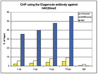
<p><small> <strong>Figure 1. ChIP results obtained with the Diagenode antibody directed against H4K20me3</strong><br />ChIP assays were performed using human HeLa cells, the Diagenode antibody against H4K20me3 (Cat. No. C15410057) and optimized PCR primer sets for qPCR. ChIP was performed with the “Auto Histone ChIP-seq” kit (Cat. No. C01010022) with sheared chromatin from 1 million cells using the SX-8G IP-Star automated system. A titration of the antibody consisting of 1, 2, 5, and 10 μg per ChIP experiment was analysed. IgG (1 μg/IP) was used as negative IP control. QPCR was performed with primers for promoters of the active genes c-fos (Cat. No. C17011004) and GAPDH (Cat. No. C17011047), used as negative controls, and for the Sat2 satellite repeat region used as a positive control. Figure 1 shows the recovery, expressed as a % of input (the relative amount of immunoprecipitated DNA compared to input DNA after qPCR analysis).</small></p>
</div>
</div>
<div class="row">
<div class="small-4 columns">
<p>A.<img src="https://www.diagenode.com/img/product/antibodies/C15410057-chipseq-A.jpg" alt="H4K20me3 Antibody for ChIP" caption="false" width="288" height="296" /></p>
</div>
<div class="small-8 columns">
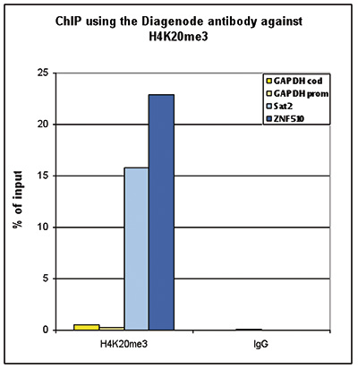
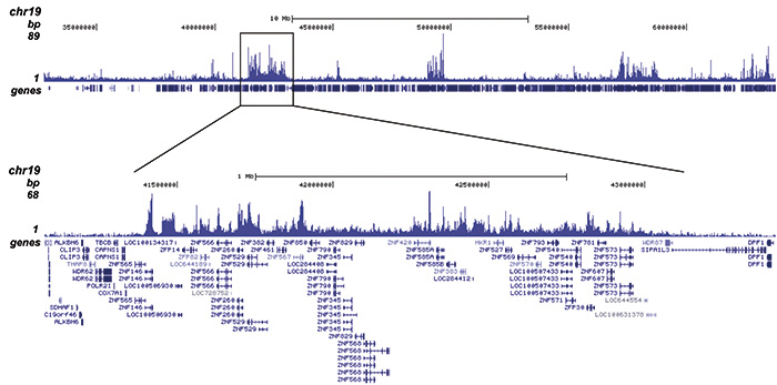
<p><small><strong>Figure 2. ChIP-seq results obtained with the Diagenode antibody directed against H4K20me3</strong><br />ChIP was performed with 1 μg of the Diagenode antibody against H4K20me3 (Cat. No. C15410057) on sheared chromatin from 1 million HeLaS3 cells using the “iDeal ChIP-seq” kit. The IP’d DNA was analysed by QPCR with optimized PCR primer pairs for the promoter and coding region of the active GAPDH gene, for the coding region of the ZNF510 gene and for the Sat2 satellite repeat (figure 2A). The IP’d DNA was subsequently analysed on an Illumina Genome Analyzer. Library preparation, cluster generation and sequencing were performed according to the manufacturer’s instructions. The 36 bp tags were aligned to the human genome using the ELAND algorithm. Figure 2B shows the signal distribution along the long arm of chromosome 19 and a zoomin to an enriched region containing several ZNF repeat genes. Figure 2C and D show the enrichment at ZNF12 and ZNF510 on chromosome 7 and 9, respectively. These results clearly show an enrichment of H4K20me3 at ZNF repeat genes.</small></p>
</div>
</div>
<div class="row">
<div class="small-12 columns">
<p>B.<img src="https://www.diagenode.com/img/product/antibodies/C15410057-chipseq-B.jpg" alt="H4K20me3 Antibody ChIP-seq Grade" caption="false" width="700" height="347" /></p>
<p>C.<img src="https://www.diagenode.com/img/product/antibodies/C15410057-chipseq-C.jpg" alt="H4K20me3 Antibody for ChIP-seq" caption="false" width="700" height="108" /></p>
<p>D.<img src="https://www.diagenode.com/img/product/antibodies/C15410057-chipseq-D.jpg" alt="H4K20me3 Antibody for ChIP-seq assay" caption="false" width="700" height="104" /></p>
</div>
</div>
<div class="row">
<div class="small-12 columns">
<p>A.<img src="https://www.diagenode.com/img/product/antibodies/C15410057-cuttagA.png" caption="false" width="700" height="347" /></p>
<p>B.<img src="https://www.diagenode.com/img/product/antibodies/C15410057-cuttagB.png" caption="false" width="700" height="108" /></p>
<div class="small-12 columns">
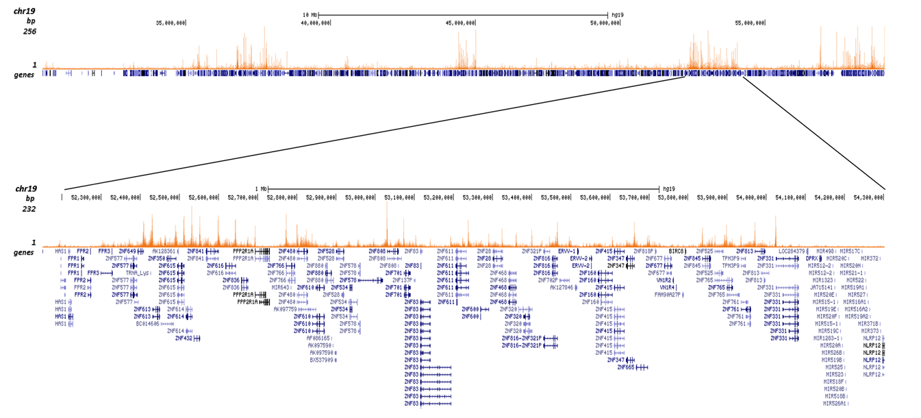
<p><small> <strong>Figure 3. Cut&Tag results obtained with the Diagenode antibody directed against H4K20me3</strong> <br />CUT&TAG (Kaya-Okur, H.S., Nat Commun 10, 1930, 2019) was performed on 50,000 K562 cells using 1 µg of the Diagenode antibody against H4K20me3 (cat. No. C15410057) and the Diagenode pA-Tn5 transposase (C01070001). The libraries were subsequently analysed on an Illumina NextSeq 500 sequencer (2x75 paired-end reads) according to the manufacturer's instructions. The tags were aligned to the human genome (hg19) using the BWA algorithm. Figure 3 shows the peak distribution on the long arm of chromosome 19 as well as a zoomin to a region enriched in ZNF repeat genes, and in a genomic region surrounding the MEG3 imprinted control gene on chromosome 14 (figure 3A and B, respectively).</small></p>
<p></p>
</div>
</div>
<br /> <br />
<div class="row">
<div class="small-4 columns">
<p><img src="https://www.diagenode.com/img/product/antibodies/C15410057-ELISA.jpg" alt="H4K20me3 Antibody ELISA validation" caption="false" width="288" height="263" /></p>
</div>
<div class="small-8 columns">
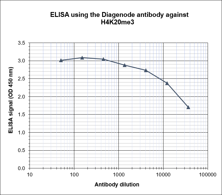
<p><small> <strong>Figure 4. Determination of the antibody titer</strong> <br />To determine the titer of the antibody, an ELISA was performed using a serial dilution of the Diagenode antibody directed against H4K20me3 (Cat. No. C15410057), crude serum and flow through in antigen coated wells. The antigen used was a peptide containing the histone modification of interest. By plotting the absorbance against the antibody dilution (Figure 4), the titer of the antibody was estimated to be 1:7,400.</small></p>
</div>
</div>
<div class="row">
<div class="small-4 columns">
<p><img src="https://www.diagenode.com/img/product/antibodies/C15410057-dotblot.jpg" alt="H4K20me3 Antibody validated in Dot Blot" caption="false" width="288" height="242" /></p>
</div>
<div class="small-8 columns">
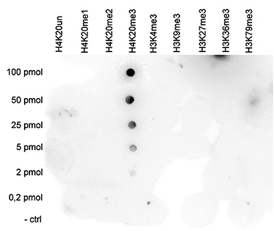
<p><small> <strong>Figure 5. Cross reactivity test using the Diagenode antibody directed against H4K20me3</strong> <br />A Dot Blot analysis was performed to test the cross reactivity of the Diagenode antibody against H4K20me3 (Cat. No. C15410057) with peptides containing other histone modifications and the unmodified H4K20. One hundred to 0.2 pmol of the respective peptides were spotted on a membrane. The antibody was used at a dilution of 1:20,000. Figure 5 shows a high specificity of the antibody for the modification of interest</small></p>
</div>
</div>
<div class="row">
<div class="small-4 columns">
<p><img src="https://www.diagenode.com/img/product/antibodies/C15410057-wb.jpg" alt="H4K20me3 Antibody validated for Western Blot" width="171" height="165" caption="false" /></p>
</div>
<div class="small-8 columns">
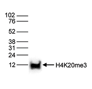
<p><small> <strong>Figure 6. Western blot analysis using the Diagenode antibody directed against H4K20me3</strong> <br />Histone extracts of HeLa cells (15 μg) were analysed by Western blot using the Diagenode antibody against H4K20me3 (Cat. No. C15410057) diluted 1:1,000 in TBS-Tween containing 5% skimmed milk. The position of the protein of interest is indicated on the right; the marker (in kDa) is shown on the left.</small></p>
</div>
</div>
<div class="row">
<div class="small-4 columns">
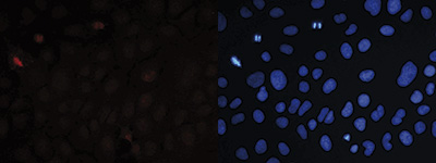
<p>A.<img src="https://www.diagenode.com/img/product/antibodies/C15410057-if-A.jpg" alt="H4K20me3 Antibody validated in Immunofluorescence" caption="false" width="288" height="108" /></p>
<p>B.<img src="https://www.diagenode.com/img/product/antibodies/C15410057-if-B.jpg" alt="H4K20me3 Antibody validated in Immunofluorescence" caption="false" width="288" height="108" /></p>
</div>
<div class="small-8 columns">
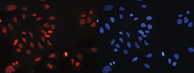
<p><small> <strong>Figure 7. Immunofluorescence using the Diagenode antibody directed against H4K20me3</strong> <br />Human osteosarcoma (U2OS) cells were stained with the Diagenode antibody against H4K20me3 (Cat. No. C15410057) and with DAPI. Cells were fixed with ice cold methanol for 10’ and blocked with PBS/TX-100 containing 5% normal goat serum. Figure 7A: cells were immunofluorescently labeled with the H4K20me3 antibody (left) diluted 1:300 in blocking solution followed by an anti-rabbit antibody conjugated to Alexa568 or with DAPI (right), which specifically labels DNA. Figure 6B: staining of the cells with the H4K20me3 antibody after incubation of the antibody with blocking peptide (Cat. No. C16000057), concentration: 5 ng/μl).</small></p>
</div>
</div>
</div>',
'label2' => 'Target Description',
'info2' => '<p>Histones are the main constituents of the protein part of chromosomes of eukaryotic cells. They are rich in the amino acids arginine and lysine and have been greatly conserved during evolution. Histones pack the DNA into tight masses of chromatin. Two core histones of each class H2A, H2B, H3 and H4 assemble and are wrapped by 146 base pairs of DNA to form one octameric nucleosome. Histone tails undergo numerous post-translational modifications, which either directly or indirectly alter chromatin structure to facilitate transcriptional activation or repression or other nuclear processes. In addition to the genetic code, combinations of the different histone modifications reveal the so-called “histone code”. Histone methylation and demethylation is dynamically regulated by respectively histone methyl transferases and histone demethylases.</p>',
'label3' => '',
'info3' => '',
'format' => '10 µg',
'catalog_number' => 'C15410057-10',
'old_catalog_number' => 'pAb-057-050',
'sf_code' => 'C15410057-D001-000582',
'type' => 'FRE',
'search_order' => '03-Antibody',
'price_EUR' => '105',
'price_USD' => '115',
'price_GBP' => '100',
'price_JPY' => '16450',
'price_CNY' => '',
'price_AUD' => '288',
'country' => 'ALL',
'except_countries' => 'None',
'quote' => false,
'in_stock' => false,
'featured' => false,
'no_promo' => false,
'online' => true,
'master' => false,
'last_datasheet_update' => '0000-00-00',
'slug' => 'h4k20me3-polyclonal-antibody-classic-sample-size-10-ug',
'meta_title' => 'H4K20me3 Antibody - ChIP-seq Grade (C15410057) | Diagenode',
'meta_keywords' => '',
'meta_description' => 'H4K20me3 (Histone H4 trimethylated at lysine 20) Polyclonal Antibody validated in ChIP-seq, ChIP-qPCR, ELISA, DB, WB and IF. Batch-specific data available on the website. Sample size available',
'modified' => '2022-01-06 15:32:16',
'created' => '2015-06-29 14:08:20',
'locale' => 'eng'
),
'Antibody' => array(
'host' => '*****',
'id' => '79',
'name' => 'H4K20me3 polyclonal antibody',
'description' => 'Histones are the main constituents of the protein part of chromosomes of eukaryotic cells. They are rich in the amino acids arginine and lysine and have been greatly conserved during evolution. Histones pack the DNA into tight masses of chromatin. Two core histones of each class H2A, H2B, H3 and H4 assemble and are wrapped by 146 base pairs of DNA to form one octameric nucleosome. Histone tails undergo numerous post-translational modifications, which either directly or indirectly alter chromatin structure to facilitate transcriptional activation or repression or other nuclear processes. In addition to the genetic code, combinations of the different histone modifications reveal the so-called “histone code”. Histone methylation and demethylation is dynamically regulated by respectively histone methyl transferases and histone demethylases.',
'clonality' => '',
'isotype' => '',
'lot' => 'A295-0014P',
'concentration' => '1.07 µg/µl',
'reactivity' => 'Human, mouse',
'type' => 'Polyclonal',
'purity' => 'Affinity purified',
'classification' => 'Classic',
'application_table' => '<table>
<thead>
<tr>
<th>Applications</th>
<th>Suggested dilution</th>
<th>References</th>
</tr>
</thead>
<tbody>
<tr>
<td>ChIP/ChIP-seq <sup>*</sup></td>
<td>1-2 μg/ChIP</td>
<td>Fig 1, 2</td>
</tr>
<tr>
<td>CUT&TAG</td>
<td>1 μg</td>
<td>Fig 3</td>
</tr>
<tr>
<td>ELISA</td>
<td>1:100</td>
<td>Fig 4</td>
</tr>
<tr>
<td>Dot Blotting</td>
<td>1:20,000</td>
<td>Fig 5</td>
</tr>
<tr>
<td>Western Blotting</td>
<td>1:1,000</td>
<td>Fig 6</td>
</tr>
<tr>
<td>Immunofluorescence</td>
<td>1:300</td>
<td>Fig 7</td>
</tr>
</tbody>
</table>
<p><small><sup>*</sup> Please note that the optimal antibody amount per IP should be determined by the end-user. We recommend testing 1-5 μg per IP.</small></p>',
'storage_conditions' => '',
'storage_buffer' => '',
'precautions' => 'This product is for research use only. Not for use in diagnostic or therapeutic procedures.',
'uniprot_acc' => '',
'slug' => '',
'meta_keywords' => '',
'meta_description' => '',
'modified' => '2022-01-06 15:29:10',
'created' => '0000-00-00 00:00:00',
'select_label' => '79 - H4K20me3 polyclonal antibody (A295-0014P - 1.07 µg/µl - Human, mouse - Affinity purified - Rabbit)'
),
'Slave' => array(),
'Group' => array(
'Group' => array(
'id' => '42',
'name' => 'C15410057',
'product_id' => '2218',
'modified' => '2016-02-18 20:47:52',
'created' => '2016-02-18 20:47:52'
),
'Master' => array(
'id' => '2218',
'antibody_id' => '79',
'name' => 'H4K20me3 Antibody',
'description' => '<p><span>Polyclonal antibody raised in rabbit against the region of histone <strong>H4 containing the trimethylated lysine 20 (H4K20me3)</strong>, using a KLH-conjugated synthetic peptide.</span></p>',
'label1' => 'Validation data',
'info1' => '<div class="row">
<div class="small-4 columns">
<p><img src="https://www.diagenode.com/img/product/antibodies/C15410057-chip.jpg" alt="H4K20me3 Antibody ChIP Grade" caption="false" width="288" height="218" /></p>
</div>
<div class="small-8 columns">
<p><small> <strong>Figure 1. ChIP results obtained with the Diagenode antibody directed against H4K20me3</strong><br />ChIP assays were performed using human HeLa cells, the Diagenode antibody against H4K20me3 (Cat. No. C15410057) and optimized PCR primer sets for qPCR. ChIP was performed with the “Auto Histone ChIP-seq” kit (Cat. No. C01010022) with sheared chromatin from 1 million cells using the SX-8G IP-Star automated system. A titration of the antibody consisting of 1, 2, 5, and 10 μg per ChIP experiment was analysed. IgG (1 μg/IP) was used as negative IP control. QPCR was performed with primers for promoters of the active genes c-fos (Cat. No. C17011004) and GAPDH (Cat. No. C17011047), used as negative controls, and for the Sat2 satellite repeat region used as a positive control. Figure 1 shows the recovery, expressed as a % of input (the relative amount of immunoprecipitated DNA compared to input DNA after qPCR analysis).</small></p>
</div>
</div>
<div class="row">
<div class="small-4 columns">
<p>A.<img src="https://www.diagenode.com/img/product/antibodies/C15410057-chipseq-A.jpg" alt="H4K20me3 Antibody for ChIP" caption="false" width="288" height="296" /></p>
</div>
<div class="small-8 columns">
<p><small><strong>Figure 2. ChIP-seq results obtained with the Diagenode antibody directed against H4K20me3</strong><br />ChIP was performed with 1 μg of the Diagenode antibody against H4K20me3 (Cat. No. C15410057) on sheared chromatin from 1 million HeLaS3 cells using the “iDeal ChIP-seq” kit. The IP’d DNA was analysed by QPCR with optimized PCR primer pairs for the promoter and coding region of the active GAPDH gene, for the coding region of the ZNF510 gene and for the Sat2 satellite repeat (figure 2A). The IP’d DNA was subsequently analysed on an Illumina Genome Analyzer. Library preparation, cluster generation and sequencing were performed according to the manufacturer’s instructions. The 36 bp tags were aligned to the human genome using the ELAND algorithm. Figure 2B shows the signal distribution along the long arm of chromosome 19 and a zoomin to an enriched region containing several ZNF repeat genes. Figure 2C and D show the enrichment at ZNF12 and ZNF510 on chromosome 7 and 9, respectively. These results clearly show an enrichment of H4K20me3 at ZNF repeat genes.</small></p>
</div>
</div>
<div class="row">
<div class="small-12 columns">
<p>B.<img src="https://www.diagenode.com/img/product/antibodies/C15410057-chipseq-B.jpg" alt="H4K20me3 Antibody ChIP-seq Grade" caption="false" width="700" height="347" /></p>
<p>C.<img src="https://www.diagenode.com/img/product/antibodies/C15410057-chipseq-C.jpg" alt="H4K20me3 Antibody for ChIP-seq" caption="false" width="700" height="108" /></p>
<p>D.<img src="https://www.diagenode.com/img/product/antibodies/C15410057-chipseq-D.jpg" alt="H4K20me3 Antibody for ChIP-seq assay" caption="false" width="700" height="104" /></p>
</div>
</div>
<div class="row">
<div class="small-12 columns">
<p>A.<img src="https://www.diagenode.com/img/product/antibodies/C15410057-cuttagA.png" caption="false" width="700" height="347" /></p>
<p>B.<img src="https://www.diagenode.com/img/product/antibodies/C15410057-cuttagB.png" caption="false" width="700" height="108" /></p>
<div class="small-12 columns">
<p><small> <strong>Figure 3. Cut&Tag results obtained with the Diagenode antibody directed against H4K20me3</strong> <br />CUT&TAG (Kaya-Okur, H.S., Nat Commun 10, 1930, 2019) was performed on 50,000 K562 cells using 1 µg of the Diagenode antibody against H4K20me3 (cat. No. C15410057) and the Diagenode pA-Tn5 transposase (C01070001). The libraries were subsequently analysed on an Illumina NextSeq 500 sequencer (2x75 paired-end reads) according to the manufacturer's instructions. The tags were aligned to the human genome (hg19) using the BWA algorithm. Figure 3 shows the peak distribution on the long arm of chromosome 19 as well as a zoomin to a region enriched in ZNF repeat genes, and in a genomic region surrounding the MEG3 imprinted control gene on chromosome 14 (figure 3A and B, respectively).</small></p>
<p></p>
</div>
</div>
<br /> <br />
<div class="row">
<div class="small-4 columns">
<p><img src="https://www.diagenode.com/img/product/antibodies/C15410057-ELISA.jpg" alt="H4K20me3 Antibody ELISA validation" caption="false" width="288" height="263" /></p>
</div>
<div class="small-8 columns">
<p><small> <strong>Figure 4. Determination of the antibody titer</strong> <br />To determine the titer of the antibody, an ELISA was performed using a serial dilution of the Diagenode antibody directed against H4K20me3 (Cat. No. C15410057), crude serum and flow through in antigen coated wells. The antigen used was a peptide containing the histone modification of interest. By plotting the absorbance against the antibody dilution (Figure 4), the titer of the antibody was estimated to be 1:7,400.</small></p>
</div>
</div>
<div class="row">
<div class="small-4 columns">
<p><img src="https://www.diagenode.com/img/product/antibodies/C15410057-dotblot.jpg" alt="H4K20me3 Antibody validated in Dot Blot" caption="false" width="288" height="242" /></p>
</div>
<div class="small-8 columns">
<p><small> <strong>Figure 5. Cross reactivity test using the Diagenode antibody directed against H4K20me3</strong> <br />A Dot Blot analysis was performed to test the cross reactivity of the Diagenode antibody against H4K20me3 (Cat. No. C15410057) with peptides containing other histone modifications and the unmodified H4K20. One hundred to 0.2 pmol of the respective peptides were spotted on a membrane. The antibody was used at a dilution of 1:20,000. Figure 5 shows a high specificity of the antibody for the modification of interest</small></p>
</div>
</div>
<div class="row">
<div class="small-4 columns">
<p><img src="https://www.diagenode.com/img/product/antibodies/C15410057-wb.jpg" alt="H4K20me3 Antibody validated for Western Blot" width="171" height="165" caption="false" /></p>
</div>
<div class="small-8 columns">
<p><small> <strong>Figure 6. Western blot analysis using the Diagenode antibody directed against H4K20me3</strong> <br />Histone extracts of HeLa cells (15 μg) were analysed by Western blot using the Diagenode antibody against H4K20me3 (Cat. No. C15410057) diluted 1:1,000 in TBS-Tween containing 5% skimmed milk. The position of the protein of interest is indicated on the right; the marker (in kDa) is shown on the left.</small></p>
</div>
</div>
<div class="row">
<div class="small-4 columns">
<p>A.<img src="https://www.diagenode.com/img/product/antibodies/C15410057-if-A.jpg" alt="H4K20me3 Antibody validated in Immunofluorescence" caption="false" width="288" height="108" /></p>
<p>B.<img src="https://www.diagenode.com/img/product/antibodies/C15410057-if-B.jpg" alt="H4K20me3 Antibody validated in Immunofluorescence" caption="false" width="288" height="108" /></p>
</div>
<div class="small-8 columns">
<p><small> <strong>Figure 7. Immunofluorescence using the Diagenode antibody directed against H4K20me3</strong> <br />Human osteosarcoma (U2OS) cells were stained with the Diagenode antibody against H4K20me3 (Cat. No. C15410057) and with DAPI. Cells were fixed with ice cold methanol for 10’ and blocked with PBS/TX-100 containing 5% normal goat serum. Figure 7A: cells were immunofluorescently labeled with the H4K20me3 antibody (left) diluted 1:300 in blocking solution followed by an anti-rabbit antibody conjugated to Alexa568 or with DAPI (right), which specifically labels DNA. Figure 6B: staining of the cells with the H4K20me3 antibody after incubation of the antibody with blocking peptide (Cat. No. C16000057), concentration: 5 ng/μl).</small></p>
</div>
</div>
</div>',
'label2' => 'Target Description',
'info2' => '<p>Histones are the main constituents of the protein part of chromosomes of eukaryotic cells. They are rich in the amino acids arginine and lysine and have been greatly conserved during evolution. Histones pack the DNA into tight masses of chromatin. Two core histones of each class H2A, H2B, H3 and H4 assemble and are wrapped by 146 base pairs of DNA to form one octameric nucleosome. Histone tails undergo numerous post-translational modifications, which either directly or indirectly alter chromatin structure to facilitate transcriptional activation or repression or other nuclear processes. In addition to the genetic code, combinations of the different histone modifications reveal the so-called “histone code”. Histone methylation and demethylation is dynamically regulated by respectively histone methyl transferases and histone demethylases.</p>',
'label3' => '',
'info3' => '',
'format' => '50 μg',
'catalog_number' => 'C15410057',
'old_catalog_number' => 'pAb-057-050',
'sf_code' => 'C15410057-D001-000581',
'type' => 'FRE',
'search_order' => '03-Antibody',
'price_EUR' => '380',
'price_USD' => '380',
'price_GBP' => '340',
'price_JPY' => '59525',
'price_CNY' => '',
'price_AUD' => '950',
'country' => 'ALL',
'except_countries' => 'None',
'quote' => false,
'in_stock' => false,
'featured' => false,
'no_promo' => false,
'online' => true,
'master' => true,
'last_datasheet_update' => '0000-00-00',
'slug' => 'h4k20me3-polyclonal-antibody-classic-50-mg-47-ml',
'meta_title' => 'H4K20me3 Antibody - ChIP-seq Grade (C15410057) | Diagenode',
'meta_keywords' => '',
'meta_description' => 'H4K20me3 (Histone H4 trimethylated at lysine 20) Polyclonal Antibody validated in ChIP-seq, ChIP-qPCR, ELISA, DB, WB and IF. Batch-specific data available on the website. Sample size available.',
'modified' => '2024-06-13 15:06:38',
'created' => '2015-06-29 14:08:20'
),
'Product' => array(
(int) 0 => array(
[maximum depth reached]
)
)
),
'Related' => array(
(int) 0 => array(
'id' => '2218',
'antibody_id' => '79',
'name' => 'H4K20me3 Antibody',
'description' => '<p><span>Polyclonal antibody raised in rabbit against the region of histone <strong>H4 containing the trimethylated lysine 20 (H4K20me3)</strong>, using a KLH-conjugated synthetic peptide.</span></p>',
'label1' => 'Validation data',
'info1' => '<div class="row">
<div class="small-4 columns">
<p><img src="https://www.diagenode.com/img/product/antibodies/C15410057-chip.jpg" alt="H4K20me3 Antibody ChIP Grade" caption="false" width="288" height="218" /></p>
</div>
<div class="small-8 columns">
<p><small> <strong>Figure 1. ChIP results obtained with the Diagenode antibody directed against H4K20me3</strong><br />ChIP assays were performed using human HeLa cells, the Diagenode antibody against H4K20me3 (Cat. No. C15410057) and optimized PCR primer sets for qPCR. ChIP was performed with the “Auto Histone ChIP-seq” kit (Cat. No. C01010022) with sheared chromatin from 1 million cells using the SX-8G IP-Star automated system. A titration of the antibody consisting of 1, 2, 5, and 10 μg per ChIP experiment was analysed. IgG (1 μg/IP) was used as negative IP control. QPCR was performed with primers for promoters of the active genes c-fos (Cat. No. C17011004) and GAPDH (Cat. No. C17011047), used as negative controls, and for the Sat2 satellite repeat region used as a positive control. Figure 1 shows the recovery, expressed as a % of input (the relative amount of immunoprecipitated DNA compared to input DNA after qPCR analysis).</small></p>
</div>
</div>
<div class="row">
<div class="small-4 columns">
<p>A.<img src="https://www.diagenode.com/img/product/antibodies/C15410057-chipseq-A.jpg" alt="H4K20me3 Antibody for ChIP" caption="false" width="288" height="296" /></p>
</div>
<div class="small-8 columns">
<p><small><strong>Figure 2. ChIP-seq results obtained with the Diagenode antibody directed against H4K20me3</strong><br />ChIP was performed with 1 μg of the Diagenode antibody against H4K20me3 (Cat. No. C15410057) on sheared chromatin from 1 million HeLaS3 cells using the “iDeal ChIP-seq” kit. The IP’d DNA was analysed by QPCR with optimized PCR primer pairs for the promoter and coding region of the active GAPDH gene, for the coding region of the ZNF510 gene and for the Sat2 satellite repeat (figure 2A). The IP’d DNA was subsequently analysed on an Illumina Genome Analyzer. Library preparation, cluster generation and sequencing were performed according to the manufacturer’s instructions. The 36 bp tags were aligned to the human genome using the ELAND algorithm. Figure 2B shows the signal distribution along the long arm of chromosome 19 and a zoomin to an enriched region containing several ZNF repeat genes. Figure 2C and D show the enrichment at ZNF12 and ZNF510 on chromosome 7 and 9, respectively. These results clearly show an enrichment of H4K20me3 at ZNF repeat genes.</small></p>
</div>
</div>
<div class="row">
<div class="small-12 columns">
<p>B.<img src="https://www.diagenode.com/img/product/antibodies/C15410057-chipseq-B.jpg" alt="H4K20me3 Antibody ChIP-seq Grade" caption="false" width="700" height="347" /></p>
<p>C.<img src="https://www.diagenode.com/img/product/antibodies/C15410057-chipseq-C.jpg" alt="H4K20me3 Antibody for ChIP-seq" caption="false" width="700" height="108" /></p>
<p>D.<img src="https://www.diagenode.com/img/product/antibodies/C15410057-chipseq-D.jpg" alt="H4K20me3 Antibody for ChIP-seq assay" caption="false" width="700" height="104" /></p>
</div>
</div>
<div class="row">
<div class="small-12 columns">
<p>A.<img src="https://www.diagenode.com/img/product/antibodies/C15410057-cuttagA.png" caption="false" width="700" height="347" /></p>
<p>B.<img src="https://www.diagenode.com/img/product/antibodies/C15410057-cuttagB.png" caption="false" width="700" height="108" /></p>
<div class="small-12 columns">
<p><small> <strong>Figure 3. Cut&Tag results obtained with the Diagenode antibody directed against H4K20me3</strong> <br />CUT&TAG (Kaya-Okur, H.S., Nat Commun 10, 1930, 2019) was performed on 50,000 K562 cells using 1 µg of the Diagenode antibody against H4K20me3 (cat. No. C15410057) and the Diagenode pA-Tn5 transposase (C01070001). The libraries were subsequently analysed on an Illumina NextSeq 500 sequencer (2x75 paired-end reads) according to the manufacturer's instructions. The tags were aligned to the human genome (hg19) using the BWA algorithm. Figure 3 shows the peak distribution on the long arm of chromosome 19 as well as a zoomin to a region enriched in ZNF repeat genes, and in a genomic region surrounding the MEG3 imprinted control gene on chromosome 14 (figure 3A and B, respectively).</small></p>
<p></p>
</div>
</div>
<br /> <br />
<div class="row">
<div class="small-4 columns">
<p><img src="https://www.diagenode.com/img/product/antibodies/C15410057-ELISA.jpg" alt="H4K20me3 Antibody ELISA validation" caption="false" width="288" height="263" /></p>
</div>
<div class="small-8 columns">
<p><small> <strong>Figure 4. Determination of the antibody titer</strong> <br />To determine the titer of the antibody, an ELISA was performed using a serial dilution of the Diagenode antibody directed against H4K20me3 (Cat. No. C15410057), crude serum and flow through in antigen coated wells. The antigen used was a peptide containing the histone modification of interest. By plotting the absorbance against the antibody dilution (Figure 4), the titer of the antibody was estimated to be 1:7,400.</small></p>
</div>
</div>
<div class="row">
<div class="small-4 columns">
<p><img src="https://www.diagenode.com/img/product/antibodies/C15410057-dotblot.jpg" alt="H4K20me3 Antibody validated in Dot Blot" caption="false" width="288" height="242" /></p>
</div>
<div class="small-8 columns">
<p><small> <strong>Figure 5. Cross reactivity test using the Diagenode antibody directed against H4K20me3</strong> <br />A Dot Blot analysis was performed to test the cross reactivity of the Diagenode antibody against H4K20me3 (Cat. No. C15410057) with peptides containing other histone modifications and the unmodified H4K20. One hundred to 0.2 pmol of the respective peptides were spotted on a membrane. The antibody was used at a dilution of 1:20,000. Figure 5 shows a high specificity of the antibody for the modification of interest</small></p>
</div>
</div>
<div class="row">
<div class="small-4 columns">
<p><img src="https://www.diagenode.com/img/product/antibodies/C15410057-wb.jpg" alt="H4K20me3 Antibody validated for Western Blot" width="171" height="165" caption="false" /></p>
</div>
<div class="small-8 columns">
<p><small> <strong>Figure 6. Western blot analysis using the Diagenode antibody directed against H4K20me3</strong> <br />Histone extracts of HeLa cells (15 μg) were analysed by Western blot using the Diagenode antibody against H4K20me3 (Cat. No. C15410057) diluted 1:1,000 in TBS-Tween containing 5% skimmed milk. The position of the protein of interest is indicated on the right; the marker (in kDa) is shown on the left.</small></p>
</div>
</div>
<div class="row">
<div class="small-4 columns">
<p>A.<img src="https://www.diagenode.com/img/product/antibodies/C15410057-if-A.jpg" alt="H4K20me3 Antibody validated in Immunofluorescence" caption="false" width="288" height="108" /></p>
<p>B.<img src="https://www.diagenode.com/img/product/antibodies/C15410057-if-B.jpg" alt="H4K20me3 Antibody validated in Immunofluorescence" caption="false" width="288" height="108" /></p>
</div>
<div class="small-8 columns">
<p><small> <strong>Figure 7. Immunofluorescence using the Diagenode antibody directed against H4K20me3</strong> <br />Human osteosarcoma (U2OS) cells were stained with the Diagenode antibody against H4K20me3 (Cat. No. C15410057) and with DAPI. Cells were fixed with ice cold methanol for 10’ and blocked with PBS/TX-100 containing 5% normal goat serum. Figure 7A: cells were immunofluorescently labeled with the H4K20me3 antibody (left) diluted 1:300 in blocking solution followed by an anti-rabbit antibody conjugated to Alexa568 or with DAPI (right), which specifically labels DNA. Figure 6B: staining of the cells with the H4K20me3 antibody after incubation of the antibody with blocking peptide (Cat. No. C16000057), concentration: 5 ng/μl).</small></p>
</div>
</div>
</div>',
'label2' => 'Target Description',
'info2' => '<p>Histones are the main constituents of the protein part of chromosomes of eukaryotic cells. They are rich in the amino acids arginine and lysine and have been greatly conserved during evolution. Histones pack the DNA into tight masses of chromatin. Two core histones of each class H2A, H2B, H3 and H4 assemble and are wrapped by 146 base pairs of DNA to form one octameric nucleosome. Histone tails undergo numerous post-translational modifications, which either directly or indirectly alter chromatin structure to facilitate transcriptional activation or repression or other nuclear processes. In addition to the genetic code, combinations of the different histone modifications reveal the so-called “histone code”. Histone methylation and demethylation is dynamically regulated by respectively histone methyl transferases and histone demethylases.</p>',
'label3' => '',
'info3' => '',
'format' => '50 μg',
'catalog_number' => 'C15410057',
'old_catalog_number' => 'pAb-057-050',
'sf_code' => 'C15410057-D001-000581',
'type' => 'FRE',
'search_order' => '03-Antibody',
'price_EUR' => '380',
'price_USD' => '380',
'price_GBP' => '340',
'price_JPY' => '59525',
'price_CNY' => '',
'price_AUD' => '950',
'country' => 'ALL',
'except_countries' => 'None',
'quote' => false,
'in_stock' => false,
'featured' => false,
'no_promo' => false,
'online' => true,
'master' => true,
'last_datasheet_update' => '0000-00-00',
'slug' => 'h4k20me3-polyclonal-antibody-classic-50-mg-47-ml',
'meta_title' => 'H4K20me3 Antibody - ChIP-seq Grade (C15410057) | Diagenode',
'meta_keywords' => '',
'meta_description' => 'H4K20me3 (Histone H4 trimethylated at lysine 20) Polyclonal Antibody validated in ChIP-seq, ChIP-qPCR, ELISA, DB, WB and IF. Batch-specific data available on the website. Sample size available.',
'modified' => '2024-06-13 15:06:38',
'created' => '2015-06-29 14:08:20',
'ProductsRelated' => array(
[maximum depth reached]
),
'Image' => array(
[maximum depth reached]
)
)
),
'Application' => array(
(int) 0 => array(
'id' => '20',
'position' => '10',
'parent_id' => '40',
'name' => 'ELISA',
'description' => '<div class="row">
<div class="small-12 medium-12 large-12 columns">Enzyme-linked immunosorbent assay.</div>
</div>',
'in_footer' => false,
'in_menu' => false,
'online' => true,
'tabular' => true,
'slug' => 'elisa-antibodies',
'meta_keywords' => ' ELISA Antibodies,Monoclonal antibody, Polyclonal antibody',
'meta_description' => 'Diagenode offers Monoclonal & Polyclonal antibodies for ELISA applications',
'meta_title' => 'ELISA Antibodies - Monoclonal & Polyclonal antibody | Diagenode',
'modified' => '2016-01-13 12:21:41',
'created' => '2014-07-08 08:13:28',
'ProductsApplication' => array(
[maximum depth reached]
)
),
(int) 1 => array(
'id' => '28',
'position' => '10',
'parent_id' => '40',
'name' => 'DB',
'description' => '<p>Dot blotting</p>',
'in_footer' => false,
'in_menu' => false,
'online' => true,
'tabular' => true,
'slug' => 'dot-blotting',
'meta_keywords' => 'Dot blotting,Monoclonal & Polyclonal antibody,',
'meta_description' => 'Diagenode offers Monoclonal & Polyclonal antibodies for Dot blotting applications',
'meta_title' => 'Dot blotting Antibodies - Monoclonal & Polyclonal antibody | Diagenode',
'modified' => '2016-01-13 14:40:49',
'created' => '2015-07-08 13:45:05',
'ProductsApplication' => array(
[maximum depth reached]
)
),
(int) 2 => array(
'id' => '19',
'position' => '10',
'parent_id' => '40',
'name' => 'WB',
'description' => '<p><strong>Western blot</strong> : The quality of antibodies used in this technique is crucial for correct and specific protein identification. Diagenode offers huge selection of highly sensitive and specific western blot-validated antibodies.</p>
<p>Learn more about: <a href="https://www.diagenode.com/applications/western-blot">Loading control, MW marker visualization</a><em>. <br /></em></p>
<p><em></em>Check our selection of antibodies validated in Western blot.</p>',
'in_footer' => false,
'in_menu' => false,
'online' => true,
'tabular' => true,
'slug' => 'western-blot-antibodies',
'meta_keywords' => ' Western Blot Antibodies ,western blot protocol,Western Blotting Products,Polyclonal antibodies ,monoclonal antibodies ',
'meta_description' => 'Diagenode offers a wide range of antibodies and technical support for western blot applications',
'meta_title' => ' Western Blot - Monoclonal antibody - Polyclonal antibody | Diagenode',
'modified' => '2016-04-26 12:44:51',
'created' => '2015-01-07 09:20:00',
'ProductsApplication' => array(
[maximum depth reached]
)
),
(int) 3 => array(
'id' => '29',
'position' => '10',
'parent_id' => '40',
'name' => 'IF',
'description' => '<p><strong>Immunofluorescence</strong>:</p>
<p>Diagenode offers huge selection of highly sensitive antibodies validated in IF.</p>
<p><img src="https://www.diagenode.com/img/product/antibodies/C15200229-IF.jpg" alt="" height="245" width="256" /></p>
<p><sup><strong>Immunofluorescence using the Diagenode monoclonal antibody directed against CRISPR/Cas9</strong></sup></p>
<p><sup>HeLa cells transfected with a Cas9 expression vector (left) or untransfected cells (right) were fixed in methanol at -20°C, permeabilized with acetone at -20°C and blocked with PBS containing 2% BSA. The cells were stained with the Cas9 C-terminal antibody (Cat. No. C15200229) diluted 1:400, followed by incubation with an anti-mouse secondary antibody coupled to AF488. The bottom images show counter-staining of the nuclei with Hoechst 33342.</sup></p>
<h5><sup>Check our selection of antibodies validated in IF.</sup></h5>',
'in_footer' => false,
'in_menu' => false,
'online' => true,
'tabular' => true,
'slug' => 'immunofluorescence',
'meta_keywords' => 'Immunofluorescence,Monoclonal antibody,Polyclonal antibody',
'meta_description' => 'Diagenode offers a wide range of antibodies and technical support for Immunofluorescence applications',
'meta_title' => 'Immunofluorescence - Monoclonal antibody - Polyclonal antibody | Diagenode',
'modified' => '2016-04-27 16:23:10',
'created' => '2015-07-08 13:46:02',
'ProductsApplication' => array(
[maximum depth reached]
)
),
(int) 4 => array(
'id' => '42',
'position' => '10',
'parent_id' => '40',
'name' => 'ChIP-seq (ab)',
'description' => '',
'in_footer' => false,
'in_menu' => false,
'online' => true,
'tabular' => true,
'slug' => 'chip-seq-antibodies',
'meta_keywords' => 'Chromatin Immunoprecipitation Sequencing,ChIP-Seq,ChIP-seq grade antibodies,DNA purification,qPCR,Shearing of chromatin',
'meta_description' => 'Diagenode offers a wide range of antibodies and technical support for ChIP Sequencing applications',
'meta_title' => 'ChIP Sequencing Antibodies (ChIP-Seq) | Diagenode',
'modified' => '2016-01-20 11:06:19',
'created' => '2015-10-20 11:44:45',
'ProductsApplication' => array(
[maximum depth reached]
)
),
(int) 5 => array(
'id' => '43',
'position' => '10',
'parent_id' => '40',
'name' => 'ChIP-qPCR (ab)',
'description' => '',
'in_footer' => false,
'in_menu' => false,
'online' => true,
'tabular' => true,
'slug' => 'chip-qpcr-antibodies',
'meta_keywords' => 'Chromatin Immunoprecipitation Sequencing,ChIP-Seq,ChIP-seq grade antibodies,DNA purification,qPCR,Shearing of chromatin',
'meta_description' => 'Diagenode offers a wide range of antibodies and technical support for ChIP-qPCR applications',
'meta_title' => 'ChIP Quantitative PCR Antibodies (ChIP-qPCR) | Diagenode',
'modified' => '2016-01-20 11:30:24',
'created' => '2015-10-20 11:45:36',
'ProductsApplication' => array(
[maximum depth reached]
)
),
(int) 6 => array(
'id' => '55',
'position' => '10',
'parent_id' => '40',
'name' => 'CUT&Tag',
'description' => '<p>CUT&Tagアッセイを成功させるための重要な要素の1つは使用される抗体の品質です。 特異性高い抗体は、目的のタンパク質のみをターゲットとした確実な結果を可能にします。 CUT&Tagで検証済みの抗体のセレクションはこちらからご覧ください。</p>
<p>Read more:</p>
<p><a href="https://www.diagenode.com/en/categories/cutandtag">Products for CUT&Tag assay</a></p>
<p><a href="https://www.diagenode.com/en/pages/cut-and-tag">Performance of Diagenode's antibodies in CUT&Tag</a></p>
<script src="chrome-extension://hhojmcideegachlhfgfdhailpfhgknjm/web_accessible_resources/index.js"></script>',
'in_footer' => false,
'in_menu' => false,
'online' => true,
'tabular' => true,
'slug' => 'cut-and-tag',
'meta_keywords' => 'CUT&Tag',
'meta_description' => 'CUT&Tag',
'meta_title' => 'CUT&Tag',
'modified' => '2021-04-27 05:17:46',
'created' => '2020-08-20 10:13:47',
'ProductsApplication' => array(
[maximum depth reached]
)
)
),
'Category' => array(
(int) 0 => array(
'id' => '111',
'position' => '40',
'parent_id' => '4',
'name' => 'Histone antibodies',
'description' => '<p>Histones are the main protein components of chromatin involved in the compaction of DNA into nucleosomes, the basic units of chromatin. A <strong>nucleosome</strong> consists of one pair of each of the core histones (<strong>H2A</strong>, <strong>H2B</strong>, <strong>H3</strong> and <strong>H4</strong>) forming an octameric structure wrapped by 146 base pairs of DNA. The different nucleosomes are linked by the linker histone<strong> H1, </strong>allowing for further condensation of chromatin.</p>
<p>The core histones have a globular structure with large unstructured N-terminal tails protruding from the nucleosome. They can undergo to multiple post-translational modifications (PTM), mainly at the N-terminal tails. These <strong>post-translational modifications </strong>include methylation, acetylation, phosphorylation, ubiquitinylation, citrullination, sumoylation, deamination and crotonylation. The most well characterized PTMs are <strong>methylation,</strong> <strong>acetylation and phosphorylation</strong>. Histone methylation occurs mainly on lysine (K) residues, which can be mono-, di- or tri-methylated, and on arginines (R), which can be mono-methylated and symmetrically or asymmetrically di-methylated. Histone acetylation occurs on lysines and histone phosphorylation mainly on serines (S), threonines (T) and tyrosines (Y).</p>
<p>The PTMs of the different residues are involved in numerous processes such as DNA repair, DNA replication and chromosome condensation. They influence the chromatin organization and can be positively or negatively associated with gene expression. Trimethylation of H3K4, H3K36 and H3K79, and lysine acetylation generally result in an open chromatin configuration (figure below) and are therefore associated with <strong>euchromatin</strong> and gene activation. Trimethylation of H3K9, K3K27 and H4K20, on the other hand, is enriched in <strong>heterochromatin </strong>and associated with gene silencing. The combination of different histone modifications is called the "<strong>histone code</strong>”, analogous to the genetic code.</p>
<p><img src="https://www.diagenode.com/img/categories/antibodies/histone-marks-illustration.png" /></p>
<p>Diagenode is proud to offer a large range of antibodies against histones and histone modifications. Our antibodies are highly specific and have been validated in many applications, including <strong>ChIP</strong> and <strong>ChIP-seq</strong>.</p>
<p>Diagenode’s collection includes antibodies recognizing:</p>
<ul>
<li><strong>Histone H1 variants</strong></li>
<li><strong>Histone H2A, H2A variants and histone H2A</strong> <strong>modifications</strong> (serine phosphorylation, lysine acetylation, lysine ubiquitinylation)</li>
<li><strong>Histone H2B and H2B</strong> <strong>modifications </strong>(serine phosphorylation, lysine acetylation)</li>
<li><strong>Histone H3 and H3 modifications </strong>(lysine methylation (mono-, di- and tri-methylated), lysine acetylation, serine phosphorylation, threonine phosphorylation, arginine methylation (mono-methylated, symmetrically and asymmetrically di-methylated))</li>
<li><strong>Histone H4 and H4 modifications (</strong>lysine methylation (mono-, di- and tri-methylated), lysine acetylation, arginine methylation (mono-methylated and symmetrically di-methylated), serine phosphorylation )</li>
</ul>
<p><span style="font-weight: 400;"><strong>HDAC's HAT's, HMT's and other</strong> <strong>enzymes</strong> which modify histones can be found in the category <a href="../categories/chromatin-modifying-proteins-histone-transferase">Histone modifying enzymes</a><br /></span></p>
<p><span style="font-weight: 400;"> Diagenode’s highly validated antibodies:</span></p>
<ul>
<li><span style="font-weight: 400;"> Highly sensitive and specific</span></li>
<li><span style="font-weight: 400;"> Cost-effective (requires less antibody per reaction)</span></li>
<li><span style="font-weight: 400;"> Batch-specific data is available on the website</span></li>
<li><span style="font-weight: 400;"> Expert technical support</span></li>
<li><span style="font-weight: 400;"> Sample sizes available</span></li>
<li><span style="font-weight: 400;"> 100% satisfaction guarantee</span></li>
</ul>',
'no_promo' => false,
'in_menu' => false,
'online' => true,
'tabular' => false,
'hide' => true,
'all_format' => false,
'is_antibody' => true,
'slug' => 'histone-antibodies',
'cookies_tag_id' => null,
'meta_keywords' => 'Histone, antibody, histone h1, histone h2, histone h3, histone h4',
'meta_description' => 'Polyclonal and Monoclonal Antibodies against Histones and their modifications validated for many applications, including Chromatin Immunoprecipitation (ChIP) and ChIP-Sequencing (ChIP-seq)',
'meta_title' => 'Histone and Modified Histone Antibodies | Diagenode',
'modified' => '2020-09-17 13:34:56',
'created' => '2016-04-01 16:01:32',
'ProductsCategory' => array(
[maximum depth reached]
),
'CookiesTag' => array([maximum depth reached])
),
(int) 1 => array(
'id' => '17',
'position' => '10',
'parent_id' => '4',
'name' => 'ChIP-seq grade antibodies',
'description' => '<p><b>Unparalleled ChIP-Seq results with the most rigorously validated antibodies</b></p>
<p><span style="font-weight: 400;">Diagenode provides leading solutions for epigenetic research. Because ChIP-seq is a widely-used technique, we validate our antibodies in ChIP and ChIP-seq experiments (in addition to conventional methods like Western blot, Dot blot, ELISA, and immunofluorescence) to provide the highest quality antibody. We standardize our validation and production to guarantee high product quality without technical bias. Diagenode guarantees ChIP-seq grade antibody performance under our suggested conditions.</span></p>
<div class="row">
<div class="small-12 medium-9 large-9 columns">
<p><strong>ChIP-seq profile</strong> of active (H3K4me3 and H3K36me3) and inactive (H3K27me3) marks using Diagenode antibodies.</p>
<img src="https://www.diagenode.com/img/categories/antibodies/chip-seq-grade-antibodies.png" /></div>
<div class="small-12 medium-3 large-3 columns">
<p><small> ChIP was performed on sheared chromatin from 100,000 K562 cells using iDeal ChIP-seq kit for Histones (cat. No. C01010051) with 1 µg of the Diagenode antibodies against H3K27me3 (cat. No. C15410195) and H3K4me3 (cat. No. C15410003), and 0.5 µg of the antibody against H3K36me3 (cat. No. C15410192). The IP'd DNA was subsequently analysed on an Illumina Genome Analyzer. Library preparation, cluster generation and sequencing were performed according to the manufacturer's instructions. The 36 bp tags were aligned to the human genome using the ELAND algorithm. The figure shows the signal distribution along the complete sequence of human chromosome 3, a zoomin to a 10 Mb region and a further zoomin to a 1.5 Mb region. </small></p>
</div>
</div>
<p>Diagenode’s highly validated antibodies:</p>
<ul>
<li>Highly sensitive and specific</li>
<li>Cost-effective (requires less antibody per reaction)</li>
<li>Batch-specific data is available on the website</li>
<li>Expert technical support</li>
<li>Sample sizes available</li>
<li>100% satisfaction guarantee</li>
</ul>',
'no_promo' => false,
'in_menu' => true,
'online' => true,
'tabular' => false,
'hide' => true,
'all_format' => false,
'is_antibody' => true,
'slug' => 'chip-seq-grade-antibodies',
'cookies_tag_id' => null,
'meta_keywords' => 'ChIP-seq grade antibodies,polyclonal antibody,WB, ELISA, ChIP-seq (ab), ChIP-qPCR (ab)',
'meta_description' => 'Diagenode Offers Wide Range of Validated ChIP-Seq Grade Antibodies for Unparalleled ChIP-Seq Results',
'meta_title' => 'Chromatin Immunoprecipitation ChIP-Seq Grade Antibodies | Diagenode',
'modified' => '2019-07-03 10:57:22',
'created' => '2015-02-16 02:24:01',
'ProductsCategory' => array(
[maximum depth reached]
),
'CookiesTag' => array([maximum depth reached])
),
(int) 2 => array(
'id' => '102',
'position' => '1',
'parent_id' => '4',
'name' => 'Sample size antibodies',
'description' => '<h1><strong>Validated epigenetics antibodies</strong> – care for a sample?<br /> </h1>
<p>Diagenode has partnered with leading epigenetics experts and numerous epigenetics consortiums to bring to you a validated and comprehensive collection of epigenetic antibodies. As an expert in epigenetics, we are committed to offering highly-specific antibodies validated for ChIP/ChIP-seq and many other applications. All batch-specific validation data is available on our website.<br /><a href="../categories/antibodies">Read about our expertise in antibody production</a>.</p>
<ul>
<li><strong>Focused</strong> - Diagenode's selection of antibodies is exclusively dedicated for epigenetic research. <a title="See the full collection." href="../categories/all-antibodies">See the full collection.</a></li>
<li><strong>Strict quality standards</strong> with rigorous QC and validation</li>
<li><strong>Classified</strong> based on level of validation for flexibility of application</li>
</ul>
<p>Existing sample sizes are listed below. We will soon expand our collection. Are you looking for a sample size of another antibody? Just <a href="mailto:agnieszka.zelisko@diagenode.com?Subject=Sample%20Size%20Request" target="_top">Contact us</a>.</p>',
'no_promo' => false,
'in_menu' => true,
'online' => true,
'tabular' => false,
'hide' => true,
'all_format' => true,
'is_antibody' => true,
'slug' => 'sample-size-antibodies',
'cookies_tag_id' => null,
'meta_keywords' => '5-hmC monoclonal antibody,CRISPR/Cas9 polyclonal antibody ,H3K36me3 polyclonal antibody,diagenode',
'meta_description' => 'Diagenode offers sample volume on selected antibodies for researchers to test, validate and provide confidence and flexibility in choosing from our wide range of antibodies ',
'meta_title' => 'Sample-size Antibodies | Diagenode',
'modified' => '2019-07-03 10:57:05',
'created' => '2015-10-27 12:13:34',
'ProductsCategory' => array(
[maximum depth reached]
),
'CookiesTag' => array([maximum depth reached])
),
(int) 3 => array(
'id' => '103',
'position' => '0',
'parent_id' => '4',
'name' => 'All antibodies',
'description' => '<p><span style="font-weight: 400;">All Diagenode’s antibodies are listed below. Please, use our Quick search field to find the antibody of interest by target name, application, purity.</span></p>
<p><span style="font-weight: 400;">Diagenode’s highly validated antibodies:</span></p>
<ul>
<li>Highly sensitive and specific</li>
<li>Cost-effective (requires less antibody per reaction)</li>
<li>Batch-specific data is available on the website</li>
<li>Expert technical support</li>
<li>Sample sizes available</li>
<li>100% satisfaction guarantee</li>
</ul>',
'no_promo' => false,
'in_menu' => true,
'online' => true,
'tabular' => false,
'hide' => true,
'all_format' => false,
'is_antibody' => true,
'slug' => 'all-antibodies',
'cookies_tag_id' => null,
'meta_keywords' => 'Antibodies,Premium Antibodies,Classic,Pioneer',
'meta_description' => 'Diagenode Offers Strict quality standards with Rigorous QC and validated Antibodies. Classified based on level of validation for flexibility of Application. Comprehensive selection of histone and non-histone Antibodies',
'meta_title' => 'Diagenode's selection of Antibodies is exclusively dedicated for Epigenetic Research | Diagenode',
'modified' => '2019-07-03 10:55:44',
'created' => '2015-11-02 14:49:22',
'ProductsCategory' => array(
[maximum depth reached]
),
'CookiesTag' => array([maximum depth reached])
),
(int) 4 => array(
'id' => '127',
'position' => '10',
'parent_id' => '4',
'name' => 'ChIP-grade antibodies',
'description' => '<div class="row">
<div class="small-12 columns"><center></center>
<p><br />Chromatin immunoprecipitation (<b>ChIP</b>) is a technique to study the associations of proteins with the specific genomic regions in intact cells. One of the most important steps of this protocol is the immunoprecipitation of targeted protein using the antibody specifically recognizing it. The quality of antibodies used in ChIP is essential for the success of the experiment. Diagenode offers extensively validated ChIP-grade antibodies, confirmed for their specificity, and high level of performance in ChIP. Each batch is validated, and batch-specific data are available on the website.</p>
<p></p>
</div>
</div>
<p><strong>ChIP results</strong> obtained with the antibody directed against H3K4me3 (Cat. No. <a href="../p/h3k4me3-polyclonal-antibody-premium-50-ug-50-ul">C15410003</a>). </p>
<div class="row">
<div class="small-12 medium-6 large-6 columns"><img src="https://www.diagenode.com/img/product/antibodies/C15410003-fig1-ChIP.jpg" alt="" width="400" height="315" /> </div>
<div class="small-12 medium-6 large-6 columns">
<p></p>
<p></p>
<p></p>
</div>
</div>
<p></p>
<p>Our aim at Diagenode is to offer the largest collection of highly specific <strong>ChIP-grade antibodies</strong>. We add new antibodies monthly. Find your ChIP-grade antibody in the list below and check more information about tested applications, extensive validation data, and product information.</p>',
'no_promo' => false,
'in_menu' => true,
'online' => true,
'tabular' => false,
'hide' => true,
'all_format' => false,
'is_antibody' => true,
'slug' => 'chip-grade-antibodies',
'cookies_tag_id' => null,
'meta_keywords' => 'ChIP-grade antibodies, polyclonal antibody, monoclonal antibody, Diagenode',
'meta_description' => 'Diagenode Offers Extensively Validated ChIP-Grade Antibodies, Confirmed for their Specificity, and high level of Performance in Chromatin Immunoprecipitation ChIP',
'meta_title' => 'Chromatin immunoprecipitation ChIP-grade antibodies | Diagenode',
'modified' => '2024-11-19 17:27:07',
'created' => '2017-02-14 11:16:04',
'ProductsCategory' => array(
[maximum depth reached]
),
'CookiesTag' => array([maximum depth reached])
)
),
'Document' => array(
(int) 0 => array(
'id' => '525',
'name' => 'Datasheet H4K20me3 C15410057',
'description' => '<p>Datasheet description</p>',
'image_id' => null,
'type' => 'Datasheet',
'url' => 'files/products/antibodies/Datasheet_H4K20me3_C15410057.pdf',
'slug' => 'datasheet-h4k20me3-C15410057',
'meta_keywords' => '',
'meta_description' => '',
'modified' => '2015-11-23 17:20:56',
'created' => '2015-07-07 11:47:44',
'ProductsDocument' => array(
[maximum depth reached]
)
),
(int) 1 => array(
'id' => '1',
'name' => 'Antibody ChIPSeq QC using the IPStar Compact',
'description' => '<p><span>Chromatin immunoprecipitation (ChIP) is the most widely used method to study protein-DNA interactions. A successful ChIP, however, is largely depending on the use of well characterised, highly specific ChIP-grade antibodies. </span></p>',
'image_id' => '190',
'type' => 'Poster',
'url' => 'files/posters/Antibody_ChIPSeq_QC_using_the_IPStar_Compact_Poster.pdf',
'slug' => 'antibody-chipseq-qc-using-the-ipstar-compact-poster',
'meta_keywords' => '',
'meta_description' => '',
'modified' => '2015-09-29 13:34:28',
'created' => '2015-07-03 16:05:15',
'ProductsDocument' => array(
[maximum depth reached]
)
),
(int) 2 => array(
'id' => '11',
'name' => 'Antibodies you can trust',
'description' => '<p style="text-align: justify;"><span>Epigenetic research tools have evolved over time from endpoint PCR to qPCR to the analyses of large sets of genome-wide sequencing data. ChIP sequencing (ChIP-seq) has now become the gold standard method for chromatin studies, given the accuracy and coverage scale of the approach over other methods. Successful ChIP-seq, however, requires a higher level of experimental accuracy and consistency in all steps of ChIP than ever before. Particularly crucial is the quality of ChIP antibodies. </span></p>',
'image_id' => null,
'type' => 'Poster',
'url' => 'files/posters/Antibodies_you_can_trust_Poster.pdf',
'slug' => 'antibodies-you-can-trust-poster',
'meta_keywords' => '',
'meta_description' => '',
'modified' => '2015-10-01 20:18:31',
'created' => '2015-07-03 16:05:15',
'ProductsDocument' => array(
[maximum depth reached]
)
),
(int) 3 => array(
'id' => '38',
'name' => 'Epigenetic Antibodies Brochure',
'description' => '<p>More than in any other immuoprecipitation assays, quality antibodies are critical tools in many epigenetics experiments. Since 10 years, Diagenode has developed the most stringent quality production available on the market for antibodies exclusively focused on epigenetic uses. All our antibodies have been qualified to work in epigenetic applications.</p>',
'image_id' => null,
'type' => 'Brochure',
'url' => 'files/brochures/Epigenetic_Antibodies_Brochure.pdf',
'slug' => 'epigenetic-antibodies-brochure',
'meta_keywords' => '',
'meta_description' => '',
'modified' => '2016-06-15 11:24:06',
'created' => '2015-07-03 16:05:27',
'ProductsDocument' => array(
[maximum depth reached]
)
)
),
'Feature' => array(),
'Image' => array(
(int) 0 => array(
'id' => '1815',
'name' => 'product/antibodies/ab-cuttag-icon.png',
'alt' => 'cut and tag antibody icon',
'modified' => '2021-02-11 12:45:34',
'created' => '2021-02-11 12:45:34',
'ProductsImage' => array(
[maximum depth reached]
)
)
),
'Promotion' => array(),
'Protocol' => array(),
'Publication' => array(
(int) 0 => array(
'id' => '3269',
'name' => 'Histone H4K20 tri-methylation at late-firing origins ensures timely heterochromatin replication',
'authors' => 'Brustel J. et al.',
'description' => '<p>Among other targets, the protein lysine methyltransferase PR-Set7 induces histone H4 lysine 20 monomethylation (H4K20me1), which is the substrate for further methylation by the Suv4-20h methyltransferase. Although these enzymes have been implicated in control of replication origins, the specific contribution of H4K20 methylation to DNA replication remains unclear. Here, we show that H4K20 mutation in mammalian cells, unlike in <i>Drosophila</i>, partially impairs S-phase progression and protects from DNA re-replication induced by stabilization of PR-Set7. Using Epstein-Barr virus-derived episomes, we further demonstrate that conversion of H4K20me1 to higher H4K20me2/3 states by Suv4-20h is not sufficient to define an efficient origin <i>per se</i>, but rather serves as an enhancer for MCM2-7 helicase loading and replication activation at defined origins. Consistent with this, we find that Suv4-20h-mediated H4K20 tri-methylation (H4K20me3) is required to sustain the licensing and activity of a subset of ORCA/LRWD1-associated origins, which ensure proper replication timing of late-replicating heterochromatin domains. Altogether, these results reveal Suv4-20h-mediated H4K20 tri-methylation as a critical determinant in the selection of active replication initiation sites in heterochromatin regions of mammalian genomes.</p>',
'date' => '2017-09-15',
'pmid' => 'https://www.ncbi.nlm.nih.gov/pubmed/28778956',
'doi' => '',
'modified' => '2017-10-10 15:56:45',
'created' => '2017-10-10 15:56:45',
'ProductsPublication' => array(
[maximum depth reached]
)
),
(int) 1 => array(
'id' => '3172',
'name' => 'Decoupling of DNA methylation and activity of intergenic LINE-1 promoters in colorectal cancer',
'authors' => 'Vafadar-Isfahani N. et al.',
'description' => '<p>Hypomethylation of LINE-1 repeats in cancer has been proposed as the main mechanism behind their activation; this assumption, however, was based on findings from early studies that were biased toward young and transpositionally active elements. Here, we investigate the relationship between methylation of 2 intergenic, transpositionally inactive LINE-1 elements and expression of the LINE-1 chimeric transcript (LCT) 13 and LCT14 driven by their antisense promoters (L1-ASP). Our data from DNA modification, expression, and 5'RACE analyses suggest that colorectal cancer methylation in the regions analyzed is not always associated with LCT repression. Consistent with this, in HCT116 colorectal cancer cells lacking DNA methyltransferases DNMT1 or DNMT3B, LCT13 expression decreases, while cells lacking both DNMTs or treated with the DNMT inhibitor 5-azacytidine (5-aza) show no change in LCT13 expression. Interestingly, levels of the H4K20me3 histone modification are inversely associated with LCT13 and LCT14 expression. Moreover, at these LINE-1s, H4K20me3 levels rather than DNA methylation seem to be good predictor of their sensitivity to 5-aza treatment. Therefore, by studying individual LINE-1 promoters we have shown that in some cases these promoters can be active without losing methylation; in addition, we provide evidence that other factors (e.g., H4K20me3 levels) play prominent roles in their regulation.</p>',
'date' => '2017-03-16',
'pmid' => 'https://www.ncbi.nlm.nih.gov/pubmed/28300471',
'doi' => '',
'modified' => '2017-05-10 16:26:24',
'created' => '2017-05-10 16:26:24',
'ProductsPublication' => array(
[maximum depth reached]
)
),
(int) 2 => array(
'id' => '2943',
'name' => 'Heat shock represses rRNA synthesis by inactivation of TIF-IA and lncRNA-dependent changes in nucleosome positioning',
'authors' => 'Zhao Z et al.',
'description' => '<p>Attenuation of ribosome biogenesis in suboptimal growth environments is crucial for cellular homeostasis and genetic integrity. Here, we show that shutdown of rRNA synthesis in response to elevated temperature is brought about by mechanisms that target both the RNA polymerase I (Pol I) transcription machinery and the epigenetic signature of the rDNA promoter. Upon heat shock, the basal transcription factor TIF-IA is inactivated by inhibition of CK2-dependent phosphorylations at Ser170/172. Attenuation of pre-rRNA synthesis in response to heat stress is accompanied by upregulation of <em>PAPAS</em>, a long non-coding RNA (lncRNA) that is transcribed in antisense orientation to pre-rRNA. <em>PAPAS</em> interacts with CHD4, the adenosine triphosphatase subunit of NuRD, leading to deacetylation of histones and movement of the promoter-bound nucleosome into a position that is refractory to transcription initiation. The results exemplify how stress-induced inactivation of TIF-IA and lncRNA-dependent changes of chromatin structure ensure repression of rRNA synthesis in response to thermo-stress.</p>',
'date' => '2016-06-01',
'pmid' => 'http://nar.oxfordjournals.org/content/early/2016/06/01/nar.gkw496.abstract',
'doi' => ' 10.1093/nar/gkw496',
'modified' => '2016-06-08 09:55:03',
'created' => '2016-06-08 09:55:03',
'ProductsPublication' => array(
[maximum depth reached]
)
),
(int) 3 => array(
'id' => '2877',
'name' => 'DOT1L Activity Promotes Proliferation and Protects Cortical Neural Stem Cells from Activation of ATF4-DDIT3-Mediated ER Stress In Vitro',
'authors' => 'Roidl D, Hellbach N, Bovio PP, Villarreal A, Heidrich S, Nestel S, Grüning BA, Boenisch U, Vogel T',
'description' => '<p>Growing evidence suggests that the lysine methyltransferase DOT1L/KMT4 has important roles in proliferation, survival, and differentiation of stem cells in development and in disease. We investigated the function of DOT1L in neural stem cells (NSCs) of the cerebral cortex. The pharmacological inhibition and shRNA-mediated knockdown of DOT1L impaired proliferation and survival of NSCs. DOT1L inhibition specifically induced genes that are activated during the unfolded protein response (UPR) in the endoplasmic reticulum (ER). Chromatin-immunoprecipitation analyses revealed that two genes encoding for central molecules involved in the ER stress response, Atf4 and Ddit3 (Chop), are marked with H3K79 methylation. Interference with DOT1L activity resulted in transcriptional activation of both genes accompanied by decreased levels of H3K79 dimethylation. Although downstream effectors of the UPR, such as Ppp1r15a/Gadd34, Atf3, and Tnfrsf10b/Dr5 were also transcriptionally activated, this most likely occurred in response to increased ATF4 expression rather than as a direct consequence of altered H3K79 methylation. While stem cells are particularly vulnerable to stress, the UPR and ER stress have not been extensively studied in these cells yet. Since activation of the ER stress program is also implicated in directing stem cells into differentiation or to maintain a proliferative status, the UPR must be tightly regulated. Our and published data suggest that histone modifications, including H3K4me3, H3K14ac, and H3K79me2, are implicated in the control of transcriptional activation of ER stress genes. In this context, the loss of H3K79me2 at the Atf4- and Ddit3-promoters appears to mark a point-of-no-return that activates the death program in NSCs.</p>',
'date' => '2016-01-01',
'pmid' => 'http://www.ncbi.nlm.nih.gov/pubmed/26299268',
'doi' => '10.1002/stem.2187',
'modified' => '2016-03-30 12:03:02',
'created' => '2016-03-30 12:03:02',
'ProductsPublication' => array(
[maximum depth reached]
)
),
(int) 4 => array(
'id' => '1977',
'name' => 'Use of a mouse in vitro fertilization model to understand the developmental origins of health and disease hypothesis.',
'authors' => 'Feuer SK, Liu X, Donjacour A, Lin W, Simbulan RK, Giritharan G, Piane LD, Kolahi K, Ameri K, Maltepe E, Rinaudo PF',
'description' => 'The Developmental Origins of Health and Disease hypothesis holds that alterations to homeostasis during critical periods of development can predispose individuals to adult-onset chronic diseases such as diabetes and metabolic syndrome. It remains controversial whether preimplantation embryo manipulation, clinically used to treat patients with infertility, disturbs homeostasis and affects long-term growth and metabolism. To address this controversy, we have assessed the effects of in vitro fertilization (IVF) on postnatal physiology in mice. We demonstrate that IVF and embryo culture, even under conditions considered optimal for mouse embryo culture, alter postnatal growth trajectory, fat accumulation, and glucose metabolism in adult mice. Unbiased metabolic profiling in serum and microarray analysis of pancreatic islets and insulin sensitive tissues (liver, skeletal muscle, and adipose tissue) revealed broad changes in metabolic homeostasis, characterized by systemic oxidative stress and mitochondrial dysfunction. Adopting a candidate approach, we identify thioredoxin-interacting protein (TXNIP), a key molecule involved in integrating cellular nutritional and oxidative states with metabolic response, as a marker for preimplantation stress and demonstrate tissue-specific epigenetic and transcriptional TXNIP misregulation in selected adult tissues. Importantly, dysregulation of TXNIP expression is associated with enrichment for H4 acetylation at the Txnip promoter that persists from the blastocyst stage through adulthood in adipose tissue. Our data support the vulnerability of preimplantation embryos to environmental disturbance and demonstrate that conception by IVF can reprogram metabolic homeostasis through metabolic, transcriptional, and epigenetic mechanisms with lasting effects for adult growth and fitness. This study has wide clinical relevance and underscores the importance of continued follow-up of IVF-conceived offspring.',
'date' => '2014-05-01',
'pmid' => 'https://www.ncbi.nlm.nih.gov/pubmed/24684304',
'doi' => '',
'modified' => '2015-07-24 15:39:02',
'created' => '2015-07-24 15:39:02',
'ProductsPublication' => array(
[maximum depth reached]
)
),
(int) 5 => array(
'id' => '3328',
'name' => 'FSHD muscular dystrophy region gene 1 binds Suv4-20h1 histone methyltransferase and impairs myogenesis',
'authors' => 'Neguembor M.V. et al.',
'description' => '<p>Facioscapulohumeral muscular dystrophy (FSHD) is an autosomal dominant myopathy with a strong epigenetic component. It is associated with deletion of a macrosatellite repeat leading to over-expression of the nearby genes. Among them, we focused on FSHD region gene 1 (FRG1) since its over-expression in mice, Xenopus laevis and Caenorhabditis elegans, leads to muscular dystrophy-like defects, suggesting that FRG1 plays a relevant role in muscle biology. Here we show that, when over-expressed, FRG1 binds and interferes with the activity of the histone methyltransferase Suv4-20h1 both in mammals and Drosophila. Accordingly, FRG1 over-expression or Suv4-20h1 knockdown inhibits myogenesis. Moreover, Suv4-20h KO mice develop muscular dystrophy signs. Finally, we identify the FRG1/Suv4-20h1 target Eid3 as a novel myogenic inhibitor that contributes to the muscle differentiation defects. Our study suggests a novel role of FRG1 as epigenetic regulator of muscle differentiation and indicates that Suv4-20h1 has a gene-specific function in myogenesis.</p>',
'date' => '2013-10-01',
'pmid' => 'https://www.ncbi.nlm.nih.gov/pubmed/23720823',
'doi' => '',
'modified' => '2018-02-07 10:09:56',
'created' => '2018-02-07 10:09:56',
'ProductsPublication' => array(
[maximum depth reached]
)
),
(int) 6 => array(
'id' => '1425',
'name' => 'Expression of a large LINE-1-driven antisense RNA is linked to epigenetic silencing of the metastasis suppressor gene TFPI-2 in cancer.',
'authors' => 'Cruickshanks HA, Vafadar-Isfahani N, Dunican DS, Lee A, Sproul D, Lund JN, Meehan RR, Tufarelli C',
'description' => 'LINE-1 retrotransposons are abundant repetitive elements of viral origin, which in normal cells are kept quiescent through epigenetic mechanisms. Activation of LINE-1 occurs frequently in cancer and can enable LINE-1 mobilization but also has retrotransposition-independent consequences. We previously reported that in cancer, aberrantly active LINE-1 promoters can drive transcription of flanking unique sequences giving rise to LINE-1 chimeric transcripts (LCTs). Here, we show that one such LCT, LCT13, is a large transcript (>300 kb) running antisense to the metastasis-suppressor gene TFPI-2. We have modelled antisense RNA expression at TFPI-2 in transgenic mouse embryonic stem (ES) cells and demonstrate that antisense RNA induces silencing and deposition of repressive histone modifications implying a causal link. Consistent with this, LCT13 expression in breast and colon cancer cell lines is associated with silencing and repressive chromatin at TFPI-2. Furthermore, we detected LCT13 transcripts in 56% of colorectal tumours exhibiting reduced TFPI-2 expression. Our findings implicate activation of LINE-1 elements in subsequent epigenetic remodelling of surrounding genes, thus hinting a novel retrotransposition-independent role for LINE-1 elements in malignancy.',
'date' => '2013-05-23',
'pmid' => 'https://www.ncbi.nlm.nih.gov/pubmed/23703216',
'doi' => '',
'modified' => '2015-07-24 15:39:00',
'created' => '2015-07-24 15:39:00',
'ProductsPublication' => array(
[maximum depth reached]
)
),
(int) 7 => array(
'id' => '1457',
'name' => 'Overexpression of facioscapulohumeral muscular dystrophy region gene 1 causes primary defects in myogenic stem cells.',
'authors' => 'Xynos A, Neguembor MV, Caccia R, Licastro D, Nonis A, Di Serio C, Stupka E, Gabellini D',
'description' => 'Overexpression of facioscapulohumeral muscular dystrophy region gene 1 (FRG1) in mice, frogs and worms leads to muscular and vascular abnormalities. Nevertheless, the mechanism that follows FRG1 overexpression and finally leads to muscular defects is currently unknown. Here, we show that the earliest phenotype displayed by mice overexpressing FRG1 is a postnatal muscle-growth defect. Long before the development of muscular dystrophy, FRG1 mice also exhibit a muscle regeneration impairment. Ex vivo and in vivo experiments revealed that FRG1 overexpression causes myogenic stem cell activation and proliferative, clonogenic and differentiation defects. A comparative gene expression profiling of muscles from young pre-dystrophic wild-type and FRG1 mice identified differentially expressed genes in several gene categories and networks that could explain the emerging tissue and myogenic stem cell defects. Overall, our study provides new insights into the pathways regulated by FRG1 and suggests that muscle stem cell defects could contribute to the pathology of FRG1 mice.',
'date' => '2013-05-15',
'pmid' => 'http://www.ncbi.nlm.nih.gov/pubmed/23525014',
'doi' => '',
'modified' => '2015-07-24 15:39:00',
'created' => '2015-07-24 15:39:00',
'ProductsPublication' => array(
[maximum depth reached]
)
)
),
'Testimonial' => array(),
'Area' => array(),
'SafetySheet' => array(
(int) 0 => array(
'id' => '70',
'name' => 'H4K20me3 antibody SDS GB en',
'language' => 'en',
'url' => 'files/SDS/H4K20me3/SSDS-C15410057-H4K20me3_Antibody-GB-en-GHS_2_0.pdf',
'countries' => 'GB',
'modified' => '2020-03-04 17:31:40',
'created' => '2020-03-04 17:31:40',
'ProductsSafetySheet' => array(
[maximum depth reached]
)
),
(int) 1 => array(
'id' => '72',
'name' => 'H4K20me3 antibody SDS US en',
'language' => 'en',
'url' => 'files/SDS/H4K20me3/SSDS-C15410057-H4K20me3_Antibody-US-en-GHS_2_0.pdf',
'countries' => 'US',
'modified' => '2020-03-04 17:32:57',
'created' => '2020-03-04 17:32:57',
'ProductsSafetySheet' => array(
[maximum depth reached]
)
),
(int) 2 => array(
'id' => '67',
'name' => 'H4K20me3 antibody SDS DE de',
'language' => 'de',
'url' => 'files/SDS/H4K20me3/SSDS-C15410057-H4K20me3_Antibody-DE-de-GHS_2_0.pdf',
'countries' => 'DE',
'modified' => '2020-03-04 17:29:14',
'created' => '2020-03-04 17:29:14',
'ProductsSafetySheet' => array(
[maximum depth reached]
)
),
(int) 3 => array(
'id' => '71',
'name' => 'H4K20me3 antibody SDS JP ja',
'language' => 'ja',
'url' => 'files/SDS/H4K20me3/SSDS-C15410057-H4K20me3_Antibody-JP-ja-GHS_2_0.pdf',
'countries' => 'JP',
'modified' => '2020-03-04 17:32:17',
'created' => '2020-03-04 17:32:17',
'ProductsSafetySheet' => array(
[maximum depth reached]
)
),
(int) 4 => array(
'id' => '66',
'name' => 'H4K20me3 antibody SDS BE nl',
'language' => 'nl',
'url' => 'files/SDS/H4K20me3/SSDS-C15410057-H4K20me3_Antibody-BE-nl-GHS_2_0.pdf',
'countries' => 'BE',
'modified' => '2020-03-04 17:28:20',
'created' => '2020-03-04 17:28:20',
'ProductsSafetySheet' => array(
[maximum depth reached]
)
),
(int) 5 => array(
'id' => '65',
'name' => 'H4K20me3 antibody SDS BE fr',
'language' => 'fr',
'url' => 'files/SDS/H4K20me3/SSDS-C15410057-H4K20me3_Antibody-BE-fr-GHS_2_0.pdf',
'countries' => 'BE',
'modified' => '2020-03-04 17:27:16',
'created' => '2020-03-04 17:27:16',
'ProductsSafetySheet' => array(
[maximum depth reached]
)
),
(int) 6 => array(
'id' => '69',
'name' => 'H4K20me3 antibody SDS FR fr',
'language' => 'fr',
'url' => 'files/SDS/H4K20me3/SSDS-C15410057-H4K20me3_Antibody-FR-fr-GHS_2_0.pdf',
'countries' => 'FR',
'modified' => '2020-03-04 17:30:58',
'created' => '2020-03-04 17:30:58',
'ProductsSafetySheet' => array(
[maximum depth reached]
)
),
(int) 7 => array(
'id' => '68',
'name' => 'H4K20me3 antibody SDS ES es',
'language' => 'es',
'url' => 'files/SDS/H4K20me3/SSDS-C15410057-H4K20me3_Antibody-ES-es-GHS_2_0.pdf',
'countries' => 'ES',
'modified' => '2020-03-04 17:30:14',
'created' => '2020-03-04 17:30:14',
'ProductsSafetySheet' => array(
[maximum depth reached]
)
)
)
)
$meta_canonical = 'https://www.diagenode.com/en/p/h4k20me3-polyclonal-antibody-classic-50-mg-47-ml'
$country = 'US'
$countries_allowed = array(
(int) 0 => 'CA',
(int) 1 => 'US',
(int) 2 => 'IE',
(int) 3 => 'GB',
(int) 4 => 'DK',
(int) 5 => 'NO',
(int) 6 => 'SE',
(int) 7 => 'FI',
(int) 8 => 'NL',
(int) 9 => 'BE',
(int) 10 => 'LU',
(int) 11 => 'FR',
(int) 12 => 'DE',
(int) 13 => 'CH',
(int) 14 => 'AT',
(int) 15 => 'ES',
(int) 16 => 'IT',
(int) 17 => 'PT'
)
$outsource = true
$other_formats = array(
(int) 0 => array(
'id' => '2218',
'antibody_id' => '79',
'name' => 'H4K20me3 Antibody',
'description' => '<p><span>Polyclonal antibody raised in rabbit against the region of histone <strong>H4 containing the trimethylated lysine 20 (H4K20me3)</strong>, using a KLH-conjugated synthetic peptide.</span></p>',
'label1' => 'Validation data',
'info1' => '<div class="row">
<div class="small-4 columns">
<p><img src="https://www.diagenode.com/img/product/antibodies/C15410057-chip.jpg" alt="H4K20me3 Antibody ChIP Grade" caption="false" width="288" height="218" /></p>
</div>
<div class="small-8 columns">
<p><small> <strong>Figure 1. ChIP results obtained with the Diagenode antibody directed against H4K20me3</strong><br />ChIP assays were performed using human HeLa cells, the Diagenode antibody against H4K20me3 (Cat. No. C15410057) and optimized PCR primer sets for qPCR. ChIP was performed with the “Auto Histone ChIP-seq” kit (Cat. No. C01010022) with sheared chromatin from 1 million cells using the SX-8G IP-Star automated system. A titration of the antibody consisting of 1, 2, 5, and 10 μg per ChIP experiment was analysed. IgG (1 μg/IP) was used as negative IP control. QPCR was performed with primers for promoters of the active genes c-fos (Cat. No. C17011004) and GAPDH (Cat. No. C17011047), used as negative controls, and for the Sat2 satellite repeat region used as a positive control. Figure 1 shows the recovery, expressed as a % of input (the relative amount of immunoprecipitated DNA compared to input DNA after qPCR analysis).</small></p>
</div>
</div>
<div class="row">
<div class="small-4 columns">
<p>A.<img src="https://www.diagenode.com/img/product/antibodies/C15410057-chipseq-A.jpg" alt="H4K20me3 Antibody for ChIP" caption="false" width="288" height="296" /></p>
</div>
<div class="small-8 columns">
<p><small><strong>Figure 2. ChIP-seq results obtained with the Diagenode antibody directed against H4K20me3</strong><br />ChIP was performed with 1 μg of the Diagenode antibody against H4K20me3 (Cat. No. C15410057) on sheared chromatin from 1 million HeLaS3 cells using the “iDeal ChIP-seq” kit. The IP’d DNA was analysed by QPCR with optimized PCR primer pairs for the promoter and coding region of the active GAPDH gene, for the coding region of the ZNF510 gene and for the Sat2 satellite repeat (figure 2A). The IP’d DNA was subsequently analysed on an Illumina Genome Analyzer. Library preparation, cluster generation and sequencing were performed according to the manufacturer’s instructions. The 36 bp tags were aligned to the human genome using the ELAND algorithm. Figure 2B shows the signal distribution along the long arm of chromosome 19 and a zoomin to an enriched region containing several ZNF repeat genes. Figure 2C and D show the enrichment at ZNF12 and ZNF510 on chromosome 7 and 9, respectively. These results clearly show an enrichment of H4K20me3 at ZNF repeat genes.</small></p>
</div>
</div>
<div class="row">
<div class="small-12 columns">
<p>B.<img src="https://www.diagenode.com/img/product/antibodies/C15410057-chipseq-B.jpg" alt="H4K20me3 Antibody ChIP-seq Grade" caption="false" width="700" height="347" /></p>
<p>C.<img src="https://www.diagenode.com/img/product/antibodies/C15410057-chipseq-C.jpg" alt="H4K20me3 Antibody for ChIP-seq" caption="false" width="700" height="108" /></p>
<p>D.<img src="https://www.diagenode.com/img/product/antibodies/C15410057-chipseq-D.jpg" alt="H4K20me3 Antibody for ChIP-seq assay" caption="false" width="700" height="104" /></p>
</div>
</div>
<div class="row">
<div class="small-12 columns">
<p>A.<img src="https://www.diagenode.com/img/product/antibodies/C15410057-cuttagA.png" caption="false" width="700" height="347" /></p>
<p>B.<img src="https://www.diagenode.com/img/product/antibodies/C15410057-cuttagB.png" caption="false" width="700" height="108" /></p>
<div class="small-12 columns">
<p><small> <strong>Figure 3. Cut&Tag results obtained with the Diagenode antibody directed against H4K20me3</strong> <br />CUT&TAG (Kaya-Okur, H.S., Nat Commun 10, 1930, 2019) was performed on 50,000 K562 cells using 1 µg of the Diagenode antibody against H4K20me3 (cat. No. C15410057) and the Diagenode pA-Tn5 transposase (C01070001). The libraries were subsequently analysed on an Illumina NextSeq 500 sequencer (2x75 paired-end reads) according to the manufacturer's instructions. The tags were aligned to the human genome (hg19) using the BWA algorithm. Figure 3 shows the peak distribution on the long arm of chromosome 19 as well as a zoomin to a region enriched in ZNF repeat genes, and in a genomic region surrounding the MEG3 imprinted control gene on chromosome 14 (figure 3A and B, respectively).</small></p>
<p></p>
</div>
</div>
<br /> <br />
<div class="row">
<div class="small-4 columns">
<p><img src="https://www.diagenode.com/img/product/antibodies/C15410057-ELISA.jpg" alt="H4K20me3 Antibody ELISA validation" caption="false" width="288" height="263" /></p>
</div>
<div class="small-8 columns">
<p><small> <strong>Figure 4. Determination of the antibody titer</strong> <br />To determine the titer of the antibody, an ELISA was performed using a serial dilution of the Diagenode antibody directed against H4K20me3 (Cat. No. C15410057), crude serum and flow through in antigen coated wells. The antigen used was a peptide containing the histone modification of interest. By plotting the absorbance against the antibody dilution (Figure 4), the titer of the antibody was estimated to be 1:7,400.</small></p>
</div>
</div>
<div class="row">
<div class="small-4 columns">
<p><img src="https://www.diagenode.com/img/product/antibodies/C15410057-dotblot.jpg" alt="H4K20me3 Antibody validated in Dot Blot" caption="false" width="288" height="242" /></p>
</div>
<div class="small-8 columns">
<p><small> <strong>Figure 5. Cross reactivity test using the Diagenode antibody directed against H4K20me3</strong> <br />A Dot Blot analysis was performed to test the cross reactivity of the Diagenode antibody against H4K20me3 (Cat. No. C15410057) with peptides containing other histone modifications and the unmodified H4K20. One hundred to 0.2 pmol of the respective peptides were spotted on a membrane. The antibody was used at a dilution of 1:20,000. Figure 5 shows a high specificity of the antibody for the modification of interest</small></p>
</div>
</div>
<div class="row">
<div class="small-4 columns">
<p><img src="https://www.diagenode.com/img/product/antibodies/C15410057-wb.jpg" alt="H4K20me3 Antibody validated for Western Blot" width="171" height="165" caption="false" /></p>
</div>
<div class="small-8 columns">
<p><small> <strong>Figure 6. Western blot analysis using the Diagenode antibody directed against H4K20me3</strong> <br />Histone extracts of HeLa cells (15 μg) were analysed by Western blot using the Diagenode antibody against H4K20me3 (Cat. No. C15410057) diluted 1:1,000 in TBS-Tween containing 5% skimmed milk. The position of the protein of interest is indicated on the right; the marker (in kDa) is shown on the left.</small></p>
</div>
</div>
<div class="row">
<div class="small-4 columns">
<p>A.<img src="https://www.diagenode.com/img/product/antibodies/C15410057-if-A.jpg" alt="H4K20me3 Antibody validated in Immunofluorescence" caption="false" width="288" height="108" /></p>
<p>B.<img src="https://www.diagenode.com/img/product/antibodies/C15410057-if-B.jpg" alt="H4K20me3 Antibody validated in Immunofluorescence" caption="false" width="288" height="108" /></p>
</div>
<div class="small-8 columns">
<p><small> <strong>Figure 7. Immunofluorescence using the Diagenode antibody directed against H4K20me3</strong> <br />Human osteosarcoma (U2OS) cells were stained with the Diagenode antibody against H4K20me3 (Cat. No. C15410057) and with DAPI. Cells were fixed with ice cold methanol for 10’ and blocked with PBS/TX-100 containing 5% normal goat serum. Figure 7A: cells were immunofluorescently labeled with the H4K20me3 antibody (left) diluted 1:300 in blocking solution followed by an anti-rabbit antibody conjugated to Alexa568 or with DAPI (right), which specifically labels DNA. Figure 6B: staining of the cells with the H4K20me3 antibody after incubation of the antibody with blocking peptide (Cat. No. C16000057), concentration: 5 ng/μl).</small></p>
</div>
</div>
</div>',
'label2' => 'Target Description',
'info2' => '<p>Histones are the main constituents of the protein part of chromosomes of eukaryotic cells. They are rich in the amino acids arginine and lysine and have been greatly conserved during evolution. Histones pack the DNA into tight masses of chromatin. Two core histones of each class H2A, H2B, H3 and H4 assemble and are wrapped by 146 base pairs of DNA to form one octameric nucleosome. Histone tails undergo numerous post-translational modifications, which either directly or indirectly alter chromatin structure to facilitate transcriptional activation or repression or other nuclear processes. In addition to the genetic code, combinations of the different histone modifications reveal the so-called “histone code”. Histone methylation and demethylation is dynamically regulated by respectively histone methyl transferases and histone demethylases.</p>',
'label3' => '',
'info3' => '',
'format' => '50 μg',
'catalog_number' => 'C15410057',
'old_catalog_number' => 'pAb-057-050',
'sf_code' => 'C15410057-D001-000581',
'type' => 'FRE',
'search_order' => '03-Antibody',
'price_EUR' => '380',
'price_USD' => '380',
'price_GBP' => '340',
'price_JPY' => '59525',
'price_CNY' => '',
'price_AUD' => '950',
'country' => 'ALL',
'except_countries' => 'None',
'quote' => false,
'in_stock' => false,
'featured' => false,
'no_promo' => false,
'online' => true,
'master' => true,
'last_datasheet_update' => '0000-00-00',
'slug' => 'h4k20me3-polyclonal-antibody-classic-50-mg-47-ml',
'meta_title' => 'H4K20me3 Antibody - ChIP-seq Grade (C15410057) | Diagenode',
'meta_keywords' => '',
'meta_description' => 'H4K20me3 (Histone H4 trimethylated at lysine 20) Polyclonal Antibody validated in ChIP-seq, ChIP-qPCR, ELISA, DB, WB and IF. Batch-specific data available on the website. Sample size available.',
'modified' => '2024-06-13 15:06:38',
'created' => '2015-06-29 14:08:20'
)
)
$pro = array(
'id' => '2219',
'antibody_id' => '79',
'name' => 'H4K20me3 Antibody (sample size)',
'description' => '<p><span>Polyclonal antibody raised in rabbit against the region of histone <strong>H4 containing the trimethylated lysine 20 (H4K20me3)</strong>, using a KLH-conjugated synthetic peptide.</span></p>',
'label1' => '',
'info1' => '',
'label2' => '',
'info2' => '',
'label3' => '',
'info3' => '',
'format' => '10 µg',
'catalog_number' => 'C15410057-10',
'old_catalog_number' => 'pAb-057-050',
'sf_code' => 'C15410057-D001-000582',
'type' => 'FRE',
'search_order' => '03-Antibody',
'price_EUR' => '105',
'price_USD' => '115',
'price_GBP' => '100',
'price_JPY' => '16450',
'price_CNY' => '',
'price_AUD' => '288',
'country' => 'ALL',
'except_countries' => 'None',
'quote' => false,
'in_stock' => false,
'featured' => false,
'no_promo' => false,
'online' => true,
'master' => false,
'last_datasheet_update' => '0000-00-00',
'slug' => 'h4k20me3-polyclonal-antibody-classic-sample-size-10-ug',
'meta_title' => 'H4K20me3 Antibody - ChIP-seq Grade (C15410057) | Diagenode',
'meta_keywords' => '',
'meta_description' => 'H4K20me3 (Histone H4 trimethylated at lysine 20) Polyclonal Antibody validated in ChIP-seq, ChIP-qPCR, ELISA, DB, WB and IF. Batch-specific data available on the website. Sample size available',
'modified' => '2022-01-06 15:32:16',
'created' => '2015-06-29 14:08:20',
'ProductsGroup' => array(
'id' => '49',
'product_id' => '2219',
'group_id' => '42'
)
)
$edit = ''
$testimonials = ''
$featured_testimonials = ''
$related_products = '<li>
<div class="row">
<div class="small-12 columns">
<a href="/en/p/h4k20me3-polyclonal-antibody-classic-50-mg-47-ml"><img src="/img/product/antibodies/ab-cuttag-icon.png" alt="cut and tag antibody icon" class="th"/></a> </div>
<div class="small-12 columns">
<div class="small-6 columns" style="padding-left:0px;padding-right:0px;margin-top:-6px;margin-left:-1px">
<span class="success label" style="">C15410057</span>
</div>
<div class="small-6 columns text-right" style="padding-left:0px;padding-right:0px;margin-top:-6px">
<!--a href="#" style="color:#B21329"><i class="fa fa-info-circle"></i></a-->
<!-- BEGIN: ADD TO CART MODAL --><div id="cartModal-2218" class="reveal-modal small" data-reveal aria-labelledby="modalTitle" aria-hidden="true" role="dialog">
<form action="/en/carts/add/2218" id="CartAdd/2218Form" method="post" accept-charset="utf-8"><div style="display:none;"><input type="hidden" name="_method" value="POST"/></div><input type="hidden" name="data[Cart][product_id]" value="2218" id="CartProductId"/>
<div class="row">
<div class="small-12 medium-12 large-12 columns">
<p>Add <input name="data[Cart][quantity]" placeholder="1" value="1" min="1" style="width:60px;display:inline" type="number" id="CartQuantity" required="required"/> <strong> H4K20me3 Antibody</strong> to my shopping cart.</p>
<div class="row">
<div class="small-6 medium-6 large-6 columns">
<button class="alert small button expand" onclick="$(this).addToCart('H4K20me3 Antibody',
'C15410057',
'380',
$('#CartQuantity').val());" name="checkout" id="checkout" value="checkout" type="submit">Checkout</button> </div>
<div class="small-6 medium-6 large-6 columns">
<button class="alert small button expand" onclick="$(this).addToCart('H4K20me3 Antibody',
'C15410057',
'380',
$('#CartQuantity').val());" name="keepshop" id="keepshop" type="submit">Keep shopping</button> </div>
</div>
</div>
</div>
</form><a class="close-reveal-modal" aria-label="Close">×</a></div><!-- END: ADD TO CART MODAL --><a href="#" id="h4k20me3-polyclonal-antibody-classic-50-mg-47-ml" data-reveal-id="cartModal-2218" class="" style="color:#B21329"><i class="fa fa-cart-plus"></i></a>
</div>
</div>
<div class="small-12 columns" >
<h6 style="height:60px">H4K20me3 Antibody</h6>
</div>
</div>
</li>
'
$related = array(
'id' => '2218',
'antibody_id' => '79',
'name' => 'H4K20me3 Antibody',
'description' => '<p><span>Polyclonal antibody raised in rabbit against the region of histone <strong>H4 containing the trimethylated lysine 20 (H4K20me3)</strong>, using a KLH-conjugated synthetic peptide.</span></p>',
'label1' => 'Validation data',
'info1' => '<div class="row">
<div class="small-4 columns">
<p><img src="https://www.diagenode.com/img/product/antibodies/C15410057-chip.jpg" alt="H4K20me3 Antibody ChIP Grade" caption="false" width="288" height="218" /></p>
</div>
<div class="small-8 columns">
<p><small> <strong>Figure 1. ChIP results obtained with the Diagenode antibody directed against H4K20me3</strong><br />ChIP assays were performed using human HeLa cells, the Diagenode antibody against H4K20me3 (Cat. No. C15410057) and optimized PCR primer sets for qPCR. ChIP was performed with the “Auto Histone ChIP-seq” kit (Cat. No. C01010022) with sheared chromatin from 1 million cells using the SX-8G IP-Star automated system. A titration of the antibody consisting of 1, 2, 5, and 10 μg per ChIP experiment was analysed. IgG (1 μg/IP) was used as negative IP control. QPCR was performed with primers for promoters of the active genes c-fos (Cat. No. C17011004) and GAPDH (Cat. No. C17011047), used as negative controls, and for the Sat2 satellite repeat region used as a positive control. Figure 1 shows the recovery, expressed as a % of input (the relative amount of immunoprecipitated DNA compared to input DNA after qPCR analysis).</small></p>
</div>
</div>
<div class="row">
<div class="small-4 columns">
<p>A.<img src="https://www.diagenode.com/img/product/antibodies/C15410057-chipseq-A.jpg" alt="H4K20me3 Antibody for ChIP" caption="false" width="288" height="296" /></p>
</div>
<div class="small-8 columns">
<p><small><strong>Figure 2. ChIP-seq results obtained with the Diagenode antibody directed against H4K20me3</strong><br />ChIP was performed with 1 μg of the Diagenode antibody against H4K20me3 (Cat. No. C15410057) on sheared chromatin from 1 million HeLaS3 cells using the “iDeal ChIP-seq” kit. The IP’d DNA was analysed by QPCR with optimized PCR primer pairs for the promoter and coding region of the active GAPDH gene, for the coding region of the ZNF510 gene and for the Sat2 satellite repeat (figure 2A). The IP’d DNA was subsequently analysed on an Illumina Genome Analyzer. Library preparation, cluster generation and sequencing were performed according to the manufacturer’s instructions. The 36 bp tags were aligned to the human genome using the ELAND algorithm. Figure 2B shows the signal distribution along the long arm of chromosome 19 and a zoomin to an enriched region containing several ZNF repeat genes. Figure 2C and D show the enrichment at ZNF12 and ZNF510 on chromosome 7 and 9, respectively. These results clearly show an enrichment of H4K20me3 at ZNF repeat genes.</small></p>
</div>
</div>
<div class="row">
<div class="small-12 columns">
<p>B.<img src="https://www.diagenode.com/img/product/antibodies/C15410057-chipseq-B.jpg" alt="H4K20me3 Antibody ChIP-seq Grade" caption="false" width="700" height="347" /></p>
<p>C.<img src="https://www.diagenode.com/img/product/antibodies/C15410057-chipseq-C.jpg" alt="H4K20me3 Antibody for ChIP-seq" caption="false" width="700" height="108" /></p>
<p>D.<img src="https://www.diagenode.com/img/product/antibodies/C15410057-chipseq-D.jpg" alt="H4K20me3 Antibody for ChIP-seq assay" caption="false" width="700" height="104" /></p>
</div>
</div>
<div class="row">
<div class="small-12 columns">
<p>A.<img src="https://www.diagenode.com/img/product/antibodies/C15410057-cuttagA.png" caption="false" width="700" height="347" /></p>
<p>B.<img src="https://www.diagenode.com/img/product/antibodies/C15410057-cuttagB.png" caption="false" width="700" height="108" /></p>
<div class="small-12 columns">
<p><small> <strong>Figure 3. Cut&Tag results obtained with the Diagenode antibody directed against H4K20me3</strong> <br />CUT&TAG (Kaya-Okur, H.S., Nat Commun 10, 1930, 2019) was performed on 50,000 K562 cells using 1 µg of the Diagenode antibody against H4K20me3 (cat. No. C15410057) and the Diagenode pA-Tn5 transposase (C01070001). The libraries were subsequently analysed on an Illumina NextSeq 500 sequencer (2x75 paired-end reads) according to the manufacturer's instructions. The tags were aligned to the human genome (hg19) using the BWA algorithm. Figure 3 shows the peak distribution on the long arm of chromosome 19 as well as a zoomin to a region enriched in ZNF repeat genes, and in a genomic region surrounding the MEG3 imprinted control gene on chromosome 14 (figure 3A and B, respectively).</small></p>
<p></p>
</div>
</div>
<br /> <br />
<div class="row">
<div class="small-4 columns">
<p><img src="https://www.diagenode.com/img/product/antibodies/C15410057-ELISA.jpg" alt="H4K20me3 Antibody ELISA validation" caption="false" width="288" height="263" /></p>
</div>
<div class="small-8 columns">
<p><small> <strong>Figure 4. Determination of the antibody titer</strong> <br />To determine the titer of the antibody, an ELISA was performed using a serial dilution of the Diagenode antibody directed against H4K20me3 (Cat. No. C15410057), crude serum and flow through in antigen coated wells. The antigen used was a peptide containing the histone modification of interest. By plotting the absorbance against the antibody dilution (Figure 4), the titer of the antibody was estimated to be 1:7,400.</small></p>
</div>
</div>
<div class="row">
<div class="small-4 columns">
<p><img src="https://www.diagenode.com/img/product/antibodies/C15410057-dotblot.jpg" alt="H4K20me3 Antibody validated in Dot Blot" caption="false" width="288" height="242" /></p>
</div>
<div class="small-8 columns">
<p><small> <strong>Figure 5. Cross reactivity test using the Diagenode antibody directed against H4K20me3</strong> <br />A Dot Blot analysis was performed to test the cross reactivity of the Diagenode antibody against H4K20me3 (Cat. No. C15410057) with peptides containing other histone modifications and the unmodified H4K20. One hundred to 0.2 pmol of the respective peptides were spotted on a membrane. The antibody was used at a dilution of 1:20,000. Figure 5 shows a high specificity of the antibody for the modification of interest</small></p>
</div>
</div>
<div class="row">
<div class="small-4 columns">
<p><img src="https://www.diagenode.com/img/product/antibodies/C15410057-wb.jpg" alt="H4K20me3 Antibody validated for Western Blot" width="171" height="165" caption="false" /></p>
</div>
<div class="small-8 columns">
<p><small> <strong>Figure 6. Western blot analysis using the Diagenode antibody directed against H4K20me3</strong> <br />Histone extracts of HeLa cells (15 μg) were analysed by Western blot using the Diagenode antibody against H4K20me3 (Cat. No. C15410057) diluted 1:1,000 in TBS-Tween containing 5% skimmed milk. The position of the protein of interest is indicated on the right; the marker (in kDa) is shown on the left.</small></p>
</div>
</div>
<div class="row">
<div class="small-4 columns">
<p>A.<img src="https://www.diagenode.com/img/product/antibodies/C15410057-if-A.jpg" alt="H4K20me3 Antibody validated in Immunofluorescence" caption="false" width="288" height="108" /></p>
<p>B.<img src="https://www.diagenode.com/img/product/antibodies/C15410057-if-B.jpg" alt="H4K20me3 Antibody validated in Immunofluorescence" caption="false" width="288" height="108" /></p>
</div>
<div class="small-8 columns">
<p><small> <strong>Figure 7. Immunofluorescence using the Diagenode antibody directed against H4K20me3</strong> <br />Human osteosarcoma (U2OS) cells were stained with the Diagenode antibody against H4K20me3 (Cat. No. C15410057) and with DAPI. Cells were fixed with ice cold methanol for 10’ and blocked with PBS/TX-100 containing 5% normal goat serum. Figure 7A: cells were immunofluorescently labeled with the H4K20me3 antibody (left) diluted 1:300 in blocking solution followed by an anti-rabbit antibody conjugated to Alexa568 or with DAPI (right), which specifically labels DNA. Figure 6B: staining of the cells with the H4K20me3 antibody after incubation of the antibody with blocking peptide (Cat. No. C16000057), concentration: 5 ng/μl).</small></p>
</div>
</div>
</div>',
'label2' => 'Target Description',
'info2' => '<p>Histones are the main constituents of the protein part of chromosomes of eukaryotic cells. They are rich in the amino acids arginine and lysine and have been greatly conserved during evolution. Histones pack the DNA into tight masses of chromatin. Two core histones of each class H2A, H2B, H3 and H4 assemble and are wrapped by 146 base pairs of DNA to form one octameric nucleosome. Histone tails undergo numerous post-translational modifications, which either directly or indirectly alter chromatin structure to facilitate transcriptional activation or repression or other nuclear processes. In addition to the genetic code, combinations of the different histone modifications reveal the so-called “histone code”. Histone methylation and demethylation is dynamically regulated by respectively histone methyl transferases and histone demethylases.</p>',
'label3' => '',
'info3' => '',
'format' => '50 μg',
'catalog_number' => 'C15410057',
'old_catalog_number' => 'pAb-057-050',
'sf_code' => 'C15410057-D001-000581',
'type' => 'FRE',
'search_order' => '03-Antibody',
'price_EUR' => '380',
'price_USD' => '380',
'price_GBP' => '340',
'price_JPY' => '59525',
'price_CNY' => '',
'price_AUD' => '950',
'country' => 'ALL',
'except_countries' => 'None',
'quote' => false,
'in_stock' => false,
'featured' => false,
'no_promo' => false,
'online' => true,
'master' => true,
'last_datasheet_update' => '0000-00-00',
'slug' => 'h4k20me3-polyclonal-antibody-classic-50-mg-47-ml',
'meta_title' => 'H4K20me3 Antibody - ChIP-seq Grade (C15410057) | Diagenode',
'meta_keywords' => '',
'meta_description' => 'H4K20me3 (Histone H4 trimethylated at lysine 20) Polyclonal Antibody validated in ChIP-seq, ChIP-qPCR, ELISA, DB, WB and IF. Batch-specific data available on the website. Sample size available.',
'modified' => '2024-06-13 15:06:38',
'created' => '2015-06-29 14:08:20',
'ProductsRelated' => array(
'id' => '3219',
'product_id' => '2219',
'related_id' => '2218'
),
'Image' => array(
(int) 0 => array(
'id' => '1815',
'name' => 'product/antibodies/ab-cuttag-icon.png',
'alt' => 'cut and tag antibody icon',
'modified' => '2021-02-11 12:45:34',
'created' => '2021-02-11 12:45:34',
'ProductsImage' => array(
[maximum depth reached]
)
)
)
)
$rrbs_service = array(
(int) 0 => (int) 1894,
(int) 1 => (int) 1895
)
$chipseq_service = array(
(int) 0 => (int) 2683,
(int) 1 => (int) 1835,
(int) 2 => (int) 1836,
(int) 3 => (int) 2684,
(int) 4 => (int) 1838,
(int) 5 => (int) 1839,
(int) 6 => (int) 1856
)
$labelize = object(Closure) {
}
$old_catalog_number = ' <span style="color:#CCC">(pAb-057-050)</span>'
$country_code = 'US'
$other_format = array(
'id' => '2218',
'antibody_id' => '79',
'name' => 'H4K20me3 Antibody',
'description' => '<p><span>Polyclonal antibody raised in rabbit against the region of histone <strong>H4 containing the trimethylated lysine 20 (H4K20me3)</strong>, using a KLH-conjugated synthetic peptide.</span></p>',
'label1' => 'Validation data',
'info1' => '<div class="row">
<div class="small-4 columns">
<p><img src="https://www.diagenode.com/img/product/antibodies/C15410057-chip.jpg" alt="H4K20me3 Antibody ChIP Grade" caption="false" width="288" height="218" /></p>
</div>
<div class="small-8 columns">
<p><small> <strong>Figure 1. ChIP results obtained with the Diagenode antibody directed against H4K20me3</strong><br />ChIP assays were performed using human HeLa cells, the Diagenode antibody against H4K20me3 (Cat. No. C15410057) and optimized PCR primer sets for qPCR. ChIP was performed with the “Auto Histone ChIP-seq” kit (Cat. No. C01010022) with sheared chromatin from 1 million cells using the SX-8G IP-Star automated system. A titration of the antibody consisting of 1, 2, 5, and 10 μg per ChIP experiment was analysed. IgG (1 μg/IP) was used as negative IP control. QPCR was performed with primers for promoters of the active genes c-fos (Cat. No. C17011004) and GAPDH (Cat. No. C17011047), used as negative controls, and for the Sat2 satellite repeat region used as a positive control. Figure 1 shows the recovery, expressed as a % of input (the relative amount of immunoprecipitated DNA compared to input DNA after qPCR analysis).</small></p>
</div>
</div>
<div class="row">
<div class="small-4 columns">
<p>A.<img src="https://www.diagenode.com/img/product/antibodies/C15410057-chipseq-A.jpg" alt="H4K20me3 Antibody for ChIP" caption="false" width="288" height="296" /></p>
</div>
<div class="small-8 columns">
<p><small><strong>Figure 2. ChIP-seq results obtained with the Diagenode antibody directed against H4K20me3</strong><br />ChIP was performed with 1 μg of the Diagenode antibody against H4K20me3 (Cat. No. C15410057) on sheared chromatin from 1 million HeLaS3 cells using the “iDeal ChIP-seq” kit. The IP’d DNA was analysed by QPCR with optimized PCR primer pairs for the promoter and coding region of the active GAPDH gene, for the coding region of the ZNF510 gene and for the Sat2 satellite repeat (figure 2A). The IP’d DNA was subsequently analysed on an Illumina Genome Analyzer. Library preparation, cluster generation and sequencing were performed according to the manufacturer’s instructions. The 36 bp tags were aligned to the human genome using the ELAND algorithm. Figure 2B shows the signal distribution along the long arm of chromosome 19 and a zoomin to an enriched region containing several ZNF repeat genes. Figure 2C and D show the enrichment at ZNF12 and ZNF510 on chromosome 7 and 9, respectively. These results clearly show an enrichment of H4K20me3 at ZNF repeat genes.</small></p>
</div>
</div>
<div class="row">
<div class="small-12 columns">
<p>B.<img src="https://www.diagenode.com/img/product/antibodies/C15410057-chipseq-B.jpg" alt="H4K20me3 Antibody ChIP-seq Grade" caption="false" width="700" height="347" /></p>
<p>C.<img src="https://www.diagenode.com/img/product/antibodies/C15410057-chipseq-C.jpg" alt="H4K20me3 Antibody for ChIP-seq" caption="false" width="700" height="108" /></p>
<p>D.<img src="https://www.diagenode.com/img/product/antibodies/C15410057-chipseq-D.jpg" alt="H4K20me3 Antibody for ChIP-seq assay" caption="false" width="700" height="104" /></p>
</div>
</div>
<div class="row">
<div class="small-12 columns">
<p>A.<img src="https://www.diagenode.com/img/product/antibodies/C15410057-cuttagA.png" caption="false" width="700" height="347" /></p>
<p>B.<img src="https://www.diagenode.com/img/product/antibodies/C15410057-cuttagB.png" caption="false" width="700" height="108" /></p>
<div class="small-12 columns">
<p><small> <strong>Figure 3. Cut&Tag results obtained with the Diagenode antibody directed against H4K20me3</strong> <br />CUT&TAG (Kaya-Okur, H.S., Nat Commun 10, 1930, 2019) was performed on 50,000 K562 cells using 1 µg of the Diagenode antibody against H4K20me3 (cat. No. C15410057) and the Diagenode pA-Tn5 transposase (C01070001). The libraries were subsequently analysed on an Illumina NextSeq 500 sequencer (2x75 paired-end reads) according to the manufacturer's instructions. The tags were aligned to the human genome (hg19) using the BWA algorithm. Figure 3 shows the peak distribution on the long arm of chromosome 19 as well as a zoomin to a region enriched in ZNF repeat genes, and in a genomic region surrounding the MEG3 imprinted control gene on chromosome 14 (figure 3A and B, respectively).</small></p>
<p></p>
</div>
</div>
<br /> <br />
<div class="row">
<div class="small-4 columns">
<p><img src="https://www.diagenode.com/img/product/antibodies/C15410057-ELISA.jpg" alt="H4K20me3 Antibody ELISA validation" caption="false" width="288" height="263" /></p>
</div>
<div class="small-8 columns">
<p><small> <strong>Figure 4. Determination of the antibody titer</strong> <br />To determine the titer of the antibody, an ELISA was performed using a serial dilution of the Diagenode antibody directed against H4K20me3 (Cat. No. C15410057), crude serum and flow through in antigen coated wells. The antigen used was a peptide containing the histone modification of interest. By plotting the absorbance against the antibody dilution (Figure 4), the titer of the antibody was estimated to be 1:7,400.</small></p>
</div>
</div>
<div class="row">
<div class="small-4 columns">
<p><img src="https://www.diagenode.com/img/product/antibodies/C15410057-dotblot.jpg" alt="H4K20me3 Antibody validated in Dot Blot" caption="false" width="288" height="242" /></p>
</div>
<div class="small-8 columns">
<p><small> <strong>Figure 5. Cross reactivity test using the Diagenode antibody directed against H4K20me3</strong> <br />A Dot Blot analysis was performed to test the cross reactivity of the Diagenode antibody against H4K20me3 (Cat. No. C15410057) with peptides containing other histone modifications and the unmodified H4K20. One hundred to 0.2 pmol of the respective peptides were spotted on a membrane. The antibody was used at a dilution of 1:20,000. Figure 5 shows a high specificity of the antibody for the modification of interest</small></p>
</div>
</div>
<div class="row">
<div class="small-4 columns">
<p><img src="https://www.diagenode.com/img/product/antibodies/C15410057-wb.jpg" alt="H4K20me3 Antibody validated for Western Blot" width="171" height="165" caption="false" /></p>
</div>
<div class="small-8 columns">
<p><small> <strong>Figure 6. Western blot analysis using the Diagenode antibody directed against H4K20me3</strong> <br />Histone extracts of HeLa cells (15 μg) were analysed by Western blot using the Diagenode antibody against H4K20me3 (Cat. No. C15410057) diluted 1:1,000 in TBS-Tween containing 5% skimmed milk. The position of the protein of interest is indicated on the right; the marker (in kDa) is shown on the left.</small></p>
</div>
</div>
<div class="row">
<div class="small-4 columns">
<p>A.<img src="https://www.diagenode.com/img/product/antibodies/C15410057-if-A.jpg" alt="H4K20me3 Antibody validated in Immunofluorescence" caption="false" width="288" height="108" /></p>
<p>B.<img src="https://www.diagenode.com/img/product/antibodies/C15410057-if-B.jpg" alt="H4K20me3 Antibody validated in Immunofluorescence" caption="false" width="288" height="108" /></p>
</div>
<div class="small-8 columns">
<p><small> <strong>Figure 7. Immunofluorescence using the Diagenode antibody directed against H4K20me3</strong> <br />Human osteosarcoma (U2OS) cells were stained with the Diagenode antibody against H4K20me3 (Cat. No. C15410057) and with DAPI. Cells were fixed with ice cold methanol for 10’ and blocked with PBS/TX-100 containing 5% normal goat serum. Figure 7A: cells were immunofluorescently labeled with the H4K20me3 antibody (left) diluted 1:300 in blocking solution followed by an anti-rabbit antibody conjugated to Alexa568 or with DAPI (right), which specifically labels DNA. Figure 6B: staining of the cells with the H4K20me3 antibody after incubation of the antibody with blocking peptide (Cat. No. C16000057), concentration: 5 ng/μl).</small></p>
</div>
</div>
</div>',
'label2' => 'Target Description',
'info2' => '<p>Histones are the main constituents of the protein part of chromosomes of eukaryotic cells. They are rich in the amino acids arginine and lysine and have been greatly conserved during evolution. Histones pack the DNA into tight masses of chromatin. Two core histones of each class H2A, H2B, H3 and H4 assemble and are wrapped by 146 base pairs of DNA to form one octameric nucleosome. Histone tails undergo numerous post-translational modifications, which either directly or indirectly alter chromatin structure to facilitate transcriptional activation or repression or other nuclear processes. In addition to the genetic code, combinations of the different histone modifications reveal the so-called “histone code”. Histone methylation and demethylation is dynamically regulated by respectively histone methyl transferases and histone demethylases.</p>',
'label3' => '',
'info3' => '',
'format' => '50 μg',
'catalog_number' => 'C15410057',
'old_catalog_number' => 'pAb-057-050',
'sf_code' => 'C15410057-D001-000581',
'type' => 'FRE',
'search_order' => '03-Antibody',
'price_EUR' => '380',
'price_USD' => '380',
'price_GBP' => '340',
'price_JPY' => '59525',
'price_CNY' => '',
'price_AUD' => '950',
'country' => 'ALL',
'except_countries' => 'None',
'quote' => false,
'in_stock' => false,
'featured' => false,
'no_promo' => false,
'online' => true,
'master' => true,
'last_datasheet_update' => '0000-00-00',
'slug' => 'h4k20me3-polyclonal-antibody-classic-50-mg-47-ml',
'meta_title' => 'H4K20me3 Antibody - ChIP-seq Grade (C15410057) | Diagenode',
'meta_keywords' => '',
'meta_description' => 'H4K20me3 (Histone H4 trimethylated at lysine 20) Polyclonal Antibody validated in ChIP-seq, ChIP-qPCR, ELISA, DB, WB and IF. Batch-specific data available on the website. Sample size available.',
'modified' => '2024-06-13 15:06:38',
'created' => '2015-06-29 14:08:20'
)
$img = 'banners/banner-cut_tag-chipmentation-500.jpg'
$label = '<img src="/img/banners/banner-customizer-back.png" alt=""/>'
$application = array(
'id' => '55',
'position' => '10',
'parent_id' => '40',
'name' => 'CUT&Tag',
'description' => '<p>CUT&Tagアッセイを成功させるための重要な要素の1つは使用される抗体の品質です。 特異性高い抗体は、目的のタンパク質のみをターゲットとした確実な結果を可能にします。 CUT&Tagで検証済みの抗体のセレクションはこちらからご覧ください。</p>
<p>Read more:</p>
<p><a href="https://www.diagenode.com/en/categories/cutandtag">Products for CUT&Tag assay</a></p>
<p><a href="https://www.diagenode.com/en/pages/cut-and-tag">Performance of Diagenode's antibodies in CUT&Tag</a></p>
<script src="chrome-extension://hhojmcideegachlhfgfdhailpfhgknjm/web_accessible_resources/index.js"></script>',
'in_footer' => false,
'in_menu' => false,
'online' => true,
'tabular' => true,
'slug' => 'cut-and-tag',
'meta_keywords' => 'CUT&Tag',
'meta_description' => 'CUT&Tag',
'meta_title' => 'CUT&Tag',
'modified' => '2021-04-27 05:17:46',
'created' => '2020-08-20 10:13:47',
'ProductsApplication' => array(
'id' => '5502',
'product_id' => '2219',
'application_id' => '55'
)
)
$slugs = array(
(int) 0 => 'cut-and-tag'
)
$applications = array(
'id' => '55',
'position' => '10',
'parent_id' => '40',
'name' => 'CUT&Tag',
'description' => '<p>The quality of antibody used in CUT&Tag is one of the crucial factors for assay success. The antibodies with confirmed high specificity will target only the protein of interest, enabling real results. Check out our selection of antibodies validated in CUT&Tag.</p>
<p>Read more:</p>
<p><a href="https://www.diagenode.com/en/categories/cutandtag">Products for CUT&Tag assay</a></p>
<p><a href="https://www.diagenode.com/en/pages/cut-and-tag">Performance of Diagenode's antibodies in CUT&Tag</a></p>',
'in_footer' => false,
'in_menu' => false,
'online' => true,
'tabular' => true,
'slug' => 'cut-and-tag',
'meta_keywords' => 'CUT&Tag',
'meta_description' => 'CUT&Tag',
'meta_title' => 'CUT&Tag',
'modified' => '2021-04-27 05:17:46',
'created' => '2020-08-20 10:13:47',
'locale' => 'eng'
)
$description = '<p>The quality of antibody used in CUT&Tag is one of the crucial factors for assay success. The antibodies with confirmed high specificity will target only the protein of interest, enabling real results. Check out our selection of antibodies validated in CUT&Tag.</p>
<p>Read more:</p>
<p><a href="https://www.diagenode.com/en/categories/cutandtag">Products for CUT&Tag assay</a></p>
<p><a href="https://www.diagenode.com/en/pages/cut-and-tag">Performance of Diagenode's antibodies in CUT&Tag</a></p>'
$name = 'CUT&Tag'
$document = array(
'id' => '38',
'name' => 'Epigenetic Antibodies Brochure',
'description' => '<p>More than in any other immuoprecipitation assays, quality antibodies are critical tools in many epigenetics experiments. Since 10 years, Diagenode has developed the most stringent quality production available on the market for antibodies exclusively focused on epigenetic uses. All our antibodies have been qualified to work in epigenetic applications.</p>',
'image_id' => null,
'type' => 'Brochure',
'url' => 'files/brochures/Epigenetic_Antibodies_Brochure.pdf',
'slug' => 'epigenetic-antibodies-brochure',
'meta_keywords' => '',
'meta_description' => '',
'modified' => '2016-06-15 11:24:06',
'created' => '2015-07-03 16:05:27',
'ProductsDocument' => array(
'id' => '1402',
'product_id' => '2219',
'document_id' => '38'
)
)
$sds = array(
'id' => '68',
'name' => 'H4K20me3 antibody SDS ES es',
'language' => 'es',
'url' => 'files/SDS/H4K20me3/SSDS-C15410057-H4K20me3_Antibody-ES-es-GHS_2_0.pdf',
'countries' => 'ES',
'modified' => '2020-03-04 17:30:14',
'created' => '2020-03-04 17:30:14',
'ProductsSafetySheet' => array(
'id' => '128',
'product_id' => '2219',
'safety_sheet_id' => '68'
)
)
$publication = array(
'id' => '1457',
'name' => 'Overexpression of facioscapulohumeral muscular dystrophy region gene 1 causes primary defects in myogenic stem cells.',
'authors' => 'Xynos A, Neguembor MV, Caccia R, Licastro D, Nonis A, Di Serio C, Stupka E, Gabellini D',
'description' => 'Overexpression of facioscapulohumeral muscular dystrophy region gene 1 (FRG1) in mice, frogs and worms leads to muscular and vascular abnormalities. Nevertheless, the mechanism that follows FRG1 overexpression and finally leads to muscular defects is currently unknown. Here, we show that the earliest phenotype displayed by mice overexpressing FRG1 is a postnatal muscle-growth defect. Long before the development of muscular dystrophy, FRG1 mice also exhibit a muscle regeneration impairment. Ex vivo and in vivo experiments revealed that FRG1 overexpression causes myogenic stem cell activation and proliferative, clonogenic and differentiation defects. A comparative gene expression profiling of muscles from young pre-dystrophic wild-type and FRG1 mice identified differentially expressed genes in several gene categories and networks that could explain the emerging tissue and myogenic stem cell defects. Overall, our study provides new insights into the pathways regulated by FRG1 and suggests that muscle stem cell defects could contribute to the pathology of FRG1 mice.',
'date' => '2013-05-15',
'pmid' => 'http://www.ncbi.nlm.nih.gov/pubmed/23525014',
'doi' => '',
'modified' => '2015-07-24 15:39:00',
'created' => '2015-07-24 15:39:00',
'ProductsPublication' => array(
'id' => '354',
'product_id' => '2219',
'publication_id' => '1457'
)
)
$externalLink = ' <a href="http://www.ncbi.nlm.nih.gov/pubmed/23525014" target="_blank"><i class="fa fa-external-link"></i></a>'
include - APP/View/Products/view.ctp, line 755
View::_evaluate() - CORE/Cake/View/View.php, line 971
View::_render() - CORE/Cake/View/View.php, line 933
View::render() - CORE/Cake/View/View.php, line 473
Controller::render() - CORE/Cake/Controller/Controller.php, line 963
ProductsController::slug() - APP/Controller/ProductsController.php, line 1052
ReflectionMethod::invokeArgs() - [internal], line ??
Controller::invokeAction() - CORE/Cake/Controller/Controller.php, line 491
Dispatcher::_invoke() - CORE/Cake/Routing/Dispatcher.php, line 193
Dispatcher::dispatch() - CORE/Cake/Routing/Dispatcher.php, line 167
[main] - APP/webroot/index.php, line 118
Notice (8): Undefined variable: header [APP/View/Products/view.ctp, line 755]Code Context<!-- BEGIN: REQUEST_FORM MODAL -->
<div id="request_formModal" class="reveal-modal medium" data-reveal aria-labelledby="modalTitle" aria-hidden="true" role="dialog">
<?= $this->element('Forms/simple_form', array('solution_of_interest' => $solution_of_interest, 'header' => $header, 'message' => $message, 'campaign_id' => $campaign_id)) ?>
$viewFile = '/home/website-server/www/app/View/Products/view.ctp'
$dataForView = array(
'language' => 'en',
'meta_keywords' => '',
'meta_description' => 'H4K20me3 (Histone H4 trimethylated at lysine 20) Polyclonal Antibody validated in ChIP-seq, ChIP-qPCR, ELISA, DB, WB and IF. Batch-specific data available on the website. Sample size available',
'meta_title' => 'H4K20me3 Antibody - ChIP-seq Grade (C15410057) | Diagenode',
'product' => array(
'Product' => array(
'id' => '2219',
'antibody_id' => '79',
'name' => 'H4K20me3 Antibody (sample size)',
'description' => '<p><span>Polyclonal antibody raised in rabbit against the region of histone <strong>H4 containing the trimethylated lysine 20 (H4K20me3)</strong>, using a KLH-conjugated synthetic peptide.</span></p>',
'label1' => '',
'info1' => '',
'label2' => '',
'info2' => '',
'label3' => '',
'info3' => '',
'format' => '10 µg',
'catalog_number' => 'C15410057-10',
'old_catalog_number' => 'pAb-057-050',
'sf_code' => 'C15410057-D001-000582',
'type' => 'FRE',
'search_order' => '03-Antibody',
'price_EUR' => '105',
'price_USD' => '115',
'price_GBP' => '100',
'price_JPY' => '16450',
'price_CNY' => '',
'price_AUD' => '288',
'country' => 'ALL',
'except_countries' => 'None',
'quote' => false,
'in_stock' => false,
'featured' => false,
'no_promo' => false,
'online' => true,
'master' => false,
'last_datasheet_update' => '0000-00-00',
'slug' => 'h4k20me3-polyclonal-antibody-classic-sample-size-10-ug',
'meta_title' => 'H4K20me3 Antibody - ChIP-seq Grade (C15410057) | Diagenode',
'meta_keywords' => '',
'meta_description' => 'H4K20me3 (Histone H4 trimethylated at lysine 20) Polyclonal Antibody validated in ChIP-seq, ChIP-qPCR, ELISA, DB, WB and IF. Batch-specific data available on the website. Sample size available',
'modified' => '2022-01-06 15:32:16',
'created' => '2015-06-29 14:08:20',
'locale' => 'eng'
),
'Antibody' => array(
'host' => '*****',
'id' => '79',
'name' => 'H4K20me3 polyclonal antibody',
'description' => 'Histones are the main constituents of the protein part of chromosomes of eukaryotic cells. They are rich in the amino acids arginine and lysine and have been greatly conserved during evolution. Histones pack the DNA into tight masses of chromatin. Two core histones of each class H2A, H2B, H3 and H4 assemble and are wrapped by 146 base pairs of DNA to form one octameric nucleosome. Histone tails undergo numerous post-translational modifications, which either directly or indirectly alter chromatin structure to facilitate transcriptional activation or repression or other nuclear processes. In addition to the genetic code, combinations of the different histone modifications reveal the so-called “histone code”. Histone methylation and demethylation is dynamically regulated by respectively histone methyl transferases and histone demethylases.',
'clonality' => '',
'isotype' => '',
'lot' => 'A295-0014P',
'concentration' => '1.07 µg/µl',
'reactivity' => 'Human, mouse',
'type' => 'Polyclonal',
'purity' => 'Affinity purified',
'classification' => 'Classic',
'application_table' => '<table>
<thead>
<tr>
<th>Applications</th>
<th>Suggested dilution</th>
<th>References</th>
</tr>
</thead>
<tbody>
<tr>
<td>ChIP/ChIP-seq <sup>*</sup></td>
<td>1-2 μg/ChIP</td>
<td>Fig 1, 2</td>
</tr>
<tr>
<td>CUT&TAG</td>
<td>1 μg</td>
<td>Fig 3</td>
</tr>
<tr>
<td>ELISA</td>
<td>1:100</td>
<td>Fig 4</td>
</tr>
<tr>
<td>Dot Blotting</td>
<td>1:20,000</td>
<td>Fig 5</td>
</tr>
<tr>
<td>Western Blotting</td>
<td>1:1,000</td>
<td>Fig 6</td>
</tr>
<tr>
<td>Immunofluorescence</td>
<td>1:300</td>
<td>Fig 7</td>
</tr>
</tbody>
</table>
<p><small><sup>*</sup> Please note that the optimal antibody amount per IP should be determined by the end-user. We recommend testing 1-5 μg per IP.</small></p>',
'storage_conditions' => '',
'storage_buffer' => '',
'precautions' => 'This product is for research use only. Not for use in diagnostic or therapeutic procedures.',
'uniprot_acc' => '',
'slug' => '',
'meta_keywords' => '',
'meta_description' => '',
'modified' => '2022-01-06 15:29:10',
'created' => '0000-00-00 00:00:00',
'select_label' => '79 - H4K20me3 polyclonal antibody (A295-0014P - 1.07 µg/µl - Human, mouse - Affinity purified - Rabbit)'
),
'Slave' => array(),
'Group' => array(
'Group' => array(
[maximum depth reached]
),
'Master' => array(
[maximum depth reached]
),
'Product' => array(
[maximum depth reached]
)
),
'Related' => array(
(int) 0 => array(
[maximum depth reached]
)
),
'Application' => array(
(int) 0 => array(
[maximum depth reached]
),
(int) 1 => array(
[maximum depth reached]
),
(int) 2 => array(
[maximum depth reached]
),
(int) 3 => array(
[maximum depth reached]
),
(int) 4 => array(
[maximum depth reached]
),
(int) 5 => array(
[maximum depth reached]
),
(int) 6 => array(
[maximum depth reached]
)
),
'Category' => array(
(int) 0 => array(
[maximum depth reached]
),
(int) 1 => array(
[maximum depth reached]
),
(int) 2 => array(
[maximum depth reached]
),
(int) 3 => array(
[maximum depth reached]
),
(int) 4 => array(
[maximum depth reached]
)
),
'Document' => array(
(int) 0 => array(
[maximum depth reached]
),
(int) 1 => array(
[maximum depth reached]
),
(int) 2 => array(
[maximum depth reached]
),
(int) 3 => array(
[maximum depth reached]
)
),
'Feature' => array(),
'Image' => array(
(int) 0 => array(
[maximum depth reached]
)
),
'Promotion' => array(),
'Protocol' => array(),
'Publication' => array(
(int) 0 => array(
[maximum depth reached]
),
(int) 1 => array(
[maximum depth reached]
),
(int) 2 => array(
[maximum depth reached]
),
(int) 3 => array(
[maximum depth reached]
),
(int) 4 => array(
[maximum depth reached]
),
(int) 5 => array(
[maximum depth reached]
),
(int) 6 => array(
[maximum depth reached]
),
(int) 7 => array(
[maximum depth reached]
)
),
'Testimonial' => array(),
'Area' => array(),
'SafetySheet' => array(
(int) 0 => array(
[maximum depth reached]
),
(int) 1 => array(
[maximum depth reached]
),
(int) 2 => array(
[maximum depth reached]
),
(int) 3 => array(
[maximum depth reached]
),
(int) 4 => array(
[maximum depth reached]
),
(int) 5 => array(
[maximum depth reached]
),
(int) 6 => array(
[maximum depth reached]
),
(int) 7 => array(
[maximum depth reached]
)
)
),
'meta_canonical' => 'https://www.diagenode.com/en/p/h4k20me3-polyclonal-antibody-classic-50-mg-47-ml'
)
$language = 'en'
$meta_keywords = ''
$meta_description = 'H4K20me3 (Histone H4 trimethylated at lysine 20) Polyclonal Antibody validated in ChIP-seq, ChIP-qPCR, ELISA, DB, WB and IF. Batch-specific data available on the website. Sample size available'
$meta_title = 'H4K20me3 Antibody - ChIP-seq Grade (C15410057) | Diagenode'
$product = array(
'Product' => array(
'id' => '2219',
'antibody_id' => '79',
'name' => 'H4K20me3 Antibody (sample size)',
'description' => '<p><span>Polyclonal antibody raised in rabbit against the region of histone <strong>H4 containing the trimethylated lysine 20 (H4K20me3)</strong>, using a KLH-conjugated synthetic peptide.</span></p>',
'label1' => 'Validation data',
'info1' => '<div class="row">
<div class="small-4 columns">
<p><img src="https://www.diagenode.com/img/product/antibodies/C15410057-chip.jpg" alt="H4K20me3 Antibody ChIP Grade" caption="false" width="288" height="218" /></p>
</div>
<div class="small-8 columns">
<p><small> <strong>Figure 1. ChIP results obtained with the Diagenode antibody directed against H4K20me3</strong><br />ChIP assays were performed using human HeLa cells, the Diagenode antibody against H4K20me3 (Cat. No. C15410057) and optimized PCR primer sets for qPCR. ChIP was performed with the “Auto Histone ChIP-seq” kit (Cat. No. C01010022) with sheared chromatin from 1 million cells using the SX-8G IP-Star automated system. A titration of the antibody consisting of 1, 2, 5, and 10 μg per ChIP experiment was analysed. IgG (1 μg/IP) was used as negative IP control. QPCR was performed with primers for promoters of the active genes c-fos (Cat. No. C17011004) and GAPDH (Cat. No. C17011047), used as negative controls, and for the Sat2 satellite repeat region used as a positive control. Figure 1 shows the recovery, expressed as a % of input (the relative amount of immunoprecipitated DNA compared to input DNA after qPCR analysis).</small></p>
</div>
</div>
<div class="row">
<div class="small-4 columns">
<p>A.<img src="https://www.diagenode.com/img/product/antibodies/C15410057-chipseq-A.jpg" alt="H4K20me3 Antibody for ChIP" caption="false" width="288" height="296" /></p>
</div>
<div class="small-8 columns">
<p><small><strong>Figure 2. ChIP-seq results obtained with the Diagenode antibody directed against H4K20me3</strong><br />ChIP was performed with 1 μg of the Diagenode antibody against H4K20me3 (Cat. No. C15410057) on sheared chromatin from 1 million HeLaS3 cells using the “iDeal ChIP-seq” kit. The IP’d DNA was analysed by QPCR with optimized PCR primer pairs for the promoter and coding region of the active GAPDH gene, for the coding region of the ZNF510 gene and for the Sat2 satellite repeat (figure 2A). The IP’d DNA was subsequently analysed on an Illumina Genome Analyzer. Library preparation, cluster generation and sequencing were performed according to the manufacturer’s instructions. The 36 bp tags were aligned to the human genome using the ELAND algorithm. Figure 2B shows the signal distribution along the long arm of chromosome 19 and a zoomin to an enriched region containing several ZNF repeat genes. Figure 2C and D show the enrichment at ZNF12 and ZNF510 on chromosome 7 and 9, respectively. These results clearly show an enrichment of H4K20me3 at ZNF repeat genes.</small></p>
</div>
</div>
<div class="row">
<div class="small-12 columns">
<p>B.<img src="https://www.diagenode.com/img/product/antibodies/C15410057-chipseq-B.jpg" alt="H4K20me3 Antibody ChIP-seq Grade" caption="false" width="700" height="347" /></p>
<p>C.<img src="https://www.diagenode.com/img/product/antibodies/C15410057-chipseq-C.jpg" alt="H4K20me3 Antibody for ChIP-seq" caption="false" width="700" height="108" /></p>
<p>D.<img src="https://www.diagenode.com/img/product/antibodies/C15410057-chipseq-D.jpg" alt="H4K20me3 Antibody for ChIP-seq assay" caption="false" width="700" height="104" /></p>
</div>
</div>
<div class="row">
<div class="small-12 columns">
<p>A.<img src="https://www.diagenode.com/img/product/antibodies/C15410057-cuttagA.png" caption="false" width="700" height="347" /></p>
<p>B.<img src="https://www.diagenode.com/img/product/antibodies/C15410057-cuttagB.png" caption="false" width="700" height="108" /></p>
<div class="small-12 columns">
<p><small> <strong>Figure 3. Cut&Tag results obtained with the Diagenode antibody directed against H4K20me3</strong> <br />CUT&TAG (Kaya-Okur, H.S., Nat Commun 10, 1930, 2019) was performed on 50,000 K562 cells using 1 µg of the Diagenode antibody against H4K20me3 (cat. No. C15410057) and the Diagenode pA-Tn5 transposase (C01070001). The libraries were subsequently analysed on an Illumina NextSeq 500 sequencer (2x75 paired-end reads) according to the manufacturer's instructions. The tags were aligned to the human genome (hg19) using the BWA algorithm. Figure 3 shows the peak distribution on the long arm of chromosome 19 as well as a zoomin to a region enriched in ZNF repeat genes, and in a genomic region surrounding the MEG3 imprinted control gene on chromosome 14 (figure 3A and B, respectively).</small></p>
<p></p>
</div>
</div>
<br /> <br />
<div class="row">
<div class="small-4 columns">
<p><img src="https://www.diagenode.com/img/product/antibodies/C15410057-ELISA.jpg" alt="H4K20me3 Antibody ELISA validation" caption="false" width="288" height="263" /></p>
</div>
<div class="small-8 columns">
<p><small> <strong>Figure 4. Determination of the antibody titer</strong> <br />To determine the titer of the antibody, an ELISA was performed using a serial dilution of the Diagenode antibody directed against H4K20me3 (Cat. No. C15410057), crude serum and flow through in antigen coated wells. The antigen used was a peptide containing the histone modification of interest. By plotting the absorbance against the antibody dilution (Figure 4), the titer of the antibody was estimated to be 1:7,400.</small></p>
</div>
</div>
<div class="row">
<div class="small-4 columns">
<p><img src="https://www.diagenode.com/img/product/antibodies/C15410057-dotblot.jpg" alt="H4K20me3 Antibody validated in Dot Blot" caption="false" width="288" height="242" /></p>
</div>
<div class="small-8 columns">
<p><small> <strong>Figure 5. Cross reactivity test using the Diagenode antibody directed against H4K20me3</strong> <br />A Dot Blot analysis was performed to test the cross reactivity of the Diagenode antibody against H4K20me3 (Cat. No. C15410057) with peptides containing other histone modifications and the unmodified H4K20. One hundred to 0.2 pmol of the respective peptides were spotted on a membrane. The antibody was used at a dilution of 1:20,000. Figure 5 shows a high specificity of the antibody for the modification of interest</small></p>
</div>
</div>
<div class="row">
<div class="small-4 columns">
<p><img src="https://www.diagenode.com/img/product/antibodies/C15410057-wb.jpg" alt="H4K20me3 Antibody validated for Western Blot" width="171" height="165" caption="false" /></p>
</div>
<div class="small-8 columns">
<p><small> <strong>Figure 6. Western blot analysis using the Diagenode antibody directed against H4K20me3</strong> <br />Histone extracts of HeLa cells (15 μg) were analysed by Western blot using the Diagenode antibody against H4K20me3 (Cat. No. C15410057) diluted 1:1,000 in TBS-Tween containing 5% skimmed milk. The position of the protein of interest is indicated on the right; the marker (in kDa) is shown on the left.</small></p>
</div>
</div>
<div class="row">
<div class="small-4 columns">
<p>A.<img src="https://www.diagenode.com/img/product/antibodies/C15410057-if-A.jpg" alt="H4K20me3 Antibody validated in Immunofluorescence" caption="false" width="288" height="108" /></p>
<p>B.<img src="https://www.diagenode.com/img/product/antibodies/C15410057-if-B.jpg" alt="H4K20me3 Antibody validated in Immunofluorescence" caption="false" width="288" height="108" /></p>
</div>
<div class="small-8 columns">
<p><small> <strong>Figure 7. Immunofluorescence using the Diagenode antibody directed against H4K20me3</strong> <br />Human osteosarcoma (U2OS) cells were stained with the Diagenode antibody against H4K20me3 (Cat. No. C15410057) and with DAPI. Cells were fixed with ice cold methanol for 10’ and blocked with PBS/TX-100 containing 5% normal goat serum. Figure 7A: cells were immunofluorescently labeled with the H4K20me3 antibody (left) diluted 1:300 in blocking solution followed by an anti-rabbit antibody conjugated to Alexa568 or with DAPI (right), which specifically labels DNA. Figure 6B: staining of the cells with the H4K20me3 antibody after incubation of the antibody with blocking peptide (Cat. No. C16000057), concentration: 5 ng/μl).</small></p>
</div>
</div>
</div>',
'label2' => 'Target Description',
'info2' => '<p>Histones are the main constituents of the protein part of chromosomes of eukaryotic cells. They are rich in the amino acids arginine and lysine and have been greatly conserved during evolution. Histones pack the DNA into tight masses of chromatin. Two core histones of each class H2A, H2B, H3 and H4 assemble and are wrapped by 146 base pairs of DNA to form one octameric nucleosome. Histone tails undergo numerous post-translational modifications, which either directly or indirectly alter chromatin structure to facilitate transcriptional activation or repression or other nuclear processes. In addition to the genetic code, combinations of the different histone modifications reveal the so-called “histone code”. Histone methylation and demethylation is dynamically regulated by respectively histone methyl transferases and histone demethylases.</p>',
'label3' => '',
'info3' => '',
'format' => '10 µg',
'catalog_number' => 'C15410057-10',
'old_catalog_number' => 'pAb-057-050',
'sf_code' => 'C15410057-D001-000582',
'type' => 'FRE',
'search_order' => '03-Antibody',
'price_EUR' => '105',
'price_USD' => '115',
'price_GBP' => '100',
'price_JPY' => '16450',
'price_CNY' => '',
'price_AUD' => '288',
'country' => 'ALL',
'except_countries' => 'None',
'quote' => false,
'in_stock' => false,
'featured' => false,
'no_promo' => false,
'online' => true,
'master' => false,
'last_datasheet_update' => '0000-00-00',
'slug' => 'h4k20me3-polyclonal-antibody-classic-sample-size-10-ug',
'meta_title' => 'H4K20me3 Antibody - ChIP-seq Grade (C15410057) | Diagenode',
'meta_keywords' => '',
'meta_description' => 'H4K20me3 (Histone H4 trimethylated at lysine 20) Polyclonal Antibody validated in ChIP-seq, ChIP-qPCR, ELISA, DB, WB and IF. Batch-specific data available on the website. Sample size available',
'modified' => '2022-01-06 15:32:16',
'created' => '2015-06-29 14:08:20',
'locale' => 'eng'
),
'Antibody' => array(
'host' => '*****',
'id' => '79',
'name' => 'H4K20me3 polyclonal antibody',
'description' => 'Histones are the main constituents of the protein part of chromosomes of eukaryotic cells. They are rich in the amino acids arginine and lysine and have been greatly conserved during evolution. Histones pack the DNA into tight masses of chromatin. Two core histones of each class H2A, H2B, H3 and H4 assemble and are wrapped by 146 base pairs of DNA to form one octameric nucleosome. Histone tails undergo numerous post-translational modifications, which either directly or indirectly alter chromatin structure to facilitate transcriptional activation or repression or other nuclear processes. In addition to the genetic code, combinations of the different histone modifications reveal the so-called “histone code”. Histone methylation and demethylation is dynamically regulated by respectively histone methyl transferases and histone demethylases.',
'clonality' => '',
'isotype' => '',
'lot' => 'A295-0014P',
'concentration' => '1.07 µg/µl',
'reactivity' => 'Human, mouse',
'type' => 'Polyclonal',
'purity' => 'Affinity purified',
'classification' => 'Classic',
'application_table' => '<table>
<thead>
<tr>
<th>Applications</th>
<th>Suggested dilution</th>
<th>References</th>
</tr>
</thead>
<tbody>
<tr>
<td>ChIP/ChIP-seq <sup>*</sup></td>
<td>1-2 μg/ChIP</td>
<td>Fig 1, 2</td>
</tr>
<tr>
<td>CUT&TAG</td>
<td>1 μg</td>
<td>Fig 3</td>
</tr>
<tr>
<td>ELISA</td>
<td>1:100</td>
<td>Fig 4</td>
</tr>
<tr>
<td>Dot Blotting</td>
<td>1:20,000</td>
<td>Fig 5</td>
</tr>
<tr>
<td>Western Blotting</td>
<td>1:1,000</td>
<td>Fig 6</td>
</tr>
<tr>
<td>Immunofluorescence</td>
<td>1:300</td>
<td>Fig 7</td>
</tr>
</tbody>
</table>
<p><small><sup>*</sup> Please note that the optimal antibody amount per IP should be determined by the end-user. We recommend testing 1-5 μg per IP.</small></p>',
'storage_conditions' => '',
'storage_buffer' => '',
'precautions' => 'This product is for research use only. Not for use in diagnostic or therapeutic procedures.',
'uniprot_acc' => '',
'slug' => '',
'meta_keywords' => '',
'meta_description' => '',
'modified' => '2022-01-06 15:29:10',
'created' => '0000-00-00 00:00:00',
'select_label' => '79 - H4K20me3 polyclonal antibody (A295-0014P - 1.07 µg/µl - Human, mouse - Affinity purified - Rabbit)'
),
'Slave' => array(),
'Group' => array(
'Group' => array(
'id' => '42',
'name' => 'C15410057',
'product_id' => '2218',
'modified' => '2016-02-18 20:47:52',
'created' => '2016-02-18 20:47:52'
),
'Master' => array(
'id' => '2218',
'antibody_id' => '79',
'name' => 'H4K20me3 Antibody',
'description' => '<p><span>Polyclonal antibody raised in rabbit against the region of histone <strong>H4 containing the trimethylated lysine 20 (H4K20me3)</strong>, using a KLH-conjugated synthetic peptide.</span></p>',
'label1' => 'Validation data',
'info1' => '<div class="row">
<div class="small-4 columns">
<p><img src="https://www.diagenode.com/img/product/antibodies/C15410057-chip.jpg" alt="H4K20me3 Antibody ChIP Grade" caption="false" width="288" height="218" /></p>
</div>
<div class="small-8 columns">
<p><small> <strong>Figure 1. ChIP results obtained with the Diagenode antibody directed against H4K20me3</strong><br />ChIP assays were performed using human HeLa cells, the Diagenode antibody against H4K20me3 (Cat. No. C15410057) and optimized PCR primer sets for qPCR. ChIP was performed with the “Auto Histone ChIP-seq” kit (Cat. No. C01010022) with sheared chromatin from 1 million cells using the SX-8G IP-Star automated system. A titration of the antibody consisting of 1, 2, 5, and 10 μg per ChIP experiment was analysed. IgG (1 μg/IP) was used as negative IP control. QPCR was performed with primers for promoters of the active genes c-fos (Cat. No. C17011004) and GAPDH (Cat. No. C17011047), used as negative controls, and for the Sat2 satellite repeat region used as a positive control. Figure 1 shows the recovery, expressed as a % of input (the relative amount of immunoprecipitated DNA compared to input DNA after qPCR analysis).</small></p>
</div>
</div>
<div class="row">
<div class="small-4 columns">
<p>A.<img src="https://www.diagenode.com/img/product/antibodies/C15410057-chipseq-A.jpg" alt="H4K20me3 Antibody for ChIP" caption="false" width="288" height="296" /></p>
</div>
<div class="small-8 columns">
<p><small><strong>Figure 2. ChIP-seq results obtained with the Diagenode antibody directed against H4K20me3</strong><br />ChIP was performed with 1 μg of the Diagenode antibody against H4K20me3 (Cat. No. C15410057) on sheared chromatin from 1 million HeLaS3 cells using the “iDeal ChIP-seq” kit. The IP’d DNA was analysed by QPCR with optimized PCR primer pairs for the promoter and coding region of the active GAPDH gene, for the coding region of the ZNF510 gene and for the Sat2 satellite repeat (figure 2A). The IP’d DNA was subsequently analysed on an Illumina Genome Analyzer. Library preparation, cluster generation and sequencing were performed according to the manufacturer’s instructions. The 36 bp tags were aligned to the human genome using the ELAND algorithm. Figure 2B shows the signal distribution along the long arm of chromosome 19 and a zoomin to an enriched region containing several ZNF repeat genes. Figure 2C and D show the enrichment at ZNF12 and ZNF510 on chromosome 7 and 9, respectively. These results clearly show an enrichment of H4K20me3 at ZNF repeat genes.</small></p>
</div>
</div>
<div class="row">
<div class="small-12 columns">
<p>B.<img src="https://www.diagenode.com/img/product/antibodies/C15410057-chipseq-B.jpg" alt="H4K20me3 Antibody ChIP-seq Grade" caption="false" width="700" height="347" /></p>
<p>C.<img src="https://www.diagenode.com/img/product/antibodies/C15410057-chipseq-C.jpg" alt="H4K20me3 Antibody for ChIP-seq" caption="false" width="700" height="108" /></p>
<p>D.<img src="https://www.diagenode.com/img/product/antibodies/C15410057-chipseq-D.jpg" alt="H4K20me3 Antibody for ChIP-seq assay" caption="false" width="700" height="104" /></p>
</div>
</div>
<div class="row">
<div class="small-12 columns">
<p>A.<img src="https://www.diagenode.com/img/product/antibodies/C15410057-cuttagA.png" caption="false" width="700" height="347" /></p>
<p>B.<img src="https://www.diagenode.com/img/product/antibodies/C15410057-cuttagB.png" caption="false" width="700" height="108" /></p>
<div class="small-12 columns">
<p><small> <strong>Figure 3. Cut&Tag results obtained with the Diagenode antibody directed against H4K20me3</strong> <br />CUT&TAG (Kaya-Okur, H.S., Nat Commun 10, 1930, 2019) was performed on 50,000 K562 cells using 1 µg of the Diagenode antibody against H4K20me3 (cat. No. C15410057) and the Diagenode pA-Tn5 transposase (C01070001). The libraries were subsequently analysed on an Illumina NextSeq 500 sequencer (2x75 paired-end reads) according to the manufacturer's instructions. The tags were aligned to the human genome (hg19) using the BWA algorithm. Figure 3 shows the peak distribution on the long arm of chromosome 19 as well as a zoomin to a region enriched in ZNF repeat genes, and in a genomic region surrounding the MEG3 imprinted control gene on chromosome 14 (figure 3A and B, respectively).</small></p>
<p></p>
</div>
</div>
<br /> <br />
<div class="row">
<div class="small-4 columns">
<p><img src="https://www.diagenode.com/img/product/antibodies/C15410057-ELISA.jpg" alt="H4K20me3 Antibody ELISA validation" caption="false" width="288" height="263" /></p>
</div>
<div class="small-8 columns">
<p><small> <strong>Figure 4. Determination of the antibody titer</strong> <br />To determine the titer of the antibody, an ELISA was performed using a serial dilution of the Diagenode antibody directed against H4K20me3 (Cat. No. C15410057), crude serum and flow through in antigen coated wells. The antigen used was a peptide containing the histone modification of interest. By plotting the absorbance against the antibody dilution (Figure 4), the titer of the antibody was estimated to be 1:7,400.</small></p>
</div>
</div>
<div class="row">
<div class="small-4 columns">
<p><img src="https://www.diagenode.com/img/product/antibodies/C15410057-dotblot.jpg" alt="H4K20me3 Antibody validated in Dot Blot" caption="false" width="288" height="242" /></p>
</div>
<div class="small-8 columns">
<p><small> <strong>Figure 5. Cross reactivity test using the Diagenode antibody directed against H4K20me3</strong> <br />A Dot Blot analysis was performed to test the cross reactivity of the Diagenode antibody against H4K20me3 (Cat. No. C15410057) with peptides containing other histone modifications and the unmodified H4K20. One hundred to 0.2 pmol of the respective peptides were spotted on a membrane. The antibody was used at a dilution of 1:20,000. Figure 5 shows a high specificity of the antibody for the modification of interest</small></p>
</div>
</div>
<div class="row">
<div class="small-4 columns">
<p><img src="https://www.diagenode.com/img/product/antibodies/C15410057-wb.jpg" alt="H4K20me3 Antibody validated for Western Blot" width="171" height="165" caption="false" /></p>
</div>
<div class="small-8 columns">
<p><small> <strong>Figure 6. Western blot analysis using the Diagenode antibody directed against H4K20me3</strong> <br />Histone extracts of HeLa cells (15 μg) were analysed by Western blot using the Diagenode antibody against H4K20me3 (Cat. No. C15410057) diluted 1:1,000 in TBS-Tween containing 5% skimmed milk. The position of the protein of interest is indicated on the right; the marker (in kDa) is shown on the left.</small></p>
</div>
</div>
<div class="row">
<div class="small-4 columns">
<p>A.<img src="https://www.diagenode.com/img/product/antibodies/C15410057-if-A.jpg" alt="H4K20me3 Antibody validated in Immunofluorescence" caption="false" width="288" height="108" /></p>
<p>B.<img src="https://www.diagenode.com/img/product/antibodies/C15410057-if-B.jpg" alt="H4K20me3 Antibody validated in Immunofluorescence" caption="false" width="288" height="108" /></p>
</div>
<div class="small-8 columns">
<p><small> <strong>Figure 7. Immunofluorescence using the Diagenode antibody directed against H4K20me3</strong> <br />Human osteosarcoma (U2OS) cells were stained with the Diagenode antibody against H4K20me3 (Cat. No. C15410057) and with DAPI. Cells were fixed with ice cold methanol for 10’ and blocked with PBS/TX-100 containing 5% normal goat serum. Figure 7A: cells were immunofluorescently labeled with the H4K20me3 antibody (left) diluted 1:300 in blocking solution followed by an anti-rabbit antibody conjugated to Alexa568 or with DAPI (right), which specifically labels DNA. Figure 6B: staining of the cells with the H4K20me3 antibody after incubation of the antibody with blocking peptide (Cat. No. C16000057), concentration: 5 ng/μl).</small></p>
</div>
</div>
</div>',
'label2' => 'Target Description',
'info2' => '<p>Histones are the main constituents of the protein part of chromosomes of eukaryotic cells. They are rich in the amino acids arginine and lysine and have been greatly conserved during evolution. Histones pack the DNA into tight masses of chromatin. Two core histones of each class H2A, H2B, H3 and H4 assemble and are wrapped by 146 base pairs of DNA to form one octameric nucleosome. Histone tails undergo numerous post-translational modifications, which either directly or indirectly alter chromatin structure to facilitate transcriptional activation or repression or other nuclear processes. In addition to the genetic code, combinations of the different histone modifications reveal the so-called “histone code”. Histone methylation and demethylation is dynamically regulated by respectively histone methyl transferases and histone demethylases.</p>',
'label3' => '',
'info3' => '',
'format' => '50 μg',
'catalog_number' => 'C15410057',
'old_catalog_number' => 'pAb-057-050',
'sf_code' => 'C15410057-D001-000581',
'type' => 'FRE',
'search_order' => '03-Antibody',
'price_EUR' => '380',
'price_USD' => '380',
'price_GBP' => '340',
'price_JPY' => '59525',
'price_CNY' => '',
'price_AUD' => '950',
'country' => 'ALL',
'except_countries' => 'None',
'quote' => false,
'in_stock' => false,
'featured' => false,
'no_promo' => false,
'online' => true,
'master' => true,
'last_datasheet_update' => '0000-00-00',
'slug' => 'h4k20me3-polyclonal-antibody-classic-50-mg-47-ml',
'meta_title' => 'H4K20me3 Antibody - ChIP-seq Grade (C15410057) | Diagenode',
'meta_keywords' => '',
'meta_description' => 'H4K20me3 (Histone H4 trimethylated at lysine 20) Polyclonal Antibody validated in ChIP-seq, ChIP-qPCR, ELISA, DB, WB and IF. Batch-specific data available on the website. Sample size available.',
'modified' => '2024-06-13 15:06:38',
'created' => '2015-06-29 14:08:20'
),
'Product' => array(
(int) 0 => array(
[maximum depth reached]
)
)
),
'Related' => array(
(int) 0 => array(
'id' => '2218',
'antibody_id' => '79',
'name' => 'H4K20me3 Antibody',
'description' => '<p><span>Polyclonal antibody raised in rabbit against the region of histone <strong>H4 containing the trimethylated lysine 20 (H4K20me3)</strong>, using a KLH-conjugated synthetic peptide.</span></p>',
'label1' => 'Validation data',
'info1' => '<div class="row">
<div class="small-4 columns">
<p><img src="https://www.diagenode.com/img/product/antibodies/C15410057-chip.jpg" alt="H4K20me3 Antibody ChIP Grade" caption="false" width="288" height="218" /></p>
</div>
<div class="small-8 columns">
<p><small> <strong>Figure 1. ChIP results obtained with the Diagenode antibody directed against H4K20me3</strong><br />ChIP assays were performed using human HeLa cells, the Diagenode antibody against H4K20me3 (Cat. No. C15410057) and optimized PCR primer sets for qPCR. ChIP was performed with the “Auto Histone ChIP-seq” kit (Cat. No. C01010022) with sheared chromatin from 1 million cells using the SX-8G IP-Star automated system. A titration of the antibody consisting of 1, 2, 5, and 10 μg per ChIP experiment was analysed. IgG (1 μg/IP) was used as negative IP control. QPCR was performed with primers for promoters of the active genes c-fos (Cat. No. C17011004) and GAPDH (Cat. No. C17011047), used as negative controls, and for the Sat2 satellite repeat region used as a positive control. Figure 1 shows the recovery, expressed as a % of input (the relative amount of immunoprecipitated DNA compared to input DNA after qPCR analysis).</small></p>
</div>
</div>
<div class="row">
<div class="small-4 columns">
<p>A.<img src="https://www.diagenode.com/img/product/antibodies/C15410057-chipseq-A.jpg" alt="H4K20me3 Antibody for ChIP" caption="false" width="288" height="296" /></p>
</div>
<div class="small-8 columns">
<p><small><strong>Figure 2. ChIP-seq results obtained with the Diagenode antibody directed against H4K20me3</strong><br />ChIP was performed with 1 μg of the Diagenode antibody against H4K20me3 (Cat. No. C15410057) on sheared chromatin from 1 million HeLaS3 cells using the “iDeal ChIP-seq” kit. The IP’d DNA was analysed by QPCR with optimized PCR primer pairs for the promoter and coding region of the active GAPDH gene, for the coding region of the ZNF510 gene and for the Sat2 satellite repeat (figure 2A). The IP’d DNA was subsequently analysed on an Illumina Genome Analyzer. Library preparation, cluster generation and sequencing were performed according to the manufacturer’s instructions. The 36 bp tags were aligned to the human genome using the ELAND algorithm. Figure 2B shows the signal distribution along the long arm of chromosome 19 and a zoomin to an enriched region containing several ZNF repeat genes. Figure 2C and D show the enrichment at ZNF12 and ZNF510 on chromosome 7 and 9, respectively. These results clearly show an enrichment of H4K20me3 at ZNF repeat genes.</small></p>
</div>
</div>
<div class="row">
<div class="small-12 columns">
<p>B.<img src="https://www.diagenode.com/img/product/antibodies/C15410057-chipseq-B.jpg" alt="H4K20me3 Antibody ChIP-seq Grade" caption="false" width="700" height="347" /></p>
<p>C.<img src="https://www.diagenode.com/img/product/antibodies/C15410057-chipseq-C.jpg" alt="H4K20me3 Antibody for ChIP-seq" caption="false" width="700" height="108" /></p>
<p>D.<img src="https://www.diagenode.com/img/product/antibodies/C15410057-chipseq-D.jpg" alt="H4K20me3 Antibody for ChIP-seq assay" caption="false" width="700" height="104" /></p>
</div>
</div>
<div class="row">
<div class="small-12 columns">
<p>A.<img src="https://www.diagenode.com/img/product/antibodies/C15410057-cuttagA.png" caption="false" width="700" height="347" /></p>
<p>B.<img src="https://www.diagenode.com/img/product/antibodies/C15410057-cuttagB.png" caption="false" width="700" height="108" /></p>
<div class="small-12 columns">
<p><small> <strong>Figure 3. Cut&Tag results obtained with the Diagenode antibody directed against H4K20me3</strong> <br />CUT&TAG (Kaya-Okur, H.S., Nat Commun 10, 1930, 2019) was performed on 50,000 K562 cells using 1 µg of the Diagenode antibody against H4K20me3 (cat. No. C15410057) and the Diagenode pA-Tn5 transposase (C01070001). The libraries were subsequently analysed on an Illumina NextSeq 500 sequencer (2x75 paired-end reads) according to the manufacturer's instructions. The tags were aligned to the human genome (hg19) using the BWA algorithm. Figure 3 shows the peak distribution on the long arm of chromosome 19 as well as a zoomin to a region enriched in ZNF repeat genes, and in a genomic region surrounding the MEG3 imprinted control gene on chromosome 14 (figure 3A and B, respectively).</small></p>
<p></p>
</div>
</div>
<br /> <br />
<div class="row">
<div class="small-4 columns">
<p><img src="https://www.diagenode.com/img/product/antibodies/C15410057-ELISA.jpg" alt="H4K20me3 Antibody ELISA validation" caption="false" width="288" height="263" /></p>
</div>
<div class="small-8 columns">
<p><small> <strong>Figure 4. Determination of the antibody titer</strong> <br />To determine the titer of the antibody, an ELISA was performed using a serial dilution of the Diagenode antibody directed against H4K20me3 (Cat. No. C15410057), crude serum and flow through in antigen coated wells. The antigen used was a peptide containing the histone modification of interest. By plotting the absorbance against the antibody dilution (Figure 4), the titer of the antibody was estimated to be 1:7,400.</small></p>
</div>
</div>
<div class="row">
<div class="small-4 columns">
<p><img src="https://www.diagenode.com/img/product/antibodies/C15410057-dotblot.jpg" alt="H4K20me3 Antibody validated in Dot Blot" caption="false" width="288" height="242" /></p>
</div>
<div class="small-8 columns">
<p><small> <strong>Figure 5. Cross reactivity test using the Diagenode antibody directed against H4K20me3</strong> <br />A Dot Blot analysis was performed to test the cross reactivity of the Diagenode antibody against H4K20me3 (Cat. No. C15410057) with peptides containing other histone modifications and the unmodified H4K20. One hundred to 0.2 pmol of the respective peptides were spotted on a membrane. The antibody was used at a dilution of 1:20,000. Figure 5 shows a high specificity of the antibody for the modification of interest</small></p>
</div>
</div>
<div class="row">
<div class="small-4 columns">
<p><img src="https://www.diagenode.com/img/product/antibodies/C15410057-wb.jpg" alt="H4K20me3 Antibody validated for Western Blot" width="171" height="165" caption="false" /></p>
</div>
<div class="small-8 columns">
<p><small> <strong>Figure 6. Western blot analysis using the Diagenode antibody directed against H4K20me3</strong> <br />Histone extracts of HeLa cells (15 μg) were analysed by Western blot using the Diagenode antibody against H4K20me3 (Cat. No. C15410057) diluted 1:1,000 in TBS-Tween containing 5% skimmed milk. The position of the protein of interest is indicated on the right; the marker (in kDa) is shown on the left.</small></p>
</div>
</div>
<div class="row">
<div class="small-4 columns">
<p>A.<img src="https://www.diagenode.com/img/product/antibodies/C15410057-if-A.jpg" alt="H4K20me3 Antibody validated in Immunofluorescence" caption="false" width="288" height="108" /></p>
<p>B.<img src="https://www.diagenode.com/img/product/antibodies/C15410057-if-B.jpg" alt="H4K20me3 Antibody validated in Immunofluorescence" caption="false" width="288" height="108" /></p>
</div>
<div class="small-8 columns">
<p><small> <strong>Figure 7. Immunofluorescence using the Diagenode antibody directed against H4K20me3</strong> <br />Human osteosarcoma (U2OS) cells were stained with the Diagenode antibody against H4K20me3 (Cat. No. C15410057) and with DAPI. Cells were fixed with ice cold methanol for 10’ and blocked with PBS/TX-100 containing 5% normal goat serum. Figure 7A: cells were immunofluorescently labeled with the H4K20me3 antibody (left) diluted 1:300 in blocking solution followed by an anti-rabbit antibody conjugated to Alexa568 or with DAPI (right), which specifically labels DNA. Figure 6B: staining of the cells with the H4K20me3 antibody after incubation of the antibody with blocking peptide (Cat. No. C16000057), concentration: 5 ng/μl).</small></p>
</div>
</div>
</div>',
'label2' => 'Target Description',
'info2' => '<p>Histones are the main constituents of the protein part of chromosomes of eukaryotic cells. They are rich in the amino acids arginine and lysine and have been greatly conserved during evolution. Histones pack the DNA into tight masses of chromatin. Two core histones of each class H2A, H2B, H3 and H4 assemble and are wrapped by 146 base pairs of DNA to form one octameric nucleosome. Histone tails undergo numerous post-translational modifications, which either directly or indirectly alter chromatin structure to facilitate transcriptional activation or repression or other nuclear processes. In addition to the genetic code, combinations of the different histone modifications reveal the so-called “histone code”. Histone methylation and demethylation is dynamically regulated by respectively histone methyl transferases and histone demethylases.</p>',
'label3' => '',
'info3' => '',
'format' => '50 μg',
'catalog_number' => 'C15410057',
'old_catalog_number' => 'pAb-057-050',
'sf_code' => 'C15410057-D001-000581',
'type' => 'FRE',
'search_order' => '03-Antibody',
'price_EUR' => '380',
'price_USD' => '380',
'price_GBP' => '340',
'price_JPY' => '59525',
'price_CNY' => '',
'price_AUD' => '950',
'country' => 'ALL',
'except_countries' => 'None',
'quote' => false,
'in_stock' => false,
'featured' => false,
'no_promo' => false,
'online' => true,
'master' => true,
'last_datasheet_update' => '0000-00-00',
'slug' => 'h4k20me3-polyclonal-antibody-classic-50-mg-47-ml',
'meta_title' => 'H4K20me3 Antibody - ChIP-seq Grade (C15410057) | Diagenode',
'meta_keywords' => '',
'meta_description' => 'H4K20me3 (Histone H4 trimethylated at lysine 20) Polyclonal Antibody validated in ChIP-seq, ChIP-qPCR, ELISA, DB, WB and IF. Batch-specific data available on the website. Sample size available.',
'modified' => '2024-06-13 15:06:38',
'created' => '2015-06-29 14:08:20',
'ProductsRelated' => array(
[maximum depth reached]
),
'Image' => array(
[maximum depth reached]
)
)
),
'Application' => array(
(int) 0 => array(
'id' => '20',
'position' => '10',
'parent_id' => '40',
'name' => 'ELISA',
'description' => '<div class="row">
<div class="small-12 medium-12 large-12 columns">Enzyme-linked immunosorbent assay.</div>
</div>',
'in_footer' => false,
'in_menu' => false,
'online' => true,
'tabular' => true,
'slug' => 'elisa-antibodies',
'meta_keywords' => ' ELISA Antibodies,Monoclonal antibody, Polyclonal antibody',
'meta_description' => 'Diagenode offers Monoclonal & Polyclonal antibodies for ELISA applications',
'meta_title' => 'ELISA Antibodies - Monoclonal & Polyclonal antibody | Diagenode',
'modified' => '2016-01-13 12:21:41',
'created' => '2014-07-08 08:13:28',
'ProductsApplication' => array(
[maximum depth reached]
)
),
(int) 1 => array(
'id' => '28',
'position' => '10',
'parent_id' => '40',
'name' => 'DB',
'description' => '<p>Dot blotting</p>',
'in_footer' => false,
'in_menu' => false,
'online' => true,
'tabular' => true,
'slug' => 'dot-blotting',
'meta_keywords' => 'Dot blotting,Monoclonal & Polyclonal antibody,',
'meta_description' => 'Diagenode offers Monoclonal & Polyclonal antibodies for Dot blotting applications',
'meta_title' => 'Dot blotting Antibodies - Monoclonal & Polyclonal antibody | Diagenode',
'modified' => '2016-01-13 14:40:49',
'created' => '2015-07-08 13:45:05',
'ProductsApplication' => array(
[maximum depth reached]
)
),
(int) 2 => array(
'id' => '19',
'position' => '10',
'parent_id' => '40',
'name' => 'WB',
'description' => '<p><strong>Western blot</strong> : The quality of antibodies used in this technique is crucial for correct and specific protein identification. Diagenode offers huge selection of highly sensitive and specific western blot-validated antibodies.</p>
<p>Learn more about: <a href="https://www.diagenode.com/applications/western-blot">Loading control, MW marker visualization</a><em>. <br /></em></p>
<p><em></em>Check our selection of antibodies validated in Western blot.</p>',
'in_footer' => false,
'in_menu' => false,
'online' => true,
'tabular' => true,
'slug' => 'western-blot-antibodies',
'meta_keywords' => ' Western Blot Antibodies ,western blot protocol,Western Blotting Products,Polyclonal antibodies ,monoclonal antibodies ',
'meta_description' => 'Diagenode offers a wide range of antibodies and technical support for western blot applications',
'meta_title' => ' Western Blot - Monoclonal antibody - Polyclonal antibody | Diagenode',
'modified' => '2016-04-26 12:44:51',
'created' => '2015-01-07 09:20:00',
'ProductsApplication' => array(
[maximum depth reached]
)
),
(int) 3 => array(
'id' => '29',
'position' => '10',
'parent_id' => '40',
'name' => 'IF',
'description' => '<p><strong>Immunofluorescence</strong>:</p>
<p>Diagenode offers huge selection of highly sensitive antibodies validated in IF.</p>
<p><img src="https://www.diagenode.com/img/product/antibodies/C15200229-IF.jpg" alt="" height="245" width="256" /></p>
<p><sup><strong>Immunofluorescence using the Diagenode monoclonal antibody directed against CRISPR/Cas9</strong></sup></p>
<p><sup>HeLa cells transfected with a Cas9 expression vector (left) or untransfected cells (right) were fixed in methanol at -20°C, permeabilized with acetone at -20°C and blocked with PBS containing 2% BSA. The cells were stained with the Cas9 C-terminal antibody (Cat. No. C15200229) diluted 1:400, followed by incubation with an anti-mouse secondary antibody coupled to AF488. The bottom images show counter-staining of the nuclei with Hoechst 33342.</sup></p>
<h5><sup>Check our selection of antibodies validated in IF.</sup></h5>',
'in_footer' => false,
'in_menu' => false,
'online' => true,
'tabular' => true,
'slug' => 'immunofluorescence',
'meta_keywords' => 'Immunofluorescence,Monoclonal antibody,Polyclonal antibody',
'meta_description' => 'Diagenode offers a wide range of antibodies and technical support for Immunofluorescence applications',
'meta_title' => 'Immunofluorescence - Monoclonal antibody - Polyclonal antibody | Diagenode',
'modified' => '2016-04-27 16:23:10',
'created' => '2015-07-08 13:46:02',
'ProductsApplication' => array(
[maximum depth reached]
)
),
(int) 4 => array(
'id' => '42',
'position' => '10',
'parent_id' => '40',
'name' => 'ChIP-seq (ab)',
'description' => '',
'in_footer' => false,
'in_menu' => false,
'online' => true,
'tabular' => true,
'slug' => 'chip-seq-antibodies',
'meta_keywords' => 'Chromatin Immunoprecipitation Sequencing,ChIP-Seq,ChIP-seq grade antibodies,DNA purification,qPCR,Shearing of chromatin',
'meta_description' => 'Diagenode offers a wide range of antibodies and technical support for ChIP Sequencing applications',
'meta_title' => 'ChIP Sequencing Antibodies (ChIP-Seq) | Diagenode',
'modified' => '2016-01-20 11:06:19',
'created' => '2015-10-20 11:44:45',
'ProductsApplication' => array(
[maximum depth reached]
)
),
(int) 5 => array(
'id' => '43',
'position' => '10',
'parent_id' => '40',
'name' => 'ChIP-qPCR (ab)',
'description' => '',
'in_footer' => false,
'in_menu' => false,
'online' => true,
'tabular' => true,
'slug' => 'chip-qpcr-antibodies',
'meta_keywords' => 'Chromatin Immunoprecipitation Sequencing,ChIP-Seq,ChIP-seq grade antibodies,DNA purification,qPCR,Shearing of chromatin',
'meta_description' => 'Diagenode offers a wide range of antibodies and technical support for ChIP-qPCR applications',
'meta_title' => 'ChIP Quantitative PCR Antibodies (ChIP-qPCR) | Diagenode',
'modified' => '2016-01-20 11:30:24',
'created' => '2015-10-20 11:45:36',
'ProductsApplication' => array(
[maximum depth reached]
)
),
(int) 6 => array(
'id' => '55',
'position' => '10',
'parent_id' => '40',
'name' => 'CUT&Tag',
'description' => '<p>CUT&Tagアッセイを成功させるための重要な要素の1つは使用される抗体の品質です。 特異性高い抗体は、目的のタンパク質のみをターゲットとした確実な結果を可能にします。 CUT&Tagで検証済みの抗体のセレクションはこちらからご覧ください。</p>
<p>Read more:</p>
<p><a href="https://www.diagenode.com/en/categories/cutandtag">Products for CUT&Tag assay</a></p>
<p><a href="https://www.diagenode.com/en/pages/cut-and-tag">Performance of Diagenode's antibodies in CUT&Tag</a></p>
<script src="chrome-extension://hhojmcideegachlhfgfdhailpfhgknjm/web_accessible_resources/index.js"></script>',
'in_footer' => false,
'in_menu' => false,
'online' => true,
'tabular' => true,
'slug' => 'cut-and-tag',
'meta_keywords' => 'CUT&Tag',
'meta_description' => 'CUT&Tag',
'meta_title' => 'CUT&Tag',
'modified' => '2021-04-27 05:17:46',
'created' => '2020-08-20 10:13:47',
'ProductsApplication' => array(
[maximum depth reached]
)
)
),
'Category' => array(
(int) 0 => array(
'id' => '111',
'position' => '40',
'parent_id' => '4',
'name' => 'Histone antibodies',
'description' => '<p>Histones are the main protein components of chromatin involved in the compaction of DNA into nucleosomes, the basic units of chromatin. A <strong>nucleosome</strong> consists of one pair of each of the core histones (<strong>H2A</strong>, <strong>H2B</strong>, <strong>H3</strong> and <strong>H4</strong>) forming an octameric structure wrapped by 146 base pairs of DNA. The different nucleosomes are linked by the linker histone<strong> H1, </strong>allowing for further condensation of chromatin.</p>
<p>The core histones have a globular structure with large unstructured N-terminal tails protruding from the nucleosome. They can undergo to multiple post-translational modifications (PTM), mainly at the N-terminal tails. These <strong>post-translational modifications </strong>include methylation, acetylation, phosphorylation, ubiquitinylation, citrullination, sumoylation, deamination and crotonylation. The most well characterized PTMs are <strong>methylation,</strong> <strong>acetylation and phosphorylation</strong>. Histone methylation occurs mainly on lysine (K) residues, which can be mono-, di- or tri-methylated, and on arginines (R), which can be mono-methylated and symmetrically or asymmetrically di-methylated. Histone acetylation occurs on lysines and histone phosphorylation mainly on serines (S), threonines (T) and tyrosines (Y).</p>
<p>The PTMs of the different residues are involved in numerous processes such as DNA repair, DNA replication and chromosome condensation. They influence the chromatin organization and can be positively or negatively associated with gene expression. Trimethylation of H3K4, H3K36 and H3K79, and lysine acetylation generally result in an open chromatin configuration (figure below) and are therefore associated with <strong>euchromatin</strong> and gene activation. Trimethylation of H3K9, K3K27 and H4K20, on the other hand, is enriched in <strong>heterochromatin </strong>and associated with gene silencing. The combination of different histone modifications is called the "<strong>histone code</strong>”, analogous to the genetic code.</p>
<p><img src="https://www.diagenode.com/img/categories/antibodies/histone-marks-illustration.png" /></p>
<p>Diagenode is proud to offer a large range of antibodies against histones and histone modifications. Our antibodies are highly specific and have been validated in many applications, including <strong>ChIP</strong> and <strong>ChIP-seq</strong>.</p>
<p>Diagenode’s collection includes antibodies recognizing:</p>
<ul>
<li><strong>Histone H1 variants</strong></li>
<li><strong>Histone H2A, H2A variants and histone H2A</strong> <strong>modifications</strong> (serine phosphorylation, lysine acetylation, lysine ubiquitinylation)</li>
<li><strong>Histone H2B and H2B</strong> <strong>modifications </strong>(serine phosphorylation, lysine acetylation)</li>
<li><strong>Histone H3 and H3 modifications </strong>(lysine methylation (mono-, di- and tri-methylated), lysine acetylation, serine phosphorylation, threonine phosphorylation, arginine methylation (mono-methylated, symmetrically and asymmetrically di-methylated))</li>
<li><strong>Histone H4 and H4 modifications (</strong>lysine methylation (mono-, di- and tri-methylated), lysine acetylation, arginine methylation (mono-methylated and symmetrically di-methylated), serine phosphorylation )</li>
</ul>
<p><span style="font-weight: 400;"><strong>HDAC's HAT's, HMT's and other</strong> <strong>enzymes</strong> which modify histones can be found in the category <a href="../categories/chromatin-modifying-proteins-histone-transferase">Histone modifying enzymes</a><br /></span></p>
<p><span style="font-weight: 400;"> Diagenode’s highly validated antibodies:</span></p>
<ul>
<li><span style="font-weight: 400;"> Highly sensitive and specific</span></li>
<li><span style="font-weight: 400;"> Cost-effective (requires less antibody per reaction)</span></li>
<li><span style="font-weight: 400;"> Batch-specific data is available on the website</span></li>
<li><span style="font-weight: 400;"> Expert technical support</span></li>
<li><span style="font-weight: 400;"> Sample sizes available</span></li>
<li><span style="font-weight: 400;"> 100% satisfaction guarantee</span></li>
</ul>',
'no_promo' => false,
'in_menu' => false,
'online' => true,
'tabular' => false,
'hide' => true,
'all_format' => false,
'is_antibody' => true,
'slug' => 'histone-antibodies',
'cookies_tag_id' => null,
'meta_keywords' => 'Histone, antibody, histone h1, histone h2, histone h3, histone h4',
'meta_description' => 'Polyclonal and Monoclonal Antibodies against Histones and their modifications validated for many applications, including Chromatin Immunoprecipitation (ChIP) and ChIP-Sequencing (ChIP-seq)',
'meta_title' => 'Histone and Modified Histone Antibodies | Diagenode',
'modified' => '2020-09-17 13:34:56',
'created' => '2016-04-01 16:01:32',
'ProductsCategory' => array(
[maximum depth reached]
),
'CookiesTag' => array([maximum depth reached])
),
(int) 1 => array(
'id' => '17',
'position' => '10',
'parent_id' => '4',
'name' => 'ChIP-seq grade antibodies',
'description' => '<p><b>Unparalleled ChIP-Seq results with the most rigorously validated antibodies</b></p>
<p><span style="font-weight: 400;">Diagenode provides leading solutions for epigenetic research. Because ChIP-seq is a widely-used technique, we validate our antibodies in ChIP and ChIP-seq experiments (in addition to conventional methods like Western blot, Dot blot, ELISA, and immunofluorescence) to provide the highest quality antibody. We standardize our validation and production to guarantee high product quality without technical bias. Diagenode guarantees ChIP-seq grade antibody performance under our suggested conditions.</span></p>
<div class="row">
<div class="small-12 medium-9 large-9 columns">
<p><strong>ChIP-seq profile</strong> of active (H3K4me3 and H3K36me3) and inactive (H3K27me3) marks using Diagenode antibodies.</p>
<img src="https://www.diagenode.com/img/categories/antibodies/chip-seq-grade-antibodies.png" /></div>
<div class="small-12 medium-3 large-3 columns">
<p><small> ChIP was performed on sheared chromatin from 100,000 K562 cells using iDeal ChIP-seq kit for Histones (cat. No. C01010051) with 1 µg of the Diagenode antibodies against H3K27me3 (cat. No. C15410195) and H3K4me3 (cat. No. C15410003), and 0.5 µg of the antibody against H3K36me3 (cat. No. C15410192). The IP'd DNA was subsequently analysed on an Illumina Genome Analyzer. Library preparation, cluster generation and sequencing were performed according to the manufacturer's instructions. The 36 bp tags were aligned to the human genome using the ELAND algorithm. The figure shows the signal distribution along the complete sequence of human chromosome 3, a zoomin to a 10 Mb region and a further zoomin to a 1.5 Mb region. </small></p>
</div>
</div>
<p>Diagenode’s highly validated antibodies:</p>
<ul>
<li>Highly sensitive and specific</li>
<li>Cost-effective (requires less antibody per reaction)</li>
<li>Batch-specific data is available on the website</li>
<li>Expert technical support</li>
<li>Sample sizes available</li>
<li>100% satisfaction guarantee</li>
</ul>',
'no_promo' => false,
'in_menu' => true,
'online' => true,
'tabular' => false,
'hide' => true,
'all_format' => false,
'is_antibody' => true,
'slug' => 'chip-seq-grade-antibodies',
'cookies_tag_id' => null,
'meta_keywords' => 'ChIP-seq grade antibodies,polyclonal antibody,WB, ELISA, ChIP-seq (ab), ChIP-qPCR (ab)',
'meta_description' => 'Diagenode Offers Wide Range of Validated ChIP-Seq Grade Antibodies for Unparalleled ChIP-Seq Results',
'meta_title' => 'Chromatin Immunoprecipitation ChIP-Seq Grade Antibodies | Diagenode',
'modified' => '2019-07-03 10:57:22',
'created' => '2015-02-16 02:24:01',
'ProductsCategory' => array(
[maximum depth reached]
),
'CookiesTag' => array([maximum depth reached])
),
(int) 2 => array(
'id' => '102',
'position' => '1',
'parent_id' => '4',
'name' => 'Sample size antibodies',
'description' => '<h1><strong>Validated epigenetics antibodies</strong> – care for a sample?<br /> </h1>
<p>Diagenode has partnered with leading epigenetics experts and numerous epigenetics consortiums to bring to you a validated and comprehensive collection of epigenetic antibodies. As an expert in epigenetics, we are committed to offering highly-specific antibodies validated for ChIP/ChIP-seq and many other applications. All batch-specific validation data is available on our website.<br /><a href="../categories/antibodies">Read about our expertise in antibody production</a>.</p>
<ul>
<li><strong>Focused</strong> - Diagenode's selection of antibodies is exclusively dedicated for epigenetic research. <a title="See the full collection." href="../categories/all-antibodies">See the full collection.</a></li>
<li><strong>Strict quality standards</strong> with rigorous QC and validation</li>
<li><strong>Classified</strong> based on level of validation for flexibility of application</li>
</ul>
<p>Existing sample sizes are listed below. We will soon expand our collection. Are you looking for a sample size of another antibody? Just <a href="mailto:agnieszka.zelisko@diagenode.com?Subject=Sample%20Size%20Request" target="_top">Contact us</a>.</p>',
'no_promo' => false,
'in_menu' => true,
'online' => true,
'tabular' => false,
'hide' => true,
'all_format' => true,
'is_antibody' => true,
'slug' => 'sample-size-antibodies',
'cookies_tag_id' => null,
'meta_keywords' => '5-hmC monoclonal antibody,CRISPR/Cas9 polyclonal antibody ,H3K36me3 polyclonal antibody,diagenode',
'meta_description' => 'Diagenode offers sample volume on selected antibodies for researchers to test, validate and provide confidence and flexibility in choosing from our wide range of antibodies ',
'meta_title' => 'Sample-size Antibodies | Diagenode',
'modified' => '2019-07-03 10:57:05',
'created' => '2015-10-27 12:13:34',
'ProductsCategory' => array(
[maximum depth reached]
),
'CookiesTag' => array([maximum depth reached])
),
(int) 3 => array(
'id' => '103',
'position' => '0',
'parent_id' => '4',
'name' => 'All antibodies',
'description' => '<p><span style="font-weight: 400;">All Diagenode’s antibodies are listed below. Please, use our Quick search field to find the antibody of interest by target name, application, purity.</span></p>
<p><span style="font-weight: 400;">Diagenode’s highly validated antibodies:</span></p>
<ul>
<li>Highly sensitive and specific</li>
<li>Cost-effective (requires less antibody per reaction)</li>
<li>Batch-specific data is available on the website</li>
<li>Expert technical support</li>
<li>Sample sizes available</li>
<li>100% satisfaction guarantee</li>
</ul>',
'no_promo' => false,
'in_menu' => true,
'online' => true,
'tabular' => false,
'hide' => true,
'all_format' => false,
'is_antibody' => true,
'slug' => 'all-antibodies',
'cookies_tag_id' => null,
'meta_keywords' => 'Antibodies,Premium Antibodies,Classic,Pioneer',
'meta_description' => 'Diagenode Offers Strict quality standards with Rigorous QC and validated Antibodies. Classified based on level of validation for flexibility of Application. Comprehensive selection of histone and non-histone Antibodies',
'meta_title' => 'Diagenode's selection of Antibodies is exclusively dedicated for Epigenetic Research | Diagenode',
'modified' => '2019-07-03 10:55:44',
'created' => '2015-11-02 14:49:22',
'ProductsCategory' => array(
[maximum depth reached]
),
'CookiesTag' => array([maximum depth reached])
),
(int) 4 => array(
'id' => '127',
'position' => '10',
'parent_id' => '4',
'name' => 'ChIP-grade antibodies',
'description' => '<div class="row">
<div class="small-12 columns"><center></center>
<p><br />Chromatin immunoprecipitation (<b>ChIP</b>) is a technique to study the associations of proteins with the specific genomic regions in intact cells. One of the most important steps of this protocol is the immunoprecipitation of targeted protein using the antibody specifically recognizing it. The quality of antibodies used in ChIP is essential for the success of the experiment. Diagenode offers extensively validated ChIP-grade antibodies, confirmed for their specificity, and high level of performance in ChIP. Each batch is validated, and batch-specific data are available on the website.</p>
<p></p>
</div>
</div>
<p><strong>ChIP results</strong> obtained with the antibody directed against H3K4me3 (Cat. No. <a href="../p/h3k4me3-polyclonal-antibody-premium-50-ug-50-ul">C15410003</a>). </p>
<div class="row">
<div class="small-12 medium-6 large-6 columns"><img src="https://www.diagenode.com/img/product/antibodies/C15410003-fig1-ChIP.jpg" alt="" width="400" height="315" /> </div>
<div class="small-12 medium-6 large-6 columns">
<p></p>
<p></p>
<p></p>
</div>
</div>
<p></p>
<p>Our aim at Diagenode is to offer the largest collection of highly specific <strong>ChIP-grade antibodies</strong>. We add new antibodies monthly. Find your ChIP-grade antibody in the list below and check more information about tested applications, extensive validation data, and product information.</p>',
'no_promo' => false,
'in_menu' => true,
'online' => true,
'tabular' => false,
'hide' => true,
'all_format' => false,
'is_antibody' => true,
'slug' => 'chip-grade-antibodies',
'cookies_tag_id' => null,
'meta_keywords' => 'ChIP-grade antibodies, polyclonal antibody, monoclonal antibody, Diagenode',
'meta_description' => 'Diagenode Offers Extensively Validated ChIP-Grade Antibodies, Confirmed for their Specificity, and high level of Performance in Chromatin Immunoprecipitation ChIP',
'meta_title' => 'Chromatin immunoprecipitation ChIP-grade antibodies | Diagenode',
'modified' => '2024-11-19 17:27:07',
'created' => '2017-02-14 11:16:04',
'ProductsCategory' => array(
[maximum depth reached]
),
'CookiesTag' => array([maximum depth reached])
)
),
'Document' => array(
(int) 0 => array(
'id' => '525',
'name' => 'Datasheet H4K20me3 C15410057',
'description' => '<p>Datasheet description</p>',
'image_id' => null,
'type' => 'Datasheet',
'url' => 'files/products/antibodies/Datasheet_H4K20me3_C15410057.pdf',
'slug' => 'datasheet-h4k20me3-C15410057',
'meta_keywords' => '',
'meta_description' => '',
'modified' => '2015-11-23 17:20:56',
'created' => '2015-07-07 11:47:44',
'ProductsDocument' => array(
[maximum depth reached]
)
),
(int) 1 => array(
'id' => '1',
'name' => 'Antibody ChIPSeq QC using the IPStar Compact',
'description' => '<p><span>Chromatin immunoprecipitation (ChIP) is the most widely used method to study protein-DNA interactions. A successful ChIP, however, is largely depending on the use of well characterised, highly specific ChIP-grade antibodies. </span></p>',
'image_id' => '190',
'type' => 'Poster',
'url' => 'files/posters/Antibody_ChIPSeq_QC_using_the_IPStar_Compact_Poster.pdf',
'slug' => 'antibody-chipseq-qc-using-the-ipstar-compact-poster',
'meta_keywords' => '',
'meta_description' => '',
'modified' => '2015-09-29 13:34:28',
'created' => '2015-07-03 16:05:15',
'ProductsDocument' => array(
[maximum depth reached]
)
),
(int) 2 => array(
'id' => '11',
'name' => 'Antibodies you can trust',
'description' => '<p style="text-align: justify;"><span>Epigenetic research tools have evolved over time from endpoint PCR to qPCR to the analyses of large sets of genome-wide sequencing data. ChIP sequencing (ChIP-seq) has now become the gold standard method for chromatin studies, given the accuracy and coverage scale of the approach over other methods. Successful ChIP-seq, however, requires a higher level of experimental accuracy and consistency in all steps of ChIP than ever before. Particularly crucial is the quality of ChIP antibodies. </span></p>',
'image_id' => null,
'type' => 'Poster',
'url' => 'files/posters/Antibodies_you_can_trust_Poster.pdf',
'slug' => 'antibodies-you-can-trust-poster',
'meta_keywords' => '',
'meta_description' => '',
'modified' => '2015-10-01 20:18:31',
'created' => '2015-07-03 16:05:15',
'ProductsDocument' => array(
[maximum depth reached]
)
),
(int) 3 => array(
'id' => '38',
'name' => 'Epigenetic Antibodies Brochure',
'description' => '<p>More than in any other immuoprecipitation assays, quality antibodies are critical tools in many epigenetics experiments. Since 10 years, Diagenode has developed the most stringent quality production available on the market for antibodies exclusively focused on epigenetic uses. All our antibodies have been qualified to work in epigenetic applications.</p>',
'image_id' => null,
'type' => 'Brochure',
'url' => 'files/brochures/Epigenetic_Antibodies_Brochure.pdf',
'slug' => 'epigenetic-antibodies-brochure',
'meta_keywords' => '',
'meta_description' => '',
'modified' => '2016-06-15 11:24:06',
'created' => '2015-07-03 16:05:27',
'ProductsDocument' => array(
[maximum depth reached]
)
)
),
'Feature' => array(),
'Image' => array(
(int) 0 => array(
'id' => '1815',
'name' => 'product/antibodies/ab-cuttag-icon.png',
'alt' => 'cut and tag antibody icon',
'modified' => '2021-02-11 12:45:34',
'created' => '2021-02-11 12:45:34',
'ProductsImage' => array(
[maximum depth reached]
)
)
),
'Promotion' => array(),
'Protocol' => array(),
'Publication' => array(
(int) 0 => array(
'id' => '3269',
'name' => 'Histone H4K20 tri-methylation at late-firing origins ensures timely heterochromatin replication',
'authors' => 'Brustel J. et al.',
'description' => '<p>Among other targets, the protein lysine methyltransferase PR-Set7 induces histone H4 lysine 20 monomethylation (H4K20me1), which is the substrate for further methylation by the Suv4-20h methyltransferase. Although these enzymes have been implicated in control of replication origins, the specific contribution of H4K20 methylation to DNA replication remains unclear. Here, we show that H4K20 mutation in mammalian cells, unlike in <i>Drosophila</i>, partially impairs S-phase progression and protects from DNA re-replication induced by stabilization of PR-Set7. Using Epstein-Barr virus-derived episomes, we further demonstrate that conversion of H4K20me1 to higher H4K20me2/3 states by Suv4-20h is not sufficient to define an efficient origin <i>per se</i>, but rather serves as an enhancer for MCM2-7 helicase loading and replication activation at defined origins. Consistent with this, we find that Suv4-20h-mediated H4K20 tri-methylation (H4K20me3) is required to sustain the licensing and activity of a subset of ORCA/LRWD1-associated origins, which ensure proper replication timing of late-replicating heterochromatin domains. Altogether, these results reveal Suv4-20h-mediated H4K20 tri-methylation as a critical determinant in the selection of active replication initiation sites in heterochromatin regions of mammalian genomes.</p>',
'date' => '2017-09-15',
'pmid' => 'https://www.ncbi.nlm.nih.gov/pubmed/28778956',
'doi' => '',
'modified' => '2017-10-10 15:56:45',
'created' => '2017-10-10 15:56:45',
'ProductsPublication' => array(
[maximum depth reached]
)
),
(int) 1 => array(
'id' => '3172',
'name' => 'Decoupling of DNA methylation and activity of intergenic LINE-1 promoters in colorectal cancer',
'authors' => 'Vafadar-Isfahani N. et al.',
'description' => '<p>Hypomethylation of LINE-1 repeats in cancer has been proposed as the main mechanism behind their activation; this assumption, however, was based on findings from early studies that were biased toward young and transpositionally active elements. Here, we investigate the relationship between methylation of 2 intergenic, transpositionally inactive LINE-1 elements and expression of the LINE-1 chimeric transcript (LCT) 13 and LCT14 driven by their antisense promoters (L1-ASP). Our data from DNA modification, expression, and 5'RACE analyses suggest that colorectal cancer methylation in the regions analyzed is not always associated with LCT repression. Consistent with this, in HCT116 colorectal cancer cells lacking DNA methyltransferases DNMT1 or DNMT3B, LCT13 expression decreases, while cells lacking both DNMTs or treated with the DNMT inhibitor 5-azacytidine (5-aza) show no change in LCT13 expression. Interestingly, levels of the H4K20me3 histone modification are inversely associated with LCT13 and LCT14 expression. Moreover, at these LINE-1s, H4K20me3 levels rather than DNA methylation seem to be good predictor of their sensitivity to 5-aza treatment. Therefore, by studying individual LINE-1 promoters we have shown that in some cases these promoters can be active without losing methylation; in addition, we provide evidence that other factors (e.g., H4K20me3 levels) play prominent roles in their regulation.</p>',
'date' => '2017-03-16',
'pmid' => 'https://www.ncbi.nlm.nih.gov/pubmed/28300471',
'doi' => '',
'modified' => '2017-05-10 16:26:24',
'created' => '2017-05-10 16:26:24',
'ProductsPublication' => array(
[maximum depth reached]
)
),
(int) 2 => array(
'id' => '2943',
'name' => 'Heat shock represses rRNA synthesis by inactivation of TIF-IA and lncRNA-dependent changes in nucleosome positioning',
'authors' => 'Zhao Z et al.',
'description' => '<p>Attenuation of ribosome biogenesis in suboptimal growth environments is crucial for cellular homeostasis and genetic integrity. Here, we show that shutdown of rRNA synthesis in response to elevated temperature is brought about by mechanisms that target both the RNA polymerase I (Pol I) transcription machinery and the epigenetic signature of the rDNA promoter. Upon heat shock, the basal transcription factor TIF-IA is inactivated by inhibition of CK2-dependent phosphorylations at Ser170/172. Attenuation of pre-rRNA synthesis in response to heat stress is accompanied by upregulation of <em>PAPAS</em>, a long non-coding RNA (lncRNA) that is transcribed in antisense orientation to pre-rRNA. <em>PAPAS</em> interacts with CHD4, the adenosine triphosphatase subunit of NuRD, leading to deacetylation of histones and movement of the promoter-bound nucleosome into a position that is refractory to transcription initiation. The results exemplify how stress-induced inactivation of TIF-IA and lncRNA-dependent changes of chromatin structure ensure repression of rRNA synthesis in response to thermo-stress.</p>',
'date' => '2016-06-01',
'pmid' => 'http://nar.oxfordjournals.org/content/early/2016/06/01/nar.gkw496.abstract',
'doi' => ' 10.1093/nar/gkw496',
'modified' => '2016-06-08 09:55:03',
'created' => '2016-06-08 09:55:03',
'ProductsPublication' => array(
[maximum depth reached]
)
),
(int) 3 => array(
'id' => '2877',
'name' => 'DOT1L Activity Promotes Proliferation and Protects Cortical Neural Stem Cells from Activation of ATF4-DDIT3-Mediated ER Stress In Vitro',
'authors' => 'Roidl D, Hellbach N, Bovio PP, Villarreal A, Heidrich S, Nestel S, Grüning BA, Boenisch U, Vogel T',
'description' => '<p>Growing evidence suggests that the lysine methyltransferase DOT1L/KMT4 has important roles in proliferation, survival, and differentiation of stem cells in development and in disease. We investigated the function of DOT1L in neural stem cells (NSCs) of the cerebral cortex. The pharmacological inhibition and shRNA-mediated knockdown of DOT1L impaired proliferation and survival of NSCs. DOT1L inhibition specifically induced genes that are activated during the unfolded protein response (UPR) in the endoplasmic reticulum (ER). Chromatin-immunoprecipitation analyses revealed that two genes encoding for central molecules involved in the ER stress response, Atf4 and Ddit3 (Chop), are marked with H3K79 methylation. Interference with DOT1L activity resulted in transcriptional activation of both genes accompanied by decreased levels of H3K79 dimethylation. Although downstream effectors of the UPR, such as Ppp1r15a/Gadd34, Atf3, and Tnfrsf10b/Dr5 were also transcriptionally activated, this most likely occurred in response to increased ATF4 expression rather than as a direct consequence of altered H3K79 methylation. While stem cells are particularly vulnerable to stress, the UPR and ER stress have not been extensively studied in these cells yet. Since activation of the ER stress program is also implicated in directing stem cells into differentiation or to maintain a proliferative status, the UPR must be tightly regulated. Our and published data suggest that histone modifications, including H3K4me3, H3K14ac, and H3K79me2, are implicated in the control of transcriptional activation of ER stress genes. In this context, the loss of H3K79me2 at the Atf4- and Ddit3-promoters appears to mark a point-of-no-return that activates the death program in NSCs.</p>',
'date' => '2016-01-01',
'pmid' => 'http://www.ncbi.nlm.nih.gov/pubmed/26299268',
'doi' => '10.1002/stem.2187',
'modified' => '2016-03-30 12:03:02',
'created' => '2016-03-30 12:03:02',
'ProductsPublication' => array(
[maximum depth reached]
)
),
(int) 4 => array(
'id' => '1977',
'name' => 'Use of a mouse in vitro fertilization model to understand the developmental origins of health and disease hypothesis.',
'authors' => 'Feuer SK, Liu X, Donjacour A, Lin W, Simbulan RK, Giritharan G, Piane LD, Kolahi K, Ameri K, Maltepe E, Rinaudo PF',
'description' => 'The Developmental Origins of Health and Disease hypothesis holds that alterations to homeostasis during critical periods of development can predispose individuals to adult-onset chronic diseases such as diabetes and metabolic syndrome. It remains controversial whether preimplantation embryo manipulation, clinically used to treat patients with infertility, disturbs homeostasis and affects long-term growth and metabolism. To address this controversy, we have assessed the effects of in vitro fertilization (IVF) on postnatal physiology in mice. We demonstrate that IVF and embryo culture, even under conditions considered optimal for mouse embryo culture, alter postnatal growth trajectory, fat accumulation, and glucose metabolism in adult mice. Unbiased metabolic profiling in serum and microarray analysis of pancreatic islets and insulin sensitive tissues (liver, skeletal muscle, and adipose tissue) revealed broad changes in metabolic homeostasis, characterized by systemic oxidative stress and mitochondrial dysfunction. Adopting a candidate approach, we identify thioredoxin-interacting protein (TXNIP), a key molecule involved in integrating cellular nutritional and oxidative states with metabolic response, as a marker for preimplantation stress and demonstrate tissue-specific epigenetic and transcriptional TXNIP misregulation in selected adult tissues. Importantly, dysregulation of TXNIP expression is associated with enrichment for H4 acetylation at the Txnip promoter that persists from the blastocyst stage through adulthood in adipose tissue. Our data support the vulnerability of preimplantation embryos to environmental disturbance and demonstrate that conception by IVF can reprogram metabolic homeostasis through metabolic, transcriptional, and epigenetic mechanisms with lasting effects for adult growth and fitness. This study has wide clinical relevance and underscores the importance of continued follow-up of IVF-conceived offspring.',
'date' => '2014-05-01',
'pmid' => 'https://www.ncbi.nlm.nih.gov/pubmed/24684304',
'doi' => '',
'modified' => '2015-07-24 15:39:02',
'created' => '2015-07-24 15:39:02',
'ProductsPublication' => array(
[maximum depth reached]
)
),
(int) 5 => array(
'id' => '3328',
'name' => 'FSHD muscular dystrophy region gene 1 binds Suv4-20h1 histone methyltransferase and impairs myogenesis',
'authors' => 'Neguembor M.V. et al.',
'description' => '<p>Facioscapulohumeral muscular dystrophy (FSHD) is an autosomal dominant myopathy with a strong epigenetic component. It is associated with deletion of a macrosatellite repeat leading to over-expression of the nearby genes. Among them, we focused on FSHD region gene 1 (FRG1) since its over-expression in mice, Xenopus laevis and Caenorhabditis elegans, leads to muscular dystrophy-like defects, suggesting that FRG1 plays a relevant role in muscle biology. Here we show that, when over-expressed, FRG1 binds and interferes with the activity of the histone methyltransferase Suv4-20h1 both in mammals and Drosophila. Accordingly, FRG1 over-expression or Suv4-20h1 knockdown inhibits myogenesis. Moreover, Suv4-20h KO mice develop muscular dystrophy signs. Finally, we identify the FRG1/Suv4-20h1 target Eid3 as a novel myogenic inhibitor that contributes to the muscle differentiation defects. Our study suggests a novel role of FRG1 as epigenetic regulator of muscle differentiation and indicates that Suv4-20h1 has a gene-specific function in myogenesis.</p>',
'date' => '2013-10-01',
'pmid' => 'https://www.ncbi.nlm.nih.gov/pubmed/23720823',
'doi' => '',
'modified' => '2018-02-07 10:09:56',
'created' => '2018-02-07 10:09:56',
'ProductsPublication' => array(
[maximum depth reached]
)
),
(int) 6 => array(
'id' => '1425',
'name' => 'Expression of a large LINE-1-driven antisense RNA is linked to epigenetic silencing of the metastasis suppressor gene TFPI-2 in cancer.',
'authors' => 'Cruickshanks HA, Vafadar-Isfahani N, Dunican DS, Lee A, Sproul D, Lund JN, Meehan RR, Tufarelli C',
'description' => 'LINE-1 retrotransposons are abundant repetitive elements of viral origin, which in normal cells are kept quiescent through epigenetic mechanisms. Activation of LINE-1 occurs frequently in cancer and can enable LINE-1 mobilization but also has retrotransposition-independent consequences. We previously reported that in cancer, aberrantly active LINE-1 promoters can drive transcription of flanking unique sequences giving rise to LINE-1 chimeric transcripts (LCTs). Here, we show that one such LCT, LCT13, is a large transcript (>300 kb) running antisense to the metastasis-suppressor gene TFPI-2. We have modelled antisense RNA expression at TFPI-2 in transgenic mouse embryonic stem (ES) cells and demonstrate that antisense RNA induces silencing and deposition of repressive histone modifications implying a causal link. Consistent with this, LCT13 expression in breast and colon cancer cell lines is associated with silencing and repressive chromatin at TFPI-2. Furthermore, we detected LCT13 transcripts in 56% of colorectal tumours exhibiting reduced TFPI-2 expression. Our findings implicate activation of LINE-1 elements in subsequent epigenetic remodelling of surrounding genes, thus hinting a novel retrotransposition-independent role for LINE-1 elements in malignancy.',
'date' => '2013-05-23',
'pmid' => 'https://www.ncbi.nlm.nih.gov/pubmed/23703216',
'doi' => '',
'modified' => '2015-07-24 15:39:00',
'created' => '2015-07-24 15:39:00',
'ProductsPublication' => array(
[maximum depth reached]
)
),
(int) 7 => array(
'id' => '1457',
'name' => 'Overexpression of facioscapulohumeral muscular dystrophy region gene 1 causes primary defects in myogenic stem cells.',
'authors' => 'Xynos A, Neguembor MV, Caccia R, Licastro D, Nonis A, Di Serio C, Stupka E, Gabellini D',
'description' => 'Overexpression of facioscapulohumeral muscular dystrophy region gene 1 (FRG1) in mice, frogs and worms leads to muscular and vascular abnormalities. Nevertheless, the mechanism that follows FRG1 overexpression and finally leads to muscular defects is currently unknown. Here, we show that the earliest phenotype displayed by mice overexpressing FRG1 is a postnatal muscle-growth defect. Long before the development of muscular dystrophy, FRG1 mice also exhibit a muscle regeneration impairment. Ex vivo and in vivo experiments revealed that FRG1 overexpression causes myogenic stem cell activation and proliferative, clonogenic and differentiation defects. A comparative gene expression profiling of muscles from young pre-dystrophic wild-type and FRG1 mice identified differentially expressed genes in several gene categories and networks that could explain the emerging tissue and myogenic stem cell defects. Overall, our study provides new insights into the pathways regulated by FRG1 and suggests that muscle stem cell defects could contribute to the pathology of FRG1 mice.',
'date' => '2013-05-15',
'pmid' => 'http://www.ncbi.nlm.nih.gov/pubmed/23525014',
'doi' => '',
'modified' => '2015-07-24 15:39:00',
'created' => '2015-07-24 15:39:00',
'ProductsPublication' => array(
[maximum depth reached]
)
)
),
'Testimonial' => array(),
'Area' => array(),
'SafetySheet' => array(
(int) 0 => array(
'id' => '70',
'name' => 'H4K20me3 antibody SDS GB en',
'language' => 'en',
'url' => 'files/SDS/H4K20me3/SSDS-C15410057-H4K20me3_Antibody-GB-en-GHS_2_0.pdf',
'countries' => 'GB',
'modified' => '2020-03-04 17:31:40',
'created' => '2020-03-04 17:31:40',
'ProductsSafetySheet' => array(
[maximum depth reached]
)
),
(int) 1 => array(
'id' => '72',
'name' => 'H4K20me3 antibody SDS US en',
'language' => 'en',
'url' => 'files/SDS/H4K20me3/SSDS-C15410057-H4K20me3_Antibody-US-en-GHS_2_0.pdf',
'countries' => 'US',
'modified' => '2020-03-04 17:32:57',
'created' => '2020-03-04 17:32:57',
'ProductsSafetySheet' => array(
[maximum depth reached]
)
),
(int) 2 => array(
'id' => '67',
'name' => 'H4K20me3 antibody SDS DE de',
'language' => 'de',
'url' => 'files/SDS/H4K20me3/SSDS-C15410057-H4K20me3_Antibody-DE-de-GHS_2_0.pdf',
'countries' => 'DE',
'modified' => '2020-03-04 17:29:14',
'created' => '2020-03-04 17:29:14',
'ProductsSafetySheet' => array(
[maximum depth reached]
)
),
(int) 3 => array(
'id' => '71',
'name' => 'H4K20me3 antibody SDS JP ja',
'language' => 'ja',
'url' => 'files/SDS/H4K20me3/SSDS-C15410057-H4K20me3_Antibody-JP-ja-GHS_2_0.pdf',
'countries' => 'JP',
'modified' => '2020-03-04 17:32:17',
'created' => '2020-03-04 17:32:17',
'ProductsSafetySheet' => array(
[maximum depth reached]
)
),
(int) 4 => array(
'id' => '66',
'name' => 'H4K20me3 antibody SDS BE nl',
'language' => 'nl',
'url' => 'files/SDS/H4K20me3/SSDS-C15410057-H4K20me3_Antibody-BE-nl-GHS_2_0.pdf',
'countries' => 'BE',
'modified' => '2020-03-04 17:28:20',
'created' => '2020-03-04 17:28:20',
'ProductsSafetySheet' => array(
[maximum depth reached]
)
),
(int) 5 => array(
'id' => '65',
'name' => 'H4K20me3 antibody SDS BE fr',
'language' => 'fr',
'url' => 'files/SDS/H4K20me3/SSDS-C15410057-H4K20me3_Antibody-BE-fr-GHS_2_0.pdf',
'countries' => 'BE',
'modified' => '2020-03-04 17:27:16',
'created' => '2020-03-04 17:27:16',
'ProductsSafetySheet' => array(
[maximum depth reached]
)
),
(int) 6 => array(
'id' => '69',
'name' => 'H4K20me3 antibody SDS FR fr',
'language' => 'fr',
'url' => 'files/SDS/H4K20me3/SSDS-C15410057-H4K20me3_Antibody-FR-fr-GHS_2_0.pdf',
'countries' => 'FR',
'modified' => '2020-03-04 17:30:58',
'created' => '2020-03-04 17:30:58',
'ProductsSafetySheet' => array(
[maximum depth reached]
)
),
(int) 7 => array(
'id' => '68',
'name' => 'H4K20me3 antibody SDS ES es',
'language' => 'es',
'url' => 'files/SDS/H4K20me3/SSDS-C15410057-H4K20me3_Antibody-ES-es-GHS_2_0.pdf',
'countries' => 'ES',
'modified' => '2020-03-04 17:30:14',
'created' => '2020-03-04 17:30:14',
'ProductsSafetySheet' => array(
[maximum depth reached]
)
)
)
)
$meta_canonical = 'https://www.diagenode.com/en/p/h4k20me3-polyclonal-antibody-classic-50-mg-47-ml'
$country = 'US'
$countries_allowed = array(
(int) 0 => 'CA',
(int) 1 => 'US',
(int) 2 => 'IE',
(int) 3 => 'GB',
(int) 4 => 'DK',
(int) 5 => 'NO',
(int) 6 => 'SE',
(int) 7 => 'FI',
(int) 8 => 'NL',
(int) 9 => 'BE',
(int) 10 => 'LU',
(int) 11 => 'FR',
(int) 12 => 'DE',
(int) 13 => 'CH',
(int) 14 => 'AT',
(int) 15 => 'ES',
(int) 16 => 'IT',
(int) 17 => 'PT'
)
$outsource = true
$other_formats = array(
(int) 0 => array(
'id' => '2218',
'antibody_id' => '79',
'name' => 'H4K20me3 Antibody',
'description' => '<p><span>Polyclonal antibody raised in rabbit against the region of histone <strong>H4 containing the trimethylated lysine 20 (H4K20me3)</strong>, using a KLH-conjugated synthetic peptide.</span></p>',
'label1' => 'Validation data',
'info1' => '<div class="row">
<div class="small-4 columns">
<p><img src="https://www.diagenode.com/img/product/antibodies/C15410057-chip.jpg" alt="H4K20me3 Antibody ChIP Grade" caption="false" width="288" height="218" /></p>
</div>
<div class="small-8 columns">
<p><small> <strong>Figure 1. ChIP results obtained with the Diagenode antibody directed against H4K20me3</strong><br />ChIP assays were performed using human HeLa cells, the Diagenode antibody against H4K20me3 (Cat. No. C15410057) and optimized PCR primer sets for qPCR. ChIP was performed with the “Auto Histone ChIP-seq” kit (Cat. No. C01010022) with sheared chromatin from 1 million cells using the SX-8G IP-Star automated system. A titration of the antibody consisting of 1, 2, 5, and 10 μg per ChIP experiment was analysed. IgG (1 μg/IP) was used as negative IP control. QPCR was performed with primers for promoters of the active genes c-fos (Cat. No. C17011004) and GAPDH (Cat. No. C17011047), used as negative controls, and for the Sat2 satellite repeat region used as a positive control. Figure 1 shows the recovery, expressed as a % of input (the relative amount of immunoprecipitated DNA compared to input DNA after qPCR analysis).</small></p>
</div>
</div>
<div class="row">
<div class="small-4 columns">
<p>A.<img src="https://www.diagenode.com/img/product/antibodies/C15410057-chipseq-A.jpg" alt="H4K20me3 Antibody for ChIP" caption="false" width="288" height="296" /></p>
</div>
<div class="small-8 columns">
<p><small><strong>Figure 2. ChIP-seq results obtained with the Diagenode antibody directed against H4K20me3</strong><br />ChIP was performed with 1 μg of the Diagenode antibody against H4K20me3 (Cat. No. C15410057) on sheared chromatin from 1 million HeLaS3 cells using the “iDeal ChIP-seq” kit. The IP’d DNA was analysed by QPCR with optimized PCR primer pairs for the promoter and coding region of the active GAPDH gene, for the coding region of the ZNF510 gene and for the Sat2 satellite repeat (figure 2A). The IP’d DNA was subsequently analysed on an Illumina Genome Analyzer. Library preparation, cluster generation and sequencing were performed according to the manufacturer’s instructions. The 36 bp tags were aligned to the human genome using the ELAND algorithm. Figure 2B shows the signal distribution along the long arm of chromosome 19 and a zoomin to an enriched region containing several ZNF repeat genes. Figure 2C and D show the enrichment at ZNF12 and ZNF510 on chromosome 7 and 9, respectively. These results clearly show an enrichment of H4K20me3 at ZNF repeat genes.</small></p>
</div>
</div>
<div class="row">
<div class="small-12 columns">
<p>B.<img src="https://www.diagenode.com/img/product/antibodies/C15410057-chipseq-B.jpg" alt="H4K20me3 Antibody ChIP-seq Grade" caption="false" width="700" height="347" /></p>
<p>C.<img src="https://www.diagenode.com/img/product/antibodies/C15410057-chipseq-C.jpg" alt="H4K20me3 Antibody for ChIP-seq" caption="false" width="700" height="108" /></p>
<p>D.<img src="https://www.diagenode.com/img/product/antibodies/C15410057-chipseq-D.jpg" alt="H4K20me3 Antibody for ChIP-seq assay" caption="false" width="700" height="104" /></p>
</div>
</div>
<div class="row">
<div class="small-12 columns">
<p>A.<img src="https://www.diagenode.com/img/product/antibodies/C15410057-cuttagA.png" caption="false" width="700" height="347" /></p>
<p>B.<img src="https://www.diagenode.com/img/product/antibodies/C15410057-cuttagB.png" caption="false" width="700" height="108" /></p>
<div class="small-12 columns">
<p><small> <strong>Figure 3. Cut&Tag results obtained with the Diagenode antibody directed against H4K20me3</strong> <br />CUT&TAG (Kaya-Okur, H.S., Nat Commun 10, 1930, 2019) was performed on 50,000 K562 cells using 1 µg of the Diagenode antibody against H4K20me3 (cat. No. C15410057) and the Diagenode pA-Tn5 transposase (C01070001). The libraries were subsequently analysed on an Illumina NextSeq 500 sequencer (2x75 paired-end reads) according to the manufacturer's instructions. The tags were aligned to the human genome (hg19) using the BWA algorithm. Figure 3 shows the peak distribution on the long arm of chromosome 19 as well as a zoomin to a region enriched in ZNF repeat genes, and in a genomic region surrounding the MEG3 imprinted control gene on chromosome 14 (figure 3A and B, respectively).</small></p>
<p></p>
</div>
</div>
<br /> <br />
<div class="row">
<div class="small-4 columns">
<p><img src="https://www.diagenode.com/img/product/antibodies/C15410057-ELISA.jpg" alt="H4K20me3 Antibody ELISA validation" caption="false" width="288" height="263" /></p>
</div>
<div class="small-8 columns">
<p><small> <strong>Figure 4. Determination of the antibody titer</strong> <br />To determine the titer of the antibody, an ELISA was performed using a serial dilution of the Diagenode antibody directed against H4K20me3 (Cat. No. C15410057), crude serum and flow through in antigen coated wells. The antigen used was a peptide containing the histone modification of interest. By plotting the absorbance against the antibody dilution (Figure 4), the titer of the antibody was estimated to be 1:7,400.</small></p>
</div>
</div>
<div class="row">
<div class="small-4 columns">
<p><img src="https://www.diagenode.com/img/product/antibodies/C15410057-dotblot.jpg" alt="H4K20me3 Antibody validated in Dot Blot" caption="false" width="288" height="242" /></p>
</div>
<div class="small-8 columns">
<p><small> <strong>Figure 5. Cross reactivity test using the Diagenode antibody directed against H4K20me3</strong> <br />A Dot Blot analysis was performed to test the cross reactivity of the Diagenode antibody against H4K20me3 (Cat. No. C15410057) with peptides containing other histone modifications and the unmodified H4K20. One hundred to 0.2 pmol of the respective peptides were spotted on a membrane. The antibody was used at a dilution of 1:20,000. Figure 5 shows a high specificity of the antibody for the modification of interest</small></p>
</div>
</div>
<div class="row">
<div class="small-4 columns">
<p><img src="https://www.diagenode.com/img/product/antibodies/C15410057-wb.jpg" alt="H4K20me3 Antibody validated for Western Blot" width="171" height="165" caption="false" /></p>
</div>
<div class="small-8 columns">
<p><small> <strong>Figure 6. Western blot analysis using the Diagenode antibody directed against H4K20me3</strong> <br />Histone extracts of HeLa cells (15 μg) were analysed by Western blot using the Diagenode antibody against H4K20me3 (Cat. No. C15410057) diluted 1:1,000 in TBS-Tween containing 5% skimmed milk. The position of the protein of interest is indicated on the right; the marker (in kDa) is shown on the left.</small></p>
</div>
</div>
<div class="row">
<div class="small-4 columns">
<p>A.<img src="https://www.diagenode.com/img/product/antibodies/C15410057-if-A.jpg" alt="H4K20me3 Antibody validated in Immunofluorescence" caption="false" width="288" height="108" /></p>
<p>B.<img src="https://www.diagenode.com/img/product/antibodies/C15410057-if-B.jpg" alt="H4K20me3 Antibody validated in Immunofluorescence" caption="false" width="288" height="108" /></p>
</div>
<div class="small-8 columns">
<p><small> <strong>Figure 7. Immunofluorescence using the Diagenode antibody directed against H4K20me3</strong> <br />Human osteosarcoma (U2OS) cells were stained with the Diagenode antibody against H4K20me3 (Cat. No. C15410057) and with DAPI. Cells were fixed with ice cold methanol for 10’ and blocked with PBS/TX-100 containing 5% normal goat serum. Figure 7A: cells were immunofluorescently labeled with the H4K20me3 antibody (left) diluted 1:300 in blocking solution followed by an anti-rabbit antibody conjugated to Alexa568 or with DAPI (right), which specifically labels DNA. Figure 6B: staining of the cells with the H4K20me3 antibody after incubation of the antibody with blocking peptide (Cat. No. C16000057), concentration: 5 ng/μl).</small></p>
</div>
</div>
</div>',
'label2' => 'Target Description',
'info2' => '<p>Histones are the main constituents of the protein part of chromosomes of eukaryotic cells. They are rich in the amino acids arginine and lysine and have been greatly conserved during evolution. Histones pack the DNA into tight masses of chromatin. Two core histones of each class H2A, H2B, H3 and H4 assemble and are wrapped by 146 base pairs of DNA to form one octameric nucleosome. Histone tails undergo numerous post-translational modifications, which either directly or indirectly alter chromatin structure to facilitate transcriptional activation or repression or other nuclear processes. In addition to the genetic code, combinations of the different histone modifications reveal the so-called “histone code”. Histone methylation and demethylation is dynamically regulated by respectively histone methyl transferases and histone demethylases.</p>',
'label3' => '',
'info3' => '',
'format' => '50 μg',
'catalog_number' => 'C15410057',
'old_catalog_number' => 'pAb-057-050',
'sf_code' => 'C15410057-D001-000581',
'type' => 'FRE',
'search_order' => '03-Antibody',
'price_EUR' => '380',
'price_USD' => '380',
'price_GBP' => '340',
'price_JPY' => '59525',
'price_CNY' => '',
'price_AUD' => '950',
'country' => 'ALL',
'except_countries' => 'None',
'quote' => false,
'in_stock' => false,
'featured' => false,
'no_promo' => false,
'online' => true,
'master' => true,
'last_datasheet_update' => '0000-00-00',
'slug' => 'h4k20me3-polyclonal-antibody-classic-50-mg-47-ml',
'meta_title' => 'H4K20me3 Antibody - ChIP-seq Grade (C15410057) | Diagenode',
'meta_keywords' => '',
'meta_description' => 'H4K20me3 (Histone H4 trimethylated at lysine 20) Polyclonal Antibody validated in ChIP-seq, ChIP-qPCR, ELISA, DB, WB and IF. Batch-specific data available on the website. Sample size available.',
'modified' => '2024-06-13 15:06:38',
'created' => '2015-06-29 14:08:20'
)
)
$pro = array(
'id' => '2219',
'antibody_id' => '79',
'name' => 'H4K20me3 Antibody (sample size)',
'description' => '<p><span>Polyclonal antibody raised in rabbit against the region of histone <strong>H4 containing the trimethylated lysine 20 (H4K20me3)</strong>, using a KLH-conjugated synthetic peptide.</span></p>',
'label1' => '',
'info1' => '',
'label2' => '',
'info2' => '',
'label3' => '',
'info3' => '',
'format' => '10 µg',
'catalog_number' => 'C15410057-10',
'old_catalog_number' => 'pAb-057-050',
'sf_code' => 'C15410057-D001-000582',
'type' => 'FRE',
'search_order' => '03-Antibody',
'price_EUR' => '105',
'price_USD' => '115',
'price_GBP' => '100',
'price_JPY' => '16450',
'price_CNY' => '',
'price_AUD' => '288',
'country' => 'ALL',
'except_countries' => 'None',
'quote' => false,
'in_stock' => false,
'featured' => false,
'no_promo' => false,
'online' => true,
'master' => false,
'last_datasheet_update' => '0000-00-00',
'slug' => 'h4k20me3-polyclonal-antibody-classic-sample-size-10-ug',
'meta_title' => 'H4K20me3 Antibody - ChIP-seq Grade (C15410057) | Diagenode',
'meta_keywords' => '',
'meta_description' => 'H4K20me3 (Histone H4 trimethylated at lysine 20) Polyclonal Antibody validated in ChIP-seq, ChIP-qPCR, ELISA, DB, WB and IF. Batch-specific data available on the website. Sample size available',
'modified' => '2022-01-06 15:32:16',
'created' => '2015-06-29 14:08:20',
'ProductsGroup' => array(
'id' => '49',
'product_id' => '2219',
'group_id' => '42'
)
)
$edit = ''
$testimonials = ''
$featured_testimonials = ''
$related_products = '<li>
<div class="row">
<div class="small-12 columns">
<a href="/en/p/h4k20me3-polyclonal-antibody-classic-50-mg-47-ml"><img src="/img/product/antibodies/ab-cuttag-icon.png" alt="cut and tag antibody icon" class="th"/></a> </div>
<div class="small-12 columns">
<div class="small-6 columns" style="padding-left:0px;padding-right:0px;margin-top:-6px;margin-left:-1px">
<span class="success label" style="">C15410057</span>
</div>
<div class="small-6 columns text-right" style="padding-left:0px;padding-right:0px;margin-top:-6px">
<!--a href="#" style="color:#B21329"><i class="fa fa-info-circle"></i></a-->
<!-- BEGIN: ADD TO CART MODAL --><div id="cartModal-2218" class="reveal-modal small" data-reveal aria-labelledby="modalTitle" aria-hidden="true" role="dialog">
<form action="/en/carts/add/2218" id="CartAdd/2218Form" method="post" accept-charset="utf-8"><div style="display:none;"><input type="hidden" name="_method" value="POST"/></div><input type="hidden" name="data[Cart][product_id]" value="2218" id="CartProductId"/>
<div class="row">
<div class="small-12 medium-12 large-12 columns">
<p>Add <input name="data[Cart][quantity]" placeholder="1" value="1" min="1" style="width:60px;display:inline" type="number" id="CartQuantity" required="required"/> <strong> H4K20me3 Antibody</strong> to my shopping cart.</p>
<div class="row">
<div class="small-6 medium-6 large-6 columns">
<button class="alert small button expand" onclick="$(this).addToCart('H4K20me3 Antibody',
'C15410057',
'380',
$('#CartQuantity').val());" name="checkout" id="checkout" value="checkout" type="submit">Checkout</button> </div>
<div class="small-6 medium-6 large-6 columns">
<button class="alert small button expand" onclick="$(this).addToCart('H4K20me3 Antibody',
'C15410057',
'380',
$('#CartQuantity').val());" name="keepshop" id="keepshop" type="submit">Keep shopping</button> </div>
</div>
</div>
</div>
</form><a class="close-reveal-modal" aria-label="Close">×</a></div><!-- END: ADD TO CART MODAL --><a href="#" id="h4k20me3-polyclonal-antibody-classic-50-mg-47-ml" data-reveal-id="cartModal-2218" class="" style="color:#B21329"><i class="fa fa-cart-plus"></i></a>
</div>
</div>
<div class="small-12 columns" >
<h6 style="height:60px">H4K20me3 Antibody</h6>
</div>
</div>
</li>
'
$related = array(
'id' => '2218',
'antibody_id' => '79',
'name' => 'H4K20me3 Antibody',
'description' => '<p><span>Polyclonal antibody raised in rabbit against the region of histone <strong>H4 containing the trimethylated lysine 20 (H4K20me3)</strong>, using a KLH-conjugated synthetic peptide.</span></p>',
'label1' => 'Validation data',
'info1' => '<div class="row">
<div class="small-4 columns">
<p><img src="https://www.diagenode.com/img/product/antibodies/C15410057-chip.jpg" alt="H4K20me3 Antibody ChIP Grade" caption="false" width="288" height="218" /></p>
</div>
<div class="small-8 columns">
<p><small> <strong>Figure 1. ChIP results obtained with the Diagenode antibody directed against H4K20me3</strong><br />ChIP assays were performed using human HeLa cells, the Diagenode antibody against H4K20me3 (Cat. No. C15410057) and optimized PCR primer sets for qPCR. ChIP was performed with the “Auto Histone ChIP-seq” kit (Cat. No. C01010022) with sheared chromatin from 1 million cells using the SX-8G IP-Star automated system. A titration of the antibody consisting of 1, 2, 5, and 10 μg per ChIP experiment was analysed. IgG (1 μg/IP) was used as negative IP control. QPCR was performed with primers for promoters of the active genes c-fos (Cat. No. C17011004) and GAPDH (Cat. No. C17011047), used as negative controls, and for the Sat2 satellite repeat region used as a positive control. Figure 1 shows the recovery, expressed as a % of input (the relative amount of immunoprecipitated DNA compared to input DNA after qPCR analysis).</small></p>
</div>
</div>
<div class="row">
<div class="small-4 columns">
<p>A.<img src="https://www.diagenode.com/img/product/antibodies/C15410057-chipseq-A.jpg" alt="H4K20me3 Antibody for ChIP" caption="false" width="288" height="296" /></p>
</div>
<div class="small-8 columns">
<p><small><strong>Figure 2. ChIP-seq results obtained with the Diagenode antibody directed against H4K20me3</strong><br />ChIP was performed with 1 μg of the Diagenode antibody against H4K20me3 (Cat. No. C15410057) on sheared chromatin from 1 million HeLaS3 cells using the “iDeal ChIP-seq” kit. The IP’d DNA was analysed by QPCR with optimized PCR primer pairs for the promoter and coding region of the active GAPDH gene, for the coding region of the ZNF510 gene and for the Sat2 satellite repeat (figure 2A). The IP’d DNA was subsequently analysed on an Illumina Genome Analyzer. Library preparation, cluster generation and sequencing were performed according to the manufacturer’s instructions. The 36 bp tags were aligned to the human genome using the ELAND algorithm. Figure 2B shows the signal distribution along the long arm of chromosome 19 and a zoomin to an enriched region containing several ZNF repeat genes. Figure 2C and D show the enrichment at ZNF12 and ZNF510 on chromosome 7 and 9, respectively. These results clearly show an enrichment of H4K20me3 at ZNF repeat genes.</small></p>
</div>
</div>
<div class="row">
<div class="small-12 columns">
<p>B.<img src="https://www.diagenode.com/img/product/antibodies/C15410057-chipseq-B.jpg" alt="H4K20me3 Antibody ChIP-seq Grade" caption="false" width="700" height="347" /></p>
<p>C.<img src="https://www.diagenode.com/img/product/antibodies/C15410057-chipseq-C.jpg" alt="H4K20me3 Antibody for ChIP-seq" caption="false" width="700" height="108" /></p>
<p>D.<img src="https://www.diagenode.com/img/product/antibodies/C15410057-chipseq-D.jpg" alt="H4K20me3 Antibody for ChIP-seq assay" caption="false" width="700" height="104" /></p>
</div>
</div>
<div class="row">
<div class="small-12 columns">
<p>A.<img src="https://www.diagenode.com/img/product/antibodies/C15410057-cuttagA.png" caption="false" width="700" height="347" /></p>
<p>B.<img src="https://www.diagenode.com/img/product/antibodies/C15410057-cuttagB.png" caption="false" width="700" height="108" /></p>
<div class="small-12 columns">
<p><small> <strong>Figure 3. Cut&Tag results obtained with the Diagenode antibody directed against H4K20me3</strong> <br />CUT&TAG (Kaya-Okur, H.S., Nat Commun 10, 1930, 2019) was performed on 50,000 K562 cells using 1 µg of the Diagenode antibody against H4K20me3 (cat. No. C15410057) and the Diagenode pA-Tn5 transposase (C01070001). The libraries were subsequently analysed on an Illumina NextSeq 500 sequencer (2x75 paired-end reads) according to the manufacturer's instructions. The tags were aligned to the human genome (hg19) using the BWA algorithm. Figure 3 shows the peak distribution on the long arm of chromosome 19 as well as a zoomin to a region enriched in ZNF repeat genes, and in a genomic region surrounding the MEG3 imprinted control gene on chromosome 14 (figure 3A and B, respectively).</small></p>
<p></p>
</div>
</div>
<br /> <br />
<div class="row">
<div class="small-4 columns">
<p><img src="https://www.diagenode.com/img/product/antibodies/C15410057-ELISA.jpg" alt="H4K20me3 Antibody ELISA validation" caption="false" width="288" height="263" /></p>
</div>
<div class="small-8 columns">
<p><small> <strong>Figure 4. Determination of the antibody titer</strong> <br />To determine the titer of the antibody, an ELISA was performed using a serial dilution of the Diagenode antibody directed against H4K20me3 (Cat. No. C15410057), crude serum and flow through in antigen coated wells. The antigen used was a peptide containing the histone modification of interest. By plotting the absorbance against the antibody dilution (Figure 4), the titer of the antibody was estimated to be 1:7,400.</small></p>
</div>
</div>
<div class="row">
<div class="small-4 columns">
<p><img src="https://www.diagenode.com/img/product/antibodies/C15410057-dotblot.jpg" alt="H4K20me3 Antibody validated in Dot Blot" caption="false" width="288" height="242" /></p>
</div>
<div class="small-8 columns">
<p><small> <strong>Figure 5. Cross reactivity test using the Diagenode antibody directed against H4K20me3</strong> <br />A Dot Blot analysis was performed to test the cross reactivity of the Diagenode antibody against H4K20me3 (Cat. No. C15410057) with peptides containing other histone modifications and the unmodified H4K20. One hundred to 0.2 pmol of the respective peptides were spotted on a membrane. The antibody was used at a dilution of 1:20,000. Figure 5 shows a high specificity of the antibody for the modification of interest</small></p>
</div>
</div>
<div class="row">
<div class="small-4 columns">
<p><img src="https://www.diagenode.com/img/product/antibodies/C15410057-wb.jpg" alt="H4K20me3 Antibody validated for Western Blot" width="171" height="165" caption="false" /></p>
</div>
<div class="small-8 columns">
<p><small> <strong>Figure 6. Western blot analysis using the Diagenode antibody directed against H4K20me3</strong> <br />Histone extracts of HeLa cells (15 μg) were analysed by Western blot using the Diagenode antibody against H4K20me3 (Cat. No. C15410057) diluted 1:1,000 in TBS-Tween containing 5% skimmed milk. The position of the protein of interest is indicated on the right; the marker (in kDa) is shown on the left.</small></p>
</div>
</div>
<div class="row">
<div class="small-4 columns">
<p>A.<img src="https://www.diagenode.com/img/product/antibodies/C15410057-if-A.jpg" alt="H4K20me3 Antibody validated in Immunofluorescence" caption="false" width="288" height="108" /></p>
<p>B.<img src="https://www.diagenode.com/img/product/antibodies/C15410057-if-B.jpg" alt="H4K20me3 Antibody validated in Immunofluorescence" caption="false" width="288" height="108" /></p>
</div>
<div class="small-8 columns">
<p><small> <strong>Figure 7. Immunofluorescence using the Diagenode antibody directed against H4K20me3</strong> <br />Human osteosarcoma (U2OS) cells were stained with the Diagenode antibody against H4K20me3 (Cat. No. C15410057) and with DAPI. Cells were fixed with ice cold methanol for 10’ and blocked with PBS/TX-100 containing 5% normal goat serum. Figure 7A: cells were immunofluorescently labeled with the H4K20me3 antibody (left) diluted 1:300 in blocking solution followed by an anti-rabbit antibody conjugated to Alexa568 or with DAPI (right), which specifically labels DNA. Figure 6B: staining of the cells with the H4K20me3 antibody after incubation of the antibody with blocking peptide (Cat. No. C16000057), concentration: 5 ng/μl).</small></p>
</div>
</div>
</div>',
'label2' => 'Target Description',
'info2' => '<p>Histones are the main constituents of the protein part of chromosomes of eukaryotic cells. They are rich in the amino acids arginine and lysine and have been greatly conserved during evolution. Histones pack the DNA into tight masses of chromatin. Two core histones of each class H2A, H2B, H3 and H4 assemble and are wrapped by 146 base pairs of DNA to form one octameric nucleosome. Histone tails undergo numerous post-translational modifications, which either directly or indirectly alter chromatin structure to facilitate transcriptional activation or repression or other nuclear processes. In addition to the genetic code, combinations of the different histone modifications reveal the so-called “histone code”. Histone methylation and demethylation is dynamically regulated by respectively histone methyl transferases and histone demethylases.</p>',
'label3' => '',
'info3' => '',
'format' => '50 μg',
'catalog_number' => 'C15410057',
'old_catalog_number' => 'pAb-057-050',
'sf_code' => 'C15410057-D001-000581',
'type' => 'FRE',
'search_order' => '03-Antibody',
'price_EUR' => '380',
'price_USD' => '380',
'price_GBP' => '340',
'price_JPY' => '59525',
'price_CNY' => '',
'price_AUD' => '950',
'country' => 'ALL',
'except_countries' => 'None',
'quote' => false,
'in_stock' => false,
'featured' => false,
'no_promo' => false,
'online' => true,
'master' => true,
'last_datasheet_update' => '0000-00-00',
'slug' => 'h4k20me3-polyclonal-antibody-classic-50-mg-47-ml',
'meta_title' => 'H4K20me3 Antibody - ChIP-seq Grade (C15410057) | Diagenode',
'meta_keywords' => '',
'meta_description' => 'H4K20me3 (Histone H4 trimethylated at lysine 20) Polyclonal Antibody validated in ChIP-seq, ChIP-qPCR, ELISA, DB, WB and IF. Batch-specific data available on the website. Sample size available.',
'modified' => '2024-06-13 15:06:38',
'created' => '2015-06-29 14:08:20',
'ProductsRelated' => array(
'id' => '3219',
'product_id' => '2219',
'related_id' => '2218'
),
'Image' => array(
(int) 0 => array(
'id' => '1815',
'name' => 'product/antibodies/ab-cuttag-icon.png',
'alt' => 'cut and tag antibody icon',
'modified' => '2021-02-11 12:45:34',
'created' => '2021-02-11 12:45:34',
'ProductsImage' => array(
[maximum depth reached]
)
)
)
)
$rrbs_service = array(
(int) 0 => (int) 1894,
(int) 1 => (int) 1895
)
$chipseq_service = array(
(int) 0 => (int) 2683,
(int) 1 => (int) 1835,
(int) 2 => (int) 1836,
(int) 3 => (int) 2684,
(int) 4 => (int) 1838,
(int) 5 => (int) 1839,
(int) 6 => (int) 1856
)
$labelize = object(Closure) {
}
$old_catalog_number = ' <span style="color:#CCC">(pAb-057-050)</span>'
$country_code = 'US'
$other_format = array(
'id' => '2218',
'antibody_id' => '79',
'name' => 'H4K20me3 Antibody',
'description' => '<p><span>Polyclonal antibody raised in rabbit against the region of histone <strong>H4 containing the trimethylated lysine 20 (H4K20me3)</strong>, using a KLH-conjugated synthetic peptide.</span></p>',
'label1' => 'Validation data',
'info1' => '<div class="row">
<div class="small-4 columns">
<p><img src="https://www.diagenode.com/img/product/antibodies/C15410057-chip.jpg" alt="H4K20me3 Antibody ChIP Grade" caption="false" width="288" height="218" /></p>
</div>
<div class="small-8 columns">
<p><small> <strong>Figure 1. ChIP results obtained with the Diagenode antibody directed against H4K20me3</strong><br />ChIP assays were performed using human HeLa cells, the Diagenode antibody against H4K20me3 (Cat. No. C15410057) and optimized PCR primer sets for qPCR. ChIP was performed with the “Auto Histone ChIP-seq” kit (Cat. No. C01010022) with sheared chromatin from 1 million cells using the SX-8G IP-Star automated system. A titration of the antibody consisting of 1, 2, 5, and 10 μg per ChIP experiment was analysed. IgG (1 μg/IP) was used as negative IP control. QPCR was performed with primers for promoters of the active genes c-fos (Cat. No. C17011004) and GAPDH (Cat. No. C17011047), used as negative controls, and for the Sat2 satellite repeat region used as a positive control. Figure 1 shows the recovery, expressed as a % of input (the relative amount of immunoprecipitated DNA compared to input DNA after qPCR analysis).</small></p>
</div>
</div>
<div class="row">
<div class="small-4 columns">
<p>A.<img src="https://www.diagenode.com/img/product/antibodies/C15410057-chipseq-A.jpg" alt="H4K20me3 Antibody for ChIP" caption="false" width="288" height="296" /></p>
</div>
<div class="small-8 columns">
<p><small><strong>Figure 2. ChIP-seq results obtained with the Diagenode antibody directed against H4K20me3</strong><br />ChIP was performed with 1 μg of the Diagenode antibody against H4K20me3 (Cat. No. C15410057) on sheared chromatin from 1 million HeLaS3 cells using the “iDeal ChIP-seq” kit. The IP’d DNA was analysed by QPCR with optimized PCR primer pairs for the promoter and coding region of the active GAPDH gene, for the coding region of the ZNF510 gene and for the Sat2 satellite repeat (figure 2A). The IP’d DNA was subsequently analysed on an Illumina Genome Analyzer. Library preparation, cluster generation and sequencing were performed according to the manufacturer’s instructions. The 36 bp tags were aligned to the human genome using the ELAND algorithm. Figure 2B shows the signal distribution along the long arm of chromosome 19 and a zoomin to an enriched region containing several ZNF repeat genes. Figure 2C and D show the enrichment at ZNF12 and ZNF510 on chromosome 7 and 9, respectively. These results clearly show an enrichment of H4K20me3 at ZNF repeat genes.</small></p>
</div>
</div>
<div class="row">
<div class="small-12 columns">
<p>B.<img src="https://www.diagenode.com/img/product/antibodies/C15410057-chipseq-B.jpg" alt="H4K20me3 Antibody ChIP-seq Grade" caption="false" width="700" height="347" /></p>
<p>C.<img src="https://www.diagenode.com/img/product/antibodies/C15410057-chipseq-C.jpg" alt="H4K20me3 Antibody for ChIP-seq" caption="false" width="700" height="108" /></p>
<p>D.<img src="https://www.diagenode.com/img/product/antibodies/C15410057-chipseq-D.jpg" alt="H4K20me3 Antibody for ChIP-seq assay" caption="false" width="700" height="104" /></p>
</div>
</div>
<div class="row">
<div class="small-12 columns">
<p>A.<img src="https://www.diagenode.com/img/product/antibodies/C15410057-cuttagA.png" caption="false" width="700" height="347" /></p>
<p>B.<img src="https://www.diagenode.com/img/product/antibodies/C15410057-cuttagB.png" caption="false" width="700" height="108" /></p>
<div class="small-12 columns">
<p><small> <strong>Figure 3. Cut&Tag results obtained with the Diagenode antibody directed against H4K20me3</strong> <br />CUT&TAG (Kaya-Okur, H.S., Nat Commun 10, 1930, 2019) was performed on 50,000 K562 cells using 1 µg of the Diagenode antibody against H4K20me3 (cat. No. C15410057) and the Diagenode pA-Tn5 transposase (C01070001). The libraries were subsequently analysed on an Illumina NextSeq 500 sequencer (2x75 paired-end reads) according to the manufacturer's instructions. The tags were aligned to the human genome (hg19) using the BWA algorithm. Figure 3 shows the peak distribution on the long arm of chromosome 19 as well as a zoomin to a region enriched in ZNF repeat genes, and in a genomic region surrounding the MEG3 imprinted control gene on chromosome 14 (figure 3A and B, respectively).</small></p>
<p></p>
</div>
</div>
<br /> <br />
<div class="row">
<div class="small-4 columns">
<p><img src="https://www.diagenode.com/img/product/antibodies/C15410057-ELISA.jpg" alt="H4K20me3 Antibody ELISA validation" caption="false" width="288" height="263" /></p>
</div>
<div class="small-8 columns">
<p><small> <strong>Figure 4. Determination of the antibody titer</strong> <br />To determine the titer of the antibody, an ELISA was performed using a serial dilution of the Diagenode antibody directed against H4K20me3 (Cat. No. C15410057), crude serum and flow through in antigen coated wells. The antigen used was a peptide containing the histone modification of interest. By plotting the absorbance against the antibody dilution (Figure 4), the titer of the antibody was estimated to be 1:7,400.</small></p>
</div>
</div>
<div class="row">
<div class="small-4 columns">
<p><img src="https://www.diagenode.com/img/product/antibodies/C15410057-dotblot.jpg" alt="H4K20me3 Antibody validated in Dot Blot" caption="false" width="288" height="242" /></p>
</div>
<div class="small-8 columns">
<p><small> <strong>Figure 5. Cross reactivity test using the Diagenode antibody directed against H4K20me3</strong> <br />A Dot Blot analysis was performed to test the cross reactivity of the Diagenode antibody against H4K20me3 (Cat. No. C15410057) with peptides containing other histone modifications and the unmodified H4K20. One hundred to 0.2 pmol of the respective peptides were spotted on a membrane. The antibody was used at a dilution of 1:20,000. Figure 5 shows a high specificity of the antibody for the modification of interest</small></p>
</div>
</div>
<div class="row">
<div class="small-4 columns">
<p><img src="https://www.diagenode.com/img/product/antibodies/C15410057-wb.jpg" alt="H4K20me3 Antibody validated for Western Blot" width="171" height="165" caption="false" /></p>
</div>
<div class="small-8 columns">
<p><small> <strong>Figure 6. Western blot analysis using the Diagenode antibody directed against H4K20me3</strong> <br />Histone extracts of HeLa cells (15 μg) were analysed by Western blot using the Diagenode antibody against H4K20me3 (Cat. No. C15410057) diluted 1:1,000 in TBS-Tween containing 5% skimmed milk. The position of the protein of interest is indicated on the right; the marker (in kDa) is shown on the left.</small></p>
</div>
</div>
<div class="row">
<div class="small-4 columns">
<p>A.<img src="https://www.diagenode.com/img/product/antibodies/C15410057-if-A.jpg" alt="H4K20me3 Antibody validated in Immunofluorescence" caption="false" width="288" height="108" /></p>
<p>B.<img src="https://www.diagenode.com/img/product/antibodies/C15410057-if-B.jpg" alt="H4K20me3 Antibody validated in Immunofluorescence" caption="false" width="288" height="108" /></p>
</div>
<div class="small-8 columns">
<p><small> <strong>Figure 7. Immunofluorescence using the Diagenode antibody directed against H4K20me3</strong> <br />Human osteosarcoma (U2OS) cells were stained with the Diagenode antibody against H4K20me3 (Cat. No. C15410057) and with DAPI. Cells were fixed with ice cold methanol for 10’ and blocked with PBS/TX-100 containing 5% normal goat serum. Figure 7A: cells were immunofluorescently labeled with the H4K20me3 antibody (left) diluted 1:300 in blocking solution followed by an anti-rabbit antibody conjugated to Alexa568 or with DAPI (right), which specifically labels DNA. Figure 6B: staining of the cells with the H4K20me3 antibody after incubation of the antibody with blocking peptide (Cat. No. C16000057), concentration: 5 ng/μl).</small></p>
</div>
</div>
</div>',
'label2' => 'Target Description',
'info2' => '<p>Histones are the main constituents of the protein part of chromosomes of eukaryotic cells. They are rich in the amino acids arginine and lysine and have been greatly conserved during evolution. Histones pack the DNA into tight masses of chromatin. Two core histones of each class H2A, H2B, H3 and H4 assemble and are wrapped by 146 base pairs of DNA to form one octameric nucleosome. Histone tails undergo numerous post-translational modifications, which either directly or indirectly alter chromatin structure to facilitate transcriptional activation or repression or other nuclear processes. In addition to the genetic code, combinations of the different histone modifications reveal the so-called “histone code”. Histone methylation and demethylation is dynamically regulated by respectively histone methyl transferases and histone demethylases.</p>',
'label3' => '',
'info3' => '',
'format' => '50 μg',
'catalog_number' => 'C15410057',
'old_catalog_number' => 'pAb-057-050',
'sf_code' => 'C15410057-D001-000581',
'type' => 'FRE',
'search_order' => '03-Antibody',
'price_EUR' => '380',
'price_USD' => '380',
'price_GBP' => '340',
'price_JPY' => '59525',
'price_CNY' => '',
'price_AUD' => '950',
'country' => 'ALL',
'except_countries' => 'None',
'quote' => false,
'in_stock' => false,
'featured' => false,
'no_promo' => false,
'online' => true,
'master' => true,
'last_datasheet_update' => '0000-00-00',
'slug' => 'h4k20me3-polyclonal-antibody-classic-50-mg-47-ml',
'meta_title' => 'H4K20me3 Antibody - ChIP-seq Grade (C15410057) | Diagenode',
'meta_keywords' => '',
'meta_description' => 'H4K20me3 (Histone H4 trimethylated at lysine 20) Polyclonal Antibody validated in ChIP-seq, ChIP-qPCR, ELISA, DB, WB and IF. Batch-specific data available on the website. Sample size available.',
'modified' => '2024-06-13 15:06:38',
'created' => '2015-06-29 14:08:20'
)
$img = 'banners/banner-cut_tag-chipmentation-500.jpg'
$label = '<img src="/img/banners/banner-customizer-back.png" alt=""/>'
$application = array(
'id' => '55',
'position' => '10',
'parent_id' => '40',
'name' => 'CUT&Tag',
'description' => '<p>CUT&Tagアッセイを成功させるための重要な要素の1つは使用される抗体の品質です。 特異性高い抗体は、目的のタンパク質のみをターゲットとした確実な結果を可能にします。 CUT&Tagで検証済みの抗体のセレクションはこちらからご覧ください。</p>
<p>Read more:</p>
<p><a href="https://www.diagenode.com/en/categories/cutandtag">Products for CUT&Tag assay</a></p>
<p><a href="https://www.diagenode.com/en/pages/cut-and-tag">Performance of Diagenode's antibodies in CUT&Tag</a></p>
<script src="chrome-extension://hhojmcideegachlhfgfdhailpfhgknjm/web_accessible_resources/index.js"></script>',
'in_footer' => false,
'in_menu' => false,
'online' => true,
'tabular' => true,
'slug' => 'cut-and-tag',
'meta_keywords' => 'CUT&Tag',
'meta_description' => 'CUT&Tag',
'meta_title' => 'CUT&Tag',
'modified' => '2021-04-27 05:17:46',
'created' => '2020-08-20 10:13:47',
'ProductsApplication' => array(
'id' => '5502',
'product_id' => '2219',
'application_id' => '55'
)
)
$slugs = array(
(int) 0 => 'cut-and-tag'
)
$applications = array(
'id' => '55',
'position' => '10',
'parent_id' => '40',
'name' => 'CUT&Tag',
'description' => '<p>The quality of antibody used in CUT&Tag is one of the crucial factors for assay success. The antibodies with confirmed high specificity will target only the protein of interest, enabling real results. Check out our selection of antibodies validated in CUT&Tag.</p>
<p>Read more:</p>
<p><a href="https://www.diagenode.com/en/categories/cutandtag">Products for CUT&Tag assay</a></p>
<p><a href="https://www.diagenode.com/en/pages/cut-and-tag">Performance of Diagenode's antibodies in CUT&Tag</a></p>',
'in_footer' => false,
'in_menu' => false,
'online' => true,
'tabular' => true,
'slug' => 'cut-and-tag',
'meta_keywords' => 'CUT&Tag',
'meta_description' => 'CUT&Tag',
'meta_title' => 'CUT&Tag',
'modified' => '2021-04-27 05:17:46',
'created' => '2020-08-20 10:13:47',
'locale' => 'eng'
)
$description = '<p>The quality of antibody used in CUT&Tag is one of the crucial factors for assay success. The antibodies with confirmed high specificity will target only the protein of interest, enabling real results. Check out our selection of antibodies validated in CUT&Tag.</p>
<p>Read more:</p>
<p><a href="https://www.diagenode.com/en/categories/cutandtag">Products for CUT&Tag assay</a></p>
<p><a href="https://www.diagenode.com/en/pages/cut-and-tag">Performance of Diagenode's antibodies in CUT&Tag</a></p>'
$name = 'CUT&Tag'
$document = array(
'id' => '38',
'name' => 'Epigenetic Antibodies Brochure',
'description' => '<p>More than in any other immuoprecipitation assays, quality antibodies are critical tools in many epigenetics experiments. Since 10 years, Diagenode has developed the most stringent quality production available on the market for antibodies exclusively focused on epigenetic uses. All our antibodies have been qualified to work in epigenetic applications.</p>',
'image_id' => null,
'type' => 'Brochure',
'url' => 'files/brochures/Epigenetic_Antibodies_Brochure.pdf',
'slug' => 'epigenetic-antibodies-brochure',
'meta_keywords' => '',
'meta_description' => '',
'modified' => '2016-06-15 11:24:06',
'created' => '2015-07-03 16:05:27',
'ProductsDocument' => array(
'id' => '1402',
'product_id' => '2219',
'document_id' => '38'
)
)
$sds = array(
'id' => '68',
'name' => 'H4K20me3 antibody SDS ES es',
'language' => 'es',
'url' => 'files/SDS/H4K20me3/SSDS-C15410057-H4K20me3_Antibody-ES-es-GHS_2_0.pdf',
'countries' => 'ES',
'modified' => '2020-03-04 17:30:14',
'created' => '2020-03-04 17:30:14',
'ProductsSafetySheet' => array(
'id' => '128',
'product_id' => '2219',
'safety_sheet_id' => '68'
)
)
$publication = array(
'id' => '1457',
'name' => 'Overexpression of facioscapulohumeral muscular dystrophy region gene 1 causes primary defects in myogenic stem cells.',
'authors' => 'Xynos A, Neguembor MV, Caccia R, Licastro D, Nonis A, Di Serio C, Stupka E, Gabellini D',
'description' => 'Overexpression of facioscapulohumeral muscular dystrophy region gene 1 (FRG1) in mice, frogs and worms leads to muscular and vascular abnormalities. Nevertheless, the mechanism that follows FRG1 overexpression and finally leads to muscular defects is currently unknown. Here, we show that the earliest phenotype displayed by mice overexpressing FRG1 is a postnatal muscle-growth defect. Long before the development of muscular dystrophy, FRG1 mice also exhibit a muscle regeneration impairment. Ex vivo and in vivo experiments revealed that FRG1 overexpression causes myogenic stem cell activation and proliferative, clonogenic and differentiation defects. A comparative gene expression profiling of muscles from young pre-dystrophic wild-type and FRG1 mice identified differentially expressed genes in several gene categories and networks that could explain the emerging tissue and myogenic stem cell defects. Overall, our study provides new insights into the pathways regulated by FRG1 and suggests that muscle stem cell defects could contribute to the pathology of FRG1 mice.',
'date' => '2013-05-15',
'pmid' => 'http://www.ncbi.nlm.nih.gov/pubmed/23525014',
'doi' => '',
'modified' => '2015-07-24 15:39:00',
'created' => '2015-07-24 15:39:00',
'ProductsPublication' => array(
'id' => '354',
'product_id' => '2219',
'publication_id' => '1457'
)
)
$externalLink = ' <a href="http://www.ncbi.nlm.nih.gov/pubmed/23525014" target="_blank"><i class="fa fa-external-link"></i></a>'
include - APP/View/Products/view.ctp, line 755
View::_evaluate() - CORE/Cake/View/View.php, line 971
View::_render() - CORE/Cake/View/View.php, line 933
View::render() - CORE/Cake/View/View.php, line 473
Controller::render() - CORE/Cake/Controller/Controller.php, line 963
ProductsController::slug() - APP/Controller/ProductsController.php, line 1052
ReflectionMethod::invokeArgs() - [internal], line ??
Controller::invokeAction() - CORE/Cake/Controller/Controller.php, line 491
Dispatcher::_invoke() - CORE/Cake/Routing/Dispatcher.php, line 193
Dispatcher::dispatch() - CORE/Cake/Routing/Dispatcher.php, line 167
[main] - APP/webroot/index.php, line 118
Notice (8): Undefined variable: message [APP/View/Products/view.ctp, line 755]Code Context<!-- BEGIN: REQUEST_FORM MODAL -->
<div id="request_formModal" class="reveal-modal medium" data-reveal aria-labelledby="modalTitle" aria-hidden="true" role="dialog">
<?= $this->element('Forms/simple_form', array('solution_of_interest' => $solution_of_interest, 'header' => $header, 'message' => $message, 'campaign_id' => $campaign_id)) ?>
$viewFile = '/home/website-server/www/app/View/Products/view.ctp'
$dataForView = array(
'language' => 'en',
'meta_keywords' => '',
'meta_description' => 'H4K20me3 (Histone H4 trimethylated at lysine 20) Polyclonal Antibody validated in ChIP-seq, ChIP-qPCR, ELISA, DB, WB and IF. Batch-specific data available on the website. Sample size available',
'meta_title' => 'H4K20me3 Antibody - ChIP-seq Grade (C15410057) | Diagenode',
'product' => array(
'Product' => array(
'id' => '2219',
'antibody_id' => '79',
'name' => 'H4K20me3 Antibody (sample size)',
'description' => '<p><span>Polyclonal antibody raised in rabbit against the region of histone <strong>H4 containing the trimethylated lysine 20 (H4K20me3)</strong>, using a KLH-conjugated synthetic peptide.</span></p>',
'label1' => '',
'info1' => '',
'label2' => '',
'info2' => '',
'label3' => '',
'info3' => '',
'format' => '10 µg',
'catalog_number' => 'C15410057-10',
'old_catalog_number' => 'pAb-057-050',
'sf_code' => 'C15410057-D001-000582',
'type' => 'FRE',
'search_order' => '03-Antibody',
'price_EUR' => '105',
'price_USD' => '115',
'price_GBP' => '100',
'price_JPY' => '16450',
'price_CNY' => '',
'price_AUD' => '288',
'country' => 'ALL',
'except_countries' => 'None',
'quote' => false,
'in_stock' => false,
'featured' => false,
'no_promo' => false,
'online' => true,
'master' => false,
'last_datasheet_update' => '0000-00-00',
'slug' => 'h4k20me3-polyclonal-antibody-classic-sample-size-10-ug',
'meta_title' => 'H4K20me3 Antibody - ChIP-seq Grade (C15410057) | Diagenode',
'meta_keywords' => '',
'meta_description' => 'H4K20me3 (Histone H4 trimethylated at lysine 20) Polyclonal Antibody validated in ChIP-seq, ChIP-qPCR, ELISA, DB, WB and IF. Batch-specific data available on the website. Sample size available',
'modified' => '2022-01-06 15:32:16',
'created' => '2015-06-29 14:08:20',
'locale' => 'eng'
),
'Antibody' => array(
'host' => '*****',
'id' => '79',
'name' => 'H4K20me3 polyclonal antibody',
'description' => 'Histones are the main constituents of the protein part of chromosomes of eukaryotic cells. They are rich in the amino acids arginine and lysine and have been greatly conserved during evolution. Histones pack the DNA into tight masses of chromatin. Two core histones of each class H2A, H2B, H3 and H4 assemble and are wrapped by 146 base pairs of DNA to form one octameric nucleosome. Histone tails undergo numerous post-translational modifications, which either directly or indirectly alter chromatin structure to facilitate transcriptional activation or repression or other nuclear processes. In addition to the genetic code, combinations of the different histone modifications reveal the so-called “histone code”. Histone methylation and demethylation is dynamically regulated by respectively histone methyl transferases and histone demethylases.',
'clonality' => '',
'isotype' => '',
'lot' => 'A295-0014P',
'concentration' => '1.07 µg/µl',
'reactivity' => 'Human, mouse',
'type' => 'Polyclonal',
'purity' => 'Affinity purified',
'classification' => 'Classic',
'application_table' => '<table>
<thead>
<tr>
<th>Applications</th>
<th>Suggested dilution</th>
<th>References</th>
</tr>
</thead>
<tbody>
<tr>
<td>ChIP/ChIP-seq <sup>*</sup></td>
<td>1-2 μg/ChIP</td>
<td>Fig 1, 2</td>
</tr>
<tr>
<td>CUT&TAG</td>
<td>1 μg</td>
<td>Fig 3</td>
</tr>
<tr>
<td>ELISA</td>
<td>1:100</td>
<td>Fig 4</td>
</tr>
<tr>
<td>Dot Blotting</td>
<td>1:20,000</td>
<td>Fig 5</td>
</tr>
<tr>
<td>Western Blotting</td>
<td>1:1,000</td>
<td>Fig 6</td>
</tr>
<tr>
<td>Immunofluorescence</td>
<td>1:300</td>
<td>Fig 7</td>
</tr>
</tbody>
</table>
<p><small><sup>*</sup> Please note that the optimal antibody amount per IP should be determined by the end-user. We recommend testing 1-5 μg per IP.</small></p>',
'storage_conditions' => '',
'storage_buffer' => '',
'precautions' => 'This product is for research use only. Not for use in diagnostic or therapeutic procedures.',
'uniprot_acc' => '',
'slug' => '',
'meta_keywords' => '',
'meta_description' => '',
'modified' => '2022-01-06 15:29:10',
'created' => '0000-00-00 00:00:00',
'select_label' => '79 - H4K20me3 polyclonal antibody (A295-0014P - 1.07 µg/µl - Human, mouse - Affinity purified - Rabbit)'
),
'Slave' => array(),
'Group' => array(
'Group' => array(
[maximum depth reached]
),
'Master' => array(
[maximum depth reached]
),
'Product' => array(
[maximum depth reached]
)
),
'Related' => array(
(int) 0 => array(
[maximum depth reached]
)
),
'Application' => array(
(int) 0 => array(
[maximum depth reached]
),
(int) 1 => array(
[maximum depth reached]
),
(int) 2 => array(
[maximum depth reached]
),
(int) 3 => array(
[maximum depth reached]
),
(int) 4 => array(
[maximum depth reached]
),
(int) 5 => array(
[maximum depth reached]
),
(int) 6 => array(
[maximum depth reached]
)
),
'Category' => array(
(int) 0 => array(
[maximum depth reached]
),
(int) 1 => array(
[maximum depth reached]
),
(int) 2 => array(
[maximum depth reached]
),
(int) 3 => array(
[maximum depth reached]
),
(int) 4 => array(
[maximum depth reached]
)
),
'Document' => array(
(int) 0 => array(
[maximum depth reached]
),
(int) 1 => array(
[maximum depth reached]
),
(int) 2 => array(
[maximum depth reached]
),
(int) 3 => array(
[maximum depth reached]
)
),
'Feature' => array(),
'Image' => array(
(int) 0 => array(
[maximum depth reached]
)
),
'Promotion' => array(),
'Protocol' => array(),
'Publication' => array(
(int) 0 => array(
[maximum depth reached]
),
(int) 1 => array(
[maximum depth reached]
),
(int) 2 => array(
[maximum depth reached]
),
(int) 3 => array(
[maximum depth reached]
),
(int) 4 => array(
[maximum depth reached]
),
(int) 5 => array(
[maximum depth reached]
),
(int) 6 => array(
[maximum depth reached]
),
(int) 7 => array(
[maximum depth reached]
)
),
'Testimonial' => array(),
'Area' => array(),
'SafetySheet' => array(
(int) 0 => array(
[maximum depth reached]
),
(int) 1 => array(
[maximum depth reached]
),
(int) 2 => array(
[maximum depth reached]
),
(int) 3 => array(
[maximum depth reached]
),
(int) 4 => array(
[maximum depth reached]
),
(int) 5 => array(
[maximum depth reached]
),
(int) 6 => array(
[maximum depth reached]
),
(int) 7 => array(
[maximum depth reached]
)
)
),
'meta_canonical' => 'https://www.diagenode.com/en/p/h4k20me3-polyclonal-antibody-classic-50-mg-47-ml'
)
$language = 'en'
$meta_keywords = ''
$meta_description = 'H4K20me3 (Histone H4 trimethylated at lysine 20) Polyclonal Antibody validated in ChIP-seq, ChIP-qPCR, ELISA, DB, WB and IF. Batch-specific data available on the website. Sample size available'
$meta_title = 'H4K20me3 Antibody - ChIP-seq Grade (C15410057) | Diagenode'
$product = array(
'Product' => array(
'id' => '2219',
'antibody_id' => '79',
'name' => 'H4K20me3 Antibody (sample size)',
'description' => '<p><span>Polyclonal antibody raised in rabbit against the region of histone <strong>H4 containing the trimethylated lysine 20 (H4K20me3)</strong>, using a KLH-conjugated synthetic peptide.</span></p>',
'label1' => 'Validation data',
'info1' => '<div class="row">
<div class="small-4 columns">
<p><img src="https://www.diagenode.com/img/product/antibodies/C15410057-chip.jpg" alt="H4K20me3 Antibody ChIP Grade" caption="false" width="288" height="218" /></p>
</div>
<div class="small-8 columns">
<p><small> <strong>Figure 1. ChIP results obtained with the Diagenode antibody directed against H4K20me3</strong><br />ChIP assays were performed using human HeLa cells, the Diagenode antibody against H4K20me3 (Cat. No. C15410057) and optimized PCR primer sets for qPCR. ChIP was performed with the “Auto Histone ChIP-seq” kit (Cat. No. C01010022) with sheared chromatin from 1 million cells using the SX-8G IP-Star automated system. A titration of the antibody consisting of 1, 2, 5, and 10 μg per ChIP experiment was analysed. IgG (1 μg/IP) was used as negative IP control. QPCR was performed with primers for promoters of the active genes c-fos (Cat. No. C17011004) and GAPDH (Cat. No. C17011047), used as negative controls, and for the Sat2 satellite repeat region used as a positive control. Figure 1 shows the recovery, expressed as a % of input (the relative amount of immunoprecipitated DNA compared to input DNA after qPCR analysis).</small></p>
</div>
</div>
<div class="row">
<div class="small-4 columns">
<p>A.<img src="https://www.diagenode.com/img/product/antibodies/C15410057-chipseq-A.jpg" alt="H4K20me3 Antibody for ChIP" caption="false" width="288" height="296" /></p>
</div>
<div class="small-8 columns">
<p><small><strong>Figure 2. ChIP-seq results obtained with the Diagenode antibody directed against H4K20me3</strong><br />ChIP was performed with 1 μg of the Diagenode antibody against H4K20me3 (Cat. No. C15410057) on sheared chromatin from 1 million HeLaS3 cells using the “iDeal ChIP-seq” kit. The IP’d DNA was analysed by QPCR with optimized PCR primer pairs for the promoter and coding region of the active GAPDH gene, for the coding region of the ZNF510 gene and for the Sat2 satellite repeat (figure 2A). The IP’d DNA was subsequently analysed on an Illumina Genome Analyzer. Library preparation, cluster generation and sequencing were performed according to the manufacturer’s instructions. The 36 bp tags were aligned to the human genome using the ELAND algorithm. Figure 2B shows the signal distribution along the long arm of chromosome 19 and a zoomin to an enriched region containing several ZNF repeat genes. Figure 2C and D show the enrichment at ZNF12 and ZNF510 on chromosome 7 and 9, respectively. These results clearly show an enrichment of H4K20me3 at ZNF repeat genes.</small></p>
</div>
</div>
<div class="row">
<div class="small-12 columns">
<p>B.<img src="https://www.diagenode.com/img/product/antibodies/C15410057-chipseq-B.jpg" alt="H4K20me3 Antibody ChIP-seq Grade" caption="false" width="700" height="347" /></p>
<p>C.<img src="https://www.diagenode.com/img/product/antibodies/C15410057-chipseq-C.jpg" alt="H4K20me3 Antibody for ChIP-seq" caption="false" width="700" height="108" /></p>
<p>D.<img src="https://www.diagenode.com/img/product/antibodies/C15410057-chipseq-D.jpg" alt="H4K20me3 Antibody for ChIP-seq assay" caption="false" width="700" height="104" /></p>
</div>
</div>
<div class="row">
<div class="small-12 columns">
<p>A.<img src="https://www.diagenode.com/img/product/antibodies/C15410057-cuttagA.png" caption="false" width="700" height="347" /></p>
<p>B.<img src="https://www.diagenode.com/img/product/antibodies/C15410057-cuttagB.png" caption="false" width="700" height="108" /></p>
<div class="small-12 columns">
<p><small> <strong>Figure 3. Cut&Tag results obtained with the Diagenode antibody directed against H4K20me3</strong> <br />CUT&TAG (Kaya-Okur, H.S., Nat Commun 10, 1930, 2019) was performed on 50,000 K562 cells using 1 µg of the Diagenode antibody against H4K20me3 (cat. No. C15410057) and the Diagenode pA-Tn5 transposase (C01070001). The libraries were subsequently analysed on an Illumina NextSeq 500 sequencer (2x75 paired-end reads) according to the manufacturer's instructions. The tags were aligned to the human genome (hg19) using the BWA algorithm. Figure 3 shows the peak distribution on the long arm of chromosome 19 as well as a zoomin to a region enriched in ZNF repeat genes, and in a genomic region surrounding the MEG3 imprinted control gene on chromosome 14 (figure 3A and B, respectively).</small></p>
<p></p>
</div>
</div>
<br /> <br />
<div class="row">
<div class="small-4 columns">
<p><img src="https://www.diagenode.com/img/product/antibodies/C15410057-ELISA.jpg" alt="H4K20me3 Antibody ELISA validation" caption="false" width="288" height="263" /></p>
</div>
<div class="small-8 columns">
<p><small> <strong>Figure 4. Determination of the antibody titer</strong> <br />To determine the titer of the antibody, an ELISA was performed using a serial dilution of the Diagenode antibody directed against H4K20me3 (Cat. No. C15410057), crude serum and flow through in antigen coated wells. The antigen used was a peptide containing the histone modification of interest. By plotting the absorbance against the antibody dilution (Figure 4), the titer of the antibody was estimated to be 1:7,400.</small></p>
</div>
</div>
<div class="row">
<div class="small-4 columns">
<p><img src="https://www.diagenode.com/img/product/antibodies/C15410057-dotblot.jpg" alt="H4K20me3 Antibody validated in Dot Blot" caption="false" width="288" height="242" /></p>
</div>
<div class="small-8 columns">
<p><small> <strong>Figure 5. Cross reactivity test using the Diagenode antibody directed against H4K20me3</strong> <br />A Dot Blot analysis was performed to test the cross reactivity of the Diagenode antibody against H4K20me3 (Cat. No. C15410057) with peptides containing other histone modifications and the unmodified H4K20. One hundred to 0.2 pmol of the respective peptides were spotted on a membrane. The antibody was used at a dilution of 1:20,000. Figure 5 shows a high specificity of the antibody for the modification of interest</small></p>
</div>
</div>
<div class="row">
<div class="small-4 columns">
<p><img src="https://www.diagenode.com/img/product/antibodies/C15410057-wb.jpg" alt="H4K20me3 Antibody validated for Western Blot" width="171" height="165" caption="false" /></p>
</div>
<div class="small-8 columns">
<p><small> <strong>Figure 6. Western blot analysis using the Diagenode antibody directed against H4K20me3</strong> <br />Histone extracts of HeLa cells (15 μg) were analysed by Western blot using the Diagenode antibody against H4K20me3 (Cat. No. C15410057) diluted 1:1,000 in TBS-Tween containing 5% skimmed milk. The position of the protein of interest is indicated on the right; the marker (in kDa) is shown on the left.</small></p>
</div>
</div>
<div class="row">
<div class="small-4 columns">
<p>A.<img src="https://www.diagenode.com/img/product/antibodies/C15410057-if-A.jpg" alt="H4K20me3 Antibody validated in Immunofluorescence" caption="false" width="288" height="108" /></p>
<p>B.<img src="https://www.diagenode.com/img/product/antibodies/C15410057-if-B.jpg" alt="H4K20me3 Antibody validated in Immunofluorescence" caption="false" width="288" height="108" /></p>
</div>
<div class="small-8 columns">
<p><small> <strong>Figure 7. Immunofluorescence using the Diagenode antibody directed against H4K20me3</strong> <br />Human osteosarcoma (U2OS) cells were stained with the Diagenode antibody against H4K20me3 (Cat. No. C15410057) and with DAPI. Cells were fixed with ice cold methanol for 10’ and blocked with PBS/TX-100 containing 5% normal goat serum. Figure 7A: cells were immunofluorescently labeled with the H4K20me3 antibody (left) diluted 1:300 in blocking solution followed by an anti-rabbit antibody conjugated to Alexa568 or with DAPI (right), which specifically labels DNA. Figure 6B: staining of the cells with the H4K20me3 antibody after incubation of the antibody with blocking peptide (Cat. No. C16000057), concentration: 5 ng/μl).</small></p>
</div>
</div>
</div>',
'label2' => 'Target Description',
'info2' => '<p>Histones are the main constituents of the protein part of chromosomes of eukaryotic cells. They are rich in the amino acids arginine and lysine and have been greatly conserved during evolution. Histones pack the DNA into tight masses of chromatin. Two core histones of each class H2A, H2B, H3 and H4 assemble and are wrapped by 146 base pairs of DNA to form one octameric nucleosome. Histone tails undergo numerous post-translational modifications, which either directly or indirectly alter chromatin structure to facilitate transcriptional activation or repression or other nuclear processes. In addition to the genetic code, combinations of the different histone modifications reveal the so-called “histone code”. Histone methylation and demethylation is dynamically regulated by respectively histone methyl transferases and histone demethylases.</p>',
'label3' => '',
'info3' => '',
'format' => '10 µg',
'catalog_number' => 'C15410057-10',
'old_catalog_number' => 'pAb-057-050',
'sf_code' => 'C15410057-D001-000582',
'type' => 'FRE',
'search_order' => '03-Antibody',
'price_EUR' => '105',
'price_USD' => '115',
'price_GBP' => '100',
'price_JPY' => '16450',
'price_CNY' => '',
'price_AUD' => '288',
'country' => 'ALL',
'except_countries' => 'None',
'quote' => false,
'in_stock' => false,
'featured' => false,
'no_promo' => false,
'online' => true,
'master' => false,
'last_datasheet_update' => '0000-00-00',
'slug' => 'h4k20me3-polyclonal-antibody-classic-sample-size-10-ug',
'meta_title' => 'H4K20me3 Antibody - ChIP-seq Grade (C15410057) | Diagenode',
'meta_keywords' => '',
'meta_description' => 'H4K20me3 (Histone H4 trimethylated at lysine 20) Polyclonal Antibody validated in ChIP-seq, ChIP-qPCR, ELISA, DB, WB and IF. Batch-specific data available on the website. Sample size available',
'modified' => '2022-01-06 15:32:16',
'created' => '2015-06-29 14:08:20',
'locale' => 'eng'
),
'Antibody' => array(
'host' => '*****',
'id' => '79',
'name' => 'H4K20me3 polyclonal antibody',
'description' => 'Histones are the main constituents of the protein part of chromosomes of eukaryotic cells. They are rich in the amino acids arginine and lysine and have been greatly conserved during evolution. Histones pack the DNA into tight masses of chromatin. Two core histones of each class H2A, H2B, H3 and H4 assemble and are wrapped by 146 base pairs of DNA to form one octameric nucleosome. Histone tails undergo numerous post-translational modifications, which either directly or indirectly alter chromatin structure to facilitate transcriptional activation or repression or other nuclear processes. In addition to the genetic code, combinations of the different histone modifications reveal the so-called “histone code”. Histone methylation and demethylation is dynamically regulated by respectively histone methyl transferases and histone demethylases.',
'clonality' => '',
'isotype' => '',
'lot' => 'A295-0014P',
'concentration' => '1.07 µg/µl',
'reactivity' => 'Human, mouse',
'type' => 'Polyclonal',
'purity' => 'Affinity purified',
'classification' => 'Classic',
'application_table' => '<table>
<thead>
<tr>
<th>Applications</th>
<th>Suggested dilution</th>
<th>References</th>
</tr>
</thead>
<tbody>
<tr>
<td>ChIP/ChIP-seq <sup>*</sup></td>
<td>1-2 μg/ChIP</td>
<td>Fig 1, 2</td>
</tr>
<tr>
<td>CUT&TAG</td>
<td>1 μg</td>
<td>Fig 3</td>
</tr>
<tr>
<td>ELISA</td>
<td>1:100</td>
<td>Fig 4</td>
</tr>
<tr>
<td>Dot Blotting</td>
<td>1:20,000</td>
<td>Fig 5</td>
</tr>
<tr>
<td>Western Blotting</td>
<td>1:1,000</td>
<td>Fig 6</td>
</tr>
<tr>
<td>Immunofluorescence</td>
<td>1:300</td>
<td>Fig 7</td>
</tr>
</tbody>
</table>
<p><small><sup>*</sup> Please note that the optimal antibody amount per IP should be determined by the end-user. We recommend testing 1-5 μg per IP.</small></p>',
'storage_conditions' => '',
'storage_buffer' => '',
'precautions' => 'This product is for research use only. Not for use in diagnostic or therapeutic procedures.',
'uniprot_acc' => '',
'slug' => '',
'meta_keywords' => '',
'meta_description' => '',
'modified' => '2022-01-06 15:29:10',
'created' => '0000-00-00 00:00:00',
'select_label' => '79 - H4K20me3 polyclonal antibody (A295-0014P - 1.07 µg/µl - Human, mouse - Affinity purified - Rabbit)'
),
'Slave' => array(),
'Group' => array(
'Group' => array(
'id' => '42',
'name' => 'C15410057',
'product_id' => '2218',
'modified' => '2016-02-18 20:47:52',
'created' => '2016-02-18 20:47:52'
),
'Master' => array(
'id' => '2218',
'antibody_id' => '79',
'name' => 'H4K20me3 Antibody',
'description' => '<p><span>Polyclonal antibody raised in rabbit against the region of histone <strong>H4 containing the trimethylated lysine 20 (H4K20me3)</strong>, using a KLH-conjugated synthetic peptide.</span></p>',
'label1' => 'Validation data',
'info1' => '<div class="row">
<div class="small-4 columns">
<p><img src="https://www.diagenode.com/img/product/antibodies/C15410057-chip.jpg" alt="H4K20me3 Antibody ChIP Grade" caption="false" width="288" height="218" /></p>
</div>
<div class="small-8 columns">
<p><small> <strong>Figure 1. ChIP results obtained with the Diagenode antibody directed against H4K20me3</strong><br />ChIP assays were performed using human HeLa cells, the Diagenode antibody against H4K20me3 (Cat. No. C15410057) and optimized PCR primer sets for qPCR. ChIP was performed with the “Auto Histone ChIP-seq” kit (Cat. No. C01010022) with sheared chromatin from 1 million cells using the SX-8G IP-Star automated system. A titration of the antibody consisting of 1, 2, 5, and 10 μg per ChIP experiment was analysed. IgG (1 μg/IP) was used as negative IP control. QPCR was performed with primers for promoters of the active genes c-fos (Cat. No. C17011004) and GAPDH (Cat. No. C17011047), used as negative controls, and for the Sat2 satellite repeat region used as a positive control. Figure 1 shows the recovery, expressed as a % of input (the relative amount of immunoprecipitated DNA compared to input DNA after qPCR analysis).</small></p>
</div>
</div>
<div class="row">
<div class="small-4 columns">
<p>A.<img src="https://www.diagenode.com/img/product/antibodies/C15410057-chipseq-A.jpg" alt="H4K20me3 Antibody for ChIP" caption="false" width="288" height="296" /></p>
</div>
<div class="small-8 columns">
<p><small><strong>Figure 2. ChIP-seq results obtained with the Diagenode antibody directed against H4K20me3</strong><br />ChIP was performed with 1 μg of the Diagenode antibody against H4K20me3 (Cat. No. C15410057) on sheared chromatin from 1 million HeLaS3 cells using the “iDeal ChIP-seq” kit. The IP’d DNA was analysed by QPCR with optimized PCR primer pairs for the promoter and coding region of the active GAPDH gene, for the coding region of the ZNF510 gene and for the Sat2 satellite repeat (figure 2A). The IP’d DNA was subsequently analysed on an Illumina Genome Analyzer. Library preparation, cluster generation and sequencing were performed according to the manufacturer’s instructions. The 36 bp tags were aligned to the human genome using the ELAND algorithm. Figure 2B shows the signal distribution along the long arm of chromosome 19 and a zoomin to an enriched region containing several ZNF repeat genes. Figure 2C and D show the enrichment at ZNF12 and ZNF510 on chromosome 7 and 9, respectively. These results clearly show an enrichment of H4K20me3 at ZNF repeat genes.</small></p>
</div>
</div>
<div class="row">
<div class="small-12 columns">
<p>B.<img src="https://www.diagenode.com/img/product/antibodies/C15410057-chipseq-B.jpg" alt="H4K20me3 Antibody ChIP-seq Grade" caption="false" width="700" height="347" /></p>
<p>C.<img src="https://www.diagenode.com/img/product/antibodies/C15410057-chipseq-C.jpg" alt="H4K20me3 Antibody for ChIP-seq" caption="false" width="700" height="108" /></p>
<p>D.<img src="https://www.diagenode.com/img/product/antibodies/C15410057-chipseq-D.jpg" alt="H4K20me3 Antibody for ChIP-seq assay" caption="false" width="700" height="104" /></p>
</div>
</div>
<div class="row">
<div class="small-12 columns">
<p>A.<img src="https://www.diagenode.com/img/product/antibodies/C15410057-cuttagA.png" caption="false" width="700" height="347" /></p>
<p>B.<img src="https://www.diagenode.com/img/product/antibodies/C15410057-cuttagB.png" caption="false" width="700" height="108" /></p>
<div class="small-12 columns">
<p><small> <strong>Figure 3. Cut&Tag results obtained with the Diagenode antibody directed against H4K20me3</strong> <br />CUT&TAG (Kaya-Okur, H.S., Nat Commun 10, 1930, 2019) was performed on 50,000 K562 cells using 1 µg of the Diagenode antibody against H4K20me3 (cat. No. C15410057) and the Diagenode pA-Tn5 transposase (C01070001). The libraries were subsequently analysed on an Illumina NextSeq 500 sequencer (2x75 paired-end reads) according to the manufacturer's instructions. The tags were aligned to the human genome (hg19) using the BWA algorithm. Figure 3 shows the peak distribution on the long arm of chromosome 19 as well as a zoomin to a region enriched in ZNF repeat genes, and in a genomic region surrounding the MEG3 imprinted control gene on chromosome 14 (figure 3A and B, respectively).</small></p>
<p></p>
</div>
</div>
<br /> <br />
<div class="row">
<div class="small-4 columns">
<p><img src="https://www.diagenode.com/img/product/antibodies/C15410057-ELISA.jpg" alt="H4K20me3 Antibody ELISA validation" caption="false" width="288" height="263" /></p>
</div>
<div class="small-8 columns">
<p><small> <strong>Figure 4. Determination of the antibody titer</strong> <br />To determine the titer of the antibody, an ELISA was performed using a serial dilution of the Diagenode antibody directed against H4K20me3 (Cat. No. C15410057), crude serum and flow through in antigen coated wells. The antigen used was a peptide containing the histone modification of interest. By plotting the absorbance against the antibody dilution (Figure 4), the titer of the antibody was estimated to be 1:7,400.</small></p>
</div>
</div>
<div class="row">
<div class="small-4 columns">
<p><img src="https://www.diagenode.com/img/product/antibodies/C15410057-dotblot.jpg" alt="H4K20me3 Antibody validated in Dot Blot" caption="false" width="288" height="242" /></p>
</div>
<div class="small-8 columns">
<p><small> <strong>Figure 5. Cross reactivity test using the Diagenode antibody directed against H4K20me3</strong> <br />A Dot Blot analysis was performed to test the cross reactivity of the Diagenode antibody against H4K20me3 (Cat. No. C15410057) with peptides containing other histone modifications and the unmodified H4K20. One hundred to 0.2 pmol of the respective peptides were spotted on a membrane. The antibody was used at a dilution of 1:20,000. Figure 5 shows a high specificity of the antibody for the modification of interest</small></p>
</div>
</div>
<div class="row">
<div class="small-4 columns">
<p><img src="https://www.diagenode.com/img/product/antibodies/C15410057-wb.jpg" alt="H4K20me3 Antibody validated for Western Blot" width="171" height="165" caption="false" /></p>
</div>
<div class="small-8 columns">
<p><small> <strong>Figure 6. Western blot analysis using the Diagenode antibody directed against H4K20me3</strong> <br />Histone extracts of HeLa cells (15 μg) were analysed by Western blot using the Diagenode antibody against H4K20me3 (Cat. No. C15410057) diluted 1:1,000 in TBS-Tween containing 5% skimmed milk. The position of the protein of interest is indicated on the right; the marker (in kDa) is shown on the left.</small></p>
</div>
</div>
<div class="row">
<div class="small-4 columns">
<p>A.<img src="https://www.diagenode.com/img/product/antibodies/C15410057-if-A.jpg" alt="H4K20me3 Antibody validated in Immunofluorescence" caption="false" width="288" height="108" /></p>
<p>B.<img src="https://www.diagenode.com/img/product/antibodies/C15410057-if-B.jpg" alt="H4K20me3 Antibody validated in Immunofluorescence" caption="false" width="288" height="108" /></p>
</div>
<div class="small-8 columns">
<p><small> <strong>Figure 7. Immunofluorescence using the Diagenode antibody directed against H4K20me3</strong> <br />Human osteosarcoma (U2OS) cells were stained with the Diagenode antibody against H4K20me3 (Cat. No. C15410057) and with DAPI. Cells were fixed with ice cold methanol for 10’ and blocked with PBS/TX-100 containing 5% normal goat serum. Figure 7A: cells were immunofluorescently labeled with the H4K20me3 antibody (left) diluted 1:300 in blocking solution followed by an anti-rabbit antibody conjugated to Alexa568 or with DAPI (right), which specifically labels DNA. Figure 6B: staining of the cells with the H4K20me3 antibody after incubation of the antibody with blocking peptide (Cat. No. C16000057), concentration: 5 ng/μl).</small></p>
</div>
</div>
</div>',
'label2' => 'Target Description',
'info2' => '<p>Histones are the main constituents of the protein part of chromosomes of eukaryotic cells. They are rich in the amino acids arginine and lysine and have been greatly conserved during evolution. Histones pack the DNA into tight masses of chromatin. Two core histones of each class H2A, H2B, H3 and H4 assemble and are wrapped by 146 base pairs of DNA to form one octameric nucleosome. Histone tails undergo numerous post-translational modifications, which either directly or indirectly alter chromatin structure to facilitate transcriptional activation or repression or other nuclear processes. In addition to the genetic code, combinations of the different histone modifications reveal the so-called “histone code”. Histone methylation and demethylation is dynamically regulated by respectively histone methyl transferases and histone demethylases.</p>',
'label3' => '',
'info3' => '',
'format' => '50 μg',
'catalog_number' => 'C15410057',
'old_catalog_number' => 'pAb-057-050',
'sf_code' => 'C15410057-D001-000581',
'type' => 'FRE',
'search_order' => '03-Antibody',
'price_EUR' => '380',
'price_USD' => '380',
'price_GBP' => '340',
'price_JPY' => '59525',
'price_CNY' => '',
'price_AUD' => '950',
'country' => 'ALL',
'except_countries' => 'None',
'quote' => false,
'in_stock' => false,
'featured' => false,
'no_promo' => false,
'online' => true,
'master' => true,
'last_datasheet_update' => '0000-00-00',
'slug' => 'h4k20me3-polyclonal-antibody-classic-50-mg-47-ml',
'meta_title' => 'H4K20me3 Antibody - ChIP-seq Grade (C15410057) | Diagenode',
'meta_keywords' => '',
'meta_description' => 'H4K20me3 (Histone H4 trimethylated at lysine 20) Polyclonal Antibody validated in ChIP-seq, ChIP-qPCR, ELISA, DB, WB and IF. Batch-specific data available on the website. Sample size available.',
'modified' => '2024-06-13 15:06:38',
'created' => '2015-06-29 14:08:20'
),
'Product' => array(
(int) 0 => array(
[maximum depth reached]
)
)
),
'Related' => array(
(int) 0 => array(
'id' => '2218',
'antibody_id' => '79',
'name' => 'H4K20me3 Antibody',
'description' => '<p><span>Polyclonal antibody raised in rabbit against the region of histone <strong>H4 containing the trimethylated lysine 20 (H4K20me3)</strong>, using a KLH-conjugated synthetic peptide.</span></p>',
'label1' => 'Validation data',
'info1' => '<div class="row">
<div class="small-4 columns">
<p><img src="https://www.diagenode.com/img/product/antibodies/C15410057-chip.jpg" alt="H4K20me3 Antibody ChIP Grade" caption="false" width="288" height="218" /></p>
</div>
<div class="small-8 columns">
<p><small> <strong>Figure 1. ChIP results obtained with the Diagenode antibody directed against H4K20me3</strong><br />ChIP assays were performed using human HeLa cells, the Diagenode antibody against H4K20me3 (Cat. No. C15410057) and optimized PCR primer sets for qPCR. ChIP was performed with the “Auto Histone ChIP-seq” kit (Cat. No. C01010022) with sheared chromatin from 1 million cells using the SX-8G IP-Star automated system. A titration of the antibody consisting of 1, 2, 5, and 10 μg per ChIP experiment was analysed. IgG (1 μg/IP) was used as negative IP control. QPCR was performed with primers for promoters of the active genes c-fos (Cat. No. C17011004) and GAPDH (Cat. No. C17011047), used as negative controls, and for the Sat2 satellite repeat region used as a positive control. Figure 1 shows the recovery, expressed as a % of input (the relative amount of immunoprecipitated DNA compared to input DNA after qPCR analysis).</small></p>
</div>
</div>
<div class="row">
<div class="small-4 columns">
<p>A.<img src="https://www.diagenode.com/img/product/antibodies/C15410057-chipseq-A.jpg" alt="H4K20me3 Antibody for ChIP" caption="false" width="288" height="296" /></p>
</div>
<div class="small-8 columns">
<p><small><strong>Figure 2. ChIP-seq results obtained with the Diagenode antibody directed against H4K20me3</strong><br />ChIP was performed with 1 μg of the Diagenode antibody against H4K20me3 (Cat. No. C15410057) on sheared chromatin from 1 million HeLaS3 cells using the “iDeal ChIP-seq” kit. The IP’d DNA was analysed by QPCR with optimized PCR primer pairs for the promoter and coding region of the active GAPDH gene, for the coding region of the ZNF510 gene and for the Sat2 satellite repeat (figure 2A). The IP’d DNA was subsequently analysed on an Illumina Genome Analyzer. Library preparation, cluster generation and sequencing were performed according to the manufacturer’s instructions. The 36 bp tags were aligned to the human genome using the ELAND algorithm. Figure 2B shows the signal distribution along the long arm of chromosome 19 and a zoomin to an enriched region containing several ZNF repeat genes. Figure 2C and D show the enrichment at ZNF12 and ZNF510 on chromosome 7 and 9, respectively. These results clearly show an enrichment of H4K20me3 at ZNF repeat genes.</small></p>
</div>
</div>
<div class="row">
<div class="small-12 columns">
<p>B.<img src="https://www.diagenode.com/img/product/antibodies/C15410057-chipseq-B.jpg" alt="H4K20me3 Antibody ChIP-seq Grade" caption="false" width="700" height="347" /></p>
<p>C.<img src="https://www.diagenode.com/img/product/antibodies/C15410057-chipseq-C.jpg" alt="H4K20me3 Antibody for ChIP-seq" caption="false" width="700" height="108" /></p>
<p>D.<img src="https://www.diagenode.com/img/product/antibodies/C15410057-chipseq-D.jpg" alt="H4K20me3 Antibody for ChIP-seq assay" caption="false" width="700" height="104" /></p>
</div>
</div>
<div class="row">
<div class="small-12 columns">
<p>A.<img src="https://www.diagenode.com/img/product/antibodies/C15410057-cuttagA.png" caption="false" width="700" height="347" /></p>
<p>B.<img src="https://www.diagenode.com/img/product/antibodies/C15410057-cuttagB.png" caption="false" width="700" height="108" /></p>
<div class="small-12 columns">
<p><small> <strong>Figure 3. Cut&Tag results obtained with the Diagenode antibody directed against H4K20me3</strong> <br />CUT&TAG (Kaya-Okur, H.S., Nat Commun 10, 1930, 2019) was performed on 50,000 K562 cells using 1 µg of the Diagenode antibody against H4K20me3 (cat. No. C15410057) and the Diagenode pA-Tn5 transposase (C01070001). The libraries were subsequently analysed on an Illumina NextSeq 500 sequencer (2x75 paired-end reads) according to the manufacturer's instructions. The tags were aligned to the human genome (hg19) using the BWA algorithm. Figure 3 shows the peak distribution on the long arm of chromosome 19 as well as a zoomin to a region enriched in ZNF repeat genes, and in a genomic region surrounding the MEG3 imprinted control gene on chromosome 14 (figure 3A and B, respectively).</small></p>
<p></p>
</div>
</div>
<br /> <br />
<div class="row">
<div class="small-4 columns">
<p><img src="https://www.diagenode.com/img/product/antibodies/C15410057-ELISA.jpg" alt="H4K20me3 Antibody ELISA validation" caption="false" width="288" height="263" /></p>
</div>
<div class="small-8 columns">
<p><small> <strong>Figure 4. Determination of the antibody titer</strong> <br />To determine the titer of the antibody, an ELISA was performed using a serial dilution of the Diagenode antibody directed against H4K20me3 (Cat. No. C15410057), crude serum and flow through in antigen coated wells. The antigen used was a peptide containing the histone modification of interest. By plotting the absorbance against the antibody dilution (Figure 4), the titer of the antibody was estimated to be 1:7,400.</small></p>
</div>
</div>
<div class="row">
<div class="small-4 columns">
<p><img src="https://www.diagenode.com/img/product/antibodies/C15410057-dotblot.jpg" alt="H4K20me3 Antibody validated in Dot Blot" caption="false" width="288" height="242" /></p>
</div>
<div class="small-8 columns">
<p><small> <strong>Figure 5. Cross reactivity test using the Diagenode antibody directed against H4K20me3</strong> <br />A Dot Blot analysis was performed to test the cross reactivity of the Diagenode antibody against H4K20me3 (Cat. No. C15410057) with peptides containing other histone modifications and the unmodified H4K20. One hundred to 0.2 pmol of the respective peptides were spotted on a membrane. The antibody was used at a dilution of 1:20,000. Figure 5 shows a high specificity of the antibody for the modification of interest</small></p>
</div>
</div>
<div class="row">
<div class="small-4 columns">
<p><img src="https://www.diagenode.com/img/product/antibodies/C15410057-wb.jpg" alt="H4K20me3 Antibody validated for Western Blot" width="171" height="165" caption="false" /></p>
</div>
<div class="small-8 columns">
<p><small> <strong>Figure 6. Western blot analysis using the Diagenode antibody directed against H4K20me3</strong> <br />Histone extracts of HeLa cells (15 μg) were analysed by Western blot using the Diagenode antibody against H4K20me3 (Cat. No. C15410057) diluted 1:1,000 in TBS-Tween containing 5% skimmed milk. The position of the protein of interest is indicated on the right; the marker (in kDa) is shown on the left.</small></p>
</div>
</div>
<div class="row">
<div class="small-4 columns">
<p>A.<img src="https://www.diagenode.com/img/product/antibodies/C15410057-if-A.jpg" alt="H4K20me3 Antibody validated in Immunofluorescence" caption="false" width="288" height="108" /></p>
<p>B.<img src="https://www.diagenode.com/img/product/antibodies/C15410057-if-B.jpg" alt="H4K20me3 Antibody validated in Immunofluorescence" caption="false" width="288" height="108" /></p>
</div>
<div class="small-8 columns">
<p><small> <strong>Figure 7. Immunofluorescence using the Diagenode antibody directed against H4K20me3</strong> <br />Human osteosarcoma (U2OS) cells were stained with the Diagenode antibody against H4K20me3 (Cat. No. C15410057) and with DAPI. Cells were fixed with ice cold methanol for 10’ and blocked with PBS/TX-100 containing 5% normal goat serum. Figure 7A: cells were immunofluorescently labeled with the H4K20me3 antibody (left) diluted 1:300 in blocking solution followed by an anti-rabbit antibody conjugated to Alexa568 or with DAPI (right), which specifically labels DNA. Figure 6B: staining of the cells with the H4K20me3 antibody after incubation of the antibody with blocking peptide (Cat. No. C16000057), concentration: 5 ng/μl).</small></p>
</div>
</div>
</div>',
'label2' => 'Target Description',
'info2' => '<p>Histones are the main constituents of the protein part of chromosomes of eukaryotic cells. They are rich in the amino acids arginine and lysine and have been greatly conserved during evolution. Histones pack the DNA into tight masses of chromatin. Two core histones of each class H2A, H2B, H3 and H4 assemble and are wrapped by 146 base pairs of DNA to form one octameric nucleosome. Histone tails undergo numerous post-translational modifications, which either directly or indirectly alter chromatin structure to facilitate transcriptional activation or repression or other nuclear processes. In addition to the genetic code, combinations of the different histone modifications reveal the so-called “histone code”. Histone methylation and demethylation is dynamically regulated by respectively histone methyl transferases and histone demethylases.</p>',
'label3' => '',
'info3' => '',
'format' => '50 μg',
'catalog_number' => 'C15410057',
'old_catalog_number' => 'pAb-057-050',
'sf_code' => 'C15410057-D001-000581',
'type' => 'FRE',
'search_order' => '03-Antibody',
'price_EUR' => '380',
'price_USD' => '380',
'price_GBP' => '340',
'price_JPY' => '59525',
'price_CNY' => '',
'price_AUD' => '950',
'country' => 'ALL',
'except_countries' => 'None',
'quote' => false,
'in_stock' => false,
'featured' => false,
'no_promo' => false,
'online' => true,
'master' => true,
'last_datasheet_update' => '0000-00-00',
'slug' => 'h4k20me3-polyclonal-antibody-classic-50-mg-47-ml',
'meta_title' => 'H4K20me3 Antibody - ChIP-seq Grade (C15410057) | Diagenode',
'meta_keywords' => '',
'meta_description' => 'H4K20me3 (Histone H4 trimethylated at lysine 20) Polyclonal Antibody validated in ChIP-seq, ChIP-qPCR, ELISA, DB, WB and IF. Batch-specific data available on the website. Sample size available.',
'modified' => '2024-06-13 15:06:38',
'created' => '2015-06-29 14:08:20',
'ProductsRelated' => array(
[maximum depth reached]
),
'Image' => array(
[maximum depth reached]
)
)
),
'Application' => array(
(int) 0 => array(
'id' => '20',
'position' => '10',
'parent_id' => '40',
'name' => 'ELISA',
'description' => '<div class="row">
<div class="small-12 medium-12 large-12 columns">Enzyme-linked immunosorbent assay.</div>
</div>',
'in_footer' => false,
'in_menu' => false,
'online' => true,
'tabular' => true,
'slug' => 'elisa-antibodies',
'meta_keywords' => ' ELISA Antibodies,Monoclonal antibody, Polyclonal antibody',
'meta_description' => 'Diagenode offers Monoclonal & Polyclonal antibodies for ELISA applications',
'meta_title' => 'ELISA Antibodies - Monoclonal & Polyclonal antibody | Diagenode',
'modified' => '2016-01-13 12:21:41',
'created' => '2014-07-08 08:13:28',
'ProductsApplication' => array(
[maximum depth reached]
)
),
(int) 1 => array(
'id' => '28',
'position' => '10',
'parent_id' => '40',
'name' => 'DB',
'description' => '<p>Dot blotting</p>',
'in_footer' => false,
'in_menu' => false,
'online' => true,
'tabular' => true,
'slug' => 'dot-blotting',
'meta_keywords' => 'Dot blotting,Monoclonal & Polyclonal antibody,',
'meta_description' => 'Diagenode offers Monoclonal & Polyclonal antibodies for Dot blotting applications',
'meta_title' => 'Dot blotting Antibodies - Monoclonal & Polyclonal antibody | Diagenode',
'modified' => '2016-01-13 14:40:49',
'created' => '2015-07-08 13:45:05',
'ProductsApplication' => array(
[maximum depth reached]
)
),
(int) 2 => array(
'id' => '19',
'position' => '10',
'parent_id' => '40',
'name' => 'WB',
'description' => '<p><strong>Western blot</strong> : The quality of antibodies used in this technique is crucial for correct and specific protein identification. Diagenode offers huge selection of highly sensitive and specific western blot-validated antibodies.</p>
<p>Learn more about: <a href="https://www.diagenode.com/applications/western-blot">Loading control, MW marker visualization</a><em>. <br /></em></p>
<p><em></em>Check our selection of antibodies validated in Western blot.</p>',
'in_footer' => false,
'in_menu' => false,
'online' => true,
'tabular' => true,
'slug' => 'western-blot-antibodies',
'meta_keywords' => ' Western Blot Antibodies ,western blot protocol,Western Blotting Products,Polyclonal antibodies ,monoclonal antibodies ',
'meta_description' => 'Diagenode offers a wide range of antibodies and technical support for western blot applications',
'meta_title' => ' Western Blot - Monoclonal antibody - Polyclonal antibody | Diagenode',
'modified' => '2016-04-26 12:44:51',
'created' => '2015-01-07 09:20:00',
'ProductsApplication' => array(
[maximum depth reached]
)
),
(int) 3 => array(
'id' => '29',
'position' => '10',
'parent_id' => '40',
'name' => 'IF',
'description' => '<p><strong>Immunofluorescence</strong>:</p>
<p>Diagenode offers huge selection of highly sensitive antibodies validated in IF.</p>
<p><img src="https://www.diagenode.com/img/product/antibodies/C15200229-IF.jpg" alt="" height="245" width="256" /></p>
<p><sup><strong>Immunofluorescence using the Diagenode monoclonal antibody directed against CRISPR/Cas9</strong></sup></p>
<p><sup>HeLa cells transfected with a Cas9 expression vector (left) or untransfected cells (right) were fixed in methanol at -20°C, permeabilized with acetone at -20°C and blocked with PBS containing 2% BSA. The cells were stained with the Cas9 C-terminal antibody (Cat. No. C15200229) diluted 1:400, followed by incubation with an anti-mouse secondary antibody coupled to AF488. The bottom images show counter-staining of the nuclei with Hoechst 33342.</sup></p>
<h5><sup>Check our selection of antibodies validated in IF.</sup></h5>',
'in_footer' => false,
'in_menu' => false,
'online' => true,
'tabular' => true,
'slug' => 'immunofluorescence',
'meta_keywords' => 'Immunofluorescence,Monoclonal antibody,Polyclonal antibody',
'meta_description' => 'Diagenode offers a wide range of antibodies and technical support for Immunofluorescence applications',
'meta_title' => 'Immunofluorescence - Monoclonal antibody - Polyclonal antibody | Diagenode',
'modified' => '2016-04-27 16:23:10',
'created' => '2015-07-08 13:46:02',
'ProductsApplication' => array(
[maximum depth reached]
)
),
(int) 4 => array(
'id' => '42',
'position' => '10',
'parent_id' => '40',
'name' => 'ChIP-seq (ab)',
'description' => '',
'in_footer' => false,
'in_menu' => false,
'online' => true,
'tabular' => true,
'slug' => 'chip-seq-antibodies',
'meta_keywords' => 'Chromatin Immunoprecipitation Sequencing,ChIP-Seq,ChIP-seq grade antibodies,DNA purification,qPCR,Shearing of chromatin',
'meta_description' => 'Diagenode offers a wide range of antibodies and technical support for ChIP Sequencing applications',
'meta_title' => 'ChIP Sequencing Antibodies (ChIP-Seq) | Diagenode',
'modified' => '2016-01-20 11:06:19',
'created' => '2015-10-20 11:44:45',
'ProductsApplication' => array(
[maximum depth reached]
)
),
(int) 5 => array(
'id' => '43',
'position' => '10',
'parent_id' => '40',
'name' => 'ChIP-qPCR (ab)',
'description' => '',
'in_footer' => false,
'in_menu' => false,
'online' => true,
'tabular' => true,
'slug' => 'chip-qpcr-antibodies',
'meta_keywords' => 'Chromatin Immunoprecipitation Sequencing,ChIP-Seq,ChIP-seq grade antibodies,DNA purification,qPCR,Shearing of chromatin',
'meta_description' => 'Diagenode offers a wide range of antibodies and technical support for ChIP-qPCR applications',
'meta_title' => 'ChIP Quantitative PCR Antibodies (ChIP-qPCR) | Diagenode',
'modified' => '2016-01-20 11:30:24',
'created' => '2015-10-20 11:45:36',
'ProductsApplication' => array(
[maximum depth reached]
)
),
(int) 6 => array(
'id' => '55',
'position' => '10',
'parent_id' => '40',
'name' => 'CUT&Tag',
'description' => '<p>CUT&Tagアッセイを成功させるための重要な要素の1つは使用される抗体の品質です。 特異性高い抗体は、目的のタンパク質のみをターゲットとした確実な結果を可能にします。 CUT&Tagで検証済みの抗体のセレクションはこちらからご覧ください。</p>
<p>Read more:</p>
<p><a href="https://www.diagenode.com/en/categories/cutandtag">Products for CUT&Tag assay</a></p>
<p><a href="https://www.diagenode.com/en/pages/cut-and-tag">Performance of Diagenode's antibodies in CUT&Tag</a></p>
<script src="chrome-extension://hhojmcideegachlhfgfdhailpfhgknjm/web_accessible_resources/index.js"></script>',
'in_footer' => false,
'in_menu' => false,
'online' => true,
'tabular' => true,
'slug' => 'cut-and-tag',
'meta_keywords' => 'CUT&Tag',
'meta_description' => 'CUT&Tag',
'meta_title' => 'CUT&Tag',
'modified' => '2021-04-27 05:17:46',
'created' => '2020-08-20 10:13:47',
'ProductsApplication' => array(
[maximum depth reached]
)
)
),
'Category' => array(
(int) 0 => array(
'id' => '111',
'position' => '40',
'parent_id' => '4',
'name' => 'Histone antibodies',
'description' => '<p>Histones are the main protein components of chromatin involved in the compaction of DNA into nucleosomes, the basic units of chromatin. A <strong>nucleosome</strong> consists of one pair of each of the core histones (<strong>H2A</strong>, <strong>H2B</strong>, <strong>H3</strong> and <strong>H4</strong>) forming an octameric structure wrapped by 146 base pairs of DNA. The different nucleosomes are linked by the linker histone<strong> H1, </strong>allowing for further condensation of chromatin.</p>
<p>The core histones have a globular structure with large unstructured N-terminal tails protruding from the nucleosome. They can undergo to multiple post-translational modifications (PTM), mainly at the N-terminal tails. These <strong>post-translational modifications </strong>include methylation, acetylation, phosphorylation, ubiquitinylation, citrullination, sumoylation, deamination and crotonylation. The most well characterized PTMs are <strong>methylation,</strong> <strong>acetylation and phosphorylation</strong>. Histone methylation occurs mainly on lysine (K) residues, which can be mono-, di- or tri-methylated, and on arginines (R), which can be mono-methylated and symmetrically or asymmetrically di-methylated. Histone acetylation occurs on lysines and histone phosphorylation mainly on serines (S), threonines (T) and tyrosines (Y).</p>
<p>The PTMs of the different residues are involved in numerous processes such as DNA repair, DNA replication and chromosome condensation. They influence the chromatin organization and can be positively or negatively associated with gene expression. Trimethylation of H3K4, H3K36 and H3K79, and lysine acetylation generally result in an open chromatin configuration (figure below) and are therefore associated with <strong>euchromatin</strong> and gene activation. Trimethylation of H3K9, K3K27 and H4K20, on the other hand, is enriched in <strong>heterochromatin </strong>and associated with gene silencing. The combination of different histone modifications is called the "<strong>histone code</strong>”, analogous to the genetic code.</p>
<p><img src="https://www.diagenode.com/img/categories/antibodies/histone-marks-illustration.png" /></p>
<p>Diagenode is proud to offer a large range of antibodies against histones and histone modifications. Our antibodies are highly specific and have been validated in many applications, including <strong>ChIP</strong> and <strong>ChIP-seq</strong>.</p>
<p>Diagenode’s collection includes antibodies recognizing:</p>
<ul>
<li><strong>Histone H1 variants</strong></li>
<li><strong>Histone H2A, H2A variants and histone H2A</strong> <strong>modifications</strong> (serine phosphorylation, lysine acetylation, lysine ubiquitinylation)</li>
<li><strong>Histone H2B and H2B</strong> <strong>modifications </strong>(serine phosphorylation, lysine acetylation)</li>
<li><strong>Histone H3 and H3 modifications </strong>(lysine methylation (mono-, di- and tri-methylated), lysine acetylation, serine phosphorylation, threonine phosphorylation, arginine methylation (mono-methylated, symmetrically and asymmetrically di-methylated))</li>
<li><strong>Histone H4 and H4 modifications (</strong>lysine methylation (mono-, di- and tri-methylated), lysine acetylation, arginine methylation (mono-methylated and symmetrically di-methylated), serine phosphorylation )</li>
</ul>
<p><span style="font-weight: 400;"><strong>HDAC's HAT's, HMT's and other</strong> <strong>enzymes</strong> which modify histones can be found in the category <a href="../categories/chromatin-modifying-proteins-histone-transferase">Histone modifying enzymes</a><br /></span></p>
<p><span style="font-weight: 400;"> Diagenode’s highly validated antibodies:</span></p>
<ul>
<li><span style="font-weight: 400;"> Highly sensitive and specific</span></li>
<li><span style="font-weight: 400;"> Cost-effective (requires less antibody per reaction)</span></li>
<li><span style="font-weight: 400;"> Batch-specific data is available on the website</span></li>
<li><span style="font-weight: 400;"> Expert technical support</span></li>
<li><span style="font-weight: 400;"> Sample sizes available</span></li>
<li><span style="font-weight: 400;"> 100% satisfaction guarantee</span></li>
</ul>',
'no_promo' => false,
'in_menu' => false,
'online' => true,
'tabular' => false,
'hide' => true,
'all_format' => false,
'is_antibody' => true,
'slug' => 'histone-antibodies',
'cookies_tag_id' => null,
'meta_keywords' => 'Histone, antibody, histone h1, histone h2, histone h3, histone h4',
'meta_description' => 'Polyclonal and Monoclonal Antibodies against Histones and their modifications validated for many applications, including Chromatin Immunoprecipitation (ChIP) and ChIP-Sequencing (ChIP-seq)',
'meta_title' => 'Histone and Modified Histone Antibodies | Diagenode',
'modified' => '2020-09-17 13:34:56',
'created' => '2016-04-01 16:01:32',
'ProductsCategory' => array(
[maximum depth reached]
),
'CookiesTag' => array([maximum depth reached])
),
(int) 1 => array(
'id' => '17',
'position' => '10',
'parent_id' => '4',
'name' => 'ChIP-seq grade antibodies',
'description' => '<p><b>Unparalleled ChIP-Seq results with the most rigorously validated antibodies</b></p>
<p><span style="font-weight: 400;">Diagenode provides leading solutions for epigenetic research. Because ChIP-seq is a widely-used technique, we validate our antibodies in ChIP and ChIP-seq experiments (in addition to conventional methods like Western blot, Dot blot, ELISA, and immunofluorescence) to provide the highest quality antibody. We standardize our validation and production to guarantee high product quality without technical bias. Diagenode guarantees ChIP-seq grade antibody performance under our suggested conditions.</span></p>
<div class="row">
<div class="small-12 medium-9 large-9 columns">
<p><strong>ChIP-seq profile</strong> of active (H3K4me3 and H3K36me3) and inactive (H3K27me3) marks using Diagenode antibodies.</p>
<img src="https://www.diagenode.com/img/categories/antibodies/chip-seq-grade-antibodies.png" /></div>
<div class="small-12 medium-3 large-3 columns">
<p><small> ChIP was performed on sheared chromatin from 100,000 K562 cells using iDeal ChIP-seq kit for Histones (cat. No. C01010051) with 1 µg of the Diagenode antibodies against H3K27me3 (cat. No. C15410195) and H3K4me3 (cat. No. C15410003), and 0.5 µg of the antibody against H3K36me3 (cat. No. C15410192). The IP'd DNA was subsequently analysed on an Illumina Genome Analyzer. Library preparation, cluster generation and sequencing were performed according to the manufacturer's instructions. The 36 bp tags were aligned to the human genome using the ELAND algorithm. The figure shows the signal distribution along the complete sequence of human chromosome 3, a zoomin to a 10 Mb region and a further zoomin to a 1.5 Mb region. </small></p>
</div>
</div>
<p>Diagenode’s highly validated antibodies:</p>
<ul>
<li>Highly sensitive and specific</li>
<li>Cost-effective (requires less antibody per reaction)</li>
<li>Batch-specific data is available on the website</li>
<li>Expert technical support</li>
<li>Sample sizes available</li>
<li>100% satisfaction guarantee</li>
</ul>',
'no_promo' => false,
'in_menu' => true,
'online' => true,
'tabular' => false,
'hide' => true,
'all_format' => false,
'is_antibody' => true,
'slug' => 'chip-seq-grade-antibodies',
'cookies_tag_id' => null,
'meta_keywords' => 'ChIP-seq grade antibodies,polyclonal antibody,WB, ELISA, ChIP-seq (ab), ChIP-qPCR (ab)',
'meta_description' => 'Diagenode Offers Wide Range of Validated ChIP-Seq Grade Antibodies for Unparalleled ChIP-Seq Results',
'meta_title' => 'Chromatin Immunoprecipitation ChIP-Seq Grade Antibodies | Diagenode',
'modified' => '2019-07-03 10:57:22',
'created' => '2015-02-16 02:24:01',
'ProductsCategory' => array(
[maximum depth reached]
),
'CookiesTag' => array([maximum depth reached])
),
(int) 2 => array(
'id' => '102',
'position' => '1',
'parent_id' => '4',
'name' => 'Sample size antibodies',
'description' => '<h1><strong>Validated epigenetics antibodies</strong> – care for a sample?<br /> </h1>
<p>Diagenode has partnered with leading epigenetics experts and numerous epigenetics consortiums to bring to you a validated and comprehensive collection of epigenetic antibodies. As an expert in epigenetics, we are committed to offering highly-specific antibodies validated for ChIP/ChIP-seq and many other applications. All batch-specific validation data is available on our website.<br /><a href="../categories/antibodies">Read about our expertise in antibody production</a>.</p>
<ul>
<li><strong>Focused</strong> - Diagenode's selection of antibodies is exclusively dedicated for epigenetic research. <a title="See the full collection." href="../categories/all-antibodies">See the full collection.</a></li>
<li><strong>Strict quality standards</strong> with rigorous QC and validation</li>
<li><strong>Classified</strong> based on level of validation for flexibility of application</li>
</ul>
<p>Existing sample sizes are listed below. We will soon expand our collection. Are you looking for a sample size of another antibody? Just <a href="mailto:agnieszka.zelisko@diagenode.com?Subject=Sample%20Size%20Request" target="_top">Contact us</a>.</p>',
'no_promo' => false,
'in_menu' => true,
'online' => true,
'tabular' => false,
'hide' => true,
'all_format' => true,
'is_antibody' => true,
'slug' => 'sample-size-antibodies',
'cookies_tag_id' => null,
'meta_keywords' => '5-hmC monoclonal antibody,CRISPR/Cas9 polyclonal antibody ,H3K36me3 polyclonal antibody,diagenode',
'meta_description' => 'Diagenode offers sample volume on selected antibodies for researchers to test, validate and provide confidence and flexibility in choosing from our wide range of antibodies ',
'meta_title' => 'Sample-size Antibodies | Diagenode',
'modified' => '2019-07-03 10:57:05',
'created' => '2015-10-27 12:13:34',
'ProductsCategory' => array(
[maximum depth reached]
),
'CookiesTag' => array([maximum depth reached])
),
(int) 3 => array(
'id' => '103',
'position' => '0',
'parent_id' => '4',
'name' => 'All antibodies',
'description' => '<p><span style="font-weight: 400;">All Diagenode’s antibodies are listed below. Please, use our Quick search field to find the antibody of interest by target name, application, purity.</span></p>
<p><span style="font-weight: 400;">Diagenode’s highly validated antibodies:</span></p>
<ul>
<li>Highly sensitive and specific</li>
<li>Cost-effective (requires less antibody per reaction)</li>
<li>Batch-specific data is available on the website</li>
<li>Expert technical support</li>
<li>Sample sizes available</li>
<li>100% satisfaction guarantee</li>
</ul>',
'no_promo' => false,
'in_menu' => true,
'online' => true,
'tabular' => false,
'hide' => true,
'all_format' => false,
'is_antibody' => true,
'slug' => 'all-antibodies',
'cookies_tag_id' => null,
'meta_keywords' => 'Antibodies,Premium Antibodies,Classic,Pioneer',
'meta_description' => 'Diagenode Offers Strict quality standards with Rigorous QC and validated Antibodies. Classified based on level of validation for flexibility of Application. Comprehensive selection of histone and non-histone Antibodies',
'meta_title' => 'Diagenode's selection of Antibodies is exclusively dedicated for Epigenetic Research | Diagenode',
'modified' => '2019-07-03 10:55:44',
'created' => '2015-11-02 14:49:22',
'ProductsCategory' => array(
[maximum depth reached]
),
'CookiesTag' => array([maximum depth reached])
),
(int) 4 => array(
'id' => '127',
'position' => '10',
'parent_id' => '4',
'name' => 'ChIP-grade antibodies',
'description' => '<div class="row">
<div class="small-12 columns"><center></center>
<p><br />Chromatin immunoprecipitation (<b>ChIP</b>) is a technique to study the associations of proteins with the specific genomic regions in intact cells. One of the most important steps of this protocol is the immunoprecipitation of targeted protein using the antibody specifically recognizing it. The quality of antibodies used in ChIP is essential for the success of the experiment. Diagenode offers extensively validated ChIP-grade antibodies, confirmed for their specificity, and high level of performance in ChIP. Each batch is validated, and batch-specific data are available on the website.</p>
<p></p>
</div>
</div>
<p><strong>ChIP results</strong> obtained with the antibody directed against H3K4me3 (Cat. No. <a href="../p/h3k4me3-polyclonal-antibody-premium-50-ug-50-ul">C15410003</a>). </p>
<div class="row">
<div class="small-12 medium-6 large-6 columns"><img src="https://www.diagenode.com/img/product/antibodies/C15410003-fig1-ChIP.jpg" alt="" width="400" height="315" /> </div>
<div class="small-12 medium-6 large-6 columns">
<p></p>
<p></p>
<p></p>
</div>
</div>
<p></p>
<p>Our aim at Diagenode is to offer the largest collection of highly specific <strong>ChIP-grade antibodies</strong>. We add new antibodies monthly. Find your ChIP-grade antibody in the list below and check more information about tested applications, extensive validation data, and product information.</p>',
'no_promo' => false,
'in_menu' => true,
'online' => true,
'tabular' => false,
'hide' => true,
'all_format' => false,
'is_antibody' => true,
'slug' => 'chip-grade-antibodies',
'cookies_tag_id' => null,
'meta_keywords' => 'ChIP-grade antibodies, polyclonal antibody, monoclonal antibody, Diagenode',
'meta_description' => 'Diagenode Offers Extensively Validated ChIP-Grade Antibodies, Confirmed for their Specificity, and high level of Performance in Chromatin Immunoprecipitation ChIP',
'meta_title' => 'Chromatin immunoprecipitation ChIP-grade antibodies | Diagenode',
'modified' => '2024-11-19 17:27:07',
'created' => '2017-02-14 11:16:04',
'ProductsCategory' => array(
[maximum depth reached]
),
'CookiesTag' => array([maximum depth reached])
)
),
'Document' => array(
(int) 0 => array(
'id' => '525',
'name' => 'Datasheet H4K20me3 C15410057',
'description' => '<p>Datasheet description</p>',
'image_id' => null,
'type' => 'Datasheet',
'url' => 'files/products/antibodies/Datasheet_H4K20me3_C15410057.pdf',
'slug' => 'datasheet-h4k20me3-C15410057',
'meta_keywords' => '',
'meta_description' => '',
'modified' => '2015-11-23 17:20:56',
'created' => '2015-07-07 11:47:44',
'ProductsDocument' => array(
[maximum depth reached]
)
),
(int) 1 => array(
'id' => '1',
'name' => 'Antibody ChIPSeq QC using the IPStar Compact',
'description' => '<p><span>Chromatin immunoprecipitation (ChIP) is the most widely used method to study protein-DNA interactions. A successful ChIP, however, is largely depending on the use of well characterised, highly specific ChIP-grade antibodies. </span></p>',
'image_id' => '190',
'type' => 'Poster',
'url' => 'files/posters/Antibody_ChIPSeq_QC_using_the_IPStar_Compact_Poster.pdf',
'slug' => 'antibody-chipseq-qc-using-the-ipstar-compact-poster',
'meta_keywords' => '',
'meta_description' => '',
'modified' => '2015-09-29 13:34:28',
'created' => '2015-07-03 16:05:15',
'ProductsDocument' => array(
[maximum depth reached]
)
),
(int) 2 => array(
'id' => '11',
'name' => 'Antibodies you can trust',
'description' => '<p style="text-align: justify;"><span>Epigenetic research tools have evolved over time from endpoint PCR to qPCR to the analyses of large sets of genome-wide sequencing data. ChIP sequencing (ChIP-seq) has now become the gold standard method for chromatin studies, given the accuracy and coverage scale of the approach over other methods. Successful ChIP-seq, however, requires a higher level of experimental accuracy and consistency in all steps of ChIP than ever before. Particularly crucial is the quality of ChIP antibodies. </span></p>',
'image_id' => null,
'type' => 'Poster',
'url' => 'files/posters/Antibodies_you_can_trust_Poster.pdf',
'slug' => 'antibodies-you-can-trust-poster',
'meta_keywords' => '',
'meta_description' => '',
'modified' => '2015-10-01 20:18:31',
'created' => '2015-07-03 16:05:15',
'ProductsDocument' => array(
[maximum depth reached]
)
),
(int) 3 => array(
'id' => '38',
'name' => 'Epigenetic Antibodies Brochure',
'description' => '<p>More than in any other immuoprecipitation assays, quality antibodies are critical tools in many epigenetics experiments. Since 10 years, Diagenode has developed the most stringent quality production available on the market for antibodies exclusively focused on epigenetic uses. All our antibodies have been qualified to work in epigenetic applications.</p>',
'image_id' => null,
'type' => 'Brochure',
'url' => 'files/brochures/Epigenetic_Antibodies_Brochure.pdf',
'slug' => 'epigenetic-antibodies-brochure',
'meta_keywords' => '',
'meta_description' => '',
'modified' => '2016-06-15 11:24:06',
'created' => '2015-07-03 16:05:27',
'ProductsDocument' => array(
[maximum depth reached]
)
)
),
'Feature' => array(),
'Image' => array(
(int) 0 => array(
'id' => '1815',
'name' => 'product/antibodies/ab-cuttag-icon.png',
'alt' => 'cut and tag antibody icon',
'modified' => '2021-02-11 12:45:34',
'created' => '2021-02-11 12:45:34',
'ProductsImage' => array(
[maximum depth reached]
)
)
),
'Promotion' => array(),
'Protocol' => array(),
'Publication' => array(
(int) 0 => array(
'id' => '3269',
'name' => 'Histone H4K20 tri-methylation at late-firing origins ensures timely heterochromatin replication',
'authors' => 'Brustel J. et al.',
'description' => '<p>Among other targets, the protein lysine methyltransferase PR-Set7 induces histone H4 lysine 20 monomethylation (H4K20me1), which is the substrate for further methylation by the Suv4-20h methyltransferase. Although these enzymes have been implicated in control of replication origins, the specific contribution of H4K20 methylation to DNA replication remains unclear. Here, we show that H4K20 mutation in mammalian cells, unlike in <i>Drosophila</i>, partially impairs S-phase progression and protects from DNA re-replication induced by stabilization of PR-Set7. Using Epstein-Barr virus-derived episomes, we further demonstrate that conversion of H4K20me1 to higher H4K20me2/3 states by Suv4-20h is not sufficient to define an efficient origin <i>per se</i>, but rather serves as an enhancer for MCM2-7 helicase loading and replication activation at defined origins. Consistent with this, we find that Suv4-20h-mediated H4K20 tri-methylation (H4K20me3) is required to sustain the licensing and activity of a subset of ORCA/LRWD1-associated origins, which ensure proper replication timing of late-replicating heterochromatin domains. Altogether, these results reveal Suv4-20h-mediated H4K20 tri-methylation as a critical determinant in the selection of active replication initiation sites in heterochromatin regions of mammalian genomes.</p>',
'date' => '2017-09-15',
'pmid' => 'https://www.ncbi.nlm.nih.gov/pubmed/28778956',
'doi' => '',
'modified' => '2017-10-10 15:56:45',
'created' => '2017-10-10 15:56:45',
'ProductsPublication' => array(
[maximum depth reached]
)
),
(int) 1 => array(
'id' => '3172',
'name' => 'Decoupling of DNA methylation and activity of intergenic LINE-1 promoters in colorectal cancer',
'authors' => 'Vafadar-Isfahani N. et al.',
'description' => '<p>Hypomethylation of LINE-1 repeats in cancer has been proposed as the main mechanism behind their activation; this assumption, however, was based on findings from early studies that were biased toward young and transpositionally active elements. Here, we investigate the relationship between methylation of 2 intergenic, transpositionally inactive LINE-1 elements and expression of the LINE-1 chimeric transcript (LCT) 13 and LCT14 driven by their antisense promoters (L1-ASP). Our data from DNA modification, expression, and 5'RACE analyses suggest that colorectal cancer methylation in the regions analyzed is not always associated with LCT repression. Consistent with this, in HCT116 colorectal cancer cells lacking DNA methyltransferases DNMT1 or DNMT3B, LCT13 expression decreases, while cells lacking both DNMTs or treated with the DNMT inhibitor 5-azacytidine (5-aza) show no change in LCT13 expression. Interestingly, levels of the H4K20me3 histone modification are inversely associated with LCT13 and LCT14 expression. Moreover, at these LINE-1s, H4K20me3 levels rather than DNA methylation seem to be good predictor of their sensitivity to 5-aza treatment. Therefore, by studying individual LINE-1 promoters we have shown that in some cases these promoters can be active without losing methylation; in addition, we provide evidence that other factors (e.g., H4K20me3 levels) play prominent roles in their regulation.</p>',
'date' => '2017-03-16',
'pmid' => 'https://www.ncbi.nlm.nih.gov/pubmed/28300471',
'doi' => '',
'modified' => '2017-05-10 16:26:24',
'created' => '2017-05-10 16:26:24',
'ProductsPublication' => array(
[maximum depth reached]
)
),
(int) 2 => array(
'id' => '2943',
'name' => 'Heat shock represses rRNA synthesis by inactivation of TIF-IA and lncRNA-dependent changes in nucleosome positioning',
'authors' => 'Zhao Z et al.',
'description' => '<p>Attenuation of ribosome biogenesis in suboptimal growth environments is crucial for cellular homeostasis and genetic integrity. Here, we show that shutdown of rRNA synthesis in response to elevated temperature is brought about by mechanisms that target both the RNA polymerase I (Pol I) transcription machinery and the epigenetic signature of the rDNA promoter. Upon heat shock, the basal transcription factor TIF-IA is inactivated by inhibition of CK2-dependent phosphorylations at Ser170/172. Attenuation of pre-rRNA synthesis in response to heat stress is accompanied by upregulation of <em>PAPAS</em>, a long non-coding RNA (lncRNA) that is transcribed in antisense orientation to pre-rRNA. <em>PAPAS</em> interacts with CHD4, the adenosine triphosphatase subunit of NuRD, leading to deacetylation of histones and movement of the promoter-bound nucleosome into a position that is refractory to transcription initiation. The results exemplify how stress-induced inactivation of TIF-IA and lncRNA-dependent changes of chromatin structure ensure repression of rRNA synthesis in response to thermo-stress.</p>',
'date' => '2016-06-01',
'pmid' => 'http://nar.oxfordjournals.org/content/early/2016/06/01/nar.gkw496.abstract',
'doi' => ' 10.1093/nar/gkw496',
'modified' => '2016-06-08 09:55:03',
'created' => '2016-06-08 09:55:03',
'ProductsPublication' => array(
[maximum depth reached]
)
),
(int) 3 => array(
'id' => '2877',
'name' => 'DOT1L Activity Promotes Proliferation and Protects Cortical Neural Stem Cells from Activation of ATF4-DDIT3-Mediated ER Stress In Vitro',
'authors' => 'Roidl D, Hellbach N, Bovio PP, Villarreal A, Heidrich S, Nestel S, Grüning BA, Boenisch U, Vogel T',
'description' => '<p>Growing evidence suggests that the lysine methyltransferase DOT1L/KMT4 has important roles in proliferation, survival, and differentiation of stem cells in development and in disease. We investigated the function of DOT1L in neural stem cells (NSCs) of the cerebral cortex. The pharmacological inhibition and shRNA-mediated knockdown of DOT1L impaired proliferation and survival of NSCs. DOT1L inhibition specifically induced genes that are activated during the unfolded protein response (UPR) in the endoplasmic reticulum (ER). Chromatin-immunoprecipitation analyses revealed that two genes encoding for central molecules involved in the ER stress response, Atf4 and Ddit3 (Chop), are marked with H3K79 methylation. Interference with DOT1L activity resulted in transcriptional activation of both genes accompanied by decreased levels of H3K79 dimethylation. Although downstream effectors of the UPR, such as Ppp1r15a/Gadd34, Atf3, and Tnfrsf10b/Dr5 were also transcriptionally activated, this most likely occurred in response to increased ATF4 expression rather than as a direct consequence of altered H3K79 methylation. While stem cells are particularly vulnerable to stress, the UPR and ER stress have not been extensively studied in these cells yet. Since activation of the ER stress program is also implicated in directing stem cells into differentiation or to maintain a proliferative status, the UPR must be tightly regulated. Our and published data suggest that histone modifications, including H3K4me3, H3K14ac, and H3K79me2, are implicated in the control of transcriptional activation of ER stress genes. In this context, the loss of H3K79me2 at the Atf4- and Ddit3-promoters appears to mark a point-of-no-return that activates the death program in NSCs.</p>',
'date' => '2016-01-01',
'pmid' => 'http://www.ncbi.nlm.nih.gov/pubmed/26299268',
'doi' => '10.1002/stem.2187',
'modified' => '2016-03-30 12:03:02',
'created' => '2016-03-30 12:03:02',
'ProductsPublication' => array(
[maximum depth reached]
)
),
(int) 4 => array(
'id' => '1977',
'name' => 'Use of a mouse in vitro fertilization model to understand the developmental origins of health and disease hypothesis.',
'authors' => 'Feuer SK, Liu X, Donjacour A, Lin W, Simbulan RK, Giritharan G, Piane LD, Kolahi K, Ameri K, Maltepe E, Rinaudo PF',
'description' => 'The Developmental Origins of Health and Disease hypothesis holds that alterations to homeostasis during critical periods of development can predispose individuals to adult-onset chronic diseases such as diabetes and metabolic syndrome. It remains controversial whether preimplantation embryo manipulation, clinically used to treat patients with infertility, disturbs homeostasis and affects long-term growth and metabolism. To address this controversy, we have assessed the effects of in vitro fertilization (IVF) on postnatal physiology in mice. We demonstrate that IVF and embryo culture, even under conditions considered optimal for mouse embryo culture, alter postnatal growth trajectory, fat accumulation, and glucose metabolism in adult mice. Unbiased metabolic profiling in serum and microarray analysis of pancreatic islets and insulin sensitive tissues (liver, skeletal muscle, and adipose tissue) revealed broad changes in metabolic homeostasis, characterized by systemic oxidative stress and mitochondrial dysfunction. Adopting a candidate approach, we identify thioredoxin-interacting protein (TXNIP), a key molecule involved in integrating cellular nutritional and oxidative states with metabolic response, as a marker for preimplantation stress and demonstrate tissue-specific epigenetic and transcriptional TXNIP misregulation in selected adult tissues. Importantly, dysregulation of TXNIP expression is associated with enrichment for H4 acetylation at the Txnip promoter that persists from the blastocyst stage through adulthood in adipose tissue. Our data support the vulnerability of preimplantation embryos to environmental disturbance and demonstrate that conception by IVF can reprogram metabolic homeostasis through metabolic, transcriptional, and epigenetic mechanisms with lasting effects for adult growth and fitness. This study has wide clinical relevance and underscores the importance of continued follow-up of IVF-conceived offspring.',
'date' => '2014-05-01',
'pmid' => 'https://www.ncbi.nlm.nih.gov/pubmed/24684304',
'doi' => '',
'modified' => '2015-07-24 15:39:02',
'created' => '2015-07-24 15:39:02',
'ProductsPublication' => array(
[maximum depth reached]
)
),
(int) 5 => array(
'id' => '3328',
'name' => 'FSHD muscular dystrophy region gene 1 binds Suv4-20h1 histone methyltransferase and impairs myogenesis',
'authors' => 'Neguembor M.V. et al.',
'description' => '<p>Facioscapulohumeral muscular dystrophy (FSHD) is an autosomal dominant myopathy with a strong epigenetic component. It is associated with deletion of a macrosatellite repeat leading to over-expression of the nearby genes. Among them, we focused on FSHD region gene 1 (FRG1) since its over-expression in mice, Xenopus laevis and Caenorhabditis elegans, leads to muscular dystrophy-like defects, suggesting that FRG1 plays a relevant role in muscle biology. Here we show that, when over-expressed, FRG1 binds and interferes with the activity of the histone methyltransferase Suv4-20h1 both in mammals and Drosophila. Accordingly, FRG1 over-expression or Suv4-20h1 knockdown inhibits myogenesis. Moreover, Suv4-20h KO mice develop muscular dystrophy signs. Finally, we identify the FRG1/Suv4-20h1 target Eid3 as a novel myogenic inhibitor that contributes to the muscle differentiation defects. Our study suggests a novel role of FRG1 as epigenetic regulator of muscle differentiation and indicates that Suv4-20h1 has a gene-specific function in myogenesis.</p>',
'date' => '2013-10-01',
'pmid' => 'https://www.ncbi.nlm.nih.gov/pubmed/23720823',
'doi' => '',
'modified' => '2018-02-07 10:09:56',
'created' => '2018-02-07 10:09:56',
'ProductsPublication' => array(
[maximum depth reached]
)
),
(int) 6 => array(
'id' => '1425',
'name' => 'Expression of a large LINE-1-driven antisense RNA is linked to epigenetic silencing of the metastasis suppressor gene TFPI-2 in cancer.',
'authors' => 'Cruickshanks HA, Vafadar-Isfahani N, Dunican DS, Lee A, Sproul D, Lund JN, Meehan RR, Tufarelli C',
'description' => 'LINE-1 retrotransposons are abundant repetitive elements of viral origin, which in normal cells are kept quiescent through epigenetic mechanisms. Activation of LINE-1 occurs frequently in cancer and can enable LINE-1 mobilization but also has retrotransposition-independent consequences. We previously reported that in cancer, aberrantly active LINE-1 promoters can drive transcription of flanking unique sequences giving rise to LINE-1 chimeric transcripts (LCTs). Here, we show that one such LCT, LCT13, is a large transcript (>300 kb) running antisense to the metastasis-suppressor gene TFPI-2. We have modelled antisense RNA expression at TFPI-2 in transgenic mouse embryonic stem (ES) cells and demonstrate that antisense RNA induces silencing and deposition of repressive histone modifications implying a causal link. Consistent with this, LCT13 expression in breast and colon cancer cell lines is associated with silencing and repressive chromatin at TFPI-2. Furthermore, we detected LCT13 transcripts in 56% of colorectal tumours exhibiting reduced TFPI-2 expression. Our findings implicate activation of LINE-1 elements in subsequent epigenetic remodelling of surrounding genes, thus hinting a novel retrotransposition-independent role for LINE-1 elements in malignancy.',
'date' => '2013-05-23',
'pmid' => 'https://www.ncbi.nlm.nih.gov/pubmed/23703216',
'doi' => '',
'modified' => '2015-07-24 15:39:00',
'created' => '2015-07-24 15:39:00',
'ProductsPublication' => array(
[maximum depth reached]
)
),
(int) 7 => array(
'id' => '1457',
'name' => 'Overexpression of facioscapulohumeral muscular dystrophy region gene 1 causes primary defects in myogenic stem cells.',
'authors' => 'Xynos A, Neguembor MV, Caccia R, Licastro D, Nonis A, Di Serio C, Stupka E, Gabellini D',
'description' => 'Overexpression of facioscapulohumeral muscular dystrophy region gene 1 (FRG1) in mice, frogs and worms leads to muscular and vascular abnormalities. Nevertheless, the mechanism that follows FRG1 overexpression and finally leads to muscular defects is currently unknown. Here, we show that the earliest phenotype displayed by mice overexpressing FRG1 is a postnatal muscle-growth defect. Long before the development of muscular dystrophy, FRG1 mice also exhibit a muscle regeneration impairment. Ex vivo and in vivo experiments revealed that FRG1 overexpression causes myogenic stem cell activation and proliferative, clonogenic and differentiation defects. A comparative gene expression profiling of muscles from young pre-dystrophic wild-type and FRG1 mice identified differentially expressed genes in several gene categories and networks that could explain the emerging tissue and myogenic stem cell defects. Overall, our study provides new insights into the pathways regulated by FRG1 and suggests that muscle stem cell defects could contribute to the pathology of FRG1 mice.',
'date' => '2013-05-15',
'pmid' => 'http://www.ncbi.nlm.nih.gov/pubmed/23525014',
'doi' => '',
'modified' => '2015-07-24 15:39:00',
'created' => '2015-07-24 15:39:00',
'ProductsPublication' => array(
[maximum depth reached]
)
)
),
'Testimonial' => array(),
'Area' => array(),
'SafetySheet' => array(
(int) 0 => array(
'id' => '70',
'name' => 'H4K20me3 antibody SDS GB en',
'language' => 'en',
'url' => 'files/SDS/H4K20me3/SSDS-C15410057-H4K20me3_Antibody-GB-en-GHS_2_0.pdf',
'countries' => 'GB',
'modified' => '2020-03-04 17:31:40',
'created' => '2020-03-04 17:31:40',
'ProductsSafetySheet' => array(
[maximum depth reached]
)
),
(int) 1 => array(
'id' => '72',
'name' => 'H4K20me3 antibody SDS US en',
'language' => 'en',
'url' => 'files/SDS/H4K20me3/SSDS-C15410057-H4K20me3_Antibody-US-en-GHS_2_0.pdf',
'countries' => 'US',
'modified' => '2020-03-04 17:32:57',
'created' => '2020-03-04 17:32:57',
'ProductsSafetySheet' => array(
[maximum depth reached]
)
),
(int) 2 => array(
'id' => '67',
'name' => 'H4K20me3 antibody SDS DE de',
'language' => 'de',
'url' => 'files/SDS/H4K20me3/SSDS-C15410057-H4K20me3_Antibody-DE-de-GHS_2_0.pdf',
'countries' => 'DE',
'modified' => '2020-03-04 17:29:14',
'created' => '2020-03-04 17:29:14',
'ProductsSafetySheet' => array(
[maximum depth reached]
)
),
(int) 3 => array(
'id' => '71',
'name' => 'H4K20me3 antibody SDS JP ja',
'language' => 'ja',
'url' => 'files/SDS/H4K20me3/SSDS-C15410057-H4K20me3_Antibody-JP-ja-GHS_2_0.pdf',
'countries' => 'JP',
'modified' => '2020-03-04 17:32:17',
'created' => '2020-03-04 17:32:17',
'ProductsSafetySheet' => array(
[maximum depth reached]
)
),
(int) 4 => array(
'id' => '66',
'name' => 'H4K20me3 antibody SDS BE nl',
'language' => 'nl',
'url' => 'files/SDS/H4K20me3/SSDS-C15410057-H4K20me3_Antibody-BE-nl-GHS_2_0.pdf',
'countries' => 'BE',
'modified' => '2020-03-04 17:28:20',
'created' => '2020-03-04 17:28:20',
'ProductsSafetySheet' => array(
[maximum depth reached]
)
),
(int) 5 => array(
'id' => '65',
'name' => 'H4K20me3 antibody SDS BE fr',
'language' => 'fr',
'url' => 'files/SDS/H4K20me3/SSDS-C15410057-H4K20me3_Antibody-BE-fr-GHS_2_0.pdf',
'countries' => 'BE',
'modified' => '2020-03-04 17:27:16',
'created' => '2020-03-04 17:27:16',
'ProductsSafetySheet' => array(
[maximum depth reached]
)
),
(int) 6 => array(
'id' => '69',
'name' => 'H4K20me3 antibody SDS FR fr',
'language' => 'fr',
'url' => 'files/SDS/H4K20me3/SSDS-C15410057-H4K20me3_Antibody-FR-fr-GHS_2_0.pdf',
'countries' => 'FR',
'modified' => '2020-03-04 17:30:58',
'created' => '2020-03-04 17:30:58',
'ProductsSafetySheet' => array(
[maximum depth reached]
)
),
(int) 7 => array(
'id' => '68',
'name' => 'H4K20me3 antibody SDS ES es',
'language' => 'es',
'url' => 'files/SDS/H4K20me3/SSDS-C15410057-H4K20me3_Antibody-ES-es-GHS_2_0.pdf',
'countries' => 'ES',
'modified' => '2020-03-04 17:30:14',
'created' => '2020-03-04 17:30:14',
'ProductsSafetySheet' => array(
[maximum depth reached]
)
)
)
)
$meta_canonical = 'https://www.diagenode.com/en/p/h4k20me3-polyclonal-antibody-classic-50-mg-47-ml'
$country = 'US'
$countries_allowed = array(
(int) 0 => 'CA',
(int) 1 => 'US',
(int) 2 => 'IE',
(int) 3 => 'GB',
(int) 4 => 'DK',
(int) 5 => 'NO',
(int) 6 => 'SE',
(int) 7 => 'FI',
(int) 8 => 'NL',
(int) 9 => 'BE',
(int) 10 => 'LU',
(int) 11 => 'FR',
(int) 12 => 'DE',
(int) 13 => 'CH',
(int) 14 => 'AT',
(int) 15 => 'ES',
(int) 16 => 'IT',
(int) 17 => 'PT'
)
$outsource = true
$other_formats = array(
(int) 0 => array(
'id' => '2218',
'antibody_id' => '79',
'name' => 'H4K20me3 Antibody',
'description' => '<p><span>Polyclonal antibody raised in rabbit against the region of histone <strong>H4 containing the trimethylated lysine 20 (H4K20me3)</strong>, using a KLH-conjugated synthetic peptide.</span></p>',
'label1' => 'Validation data',
'info1' => '<div class="row">
<div class="small-4 columns">
<p><img src="https://www.diagenode.com/img/product/antibodies/C15410057-chip.jpg" alt="H4K20me3 Antibody ChIP Grade" caption="false" width="288" height="218" /></p>
</div>
<div class="small-8 columns">
<p><small> <strong>Figure 1. ChIP results obtained with the Diagenode antibody directed against H4K20me3</strong><br />ChIP assays were performed using human HeLa cells, the Diagenode antibody against H4K20me3 (Cat. No. C15410057) and optimized PCR primer sets for qPCR. ChIP was performed with the “Auto Histone ChIP-seq” kit (Cat. No. C01010022) with sheared chromatin from 1 million cells using the SX-8G IP-Star automated system. A titration of the antibody consisting of 1, 2, 5, and 10 μg per ChIP experiment was analysed. IgG (1 μg/IP) was used as negative IP control. QPCR was performed with primers for promoters of the active genes c-fos (Cat. No. C17011004) and GAPDH (Cat. No. C17011047), used as negative controls, and for the Sat2 satellite repeat region used as a positive control. Figure 1 shows the recovery, expressed as a % of input (the relative amount of immunoprecipitated DNA compared to input DNA after qPCR analysis).</small></p>
</div>
</div>
<div class="row">
<div class="small-4 columns">
<p>A.<img src="https://www.diagenode.com/img/product/antibodies/C15410057-chipseq-A.jpg" alt="H4K20me3 Antibody for ChIP" caption="false" width="288" height="296" /></p>
</div>
<div class="small-8 columns">
<p><small><strong>Figure 2. ChIP-seq results obtained with the Diagenode antibody directed against H4K20me3</strong><br />ChIP was performed with 1 μg of the Diagenode antibody against H4K20me3 (Cat. No. C15410057) on sheared chromatin from 1 million HeLaS3 cells using the “iDeal ChIP-seq” kit. The IP’d DNA was analysed by QPCR with optimized PCR primer pairs for the promoter and coding region of the active GAPDH gene, for the coding region of the ZNF510 gene and for the Sat2 satellite repeat (figure 2A). The IP’d DNA was subsequently analysed on an Illumina Genome Analyzer. Library preparation, cluster generation and sequencing were performed according to the manufacturer’s instructions. The 36 bp tags were aligned to the human genome using the ELAND algorithm. Figure 2B shows the signal distribution along the long arm of chromosome 19 and a zoomin to an enriched region containing several ZNF repeat genes. Figure 2C and D show the enrichment at ZNF12 and ZNF510 on chromosome 7 and 9, respectively. These results clearly show an enrichment of H4K20me3 at ZNF repeat genes.</small></p>
</div>
</div>
<div class="row">
<div class="small-12 columns">
<p>B.<img src="https://www.diagenode.com/img/product/antibodies/C15410057-chipseq-B.jpg" alt="H4K20me3 Antibody ChIP-seq Grade" caption="false" width="700" height="347" /></p>
<p>C.<img src="https://www.diagenode.com/img/product/antibodies/C15410057-chipseq-C.jpg" alt="H4K20me3 Antibody for ChIP-seq" caption="false" width="700" height="108" /></p>
<p>D.<img src="https://www.diagenode.com/img/product/antibodies/C15410057-chipseq-D.jpg" alt="H4K20me3 Antibody for ChIP-seq assay" caption="false" width="700" height="104" /></p>
</div>
</div>
<div class="row">
<div class="small-12 columns">
<p>A.<img src="https://www.diagenode.com/img/product/antibodies/C15410057-cuttagA.png" caption="false" width="700" height="347" /></p>
<p>B.<img src="https://www.diagenode.com/img/product/antibodies/C15410057-cuttagB.png" caption="false" width="700" height="108" /></p>
<div class="small-12 columns">
<p><small> <strong>Figure 3. Cut&Tag results obtained with the Diagenode antibody directed against H4K20me3</strong> <br />CUT&TAG (Kaya-Okur, H.S., Nat Commun 10, 1930, 2019) was performed on 50,000 K562 cells using 1 µg of the Diagenode antibody against H4K20me3 (cat. No. C15410057) and the Diagenode pA-Tn5 transposase (C01070001). The libraries were subsequently analysed on an Illumina NextSeq 500 sequencer (2x75 paired-end reads) according to the manufacturer's instructions. The tags were aligned to the human genome (hg19) using the BWA algorithm. Figure 3 shows the peak distribution on the long arm of chromosome 19 as well as a zoomin to a region enriched in ZNF repeat genes, and in a genomic region surrounding the MEG3 imprinted control gene on chromosome 14 (figure 3A and B, respectively).</small></p>
<p></p>
</div>
</div>
<br /> <br />
<div class="row">
<div class="small-4 columns">
<p><img src="https://www.diagenode.com/img/product/antibodies/C15410057-ELISA.jpg" alt="H4K20me3 Antibody ELISA validation" caption="false" width="288" height="263" /></p>
</div>
<div class="small-8 columns">
<p><small> <strong>Figure 4. Determination of the antibody titer</strong> <br />To determine the titer of the antibody, an ELISA was performed using a serial dilution of the Diagenode antibody directed against H4K20me3 (Cat. No. C15410057), crude serum and flow through in antigen coated wells. The antigen used was a peptide containing the histone modification of interest. By plotting the absorbance against the antibody dilution (Figure 4), the titer of the antibody was estimated to be 1:7,400.</small></p>
</div>
</div>
<div class="row">
<div class="small-4 columns">
<p><img src="https://www.diagenode.com/img/product/antibodies/C15410057-dotblot.jpg" alt="H4K20me3 Antibody validated in Dot Blot" caption="false" width="288" height="242" /></p>
</div>
<div class="small-8 columns">
<p><small> <strong>Figure 5. Cross reactivity test using the Diagenode antibody directed against H4K20me3</strong> <br />A Dot Blot analysis was performed to test the cross reactivity of the Diagenode antibody against H4K20me3 (Cat. No. C15410057) with peptides containing other histone modifications and the unmodified H4K20. One hundred to 0.2 pmol of the respective peptides were spotted on a membrane. The antibody was used at a dilution of 1:20,000. Figure 5 shows a high specificity of the antibody for the modification of interest</small></p>
</div>
</div>
<div class="row">
<div class="small-4 columns">
<p><img src="https://www.diagenode.com/img/product/antibodies/C15410057-wb.jpg" alt="H4K20me3 Antibody validated for Western Blot" width="171" height="165" caption="false" /></p>
</div>
<div class="small-8 columns">
<p><small> <strong>Figure 6. Western blot analysis using the Diagenode antibody directed against H4K20me3</strong> <br />Histone extracts of HeLa cells (15 μg) were analysed by Western blot using the Diagenode antibody against H4K20me3 (Cat. No. C15410057) diluted 1:1,000 in TBS-Tween containing 5% skimmed milk. The position of the protein of interest is indicated on the right; the marker (in kDa) is shown on the left.</small></p>
</div>
</div>
<div class="row">
<div class="small-4 columns">
<p>A.<img src="https://www.diagenode.com/img/product/antibodies/C15410057-if-A.jpg" alt="H4K20me3 Antibody validated in Immunofluorescence" caption="false" width="288" height="108" /></p>
<p>B.<img src="https://www.diagenode.com/img/product/antibodies/C15410057-if-B.jpg" alt="H4K20me3 Antibody validated in Immunofluorescence" caption="false" width="288" height="108" /></p>
</div>
<div class="small-8 columns">
<p><small> <strong>Figure 7. Immunofluorescence using the Diagenode antibody directed against H4K20me3</strong> <br />Human osteosarcoma (U2OS) cells were stained with the Diagenode antibody against H4K20me3 (Cat. No. C15410057) and with DAPI. Cells were fixed with ice cold methanol for 10’ and blocked with PBS/TX-100 containing 5% normal goat serum. Figure 7A: cells were immunofluorescently labeled with the H4K20me3 antibody (left) diluted 1:300 in blocking solution followed by an anti-rabbit antibody conjugated to Alexa568 or with DAPI (right), which specifically labels DNA. Figure 6B: staining of the cells with the H4K20me3 antibody after incubation of the antibody with blocking peptide (Cat. No. C16000057), concentration: 5 ng/μl).</small></p>
</div>
</div>
</div>',
'label2' => 'Target Description',
'info2' => '<p>Histones are the main constituents of the protein part of chromosomes of eukaryotic cells. They are rich in the amino acids arginine and lysine and have been greatly conserved during evolution. Histones pack the DNA into tight masses of chromatin. Two core histones of each class H2A, H2B, H3 and H4 assemble and are wrapped by 146 base pairs of DNA to form one octameric nucleosome. Histone tails undergo numerous post-translational modifications, which either directly or indirectly alter chromatin structure to facilitate transcriptional activation or repression or other nuclear processes. In addition to the genetic code, combinations of the different histone modifications reveal the so-called “histone code”. Histone methylation and demethylation is dynamically regulated by respectively histone methyl transferases and histone demethylases.</p>',
'label3' => '',
'info3' => '',
'format' => '50 μg',
'catalog_number' => 'C15410057',
'old_catalog_number' => 'pAb-057-050',
'sf_code' => 'C15410057-D001-000581',
'type' => 'FRE',
'search_order' => '03-Antibody',
'price_EUR' => '380',
'price_USD' => '380',
'price_GBP' => '340',
'price_JPY' => '59525',
'price_CNY' => '',
'price_AUD' => '950',
'country' => 'ALL',
'except_countries' => 'None',
'quote' => false,
'in_stock' => false,
'featured' => false,
'no_promo' => false,
'online' => true,
'master' => true,
'last_datasheet_update' => '0000-00-00',
'slug' => 'h4k20me3-polyclonal-antibody-classic-50-mg-47-ml',
'meta_title' => 'H4K20me3 Antibody - ChIP-seq Grade (C15410057) | Diagenode',
'meta_keywords' => '',
'meta_description' => 'H4K20me3 (Histone H4 trimethylated at lysine 20) Polyclonal Antibody validated in ChIP-seq, ChIP-qPCR, ELISA, DB, WB and IF. Batch-specific data available on the website. Sample size available.',
'modified' => '2024-06-13 15:06:38',
'created' => '2015-06-29 14:08:20'
)
)
$pro = array(
'id' => '2219',
'antibody_id' => '79',
'name' => 'H4K20me3 Antibody (sample size)',
'description' => '<p><span>Polyclonal antibody raised in rabbit against the region of histone <strong>H4 containing the trimethylated lysine 20 (H4K20me3)</strong>, using a KLH-conjugated synthetic peptide.</span></p>',
'label1' => '',
'info1' => '',
'label2' => '',
'info2' => '',
'label3' => '',
'info3' => '',
'format' => '10 µg',
'catalog_number' => 'C15410057-10',
'old_catalog_number' => 'pAb-057-050',
'sf_code' => 'C15410057-D001-000582',
'type' => 'FRE',
'search_order' => '03-Antibody',
'price_EUR' => '105',
'price_USD' => '115',
'price_GBP' => '100',
'price_JPY' => '16450',
'price_CNY' => '',
'price_AUD' => '288',
'country' => 'ALL',
'except_countries' => 'None',
'quote' => false,
'in_stock' => false,
'featured' => false,
'no_promo' => false,
'online' => true,
'master' => false,
'last_datasheet_update' => '0000-00-00',
'slug' => 'h4k20me3-polyclonal-antibody-classic-sample-size-10-ug',
'meta_title' => 'H4K20me3 Antibody - ChIP-seq Grade (C15410057) | Diagenode',
'meta_keywords' => '',
'meta_description' => 'H4K20me3 (Histone H4 trimethylated at lysine 20) Polyclonal Antibody validated in ChIP-seq, ChIP-qPCR, ELISA, DB, WB and IF. Batch-specific data available on the website. Sample size available',
'modified' => '2022-01-06 15:32:16',
'created' => '2015-06-29 14:08:20',
'ProductsGroup' => array(
'id' => '49',
'product_id' => '2219',
'group_id' => '42'
)
)
$edit = ''
$testimonials = ''
$featured_testimonials = ''
$related_products = '<li>
<div class="row">
<div class="small-12 columns">
<a href="/en/p/h4k20me3-polyclonal-antibody-classic-50-mg-47-ml"><img src="/img/product/antibodies/ab-cuttag-icon.png" alt="cut and tag antibody icon" class="th"/></a> </div>
<div class="small-12 columns">
<div class="small-6 columns" style="padding-left:0px;padding-right:0px;margin-top:-6px;margin-left:-1px">
<span class="success label" style="">C15410057</span>
</div>
<div class="small-6 columns text-right" style="padding-left:0px;padding-right:0px;margin-top:-6px">
<!--a href="#" style="color:#B21329"><i class="fa fa-info-circle"></i></a-->
<!-- BEGIN: ADD TO CART MODAL --><div id="cartModal-2218" class="reveal-modal small" data-reveal aria-labelledby="modalTitle" aria-hidden="true" role="dialog">
<form action="/en/carts/add/2218" id="CartAdd/2218Form" method="post" accept-charset="utf-8"><div style="display:none;"><input type="hidden" name="_method" value="POST"/></div><input type="hidden" name="data[Cart][product_id]" value="2218" id="CartProductId"/>
<div class="row">
<div class="small-12 medium-12 large-12 columns">
<p>Add <input name="data[Cart][quantity]" placeholder="1" value="1" min="1" style="width:60px;display:inline" type="number" id="CartQuantity" required="required"/> <strong> H4K20me3 Antibody</strong> to my shopping cart.</p>
<div class="row">
<div class="small-6 medium-6 large-6 columns">
<button class="alert small button expand" onclick="$(this).addToCart('H4K20me3 Antibody',
'C15410057',
'380',
$('#CartQuantity').val());" name="checkout" id="checkout" value="checkout" type="submit">Checkout</button> </div>
<div class="small-6 medium-6 large-6 columns">
<button class="alert small button expand" onclick="$(this).addToCart('H4K20me3 Antibody',
'C15410057',
'380',
$('#CartQuantity').val());" name="keepshop" id="keepshop" type="submit">Keep shopping</button> </div>
</div>
</div>
</div>
</form><a class="close-reveal-modal" aria-label="Close">×</a></div><!-- END: ADD TO CART MODAL --><a href="#" id="h4k20me3-polyclonal-antibody-classic-50-mg-47-ml" data-reveal-id="cartModal-2218" class="" style="color:#B21329"><i class="fa fa-cart-plus"></i></a>
</div>
</div>
<div class="small-12 columns" >
<h6 style="height:60px">H4K20me3 Antibody</h6>
</div>
</div>
</li>
'
$related = array(
'id' => '2218',
'antibody_id' => '79',
'name' => 'H4K20me3 Antibody',
'description' => '<p><span>Polyclonal antibody raised in rabbit against the region of histone <strong>H4 containing the trimethylated lysine 20 (H4K20me3)</strong>, using a KLH-conjugated synthetic peptide.</span></p>',
'label1' => 'Validation data',
'info1' => '<div class="row">
<div class="small-4 columns">
<p><img src="https://www.diagenode.com/img/product/antibodies/C15410057-chip.jpg" alt="H4K20me3 Antibody ChIP Grade" caption="false" width="288" height="218" /></p>
</div>
<div class="small-8 columns">
<p><small> <strong>Figure 1. ChIP results obtained with the Diagenode antibody directed against H4K20me3</strong><br />ChIP assays were performed using human HeLa cells, the Diagenode antibody against H4K20me3 (Cat. No. C15410057) and optimized PCR primer sets for qPCR. ChIP was performed with the “Auto Histone ChIP-seq” kit (Cat. No. C01010022) with sheared chromatin from 1 million cells using the SX-8G IP-Star automated system. A titration of the antibody consisting of 1, 2, 5, and 10 μg per ChIP experiment was analysed. IgG (1 μg/IP) was used as negative IP control. QPCR was performed with primers for promoters of the active genes c-fos (Cat. No. C17011004) and GAPDH (Cat. No. C17011047), used as negative controls, and for the Sat2 satellite repeat region used as a positive control. Figure 1 shows the recovery, expressed as a % of input (the relative amount of immunoprecipitated DNA compared to input DNA after qPCR analysis).</small></p>
</div>
</div>
<div class="row">
<div class="small-4 columns">
<p>A.<img src="https://www.diagenode.com/img/product/antibodies/C15410057-chipseq-A.jpg" alt="H4K20me3 Antibody for ChIP" caption="false" width="288" height="296" /></p>
</div>
<div class="small-8 columns">
<p><small><strong>Figure 2. ChIP-seq results obtained with the Diagenode antibody directed against H4K20me3</strong><br />ChIP was performed with 1 μg of the Diagenode antibody against H4K20me3 (Cat. No. C15410057) on sheared chromatin from 1 million HeLaS3 cells using the “iDeal ChIP-seq” kit. The IP’d DNA was analysed by QPCR with optimized PCR primer pairs for the promoter and coding region of the active GAPDH gene, for the coding region of the ZNF510 gene and for the Sat2 satellite repeat (figure 2A). The IP’d DNA was subsequently analysed on an Illumina Genome Analyzer. Library preparation, cluster generation and sequencing were performed according to the manufacturer’s instructions. The 36 bp tags were aligned to the human genome using the ELAND algorithm. Figure 2B shows the signal distribution along the long arm of chromosome 19 and a zoomin to an enriched region containing several ZNF repeat genes. Figure 2C and D show the enrichment at ZNF12 and ZNF510 on chromosome 7 and 9, respectively. These results clearly show an enrichment of H4K20me3 at ZNF repeat genes.</small></p>
</div>
</div>
<div class="row">
<div class="small-12 columns">
<p>B.<img src="https://www.diagenode.com/img/product/antibodies/C15410057-chipseq-B.jpg" alt="H4K20me3 Antibody ChIP-seq Grade" caption="false" width="700" height="347" /></p>
<p>C.<img src="https://www.diagenode.com/img/product/antibodies/C15410057-chipseq-C.jpg" alt="H4K20me3 Antibody for ChIP-seq" caption="false" width="700" height="108" /></p>
<p>D.<img src="https://www.diagenode.com/img/product/antibodies/C15410057-chipseq-D.jpg" alt="H4K20me3 Antibody for ChIP-seq assay" caption="false" width="700" height="104" /></p>
</div>
</div>
<div class="row">
<div class="small-12 columns">
<p>A.<img src="https://www.diagenode.com/img/product/antibodies/C15410057-cuttagA.png" caption="false" width="700" height="347" /></p>
<p>B.<img src="https://www.diagenode.com/img/product/antibodies/C15410057-cuttagB.png" caption="false" width="700" height="108" /></p>
<div class="small-12 columns">
<p><small> <strong>Figure 3. Cut&Tag results obtained with the Diagenode antibody directed against H4K20me3</strong> <br />CUT&TAG (Kaya-Okur, H.S., Nat Commun 10, 1930, 2019) was performed on 50,000 K562 cells using 1 µg of the Diagenode antibody against H4K20me3 (cat. No. C15410057) and the Diagenode pA-Tn5 transposase (C01070001). The libraries were subsequently analysed on an Illumina NextSeq 500 sequencer (2x75 paired-end reads) according to the manufacturer's instructions. The tags were aligned to the human genome (hg19) using the BWA algorithm. Figure 3 shows the peak distribution on the long arm of chromosome 19 as well as a zoomin to a region enriched in ZNF repeat genes, and in a genomic region surrounding the MEG3 imprinted control gene on chromosome 14 (figure 3A and B, respectively).</small></p>
<p></p>
</div>
</div>
<br /> <br />
<div class="row">
<div class="small-4 columns">
<p><img src="https://www.diagenode.com/img/product/antibodies/C15410057-ELISA.jpg" alt="H4K20me3 Antibody ELISA validation" caption="false" width="288" height="263" /></p>
</div>
<div class="small-8 columns">
<p><small> <strong>Figure 4. Determination of the antibody titer</strong> <br />To determine the titer of the antibody, an ELISA was performed using a serial dilution of the Diagenode antibody directed against H4K20me3 (Cat. No. C15410057), crude serum and flow through in antigen coated wells. The antigen used was a peptide containing the histone modification of interest. By plotting the absorbance against the antibody dilution (Figure 4), the titer of the antibody was estimated to be 1:7,400.</small></p>
</div>
</div>
<div class="row">
<div class="small-4 columns">
<p><img src="https://www.diagenode.com/img/product/antibodies/C15410057-dotblot.jpg" alt="H4K20me3 Antibody validated in Dot Blot" caption="false" width="288" height="242" /></p>
</div>
<div class="small-8 columns">
<p><small> <strong>Figure 5. Cross reactivity test using the Diagenode antibody directed against H4K20me3</strong> <br />A Dot Blot analysis was performed to test the cross reactivity of the Diagenode antibody against H4K20me3 (Cat. No. C15410057) with peptides containing other histone modifications and the unmodified H4K20. One hundred to 0.2 pmol of the respective peptides were spotted on a membrane. The antibody was used at a dilution of 1:20,000. Figure 5 shows a high specificity of the antibody for the modification of interest</small></p>
</div>
</div>
<div class="row">
<div class="small-4 columns">
<p><img src="https://www.diagenode.com/img/product/antibodies/C15410057-wb.jpg" alt="H4K20me3 Antibody validated for Western Blot" width="171" height="165" caption="false" /></p>
</div>
<div class="small-8 columns">
<p><small> <strong>Figure 6. Western blot analysis using the Diagenode antibody directed against H4K20me3</strong> <br />Histone extracts of HeLa cells (15 μg) were analysed by Western blot using the Diagenode antibody against H4K20me3 (Cat. No. C15410057) diluted 1:1,000 in TBS-Tween containing 5% skimmed milk. The position of the protein of interest is indicated on the right; the marker (in kDa) is shown on the left.</small></p>
</div>
</div>
<div class="row">
<div class="small-4 columns">
<p>A.<img src="https://www.diagenode.com/img/product/antibodies/C15410057-if-A.jpg" alt="H4K20me3 Antibody validated in Immunofluorescence" caption="false" width="288" height="108" /></p>
<p>B.<img src="https://www.diagenode.com/img/product/antibodies/C15410057-if-B.jpg" alt="H4K20me3 Antibody validated in Immunofluorescence" caption="false" width="288" height="108" /></p>
</div>
<div class="small-8 columns">
<p><small> <strong>Figure 7. Immunofluorescence using the Diagenode antibody directed against H4K20me3</strong> <br />Human osteosarcoma (U2OS) cells were stained with the Diagenode antibody against H4K20me3 (Cat. No. C15410057) and with DAPI. Cells were fixed with ice cold methanol for 10’ and blocked with PBS/TX-100 containing 5% normal goat serum. Figure 7A: cells were immunofluorescently labeled with the H4K20me3 antibody (left) diluted 1:300 in blocking solution followed by an anti-rabbit antibody conjugated to Alexa568 or with DAPI (right), which specifically labels DNA. Figure 6B: staining of the cells with the H4K20me3 antibody after incubation of the antibody with blocking peptide (Cat. No. C16000057), concentration: 5 ng/μl).</small></p>
</div>
</div>
</div>',
'label2' => 'Target Description',
'info2' => '<p>Histones are the main constituents of the protein part of chromosomes of eukaryotic cells. They are rich in the amino acids arginine and lysine and have been greatly conserved during evolution. Histones pack the DNA into tight masses of chromatin. Two core histones of each class H2A, H2B, H3 and H4 assemble and are wrapped by 146 base pairs of DNA to form one octameric nucleosome. Histone tails undergo numerous post-translational modifications, which either directly or indirectly alter chromatin structure to facilitate transcriptional activation or repression or other nuclear processes. In addition to the genetic code, combinations of the different histone modifications reveal the so-called “histone code”. Histone methylation and demethylation is dynamically regulated by respectively histone methyl transferases and histone demethylases.</p>',
'label3' => '',
'info3' => '',
'format' => '50 μg',
'catalog_number' => 'C15410057',
'old_catalog_number' => 'pAb-057-050',
'sf_code' => 'C15410057-D001-000581',
'type' => 'FRE',
'search_order' => '03-Antibody',
'price_EUR' => '380',
'price_USD' => '380',
'price_GBP' => '340',
'price_JPY' => '59525',
'price_CNY' => '',
'price_AUD' => '950',
'country' => 'ALL',
'except_countries' => 'None',
'quote' => false,
'in_stock' => false,
'featured' => false,
'no_promo' => false,
'online' => true,
'master' => true,
'last_datasheet_update' => '0000-00-00',
'slug' => 'h4k20me3-polyclonal-antibody-classic-50-mg-47-ml',
'meta_title' => 'H4K20me3 Antibody - ChIP-seq Grade (C15410057) | Diagenode',
'meta_keywords' => '',
'meta_description' => 'H4K20me3 (Histone H4 trimethylated at lysine 20) Polyclonal Antibody validated in ChIP-seq, ChIP-qPCR, ELISA, DB, WB and IF. Batch-specific data available on the website. Sample size available.',
'modified' => '2024-06-13 15:06:38',
'created' => '2015-06-29 14:08:20',
'ProductsRelated' => array(
'id' => '3219',
'product_id' => '2219',
'related_id' => '2218'
),
'Image' => array(
(int) 0 => array(
'id' => '1815',
'name' => 'product/antibodies/ab-cuttag-icon.png',
'alt' => 'cut and tag antibody icon',
'modified' => '2021-02-11 12:45:34',
'created' => '2021-02-11 12:45:34',
'ProductsImage' => array(
[maximum depth reached]
)
)
)
)
$rrbs_service = array(
(int) 0 => (int) 1894,
(int) 1 => (int) 1895
)
$chipseq_service = array(
(int) 0 => (int) 2683,
(int) 1 => (int) 1835,
(int) 2 => (int) 1836,
(int) 3 => (int) 2684,
(int) 4 => (int) 1838,
(int) 5 => (int) 1839,
(int) 6 => (int) 1856
)
$labelize = object(Closure) {
}
$old_catalog_number = ' <span style="color:#CCC">(pAb-057-050)</span>'
$country_code = 'US'
$other_format = array(
'id' => '2218',
'antibody_id' => '79',
'name' => 'H4K20me3 Antibody',
'description' => '<p><span>Polyclonal antibody raised in rabbit against the region of histone <strong>H4 containing the trimethylated lysine 20 (H4K20me3)</strong>, using a KLH-conjugated synthetic peptide.</span></p>',
'label1' => 'Validation data',
'info1' => '<div class="row">
<div class="small-4 columns">
<p><img src="https://www.diagenode.com/img/product/antibodies/C15410057-chip.jpg" alt="H4K20me3 Antibody ChIP Grade" caption="false" width="288" height="218" /></p>
</div>
<div class="small-8 columns">
<p><small> <strong>Figure 1. ChIP results obtained with the Diagenode antibody directed against H4K20me3</strong><br />ChIP assays were performed using human HeLa cells, the Diagenode antibody against H4K20me3 (Cat. No. C15410057) and optimized PCR primer sets for qPCR. ChIP was performed with the “Auto Histone ChIP-seq” kit (Cat. No. C01010022) with sheared chromatin from 1 million cells using the SX-8G IP-Star automated system. A titration of the antibody consisting of 1, 2, 5, and 10 μg per ChIP experiment was analysed. IgG (1 μg/IP) was used as negative IP control. QPCR was performed with primers for promoters of the active genes c-fos (Cat. No. C17011004) and GAPDH (Cat. No. C17011047), used as negative controls, and for the Sat2 satellite repeat region used as a positive control. Figure 1 shows the recovery, expressed as a % of input (the relative amount of immunoprecipitated DNA compared to input DNA after qPCR analysis).</small></p>
</div>
</div>
<div class="row">
<div class="small-4 columns">
<p>A.<img src="https://www.diagenode.com/img/product/antibodies/C15410057-chipseq-A.jpg" alt="H4K20me3 Antibody for ChIP" caption="false" width="288" height="296" /></p>
</div>
<div class="small-8 columns">
<p><small><strong>Figure 2. ChIP-seq results obtained with the Diagenode antibody directed against H4K20me3</strong><br />ChIP was performed with 1 μg of the Diagenode antibody against H4K20me3 (Cat. No. C15410057) on sheared chromatin from 1 million HeLaS3 cells using the “iDeal ChIP-seq” kit. The IP’d DNA was analysed by QPCR with optimized PCR primer pairs for the promoter and coding region of the active GAPDH gene, for the coding region of the ZNF510 gene and for the Sat2 satellite repeat (figure 2A). The IP’d DNA was subsequently analysed on an Illumina Genome Analyzer. Library preparation, cluster generation and sequencing were performed according to the manufacturer’s instructions. The 36 bp tags were aligned to the human genome using the ELAND algorithm. Figure 2B shows the signal distribution along the long arm of chromosome 19 and a zoomin to an enriched region containing several ZNF repeat genes. Figure 2C and D show the enrichment at ZNF12 and ZNF510 on chromosome 7 and 9, respectively. These results clearly show an enrichment of H4K20me3 at ZNF repeat genes.</small></p>
</div>
</div>
<div class="row">
<div class="small-12 columns">
<p>B.<img src="https://www.diagenode.com/img/product/antibodies/C15410057-chipseq-B.jpg" alt="H4K20me3 Antibody ChIP-seq Grade" caption="false" width="700" height="347" /></p>
<p>C.<img src="https://www.diagenode.com/img/product/antibodies/C15410057-chipseq-C.jpg" alt="H4K20me3 Antibody for ChIP-seq" caption="false" width="700" height="108" /></p>
<p>D.<img src="https://www.diagenode.com/img/product/antibodies/C15410057-chipseq-D.jpg" alt="H4K20me3 Antibody for ChIP-seq assay" caption="false" width="700" height="104" /></p>
</div>
</div>
<div class="row">
<div class="small-12 columns">
<p>A.<img src="https://www.diagenode.com/img/product/antibodies/C15410057-cuttagA.png" caption="false" width="700" height="347" /></p>
<p>B.<img src="https://www.diagenode.com/img/product/antibodies/C15410057-cuttagB.png" caption="false" width="700" height="108" /></p>
<div class="small-12 columns">
<p><small> <strong>Figure 3. Cut&Tag results obtained with the Diagenode antibody directed against H4K20me3</strong> <br />CUT&TAG (Kaya-Okur, H.S., Nat Commun 10, 1930, 2019) was performed on 50,000 K562 cells using 1 µg of the Diagenode antibody against H4K20me3 (cat. No. C15410057) and the Diagenode pA-Tn5 transposase (C01070001). The libraries were subsequently analysed on an Illumina NextSeq 500 sequencer (2x75 paired-end reads) according to the manufacturer's instructions. The tags were aligned to the human genome (hg19) using the BWA algorithm. Figure 3 shows the peak distribution on the long arm of chromosome 19 as well as a zoomin to a region enriched in ZNF repeat genes, and in a genomic region surrounding the MEG3 imprinted control gene on chromosome 14 (figure 3A and B, respectively).</small></p>
<p></p>
</div>
</div>
<br /> <br />
<div class="row">
<div class="small-4 columns">
<p><img src="https://www.diagenode.com/img/product/antibodies/C15410057-ELISA.jpg" alt="H4K20me3 Antibody ELISA validation" caption="false" width="288" height="263" /></p>
</div>
<div class="small-8 columns">
<p><small> <strong>Figure 4. Determination of the antibody titer</strong> <br />To determine the titer of the antibody, an ELISA was performed using a serial dilution of the Diagenode antibody directed against H4K20me3 (Cat. No. C15410057), crude serum and flow through in antigen coated wells. The antigen used was a peptide containing the histone modification of interest. By plotting the absorbance against the antibody dilution (Figure 4), the titer of the antibody was estimated to be 1:7,400.</small></p>
</div>
</div>
<div class="row">
<div class="small-4 columns">
<p><img src="https://www.diagenode.com/img/product/antibodies/C15410057-dotblot.jpg" alt="H4K20me3 Antibody validated in Dot Blot" caption="false" width="288" height="242" /></p>
</div>
<div class="small-8 columns">
<p><small> <strong>Figure 5. Cross reactivity test using the Diagenode antibody directed against H4K20me3</strong> <br />A Dot Blot analysis was performed to test the cross reactivity of the Diagenode antibody against H4K20me3 (Cat. No. C15410057) with peptides containing other histone modifications and the unmodified H4K20. One hundred to 0.2 pmol of the respective peptides were spotted on a membrane. The antibody was used at a dilution of 1:20,000. Figure 5 shows a high specificity of the antibody for the modification of interest</small></p>
</div>
</div>
<div class="row">
<div class="small-4 columns">
<p><img src="https://www.diagenode.com/img/product/antibodies/C15410057-wb.jpg" alt="H4K20me3 Antibody validated for Western Blot" width="171" height="165" caption="false" /></p>
</div>
<div class="small-8 columns">
<p><small> <strong>Figure 6. Western blot analysis using the Diagenode antibody directed against H4K20me3</strong> <br />Histone extracts of HeLa cells (15 μg) were analysed by Western blot using the Diagenode antibody against H4K20me3 (Cat. No. C15410057) diluted 1:1,000 in TBS-Tween containing 5% skimmed milk. The position of the protein of interest is indicated on the right; the marker (in kDa) is shown on the left.</small></p>
</div>
</div>
<div class="row">
<div class="small-4 columns">
<p>A.<img src="https://www.diagenode.com/img/product/antibodies/C15410057-if-A.jpg" alt="H4K20me3 Antibody validated in Immunofluorescence" caption="false" width="288" height="108" /></p>
<p>B.<img src="https://www.diagenode.com/img/product/antibodies/C15410057-if-B.jpg" alt="H4K20me3 Antibody validated in Immunofluorescence" caption="false" width="288" height="108" /></p>
</div>
<div class="small-8 columns">
<p><small> <strong>Figure 7. Immunofluorescence using the Diagenode antibody directed against H4K20me3</strong> <br />Human osteosarcoma (U2OS) cells were stained with the Diagenode antibody against H4K20me3 (Cat. No. C15410057) and with DAPI. Cells were fixed with ice cold methanol for 10’ and blocked with PBS/TX-100 containing 5% normal goat serum. Figure 7A: cells were immunofluorescently labeled with the H4K20me3 antibody (left) diluted 1:300 in blocking solution followed by an anti-rabbit antibody conjugated to Alexa568 or with DAPI (right), which specifically labels DNA. Figure 6B: staining of the cells with the H4K20me3 antibody after incubation of the antibody with blocking peptide (Cat. No. C16000057), concentration: 5 ng/μl).</small></p>
</div>
</div>
</div>',
'label2' => 'Target Description',
'info2' => '<p>Histones are the main constituents of the protein part of chromosomes of eukaryotic cells. They are rich in the amino acids arginine and lysine and have been greatly conserved during evolution. Histones pack the DNA into tight masses of chromatin. Two core histones of each class H2A, H2B, H3 and H4 assemble and are wrapped by 146 base pairs of DNA to form one octameric nucleosome. Histone tails undergo numerous post-translational modifications, which either directly or indirectly alter chromatin structure to facilitate transcriptional activation or repression or other nuclear processes. In addition to the genetic code, combinations of the different histone modifications reveal the so-called “histone code”. Histone methylation and demethylation is dynamically regulated by respectively histone methyl transferases and histone demethylases.</p>',
'label3' => '',
'info3' => '',
'format' => '50 μg',
'catalog_number' => 'C15410057',
'old_catalog_number' => 'pAb-057-050',
'sf_code' => 'C15410057-D001-000581',
'type' => 'FRE',
'search_order' => '03-Antibody',
'price_EUR' => '380',
'price_USD' => '380',
'price_GBP' => '340',
'price_JPY' => '59525',
'price_CNY' => '',
'price_AUD' => '950',
'country' => 'ALL',
'except_countries' => 'None',
'quote' => false,
'in_stock' => false,
'featured' => false,
'no_promo' => false,
'online' => true,
'master' => true,
'last_datasheet_update' => '0000-00-00',
'slug' => 'h4k20me3-polyclonal-antibody-classic-50-mg-47-ml',
'meta_title' => 'H4K20me3 Antibody - ChIP-seq Grade (C15410057) | Diagenode',
'meta_keywords' => '',
'meta_description' => 'H4K20me3 (Histone H4 trimethylated at lysine 20) Polyclonal Antibody validated in ChIP-seq, ChIP-qPCR, ELISA, DB, WB and IF. Batch-specific data available on the website. Sample size available.',
'modified' => '2024-06-13 15:06:38',
'created' => '2015-06-29 14:08:20'
)
$img = 'banners/banner-cut_tag-chipmentation-500.jpg'
$label = '<img src="/img/banners/banner-customizer-back.png" alt=""/>'
$application = array(
'id' => '55',
'position' => '10',
'parent_id' => '40',
'name' => 'CUT&Tag',
'description' => '<p>CUT&Tagアッセイを成功させるための重要な要素の1つは使用される抗体の品質です。 特異性高い抗体は、目的のタンパク質のみをターゲットとした確実な結果を可能にします。 CUT&Tagで検証済みの抗体のセレクションはこちらからご覧ください。</p>
<p>Read more:</p>
<p><a href="https://www.diagenode.com/en/categories/cutandtag">Products for CUT&Tag assay</a></p>
<p><a href="https://www.diagenode.com/en/pages/cut-and-tag">Performance of Diagenode's antibodies in CUT&Tag</a></p>
<script src="chrome-extension://hhojmcideegachlhfgfdhailpfhgknjm/web_accessible_resources/index.js"></script>',
'in_footer' => false,
'in_menu' => false,
'online' => true,
'tabular' => true,
'slug' => 'cut-and-tag',
'meta_keywords' => 'CUT&Tag',
'meta_description' => 'CUT&Tag',
'meta_title' => 'CUT&Tag',
'modified' => '2021-04-27 05:17:46',
'created' => '2020-08-20 10:13:47',
'ProductsApplication' => array(
'id' => '5502',
'product_id' => '2219',
'application_id' => '55'
)
)
$slugs = array(
(int) 0 => 'cut-and-tag'
)
$applications = array(
'id' => '55',
'position' => '10',
'parent_id' => '40',
'name' => 'CUT&Tag',
'description' => '<p>The quality of antibody used in CUT&Tag is one of the crucial factors for assay success. The antibodies with confirmed high specificity will target only the protein of interest, enabling real results. Check out our selection of antibodies validated in CUT&Tag.</p>
<p>Read more:</p>
<p><a href="https://www.diagenode.com/en/categories/cutandtag">Products for CUT&Tag assay</a></p>
<p><a href="https://www.diagenode.com/en/pages/cut-and-tag">Performance of Diagenode's antibodies in CUT&Tag</a></p>',
'in_footer' => false,
'in_menu' => false,
'online' => true,
'tabular' => true,
'slug' => 'cut-and-tag',
'meta_keywords' => 'CUT&Tag',
'meta_description' => 'CUT&Tag',
'meta_title' => 'CUT&Tag',
'modified' => '2021-04-27 05:17:46',
'created' => '2020-08-20 10:13:47',
'locale' => 'eng'
)
$description = '<p>The quality of antibody used in CUT&Tag is one of the crucial factors for assay success. The antibodies with confirmed high specificity will target only the protein of interest, enabling real results. Check out our selection of antibodies validated in CUT&Tag.</p>
<p>Read more:</p>
<p><a href="https://www.diagenode.com/en/categories/cutandtag">Products for CUT&Tag assay</a></p>
<p><a href="https://www.diagenode.com/en/pages/cut-and-tag">Performance of Diagenode's antibodies in CUT&Tag</a></p>'
$name = 'CUT&Tag'
$document = array(
'id' => '38',
'name' => 'Epigenetic Antibodies Brochure',
'description' => '<p>More than in any other immuoprecipitation assays, quality antibodies are critical tools in many epigenetics experiments. Since 10 years, Diagenode has developed the most stringent quality production available on the market for antibodies exclusively focused on epigenetic uses. All our antibodies have been qualified to work in epigenetic applications.</p>',
'image_id' => null,
'type' => 'Brochure',
'url' => 'files/brochures/Epigenetic_Antibodies_Brochure.pdf',
'slug' => 'epigenetic-antibodies-brochure',
'meta_keywords' => '',
'meta_description' => '',
'modified' => '2016-06-15 11:24:06',
'created' => '2015-07-03 16:05:27',
'ProductsDocument' => array(
'id' => '1402',
'product_id' => '2219',
'document_id' => '38'
)
)
$sds = array(
'id' => '68',
'name' => 'H4K20me3 antibody SDS ES es',
'language' => 'es',
'url' => 'files/SDS/H4K20me3/SSDS-C15410057-H4K20me3_Antibody-ES-es-GHS_2_0.pdf',
'countries' => 'ES',
'modified' => '2020-03-04 17:30:14',
'created' => '2020-03-04 17:30:14',
'ProductsSafetySheet' => array(
'id' => '128',
'product_id' => '2219',
'safety_sheet_id' => '68'
)
)
$publication = array(
'id' => '1457',
'name' => 'Overexpression of facioscapulohumeral muscular dystrophy region gene 1 causes primary defects in myogenic stem cells.',
'authors' => 'Xynos A, Neguembor MV, Caccia R, Licastro D, Nonis A, Di Serio C, Stupka E, Gabellini D',
'description' => 'Overexpression of facioscapulohumeral muscular dystrophy region gene 1 (FRG1) in mice, frogs and worms leads to muscular and vascular abnormalities. Nevertheless, the mechanism that follows FRG1 overexpression and finally leads to muscular defects is currently unknown. Here, we show that the earliest phenotype displayed by mice overexpressing FRG1 is a postnatal muscle-growth defect. Long before the development of muscular dystrophy, FRG1 mice also exhibit a muscle regeneration impairment. Ex vivo and in vivo experiments revealed that FRG1 overexpression causes myogenic stem cell activation and proliferative, clonogenic and differentiation defects. A comparative gene expression profiling of muscles from young pre-dystrophic wild-type and FRG1 mice identified differentially expressed genes in several gene categories and networks that could explain the emerging tissue and myogenic stem cell defects. Overall, our study provides new insights into the pathways regulated by FRG1 and suggests that muscle stem cell defects could contribute to the pathology of FRG1 mice.',
'date' => '2013-05-15',
'pmid' => 'http://www.ncbi.nlm.nih.gov/pubmed/23525014',
'doi' => '',
'modified' => '2015-07-24 15:39:00',
'created' => '2015-07-24 15:39:00',
'ProductsPublication' => array(
'id' => '354',
'product_id' => '2219',
'publication_id' => '1457'
)
)
$externalLink = ' <a href="http://www.ncbi.nlm.nih.gov/pubmed/23525014" target="_blank"><i class="fa fa-external-link"></i></a>'
include - APP/View/Products/view.ctp, line 755
View::_evaluate() - CORE/Cake/View/View.php, line 971
View::_render() - CORE/Cake/View/View.php, line 933
View::render() - CORE/Cake/View/View.php, line 473
Controller::render() - CORE/Cake/Controller/Controller.php, line 963
ProductsController::slug() - APP/Controller/ProductsController.php, line 1052
ReflectionMethod::invokeArgs() - [internal], line ??
Controller::invokeAction() - CORE/Cake/Controller/Controller.php, line 491
Dispatcher::_invoke() - CORE/Cake/Routing/Dispatcher.php, line 193
Dispatcher::dispatch() - CORE/Cake/Routing/Dispatcher.php, line 167
[main] - APP/webroot/index.php, line 118
Notice (8): Undefined variable: campaign_id [APP/View/Products/view.ctp, line 755]Code Context<!-- BEGIN: REQUEST_FORM MODAL -->
<div id="request_formModal" class="reveal-modal medium" data-reveal aria-labelledby="modalTitle" aria-hidden="true" role="dialog">
<?= $this->element('Forms/simple_form', array('solution_of_interest' => $solution_of_interest, 'header' => $header, 'message' => $message, 'campaign_id' => $campaign_id)) ?>
$viewFile = '/home/website-server/www/app/View/Products/view.ctp'
$dataForView = array(
'language' => 'en',
'meta_keywords' => '',
'meta_description' => 'H4K20me3 (Histone H4 trimethylated at lysine 20) Polyclonal Antibody validated in ChIP-seq, ChIP-qPCR, ELISA, DB, WB and IF. Batch-specific data available on the website. Sample size available',
'meta_title' => 'H4K20me3 Antibody - ChIP-seq Grade (C15410057) | Diagenode',
'product' => array(
'Product' => array(
'id' => '2219',
'antibody_id' => '79',
'name' => 'H4K20me3 Antibody (sample size)',
'description' => '<p><span>Polyclonal antibody raised in rabbit against the region of histone <strong>H4 containing the trimethylated lysine 20 (H4K20me3)</strong>, using a KLH-conjugated synthetic peptide.</span></p>',
'label1' => '',
'info1' => '',
'label2' => '',
'info2' => '',
'label3' => '',
'info3' => '',
'format' => '10 µg',
'catalog_number' => 'C15410057-10',
'old_catalog_number' => 'pAb-057-050',
'sf_code' => 'C15410057-D001-000582',
'type' => 'FRE',
'search_order' => '03-Antibody',
'price_EUR' => '105',
'price_USD' => '115',
'price_GBP' => '100',
'price_JPY' => '16450',
'price_CNY' => '',
'price_AUD' => '288',
'country' => 'ALL',
'except_countries' => 'None',
'quote' => false,
'in_stock' => false,
'featured' => false,
'no_promo' => false,
'online' => true,
'master' => false,
'last_datasheet_update' => '0000-00-00',
'slug' => 'h4k20me3-polyclonal-antibody-classic-sample-size-10-ug',
'meta_title' => 'H4K20me3 Antibody - ChIP-seq Grade (C15410057) | Diagenode',
'meta_keywords' => '',
'meta_description' => 'H4K20me3 (Histone H4 trimethylated at lysine 20) Polyclonal Antibody validated in ChIP-seq, ChIP-qPCR, ELISA, DB, WB and IF. Batch-specific data available on the website. Sample size available',
'modified' => '2022-01-06 15:32:16',
'created' => '2015-06-29 14:08:20',
'locale' => 'eng'
),
'Antibody' => array(
'host' => '*****',
'id' => '79',
'name' => 'H4K20me3 polyclonal antibody',
'description' => 'Histones are the main constituents of the protein part of chromosomes of eukaryotic cells. They are rich in the amino acids arginine and lysine and have been greatly conserved during evolution. Histones pack the DNA into tight masses of chromatin. Two core histones of each class H2A, H2B, H3 and H4 assemble and are wrapped by 146 base pairs of DNA to form one octameric nucleosome. Histone tails undergo numerous post-translational modifications, which either directly or indirectly alter chromatin structure to facilitate transcriptional activation or repression or other nuclear processes. In addition to the genetic code, combinations of the different histone modifications reveal the so-called “histone code”. Histone methylation and demethylation is dynamically regulated by respectively histone methyl transferases and histone demethylases.',
'clonality' => '',
'isotype' => '',
'lot' => 'A295-0014P',
'concentration' => '1.07 µg/µl',
'reactivity' => 'Human, mouse',
'type' => 'Polyclonal',
'purity' => 'Affinity purified',
'classification' => 'Classic',
'application_table' => '<table>
<thead>
<tr>
<th>Applications</th>
<th>Suggested dilution</th>
<th>References</th>
</tr>
</thead>
<tbody>
<tr>
<td>ChIP/ChIP-seq <sup>*</sup></td>
<td>1-2 μg/ChIP</td>
<td>Fig 1, 2</td>
</tr>
<tr>
<td>CUT&TAG</td>
<td>1 μg</td>
<td>Fig 3</td>
</tr>
<tr>
<td>ELISA</td>
<td>1:100</td>
<td>Fig 4</td>
</tr>
<tr>
<td>Dot Blotting</td>
<td>1:20,000</td>
<td>Fig 5</td>
</tr>
<tr>
<td>Western Blotting</td>
<td>1:1,000</td>
<td>Fig 6</td>
</tr>
<tr>
<td>Immunofluorescence</td>
<td>1:300</td>
<td>Fig 7</td>
</tr>
</tbody>
</table>
<p><small><sup>*</sup> Please note that the optimal antibody amount per IP should be determined by the end-user. We recommend testing 1-5 μg per IP.</small></p>',
'storage_conditions' => '',
'storage_buffer' => '',
'precautions' => 'This product is for research use only. Not for use in diagnostic or therapeutic procedures.',
'uniprot_acc' => '',
'slug' => '',
'meta_keywords' => '',
'meta_description' => '',
'modified' => '2022-01-06 15:29:10',
'created' => '0000-00-00 00:00:00',
'select_label' => '79 - H4K20me3 polyclonal antibody (A295-0014P - 1.07 µg/µl - Human, mouse - Affinity purified - Rabbit)'
),
'Slave' => array(),
'Group' => array(
'Group' => array(
[maximum depth reached]
),
'Master' => array(
[maximum depth reached]
),
'Product' => array(
[maximum depth reached]
)
),
'Related' => array(
(int) 0 => array(
[maximum depth reached]
)
),
'Application' => array(
(int) 0 => array(
[maximum depth reached]
),
(int) 1 => array(
[maximum depth reached]
),
(int) 2 => array(
[maximum depth reached]
),
(int) 3 => array(
[maximum depth reached]
),
(int) 4 => array(
[maximum depth reached]
),
(int) 5 => array(
[maximum depth reached]
),
(int) 6 => array(
[maximum depth reached]
)
),
'Category' => array(
(int) 0 => array(
[maximum depth reached]
),
(int) 1 => array(
[maximum depth reached]
),
(int) 2 => array(
[maximum depth reached]
),
(int) 3 => array(
[maximum depth reached]
),
(int) 4 => array(
[maximum depth reached]
)
),
'Document' => array(
(int) 0 => array(
[maximum depth reached]
),
(int) 1 => array(
[maximum depth reached]
),
(int) 2 => array(
[maximum depth reached]
),
(int) 3 => array(
[maximum depth reached]
)
),
'Feature' => array(),
'Image' => array(
(int) 0 => array(
[maximum depth reached]
)
),
'Promotion' => array(),
'Protocol' => array(),
'Publication' => array(
(int) 0 => array(
[maximum depth reached]
),
(int) 1 => array(
[maximum depth reached]
),
(int) 2 => array(
[maximum depth reached]
),
(int) 3 => array(
[maximum depth reached]
),
(int) 4 => array(
[maximum depth reached]
),
(int) 5 => array(
[maximum depth reached]
),
(int) 6 => array(
[maximum depth reached]
),
(int) 7 => array(
[maximum depth reached]
)
),
'Testimonial' => array(),
'Area' => array(),
'SafetySheet' => array(
(int) 0 => array(
[maximum depth reached]
),
(int) 1 => array(
[maximum depth reached]
),
(int) 2 => array(
[maximum depth reached]
),
(int) 3 => array(
[maximum depth reached]
),
(int) 4 => array(
[maximum depth reached]
),
(int) 5 => array(
[maximum depth reached]
),
(int) 6 => array(
[maximum depth reached]
),
(int) 7 => array(
[maximum depth reached]
)
)
),
'meta_canonical' => 'https://www.diagenode.com/en/p/h4k20me3-polyclonal-antibody-classic-50-mg-47-ml'
)
$language = 'en'
$meta_keywords = ''
$meta_description = 'H4K20me3 (Histone H4 trimethylated at lysine 20) Polyclonal Antibody validated in ChIP-seq, ChIP-qPCR, ELISA, DB, WB and IF. Batch-specific data available on the website. Sample size available'
$meta_title = 'H4K20me3 Antibody - ChIP-seq Grade (C15410057) | Diagenode'
$product = array(
'Product' => array(
'id' => '2219',
'antibody_id' => '79',
'name' => 'H4K20me3 Antibody (sample size)',
'description' => '<p><span>Polyclonal antibody raised in rabbit against the region of histone <strong>H4 containing the trimethylated lysine 20 (H4K20me3)</strong>, using a KLH-conjugated synthetic peptide.</span></p>',
'label1' => 'Validation data',
'info1' => '<div class="row">
<div class="small-4 columns">
<p><img src="https://www.diagenode.com/img/product/antibodies/C15410057-chip.jpg" alt="H4K20me3 Antibody ChIP Grade" caption="false" width="288" height="218" /></p>
</div>
<div class="small-8 columns">
<p><small> <strong>Figure 1. ChIP results obtained with the Diagenode antibody directed against H4K20me3</strong><br />ChIP assays were performed using human HeLa cells, the Diagenode antibody against H4K20me3 (Cat. No. C15410057) and optimized PCR primer sets for qPCR. ChIP was performed with the “Auto Histone ChIP-seq” kit (Cat. No. C01010022) with sheared chromatin from 1 million cells using the SX-8G IP-Star automated system. A titration of the antibody consisting of 1, 2, 5, and 10 μg per ChIP experiment was analysed. IgG (1 μg/IP) was used as negative IP control. QPCR was performed with primers for promoters of the active genes c-fos (Cat. No. C17011004) and GAPDH (Cat. No. C17011047), used as negative controls, and for the Sat2 satellite repeat region used as a positive control. Figure 1 shows the recovery, expressed as a % of input (the relative amount of immunoprecipitated DNA compared to input DNA after qPCR analysis).</small></p>
</div>
</div>
<div class="row">
<div class="small-4 columns">
<p>A.<img src="https://www.diagenode.com/img/product/antibodies/C15410057-chipseq-A.jpg" alt="H4K20me3 Antibody for ChIP" caption="false" width="288" height="296" /></p>
</div>
<div class="small-8 columns">
<p><small><strong>Figure 2. ChIP-seq results obtained with the Diagenode antibody directed against H4K20me3</strong><br />ChIP was performed with 1 μg of the Diagenode antibody against H4K20me3 (Cat. No. C15410057) on sheared chromatin from 1 million HeLaS3 cells using the “iDeal ChIP-seq” kit. The IP’d DNA was analysed by QPCR with optimized PCR primer pairs for the promoter and coding region of the active GAPDH gene, for the coding region of the ZNF510 gene and for the Sat2 satellite repeat (figure 2A). The IP’d DNA was subsequently analysed on an Illumina Genome Analyzer. Library preparation, cluster generation and sequencing were performed according to the manufacturer’s instructions. The 36 bp tags were aligned to the human genome using the ELAND algorithm. Figure 2B shows the signal distribution along the long arm of chromosome 19 and a zoomin to an enriched region containing several ZNF repeat genes. Figure 2C and D show the enrichment at ZNF12 and ZNF510 on chromosome 7 and 9, respectively. These results clearly show an enrichment of H4K20me3 at ZNF repeat genes.</small></p>
</div>
</div>
<div class="row">
<div class="small-12 columns">
<p>B.<img src="https://www.diagenode.com/img/product/antibodies/C15410057-chipseq-B.jpg" alt="H4K20me3 Antibody ChIP-seq Grade" caption="false" width="700" height="347" /></p>
<p>C.<img src="https://www.diagenode.com/img/product/antibodies/C15410057-chipseq-C.jpg" alt="H4K20me3 Antibody for ChIP-seq" caption="false" width="700" height="108" /></p>
<p>D.<img src="https://www.diagenode.com/img/product/antibodies/C15410057-chipseq-D.jpg" alt="H4K20me3 Antibody for ChIP-seq assay" caption="false" width="700" height="104" /></p>
</div>
</div>
<div class="row">
<div class="small-12 columns">
<p>A.<img src="https://www.diagenode.com/img/product/antibodies/C15410057-cuttagA.png" caption="false" width="700" height="347" /></p>
<p>B.<img src="https://www.diagenode.com/img/product/antibodies/C15410057-cuttagB.png" caption="false" width="700" height="108" /></p>
<div class="small-12 columns">
<p><small> <strong>Figure 3. Cut&Tag results obtained with the Diagenode antibody directed against H4K20me3</strong> <br />CUT&TAG (Kaya-Okur, H.S., Nat Commun 10, 1930, 2019) was performed on 50,000 K562 cells using 1 µg of the Diagenode antibody against H4K20me3 (cat. No. C15410057) and the Diagenode pA-Tn5 transposase (C01070001). The libraries were subsequently analysed on an Illumina NextSeq 500 sequencer (2x75 paired-end reads) according to the manufacturer's instructions. The tags were aligned to the human genome (hg19) using the BWA algorithm. Figure 3 shows the peak distribution on the long arm of chromosome 19 as well as a zoomin to a region enriched in ZNF repeat genes, and in a genomic region surrounding the MEG3 imprinted control gene on chromosome 14 (figure 3A and B, respectively).</small></p>
<p></p>
</div>
</div>
<br /> <br />
<div class="row">
<div class="small-4 columns">
<p><img src="https://www.diagenode.com/img/product/antibodies/C15410057-ELISA.jpg" alt="H4K20me3 Antibody ELISA validation" caption="false" width="288" height="263" /></p>
</div>
<div class="small-8 columns">
<p><small> <strong>Figure 4. Determination of the antibody titer</strong> <br />To determine the titer of the antibody, an ELISA was performed using a serial dilution of the Diagenode antibody directed against H4K20me3 (Cat. No. C15410057), crude serum and flow through in antigen coated wells. The antigen used was a peptide containing the histone modification of interest. By plotting the absorbance against the antibody dilution (Figure 4), the titer of the antibody was estimated to be 1:7,400.</small></p>
</div>
</div>
<div class="row">
<div class="small-4 columns">
<p><img src="https://www.diagenode.com/img/product/antibodies/C15410057-dotblot.jpg" alt="H4K20me3 Antibody validated in Dot Blot" caption="false" width="288" height="242" /></p>
</div>
<div class="small-8 columns">
<p><small> <strong>Figure 5. Cross reactivity test using the Diagenode antibody directed against H4K20me3</strong> <br />A Dot Blot analysis was performed to test the cross reactivity of the Diagenode antibody against H4K20me3 (Cat. No. C15410057) with peptides containing other histone modifications and the unmodified H4K20. One hundred to 0.2 pmol of the respective peptides were spotted on a membrane. The antibody was used at a dilution of 1:20,000. Figure 5 shows a high specificity of the antibody for the modification of interest</small></p>
</div>
</div>
<div class="row">
<div class="small-4 columns">
<p><img src="https://www.diagenode.com/img/product/antibodies/C15410057-wb.jpg" alt="H4K20me3 Antibody validated for Western Blot" width="171" height="165" caption="false" /></p>
</div>
<div class="small-8 columns">
<p><small> <strong>Figure 6. Western blot analysis using the Diagenode antibody directed against H4K20me3</strong> <br />Histone extracts of HeLa cells (15 μg) were analysed by Western blot using the Diagenode antibody against H4K20me3 (Cat. No. C15410057) diluted 1:1,000 in TBS-Tween containing 5% skimmed milk. The position of the protein of interest is indicated on the right; the marker (in kDa) is shown on the left.</small></p>
</div>
</div>
<div class="row">
<div class="small-4 columns">
<p>A.<img src="https://www.diagenode.com/img/product/antibodies/C15410057-if-A.jpg" alt="H4K20me3 Antibody validated in Immunofluorescence" caption="false" width="288" height="108" /></p>
<p>B.<img src="https://www.diagenode.com/img/product/antibodies/C15410057-if-B.jpg" alt="H4K20me3 Antibody validated in Immunofluorescence" caption="false" width="288" height="108" /></p>
</div>
<div class="small-8 columns">
<p><small> <strong>Figure 7. Immunofluorescence using the Diagenode antibody directed against H4K20me3</strong> <br />Human osteosarcoma (U2OS) cells were stained with the Diagenode antibody against H4K20me3 (Cat. No. C15410057) and with DAPI. Cells were fixed with ice cold methanol for 10’ and blocked with PBS/TX-100 containing 5% normal goat serum. Figure 7A: cells were immunofluorescently labeled with the H4K20me3 antibody (left) diluted 1:300 in blocking solution followed by an anti-rabbit antibody conjugated to Alexa568 or with DAPI (right), which specifically labels DNA. Figure 6B: staining of the cells with the H4K20me3 antibody after incubation of the antibody with blocking peptide (Cat. No. C16000057), concentration: 5 ng/μl).</small></p>
</div>
</div>
</div>',
'label2' => 'Target Description',
'info2' => '<p>Histones are the main constituents of the protein part of chromosomes of eukaryotic cells. They are rich in the amino acids arginine and lysine and have been greatly conserved during evolution. Histones pack the DNA into tight masses of chromatin. Two core histones of each class H2A, H2B, H3 and H4 assemble and are wrapped by 146 base pairs of DNA to form one octameric nucleosome. Histone tails undergo numerous post-translational modifications, which either directly or indirectly alter chromatin structure to facilitate transcriptional activation or repression or other nuclear processes. In addition to the genetic code, combinations of the different histone modifications reveal the so-called “histone code”. Histone methylation and demethylation is dynamically regulated by respectively histone methyl transferases and histone demethylases.</p>',
'label3' => '',
'info3' => '',
'format' => '10 µg',
'catalog_number' => 'C15410057-10',
'old_catalog_number' => 'pAb-057-050',
'sf_code' => 'C15410057-D001-000582',
'type' => 'FRE',
'search_order' => '03-Antibody',
'price_EUR' => '105',
'price_USD' => '115',
'price_GBP' => '100',
'price_JPY' => '16450',
'price_CNY' => '',
'price_AUD' => '288',
'country' => 'ALL',
'except_countries' => 'None',
'quote' => false,
'in_stock' => false,
'featured' => false,
'no_promo' => false,
'online' => true,
'master' => false,
'last_datasheet_update' => '0000-00-00',
'slug' => 'h4k20me3-polyclonal-antibody-classic-sample-size-10-ug',
'meta_title' => 'H4K20me3 Antibody - ChIP-seq Grade (C15410057) | Diagenode',
'meta_keywords' => '',
'meta_description' => 'H4K20me3 (Histone H4 trimethylated at lysine 20) Polyclonal Antibody validated in ChIP-seq, ChIP-qPCR, ELISA, DB, WB and IF. Batch-specific data available on the website. Sample size available',
'modified' => '2022-01-06 15:32:16',
'created' => '2015-06-29 14:08:20',
'locale' => 'eng'
),
'Antibody' => array(
'host' => '*****',
'id' => '79',
'name' => 'H4K20me3 polyclonal antibody',
'description' => 'Histones are the main constituents of the protein part of chromosomes of eukaryotic cells. They are rich in the amino acids arginine and lysine and have been greatly conserved during evolution. Histones pack the DNA into tight masses of chromatin. Two core histones of each class H2A, H2B, H3 and H4 assemble and are wrapped by 146 base pairs of DNA to form one octameric nucleosome. Histone tails undergo numerous post-translational modifications, which either directly or indirectly alter chromatin structure to facilitate transcriptional activation or repression or other nuclear processes. In addition to the genetic code, combinations of the different histone modifications reveal the so-called “histone code”. Histone methylation and demethylation is dynamically regulated by respectively histone methyl transferases and histone demethylases.',
'clonality' => '',
'isotype' => '',
'lot' => 'A295-0014P',
'concentration' => '1.07 µg/µl',
'reactivity' => 'Human, mouse',
'type' => 'Polyclonal',
'purity' => 'Affinity purified',
'classification' => 'Classic',
'application_table' => '<table>
<thead>
<tr>
<th>Applications</th>
<th>Suggested dilution</th>
<th>References</th>
</tr>
</thead>
<tbody>
<tr>
<td>ChIP/ChIP-seq <sup>*</sup></td>
<td>1-2 μg/ChIP</td>
<td>Fig 1, 2</td>
</tr>
<tr>
<td>CUT&TAG</td>
<td>1 μg</td>
<td>Fig 3</td>
</tr>
<tr>
<td>ELISA</td>
<td>1:100</td>
<td>Fig 4</td>
</tr>
<tr>
<td>Dot Blotting</td>
<td>1:20,000</td>
<td>Fig 5</td>
</tr>
<tr>
<td>Western Blotting</td>
<td>1:1,000</td>
<td>Fig 6</td>
</tr>
<tr>
<td>Immunofluorescence</td>
<td>1:300</td>
<td>Fig 7</td>
</tr>
</tbody>
</table>
<p><small><sup>*</sup> Please note that the optimal antibody amount per IP should be determined by the end-user. We recommend testing 1-5 μg per IP.</small></p>',
'storage_conditions' => '',
'storage_buffer' => '',
'precautions' => 'This product is for research use only. Not for use in diagnostic or therapeutic procedures.',
'uniprot_acc' => '',
'slug' => '',
'meta_keywords' => '',
'meta_description' => '',
'modified' => '2022-01-06 15:29:10',
'created' => '0000-00-00 00:00:00',
'select_label' => '79 - H4K20me3 polyclonal antibody (A295-0014P - 1.07 µg/µl - Human, mouse - Affinity purified - Rabbit)'
),
'Slave' => array(),
'Group' => array(
'Group' => array(
'id' => '42',
'name' => 'C15410057',
'product_id' => '2218',
'modified' => '2016-02-18 20:47:52',
'created' => '2016-02-18 20:47:52'
),
'Master' => array(
'id' => '2218',
'antibody_id' => '79',
'name' => 'H4K20me3 Antibody',
'description' => '<p><span>Polyclonal antibody raised in rabbit against the region of histone <strong>H4 containing the trimethylated lysine 20 (H4K20me3)</strong>, using a KLH-conjugated synthetic peptide.</span></p>',
'label1' => 'Validation data',
'info1' => '<div class="row">
<div class="small-4 columns">
<p><img src="https://www.diagenode.com/img/product/antibodies/C15410057-chip.jpg" alt="H4K20me3 Antibody ChIP Grade" caption="false" width="288" height="218" /></p>
</div>
<div class="small-8 columns">
<p><small> <strong>Figure 1. ChIP results obtained with the Diagenode antibody directed against H4K20me3</strong><br />ChIP assays were performed using human HeLa cells, the Diagenode antibody against H4K20me3 (Cat. No. C15410057) and optimized PCR primer sets for qPCR. ChIP was performed with the “Auto Histone ChIP-seq” kit (Cat. No. C01010022) with sheared chromatin from 1 million cells using the SX-8G IP-Star automated system. A titration of the antibody consisting of 1, 2, 5, and 10 μg per ChIP experiment was analysed. IgG (1 μg/IP) was used as negative IP control. QPCR was performed with primers for promoters of the active genes c-fos (Cat. No. C17011004) and GAPDH (Cat. No. C17011047), used as negative controls, and for the Sat2 satellite repeat region used as a positive control. Figure 1 shows the recovery, expressed as a % of input (the relative amount of immunoprecipitated DNA compared to input DNA after qPCR analysis).</small></p>
</div>
</div>
<div class="row">
<div class="small-4 columns">
<p>A.<img src="https://www.diagenode.com/img/product/antibodies/C15410057-chipseq-A.jpg" alt="H4K20me3 Antibody for ChIP" caption="false" width="288" height="296" /></p>
</div>
<div class="small-8 columns">
<p><small><strong>Figure 2. ChIP-seq results obtained with the Diagenode antibody directed against H4K20me3</strong><br />ChIP was performed with 1 μg of the Diagenode antibody against H4K20me3 (Cat. No. C15410057) on sheared chromatin from 1 million HeLaS3 cells using the “iDeal ChIP-seq” kit. The IP’d DNA was analysed by QPCR with optimized PCR primer pairs for the promoter and coding region of the active GAPDH gene, for the coding region of the ZNF510 gene and for the Sat2 satellite repeat (figure 2A). The IP’d DNA was subsequently analysed on an Illumina Genome Analyzer. Library preparation, cluster generation and sequencing were performed according to the manufacturer’s instructions. The 36 bp tags were aligned to the human genome using the ELAND algorithm. Figure 2B shows the signal distribution along the long arm of chromosome 19 and a zoomin to an enriched region containing several ZNF repeat genes. Figure 2C and D show the enrichment at ZNF12 and ZNF510 on chromosome 7 and 9, respectively. These results clearly show an enrichment of H4K20me3 at ZNF repeat genes.</small></p>
</div>
</div>
<div class="row">
<div class="small-12 columns">
<p>B.<img src="https://www.diagenode.com/img/product/antibodies/C15410057-chipseq-B.jpg" alt="H4K20me3 Antibody ChIP-seq Grade" caption="false" width="700" height="347" /></p>
<p>C.<img src="https://www.diagenode.com/img/product/antibodies/C15410057-chipseq-C.jpg" alt="H4K20me3 Antibody for ChIP-seq" caption="false" width="700" height="108" /></p>
<p>D.<img src="https://www.diagenode.com/img/product/antibodies/C15410057-chipseq-D.jpg" alt="H4K20me3 Antibody for ChIP-seq assay" caption="false" width="700" height="104" /></p>
</div>
</div>
<div class="row">
<div class="small-12 columns">
<p>A.<img src="https://www.diagenode.com/img/product/antibodies/C15410057-cuttagA.png" caption="false" width="700" height="347" /></p>
<p>B.<img src="https://www.diagenode.com/img/product/antibodies/C15410057-cuttagB.png" caption="false" width="700" height="108" /></p>
<div class="small-12 columns">
<p><small> <strong>Figure 3. Cut&Tag results obtained with the Diagenode antibody directed against H4K20me3</strong> <br />CUT&TAG (Kaya-Okur, H.S., Nat Commun 10, 1930, 2019) was performed on 50,000 K562 cells using 1 µg of the Diagenode antibody against H4K20me3 (cat. No. C15410057) and the Diagenode pA-Tn5 transposase (C01070001). The libraries were subsequently analysed on an Illumina NextSeq 500 sequencer (2x75 paired-end reads) according to the manufacturer's instructions. The tags were aligned to the human genome (hg19) using the BWA algorithm. Figure 3 shows the peak distribution on the long arm of chromosome 19 as well as a zoomin to a region enriched in ZNF repeat genes, and in a genomic region surrounding the MEG3 imprinted control gene on chromosome 14 (figure 3A and B, respectively).</small></p>
<p></p>
</div>
</div>
<br /> <br />
<div class="row">
<div class="small-4 columns">
<p><img src="https://www.diagenode.com/img/product/antibodies/C15410057-ELISA.jpg" alt="H4K20me3 Antibody ELISA validation" caption="false" width="288" height="263" /></p>
</div>
<div class="small-8 columns">
<p><small> <strong>Figure 4. Determination of the antibody titer</strong> <br />To determine the titer of the antibody, an ELISA was performed using a serial dilution of the Diagenode antibody directed against H4K20me3 (Cat. No. C15410057), crude serum and flow through in antigen coated wells. The antigen used was a peptide containing the histone modification of interest. By plotting the absorbance against the antibody dilution (Figure 4), the titer of the antibody was estimated to be 1:7,400.</small></p>
</div>
</div>
<div class="row">
<div class="small-4 columns">
<p><img src="https://www.diagenode.com/img/product/antibodies/C15410057-dotblot.jpg" alt="H4K20me3 Antibody validated in Dot Blot" caption="false" width="288" height="242" /></p>
</div>
<div class="small-8 columns">
<p><small> <strong>Figure 5. Cross reactivity test using the Diagenode antibody directed against H4K20me3</strong> <br />A Dot Blot analysis was performed to test the cross reactivity of the Diagenode antibody against H4K20me3 (Cat. No. C15410057) with peptides containing other histone modifications and the unmodified H4K20. One hundred to 0.2 pmol of the respective peptides were spotted on a membrane. The antibody was used at a dilution of 1:20,000. Figure 5 shows a high specificity of the antibody for the modification of interest</small></p>
</div>
</div>
<div class="row">
<div class="small-4 columns">
<p><img src="https://www.diagenode.com/img/product/antibodies/C15410057-wb.jpg" alt="H4K20me3 Antibody validated for Western Blot" width="171" height="165" caption="false" /></p>
</div>
<div class="small-8 columns">
<p><small> <strong>Figure 6. Western blot analysis using the Diagenode antibody directed against H4K20me3</strong> <br />Histone extracts of HeLa cells (15 μg) were analysed by Western blot using the Diagenode antibody against H4K20me3 (Cat. No. C15410057) diluted 1:1,000 in TBS-Tween containing 5% skimmed milk. The position of the protein of interest is indicated on the right; the marker (in kDa) is shown on the left.</small></p>
</div>
</div>
<div class="row">
<div class="small-4 columns">
<p>A.<img src="https://www.diagenode.com/img/product/antibodies/C15410057-if-A.jpg" alt="H4K20me3 Antibody validated in Immunofluorescence" caption="false" width="288" height="108" /></p>
<p>B.<img src="https://www.diagenode.com/img/product/antibodies/C15410057-if-B.jpg" alt="H4K20me3 Antibody validated in Immunofluorescence" caption="false" width="288" height="108" /></p>
</div>
<div class="small-8 columns">
<p><small> <strong>Figure 7. Immunofluorescence using the Diagenode antibody directed against H4K20me3</strong> <br />Human osteosarcoma (U2OS) cells were stained with the Diagenode antibody against H4K20me3 (Cat. No. C15410057) and with DAPI. Cells were fixed with ice cold methanol for 10’ and blocked with PBS/TX-100 containing 5% normal goat serum. Figure 7A: cells were immunofluorescently labeled with the H4K20me3 antibody (left) diluted 1:300 in blocking solution followed by an anti-rabbit antibody conjugated to Alexa568 or with DAPI (right), which specifically labels DNA. Figure 6B: staining of the cells with the H4K20me3 antibody after incubation of the antibody with blocking peptide (Cat. No. C16000057), concentration: 5 ng/μl).</small></p>
</div>
</div>
</div>',
'label2' => 'Target Description',
'info2' => '<p>Histones are the main constituents of the protein part of chromosomes of eukaryotic cells. They are rich in the amino acids arginine and lysine and have been greatly conserved during evolution. Histones pack the DNA into tight masses of chromatin. Two core histones of each class H2A, H2B, H3 and H4 assemble and are wrapped by 146 base pairs of DNA to form one octameric nucleosome. Histone tails undergo numerous post-translational modifications, which either directly or indirectly alter chromatin structure to facilitate transcriptional activation or repression or other nuclear processes. In addition to the genetic code, combinations of the different histone modifications reveal the so-called “histone code”. Histone methylation and demethylation is dynamically regulated by respectively histone methyl transferases and histone demethylases.</p>',
'label3' => '',
'info3' => '',
'format' => '50 μg',
'catalog_number' => 'C15410057',
'old_catalog_number' => 'pAb-057-050',
'sf_code' => 'C15410057-D001-000581',
'type' => 'FRE',
'search_order' => '03-Antibody',
'price_EUR' => '380',
'price_USD' => '380',
'price_GBP' => '340',
'price_JPY' => '59525',
'price_CNY' => '',
'price_AUD' => '950',
'country' => 'ALL',
'except_countries' => 'None',
'quote' => false,
'in_stock' => false,
'featured' => false,
'no_promo' => false,
'online' => true,
'master' => true,
'last_datasheet_update' => '0000-00-00',
'slug' => 'h4k20me3-polyclonal-antibody-classic-50-mg-47-ml',
'meta_title' => 'H4K20me3 Antibody - ChIP-seq Grade (C15410057) | Diagenode',
'meta_keywords' => '',
'meta_description' => 'H4K20me3 (Histone H4 trimethylated at lysine 20) Polyclonal Antibody validated in ChIP-seq, ChIP-qPCR, ELISA, DB, WB and IF. Batch-specific data available on the website. Sample size available.',
'modified' => '2024-06-13 15:06:38',
'created' => '2015-06-29 14:08:20'
),
'Product' => array(
(int) 0 => array(
[maximum depth reached]
)
)
),
'Related' => array(
(int) 0 => array(
'id' => '2218',
'antibody_id' => '79',
'name' => 'H4K20me3 Antibody',
'description' => '<p><span>Polyclonal antibody raised in rabbit against the region of histone <strong>H4 containing the trimethylated lysine 20 (H4K20me3)</strong>, using a KLH-conjugated synthetic peptide.</span></p>',
'label1' => 'Validation data',
'info1' => '<div class="row">
<div class="small-4 columns">
<p><img src="https://www.diagenode.com/img/product/antibodies/C15410057-chip.jpg" alt="H4K20me3 Antibody ChIP Grade" caption="false" width="288" height="218" /></p>
</div>
<div class="small-8 columns">
<p><small> <strong>Figure 1. ChIP results obtained with the Diagenode antibody directed against H4K20me3</strong><br />ChIP assays were performed using human HeLa cells, the Diagenode antibody against H4K20me3 (Cat. No. C15410057) and optimized PCR primer sets for qPCR. ChIP was performed with the “Auto Histone ChIP-seq” kit (Cat. No. C01010022) with sheared chromatin from 1 million cells using the SX-8G IP-Star automated system. A titration of the antibody consisting of 1, 2, 5, and 10 μg per ChIP experiment was analysed. IgG (1 μg/IP) was used as negative IP control. QPCR was performed with primers for promoters of the active genes c-fos (Cat. No. C17011004) and GAPDH (Cat. No. C17011047), used as negative controls, and for the Sat2 satellite repeat region used as a positive control. Figure 1 shows the recovery, expressed as a % of input (the relative amount of immunoprecipitated DNA compared to input DNA after qPCR analysis).</small></p>
</div>
</div>
<div class="row">
<div class="small-4 columns">
<p>A.<img src="https://www.diagenode.com/img/product/antibodies/C15410057-chipseq-A.jpg" alt="H4K20me3 Antibody for ChIP" caption="false" width="288" height="296" /></p>
</div>
<div class="small-8 columns">
<p><small><strong>Figure 2. ChIP-seq results obtained with the Diagenode antibody directed against H4K20me3</strong><br />ChIP was performed with 1 μg of the Diagenode antibody against H4K20me3 (Cat. No. C15410057) on sheared chromatin from 1 million HeLaS3 cells using the “iDeal ChIP-seq” kit. The IP’d DNA was analysed by QPCR with optimized PCR primer pairs for the promoter and coding region of the active GAPDH gene, for the coding region of the ZNF510 gene and for the Sat2 satellite repeat (figure 2A). The IP’d DNA was subsequently analysed on an Illumina Genome Analyzer. Library preparation, cluster generation and sequencing were performed according to the manufacturer’s instructions. The 36 bp tags were aligned to the human genome using the ELAND algorithm. Figure 2B shows the signal distribution along the long arm of chromosome 19 and a zoomin to an enriched region containing several ZNF repeat genes. Figure 2C and D show the enrichment at ZNF12 and ZNF510 on chromosome 7 and 9, respectively. These results clearly show an enrichment of H4K20me3 at ZNF repeat genes.</small></p>
</div>
</div>
<div class="row">
<div class="small-12 columns">
<p>B.<img src="https://www.diagenode.com/img/product/antibodies/C15410057-chipseq-B.jpg" alt="H4K20me3 Antibody ChIP-seq Grade" caption="false" width="700" height="347" /></p>
<p>C.<img src="https://www.diagenode.com/img/product/antibodies/C15410057-chipseq-C.jpg" alt="H4K20me3 Antibody for ChIP-seq" caption="false" width="700" height="108" /></p>
<p>D.<img src="https://www.diagenode.com/img/product/antibodies/C15410057-chipseq-D.jpg" alt="H4K20me3 Antibody for ChIP-seq assay" caption="false" width="700" height="104" /></p>
</div>
</div>
<div class="row">
<div class="small-12 columns">
<p>A.<img src="https://www.diagenode.com/img/product/antibodies/C15410057-cuttagA.png" caption="false" width="700" height="347" /></p>
<p>B.<img src="https://www.diagenode.com/img/product/antibodies/C15410057-cuttagB.png" caption="false" width="700" height="108" /></p>
<div class="small-12 columns">
<p><small> <strong>Figure 3. Cut&Tag results obtained with the Diagenode antibody directed against H4K20me3</strong> <br />CUT&TAG (Kaya-Okur, H.S., Nat Commun 10, 1930, 2019) was performed on 50,000 K562 cells using 1 µg of the Diagenode antibody against H4K20me3 (cat. No. C15410057) and the Diagenode pA-Tn5 transposase (C01070001). The libraries were subsequently analysed on an Illumina NextSeq 500 sequencer (2x75 paired-end reads) according to the manufacturer's instructions. The tags were aligned to the human genome (hg19) using the BWA algorithm. Figure 3 shows the peak distribution on the long arm of chromosome 19 as well as a zoomin to a region enriched in ZNF repeat genes, and in a genomic region surrounding the MEG3 imprinted control gene on chromosome 14 (figure 3A and B, respectively).</small></p>
<p></p>
</div>
</div>
<br /> <br />
<div class="row">
<div class="small-4 columns">
<p><img src="https://www.diagenode.com/img/product/antibodies/C15410057-ELISA.jpg" alt="H4K20me3 Antibody ELISA validation" caption="false" width="288" height="263" /></p>
</div>
<div class="small-8 columns">
<p><small> <strong>Figure 4. Determination of the antibody titer</strong> <br />To determine the titer of the antibody, an ELISA was performed using a serial dilution of the Diagenode antibody directed against H4K20me3 (Cat. No. C15410057), crude serum and flow through in antigen coated wells. The antigen used was a peptide containing the histone modification of interest. By plotting the absorbance against the antibody dilution (Figure 4), the titer of the antibody was estimated to be 1:7,400.</small></p>
</div>
</div>
<div class="row">
<div class="small-4 columns">
<p><img src="https://www.diagenode.com/img/product/antibodies/C15410057-dotblot.jpg" alt="H4K20me3 Antibody validated in Dot Blot" caption="false" width="288" height="242" /></p>
</div>
<div class="small-8 columns">
<p><small> <strong>Figure 5. Cross reactivity test using the Diagenode antibody directed against H4K20me3</strong> <br />A Dot Blot analysis was performed to test the cross reactivity of the Diagenode antibody against H4K20me3 (Cat. No. C15410057) with peptides containing other histone modifications and the unmodified H4K20. One hundred to 0.2 pmol of the respective peptides were spotted on a membrane. The antibody was used at a dilution of 1:20,000. Figure 5 shows a high specificity of the antibody for the modification of interest</small></p>
</div>
</div>
<div class="row">
<div class="small-4 columns">
<p><img src="https://www.diagenode.com/img/product/antibodies/C15410057-wb.jpg" alt="H4K20me3 Antibody validated for Western Blot" width="171" height="165" caption="false" /></p>
</div>
<div class="small-8 columns">
<p><small> <strong>Figure 6. Western blot analysis using the Diagenode antibody directed against H4K20me3</strong> <br />Histone extracts of HeLa cells (15 μg) were analysed by Western blot using the Diagenode antibody against H4K20me3 (Cat. No. C15410057) diluted 1:1,000 in TBS-Tween containing 5% skimmed milk. The position of the protein of interest is indicated on the right; the marker (in kDa) is shown on the left.</small></p>
</div>
</div>
<div class="row">
<div class="small-4 columns">
<p>A.<img src="https://www.diagenode.com/img/product/antibodies/C15410057-if-A.jpg" alt="H4K20me3 Antibody validated in Immunofluorescence" caption="false" width="288" height="108" /></p>
<p>B.<img src="https://www.diagenode.com/img/product/antibodies/C15410057-if-B.jpg" alt="H4K20me3 Antibody validated in Immunofluorescence" caption="false" width="288" height="108" /></p>
</div>
<div class="small-8 columns">
<p><small> <strong>Figure 7. Immunofluorescence using the Diagenode antibody directed against H4K20me3</strong> <br />Human osteosarcoma (U2OS) cells were stained with the Diagenode antibody against H4K20me3 (Cat. No. C15410057) and with DAPI. Cells were fixed with ice cold methanol for 10’ and blocked with PBS/TX-100 containing 5% normal goat serum. Figure 7A: cells were immunofluorescently labeled with the H4K20me3 antibody (left) diluted 1:300 in blocking solution followed by an anti-rabbit antibody conjugated to Alexa568 or with DAPI (right), which specifically labels DNA. Figure 6B: staining of the cells with the H4K20me3 antibody after incubation of the antibody with blocking peptide (Cat. No. C16000057), concentration: 5 ng/μl).</small></p>
</div>
</div>
</div>',
'label2' => 'Target Description',
'info2' => '<p>Histones are the main constituents of the protein part of chromosomes of eukaryotic cells. They are rich in the amino acids arginine and lysine and have been greatly conserved during evolution. Histones pack the DNA into tight masses of chromatin. Two core histones of each class H2A, H2B, H3 and H4 assemble and are wrapped by 146 base pairs of DNA to form one octameric nucleosome. Histone tails undergo numerous post-translational modifications, which either directly or indirectly alter chromatin structure to facilitate transcriptional activation or repression or other nuclear processes. In addition to the genetic code, combinations of the different histone modifications reveal the so-called “histone code”. Histone methylation and demethylation is dynamically regulated by respectively histone methyl transferases and histone demethylases.</p>',
'label3' => '',
'info3' => '',
'format' => '50 μg',
'catalog_number' => 'C15410057',
'old_catalog_number' => 'pAb-057-050',
'sf_code' => 'C15410057-D001-000581',
'type' => 'FRE',
'search_order' => '03-Antibody',
'price_EUR' => '380',
'price_USD' => '380',
'price_GBP' => '340',
'price_JPY' => '59525',
'price_CNY' => '',
'price_AUD' => '950',
'country' => 'ALL',
'except_countries' => 'None',
'quote' => false,
'in_stock' => false,
'featured' => false,
'no_promo' => false,
'online' => true,
'master' => true,
'last_datasheet_update' => '0000-00-00',
'slug' => 'h4k20me3-polyclonal-antibody-classic-50-mg-47-ml',
'meta_title' => 'H4K20me3 Antibody - ChIP-seq Grade (C15410057) | Diagenode',
'meta_keywords' => '',
'meta_description' => 'H4K20me3 (Histone H4 trimethylated at lysine 20) Polyclonal Antibody validated in ChIP-seq, ChIP-qPCR, ELISA, DB, WB and IF. Batch-specific data available on the website. Sample size available.',
'modified' => '2024-06-13 15:06:38',
'created' => '2015-06-29 14:08:20',
'ProductsRelated' => array(
[maximum depth reached]
),
'Image' => array(
[maximum depth reached]
)
)
),
'Application' => array(
(int) 0 => array(
'id' => '20',
'position' => '10',
'parent_id' => '40',
'name' => 'ELISA',
'description' => '<div class="row">
<div class="small-12 medium-12 large-12 columns">Enzyme-linked immunosorbent assay.</div>
</div>',
'in_footer' => false,
'in_menu' => false,
'online' => true,
'tabular' => true,
'slug' => 'elisa-antibodies',
'meta_keywords' => ' ELISA Antibodies,Monoclonal antibody, Polyclonal antibody',
'meta_description' => 'Diagenode offers Monoclonal & Polyclonal antibodies for ELISA applications',
'meta_title' => 'ELISA Antibodies - Monoclonal & Polyclonal antibody | Diagenode',
'modified' => '2016-01-13 12:21:41',
'created' => '2014-07-08 08:13:28',
'ProductsApplication' => array(
[maximum depth reached]
)
),
(int) 1 => array(
'id' => '28',
'position' => '10',
'parent_id' => '40',
'name' => 'DB',
'description' => '<p>Dot blotting</p>',
'in_footer' => false,
'in_menu' => false,
'online' => true,
'tabular' => true,
'slug' => 'dot-blotting',
'meta_keywords' => 'Dot blotting,Monoclonal & Polyclonal antibody,',
'meta_description' => 'Diagenode offers Monoclonal & Polyclonal antibodies for Dot blotting applications',
'meta_title' => 'Dot blotting Antibodies - Monoclonal & Polyclonal antibody | Diagenode',
'modified' => '2016-01-13 14:40:49',
'created' => '2015-07-08 13:45:05',
'ProductsApplication' => array(
[maximum depth reached]
)
),
(int) 2 => array(
'id' => '19',
'position' => '10',
'parent_id' => '40',
'name' => 'WB',
'description' => '<p><strong>Western blot</strong> : The quality of antibodies used in this technique is crucial for correct and specific protein identification. Diagenode offers huge selection of highly sensitive and specific western blot-validated antibodies.</p>
<p>Learn more about: <a href="https://www.diagenode.com/applications/western-blot">Loading control, MW marker visualization</a><em>. <br /></em></p>
<p><em></em>Check our selection of antibodies validated in Western blot.</p>',
'in_footer' => false,
'in_menu' => false,
'online' => true,
'tabular' => true,
'slug' => 'western-blot-antibodies',
'meta_keywords' => ' Western Blot Antibodies ,western blot protocol,Western Blotting Products,Polyclonal antibodies ,monoclonal antibodies ',
'meta_description' => 'Diagenode offers a wide range of antibodies and technical support for western blot applications',
'meta_title' => ' Western Blot - Monoclonal antibody - Polyclonal antibody | Diagenode',
'modified' => '2016-04-26 12:44:51',
'created' => '2015-01-07 09:20:00',
'ProductsApplication' => array(
[maximum depth reached]
)
),
(int) 3 => array(
'id' => '29',
'position' => '10',
'parent_id' => '40',
'name' => 'IF',
'description' => '<p><strong>Immunofluorescence</strong>:</p>
<p>Diagenode offers huge selection of highly sensitive antibodies validated in IF.</p>
<p><img src="https://www.diagenode.com/img/product/antibodies/C15200229-IF.jpg" alt="" height="245" width="256" /></p>
<p><sup><strong>Immunofluorescence using the Diagenode monoclonal antibody directed against CRISPR/Cas9</strong></sup></p>
<p><sup>HeLa cells transfected with a Cas9 expression vector (left) or untransfected cells (right) were fixed in methanol at -20°C, permeabilized with acetone at -20°C and blocked with PBS containing 2% BSA. The cells were stained with the Cas9 C-terminal antibody (Cat. No. C15200229) diluted 1:400, followed by incubation with an anti-mouse secondary antibody coupled to AF488. The bottom images show counter-staining of the nuclei with Hoechst 33342.</sup></p>
<h5><sup>Check our selection of antibodies validated in IF.</sup></h5>',
'in_footer' => false,
'in_menu' => false,
'online' => true,
'tabular' => true,
'slug' => 'immunofluorescence',
'meta_keywords' => 'Immunofluorescence,Monoclonal antibody,Polyclonal antibody',
'meta_description' => 'Diagenode offers a wide range of antibodies and technical support for Immunofluorescence applications',
'meta_title' => 'Immunofluorescence - Monoclonal antibody - Polyclonal antibody | Diagenode',
'modified' => '2016-04-27 16:23:10',
'created' => '2015-07-08 13:46:02',
'ProductsApplication' => array(
[maximum depth reached]
)
),
(int) 4 => array(
'id' => '42',
'position' => '10',
'parent_id' => '40',
'name' => 'ChIP-seq (ab)',
'description' => '',
'in_footer' => false,
'in_menu' => false,
'online' => true,
'tabular' => true,
'slug' => 'chip-seq-antibodies',
'meta_keywords' => 'Chromatin Immunoprecipitation Sequencing,ChIP-Seq,ChIP-seq grade antibodies,DNA purification,qPCR,Shearing of chromatin',
'meta_description' => 'Diagenode offers a wide range of antibodies and technical support for ChIP Sequencing applications',
'meta_title' => 'ChIP Sequencing Antibodies (ChIP-Seq) | Diagenode',
'modified' => '2016-01-20 11:06:19',
'created' => '2015-10-20 11:44:45',
'ProductsApplication' => array(
[maximum depth reached]
)
),
(int) 5 => array(
'id' => '43',
'position' => '10',
'parent_id' => '40',
'name' => 'ChIP-qPCR (ab)',
'description' => '',
'in_footer' => false,
'in_menu' => false,
'online' => true,
'tabular' => true,
'slug' => 'chip-qpcr-antibodies',
'meta_keywords' => 'Chromatin Immunoprecipitation Sequencing,ChIP-Seq,ChIP-seq grade antibodies,DNA purification,qPCR,Shearing of chromatin',
'meta_description' => 'Diagenode offers a wide range of antibodies and technical support for ChIP-qPCR applications',
'meta_title' => 'ChIP Quantitative PCR Antibodies (ChIP-qPCR) | Diagenode',
'modified' => '2016-01-20 11:30:24',
'created' => '2015-10-20 11:45:36',
'ProductsApplication' => array(
[maximum depth reached]
)
),
(int) 6 => array(
'id' => '55',
'position' => '10',
'parent_id' => '40',
'name' => 'CUT&Tag',
'description' => '<p>CUT&Tagアッセイを成功させるための重要な要素の1つは使用される抗体の品質です。 特異性高い抗体は、目的のタンパク質のみをターゲットとした確実な結果を可能にします。 CUT&Tagで検証済みの抗体のセレクションはこちらからご覧ください。</p>
<p>Read more:</p>
<p><a href="https://www.diagenode.com/en/categories/cutandtag">Products for CUT&Tag assay</a></p>
<p><a href="https://www.diagenode.com/en/pages/cut-and-tag">Performance of Diagenode's antibodies in CUT&Tag</a></p>
<script src="chrome-extension://hhojmcideegachlhfgfdhailpfhgknjm/web_accessible_resources/index.js"></script>',
'in_footer' => false,
'in_menu' => false,
'online' => true,
'tabular' => true,
'slug' => 'cut-and-tag',
'meta_keywords' => 'CUT&Tag',
'meta_description' => 'CUT&Tag',
'meta_title' => 'CUT&Tag',
'modified' => '2021-04-27 05:17:46',
'created' => '2020-08-20 10:13:47',
'ProductsApplication' => array(
[maximum depth reached]
)
)
),
'Category' => array(
(int) 0 => array(
'id' => '111',
'position' => '40',
'parent_id' => '4',
'name' => 'Histone antibodies',
'description' => '<p>Histones are the main protein components of chromatin involved in the compaction of DNA into nucleosomes, the basic units of chromatin. A <strong>nucleosome</strong> consists of one pair of each of the core histones (<strong>H2A</strong>, <strong>H2B</strong>, <strong>H3</strong> and <strong>H4</strong>) forming an octameric structure wrapped by 146 base pairs of DNA. The different nucleosomes are linked by the linker histone<strong> H1, </strong>allowing for further condensation of chromatin.</p>
<p>The core histones have a globular structure with large unstructured N-terminal tails protruding from the nucleosome. They can undergo to multiple post-translational modifications (PTM), mainly at the N-terminal tails. These <strong>post-translational modifications </strong>include methylation, acetylation, phosphorylation, ubiquitinylation, citrullination, sumoylation, deamination and crotonylation. The most well characterized PTMs are <strong>methylation,</strong> <strong>acetylation and phosphorylation</strong>. Histone methylation occurs mainly on lysine (K) residues, which can be mono-, di- or tri-methylated, and on arginines (R), which can be mono-methylated and symmetrically or asymmetrically di-methylated. Histone acetylation occurs on lysines and histone phosphorylation mainly on serines (S), threonines (T) and tyrosines (Y).</p>
<p>The PTMs of the different residues are involved in numerous processes such as DNA repair, DNA replication and chromosome condensation. They influence the chromatin organization and can be positively or negatively associated with gene expression. Trimethylation of H3K4, H3K36 and H3K79, and lysine acetylation generally result in an open chromatin configuration (figure below) and are therefore associated with <strong>euchromatin</strong> and gene activation. Trimethylation of H3K9, K3K27 and H4K20, on the other hand, is enriched in <strong>heterochromatin </strong>and associated with gene silencing. The combination of different histone modifications is called the "<strong>histone code</strong>”, analogous to the genetic code.</p>
<p><img src="https://www.diagenode.com/img/categories/antibodies/histone-marks-illustration.png" /></p>
<p>Diagenode is proud to offer a large range of antibodies against histones and histone modifications. Our antibodies are highly specific and have been validated in many applications, including <strong>ChIP</strong> and <strong>ChIP-seq</strong>.</p>
<p>Diagenode’s collection includes antibodies recognizing:</p>
<ul>
<li><strong>Histone H1 variants</strong></li>
<li><strong>Histone H2A, H2A variants and histone H2A</strong> <strong>modifications</strong> (serine phosphorylation, lysine acetylation, lysine ubiquitinylation)</li>
<li><strong>Histone H2B and H2B</strong> <strong>modifications </strong>(serine phosphorylation, lysine acetylation)</li>
<li><strong>Histone H3 and H3 modifications </strong>(lysine methylation (mono-, di- and tri-methylated), lysine acetylation, serine phosphorylation, threonine phosphorylation, arginine methylation (mono-methylated, symmetrically and asymmetrically di-methylated))</li>
<li><strong>Histone H4 and H4 modifications (</strong>lysine methylation (mono-, di- and tri-methylated), lysine acetylation, arginine methylation (mono-methylated and symmetrically di-methylated), serine phosphorylation )</li>
</ul>
<p><span style="font-weight: 400;"><strong>HDAC's HAT's, HMT's and other</strong> <strong>enzymes</strong> which modify histones can be found in the category <a href="../categories/chromatin-modifying-proteins-histone-transferase">Histone modifying enzymes</a><br /></span></p>
<p><span style="font-weight: 400;"> Diagenode’s highly validated antibodies:</span></p>
<ul>
<li><span style="font-weight: 400;"> Highly sensitive and specific</span></li>
<li><span style="font-weight: 400;"> Cost-effective (requires less antibody per reaction)</span></li>
<li><span style="font-weight: 400;"> Batch-specific data is available on the website</span></li>
<li><span style="font-weight: 400;"> Expert technical support</span></li>
<li><span style="font-weight: 400;"> Sample sizes available</span></li>
<li><span style="font-weight: 400;"> 100% satisfaction guarantee</span></li>
</ul>',
'no_promo' => false,
'in_menu' => false,
'online' => true,
'tabular' => false,
'hide' => true,
'all_format' => false,
'is_antibody' => true,
'slug' => 'histone-antibodies',
'cookies_tag_id' => null,
'meta_keywords' => 'Histone, antibody, histone h1, histone h2, histone h3, histone h4',
'meta_description' => 'Polyclonal and Monoclonal Antibodies against Histones and their modifications validated for many applications, including Chromatin Immunoprecipitation (ChIP) and ChIP-Sequencing (ChIP-seq)',
'meta_title' => 'Histone and Modified Histone Antibodies | Diagenode',
'modified' => '2020-09-17 13:34:56',
'created' => '2016-04-01 16:01:32',
'ProductsCategory' => array(
[maximum depth reached]
),
'CookiesTag' => array([maximum depth reached])
),
(int) 1 => array(
'id' => '17',
'position' => '10',
'parent_id' => '4',
'name' => 'ChIP-seq grade antibodies',
'description' => '<p><b>Unparalleled ChIP-Seq results with the most rigorously validated antibodies</b></p>
<p><span style="font-weight: 400;">Diagenode provides leading solutions for epigenetic research. Because ChIP-seq is a widely-used technique, we validate our antibodies in ChIP and ChIP-seq experiments (in addition to conventional methods like Western blot, Dot blot, ELISA, and immunofluorescence) to provide the highest quality antibody. We standardize our validation and production to guarantee high product quality without technical bias. Diagenode guarantees ChIP-seq grade antibody performance under our suggested conditions.</span></p>
<div class="row">
<div class="small-12 medium-9 large-9 columns">
<p><strong>ChIP-seq profile</strong> of active (H3K4me3 and H3K36me3) and inactive (H3K27me3) marks using Diagenode antibodies.</p>
<img src="https://www.diagenode.com/img/categories/antibodies/chip-seq-grade-antibodies.png" /></div>
<div class="small-12 medium-3 large-3 columns">
<p><small> ChIP was performed on sheared chromatin from 100,000 K562 cells using iDeal ChIP-seq kit for Histones (cat. No. C01010051) with 1 µg of the Diagenode antibodies against H3K27me3 (cat. No. C15410195) and H3K4me3 (cat. No. C15410003), and 0.5 µg of the antibody against H3K36me3 (cat. No. C15410192). The IP'd DNA was subsequently analysed on an Illumina Genome Analyzer. Library preparation, cluster generation and sequencing were performed according to the manufacturer's instructions. The 36 bp tags were aligned to the human genome using the ELAND algorithm. The figure shows the signal distribution along the complete sequence of human chromosome 3, a zoomin to a 10 Mb region and a further zoomin to a 1.5 Mb region. </small></p>
</div>
</div>
<p>Diagenode’s highly validated antibodies:</p>
<ul>
<li>Highly sensitive and specific</li>
<li>Cost-effective (requires less antibody per reaction)</li>
<li>Batch-specific data is available on the website</li>
<li>Expert technical support</li>
<li>Sample sizes available</li>
<li>100% satisfaction guarantee</li>
</ul>',
'no_promo' => false,
'in_menu' => true,
'online' => true,
'tabular' => false,
'hide' => true,
'all_format' => false,
'is_antibody' => true,
'slug' => 'chip-seq-grade-antibodies',
'cookies_tag_id' => null,
'meta_keywords' => 'ChIP-seq grade antibodies,polyclonal antibody,WB, ELISA, ChIP-seq (ab), ChIP-qPCR (ab)',
'meta_description' => 'Diagenode Offers Wide Range of Validated ChIP-Seq Grade Antibodies for Unparalleled ChIP-Seq Results',
'meta_title' => 'Chromatin Immunoprecipitation ChIP-Seq Grade Antibodies | Diagenode',
'modified' => '2019-07-03 10:57:22',
'created' => '2015-02-16 02:24:01',
'ProductsCategory' => array(
[maximum depth reached]
),
'CookiesTag' => array([maximum depth reached])
),
(int) 2 => array(
'id' => '102',
'position' => '1',
'parent_id' => '4',
'name' => 'Sample size antibodies',
'description' => '<h1><strong>Validated epigenetics antibodies</strong> – care for a sample?<br /> </h1>
<p>Diagenode has partnered with leading epigenetics experts and numerous epigenetics consortiums to bring to you a validated and comprehensive collection of epigenetic antibodies. As an expert in epigenetics, we are committed to offering highly-specific antibodies validated for ChIP/ChIP-seq and many other applications. All batch-specific validation data is available on our website.<br /><a href="../categories/antibodies">Read about our expertise in antibody production</a>.</p>
<ul>
<li><strong>Focused</strong> - Diagenode's selection of antibodies is exclusively dedicated for epigenetic research. <a title="See the full collection." href="../categories/all-antibodies">See the full collection.</a></li>
<li><strong>Strict quality standards</strong> with rigorous QC and validation</li>
<li><strong>Classified</strong> based on level of validation for flexibility of application</li>
</ul>
<p>Existing sample sizes are listed below. We will soon expand our collection. Are you looking for a sample size of another antibody? Just <a href="mailto:agnieszka.zelisko@diagenode.com?Subject=Sample%20Size%20Request" target="_top">Contact us</a>.</p>',
'no_promo' => false,
'in_menu' => true,
'online' => true,
'tabular' => false,
'hide' => true,
'all_format' => true,
'is_antibody' => true,
'slug' => 'sample-size-antibodies',
'cookies_tag_id' => null,
'meta_keywords' => '5-hmC monoclonal antibody,CRISPR/Cas9 polyclonal antibody ,H3K36me3 polyclonal antibody,diagenode',
'meta_description' => 'Diagenode offers sample volume on selected antibodies for researchers to test, validate and provide confidence and flexibility in choosing from our wide range of antibodies ',
'meta_title' => 'Sample-size Antibodies | Diagenode',
'modified' => '2019-07-03 10:57:05',
'created' => '2015-10-27 12:13:34',
'ProductsCategory' => array(
[maximum depth reached]
),
'CookiesTag' => array([maximum depth reached])
),
(int) 3 => array(
'id' => '103',
'position' => '0',
'parent_id' => '4',
'name' => 'All antibodies',
'description' => '<p><span style="font-weight: 400;">All Diagenode’s antibodies are listed below. Please, use our Quick search field to find the antibody of interest by target name, application, purity.</span></p>
<p><span style="font-weight: 400;">Diagenode’s highly validated antibodies:</span></p>
<ul>
<li>Highly sensitive and specific</li>
<li>Cost-effective (requires less antibody per reaction)</li>
<li>Batch-specific data is available on the website</li>
<li>Expert technical support</li>
<li>Sample sizes available</li>
<li>100% satisfaction guarantee</li>
</ul>',
'no_promo' => false,
'in_menu' => true,
'online' => true,
'tabular' => false,
'hide' => true,
'all_format' => false,
'is_antibody' => true,
'slug' => 'all-antibodies',
'cookies_tag_id' => null,
'meta_keywords' => 'Antibodies,Premium Antibodies,Classic,Pioneer',
'meta_description' => 'Diagenode Offers Strict quality standards with Rigorous QC and validated Antibodies. Classified based on level of validation for flexibility of Application. Comprehensive selection of histone and non-histone Antibodies',
'meta_title' => 'Diagenode's selection of Antibodies is exclusively dedicated for Epigenetic Research | Diagenode',
'modified' => '2019-07-03 10:55:44',
'created' => '2015-11-02 14:49:22',
'ProductsCategory' => array(
[maximum depth reached]
),
'CookiesTag' => array([maximum depth reached])
),
(int) 4 => array(
'id' => '127',
'position' => '10',
'parent_id' => '4',
'name' => 'ChIP-grade antibodies',
'description' => '<div class="row">
<div class="small-12 columns"><center></center>
<p><br />Chromatin immunoprecipitation (<b>ChIP</b>) is a technique to study the associations of proteins with the specific genomic regions in intact cells. One of the most important steps of this protocol is the immunoprecipitation of targeted protein using the antibody specifically recognizing it. The quality of antibodies used in ChIP is essential for the success of the experiment. Diagenode offers extensively validated ChIP-grade antibodies, confirmed for their specificity, and high level of performance in ChIP. Each batch is validated, and batch-specific data are available on the website.</p>
<p></p>
</div>
</div>
<p><strong>ChIP results</strong> obtained with the antibody directed against H3K4me3 (Cat. No. <a href="../p/h3k4me3-polyclonal-antibody-premium-50-ug-50-ul">C15410003</a>). </p>
<div class="row">
<div class="small-12 medium-6 large-6 columns"><img src="https://www.diagenode.com/img/product/antibodies/C15410003-fig1-ChIP.jpg" alt="" width="400" height="315" /> </div>
<div class="small-12 medium-6 large-6 columns">
<p></p>
<p></p>
<p></p>
</div>
</div>
<p></p>
<p>Our aim at Diagenode is to offer the largest collection of highly specific <strong>ChIP-grade antibodies</strong>. We add new antibodies monthly. Find your ChIP-grade antibody in the list below and check more information about tested applications, extensive validation data, and product information.</p>',
'no_promo' => false,
'in_menu' => true,
'online' => true,
'tabular' => false,
'hide' => true,
'all_format' => false,
'is_antibody' => true,
'slug' => 'chip-grade-antibodies',
'cookies_tag_id' => null,
'meta_keywords' => 'ChIP-grade antibodies, polyclonal antibody, monoclonal antibody, Diagenode',
'meta_description' => 'Diagenode Offers Extensively Validated ChIP-Grade Antibodies, Confirmed for their Specificity, and high level of Performance in Chromatin Immunoprecipitation ChIP',
'meta_title' => 'Chromatin immunoprecipitation ChIP-grade antibodies | Diagenode',
'modified' => '2024-11-19 17:27:07',
'created' => '2017-02-14 11:16:04',
'ProductsCategory' => array(
[maximum depth reached]
),
'CookiesTag' => array([maximum depth reached])
)
),
'Document' => array(
(int) 0 => array(
'id' => '525',
'name' => 'Datasheet H4K20me3 C15410057',
'description' => '<p>Datasheet description</p>',
'image_id' => null,
'type' => 'Datasheet',
'url' => 'files/products/antibodies/Datasheet_H4K20me3_C15410057.pdf',
'slug' => 'datasheet-h4k20me3-C15410057',
'meta_keywords' => '',
'meta_description' => '',
'modified' => '2015-11-23 17:20:56',
'created' => '2015-07-07 11:47:44',
'ProductsDocument' => array(
[maximum depth reached]
)
),
(int) 1 => array(
'id' => '1',
'name' => 'Antibody ChIPSeq QC using the IPStar Compact',
'description' => '<p><span>Chromatin immunoprecipitation (ChIP) is the most widely used method to study protein-DNA interactions. A successful ChIP, however, is largely depending on the use of well characterised, highly specific ChIP-grade antibodies. </span></p>',
'image_id' => '190',
'type' => 'Poster',
'url' => 'files/posters/Antibody_ChIPSeq_QC_using_the_IPStar_Compact_Poster.pdf',
'slug' => 'antibody-chipseq-qc-using-the-ipstar-compact-poster',
'meta_keywords' => '',
'meta_description' => '',
'modified' => '2015-09-29 13:34:28',
'created' => '2015-07-03 16:05:15',
'ProductsDocument' => array(
[maximum depth reached]
)
),
(int) 2 => array(
'id' => '11',
'name' => 'Antibodies you can trust',
'description' => '<p style="text-align: justify;"><span>Epigenetic research tools have evolved over time from endpoint PCR to qPCR to the analyses of large sets of genome-wide sequencing data. ChIP sequencing (ChIP-seq) has now become the gold standard method for chromatin studies, given the accuracy and coverage scale of the approach over other methods. Successful ChIP-seq, however, requires a higher level of experimental accuracy and consistency in all steps of ChIP than ever before. Particularly crucial is the quality of ChIP antibodies. </span></p>',
'image_id' => null,
'type' => 'Poster',
'url' => 'files/posters/Antibodies_you_can_trust_Poster.pdf',
'slug' => 'antibodies-you-can-trust-poster',
'meta_keywords' => '',
'meta_description' => '',
'modified' => '2015-10-01 20:18:31',
'created' => '2015-07-03 16:05:15',
'ProductsDocument' => array(
[maximum depth reached]
)
),
(int) 3 => array(
'id' => '38',
'name' => 'Epigenetic Antibodies Brochure',
'description' => '<p>More than in any other immuoprecipitation assays, quality antibodies are critical tools in many epigenetics experiments. Since 10 years, Diagenode has developed the most stringent quality production available on the market for antibodies exclusively focused on epigenetic uses. All our antibodies have been qualified to work in epigenetic applications.</p>',
'image_id' => null,
'type' => 'Brochure',
'url' => 'files/brochures/Epigenetic_Antibodies_Brochure.pdf',
'slug' => 'epigenetic-antibodies-brochure',
'meta_keywords' => '',
'meta_description' => '',
'modified' => '2016-06-15 11:24:06',
'created' => '2015-07-03 16:05:27',
'ProductsDocument' => array(
[maximum depth reached]
)
)
),
'Feature' => array(),
'Image' => array(
(int) 0 => array(
'id' => '1815',
'name' => 'product/antibodies/ab-cuttag-icon.png',
'alt' => 'cut and tag antibody icon',
'modified' => '2021-02-11 12:45:34',
'created' => '2021-02-11 12:45:34',
'ProductsImage' => array(
[maximum depth reached]
)
)
),
'Promotion' => array(),
'Protocol' => array(),
'Publication' => array(
(int) 0 => array(
'id' => '3269',
'name' => 'Histone H4K20 tri-methylation at late-firing origins ensures timely heterochromatin replication',
'authors' => 'Brustel J. et al.',
'description' => '<p>Among other targets, the protein lysine methyltransferase PR-Set7 induces histone H4 lysine 20 monomethylation (H4K20me1), which is the substrate for further methylation by the Suv4-20h methyltransferase. Although these enzymes have been implicated in control of replication origins, the specific contribution of H4K20 methylation to DNA replication remains unclear. Here, we show that H4K20 mutation in mammalian cells, unlike in <i>Drosophila</i>, partially impairs S-phase progression and protects from DNA re-replication induced by stabilization of PR-Set7. Using Epstein-Barr virus-derived episomes, we further demonstrate that conversion of H4K20me1 to higher H4K20me2/3 states by Suv4-20h is not sufficient to define an efficient origin <i>per se</i>, but rather serves as an enhancer for MCM2-7 helicase loading and replication activation at defined origins. Consistent with this, we find that Suv4-20h-mediated H4K20 tri-methylation (H4K20me3) is required to sustain the licensing and activity of a subset of ORCA/LRWD1-associated origins, which ensure proper replication timing of late-replicating heterochromatin domains. Altogether, these results reveal Suv4-20h-mediated H4K20 tri-methylation as a critical determinant in the selection of active replication initiation sites in heterochromatin regions of mammalian genomes.</p>',
'date' => '2017-09-15',
'pmid' => 'https://www.ncbi.nlm.nih.gov/pubmed/28778956',
'doi' => '',
'modified' => '2017-10-10 15:56:45',
'created' => '2017-10-10 15:56:45',
'ProductsPublication' => array(
[maximum depth reached]
)
),
(int) 1 => array(
'id' => '3172',
'name' => 'Decoupling of DNA methylation and activity of intergenic LINE-1 promoters in colorectal cancer',
'authors' => 'Vafadar-Isfahani N. et al.',
'description' => '<p>Hypomethylation of LINE-1 repeats in cancer has been proposed as the main mechanism behind their activation; this assumption, however, was based on findings from early studies that were biased toward young and transpositionally active elements. Here, we investigate the relationship between methylation of 2 intergenic, transpositionally inactive LINE-1 elements and expression of the LINE-1 chimeric transcript (LCT) 13 and LCT14 driven by their antisense promoters (L1-ASP). Our data from DNA modification, expression, and 5'RACE analyses suggest that colorectal cancer methylation in the regions analyzed is not always associated with LCT repression. Consistent with this, in HCT116 colorectal cancer cells lacking DNA methyltransferases DNMT1 or DNMT3B, LCT13 expression decreases, while cells lacking both DNMTs or treated with the DNMT inhibitor 5-azacytidine (5-aza) show no change in LCT13 expression. Interestingly, levels of the H4K20me3 histone modification are inversely associated with LCT13 and LCT14 expression. Moreover, at these LINE-1s, H4K20me3 levels rather than DNA methylation seem to be good predictor of their sensitivity to 5-aza treatment. Therefore, by studying individual LINE-1 promoters we have shown that in some cases these promoters can be active without losing methylation; in addition, we provide evidence that other factors (e.g., H4K20me3 levels) play prominent roles in their regulation.</p>',
'date' => '2017-03-16',
'pmid' => 'https://www.ncbi.nlm.nih.gov/pubmed/28300471',
'doi' => '',
'modified' => '2017-05-10 16:26:24',
'created' => '2017-05-10 16:26:24',
'ProductsPublication' => array(
[maximum depth reached]
)
),
(int) 2 => array(
'id' => '2943',
'name' => 'Heat shock represses rRNA synthesis by inactivation of TIF-IA and lncRNA-dependent changes in nucleosome positioning',
'authors' => 'Zhao Z et al.',
'description' => '<p>Attenuation of ribosome biogenesis in suboptimal growth environments is crucial for cellular homeostasis and genetic integrity. Here, we show that shutdown of rRNA synthesis in response to elevated temperature is brought about by mechanisms that target both the RNA polymerase I (Pol I) transcription machinery and the epigenetic signature of the rDNA promoter. Upon heat shock, the basal transcription factor TIF-IA is inactivated by inhibition of CK2-dependent phosphorylations at Ser170/172. Attenuation of pre-rRNA synthesis in response to heat stress is accompanied by upregulation of <em>PAPAS</em>, a long non-coding RNA (lncRNA) that is transcribed in antisense orientation to pre-rRNA. <em>PAPAS</em> interacts with CHD4, the adenosine triphosphatase subunit of NuRD, leading to deacetylation of histones and movement of the promoter-bound nucleosome into a position that is refractory to transcription initiation. The results exemplify how stress-induced inactivation of TIF-IA and lncRNA-dependent changes of chromatin structure ensure repression of rRNA synthesis in response to thermo-stress.</p>',
'date' => '2016-06-01',
'pmid' => 'http://nar.oxfordjournals.org/content/early/2016/06/01/nar.gkw496.abstract',
'doi' => ' 10.1093/nar/gkw496',
'modified' => '2016-06-08 09:55:03',
'created' => '2016-06-08 09:55:03',
'ProductsPublication' => array(
[maximum depth reached]
)
),
(int) 3 => array(
'id' => '2877',
'name' => 'DOT1L Activity Promotes Proliferation and Protects Cortical Neural Stem Cells from Activation of ATF4-DDIT3-Mediated ER Stress In Vitro',
'authors' => 'Roidl D, Hellbach N, Bovio PP, Villarreal A, Heidrich S, Nestel S, Grüning BA, Boenisch U, Vogel T',
'description' => '<p>Growing evidence suggests that the lysine methyltransferase DOT1L/KMT4 has important roles in proliferation, survival, and differentiation of stem cells in development and in disease. We investigated the function of DOT1L in neural stem cells (NSCs) of the cerebral cortex. The pharmacological inhibition and shRNA-mediated knockdown of DOT1L impaired proliferation and survival of NSCs. DOT1L inhibition specifically induced genes that are activated during the unfolded protein response (UPR) in the endoplasmic reticulum (ER). Chromatin-immunoprecipitation analyses revealed that two genes encoding for central molecules involved in the ER stress response, Atf4 and Ddit3 (Chop), are marked with H3K79 methylation. Interference with DOT1L activity resulted in transcriptional activation of both genes accompanied by decreased levels of H3K79 dimethylation. Although downstream effectors of the UPR, such as Ppp1r15a/Gadd34, Atf3, and Tnfrsf10b/Dr5 were also transcriptionally activated, this most likely occurred in response to increased ATF4 expression rather than as a direct consequence of altered H3K79 methylation. While stem cells are particularly vulnerable to stress, the UPR and ER stress have not been extensively studied in these cells yet. Since activation of the ER stress program is also implicated in directing stem cells into differentiation or to maintain a proliferative status, the UPR must be tightly regulated. Our and published data suggest that histone modifications, including H3K4me3, H3K14ac, and H3K79me2, are implicated in the control of transcriptional activation of ER stress genes. In this context, the loss of H3K79me2 at the Atf4- and Ddit3-promoters appears to mark a point-of-no-return that activates the death program in NSCs.</p>',
'date' => '2016-01-01',
'pmid' => 'http://www.ncbi.nlm.nih.gov/pubmed/26299268',
'doi' => '10.1002/stem.2187',
'modified' => '2016-03-30 12:03:02',
'created' => '2016-03-30 12:03:02',
'ProductsPublication' => array(
[maximum depth reached]
)
),
(int) 4 => array(
'id' => '1977',
'name' => 'Use of a mouse in vitro fertilization model to understand the developmental origins of health and disease hypothesis.',
'authors' => 'Feuer SK, Liu X, Donjacour A, Lin W, Simbulan RK, Giritharan G, Piane LD, Kolahi K, Ameri K, Maltepe E, Rinaudo PF',
'description' => 'The Developmental Origins of Health and Disease hypothesis holds that alterations to homeostasis during critical periods of development can predispose individuals to adult-onset chronic diseases such as diabetes and metabolic syndrome. It remains controversial whether preimplantation embryo manipulation, clinically used to treat patients with infertility, disturbs homeostasis and affects long-term growth and metabolism. To address this controversy, we have assessed the effects of in vitro fertilization (IVF) on postnatal physiology in mice. We demonstrate that IVF and embryo culture, even under conditions considered optimal for mouse embryo culture, alter postnatal growth trajectory, fat accumulation, and glucose metabolism in adult mice. Unbiased metabolic profiling in serum and microarray analysis of pancreatic islets and insulin sensitive tissues (liver, skeletal muscle, and adipose tissue) revealed broad changes in metabolic homeostasis, characterized by systemic oxidative stress and mitochondrial dysfunction. Adopting a candidate approach, we identify thioredoxin-interacting protein (TXNIP), a key molecule involved in integrating cellular nutritional and oxidative states with metabolic response, as a marker for preimplantation stress and demonstrate tissue-specific epigenetic and transcriptional TXNIP misregulation in selected adult tissues. Importantly, dysregulation of TXNIP expression is associated with enrichment for H4 acetylation at the Txnip promoter that persists from the blastocyst stage through adulthood in adipose tissue. Our data support the vulnerability of preimplantation embryos to environmental disturbance and demonstrate that conception by IVF can reprogram metabolic homeostasis through metabolic, transcriptional, and epigenetic mechanisms with lasting effects for adult growth and fitness. This study has wide clinical relevance and underscores the importance of continued follow-up of IVF-conceived offspring.',
'date' => '2014-05-01',
'pmid' => 'https://www.ncbi.nlm.nih.gov/pubmed/24684304',
'doi' => '',
'modified' => '2015-07-24 15:39:02',
'created' => '2015-07-24 15:39:02',
'ProductsPublication' => array(
[maximum depth reached]
)
),
(int) 5 => array(
'id' => '3328',
'name' => 'FSHD muscular dystrophy region gene 1 binds Suv4-20h1 histone methyltransferase and impairs myogenesis',
'authors' => 'Neguembor M.V. et al.',
'description' => '<p>Facioscapulohumeral muscular dystrophy (FSHD) is an autosomal dominant myopathy with a strong epigenetic component. It is associated with deletion of a macrosatellite repeat leading to over-expression of the nearby genes. Among them, we focused on FSHD region gene 1 (FRG1) since its over-expression in mice, Xenopus laevis and Caenorhabditis elegans, leads to muscular dystrophy-like defects, suggesting that FRG1 plays a relevant role in muscle biology. Here we show that, when over-expressed, FRG1 binds and interferes with the activity of the histone methyltransferase Suv4-20h1 both in mammals and Drosophila. Accordingly, FRG1 over-expression or Suv4-20h1 knockdown inhibits myogenesis. Moreover, Suv4-20h KO mice develop muscular dystrophy signs. Finally, we identify the FRG1/Suv4-20h1 target Eid3 as a novel myogenic inhibitor that contributes to the muscle differentiation defects. Our study suggests a novel role of FRG1 as epigenetic regulator of muscle differentiation and indicates that Suv4-20h1 has a gene-specific function in myogenesis.</p>',
'date' => '2013-10-01',
'pmid' => 'https://www.ncbi.nlm.nih.gov/pubmed/23720823',
'doi' => '',
'modified' => '2018-02-07 10:09:56',
'created' => '2018-02-07 10:09:56',
'ProductsPublication' => array(
[maximum depth reached]
)
),
(int) 6 => array(
'id' => '1425',
'name' => 'Expression of a large LINE-1-driven antisense RNA is linked to epigenetic silencing of the metastasis suppressor gene TFPI-2 in cancer.',
'authors' => 'Cruickshanks HA, Vafadar-Isfahani N, Dunican DS, Lee A, Sproul D, Lund JN, Meehan RR, Tufarelli C',
'description' => 'LINE-1 retrotransposons are abundant repetitive elements of viral origin, which in normal cells are kept quiescent through epigenetic mechanisms. Activation of LINE-1 occurs frequently in cancer and can enable LINE-1 mobilization but also has retrotransposition-independent consequences. We previously reported that in cancer, aberrantly active LINE-1 promoters can drive transcription of flanking unique sequences giving rise to LINE-1 chimeric transcripts (LCTs). Here, we show that one such LCT, LCT13, is a large transcript (>300 kb) running antisense to the metastasis-suppressor gene TFPI-2. We have modelled antisense RNA expression at TFPI-2 in transgenic mouse embryonic stem (ES) cells and demonstrate that antisense RNA induces silencing and deposition of repressive histone modifications implying a causal link. Consistent with this, LCT13 expression in breast and colon cancer cell lines is associated with silencing and repressive chromatin at TFPI-2. Furthermore, we detected LCT13 transcripts in 56% of colorectal tumours exhibiting reduced TFPI-2 expression. Our findings implicate activation of LINE-1 elements in subsequent epigenetic remodelling of surrounding genes, thus hinting a novel retrotransposition-independent role for LINE-1 elements in malignancy.',
'date' => '2013-05-23',
'pmid' => 'https://www.ncbi.nlm.nih.gov/pubmed/23703216',
'doi' => '',
'modified' => '2015-07-24 15:39:00',
'created' => '2015-07-24 15:39:00',
'ProductsPublication' => array(
[maximum depth reached]
)
),
(int) 7 => array(
'id' => '1457',
'name' => 'Overexpression of facioscapulohumeral muscular dystrophy region gene 1 causes primary defects in myogenic stem cells.',
'authors' => 'Xynos A, Neguembor MV, Caccia R, Licastro D, Nonis A, Di Serio C, Stupka E, Gabellini D',
'description' => 'Overexpression of facioscapulohumeral muscular dystrophy region gene 1 (FRG1) in mice, frogs and worms leads to muscular and vascular abnormalities. Nevertheless, the mechanism that follows FRG1 overexpression and finally leads to muscular defects is currently unknown. Here, we show that the earliest phenotype displayed by mice overexpressing FRG1 is a postnatal muscle-growth defect. Long before the development of muscular dystrophy, FRG1 mice also exhibit a muscle regeneration impairment. Ex vivo and in vivo experiments revealed that FRG1 overexpression causes myogenic stem cell activation and proliferative, clonogenic and differentiation defects. A comparative gene expression profiling of muscles from young pre-dystrophic wild-type and FRG1 mice identified differentially expressed genes in several gene categories and networks that could explain the emerging tissue and myogenic stem cell defects. Overall, our study provides new insights into the pathways regulated by FRG1 and suggests that muscle stem cell defects could contribute to the pathology of FRG1 mice.',
'date' => '2013-05-15',
'pmid' => 'http://www.ncbi.nlm.nih.gov/pubmed/23525014',
'doi' => '',
'modified' => '2015-07-24 15:39:00',
'created' => '2015-07-24 15:39:00',
'ProductsPublication' => array(
[maximum depth reached]
)
)
),
'Testimonial' => array(),
'Area' => array(),
'SafetySheet' => array(
(int) 0 => array(
'id' => '70',
'name' => 'H4K20me3 antibody SDS GB en',
'language' => 'en',
'url' => 'files/SDS/H4K20me3/SSDS-C15410057-H4K20me3_Antibody-GB-en-GHS_2_0.pdf',
'countries' => 'GB',
'modified' => '2020-03-04 17:31:40',
'created' => '2020-03-04 17:31:40',
'ProductsSafetySheet' => array(
[maximum depth reached]
)
),
(int) 1 => array(
'id' => '72',
'name' => 'H4K20me3 antibody SDS US en',
'language' => 'en',
'url' => 'files/SDS/H4K20me3/SSDS-C15410057-H4K20me3_Antibody-US-en-GHS_2_0.pdf',
'countries' => 'US',
'modified' => '2020-03-04 17:32:57',
'created' => '2020-03-04 17:32:57',
'ProductsSafetySheet' => array(
[maximum depth reached]
)
),
(int) 2 => array(
'id' => '67',
'name' => 'H4K20me3 antibody SDS DE de',
'language' => 'de',
'url' => 'files/SDS/H4K20me3/SSDS-C15410057-H4K20me3_Antibody-DE-de-GHS_2_0.pdf',
'countries' => 'DE',
'modified' => '2020-03-04 17:29:14',
'created' => '2020-03-04 17:29:14',
'ProductsSafetySheet' => array(
[maximum depth reached]
)
),
(int) 3 => array(
'id' => '71',
'name' => 'H4K20me3 antibody SDS JP ja',
'language' => 'ja',
'url' => 'files/SDS/H4K20me3/SSDS-C15410057-H4K20me3_Antibody-JP-ja-GHS_2_0.pdf',
'countries' => 'JP',
'modified' => '2020-03-04 17:32:17',
'created' => '2020-03-04 17:32:17',
'ProductsSafetySheet' => array(
[maximum depth reached]
)
),
(int) 4 => array(
'id' => '66',
'name' => 'H4K20me3 antibody SDS BE nl',
'language' => 'nl',
'url' => 'files/SDS/H4K20me3/SSDS-C15410057-H4K20me3_Antibody-BE-nl-GHS_2_0.pdf',
'countries' => 'BE',
'modified' => '2020-03-04 17:28:20',
'created' => '2020-03-04 17:28:20',
'ProductsSafetySheet' => array(
[maximum depth reached]
)
),
(int) 5 => array(
'id' => '65',
'name' => 'H4K20me3 antibody SDS BE fr',
'language' => 'fr',
'url' => 'files/SDS/H4K20me3/SSDS-C15410057-H4K20me3_Antibody-BE-fr-GHS_2_0.pdf',
'countries' => 'BE',
'modified' => '2020-03-04 17:27:16',
'created' => '2020-03-04 17:27:16',
'ProductsSafetySheet' => array(
[maximum depth reached]
)
),
(int) 6 => array(
'id' => '69',
'name' => 'H4K20me3 antibody SDS FR fr',
'language' => 'fr',
'url' => 'files/SDS/H4K20me3/SSDS-C15410057-H4K20me3_Antibody-FR-fr-GHS_2_0.pdf',
'countries' => 'FR',
'modified' => '2020-03-04 17:30:58',
'created' => '2020-03-04 17:30:58',
'ProductsSafetySheet' => array(
[maximum depth reached]
)
),
(int) 7 => array(
'id' => '68',
'name' => 'H4K20me3 antibody SDS ES es',
'language' => 'es',
'url' => 'files/SDS/H4K20me3/SSDS-C15410057-H4K20me3_Antibody-ES-es-GHS_2_0.pdf',
'countries' => 'ES',
'modified' => '2020-03-04 17:30:14',
'created' => '2020-03-04 17:30:14',
'ProductsSafetySheet' => array(
[maximum depth reached]
)
)
)
)
$meta_canonical = 'https://www.diagenode.com/en/p/h4k20me3-polyclonal-antibody-classic-50-mg-47-ml'
$country = 'US'
$countries_allowed = array(
(int) 0 => 'CA',
(int) 1 => 'US',
(int) 2 => 'IE',
(int) 3 => 'GB',
(int) 4 => 'DK',
(int) 5 => 'NO',
(int) 6 => 'SE',
(int) 7 => 'FI',
(int) 8 => 'NL',
(int) 9 => 'BE',
(int) 10 => 'LU',
(int) 11 => 'FR',
(int) 12 => 'DE',
(int) 13 => 'CH',
(int) 14 => 'AT',
(int) 15 => 'ES',
(int) 16 => 'IT',
(int) 17 => 'PT'
)
$outsource = true
$other_formats = array(
(int) 0 => array(
'id' => '2218',
'antibody_id' => '79',
'name' => 'H4K20me3 Antibody',
'description' => '<p><span>Polyclonal antibody raised in rabbit against the region of histone <strong>H4 containing the trimethylated lysine 20 (H4K20me3)</strong>, using a KLH-conjugated synthetic peptide.</span></p>',
'label1' => 'Validation data',
'info1' => '<div class="row">
<div class="small-4 columns">
<p><img src="https://www.diagenode.com/img/product/antibodies/C15410057-chip.jpg" alt="H4K20me3 Antibody ChIP Grade" caption="false" width="288" height="218" /></p>
</div>
<div class="small-8 columns">
<p><small> <strong>Figure 1. ChIP results obtained with the Diagenode antibody directed against H4K20me3</strong><br />ChIP assays were performed using human HeLa cells, the Diagenode antibody against H4K20me3 (Cat. No. C15410057) and optimized PCR primer sets for qPCR. ChIP was performed with the “Auto Histone ChIP-seq” kit (Cat. No. C01010022) with sheared chromatin from 1 million cells using the SX-8G IP-Star automated system. A titration of the antibody consisting of 1, 2, 5, and 10 μg per ChIP experiment was analysed. IgG (1 μg/IP) was used as negative IP control. QPCR was performed with primers for promoters of the active genes c-fos (Cat. No. C17011004) and GAPDH (Cat. No. C17011047), used as negative controls, and for the Sat2 satellite repeat region used as a positive control. Figure 1 shows the recovery, expressed as a % of input (the relative amount of immunoprecipitated DNA compared to input DNA after qPCR analysis).</small></p>
</div>
</div>
<div class="row">
<div class="small-4 columns">
<p>A.<img src="https://www.diagenode.com/img/product/antibodies/C15410057-chipseq-A.jpg" alt="H4K20me3 Antibody for ChIP" caption="false" width="288" height="296" /></p>
</div>
<div class="small-8 columns">
<p><small><strong>Figure 2. ChIP-seq results obtained with the Diagenode antibody directed against H4K20me3</strong><br />ChIP was performed with 1 μg of the Diagenode antibody against H4K20me3 (Cat. No. C15410057) on sheared chromatin from 1 million HeLaS3 cells using the “iDeal ChIP-seq” kit. The IP’d DNA was analysed by QPCR with optimized PCR primer pairs for the promoter and coding region of the active GAPDH gene, for the coding region of the ZNF510 gene and for the Sat2 satellite repeat (figure 2A). The IP’d DNA was subsequently analysed on an Illumina Genome Analyzer. Library preparation, cluster generation and sequencing were performed according to the manufacturer’s instructions. The 36 bp tags were aligned to the human genome using the ELAND algorithm. Figure 2B shows the signal distribution along the long arm of chromosome 19 and a zoomin to an enriched region containing several ZNF repeat genes. Figure 2C and D show the enrichment at ZNF12 and ZNF510 on chromosome 7 and 9, respectively. These results clearly show an enrichment of H4K20me3 at ZNF repeat genes.</small></p>
</div>
</div>
<div class="row">
<div class="small-12 columns">
<p>B.<img src="https://www.diagenode.com/img/product/antibodies/C15410057-chipseq-B.jpg" alt="H4K20me3 Antibody ChIP-seq Grade" caption="false" width="700" height="347" /></p>
<p>C.<img src="https://www.diagenode.com/img/product/antibodies/C15410057-chipseq-C.jpg" alt="H4K20me3 Antibody for ChIP-seq" caption="false" width="700" height="108" /></p>
<p>D.<img src="https://www.diagenode.com/img/product/antibodies/C15410057-chipseq-D.jpg" alt="H4K20me3 Antibody for ChIP-seq assay" caption="false" width="700" height="104" /></p>
</div>
</div>
<div class="row">
<div class="small-12 columns">
<p>A.<img src="https://www.diagenode.com/img/product/antibodies/C15410057-cuttagA.png" caption="false" width="700" height="347" /></p>
<p>B.<img src="https://www.diagenode.com/img/product/antibodies/C15410057-cuttagB.png" caption="false" width="700" height="108" /></p>
<div class="small-12 columns">
<p><small> <strong>Figure 3. Cut&Tag results obtained with the Diagenode antibody directed against H4K20me3</strong> <br />CUT&TAG (Kaya-Okur, H.S., Nat Commun 10, 1930, 2019) was performed on 50,000 K562 cells using 1 µg of the Diagenode antibody against H4K20me3 (cat. No. C15410057) and the Diagenode pA-Tn5 transposase (C01070001). The libraries were subsequently analysed on an Illumina NextSeq 500 sequencer (2x75 paired-end reads) according to the manufacturer's instructions. The tags were aligned to the human genome (hg19) using the BWA algorithm. Figure 3 shows the peak distribution on the long arm of chromosome 19 as well as a zoomin to a region enriched in ZNF repeat genes, and in a genomic region surrounding the MEG3 imprinted control gene on chromosome 14 (figure 3A and B, respectively).</small></p>
<p></p>
</div>
</div>
<br /> <br />
<div class="row">
<div class="small-4 columns">
<p><img src="https://www.diagenode.com/img/product/antibodies/C15410057-ELISA.jpg" alt="H4K20me3 Antibody ELISA validation" caption="false" width="288" height="263" /></p>
</div>
<div class="small-8 columns">
<p><small> <strong>Figure 4. Determination of the antibody titer</strong> <br />To determine the titer of the antibody, an ELISA was performed using a serial dilution of the Diagenode antibody directed against H4K20me3 (Cat. No. C15410057), crude serum and flow through in antigen coated wells. The antigen used was a peptide containing the histone modification of interest. By plotting the absorbance against the antibody dilution (Figure 4), the titer of the antibody was estimated to be 1:7,400.</small></p>
</div>
</div>
<div class="row">
<div class="small-4 columns">
<p><img src="https://www.diagenode.com/img/product/antibodies/C15410057-dotblot.jpg" alt="H4K20me3 Antibody validated in Dot Blot" caption="false" width="288" height="242" /></p>
</div>
<div class="small-8 columns">
<p><small> <strong>Figure 5. Cross reactivity test using the Diagenode antibody directed against H4K20me3</strong> <br />A Dot Blot analysis was performed to test the cross reactivity of the Diagenode antibody against H4K20me3 (Cat. No. C15410057) with peptides containing other histone modifications and the unmodified H4K20. One hundred to 0.2 pmol of the respective peptides were spotted on a membrane. The antibody was used at a dilution of 1:20,000. Figure 5 shows a high specificity of the antibody for the modification of interest</small></p>
</div>
</div>
<div class="row">
<div class="small-4 columns">
<p><img src="https://www.diagenode.com/img/product/antibodies/C15410057-wb.jpg" alt="H4K20me3 Antibody validated for Western Blot" width="171" height="165" caption="false" /></p>
</div>
<div class="small-8 columns">
<p><small> <strong>Figure 6. Western blot analysis using the Diagenode antibody directed against H4K20me3</strong> <br />Histone extracts of HeLa cells (15 μg) were analysed by Western blot using the Diagenode antibody against H4K20me3 (Cat. No. C15410057) diluted 1:1,000 in TBS-Tween containing 5% skimmed milk. The position of the protein of interest is indicated on the right; the marker (in kDa) is shown on the left.</small></p>
</div>
</div>
<div class="row">
<div class="small-4 columns">
<p>A.<img src="https://www.diagenode.com/img/product/antibodies/C15410057-if-A.jpg" alt="H4K20me3 Antibody validated in Immunofluorescence" caption="false" width="288" height="108" /></p>
<p>B.<img src="https://www.diagenode.com/img/product/antibodies/C15410057-if-B.jpg" alt="H4K20me3 Antibody validated in Immunofluorescence" caption="false" width="288" height="108" /></p>
</div>
<div class="small-8 columns">
<p><small> <strong>Figure 7. Immunofluorescence using the Diagenode antibody directed against H4K20me3</strong> <br />Human osteosarcoma (U2OS) cells were stained with the Diagenode antibody against H4K20me3 (Cat. No. C15410057) and with DAPI. Cells were fixed with ice cold methanol for 10’ and blocked with PBS/TX-100 containing 5% normal goat serum. Figure 7A: cells were immunofluorescently labeled with the H4K20me3 antibody (left) diluted 1:300 in blocking solution followed by an anti-rabbit antibody conjugated to Alexa568 or with DAPI (right), which specifically labels DNA. Figure 6B: staining of the cells with the H4K20me3 antibody after incubation of the antibody with blocking peptide (Cat. No. C16000057), concentration: 5 ng/μl).</small></p>
</div>
</div>
</div>',
'label2' => 'Target Description',
'info2' => '<p>Histones are the main constituents of the protein part of chromosomes of eukaryotic cells. They are rich in the amino acids arginine and lysine and have been greatly conserved during evolution. Histones pack the DNA into tight masses of chromatin. Two core histones of each class H2A, H2B, H3 and H4 assemble and are wrapped by 146 base pairs of DNA to form one octameric nucleosome. Histone tails undergo numerous post-translational modifications, which either directly or indirectly alter chromatin structure to facilitate transcriptional activation or repression or other nuclear processes. In addition to the genetic code, combinations of the different histone modifications reveal the so-called “histone code”. Histone methylation and demethylation is dynamically regulated by respectively histone methyl transferases and histone demethylases.</p>',
'label3' => '',
'info3' => '',
'format' => '50 μg',
'catalog_number' => 'C15410057',
'old_catalog_number' => 'pAb-057-050',
'sf_code' => 'C15410057-D001-000581',
'type' => 'FRE',
'search_order' => '03-Antibody',
'price_EUR' => '380',
'price_USD' => '380',
'price_GBP' => '340',
'price_JPY' => '59525',
'price_CNY' => '',
'price_AUD' => '950',
'country' => 'ALL',
'except_countries' => 'None',
'quote' => false,
'in_stock' => false,
'featured' => false,
'no_promo' => false,
'online' => true,
'master' => true,
'last_datasheet_update' => '0000-00-00',
'slug' => 'h4k20me3-polyclonal-antibody-classic-50-mg-47-ml',
'meta_title' => 'H4K20me3 Antibody - ChIP-seq Grade (C15410057) | Diagenode',
'meta_keywords' => '',
'meta_description' => 'H4K20me3 (Histone H4 trimethylated at lysine 20) Polyclonal Antibody validated in ChIP-seq, ChIP-qPCR, ELISA, DB, WB and IF. Batch-specific data available on the website. Sample size available.',
'modified' => '2024-06-13 15:06:38',
'created' => '2015-06-29 14:08:20'
)
)
$pro = array(
'id' => '2219',
'antibody_id' => '79',
'name' => 'H4K20me3 Antibody (sample size)',
'description' => '<p><span>Polyclonal antibody raised in rabbit against the region of histone <strong>H4 containing the trimethylated lysine 20 (H4K20me3)</strong>, using a KLH-conjugated synthetic peptide.</span></p>',
'label1' => '',
'info1' => '',
'label2' => '',
'info2' => '',
'label3' => '',
'info3' => '',
'format' => '10 µg',
'catalog_number' => 'C15410057-10',
'old_catalog_number' => 'pAb-057-050',
'sf_code' => 'C15410057-D001-000582',
'type' => 'FRE',
'search_order' => '03-Antibody',
'price_EUR' => '105',
'price_USD' => '115',
'price_GBP' => '100',
'price_JPY' => '16450',
'price_CNY' => '',
'price_AUD' => '288',
'country' => 'ALL',
'except_countries' => 'None',
'quote' => false,
'in_stock' => false,
'featured' => false,
'no_promo' => false,
'online' => true,
'master' => false,
'last_datasheet_update' => '0000-00-00',
'slug' => 'h4k20me3-polyclonal-antibody-classic-sample-size-10-ug',
'meta_title' => 'H4K20me3 Antibody - ChIP-seq Grade (C15410057) | Diagenode',
'meta_keywords' => '',
'meta_description' => 'H4K20me3 (Histone H4 trimethylated at lysine 20) Polyclonal Antibody validated in ChIP-seq, ChIP-qPCR, ELISA, DB, WB and IF. Batch-specific data available on the website. Sample size available',
'modified' => '2022-01-06 15:32:16',
'created' => '2015-06-29 14:08:20',
'ProductsGroup' => array(
'id' => '49',
'product_id' => '2219',
'group_id' => '42'
)
)
$edit = ''
$testimonials = ''
$featured_testimonials = ''
$related_products = '<li>
<div class="row">
<div class="small-12 columns">
<a href="/en/p/h4k20me3-polyclonal-antibody-classic-50-mg-47-ml"><img src="/img/product/antibodies/ab-cuttag-icon.png" alt="cut and tag antibody icon" class="th"/></a> </div>
<div class="small-12 columns">
<div class="small-6 columns" style="padding-left:0px;padding-right:0px;margin-top:-6px;margin-left:-1px">
<span class="success label" style="">C15410057</span>
</div>
<div class="small-6 columns text-right" style="padding-left:0px;padding-right:0px;margin-top:-6px">
<!--a href="#" style="color:#B21329"><i class="fa fa-info-circle"></i></a-->
<!-- BEGIN: ADD TO CART MODAL --><div id="cartModal-2218" class="reveal-modal small" data-reveal aria-labelledby="modalTitle" aria-hidden="true" role="dialog">
<form action="/en/carts/add/2218" id="CartAdd/2218Form" method="post" accept-charset="utf-8"><div style="display:none;"><input type="hidden" name="_method" value="POST"/></div><input type="hidden" name="data[Cart][product_id]" value="2218" id="CartProductId"/>
<div class="row">
<div class="small-12 medium-12 large-12 columns">
<p>Add <input name="data[Cart][quantity]" placeholder="1" value="1" min="1" style="width:60px;display:inline" type="number" id="CartQuantity" required="required"/> <strong> H4K20me3 Antibody</strong> to my shopping cart.</p>
<div class="row">
<div class="small-6 medium-6 large-6 columns">
<button class="alert small button expand" onclick="$(this).addToCart('H4K20me3 Antibody',
'C15410057',
'380',
$('#CartQuantity').val());" name="checkout" id="checkout" value="checkout" type="submit">Checkout</button> </div>
<div class="small-6 medium-6 large-6 columns">
<button class="alert small button expand" onclick="$(this).addToCart('H4K20me3 Antibody',
'C15410057',
'380',
$('#CartQuantity').val());" name="keepshop" id="keepshop" type="submit">Keep shopping</button> </div>
</div>
</div>
</div>
</form><a class="close-reveal-modal" aria-label="Close">×</a></div><!-- END: ADD TO CART MODAL --><a href="#" id="h4k20me3-polyclonal-antibody-classic-50-mg-47-ml" data-reveal-id="cartModal-2218" class="" style="color:#B21329"><i class="fa fa-cart-plus"></i></a>
</div>
</div>
<div class="small-12 columns" >
<h6 style="height:60px">H4K20me3 Antibody</h6>
</div>
</div>
</li>
'
$related = array(
'id' => '2218',
'antibody_id' => '79',
'name' => 'H4K20me3 Antibody',
'description' => '<p><span>Polyclonal antibody raised in rabbit against the region of histone <strong>H4 containing the trimethylated lysine 20 (H4K20me3)</strong>, using a KLH-conjugated synthetic peptide.</span></p>',
'label1' => 'Validation data',
'info1' => '<div class="row">
<div class="small-4 columns">
<p><img src="https://www.diagenode.com/img/product/antibodies/C15410057-chip.jpg" alt="H4K20me3 Antibody ChIP Grade" caption="false" width="288" height="218" /></p>
</div>
<div class="small-8 columns">
<p><small> <strong>Figure 1. ChIP results obtained with the Diagenode antibody directed against H4K20me3</strong><br />ChIP assays were performed using human HeLa cells, the Diagenode antibody against H4K20me3 (Cat. No. C15410057) and optimized PCR primer sets for qPCR. ChIP was performed with the “Auto Histone ChIP-seq” kit (Cat. No. C01010022) with sheared chromatin from 1 million cells using the SX-8G IP-Star automated system. A titration of the antibody consisting of 1, 2, 5, and 10 μg per ChIP experiment was analysed. IgG (1 μg/IP) was used as negative IP control. QPCR was performed with primers for promoters of the active genes c-fos (Cat. No. C17011004) and GAPDH (Cat. No. C17011047), used as negative controls, and for the Sat2 satellite repeat region used as a positive control. Figure 1 shows the recovery, expressed as a % of input (the relative amount of immunoprecipitated DNA compared to input DNA after qPCR analysis).</small></p>
</div>
</div>
<div class="row">
<div class="small-4 columns">
<p>A.<img src="https://www.diagenode.com/img/product/antibodies/C15410057-chipseq-A.jpg" alt="H4K20me3 Antibody for ChIP" caption="false" width="288" height="296" /></p>
</div>
<div class="small-8 columns">
<p><small><strong>Figure 2. ChIP-seq results obtained with the Diagenode antibody directed against H4K20me3</strong><br />ChIP was performed with 1 μg of the Diagenode antibody against H4K20me3 (Cat. No. C15410057) on sheared chromatin from 1 million HeLaS3 cells using the “iDeal ChIP-seq” kit. The IP’d DNA was analysed by QPCR with optimized PCR primer pairs for the promoter and coding region of the active GAPDH gene, for the coding region of the ZNF510 gene and for the Sat2 satellite repeat (figure 2A). The IP’d DNA was subsequently analysed on an Illumina Genome Analyzer. Library preparation, cluster generation and sequencing were performed according to the manufacturer’s instructions. The 36 bp tags were aligned to the human genome using the ELAND algorithm. Figure 2B shows the signal distribution along the long arm of chromosome 19 and a zoomin to an enriched region containing several ZNF repeat genes. Figure 2C and D show the enrichment at ZNF12 and ZNF510 on chromosome 7 and 9, respectively. These results clearly show an enrichment of H4K20me3 at ZNF repeat genes.</small></p>
</div>
</div>
<div class="row">
<div class="small-12 columns">
<p>B.<img src="https://www.diagenode.com/img/product/antibodies/C15410057-chipseq-B.jpg" alt="H4K20me3 Antibody ChIP-seq Grade" caption="false" width="700" height="347" /></p>
<p>C.<img src="https://www.diagenode.com/img/product/antibodies/C15410057-chipseq-C.jpg" alt="H4K20me3 Antibody for ChIP-seq" caption="false" width="700" height="108" /></p>
<p>D.<img src="https://www.diagenode.com/img/product/antibodies/C15410057-chipseq-D.jpg" alt="H4K20me3 Antibody for ChIP-seq assay" caption="false" width="700" height="104" /></p>
</div>
</div>
<div class="row">
<div class="small-12 columns">
<p>A.<img src="https://www.diagenode.com/img/product/antibodies/C15410057-cuttagA.png" caption="false" width="700" height="347" /></p>
<p>B.<img src="https://www.diagenode.com/img/product/antibodies/C15410057-cuttagB.png" caption="false" width="700" height="108" /></p>
<div class="small-12 columns">
<p><small> <strong>Figure 3. Cut&Tag results obtained with the Diagenode antibody directed against H4K20me3</strong> <br />CUT&TAG (Kaya-Okur, H.S., Nat Commun 10, 1930, 2019) was performed on 50,000 K562 cells using 1 µg of the Diagenode antibody against H4K20me3 (cat. No. C15410057) and the Diagenode pA-Tn5 transposase (C01070001). The libraries were subsequently analysed on an Illumina NextSeq 500 sequencer (2x75 paired-end reads) according to the manufacturer's instructions. The tags were aligned to the human genome (hg19) using the BWA algorithm. Figure 3 shows the peak distribution on the long arm of chromosome 19 as well as a zoomin to a region enriched in ZNF repeat genes, and in a genomic region surrounding the MEG3 imprinted control gene on chromosome 14 (figure 3A and B, respectively).</small></p>
<p></p>
</div>
</div>
<br /> <br />
<div class="row">
<div class="small-4 columns">
<p><img src="https://www.diagenode.com/img/product/antibodies/C15410057-ELISA.jpg" alt="H4K20me3 Antibody ELISA validation" caption="false" width="288" height="263" /></p>
</div>
<div class="small-8 columns">
<p><small> <strong>Figure 4. Determination of the antibody titer</strong> <br />To determine the titer of the antibody, an ELISA was performed using a serial dilution of the Diagenode antibody directed against H4K20me3 (Cat. No. C15410057), crude serum and flow through in antigen coated wells. The antigen used was a peptide containing the histone modification of interest. By plotting the absorbance against the antibody dilution (Figure 4), the titer of the antibody was estimated to be 1:7,400.</small></p>
</div>
</div>
<div class="row">
<div class="small-4 columns">
<p><img src="https://www.diagenode.com/img/product/antibodies/C15410057-dotblot.jpg" alt="H4K20me3 Antibody validated in Dot Blot" caption="false" width="288" height="242" /></p>
</div>
<div class="small-8 columns">
<p><small> <strong>Figure 5. Cross reactivity test using the Diagenode antibody directed against H4K20me3</strong> <br />A Dot Blot analysis was performed to test the cross reactivity of the Diagenode antibody against H4K20me3 (Cat. No. C15410057) with peptides containing other histone modifications and the unmodified H4K20. One hundred to 0.2 pmol of the respective peptides were spotted on a membrane. The antibody was used at a dilution of 1:20,000. Figure 5 shows a high specificity of the antibody for the modification of interest</small></p>
</div>
</div>
<div class="row">
<div class="small-4 columns">
<p><img src="https://www.diagenode.com/img/product/antibodies/C15410057-wb.jpg" alt="H4K20me3 Antibody validated for Western Blot" width="171" height="165" caption="false" /></p>
</div>
<div class="small-8 columns">
<p><small> <strong>Figure 6. Western blot analysis using the Diagenode antibody directed against H4K20me3</strong> <br />Histone extracts of HeLa cells (15 μg) were analysed by Western blot using the Diagenode antibody against H4K20me3 (Cat. No. C15410057) diluted 1:1,000 in TBS-Tween containing 5% skimmed milk. The position of the protein of interest is indicated on the right; the marker (in kDa) is shown on the left.</small></p>
</div>
</div>
<div class="row">
<div class="small-4 columns">
<p>A.<img src="https://www.diagenode.com/img/product/antibodies/C15410057-if-A.jpg" alt="H4K20me3 Antibody validated in Immunofluorescence" caption="false" width="288" height="108" /></p>
<p>B.<img src="https://www.diagenode.com/img/product/antibodies/C15410057-if-B.jpg" alt="H4K20me3 Antibody validated in Immunofluorescence" caption="false" width="288" height="108" /></p>
</div>
<div class="small-8 columns">
<p><small> <strong>Figure 7. Immunofluorescence using the Diagenode antibody directed against H4K20me3</strong> <br />Human osteosarcoma (U2OS) cells were stained with the Diagenode antibody against H4K20me3 (Cat. No. C15410057) and with DAPI. Cells were fixed with ice cold methanol for 10’ and blocked with PBS/TX-100 containing 5% normal goat serum. Figure 7A: cells were immunofluorescently labeled with the H4K20me3 antibody (left) diluted 1:300 in blocking solution followed by an anti-rabbit antibody conjugated to Alexa568 or with DAPI (right), which specifically labels DNA. Figure 6B: staining of the cells with the H4K20me3 antibody after incubation of the antibody with blocking peptide (Cat. No. C16000057), concentration: 5 ng/μl).</small></p>
</div>
</div>
</div>',
'label2' => 'Target Description',
'info2' => '<p>Histones are the main constituents of the protein part of chromosomes of eukaryotic cells. They are rich in the amino acids arginine and lysine and have been greatly conserved during evolution. Histones pack the DNA into tight masses of chromatin. Two core histones of each class H2A, H2B, H3 and H4 assemble and are wrapped by 146 base pairs of DNA to form one octameric nucleosome. Histone tails undergo numerous post-translational modifications, which either directly or indirectly alter chromatin structure to facilitate transcriptional activation or repression or other nuclear processes. In addition to the genetic code, combinations of the different histone modifications reveal the so-called “histone code”. Histone methylation and demethylation is dynamically regulated by respectively histone methyl transferases and histone demethylases.</p>',
'label3' => '',
'info3' => '',
'format' => '50 μg',
'catalog_number' => 'C15410057',
'old_catalog_number' => 'pAb-057-050',
'sf_code' => 'C15410057-D001-000581',
'type' => 'FRE',
'search_order' => '03-Antibody',
'price_EUR' => '380',
'price_USD' => '380',
'price_GBP' => '340',
'price_JPY' => '59525',
'price_CNY' => '',
'price_AUD' => '950',
'country' => 'ALL',
'except_countries' => 'None',
'quote' => false,
'in_stock' => false,
'featured' => false,
'no_promo' => false,
'online' => true,
'master' => true,
'last_datasheet_update' => '0000-00-00',
'slug' => 'h4k20me3-polyclonal-antibody-classic-50-mg-47-ml',
'meta_title' => 'H4K20me3 Antibody - ChIP-seq Grade (C15410057) | Diagenode',
'meta_keywords' => '',
'meta_description' => 'H4K20me3 (Histone H4 trimethylated at lysine 20) Polyclonal Antibody validated in ChIP-seq, ChIP-qPCR, ELISA, DB, WB and IF. Batch-specific data available on the website. Sample size available.',
'modified' => '2024-06-13 15:06:38',
'created' => '2015-06-29 14:08:20',
'ProductsRelated' => array(
'id' => '3219',
'product_id' => '2219',
'related_id' => '2218'
),
'Image' => array(
(int) 0 => array(
'id' => '1815',
'name' => 'product/antibodies/ab-cuttag-icon.png',
'alt' => 'cut and tag antibody icon',
'modified' => '2021-02-11 12:45:34',
'created' => '2021-02-11 12:45:34',
'ProductsImage' => array(
[maximum depth reached]
)
)
)
)
$rrbs_service = array(
(int) 0 => (int) 1894,
(int) 1 => (int) 1895
)
$chipseq_service = array(
(int) 0 => (int) 2683,
(int) 1 => (int) 1835,
(int) 2 => (int) 1836,
(int) 3 => (int) 2684,
(int) 4 => (int) 1838,
(int) 5 => (int) 1839,
(int) 6 => (int) 1856
)
$labelize = object(Closure) {
}
$old_catalog_number = ' <span style="color:#CCC">(pAb-057-050)</span>'
$country_code = 'US'
$other_format = array(
'id' => '2218',
'antibody_id' => '79',
'name' => 'H4K20me3 Antibody',
'description' => '<p><span>Polyclonal antibody raised in rabbit against the region of histone <strong>H4 containing the trimethylated lysine 20 (H4K20me3)</strong>, using a KLH-conjugated synthetic peptide.</span></p>',
'label1' => 'Validation data',
'info1' => '<div class="row">
<div class="small-4 columns">
<p><img src="https://www.diagenode.com/img/product/antibodies/C15410057-chip.jpg" alt="H4K20me3 Antibody ChIP Grade" caption="false" width="288" height="218" /></p>
</div>
<div class="small-8 columns">
<p><small> <strong>Figure 1. ChIP results obtained with the Diagenode antibody directed against H4K20me3</strong><br />ChIP assays were performed using human HeLa cells, the Diagenode antibody against H4K20me3 (Cat. No. C15410057) and optimized PCR primer sets for qPCR. ChIP was performed with the “Auto Histone ChIP-seq” kit (Cat. No. C01010022) with sheared chromatin from 1 million cells using the SX-8G IP-Star automated system. A titration of the antibody consisting of 1, 2, 5, and 10 μg per ChIP experiment was analysed. IgG (1 μg/IP) was used as negative IP control. QPCR was performed with primers for promoters of the active genes c-fos (Cat. No. C17011004) and GAPDH (Cat. No. C17011047), used as negative controls, and for the Sat2 satellite repeat region used as a positive control. Figure 1 shows the recovery, expressed as a % of input (the relative amount of immunoprecipitated DNA compared to input DNA after qPCR analysis).</small></p>
</div>
</div>
<div class="row">
<div class="small-4 columns">
<p>A.<img src="https://www.diagenode.com/img/product/antibodies/C15410057-chipseq-A.jpg" alt="H4K20me3 Antibody for ChIP" caption="false" width="288" height="296" /></p>
</div>
<div class="small-8 columns">
<p><small><strong>Figure 2. ChIP-seq results obtained with the Diagenode antibody directed against H4K20me3</strong><br />ChIP was performed with 1 μg of the Diagenode antibody against H4K20me3 (Cat. No. C15410057) on sheared chromatin from 1 million HeLaS3 cells using the “iDeal ChIP-seq” kit. The IP’d DNA was analysed by QPCR with optimized PCR primer pairs for the promoter and coding region of the active GAPDH gene, for the coding region of the ZNF510 gene and for the Sat2 satellite repeat (figure 2A). The IP’d DNA was subsequently analysed on an Illumina Genome Analyzer. Library preparation, cluster generation and sequencing were performed according to the manufacturer’s instructions. The 36 bp tags were aligned to the human genome using the ELAND algorithm. Figure 2B shows the signal distribution along the long arm of chromosome 19 and a zoomin to an enriched region containing several ZNF repeat genes. Figure 2C and D show the enrichment at ZNF12 and ZNF510 on chromosome 7 and 9, respectively. These results clearly show an enrichment of H4K20me3 at ZNF repeat genes.</small></p>
</div>
</div>
<div class="row">
<div class="small-12 columns">
<p>B.<img src="https://www.diagenode.com/img/product/antibodies/C15410057-chipseq-B.jpg" alt="H4K20me3 Antibody ChIP-seq Grade" caption="false" width="700" height="347" /></p>
<p>C.<img src="https://www.diagenode.com/img/product/antibodies/C15410057-chipseq-C.jpg" alt="H4K20me3 Antibody for ChIP-seq" caption="false" width="700" height="108" /></p>
<p>D.<img src="https://www.diagenode.com/img/product/antibodies/C15410057-chipseq-D.jpg" alt="H4K20me3 Antibody for ChIP-seq assay" caption="false" width="700" height="104" /></p>
</div>
</div>
<div class="row">
<div class="small-12 columns">
<p>A.<img src="https://www.diagenode.com/img/product/antibodies/C15410057-cuttagA.png" caption="false" width="700" height="347" /></p>
<p>B.<img src="https://www.diagenode.com/img/product/antibodies/C15410057-cuttagB.png" caption="false" width="700" height="108" /></p>
<div class="small-12 columns">
<p><small> <strong>Figure 3. Cut&Tag results obtained with the Diagenode antibody directed against H4K20me3</strong> <br />CUT&TAG (Kaya-Okur, H.S., Nat Commun 10, 1930, 2019) was performed on 50,000 K562 cells using 1 µg of the Diagenode antibody against H4K20me3 (cat. No. C15410057) and the Diagenode pA-Tn5 transposase (C01070001). The libraries were subsequently analysed on an Illumina NextSeq 500 sequencer (2x75 paired-end reads) according to the manufacturer's instructions. The tags were aligned to the human genome (hg19) using the BWA algorithm. Figure 3 shows the peak distribution on the long arm of chromosome 19 as well as a zoomin to a region enriched in ZNF repeat genes, and in a genomic region surrounding the MEG3 imprinted control gene on chromosome 14 (figure 3A and B, respectively).</small></p>
<p></p>
</div>
</div>
<br /> <br />
<div class="row">
<div class="small-4 columns">
<p><img src="https://www.diagenode.com/img/product/antibodies/C15410057-ELISA.jpg" alt="H4K20me3 Antibody ELISA validation" caption="false" width="288" height="263" /></p>
</div>
<div class="small-8 columns">
<p><small> <strong>Figure 4. Determination of the antibody titer</strong> <br />To determine the titer of the antibody, an ELISA was performed using a serial dilution of the Diagenode antibody directed against H4K20me3 (Cat. No. C15410057), crude serum and flow through in antigen coated wells. The antigen used was a peptide containing the histone modification of interest. By plotting the absorbance against the antibody dilution (Figure 4), the titer of the antibody was estimated to be 1:7,400.</small></p>
</div>
</div>
<div class="row">
<div class="small-4 columns">
<p><img src="https://www.diagenode.com/img/product/antibodies/C15410057-dotblot.jpg" alt="H4K20me3 Antibody validated in Dot Blot" caption="false" width="288" height="242" /></p>
</div>
<div class="small-8 columns">
<p><small> <strong>Figure 5. Cross reactivity test using the Diagenode antibody directed against H4K20me3</strong> <br />A Dot Blot analysis was performed to test the cross reactivity of the Diagenode antibody against H4K20me3 (Cat. No. C15410057) with peptides containing other histone modifications and the unmodified H4K20. One hundred to 0.2 pmol of the respective peptides were spotted on a membrane. The antibody was used at a dilution of 1:20,000. Figure 5 shows a high specificity of the antibody for the modification of interest</small></p>
</div>
</div>
<div class="row">
<div class="small-4 columns">
<p><img src="https://www.diagenode.com/img/product/antibodies/C15410057-wb.jpg" alt="H4K20me3 Antibody validated for Western Blot" width="171" height="165" caption="false" /></p>
</div>
<div class="small-8 columns">
<p><small> <strong>Figure 6. Western blot analysis using the Diagenode antibody directed against H4K20me3</strong> <br />Histone extracts of HeLa cells (15 μg) were analysed by Western blot using the Diagenode antibody against H4K20me3 (Cat. No. C15410057) diluted 1:1,000 in TBS-Tween containing 5% skimmed milk. The position of the protein of interest is indicated on the right; the marker (in kDa) is shown on the left.</small></p>
</div>
</div>
<div class="row">
<div class="small-4 columns">
<p>A.<img src="https://www.diagenode.com/img/product/antibodies/C15410057-if-A.jpg" alt="H4K20me3 Antibody validated in Immunofluorescence" caption="false" width="288" height="108" /></p>
<p>B.<img src="https://www.diagenode.com/img/product/antibodies/C15410057-if-B.jpg" alt="H4K20me3 Antibody validated in Immunofluorescence" caption="false" width="288" height="108" /></p>
</div>
<div class="small-8 columns">
<p><small> <strong>Figure 7. Immunofluorescence using the Diagenode antibody directed against H4K20me3</strong> <br />Human osteosarcoma (U2OS) cells were stained with the Diagenode antibody against H4K20me3 (Cat. No. C15410057) and with DAPI. Cells were fixed with ice cold methanol for 10’ and blocked with PBS/TX-100 containing 5% normal goat serum. Figure 7A: cells were immunofluorescently labeled with the H4K20me3 antibody (left) diluted 1:300 in blocking solution followed by an anti-rabbit antibody conjugated to Alexa568 or with DAPI (right), which specifically labels DNA. Figure 6B: staining of the cells with the H4K20me3 antibody after incubation of the antibody with blocking peptide (Cat. No. C16000057), concentration: 5 ng/μl).</small></p>
</div>
</div>
</div>',
'label2' => 'Target Description',
'info2' => '<p>Histones are the main constituents of the protein part of chromosomes of eukaryotic cells. They are rich in the amino acids arginine and lysine and have been greatly conserved during evolution. Histones pack the DNA into tight masses of chromatin. Two core histones of each class H2A, H2B, H3 and H4 assemble and are wrapped by 146 base pairs of DNA to form one octameric nucleosome. Histone tails undergo numerous post-translational modifications, which either directly or indirectly alter chromatin structure to facilitate transcriptional activation or repression or other nuclear processes. In addition to the genetic code, combinations of the different histone modifications reveal the so-called “histone code”. Histone methylation and demethylation is dynamically regulated by respectively histone methyl transferases and histone demethylases.</p>',
'label3' => '',
'info3' => '',
'format' => '50 μg',
'catalog_number' => 'C15410057',
'old_catalog_number' => 'pAb-057-050',
'sf_code' => 'C15410057-D001-000581',
'type' => 'FRE',
'search_order' => '03-Antibody',
'price_EUR' => '380',
'price_USD' => '380',
'price_GBP' => '340',
'price_JPY' => '59525',
'price_CNY' => '',
'price_AUD' => '950',
'country' => 'ALL',
'except_countries' => 'None',
'quote' => false,
'in_stock' => false,
'featured' => false,
'no_promo' => false,
'online' => true,
'master' => true,
'last_datasheet_update' => '0000-00-00',
'slug' => 'h4k20me3-polyclonal-antibody-classic-50-mg-47-ml',
'meta_title' => 'H4K20me3 Antibody - ChIP-seq Grade (C15410057) | Diagenode',
'meta_keywords' => '',
'meta_description' => 'H4K20me3 (Histone H4 trimethylated at lysine 20) Polyclonal Antibody validated in ChIP-seq, ChIP-qPCR, ELISA, DB, WB and IF. Batch-specific data available on the website. Sample size available.',
'modified' => '2024-06-13 15:06:38',
'created' => '2015-06-29 14:08:20'
)
$img = 'banners/banner-cut_tag-chipmentation-500.jpg'
$label = '<img src="/img/banners/banner-customizer-back.png" alt=""/>'
$application = array(
'id' => '55',
'position' => '10',
'parent_id' => '40',
'name' => 'CUT&Tag',
'description' => '<p>CUT&Tagアッセイを成功させるための重要な要素の1つは使用される抗体の品質です。 特異性高い抗体は、目的のタンパク質のみをターゲットとした確実な結果を可能にします。 CUT&Tagで検証済みの抗体のセレクションはこちらからご覧ください。</p>
<p>Read more:</p>
<p><a href="https://www.diagenode.com/en/categories/cutandtag">Products for CUT&Tag assay</a></p>
<p><a href="https://www.diagenode.com/en/pages/cut-and-tag">Performance of Diagenode's antibodies in CUT&Tag</a></p>
<script src="chrome-extension://hhojmcideegachlhfgfdhailpfhgknjm/web_accessible_resources/index.js"></script>',
'in_footer' => false,
'in_menu' => false,
'online' => true,
'tabular' => true,
'slug' => 'cut-and-tag',
'meta_keywords' => 'CUT&Tag',
'meta_description' => 'CUT&Tag',
'meta_title' => 'CUT&Tag',
'modified' => '2021-04-27 05:17:46',
'created' => '2020-08-20 10:13:47',
'ProductsApplication' => array(
'id' => '5502',
'product_id' => '2219',
'application_id' => '55'
)
)
$slugs = array(
(int) 0 => 'cut-and-tag'
)
$applications = array(
'id' => '55',
'position' => '10',
'parent_id' => '40',
'name' => 'CUT&Tag',
'description' => '<p>The quality of antibody used in CUT&Tag is one of the crucial factors for assay success. The antibodies with confirmed high specificity will target only the protein of interest, enabling real results. Check out our selection of antibodies validated in CUT&Tag.</p>
<p>Read more:</p>
<p><a href="https://www.diagenode.com/en/categories/cutandtag">Products for CUT&Tag assay</a></p>
<p><a href="https://www.diagenode.com/en/pages/cut-and-tag">Performance of Diagenode's antibodies in CUT&Tag</a></p>',
'in_footer' => false,
'in_menu' => false,
'online' => true,
'tabular' => true,
'slug' => 'cut-and-tag',
'meta_keywords' => 'CUT&Tag',
'meta_description' => 'CUT&Tag',
'meta_title' => 'CUT&Tag',
'modified' => '2021-04-27 05:17:46',
'created' => '2020-08-20 10:13:47',
'locale' => 'eng'
)
$description = '<p>The quality of antibody used in CUT&Tag is one of the crucial factors for assay success. The antibodies with confirmed high specificity will target only the protein of interest, enabling real results. Check out our selection of antibodies validated in CUT&Tag.</p>
<p>Read more:</p>
<p><a href="https://www.diagenode.com/en/categories/cutandtag">Products for CUT&Tag assay</a></p>
<p><a href="https://www.diagenode.com/en/pages/cut-and-tag">Performance of Diagenode's antibodies in CUT&Tag</a></p>'
$name = 'CUT&Tag'
$document = array(
'id' => '38',
'name' => 'Epigenetic Antibodies Brochure',
'description' => '<p>More than in any other immuoprecipitation assays, quality antibodies are critical tools in many epigenetics experiments. Since 10 years, Diagenode has developed the most stringent quality production available on the market for antibodies exclusively focused on epigenetic uses. All our antibodies have been qualified to work in epigenetic applications.</p>',
'image_id' => null,
'type' => 'Brochure',
'url' => 'files/brochures/Epigenetic_Antibodies_Brochure.pdf',
'slug' => 'epigenetic-antibodies-brochure',
'meta_keywords' => '',
'meta_description' => '',
'modified' => '2016-06-15 11:24:06',
'created' => '2015-07-03 16:05:27',
'ProductsDocument' => array(
'id' => '1402',
'product_id' => '2219',
'document_id' => '38'
)
)
$sds = array(
'id' => '68',
'name' => 'H4K20me3 antibody SDS ES es',
'language' => 'es',
'url' => 'files/SDS/H4K20me3/SSDS-C15410057-H4K20me3_Antibody-ES-es-GHS_2_0.pdf',
'countries' => 'ES',
'modified' => '2020-03-04 17:30:14',
'created' => '2020-03-04 17:30:14',
'ProductsSafetySheet' => array(
'id' => '128',
'product_id' => '2219',
'safety_sheet_id' => '68'
)
)
$publication = array(
'id' => '1457',
'name' => 'Overexpression of facioscapulohumeral muscular dystrophy region gene 1 causes primary defects in myogenic stem cells.',
'authors' => 'Xynos A, Neguembor MV, Caccia R, Licastro D, Nonis A, Di Serio C, Stupka E, Gabellini D',
'description' => 'Overexpression of facioscapulohumeral muscular dystrophy region gene 1 (FRG1) in mice, frogs and worms leads to muscular and vascular abnormalities. Nevertheless, the mechanism that follows FRG1 overexpression and finally leads to muscular defects is currently unknown. Here, we show that the earliest phenotype displayed by mice overexpressing FRG1 is a postnatal muscle-growth defect. Long before the development of muscular dystrophy, FRG1 mice also exhibit a muscle regeneration impairment. Ex vivo and in vivo experiments revealed that FRG1 overexpression causes myogenic stem cell activation and proliferative, clonogenic and differentiation defects. A comparative gene expression profiling of muscles from young pre-dystrophic wild-type and FRG1 mice identified differentially expressed genes in several gene categories and networks that could explain the emerging tissue and myogenic stem cell defects. Overall, our study provides new insights into the pathways regulated by FRG1 and suggests that muscle stem cell defects could contribute to the pathology of FRG1 mice.',
'date' => '2013-05-15',
'pmid' => 'http://www.ncbi.nlm.nih.gov/pubmed/23525014',
'doi' => '',
'modified' => '2015-07-24 15:39:00',
'created' => '2015-07-24 15:39:00',
'ProductsPublication' => array(
'id' => '354',
'product_id' => '2219',
'publication_id' => '1457'
)
)
$externalLink = ' <a href="http://www.ncbi.nlm.nih.gov/pubmed/23525014" target="_blank"><i class="fa fa-external-link"></i></a>'
include - APP/View/Products/view.ctp, line 755
View::_evaluate() - CORE/Cake/View/View.php, line 971
View::_render() - CORE/Cake/View/View.php, line 933
View::render() - CORE/Cake/View/View.php, line 473
Controller::render() - CORE/Cake/Controller/Controller.php, line 963
ProductsController::slug() - APP/Controller/ProductsController.php, line 1052
ReflectionMethod::invokeArgs() - [internal], line ??
Controller::invokeAction() - CORE/Cake/Controller/Controller.php, line 491
Dispatcher::_invoke() - CORE/Cake/Routing/Dispatcher.php, line 193
Dispatcher::dispatch() - CORE/Cake/Routing/Dispatcher.php, line 167
[main] - APP/webroot/index.php, line 118
×