Notice (8): Undefined variable: solution_of_interest [APP/View/Products/view.ctp, line 755]Code Context<!-- BEGIN: REQUEST_FORM MODAL -->
<div id="request_formModal" class="reveal-modal medium" data-reveal aria-labelledby="modalTitle" aria-hidden="true" role="dialog">
<?= $this->element('Forms/simple_form', array('solution_of_interest' => $solution_of_interest, 'header' => $header, 'message' => $message, 'campaign_id' => $campaign_id)) ?>
$viewFile = '/home/website-server/www/app/View/Products/view.ctp'
$dataForView = array(
'language' => 'en',
'meta_keywords' => '',
'meta_description' => 'H4K5,8,12,16ac (Histone H4 acetylated at lysines 5, 8, 12 and 16) Polyclonal Antibody validated in ChIP-seq, ChIP-qPCR, DB, IF and ELISA. Batch-specific data available on the website. Sample size available',
'meta_title' => 'H4K5,8,12,16ac Antibody - ChIP-seq Grade (C15410024-10) | Diagenode',
'product' => array(
'Product' => array(
'id' => '2660',
'antibody_id' => '44',
'name' => 'H4K5,8,12,16ac Antibody (sample size)',
'description' => '<p><span>Polyclonal antibody raised in rabbit against the region of histone <strong>H4 containing the acetylated lysines 5, 8, 12 and 16 (H4K5,8,12,16ac)</strong>, using a KLH-conjugated synthetic peptide.</span></p>',
'label1' => 'Validation data',
'info1' => '<div class="row">
<div class="small-4 columns">
<p><img src="https://www.diagenode.com/img/product/antibodies/C15410024-Fig1-ChIP.png" alt="ChIPH4K5,8,12,16ac Antibody ChIP Grade " caption="false" width="288" height="218" /></p>
</div>
<div class="small-8 columns">
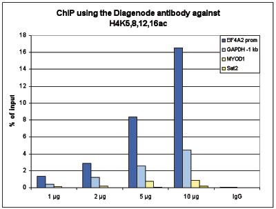
<p><small><strong> Figure 1. ChIP results obtained with the Diagenode antibody directed against H4K5,8,12,16ac </strong><br />ChIP assays were performed using human HeLa cells, the Diagenode antibody against H4K5,8,12,16ac (cat. No. C15410024) and optimized PCR primer sets for qPCR. ChIP was performed with the “iDeal ChIP- seq” kit (cat. No. AB-001-0024) on sheared chromatin from 1 million cells. A titration of the antibody consisting of 1, 2, 5 and 10 μg per ChIP experiment was analysed. IgG (2 μg/IP) was used as negative IP control. QPCR was performed with primers for promoter of the active gene EIF4A2 and for a region 1 kb upstream of the GAPDH gene, used as positive controls, and for the inactive MYOD1 gene and the Sat2 satellite repeat region used as negative controls. Figure 1 shows the recovery, expressed as a % of input (the relative amount of immunoprecipitated DNA compared to input DNA after qPCR analysis). </small></p>
</div>
</div>
<div class="row">
<div class="small-5 columns">
<p><img src="https://www.diagenode.com/img/product/antibodies/C15410024-Fig2A-ChIp-seq.png" alt="H4K5,8,12,16ac Antibody ChIP-seq Grade " caption="false" width="367" height="46" /></p>
<p><img src="https://www.diagenode.com/img/product/antibodies/C15410024-Fig2B-ChIp-seq.png" alt="H4K5,8,12,16ac Antibody for ChIP-seq" caption="false" width="367" height="62" /></p>
<p><img src="https://www.diagenode.com/img/product/antibodies/C15410024-Fig2C-ChIp-seq.png" alt="H4K5,8,12,16ac Antibody for ChIP-seq assay" caption="false" width="367" height="76" /></p>
<p><img src="https://www.diagenode.com/img/product/antibodies/C15410024-Fig2D-ChIp-seq.png" alt="H4K5,8,12,16ac Antibody validated in ChIP-seq" caption="false" width="367" height="89" /></p>
</div>
<div class="small-7 columns">
<p><small><strong> Figure 2. ChIP-seq results obtained with the Diagenode antibody directed against H4K5,8,12,16ac </strong><br />ChIP was performed with 2 μg of the Diagenode antibody against H4K5,8,12,16ac (cat. No. C15410024) on sheared chromatin from 1 million HeLa cells using the “iDeal ChIP-seq” kit as described above. The IP’d DNA was subsequently analysed on an Illumina Genome Analyzer. Library preparation, cluster generation and sequencing were performed according to the manufacturer’s instructions. The 36 bp tags were aligned to the human genome using the ELAND algorithm. Figure 2 shows the signal distribution along the complete length of chromosome 5 (figure 2A) and a zoomin to a 600 kb region (figure 2B). Figure 2C and D show the enrichment in two genomic regions on chromosome 3 and 12, respectively, containing EIF4A2 and GAPDH positive controls. The position of the amplicon used for validating the QPCR results is shown with an arrow </small></p>
</div>
</div>
<div class="row">
<div class="small-4 columns">
<p><img src="https://www.diagenode.com/img/product/antibodies/C15410024-Fig3-ELISA.png" alt="H4K5,8,12,16ac Antibody ELISA validation" caption="false" width="288" height="218" /></p>
</div>
<div class="small-8 columns">
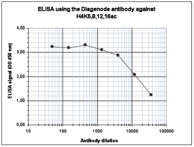
<p><small><strong> Figure 3. Determination of the antibody titer </strong><br />To determine the titer of the antibody, an ELISA was performed using a serial dilution of the Diagenode antibody directed against H4K5,8,12,16ac (cat. No. C15410024) in antigen coated wells. The antigen used was a peptide containing the histone modification of interest. By plotting the absorbance against the antibody dilution (Figure 3), the titer of the antibody was estimated to be 1:21,200. </small></p>
</div>
</div>
<div class="row">
<div class="small-4 columns">
<p><img src="https://www.diagenode.com/img/product/antibodies/C15410024-Fig4-DB.png" alt="H4K5,8,12,16ac Antibody Dot Blot validation" caption="false" width="288" height="218" /></p>
</div>
<div class="small-8 columns">
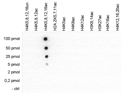
<p><small><strong> Figure 4. Cross reactivity tests using the Diagenode antibody directed against H4K5,8,12,16ac </strong><br />To test the cross reactivity of the Diagenode antibody against H4K5,8,12,16ac (cat. No. C15410024), a Dot Blot analysis was performed with peptides containing other histone modifications and the unmodified H4. One hundred to 0.2 pmol of the respective peptides were spotted on a membrane. The antibody was used at a dilution of 1:20,000. Figure 4 shows a high specificity of the antibody for the modification of interest. </small></p>
</div>
</div>
<div class="row">
<div class="small-4 columns">
<p><img src="https://www.diagenode.com/img/product/antibodies/C15410024-Fig5-IF.png" alt="H4K5,8,12,16ac Antibody validated in Immunofluorescence" caption="false" width="288" height="70" /></p>
</div>
<div class="small-8 columns">
<p><small><strong> Figure 5. Immunofluorescence using the Diagenode antibody directed against H4K5,8,12,16ac </strong><br />Mouse NIH3T3 cells were stained with the Diagenode antibody against H4K5,8,12,16ac (cat. No. C15410024) and with DAPI. Cells were fixed with 4% formaldehyde for 10’ and blocked with PBS/TX-100 containing 5% normal goat serum and 1% BSA. The cells were immunofluorescently labeled with the H4K5,8,12,16ac antibody (left) diluted 1:500 in blocking solution followed by an anti- rabbit antibody conjugated to Alexa488. The middle panel shows staining of the nuclei with DAPI. A merge of the two stainings is shown on the right. </small></p>
</div>
</div>',
'label2' => 'Target Description',
'info2' => '<p>Histones are the main constituents of the protein part of chromosomes of eukaryotic cells. They are rich in the amino acids arginine and lysine and have been greatly conserved during evolution. Histones pack the DNA into tight masses of chromatin. Two core histones of each class H2A, H2B, H3 and H4 assemble and are wrapped by 146 base pairs of DNA to form one octameric nucleosome. Histone tails undergo numerous post-translational modifications, which either directly or indirectly alter chromatin structure to facilitate transcriptional activation or repression or other nuclear processes. In addition to the genetic code, combinations of the different histone modifications reveal the so-called “histone code”. Histone methylation and demethylation is dynamically regulated by respectively histone methyl transferases and histone demethylases. Acetylation of histone H4 is associated with active genes.</p>',
'label3' => '',
'info3' => '',
'format' => '10 μg',
'catalog_number' => 'C15410024-10',
'old_catalog_number' => '',
'sf_code' => 'C15410024-D001-000582',
'type' => 'FRE',
'search_order' => '03-Antibody',
'price_EUR' => '105',
'price_USD' => '120',
'price_GBP' => '100',
'price_JPY' => '17205',
'price_CNY' => '',
'price_AUD' => '300',
'country' => 'ALL',
'except_countries' => 'None',
'quote' => false,
'in_stock' => false,
'featured' => false,
'no_promo' => false,
'online' => true,
'master' => false,
'last_datasheet_update' => '0000-00-00',
'slug' => 'h4k5-8-12-16ac-polyclonal-antibody-classic-sample-size-10-ug',
'meta_title' => 'H4K5,8,12,16ac Antibody - ChIP-seq Grade (C15410024-10) | Diagenode',
'meta_keywords' => '',
'meta_description' => 'H4K5,8,12,16ac (Histone H4 acetylated at lysines 5, 8, 12 and 16) Polyclonal Antibody validated in ChIP-seq, ChIP-qPCR, DB, IF and ELISA. Batch-specific data available on the website. Sample size available',
'modified' => '2022-01-05 14:50:58',
'created' => '2015-07-30 11:48:07',
'locale' => 'eng'
),
'Antibody' => array(
'host' => '*****',
'id' => '44',
'name' => 'H4K5,8,12,16ac polyclonal antibody',
'description' => 'Histones are the main constituents of the protein part of chromosomes of eukaryotic cells. They are rich in the amino acids arginine and lysine and have been greatly conserved during evolution. Histones pack the DNA into tight masses of chromatin. Two core histones of each class H2A, H2B, H3 and H4 assemble and are wrapped by 146 base pairs of DNA to form one octameric nucleosome. Histone tails undergo numerous post-translational modifications, which either directly or indirectly alter chromatin structure to facilitate transcriptional activation or repression or other nuclear processes. In addition to the genetic code, combinations of the different histone modifications reveal the so-called “histone code”. Histone methylation and demethylation is dynamically regulated by respectively histone methyl transferases and histone demethylases. Acetylation of histone H4 is associated with active genes.',
'clonality' => '',
'isotype' => '',
'lot' => 'A0607P',
'concentration' => '0.76 µg/µl',
'reactivity' => 'Human, mouse, silena latifolia, wide range expected.',
'type' => 'Polyclonal',
'purity' => 'Affinity purified polyclonal antibody.',
'classification' => 'Classic',
'application_table' => '<table>
<thead>
<tr>
<th>Applications</th>
<th>Suggested dilution</th>
<th>References</th>
</tr>
</thead>
<tbody>
<tr>
<td>ChIP/ChIP-seq <sup>*</sup></td>
<td>2 µg/IP</td>
<td>Fig 1, 2</td>
</tr>
<tr>
<td>ELISA</td>
<td>1:1,000</td>
<td>Fig 3</td>
</tr>
<tr>
<td>Dot Blotting</td>
<td>1:20,000</td>
<td>Fig 4</td>
</tr>
<tr>
<td>IF</td>
<td>1:500</td>
<td>Fig 5</td>
</tr>
</tbody>
</table>
<p></p>
<p><small><sup>*</sup> Please note that the optimal antibody amount per ChIP should be determined by the end-user. We recommend testing 1-5 µg per IP.</small></p>',
'storage_conditions' => 'Store at -20°C; for long storage, store at -80°C. Avoid multiple freeze-thaw cycles.',
'storage_buffer' => 'PBS containing 0.05% azide and 0.05% ProClin 300.',
'precautions' => 'This product is for research use only. Not for use in diagnostic or therapeutic procedures.',
'uniprot_acc' => '',
'slug' => '',
'meta_keywords' => '',
'meta_description' => '',
'modified' => '2020-07-01 11:17:45',
'created' => '0000-00-00 00:00:00',
'select_label' => '44 - H4K5,8,12,16ac polyclonal antibody (A0607P - 0.76 µg/µl - Human, mouse, silena latifolia, wide range expected. - Affinity purified polyclonal antibody. - Rabbit)'
),
'Slave' => array(),
'Group' => array(
'Group' => array(
[maximum depth reached]
),
'Master' => array(
[maximum depth reached]
),
'Product' => array(
[maximum depth reached]
)
),
'Related' => array(
(int) 0 => array(
[maximum depth reached]
)
),
'Application' => array(
(int) 0 => array(
[maximum depth reached]
),
(int) 1 => array(
[maximum depth reached]
),
(int) 2 => array(
[maximum depth reached]
),
(int) 3 => array(
[maximum depth reached]
),
(int) 4 => array(
[maximum depth reached]
)
),
'Category' => array(
(int) 0 => array(
[maximum depth reached]
),
(int) 1 => array(
[maximum depth reached]
),
(int) 2 => array(
[maximum depth reached]
),
(int) 3 => array(
[maximum depth reached]
),
(int) 4 => array(
[maximum depth reached]
)
),
'Document' => array(
(int) 0 => array(
[maximum depth reached]
),
(int) 1 => array(
[maximum depth reached]
),
(int) 2 => array(
[maximum depth reached]
)
),
'Feature' => array(),
'Image' => array(
(int) 0 => array(
[maximum depth reached]
)
),
'Promotion' => array(),
'Protocol' => array(),
'Publication' => array(
(int) 0 => array(
[maximum depth reached]
),
(int) 1 => array(
[maximum depth reached]
)
),
'Testimonial' => array(),
'Area' => array(),
'SafetySheet' => array(
(int) 0 => array(
[maximum depth reached]
),
(int) 1 => array(
[maximum depth reached]
),
(int) 2 => array(
[maximum depth reached]
),
(int) 3 => array(
[maximum depth reached]
),
(int) 4 => array(
[maximum depth reached]
),
(int) 5 => array(
[maximum depth reached]
),
(int) 6 => array(
[maximum depth reached]
),
(int) 7 => array(
[maximum depth reached]
)
)
),
'meta_canonical' => 'https://www.diagenode.com/en/p/h4k5-8-12-16ac-polyclonal-antibody-classic-50-mg-66-ml'
)
$language = 'en'
$meta_keywords = ''
$meta_description = 'H4K5,8,12,16ac (Histone H4 acetylated at lysines 5, 8, 12 and 16) Polyclonal Antibody validated in ChIP-seq, ChIP-qPCR, DB, IF and ELISA. Batch-specific data available on the website. Sample size available'
$meta_title = 'H4K5,8,12,16ac Antibody - ChIP-seq Grade (C15410024-10) | Diagenode'
$product = array(
'Product' => array(
'id' => '2660',
'antibody_id' => '44',
'name' => 'H4K5,8,12,16ac Antibody (sample size)',
'description' => '<p><span>Polyclonal antibody raised in rabbit against the region of histone <strong>H4 containing the acetylated lysines 5, 8, 12 and 16 (H4K5,8,12,16ac)</strong>, using a KLH-conjugated synthetic peptide.</span></p>',
'label1' => 'Validation data',
'info1' => '<div class="row">
<div class="small-4 columns">
<p><img src="https://www.diagenode.com/img/product/antibodies/C15410024-Fig1-ChIP.png" alt="ChIPH4K5,8,12,16ac Antibody ChIP Grade " caption="false" width="288" height="218" /></p>
</div>
<div class="small-8 columns">
<p><small><strong> Figure 1. ChIP results obtained with the Diagenode antibody directed against H4K5,8,12,16ac </strong><br />ChIP assays were performed using human HeLa cells, the Diagenode antibody against H4K5,8,12,16ac (cat. No. C15410024) and optimized PCR primer sets for qPCR. ChIP was performed with the “iDeal ChIP- seq” kit (cat. No. AB-001-0024) on sheared chromatin from 1 million cells. A titration of the antibody consisting of 1, 2, 5 and 10 μg per ChIP experiment was analysed. IgG (2 μg/IP) was used as negative IP control. QPCR was performed with primers for promoter of the active gene EIF4A2 and for a region 1 kb upstream of the GAPDH gene, used as positive controls, and for the inactive MYOD1 gene and the Sat2 satellite repeat region used as negative controls. Figure 1 shows the recovery, expressed as a % of input (the relative amount of immunoprecipitated DNA compared to input DNA after qPCR analysis). </small></p>
</div>
</div>
<div class="row">
<div class="small-5 columns">
<p><img src="https://www.diagenode.com/img/product/antibodies/C15410024-Fig2A-ChIp-seq.png" alt="H4K5,8,12,16ac Antibody ChIP-seq Grade " caption="false" width="367" height="46" /></p>
<p><img src="https://www.diagenode.com/img/product/antibodies/C15410024-Fig2B-ChIp-seq.png" alt="H4K5,8,12,16ac Antibody for ChIP-seq" caption="false" width="367" height="62" /></p>
<p><img src="https://www.diagenode.com/img/product/antibodies/C15410024-Fig2C-ChIp-seq.png" alt="H4K5,8,12,16ac Antibody for ChIP-seq assay" caption="false" width="367" height="76" /></p>
<p><img src="https://www.diagenode.com/img/product/antibodies/C15410024-Fig2D-ChIp-seq.png" alt="H4K5,8,12,16ac Antibody validated in ChIP-seq" caption="false" width="367" height="89" /></p>
</div>
<div class="small-7 columns">
<p><small><strong> Figure 2. ChIP-seq results obtained with the Diagenode antibody directed against H4K5,8,12,16ac </strong><br />ChIP was performed with 2 μg of the Diagenode antibody against H4K5,8,12,16ac (cat. No. C15410024) on sheared chromatin from 1 million HeLa cells using the “iDeal ChIP-seq” kit as described above. The IP’d DNA was subsequently analysed on an Illumina Genome Analyzer. Library preparation, cluster generation and sequencing were performed according to the manufacturer’s instructions. The 36 bp tags were aligned to the human genome using the ELAND algorithm. Figure 2 shows the signal distribution along the complete length of chromosome 5 (figure 2A) and a zoomin to a 600 kb region (figure 2B). Figure 2C and D show the enrichment in two genomic regions on chromosome 3 and 12, respectively, containing EIF4A2 and GAPDH positive controls. The position of the amplicon used for validating the QPCR results is shown with an arrow </small></p>
</div>
</div>
<div class="row">
<div class="small-4 columns">
<p><img src="https://www.diagenode.com/img/product/antibodies/C15410024-Fig3-ELISA.png" alt="H4K5,8,12,16ac Antibody ELISA validation" caption="false" width="288" height="218" /></p>
</div>
<div class="small-8 columns">
<p><small><strong> Figure 3. Determination of the antibody titer </strong><br />To determine the titer of the antibody, an ELISA was performed using a serial dilution of the Diagenode antibody directed against H4K5,8,12,16ac (cat. No. C15410024) in antigen coated wells. The antigen used was a peptide containing the histone modification of interest. By plotting the absorbance against the antibody dilution (Figure 3), the titer of the antibody was estimated to be 1:21,200. </small></p>
</div>
</div>
<div class="row">
<div class="small-4 columns">
<p><img src="https://www.diagenode.com/img/product/antibodies/C15410024-Fig4-DB.png" alt="H4K5,8,12,16ac Antibody Dot Blot validation" caption="false" width="288" height="218" /></p>
</div>
<div class="small-8 columns">
<p><small><strong> Figure 4. Cross reactivity tests using the Diagenode antibody directed against H4K5,8,12,16ac </strong><br />To test the cross reactivity of the Diagenode antibody against H4K5,8,12,16ac (cat. No. C15410024), a Dot Blot analysis was performed with peptides containing other histone modifications and the unmodified H4. One hundred to 0.2 pmol of the respective peptides were spotted on a membrane. The antibody was used at a dilution of 1:20,000. Figure 4 shows a high specificity of the antibody for the modification of interest. </small></p>
</div>
</div>
<div class="row">
<div class="small-4 columns">
<p><img src="https://www.diagenode.com/img/product/antibodies/C15410024-Fig5-IF.png" alt="H4K5,8,12,16ac Antibody validated in Immunofluorescence" caption="false" width="288" height="70" /></p>
</div>
<div class="small-8 columns">
<p><small><strong> Figure 5. Immunofluorescence using the Diagenode antibody directed against H4K5,8,12,16ac </strong><br />Mouse NIH3T3 cells were stained with the Diagenode antibody against H4K5,8,12,16ac (cat. No. C15410024) and with DAPI. Cells were fixed with 4% formaldehyde for 10’ and blocked with PBS/TX-100 containing 5% normal goat serum and 1% BSA. The cells were immunofluorescently labeled with the H4K5,8,12,16ac antibody (left) diluted 1:500 in blocking solution followed by an anti- rabbit antibody conjugated to Alexa488. The middle panel shows staining of the nuclei with DAPI. A merge of the two stainings is shown on the right. </small></p>
</div>
</div>',
'label2' => 'Target Description',
'info2' => '<p>Histones are the main constituents of the protein part of chromosomes of eukaryotic cells. They are rich in the amino acids arginine and lysine and have been greatly conserved during evolution. Histones pack the DNA into tight masses of chromatin. Two core histones of each class H2A, H2B, H3 and H4 assemble and are wrapped by 146 base pairs of DNA to form one octameric nucleosome. Histone tails undergo numerous post-translational modifications, which either directly or indirectly alter chromatin structure to facilitate transcriptional activation or repression or other nuclear processes. In addition to the genetic code, combinations of the different histone modifications reveal the so-called “histone code”. Histone methylation and demethylation is dynamically regulated by respectively histone methyl transferases and histone demethylases. Acetylation of histone H4 is associated with active genes.</p>',
'label3' => '',
'info3' => '',
'format' => '10 μg',
'catalog_number' => 'C15410024-10',
'old_catalog_number' => '',
'sf_code' => 'C15410024-D001-000582',
'type' => 'FRE',
'search_order' => '03-Antibody',
'price_EUR' => '105',
'price_USD' => '120',
'price_GBP' => '100',
'price_JPY' => '17205',
'price_CNY' => '',
'price_AUD' => '300',
'country' => 'ALL',
'except_countries' => 'None',
'quote' => false,
'in_stock' => false,
'featured' => false,
'no_promo' => false,
'online' => true,
'master' => false,
'last_datasheet_update' => '0000-00-00',
'slug' => 'h4k5-8-12-16ac-polyclonal-antibody-classic-sample-size-10-ug',
'meta_title' => 'H4K5,8,12,16ac Antibody - ChIP-seq Grade (C15410024-10) | Diagenode',
'meta_keywords' => '',
'meta_description' => 'H4K5,8,12,16ac (Histone H4 acetylated at lysines 5, 8, 12 and 16) Polyclonal Antibody validated in ChIP-seq, ChIP-qPCR, DB, IF and ELISA. Batch-specific data available on the website. Sample size available',
'modified' => '2022-01-05 14:50:58',
'created' => '2015-07-30 11:48:07',
'locale' => 'eng'
),
'Antibody' => array(
'host' => '*****',
'id' => '44',
'name' => 'H4K5,8,12,16ac polyclonal antibody',
'description' => 'Histones are the main constituents of the protein part of chromosomes of eukaryotic cells. They are rich in the amino acids arginine and lysine and have been greatly conserved during evolution. Histones pack the DNA into tight masses of chromatin. Two core histones of each class H2A, H2B, H3 and H4 assemble and are wrapped by 146 base pairs of DNA to form one octameric nucleosome. Histone tails undergo numerous post-translational modifications, which either directly or indirectly alter chromatin structure to facilitate transcriptional activation or repression or other nuclear processes. In addition to the genetic code, combinations of the different histone modifications reveal the so-called “histone code”. Histone methylation and demethylation is dynamically regulated by respectively histone methyl transferases and histone demethylases. Acetylation of histone H4 is associated with active genes.',
'clonality' => '',
'isotype' => '',
'lot' => 'A0607P',
'concentration' => '0.76 µg/µl',
'reactivity' => 'Human, mouse, silena latifolia, wide range expected.',
'type' => 'Polyclonal',
'purity' => 'Affinity purified polyclonal antibody.',
'classification' => 'Classic',
'application_table' => '<table>
<thead>
<tr>
<th>Applications</th>
<th>Suggested dilution</th>
<th>References</th>
</tr>
</thead>
<tbody>
<tr>
<td>ChIP/ChIP-seq <sup>*</sup></td>
<td>2 µg/IP</td>
<td>Fig 1, 2</td>
</tr>
<tr>
<td>ELISA</td>
<td>1:1,000</td>
<td>Fig 3</td>
</tr>
<tr>
<td>Dot Blotting</td>
<td>1:20,000</td>
<td>Fig 4</td>
</tr>
<tr>
<td>IF</td>
<td>1:500</td>
<td>Fig 5</td>
</tr>
</tbody>
</table>
<p></p>
<p><small><sup>*</sup> Please note that the optimal antibody amount per ChIP should be determined by the end-user. We recommend testing 1-5 µg per IP.</small></p>',
'storage_conditions' => 'Store at -20°C; for long storage, store at -80°C. Avoid multiple freeze-thaw cycles.',
'storage_buffer' => 'PBS containing 0.05% azide and 0.05% ProClin 300.',
'precautions' => 'This product is for research use only. Not for use in diagnostic or therapeutic procedures.',
'uniprot_acc' => '',
'slug' => '',
'meta_keywords' => '',
'meta_description' => '',
'modified' => '2020-07-01 11:17:45',
'created' => '0000-00-00 00:00:00',
'select_label' => '44 - H4K5,8,12,16ac polyclonal antibody (A0607P - 0.76 µg/µl - Human, mouse, silena latifolia, wide range expected. - Affinity purified polyclonal antibody. - Rabbit)'
),
'Slave' => array(),
'Group' => array(
'Group' => array(
'id' => '40',
'name' => 'C15410024',
'product_id' => '2189',
'modified' => '2016-02-18 20:47:10',
'created' => '2016-02-18 20:47:10'
),
'Master' => array(
'id' => '2189',
'antibody_id' => '44',
'name' => 'H4K5,8,12,16ac Antibody',
'description' => '<p><span>Polyclonal antibody raised in rabbit against the region of histone<strong> H4 containing the acetylated lysines 5, 8, 12 and 16 (H4K5,8,12,16ac)</strong>, using a KLH-conjugated synthetic peptide.</span></p>',
'label1' => 'Validation data',
'info1' => '<div class="row">
<div class="small-5 columns">
<p><img src="https://www.diagenode.com/img/product/antibodies/C15410024-Fig1-ChIP.png" alt="H4K5,8,12,16ac Antibody ChIP Grade" /></p>
</div>
<div class="small-7 columns">
<p><small><strong>Figure 1. ChIP results obtained with the Diagenode antibody directed against H4K5,8,12,16ac </strong><br />ChIP assays were performed using human HeLa cells, the Diagenode antibody against H4K5,8,12,16ac (Cat. No. C15410024) and optimized PCR primer sets for qPCR. ChIP was performed with the “iDeal ChIP- seq” kit (Cat. No. AB-001-0024) on sheared chromatin from 1 million cells. A titration of the antibody consisting of 1, 2, 5 and 10 µg per ChIP experiment was analysed. IgG (2 µg/IP) was used as negative IP control. QPCR was performed with primers for promoter of the active gene EIF4A2 and for a region 1 kb upstream of the GAPDH gene, used as positive controls, and for the inactive MYOD1 gene and the Sat2 satellite repeat region used as negative controls. Figure 1 shows the recovery, expressed as a % of input (the relative amount of immunoprecipitated DNA compared to input DNA after qPCR analysis). </small></p>
</div>
</div>
<div class="row">
<div class="small-12 columns">
<p>A. <img src="https://www.diagenode.com/img/product/antibodies/C15410024-Fig2A-ChIp-seq.png" alt="H4K5,8,12,16ac Antibody ChIP-seq Grade" /></p>
<p>B. <img src="https://www.diagenode.com/img/product/antibodies/C15410024-Fig2B-ChIp-seq.png" alt="H4K5,8,12,16ac Antibody for ChIP-seq" /></p>
<p>C. <img src="https://www.diagenode.com/img/product/antibodies/C15410024-Fig2C-ChIp-seq.png" alt="H4K5,8,12,16ac Antibody for ChIP-seq assay" /></p>
</div>
</div>
<div class="extra-spaced"></div>
<div class="row">
<div class="small-12 columns">
<p>D. <img src="https://www.diagenode.com/img/product/antibodies/C15410024-Fig2D-ChIp-seq.png" alt="H4K5,8,12,16ac Antibody validated in ChIP-seq " /></p>
</div>
</div>
<div class="row">
<div class="small-12 columns">
<p><small><strong>Figure 2. ChIP-seq results obtained with the Diagenode antibody directed against H4K5,8,12,16ac </strong><br />ChIP was performed with 2 μg of the Diagenode antibody against H4K5,8,12,16ac (Cat. No. C15410024) on sheared chromatin from 1 million HeLa cells using the “iDeal ChIP-seq” kit as described above. The IP’d DNA was subsequently analysed on an Illumina Genome Analyzer. Library preparation, cluster generation and sequencing were performed according to the manufacturer’s instructions. The 36 bp tags were aligned to the human genome using the ELAND algorithm. Figure 2 shows the signal distribution along the complete length of chromosome 5 (figure 2A) and a zoomin to a 600 kb region (figure 2B). Figure 2C and D show the enrichment in two genomic regions on chromosome 3 and 12, respectively, containing EIF4A2 and GAPDH positive controls. The position of the amplicon used for validating the QPCR results is shown with an arrow </small></p>
</div>
</div>
<div class="row">
<div class="small-5 columns">
<p><img src="https://www.diagenode.com/img/product/antibodies/C15410024-Fig3-ELISA.png" alt="H4K5,8,12,16ac Antibody ELISA validation" /></p>
</div>
<div class="small-7 columns">
<p><small><strong> Figure 3. Determination of the antibody titer </strong><br />To determine the titer of the antibody, an ELISA was performed using a serial dilution of the Diagenode antibody directed against H4K5,8,12,16ac (Cat. No. C15410024) in antigen coated wells. The antigen used was a peptide containing the histone modification of interest. By plotting the absorbance against the antibody dilution (Figure 3), the titer of the antibody was estimated to be 1:21,200. </small></p>
</div>
</div>
<div class="row">
<div class="small-5 columns">
<p><img src="https://www.diagenode.com/img/product/antibodies/C15410024-Fig4-DB.png" alt="H4K5,8,12,16ac Antibody Dot Blot validation" /></p>
</div>
<div class="small-7 columns">
<p><small><strong> Figure 4. Cross reactivity tests using the Diagenode antibody directed against H4K5,8,12,16ac </strong><br />To test the cross reactivity of the Diagenode antibody against H4K5,8,12,16ac (Cat. No. C15410024), a Dot Blot analysis was performed with peptides containing other histone modifications and the unmodified H4. One hundred to 0.2 pmol of the respective peptides were spotted on a membrane. The antibody was used at a dilution of 1:20,000. Figure 4 shows a high specificity of the antibody for the modification of interest. </small></p>
</div>
</div>
<div class="extra-spaced"></div>
<div class="row">
<div class="small-5 columns">
<p><img src="https://www.diagenode.com/img/product/antibodies/C15410024-Fig5-IF.png" alt="H4K5,8,12,16ac Antibody validated in Immunofluorescence" /></p>
</div>
<div class="small-7 columns">
<p><small><strong> Figure 5. Immunofluorescence using the Diagenode antibody directed against H4K5,8,12,16ac </strong><br />Mouse NIH3T3 cells were stained with the Diagenode antibody against H4K5,8,12,16ac (Cat. No. C15410024) and with DAPI. Cells were fixed with 4% formaldehyde for 10’ and blocked with PBS/TX-100 containing 5% normal goat serum and 1% BSA. The cells were immunofluorescently labeled with the H4K5,8,12,16ac antibody (left) diluted 1:500 in blocking solution followed by an anti- rabbit antibody conjugated to Alexa488. The middle panel shows staining of the nuclei with DAPI. A merge of the two stainings is shown on the right. </small></p>
</div>
</div>',
'label2' => '',
'info2' => '',
'label3' => '',
'info3' => '',
'format' => '50 μg',
'catalog_number' => 'C15410024',
'old_catalog_number' => '',
'sf_code' => 'C15410024-D001-000581',
'type' => 'FRE',
'search_order' => '03-Antibody',
'price_EUR' => '390',
'price_USD' => '390',
'price_GBP' => '345',
'price_JPY' => '63900',
'price_CNY' => '',
'price_AUD' => '975',
'country' => 'ALL',
'except_countries' => 'None',
'quote' => false,
'in_stock' => false,
'featured' => false,
'no_promo' => false,
'online' => true,
'master' => true,
'last_datasheet_update' => 'June 17, 2020',
'slug' => 'h4k5-8-12-16ac-polyclonal-antibody-classic-50-mg-66-ml',
'meta_title' => 'H4K5,8,12,16ac Antibody - ChIP-seq Grade (C15410024) | Diagenode',
'meta_keywords' => '',
'meta_description' => 'H4K5,8,12,16ac (Histone H4 acetylated at lysines 5, 8, 12 and 16) Polyclonal Antibody validated in ChIP-seq, ChIP-qPCR, IF and DB. Batch-specific data available on the website. Sample size available.',
'modified' => '2022-01-05 14:50:44',
'created' => '2015-06-29 14:08:20'
),
'Product' => array(
(int) 0 => array(
[maximum depth reached]
)
)
),
'Related' => array(
(int) 0 => array(
'id' => '2189',
'antibody_id' => '44',
'name' => 'H4K5,8,12,16ac Antibody',
'description' => '<p><span>Polyclonal antibody raised in rabbit against the region of histone<strong> H4 containing the acetylated lysines 5, 8, 12 and 16 (H4K5,8,12,16ac)</strong>, using a KLH-conjugated synthetic peptide.</span></p>',
'label1' => 'Validation data',
'info1' => '<div class="row">
<div class="small-5 columns">
<p><img src="https://www.diagenode.com/img/product/antibodies/C15410024-Fig1-ChIP.png" alt="H4K5,8,12,16ac Antibody ChIP Grade" /></p>
</div>
<div class="small-7 columns">
<p><small><strong>Figure 1. ChIP results obtained with the Diagenode antibody directed against H4K5,8,12,16ac </strong><br />ChIP assays were performed using human HeLa cells, the Diagenode antibody against H4K5,8,12,16ac (Cat. No. C15410024) and optimized PCR primer sets for qPCR. ChIP was performed with the “iDeal ChIP- seq” kit (Cat. No. AB-001-0024) on sheared chromatin from 1 million cells. A titration of the antibody consisting of 1, 2, 5 and 10 µg per ChIP experiment was analysed. IgG (2 µg/IP) was used as negative IP control. QPCR was performed with primers for promoter of the active gene EIF4A2 and for a region 1 kb upstream of the GAPDH gene, used as positive controls, and for the inactive MYOD1 gene and the Sat2 satellite repeat region used as negative controls. Figure 1 shows the recovery, expressed as a % of input (the relative amount of immunoprecipitated DNA compared to input DNA after qPCR analysis). </small></p>
</div>
</div>
<div class="row">
<div class="small-12 columns">
<p>A. <img src="https://www.diagenode.com/img/product/antibodies/C15410024-Fig2A-ChIp-seq.png" alt="H4K5,8,12,16ac Antibody ChIP-seq Grade" /></p>
<p>B. <img src="https://www.diagenode.com/img/product/antibodies/C15410024-Fig2B-ChIp-seq.png" alt="H4K5,8,12,16ac Antibody for ChIP-seq" /></p>
<p>C. <img src="https://www.diagenode.com/img/product/antibodies/C15410024-Fig2C-ChIp-seq.png" alt="H4K5,8,12,16ac Antibody for ChIP-seq assay" /></p>
</div>
</div>
<div class="extra-spaced"></div>
<div class="row">
<div class="small-12 columns">
<p>D. <img src="https://www.diagenode.com/img/product/antibodies/C15410024-Fig2D-ChIp-seq.png" alt="H4K5,8,12,16ac Antibody validated in ChIP-seq " /></p>
</div>
</div>
<div class="row">
<div class="small-12 columns">
<p><small><strong>Figure 2. ChIP-seq results obtained with the Diagenode antibody directed against H4K5,8,12,16ac </strong><br />ChIP was performed with 2 μg of the Diagenode antibody against H4K5,8,12,16ac (Cat. No. C15410024) on sheared chromatin from 1 million HeLa cells using the “iDeal ChIP-seq” kit as described above. The IP’d DNA was subsequently analysed on an Illumina Genome Analyzer. Library preparation, cluster generation and sequencing were performed according to the manufacturer’s instructions. The 36 bp tags were aligned to the human genome using the ELAND algorithm. Figure 2 shows the signal distribution along the complete length of chromosome 5 (figure 2A) and a zoomin to a 600 kb region (figure 2B). Figure 2C and D show the enrichment in two genomic regions on chromosome 3 and 12, respectively, containing EIF4A2 and GAPDH positive controls. The position of the amplicon used for validating the QPCR results is shown with an arrow </small></p>
</div>
</div>
<div class="row">
<div class="small-5 columns">
<p><img src="https://www.diagenode.com/img/product/antibodies/C15410024-Fig3-ELISA.png" alt="H4K5,8,12,16ac Antibody ELISA validation" /></p>
</div>
<div class="small-7 columns">
<p><small><strong> Figure 3. Determination of the antibody titer </strong><br />To determine the titer of the antibody, an ELISA was performed using a serial dilution of the Diagenode antibody directed against H4K5,8,12,16ac (Cat. No. C15410024) in antigen coated wells. The antigen used was a peptide containing the histone modification of interest. By plotting the absorbance against the antibody dilution (Figure 3), the titer of the antibody was estimated to be 1:21,200. </small></p>
</div>
</div>
<div class="row">
<div class="small-5 columns">
<p><img src="https://www.diagenode.com/img/product/antibodies/C15410024-Fig4-DB.png" alt="H4K5,8,12,16ac Antibody Dot Blot validation" /></p>
</div>
<div class="small-7 columns">
<p><small><strong> Figure 4. Cross reactivity tests using the Diagenode antibody directed against H4K5,8,12,16ac </strong><br />To test the cross reactivity of the Diagenode antibody against H4K5,8,12,16ac (Cat. No. C15410024), a Dot Blot analysis was performed with peptides containing other histone modifications and the unmodified H4. One hundred to 0.2 pmol of the respective peptides were spotted on a membrane. The antibody was used at a dilution of 1:20,000. Figure 4 shows a high specificity of the antibody for the modification of interest. </small></p>
</div>
</div>
<div class="extra-spaced"></div>
<div class="row">
<div class="small-5 columns">
<p><img src="https://www.diagenode.com/img/product/antibodies/C15410024-Fig5-IF.png" alt="H4K5,8,12,16ac Antibody validated in Immunofluorescence" /></p>
</div>
<div class="small-7 columns">
<p><small><strong> Figure 5. Immunofluorescence using the Diagenode antibody directed against H4K5,8,12,16ac </strong><br />Mouse NIH3T3 cells were stained with the Diagenode antibody against H4K5,8,12,16ac (Cat. No. C15410024) and with DAPI. Cells were fixed with 4% formaldehyde for 10’ and blocked with PBS/TX-100 containing 5% normal goat serum and 1% BSA. The cells were immunofluorescently labeled with the H4K5,8,12,16ac antibody (left) diluted 1:500 in blocking solution followed by an anti- rabbit antibody conjugated to Alexa488. The middle panel shows staining of the nuclei with DAPI. A merge of the two stainings is shown on the right. </small></p>
</div>
</div>',
'label2' => '',
'info2' => '',
'label3' => '',
'info3' => '',
'format' => '50 μg',
'catalog_number' => 'C15410024',
'old_catalog_number' => '',
'sf_code' => 'C15410024-D001-000581',
'type' => 'FRE',
'search_order' => '03-Antibody',
'price_EUR' => '390',
'price_USD' => '390',
'price_GBP' => '345',
'price_JPY' => '63900',
'price_CNY' => '',
'price_AUD' => '975',
'country' => 'ALL',
'except_countries' => 'None',
'quote' => false,
'in_stock' => false,
'featured' => false,
'no_promo' => false,
'online' => true,
'master' => true,
'last_datasheet_update' => 'June 17, 2020',
'slug' => 'h4k5-8-12-16ac-polyclonal-antibody-classic-50-mg-66-ml',
'meta_title' => 'H4K5,8,12,16ac Antibody - ChIP-seq Grade (C15410024) | Diagenode',
'meta_keywords' => '',
'meta_description' => 'H4K5,8,12,16ac (Histone H4 acetylated at lysines 5, 8, 12 and 16) Polyclonal Antibody validated in ChIP-seq, ChIP-qPCR, IF and DB. Batch-specific data available on the website. Sample size available.',
'modified' => '2022-01-05 14:50:44',
'created' => '2015-06-29 14:08:20',
'ProductsRelated' => array(
[maximum depth reached]
),
'Image' => array(
[maximum depth reached]
)
)
),
'Application' => array(
(int) 0 => array(
'id' => '20',
'position' => '10',
'parent_id' => '40',
'name' => 'ELISA',
'description' => '<div class="row">
<div class="small-12 medium-12 large-12 columns">Enzyme-linked immunosorbent assay.</div>
</div>',
'in_footer' => false,
'in_menu' => false,
'online' => true,
'tabular' => true,
'slug' => 'elisa-antibodies',
'meta_keywords' => ' ELISA Antibodies,Monoclonal antibody, Polyclonal antibody',
'meta_description' => 'Diagenode offers Monoclonal & Polyclonal antibodies for ELISA applications',
'meta_title' => 'ELISA Antibodies - Monoclonal & Polyclonal antibody | Diagenode',
'modified' => '2016-01-13 12:21:41',
'created' => '2014-07-08 08:13:28',
'ProductsApplication' => array(
[maximum depth reached]
)
),
(int) 1 => array(
'id' => '28',
'position' => '10',
'parent_id' => '40',
'name' => 'DB',
'description' => '<p>Dot blotting</p>',
'in_footer' => false,
'in_menu' => false,
'online' => true,
'tabular' => true,
'slug' => 'dot-blotting',
'meta_keywords' => 'Dot blotting,Monoclonal & Polyclonal antibody,',
'meta_description' => 'Diagenode offers Monoclonal & Polyclonal antibodies for Dot blotting applications',
'meta_title' => 'Dot blotting Antibodies - Monoclonal & Polyclonal antibody | Diagenode',
'modified' => '2016-01-13 14:40:49',
'created' => '2015-07-08 13:45:05',
'ProductsApplication' => array(
[maximum depth reached]
)
),
(int) 2 => array(
'id' => '29',
'position' => '10',
'parent_id' => '40',
'name' => 'IF',
'description' => '<p><strong>Immunofluorescence</strong>:</p>
<p>Diagenode offers huge selection of highly sensitive antibodies validated in IF.</p>
<p><img src="https://www.diagenode.com/img/product/antibodies/C15200229-IF.jpg" alt="" height="245" width="256" /></p>
<p><sup><strong>Immunofluorescence using the Diagenode monoclonal antibody directed against CRISPR/Cas9</strong></sup></p>
<p><sup>HeLa cells transfected with a Cas9 expression vector (left) or untransfected cells (right) were fixed in methanol at -20°C, permeabilized with acetone at -20°C and blocked with PBS containing 2% BSA. The cells were stained with the Cas9 C-terminal antibody (Cat. No. C15200229) diluted 1:400, followed by incubation with an anti-mouse secondary antibody coupled to AF488. The bottom images show counter-staining of the nuclei with Hoechst 33342.</sup></p>
<h5><sup>Check our selection of antibodies validated in IF.</sup></h5>',
'in_footer' => false,
'in_menu' => false,
'online' => true,
'tabular' => true,
'slug' => 'immunofluorescence',
'meta_keywords' => 'Immunofluorescence,Monoclonal antibody,Polyclonal antibody',
'meta_description' => 'Diagenode offers a wide range of antibodies and technical support for Immunofluorescence applications',
'meta_title' => 'Immunofluorescence - Monoclonal antibody - Polyclonal antibody | Diagenode',
'modified' => '2016-04-27 16:23:10',
'created' => '2015-07-08 13:46:02',
'ProductsApplication' => array(
[maximum depth reached]
)
),
(int) 3 => array(
'id' => '42',
'position' => '10',
'parent_id' => '40',
'name' => 'ChIP-seq (ab)',
'description' => '',
'in_footer' => false,
'in_menu' => false,
'online' => true,
'tabular' => true,
'slug' => 'chip-seq-antibodies',
'meta_keywords' => 'Chromatin Immunoprecipitation Sequencing,ChIP-Seq,ChIP-seq grade antibodies,DNA purification,qPCR,Shearing of chromatin',
'meta_description' => 'Diagenode offers a wide range of antibodies and technical support for ChIP Sequencing applications',
'meta_title' => 'ChIP Sequencing Antibodies (ChIP-Seq) | Diagenode',
'modified' => '2016-01-20 11:06:19',
'created' => '2015-10-20 11:44:45',
'ProductsApplication' => array(
[maximum depth reached]
)
),
(int) 4 => array(
'id' => '43',
'position' => '10',
'parent_id' => '40',
'name' => 'ChIP-qPCR (ab)',
'description' => '',
'in_footer' => false,
'in_menu' => false,
'online' => true,
'tabular' => true,
'slug' => 'chip-qpcr-antibodies',
'meta_keywords' => 'Chromatin Immunoprecipitation Sequencing,ChIP-Seq,ChIP-seq grade antibodies,DNA purification,qPCR,Shearing of chromatin',
'meta_description' => 'Diagenode offers a wide range of antibodies and technical support for ChIP-qPCR applications',
'meta_title' => 'ChIP Quantitative PCR Antibodies (ChIP-qPCR) | Diagenode',
'modified' => '2016-01-20 11:30:24',
'created' => '2015-10-20 11:45:36',
'ProductsApplication' => array(
[maximum depth reached]
)
)
),
'Category' => array(
(int) 0 => array(
'id' => '17',
'position' => '10',
'parent_id' => '4',
'name' => 'ChIP-seq grade antibodies',
'description' => '<p><b>Unparalleled ChIP-Seq results with the most rigorously validated antibodies</b></p>
<p><span style="font-weight: 400;">Diagenode provides leading solutions for epigenetic research. Because ChIP-seq is a widely-used technique, we validate our antibodies in ChIP and ChIP-seq experiments (in addition to conventional methods like Western blot, Dot blot, ELISA, and immunofluorescence) to provide the highest quality antibody. We standardize our validation and production to guarantee high product quality without technical bias. Diagenode guarantees ChIP-seq grade antibody performance under our suggested conditions.</span></p>
<div class="row">
<div class="small-12 medium-9 large-9 columns">
<p><strong>ChIP-seq profile</strong> of active (H3K4me3 and H3K36me3) and inactive (H3K27me3) marks using Diagenode antibodies.</p>
<img src="https://www.diagenode.com/img/categories/antibodies/chip-seq-grade-antibodies.png" /></div>
<div class="small-12 medium-3 large-3 columns">
<p><small> ChIP was performed on sheared chromatin from 100,000 K562 cells using iDeal ChIP-seq kit for Histones (cat. No. C01010051) with 1 µg of the Diagenode antibodies against H3K27me3 (cat. No. C15410195) and H3K4me3 (cat. No. C15410003), and 0.5 µg of the antibody against H3K36me3 (cat. No. C15410192). The IP'd DNA was subsequently analysed on an Illumina Genome Analyzer. Library preparation, cluster generation and sequencing were performed according to the manufacturer's instructions. The 36 bp tags were aligned to the human genome using the ELAND algorithm. The figure shows the signal distribution along the complete sequence of human chromosome 3, a zoomin to a 10 Mb region and a further zoomin to a 1.5 Mb region. </small></p>
</div>
</div>
<p>Diagenode’s highly validated antibodies:</p>
<ul>
<li>Highly sensitive and specific</li>
<li>Cost-effective (requires less antibody per reaction)</li>
<li>Batch-specific data is available on the website</li>
<li>Expert technical support</li>
<li>Sample sizes available</li>
<li>100% satisfaction guarantee</li>
</ul>',
'no_promo' => false,
'in_menu' => true,
'online' => true,
'tabular' => false,
'hide' => true,
'all_format' => false,
'is_antibody' => true,
'slug' => 'chip-seq-grade-antibodies',
'cookies_tag_id' => null,
'meta_keywords' => 'ChIP-seq grade antibodies,polyclonal antibody,WB, ELISA, ChIP-seq (ab), ChIP-qPCR (ab)',
'meta_description' => 'Diagenode Offers Wide Range of Validated ChIP-Seq Grade Antibodies for Unparalleled ChIP-Seq Results',
'meta_title' => 'Chromatin Immunoprecipitation ChIP-Seq Grade Antibodies | Diagenode',
'modified' => '2019-07-03 10:57:22',
'created' => '2015-02-16 02:24:01',
'ProductsCategory' => array(
[maximum depth reached]
),
'CookiesTag' => array([maximum depth reached])
),
(int) 1 => array(
'id' => '111',
'position' => '40',
'parent_id' => '4',
'name' => 'Histone antibodies',
'description' => '<p>Histones are the main protein components of chromatin involved in the compaction of DNA into nucleosomes, the basic units of chromatin. A <strong>nucleosome</strong> consists of one pair of each of the core histones (<strong>H2A</strong>, <strong>H2B</strong>, <strong>H3</strong> and <strong>H4</strong>) forming an octameric structure wrapped by 146 base pairs of DNA. The different nucleosomes are linked by the linker histone<strong> H1, </strong>allowing for further condensation of chromatin.</p>
<p>The core histones have a globular structure with large unstructured N-terminal tails protruding from the nucleosome. They can undergo to multiple post-translational modifications (PTM), mainly at the N-terminal tails. These <strong>post-translational modifications </strong>include methylation, acetylation, phosphorylation, ubiquitinylation, citrullination, sumoylation, deamination and crotonylation. The most well characterized PTMs are <strong>methylation,</strong> <strong>acetylation and phosphorylation</strong>. Histone methylation occurs mainly on lysine (K) residues, which can be mono-, di- or tri-methylated, and on arginines (R), which can be mono-methylated and symmetrically or asymmetrically di-methylated. Histone acetylation occurs on lysines and histone phosphorylation mainly on serines (S), threonines (T) and tyrosines (Y).</p>
<p>The PTMs of the different residues are involved in numerous processes such as DNA repair, DNA replication and chromosome condensation. They influence the chromatin organization and can be positively or negatively associated with gene expression. Trimethylation of H3K4, H3K36 and H3K79, and lysine acetylation generally result in an open chromatin configuration (figure below) and are therefore associated with <strong>euchromatin</strong> and gene activation. Trimethylation of H3K9, K3K27 and H4K20, on the other hand, is enriched in <strong>heterochromatin </strong>and associated with gene silencing. The combination of different histone modifications is called the "<strong>histone code</strong>”, analogous to the genetic code.</p>
<p><img src="https://www.diagenode.com/img/categories/antibodies/histone-marks-illustration.png" /></p>
<p>Diagenode is proud to offer a large range of antibodies against histones and histone modifications. Our antibodies are highly specific and have been validated in many applications, including <strong>ChIP</strong> and <strong>ChIP-seq</strong>.</p>
<p>Diagenode’s collection includes antibodies recognizing:</p>
<ul>
<li><strong>Histone H1 variants</strong></li>
<li><strong>Histone H2A, H2A variants and histone H2A</strong> <strong>modifications</strong> (serine phosphorylation, lysine acetylation, lysine ubiquitinylation)</li>
<li><strong>Histone H2B and H2B</strong> <strong>modifications </strong>(serine phosphorylation, lysine acetylation)</li>
<li><strong>Histone H3 and H3 modifications </strong>(lysine methylation (mono-, di- and tri-methylated), lysine acetylation, serine phosphorylation, threonine phosphorylation, arginine methylation (mono-methylated, symmetrically and asymmetrically di-methylated))</li>
<li><strong>Histone H4 and H4 modifications (</strong>lysine methylation (mono-, di- and tri-methylated), lysine acetylation, arginine methylation (mono-methylated and symmetrically di-methylated), serine phosphorylation )</li>
</ul>
<p><span style="font-weight: 400;"><strong>HDAC's HAT's, HMT's and other</strong> <strong>enzymes</strong> which modify histones can be found in the category <a href="../categories/chromatin-modifying-proteins-histone-transferase">Histone modifying enzymes</a><br /></span></p>
<p><span style="font-weight: 400;"> Diagenode’s highly validated antibodies:</span></p>
<ul>
<li><span style="font-weight: 400;"> Highly sensitive and specific</span></li>
<li><span style="font-weight: 400;"> Cost-effective (requires less antibody per reaction)</span></li>
<li><span style="font-weight: 400;"> Batch-specific data is available on the website</span></li>
<li><span style="font-weight: 400;"> Expert technical support</span></li>
<li><span style="font-weight: 400;"> Sample sizes available</span></li>
<li><span style="font-weight: 400;"> 100% satisfaction guarantee</span></li>
</ul>',
'no_promo' => false,
'in_menu' => false,
'online' => true,
'tabular' => false,
'hide' => true,
'all_format' => false,
'is_antibody' => true,
'slug' => 'histone-antibodies',
'cookies_tag_id' => null,
'meta_keywords' => 'Histone, antibody, histone h1, histone h2, histone h3, histone h4',
'meta_description' => 'Polyclonal and Monoclonal Antibodies against Histones and their modifications validated for many applications, including Chromatin Immunoprecipitation (ChIP) and ChIP-Sequencing (ChIP-seq)',
'meta_title' => 'Histone and Modified Histone Antibodies | Diagenode',
'modified' => '2020-09-17 13:34:56',
'created' => '2016-04-01 16:01:32',
'ProductsCategory' => array(
[maximum depth reached]
),
'CookiesTag' => array([maximum depth reached])
),
(int) 2 => array(
'id' => '102',
'position' => '1',
'parent_id' => '4',
'name' => 'Sample size antibodies',
'description' => '<h1><strong>Validated epigenetics antibodies</strong> – care for a sample?<br /> </h1>
<p>Diagenode has partnered with leading epigenetics experts and numerous epigenetics consortiums to bring to you a validated and comprehensive collection of epigenetic antibodies. As an expert in epigenetics, we are committed to offering highly-specific antibodies validated for ChIP/ChIP-seq and many other applications. All batch-specific validation data is available on our website.<br /><a href="../categories/antibodies">Read about our expertise in antibody production</a>.</p>
<ul>
<li><strong>Focused</strong> - Diagenode's selection of antibodies is exclusively dedicated for epigenetic research. <a title="See the full collection." href="../categories/all-antibodies">See the full collection.</a></li>
<li><strong>Strict quality standards</strong> with rigorous QC and validation</li>
<li><strong>Classified</strong> based on level of validation for flexibility of application</li>
</ul>
<p>Existing sample sizes are listed below. We will soon expand our collection. Are you looking for a sample size of another antibody? Just <a href="mailto:agnieszka.zelisko@diagenode.com?Subject=Sample%20Size%20Request" target="_top">Contact us</a>.</p>',
'no_promo' => false,
'in_menu' => true,
'online' => true,
'tabular' => false,
'hide' => true,
'all_format' => true,
'is_antibody' => true,
'slug' => 'sample-size-antibodies',
'cookies_tag_id' => null,
'meta_keywords' => '5-hmC monoclonal antibody,CRISPR/Cas9 polyclonal antibody ,H3K36me3 polyclonal antibody,diagenode',
'meta_description' => 'Diagenode offers sample volume on selected antibodies for researchers to test, validate and provide confidence and flexibility in choosing from our wide range of antibodies ',
'meta_title' => 'Sample-size Antibodies | Diagenode',
'modified' => '2019-07-03 10:57:05',
'created' => '2015-10-27 12:13:34',
'ProductsCategory' => array(
[maximum depth reached]
),
'CookiesTag' => array([maximum depth reached])
),
(int) 3 => array(
'id' => '103',
'position' => '0',
'parent_id' => '4',
'name' => 'All antibodies',
'description' => '<p><span style="font-weight: 400;">All Diagenode’s antibodies are listed below. Please, use our Quick search field to find the antibody of interest by target name, application, purity.</span></p>
<p><span style="font-weight: 400;">Diagenode’s highly validated antibodies:</span></p>
<ul>
<li>Highly sensitive and specific</li>
<li>Cost-effective (requires less antibody per reaction)</li>
<li>Batch-specific data is available on the website</li>
<li>Expert technical support</li>
<li>Sample sizes available</li>
<li>100% satisfaction guarantee</li>
</ul>',
'no_promo' => false,
'in_menu' => true,
'online' => true,
'tabular' => false,
'hide' => true,
'all_format' => false,
'is_antibody' => true,
'slug' => 'all-antibodies',
'cookies_tag_id' => null,
'meta_keywords' => 'Antibodies,Premium Antibodies,Classic,Pioneer',
'meta_description' => 'Diagenode Offers Strict quality standards with Rigorous QC and validated Antibodies. Classified based on level of validation for flexibility of Application. Comprehensive selection of histone and non-histone Antibodies',
'meta_title' => 'Diagenode's selection of Antibodies is exclusively dedicated for Epigenetic Research | Diagenode',
'modified' => '2019-07-03 10:55:44',
'created' => '2015-11-02 14:49:22',
'ProductsCategory' => array(
[maximum depth reached]
),
'CookiesTag' => array([maximum depth reached])
),
(int) 4 => array(
'id' => '127',
'position' => '10',
'parent_id' => '4',
'name' => 'ChIP-grade antibodies',
'description' => '<div class="row">
<div class="small-12 columns"><center></center>
<p><br />Chromatin immunoprecipitation (<b>ChIP</b>) is a technique to study the associations of proteins with the specific genomic regions in intact cells. One of the most important steps of this protocol is the immunoprecipitation of targeted protein using the antibody specifically recognizing it. The quality of antibodies used in ChIP is essential for the success of the experiment. Diagenode offers extensively validated ChIP-grade antibodies, confirmed for their specificity, and high level of performance in ChIP. Each batch is validated, and batch-specific data are available on the website.</p>
<p></p>
</div>
</div>
<p><strong>ChIP results</strong> obtained with the antibody directed against H3K4me3 (Cat. No. <a href="../p/h3k4me3-polyclonal-antibody-premium-50-ug-50-ul">C15410003</a>). </p>
<div class="row">
<div class="small-12 medium-6 large-6 columns"><img src="https://www.diagenode.com/img/product/antibodies/C15410003-fig1-ChIP.jpg" alt="" width="400" height="315" /> </div>
<div class="small-12 medium-6 large-6 columns">
<p></p>
<p></p>
<p></p>
</div>
</div>
<p></p>
<p>Our aim at Diagenode is to offer the largest collection of highly specific <strong>ChIP-grade antibodies</strong>. We add new antibodies monthly. Find your ChIP-grade antibody in the list below and check more information about tested applications, extensive validation data, and product information.</p>',
'no_promo' => false,
'in_menu' => true,
'online' => true,
'tabular' => false,
'hide' => true,
'all_format' => false,
'is_antibody' => true,
'slug' => 'chip-grade-antibodies',
'cookies_tag_id' => null,
'meta_keywords' => 'ChIP-grade antibodies, polyclonal antibody, monoclonal antibody, Diagenode',
'meta_description' => 'Diagenode Offers Extensively Validated ChIP-Grade Antibodies, Confirmed for their Specificity, and high level of Performance in Chromatin Immunoprecipitation ChIP',
'meta_title' => 'Chromatin immunoprecipitation ChIP-grade antibodies | Diagenode',
'modified' => '2024-11-19 17:27:07',
'created' => '2017-02-14 11:16:04',
'ProductsCategory' => array(
[maximum depth reached]
),
'CookiesTag' => array([maximum depth reached])
)
),
'Document' => array(
(int) 0 => array(
'id' => '427',
'name' => 'Datasheet H4K581216ac C15410024',
'description' => 'Datasheet description',
'image_id' => null,
'type' => 'Datasheet',
'url' => 'files/products/antibodies/Datasheet_H4K581216ac_C15410024.pdf',
'slug' => 'datasheet-h4k581216ac-c15410024',
'meta_keywords' => null,
'meta_description' => null,
'modified' => '2015-07-07 11:47:44',
'created' => '2015-07-07 11:47:44',
'ProductsDocument' => array(
[maximum depth reached]
)
),
(int) 1 => array(
'id' => '11',
'name' => 'Antibodies you can trust',
'description' => '<p style="text-align: justify;"><span>Epigenetic research tools have evolved over time from endpoint PCR to qPCR to the analyses of large sets of genome-wide sequencing data. ChIP sequencing (ChIP-seq) has now become the gold standard method for chromatin studies, given the accuracy and coverage scale of the approach over other methods. Successful ChIP-seq, however, requires a higher level of experimental accuracy and consistency in all steps of ChIP than ever before. Particularly crucial is the quality of ChIP antibodies. </span></p>',
'image_id' => null,
'type' => 'Poster',
'url' => 'files/posters/Antibodies_you_can_trust_Poster.pdf',
'slug' => 'antibodies-you-can-trust-poster',
'meta_keywords' => '',
'meta_description' => '',
'modified' => '2015-10-01 20:18:31',
'created' => '2015-07-03 16:05:15',
'ProductsDocument' => array(
[maximum depth reached]
)
),
(int) 2 => array(
'id' => '38',
'name' => 'Epigenetic Antibodies Brochure',
'description' => '<p>More than in any other immuoprecipitation assays, quality antibodies are critical tools in many epigenetics experiments. Since 10 years, Diagenode has developed the most stringent quality production available on the market for antibodies exclusively focused on epigenetic uses. All our antibodies have been qualified to work in epigenetic applications.</p>',
'image_id' => null,
'type' => 'Brochure',
'url' => 'files/brochures/Epigenetic_Antibodies_Brochure.pdf',
'slug' => 'epigenetic-antibodies-brochure',
'meta_keywords' => '',
'meta_description' => '',
'modified' => '2016-06-15 11:24:06',
'created' => '2015-07-03 16:05:27',
'ProductsDocument' => array(
[maximum depth reached]
)
)
),
'Feature' => array(),
'Image' => array(
(int) 0 => array(
'id' => '1783',
'name' => 'product/antibodies/chipseq-grade-ab-icon.png',
'alt' => 'ChIP-seq Grade',
'modified' => '2020-11-27 07:04:40',
'created' => '2018-03-15 15:54:09',
'ProductsImage' => array(
[maximum depth reached]
)
)
),
'Promotion' => array(),
'Protocol' => array(),
'Publication' => array(
(int) 0 => array(
'id' => '3965',
'name' => 'Histone post-translational modifications in Silene latifolia X and Y chromosomes suggest a mammal-like dosage compensation system',
'authors' => 'Luis Rodríguez Lorenzo José, Hubinský Marcel, Vyskot Boris, Hobza Roman',
'description' => '<p>Silene latifolia is a model organism to study evolutionary young heteromorphic sex chromosome evolution in plants. Previous research indicates a Y-allele gene degeneration and a dosage compensation system already operating. Here, we propose an epigenetic approach based on analysis of several histone post-translational modifications (PTMs) to find the first epigenetic hints of the X:Y sex chromosome system regulation in S. latifolia. Through chromatin immunoprecipitation we interrogated six genes from X and Y alleles. Several histone PTMS linked to DNA methylation and transcriptional repression (H3K27me3, H3K23me, H3K9me2 and H3K9me3) and to transcriptional activation (H3K4me3 and H4K5, 8, 12, 16ac) were used. DNA enrichment (Immunoprecipitated DNA/input DNA) was analyzed and showed three main results: i) promoters of the Y allele are associated with heterochromatin marks, ii) promoters of the X allele in males are associated with activation of transcription marks and finally, iii) promoters of X alleles in females are associated with active and repressive marks. Our finding indicates a transcription activation of X allele and transcription repression of Y allele in males. In females we found a possible differential regulation (up X1, down X2) of each female X allele. These results agree with the mammal-like epigenetic dosage compensation regulation.</p>',
'date' => '2020-05-24',
'pmid' => 'https://www.sciencedirect.com/science/article/abs/pii/S0168945220301333',
'doi' => '10.1016/j.plantsci.2020.110528',
'modified' => '2020-08-12 09:42:21',
'created' => '2020-08-10 12:12:25',
'ProductsPublication' => array(
[maximum depth reached]
)
),
(int) 1 => array(
'id' => '497',
'name' => 'Epigenetic control of vascular smooth muscle cells in Marfan and non-Marfan thoracic aortic aneurysms.',
'authors' => 'Gomez D, Coyet A, Ollivier V, Jeunemaitre X, Jondeau G, Michel JB, Vranckx R',
'description' => 'AIMS: Human thoracic aortic aneurysms (TAAs) are characterized by extracellular matrix breakdown associated with progressive smooth muscle cell (SMC) rarefaction. These features are present in all types of TAA: monogenic forms [mainly Marfan syndrome (MFS)], forms associated with bicuspid aortic valve (BAV), and degenerative forms. Initially described in a mouse model of MFS, the transforming growth factor-β1 (TGF-β1)/Smad2 signalling pathway is now assumed to play a role in TAA of various aetiologies. However, the relation between the aetiological diversity and the common cell phenotype with respect to TGF-β signalling remains unexplained. METHODS AND RESULTS: This study was performed on human aortic samples, including TAA [MFS, n = 14; BAV, n = 15; and degenerative, n = 19] and normal aortas (n = 10) from which tissue extracts and human SMCs and fibroblasts were obtained. We show that all types of TAA share a complex dysregulation of Smad2 signalling, independent of TGF-β1 in TAA-derived SMCs (pharmacological study, qPCR). The Smad2 dysregulation is characterized by an SMC-specific, heritable activation and overexpression of Smad2, compared with normal aortas. The cell specificity and heritability of this overexpression strongly suggest the implication of epigenetic control of Smad2 expression. By chromatin immunoprecipitation, we demonstrate that the increases in H3K9/14 acetylation and H3K4 methylation are involved in Smad2 overexpression in TAA, in a cell-specific and transcription start site-specific manner. CONCLUSION: Our results demonstrate the heritability, the cell specificity, and the independence with regard to TGF-β1 and genetic backgrounds of the Smad2 dysregulation in human thoracic aneurysms and the involvement of epigenetic mechanisms regulating histone marks in this process.',
'date' => '2011-02-01',
'pmid' => 'https://www.ncbi.nlm.nih.gov/pubmed/20829218',
'doi' => '',
'modified' => '2015-07-24 15:38:57',
'created' => '2015-07-24 15:38:57',
'ProductsPublication' => array(
[maximum depth reached]
)
)
),
'Testimonial' => array(),
'Area' => array(),
'SafetySheet' => array(
(int) 0 => array(
'id' => '1632',
'name' => 'H4K5,8,12,16ac Antibody SDS US en',
'language' => 'en',
'url' => 'files/SDS/H4K5_8_12_16ac/SDS-C15410024-H4K5_8_12_16ac_Antibody-US-en-GHS_2_0.pdf',
'countries' => 'US',
'modified' => '2021-08-31 09:56:59',
'created' => '2021-08-31 09:56:59',
'ProductsSafetySheet' => array(
[maximum depth reached]
)
),
(int) 1 => array(
'id' => '1630',
'name' => 'H4K5,8,12,16ac Antibody SDS GB en',
'language' => 'en',
'url' => 'files/SDS/H4K5_8_12_16ac/SDS-C15410024-H4K5_8_12_16ac_Antibody-GB-en-GHS_2_0.pdf',
'countries' => 'GB',
'modified' => '2021-08-31 09:56:59',
'created' => '2021-08-31 09:56:59',
'ProductsSafetySheet' => array(
[maximum depth reached]
)
),
(int) 2 => array(
'id' => '1628',
'name' => 'H4K5,8,12,16ac Antibody SDS ES es',
'language' => 'es',
'url' => 'files/SDS/H4K5_8_12_16ac/SDS-C15410024-H4K5_8_12_16ac_Antibody-ES-es-GHS_2_0.pdf',
'countries' => 'ES',
'modified' => '2021-08-31 09:56:59',
'created' => '2021-08-31 09:56:59',
'ProductsSafetySheet' => array(
[maximum depth reached]
)
),
(int) 3 => array(
'id' => '1631',
'name' => 'H4K5,8,12,16ac Antibody SDS JP ja',
'language' => 'ja',
'url' => 'files/SDS/H4K5_8_12_16ac/SDS-C15410024-H4K5_8_12_16ac_Antibody-JP-ja-GHS_3_0.pdf',
'countries' => 'JP',
'modified' => '2021-08-31 09:56:59',
'created' => '2021-08-31 09:56:59',
'ProductsSafetySheet' => array(
[maximum depth reached]
)
),
(int) 4 => array(
'id' => '1627',
'name' => 'H4K5,8,12,16ac Antibody SDS DE de',
'language' => 'de',
'url' => 'files/SDS/H4K5_8_12_16ac/SDS-C15410024-H4K5_8_12_16ac_Antibody-DE-de-GHS_2_0.pdf',
'countries' => 'DE',
'modified' => '2021-08-31 09:56:59',
'created' => '2021-08-31 09:56:59',
'ProductsSafetySheet' => array(
[maximum depth reached]
)
),
(int) 5 => array(
'id' => '1626',
'name' => 'H4K5,8,12,16ac Antibody SDS BE nl',
'language' => 'nl',
'url' => 'files/SDS/H4K5_8_12_16ac/SDS-C15410024-H4K5_8_12_16ac_Antibody-BE-nl-GHS_2_0.pdf',
'countries' => 'BE',
'modified' => '2021-08-31 09:56:59',
'created' => '2021-08-31 09:56:59',
'ProductsSafetySheet' => array(
[maximum depth reached]
)
),
(int) 6 => array(
'id' => '1629',
'name' => 'H4K5,8,12,16ac Antibody SDS FR fr',
'language' => 'fr',
'url' => 'files/SDS/H4K5_8_12_16ac/SDS-C15410024-H4K5_8_12_16ac_Antibody-FR-fr-GHS_2_0.pdf',
'countries' => 'FR',
'modified' => '2021-08-31 09:56:59',
'created' => '2021-08-31 09:56:59',
'ProductsSafetySheet' => array(
[maximum depth reached]
)
),
(int) 7 => array(
'id' => '1625',
'name' => 'H4K5,8,12,16ac Antibody SDS BE fr',
'language' => 'fr',
'url' => 'files/SDS/H4K5_8_12_16ac/SDS-C15410024-H4K5_8_12_16ac_Antibody-BE-fr-GHS_2_0.pdf',
'countries' => 'BE',
'modified' => '2021-08-31 09:56:59',
'created' => '2021-08-31 09:56:59',
'ProductsSafetySheet' => array(
[maximum depth reached]
)
)
)
)
$meta_canonical = 'https://www.diagenode.com/en/p/h4k5-8-12-16ac-polyclonal-antibody-classic-50-mg-66-ml'
$country = 'US'
$countries_allowed = array(
(int) 0 => 'CA',
(int) 1 => 'US',
(int) 2 => 'IE',
(int) 3 => 'GB',
(int) 4 => 'DK',
(int) 5 => 'NO',
(int) 6 => 'SE',
(int) 7 => 'FI',
(int) 8 => 'NL',
(int) 9 => 'BE',
(int) 10 => 'LU',
(int) 11 => 'FR',
(int) 12 => 'DE',
(int) 13 => 'CH',
(int) 14 => 'AT',
(int) 15 => 'ES',
(int) 16 => 'IT',
(int) 17 => 'PT'
)
$outsource = false
$other_formats = array(
(int) 0 => array(
'id' => '2189',
'antibody_id' => '44',
'name' => 'H4K5,8,12,16ac Antibody',
'description' => '<p><span>Polyclonal antibody raised in rabbit against the region of histone<strong> H4 containing the acetylated lysines 5, 8, 12 and 16 (H4K5,8,12,16ac)</strong>, using a KLH-conjugated synthetic peptide.</span></p>',
'label1' => 'Validation data',
'info1' => '<div class="row">
<div class="small-5 columns">
<p><img src="https://www.diagenode.com/img/product/antibodies/C15410024-Fig1-ChIP.png" alt="H4K5,8,12,16ac Antibody ChIP Grade" /></p>
</div>
<div class="small-7 columns">
<p><small><strong>Figure 1. ChIP results obtained with the Diagenode antibody directed against H4K5,8,12,16ac </strong><br />ChIP assays were performed using human HeLa cells, the Diagenode antibody against H4K5,8,12,16ac (Cat. No. C15410024) and optimized PCR primer sets for qPCR. ChIP was performed with the “iDeal ChIP- seq” kit (Cat. No. AB-001-0024) on sheared chromatin from 1 million cells. A titration of the antibody consisting of 1, 2, 5 and 10 µg per ChIP experiment was analysed. IgG (2 µg/IP) was used as negative IP control. QPCR was performed with primers for promoter of the active gene EIF4A2 and for a region 1 kb upstream of the GAPDH gene, used as positive controls, and for the inactive MYOD1 gene and the Sat2 satellite repeat region used as negative controls. Figure 1 shows the recovery, expressed as a % of input (the relative amount of immunoprecipitated DNA compared to input DNA after qPCR analysis). </small></p>
</div>
</div>
<div class="row">
<div class="small-12 columns">
<p>A. <img src="https://www.diagenode.com/img/product/antibodies/C15410024-Fig2A-ChIp-seq.png" alt="H4K5,8,12,16ac Antibody ChIP-seq Grade" /></p>
<p>B. <img src="https://www.diagenode.com/img/product/antibodies/C15410024-Fig2B-ChIp-seq.png" alt="H4K5,8,12,16ac Antibody for ChIP-seq" /></p>
<p>C. <img src="https://www.diagenode.com/img/product/antibodies/C15410024-Fig2C-ChIp-seq.png" alt="H4K5,8,12,16ac Antibody for ChIP-seq assay" /></p>
</div>
</div>
<div class="extra-spaced"></div>
<div class="row">
<div class="small-12 columns">
<p>D. <img src="https://www.diagenode.com/img/product/antibodies/C15410024-Fig2D-ChIp-seq.png" alt="H4K5,8,12,16ac Antibody validated in ChIP-seq " /></p>
</div>
</div>
<div class="row">
<div class="small-12 columns">
<p><small><strong>Figure 2. ChIP-seq results obtained with the Diagenode antibody directed against H4K5,8,12,16ac </strong><br />ChIP was performed with 2 μg of the Diagenode antibody against H4K5,8,12,16ac (Cat. No. C15410024) on sheared chromatin from 1 million HeLa cells using the “iDeal ChIP-seq” kit as described above. The IP’d DNA was subsequently analysed on an Illumina Genome Analyzer. Library preparation, cluster generation and sequencing were performed according to the manufacturer’s instructions. The 36 bp tags were aligned to the human genome using the ELAND algorithm. Figure 2 shows the signal distribution along the complete length of chromosome 5 (figure 2A) and a zoomin to a 600 kb region (figure 2B). Figure 2C and D show the enrichment in two genomic regions on chromosome 3 and 12, respectively, containing EIF4A2 and GAPDH positive controls. The position of the amplicon used for validating the QPCR results is shown with an arrow </small></p>
</div>
</div>
<div class="row">
<div class="small-5 columns">
<p><img src="https://www.diagenode.com/img/product/antibodies/C15410024-Fig3-ELISA.png" alt="H4K5,8,12,16ac Antibody ELISA validation" /></p>
</div>
<div class="small-7 columns">
<p><small><strong> Figure 3. Determination of the antibody titer </strong><br />To determine the titer of the antibody, an ELISA was performed using a serial dilution of the Diagenode antibody directed against H4K5,8,12,16ac (Cat. No. C15410024) in antigen coated wells. The antigen used was a peptide containing the histone modification of interest. By plotting the absorbance against the antibody dilution (Figure 3), the titer of the antibody was estimated to be 1:21,200. </small></p>
</div>
</div>
<div class="row">
<div class="small-5 columns">
<p><img src="https://www.diagenode.com/img/product/antibodies/C15410024-Fig4-DB.png" alt="H4K5,8,12,16ac Antibody Dot Blot validation" /></p>
</div>
<div class="small-7 columns">
<p><small><strong> Figure 4. Cross reactivity tests using the Diagenode antibody directed against H4K5,8,12,16ac </strong><br />To test the cross reactivity of the Diagenode antibody against H4K5,8,12,16ac (Cat. No. C15410024), a Dot Blot analysis was performed with peptides containing other histone modifications and the unmodified H4. One hundred to 0.2 pmol of the respective peptides were spotted on a membrane. The antibody was used at a dilution of 1:20,000. Figure 4 shows a high specificity of the antibody for the modification of interest. </small></p>
</div>
</div>
<div class="extra-spaced"></div>
<div class="row">
<div class="small-5 columns">
<p><img src="https://www.diagenode.com/img/product/antibodies/C15410024-Fig5-IF.png" alt="H4K5,8,12,16ac Antibody validated in Immunofluorescence" /></p>
</div>
<div class="small-7 columns">
<p><small><strong> Figure 5. Immunofluorescence using the Diagenode antibody directed against H4K5,8,12,16ac </strong><br />Mouse NIH3T3 cells were stained with the Diagenode antibody against H4K5,8,12,16ac (Cat. No. C15410024) and with DAPI. Cells were fixed with 4% formaldehyde for 10’ and blocked with PBS/TX-100 containing 5% normal goat serum and 1% BSA. The cells were immunofluorescently labeled with the H4K5,8,12,16ac antibody (left) diluted 1:500 in blocking solution followed by an anti- rabbit antibody conjugated to Alexa488. The middle panel shows staining of the nuclei with DAPI. A merge of the two stainings is shown on the right. </small></p>
</div>
</div>',
'label2' => '',
'info2' => '',
'label3' => '',
'info3' => '',
'format' => '50 μg',
'catalog_number' => 'C15410024',
'old_catalog_number' => '',
'sf_code' => 'C15410024-D001-000581',
'type' => 'FRE',
'search_order' => '03-Antibody',
'price_EUR' => '390',
'price_USD' => '390',
'price_GBP' => '345',
'price_JPY' => '63900',
'price_CNY' => '',
'price_AUD' => '975',
'country' => 'ALL',
'except_countries' => 'None',
'quote' => false,
'in_stock' => false,
'featured' => false,
'no_promo' => false,
'online' => true,
'master' => true,
'last_datasheet_update' => 'June 17, 2020',
'slug' => 'h4k5-8-12-16ac-polyclonal-antibody-classic-50-mg-66-ml',
'meta_title' => 'H4K5,8,12,16ac Antibody - ChIP-seq Grade (C15410024) | Diagenode',
'meta_keywords' => '',
'meta_description' => 'H4K5,8,12,16ac (Histone H4 acetylated at lysines 5, 8, 12 and 16) Polyclonal Antibody validated in ChIP-seq, ChIP-qPCR, IF and DB. Batch-specific data available on the website. Sample size available.',
'modified' => '2022-01-05 14:50:44',
'created' => '2015-06-29 14:08:20'
)
)
$pro = array(
'id' => '2660',
'antibody_id' => '44',
'name' => 'H4K5,8,12,16ac Antibody (sample size)',
'description' => '<p><span>Polyclonal antibody raised in rabbit against the region of histone <strong>H4 containing the acetylated lysines 5, 8, 12 and 16 (H4K5,8,12,16ac)</strong>, using a KLH-conjugated synthetic peptide.</span></p>',
'label1' => 'Validation data',
'info1' => '<div class="row">
<div class="small-4 columns">
<p><img src="https://www.diagenode.com/img/product/antibodies/C15410024-Fig1-ChIP.png" alt="ChIPH4K5,8,12,16ac Antibody ChIP Grade " caption="false" width="288" height="218" /></p>
</div>
<div class="small-8 columns">
<p><small><strong> Figure 1. ChIP results obtained with the Diagenode antibody directed against H4K5,8,12,16ac </strong><br />ChIP assays were performed using human HeLa cells, the Diagenode antibody against H4K5,8,12,16ac (cat. No. C15410024) and optimized PCR primer sets for qPCR. ChIP was performed with the “iDeal ChIP- seq” kit (cat. No. AB-001-0024) on sheared chromatin from 1 million cells. A titration of the antibody consisting of 1, 2, 5 and 10 μg per ChIP experiment was analysed. IgG (2 μg/IP) was used as negative IP control. QPCR was performed with primers for promoter of the active gene EIF4A2 and for a region 1 kb upstream of the GAPDH gene, used as positive controls, and for the inactive MYOD1 gene and the Sat2 satellite repeat region used as negative controls. Figure 1 shows the recovery, expressed as a % of input (the relative amount of immunoprecipitated DNA compared to input DNA after qPCR analysis). </small></p>
</div>
</div>
<div class="row">
<div class="small-5 columns">
<p><img src="https://www.diagenode.com/img/product/antibodies/C15410024-Fig2A-ChIp-seq.png" alt="H4K5,8,12,16ac Antibody ChIP-seq Grade " caption="false" width="367" height="46" /></p>
<p><img src="https://www.diagenode.com/img/product/antibodies/C15410024-Fig2B-ChIp-seq.png" alt="H4K5,8,12,16ac Antibody for ChIP-seq" caption="false" width="367" height="62" /></p>
<p><img src="https://www.diagenode.com/img/product/antibodies/C15410024-Fig2C-ChIp-seq.png" alt="H4K5,8,12,16ac Antibody for ChIP-seq assay" caption="false" width="367" height="76" /></p>
<p><img src="https://www.diagenode.com/img/product/antibodies/C15410024-Fig2D-ChIp-seq.png" alt="H4K5,8,12,16ac Antibody validated in ChIP-seq" caption="false" width="367" height="89" /></p>
</div>
<div class="small-7 columns">
<p><small><strong> Figure 2. ChIP-seq results obtained with the Diagenode antibody directed against H4K5,8,12,16ac </strong><br />ChIP was performed with 2 μg of the Diagenode antibody against H4K5,8,12,16ac (cat. No. C15410024) on sheared chromatin from 1 million HeLa cells using the “iDeal ChIP-seq” kit as described above. The IP’d DNA was subsequently analysed on an Illumina Genome Analyzer. Library preparation, cluster generation and sequencing were performed according to the manufacturer’s instructions. The 36 bp tags were aligned to the human genome using the ELAND algorithm. Figure 2 shows the signal distribution along the complete length of chromosome 5 (figure 2A) and a zoomin to a 600 kb region (figure 2B). Figure 2C and D show the enrichment in two genomic regions on chromosome 3 and 12, respectively, containing EIF4A2 and GAPDH positive controls. The position of the amplicon used for validating the QPCR results is shown with an arrow </small></p>
</div>
</div>
<div class="row">
<div class="small-4 columns">
<p><img src="https://www.diagenode.com/img/product/antibodies/C15410024-Fig3-ELISA.png" alt="H4K5,8,12,16ac Antibody ELISA validation" caption="false" width="288" height="218" /></p>
</div>
<div class="small-8 columns">
<p><small><strong> Figure 3. Determination of the antibody titer </strong><br />To determine the titer of the antibody, an ELISA was performed using a serial dilution of the Diagenode antibody directed against H4K5,8,12,16ac (cat. No. C15410024) in antigen coated wells. The antigen used was a peptide containing the histone modification of interest. By plotting the absorbance against the antibody dilution (Figure 3), the titer of the antibody was estimated to be 1:21,200. </small></p>
</div>
</div>
<div class="row">
<div class="small-4 columns">
<p><img src="https://www.diagenode.com/img/product/antibodies/C15410024-Fig4-DB.png" alt="H4K5,8,12,16ac Antibody Dot Blot validation" caption="false" width="288" height="218" /></p>
</div>
<div class="small-8 columns">
<p><small><strong> Figure 4. Cross reactivity tests using the Diagenode antibody directed against H4K5,8,12,16ac </strong><br />To test the cross reactivity of the Diagenode antibody against H4K5,8,12,16ac (cat. No. C15410024), a Dot Blot analysis was performed with peptides containing other histone modifications and the unmodified H4. One hundred to 0.2 pmol of the respective peptides were spotted on a membrane. The antibody was used at a dilution of 1:20,000. Figure 4 shows a high specificity of the antibody for the modification of interest. </small></p>
</div>
</div>
<div class="row">
<div class="small-4 columns">
<p><img src="https://www.diagenode.com/img/product/antibodies/C15410024-Fig5-IF.png" alt="H4K5,8,12,16ac Antibody validated in Immunofluorescence" caption="false" width="288" height="70" /></p>
</div>
<div class="small-8 columns">
<p><small><strong> Figure 5. Immunofluorescence using the Diagenode antibody directed against H4K5,8,12,16ac </strong><br />Mouse NIH3T3 cells were stained with the Diagenode antibody against H4K5,8,12,16ac (cat. No. C15410024) and with DAPI. Cells were fixed with 4% formaldehyde for 10’ and blocked with PBS/TX-100 containing 5% normal goat serum and 1% BSA. The cells were immunofluorescently labeled with the H4K5,8,12,16ac antibody (left) diluted 1:500 in blocking solution followed by an anti- rabbit antibody conjugated to Alexa488. The middle panel shows staining of the nuclei with DAPI. A merge of the two stainings is shown on the right. </small></p>
</div>
</div>',
'label2' => 'Target Description',
'info2' => '<p>Histones are the main constituents of the protein part of chromosomes of eukaryotic cells. They are rich in the amino acids arginine and lysine and have been greatly conserved during evolution. Histones pack the DNA into tight masses of chromatin. Two core histones of each class H2A, H2B, H3 and H4 assemble and are wrapped by 146 base pairs of DNA to form one octameric nucleosome. Histone tails undergo numerous post-translational modifications, which either directly or indirectly alter chromatin structure to facilitate transcriptional activation or repression or other nuclear processes. In addition to the genetic code, combinations of the different histone modifications reveal the so-called “histone code”. Histone methylation and demethylation is dynamically regulated by respectively histone methyl transferases and histone demethylases. Acetylation of histone H4 is associated with active genes.</p>',
'label3' => '',
'info3' => '',
'format' => '10 μg',
'catalog_number' => 'C15410024-10',
'old_catalog_number' => '',
'sf_code' => 'C15410024-D001-000582',
'type' => 'FRE',
'search_order' => '03-Antibody',
'price_EUR' => '105',
'price_USD' => '120',
'price_GBP' => '100',
'price_JPY' => '17205',
'price_CNY' => '',
'price_AUD' => '300',
'country' => 'ALL',
'except_countries' => 'None',
'quote' => false,
'in_stock' => false,
'featured' => false,
'no_promo' => false,
'online' => true,
'master' => false,
'last_datasheet_update' => '0000-00-00',
'slug' => 'h4k5-8-12-16ac-polyclonal-antibody-classic-sample-size-10-ug',
'meta_title' => 'H4K5,8,12,16ac Antibody - ChIP-seq Grade (C15410024-10) | Diagenode',
'meta_keywords' => '',
'meta_description' => 'H4K5,8,12,16ac (Histone H4 acetylated at lysines 5, 8, 12 and 16) Polyclonal Antibody validated in ChIP-seq, ChIP-qPCR, DB, IF and ELISA. Batch-specific data available on the website. Sample size available',
'modified' => '2022-01-05 14:50:58',
'created' => '2015-07-30 11:48:07',
'ProductsGroup' => array(
'id' => '47',
'product_id' => '2660',
'group_id' => '40'
)
)
$edit = ''
$testimonials = ''
$featured_testimonials = ''
$related_products = '<li>
<div class="row">
<div class="small-12 columns">
<a href="/en/p/h4k5-8-12-16ac-polyclonal-antibody-classic-50-mg-66-ml"><img src="/img/product/antibodies/chipseq-grade-ab-icon.png" alt="ChIP-seq Grade" class="th"/></a> </div>
<div class="small-12 columns">
<div class="small-6 columns" style="padding-left:0px;padding-right:0px;margin-top:-6px;margin-left:-1px">
<span class="success label" style="">C15410024</span>
</div>
<div class="small-6 columns text-right" style="padding-left:0px;padding-right:0px;margin-top:-6px">
<!--a href="#" style="color:#B21329"><i class="fa fa-info-circle"></i></a-->
<!-- BEGIN: ADD TO CART MODAL --><div id="cartModal-2189" class="reveal-modal small" data-reveal aria-labelledby="modalTitle" aria-hidden="true" role="dialog">
<form action="/en/carts/add/2189" id="CartAdd/2189Form" method="post" accept-charset="utf-8"><div style="display:none;"><input type="hidden" name="_method" value="POST"/></div><input type="hidden" name="data[Cart][product_id]" value="2189" id="CartProductId"/>
<div class="row">
<div class="small-12 medium-12 large-12 columns">
<p>Add <input name="data[Cart][quantity]" placeholder="1" value="1" min="1" style="width:60px;display:inline" type="number" id="CartQuantity" required="required"/> <strong> H4K5,8,12,16ac Antibody</strong> to my shopping cart.</p>
<div class="row">
<div class="small-6 medium-6 large-6 columns">
<button class="alert small button expand" onclick="$(this).addToCart('H4K5,8,12,16ac Antibody',
'C15410024',
'390',
$('#CartQuantity').val());" name="checkout" id="checkout" value="checkout" type="submit">Checkout</button> </div>
<div class="small-6 medium-6 large-6 columns">
<button class="alert small button expand" onclick="$(this).addToCart('H4K5,8,12,16ac Antibody',
'C15410024',
'390',
$('#CartQuantity').val());" name="keepshop" id="keepshop" type="submit">Keep shopping</button> </div>
</div>
</div>
</div>
</form><a class="close-reveal-modal" aria-label="Close">×</a></div><!-- END: ADD TO CART MODAL --><a href="#" id="h4k5-8-12-16ac-polyclonal-antibody-classic-50-mg-66-ml" data-reveal-id="cartModal-2189" class="" style="color:#B21329"><i class="fa fa-cart-plus"></i></a>
</div>
</div>
<div class="small-12 columns" >
<h6 style="height:60px">H4K5,8,12,16ac Antibody</h6>
</div>
</div>
</li>
'
$related = array(
'id' => '2189',
'antibody_id' => '44',
'name' => 'H4K5,8,12,16ac Antibody',
'description' => '<p><span>Polyclonal antibody raised in rabbit against the region of histone<strong> H4 containing the acetylated lysines 5, 8, 12 and 16 (H4K5,8,12,16ac)</strong>, using a KLH-conjugated synthetic peptide.</span></p>',
'label1' => 'Validation data',
'info1' => '<div class="row">
<div class="small-5 columns">
<p><img src="https://www.diagenode.com/img/product/antibodies/C15410024-Fig1-ChIP.png" alt="H4K5,8,12,16ac Antibody ChIP Grade" /></p>
</div>
<div class="small-7 columns">
<p><small><strong>Figure 1. ChIP results obtained with the Diagenode antibody directed against H4K5,8,12,16ac </strong><br />ChIP assays were performed using human HeLa cells, the Diagenode antibody against H4K5,8,12,16ac (Cat. No. C15410024) and optimized PCR primer sets for qPCR. ChIP was performed with the “iDeal ChIP- seq” kit (Cat. No. AB-001-0024) on sheared chromatin from 1 million cells. A titration of the antibody consisting of 1, 2, 5 and 10 µg per ChIP experiment was analysed. IgG (2 µg/IP) was used as negative IP control. QPCR was performed with primers for promoter of the active gene EIF4A2 and for a region 1 kb upstream of the GAPDH gene, used as positive controls, and for the inactive MYOD1 gene and the Sat2 satellite repeat region used as negative controls. Figure 1 shows the recovery, expressed as a % of input (the relative amount of immunoprecipitated DNA compared to input DNA after qPCR analysis). </small></p>
</div>
</div>
<div class="row">
<div class="small-12 columns">
<p>A. <img src="https://www.diagenode.com/img/product/antibodies/C15410024-Fig2A-ChIp-seq.png" alt="H4K5,8,12,16ac Antibody ChIP-seq Grade" /></p>
<p>B. <img src="https://www.diagenode.com/img/product/antibodies/C15410024-Fig2B-ChIp-seq.png" alt="H4K5,8,12,16ac Antibody for ChIP-seq" /></p>
<p>C. <img src="https://www.diagenode.com/img/product/antibodies/C15410024-Fig2C-ChIp-seq.png" alt="H4K5,8,12,16ac Antibody for ChIP-seq assay" /></p>
</div>
</div>
<div class="extra-spaced"></div>
<div class="row">
<div class="small-12 columns">
<p>D. <img src="https://www.diagenode.com/img/product/antibodies/C15410024-Fig2D-ChIp-seq.png" alt="H4K5,8,12,16ac Antibody validated in ChIP-seq " /></p>
</div>
</div>
<div class="row">
<div class="small-12 columns">
<p><small><strong>Figure 2. ChIP-seq results obtained with the Diagenode antibody directed against H4K5,8,12,16ac </strong><br />ChIP was performed with 2 μg of the Diagenode antibody against H4K5,8,12,16ac (Cat. No. C15410024) on sheared chromatin from 1 million HeLa cells using the “iDeal ChIP-seq” kit as described above. The IP’d DNA was subsequently analysed on an Illumina Genome Analyzer. Library preparation, cluster generation and sequencing were performed according to the manufacturer’s instructions. The 36 bp tags were aligned to the human genome using the ELAND algorithm. Figure 2 shows the signal distribution along the complete length of chromosome 5 (figure 2A) and a zoomin to a 600 kb region (figure 2B). Figure 2C and D show the enrichment in two genomic regions on chromosome 3 and 12, respectively, containing EIF4A2 and GAPDH positive controls. The position of the amplicon used for validating the QPCR results is shown with an arrow </small></p>
</div>
</div>
<div class="row">
<div class="small-5 columns">
<p><img src="https://www.diagenode.com/img/product/antibodies/C15410024-Fig3-ELISA.png" alt="H4K5,8,12,16ac Antibody ELISA validation" /></p>
</div>
<div class="small-7 columns">
<p><small><strong> Figure 3. Determination of the antibody titer </strong><br />To determine the titer of the antibody, an ELISA was performed using a serial dilution of the Diagenode antibody directed against H4K5,8,12,16ac (Cat. No. C15410024) in antigen coated wells. The antigen used was a peptide containing the histone modification of interest. By plotting the absorbance against the antibody dilution (Figure 3), the titer of the antibody was estimated to be 1:21,200. </small></p>
</div>
</div>
<div class="row">
<div class="small-5 columns">
<p><img src="https://www.diagenode.com/img/product/antibodies/C15410024-Fig4-DB.png" alt="H4K5,8,12,16ac Antibody Dot Blot validation" /></p>
</div>
<div class="small-7 columns">
<p><small><strong> Figure 4. Cross reactivity tests using the Diagenode antibody directed against H4K5,8,12,16ac </strong><br />To test the cross reactivity of the Diagenode antibody against H4K5,8,12,16ac (Cat. No. C15410024), a Dot Blot analysis was performed with peptides containing other histone modifications and the unmodified H4. One hundred to 0.2 pmol of the respective peptides were spotted on a membrane. The antibody was used at a dilution of 1:20,000. Figure 4 shows a high specificity of the antibody for the modification of interest. </small></p>
</div>
</div>
<div class="extra-spaced"></div>
<div class="row">
<div class="small-5 columns">
<p><img src="https://www.diagenode.com/img/product/antibodies/C15410024-Fig5-IF.png" alt="H4K5,8,12,16ac Antibody validated in Immunofluorescence" /></p>
</div>
<div class="small-7 columns">
<p><small><strong> Figure 5. Immunofluorescence using the Diagenode antibody directed against H4K5,8,12,16ac </strong><br />Mouse NIH3T3 cells were stained with the Diagenode antibody against H4K5,8,12,16ac (Cat. No. C15410024) and with DAPI. Cells were fixed with 4% formaldehyde for 10’ and blocked with PBS/TX-100 containing 5% normal goat serum and 1% BSA. The cells were immunofluorescently labeled with the H4K5,8,12,16ac antibody (left) diluted 1:500 in blocking solution followed by an anti- rabbit antibody conjugated to Alexa488. The middle panel shows staining of the nuclei with DAPI. A merge of the two stainings is shown on the right. </small></p>
</div>
</div>',
'label2' => '',
'info2' => '',
'label3' => '',
'info3' => '',
'format' => '50 μg',
'catalog_number' => 'C15410024',
'old_catalog_number' => '',
'sf_code' => 'C15410024-D001-000581',
'type' => 'FRE',
'search_order' => '03-Antibody',
'price_EUR' => '390',
'price_USD' => '390',
'price_GBP' => '345',
'price_JPY' => '63900',
'price_CNY' => '',
'price_AUD' => '975',
'country' => 'ALL',
'except_countries' => 'None',
'quote' => false,
'in_stock' => false,
'featured' => false,
'no_promo' => false,
'online' => true,
'master' => true,
'last_datasheet_update' => 'June 17, 2020',
'slug' => 'h4k5-8-12-16ac-polyclonal-antibody-classic-50-mg-66-ml',
'meta_title' => 'H4K5,8,12,16ac Antibody - ChIP-seq Grade (C15410024) | Diagenode',
'meta_keywords' => '',
'meta_description' => 'H4K5,8,12,16ac (Histone H4 acetylated at lysines 5, 8, 12 and 16) Polyclonal Antibody validated in ChIP-seq, ChIP-qPCR, IF and DB. Batch-specific data available on the website. Sample size available.',
'modified' => '2022-01-05 14:50:44',
'created' => '2015-06-29 14:08:20',
'ProductsRelated' => array(
'id' => '3222',
'product_id' => '2660',
'related_id' => '2189'
),
'Image' => array(
(int) 0 => array(
'id' => '1783',
'name' => 'product/antibodies/chipseq-grade-ab-icon.png',
'alt' => 'ChIP-seq Grade',
'modified' => '2020-11-27 07:04:40',
'created' => '2018-03-15 15:54:09',
'ProductsImage' => array(
[maximum depth reached]
)
)
)
)
$rrbs_service = array(
(int) 0 => (int) 1894,
(int) 1 => (int) 1895
)
$chipseq_service = array(
(int) 0 => (int) 2683,
(int) 1 => (int) 1835,
(int) 2 => (int) 1836,
(int) 3 => (int) 2684,
(int) 4 => (int) 1838,
(int) 5 => (int) 1839,
(int) 6 => (int) 1856
)
$labelize = object(Closure) {
}
$old_catalog_number = ''
$country_code = 'US'
$other_format = array(
'id' => '2189',
'antibody_id' => '44',
'name' => 'H4K5,8,12,16ac Antibody',
'description' => '<p><span>Polyclonal antibody raised in rabbit against the region of histone<strong> H4 containing the acetylated lysines 5, 8, 12 and 16 (H4K5,8,12,16ac)</strong>, using a KLH-conjugated synthetic peptide.</span></p>',
'label1' => 'Validation data',
'info1' => '<div class="row">
<div class="small-5 columns">
<p><img src="https://www.diagenode.com/img/product/antibodies/C15410024-Fig1-ChIP.png" alt="H4K5,8,12,16ac Antibody ChIP Grade" /></p>
</div>
<div class="small-7 columns">
<p><small><strong>Figure 1. ChIP results obtained with the Diagenode antibody directed against H4K5,8,12,16ac </strong><br />ChIP assays were performed using human HeLa cells, the Diagenode antibody against H4K5,8,12,16ac (Cat. No. C15410024) and optimized PCR primer sets for qPCR. ChIP was performed with the “iDeal ChIP- seq” kit (Cat. No. AB-001-0024) on sheared chromatin from 1 million cells. A titration of the antibody consisting of 1, 2, 5 and 10 µg per ChIP experiment was analysed. IgG (2 µg/IP) was used as negative IP control. QPCR was performed with primers for promoter of the active gene EIF4A2 and for a region 1 kb upstream of the GAPDH gene, used as positive controls, and for the inactive MYOD1 gene and the Sat2 satellite repeat region used as negative controls. Figure 1 shows the recovery, expressed as a % of input (the relative amount of immunoprecipitated DNA compared to input DNA after qPCR analysis). </small></p>
</div>
</div>
<div class="row">
<div class="small-12 columns">
<p>A. <img src="https://www.diagenode.com/img/product/antibodies/C15410024-Fig2A-ChIp-seq.png" alt="H4K5,8,12,16ac Antibody ChIP-seq Grade" /></p>
<p>B. <img src="https://www.diagenode.com/img/product/antibodies/C15410024-Fig2B-ChIp-seq.png" alt="H4K5,8,12,16ac Antibody for ChIP-seq" /></p>
<p>C. <img src="https://www.diagenode.com/img/product/antibodies/C15410024-Fig2C-ChIp-seq.png" alt="H4K5,8,12,16ac Antibody for ChIP-seq assay" /></p>
</div>
</div>
<div class="extra-spaced"></div>
<div class="row">
<div class="small-12 columns">
<p>D. <img src="https://www.diagenode.com/img/product/antibodies/C15410024-Fig2D-ChIp-seq.png" alt="H4K5,8,12,16ac Antibody validated in ChIP-seq " /></p>
</div>
</div>
<div class="row">
<div class="small-12 columns">
<p><small><strong>Figure 2. ChIP-seq results obtained with the Diagenode antibody directed against H4K5,8,12,16ac </strong><br />ChIP was performed with 2 μg of the Diagenode antibody against H4K5,8,12,16ac (Cat. No. C15410024) on sheared chromatin from 1 million HeLa cells using the “iDeal ChIP-seq” kit as described above. The IP’d DNA was subsequently analysed on an Illumina Genome Analyzer. Library preparation, cluster generation and sequencing were performed according to the manufacturer’s instructions. The 36 bp tags were aligned to the human genome using the ELAND algorithm. Figure 2 shows the signal distribution along the complete length of chromosome 5 (figure 2A) and a zoomin to a 600 kb region (figure 2B). Figure 2C and D show the enrichment in two genomic regions on chromosome 3 and 12, respectively, containing EIF4A2 and GAPDH positive controls. The position of the amplicon used for validating the QPCR results is shown with an arrow </small></p>
</div>
</div>
<div class="row">
<div class="small-5 columns">
<p><img src="https://www.diagenode.com/img/product/antibodies/C15410024-Fig3-ELISA.png" alt="H4K5,8,12,16ac Antibody ELISA validation" /></p>
</div>
<div class="small-7 columns">
<p><small><strong> Figure 3. Determination of the antibody titer </strong><br />To determine the titer of the antibody, an ELISA was performed using a serial dilution of the Diagenode antibody directed against H4K5,8,12,16ac (Cat. No. C15410024) in antigen coated wells. The antigen used was a peptide containing the histone modification of interest. By plotting the absorbance against the antibody dilution (Figure 3), the titer of the antibody was estimated to be 1:21,200. </small></p>
</div>
</div>
<div class="row">
<div class="small-5 columns">
<p><img src="https://www.diagenode.com/img/product/antibodies/C15410024-Fig4-DB.png" alt="H4K5,8,12,16ac Antibody Dot Blot validation" /></p>
</div>
<div class="small-7 columns">
<p><small><strong> Figure 4. Cross reactivity tests using the Diagenode antibody directed against H4K5,8,12,16ac </strong><br />To test the cross reactivity of the Diagenode antibody against H4K5,8,12,16ac (Cat. No. C15410024), a Dot Blot analysis was performed with peptides containing other histone modifications and the unmodified H4. One hundred to 0.2 pmol of the respective peptides were spotted on a membrane. The antibody was used at a dilution of 1:20,000. Figure 4 shows a high specificity of the antibody for the modification of interest. </small></p>
</div>
</div>
<div class="extra-spaced"></div>
<div class="row">
<div class="small-5 columns">
<p><img src="https://www.diagenode.com/img/product/antibodies/C15410024-Fig5-IF.png" alt="H4K5,8,12,16ac Antibody validated in Immunofluorescence" /></p>
</div>
<div class="small-7 columns">
<p><small><strong> Figure 5. Immunofluorescence using the Diagenode antibody directed against H4K5,8,12,16ac </strong><br />Mouse NIH3T3 cells were stained with the Diagenode antibody against H4K5,8,12,16ac (Cat. No. C15410024) and with DAPI. Cells were fixed with 4% formaldehyde for 10’ and blocked with PBS/TX-100 containing 5% normal goat serum and 1% BSA. The cells were immunofluorescently labeled with the H4K5,8,12,16ac antibody (left) diluted 1:500 in blocking solution followed by an anti- rabbit antibody conjugated to Alexa488. The middle panel shows staining of the nuclei with DAPI. A merge of the two stainings is shown on the right. </small></p>
</div>
</div>',
'label2' => '',
'info2' => '',
'label3' => '',
'info3' => '',
'format' => '50 μg',
'catalog_number' => 'C15410024',
'old_catalog_number' => '',
'sf_code' => 'C15410024-D001-000581',
'type' => 'FRE',
'search_order' => '03-Antibody',
'price_EUR' => '390',
'price_USD' => '390',
'price_GBP' => '345',
'price_JPY' => '63900',
'price_CNY' => '',
'price_AUD' => '975',
'country' => 'ALL',
'except_countries' => 'None',
'quote' => false,
'in_stock' => false,
'featured' => false,
'no_promo' => false,
'online' => true,
'master' => true,
'last_datasheet_update' => 'June 17, 2020',
'slug' => 'h4k5-8-12-16ac-polyclonal-antibody-classic-50-mg-66-ml',
'meta_title' => 'H4K5,8,12,16ac Antibody - ChIP-seq Grade (C15410024) | Diagenode',
'meta_keywords' => '',
'meta_description' => 'H4K5,8,12,16ac (Histone H4 acetylated at lysines 5, 8, 12 and 16) Polyclonal Antibody validated in ChIP-seq, ChIP-qPCR, IF and DB. Batch-specific data available on the website. Sample size available.',
'modified' => '2022-01-05 14:50:44',
'created' => '2015-06-29 14:08:20'
)
$img = 'banners/banner-cut_tag-chipmentation-500.jpg'
$label = '<img src="/img/banners/banner-customizer-back.png" alt=""/>'
$application = array(
'id' => '43',
'position' => '10',
'parent_id' => '40',
'name' => 'ChIP-qPCR (ab)',
'description' => '',
'in_footer' => false,
'in_menu' => false,
'online' => true,
'tabular' => true,
'slug' => 'chip-qpcr-antibodies',
'meta_keywords' => 'Chromatin Immunoprecipitation Sequencing,ChIP-Seq,ChIP-seq grade antibodies,DNA purification,qPCR,Shearing of chromatin',
'meta_description' => 'Diagenode offers a wide range of antibodies and technical support for ChIP-qPCR applications',
'meta_title' => 'ChIP Quantitative PCR Antibodies (ChIP-qPCR) | Diagenode',
'modified' => '2016-01-20 11:30:24',
'created' => '2015-10-20 11:45:36',
'ProductsApplication' => array(
'id' => '4100',
'product_id' => '2660',
'application_id' => '43'
)
)
$slugs = array(
(int) 0 => 'chip-qpcr-antibodies'
)
$applications = array(
'id' => '43',
'position' => '10',
'parent_id' => '40',
'name' => 'ChIP-qPCR (ab)',
'description' => '',
'in_footer' => false,
'in_menu' => false,
'online' => true,
'tabular' => true,
'slug' => 'chip-qpcr-antibodies',
'meta_keywords' => 'Chromatin Immunoprecipitation Sequencing,ChIP-Seq,ChIP-seq grade antibodies,DNA purification,qPCR,Shearing of chromatin',
'meta_description' => 'Diagenode offers a wide range of antibodies and technical support for ChIP-qPCR applications',
'meta_title' => 'ChIP Quantitative PCR Antibodies (ChIP-qPCR) | Diagenode',
'modified' => '2016-01-20 11:30:24',
'created' => '2015-10-20 11:45:36',
'locale' => 'eng'
)
$description = ''
$name = 'ChIP-qPCR (ab)'
$document = array(
'id' => '38',
'name' => 'Epigenetic Antibodies Brochure',
'description' => '<p>More than in any other immuoprecipitation assays, quality antibodies are critical tools in many epigenetics experiments. Since 10 years, Diagenode has developed the most stringent quality production available on the market for antibodies exclusively focused on epigenetic uses. All our antibodies have been qualified to work in epigenetic applications.</p>',
'image_id' => null,
'type' => 'Brochure',
'url' => 'files/brochures/Epigenetic_Antibodies_Brochure.pdf',
'slug' => 'epigenetic-antibodies-brochure',
'meta_keywords' => '',
'meta_description' => '',
'modified' => '2016-06-15 11:24:06',
'created' => '2015-07-03 16:05:27',
'ProductsDocument' => array(
'id' => '1410',
'product_id' => '2660',
'document_id' => '38'
)
)
$sds = array(
'id' => '1625',
'name' => 'H4K5,8,12,16ac Antibody SDS BE fr',
'language' => 'fr',
'url' => 'files/SDS/H4K5_8_12_16ac/SDS-C15410024-H4K5_8_12_16ac_Antibody-BE-fr-GHS_2_0.pdf',
'countries' => 'BE',
'modified' => '2021-08-31 09:56:59',
'created' => '2021-08-31 09:56:59',
'ProductsSafetySheet' => array(
'id' => '2841',
'product_id' => '2660',
'safety_sheet_id' => '1625'
)
)
$publication = array(
'id' => '497',
'name' => 'Epigenetic control of vascular smooth muscle cells in Marfan and non-Marfan thoracic aortic aneurysms.',
'authors' => 'Gomez D, Coyet A, Ollivier V, Jeunemaitre X, Jondeau G, Michel JB, Vranckx R',
'description' => 'AIMS: Human thoracic aortic aneurysms (TAAs) are characterized by extracellular matrix breakdown associated with progressive smooth muscle cell (SMC) rarefaction. These features are present in all types of TAA: monogenic forms [mainly Marfan syndrome (MFS)], forms associated with bicuspid aortic valve (BAV), and degenerative forms. Initially described in a mouse model of MFS, the transforming growth factor-β1 (TGF-β1)/Smad2 signalling pathway is now assumed to play a role in TAA of various aetiologies. However, the relation between the aetiological diversity and the common cell phenotype with respect to TGF-β signalling remains unexplained. METHODS AND RESULTS: This study was performed on human aortic samples, including TAA [MFS, n = 14; BAV, n = 15; and degenerative, n = 19] and normal aortas (n = 10) from which tissue extracts and human SMCs and fibroblasts were obtained. We show that all types of TAA share a complex dysregulation of Smad2 signalling, independent of TGF-β1 in TAA-derived SMCs (pharmacological study, qPCR). The Smad2 dysregulation is characterized by an SMC-specific, heritable activation and overexpression of Smad2, compared with normal aortas. The cell specificity and heritability of this overexpression strongly suggest the implication of epigenetic control of Smad2 expression. By chromatin immunoprecipitation, we demonstrate that the increases in H3K9/14 acetylation and H3K4 methylation are involved in Smad2 overexpression in TAA, in a cell-specific and transcription start site-specific manner. CONCLUSION: Our results demonstrate the heritability, the cell specificity, and the independence with regard to TGF-β1 and genetic backgrounds of the Smad2 dysregulation in human thoracic aneurysms and the involvement of epigenetic mechanisms regulating histone marks in this process.',
'date' => '2011-02-01',
'pmid' => 'https://www.ncbi.nlm.nih.gov/pubmed/20829218',
'doi' => '',
'modified' => '2015-07-24 15:38:57',
'created' => '2015-07-24 15:38:57',
'ProductsPublication' => array(
'id' => '356',
'product_id' => '2660',
'publication_id' => '497'
)
)
$externalLink = ' <a href="https://www.ncbi.nlm.nih.gov/pubmed/20829218" target="_blank"><i class="fa fa-external-link"></i></a>'
include - APP/View/Products/view.ctp, line 755
View::_evaluate() - CORE/Cake/View/View.php, line 971
View::_render() - CORE/Cake/View/View.php, line 933
View::render() - CORE/Cake/View/View.php, line 473
Controller::render() - CORE/Cake/Controller/Controller.php, line 963
ProductsController::slug() - APP/Controller/ProductsController.php, line 1052
ReflectionMethod::invokeArgs() - [internal], line ??
Controller::invokeAction() - CORE/Cake/Controller/Controller.php, line 491
Dispatcher::_invoke() - CORE/Cake/Routing/Dispatcher.php, line 193
Dispatcher::dispatch() - CORE/Cake/Routing/Dispatcher.php, line 167
[main] - APP/webroot/index.php, line 118
Notice (8): Undefined variable: header [APP/View/Products/view.ctp, line 755]Code Context<!-- BEGIN: REQUEST_FORM MODAL -->
<div id="request_formModal" class="reveal-modal medium" data-reveal aria-labelledby="modalTitle" aria-hidden="true" role="dialog">
<?= $this->element('Forms/simple_form', array('solution_of_interest' => $solution_of_interest, 'header' => $header, 'message' => $message, 'campaign_id' => $campaign_id)) ?>
$viewFile = '/home/website-server/www/app/View/Products/view.ctp'
$dataForView = array(
'language' => 'en',
'meta_keywords' => '',
'meta_description' => 'H4K5,8,12,16ac (Histone H4 acetylated at lysines 5, 8, 12 and 16) Polyclonal Antibody validated in ChIP-seq, ChIP-qPCR, DB, IF and ELISA. Batch-specific data available on the website. Sample size available',
'meta_title' => 'H4K5,8,12,16ac Antibody - ChIP-seq Grade (C15410024-10) | Diagenode',
'product' => array(
'Product' => array(
'id' => '2660',
'antibody_id' => '44',
'name' => 'H4K5,8,12,16ac Antibody (sample size)',
'description' => '<p><span>Polyclonal antibody raised in rabbit against the region of histone <strong>H4 containing the acetylated lysines 5, 8, 12 and 16 (H4K5,8,12,16ac)</strong>, using a KLH-conjugated synthetic peptide.</span></p>',
'label1' => 'Validation data',
'info1' => '<div class="row">
<div class="small-4 columns">
<p><img src="https://www.diagenode.com/img/product/antibodies/C15410024-Fig1-ChIP.png" alt="ChIPH4K5,8,12,16ac Antibody ChIP Grade " caption="false" width="288" height="218" /></p>
</div>
<div class="small-8 columns">
<p><small><strong> Figure 1. ChIP results obtained with the Diagenode antibody directed against H4K5,8,12,16ac </strong><br />ChIP assays were performed using human HeLa cells, the Diagenode antibody against H4K5,8,12,16ac (cat. No. C15410024) and optimized PCR primer sets for qPCR. ChIP was performed with the “iDeal ChIP- seq” kit (cat. No. AB-001-0024) on sheared chromatin from 1 million cells. A titration of the antibody consisting of 1, 2, 5 and 10 μg per ChIP experiment was analysed. IgG (2 μg/IP) was used as negative IP control. QPCR was performed with primers for promoter of the active gene EIF4A2 and for a region 1 kb upstream of the GAPDH gene, used as positive controls, and for the inactive MYOD1 gene and the Sat2 satellite repeat region used as negative controls. Figure 1 shows the recovery, expressed as a % of input (the relative amount of immunoprecipitated DNA compared to input DNA after qPCR analysis). </small></p>
</div>
</div>
<div class="row">
<div class="small-5 columns">
<p><img src="https://www.diagenode.com/img/product/antibodies/C15410024-Fig2A-ChIp-seq.png" alt="H4K5,8,12,16ac Antibody ChIP-seq Grade " caption="false" width="367" height="46" /></p>
<p><img src="https://www.diagenode.com/img/product/antibodies/C15410024-Fig2B-ChIp-seq.png" alt="H4K5,8,12,16ac Antibody for ChIP-seq" caption="false" width="367" height="62" /></p>
<p><img src="https://www.diagenode.com/img/product/antibodies/C15410024-Fig2C-ChIp-seq.png" alt="H4K5,8,12,16ac Antibody for ChIP-seq assay" caption="false" width="367" height="76" /></p>
<p><img src="https://www.diagenode.com/img/product/antibodies/C15410024-Fig2D-ChIp-seq.png" alt="H4K5,8,12,16ac Antibody validated in ChIP-seq" caption="false" width="367" height="89" /></p>
</div>
<div class="small-7 columns">
<p><small><strong> Figure 2. ChIP-seq results obtained with the Diagenode antibody directed against H4K5,8,12,16ac </strong><br />ChIP was performed with 2 μg of the Diagenode antibody against H4K5,8,12,16ac (cat. No. C15410024) on sheared chromatin from 1 million HeLa cells using the “iDeal ChIP-seq” kit as described above. The IP’d DNA was subsequently analysed on an Illumina Genome Analyzer. Library preparation, cluster generation and sequencing were performed according to the manufacturer’s instructions. The 36 bp tags were aligned to the human genome using the ELAND algorithm. Figure 2 shows the signal distribution along the complete length of chromosome 5 (figure 2A) and a zoomin to a 600 kb region (figure 2B). Figure 2C and D show the enrichment in two genomic regions on chromosome 3 and 12, respectively, containing EIF4A2 and GAPDH positive controls. The position of the amplicon used for validating the QPCR results is shown with an arrow </small></p>
</div>
</div>
<div class="row">
<div class="small-4 columns">
<p><img src="https://www.diagenode.com/img/product/antibodies/C15410024-Fig3-ELISA.png" alt="H4K5,8,12,16ac Antibody ELISA validation" caption="false" width="288" height="218" /></p>
</div>
<div class="small-8 columns">
<p><small><strong> Figure 3. Determination of the antibody titer </strong><br />To determine the titer of the antibody, an ELISA was performed using a serial dilution of the Diagenode antibody directed against H4K5,8,12,16ac (cat. No. C15410024) in antigen coated wells. The antigen used was a peptide containing the histone modification of interest. By plotting the absorbance against the antibody dilution (Figure 3), the titer of the antibody was estimated to be 1:21,200. </small></p>
</div>
</div>
<div class="row">
<div class="small-4 columns">
<p><img src="https://www.diagenode.com/img/product/antibodies/C15410024-Fig4-DB.png" alt="H4K5,8,12,16ac Antibody Dot Blot validation" caption="false" width="288" height="218" /></p>
</div>
<div class="small-8 columns">
<p><small><strong> Figure 4. Cross reactivity tests using the Diagenode antibody directed against H4K5,8,12,16ac </strong><br />To test the cross reactivity of the Diagenode antibody against H4K5,8,12,16ac (cat. No. C15410024), a Dot Blot analysis was performed with peptides containing other histone modifications and the unmodified H4. One hundred to 0.2 pmol of the respective peptides were spotted on a membrane. The antibody was used at a dilution of 1:20,000. Figure 4 shows a high specificity of the antibody for the modification of interest. </small></p>
</div>
</div>
<div class="row">
<div class="small-4 columns">
<p><img src="https://www.diagenode.com/img/product/antibodies/C15410024-Fig5-IF.png" alt="H4K5,8,12,16ac Antibody validated in Immunofluorescence" caption="false" width="288" height="70" /></p>
</div>
<div class="small-8 columns">
<p><small><strong> Figure 5. Immunofluorescence using the Diagenode antibody directed against H4K5,8,12,16ac </strong><br />Mouse NIH3T3 cells were stained with the Diagenode antibody against H4K5,8,12,16ac (cat. No. C15410024) and with DAPI. Cells were fixed with 4% formaldehyde for 10’ and blocked with PBS/TX-100 containing 5% normal goat serum and 1% BSA. The cells were immunofluorescently labeled with the H4K5,8,12,16ac antibody (left) diluted 1:500 in blocking solution followed by an anti- rabbit antibody conjugated to Alexa488. The middle panel shows staining of the nuclei with DAPI. A merge of the two stainings is shown on the right. </small></p>
</div>
</div>',
'label2' => 'Target Description',
'info2' => '<p>Histones are the main constituents of the protein part of chromosomes of eukaryotic cells. They are rich in the amino acids arginine and lysine and have been greatly conserved during evolution. Histones pack the DNA into tight masses of chromatin. Two core histones of each class H2A, H2B, H3 and H4 assemble and are wrapped by 146 base pairs of DNA to form one octameric nucleosome. Histone tails undergo numerous post-translational modifications, which either directly or indirectly alter chromatin structure to facilitate transcriptional activation or repression or other nuclear processes. In addition to the genetic code, combinations of the different histone modifications reveal the so-called “histone code”. Histone methylation and demethylation is dynamically regulated by respectively histone methyl transferases and histone demethylases. Acetylation of histone H4 is associated with active genes.</p>',
'label3' => '',
'info3' => '',
'format' => '10 μg',
'catalog_number' => 'C15410024-10',
'old_catalog_number' => '',
'sf_code' => 'C15410024-D001-000582',
'type' => 'FRE',
'search_order' => '03-Antibody',
'price_EUR' => '105',
'price_USD' => '120',
'price_GBP' => '100',
'price_JPY' => '17205',
'price_CNY' => '',
'price_AUD' => '300',
'country' => 'ALL',
'except_countries' => 'None',
'quote' => false,
'in_stock' => false,
'featured' => false,
'no_promo' => false,
'online' => true,
'master' => false,
'last_datasheet_update' => '0000-00-00',
'slug' => 'h4k5-8-12-16ac-polyclonal-antibody-classic-sample-size-10-ug',
'meta_title' => 'H4K5,8,12,16ac Antibody - ChIP-seq Grade (C15410024-10) | Diagenode',
'meta_keywords' => '',
'meta_description' => 'H4K5,8,12,16ac (Histone H4 acetylated at lysines 5, 8, 12 and 16) Polyclonal Antibody validated in ChIP-seq, ChIP-qPCR, DB, IF and ELISA. Batch-specific data available on the website. Sample size available',
'modified' => '2022-01-05 14:50:58',
'created' => '2015-07-30 11:48:07',
'locale' => 'eng'
),
'Antibody' => array(
'host' => '*****',
'id' => '44',
'name' => 'H4K5,8,12,16ac polyclonal antibody',
'description' => 'Histones are the main constituents of the protein part of chromosomes of eukaryotic cells. They are rich in the amino acids arginine and lysine and have been greatly conserved during evolution. Histones pack the DNA into tight masses of chromatin. Two core histones of each class H2A, H2B, H3 and H4 assemble and are wrapped by 146 base pairs of DNA to form one octameric nucleosome. Histone tails undergo numerous post-translational modifications, which either directly or indirectly alter chromatin structure to facilitate transcriptional activation or repression or other nuclear processes. In addition to the genetic code, combinations of the different histone modifications reveal the so-called “histone code”. Histone methylation and demethylation is dynamically regulated by respectively histone methyl transferases and histone demethylases. Acetylation of histone H4 is associated with active genes.',
'clonality' => '',
'isotype' => '',
'lot' => 'A0607P',
'concentration' => '0.76 µg/µl',
'reactivity' => 'Human, mouse, silena latifolia, wide range expected.',
'type' => 'Polyclonal',
'purity' => 'Affinity purified polyclonal antibody.',
'classification' => 'Classic',
'application_table' => '<table>
<thead>
<tr>
<th>Applications</th>
<th>Suggested dilution</th>
<th>References</th>
</tr>
</thead>
<tbody>
<tr>
<td>ChIP/ChIP-seq <sup>*</sup></td>
<td>2 µg/IP</td>
<td>Fig 1, 2</td>
</tr>
<tr>
<td>ELISA</td>
<td>1:1,000</td>
<td>Fig 3</td>
</tr>
<tr>
<td>Dot Blotting</td>
<td>1:20,000</td>
<td>Fig 4</td>
</tr>
<tr>
<td>IF</td>
<td>1:500</td>
<td>Fig 5</td>
</tr>
</tbody>
</table>
<p></p>
<p><small><sup>*</sup> Please note that the optimal antibody amount per ChIP should be determined by the end-user. We recommend testing 1-5 µg per IP.</small></p>',
'storage_conditions' => 'Store at -20°C; for long storage, store at -80°C. Avoid multiple freeze-thaw cycles.',
'storage_buffer' => 'PBS containing 0.05% azide and 0.05% ProClin 300.',
'precautions' => 'This product is for research use only. Not for use in diagnostic or therapeutic procedures.',
'uniprot_acc' => '',
'slug' => '',
'meta_keywords' => '',
'meta_description' => '',
'modified' => '2020-07-01 11:17:45',
'created' => '0000-00-00 00:00:00',
'select_label' => '44 - H4K5,8,12,16ac polyclonal antibody (A0607P - 0.76 µg/µl - Human, mouse, silena latifolia, wide range expected. - Affinity purified polyclonal antibody. - Rabbit)'
),
'Slave' => array(),
'Group' => array(
'Group' => array(
[maximum depth reached]
),
'Master' => array(
[maximum depth reached]
),
'Product' => array(
[maximum depth reached]
)
),
'Related' => array(
(int) 0 => array(
[maximum depth reached]
)
),
'Application' => array(
(int) 0 => array(
[maximum depth reached]
),
(int) 1 => array(
[maximum depth reached]
),
(int) 2 => array(
[maximum depth reached]
),
(int) 3 => array(
[maximum depth reached]
),
(int) 4 => array(
[maximum depth reached]
)
),
'Category' => array(
(int) 0 => array(
[maximum depth reached]
),
(int) 1 => array(
[maximum depth reached]
),
(int) 2 => array(
[maximum depth reached]
),
(int) 3 => array(
[maximum depth reached]
),
(int) 4 => array(
[maximum depth reached]
)
),
'Document' => array(
(int) 0 => array(
[maximum depth reached]
),
(int) 1 => array(
[maximum depth reached]
),
(int) 2 => array(
[maximum depth reached]
)
),
'Feature' => array(),
'Image' => array(
(int) 0 => array(
[maximum depth reached]
)
),
'Promotion' => array(),
'Protocol' => array(),
'Publication' => array(
(int) 0 => array(
[maximum depth reached]
),
(int) 1 => array(
[maximum depth reached]
)
),
'Testimonial' => array(),
'Area' => array(),
'SafetySheet' => array(
(int) 0 => array(
[maximum depth reached]
),
(int) 1 => array(
[maximum depth reached]
),
(int) 2 => array(
[maximum depth reached]
),
(int) 3 => array(
[maximum depth reached]
),
(int) 4 => array(
[maximum depth reached]
),
(int) 5 => array(
[maximum depth reached]
),
(int) 6 => array(
[maximum depth reached]
),
(int) 7 => array(
[maximum depth reached]
)
)
),
'meta_canonical' => 'https://www.diagenode.com/en/p/h4k5-8-12-16ac-polyclonal-antibody-classic-50-mg-66-ml'
)
$language = 'en'
$meta_keywords = ''
$meta_description = 'H4K5,8,12,16ac (Histone H4 acetylated at lysines 5, 8, 12 and 16) Polyclonal Antibody validated in ChIP-seq, ChIP-qPCR, DB, IF and ELISA. Batch-specific data available on the website. Sample size available'
$meta_title = 'H4K5,8,12,16ac Antibody - ChIP-seq Grade (C15410024-10) | Diagenode'
$product = array(
'Product' => array(
'id' => '2660',
'antibody_id' => '44',
'name' => 'H4K5,8,12,16ac Antibody (sample size)',
'description' => '<p><span>Polyclonal antibody raised in rabbit against the region of histone <strong>H4 containing the acetylated lysines 5, 8, 12 and 16 (H4K5,8,12,16ac)</strong>, using a KLH-conjugated synthetic peptide.</span></p>',
'label1' => 'Validation data',
'info1' => '<div class="row">
<div class="small-4 columns">
<p><img src="https://www.diagenode.com/img/product/antibodies/C15410024-Fig1-ChIP.png" alt="ChIPH4K5,8,12,16ac Antibody ChIP Grade " caption="false" width="288" height="218" /></p>
</div>
<div class="small-8 columns">
<p><small><strong> Figure 1. ChIP results obtained with the Diagenode antibody directed against H4K5,8,12,16ac </strong><br />ChIP assays were performed using human HeLa cells, the Diagenode antibody against H4K5,8,12,16ac (cat. No. C15410024) and optimized PCR primer sets for qPCR. ChIP was performed with the “iDeal ChIP- seq” kit (cat. No. AB-001-0024) on sheared chromatin from 1 million cells. A titration of the antibody consisting of 1, 2, 5 and 10 μg per ChIP experiment was analysed. IgG (2 μg/IP) was used as negative IP control. QPCR was performed with primers for promoter of the active gene EIF4A2 and for a region 1 kb upstream of the GAPDH gene, used as positive controls, and for the inactive MYOD1 gene and the Sat2 satellite repeat region used as negative controls. Figure 1 shows the recovery, expressed as a % of input (the relative amount of immunoprecipitated DNA compared to input DNA after qPCR analysis). </small></p>
</div>
</div>
<div class="row">
<div class="small-5 columns">
<p><img src="https://www.diagenode.com/img/product/antibodies/C15410024-Fig2A-ChIp-seq.png" alt="H4K5,8,12,16ac Antibody ChIP-seq Grade " caption="false" width="367" height="46" /></p>
<p><img src="https://www.diagenode.com/img/product/antibodies/C15410024-Fig2B-ChIp-seq.png" alt="H4K5,8,12,16ac Antibody for ChIP-seq" caption="false" width="367" height="62" /></p>
<p><img src="https://www.diagenode.com/img/product/antibodies/C15410024-Fig2C-ChIp-seq.png" alt="H4K5,8,12,16ac Antibody for ChIP-seq assay" caption="false" width="367" height="76" /></p>
<p><img src="https://www.diagenode.com/img/product/antibodies/C15410024-Fig2D-ChIp-seq.png" alt="H4K5,8,12,16ac Antibody validated in ChIP-seq" caption="false" width="367" height="89" /></p>
</div>
<div class="small-7 columns">
<p><small><strong> Figure 2. ChIP-seq results obtained with the Diagenode antibody directed against H4K5,8,12,16ac </strong><br />ChIP was performed with 2 μg of the Diagenode antibody against H4K5,8,12,16ac (cat. No. C15410024) on sheared chromatin from 1 million HeLa cells using the “iDeal ChIP-seq” kit as described above. The IP’d DNA was subsequently analysed on an Illumina Genome Analyzer. Library preparation, cluster generation and sequencing were performed according to the manufacturer’s instructions. The 36 bp tags were aligned to the human genome using the ELAND algorithm. Figure 2 shows the signal distribution along the complete length of chromosome 5 (figure 2A) and a zoomin to a 600 kb region (figure 2B). Figure 2C and D show the enrichment in two genomic regions on chromosome 3 and 12, respectively, containing EIF4A2 and GAPDH positive controls. The position of the amplicon used for validating the QPCR results is shown with an arrow </small></p>
</div>
</div>
<div class="row">
<div class="small-4 columns">
<p><img src="https://www.diagenode.com/img/product/antibodies/C15410024-Fig3-ELISA.png" alt="H4K5,8,12,16ac Antibody ELISA validation" caption="false" width="288" height="218" /></p>
</div>
<div class="small-8 columns">
<p><small><strong> Figure 3. Determination of the antibody titer </strong><br />To determine the titer of the antibody, an ELISA was performed using a serial dilution of the Diagenode antibody directed against H4K5,8,12,16ac (cat. No. C15410024) in antigen coated wells. The antigen used was a peptide containing the histone modification of interest. By plotting the absorbance against the antibody dilution (Figure 3), the titer of the antibody was estimated to be 1:21,200. </small></p>
</div>
</div>
<div class="row">
<div class="small-4 columns">
<p><img src="https://www.diagenode.com/img/product/antibodies/C15410024-Fig4-DB.png" alt="H4K5,8,12,16ac Antibody Dot Blot validation" caption="false" width="288" height="218" /></p>
</div>
<div class="small-8 columns">
<p><small><strong> Figure 4. Cross reactivity tests using the Diagenode antibody directed against H4K5,8,12,16ac </strong><br />To test the cross reactivity of the Diagenode antibody against H4K5,8,12,16ac (cat. No. C15410024), a Dot Blot analysis was performed with peptides containing other histone modifications and the unmodified H4. One hundred to 0.2 pmol of the respective peptides were spotted on a membrane. The antibody was used at a dilution of 1:20,000. Figure 4 shows a high specificity of the antibody for the modification of interest. </small></p>
</div>
</div>
<div class="row">
<div class="small-4 columns">
<p><img src="https://www.diagenode.com/img/product/antibodies/C15410024-Fig5-IF.png" alt="H4K5,8,12,16ac Antibody validated in Immunofluorescence" caption="false" width="288" height="70" /></p>
</div>
<div class="small-8 columns">
<p><small><strong> Figure 5. Immunofluorescence using the Diagenode antibody directed against H4K5,8,12,16ac </strong><br />Mouse NIH3T3 cells were stained with the Diagenode antibody against H4K5,8,12,16ac (cat. No. C15410024) and with DAPI. Cells were fixed with 4% formaldehyde for 10’ and blocked with PBS/TX-100 containing 5% normal goat serum and 1% BSA. The cells were immunofluorescently labeled with the H4K5,8,12,16ac antibody (left) diluted 1:500 in blocking solution followed by an anti- rabbit antibody conjugated to Alexa488. The middle panel shows staining of the nuclei with DAPI. A merge of the two stainings is shown on the right. </small></p>
</div>
</div>',
'label2' => 'Target Description',
'info2' => '<p>Histones are the main constituents of the protein part of chromosomes of eukaryotic cells. They are rich in the amino acids arginine and lysine and have been greatly conserved during evolution. Histones pack the DNA into tight masses of chromatin. Two core histones of each class H2A, H2B, H3 and H4 assemble and are wrapped by 146 base pairs of DNA to form one octameric nucleosome. Histone tails undergo numerous post-translational modifications, which either directly or indirectly alter chromatin structure to facilitate transcriptional activation or repression or other nuclear processes. In addition to the genetic code, combinations of the different histone modifications reveal the so-called “histone code”. Histone methylation and demethylation is dynamically regulated by respectively histone methyl transferases and histone demethylases. Acetylation of histone H4 is associated with active genes.</p>',
'label3' => '',
'info3' => '',
'format' => '10 μg',
'catalog_number' => 'C15410024-10',
'old_catalog_number' => '',
'sf_code' => 'C15410024-D001-000582',
'type' => 'FRE',
'search_order' => '03-Antibody',
'price_EUR' => '105',
'price_USD' => '120',
'price_GBP' => '100',
'price_JPY' => '17205',
'price_CNY' => '',
'price_AUD' => '300',
'country' => 'ALL',
'except_countries' => 'None',
'quote' => false,
'in_stock' => false,
'featured' => false,
'no_promo' => false,
'online' => true,
'master' => false,
'last_datasheet_update' => '0000-00-00',
'slug' => 'h4k5-8-12-16ac-polyclonal-antibody-classic-sample-size-10-ug',
'meta_title' => 'H4K5,8,12,16ac Antibody - ChIP-seq Grade (C15410024-10) | Diagenode',
'meta_keywords' => '',
'meta_description' => 'H4K5,8,12,16ac (Histone H4 acetylated at lysines 5, 8, 12 and 16) Polyclonal Antibody validated in ChIP-seq, ChIP-qPCR, DB, IF and ELISA. Batch-specific data available on the website. Sample size available',
'modified' => '2022-01-05 14:50:58',
'created' => '2015-07-30 11:48:07',
'locale' => 'eng'
),
'Antibody' => array(
'host' => '*****',
'id' => '44',
'name' => 'H4K5,8,12,16ac polyclonal antibody',
'description' => 'Histones are the main constituents of the protein part of chromosomes of eukaryotic cells. They are rich in the amino acids arginine and lysine and have been greatly conserved during evolution. Histones pack the DNA into tight masses of chromatin. Two core histones of each class H2A, H2B, H3 and H4 assemble and are wrapped by 146 base pairs of DNA to form one octameric nucleosome. Histone tails undergo numerous post-translational modifications, which either directly or indirectly alter chromatin structure to facilitate transcriptional activation or repression or other nuclear processes. In addition to the genetic code, combinations of the different histone modifications reveal the so-called “histone code”. Histone methylation and demethylation is dynamically regulated by respectively histone methyl transferases and histone demethylases. Acetylation of histone H4 is associated with active genes.',
'clonality' => '',
'isotype' => '',
'lot' => 'A0607P',
'concentration' => '0.76 µg/µl',
'reactivity' => 'Human, mouse, silena latifolia, wide range expected.',
'type' => 'Polyclonal',
'purity' => 'Affinity purified polyclonal antibody.',
'classification' => 'Classic',
'application_table' => '<table>
<thead>
<tr>
<th>Applications</th>
<th>Suggested dilution</th>
<th>References</th>
</tr>
</thead>
<tbody>
<tr>
<td>ChIP/ChIP-seq <sup>*</sup></td>
<td>2 µg/IP</td>
<td>Fig 1, 2</td>
</tr>
<tr>
<td>ELISA</td>
<td>1:1,000</td>
<td>Fig 3</td>
</tr>
<tr>
<td>Dot Blotting</td>
<td>1:20,000</td>
<td>Fig 4</td>
</tr>
<tr>
<td>IF</td>
<td>1:500</td>
<td>Fig 5</td>
</tr>
</tbody>
</table>
<p></p>
<p><small><sup>*</sup> Please note that the optimal antibody amount per ChIP should be determined by the end-user. We recommend testing 1-5 µg per IP.</small></p>',
'storage_conditions' => 'Store at -20°C; for long storage, store at -80°C. Avoid multiple freeze-thaw cycles.',
'storage_buffer' => 'PBS containing 0.05% azide and 0.05% ProClin 300.',
'precautions' => 'This product is for research use only. Not for use in diagnostic or therapeutic procedures.',
'uniprot_acc' => '',
'slug' => '',
'meta_keywords' => '',
'meta_description' => '',
'modified' => '2020-07-01 11:17:45',
'created' => '0000-00-00 00:00:00',
'select_label' => '44 - H4K5,8,12,16ac polyclonal antibody (A0607P - 0.76 µg/µl - Human, mouse, silena latifolia, wide range expected. - Affinity purified polyclonal antibody. - Rabbit)'
),
'Slave' => array(),
'Group' => array(
'Group' => array(
'id' => '40',
'name' => 'C15410024',
'product_id' => '2189',
'modified' => '2016-02-18 20:47:10',
'created' => '2016-02-18 20:47:10'
),
'Master' => array(
'id' => '2189',
'antibody_id' => '44',
'name' => 'H4K5,8,12,16ac Antibody',
'description' => '<p><span>Polyclonal antibody raised in rabbit against the region of histone<strong> H4 containing the acetylated lysines 5, 8, 12 and 16 (H4K5,8,12,16ac)</strong>, using a KLH-conjugated synthetic peptide.</span></p>',
'label1' => 'Validation data',
'info1' => '<div class="row">
<div class="small-5 columns">
<p><img src="https://www.diagenode.com/img/product/antibodies/C15410024-Fig1-ChIP.png" alt="H4K5,8,12,16ac Antibody ChIP Grade" /></p>
</div>
<div class="small-7 columns">
<p><small><strong>Figure 1. ChIP results obtained with the Diagenode antibody directed against H4K5,8,12,16ac </strong><br />ChIP assays were performed using human HeLa cells, the Diagenode antibody against H4K5,8,12,16ac (Cat. No. C15410024) and optimized PCR primer sets for qPCR. ChIP was performed with the “iDeal ChIP- seq” kit (Cat. No. AB-001-0024) on sheared chromatin from 1 million cells. A titration of the antibody consisting of 1, 2, 5 and 10 µg per ChIP experiment was analysed. IgG (2 µg/IP) was used as negative IP control. QPCR was performed with primers for promoter of the active gene EIF4A2 and for a region 1 kb upstream of the GAPDH gene, used as positive controls, and for the inactive MYOD1 gene and the Sat2 satellite repeat region used as negative controls. Figure 1 shows the recovery, expressed as a % of input (the relative amount of immunoprecipitated DNA compared to input DNA after qPCR analysis). </small></p>
</div>
</div>
<div class="row">
<div class="small-12 columns">
<p>A. <img src="https://www.diagenode.com/img/product/antibodies/C15410024-Fig2A-ChIp-seq.png" alt="H4K5,8,12,16ac Antibody ChIP-seq Grade" /></p>
<p>B. <img src="https://www.diagenode.com/img/product/antibodies/C15410024-Fig2B-ChIp-seq.png" alt="H4K5,8,12,16ac Antibody for ChIP-seq" /></p>
<p>C. <img src="https://www.diagenode.com/img/product/antibodies/C15410024-Fig2C-ChIp-seq.png" alt="H4K5,8,12,16ac Antibody for ChIP-seq assay" /></p>
</div>
</div>
<div class="extra-spaced"></div>
<div class="row">
<div class="small-12 columns">
<p>D. <img src="https://www.diagenode.com/img/product/antibodies/C15410024-Fig2D-ChIp-seq.png" alt="H4K5,8,12,16ac Antibody validated in ChIP-seq " /></p>
</div>
</div>
<div class="row">
<div class="small-12 columns">
<p><small><strong>Figure 2. ChIP-seq results obtained with the Diagenode antibody directed against H4K5,8,12,16ac </strong><br />ChIP was performed with 2 μg of the Diagenode antibody against H4K5,8,12,16ac (Cat. No. C15410024) on sheared chromatin from 1 million HeLa cells using the “iDeal ChIP-seq” kit as described above. The IP’d DNA was subsequently analysed on an Illumina Genome Analyzer. Library preparation, cluster generation and sequencing were performed according to the manufacturer’s instructions. The 36 bp tags were aligned to the human genome using the ELAND algorithm. Figure 2 shows the signal distribution along the complete length of chromosome 5 (figure 2A) and a zoomin to a 600 kb region (figure 2B). Figure 2C and D show the enrichment in two genomic regions on chromosome 3 and 12, respectively, containing EIF4A2 and GAPDH positive controls. The position of the amplicon used for validating the QPCR results is shown with an arrow </small></p>
</div>
</div>
<div class="row">
<div class="small-5 columns">
<p><img src="https://www.diagenode.com/img/product/antibodies/C15410024-Fig3-ELISA.png" alt="H4K5,8,12,16ac Antibody ELISA validation" /></p>
</div>
<div class="small-7 columns">
<p><small><strong> Figure 3. Determination of the antibody titer </strong><br />To determine the titer of the antibody, an ELISA was performed using a serial dilution of the Diagenode antibody directed against H4K5,8,12,16ac (Cat. No. C15410024) in antigen coated wells. The antigen used was a peptide containing the histone modification of interest. By plotting the absorbance against the antibody dilution (Figure 3), the titer of the antibody was estimated to be 1:21,200. </small></p>
</div>
</div>
<div class="row">
<div class="small-5 columns">
<p><img src="https://www.diagenode.com/img/product/antibodies/C15410024-Fig4-DB.png" alt="H4K5,8,12,16ac Antibody Dot Blot validation" /></p>
</div>
<div class="small-7 columns">
<p><small><strong> Figure 4. Cross reactivity tests using the Diagenode antibody directed against H4K5,8,12,16ac </strong><br />To test the cross reactivity of the Diagenode antibody against H4K5,8,12,16ac (Cat. No. C15410024), a Dot Blot analysis was performed with peptides containing other histone modifications and the unmodified H4. One hundred to 0.2 pmol of the respective peptides were spotted on a membrane. The antibody was used at a dilution of 1:20,000. Figure 4 shows a high specificity of the antibody for the modification of interest. </small></p>
</div>
</div>
<div class="extra-spaced"></div>
<div class="row">
<div class="small-5 columns">
<p><img src="https://www.diagenode.com/img/product/antibodies/C15410024-Fig5-IF.png" alt="H4K5,8,12,16ac Antibody validated in Immunofluorescence" /></p>
</div>
<div class="small-7 columns">
<p><small><strong> Figure 5. Immunofluorescence using the Diagenode antibody directed against H4K5,8,12,16ac </strong><br />Mouse NIH3T3 cells were stained with the Diagenode antibody against H4K5,8,12,16ac (Cat. No. C15410024) and with DAPI. Cells were fixed with 4% formaldehyde for 10’ and blocked with PBS/TX-100 containing 5% normal goat serum and 1% BSA. The cells were immunofluorescently labeled with the H4K5,8,12,16ac antibody (left) diluted 1:500 in blocking solution followed by an anti- rabbit antibody conjugated to Alexa488. The middle panel shows staining of the nuclei with DAPI. A merge of the two stainings is shown on the right. </small></p>
</div>
</div>',
'label2' => '',
'info2' => '',
'label3' => '',
'info3' => '',
'format' => '50 μg',
'catalog_number' => 'C15410024',
'old_catalog_number' => '',
'sf_code' => 'C15410024-D001-000581',
'type' => 'FRE',
'search_order' => '03-Antibody',
'price_EUR' => '390',
'price_USD' => '390',
'price_GBP' => '345',
'price_JPY' => '63900',
'price_CNY' => '',
'price_AUD' => '975',
'country' => 'ALL',
'except_countries' => 'None',
'quote' => false,
'in_stock' => false,
'featured' => false,
'no_promo' => false,
'online' => true,
'master' => true,
'last_datasheet_update' => 'June 17, 2020',
'slug' => 'h4k5-8-12-16ac-polyclonal-antibody-classic-50-mg-66-ml',
'meta_title' => 'H4K5,8,12,16ac Antibody - ChIP-seq Grade (C15410024) | Diagenode',
'meta_keywords' => '',
'meta_description' => 'H4K5,8,12,16ac (Histone H4 acetylated at lysines 5, 8, 12 and 16) Polyclonal Antibody validated in ChIP-seq, ChIP-qPCR, IF and DB. Batch-specific data available on the website. Sample size available.',
'modified' => '2022-01-05 14:50:44',
'created' => '2015-06-29 14:08:20'
),
'Product' => array(
(int) 0 => array(
[maximum depth reached]
)
)
),
'Related' => array(
(int) 0 => array(
'id' => '2189',
'antibody_id' => '44',
'name' => 'H4K5,8,12,16ac Antibody',
'description' => '<p><span>Polyclonal antibody raised in rabbit against the region of histone<strong> H4 containing the acetylated lysines 5, 8, 12 and 16 (H4K5,8,12,16ac)</strong>, using a KLH-conjugated synthetic peptide.</span></p>',
'label1' => 'Validation data',
'info1' => '<div class="row">
<div class="small-5 columns">
<p><img src="https://www.diagenode.com/img/product/antibodies/C15410024-Fig1-ChIP.png" alt="H4K5,8,12,16ac Antibody ChIP Grade" /></p>
</div>
<div class="small-7 columns">
<p><small><strong>Figure 1. ChIP results obtained with the Diagenode antibody directed against H4K5,8,12,16ac </strong><br />ChIP assays were performed using human HeLa cells, the Diagenode antibody against H4K5,8,12,16ac (Cat. No. C15410024) and optimized PCR primer sets for qPCR. ChIP was performed with the “iDeal ChIP- seq” kit (Cat. No. AB-001-0024) on sheared chromatin from 1 million cells. A titration of the antibody consisting of 1, 2, 5 and 10 µg per ChIP experiment was analysed. IgG (2 µg/IP) was used as negative IP control. QPCR was performed with primers for promoter of the active gene EIF4A2 and for a region 1 kb upstream of the GAPDH gene, used as positive controls, and for the inactive MYOD1 gene and the Sat2 satellite repeat region used as negative controls. Figure 1 shows the recovery, expressed as a % of input (the relative amount of immunoprecipitated DNA compared to input DNA after qPCR analysis). </small></p>
</div>
</div>
<div class="row">
<div class="small-12 columns">
<p>A. <img src="https://www.diagenode.com/img/product/antibodies/C15410024-Fig2A-ChIp-seq.png" alt="H4K5,8,12,16ac Antibody ChIP-seq Grade" /></p>
<p>B. <img src="https://www.diagenode.com/img/product/antibodies/C15410024-Fig2B-ChIp-seq.png" alt="H4K5,8,12,16ac Antibody for ChIP-seq" /></p>
<p>C. <img src="https://www.diagenode.com/img/product/antibodies/C15410024-Fig2C-ChIp-seq.png" alt="H4K5,8,12,16ac Antibody for ChIP-seq assay" /></p>
</div>
</div>
<div class="extra-spaced"></div>
<div class="row">
<div class="small-12 columns">
<p>D. <img src="https://www.diagenode.com/img/product/antibodies/C15410024-Fig2D-ChIp-seq.png" alt="H4K5,8,12,16ac Antibody validated in ChIP-seq " /></p>
</div>
</div>
<div class="row">
<div class="small-12 columns">
<p><small><strong>Figure 2. ChIP-seq results obtained with the Diagenode antibody directed against H4K5,8,12,16ac </strong><br />ChIP was performed with 2 μg of the Diagenode antibody against H4K5,8,12,16ac (Cat. No. C15410024) on sheared chromatin from 1 million HeLa cells using the “iDeal ChIP-seq” kit as described above. The IP’d DNA was subsequently analysed on an Illumina Genome Analyzer. Library preparation, cluster generation and sequencing were performed according to the manufacturer’s instructions. The 36 bp tags were aligned to the human genome using the ELAND algorithm. Figure 2 shows the signal distribution along the complete length of chromosome 5 (figure 2A) and a zoomin to a 600 kb region (figure 2B). Figure 2C and D show the enrichment in two genomic regions on chromosome 3 and 12, respectively, containing EIF4A2 and GAPDH positive controls. The position of the amplicon used for validating the QPCR results is shown with an arrow </small></p>
</div>
</div>
<div class="row">
<div class="small-5 columns">
<p><img src="https://www.diagenode.com/img/product/antibodies/C15410024-Fig3-ELISA.png" alt="H4K5,8,12,16ac Antibody ELISA validation" /></p>
</div>
<div class="small-7 columns">
<p><small><strong> Figure 3. Determination of the antibody titer </strong><br />To determine the titer of the antibody, an ELISA was performed using a serial dilution of the Diagenode antibody directed against H4K5,8,12,16ac (Cat. No. C15410024) in antigen coated wells. The antigen used was a peptide containing the histone modification of interest. By plotting the absorbance against the antibody dilution (Figure 3), the titer of the antibody was estimated to be 1:21,200. </small></p>
</div>
</div>
<div class="row">
<div class="small-5 columns">
<p><img src="https://www.diagenode.com/img/product/antibodies/C15410024-Fig4-DB.png" alt="H4K5,8,12,16ac Antibody Dot Blot validation" /></p>
</div>
<div class="small-7 columns">
<p><small><strong> Figure 4. Cross reactivity tests using the Diagenode antibody directed against H4K5,8,12,16ac </strong><br />To test the cross reactivity of the Diagenode antibody against H4K5,8,12,16ac (Cat. No. C15410024), a Dot Blot analysis was performed with peptides containing other histone modifications and the unmodified H4. One hundred to 0.2 pmol of the respective peptides were spotted on a membrane. The antibody was used at a dilution of 1:20,000. Figure 4 shows a high specificity of the antibody for the modification of interest. </small></p>
</div>
</div>
<div class="extra-spaced"></div>
<div class="row">
<div class="small-5 columns">
<p><img src="https://www.diagenode.com/img/product/antibodies/C15410024-Fig5-IF.png" alt="H4K5,8,12,16ac Antibody validated in Immunofluorescence" /></p>
</div>
<div class="small-7 columns">
<p><small><strong> Figure 5. Immunofluorescence using the Diagenode antibody directed against H4K5,8,12,16ac </strong><br />Mouse NIH3T3 cells were stained with the Diagenode antibody against H4K5,8,12,16ac (Cat. No. C15410024) and with DAPI. Cells were fixed with 4% formaldehyde for 10’ and blocked with PBS/TX-100 containing 5% normal goat serum and 1% BSA. The cells were immunofluorescently labeled with the H4K5,8,12,16ac antibody (left) diluted 1:500 in blocking solution followed by an anti- rabbit antibody conjugated to Alexa488. The middle panel shows staining of the nuclei with DAPI. A merge of the two stainings is shown on the right. </small></p>
</div>
</div>',
'label2' => '',
'info2' => '',
'label3' => '',
'info3' => '',
'format' => '50 μg',
'catalog_number' => 'C15410024',
'old_catalog_number' => '',
'sf_code' => 'C15410024-D001-000581',
'type' => 'FRE',
'search_order' => '03-Antibody',
'price_EUR' => '390',
'price_USD' => '390',
'price_GBP' => '345',
'price_JPY' => '63900',
'price_CNY' => '',
'price_AUD' => '975',
'country' => 'ALL',
'except_countries' => 'None',
'quote' => false,
'in_stock' => false,
'featured' => false,
'no_promo' => false,
'online' => true,
'master' => true,
'last_datasheet_update' => 'June 17, 2020',
'slug' => 'h4k5-8-12-16ac-polyclonal-antibody-classic-50-mg-66-ml',
'meta_title' => 'H4K5,8,12,16ac Antibody - ChIP-seq Grade (C15410024) | Diagenode',
'meta_keywords' => '',
'meta_description' => 'H4K5,8,12,16ac (Histone H4 acetylated at lysines 5, 8, 12 and 16) Polyclonal Antibody validated in ChIP-seq, ChIP-qPCR, IF and DB. Batch-specific data available on the website. Sample size available.',
'modified' => '2022-01-05 14:50:44',
'created' => '2015-06-29 14:08:20',
'ProductsRelated' => array(
[maximum depth reached]
),
'Image' => array(
[maximum depth reached]
)
)
),
'Application' => array(
(int) 0 => array(
'id' => '20',
'position' => '10',
'parent_id' => '40',
'name' => 'ELISA',
'description' => '<div class="row">
<div class="small-12 medium-12 large-12 columns">Enzyme-linked immunosorbent assay.</div>
</div>',
'in_footer' => false,
'in_menu' => false,
'online' => true,
'tabular' => true,
'slug' => 'elisa-antibodies',
'meta_keywords' => ' ELISA Antibodies,Monoclonal antibody, Polyclonal antibody',
'meta_description' => 'Diagenode offers Monoclonal & Polyclonal antibodies for ELISA applications',
'meta_title' => 'ELISA Antibodies - Monoclonal & Polyclonal antibody | Diagenode',
'modified' => '2016-01-13 12:21:41',
'created' => '2014-07-08 08:13:28',
'ProductsApplication' => array(
[maximum depth reached]
)
),
(int) 1 => array(
'id' => '28',
'position' => '10',
'parent_id' => '40',
'name' => 'DB',
'description' => '<p>Dot blotting</p>',
'in_footer' => false,
'in_menu' => false,
'online' => true,
'tabular' => true,
'slug' => 'dot-blotting',
'meta_keywords' => 'Dot blotting,Monoclonal & Polyclonal antibody,',
'meta_description' => 'Diagenode offers Monoclonal & Polyclonal antibodies for Dot blotting applications',
'meta_title' => 'Dot blotting Antibodies - Monoclonal & Polyclonal antibody | Diagenode',
'modified' => '2016-01-13 14:40:49',
'created' => '2015-07-08 13:45:05',
'ProductsApplication' => array(
[maximum depth reached]
)
),
(int) 2 => array(
'id' => '29',
'position' => '10',
'parent_id' => '40',
'name' => 'IF',
'description' => '<p><strong>Immunofluorescence</strong>:</p>
<p>Diagenode offers huge selection of highly sensitive antibodies validated in IF.</p>
<p><img src="https://www.diagenode.com/img/product/antibodies/C15200229-IF.jpg" alt="" height="245" width="256" /></p>
<p><sup><strong>Immunofluorescence using the Diagenode monoclonal antibody directed against CRISPR/Cas9</strong></sup></p>
<p><sup>HeLa cells transfected with a Cas9 expression vector (left) or untransfected cells (right) were fixed in methanol at -20°C, permeabilized with acetone at -20°C and blocked with PBS containing 2% BSA. The cells were stained with the Cas9 C-terminal antibody (Cat. No. C15200229) diluted 1:400, followed by incubation with an anti-mouse secondary antibody coupled to AF488. The bottom images show counter-staining of the nuclei with Hoechst 33342.</sup></p>
<h5><sup>Check our selection of antibodies validated in IF.</sup></h5>',
'in_footer' => false,
'in_menu' => false,
'online' => true,
'tabular' => true,
'slug' => 'immunofluorescence',
'meta_keywords' => 'Immunofluorescence,Monoclonal antibody,Polyclonal antibody',
'meta_description' => 'Diagenode offers a wide range of antibodies and technical support for Immunofluorescence applications',
'meta_title' => 'Immunofluorescence - Monoclonal antibody - Polyclonal antibody | Diagenode',
'modified' => '2016-04-27 16:23:10',
'created' => '2015-07-08 13:46:02',
'ProductsApplication' => array(
[maximum depth reached]
)
),
(int) 3 => array(
'id' => '42',
'position' => '10',
'parent_id' => '40',
'name' => 'ChIP-seq (ab)',
'description' => '',
'in_footer' => false,
'in_menu' => false,
'online' => true,
'tabular' => true,
'slug' => 'chip-seq-antibodies',
'meta_keywords' => 'Chromatin Immunoprecipitation Sequencing,ChIP-Seq,ChIP-seq grade antibodies,DNA purification,qPCR,Shearing of chromatin',
'meta_description' => 'Diagenode offers a wide range of antibodies and technical support for ChIP Sequencing applications',
'meta_title' => 'ChIP Sequencing Antibodies (ChIP-Seq) | Diagenode',
'modified' => '2016-01-20 11:06:19',
'created' => '2015-10-20 11:44:45',
'ProductsApplication' => array(
[maximum depth reached]
)
),
(int) 4 => array(
'id' => '43',
'position' => '10',
'parent_id' => '40',
'name' => 'ChIP-qPCR (ab)',
'description' => '',
'in_footer' => false,
'in_menu' => false,
'online' => true,
'tabular' => true,
'slug' => 'chip-qpcr-antibodies',
'meta_keywords' => 'Chromatin Immunoprecipitation Sequencing,ChIP-Seq,ChIP-seq grade antibodies,DNA purification,qPCR,Shearing of chromatin',
'meta_description' => 'Diagenode offers a wide range of antibodies and technical support for ChIP-qPCR applications',
'meta_title' => 'ChIP Quantitative PCR Antibodies (ChIP-qPCR) | Diagenode',
'modified' => '2016-01-20 11:30:24',
'created' => '2015-10-20 11:45:36',
'ProductsApplication' => array(
[maximum depth reached]
)
)
),
'Category' => array(
(int) 0 => array(
'id' => '17',
'position' => '10',
'parent_id' => '4',
'name' => 'ChIP-seq grade antibodies',
'description' => '<p><b>Unparalleled ChIP-Seq results with the most rigorously validated antibodies</b></p>
<p><span style="font-weight: 400;">Diagenode provides leading solutions for epigenetic research. Because ChIP-seq is a widely-used technique, we validate our antibodies in ChIP and ChIP-seq experiments (in addition to conventional methods like Western blot, Dot blot, ELISA, and immunofluorescence) to provide the highest quality antibody. We standardize our validation and production to guarantee high product quality without technical bias. Diagenode guarantees ChIP-seq grade antibody performance under our suggested conditions.</span></p>
<div class="row">
<div class="small-12 medium-9 large-9 columns">
<p><strong>ChIP-seq profile</strong> of active (H3K4me3 and H3K36me3) and inactive (H3K27me3) marks using Diagenode antibodies.</p>
<img src="https://www.diagenode.com/img/categories/antibodies/chip-seq-grade-antibodies.png" /></div>
<div class="small-12 medium-3 large-3 columns">
<p><small> ChIP was performed on sheared chromatin from 100,000 K562 cells using iDeal ChIP-seq kit for Histones (cat. No. C01010051) with 1 µg of the Diagenode antibodies against H3K27me3 (cat. No. C15410195) and H3K4me3 (cat. No. C15410003), and 0.5 µg of the antibody against H3K36me3 (cat. No. C15410192). The IP'd DNA was subsequently analysed on an Illumina Genome Analyzer. Library preparation, cluster generation and sequencing were performed according to the manufacturer's instructions. The 36 bp tags were aligned to the human genome using the ELAND algorithm. The figure shows the signal distribution along the complete sequence of human chromosome 3, a zoomin to a 10 Mb region and a further zoomin to a 1.5 Mb region. </small></p>
</div>
</div>
<p>Diagenode’s highly validated antibodies:</p>
<ul>
<li>Highly sensitive and specific</li>
<li>Cost-effective (requires less antibody per reaction)</li>
<li>Batch-specific data is available on the website</li>
<li>Expert technical support</li>
<li>Sample sizes available</li>
<li>100% satisfaction guarantee</li>
</ul>',
'no_promo' => false,
'in_menu' => true,
'online' => true,
'tabular' => false,
'hide' => true,
'all_format' => false,
'is_antibody' => true,
'slug' => 'chip-seq-grade-antibodies',
'cookies_tag_id' => null,
'meta_keywords' => 'ChIP-seq grade antibodies,polyclonal antibody,WB, ELISA, ChIP-seq (ab), ChIP-qPCR (ab)',
'meta_description' => 'Diagenode Offers Wide Range of Validated ChIP-Seq Grade Antibodies for Unparalleled ChIP-Seq Results',
'meta_title' => 'Chromatin Immunoprecipitation ChIP-Seq Grade Antibodies | Diagenode',
'modified' => '2019-07-03 10:57:22',
'created' => '2015-02-16 02:24:01',
'ProductsCategory' => array(
[maximum depth reached]
),
'CookiesTag' => array([maximum depth reached])
),
(int) 1 => array(
'id' => '111',
'position' => '40',
'parent_id' => '4',
'name' => 'Histone antibodies',
'description' => '<p>Histones are the main protein components of chromatin involved in the compaction of DNA into nucleosomes, the basic units of chromatin. A <strong>nucleosome</strong> consists of one pair of each of the core histones (<strong>H2A</strong>, <strong>H2B</strong>, <strong>H3</strong> and <strong>H4</strong>) forming an octameric structure wrapped by 146 base pairs of DNA. The different nucleosomes are linked by the linker histone<strong> H1, </strong>allowing for further condensation of chromatin.</p>
<p>The core histones have a globular structure with large unstructured N-terminal tails protruding from the nucleosome. They can undergo to multiple post-translational modifications (PTM), mainly at the N-terminal tails. These <strong>post-translational modifications </strong>include methylation, acetylation, phosphorylation, ubiquitinylation, citrullination, sumoylation, deamination and crotonylation. The most well characterized PTMs are <strong>methylation,</strong> <strong>acetylation and phosphorylation</strong>. Histone methylation occurs mainly on lysine (K) residues, which can be mono-, di- or tri-methylated, and on arginines (R), which can be mono-methylated and symmetrically or asymmetrically di-methylated. Histone acetylation occurs on lysines and histone phosphorylation mainly on serines (S), threonines (T) and tyrosines (Y).</p>
<p>The PTMs of the different residues are involved in numerous processes such as DNA repair, DNA replication and chromosome condensation. They influence the chromatin organization and can be positively or negatively associated with gene expression. Trimethylation of H3K4, H3K36 and H3K79, and lysine acetylation generally result in an open chromatin configuration (figure below) and are therefore associated with <strong>euchromatin</strong> and gene activation. Trimethylation of H3K9, K3K27 and H4K20, on the other hand, is enriched in <strong>heterochromatin </strong>and associated with gene silencing. The combination of different histone modifications is called the "<strong>histone code</strong>”, analogous to the genetic code.</p>
<p><img src="https://www.diagenode.com/img/categories/antibodies/histone-marks-illustration.png" /></p>
<p>Diagenode is proud to offer a large range of antibodies against histones and histone modifications. Our antibodies are highly specific and have been validated in many applications, including <strong>ChIP</strong> and <strong>ChIP-seq</strong>.</p>
<p>Diagenode’s collection includes antibodies recognizing:</p>
<ul>
<li><strong>Histone H1 variants</strong></li>
<li><strong>Histone H2A, H2A variants and histone H2A</strong> <strong>modifications</strong> (serine phosphorylation, lysine acetylation, lysine ubiquitinylation)</li>
<li><strong>Histone H2B and H2B</strong> <strong>modifications </strong>(serine phosphorylation, lysine acetylation)</li>
<li><strong>Histone H3 and H3 modifications </strong>(lysine methylation (mono-, di- and tri-methylated), lysine acetylation, serine phosphorylation, threonine phosphorylation, arginine methylation (mono-methylated, symmetrically and asymmetrically di-methylated))</li>
<li><strong>Histone H4 and H4 modifications (</strong>lysine methylation (mono-, di- and tri-methylated), lysine acetylation, arginine methylation (mono-methylated and symmetrically di-methylated), serine phosphorylation )</li>
</ul>
<p><span style="font-weight: 400;"><strong>HDAC's HAT's, HMT's and other</strong> <strong>enzymes</strong> which modify histones can be found in the category <a href="../categories/chromatin-modifying-proteins-histone-transferase">Histone modifying enzymes</a><br /></span></p>
<p><span style="font-weight: 400;"> Diagenode’s highly validated antibodies:</span></p>
<ul>
<li><span style="font-weight: 400;"> Highly sensitive and specific</span></li>
<li><span style="font-weight: 400;"> Cost-effective (requires less antibody per reaction)</span></li>
<li><span style="font-weight: 400;"> Batch-specific data is available on the website</span></li>
<li><span style="font-weight: 400;"> Expert technical support</span></li>
<li><span style="font-weight: 400;"> Sample sizes available</span></li>
<li><span style="font-weight: 400;"> 100% satisfaction guarantee</span></li>
</ul>',
'no_promo' => false,
'in_menu' => false,
'online' => true,
'tabular' => false,
'hide' => true,
'all_format' => false,
'is_antibody' => true,
'slug' => 'histone-antibodies',
'cookies_tag_id' => null,
'meta_keywords' => 'Histone, antibody, histone h1, histone h2, histone h3, histone h4',
'meta_description' => 'Polyclonal and Monoclonal Antibodies against Histones and their modifications validated for many applications, including Chromatin Immunoprecipitation (ChIP) and ChIP-Sequencing (ChIP-seq)',
'meta_title' => 'Histone and Modified Histone Antibodies | Diagenode',
'modified' => '2020-09-17 13:34:56',
'created' => '2016-04-01 16:01:32',
'ProductsCategory' => array(
[maximum depth reached]
),
'CookiesTag' => array([maximum depth reached])
),
(int) 2 => array(
'id' => '102',
'position' => '1',
'parent_id' => '4',
'name' => 'Sample size antibodies',
'description' => '<h1><strong>Validated epigenetics antibodies</strong> – care for a sample?<br /> </h1>
<p>Diagenode has partnered with leading epigenetics experts and numerous epigenetics consortiums to bring to you a validated and comprehensive collection of epigenetic antibodies. As an expert in epigenetics, we are committed to offering highly-specific antibodies validated for ChIP/ChIP-seq and many other applications. All batch-specific validation data is available on our website.<br /><a href="../categories/antibodies">Read about our expertise in antibody production</a>.</p>
<ul>
<li><strong>Focused</strong> - Diagenode's selection of antibodies is exclusively dedicated for epigenetic research. <a title="See the full collection." href="../categories/all-antibodies">See the full collection.</a></li>
<li><strong>Strict quality standards</strong> with rigorous QC and validation</li>
<li><strong>Classified</strong> based on level of validation for flexibility of application</li>
</ul>
<p>Existing sample sizes are listed below. We will soon expand our collection. Are you looking for a sample size of another antibody? Just <a href="mailto:agnieszka.zelisko@diagenode.com?Subject=Sample%20Size%20Request" target="_top">Contact us</a>.</p>',
'no_promo' => false,
'in_menu' => true,
'online' => true,
'tabular' => false,
'hide' => true,
'all_format' => true,
'is_antibody' => true,
'slug' => 'sample-size-antibodies',
'cookies_tag_id' => null,
'meta_keywords' => '5-hmC monoclonal antibody,CRISPR/Cas9 polyclonal antibody ,H3K36me3 polyclonal antibody,diagenode',
'meta_description' => 'Diagenode offers sample volume on selected antibodies for researchers to test, validate and provide confidence and flexibility in choosing from our wide range of antibodies ',
'meta_title' => 'Sample-size Antibodies | Diagenode',
'modified' => '2019-07-03 10:57:05',
'created' => '2015-10-27 12:13:34',
'ProductsCategory' => array(
[maximum depth reached]
),
'CookiesTag' => array([maximum depth reached])
),
(int) 3 => array(
'id' => '103',
'position' => '0',
'parent_id' => '4',
'name' => 'All antibodies',
'description' => '<p><span style="font-weight: 400;">All Diagenode’s antibodies are listed below. Please, use our Quick search field to find the antibody of interest by target name, application, purity.</span></p>
<p><span style="font-weight: 400;">Diagenode’s highly validated antibodies:</span></p>
<ul>
<li>Highly sensitive and specific</li>
<li>Cost-effective (requires less antibody per reaction)</li>
<li>Batch-specific data is available on the website</li>
<li>Expert technical support</li>
<li>Sample sizes available</li>
<li>100% satisfaction guarantee</li>
</ul>',
'no_promo' => false,
'in_menu' => true,
'online' => true,
'tabular' => false,
'hide' => true,
'all_format' => false,
'is_antibody' => true,
'slug' => 'all-antibodies',
'cookies_tag_id' => null,
'meta_keywords' => 'Antibodies,Premium Antibodies,Classic,Pioneer',
'meta_description' => 'Diagenode Offers Strict quality standards with Rigorous QC and validated Antibodies. Classified based on level of validation for flexibility of Application. Comprehensive selection of histone and non-histone Antibodies',
'meta_title' => 'Diagenode's selection of Antibodies is exclusively dedicated for Epigenetic Research | Diagenode',
'modified' => '2019-07-03 10:55:44',
'created' => '2015-11-02 14:49:22',
'ProductsCategory' => array(
[maximum depth reached]
),
'CookiesTag' => array([maximum depth reached])
),
(int) 4 => array(
'id' => '127',
'position' => '10',
'parent_id' => '4',
'name' => 'ChIP-grade antibodies',
'description' => '<div class="row">
<div class="small-12 columns"><center></center>
<p><br />Chromatin immunoprecipitation (<b>ChIP</b>) is a technique to study the associations of proteins with the specific genomic regions in intact cells. One of the most important steps of this protocol is the immunoprecipitation of targeted protein using the antibody specifically recognizing it. The quality of antibodies used in ChIP is essential for the success of the experiment. Diagenode offers extensively validated ChIP-grade antibodies, confirmed for their specificity, and high level of performance in ChIP. Each batch is validated, and batch-specific data are available on the website.</p>
<p></p>
</div>
</div>
<p><strong>ChIP results</strong> obtained with the antibody directed against H3K4me3 (Cat. No. <a href="../p/h3k4me3-polyclonal-antibody-premium-50-ug-50-ul">C15410003</a>). </p>
<div class="row">
<div class="small-12 medium-6 large-6 columns"><img src="https://www.diagenode.com/img/product/antibodies/C15410003-fig1-ChIP.jpg" alt="" width="400" height="315" /> </div>
<div class="small-12 medium-6 large-6 columns">
<p></p>
<p></p>
<p></p>
</div>
</div>
<p></p>
<p>Our aim at Diagenode is to offer the largest collection of highly specific <strong>ChIP-grade antibodies</strong>. We add new antibodies monthly. Find your ChIP-grade antibody in the list below and check more information about tested applications, extensive validation data, and product information.</p>',
'no_promo' => false,
'in_menu' => true,
'online' => true,
'tabular' => false,
'hide' => true,
'all_format' => false,
'is_antibody' => true,
'slug' => 'chip-grade-antibodies',
'cookies_tag_id' => null,
'meta_keywords' => 'ChIP-grade antibodies, polyclonal antibody, monoclonal antibody, Diagenode',
'meta_description' => 'Diagenode Offers Extensively Validated ChIP-Grade Antibodies, Confirmed for their Specificity, and high level of Performance in Chromatin Immunoprecipitation ChIP',
'meta_title' => 'Chromatin immunoprecipitation ChIP-grade antibodies | Diagenode',
'modified' => '2024-11-19 17:27:07',
'created' => '2017-02-14 11:16:04',
'ProductsCategory' => array(
[maximum depth reached]
),
'CookiesTag' => array([maximum depth reached])
)
),
'Document' => array(
(int) 0 => array(
'id' => '427',
'name' => 'Datasheet H4K581216ac C15410024',
'description' => 'Datasheet description',
'image_id' => null,
'type' => 'Datasheet',
'url' => 'files/products/antibodies/Datasheet_H4K581216ac_C15410024.pdf',
'slug' => 'datasheet-h4k581216ac-c15410024',
'meta_keywords' => null,
'meta_description' => null,
'modified' => '2015-07-07 11:47:44',
'created' => '2015-07-07 11:47:44',
'ProductsDocument' => array(
[maximum depth reached]
)
),
(int) 1 => array(
'id' => '11',
'name' => 'Antibodies you can trust',
'description' => '<p style="text-align: justify;"><span>Epigenetic research tools have evolved over time from endpoint PCR to qPCR to the analyses of large sets of genome-wide sequencing data. ChIP sequencing (ChIP-seq) has now become the gold standard method for chromatin studies, given the accuracy and coverage scale of the approach over other methods. Successful ChIP-seq, however, requires a higher level of experimental accuracy and consistency in all steps of ChIP than ever before. Particularly crucial is the quality of ChIP antibodies. </span></p>',
'image_id' => null,
'type' => 'Poster',
'url' => 'files/posters/Antibodies_you_can_trust_Poster.pdf',
'slug' => 'antibodies-you-can-trust-poster',
'meta_keywords' => '',
'meta_description' => '',
'modified' => '2015-10-01 20:18:31',
'created' => '2015-07-03 16:05:15',
'ProductsDocument' => array(
[maximum depth reached]
)
),
(int) 2 => array(
'id' => '38',
'name' => 'Epigenetic Antibodies Brochure',
'description' => '<p>More than in any other immuoprecipitation assays, quality antibodies are critical tools in many epigenetics experiments. Since 10 years, Diagenode has developed the most stringent quality production available on the market for antibodies exclusively focused on epigenetic uses. All our antibodies have been qualified to work in epigenetic applications.</p>',
'image_id' => null,
'type' => 'Brochure',
'url' => 'files/brochures/Epigenetic_Antibodies_Brochure.pdf',
'slug' => 'epigenetic-antibodies-brochure',
'meta_keywords' => '',
'meta_description' => '',
'modified' => '2016-06-15 11:24:06',
'created' => '2015-07-03 16:05:27',
'ProductsDocument' => array(
[maximum depth reached]
)
)
),
'Feature' => array(),
'Image' => array(
(int) 0 => array(
'id' => '1783',
'name' => 'product/antibodies/chipseq-grade-ab-icon.png',
'alt' => 'ChIP-seq Grade',
'modified' => '2020-11-27 07:04:40',
'created' => '2018-03-15 15:54:09',
'ProductsImage' => array(
[maximum depth reached]
)
)
),
'Promotion' => array(),
'Protocol' => array(),
'Publication' => array(
(int) 0 => array(
'id' => '3965',
'name' => 'Histone post-translational modifications in Silene latifolia X and Y chromosomes suggest a mammal-like dosage compensation system',
'authors' => 'Luis Rodríguez Lorenzo José, Hubinský Marcel, Vyskot Boris, Hobza Roman',
'description' => '<p>Silene latifolia is a model organism to study evolutionary young heteromorphic sex chromosome evolution in plants. Previous research indicates a Y-allele gene degeneration and a dosage compensation system already operating. Here, we propose an epigenetic approach based on analysis of several histone post-translational modifications (PTMs) to find the first epigenetic hints of the X:Y sex chromosome system regulation in S. latifolia. Through chromatin immunoprecipitation we interrogated six genes from X and Y alleles. Several histone PTMS linked to DNA methylation and transcriptional repression (H3K27me3, H3K23me, H3K9me2 and H3K9me3) and to transcriptional activation (H3K4me3 and H4K5, 8, 12, 16ac) were used. DNA enrichment (Immunoprecipitated DNA/input DNA) was analyzed and showed three main results: i) promoters of the Y allele are associated with heterochromatin marks, ii) promoters of the X allele in males are associated with activation of transcription marks and finally, iii) promoters of X alleles in females are associated with active and repressive marks. Our finding indicates a transcription activation of X allele and transcription repression of Y allele in males. In females we found a possible differential regulation (up X1, down X2) of each female X allele. These results agree with the mammal-like epigenetic dosage compensation regulation.</p>',
'date' => '2020-05-24',
'pmid' => 'https://www.sciencedirect.com/science/article/abs/pii/S0168945220301333',
'doi' => '10.1016/j.plantsci.2020.110528',
'modified' => '2020-08-12 09:42:21',
'created' => '2020-08-10 12:12:25',
'ProductsPublication' => array(
[maximum depth reached]
)
),
(int) 1 => array(
'id' => '497',
'name' => 'Epigenetic control of vascular smooth muscle cells in Marfan and non-Marfan thoracic aortic aneurysms.',
'authors' => 'Gomez D, Coyet A, Ollivier V, Jeunemaitre X, Jondeau G, Michel JB, Vranckx R',
'description' => 'AIMS: Human thoracic aortic aneurysms (TAAs) are characterized by extracellular matrix breakdown associated with progressive smooth muscle cell (SMC) rarefaction. These features are present in all types of TAA: monogenic forms [mainly Marfan syndrome (MFS)], forms associated with bicuspid aortic valve (BAV), and degenerative forms. Initially described in a mouse model of MFS, the transforming growth factor-β1 (TGF-β1)/Smad2 signalling pathway is now assumed to play a role in TAA of various aetiologies. However, the relation between the aetiological diversity and the common cell phenotype with respect to TGF-β signalling remains unexplained. METHODS AND RESULTS: This study was performed on human aortic samples, including TAA [MFS, n = 14; BAV, n = 15; and degenerative, n = 19] and normal aortas (n = 10) from which tissue extracts and human SMCs and fibroblasts were obtained. We show that all types of TAA share a complex dysregulation of Smad2 signalling, independent of TGF-β1 in TAA-derived SMCs (pharmacological study, qPCR). The Smad2 dysregulation is characterized by an SMC-specific, heritable activation and overexpression of Smad2, compared with normal aortas. The cell specificity and heritability of this overexpression strongly suggest the implication of epigenetic control of Smad2 expression. By chromatin immunoprecipitation, we demonstrate that the increases in H3K9/14 acetylation and H3K4 methylation are involved in Smad2 overexpression in TAA, in a cell-specific and transcription start site-specific manner. CONCLUSION: Our results demonstrate the heritability, the cell specificity, and the independence with regard to TGF-β1 and genetic backgrounds of the Smad2 dysregulation in human thoracic aneurysms and the involvement of epigenetic mechanisms regulating histone marks in this process.',
'date' => '2011-02-01',
'pmid' => 'https://www.ncbi.nlm.nih.gov/pubmed/20829218',
'doi' => '',
'modified' => '2015-07-24 15:38:57',
'created' => '2015-07-24 15:38:57',
'ProductsPublication' => array(
[maximum depth reached]
)
)
),
'Testimonial' => array(),
'Area' => array(),
'SafetySheet' => array(
(int) 0 => array(
'id' => '1632',
'name' => 'H4K5,8,12,16ac Antibody SDS US en',
'language' => 'en',
'url' => 'files/SDS/H4K5_8_12_16ac/SDS-C15410024-H4K5_8_12_16ac_Antibody-US-en-GHS_2_0.pdf',
'countries' => 'US',
'modified' => '2021-08-31 09:56:59',
'created' => '2021-08-31 09:56:59',
'ProductsSafetySheet' => array(
[maximum depth reached]
)
),
(int) 1 => array(
'id' => '1630',
'name' => 'H4K5,8,12,16ac Antibody SDS GB en',
'language' => 'en',
'url' => 'files/SDS/H4K5_8_12_16ac/SDS-C15410024-H4K5_8_12_16ac_Antibody-GB-en-GHS_2_0.pdf',
'countries' => 'GB',
'modified' => '2021-08-31 09:56:59',
'created' => '2021-08-31 09:56:59',
'ProductsSafetySheet' => array(
[maximum depth reached]
)
),
(int) 2 => array(
'id' => '1628',
'name' => 'H4K5,8,12,16ac Antibody SDS ES es',
'language' => 'es',
'url' => 'files/SDS/H4K5_8_12_16ac/SDS-C15410024-H4K5_8_12_16ac_Antibody-ES-es-GHS_2_0.pdf',
'countries' => 'ES',
'modified' => '2021-08-31 09:56:59',
'created' => '2021-08-31 09:56:59',
'ProductsSafetySheet' => array(
[maximum depth reached]
)
),
(int) 3 => array(
'id' => '1631',
'name' => 'H4K5,8,12,16ac Antibody SDS JP ja',
'language' => 'ja',
'url' => 'files/SDS/H4K5_8_12_16ac/SDS-C15410024-H4K5_8_12_16ac_Antibody-JP-ja-GHS_3_0.pdf',
'countries' => 'JP',
'modified' => '2021-08-31 09:56:59',
'created' => '2021-08-31 09:56:59',
'ProductsSafetySheet' => array(
[maximum depth reached]
)
),
(int) 4 => array(
'id' => '1627',
'name' => 'H4K5,8,12,16ac Antibody SDS DE de',
'language' => 'de',
'url' => 'files/SDS/H4K5_8_12_16ac/SDS-C15410024-H4K5_8_12_16ac_Antibody-DE-de-GHS_2_0.pdf',
'countries' => 'DE',
'modified' => '2021-08-31 09:56:59',
'created' => '2021-08-31 09:56:59',
'ProductsSafetySheet' => array(
[maximum depth reached]
)
),
(int) 5 => array(
'id' => '1626',
'name' => 'H4K5,8,12,16ac Antibody SDS BE nl',
'language' => 'nl',
'url' => 'files/SDS/H4K5_8_12_16ac/SDS-C15410024-H4K5_8_12_16ac_Antibody-BE-nl-GHS_2_0.pdf',
'countries' => 'BE',
'modified' => '2021-08-31 09:56:59',
'created' => '2021-08-31 09:56:59',
'ProductsSafetySheet' => array(
[maximum depth reached]
)
),
(int) 6 => array(
'id' => '1629',
'name' => 'H4K5,8,12,16ac Antibody SDS FR fr',
'language' => 'fr',
'url' => 'files/SDS/H4K5_8_12_16ac/SDS-C15410024-H4K5_8_12_16ac_Antibody-FR-fr-GHS_2_0.pdf',
'countries' => 'FR',
'modified' => '2021-08-31 09:56:59',
'created' => '2021-08-31 09:56:59',
'ProductsSafetySheet' => array(
[maximum depth reached]
)
),
(int) 7 => array(
'id' => '1625',
'name' => 'H4K5,8,12,16ac Antibody SDS BE fr',
'language' => 'fr',
'url' => 'files/SDS/H4K5_8_12_16ac/SDS-C15410024-H4K5_8_12_16ac_Antibody-BE-fr-GHS_2_0.pdf',
'countries' => 'BE',
'modified' => '2021-08-31 09:56:59',
'created' => '2021-08-31 09:56:59',
'ProductsSafetySheet' => array(
[maximum depth reached]
)
)
)
)
$meta_canonical = 'https://www.diagenode.com/en/p/h4k5-8-12-16ac-polyclonal-antibody-classic-50-mg-66-ml'
$country = 'US'
$countries_allowed = array(
(int) 0 => 'CA',
(int) 1 => 'US',
(int) 2 => 'IE',
(int) 3 => 'GB',
(int) 4 => 'DK',
(int) 5 => 'NO',
(int) 6 => 'SE',
(int) 7 => 'FI',
(int) 8 => 'NL',
(int) 9 => 'BE',
(int) 10 => 'LU',
(int) 11 => 'FR',
(int) 12 => 'DE',
(int) 13 => 'CH',
(int) 14 => 'AT',
(int) 15 => 'ES',
(int) 16 => 'IT',
(int) 17 => 'PT'
)
$outsource = false
$other_formats = array(
(int) 0 => array(
'id' => '2189',
'antibody_id' => '44',
'name' => 'H4K5,8,12,16ac Antibody',
'description' => '<p><span>Polyclonal antibody raised in rabbit against the region of histone<strong> H4 containing the acetylated lysines 5, 8, 12 and 16 (H4K5,8,12,16ac)</strong>, using a KLH-conjugated synthetic peptide.</span></p>',
'label1' => 'Validation data',
'info1' => '<div class="row">
<div class="small-5 columns">
<p><img src="https://www.diagenode.com/img/product/antibodies/C15410024-Fig1-ChIP.png" alt="H4K5,8,12,16ac Antibody ChIP Grade" /></p>
</div>
<div class="small-7 columns">
<p><small><strong>Figure 1. ChIP results obtained with the Diagenode antibody directed against H4K5,8,12,16ac </strong><br />ChIP assays were performed using human HeLa cells, the Diagenode antibody against H4K5,8,12,16ac (Cat. No. C15410024) and optimized PCR primer sets for qPCR. ChIP was performed with the “iDeal ChIP- seq” kit (Cat. No. AB-001-0024) on sheared chromatin from 1 million cells. A titration of the antibody consisting of 1, 2, 5 and 10 µg per ChIP experiment was analysed. IgG (2 µg/IP) was used as negative IP control. QPCR was performed with primers for promoter of the active gene EIF4A2 and for a region 1 kb upstream of the GAPDH gene, used as positive controls, and for the inactive MYOD1 gene and the Sat2 satellite repeat region used as negative controls. Figure 1 shows the recovery, expressed as a % of input (the relative amount of immunoprecipitated DNA compared to input DNA after qPCR analysis). </small></p>
</div>
</div>
<div class="row">
<div class="small-12 columns">
<p>A. <img src="https://www.diagenode.com/img/product/antibodies/C15410024-Fig2A-ChIp-seq.png" alt="H4K5,8,12,16ac Antibody ChIP-seq Grade" /></p>
<p>B. <img src="https://www.diagenode.com/img/product/antibodies/C15410024-Fig2B-ChIp-seq.png" alt="H4K5,8,12,16ac Antibody for ChIP-seq" /></p>
<p>C. <img src="https://www.diagenode.com/img/product/antibodies/C15410024-Fig2C-ChIp-seq.png" alt="H4K5,8,12,16ac Antibody for ChIP-seq assay" /></p>
</div>
</div>
<div class="extra-spaced"></div>
<div class="row">
<div class="small-12 columns">
<p>D. <img src="https://www.diagenode.com/img/product/antibodies/C15410024-Fig2D-ChIp-seq.png" alt="H4K5,8,12,16ac Antibody validated in ChIP-seq " /></p>
</div>
</div>
<div class="row">
<div class="small-12 columns">
<p><small><strong>Figure 2. ChIP-seq results obtained with the Diagenode antibody directed against H4K5,8,12,16ac </strong><br />ChIP was performed with 2 μg of the Diagenode antibody against H4K5,8,12,16ac (Cat. No. C15410024) on sheared chromatin from 1 million HeLa cells using the “iDeal ChIP-seq” kit as described above. The IP’d DNA was subsequently analysed on an Illumina Genome Analyzer. Library preparation, cluster generation and sequencing were performed according to the manufacturer’s instructions. The 36 bp tags were aligned to the human genome using the ELAND algorithm. Figure 2 shows the signal distribution along the complete length of chromosome 5 (figure 2A) and a zoomin to a 600 kb region (figure 2B). Figure 2C and D show the enrichment in two genomic regions on chromosome 3 and 12, respectively, containing EIF4A2 and GAPDH positive controls. The position of the amplicon used for validating the QPCR results is shown with an arrow </small></p>
</div>
</div>
<div class="row">
<div class="small-5 columns">
<p><img src="https://www.diagenode.com/img/product/antibodies/C15410024-Fig3-ELISA.png" alt="H4K5,8,12,16ac Antibody ELISA validation" /></p>
</div>
<div class="small-7 columns">
<p><small><strong> Figure 3. Determination of the antibody titer </strong><br />To determine the titer of the antibody, an ELISA was performed using a serial dilution of the Diagenode antibody directed against H4K5,8,12,16ac (Cat. No. C15410024) in antigen coated wells. The antigen used was a peptide containing the histone modification of interest. By plotting the absorbance against the antibody dilution (Figure 3), the titer of the antibody was estimated to be 1:21,200. </small></p>
</div>
</div>
<div class="row">
<div class="small-5 columns">
<p><img src="https://www.diagenode.com/img/product/antibodies/C15410024-Fig4-DB.png" alt="H4K5,8,12,16ac Antibody Dot Blot validation" /></p>
</div>
<div class="small-7 columns">
<p><small><strong> Figure 4. Cross reactivity tests using the Diagenode antibody directed against H4K5,8,12,16ac </strong><br />To test the cross reactivity of the Diagenode antibody against H4K5,8,12,16ac (Cat. No. C15410024), a Dot Blot analysis was performed with peptides containing other histone modifications and the unmodified H4. One hundred to 0.2 pmol of the respective peptides were spotted on a membrane. The antibody was used at a dilution of 1:20,000. Figure 4 shows a high specificity of the antibody for the modification of interest. </small></p>
</div>
</div>
<div class="extra-spaced"></div>
<div class="row">
<div class="small-5 columns">
<p><img src="https://www.diagenode.com/img/product/antibodies/C15410024-Fig5-IF.png" alt="H4K5,8,12,16ac Antibody validated in Immunofluorescence" /></p>
</div>
<div class="small-7 columns">
<p><small><strong> Figure 5. Immunofluorescence using the Diagenode antibody directed against H4K5,8,12,16ac </strong><br />Mouse NIH3T3 cells were stained with the Diagenode antibody against H4K5,8,12,16ac (Cat. No. C15410024) and with DAPI. Cells were fixed with 4% formaldehyde for 10’ and blocked with PBS/TX-100 containing 5% normal goat serum and 1% BSA. The cells were immunofluorescently labeled with the H4K5,8,12,16ac antibody (left) diluted 1:500 in blocking solution followed by an anti- rabbit antibody conjugated to Alexa488. The middle panel shows staining of the nuclei with DAPI. A merge of the two stainings is shown on the right. </small></p>
</div>
</div>',
'label2' => '',
'info2' => '',
'label3' => '',
'info3' => '',
'format' => '50 μg',
'catalog_number' => 'C15410024',
'old_catalog_number' => '',
'sf_code' => 'C15410024-D001-000581',
'type' => 'FRE',
'search_order' => '03-Antibody',
'price_EUR' => '390',
'price_USD' => '390',
'price_GBP' => '345',
'price_JPY' => '63900',
'price_CNY' => '',
'price_AUD' => '975',
'country' => 'ALL',
'except_countries' => 'None',
'quote' => false,
'in_stock' => false,
'featured' => false,
'no_promo' => false,
'online' => true,
'master' => true,
'last_datasheet_update' => 'June 17, 2020',
'slug' => 'h4k5-8-12-16ac-polyclonal-antibody-classic-50-mg-66-ml',
'meta_title' => 'H4K5,8,12,16ac Antibody - ChIP-seq Grade (C15410024) | Diagenode',
'meta_keywords' => '',
'meta_description' => 'H4K5,8,12,16ac (Histone H4 acetylated at lysines 5, 8, 12 and 16) Polyclonal Antibody validated in ChIP-seq, ChIP-qPCR, IF and DB. Batch-specific data available on the website. Sample size available.',
'modified' => '2022-01-05 14:50:44',
'created' => '2015-06-29 14:08:20'
)
)
$pro = array(
'id' => '2660',
'antibody_id' => '44',
'name' => 'H4K5,8,12,16ac Antibody (sample size)',
'description' => '<p><span>Polyclonal antibody raised in rabbit against the region of histone <strong>H4 containing the acetylated lysines 5, 8, 12 and 16 (H4K5,8,12,16ac)</strong>, using a KLH-conjugated synthetic peptide.</span></p>',
'label1' => 'Validation data',
'info1' => '<div class="row">
<div class="small-4 columns">
<p><img src="https://www.diagenode.com/img/product/antibodies/C15410024-Fig1-ChIP.png" alt="ChIPH4K5,8,12,16ac Antibody ChIP Grade " caption="false" width="288" height="218" /></p>
</div>
<div class="small-8 columns">
<p><small><strong> Figure 1. ChIP results obtained with the Diagenode antibody directed against H4K5,8,12,16ac </strong><br />ChIP assays were performed using human HeLa cells, the Diagenode antibody against H4K5,8,12,16ac (cat. No. C15410024) and optimized PCR primer sets for qPCR. ChIP was performed with the “iDeal ChIP- seq” kit (cat. No. AB-001-0024) on sheared chromatin from 1 million cells. A titration of the antibody consisting of 1, 2, 5 and 10 μg per ChIP experiment was analysed. IgG (2 μg/IP) was used as negative IP control. QPCR was performed with primers for promoter of the active gene EIF4A2 and for a region 1 kb upstream of the GAPDH gene, used as positive controls, and for the inactive MYOD1 gene and the Sat2 satellite repeat region used as negative controls. Figure 1 shows the recovery, expressed as a % of input (the relative amount of immunoprecipitated DNA compared to input DNA after qPCR analysis). </small></p>
</div>
</div>
<div class="row">
<div class="small-5 columns">
<p><img src="https://www.diagenode.com/img/product/antibodies/C15410024-Fig2A-ChIp-seq.png" alt="H4K5,8,12,16ac Antibody ChIP-seq Grade " caption="false" width="367" height="46" /></p>
<p><img src="https://www.diagenode.com/img/product/antibodies/C15410024-Fig2B-ChIp-seq.png" alt="H4K5,8,12,16ac Antibody for ChIP-seq" caption="false" width="367" height="62" /></p>
<p><img src="https://www.diagenode.com/img/product/antibodies/C15410024-Fig2C-ChIp-seq.png" alt="H4K5,8,12,16ac Antibody for ChIP-seq assay" caption="false" width="367" height="76" /></p>
<p><img src="https://www.diagenode.com/img/product/antibodies/C15410024-Fig2D-ChIp-seq.png" alt="H4K5,8,12,16ac Antibody validated in ChIP-seq" caption="false" width="367" height="89" /></p>
</div>
<div class="small-7 columns">
<p><small><strong> Figure 2. ChIP-seq results obtained with the Diagenode antibody directed against H4K5,8,12,16ac </strong><br />ChIP was performed with 2 μg of the Diagenode antibody against H4K5,8,12,16ac (cat. No. C15410024) on sheared chromatin from 1 million HeLa cells using the “iDeal ChIP-seq” kit as described above. The IP’d DNA was subsequently analysed on an Illumina Genome Analyzer. Library preparation, cluster generation and sequencing were performed according to the manufacturer’s instructions. The 36 bp tags were aligned to the human genome using the ELAND algorithm. Figure 2 shows the signal distribution along the complete length of chromosome 5 (figure 2A) and a zoomin to a 600 kb region (figure 2B). Figure 2C and D show the enrichment in two genomic regions on chromosome 3 and 12, respectively, containing EIF4A2 and GAPDH positive controls. The position of the amplicon used for validating the QPCR results is shown with an arrow </small></p>
</div>
</div>
<div class="row">
<div class="small-4 columns">
<p><img src="https://www.diagenode.com/img/product/antibodies/C15410024-Fig3-ELISA.png" alt="H4K5,8,12,16ac Antibody ELISA validation" caption="false" width="288" height="218" /></p>
</div>
<div class="small-8 columns">
<p><small><strong> Figure 3. Determination of the antibody titer </strong><br />To determine the titer of the antibody, an ELISA was performed using a serial dilution of the Diagenode antibody directed against H4K5,8,12,16ac (cat. No. C15410024) in antigen coated wells. The antigen used was a peptide containing the histone modification of interest. By plotting the absorbance against the antibody dilution (Figure 3), the titer of the antibody was estimated to be 1:21,200. </small></p>
</div>
</div>
<div class="row">
<div class="small-4 columns">
<p><img src="https://www.diagenode.com/img/product/antibodies/C15410024-Fig4-DB.png" alt="H4K5,8,12,16ac Antibody Dot Blot validation" caption="false" width="288" height="218" /></p>
</div>
<div class="small-8 columns">
<p><small><strong> Figure 4. Cross reactivity tests using the Diagenode antibody directed against H4K5,8,12,16ac </strong><br />To test the cross reactivity of the Diagenode antibody against H4K5,8,12,16ac (cat. No. C15410024), a Dot Blot analysis was performed with peptides containing other histone modifications and the unmodified H4. One hundred to 0.2 pmol of the respective peptides were spotted on a membrane. The antibody was used at a dilution of 1:20,000. Figure 4 shows a high specificity of the antibody for the modification of interest. </small></p>
</div>
</div>
<div class="row">
<div class="small-4 columns">
<p><img src="https://www.diagenode.com/img/product/antibodies/C15410024-Fig5-IF.png" alt="H4K5,8,12,16ac Antibody validated in Immunofluorescence" caption="false" width="288" height="70" /></p>
</div>
<div class="small-8 columns">
<p><small><strong> Figure 5. Immunofluorescence using the Diagenode antibody directed against H4K5,8,12,16ac </strong><br />Mouse NIH3T3 cells were stained with the Diagenode antibody against H4K5,8,12,16ac (cat. No. C15410024) and with DAPI. Cells were fixed with 4% formaldehyde for 10’ and blocked with PBS/TX-100 containing 5% normal goat serum and 1% BSA. The cells were immunofluorescently labeled with the H4K5,8,12,16ac antibody (left) diluted 1:500 in blocking solution followed by an anti- rabbit antibody conjugated to Alexa488. The middle panel shows staining of the nuclei with DAPI. A merge of the two stainings is shown on the right. </small></p>
</div>
</div>',
'label2' => 'Target Description',
'info2' => '<p>Histones are the main constituents of the protein part of chromosomes of eukaryotic cells. They are rich in the amino acids arginine and lysine and have been greatly conserved during evolution. Histones pack the DNA into tight masses of chromatin. Two core histones of each class H2A, H2B, H3 and H4 assemble and are wrapped by 146 base pairs of DNA to form one octameric nucleosome. Histone tails undergo numerous post-translational modifications, which either directly or indirectly alter chromatin structure to facilitate transcriptional activation or repression or other nuclear processes. In addition to the genetic code, combinations of the different histone modifications reveal the so-called “histone code”. Histone methylation and demethylation is dynamically regulated by respectively histone methyl transferases and histone demethylases. Acetylation of histone H4 is associated with active genes.</p>',
'label3' => '',
'info3' => '',
'format' => '10 μg',
'catalog_number' => 'C15410024-10',
'old_catalog_number' => '',
'sf_code' => 'C15410024-D001-000582',
'type' => 'FRE',
'search_order' => '03-Antibody',
'price_EUR' => '105',
'price_USD' => '120',
'price_GBP' => '100',
'price_JPY' => '17205',
'price_CNY' => '',
'price_AUD' => '300',
'country' => 'ALL',
'except_countries' => 'None',
'quote' => false,
'in_stock' => false,
'featured' => false,
'no_promo' => false,
'online' => true,
'master' => false,
'last_datasheet_update' => '0000-00-00',
'slug' => 'h4k5-8-12-16ac-polyclonal-antibody-classic-sample-size-10-ug',
'meta_title' => 'H4K5,8,12,16ac Antibody - ChIP-seq Grade (C15410024-10) | Diagenode',
'meta_keywords' => '',
'meta_description' => 'H4K5,8,12,16ac (Histone H4 acetylated at lysines 5, 8, 12 and 16) Polyclonal Antibody validated in ChIP-seq, ChIP-qPCR, DB, IF and ELISA. Batch-specific data available on the website. Sample size available',
'modified' => '2022-01-05 14:50:58',
'created' => '2015-07-30 11:48:07',
'ProductsGroup' => array(
'id' => '47',
'product_id' => '2660',
'group_id' => '40'
)
)
$edit = ''
$testimonials = ''
$featured_testimonials = ''
$related_products = '<li>
<div class="row">
<div class="small-12 columns">
<a href="/en/p/h4k5-8-12-16ac-polyclonal-antibody-classic-50-mg-66-ml"><img src="/img/product/antibodies/chipseq-grade-ab-icon.png" alt="ChIP-seq Grade" class="th"/></a> </div>
<div class="small-12 columns">
<div class="small-6 columns" style="padding-left:0px;padding-right:0px;margin-top:-6px;margin-left:-1px">
<span class="success label" style="">C15410024</span>
</div>
<div class="small-6 columns text-right" style="padding-left:0px;padding-right:0px;margin-top:-6px">
<!--a href="#" style="color:#B21329"><i class="fa fa-info-circle"></i></a-->
<!-- BEGIN: ADD TO CART MODAL --><div id="cartModal-2189" class="reveal-modal small" data-reveal aria-labelledby="modalTitle" aria-hidden="true" role="dialog">
<form action="/en/carts/add/2189" id="CartAdd/2189Form" method="post" accept-charset="utf-8"><div style="display:none;"><input type="hidden" name="_method" value="POST"/></div><input type="hidden" name="data[Cart][product_id]" value="2189" id="CartProductId"/>
<div class="row">
<div class="small-12 medium-12 large-12 columns">
<p>Add <input name="data[Cart][quantity]" placeholder="1" value="1" min="1" style="width:60px;display:inline" type="number" id="CartQuantity" required="required"/> <strong> H4K5,8,12,16ac Antibody</strong> to my shopping cart.</p>
<div class="row">
<div class="small-6 medium-6 large-6 columns">
<button class="alert small button expand" onclick="$(this).addToCart('H4K5,8,12,16ac Antibody',
'C15410024',
'390',
$('#CartQuantity').val());" name="checkout" id="checkout" value="checkout" type="submit">Checkout</button> </div>
<div class="small-6 medium-6 large-6 columns">
<button class="alert small button expand" onclick="$(this).addToCart('H4K5,8,12,16ac Antibody',
'C15410024',
'390',
$('#CartQuantity').val());" name="keepshop" id="keepshop" type="submit">Keep shopping</button> </div>
</div>
</div>
</div>
</form><a class="close-reveal-modal" aria-label="Close">×</a></div><!-- END: ADD TO CART MODAL --><a href="#" id="h4k5-8-12-16ac-polyclonal-antibody-classic-50-mg-66-ml" data-reveal-id="cartModal-2189" class="" style="color:#B21329"><i class="fa fa-cart-plus"></i></a>
</div>
</div>
<div class="small-12 columns" >
<h6 style="height:60px">H4K5,8,12,16ac Antibody</h6>
</div>
</div>
</li>
'
$related = array(
'id' => '2189',
'antibody_id' => '44',
'name' => 'H4K5,8,12,16ac Antibody',
'description' => '<p><span>Polyclonal antibody raised in rabbit against the region of histone<strong> H4 containing the acetylated lysines 5, 8, 12 and 16 (H4K5,8,12,16ac)</strong>, using a KLH-conjugated synthetic peptide.</span></p>',
'label1' => 'Validation data',
'info1' => '<div class="row">
<div class="small-5 columns">
<p><img src="https://www.diagenode.com/img/product/antibodies/C15410024-Fig1-ChIP.png" alt="H4K5,8,12,16ac Antibody ChIP Grade" /></p>
</div>
<div class="small-7 columns">
<p><small><strong>Figure 1. ChIP results obtained with the Diagenode antibody directed against H4K5,8,12,16ac </strong><br />ChIP assays were performed using human HeLa cells, the Diagenode antibody against H4K5,8,12,16ac (Cat. No. C15410024) and optimized PCR primer sets for qPCR. ChIP was performed with the “iDeal ChIP- seq” kit (Cat. No. AB-001-0024) on sheared chromatin from 1 million cells. A titration of the antibody consisting of 1, 2, 5 and 10 µg per ChIP experiment was analysed. IgG (2 µg/IP) was used as negative IP control. QPCR was performed with primers for promoter of the active gene EIF4A2 and for a region 1 kb upstream of the GAPDH gene, used as positive controls, and for the inactive MYOD1 gene and the Sat2 satellite repeat region used as negative controls. Figure 1 shows the recovery, expressed as a % of input (the relative amount of immunoprecipitated DNA compared to input DNA after qPCR analysis). </small></p>
</div>
</div>
<div class="row">
<div class="small-12 columns">
<p>A. <img src="https://www.diagenode.com/img/product/antibodies/C15410024-Fig2A-ChIp-seq.png" alt="H4K5,8,12,16ac Antibody ChIP-seq Grade" /></p>
<p>B. <img src="https://www.diagenode.com/img/product/antibodies/C15410024-Fig2B-ChIp-seq.png" alt="H4K5,8,12,16ac Antibody for ChIP-seq" /></p>
<p>C. <img src="https://www.diagenode.com/img/product/antibodies/C15410024-Fig2C-ChIp-seq.png" alt="H4K5,8,12,16ac Antibody for ChIP-seq assay" /></p>
</div>
</div>
<div class="extra-spaced"></div>
<div class="row">
<div class="small-12 columns">
<p>D. <img src="https://www.diagenode.com/img/product/antibodies/C15410024-Fig2D-ChIp-seq.png" alt="H4K5,8,12,16ac Antibody validated in ChIP-seq " /></p>
</div>
</div>
<div class="row">
<div class="small-12 columns">
<p><small><strong>Figure 2. ChIP-seq results obtained with the Diagenode antibody directed against H4K5,8,12,16ac </strong><br />ChIP was performed with 2 μg of the Diagenode antibody against H4K5,8,12,16ac (Cat. No. C15410024) on sheared chromatin from 1 million HeLa cells using the “iDeal ChIP-seq” kit as described above. The IP’d DNA was subsequently analysed on an Illumina Genome Analyzer. Library preparation, cluster generation and sequencing were performed according to the manufacturer’s instructions. The 36 bp tags were aligned to the human genome using the ELAND algorithm. Figure 2 shows the signal distribution along the complete length of chromosome 5 (figure 2A) and a zoomin to a 600 kb region (figure 2B). Figure 2C and D show the enrichment in two genomic regions on chromosome 3 and 12, respectively, containing EIF4A2 and GAPDH positive controls. The position of the amplicon used for validating the QPCR results is shown with an arrow </small></p>
</div>
</div>
<div class="row">
<div class="small-5 columns">
<p><img src="https://www.diagenode.com/img/product/antibodies/C15410024-Fig3-ELISA.png" alt="H4K5,8,12,16ac Antibody ELISA validation" /></p>
</div>
<div class="small-7 columns">
<p><small><strong> Figure 3. Determination of the antibody titer </strong><br />To determine the titer of the antibody, an ELISA was performed using a serial dilution of the Diagenode antibody directed against H4K5,8,12,16ac (Cat. No. C15410024) in antigen coated wells. The antigen used was a peptide containing the histone modification of interest. By plotting the absorbance against the antibody dilution (Figure 3), the titer of the antibody was estimated to be 1:21,200. </small></p>
</div>
</div>
<div class="row">
<div class="small-5 columns">
<p><img src="https://www.diagenode.com/img/product/antibodies/C15410024-Fig4-DB.png" alt="H4K5,8,12,16ac Antibody Dot Blot validation" /></p>
</div>
<div class="small-7 columns">
<p><small><strong> Figure 4. Cross reactivity tests using the Diagenode antibody directed against H4K5,8,12,16ac </strong><br />To test the cross reactivity of the Diagenode antibody against H4K5,8,12,16ac (Cat. No. C15410024), a Dot Blot analysis was performed with peptides containing other histone modifications and the unmodified H4. One hundred to 0.2 pmol of the respective peptides were spotted on a membrane. The antibody was used at a dilution of 1:20,000. Figure 4 shows a high specificity of the antibody for the modification of interest. </small></p>
</div>
</div>
<div class="extra-spaced"></div>
<div class="row">
<div class="small-5 columns">
<p><img src="https://www.diagenode.com/img/product/antibodies/C15410024-Fig5-IF.png" alt="H4K5,8,12,16ac Antibody validated in Immunofluorescence" /></p>
</div>
<div class="small-7 columns">
<p><small><strong> Figure 5. Immunofluorescence using the Diagenode antibody directed against H4K5,8,12,16ac </strong><br />Mouse NIH3T3 cells were stained with the Diagenode antibody against H4K5,8,12,16ac (Cat. No. C15410024) and with DAPI. Cells were fixed with 4% formaldehyde for 10’ and blocked with PBS/TX-100 containing 5% normal goat serum and 1% BSA. The cells were immunofluorescently labeled with the H4K5,8,12,16ac antibody (left) diluted 1:500 in blocking solution followed by an anti- rabbit antibody conjugated to Alexa488. The middle panel shows staining of the nuclei with DAPI. A merge of the two stainings is shown on the right. </small></p>
</div>
</div>',
'label2' => '',
'info2' => '',
'label3' => '',
'info3' => '',
'format' => '50 μg',
'catalog_number' => 'C15410024',
'old_catalog_number' => '',
'sf_code' => 'C15410024-D001-000581',
'type' => 'FRE',
'search_order' => '03-Antibody',
'price_EUR' => '390',
'price_USD' => '390',
'price_GBP' => '345',
'price_JPY' => '63900',
'price_CNY' => '',
'price_AUD' => '975',
'country' => 'ALL',
'except_countries' => 'None',
'quote' => false,
'in_stock' => false,
'featured' => false,
'no_promo' => false,
'online' => true,
'master' => true,
'last_datasheet_update' => 'June 17, 2020',
'slug' => 'h4k5-8-12-16ac-polyclonal-antibody-classic-50-mg-66-ml',
'meta_title' => 'H4K5,8,12,16ac Antibody - ChIP-seq Grade (C15410024) | Diagenode',
'meta_keywords' => '',
'meta_description' => 'H4K5,8,12,16ac (Histone H4 acetylated at lysines 5, 8, 12 and 16) Polyclonal Antibody validated in ChIP-seq, ChIP-qPCR, IF and DB. Batch-specific data available on the website. Sample size available.',
'modified' => '2022-01-05 14:50:44',
'created' => '2015-06-29 14:08:20',
'ProductsRelated' => array(
'id' => '3222',
'product_id' => '2660',
'related_id' => '2189'
),
'Image' => array(
(int) 0 => array(
'id' => '1783',
'name' => 'product/antibodies/chipseq-grade-ab-icon.png',
'alt' => 'ChIP-seq Grade',
'modified' => '2020-11-27 07:04:40',
'created' => '2018-03-15 15:54:09',
'ProductsImage' => array(
[maximum depth reached]
)
)
)
)
$rrbs_service = array(
(int) 0 => (int) 1894,
(int) 1 => (int) 1895
)
$chipseq_service = array(
(int) 0 => (int) 2683,
(int) 1 => (int) 1835,
(int) 2 => (int) 1836,
(int) 3 => (int) 2684,
(int) 4 => (int) 1838,
(int) 5 => (int) 1839,
(int) 6 => (int) 1856
)
$labelize = object(Closure) {
}
$old_catalog_number = ''
$country_code = 'US'
$other_format = array(
'id' => '2189',
'antibody_id' => '44',
'name' => 'H4K5,8,12,16ac Antibody',
'description' => '<p><span>Polyclonal antibody raised in rabbit against the region of histone<strong> H4 containing the acetylated lysines 5, 8, 12 and 16 (H4K5,8,12,16ac)</strong>, using a KLH-conjugated synthetic peptide.</span></p>',
'label1' => 'Validation data',
'info1' => '<div class="row">
<div class="small-5 columns">
<p><img src="https://www.diagenode.com/img/product/antibodies/C15410024-Fig1-ChIP.png" alt="H4K5,8,12,16ac Antibody ChIP Grade" /></p>
</div>
<div class="small-7 columns">
<p><small><strong>Figure 1. ChIP results obtained with the Diagenode antibody directed against H4K5,8,12,16ac </strong><br />ChIP assays were performed using human HeLa cells, the Diagenode antibody against H4K5,8,12,16ac (Cat. No. C15410024) and optimized PCR primer sets for qPCR. ChIP was performed with the “iDeal ChIP- seq” kit (Cat. No. AB-001-0024) on sheared chromatin from 1 million cells. A titration of the antibody consisting of 1, 2, 5 and 10 µg per ChIP experiment was analysed. IgG (2 µg/IP) was used as negative IP control. QPCR was performed with primers for promoter of the active gene EIF4A2 and for a region 1 kb upstream of the GAPDH gene, used as positive controls, and for the inactive MYOD1 gene and the Sat2 satellite repeat region used as negative controls. Figure 1 shows the recovery, expressed as a % of input (the relative amount of immunoprecipitated DNA compared to input DNA after qPCR analysis). </small></p>
</div>
</div>
<div class="row">
<div class="small-12 columns">
<p>A. <img src="https://www.diagenode.com/img/product/antibodies/C15410024-Fig2A-ChIp-seq.png" alt="H4K5,8,12,16ac Antibody ChIP-seq Grade" /></p>
<p>B. <img src="https://www.diagenode.com/img/product/antibodies/C15410024-Fig2B-ChIp-seq.png" alt="H4K5,8,12,16ac Antibody for ChIP-seq" /></p>
<p>C. <img src="https://www.diagenode.com/img/product/antibodies/C15410024-Fig2C-ChIp-seq.png" alt="H4K5,8,12,16ac Antibody for ChIP-seq assay" /></p>
</div>
</div>
<div class="extra-spaced"></div>
<div class="row">
<div class="small-12 columns">
<p>D. <img src="https://www.diagenode.com/img/product/antibodies/C15410024-Fig2D-ChIp-seq.png" alt="H4K5,8,12,16ac Antibody validated in ChIP-seq " /></p>
</div>
</div>
<div class="row">
<div class="small-12 columns">
<p><small><strong>Figure 2. ChIP-seq results obtained with the Diagenode antibody directed against H4K5,8,12,16ac </strong><br />ChIP was performed with 2 μg of the Diagenode antibody against H4K5,8,12,16ac (Cat. No. C15410024) on sheared chromatin from 1 million HeLa cells using the “iDeal ChIP-seq” kit as described above. The IP’d DNA was subsequently analysed on an Illumina Genome Analyzer. Library preparation, cluster generation and sequencing were performed according to the manufacturer’s instructions. The 36 bp tags were aligned to the human genome using the ELAND algorithm. Figure 2 shows the signal distribution along the complete length of chromosome 5 (figure 2A) and a zoomin to a 600 kb region (figure 2B). Figure 2C and D show the enrichment in two genomic regions on chromosome 3 and 12, respectively, containing EIF4A2 and GAPDH positive controls. The position of the amplicon used for validating the QPCR results is shown with an arrow </small></p>
</div>
</div>
<div class="row">
<div class="small-5 columns">
<p><img src="https://www.diagenode.com/img/product/antibodies/C15410024-Fig3-ELISA.png" alt="H4K5,8,12,16ac Antibody ELISA validation" /></p>
</div>
<div class="small-7 columns">
<p><small><strong> Figure 3. Determination of the antibody titer </strong><br />To determine the titer of the antibody, an ELISA was performed using a serial dilution of the Diagenode antibody directed against H4K5,8,12,16ac (Cat. No. C15410024) in antigen coated wells. The antigen used was a peptide containing the histone modification of interest. By plotting the absorbance against the antibody dilution (Figure 3), the titer of the antibody was estimated to be 1:21,200. </small></p>
</div>
</div>
<div class="row">
<div class="small-5 columns">
<p><img src="https://www.diagenode.com/img/product/antibodies/C15410024-Fig4-DB.png" alt="H4K5,8,12,16ac Antibody Dot Blot validation" /></p>
</div>
<div class="small-7 columns">
<p><small><strong> Figure 4. Cross reactivity tests using the Diagenode antibody directed against H4K5,8,12,16ac </strong><br />To test the cross reactivity of the Diagenode antibody against H4K5,8,12,16ac (Cat. No. C15410024), a Dot Blot analysis was performed with peptides containing other histone modifications and the unmodified H4. One hundred to 0.2 pmol of the respective peptides were spotted on a membrane. The antibody was used at a dilution of 1:20,000. Figure 4 shows a high specificity of the antibody for the modification of interest. </small></p>
</div>
</div>
<div class="extra-spaced"></div>
<div class="row">
<div class="small-5 columns">
<p><img src="https://www.diagenode.com/img/product/antibodies/C15410024-Fig5-IF.png" alt="H4K5,8,12,16ac Antibody validated in Immunofluorescence" /></p>
</div>
<div class="small-7 columns">
<p><small><strong> Figure 5. Immunofluorescence using the Diagenode antibody directed against H4K5,8,12,16ac </strong><br />Mouse NIH3T3 cells were stained with the Diagenode antibody against H4K5,8,12,16ac (Cat. No. C15410024) and with DAPI. Cells were fixed with 4% formaldehyde for 10’ and blocked with PBS/TX-100 containing 5% normal goat serum and 1% BSA. The cells were immunofluorescently labeled with the H4K5,8,12,16ac antibody (left) diluted 1:500 in blocking solution followed by an anti- rabbit antibody conjugated to Alexa488. The middle panel shows staining of the nuclei with DAPI. A merge of the two stainings is shown on the right. </small></p>
</div>
</div>',
'label2' => '',
'info2' => '',
'label3' => '',
'info3' => '',
'format' => '50 μg',
'catalog_number' => 'C15410024',
'old_catalog_number' => '',
'sf_code' => 'C15410024-D001-000581',
'type' => 'FRE',
'search_order' => '03-Antibody',
'price_EUR' => '390',
'price_USD' => '390',
'price_GBP' => '345',
'price_JPY' => '63900',
'price_CNY' => '',
'price_AUD' => '975',
'country' => 'ALL',
'except_countries' => 'None',
'quote' => false,
'in_stock' => false,
'featured' => false,
'no_promo' => false,
'online' => true,
'master' => true,
'last_datasheet_update' => 'June 17, 2020',
'slug' => 'h4k5-8-12-16ac-polyclonal-antibody-classic-50-mg-66-ml',
'meta_title' => 'H4K5,8,12,16ac Antibody - ChIP-seq Grade (C15410024) | Diagenode',
'meta_keywords' => '',
'meta_description' => 'H4K5,8,12,16ac (Histone H4 acetylated at lysines 5, 8, 12 and 16) Polyclonal Antibody validated in ChIP-seq, ChIP-qPCR, IF and DB. Batch-specific data available on the website. Sample size available.',
'modified' => '2022-01-05 14:50:44',
'created' => '2015-06-29 14:08:20'
)
$img = 'banners/banner-cut_tag-chipmentation-500.jpg'
$label = '<img src="/img/banners/banner-customizer-back.png" alt=""/>'
$application = array(
'id' => '43',
'position' => '10',
'parent_id' => '40',
'name' => 'ChIP-qPCR (ab)',
'description' => '',
'in_footer' => false,
'in_menu' => false,
'online' => true,
'tabular' => true,
'slug' => 'chip-qpcr-antibodies',
'meta_keywords' => 'Chromatin Immunoprecipitation Sequencing,ChIP-Seq,ChIP-seq grade antibodies,DNA purification,qPCR,Shearing of chromatin',
'meta_description' => 'Diagenode offers a wide range of antibodies and technical support for ChIP-qPCR applications',
'meta_title' => 'ChIP Quantitative PCR Antibodies (ChIP-qPCR) | Diagenode',
'modified' => '2016-01-20 11:30:24',
'created' => '2015-10-20 11:45:36',
'ProductsApplication' => array(
'id' => '4100',
'product_id' => '2660',
'application_id' => '43'
)
)
$slugs = array(
(int) 0 => 'chip-qpcr-antibodies'
)
$applications = array(
'id' => '43',
'position' => '10',
'parent_id' => '40',
'name' => 'ChIP-qPCR (ab)',
'description' => '',
'in_footer' => false,
'in_menu' => false,
'online' => true,
'tabular' => true,
'slug' => 'chip-qpcr-antibodies',
'meta_keywords' => 'Chromatin Immunoprecipitation Sequencing,ChIP-Seq,ChIP-seq grade antibodies,DNA purification,qPCR,Shearing of chromatin',
'meta_description' => 'Diagenode offers a wide range of antibodies and technical support for ChIP-qPCR applications',
'meta_title' => 'ChIP Quantitative PCR Antibodies (ChIP-qPCR) | Diagenode',
'modified' => '2016-01-20 11:30:24',
'created' => '2015-10-20 11:45:36',
'locale' => 'eng'
)
$description = ''
$name = 'ChIP-qPCR (ab)'
$document = array(
'id' => '38',
'name' => 'Epigenetic Antibodies Brochure',
'description' => '<p>More than in any other immuoprecipitation assays, quality antibodies are critical tools in many epigenetics experiments. Since 10 years, Diagenode has developed the most stringent quality production available on the market for antibodies exclusively focused on epigenetic uses. All our antibodies have been qualified to work in epigenetic applications.</p>',
'image_id' => null,
'type' => 'Brochure',
'url' => 'files/brochures/Epigenetic_Antibodies_Brochure.pdf',
'slug' => 'epigenetic-antibodies-brochure',
'meta_keywords' => '',
'meta_description' => '',
'modified' => '2016-06-15 11:24:06',
'created' => '2015-07-03 16:05:27',
'ProductsDocument' => array(
'id' => '1410',
'product_id' => '2660',
'document_id' => '38'
)
)
$sds = array(
'id' => '1625',
'name' => 'H4K5,8,12,16ac Antibody SDS BE fr',
'language' => 'fr',
'url' => 'files/SDS/H4K5_8_12_16ac/SDS-C15410024-H4K5_8_12_16ac_Antibody-BE-fr-GHS_2_0.pdf',
'countries' => 'BE',
'modified' => '2021-08-31 09:56:59',
'created' => '2021-08-31 09:56:59',
'ProductsSafetySheet' => array(
'id' => '2841',
'product_id' => '2660',
'safety_sheet_id' => '1625'
)
)
$publication = array(
'id' => '497',
'name' => 'Epigenetic control of vascular smooth muscle cells in Marfan and non-Marfan thoracic aortic aneurysms.',
'authors' => 'Gomez D, Coyet A, Ollivier V, Jeunemaitre X, Jondeau G, Michel JB, Vranckx R',
'description' => 'AIMS: Human thoracic aortic aneurysms (TAAs) are characterized by extracellular matrix breakdown associated with progressive smooth muscle cell (SMC) rarefaction. These features are present in all types of TAA: monogenic forms [mainly Marfan syndrome (MFS)], forms associated with bicuspid aortic valve (BAV), and degenerative forms. Initially described in a mouse model of MFS, the transforming growth factor-β1 (TGF-β1)/Smad2 signalling pathway is now assumed to play a role in TAA of various aetiologies. However, the relation between the aetiological diversity and the common cell phenotype with respect to TGF-β signalling remains unexplained. METHODS AND RESULTS: This study was performed on human aortic samples, including TAA [MFS, n = 14; BAV, n = 15; and degenerative, n = 19] and normal aortas (n = 10) from which tissue extracts and human SMCs and fibroblasts were obtained. We show that all types of TAA share a complex dysregulation of Smad2 signalling, independent of TGF-β1 in TAA-derived SMCs (pharmacological study, qPCR). The Smad2 dysregulation is characterized by an SMC-specific, heritable activation and overexpression of Smad2, compared with normal aortas. The cell specificity and heritability of this overexpression strongly suggest the implication of epigenetic control of Smad2 expression. By chromatin immunoprecipitation, we demonstrate that the increases in H3K9/14 acetylation and H3K4 methylation are involved in Smad2 overexpression in TAA, in a cell-specific and transcription start site-specific manner. CONCLUSION: Our results demonstrate the heritability, the cell specificity, and the independence with regard to TGF-β1 and genetic backgrounds of the Smad2 dysregulation in human thoracic aneurysms and the involvement of epigenetic mechanisms regulating histone marks in this process.',
'date' => '2011-02-01',
'pmid' => 'https://www.ncbi.nlm.nih.gov/pubmed/20829218',
'doi' => '',
'modified' => '2015-07-24 15:38:57',
'created' => '2015-07-24 15:38:57',
'ProductsPublication' => array(
'id' => '356',
'product_id' => '2660',
'publication_id' => '497'
)
)
$externalLink = ' <a href="https://www.ncbi.nlm.nih.gov/pubmed/20829218" target="_blank"><i class="fa fa-external-link"></i></a>'
include - APP/View/Products/view.ctp, line 755
View::_evaluate() - CORE/Cake/View/View.php, line 971
View::_render() - CORE/Cake/View/View.php, line 933
View::render() - CORE/Cake/View/View.php, line 473
Controller::render() - CORE/Cake/Controller/Controller.php, line 963
ProductsController::slug() - APP/Controller/ProductsController.php, line 1052
ReflectionMethod::invokeArgs() - [internal], line ??
Controller::invokeAction() - CORE/Cake/Controller/Controller.php, line 491
Dispatcher::_invoke() - CORE/Cake/Routing/Dispatcher.php, line 193
Dispatcher::dispatch() - CORE/Cake/Routing/Dispatcher.php, line 167
[main] - APP/webroot/index.php, line 118
Notice (8): Undefined variable: message [APP/View/Products/view.ctp, line 755]Code Context<!-- BEGIN: REQUEST_FORM MODAL -->
<div id="request_formModal" class="reveal-modal medium" data-reveal aria-labelledby="modalTitle" aria-hidden="true" role="dialog">
<?= $this->element('Forms/simple_form', array('solution_of_interest' => $solution_of_interest, 'header' => $header, 'message' => $message, 'campaign_id' => $campaign_id)) ?>
$viewFile = '/home/website-server/www/app/View/Products/view.ctp'
$dataForView = array(
'language' => 'en',
'meta_keywords' => '',
'meta_description' => 'H4K5,8,12,16ac (Histone H4 acetylated at lysines 5, 8, 12 and 16) Polyclonal Antibody validated in ChIP-seq, ChIP-qPCR, DB, IF and ELISA. Batch-specific data available on the website. Sample size available',
'meta_title' => 'H4K5,8,12,16ac Antibody - ChIP-seq Grade (C15410024-10) | Diagenode',
'product' => array(
'Product' => array(
'id' => '2660',
'antibody_id' => '44',
'name' => 'H4K5,8,12,16ac Antibody (sample size)',
'description' => '<p><span>Polyclonal antibody raised in rabbit against the region of histone <strong>H4 containing the acetylated lysines 5, 8, 12 and 16 (H4K5,8,12,16ac)</strong>, using a KLH-conjugated synthetic peptide.</span></p>',
'label1' => 'Validation data',
'info1' => '<div class="row">
<div class="small-4 columns">
<p><img src="https://www.diagenode.com/img/product/antibodies/C15410024-Fig1-ChIP.png" alt="ChIPH4K5,8,12,16ac Antibody ChIP Grade " caption="false" width="288" height="218" /></p>
</div>
<div class="small-8 columns">
<p><small><strong> Figure 1. ChIP results obtained with the Diagenode antibody directed against H4K5,8,12,16ac </strong><br />ChIP assays were performed using human HeLa cells, the Diagenode antibody against H4K5,8,12,16ac (cat. No. C15410024) and optimized PCR primer sets for qPCR. ChIP was performed with the “iDeal ChIP- seq” kit (cat. No. AB-001-0024) on sheared chromatin from 1 million cells. A titration of the antibody consisting of 1, 2, 5 and 10 μg per ChIP experiment was analysed. IgG (2 μg/IP) was used as negative IP control. QPCR was performed with primers for promoter of the active gene EIF4A2 and for a region 1 kb upstream of the GAPDH gene, used as positive controls, and for the inactive MYOD1 gene and the Sat2 satellite repeat region used as negative controls. Figure 1 shows the recovery, expressed as a % of input (the relative amount of immunoprecipitated DNA compared to input DNA after qPCR analysis). </small></p>
</div>
</div>
<div class="row">
<div class="small-5 columns">
<p><img src="https://www.diagenode.com/img/product/antibodies/C15410024-Fig2A-ChIp-seq.png" alt="H4K5,8,12,16ac Antibody ChIP-seq Grade " caption="false" width="367" height="46" /></p>
<p><img src="https://www.diagenode.com/img/product/antibodies/C15410024-Fig2B-ChIp-seq.png" alt="H4K5,8,12,16ac Antibody for ChIP-seq" caption="false" width="367" height="62" /></p>
<p><img src="https://www.diagenode.com/img/product/antibodies/C15410024-Fig2C-ChIp-seq.png" alt="H4K5,8,12,16ac Antibody for ChIP-seq assay" caption="false" width="367" height="76" /></p>
<p><img src="https://www.diagenode.com/img/product/antibodies/C15410024-Fig2D-ChIp-seq.png" alt="H4K5,8,12,16ac Antibody validated in ChIP-seq" caption="false" width="367" height="89" /></p>
</div>
<div class="small-7 columns">
<p><small><strong> Figure 2. ChIP-seq results obtained with the Diagenode antibody directed against H4K5,8,12,16ac </strong><br />ChIP was performed with 2 μg of the Diagenode antibody against H4K5,8,12,16ac (cat. No. C15410024) on sheared chromatin from 1 million HeLa cells using the “iDeal ChIP-seq” kit as described above. The IP’d DNA was subsequently analysed on an Illumina Genome Analyzer. Library preparation, cluster generation and sequencing were performed according to the manufacturer’s instructions. The 36 bp tags were aligned to the human genome using the ELAND algorithm. Figure 2 shows the signal distribution along the complete length of chromosome 5 (figure 2A) and a zoomin to a 600 kb region (figure 2B). Figure 2C and D show the enrichment in two genomic regions on chromosome 3 and 12, respectively, containing EIF4A2 and GAPDH positive controls. The position of the amplicon used for validating the QPCR results is shown with an arrow </small></p>
</div>
</div>
<div class="row">
<div class="small-4 columns">
<p><img src="https://www.diagenode.com/img/product/antibodies/C15410024-Fig3-ELISA.png" alt="H4K5,8,12,16ac Antibody ELISA validation" caption="false" width="288" height="218" /></p>
</div>
<div class="small-8 columns">
<p><small><strong> Figure 3. Determination of the antibody titer </strong><br />To determine the titer of the antibody, an ELISA was performed using a serial dilution of the Diagenode antibody directed against H4K5,8,12,16ac (cat. No. C15410024) in antigen coated wells. The antigen used was a peptide containing the histone modification of interest. By plotting the absorbance against the antibody dilution (Figure 3), the titer of the antibody was estimated to be 1:21,200. </small></p>
</div>
</div>
<div class="row">
<div class="small-4 columns">
<p><img src="https://www.diagenode.com/img/product/antibodies/C15410024-Fig4-DB.png" alt="H4K5,8,12,16ac Antibody Dot Blot validation" caption="false" width="288" height="218" /></p>
</div>
<div class="small-8 columns">
<p><small><strong> Figure 4. Cross reactivity tests using the Diagenode antibody directed against H4K5,8,12,16ac </strong><br />To test the cross reactivity of the Diagenode antibody against H4K5,8,12,16ac (cat. No. C15410024), a Dot Blot analysis was performed with peptides containing other histone modifications and the unmodified H4. One hundred to 0.2 pmol of the respective peptides were spotted on a membrane. The antibody was used at a dilution of 1:20,000. Figure 4 shows a high specificity of the antibody for the modification of interest. </small></p>
</div>
</div>
<div class="row">
<div class="small-4 columns">
<p><img src="https://www.diagenode.com/img/product/antibodies/C15410024-Fig5-IF.png" alt="H4K5,8,12,16ac Antibody validated in Immunofluorescence" caption="false" width="288" height="70" /></p>
</div>
<div class="small-8 columns">
<p><small><strong> Figure 5. Immunofluorescence using the Diagenode antibody directed against H4K5,8,12,16ac </strong><br />Mouse NIH3T3 cells were stained with the Diagenode antibody against H4K5,8,12,16ac (cat. No. C15410024) and with DAPI. Cells were fixed with 4% formaldehyde for 10’ and blocked with PBS/TX-100 containing 5% normal goat serum and 1% BSA. The cells were immunofluorescently labeled with the H4K5,8,12,16ac antibody (left) diluted 1:500 in blocking solution followed by an anti- rabbit antibody conjugated to Alexa488. The middle panel shows staining of the nuclei with DAPI. A merge of the two stainings is shown on the right. </small></p>
</div>
</div>',
'label2' => 'Target Description',
'info2' => '<p>Histones are the main constituents of the protein part of chromosomes of eukaryotic cells. They are rich in the amino acids arginine and lysine and have been greatly conserved during evolution. Histones pack the DNA into tight masses of chromatin. Two core histones of each class H2A, H2B, H3 and H4 assemble and are wrapped by 146 base pairs of DNA to form one octameric nucleosome. Histone tails undergo numerous post-translational modifications, which either directly or indirectly alter chromatin structure to facilitate transcriptional activation or repression or other nuclear processes. In addition to the genetic code, combinations of the different histone modifications reveal the so-called “histone code”. Histone methylation and demethylation is dynamically regulated by respectively histone methyl transferases and histone demethylases. Acetylation of histone H4 is associated with active genes.</p>',
'label3' => '',
'info3' => '',
'format' => '10 μg',
'catalog_number' => 'C15410024-10',
'old_catalog_number' => '',
'sf_code' => 'C15410024-D001-000582',
'type' => 'FRE',
'search_order' => '03-Antibody',
'price_EUR' => '105',
'price_USD' => '120',
'price_GBP' => '100',
'price_JPY' => '17205',
'price_CNY' => '',
'price_AUD' => '300',
'country' => 'ALL',
'except_countries' => 'None',
'quote' => false,
'in_stock' => false,
'featured' => false,
'no_promo' => false,
'online' => true,
'master' => false,
'last_datasheet_update' => '0000-00-00',
'slug' => 'h4k5-8-12-16ac-polyclonal-antibody-classic-sample-size-10-ug',
'meta_title' => 'H4K5,8,12,16ac Antibody - ChIP-seq Grade (C15410024-10) | Diagenode',
'meta_keywords' => '',
'meta_description' => 'H4K5,8,12,16ac (Histone H4 acetylated at lysines 5, 8, 12 and 16) Polyclonal Antibody validated in ChIP-seq, ChIP-qPCR, DB, IF and ELISA. Batch-specific data available on the website. Sample size available',
'modified' => '2022-01-05 14:50:58',
'created' => '2015-07-30 11:48:07',
'locale' => 'eng'
),
'Antibody' => array(
'host' => '*****',
'id' => '44',
'name' => 'H4K5,8,12,16ac polyclonal antibody',
'description' => 'Histones are the main constituents of the protein part of chromosomes of eukaryotic cells. They are rich in the amino acids arginine and lysine and have been greatly conserved during evolution. Histones pack the DNA into tight masses of chromatin. Two core histones of each class H2A, H2B, H3 and H4 assemble and are wrapped by 146 base pairs of DNA to form one octameric nucleosome. Histone tails undergo numerous post-translational modifications, which either directly or indirectly alter chromatin structure to facilitate transcriptional activation or repression or other nuclear processes. In addition to the genetic code, combinations of the different histone modifications reveal the so-called “histone code”. Histone methylation and demethylation is dynamically regulated by respectively histone methyl transferases and histone demethylases. Acetylation of histone H4 is associated with active genes.',
'clonality' => '',
'isotype' => '',
'lot' => 'A0607P',
'concentration' => '0.76 µg/µl',
'reactivity' => 'Human, mouse, silena latifolia, wide range expected.',
'type' => 'Polyclonal',
'purity' => 'Affinity purified polyclonal antibody.',
'classification' => 'Classic',
'application_table' => '<table>
<thead>
<tr>
<th>Applications</th>
<th>Suggested dilution</th>
<th>References</th>
</tr>
</thead>
<tbody>
<tr>
<td>ChIP/ChIP-seq <sup>*</sup></td>
<td>2 µg/IP</td>
<td>Fig 1, 2</td>
</tr>
<tr>
<td>ELISA</td>
<td>1:1,000</td>
<td>Fig 3</td>
</tr>
<tr>
<td>Dot Blotting</td>
<td>1:20,000</td>
<td>Fig 4</td>
</tr>
<tr>
<td>IF</td>
<td>1:500</td>
<td>Fig 5</td>
</tr>
</tbody>
</table>
<p></p>
<p><small><sup>*</sup> Please note that the optimal antibody amount per ChIP should be determined by the end-user. We recommend testing 1-5 µg per IP.</small></p>',
'storage_conditions' => 'Store at -20°C; for long storage, store at -80°C. Avoid multiple freeze-thaw cycles.',
'storage_buffer' => 'PBS containing 0.05% azide and 0.05% ProClin 300.',
'precautions' => 'This product is for research use only. Not for use in diagnostic or therapeutic procedures.',
'uniprot_acc' => '',
'slug' => '',
'meta_keywords' => '',
'meta_description' => '',
'modified' => '2020-07-01 11:17:45',
'created' => '0000-00-00 00:00:00',
'select_label' => '44 - H4K5,8,12,16ac polyclonal antibody (A0607P - 0.76 µg/µl - Human, mouse, silena latifolia, wide range expected. - Affinity purified polyclonal antibody. - Rabbit)'
),
'Slave' => array(),
'Group' => array(
'Group' => array(
[maximum depth reached]
),
'Master' => array(
[maximum depth reached]
),
'Product' => array(
[maximum depth reached]
)
),
'Related' => array(
(int) 0 => array(
[maximum depth reached]
)
),
'Application' => array(
(int) 0 => array(
[maximum depth reached]
),
(int) 1 => array(
[maximum depth reached]
),
(int) 2 => array(
[maximum depth reached]
),
(int) 3 => array(
[maximum depth reached]
),
(int) 4 => array(
[maximum depth reached]
)
),
'Category' => array(
(int) 0 => array(
[maximum depth reached]
),
(int) 1 => array(
[maximum depth reached]
),
(int) 2 => array(
[maximum depth reached]
),
(int) 3 => array(
[maximum depth reached]
),
(int) 4 => array(
[maximum depth reached]
)
),
'Document' => array(
(int) 0 => array(
[maximum depth reached]
),
(int) 1 => array(
[maximum depth reached]
),
(int) 2 => array(
[maximum depth reached]
)
),
'Feature' => array(),
'Image' => array(
(int) 0 => array(
[maximum depth reached]
)
),
'Promotion' => array(),
'Protocol' => array(),
'Publication' => array(
(int) 0 => array(
[maximum depth reached]
),
(int) 1 => array(
[maximum depth reached]
)
),
'Testimonial' => array(),
'Area' => array(),
'SafetySheet' => array(
(int) 0 => array(
[maximum depth reached]
),
(int) 1 => array(
[maximum depth reached]
),
(int) 2 => array(
[maximum depth reached]
),
(int) 3 => array(
[maximum depth reached]
),
(int) 4 => array(
[maximum depth reached]
),
(int) 5 => array(
[maximum depth reached]
),
(int) 6 => array(
[maximum depth reached]
),
(int) 7 => array(
[maximum depth reached]
)
)
),
'meta_canonical' => 'https://www.diagenode.com/en/p/h4k5-8-12-16ac-polyclonal-antibody-classic-50-mg-66-ml'
)
$language = 'en'
$meta_keywords = ''
$meta_description = 'H4K5,8,12,16ac (Histone H4 acetylated at lysines 5, 8, 12 and 16) Polyclonal Antibody validated in ChIP-seq, ChIP-qPCR, DB, IF and ELISA. Batch-specific data available on the website. Sample size available'
$meta_title = 'H4K5,8,12,16ac Antibody - ChIP-seq Grade (C15410024-10) | Diagenode'
$product = array(
'Product' => array(
'id' => '2660',
'antibody_id' => '44',
'name' => 'H4K5,8,12,16ac Antibody (sample size)',
'description' => '<p><span>Polyclonal antibody raised in rabbit against the region of histone <strong>H4 containing the acetylated lysines 5, 8, 12 and 16 (H4K5,8,12,16ac)</strong>, using a KLH-conjugated synthetic peptide.</span></p>',
'label1' => 'Validation data',
'info1' => '<div class="row">
<div class="small-4 columns">
<p><img src="https://www.diagenode.com/img/product/antibodies/C15410024-Fig1-ChIP.png" alt="ChIPH4K5,8,12,16ac Antibody ChIP Grade " caption="false" width="288" height="218" /></p>
</div>
<div class="small-8 columns">
<p><small><strong> Figure 1. ChIP results obtained with the Diagenode antibody directed against H4K5,8,12,16ac </strong><br />ChIP assays were performed using human HeLa cells, the Diagenode antibody against H4K5,8,12,16ac (cat. No. C15410024) and optimized PCR primer sets for qPCR. ChIP was performed with the “iDeal ChIP- seq” kit (cat. No. AB-001-0024) on sheared chromatin from 1 million cells. A titration of the antibody consisting of 1, 2, 5 and 10 μg per ChIP experiment was analysed. IgG (2 μg/IP) was used as negative IP control. QPCR was performed with primers for promoter of the active gene EIF4A2 and for a region 1 kb upstream of the GAPDH gene, used as positive controls, and for the inactive MYOD1 gene and the Sat2 satellite repeat region used as negative controls. Figure 1 shows the recovery, expressed as a % of input (the relative amount of immunoprecipitated DNA compared to input DNA after qPCR analysis). </small></p>
</div>
</div>
<div class="row">
<div class="small-5 columns">
<p><img src="https://www.diagenode.com/img/product/antibodies/C15410024-Fig2A-ChIp-seq.png" alt="H4K5,8,12,16ac Antibody ChIP-seq Grade " caption="false" width="367" height="46" /></p>
<p><img src="https://www.diagenode.com/img/product/antibodies/C15410024-Fig2B-ChIp-seq.png" alt="H4K5,8,12,16ac Antibody for ChIP-seq" caption="false" width="367" height="62" /></p>
<p><img src="https://www.diagenode.com/img/product/antibodies/C15410024-Fig2C-ChIp-seq.png" alt="H4K5,8,12,16ac Antibody for ChIP-seq assay" caption="false" width="367" height="76" /></p>
<p><img src="https://www.diagenode.com/img/product/antibodies/C15410024-Fig2D-ChIp-seq.png" alt="H4K5,8,12,16ac Antibody validated in ChIP-seq" caption="false" width="367" height="89" /></p>
</div>
<div class="small-7 columns">
<p><small><strong> Figure 2. ChIP-seq results obtained with the Diagenode antibody directed against H4K5,8,12,16ac </strong><br />ChIP was performed with 2 μg of the Diagenode antibody against H4K5,8,12,16ac (cat. No. C15410024) on sheared chromatin from 1 million HeLa cells using the “iDeal ChIP-seq” kit as described above. The IP’d DNA was subsequently analysed on an Illumina Genome Analyzer. Library preparation, cluster generation and sequencing were performed according to the manufacturer’s instructions. The 36 bp tags were aligned to the human genome using the ELAND algorithm. Figure 2 shows the signal distribution along the complete length of chromosome 5 (figure 2A) and a zoomin to a 600 kb region (figure 2B). Figure 2C and D show the enrichment in two genomic regions on chromosome 3 and 12, respectively, containing EIF4A2 and GAPDH positive controls. The position of the amplicon used for validating the QPCR results is shown with an arrow </small></p>
</div>
</div>
<div class="row">
<div class="small-4 columns">
<p><img src="https://www.diagenode.com/img/product/antibodies/C15410024-Fig3-ELISA.png" alt="H4K5,8,12,16ac Antibody ELISA validation" caption="false" width="288" height="218" /></p>
</div>
<div class="small-8 columns">
<p><small><strong> Figure 3. Determination of the antibody titer </strong><br />To determine the titer of the antibody, an ELISA was performed using a serial dilution of the Diagenode antibody directed against H4K5,8,12,16ac (cat. No. C15410024) in antigen coated wells. The antigen used was a peptide containing the histone modification of interest. By plotting the absorbance against the antibody dilution (Figure 3), the titer of the antibody was estimated to be 1:21,200. </small></p>
</div>
</div>
<div class="row">
<div class="small-4 columns">
<p><img src="https://www.diagenode.com/img/product/antibodies/C15410024-Fig4-DB.png" alt="H4K5,8,12,16ac Antibody Dot Blot validation" caption="false" width="288" height="218" /></p>
</div>
<div class="small-8 columns">
<p><small><strong> Figure 4. Cross reactivity tests using the Diagenode antibody directed against H4K5,8,12,16ac </strong><br />To test the cross reactivity of the Diagenode antibody against H4K5,8,12,16ac (cat. No. C15410024), a Dot Blot analysis was performed with peptides containing other histone modifications and the unmodified H4. One hundred to 0.2 pmol of the respective peptides were spotted on a membrane. The antibody was used at a dilution of 1:20,000. Figure 4 shows a high specificity of the antibody for the modification of interest. </small></p>
</div>
</div>
<div class="row">
<div class="small-4 columns">
<p><img src="https://www.diagenode.com/img/product/antibodies/C15410024-Fig5-IF.png" alt="H4K5,8,12,16ac Antibody validated in Immunofluorescence" caption="false" width="288" height="70" /></p>
</div>
<div class="small-8 columns">
<p><small><strong> Figure 5. Immunofluorescence using the Diagenode antibody directed against H4K5,8,12,16ac </strong><br />Mouse NIH3T3 cells were stained with the Diagenode antibody against H4K5,8,12,16ac (cat. No. C15410024) and with DAPI. Cells were fixed with 4% formaldehyde for 10’ and blocked with PBS/TX-100 containing 5% normal goat serum and 1% BSA. The cells were immunofluorescently labeled with the H4K5,8,12,16ac antibody (left) diluted 1:500 in blocking solution followed by an anti- rabbit antibody conjugated to Alexa488. The middle panel shows staining of the nuclei with DAPI. A merge of the two stainings is shown on the right. </small></p>
</div>
</div>',
'label2' => 'Target Description',
'info2' => '<p>Histones are the main constituents of the protein part of chromosomes of eukaryotic cells. They are rich in the amino acids arginine and lysine and have been greatly conserved during evolution. Histones pack the DNA into tight masses of chromatin. Two core histones of each class H2A, H2B, H3 and H4 assemble and are wrapped by 146 base pairs of DNA to form one octameric nucleosome. Histone tails undergo numerous post-translational modifications, which either directly or indirectly alter chromatin structure to facilitate transcriptional activation or repression or other nuclear processes. In addition to the genetic code, combinations of the different histone modifications reveal the so-called “histone code”. Histone methylation and demethylation is dynamically regulated by respectively histone methyl transferases and histone demethylases. Acetylation of histone H4 is associated with active genes.</p>',
'label3' => '',
'info3' => '',
'format' => '10 μg',
'catalog_number' => 'C15410024-10',
'old_catalog_number' => '',
'sf_code' => 'C15410024-D001-000582',
'type' => 'FRE',
'search_order' => '03-Antibody',
'price_EUR' => '105',
'price_USD' => '120',
'price_GBP' => '100',
'price_JPY' => '17205',
'price_CNY' => '',
'price_AUD' => '300',
'country' => 'ALL',
'except_countries' => 'None',
'quote' => false,
'in_stock' => false,
'featured' => false,
'no_promo' => false,
'online' => true,
'master' => false,
'last_datasheet_update' => '0000-00-00',
'slug' => 'h4k5-8-12-16ac-polyclonal-antibody-classic-sample-size-10-ug',
'meta_title' => 'H4K5,8,12,16ac Antibody - ChIP-seq Grade (C15410024-10) | Diagenode',
'meta_keywords' => '',
'meta_description' => 'H4K5,8,12,16ac (Histone H4 acetylated at lysines 5, 8, 12 and 16) Polyclonal Antibody validated in ChIP-seq, ChIP-qPCR, DB, IF and ELISA. Batch-specific data available on the website. Sample size available',
'modified' => '2022-01-05 14:50:58',
'created' => '2015-07-30 11:48:07',
'locale' => 'eng'
),
'Antibody' => array(
'host' => '*****',
'id' => '44',
'name' => 'H4K5,8,12,16ac polyclonal antibody',
'description' => 'Histones are the main constituents of the protein part of chromosomes of eukaryotic cells. They are rich in the amino acids arginine and lysine and have been greatly conserved during evolution. Histones pack the DNA into tight masses of chromatin. Two core histones of each class H2A, H2B, H3 and H4 assemble and are wrapped by 146 base pairs of DNA to form one octameric nucleosome. Histone tails undergo numerous post-translational modifications, which either directly or indirectly alter chromatin structure to facilitate transcriptional activation or repression or other nuclear processes. In addition to the genetic code, combinations of the different histone modifications reveal the so-called “histone code”. Histone methylation and demethylation is dynamically regulated by respectively histone methyl transferases and histone demethylases. Acetylation of histone H4 is associated with active genes.',
'clonality' => '',
'isotype' => '',
'lot' => 'A0607P',
'concentration' => '0.76 µg/µl',
'reactivity' => 'Human, mouse, silena latifolia, wide range expected.',
'type' => 'Polyclonal',
'purity' => 'Affinity purified polyclonal antibody.',
'classification' => 'Classic',
'application_table' => '<table>
<thead>
<tr>
<th>Applications</th>
<th>Suggested dilution</th>
<th>References</th>
</tr>
</thead>
<tbody>
<tr>
<td>ChIP/ChIP-seq <sup>*</sup></td>
<td>2 µg/IP</td>
<td>Fig 1, 2</td>
</tr>
<tr>
<td>ELISA</td>
<td>1:1,000</td>
<td>Fig 3</td>
</tr>
<tr>
<td>Dot Blotting</td>
<td>1:20,000</td>
<td>Fig 4</td>
</tr>
<tr>
<td>IF</td>
<td>1:500</td>
<td>Fig 5</td>
</tr>
</tbody>
</table>
<p></p>
<p><small><sup>*</sup> Please note that the optimal antibody amount per ChIP should be determined by the end-user. We recommend testing 1-5 µg per IP.</small></p>',
'storage_conditions' => 'Store at -20°C; for long storage, store at -80°C. Avoid multiple freeze-thaw cycles.',
'storage_buffer' => 'PBS containing 0.05% azide and 0.05% ProClin 300.',
'precautions' => 'This product is for research use only. Not for use in diagnostic or therapeutic procedures.',
'uniprot_acc' => '',
'slug' => '',
'meta_keywords' => '',
'meta_description' => '',
'modified' => '2020-07-01 11:17:45',
'created' => '0000-00-00 00:00:00',
'select_label' => '44 - H4K5,8,12,16ac polyclonal antibody (A0607P - 0.76 µg/µl - Human, mouse, silena latifolia, wide range expected. - Affinity purified polyclonal antibody. - Rabbit)'
),
'Slave' => array(),
'Group' => array(
'Group' => array(
'id' => '40',
'name' => 'C15410024',
'product_id' => '2189',
'modified' => '2016-02-18 20:47:10',
'created' => '2016-02-18 20:47:10'
),
'Master' => array(
'id' => '2189',
'antibody_id' => '44',
'name' => 'H4K5,8,12,16ac Antibody',
'description' => '<p><span>Polyclonal antibody raised in rabbit against the region of histone<strong> H4 containing the acetylated lysines 5, 8, 12 and 16 (H4K5,8,12,16ac)</strong>, using a KLH-conjugated synthetic peptide.</span></p>',
'label1' => 'Validation data',
'info1' => '<div class="row">
<div class="small-5 columns">
<p><img src="https://www.diagenode.com/img/product/antibodies/C15410024-Fig1-ChIP.png" alt="H4K5,8,12,16ac Antibody ChIP Grade" /></p>
</div>
<div class="small-7 columns">
<p><small><strong>Figure 1. ChIP results obtained with the Diagenode antibody directed against H4K5,8,12,16ac </strong><br />ChIP assays were performed using human HeLa cells, the Diagenode antibody against H4K5,8,12,16ac (Cat. No. C15410024) and optimized PCR primer sets for qPCR. ChIP was performed with the “iDeal ChIP- seq” kit (Cat. No. AB-001-0024) on sheared chromatin from 1 million cells. A titration of the antibody consisting of 1, 2, 5 and 10 µg per ChIP experiment was analysed. IgG (2 µg/IP) was used as negative IP control. QPCR was performed with primers for promoter of the active gene EIF4A2 and for a region 1 kb upstream of the GAPDH gene, used as positive controls, and for the inactive MYOD1 gene and the Sat2 satellite repeat region used as negative controls. Figure 1 shows the recovery, expressed as a % of input (the relative amount of immunoprecipitated DNA compared to input DNA after qPCR analysis). </small></p>
</div>
</div>
<div class="row">
<div class="small-12 columns">
<p>A. <img src="https://www.diagenode.com/img/product/antibodies/C15410024-Fig2A-ChIp-seq.png" alt="H4K5,8,12,16ac Antibody ChIP-seq Grade" /></p>
<p>B. <img src="https://www.diagenode.com/img/product/antibodies/C15410024-Fig2B-ChIp-seq.png" alt="H4K5,8,12,16ac Antibody for ChIP-seq" /></p>
<p>C. <img src="https://www.diagenode.com/img/product/antibodies/C15410024-Fig2C-ChIp-seq.png" alt="H4K5,8,12,16ac Antibody for ChIP-seq assay" /></p>
</div>
</div>
<div class="extra-spaced"></div>
<div class="row">
<div class="small-12 columns">
<p>D. <img src="https://www.diagenode.com/img/product/antibodies/C15410024-Fig2D-ChIp-seq.png" alt="H4K5,8,12,16ac Antibody validated in ChIP-seq " /></p>
</div>
</div>
<div class="row">
<div class="small-12 columns">
<p><small><strong>Figure 2. ChIP-seq results obtained with the Diagenode antibody directed against H4K5,8,12,16ac </strong><br />ChIP was performed with 2 μg of the Diagenode antibody against H4K5,8,12,16ac (Cat. No. C15410024) on sheared chromatin from 1 million HeLa cells using the “iDeal ChIP-seq” kit as described above. The IP’d DNA was subsequently analysed on an Illumina Genome Analyzer. Library preparation, cluster generation and sequencing were performed according to the manufacturer’s instructions. The 36 bp tags were aligned to the human genome using the ELAND algorithm. Figure 2 shows the signal distribution along the complete length of chromosome 5 (figure 2A) and a zoomin to a 600 kb region (figure 2B). Figure 2C and D show the enrichment in two genomic regions on chromosome 3 and 12, respectively, containing EIF4A2 and GAPDH positive controls. The position of the amplicon used for validating the QPCR results is shown with an arrow </small></p>
</div>
</div>
<div class="row">
<div class="small-5 columns">
<p><img src="https://www.diagenode.com/img/product/antibodies/C15410024-Fig3-ELISA.png" alt="H4K5,8,12,16ac Antibody ELISA validation" /></p>
</div>
<div class="small-7 columns">
<p><small><strong> Figure 3. Determination of the antibody titer </strong><br />To determine the titer of the antibody, an ELISA was performed using a serial dilution of the Diagenode antibody directed against H4K5,8,12,16ac (Cat. No. C15410024) in antigen coated wells. The antigen used was a peptide containing the histone modification of interest. By plotting the absorbance against the antibody dilution (Figure 3), the titer of the antibody was estimated to be 1:21,200. </small></p>
</div>
</div>
<div class="row">
<div class="small-5 columns">
<p><img src="https://www.diagenode.com/img/product/antibodies/C15410024-Fig4-DB.png" alt="H4K5,8,12,16ac Antibody Dot Blot validation" /></p>
</div>
<div class="small-7 columns">
<p><small><strong> Figure 4. Cross reactivity tests using the Diagenode antibody directed against H4K5,8,12,16ac </strong><br />To test the cross reactivity of the Diagenode antibody against H4K5,8,12,16ac (Cat. No. C15410024), a Dot Blot analysis was performed with peptides containing other histone modifications and the unmodified H4. One hundred to 0.2 pmol of the respective peptides were spotted on a membrane. The antibody was used at a dilution of 1:20,000. Figure 4 shows a high specificity of the antibody for the modification of interest. </small></p>
</div>
</div>
<div class="extra-spaced"></div>
<div class="row">
<div class="small-5 columns">
<p><img src="https://www.diagenode.com/img/product/antibodies/C15410024-Fig5-IF.png" alt="H4K5,8,12,16ac Antibody validated in Immunofluorescence" /></p>
</div>
<div class="small-7 columns">
<p><small><strong> Figure 5. Immunofluorescence using the Diagenode antibody directed against H4K5,8,12,16ac </strong><br />Mouse NIH3T3 cells were stained with the Diagenode antibody against H4K5,8,12,16ac (Cat. No. C15410024) and with DAPI. Cells were fixed with 4% formaldehyde for 10’ and blocked with PBS/TX-100 containing 5% normal goat serum and 1% BSA. The cells were immunofluorescently labeled with the H4K5,8,12,16ac antibody (left) diluted 1:500 in blocking solution followed by an anti- rabbit antibody conjugated to Alexa488. The middle panel shows staining of the nuclei with DAPI. A merge of the two stainings is shown on the right. </small></p>
</div>
</div>',
'label2' => '',
'info2' => '',
'label3' => '',
'info3' => '',
'format' => '50 μg',
'catalog_number' => 'C15410024',
'old_catalog_number' => '',
'sf_code' => 'C15410024-D001-000581',
'type' => 'FRE',
'search_order' => '03-Antibody',
'price_EUR' => '390',
'price_USD' => '390',
'price_GBP' => '345',
'price_JPY' => '63900',
'price_CNY' => '',
'price_AUD' => '975',
'country' => 'ALL',
'except_countries' => 'None',
'quote' => false,
'in_stock' => false,
'featured' => false,
'no_promo' => false,
'online' => true,
'master' => true,
'last_datasheet_update' => 'June 17, 2020',
'slug' => 'h4k5-8-12-16ac-polyclonal-antibody-classic-50-mg-66-ml',
'meta_title' => 'H4K5,8,12,16ac Antibody - ChIP-seq Grade (C15410024) | Diagenode',
'meta_keywords' => '',
'meta_description' => 'H4K5,8,12,16ac (Histone H4 acetylated at lysines 5, 8, 12 and 16) Polyclonal Antibody validated in ChIP-seq, ChIP-qPCR, IF and DB. Batch-specific data available on the website. Sample size available.',
'modified' => '2022-01-05 14:50:44',
'created' => '2015-06-29 14:08:20'
),
'Product' => array(
(int) 0 => array(
[maximum depth reached]
)
)
),
'Related' => array(
(int) 0 => array(
'id' => '2189',
'antibody_id' => '44',
'name' => 'H4K5,8,12,16ac Antibody',
'description' => '<p><span>Polyclonal antibody raised in rabbit against the region of histone<strong> H4 containing the acetylated lysines 5, 8, 12 and 16 (H4K5,8,12,16ac)</strong>, using a KLH-conjugated synthetic peptide.</span></p>',
'label1' => 'Validation data',
'info1' => '<div class="row">
<div class="small-5 columns">
<p><img src="https://www.diagenode.com/img/product/antibodies/C15410024-Fig1-ChIP.png" alt="H4K5,8,12,16ac Antibody ChIP Grade" /></p>
</div>
<div class="small-7 columns">
<p><small><strong>Figure 1. ChIP results obtained with the Diagenode antibody directed against H4K5,8,12,16ac </strong><br />ChIP assays were performed using human HeLa cells, the Diagenode antibody against H4K5,8,12,16ac (Cat. No. C15410024) and optimized PCR primer sets for qPCR. ChIP was performed with the “iDeal ChIP- seq” kit (Cat. No. AB-001-0024) on sheared chromatin from 1 million cells. A titration of the antibody consisting of 1, 2, 5 and 10 µg per ChIP experiment was analysed. IgG (2 µg/IP) was used as negative IP control. QPCR was performed with primers for promoter of the active gene EIF4A2 and for a region 1 kb upstream of the GAPDH gene, used as positive controls, and for the inactive MYOD1 gene and the Sat2 satellite repeat region used as negative controls. Figure 1 shows the recovery, expressed as a % of input (the relative amount of immunoprecipitated DNA compared to input DNA after qPCR analysis). </small></p>
</div>
</div>
<div class="row">
<div class="small-12 columns">
<p>A. <img src="https://www.diagenode.com/img/product/antibodies/C15410024-Fig2A-ChIp-seq.png" alt="H4K5,8,12,16ac Antibody ChIP-seq Grade" /></p>
<p>B. <img src="https://www.diagenode.com/img/product/antibodies/C15410024-Fig2B-ChIp-seq.png" alt="H4K5,8,12,16ac Antibody for ChIP-seq" /></p>
<p>C. <img src="https://www.diagenode.com/img/product/antibodies/C15410024-Fig2C-ChIp-seq.png" alt="H4K5,8,12,16ac Antibody for ChIP-seq assay" /></p>
</div>
</div>
<div class="extra-spaced"></div>
<div class="row">
<div class="small-12 columns">
<p>D. <img src="https://www.diagenode.com/img/product/antibodies/C15410024-Fig2D-ChIp-seq.png" alt="H4K5,8,12,16ac Antibody validated in ChIP-seq " /></p>
</div>
</div>
<div class="row">
<div class="small-12 columns">
<p><small><strong>Figure 2. ChIP-seq results obtained with the Diagenode antibody directed against H4K5,8,12,16ac </strong><br />ChIP was performed with 2 μg of the Diagenode antibody against H4K5,8,12,16ac (Cat. No. C15410024) on sheared chromatin from 1 million HeLa cells using the “iDeal ChIP-seq” kit as described above. The IP’d DNA was subsequently analysed on an Illumina Genome Analyzer. Library preparation, cluster generation and sequencing were performed according to the manufacturer’s instructions. The 36 bp tags were aligned to the human genome using the ELAND algorithm. Figure 2 shows the signal distribution along the complete length of chromosome 5 (figure 2A) and a zoomin to a 600 kb region (figure 2B). Figure 2C and D show the enrichment in two genomic regions on chromosome 3 and 12, respectively, containing EIF4A2 and GAPDH positive controls. The position of the amplicon used for validating the QPCR results is shown with an arrow </small></p>
</div>
</div>
<div class="row">
<div class="small-5 columns">
<p><img src="https://www.diagenode.com/img/product/antibodies/C15410024-Fig3-ELISA.png" alt="H4K5,8,12,16ac Antibody ELISA validation" /></p>
</div>
<div class="small-7 columns">
<p><small><strong> Figure 3. Determination of the antibody titer </strong><br />To determine the titer of the antibody, an ELISA was performed using a serial dilution of the Diagenode antibody directed against H4K5,8,12,16ac (Cat. No. C15410024) in antigen coated wells. The antigen used was a peptide containing the histone modification of interest. By plotting the absorbance against the antibody dilution (Figure 3), the titer of the antibody was estimated to be 1:21,200. </small></p>
</div>
</div>
<div class="row">
<div class="small-5 columns">
<p><img src="https://www.diagenode.com/img/product/antibodies/C15410024-Fig4-DB.png" alt="H4K5,8,12,16ac Antibody Dot Blot validation" /></p>
</div>
<div class="small-7 columns">
<p><small><strong> Figure 4. Cross reactivity tests using the Diagenode antibody directed against H4K5,8,12,16ac </strong><br />To test the cross reactivity of the Diagenode antibody against H4K5,8,12,16ac (Cat. No. C15410024), a Dot Blot analysis was performed with peptides containing other histone modifications and the unmodified H4. One hundred to 0.2 pmol of the respective peptides were spotted on a membrane. The antibody was used at a dilution of 1:20,000. Figure 4 shows a high specificity of the antibody for the modification of interest. </small></p>
</div>
</div>
<div class="extra-spaced"></div>
<div class="row">
<div class="small-5 columns">
<p><img src="https://www.diagenode.com/img/product/antibodies/C15410024-Fig5-IF.png" alt="H4K5,8,12,16ac Antibody validated in Immunofluorescence" /></p>
</div>
<div class="small-7 columns">
<p><small><strong> Figure 5. Immunofluorescence using the Diagenode antibody directed against H4K5,8,12,16ac </strong><br />Mouse NIH3T3 cells were stained with the Diagenode antibody against H4K5,8,12,16ac (Cat. No. C15410024) and with DAPI. Cells were fixed with 4% formaldehyde for 10’ and blocked with PBS/TX-100 containing 5% normal goat serum and 1% BSA. The cells were immunofluorescently labeled with the H4K5,8,12,16ac antibody (left) diluted 1:500 in blocking solution followed by an anti- rabbit antibody conjugated to Alexa488. The middle panel shows staining of the nuclei with DAPI. A merge of the two stainings is shown on the right. </small></p>
</div>
</div>',
'label2' => '',
'info2' => '',
'label3' => '',
'info3' => '',
'format' => '50 μg',
'catalog_number' => 'C15410024',
'old_catalog_number' => '',
'sf_code' => 'C15410024-D001-000581',
'type' => 'FRE',
'search_order' => '03-Antibody',
'price_EUR' => '390',
'price_USD' => '390',
'price_GBP' => '345',
'price_JPY' => '63900',
'price_CNY' => '',
'price_AUD' => '975',
'country' => 'ALL',
'except_countries' => 'None',
'quote' => false,
'in_stock' => false,
'featured' => false,
'no_promo' => false,
'online' => true,
'master' => true,
'last_datasheet_update' => 'June 17, 2020',
'slug' => 'h4k5-8-12-16ac-polyclonal-antibody-classic-50-mg-66-ml',
'meta_title' => 'H4K5,8,12,16ac Antibody - ChIP-seq Grade (C15410024) | Diagenode',
'meta_keywords' => '',
'meta_description' => 'H4K5,8,12,16ac (Histone H4 acetylated at lysines 5, 8, 12 and 16) Polyclonal Antibody validated in ChIP-seq, ChIP-qPCR, IF and DB. Batch-specific data available on the website. Sample size available.',
'modified' => '2022-01-05 14:50:44',
'created' => '2015-06-29 14:08:20',
'ProductsRelated' => array(
[maximum depth reached]
),
'Image' => array(
[maximum depth reached]
)
)
),
'Application' => array(
(int) 0 => array(
'id' => '20',
'position' => '10',
'parent_id' => '40',
'name' => 'ELISA',
'description' => '<div class="row">
<div class="small-12 medium-12 large-12 columns">Enzyme-linked immunosorbent assay.</div>
</div>',
'in_footer' => false,
'in_menu' => false,
'online' => true,
'tabular' => true,
'slug' => 'elisa-antibodies',
'meta_keywords' => ' ELISA Antibodies,Monoclonal antibody, Polyclonal antibody',
'meta_description' => 'Diagenode offers Monoclonal & Polyclonal antibodies for ELISA applications',
'meta_title' => 'ELISA Antibodies - Monoclonal & Polyclonal antibody | Diagenode',
'modified' => '2016-01-13 12:21:41',
'created' => '2014-07-08 08:13:28',
'ProductsApplication' => array(
[maximum depth reached]
)
),
(int) 1 => array(
'id' => '28',
'position' => '10',
'parent_id' => '40',
'name' => 'DB',
'description' => '<p>Dot blotting</p>',
'in_footer' => false,
'in_menu' => false,
'online' => true,
'tabular' => true,
'slug' => 'dot-blotting',
'meta_keywords' => 'Dot blotting,Monoclonal & Polyclonal antibody,',
'meta_description' => 'Diagenode offers Monoclonal & Polyclonal antibodies for Dot blotting applications',
'meta_title' => 'Dot blotting Antibodies - Monoclonal & Polyclonal antibody | Diagenode',
'modified' => '2016-01-13 14:40:49',
'created' => '2015-07-08 13:45:05',
'ProductsApplication' => array(
[maximum depth reached]
)
),
(int) 2 => array(
'id' => '29',
'position' => '10',
'parent_id' => '40',
'name' => 'IF',
'description' => '<p><strong>Immunofluorescence</strong>:</p>
<p>Diagenode offers huge selection of highly sensitive antibodies validated in IF.</p>
<p><img src="https://www.diagenode.com/img/product/antibodies/C15200229-IF.jpg" alt="" height="245" width="256" /></p>
<p><sup><strong>Immunofluorescence using the Diagenode monoclonal antibody directed against CRISPR/Cas9</strong></sup></p>
<p><sup>HeLa cells transfected with a Cas9 expression vector (left) or untransfected cells (right) were fixed in methanol at -20°C, permeabilized with acetone at -20°C and blocked with PBS containing 2% BSA. The cells were stained with the Cas9 C-terminal antibody (Cat. No. C15200229) diluted 1:400, followed by incubation with an anti-mouse secondary antibody coupled to AF488. The bottom images show counter-staining of the nuclei with Hoechst 33342.</sup></p>
<h5><sup>Check our selection of antibodies validated in IF.</sup></h5>',
'in_footer' => false,
'in_menu' => false,
'online' => true,
'tabular' => true,
'slug' => 'immunofluorescence',
'meta_keywords' => 'Immunofluorescence,Monoclonal antibody,Polyclonal antibody',
'meta_description' => 'Diagenode offers a wide range of antibodies and technical support for Immunofluorescence applications',
'meta_title' => 'Immunofluorescence - Monoclonal antibody - Polyclonal antibody | Diagenode',
'modified' => '2016-04-27 16:23:10',
'created' => '2015-07-08 13:46:02',
'ProductsApplication' => array(
[maximum depth reached]
)
),
(int) 3 => array(
'id' => '42',
'position' => '10',
'parent_id' => '40',
'name' => 'ChIP-seq (ab)',
'description' => '',
'in_footer' => false,
'in_menu' => false,
'online' => true,
'tabular' => true,
'slug' => 'chip-seq-antibodies',
'meta_keywords' => 'Chromatin Immunoprecipitation Sequencing,ChIP-Seq,ChIP-seq grade antibodies,DNA purification,qPCR,Shearing of chromatin',
'meta_description' => 'Diagenode offers a wide range of antibodies and technical support for ChIP Sequencing applications',
'meta_title' => 'ChIP Sequencing Antibodies (ChIP-Seq) | Diagenode',
'modified' => '2016-01-20 11:06:19',
'created' => '2015-10-20 11:44:45',
'ProductsApplication' => array(
[maximum depth reached]
)
),
(int) 4 => array(
'id' => '43',
'position' => '10',
'parent_id' => '40',
'name' => 'ChIP-qPCR (ab)',
'description' => '',
'in_footer' => false,
'in_menu' => false,
'online' => true,
'tabular' => true,
'slug' => 'chip-qpcr-antibodies',
'meta_keywords' => 'Chromatin Immunoprecipitation Sequencing,ChIP-Seq,ChIP-seq grade antibodies,DNA purification,qPCR,Shearing of chromatin',
'meta_description' => 'Diagenode offers a wide range of antibodies and technical support for ChIP-qPCR applications',
'meta_title' => 'ChIP Quantitative PCR Antibodies (ChIP-qPCR) | Diagenode',
'modified' => '2016-01-20 11:30:24',
'created' => '2015-10-20 11:45:36',
'ProductsApplication' => array(
[maximum depth reached]
)
)
),
'Category' => array(
(int) 0 => array(
'id' => '17',
'position' => '10',
'parent_id' => '4',
'name' => 'ChIP-seq grade antibodies',
'description' => '<p><b>Unparalleled ChIP-Seq results with the most rigorously validated antibodies</b></p>
<p><span style="font-weight: 400;">Diagenode provides leading solutions for epigenetic research. Because ChIP-seq is a widely-used technique, we validate our antibodies in ChIP and ChIP-seq experiments (in addition to conventional methods like Western blot, Dot blot, ELISA, and immunofluorescence) to provide the highest quality antibody. We standardize our validation and production to guarantee high product quality without technical bias. Diagenode guarantees ChIP-seq grade antibody performance under our suggested conditions.</span></p>
<div class="row">
<div class="small-12 medium-9 large-9 columns">
<p><strong>ChIP-seq profile</strong> of active (H3K4me3 and H3K36me3) and inactive (H3K27me3) marks using Diagenode antibodies.</p>
<img src="https://www.diagenode.com/img/categories/antibodies/chip-seq-grade-antibodies.png" /></div>
<div class="small-12 medium-3 large-3 columns">
<p><small> ChIP was performed on sheared chromatin from 100,000 K562 cells using iDeal ChIP-seq kit for Histones (cat. No. C01010051) with 1 µg of the Diagenode antibodies against H3K27me3 (cat. No. C15410195) and H3K4me3 (cat. No. C15410003), and 0.5 µg of the antibody against H3K36me3 (cat. No. C15410192). The IP'd DNA was subsequently analysed on an Illumina Genome Analyzer. Library preparation, cluster generation and sequencing were performed according to the manufacturer's instructions. The 36 bp tags were aligned to the human genome using the ELAND algorithm. The figure shows the signal distribution along the complete sequence of human chromosome 3, a zoomin to a 10 Mb region and a further zoomin to a 1.5 Mb region. </small></p>
</div>
</div>
<p>Diagenode’s highly validated antibodies:</p>
<ul>
<li>Highly sensitive and specific</li>
<li>Cost-effective (requires less antibody per reaction)</li>
<li>Batch-specific data is available on the website</li>
<li>Expert technical support</li>
<li>Sample sizes available</li>
<li>100% satisfaction guarantee</li>
</ul>',
'no_promo' => false,
'in_menu' => true,
'online' => true,
'tabular' => false,
'hide' => true,
'all_format' => false,
'is_antibody' => true,
'slug' => 'chip-seq-grade-antibodies',
'cookies_tag_id' => null,
'meta_keywords' => 'ChIP-seq grade antibodies,polyclonal antibody,WB, ELISA, ChIP-seq (ab), ChIP-qPCR (ab)',
'meta_description' => 'Diagenode Offers Wide Range of Validated ChIP-Seq Grade Antibodies for Unparalleled ChIP-Seq Results',
'meta_title' => 'Chromatin Immunoprecipitation ChIP-Seq Grade Antibodies | Diagenode',
'modified' => '2019-07-03 10:57:22',
'created' => '2015-02-16 02:24:01',
'ProductsCategory' => array(
[maximum depth reached]
),
'CookiesTag' => array([maximum depth reached])
),
(int) 1 => array(
'id' => '111',
'position' => '40',
'parent_id' => '4',
'name' => 'Histone antibodies',
'description' => '<p>Histones are the main protein components of chromatin involved in the compaction of DNA into nucleosomes, the basic units of chromatin. A <strong>nucleosome</strong> consists of one pair of each of the core histones (<strong>H2A</strong>, <strong>H2B</strong>, <strong>H3</strong> and <strong>H4</strong>) forming an octameric structure wrapped by 146 base pairs of DNA. The different nucleosomes are linked by the linker histone<strong> H1, </strong>allowing for further condensation of chromatin.</p>
<p>The core histones have a globular structure with large unstructured N-terminal tails protruding from the nucleosome. They can undergo to multiple post-translational modifications (PTM), mainly at the N-terminal tails. These <strong>post-translational modifications </strong>include methylation, acetylation, phosphorylation, ubiquitinylation, citrullination, sumoylation, deamination and crotonylation. The most well characterized PTMs are <strong>methylation,</strong> <strong>acetylation and phosphorylation</strong>. Histone methylation occurs mainly on lysine (K) residues, which can be mono-, di- or tri-methylated, and on arginines (R), which can be mono-methylated and symmetrically or asymmetrically di-methylated. Histone acetylation occurs on lysines and histone phosphorylation mainly on serines (S), threonines (T) and tyrosines (Y).</p>
<p>The PTMs of the different residues are involved in numerous processes such as DNA repair, DNA replication and chromosome condensation. They influence the chromatin organization and can be positively or negatively associated with gene expression. Trimethylation of H3K4, H3K36 and H3K79, and lysine acetylation generally result in an open chromatin configuration (figure below) and are therefore associated with <strong>euchromatin</strong> and gene activation. Trimethylation of H3K9, K3K27 and H4K20, on the other hand, is enriched in <strong>heterochromatin </strong>and associated with gene silencing. The combination of different histone modifications is called the "<strong>histone code</strong>”, analogous to the genetic code.</p>
<p><img src="https://www.diagenode.com/img/categories/antibodies/histone-marks-illustration.png" /></p>
<p>Diagenode is proud to offer a large range of antibodies against histones and histone modifications. Our antibodies are highly specific and have been validated in many applications, including <strong>ChIP</strong> and <strong>ChIP-seq</strong>.</p>
<p>Diagenode’s collection includes antibodies recognizing:</p>
<ul>
<li><strong>Histone H1 variants</strong></li>
<li><strong>Histone H2A, H2A variants and histone H2A</strong> <strong>modifications</strong> (serine phosphorylation, lysine acetylation, lysine ubiquitinylation)</li>
<li><strong>Histone H2B and H2B</strong> <strong>modifications </strong>(serine phosphorylation, lysine acetylation)</li>
<li><strong>Histone H3 and H3 modifications </strong>(lysine methylation (mono-, di- and tri-methylated), lysine acetylation, serine phosphorylation, threonine phosphorylation, arginine methylation (mono-methylated, symmetrically and asymmetrically di-methylated))</li>
<li><strong>Histone H4 and H4 modifications (</strong>lysine methylation (mono-, di- and tri-methylated), lysine acetylation, arginine methylation (mono-methylated and symmetrically di-methylated), serine phosphorylation )</li>
</ul>
<p><span style="font-weight: 400;"><strong>HDAC's HAT's, HMT's and other</strong> <strong>enzymes</strong> which modify histones can be found in the category <a href="../categories/chromatin-modifying-proteins-histone-transferase">Histone modifying enzymes</a><br /></span></p>
<p><span style="font-weight: 400;"> Diagenode’s highly validated antibodies:</span></p>
<ul>
<li><span style="font-weight: 400;"> Highly sensitive and specific</span></li>
<li><span style="font-weight: 400;"> Cost-effective (requires less antibody per reaction)</span></li>
<li><span style="font-weight: 400;"> Batch-specific data is available on the website</span></li>
<li><span style="font-weight: 400;"> Expert technical support</span></li>
<li><span style="font-weight: 400;"> Sample sizes available</span></li>
<li><span style="font-weight: 400;"> 100% satisfaction guarantee</span></li>
</ul>',
'no_promo' => false,
'in_menu' => false,
'online' => true,
'tabular' => false,
'hide' => true,
'all_format' => false,
'is_antibody' => true,
'slug' => 'histone-antibodies',
'cookies_tag_id' => null,
'meta_keywords' => 'Histone, antibody, histone h1, histone h2, histone h3, histone h4',
'meta_description' => 'Polyclonal and Monoclonal Antibodies against Histones and their modifications validated for many applications, including Chromatin Immunoprecipitation (ChIP) and ChIP-Sequencing (ChIP-seq)',
'meta_title' => 'Histone and Modified Histone Antibodies | Diagenode',
'modified' => '2020-09-17 13:34:56',
'created' => '2016-04-01 16:01:32',
'ProductsCategory' => array(
[maximum depth reached]
),
'CookiesTag' => array([maximum depth reached])
),
(int) 2 => array(
'id' => '102',
'position' => '1',
'parent_id' => '4',
'name' => 'Sample size antibodies',
'description' => '<h1><strong>Validated epigenetics antibodies</strong> – care for a sample?<br /> </h1>
<p>Diagenode has partnered with leading epigenetics experts and numerous epigenetics consortiums to bring to you a validated and comprehensive collection of epigenetic antibodies. As an expert in epigenetics, we are committed to offering highly-specific antibodies validated for ChIP/ChIP-seq and many other applications. All batch-specific validation data is available on our website.<br /><a href="../categories/antibodies">Read about our expertise in antibody production</a>.</p>
<ul>
<li><strong>Focused</strong> - Diagenode's selection of antibodies is exclusively dedicated for epigenetic research. <a title="See the full collection." href="../categories/all-antibodies">See the full collection.</a></li>
<li><strong>Strict quality standards</strong> with rigorous QC and validation</li>
<li><strong>Classified</strong> based on level of validation for flexibility of application</li>
</ul>
<p>Existing sample sizes are listed below. We will soon expand our collection. Are you looking for a sample size of another antibody? Just <a href="mailto:agnieszka.zelisko@diagenode.com?Subject=Sample%20Size%20Request" target="_top">Contact us</a>.</p>',
'no_promo' => false,
'in_menu' => true,
'online' => true,
'tabular' => false,
'hide' => true,
'all_format' => true,
'is_antibody' => true,
'slug' => 'sample-size-antibodies',
'cookies_tag_id' => null,
'meta_keywords' => '5-hmC monoclonal antibody,CRISPR/Cas9 polyclonal antibody ,H3K36me3 polyclonal antibody,diagenode',
'meta_description' => 'Diagenode offers sample volume on selected antibodies for researchers to test, validate and provide confidence and flexibility in choosing from our wide range of antibodies ',
'meta_title' => 'Sample-size Antibodies | Diagenode',
'modified' => '2019-07-03 10:57:05',
'created' => '2015-10-27 12:13:34',
'ProductsCategory' => array(
[maximum depth reached]
),
'CookiesTag' => array([maximum depth reached])
),
(int) 3 => array(
'id' => '103',
'position' => '0',
'parent_id' => '4',
'name' => 'All antibodies',
'description' => '<p><span style="font-weight: 400;">All Diagenode’s antibodies are listed below. Please, use our Quick search field to find the antibody of interest by target name, application, purity.</span></p>
<p><span style="font-weight: 400;">Diagenode’s highly validated antibodies:</span></p>
<ul>
<li>Highly sensitive and specific</li>
<li>Cost-effective (requires less antibody per reaction)</li>
<li>Batch-specific data is available on the website</li>
<li>Expert technical support</li>
<li>Sample sizes available</li>
<li>100% satisfaction guarantee</li>
</ul>',
'no_promo' => false,
'in_menu' => true,
'online' => true,
'tabular' => false,
'hide' => true,
'all_format' => false,
'is_antibody' => true,
'slug' => 'all-antibodies',
'cookies_tag_id' => null,
'meta_keywords' => 'Antibodies,Premium Antibodies,Classic,Pioneer',
'meta_description' => 'Diagenode Offers Strict quality standards with Rigorous QC and validated Antibodies. Classified based on level of validation for flexibility of Application. Comprehensive selection of histone and non-histone Antibodies',
'meta_title' => 'Diagenode's selection of Antibodies is exclusively dedicated for Epigenetic Research | Diagenode',
'modified' => '2019-07-03 10:55:44',
'created' => '2015-11-02 14:49:22',
'ProductsCategory' => array(
[maximum depth reached]
),
'CookiesTag' => array([maximum depth reached])
),
(int) 4 => array(
'id' => '127',
'position' => '10',
'parent_id' => '4',
'name' => 'ChIP-grade antibodies',
'description' => '<div class="row">
<div class="small-12 columns"><center></center>
<p><br />Chromatin immunoprecipitation (<b>ChIP</b>) is a technique to study the associations of proteins with the specific genomic regions in intact cells. One of the most important steps of this protocol is the immunoprecipitation of targeted protein using the antibody specifically recognizing it. The quality of antibodies used in ChIP is essential for the success of the experiment. Diagenode offers extensively validated ChIP-grade antibodies, confirmed for their specificity, and high level of performance in ChIP. Each batch is validated, and batch-specific data are available on the website.</p>
<p></p>
</div>
</div>
<p><strong>ChIP results</strong> obtained with the antibody directed against H3K4me3 (Cat. No. <a href="../p/h3k4me3-polyclonal-antibody-premium-50-ug-50-ul">C15410003</a>). </p>
<div class="row">
<div class="small-12 medium-6 large-6 columns"><img src="https://www.diagenode.com/img/product/antibodies/C15410003-fig1-ChIP.jpg" alt="" width="400" height="315" /> </div>
<div class="small-12 medium-6 large-6 columns">
<p></p>
<p></p>
<p></p>
</div>
</div>
<p></p>
<p>Our aim at Diagenode is to offer the largest collection of highly specific <strong>ChIP-grade antibodies</strong>. We add new antibodies monthly. Find your ChIP-grade antibody in the list below and check more information about tested applications, extensive validation data, and product information.</p>',
'no_promo' => false,
'in_menu' => true,
'online' => true,
'tabular' => false,
'hide' => true,
'all_format' => false,
'is_antibody' => true,
'slug' => 'chip-grade-antibodies',
'cookies_tag_id' => null,
'meta_keywords' => 'ChIP-grade antibodies, polyclonal antibody, monoclonal antibody, Diagenode',
'meta_description' => 'Diagenode Offers Extensively Validated ChIP-Grade Antibodies, Confirmed for their Specificity, and high level of Performance in Chromatin Immunoprecipitation ChIP',
'meta_title' => 'Chromatin immunoprecipitation ChIP-grade antibodies | Diagenode',
'modified' => '2024-11-19 17:27:07',
'created' => '2017-02-14 11:16:04',
'ProductsCategory' => array(
[maximum depth reached]
),
'CookiesTag' => array([maximum depth reached])
)
),
'Document' => array(
(int) 0 => array(
'id' => '427',
'name' => 'Datasheet H4K581216ac C15410024',
'description' => 'Datasheet description',
'image_id' => null,
'type' => 'Datasheet',
'url' => 'files/products/antibodies/Datasheet_H4K581216ac_C15410024.pdf',
'slug' => 'datasheet-h4k581216ac-c15410024',
'meta_keywords' => null,
'meta_description' => null,
'modified' => '2015-07-07 11:47:44',
'created' => '2015-07-07 11:47:44',
'ProductsDocument' => array(
[maximum depth reached]
)
),
(int) 1 => array(
'id' => '11',
'name' => 'Antibodies you can trust',
'description' => '<p style="text-align: justify;"><span>Epigenetic research tools have evolved over time from endpoint PCR to qPCR to the analyses of large sets of genome-wide sequencing data. ChIP sequencing (ChIP-seq) has now become the gold standard method for chromatin studies, given the accuracy and coverage scale of the approach over other methods. Successful ChIP-seq, however, requires a higher level of experimental accuracy and consistency in all steps of ChIP than ever before. Particularly crucial is the quality of ChIP antibodies. </span></p>',
'image_id' => null,
'type' => 'Poster',
'url' => 'files/posters/Antibodies_you_can_trust_Poster.pdf',
'slug' => 'antibodies-you-can-trust-poster',
'meta_keywords' => '',
'meta_description' => '',
'modified' => '2015-10-01 20:18:31',
'created' => '2015-07-03 16:05:15',
'ProductsDocument' => array(
[maximum depth reached]
)
),
(int) 2 => array(
'id' => '38',
'name' => 'Epigenetic Antibodies Brochure',
'description' => '<p>More than in any other immuoprecipitation assays, quality antibodies are critical tools in many epigenetics experiments. Since 10 years, Diagenode has developed the most stringent quality production available on the market for antibodies exclusively focused on epigenetic uses. All our antibodies have been qualified to work in epigenetic applications.</p>',
'image_id' => null,
'type' => 'Brochure',
'url' => 'files/brochures/Epigenetic_Antibodies_Brochure.pdf',
'slug' => 'epigenetic-antibodies-brochure',
'meta_keywords' => '',
'meta_description' => '',
'modified' => '2016-06-15 11:24:06',
'created' => '2015-07-03 16:05:27',
'ProductsDocument' => array(
[maximum depth reached]
)
)
),
'Feature' => array(),
'Image' => array(
(int) 0 => array(
'id' => '1783',
'name' => 'product/antibodies/chipseq-grade-ab-icon.png',
'alt' => 'ChIP-seq Grade',
'modified' => '2020-11-27 07:04:40',
'created' => '2018-03-15 15:54:09',
'ProductsImage' => array(
[maximum depth reached]
)
)
),
'Promotion' => array(),
'Protocol' => array(),
'Publication' => array(
(int) 0 => array(
'id' => '3965',
'name' => 'Histone post-translational modifications in Silene latifolia X and Y chromosomes suggest a mammal-like dosage compensation system',
'authors' => 'Luis Rodríguez Lorenzo José, Hubinský Marcel, Vyskot Boris, Hobza Roman',
'description' => '<p>Silene latifolia is a model organism to study evolutionary young heteromorphic sex chromosome evolution in plants. Previous research indicates a Y-allele gene degeneration and a dosage compensation system already operating. Here, we propose an epigenetic approach based on analysis of several histone post-translational modifications (PTMs) to find the first epigenetic hints of the X:Y sex chromosome system regulation in S. latifolia. Through chromatin immunoprecipitation we interrogated six genes from X and Y alleles. Several histone PTMS linked to DNA methylation and transcriptional repression (H3K27me3, H3K23me, H3K9me2 and H3K9me3) and to transcriptional activation (H3K4me3 and H4K5, 8, 12, 16ac) were used. DNA enrichment (Immunoprecipitated DNA/input DNA) was analyzed and showed three main results: i) promoters of the Y allele are associated with heterochromatin marks, ii) promoters of the X allele in males are associated with activation of transcription marks and finally, iii) promoters of X alleles in females are associated with active and repressive marks. Our finding indicates a transcription activation of X allele and transcription repression of Y allele in males. In females we found a possible differential regulation (up X1, down X2) of each female X allele. These results agree with the mammal-like epigenetic dosage compensation regulation.</p>',
'date' => '2020-05-24',
'pmid' => 'https://www.sciencedirect.com/science/article/abs/pii/S0168945220301333',
'doi' => '10.1016/j.plantsci.2020.110528',
'modified' => '2020-08-12 09:42:21',
'created' => '2020-08-10 12:12:25',
'ProductsPublication' => array(
[maximum depth reached]
)
),
(int) 1 => array(
'id' => '497',
'name' => 'Epigenetic control of vascular smooth muscle cells in Marfan and non-Marfan thoracic aortic aneurysms.',
'authors' => 'Gomez D, Coyet A, Ollivier V, Jeunemaitre X, Jondeau G, Michel JB, Vranckx R',
'description' => 'AIMS: Human thoracic aortic aneurysms (TAAs) are characterized by extracellular matrix breakdown associated with progressive smooth muscle cell (SMC) rarefaction. These features are present in all types of TAA: monogenic forms [mainly Marfan syndrome (MFS)], forms associated with bicuspid aortic valve (BAV), and degenerative forms. Initially described in a mouse model of MFS, the transforming growth factor-β1 (TGF-β1)/Smad2 signalling pathway is now assumed to play a role in TAA of various aetiologies. However, the relation between the aetiological diversity and the common cell phenotype with respect to TGF-β signalling remains unexplained. METHODS AND RESULTS: This study was performed on human aortic samples, including TAA [MFS, n = 14; BAV, n = 15; and degenerative, n = 19] and normal aortas (n = 10) from which tissue extracts and human SMCs and fibroblasts were obtained. We show that all types of TAA share a complex dysregulation of Smad2 signalling, independent of TGF-β1 in TAA-derived SMCs (pharmacological study, qPCR). The Smad2 dysregulation is characterized by an SMC-specific, heritable activation and overexpression of Smad2, compared with normal aortas. The cell specificity and heritability of this overexpression strongly suggest the implication of epigenetic control of Smad2 expression. By chromatin immunoprecipitation, we demonstrate that the increases in H3K9/14 acetylation and H3K4 methylation are involved in Smad2 overexpression in TAA, in a cell-specific and transcription start site-specific manner. CONCLUSION: Our results demonstrate the heritability, the cell specificity, and the independence with regard to TGF-β1 and genetic backgrounds of the Smad2 dysregulation in human thoracic aneurysms and the involvement of epigenetic mechanisms regulating histone marks in this process.',
'date' => '2011-02-01',
'pmid' => 'https://www.ncbi.nlm.nih.gov/pubmed/20829218',
'doi' => '',
'modified' => '2015-07-24 15:38:57',
'created' => '2015-07-24 15:38:57',
'ProductsPublication' => array(
[maximum depth reached]
)
)
),
'Testimonial' => array(),
'Area' => array(),
'SafetySheet' => array(
(int) 0 => array(
'id' => '1632',
'name' => 'H4K5,8,12,16ac Antibody SDS US en',
'language' => 'en',
'url' => 'files/SDS/H4K5_8_12_16ac/SDS-C15410024-H4K5_8_12_16ac_Antibody-US-en-GHS_2_0.pdf',
'countries' => 'US',
'modified' => '2021-08-31 09:56:59',
'created' => '2021-08-31 09:56:59',
'ProductsSafetySheet' => array(
[maximum depth reached]
)
),
(int) 1 => array(
'id' => '1630',
'name' => 'H4K5,8,12,16ac Antibody SDS GB en',
'language' => 'en',
'url' => 'files/SDS/H4K5_8_12_16ac/SDS-C15410024-H4K5_8_12_16ac_Antibody-GB-en-GHS_2_0.pdf',
'countries' => 'GB',
'modified' => '2021-08-31 09:56:59',
'created' => '2021-08-31 09:56:59',
'ProductsSafetySheet' => array(
[maximum depth reached]
)
),
(int) 2 => array(
'id' => '1628',
'name' => 'H4K5,8,12,16ac Antibody SDS ES es',
'language' => 'es',
'url' => 'files/SDS/H4K5_8_12_16ac/SDS-C15410024-H4K5_8_12_16ac_Antibody-ES-es-GHS_2_0.pdf',
'countries' => 'ES',
'modified' => '2021-08-31 09:56:59',
'created' => '2021-08-31 09:56:59',
'ProductsSafetySheet' => array(
[maximum depth reached]
)
),
(int) 3 => array(
'id' => '1631',
'name' => 'H4K5,8,12,16ac Antibody SDS JP ja',
'language' => 'ja',
'url' => 'files/SDS/H4K5_8_12_16ac/SDS-C15410024-H4K5_8_12_16ac_Antibody-JP-ja-GHS_3_0.pdf',
'countries' => 'JP',
'modified' => '2021-08-31 09:56:59',
'created' => '2021-08-31 09:56:59',
'ProductsSafetySheet' => array(
[maximum depth reached]
)
),
(int) 4 => array(
'id' => '1627',
'name' => 'H4K5,8,12,16ac Antibody SDS DE de',
'language' => 'de',
'url' => 'files/SDS/H4K5_8_12_16ac/SDS-C15410024-H4K5_8_12_16ac_Antibody-DE-de-GHS_2_0.pdf',
'countries' => 'DE',
'modified' => '2021-08-31 09:56:59',
'created' => '2021-08-31 09:56:59',
'ProductsSafetySheet' => array(
[maximum depth reached]
)
),
(int) 5 => array(
'id' => '1626',
'name' => 'H4K5,8,12,16ac Antibody SDS BE nl',
'language' => 'nl',
'url' => 'files/SDS/H4K5_8_12_16ac/SDS-C15410024-H4K5_8_12_16ac_Antibody-BE-nl-GHS_2_0.pdf',
'countries' => 'BE',
'modified' => '2021-08-31 09:56:59',
'created' => '2021-08-31 09:56:59',
'ProductsSafetySheet' => array(
[maximum depth reached]
)
),
(int) 6 => array(
'id' => '1629',
'name' => 'H4K5,8,12,16ac Antibody SDS FR fr',
'language' => 'fr',
'url' => 'files/SDS/H4K5_8_12_16ac/SDS-C15410024-H4K5_8_12_16ac_Antibody-FR-fr-GHS_2_0.pdf',
'countries' => 'FR',
'modified' => '2021-08-31 09:56:59',
'created' => '2021-08-31 09:56:59',
'ProductsSafetySheet' => array(
[maximum depth reached]
)
),
(int) 7 => array(
'id' => '1625',
'name' => 'H4K5,8,12,16ac Antibody SDS BE fr',
'language' => 'fr',
'url' => 'files/SDS/H4K5_8_12_16ac/SDS-C15410024-H4K5_8_12_16ac_Antibody-BE-fr-GHS_2_0.pdf',
'countries' => 'BE',
'modified' => '2021-08-31 09:56:59',
'created' => '2021-08-31 09:56:59',
'ProductsSafetySheet' => array(
[maximum depth reached]
)
)
)
)
$meta_canonical = 'https://www.diagenode.com/en/p/h4k5-8-12-16ac-polyclonal-antibody-classic-50-mg-66-ml'
$country = 'US'
$countries_allowed = array(
(int) 0 => 'CA',
(int) 1 => 'US',
(int) 2 => 'IE',
(int) 3 => 'GB',
(int) 4 => 'DK',
(int) 5 => 'NO',
(int) 6 => 'SE',
(int) 7 => 'FI',
(int) 8 => 'NL',
(int) 9 => 'BE',
(int) 10 => 'LU',
(int) 11 => 'FR',
(int) 12 => 'DE',
(int) 13 => 'CH',
(int) 14 => 'AT',
(int) 15 => 'ES',
(int) 16 => 'IT',
(int) 17 => 'PT'
)
$outsource = false
$other_formats = array(
(int) 0 => array(
'id' => '2189',
'antibody_id' => '44',
'name' => 'H4K5,8,12,16ac Antibody',
'description' => '<p><span>Polyclonal antibody raised in rabbit against the region of histone<strong> H4 containing the acetylated lysines 5, 8, 12 and 16 (H4K5,8,12,16ac)</strong>, using a KLH-conjugated synthetic peptide.</span></p>',
'label1' => 'Validation data',
'info1' => '<div class="row">
<div class="small-5 columns">
<p><img src="https://www.diagenode.com/img/product/antibodies/C15410024-Fig1-ChIP.png" alt="H4K5,8,12,16ac Antibody ChIP Grade" /></p>
</div>
<div class="small-7 columns">
<p><small><strong>Figure 1. ChIP results obtained with the Diagenode antibody directed against H4K5,8,12,16ac </strong><br />ChIP assays were performed using human HeLa cells, the Diagenode antibody against H4K5,8,12,16ac (Cat. No. C15410024) and optimized PCR primer sets for qPCR. ChIP was performed with the “iDeal ChIP- seq” kit (Cat. No. AB-001-0024) on sheared chromatin from 1 million cells. A titration of the antibody consisting of 1, 2, 5 and 10 µg per ChIP experiment was analysed. IgG (2 µg/IP) was used as negative IP control. QPCR was performed with primers for promoter of the active gene EIF4A2 and for a region 1 kb upstream of the GAPDH gene, used as positive controls, and for the inactive MYOD1 gene and the Sat2 satellite repeat region used as negative controls. Figure 1 shows the recovery, expressed as a % of input (the relative amount of immunoprecipitated DNA compared to input DNA after qPCR analysis). </small></p>
</div>
</div>
<div class="row">
<div class="small-12 columns">
<p>A. <img src="https://www.diagenode.com/img/product/antibodies/C15410024-Fig2A-ChIp-seq.png" alt="H4K5,8,12,16ac Antibody ChIP-seq Grade" /></p>
<p>B. <img src="https://www.diagenode.com/img/product/antibodies/C15410024-Fig2B-ChIp-seq.png" alt="H4K5,8,12,16ac Antibody for ChIP-seq" /></p>
<p>C. <img src="https://www.diagenode.com/img/product/antibodies/C15410024-Fig2C-ChIp-seq.png" alt="H4K5,8,12,16ac Antibody for ChIP-seq assay" /></p>
</div>
</div>
<div class="extra-spaced"></div>
<div class="row">
<div class="small-12 columns">
<p>D. <img src="https://www.diagenode.com/img/product/antibodies/C15410024-Fig2D-ChIp-seq.png" alt="H4K5,8,12,16ac Antibody validated in ChIP-seq " /></p>
</div>
</div>
<div class="row">
<div class="small-12 columns">
<p><small><strong>Figure 2. ChIP-seq results obtained with the Diagenode antibody directed against H4K5,8,12,16ac </strong><br />ChIP was performed with 2 μg of the Diagenode antibody against H4K5,8,12,16ac (Cat. No. C15410024) on sheared chromatin from 1 million HeLa cells using the “iDeal ChIP-seq” kit as described above. The IP’d DNA was subsequently analysed on an Illumina Genome Analyzer. Library preparation, cluster generation and sequencing were performed according to the manufacturer’s instructions. The 36 bp tags were aligned to the human genome using the ELAND algorithm. Figure 2 shows the signal distribution along the complete length of chromosome 5 (figure 2A) and a zoomin to a 600 kb region (figure 2B). Figure 2C and D show the enrichment in two genomic regions on chromosome 3 and 12, respectively, containing EIF4A2 and GAPDH positive controls. The position of the amplicon used for validating the QPCR results is shown with an arrow </small></p>
</div>
</div>
<div class="row">
<div class="small-5 columns">
<p><img src="https://www.diagenode.com/img/product/antibodies/C15410024-Fig3-ELISA.png" alt="H4K5,8,12,16ac Antibody ELISA validation" /></p>
</div>
<div class="small-7 columns">
<p><small><strong> Figure 3. Determination of the antibody titer </strong><br />To determine the titer of the antibody, an ELISA was performed using a serial dilution of the Diagenode antibody directed against H4K5,8,12,16ac (Cat. No. C15410024) in antigen coated wells. The antigen used was a peptide containing the histone modification of interest. By plotting the absorbance against the antibody dilution (Figure 3), the titer of the antibody was estimated to be 1:21,200. </small></p>
</div>
</div>
<div class="row">
<div class="small-5 columns">
<p><img src="https://www.diagenode.com/img/product/antibodies/C15410024-Fig4-DB.png" alt="H4K5,8,12,16ac Antibody Dot Blot validation" /></p>
</div>
<div class="small-7 columns">
<p><small><strong> Figure 4. Cross reactivity tests using the Diagenode antibody directed against H4K5,8,12,16ac </strong><br />To test the cross reactivity of the Diagenode antibody against H4K5,8,12,16ac (Cat. No. C15410024), a Dot Blot analysis was performed with peptides containing other histone modifications and the unmodified H4. One hundred to 0.2 pmol of the respective peptides were spotted on a membrane. The antibody was used at a dilution of 1:20,000. Figure 4 shows a high specificity of the antibody for the modification of interest. </small></p>
</div>
</div>
<div class="extra-spaced"></div>
<div class="row">
<div class="small-5 columns">
<p><img src="https://www.diagenode.com/img/product/antibodies/C15410024-Fig5-IF.png" alt="H4K5,8,12,16ac Antibody validated in Immunofluorescence" /></p>
</div>
<div class="small-7 columns">
<p><small><strong> Figure 5. Immunofluorescence using the Diagenode antibody directed against H4K5,8,12,16ac </strong><br />Mouse NIH3T3 cells were stained with the Diagenode antibody against H4K5,8,12,16ac (Cat. No. C15410024) and with DAPI. Cells were fixed with 4% formaldehyde for 10’ and blocked with PBS/TX-100 containing 5% normal goat serum and 1% BSA. The cells were immunofluorescently labeled with the H4K5,8,12,16ac antibody (left) diluted 1:500 in blocking solution followed by an anti- rabbit antibody conjugated to Alexa488. The middle panel shows staining of the nuclei with DAPI. A merge of the two stainings is shown on the right. </small></p>
</div>
</div>',
'label2' => '',
'info2' => '',
'label3' => '',
'info3' => '',
'format' => '50 μg',
'catalog_number' => 'C15410024',
'old_catalog_number' => '',
'sf_code' => 'C15410024-D001-000581',
'type' => 'FRE',
'search_order' => '03-Antibody',
'price_EUR' => '390',
'price_USD' => '390',
'price_GBP' => '345',
'price_JPY' => '63900',
'price_CNY' => '',
'price_AUD' => '975',
'country' => 'ALL',
'except_countries' => 'None',
'quote' => false,
'in_stock' => false,
'featured' => false,
'no_promo' => false,
'online' => true,
'master' => true,
'last_datasheet_update' => 'June 17, 2020',
'slug' => 'h4k5-8-12-16ac-polyclonal-antibody-classic-50-mg-66-ml',
'meta_title' => 'H4K5,8,12,16ac Antibody - ChIP-seq Grade (C15410024) | Diagenode',
'meta_keywords' => '',
'meta_description' => 'H4K5,8,12,16ac (Histone H4 acetylated at lysines 5, 8, 12 and 16) Polyclonal Antibody validated in ChIP-seq, ChIP-qPCR, IF and DB. Batch-specific data available on the website. Sample size available.',
'modified' => '2022-01-05 14:50:44',
'created' => '2015-06-29 14:08:20'
)
)
$pro = array(
'id' => '2660',
'antibody_id' => '44',
'name' => 'H4K5,8,12,16ac Antibody (sample size)',
'description' => '<p><span>Polyclonal antibody raised in rabbit against the region of histone <strong>H4 containing the acetylated lysines 5, 8, 12 and 16 (H4K5,8,12,16ac)</strong>, using a KLH-conjugated synthetic peptide.</span></p>',
'label1' => 'Validation data',
'info1' => '<div class="row">
<div class="small-4 columns">
<p><img src="https://www.diagenode.com/img/product/antibodies/C15410024-Fig1-ChIP.png" alt="ChIPH4K5,8,12,16ac Antibody ChIP Grade " caption="false" width="288" height="218" /></p>
</div>
<div class="small-8 columns">
<p><small><strong> Figure 1. ChIP results obtained with the Diagenode antibody directed against H4K5,8,12,16ac </strong><br />ChIP assays were performed using human HeLa cells, the Diagenode antibody against H4K5,8,12,16ac (cat. No. C15410024) and optimized PCR primer sets for qPCR. ChIP was performed with the “iDeal ChIP- seq” kit (cat. No. AB-001-0024) on sheared chromatin from 1 million cells. A titration of the antibody consisting of 1, 2, 5 and 10 μg per ChIP experiment was analysed. IgG (2 μg/IP) was used as negative IP control. QPCR was performed with primers for promoter of the active gene EIF4A2 and for a region 1 kb upstream of the GAPDH gene, used as positive controls, and for the inactive MYOD1 gene and the Sat2 satellite repeat region used as negative controls. Figure 1 shows the recovery, expressed as a % of input (the relative amount of immunoprecipitated DNA compared to input DNA after qPCR analysis). </small></p>
</div>
</div>
<div class="row">
<div class="small-5 columns">
<p><img src="https://www.diagenode.com/img/product/antibodies/C15410024-Fig2A-ChIp-seq.png" alt="H4K5,8,12,16ac Antibody ChIP-seq Grade " caption="false" width="367" height="46" /></p>
<p><img src="https://www.diagenode.com/img/product/antibodies/C15410024-Fig2B-ChIp-seq.png" alt="H4K5,8,12,16ac Antibody for ChIP-seq" caption="false" width="367" height="62" /></p>
<p><img src="https://www.diagenode.com/img/product/antibodies/C15410024-Fig2C-ChIp-seq.png" alt="H4K5,8,12,16ac Antibody for ChIP-seq assay" caption="false" width="367" height="76" /></p>
<p><img src="https://www.diagenode.com/img/product/antibodies/C15410024-Fig2D-ChIp-seq.png" alt="H4K5,8,12,16ac Antibody validated in ChIP-seq" caption="false" width="367" height="89" /></p>
</div>
<div class="small-7 columns">
<p><small><strong> Figure 2. ChIP-seq results obtained with the Diagenode antibody directed against H4K5,8,12,16ac </strong><br />ChIP was performed with 2 μg of the Diagenode antibody against H4K5,8,12,16ac (cat. No. C15410024) on sheared chromatin from 1 million HeLa cells using the “iDeal ChIP-seq” kit as described above. The IP’d DNA was subsequently analysed on an Illumina Genome Analyzer. Library preparation, cluster generation and sequencing were performed according to the manufacturer’s instructions. The 36 bp tags were aligned to the human genome using the ELAND algorithm. Figure 2 shows the signal distribution along the complete length of chromosome 5 (figure 2A) and a zoomin to a 600 kb region (figure 2B). Figure 2C and D show the enrichment in two genomic regions on chromosome 3 and 12, respectively, containing EIF4A2 and GAPDH positive controls. The position of the amplicon used for validating the QPCR results is shown with an arrow </small></p>
</div>
</div>
<div class="row">
<div class="small-4 columns">
<p><img src="https://www.diagenode.com/img/product/antibodies/C15410024-Fig3-ELISA.png" alt="H4K5,8,12,16ac Antibody ELISA validation" caption="false" width="288" height="218" /></p>
</div>
<div class="small-8 columns">
<p><small><strong> Figure 3. Determination of the antibody titer </strong><br />To determine the titer of the antibody, an ELISA was performed using a serial dilution of the Diagenode antibody directed against H4K5,8,12,16ac (cat. No. C15410024) in antigen coated wells. The antigen used was a peptide containing the histone modification of interest. By plotting the absorbance against the antibody dilution (Figure 3), the titer of the antibody was estimated to be 1:21,200. </small></p>
</div>
</div>
<div class="row">
<div class="small-4 columns">
<p><img src="https://www.diagenode.com/img/product/antibodies/C15410024-Fig4-DB.png" alt="H4K5,8,12,16ac Antibody Dot Blot validation" caption="false" width="288" height="218" /></p>
</div>
<div class="small-8 columns">
<p><small><strong> Figure 4. Cross reactivity tests using the Diagenode antibody directed against H4K5,8,12,16ac </strong><br />To test the cross reactivity of the Diagenode antibody against H4K5,8,12,16ac (cat. No. C15410024), a Dot Blot analysis was performed with peptides containing other histone modifications and the unmodified H4. One hundred to 0.2 pmol of the respective peptides were spotted on a membrane. The antibody was used at a dilution of 1:20,000. Figure 4 shows a high specificity of the antibody for the modification of interest. </small></p>
</div>
</div>
<div class="row">
<div class="small-4 columns">
<p><img src="https://www.diagenode.com/img/product/antibodies/C15410024-Fig5-IF.png" alt="H4K5,8,12,16ac Antibody validated in Immunofluorescence" caption="false" width="288" height="70" /></p>
</div>
<div class="small-8 columns">
<p><small><strong> Figure 5. Immunofluorescence using the Diagenode antibody directed against H4K5,8,12,16ac </strong><br />Mouse NIH3T3 cells were stained with the Diagenode antibody against H4K5,8,12,16ac (cat. No. C15410024) and with DAPI. Cells were fixed with 4% formaldehyde for 10’ and blocked with PBS/TX-100 containing 5% normal goat serum and 1% BSA. The cells were immunofluorescently labeled with the H4K5,8,12,16ac antibody (left) diluted 1:500 in blocking solution followed by an anti- rabbit antibody conjugated to Alexa488. The middle panel shows staining of the nuclei with DAPI. A merge of the two stainings is shown on the right. </small></p>
</div>
</div>',
'label2' => 'Target Description',
'info2' => '<p>Histones are the main constituents of the protein part of chromosomes of eukaryotic cells. They are rich in the amino acids arginine and lysine and have been greatly conserved during evolution. Histones pack the DNA into tight masses of chromatin. Two core histones of each class H2A, H2B, H3 and H4 assemble and are wrapped by 146 base pairs of DNA to form one octameric nucleosome. Histone tails undergo numerous post-translational modifications, which either directly or indirectly alter chromatin structure to facilitate transcriptional activation or repression or other nuclear processes. In addition to the genetic code, combinations of the different histone modifications reveal the so-called “histone code”. Histone methylation and demethylation is dynamically regulated by respectively histone methyl transferases and histone demethylases. Acetylation of histone H4 is associated with active genes.</p>',
'label3' => '',
'info3' => '',
'format' => '10 μg',
'catalog_number' => 'C15410024-10',
'old_catalog_number' => '',
'sf_code' => 'C15410024-D001-000582',
'type' => 'FRE',
'search_order' => '03-Antibody',
'price_EUR' => '105',
'price_USD' => '120',
'price_GBP' => '100',
'price_JPY' => '17205',
'price_CNY' => '',
'price_AUD' => '300',
'country' => 'ALL',
'except_countries' => 'None',
'quote' => false,
'in_stock' => false,
'featured' => false,
'no_promo' => false,
'online' => true,
'master' => false,
'last_datasheet_update' => '0000-00-00',
'slug' => 'h4k5-8-12-16ac-polyclonal-antibody-classic-sample-size-10-ug',
'meta_title' => 'H4K5,8,12,16ac Antibody - ChIP-seq Grade (C15410024-10) | Diagenode',
'meta_keywords' => '',
'meta_description' => 'H4K5,8,12,16ac (Histone H4 acetylated at lysines 5, 8, 12 and 16) Polyclonal Antibody validated in ChIP-seq, ChIP-qPCR, DB, IF and ELISA. Batch-specific data available on the website. Sample size available',
'modified' => '2022-01-05 14:50:58',
'created' => '2015-07-30 11:48:07',
'ProductsGroup' => array(
'id' => '47',
'product_id' => '2660',
'group_id' => '40'
)
)
$edit = ''
$testimonials = ''
$featured_testimonials = ''
$related_products = '<li>
<div class="row">
<div class="small-12 columns">
<a href="/en/p/h4k5-8-12-16ac-polyclonal-antibody-classic-50-mg-66-ml"><img src="/img/product/antibodies/chipseq-grade-ab-icon.png" alt="ChIP-seq Grade" class="th"/></a> </div>
<div class="small-12 columns">
<div class="small-6 columns" style="padding-left:0px;padding-right:0px;margin-top:-6px;margin-left:-1px">
<span class="success label" style="">C15410024</span>
</div>
<div class="small-6 columns text-right" style="padding-left:0px;padding-right:0px;margin-top:-6px">
<!--a href="#" style="color:#B21329"><i class="fa fa-info-circle"></i></a-->
<!-- BEGIN: ADD TO CART MODAL --><div id="cartModal-2189" class="reveal-modal small" data-reveal aria-labelledby="modalTitle" aria-hidden="true" role="dialog">
<form action="/en/carts/add/2189" id="CartAdd/2189Form" method="post" accept-charset="utf-8"><div style="display:none;"><input type="hidden" name="_method" value="POST"/></div><input type="hidden" name="data[Cart][product_id]" value="2189" id="CartProductId"/>
<div class="row">
<div class="small-12 medium-12 large-12 columns">
<p>Add <input name="data[Cart][quantity]" placeholder="1" value="1" min="1" style="width:60px;display:inline" type="number" id="CartQuantity" required="required"/> <strong> H4K5,8,12,16ac Antibody</strong> to my shopping cart.</p>
<div class="row">
<div class="small-6 medium-6 large-6 columns">
<button class="alert small button expand" onclick="$(this).addToCart('H4K5,8,12,16ac Antibody',
'C15410024',
'390',
$('#CartQuantity').val());" name="checkout" id="checkout" value="checkout" type="submit">Checkout</button> </div>
<div class="small-6 medium-6 large-6 columns">
<button class="alert small button expand" onclick="$(this).addToCart('H4K5,8,12,16ac Antibody',
'C15410024',
'390',
$('#CartQuantity').val());" name="keepshop" id="keepshop" type="submit">Keep shopping</button> </div>
</div>
</div>
</div>
</form><a class="close-reveal-modal" aria-label="Close">×</a></div><!-- END: ADD TO CART MODAL --><a href="#" id="h4k5-8-12-16ac-polyclonal-antibody-classic-50-mg-66-ml" data-reveal-id="cartModal-2189" class="" style="color:#B21329"><i class="fa fa-cart-plus"></i></a>
</div>
</div>
<div class="small-12 columns" >
<h6 style="height:60px">H4K5,8,12,16ac Antibody</h6>
</div>
</div>
</li>
'
$related = array(
'id' => '2189',
'antibody_id' => '44',
'name' => 'H4K5,8,12,16ac Antibody',
'description' => '<p><span>Polyclonal antibody raised in rabbit against the region of histone<strong> H4 containing the acetylated lysines 5, 8, 12 and 16 (H4K5,8,12,16ac)</strong>, using a KLH-conjugated synthetic peptide.</span></p>',
'label1' => 'Validation data',
'info1' => '<div class="row">
<div class="small-5 columns">
<p><img src="https://www.diagenode.com/img/product/antibodies/C15410024-Fig1-ChIP.png" alt="H4K5,8,12,16ac Antibody ChIP Grade" /></p>
</div>
<div class="small-7 columns">
<p><small><strong>Figure 1. ChIP results obtained with the Diagenode antibody directed against H4K5,8,12,16ac </strong><br />ChIP assays were performed using human HeLa cells, the Diagenode antibody against H4K5,8,12,16ac (Cat. No. C15410024) and optimized PCR primer sets for qPCR. ChIP was performed with the “iDeal ChIP- seq” kit (Cat. No. AB-001-0024) on sheared chromatin from 1 million cells. A titration of the antibody consisting of 1, 2, 5 and 10 µg per ChIP experiment was analysed. IgG (2 µg/IP) was used as negative IP control. QPCR was performed with primers for promoter of the active gene EIF4A2 and for a region 1 kb upstream of the GAPDH gene, used as positive controls, and for the inactive MYOD1 gene and the Sat2 satellite repeat region used as negative controls. Figure 1 shows the recovery, expressed as a % of input (the relative amount of immunoprecipitated DNA compared to input DNA after qPCR analysis). </small></p>
</div>
</div>
<div class="row">
<div class="small-12 columns">
<p>A. <img src="https://www.diagenode.com/img/product/antibodies/C15410024-Fig2A-ChIp-seq.png" alt="H4K5,8,12,16ac Antibody ChIP-seq Grade" /></p>
<p>B. <img src="https://www.diagenode.com/img/product/antibodies/C15410024-Fig2B-ChIp-seq.png" alt="H4K5,8,12,16ac Antibody for ChIP-seq" /></p>
<p>C. <img src="https://www.diagenode.com/img/product/antibodies/C15410024-Fig2C-ChIp-seq.png" alt="H4K5,8,12,16ac Antibody for ChIP-seq assay" /></p>
</div>
</div>
<div class="extra-spaced"></div>
<div class="row">
<div class="small-12 columns">
<p>D. <img src="https://www.diagenode.com/img/product/antibodies/C15410024-Fig2D-ChIp-seq.png" alt="H4K5,8,12,16ac Antibody validated in ChIP-seq " /></p>
</div>
</div>
<div class="row">
<div class="small-12 columns">
<p><small><strong>Figure 2. ChIP-seq results obtained with the Diagenode antibody directed against H4K5,8,12,16ac </strong><br />ChIP was performed with 2 μg of the Diagenode antibody against H4K5,8,12,16ac (Cat. No. C15410024) on sheared chromatin from 1 million HeLa cells using the “iDeal ChIP-seq” kit as described above. The IP’d DNA was subsequently analysed on an Illumina Genome Analyzer. Library preparation, cluster generation and sequencing were performed according to the manufacturer’s instructions. The 36 bp tags were aligned to the human genome using the ELAND algorithm. Figure 2 shows the signal distribution along the complete length of chromosome 5 (figure 2A) and a zoomin to a 600 kb region (figure 2B). Figure 2C and D show the enrichment in two genomic regions on chromosome 3 and 12, respectively, containing EIF4A2 and GAPDH positive controls. The position of the amplicon used for validating the QPCR results is shown with an arrow </small></p>
</div>
</div>
<div class="row">
<div class="small-5 columns">
<p><img src="https://www.diagenode.com/img/product/antibodies/C15410024-Fig3-ELISA.png" alt="H4K5,8,12,16ac Antibody ELISA validation" /></p>
</div>
<div class="small-7 columns">
<p><small><strong> Figure 3. Determination of the antibody titer </strong><br />To determine the titer of the antibody, an ELISA was performed using a serial dilution of the Diagenode antibody directed against H4K5,8,12,16ac (Cat. No. C15410024) in antigen coated wells. The antigen used was a peptide containing the histone modification of interest. By plotting the absorbance against the antibody dilution (Figure 3), the titer of the antibody was estimated to be 1:21,200. </small></p>
</div>
</div>
<div class="row">
<div class="small-5 columns">
<p><img src="https://www.diagenode.com/img/product/antibodies/C15410024-Fig4-DB.png" alt="H4K5,8,12,16ac Antibody Dot Blot validation" /></p>
</div>
<div class="small-7 columns">
<p><small><strong> Figure 4. Cross reactivity tests using the Diagenode antibody directed against H4K5,8,12,16ac </strong><br />To test the cross reactivity of the Diagenode antibody against H4K5,8,12,16ac (Cat. No. C15410024), a Dot Blot analysis was performed with peptides containing other histone modifications and the unmodified H4. One hundred to 0.2 pmol of the respective peptides were spotted on a membrane. The antibody was used at a dilution of 1:20,000. Figure 4 shows a high specificity of the antibody for the modification of interest. </small></p>
</div>
</div>
<div class="extra-spaced"></div>
<div class="row">
<div class="small-5 columns">
<p><img src="https://www.diagenode.com/img/product/antibodies/C15410024-Fig5-IF.png" alt="H4K5,8,12,16ac Antibody validated in Immunofluorescence" /></p>
</div>
<div class="small-7 columns">
<p><small><strong> Figure 5. Immunofluorescence using the Diagenode antibody directed against H4K5,8,12,16ac </strong><br />Mouse NIH3T3 cells were stained with the Diagenode antibody against H4K5,8,12,16ac (Cat. No. C15410024) and with DAPI. Cells were fixed with 4% formaldehyde for 10’ and blocked with PBS/TX-100 containing 5% normal goat serum and 1% BSA. The cells were immunofluorescently labeled with the H4K5,8,12,16ac antibody (left) diluted 1:500 in blocking solution followed by an anti- rabbit antibody conjugated to Alexa488. The middle panel shows staining of the nuclei with DAPI. A merge of the two stainings is shown on the right. </small></p>
</div>
</div>',
'label2' => '',
'info2' => '',
'label3' => '',
'info3' => '',
'format' => '50 μg',
'catalog_number' => 'C15410024',
'old_catalog_number' => '',
'sf_code' => 'C15410024-D001-000581',
'type' => 'FRE',
'search_order' => '03-Antibody',
'price_EUR' => '390',
'price_USD' => '390',
'price_GBP' => '345',
'price_JPY' => '63900',
'price_CNY' => '',
'price_AUD' => '975',
'country' => 'ALL',
'except_countries' => 'None',
'quote' => false,
'in_stock' => false,
'featured' => false,
'no_promo' => false,
'online' => true,
'master' => true,
'last_datasheet_update' => 'June 17, 2020',
'slug' => 'h4k5-8-12-16ac-polyclonal-antibody-classic-50-mg-66-ml',
'meta_title' => 'H4K5,8,12,16ac Antibody - ChIP-seq Grade (C15410024) | Diagenode',
'meta_keywords' => '',
'meta_description' => 'H4K5,8,12,16ac (Histone H4 acetylated at lysines 5, 8, 12 and 16) Polyclonal Antibody validated in ChIP-seq, ChIP-qPCR, IF and DB. Batch-specific data available on the website. Sample size available.',
'modified' => '2022-01-05 14:50:44',
'created' => '2015-06-29 14:08:20',
'ProductsRelated' => array(
'id' => '3222',
'product_id' => '2660',
'related_id' => '2189'
),
'Image' => array(
(int) 0 => array(
'id' => '1783',
'name' => 'product/antibodies/chipseq-grade-ab-icon.png',
'alt' => 'ChIP-seq Grade',
'modified' => '2020-11-27 07:04:40',
'created' => '2018-03-15 15:54:09',
'ProductsImage' => array(
[maximum depth reached]
)
)
)
)
$rrbs_service = array(
(int) 0 => (int) 1894,
(int) 1 => (int) 1895
)
$chipseq_service = array(
(int) 0 => (int) 2683,
(int) 1 => (int) 1835,
(int) 2 => (int) 1836,
(int) 3 => (int) 2684,
(int) 4 => (int) 1838,
(int) 5 => (int) 1839,
(int) 6 => (int) 1856
)
$labelize = object(Closure) {
}
$old_catalog_number = ''
$country_code = 'US'
$other_format = array(
'id' => '2189',
'antibody_id' => '44',
'name' => 'H4K5,8,12,16ac Antibody',
'description' => '<p><span>Polyclonal antibody raised in rabbit against the region of histone<strong> H4 containing the acetylated lysines 5, 8, 12 and 16 (H4K5,8,12,16ac)</strong>, using a KLH-conjugated synthetic peptide.</span></p>',
'label1' => 'Validation data',
'info1' => '<div class="row">
<div class="small-5 columns">
<p><img src="https://www.diagenode.com/img/product/antibodies/C15410024-Fig1-ChIP.png" alt="H4K5,8,12,16ac Antibody ChIP Grade" /></p>
</div>
<div class="small-7 columns">
<p><small><strong>Figure 1. ChIP results obtained with the Diagenode antibody directed against H4K5,8,12,16ac </strong><br />ChIP assays were performed using human HeLa cells, the Diagenode antibody against H4K5,8,12,16ac (Cat. No. C15410024) and optimized PCR primer sets for qPCR. ChIP was performed with the “iDeal ChIP- seq” kit (Cat. No. AB-001-0024) on sheared chromatin from 1 million cells. A titration of the antibody consisting of 1, 2, 5 and 10 µg per ChIP experiment was analysed. IgG (2 µg/IP) was used as negative IP control. QPCR was performed with primers for promoter of the active gene EIF4A2 and for a region 1 kb upstream of the GAPDH gene, used as positive controls, and for the inactive MYOD1 gene and the Sat2 satellite repeat region used as negative controls. Figure 1 shows the recovery, expressed as a % of input (the relative amount of immunoprecipitated DNA compared to input DNA after qPCR analysis). </small></p>
</div>
</div>
<div class="row">
<div class="small-12 columns">
<p>A. <img src="https://www.diagenode.com/img/product/antibodies/C15410024-Fig2A-ChIp-seq.png" alt="H4K5,8,12,16ac Antibody ChIP-seq Grade" /></p>
<p>B. <img src="https://www.diagenode.com/img/product/antibodies/C15410024-Fig2B-ChIp-seq.png" alt="H4K5,8,12,16ac Antibody for ChIP-seq" /></p>
<p>C. <img src="https://www.diagenode.com/img/product/antibodies/C15410024-Fig2C-ChIp-seq.png" alt="H4K5,8,12,16ac Antibody for ChIP-seq assay" /></p>
</div>
</div>
<div class="extra-spaced"></div>
<div class="row">
<div class="small-12 columns">
<p>D. <img src="https://www.diagenode.com/img/product/antibodies/C15410024-Fig2D-ChIp-seq.png" alt="H4K5,8,12,16ac Antibody validated in ChIP-seq " /></p>
</div>
</div>
<div class="row">
<div class="small-12 columns">
<p><small><strong>Figure 2. ChIP-seq results obtained with the Diagenode antibody directed against H4K5,8,12,16ac </strong><br />ChIP was performed with 2 μg of the Diagenode antibody against H4K5,8,12,16ac (Cat. No. C15410024) on sheared chromatin from 1 million HeLa cells using the “iDeal ChIP-seq” kit as described above. The IP’d DNA was subsequently analysed on an Illumina Genome Analyzer. Library preparation, cluster generation and sequencing were performed according to the manufacturer’s instructions. The 36 bp tags were aligned to the human genome using the ELAND algorithm. Figure 2 shows the signal distribution along the complete length of chromosome 5 (figure 2A) and a zoomin to a 600 kb region (figure 2B). Figure 2C and D show the enrichment in two genomic regions on chromosome 3 and 12, respectively, containing EIF4A2 and GAPDH positive controls. The position of the amplicon used for validating the QPCR results is shown with an arrow </small></p>
</div>
</div>
<div class="row">
<div class="small-5 columns">
<p><img src="https://www.diagenode.com/img/product/antibodies/C15410024-Fig3-ELISA.png" alt="H4K5,8,12,16ac Antibody ELISA validation" /></p>
</div>
<div class="small-7 columns">
<p><small><strong> Figure 3. Determination of the antibody titer </strong><br />To determine the titer of the antibody, an ELISA was performed using a serial dilution of the Diagenode antibody directed against H4K5,8,12,16ac (Cat. No. C15410024) in antigen coated wells. The antigen used was a peptide containing the histone modification of interest. By plotting the absorbance against the antibody dilution (Figure 3), the titer of the antibody was estimated to be 1:21,200. </small></p>
</div>
</div>
<div class="row">
<div class="small-5 columns">
<p><img src="https://www.diagenode.com/img/product/antibodies/C15410024-Fig4-DB.png" alt="H4K5,8,12,16ac Antibody Dot Blot validation" /></p>
</div>
<div class="small-7 columns">
<p><small><strong> Figure 4. Cross reactivity tests using the Diagenode antibody directed against H4K5,8,12,16ac </strong><br />To test the cross reactivity of the Diagenode antibody against H4K5,8,12,16ac (Cat. No. C15410024), a Dot Blot analysis was performed with peptides containing other histone modifications and the unmodified H4. One hundred to 0.2 pmol of the respective peptides were spotted on a membrane. The antibody was used at a dilution of 1:20,000. Figure 4 shows a high specificity of the antibody for the modification of interest. </small></p>
</div>
</div>
<div class="extra-spaced"></div>
<div class="row">
<div class="small-5 columns">
<p><img src="https://www.diagenode.com/img/product/antibodies/C15410024-Fig5-IF.png" alt="H4K5,8,12,16ac Antibody validated in Immunofluorescence" /></p>
</div>
<div class="small-7 columns">
<p><small><strong> Figure 5. Immunofluorescence using the Diagenode antibody directed against H4K5,8,12,16ac </strong><br />Mouse NIH3T3 cells were stained with the Diagenode antibody against H4K5,8,12,16ac (Cat. No. C15410024) and with DAPI. Cells were fixed with 4% formaldehyde for 10’ and blocked with PBS/TX-100 containing 5% normal goat serum and 1% BSA. The cells were immunofluorescently labeled with the H4K5,8,12,16ac antibody (left) diluted 1:500 in blocking solution followed by an anti- rabbit antibody conjugated to Alexa488. The middle panel shows staining of the nuclei with DAPI. A merge of the two stainings is shown on the right. </small></p>
</div>
</div>',
'label2' => '',
'info2' => '',
'label3' => '',
'info3' => '',
'format' => '50 μg',
'catalog_number' => 'C15410024',
'old_catalog_number' => '',
'sf_code' => 'C15410024-D001-000581',
'type' => 'FRE',
'search_order' => '03-Antibody',
'price_EUR' => '390',
'price_USD' => '390',
'price_GBP' => '345',
'price_JPY' => '63900',
'price_CNY' => '',
'price_AUD' => '975',
'country' => 'ALL',
'except_countries' => 'None',
'quote' => false,
'in_stock' => false,
'featured' => false,
'no_promo' => false,
'online' => true,
'master' => true,
'last_datasheet_update' => 'June 17, 2020',
'slug' => 'h4k5-8-12-16ac-polyclonal-antibody-classic-50-mg-66-ml',
'meta_title' => 'H4K5,8,12,16ac Antibody - ChIP-seq Grade (C15410024) | Diagenode',
'meta_keywords' => '',
'meta_description' => 'H4K5,8,12,16ac (Histone H4 acetylated at lysines 5, 8, 12 and 16) Polyclonal Antibody validated in ChIP-seq, ChIP-qPCR, IF and DB. Batch-specific data available on the website. Sample size available.',
'modified' => '2022-01-05 14:50:44',
'created' => '2015-06-29 14:08:20'
)
$img = 'banners/banner-cut_tag-chipmentation-500.jpg'
$label = '<img src="/img/banners/banner-customizer-back.png" alt=""/>'
$application = array(
'id' => '43',
'position' => '10',
'parent_id' => '40',
'name' => 'ChIP-qPCR (ab)',
'description' => '',
'in_footer' => false,
'in_menu' => false,
'online' => true,
'tabular' => true,
'slug' => 'chip-qpcr-antibodies',
'meta_keywords' => 'Chromatin Immunoprecipitation Sequencing,ChIP-Seq,ChIP-seq grade antibodies,DNA purification,qPCR,Shearing of chromatin',
'meta_description' => 'Diagenode offers a wide range of antibodies and technical support for ChIP-qPCR applications',
'meta_title' => 'ChIP Quantitative PCR Antibodies (ChIP-qPCR) | Diagenode',
'modified' => '2016-01-20 11:30:24',
'created' => '2015-10-20 11:45:36',
'ProductsApplication' => array(
'id' => '4100',
'product_id' => '2660',
'application_id' => '43'
)
)
$slugs = array(
(int) 0 => 'chip-qpcr-antibodies'
)
$applications = array(
'id' => '43',
'position' => '10',
'parent_id' => '40',
'name' => 'ChIP-qPCR (ab)',
'description' => '',
'in_footer' => false,
'in_menu' => false,
'online' => true,
'tabular' => true,
'slug' => 'chip-qpcr-antibodies',
'meta_keywords' => 'Chromatin Immunoprecipitation Sequencing,ChIP-Seq,ChIP-seq grade antibodies,DNA purification,qPCR,Shearing of chromatin',
'meta_description' => 'Diagenode offers a wide range of antibodies and technical support for ChIP-qPCR applications',
'meta_title' => 'ChIP Quantitative PCR Antibodies (ChIP-qPCR) | Diagenode',
'modified' => '2016-01-20 11:30:24',
'created' => '2015-10-20 11:45:36',
'locale' => 'eng'
)
$description = ''
$name = 'ChIP-qPCR (ab)'
$document = array(
'id' => '38',
'name' => 'Epigenetic Antibodies Brochure',
'description' => '<p>More than in any other immuoprecipitation assays, quality antibodies are critical tools in many epigenetics experiments. Since 10 years, Diagenode has developed the most stringent quality production available on the market for antibodies exclusively focused on epigenetic uses. All our antibodies have been qualified to work in epigenetic applications.</p>',
'image_id' => null,
'type' => 'Brochure',
'url' => 'files/brochures/Epigenetic_Antibodies_Brochure.pdf',
'slug' => 'epigenetic-antibodies-brochure',
'meta_keywords' => '',
'meta_description' => '',
'modified' => '2016-06-15 11:24:06',
'created' => '2015-07-03 16:05:27',
'ProductsDocument' => array(
'id' => '1410',
'product_id' => '2660',
'document_id' => '38'
)
)
$sds = array(
'id' => '1625',
'name' => 'H4K5,8,12,16ac Antibody SDS BE fr',
'language' => 'fr',
'url' => 'files/SDS/H4K5_8_12_16ac/SDS-C15410024-H4K5_8_12_16ac_Antibody-BE-fr-GHS_2_0.pdf',
'countries' => 'BE',
'modified' => '2021-08-31 09:56:59',
'created' => '2021-08-31 09:56:59',
'ProductsSafetySheet' => array(
'id' => '2841',
'product_id' => '2660',
'safety_sheet_id' => '1625'
)
)
$publication = array(
'id' => '497',
'name' => 'Epigenetic control of vascular smooth muscle cells in Marfan and non-Marfan thoracic aortic aneurysms.',
'authors' => 'Gomez D, Coyet A, Ollivier V, Jeunemaitre X, Jondeau G, Michel JB, Vranckx R',
'description' => 'AIMS: Human thoracic aortic aneurysms (TAAs) are characterized by extracellular matrix breakdown associated with progressive smooth muscle cell (SMC) rarefaction. These features are present in all types of TAA: monogenic forms [mainly Marfan syndrome (MFS)], forms associated with bicuspid aortic valve (BAV), and degenerative forms. Initially described in a mouse model of MFS, the transforming growth factor-β1 (TGF-β1)/Smad2 signalling pathway is now assumed to play a role in TAA of various aetiologies. However, the relation between the aetiological diversity and the common cell phenotype with respect to TGF-β signalling remains unexplained. METHODS AND RESULTS: This study was performed on human aortic samples, including TAA [MFS, n = 14; BAV, n = 15; and degenerative, n = 19] and normal aortas (n = 10) from which tissue extracts and human SMCs and fibroblasts were obtained. We show that all types of TAA share a complex dysregulation of Smad2 signalling, independent of TGF-β1 in TAA-derived SMCs (pharmacological study, qPCR). The Smad2 dysregulation is characterized by an SMC-specific, heritable activation and overexpression of Smad2, compared with normal aortas. The cell specificity and heritability of this overexpression strongly suggest the implication of epigenetic control of Smad2 expression. By chromatin immunoprecipitation, we demonstrate that the increases in H3K9/14 acetylation and H3K4 methylation are involved in Smad2 overexpression in TAA, in a cell-specific and transcription start site-specific manner. CONCLUSION: Our results demonstrate the heritability, the cell specificity, and the independence with regard to TGF-β1 and genetic backgrounds of the Smad2 dysregulation in human thoracic aneurysms and the involvement of epigenetic mechanisms regulating histone marks in this process.',
'date' => '2011-02-01',
'pmid' => 'https://www.ncbi.nlm.nih.gov/pubmed/20829218',
'doi' => '',
'modified' => '2015-07-24 15:38:57',
'created' => '2015-07-24 15:38:57',
'ProductsPublication' => array(
'id' => '356',
'product_id' => '2660',
'publication_id' => '497'
)
)
$externalLink = ' <a href="https://www.ncbi.nlm.nih.gov/pubmed/20829218" target="_blank"><i class="fa fa-external-link"></i></a>'
include - APP/View/Products/view.ctp, line 755
View::_evaluate() - CORE/Cake/View/View.php, line 971
View::_render() - CORE/Cake/View/View.php, line 933
View::render() - CORE/Cake/View/View.php, line 473
Controller::render() - CORE/Cake/Controller/Controller.php, line 963
ProductsController::slug() - APP/Controller/ProductsController.php, line 1052
ReflectionMethod::invokeArgs() - [internal], line ??
Controller::invokeAction() - CORE/Cake/Controller/Controller.php, line 491
Dispatcher::_invoke() - CORE/Cake/Routing/Dispatcher.php, line 193
Dispatcher::dispatch() - CORE/Cake/Routing/Dispatcher.php, line 167
[main] - APP/webroot/index.php, line 118
Notice (8): Undefined variable: campaign_id [APP/View/Products/view.ctp, line 755]Code Context<!-- BEGIN: REQUEST_FORM MODAL -->
<div id="request_formModal" class="reveal-modal medium" data-reveal aria-labelledby="modalTitle" aria-hidden="true" role="dialog">
<?= $this->element('Forms/simple_form', array('solution_of_interest' => $solution_of_interest, 'header' => $header, 'message' => $message, 'campaign_id' => $campaign_id)) ?>
$viewFile = '/home/website-server/www/app/View/Products/view.ctp'
$dataForView = array(
'language' => 'en',
'meta_keywords' => '',
'meta_description' => 'H4K5,8,12,16ac (Histone H4 acetylated at lysines 5, 8, 12 and 16) Polyclonal Antibody validated in ChIP-seq, ChIP-qPCR, DB, IF and ELISA. Batch-specific data available on the website. Sample size available',
'meta_title' => 'H4K5,8,12,16ac Antibody - ChIP-seq Grade (C15410024-10) | Diagenode',
'product' => array(
'Product' => array(
'id' => '2660',
'antibody_id' => '44',
'name' => 'H4K5,8,12,16ac Antibody (sample size)',
'description' => '<p><span>Polyclonal antibody raised in rabbit against the region of histone <strong>H4 containing the acetylated lysines 5, 8, 12 and 16 (H4K5,8,12,16ac)</strong>, using a KLH-conjugated synthetic peptide.</span></p>',
'label1' => 'Validation data',
'info1' => '<div class="row">
<div class="small-4 columns">
<p><img src="https://www.diagenode.com/img/product/antibodies/C15410024-Fig1-ChIP.png" alt="ChIPH4K5,8,12,16ac Antibody ChIP Grade " caption="false" width="288" height="218" /></p>
</div>
<div class="small-8 columns">
<p><small><strong> Figure 1. ChIP results obtained with the Diagenode antibody directed against H4K5,8,12,16ac </strong><br />ChIP assays were performed using human HeLa cells, the Diagenode antibody against H4K5,8,12,16ac (cat. No. C15410024) and optimized PCR primer sets for qPCR. ChIP was performed with the “iDeal ChIP- seq” kit (cat. No. AB-001-0024) on sheared chromatin from 1 million cells. A titration of the antibody consisting of 1, 2, 5 and 10 μg per ChIP experiment was analysed. IgG (2 μg/IP) was used as negative IP control. QPCR was performed with primers for promoter of the active gene EIF4A2 and for a region 1 kb upstream of the GAPDH gene, used as positive controls, and for the inactive MYOD1 gene and the Sat2 satellite repeat region used as negative controls. Figure 1 shows the recovery, expressed as a % of input (the relative amount of immunoprecipitated DNA compared to input DNA after qPCR analysis). </small></p>
</div>
</div>
<div class="row">
<div class="small-5 columns">
<p><img src="https://www.diagenode.com/img/product/antibodies/C15410024-Fig2A-ChIp-seq.png" alt="H4K5,8,12,16ac Antibody ChIP-seq Grade " caption="false" width="367" height="46" /></p>
<p><img src="https://www.diagenode.com/img/product/antibodies/C15410024-Fig2B-ChIp-seq.png" alt="H4K5,8,12,16ac Antibody for ChIP-seq" caption="false" width="367" height="62" /></p>
<p><img src="https://www.diagenode.com/img/product/antibodies/C15410024-Fig2C-ChIp-seq.png" alt="H4K5,8,12,16ac Antibody for ChIP-seq assay" caption="false" width="367" height="76" /></p>
<p><img src="https://www.diagenode.com/img/product/antibodies/C15410024-Fig2D-ChIp-seq.png" alt="H4K5,8,12,16ac Antibody validated in ChIP-seq" caption="false" width="367" height="89" /></p>
</div>
<div class="small-7 columns">
<p><small><strong> Figure 2. ChIP-seq results obtained with the Diagenode antibody directed against H4K5,8,12,16ac </strong><br />ChIP was performed with 2 μg of the Diagenode antibody against H4K5,8,12,16ac (cat. No. C15410024) on sheared chromatin from 1 million HeLa cells using the “iDeal ChIP-seq” kit as described above. The IP’d DNA was subsequently analysed on an Illumina Genome Analyzer. Library preparation, cluster generation and sequencing were performed according to the manufacturer’s instructions. The 36 bp tags were aligned to the human genome using the ELAND algorithm. Figure 2 shows the signal distribution along the complete length of chromosome 5 (figure 2A) and a zoomin to a 600 kb region (figure 2B). Figure 2C and D show the enrichment in two genomic regions on chromosome 3 and 12, respectively, containing EIF4A2 and GAPDH positive controls. The position of the amplicon used for validating the QPCR results is shown with an arrow </small></p>
</div>
</div>
<div class="row">
<div class="small-4 columns">
<p><img src="https://www.diagenode.com/img/product/antibodies/C15410024-Fig3-ELISA.png" alt="H4K5,8,12,16ac Antibody ELISA validation" caption="false" width="288" height="218" /></p>
</div>
<div class="small-8 columns">
<p><small><strong> Figure 3. Determination of the antibody titer </strong><br />To determine the titer of the antibody, an ELISA was performed using a serial dilution of the Diagenode antibody directed against H4K5,8,12,16ac (cat. No. C15410024) in antigen coated wells. The antigen used was a peptide containing the histone modification of interest. By plotting the absorbance against the antibody dilution (Figure 3), the titer of the antibody was estimated to be 1:21,200. </small></p>
</div>
</div>
<div class="row">
<div class="small-4 columns">
<p><img src="https://www.diagenode.com/img/product/antibodies/C15410024-Fig4-DB.png" alt="H4K5,8,12,16ac Antibody Dot Blot validation" caption="false" width="288" height="218" /></p>
</div>
<div class="small-8 columns">
<p><small><strong> Figure 4. Cross reactivity tests using the Diagenode antibody directed against H4K5,8,12,16ac </strong><br />To test the cross reactivity of the Diagenode antibody against H4K5,8,12,16ac (cat. No. C15410024), a Dot Blot analysis was performed with peptides containing other histone modifications and the unmodified H4. One hundred to 0.2 pmol of the respective peptides were spotted on a membrane. The antibody was used at a dilution of 1:20,000. Figure 4 shows a high specificity of the antibody for the modification of interest. </small></p>
</div>
</div>
<div class="row">
<div class="small-4 columns">
<p><img src="https://www.diagenode.com/img/product/antibodies/C15410024-Fig5-IF.png" alt="H4K5,8,12,16ac Antibody validated in Immunofluorescence" caption="false" width="288" height="70" /></p>
</div>
<div class="small-8 columns">
<p><small><strong> Figure 5. Immunofluorescence using the Diagenode antibody directed against H4K5,8,12,16ac </strong><br />Mouse NIH3T3 cells were stained with the Diagenode antibody against H4K5,8,12,16ac (cat. No. C15410024) and with DAPI. Cells were fixed with 4% formaldehyde for 10’ and blocked with PBS/TX-100 containing 5% normal goat serum and 1% BSA. The cells were immunofluorescently labeled with the H4K5,8,12,16ac antibody (left) diluted 1:500 in blocking solution followed by an anti- rabbit antibody conjugated to Alexa488. The middle panel shows staining of the nuclei with DAPI. A merge of the two stainings is shown on the right. </small></p>
</div>
</div>',
'label2' => 'Target Description',
'info2' => '<p>Histones are the main constituents of the protein part of chromosomes of eukaryotic cells. They are rich in the amino acids arginine and lysine and have been greatly conserved during evolution. Histones pack the DNA into tight masses of chromatin. Two core histones of each class H2A, H2B, H3 and H4 assemble and are wrapped by 146 base pairs of DNA to form one octameric nucleosome. Histone tails undergo numerous post-translational modifications, which either directly or indirectly alter chromatin structure to facilitate transcriptional activation or repression or other nuclear processes. In addition to the genetic code, combinations of the different histone modifications reveal the so-called “histone code”. Histone methylation and demethylation is dynamically regulated by respectively histone methyl transferases and histone demethylases. Acetylation of histone H4 is associated with active genes.</p>',
'label3' => '',
'info3' => '',
'format' => '10 μg',
'catalog_number' => 'C15410024-10',
'old_catalog_number' => '',
'sf_code' => 'C15410024-D001-000582',
'type' => 'FRE',
'search_order' => '03-Antibody',
'price_EUR' => '105',
'price_USD' => '120',
'price_GBP' => '100',
'price_JPY' => '17205',
'price_CNY' => '',
'price_AUD' => '300',
'country' => 'ALL',
'except_countries' => 'None',
'quote' => false,
'in_stock' => false,
'featured' => false,
'no_promo' => false,
'online' => true,
'master' => false,
'last_datasheet_update' => '0000-00-00',
'slug' => 'h4k5-8-12-16ac-polyclonal-antibody-classic-sample-size-10-ug',
'meta_title' => 'H4K5,8,12,16ac Antibody - ChIP-seq Grade (C15410024-10) | Diagenode',
'meta_keywords' => '',
'meta_description' => 'H4K5,8,12,16ac (Histone H4 acetylated at lysines 5, 8, 12 and 16) Polyclonal Antibody validated in ChIP-seq, ChIP-qPCR, DB, IF and ELISA. Batch-specific data available on the website. Sample size available',
'modified' => '2022-01-05 14:50:58',
'created' => '2015-07-30 11:48:07',
'locale' => 'eng'
),
'Antibody' => array(
'host' => '*****',
'id' => '44',
'name' => 'H4K5,8,12,16ac polyclonal antibody',
'description' => 'Histones are the main constituents of the protein part of chromosomes of eukaryotic cells. They are rich in the amino acids arginine and lysine and have been greatly conserved during evolution. Histones pack the DNA into tight masses of chromatin. Two core histones of each class H2A, H2B, H3 and H4 assemble and are wrapped by 146 base pairs of DNA to form one octameric nucleosome. Histone tails undergo numerous post-translational modifications, which either directly or indirectly alter chromatin structure to facilitate transcriptional activation or repression or other nuclear processes. In addition to the genetic code, combinations of the different histone modifications reveal the so-called “histone code”. Histone methylation and demethylation is dynamically regulated by respectively histone methyl transferases and histone demethylases. Acetylation of histone H4 is associated with active genes.',
'clonality' => '',
'isotype' => '',
'lot' => 'A0607P',
'concentration' => '0.76 µg/µl',
'reactivity' => 'Human, mouse, silena latifolia, wide range expected.',
'type' => 'Polyclonal',
'purity' => 'Affinity purified polyclonal antibody.',
'classification' => 'Classic',
'application_table' => '<table>
<thead>
<tr>
<th>Applications</th>
<th>Suggested dilution</th>
<th>References</th>
</tr>
</thead>
<tbody>
<tr>
<td>ChIP/ChIP-seq <sup>*</sup></td>
<td>2 µg/IP</td>
<td>Fig 1, 2</td>
</tr>
<tr>
<td>ELISA</td>
<td>1:1,000</td>
<td>Fig 3</td>
</tr>
<tr>
<td>Dot Blotting</td>
<td>1:20,000</td>
<td>Fig 4</td>
</tr>
<tr>
<td>IF</td>
<td>1:500</td>
<td>Fig 5</td>
</tr>
</tbody>
</table>
<p></p>
<p><small><sup>*</sup> Please note that the optimal antibody amount per ChIP should be determined by the end-user. We recommend testing 1-5 µg per IP.</small></p>',
'storage_conditions' => 'Store at -20°C; for long storage, store at -80°C. Avoid multiple freeze-thaw cycles.',
'storage_buffer' => 'PBS containing 0.05% azide and 0.05% ProClin 300.',
'precautions' => 'This product is for research use only. Not for use in diagnostic or therapeutic procedures.',
'uniprot_acc' => '',
'slug' => '',
'meta_keywords' => '',
'meta_description' => '',
'modified' => '2020-07-01 11:17:45',
'created' => '0000-00-00 00:00:00',
'select_label' => '44 - H4K5,8,12,16ac polyclonal antibody (A0607P - 0.76 µg/µl - Human, mouse, silena latifolia, wide range expected. - Affinity purified polyclonal antibody. - Rabbit)'
),
'Slave' => array(),
'Group' => array(
'Group' => array(
[maximum depth reached]
),
'Master' => array(
[maximum depth reached]
),
'Product' => array(
[maximum depth reached]
)
),
'Related' => array(
(int) 0 => array(
[maximum depth reached]
)
),
'Application' => array(
(int) 0 => array(
[maximum depth reached]
),
(int) 1 => array(
[maximum depth reached]
),
(int) 2 => array(
[maximum depth reached]
),
(int) 3 => array(
[maximum depth reached]
),
(int) 4 => array(
[maximum depth reached]
)
),
'Category' => array(
(int) 0 => array(
[maximum depth reached]
),
(int) 1 => array(
[maximum depth reached]
),
(int) 2 => array(
[maximum depth reached]
),
(int) 3 => array(
[maximum depth reached]
),
(int) 4 => array(
[maximum depth reached]
)
),
'Document' => array(
(int) 0 => array(
[maximum depth reached]
),
(int) 1 => array(
[maximum depth reached]
),
(int) 2 => array(
[maximum depth reached]
)
),
'Feature' => array(),
'Image' => array(
(int) 0 => array(
[maximum depth reached]
)
),
'Promotion' => array(),
'Protocol' => array(),
'Publication' => array(
(int) 0 => array(
[maximum depth reached]
),
(int) 1 => array(
[maximum depth reached]
)
),
'Testimonial' => array(),
'Area' => array(),
'SafetySheet' => array(
(int) 0 => array(
[maximum depth reached]
),
(int) 1 => array(
[maximum depth reached]
),
(int) 2 => array(
[maximum depth reached]
),
(int) 3 => array(
[maximum depth reached]
),
(int) 4 => array(
[maximum depth reached]
),
(int) 5 => array(
[maximum depth reached]
),
(int) 6 => array(
[maximum depth reached]
),
(int) 7 => array(
[maximum depth reached]
)
)
),
'meta_canonical' => 'https://www.diagenode.com/en/p/h4k5-8-12-16ac-polyclonal-antibody-classic-50-mg-66-ml'
)
$language = 'en'
$meta_keywords = ''
$meta_description = 'H4K5,8,12,16ac (Histone H4 acetylated at lysines 5, 8, 12 and 16) Polyclonal Antibody validated in ChIP-seq, ChIP-qPCR, DB, IF and ELISA. Batch-specific data available on the website. Sample size available'
$meta_title = 'H4K5,8,12,16ac Antibody - ChIP-seq Grade (C15410024-10) | Diagenode'
$product = array(
'Product' => array(
'id' => '2660',
'antibody_id' => '44',
'name' => 'H4K5,8,12,16ac Antibody (sample size)',
'description' => '<p><span>Polyclonal antibody raised in rabbit against the region of histone <strong>H4 containing the acetylated lysines 5, 8, 12 and 16 (H4K5,8,12,16ac)</strong>, using a KLH-conjugated synthetic peptide.</span></p>',
'label1' => 'Validation data',
'info1' => '<div class="row">
<div class="small-4 columns">
<p><img src="https://www.diagenode.com/img/product/antibodies/C15410024-Fig1-ChIP.png" alt="ChIPH4K5,8,12,16ac Antibody ChIP Grade " caption="false" width="288" height="218" /></p>
</div>
<div class="small-8 columns">
<p><small><strong> Figure 1. ChIP results obtained with the Diagenode antibody directed against H4K5,8,12,16ac </strong><br />ChIP assays were performed using human HeLa cells, the Diagenode antibody against H4K5,8,12,16ac (cat. No. C15410024) and optimized PCR primer sets for qPCR. ChIP was performed with the “iDeal ChIP- seq” kit (cat. No. AB-001-0024) on sheared chromatin from 1 million cells. A titration of the antibody consisting of 1, 2, 5 and 10 μg per ChIP experiment was analysed. IgG (2 μg/IP) was used as negative IP control. QPCR was performed with primers for promoter of the active gene EIF4A2 and for a region 1 kb upstream of the GAPDH gene, used as positive controls, and for the inactive MYOD1 gene and the Sat2 satellite repeat region used as negative controls. Figure 1 shows the recovery, expressed as a % of input (the relative amount of immunoprecipitated DNA compared to input DNA after qPCR analysis). </small></p>
</div>
</div>
<div class="row">
<div class="small-5 columns">
<p><img src="https://www.diagenode.com/img/product/antibodies/C15410024-Fig2A-ChIp-seq.png" alt="H4K5,8,12,16ac Antibody ChIP-seq Grade " caption="false" width="367" height="46" /></p>
<p><img src="https://www.diagenode.com/img/product/antibodies/C15410024-Fig2B-ChIp-seq.png" alt="H4K5,8,12,16ac Antibody for ChIP-seq" caption="false" width="367" height="62" /></p>
<p><img src="https://www.diagenode.com/img/product/antibodies/C15410024-Fig2C-ChIp-seq.png" alt="H4K5,8,12,16ac Antibody for ChIP-seq assay" caption="false" width="367" height="76" /></p>
<p><img src="https://www.diagenode.com/img/product/antibodies/C15410024-Fig2D-ChIp-seq.png" alt="H4K5,8,12,16ac Antibody validated in ChIP-seq" caption="false" width="367" height="89" /></p>
</div>
<div class="small-7 columns">
<p><small><strong> Figure 2. ChIP-seq results obtained with the Diagenode antibody directed against H4K5,8,12,16ac </strong><br />ChIP was performed with 2 μg of the Diagenode antibody against H4K5,8,12,16ac (cat. No. C15410024) on sheared chromatin from 1 million HeLa cells using the “iDeal ChIP-seq” kit as described above. The IP’d DNA was subsequently analysed on an Illumina Genome Analyzer. Library preparation, cluster generation and sequencing were performed according to the manufacturer’s instructions. The 36 bp tags were aligned to the human genome using the ELAND algorithm. Figure 2 shows the signal distribution along the complete length of chromosome 5 (figure 2A) and a zoomin to a 600 kb region (figure 2B). Figure 2C and D show the enrichment in two genomic regions on chromosome 3 and 12, respectively, containing EIF4A2 and GAPDH positive controls. The position of the amplicon used for validating the QPCR results is shown with an arrow </small></p>
</div>
</div>
<div class="row">
<div class="small-4 columns">
<p><img src="https://www.diagenode.com/img/product/antibodies/C15410024-Fig3-ELISA.png" alt="H4K5,8,12,16ac Antibody ELISA validation" caption="false" width="288" height="218" /></p>
</div>
<div class="small-8 columns">
<p><small><strong> Figure 3. Determination of the antibody titer </strong><br />To determine the titer of the antibody, an ELISA was performed using a serial dilution of the Diagenode antibody directed against H4K5,8,12,16ac (cat. No. C15410024) in antigen coated wells. The antigen used was a peptide containing the histone modification of interest. By plotting the absorbance against the antibody dilution (Figure 3), the titer of the antibody was estimated to be 1:21,200. </small></p>
</div>
</div>
<div class="row">
<div class="small-4 columns">
<p><img src="https://www.diagenode.com/img/product/antibodies/C15410024-Fig4-DB.png" alt="H4K5,8,12,16ac Antibody Dot Blot validation" caption="false" width="288" height="218" /></p>
</div>
<div class="small-8 columns">
<p><small><strong> Figure 4. Cross reactivity tests using the Diagenode antibody directed against H4K5,8,12,16ac </strong><br />To test the cross reactivity of the Diagenode antibody against H4K5,8,12,16ac (cat. No. C15410024), a Dot Blot analysis was performed with peptides containing other histone modifications and the unmodified H4. One hundred to 0.2 pmol of the respective peptides were spotted on a membrane. The antibody was used at a dilution of 1:20,000. Figure 4 shows a high specificity of the antibody for the modification of interest. </small></p>
</div>
</div>
<div class="row">
<div class="small-4 columns">
<p><img src="https://www.diagenode.com/img/product/antibodies/C15410024-Fig5-IF.png" alt="H4K5,8,12,16ac Antibody validated in Immunofluorescence" caption="false" width="288" height="70" /></p>
</div>
<div class="small-8 columns">
<p><small><strong> Figure 5. Immunofluorescence using the Diagenode antibody directed against H4K5,8,12,16ac </strong><br />Mouse NIH3T3 cells were stained with the Diagenode antibody against H4K5,8,12,16ac (cat. No. C15410024) and with DAPI. Cells were fixed with 4% formaldehyde for 10’ and blocked with PBS/TX-100 containing 5% normal goat serum and 1% BSA. The cells were immunofluorescently labeled with the H4K5,8,12,16ac antibody (left) diluted 1:500 in blocking solution followed by an anti- rabbit antibody conjugated to Alexa488. The middle panel shows staining of the nuclei with DAPI. A merge of the two stainings is shown on the right. </small></p>
</div>
</div>',
'label2' => 'Target Description',
'info2' => '<p>Histones are the main constituents of the protein part of chromosomes of eukaryotic cells. They are rich in the amino acids arginine and lysine and have been greatly conserved during evolution. Histones pack the DNA into tight masses of chromatin. Two core histones of each class H2A, H2B, H3 and H4 assemble and are wrapped by 146 base pairs of DNA to form one octameric nucleosome. Histone tails undergo numerous post-translational modifications, which either directly or indirectly alter chromatin structure to facilitate transcriptional activation or repression or other nuclear processes. In addition to the genetic code, combinations of the different histone modifications reveal the so-called “histone code”. Histone methylation and demethylation is dynamically regulated by respectively histone methyl transferases and histone demethylases. Acetylation of histone H4 is associated with active genes.</p>',
'label3' => '',
'info3' => '',
'format' => '10 μg',
'catalog_number' => 'C15410024-10',
'old_catalog_number' => '',
'sf_code' => 'C15410024-D001-000582',
'type' => 'FRE',
'search_order' => '03-Antibody',
'price_EUR' => '105',
'price_USD' => '120',
'price_GBP' => '100',
'price_JPY' => '17205',
'price_CNY' => '',
'price_AUD' => '300',
'country' => 'ALL',
'except_countries' => 'None',
'quote' => false,
'in_stock' => false,
'featured' => false,
'no_promo' => false,
'online' => true,
'master' => false,
'last_datasheet_update' => '0000-00-00',
'slug' => 'h4k5-8-12-16ac-polyclonal-antibody-classic-sample-size-10-ug',
'meta_title' => 'H4K5,8,12,16ac Antibody - ChIP-seq Grade (C15410024-10) | Diagenode',
'meta_keywords' => '',
'meta_description' => 'H4K5,8,12,16ac (Histone H4 acetylated at lysines 5, 8, 12 and 16) Polyclonal Antibody validated in ChIP-seq, ChIP-qPCR, DB, IF and ELISA. Batch-specific data available on the website. Sample size available',
'modified' => '2022-01-05 14:50:58',
'created' => '2015-07-30 11:48:07',
'locale' => 'eng'
),
'Antibody' => array(
'host' => '*****',
'id' => '44',
'name' => 'H4K5,8,12,16ac polyclonal antibody',
'description' => 'Histones are the main constituents of the protein part of chromosomes of eukaryotic cells. They are rich in the amino acids arginine and lysine and have been greatly conserved during evolution. Histones pack the DNA into tight masses of chromatin. Two core histones of each class H2A, H2B, H3 and H4 assemble and are wrapped by 146 base pairs of DNA to form one octameric nucleosome. Histone tails undergo numerous post-translational modifications, which either directly or indirectly alter chromatin structure to facilitate transcriptional activation or repression or other nuclear processes. In addition to the genetic code, combinations of the different histone modifications reveal the so-called “histone code”. Histone methylation and demethylation is dynamically regulated by respectively histone methyl transferases and histone demethylases. Acetylation of histone H4 is associated with active genes.',
'clonality' => '',
'isotype' => '',
'lot' => 'A0607P',
'concentration' => '0.76 µg/µl',
'reactivity' => 'Human, mouse, silena latifolia, wide range expected.',
'type' => 'Polyclonal',
'purity' => 'Affinity purified polyclonal antibody.',
'classification' => 'Classic',
'application_table' => '<table>
<thead>
<tr>
<th>Applications</th>
<th>Suggested dilution</th>
<th>References</th>
</tr>
</thead>
<tbody>
<tr>
<td>ChIP/ChIP-seq <sup>*</sup></td>
<td>2 µg/IP</td>
<td>Fig 1, 2</td>
</tr>
<tr>
<td>ELISA</td>
<td>1:1,000</td>
<td>Fig 3</td>
</tr>
<tr>
<td>Dot Blotting</td>
<td>1:20,000</td>
<td>Fig 4</td>
</tr>
<tr>
<td>IF</td>
<td>1:500</td>
<td>Fig 5</td>
</tr>
</tbody>
</table>
<p></p>
<p><small><sup>*</sup> Please note that the optimal antibody amount per ChIP should be determined by the end-user. We recommend testing 1-5 µg per IP.</small></p>',
'storage_conditions' => 'Store at -20°C; for long storage, store at -80°C. Avoid multiple freeze-thaw cycles.',
'storage_buffer' => 'PBS containing 0.05% azide and 0.05% ProClin 300.',
'precautions' => 'This product is for research use only. Not for use in diagnostic or therapeutic procedures.',
'uniprot_acc' => '',
'slug' => '',
'meta_keywords' => '',
'meta_description' => '',
'modified' => '2020-07-01 11:17:45',
'created' => '0000-00-00 00:00:00',
'select_label' => '44 - H4K5,8,12,16ac polyclonal antibody (A0607P - 0.76 µg/µl - Human, mouse, silena latifolia, wide range expected. - Affinity purified polyclonal antibody. - Rabbit)'
),
'Slave' => array(),
'Group' => array(
'Group' => array(
'id' => '40',
'name' => 'C15410024',
'product_id' => '2189',
'modified' => '2016-02-18 20:47:10',
'created' => '2016-02-18 20:47:10'
),
'Master' => array(
'id' => '2189',
'antibody_id' => '44',
'name' => 'H4K5,8,12,16ac Antibody',
'description' => '<p><span>Polyclonal antibody raised in rabbit against the region of histone<strong> H4 containing the acetylated lysines 5, 8, 12 and 16 (H4K5,8,12,16ac)</strong>, using a KLH-conjugated synthetic peptide.</span></p>',
'label1' => 'Validation data',
'info1' => '<div class="row">
<div class="small-5 columns">
<p><img src="https://www.diagenode.com/img/product/antibodies/C15410024-Fig1-ChIP.png" alt="H4K5,8,12,16ac Antibody ChIP Grade" /></p>
</div>
<div class="small-7 columns">
<p><small><strong>Figure 1. ChIP results obtained with the Diagenode antibody directed against H4K5,8,12,16ac </strong><br />ChIP assays were performed using human HeLa cells, the Diagenode antibody against H4K5,8,12,16ac (Cat. No. C15410024) and optimized PCR primer sets for qPCR. ChIP was performed with the “iDeal ChIP- seq” kit (Cat. No. AB-001-0024) on sheared chromatin from 1 million cells. A titration of the antibody consisting of 1, 2, 5 and 10 µg per ChIP experiment was analysed. IgG (2 µg/IP) was used as negative IP control. QPCR was performed with primers for promoter of the active gene EIF4A2 and for a region 1 kb upstream of the GAPDH gene, used as positive controls, and for the inactive MYOD1 gene and the Sat2 satellite repeat region used as negative controls. Figure 1 shows the recovery, expressed as a % of input (the relative amount of immunoprecipitated DNA compared to input DNA after qPCR analysis). </small></p>
</div>
</div>
<div class="row">
<div class="small-12 columns">
<p>A. <img src="https://www.diagenode.com/img/product/antibodies/C15410024-Fig2A-ChIp-seq.png" alt="H4K5,8,12,16ac Antibody ChIP-seq Grade" /></p>
<p>B. <img src="https://www.diagenode.com/img/product/antibodies/C15410024-Fig2B-ChIp-seq.png" alt="H4K5,8,12,16ac Antibody for ChIP-seq" /></p>
<p>C. <img src="https://www.diagenode.com/img/product/antibodies/C15410024-Fig2C-ChIp-seq.png" alt="H4K5,8,12,16ac Antibody for ChIP-seq assay" /></p>
</div>
</div>
<div class="extra-spaced"></div>
<div class="row">
<div class="small-12 columns">
<p>D. <img src="https://www.diagenode.com/img/product/antibodies/C15410024-Fig2D-ChIp-seq.png" alt="H4K5,8,12,16ac Antibody validated in ChIP-seq " /></p>
</div>
</div>
<div class="row">
<div class="small-12 columns">
<p><small><strong>Figure 2. ChIP-seq results obtained with the Diagenode antibody directed against H4K5,8,12,16ac </strong><br />ChIP was performed with 2 μg of the Diagenode antibody against H4K5,8,12,16ac (Cat. No. C15410024) on sheared chromatin from 1 million HeLa cells using the “iDeal ChIP-seq” kit as described above. The IP’d DNA was subsequently analysed on an Illumina Genome Analyzer. Library preparation, cluster generation and sequencing were performed according to the manufacturer’s instructions. The 36 bp tags were aligned to the human genome using the ELAND algorithm. Figure 2 shows the signal distribution along the complete length of chromosome 5 (figure 2A) and a zoomin to a 600 kb region (figure 2B). Figure 2C and D show the enrichment in two genomic regions on chromosome 3 and 12, respectively, containing EIF4A2 and GAPDH positive controls. The position of the amplicon used for validating the QPCR results is shown with an arrow </small></p>
</div>
</div>
<div class="row">
<div class="small-5 columns">
<p><img src="https://www.diagenode.com/img/product/antibodies/C15410024-Fig3-ELISA.png" alt="H4K5,8,12,16ac Antibody ELISA validation" /></p>
</div>
<div class="small-7 columns">
<p><small><strong> Figure 3. Determination of the antibody titer </strong><br />To determine the titer of the antibody, an ELISA was performed using a serial dilution of the Diagenode antibody directed against H4K5,8,12,16ac (Cat. No. C15410024) in antigen coated wells. The antigen used was a peptide containing the histone modification of interest. By plotting the absorbance against the antibody dilution (Figure 3), the titer of the antibody was estimated to be 1:21,200. </small></p>
</div>
</div>
<div class="row">
<div class="small-5 columns">
<p><img src="https://www.diagenode.com/img/product/antibodies/C15410024-Fig4-DB.png" alt="H4K5,8,12,16ac Antibody Dot Blot validation" /></p>
</div>
<div class="small-7 columns">
<p><small><strong> Figure 4. Cross reactivity tests using the Diagenode antibody directed against H4K5,8,12,16ac </strong><br />To test the cross reactivity of the Diagenode antibody against H4K5,8,12,16ac (Cat. No. C15410024), a Dot Blot analysis was performed with peptides containing other histone modifications and the unmodified H4. One hundred to 0.2 pmol of the respective peptides were spotted on a membrane. The antibody was used at a dilution of 1:20,000. Figure 4 shows a high specificity of the antibody for the modification of interest. </small></p>
</div>
</div>
<div class="extra-spaced"></div>
<div class="row">
<div class="small-5 columns">
<p><img src="https://www.diagenode.com/img/product/antibodies/C15410024-Fig5-IF.png" alt="H4K5,8,12,16ac Antibody validated in Immunofluorescence" /></p>
</div>
<div class="small-7 columns">
<p><small><strong> Figure 5. Immunofluorescence using the Diagenode antibody directed against H4K5,8,12,16ac </strong><br />Mouse NIH3T3 cells were stained with the Diagenode antibody against H4K5,8,12,16ac (Cat. No. C15410024) and with DAPI. Cells were fixed with 4% formaldehyde for 10’ and blocked with PBS/TX-100 containing 5% normal goat serum and 1% BSA. The cells were immunofluorescently labeled with the H4K5,8,12,16ac antibody (left) diluted 1:500 in blocking solution followed by an anti- rabbit antibody conjugated to Alexa488. The middle panel shows staining of the nuclei with DAPI. A merge of the two stainings is shown on the right. </small></p>
</div>
</div>',
'label2' => '',
'info2' => '',
'label3' => '',
'info3' => '',
'format' => '50 μg',
'catalog_number' => 'C15410024',
'old_catalog_number' => '',
'sf_code' => 'C15410024-D001-000581',
'type' => 'FRE',
'search_order' => '03-Antibody',
'price_EUR' => '390',
'price_USD' => '390',
'price_GBP' => '345',
'price_JPY' => '63900',
'price_CNY' => '',
'price_AUD' => '975',
'country' => 'ALL',
'except_countries' => 'None',
'quote' => false,
'in_stock' => false,
'featured' => false,
'no_promo' => false,
'online' => true,
'master' => true,
'last_datasheet_update' => 'June 17, 2020',
'slug' => 'h4k5-8-12-16ac-polyclonal-antibody-classic-50-mg-66-ml',
'meta_title' => 'H4K5,8,12,16ac Antibody - ChIP-seq Grade (C15410024) | Diagenode',
'meta_keywords' => '',
'meta_description' => 'H4K5,8,12,16ac (Histone H4 acetylated at lysines 5, 8, 12 and 16) Polyclonal Antibody validated in ChIP-seq, ChIP-qPCR, IF and DB. Batch-specific data available on the website. Sample size available.',
'modified' => '2022-01-05 14:50:44',
'created' => '2015-06-29 14:08:20'
),
'Product' => array(
(int) 0 => array(
[maximum depth reached]
)
)
),
'Related' => array(
(int) 0 => array(
'id' => '2189',
'antibody_id' => '44',
'name' => 'H4K5,8,12,16ac Antibody',
'description' => '<p><span>Polyclonal antibody raised in rabbit against the region of histone<strong> H4 containing the acetylated lysines 5, 8, 12 and 16 (H4K5,8,12,16ac)</strong>, using a KLH-conjugated synthetic peptide.</span></p>',
'label1' => 'Validation data',
'info1' => '<div class="row">
<div class="small-5 columns">
<p><img src="https://www.diagenode.com/img/product/antibodies/C15410024-Fig1-ChIP.png" alt="H4K5,8,12,16ac Antibody ChIP Grade" /></p>
</div>
<div class="small-7 columns">
<p><small><strong>Figure 1. ChIP results obtained with the Diagenode antibody directed against H4K5,8,12,16ac </strong><br />ChIP assays were performed using human HeLa cells, the Diagenode antibody against H4K5,8,12,16ac (Cat. No. C15410024) and optimized PCR primer sets for qPCR. ChIP was performed with the “iDeal ChIP- seq” kit (Cat. No. AB-001-0024) on sheared chromatin from 1 million cells. A titration of the antibody consisting of 1, 2, 5 and 10 µg per ChIP experiment was analysed. IgG (2 µg/IP) was used as negative IP control. QPCR was performed with primers for promoter of the active gene EIF4A2 and for a region 1 kb upstream of the GAPDH gene, used as positive controls, and for the inactive MYOD1 gene and the Sat2 satellite repeat region used as negative controls. Figure 1 shows the recovery, expressed as a % of input (the relative amount of immunoprecipitated DNA compared to input DNA after qPCR analysis). </small></p>
</div>
</div>
<div class="row">
<div class="small-12 columns">
<p>A. <img src="https://www.diagenode.com/img/product/antibodies/C15410024-Fig2A-ChIp-seq.png" alt="H4K5,8,12,16ac Antibody ChIP-seq Grade" /></p>
<p>B. <img src="https://www.diagenode.com/img/product/antibodies/C15410024-Fig2B-ChIp-seq.png" alt="H4K5,8,12,16ac Antibody for ChIP-seq" /></p>
<p>C. <img src="https://www.diagenode.com/img/product/antibodies/C15410024-Fig2C-ChIp-seq.png" alt="H4K5,8,12,16ac Antibody for ChIP-seq assay" /></p>
</div>
</div>
<div class="extra-spaced"></div>
<div class="row">
<div class="small-12 columns">
<p>D. <img src="https://www.diagenode.com/img/product/antibodies/C15410024-Fig2D-ChIp-seq.png" alt="H4K5,8,12,16ac Antibody validated in ChIP-seq " /></p>
</div>
</div>
<div class="row">
<div class="small-12 columns">
<p><small><strong>Figure 2. ChIP-seq results obtained with the Diagenode antibody directed against H4K5,8,12,16ac </strong><br />ChIP was performed with 2 μg of the Diagenode antibody against H4K5,8,12,16ac (Cat. No. C15410024) on sheared chromatin from 1 million HeLa cells using the “iDeal ChIP-seq” kit as described above. The IP’d DNA was subsequently analysed on an Illumina Genome Analyzer. Library preparation, cluster generation and sequencing were performed according to the manufacturer’s instructions. The 36 bp tags were aligned to the human genome using the ELAND algorithm. Figure 2 shows the signal distribution along the complete length of chromosome 5 (figure 2A) and a zoomin to a 600 kb region (figure 2B). Figure 2C and D show the enrichment in two genomic regions on chromosome 3 and 12, respectively, containing EIF4A2 and GAPDH positive controls. The position of the amplicon used for validating the QPCR results is shown with an arrow </small></p>
</div>
</div>
<div class="row">
<div class="small-5 columns">
<p><img src="https://www.diagenode.com/img/product/antibodies/C15410024-Fig3-ELISA.png" alt="H4K5,8,12,16ac Antibody ELISA validation" /></p>
</div>
<div class="small-7 columns">
<p><small><strong> Figure 3. Determination of the antibody titer </strong><br />To determine the titer of the antibody, an ELISA was performed using a serial dilution of the Diagenode antibody directed against H4K5,8,12,16ac (Cat. No. C15410024) in antigen coated wells. The antigen used was a peptide containing the histone modification of interest. By plotting the absorbance against the antibody dilution (Figure 3), the titer of the antibody was estimated to be 1:21,200. </small></p>
</div>
</div>
<div class="row">
<div class="small-5 columns">
<p><img src="https://www.diagenode.com/img/product/antibodies/C15410024-Fig4-DB.png" alt="H4K5,8,12,16ac Antibody Dot Blot validation" /></p>
</div>
<div class="small-7 columns">
<p><small><strong> Figure 4. Cross reactivity tests using the Diagenode antibody directed against H4K5,8,12,16ac </strong><br />To test the cross reactivity of the Diagenode antibody against H4K5,8,12,16ac (Cat. No. C15410024), a Dot Blot analysis was performed with peptides containing other histone modifications and the unmodified H4. One hundred to 0.2 pmol of the respective peptides were spotted on a membrane. The antibody was used at a dilution of 1:20,000. Figure 4 shows a high specificity of the antibody for the modification of interest. </small></p>
</div>
</div>
<div class="extra-spaced"></div>
<div class="row">
<div class="small-5 columns">
<p><img src="https://www.diagenode.com/img/product/antibodies/C15410024-Fig5-IF.png" alt="H4K5,8,12,16ac Antibody validated in Immunofluorescence" /></p>
</div>
<div class="small-7 columns">
<p><small><strong> Figure 5. Immunofluorescence using the Diagenode antibody directed against H4K5,8,12,16ac </strong><br />Mouse NIH3T3 cells were stained with the Diagenode antibody against H4K5,8,12,16ac (Cat. No. C15410024) and with DAPI. Cells were fixed with 4% formaldehyde for 10’ and blocked with PBS/TX-100 containing 5% normal goat serum and 1% BSA. The cells were immunofluorescently labeled with the H4K5,8,12,16ac antibody (left) diluted 1:500 in blocking solution followed by an anti- rabbit antibody conjugated to Alexa488. The middle panel shows staining of the nuclei with DAPI. A merge of the two stainings is shown on the right. </small></p>
</div>
</div>',
'label2' => '',
'info2' => '',
'label3' => '',
'info3' => '',
'format' => '50 μg',
'catalog_number' => 'C15410024',
'old_catalog_number' => '',
'sf_code' => 'C15410024-D001-000581',
'type' => 'FRE',
'search_order' => '03-Antibody',
'price_EUR' => '390',
'price_USD' => '390',
'price_GBP' => '345',
'price_JPY' => '63900',
'price_CNY' => '',
'price_AUD' => '975',
'country' => 'ALL',
'except_countries' => 'None',
'quote' => false,
'in_stock' => false,
'featured' => false,
'no_promo' => false,
'online' => true,
'master' => true,
'last_datasheet_update' => 'June 17, 2020',
'slug' => 'h4k5-8-12-16ac-polyclonal-antibody-classic-50-mg-66-ml',
'meta_title' => 'H4K5,8,12,16ac Antibody - ChIP-seq Grade (C15410024) | Diagenode',
'meta_keywords' => '',
'meta_description' => 'H4K5,8,12,16ac (Histone H4 acetylated at lysines 5, 8, 12 and 16) Polyclonal Antibody validated in ChIP-seq, ChIP-qPCR, IF and DB. Batch-specific data available on the website. Sample size available.',
'modified' => '2022-01-05 14:50:44',
'created' => '2015-06-29 14:08:20',
'ProductsRelated' => array(
[maximum depth reached]
),
'Image' => array(
[maximum depth reached]
)
)
),
'Application' => array(
(int) 0 => array(
'id' => '20',
'position' => '10',
'parent_id' => '40',
'name' => 'ELISA',
'description' => '<div class="row">
<div class="small-12 medium-12 large-12 columns">Enzyme-linked immunosorbent assay.</div>
</div>',
'in_footer' => false,
'in_menu' => false,
'online' => true,
'tabular' => true,
'slug' => 'elisa-antibodies',
'meta_keywords' => ' ELISA Antibodies,Monoclonal antibody, Polyclonal antibody',
'meta_description' => 'Diagenode offers Monoclonal & Polyclonal antibodies for ELISA applications',
'meta_title' => 'ELISA Antibodies - Monoclonal & Polyclonal antibody | Diagenode',
'modified' => '2016-01-13 12:21:41',
'created' => '2014-07-08 08:13:28',
'ProductsApplication' => array(
[maximum depth reached]
)
),
(int) 1 => array(
'id' => '28',
'position' => '10',
'parent_id' => '40',
'name' => 'DB',
'description' => '<p>Dot blotting</p>',
'in_footer' => false,
'in_menu' => false,
'online' => true,
'tabular' => true,
'slug' => 'dot-blotting',
'meta_keywords' => 'Dot blotting,Monoclonal & Polyclonal antibody,',
'meta_description' => 'Diagenode offers Monoclonal & Polyclonal antibodies for Dot blotting applications',
'meta_title' => 'Dot blotting Antibodies - Monoclonal & Polyclonal antibody | Diagenode',
'modified' => '2016-01-13 14:40:49',
'created' => '2015-07-08 13:45:05',
'ProductsApplication' => array(
[maximum depth reached]
)
),
(int) 2 => array(
'id' => '29',
'position' => '10',
'parent_id' => '40',
'name' => 'IF',
'description' => '<p><strong>Immunofluorescence</strong>:</p>
<p>Diagenode offers huge selection of highly sensitive antibodies validated in IF.</p>
<p><img src="https://www.diagenode.com/img/product/antibodies/C15200229-IF.jpg" alt="" height="245" width="256" /></p>
<p><sup><strong>Immunofluorescence using the Diagenode monoclonal antibody directed against CRISPR/Cas9</strong></sup></p>
<p><sup>HeLa cells transfected with a Cas9 expression vector (left) or untransfected cells (right) were fixed in methanol at -20°C, permeabilized with acetone at -20°C and blocked with PBS containing 2% BSA. The cells were stained with the Cas9 C-terminal antibody (Cat. No. C15200229) diluted 1:400, followed by incubation with an anti-mouse secondary antibody coupled to AF488. The bottom images show counter-staining of the nuclei with Hoechst 33342.</sup></p>
<h5><sup>Check our selection of antibodies validated in IF.</sup></h5>',
'in_footer' => false,
'in_menu' => false,
'online' => true,
'tabular' => true,
'slug' => 'immunofluorescence',
'meta_keywords' => 'Immunofluorescence,Monoclonal antibody,Polyclonal antibody',
'meta_description' => 'Diagenode offers a wide range of antibodies and technical support for Immunofluorescence applications',
'meta_title' => 'Immunofluorescence - Monoclonal antibody - Polyclonal antibody | Diagenode',
'modified' => '2016-04-27 16:23:10',
'created' => '2015-07-08 13:46:02',
'ProductsApplication' => array(
[maximum depth reached]
)
),
(int) 3 => array(
'id' => '42',
'position' => '10',
'parent_id' => '40',
'name' => 'ChIP-seq (ab)',
'description' => '',
'in_footer' => false,
'in_menu' => false,
'online' => true,
'tabular' => true,
'slug' => 'chip-seq-antibodies',
'meta_keywords' => 'Chromatin Immunoprecipitation Sequencing,ChIP-Seq,ChIP-seq grade antibodies,DNA purification,qPCR,Shearing of chromatin',
'meta_description' => 'Diagenode offers a wide range of antibodies and technical support for ChIP Sequencing applications',
'meta_title' => 'ChIP Sequencing Antibodies (ChIP-Seq) | Diagenode',
'modified' => '2016-01-20 11:06:19',
'created' => '2015-10-20 11:44:45',
'ProductsApplication' => array(
[maximum depth reached]
)
),
(int) 4 => array(
'id' => '43',
'position' => '10',
'parent_id' => '40',
'name' => 'ChIP-qPCR (ab)',
'description' => '',
'in_footer' => false,
'in_menu' => false,
'online' => true,
'tabular' => true,
'slug' => 'chip-qpcr-antibodies',
'meta_keywords' => 'Chromatin Immunoprecipitation Sequencing,ChIP-Seq,ChIP-seq grade antibodies,DNA purification,qPCR,Shearing of chromatin',
'meta_description' => 'Diagenode offers a wide range of antibodies and technical support for ChIP-qPCR applications',
'meta_title' => 'ChIP Quantitative PCR Antibodies (ChIP-qPCR) | Diagenode',
'modified' => '2016-01-20 11:30:24',
'created' => '2015-10-20 11:45:36',
'ProductsApplication' => array(
[maximum depth reached]
)
)
),
'Category' => array(
(int) 0 => array(
'id' => '17',
'position' => '10',
'parent_id' => '4',
'name' => 'ChIP-seq grade antibodies',
'description' => '<p><b>Unparalleled ChIP-Seq results with the most rigorously validated antibodies</b></p>
<p><span style="font-weight: 400;">Diagenode provides leading solutions for epigenetic research. Because ChIP-seq is a widely-used technique, we validate our antibodies in ChIP and ChIP-seq experiments (in addition to conventional methods like Western blot, Dot blot, ELISA, and immunofluorescence) to provide the highest quality antibody. We standardize our validation and production to guarantee high product quality without technical bias. Diagenode guarantees ChIP-seq grade antibody performance under our suggested conditions.</span></p>
<div class="row">
<div class="small-12 medium-9 large-9 columns">
<p><strong>ChIP-seq profile</strong> of active (H3K4me3 and H3K36me3) and inactive (H3K27me3) marks using Diagenode antibodies.</p>
<img src="https://www.diagenode.com/img/categories/antibodies/chip-seq-grade-antibodies.png" /></div>
<div class="small-12 medium-3 large-3 columns">
<p><small> ChIP was performed on sheared chromatin from 100,000 K562 cells using iDeal ChIP-seq kit for Histones (cat. No. C01010051) with 1 µg of the Diagenode antibodies against H3K27me3 (cat. No. C15410195) and H3K4me3 (cat. No. C15410003), and 0.5 µg of the antibody against H3K36me3 (cat. No. C15410192). The IP'd DNA was subsequently analysed on an Illumina Genome Analyzer. Library preparation, cluster generation and sequencing were performed according to the manufacturer's instructions. The 36 bp tags were aligned to the human genome using the ELAND algorithm. The figure shows the signal distribution along the complete sequence of human chromosome 3, a zoomin to a 10 Mb region and a further zoomin to a 1.5 Mb region. </small></p>
</div>
</div>
<p>Diagenode’s highly validated antibodies:</p>
<ul>
<li>Highly sensitive and specific</li>
<li>Cost-effective (requires less antibody per reaction)</li>
<li>Batch-specific data is available on the website</li>
<li>Expert technical support</li>
<li>Sample sizes available</li>
<li>100% satisfaction guarantee</li>
</ul>',
'no_promo' => false,
'in_menu' => true,
'online' => true,
'tabular' => false,
'hide' => true,
'all_format' => false,
'is_antibody' => true,
'slug' => 'chip-seq-grade-antibodies',
'cookies_tag_id' => null,
'meta_keywords' => 'ChIP-seq grade antibodies,polyclonal antibody,WB, ELISA, ChIP-seq (ab), ChIP-qPCR (ab)',
'meta_description' => 'Diagenode Offers Wide Range of Validated ChIP-Seq Grade Antibodies for Unparalleled ChIP-Seq Results',
'meta_title' => 'Chromatin Immunoprecipitation ChIP-Seq Grade Antibodies | Diagenode',
'modified' => '2019-07-03 10:57:22',
'created' => '2015-02-16 02:24:01',
'ProductsCategory' => array(
[maximum depth reached]
),
'CookiesTag' => array([maximum depth reached])
),
(int) 1 => array(
'id' => '111',
'position' => '40',
'parent_id' => '4',
'name' => 'Histone antibodies',
'description' => '<p>Histones are the main protein components of chromatin involved in the compaction of DNA into nucleosomes, the basic units of chromatin. A <strong>nucleosome</strong> consists of one pair of each of the core histones (<strong>H2A</strong>, <strong>H2B</strong>, <strong>H3</strong> and <strong>H4</strong>) forming an octameric structure wrapped by 146 base pairs of DNA. The different nucleosomes are linked by the linker histone<strong> H1, </strong>allowing for further condensation of chromatin.</p>
<p>The core histones have a globular structure with large unstructured N-terminal tails protruding from the nucleosome. They can undergo to multiple post-translational modifications (PTM), mainly at the N-terminal tails. These <strong>post-translational modifications </strong>include methylation, acetylation, phosphorylation, ubiquitinylation, citrullination, sumoylation, deamination and crotonylation. The most well characterized PTMs are <strong>methylation,</strong> <strong>acetylation and phosphorylation</strong>. Histone methylation occurs mainly on lysine (K) residues, which can be mono-, di- or tri-methylated, and on arginines (R), which can be mono-methylated and symmetrically or asymmetrically di-methylated. Histone acetylation occurs on lysines and histone phosphorylation mainly on serines (S), threonines (T) and tyrosines (Y).</p>
<p>The PTMs of the different residues are involved in numerous processes such as DNA repair, DNA replication and chromosome condensation. They influence the chromatin organization and can be positively or negatively associated with gene expression. Trimethylation of H3K4, H3K36 and H3K79, and lysine acetylation generally result in an open chromatin configuration (figure below) and are therefore associated with <strong>euchromatin</strong> and gene activation. Trimethylation of H3K9, K3K27 and H4K20, on the other hand, is enriched in <strong>heterochromatin </strong>and associated with gene silencing. The combination of different histone modifications is called the "<strong>histone code</strong>”, analogous to the genetic code.</p>
<p><img src="https://www.diagenode.com/img/categories/antibodies/histone-marks-illustration.png" /></p>
<p>Diagenode is proud to offer a large range of antibodies against histones and histone modifications. Our antibodies are highly specific and have been validated in many applications, including <strong>ChIP</strong> and <strong>ChIP-seq</strong>.</p>
<p>Diagenode’s collection includes antibodies recognizing:</p>
<ul>
<li><strong>Histone H1 variants</strong></li>
<li><strong>Histone H2A, H2A variants and histone H2A</strong> <strong>modifications</strong> (serine phosphorylation, lysine acetylation, lysine ubiquitinylation)</li>
<li><strong>Histone H2B and H2B</strong> <strong>modifications </strong>(serine phosphorylation, lysine acetylation)</li>
<li><strong>Histone H3 and H3 modifications </strong>(lysine methylation (mono-, di- and tri-methylated), lysine acetylation, serine phosphorylation, threonine phosphorylation, arginine methylation (mono-methylated, symmetrically and asymmetrically di-methylated))</li>
<li><strong>Histone H4 and H4 modifications (</strong>lysine methylation (mono-, di- and tri-methylated), lysine acetylation, arginine methylation (mono-methylated and symmetrically di-methylated), serine phosphorylation )</li>
</ul>
<p><span style="font-weight: 400;"><strong>HDAC's HAT's, HMT's and other</strong> <strong>enzymes</strong> which modify histones can be found in the category <a href="../categories/chromatin-modifying-proteins-histone-transferase">Histone modifying enzymes</a><br /></span></p>
<p><span style="font-weight: 400;"> Diagenode’s highly validated antibodies:</span></p>
<ul>
<li><span style="font-weight: 400;"> Highly sensitive and specific</span></li>
<li><span style="font-weight: 400;"> Cost-effective (requires less antibody per reaction)</span></li>
<li><span style="font-weight: 400;"> Batch-specific data is available on the website</span></li>
<li><span style="font-weight: 400;"> Expert technical support</span></li>
<li><span style="font-weight: 400;"> Sample sizes available</span></li>
<li><span style="font-weight: 400;"> 100% satisfaction guarantee</span></li>
</ul>',
'no_promo' => false,
'in_menu' => false,
'online' => true,
'tabular' => false,
'hide' => true,
'all_format' => false,
'is_antibody' => true,
'slug' => 'histone-antibodies',
'cookies_tag_id' => null,
'meta_keywords' => 'Histone, antibody, histone h1, histone h2, histone h3, histone h4',
'meta_description' => 'Polyclonal and Monoclonal Antibodies against Histones and their modifications validated for many applications, including Chromatin Immunoprecipitation (ChIP) and ChIP-Sequencing (ChIP-seq)',
'meta_title' => 'Histone and Modified Histone Antibodies | Diagenode',
'modified' => '2020-09-17 13:34:56',
'created' => '2016-04-01 16:01:32',
'ProductsCategory' => array(
[maximum depth reached]
),
'CookiesTag' => array([maximum depth reached])
),
(int) 2 => array(
'id' => '102',
'position' => '1',
'parent_id' => '4',
'name' => 'Sample size antibodies',
'description' => '<h1><strong>Validated epigenetics antibodies</strong> – care for a sample?<br /> </h1>
<p>Diagenode has partnered with leading epigenetics experts and numerous epigenetics consortiums to bring to you a validated and comprehensive collection of epigenetic antibodies. As an expert in epigenetics, we are committed to offering highly-specific antibodies validated for ChIP/ChIP-seq and many other applications. All batch-specific validation data is available on our website.<br /><a href="../categories/antibodies">Read about our expertise in antibody production</a>.</p>
<ul>
<li><strong>Focused</strong> - Diagenode's selection of antibodies is exclusively dedicated for epigenetic research. <a title="See the full collection." href="../categories/all-antibodies">See the full collection.</a></li>
<li><strong>Strict quality standards</strong> with rigorous QC and validation</li>
<li><strong>Classified</strong> based on level of validation for flexibility of application</li>
</ul>
<p>Existing sample sizes are listed below. We will soon expand our collection. Are you looking for a sample size of another antibody? Just <a href="mailto:agnieszka.zelisko@diagenode.com?Subject=Sample%20Size%20Request" target="_top">Contact us</a>.</p>',
'no_promo' => false,
'in_menu' => true,
'online' => true,
'tabular' => false,
'hide' => true,
'all_format' => true,
'is_antibody' => true,
'slug' => 'sample-size-antibodies',
'cookies_tag_id' => null,
'meta_keywords' => '5-hmC monoclonal antibody,CRISPR/Cas9 polyclonal antibody ,H3K36me3 polyclonal antibody,diagenode',
'meta_description' => 'Diagenode offers sample volume on selected antibodies for researchers to test, validate and provide confidence and flexibility in choosing from our wide range of antibodies ',
'meta_title' => 'Sample-size Antibodies | Diagenode',
'modified' => '2019-07-03 10:57:05',
'created' => '2015-10-27 12:13:34',
'ProductsCategory' => array(
[maximum depth reached]
),
'CookiesTag' => array([maximum depth reached])
),
(int) 3 => array(
'id' => '103',
'position' => '0',
'parent_id' => '4',
'name' => 'All antibodies',
'description' => '<p><span style="font-weight: 400;">All Diagenode’s antibodies are listed below. Please, use our Quick search field to find the antibody of interest by target name, application, purity.</span></p>
<p><span style="font-weight: 400;">Diagenode’s highly validated antibodies:</span></p>
<ul>
<li>Highly sensitive and specific</li>
<li>Cost-effective (requires less antibody per reaction)</li>
<li>Batch-specific data is available on the website</li>
<li>Expert technical support</li>
<li>Sample sizes available</li>
<li>100% satisfaction guarantee</li>
</ul>',
'no_promo' => false,
'in_menu' => true,
'online' => true,
'tabular' => false,
'hide' => true,
'all_format' => false,
'is_antibody' => true,
'slug' => 'all-antibodies',
'cookies_tag_id' => null,
'meta_keywords' => 'Antibodies,Premium Antibodies,Classic,Pioneer',
'meta_description' => 'Diagenode Offers Strict quality standards with Rigorous QC and validated Antibodies. Classified based on level of validation for flexibility of Application. Comprehensive selection of histone and non-histone Antibodies',
'meta_title' => 'Diagenode's selection of Antibodies is exclusively dedicated for Epigenetic Research | Diagenode',
'modified' => '2019-07-03 10:55:44',
'created' => '2015-11-02 14:49:22',
'ProductsCategory' => array(
[maximum depth reached]
),
'CookiesTag' => array([maximum depth reached])
),
(int) 4 => array(
'id' => '127',
'position' => '10',
'parent_id' => '4',
'name' => 'ChIP-grade antibodies',
'description' => '<div class="row">
<div class="small-12 columns"><center></center>
<p><br />Chromatin immunoprecipitation (<b>ChIP</b>) is a technique to study the associations of proteins with the specific genomic regions in intact cells. One of the most important steps of this protocol is the immunoprecipitation of targeted protein using the antibody specifically recognizing it. The quality of antibodies used in ChIP is essential for the success of the experiment. Diagenode offers extensively validated ChIP-grade antibodies, confirmed for their specificity, and high level of performance in ChIP. Each batch is validated, and batch-specific data are available on the website.</p>
<p></p>
</div>
</div>
<p><strong>ChIP results</strong> obtained with the antibody directed against H3K4me3 (Cat. No. <a href="../p/h3k4me3-polyclonal-antibody-premium-50-ug-50-ul">C15410003</a>). </p>
<div class="row">
<div class="small-12 medium-6 large-6 columns"><img src="https://www.diagenode.com/img/product/antibodies/C15410003-fig1-ChIP.jpg" alt="" width="400" height="315" /> </div>
<div class="small-12 medium-6 large-6 columns">
<p></p>
<p></p>
<p></p>
</div>
</div>
<p></p>
<p>Our aim at Diagenode is to offer the largest collection of highly specific <strong>ChIP-grade antibodies</strong>. We add new antibodies monthly. Find your ChIP-grade antibody in the list below and check more information about tested applications, extensive validation data, and product information.</p>',
'no_promo' => false,
'in_menu' => true,
'online' => true,
'tabular' => false,
'hide' => true,
'all_format' => false,
'is_antibody' => true,
'slug' => 'chip-grade-antibodies',
'cookies_tag_id' => null,
'meta_keywords' => 'ChIP-grade antibodies, polyclonal antibody, monoclonal antibody, Diagenode',
'meta_description' => 'Diagenode Offers Extensively Validated ChIP-Grade Antibodies, Confirmed for their Specificity, and high level of Performance in Chromatin Immunoprecipitation ChIP',
'meta_title' => 'Chromatin immunoprecipitation ChIP-grade antibodies | Diagenode',
'modified' => '2024-11-19 17:27:07',
'created' => '2017-02-14 11:16:04',
'ProductsCategory' => array(
[maximum depth reached]
),
'CookiesTag' => array([maximum depth reached])
)
),
'Document' => array(
(int) 0 => array(
'id' => '427',
'name' => 'Datasheet H4K581216ac C15410024',
'description' => 'Datasheet description',
'image_id' => null,
'type' => 'Datasheet',
'url' => 'files/products/antibodies/Datasheet_H4K581216ac_C15410024.pdf',
'slug' => 'datasheet-h4k581216ac-c15410024',
'meta_keywords' => null,
'meta_description' => null,
'modified' => '2015-07-07 11:47:44',
'created' => '2015-07-07 11:47:44',
'ProductsDocument' => array(
[maximum depth reached]
)
),
(int) 1 => array(
'id' => '11',
'name' => 'Antibodies you can trust',
'description' => '<p style="text-align: justify;"><span>Epigenetic research tools have evolved over time from endpoint PCR to qPCR to the analyses of large sets of genome-wide sequencing data. ChIP sequencing (ChIP-seq) has now become the gold standard method for chromatin studies, given the accuracy and coverage scale of the approach over other methods. Successful ChIP-seq, however, requires a higher level of experimental accuracy and consistency in all steps of ChIP than ever before. Particularly crucial is the quality of ChIP antibodies. </span></p>',
'image_id' => null,
'type' => 'Poster',
'url' => 'files/posters/Antibodies_you_can_trust_Poster.pdf',
'slug' => 'antibodies-you-can-trust-poster',
'meta_keywords' => '',
'meta_description' => '',
'modified' => '2015-10-01 20:18:31',
'created' => '2015-07-03 16:05:15',
'ProductsDocument' => array(
[maximum depth reached]
)
),
(int) 2 => array(
'id' => '38',
'name' => 'Epigenetic Antibodies Brochure',
'description' => '<p>More than in any other immuoprecipitation assays, quality antibodies are critical tools in many epigenetics experiments. Since 10 years, Diagenode has developed the most stringent quality production available on the market for antibodies exclusively focused on epigenetic uses. All our antibodies have been qualified to work in epigenetic applications.</p>',
'image_id' => null,
'type' => 'Brochure',
'url' => 'files/brochures/Epigenetic_Antibodies_Brochure.pdf',
'slug' => 'epigenetic-antibodies-brochure',
'meta_keywords' => '',
'meta_description' => '',
'modified' => '2016-06-15 11:24:06',
'created' => '2015-07-03 16:05:27',
'ProductsDocument' => array(
[maximum depth reached]
)
)
),
'Feature' => array(),
'Image' => array(
(int) 0 => array(
'id' => '1783',
'name' => 'product/antibodies/chipseq-grade-ab-icon.png',
'alt' => 'ChIP-seq Grade',
'modified' => '2020-11-27 07:04:40',
'created' => '2018-03-15 15:54:09',
'ProductsImage' => array(
[maximum depth reached]
)
)
),
'Promotion' => array(),
'Protocol' => array(),
'Publication' => array(
(int) 0 => array(
'id' => '3965',
'name' => 'Histone post-translational modifications in Silene latifolia X and Y chromosomes suggest a mammal-like dosage compensation system',
'authors' => 'Luis Rodríguez Lorenzo José, Hubinský Marcel, Vyskot Boris, Hobza Roman',
'description' => '<p>Silene latifolia is a model organism to study evolutionary young heteromorphic sex chromosome evolution in plants. Previous research indicates a Y-allele gene degeneration and a dosage compensation system already operating. Here, we propose an epigenetic approach based on analysis of several histone post-translational modifications (PTMs) to find the first epigenetic hints of the X:Y sex chromosome system regulation in S. latifolia. Through chromatin immunoprecipitation we interrogated six genes from X and Y alleles. Several histone PTMS linked to DNA methylation and transcriptional repression (H3K27me3, H3K23me, H3K9me2 and H3K9me3) and to transcriptional activation (H3K4me3 and H4K5, 8, 12, 16ac) were used. DNA enrichment (Immunoprecipitated DNA/input DNA) was analyzed and showed three main results: i) promoters of the Y allele are associated with heterochromatin marks, ii) promoters of the X allele in males are associated with activation of transcription marks and finally, iii) promoters of X alleles in females are associated with active and repressive marks. Our finding indicates a transcription activation of X allele and transcription repression of Y allele in males. In females we found a possible differential regulation (up X1, down X2) of each female X allele. These results agree with the mammal-like epigenetic dosage compensation regulation.</p>',
'date' => '2020-05-24',
'pmid' => 'https://www.sciencedirect.com/science/article/abs/pii/S0168945220301333',
'doi' => '10.1016/j.plantsci.2020.110528',
'modified' => '2020-08-12 09:42:21',
'created' => '2020-08-10 12:12:25',
'ProductsPublication' => array(
[maximum depth reached]
)
),
(int) 1 => array(
'id' => '497',
'name' => 'Epigenetic control of vascular smooth muscle cells in Marfan and non-Marfan thoracic aortic aneurysms.',
'authors' => 'Gomez D, Coyet A, Ollivier V, Jeunemaitre X, Jondeau G, Michel JB, Vranckx R',
'description' => 'AIMS: Human thoracic aortic aneurysms (TAAs) are characterized by extracellular matrix breakdown associated with progressive smooth muscle cell (SMC) rarefaction. These features are present in all types of TAA: monogenic forms [mainly Marfan syndrome (MFS)], forms associated with bicuspid aortic valve (BAV), and degenerative forms. Initially described in a mouse model of MFS, the transforming growth factor-β1 (TGF-β1)/Smad2 signalling pathway is now assumed to play a role in TAA of various aetiologies. However, the relation between the aetiological diversity and the common cell phenotype with respect to TGF-β signalling remains unexplained. METHODS AND RESULTS: This study was performed on human aortic samples, including TAA [MFS, n = 14; BAV, n = 15; and degenerative, n = 19] and normal aortas (n = 10) from which tissue extracts and human SMCs and fibroblasts were obtained. We show that all types of TAA share a complex dysregulation of Smad2 signalling, independent of TGF-β1 in TAA-derived SMCs (pharmacological study, qPCR). The Smad2 dysregulation is characterized by an SMC-specific, heritable activation and overexpression of Smad2, compared with normal aortas. The cell specificity and heritability of this overexpression strongly suggest the implication of epigenetic control of Smad2 expression. By chromatin immunoprecipitation, we demonstrate that the increases in H3K9/14 acetylation and H3K4 methylation are involved in Smad2 overexpression in TAA, in a cell-specific and transcription start site-specific manner. CONCLUSION: Our results demonstrate the heritability, the cell specificity, and the independence with regard to TGF-β1 and genetic backgrounds of the Smad2 dysregulation in human thoracic aneurysms and the involvement of epigenetic mechanisms regulating histone marks in this process.',
'date' => '2011-02-01',
'pmid' => 'https://www.ncbi.nlm.nih.gov/pubmed/20829218',
'doi' => '',
'modified' => '2015-07-24 15:38:57',
'created' => '2015-07-24 15:38:57',
'ProductsPublication' => array(
[maximum depth reached]
)
)
),
'Testimonial' => array(),
'Area' => array(),
'SafetySheet' => array(
(int) 0 => array(
'id' => '1632',
'name' => 'H4K5,8,12,16ac Antibody SDS US en',
'language' => 'en',
'url' => 'files/SDS/H4K5_8_12_16ac/SDS-C15410024-H4K5_8_12_16ac_Antibody-US-en-GHS_2_0.pdf',
'countries' => 'US',
'modified' => '2021-08-31 09:56:59',
'created' => '2021-08-31 09:56:59',
'ProductsSafetySheet' => array(
[maximum depth reached]
)
),
(int) 1 => array(
'id' => '1630',
'name' => 'H4K5,8,12,16ac Antibody SDS GB en',
'language' => 'en',
'url' => 'files/SDS/H4K5_8_12_16ac/SDS-C15410024-H4K5_8_12_16ac_Antibody-GB-en-GHS_2_0.pdf',
'countries' => 'GB',
'modified' => '2021-08-31 09:56:59',
'created' => '2021-08-31 09:56:59',
'ProductsSafetySheet' => array(
[maximum depth reached]
)
),
(int) 2 => array(
'id' => '1628',
'name' => 'H4K5,8,12,16ac Antibody SDS ES es',
'language' => 'es',
'url' => 'files/SDS/H4K5_8_12_16ac/SDS-C15410024-H4K5_8_12_16ac_Antibody-ES-es-GHS_2_0.pdf',
'countries' => 'ES',
'modified' => '2021-08-31 09:56:59',
'created' => '2021-08-31 09:56:59',
'ProductsSafetySheet' => array(
[maximum depth reached]
)
),
(int) 3 => array(
'id' => '1631',
'name' => 'H4K5,8,12,16ac Antibody SDS JP ja',
'language' => 'ja',
'url' => 'files/SDS/H4K5_8_12_16ac/SDS-C15410024-H4K5_8_12_16ac_Antibody-JP-ja-GHS_3_0.pdf',
'countries' => 'JP',
'modified' => '2021-08-31 09:56:59',
'created' => '2021-08-31 09:56:59',
'ProductsSafetySheet' => array(
[maximum depth reached]
)
),
(int) 4 => array(
'id' => '1627',
'name' => 'H4K5,8,12,16ac Antibody SDS DE de',
'language' => 'de',
'url' => 'files/SDS/H4K5_8_12_16ac/SDS-C15410024-H4K5_8_12_16ac_Antibody-DE-de-GHS_2_0.pdf',
'countries' => 'DE',
'modified' => '2021-08-31 09:56:59',
'created' => '2021-08-31 09:56:59',
'ProductsSafetySheet' => array(
[maximum depth reached]
)
),
(int) 5 => array(
'id' => '1626',
'name' => 'H4K5,8,12,16ac Antibody SDS BE nl',
'language' => 'nl',
'url' => 'files/SDS/H4K5_8_12_16ac/SDS-C15410024-H4K5_8_12_16ac_Antibody-BE-nl-GHS_2_0.pdf',
'countries' => 'BE',
'modified' => '2021-08-31 09:56:59',
'created' => '2021-08-31 09:56:59',
'ProductsSafetySheet' => array(
[maximum depth reached]
)
),
(int) 6 => array(
'id' => '1629',
'name' => 'H4K5,8,12,16ac Antibody SDS FR fr',
'language' => 'fr',
'url' => 'files/SDS/H4K5_8_12_16ac/SDS-C15410024-H4K5_8_12_16ac_Antibody-FR-fr-GHS_2_0.pdf',
'countries' => 'FR',
'modified' => '2021-08-31 09:56:59',
'created' => '2021-08-31 09:56:59',
'ProductsSafetySheet' => array(
[maximum depth reached]
)
),
(int) 7 => array(
'id' => '1625',
'name' => 'H4K5,8,12,16ac Antibody SDS BE fr',
'language' => 'fr',
'url' => 'files/SDS/H4K5_8_12_16ac/SDS-C15410024-H4K5_8_12_16ac_Antibody-BE-fr-GHS_2_0.pdf',
'countries' => 'BE',
'modified' => '2021-08-31 09:56:59',
'created' => '2021-08-31 09:56:59',
'ProductsSafetySheet' => array(
[maximum depth reached]
)
)
)
)
$meta_canonical = 'https://www.diagenode.com/en/p/h4k5-8-12-16ac-polyclonal-antibody-classic-50-mg-66-ml'
$country = 'US'
$countries_allowed = array(
(int) 0 => 'CA',
(int) 1 => 'US',
(int) 2 => 'IE',
(int) 3 => 'GB',
(int) 4 => 'DK',
(int) 5 => 'NO',
(int) 6 => 'SE',
(int) 7 => 'FI',
(int) 8 => 'NL',
(int) 9 => 'BE',
(int) 10 => 'LU',
(int) 11 => 'FR',
(int) 12 => 'DE',
(int) 13 => 'CH',
(int) 14 => 'AT',
(int) 15 => 'ES',
(int) 16 => 'IT',
(int) 17 => 'PT'
)
$outsource = false
$other_formats = array(
(int) 0 => array(
'id' => '2189',
'antibody_id' => '44',
'name' => 'H4K5,8,12,16ac Antibody',
'description' => '<p><span>Polyclonal antibody raised in rabbit against the region of histone<strong> H4 containing the acetylated lysines 5, 8, 12 and 16 (H4K5,8,12,16ac)</strong>, using a KLH-conjugated synthetic peptide.</span></p>',
'label1' => 'Validation data',
'info1' => '<div class="row">
<div class="small-5 columns">
<p><img src="https://www.diagenode.com/img/product/antibodies/C15410024-Fig1-ChIP.png" alt="H4K5,8,12,16ac Antibody ChIP Grade" /></p>
</div>
<div class="small-7 columns">
<p><small><strong>Figure 1. ChIP results obtained with the Diagenode antibody directed against H4K5,8,12,16ac </strong><br />ChIP assays were performed using human HeLa cells, the Diagenode antibody against H4K5,8,12,16ac (Cat. No. C15410024) and optimized PCR primer sets for qPCR. ChIP was performed with the “iDeal ChIP- seq” kit (Cat. No. AB-001-0024) on sheared chromatin from 1 million cells. A titration of the antibody consisting of 1, 2, 5 and 10 µg per ChIP experiment was analysed. IgG (2 µg/IP) was used as negative IP control. QPCR was performed with primers for promoter of the active gene EIF4A2 and for a region 1 kb upstream of the GAPDH gene, used as positive controls, and for the inactive MYOD1 gene and the Sat2 satellite repeat region used as negative controls. Figure 1 shows the recovery, expressed as a % of input (the relative amount of immunoprecipitated DNA compared to input DNA after qPCR analysis). </small></p>
</div>
</div>
<div class="row">
<div class="small-12 columns">
<p>A. <img src="https://www.diagenode.com/img/product/antibodies/C15410024-Fig2A-ChIp-seq.png" alt="H4K5,8,12,16ac Antibody ChIP-seq Grade" /></p>
<p>B. <img src="https://www.diagenode.com/img/product/antibodies/C15410024-Fig2B-ChIp-seq.png" alt="H4K5,8,12,16ac Antibody for ChIP-seq" /></p>
<p>C. <img src="https://www.diagenode.com/img/product/antibodies/C15410024-Fig2C-ChIp-seq.png" alt="H4K5,8,12,16ac Antibody for ChIP-seq assay" /></p>
</div>
</div>
<div class="extra-spaced"></div>
<div class="row">
<div class="small-12 columns">
<p>D. <img src="https://www.diagenode.com/img/product/antibodies/C15410024-Fig2D-ChIp-seq.png" alt="H4K5,8,12,16ac Antibody validated in ChIP-seq " /></p>
</div>
</div>
<div class="row">
<div class="small-12 columns">
<p><small><strong>Figure 2. ChIP-seq results obtained with the Diagenode antibody directed against H4K5,8,12,16ac </strong><br />ChIP was performed with 2 μg of the Diagenode antibody against H4K5,8,12,16ac (Cat. No. C15410024) on sheared chromatin from 1 million HeLa cells using the “iDeal ChIP-seq” kit as described above. The IP’d DNA was subsequently analysed on an Illumina Genome Analyzer. Library preparation, cluster generation and sequencing were performed according to the manufacturer’s instructions. The 36 bp tags were aligned to the human genome using the ELAND algorithm. Figure 2 shows the signal distribution along the complete length of chromosome 5 (figure 2A) and a zoomin to a 600 kb region (figure 2B). Figure 2C and D show the enrichment in two genomic regions on chromosome 3 and 12, respectively, containing EIF4A2 and GAPDH positive controls. The position of the amplicon used for validating the QPCR results is shown with an arrow </small></p>
</div>
</div>
<div class="row">
<div class="small-5 columns">
<p><img src="https://www.diagenode.com/img/product/antibodies/C15410024-Fig3-ELISA.png" alt="H4K5,8,12,16ac Antibody ELISA validation" /></p>
</div>
<div class="small-7 columns">
<p><small><strong> Figure 3. Determination of the antibody titer </strong><br />To determine the titer of the antibody, an ELISA was performed using a serial dilution of the Diagenode antibody directed against H4K5,8,12,16ac (Cat. No. C15410024) in antigen coated wells. The antigen used was a peptide containing the histone modification of interest. By plotting the absorbance against the antibody dilution (Figure 3), the titer of the antibody was estimated to be 1:21,200. </small></p>
</div>
</div>
<div class="row">
<div class="small-5 columns">
<p><img src="https://www.diagenode.com/img/product/antibodies/C15410024-Fig4-DB.png" alt="H4K5,8,12,16ac Antibody Dot Blot validation" /></p>
</div>
<div class="small-7 columns">
<p><small><strong> Figure 4. Cross reactivity tests using the Diagenode antibody directed against H4K5,8,12,16ac </strong><br />To test the cross reactivity of the Diagenode antibody against H4K5,8,12,16ac (Cat. No. C15410024), a Dot Blot analysis was performed with peptides containing other histone modifications and the unmodified H4. One hundred to 0.2 pmol of the respective peptides were spotted on a membrane. The antibody was used at a dilution of 1:20,000. Figure 4 shows a high specificity of the antibody for the modification of interest. </small></p>
</div>
</div>
<div class="extra-spaced"></div>
<div class="row">
<div class="small-5 columns">
<p><img src="https://www.diagenode.com/img/product/antibodies/C15410024-Fig5-IF.png" alt="H4K5,8,12,16ac Antibody validated in Immunofluorescence" /></p>
</div>
<div class="small-7 columns">
<p><small><strong> Figure 5. Immunofluorescence using the Diagenode antibody directed against H4K5,8,12,16ac </strong><br />Mouse NIH3T3 cells were stained with the Diagenode antibody against H4K5,8,12,16ac (Cat. No. C15410024) and with DAPI. Cells were fixed with 4% formaldehyde for 10’ and blocked with PBS/TX-100 containing 5% normal goat serum and 1% BSA. The cells were immunofluorescently labeled with the H4K5,8,12,16ac antibody (left) diluted 1:500 in blocking solution followed by an anti- rabbit antibody conjugated to Alexa488. The middle panel shows staining of the nuclei with DAPI. A merge of the two stainings is shown on the right. </small></p>
</div>
</div>',
'label2' => '',
'info2' => '',
'label3' => '',
'info3' => '',
'format' => '50 μg',
'catalog_number' => 'C15410024',
'old_catalog_number' => '',
'sf_code' => 'C15410024-D001-000581',
'type' => 'FRE',
'search_order' => '03-Antibody',
'price_EUR' => '390',
'price_USD' => '390',
'price_GBP' => '345',
'price_JPY' => '63900',
'price_CNY' => '',
'price_AUD' => '975',
'country' => 'ALL',
'except_countries' => 'None',
'quote' => false,
'in_stock' => false,
'featured' => false,
'no_promo' => false,
'online' => true,
'master' => true,
'last_datasheet_update' => 'June 17, 2020',
'slug' => 'h4k5-8-12-16ac-polyclonal-antibody-classic-50-mg-66-ml',
'meta_title' => 'H4K5,8,12,16ac Antibody - ChIP-seq Grade (C15410024) | Diagenode',
'meta_keywords' => '',
'meta_description' => 'H4K5,8,12,16ac (Histone H4 acetylated at lysines 5, 8, 12 and 16) Polyclonal Antibody validated in ChIP-seq, ChIP-qPCR, IF and DB. Batch-specific data available on the website. Sample size available.',
'modified' => '2022-01-05 14:50:44',
'created' => '2015-06-29 14:08:20'
)
)
$pro = array(
'id' => '2660',
'antibody_id' => '44',
'name' => 'H4K5,8,12,16ac Antibody (sample size)',
'description' => '<p><span>Polyclonal antibody raised in rabbit against the region of histone <strong>H4 containing the acetylated lysines 5, 8, 12 and 16 (H4K5,8,12,16ac)</strong>, using a KLH-conjugated synthetic peptide.</span></p>',
'label1' => 'Validation data',
'info1' => '<div class="row">
<div class="small-4 columns">
<p><img src="https://www.diagenode.com/img/product/antibodies/C15410024-Fig1-ChIP.png" alt="ChIPH4K5,8,12,16ac Antibody ChIP Grade " caption="false" width="288" height="218" /></p>
</div>
<div class="small-8 columns">
<p><small><strong> Figure 1. ChIP results obtained with the Diagenode antibody directed against H4K5,8,12,16ac </strong><br />ChIP assays were performed using human HeLa cells, the Diagenode antibody against H4K5,8,12,16ac (cat. No. C15410024) and optimized PCR primer sets for qPCR. ChIP was performed with the “iDeal ChIP- seq” kit (cat. No. AB-001-0024) on sheared chromatin from 1 million cells. A titration of the antibody consisting of 1, 2, 5 and 10 μg per ChIP experiment was analysed. IgG (2 μg/IP) was used as negative IP control. QPCR was performed with primers for promoter of the active gene EIF4A2 and for a region 1 kb upstream of the GAPDH gene, used as positive controls, and for the inactive MYOD1 gene and the Sat2 satellite repeat region used as negative controls. Figure 1 shows the recovery, expressed as a % of input (the relative amount of immunoprecipitated DNA compared to input DNA after qPCR analysis). </small></p>
</div>
</div>
<div class="row">
<div class="small-5 columns">
<p><img src="https://www.diagenode.com/img/product/antibodies/C15410024-Fig2A-ChIp-seq.png" alt="H4K5,8,12,16ac Antibody ChIP-seq Grade " caption="false" width="367" height="46" /></p>
<p><img src="https://www.diagenode.com/img/product/antibodies/C15410024-Fig2B-ChIp-seq.png" alt="H4K5,8,12,16ac Antibody for ChIP-seq" caption="false" width="367" height="62" /></p>
<p><img src="https://www.diagenode.com/img/product/antibodies/C15410024-Fig2C-ChIp-seq.png" alt="H4K5,8,12,16ac Antibody for ChIP-seq assay" caption="false" width="367" height="76" /></p>
<p><img src="https://www.diagenode.com/img/product/antibodies/C15410024-Fig2D-ChIp-seq.png" alt="H4K5,8,12,16ac Antibody validated in ChIP-seq" caption="false" width="367" height="89" /></p>
</div>
<div class="small-7 columns">
<p><small><strong> Figure 2. ChIP-seq results obtained with the Diagenode antibody directed against H4K5,8,12,16ac </strong><br />ChIP was performed with 2 μg of the Diagenode antibody against H4K5,8,12,16ac (cat. No. C15410024) on sheared chromatin from 1 million HeLa cells using the “iDeal ChIP-seq” kit as described above. The IP’d DNA was subsequently analysed on an Illumina Genome Analyzer. Library preparation, cluster generation and sequencing were performed according to the manufacturer’s instructions. The 36 bp tags were aligned to the human genome using the ELAND algorithm. Figure 2 shows the signal distribution along the complete length of chromosome 5 (figure 2A) and a zoomin to a 600 kb region (figure 2B). Figure 2C and D show the enrichment in two genomic regions on chromosome 3 and 12, respectively, containing EIF4A2 and GAPDH positive controls. The position of the amplicon used for validating the QPCR results is shown with an arrow </small></p>
</div>
</div>
<div class="row">
<div class="small-4 columns">
<p><img src="https://www.diagenode.com/img/product/antibodies/C15410024-Fig3-ELISA.png" alt="H4K5,8,12,16ac Antibody ELISA validation" caption="false" width="288" height="218" /></p>
</div>
<div class="small-8 columns">
<p><small><strong> Figure 3. Determination of the antibody titer </strong><br />To determine the titer of the antibody, an ELISA was performed using a serial dilution of the Diagenode antibody directed against H4K5,8,12,16ac (cat. No. C15410024) in antigen coated wells. The antigen used was a peptide containing the histone modification of interest. By plotting the absorbance against the antibody dilution (Figure 3), the titer of the antibody was estimated to be 1:21,200. </small></p>
</div>
</div>
<div class="row">
<div class="small-4 columns">
<p><img src="https://www.diagenode.com/img/product/antibodies/C15410024-Fig4-DB.png" alt="H4K5,8,12,16ac Antibody Dot Blot validation" caption="false" width="288" height="218" /></p>
</div>
<div class="small-8 columns">
<p><small><strong> Figure 4. Cross reactivity tests using the Diagenode antibody directed against H4K5,8,12,16ac </strong><br />To test the cross reactivity of the Diagenode antibody against H4K5,8,12,16ac (cat. No. C15410024), a Dot Blot analysis was performed with peptides containing other histone modifications and the unmodified H4. One hundred to 0.2 pmol of the respective peptides were spotted on a membrane. The antibody was used at a dilution of 1:20,000. Figure 4 shows a high specificity of the antibody for the modification of interest. </small></p>
</div>
</div>
<div class="row">
<div class="small-4 columns">
<p><img src="https://www.diagenode.com/img/product/antibodies/C15410024-Fig5-IF.png" alt="H4K5,8,12,16ac Antibody validated in Immunofluorescence" caption="false" width="288" height="70" /></p>
</div>
<div class="small-8 columns">
<p><small><strong> Figure 5. Immunofluorescence using the Diagenode antibody directed against H4K5,8,12,16ac </strong><br />Mouse NIH3T3 cells were stained with the Diagenode antibody against H4K5,8,12,16ac (cat. No. C15410024) and with DAPI. Cells were fixed with 4% formaldehyde for 10’ and blocked with PBS/TX-100 containing 5% normal goat serum and 1% BSA. The cells were immunofluorescently labeled with the H4K5,8,12,16ac antibody (left) diluted 1:500 in blocking solution followed by an anti- rabbit antibody conjugated to Alexa488. The middle panel shows staining of the nuclei with DAPI. A merge of the two stainings is shown on the right. </small></p>
</div>
</div>',
'label2' => 'Target Description',
'info2' => '<p>Histones are the main constituents of the protein part of chromosomes of eukaryotic cells. They are rich in the amino acids arginine and lysine and have been greatly conserved during evolution. Histones pack the DNA into tight masses of chromatin. Two core histones of each class H2A, H2B, H3 and H4 assemble and are wrapped by 146 base pairs of DNA to form one octameric nucleosome. Histone tails undergo numerous post-translational modifications, which either directly or indirectly alter chromatin structure to facilitate transcriptional activation or repression or other nuclear processes. In addition to the genetic code, combinations of the different histone modifications reveal the so-called “histone code”. Histone methylation and demethylation is dynamically regulated by respectively histone methyl transferases and histone demethylases. Acetylation of histone H4 is associated with active genes.</p>',
'label3' => '',
'info3' => '',
'format' => '10 μg',
'catalog_number' => 'C15410024-10',
'old_catalog_number' => '',
'sf_code' => 'C15410024-D001-000582',
'type' => 'FRE',
'search_order' => '03-Antibody',
'price_EUR' => '105',
'price_USD' => '120',
'price_GBP' => '100',
'price_JPY' => '17205',
'price_CNY' => '',
'price_AUD' => '300',
'country' => 'ALL',
'except_countries' => 'None',
'quote' => false,
'in_stock' => false,
'featured' => false,
'no_promo' => false,
'online' => true,
'master' => false,
'last_datasheet_update' => '0000-00-00',
'slug' => 'h4k5-8-12-16ac-polyclonal-antibody-classic-sample-size-10-ug',
'meta_title' => 'H4K5,8,12,16ac Antibody - ChIP-seq Grade (C15410024-10) | Diagenode',
'meta_keywords' => '',
'meta_description' => 'H4K5,8,12,16ac (Histone H4 acetylated at lysines 5, 8, 12 and 16) Polyclonal Antibody validated in ChIP-seq, ChIP-qPCR, DB, IF and ELISA. Batch-specific data available on the website. Sample size available',
'modified' => '2022-01-05 14:50:58',
'created' => '2015-07-30 11:48:07',
'ProductsGroup' => array(
'id' => '47',
'product_id' => '2660',
'group_id' => '40'
)
)
$edit = ''
$testimonials = ''
$featured_testimonials = ''
$related_products = '<li>
<div class="row">
<div class="small-12 columns">
<a href="/en/p/h4k5-8-12-16ac-polyclonal-antibody-classic-50-mg-66-ml"><img src="/img/product/antibodies/chipseq-grade-ab-icon.png" alt="ChIP-seq Grade" class="th"/></a> </div>
<div class="small-12 columns">
<div class="small-6 columns" style="padding-left:0px;padding-right:0px;margin-top:-6px;margin-left:-1px">
<span class="success label" style="">C15410024</span>
</div>
<div class="small-6 columns text-right" style="padding-left:0px;padding-right:0px;margin-top:-6px">
<!--a href="#" style="color:#B21329"><i class="fa fa-info-circle"></i></a-->
<!-- BEGIN: ADD TO CART MODAL --><div id="cartModal-2189" class="reveal-modal small" data-reveal aria-labelledby="modalTitle" aria-hidden="true" role="dialog">
<form action="/en/carts/add/2189" id="CartAdd/2189Form" method="post" accept-charset="utf-8"><div style="display:none;"><input type="hidden" name="_method" value="POST"/></div><input type="hidden" name="data[Cart][product_id]" value="2189" id="CartProductId"/>
<div class="row">
<div class="small-12 medium-12 large-12 columns">
<p>Add <input name="data[Cart][quantity]" placeholder="1" value="1" min="1" style="width:60px;display:inline" type="number" id="CartQuantity" required="required"/> <strong> H4K5,8,12,16ac Antibody</strong> to my shopping cart.</p>
<div class="row">
<div class="small-6 medium-6 large-6 columns">
<button class="alert small button expand" onclick="$(this).addToCart('H4K5,8,12,16ac Antibody',
'C15410024',
'390',
$('#CartQuantity').val());" name="checkout" id="checkout" value="checkout" type="submit">Checkout</button> </div>
<div class="small-6 medium-6 large-6 columns">
<button class="alert small button expand" onclick="$(this).addToCart('H4K5,8,12,16ac Antibody',
'C15410024',
'390',
$('#CartQuantity').val());" name="keepshop" id="keepshop" type="submit">Keep shopping</button> </div>
</div>
</div>
</div>
</form><a class="close-reveal-modal" aria-label="Close">×</a></div><!-- END: ADD TO CART MODAL --><a href="#" id="h4k5-8-12-16ac-polyclonal-antibody-classic-50-mg-66-ml" data-reveal-id="cartModal-2189" class="" style="color:#B21329"><i class="fa fa-cart-plus"></i></a>
</div>
</div>
<div class="small-12 columns" >
<h6 style="height:60px">H4K5,8,12,16ac Antibody</h6>
</div>
</div>
</li>
'
$related = array(
'id' => '2189',
'antibody_id' => '44',
'name' => 'H4K5,8,12,16ac Antibody',
'description' => '<p><span>Polyclonal antibody raised in rabbit against the region of histone<strong> H4 containing the acetylated lysines 5, 8, 12 and 16 (H4K5,8,12,16ac)</strong>, using a KLH-conjugated synthetic peptide.</span></p>',
'label1' => 'Validation data',
'info1' => '<div class="row">
<div class="small-5 columns">
<p><img src="https://www.diagenode.com/img/product/antibodies/C15410024-Fig1-ChIP.png" alt="H4K5,8,12,16ac Antibody ChIP Grade" /></p>
</div>
<div class="small-7 columns">
<p><small><strong>Figure 1. ChIP results obtained with the Diagenode antibody directed against H4K5,8,12,16ac </strong><br />ChIP assays were performed using human HeLa cells, the Diagenode antibody against H4K5,8,12,16ac (Cat. No. C15410024) and optimized PCR primer sets for qPCR. ChIP was performed with the “iDeal ChIP- seq” kit (Cat. No. AB-001-0024) on sheared chromatin from 1 million cells. A titration of the antibody consisting of 1, 2, 5 and 10 µg per ChIP experiment was analysed. IgG (2 µg/IP) was used as negative IP control. QPCR was performed with primers for promoter of the active gene EIF4A2 and for a region 1 kb upstream of the GAPDH gene, used as positive controls, and for the inactive MYOD1 gene and the Sat2 satellite repeat region used as negative controls. Figure 1 shows the recovery, expressed as a % of input (the relative amount of immunoprecipitated DNA compared to input DNA after qPCR analysis). </small></p>
</div>
</div>
<div class="row">
<div class="small-12 columns">
<p>A. <img src="https://www.diagenode.com/img/product/antibodies/C15410024-Fig2A-ChIp-seq.png" alt="H4K5,8,12,16ac Antibody ChIP-seq Grade" /></p>
<p>B. <img src="https://www.diagenode.com/img/product/antibodies/C15410024-Fig2B-ChIp-seq.png" alt="H4K5,8,12,16ac Antibody for ChIP-seq" /></p>
<p>C. <img src="https://www.diagenode.com/img/product/antibodies/C15410024-Fig2C-ChIp-seq.png" alt="H4K5,8,12,16ac Antibody for ChIP-seq assay" /></p>
</div>
</div>
<div class="extra-spaced"></div>
<div class="row">
<div class="small-12 columns">
<p>D. <img src="https://www.diagenode.com/img/product/antibodies/C15410024-Fig2D-ChIp-seq.png" alt="H4K5,8,12,16ac Antibody validated in ChIP-seq " /></p>
</div>
</div>
<div class="row">
<div class="small-12 columns">
<p><small><strong>Figure 2. ChIP-seq results obtained with the Diagenode antibody directed against H4K5,8,12,16ac </strong><br />ChIP was performed with 2 μg of the Diagenode antibody against H4K5,8,12,16ac (Cat. No. C15410024) on sheared chromatin from 1 million HeLa cells using the “iDeal ChIP-seq” kit as described above. The IP’d DNA was subsequently analysed on an Illumina Genome Analyzer. Library preparation, cluster generation and sequencing were performed according to the manufacturer’s instructions. The 36 bp tags were aligned to the human genome using the ELAND algorithm. Figure 2 shows the signal distribution along the complete length of chromosome 5 (figure 2A) and a zoomin to a 600 kb region (figure 2B). Figure 2C and D show the enrichment in two genomic regions on chromosome 3 and 12, respectively, containing EIF4A2 and GAPDH positive controls. The position of the amplicon used for validating the QPCR results is shown with an arrow </small></p>
</div>
</div>
<div class="row">
<div class="small-5 columns">
<p><img src="https://www.diagenode.com/img/product/antibodies/C15410024-Fig3-ELISA.png" alt="H4K5,8,12,16ac Antibody ELISA validation" /></p>
</div>
<div class="small-7 columns">
<p><small><strong> Figure 3. Determination of the antibody titer </strong><br />To determine the titer of the antibody, an ELISA was performed using a serial dilution of the Diagenode antibody directed against H4K5,8,12,16ac (Cat. No. C15410024) in antigen coated wells. The antigen used was a peptide containing the histone modification of interest. By plotting the absorbance against the antibody dilution (Figure 3), the titer of the antibody was estimated to be 1:21,200. </small></p>
</div>
</div>
<div class="row">
<div class="small-5 columns">
<p><img src="https://www.diagenode.com/img/product/antibodies/C15410024-Fig4-DB.png" alt="H4K5,8,12,16ac Antibody Dot Blot validation" /></p>
</div>
<div class="small-7 columns">
<p><small><strong> Figure 4. Cross reactivity tests using the Diagenode antibody directed against H4K5,8,12,16ac </strong><br />To test the cross reactivity of the Diagenode antibody against H4K5,8,12,16ac (Cat. No. C15410024), a Dot Blot analysis was performed with peptides containing other histone modifications and the unmodified H4. One hundred to 0.2 pmol of the respective peptides were spotted on a membrane. The antibody was used at a dilution of 1:20,000. Figure 4 shows a high specificity of the antibody for the modification of interest. </small></p>
</div>
</div>
<div class="extra-spaced"></div>
<div class="row">
<div class="small-5 columns">
<p><img src="https://www.diagenode.com/img/product/antibodies/C15410024-Fig5-IF.png" alt="H4K5,8,12,16ac Antibody validated in Immunofluorescence" /></p>
</div>
<div class="small-7 columns">
<p><small><strong> Figure 5. Immunofluorescence using the Diagenode antibody directed against H4K5,8,12,16ac </strong><br />Mouse NIH3T3 cells were stained with the Diagenode antibody against H4K5,8,12,16ac (Cat. No. C15410024) and with DAPI. Cells were fixed with 4% formaldehyde for 10’ and blocked with PBS/TX-100 containing 5% normal goat serum and 1% BSA. The cells were immunofluorescently labeled with the H4K5,8,12,16ac antibody (left) diluted 1:500 in blocking solution followed by an anti- rabbit antibody conjugated to Alexa488. The middle panel shows staining of the nuclei with DAPI. A merge of the two stainings is shown on the right. </small></p>
</div>
</div>',
'label2' => '',
'info2' => '',
'label3' => '',
'info3' => '',
'format' => '50 μg',
'catalog_number' => 'C15410024',
'old_catalog_number' => '',
'sf_code' => 'C15410024-D001-000581',
'type' => 'FRE',
'search_order' => '03-Antibody',
'price_EUR' => '390',
'price_USD' => '390',
'price_GBP' => '345',
'price_JPY' => '63900',
'price_CNY' => '',
'price_AUD' => '975',
'country' => 'ALL',
'except_countries' => 'None',
'quote' => false,
'in_stock' => false,
'featured' => false,
'no_promo' => false,
'online' => true,
'master' => true,
'last_datasheet_update' => 'June 17, 2020',
'slug' => 'h4k5-8-12-16ac-polyclonal-antibody-classic-50-mg-66-ml',
'meta_title' => 'H4K5,8,12,16ac Antibody - ChIP-seq Grade (C15410024) | Diagenode',
'meta_keywords' => '',
'meta_description' => 'H4K5,8,12,16ac (Histone H4 acetylated at lysines 5, 8, 12 and 16) Polyclonal Antibody validated in ChIP-seq, ChIP-qPCR, IF and DB. Batch-specific data available on the website. Sample size available.',
'modified' => '2022-01-05 14:50:44',
'created' => '2015-06-29 14:08:20',
'ProductsRelated' => array(
'id' => '3222',
'product_id' => '2660',
'related_id' => '2189'
),
'Image' => array(
(int) 0 => array(
'id' => '1783',
'name' => 'product/antibodies/chipseq-grade-ab-icon.png',
'alt' => 'ChIP-seq Grade',
'modified' => '2020-11-27 07:04:40',
'created' => '2018-03-15 15:54:09',
'ProductsImage' => array(
[maximum depth reached]
)
)
)
)
$rrbs_service = array(
(int) 0 => (int) 1894,
(int) 1 => (int) 1895
)
$chipseq_service = array(
(int) 0 => (int) 2683,
(int) 1 => (int) 1835,
(int) 2 => (int) 1836,
(int) 3 => (int) 2684,
(int) 4 => (int) 1838,
(int) 5 => (int) 1839,
(int) 6 => (int) 1856
)
$labelize = object(Closure) {
}
$old_catalog_number = ''
$country_code = 'US'
$other_format = array(
'id' => '2189',
'antibody_id' => '44',
'name' => 'H4K5,8,12,16ac Antibody',
'description' => '<p><span>Polyclonal antibody raised in rabbit against the region of histone<strong> H4 containing the acetylated lysines 5, 8, 12 and 16 (H4K5,8,12,16ac)</strong>, using a KLH-conjugated synthetic peptide.</span></p>',
'label1' => 'Validation data',
'info1' => '<div class="row">
<div class="small-5 columns">
<p><img src="https://www.diagenode.com/img/product/antibodies/C15410024-Fig1-ChIP.png" alt="H4K5,8,12,16ac Antibody ChIP Grade" /></p>
</div>
<div class="small-7 columns">
<p><small><strong>Figure 1. ChIP results obtained with the Diagenode antibody directed against H4K5,8,12,16ac </strong><br />ChIP assays were performed using human HeLa cells, the Diagenode antibody against H4K5,8,12,16ac (Cat. No. C15410024) and optimized PCR primer sets for qPCR. ChIP was performed with the “iDeal ChIP- seq” kit (Cat. No. AB-001-0024) on sheared chromatin from 1 million cells. A titration of the antibody consisting of 1, 2, 5 and 10 µg per ChIP experiment was analysed. IgG (2 µg/IP) was used as negative IP control. QPCR was performed with primers for promoter of the active gene EIF4A2 and for a region 1 kb upstream of the GAPDH gene, used as positive controls, and for the inactive MYOD1 gene and the Sat2 satellite repeat region used as negative controls. Figure 1 shows the recovery, expressed as a % of input (the relative amount of immunoprecipitated DNA compared to input DNA after qPCR analysis). </small></p>
</div>
</div>
<div class="row">
<div class="small-12 columns">
<p>A. <img src="https://www.diagenode.com/img/product/antibodies/C15410024-Fig2A-ChIp-seq.png" alt="H4K5,8,12,16ac Antibody ChIP-seq Grade" /></p>
<p>B. <img src="https://www.diagenode.com/img/product/antibodies/C15410024-Fig2B-ChIp-seq.png" alt="H4K5,8,12,16ac Antibody for ChIP-seq" /></p>
<p>C. <img src="https://www.diagenode.com/img/product/antibodies/C15410024-Fig2C-ChIp-seq.png" alt="H4K5,8,12,16ac Antibody for ChIP-seq assay" /></p>
</div>
</div>
<div class="extra-spaced"></div>
<div class="row">
<div class="small-12 columns">
<p>D. <img src="https://www.diagenode.com/img/product/antibodies/C15410024-Fig2D-ChIp-seq.png" alt="H4K5,8,12,16ac Antibody validated in ChIP-seq " /></p>
</div>
</div>
<div class="row">
<div class="small-12 columns">
<p><small><strong>Figure 2. ChIP-seq results obtained with the Diagenode antibody directed against H4K5,8,12,16ac </strong><br />ChIP was performed with 2 μg of the Diagenode antibody against H4K5,8,12,16ac (Cat. No. C15410024) on sheared chromatin from 1 million HeLa cells using the “iDeal ChIP-seq” kit as described above. The IP’d DNA was subsequently analysed on an Illumina Genome Analyzer. Library preparation, cluster generation and sequencing were performed according to the manufacturer’s instructions. The 36 bp tags were aligned to the human genome using the ELAND algorithm. Figure 2 shows the signal distribution along the complete length of chromosome 5 (figure 2A) and a zoomin to a 600 kb region (figure 2B). Figure 2C and D show the enrichment in two genomic regions on chromosome 3 and 12, respectively, containing EIF4A2 and GAPDH positive controls. The position of the amplicon used for validating the QPCR results is shown with an arrow </small></p>
</div>
</div>
<div class="row">
<div class="small-5 columns">
<p><img src="https://www.diagenode.com/img/product/antibodies/C15410024-Fig3-ELISA.png" alt="H4K5,8,12,16ac Antibody ELISA validation" /></p>
</div>
<div class="small-7 columns">
<p><small><strong> Figure 3. Determination of the antibody titer </strong><br />To determine the titer of the antibody, an ELISA was performed using a serial dilution of the Diagenode antibody directed against H4K5,8,12,16ac (Cat. No. C15410024) in antigen coated wells. The antigen used was a peptide containing the histone modification of interest. By plotting the absorbance against the antibody dilution (Figure 3), the titer of the antibody was estimated to be 1:21,200. </small></p>
</div>
</div>
<div class="row">
<div class="small-5 columns">
<p><img src="https://www.diagenode.com/img/product/antibodies/C15410024-Fig4-DB.png" alt="H4K5,8,12,16ac Antibody Dot Blot validation" /></p>
</div>
<div class="small-7 columns">
<p><small><strong> Figure 4. Cross reactivity tests using the Diagenode antibody directed against H4K5,8,12,16ac </strong><br />To test the cross reactivity of the Diagenode antibody against H4K5,8,12,16ac (Cat. No. C15410024), a Dot Blot analysis was performed with peptides containing other histone modifications and the unmodified H4. One hundred to 0.2 pmol of the respective peptides were spotted on a membrane. The antibody was used at a dilution of 1:20,000. Figure 4 shows a high specificity of the antibody for the modification of interest. </small></p>
</div>
</div>
<div class="extra-spaced"></div>
<div class="row">
<div class="small-5 columns">
<p><img src="https://www.diagenode.com/img/product/antibodies/C15410024-Fig5-IF.png" alt="H4K5,8,12,16ac Antibody validated in Immunofluorescence" /></p>
</div>
<div class="small-7 columns">
<p><small><strong> Figure 5. Immunofluorescence using the Diagenode antibody directed against H4K5,8,12,16ac </strong><br />Mouse NIH3T3 cells were stained with the Diagenode antibody against H4K5,8,12,16ac (Cat. No. C15410024) and with DAPI. Cells were fixed with 4% formaldehyde for 10’ and blocked with PBS/TX-100 containing 5% normal goat serum and 1% BSA. The cells were immunofluorescently labeled with the H4K5,8,12,16ac antibody (left) diluted 1:500 in blocking solution followed by an anti- rabbit antibody conjugated to Alexa488. The middle panel shows staining of the nuclei with DAPI. A merge of the two stainings is shown on the right. </small></p>
</div>
</div>',
'label2' => '',
'info2' => '',
'label3' => '',
'info3' => '',
'format' => '50 μg',
'catalog_number' => 'C15410024',
'old_catalog_number' => '',
'sf_code' => 'C15410024-D001-000581',
'type' => 'FRE',
'search_order' => '03-Antibody',
'price_EUR' => '390',
'price_USD' => '390',
'price_GBP' => '345',
'price_JPY' => '63900',
'price_CNY' => '',
'price_AUD' => '975',
'country' => 'ALL',
'except_countries' => 'None',
'quote' => false,
'in_stock' => false,
'featured' => false,
'no_promo' => false,
'online' => true,
'master' => true,
'last_datasheet_update' => 'June 17, 2020',
'slug' => 'h4k5-8-12-16ac-polyclonal-antibody-classic-50-mg-66-ml',
'meta_title' => 'H4K5,8,12,16ac Antibody - ChIP-seq Grade (C15410024) | Diagenode',
'meta_keywords' => '',
'meta_description' => 'H4K5,8,12,16ac (Histone H4 acetylated at lysines 5, 8, 12 and 16) Polyclonal Antibody validated in ChIP-seq, ChIP-qPCR, IF and DB. Batch-specific data available on the website. Sample size available.',
'modified' => '2022-01-05 14:50:44',
'created' => '2015-06-29 14:08:20'
)
$img = 'banners/banner-cut_tag-chipmentation-500.jpg'
$label = '<img src="/img/banners/banner-customizer-back.png" alt=""/>'
$application = array(
'id' => '43',
'position' => '10',
'parent_id' => '40',
'name' => 'ChIP-qPCR (ab)',
'description' => '',
'in_footer' => false,
'in_menu' => false,
'online' => true,
'tabular' => true,
'slug' => 'chip-qpcr-antibodies',
'meta_keywords' => 'Chromatin Immunoprecipitation Sequencing,ChIP-Seq,ChIP-seq grade antibodies,DNA purification,qPCR,Shearing of chromatin',
'meta_description' => 'Diagenode offers a wide range of antibodies and technical support for ChIP-qPCR applications',
'meta_title' => 'ChIP Quantitative PCR Antibodies (ChIP-qPCR) | Diagenode',
'modified' => '2016-01-20 11:30:24',
'created' => '2015-10-20 11:45:36',
'ProductsApplication' => array(
'id' => '4100',
'product_id' => '2660',
'application_id' => '43'
)
)
$slugs = array(
(int) 0 => 'chip-qpcr-antibodies'
)
$applications = array(
'id' => '43',
'position' => '10',
'parent_id' => '40',
'name' => 'ChIP-qPCR (ab)',
'description' => '',
'in_footer' => false,
'in_menu' => false,
'online' => true,
'tabular' => true,
'slug' => 'chip-qpcr-antibodies',
'meta_keywords' => 'Chromatin Immunoprecipitation Sequencing,ChIP-Seq,ChIP-seq grade antibodies,DNA purification,qPCR,Shearing of chromatin',
'meta_description' => 'Diagenode offers a wide range of antibodies and technical support for ChIP-qPCR applications',
'meta_title' => 'ChIP Quantitative PCR Antibodies (ChIP-qPCR) | Diagenode',
'modified' => '2016-01-20 11:30:24',
'created' => '2015-10-20 11:45:36',
'locale' => 'eng'
)
$description = ''
$name = 'ChIP-qPCR (ab)'
$document = array(
'id' => '38',
'name' => 'Epigenetic Antibodies Brochure',
'description' => '<p>More than in any other immuoprecipitation assays, quality antibodies are critical tools in many epigenetics experiments. Since 10 years, Diagenode has developed the most stringent quality production available on the market for antibodies exclusively focused on epigenetic uses. All our antibodies have been qualified to work in epigenetic applications.</p>',
'image_id' => null,
'type' => 'Brochure',
'url' => 'files/brochures/Epigenetic_Antibodies_Brochure.pdf',
'slug' => 'epigenetic-antibodies-brochure',
'meta_keywords' => '',
'meta_description' => '',
'modified' => '2016-06-15 11:24:06',
'created' => '2015-07-03 16:05:27',
'ProductsDocument' => array(
'id' => '1410',
'product_id' => '2660',
'document_id' => '38'
)
)
$sds = array(
'id' => '1625',
'name' => 'H4K5,8,12,16ac Antibody SDS BE fr',
'language' => 'fr',
'url' => 'files/SDS/H4K5_8_12_16ac/SDS-C15410024-H4K5_8_12_16ac_Antibody-BE-fr-GHS_2_0.pdf',
'countries' => 'BE',
'modified' => '2021-08-31 09:56:59',
'created' => '2021-08-31 09:56:59',
'ProductsSafetySheet' => array(
'id' => '2841',
'product_id' => '2660',
'safety_sheet_id' => '1625'
)
)
$publication = array(
'id' => '497',
'name' => 'Epigenetic control of vascular smooth muscle cells in Marfan and non-Marfan thoracic aortic aneurysms.',
'authors' => 'Gomez D, Coyet A, Ollivier V, Jeunemaitre X, Jondeau G, Michel JB, Vranckx R',
'description' => 'AIMS: Human thoracic aortic aneurysms (TAAs) are characterized by extracellular matrix breakdown associated with progressive smooth muscle cell (SMC) rarefaction. These features are present in all types of TAA: monogenic forms [mainly Marfan syndrome (MFS)], forms associated with bicuspid aortic valve (BAV), and degenerative forms. Initially described in a mouse model of MFS, the transforming growth factor-β1 (TGF-β1)/Smad2 signalling pathway is now assumed to play a role in TAA of various aetiologies. However, the relation between the aetiological diversity and the common cell phenotype with respect to TGF-β signalling remains unexplained. METHODS AND RESULTS: This study was performed on human aortic samples, including TAA [MFS, n = 14; BAV, n = 15; and degenerative, n = 19] and normal aortas (n = 10) from which tissue extracts and human SMCs and fibroblasts were obtained. We show that all types of TAA share a complex dysregulation of Smad2 signalling, independent of TGF-β1 in TAA-derived SMCs (pharmacological study, qPCR). The Smad2 dysregulation is characterized by an SMC-specific, heritable activation and overexpression of Smad2, compared with normal aortas. The cell specificity and heritability of this overexpression strongly suggest the implication of epigenetic control of Smad2 expression. By chromatin immunoprecipitation, we demonstrate that the increases in H3K9/14 acetylation and H3K4 methylation are involved in Smad2 overexpression in TAA, in a cell-specific and transcription start site-specific manner. CONCLUSION: Our results demonstrate the heritability, the cell specificity, and the independence with regard to TGF-β1 and genetic backgrounds of the Smad2 dysregulation in human thoracic aneurysms and the involvement of epigenetic mechanisms regulating histone marks in this process.',
'date' => '2011-02-01',
'pmid' => 'https://www.ncbi.nlm.nih.gov/pubmed/20829218',
'doi' => '',
'modified' => '2015-07-24 15:38:57',
'created' => '2015-07-24 15:38:57',
'ProductsPublication' => array(
'id' => '356',
'product_id' => '2660',
'publication_id' => '497'
)
)
$externalLink = ' <a href="https://www.ncbi.nlm.nih.gov/pubmed/20829218" target="_blank"><i class="fa fa-external-link"></i></a>'
include - APP/View/Products/view.ctp, line 755
View::_evaluate() - CORE/Cake/View/View.php, line 971
View::_render() - CORE/Cake/View/View.php, line 933
View::render() - CORE/Cake/View/View.php, line 473
Controller::render() - CORE/Cake/Controller/Controller.php, line 963
ProductsController::slug() - APP/Controller/ProductsController.php, line 1052
ReflectionMethod::invokeArgs() - [internal], line ??
Controller::invokeAction() - CORE/Cake/Controller/Controller.php, line 491
Dispatcher::_invoke() - CORE/Cake/Routing/Dispatcher.php, line 193
Dispatcher::dispatch() - CORE/Cake/Routing/Dispatcher.php, line 167
[main] - APP/webroot/index.php, line 118
×