Notice (8): Undefined variable: solution_of_interest [APP/View/Products/view.ctp, line 755]Code Context<!-- BEGIN: REQUEST_FORM MODAL -->
<div id="request_formModal" class="reveal-modal medium" data-reveal aria-labelledby="modalTitle" aria-hidden="true" role="dialog">
<?= $this->element('Forms/simple_form', array('solution_of_interest' => $solution_of_interest, 'header' => $header, 'message' => $message, 'campaign_id' => $campaign_id)) ?>
$viewFile = '/home/website-server/www/app/View/Products/view.ctp'
$dataForView = array(
'language' => 'en',
'meta_keywords' => '',
'meta_description' => 'Pol II (B1 subunit of RNA polymerase II) Monoclonal Antibody validated in ChIP-seq, ChIP-qPCR and WB. Specificity confirmed by siRNA assay. Batch-specific data available on the website. Alternative names: POLR2A, RPB1, POLR2, RPOL2. Sample size available.',
'meta_title' => 'Pol II Antibody - ChIP-seq Grade (C15100055) | Diagenode',
'product' => array(
'Product' => array(
'id' => '1951',
'antibody_id' => '194',
'name' => 'Pol II Antibody - replaced by the antibody C15200253 ',
'description' => '<p><strong>The antibody C15100055, format 100 µl has been discontinued. We recommend using the antibody <a href="https://www.diagenode.com/en/p/pol-ii-monoclonal-antibody-50-ul">C15200253</a></strong><span><strong>. </strong> </span></p>
<p>Alternative names: <strong>POLR2A</strong>, <strong>RPB1</strong>, <strong>POLR2</strong>, <strong>RPOL2</strong></p>
<p>Monoclonal antibody raised in mouse against the <strong>B1 subunit of RNA polymerase II</strong> (polymerase (RNA) II (DNA directed) polypeptide A) of wheat germ. Interacts with the highly conserved C-terminal domain of the protein containing the YSPTSPS repeat.</p>',
'label1' => 'Validation Data',
'info1' => '<div class="row">
<div class="small-4 columns">
<p><img src="https://www.diagenode.com/img/product/antibodies/C15100055_008_ChIP.png" alt="Pol II Antibody ChIP Grade " style="display: block; margin-left: auto; margin-right: auto;" /></p>
</div>
<div class="small-8 columns">
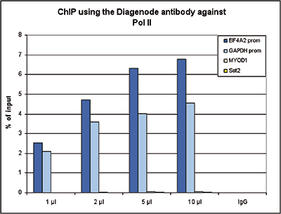
<p><small><strong> Figure 1. ChIP results obtained with the Diagenode monoclonal antibody directed against Pol II </strong><br />ChIP assays were performed using human HeLa cells, the Diagenode monoclonal antibody against Pol II (cat. No. C15100055) and optimized PCR primer pairs for qPCR. ChIP was performed with the “iDeal ChIP-seq” kit (cat. No. C01010051), using sheared chromatin from 1 million cells. A titration consisting of 1, 2, 5 and 10 μl of antibody per ChIP experiment was analyzed. IgG (2 μg/ IP) was used as a negative IP control. Quantitative PCR was performed with primers specific for the promoter of the GAPDH and EIF4A2 genes, used as positive controls, and for the MYOD1 gene and the Sat2 satellite repeat, used as negative controls. Figure 1 shows the recovery, expressed as a % of input (the relative amount of immunoprecipitated DNA compared to input DNA after qPCR analysis). </small></p>
</div>
</div>
<div class="row">
<div class="small-5 columns">
<p><img src="https://www.diagenode.com/img/product/antibodies/C15100055_ChIPSeq-A.png" alt="Pol II Antibody ChIP-seq Grade " style="display: block; margin-left: auto; margin-right: auto;" /></p>
<p><img src="https://www.diagenode.com/img/product/antibodies/C15100055_ChIPSeq-B.png" alt="Pol II Antibody for ChIP-seq" style="display: block; margin-left: auto; margin-right: auto;" /></p>
<p><img src="https://www.diagenode.com/img/product/antibodies/C15100055_ChIPSeq-C.png" alt="Pol II Antibody for ChIP-seq assay" style="display: block; margin-left: auto; margin-right: auto;" /></p>
<p><img src="https://www.diagenode.com/img/product/antibodies/C15100055_ChIPSeq-D.png" alt="Pol II Antibody validated in ChIP-seq" style="display: block; margin-left: auto; margin-right: auto;" /></p>
</div>
<div class="small-7 columns">
<p><small><strong> Figure 2. ChIP-seq results obtained with the Diagenode monoclonal antibody directed against Pol II</strong> <br />ChIP was performed on sheared chromatin from 1 million HeLaS3 cells using 2 μl of the Diagenode antibody against Pol II (cat. No. C15100055) as described above. The IP’d DNA was subsequently analysed on an Illumina HiSeq. Library preparation, cluster generation and sequencing were performed according to the manufacturer’s instructions. The 51 bp tags were aligned to the human genome using the BWA algorithm. Figure 2 shows the enrichment along the complete sequence and a 1 Mb region of the X-chromosome (fig 2A and B) and in genomic regions of chromosome 12 and 3, surrounding the GAPDH and EIF4A2 positive control genes (fig 2C and D). </small></p>
</div>
</div>
<div class="row">
<div class="small-4 columns">
<p><img src="https://www.diagenode.com/img/product/antibodies/C15100055_wb.png" alt="Pol II Antibody for Western Blot" style="display: block; margin-left: auto; margin-right: auto;" /></p>
</div>
<div class="small-8 columns">
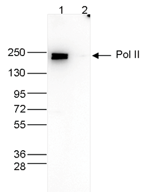
<p><small><strong> Figure 3. Western blot analysis using the Diagenode monoclonal antibody directed against Pol II </strong><br />Whole cell extracts (40 μg) from HeLa cells transfected with Pol II siRNA (lane 2) and from an untransfected control (lane 1) were analysed by Western blot using the Diagenode antibody against Pol II (Cat. No. C15100055) diluted 1:500 in TBS-Tween containing 5% skimmed milk. The position of the protein of interest is indicated on the right; the marker (in kDa) is shown on the left.</small></p>
</div>
</div>',
'label2' => 'Target Description',
'info2' => '<p>RNA polymerase II (pol II) is a key enzyme in the regulation and control of gene transcription. It is able to unwind the DNA double helix, synthesize RNA, and proofread the result. Pol II is a complex enzyme, consisting of 12 subunits, of which the B1 subunit (UniProt/Swiss-Prot entry P24928) is the largest. Together with the second largest subunit, B1 forms the catalytic core of the RNA polymerase II transcription machinery</p>',
'label3' => '',
'info3' => '',
'format' => '100 µl',
'catalog_number' => 'C15100055-100',
'old_catalog_number' => 'AC-055-100',
'sf_code' => 'C15100055-D001-001161',
'type' => 'FRE',
'search_order' => '03-Antibody',
'price_EUR' => '380',
'price_USD' => '380',
'price_GBP' => '340',
'price_JPY' => '59525',
'price_CNY' => '',
'price_AUD' => '950',
'country' => 'ALL',
'except_countries' => 'None',
'quote' => false,
'in_stock' => true,
'featured' => false,
'no_promo' => false,
'online' => true,
'master' => true,
'last_datasheet_update' => '0000-00-00',
'slug' => 'pol-ii-monoclonal-antibody-classic-100-ul',
'meta_title' => 'Pol II Antibody - ChIP-seq Grade (C15100055) | Diagenode',
'meta_keywords' => '',
'meta_description' => 'Pol II (B1 subunit of RNA polymerase II) Monoclonal Antibody validated in ChIP-seq, ChIP-qPCR and WB. Specificity confirmed by siRNA assay. Batch-specific data available on the website. Alternative names: POLR2A, RPB1, POLR2, RPOL2. Sample size available.',
'modified' => '2024-12-03 15:02:42',
'created' => '2015-06-29 14:08:20',
'locale' => 'eng'
),
'Antibody' => array(
'host' => '*****',
'id' => '194',
'name' => 'Pol II monoclonal antibody',
'description' => 'RNA polymerase II (pol II) is a key enzyme in the regulation and control of gene transcription. It is able to unwind the DNA double helix, synthesize RNA, and proofread the result. Pol II is a complex enzyme, consisting of 12 subunits, of which the B1 subunit (UniProt/Swiss-Prot entry P24928) is the largest. Together with the second largest subunit, B1 forms the catalytic core of the RNA polymerase II transcription machinery.',
'clonality' => '',
'isotype' => '',
'lot' => '008',
'concentration' => 'not determined',
'reactivity' => 'Human, Mouse, Xenopus',
'type' => 'Monoclonal',
'purity' => 'Ascites fluid',
'classification' => 'Classic',
'application_table' => '<table>
<thead>
<tr>
<th>Applications</th>
<th>Suggested dilution</th>
<th>References</th>
</tr>
</thead>
<tbody>
<tr>
<td>ChIP/ChIP-seq <sup>*</sup></td>
<td>2 μl/ChIP</td>
<td>Fig 1, 2</td>
</tr>
<tr>
<td>WB</td>
<td>1:500</td>
<td>Fig 3</td>
</tr>
</tbody>
</table>
<p><small><sup>*</sup> Please note that the optimal antibody amount per IP should be determined by the end-user. We recommend testing 1-10 μl per IP.</small></p>',
'storage_conditions' => '',
'storage_buffer' => '',
'precautions' => 'This product is for research use only. Not for use in diagnostic or therapeutic procedures.',
'uniprot_acc' => '',
'slug' => '',
'meta_keywords' => '',
'meta_description' => '',
'modified' => '2024-08-10 16:40:07',
'created' => '0000-00-00 00:00:00',
'select_label' => '194 - Pol II monoclonal antibody (008 - not determined - Human, Mouse, Xenopus - Ascites fluid - Mouse)'
),
'Slave' => array(
(int) 0 => array(
[maximum depth reached]
)
),
'Group' => array(
'Group' => array(
[maximum depth reached]
),
'Master' => array(
[maximum depth reached]
),
'Product' => array(
[maximum depth reached]
)
),
'Related' => array(),
'Application' => array(
(int) 0 => array(
[maximum depth reached]
),
(int) 1 => array(
[maximum depth reached]
),
(int) 2 => array(
[maximum depth reached]
),
(int) 3 => array(
[maximum depth reached]
)
),
'Category' => array(
(int) 0 => array(
[maximum depth reached]
),
(int) 1 => array(
[maximum depth reached]
),
(int) 2 => array(
[maximum depth reached]
),
(int) 3 => array(
[maximum depth reached]
)
),
'Document' => array(
(int) 0 => array(
[maximum depth reached]
),
(int) 1 => array(
[maximum depth reached]
),
(int) 2 => array(
[maximum depth reached]
)
),
'Feature' => array(),
'Image' => array(
(int) 0 => array(
[maximum depth reached]
)
),
'Promotion' => array(),
'Protocol' => array(),
'Publication' => array(
(int) 0 => array(
[maximum depth reached]
),
(int) 1 => array(
[maximum depth reached]
),
(int) 2 => array(
[maximum depth reached]
),
(int) 3 => array(
[maximum depth reached]
),
(int) 4 => array(
[maximum depth reached]
),
(int) 5 => array(
[maximum depth reached]
),
(int) 6 => array(
[maximum depth reached]
),
(int) 7 => array(
[maximum depth reached]
),
(int) 8 => array(
[maximum depth reached]
),
(int) 9 => array(
[maximum depth reached]
),
(int) 10 => array(
[maximum depth reached]
),
(int) 11 => array(
[maximum depth reached]
),
(int) 12 => array(
[maximum depth reached]
),
(int) 13 => array(
[maximum depth reached]
),
(int) 14 => array(
[maximum depth reached]
),
(int) 15 => array(
[maximum depth reached]
),
(int) 16 => array(
[maximum depth reached]
),
(int) 17 => array(
[maximum depth reached]
),
(int) 18 => array(
[maximum depth reached]
),
(int) 19 => array(
[maximum depth reached]
),
(int) 20 => array(
[maximum depth reached]
),
(int) 21 => array(
[maximum depth reached]
),
(int) 22 => array(
[maximum depth reached]
),
(int) 23 => array(
[maximum depth reached]
),
(int) 24 => array(
[maximum depth reached]
),
(int) 25 => array(
[maximum depth reached]
),
(int) 26 => array(
[maximum depth reached]
),
(int) 27 => array(
[maximum depth reached]
),
(int) 28 => array(
[maximum depth reached]
)
),
'Testimonial' => array(),
'Area' => array(),
'SafetySheet' => array(
(int) 0 => array(
[maximum depth reached]
),
(int) 1 => array(
[maximum depth reached]
),
(int) 2 => array(
[maximum depth reached]
),
(int) 3 => array(
[maximum depth reached]
),
(int) 4 => array(
[maximum depth reached]
),
(int) 5 => array(
[maximum depth reached]
),
(int) 6 => array(
[maximum depth reached]
),
(int) 7 => array(
[maximum depth reached]
)
)
),
'meta_canonical' => 'https://www.diagenode.com/en/p/pol-ii-monoclonal-antibody-classic-100-ul'
)
$language = 'en'
$meta_keywords = ''
$meta_description = 'Pol II (B1 subunit of RNA polymerase II) Monoclonal Antibody validated in ChIP-seq, ChIP-qPCR and WB. Specificity confirmed by siRNA assay. Batch-specific data available on the website. Alternative names: POLR2A, RPB1, POLR2, RPOL2. Sample size available.'
$meta_title = 'Pol II Antibody - ChIP-seq Grade (C15100055) | Diagenode'
$product = array(
'Product' => array(
'id' => '1951',
'antibody_id' => '194',
'name' => 'Pol II Antibody - replaced by the antibody C15200253 ',
'description' => '<p><strong>The antibody C15100055, format 100 µl has been discontinued. We recommend using the antibody <a href="https://www.diagenode.com/en/p/pol-ii-monoclonal-antibody-50-ul">C15200253</a></strong><span><strong>. </strong> </span></p>
<p>Alternative names: <strong>POLR2A</strong>, <strong>RPB1</strong>, <strong>POLR2</strong>, <strong>RPOL2</strong></p>
<p>Monoclonal antibody raised in mouse against the <strong>B1 subunit of RNA polymerase II</strong> (polymerase (RNA) II (DNA directed) polypeptide A) of wheat germ. Interacts with the highly conserved C-terminal domain of the protein containing the YSPTSPS repeat.</p>',
'label1' => 'Validation Data',
'info1' => '<div class="row">
<div class="small-4 columns">
<p><img src="https://www.diagenode.com/img/product/antibodies/C15100055_008_ChIP.png" alt="Pol II Antibody ChIP Grade " style="display: block; margin-left: auto; margin-right: auto;" /></p>
</div>
<div class="small-8 columns">
<p><small><strong> Figure 1. ChIP results obtained with the Diagenode monoclonal antibody directed against Pol II </strong><br />ChIP assays were performed using human HeLa cells, the Diagenode monoclonal antibody against Pol II (cat. No. C15100055) and optimized PCR primer pairs for qPCR. ChIP was performed with the “iDeal ChIP-seq” kit (cat. No. C01010051), using sheared chromatin from 1 million cells. A titration consisting of 1, 2, 5 and 10 μl of antibody per ChIP experiment was analyzed. IgG (2 μg/ IP) was used as a negative IP control. Quantitative PCR was performed with primers specific for the promoter of the GAPDH and EIF4A2 genes, used as positive controls, and for the MYOD1 gene and the Sat2 satellite repeat, used as negative controls. Figure 1 shows the recovery, expressed as a % of input (the relative amount of immunoprecipitated DNA compared to input DNA after qPCR analysis). </small></p>
</div>
</div>
<div class="row">
<div class="small-5 columns">
<p><img src="https://www.diagenode.com/img/product/antibodies/C15100055_ChIPSeq-A.png" alt="Pol II Antibody ChIP-seq Grade " style="display: block; margin-left: auto; margin-right: auto;" /></p>
<p><img src="https://www.diagenode.com/img/product/antibodies/C15100055_ChIPSeq-B.png" alt="Pol II Antibody for ChIP-seq" style="display: block; margin-left: auto; margin-right: auto;" /></p>
<p><img src="https://www.diagenode.com/img/product/antibodies/C15100055_ChIPSeq-C.png" alt="Pol II Antibody for ChIP-seq assay" style="display: block; margin-left: auto; margin-right: auto;" /></p>
<p><img src="https://www.diagenode.com/img/product/antibodies/C15100055_ChIPSeq-D.png" alt="Pol II Antibody validated in ChIP-seq" style="display: block; margin-left: auto; margin-right: auto;" /></p>
</div>
<div class="small-7 columns">
<p><small><strong> Figure 2. ChIP-seq results obtained with the Diagenode monoclonal antibody directed against Pol II</strong> <br />ChIP was performed on sheared chromatin from 1 million HeLaS3 cells using 2 μl of the Diagenode antibody against Pol II (cat. No. C15100055) as described above. The IP’d DNA was subsequently analysed on an Illumina HiSeq. Library preparation, cluster generation and sequencing were performed according to the manufacturer’s instructions. The 51 bp tags were aligned to the human genome using the BWA algorithm. Figure 2 shows the enrichment along the complete sequence and a 1 Mb region of the X-chromosome (fig 2A and B) and in genomic regions of chromosome 12 and 3, surrounding the GAPDH and EIF4A2 positive control genes (fig 2C and D). </small></p>
</div>
</div>
<div class="row">
<div class="small-4 columns">
<p><img src="https://www.diagenode.com/img/product/antibodies/C15100055_wb.png" alt="Pol II Antibody for Western Blot" style="display: block; margin-left: auto; margin-right: auto;" /></p>
</div>
<div class="small-8 columns">
<p><small><strong> Figure 3. Western blot analysis using the Diagenode monoclonal antibody directed against Pol II </strong><br />Whole cell extracts (40 μg) from HeLa cells transfected with Pol II siRNA (lane 2) and from an untransfected control (lane 1) were analysed by Western blot using the Diagenode antibody against Pol II (Cat. No. C15100055) diluted 1:500 in TBS-Tween containing 5% skimmed milk. The position of the protein of interest is indicated on the right; the marker (in kDa) is shown on the left.</small></p>
</div>
</div>',
'label2' => 'Target Description',
'info2' => '<p>RNA polymerase II (pol II) is a key enzyme in the regulation and control of gene transcription. It is able to unwind the DNA double helix, synthesize RNA, and proofread the result. Pol II is a complex enzyme, consisting of 12 subunits, of which the B1 subunit (UniProt/Swiss-Prot entry P24928) is the largest. Together with the second largest subunit, B1 forms the catalytic core of the RNA polymerase II transcription machinery</p>',
'label3' => '',
'info3' => '',
'format' => '100 µl',
'catalog_number' => 'C15100055-100',
'old_catalog_number' => 'AC-055-100',
'sf_code' => 'C15100055-D001-001161',
'type' => 'FRE',
'search_order' => '03-Antibody',
'price_EUR' => '380',
'price_USD' => '380',
'price_GBP' => '340',
'price_JPY' => '59525',
'price_CNY' => '',
'price_AUD' => '950',
'country' => 'ALL',
'except_countries' => 'None',
'quote' => false,
'in_stock' => true,
'featured' => false,
'no_promo' => false,
'online' => true,
'master' => true,
'last_datasheet_update' => '0000-00-00',
'slug' => 'pol-ii-monoclonal-antibody-classic-100-ul',
'meta_title' => 'Pol II Antibody - ChIP-seq Grade (C15100055) | Diagenode',
'meta_keywords' => '',
'meta_description' => 'Pol II (B1 subunit of RNA polymerase II) Monoclonal Antibody validated in ChIP-seq, ChIP-qPCR and WB. Specificity confirmed by siRNA assay. Batch-specific data available on the website. Alternative names: POLR2A, RPB1, POLR2, RPOL2. Sample size available.',
'modified' => '2024-12-03 15:02:42',
'created' => '2015-06-29 14:08:20',
'locale' => 'eng'
),
'Antibody' => array(
'host' => '*****',
'id' => '194',
'name' => 'Pol II monoclonal antibody',
'description' => 'RNA polymerase II (pol II) is a key enzyme in the regulation and control of gene transcription. It is able to unwind the DNA double helix, synthesize RNA, and proofread the result. Pol II is a complex enzyme, consisting of 12 subunits, of which the B1 subunit (UniProt/Swiss-Prot entry P24928) is the largest. Together with the second largest subunit, B1 forms the catalytic core of the RNA polymerase II transcription machinery.',
'clonality' => '',
'isotype' => '',
'lot' => '008',
'concentration' => 'not determined',
'reactivity' => 'Human, Mouse, Xenopus',
'type' => 'Monoclonal',
'purity' => 'Ascites fluid',
'classification' => 'Classic',
'application_table' => '<table>
<thead>
<tr>
<th>Applications</th>
<th>Suggested dilution</th>
<th>References</th>
</tr>
</thead>
<tbody>
<tr>
<td>ChIP/ChIP-seq <sup>*</sup></td>
<td>2 μl/ChIP</td>
<td>Fig 1, 2</td>
</tr>
<tr>
<td>WB</td>
<td>1:500</td>
<td>Fig 3</td>
</tr>
</tbody>
</table>
<p><small><sup>*</sup> Please note that the optimal antibody amount per IP should be determined by the end-user. We recommend testing 1-10 μl per IP.</small></p>',
'storage_conditions' => '',
'storage_buffer' => '',
'precautions' => 'This product is for research use only. Not for use in diagnostic or therapeutic procedures.',
'uniprot_acc' => '',
'slug' => '',
'meta_keywords' => '',
'meta_description' => '',
'modified' => '2024-08-10 16:40:07',
'created' => '0000-00-00 00:00:00',
'select_label' => '194 - Pol II monoclonal antibody (008 - not determined - Human, Mouse, Xenopus - Ascites fluid - Mouse)'
),
'Slave' => array(
(int) 0 => array(
'id' => '212',
'name' => 'C15100055',
'product_id' => '1951',
'modified' => '2017-05-22 16:32:23',
'created' => '2017-05-22 16:32:23'
)
),
'Group' => array(
'Group' => array(
'id' => '212',
'name' => 'C15100055',
'product_id' => '1951',
'modified' => '2017-05-22 16:32:23',
'created' => '2017-05-22 16:32:23'
),
'Master' => array(
'id' => '1951',
'antibody_id' => '194',
'name' => 'Pol II Antibody - replaced by the antibody C15200253 ',
'description' => '<p><strong>The antibody C15100055, format 100 µl has been discontinued. We recommend using the antibody <a href="https://www.diagenode.com/en/p/pol-ii-monoclonal-antibody-50-ul">C15200253</a></strong><span><strong>. </strong> </span></p>
<p>Alternative names: <strong>POLR2A</strong>, <strong>RPB1</strong>, <strong>POLR2</strong>, <strong>RPOL2</strong></p>
<p>Monoclonal antibody raised in mouse against the <strong>B1 subunit of RNA polymerase II</strong> (polymerase (RNA) II (DNA directed) polypeptide A) of wheat germ. Interacts with the highly conserved C-terminal domain of the protein containing the YSPTSPS repeat.</p>',
'label1' => 'Validation Data',
'info1' => '<div class="row">
<div class="small-4 columns">
<p><img src="https://www.diagenode.com/img/product/antibodies/C15100055_008_ChIP.png" alt="Pol II Antibody ChIP Grade " style="display: block; margin-left: auto; margin-right: auto;" /></p>
</div>
<div class="small-8 columns">
<p><small><strong> Figure 1. ChIP results obtained with the Diagenode monoclonal antibody directed against Pol II </strong><br />ChIP assays were performed using human HeLa cells, the Diagenode monoclonal antibody against Pol II (cat. No. C15100055) and optimized PCR primer pairs for qPCR. ChIP was performed with the “iDeal ChIP-seq” kit (cat. No. C01010051), using sheared chromatin from 1 million cells. A titration consisting of 1, 2, 5 and 10 μl of antibody per ChIP experiment was analyzed. IgG (2 μg/ IP) was used as a negative IP control. Quantitative PCR was performed with primers specific for the promoter of the GAPDH and EIF4A2 genes, used as positive controls, and for the MYOD1 gene and the Sat2 satellite repeat, used as negative controls. Figure 1 shows the recovery, expressed as a % of input (the relative amount of immunoprecipitated DNA compared to input DNA after qPCR analysis). </small></p>
</div>
</div>
<div class="row">
<div class="small-5 columns">
<p><img src="https://www.diagenode.com/img/product/antibodies/C15100055_ChIPSeq-A.png" alt="Pol II Antibody ChIP-seq Grade " style="display: block; margin-left: auto; margin-right: auto;" /></p>
<p><img src="https://www.diagenode.com/img/product/antibodies/C15100055_ChIPSeq-B.png" alt="Pol II Antibody for ChIP-seq" style="display: block; margin-left: auto; margin-right: auto;" /></p>
<p><img src="https://www.diagenode.com/img/product/antibodies/C15100055_ChIPSeq-C.png" alt="Pol II Antibody for ChIP-seq assay" style="display: block; margin-left: auto; margin-right: auto;" /></p>
<p><img src="https://www.diagenode.com/img/product/antibodies/C15100055_ChIPSeq-D.png" alt="Pol II Antibody validated in ChIP-seq" style="display: block; margin-left: auto; margin-right: auto;" /></p>
</div>
<div class="small-7 columns">
<p><small><strong> Figure 2. ChIP-seq results obtained with the Diagenode monoclonal antibody directed against Pol II</strong> <br />ChIP was performed on sheared chromatin from 1 million HeLaS3 cells using 2 μl of the Diagenode antibody against Pol II (cat. No. C15100055) as described above. The IP’d DNA was subsequently analysed on an Illumina HiSeq. Library preparation, cluster generation and sequencing were performed according to the manufacturer’s instructions. The 51 bp tags were aligned to the human genome using the BWA algorithm. Figure 2 shows the enrichment along the complete sequence and a 1 Mb region of the X-chromosome (fig 2A and B) and in genomic regions of chromosome 12 and 3, surrounding the GAPDH and EIF4A2 positive control genes (fig 2C and D). </small></p>
</div>
</div>
<div class="row">
<div class="small-4 columns">
<p><img src="https://www.diagenode.com/img/product/antibodies/C15100055_wb.png" alt="Pol II Antibody for Western Blot" style="display: block; margin-left: auto; margin-right: auto;" /></p>
</div>
<div class="small-8 columns">
<p><small><strong> Figure 3. Western blot analysis using the Diagenode monoclonal antibody directed against Pol II </strong><br />Whole cell extracts (40 μg) from HeLa cells transfected with Pol II siRNA (lane 2) and from an untransfected control (lane 1) were analysed by Western blot using the Diagenode antibody against Pol II (Cat. No. C15100055) diluted 1:500 in TBS-Tween containing 5% skimmed milk. The position of the protein of interest is indicated on the right; the marker (in kDa) is shown on the left.</small></p>
</div>
</div>',
'label2' => 'Target Description',
'info2' => '<p>RNA polymerase II (pol II) is a key enzyme in the regulation and control of gene transcription. It is able to unwind the DNA double helix, synthesize RNA, and proofread the result. Pol II is a complex enzyme, consisting of 12 subunits, of which the B1 subunit (UniProt/Swiss-Prot entry P24928) is the largest. Together with the second largest subunit, B1 forms the catalytic core of the RNA polymerase II transcription machinery</p>',
'label3' => '',
'info3' => '',
'format' => '100 µl',
'catalog_number' => 'C15100055-100',
'old_catalog_number' => 'AC-055-100',
'sf_code' => 'C15100055-D001-001161',
'type' => 'FRE',
'search_order' => '03-Antibody',
'price_EUR' => '380',
'price_USD' => '380',
'price_GBP' => '340',
'price_JPY' => '59525',
'price_CNY' => '',
'price_AUD' => '950',
'country' => 'ALL',
'except_countries' => 'None',
'quote' => false,
'in_stock' => true,
'featured' => false,
'no_promo' => false,
'online' => true,
'master' => true,
'last_datasheet_update' => '0000-00-00',
'slug' => 'pol-ii-monoclonal-antibody-classic-100-ul',
'meta_title' => 'Pol II Antibody - ChIP-seq Grade (C15100055) | Diagenode',
'meta_keywords' => '',
'meta_description' => 'Pol II (B1 subunit of RNA polymerase II) Monoclonal Antibody validated in ChIP-seq, ChIP-qPCR and WB. Specificity confirmed by siRNA assay. Batch-specific data available on the website. Alternative names: POLR2A, RPB1, POLR2, RPOL2. Sample size available.',
'modified' => '2024-12-03 15:02:42',
'created' => '2015-06-29 14:08:20'
),
'Product' => array(
(int) 0 => array(
[maximum depth reached]
)
)
),
'Related' => array(),
'Application' => array(
(int) 0 => array(
'id' => '42',
'position' => '10',
'parent_id' => '40',
'name' => 'ChIP-seq (ab)',
'description' => '',
'in_footer' => false,
'in_menu' => false,
'online' => true,
'tabular' => true,
'slug' => 'chip-seq-antibodies',
'meta_keywords' => 'Chromatin Immunoprecipitation Sequencing,ChIP-Seq,ChIP-seq grade antibodies,DNA purification,qPCR,Shearing of chromatin',
'meta_description' => 'Diagenode offers a wide range of antibodies and technical support for ChIP Sequencing applications',
'meta_title' => 'ChIP Sequencing Antibodies (ChIP-Seq) | Diagenode',
'modified' => '2016-01-20 11:06:19',
'created' => '2015-10-20 11:44:45',
'ProductsApplication' => array(
[maximum depth reached]
)
),
(int) 1 => array(
'id' => '43',
'position' => '10',
'parent_id' => '40',
'name' => 'ChIP-qPCR (ab)',
'description' => '',
'in_footer' => false,
'in_menu' => false,
'online' => true,
'tabular' => true,
'slug' => 'chip-qpcr-antibodies',
'meta_keywords' => 'Chromatin Immunoprecipitation Sequencing,ChIP-Seq,ChIP-seq grade antibodies,DNA purification,qPCR,Shearing of chromatin',
'meta_description' => 'Diagenode offers a wide range of antibodies and technical support for ChIP-qPCR applications',
'meta_title' => 'ChIP Quantitative PCR Antibodies (ChIP-qPCR) | Diagenode',
'modified' => '2016-01-20 11:30:24',
'created' => '2015-10-20 11:45:36',
'ProductsApplication' => array(
[maximum depth reached]
)
),
(int) 2 => array(
'id' => '19',
'position' => '10',
'parent_id' => '40',
'name' => 'WB',
'description' => '<p><strong>Western blot</strong> : The quality of antibodies used in this technique is crucial for correct and specific protein identification. Diagenode offers huge selection of highly sensitive and specific western blot-validated antibodies.</p>
<p>Learn more about: <a href="https://www.diagenode.com/applications/western-blot">Loading control, MW marker visualization</a><em>. <br /></em></p>
<p><em></em>Check our selection of antibodies validated in Western blot.</p>',
'in_footer' => false,
'in_menu' => false,
'online' => true,
'tabular' => true,
'slug' => 'western-blot-antibodies',
'meta_keywords' => ' Western Blot Antibodies ,western blot protocol,Western Blotting Products,Polyclonal antibodies ,monoclonal antibodies ',
'meta_description' => 'Diagenode offers a wide range of antibodies and technical support for western blot applications',
'meta_title' => ' Western Blot - Monoclonal antibody - Polyclonal antibody | Diagenode',
'modified' => '2016-04-26 12:44:51',
'created' => '2015-01-07 09:20:00',
'ProductsApplication' => array(
[maximum depth reached]
)
),
(int) 3 => array(
'id' => '45',
'position' => '10',
'parent_id' => '40',
'name' => 'siRNA Knockdown',
'description' => '<div class="row">
<div class="small-10 columns">
<h3>Epigenetic antibodies you can trust!</h3>
<p>Antibody quality is essential for assay success. Diagenode offers antibodies that are actually validated and have been widely used and published by the scientific community. Now we are adding a new level of siRNA knockdown validation to assure the specificity of our non-histone antibodies.</p>
<p><strong>Short interfering RNA (siRNA)</strong> degrades target mRNA, followed by the knock-down of protein production. If the antibody that recognizes the protein of interest is specific, the Western blot of siRNA-treated cells will show a significant reduction of signal vs. untreated cells.</p>
<center><img src="https://www.diagenode.com/emailing/images/C15100144-wb.png" alt="" /></center>
<p class="text-center"><small>WB results obtained with the HDAC1 pAb (Cat. No. C15100144) <br />on siRNA transfected cells (lane 2) and on untransfected control cells (lane 1).</small></p>
</div>
<div class="small-2 columns">
<p><img src="https://www.diagenode.com/emailing/images/epi-success-guaranteed-icon.png" alt="Epigenetic success guaranteed" /></p>
</div>
</div>
<div class="spaced"></div>
<p style="text-align: left;"><span style="font-weight: 400;">The below list shows our first siRNA validated antibodies. More results - coming soon</span>.</p>',
'in_footer' => false,
'in_menu' => false,
'online' => true,
'tabular' => true,
'slug' => 'sirna',
'meta_keywords' => 'siRNA knockdown antibodies',
'meta_description' => 'Antibodies validated by siRNA Knockdown',
'meta_title' => '',
'modified' => '2017-01-26 15:58:23',
'created' => '2017-01-09 10:53:09',
'ProductsApplication' => array(
[maximum depth reached]
)
)
),
'Category' => array(
(int) 0 => array(
'id' => '17',
'position' => '10',
'parent_id' => '4',
'name' => 'ChIP-seq grade antibodies',
'description' => '<p><b>Unparalleled ChIP-Seq results with the most rigorously validated antibodies</b></p>
<p><span style="font-weight: 400;">Diagenode provides leading solutions for epigenetic research. Because ChIP-seq is a widely-used technique, we validate our antibodies in ChIP and ChIP-seq experiments (in addition to conventional methods like Western blot, Dot blot, ELISA, and immunofluorescence) to provide the highest quality antibody. We standardize our validation and production to guarantee high product quality without technical bias. Diagenode guarantees ChIP-seq grade antibody performance under our suggested conditions.</span></p>
<div class="row">
<div class="small-12 medium-9 large-9 columns">
<p><strong>ChIP-seq profile</strong> of active (H3K4me3 and H3K36me3) and inactive (H3K27me3) marks using Diagenode antibodies.</p>
<img src="https://www.diagenode.com/img/categories/antibodies/chip-seq-grade-antibodies.png" /></div>
<div class="small-12 medium-3 large-3 columns">
<p><small> ChIP was performed on sheared chromatin from 100,000 K562 cells using iDeal ChIP-seq kit for Histones (cat. No. C01010051) with 1 µg of the Diagenode antibodies against H3K27me3 (cat. No. C15410195) and H3K4me3 (cat. No. C15410003), and 0.5 µg of the antibody against H3K36me3 (cat. No. C15410192). The IP'd DNA was subsequently analysed on an Illumina Genome Analyzer. Library preparation, cluster generation and sequencing were performed according to the manufacturer's instructions. The 36 bp tags were aligned to the human genome using the ELAND algorithm. The figure shows the signal distribution along the complete sequence of human chromosome 3, a zoomin to a 10 Mb region and a further zoomin to a 1.5 Mb region. </small></p>
</div>
</div>
<p>Diagenode’s highly validated antibodies:</p>
<ul>
<li>Highly sensitive and specific</li>
<li>Cost-effective (requires less antibody per reaction)</li>
<li>Batch-specific data is available on the website</li>
<li>Expert technical support</li>
<li>Sample sizes available</li>
<li>100% satisfaction guarantee</li>
</ul>',
'no_promo' => false,
'in_menu' => true,
'online' => true,
'tabular' => false,
'hide' => true,
'all_format' => false,
'is_antibody' => true,
'slug' => 'chip-seq-grade-antibodies',
'cookies_tag_id' => null,
'meta_keywords' => 'ChIP-seq grade antibodies,polyclonal antibody,WB, ELISA, ChIP-seq (ab), ChIP-qPCR (ab)',
'meta_description' => 'Diagenode Offers Wide Range of Validated ChIP-Seq Grade Antibodies for Unparalleled ChIP-Seq Results',
'meta_title' => 'Chromatin Immunoprecipitation ChIP-Seq Grade Antibodies | Diagenode',
'modified' => '2019-07-03 10:57:22',
'created' => '2015-02-16 02:24:01',
'ProductsCategory' => array(
[maximum depth reached]
),
'CookiesTag' => array([maximum depth reached])
),
(int) 1 => array(
'id' => '30',
'position' => '50',
'parent_id' => '4',
'name' => 'Transcription',
'description' => '<p><span style="font-weight: 400;">The list of Diagenode’s highly specific antibodies for transcription studies includes the antibodies against many transcription factors and nuclear receptors. Check the list below to see our targets.</span></p>
<p><span style="font-weight: 400;">Diagenode’s highly validated antibodies:</span></p>
<ul>
<li><span style="font-weight: 400;"> Highly sensitive and specific</span></li>
<li><span style="font-weight: 400;"> Cost-effective (requires less antibody per reaction)</span></li>
<li><span style="font-weight: 400;"> Batch-specific data is available on the website</span></li>
<li><span style="font-weight: 400;"> Expert technical support</span></li>
<li><span style="font-weight: 400;"> Sample sizes available</span></li>
<li><span style="font-weight: 400;"> 100% satisfaction guarantee</span></li>
</ul>',
'no_promo' => false,
'in_menu' => false,
'online' => true,
'tabular' => false,
'hide' => true,
'all_format' => false,
'is_antibody' => true,
'slug' => 'transcription-factor',
'cookies_tag_id' => null,
'meta_keywords' => ' Transcription factor antibodies,monoclonal antibodies,polyclonal antibodies',
'meta_description' => 'Diagenode offers polyclonal and monoclonal antibodies for Transcription studie',
'meta_title' => 'Transcription factor Antibodies | Diagenode',
'modified' => '2020-07-06 12:59:19',
'created' => '2015-03-12 10:20:08',
'ProductsCategory' => array(
[maximum depth reached]
),
'CookiesTag' => array([maximum depth reached])
),
(int) 2 => array(
'id' => '103',
'position' => '0',
'parent_id' => '4',
'name' => 'All antibodies',
'description' => '<p><span style="font-weight: 400;">All Diagenode’s antibodies are listed below. Please, use our Quick search field to find the antibody of interest by target name, application, purity.</span></p>
<p><span style="font-weight: 400;">Diagenode’s highly validated antibodies:</span></p>
<ul>
<li>Highly sensitive and specific</li>
<li>Cost-effective (requires less antibody per reaction)</li>
<li>Batch-specific data is available on the website</li>
<li>Expert technical support</li>
<li>Sample sizes available</li>
<li>100% satisfaction guarantee</li>
</ul>',
'no_promo' => false,
'in_menu' => true,
'online' => true,
'tabular' => false,
'hide' => true,
'all_format' => false,
'is_antibody' => true,
'slug' => 'all-antibodies',
'cookies_tag_id' => null,
'meta_keywords' => 'Antibodies,Premium Antibodies,Classic,Pioneer',
'meta_description' => 'Diagenode Offers Strict quality standards with Rigorous QC and validated Antibodies. Classified based on level of validation for flexibility of Application. Comprehensive selection of histone and non-histone Antibodies',
'meta_title' => 'Diagenode's selection of Antibodies is exclusively dedicated for Epigenetic Research | Diagenode',
'modified' => '2019-07-03 10:55:44',
'created' => '2015-11-02 14:49:22',
'ProductsCategory' => array(
[maximum depth reached]
),
'CookiesTag' => array([maximum depth reached])
),
(int) 3 => array(
'id' => '127',
'position' => '10',
'parent_id' => '4',
'name' => 'ChIP-grade antibodies',
'description' => '<div class="row">
<div class="small-12 columns"><center></center>
<p><br />Chromatin immunoprecipitation (<b>ChIP</b>) is a technique to study the associations of proteins with the specific genomic regions in intact cells. One of the most important steps of this protocol is the immunoprecipitation of targeted protein using the antibody specifically recognizing it. The quality of antibodies used in ChIP is essential for the success of the experiment. Diagenode offers extensively validated ChIP-grade antibodies, confirmed for their specificity, and high level of performance in ChIP. Each batch is validated, and batch-specific data are available on the website.</p>
<p></p>
</div>
</div>
<p><strong>ChIP results</strong> obtained with the antibody directed against H3K4me3 (Cat. No. <a href="../p/h3k4me3-polyclonal-antibody-premium-50-ug-50-ul">C15410003</a>). </p>
<div class="row">
<div class="small-12 medium-6 large-6 columns"><img src="https://www.diagenode.com/img/product/antibodies/C15410003-fig1-ChIP.jpg" alt="" width="400" height="315" /> </div>
<div class="small-12 medium-6 large-6 columns">
<p></p>
<p></p>
<p></p>
</div>
</div>
<p></p>
<p>Our aim at Diagenode is to offer the largest collection of highly specific <strong>ChIP-grade antibodies</strong>. We add new antibodies monthly. Find your ChIP-grade antibody in the list below and check more information about tested applications, extensive validation data, and product information.</p>',
'no_promo' => false,
'in_menu' => true,
'online' => true,
'tabular' => false,
'hide' => true,
'all_format' => false,
'is_antibody' => true,
'slug' => 'chip-grade-antibodies',
'cookies_tag_id' => null,
'meta_keywords' => 'ChIP-grade antibodies, polyclonal antibody, monoclonal antibody, Diagenode',
'meta_description' => 'Diagenode Offers Extensively Validated ChIP-Grade Antibodies, Confirmed for their Specificity, and high level of Performance in Chromatin Immunoprecipitation ChIP',
'meta_title' => 'Chromatin immunoprecipitation ChIP-grade antibodies | Diagenode',
'modified' => '2024-11-19 17:27:07',
'created' => '2017-02-14 11:16:04',
'ProductsCategory' => array(
[maximum depth reached]
),
'CookiesTag' => array([maximum depth reached])
)
),
'Document' => array(
(int) 0 => array(
'id' => '333',
'name' => 'Datasheet PoII C15100055',
'description' => '<p>Datasheet description</p>',
'image_id' => null,
'type' => 'Datasheet',
'url' => 'files/products/antibodies/Datasheet_PoII_C15100055.pdf',
'slug' => 'datasheet-poii-C15100055',
'meta_keywords' => '',
'meta_description' => '',
'modified' => '2015-11-20 17:17:51',
'created' => '2015-07-07 11:47:43',
'ProductsDocument' => array(
[maximum depth reached]
)
),
(int) 1 => array(
'id' => '38',
'name' => 'Epigenetic Antibodies Brochure',
'description' => '<p>More than in any other immuoprecipitation assays, quality antibodies are critical tools in many epigenetics experiments. Since 10 years, Diagenode has developed the most stringent quality production available on the market for antibodies exclusively focused on epigenetic uses. All our antibodies have been qualified to work in epigenetic applications.</p>',
'image_id' => null,
'type' => 'Brochure',
'url' => 'files/brochures/Epigenetic_Antibodies_Brochure.pdf',
'slug' => 'epigenetic-antibodies-brochure',
'meta_keywords' => '',
'meta_description' => '',
'modified' => '2016-06-15 11:24:06',
'created' => '2015-07-03 16:05:27',
'ProductsDocument' => array(
[maximum depth reached]
)
),
(int) 2 => array(
'id' => '11',
'name' => 'Antibodies you can trust',
'description' => '<p style="text-align: justify;"><span>Epigenetic research tools have evolved over time from endpoint PCR to qPCR to the analyses of large sets of genome-wide sequencing data. ChIP sequencing (ChIP-seq) has now become the gold standard method for chromatin studies, given the accuracy and coverage scale of the approach over other methods. Successful ChIP-seq, however, requires a higher level of experimental accuracy and consistency in all steps of ChIP than ever before. Particularly crucial is the quality of ChIP antibodies. </span></p>',
'image_id' => null,
'type' => 'Poster',
'url' => 'files/posters/Antibodies_you_can_trust_Poster.pdf',
'slug' => 'antibodies-you-can-trust-poster',
'meta_keywords' => '',
'meta_description' => '',
'modified' => '2015-10-01 20:18:31',
'created' => '2015-07-03 16:05:15',
'ProductsDocument' => array(
[maximum depth reached]
)
)
),
'Feature' => array(),
'Image' => array(
(int) 0 => array(
'id' => '1783',
'name' => 'product/antibodies/chipseq-grade-ab-icon.png',
'alt' => 'ChIP-seq Grade',
'modified' => '2020-11-27 07:04:40',
'created' => '2018-03-15 15:54:09',
'ProductsImage' => array(
[maximum depth reached]
)
)
),
'Promotion' => array(),
'Protocol' => array(),
'Publication' => array(
(int) 0 => array(
'id' => '4668',
'name' => 'Cotranscriptional demethylation induces global loss of H3K4me2 fromactive genes in Arabidopsis',
'authors' => 'Mori S. et al.',
'description' => '<p>Based on studies of animals and yeasts, methylation of histone H3 lysine 4 (H3K4me1/2/3, for mono-, di-, and tri-methylation, respectively) is regarded as the key epigenetic modification of transcriptionally active genes. In plants, however, H3K4me2 correlates negatively with transcription, and the regulatory mechanisms of this counterintuitive H3K4me2 distribution in plants remain largely unexplored. A previous genetic screen for factors regulating plant regeneration identified Arabidopsis LYSINE-SPECIFIC DEMETHYLASE 1-LIKE 3 (LDL3), which is a major H3K4me2 demethylase. Here, we show that LDL3-mediated H3K4me2 demethylation depends on the transcription elongation factor Paf1C and phosphorylation of the C-terminal domain (CTD) of RNA polymerase II (RNAPII). In addition, LDL3 binds to phosphorylated RNAPII. These results suggest that LDL3 is recruited to transcribed genes by binding to elongating RNAPII and demethylates H3K4me2 cotranscriptionally. Importantly, the negative correlation between H3K4me2 and transcription is disrupted in the ldl3 mutant, demonstrating the genome-wide impacts of the transcription-driven LDL3 pathway to control H3K4me2 in plants. Our findings implicate H3K4me2 in plants as chromatin memory for transcriptionally repressive states, which ensures robust gene control.</p>',
'date' => '2023-02-01',
'pmid' => 'https://www.biorxiv.org/content/10.1101/2023.02.17.528985v1',
'doi' => '10.1101/2023.02.17.528985',
'modified' => '2023-04-14 09:31:55',
'created' => '2023-02-28 12:19:11',
'ProductsPublication' => array(
[maximum depth reached]
)
),
(int) 1 => array(
'id' => '4411',
'name' => 'MLL-AF4 and a murinized pSer-variant thereof are turning on thenucleolar stress pathway.',
'authors' => 'Siemund Anna Lena and Hanewald Thomas and Kowarz Eric andMarschalek Rolf',
'description' => '<p>BACKGROUND: Recent pathomolecular studies on the MLL-AF4 fusion protein revealed that the murinized version of MLL-AF4, the MLL-Af4 fusion protein, was able to induce leukemia when expressed in murine or human hematopoietic stem/progenitor cells (Lin et al. in Cancer Cell 30:737-749, 2016). In parallel, a group from Japan demonstrated that the pSer domain of the AF4 protein, as well as the pSer domain of the MLL-AF4 fusion is able to bind the Pol I transcription factor complex SL1 (Okuda et al. in Nat Commun 6:8869, 2015). Here, we investigated the human MLL-AF4 and a pSer-murinized version thereof for their functional properties in mammalian cells. Gene expression profiling studies were complemented by intracellular localization studies and functional experiments concerning their biological activities in the nucleolus. RESULTS: Based on our results, we have to conclude that MLL-AF4 is predominantly localizing inside the nucleolus, thereby interfering with Pol I transcription and ribosome biogenesis. The murinized pSer-variant is localizing more to the nucleus, which may suggest a different biological behavior. Of note, AF4-MLL seems to cooperate at the molecular level with MLL-AF4 to steer target gene transcription, but not with the pSer-murinized version of it. CONCLUSION: This study provides new insights and a molecular explanation for the described differences between hMLL-hAF4 (not leukemogenic) and hMLL-mAf4 (leukemogenic). While the human pSer domain is able to efficiently recruit the SL1 transcription factor complex, the murine counterpart seems to be not. This has several consequences for our understanding of t(4;11) leukemia which is the most frequent leukemia in infants, childhood and adults suffering from MLL-r acute leukemia.</p>',
'date' => '2022-04-01',
'pmid' => 'https://www.ncbi.nlm.nih.gov/pubmed/35468859',
'doi' => '10.1186/s13578-022-00781-y',
'modified' => '2022-08-11 15:30:01',
'created' => '2022-08-11 12:14:50',
'ProductsPublication' => array(
[maximum depth reached]
)
),
(int) 2 => array(
'id' => '4228',
'name' => 'Human centromere formation activates transcription and opens chromatinfibre structure',
'authors' => 'Gilbert, Nick and Naughton, Catherine and Huidobro, Covadongaand Catacchio, Claudia and Buckle, Adam and Grimes, Graeme andNozawa, Ryu-Suke and Purgato, Stefania and Rocchi, Mariano',
'description' => '<p>Human centromeres appear as constrictions on mitotic chromosomes and form a platform for kinetochore assembly in mitosis. Biophysical experiments led to a suggestion that repetitive DNA at centromeric regions form a compact scaffold necessary for function, but this was revised when neocentromeres were discovered on non-repetitive DNA. To test whether centromeres have a special chromatin structure we have analysed the architecture of a neocentromere. Centromere formation is accompanied by RNA pol II recruitment and active transcription to form a decompacted, negatively supercoiled domain enriched in ‘open’ chromatin fibres. In contrast, centromerisation causes a spreading of repressive epigenetic marks to surrounding regions, delimited by H3K27me3 polycomb boundaries and divergent genes. This flanking domain is transcriptionally silent and partially remodelled to form ‘compact’ chromatin, similar to satellite-containing DNA sequences, and exhibits genomic instability. We suggest transcription disrupts chromatin to provide a foundation for kine-tochore formation whilst compact pericentromeric heterochromatin generates mechanical rigidity.</p>',
'date' => '2022-01-01',
'pmid' => 'https://www.researchsquare.com/article/rs-1061218/v1',
'doi' => '10.21203/rs.3.rs-1061218/v1',
'modified' => '2022-05-19 16:02:58',
'created' => '2022-05-19 10:41:50',
'ProductsPublication' => array(
[maximum depth reached]
)
),
(int) 3 => array(
'id' => '4262',
'name' => 'Conditional depletion of transcriptional kinases Ctk1 and Bur1 andeffects on co-transcriptional spliceosome assembly and pre-mRNA splicing.',
'authors' => 'Maudlin Isabella E and Beggs Jean D',
'description' => '<p>From yeast to humans, pre-mRNA splicing occurs mainly co-transcriptionally, with splicing and transcription functionally coupled such that they influence one another. The recruitment model of co-transcriptional splicing proposes that core members of the transcription elongation machinery have the potential to influence co-transcriptional spliceosome assembly and pre-mRNA splicing. Here, we tested whether the transcription elongation kinases Bur1 and Ctk1 affect co-transcriptional spliceosome assembly and pre-mRNA splicing in the budding yeast . In , Ctk1 is the major kinase that phosphorylates serine 2 of the carboxy-terminal domain of the largest subunit of RNA polymerase II, whilst Bur1 augments the kinase activity of Ctk1 and is the major kinase for elongation factor Spt5. We used the auxin-inducible degron system to conditionally deplete Bur1 and Ctk1 kinases, and investigated the effects on co-transcriptional spliceosome assembly and pre-mRNA splicing. Depletion of Ctk1 effectively reduced phosphorylation of serine 2 of the carboxy-terminal domain but did not impact co-transcriptional spliceosome assembly or pre-mRNA splicing. In striking contrast, depletion of Bur1 did not reduce phosphorylation of serine 2 of the carboxy-terminal domain, but reduced Spt5 phosphorylation and enhanced co-transcriptional spliceosome assembly and pre-mRNA splicing, suggesting a role for this kinase in modulating co-transcriptional splicing.</p>',
'date' => '2021-11-01',
'pmid' => 'https://doi.org/10.1080%2F15476286.2021.1991673',
'doi' => '10.1080/15476286.2021.1991673',
'modified' => '2022-05-20 09:48:29',
'created' => '2022-05-19 10:41:50',
'ProductsPublication' => array(
[maximum depth reached]
)
),
(int) 4 => array(
'id' => '4163',
'name' => 'Transcriptional programming drives Ibrutinib-resistance evolution in mantlecell lymphoma.',
'authors' => 'Zhao, Xiaohong et al.',
'description' => '<p>Ibrutinib, a bruton's tyrosine kinase (BTK) inhibitor, provokes robust clinical responses in aggressive mantle cell lymphoma (MCL), yet many patients relapse with lethal Ibrutinib-resistant (IR) disease. Here, using genomic, chemical proteomic, and drug screen profiling, we report that enhancer remodeling-mediated transcriptional activation and adaptive signaling changes drive the aggressive phenotypes of IR. Accordingly, IR MCL cells are vulnerable to inhibitors of the transcriptional machinery and especially so to inhibitors of cyclin-dependent kinase 9 (CDK9), the catalytic subunit of the positive transcription elongation factor b (P-TEFb) of RNA polymerase II (RNAPII). Further, CDK9 inhibition disables reprogrammed signaling circuits and prevents the emergence of IR in MCL. Finally, and importantly, we find that a robust and facile ex vivo image-based functional drug screening platform can predict clinical therapeutic responses of IR MCL and identify vulnerabilities that can be targeted to disable the evolution of IR.</p>',
'date' => '2021-03-01',
'pmid' => 'https://www.ncbi.nlm.nih.gov/pubmed/33730585',
'doi' => '10.1016/j.celrep.2021.108870',
'modified' => '2021-12-21 15:28:26',
'created' => '2021-12-06 15:53:19',
'ProductsPublication' => array(
[maximum depth reached]
)
),
(int) 5 => array(
'id' => '3768',
'name' => 'Slc6a8-Mediated Creatine Uptake and Accumulation Reprogram Macrophage Polarization via Regulating Cytokine Responses.',
'authors' => 'Ji L, Zhao X, Zhang B, Kang L, Song W, Zhao B, Xie W, Chen L, Hu X',
'description' => '<p>Macrophage polarization is accompanied by drastic changes in L-arginine metabolism. Two L-arginine catalytic enzymes, iNOS and arginase 1, are well-characterized hallmark molecules of classically and alternatively activated macrophages, respectively. The third metabolic fate of L-arginine is the generation of creatine that acts as a key source of cellular energy reserve, yet little is known about the role of creatine in the immune system. Here, genetic, genomic, metabolic, and immunological analyses revealed that creatine reprogrammed macrophage polarization by suppressing M(interferon-γ [IFN-γ]) yet promoting M(interleukin-4 [IL-4]) effector functions. Mechanistically, creatine inhibited the induction of immune effector molecules, including iNOS, by suppressing IFN-γ-JAK-STAT1 transcription-factor signaling while supporting IL-4-STAT6-activated arginase 1 expression by promoting chromatin remodeling. Depletion of intracellular creatine by ablation of the creatine transporter Slc6a8 altered macrophage-mediated immune responses in vivo. These results uncover a previously uncharacterized role for creatine in macrophage polarization by modulating cellular responses to cytokines such as IFN-γ and IL-4.</p>',
'date' => '2019-08-20',
'pmid' => 'http://www.pubmed.gov/31399282',
'doi' => '10.1016/j.immuni.2019.06.007',
'modified' => '2019-10-03 09:20:35',
'created' => '2019-10-02 16:16:55',
'ProductsPublication' => array(
[maximum depth reached]
)
),
(int) 6 => array(
'id' => '3665',
'name' => 'BCL2 Amplicon Loss and Transcriptional Remodeling Drives ABT-199 Resistance in B Cell Lymphoma Models.',
'authors' => 'Zhao X, Ren Y, Lawlor M, Shah BD, Park PMC, Lwin T, Wang X, Liu K, Wang M, Gao J, Li T, Xu M, Silva AS, Lee K, Zhang T, Koomen JM, Jiang H, Sudalagunta PR, Meads MB, Cheng F, Bi C, Fu K, Fan H, Dalton WS, Moscinski LC, Shain KH, Sotomayor EM, Wang GG, Gra',
'description' => '<p>Drug-tolerant "persister" tumor cells underlie emergence of drug-resistant clones and contribute to relapse and disease progression. Here we report that resistance to the BCL-2 targeting drug ABT-199 in models of mantle cell lymphoma and double-hit lymphoma evolves from outgrowth of persister clones displaying loss of 18q21 amplicons that harbor BCL2. Further, persister status is generated via adaptive super-enhancer remodeling that reprograms transcription and offers opportunities for overcoming ABT-199 resistance. Notably, pharmacoproteomic and pharmacogenomic screens revealed that persisters are vulnerable to inhibition of the transcriptional machinery and especially to inhibition of cyclin-dependent kinase 7 (CDK7), which is essential for the transcriptional reprogramming that drives and sustains ABT-199 resistance. Thus, transcription-targeting agents offer new approaches to disable drug resistance in B-cell lymphomas.</p>',
'date' => '2019-05-13',
'pmid' => 'http://www.pubmed.gov/31085176',
'doi' => '10.1016/j.ccell.2019.04.005',
'modified' => '2019-07-01 11:37:51',
'created' => '2019-06-21 14:55:31',
'ProductsPublication' => array(
[maximum depth reached]
)
),
(int) 7 => array(
'id' => '3669',
'name' => 'Enhancers in the Peril lincRNA locus regulate distant but not local genes.',
'authors' => 'Groff AF, Barutcu AR, Lewandowski JP, Rinn JL',
'description' => '<p>BACKGROUND: Recently, it has become clear that some promoters function as long-range regulators of gene expression. However, direct and quantitative assessment of enhancer activity at long intergenic noncoding RNA (lincRNA) or mRNA gene bodies has not been performed. To unbiasedly assess the enhancer capacity across lincRNA and mRNA loci, we performed a massively parallel reporter assay (MPRA) on six lincRNA loci and their closest protein-coding neighbors. RESULTS: For both gene classes, we find significantly more MPRA activity in promoter regions than in gene bodies. However, three lincRNA loci, Lincp21, LincEnc1, and Peril, and one mRNA locus, Morc2a, display significant enhancer activity within their gene bodies. We hypothesize that such peaks may mark long-range enhancers, and test this in vivo using RNA sequencing from a knockout mouse model and high-throughput chromosome conformation capture (Hi-C). We find that ablation of a high-activity MPRA peak in the Peril gene body leads to consistent dysregulation of Mccc1 and Exosc9 in the neighboring topologically associated domain (TAD). This occurs irrespective of Peril lincRNA expression, demonstrating this regulation is DNA-dependent. Hi-C confirms long-range contacts with the neighboring TAD, and these interactions are altered upon Peril knockout. Surprisingly, we do not observe consistent regulation of genes within the local TAD. Together, these data suggest a long-range enhancer-like function for the Peril gene body. CONCLUSIONS: A multi-faceted approach combining high-throughput enhancer discovery with genetic models can connect enhancers to their gene targets and provides evidence of inter-TAD gene regulation.</p>',
'date' => '2018-12-11',
'pmid' => 'http://www.pubmed.gov/30537984',
'doi' => '10.1186/s13059-018-1589-8',
'modified' => '2019-07-01 11:33:17',
'created' => '2019-06-21 14:55:31',
'ProductsPublication' => array(
[maximum depth reached]
)
),
(int) 8 => array(
'id' => '3419',
'name' => 'Gap junction protein Connexin-43 is a direct transcriptional regulator of N-cadherin in vivo.',
'authors' => 'Kotini M, Barriga EH, Leslie J, Gentzel M, Rauschenberger V, Schambony A, Mayor R',
'description' => '<p>Connexins are the primary components of gap junctions, providing direct links between cells under many physiological processes. Here, we demonstrate that in addition to this canonical role, Connexins act as transcriptional regulators. We show that Connexin 43 (Cx43) controls neural crest cell migration in vivo by directly regulating N-cadherin transcription. This activity requires interaction between Cx43 carboxy tail and the basic transcription factor-3, which drives the translocation of Cx43 tail to the nucleus. Once in the nucleus they form a complex with PolII which directly binds to the N-cadherin promoter. We found that this mechanism is conserved between amphibian and mammalian cells. Given the strong evolutionary conservation of connexins across vertebrates, this may reflect a common mechanism of gene regulation by a protein whose function was previously ascribed only to gap junctional communication.</p>',
'date' => '2018-09-21',
'pmid' => 'http://www.pubmed.gov/30242148',
'doi' => '10.1038/s41467-018-06368-x',
'modified' => '2018-12-31 11:28:27',
'created' => '2018-12-04 09:51:07',
'ProductsPublication' => array(
[maximum depth reached]
)
),
(int) 9 => array(
'id' => '3538',
'name' => 'A Non-catalytic Function of SETD1A Regulates Cyclin K and the DNA Damage Response.',
'authors' => 'Hoshii T, Cifani P, Feng Z, Huang CH, Koche R, Chen CW, Delaney CD, Lowe SW, Kentsis A, Armstrong SA',
'description' => '<p>MLL/SET methyltransferases catalyze methylation of histone 3 lysine 4 and play critical roles in development and cancer. We assessed MLL/SET proteins and found that SETD1A is required for survival of acute myeloid leukemia (AML) cells. Mutagenesis studies and CRISPR-Cas9 domain screening show the enzymatic SET domain is not necessary for AML cell survival but that a newly identified region termed the "FLOS" (functional location on SETD1A) domain is indispensable. FLOS disruption suppresses DNA damage response genes and induces p53-dependent apoptosis. The FLOS domain acts as a cyclin-K-binding site that is required for chromosomal recruitment of cyclin K and for DNA-repair-associated gene expression in S phase. These data identify a connection between the chromatin regulator SETD1A and the DNA damage response that is independent of histone methylation and suggests that targeting SETD1A and cyclin K complexes may represent a therapeutic opportunity for AML and, potentially, for other cancers.</p>',
'date' => '2018-02-22',
'pmid' => 'http://www.pubmed.gov/29474905',
'doi' => '10.1016/j.cell.2018.01.032',
'modified' => '2019-02-28 10:53:57',
'created' => '2019-02-27 12:54:44',
'ProductsPublication' => array(
[maximum depth reached]
)
),
(int) 10 => array(
'id' => '3296',
'name' => 'Predicting stimulation-dependent enhancer-promoter interactions from ChIP-Seq time course data',
'authors' => 'Dzida T. et al.',
'description' => '<p>We have developed a machine learning approach to predict stimulation-dependent enhancer-promoter interactions using evidence from changes in genomic protein occupancy over time. The occupancy of estrogen receptor alpha (ERα), RNA polymerase (Pol II) and histone marks H2AZ and H3K4me3 were measured over time using ChIP-Seq experiments in MCF7 cells stimulated with estrogen. A Bayesian classifier was developed which uses the correlation of temporal binding patterns at enhancers and promoters and genomic proximity as features to predict interactions. This method was trained using experimentally determined interactions from the same system and was shown to achieve much higher precision than predictions based on the genomic proximity of nearest ERα binding. We use the method to identify a genome-wide confident set of ERα target genes and their regulatory enhancers genome-wide. Validation with publicly available GRO-Seq data demonstrates that our predicted targets are much more likely to show early nascent transcription than predictions based on genomic ERα binding proximity alone.</p>',
'date' => '2017-09-28',
'pmid' => 'https://www.ncbi.nlm.nih.gov/pubmed/28970965',
'doi' => '',
'modified' => '2017-12-04 11:06:11',
'created' => '2017-12-04 11:06:11',
'ProductsPublication' => array(
[maximum depth reached]
)
),
(int) 11 => array(
'id' => '3173',
'name' => 'Connexin43 controls N-cadherin transcription during collective cell migration',
'authors' => 'Kotini M. et al.',
'description' => '<p>Connexins are the primary components of gap junctions, providing direct links between cells in many physiological processes, including cell migration and cancer metastasis. Exactly how cell migration is controlled by gap junctions remains a mystery. To shed light on this, we investigated the role of Connexin43 in collective cell migration during embryo development using the neural crest, an embryonic cell population whose migratory behavior has been likened to cancer invasion. We discovered that Connexin43 is required for contact inhibition of locomotion by directly regulating the transcription of N-cadherin. For this function, the Connexin43 carboxy tail interacts with Basic Transcription Factor 3, which mediates its translocation to the nucleus. Together, they bind to the n-cad promotor regulating n-cad transcription. Thus, we uncover an unexpected, gap junction-independent role for Connexin43 in collective migration that illustrates the possibility that connexins, in general, may be important for a wide variety of cellular processes that we are only beginning to understand.</p>',
'date' => '2017-03-06',
'pmid' => 'http://biorxiv.org/content/early/2017/03/06/114371',
'doi' => '',
'modified' => '2017-05-10 16:35:53',
'created' => '2017-05-10 16:35:53',
'ProductsPublication' => array(
[maximum depth reached]
)
),
(int) 12 => array(
'id' => '2915',
'name' => 'PAFAH1B1 and the lncRNA NONHSAT073641 maintain an angiogenic phenotype in human endothelial cells',
'authors' => 'Josipovic I at al.',
'description' => '<h4>AIM:</h4>
<p><abstracttext label="AIM" nlmcategory="OBJECTIVE">Platelet-activating factor acetyl hydrolase 1B1 (PAFAH1B1, also known as Lis1) is a protein essentially involved in neurogenesis and mostly studied in the nervous system. As we observed a significant expression of PAFAH1B1 in the vascular system, we hypothesized that PAFAH1B1 is important during angiogenesis of endothelial cells as well as in human vascular diseases.</abstracttext></p>
<h4>METHOD:</h4>
<p><abstracttext label="METHOD" nlmcategory="METHODS">The functional relevance of the protein in endothelial cell angiogenic function, its downstream targets and the influence of NONHSAT073641, a long non-coding RNA (lncRNA) with 92% similarity to PAFAH1B1, were studied by knockdown and overexpression in human umbilical vein endothelial cells (HUVEC).</abstracttext></p>
<h4>RESULTS:</h4>
<p><abstracttext label="RESULTS" nlmcategory="RESULTS">Knockdown of PAFAH1B1 led to impaired tube formation of HUVEC and decreased sprouting in the spheroid assay. Accordingly, the overexpression of PAFAH1B1 increased tube number, sprout length and sprout number. LncRNA NONHSAT073641 behaved similarly. Microarray analysis after PAFAH1B1 knockdown and its overexpression indicated that the protein maintains Matrix Gla Protein (MGP) expression. Chromatin immunoprecipitation experiments revealed that PAFAH1B1 is required for active histone marks and proper binding of RNA Polymerase II to the transcriptional start site of MGP. MGP itself was required for endothelial angiogenic capacity and knockdown of both, PAFAH1B1 and MGP, reduced migration. In vascular samples of patients with chronic thromboembolic pulmonary hypertension (CTEPH), PAFAH1B1 and MGP were upregulated. The function of PAFAH1B1 required the presence of the intact protein as overexpression of NONHSAT073641, which was highly upregulated during CTEPH, did not affect PAFAH1B1 target genes.</abstracttext></p>
<h4>CONCLUSION:</h4>
<p><abstracttext label="CONCLUSION" nlmcategory="CONCLUSIONS">PAFAH1B1 and NONHSAT073641 are important for endothelial angiogenic function. This article is protected by copyright. All rights reserved.</abstracttext></p>',
'date' => '2016-04-28',
'pmid' => 'http://www.ncbi.nlm.nih.gov/pubmed/27124368',
'doi' => ' 10.1111/apha.12700',
'modified' => '2016-05-12 10:42:06',
'created' => '2016-05-12 10:42:06',
'ProductsPublication' => array(
[maximum depth reached]
)
),
(int) 13 => array(
'id' => '2817',
'name' => 'Spatiotemporal control of estrogen-responsive transcription in ERα-positive breast cancer cells.',
'authors' => 'P-Y Hsu, H-K Hsu, T-H Hsiao, Z Ye, E Wang, A L Profit, I Jatoi, Y Chen, N B Kirma, V X Jin, Z D Sharp and T H-M Huang',
'description' => '<p><span>Recruitment of transcription machinery to target promoters for aberrant gene expression has been well studied, but underlying control directed by distant-acting enhancers remains unclear in cancer development. Our previous study demonstrated that distant estrogen response elements (DEREs) located on chromosome 20q13 are frequently amplified and translocated to other chromosomes in ERα-positive breast cancer cells. In this study, we used three-dimensional interphase fluorescence in situ hybridization to decipher spatiotemporal gathering of multiple DEREs in the nucleus. Upon estrogen stimulation, scattered 20q13 DEREs were mobilized to form regulatory depots for synchronized gene expression of target loci. A chromosome conformation capture assay coupled with chromatin immunoprecipitation further uncovered that ERα-bound regulatory depots are tethered to heterochromatin protein 1 (HP1) for coordinated chromatin movement and histone modifications of target loci, resulting in transcription repression. Neutralizing HP1 function dysregulated the formation of DERE-involved regulatory depots and transcription inactivation of candidate tumor-suppressor genes. Deletion of amplified DEREs using the CRISPR/Cas9 genomic-editing system profoundly altered transcriptional profiles of proliferation-associated signaling networks, resulting in reduction of cancer cell growth. These findings reveal a formerly uncharacterized feature wherein multiple copies of the amplicon congregate as transcriptional units in the nucleus for synchronous regulation of function-related loci in tumorigenesis. Disruption of their assembly can be a new strategy for treating breast cancers and other malignancies</span></p>',
'date' => '2015-08-24',
'pmid' => 'http://www.ncbi.nlm.nih.gov/pubmed/26300005',
'doi' => '10.1038/onc.2015.298',
'modified' => '2016-02-10 16:20:01',
'created' => '2016-02-10 16:20:01',
'ProductsPublication' => array(
[maximum depth reached]
)
),
(int) 14 => array(
'id' => '2861',
'name' => 'The oncofusion protein FUS-ERG targets key hematopoietic regulators and modulates the all-trans retinoic acid signaling pathway in t(16;21) acute myeloid leukemia.',
'authors' => 'Sotoca AM1, Prange KH1, Reijnders B1, Mandoli A1, Nguyen LN1, Stunnenberg HG1, Martens JH',
'description' => '<p>The ETS transcription factor ERG has been implicated as a major regulator of both normal and aberrant hematopoiesis. In acute myeloid leukemias harboring t(16;21), ERG function is deregulated due to a fusion with FUS/TLS resulting in the expression of a FUS-ERG oncofusion protein. How this oncofusion protein deregulates the normal ERG transcription program is unclear. Here, we show that FUS-ERG acts in the context of a heptad of proteins (ERG, FLI1, GATA2, LYL1, LMO2, RUNX1 and TAL1) central to proper expression of genes involved in maintaining a stem cell hematopoietic phenotype. Moreover, in t(16;21) FUS-ERG co-occupies genomic regions bound by the nuclear receptor heterodimer RXR:RARA inhibiting target gene expression and interfering with hematopoietic differentiation. All-trans retinoic acid treatment of t(16;21) cells as well as FUS-ERG knockdown alleviate the myeloid-differentiation block. Together, the results suggest that FUS-ERG acts as a transcriptional repressor of the retinoic acid signaling pathway.</p>',
'date' => '2015-07-06',
'pmid' => 'http://www.ncbi.nlm.nih.gov/pubmed/26148230',
'doi' => '10.1038/onc.2015.261',
'modified' => '2016-03-17 10:01:30',
'created' => '2016-03-17 10:01:30',
'ProductsPublication' => array(
[maximum depth reached]
)
),
(int) 15 => array(
'id' => '2762',
'name' => 'Composite macroH2A/NRF-1 Nucleosomes Suppress Noise and Generate Robustness in Gene Expression.',
'authors' => 'Lavigne MD, Vatsellas G, Polyzos A, Mantouvalou E, Sianidis G, Maraziotis I, Agelopoulos M, Thanos D',
'description' => 'The histone variant macroH2A (mH2A) has been implicated in transcriptional repression, but the molecular mechanisms that contribute to global mH2A-dependent genome regulation remain elusive. Using chromatin immunoprecipitation sequencing (ChIP-seq) coupled with transcriptional profiling in mH2A knockdown cells, we demonstrate that singular mH2A nucleosomes occupy transcription start sites of subsets of both expressed and repressed genes, with opposing regulatory consequences. Specifically, mH2A nucleosomes mask repressor binding sites in expressed genes but activator binding sites in repressed genes, thus generating distinct chromatin landscapes that limit genetic or extracellular inductive signals. We show that composite nucleosomes containing mH2A and NRF-1 are stably positioned on gene regulatory regions and can buffer transcriptional noise associated with antiviral responses. In contrast, mH2A nucleosomes without NRF-1 bind promoters weakly and mark genes with noisier gene expression patterns. Thus, the strategic position and stabilization of mH2A nucleosomes in human promoters defines robust gene expression patterns.',
'date' => '2015-05-19',
'pmid' => 'https://www.ncbi.nlm.nih.gov/pubmed/25959814',
'doi' => '',
'modified' => '2015-07-24 15:39:05',
'created' => '2015-07-24 15:39:05',
'ProductsPublication' => array(
[maximum depth reached]
)
),
(int) 16 => array(
'id' => '1979',
'name' => 'Persistent STAT5 activation in myeloid neoplasms recruits p53 into gene regulation.',
'authors' => 'Girardot M, Pecquet C, Chachoua I, Van Hees J, Guibert S, Ferrant A, Knoops L, Baxter EJ, Beer PA, Giraudier S, Moriggl R, Vainchenker W, Green AR, Constantinescu SN',
'description' => 'STAT (Signal Transducer and Activator of Transcription) transcription factors are constitutively activated in most hematopoietic cancers. We previously identified a target gene, LPP/miR-28 (LIM domain containing preferred translocation partner in lipoma), induced by constitutive activation of STAT5, but not by transient cytokine-activated STAT5. miR-28 exerts negative effects on thrombopoietin receptor signaling and platelet formation. Here, we demonstrate that, in transformed hematopoietic cells, STAT5 and p53 must be synergistically bound to chromatin for induction of LPP/miR-28 transcription. Genome-wide association studies show that both STAT5 and p53 are co-localized on the chromatin at 463 genomic positions in proximal promoters. Chromatin binding of p53 is dependent on persistent STAT5 activation at these proximal promoters. The transcriptional activity of selected promoters bound by STAT5 and p53 was significantly changed upon STAT5 or p53 inhibition. Abnormal expression of several STAT5-p53 target genes (LEP, ATP5J, GTF2A2, VEGFC, NPY1R and NPY5R) is frequently detected in platelets of myeloproliferative neoplasm (MPN) patients, but not in platelets from healthy controls. In conclusion, persistently active STAT5 can recruit normal p53, like in the case of MPN cells, but also p53 mutants, such as p53 M133K in human erythroleukemia cells, leading to pathologic gene expression that differs from canonical STAT5 or p53 transcriptional programs.Oncogene advance online publication, 31 March 2014; doi:10.1038/onc.2014.60.',
'date' => '2014-03-31',
'pmid' => 'https://www.ncbi.nlm.nih.gov/pubmed/24681953',
'doi' => '',
'modified' => '2015-07-24 15:39:02',
'created' => '2015-07-24 15:39:02',
'ProductsPublication' => array(
[maximum depth reached]
)
),
(int) 17 => array(
'id' => '1824',
'name' => 'Principles of nucleation of H3K27 methylation during embryonic development.',
'authors' => 'van Heeringen SJ, Akkers RC, van Kruijsbergen I, Arif MA, Hanssen LL, Sharifi N, Veenstra GJ',
'description' => 'During embryonic development, maintenance of cell identity and lineage commitment requires the Polycomb-group PRC2 complex, which catalyzes histone H3 lysine 27 trimethylation (H3K27me3). However, the developmental origins of this regulation are unknown. Here we show that H3K27me3 enrichment increases from blastula stages onward in embryos of the Western clawed frog (Xenopus tropicalis) within constrained domains strictly defined by sequence. Strikingly, although PRC2 also binds widely to active enhancers, H3K27me3 is only deposited at a small subset of these sites. Using a Support Vector Machine algorithm, these sequences can be predicted accurately on the basis of DNA sequence alone, with a sequence signature conserved between humans, frogs, and fish. These regions correspond to the subset of blastula-stage DNA methylation-free domains that are depleted for activating promoter motifs, and enriched for motifs of developmental factors. These results imply a genetic-default model in which a preexisting absence of DNA methylation is the major determinant of H3K27 methylation when not opposed by transcriptional activation. The sequence and motif signatures reveal the hierarchical and genetically inheritable features of epigenetic cross-talk that impose constraints on Polycomb regulation and guide H3K27 methylation during the exit of pluripotency.',
'date' => '2014-03-01',
'pmid' => 'https://www.ncbi.nlm.nih.gov/pubmed/24336765',
'doi' => '',
'modified' => '2015-07-24 15:39:01',
'created' => '2015-07-24 15:39:01',
'ProductsPublication' => array(
[maximum depth reached]
)
),
(int) 18 => array(
'id' => '1458',
'name' => 'Integrative analysis of deep sequencing data identifies estrogen receptor early response genes and links ATAD3B to poor survival in breast cancer.',
'authors' => 'Ovaska K, Matarese F, Grote K, Charapitsa I, Cervera A, Liu C, Reid G, Seifert M, Stunnenberg HG, Hautaniemi S',
'description' => 'Identification of responsive genes to an extra-cellular cue enables characterization of pathophysiologically crucial biological processes. Deep sequencing technologies provide a powerful means to identify responsive genes, which creates a need for computational methods able to analyze dynamic and multi-level deep sequencing data. To answer this need we introduce here a data-driven algorithm, SPINLONG, which is designed to search for genes that match the user-defined hypotheses or models. SPINLONG is applicable to various experimental setups measuring several molecular markers in parallel. To demonstrate the SPINLONG approach, we analyzed ChIP-seq data reporting PolII, estrogen receptor α (ERα), H3K4me3 and H2A.Z occupancy at five time points in the MCF-7 breast cancer cell line after estradiol stimulus. We obtained 777 ERa early responsive genes and compared the biological functions of the genes having ERα binding within 20 kb of the transcription start site (TSS) to genes without such binding site. Our results show that the non-genomic action of ERα via the MAPK pathway, instead of direct ERa binding, may be responsible for early cell responses to ERα activation. Our results also indicate that the ERα responsive genes triggered by the genomic pathway are transcribed faster than those without ERα binding sites. The survival analysis of the 777 ERα responsive genes with 150 primary breast cancer tumors and in two independent validation cohorts indicated the ATAD3B gene, which does not have ERα binding site within 20 kb of its TSS, to be significantly associated with poor patient survival.',
'date' => '2013-06-01',
'pmid' => 'https://www.ncbi.nlm.nih.gov/pubmed/23818839',
'doi' => '',
'modified' => '2015-07-24 15:39:00',
'created' => '2015-07-24 15:39:00',
'ProductsPublication' => array(
[maximum depth reached]
)
),
(int) 19 => array(
'id' => '1749',
'name' => 'Human β cell transcriptome analysis uncovers lncRNAs that are tissue-specific, dynamically regulated, and abnormally expressed in type 2 diabetes.',
'authors' => 'Morán I, Akerman I, van de Bunt M, Xie R, Benazra M, Nammo T, Arnes L, Nakić N, García-Hurtado J, Rodríguez-Seguí S, Pasquali L, Sauty-Colace C, Beucher A, Scharfmann R, van Arensbergen J, Johnson PR, Berry A, Lee C, Harkins T, Gmyr V, Pattou F, Kerr-Cont',
'description' => 'A significant portion of the genome is transcribed as long noncoding RNAs (lncRNAs), several of which are known to control gene expression. The repertoire and regulation of lncRNAs in disease-relevant tissues, however, has not been systematically explored. We report a comprehensive strand-specific transcriptome map of human pancreatic islets and β cells, and uncover >1100 intergenic and antisense islet-cell lncRNA genes. We find islet lncRNAs that are dynamically regulated and show that they are an integral component of the β cell differentiation and maturation program. We sequenced the mouse islet transcriptome and identify lncRNA orthologs that are regulated like their human counterparts. Depletion of HI-LNC25, a β cell-specific lncRNA, downregulated GLIS3 mRNA, thus exemplifying a gene regulatory function of islet lncRNAs. Finally, selected islet lncRNAs were dysregulated in type 2 diabetes or mapped to genetic loci underlying diabetes susceptibility. These findings reveal a new class of islet-cell genes relevant to β cell programming and diabetes pathophysiology.',
'date' => '2012-10-03',
'pmid' => 'https://www.ncbi.nlm.nih.gov/pubmed/23040067',
'doi' => '',
'modified' => '2015-07-24 15:39:01',
'created' => '2015-07-24 15:39:01',
'ProductsPublication' => array(
[maximum depth reached]
)
),
(int) 20 => array(
'id' => '989',
'name' => 'Chromatin Immunoprecipitation Analysis of Xenopus Embryos',
'authors' => 'Akkers RC, Jacobi UG, Veenstra GJ.',
'description' => 'Chromatin immunoprecipitation (ChIP) is a powerful technique to study epigenetic regulation and transcription factor binding events in the nucleus. It is based on immune-affinity capture of epitopes that have been cross-linked to genomic DNA in vivo. A readout of the extent to which the epitope is associated with particular genomic regions can be obtained by quantitative PCR (ChIP-qPCR), microarray hybridization (ChIP-chip), or deep sequencing (ChIP-seq). ChIP can be used for molecular and quantitative analyses of histone modifications, transcription factors, and elongating RNA polymerase II at specific loci. It can also be applied to assess the cellular state of transcriptional activation or repression as a predictor of the cells' capabilities and potential. Another possibility is to employ ChIP to characterize genomes, as histone modifications and binding events occur at specific and highly characteristic genomic elements and locations. This chapter provides a step-by-step protocol of ChIP using early Xenopus embryos and discusses potential pitfalls and other issues relevant for successful probing of protein-genome interactions by ChIP-qPCR and ChIP-seq.',
'date' => '2012-08-01',
'pmid' => 'http://www.ncbi.nlm.nih.gov/pubmed/22956095',
'doi' => '',
'modified' => '2015-07-24 15:38:59',
'created' => '2015-07-24 15:38:59',
'ProductsPublication' => array(
[maximum depth reached]
)
),
(int) 21 => array(
'id' => '732',
'name' => 'The transcriptional and epigenomic foundations of ground state pluripotency.',
'authors' => 'Marks H, Kalkan T, Menafra R, Denissov S, Jones K, Hofemeister H, Nichols J, Kranz A, Francis Stewart A, Smith A, Stunnenberg HG',
'description' => 'Mouse embryonic stem (ES) cells grown in serum exhibit greater heterogeneity in morphology and expression of pluripotency factors than ES cells cultured in defined medium with inhibitors of two kinases (Mek and GSK3), a condition known as "2i" postulated to establish a naive ground state. We show that the transcriptome and epigenome profiles of serum- and 2i-grown ES cells are distinct. 2i-treated cells exhibit lower expression of lineage-affiliated genes, reduced prevalence at promoters of the repressive histone modification H3K27me3, and fewer bivalent domains, which are thought to mark genes poised for either up- or downregulation. Nonetheless, serum- and 2i-grown ES cells have similar differentiation potential. Precocious transcription of developmental genes in 2i is restrained by RNA polymerase II promoter-proximal pausing. These findings suggest that transcriptional potentiation and a permissive chromatin context characterize the ground state and that exit from it may not require a metastable intermediate or multilineage priming.',
'date' => '2012-04-27',
'pmid' => 'https://www.ncbi.nlm.nih.gov/pubmed/22541430',
'doi' => '',
'modified' => '2015-07-24 15:38:58',
'created' => '2015-07-24 15:38:58',
'ProductsPublication' => array(
[maximum depth reached]
)
),
(int) 22 => array(
'id' => '340',
'name' => 'Genome-wide profiling of LXR, RXR and PPARα in mouse liver reveals extensive sharing of binding sites.',
'authors' => 'Boergesen M, Pedersen TA, Gross B, van Heeringen SJ, Hagenbeek D, Bindesbøll C, Caron S, Lalloyer F, Steffensen KR, Nebb H, Gustafsson JA, Stunnenberg HG, Staels B, Mandrup S',
'description' => 'The liver X receptors (LXRs) are nuclear receptors that form permissive heterodimers with retinoid X receptor (RXR) and are important regulators of lipid metabolism in the liver. We have recently shown that RXR agonist-induced hypertriglyceridemia and hepatic steatosis in mice is dependent on LXR and correlates with an LXR-dependent hepatic induction of lipogenic genes. To further investigate the role of RXR and LXR in the regulation of hepatic gene expression, we have mapped the ligand-regulated genome-wide binding of these factors in mouse liver. We find that the RXR agonist bexarotene primarily increases the genomic binding of RXR, whereas the LXR agonist T0901317 greatly increases both LXR and RXR binding. Functional annotation of putative direct LXR target genes revealed a significant association with classical LXR-regulated pathways as well as PPAR signaling pathways, and subsequent ChIP-seq mapping of PPARα binding demonstrated binding of PPARα to 71-88% of the identified LXR:RXR binding sites. Sequence analysis of shared binding regions combined with sequential ChIP on selected sites indicate that LXR:RXR and PPARα:RXR bind to degenerate response elements in a mutually exclusive manner. Together our findings suggest extensive and unexpected cross-talk between hepatic LXR and PPARα at the level of binding to shared genomic sites.',
'date' => '2011-12-12',
'pmid' => 'https://www.ncbi.nlm.nih.gov/pubmed/22158963',
'doi' => '',
'modified' => '2015-07-24 15:38:57',
'created' => '2015-07-24 15:38:57',
'ProductsPublication' => array(
[maximum depth reached]
)
),
(int) 23 => array(
'id' => '1024',
'name' => 'The human histone H3 complement anno 2011.',
'authors' => 'Ederveen TH, Mandemaker IK, Logie C',
'description' => 'Histones are highly basic, relatively small proteins that complex with DNA to form higher order structures that underlie chromosome topology. Of the four core histones H2A, H2B, H3 and H4, it is H3 that is most heavily modified at the post-translational level. The human genome harbours 16 annotated bona fide histone H3 genes which code for four H3 protein variants. In 2010, two novel histone H3.3 protein variants were reported, carrying over twenty amino acid substitutions. Nevertheless, they appear to be incorporated into chromatin. Interestingly, these new H3 genes are located on human chromosome 5 in a repetitive region that harbours an additional five H3 pseudogenes, but no other core histone ORFs. In addition, a human-specific novel putative histone H3.3 variant located at 12p11.21 was reported in 2011. These developments raised the question as to how many more human histone H3 ORFs there may be. Using homology searches, we detected 41 histone H3 pseudogenes in the current human genome assembly. The large majority are derived from the H3.3 gene H3F3A, and three of those may code for yet more histone H3.3 protein variants. We also identified one extra intact H3.2-type variant ORF in the vicinity of the canonical HIST2 gene cluster at chromosome 1p21.2. RNA polymerase II occupancy data revealed heterogeneity in H3 gene expression in human cell lines. None of the novel H3 genes were significantly occupied by RNA polymerase II in the data sets at hand, however. We discuss the implications of these recent developments.',
'date' => '2011-10-01',
'pmid' => 'https://www.ncbi.nlm.nih.gov/pubmed/21782046',
'doi' => '',
'modified' => '2015-07-24 15:38:59',
'created' => '2015-07-24 15:38:59',
'ProductsPublication' => array(
[maximum depth reached]
)
),
(int) 24 => array(
'id' => '253',
'name' => 'Coactivation of GR and NFKB alters the repertoire of their binding sites and target genes.',
'authors' => 'Rao NA, McCalman MT, Moulos P, Francoijs KJ, Chatziioannou A, Kolisis FN, Alexis MN, Mitsiou DJ, Stunnenberg HG',
'description' => 'Glucocorticoid receptor (GR) exerts anti-inflammatory action in part by antagonizing proinflammatory transcription factors such as the nuclear factor kappa-b (NFKB). Here, we assess the crosstalk of activated GR and RELA (p65, major NFKB component) by global identification of their binding sites and target genes. We show that coactivation of GR and p65 alters the repertoire of regulated genes and results in their association with novel sites in a mutually dependent manner. These novel sites predominantly cluster with p65 target genes that are antagonized by activated GR and vice versa. Our data show that coactivation of GR and NFKB alters signaling pathways that are regulated by each factor separately and provide insight into the networks underlying the GR and NFKB crosstalk.',
'date' => '2011-09-01',
'pmid' => 'https://www.ncbi.nlm.nih.gov/pubmed/21750107',
'doi' => '',
'modified' => '2015-07-24 15:38:57',
'created' => '2015-07-24 15:38:57',
'ProductsPublication' => array(
[maximum depth reached]
)
),
(int) 25 => array(
'id' => '85',
'name' => 'UPF2 is a critical regulator of liver development, function and regeneration.',
'authors' => 'Thoren LA, Nørgaard GA, Weischenfeldt J, Waage J, Jakobsen JS, Damgaard I, Bergström FC, Blom AM, Borup R, Bisgaard HC, Porse BT',
'description' => 'BACKGROUND: Nonsense-mediated mRNA decay (NMD) is a post-transcriptional RNA surveillance process that facilitates the recognition and destruction of mRNAs bearing premature terminations codons (PTCs). Such PTC-containing (PTC+) mRNAs may arise from different processes, including erroneous processing and expression of pseudogenes, but also from more regulated events such as alternative splicing coupled NMD (AS-NMD). Thus, the NMD pathway serves both as a silencer of genomic noise and a regulator of gene expression. Given the early embryonic lethality in NMD deficient mice, uncovering the full regulatory potential of the NMD pathway in mammals will require the functional assessment of NMD in different tissues. METHODOLOGY/PRINCIPAL FINDINGS: Here we use mouse genetics to address the role of UPF2, a core NMD component, in the development, function and regeneration of the liver. We find that loss of NMD during fetal liver development is incompatible with postnatal life due to failure of terminal differentiation. Moreover, deletion of Upf2 in the adult liver results in hepatosteatosis and disruption of liver homeostasis. Finally, NMD was found to be absolutely required for liver regeneration. CONCLUSION/SIGNIFICANCE: Collectively, our data demonstrate the critical role of the NMD pathway in liver development, function and regeneration and highlights the importance of NMD for mammalian biology.',
'date' => '2010-01-01',
'pmid' => 'https://www.ncbi.nlm.nih.gov/pubmed/20657840',
'doi' => '',
'modified' => '2015-07-24 15:38:56',
'created' => '2015-07-24 15:38:56',
'ProductsPublication' => array(
[maximum depth reached]
)
),
(int) 26 => array(
'id' => '616',
'name' => 'A hierarchy of H3K4me3 and H3K27me3 acquisition in spatial gene regulation in Xenopus embryos.',
'authors' => 'Akkers RC, van Heeringen SJ, Jacobi UG, Janssen-Megens EM, Françoijs KJ, Stunnenberg HG, Veenstra GJ',
'description' => 'Epigenetic mechanisms set apart the active and inactive regions in the genome of multicellular organisms to produce distinct cell fates during embryogenesis. Here, we report on the epigenetic and transcriptome genome-wide maps of gastrula-stage Xenopus tropicalis embryos using massive parallel sequencing of cDNA (RNA-seq) and DNA obtained by chromatin immunoprecipitation (ChIP-seq) of histone H3 K4 and K27 trimethylation and RNA Polymerase II (RNAPII). These maps identify promoters and transcribed regions. Strikingly, genomic regions featuring opposing histone modifications are mostly transcribed, reflecting spatially regulated expression rather than bivalency as determined by expression profile analyses, sequential ChIP, and ChIP-seq on dissected embryos. Spatial differences in H3K27me3 deposition are predictive of localized gene expression. Moreover, the appearance of H3K4me3 coincides with zygotic gene activation, whereas H3K27me3 is predominantly deposited upon subsequent spatial restriction or repression of transcriptional regulators. These results reveal a hierarchy in the spatial control of zygotic gene activation.',
'date' => '2009-09-01',
'pmid' => 'https://www.ncbi.nlm.nih.gov/pubmed/19758566',
'doi' => '',
'modified' => '2015-07-24 15:38:58',
'created' => '2015-07-24 15:38:58',
'ProductsPublication' => array(
[maximum depth reached]
)
),
(int) 27 => array(
'id' => '117',
'name' => 'High-resolution analysis of epigenetic changes associated with X inactivation.',
'authors' => 'Marks H, Chow JC, Denissov S, Françoijs KJ, Brockdorff N, Heard E, Stunnenberg HG',
'description' => 'Differentiation of female murine ES cells triggers silencing of one X chromosome through X-chromosome inactivation (XCI). Immunofluorescence studies showed that soon after Xist RNA coating the inactive X (Xi) undergoes many heterochromatic changes, including the acquisition of H3K27me3. However, the mechanisms that lead to the establishment of heterochromatin remain unclear. We first analyze chromatin changes by ChIP-chip, as well as RNA expression, around the X-inactivation center (Xic) in female and male ES cells, and their day 4 and 10 differentiated derivatives. A dynamic epigenetic landscape is observed within the Xic locus. Tsix repression is accompanied by deposition of H3K27me3 at its promoter during differentiation of both female and male cells. However, only in female cells does an active epigenetic landscape emerge at the Xist locus, concomitant with high Xist expression. Several regions within and around the Xic show unsuspected chromatin changes, and we define a series of unusual loci containing highly enriched H3K27me3. Genome-wide ChIP-seq analyses show a female-specific quantitative increase of H3K27me3 across the X chromosome as XCI proceeds in differentiating female ES cells. Using female ES cells with nonrandom XCI and polymorphic X chromosomes, we demonstrate that this increase is specific to the Xi by allele-specific SNP mapping of the ChIP-seq tags. H3K27me3 becomes evenly associated with the Xi in a chromosome-wide fashion. A selective and robust increase of H3K27me3 and concomitant decrease in H3K4me3 is observed over active genes. This indicates that deposition of H3K27me3 during XCI is tightly associated with the act of silencing of individual genes across the Xi.',
'date' => '2009-08-01',
'pmid' => 'https://www.ncbi.nlm.nih.gov/pubmed/19581487',
'doi' => '',
'modified' => '2015-07-24 15:38:57',
'created' => '2015-07-24 15:38:57',
'ProductsPublication' => array(
[maximum depth reached]
)
),
(int) 28 => array(
'id' => '67',
'name' => 'ChIP-Seq of ERalpha and RNA polymerase II defines genes differentially responding to ligands.',
'authors' => 'Welboren WJ, van Driel MA, et al.,',
'description' => 'We used ChIP-Seq to map ERa-binding sites and to profile changes in RNA polymerase II (RNAPII) occupancy in MCF-7 cells in response to estradiol (E2), tamoxifen or fulvestrant. We identify 10 205 high confidence ERa-binding sites in response to E2 of which 68% contain an estrogen response element (ERE) and only 7% contain a FOXA1 motif. Remarkably, 596 genes change significantly in RNAPII occupancy (59% up and 41% down) already after 1 h of E2 exposure. Although promoter proximal enrichment of RNAPII (PPEP) occurs frequently in MCF- 7 cells (17%), it is only observed on a minority of E2- regulated genes (4%). Tamoxifen and fulvestrant partially reduce ERa DNA binding and prevent RNAPII loading on the promoter and coding body on E2-upregulated genes. Both ligands act differently on E2-downregulated genes: tamoxifen acts as an agonist thus downregulating these genes, whereas fulvestrant antagonizes E2-induced repression and often increases RNAPII occupancy. Furthermore, our data identify genes preferentially regulated by tamoxifen but not by E2 or fulvestrant. Thus (partial) antagonist loaded ERa acts mechanistically different on E2-activated and E2-repressed genes.',
'date' => '0000-00-00',
'pmid' => '',
'doi' => '',
'modified' => '2015-07-24 15:38:56',
'created' => '2015-07-24 15:38:56',
'ProductsPublication' => array(
[maximum depth reached]
)
)
),
'Testimonial' => array(),
'Area' => array(),
'SafetySheet' => array(
(int) 0 => array(
'id' => '838',
'name' => 'Pol II antibody SDS GB en',
'language' => 'en',
'url' => 'files/SDS/Pol-II/SDS-C15100055-Pol_II_antibody-GB-en-GHS_2_0.pdf',
'countries' => 'GB',
'modified' => '2020-09-22 13:53:55',
'created' => '2020-09-22 13:53:55',
'ProductsSafetySheet' => array(
[maximum depth reached]
)
),
(int) 1 => array(
'id' => '840',
'name' => 'Pol II antibody SDS US en',
'language' => 'en',
'url' => 'files/SDS/Pol-II/SDS-C15100055-Pol_II_antibody-US-en-GHS_2_0.pdf',
'countries' => 'US',
'modified' => '2020-09-22 13:54:49',
'created' => '2020-09-22 13:54:49',
'ProductsSafetySheet' => array(
[maximum depth reached]
)
),
(int) 2 => array(
'id' => '835',
'name' => 'Pol II antibody SDS DE de',
'language' => 'de',
'url' => 'files/SDS/Pol-II/SDS-C15100055-Pol_II_antibody-DE-de-GHS_2_0.pdf',
'countries' => 'DE',
'modified' => '2020-09-22 13:52:34',
'created' => '2020-09-22 13:52:34',
'ProductsSafetySheet' => array(
[maximum depth reached]
)
),
(int) 3 => array(
'id' => '839',
'name' => 'Pol II antibody SDS JP ja',
'language' => 'ja',
'url' => 'files/SDS/Pol-II/SDS-C15100055-Pol_II_antibody-JP-ja-GHS_2_0.pdf',
'countries' => 'JP',
'modified' => '2020-09-22 13:54:23',
'created' => '2020-09-22 13:54:23',
'ProductsSafetySheet' => array(
[maximum depth reached]
)
),
(int) 4 => array(
'id' => '834',
'name' => 'Pol II antibody SDS BE nl',
'language' => 'nl',
'url' => 'files/SDS/Pol-II/SDS-C15100055-Pol_II_antibody-BE-nl-GHS_2_0.pdf',
'countries' => 'BE',
'modified' => '2020-09-22 13:52:03',
'created' => '2020-09-22 13:52:03',
'ProductsSafetySheet' => array(
[maximum depth reached]
)
),
(int) 5 => array(
'id' => '833',
'name' => 'Pol II antibody SDS BE fr',
'language' => 'fr',
'url' => 'files/SDS/Pol-II/SDS-C15100055-Pol_II_antibody-BE-fr-GHS_2_0.pdf',
'countries' => 'BE',
'modified' => '2020-09-22 13:51:37',
'created' => '2020-09-22 13:51:37',
'ProductsSafetySheet' => array(
[maximum depth reached]
)
),
(int) 6 => array(
'id' => '837',
'name' => 'Pol II antibody SDS FR fr',
'language' => 'fr',
'url' => 'files/SDS/Pol-IISDS-C15100055-Pol_II_antibody-FR-fr-GHS_2_0.pdf',
'countries' => 'FR',
'modified' => '2020-09-22 13:53:28',
'created' => '2020-09-22 13:53:28',
'ProductsSafetySheet' => array(
[maximum depth reached]
)
),
(int) 7 => array(
'id' => '836',
'name' => 'Pol II antibody SDS ES es',
'language' => 'es',
'url' => 'files/SDS/Pol-II/SDS-C15100055-Pol_II_antibody-ES-es-GHS_2_0.pdf',
'countries' => 'ES',
'modified' => '2020-09-22 13:53:05',
'created' => '2020-09-22 13:53:05',
'ProductsSafetySheet' => array(
[maximum depth reached]
)
)
)
)
$meta_canonical = 'https://www.diagenode.com/en/p/pol-ii-monoclonal-antibody-classic-100-ul'
$country = 'US'
$countries_allowed = array(
(int) 0 => 'CA',
(int) 1 => 'US',
(int) 2 => 'IE',
(int) 3 => 'GB',
(int) 4 => 'DK',
(int) 5 => 'NO',
(int) 6 => 'SE',
(int) 7 => 'FI',
(int) 8 => 'NL',
(int) 9 => 'BE',
(int) 10 => 'LU',
(int) 11 => 'FR',
(int) 12 => 'DE',
(int) 13 => 'CH',
(int) 14 => 'AT',
(int) 15 => 'ES',
(int) 16 => 'IT',
(int) 17 => 'PT'
)
$outsource = false
$other_formats = array(
(int) 0 => array(
'id' => '2897',
'antibody_id' => '194',
'name' => 'Pol II Antibody (sample size) - replaced by the antibody C15200253',
'description' => '<p><strong>The antibody C15100055, format 100 µl has been discontinued. We recommend using the antibody<span> </span><a href="https://www.diagenode.com/en/p/pol-ii-monoclonal-antibody-50-ul">C15200253</a></strong><span><strong>. </strong> </span></p>
<p>Alternative names: <strong>POLR2A</strong>, <strong>RPB1</strong>, <strong>POLR2</strong>, <strong>RPOL2</strong></p>
<p>Monoclonal antibody raised in mouse against the B1 subunit of RNA polymerase II (polymerase (RNA) II (DNA directed) polypeptide A) of wheat germ. Interacts with the highly conserved C-terminal domain of the protein containing the YSPTSPS repeat.</p>',
'label1' => 'Validation data',
'info1' => '',
'label2' => '',
'info2' => '',
'label3' => '',
'info3' => '',
'format' => '20 µl',
'catalog_number' => 'C15100055-20',
'old_catalog_number' => '',
'sf_code' => 'C15100055-361',
'type' => 'FRE',
'search_order' => '03-Antibody',
'price_EUR' => '105',
'price_USD' => '115',
'price_GBP' => '100',
'price_JPY' => '16450',
'price_CNY' => '',
'price_AUD' => '288',
'country' => 'ALL',
'except_countries' => 'None',
'quote' => false,
'in_stock' => true,
'featured' => false,
'no_promo' => false,
'online' => true,
'master' => false,
'last_datasheet_update' => '',
'slug' => 'pol-ii-monoclonal-antibody-classic-20-ul',
'meta_title' => 'Pol II Antibody - ChIP-seq Grade (C15100055) | Diagenode',
'meta_keywords' => '',
'meta_description' => 'Pol II Monoclonal Antibody validated in ChIP-seq, ChIP-qPCR and WB. Specificity confirmed by siRNA assay. Batch-specific data available on the website. Alternative names: POLR2A, RPB1, POLR2, RPOL2',
'modified' => '2024-12-03 15:48:00',
'created' => '2017-05-22 16:30:58',
'ProductsGroup' => array(
'id' => '237',
'product_id' => '2897',
'group_id' => '212'
)
)
)
$pro = array(
'id' => '2897',
'antibody_id' => '194',
'name' => 'Pol II Antibody (sample size) - replaced by the antibody C15200253',
'description' => '<p><strong>The antibody C15100055, format 100 µl has been discontinued. We recommend using the antibody<span> </span><a href="https://www.diagenode.com/en/p/pol-ii-monoclonal-antibody-50-ul">C15200253</a></strong><span><strong>. </strong> </span></p>
<p>Alternative names: <strong>POLR2A</strong>, <strong>RPB1</strong>, <strong>POLR2</strong>, <strong>RPOL2</strong></p>
<p>Monoclonal antibody raised in mouse against the B1 subunit of RNA polymerase II (polymerase (RNA) II (DNA directed) polypeptide A) of wheat germ. Interacts with the highly conserved C-terminal domain of the protein containing the YSPTSPS repeat.</p>',
'label1' => 'Validation data',
'info1' => '',
'label2' => '',
'info2' => '',
'label3' => '',
'info3' => '',
'format' => '20 µl',
'catalog_number' => 'C15100055-20',
'old_catalog_number' => '',
'sf_code' => 'C15100055-361',
'type' => 'FRE',
'search_order' => '03-Antibody',
'price_EUR' => '105',
'price_USD' => '115',
'price_GBP' => '100',
'price_JPY' => '16450',
'price_CNY' => '',
'price_AUD' => '288',
'country' => 'ALL',
'except_countries' => 'None',
'quote' => false,
'in_stock' => true,
'featured' => false,
'no_promo' => false,
'online' => true,
'master' => false,
'last_datasheet_update' => '',
'slug' => 'pol-ii-monoclonal-antibody-classic-20-ul',
'meta_title' => 'Pol II Antibody - ChIP-seq Grade (C15100055) | Diagenode',
'meta_keywords' => '',
'meta_description' => 'Pol II Monoclonal Antibody validated in ChIP-seq, ChIP-qPCR and WB. Specificity confirmed by siRNA assay. Batch-specific data available on the website. Alternative names: POLR2A, RPB1, POLR2, RPOL2',
'modified' => '2024-12-03 15:48:00',
'created' => '2017-05-22 16:30:58',
'ProductsGroup' => array(
'id' => '237',
'product_id' => '2897',
'group_id' => '212'
)
)
$edit = ''
$testimonials = ''
$featured_testimonials = ''
$related_products = ''
$rrbs_service = array(
(int) 0 => (int) 1894,
(int) 1 => (int) 1895
)
$chipseq_service = array(
(int) 0 => (int) 2683,
(int) 1 => (int) 1835,
(int) 2 => (int) 1836,
(int) 3 => (int) 2684,
(int) 4 => (int) 1838,
(int) 5 => (int) 1839,
(int) 6 => (int) 1856
)
$labelize = object(Closure) {
}
$old_catalog_number = ''
$other_format = array(
'id' => '2897',
'antibody_id' => '194',
'name' => 'Pol II Antibody (sample size) - replaced by the antibody C15200253',
'description' => '<p><strong>The antibody C15100055, format 100 µl has been discontinued. We recommend using the antibody<span> </span><a href="https://www.diagenode.com/en/p/pol-ii-monoclonal-antibody-50-ul">C15200253</a></strong><span><strong>. </strong> </span></p>
<p>Alternative names: <strong>POLR2A</strong>, <strong>RPB1</strong>, <strong>POLR2</strong>, <strong>RPOL2</strong></p>
<p>Monoclonal antibody raised in mouse against the B1 subunit of RNA polymerase II (polymerase (RNA) II (DNA directed) polypeptide A) of wheat germ. Interacts with the highly conserved C-terminal domain of the protein containing the YSPTSPS repeat.</p>',
'label1' => 'Validation data',
'info1' => '',
'label2' => '',
'info2' => '',
'label3' => '',
'info3' => '',
'format' => '20 µl',
'catalog_number' => 'C15100055-20',
'old_catalog_number' => '',
'sf_code' => 'C15100055-361',
'type' => 'FRE',
'search_order' => '03-Antibody',
'price_EUR' => '105',
'price_USD' => '115',
'price_GBP' => '100',
'price_JPY' => '16450',
'price_CNY' => '',
'price_AUD' => '288',
'country' => 'ALL',
'except_countries' => 'None',
'quote' => false,
'in_stock' => true,
'featured' => false,
'no_promo' => false,
'online' => true,
'master' => false,
'last_datasheet_update' => '',
'slug' => 'pol-ii-monoclonal-antibody-classic-20-ul',
'meta_title' => 'Pol II Antibody - ChIP-seq Grade (C15100055) | Diagenode',
'meta_keywords' => '',
'meta_description' => 'Pol II Monoclonal Antibody validated in ChIP-seq, ChIP-qPCR and WB. Specificity confirmed by siRNA assay. Batch-specific data available on the website. Alternative names: POLR2A, RPB1, POLR2, RPOL2',
'modified' => '2024-12-03 15:48:00',
'created' => '2017-05-22 16:30:58',
'ProductsGroup' => array(
'id' => '237',
'product_id' => '2897',
'group_id' => '212'
)
)
$img = 'banners/banner-cut_tag-chipmentation-500.jpg'
$label = '<img src="/img/banners/banner-customizer-back.png" alt=""/>'
$application = array(
'id' => '45',
'position' => '10',
'parent_id' => '40',
'name' => 'siRNA Knockdown',
'description' => '<div class="row">
<div class="small-10 columns">
<h3>Epigenetic antibodies you can trust!</h3>
<p>Antibody quality is essential for assay success. Diagenode offers antibodies that are actually validated and have been widely used and published by the scientific community. Now we are adding a new level of siRNA knockdown validation to assure the specificity of our non-histone antibodies.</p>
<p><strong>Short interfering RNA (siRNA)</strong> degrades target mRNA, followed by the knock-down of protein production. If the antibody that recognizes the protein of interest is specific, the Western blot of siRNA-treated cells will show a significant reduction of signal vs. untreated cells.</p>
<center><img src="https://www.diagenode.com/emailing/images/C15100144-wb.png" alt="" /></center>
<p class="text-center"><small>WB results obtained with the HDAC1 pAb (Cat. No. C15100144) <br />on siRNA transfected cells (lane 2) and on untransfected control cells (lane 1).</small></p>
</div>
<div class="small-2 columns">
<p><img src="https://www.diagenode.com/emailing/images/epi-success-guaranteed-icon.png" alt="Epigenetic success guaranteed" /></p>
</div>
</div>
<div class="spaced"></div>
<p style="text-align: left;"><span style="font-weight: 400;">The below list shows our first siRNA validated antibodies. More results - coming soon</span>.</p>',
'in_footer' => false,
'in_menu' => false,
'online' => true,
'tabular' => true,
'slug' => 'sirna',
'meta_keywords' => 'siRNA knockdown antibodies',
'meta_description' => 'Antibodies validated by siRNA Knockdown',
'meta_title' => '',
'modified' => '2017-01-26 15:58:23',
'created' => '2017-01-09 10:53:09',
'ProductsApplication' => array(
'id' => '4582',
'product_id' => '1951',
'application_id' => '45'
)
)
$slugs = array(
(int) 0 => 'sirna'
)
$applications = array(
'id' => '45',
'position' => '10',
'parent_id' => '40',
'name' => 'siRNA Knockdown',
'description' => '<div class="row">
<div class="small-10 columns">
<h3>Epigenetic antibodies you can trust!</h3>
<p>Antibody quality is essential for assay success. Diagenode offers antibodies that are actually validated and have been widely used and published by the scientific community. Now we are adding a new level of siRNA knockdown validation to assure the specificity of our non-histone antibodies.</p>
<p><strong>Short interfering RNA (siRNA)</strong> degrades target mRNA, followed by the knock-down of protein production. If the antibody that recognizes the protein of interest is specific, the Western blot of siRNA-treated cells will show a significant reduction of signal vs. untreated cells.</p>
<center><img src="https://www.diagenode.com/emailing/images/C15100144-wb.png" alt="" /></center>
<p class="text-center"><small>WB results obtained with the HDAC1 pAb (Cat. No. C15100144) <br />on siRNA transfected cells (lane 2) and on untransfected control cells (lane 1).</small></p>
</div>
<div class="small-2 columns">
<p><img src="https://www.diagenode.com/emailing/images/epi-success-guaranteed-icon.png" alt="Epigenetic success guaranteed" /></p>
</div>
</div>
<div class="spaced"></div>
<p style="text-align: left;"><span style="font-weight: 400;">The below list shows our first siRNA validated antibodies. More results - coming soon</span>.</p>',
'in_footer' => false,
'in_menu' => false,
'online' => true,
'tabular' => true,
'slug' => 'sirna',
'meta_keywords' => 'siRNA knockdown antibodies',
'meta_description' => 'Antibodies validated by siRNA Knockdown',
'meta_title' => '',
'modified' => '2017-01-26 15:58:23',
'created' => '2017-01-09 10:53:09',
'locale' => 'eng'
)
$description = '<div class="row">
<div class="small-10 columns">
<h3>Epigenetic antibodies you can trust!</h3>
<p>Antibody quality is essential for assay success. Diagenode offers antibodies that are actually validated and have been widely used and published by the scientific community. Now we are adding a new level of siRNA knockdown validation to assure the specificity of our non-histone antibodies.</p>
<p><strong>Short interfering RNA (siRNA)</strong> degrades target mRNA, followed by the knock-down of protein production. If the antibody that recognizes the protein of interest is specific, the Western blot of siRNA-treated cells will show a significant reduction of signal vs. untreated cells.</p>
<center><img src="https://www.diagenode.com/emailing/images/C15100144-wb.png" alt="" /></center>
<p class="text-center"><small>WB results obtained with the HDAC1 pAb (Cat. No. C15100144) <br />on siRNA transfected cells (lane 2) and on untransfected control cells (lane 1).</small></p>
</div>
<div class="small-2 columns">
<p><img src="https://www.diagenode.com/emailing/images/epi-success-guaranteed-icon.png" alt="Epigenetic success guaranteed" /></p>
</div>
</div>
<div class="spaced"></div>
<p style="text-align: left;"><span style="font-weight: 400;">The below list shows our first siRNA validated antibodies. More results - coming soon</span>.</p>'
$name = 'siRNA Knockdown'
$document = array(
'id' => '11',
'name' => 'Antibodies you can trust',
'description' => '<p style="text-align: justify;"><span>Epigenetic research tools have evolved over time from endpoint PCR to qPCR to the analyses of large sets of genome-wide sequencing data. ChIP sequencing (ChIP-seq) has now become the gold standard method for chromatin studies, given the accuracy and coverage scale of the approach over other methods. Successful ChIP-seq, however, requires a higher level of experimental accuracy and consistency in all steps of ChIP than ever before. Particularly crucial is the quality of ChIP antibodies. </span></p>',
'image_id' => null,
'type' => 'Poster',
'url' => 'files/posters/Antibodies_you_can_trust_Poster.pdf',
'slug' => 'antibodies-you-can-trust-poster',
'meta_keywords' => '',
'meta_description' => '',
'modified' => '2015-10-01 20:18:31',
'created' => '2015-07-03 16:05:15',
'ProductsDocument' => array(
'id' => '1447',
'product_id' => '1951',
'document_id' => '11'
)
)
$sds = array(
'id' => '836',
'name' => 'Pol II antibody SDS ES es',
'language' => 'es',
'url' => 'files/SDS/Pol-II/SDS-C15100055-Pol_II_antibody-ES-es-GHS_2_0.pdf',
'countries' => 'ES',
'modified' => '2020-09-22 13:53:05',
'created' => '2020-09-22 13:53:05',
'ProductsSafetySheet' => array(
'id' => '1510',
'product_id' => '1951',
'safety_sheet_id' => '836'
)
)
$publication = array(
'id' => '67',
'name' => 'ChIP-Seq of ERalpha and RNA polymerase II defines genes differentially responding to ligands.',
'authors' => 'Welboren WJ, van Driel MA, et al.,',
'description' => 'We used ChIP-Seq to map ERa-binding sites and to profile changes in RNA polymerase II (RNAPII) occupancy in MCF-7 cells in response to estradiol (E2), tamoxifen or fulvestrant. We identify 10 205 high confidence ERa-binding sites in response to E2 of which 68% contain an estrogen response element (ERE) and only 7% contain a FOXA1 motif. Remarkably, 596 genes change significantly in RNAPII occupancy (59% up and 41% down) already after 1 h of E2 exposure. Although promoter proximal enrichment of RNAPII (PPEP) occurs frequently in MCF- 7 cells (17%), it is only observed on a minority of E2- regulated genes (4%). Tamoxifen and fulvestrant partially reduce ERa DNA binding and prevent RNAPII loading on the promoter and coding body on E2-upregulated genes. Both ligands act differently on E2-downregulated genes: tamoxifen acts as an agonist thus downregulating these genes, whereas fulvestrant antagonizes E2-induced repression and often increases RNAPII occupancy. Furthermore, our data identify genes preferentially regulated by tamoxifen but not by E2 or fulvestrant. Thus (partial) antagonist loaded ERa acts mechanistically different on E2-activated and E2-repressed genes.',
'date' => '0000-00-00',
'pmid' => '',
'doi' => '',
'modified' => '2015-07-24 15:38:56',
'created' => '2015-07-24 15:38:56',
'ProductsPublication' => array(
'id' => '1117',
'product_id' => '1951',
'publication_id' => '67'
)
)
$externalLink = ''
include - APP/View/Products/view.ctp, line 755
View::_evaluate() - CORE/Cake/View/View.php, line 971
View::_render() - CORE/Cake/View/View.php, line 933
View::render() - CORE/Cake/View/View.php, line 473
Controller::render() - CORE/Cake/Controller/Controller.php, line 963
ProductsController::slug() - APP/Controller/ProductsController.php, line 1052
ReflectionMethod::invokeArgs() - [internal], line ??
Controller::invokeAction() - CORE/Cake/Controller/Controller.php, line 491
Dispatcher::_invoke() - CORE/Cake/Routing/Dispatcher.php, line 193
Dispatcher::dispatch() - CORE/Cake/Routing/Dispatcher.php, line 167
[main] - APP/webroot/index.php, line 118
Notice (8): Undefined variable: header [APP/View/Products/view.ctp, line 755]Code Context<!-- BEGIN: REQUEST_FORM MODAL -->
<div id="request_formModal" class="reveal-modal medium" data-reveal aria-labelledby="modalTitle" aria-hidden="true" role="dialog">
<?= $this->element('Forms/simple_form', array('solution_of_interest' => $solution_of_interest, 'header' => $header, 'message' => $message, 'campaign_id' => $campaign_id)) ?>
$viewFile = '/home/website-server/www/app/View/Products/view.ctp'
$dataForView = array(
'language' => 'en',
'meta_keywords' => '',
'meta_description' => 'Pol II (B1 subunit of RNA polymerase II) Monoclonal Antibody validated in ChIP-seq, ChIP-qPCR and WB. Specificity confirmed by siRNA assay. Batch-specific data available on the website. Alternative names: POLR2A, RPB1, POLR2, RPOL2. Sample size available.',
'meta_title' => 'Pol II Antibody - ChIP-seq Grade (C15100055) | Diagenode',
'product' => array(
'Product' => array(
'id' => '1951',
'antibody_id' => '194',
'name' => 'Pol II Antibody - replaced by the antibody C15200253 ',
'description' => '<p><strong>The antibody C15100055, format 100 µl has been discontinued. We recommend using the antibody <a href="https://www.diagenode.com/en/p/pol-ii-monoclonal-antibody-50-ul">C15200253</a></strong><span><strong>. </strong> </span></p>
<p>Alternative names: <strong>POLR2A</strong>, <strong>RPB1</strong>, <strong>POLR2</strong>, <strong>RPOL2</strong></p>
<p>Monoclonal antibody raised in mouse against the <strong>B1 subunit of RNA polymerase II</strong> (polymerase (RNA) II (DNA directed) polypeptide A) of wheat germ. Interacts with the highly conserved C-terminal domain of the protein containing the YSPTSPS repeat.</p>',
'label1' => 'Validation Data',
'info1' => '<div class="row">
<div class="small-4 columns">
<p><img src="https://www.diagenode.com/img/product/antibodies/C15100055_008_ChIP.png" alt="Pol II Antibody ChIP Grade " style="display: block; margin-left: auto; margin-right: auto;" /></p>
</div>
<div class="small-8 columns">
<p><small><strong> Figure 1. ChIP results obtained with the Diagenode monoclonal antibody directed against Pol II </strong><br />ChIP assays were performed using human HeLa cells, the Diagenode monoclonal antibody against Pol II (cat. No. C15100055) and optimized PCR primer pairs for qPCR. ChIP was performed with the “iDeal ChIP-seq” kit (cat. No. C01010051), using sheared chromatin from 1 million cells. A titration consisting of 1, 2, 5 and 10 μl of antibody per ChIP experiment was analyzed. IgG (2 μg/ IP) was used as a negative IP control. Quantitative PCR was performed with primers specific for the promoter of the GAPDH and EIF4A2 genes, used as positive controls, and for the MYOD1 gene and the Sat2 satellite repeat, used as negative controls. Figure 1 shows the recovery, expressed as a % of input (the relative amount of immunoprecipitated DNA compared to input DNA after qPCR analysis). </small></p>
</div>
</div>
<div class="row">
<div class="small-5 columns">
<p><img src="https://www.diagenode.com/img/product/antibodies/C15100055_ChIPSeq-A.png" alt="Pol II Antibody ChIP-seq Grade " style="display: block; margin-left: auto; margin-right: auto;" /></p>
<p><img src="https://www.diagenode.com/img/product/antibodies/C15100055_ChIPSeq-B.png" alt="Pol II Antibody for ChIP-seq" style="display: block; margin-left: auto; margin-right: auto;" /></p>
<p><img src="https://www.diagenode.com/img/product/antibodies/C15100055_ChIPSeq-C.png" alt="Pol II Antibody for ChIP-seq assay" style="display: block; margin-left: auto; margin-right: auto;" /></p>
<p><img src="https://www.diagenode.com/img/product/antibodies/C15100055_ChIPSeq-D.png" alt="Pol II Antibody validated in ChIP-seq" style="display: block; margin-left: auto; margin-right: auto;" /></p>
</div>
<div class="small-7 columns">
<p><small><strong> Figure 2. ChIP-seq results obtained with the Diagenode monoclonal antibody directed against Pol II</strong> <br />ChIP was performed on sheared chromatin from 1 million HeLaS3 cells using 2 μl of the Diagenode antibody against Pol II (cat. No. C15100055) as described above. The IP’d DNA was subsequently analysed on an Illumina HiSeq. Library preparation, cluster generation and sequencing were performed according to the manufacturer’s instructions. The 51 bp tags were aligned to the human genome using the BWA algorithm. Figure 2 shows the enrichment along the complete sequence and a 1 Mb region of the X-chromosome (fig 2A and B) and in genomic regions of chromosome 12 and 3, surrounding the GAPDH and EIF4A2 positive control genes (fig 2C and D). </small></p>
</div>
</div>
<div class="row">
<div class="small-4 columns">
<p><img src="https://www.diagenode.com/img/product/antibodies/C15100055_wb.png" alt="Pol II Antibody for Western Blot" style="display: block; margin-left: auto; margin-right: auto;" /></p>
</div>
<div class="small-8 columns">
<p><small><strong> Figure 3. Western blot analysis using the Diagenode monoclonal antibody directed against Pol II </strong><br />Whole cell extracts (40 μg) from HeLa cells transfected with Pol II siRNA (lane 2) and from an untransfected control (lane 1) were analysed by Western blot using the Diagenode antibody against Pol II (Cat. No. C15100055) diluted 1:500 in TBS-Tween containing 5% skimmed milk. The position of the protein of interest is indicated on the right; the marker (in kDa) is shown on the left.</small></p>
</div>
</div>',
'label2' => 'Target Description',
'info2' => '<p>RNA polymerase II (pol II) is a key enzyme in the regulation and control of gene transcription. It is able to unwind the DNA double helix, synthesize RNA, and proofread the result. Pol II is a complex enzyme, consisting of 12 subunits, of which the B1 subunit (UniProt/Swiss-Prot entry P24928) is the largest. Together with the second largest subunit, B1 forms the catalytic core of the RNA polymerase II transcription machinery</p>',
'label3' => '',
'info3' => '',
'format' => '100 µl',
'catalog_number' => 'C15100055-100',
'old_catalog_number' => 'AC-055-100',
'sf_code' => 'C15100055-D001-001161',
'type' => 'FRE',
'search_order' => '03-Antibody',
'price_EUR' => '380',
'price_USD' => '380',
'price_GBP' => '340',
'price_JPY' => '59525',
'price_CNY' => '',
'price_AUD' => '950',
'country' => 'ALL',
'except_countries' => 'None',
'quote' => false,
'in_stock' => true,
'featured' => false,
'no_promo' => false,
'online' => true,
'master' => true,
'last_datasheet_update' => '0000-00-00',
'slug' => 'pol-ii-monoclonal-antibody-classic-100-ul',
'meta_title' => 'Pol II Antibody - ChIP-seq Grade (C15100055) | Diagenode',
'meta_keywords' => '',
'meta_description' => 'Pol II (B1 subunit of RNA polymerase II) Monoclonal Antibody validated in ChIP-seq, ChIP-qPCR and WB. Specificity confirmed by siRNA assay. Batch-specific data available on the website. Alternative names: POLR2A, RPB1, POLR2, RPOL2. Sample size available.',
'modified' => '2024-12-03 15:02:42',
'created' => '2015-06-29 14:08:20',
'locale' => 'eng'
),
'Antibody' => array(
'host' => '*****',
'id' => '194',
'name' => 'Pol II monoclonal antibody',
'description' => 'RNA polymerase II (pol II) is a key enzyme in the regulation and control of gene transcription. It is able to unwind the DNA double helix, synthesize RNA, and proofread the result. Pol II is a complex enzyme, consisting of 12 subunits, of which the B1 subunit (UniProt/Swiss-Prot entry P24928) is the largest. Together with the second largest subunit, B1 forms the catalytic core of the RNA polymerase II transcription machinery.',
'clonality' => '',
'isotype' => '',
'lot' => '008',
'concentration' => 'not determined',
'reactivity' => 'Human, Mouse, Xenopus',
'type' => 'Monoclonal',
'purity' => 'Ascites fluid',
'classification' => 'Classic',
'application_table' => '<table>
<thead>
<tr>
<th>Applications</th>
<th>Suggested dilution</th>
<th>References</th>
</tr>
</thead>
<tbody>
<tr>
<td>ChIP/ChIP-seq <sup>*</sup></td>
<td>2 μl/ChIP</td>
<td>Fig 1, 2</td>
</tr>
<tr>
<td>WB</td>
<td>1:500</td>
<td>Fig 3</td>
</tr>
</tbody>
</table>
<p><small><sup>*</sup> Please note that the optimal antibody amount per IP should be determined by the end-user. We recommend testing 1-10 μl per IP.</small></p>',
'storage_conditions' => '',
'storage_buffer' => '',
'precautions' => 'This product is for research use only. Not for use in diagnostic or therapeutic procedures.',
'uniprot_acc' => '',
'slug' => '',
'meta_keywords' => '',
'meta_description' => '',
'modified' => '2024-08-10 16:40:07',
'created' => '0000-00-00 00:00:00',
'select_label' => '194 - Pol II monoclonal antibody (008 - not determined - Human, Mouse, Xenopus - Ascites fluid - Mouse)'
),
'Slave' => array(
(int) 0 => array(
[maximum depth reached]
)
),
'Group' => array(
'Group' => array(
[maximum depth reached]
),
'Master' => array(
[maximum depth reached]
),
'Product' => array(
[maximum depth reached]
)
),
'Related' => array(),
'Application' => array(
(int) 0 => array(
[maximum depth reached]
),
(int) 1 => array(
[maximum depth reached]
),
(int) 2 => array(
[maximum depth reached]
),
(int) 3 => array(
[maximum depth reached]
)
),
'Category' => array(
(int) 0 => array(
[maximum depth reached]
),
(int) 1 => array(
[maximum depth reached]
),
(int) 2 => array(
[maximum depth reached]
),
(int) 3 => array(
[maximum depth reached]
)
),
'Document' => array(
(int) 0 => array(
[maximum depth reached]
),
(int) 1 => array(
[maximum depth reached]
),
(int) 2 => array(
[maximum depth reached]
)
),
'Feature' => array(),
'Image' => array(
(int) 0 => array(
[maximum depth reached]
)
),
'Promotion' => array(),
'Protocol' => array(),
'Publication' => array(
(int) 0 => array(
[maximum depth reached]
),
(int) 1 => array(
[maximum depth reached]
),
(int) 2 => array(
[maximum depth reached]
),
(int) 3 => array(
[maximum depth reached]
),
(int) 4 => array(
[maximum depth reached]
),
(int) 5 => array(
[maximum depth reached]
),
(int) 6 => array(
[maximum depth reached]
),
(int) 7 => array(
[maximum depth reached]
),
(int) 8 => array(
[maximum depth reached]
),
(int) 9 => array(
[maximum depth reached]
),
(int) 10 => array(
[maximum depth reached]
),
(int) 11 => array(
[maximum depth reached]
),
(int) 12 => array(
[maximum depth reached]
),
(int) 13 => array(
[maximum depth reached]
),
(int) 14 => array(
[maximum depth reached]
),
(int) 15 => array(
[maximum depth reached]
),
(int) 16 => array(
[maximum depth reached]
),
(int) 17 => array(
[maximum depth reached]
),
(int) 18 => array(
[maximum depth reached]
),
(int) 19 => array(
[maximum depth reached]
),
(int) 20 => array(
[maximum depth reached]
),
(int) 21 => array(
[maximum depth reached]
),
(int) 22 => array(
[maximum depth reached]
),
(int) 23 => array(
[maximum depth reached]
),
(int) 24 => array(
[maximum depth reached]
),
(int) 25 => array(
[maximum depth reached]
),
(int) 26 => array(
[maximum depth reached]
),
(int) 27 => array(
[maximum depth reached]
),
(int) 28 => array(
[maximum depth reached]
)
),
'Testimonial' => array(),
'Area' => array(),
'SafetySheet' => array(
(int) 0 => array(
[maximum depth reached]
),
(int) 1 => array(
[maximum depth reached]
),
(int) 2 => array(
[maximum depth reached]
),
(int) 3 => array(
[maximum depth reached]
),
(int) 4 => array(
[maximum depth reached]
),
(int) 5 => array(
[maximum depth reached]
),
(int) 6 => array(
[maximum depth reached]
),
(int) 7 => array(
[maximum depth reached]
)
)
),
'meta_canonical' => 'https://www.diagenode.com/en/p/pol-ii-monoclonal-antibody-classic-100-ul'
)
$language = 'en'
$meta_keywords = ''
$meta_description = 'Pol II (B1 subunit of RNA polymerase II) Monoclonal Antibody validated in ChIP-seq, ChIP-qPCR and WB. Specificity confirmed by siRNA assay. Batch-specific data available on the website. Alternative names: POLR2A, RPB1, POLR2, RPOL2. Sample size available.'
$meta_title = 'Pol II Antibody - ChIP-seq Grade (C15100055) | Diagenode'
$product = array(
'Product' => array(
'id' => '1951',
'antibody_id' => '194',
'name' => 'Pol II Antibody - replaced by the antibody C15200253 ',
'description' => '<p><strong>The antibody C15100055, format 100 µl has been discontinued. We recommend using the antibody <a href="https://www.diagenode.com/en/p/pol-ii-monoclonal-antibody-50-ul">C15200253</a></strong><span><strong>. </strong> </span></p>
<p>Alternative names: <strong>POLR2A</strong>, <strong>RPB1</strong>, <strong>POLR2</strong>, <strong>RPOL2</strong></p>
<p>Monoclonal antibody raised in mouse against the <strong>B1 subunit of RNA polymerase II</strong> (polymerase (RNA) II (DNA directed) polypeptide A) of wheat germ. Interacts with the highly conserved C-terminal domain of the protein containing the YSPTSPS repeat.</p>',
'label1' => 'Validation Data',
'info1' => '<div class="row">
<div class="small-4 columns">
<p><img src="https://www.diagenode.com/img/product/antibodies/C15100055_008_ChIP.png" alt="Pol II Antibody ChIP Grade " style="display: block; margin-left: auto; margin-right: auto;" /></p>
</div>
<div class="small-8 columns">
<p><small><strong> Figure 1. ChIP results obtained with the Diagenode monoclonal antibody directed against Pol II </strong><br />ChIP assays were performed using human HeLa cells, the Diagenode monoclonal antibody against Pol II (cat. No. C15100055) and optimized PCR primer pairs for qPCR. ChIP was performed with the “iDeal ChIP-seq” kit (cat. No. C01010051), using sheared chromatin from 1 million cells. A titration consisting of 1, 2, 5 and 10 μl of antibody per ChIP experiment was analyzed. IgG (2 μg/ IP) was used as a negative IP control. Quantitative PCR was performed with primers specific for the promoter of the GAPDH and EIF4A2 genes, used as positive controls, and for the MYOD1 gene and the Sat2 satellite repeat, used as negative controls. Figure 1 shows the recovery, expressed as a % of input (the relative amount of immunoprecipitated DNA compared to input DNA after qPCR analysis). </small></p>
</div>
</div>
<div class="row">
<div class="small-5 columns">
<p><img src="https://www.diagenode.com/img/product/antibodies/C15100055_ChIPSeq-A.png" alt="Pol II Antibody ChIP-seq Grade " style="display: block; margin-left: auto; margin-right: auto;" /></p>
<p><img src="https://www.diagenode.com/img/product/antibodies/C15100055_ChIPSeq-B.png" alt="Pol II Antibody for ChIP-seq" style="display: block; margin-left: auto; margin-right: auto;" /></p>
<p><img src="https://www.diagenode.com/img/product/antibodies/C15100055_ChIPSeq-C.png" alt="Pol II Antibody for ChIP-seq assay" style="display: block; margin-left: auto; margin-right: auto;" /></p>
<p><img src="https://www.diagenode.com/img/product/antibodies/C15100055_ChIPSeq-D.png" alt="Pol II Antibody validated in ChIP-seq" style="display: block; margin-left: auto; margin-right: auto;" /></p>
</div>
<div class="small-7 columns">
<p><small><strong> Figure 2. ChIP-seq results obtained with the Diagenode monoclonal antibody directed against Pol II</strong> <br />ChIP was performed on sheared chromatin from 1 million HeLaS3 cells using 2 μl of the Diagenode antibody against Pol II (cat. No. C15100055) as described above. The IP’d DNA was subsequently analysed on an Illumina HiSeq. Library preparation, cluster generation and sequencing were performed according to the manufacturer’s instructions. The 51 bp tags were aligned to the human genome using the BWA algorithm. Figure 2 shows the enrichment along the complete sequence and a 1 Mb region of the X-chromosome (fig 2A and B) and in genomic regions of chromosome 12 and 3, surrounding the GAPDH and EIF4A2 positive control genes (fig 2C and D). </small></p>
</div>
</div>
<div class="row">
<div class="small-4 columns">
<p><img src="https://www.diagenode.com/img/product/antibodies/C15100055_wb.png" alt="Pol II Antibody for Western Blot" style="display: block; margin-left: auto; margin-right: auto;" /></p>
</div>
<div class="small-8 columns">
<p><small><strong> Figure 3. Western blot analysis using the Diagenode monoclonal antibody directed against Pol II </strong><br />Whole cell extracts (40 μg) from HeLa cells transfected with Pol II siRNA (lane 2) and from an untransfected control (lane 1) were analysed by Western blot using the Diagenode antibody against Pol II (Cat. No. C15100055) diluted 1:500 in TBS-Tween containing 5% skimmed milk. The position of the protein of interest is indicated on the right; the marker (in kDa) is shown on the left.</small></p>
</div>
</div>',
'label2' => 'Target Description',
'info2' => '<p>RNA polymerase II (pol II) is a key enzyme in the regulation and control of gene transcription. It is able to unwind the DNA double helix, synthesize RNA, and proofread the result. Pol II is a complex enzyme, consisting of 12 subunits, of which the B1 subunit (UniProt/Swiss-Prot entry P24928) is the largest. Together with the second largest subunit, B1 forms the catalytic core of the RNA polymerase II transcription machinery</p>',
'label3' => '',
'info3' => '',
'format' => '100 µl',
'catalog_number' => 'C15100055-100',
'old_catalog_number' => 'AC-055-100',
'sf_code' => 'C15100055-D001-001161',
'type' => 'FRE',
'search_order' => '03-Antibody',
'price_EUR' => '380',
'price_USD' => '380',
'price_GBP' => '340',
'price_JPY' => '59525',
'price_CNY' => '',
'price_AUD' => '950',
'country' => 'ALL',
'except_countries' => 'None',
'quote' => false,
'in_stock' => true,
'featured' => false,
'no_promo' => false,
'online' => true,
'master' => true,
'last_datasheet_update' => '0000-00-00',
'slug' => 'pol-ii-monoclonal-antibody-classic-100-ul',
'meta_title' => 'Pol II Antibody - ChIP-seq Grade (C15100055) | Diagenode',
'meta_keywords' => '',
'meta_description' => 'Pol II (B1 subunit of RNA polymerase II) Monoclonal Antibody validated in ChIP-seq, ChIP-qPCR and WB. Specificity confirmed by siRNA assay. Batch-specific data available on the website. Alternative names: POLR2A, RPB1, POLR2, RPOL2. Sample size available.',
'modified' => '2024-12-03 15:02:42',
'created' => '2015-06-29 14:08:20',
'locale' => 'eng'
),
'Antibody' => array(
'host' => '*****',
'id' => '194',
'name' => 'Pol II monoclonal antibody',
'description' => 'RNA polymerase II (pol II) is a key enzyme in the regulation and control of gene transcription. It is able to unwind the DNA double helix, synthesize RNA, and proofread the result. Pol II is a complex enzyme, consisting of 12 subunits, of which the B1 subunit (UniProt/Swiss-Prot entry P24928) is the largest. Together with the second largest subunit, B1 forms the catalytic core of the RNA polymerase II transcription machinery.',
'clonality' => '',
'isotype' => '',
'lot' => '008',
'concentration' => 'not determined',
'reactivity' => 'Human, Mouse, Xenopus',
'type' => 'Monoclonal',
'purity' => 'Ascites fluid',
'classification' => 'Classic',
'application_table' => '<table>
<thead>
<tr>
<th>Applications</th>
<th>Suggested dilution</th>
<th>References</th>
</tr>
</thead>
<tbody>
<tr>
<td>ChIP/ChIP-seq <sup>*</sup></td>
<td>2 μl/ChIP</td>
<td>Fig 1, 2</td>
</tr>
<tr>
<td>WB</td>
<td>1:500</td>
<td>Fig 3</td>
</tr>
</tbody>
</table>
<p><small><sup>*</sup> Please note that the optimal antibody amount per IP should be determined by the end-user. We recommend testing 1-10 μl per IP.</small></p>',
'storage_conditions' => '',
'storage_buffer' => '',
'precautions' => 'This product is for research use only. Not for use in diagnostic or therapeutic procedures.',
'uniprot_acc' => '',
'slug' => '',
'meta_keywords' => '',
'meta_description' => '',
'modified' => '2024-08-10 16:40:07',
'created' => '0000-00-00 00:00:00',
'select_label' => '194 - Pol II monoclonal antibody (008 - not determined - Human, Mouse, Xenopus - Ascites fluid - Mouse)'
),
'Slave' => array(
(int) 0 => array(
'id' => '212',
'name' => 'C15100055',
'product_id' => '1951',
'modified' => '2017-05-22 16:32:23',
'created' => '2017-05-22 16:32:23'
)
),
'Group' => array(
'Group' => array(
'id' => '212',
'name' => 'C15100055',
'product_id' => '1951',
'modified' => '2017-05-22 16:32:23',
'created' => '2017-05-22 16:32:23'
),
'Master' => array(
'id' => '1951',
'antibody_id' => '194',
'name' => 'Pol II Antibody - replaced by the antibody C15200253 ',
'description' => '<p><strong>The antibody C15100055, format 100 µl has been discontinued. We recommend using the antibody <a href="https://www.diagenode.com/en/p/pol-ii-monoclonal-antibody-50-ul">C15200253</a></strong><span><strong>. </strong> </span></p>
<p>Alternative names: <strong>POLR2A</strong>, <strong>RPB1</strong>, <strong>POLR2</strong>, <strong>RPOL2</strong></p>
<p>Monoclonal antibody raised in mouse against the <strong>B1 subunit of RNA polymerase II</strong> (polymerase (RNA) II (DNA directed) polypeptide A) of wheat germ. Interacts with the highly conserved C-terminal domain of the protein containing the YSPTSPS repeat.</p>',
'label1' => 'Validation Data',
'info1' => '<div class="row">
<div class="small-4 columns">
<p><img src="https://www.diagenode.com/img/product/antibodies/C15100055_008_ChIP.png" alt="Pol II Antibody ChIP Grade " style="display: block; margin-left: auto; margin-right: auto;" /></p>
</div>
<div class="small-8 columns">
<p><small><strong> Figure 1. ChIP results obtained with the Diagenode monoclonal antibody directed against Pol II </strong><br />ChIP assays were performed using human HeLa cells, the Diagenode monoclonal antibody against Pol II (cat. No. C15100055) and optimized PCR primer pairs for qPCR. ChIP was performed with the “iDeal ChIP-seq” kit (cat. No. C01010051), using sheared chromatin from 1 million cells. A titration consisting of 1, 2, 5 and 10 μl of antibody per ChIP experiment was analyzed. IgG (2 μg/ IP) was used as a negative IP control. Quantitative PCR was performed with primers specific for the promoter of the GAPDH and EIF4A2 genes, used as positive controls, and for the MYOD1 gene and the Sat2 satellite repeat, used as negative controls. Figure 1 shows the recovery, expressed as a % of input (the relative amount of immunoprecipitated DNA compared to input DNA after qPCR analysis). </small></p>
</div>
</div>
<div class="row">
<div class="small-5 columns">
<p><img src="https://www.diagenode.com/img/product/antibodies/C15100055_ChIPSeq-A.png" alt="Pol II Antibody ChIP-seq Grade " style="display: block; margin-left: auto; margin-right: auto;" /></p>
<p><img src="https://www.diagenode.com/img/product/antibodies/C15100055_ChIPSeq-B.png" alt="Pol II Antibody for ChIP-seq" style="display: block; margin-left: auto; margin-right: auto;" /></p>
<p><img src="https://www.diagenode.com/img/product/antibodies/C15100055_ChIPSeq-C.png" alt="Pol II Antibody for ChIP-seq assay" style="display: block; margin-left: auto; margin-right: auto;" /></p>
<p><img src="https://www.diagenode.com/img/product/antibodies/C15100055_ChIPSeq-D.png" alt="Pol II Antibody validated in ChIP-seq" style="display: block; margin-left: auto; margin-right: auto;" /></p>
</div>
<div class="small-7 columns">
<p><small><strong> Figure 2. ChIP-seq results obtained with the Diagenode monoclonal antibody directed against Pol II</strong> <br />ChIP was performed on sheared chromatin from 1 million HeLaS3 cells using 2 μl of the Diagenode antibody against Pol II (cat. No. C15100055) as described above. The IP’d DNA was subsequently analysed on an Illumina HiSeq. Library preparation, cluster generation and sequencing were performed according to the manufacturer’s instructions. The 51 bp tags were aligned to the human genome using the BWA algorithm. Figure 2 shows the enrichment along the complete sequence and a 1 Mb region of the X-chromosome (fig 2A and B) and in genomic regions of chromosome 12 and 3, surrounding the GAPDH and EIF4A2 positive control genes (fig 2C and D). </small></p>
</div>
</div>
<div class="row">
<div class="small-4 columns">
<p><img src="https://www.diagenode.com/img/product/antibodies/C15100055_wb.png" alt="Pol II Antibody for Western Blot" style="display: block; margin-left: auto; margin-right: auto;" /></p>
</div>
<div class="small-8 columns">
<p><small><strong> Figure 3. Western blot analysis using the Diagenode monoclonal antibody directed against Pol II </strong><br />Whole cell extracts (40 μg) from HeLa cells transfected with Pol II siRNA (lane 2) and from an untransfected control (lane 1) were analysed by Western blot using the Diagenode antibody against Pol II (Cat. No. C15100055) diluted 1:500 in TBS-Tween containing 5% skimmed milk. The position of the protein of interest is indicated on the right; the marker (in kDa) is shown on the left.</small></p>
</div>
</div>',
'label2' => 'Target Description',
'info2' => '<p>RNA polymerase II (pol II) is a key enzyme in the regulation and control of gene transcription. It is able to unwind the DNA double helix, synthesize RNA, and proofread the result. Pol II is a complex enzyme, consisting of 12 subunits, of which the B1 subunit (UniProt/Swiss-Prot entry P24928) is the largest. Together with the second largest subunit, B1 forms the catalytic core of the RNA polymerase II transcription machinery</p>',
'label3' => '',
'info3' => '',
'format' => '100 µl',
'catalog_number' => 'C15100055-100',
'old_catalog_number' => 'AC-055-100',
'sf_code' => 'C15100055-D001-001161',
'type' => 'FRE',
'search_order' => '03-Antibody',
'price_EUR' => '380',
'price_USD' => '380',
'price_GBP' => '340',
'price_JPY' => '59525',
'price_CNY' => '',
'price_AUD' => '950',
'country' => 'ALL',
'except_countries' => 'None',
'quote' => false,
'in_stock' => true,
'featured' => false,
'no_promo' => false,
'online' => true,
'master' => true,
'last_datasheet_update' => '0000-00-00',
'slug' => 'pol-ii-monoclonal-antibody-classic-100-ul',
'meta_title' => 'Pol II Antibody - ChIP-seq Grade (C15100055) | Diagenode',
'meta_keywords' => '',
'meta_description' => 'Pol II (B1 subunit of RNA polymerase II) Monoclonal Antibody validated in ChIP-seq, ChIP-qPCR and WB. Specificity confirmed by siRNA assay. Batch-specific data available on the website. Alternative names: POLR2A, RPB1, POLR2, RPOL2. Sample size available.',
'modified' => '2024-12-03 15:02:42',
'created' => '2015-06-29 14:08:20'
),
'Product' => array(
(int) 0 => array(
[maximum depth reached]
)
)
),
'Related' => array(),
'Application' => array(
(int) 0 => array(
'id' => '42',
'position' => '10',
'parent_id' => '40',
'name' => 'ChIP-seq (ab)',
'description' => '',
'in_footer' => false,
'in_menu' => false,
'online' => true,
'tabular' => true,
'slug' => 'chip-seq-antibodies',
'meta_keywords' => 'Chromatin Immunoprecipitation Sequencing,ChIP-Seq,ChIP-seq grade antibodies,DNA purification,qPCR,Shearing of chromatin',
'meta_description' => 'Diagenode offers a wide range of antibodies and technical support for ChIP Sequencing applications',
'meta_title' => 'ChIP Sequencing Antibodies (ChIP-Seq) | Diagenode',
'modified' => '2016-01-20 11:06:19',
'created' => '2015-10-20 11:44:45',
'ProductsApplication' => array(
[maximum depth reached]
)
),
(int) 1 => array(
'id' => '43',
'position' => '10',
'parent_id' => '40',
'name' => 'ChIP-qPCR (ab)',
'description' => '',
'in_footer' => false,
'in_menu' => false,
'online' => true,
'tabular' => true,
'slug' => 'chip-qpcr-antibodies',
'meta_keywords' => 'Chromatin Immunoprecipitation Sequencing,ChIP-Seq,ChIP-seq grade antibodies,DNA purification,qPCR,Shearing of chromatin',
'meta_description' => 'Diagenode offers a wide range of antibodies and technical support for ChIP-qPCR applications',
'meta_title' => 'ChIP Quantitative PCR Antibodies (ChIP-qPCR) | Diagenode',
'modified' => '2016-01-20 11:30:24',
'created' => '2015-10-20 11:45:36',
'ProductsApplication' => array(
[maximum depth reached]
)
),
(int) 2 => array(
'id' => '19',
'position' => '10',
'parent_id' => '40',
'name' => 'WB',
'description' => '<p><strong>Western blot</strong> : The quality of antibodies used in this technique is crucial for correct and specific protein identification. Diagenode offers huge selection of highly sensitive and specific western blot-validated antibodies.</p>
<p>Learn more about: <a href="https://www.diagenode.com/applications/western-blot">Loading control, MW marker visualization</a><em>. <br /></em></p>
<p><em></em>Check our selection of antibodies validated in Western blot.</p>',
'in_footer' => false,
'in_menu' => false,
'online' => true,
'tabular' => true,
'slug' => 'western-blot-antibodies',
'meta_keywords' => ' Western Blot Antibodies ,western blot protocol,Western Blotting Products,Polyclonal antibodies ,monoclonal antibodies ',
'meta_description' => 'Diagenode offers a wide range of antibodies and technical support for western blot applications',
'meta_title' => ' Western Blot - Monoclonal antibody - Polyclonal antibody | Diagenode',
'modified' => '2016-04-26 12:44:51',
'created' => '2015-01-07 09:20:00',
'ProductsApplication' => array(
[maximum depth reached]
)
),
(int) 3 => array(
'id' => '45',
'position' => '10',
'parent_id' => '40',
'name' => 'siRNA Knockdown',
'description' => '<div class="row">
<div class="small-10 columns">
<h3>Epigenetic antibodies you can trust!</h3>
<p>Antibody quality is essential for assay success. Diagenode offers antibodies that are actually validated and have been widely used and published by the scientific community. Now we are adding a new level of siRNA knockdown validation to assure the specificity of our non-histone antibodies.</p>
<p><strong>Short interfering RNA (siRNA)</strong> degrades target mRNA, followed by the knock-down of protein production. If the antibody that recognizes the protein of interest is specific, the Western blot of siRNA-treated cells will show a significant reduction of signal vs. untreated cells.</p>
<center><img src="https://www.diagenode.com/emailing/images/C15100144-wb.png" alt="" /></center>
<p class="text-center"><small>WB results obtained with the HDAC1 pAb (Cat. No. C15100144) <br />on siRNA transfected cells (lane 2) and on untransfected control cells (lane 1).</small></p>
</div>
<div class="small-2 columns">
<p><img src="https://www.diagenode.com/emailing/images/epi-success-guaranteed-icon.png" alt="Epigenetic success guaranteed" /></p>
</div>
</div>
<div class="spaced"></div>
<p style="text-align: left;"><span style="font-weight: 400;">The below list shows our first siRNA validated antibodies. More results - coming soon</span>.</p>',
'in_footer' => false,
'in_menu' => false,
'online' => true,
'tabular' => true,
'slug' => 'sirna',
'meta_keywords' => 'siRNA knockdown antibodies',
'meta_description' => 'Antibodies validated by siRNA Knockdown',
'meta_title' => '',
'modified' => '2017-01-26 15:58:23',
'created' => '2017-01-09 10:53:09',
'ProductsApplication' => array(
[maximum depth reached]
)
)
),
'Category' => array(
(int) 0 => array(
'id' => '17',
'position' => '10',
'parent_id' => '4',
'name' => 'ChIP-seq grade antibodies',
'description' => '<p><b>Unparalleled ChIP-Seq results with the most rigorously validated antibodies</b></p>
<p><span style="font-weight: 400;">Diagenode provides leading solutions for epigenetic research. Because ChIP-seq is a widely-used technique, we validate our antibodies in ChIP and ChIP-seq experiments (in addition to conventional methods like Western blot, Dot blot, ELISA, and immunofluorescence) to provide the highest quality antibody. We standardize our validation and production to guarantee high product quality without technical bias. Diagenode guarantees ChIP-seq grade antibody performance under our suggested conditions.</span></p>
<div class="row">
<div class="small-12 medium-9 large-9 columns">
<p><strong>ChIP-seq profile</strong> of active (H3K4me3 and H3K36me3) and inactive (H3K27me3) marks using Diagenode antibodies.</p>
<img src="https://www.diagenode.com/img/categories/antibodies/chip-seq-grade-antibodies.png" /></div>
<div class="small-12 medium-3 large-3 columns">
<p><small> ChIP was performed on sheared chromatin from 100,000 K562 cells using iDeal ChIP-seq kit for Histones (cat. No. C01010051) with 1 µg of the Diagenode antibodies against H3K27me3 (cat. No. C15410195) and H3K4me3 (cat. No. C15410003), and 0.5 µg of the antibody against H3K36me3 (cat. No. C15410192). The IP'd DNA was subsequently analysed on an Illumina Genome Analyzer. Library preparation, cluster generation and sequencing were performed according to the manufacturer's instructions. The 36 bp tags were aligned to the human genome using the ELAND algorithm. The figure shows the signal distribution along the complete sequence of human chromosome 3, a zoomin to a 10 Mb region and a further zoomin to a 1.5 Mb region. </small></p>
</div>
</div>
<p>Diagenode’s highly validated antibodies:</p>
<ul>
<li>Highly sensitive and specific</li>
<li>Cost-effective (requires less antibody per reaction)</li>
<li>Batch-specific data is available on the website</li>
<li>Expert technical support</li>
<li>Sample sizes available</li>
<li>100% satisfaction guarantee</li>
</ul>',
'no_promo' => false,
'in_menu' => true,
'online' => true,
'tabular' => false,
'hide' => true,
'all_format' => false,
'is_antibody' => true,
'slug' => 'chip-seq-grade-antibodies',
'cookies_tag_id' => null,
'meta_keywords' => 'ChIP-seq grade antibodies,polyclonal antibody,WB, ELISA, ChIP-seq (ab), ChIP-qPCR (ab)',
'meta_description' => 'Diagenode Offers Wide Range of Validated ChIP-Seq Grade Antibodies for Unparalleled ChIP-Seq Results',
'meta_title' => 'Chromatin Immunoprecipitation ChIP-Seq Grade Antibodies | Diagenode',
'modified' => '2019-07-03 10:57:22',
'created' => '2015-02-16 02:24:01',
'ProductsCategory' => array(
[maximum depth reached]
),
'CookiesTag' => array([maximum depth reached])
),
(int) 1 => array(
'id' => '30',
'position' => '50',
'parent_id' => '4',
'name' => 'Transcription',
'description' => '<p><span style="font-weight: 400;">The list of Diagenode’s highly specific antibodies for transcription studies includes the antibodies against many transcription factors and nuclear receptors. Check the list below to see our targets.</span></p>
<p><span style="font-weight: 400;">Diagenode’s highly validated antibodies:</span></p>
<ul>
<li><span style="font-weight: 400;"> Highly sensitive and specific</span></li>
<li><span style="font-weight: 400;"> Cost-effective (requires less antibody per reaction)</span></li>
<li><span style="font-weight: 400;"> Batch-specific data is available on the website</span></li>
<li><span style="font-weight: 400;"> Expert technical support</span></li>
<li><span style="font-weight: 400;"> Sample sizes available</span></li>
<li><span style="font-weight: 400;"> 100% satisfaction guarantee</span></li>
</ul>',
'no_promo' => false,
'in_menu' => false,
'online' => true,
'tabular' => false,
'hide' => true,
'all_format' => false,
'is_antibody' => true,
'slug' => 'transcription-factor',
'cookies_tag_id' => null,
'meta_keywords' => ' Transcription factor antibodies,monoclonal antibodies,polyclonal antibodies',
'meta_description' => 'Diagenode offers polyclonal and monoclonal antibodies for Transcription studie',
'meta_title' => 'Transcription factor Antibodies | Diagenode',
'modified' => '2020-07-06 12:59:19',
'created' => '2015-03-12 10:20:08',
'ProductsCategory' => array(
[maximum depth reached]
),
'CookiesTag' => array([maximum depth reached])
),
(int) 2 => array(
'id' => '103',
'position' => '0',
'parent_id' => '4',
'name' => 'All antibodies',
'description' => '<p><span style="font-weight: 400;">All Diagenode’s antibodies are listed below. Please, use our Quick search field to find the antibody of interest by target name, application, purity.</span></p>
<p><span style="font-weight: 400;">Diagenode’s highly validated antibodies:</span></p>
<ul>
<li>Highly sensitive and specific</li>
<li>Cost-effective (requires less antibody per reaction)</li>
<li>Batch-specific data is available on the website</li>
<li>Expert technical support</li>
<li>Sample sizes available</li>
<li>100% satisfaction guarantee</li>
</ul>',
'no_promo' => false,
'in_menu' => true,
'online' => true,
'tabular' => false,
'hide' => true,
'all_format' => false,
'is_antibody' => true,
'slug' => 'all-antibodies',
'cookies_tag_id' => null,
'meta_keywords' => 'Antibodies,Premium Antibodies,Classic,Pioneer',
'meta_description' => 'Diagenode Offers Strict quality standards with Rigorous QC and validated Antibodies. Classified based on level of validation for flexibility of Application. Comprehensive selection of histone and non-histone Antibodies',
'meta_title' => 'Diagenode's selection of Antibodies is exclusively dedicated for Epigenetic Research | Diagenode',
'modified' => '2019-07-03 10:55:44',
'created' => '2015-11-02 14:49:22',
'ProductsCategory' => array(
[maximum depth reached]
),
'CookiesTag' => array([maximum depth reached])
),
(int) 3 => array(
'id' => '127',
'position' => '10',
'parent_id' => '4',
'name' => 'ChIP-grade antibodies',
'description' => '<div class="row">
<div class="small-12 columns"><center></center>
<p><br />Chromatin immunoprecipitation (<b>ChIP</b>) is a technique to study the associations of proteins with the specific genomic regions in intact cells. One of the most important steps of this protocol is the immunoprecipitation of targeted protein using the antibody specifically recognizing it. The quality of antibodies used in ChIP is essential for the success of the experiment. Diagenode offers extensively validated ChIP-grade antibodies, confirmed for their specificity, and high level of performance in ChIP. Each batch is validated, and batch-specific data are available on the website.</p>
<p></p>
</div>
</div>
<p><strong>ChIP results</strong> obtained with the antibody directed against H3K4me3 (Cat. No. <a href="../p/h3k4me3-polyclonal-antibody-premium-50-ug-50-ul">C15410003</a>). </p>
<div class="row">
<div class="small-12 medium-6 large-6 columns"><img src="https://www.diagenode.com/img/product/antibodies/C15410003-fig1-ChIP.jpg" alt="" width="400" height="315" /> </div>
<div class="small-12 medium-6 large-6 columns">
<p></p>
<p></p>
<p></p>
</div>
</div>
<p></p>
<p>Our aim at Diagenode is to offer the largest collection of highly specific <strong>ChIP-grade antibodies</strong>. We add new antibodies monthly. Find your ChIP-grade antibody in the list below and check more information about tested applications, extensive validation data, and product information.</p>',
'no_promo' => false,
'in_menu' => true,
'online' => true,
'tabular' => false,
'hide' => true,
'all_format' => false,
'is_antibody' => true,
'slug' => 'chip-grade-antibodies',
'cookies_tag_id' => null,
'meta_keywords' => 'ChIP-grade antibodies, polyclonal antibody, monoclonal antibody, Diagenode',
'meta_description' => 'Diagenode Offers Extensively Validated ChIP-Grade Antibodies, Confirmed for their Specificity, and high level of Performance in Chromatin Immunoprecipitation ChIP',
'meta_title' => 'Chromatin immunoprecipitation ChIP-grade antibodies | Diagenode',
'modified' => '2024-11-19 17:27:07',
'created' => '2017-02-14 11:16:04',
'ProductsCategory' => array(
[maximum depth reached]
),
'CookiesTag' => array([maximum depth reached])
)
),
'Document' => array(
(int) 0 => array(
'id' => '333',
'name' => 'Datasheet PoII C15100055',
'description' => '<p>Datasheet description</p>',
'image_id' => null,
'type' => 'Datasheet',
'url' => 'files/products/antibodies/Datasheet_PoII_C15100055.pdf',
'slug' => 'datasheet-poii-C15100055',
'meta_keywords' => '',
'meta_description' => '',
'modified' => '2015-11-20 17:17:51',
'created' => '2015-07-07 11:47:43',
'ProductsDocument' => array(
[maximum depth reached]
)
),
(int) 1 => array(
'id' => '38',
'name' => 'Epigenetic Antibodies Brochure',
'description' => '<p>More than in any other immuoprecipitation assays, quality antibodies are critical tools in many epigenetics experiments. Since 10 years, Diagenode has developed the most stringent quality production available on the market for antibodies exclusively focused on epigenetic uses. All our antibodies have been qualified to work in epigenetic applications.</p>',
'image_id' => null,
'type' => 'Brochure',
'url' => 'files/brochures/Epigenetic_Antibodies_Brochure.pdf',
'slug' => 'epigenetic-antibodies-brochure',
'meta_keywords' => '',
'meta_description' => '',
'modified' => '2016-06-15 11:24:06',
'created' => '2015-07-03 16:05:27',
'ProductsDocument' => array(
[maximum depth reached]
)
),
(int) 2 => array(
'id' => '11',
'name' => 'Antibodies you can trust',
'description' => '<p style="text-align: justify;"><span>Epigenetic research tools have evolved over time from endpoint PCR to qPCR to the analyses of large sets of genome-wide sequencing data. ChIP sequencing (ChIP-seq) has now become the gold standard method for chromatin studies, given the accuracy and coverage scale of the approach over other methods. Successful ChIP-seq, however, requires a higher level of experimental accuracy and consistency in all steps of ChIP than ever before. Particularly crucial is the quality of ChIP antibodies. </span></p>',
'image_id' => null,
'type' => 'Poster',
'url' => 'files/posters/Antibodies_you_can_trust_Poster.pdf',
'slug' => 'antibodies-you-can-trust-poster',
'meta_keywords' => '',
'meta_description' => '',
'modified' => '2015-10-01 20:18:31',
'created' => '2015-07-03 16:05:15',
'ProductsDocument' => array(
[maximum depth reached]
)
)
),
'Feature' => array(),
'Image' => array(
(int) 0 => array(
'id' => '1783',
'name' => 'product/antibodies/chipseq-grade-ab-icon.png',
'alt' => 'ChIP-seq Grade',
'modified' => '2020-11-27 07:04:40',
'created' => '2018-03-15 15:54:09',
'ProductsImage' => array(
[maximum depth reached]
)
)
),
'Promotion' => array(),
'Protocol' => array(),
'Publication' => array(
(int) 0 => array(
'id' => '4668',
'name' => 'Cotranscriptional demethylation induces global loss of H3K4me2 fromactive genes in Arabidopsis',
'authors' => 'Mori S. et al.',
'description' => '<p>Based on studies of animals and yeasts, methylation of histone H3 lysine 4 (H3K4me1/2/3, for mono-, di-, and tri-methylation, respectively) is regarded as the key epigenetic modification of transcriptionally active genes. In plants, however, H3K4me2 correlates negatively with transcription, and the regulatory mechanisms of this counterintuitive H3K4me2 distribution in plants remain largely unexplored. A previous genetic screen for factors regulating plant regeneration identified Arabidopsis LYSINE-SPECIFIC DEMETHYLASE 1-LIKE 3 (LDL3), which is a major H3K4me2 demethylase. Here, we show that LDL3-mediated H3K4me2 demethylation depends on the transcription elongation factor Paf1C and phosphorylation of the C-terminal domain (CTD) of RNA polymerase II (RNAPII). In addition, LDL3 binds to phosphorylated RNAPII. These results suggest that LDL3 is recruited to transcribed genes by binding to elongating RNAPII and demethylates H3K4me2 cotranscriptionally. Importantly, the negative correlation between H3K4me2 and transcription is disrupted in the ldl3 mutant, demonstrating the genome-wide impacts of the transcription-driven LDL3 pathway to control H3K4me2 in plants. Our findings implicate H3K4me2 in plants as chromatin memory for transcriptionally repressive states, which ensures robust gene control.</p>',
'date' => '2023-02-01',
'pmid' => 'https://www.biorxiv.org/content/10.1101/2023.02.17.528985v1',
'doi' => '10.1101/2023.02.17.528985',
'modified' => '2023-04-14 09:31:55',
'created' => '2023-02-28 12:19:11',
'ProductsPublication' => array(
[maximum depth reached]
)
),
(int) 1 => array(
'id' => '4411',
'name' => 'MLL-AF4 and a murinized pSer-variant thereof are turning on thenucleolar stress pathway.',
'authors' => 'Siemund Anna Lena and Hanewald Thomas and Kowarz Eric andMarschalek Rolf',
'description' => '<p>BACKGROUND: Recent pathomolecular studies on the MLL-AF4 fusion protein revealed that the murinized version of MLL-AF4, the MLL-Af4 fusion protein, was able to induce leukemia when expressed in murine or human hematopoietic stem/progenitor cells (Lin et al. in Cancer Cell 30:737-749, 2016). In parallel, a group from Japan demonstrated that the pSer domain of the AF4 protein, as well as the pSer domain of the MLL-AF4 fusion is able to bind the Pol I transcription factor complex SL1 (Okuda et al. in Nat Commun 6:8869, 2015). Here, we investigated the human MLL-AF4 and a pSer-murinized version thereof for their functional properties in mammalian cells. Gene expression profiling studies were complemented by intracellular localization studies and functional experiments concerning their biological activities in the nucleolus. RESULTS: Based on our results, we have to conclude that MLL-AF4 is predominantly localizing inside the nucleolus, thereby interfering with Pol I transcription and ribosome biogenesis. The murinized pSer-variant is localizing more to the nucleus, which may suggest a different biological behavior. Of note, AF4-MLL seems to cooperate at the molecular level with MLL-AF4 to steer target gene transcription, but not with the pSer-murinized version of it. CONCLUSION: This study provides new insights and a molecular explanation for the described differences between hMLL-hAF4 (not leukemogenic) and hMLL-mAf4 (leukemogenic). While the human pSer domain is able to efficiently recruit the SL1 transcription factor complex, the murine counterpart seems to be not. This has several consequences for our understanding of t(4;11) leukemia which is the most frequent leukemia in infants, childhood and adults suffering from MLL-r acute leukemia.</p>',
'date' => '2022-04-01',
'pmid' => 'https://www.ncbi.nlm.nih.gov/pubmed/35468859',
'doi' => '10.1186/s13578-022-00781-y',
'modified' => '2022-08-11 15:30:01',
'created' => '2022-08-11 12:14:50',
'ProductsPublication' => array(
[maximum depth reached]
)
),
(int) 2 => array(
'id' => '4228',
'name' => 'Human centromere formation activates transcription and opens chromatinfibre structure',
'authors' => 'Gilbert, Nick and Naughton, Catherine and Huidobro, Covadongaand Catacchio, Claudia and Buckle, Adam and Grimes, Graeme andNozawa, Ryu-Suke and Purgato, Stefania and Rocchi, Mariano',
'description' => '<p>Human centromeres appear as constrictions on mitotic chromosomes and form a platform for kinetochore assembly in mitosis. Biophysical experiments led to a suggestion that repetitive DNA at centromeric regions form a compact scaffold necessary for function, but this was revised when neocentromeres were discovered on non-repetitive DNA. To test whether centromeres have a special chromatin structure we have analysed the architecture of a neocentromere. Centromere formation is accompanied by RNA pol II recruitment and active transcription to form a decompacted, negatively supercoiled domain enriched in ‘open’ chromatin fibres. In contrast, centromerisation causes a spreading of repressive epigenetic marks to surrounding regions, delimited by H3K27me3 polycomb boundaries and divergent genes. This flanking domain is transcriptionally silent and partially remodelled to form ‘compact’ chromatin, similar to satellite-containing DNA sequences, and exhibits genomic instability. We suggest transcription disrupts chromatin to provide a foundation for kine-tochore formation whilst compact pericentromeric heterochromatin generates mechanical rigidity.</p>',
'date' => '2022-01-01',
'pmid' => 'https://www.researchsquare.com/article/rs-1061218/v1',
'doi' => '10.21203/rs.3.rs-1061218/v1',
'modified' => '2022-05-19 16:02:58',
'created' => '2022-05-19 10:41:50',
'ProductsPublication' => array(
[maximum depth reached]
)
),
(int) 3 => array(
'id' => '4262',
'name' => 'Conditional depletion of transcriptional kinases Ctk1 and Bur1 andeffects on co-transcriptional spliceosome assembly and pre-mRNA splicing.',
'authors' => 'Maudlin Isabella E and Beggs Jean D',
'description' => '<p>From yeast to humans, pre-mRNA splicing occurs mainly co-transcriptionally, with splicing and transcription functionally coupled such that they influence one another. The recruitment model of co-transcriptional splicing proposes that core members of the transcription elongation machinery have the potential to influence co-transcriptional spliceosome assembly and pre-mRNA splicing. Here, we tested whether the transcription elongation kinases Bur1 and Ctk1 affect co-transcriptional spliceosome assembly and pre-mRNA splicing in the budding yeast . In , Ctk1 is the major kinase that phosphorylates serine 2 of the carboxy-terminal domain of the largest subunit of RNA polymerase II, whilst Bur1 augments the kinase activity of Ctk1 and is the major kinase for elongation factor Spt5. We used the auxin-inducible degron system to conditionally deplete Bur1 and Ctk1 kinases, and investigated the effects on co-transcriptional spliceosome assembly and pre-mRNA splicing. Depletion of Ctk1 effectively reduced phosphorylation of serine 2 of the carboxy-terminal domain but did not impact co-transcriptional spliceosome assembly or pre-mRNA splicing. In striking contrast, depletion of Bur1 did not reduce phosphorylation of serine 2 of the carboxy-terminal domain, but reduced Spt5 phosphorylation and enhanced co-transcriptional spliceosome assembly and pre-mRNA splicing, suggesting a role for this kinase in modulating co-transcriptional splicing.</p>',
'date' => '2021-11-01',
'pmid' => 'https://doi.org/10.1080%2F15476286.2021.1991673',
'doi' => '10.1080/15476286.2021.1991673',
'modified' => '2022-05-20 09:48:29',
'created' => '2022-05-19 10:41:50',
'ProductsPublication' => array(
[maximum depth reached]
)
),
(int) 4 => array(
'id' => '4163',
'name' => 'Transcriptional programming drives Ibrutinib-resistance evolution in mantlecell lymphoma.',
'authors' => 'Zhao, Xiaohong et al.',
'description' => '<p>Ibrutinib, a bruton's tyrosine kinase (BTK) inhibitor, provokes robust clinical responses in aggressive mantle cell lymphoma (MCL), yet many patients relapse with lethal Ibrutinib-resistant (IR) disease. Here, using genomic, chemical proteomic, and drug screen profiling, we report that enhancer remodeling-mediated transcriptional activation and adaptive signaling changes drive the aggressive phenotypes of IR. Accordingly, IR MCL cells are vulnerable to inhibitors of the transcriptional machinery and especially so to inhibitors of cyclin-dependent kinase 9 (CDK9), the catalytic subunit of the positive transcription elongation factor b (P-TEFb) of RNA polymerase II (RNAPII). Further, CDK9 inhibition disables reprogrammed signaling circuits and prevents the emergence of IR in MCL. Finally, and importantly, we find that a robust and facile ex vivo image-based functional drug screening platform can predict clinical therapeutic responses of IR MCL and identify vulnerabilities that can be targeted to disable the evolution of IR.</p>',
'date' => '2021-03-01',
'pmid' => 'https://www.ncbi.nlm.nih.gov/pubmed/33730585',
'doi' => '10.1016/j.celrep.2021.108870',
'modified' => '2021-12-21 15:28:26',
'created' => '2021-12-06 15:53:19',
'ProductsPublication' => array(
[maximum depth reached]
)
),
(int) 5 => array(
'id' => '3768',
'name' => 'Slc6a8-Mediated Creatine Uptake and Accumulation Reprogram Macrophage Polarization via Regulating Cytokine Responses.',
'authors' => 'Ji L, Zhao X, Zhang B, Kang L, Song W, Zhao B, Xie W, Chen L, Hu X',
'description' => '<p>Macrophage polarization is accompanied by drastic changes in L-arginine metabolism. Two L-arginine catalytic enzymes, iNOS and arginase 1, are well-characterized hallmark molecules of classically and alternatively activated macrophages, respectively. The third metabolic fate of L-arginine is the generation of creatine that acts as a key source of cellular energy reserve, yet little is known about the role of creatine in the immune system. Here, genetic, genomic, metabolic, and immunological analyses revealed that creatine reprogrammed macrophage polarization by suppressing M(interferon-γ [IFN-γ]) yet promoting M(interleukin-4 [IL-4]) effector functions. Mechanistically, creatine inhibited the induction of immune effector molecules, including iNOS, by suppressing IFN-γ-JAK-STAT1 transcription-factor signaling while supporting IL-4-STAT6-activated arginase 1 expression by promoting chromatin remodeling. Depletion of intracellular creatine by ablation of the creatine transporter Slc6a8 altered macrophage-mediated immune responses in vivo. These results uncover a previously uncharacterized role for creatine in macrophage polarization by modulating cellular responses to cytokines such as IFN-γ and IL-4.</p>',
'date' => '2019-08-20',
'pmid' => 'http://www.pubmed.gov/31399282',
'doi' => '10.1016/j.immuni.2019.06.007',
'modified' => '2019-10-03 09:20:35',
'created' => '2019-10-02 16:16:55',
'ProductsPublication' => array(
[maximum depth reached]
)
),
(int) 6 => array(
'id' => '3665',
'name' => 'BCL2 Amplicon Loss and Transcriptional Remodeling Drives ABT-199 Resistance in B Cell Lymphoma Models.',
'authors' => 'Zhao X, Ren Y, Lawlor M, Shah BD, Park PMC, Lwin T, Wang X, Liu K, Wang M, Gao J, Li T, Xu M, Silva AS, Lee K, Zhang T, Koomen JM, Jiang H, Sudalagunta PR, Meads MB, Cheng F, Bi C, Fu K, Fan H, Dalton WS, Moscinski LC, Shain KH, Sotomayor EM, Wang GG, Gra',
'description' => '<p>Drug-tolerant "persister" tumor cells underlie emergence of drug-resistant clones and contribute to relapse and disease progression. Here we report that resistance to the BCL-2 targeting drug ABT-199 in models of mantle cell lymphoma and double-hit lymphoma evolves from outgrowth of persister clones displaying loss of 18q21 amplicons that harbor BCL2. Further, persister status is generated via adaptive super-enhancer remodeling that reprograms transcription and offers opportunities for overcoming ABT-199 resistance. Notably, pharmacoproteomic and pharmacogenomic screens revealed that persisters are vulnerable to inhibition of the transcriptional machinery and especially to inhibition of cyclin-dependent kinase 7 (CDK7), which is essential for the transcriptional reprogramming that drives and sustains ABT-199 resistance. Thus, transcription-targeting agents offer new approaches to disable drug resistance in B-cell lymphomas.</p>',
'date' => '2019-05-13',
'pmid' => 'http://www.pubmed.gov/31085176',
'doi' => '10.1016/j.ccell.2019.04.005',
'modified' => '2019-07-01 11:37:51',
'created' => '2019-06-21 14:55:31',
'ProductsPublication' => array(
[maximum depth reached]
)
),
(int) 7 => array(
'id' => '3669',
'name' => 'Enhancers in the Peril lincRNA locus regulate distant but not local genes.',
'authors' => 'Groff AF, Barutcu AR, Lewandowski JP, Rinn JL',
'description' => '<p>BACKGROUND: Recently, it has become clear that some promoters function as long-range regulators of gene expression. However, direct and quantitative assessment of enhancer activity at long intergenic noncoding RNA (lincRNA) or mRNA gene bodies has not been performed. To unbiasedly assess the enhancer capacity across lincRNA and mRNA loci, we performed a massively parallel reporter assay (MPRA) on six lincRNA loci and their closest protein-coding neighbors. RESULTS: For both gene classes, we find significantly more MPRA activity in promoter regions than in gene bodies. However, three lincRNA loci, Lincp21, LincEnc1, and Peril, and one mRNA locus, Morc2a, display significant enhancer activity within their gene bodies. We hypothesize that such peaks may mark long-range enhancers, and test this in vivo using RNA sequencing from a knockout mouse model and high-throughput chromosome conformation capture (Hi-C). We find that ablation of a high-activity MPRA peak in the Peril gene body leads to consistent dysregulation of Mccc1 and Exosc9 in the neighboring topologically associated domain (TAD). This occurs irrespective of Peril lincRNA expression, demonstrating this regulation is DNA-dependent. Hi-C confirms long-range contacts with the neighboring TAD, and these interactions are altered upon Peril knockout. Surprisingly, we do not observe consistent regulation of genes within the local TAD. Together, these data suggest a long-range enhancer-like function for the Peril gene body. CONCLUSIONS: A multi-faceted approach combining high-throughput enhancer discovery with genetic models can connect enhancers to their gene targets and provides evidence of inter-TAD gene regulation.</p>',
'date' => '2018-12-11',
'pmid' => 'http://www.pubmed.gov/30537984',
'doi' => '10.1186/s13059-018-1589-8',
'modified' => '2019-07-01 11:33:17',
'created' => '2019-06-21 14:55:31',
'ProductsPublication' => array(
[maximum depth reached]
)
),
(int) 8 => array(
'id' => '3419',
'name' => 'Gap junction protein Connexin-43 is a direct transcriptional regulator of N-cadherin in vivo.',
'authors' => 'Kotini M, Barriga EH, Leslie J, Gentzel M, Rauschenberger V, Schambony A, Mayor R',
'description' => '<p>Connexins are the primary components of gap junctions, providing direct links between cells under many physiological processes. Here, we demonstrate that in addition to this canonical role, Connexins act as transcriptional regulators. We show that Connexin 43 (Cx43) controls neural crest cell migration in vivo by directly regulating N-cadherin transcription. This activity requires interaction between Cx43 carboxy tail and the basic transcription factor-3, which drives the translocation of Cx43 tail to the nucleus. Once in the nucleus they form a complex with PolII which directly binds to the N-cadherin promoter. We found that this mechanism is conserved between amphibian and mammalian cells. Given the strong evolutionary conservation of connexins across vertebrates, this may reflect a common mechanism of gene regulation by a protein whose function was previously ascribed only to gap junctional communication.</p>',
'date' => '2018-09-21',
'pmid' => 'http://www.pubmed.gov/30242148',
'doi' => '10.1038/s41467-018-06368-x',
'modified' => '2018-12-31 11:28:27',
'created' => '2018-12-04 09:51:07',
'ProductsPublication' => array(
[maximum depth reached]
)
),
(int) 9 => array(
'id' => '3538',
'name' => 'A Non-catalytic Function of SETD1A Regulates Cyclin K and the DNA Damage Response.',
'authors' => 'Hoshii T, Cifani P, Feng Z, Huang CH, Koche R, Chen CW, Delaney CD, Lowe SW, Kentsis A, Armstrong SA',
'description' => '<p>MLL/SET methyltransferases catalyze methylation of histone 3 lysine 4 and play critical roles in development and cancer. We assessed MLL/SET proteins and found that SETD1A is required for survival of acute myeloid leukemia (AML) cells. Mutagenesis studies and CRISPR-Cas9 domain screening show the enzymatic SET domain is not necessary for AML cell survival but that a newly identified region termed the "FLOS" (functional location on SETD1A) domain is indispensable. FLOS disruption suppresses DNA damage response genes and induces p53-dependent apoptosis. The FLOS domain acts as a cyclin-K-binding site that is required for chromosomal recruitment of cyclin K and for DNA-repair-associated gene expression in S phase. These data identify a connection between the chromatin regulator SETD1A and the DNA damage response that is independent of histone methylation and suggests that targeting SETD1A and cyclin K complexes may represent a therapeutic opportunity for AML and, potentially, for other cancers.</p>',
'date' => '2018-02-22',
'pmid' => 'http://www.pubmed.gov/29474905',
'doi' => '10.1016/j.cell.2018.01.032',
'modified' => '2019-02-28 10:53:57',
'created' => '2019-02-27 12:54:44',
'ProductsPublication' => array(
[maximum depth reached]
)
),
(int) 10 => array(
'id' => '3296',
'name' => 'Predicting stimulation-dependent enhancer-promoter interactions from ChIP-Seq time course data',
'authors' => 'Dzida T. et al.',
'description' => '<p>We have developed a machine learning approach to predict stimulation-dependent enhancer-promoter interactions using evidence from changes in genomic protein occupancy over time. The occupancy of estrogen receptor alpha (ERα), RNA polymerase (Pol II) and histone marks H2AZ and H3K4me3 were measured over time using ChIP-Seq experiments in MCF7 cells stimulated with estrogen. A Bayesian classifier was developed which uses the correlation of temporal binding patterns at enhancers and promoters and genomic proximity as features to predict interactions. This method was trained using experimentally determined interactions from the same system and was shown to achieve much higher precision than predictions based on the genomic proximity of nearest ERα binding. We use the method to identify a genome-wide confident set of ERα target genes and their regulatory enhancers genome-wide. Validation with publicly available GRO-Seq data demonstrates that our predicted targets are much more likely to show early nascent transcription than predictions based on genomic ERα binding proximity alone.</p>',
'date' => '2017-09-28',
'pmid' => 'https://www.ncbi.nlm.nih.gov/pubmed/28970965',
'doi' => '',
'modified' => '2017-12-04 11:06:11',
'created' => '2017-12-04 11:06:11',
'ProductsPublication' => array(
[maximum depth reached]
)
),
(int) 11 => array(
'id' => '3173',
'name' => 'Connexin43 controls N-cadherin transcription during collective cell migration',
'authors' => 'Kotini M. et al.',
'description' => '<p>Connexins are the primary components of gap junctions, providing direct links between cells in many physiological processes, including cell migration and cancer metastasis. Exactly how cell migration is controlled by gap junctions remains a mystery. To shed light on this, we investigated the role of Connexin43 in collective cell migration during embryo development using the neural crest, an embryonic cell population whose migratory behavior has been likened to cancer invasion. We discovered that Connexin43 is required for contact inhibition of locomotion by directly regulating the transcription of N-cadherin. For this function, the Connexin43 carboxy tail interacts with Basic Transcription Factor 3, which mediates its translocation to the nucleus. Together, they bind to the n-cad promotor regulating n-cad transcription. Thus, we uncover an unexpected, gap junction-independent role for Connexin43 in collective migration that illustrates the possibility that connexins, in general, may be important for a wide variety of cellular processes that we are only beginning to understand.</p>',
'date' => '2017-03-06',
'pmid' => 'http://biorxiv.org/content/early/2017/03/06/114371',
'doi' => '',
'modified' => '2017-05-10 16:35:53',
'created' => '2017-05-10 16:35:53',
'ProductsPublication' => array(
[maximum depth reached]
)
),
(int) 12 => array(
'id' => '2915',
'name' => 'PAFAH1B1 and the lncRNA NONHSAT073641 maintain an angiogenic phenotype in human endothelial cells',
'authors' => 'Josipovic I at al.',
'description' => '<h4>AIM:</h4>
<p><abstracttext label="AIM" nlmcategory="OBJECTIVE">Platelet-activating factor acetyl hydrolase 1B1 (PAFAH1B1, also known as Lis1) is a protein essentially involved in neurogenesis and mostly studied in the nervous system. As we observed a significant expression of PAFAH1B1 in the vascular system, we hypothesized that PAFAH1B1 is important during angiogenesis of endothelial cells as well as in human vascular diseases.</abstracttext></p>
<h4>METHOD:</h4>
<p><abstracttext label="METHOD" nlmcategory="METHODS">The functional relevance of the protein in endothelial cell angiogenic function, its downstream targets and the influence of NONHSAT073641, a long non-coding RNA (lncRNA) with 92% similarity to PAFAH1B1, were studied by knockdown and overexpression in human umbilical vein endothelial cells (HUVEC).</abstracttext></p>
<h4>RESULTS:</h4>
<p><abstracttext label="RESULTS" nlmcategory="RESULTS">Knockdown of PAFAH1B1 led to impaired tube formation of HUVEC and decreased sprouting in the spheroid assay. Accordingly, the overexpression of PAFAH1B1 increased tube number, sprout length and sprout number. LncRNA NONHSAT073641 behaved similarly. Microarray analysis after PAFAH1B1 knockdown and its overexpression indicated that the protein maintains Matrix Gla Protein (MGP) expression. Chromatin immunoprecipitation experiments revealed that PAFAH1B1 is required for active histone marks and proper binding of RNA Polymerase II to the transcriptional start site of MGP. MGP itself was required for endothelial angiogenic capacity and knockdown of both, PAFAH1B1 and MGP, reduced migration. In vascular samples of patients with chronic thromboembolic pulmonary hypertension (CTEPH), PAFAH1B1 and MGP were upregulated. The function of PAFAH1B1 required the presence of the intact protein as overexpression of NONHSAT073641, which was highly upregulated during CTEPH, did not affect PAFAH1B1 target genes.</abstracttext></p>
<h4>CONCLUSION:</h4>
<p><abstracttext label="CONCLUSION" nlmcategory="CONCLUSIONS">PAFAH1B1 and NONHSAT073641 are important for endothelial angiogenic function. This article is protected by copyright. All rights reserved.</abstracttext></p>',
'date' => '2016-04-28',
'pmid' => 'http://www.ncbi.nlm.nih.gov/pubmed/27124368',
'doi' => ' 10.1111/apha.12700',
'modified' => '2016-05-12 10:42:06',
'created' => '2016-05-12 10:42:06',
'ProductsPublication' => array(
[maximum depth reached]
)
),
(int) 13 => array(
'id' => '2817',
'name' => 'Spatiotemporal control of estrogen-responsive transcription in ERα-positive breast cancer cells.',
'authors' => 'P-Y Hsu, H-K Hsu, T-H Hsiao, Z Ye, E Wang, A L Profit, I Jatoi, Y Chen, N B Kirma, V X Jin, Z D Sharp and T H-M Huang',
'description' => '<p><span>Recruitment of transcription machinery to target promoters for aberrant gene expression has been well studied, but underlying control directed by distant-acting enhancers remains unclear in cancer development. Our previous study demonstrated that distant estrogen response elements (DEREs) located on chromosome 20q13 are frequently amplified and translocated to other chromosomes in ERα-positive breast cancer cells. In this study, we used three-dimensional interphase fluorescence in situ hybridization to decipher spatiotemporal gathering of multiple DEREs in the nucleus. Upon estrogen stimulation, scattered 20q13 DEREs were mobilized to form regulatory depots for synchronized gene expression of target loci. A chromosome conformation capture assay coupled with chromatin immunoprecipitation further uncovered that ERα-bound regulatory depots are tethered to heterochromatin protein 1 (HP1) for coordinated chromatin movement and histone modifications of target loci, resulting in transcription repression. Neutralizing HP1 function dysregulated the formation of DERE-involved regulatory depots and transcription inactivation of candidate tumor-suppressor genes. Deletion of amplified DEREs using the CRISPR/Cas9 genomic-editing system profoundly altered transcriptional profiles of proliferation-associated signaling networks, resulting in reduction of cancer cell growth. These findings reveal a formerly uncharacterized feature wherein multiple copies of the amplicon congregate as transcriptional units in the nucleus for synchronous regulation of function-related loci in tumorigenesis. Disruption of their assembly can be a new strategy for treating breast cancers and other malignancies</span></p>',
'date' => '2015-08-24',
'pmid' => 'http://www.ncbi.nlm.nih.gov/pubmed/26300005',
'doi' => '10.1038/onc.2015.298',
'modified' => '2016-02-10 16:20:01',
'created' => '2016-02-10 16:20:01',
'ProductsPublication' => array(
[maximum depth reached]
)
),
(int) 14 => array(
'id' => '2861',
'name' => 'The oncofusion protein FUS-ERG targets key hematopoietic regulators and modulates the all-trans retinoic acid signaling pathway in t(16;21) acute myeloid leukemia.',
'authors' => 'Sotoca AM1, Prange KH1, Reijnders B1, Mandoli A1, Nguyen LN1, Stunnenberg HG1, Martens JH',
'description' => '<p>The ETS transcription factor ERG has been implicated as a major regulator of both normal and aberrant hematopoiesis. In acute myeloid leukemias harboring t(16;21), ERG function is deregulated due to a fusion with FUS/TLS resulting in the expression of a FUS-ERG oncofusion protein. How this oncofusion protein deregulates the normal ERG transcription program is unclear. Here, we show that FUS-ERG acts in the context of a heptad of proteins (ERG, FLI1, GATA2, LYL1, LMO2, RUNX1 and TAL1) central to proper expression of genes involved in maintaining a stem cell hematopoietic phenotype. Moreover, in t(16;21) FUS-ERG co-occupies genomic regions bound by the nuclear receptor heterodimer RXR:RARA inhibiting target gene expression and interfering with hematopoietic differentiation. All-trans retinoic acid treatment of t(16;21) cells as well as FUS-ERG knockdown alleviate the myeloid-differentiation block. Together, the results suggest that FUS-ERG acts as a transcriptional repressor of the retinoic acid signaling pathway.</p>',
'date' => '2015-07-06',
'pmid' => 'http://www.ncbi.nlm.nih.gov/pubmed/26148230',
'doi' => '10.1038/onc.2015.261',
'modified' => '2016-03-17 10:01:30',
'created' => '2016-03-17 10:01:30',
'ProductsPublication' => array(
[maximum depth reached]
)
),
(int) 15 => array(
'id' => '2762',
'name' => 'Composite macroH2A/NRF-1 Nucleosomes Suppress Noise and Generate Robustness in Gene Expression.',
'authors' => 'Lavigne MD, Vatsellas G, Polyzos A, Mantouvalou E, Sianidis G, Maraziotis I, Agelopoulos M, Thanos D',
'description' => 'The histone variant macroH2A (mH2A) has been implicated in transcriptional repression, but the molecular mechanisms that contribute to global mH2A-dependent genome regulation remain elusive. Using chromatin immunoprecipitation sequencing (ChIP-seq) coupled with transcriptional profiling in mH2A knockdown cells, we demonstrate that singular mH2A nucleosomes occupy transcription start sites of subsets of both expressed and repressed genes, with opposing regulatory consequences. Specifically, mH2A nucleosomes mask repressor binding sites in expressed genes but activator binding sites in repressed genes, thus generating distinct chromatin landscapes that limit genetic or extracellular inductive signals. We show that composite nucleosomes containing mH2A and NRF-1 are stably positioned on gene regulatory regions and can buffer transcriptional noise associated with antiviral responses. In contrast, mH2A nucleosomes without NRF-1 bind promoters weakly and mark genes with noisier gene expression patterns. Thus, the strategic position and stabilization of mH2A nucleosomes in human promoters defines robust gene expression patterns.',
'date' => '2015-05-19',
'pmid' => 'https://www.ncbi.nlm.nih.gov/pubmed/25959814',
'doi' => '',
'modified' => '2015-07-24 15:39:05',
'created' => '2015-07-24 15:39:05',
'ProductsPublication' => array(
[maximum depth reached]
)
),
(int) 16 => array(
'id' => '1979',
'name' => 'Persistent STAT5 activation in myeloid neoplasms recruits p53 into gene regulation.',
'authors' => 'Girardot M, Pecquet C, Chachoua I, Van Hees J, Guibert S, Ferrant A, Knoops L, Baxter EJ, Beer PA, Giraudier S, Moriggl R, Vainchenker W, Green AR, Constantinescu SN',
'description' => 'STAT (Signal Transducer and Activator of Transcription) transcription factors are constitutively activated in most hematopoietic cancers. We previously identified a target gene, LPP/miR-28 (LIM domain containing preferred translocation partner in lipoma), induced by constitutive activation of STAT5, but not by transient cytokine-activated STAT5. miR-28 exerts negative effects on thrombopoietin receptor signaling and platelet formation. Here, we demonstrate that, in transformed hematopoietic cells, STAT5 and p53 must be synergistically bound to chromatin for induction of LPP/miR-28 transcription. Genome-wide association studies show that both STAT5 and p53 are co-localized on the chromatin at 463 genomic positions in proximal promoters. Chromatin binding of p53 is dependent on persistent STAT5 activation at these proximal promoters. The transcriptional activity of selected promoters bound by STAT5 and p53 was significantly changed upon STAT5 or p53 inhibition. Abnormal expression of several STAT5-p53 target genes (LEP, ATP5J, GTF2A2, VEGFC, NPY1R and NPY5R) is frequently detected in platelets of myeloproliferative neoplasm (MPN) patients, but not in platelets from healthy controls. In conclusion, persistently active STAT5 can recruit normal p53, like in the case of MPN cells, but also p53 mutants, such as p53 M133K in human erythroleukemia cells, leading to pathologic gene expression that differs from canonical STAT5 or p53 transcriptional programs.Oncogene advance online publication, 31 March 2014; doi:10.1038/onc.2014.60.',
'date' => '2014-03-31',
'pmid' => 'https://www.ncbi.nlm.nih.gov/pubmed/24681953',
'doi' => '',
'modified' => '2015-07-24 15:39:02',
'created' => '2015-07-24 15:39:02',
'ProductsPublication' => array(
[maximum depth reached]
)
),
(int) 17 => array(
'id' => '1824',
'name' => 'Principles of nucleation of H3K27 methylation during embryonic development.',
'authors' => 'van Heeringen SJ, Akkers RC, van Kruijsbergen I, Arif MA, Hanssen LL, Sharifi N, Veenstra GJ',
'description' => 'During embryonic development, maintenance of cell identity and lineage commitment requires the Polycomb-group PRC2 complex, which catalyzes histone H3 lysine 27 trimethylation (H3K27me3). However, the developmental origins of this regulation are unknown. Here we show that H3K27me3 enrichment increases from blastula stages onward in embryos of the Western clawed frog (Xenopus tropicalis) within constrained domains strictly defined by sequence. Strikingly, although PRC2 also binds widely to active enhancers, H3K27me3 is only deposited at a small subset of these sites. Using a Support Vector Machine algorithm, these sequences can be predicted accurately on the basis of DNA sequence alone, with a sequence signature conserved between humans, frogs, and fish. These regions correspond to the subset of blastula-stage DNA methylation-free domains that are depleted for activating promoter motifs, and enriched for motifs of developmental factors. These results imply a genetic-default model in which a preexisting absence of DNA methylation is the major determinant of H3K27 methylation when not opposed by transcriptional activation. The sequence and motif signatures reveal the hierarchical and genetically inheritable features of epigenetic cross-talk that impose constraints on Polycomb regulation and guide H3K27 methylation during the exit of pluripotency.',
'date' => '2014-03-01',
'pmid' => 'https://www.ncbi.nlm.nih.gov/pubmed/24336765',
'doi' => '',
'modified' => '2015-07-24 15:39:01',
'created' => '2015-07-24 15:39:01',
'ProductsPublication' => array(
[maximum depth reached]
)
),
(int) 18 => array(
'id' => '1458',
'name' => 'Integrative analysis of deep sequencing data identifies estrogen receptor early response genes and links ATAD3B to poor survival in breast cancer.',
'authors' => 'Ovaska K, Matarese F, Grote K, Charapitsa I, Cervera A, Liu C, Reid G, Seifert M, Stunnenberg HG, Hautaniemi S',
'description' => 'Identification of responsive genes to an extra-cellular cue enables characterization of pathophysiologically crucial biological processes. Deep sequencing technologies provide a powerful means to identify responsive genes, which creates a need for computational methods able to analyze dynamic and multi-level deep sequencing data. To answer this need we introduce here a data-driven algorithm, SPINLONG, which is designed to search for genes that match the user-defined hypotheses or models. SPINLONG is applicable to various experimental setups measuring several molecular markers in parallel. To demonstrate the SPINLONG approach, we analyzed ChIP-seq data reporting PolII, estrogen receptor α (ERα), H3K4me3 and H2A.Z occupancy at five time points in the MCF-7 breast cancer cell line after estradiol stimulus. We obtained 777 ERa early responsive genes and compared the biological functions of the genes having ERα binding within 20 kb of the transcription start site (TSS) to genes without such binding site. Our results show that the non-genomic action of ERα via the MAPK pathway, instead of direct ERa binding, may be responsible for early cell responses to ERα activation. Our results also indicate that the ERα responsive genes triggered by the genomic pathway are transcribed faster than those without ERα binding sites. The survival analysis of the 777 ERα responsive genes with 150 primary breast cancer tumors and in two independent validation cohorts indicated the ATAD3B gene, which does not have ERα binding site within 20 kb of its TSS, to be significantly associated with poor patient survival.',
'date' => '2013-06-01',
'pmid' => 'https://www.ncbi.nlm.nih.gov/pubmed/23818839',
'doi' => '',
'modified' => '2015-07-24 15:39:00',
'created' => '2015-07-24 15:39:00',
'ProductsPublication' => array(
[maximum depth reached]
)
),
(int) 19 => array(
'id' => '1749',
'name' => 'Human β cell transcriptome analysis uncovers lncRNAs that are tissue-specific, dynamically regulated, and abnormally expressed in type 2 diabetes.',
'authors' => 'Morán I, Akerman I, van de Bunt M, Xie R, Benazra M, Nammo T, Arnes L, Nakić N, García-Hurtado J, Rodríguez-Seguí S, Pasquali L, Sauty-Colace C, Beucher A, Scharfmann R, van Arensbergen J, Johnson PR, Berry A, Lee C, Harkins T, Gmyr V, Pattou F, Kerr-Cont',
'description' => 'A significant portion of the genome is transcribed as long noncoding RNAs (lncRNAs), several of which are known to control gene expression. The repertoire and regulation of lncRNAs in disease-relevant tissues, however, has not been systematically explored. We report a comprehensive strand-specific transcriptome map of human pancreatic islets and β cells, and uncover >1100 intergenic and antisense islet-cell lncRNA genes. We find islet lncRNAs that are dynamically regulated and show that they are an integral component of the β cell differentiation and maturation program. We sequenced the mouse islet transcriptome and identify lncRNA orthologs that are regulated like their human counterparts. Depletion of HI-LNC25, a β cell-specific lncRNA, downregulated GLIS3 mRNA, thus exemplifying a gene regulatory function of islet lncRNAs. Finally, selected islet lncRNAs were dysregulated in type 2 diabetes or mapped to genetic loci underlying diabetes susceptibility. These findings reveal a new class of islet-cell genes relevant to β cell programming and diabetes pathophysiology.',
'date' => '2012-10-03',
'pmid' => 'https://www.ncbi.nlm.nih.gov/pubmed/23040067',
'doi' => '',
'modified' => '2015-07-24 15:39:01',
'created' => '2015-07-24 15:39:01',
'ProductsPublication' => array(
[maximum depth reached]
)
),
(int) 20 => array(
'id' => '989',
'name' => 'Chromatin Immunoprecipitation Analysis of Xenopus Embryos',
'authors' => 'Akkers RC, Jacobi UG, Veenstra GJ.',
'description' => 'Chromatin immunoprecipitation (ChIP) is a powerful technique to study epigenetic regulation and transcription factor binding events in the nucleus. It is based on immune-affinity capture of epitopes that have been cross-linked to genomic DNA in vivo. A readout of the extent to which the epitope is associated with particular genomic regions can be obtained by quantitative PCR (ChIP-qPCR), microarray hybridization (ChIP-chip), or deep sequencing (ChIP-seq). ChIP can be used for molecular and quantitative analyses of histone modifications, transcription factors, and elongating RNA polymerase II at specific loci. It can also be applied to assess the cellular state of transcriptional activation or repression as a predictor of the cells' capabilities and potential. Another possibility is to employ ChIP to characterize genomes, as histone modifications and binding events occur at specific and highly characteristic genomic elements and locations. This chapter provides a step-by-step protocol of ChIP using early Xenopus embryos and discusses potential pitfalls and other issues relevant for successful probing of protein-genome interactions by ChIP-qPCR and ChIP-seq.',
'date' => '2012-08-01',
'pmid' => 'http://www.ncbi.nlm.nih.gov/pubmed/22956095',
'doi' => '',
'modified' => '2015-07-24 15:38:59',
'created' => '2015-07-24 15:38:59',
'ProductsPublication' => array(
[maximum depth reached]
)
),
(int) 21 => array(
'id' => '732',
'name' => 'The transcriptional and epigenomic foundations of ground state pluripotency.',
'authors' => 'Marks H, Kalkan T, Menafra R, Denissov S, Jones K, Hofemeister H, Nichols J, Kranz A, Francis Stewart A, Smith A, Stunnenberg HG',
'description' => 'Mouse embryonic stem (ES) cells grown in serum exhibit greater heterogeneity in morphology and expression of pluripotency factors than ES cells cultured in defined medium with inhibitors of two kinases (Mek and GSK3), a condition known as "2i" postulated to establish a naive ground state. We show that the transcriptome and epigenome profiles of serum- and 2i-grown ES cells are distinct. 2i-treated cells exhibit lower expression of lineage-affiliated genes, reduced prevalence at promoters of the repressive histone modification H3K27me3, and fewer bivalent domains, which are thought to mark genes poised for either up- or downregulation. Nonetheless, serum- and 2i-grown ES cells have similar differentiation potential. Precocious transcription of developmental genes in 2i is restrained by RNA polymerase II promoter-proximal pausing. These findings suggest that transcriptional potentiation and a permissive chromatin context characterize the ground state and that exit from it may not require a metastable intermediate or multilineage priming.',
'date' => '2012-04-27',
'pmid' => 'https://www.ncbi.nlm.nih.gov/pubmed/22541430',
'doi' => '',
'modified' => '2015-07-24 15:38:58',
'created' => '2015-07-24 15:38:58',
'ProductsPublication' => array(
[maximum depth reached]
)
),
(int) 22 => array(
'id' => '340',
'name' => 'Genome-wide profiling of LXR, RXR and PPARα in mouse liver reveals extensive sharing of binding sites.',
'authors' => 'Boergesen M, Pedersen TA, Gross B, van Heeringen SJ, Hagenbeek D, Bindesbøll C, Caron S, Lalloyer F, Steffensen KR, Nebb H, Gustafsson JA, Stunnenberg HG, Staels B, Mandrup S',
'description' => 'The liver X receptors (LXRs) are nuclear receptors that form permissive heterodimers with retinoid X receptor (RXR) and are important regulators of lipid metabolism in the liver. We have recently shown that RXR agonist-induced hypertriglyceridemia and hepatic steatosis in mice is dependent on LXR and correlates with an LXR-dependent hepatic induction of lipogenic genes. To further investigate the role of RXR and LXR in the regulation of hepatic gene expression, we have mapped the ligand-regulated genome-wide binding of these factors in mouse liver. We find that the RXR agonist bexarotene primarily increases the genomic binding of RXR, whereas the LXR agonist T0901317 greatly increases both LXR and RXR binding. Functional annotation of putative direct LXR target genes revealed a significant association with classical LXR-regulated pathways as well as PPAR signaling pathways, and subsequent ChIP-seq mapping of PPARα binding demonstrated binding of PPARα to 71-88% of the identified LXR:RXR binding sites. Sequence analysis of shared binding regions combined with sequential ChIP on selected sites indicate that LXR:RXR and PPARα:RXR bind to degenerate response elements in a mutually exclusive manner. Together our findings suggest extensive and unexpected cross-talk between hepatic LXR and PPARα at the level of binding to shared genomic sites.',
'date' => '2011-12-12',
'pmid' => 'https://www.ncbi.nlm.nih.gov/pubmed/22158963',
'doi' => '',
'modified' => '2015-07-24 15:38:57',
'created' => '2015-07-24 15:38:57',
'ProductsPublication' => array(
[maximum depth reached]
)
),
(int) 23 => array(
'id' => '1024',
'name' => 'The human histone H3 complement anno 2011.',
'authors' => 'Ederveen TH, Mandemaker IK, Logie C',
'description' => 'Histones are highly basic, relatively small proteins that complex with DNA to form higher order structures that underlie chromosome topology. Of the four core histones H2A, H2B, H3 and H4, it is H3 that is most heavily modified at the post-translational level. The human genome harbours 16 annotated bona fide histone H3 genes which code for four H3 protein variants. In 2010, two novel histone H3.3 protein variants were reported, carrying over twenty amino acid substitutions. Nevertheless, they appear to be incorporated into chromatin. Interestingly, these new H3 genes are located on human chromosome 5 in a repetitive region that harbours an additional five H3 pseudogenes, but no other core histone ORFs. In addition, a human-specific novel putative histone H3.3 variant located at 12p11.21 was reported in 2011. These developments raised the question as to how many more human histone H3 ORFs there may be. Using homology searches, we detected 41 histone H3 pseudogenes in the current human genome assembly. The large majority are derived from the H3.3 gene H3F3A, and three of those may code for yet more histone H3.3 protein variants. We also identified one extra intact H3.2-type variant ORF in the vicinity of the canonical HIST2 gene cluster at chromosome 1p21.2. RNA polymerase II occupancy data revealed heterogeneity in H3 gene expression in human cell lines. None of the novel H3 genes were significantly occupied by RNA polymerase II in the data sets at hand, however. We discuss the implications of these recent developments.',
'date' => '2011-10-01',
'pmid' => 'https://www.ncbi.nlm.nih.gov/pubmed/21782046',
'doi' => '',
'modified' => '2015-07-24 15:38:59',
'created' => '2015-07-24 15:38:59',
'ProductsPublication' => array(
[maximum depth reached]
)
),
(int) 24 => array(
'id' => '253',
'name' => 'Coactivation of GR and NFKB alters the repertoire of their binding sites and target genes.',
'authors' => 'Rao NA, McCalman MT, Moulos P, Francoijs KJ, Chatziioannou A, Kolisis FN, Alexis MN, Mitsiou DJ, Stunnenberg HG',
'description' => 'Glucocorticoid receptor (GR) exerts anti-inflammatory action in part by antagonizing proinflammatory transcription factors such as the nuclear factor kappa-b (NFKB). Here, we assess the crosstalk of activated GR and RELA (p65, major NFKB component) by global identification of their binding sites and target genes. We show that coactivation of GR and p65 alters the repertoire of regulated genes and results in their association with novel sites in a mutually dependent manner. These novel sites predominantly cluster with p65 target genes that are antagonized by activated GR and vice versa. Our data show that coactivation of GR and NFKB alters signaling pathways that are regulated by each factor separately and provide insight into the networks underlying the GR and NFKB crosstalk.',
'date' => '2011-09-01',
'pmid' => 'https://www.ncbi.nlm.nih.gov/pubmed/21750107',
'doi' => '',
'modified' => '2015-07-24 15:38:57',
'created' => '2015-07-24 15:38:57',
'ProductsPublication' => array(
[maximum depth reached]
)
),
(int) 25 => array(
'id' => '85',
'name' => 'UPF2 is a critical regulator of liver development, function and regeneration.',
'authors' => 'Thoren LA, Nørgaard GA, Weischenfeldt J, Waage J, Jakobsen JS, Damgaard I, Bergström FC, Blom AM, Borup R, Bisgaard HC, Porse BT',
'description' => 'BACKGROUND: Nonsense-mediated mRNA decay (NMD) is a post-transcriptional RNA surveillance process that facilitates the recognition and destruction of mRNAs bearing premature terminations codons (PTCs). Such PTC-containing (PTC+) mRNAs may arise from different processes, including erroneous processing and expression of pseudogenes, but also from more regulated events such as alternative splicing coupled NMD (AS-NMD). Thus, the NMD pathway serves both as a silencer of genomic noise and a regulator of gene expression. Given the early embryonic lethality in NMD deficient mice, uncovering the full regulatory potential of the NMD pathway in mammals will require the functional assessment of NMD in different tissues. METHODOLOGY/PRINCIPAL FINDINGS: Here we use mouse genetics to address the role of UPF2, a core NMD component, in the development, function and regeneration of the liver. We find that loss of NMD during fetal liver development is incompatible with postnatal life due to failure of terminal differentiation. Moreover, deletion of Upf2 in the adult liver results in hepatosteatosis and disruption of liver homeostasis. Finally, NMD was found to be absolutely required for liver regeneration. CONCLUSION/SIGNIFICANCE: Collectively, our data demonstrate the critical role of the NMD pathway in liver development, function and regeneration and highlights the importance of NMD for mammalian biology.',
'date' => '2010-01-01',
'pmid' => 'https://www.ncbi.nlm.nih.gov/pubmed/20657840',
'doi' => '',
'modified' => '2015-07-24 15:38:56',
'created' => '2015-07-24 15:38:56',
'ProductsPublication' => array(
[maximum depth reached]
)
),
(int) 26 => array(
'id' => '616',
'name' => 'A hierarchy of H3K4me3 and H3K27me3 acquisition in spatial gene regulation in Xenopus embryos.',
'authors' => 'Akkers RC, van Heeringen SJ, Jacobi UG, Janssen-Megens EM, Françoijs KJ, Stunnenberg HG, Veenstra GJ',
'description' => 'Epigenetic mechanisms set apart the active and inactive regions in the genome of multicellular organisms to produce distinct cell fates during embryogenesis. Here, we report on the epigenetic and transcriptome genome-wide maps of gastrula-stage Xenopus tropicalis embryos using massive parallel sequencing of cDNA (RNA-seq) and DNA obtained by chromatin immunoprecipitation (ChIP-seq) of histone H3 K4 and K27 trimethylation and RNA Polymerase II (RNAPII). These maps identify promoters and transcribed regions. Strikingly, genomic regions featuring opposing histone modifications are mostly transcribed, reflecting spatially regulated expression rather than bivalency as determined by expression profile analyses, sequential ChIP, and ChIP-seq on dissected embryos. Spatial differences in H3K27me3 deposition are predictive of localized gene expression. Moreover, the appearance of H3K4me3 coincides with zygotic gene activation, whereas H3K27me3 is predominantly deposited upon subsequent spatial restriction or repression of transcriptional regulators. These results reveal a hierarchy in the spatial control of zygotic gene activation.',
'date' => '2009-09-01',
'pmid' => 'https://www.ncbi.nlm.nih.gov/pubmed/19758566',
'doi' => '',
'modified' => '2015-07-24 15:38:58',
'created' => '2015-07-24 15:38:58',
'ProductsPublication' => array(
[maximum depth reached]
)
),
(int) 27 => array(
'id' => '117',
'name' => 'High-resolution analysis of epigenetic changes associated with X inactivation.',
'authors' => 'Marks H, Chow JC, Denissov S, Françoijs KJ, Brockdorff N, Heard E, Stunnenberg HG',
'description' => 'Differentiation of female murine ES cells triggers silencing of one X chromosome through X-chromosome inactivation (XCI). Immunofluorescence studies showed that soon after Xist RNA coating the inactive X (Xi) undergoes many heterochromatic changes, including the acquisition of H3K27me3. However, the mechanisms that lead to the establishment of heterochromatin remain unclear. We first analyze chromatin changes by ChIP-chip, as well as RNA expression, around the X-inactivation center (Xic) in female and male ES cells, and their day 4 and 10 differentiated derivatives. A dynamic epigenetic landscape is observed within the Xic locus. Tsix repression is accompanied by deposition of H3K27me3 at its promoter during differentiation of both female and male cells. However, only in female cells does an active epigenetic landscape emerge at the Xist locus, concomitant with high Xist expression. Several regions within and around the Xic show unsuspected chromatin changes, and we define a series of unusual loci containing highly enriched H3K27me3. Genome-wide ChIP-seq analyses show a female-specific quantitative increase of H3K27me3 across the X chromosome as XCI proceeds in differentiating female ES cells. Using female ES cells with nonrandom XCI and polymorphic X chromosomes, we demonstrate that this increase is specific to the Xi by allele-specific SNP mapping of the ChIP-seq tags. H3K27me3 becomes evenly associated with the Xi in a chromosome-wide fashion. A selective and robust increase of H3K27me3 and concomitant decrease in H3K4me3 is observed over active genes. This indicates that deposition of H3K27me3 during XCI is tightly associated with the act of silencing of individual genes across the Xi.',
'date' => '2009-08-01',
'pmid' => 'https://www.ncbi.nlm.nih.gov/pubmed/19581487',
'doi' => '',
'modified' => '2015-07-24 15:38:57',
'created' => '2015-07-24 15:38:57',
'ProductsPublication' => array(
[maximum depth reached]
)
),
(int) 28 => array(
'id' => '67',
'name' => 'ChIP-Seq of ERalpha and RNA polymerase II defines genes differentially responding to ligands.',
'authors' => 'Welboren WJ, van Driel MA, et al.,',
'description' => 'We used ChIP-Seq to map ERa-binding sites and to profile changes in RNA polymerase II (RNAPII) occupancy in MCF-7 cells in response to estradiol (E2), tamoxifen or fulvestrant. We identify 10 205 high confidence ERa-binding sites in response to E2 of which 68% contain an estrogen response element (ERE) and only 7% contain a FOXA1 motif. Remarkably, 596 genes change significantly in RNAPII occupancy (59% up and 41% down) already after 1 h of E2 exposure. Although promoter proximal enrichment of RNAPII (PPEP) occurs frequently in MCF- 7 cells (17%), it is only observed on a minority of E2- regulated genes (4%). Tamoxifen and fulvestrant partially reduce ERa DNA binding and prevent RNAPII loading on the promoter and coding body on E2-upregulated genes. Both ligands act differently on E2-downregulated genes: tamoxifen acts as an agonist thus downregulating these genes, whereas fulvestrant antagonizes E2-induced repression and often increases RNAPII occupancy. Furthermore, our data identify genes preferentially regulated by tamoxifen but not by E2 or fulvestrant. Thus (partial) antagonist loaded ERa acts mechanistically different on E2-activated and E2-repressed genes.',
'date' => '0000-00-00',
'pmid' => '',
'doi' => '',
'modified' => '2015-07-24 15:38:56',
'created' => '2015-07-24 15:38:56',
'ProductsPublication' => array(
[maximum depth reached]
)
)
),
'Testimonial' => array(),
'Area' => array(),
'SafetySheet' => array(
(int) 0 => array(
'id' => '838',
'name' => 'Pol II antibody SDS GB en',
'language' => 'en',
'url' => 'files/SDS/Pol-II/SDS-C15100055-Pol_II_antibody-GB-en-GHS_2_0.pdf',
'countries' => 'GB',
'modified' => '2020-09-22 13:53:55',
'created' => '2020-09-22 13:53:55',
'ProductsSafetySheet' => array(
[maximum depth reached]
)
),
(int) 1 => array(
'id' => '840',
'name' => 'Pol II antibody SDS US en',
'language' => 'en',
'url' => 'files/SDS/Pol-II/SDS-C15100055-Pol_II_antibody-US-en-GHS_2_0.pdf',
'countries' => 'US',
'modified' => '2020-09-22 13:54:49',
'created' => '2020-09-22 13:54:49',
'ProductsSafetySheet' => array(
[maximum depth reached]
)
),
(int) 2 => array(
'id' => '835',
'name' => 'Pol II antibody SDS DE de',
'language' => 'de',
'url' => 'files/SDS/Pol-II/SDS-C15100055-Pol_II_antibody-DE-de-GHS_2_0.pdf',
'countries' => 'DE',
'modified' => '2020-09-22 13:52:34',
'created' => '2020-09-22 13:52:34',
'ProductsSafetySheet' => array(
[maximum depth reached]
)
),
(int) 3 => array(
'id' => '839',
'name' => 'Pol II antibody SDS JP ja',
'language' => 'ja',
'url' => 'files/SDS/Pol-II/SDS-C15100055-Pol_II_antibody-JP-ja-GHS_2_0.pdf',
'countries' => 'JP',
'modified' => '2020-09-22 13:54:23',
'created' => '2020-09-22 13:54:23',
'ProductsSafetySheet' => array(
[maximum depth reached]
)
),
(int) 4 => array(
'id' => '834',
'name' => 'Pol II antibody SDS BE nl',
'language' => 'nl',
'url' => 'files/SDS/Pol-II/SDS-C15100055-Pol_II_antibody-BE-nl-GHS_2_0.pdf',
'countries' => 'BE',
'modified' => '2020-09-22 13:52:03',
'created' => '2020-09-22 13:52:03',
'ProductsSafetySheet' => array(
[maximum depth reached]
)
),
(int) 5 => array(
'id' => '833',
'name' => 'Pol II antibody SDS BE fr',
'language' => 'fr',
'url' => 'files/SDS/Pol-II/SDS-C15100055-Pol_II_antibody-BE-fr-GHS_2_0.pdf',
'countries' => 'BE',
'modified' => '2020-09-22 13:51:37',
'created' => '2020-09-22 13:51:37',
'ProductsSafetySheet' => array(
[maximum depth reached]
)
),
(int) 6 => array(
'id' => '837',
'name' => 'Pol II antibody SDS FR fr',
'language' => 'fr',
'url' => 'files/SDS/Pol-IISDS-C15100055-Pol_II_antibody-FR-fr-GHS_2_0.pdf',
'countries' => 'FR',
'modified' => '2020-09-22 13:53:28',
'created' => '2020-09-22 13:53:28',
'ProductsSafetySheet' => array(
[maximum depth reached]
)
),
(int) 7 => array(
'id' => '836',
'name' => 'Pol II antibody SDS ES es',
'language' => 'es',
'url' => 'files/SDS/Pol-II/SDS-C15100055-Pol_II_antibody-ES-es-GHS_2_0.pdf',
'countries' => 'ES',
'modified' => '2020-09-22 13:53:05',
'created' => '2020-09-22 13:53:05',
'ProductsSafetySheet' => array(
[maximum depth reached]
)
)
)
)
$meta_canonical = 'https://www.diagenode.com/en/p/pol-ii-monoclonal-antibody-classic-100-ul'
$country = 'US'
$countries_allowed = array(
(int) 0 => 'CA',
(int) 1 => 'US',
(int) 2 => 'IE',
(int) 3 => 'GB',
(int) 4 => 'DK',
(int) 5 => 'NO',
(int) 6 => 'SE',
(int) 7 => 'FI',
(int) 8 => 'NL',
(int) 9 => 'BE',
(int) 10 => 'LU',
(int) 11 => 'FR',
(int) 12 => 'DE',
(int) 13 => 'CH',
(int) 14 => 'AT',
(int) 15 => 'ES',
(int) 16 => 'IT',
(int) 17 => 'PT'
)
$outsource = false
$other_formats = array(
(int) 0 => array(
'id' => '2897',
'antibody_id' => '194',
'name' => 'Pol II Antibody (sample size) - replaced by the antibody C15200253',
'description' => '<p><strong>The antibody C15100055, format 100 µl has been discontinued. We recommend using the antibody<span> </span><a href="https://www.diagenode.com/en/p/pol-ii-monoclonal-antibody-50-ul">C15200253</a></strong><span><strong>. </strong> </span></p>
<p>Alternative names: <strong>POLR2A</strong>, <strong>RPB1</strong>, <strong>POLR2</strong>, <strong>RPOL2</strong></p>
<p>Monoclonal antibody raised in mouse against the B1 subunit of RNA polymerase II (polymerase (RNA) II (DNA directed) polypeptide A) of wheat germ. Interacts with the highly conserved C-terminal domain of the protein containing the YSPTSPS repeat.</p>',
'label1' => 'Validation data',
'info1' => '',
'label2' => '',
'info2' => '',
'label3' => '',
'info3' => '',
'format' => '20 µl',
'catalog_number' => 'C15100055-20',
'old_catalog_number' => '',
'sf_code' => 'C15100055-361',
'type' => 'FRE',
'search_order' => '03-Antibody',
'price_EUR' => '105',
'price_USD' => '115',
'price_GBP' => '100',
'price_JPY' => '16450',
'price_CNY' => '',
'price_AUD' => '288',
'country' => 'ALL',
'except_countries' => 'None',
'quote' => false,
'in_stock' => true,
'featured' => false,
'no_promo' => false,
'online' => true,
'master' => false,
'last_datasheet_update' => '',
'slug' => 'pol-ii-monoclonal-antibody-classic-20-ul',
'meta_title' => 'Pol II Antibody - ChIP-seq Grade (C15100055) | Diagenode',
'meta_keywords' => '',
'meta_description' => 'Pol II Monoclonal Antibody validated in ChIP-seq, ChIP-qPCR and WB. Specificity confirmed by siRNA assay. Batch-specific data available on the website. Alternative names: POLR2A, RPB1, POLR2, RPOL2',
'modified' => '2024-12-03 15:48:00',
'created' => '2017-05-22 16:30:58',
'ProductsGroup' => array(
'id' => '237',
'product_id' => '2897',
'group_id' => '212'
)
)
)
$pro = array(
'id' => '2897',
'antibody_id' => '194',
'name' => 'Pol II Antibody (sample size) - replaced by the antibody C15200253',
'description' => '<p><strong>The antibody C15100055, format 100 µl has been discontinued. We recommend using the antibody<span> </span><a href="https://www.diagenode.com/en/p/pol-ii-monoclonal-antibody-50-ul">C15200253</a></strong><span><strong>. </strong> </span></p>
<p>Alternative names: <strong>POLR2A</strong>, <strong>RPB1</strong>, <strong>POLR2</strong>, <strong>RPOL2</strong></p>
<p>Monoclonal antibody raised in mouse against the B1 subunit of RNA polymerase II (polymerase (RNA) II (DNA directed) polypeptide A) of wheat germ. Interacts with the highly conserved C-terminal domain of the protein containing the YSPTSPS repeat.</p>',
'label1' => 'Validation data',
'info1' => '',
'label2' => '',
'info2' => '',
'label3' => '',
'info3' => '',
'format' => '20 µl',
'catalog_number' => 'C15100055-20',
'old_catalog_number' => '',
'sf_code' => 'C15100055-361',
'type' => 'FRE',
'search_order' => '03-Antibody',
'price_EUR' => '105',
'price_USD' => '115',
'price_GBP' => '100',
'price_JPY' => '16450',
'price_CNY' => '',
'price_AUD' => '288',
'country' => 'ALL',
'except_countries' => 'None',
'quote' => false,
'in_stock' => true,
'featured' => false,
'no_promo' => false,
'online' => true,
'master' => false,
'last_datasheet_update' => '',
'slug' => 'pol-ii-monoclonal-antibody-classic-20-ul',
'meta_title' => 'Pol II Antibody - ChIP-seq Grade (C15100055) | Diagenode',
'meta_keywords' => '',
'meta_description' => 'Pol II Monoclonal Antibody validated in ChIP-seq, ChIP-qPCR and WB. Specificity confirmed by siRNA assay. Batch-specific data available on the website. Alternative names: POLR2A, RPB1, POLR2, RPOL2',
'modified' => '2024-12-03 15:48:00',
'created' => '2017-05-22 16:30:58',
'ProductsGroup' => array(
'id' => '237',
'product_id' => '2897',
'group_id' => '212'
)
)
$edit = ''
$testimonials = ''
$featured_testimonials = ''
$related_products = ''
$rrbs_service = array(
(int) 0 => (int) 1894,
(int) 1 => (int) 1895
)
$chipseq_service = array(
(int) 0 => (int) 2683,
(int) 1 => (int) 1835,
(int) 2 => (int) 1836,
(int) 3 => (int) 2684,
(int) 4 => (int) 1838,
(int) 5 => (int) 1839,
(int) 6 => (int) 1856
)
$labelize = object(Closure) {
}
$old_catalog_number = ''
$other_format = array(
'id' => '2897',
'antibody_id' => '194',
'name' => 'Pol II Antibody (sample size) - replaced by the antibody C15200253',
'description' => '<p><strong>The antibody C15100055, format 100 µl has been discontinued. We recommend using the antibody<span> </span><a href="https://www.diagenode.com/en/p/pol-ii-monoclonal-antibody-50-ul">C15200253</a></strong><span><strong>. </strong> </span></p>
<p>Alternative names: <strong>POLR2A</strong>, <strong>RPB1</strong>, <strong>POLR2</strong>, <strong>RPOL2</strong></p>
<p>Monoclonal antibody raised in mouse against the B1 subunit of RNA polymerase II (polymerase (RNA) II (DNA directed) polypeptide A) of wheat germ. Interacts with the highly conserved C-terminal domain of the protein containing the YSPTSPS repeat.</p>',
'label1' => 'Validation data',
'info1' => '',
'label2' => '',
'info2' => '',
'label3' => '',
'info3' => '',
'format' => '20 µl',
'catalog_number' => 'C15100055-20',
'old_catalog_number' => '',
'sf_code' => 'C15100055-361',
'type' => 'FRE',
'search_order' => '03-Antibody',
'price_EUR' => '105',
'price_USD' => '115',
'price_GBP' => '100',
'price_JPY' => '16450',
'price_CNY' => '',
'price_AUD' => '288',
'country' => 'ALL',
'except_countries' => 'None',
'quote' => false,
'in_stock' => true,
'featured' => false,
'no_promo' => false,
'online' => true,
'master' => false,
'last_datasheet_update' => '',
'slug' => 'pol-ii-monoclonal-antibody-classic-20-ul',
'meta_title' => 'Pol II Antibody - ChIP-seq Grade (C15100055) | Diagenode',
'meta_keywords' => '',
'meta_description' => 'Pol II Monoclonal Antibody validated in ChIP-seq, ChIP-qPCR and WB. Specificity confirmed by siRNA assay. Batch-specific data available on the website. Alternative names: POLR2A, RPB1, POLR2, RPOL2',
'modified' => '2024-12-03 15:48:00',
'created' => '2017-05-22 16:30:58',
'ProductsGroup' => array(
'id' => '237',
'product_id' => '2897',
'group_id' => '212'
)
)
$img = 'banners/banner-cut_tag-chipmentation-500.jpg'
$label = '<img src="/img/banners/banner-customizer-back.png" alt=""/>'
$application = array(
'id' => '45',
'position' => '10',
'parent_id' => '40',
'name' => 'siRNA Knockdown',
'description' => '<div class="row">
<div class="small-10 columns">
<h3>Epigenetic antibodies you can trust!</h3>
<p>Antibody quality is essential for assay success. Diagenode offers antibodies that are actually validated and have been widely used and published by the scientific community. Now we are adding a new level of siRNA knockdown validation to assure the specificity of our non-histone antibodies.</p>
<p><strong>Short interfering RNA (siRNA)</strong> degrades target mRNA, followed by the knock-down of protein production. If the antibody that recognizes the protein of interest is specific, the Western blot of siRNA-treated cells will show a significant reduction of signal vs. untreated cells.</p>
<center><img src="https://www.diagenode.com/emailing/images/C15100144-wb.png" alt="" /></center>
<p class="text-center"><small>WB results obtained with the HDAC1 pAb (Cat. No. C15100144) <br />on siRNA transfected cells (lane 2) and on untransfected control cells (lane 1).</small></p>
</div>
<div class="small-2 columns">
<p><img src="https://www.diagenode.com/emailing/images/epi-success-guaranteed-icon.png" alt="Epigenetic success guaranteed" /></p>
</div>
</div>
<div class="spaced"></div>
<p style="text-align: left;"><span style="font-weight: 400;">The below list shows our first siRNA validated antibodies. More results - coming soon</span>.</p>',
'in_footer' => false,
'in_menu' => false,
'online' => true,
'tabular' => true,
'slug' => 'sirna',
'meta_keywords' => 'siRNA knockdown antibodies',
'meta_description' => 'Antibodies validated by siRNA Knockdown',
'meta_title' => '',
'modified' => '2017-01-26 15:58:23',
'created' => '2017-01-09 10:53:09',
'ProductsApplication' => array(
'id' => '4582',
'product_id' => '1951',
'application_id' => '45'
)
)
$slugs = array(
(int) 0 => 'sirna'
)
$applications = array(
'id' => '45',
'position' => '10',
'parent_id' => '40',
'name' => 'siRNA Knockdown',
'description' => '<div class="row">
<div class="small-10 columns">
<h3>Epigenetic antibodies you can trust!</h3>
<p>Antibody quality is essential for assay success. Diagenode offers antibodies that are actually validated and have been widely used and published by the scientific community. Now we are adding a new level of siRNA knockdown validation to assure the specificity of our non-histone antibodies.</p>
<p><strong>Short interfering RNA (siRNA)</strong> degrades target mRNA, followed by the knock-down of protein production. If the antibody that recognizes the protein of interest is specific, the Western blot of siRNA-treated cells will show a significant reduction of signal vs. untreated cells.</p>
<center><img src="https://www.diagenode.com/emailing/images/C15100144-wb.png" alt="" /></center>
<p class="text-center"><small>WB results obtained with the HDAC1 pAb (Cat. No. C15100144) <br />on siRNA transfected cells (lane 2) and on untransfected control cells (lane 1).</small></p>
</div>
<div class="small-2 columns">
<p><img src="https://www.diagenode.com/emailing/images/epi-success-guaranteed-icon.png" alt="Epigenetic success guaranteed" /></p>
</div>
</div>
<div class="spaced"></div>
<p style="text-align: left;"><span style="font-weight: 400;">The below list shows our first siRNA validated antibodies. More results - coming soon</span>.</p>',
'in_footer' => false,
'in_menu' => false,
'online' => true,
'tabular' => true,
'slug' => 'sirna',
'meta_keywords' => 'siRNA knockdown antibodies',
'meta_description' => 'Antibodies validated by siRNA Knockdown',
'meta_title' => '',
'modified' => '2017-01-26 15:58:23',
'created' => '2017-01-09 10:53:09',
'locale' => 'eng'
)
$description = '<div class="row">
<div class="small-10 columns">
<h3>Epigenetic antibodies you can trust!</h3>
<p>Antibody quality is essential for assay success. Diagenode offers antibodies that are actually validated and have been widely used and published by the scientific community. Now we are adding a new level of siRNA knockdown validation to assure the specificity of our non-histone antibodies.</p>
<p><strong>Short interfering RNA (siRNA)</strong> degrades target mRNA, followed by the knock-down of protein production. If the antibody that recognizes the protein of interest is specific, the Western blot of siRNA-treated cells will show a significant reduction of signal vs. untreated cells.</p>
<center><img src="https://www.diagenode.com/emailing/images/C15100144-wb.png" alt="" /></center>
<p class="text-center"><small>WB results obtained with the HDAC1 pAb (Cat. No. C15100144) <br />on siRNA transfected cells (lane 2) and on untransfected control cells (lane 1).</small></p>
</div>
<div class="small-2 columns">
<p><img src="https://www.diagenode.com/emailing/images/epi-success-guaranteed-icon.png" alt="Epigenetic success guaranteed" /></p>
</div>
</div>
<div class="spaced"></div>
<p style="text-align: left;"><span style="font-weight: 400;">The below list shows our first siRNA validated antibodies. More results - coming soon</span>.</p>'
$name = 'siRNA Knockdown'
$document = array(
'id' => '11',
'name' => 'Antibodies you can trust',
'description' => '<p style="text-align: justify;"><span>Epigenetic research tools have evolved over time from endpoint PCR to qPCR to the analyses of large sets of genome-wide sequencing data. ChIP sequencing (ChIP-seq) has now become the gold standard method for chromatin studies, given the accuracy and coverage scale of the approach over other methods. Successful ChIP-seq, however, requires a higher level of experimental accuracy and consistency in all steps of ChIP than ever before. Particularly crucial is the quality of ChIP antibodies. </span></p>',
'image_id' => null,
'type' => 'Poster',
'url' => 'files/posters/Antibodies_you_can_trust_Poster.pdf',
'slug' => 'antibodies-you-can-trust-poster',
'meta_keywords' => '',
'meta_description' => '',
'modified' => '2015-10-01 20:18:31',
'created' => '2015-07-03 16:05:15',
'ProductsDocument' => array(
'id' => '1447',
'product_id' => '1951',
'document_id' => '11'
)
)
$sds = array(
'id' => '836',
'name' => 'Pol II antibody SDS ES es',
'language' => 'es',
'url' => 'files/SDS/Pol-II/SDS-C15100055-Pol_II_antibody-ES-es-GHS_2_0.pdf',
'countries' => 'ES',
'modified' => '2020-09-22 13:53:05',
'created' => '2020-09-22 13:53:05',
'ProductsSafetySheet' => array(
'id' => '1510',
'product_id' => '1951',
'safety_sheet_id' => '836'
)
)
$publication = array(
'id' => '67',
'name' => 'ChIP-Seq of ERalpha and RNA polymerase II defines genes differentially responding to ligands.',
'authors' => 'Welboren WJ, van Driel MA, et al.,',
'description' => 'We used ChIP-Seq to map ERa-binding sites and to profile changes in RNA polymerase II (RNAPII) occupancy in MCF-7 cells in response to estradiol (E2), tamoxifen or fulvestrant. We identify 10 205 high confidence ERa-binding sites in response to E2 of which 68% contain an estrogen response element (ERE) and only 7% contain a FOXA1 motif. Remarkably, 596 genes change significantly in RNAPII occupancy (59% up and 41% down) already after 1 h of E2 exposure. Although promoter proximal enrichment of RNAPII (PPEP) occurs frequently in MCF- 7 cells (17%), it is only observed on a minority of E2- regulated genes (4%). Tamoxifen and fulvestrant partially reduce ERa DNA binding and prevent RNAPII loading on the promoter and coding body on E2-upregulated genes. Both ligands act differently on E2-downregulated genes: tamoxifen acts as an agonist thus downregulating these genes, whereas fulvestrant antagonizes E2-induced repression and often increases RNAPII occupancy. Furthermore, our data identify genes preferentially regulated by tamoxifen but not by E2 or fulvestrant. Thus (partial) antagonist loaded ERa acts mechanistically different on E2-activated and E2-repressed genes.',
'date' => '0000-00-00',
'pmid' => '',
'doi' => '',
'modified' => '2015-07-24 15:38:56',
'created' => '2015-07-24 15:38:56',
'ProductsPublication' => array(
'id' => '1117',
'product_id' => '1951',
'publication_id' => '67'
)
)
$externalLink = ''
include - APP/View/Products/view.ctp, line 755
View::_evaluate() - CORE/Cake/View/View.php, line 971
View::_render() - CORE/Cake/View/View.php, line 933
View::render() - CORE/Cake/View/View.php, line 473
Controller::render() - CORE/Cake/Controller/Controller.php, line 963
ProductsController::slug() - APP/Controller/ProductsController.php, line 1052
ReflectionMethod::invokeArgs() - [internal], line ??
Controller::invokeAction() - CORE/Cake/Controller/Controller.php, line 491
Dispatcher::_invoke() - CORE/Cake/Routing/Dispatcher.php, line 193
Dispatcher::dispatch() - CORE/Cake/Routing/Dispatcher.php, line 167
[main] - APP/webroot/index.php, line 118
Notice (8): Undefined variable: message [APP/View/Products/view.ctp, line 755]Code Context<!-- BEGIN: REQUEST_FORM MODAL -->
<div id="request_formModal" class="reveal-modal medium" data-reveal aria-labelledby="modalTitle" aria-hidden="true" role="dialog">
<?= $this->element('Forms/simple_form', array('solution_of_interest' => $solution_of_interest, 'header' => $header, 'message' => $message, 'campaign_id' => $campaign_id)) ?>
$viewFile = '/home/website-server/www/app/View/Products/view.ctp'
$dataForView = array(
'language' => 'en',
'meta_keywords' => '',
'meta_description' => 'Pol II (B1 subunit of RNA polymerase II) Monoclonal Antibody validated in ChIP-seq, ChIP-qPCR and WB. Specificity confirmed by siRNA assay. Batch-specific data available on the website. Alternative names: POLR2A, RPB1, POLR2, RPOL2. Sample size available.',
'meta_title' => 'Pol II Antibody - ChIP-seq Grade (C15100055) | Diagenode',
'product' => array(
'Product' => array(
'id' => '1951',
'antibody_id' => '194',
'name' => 'Pol II Antibody - replaced by the antibody C15200253 ',
'description' => '<p><strong>The antibody C15100055, format 100 µl has been discontinued. We recommend using the antibody <a href="https://www.diagenode.com/en/p/pol-ii-monoclonal-antibody-50-ul">C15200253</a></strong><span><strong>. </strong> </span></p>
<p>Alternative names: <strong>POLR2A</strong>, <strong>RPB1</strong>, <strong>POLR2</strong>, <strong>RPOL2</strong></p>
<p>Monoclonal antibody raised in mouse against the <strong>B1 subunit of RNA polymerase II</strong> (polymerase (RNA) II (DNA directed) polypeptide A) of wheat germ. Interacts with the highly conserved C-terminal domain of the protein containing the YSPTSPS repeat.</p>',
'label1' => 'Validation Data',
'info1' => '<div class="row">
<div class="small-4 columns">
<p><img src="https://www.diagenode.com/img/product/antibodies/C15100055_008_ChIP.png" alt="Pol II Antibody ChIP Grade " style="display: block; margin-left: auto; margin-right: auto;" /></p>
</div>
<div class="small-8 columns">
<p><small><strong> Figure 1. ChIP results obtained with the Diagenode monoclonal antibody directed against Pol II </strong><br />ChIP assays were performed using human HeLa cells, the Diagenode monoclonal antibody against Pol II (cat. No. C15100055) and optimized PCR primer pairs for qPCR. ChIP was performed with the “iDeal ChIP-seq” kit (cat. No. C01010051), using sheared chromatin from 1 million cells. A titration consisting of 1, 2, 5 and 10 μl of antibody per ChIP experiment was analyzed. IgG (2 μg/ IP) was used as a negative IP control. Quantitative PCR was performed with primers specific for the promoter of the GAPDH and EIF4A2 genes, used as positive controls, and for the MYOD1 gene and the Sat2 satellite repeat, used as negative controls. Figure 1 shows the recovery, expressed as a % of input (the relative amount of immunoprecipitated DNA compared to input DNA after qPCR analysis). </small></p>
</div>
</div>
<div class="row">
<div class="small-5 columns">
<p><img src="https://www.diagenode.com/img/product/antibodies/C15100055_ChIPSeq-A.png" alt="Pol II Antibody ChIP-seq Grade " style="display: block; margin-left: auto; margin-right: auto;" /></p>
<p><img src="https://www.diagenode.com/img/product/antibodies/C15100055_ChIPSeq-B.png" alt="Pol II Antibody for ChIP-seq" style="display: block; margin-left: auto; margin-right: auto;" /></p>
<p><img src="https://www.diagenode.com/img/product/antibodies/C15100055_ChIPSeq-C.png" alt="Pol II Antibody for ChIP-seq assay" style="display: block; margin-left: auto; margin-right: auto;" /></p>
<p><img src="https://www.diagenode.com/img/product/antibodies/C15100055_ChIPSeq-D.png" alt="Pol II Antibody validated in ChIP-seq" style="display: block; margin-left: auto; margin-right: auto;" /></p>
</div>
<div class="small-7 columns">
<p><small><strong> Figure 2. ChIP-seq results obtained with the Diagenode monoclonal antibody directed against Pol II</strong> <br />ChIP was performed on sheared chromatin from 1 million HeLaS3 cells using 2 μl of the Diagenode antibody against Pol II (cat. No. C15100055) as described above. The IP’d DNA was subsequently analysed on an Illumina HiSeq. Library preparation, cluster generation and sequencing were performed according to the manufacturer’s instructions. The 51 bp tags were aligned to the human genome using the BWA algorithm. Figure 2 shows the enrichment along the complete sequence and a 1 Mb region of the X-chromosome (fig 2A and B) and in genomic regions of chromosome 12 and 3, surrounding the GAPDH and EIF4A2 positive control genes (fig 2C and D). </small></p>
</div>
</div>
<div class="row">
<div class="small-4 columns">
<p><img src="https://www.diagenode.com/img/product/antibodies/C15100055_wb.png" alt="Pol II Antibody for Western Blot" style="display: block; margin-left: auto; margin-right: auto;" /></p>
</div>
<div class="small-8 columns">
<p><small><strong> Figure 3. Western blot analysis using the Diagenode monoclonal antibody directed against Pol II </strong><br />Whole cell extracts (40 μg) from HeLa cells transfected with Pol II siRNA (lane 2) and from an untransfected control (lane 1) were analysed by Western blot using the Diagenode antibody against Pol II (Cat. No. C15100055) diluted 1:500 in TBS-Tween containing 5% skimmed milk. The position of the protein of interest is indicated on the right; the marker (in kDa) is shown on the left.</small></p>
</div>
</div>',
'label2' => 'Target Description',
'info2' => '<p>RNA polymerase II (pol II) is a key enzyme in the regulation and control of gene transcription. It is able to unwind the DNA double helix, synthesize RNA, and proofread the result. Pol II is a complex enzyme, consisting of 12 subunits, of which the B1 subunit (UniProt/Swiss-Prot entry P24928) is the largest. Together with the second largest subunit, B1 forms the catalytic core of the RNA polymerase II transcription machinery</p>',
'label3' => '',
'info3' => '',
'format' => '100 µl',
'catalog_number' => 'C15100055-100',
'old_catalog_number' => 'AC-055-100',
'sf_code' => 'C15100055-D001-001161',
'type' => 'FRE',
'search_order' => '03-Antibody',
'price_EUR' => '380',
'price_USD' => '380',
'price_GBP' => '340',
'price_JPY' => '59525',
'price_CNY' => '',
'price_AUD' => '950',
'country' => 'ALL',
'except_countries' => 'None',
'quote' => false,
'in_stock' => true,
'featured' => false,
'no_promo' => false,
'online' => true,
'master' => true,
'last_datasheet_update' => '0000-00-00',
'slug' => 'pol-ii-monoclonal-antibody-classic-100-ul',
'meta_title' => 'Pol II Antibody - ChIP-seq Grade (C15100055) | Diagenode',
'meta_keywords' => '',
'meta_description' => 'Pol II (B1 subunit of RNA polymerase II) Monoclonal Antibody validated in ChIP-seq, ChIP-qPCR and WB. Specificity confirmed by siRNA assay. Batch-specific data available on the website. Alternative names: POLR2A, RPB1, POLR2, RPOL2. Sample size available.',
'modified' => '2024-12-03 15:02:42',
'created' => '2015-06-29 14:08:20',
'locale' => 'eng'
),
'Antibody' => array(
'host' => '*****',
'id' => '194',
'name' => 'Pol II monoclonal antibody',
'description' => 'RNA polymerase II (pol II) is a key enzyme in the regulation and control of gene transcription. It is able to unwind the DNA double helix, synthesize RNA, and proofread the result. Pol II is a complex enzyme, consisting of 12 subunits, of which the B1 subunit (UniProt/Swiss-Prot entry P24928) is the largest. Together with the second largest subunit, B1 forms the catalytic core of the RNA polymerase II transcription machinery.',
'clonality' => '',
'isotype' => '',
'lot' => '008',
'concentration' => 'not determined',
'reactivity' => 'Human, Mouse, Xenopus',
'type' => 'Monoclonal',
'purity' => 'Ascites fluid',
'classification' => 'Classic',
'application_table' => '<table>
<thead>
<tr>
<th>Applications</th>
<th>Suggested dilution</th>
<th>References</th>
</tr>
</thead>
<tbody>
<tr>
<td>ChIP/ChIP-seq <sup>*</sup></td>
<td>2 μl/ChIP</td>
<td>Fig 1, 2</td>
</tr>
<tr>
<td>WB</td>
<td>1:500</td>
<td>Fig 3</td>
</tr>
</tbody>
</table>
<p><small><sup>*</sup> Please note that the optimal antibody amount per IP should be determined by the end-user. We recommend testing 1-10 μl per IP.</small></p>',
'storage_conditions' => '',
'storage_buffer' => '',
'precautions' => 'This product is for research use only. Not for use in diagnostic or therapeutic procedures.',
'uniprot_acc' => '',
'slug' => '',
'meta_keywords' => '',
'meta_description' => '',
'modified' => '2024-08-10 16:40:07',
'created' => '0000-00-00 00:00:00',
'select_label' => '194 - Pol II monoclonal antibody (008 - not determined - Human, Mouse, Xenopus - Ascites fluid - Mouse)'
),
'Slave' => array(
(int) 0 => array(
[maximum depth reached]
)
),
'Group' => array(
'Group' => array(
[maximum depth reached]
),
'Master' => array(
[maximum depth reached]
),
'Product' => array(
[maximum depth reached]
)
),
'Related' => array(),
'Application' => array(
(int) 0 => array(
[maximum depth reached]
),
(int) 1 => array(
[maximum depth reached]
),
(int) 2 => array(
[maximum depth reached]
),
(int) 3 => array(
[maximum depth reached]
)
),
'Category' => array(
(int) 0 => array(
[maximum depth reached]
),
(int) 1 => array(
[maximum depth reached]
),
(int) 2 => array(
[maximum depth reached]
),
(int) 3 => array(
[maximum depth reached]
)
),
'Document' => array(
(int) 0 => array(
[maximum depth reached]
),
(int) 1 => array(
[maximum depth reached]
),
(int) 2 => array(
[maximum depth reached]
)
),
'Feature' => array(),
'Image' => array(
(int) 0 => array(
[maximum depth reached]
)
),
'Promotion' => array(),
'Protocol' => array(),
'Publication' => array(
(int) 0 => array(
[maximum depth reached]
),
(int) 1 => array(
[maximum depth reached]
),
(int) 2 => array(
[maximum depth reached]
),
(int) 3 => array(
[maximum depth reached]
),
(int) 4 => array(
[maximum depth reached]
),
(int) 5 => array(
[maximum depth reached]
),
(int) 6 => array(
[maximum depth reached]
),
(int) 7 => array(
[maximum depth reached]
),
(int) 8 => array(
[maximum depth reached]
),
(int) 9 => array(
[maximum depth reached]
),
(int) 10 => array(
[maximum depth reached]
),
(int) 11 => array(
[maximum depth reached]
),
(int) 12 => array(
[maximum depth reached]
),
(int) 13 => array(
[maximum depth reached]
),
(int) 14 => array(
[maximum depth reached]
),
(int) 15 => array(
[maximum depth reached]
),
(int) 16 => array(
[maximum depth reached]
),
(int) 17 => array(
[maximum depth reached]
),
(int) 18 => array(
[maximum depth reached]
),
(int) 19 => array(
[maximum depth reached]
),
(int) 20 => array(
[maximum depth reached]
),
(int) 21 => array(
[maximum depth reached]
),
(int) 22 => array(
[maximum depth reached]
),
(int) 23 => array(
[maximum depth reached]
),
(int) 24 => array(
[maximum depth reached]
),
(int) 25 => array(
[maximum depth reached]
),
(int) 26 => array(
[maximum depth reached]
),
(int) 27 => array(
[maximum depth reached]
),
(int) 28 => array(
[maximum depth reached]
)
),
'Testimonial' => array(),
'Area' => array(),
'SafetySheet' => array(
(int) 0 => array(
[maximum depth reached]
),
(int) 1 => array(
[maximum depth reached]
),
(int) 2 => array(
[maximum depth reached]
),
(int) 3 => array(
[maximum depth reached]
),
(int) 4 => array(
[maximum depth reached]
),
(int) 5 => array(
[maximum depth reached]
),
(int) 6 => array(
[maximum depth reached]
),
(int) 7 => array(
[maximum depth reached]
)
)
),
'meta_canonical' => 'https://www.diagenode.com/en/p/pol-ii-monoclonal-antibody-classic-100-ul'
)
$language = 'en'
$meta_keywords = ''
$meta_description = 'Pol II (B1 subunit of RNA polymerase II) Monoclonal Antibody validated in ChIP-seq, ChIP-qPCR and WB. Specificity confirmed by siRNA assay. Batch-specific data available on the website. Alternative names: POLR2A, RPB1, POLR2, RPOL2. Sample size available.'
$meta_title = 'Pol II Antibody - ChIP-seq Grade (C15100055) | Diagenode'
$product = array(
'Product' => array(
'id' => '1951',
'antibody_id' => '194',
'name' => 'Pol II Antibody - replaced by the antibody C15200253 ',
'description' => '<p><strong>The antibody C15100055, format 100 µl has been discontinued. We recommend using the antibody <a href="https://www.diagenode.com/en/p/pol-ii-monoclonal-antibody-50-ul">C15200253</a></strong><span><strong>. </strong> </span></p>
<p>Alternative names: <strong>POLR2A</strong>, <strong>RPB1</strong>, <strong>POLR2</strong>, <strong>RPOL2</strong></p>
<p>Monoclonal antibody raised in mouse against the <strong>B1 subunit of RNA polymerase II</strong> (polymerase (RNA) II (DNA directed) polypeptide A) of wheat germ. Interacts with the highly conserved C-terminal domain of the protein containing the YSPTSPS repeat.</p>',
'label1' => 'Validation Data',
'info1' => '<div class="row">
<div class="small-4 columns">
<p><img src="https://www.diagenode.com/img/product/antibodies/C15100055_008_ChIP.png" alt="Pol II Antibody ChIP Grade " style="display: block; margin-left: auto; margin-right: auto;" /></p>
</div>
<div class="small-8 columns">
<p><small><strong> Figure 1. ChIP results obtained with the Diagenode monoclonal antibody directed against Pol II </strong><br />ChIP assays were performed using human HeLa cells, the Diagenode monoclonal antibody against Pol II (cat. No. C15100055) and optimized PCR primer pairs for qPCR. ChIP was performed with the “iDeal ChIP-seq” kit (cat. No. C01010051), using sheared chromatin from 1 million cells. A titration consisting of 1, 2, 5 and 10 μl of antibody per ChIP experiment was analyzed. IgG (2 μg/ IP) was used as a negative IP control. Quantitative PCR was performed with primers specific for the promoter of the GAPDH and EIF4A2 genes, used as positive controls, and for the MYOD1 gene and the Sat2 satellite repeat, used as negative controls. Figure 1 shows the recovery, expressed as a % of input (the relative amount of immunoprecipitated DNA compared to input DNA after qPCR analysis). </small></p>
</div>
</div>
<div class="row">
<div class="small-5 columns">
<p><img src="https://www.diagenode.com/img/product/antibodies/C15100055_ChIPSeq-A.png" alt="Pol II Antibody ChIP-seq Grade " style="display: block; margin-left: auto; margin-right: auto;" /></p>
<p><img src="https://www.diagenode.com/img/product/antibodies/C15100055_ChIPSeq-B.png" alt="Pol II Antibody for ChIP-seq" style="display: block; margin-left: auto; margin-right: auto;" /></p>
<p><img src="https://www.diagenode.com/img/product/antibodies/C15100055_ChIPSeq-C.png" alt="Pol II Antibody for ChIP-seq assay" style="display: block; margin-left: auto; margin-right: auto;" /></p>
<p><img src="https://www.diagenode.com/img/product/antibodies/C15100055_ChIPSeq-D.png" alt="Pol II Antibody validated in ChIP-seq" style="display: block; margin-left: auto; margin-right: auto;" /></p>
</div>
<div class="small-7 columns">
<p><small><strong> Figure 2. ChIP-seq results obtained with the Diagenode monoclonal antibody directed against Pol II</strong> <br />ChIP was performed on sheared chromatin from 1 million HeLaS3 cells using 2 μl of the Diagenode antibody against Pol II (cat. No. C15100055) as described above. The IP’d DNA was subsequently analysed on an Illumina HiSeq. Library preparation, cluster generation and sequencing were performed according to the manufacturer’s instructions. The 51 bp tags were aligned to the human genome using the BWA algorithm. Figure 2 shows the enrichment along the complete sequence and a 1 Mb region of the X-chromosome (fig 2A and B) and in genomic regions of chromosome 12 and 3, surrounding the GAPDH and EIF4A2 positive control genes (fig 2C and D). </small></p>
</div>
</div>
<div class="row">
<div class="small-4 columns">
<p><img src="https://www.diagenode.com/img/product/antibodies/C15100055_wb.png" alt="Pol II Antibody for Western Blot" style="display: block; margin-left: auto; margin-right: auto;" /></p>
</div>
<div class="small-8 columns">
<p><small><strong> Figure 3. Western blot analysis using the Diagenode monoclonal antibody directed against Pol II </strong><br />Whole cell extracts (40 μg) from HeLa cells transfected with Pol II siRNA (lane 2) and from an untransfected control (lane 1) were analysed by Western blot using the Diagenode antibody against Pol II (Cat. No. C15100055) diluted 1:500 in TBS-Tween containing 5% skimmed milk. The position of the protein of interest is indicated on the right; the marker (in kDa) is shown on the left.</small></p>
</div>
</div>',
'label2' => 'Target Description',
'info2' => '<p>RNA polymerase II (pol II) is a key enzyme in the regulation and control of gene transcription. It is able to unwind the DNA double helix, synthesize RNA, and proofread the result. Pol II is a complex enzyme, consisting of 12 subunits, of which the B1 subunit (UniProt/Swiss-Prot entry P24928) is the largest. Together with the second largest subunit, B1 forms the catalytic core of the RNA polymerase II transcription machinery</p>',
'label3' => '',
'info3' => '',
'format' => '100 µl',
'catalog_number' => 'C15100055-100',
'old_catalog_number' => 'AC-055-100',
'sf_code' => 'C15100055-D001-001161',
'type' => 'FRE',
'search_order' => '03-Antibody',
'price_EUR' => '380',
'price_USD' => '380',
'price_GBP' => '340',
'price_JPY' => '59525',
'price_CNY' => '',
'price_AUD' => '950',
'country' => 'ALL',
'except_countries' => 'None',
'quote' => false,
'in_stock' => true,
'featured' => false,
'no_promo' => false,
'online' => true,
'master' => true,
'last_datasheet_update' => '0000-00-00',
'slug' => 'pol-ii-monoclonal-antibody-classic-100-ul',
'meta_title' => 'Pol II Antibody - ChIP-seq Grade (C15100055) | Diagenode',
'meta_keywords' => '',
'meta_description' => 'Pol II (B1 subunit of RNA polymerase II) Monoclonal Antibody validated in ChIP-seq, ChIP-qPCR and WB. Specificity confirmed by siRNA assay. Batch-specific data available on the website. Alternative names: POLR2A, RPB1, POLR2, RPOL2. Sample size available.',
'modified' => '2024-12-03 15:02:42',
'created' => '2015-06-29 14:08:20',
'locale' => 'eng'
),
'Antibody' => array(
'host' => '*****',
'id' => '194',
'name' => 'Pol II monoclonal antibody',
'description' => 'RNA polymerase II (pol II) is a key enzyme in the regulation and control of gene transcription. It is able to unwind the DNA double helix, synthesize RNA, and proofread the result. Pol II is a complex enzyme, consisting of 12 subunits, of which the B1 subunit (UniProt/Swiss-Prot entry P24928) is the largest. Together with the second largest subunit, B1 forms the catalytic core of the RNA polymerase II transcription machinery.',
'clonality' => '',
'isotype' => '',
'lot' => '008',
'concentration' => 'not determined',
'reactivity' => 'Human, Mouse, Xenopus',
'type' => 'Monoclonal',
'purity' => 'Ascites fluid',
'classification' => 'Classic',
'application_table' => '<table>
<thead>
<tr>
<th>Applications</th>
<th>Suggested dilution</th>
<th>References</th>
</tr>
</thead>
<tbody>
<tr>
<td>ChIP/ChIP-seq <sup>*</sup></td>
<td>2 μl/ChIP</td>
<td>Fig 1, 2</td>
</tr>
<tr>
<td>WB</td>
<td>1:500</td>
<td>Fig 3</td>
</tr>
</tbody>
</table>
<p><small><sup>*</sup> Please note that the optimal antibody amount per IP should be determined by the end-user. We recommend testing 1-10 μl per IP.</small></p>',
'storage_conditions' => '',
'storage_buffer' => '',
'precautions' => 'This product is for research use only. Not for use in diagnostic or therapeutic procedures.',
'uniprot_acc' => '',
'slug' => '',
'meta_keywords' => '',
'meta_description' => '',
'modified' => '2024-08-10 16:40:07',
'created' => '0000-00-00 00:00:00',
'select_label' => '194 - Pol II monoclonal antibody (008 - not determined - Human, Mouse, Xenopus - Ascites fluid - Mouse)'
),
'Slave' => array(
(int) 0 => array(
'id' => '212',
'name' => 'C15100055',
'product_id' => '1951',
'modified' => '2017-05-22 16:32:23',
'created' => '2017-05-22 16:32:23'
)
),
'Group' => array(
'Group' => array(
'id' => '212',
'name' => 'C15100055',
'product_id' => '1951',
'modified' => '2017-05-22 16:32:23',
'created' => '2017-05-22 16:32:23'
),
'Master' => array(
'id' => '1951',
'antibody_id' => '194',
'name' => 'Pol II Antibody - replaced by the antibody C15200253 ',
'description' => '<p><strong>The antibody C15100055, format 100 µl has been discontinued. We recommend using the antibody <a href="https://www.diagenode.com/en/p/pol-ii-monoclonal-antibody-50-ul">C15200253</a></strong><span><strong>. </strong> </span></p>
<p>Alternative names: <strong>POLR2A</strong>, <strong>RPB1</strong>, <strong>POLR2</strong>, <strong>RPOL2</strong></p>
<p>Monoclonal antibody raised in mouse against the <strong>B1 subunit of RNA polymerase II</strong> (polymerase (RNA) II (DNA directed) polypeptide A) of wheat germ. Interacts with the highly conserved C-terminal domain of the protein containing the YSPTSPS repeat.</p>',
'label1' => 'Validation Data',
'info1' => '<div class="row">
<div class="small-4 columns">
<p><img src="https://www.diagenode.com/img/product/antibodies/C15100055_008_ChIP.png" alt="Pol II Antibody ChIP Grade " style="display: block; margin-left: auto; margin-right: auto;" /></p>
</div>
<div class="small-8 columns">
<p><small><strong> Figure 1. ChIP results obtained with the Diagenode monoclonal antibody directed against Pol II </strong><br />ChIP assays were performed using human HeLa cells, the Diagenode monoclonal antibody against Pol II (cat. No. C15100055) and optimized PCR primer pairs for qPCR. ChIP was performed with the “iDeal ChIP-seq” kit (cat. No. C01010051), using sheared chromatin from 1 million cells. A titration consisting of 1, 2, 5 and 10 μl of antibody per ChIP experiment was analyzed. IgG (2 μg/ IP) was used as a negative IP control. Quantitative PCR was performed with primers specific for the promoter of the GAPDH and EIF4A2 genes, used as positive controls, and for the MYOD1 gene and the Sat2 satellite repeat, used as negative controls. Figure 1 shows the recovery, expressed as a % of input (the relative amount of immunoprecipitated DNA compared to input DNA after qPCR analysis). </small></p>
</div>
</div>
<div class="row">
<div class="small-5 columns">
<p><img src="https://www.diagenode.com/img/product/antibodies/C15100055_ChIPSeq-A.png" alt="Pol II Antibody ChIP-seq Grade " style="display: block; margin-left: auto; margin-right: auto;" /></p>
<p><img src="https://www.diagenode.com/img/product/antibodies/C15100055_ChIPSeq-B.png" alt="Pol II Antibody for ChIP-seq" style="display: block; margin-left: auto; margin-right: auto;" /></p>
<p><img src="https://www.diagenode.com/img/product/antibodies/C15100055_ChIPSeq-C.png" alt="Pol II Antibody for ChIP-seq assay" style="display: block; margin-left: auto; margin-right: auto;" /></p>
<p><img src="https://www.diagenode.com/img/product/antibodies/C15100055_ChIPSeq-D.png" alt="Pol II Antibody validated in ChIP-seq" style="display: block; margin-left: auto; margin-right: auto;" /></p>
</div>
<div class="small-7 columns">
<p><small><strong> Figure 2. ChIP-seq results obtained with the Diagenode monoclonal antibody directed against Pol II</strong> <br />ChIP was performed on sheared chromatin from 1 million HeLaS3 cells using 2 μl of the Diagenode antibody against Pol II (cat. No. C15100055) as described above. The IP’d DNA was subsequently analysed on an Illumina HiSeq. Library preparation, cluster generation and sequencing were performed according to the manufacturer’s instructions. The 51 bp tags were aligned to the human genome using the BWA algorithm. Figure 2 shows the enrichment along the complete sequence and a 1 Mb region of the X-chromosome (fig 2A and B) and in genomic regions of chromosome 12 and 3, surrounding the GAPDH and EIF4A2 positive control genes (fig 2C and D). </small></p>
</div>
</div>
<div class="row">
<div class="small-4 columns">
<p><img src="https://www.diagenode.com/img/product/antibodies/C15100055_wb.png" alt="Pol II Antibody for Western Blot" style="display: block; margin-left: auto; margin-right: auto;" /></p>
</div>
<div class="small-8 columns">
<p><small><strong> Figure 3. Western blot analysis using the Diagenode monoclonal antibody directed against Pol II </strong><br />Whole cell extracts (40 μg) from HeLa cells transfected with Pol II siRNA (lane 2) and from an untransfected control (lane 1) were analysed by Western blot using the Diagenode antibody against Pol II (Cat. No. C15100055) diluted 1:500 in TBS-Tween containing 5% skimmed milk. The position of the protein of interest is indicated on the right; the marker (in kDa) is shown on the left.</small></p>
</div>
</div>',
'label2' => 'Target Description',
'info2' => '<p>RNA polymerase II (pol II) is a key enzyme in the regulation and control of gene transcription. It is able to unwind the DNA double helix, synthesize RNA, and proofread the result. Pol II is a complex enzyme, consisting of 12 subunits, of which the B1 subunit (UniProt/Swiss-Prot entry P24928) is the largest. Together with the second largest subunit, B1 forms the catalytic core of the RNA polymerase II transcription machinery</p>',
'label3' => '',
'info3' => '',
'format' => '100 µl',
'catalog_number' => 'C15100055-100',
'old_catalog_number' => 'AC-055-100',
'sf_code' => 'C15100055-D001-001161',
'type' => 'FRE',
'search_order' => '03-Antibody',
'price_EUR' => '380',
'price_USD' => '380',
'price_GBP' => '340',
'price_JPY' => '59525',
'price_CNY' => '',
'price_AUD' => '950',
'country' => 'ALL',
'except_countries' => 'None',
'quote' => false,
'in_stock' => true,
'featured' => false,
'no_promo' => false,
'online' => true,
'master' => true,
'last_datasheet_update' => '0000-00-00',
'slug' => 'pol-ii-monoclonal-antibody-classic-100-ul',
'meta_title' => 'Pol II Antibody - ChIP-seq Grade (C15100055) | Diagenode',
'meta_keywords' => '',
'meta_description' => 'Pol II (B1 subunit of RNA polymerase II) Monoclonal Antibody validated in ChIP-seq, ChIP-qPCR and WB. Specificity confirmed by siRNA assay. Batch-specific data available on the website. Alternative names: POLR2A, RPB1, POLR2, RPOL2. Sample size available.',
'modified' => '2024-12-03 15:02:42',
'created' => '2015-06-29 14:08:20'
),
'Product' => array(
(int) 0 => array(
[maximum depth reached]
)
)
),
'Related' => array(),
'Application' => array(
(int) 0 => array(
'id' => '42',
'position' => '10',
'parent_id' => '40',
'name' => 'ChIP-seq (ab)',
'description' => '',
'in_footer' => false,
'in_menu' => false,
'online' => true,
'tabular' => true,
'slug' => 'chip-seq-antibodies',
'meta_keywords' => 'Chromatin Immunoprecipitation Sequencing,ChIP-Seq,ChIP-seq grade antibodies,DNA purification,qPCR,Shearing of chromatin',
'meta_description' => 'Diagenode offers a wide range of antibodies and technical support for ChIP Sequencing applications',
'meta_title' => 'ChIP Sequencing Antibodies (ChIP-Seq) | Diagenode',
'modified' => '2016-01-20 11:06:19',
'created' => '2015-10-20 11:44:45',
'ProductsApplication' => array(
[maximum depth reached]
)
),
(int) 1 => array(
'id' => '43',
'position' => '10',
'parent_id' => '40',
'name' => 'ChIP-qPCR (ab)',
'description' => '',
'in_footer' => false,
'in_menu' => false,
'online' => true,
'tabular' => true,
'slug' => 'chip-qpcr-antibodies',
'meta_keywords' => 'Chromatin Immunoprecipitation Sequencing,ChIP-Seq,ChIP-seq grade antibodies,DNA purification,qPCR,Shearing of chromatin',
'meta_description' => 'Diagenode offers a wide range of antibodies and technical support for ChIP-qPCR applications',
'meta_title' => 'ChIP Quantitative PCR Antibodies (ChIP-qPCR) | Diagenode',
'modified' => '2016-01-20 11:30:24',
'created' => '2015-10-20 11:45:36',
'ProductsApplication' => array(
[maximum depth reached]
)
),
(int) 2 => array(
'id' => '19',
'position' => '10',
'parent_id' => '40',
'name' => 'WB',
'description' => '<p><strong>Western blot</strong> : The quality of antibodies used in this technique is crucial for correct and specific protein identification. Diagenode offers huge selection of highly sensitive and specific western blot-validated antibodies.</p>
<p>Learn more about: <a href="https://www.diagenode.com/applications/western-blot">Loading control, MW marker visualization</a><em>. <br /></em></p>
<p><em></em>Check our selection of antibodies validated in Western blot.</p>',
'in_footer' => false,
'in_menu' => false,
'online' => true,
'tabular' => true,
'slug' => 'western-blot-antibodies',
'meta_keywords' => ' Western Blot Antibodies ,western blot protocol,Western Blotting Products,Polyclonal antibodies ,monoclonal antibodies ',
'meta_description' => 'Diagenode offers a wide range of antibodies and technical support for western blot applications',
'meta_title' => ' Western Blot - Monoclonal antibody - Polyclonal antibody | Diagenode',
'modified' => '2016-04-26 12:44:51',
'created' => '2015-01-07 09:20:00',
'ProductsApplication' => array(
[maximum depth reached]
)
),
(int) 3 => array(
'id' => '45',
'position' => '10',
'parent_id' => '40',
'name' => 'siRNA Knockdown',
'description' => '<div class="row">
<div class="small-10 columns">
<h3>Epigenetic antibodies you can trust!</h3>
<p>Antibody quality is essential for assay success. Diagenode offers antibodies that are actually validated and have been widely used and published by the scientific community. Now we are adding a new level of siRNA knockdown validation to assure the specificity of our non-histone antibodies.</p>
<p><strong>Short interfering RNA (siRNA)</strong> degrades target mRNA, followed by the knock-down of protein production. If the antibody that recognizes the protein of interest is specific, the Western blot of siRNA-treated cells will show a significant reduction of signal vs. untreated cells.</p>
<center><img src="https://www.diagenode.com/emailing/images/C15100144-wb.png" alt="" /></center>
<p class="text-center"><small>WB results obtained with the HDAC1 pAb (Cat. No. C15100144) <br />on siRNA transfected cells (lane 2) and on untransfected control cells (lane 1).</small></p>
</div>
<div class="small-2 columns">
<p><img src="https://www.diagenode.com/emailing/images/epi-success-guaranteed-icon.png" alt="Epigenetic success guaranteed" /></p>
</div>
</div>
<div class="spaced"></div>
<p style="text-align: left;"><span style="font-weight: 400;">The below list shows our first siRNA validated antibodies. More results - coming soon</span>.</p>',
'in_footer' => false,
'in_menu' => false,
'online' => true,
'tabular' => true,
'slug' => 'sirna',
'meta_keywords' => 'siRNA knockdown antibodies',
'meta_description' => 'Antibodies validated by siRNA Knockdown',
'meta_title' => '',
'modified' => '2017-01-26 15:58:23',
'created' => '2017-01-09 10:53:09',
'ProductsApplication' => array(
[maximum depth reached]
)
)
),
'Category' => array(
(int) 0 => array(
'id' => '17',
'position' => '10',
'parent_id' => '4',
'name' => 'ChIP-seq grade antibodies',
'description' => '<p><b>Unparalleled ChIP-Seq results with the most rigorously validated antibodies</b></p>
<p><span style="font-weight: 400;">Diagenode provides leading solutions for epigenetic research. Because ChIP-seq is a widely-used technique, we validate our antibodies in ChIP and ChIP-seq experiments (in addition to conventional methods like Western blot, Dot blot, ELISA, and immunofluorescence) to provide the highest quality antibody. We standardize our validation and production to guarantee high product quality without technical bias. Diagenode guarantees ChIP-seq grade antibody performance under our suggested conditions.</span></p>
<div class="row">
<div class="small-12 medium-9 large-9 columns">
<p><strong>ChIP-seq profile</strong> of active (H3K4me3 and H3K36me3) and inactive (H3K27me3) marks using Diagenode antibodies.</p>
<img src="https://www.diagenode.com/img/categories/antibodies/chip-seq-grade-antibodies.png" /></div>
<div class="small-12 medium-3 large-3 columns">
<p><small> ChIP was performed on sheared chromatin from 100,000 K562 cells using iDeal ChIP-seq kit for Histones (cat. No. C01010051) with 1 µg of the Diagenode antibodies against H3K27me3 (cat. No. C15410195) and H3K4me3 (cat. No. C15410003), and 0.5 µg of the antibody against H3K36me3 (cat. No. C15410192). The IP'd DNA was subsequently analysed on an Illumina Genome Analyzer. Library preparation, cluster generation and sequencing were performed according to the manufacturer's instructions. The 36 bp tags were aligned to the human genome using the ELAND algorithm. The figure shows the signal distribution along the complete sequence of human chromosome 3, a zoomin to a 10 Mb region and a further zoomin to a 1.5 Mb region. </small></p>
</div>
</div>
<p>Diagenode’s highly validated antibodies:</p>
<ul>
<li>Highly sensitive and specific</li>
<li>Cost-effective (requires less antibody per reaction)</li>
<li>Batch-specific data is available on the website</li>
<li>Expert technical support</li>
<li>Sample sizes available</li>
<li>100% satisfaction guarantee</li>
</ul>',
'no_promo' => false,
'in_menu' => true,
'online' => true,
'tabular' => false,
'hide' => true,
'all_format' => false,
'is_antibody' => true,
'slug' => 'chip-seq-grade-antibodies',
'cookies_tag_id' => null,
'meta_keywords' => 'ChIP-seq grade antibodies,polyclonal antibody,WB, ELISA, ChIP-seq (ab), ChIP-qPCR (ab)',
'meta_description' => 'Diagenode Offers Wide Range of Validated ChIP-Seq Grade Antibodies for Unparalleled ChIP-Seq Results',
'meta_title' => 'Chromatin Immunoprecipitation ChIP-Seq Grade Antibodies | Diagenode',
'modified' => '2019-07-03 10:57:22',
'created' => '2015-02-16 02:24:01',
'ProductsCategory' => array(
[maximum depth reached]
),
'CookiesTag' => array([maximum depth reached])
),
(int) 1 => array(
'id' => '30',
'position' => '50',
'parent_id' => '4',
'name' => 'Transcription',
'description' => '<p><span style="font-weight: 400;">The list of Diagenode’s highly specific antibodies for transcription studies includes the antibodies against many transcription factors and nuclear receptors. Check the list below to see our targets.</span></p>
<p><span style="font-weight: 400;">Diagenode’s highly validated antibodies:</span></p>
<ul>
<li><span style="font-weight: 400;"> Highly sensitive and specific</span></li>
<li><span style="font-weight: 400;"> Cost-effective (requires less antibody per reaction)</span></li>
<li><span style="font-weight: 400;"> Batch-specific data is available on the website</span></li>
<li><span style="font-weight: 400;"> Expert technical support</span></li>
<li><span style="font-weight: 400;"> Sample sizes available</span></li>
<li><span style="font-weight: 400;"> 100% satisfaction guarantee</span></li>
</ul>',
'no_promo' => false,
'in_menu' => false,
'online' => true,
'tabular' => false,
'hide' => true,
'all_format' => false,
'is_antibody' => true,
'slug' => 'transcription-factor',
'cookies_tag_id' => null,
'meta_keywords' => ' Transcription factor antibodies,monoclonal antibodies,polyclonal antibodies',
'meta_description' => 'Diagenode offers polyclonal and monoclonal antibodies for Transcription studie',
'meta_title' => 'Transcription factor Antibodies | Diagenode',
'modified' => '2020-07-06 12:59:19',
'created' => '2015-03-12 10:20:08',
'ProductsCategory' => array(
[maximum depth reached]
),
'CookiesTag' => array([maximum depth reached])
),
(int) 2 => array(
'id' => '103',
'position' => '0',
'parent_id' => '4',
'name' => 'All antibodies',
'description' => '<p><span style="font-weight: 400;">All Diagenode’s antibodies are listed below. Please, use our Quick search field to find the antibody of interest by target name, application, purity.</span></p>
<p><span style="font-weight: 400;">Diagenode’s highly validated antibodies:</span></p>
<ul>
<li>Highly sensitive and specific</li>
<li>Cost-effective (requires less antibody per reaction)</li>
<li>Batch-specific data is available on the website</li>
<li>Expert technical support</li>
<li>Sample sizes available</li>
<li>100% satisfaction guarantee</li>
</ul>',
'no_promo' => false,
'in_menu' => true,
'online' => true,
'tabular' => false,
'hide' => true,
'all_format' => false,
'is_antibody' => true,
'slug' => 'all-antibodies',
'cookies_tag_id' => null,
'meta_keywords' => 'Antibodies,Premium Antibodies,Classic,Pioneer',
'meta_description' => 'Diagenode Offers Strict quality standards with Rigorous QC and validated Antibodies. Classified based on level of validation for flexibility of Application. Comprehensive selection of histone and non-histone Antibodies',
'meta_title' => 'Diagenode's selection of Antibodies is exclusively dedicated for Epigenetic Research | Diagenode',
'modified' => '2019-07-03 10:55:44',
'created' => '2015-11-02 14:49:22',
'ProductsCategory' => array(
[maximum depth reached]
),
'CookiesTag' => array([maximum depth reached])
),
(int) 3 => array(
'id' => '127',
'position' => '10',
'parent_id' => '4',
'name' => 'ChIP-grade antibodies',
'description' => '<div class="row">
<div class="small-12 columns"><center></center>
<p><br />Chromatin immunoprecipitation (<b>ChIP</b>) is a technique to study the associations of proteins with the specific genomic regions in intact cells. One of the most important steps of this protocol is the immunoprecipitation of targeted protein using the antibody specifically recognizing it. The quality of antibodies used in ChIP is essential for the success of the experiment. Diagenode offers extensively validated ChIP-grade antibodies, confirmed for their specificity, and high level of performance in ChIP. Each batch is validated, and batch-specific data are available on the website.</p>
<p></p>
</div>
</div>
<p><strong>ChIP results</strong> obtained with the antibody directed against H3K4me3 (Cat. No. <a href="../p/h3k4me3-polyclonal-antibody-premium-50-ug-50-ul">C15410003</a>). </p>
<div class="row">
<div class="small-12 medium-6 large-6 columns"><img src="https://www.diagenode.com/img/product/antibodies/C15410003-fig1-ChIP.jpg" alt="" width="400" height="315" /> </div>
<div class="small-12 medium-6 large-6 columns">
<p></p>
<p></p>
<p></p>
</div>
</div>
<p></p>
<p>Our aim at Diagenode is to offer the largest collection of highly specific <strong>ChIP-grade antibodies</strong>. We add new antibodies monthly. Find your ChIP-grade antibody in the list below and check more information about tested applications, extensive validation data, and product information.</p>',
'no_promo' => false,
'in_menu' => true,
'online' => true,
'tabular' => false,
'hide' => true,
'all_format' => false,
'is_antibody' => true,
'slug' => 'chip-grade-antibodies',
'cookies_tag_id' => null,
'meta_keywords' => 'ChIP-grade antibodies, polyclonal antibody, monoclonal antibody, Diagenode',
'meta_description' => 'Diagenode Offers Extensively Validated ChIP-Grade Antibodies, Confirmed for their Specificity, and high level of Performance in Chromatin Immunoprecipitation ChIP',
'meta_title' => 'Chromatin immunoprecipitation ChIP-grade antibodies | Diagenode',
'modified' => '2024-11-19 17:27:07',
'created' => '2017-02-14 11:16:04',
'ProductsCategory' => array(
[maximum depth reached]
),
'CookiesTag' => array([maximum depth reached])
)
),
'Document' => array(
(int) 0 => array(
'id' => '333',
'name' => 'Datasheet PoII C15100055',
'description' => '<p>Datasheet description</p>',
'image_id' => null,
'type' => 'Datasheet',
'url' => 'files/products/antibodies/Datasheet_PoII_C15100055.pdf',
'slug' => 'datasheet-poii-C15100055',
'meta_keywords' => '',
'meta_description' => '',
'modified' => '2015-11-20 17:17:51',
'created' => '2015-07-07 11:47:43',
'ProductsDocument' => array(
[maximum depth reached]
)
),
(int) 1 => array(
'id' => '38',
'name' => 'Epigenetic Antibodies Brochure',
'description' => '<p>More than in any other immuoprecipitation assays, quality antibodies are critical tools in many epigenetics experiments. Since 10 years, Diagenode has developed the most stringent quality production available on the market for antibodies exclusively focused on epigenetic uses. All our antibodies have been qualified to work in epigenetic applications.</p>',
'image_id' => null,
'type' => 'Brochure',
'url' => 'files/brochures/Epigenetic_Antibodies_Brochure.pdf',
'slug' => 'epigenetic-antibodies-brochure',
'meta_keywords' => '',
'meta_description' => '',
'modified' => '2016-06-15 11:24:06',
'created' => '2015-07-03 16:05:27',
'ProductsDocument' => array(
[maximum depth reached]
)
),
(int) 2 => array(
'id' => '11',
'name' => 'Antibodies you can trust',
'description' => '<p style="text-align: justify;"><span>Epigenetic research tools have evolved over time from endpoint PCR to qPCR to the analyses of large sets of genome-wide sequencing data. ChIP sequencing (ChIP-seq) has now become the gold standard method for chromatin studies, given the accuracy and coverage scale of the approach over other methods. Successful ChIP-seq, however, requires a higher level of experimental accuracy and consistency in all steps of ChIP than ever before. Particularly crucial is the quality of ChIP antibodies. </span></p>',
'image_id' => null,
'type' => 'Poster',
'url' => 'files/posters/Antibodies_you_can_trust_Poster.pdf',
'slug' => 'antibodies-you-can-trust-poster',
'meta_keywords' => '',
'meta_description' => '',
'modified' => '2015-10-01 20:18:31',
'created' => '2015-07-03 16:05:15',
'ProductsDocument' => array(
[maximum depth reached]
)
)
),
'Feature' => array(),
'Image' => array(
(int) 0 => array(
'id' => '1783',
'name' => 'product/antibodies/chipseq-grade-ab-icon.png',
'alt' => 'ChIP-seq Grade',
'modified' => '2020-11-27 07:04:40',
'created' => '2018-03-15 15:54:09',
'ProductsImage' => array(
[maximum depth reached]
)
)
),
'Promotion' => array(),
'Protocol' => array(),
'Publication' => array(
(int) 0 => array(
'id' => '4668',
'name' => 'Cotranscriptional demethylation induces global loss of H3K4me2 fromactive genes in Arabidopsis',
'authors' => 'Mori S. et al.',
'description' => '<p>Based on studies of animals and yeasts, methylation of histone H3 lysine 4 (H3K4me1/2/3, for mono-, di-, and tri-methylation, respectively) is regarded as the key epigenetic modification of transcriptionally active genes. In plants, however, H3K4me2 correlates negatively with transcription, and the regulatory mechanisms of this counterintuitive H3K4me2 distribution in plants remain largely unexplored. A previous genetic screen for factors regulating plant regeneration identified Arabidopsis LYSINE-SPECIFIC DEMETHYLASE 1-LIKE 3 (LDL3), which is a major H3K4me2 demethylase. Here, we show that LDL3-mediated H3K4me2 demethylation depends on the transcription elongation factor Paf1C and phosphorylation of the C-terminal domain (CTD) of RNA polymerase II (RNAPII). In addition, LDL3 binds to phosphorylated RNAPII. These results suggest that LDL3 is recruited to transcribed genes by binding to elongating RNAPII and demethylates H3K4me2 cotranscriptionally. Importantly, the negative correlation between H3K4me2 and transcription is disrupted in the ldl3 mutant, demonstrating the genome-wide impacts of the transcription-driven LDL3 pathway to control H3K4me2 in plants. Our findings implicate H3K4me2 in plants as chromatin memory for transcriptionally repressive states, which ensures robust gene control.</p>',
'date' => '2023-02-01',
'pmid' => 'https://www.biorxiv.org/content/10.1101/2023.02.17.528985v1',
'doi' => '10.1101/2023.02.17.528985',
'modified' => '2023-04-14 09:31:55',
'created' => '2023-02-28 12:19:11',
'ProductsPublication' => array(
[maximum depth reached]
)
),
(int) 1 => array(
'id' => '4411',
'name' => 'MLL-AF4 and a murinized pSer-variant thereof are turning on thenucleolar stress pathway.',
'authors' => 'Siemund Anna Lena and Hanewald Thomas and Kowarz Eric andMarschalek Rolf',
'description' => '<p>BACKGROUND: Recent pathomolecular studies on the MLL-AF4 fusion protein revealed that the murinized version of MLL-AF4, the MLL-Af4 fusion protein, was able to induce leukemia when expressed in murine or human hematopoietic stem/progenitor cells (Lin et al. in Cancer Cell 30:737-749, 2016). In parallel, a group from Japan demonstrated that the pSer domain of the AF4 protein, as well as the pSer domain of the MLL-AF4 fusion is able to bind the Pol I transcription factor complex SL1 (Okuda et al. in Nat Commun 6:8869, 2015). Here, we investigated the human MLL-AF4 and a pSer-murinized version thereof for their functional properties in mammalian cells. Gene expression profiling studies were complemented by intracellular localization studies and functional experiments concerning their biological activities in the nucleolus. RESULTS: Based on our results, we have to conclude that MLL-AF4 is predominantly localizing inside the nucleolus, thereby interfering with Pol I transcription and ribosome biogenesis. The murinized pSer-variant is localizing more to the nucleus, which may suggest a different biological behavior. Of note, AF4-MLL seems to cooperate at the molecular level with MLL-AF4 to steer target gene transcription, but not with the pSer-murinized version of it. CONCLUSION: This study provides new insights and a molecular explanation for the described differences between hMLL-hAF4 (not leukemogenic) and hMLL-mAf4 (leukemogenic). While the human pSer domain is able to efficiently recruit the SL1 transcription factor complex, the murine counterpart seems to be not. This has several consequences for our understanding of t(4;11) leukemia which is the most frequent leukemia in infants, childhood and adults suffering from MLL-r acute leukemia.</p>',
'date' => '2022-04-01',
'pmid' => 'https://www.ncbi.nlm.nih.gov/pubmed/35468859',
'doi' => '10.1186/s13578-022-00781-y',
'modified' => '2022-08-11 15:30:01',
'created' => '2022-08-11 12:14:50',
'ProductsPublication' => array(
[maximum depth reached]
)
),
(int) 2 => array(
'id' => '4228',
'name' => 'Human centromere formation activates transcription and opens chromatinfibre structure',
'authors' => 'Gilbert, Nick and Naughton, Catherine and Huidobro, Covadongaand Catacchio, Claudia and Buckle, Adam and Grimes, Graeme andNozawa, Ryu-Suke and Purgato, Stefania and Rocchi, Mariano',
'description' => '<p>Human centromeres appear as constrictions on mitotic chromosomes and form a platform for kinetochore assembly in mitosis. Biophysical experiments led to a suggestion that repetitive DNA at centromeric regions form a compact scaffold necessary for function, but this was revised when neocentromeres were discovered on non-repetitive DNA. To test whether centromeres have a special chromatin structure we have analysed the architecture of a neocentromere. Centromere formation is accompanied by RNA pol II recruitment and active transcription to form a decompacted, negatively supercoiled domain enriched in ‘open’ chromatin fibres. In contrast, centromerisation causes a spreading of repressive epigenetic marks to surrounding regions, delimited by H3K27me3 polycomb boundaries and divergent genes. This flanking domain is transcriptionally silent and partially remodelled to form ‘compact’ chromatin, similar to satellite-containing DNA sequences, and exhibits genomic instability. We suggest transcription disrupts chromatin to provide a foundation for kine-tochore formation whilst compact pericentromeric heterochromatin generates mechanical rigidity.</p>',
'date' => '2022-01-01',
'pmid' => 'https://www.researchsquare.com/article/rs-1061218/v1',
'doi' => '10.21203/rs.3.rs-1061218/v1',
'modified' => '2022-05-19 16:02:58',
'created' => '2022-05-19 10:41:50',
'ProductsPublication' => array(
[maximum depth reached]
)
),
(int) 3 => array(
'id' => '4262',
'name' => 'Conditional depletion of transcriptional kinases Ctk1 and Bur1 andeffects on co-transcriptional spliceosome assembly and pre-mRNA splicing.',
'authors' => 'Maudlin Isabella E and Beggs Jean D',
'description' => '<p>From yeast to humans, pre-mRNA splicing occurs mainly co-transcriptionally, with splicing and transcription functionally coupled such that they influence one another. The recruitment model of co-transcriptional splicing proposes that core members of the transcription elongation machinery have the potential to influence co-transcriptional spliceosome assembly and pre-mRNA splicing. Here, we tested whether the transcription elongation kinases Bur1 and Ctk1 affect co-transcriptional spliceosome assembly and pre-mRNA splicing in the budding yeast . In , Ctk1 is the major kinase that phosphorylates serine 2 of the carboxy-terminal domain of the largest subunit of RNA polymerase II, whilst Bur1 augments the kinase activity of Ctk1 and is the major kinase for elongation factor Spt5. We used the auxin-inducible degron system to conditionally deplete Bur1 and Ctk1 kinases, and investigated the effects on co-transcriptional spliceosome assembly and pre-mRNA splicing. Depletion of Ctk1 effectively reduced phosphorylation of serine 2 of the carboxy-terminal domain but did not impact co-transcriptional spliceosome assembly or pre-mRNA splicing. In striking contrast, depletion of Bur1 did not reduce phosphorylation of serine 2 of the carboxy-terminal domain, but reduced Spt5 phosphorylation and enhanced co-transcriptional spliceosome assembly and pre-mRNA splicing, suggesting a role for this kinase in modulating co-transcriptional splicing.</p>',
'date' => '2021-11-01',
'pmid' => 'https://doi.org/10.1080%2F15476286.2021.1991673',
'doi' => '10.1080/15476286.2021.1991673',
'modified' => '2022-05-20 09:48:29',
'created' => '2022-05-19 10:41:50',
'ProductsPublication' => array(
[maximum depth reached]
)
),
(int) 4 => array(
'id' => '4163',
'name' => 'Transcriptional programming drives Ibrutinib-resistance evolution in mantlecell lymphoma.',
'authors' => 'Zhao, Xiaohong et al.',
'description' => '<p>Ibrutinib, a bruton's tyrosine kinase (BTK) inhibitor, provokes robust clinical responses in aggressive mantle cell lymphoma (MCL), yet many patients relapse with lethal Ibrutinib-resistant (IR) disease. Here, using genomic, chemical proteomic, and drug screen profiling, we report that enhancer remodeling-mediated transcriptional activation and adaptive signaling changes drive the aggressive phenotypes of IR. Accordingly, IR MCL cells are vulnerable to inhibitors of the transcriptional machinery and especially so to inhibitors of cyclin-dependent kinase 9 (CDK9), the catalytic subunit of the positive transcription elongation factor b (P-TEFb) of RNA polymerase II (RNAPII). Further, CDK9 inhibition disables reprogrammed signaling circuits and prevents the emergence of IR in MCL. Finally, and importantly, we find that a robust and facile ex vivo image-based functional drug screening platform can predict clinical therapeutic responses of IR MCL and identify vulnerabilities that can be targeted to disable the evolution of IR.</p>',
'date' => '2021-03-01',
'pmid' => 'https://www.ncbi.nlm.nih.gov/pubmed/33730585',
'doi' => '10.1016/j.celrep.2021.108870',
'modified' => '2021-12-21 15:28:26',
'created' => '2021-12-06 15:53:19',
'ProductsPublication' => array(
[maximum depth reached]
)
),
(int) 5 => array(
'id' => '3768',
'name' => 'Slc6a8-Mediated Creatine Uptake and Accumulation Reprogram Macrophage Polarization via Regulating Cytokine Responses.',
'authors' => 'Ji L, Zhao X, Zhang B, Kang L, Song W, Zhao B, Xie W, Chen L, Hu X',
'description' => '<p>Macrophage polarization is accompanied by drastic changes in L-arginine metabolism. Two L-arginine catalytic enzymes, iNOS and arginase 1, are well-characterized hallmark molecules of classically and alternatively activated macrophages, respectively. The third metabolic fate of L-arginine is the generation of creatine that acts as a key source of cellular energy reserve, yet little is known about the role of creatine in the immune system. Here, genetic, genomic, metabolic, and immunological analyses revealed that creatine reprogrammed macrophage polarization by suppressing M(interferon-γ [IFN-γ]) yet promoting M(interleukin-4 [IL-4]) effector functions. Mechanistically, creatine inhibited the induction of immune effector molecules, including iNOS, by suppressing IFN-γ-JAK-STAT1 transcription-factor signaling while supporting IL-4-STAT6-activated arginase 1 expression by promoting chromatin remodeling. Depletion of intracellular creatine by ablation of the creatine transporter Slc6a8 altered macrophage-mediated immune responses in vivo. These results uncover a previously uncharacterized role for creatine in macrophage polarization by modulating cellular responses to cytokines such as IFN-γ and IL-4.</p>',
'date' => '2019-08-20',
'pmid' => 'http://www.pubmed.gov/31399282',
'doi' => '10.1016/j.immuni.2019.06.007',
'modified' => '2019-10-03 09:20:35',
'created' => '2019-10-02 16:16:55',
'ProductsPublication' => array(
[maximum depth reached]
)
),
(int) 6 => array(
'id' => '3665',
'name' => 'BCL2 Amplicon Loss and Transcriptional Remodeling Drives ABT-199 Resistance in B Cell Lymphoma Models.',
'authors' => 'Zhao X, Ren Y, Lawlor M, Shah BD, Park PMC, Lwin T, Wang X, Liu K, Wang M, Gao J, Li T, Xu M, Silva AS, Lee K, Zhang T, Koomen JM, Jiang H, Sudalagunta PR, Meads MB, Cheng F, Bi C, Fu K, Fan H, Dalton WS, Moscinski LC, Shain KH, Sotomayor EM, Wang GG, Gra',
'description' => '<p>Drug-tolerant "persister" tumor cells underlie emergence of drug-resistant clones and contribute to relapse and disease progression. Here we report that resistance to the BCL-2 targeting drug ABT-199 in models of mantle cell lymphoma and double-hit lymphoma evolves from outgrowth of persister clones displaying loss of 18q21 amplicons that harbor BCL2. Further, persister status is generated via adaptive super-enhancer remodeling that reprograms transcription and offers opportunities for overcoming ABT-199 resistance. Notably, pharmacoproteomic and pharmacogenomic screens revealed that persisters are vulnerable to inhibition of the transcriptional machinery and especially to inhibition of cyclin-dependent kinase 7 (CDK7), which is essential for the transcriptional reprogramming that drives and sustains ABT-199 resistance. Thus, transcription-targeting agents offer new approaches to disable drug resistance in B-cell lymphomas.</p>',
'date' => '2019-05-13',
'pmid' => 'http://www.pubmed.gov/31085176',
'doi' => '10.1016/j.ccell.2019.04.005',
'modified' => '2019-07-01 11:37:51',
'created' => '2019-06-21 14:55:31',
'ProductsPublication' => array(
[maximum depth reached]
)
),
(int) 7 => array(
'id' => '3669',
'name' => 'Enhancers in the Peril lincRNA locus regulate distant but not local genes.',
'authors' => 'Groff AF, Barutcu AR, Lewandowski JP, Rinn JL',
'description' => '<p>BACKGROUND: Recently, it has become clear that some promoters function as long-range regulators of gene expression. However, direct and quantitative assessment of enhancer activity at long intergenic noncoding RNA (lincRNA) or mRNA gene bodies has not been performed. To unbiasedly assess the enhancer capacity across lincRNA and mRNA loci, we performed a massively parallel reporter assay (MPRA) on six lincRNA loci and their closest protein-coding neighbors. RESULTS: For both gene classes, we find significantly more MPRA activity in promoter regions than in gene bodies. However, three lincRNA loci, Lincp21, LincEnc1, and Peril, and one mRNA locus, Morc2a, display significant enhancer activity within their gene bodies. We hypothesize that such peaks may mark long-range enhancers, and test this in vivo using RNA sequencing from a knockout mouse model and high-throughput chromosome conformation capture (Hi-C). We find that ablation of a high-activity MPRA peak in the Peril gene body leads to consistent dysregulation of Mccc1 and Exosc9 in the neighboring topologically associated domain (TAD). This occurs irrespective of Peril lincRNA expression, demonstrating this regulation is DNA-dependent. Hi-C confirms long-range contacts with the neighboring TAD, and these interactions are altered upon Peril knockout. Surprisingly, we do not observe consistent regulation of genes within the local TAD. Together, these data suggest a long-range enhancer-like function for the Peril gene body. CONCLUSIONS: A multi-faceted approach combining high-throughput enhancer discovery with genetic models can connect enhancers to their gene targets and provides evidence of inter-TAD gene regulation.</p>',
'date' => '2018-12-11',
'pmid' => 'http://www.pubmed.gov/30537984',
'doi' => '10.1186/s13059-018-1589-8',
'modified' => '2019-07-01 11:33:17',
'created' => '2019-06-21 14:55:31',
'ProductsPublication' => array(
[maximum depth reached]
)
),
(int) 8 => array(
'id' => '3419',
'name' => 'Gap junction protein Connexin-43 is a direct transcriptional regulator of N-cadherin in vivo.',
'authors' => 'Kotini M, Barriga EH, Leslie J, Gentzel M, Rauschenberger V, Schambony A, Mayor R',
'description' => '<p>Connexins are the primary components of gap junctions, providing direct links between cells under many physiological processes. Here, we demonstrate that in addition to this canonical role, Connexins act as transcriptional regulators. We show that Connexin 43 (Cx43) controls neural crest cell migration in vivo by directly regulating N-cadherin transcription. This activity requires interaction between Cx43 carboxy tail and the basic transcription factor-3, which drives the translocation of Cx43 tail to the nucleus. Once in the nucleus they form a complex with PolII which directly binds to the N-cadherin promoter. We found that this mechanism is conserved between amphibian and mammalian cells. Given the strong evolutionary conservation of connexins across vertebrates, this may reflect a common mechanism of gene regulation by a protein whose function was previously ascribed only to gap junctional communication.</p>',
'date' => '2018-09-21',
'pmid' => 'http://www.pubmed.gov/30242148',
'doi' => '10.1038/s41467-018-06368-x',
'modified' => '2018-12-31 11:28:27',
'created' => '2018-12-04 09:51:07',
'ProductsPublication' => array(
[maximum depth reached]
)
),
(int) 9 => array(
'id' => '3538',
'name' => 'A Non-catalytic Function of SETD1A Regulates Cyclin K and the DNA Damage Response.',
'authors' => 'Hoshii T, Cifani P, Feng Z, Huang CH, Koche R, Chen CW, Delaney CD, Lowe SW, Kentsis A, Armstrong SA',
'description' => '<p>MLL/SET methyltransferases catalyze methylation of histone 3 lysine 4 and play critical roles in development and cancer. We assessed MLL/SET proteins and found that SETD1A is required for survival of acute myeloid leukemia (AML) cells. Mutagenesis studies and CRISPR-Cas9 domain screening show the enzymatic SET domain is not necessary for AML cell survival but that a newly identified region termed the "FLOS" (functional location on SETD1A) domain is indispensable. FLOS disruption suppresses DNA damage response genes and induces p53-dependent apoptosis. The FLOS domain acts as a cyclin-K-binding site that is required for chromosomal recruitment of cyclin K and for DNA-repair-associated gene expression in S phase. These data identify a connection between the chromatin regulator SETD1A and the DNA damage response that is independent of histone methylation and suggests that targeting SETD1A and cyclin K complexes may represent a therapeutic opportunity for AML and, potentially, for other cancers.</p>',
'date' => '2018-02-22',
'pmid' => 'http://www.pubmed.gov/29474905',
'doi' => '10.1016/j.cell.2018.01.032',
'modified' => '2019-02-28 10:53:57',
'created' => '2019-02-27 12:54:44',
'ProductsPublication' => array(
[maximum depth reached]
)
),
(int) 10 => array(
'id' => '3296',
'name' => 'Predicting stimulation-dependent enhancer-promoter interactions from ChIP-Seq time course data',
'authors' => 'Dzida T. et al.',
'description' => '<p>We have developed a machine learning approach to predict stimulation-dependent enhancer-promoter interactions using evidence from changes in genomic protein occupancy over time. The occupancy of estrogen receptor alpha (ERα), RNA polymerase (Pol II) and histone marks H2AZ and H3K4me3 were measured over time using ChIP-Seq experiments in MCF7 cells stimulated with estrogen. A Bayesian classifier was developed which uses the correlation of temporal binding patterns at enhancers and promoters and genomic proximity as features to predict interactions. This method was trained using experimentally determined interactions from the same system and was shown to achieve much higher precision than predictions based on the genomic proximity of nearest ERα binding. We use the method to identify a genome-wide confident set of ERα target genes and their regulatory enhancers genome-wide. Validation with publicly available GRO-Seq data demonstrates that our predicted targets are much more likely to show early nascent transcription than predictions based on genomic ERα binding proximity alone.</p>',
'date' => '2017-09-28',
'pmid' => 'https://www.ncbi.nlm.nih.gov/pubmed/28970965',
'doi' => '',
'modified' => '2017-12-04 11:06:11',
'created' => '2017-12-04 11:06:11',
'ProductsPublication' => array(
[maximum depth reached]
)
),
(int) 11 => array(
'id' => '3173',
'name' => 'Connexin43 controls N-cadherin transcription during collective cell migration',
'authors' => 'Kotini M. et al.',
'description' => '<p>Connexins are the primary components of gap junctions, providing direct links between cells in many physiological processes, including cell migration and cancer metastasis. Exactly how cell migration is controlled by gap junctions remains a mystery. To shed light on this, we investigated the role of Connexin43 in collective cell migration during embryo development using the neural crest, an embryonic cell population whose migratory behavior has been likened to cancer invasion. We discovered that Connexin43 is required for contact inhibition of locomotion by directly regulating the transcription of N-cadherin. For this function, the Connexin43 carboxy tail interacts with Basic Transcription Factor 3, which mediates its translocation to the nucleus. Together, they bind to the n-cad promotor regulating n-cad transcription. Thus, we uncover an unexpected, gap junction-independent role for Connexin43 in collective migration that illustrates the possibility that connexins, in general, may be important for a wide variety of cellular processes that we are only beginning to understand.</p>',
'date' => '2017-03-06',
'pmid' => 'http://biorxiv.org/content/early/2017/03/06/114371',
'doi' => '',
'modified' => '2017-05-10 16:35:53',
'created' => '2017-05-10 16:35:53',
'ProductsPublication' => array(
[maximum depth reached]
)
),
(int) 12 => array(
'id' => '2915',
'name' => 'PAFAH1B1 and the lncRNA NONHSAT073641 maintain an angiogenic phenotype in human endothelial cells',
'authors' => 'Josipovic I at al.',
'description' => '<h4>AIM:</h4>
<p><abstracttext label="AIM" nlmcategory="OBJECTIVE">Platelet-activating factor acetyl hydrolase 1B1 (PAFAH1B1, also known as Lis1) is a protein essentially involved in neurogenesis and mostly studied in the nervous system. As we observed a significant expression of PAFAH1B1 in the vascular system, we hypothesized that PAFAH1B1 is important during angiogenesis of endothelial cells as well as in human vascular diseases.</abstracttext></p>
<h4>METHOD:</h4>
<p><abstracttext label="METHOD" nlmcategory="METHODS">The functional relevance of the protein in endothelial cell angiogenic function, its downstream targets and the influence of NONHSAT073641, a long non-coding RNA (lncRNA) with 92% similarity to PAFAH1B1, were studied by knockdown and overexpression in human umbilical vein endothelial cells (HUVEC).</abstracttext></p>
<h4>RESULTS:</h4>
<p><abstracttext label="RESULTS" nlmcategory="RESULTS">Knockdown of PAFAH1B1 led to impaired tube formation of HUVEC and decreased sprouting in the spheroid assay. Accordingly, the overexpression of PAFAH1B1 increased tube number, sprout length and sprout number. LncRNA NONHSAT073641 behaved similarly. Microarray analysis after PAFAH1B1 knockdown and its overexpression indicated that the protein maintains Matrix Gla Protein (MGP) expression. Chromatin immunoprecipitation experiments revealed that PAFAH1B1 is required for active histone marks and proper binding of RNA Polymerase II to the transcriptional start site of MGP. MGP itself was required for endothelial angiogenic capacity and knockdown of both, PAFAH1B1 and MGP, reduced migration. In vascular samples of patients with chronic thromboembolic pulmonary hypertension (CTEPH), PAFAH1B1 and MGP were upregulated. The function of PAFAH1B1 required the presence of the intact protein as overexpression of NONHSAT073641, which was highly upregulated during CTEPH, did not affect PAFAH1B1 target genes.</abstracttext></p>
<h4>CONCLUSION:</h4>
<p><abstracttext label="CONCLUSION" nlmcategory="CONCLUSIONS">PAFAH1B1 and NONHSAT073641 are important for endothelial angiogenic function. This article is protected by copyright. All rights reserved.</abstracttext></p>',
'date' => '2016-04-28',
'pmid' => 'http://www.ncbi.nlm.nih.gov/pubmed/27124368',
'doi' => ' 10.1111/apha.12700',
'modified' => '2016-05-12 10:42:06',
'created' => '2016-05-12 10:42:06',
'ProductsPublication' => array(
[maximum depth reached]
)
),
(int) 13 => array(
'id' => '2817',
'name' => 'Spatiotemporal control of estrogen-responsive transcription in ERα-positive breast cancer cells.',
'authors' => 'P-Y Hsu, H-K Hsu, T-H Hsiao, Z Ye, E Wang, A L Profit, I Jatoi, Y Chen, N B Kirma, V X Jin, Z D Sharp and T H-M Huang',
'description' => '<p><span>Recruitment of transcription machinery to target promoters for aberrant gene expression has been well studied, but underlying control directed by distant-acting enhancers remains unclear in cancer development. Our previous study demonstrated that distant estrogen response elements (DEREs) located on chromosome 20q13 are frequently amplified and translocated to other chromosomes in ERα-positive breast cancer cells. In this study, we used three-dimensional interphase fluorescence in situ hybridization to decipher spatiotemporal gathering of multiple DEREs in the nucleus. Upon estrogen stimulation, scattered 20q13 DEREs were mobilized to form regulatory depots for synchronized gene expression of target loci. A chromosome conformation capture assay coupled with chromatin immunoprecipitation further uncovered that ERα-bound regulatory depots are tethered to heterochromatin protein 1 (HP1) for coordinated chromatin movement and histone modifications of target loci, resulting in transcription repression. Neutralizing HP1 function dysregulated the formation of DERE-involved regulatory depots and transcription inactivation of candidate tumor-suppressor genes. Deletion of amplified DEREs using the CRISPR/Cas9 genomic-editing system profoundly altered transcriptional profiles of proliferation-associated signaling networks, resulting in reduction of cancer cell growth. These findings reveal a formerly uncharacterized feature wherein multiple copies of the amplicon congregate as transcriptional units in the nucleus for synchronous regulation of function-related loci in tumorigenesis. Disruption of their assembly can be a new strategy for treating breast cancers and other malignancies</span></p>',
'date' => '2015-08-24',
'pmid' => 'http://www.ncbi.nlm.nih.gov/pubmed/26300005',
'doi' => '10.1038/onc.2015.298',
'modified' => '2016-02-10 16:20:01',
'created' => '2016-02-10 16:20:01',
'ProductsPublication' => array(
[maximum depth reached]
)
),
(int) 14 => array(
'id' => '2861',
'name' => 'The oncofusion protein FUS-ERG targets key hematopoietic regulators and modulates the all-trans retinoic acid signaling pathway in t(16;21) acute myeloid leukemia.',
'authors' => 'Sotoca AM1, Prange KH1, Reijnders B1, Mandoli A1, Nguyen LN1, Stunnenberg HG1, Martens JH',
'description' => '<p>The ETS transcription factor ERG has been implicated as a major regulator of both normal and aberrant hematopoiesis. In acute myeloid leukemias harboring t(16;21), ERG function is deregulated due to a fusion with FUS/TLS resulting in the expression of a FUS-ERG oncofusion protein. How this oncofusion protein deregulates the normal ERG transcription program is unclear. Here, we show that FUS-ERG acts in the context of a heptad of proteins (ERG, FLI1, GATA2, LYL1, LMO2, RUNX1 and TAL1) central to proper expression of genes involved in maintaining a stem cell hematopoietic phenotype. Moreover, in t(16;21) FUS-ERG co-occupies genomic regions bound by the nuclear receptor heterodimer RXR:RARA inhibiting target gene expression and interfering with hematopoietic differentiation. All-trans retinoic acid treatment of t(16;21) cells as well as FUS-ERG knockdown alleviate the myeloid-differentiation block. Together, the results suggest that FUS-ERG acts as a transcriptional repressor of the retinoic acid signaling pathway.</p>',
'date' => '2015-07-06',
'pmid' => 'http://www.ncbi.nlm.nih.gov/pubmed/26148230',
'doi' => '10.1038/onc.2015.261',
'modified' => '2016-03-17 10:01:30',
'created' => '2016-03-17 10:01:30',
'ProductsPublication' => array(
[maximum depth reached]
)
),
(int) 15 => array(
'id' => '2762',
'name' => 'Composite macroH2A/NRF-1 Nucleosomes Suppress Noise and Generate Robustness in Gene Expression.',
'authors' => 'Lavigne MD, Vatsellas G, Polyzos A, Mantouvalou E, Sianidis G, Maraziotis I, Agelopoulos M, Thanos D',
'description' => 'The histone variant macroH2A (mH2A) has been implicated in transcriptional repression, but the molecular mechanisms that contribute to global mH2A-dependent genome regulation remain elusive. Using chromatin immunoprecipitation sequencing (ChIP-seq) coupled with transcriptional profiling in mH2A knockdown cells, we demonstrate that singular mH2A nucleosomes occupy transcription start sites of subsets of both expressed and repressed genes, with opposing regulatory consequences. Specifically, mH2A nucleosomes mask repressor binding sites in expressed genes but activator binding sites in repressed genes, thus generating distinct chromatin landscapes that limit genetic or extracellular inductive signals. We show that composite nucleosomes containing mH2A and NRF-1 are stably positioned on gene regulatory regions and can buffer transcriptional noise associated with antiviral responses. In contrast, mH2A nucleosomes without NRF-1 bind promoters weakly and mark genes with noisier gene expression patterns. Thus, the strategic position and stabilization of mH2A nucleosomes in human promoters defines robust gene expression patterns.',
'date' => '2015-05-19',
'pmid' => 'https://www.ncbi.nlm.nih.gov/pubmed/25959814',
'doi' => '',
'modified' => '2015-07-24 15:39:05',
'created' => '2015-07-24 15:39:05',
'ProductsPublication' => array(
[maximum depth reached]
)
),
(int) 16 => array(
'id' => '1979',
'name' => 'Persistent STAT5 activation in myeloid neoplasms recruits p53 into gene regulation.',
'authors' => 'Girardot M, Pecquet C, Chachoua I, Van Hees J, Guibert S, Ferrant A, Knoops L, Baxter EJ, Beer PA, Giraudier S, Moriggl R, Vainchenker W, Green AR, Constantinescu SN',
'description' => 'STAT (Signal Transducer and Activator of Transcription) transcription factors are constitutively activated in most hematopoietic cancers. We previously identified a target gene, LPP/miR-28 (LIM domain containing preferred translocation partner in lipoma), induced by constitutive activation of STAT5, but not by transient cytokine-activated STAT5. miR-28 exerts negative effects on thrombopoietin receptor signaling and platelet formation. Here, we demonstrate that, in transformed hematopoietic cells, STAT5 and p53 must be synergistically bound to chromatin for induction of LPP/miR-28 transcription. Genome-wide association studies show that both STAT5 and p53 are co-localized on the chromatin at 463 genomic positions in proximal promoters. Chromatin binding of p53 is dependent on persistent STAT5 activation at these proximal promoters. The transcriptional activity of selected promoters bound by STAT5 and p53 was significantly changed upon STAT5 or p53 inhibition. Abnormal expression of several STAT5-p53 target genes (LEP, ATP5J, GTF2A2, VEGFC, NPY1R and NPY5R) is frequently detected in platelets of myeloproliferative neoplasm (MPN) patients, but not in platelets from healthy controls. In conclusion, persistently active STAT5 can recruit normal p53, like in the case of MPN cells, but also p53 mutants, such as p53 M133K in human erythroleukemia cells, leading to pathologic gene expression that differs from canonical STAT5 or p53 transcriptional programs.Oncogene advance online publication, 31 March 2014; doi:10.1038/onc.2014.60.',
'date' => '2014-03-31',
'pmid' => 'https://www.ncbi.nlm.nih.gov/pubmed/24681953',
'doi' => '',
'modified' => '2015-07-24 15:39:02',
'created' => '2015-07-24 15:39:02',
'ProductsPublication' => array(
[maximum depth reached]
)
),
(int) 17 => array(
'id' => '1824',
'name' => 'Principles of nucleation of H3K27 methylation during embryonic development.',
'authors' => 'van Heeringen SJ, Akkers RC, van Kruijsbergen I, Arif MA, Hanssen LL, Sharifi N, Veenstra GJ',
'description' => 'During embryonic development, maintenance of cell identity and lineage commitment requires the Polycomb-group PRC2 complex, which catalyzes histone H3 lysine 27 trimethylation (H3K27me3). However, the developmental origins of this regulation are unknown. Here we show that H3K27me3 enrichment increases from blastula stages onward in embryos of the Western clawed frog (Xenopus tropicalis) within constrained domains strictly defined by sequence. Strikingly, although PRC2 also binds widely to active enhancers, H3K27me3 is only deposited at a small subset of these sites. Using a Support Vector Machine algorithm, these sequences can be predicted accurately on the basis of DNA sequence alone, with a sequence signature conserved between humans, frogs, and fish. These regions correspond to the subset of blastula-stage DNA methylation-free domains that are depleted for activating promoter motifs, and enriched for motifs of developmental factors. These results imply a genetic-default model in which a preexisting absence of DNA methylation is the major determinant of H3K27 methylation when not opposed by transcriptional activation. The sequence and motif signatures reveal the hierarchical and genetically inheritable features of epigenetic cross-talk that impose constraints on Polycomb regulation and guide H3K27 methylation during the exit of pluripotency.',
'date' => '2014-03-01',
'pmid' => 'https://www.ncbi.nlm.nih.gov/pubmed/24336765',
'doi' => '',
'modified' => '2015-07-24 15:39:01',
'created' => '2015-07-24 15:39:01',
'ProductsPublication' => array(
[maximum depth reached]
)
),
(int) 18 => array(
'id' => '1458',
'name' => 'Integrative analysis of deep sequencing data identifies estrogen receptor early response genes and links ATAD3B to poor survival in breast cancer.',
'authors' => 'Ovaska K, Matarese F, Grote K, Charapitsa I, Cervera A, Liu C, Reid G, Seifert M, Stunnenberg HG, Hautaniemi S',
'description' => 'Identification of responsive genes to an extra-cellular cue enables characterization of pathophysiologically crucial biological processes. Deep sequencing technologies provide a powerful means to identify responsive genes, which creates a need for computational methods able to analyze dynamic and multi-level deep sequencing data. To answer this need we introduce here a data-driven algorithm, SPINLONG, which is designed to search for genes that match the user-defined hypotheses or models. SPINLONG is applicable to various experimental setups measuring several molecular markers in parallel. To demonstrate the SPINLONG approach, we analyzed ChIP-seq data reporting PolII, estrogen receptor α (ERα), H3K4me3 and H2A.Z occupancy at five time points in the MCF-7 breast cancer cell line after estradiol stimulus. We obtained 777 ERa early responsive genes and compared the biological functions of the genes having ERα binding within 20 kb of the transcription start site (TSS) to genes without such binding site. Our results show that the non-genomic action of ERα via the MAPK pathway, instead of direct ERa binding, may be responsible for early cell responses to ERα activation. Our results also indicate that the ERα responsive genes triggered by the genomic pathway are transcribed faster than those without ERα binding sites. The survival analysis of the 777 ERα responsive genes with 150 primary breast cancer tumors and in two independent validation cohorts indicated the ATAD3B gene, which does not have ERα binding site within 20 kb of its TSS, to be significantly associated with poor patient survival.',
'date' => '2013-06-01',
'pmid' => 'https://www.ncbi.nlm.nih.gov/pubmed/23818839',
'doi' => '',
'modified' => '2015-07-24 15:39:00',
'created' => '2015-07-24 15:39:00',
'ProductsPublication' => array(
[maximum depth reached]
)
),
(int) 19 => array(
'id' => '1749',
'name' => 'Human β cell transcriptome analysis uncovers lncRNAs that are tissue-specific, dynamically regulated, and abnormally expressed in type 2 diabetes.',
'authors' => 'Morán I, Akerman I, van de Bunt M, Xie R, Benazra M, Nammo T, Arnes L, Nakić N, García-Hurtado J, Rodríguez-Seguí S, Pasquali L, Sauty-Colace C, Beucher A, Scharfmann R, van Arensbergen J, Johnson PR, Berry A, Lee C, Harkins T, Gmyr V, Pattou F, Kerr-Cont',
'description' => 'A significant portion of the genome is transcribed as long noncoding RNAs (lncRNAs), several of which are known to control gene expression. The repertoire and regulation of lncRNAs in disease-relevant tissues, however, has not been systematically explored. We report a comprehensive strand-specific transcriptome map of human pancreatic islets and β cells, and uncover >1100 intergenic and antisense islet-cell lncRNA genes. We find islet lncRNAs that are dynamically regulated and show that they are an integral component of the β cell differentiation and maturation program. We sequenced the mouse islet transcriptome and identify lncRNA orthologs that are regulated like their human counterparts. Depletion of HI-LNC25, a β cell-specific lncRNA, downregulated GLIS3 mRNA, thus exemplifying a gene regulatory function of islet lncRNAs. Finally, selected islet lncRNAs were dysregulated in type 2 diabetes or mapped to genetic loci underlying diabetes susceptibility. These findings reveal a new class of islet-cell genes relevant to β cell programming and diabetes pathophysiology.',
'date' => '2012-10-03',
'pmid' => 'https://www.ncbi.nlm.nih.gov/pubmed/23040067',
'doi' => '',
'modified' => '2015-07-24 15:39:01',
'created' => '2015-07-24 15:39:01',
'ProductsPublication' => array(
[maximum depth reached]
)
),
(int) 20 => array(
'id' => '989',
'name' => 'Chromatin Immunoprecipitation Analysis of Xenopus Embryos',
'authors' => 'Akkers RC, Jacobi UG, Veenstra GJ.',
'description' => 'Chromatin immunoprecipitation (ChIP) is a powerful technique to study epigenetic regulation and transcription factor binding events in the nucleus. It is based on immune-affinity capture of epitopes that have been cross-linked to genomic DNA in vivo. A readout of the extent to which the epitope is associated with particular genomic regions can be obtained by quantitative PCR (ChIP-qPCR), microarray hybridization (ChIP-chip), or deep sequencing (ChIP-seq). ChIP can be used for molecular and quantitative analyses of histone modifications, transcription factors, and elongating RNA polymerase II at specific loci. It can also be applied to assess the cellular state of transcriptional activation or repression as a predictor of the cells' capabilities and potential. Another possibility is to employ ChIP to characterize genomes, as histone modifications and binding events occur at specific and highly characteristic genomic elements and locations. This chapter provides a step-by-step protocol of ChIP using early Xenopus embryos and discusses potential pitfalls and other issues relevant for successful probing of protein-genome interactions by ChIP-qPCR and ChIP-seq.',
'date' => '2012-08-01',
'pmid' => 'http://www.ncbi.nlm.nih.gov/pubmed/22956095',
'doi' => '',
'modified' => '2015-07-24 15:38:59',
'created' => '2015-07-24 15:38:59',
'ProductsPublication' => array(
[maximum depth reached]
)
),
(int) 21 => array(
'id' => '732',
'name' => 'The transcriptional and epigenomic foundations of ground state pluripotency.',
'authors' => 'Marks H, Kalkan T, Menafra R, Denissov S, Jones K, Hofemeister H, Nichols J, Kranz A, Francis Stewart A, Smith A, Stunnenberg HG',
'description' => 'Mouse embryonic stem (ES) cells grown in serum exhibit greater heterogeneity in morphology and expression of pluripotency factors than ES cells cultured in defined medium with inhibitors of two kinases (Mek and GSK3), a condition known as "2i" postulated to establish a naive ground state. We show that the transcriptome and epigenome profiles of serum- and 2i-grown ES cells are distinct. 2i-treated cells exhibit lower expression of lineage-affiliated genes, reduced prevalence at promoters of the repressive histone modification H3K27me3, and fewer bivalent domains, which are thought to mark genes poised for either up- or downregulation. Nonetheless, serum- and 2i-grown ES cells have similar differentiation potential. Precocious transcription of developmental genes in 2i is restrained by RNA polymerase II promoter-proximal pausing. These findings suggest that transcriptional potentiation and a permissive chromatin context characterize the ground state and that exit from it may not require a metastable intermediate or multilineage priming.',
'date' => '2012-04-27',
'pmid' => 'https://www.ncbi.nlm.nih.gov/pubmed/22541430',
'doi' => '',
'modified' => '2015-07-24 15:38:58',
'created' => '2015-07-24 15:38:58',
'ProductsPublication' => array(
[maximum depth reached]
)
),
(int) 22 => array(
'id' => '340',
'name' => 'Genome-wide profiling of LXR, RXR and PPARα in mouse liver reveals extensive sharing of binding sites.',
'authors' => 'Boergesen M, Pedersen TA, Gross B, van Heeringen SJ, Hagenbeek D, Bindesbøll C, Caron S, Lalloyer F, Steffensen KR, Nebb H, Gustafsson JA, Stunnenberg HG, Staels B, Mandrup S',
'description' => 'The liver X receptors (LXRs) are nuclear receptors that form permissive heterodimers with retinoid X receptor (RXR) and are important regulators of lipid metabolism in the liver. We have recently shown that RXR agonist-induced hypertriglyceridemia and hepatic steatosis in mice is dependent on LXR and correlates with an LXR-dependent hepatic induction of lipogenic genes. To further investigate the role of RXR and LXR in the regulation of hepatic gene expression, we have mapped the ligand-regulated genome-wide binding of these factors in mouse liver. We find that the RXR agonist bexarotene primarily increases the genomic binding of RXR, whereas the LXR agonist T0901317 greatly increases both LXR and RXR binding. Functional annotation of putative direct LXR target genes revealed a significant association with classical LXR-regulated pathways as well as PPAR signaling pathways, and subsequent ChIP-seq mapping of PPARα binding demonstrated binding of PPARα to 71-88% of the identified LXR:RXR binding sites. Sequence analysis of shared binding regions combined with sequential ChIP on selected sites indicate that LXR:RXR and PPARα:RXR bind to degenerate response elements in a mutually exclusive manner. Together our findings suggest extensive and unexpected cross-talk between hepatic LXR and PPARα at the level of binding to shared genomic sites.',
'date' => '2011-12-12',
'pmid' => 'https://www.ncbi.nlm.nih.gov/pubmed/22158963',
'doi' => '',
'modified' => '2015-07-24 15:38:57',
'created' => '2015-07-24 15:38:57',
'ProductsPublication' => array(
[maximum depth reached]
)
),
(int) 23 => array(
'id' => '1024',
'name' => 'The human histone H3 complement anno 2011.',
'authors' => 'Ederveen TH, Mandemaker IK, Logie C',
'description' => 'Histones are highly basic, relatively small proteins that complex with DNA to form higher order structures that underlie chromosome topology. Of the four core histones H2A, H2B, H3 and H4, it is H3 that is most heavily modified at the post-translational level. The human genome harbours 16 annotated bona fide histone H3 genes which code for four H3 protein variants. In 2010, two novel histone H3.3 protein variants were reported, carrying over twenty amino acid substitutions. Nevertheless, they appear to be incorporated into chromatin. Interestingly, these new H3 genes are located on human chromosome 5 in a repetitive region that harbours an additional five H3 pseudogenes, but no other core histone ORFs. In addition, a human-specific novel putative histone H3.3 variant located at 12p11.21 was reported in 2011. These developments raised the question as to how many more human histone H3 ORFs there may be. Using homology searches, we detected 41 histone H3 pseudogenes in the current human genome assembly. The large majority are derived from the H3.3 gene H3F3A, and three of those may code for yet more histone H3.3 protein variants. We also identified one extra intact H3.2-type variant ORF in the vicinity of the canonical HIST2 gene cluster at chromosome 1p21.2. RNA polymerase II occupancy data revealed heterogeneity in H3 gene expression in human cell lines. None of the novel H3 genes were significantly occupied by RNA polymerase II in the data sets at hand, however. We discuss the implications of these recent developments.',
'date' => '2011-10-01',
'pmid' => 'https://www.ncbi.nlm.nih.gov/pubmed/21782046',
'doi' => '',
'modified' => '2015-07-24 15:38:59',
'created' => '2015-07-24 15:38:59',
'ProductsPublication' => array(
[maximum depth reached]
)
),
(int) 24 => array(
'id' => '253',
'name' => 'Coactivation of GR and NFKB alters the repertoire of their binding sites and target genes.',
'authors' => 'Rao NA, McCalman MT, Moulos P, Francoijs KJ, Chatziioannou A, Kolisis FN, Alexis MN, Mitsiou DJ, Stunnenberg HG',
'description' => 'Glucocorticoid receptor (GR) exerts anti-inflammatory action in part by antagonizing proinflammatory transcription factors such as the nuclear factor kappa-b (NFKB). Here, we assess the crosstalk of activated GR and RELA (p65, major NFKB component) by global identification of their binding sites and target genes. We show that coactivation of GR and p65 alters the repertoire of regulated genes and results in their association with novel sites in a mutually dependent manner. These novel sites predominantly cluster with p65 target genes that are antagonized by activated GR and vice versa. Our data show that coactivation of GR and NFKB alters signaling pathways that are regulated by each factor separately and provide insight into the networks underlying the GR and NFKB crosstalk.',
'date' => '2011-09-01',
'pmid' => 'https://www.ncbi.nlm.nih.gov/pubmed/21750107',
'doi' => '',
'modified' => '2015-07-24 15:38:57',
'created' => '2015-07-24 15:38:57',
'ProductsPublication' => array(
[maximum depth reached]
)
),
(int) 25 => array(
'id' => '85',
'name' => 'UPF2 is a critical regulator of liver development, function and regeneration.',
'authors' => 'Thoren LA, Nørgaard GA, Weischenfeldt J, Waage J, Jakobsen JS, Damgaard I, Bergström FC, Blom AM, Borup R, Bisgaard HC, Porse BT',
'description' => 'BACKGROUND: Nonsense-mediated mRNA decay (NMD) is a post-transcriptional RNA surveillance process that facilitates the recognition and destruction of mRNAs bearing premature terminations codons (PTCs). Such PTC-containing (PTC+) mRNAs may arise from different processes, including erroneous processing and expression of pseudogenes, but also from more regulated events such as alternative splicing coupled NMD (AS-NMD). Thus, the NMD pathway serves both as a silencer of genomic noise and a regulator of gene expression. Given the early embryonic lethality in NMD deficient mice, uncovering the full regulatory potential of the NMD pathway in mammals will require the functional assessment of NMD in different tissues. METHODOLOGY/PRINCIPAL FINDINGS: Here we use mouse genetics to address the role of UPF2, a core NMD component, in the development, function and regeneration of the liver. We find that loss of NMD during fetal liver development is incompatible with postnatal life due to failure of terminal differentiation. Moreover, deletion of Upf2 in the adult liver results in hepatosteatosis and disruption of liver homeostasis. Finally, NMD was found to be absolutely required for liver regeneration. CONCLUSION/SIGNIFICANCE: Collectively, our data demonstrate the critical role of the NMD pathway in liver development, function and regeneration and highlights the importance of NMD for mammalian biology.',
'date' => '2010-01-01',
'pmid' => 'https://www.ncbi.nlm.nih.gov/pubmed/20657840',
'doi' => '',
'modified' => '2015-07-24 15:38:56',
'created' => '2015-07-24 15:38:56',
'ProductsPublication' => array(
[maximum depth reached]
)
),
(int) 26 => array(
'id' => '616',
'name' => 'A hierarchy of H3K4me3 and H3K27me3 acquisition in spatial gene regulation in Xenopus embryos.',
'authors' => 'Akkers RC, van Heeringen SJ, Jacobi UG, Janssen-Megens EM, Françoijs KJ, Stunnenberg HG, Veenstra GJ',
'description' => 'Epigenetic mechanisms set apart the active and inactive regions in the genome of multicellular organisms to produce distinct cell fates during embryogenesis. Here, we report on the epigenetic and transcriptome genome-wide maps of gastrula-stage Xenopus tropicalis embryos using massive parallel sequencing of cDNA (RNA-seq) and DNA obtained by chromatin immunoprecipitation (ChIP-seq) of histone H3 K4 and K27 trimethylation and RNA Polymerase II (RNAPII). These maps identify promoters and transcribed regions. Strikingly, genomic regions featuring opposing histone modifications are mostly transcribed, reflecting spatially regulated expression rather than bivalency as determined by expression profile analyses, sequential ChIP, and ChIP-seq on dissected embryos. Spatial differences in H3K27me3 deposition are predictive of localized gene expression. Moreover, the appearance of H3K4me3 coincides with zygotic gene activation, whereas H3K27me3 is predominantly deposited upon subsequent spatial restriction or repression of transcriptional regulators. These results reveal a hierarchy in the spatial control of zygotic gene activation.',
'date' => '2009-09-01',
'pmid' => 'https://www.ncbi.nlm.nih.gov/pubmed/19758566',
'doi' => '',
'modified' => '2015-07-24 15:38:58',
'created' => '2015-07-24 15:38:58',
'ProductsPublication' => array(
[maximum depth reached]
)
),
(int) 27 => array(
'id' => '117',
'name' => 'High-resolution analysis of epigenetic changes associated with X inactivation.',
'authors' => 'Marks H, Chow JC, Denissov S, Françoijs KJ, Brockdorff N, Heard E, Stunnenberg HG',
'description' => 'Differentiation of female murine ES cells triggers silencing of one X chromosome through X-chromosome inactivation (XCI). Immunofluorescence studies showed that soon after Xist RNA coating the inactive X (Xi) undergoes many heterochromatic changes, including the acquisition of H3K27me3. However, the mechanisms that lead to the establishment of heterochromatin remain unclear. We first analyze chromatin changes by ChIP-chip, as well as RNA expression, around the X-inactivation center (Xic) in female and male ES cells, and their day 4 and 10 differentiated derivatives. A dynamic epigenetic landscape is observed within the Xic locus. Tsix repression is accompanied by deposition of H3K27me3 at its promoter during differentiation of both female and male cells. However, only in female cells does an active epigenetic landscape emerge at the Xist locus, concomitant with high Xist expression. Several regions within and around the Xic show unsuspected chromatin changes, and we define a series of unusual loci containing highly enriched H3K27me3. Genome-wide ChIP-seq analyses show a female-specific quantitative increase of H3K27me3 across the X chromosome as XCI proceeds in differentiating female ES cells. Using female ES cells with nonrandom XCI and polymorphic X chromosomes, we demonstrate that this increase is specific to the Xi by allele-specific SNP mapping of the ChIP-seq tags. H3K27me3 becomes evenly associated with the Xi in a chromosome-wide fashion. A selective and robust increase of H3K27me3 and concomitant decrease in H3K4me3 is observed over active genes. This indicates that deposition of H3K27me3 during XCI is tightly associated with the act of silencing of individual genes across the Xi.',
'date' => '2009-08-01',
'pmid' => 'https://www.ncbi.nlm.nih.gov/pubmed/19581487',
'doi' => '',
'modified' => '2015-07-24 15:38:57',
'created' => '2015-07-24 15:38:57',
'ProductsPublication' => array(
[maximum depth reached]
)
),
(int) 28 => array(
'id' => '67',
'name' => 'ChIP-Seq of ERalpha and RNA polymerase II defines genes differentially responding to ligands.',
'authors' => 'Welboren WJ, van Driel MA, et al.,',
'description' => 'We used ChIP-Seq to map ERa-binding sites and to profile changes in RNA polymerase II (RNAPII) occupancy in MCF-7 cells in response to estradiol (E2), tamoxifen or fulvestrant. We identify 10 205 high confidence ERa-binding sites in response to E2 of which 68% contain an estrogen response element (ERE) and only 7% contain a FOXA1 motif. Remarkably, 596 genes change significantly in RNAPII occupancy (59% up and 41% down) already after 1 h of E2 exposure. Although promoter proximal enrichment of RNAPII (PPEP) occurs frequently in MCF- 7 cells (17%), it is only observed on a minority of E2- regulated genes (4%). Tamoxifen and fulvestrant partially reduce ERa DNA binding and prevent RNAPII loading on the promoter and coding body on E2-upregulated genes. Both ligands act differently on E2-downregulated genes: tamoxifen acts as an agonist thus downregulating these genes, whereas fulvestrant antagonizes E2-induced repression and often increases RNAPII occupancy. Furthermore, our data identify genes preferentially regulated by tamoxifen but not by E2 or fulvestrant. Thus (partial) antagonist loaded ERa acts mechanistically different on E2-activated and E2-repressed genes.',
'date' => '0000-00-00',
'pmid' => '',
'doi' => '',
'modified' => '2015-07-24 15:38:56',
'created' => '2015-07-24 15:38:56',
'ProductsPublication' => array(
[maximum depth reached]
)
)
),
'Testimonial' => array(),
'Area' => array(),
'SafetySheet' => array(
(int) 0 => array(
'id' => '838',
'name' => 'Pol II antibody SDS GB en',
'language' => 'en',
'url' => 'files/SDS/Pol-II/SDS-C15100055-Pol_II_antibody-GB-en-GHS_2_0.pdf',
'countries' => 'GB',
'modified' => '2020-09-22 13:53:55',
'created' => '2020-09-22 13:53:55',
'ProductsSafetySheet' => array(
[maximum depth reached]
)
),
(int) 1 => array(
'id' => '840',
'name' => 'Pol II antibody SDS US en',
'language' => 'en',
'url' => 'files/SDS/Pol-II/SDS-C15100055-Pol_II_antibody-US-en-GHS_2_0.pdf',
'countries' => 'US',
'modified' => '2020-09-22 13:54:49',
'created' => '2020-09-22 13:54:49',
'ProductsSafetySheet' => array(
[maximum depth reached]
)
),
(int) 2 => array(
'id' => '835',
'name' => 'Pol II antibody SDS DE de',
'language' => 'de',
'url' => 'files/SDS/Pol-II/SDS-C15100055-Pol_II_antibody-DE-de-GHS_2_0.pdf',
'countries' => 'DE',
'modified' => '2020-09-22 13:52:34',
'created' => '2020-09-22 13:52:34',
'ProductsSafetySheet' => array(
[maximum depth reached]
)
),
(int) 3 => array(
'id' => '839',
'name' => 'Pol II antibody SDS JP ja',
'language' => 'ja',
'url' => 'files/SDS/Pol-II/SDS-C15100055-Pol_II_antibody-JP-ja-GHS_2_0.pdf',
'countries' => 'JP',
'modified' => '2020-09-22 13:54:23',
'created' => '2020-09-22 13:54:23',
'ProductsSafetySheet' => array(
[maximum depth reached]
)
),
(int) 4 => array(
'id' => '834',
'name' => 'Pol II antibody SDS BE nl',
'language' => 'nl',
'url' => 'files/SDS/Pol-II/SDS-C15100055-Pol_II_antibody-BE-nl-GHS_2_0.pdf',
'countries' => 'BE',
'modified' => '2020-09-22 13:52:03',
'created' => '2020-09-22 13:52:03',
'ProductsSafetySheet' => array(
[maximum depth reached]
)
),
(int) 5 => array(
'id' => '833',
'name' => 'Pol II antibody SDS BE fr',
'language' => 'fr',
'url' => 'files/SDS/Pol-II/SDS-C15100055-Pol_II_antibody-BE-fr-GHS_2_0.pdf',
'countries' => 'BE',
'modified' => '2020-09-22 13:51:37',
'created' => '2020-09-22 13:51:37',
'ProductsSafetySheet' => array(
[maximum depth reached]
)
),
(int) 6 => array(
'id' => '837',
'name' => 'Pol II antibody SDS FR fr',
'language' => 'fr',
'url' => 'files/SDS/Pol-IISDS-C15100055-Pol_II_antibody-FR-fr-GHS_2_0.pdf',
'countries' => 'FR',
'modified' => '2020-09-22 13:53:28',
'created' => '2020-09-22 13:53:28',
'ProductsSafetySheet' => array(
[maximum depth reached]
)
),
(int) 7 => array(
'id' => '836',
'name' => 'Pol II antibody SDS ES es',
'language' => 'es',
'url' => 'files/SDS/Pol-II/SDS-C15100055-Pol_II_antibody-ES-es-GHS_2_0.pdf',
'countries' => 'ES',
'modified' => '2020-09-22 13:53:05',
'created' => '2020-09-22 13:53:05',
'ProductsSafetySheet' => array(
[maximum depth reached]
)
)
)
)
$meta_canonical = 'https://www.diagenode.com/en/p/pol-ii-monoclonal-antibody-classic-100-ul'
$country = 'US'
$countries_allowed = array(
(int) 0 => 'CA',
(int) 1 => 'US',
(int) 2 => 'IE',
(int) 3 => 'GB',
(int) 4 => 'DK',
(int) 5 => 'NO',
(int) 6 => 'SE',
(int) 7 => 'FI',
(int) 8 => 'NL',
(int) 9 => 'BE',
(int) 10 => 'LU',
(int) 11 => 'FR',
(int) 12 => 'DE',
(int) 13 => 'CH',
(int) 14 => 'AT',
(int) 15 => 'ES',
(int) 16 => 'IT',
(int) 17 => 'PT'
)
$outsource = false
$other_formats = array(
(int) 0 => array(
'id' => '2897',
'antibody_id' => '194',
'name' => 'Pol II Antibody (sample size) - replaced by the antibody C15200253',
'description' => '<p><strong>The antibody C15100055, format 100 µl has been discontinued. We recommend using the antibody<span> </span><a href="https://www.diagenode.com/en/p/pol-ii-monoclonal-antibody-50-ul">C15200253</a></strong><span><strong>. </strong> </span></p>
<p>Alternative names: <strong>POLR2A</strong>, <strong>RPB1</strong>, <strong>POLR2</strong>, <strong>RPOL2</strong></p>
<p>Monoclonal antibody raised in mouse against the B1 subunit of RNA polymerase II (polymerase (RNA) II (DNA directed) polypeptide A) of wheat germ. Interacts with the highly conserved C-terminal domain of the protein containing the YSPTSPS repeat.</p>',
'label1' => 'Validation data',
'info1' => '',
'label2' => '',
'info2' => '',
'label3' => '',
'info3' => '',
'format' => '20 µl',
'catalog_number' => 'C15100055-20',
'old_catalog_number' => '',
'sf_code' => 'C15100055-361',
'type' => 'FRE',
'search_order' => '03-Antibody',
'price_EUR' => '105',
'price_USD' => '115',
'price_GBP' => '100',
'price_JPY' => '16450',
'price_CNY' => '',
'price_AUD' => '288',
'country' => 'ALL',
'except_countries' => 'None',
'quote' => false,
'in_stock' => true,
'featured' => false,
'no_promo' => false,
'online' => true,
'master' => false,
'last_datasheet_update' => '',
'slug' => 'pol-ii-monoclonal-antibody-classic-20-ul',
'meta_title' => 'Pol II Antibody - ChIP-seq Grade (C15100055) | Diagenode',
'meta_keywords' => '',
'meta_description' => 'Pol II Monoclonal Antibody validated in ChIP-seq, ChIP-qPCR and WB. Specificity confirmed by siRNA assay. Batch-specific data available on the website. Alternative names: POLR2A, RPB1, POLR2, RPOL2',
'modified' => '2024-12-03 15:48:00',
'created' => '2017-05-22 16:30:58',
'ProductsGroup' => array(
'id' => '237',
'product_id' => '2897',
'group_id' => '212'
)
)
)
$pro = array(
'id' => '2897',
'antibody_id' => '194',
'name' => 'Pol II Antibody (sample size) - replaced by the antibody C15200253',
'description' => '<p><strong>The antibody C15100055, format 100 µl has been discontinued. We recommend using the antibody<span> </span><a href="https://www.diagenode.com/en/p/pol-ii-monoclonal-antibody-50-ul">C15200253</a></strong><span><strong>. </strong> </span></p>
<p>Alternative names: <strong>POLR2A</strong>, <strong>RPB1</strong>, <strong>POLR2</strong>, <strong>RPOL2</strong></p>
<p>Monoclonal antibody raised in mouse against the B1 subunit of RNA polymerase II (polymerase (RNA) II (DNA directed) polypeptide A) of wheat germ. Interacts with the highly conserved C-terminal domain of the protein containing the YSPTSPS repeat.</p>',
'label1' => 'Validation data',
'info1' => '',
'label2' => '',
'info2' => '',
'label3' => '',
'info3' => '',
'format' => '20 µl',
'catalog_number' => 'C15100055-20',
'old_catalog_number' => '',
'sf_code' => 'C15100055-361',
'type' => 'FRE',
'search_order' => '03-Antibody',
'price_EUR' => '105',
'price_USD' => '115',
'price_GBP' => '100',
'price_JPY' => '16450',
'price_CNY' => '',
'price_AUD' => '288',
'country' => 'ALL',
'except_countries' => 'None',
'quote' => false,
'in_stock' => true,
'featured' => false,
'no_promo' => false,
'online' => true,
'master' => false,
'last_datasheet_update' => '',
'slug' => 'pol-ii-monoclonal-antibody-classic-20-ul',
'meta_title' => 'Pol II Antibody - ChIP-seq Grade (C15100055) | Diagenode',
'meta_keywords' => '',
'meta_description' => 'Pol II Monoclonal Antibody validated in ChIP-seq, ChIP-qPCR and WB. Specificity confirmed by siRNA assay. Batch-specific data available on the website. Alternative names: POLR2A, RPB1, POLR2, RPOL2',
'modified' => '2024-12-03 15:48:00',
'created' => '2017-05-22 16:30:58',
'ProductsGroup' => array(
'id' => '237',
'product_id' => '2897',
'group_id' => '212'
)
)
$edit = ''
$testimonials = ''
$featured_testimonials = ''
$related_products = ''
$rrbs_service = array(
(int) 0 => (int) 1894,
(int) 1 => (int) 1895
)
$chipseq_service = array(
(int) 0 => (int) 2683,
(int) 1 => (int) 1835,
(int) 2 => (int) 1836,
(int) 3 => (int) 2684,
(int) 4 => (int) 1838,
(int) 5 => (int) 1839,
(int) 6 => (int) 1856
)
$labelize = object(Closure) {
}
$old_catalog_number = ''
$other_format = array(
'id' => '2897',
'antibody_id' => '194',
'name' => 'Pol II Antibody (sample size) - replaced by the antibody C15200253',
'description' => '<p><strong>The antibody C15100055, format 100 µl has been discontinued. We recommend using the antibody<span> </span><a href="https://www.diagenode.com/en/p/pol-ii-monoclonal-antibody-50-ul">C15200253</a></strong><span><strong>. </strong> </span></p>
<p>Alternative names: <strong>POLR2A</strong>, <strong>RPB1</strong>, <strong>POLR2</strong>, <strong>RPOL2</strong></p>
<p>Monoclonal antibody raised in mouse against the B1 subunit of RNA polymerase II (polymerase (RNA) II (DNA directed) polypeptide A) of wheat germ. Interacts with the highly conserved C-terminal domain of the protein containing the YSPTSPS repeat.</p>',
'label1' => 'Validation data',
'info1' => '',
'label2' => '',
'info2' => '',
'label3' => '',
'info3' => '',
'format' => '20 µl',
'catalog_number' => 'C15100055-20',
'old_catalog_number' => '',
'sf_code' => 'C15100055-361',
'type' => 'FRE',
'search_order' => '03-Antibody',
'price_EUR' => '105',
'price_USD' => '115',
'price_GBP' => '100',
'price_JPY' => '16450',
'price_CNY' => '',
'price_AUD' => '288',
'country' => 'ALL',
'except_countries' => 'None',
'quote' => false,
'in_stock' => true,
'featured' => false,
'no_promo' => false,
'online' => true,
'master' => false,
'last_datasheet_update' => '',
'slug' => 'pol-ii-monoclonal-antibody-classic-20-ul',
'meta_title' => 'Pol II Antibody - ChIP-seq Grade (C15100055) | Diagenode',
'meta_keywords' => '',
'meta_description' => 'Pol II Monoclonal Antibody validated in ChIP-seq, ChIP-qPCR and WB. Specificity confirmed by siRNA assay. Batch-specific data available on the website. Alternative names: POLR2A, RPB1, POLR2, RPOL2',
'modified' => '2024-12-03 15:48:00',
'created' => '2017-05-22 16:30:58',
'ProductsGroup' => array(
'id' => '237',
'product_id' => '2897',
'group_id' => '212'
)
)
$img = 'banners/banner-cut_tag-chipmentation-500.jpg'
$label = '<img src="/img/banners/banner-customizer-back.png" alt=""/>'
$application = array(
'id' => '45',
'position' => '10',
'parent_id' => '40',
'name' => 'siRNA Knockdown',
'description' => '<div class="row">
<div class="small-10 columns">
<h3>Epigenetic antibodies you can trust!</h3>
<p>Antibody quality is essential for assay success. Diagenode offers antibodies that are actually validated and have been widely used and published by the scientific community. Now we are adding a new level of siRNA knockdown validation to assure the specificity of our non-histone antibodies.</p>
<p><strong>Short interfering RNA (siRNA)</strong> degrades target mRNA, followed by the knock-down of protein production. If the antibody that recognizes the protein of interest is specific, the Western blot of siRNA-treated cells will show a significant reduction of signal vs. untreated cells.</p>
<center><img src="https://www.diagenode.com/emailing/images/C15100144-wb.png" alt="" /></center>
<p class="text-center"><small>WB results obtained with the HDAC1 pAb (Cat. No. C15100144) <br />on siRNA transfected cells (lane 2) and on untransfected control cells (lane 1).</small></p>
</div>
<div class="small-2 columns">
<p><img src="https://www.diagenode.com/emailing/images/epi-success-guaranteed-icon.png" alt="Epigenetic success guaranteed" /></p>
</div>
</div>
<div class="spaced"></div>
<p style="text-align: left;"><span style="font-weight: 400;">The below list shows our first siRNA validated antibodies. More results - coming soon</span>.</p>',
'in_footer' => false,
'in_menu' => false,
'online' => true,
'tabular' => true,
'slug' => 'sirna',
'meta_keywords' => 'siRNA knockdown antibodies',
'meta_description' => 'Antibodies validated by siRNA Knockdown',
'meta_title' => '',
'modified' => '2017-01-26 15:58:23',
'created' => '2017-01-09 10:53:09',
'ProductsApplication' => array(
'id' => '4582',
'product_id' => '1951',
'application_id' => '45'
)
)
$slugs = array(
(int) 0 => 'sirna'
)
$applications = array(
'id' => '45',
'position' => '10',
'parent_id' => '40',
'name' => 'siRNA Knockdown',
'description' => '<div class="row">
<div class="small-10 columns">
<h3>Epigenetic antibodies you can trust!</h3>
<p>Antibody quality is essential for assay success. Diagenode offers antibodies that are actually validated and have been widely used and published by the scientific community. Now we are adding a new level of siRNA knockdown validation to assure the specificity of our non-histone antibodies.</p>
<p><strong>Short interfering RNA (siRNA)</strong> degrades target mRNA, followed by the knock-down of protein production. If the antibody that recognizes the protein of interest is specific, the Western blot of siRNA-treated cells will show a significant reduction of signal vs. untreated cells.</p>
<center><img src="https://www.diagenode.com/emailing/images/C15100144-wb.png" alt="" /></center>
<p class="text-center"><small>WB results obtained with the HDAC1 pAb (Cat. No. C15100144) <br />on siRNA transfected cells (lane 2) and on untransfected control cells (lane 1).</small></p>
</div>
<div class="small-2 columns">
<p><img src="https://www.diagenode.com/emailing/images/epi-success-guaranteed-icon.png" alt="Epigenetic success guaranteed" /></p>
</div>
</div>
<div class="spaced"></div>
<p style="text-align: left;"><span style="font-weight: 400;">The below list shows our first siRNA validated antibodies. More results - coming soon</span>.</p>',
'in_footer' => false,
'in_menu' => false,
'online' => true,
'tabular' => true,
'slug' => 'sirna',
'meta_keywords' => 'siRNA knockdown antibodies',
'meta_description' => 'Antibodies validated by siRNA Knockdown',
'meta_title' => '',
'modified' => '2017-01-26 15:58:23',
'created' => '2017-01-09 10:53:09',
'locale' => 'eng'
)
$description = '<div class="row">
<div class="small-10 columns">
<h3>Epigenetic antibodies you can trust!</h3>
<p>Antibody quality is essential for assay success. Diagenode offers antibodies that are actually validated and have been widely used and published by the scientific community. Now we are adding a new level of siRNA knockdown validation to assure the specificity of our non-histone antibodies.</p>
<p><strong>Short interfering RNA (siRNA)</strong> degrades target mRNA, followed by the knock-down of protein production. If the antibody that recognizes the protein of interest is specific, the Western blot of siRNA-treated cells will show a significant reduction of signal vs. untreated cells.</p>
<center><img src="https://www.diagenode.com/emailing/images/C15100144-wb.png" alt="" /></center>
<p class="text-center"><small>WB results obtained with the HDAC1 pAb (Cat. No. C15100144) <br />on siRNA transfected cells (lane 2) and on untransfected control cells (lane 1).</small></p>
</div>
<div class="small-2 columns">
<p><img src="https://www.diagenode.com/emailing/images/epi-success-guaranteed-icon.png" alt="Epigenetic success guaranteed" /></p>
</div>
</div>
<div class="spaced"></div>
<p style="text-align: left;"><span style="font-weight: 400;">The below list shows our first siRNA validated antibodies. More results - coming soon</span>.</p>'
$name = 'siRNA Knockdown'
$document = array(
'id' => '11',
'name' => 'Antibodies you can trust',
'description' => '<p style="text-align: justify;"><span>Epigenetic research tools have evolved over time from endpoint PCR to qPCR to the analyses of large sets of genome-wide sequencing data. ChIP sequencing (ChIP-seq) has now become the gold standard method for chromatin studies, given the accuracy and coverage scale of the approach over other methods. Successful ChIP-seq, however, requires a higher level of experimental accuracy and consistency in all steps of ChIP than ever before. Particularly crucial is the quality of ChIP antibodies. </span></p>',
'image_id' => null,
'type' => 'Poster',
'url' => 'files/posters/Antibodies_you_can_trust_Poster.pdf',
'slug' => 'antibodies-you-can-trust-poster',
'meta_keywords' => '',
'meta_description' => '',
'modified' => '2015-10-01 20:18:31',
'created' => '2015-07-03 16:05:15',
'ProductsDocument' => array(
'id' => '1447',
'product_id' => '1951',
'document_id' => '11'
)
)
$sds = array(
'id' => '836',
'name' => 'Pol II antibody SDS ES es',
'language' => 'es',
'url' => 'files/SDS/Pol-II/SDS-C15100055-Pol_II_antibody-ES-es-GHS_2_0.pdf',
'countries' => 'ES',
'modified' => '2020-09-22 13:53:05',
'created' => '2020-09-22 13:53:05',
'ProductsSafetySheet' => array(
'id' => '1510',
'product_id' => '1951',
'safety_sheet_id' => '836'
)
)
$publication = array(
'id' => '67',
'name' => 'ChIP-Seq of ERalpha and RNA polymerase II defines genes differentially responding to ligands.',
'authors' => 'Welboren WJ, van Driel MA, et al.,',
'description' => 'We used ChIP-Seq to map ERa-binding sites and to profile changes in RNA polymerase II (RNAPII) occupancy in MCF-7 cells in response to estradiol (E2), tamoxifen or fulvestrant. We identify 10 205 high confidence ERa-binding sites in response to E2 of which 68% contain an estrogen response element (ERE) and only 7% contain a FOXA1 motif. Remarkably, 596 genes change significantly in RNAPII occupancy (59% up and 41% down) already after 1 h of E2 exposure. Although promoter proximal enrichment of RNAPII (PPEP) occurs frequently in MCF- 7 cells (17%), it is only observed on a minority of E2- regulated genes (4%). Tamoxifen and fulvestrant partially reduce ERa DNA binding and prevent RNAPII loading on the promoter and coding body on E2-upregulated genes. Both ligands act differently on E2-downregulated genes: tamoxifen acts as an agonist thus downregulating these genes, whereas fulvestrant antagonizes E2-induced repression and often increases RNAPII occupancy. Furthermore, our data identify genes preferentially regulated by tamoxifen but not by E2 or fulvestrant. Thus (partial) antagonist loaded ERa acts mechanistically different on E2-activated and E2-repressed genes.',
'date' => '0000-00-00',
'pmid' => '',
'doi' => '',
'modified' => '2015-07-24 15:38:56',
'created' => '2015-07-24 15:38:56',
'ProductsPublication' => array(
'id' => '1117',
'product_id' => '1951',
'publication_id' => '67'
)
)
$externalLink = ''
include - APP/View/Products/view.ctp, line 755
View::_evaluate() - CORE/Cake/View/View.php, line 971
View::_render() - CORE/Cake/View/View.php, line 933
View::render() - CORE/Cake/View/View.php, line 473
Controller::render() - CORE/Cake/Controller/Controller.php, line 963
ProductsController::slug() - APP/Controller/ProductsController.php, line 1052
ReflectionMethod::invokeArgs() - [internal], line ??
Controller::invokeAction() - CORE/Cake/Controller/Controller.php, line 491
Dispatcher::_invoke() - CORE/Cake/Routing/Dispatcher.php, line 193
Dispatcher::dispatch() - CORE/Cake/Routing/Dispatcher.php, line 167
[main] - APP/webroot/index.php, line 118
Notice (8): Undefined variable: campaign_id [APP/View/Products/view.ctp, line 755]Code Context<!-- BEGIN: REQUEST_FORM MODAL -->
<div id="request_formModal" class="reveal-modal medium" data-reveal aria-labelledby="modalTitle" aria-hidden="true" role="dialog">
<?= $this->element('Forms/simple_form', array('solution_of_interest' => $solution_of_interest, 'header' => $header, 'message' => $message, 'campaign_id' => $campaign_id)) ?>
$viewFile = '/home/website-server/www/app/View/Products/view.ctp'
$dataForView = array(
'language' => 'en',
'meta_keywords' => '',
'meta_description' => 'Pol II (B1 subunit of RNA polymerase II) Monoclonal Antibody validated in ChIP-seq, ChIP-qPCR and WB. Specificity confirmed by siRNA assay. Batch-specific data available on the website. Alternative names: POLR2A, RPB1, POLR2, RPOL2. Sample size available.',
'meta_title' => 'Pol II Antibody - ChIP-seq Grade (C15100055) | Diagenode',
'product' => array(
'Product' => array(
'id' => '1951',
'antibody_id' => '194',
'name' => 'Pol II Antibody - replaced by the antibody C15200253 ',
'description' => '<p><strong>The antibody C15100055, format 100 µl has been discontinued. We recommend using the antibody <a href="https://www.diagenode.com/en/p/pol-ii-monoclonal-antibody-50-ul">C15200253</a></strong><span><strong>. </strong> </span></p>
<p>Alternative names: <strong>POLR2A</strong>, <strong>RPB1</strong>, <strong>POLR2</strong>, <strong>RPOL2</strong></p>
<p>Monoclonal antibody raised in mouse against the <strong>B1 subunit of RNA polymerase II</strong> (polymerase (RNA) II (DNA directed) polypeptide A) of wheat germ. Interacts with the highly conserved C-terminal domain of the protein containing the YSPTSPS repeat.</p>',
'label1' => 'Validation Data',
'info1' => '<div class="row">
<div class="small-4 columns">
<p><img src="https://www.diagenode.com/img/product/antibodies/C15100055_008_ChIP.png" alt="Pol II Antibody ChIP Grade " style="display: block; margin-left: auto; margin-right: auto;" /></p>
</div>
<div class="small-8 columns">
<p><small><strong> Figure 1. ChIP results obtained with the Diagenode monoclonal antibody directed against Pol II </strong><br />ChIP assays were performed using human HeLa cells, the Diagenode monoclonal antibody against Pol II (cat. No. C15100055) and optimized PCR primer pairs for qPCR. ChIP was performed with the “iDeal ChIP-seq” kit (cat. No. C01010051), using sheared chromatin from 1 million cells. A titration consisting of 1, 2, 5 and 10 μl of antibody per ChIP experiment was analyzed. IgG (2 μg/ IP) was used as a negative IP control. Quantitative PCR was performed with primers specific for the promoter of the GAPDH and EIF4A2 genes, used as positive controls, and for the MYOD1 gene and the Sat2 satellite repeat, used as negative controls. Figure 1 shows the recovery, expressed as a % of input (the relative amount of immunoprecipitated DNA compared to input DNA after qPCR analysis). </small></p>
</div>
</div>
<div class="row">
<div class="small-5 columns">
<p><img src="https://www.diagenode.com/img/product/antibodies/C15100055_ChIPSeq-A.png" alt="Pol II Antibody ChIP-seq Grade " style="display: block; margin-left: auto; margin-right: auto;" /></p>
<p><img src="https://www.diagenode.com/img/product/antibodies/C15100055_ChIPSeq-B.png" alt="Pol II Antibody for ChIP-seq" style="display: block; margin-left: auto; margin-right: auto;" /></p>
<p><img src="https://www.diagenode.com/img/product/antibodies/C15100055_ChIPSeq-C.png" alt="Pol II Antibody for ChIP-seq assay" style="display: block; margin-left: auto; margin-right: auto;" /></p>
<p><img src="https://www.diagenode.com/img/product/antibodies/C15100055_ChIPSeq-D.png" alt="Pol II Antibody validated in ChIP-seq" style="display: block; margin-left: auto; margin-right: auto;" /></p>
</div>
<div class="small-7 columns">
<p><small><strong> Figure 2. ChIP-seq results obtained with the Diagenode monoclonal antibody directed against Pol II</strong> <br />ChIP was performed on sheared chromatin from 1 million HeLaS3 cells using 2 μl of the Diagenode antibody against Pol II (cat. No. C15100055) as described above. The IP’d DNA was subsequently analysed on an Illumina HiSeq. Library preparation, cluster generation and sequencing were performed according to the manufacturer’s instructions. The 51 bp tags were aligned to the human genome using the BWA algorithm. Figure 2 shows the enrichment along the complete sequence and a 1 Mb region of the X-chromosome (fig 2A and B) and in genomic regions of chromosome 12 and 3, surrounding the GAPDH and EIF4A2 positive control genes (fig 2C and D). </small></p>
</div>
</div>
<div class="row">
<div class="small-4 columns">
<p><img src="https://www.diagenode.com/img/product/antibodies/C15100055_wb.png" alt="Pol II Antibody for Western Blot" style="display: block; margin-left: auto; margin-right: auto;" /></p>
</div>
<div class="small-8 columns">
<p><small><strong> Figure 3. Western blot analysis using the Diagenode monoclonal antibody directed against Pol II </strong><br />Whole cell extracts (40 μg) from HeLa cells transfected with Pol II siRNA (lane 2) and from an untransfected control (lane 1) were analysed by Western blot using the Diagenode antibody against Pol II (Cat. No. C15100055) diluted 1:500 in TBS-Tween containing 5% skimmed milk. The position of the protein of interest is indicated on the right; the marker (in kDa) is shown on the left.</small></p>
</div>
</div>',
'label2' => 'Target Description',
'info2' => '<p>RNA polymerase II (pol II) is a key enzyme in the regulation and control of gene transcription. It is able to unwind the DNA double helix, synthesize RNA, and proofread the result. Pol II is a complex enzyme, consisting of 12 subunits, of which the B1 subunit (UniProt/Swiss-Prot entry P24928) is the largest. Together with the second largest subunit, B1 forms the catalytic core of the RNA polymerase II transcription machinery</p>',
'label3' => '',
'info3' => '',
'format' => '100 µl',
'catalog_number' => 'C15100055-100',
'old_catalog_number' => 'AC-055-100',
'sf_code' => 'C15100055-D001-001161',
'type' => 'FRE',
'search_order' => '03-Antibody',
'price_EUR' => '380',
'price_USD' => '380',
'price_GBP' => '340',
'price_JPY' => '59525',
'price_CNY' => '',
'price_AUD' => '950',
'country' => 'ALL',
'except_countries' => 'None',
'quote' => false,
'in_stock' => true,
'featured' => false,
'no_promo' => false,
'online' => true,
'master' => true,
'last_datasheet_update' => '0000-00-00',
'slug' => 'pol-ii-monoclonal-antibody-classic-100-ul',
'meta_title' => 'Pol II Antibody - ChIP-seq Grade (C15100055) | Diagenode',
'meta_keywords' => '',
'meta_description' => 'Pol II (B1 subunit of RNA polymerase II) Monoclonal Antibody validated in ChIP-seq, ChIP-qPCR and WB. Specificity confirmed by siRNA assay. Batch-specific data available on the website. Alternative names: POLR2A, RPB1, POLR2, RPOL2. Sample size available.',
'modified' => '2024-12-03 15:02:42',
'created' => '2015-06-29 14:08:20',
'locale' => 'eng'
),
'Antibody' => array(
'host' => '*****',
'id' => '194',
'name' => 'Pol II monoclonal antibody',
'description' => 'RNA polymerase II (pol II) is a key enzyme in the regulation and control of gene transcription. It is able to unwind the DNA double helix, synthesize RNA, and proofread the result. Pol II is a complex enzyme, consisting of 12 subunits, of which the B1 subunit (UniProt/Swiss-Prot entry P24928) is the largest. Together with the second largest subunit, B1 forms the catalytic core of the RNA polymerase II transcription machinery.',
'clonality' => '',
'isotype' => '',
'lot' => '008',
'concentration' => 'not determined',
'reactivity' => 'Human, Mouse, Xenopus',
'type' => 'Monoclonal',
'purity' => 'Ascites fluid',
'classification' => 'Classic',
'application_table' => '<table>
<thead>
<tr>
<th>Applications</th>
<th>Suggested dilution</th>
<th>References</th>
</tr>
</thead>
<tbody>
<tr>
<td>ChIP/ChIP-seq <sup>*</sup></td>
<td>2 μl/ChIP</td>
<td>Fig 1, 2</td>
</tr>
<tr>
<td>WB</td>
<td>1:500</td>
<td>Fig 3</td>
</tr>
</tbody>
</table>
<p><small><sup>*</sup> Please note that the optimal antibody amount per IP should be determined by the end-user. We recommend testing 1-10 μl per IP.</small></p>',
'storage_conditions' => '',
'storage_buffer' => '',
'precautions' => 'This product is for research use only. Not for use in diagnostic or therapeutic procedures.',
'uniprot_acc' => '',
'slug' => '',
'meta_keywords' => '',
'meta_description' => '',
'modified' => '2024-08-10 16:40:07',
'created' => '0000-00-00 00:00:00',
'select_label' => '194 - Pol II monoclonal antibody (008 - not determined - Human, Mouse, Xenopus - Ascites fluid - Mouse)'
),
'Slave' => array(
(int) 0 => array(
[maximum depth reached]
)
),
'Group' => array(
'Group' => array(
[maximum depth reached]
),
'Master' => array(
[maximum depth reached]
),
'Product' => array(
[maximum depth reached]
)
),
'Related' => array(),
'Application' => array(
(int) 0 => array(
[maximum depth reached]
),
(int) 1 => array(
[maximum depth reached]
),
(int) 2 => array(
[maximum depth reached]
),
(int) 3 => array(
[maximum depth reached]
)
),
'Category' => array(
(int) 0 => array(
[maximum depth reached]
),
(int) 1 => array(
[maximum depth reached]
),
(int) 2 => array(
[maximum depth reached]
),
(int) 3 => array(
[maximum depth reached]
)
),
'Document' => array(
(int) 0 => array(
[maximum depth reached]
),
(int) 1 => array(
[maximum depth reached]
),
(int) 2 => array(
[maximum depth reached]
)
),
'Feature' => array(),
'Image' => array(
(int) 0 => array(
[maximum depth reached]
)
),
'Promotion' => array(),
'Protocol' => array(),
'Publication' => array(
(int) 0 => array(
[maximum depth reached]
),
(int) 1 => array(
[maximum depth reached]
),
(int) 2 => array(
[maximum depth reached]
),
(int) 3 => array(
[maximum depth reached]
),
(int) 4 => array(
[maximum depth reached]
),
(int) 5 => array(
[maximum depth reached]
),
(int) 6 => array(
[maximum depth reached]
),
(int) 7 => array(
[maximum depth reached]
),
(int) 8 => array(
[maximum depth reached]
),
(int) 9 => array(
[maximum depth reached]
),
(int) 10 => array(
[maximum depth reached]
),
(int) 11 => array(
[maximum depth reached]
),
(int) 12 => array(
[maximum depth reached]
),
(int) 13 => array(
[maximum depth reached]
),
(int) 14 => array(
[maximum depth reached]
),
(int) 15 => array(
[maximum depth reached]
),
(int) 16 => array(
[maximum depth reached]
),
(int) 17 => array(
[maximum depth reached]
),
(int) 18 => array(
[maximum depth reached]
),
(int) 19 => array(
[maximum depth reached]
),
(int) 20 => array(
[maximum depth reached]
),
(int) 21 => array(
[maximum depth reached]
),
(int) 22 => array(
[maximum depth reached]
),
(int) 23 => array(
[maximum depth reached]
),
(int) 24 => array(
[maximum depth reached]
),
(int) 25 => array(
[maximum depth reached]
),
(int) 26 => array(
[maximum depth reached]
),
(int) 27 => array(
[maximum depth reached]
),
(int) 28 => array(
[maximum depth reached]
)
),
'Testimonial' => array(),
'Area' => array(),
'SafetySheet' => array(
(int) 0 => array(
[maximum depth reached]
),
(int) 1 => array(
[maximum depth reached]
),
(int) 2 => array(
[maximum depth reached]
),
(int) 3 => array(
[maximum depth reached]
),
(int) 4 => array(
[maximum depth reached]
),
(int) 5 => array(
[maximum depth reached]
),
(int) 6 => array(
[maximum depth reached]
),
(int) 7 => array(
[maximum depth reached]
)
)
),
'meta_canonical' => 'https://www.diagenode.com/en/p/pol-ii-monoclonal-antibody-classic-100-ul'
)
$language = 'en'
$meta_keywords = ''
$meta_description = 'Pol II (B1 subunit of RNA polymerase II) Monoclonal Antibody validated in ChIP-seq, ChIP-qPCR and WB. Specificity confirmed by siRNA assay. Batch-specific data available on the website. Alternative names: POLR2A, RPB1, POLR2, RPOL2. Sample size available.'
$meta_title = 'Pol II Antibody - ChIP-seq Grade (C15100055) | Diagenode'
$product = array(
'Product' => array(
'id' => '1951',
'antibody_id' => '194',
'name' => 'Pol II Antibody - replaced by the antibody C15200253 ',
'description' => '<p><strong>The antibody C15100055, format 100 µl has been discontinued. We recommend using the antibody <a href="https://www.diagenode.com/en/p/pol-ii-monoclonal-antibody-50-ul">C15200253</a></strong><span><strong>. </strong> </span></p>
<p>Alternative names: <strong>POLR2A</strong>, <strong>RPB1</strong>, <strong>POLR2</strong>, <strong>RPOL2</strong></p>
<p>Monoclonal antibody raised in mouse against the <strong>B1 subunit of RNA polymerase II</strong> (polymerase (RNA) II (DNA directed) polypeptide A) of wheat germ. Interacts with the highly conserved C-terminal domain of the protein containing the YSPTSPS repeat.</p>',
'label1' => 'Validation Data',
'info1' => '<div class="row">
<div class="small-4 columns">
<p><img src="https://www.diagenode.com/img/product/antibodies/C15100055_008_ChIP.png" alt="Pol II Antibody ChIP Grade " style="display: block; margin-left: auto; margin-right: auto;" /></p>
</div>
<div class="small-8 columns">
<p><small><strong> Figure 1. ChIP results obtained with the Diagenode monoclonal antibody directed against Pol II </strong><br />ChIP assays were performed using human HeLa cells, the Diagenode monoclonal antibody against Pol II (cat. No. C15100055) and optimized PCR primer pairs for qPCR. ChIP was performed with the “iDeal ChIP-seq” kit (cat. No. C01010051), using sheared chromatin from 1 million cells. A titration consisting of 1, 2, 5 and 10 μl of antibody per ChIP experiment was analyzed. IgG (2 μg/ IP) was used as a negative IP control. Quantitative PCR was performed with primers specific for the promoter of the GAPDH and EIF4A2 genes, used as positive controls, and for the MYOD1 gene and the Sat2 satellite repeat, used as negative controls. Figure 1 shows the recovery, expressed as a % of input (the relative amount of immunoprecipitated DNA compared to input DNA after qPCR analysis). </small></p>
</div>
</div>
<div class="row">
<div class="small-5 columns">
<p><img src="https://www.diagenode.com/img/product/antibodies/C15100055_ChIPSeq-A.png" alt="Pol II Antibody ChIP-seq Grade " style="display: block; margin-left: auto; margin-right: auto;" /></p>
<p><img src="https://www.diagenode.com/img/product/antibodies/C15100055_ChIPSeq-B.png" alt="Pol II Antibody for ChIP-seq" style="display: block; margin-left: auto; margin-right: auto;" /></p>
<p><img src="https://www.diagenode.com/img/product/antibodies/C15100055_ChIPSeq-C.png" alt="Pol II Antibody for ChIP-seq assay" style="display: block; margin-left: auto; margin-right: auto;" /></p>
<p><img src="https://www.diagenode.com/img/product/antibodies/C15100055_ChIPSeq-D.png" alt="Pol II Antibody validated in ChIP-seq" style="display: block; margin-left: auto; margin-right: auto;" /></p>
</div>
<div class="small-7 columns">
<p><small><strong> Figure 2. ChIP-seq results obtained with the Diagenode monoclonal antibody directed against Pol II</strong> <br />ChIP was performed on sheared chromatin from 1 million HeLaS3 cells using 2 μl of the Diagenode antibody against Pol II (cat. No. C15100055) as described above. The IP’d DNA was subsequently analysed on an Illumina HiSeq. Library preparation, cluster generation and sequencing were performed according to the manufacturer’s instructions. The 51 bp tags were aligned to the human genome using the BWA algorithm. Figure 2 shows the enrichment along the complete sequence and a 1 Mb region of the X-chromosome (fig 2A and B) and in genomic regions of chromosome 12 and 3, surrounding the GAPDH and EIF4A2 positive control genes (fig 2C and D). </small></p>
</div>
</div>
<div class="row">
<div class="small-4 columns">
<p><img src="https://www.diagenode.com/img/product/antibodies/C15100055_wb.png" alt="Pol II Antibody for Western Blot" style="display: block; margin-left: auto; margin-right: auto;" /></p>
</div>
<div class="small-8 columns">
<p><small><strong> Figure 3. Western blot analysis using the Diagenode monoclonal antibody directed against Pol II </strong><br />Whole cell extracts (40 μg) from HeLa cells transfected with Pol II siRNA (lane 2) and from an untransfected control (lane 1) were analysed by Western blot using the Diagenode antibody against Pol II (Cat. No. C15100055) diluted 1:500 in TBS-Tween containing 5% skimmed milk. The position of the protein of interest is indicated on the right; the marker (in kDa) is shown on the left.</small></p>
</div>
</div>',
'label2' => 'Target Description',
'info2' => '<p>RNA polymerase II (pol II) is a key enzyme in the regulation and control of gene transcription. It is able to unwind the DNA double helix, synthesize RNA, and proofread the result. Pol II is a complex enzyme, consisting of 12 subunits, of which the B1 subunit (UniProt/Swiss-Prot entry P24928) is the largest. Together with the second largest subunit, B1 forms the catalytic core of the RNA polymerase II transcription machinery</p>',
'label3' => '',
'info3' => '',
'format' => '100 µl',
'catalog_number' => 'C15100055-100',
'old_catalog_number' => 'AC-055-100',
'sf_code' => 'C15100055-D001-001161',
'type' => 'FRE',
'search_order' => '03-Antibody',
'price_EUR' => '380',
'price_USD' => '380',
'price_GBP' => '340',
'price_JPY' => '59525',
'price_CNY' => '',
'price_AUD' => '950',
'country' => 'ALL',
'except_countries' => 'None',
'quote' => false,
'in_stock' => true,
'featured' => false,
'no_promo' => false,
'online' => true,
'master' => true,
'last_datasheet_update' => '0000-00-00',
'slug' => 'pol-ii-monoclonal-antibody-classic-100-ul',
'meta_title' => 'Pol II Antibody - ChIP-seq Grade (C15100055) | Diagenode',
'meta_keywords' => '',
'meta_description' => 'Pol II (B1 subunit of RNA polymerase II) Monoclonal Antibody validated in ChIP-seq, ChIP-qPCR and WB. Specificity confirmed by siRNA assay. Batch-specific data available on the website. Alternative names: POLR2A, RPB1, POLR2, RPOL2. Sample size available.',
'modified' => '2024-12-03 15:02:42',
'created' => '2015-06-29 14:08:20',
'locale' => 'eng'
),
'Antibody' => array(
'host' => '*****',
'id' => '194',
'name' => 'Pol II monoclonal antibody',
'description' => 'RNA polymerase II (pol II) is a key enzyme in the regulation and control of gene transcription. It is able to unwind the DNA double helix, synthesize RNA, and proofread the result. Pol II is a complex enzyme, consisting of 12 subunits, of which the B1 subunit (UniProt/Swiss-Prot entry P24928) is the largest. Together with the second largest subunit, B1 forms the catalytic core of the RNA polymerase II transcription machinery.',
'clonality' => '',
'isotype' => '',
'lot' => '008',
'concentration' => 'not determined',
'reactivity' => 'Human, Mouse, Xenopus',
'type' => 'Monoclonal',
'purity' => 'Ascites fluid',
'classification' => 'Classic',
'application_table' => '<table>
<thead>
<tr>
<th>Applications</th>
<th>Suggested dilution</th>
<th>References</th>
</tr>
</thead>
<tbody>
<tr>
<td>ChIP/ChIP-seq <sup>*</sup></td>
<td>2 μl/ChIP</td>
<td>Fig 1, 2</td>
</tr>
<tr>
<td>WB</td>
<td>1:500</td>
<td>Fig 3</td>
</tr>
</tbody>
</table>
<p><small><sup>*</sup> Please note that the optimal antibody amount per IP should be determined by the end-user. We recommend testing 1-10 μl per IP.</small></p>',
'storage_conditions' => '',
'storage_buffer' => '',
'precautions' => 'This product is for research use only. Not for use in diagnostic or therapeutic procedures.',
'uniprot_acc' => '',
'slug' => '',
'meta_keywords' => '',
'meta_description' => '',
'modified' => '2024-08-10 16:40:07',
'created' => '0000-00-00 00:00:00',
'select_label' => '194 - Pol II monoclonal antibody (008 - not determined - Human, Mouse, Xenopus - Ascites fluid - Mouse)'
),
'Slave' => array(
(int) 0 => array(
'id' => '212',
'name' => 'C15100055',
'product_id' => '1951',
'modified' => '2017-05-22 16:32:23',
'created' => '2017-05-22 16:32:23'
)
),
'Group' => array(
'Group' => array(
'id' => '212',
'name' => 'C15100055',
'product_id' => '1951',
'modified' => '2017-05-22 16:32:23',
'created' => '2017-05-22 16:32:23'
),
'Master' => array(
'id' => '1951',
'antibody_id' => '194',
'name' => 'Pol II Antibody - replaced by the antibody C15200253 ',
'description' => '<p><strong>The antibody C15100055, format 100 µl has been discontinued. We recommend using the antibody <a href="https://www.diagenode.com/en/p/pol-ii-monoclonal-antibody-50-ul">C15200253</a></strong><span><strong>. </strong> </span></p>
<p>Alternative names: <strong>POLR2A</strong>, <strong>RPB1</strong>, <strong>POLR2</strong>, <strong>RPOL2</strong></p>
<p>Monoclonal antibody raised in mouse against the <strong>B1 subunit of RNA polymerase II</strong> (polymerase (RNA) II (DNA directed) polypeptide A) of wheat germ. Interacts with the highly conserved C-terminal domain of the protein containing the YSPTSPS repeat.</p>',
'label1' => 'Validation Data',
'info1' => '<div class="row">
<div class="small-4 columns">
<p><img src="https://www.diagenode.com/img/product/antibodies/C15100055_008_ChIP.png" alt="Pol II Antibody ChIP Grade " style="display: block; margin-left: auto; margin-right: auto;" /></p>
</div>
<div class="small-8 columns">
<p><small><strong> Figure 1. ChIP results obtained with the Diagenode monoclonal antibody directed against Pol II </strong><br />ChIP assays were performed using human HeLa cells, the Diagenode monoclonal antibody against Pol II (cat. No. C15100055) and optimized PCR primer pairs for qPCR. ChIP was performed with the “iDeal ChIP-seq” kit (cat. No. C01010051), using sheared chromatin from 1 million cells. A titration consisting of 1, 2, 5 and 10 μl of antibody per ChIP experiment was analyzed. IgG (2 μg/ IP) was used as a negative IP control. Quantitative PCR was performed with primers specific for the promoter of the GAPDH and EIF4A2 genes, used as positive controls, and for the MYOD1 gene and the Sat2 satellite repeat, used as negative controls. Figure 1 shows the recovery, expressed as a % of input (the relative amount of immunoprecipitated DNA compared to input DNA after qPCR analysis). </small></p>
</div>
</div>
<div class="row">
<div class="small-5 columns">
<p><img src="https://www.diagenode.com/img/product/antibodies/C15100055_ChIPSeq-A.png" alt="Pol II Antibody ChIP-seq Grade " style="display: block; margin-left: auto; margin-right: auto;" /></p>
<p><img src="https://www.diagenode.com/img/product/antibodies/C15100055_ChIPSeq-B.png" alt="Pol II Antibody for ChIP-seq" style="display: block; margin-left: auto; margin-right: auto;" /></p>
<p><img src="https://www.diagenode.com/img/product/antibodies/C15100055_ChIPSeq-C.png" alt="Pol II Antibody for ChIP-seq assay" style="display: block; margin-left: auto; margin-right: auto;" /></p>
<p><img src="https://www.diagenode.com/img/product/antibodies/C15100055_ChIPSeq-D.png" alt="Pol II Antibody validated in ChIP-seq" style="display: block; margin-left: auto; margin-right: auto;" /></p>
</div>
<div class="small-7 columns">
<p><small><strong> Figure 2. ChIP-seq results obtained with the Diagenode monoclonal antibody directed against Pol II</strong> <br />ChIP was performed on sheared chromatin from 1 million HeLaS3 cells using 2 μl of the Diagenode antibody against Pol II (cat. No. C15100055) as described above. The IP’d DNA was subsequently analysed on an Illumina HiSeq. Library preparation, cluster generation and sequencing were performed according to the manufacturer’s instructions. The 51 bp tags were aligned to the human genome using the BWA algorithm. Figure 2 shows the enrichment along the complete sequence and a 1 Mb region of the X-chromosome (fig 2A and B) and in genomic regions of chromosome 12 and 3, surrounding the GAPDH and EIF4A2 positive control genes (fig 2C and D). </small></p>
</div>
</div>
<div class="row">
<div class="small-4 columns">
<p><img src="https://www.diagenode.com/img/product/antibodies/C15100055_wb.png" alt="Pol II Antibody for Western Blot" style="display: block; margin-left: auto; margin-right: auto;" /></p>
</div>
<div class="small-8 columns">
<p><small><strong> Figure 3. Western blot analysis using the Diagenode monoclonal antibody directed against Pol II </strong><br />Whole cell extracts (40 μg) from HeLa cells transfected with Pol II siRNA (lane 2) and from an untransfected control (lane 1) were analysed by Western blot using the Diagenode antibody against Pol II (Cat. No. C15100055) diluted 1:500 in TBS-Tween containing 5% skimmed milk. The position of the protein of interest is indicated on the right; the marker (in kDa) is shown on the left.</small></p>
</div>
</div>',
'label2' => 'Target Description',
'info2' => '<p>RNA polymerase II (pol II) is a key enzyme in the regulation and control of gene transcription. It is able to unwind the DNA double helix, synthesize RNA, and proofread the result. Pol II is a complex enzyme, consisting of 12 subunits, of which the B1 subunit (UniProt/Swiss-Prot entry P24928) is the largest. Together with the second largest subunit, B1 forms the catalytic core of the RNA polymerase II transcription machinery</p>',
'label3' => '',
'info3' => '',
'format' => '100 µl',
'catalog_number' => 'C15100055-100',
'old_catalog_number' => 'AC-055-100',
'sf_code' => 'C15100055-D001-001161',
'type' => 'FRE',
'search_order' => '03-Antibody',
'price_EUR' => '380',
'price_USD' => '380',
'price_GBP' => '340',
'price_JPY' => '59525',
'price_CNY' => '',
'price_AUD' => '950',
'country' => 'ALL',
'except_countries' => 'None',
'quote' => false,
'in_stock' => true,
'featured' => false,
'no_promo' => false,
'online' => true,
'master' => true,
'last_datasheet_update' => '0000-00-00',
'slug' => 'pol-ii-monoclonal-antibody-classic-100-ul',
'meta_title' => 'Pol II Antibody - ChIP-seq Grade (C15100055) | Diagenode',
'meta_keywords' => '',
'meta_description' => 'Pol II (B1 subunit of RNA polymerase II) Monoclonal Antibody validated in ChIP-seq, ChIP-qPCR and WB. Specificity confirmed by siRNA assay. Batch-specific data available on the website. Alternative names: POLR2A, RPB1, POLR2, RPOL2. Sample size available.',
'modified' => '2024-12-03 15:02:42',
'created' => '2015-06-29 14:08:20'
),
'Product' => array(
(int) 0 => array(
[maximum depth reached]
)
)
),
'Related' => array(),
'Application' => array(
(int) 0 => array(
'id' => '42',
'position' => '10',
'parent_id' => '40',
'name' => 'ChIP-seq (ab)',
'description' => '',
'in_footer' => false,
'in_menu' => false,
'online' => true,
'tabular' => true,
'slug' => 'chip-seq-antibodies',
'meta_keywords' => 'Chromatin Immunoprecipitation Sequencing,ChIP-Seq,ChIP-seq grade antibodies,DNA purification,qPCR,Shearing of chromatin',
'meta_description' => 'Diagenode offers a wide range of antibodies and technical support for ChIP Sequencing applications',
'meta_title' => 'ChIP Sequencing Antibodies (ChIP-Seq) | Diagenode',
'modified' => '2016-01-20 11:06:19',
'created' => '2015-10-20 11:44:45',
'ProductsApplication' => array(
[maximum depth reached]
)
),
(int) 1 => array(
'id' => '43',
'position' => '10',
'parent_id' => '40',
'name' => 'ChIP-qPCR (ab)',
'description' => '',
'in_footer' => false,
'in_menu' => false,
'online' => true,
'tabular' => true,
'slug' => 'chip-qpcr-antibodies',
'meta_keywords' => 'Chromatin Immunoprecipitation Sequencing,ChIP-Seq,ChIP-seq grade antibodies,DNA purification,qPCR,Shearing of chromatin',
'meta_description' => 'Diagenode offers a wide range of antibodies and technical support for ChIP-qPCR applications',
'meta_title' => 'ChIP Quantitative PCR Antibodies (ChIP-qPCR) | Diagenode',
'modified' => '2016-01-20 11:30:24',
'created' => '2015-10-20 11:45:36',
'ProductsApplication' => array(
[maximum depth reached]
)
),
(int) 2 => array(
'id' => '19',
'position' => '10',
'parent_id' => '40',
'name' => 'WB',
'description' => '<p><strong>Western blot</strong> : The quality of antibodies used in this technique is crucial for correct and specific protein identification. Diagenode offers huge selection of highly sensitive and specific western blot-validated antibodies.</p>
<p>Learn more about: <a href="https://www.diagenode.com/applications/western-blot">Loading control, MW marker visualization</a><em>. <br /></em></p>
<p><em></em>Check our selection of antibodies validated in Western blot.</p>',
'in_footer' => false,
'in_menu' => false,
'online' => true,
'tabular' => true,
'slug' => 'western-blot-antibodies',
'meta_keywords' => ' Western Blot Antibodies ,western blot protocol,Western Blotting Products,Polyclonal antibodies ,monoclonal antibodies ',
'meta_description' => 'Diagenode offers a wide range of antibodies and technical support for western blot applications',
'meta_title' => ' Western Blot - Monoclonal antibody - Polyclonal antibody | Diagenode',
'modified' => '2016-04-26 12:44:51',
'created' => '2015-01-07 09:20:00',
'ProductsApplication' => array(
[maximum depth reached]
)
),
(int) 3 => array(
'id' => '45',
'position' => '10',
'parent_id' => '40',
'name' => 'siRNA Knockdown',
'description' => '<div class="row">
<div class="small-10 columns">
<h3>Epigenetic antibodies you can trust!</h3>
<p>Antibody quality is essential for assay success. Diagenode offers antibodies that are actually validated and have been widely used and published by the scientific community. Now we are adding a new level of siRNA knockdown validation to assure the specificity of our non-histone antibodies.</p>
<p><strong>Short interfering RNA (siRNA)</strong> degrades target mRNA, followed by the knock-down of protein production. If the antibody that recognizes the protein of interest is specific, the Western blot of siRNA-treated cells will show a significant reduction of signal vs. untreated cells.</p>
<center><img src="https://www.diagenode.com/emailing/images/C15100144-wb.png" alt="" /></center>
<p class="text-center"><small>WB results obtained with the HDAC1 pAb (Cat. No. C15100144) <br />on siRNA transfected cells (lane 2) and on untransfected control cells (lane 1).</small></p>
</div>
<div class="small-2 columns">
<p><img src="https://www.diagenode.com/emailing/images/epi-success-guaranteed-icon.png" alt="Epigenetic success guaranteed" /></p>
</div>
</div>
<div class="spaced"></div>
<p style="text-align: left;"><span style="font-weight: 400;">The below list shows our first siRNA validated antibodies. More results - coming soon</span>.</p>',
'in_footer' => false,
'in_menu' => false,
'online' => true,
'tabular' => true,
'slug' => 'sirna',
'meta_keywords' => 'siRNA knockdown antibodies',
'meta_description' => 'Antibodies validated by siRNA Knockdown',
'meta_title' => '',
'modified' => '2017-01-26 15:58:23',
'created' => '2017-01-09 10:53:09',
'ProductsApplication' => array(
[maximum depth reached]
)
)
),
'Category' => array(
(int) 0 => array(
'id' => '17',
'position' => '10',
'parent_id' => '4',
'name' => 'ChIP-seq grade antibodies',
'description' => '<p><b>Unparalleled ChIP-Seq results with the most rigorously validated antibodies</b></p>
<p><span style="font-weight: 400;">Diagenode provides leading solutions for epigenetic research. Because ChIP-seq is a widely-used technique, we validate our antibodies in ChIP and ChIP-seq experiments (in addition to conventional methods like Western blot, Dot blot, ELISA, and immunofluorescence) to provide the highest quality antibody. We standardize our validation and production to guarantee high product quality without technical bias. Diagenode guarantees ChIP-seq grade antibody performance under our suggested conditions.</span></p>
<div class="row">
<div class="small-12 medium-9 large-9 columns">
<p><strong>ChIP-seq profile</strong> of active (H3K4me3 and H3K36me3) and inactive (H3K27me3) marks using Diagenode antibodies.</p>
<img src="https://www.diagenode.com/img/categories/antibodies/chip-seq-grade-antibodies.png" /></div>
<div class="small-12 medium-3 large-3 columns">
<p><small> ChIP was performed on sheared chromatin from 100,000 K562 cells using iDeal ChIP-seq kit for Histones (cat. No. C01010051) with 1 µg of the Diagenode antibodies against H3K27me3 (cat. No. C15410195) and H3K4me3 (cat. No. C15410003), and 0.5 µg of the antibody against H3K36me3 (cat. No. C15410192). The IP'd DNA was subsequently analysed on an Illumina Genome Analyzer. Library preparation, cluster generation and sequencing were performed according to the manufacturer's instructions. The 36 bp tags were aligned to the human genome using the ELAND algorithm. The figure shows the signal distribution along the complete sequence of human chromosome 3, a zoomin to a 10 Mb region and a further zoomin to a 1.5 Mb region. </small></p>
</div>
</div>
<p>Diagenode’s highly validated antibodies:</p>
<ul>
<li>Highly sensitive and specific</li>
<li>Cost-effective (requires less antibody per reaction)</li>
<li>Batch-specific data is available on the website</li>
<li>Expert technical support</li>
<li>Sample sizes available</li>
<li>100% satisfaction guarantee</li>
</ul>',
'no_promo' => false,
'in_menu' => true,
'online' => true,
'tabular' => false,
'hide' => true,
'all_format' => false,
'is_antibody' => true,
'slug' => 'chip-seq-grade-antibodies',
'cookies_tag_id' => null,
'meta_keywords' => 'ChIP-seq grade antibodies,polyclonal antibody,WB, ELISA, ChIP-seq (ab), ChIP-qPCR (ab)',
'meta_description' => 'Diagenode Offers Wide Range of Validated ChIP-Seq Grade Antibodies for Unparalleled ChIP-Seq Results',
'meta_title' => 'Chromatin Immunoprecipitation ChIP-Seq Grade Antibodies | Diagenode',
'modified' => '2019-07-03 10:57:22',
'created' => '2015-02-16 02:24:01',
'ProductsCategory' => array(
[maximum depth reached]
),
'CookiesTag' => array([maximum depth reached])
),
(int) 1 => array(
'id' => '30',
'position' => '50',
'parent_id' => '4',
'name' => 'Transcription',
'description' => '<p><span style="font-weight: 400;">The list of Diagenode’s highly specific antibodies for transcription studies includes the antibodies against many transcription factors and nuclear receptors. Check the list below to see our targets.</span></p>
<p><span style="font-weight: 400;">Diagenode’s highly validated antibodies:</span></p>
<ul>
<li><span style="font-weight: 400;"> Highly sensitive and specific</span></li>
<li><span style="font-weight: 400;"> Cost-effective (requires less antibody per reaction)</span></li>
<li><span style="font-weight: 400;"> Batch-specific data is available on the website</span></li>
<li><span style="font-weight: 400;"> Expert technical support</span></li>
<li><span style="font-weight: 400;"> Sample sizes available</span></li>
<li><span style="font-weight: 400;"> 100% satisfaction guarantee</span></li>
</ul>',
'no_promo' => false,
'in_menu' => false,
'online' => true,
'tabular' => false,
'hide' => true,
'all_format' => false,
'is_antibody' => true,
'slug' => 'transcription-factor',
'cookies_tag_id' => null,
'meta_keywords' => ' Transcription factor antibodies,monoclonal antibodies,polyclonal antibodies',
'meta_description' => 'Diagenode offers polyclonal and monoclonal antibodies for Transcription studie',
'meta_title' => 'Transcription factor Antibodies | Diagenode',
'modified' => '2020-07-06 12:59:19',
'created' => '2015-03-12 10:20:08',
'ProductsCategory' => array(
[maximum depth reached]
),
'CookiesTag' => array([maximum depth reached])
),
(int) 2 => array(
'id' => '103',
'position' => '0',
'parent_id' => '4',
'name' => 'All antibodies',
'description' => '<p><span style="font-weight: 400;">All Diagenode’s antibodies are listed below. Please, use our Quick search field to find the antibody of interest by target name, application, purity.</span></p>
<p><span style="font-weight: 400;">Diagenode’s highly validated antibodies:</span></p>
<ul>
<li>Highly sensitive and specific</li>
<li>Cost-effective (requires less antibody per reaction)</li>
<li>Batch-specific data is available on the website</li>
<li>Expert technical support</li>
<li>Sample sizes available</li>
<li>100% satisfaction guarantee</li>
</ul>',
'no_promo' => false,
'in_menu' => true,
'online' => true,
'tabular' => false,
'hide' => true,
'all_format' => false,
'is_antibody' => true,
'slug' => 'all-antibodies',
'cookies_tag_id' => null,
'meta_keywords' => 'Antibodies,Premium Antibodies,Classic,Pioneer',
'meta_description' => 'Diagenode Offers Strict quality standards with Rigorous QC and validated Antibodies. Classified based on level of validation for flexibility of Application. Comprehensive selection of histone and non-histone Antibodies',
'meta_title' => 'Diagenode's selection of Antibodies is exclusively dedicated for Epigenetic Research | Diagenode',
'modified' => '2019-07-03 10:55:44',
'created' => '2015-11-02 14:49:22',
'ProductsCategory' => array(
[maximum depth reached]
),
'CookiesTag' => array([maximum depth reached])
),
(int) 3 => array(
'id' => '127',
'position' => '10',
'parent_id' => '4',
'name' => 'ChIP-grade antibodies',
'description' => '<div class="row">
<div class="small-12 columns"><center></center>
<p><br />Chromatin immunoprecipitation (<b>ChIP</b>) is a technique to study the associations of proteins with the specific genomic regions in intact cells. One of the most important steps of this protocol is the immunoprecipitation of targeted protein using the antibody specifically recognizing it. The quality of antibodies used in ChIP is essential for the success of the experiment. Diagenode offers extensively validated ChIP-grade antibodies, confirmed for their specificity, and high level of performance in ChIP. Each batch is validated, and batch-specific data are available on the website.</p>
<p></p>
</div>
</div>
<p><strong>ChIP results</strong> obtained with the antibody directed against H3K4me3 (Cat. No. <a href="../p/h3k4me3-polyclonal-antibody-premium-50-ug-50-ul">C15410003</a>). </p>
<div class="row">
<div class="small-12 medium-6 large-6 columns"><img src="https://www.diagenode.com/img/product/antibodies/C15410003-fig1-ChIP.jpg" alt="" width="400" height="315" /> </div>
<div class="small-12 medium-6 large-6 columns">
<p></p>
<p></p>
<p></p>
</div>
</div>
<p></p>
<p>Our aim at Diagenode is to offer the largest collection of highly specific <strong>ChIP-grade antibodies</strong>. We add new antibodies monthly. Find your ChIP-grade antibody in the list below and check more information about tested applications, extensive validation data, and product information.</p>',
'no_promo' => false,
'in_menu' => true,
'online' => true,
'tabular' => false,
'hide' => true,
'all_format' => false,
'is_antibody' => true,
'slug' => 'chip-grade-antibodies',
'cookies_tag_id' => null,
'meta_keywords' => 'ChIP-grade antibodies, polyclonal antibody, monoclonal antibody, Diagenode',
'meta_description' => 'Diagenode Offers Extensively Validated ChIP-Grade Antibodies, Confirmed for their Specificity, and high level of Performance in Chromatin Immunoprecipitation ChIP',
'meta_title' => 'Chromatin immunoprecipitation ChIP-grade antibodies | Diagenode',
'modified' => '2024-11-19 17:27:07',
'created' => '2017-02-14 11:16:04',
'ProductsCategory' => array(
[maximum depth reached]
),
'CookiesTag' => array([maximum depth reached])
)
),
'Document' => array(
(int) 0 => array(
'id' => '333',
'name' => 'Datasheet PoII C15100055',
'description' => '<p>Datasheet description</p>',
'image_id' => null,
'type' => 'Datasheet',
'url' => 'files/products/antibodies/Datasheet_PoII_C15100055.pdf',
'slug' => 'datasheet-poii-C15100055',
'meta_keywords' => '',
'meta_description' => '',
'modified' => '2015-11-20 17:17:51',
'created' => '2015-07-07 11:47:43',
'ProductsDocument' => array(
[maximum depth reached]
)
),
(int) 1 => array(
'id' => '38',
'name' => 'Epigenetic Antibodies Brochure',
'description' => '<p>More than in any other immuoprecipitation assays, quality antibodies are critical tools in many epigenetics experiments. Since 10 years, Diagenode has developed the most stringent quality production available on the market for antibodies exclusively focused on epigenetic uses. All our antibodies have been qualified to work in epigenetic applications.</p>',
'image_id' => null,
'type' => 'Brochure',
'url' => 'files/brochures/Epigenetic_Antibodies_Brochure.pdf',
'slug' => 'epigenetic-antibodies-brochure',
'meta_keywords' => '',
'meta_description' => '',
'modified' => '2016-06-15 11:24:06',
'created' => '2015-07-03 16:05:27',
'ProductsDocument' => array(
[maximum depth reached]
)
),
(int) 2 => array(
'id' => '11',
'name' => 'Antibodies you can trust',
'description' => '<p style="text-align: justify;"><span>Epigenetic research tools have evolved over time from endpoint PCR to qPCR to the analyses of large sets of genome-wide sequencing data. ChIP sequencing (ChIP-seq) has now become the gold standard method for chromatin studies, given the accuracy and coverage scale of the approach over other methods. Successful ChIP-seq, however, requires a higher level of experimental accuracy and consistency in all steps of ChIP than ever before. Particularly crucial is the quality of ChIP antibodies. </span></p>',
'image_id' => null,
'type' => 'Poster',
'url' => 'files/posters/Antibodies_you_can_trust_Poster.pdf',
'slug' => 'antibodies-you-can-trust-poster',
'meta_keywords' => '',
'meta_description' => '',
'modified' => '2015-10-01 20:18:31',
'created' => '2015-07-03 16:05:15',
'ProductsDocument' => array(
[maximum depth reached]
)
)
),
'Feature' => array(),
'Image' => array(
(int) 0 => array(
'id' => '1783',
'name' => 'product/antibodies/chipseq-grade-ab-icon.png',
'alt' => 'ChIP-seq Grade',
'modified' => '2020-11-27 07:04:40',
'created' => '2018-03-15 15:54:09',
'ProductsImage' => array(
[maximum depth reached]
)
)
),
'Promotion' => array(),
'Protocol' => array(),
'Publication' => array(
(int) 0 => array(
'id' => '4668',
'name' => 'Cotranscriptional demethylation induces global loss of H3K4me2 fromactive genes in Arabidopsis',
'authors' => 'Mori S. et al.',
'description' => '<p>Based on studies of animals and yeasts, methylation of histone H3 lysine 4 (H3K4me1/2/3, for mono-, di-, and tri-methylation, respectively) is regarded as the key epigenetic modification of transcriptionally active genes. In plants, however, H3K4me2 correlates negatively with transcription, and the regulatory mechanisms of this counterintuitive H3K4me2 distribution in plants remain largely unexplored. A previous genetic screen for factors regulating plant regeneration identified Arabidopsis LYSINE-SPECIFIC DEMETHYLASE 1-LIKE 3 (LDL3), which is a major H3K4me2 demethylase. Here, we show that LDL3-mediated H3K4me2 demethylation depends on the transcription elongation factor Paf1C and phosphorylation of the C-terminal domain (CTD) of RNA polymerase II (RNAPII). In addition, LDL3 binds to phosphorylated RNAPII. These results suggest that LDL3 is recruited to transcribed genes by binding to elongating RNAPII and demethylates H3K4me2 cotranscriptionally. Importantly, the negative correlation between H3K4me2 and transcription is disrupted in the ldl3 mutant, demonstrating the genome-wide impacts of the transcription-driven LDL3 pathway to control H3K4me2 in plants. Our findings implicate H3K4me2 in plants as chromatin memory for transcriptionally repressive states, which ensures robust gene control.</p>',
'date' => '2023-02-01',
'pmid' => 'https://www.biorxiv.org/content/10.1101/2023.02.17.528985v1',
'doi' => '10.1101/2023.02.17.528985',
'modified' => '2023-04-14 09:31:55',
'created' => '2023-02-28 12:19:11',
'ProductsPublication' => array(
[maximum depth reached]
)
),
(int) 1 => array(
'id' => '4411',
'name' => 'MLL-AF4 and a murinized pSer-variant thereof are turning on thenucleolar stress pathway.',
'authors' => 'Siemund Anna Lena and Hanewald Thomas and Kowarz Eric andMarschalek Rolf',
'description' => '<p>BACKGROUND: Recent pathomolecular studies on the MLL-AF4 fusion protein revealed that the murinized version of MLL-AF4, the MLL-Af4 fusion protein, was able to induce leukemia when expressed in murine or human hematopoietic stem/progenitor cells (Lin et al. in Cancer Cell 30:737-749, 2016). In parallel, a group from Japan demonstrated that the pSer domain of the AF4 protein, as well as the pSer domain of the MLL-AF4 fusion is able to bind the Pol I transcription factor complex SL1 (Okuda et al. in Nat Commun 6:8869, 2015). Here, we investigated the human MLL-AF4 and a pSer-murinized version thereof for their functional properties in mammalian cells. Gene expression profiling studies were complemented by intracellular localization studies and functional experiments concerning their biological activities in the nucleolus. RESULTS: Based on our results, we have to conclude that MLL-AF4 is predominantly localizing inside the nucleolus, thereby interfering with Pol I transcription and ribosome biogenesis. The murinized pSer-variant is localizing more to the nucleus, which may suggest a different biological behavior. Of note, AF4-MLL seems to cooperate at the molecular level with MLL-AF4 to steer target gene transcription, but not with the pSer-murinized version of it. CONCLUSION: This study provides new insights and a molecular explanation for the described differences between hMLL-hAF4 (not leukemogenic) and hMLL-mAf4 (leukemogenic). While the human pSer domain is able to efficiently recruit the SL1 transcription factor complex, the murine counterpart seems to be not. This has several consequences for our understanding of t(4;11) leukemia which is the most frequent leukemia in infants, childhood and adults suffering from MLL-r acute leukemia.</p>',
'date' => '2022-04-01',
'pmid' => 'https://www.ncbi.nlm.nih.gov/pubmed/35468859',
'doi' => '10.1186/s13578-022-00781-y',
'modified' => '2022-08-11 15:30:01',
'created' => '2022-08-11 12:14:50',
'ProductsPublication' => array(
[maximum depth reached]
)
),
(int) 2 => array(
'id' => '4228',
'name' => 'Human centromere formation activates transcription and opens chromatinfibre structure',
'authors' => 'Gilbert, Nick and Naughton, Catherine and Huidobro, Covadongaand Catacchio, Claudia and Buckle, Adam and Grimes, Graeme andNozawa, Ryu-Suke and Purgato, Stefania and Rocchi, Mariano',
'description' => '<p>Human centromeres appear as constrictions on mitotic chromosomes and form a platform for kinetochore assembly in mitosis. Biophysical experiments led to a suggestion that repetitive DNA at centromeric regions form a compact scaffold necessary for function, but this was revised when neocentromeres were discovered on non-repetitive DNA. To test whether centromeres have a special chromatin structure we have analysed the architecture of a neocentromere. Centromere formation is accompanied by RNA pol II recruitment and active transcription to form a decompacted, negatively supercoiled domain enriched in ‘open’ chromatin fibres. In contrast, centromerisation causes a spreading of repressive epigenetic marks to surrounding regions, delimited by H3K27me3 polycomb boundaries and divergent genes. This flanking domain is transcriptionally silent and partially remodelled to form ‘compact’ chromatin, similar to satellite-containing DNA sequences, and exhibits genomic instability. We suggest transcription disrupts chromatin to provide a foundation for kine-tochore formation whilst compact pericentromeric heterochromatin generates mechanical rigidity.</p>',
'date' => '2022-01-01',
'pmid' => 'https://www.researchsquare.com/article/rs-1061218/v1',
'doi' => '10.21203/rs.3.rs-1061218/v1',
'modified' => '2022-05-19 16:02:58',
'created' => '2022-05-19 10:41:50',
'ProductsPublication' => array(
[maximum depth reached]
)
),
(int) 3 => array(
'id' => '4262',
'name' => 'Conditional depletion of transcriptional kinases Ctk1 and Bur1 andeffects on co-transcriptional spliceosome assembly and pre-mRNA splicing.',
'authors' => 'Maudlin Isabella E and Beggs Jean D',
'description' => '<p>From yeast to humans, pre-mRNA splicing occurs mainly co-transcriptionally, with splicing and transcription functionally coupled such that they influence one another. The recruitment model of co-transcriptional splicing proposes that core members of the transcription elongation machinery have the potential to influence co-transcriptional spliceosome assembly and pre-mRNA splicing. Here, we tested whether the transcription elongation kinases Bur1 and Ctk1 affect co-transcriptional spliceosome assembly and pre-mRNA splicing in the budding yeast . In , Ctk1 is the major kinase that phosphorylates serine 2 of the carboxy-terminal domain of the largest subunit of RNA polymerase II, whilst Bur1 augments the kinase activity of Ctk1 and is the major kinase for elongation factor Spt5. We used the auxin-inducible degron system to conditionally deplete Bur1 and Ctk1 kinases, and investigated the effects on co-transcriptional spliceosome assembly and pre-mRNA splicing. Depletion of Ctk1 effectively reduced phosphorylation of serine 2 of the carboxy-terminal domain but did not impact co-transcriptional spliceosome assembly or pre-mRNA splicing. In striking contrast, depletion of Bur1 did not reduce phosphorylation of serine 2 of the carboxy-terminal domain, but reduced Spt5 phosphorylation and enhanced co-transcriptional spliceosome assembly and pre-mRNA splicing, suggesting a role for this kinase in modulating co-transcriptional splicing.</p>',
'date' => '2021-11-01',
'pmid' => 'https://doi.org/10.1080%2F15476286.2021.1991673',
'doi' => '10.1080/15476286.2021.1991673',
'modified' => '2022-05-20 09:48:29',
'created' => '2022-05-19 10:41:50',
'ProductsPublication' => array(
[maximum depth reached]
)
),
(int) 4 => array(
'id' => '4163',
'name' => 'Transcriptional programming drives Ibrutinib-resistance evolution in mantlecell lymphoma.',
'authors' => 'Zhao, Xiaohong et al.',
'description' => '<p>Ibrutinib, a bruton's tyrosine kinase (BTK) inhibitor, provokes robust clinical responses in aggressive mantle cell lymphoma (MCL), yet many patients relapse with lethal Ibrutinib-resistant (IR) disease. Here, using genomic, chemical proteomic, and drug screen profiling, we report that enhancer remodeling-mediated transcriptional activation and adaptive signaling changes drive the aggressive phenotypes of IR. Accordingly, IR MCL cells are vulnerable to inhibitors of the transcriptional machinery and especially so to inhibitors of cyclin-dependent kinase 9 (CDK9), the catalytic subunit of the positive transcription elongation factor b (P-TEFb) of RNA polymerase II (RNAPII). Further, CDK9 inhibition disables reprogrammed signaling circuits and prevents the emergence of IR in MCL. Finally, and importantly, we find that a robust and facile ex vivo image-based functional drug screening platform can predict clinical therapeutic responses of IR MCL and identify vulnerabilities that can be targeted to disable the evolution of IR.</p>',
'date' => '2021-03-01',
'pmid' => 'https://www.ncbi.nlm.nih.gov/pubmed/33730585',
'doi' => '10.1016/j.celrep.2021.108870',
'modified' => '2021-12-21 15:28:26',
'created' => '2021-12-06 15:53:19',
'ProductsPublication' => array(
[maximum depth reached]
)
),
(int) 5 => array(
'id' => '3768',
'name' => 'Slc6a8-Mediated Creatine Uptake and Accumulation Reprogram Macrophage Polarization via Regulating Cytokine Responses.',
'authors' => 'Ji L, Zhao X, Zhang B, Kang L, Song W, Zhao B, Xie W, Chen L, Hu X',
'description' => '<p>Macrophage polarization is accompanied by drastic changes in L-arginine metabolism. Two L-arginine catalytic enzymes, iNOS and arginase 1, are well-characterized hallmark molecules of classically and alternatively activated macrophages, respectively. The third metabolic fate of L-arginine is the generation of creatine that acts as a key source of cellular energy reserve, yet little is known about the role of creatine in the immune system. Here, genetic, genomic, metabolic, and immunological analyses revealed that creatine reprogrammed macrophage polarization by suppressing M(interferon-γ [IFN-γ]) yet promoting M(interleukin-4 [IL-4]) effector functions. Mechanistically, creatine inhibited the induction of immune effector molecules, including iNOS, by suppressing IFN-γ-JAK-STAT1 transcription-factor signaling while supporting IL-4-STAT6-activated arginase 1 expression by promoting chromatin remodeling. Depletion of intracellular creatine by ablation of the creatine transporter Slc6a8 altered macrophage-mediated immune responses in vivo. These results uncover a previously uncharacterized role for creatine in macrophage polarization by modulating cellular responses to cytokines such as IFN-γ and IL-4.</p>',
'date' => '2019-08-20',
'pmid' => 'http://www.pubmed.gov/31399282',
'doi' => '10.1016/j.immuni.2019.06.007',
'modified' => '2019-10-03 09:20:35',
'created' => '2019-10-02 16:16:55',
'ProductsPublication' => array(
[maximum depth reached]
)
),
(int) 6 => array(
'id' => '3665',
'name' => 'BCL2 Amplicon Loss and Transcriptional Remodeling Drives ABT-199 Resistance in B Cell Lymphoma Models.',
'authors' => 'Zhao X, Ren Y, Lawlor M, Shah BD, Park PMC, Lwin T, Wang X, Liu K, Wang M, Gao J, Li T, Xu M, Silva AS, Lee K, Zhang T, Koomen JM, Jiang H, Sudalagunta PR, Meads MB, Cheng F, Bi C, Fu K, Fan H, Dalton WS, Moscinski LC, Shain KH, Sotomayor EM, Wang GG, Gra',
'description' => '<p>Drug-tolerant "persister" tumor cells underlie emergence of drug-resistant clones and contribute to relapse and disease progression. Here we report that resistance to the BCL-2 targeting drug ABT-199 in models of mantle cell lymphoma and double-hit lymphoma evolves from outgrowth of persister clones displaying loss of 18q21 amplicons that harbor BCL2. Further, persister status is generated via adaptive super-enhancer remodeling that reprograms transcription and offers opportunities for overcoming ABT-199 resistance. Notably, pharmacoproteomic and pharmacogenomic screens revealed that persisters are vulnerable to inhibition of the transcriptional machinery and especially to inhibition of cyclin-dependent kinase 7 (CDK7), which is essential for the transcriptional reprogramming that drives and sustains ABT-199 resistance. Thus, transcription-targeting agents offer new approaches to disable drug resistance in B-cell lymphomas.</p>',
'date' => '2019-05-13',
'pmid' => 'http://www.pubmed.gov/31085176',
'doi' => '10.1016/j.ccell.2019.04.005',
'modified' => '2019-07-01 11:37:51',
'created' => '2019-06-21 14:55:31',
'ProductsPublication' => array(
[maximum depth reached]
)
),
(int) 7 => array(
'id' => '3669',
'name' => 'Enhancers in the Peril lincRNA locus regulate distant but not local genes.',
'authors' => 'Groff AF, Barutcu AR, Lewandowski JP, Rinn JL',
'description' => '<p>BACKGROUND: Recently, it has become clear that some promoters function as long-range regulators of gene expression. However, direct and quantitative assessment of enhancer activity at long intergenic noncoding RNA (lincRNA) or mRNA gene bodies has not been performed. To unbiasedly assess the enhancer capacity across lincRNA and mRNA loci, we performed a massively parallel reporter assay (MPRA) on six lincRNA loci and their closest protein-coding neighbors. RESULTS: For both gene classes, we find significantly more MPRA activity in promoter regions than in gene bodies. However, three lincRNA loci, Lincp21, LincEnc1, and Peril, and one mRNA locus, Morc2a, display significant enhancer activity within their gene bodies. We hypothesize that such peaks may mark long-range enhancers, and test this in vivo using RNA sequencing from a knockout mouse model and high-throughput chromosome conformation capture (Hi-C). We find that ablation of a high-activity MPRA peak in the Peril gene body leads to consistent dysregulation of Mccc1 and Exosc9 in the neighboring topologically associated domain (TAD). This occurs irrespective of Peril lincRNA expression, demonstrating this regulation is DNA-dependent. Hi-C confirms long-range contacts with the neighboring TAD, and these interactions are altered upon Peril knockout. Surprisingly, we do not observe consistent regulation of genes within the local TAD. Together, these data suggest a long-range enhancer-like function for the Peril gene body. CONCLUSIONS: A multi-faceted approach combining high-throughput enhancer discovery with genetic models can connect enhancers to their gene targets and provides evidence of inter-TAD gene regulation.</p>',
'date' => '2018-12-11',
'pmid' => 'http://www.pubmed.gov/30537984',
'doi' => '10.1186/s13059-018-1589-8',
'modified' => '2019-07-01 11:33:17',
'created' => '2019-06-21 14:55:31',
'ProductsPublication' => array(
[maximum depth reached]
)
),
(int) 8 => array(
'id' => '3419',
'name' => 'Gap junction protein Connexin-43 is a direct transcriptional regulator of N-cadherin in vivo.',
'authors' => 'Kotini M, Barriga EH, Leslie J, Gentzel M, Rauschenberger V, Schambony A, Mayor R',
'description' => '<p>Connexins are the primary components of gap junctions, providing direct links between cells under many physiological processes. Here, we demonstrate that in addition to this canonical role, Connexins act as transcriptional regulators. We show that Connexin 43 (Cx43) controls neural crest cell migration in vivo by directly regulating N-cadherin transcription. This activity requires interaction between Cx43 carboxy tail and the basic transcription factor-3, which drives the translocation of Cx43 tail to the nucleus. Once in the nucleus they form a complex with PolII which directly binds to the N-cadherin promoter. We found that this mechanism is conserved between amphibian and mammalian cells. Given the strong evolutionary conservation of connexins across vertebrates, this may reflect a common mechanism of gene regulation by a protein whose function was previously ascribed only to gap junctional communication.</p>',
'date' => '2018-09-21',
'pmid' => 'http://www.pubmed.gov/30242148',
'doi' => '10.1038/s41467-018-06368-x',
'modified' => '2018-12-31 11:28:27',
'created' => '2018-12-04 09:51:07',
'ProductsPublication' => array(
[maximum depth reached]
)
),
(int) 9 => array(
'id' => '3538',
'name' => 'A Non-catalytic Function of SETD1A Regulates Cyclin K and the DNA Damage Response.',
'authors' => 'Hoshii T, Cifani P, Feng Z, Huang CH, Koche R, Chen CW, Delaney CD, Lowe SW, Kentsis A, Armstrong SA',
'description' => '<p>MLL/SET methyltransferases catalyze methylation of histone 3 lysine 4 and play critical roles in development and cancer. We assessed MLL/SET proteins and found that SETD1A is required for survival of acute myeloid leukemia (AML) cells. Mutagenesis studies and CRISPR-Cas9 domain screening show the enzymatic SET domain is not necessary for AML cell survival but that a newly identified region termed the "FLOS" (functional location on SETD1A) domain is indispensable. FLOS disruption suppresses DNA damage response genes and induces p53-dependent apoptosis. The FLOS domain acts as a cyclin-K-binding site that is required for chromosomal recruitment of cyclin K and for DNA-repair-associated gene expression in S phase. These data identify a connection between the chromatin regulator SETD1A and the DNA damage response that is independent of histone methylation and suggests that targeting SETD1A and cyclin K complexes may represent a therapeutic opportunity for AML and, potentially, for other cancers.</p>',
'date' => '2018-02-22',
'pmid' => 'http://www.pubmed.gov/29474905',
'doi' => '10.1016/j.cell.2018.01.032',
'modified' => '2019-02-28 10:53:57',
'created' => '2019-02-27 12:54:44',
'ProductsPublication' => array(
[maximum depth reached]
)
),
(int) 10 => array(
'id' => '3296',
'name' => 'Predicting stimulation-dependent enhancer-promoter interactions from ChIP-Seq time course data',
'authors' => 'Dzida T. et al.',
'description' => '<p>We have developed a machine learning approach to predict stimulation-dependent enhancer-promoter interactions using evidence from changes in genomic protein occupancy over time. The occupancy of estrogen receptor alpha (ERα), RNA polymerase (Pol II) and histone marks H2AZ and H3K4me3 were measured over time using ChIP-Seq experiments in MCF7 cells stimulated with estrogen. A Bayesian classifier was developed which uses the correlation of temporal binding patterns at enhancers and promoters and genomic proximity as features to predict interactions. This method was trained using experimentally determined interactions from the same system and was shown to achieve much higher precision than predictions based on the genomic proximity of nearest ERα binding. We use the method to identify a genome-wide confident set of ERα target genes and their regulatory enhancers genome-wide. Validation with publicly available GRO-Seq data demonstrates that our predicted targets are much more likely to show early nascent transcription than predictions based on genomic ERα binding proximity alone.</p>',
'date' => '2017-09-28',
'pmid' => 'https://www.ncbi.nlm.nih.gov/pubmed/28970965',
'doi' => '',
'modified' => '2017-12-04 11:06:11',
'created' => '2017-12-04 11:06:11',
'ProductsPublication' => array(
[maximum depth reached]
)
),
(int) 11 => array(
'id' => '3173',
'name' => 'Connexin43 controls N-cadherin transcription during collective cell migration',
'authors' => 'Kotini M. et al.',
'description' => '<p>Connexins are the primary components of gap junctions, providing direct links between cells in many physiological processes, including cell migration and cancer metastasis. Exactly how cell migration is controlled by gap junctions remains a mystery. To shed light on this, we investigated the role of Connexin43 in collective cell migration during embryo development using the neural crest, an embryonic cell population whose migratory behavior has been likened to cancer invasion. We discovered that Connexin43 is required for contact inhibition of locomotion by directly regulating the transcription of N-cadherin. For this function, the Connexin43 carboxy tail interacts with Basic Transcription Factor 3, which mediates its translocation to the nucleus. Together, they bind to the n-cad promotor regulating n-cad transcription. Thus, we uncover an unexpected, gap junction-independent role for Connexin43 in collective migration that illustrates the possibility that connexins, in general, may be important for a wide variety of cellular processes that we are only beginning to understand.</p>',
'date' => '2017-03-06',
'pmid' => 'http://biorxiv.org/content/early/2017/03/06/114371',
'doi' => '',
'modified' => '2017-05-10 16:35:53',
'created' => '2017-05-10 16:35:53',
'ProductsPublication' => array(
[maximum depth reached]
)
),
(int) 12 => array(
'id' => '2915',
'name' => 'PAFAH1B1 and the lncRNA NONHSAT073641 maintain an angiogenic phenotype in human endothelial cells',
'authors' => 'Josipovic I at al.',
'description' => '<h4>AIM:</h4>
<p><abstracttext label="AIM" nlmcategory="OBJECTIVE">Platelet-activating factor acetyl hydrolase 1B1 (PAFAH1B1, also known as Lis1) is a protein essentially involved in neurogenesis and mostly studied in the nervous system. As we observed a significant expression of PAFAH1B1 in the vascular system, we hypothesized that PAFAH1B1 is important during angiogenesis of endothelial cells as well as in human vascular diseases.</abstracttext></p>
<h4>METHOD:</h4>
<p><abstracttext label="METHOD" nlmcategory="METHODS">The functional relevance of the protein in endothelial cell angiogenic function, its downstream targets and the influence of NONHSAT073641, a long non-coding RNA (lncRNA) with 92% similarity to PAFAH1B1, were studied by knockdown and overexpression in human umbilical vein endothelial cells (HUVEC).</abstracttext></p>
<h4>RESULTS:</h4>
<p><abstracttext label="RESULTS" nlmcategory="RESULTS">Knockdown of PAFAH1B1 led to impaired tube formation of HUVEC and decreased sprouting in the spheroid assay. Accordingly, the overexpression of PAFAH1B1 increased tube number, sprout length and sprout number. LncRNA NONHSAT073641 behaved similarly. Microarray analysis after PAFAH1B1 knockdown and its overexpression indicated that the protein maintains Matrix Gla Protein (MGP) expression. Chromatin immunoprecipitation experiments revealed that PAFAH1B1 is required for active histone marks and proper binding of RNA Polymerase II to the transcriptional start site of MGP. MGP itself was required for endothelial angiogenic capacity and knockdown of both, PAFAH1B1 and MGP, reduced migration. In vascular samples of patients with chronic thromboembolic pulmonary hypertension (CTEPH), PAFAH1B1 and MGP were upregulated. The function of PAFAH1B1 required the presence of the intact protein as overexpression of NONHSAT073641, which was highly upregulated during CTEPH, did not affect PAFAH1B1 target genes.</abstracttext></p>
<h4>CONCLUSION:</h4>
<p><abstracttext label="CONCLUSION" nlmcategory="CONCLUSIONS">PAFAH1B1 and NONHSAT073641 are important for endothelial angiogenic function. This article is protected by copyright. All rights reserved.</abstracttext></p>',
'date' => '2016-04-28',
'pmid' => 'http://www.ncbi.nlm.nih.gov/pubmed/27124368',
'doi' => ' 10.1111/apha.12700',
'modified' => '2016-05-12 10:42:06',
'created' => '2016-05-12 10:42:06',
'ProductsPublication' => array(
[maximum depth reached]
)
),
(int) 13 => array(
'id' => '2817',
'name' => 'Spatiotemporal control of estrogen-responsive transcription in ERα-positive breast cancer cells.',
'authors' => 'P-Y Hsu, H-K Hsu, T-H Hsiao, Z Ye, E Wang, A L Profit, I Jatoi, Y Chen, N B Kirma, V X Jin, Z D Sharp and T H-M Huang',
'description' => '<p><span>Recruitment of transcription machinery to target promoters for aberrant gene expression has been well studied, but underlying control directed by distant-acting enhancers remains unclear in cancer development. Our previous study demonstrated that distant estrogen response elements (DEREs) located on chromosome 20q13 are frequently amplified and translocated to other chromosomes in ERα-positive breast cancer cells. In this study, we used three-dimensional interphase fluorescence in situ hybridization to decipher spatiotemporal gathering of multiple DEREs in the nucleus. Upon estrogen stimulation, scattered 20q13 DEREs were mobilized to form regulatory depots for synchronized gene expression of target loci. A chromosome conformation capture assay coupled with chromatin immunoprecipitation further uncovered that ERα-bound regulatory depots are tethered to heterochromatin protein 1 (HP1) for coordinated chromatin movement and histone modifications of target loci, resulting in transcription repression. Neutralizing HP1 function dysregulated the formation of DERE-involved regulatory depots and transcription inactivation of candidate tumor-suppressor genes. Deletion of amplified DEREs using the CRISPR/Cas9 genomic-editing system profoundly altered transcriptional profiles of proliferation-associated signaling networks, resulting in reduction of cancer cell growth. These findings reveal a formerly uncharacterized feature wherein multiple copies of the amplicon congregate as transcriptional units in the nucleus for synchronous regulation of function-related loci in tumorigenesis. Disruption of their assembly can be a new strategy for treating breast cancers and other malignancies</span></p>',
'date' => '2015-08-24',
'pmid' => 'http://www.ncbi.nlm.nih.gov/pubmed/26300005',
'doi' => '10.1038/onc.2015.298',
'modified' => '2016-02-10 16:20:01',
'created' => '2016-02-10 16:20:01',
'ProductsPublication' => array(
[maximum depth reached]
)
),
(int) 14 => array(
'id' => '2861',
'name' => 'The oncofusion protein FUS-ERG targets key hematopoietic regulators and modulates the all-trans retinoic acid signaling pathway in t(16;21) acute myeloid leukemia.',
'authors' => 'Sotoca AM1, Prange KH1, Reijnders B1, Mandoli A1, Nguyen LN1, Stunnenberg HG1, Martens JH',
'description' => '<p>The ETS transcription factor ERG has been implicated as a major regulator of both normal and aberrant hematopoiesis. In acute myeloid leukemias harboring t(16;21), ERG function is deregulated due to a fusion with FUS/TLS resulting in the expression of a FUS-ERG oncofusion protein. How this oncofusion protein deregulates the normal ERG transcription program is unclear. Here, we show that FUS-ERG acts in the context of a heptad of proteins (ERG, FLI1, GATA2, LYL1, LMO2, RUNX1 and TAL1) central to proper expression of genes involved in maintaining a stem cell hematopoietic phenotype. Moreover, in t(16;21) FUS-ERG co-occupies genomic regions bound by the nuclear receptor heterodimer RXR:RARA inhibiting target gene expression and interfering with hematopoietic differentiation. All-trans retinoic acid treatment of t(16;21) cells as well as FUS-ERG knockdown alleviate the myeloid-differentiation block. Together, the results suggest that FUS-ERG acts as a transcriptional repressor of the retinoic acid signaling pathway.</p>',
'date' => '2015-07-06',
'pmid' => 'http://www.ncbi.nlm.nih.gov/pubmed/26148230',
'doi' => '10.1038/onc.2015.261',
'modified' => '2016-03-17 10:01:30',
'created' => '2016-03-17 10:01:30',
'ProductsPublication' => array(
[maximum depth reached]
)
),
(int) 15 => array(
'id' => '2762',
'name' => 'Composite macroH2A/NRF-1 Nucleosomes Suppress Noise and Generate Robustness in Gene Expression.',
'authors' => 'Lavigne MD, Vatsellas G, Polyzos A, Mantouvalou E, Sianidis G, Maraziotis I, Agelopoulos M, Thanos D',
'description' => 'The histone variant macroH2A (mH2A) has been implicated in transcriptional repression, but the molecular mechanisms that contribute to global mH2A-dependent genome regulation remain elusive. Using chromatin immunoprecipitation sequencing (ChIP-seq) coupled with transcriptional profiling in mH2A knockdown cells, we demonstrate that singular mH2A nucleosomes occupy transcription start sites of subsets of both expressed and repressed genes, with opposing regulatory consequences. Specifically, mH2A nucleosomes mask repressor binding sites in expressed genes but activator binding sites in repressed genes, thus generating distinct chromatin landscapes that limit genetic or extracellular inductive signals. We show that composite nucleosomes containing mH2A and NRF-1 are stably positioned on gene regulatory regions and can buffer transcriptional noise associated with antiviral responses. In contrast, mH2A nucleosomes without NRF-1 bind promoters weakly and mark genes with noisier gene expression patterns. Thus, the strategic position and stabilization of mH2A nucleosomes in human promoters defines robust gene expression patterns.',
'date' => '2015-05-19',
'pmid' => 'https://www.ncbi.nlm.nih.gov/pubmed/25959814',
'doi' => '',
'modified' => '2015-07-24 15:39:05',
'created' => '2015-07-24 15:39:05',
'ProductsPublication' => array(
[maximum depth reached]
)
),
(int) 16 => array(
'id' => '1979',
'name' => 'Persistent STAT5 activation in myeloid neoplasms recruits p53 into gene regulation.',
'authors' => 'Girardot M, Pecquet C, Chachoua I, Van Hees J, Guibert S, Ferrant A, Knoops L, Baxter EJ, Beer PA, Giraudier S, Moriggl R, Vainchenker W, Green AR, Constantinescu SN',
'description' => 'STAT (Signal Transducer and Activator of Transcription) transcription factors are constitutively activated in most hematopoietic cancers. We previously identified a target gene, LPP/miR-28 (LIM domain containing preferred translocation partner in lipoma), induced by constitutive activation of STAT5, but not by transient cytokine-activated STAT5. miR-28 exerts negative effects on thrombopoietin receptor signaling and platelet formation. Here, we demonstrate that, in transformed hematopoietic cells, STAT5 and p53 must be synergistically bound to chromatin for induction of LPP/miR-28 transcription. Genome-wide association studies show that both STAT5 and p53 are co-localized on the chromatin at 463 genomic positions in proximal promoters. Chromatin binding of p53 is dependent on persistent STAT5 activation at these proximal promoters. The transcriptional activity of selected promoters bound by STAT5 and p53 was significantly changed upon STAT5 or p53 inhibition. Abnormal expression of several STAT5-p53 target genes (LEP, ATP5J, GTF2A2, VEGFC, NPY1R and NPY5R) is frequently detected in platelets of myeloproliferative neoplasm (MPN) patients, but not in platelets from healthy controls. In conclusion, persistently active STAT5 can recruit normal p53, like in the case of MPN cells, but also p53 mutants, such as p53 M133K in human erythroleukemia cells, leading to pathologic gene expression that differs from canonical STAT5 or p53 transcriptional programs.Oncogene advance online publication, 31 March 2014; doi:10.1038/onc.2014.60.',
'date' => '2014-03-31',
'pmid' => 'https://www.ncbi.nlm.nih.gov/pubmed/24681953',
'doi' => '',
'modified' => '2015-07-24 15:39:02',
'created' => '2015-07-24 15:39:02',
'ProductsPublication' => array(
[maximum depth reached]
)
),
(int) 17 => array(
'id' => '1824',
'name' => 'Principles of nucleation of H3K27 methylation during embryonic development.',
'authors' => 'van Heeringen SJ, Akkers RC, van Kruijsbergen I, Arif MA, Hanssen LL, Sharifi N, Veenstra GJ',
'description' => 'During embryonic development, maintenance of cell identity and lineage commitment requires the Polycomb-group PRC2 complex, which catalyzes histone H3 lysine 27 trimethylation (H3K27me3). However, the developmental origins of this regulation are unknown. Here we show that H3K27me3 enrichment increases from blastula stages onward in embryos of the Western clawed frog (Xenopus tropicalis) within constrained domains strictly defined by sequence. Strikingly, although PRC2 also binds widely to active enhancers, H3K27me3 is only deposited at a small subset of these sites. Using a Support Vector Machine algorithm, these sequences can be predicted accurately on the basis of DNA sequence alone, with a sequence signature conserved between humans, frogs, and fish. These regions correspond to the subset of blastula-stage DNA methylation-free domains that are depleted for activating promoter motifs, and enriched for motifs of developmental factors. These results imply a genetic-default model in which a preexisting absence of DNA methylation is the major determinant of H3K27 methylation when not opposed by transcriptional activation. The sequence and motif signatures reveal the hierarchical and genetically inheritable features of epigenetic cross-talk that impose constraints on Polycomb regulation and guide H3K27 methylation during the exit of pluripotency.',
'date' => '2014-03-01',
'pmid' => 'https://www.ncbi.nlm.nih.gov/pubmed/24336765',
'doi' => '',
'modified' => '2015-07-24 15:39:01',
'created' => '2015-07-24 15:39:01',
'ProductsPublication' => array(
[maximum depth reached]
)
),
(int) 18 => array(
'id' => '1458',
'name' => 'Integrative analysis of deep sequencing data identifies estrogen receptor early response genes and links ATAD3B to poor survival in breast cancer.',
'authors' => 'Ovaska K, Matarese F, Grote K, Charapitsa I, Cervera A, Liu C, Reid G, Seifert M, Stunnenberg HG, Hautaniemi S',
'description' => 'Identification of responsive genes to an extra-cellular cue enables characterization of pathophysiologically crucial biological processes. Deep sequencing technologies provide a powerful means to identify responsive genes, which creates a need for computational methods able to analyze dynamic and multi-level deep sequencing data. To answer this need we introduce here a data-driven algorithm, SPINLONG, which is designed to search for genes that match the user-defined hypotheses or models. SPINLONG is applicable to various experimental setups measuring several molecular markers in parallel. To demonstrate the SPINLONG approach, we analyzed ChIP-seq data reporting PolII, estrogen receptor α (ERα), H3K4me3 and H2A.Z occupancy at five time points in the MCF-7 breast cancer cell line after estradiol stimulus. We obtained 777 ERa early responsive genes and compared the biological functions of the genes having ERα binding within 20 kb of the transcription start site (TSS) to genes without such binding site. Our results show that the non-genomic action of ERα via the MAPK pathway, instead of direct ERa binding, may be responsible for early cell responses to ERα activation. Our results also indicate that the ERα responsive genes triggered by the genomic pathway are transcribed faster than those without ERα binding sites. The survival analysis of the 777 ERα responsive genes with 150 primary breast cancer tumors and in two independent validation cohorts indicated the ATAD3B gene, which does not have ERα binding site within 20 kb of its TSS, to be significantly associated with poor patient survival.',
'date' => '2013-06-01',
'pmid' => 'https://www.ncbi.nlm.nih.gov/pubmed/23818839',
'doi' => '',
'modified' => '2015-07-24 15:39:00',
'created' => '2015-07-24 15:39:00',
'ProductsPublication' => array(
[maximum depth reached]
)
),
(int) 19 => array(
'id' => '1749',
'name' => 'Human β cell transcriptome analysis uncovers lncRNAs that are tissue-specific, dynamically regulated, and abnormally expressed in type 2 diabetes.',
'authors' => 'Morán I, Akerman I, van de Bunt M, Xie R, Benazra M, Nammo T, Arnes L, Nakić N, García-Hurtado J, Rodríguez-Seguí S, Pasquali L, Sauty-Colace C, Beucher A, Scharfmann R, van Arensbergen J, Johnson PR, Berry A, Lee C, Harkins T, Gmyr V, Pattou F, Kerr-Cont',
'description' => 'A significant portion of the genome is transcribed as long noncoding RNAs (lncRNAs), several of which are known to control gene expression. The repertoire and regulation of lncRNAs in disease-relevant tissues, however, has not been systematically explored. We report a comprehensive strand-specific transcriptome map of human pancreatic islets and β cells, and uncover >1100 intergenic and antisense islet-cell lncRNA genes. We find islet lncRNAs that are dynamically regulated and show that they are an integral component of the β cell differentiation and maturation program. We sequenced the mouse islet transcriptome and identify lncRNA orthologs that are regulated like their human counterparts. Depletion of HI-LNC25, a β cell-specific lncRNA, downregulated GLIS3 mRNA, thus exemplifying a gene regulatory function of islet lncRNAs. Finally, selected islet lncRNAs were dysregulated in type 2 diabetes or mapped to genetic loci underlying diabetes susceptibility. These findings reveal a new class of islet-cell genes relevant to β cell programming and diabetes pathophysiology.',
'date' => '2012-10-03',
'pmid' => 'https://www.ncbi.nlm.nih.gov/pubmed/23040067',
'doi' => '',
'modified' => '2015-07-24 15:39:01',
'created' => '2015-07-24 15:39:01',
'ProductsPublication' => array(
[maximum depth reached]
)
),
(int) 20 => array(
'id' => '989',
'name' => 'Chromatin Immunoprecipitation Analysis of Xenopus Embryos',
'authors' => 'Akkers RC, Jacobi UG, Veenstra GJ.',
'description' => 'Chromatin immunoprecipitation (ChIP) is a powerful technique to study epigenetic regulation and transcription factor binding events in the nucleus. It is based on immune-affinity capture of epitopes that have been cross-linked to genomic DNA in vivo. A readout of the extent to which the epitope is associated with particular genomic regions can be obtained by quantitative PCR (ChIP-qPCR), microarray hybridization (ChIP-chip), or deep sequencing (ChIP-seq). ChIP can be used for molecular and quantitative analyses of histone modifications, transcription factors, and elongating RNA polymerase II at specific loci. It can also be applied to assess the cellular state of transcriptional activation or repression as a predictor of the cells' capabilities and potential. Another possibility is to employ ChIP to characterize genomes, as histone modifications and binding events occur at specific and highly characteristic genomic elements and locations. This chapter provides a step-by-step protocol of ChIP using early Xenopus embryos and discusses potential pitfalls and other issues relevant for successful probing of protein-genome interactions by ChIP-qPCR and ChIP-seq.',
'date' => '2012-08-01',
'pmid' => 'http://www.ncbi.nlm.nih.gov/pubmed/22956095',
'doi' => '',
'modified' => '2015-07-24 15:38:59',
'created' => '2015-07-24 15:38:59',
'ProductsPublication' => array(
[maximum depth reached]
)
),
(int) 21 => array(
'id' => '732',
'name' => 'The transcriptional and epigenomic foundations of ground state pluripotency.',
'authors' => 'Marks H, Kalkan T, Menafra R, Denissov S, Jones K, Hofemeister H, Nichols J, Kranz A, Francis Stewart A, Smith A, Stunnenberg HG',
'description' => 'Mouse embryonic stem (ES) cells grown in serum exhibit greater heterogeneity in morphology and expression of pluripotency factors than ES cells cultured in defined medium with inhibitors of two kinases (Mek and GSK3), a condition known as "2i" postulated to establish a naive ground state. We show that the transcriptome and epigenome profiles of serum- and 2i-grown ES cells are distinct. 2i-treated cells exhibit lower expression of lineage-affiliated genes, reduced prevalence at promoters of the repressive histone modification H3K27me3, and fewer bivalent domains, which are thought to mark genes poised for either up- or downregulation. Nonetheless, serum- and 2i-grown ES cells have similar differentiation potential. Precocious transcription of developmental genes in 2i is restrained by RNA polymerase II promoter-proximal pausing. These findings suggest that transcriptional potentiation and a permissive chromatin context characterize the ground state and that exit from it may not require a metastable intermediate or multilineage priming.',
'date' => '2012-04-27',
'pmid' => 'https://www.ncbi.nlm.nih.gov/pubmed/22541430',
'doi' => '',
'modified' => '2015-07-24 15:38:58',
'created' => '2015-07-24 15:38:58',
'ProductsPublication' => array(
[maximum depth reached]
)
),
(int) 22 => array(
'id' => '340',
'name' => 'Genome-wide profiling of LXR, RXR and PPARα in mouse liver reveals extensive sharing of binding sites.',
'authors' => 'Boergesen M, Pedersen TA, Gross B, van Heeringen SJ, Hagenbeek D, Bindesbøll C, Caron S, Lalloyer F, Steffensen KR, Nebb H, Gustafsson JA, Stunnenberg HG, Staels B, Mandrup S',
'description' => 'The liver X receptors (LXRs) are nuclear receptors that form permissive heterodimers with retinoid X receptor (RXR) and are important regulators of lipid metabolism in the liver. We have recently shown that RXR agonist-induced hypertriglyceridemia and hepatic steatosis in mice is dependent on LXR and correlates with an LXR-dependent hepatic induction of lipogenic genes. To further investigate the role of RXR and LXR in the regulation of hepatic gene expression, we have mapped the ligand-regulated genome-wide binding of these factors in mouse liver. We find that the RXR agonist bexarotene primarily increases the genomic binding of RXR, whereas the LXR agonist T0901317 greatly increases both LXR and RXR binding. Functional annotation of putative direct LXR target genes revealed a significant association with classical LXR-regulated pathways as well as PPAR signaling pathways, and subsequent ChIP-seq mapping of PPARα binding demonstrated binding of PPARα to 71-88% of the identified LXR:RXR binding sites. Sequence analysis of shared binding regions combined with sequential ChIP on selected sites indicate that LXR:RXR and PPARα:RXR bind to degenerate response elements in a mutually exclusive manner. Together our findings suggest extensive and unexpected cross-talk between hepatic LXR and PPARα at the level of binding to shared genomic sites.',
'date' => '2011-12-12',
'pmid' => 'https://www.ncbi.nlm.nih.gov/pubmed/22158963',
'doi' => '',
'modified' => '2015-07-24 15:38:57',
'created' => '2015-07-24 15:38:57',
'ProductsPublication' => array(
[maximum depth reached]
)
),
(int) 23 => array(
'id' => '1024',
'name' => 'The human histone H3 complement anno 2011.',
'authors' => 'Ederveen TH, Mandemaker IK, Logie C',
'description' => 'Histones are highly basic, relatively small proteins that complex with DNA to form higher order structures that underlie chromosome topology. Of the four core histones H2A, H2B, H3 and H4, it is H3 that is most heavily modified at the post-translational level. The human genome harbours 16 annotated bona fide histone H3 genes which code for four H3 protein variants. In 2010, two novel histone H3.3 protein variants were reported, carrying over twenty amino acid substitutions. Nevertheless, they appear to be incorporated into chromatin. Interestingly, these new H3 genes are located on human chromosome 5 in a repetitive region that harbours an additional five H3 pseudogenes, but no other core histone ORFs. In addition, a human-specific novel putative histone H3.3 variant located at 12p11.21 was reported in 2011. These developments raised the question as to how many more human histone H3 ORFs there may be. Using homology searches, we detected 41 histone H3 pseudogenes in the current human genome assembly. The large majority are derived from the H3.3 gene H3F3A, and three of those may code for yet more histone H3.3 protein variants. We also identified one extra intact H3.2-type variant ORF in the vicinity of the canonical HIST2 gene cluster at chromosome 1p21.2. RNA polymerase II occupancy data revealed heterogeneity in H3 gene expression in human cell lines. None of the novel H3 genes were significantly occupied by RNA polymerase II in the data sets at hand, however. We discuss the implications of these recent developments.',
'date' => '2011-10-01',
'pmid' => 'https://www.ncbi.nlm.nih.gov/pubmed/21782046',
'doi' => '',
'modified' => '2015-07-24 15:38:59',
'created' => '2015-07-24 15:38:59',
'ProductsPublication' => array(
[maximum depth reached]
)
),
(int) 24 => array(
'id' => '253',
'name' => 'Coactivation of GR and NFKB alters the repertoire of their binding sites and target genes.',
'authors' => 'Rao NA, McCalman MT, Moulos P, Francoijs KJ, Chatziioannou A, Kolisis FN, Alexis MN, Mitsiou DJ, Stunnenberg HG',
'description' => 'Glucocorticoid receptor (GR) exerts anti-inflammatory action in part by antagonizing proinflammatory transcription factors such as the nuclear factor kappa-b (NFKB). Here, we assess the crosstalk of activated GR and RELA (p65, major NFKB component) by global identification of their binding sites and target genes. We show that coactivation of GR and p65 alters the repertoire of regulated genes and results in their association with novel sites in a mutually dependent manner. These novel sites predominantly cluster with p65 target genes that are antagonized by activated GR and vice versa. Our data show that coactivation of GR and NFKB alters signaling pathways that are regulated by each factor separately and provide insight into the networks underlying the GR and NFKB crosstalk.',
'date' => '2011-09-01',
'pmid' => 'https://www.ncbi.nlm.nih.gov/pubmed/21750107',
'doi' => '',
'modified' => '2015-07-24 15:38:57',
'created' => '2015-07-24 15:38:57',
'ProductsPublication' => array(
[maximum depth reached]
)
),
(int) 25 => array(
'id' => '85',
'name' => 'UPF2 is a critical regulator of liver development, function and regeneration.',
'authors' => 'Thoren LA, Nørgaard GA, Weischenfeldt J, Waage J, Jakobsen JS, Damgaard I, Bergström FC, Blom AM, Borup R, Bisgaard HC, Porse BT',
'description' => 'BACKGROUND: Nonsense-mediated mRNA decay (NMD) is a post-transcriptional RNA surveillance process that facilitates the recognition and destruction of mRNAs bearing premature terminations codons (PTCs). Such PTC-containing (PTC+) mRNAs may arise from different processes, including erroneous processing and expression of pseudogenes, but also from more regulated events such as alternative splicing coupled NMD (AS-NMD). Thus, the NMD pathway serves both as a silencer of genomic noise and a regulator of gene expression. Given the early embryonic lethality in NMD deficient mice, uncovering the full regulatory potential of the NMD pathway in mammals will require the functional assessment of NMD in different tissues. METHODOLOGY/PRINCIPAL FINDINGS: Here we use mouse genetics to address the role of UPF2, a core NMD component, in the development, function and regeneration of the liver. We find that loss of NMD during fetal liver development is incompatible with postnatal life due to failure of terminal differentiation. Moreover, deletion of Upf2 in the adult liver results in hepatosteatosis and disruption of liver homeostasis. Finally, NMD was found to be absolutely required for liver regeneration. CONCLUSION/SIGNIFICANCE: Collectively, our data demonstrate the critical role of the NMD pathway in liver development, function and regeneration and highlights the importance of NMD for mammalian biology.',
'date' => '2010-01-01',
'pmid' => 'https://www.ncbi.nlm.nih.gov/pubmed/20657840',
'doi' => '',
'modified' => '2015-07-24 15:38:56',
'created' => '2015-07-24 15:38:56',
'ProductsPublication' => array(
[maximum depth reached]
)
),
(int) 26 => array(
'id' => '616',
'name' => 'A hierarchy of H3K4me3 and H3K27me3 acquisition in spatial gene regulation in Xenopus embryos.',
'authors' => 'Akkers RC, van Heeringen SJ, Jacobi UG, Janssen-Megens EM, Françoijs KJ, Stunnenberg HG, Veenstra GJ',
'description' => 'Epigenetic mechanisms set apart the active and inactive regions in the genome of multicellular organisms to produce distinct cell fates during embryogenesis. Here, we report on the epigenetic and transcriptome genome-wide maps of gastrula-stage Xenopus tropicalis embryos using massive parallel sequencing of cDNA (RNA-seq) and DNA obtained by chromatin immunoprecipitation (ChIP-seq) of histone H3 K4 and K27 trimethylation and RNA Polymerase II (RNAPII). These maps identify promoters and transcribed regions. Strikingly, genomic regions featuring opposing histone modifications are mostly transcribed, reflecting spatially regulated expression rather than bivalency as determined by expression profile analyses, sequential ChIP, and ChIP-seq on dissected embryos. Spatial differences in H3K27me3 deposition are predictive of localized gene expression. Moreover, the appearance of H3K4me3 coincides with zygotic gene activation, whereas H3K27me3 is predominantly deposited upon subsequent spatial restriction or repression of transcriptional regulators. These results reveal a hierarchy in the spatial control of zygotic gene activation.',
'date' => '2009-09-01',
'pmid' => 'https://www.ncbi.nlm.nih.gov/pubmed/19758566',
'doi' => '',
'modified' => '2015-07-24 15:38:58',
'created' => '2015-07-24 15:38:58',
'ProductsPublication' => array(
[maximum depth reached]
)
),
(int) 27 => array(
'id' => '117',
'name' => 'High-resolution analysis of epigenetic changes associated with X inactivation.',
'authors' => 'Marks H, Chow JC, Denissov S, Françoijs KJ, Brockdorff N, Heard E, Stunnenberg HG',
'description' => 'Differentiation of female murine ES cells triggers silencing of one X chromosome through X-chromosome inactivation (XCI). Immunofluorescence studies showed that soon after Xist RNA coating the inactive X (Xi) undergoes many heterochromatic changes, including the acquisition of H3K27me3. However, the mechanisms that lead to the establishment of heterochromatin remain unclear. We first analyze chromatin changes by ChIP-chip, as well as RNA expression, around the X-inactivation center (Xic) in female and male ES cells, and their day 4 and 10 differentiated derivatives. A dynamic epigenetic landscape is observed within the Xic locus. Tsix repression is accompanied by deposition of H3K27me3 at its promoter during differentiation of both female and male cells. However, only in female cells does an active epigenetic landscape emerge at the Xist locus, concomitant with high Xist expression. Several regions within and around the Xic show unsuspected chromatin changes, and we define a series of unusual loci containing highly enriched H3K27me3. Genome-wide ChIP-seq analyses show a female-specific quantitative increase of H3K27me3 across the X chromosome as XCI proceeds in differentiating female ES cells. Using female ES cells with nonrandom XCI and polymorphic X chromosomes, we demonstrate that this increase is specific to the Xi by allele-specific SNP mapping of the ChIP-seq tags. H3K27me3 becomes evenly associated with the Xi in a chromosome-wide fashion. A selective and robust increase of H3K27me3 and concomitant decrease in H3K4me3 is observed over active genes. This indicates that deposition of H3K27me3 during XCI is tightly associated with the act of silencing of individual genes across the Xi.',
'date' => '2009-08-01',
'pmid' => 'https://www.ncbi.nlm.nih.gov/pubmed/19581487',
'doi' => '',
'modified' => '2015-07-24 15:38:57',
'created' => '2015-07-24 15:38:57',
'ProductsPublication' => array(
[maximum depth reached]
)
),
(int) 28 => array(
'id' => '67',
'name' => 'ChIP-Seq of ERalpha and RNA polymerase II defines genes differentially responding to ligands.',
'authors' => 'Welboren WJ, van Driel MA, et al.,',
'description' => 'We used ChIP-Seq to map ERa-binding sites and to profile changes in RNA polymerase II (RNAPII) occupancy in MCF-7 cells in response to estradiol (E2), tamoxifen or fulvestrant. We identify 10 205 high confidence ERa-binding sites in response to E2 of which 68% contain an estrogen response element (ERE) and only 7% contain a FOXA1 motif. Remarkably, 596 genes change significantly in RNAPII occupancy (59% up and 41% down) already after 1 h of E2 exposure. Although promoter proximal enrichment of RNAPII (PPEP) occurs frequently in MCF- 7 cells (17%), it is only observed on a minority of E2- regulated genes (4%). Tamoxifen and fulvestrant partially reduce ERa DNA binding and prevent RNAPII loading on the promoter and coding body on E2-upregulated genes. Both ligands act differently on E2-downregulated genes: tamoxifen acts as an agonist thus downregulating these genes, whereas fulvestrant antagonizes E2-induced repression and often increases RNAPII occupancy. Furthermore, our data identify genes preferentially regulated by tamoxifen but not by E2 or fulvestrant. Thus (partial) antagonist loaded ERa acts mechanistically different on E2-activated and E2-repressed genes.',
'date' => '0000-00-00',
'pmid' => '',
'doi' => '',
'modified' => '2015-07-24 15:38:56',
'created' => '2015-07-24 15:38:56',
'ProductsPublication' => array(
[maximum depth reached]
)
)
),
'Testimonial' => array(),
'Area' => array(),
'SafetySheet' => array(
(int) 0 => array(
'id' => '838',
'name' => 'Pol II antibody SDS GB en',
'language' => 'en',
'url' => 'files/SDS/Pol-II/SDS-C15100055-Pol_II_antibody-GB-en-GHS_2_0.pdf',
'countries' => 'GB',
'modified' => '2020-09-22 13:53:55',
'created' => '2020-09-22 13:53:55',
'ProductsSafetySheet' => array(
[maximum depth reached]
)
),
(int) 1 => array(
'id' => '840',
'name' => 'Pol II antibody SDS US en',
'language' => 'en',
'url' => 'files/SDS/Pol-II/SDS-C15100055-Pol_II_antibody-US-en-GHS_2_0.pdf',
'countries' => 'US',
'modified' => '2020-09-22 13:54:49',
'created' => '2020-09-22 13:54:49',
'ProductsSafetySheet' => array(
[maximum depth reached]
)
),
(int) 2 => array(
'id' => '835',
'name' => 'Pol II antibody SDS DE de',
'language' => 'de',
'url' => 'files/SDS/Pol-II/SDS-C15100055-Pol_II_antibody-DE-de-GHS_2_0.pdf',
'countries' => 'DE',
'modified' => '2020-09-22 13:52:34',
'created' => '2020-09-22 13:52:34',
'ProductsSafetySheet' => array(
[maximum depth reached]
)
),
(int) 3 => array(
'id' => '839',
'name' => 'Pol II antibody SDS JP ja',
'language' => 'ja',
'url' => 'files/SDS/Pol-II/SDS-C15100055-Pol_II_antibody-JP-ja-GHS_2_0.pdf',
'countries' => 'JP',
'modified' => '2020-09-22 13:54:23',
'created' => '2020-09-22 13:54:23',
'ProductsSafetySheet' => array(
[maximum depth reached]
)
),
(int) 4 => array(
'id' => '834',
'name' => 'Pol II antibody SDS BE nl',
'language' => 'nl',
'url' => 'files/SDS/Pol-II/SDS-C15100055-Pol_II_antibody-BE-nl-GHS_2_0.pdf',
'countries' => 'BE',
'modified' => '2020-09-22 13:52:03',
'created' => '2020-09-22 13:52:03',
'ProductsSafetySheet' => array(
[maximum depth reached]
)
),
(int) 5 => array(
'id' => '833',
'name' => 'Pol II antibody SDS BE fr',
'language' => 'fr',
'url' => 'files/SDS/Pol-II/SDS-C15100055-Pol_II_antibody-BE-fr-GHS_2_0.pdf',
'countries' => 'BE',
'modified' => '2020-09-22 13:51:37',
'created' => '2020-09-22 13:51:37',
'ProductsSafetySheet' => array(
[maximum depth reached]
)
),
(int) 6 => array(
'id' => '837',
'name' => 'Pol II antibody SDS FR fr',
'language' => 'fr',
'url' => 'files/SDS/Pol-IISDS-C15100055-Pol_II_antibody-FR-fr-GHS_2_0.pdf',
'countries' => 'FR',
'modified' => '2020-09-22 13:53:28',
'created' => '2020-09-22 13:53:28',
'ProductsSafetySheet' => array(
[maximum depth reached]
)
),
(int) 7 => array(
'id' => '836',
'name' => 'Pol II antibody SDS ES es',
'language' => 'es',
'url' => 'files/SDS/Pol-II/SDS-C15100055-Pol_II_antibody-ES-es-GHS_2_0.pdf',
'countries' => 'ES',
'modified' => '2020-09-22 13:53:05',
'created' => '2020-09-22 13:53:05',
'ProductsSafetySheet' => array(
[maximum depth reached]
)
)
)
)
$meta_canonical = 'https://www.diagenode.com/en/p/pol-ii-monoclonal-antibody-classic-100-ul'
$country = 'US'
$countries_allowed = array(
(int) 0 => 'CA',
(int) 1 => 'US',
(int) 2 => 'IE',
(int) 3 => 'GB',
(int) 4 => 'DK',
(int) 5 => 'NO',
(int) 6 => 'SE',
(int) 7 => 'FI',
(int) 8 => 'NL',
(int) 9 => 'BE',
(int) 10 => 'LU',
(int) 11 => 'FR',
(int) 12 => 'DE',
(int) 13 => 'CH',
(int) 14 => 'AT',
(int) 15 => 'ES',
(int) 16 => 'IT',
(int) 17 => 'PT'
)
$outsource = false
$other_formats = array(
(int) 0 => array(
'id' => '2897',
'antibody_id' => '194',
'name' => 'Pol II Antibody (sample size) - replaced by the antibody C15200253',
'description' => '<p><strong>The antibody C15100055, format 100 µl has been discontinued. We recommend using the antibody<span> </span><a href="https://www.diagenode.com/en/p/pol-ii-monoclonal-antibody-50-ul">C15200253</a></strong><span><strong>. </strong> </span></p>
<p>Alternative names: <strong>POLR2A</strong>, <strong>RPB1</strong>, <strong>POLR2</strong>, <strong>RPOL2</strong></p>
<p>Monoclonal antibody raised in mouse against the B1 subunit of RNA polymerase II (polymerase (RNA) II (DNA directed) polypeptide A) of wheat germ. Interacts with the highly conserved C-terminal domain of the protein containing the YSPTSPS repeat.</p>',
'label1' => 'Validation data',
'info1' => '',
'label2' => '',
'info2' => '',
'label3' => '',
'info3' => '',
'format' => '20 µl',
'catalog_number' => 'C15100055-20',
'old_catalog_number' => '',
'sf_code' => 'C15100055-361',
'type' => 'FRE',
'search_order' => '03-Antibody',
'price_EUR' => '105',
'price_USD' => '115',
'price_GBP' => '100',
'price_JPY' => '16450',
'price_CNY' => '',
'price_AUD' => '288',
'country' => 'ALL',
'except_countries' => 'None',
'quote' => false,
'in_stock' => true,
'featured' => false,
'no_promo' => false,
'online' => true,
'master' => false,
'last_datasheet_update' => '',
'slug' => 'pol-ii-monoclonal-antibody-classic-20-ul',
'meta_title' => 'Pol II Antibody - ChIP-seq Grade (C15100055) | Diagenode',
'meta_keywords' => '',
'meta_description' => 'Pol II Monoclonal Antibody validated in ChIP-seq, ChIP-qPCR and WB. Specificity confirmed by siRNA assay. Batch-specific data available on the website. Alternative names: POLR2A, RPB1, POLR2, RPOL2',
'modified' => '2024-12-03 15:48:00',
'created' => '2017-05-22 16:30:58',
'ProductsGroup' => array(
'id' => '237',
'product_id' => '2897',
'group_id' => '212'
)
)
)
$pro = array(
'id' => '2897',
'antibody_id' => '194',
'name' => 'Pol II Antibody (sample size) - replaced by the antibody C15200253',
'description' => '<p><strong>The antibody C15100055, format 100 µl has been discontinued. We recommend using the antibody<span> </span><a href="https://www.diagenode.com/en/p/pol-ii-monoclonal-antibody-50-ul">C15200253</a></strong><span><strong>. </strong> </span></p>
<p>Alternative names: <strong>POLR2A</strong>, <strong>RPB1</strong>, <strong>POLR2</strong>, <strong>RPOL2</strong></p>
<p>Monoclonal antibody raised in mouse against the B1 subunit of RNA polymerase II (polymerase (RNA) II (DNA directed) polypeptide A) of wheat germ. Interacts with the highly conserved C-terminal domain of the protein containing the YSPTSPS repeat.</p>',
'label1' => 'Validation data',
'info1' => '',
'label2' => '',
'info2' => '',
'label3' => '',
'info3' => '',
'format' => '20 µl',
'catalog_number' => 'C15100055-20',
'old_catalog_number' => '',
'sf_code' => 'C15100055-361',
'type' => 'FRE',
'search_order' => '03-Antibody',
'price_EUR' => '105',
'price_USD' => '115',
'price_GBP' => '100',
'price_JPY' => '16450',
'price_CNY' => '',
'price_AUD' => '288',
'country' => 'ALL',
'except_countries' => 'None',
'quote' => false,
'in_stock' => true,
'featured' => false,
'no_promo' => false,
'online' => true,
'master' => false,
'last_datasheet_update' => '',
'slug' => 'pol-ii-monoclonal-antibody-classic-20-ul',
'meta_title' => 'Pol II Antibody - ChIP-seq Grade (C15100055) | Diagenode',
'meta_keywords' => '',
'meta_description' => 'Pol II Monoclonal Antibody validated in ChIP-seq, ChIP-qPCR and WB. Specificity confirmed by siRNA assay. Batch-specific data available on the website. Alternative names: POLR2A, RPB1, POLR2, RPOL2',
'modified' => '2024-12-03 15:48:00',
'created' => '2017-05-22 16:30:58',
'ProductsGroup' => array(
'id' => '237',
'product_id' => '2897',
'group_id' => '212'
)
)
$edit = ''
$testimonials = ''
$featured_testimonials = ''
$related_products = ''
$rrbs_service = array(
(int) 0 => (int) 1894,
(int) 1 => (int) 1895
)
$chipseq_service = array(
(int) 0 => (int) 2683,
(int) 1 => (int) 1835,
(int) 2 => (int) 1836,
(int) 3 => (int) 2684,
(int) 4 => (int) 1838,
(int) 5 => (int) 1839,
(int) 6 => (int) 1856
)
$labelize = object(Closure) {
}
$old_catalog_number = ''
$other_format = array(
'id' => '2897',
'antibody_id' => '194',
'name' => 'Pol II Antibody (sample size) - replaced by the antibody C15200253',
'description' => '<p><strong>The antibody C15100055, format 100 µl has been discontinued. We recommend using the antibody<span> </span><a href="https://www.diagenode.com/en/p/pol-ii-monoclonal-antibody-50-ul">C15200253</a></strong><span><strong>. </strong> </span></p>
<p>Alternative names: <strong>POLR2A</strong>, <strong>RPB1</strong>, <strong>POLR2</strong>, <strong>RPOL2</strong></p>
<p>Monoclonal antibody raised in mouse against the B1 subunit of RNA polymerase II (polymerase (RNA) II (DNA directed) polypeptide A) of wheat germ. Interacts with the highly conserved C-terminal domain of the protein containing the YSPTSPS repeat.</p>',
'label1' => 'Validation data',
'info1' => '',
'label2' => '',
'info2' => '',
'label3' => '',
'info3' => '',
'format' => '20 µl',
'catalog_number' => 'C15100055-20',
'old_catalog_number' => '',
'sf_code' => 'C15100055-361',
'type' => 'FRE',
'search_order' => '03-Antibody',
'price_EUR' => '105',
'price_USD' => '115',
'price_GBP' => '100',
'price_JPY' => '16450',
'price_CNY' => '',
'price_AUD' => '288',
'country' => 'ALL',
'except_countries' => 'None',
'quote' => false,
'in_stock' => true,
'featured' => false,
'no_promo' => false,
'online' => true,
'master' => false,
'last_datasheet_update' => '',
'slug' => 'pol-ii-monoclonal-antibody-classic-20-ul',
'meta_title' => 'Pol II Antibody - ChIP-seq Grade (C15100055) | Diagenode',
'meta_keywords' => '',
'meta_description' => 'Pol II Monoclonal Antibody validated in ChIP-seq, ChIP-qPCR and WB. Specificity confirmed by siRNA assay. Batch-specific data available on the website. Alternative names: POLR2A, RPB1, POLR2, RPOL2',
'modified' => '2024-12-03 15:48:00',
'created' => '2017-05-22 16:30:58',
'ProductsGroup' => array(
'id' => '237',
'product_id' => '2897',
'group_id' => '212'
)
)
$img = 'banners/banner-cut_tag-chipmentation-500.jpg'
$label = '<img src="/img/banners/banner-customizer-back.png" alt=""/>'
$application = array(
'id' => '45',
'position' => '10',
'parent_id' => '40',
'name' => 'siRNA Knockdown',
'description' => '<div class="row">
<div class="small-10 columns">
<h3>Epigenetic antibodies you can trust!</h3>
<p>Antibody quality is essential for assay success. Diagenode offers antibodies that are actually validated and have been widely used and published by the scientific community. Now we are adding a new level of siRNA knockdown validation to assure the specificity of our non-histone antibodies.</p>
<p><strong>Short interfering RNA (siRNA)</strong> degrades target mRNA, followed by the knock-down of protein production. If the antibody that recognizes the protein of interest is specific, the Western blot of siRNA-treated cells will show a significant reduction of signal vs. untreated cells.</p>
<center><img src="https://www.diagenode.com/emailing/images/C15100144-wb.png" alt="" /></center>
<p class="text-center"><small>WB results obtained with the HDAC1 pAb (Cat. No. C15100144) <br />on siRNA transfected cells (lane 2) and on untransfected control cells (lane 1).</small></p>
</div>
<div class="small-2 columns">
<p><img src="https://www.diagenode.com/emailing/images/epi-success-guaranteed-icon.png" alt="Epigenetic success guaranteed" /></p>
</div>
</div>
<div class="spaced"></div>
<p style="text-align: left;"><span style="font-weight: 400;">The below list shows our first siRNA validated antibodies. More results - coming soon</span>.</p>',
'in_footer' => false,
'in_menu' => false,
'online' => true,
'tabular' => true,
'slug' => 'sirna',
'meta_keywords' => 'siRNA knockdown antibodies',
'meta_description' => 'Antibodies validated by siRNA Knockdown',
'meta_title' => '',
'modified' => '2017-01-26 15:58:23',
'created' => '2017-01-09 10:53:09',
'ProductsApplication' => array(
'id' => '4582',
'product_id' => '1951',
'application_id' => '45'
)
)
$slugs = array(
(int) 0 => 'sirna'
)
$applications = array(
'id' => '45',
'position' => '10',
'parent_id' => '40',
'name' => 'siRNA Knockdown',
'description' => '<div class="row">
<div class="small-10 columns">
<h3>Epigenetic antibodies you can trust!</h3>
<p>Antibody quality is essential for assay success. Diagenode offers antibodies that are actually validated and have been widely used and published by the scientific community. Now we are adding a new level of siRNA knockdown validation to assure the specificity of our non-histone antibodies.</p>
<p><strong>Short interfering RNA (siRNA)</strong> degrades target mRNA, followed by the knock-down of protein production. If the antibody that recognizes the protein of interest is specific, the Western blot of siRNA-treated cells will show a significant reduction of signal vs. untreated cells.</p>
<center><img src="https://www.diagenode.com/emailing/images/C15100144-wb.png" alt="" /></center>
<p class="text-center"><small>WB results obtained with the HDAC1 pAb (Cat. No. C15100144) <br />on siRNA transfected cells (lane 2) and on untransfected control cells (lane 1).</small></p>
</div>
<div class="small-2 columns">
<p><img src="https://www.diagenode.com/emailing/images/epi-success-guaranteed-icon.png" alt="Epigenetic success guaranteed" /></p>
</div>
</div>
<div class="spaced"></div>
<p style="text-align: left;"><span style="font-weight: 400;">The below list shows our first siRNA validated antibodies. More results - coming soon</span>.</p>',
'in_footer' => false,
'in_menu' => false,
'online' => true,
'tabular' => true,
'slug' => 'sirna',
'meta_keywords' => 'siRNA knockdown antibodies',
'meta_description' => 'Antibodies validated by siRNA Knockdown',
'meta_title' => '',
'modified' => '2017-01-26 15:58:23',
'created' => '2017-01-09 10:53:09',
'locale' => 'eng'
)
$description = '<div class="row">
<div class="small-10 columns">
<h3>Epigenetic antibodies you can trust!</h3>
<p>Antibody quality is essential for assay success. Diagenode offers antibodies that are actually validated and have been widely used and published by the scientific community. Now we are adding a new level of siRNA knockdown validation to assure the specificity of our non-histone antibodies.</p>
<p><strong>Short interfering RNA (siRNA)</strong> degrades target mRNA, followed by the knock-down of protein production. If the antibody that recognizes the protein of interest is specific, the Western blot of siRNA-treated cells will show a significant reduction of signal vs. untreated cells.</p>
<center><img src="https://www.diagenode.com/emailing/images/C15100144-wb.png" alt="" /></center>
<p class="text-center"><small>WB results obtained with the HDAC1 pAb (Cat. No. C15100144) <br />on siRNA transfected cells (lane 2) and on untransfected control cells (lane 1).</small></p>
</div>
<div class="small-2 columns">
<p><img src="https://www.diagenode.com/emailing/images/epi-success-guaranteed-icon.png" alt="Epigenetic success guaranteed" /></p>
</div>
</div>
<div class="spaced"></div>
<p style="text-align: left;"><span style="font-weight: 400;">The below list shows our first siRNA validated antibodies. More results - coming soon</span>.</p>'
$name = 'siRNA Knockdown'
$document = array(
'id' => '11',
'name' => 'Antibodies you can trust',
'description' => '<p style="text-align: justify;"><span>Epigenetic research tools have evolved over time from endpoint PCR to qPCR to the analyses of large sets of genome-wide sequencing data. ChIP sequencing (ChIP-seq) has now become the gold standard method for chromatin studies, given the accuracy and coverage scale of the approach over other methods. Successful ChIP-seq, however, requires a higher level of experimental accuracy and consistency in all steps of ChIP than ever before. Particularly crucial is the quality of ChIP antibodies. </span></p>',
'image_id' => null,
'type' => 'Poster',
'url' => 'files/posters/Antibodies_you_can_trust_Poster.pdf',
'slug' => 'antibodies-you-can-trust-poster',
'meta_keywords' => '',
'meta_description' => '',
'modified' => '2015-10-01 20:18:31',
'created' => '2015-07-03 16:05:15',
'ProductsDocument' => array(
'id' => '1447',
'product_id' => '1951',
'document_id' => '11'
)
)
$sds = array(
'id' => '836',
'name' => 'Pol II antibody SDS ES es',
'language' => 'es',
'url' => 'files/SDS/Pol-II/SDS-C15100055-Pol_II_antibody-ES-es-GHS_2_0.pdf',
'countries' => 'ES',
'modified' => '2020-09-22 13:53:05',
'created' => '2020-09-22 13:53:05',
'ProductsSafetySheet' => array(
'id' => '1510',
'product_id' => '1951',
'safety_sheet_id' => '836'
)
)
$publication = array(
'id' => '67',
'name' => 'ChIP-Seq of ERalpha and RNA polymerase II defines genes differentially responding to ligands.',
'authors' => 'Welboren WJ, van Driel MA, et al.,',
'description' => 'We used ChIP-Seq to map ERa-binding sites and to profile changes in RNA polymerase II (RNAPII) occupancy in MCF-7 cells in response to estradiol (E2), tamoxifen or fulvestrant. We identify 10 205 high confidence ERa-binding sites in response to E2 of which 68% contain an estrogen response element (ERE) and only 7% contain a FOXA1 motif. Remarkably, 596 genes change significantly in RNAPII occupancy (59% up and 41% down) already after 1 h of E2 exposure. Although promoter proximal enrichment of RNAPII (PPEP) occurs frequently in MCF- 7 cells (17%), it is only observed on a minority of E2- regulated genes (4%). Tamoxifen and fulvestrant partially reduce ERa DNA binding and prevent RNAPII loading on the promoter and coding body on E2-upregulated genes. Both ligands act differently on E2-downregulated genes: tamoxifen acts as an agonist thus downregulating these genes, whereas fulvestrant antagonizes E2-induced repression and often increases RNAPII occupancy. Furthermore, our data identify genes preferentially regulated by tamoxifen but not by E2 or fulvestrant. Thus (partial) antagonist loaded ERa acts mechanistically different on E2-activated and E2-repressed genes.',
'date' => '0000-00-00',
'pmid' => '',
'doi' => '',
'modified' => '2015-07-24 15:38:56',
'created' => '2015-07-24 15:38:56',
'ProductsPublication' => array(
'id' => '1117',
'product_id' => '1951',
'publication_id' => '67'
)
)
$externalLink = ''
include - APP/View/Products/view.ctp, line 755
View::_evaluate() - CORE/Cake/View/View.php, line 971
View::_render() - CORE/Cake/View/View.php, line 933
View::render() - CORE/Cake/View/View.php, line 473
Controller::render() - CORE/Cake/Controller/Controller.php, line 963
ProductsController::slug() - APP/Controller/ProductsController.php, line 1052
ReflectionMethod::invokeArgs() - [internal], line ??
Controller::invokeAction() - CORE/Cake/Controller/Controller.php, line 491
Dispatcher::_invoke() - CORE/Cake/Routing/Dispatcher.php, line 193
Dispatcher::dispatch() - CORE/Cake/Routing/Dispatcher.php, line 167
[main] - APP/webroot/index.php, line 118
×