Notice (8): Undefined variable: solution_of_interest [APP/View/Products/view.ctp, line 755]Code Context<!-- BEGIN: REQUEST_FORM MODAL -->
<div id="request_formModal" class="reveal-modal medium" data-reveal aria-labelledby="modalTitle" aria-hidden="true" role="dialog">
<?= $this->element('Forms/simple_form', array('solution_of_interest' => $solution_of_interest, 'header' => $header, 'message' => $message, 'campaign_id' => $campaign_id)) ?>
$viewFile = '/home/website-server/www/app/View/Products/view.ctp'
$dataForView = array(
'language' => 'en',
'meta_keywords' => '',
'meta_description' => 'TET2 (Tet oncogene family member 2) Monoclonal Antibody validated in IP and WB. Batch-specific data available on the website. Alternative name: MDS',
'meta_title' => 'TET2 Monoclonal Antibody - sample size | Diagenode',
'product' => array(
'Product' => array(
'id' => '3238',
'antibody_id' => '650',
'name' => 'TET2 Antibody (sample size)',
'description' => '<p><span>Alternative name: </span><strong>MDS</strong><br /><span>Monoclonal antibody raised in mouse against a recombinant protein containing the N-terminal 300 amino acids of human TET2 (tet oncogene family member 2).</span></p>',
'label1' => 'Validation Data',
'info1' => '<div class="row">
<div class="small-4 columns"><center><img src="https://www.diagenode.com/img/product/antibodies/c15200179-fig1a-wb.jpg" alt="Western Blot" width="130" height="161" caption="false" /></center><center><img src="https://www.diagenode.com/img/product/antibodies/c15200179-fig1b-wb.jpg" alt="Western Blot" width="110" height="142" caption="false" /></center></div>
<div class="small-8 columns">
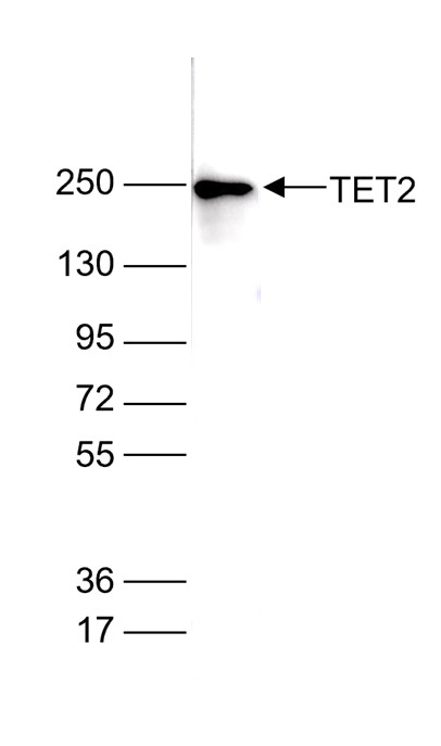
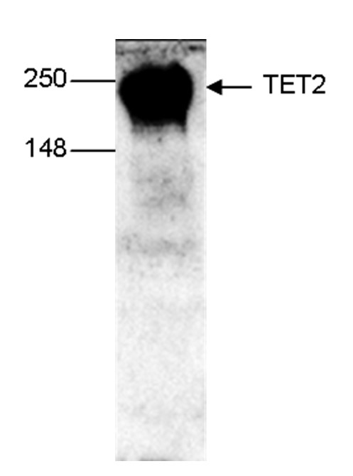
<p><small><strong> Figure 1. Western blot analysis using the Diagenode monoclonal antibody directed against TET2 </strong><br /><strong>Figure 1A.</strong> Whole cell extracts from HeLa cells (40 µg) were analysed by Western blot using the Diagenode antibody against TET2 (cat. No. C15200179), diluted 1:2,000 in PBS containing 10% milk. The position of the protein of interest (expected MW 224 kDa) is indicated on the right; the marker (in kDa) is shown on the left.<br /><strong>Figure 1B.</strong> Western blot on mouse E14 ES cells. The antibody was used at a dilution of 1:1,000. </small></p>
</div>
</div>
<div class="row">
<div class="small-4 columns">
<p><img src="https://www.diagenode.com/img/product/antibodies/c15200179-fig2-ip.jpg" alt="Immunoprecipitation" style="display: block; margin-left: auto; margin-right: auto;" width="222" height="135" /></p>
</div>
<div class="small-8 columns">
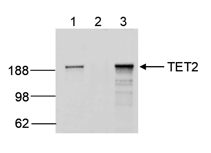
<p><small><strong> Figure 2. Immunoprecipitation using the Diagenode monoclonal antibody directed against TET2 </strong><br />IP was performed on 250 µg HL60 RIPA cell lysate using the Diagenode antibody against TET2 (cat. No. C15200179) (lane 3) or an IgG negative control (lane 2). The samples were analysed by Western blot analysis as described above. The input sample (25 µg RIPA lysate) was used as a positive control (lane 1). </small></p>
</div>
</div>',
'label2' => 'Target description',
'info2' => '<p>TET2 (UniProt/Swiss-Prot entry Q6N021) is a methylcytosine dioxygenase that catalyzes the conversion of 5-methylcytosine to 5-hydroxymethylcytosine (5-hmC). 5-hmC has been recently discovered in mammalian DNA and is abundant in Purkinje neurons, granule cells, embryonic stem cells, and brain tissue, especially in areas that are associated with higher cognitive function. Although its precise role has still to be shown, recent studies indicate that 5-hmC plays important roles distinct from 5-mC. Early evidence suggests that 5-hmC may represent a new pathway to demethylate DNA involving a repair mechanism converting 5-hmC to cytosine. Mutations in TET2 have been associated with myeloproliferative diseases such as essential thrombocythemia, polycythemia vera and primary myelofibrosis.</p>',
'label3' => '',
'info3' => '',
'format' => '10 μg/10 μl',
'catalog_number' => 'C15200179-10',
'old_catalog_number' => '',
'sf_code' => '',
'type' => 'FRE',
'search_order' => '',
'price_EUR' => '105',
'price_USD' => '115',
'price_GBP' => '100',
'price_JPY' => '/',
'price_CNY' => '/',
'price_AUD' => '/',
'country' => 'ALL',
'except_countries' => 'None',
'quote' => false,
'in_stock' => false,
'featured' => false,
'no_promo' => false,
'online' => true,
'master' => false,
'last_datasheet_update' => '',
'slug' => 'tet2-monoclonal-antibody-10ul',
'meta_title' => 'TET2 Monoclonal Antibody - sample size | Diagenode',
'meta_keywords' => '',
'meta_description' => 'TET2 (Tet oncogene family member 2) Monoclonal Antibody validated in IP and WB. Batch-specific data available on the website. Alternative name: MDS',
'modified' => '2024-06-20 15:20:36',
'created' => '2024-06-20 15:17:20',
'locale' => 'eng'
),
'Antibody' => array(
'host' => '*****',
'id' => '650',
'name' => 'TET2 monoclonal antibody',
'description' => 'TET2 (UniProt/Swiss-Prot entry Q6N021) is a methylcytosine dioxygenase that catalyzes the conversion of 5-methylcytosine to 5-hydroxymethylcytosine (5-hmC). 5-hmC has been recently discovered in mammalian DNA and is abundant in Purkinje neurons, granule cells, embryonic stem cells, and brain tissue, especially in areas that are associated with higher cognitive function. Although its precise role has still to be shown, recent studies indicate that 5-hmC plays important roles distinct from 5-mC. Early evidence suggests that 5-hmC may represent a new pathway to demethylate DNA involving a repair mechanism converting 5-hmC to cytosine. Mutations in TET2 have been associated with myeloproliferative diseases such as essential thrombocythemia, polycythemia vera and primary myelofibrosis.',
'clonality' => '',
'isotype' => '',
'lot' => '005',
'concentration' => '1.5 µg/µl',
'reactivity' => 'Human, mouse',
'type' => 'Monoclonal',
'purity' => 'Protein G purified',
'classification' => '',
'application_table' => '<table>
<thead>
<tr>
<th>Applications</th>
<th>Suggested dilution</th>
<th>References</th>
</tr>
</thead>
<tbody>
<tr>
<td>Western Blotting</td>
<td>1:1,000 - 1:5,000</td>
<td>Fig 1, 2</td>
</tr>
<tr>
<td>Immunoprecipitation</td>
<td>5 μg/mg of RIPA lysate</td>
<td>Fig 2</td>
</tr>
</tbody>
</table>',
'storage_conditions' => '',
'storage_buffer' => '',
'precautions' => 'This product is for research use only. Not for use in diagnostic or therapeutic procedures.',
'uniprot_acc' => '',
'slug' => 'tet2-monoclonal-antibody-10ul',
'meta_keywords' => '',
'meta_description' => '',
'modified' => '2024-06-20 15:05:01',
'created' => '2024-06-20 15:05:01',
'select_label' => '650 - TET2 monoclonal antibody (005 - 1.5 µg/µl - Human, mouse - Protein G purified - Mouse)'
),
'Slave' => array(),
'Group' => array(
'Group' => array(
[maximum depth reached]
),
'Master' => array(
[maximum depth reached]
),
'Product' => array(
[maximum depth reached]
)
),
'Related' => array(),
'Application' => array(
(int) 0 => array(
[maximum depth reached]
),
(int) 1 => array(
[maximum depth reached]
)
),
'Category' => array(
(int) 0 => array(
[maximum depth reached]
),
(int) 1 => array(
[maximum depth reached]
)
),
'Document' => array(
(int) 0 => array(
[maximum depth reached]
),
(int) 1 => array(
[maximum depth reached]
),
(int) 2 => array(
[maximum depth reached]
)
),
'Feature' => array(),
'Image' => array(
(int) 0 => array(
[maximum depth reached]
)
),
'Promotion' => array(),
'Protocol' => array(),
'Publication' => array(
(int) 0 => array(
[maximum depth reached]
),
(int) 1 => array(
[maximum depth reached]
),
(int) 2 => array(
[maximum depth reached]
),
(int) 3 => array(
[maximum depth reached]
),
(int) 4 => array(
[maximum depth reached]
),
(int) 5 => array(
[maximum depth reached]
),
(int) 6 => array(
[maximum depth reached]
),
(int) 7 => array(
[maximum depth reached]
)
),
'Testimonial' => array(),
'Area' => array(),
'SafetySheet' => array(
(int) 0 => array(
[maximum depth reached]
),
(int) 1 => array(
[maximum depth reached]
),
(int) 2 => array(
[maximum depth reached]
),
(int) 3 => array(
[maximum depth reached]
),
(int) 4 => array(
[maximum depth reached]
),
(int) 5 => array(
[maximum depth reached]
),
(int) 6 => array(
[maximum depth reached]
),
(int) 7 => array(
[maximum depth reached]
)
)
),
'meta_canonical' => 'https://www.diagenode.com/en/p/tet2-monoclonal-antibody-classic-50-mg-50-ml'
)
$language = 'en'
$meta_keywords = ''
$meta_description = 'TET2 (Tet oncogene family member 2) Monoclonal Antibody validated in IP and WB. Batch-specific data available on the website. Alternative name: MDS'
$meta_title = 'TET2 Monoclonal Antibody - sample size | Diagenode'
$product = array(
'Product' => array(
'id' => '3238',
'antibody_id' => '650',
'name' => 'TET2 Antibody (sample size)',
'description' => '<p><span>Alternative name: </span><strong>MDS</strong><br /><span>Monoclonal antibody raised in mouse against a recombinant protein containing the N-terminal 300 amino acids of human TET2 (tet oncogene family member 2).</span></p>',
'label1' => 'Validation Data',
'info1' => '<div class="row">
<div class="small-4 columns"><center><img src="https://www.diagenode.com/img/product/antibodies/c15200179-fig1a-wb.jpg" alt="Western Blot" width="130" height="161" caption="false" /></center><center><img src="https://www.diagenode.com/img/product/antibodies/c15200179-fig1b-wb.jpg" alt="Western Blot" width="110" height="142" caption="false" /></center></div>
<div class="small-8 columns">
<p><small><strong> Figure 1. Western blot analysis using the Diagenode monoclonal antibody directed against TET2 </strong><br /><strong>Figure 1A.</strong> Whole cell extracts from HeLa cells (40 µg) were analysed by Western blot using the Diagenode antibody against TET2 (cat. No. C15200179), diluted 1:2,000 in PBS containing 10% milk. The position of the protein of interest (expected MW 224 kDa) is indicated on the right; the marker (in kDa) is shown on the left.<br /><strong>Figure 1B.</strong> Western blot on mouse E14 ES cells. The antibody was used at a dilution of 1:1,000. </small></p>
</div>
</div>
<div class="row">
<div class="small-4 columns">
<p><img src="https://www.diagenode.com/img/product/antibodies/c15200179-fig2-ip.jpg" alt="Immunoprecipitation" style="display: block; margin-left: auto; margin-right: auto;" width="222" height="135" /></p>
</div>
<div class="small-8 columns">
<p><small><strong> Figure 2. Immunoprecipitation using the Diagenode monoclonal antibody directed against TET2 </strong><br />IP was performed on 250 µg HL60 RIPA cell lysate using the Diagenode antibody against TET2 (cat. No. C15200179) (lane 3) or an IgG negative control (lane 2). The samples were analysed by Western blot analysis as described above. The input sample (25 µg RIPA lysate) was used as a positive control (lane 1). </small></p>
</div>
</div>',
'label2' => 'Target description',
'info2' => '<p>TET2 (UniProt/Swiss-Prot entry Q6N021) is a methylcytosine dioxygenase that catalyzes the conversion of 5-methylcytosine to 5-hydroxymethylcytosine (5-hmC). 5-hmC has been recently discovered in mammalian DNA and is abundant in Purkinje neurons, granule cells, embryonic stem cells, and brain tissue, especially in areas that are associated with higher cognitive function. Although its precise role has still to be shown, recent studies indicate that 5-hmC plays important roles distinct from 5-mC. Early evidence suggests that 5-hmC may represent a new pathway to demethylate DNA involving a repair mechanism converting 5-hmC to cytosine. Mutations in TET2 have been associated with myeloproliferative diseases such as essential thrombocythemia, polycythemia vera and primary myelofibrosis.</p>',
'label3' => '',
'info3' => '',
'format' => '10 μg/10 μl',
'catalog_number' => 'C15200179-10',
'old_catalog_number' => '',
'sf_code' => '',
'type' => 'FRE',
'search_order' => '',
'price_EUR' => '105',
'price_USD' => '115',
'price_GBP' => '100',
'price_JPY' => '/',
'price_CNY' => '/',
'price_AUD' => '/',
'country' => 'ALL',
'except_countries' => 'None',
'quote' => false,
'in_stock' => false,
'featured' => false,
'no_promo' => false,
'online' => true,
'master' => false,
'last_datasheet_update' => '',
'slug' => 'tet2-monoclonal-antibody-10ul',
'meta_title' => 'TET2 Monoclonal Antibody - sample size | Diagenode',
'meta_keywords' => '',
'meta_description' => 'TET2 (Tet oncogene family member 2) Monoclonal Antibody validated in IP and WB. Batch-specific data available on the website. Alternative name: MDS',
'modified' => '2024-06-20 15:20:36',
'created' => '2024-06-20 15:17:20',
'locale' => 'eng'
),
'Antibody' => array(
'host' => '*****',
'id' => '650',
'name' => 'TET2 monoclonal antibody',
'description' => 'TET2 (UniProt/Swiss-Prot entry Q6N021) is a methylcytosine dioxygenase that catalyzes the conversion of 5-methylcytosine to 5-hydroxymethylcytosine (5-hmC). 5-hmC has been recently discovered in mammalian DNA and is abundant in Purkinje neurons, granule cells, embryonic stem cells, and brain tissue, especially in areas that are associated with higher cognitive function. Although its precise role has still to be shown, recent studies indicate that 5-hmC plays important roles distinct from 5-mC. Early evidence suggests that 5-hmC may represent a new pathway to demethylate DNA involving a repair mechanism converting 5-hmC to cytosine. Mutations in TET2 have been associated with myeloproliferative diseases such as essential thrombocythemia, polycythemia vera and primary myelofibrosis.',
'clonality' => '',
'isotype' => '',
'lot' => '005',
'concentration' => '1.5 µg/µl',
'reactivity' => 'Human, mouse',
'type' => 'Monoclonal',
'purity' => 'Protein G purified',
'classification' => '',
'application_table' => '<table>
<thead>
<tr>
<th>Applications</th>
<th>Suggested dilution</th>
<th>References</th>
</tr>
</thead>
<tbody>
<tr>
<td>Western Blotting</td>
<td>1:1,000 - 1:5,000</td>
<td>Fig 1, 2</td>
</tr>
<tr>
<td>Immunoprecipitation</td>
<td>5 μg/mg of RIPA lysate</td>
<td>Fig 2</td>
</tr>
</tbody>
</table>',
'storage_conditions' => '',
'storage_buffer' => '',
'precautions' => 'This product is for research use only. Not for use in diagnostic or therapeutic procedures.',
'uniprot_acc' => '',
'slug' => 'tet2-monoclonal-antibody-10ul',
'meta_keywords' => '',
'meta_description' => '',
'modified' => '2024-06-20 15:05:01',
'created' => '2024-06-20 15:05:01',
'select_label' => '650 - TET2 monoclonal antibody (005 - 1.5 µg/µl - Human, mouse - Protein G purified - Mouse)'
),
'Slave' => array(),
'Group' => array(
'Group' => array(
'id' => '367',
'name' => 'C15200179',
'product_id' => '1996',
'modified' => '2024-06-20 15:17:58',
'created' => '2024-06-20 15:17:58'
),
'Master' => array(
'id' => '1996',
'antibody_id' => '304',
'name' => 'TET2 Antibody ',
'description' => '<p>Alternative name: <strong>MDS</strong><br /> Monoclonal antibody raised in mouse against a recombinant protein containing the N-terminal 300 amino acids of human TET2 (tet oncogene family member 2).</p>',
'label1' => 'Validation Data',
'info1' => '<div class="row">
<div class="small-4 columns"><center><img src="https://www.diagenode.com/img/product/antibodies/c15200179-fig1a-wb.jpg" alt="Western Blot" width="130" height="161" caption="false" /></center><center><img src="https://www.diagenode.com/img/product/antibodies/c15200179-fig1b-wb.jpg" alt="Western Blot" width="110" height="142" caption="false" /></center></div>
<div class="small-8 columns">
<p><small><strong> Figure 1. Western blot analysis using the Diagenode monoclonal antibody directed against TET2 </strong><br /><strong>Figure 1A.</strong> Whole cell extracts from HeLa cells (40 µg) were analysed by Western blot using the Diagenode antibody against TET2 (cat. No. C15200179), diluted 1:2,000 in PBS containing 10% milk. The position of the protein of interest (expected MW 224 kDa) is indicated on the right; the marker (in kDa) is shown on the left.<br /><strong>Figure 1B.</strong> Western blot on mouse E14 ES cells. The antibody was used at a dilution of 1:1,000. </small></p>
</div>
</div>
<div class="row">
<div class="small-4 columns">
<p><img src="https://www.diagenode.com/img/product/antibodies/c15200179-fig2-ip.jpg" alt="Immunoprecipitation" style="display: block; margin-left: auto; margin-right: auto;" width="222" height="135" /></p>
</div>
<div class="small-8 columns">
<p><small><strong> Figure 2. Immunoprecipitation using the Diagenode monoclonal antibody directed against TET2 </strong><br />IP was performed on 250 µg HL60 RIPA cell lysate using the Diagenode antibody against TET2 (cat. No. C15200179) (lane 3) or an IgG negative control (lane 2). The samples were analysed by Western blot analysis as described above. The input sample (25 µg RIPA lysate) was used as a positive control (lane 1). </small></p>
</div>
</div>',
'label2' => 'Target description',
'info2' => '<p>TET2 (UniProt/Swiss-Prot entry Q6N021) is a methylcytosine dioxygenase that catalyzes the conversion of 5-methylcytosine to 5-hydroxymethylcytosine (5-hmC). 5-hmC has been recently discovered in mammalian DNA and is abundant in Purkinje neurons, granule cells, embryonic stem cells, and brain tissue, especially in areas that are associated with higher cognitive function. Although its precise role has still to be shown, recent studies indicate that 5-hmC plays important roles distinct from 5-mC. Early evidence suggests that 5-hmC may represent a new pathway to demethylate DNA involving a repair mechanism converting 5-hmC to cytosine. Mutations in TET2 have been associated with myeloproliferative diseases such as essential thrombocythemia, polycythemia vera and primary myelofibrosis.</p>',
'label3' => '',
'info3' => '',
'format' => '50 μg/50 μl',
'catalog_number' => 'C15200179',
'old_catalog_number' => 'MAb-179-050',
'sf_code' => 'C15200179-D001-000581',
'type' => 'FRE',
'search_order' => '03-Antibody',
'price_EUR' => '380',
'price_USD' => '380',
'price_GBP' => '340',
'price_JPY' => '59525',
'price_CNY' => '',
'price_AUD' => '950',
'country' => 'ALL',
'except_countries' => 'None',
'quote' => false,
'in_stock' => false,
'featured' => false,
'no_promo' => false,
'online' => true,
'master' => true,
'last_datasheet_update' => '0000-00-00',
'slug' => 'tet2-monoclonal-antibody-classic-50-mg-50-ml',
'meta_title' => 'TET2 Monoclonal Antibody | Diagenode',
'meta_keywords' => '',
'meta_description' => 'TET2 (Tet oncogene family member 2) Monoclonal Antibody validated in IP and WB. Batch-specific data available on the website. Alternative name: MDS',
'modified' => '2023-12-14 15:13:33',
'created' => '2015-06-29 14:08:20'
),
'Product' => array(
(int) 0 => array(
[maximum depth reached]
)
)
),
'Related' => array(),
'Application' => array(
(int) 0 => array(
'id' => '19',
'position' => '10',
'parent_id' => '40',
'name' => 'WB',
'description' => '<p><strong>Western blot</strong> : The quality of antibodies used in this technique is crucial for correct and specific protein identification. Diagenode offers huge selection of highly sensitive and specific western blot-validated antibodies.</p>
<p>Learn more about: <a href="https://www.diagenode.com/applications/western-blot">Loading control, MW marker visualization</a><em>. <br /></em></p>
<p><em></em>Check our selection of antibodies validated in Western blot.</p>',
'in_footer' => false,
'in_menu' => false,
'online' => true,
'tabular' => true,
'slug' => 'western-blot-antibodies',
'meta_keywords' => ' Western Blot Antibodies ,western blot protocol,Western Blotting Products,Polyclonal antibodies ,monoclonal antibodies ',
'meta_description' => 'Diagenode offers a wide range of antibodies and technical support for western blot applications',
'meta_title' => ' Western Blot - Monoclonal antibody - Polyclonal antibody | Diagenode',
'modified' => '2016-04-26 12:44:51',
'created' => '2015-01-07 09:20:00',
'ProductsApplication' => array(
[maximum depth reached]
)
),
(int) 1 => array(
'id' => '30',
'position' => '10',
'parent_id' => '40',
'name' => 'IP',
'description' => '<p>Immunoprecipitation</p>',
'in_footer' => false,
'in_menu' => false,
'online' => true,
'tabular' => true,
'slug' => 'immunoprecipitation',
'meta_keywords' => 'Immunoprecipitation,Monoclonal antibody,Polyclonal antibody',
'meta_description' => 'Diagenode offers a wide range of antibodies and technical support for Immunoprecipitation applications',
'meta_title' => 'Immunoprecipitation - Monoclonal antibody - Polyclonal antibody | Diagenode',
'modified' => '2016-01-13 12:23:07',
'created' => '2015-07-08 13:46:50',
'ProductsApplication' => array(
[maximum depth reached]
)
)
),
'Category' => array(
(int) 0 => array(
'id' => '23',
'position' => '40',
'parent_id' => '4',
'name' => 'DNA modifications',
'description' => '<p><span style="font-weight: 400;">T</span><span style="font-weight: 400;">he pattern of <strong>DNA modifications</strong> is critical for genome stability and the control of gene expression in the cell. Methylation of 5-cytosine (5-mC), one of the best-studied epigenetic marks, is carried out by the <strong>DNA methyltransferases</strong> DNMT3A and B and DNMT1. DNMT3A and DNMT3B are responsible for </span><i><span style="font-weight: 400;">de novo</span></i><span style="font-weight: 400;"> DNA methylation, whereas DNMT1 maintains existing methylation. 5-mC undergoes active demethylation which is performed by the <strong>Ten-Eleven Translocation</strong> (TET) familly of DNA hydroxylases. The latter consists of 3 members TET1, 2 and 3. All 3 members catalyze the conversion of <strong>5-methylcytosine</strong> (5-mC) into <strong>5-hydroxymethylcytosine</strong> (5-hmC), and further into <strong>5-formylcytosine</strong> (5-fC) and <strong>5-carboxycytosine</strong> (5-caC). 5-fC and 5-caC can be converted to unmodified cytosine by <strong>Thymine DNA Glycosylase</strong> (TDG). It is not yet clear if 5-hmC, 5-fC and 5-caC have specific functions or are simply intermediates in the demethylation of 5-mC.</span></p>
<p><span style="font-weight: 400;">DNA methylation is generally considered as a repressive mark and is usually associated with gene silencing. It is essential that the balance between DNA methylation and demethylation is precisely maintained. Dysregulation of DNA methylation may lead to many different human diseases and is often observed in cancer cells.</span></p>
<p><span style="font-weight: 400;">Diagenode offers highly validated antibodies against different proteins involved in DNA modifications as well as against the modified bases allowing the study of all steps and intermediates in the DNA methylation/demethylation pathway:</span></p>
<p><img src="https://www.diagenode.com/img/categories/antibodies/dna-methylation.jpg" height="599" width="816" /></p>
<p><strong>Diagenode exclusively sources the original 5-methylcytosine monoclonal antibody (clone 33D3).</strong></p>
<p>Check out the list below to see all proposed antibodies for DNA modifications.</p>
<p>Diagenode’s highly validated antibodies:</p>
<ul>
<li>Highly sensitive and specific</li>
<li>Cost-effective (requires less antibody per reaction)</li>
<li>Batch-specific data is available on the website</li>
<li>Expert technical support</li>
<li>Sample sizes available</li>
<li>100% satisfaction guarantee</li>
</ul>',
'no_promo' => false,
'in_menu' => true,
'online' => true,
'tabular' => false,
'hide' => true,
'all_format' => false,
'is_antibody' => true,
'slug' => 'dna-methylation-antibody',
'cookies_tag_id' => null,
'meta_keywords' => 'Monoclonal Antibodies,Polyclonal antibodies,DNA methylation,Diagenode',
'meta_description' => 'Diagenode offers Monoclonal and Polyclonal antibodies for DNA Methylation. The pattern of DNA modifications is critical for genome stability and the control of gene expression in the cell. ',
'meta_title' => 'DNA modifications - Monoclonal and Polyclonal Antibodies for DNA Methylation | Diagenode',
'modified' => '2020-08-03 15:25:37',
'created' => '2014-07-31 03:51:53',
'ProductsCategory' => array(
[maximum depth reached]
),
'CookiesTag' => array([maximum depth reached])
),
(int) 1 => array(
'id' => '102',
'position' => '1',
'parent_id' => '4',
'name' => 'Sample size antibodies',
'description' => '<h1><strong>Validated epigenetics antibodies</strong> – care for a sample?<br /> </h1>
<p>Diagenode has partnered with leading epigenetics experts and numerous epigenetics consortiums to bring to you a validated and comprehensive collection of epigenetic antibodies. As an expert in epigenetics, we are committed to offering highly-specific antibodies validated for ChIP/ChIP-seq and many other applications. All batch-specific validation data is available on our website.<br /><a href="../categories/antibodies">Read about our expertise in antibody production</a>.</p>
<ul>
<li><strong>Focused</strong> - Diagenode's selection of antibodies is exclusively dedicated for epigenetic research. <a title="See the full collection." href="../categories/all-antibodies">See the full collection.</a></li>
<li><strong>Strict quality standards</strong> with rigorous QC and validation</li>
<li><strong>Classified</strong> based on level of validation for flexibility of application</li>
</ul>
<p>Existing sample sizes are listed below. We will soon expand our collection. Are you looking for a sample size of another antibody? Just <a href="mailto:agnieszka.zelisko@diagenode.com?Subject=Sample%20Size%20Request" target="_top">Contact us</a>.</p>',
'no_promo' => false,
'in_menu' => true,
'online' => true,
'tabular' => false,
'hide' => true,
'all_format' => true,
'is_antibody' => true,
'slug' => 'sample-size-antibodies',
'cookies_tag_id' => null,
'meta_keywords' => '5-hmC monoclonal antibody,CRISPR/Cas9 polyclonal antibody ,H3K36me3 polyclonal antibody,diagenode',
'meta_description' => 'Diagenode offers sample volume on selected antibodies for researchers to test, validate and provide confidence and flexibility in choosing from our wide range of antibodies ',
'meta_title' => 'Sample-size Antibodies | Diagenode',
'modified' => '2019-07-03 10:57:05',
'created' => '2015-10-27 12:13:34',
'ProductsCategory' => array(
[maximum depth reached]
),
'CookiesTag' => array([maximum depth reached])
)
),
'Document' => array(
(int) 0 => array(
'id' => '11',
'name' => 'Antibodies you can trust',
'description' => '<p style="text-align: justify;"><span>Epigenetic research tools have evolved over time from endpoint PCR to qPCR to the analyses of large sets of genome-wide sequencing data. ChIP sequencing (ChIP-seq) has now become the gold standard method for chromatin studies, given the accuracy and coverage scale of the approach over other methods. Successful ChIP-seq, however, requires a higher level of experimental accuracy and consistency in all steps of ChIP than ever before. Particularly crucial is the quality of ChIP antibodies. </span></p>',
'image_id' => null,
'type' => 'Poster',
'url' => 'files/posters/Antibodies_you_can_trust_Poster.pdf',
'slug' => 'antibodies-you-can-trust-poster',
'meta_keywords' => '',
'meta_description' => '',
'modified' => '2015-10-01 20:18:31',
'created' => '2015-07-03 16:05:15',
'ProductsDocument' => array(
[maximum depth reached]
)
),
(int) 1 => array(
'id' => '38',
'name' => 'Epigenetic Antibodies Brochure',
'description' => '<p>More than in any other immuoprecipitation assays, quality antibodies are critical tools in many epigenetics experiments. Since 10 years, Diagenode has developed the most stringent quality production available on the market for antibodies exclusively focused on epigenetic uses. All our antibodies have been qualified to work in epigenetic applications.</p>',
'image_id' => null,
'type' => 'Brochure',
'url' => 'files/brochures/Epigenetic_Antibodies_Brochure.pdf',
'slug' => 'epigenetic-antibodies-brochure',
'meta_keywords' => '',
'meta_description' => '',
'modified' => '2016-06-15 11:24:06',
'created' => '2015-07-03 16:05:27',
'ProductsDocument' => array(
[maximum depth reached]
)
),
(int) 2 => array(
'id' => '708',
'name' => 'Datasheet TET2 C15200179',
'description' => '<p>Monoclonal antibody raised in mouse against a recombinant protein containing the N-terminal 300 amino acids of human TET2 (tet oncogene family member 2).</p>',
'image_id' => null,
'type' => 'Datasheet',
'url' => 'files/products/antibodies/Datasheet_TET2_C15200179.pdf',
'slug' => 'datasheet-tet2-C15200179',
'meta_keywords' => '',
'meta_description' => '',
'modified' => '2016-07-08 16:19:26',
'created' => '2015-07-07 11:47:44',
'ProductsDocument' => array(
[maximum depth reached]
)
)
),
'Feature' => array(),
'Image' => array(
(int) 0 => array(
'id' => '250',
'name' => 'product/antibodies/antibody.png',
'alt' => 'Mouse IgG',
'modified' => '2020-11-27 07:00:09',
'created' => '2015-07-17 10:12:18',
'ProductsImage' => array(
[maximum depth reached]
)
)
),
'Promotion' => array(),
'Protocol' => array(),
'Publication' => array(
(int) 0 => array(
'id' => '4482',
'name' => 'Vitamin C enhances NF-κB-driven epigenomic reprogramming andboosts the immunogenic properties of dendritic cells.',
'authors' => 'Morante-Palacios O. et al.',
'description' => '<p>Dendritic cells (DCs), the most potent antigen-presenting cells, are necessary for effective activation of naïve T cells. DCs' immunological properties are modulated in response to various stimuli. Active DNA demethylation is crucial for DC differentiation and function. Vitamin C, a known cofactor of ten-eleven translocation (TET) enzymes, drives active demethylation. Vitamin C has recently emerged as a promising adjuvant for several types of cancer; however, its effects on human immune cells are poorly understood. In this study, we investigate the epigenomic and transcriptomic reprogramming orchestrated by vitamin C in monocyte-derived DC differentiation and maturation. Vitamin C triggers extensive demethylation at NF-κB/p65 binding sites, together with concordant upregulation of antigen-presentation and immune response-related genes during DC maturation. p65 interacts with TET2 and mediates the aforementioned vitamin C-mediated changes, as demonstrated by pharmacological inhibition. Moreover, vitamin C increases TNFβ production in DCs through NF-κB, in concordance with the upregulation of its coding gene and the demethylation of adjacent CpGs. Finally, vitamin C enhances DC's ability to stimulate the proliferation of autologous antigen-specific T cells. We propose that vitamin C could potentially improve monocyte-derived DC-based cell therapies.</p>',
'date' => '2022-10-01',
'pmid' => 'https://www.ncbi.nlm.nih.gov/pubmed/36305821',
'doi' => '10.1093/nar/gkac941',
'modified' => '2022-11-18 12:30:06',
'created' => '2022-11-15 09:26:20',
'ProductsPublication' => array(
[maximum depth reached]
)
),
(int) 1 => array(
'id' => '4253',
'name' => 'Coordinated glucocorticoid receptor and MAFB action inducestolerogenesis and epigenome remodeling in dendritic cells',
'authors' => 'Morante-Palacios Octavio et al.',
'description' => '<p>Abstract Glucocorticoids (GCs) exert potent anti-inflammatory effects in immune cells through the glucocorticoid receptor (GR). Dendritic cells (DCs), central actors for coordinating immune responses, acquire tolerogenic properties in response to GCs. Tolerogenic DCs (tolDCs) have emerged as a potential treatment for various inflammatory diseases. To date, the underlying cell type-specific regulatory mechanisms orchestrating GC-mediated acquisition of immunosuppressive properties remain poorly understood. In this study, we investigated the transcriptomic and epigenomic remodeling associated with differentiation to DCs in the presence of GCs. Our analysis demonstrates a major role of MAFB in this process, in synergy with GR. GR and MAFB both interact with methylcytosine dioxygenase TET2 and bind to genomic loci that undergo specific demethylation in tolDCs. We also show that the role of MAFB is more extensive, binding to thousands of genomic loci in tolDCs. Finally, MAFB knockdown erases the tolerogenic properties of tolDCs and reverts the specific DNA demethylation and gene upregulation. The preeminent role of MAFB is also demonstrated in vivo for myeloid cells from synovium in rheumatoid arthritis following GC treatment. Our results imply that, once directly activated by GR, MAFB plays a critical role in orchestrating the epigenomic and transcriptomic remodeling that define the tolerogenic phenotype.</p>',
'date' => '2022-01-01',
'pmid' => 'https://www.ncbi.nlm.nih.gov/pubmed/34893889',
'doi' => '10.1093/nar/gkab1182',
'modified' => '2022-05-20 09:44:29',
'created' => '2022-05-19 10:41:50',
'ProductsPublication' => array(
[maximum depth reached]
)
),
(int) 2 => array(
'id' => '3641',
'name' => 'TET2 inhibits tumorigenesis of breast cancer cells by regulating caspase-4.',
'authors' => 'Zhu X, Li S',
'description' => '<p>Epigenetic regulators have been shown to influence breast cancer progression. However, the detailed mechanism by which TET2 plays the suppressive role in tumorigenesis remains not completely understood. We employed RT-qPCR and westernblot to examine genes expression. Next, the bisulphite sequencing PCR was used to determine the methylation level at CASP4 promoter in the cells. Phenotypically, we utilized growth curve analysis, colony formation in soft agar and xenograft tumor assay to assess tumorigenesis of MCF-7 cell. We found that TET2 knockout enhanced colony formation ability and in vivo tumor formation ability of MCF-7 cell, whereas TET2 depletion not affected the growth rate of MCF-7 cell in the culture. Mechanistically, TET2 loss led to a significant decrease in caspase-4 expression possibly via increasing DNA methylation of CASP4 promoter in MCF-7 cell. To validate, TET2 overexpression led to higher level of caspase-4 in MDA-MB-231 and 293T cells, which was dependent on TET2 enzymatic activity. Finally, we observed that caspase-4 could revert, at least partially, TET2 deletion-induced tumorigenesis of MCF-7. In summary, we reveal a novel mechanism that TET2 suppresses tumorigenesis of breast cancer cells through caspase-4. Our findings will facilitate development of new diagnostic markers or therapeutical therapies for breast cancer.</p>',
'date' => '2018-11-01',
'pmid' => 'http://www.pubmed.gov/30385776',
'doi' => '10.1038/s41598-018-34462-z',
'modified' => '2019-06-07 10:20:47',
'created' => '2019-06-06 12:11:18',
'ProductsPublication' => array(
[maximum depth reached]
)
),
(int) 3 => array(
'id' => '3293',
'name' => 'The Role of N-α-acetyltransferase 10 Protein in DNA Methylation and Genomic Imprinting',
'authors' => 'Lee C.C. et al.',
'description' => '<p>Genomic imprinting is an allelic gene expression phenomenon primarily controlled by allele-specific DNA methylation at the imprinting control region (ICR), but the underlying mechanism remains largely unclear. N-α-acetyltransferase 10 protein (Naa10p) catalyzes N-α-acetylation of nascent proteins, and mutation of human Naa10p is linked to severe developmental delays. Here we report that Naa10-null mice display partial embryonic lethality, growth retardation, brain disorders, and maternal effect lethality, phenotypes commonly observed in defective genomic imprinting. Genome-wide analyses further revealed global DNA hypomethylation and enriched dysregulation of imprinted genes in Naa10p-knockout embryos and embryonic stem cells. Mechanistically, Naa10p facilitates binding of DNA methyltransferase 1 (Dnmt1) to DNA substrates, including the ICRs of the imprinted allele during S phase. Moreover, the lethal Ogden syndrome-associated mutation of human Naa10p disrupts its binding to the ICR of H19 and Dnmt1 recruitment. Our study thus links Naa10p mutation-associated Ogden syndrome to defective DNA methylation and genomic imprinting.</p>',
'date' => '2017-10-05',
'pmid' => 'https://www.ncbi.nlm.nih.gov/pubmed/28943313',
'doi' => '',
'modified' => '2017-12-04 10:51:06',
'created' => '2017-12-04 10:51:06',
'ProductsPublication' => array(
[maximum depth reached]
)
),
(int) 4 => array(
'id' => '3266',
'name' => 'TET2- and TDG-mediated changes are required for the acquisition of distinct histone modifications in divergent terminal differentiation of myeloid cells',
'authors' => 'Garcia-Gomez A. et al.',
'description' => '<p>The plasticity of myeloid cells is illustrated by a diversity of functions including their role as effectors of innate immunity as macrophages (MACs) and bone remodelling as osteoclasts (OCs). TET2, a methylcytosine dioxygenase highly expressed in these cells and frequently mutated in myeloid leukemias, may be a key contributor to this plasticity. Through transcriptomic and epigenomic analyses, we investigated 5-methylcytosine (5mC), 5-hydroxymethylcytosine (5hmC) and gene expression changes in two divergent terminal myeloid differentiation processes, namely MAC and OC differentiation. MACs and OCs undergo highly similar 5hmC and 5mC changes, despite their wide differences in gene expression. Many TET2- and thymine-DNA glycosylase (TDG)-dependent 5mC and 5hmC changes directly activate the common terminal myeloid differentiation programme. However, the acquisition of differential features between MACs and OCs also depends on TET2/TDG. In fact, 5mC oxidation precedes differential histone modification changes between MACs and OCs. TET2 and TDG downregulation impairs the acquisition of such differential histone modification and expression patterns at MAC-/OC-specific genes. We prove that the histone H3K4 methyltransferase SETD1A is differentially recruited between MACs and OCs in a TET2-dependent manner. We demonstrate a novel role of these enzymes in the establishment of specific elements of identity and function in terminal myeloid differentiation.</p>',
'date' => '2017-09-29',
'pmid' => 'https://www.ncbi.nlm.nih.gov/pubmed/28973458',
'doi' => '',
'modified' => '2017-10-09 16:19:32',
'created' => '2017-10-09 16:19:32',
'ProductsPublication' => array(
[maximum depth reached]
)
),
(int) 5 => array(
'id' => '3104',
'name' => 'TET2 binds the androgen receptor and loss is associated with prostate cancer',
'authors' => 'Nickerson M.L. et al.',
'description' => '<p>Genetic alterations associated with prostate cancer (PCa) may be identified by sequencing metastatic tumour genomes to identify molecular markers at this lethal stage of disease. Previously, we characterized somatic alterations in metastatic tumours in the methylcytosine dioxygenase ten-eleven translocation 2 (TET2), which is altered in 5-15% of myeloid, kidney, colon and PCas. Genome-wide association studies previously identified non-coding risk variants associated with PCa and melanoma. We perform fine-mapping of PCa risk across TET2 using genotypes from the PEGASUS case-control cohort and identify six new risk variants in introns 1 and 2. Oligonucleotides containing two risk variants are bound by the transcription factor octamer-binding protein 1 (Oct1/POU2F1) and TET2 and Oct1 expression are positively correlated in prostate tumours. TET2 is expressed in normal prostate tissue and reduced in a subset of tumours from the Cancer Genome Atlas (TCGA). Small interfering RNA-mediated TET2 knockdown (KD) increases LNCaP cell proliferation, migration and wound healing, verifying loss drives a cancer phenotype. Endogenous TET2 bound the androgen receptor (AR) and AR-coactivator proteins in LNCaP cell extracts, and TET2 KD increases prostate-specific antigen (KLK3/PSA) expression. Published data reveal TET2 binding sites and hydroxymethylcytosine proximal to KLK3. A gene co-expression network identified using TCGA prostate tumour RNA-sequencing identifies co-regulated cancer genes associated with 2-oxoglutarate (2-OG) and succinate metabolism, including TET2, lysine demethylase (KDM) KDM6A, BRCA1-associated BAP1, and citric acid cycle enzymes IDH1/2, SDHA/B, and FH. The co-expression signature is conserved across 31 TCGA cancers suggesting a putative role for TET2 as an energy sensor (of 2-OG) that modifies aspects of androgen-AR signalling. Decreased TET2 mRNA expression in TCGA PCa tumours is strongly associated with reduced patient survival, indicating reduced expression in tumours may be an informative biomarker of disease progression and perhaps metastatic disease.</p>',
'date' => '2016-11-07',
'pmid' => 'https://www.ncbi.nlm.nih.gov/pubmed/27819678',
'doi' => '',
'modified' => '2017-01-03 15:50:31',
'created' => '2017-01-03 15:50:31',
'ProductsPublication' => array(
[maximum depth reached]
)
),
(int) 6 => array(
'id' => '2978',
'name' => 'TET-catalyzed oxidation of intragenic 5-methylcytosine regulates CTCF-dependent alternative splicing.',
'authors' => 'Marina RJ et al.',
'description' => '<p>Intragenic 5-methylcytosine and CTCF mediate opposing effects on pre-mRNA splicing: CTCF promotes inclusion of weak upstream exons through RNA polymerase II pausing, whereas 5-methylcytosine evicts CTCF, leading to exon exclusion. However, the mechanisms governing dynamic DNA methylation at CTCF-binding sites were unclear. Here, we reveal the methylcytosine dioxygenases TET1 and TET2 as active regulators of CTCF-mediated alternative splicing through conversion of 5-methylcytosine to its oxidation derivatives. 5-hydroxymethylcytosine and 5-carboxylcytosine are enriched at an intragenic CTCF-binding sites in the CD45 model gene and are associated with alternative exon inclusion. Reduced TET levels culminate in increased 5-methylcytosine, resulting in CTCF eviction and exon exclusion. In vitro analyses establish the oxidation derivatives are not sufficient to stimulate splicing, but efficiently promote CTCF association. We further show genomewide that reciprocal exchange of 5-hydroxymethylcytosine and 5-methylcytosine at downstream CTCF-binding sites is a general feature of alternative splicing in naïve and activated CD4(+) T cells. These findings significantly expand our current concept of the pre-mRNA "splicing code" to include dynamic intragenic DNA methylation catalyzed by the TET proteins.</p>',
'date' => '2016-02-01',
'pmid' => 'http://www.ncbi.nlm.nih.gov/pubmed/26711177',
'doi' => ' 10.15252/embj.201593235',
'modified' => '2016-07-08 10:05:02',
'created' => '2016-07-08 10:05:02',
'ProductsPublication' => array(
[maximum depth reached]
)
),
(int) 7 => array(
'id' => '2604',
'name' => 'Single-Base Resolution Analysis of 5-Formyl and 5-Carboxyl Cytosine Reveals Promoter DNA Methylation Dynamics.',
'authors' => 'Neri F, Incarnato D, Krepelova A, Rapelli S, Anselmi F, Parlato C, Medana C, Dal Bello F, Oliviero S',
'description' => '<p>Ten eleven translocation (Tet) proteins oxidize 5-methylcytosine (5mC) to 5-hydroxymethylcytosine (5hmC), 5-formylcytosine (5fC), and 5-carboxylcytosine (5caC). 5fC and 5caC can be further excised by thymine-DNA glycosylase (Tdg). Here, we present a genome-wide approach, named methylation-assisted bisulfite sequencing (MAB-seq), that enables single-base resolution mapping of 5fC and 5caC and measures their abundance. Application of this method to mouse embryonic stem cells (ESCs) shows the occurrence of 5fC and 5caC residues on the hypomethylated promoters of highly expressed genes, which is increased upon Tdg silencing, revealing active DNA demethylation on these promoters. Genome-wide mapping of Tdg reveals extensive colocalization with Tet1 on active promoters. These regions were found to be methylated by Dnmt1 and Dnmt3a and demethylated by a Tet-dependent mechanism. Our work demonstrates the DNA methylation dynamics that occurs on the promoters of the expressed genes and provides a genomic reference map of 5fC and 5caC in ESCs.</p>',
'date' => '2015-02-04',
'pmid' => 'https://www.ncbi.nlm.nih.gov/pubmed/25660018',
'doi' => '',
'modified' => '2016-04-04 10:37:14',
'created' => '2015-07-24 15:39:05',
'ProductsPublication' => array(
[maximum depth reached]
)
)
),
'Testimonial' => array(),
'Area' => array(),
'SafetySheet' => array(
(int) 0 => array(
'id' => '1486',
'name' => 'TET2 antibody SDS GB en',
'language' => 'en',
'url' => 'files/SDS/TET2/SDS-C15200179-TET2_Antibody-GB-en-GHS_2_0.pdf',
'countries' => 'GB',
'modified' => '2021-08-30 15:16:20',
'created' => '2021-08-30 15:16:20',
'ProductsSafetySheet' => array(
[maximum depth reached]
)
),
(int) 1 => array(
'id' => '1488',
'name' => 'TET2 antibody SDS US en',
'language' => 'en',
'url' => 'files/SDS/TET2/SDS-C15200179-TET2_Antibody-US-en-GHS_2_0.pdf',
'countries' => 'US',
'modified' => '2021-08-30 15:16:20',
'created' => '2021-08-30 15:16:20',
'ProductsSafetySheet' => array(
[maximum depth reached]
)
),
(int) 2 => array(
'id' => '1483',
'name' => 'TET2 antibody SDS DE de',
'language' => 'de',
'url' => 'files/SDS/TET2/SDS-C15200179-TET2_Antibody-DE-de-GHS_2_0.pdf',
'countries' => 'DE',
'modified' => '2021-08-30 15:16:20',
'created' => '2021-08-30 15:16:20',
'ProductsSafetySheet' => array(
[maximum depth reached]
)
),
(int) 3 => array(
'id' => '1487',
'name' => 'TET2 antibody SDS JP ja',
'language' => 'ja',
'url' => 'files/SDS/TET2/SDS-C15200179-TET2_Antibody-JP-ja-GHS_3_0.pdf',
'countries' => 'JP',
'modified' => '2021-08-30 15:16:20',
'created' => '2021-08-30 15:16:20',
'ProductsSafetySheet' => array(
[maximum depth reached]
)
),
(int) 4 => array(
'id' => '1482',
'name' => 'TET2 antibody SDS BE nl',
'language' => 'nl',
'url' => 'files/SDS/TET2/SDS-C15200179-TET2_Antibody-BE-nl-GHS_2_0.pdf',
'countries' => 'BE',
'modified' => '2021-08-30 15:16:20',
'created' => '2021-08-30 15:16:20',
'ProductsSafetySheet' => array(
[maximum depth reached]
)
),
(int) 5 => array(
'id' => '1481',
'name' => 'TET2 antibody SDS BE fr',
'language' => 'fr',
'url' => 'files/SDS/TET2/SDS-C15200179-TET2_Antibody-BE-fr-GHS_2_0.pdf',
'countries' => 'BE',
'modified' => '2021-08-30 15:16:20',
'created' => '2021-08-30 15:16:20',
'ProductsSafetySheet' => array(
[maximum depth reached]
)
),
(int) 6 => array(
'id' => '1485',
'name' => 'TET2 antibody SDS FR fr',
'language' => 'fr',
'url' => 'files/SDS/TET2/SDS-C15200179-TET2_Antibody-FR-fr-GHS_2_0.pdf',
'countries' => 'FR',
'modified' => '2021-08-30 15:16:20',
'created' => '2021-08-30 15:16:20',
'ProductsSafetySheet' => array(
[maximum depth reached]
)
),
(int) 7 => array(
'id' => '1484',
'name' => 'TET2 antibody SDS ES es',
'language' => 'es',
'url' => 'files/SDS/TET2/SDS-C15200179-TET2_Antibody-ES-es-GHS_2_0.pdf',
'countries' => 'ES',
'modified' => '2021-08-30 15:16:20',
'created' => '2021-08-30 15:16:20',
'ProductsSafetySheet' => array(
[maximum depth reached]
)
)
)
)
$meta_canonical = 'https://www.diagenode.com/en/p/tet2-monoclonal-antibody-classic-50-mg-50-ml'
$country = 'US'
$countries_allowed = array(
(int) 0 => 'CA',
(int) 1 => 'US',
(int) 2 => 'IE',
(int) 3 => 'GB',
(int) 4 => 'DK',
(int) 5 => 'NO',
(int) 6 => 'SE',
(int) 7 => 'FI',
(int) 8 => 'NL',
(int) 9 => 'BE',
(int) 10 => 'LU',
(int) 11 => 'FR',
(int) 12 => 'DE',
(int) 13 => 'CH',
(int) 14 => 'AT',
(int) 15 => 'ES',
(int) 16 => 'IT',
(int) 17 => 'PT'
)
$outsource = false
$other_formats = array(
(int) 0 => array(
'id' => '1996',
'antibody_id' => '304',
'name' => 'TET2 Antibody ',
'description' => '<p>Alternative name: <strong>MDS</strong><br /> Monoclonal antibody raised in mouse against a recombinant protein containing the N-terminal 300 amino acids of human TET2 (tet oncogene family member 2).</p>',
'label1' => 'Validation Data',
'info1' => '<div class="row">
<div class="small-4 columns"><center><img src="https://www.diagenode.com/img/product/antibodies/c15200179-fig1a-wb.jpg" alt="Western Blot" width="130" height="161" caption="false" /></center><center><img src="https://www.diagenode.com/img/product/antibodies/c15200179-fig1b-wb.jpg" alt="Western Blot" width="110" height="142" caption="false" /></center></div>
<div class="small-8 columns">
<p><small><strong> Figure 1. Western blot analysis using the Diagenode monoclonal antibody directed against TET2 </strong><br /><strong>Figure 1A.</strong> Whole cell extracts from HeLa cells (40 µg) were analysed by Western blot using the Diagenode antibody against TET2 (cat. No. C15200179), diluted 1:2,000 in PBS containing 10% milk. The position of the protein of interest (expected MW 224 kDa) is indicated on the right; the marker (in kDa) is shown on the left.<br /><strong>Figure 1B.</strong> Western blot on mouse E14 ES cells. The antibody was used at a dilution of 1:1,000. </small></p>
</div>
</div>
<div class="row">
<div class="small-4 columns">
<p><img src="https://www.diagenode.com/img/product/antibodies/c15200179-fig2-ip.jpg" alt="Immunoprecipitation" style="display: block; margin-left: auto; margin-right: auto;" width="222" height="135" /></p>
</div>
<div class="small-8 columns">
<p><small><strong> Figure 2. Immunoprecipitation using the Diagenode monoclonal antibody directed against TET2 </strong><br />IP was performed on 250 µg HL60 RIPA cell lysate using the Diagenode antibody against TET2 (cat. No. C15200179) (lane 3) or an IgG negative control (lane 2). The samples were analysed by Western blot analysis as described above. The input sample (25 µg RIPA lysate) was used as a positive control (lane 1). </small></p>
</div>
</div>',
'label2' => 'Target description',
'info2' => '<p>TET2 (UniProt/Swiss-Prot entry Q6N021) is a methylcytosine dioxygenase that catalyzes the conversion of 5-methylcytosine to 5-hydroxymethylcytosine (5-hmC). 5-hmC has been recently discovered in mammalian DNA and is abundant in Purkinje neurons, granule cells, embryonic stem cells, and brain tissue, especially in areas that are associated with higher cognitive function. Although its precise role has still to be shown, recent studies indicate that 5-hmC plays important roles distinct from 5-mC. Early evidence suggests that 5-hmC may represent a new pathway to demethylate DNA involving a repair mechanism converting 5-hmC to cytosine. Mutations in TET2 have been associated with myeloproliferative diseases such as essential thrombocythemia, polycythemia vera and primary myelofibrosis.</p>',
'label3' => '',
'info3' => '',
'format' => '50 μg/50 μl',
'catalog_number' => 'C15200179',
'old_catalog_number' => 'MAb-179-050',
'sf_code' => 'C15200179-D001-000581',
'type' => 'FRE',
'search_order' => '03-Antibody',
'price_EUR' => '380',
'price_USD' => '380',
'price_GBP' => '340',
'price_JPY' => '59525',
'price_CNY' => '',
'price_AUD' => '950',
'country' => 'ALL',
'except_countries' => 'None',
'quote' => false,
'in_stock' => false,
'featured' => false,
'no_promo' => false,
'online' => true,
'master' => true,
'last_datasheet_update' => '0000-00-00',
'slug' => 'tet2-monoclonal-antibody-classic-50-mg-50-ml',
'meta_title' => 'TET2 Monoclonal Antibody | Diagenode',
'meta_keywords' => '',
'meta_description' => 'TET2 (Tet oncogene family member 2) Monoclonal Antibody validated in IP and WB. Batch-specific data available on the website. Alternative name: MDS',
'modified' => '2023-12-14 15:13:33',
'created' => '2015-06-29 14:08:20'
)
)
$pro = array(
'id' => '3238',
'antibody_id' => '650',
'name' => 'TET2 Antibody (sample size)',
'description' => '<p><span>Alternative name: </span><strong>MDS</strong><br /><span>Monoclonal antibody raised in mouse against a recombinant protein containing the N-terminal 300 amino acids of human TET2 (tet oncogene family member 2).</span></p>',
'label1' => 'Validation Data',
'info1' => '<div class="row">
<div class="small-4 columns"><center><img src="https://www.diagenode.com/img/product/antibodies/c15200179-fig1a-wb.jpg" alt="Western Blot" width="130" height="161" caption="false" /></center><center><img src="https://www.diagenode.com/img/product/antibodies/c15200179-fig1b-wb.jpg" alt="Western Blot" width="110" height="142" caption="false" /></center></div>
<div class="small-8 columns">
<p><small><strong> Figure 1. Western blot analysis using the Diagenode monoclonal antibody directed against TET2 </strong><br /><strong>Figure 1A.</strong> Whole cell extracts from HeLa cells (40 µg) were analysed by Western blot using the Diagenode antibody against TET2 (cat. No. C15200179), diluted 1:2,000 in PBS containing 10% milk. The position of the protein of interest (expected MW 224 kDa) is indicated on the right; the marker (in kDa) is shown on the left.<br /><strong>Figure 1B.</strong> Western blot on mouse E14 ES cells. The antibody was used at a dilution of 1:1,000. </small></p>
</div>
</div>
<div class="row">
<div class="small-4 columns">
<p><img src="https://www.diagenode.com/img/product/antibodies/c15200179-fig2-ip.jpg" alt="Immunoprecipitation" style="display: block; margin-left: auto; margin-right: auto;" width="222" height="135" /></p>
</div>
<div class="small-8 columns">
<p><small><strong> Figure 2. Immunoprecipitation using the Diagenode monoclonal antibody directed against TET2 </strong><br />IP was performed on 250 µg HL60 RIPA cell lysate using the Diagenode antibody against TET2 (cat. No. C15200179) (lane 3) or an IgG negative control (lane 2). The samples were analysed by Western blot analysis as described above. The input sample (25 µg RIPA lysate) was used as a positive control (lane 1). </small></p>
</div>
</div>',
'label2' => 'Target description',
'info2' => '<p>TET2 (UniProt/Swiss-Prot entry Q6N021) is a methylcytosine dioxygenase that catalyzes the conversion of 5-methylcytosine to 5-hydroxymethylcytosine (5-hmC). 5-hmC has been recently discovered in mammalian DNA and is abundant in Purkinje neurons, granule cells, embryonic stem cells, and brain tissue, especially in areas that are associated with higher cognitive function. Although its precise role has still to be shown, recent studies indicate that 5-hmC plays important roles distinct from 5-mC. Early evidence suggests that 5-hmC may represent a new pathway to demethylate DNA involving a repair mechanism converting 5-hmC to cytosine. Mutations in TET2 have been associated with myeloproliferative diseases such as essential thrombocythemia, polycythemia vera and primary myelofibrosis.</p>',
'label3' => '',
'info3' => '',
'format' => '10 μg/10 μl',
'catalog_number' => 'C15200179-10',
'old_catalog_number' => '',
'sf_code' => '',
'type' => 'FRE',
'search_order' => '',
'price_EUR' => '105',
'price_USD' => '115',
'price_GBP' => '100',
'price_JPY' => '/',
'price_CNY' => '/',
'price_AUD' => '/',
'country' => 'ALL',
'except_countries' => 'None',
'quote' => false,
'in_stock' => false,
'featured' => false,
'no_promo' => false,
'online' => true,
'master' => false,
'last_datasheet_update' => '',
'slug' => 'tet2-monoclonal-antibody-10ul',
'meta_title' => 'TET2 Monoclonal Antibody - sample size | Diagenode',
'meta_keywords' => '',
'meta_description' => 'TET2 (Tet oncogene family member 2) Monoclonal Antibody validated in IP and WB. Batch-specific data available on the website. Alternative name: MDS',
'modified' => '2024-06-20 15:20:36',
'created' => '2024-06-20 15:17:20',
'ProductsGroup' => array(
'id' => '427',
'product_id' => '3238',
'group_id' => '367'
)
)
$edit = ''
$testimonials = ''
$featured_testimonials = ''
$related_products = ''
$rrbs_service = array(
(int) 0 => (int) 1894,
(int) 1 => (int) 1895
)
$chipseq_service = array(
(int) 0 => (int) 2683,
(int) 1 => (int) 1835,
(int) 2 => (int) 1836,
(int) 3 => (int) 2684,
(int) 4 => (int) 1838,
(int) 5 => (int) 1839,
(int) 6 => (int) 1856
)
$labelize = object(Closure) {
}
$old_catalog_number = ' <span style="color:#CCC">(MAb-179-050)</span>'
$country_code = 'US'
$other_format = array(
'id' => '1996',
'antibody_id' => '304',
'name' => 'TET2 Antibody ',
'description' => '<p>Alternative name: <strong>MDS</strong><br /> Monoclonal antibody raised in mouse against a recombinant protein containing the N-terminal 300 amino acids of human TET2 (tet oncogene family member 2).</p>',
'label1' => 'Validation Data',
'info1' => '<div class="row">
<div class="small-4 columns"><center><img src="https://www.diagenode.com/img/product/antibodies/c15200179-fig1a-wb.jpg" alt="Western Blot" width="130" height="161" caption="false" /></center><center><img src="https://www.diagenode.com/img/product/antibodies/c15200179-fig1b-wb.jpg" alt="Western Blot" width="110" height="142" caption="false" /></center></div>
<div class="small-8 columns">
<p><small><strong> Figure 1. Western blot analysis using the Diagenode monoclonal antibody directed against TET2 </strong><br /><strong>Figure 1A.</strong> Whole cell extracts from HeLa cells (40 µg) were analysed by Western blot using the Diagenode antibody against TET2 (cat. No. C15200179), diluted 1:2,000 in PBS containing 10% milk. The position of the protein of interest (expected MW 224 kDa) is indicated on the right; the marker (in kDa) is shown on the left.<br /><strong>Figure 1B.</strong> Western blot on mouse E14 ES cells. The antibody was used at a dilution of 1:1,000. </small></p>
</div>
</div>
<div class="row">
<div class="small-4 columns">
<p><img src="https://www.diagenode.com/img/product/antibodies/c15200179-fig2-ip.jpg" alt="Immunoprecipitation" style="display: block; margin-left: auto; margin-right: auto;" width="222" height="135" /></p>
</div>
<div class="small-8 columns">
<p><small><strong> Figure 2. Immunoprecipitation using the Diagenode monoclonal antibody directed against TET2 </strong><br />IP was performed on 250 µg HL60 RIPA cell lysate using the Diagenode antibody against TET2 (cat. No. C15200179) (lane 3) or an IgG negative control (lane 2). The samples were analysed by Western blot analysis as described above. The input sample (25 µg RIPA lysate) was used as a positive control (lane 1). </small></p>
</div>
</div>',
'label2' => 'Target description',
'info2' => '<p>TET2 (UniProt/Swiss-Prot entry Q6N021) is a methylcytosine dioxygenase that catalyzes the conversion of 5-methylcytosine to 5-hydroxymethylcytosine (5-hmC). 5-hmC has been recently discovered in mammalian DNA and is abundant in Purkinje neurons, granule cells, embryonic stem cells, and brain tissue, especially in areas that are associated with higher cognitive function. Although its precise role has still to be shown, recent studies indicate that 5-hmC plays important roles distinct from 5-mC. Early evidence suggests that 5-hmC may represent a new pathway to demethylate DNA involving a repair mechanism converting 5-hmC to cytosine. Mutations in TET2 have been associated with myeloproliferative diseases such as essential thrombocythemia, polycythemia vera and primary myelofibrosis.</p>',
'label3' => '',
'info3' => '',
'format' => '50 μg/50 μl',
'catalog_number' => 'C15200179',
'old_catalog_number' => 'MAb-179-050',
'sf_code' => 'C15200179-D001-000581',
'type' => 'FRE',
'search_order' => '03-Antibody',
'price_EUR' => '380',
'price_USD' => '380',
'price_GBP' => '340',
'price_JPY' => '59525',
'price_CNY' => '',
'price_AUD' => '950',
'country' => 'ALL',
'except_countries' => 'None',
'quote' => false,
'in_stock' => false,
'featured' => false,
'no_promo' => false,
'online' => true,
'master' => true,
'last_datasheet_update' => '0000-00-00',
'slug' => 'tet2-monoclonal-antibody-classic-50-mg-50-ml',
'meta_title' => 'TET2 Monoclonal Antibody | Diagenode',
'meta_keywords' => '',
'meta_description' => 'TET2 (Tet oncogene family member 2) Monoclonal Antibody validated in IP and WB. Batch-specific data available on the website. Alternative name: MDS',
'modified' => '2023-12-14 15:13:33',
'created' => '2015-06-29 14:08:20'
)
$img = 'banners/banner-cut_tag-chipmentation-500.jpg'
$label = '<img src="/img/banners/banner-customizer-back.png" alt=""/>'
$application = array(
'id' => '30',
'position' => '10',
'parent_id' => '40',
'name' => 'IP',
'description' => '<p>Immunoprecipitation</p>',
'in_footer' => false,
'in_menu' => false,
'online' => true,
'tabular' => true,
'slug' => 'immunoprecipitation',
'meta_keywords' => 'Immunoprecipitation,Monoclonal antibody,Polyclonal antibody',
'meta_description' => 'Diagenode offers a wide range of antibodies and technical support for Immunoprecipitation applications',
'meta_title' => 'Immunoprecipitation - Monoclonal antibody - Polyclonal antibody | Diagenode',
'modified' => '2016-01-13 12:23:07',
'created' => '2015-07-08 13:46:50',
'ProductsApplication' => array(
'id' => '5538',
'product_id' => '3238',
'application_id' => '30'
)
)
$slugs = array(
(int) 0 => 'immunoprecipitation'
)
$applications = array(
'id' => '30',
'position' => '10',
'parent_id' => '40',
'name' => 'IP',
'description' => '<p>Immunoprecipitation</p>',
'in_footer' => false,
'in_menu' => false,
'online' => true,
'tabular' => true,
'slug' => 'immunoprecipitation',
'meta_keywords' => 'Immunoprecipitation,Monoclonal antibody,Polyclonal antibody',
'meta_description' => 'Diagenode offers a wide range of antibodies and technical support for Immunoprecipitation applications',
'meta_title' => 'Immunoprecipitation - Monoclonal antibody - Polyclonal antibody | Diagenode',
'modified' => '2016-01-13 12:23:07',
'created' => '2015-07-08 13:46:50',
'locale' => 'eng'
)
$description = '<p>Immunoprecipitation</p>'
$name = 'IP'
$document = array(
'id' => '708',
'name' => 'Datasheet TET2 C15200179',
'description' => '<p>Monoclonal antibody raised in mouse against a recombinant protein containing the N-terminal 300 amino acids of human TET2 (tet oncogene family member 2).</p>',
'image_id' => null,
'type' => 'Datasheet',
'url' => 'files/products/antibodies/Datasheet_TET2_C15200179.pdf',
'slug' => 'datasheet-tet2-C15200179',
'meta_keywords' => '',
'meta_description' => '',
'modified' => '2016-07-08 16:19:26',
'created' => '2015-07-07 11:47:44',
'ProductsDocument' => array(
'id' => '3285',
'product_id' => '3238',
'document_id' => '708'
)
)
$sds = array(
'id' => '1484',
'name' => 'TET2 antibody SDS ES es',
'language' => 'es',
'url' => 'files/SDS/TET2/SDS-C15200179-TET2_Antibody-ES-es-GHS_2_0.pdf',
'countries' => 'ES',
'modified' => '2021-08-30 15:16:20',
'created' => '2021-08-30 15:16:20',
'ProductsSafetySheet' => array(
'id' => '6337',
'product_id' => '3238',
'safety_sheet_id' => '1484'
)
)
$publication = array(
'id' => '2604',
'name' => 'Single-Base Resolution Analysis of 5-Formyl and 5-Carboxyl Cytosine Reveals Promoter DNA Methylation Dynamics.',
'authors' => 'Neri F, Incarnato D, Krepelova A, Rapelli S, Anselmi F, Parlato C, Medana C, Dal Bello F, Oliviero S',
'description' => '<p>Ten eleven translocation (Tet) proteins oxidize 5-methylcytosine (5mC) to 5-hydroxymethylcytosine (5hmC), 5-formylcytosine (5fC), and 5-carboxylcytosine (5caC). 5fC and 5caC can be further excised by thymine-DNA glycosylase (Tdg). Here, we present a genome-wide approach, named methylation-assisted bisulfite sequencing (MAB-seq), that enables single-base resolution mapping of 5fC and 5caC and measures their abundance. Application of this method to mouse embryonic stem cells (ESCs) shows the occurrence of 5fC and 5caC residues on the hypomethylated promoters of highly expressed genes, which is increased upon Tdg silencing, revealing active DNA demethylation on these promoters. Genome-wide mapping of Tdg reveals extensive colocalization with Tet1 on active promoters. These regions were found to be methylated by Dnmt1 and Dnmt3a and demethylated by a Tet-dependent mechanism. Our work demonstrates the DNA methylation dynamics that occurs on the promoters of the expressed genes and provides a genomic reference map of 5fC and 5caC in ESCs.</p>',
'date' => '2015-02-04',
'pmid' => 'https://www.ncbi.nlm.nih.gov/pubmed/25660018',
'doi' => '',
'modified' => '2016-04-04 10:37:14',
'created' => '2015-07-24 15:39:05',
'ProductsPublication' => array(
'id' => '7361',
'product_id' => '3238',
'publication_id' => '2604'
)
)
$externalLink = ' <a href="https://www.ncbi.nlm.nih.gov/pubmed/25660018" target="_blank"><i class="fa fa-external-link"></i></a>'
include - APP/View/Products/view.ctp, line 755
View::_evaluate() - CORE/Cake/View/View.php, line 971
View::_render() - CORE/Cake/View/View.php, line 933
View::render() - CORE/Cake/View/View.php, line 473
Controller::render() - CORE/Cake/Controller/Controller.php, line 963
ProductsController::slug() - APP/Controller/ProductsController.php, line 1052
ReflectionMethod::invokeArgs() - [internal], line ??
Controller::invokeAction() - CORE/Cake/Controller/Controller.php, line 491
Dispatcher::_invoke() - CORE/Cake/Routing/Dispatcher.php, line 193
Dispatcher::dispatch() - CORE/Cake/Routing/Dispatcher.php, line 167
[main] - APP/webroot/index.php, line 118
Notice (8): Undefined variable: header [APP/View/Products/view.ctp, line 755]Code Context<!-- BEGIN: REQUEST_FORM MODAL -->
<div id="request_formModal" class="reveal-modal medium" data-reveal aria-labelledby="modalTitle" aria-hidden="true" role="dialog">
<?= $this->element('Forms/simple_form', array('solution_of_interest' => $solution_of_interest, 'header' => $header, 'message' => $message, 'campaign_id' => $campaign_id)) ?>
$viewFile = '/home/website-server/www/app/View/Products/view.ctp'
$dataForView = array(
'language' => 'en',
'meta_keywords' => '',
'meta_description' => 'TET2 (Tet oncogene family member 2) Monoclonal Antibody validated in IP and WB. Batch-specific data available on the website. Alternative name: MDS',
'meta_title' => 'TET2 Monoclonal Antibody - sample size | Diagenode',
'product' => array(
'Product' => array(
'id' => '3238',
'antibody_id' => '650',
'name' => 'TET2 Antibody (sample size)',
'description' => '<p><span>Alternative name: </span><strong>MDS</strong><br /><span>Monoclonal antibody raised in mouse against a recombinant protein containing the N-terminal 300 amino acids of human TET2 (tet oncogene family member 2).</span></p>',
'label1' => 'Validation Data',
'info1' => '<div class="row">
<div class="small-4 columns"><center><img src="https://www.diagenode.com/img/product/antibodies/c15200179-fig1a-wb.jpg" alt="Western Blot" width="130" height="161" caption="false" /></center><center><img src="https://www.diagenode.com/img/product/antibodies/c15200179-fig1b-wb.jpg" alt="Western Blot" width="110" height="142" caption="false" /></center></div>
<div class="small-8 columns">
<p><small><strong> Figure 1. Western blot analysis using the Diagenode monoclonal antibody directed against TET2 </strong><br /><strong>Figure 1A.</strong> Whole cell extracts from HeLa cells (40 µg) were analysed by Western blot using the Diagenode antibody against TET2 (cat. No. C15200179), diluted 1:2,000 in PBS containing 10% milk. The position of the protein of interest (expected MW 224 kDa) is indicated on the right; the marker (in kDa) is shown on the left.<br /><strong>Figure 1B.</strong> Western blot on mouse E14 ES cells. The antibody was used at a dilution of 1:1,000. </small></p>
</div>
</div>
<div class="row">
<div class="small-4 columns">
<p><img src="https://www.diagenode.com/img/product/antibodies/c15200179-fig2-ip.jpg" alt="Immunoprecipitation" style="display: block; margin-left: auto; margin-right: auto;" width="222" height="135" /></p>
</div>
<div class="small-8 columns">
<p><small><strong> Figure 2. Immunoprecipitation using the Diagenode monoclonal antibody directed against TET2 </strong><br />IP was performed on 250 µg HL60 RIPA cell lysate using the Diagenode antibody against TET2 (cat. No. C15200179) (lane 3) or an IgG negative control (lane 2). The samples were analysed by Western blot analysis as described above. The input sample (25 µg RIPA lysate) was used as a positive control (lane 1). </small></p>
</div>
</div>',
'label2' => 'Target description',
'info2' => '<p>TET2 (UniProt/Swiss-Prot entry Q6N021) is a methylcytosine dioxygenase that catalyzes the conversion of 5-methylcytosine to 5-hydroxymethylcytosine (5-hmC). 5-hmC has been recently discovered in mammalian DNA and is abundant in Purkinje neurons, granule cells, embryonic stem cells, and brain tissue, especially in areas that are associated with higher cognitive function. Although its precise role has still to be shown, recent studies indicate that 5-hmC plays important roles distinct from 5-mC. Early evidence suggests that 5-hmC may represent a new pathway to demethylate DNA involving a repair mechanism converting 5-hmC to cytosine. Mutations in TET2 have been associated with myeloproliferative diseases such as essential thrombocythemia, polycythemia vera and primary myelofibrosis.</p>',
'label3' => '',
'info3' => '',
'format' => '10 μg/10 μl',
'catalog_number' => 'C15200179-10',
'old_catalog_number' => '',
'sf_code' => '',
'type' => 'FRE',
'search_order' => '',
'price_EUR' => '105',
'price_USD' => '115',
'price_GBP' => '100',
'price_JPY' => '/',
'price_CNY' => '/',
'price_AUD' => '/',
'country' => 'ALL',
'except_countries' => 'None',
'quote' => false,
'in_stock' => false,
'featured' => false,
'no_promo' => false,
'online' => true,
'master' => false,
'last_datasheet_update' => '',
'slug' => 'tet2-monoclonal-antibody-10ul',
'meta_title' => 'TET2 Monoclonal Antibody - sample size | Diagenode',
'meta_keywords' => '',
'meta_description' => 'TET2 (Tet oncogene family member 2) Monoclonal Antibody validated in IP and WB. Batch-specific data available on the website. Alternative name: MDS',
'modified' => '2024-06-20 15:20:36',
'created' => '2024-06-20 15:17:20',
'locale' => 'eng'
),
'Antibody' => array(
'host' => '*****',
'id' => '650',
'name' => 'TET2 monoclonal antibody',
'description' => 'TET2 (UniProt/Swiss-Prot entry Q6N021) is a methylcytosine dioxygenase that catalyzes the conversion of 5-methylcytosine to 5-hydroxymethylcytosine (5-hmC). 5-hmC has been recently discovered in mammalian DNA and is abundant in Purkinje neurons, granule cells, embryonic stem cells, and brain tissue, especially in areas that are associated with higher cognitive function. Although its precise role has still to be shown, recent studies indicate that 5-hmC plays important roles distinct from 5-mC. Early evidence suggests that 5-hmC may represent a new pathway to demethylate DNA involving a repair mechanism converting 5-hmC to cytosine. Mutations in TET2 have been associated with myeloproliferative diseases such as essential thrombocythemia, polycythemia vera and primary myelofibrosis.',
'clonality' => '',
'isotype' => '',
'lot' => '005',
'concentration' => '1.5 µg/µl',
'reactivity' => 'Human, mouse',
'type' => 'Monoclonal',
'purity' => 'Protein G purified',
'classification' => '',
'application_table' => '<table>
<thead>
<tr>
<th>Applications</th>
<th>Suggested dilution</th>
<th>References</th>
</tr>
</thead>
<tbody>
<tr>
<td>Western Blotting</td>
<td>1:1,000 - 1:5,000</td>
<td>Fig 1, 2</td>
</tr>
<tr>
<td>Immunoprecipitation</td>
<td>5 μg/mg of RIPA lysate</td>
<td>Fig 2</td>
</tr>
</tbody>
</table>',
'storage_conditions' => '',
'storage_buffer' => '',
'precautions' => 'This product is for research use only. Not for use in diagnostic or therapeutic procedures.',
'uniprot_acc' => '',
'slug' => 'tet2-monoclonal-antibody-10ul',
'meta_keywords' => '',
'meta_description' => '',
'modified' => '2024-06-20 15:05:01',
'created' => '2024-06-20 15:05:01',
'select_label' => '650 - TET2 monoclonal antibody (005 - 1.5 µg/µl - Human, mouse - Protein G purified - Mouse)'
),
'Slave' => array(),
'Group' => array(
'Group' => array(
[maximum depth reached]
),
'Master' => array(
[maximum depth reached]
),
'Product' => array(
[maximum depth reached]
)
),
'Related' => array(),
'Application' => array(
(int) 0 => array(
[maximum depth reached]
),
(int) 1 => array(
[maximum depth reached]
)
),
'Category' => array(
(int) 0 => array(
[maximum depth reached]
),
(int) 1 => array(
[maximum depth reached]
)
),
'Document' => array(
(int) 0 => array(
[maximum depth reached]
),
(int) 1 => array(
[maximum depth reached]
),
(int) 2 => array(
[maximum depth reached]
)
),
'Feature' => array(),
'Image' => array(
(int) 0 => array(
[maximum depth reached]
)
),
'Promotion' => array(),
'Protocol' => array(),
'Publication' => array(
(int) 0 => array(
[maximum depth reached]
),
(int) 1 => array(
[maximum depth reached]
),
(int) 2 => array(
[maximum depth reached]
),
(int) 3 => array(
[maximum depth reached]
),
(int) 4 => array(
[maximum depth reached]
),
(int) 5 => array(
[maximum depth reached]
),
(int) 6 => array(
[maximum depth reached]
),
(int) 7 => array(
[maximum depth reached]
)
),
'Testimonial' => array(),
'Area' => array(),
'SafetySheet' => array(
(int) 0 => array(
[maximum depth reached]
),
(int) 1 => array(
[maximum depth reached]
),
(int) 2 => array(
[maximum depth reached]
),
(int) 3 => array(
[maximum depth reached]
),
(int) 4 => array(
[maximum depth reached]
),
(int) 5 => array(
[maximum depth reached]
),
(int) 6 => array(
[maximum depth reached]
),
(int) 7 => array(
[maximum depth reached]
)
)
),
'meta_canonical' => 'https://www.diagenode.com/en/p/tet2-monoclonal-antibody-classic-50-mg-50-ml'
)
$language = 'en'
$meta_keywords = ''
$meta_description = 'TET2 (Tet oncogene family member 2) Monoclonal Antibody validated in IP and WB. Batch-specific data available on the website. Alternative name: MDS'
$meta_title = 'TET2 Monoclonal Antibody - sample size | Diagenode'
$product = array(
'Product' => array(
'id' => '3238',
'antibody_id' => '650',
'name' => 'TET2 Antibody (sample size)',
'description' => '<p><span>Alternative name: </span><strong>MDS</strong><br /><span>Monoclonal antibody raised in mouse against a recombinant protein containing the N-terminal 300 amino acids of human TET2 (tet oncogene family member 2).</span></p>',
'label1' => 'Validation Data',
'info1' => '<div class="row">
<div class="small-4 columns"><center><img src="https://www.diagenode.com/img/product/antibodies/c15200179-fig1a-wb.jpg" alt="Western Blot" width="130" height="161" caption="false" /></center><center><img src="https://www.diagenode.com/img/product/antibodies/c15200179-fig1b-wb.jpg" alt="Western Blot" width="110" height="142" caption="false" /></center></div>
<div class="small-8 columns">
<p><small><strong> Figure 1. Western blot analysis using the Diagenode monoclonal antibody directed against TET2 </strong><br /><strong>Figure 1A.</strong> Whole cell extracts from HeLa cells (40 µg) were analysed by Western blot using the Diagenode antibody against TET2 (cat. No. C15200179), diluted 1:2,000 in PBS containing 10% milk. The position of the protein of interest (expected MW 224 kDa) is indicated on the right; the marker (in kDa) is shown on the left.<br /><strong>Figure 1B.</strong> Western blot on mouse E14 ES cells. The antibody was used at a dilution of 1:1,000. </small></p>
</div>
</div>
<div class="row">
<div class="small-4 columns">
<p><img src="https://www.diagenode.com/img/product/antibodies/c15200179-fig2-ip.jpg" alt="Immunoprecipitation" style="display: block; margin-left: auto; margin-right: auto;" width="222" height="135" /></p>
</div>
<div class="small-8 columns">
<p><small><strong> Figure 2. Immunoprecipitation using the Diagenode monoclonal antibody directed against TET2 </strong><br />IP was performed on 250 µg HL60 RIPA cell lysate using the Diagenode antibody against TET2 (cat. No. C15200179) (lane 3) or an IgG negative control (lane 2). The samples were analysed by Western blot analysis as described above. The input sample (25 µg RIPA lysate) was used as a positive control (lane 1). </small></p>
</div>
</div>',
'label2' => 'Target description',
'info2' => '<p>TET2 (UniProt/Swiss-Prot entry Q6N021) is a methylcytosine dioxygenase that catalyzes the conversion of 5-methylcytosine to 5-hydroxymethylcytosine (5-hmC). 5-hmC has been recently discovered in mammalian DNA and is abundant in Purkinje neurons, granule cells, embryonic stem cells, and brain tissue, especially in areas that are associated with higher cognitive function. Although its precise role has still to be shown, recent studies indicate that 5-hmC plays important roles distinct from 5-mC. Early evidence suggests that 5-hmC may represent a new pathway to demethylate DNA involving a repair mechanism converting 5-hmC to cytosine. Mutations in TET2 have been associated with myeloproliferative diseases such as essential thrombocythemia, polycythemia vera and primary myelofibrosis.</p>',
'label3' => '',
'info3' => '',
'format' => '10 μg/10 μl',
'catalog_number' => 'C15200179-10',
'old_catalog_number' => '',
'sf_code' => '',
'type' => 'FRE',
'search_order' => '',
'price_EUR' => '105',
'price_USD' => '115',
'price_GBP' => '100',
'price_JPY' => '/',
'price_CNY' => '/',
'price_AUD' => '/',
'country' => 'ALL',
'except_countries' => 'None',
'quote' => false,
'in_stock' => false,
'featured' => false,
'no_promo' => false,
'online' => true,
'master' => false,
'last_datasheet_update' => '',
'slug' => 'tet2-monoclonal-antibody-10ul',
'meta_title' => 'TET2 Monoclonal Antibody - sample size | Diagenode',
'meta_keywords' => '',
'meta_description' => 'TET2 (Tet oncogene family member 2) Monoclonal Antibody validated in IP and WB. Batch-specific data available on the website. Alternative name: MDS',
'modified' => '2024-06-20 15:20:36',
'created' => '2024-06-20 15:17:20',
'locale' => 'eng'
),
'Antibody' => array(
'host' => '*****',
'id' => '650',
'name' => 'TET2 monoclonal antibody',
'description' => 'TET2 (UniProt/Swiss-Prot entry Q6N021) is a methylcytosine dioxygenase that catalyzes the conversion of 5-methylcytosine to 5-hydroxymethylcytosine (5-hmC). 5-hmC has been recently discovered in mammalian DNA and is abundant in Purkinje neurons, granule cells, embryonic stem cells, and brain tissue, especially in areas that are associated with higher cognitive function. Although its precise role has still to be shown, recent studies indicate that 5-hmC plays important roles distinct from 5-mC. Early evidence suggests that 5-hmC may represent a new pathway to demethylate DNA involving a repair mechanism converting 5-hmC to cytosine. Mutations in TET2 have been associated with myeloproliferative diseases such as essential thrombocythemia, polycythemia vera and primary myelofibrosis.',
'clonality' => '',
'isotype' => '',
'lot' => '005',
'concentration' => '1.5 µg/µl',
'reactivity' => 'Human, mouse',
'type' => 'Monoclonal',
'purity' => 'Protein G purified',
'classification' => '',
'application_table' => '<table>
<thead>
<tr>
<th>Applications</th>
<th>Suggested dilution</th>
<th>References</th>
</tr>
</thead>
<tbody>
<tr>
<td>Western Blotting</td>
<td>1:1,000 - 1:5,000</td>
<td>Fig 1, 2</td>
</tr>
<tr>
<td>Immunoprecipitation</td>
<td>5 μg/mg of RIPA lysate</td>
<td>Fig 2</td>
</tr>
</tbody>
</table>',
'storage_conditions' => '',
'storage_buffer' => '',
'precautions' => 'This product is for research use only. Not for use in diagnostic or therapeutic procedures.',
'uniprot_acc' => '',
'slug' => 'tet2-monoclonal-antibody-10ul',
'meta_keywords' => '',
'meta_description' => '',
'modified' => '2024-06-20 15:05:01',
'created' => '2024-06-20 15:05:01',
'select_label' => '650 - TET2 monoclonal antibody (005 - 1.5 µg/µl - Human, mouse - Protein G purified - Mouse)'
),
'Slave' => array(),
'Group' => array(
'Group' => array(
'id' => '367',
'name' => 'C15200179',
'product_id' => '1996',
'modified' => '2024-06-20 15:17:58',
'created' => '2024-06-20 15:17:58'
),
'Master' => array(
'id' => '1996',
'antibody_id' => '304',
'name' => 'TET2 Antibody ',
'description' => '<p>Alternative name: <strong>MDS</strong><br /> Monoclonal antibody raised in mouse against a recombinant protein containing the N-terminal 300 amino acids of human TET2 (tet oncogene family member 2).</p>',
'label1' => 'Validation Data',
'info1' => '<div class="row">
<div class="small-4 columns"><center><img src="https://www.diagenode.com/img/product/antibodies/c15200179-fig1a-wb.jpg" alt="Western Blot" width="130" height="161" caption="false" /></center><center><img src="https://www.diagenode.com/img/product/antibodies/c15200179-fig1b-wb.jpg" alt="Western Blot" width="110" height="142" caption="false" /></center></div>
<div class="small-8 columns">
<p><small><strong> Figure 1. Western blot analysis using the Diagenode monoclonal antibody directed against TET2 </strong><br /><strong>Figure 1A.</strong> Whole cell extracts from HeLa cells (40 µg) were analysed by Western blot using the Diagenode antibody against TET2 (cat. No. C15200179), diluted 1:2,000 in PBS containing 10% milk. The position of the protein of interest (expected MW 224 kDa) is indicated on the right; the marker (in kDa) is shown on the left.<br /><strong>Figure 1B.</strong> Western blot on mouse E14 ES cells. The antibody was used at a dilution of 1:1,000. </small></p>
</div>
</div>
<div class="row">
<div class="small-4 columns">
<p><img src="https://www.diagenode.com/img/product/antibodies/c15200179-fig2-ip.jpg" alt="Immunoprecipitation" style="display: block; margin-left: auto; margin-right: auto;" width="222" height="135" /></p>
</div>
<div class="small-8 columns">
<p><small><strong> Figure 2. Immunoprecipitation using the Diagenode monoclonal antibody directed against TET2 </strong><br />IP was performed on 250 µg HL60 RIPA cell lysate using the Diagenode antibody against TET2 (cat. No. C15200179) (lane 3) or an IgG negative control (lane 2). The samples were analysed by Western blot analysis as described above. The input sample (25 µg RIPA lysate) was used as a positive control (lane 1). </small></p>
</div>
</div>',
'label2' => 'Target description',
'info2' => '<p>TET2 (UniProt/Swiss-Prot entry Q6N021) is a methylcytosine dioxygenase that catalyzes the conversion of 5-methylcytosine to 5-hydroxymethylcytosine (5-hmC). 5-hmC has been recently discovered in mammalian DNA and is abundant in Purkinje neurons, granule cells, embryonic stem cells, and brain tissue, especially in areas that are associated with higher cognitive function. Although its precise role has still to be shown, recent studies indicate that 5-hmC plays important roles distinct from 5-mC. Early evidence suggests that 5-hmC may represent a new pathway to demethylate DNA involving a repair mechanism converting 5-hmC to cytosine. Mutations in TET2 have been associated with myeloproliferative diseases such as essential thrombocythemia, polycythemia vera and primary myelofibrosis.</p>',
'label3' => '',
'info3' => '',
'format' => '50 μg/50 μl',
'catalog_number' => 'C15200179',
'old_catalog_number' => 'MAb-179-050',
'sf_code' => 'C15200179-D001-000581',
'type' => 'FRE',
'search_order' => '03-Antibody',
'price_EUR' => '380',
'price_USD' => '380',
'price_GBP' => '340',
'price_JPY' => '59525',
'price_CNY' => '',
'price_AUD' => '950',
'country' => 'ALL',
'except_countries' => 'None',
'quote' => false,
'in_stock' => false,
'featured' => false,
'no_promo' => false,
'online' => true,
'master' => true,
'last_datasheet_update' => '0000-00-00',
'slug' => 'tet2-monoclonal-antibody-classic-50-mg-50-ml',
'meta_title' => 'TET2 Monoclonal Antibody | Diagenode',
'meta_keywords' => '',
'meta_description' => 'TET2 (Tet oncogene family member 2) Monoclonal Antibody validated in IP and WB. Batch-specific data available on the website. Alternative name: MDS',
'modified' => '2023-12-14 15:13:33',
'created' => '2015-06-29 14:08:20'
),
'Product' => array(
(int) 0 => array(
[maximum depth reached]
)
)
),
'Related' => array(),
'Application' => array(
(int) 0 => array(
'id' => '19',
'position' => '10',
'parent_id' => '40',
'name' => 'WB',
'description' => '<p><strong>Western blot</strong> : The quality of antibodies used in this technique is crucial for correct and specific protein identification. Diagenode offers huge selection of highly sensitive and specific western blot-validated antibodies.</p>
<p>Learn more about: <a href="https://www.diagenode.com/applications/western-blot">Loading control, MW marker visualization</a><em>. <br /></em></p>
<p><em></em>Check our selection of antibodies validated in Western blot.</p>',
'in_footer' => false,
'in_menu' => false,
'online' => true,
'tabular' => true,
'slug' => 'western-blot-antibodies',
'meta_keywords' => ' Western Blot Antibodies ,western blot protocol,Western Blotting Products,Polyclonal antibodies ,monoclonal antibodies ',
'meta_description' => 'Diagenode offers a wide range of antibodies and technical support for western blot applications',
'meta_title' => ' Western Blot - Monoclonal antibody - Polyclonal antibody | Diagenode',
'modified' => '2016-04-26 12:44:51',
'created' => '2015-01-07 09:20:00',
'ProductsApplication' => array(
[maximum depth reached]
)
),
(int) 1 => array(
'id' => '30',
'position' => '10',
'parent_id' => '40',
'name' => 'IP',
'description' => '<p>Immunoprecipitation</p>',
'in_footer' => false,
'in_menu' => false,
'online' => true,
'tabular' => true,
'slug' => 'immunoprecipitation',
'meta_keywords' => 'Immunoprecipitation,Monoclonal antibody,Polyclonal antibody',
'meta_description' => 'Diagenode offers a wide range of antibodies and technical support for Immunoprecipitation applications',
'meta_title' => 'Immunoprecipitation - Monoclonal antibody - Polyclonal antibody | Diagenode',
'modified' => '2016-01-13 12:23:07',
'created' => '2015-07-08 13:46:50',
'ProductsApplication' => array(
[maximum depth reached]
)
)
),
'Category' => array(
(int) 0 => array(
'id' => '23',
'position' => '40',
'parent_id' => '4',
'name' => 'DNA modifications',
'description' => '<p><span style="font-weight: 400;">T</span><span style="font-weight: 400;">he pattern of <strong>DNA modifications</strong> is critical for genome stability and the control of gene expression in the cell. Methylation of 5-cytosine (5-mC), one of the best-studied epigenetic marks, is carried out by the <strong>DNA methyltransferases</strong> DNMT3A and B and DNMT1. DNMT3A and DNMT3B are responsible for </span><i><span style="font-weight: 400;">de novo</span></i><span style="font-weight: 400;"> DNA methylation, whereas DNMT1 maintains existing methylation. 5-mC undergoes active demethylation which is performed by the <strong>Ten-Eleven Translocation</strong> (TET) familly of DNA hydroxylases. The latter consists of 3 members TET1, 2 and 3. All 3 members catalyze the conversion of <strong>5-methylcytosine</strong> (5-mC) into <strong>5-hydroxymethylcytosine</strong> (5-hmC), and further into <strong>5-formylcytosine</strong> (5-fC) and <strong>5-carboxycytosine</strong> (5-caC). 5-fC and 5-caC can be converted to unmodified cytosine by <strong>Thymine DNA Glycosylase</strong> (TDG). It is not yet clear if 5-hmC, 5-fC and 5-caC have specific functions or are simply intermediates in the demethylation of 5-mC.</span></p>
<p><span style="font-weight: 400;">DNA methylation is generally considered as a repressive mark and is usually associated with gene silencing. It is essential that the balance between DNA methylation and demethylation is precisely maintained. Dysregulation of DNA methylation may lead to many different human diseases and is often observed in cancer cells.</span></p>
<p><span style="font-weight: 400;">Diagenode offers highly validated antibodies against different proteins involved in DNA modifications as well as against the modified bases allowing the study of all steps and intermediates in the DNA methylation/demethylation pathway:</span></p>
<p><img src="https://www.diagenode.com/img/categories/antibodies/dna-methylation.jpg" height="599" width="816" /></p>
<p><strong>Diagenode exclusively sources the original 5-methylcytosine monoclonal antibody (clone 33D3).</strong></p>
<p>Check out the list below to see all proposed antibodies for DNA modifications.</p>
<p>Diagenode’s highly validated antibodies:</p>
<ul>
<li>Highly sensitive and specific</li>
<li>Cost-effective (requires less antibody per reaction)</li>
<li>Batch-specific data is available on the website</li>
<li>Expert technical support</li>
<li>Sample sizes available</li>
<li>100% satisfaction guarantee</li>
</ul>',
'no_promo' => false,
'in_menu' => true,
'online' => true,
'tabular' => false,
'hide' => true,
'all_format' => false,
'is_antibody' => true,
'slug' => 'dna-methylation-antibody',
'cookies_tag_id' => null,
'meta_keywords' => 'Monoclonal Antibodies,Polyclonal antibodies,DNA methylation,Diagenode',
'meta_description' => 'Diagenode offers Monoclonal and Polyclonal antibodies for DNA Methylation. The pattern of DNA modifications is critical for genome stability and the control of gene expression in the cell. ',
'meta_title' => 'DNA modifications - Monoclonal and Polyclonal Antibodies for DNA Methylation | Diagenode',
'modified' => '2020-08-03 15:25:37',
'created' => '2014-07-31 03:51:53',
'ProductsCategory' => array(
[maximum depth reached]
),
'CookiesTag' => array([maximum depth reached])
),
(int) 1 => array(
'id' => '102',
'position' => '1',
'parent_id' => '4',
'name' => 'Sample size antibodies',
'description' => '<h1><strong>Validated epigenetics antibodies</strong> – care for a sample?<br /> </h1>
<p>Diagenode has partnered with leading epigenetics experts and numerous epigenetics consortiums to bring to you a validated and comprehensive collection of epigenetic antibodies. As an expert in epigenetics, we are committed to offering highly-specific antibodies validated for ChIP/ChIP-seq and many other applications. All batch-specific validation data is available on our website.<br /><a href="../categories/antibodies">Read about our expertise in antibody production</a>.</p>
<ul>
<li><strong>Focused</strong> - Diagenode's selection of antibodies is exclusively dedicated for epigenetic research. <a title="See the full collection." href="../categories/all-antibodies">See the full collection.</a></li>
<li><strong>Strict quality standards</strong> with rigorous QC and validation</li>
<li><strong>Classified</strong> based on level of validation for flexibility of application</li>
</ul>
<p>Existing sample sizes are listed below. We will soon expand our collection. Are you looking for a sample size of another antibody? Just <a href="mailto:agnieszka.zelisko@diagenode.com?Subject=Sample%20Size%20Request" target="_top">Contact us</a>.</p>',
'no_promo' => false,
'in_menu' => true,
'online' => true,
'tabular' => false,
'hide' => true,
'all_format' => true,
'is_antibody' => true,
'slug' => 'sample-size-antibodies',
'cookies_tag_id' => null,
'meta_keywords' => '5-hmC monoclonal antibody,CRISPR/Cas9 polyclonal antibody ,H3K36me3 polyclonal antibody,diagenode',
'meta_description' => 'Diagenode offers sample volume on selected antibodies for researchers to test, validate and provide confidence and flexibility in choosing from our wide range of antibodies ',
'meta_title' => 'Sample-size Antibodies | Diagenode',
'modified' => '2019-07-03 10:57:05',
'created' => '2015-10-27 12:13:34',
'ProductsCategory' => array(
[maximum depth reached]
),
'CookiesTag' => array([maximum depth reached])
)
),
'Document' => array(
(int) 0 => array(
'id' => '11',
'name' => 'Antibodies you can trust',
'description' => '<p style="text-align: justify;"><span>Epigenetic research tools have evolved over time from endpoint PCR to qPCR to the analyses of large sets of genome-wide sequencing data. ChIP sequencing (ChIP-seq) has now become the gold standard method for chromatin studies, given the accuracy and coverage scale of the approach over other methods. Successful ChIP-seq, however, requires a higher level of experimental accuracy and consistency in all steps of ChIP than ever before. Particularly crucial is the quality of ChIP antibodies. </span></p>',
'image_id' => null,
'type' => 'Poster',
'url' => 'files/posters/Antibodies_you_can_trust_Poster.pdf',
'slug' => 'antibodies-you-can-trust-poster',
'meta_keywords' => '',
'meta_description' => '',
'modified' => '2015-10-01 20:18:31',
'created' => '2015-07-03 16:05:15',
'ProductsDocument' => array(
[maximum depth reached]
)
),
(int) 1 => array(
'id' => '38',
'name' => 'Epigenetic Antibodies Brochure',
'description' => '<p>More than in any other immuoprecipitation assays, quality antibodies are critical tools in many epigenetics experiments. Since 10 years, Diagenode has developed the most stringent quality production available on the market for antibodies exclusively focused on epigenetic uses. All our antibodies have been qualified to work in epigenetic applications.</p>',
'image_id' => null,
'type' => 'Brochure',
'url' => 'files/brochures/Epigenetic_Antibodies_Brochure.pdf',
'slug' => 'epigenetic-antibodies-brochure',
'meta_keywords' => '',
'meta_description' => '',
'modified' => '2016-06-15 11:24:06',
'created' => '2015-07-03 16:05:27',
'ProductsDocument' => array(
[maximum depth reached]
)
),
(int) 2 => array(
'id' => '708',
'name' => 'Datasheet TET2 C15200179',
'description' => '<p>Monoclonal antibody raised in mouse against a recombinant protein containing the N-terminal 300 amino acids of human TET2 (tet oncogene family member 2).</p>',
'image_id' => null,
'type' => 'Datasheet',
'url' => 'files/products/antibodies/Datasheet_TET2_C15200179.pdf',
'slug' => 'datasheet-tet2-C15200179',
'meta_keywords' => '',
'meta_description' => '',
'modified' => '2016-07-08 16:19:26',
'created' => '2015-07-07 11:47:44',
'ProductsDocument' => array(
[maximum depth reached]
)
)
),
'Feature' => array(),
'Image' => array(
(int) 0 => array(
'id' => '250',
'name' => 'product/antibodies/antibody.png',
'alt' => 'Mouse IgG',
'modified' => '2020-11-27 07:00:09',
'created' => '2015-07-17 10:12:18',
'ProductsImage' => array(
[maximum depth reached]
)
)
),
'Promotion' => array(),
'Protocol' => array(),
'Publication' => array(
(int) 0 => array(
'id' => '4482',
'name' => 'Vitamin C enhances NF-κB-driven epigenomic reprogramming andboosts the immunogenic properties of dendritic cells.',
'authors' => 'Morante-Palacios O. et al.',
'description' => '<p>Dendritic cells (DCs), the most potent antigen-presenting cells, are necessary for effective activation of naïve T cells. DCs' immunological properties are modulated in response to various stimuli. Active DNA demethylation is crucial for DC differentiation and function. Vitamin C, a known cofactor of ten-eleven translocation (TET) enzymes, drives active demethylation. Vitamin C has recently emerged as a promising adjuvant for several types of cancer; however, its effects on human immune cells are poorly understood. In this study, we investigate the epigenomic and transcriptomic reprogramming orchestrated by vitamin C in monocyte-derived DC differentiation and maturation. Vitamin C triggers extensive demethylation at NF-κB/p65 binding sites, together with concordant upregulation of antigen-presentation and immune response-related genes during DC maturation. p65 interacts with TET2 and mediates the aforementioned vitamin C-mediated changes, as demonstrated by pharmacological inhibition. Moreover, vitamin C increases TNFβ production in DCs through NF-κB, in concordance with the upregulation of its coding gene and the demethylation of adjacent CpGs. Finally, vitamin C enhances DC's ability to stimulate the proliferation of autologous antigen-specific T cells. We propose that vitamin C could potentially improve monocyte-derived DC-based cell therapies.</p>',
'date' => '2022-10-01',
'pmid' => 'https://www.ncbi.nlm.nih.gov/pubmed/36305821',
'doi' => '10.1093/nar/gkac941',
'modified' => '2022-11-18 12:30:06',
'created' => '2022-11-15 09:26:20',
'ProductsPublication' => array(
[maximum depth reached]
)
),
(int) 1 => array(
'id' => '4253',
'name' => 'Coordinated glucocorticoid receptor and MAFB action inducestolerogenesis and epigenome remodeling in dendritic cells',
'authors' => 'Morante-Palacios Octavio et al.',
'description' => '<p>Abstract Glucocorticoids (GCs) exert potent anti-inflammatory effects in immune cells through the glucocorticoid receptor (GR). Dendritic cells (DCs), central actors for coordinating immune responses, acquire tolerogenic properties in response to GCs. Tolerogenic DCs (tolDCs) have emerged as a potential treatment for various inflammatory diseases. To date, the underlying cell type-specific regulatory mechanisms orchestrating GC-mediated acquisition of immunosuppressive properties remain poorly understood. In this study, we investigated the transcriptomic and epigenomic remodeling associated with differentiation to DCs in the presence of GCs. Our analysis demonstrates a major role of MAFB in this process, in synergy with GR. GR and MAFB both interact with methylcytosine dioxygenase TET2 and bind to genomic loci that undergo specific demethylation in tolDCs. We also show that the role of MAFB is more extensive, binding to thousands of genomic loci in tolDCs. Finally, MAFB knockdown erases the tolerogenic properties of tolDCs and reverts the specific DNA demethylation and gene upregulation. The preeminent role of MAFB is also demonstrated in vivo for myeloid cells from synovium in rheumatoid arthritis following GC treatment. Our results imply that, once directly activated by GR, MAFB plays a critical role in orchestrating the epigenomic and transcriptomic remodeling that define the tolerogenic phenotype.</p>',
'date' => '2022-01-01',
'pmid' => 'https://www.ncbi.nlm.nih.gov/pubmed/34893889',
'doi' => '10.1093/nar/gkab1182',
'modified' => '2022-05-20 09:44:29',
'created' => '2022-05-19 10:41:50',
'ProductsPublication' => array(
[maximum depth reached]
)
),
(int) 2 => array(
'id' => '3641',
'name' => 'TET2 inhibits tumorigenesis of breast cancer cells by regulating caspase-4.',
'authors' => 'Zhu X, Li S',
'description' => '<p>Epigenetic regulators have been shown to influence breast cancer progression. However, the detailed mechanism by which TET2 plays the suppressive role in tumorigenesis remains not completely understood. We employed RT-qPCR and westernblot to examine genes expression. Next, the bisulphite sequencing PCR was used to determine the methylation level at CASP4 promoter in the cells. Phenotypically, we utilized growth curve analysis, colony formation in soft agar and xenograft tumor assay to assess tumorigenesis of MCF-7 cell. We found that TET2 knockout enhanced colony formation ability and in vivo tumor formation ability of MCF-7 cell, whereas TET2 depletion not affected the growth rate of MCF-7 cell in the culture. Mechanistically, TET2 loss led to a significant decrease in caspase-4 expression possibly via increasing DNA methylation of CASP4 promoter in MCF-7 cell. To validate, TET2 overexpression led to higher level of caspase-4 in MDA-MB-231 and 293T cells, which was dependent on TET2 enzymatic activity. Finally, we observed that caspase-4 could revert, at least partially, TET2 deletion-induced tumorigenesis of MCF-7. In summary, we reveal a novel mechanism that TET2 suppresses tumorigenesis of breast cancer cells through caspase-4. Our findings will facilitate development of new diagnostic markers or therapeutical therapies for breast cancer.</p>',
'date' => '2018-11-01',
'pmid' => 'http://www.pubmed.gov/30385776',
'doi' => '10.1038/s41598-018-34462-z',
'modified' => '2019-06-07 10:20:47',
'created' => '2019-06-06 12:11:18',
'ProductsPublication' => array(
[maximum depth reached]
)
),
(int) 3 => array(
'id' => '3293',
'name' => 'The Role of N-α-acetyltransferase 10 Protein in DNA Methylation and Genomic Imprinting',
'authors' => 'Lee C.C. et al.',
'description' => '<p>Genomic imprinting is an allelic gene expression phenomenon primarily controlled by allele-specific DNA methylation at the imprinting control region (ICR), but the underlying mechanism remains largely unclear. N-α-acetyltransferase 10 protein (Naa10p) catalyzes N-α-acetylation of nascent proteins, and mutation of human Naa10p is linked to severe developmental delays. Here we report that Naa10-null mice display partial embryonic lethality, growth retardation, brain disorders, and maternal effect lethality, phenotypes commonly observed in defective genomic imprinting. Genome-wide analyses further revealed global DNA hypomethylation and enriched dysregulation of imprinted genes in Naa10p-knockout embryos and embryonic stem cells. Mechanistically, Naa10p facilitates binding of DNA methyltransferase 1 (Dnmt1) to DNA substrates, including the ICRs of the imprinted allele during S phase. Moreover, the lethal Ogden syndrome-associated mutation of human Naa10p disrupts its binding to the ICR of H19 and Dnmt1 recruitment. Our study thus links Naa10p mutation-associated Ogden syndrome to defective DNA methylation and genomic imprinting.</p>',
'date' => '2017-10-05',
'pmid' => 'https://www.ncbi.nlm.nih.gov/pubmed/28943313',
'doi' => '',
'modified' => '2017-12-04 10:51:06',
'created' => '2017-12-04 10:51:06',
'ProductsPublication' => array(
[maximum depth reached]
)
),
(int) 4 => array(
'id' => '3266',
'name' => 'TET2- and TDG-mediated changes are required for the acquisition of distinct histone modifications in divergent terminal differentiation of myeloid cells',
'authors' => 'Garcia-Gomez A. et al.',
'description' => '<p>The plasticity of myeloid cells is illustrated by a diversity of functions including their role as effectors of innate immunity as macrophages (MACs) and bone remodelling as osteoclasts (OCs). TET2, a methylcytosine dioxygenase highly expressed in these cells and frequently mutated in myeloid leukemias, may be a key contributor to this plasticity. Through transcriptomic and epigenomic analyses, we investigated 5-methylcytosine (5mC), 5-hydroxymethylcytosine (5hmC) and gene expression changes in two divergent terminal myeloid differentiation processes, namely MAC and OC differentiation. MACs and OCs undergo highly similar 5hmC and 5mC changes, despite their wide differences in gene expression. Many TET2- and thymine-DNA glycosylase (TDG)-dependent 5mC and 5hmC changes directly activate the common terminal myeloid differentiation programme. However, the acquisition of differential features between MACs and OCs also depends on TET2/TDG. In fact, 5mC oxidation precedes differential histone modification changes between MACs and OCs. TET2 and TDG downregulation impairs the acquisition of such differential histone modification and expression patterns at MAC-/OC-specific genes. We prove that the histone H3K4 methyltransferase SETD1A is differentially recruited between MACs and OCs in a TET2-dependent manner. We demonstrate a novel role of these enzymes in the establishment of specific elements of identity and function in terminal myeloid differentiation.</p>',
'date' => '2017-09-29',
'pmid' => 'https://www.ncbi.nlm.nih.gov/pubmed/28973458',
'doi' => '',
'modified' => '2017-10-09 16:19:32',
'created' => '2017-10-09 16:19:32',
'ProductsPublication' => array(
[maximum depth reached]
)
),
(int) 5 => array(
'id' => '3104',
'name' => 'TET2 binds the androgen receptor and loss is associated with prostate cancer',
'authors' => 'Nickerson M.L. et al.',
'description' => '<p>Genetic alterations associated with prostate cancer (PCa) may be identified by sequencing metastatic tumour genomes to identify molecular markers at this lethal stage of disease. Previously, we characterized somatic alterations in metastatic tumours in the methylcytosine dioxygenase ten-eleven translocation 2 (TET2), which is altered in 5-15% of myeloid, kidney, colon and PCas. Genome-wide association studies previously identified non-coding risk variants associated with PCa and melanoma. We perform fine-mapping of PCa risk across TET2 using genotypes from the PEGASUS case-control cohort and identify six new risk variants in introns 1 and 2. Oligonucleotides containing two risk variants are bound by the transcription factor octamer-binding protein 1 (Oct1/POU2F1) and TET2 and Oct1 expression are positively correlated in prostate tumours. TET2 is expressed in normal prostate tissue and reduced in a subset of tumours from the Cancer Genome Atlas (TCGA). Small interfering RNA-mediated TET2 knockdown (KD) increases LNCaP cell proliferation, migration and wound healing, verifying loss drives a cancer phenotype. Endogenous TET2 bound the androgen receptor (AR) and AR-coactivator proteins in LNCaP cell extracts, and TET2 KD increases prostate-specific antigen (KLK3/PSA) expression. Published data reveal TET2 binding sites and hydroxymethylcytosine proximal to KLK3. A gene co-expression network identified using TCGA prostate tumour RNA-sequencing identifies co-regulated cancer genes associated with 2-oxoglutarate (2-OG) and succinate metabolism, including TET2, lysine demethylase (KDM) KDM6A, BRCA1-associated BAP1, and citric acid cycle enzymes IDH1/2, SDHA/B, and FH. The co-expression signature is conserved across 31 TCGA cancers suggesting a putative role for TET2 as an energy sensor (of 2-OG) that modifies aspects of androgen-AR signalling. Decreased TET2 mRNA expression in TCGA PCa tumours is strongly associated with reduced patient survival, indicating reduced expression in tumours may be an informative biomarker of disease progression and perhaps metastatic disease.</p>',
'date' => '2016-11-07',
'pmid' => 'https://www.ncbi.nlm.nih.gov/pubmed/27819678',
'doi' => '',
'modified' => '2017-01-03 15:50:31',
'created' => '2017-01-03 15:50:31',
'ProductsPublication' => array(
[maximum depth reached]
)
),
(int) 6 => array(
'id' => '2978',
'name' => 'TET-catalyzed oxidation of intragenic 5-methylcytosine regulates CTCF-dependent alternative splicing.',
'authors' => 'Marina RJ et al.',
'description' => '<p>Intragenic 5-methylcytosine and CTCF mediate opposing effects on pre-mRNA splicing: CTCF promotes inclusion of weak upstream exons through RNA polymerase II pausing, whereas 5-methylcytosine evicts CTCF, leading to exon exclusion. However, the mechanisms governing dynamic DNA methylation at CTCF-binding sites were unclear. Here, we reveal the methylcytosine dioxygenases TET1 and TET2 as active regulators of CTCF-mediated alternative splicing through conversion of 5-methylcytosine to its oxidation derivatives. 5-hydroxymethylcytosine and 5-carboxylcytosine are enriched at an intragenic CTCF-binding sites in the CD45 model gene and are associated with alternative exon inclusion. Reduced TET levels culminate in increased 5-methylcytosine, resulting in CTCF eviction and exon exclusion. In vitro analyses establish the oxidation derivatives are not sufficient to stimulate splicing, but efficiently promote CTCF association. We further show genomewide that reciprocal exchange of 5-hydroxymethylcytosine and 5-methylcytosine at downstream CTCF-binding sites is a general feature of alternative splicing in naïve and activated CD4(+) T cells. These findings significantly expand our current concept of the pre-mRNA "splicing code" to include dynamic intragenic DNA methylation catalyzed by the TET proteins.</p>',
'date' => '2016-02-01',
'pmid' => 'http://www.ncbi.nlm.nih.gov/pubmed/26711177',
'doi' => ' 10.15252/embj.201593235',
'modified' => '2016-07-08 10:05:02',
'created' => '2016-07-08 10:05:02',
'ProductsPublication' => array(
[maximum depth reached]
)
),
(int) 7 => array(
'id' => '2604',
'name' => 'Single-Base Resolution Analysis of 5-Formyl and 5-Carboxyl Cytosine Reveals Promoter DNA Methylation Dynamics.',
'authors' => 'Neri F, Incarnato D, Krepelova A, Rapelli S, Anselmi F, Parlato C, Medana C, Dal Bello F, Oliviero S',
'description' => '<p>Ten eleven translocation (Tet) proteins oxidize 5-methylcytosine (5mC) to 5-hydroxymethylcytosine (5hmC), 5-formylcytosine (5fC), and 5-carboxylcytosine (5caC). 5fC and 5caC can be further excised by thymine-DNA glycosylase (Tdg). Here, we present a genome-wide approach, named methylation-assisted bisulfite sequencing (MAB-seq), that enables single-base resolution mapping of 5fC and 5caC and measures their abundance. Application of this method to mouse embryonic stem cells (ESCs) shows the occurrence of 5fC and 5caC residues on the hypomethylated promoters of highly expressed genes, which is increased upon Tdg silencing, revealing active DNA demethylation on these promoters. Genome-wide mapping of Tdg reveals extensive colocalization with Tet1 on active promoters. These regions were found to be methylated by Dnmt1 and Dnmt3a and demethylated by a Tet-dependent mechanism. Our work demonstrates the DNA methylation dynamics that occurs on the promoters of the expressed genes and provides a genomic reference map of 5fC and 5caC in ESCs.</p>',
'date' => '2015-02-04',
'pmid' => 'https://www.ncbi.nlm.nih.gov/pubmed/25660018',
'doi' => '',
'modified' => '2016-04-04 10:37:14',
'created' => '2015-07-24 15:39:05',
'ProductsPublication' => array(
[maximum depth reached]
)
)
),
'Testimonial' => array(),
'Area' => array(),
'SafetySheet' => array(
(int) 0 => array(
'id' => '1486',
'name' => 'TET2 antibody SDS GB en',
'language' => 'en',
'url' => 'files/SDS/TET2/SDS-C15200179-TET2_Antibody-GB-en-GHS_2_0.pdf',
'countries' => 'GB',
'modified' => '2021-08-30 15:16:20',
'created' => '2021-08-30 15:16:20',
'ProductsSafetySheet' => array(
[maximum depth reached]
)
),
(int) 1 => array(
'id' => '1488',
'name' => 'TET2 antibody SDS US en',
'language' => 'en',
'url' => 'files/SDS/TET2/SDS-C15200179-TET2_Antibody-US-en-GHS_2_0.pdf',
'countries' => 'US',
'modified' => '2021-08-30 15:16:20',
'created' => '2021-08-30 15:16:20',
'ProductsSafetySheet' => array(
[maximum depth reached]
)
),
(int) 2 => array(
'id' => '1483',
'name' => 'TET2 antibody SDS DE de',
'language' => 'de',
'url' => 'files/SDS/TET2/SDS-C15200179-TET2_Antibody-DE-de-GHS_2_0.pdf',
'countries' => 'DE',
'modified' => '2021-08-30 15:16:20',
'created' => '2021-08-30 15:16:20',
'ProductsSafetySheet' => array(
[maximum depth reached]
)
),
(int) 3 => array(
'id' => '1487',
'name' => 'TET2 antibody SDS JP ja',
'language' => 'ja',
'url' => 'files/SDS/TET2/SDS-C15200179-TET2_Antibody-JP-ja-GHS_3_0.pdf',
'countries' => 'JP',
'modified' => '2021-08-30 15:16:20',
'created' => '2021-08-30 15:16:20',
'ProductsSafetySheet' => array(
[maximum depth reached]
)
),
(int) 4 => array(
'id' => '1482',
'name' => 'TET2 antibody SDS BE nl',
'language' => 'nl',
'url' => 'files/SDS/TET2/SDS-C15200179-TET2_Antibody-BE-nl-GHS_2_0.pdf',
'countries' => 'BE',
'modified' => '2021-08-30 15:16:20',
'created' => '2021-08-30 15:16:20',
'ProductsSafetySheet' => array(
[maximum depth reached]
)
),
(int) 5 => array(
'id' => '1481',
'name' => 'TET2 antibody SDS BE fr',
'language' => 'fr',
'url' => 'files/SDS/TET2/SDS-C15200179-TET2_Antibody-BE-fr-GHS_2_0.pdf',
'countries' => 'BE',
'modified' => '2021-08-30 15:16:20',
'created' => '2021-08-30 15:16:20',
'ProductsSafetySheet' => array(
[maximum depth reached]
)
),
(int) 6 => array(
'id' => '1485',
'name' => 'TET2 antibody SDS FR fr',
'language' => 'fr',
'url' => 'files/SDS/TET2/SDS-C15200179-TET2_Antibody-FR-fr-GHS_2_0.pdf',
'countries' => 'FR',
'modified' => '2021-08-30 15:16:20',
'created' => '2021-08-30 15:16:20',
'ProductsSafetySheet' => array(
[maximum depth reached]
)
),
(int) 7 => array(
'id' => '1484',
'name' => 'TET2 antibody SDS ES es',
'language' => 'es',
'url' => 'files/SDS/TET2/SDS-C15200179-TET2_Antibody-ES-es-GHS_2_0.pdf',
'countries' => 'ES',
'modified' => '2021-08-30 15:16:20',
'created' => '2021-08-30 15:16:20',
'ProductsSafetySheet' => array(
[maximum depth reached]
)
)
)
)
$meta_canonical = 'https://www.diagenode.com/en/p/tet2-monoclonal-antibody-classic-50-mg-50-ml'
$country = 'US'
$countries_allowed = array(
(int) 0 => 'CA',
(int) 1 => 'US',
(int) 2 => 'IE',
(int) 3 => 'GB',
(int) 4 => 'DK',
(int) 5 => 'NO',
(int) 6 => 'SE',
(int) 7 => 'FI',
(int) 8 => 'NL',
(int) 9 => 'BE',
(int) 10 => 'LU',
(int) 11 => 'FR',
(int) 12 => 'DE',
(int) 13 => 'CH',
(int) 14 => 'AT',
(int) 15 => 'ES',
(int) 16 => 'IT',
(int) 17 => 'PT'
)
$outsource = false
$other_formats = array(
(int) 0 => array(
'id' => '1996',
'antibody_id' => '304',
'name' => 'TET2 Antibody ',
'description' => '<p>Alternative name: <strong>MDS</strong><br /> Monoclonal antibody raised in mouse against a recombinant protein containing the N-terminal 300 amino acids of human TET2 (tet oncogene family member 2).</p>',
'label1' => 'Validation Data',
'info1' => '<div class="row">
<div class="small-4 columns"><center><img src="https://www.diagenode.com/img/product/antibodies/c15200179-fig1a-wb.jpg" alt="Western Blot" width="130" height="161" caption="false" /></center><center><img src="https://www.diagenode.com/img/product/antibodies/c15200179-fig1b-wb.jpg" alt="Western Blot" width="110" height="142" caption="false" /></center></div>
<div class="small-8 columns">
<p><small><strong> Figure 1. Western blot analysis using the Diagenode monoclonal antibody directed against TET2 </strong><br /><strong>Figure 1A.</strong> Whole cell extracts from HeLa cells (40 µg) were analysed by Western blot using the Diagenode antibody against TET2 (cat. No. C15200179), diluted 1:2,000 in PBS containing 10% milk. The position of the protein of interest (expected MW 224 kDa) is indicated on the right; the marker (in kDa) is shown on the left.<br /><strong>Figure 1B.</strong> Western blot on mouse E14 ES cells. The antibody was used at a dilution of 1:1,000. </small></p>
</div>
</div>
<div class="row">
<div class="small-4 columns">
<p><img src="https://www.diagenode.com/img/product/antibodies/c15200179-fig2-ip.jpg" alt="Immunoprecipitation" style="display: block; margin-left: auto; margin-right: auto;" width="222" height="135" /></p>
</div>
<div class="small-8 columns">
<p><small><strong> Figure 2. Immunoprecipitation using the Diagenode monoclonal antibody directed against TET2 </strong><br />IP was performed on 250 µg HL60 RIPA cell lysate using the Diagenode antibody against TET2 (cat. No. C15200179) (lane 3) or an IgG negative control (lane 2). The samples were analysed by Western blot analysis as described above. The input sample (25 µg RIPA lysate) was used as a positive control (lane 1). </small></p>
</div>
</div>',
'label2' => 'Target description',
'info2' => '<p>TET2 (UniProt/Swiss-Prot entry Q6N021) is a methylcytosine dioxygenase that catalyzes the conversion of 5-methylcytosine to 5-hydroxymethylcytosine (5-hmC). 5-hmC has been recently discovered in mammalian DNA and is abundant in Purkinje neurons, granule cells, embryonic stem cells, and brain tissue, especially in areas that are associated with higher cognitive function. Although its precise role has still to be shown, recent studies indicate that 5-hmC plays important roles distinct from 5-mC. Early evidence suggests that 5-hmC may represent a new pathway to demethylate DNA involving a repair mechanism converting 5-hmC to cytosine. Mutations in TET2 have been associated with myeloproliferative diseases such as essential thrombocythemia, polycythemia vera and primary myelofibrosis.</p>',
'label3' => '',
'info3' => '',
'format' => '50 μg/50 μl',
'catalog_number' => 'C15200179',
'old_catalog_number' => 'MAb-179-050',
'sf_code' => 'C15200179-D001-000581',
'type' => 'FRE',
'search_order' => '03-Antibody',
'price_EUR' => '380',
'price_USD' => '380',
'price_GBP' => '340',
'price_JPY' => '59525',
'price_CNY' => '',
'price_AUD' => '950',
'country' => 'ALL',
'except_countries' => 'None',
'quote' => false,
'in_stock' => false,
'featured' => false,
'no_promo' => false,
'online' => true,
'master' => true,
'last_datasheet_update' => '0000-00-00',
'slug' => 'tet2-monoclonal-antibody-classic-50-mg-50-ml',
'meta_title' => 'TET2 Monoclonal Antibody | Diagenode',
'meta_keywords' => '',
'meta_description' => 'TET2 (Tet oncogene family member 2) Monoclonal Antibody validated in IP and WB. Batch-specific data available on the website. Alternative name: MDS',
'modified' => '2023-12-14 15:13:33',
'created' => '2015-06-29 14:08:20'
)
)
$pro = array(
'id' => '3238',
'antibody_id' => '650',
'name' => 'TET2 Antibody (sample size)',
'description' => '<p><span>Alternative name: </span><strong>MDS</strong><br /><span>Monoclonal antibody raised in mouse against a recombinant protein containing the N-terminal 300 amino acids of human TET2 (tet oncogene family member 2).</span></p>',
'label1' => 'Validation Data',
'info1' => '<div class="row">
<div class="small-4 columns"><center><img src="https://www.diagenode.com/img/product/antibodies/c15200179-fig1a-wb.jpg" alt="Western Blot" width="130" height="161" caption="false" /></center><center><img src="https://www.diagenode.com/img/product/antibodies/c15200179-fig1b-wb.jpg" alt="Western Blot" width="110" height="142" caption="false" /></center></div>
<div class="small-8 columns">
<p><small><strong> Figure 1. Western blot analysis using the Diagenode monoclonal antibody directed against TET2 </strong><br /><strong>Figure 1A.</strong> Whole cell extracts from HeLa cells (40 µg) were analysed by Western blot using the Diagenode antibody against TET2 (cat. No. C15200179), diluted 1:2,000 in PBS containing 10% milk. The position of the protein of interest (expected MW 224 kDa) is indicated on the right; the marker (in kDa) is shown on the left.<br /><strong>Figure 1B.</strong> Western blot on mouse E14 ES cells. The antibody was used at a dilution of 1:1,000. </small></p>
</div>
</div>
<div class="row">
<div class="small-4 columns">
<p><img src="https://www.diagenode.com/img/product/antibodies/c15200179-fig2-ip.jpg" alt="Immunoprecipitation" style="display: block; margin-left: auto; margin-right: auto;" width="222" height="135" /></p>
</div>
<div class="small-8 columns">
<p><small><strong> Figure 2. Immunoprecipitation using the Diagenode monoclonal antibody directed against TET2 </strong><br />IP was performed on 250 µg HL60 RIPA cell lysate using the Diagenode antibody against TET2 (cat. No. C15200179) (lane 3) or an IgG negative control (lane 2). The samples were analysed by Western blot analysis as described above. The input sample (25 µg RIPA lysate) was used as a positive control (lane 1). </small></p>
</div>
</div>',
'label2' => 'Target description',
'info2' => '<p>TET2 (UniProt/Swiss-Prot entry Q6N021) is a methylcytosine dioxygenase that catalyzes the conversion of 5-methylcytosine to 5-hydroxymethylcytosine (5-hmC). 5-hmC has been recently discovered in mammalian DNA and is abundant in Purkinje neurons, granule cells, embryonic stem cells, and brain tissue, especially in areas that are associated with higher cognitive function. Although its precise role has still to be shown, recent studies indicate that 5-hmC plays important roles distinct from 5-mC. Early evidence suggests that 5-hmC may represent a new pathway to demethylate DNA involving a repair mechanism converting 5-hmC to cytosine. Mutations in TET2 have been associated with myeloproliferative diseases such as essential thrombocythemia, polycythemia vera and primary myelofibrosis.</p>',
'label3' => '',
'info3' => '',
'format' => '10 μg/10 μl',
'catalog_number' => 'C15200179-10',
'old_catalog_number' => '',
'sf_code' => '',
'type' => 'FRE',
'search_order' => '',
'price_EUR' => '105',
'price_USD' => '115',
'price_GBP' => '100',
'price_JPY' => '/',
'price_CNY' => '/',
'price_AUD' => '/',
'country' => 'ALL',
'except_countries' => 'None',
'quote' => false,
'in_stock' => false,
'featured' => false,
'no_promo' => false,
'online' => true,
'master' => false,
'last_datasheet_update' => '',
'slug' => 'tet2-monoclonal-antibody-10ul',
'meta_title' => 'TET2 Monoclonal Antibody - sample size | Diagenode',
'meta_keywords' => '',
'meta_description' => 'TET2 (Tet oncogene family member 2) Monoclonal Antibody validated in IP and WB. Batch-specific data available on the website. Alternative name: MDS',
'modified' => '2024-06-20 15:20:36',
'created' => '2024-06-20 15:17:20',
'ProductsGroup' => array(
'id' => '427',
'product_id' => '3238',
'group_id' => '367'
)
)
$edit = ''
$testimonials = ''
$featured_testimonials = ''
$related_products = ''
$rrbs_service = array(
(int) 0 => (int) 1894,
(int) 1 => (int) 1895
)
$chipseq_service = array(
(int) 0 => (int) 2683,
(int) 1 => (int) 1835,
(int) 2 => (int) 1836,
(int) 3 => (int) 2684,
(int) 4 => (int) 1838,
(int) 5 => (int) 1839,
(int) 6 => (int) 1856
)
$labelize = object(Closure) {
}
$old_catalog_number = ' <span style="color:#CCC">(MAb-179-050)</span>'
$country_code = 'US'
$other_format = array(
'id' => '1996',
'antibody_id' => '304',
'name' => 'TET2 Antibody ',
'description' => '<p>Alternative name: <strong>MDS</strong><br /> Monoclonal antibody raised in mouse against a recombinant protein containing the N-terminal 300 amino acids of human TET2 (tet oncogene family member 2).</p>',
'label1' => 'Validation Data',
'info1' => '<div class="row">
<div class="small-4 columns"><center><img src="https://www.diagenode.com/img/product/antibodies/c15200179-fig1a-wb.jpg" alt="Western Blot" width="130" height="161" caption="false" /></center><center><img src="https://www.diagenode.com/img/product/antibodies/c15200179-fig1b-wb.jpg" alt="Western Blot" width="110" height="142" caption="false" /></center></div>
<div class="small-8 columns">
<p><small><strong> Figure 1. Western blot analysis using the Diagenode monoclonal antibody directed against TET2 </strong><br /><strong>Figure 1A.</strong> Whole cell extracts from HeLa cells (40 µg) were analysed by Western blot using the Diagenode antibody against TET2 (cat. No. C15200179), diluted 1:2,000 in PBS containing 10% milk. The position of the protein of interest (expected MW 224 kDa) is indicated on the right; the marker (in kDa) is shown on the left.<br /><strong>Figure 1B.</strong> Western blot on mouse E14 ES cells. The antibody was used at a dilution of 1:1,000. </small></p>
</div>
</div>
<div class="row">
<div class="small-4 columns">
<p><img src="https://www.diagenode.com/img/product/antibodies/c15200179-fig2-ip.jpg" alt="Immunoprecipitation" style="display: block; margin-left: auto; margin-right: auto;" width="222" height="135" /></p>
</div>
<div class="small-8 columns">
<p><small><strong> Figure 2. Immunoprecipitation using the Diagenode monoclonal antibody directed against TET2 </strong><br />IP was performed on 250 µg HL60 RIPA cell lysate using the Diagenode antibody against TET2 (cat. No. C15200179) (lane 3) or an IgG negative control (lane 2). The samples were analysed by Western blot analysis as described above. The input sample (25 µg RIPA lysate) was used as a positive control (lane 1). </small></p>
</div>
</div>',
'label2' => 'Target description',
'info2' => '<p>TET2 (UniProt/Swiss-Prot entry Q6N021) is a methylcytosine dioxygenase that catalyzes the conversion of 5-methylcytosine to 5-hydroxymethylcytosine (5-hmC). 5-hmC has been recently discovered in mammalian DNA and is abundant in Purkinje neurons, granule cells, embryonic stem cells, and brain tissue, especially in areas that are associated with higher cognitive function. Although its precise role has still to be shown, recent studies indicate that 5-hmC plays important roles distinct from 5-mC. Early evidence suggests that 5-hmC may represent a new pathway to demethylate DNA involving a repair mechanism converting 5-hmC to cytosine. Mutations in TET2 have been associated with myeloproliferative diseases such as essential thrombocythemia, polycythemia vera and primary myelofibrosis.</p>',
'label3' => '',
'info3' => '',
'format' => '50 μg/50 μl',
'catalog_number' => 'C15200179',
'old_catalog_number' => 'MAb-179-050',
'sf_code' => 'C15200179-D001-000581',
'type' => 'FRE',
'search_order' => '03-Antibody',
'price_EUR' => '380',
'price_USD' => '380',
'price_GBP' => '340',
'price_JPY' => '59525',
'price_CNY' => '',
'price_AUD' => '950',
'country' => 'ALL',
'except_countries' => 'None',
'quote' => false,
'in_stock' => false,
'featured' => false,
'no_promo' => false,
'online' => true,
'master' => true,
'last_datasheet_update' => '0000-00-00',
'slug' => 'tet2-monoclonal-antibody-classic-50-mg-50-ml',
'meta_title' => 'TET2 Monoclonal Antibody | Diagenode',
'meta_keywords' => '',
'meta_description' => 'TET2 (Tet oncogene family member 2) Monoclonal Antibody validated in IP and WB. Batch-specific data available on the website. Alternative name: MDS',
'modified' => '2023-12-14 15:13:33',
'created' => '2015-06-29 14:08:20'
)
$img = 'banners/banner-cut_tag-chipmentation-500.jpg'
$label = '<img src="/img/banners/banner-customizer-back.png" alt=""/>'
$application = array(
'id' => '30',
'position' => '10',
'parent_id' => '40',
'name' => 'IP',
'description' => '<p>Immunoprecipitation</p>',
'in_footer' => false,
'in_menu' => false,
'online' => true,
'tabular' => true,
'slug' => 'immunoprecipitation',
'meta_keywords' => 'Immunoprecipitation,Monoclonal antibody,Polyclonal antibody',
'meta_description' => 'Diagenode offers a wide range of antibodies and technical support for Immunoprecipitation applications',
'meta_title' => 'Immunoprecipitation - Monoclonal antibody - Polyclonal antibody | Diagenode',
'modified' => '2016-01-13 12:23:07',
'created' => '2015-07-08 13:46:50',
'ProductsApplication' => array(
'id' => '5538',
'product_id' => '3238',
'application_id' => '30'
)
)
$slugs = array(
(int) 0 => 'immunoprecipitation'
)
$applications = array(
'id' => '30',
'position' => '10',
'parent_id' => '40',
'name' => 'IP',
'description' => '<p>Immunoprecipitation</p>',
'in_footer' => false,
'in_menu' => false,
'online' => true,
'tabular' => true,
'slug' => 'immunoprecipitation',
'meta_keywords' => 'Immunoprecipitation,Monoclonal antibody,Polyclonal antibody',
'meta_description' => 'Diagenode offers a wide range of antibodies and technical support for Immunoprecipitation applications',
'meta_title' => 'Immunoprecipitation - Monoclonal antibody - Polyclonal antibody | Diagenode',
'modified' => '2016-01-13 12:23:07',
'created' => '2015-07-08 13:46:50',
'locale' => 'eng'
)
$description = '<p>Immunoprecipitation</p>'
$name = 'IP'
$document = array(
'id' => '708',
'name' => 'Datasheet TET2 C15200179',
'description' => '<p>Monoclonal antibody raised in mouse against a recombinant protein containing the N-terminal 300 amino acids of human TET2 (tet oncogene family member 2).</p>',
'image_id' => null,
'type' => 'Datasheet',
'url' => 'files/products/antibodies/Datasheet_TET2_C15200179.pdf',
'slug' => 'datasheet-tet2-C15200179',
'meta_keywords' => '',
'meta_description' => '',
'modified' => '2016-07-08 16:19:26',
'created' => '2015-07-07 11:47:44',
'ProductsDocument' => array(
'id' => '3285',
'product_id' => '3238',
'document_id' => '708'
)
)
$sds = array(
'id' => '1484',
'name' => 'TET2 antibody SDS ES es',
'language' => 'es',
'url' => 'files/SDS/TET2/SDS-C15200179-TET2_Antibody-ES-es-GHS_2_0.pdf',
'countries' => 'ES',
'modified' => '2021-08-30 15:16:20',
'created' => '2021-08-30 15:16:20',
'ProductsSafetySheet' => array(
'id' => '6337',
'product_id' => '3238',
'safety_sheet_id' => '1484'
)
)
$publication = array(
'id' => '2604',
'name' => 'Single-Base Resolution Analysis of 5-Formyl and 5-Carboxyl Cytosine Reveals Promoter DNA Methylation Dynamics.',
'authors' => 'Neri F, Incarnato D, Krepelova A, Rapelli S, Anselmi F, Parlato C, Medana C, Dal Bello F, Oliviero S',
'description' => '<p>Ten eleven translocation (Tet) proteins oxidize 5-methylcytosine (5mC) to 5-hydroxymethylcytosine (5hmC), 5-formylcytosine (5fC), and 5-carboxylcytosine (5caC). 5fC and 5caC can be further excised by thymine-DNA glycosylase (Tdg). Here, we present a genome-wide approach, named methylation-assisted bisulfite sequencing (MAB-seq), that enables single-base resolution mapping of 5fC and 5caC and measures their abundance. Application of this method to mouse embryonic stem cells (ESCs) shows the occurrence of 5fC and 5caC residues on the hypomethylated promoters of highly expressed genes, which is increased upon Tdg silencing, revealing active DNA demethylation on these promoters. Genome-wide mapping of Tdg reveals extensive colocalization with Tet1 on active promoters. These regions were found to be methylated by Dnmt1 and Dnmt3a and demethylated by a Tet-dependent mechanism. Our work demonstrates the DNA methylation dynamics that occurs on the promoters of the expressed genes and provides a genomic reference map of 5fC and 5caC in ESCs.</p>',
'date' => '2015-02-04',
'pmid' => 'https://www.ncbi.nlm.nih.gov/pubmed/25660018',
'doi' => '',
'modified' => '2016-04-04 10:37:14',
'created' => '2015-07-24 15:39:05',
'ProductsPublication' => array(
'id' => '7361',
'product_id' => '3238',
'publication_id' => '2604'
)
)
$externalLink = ' <a href="https://www.ncbi.nlm.nih.gov/pubmed/25660018" target="_blank"><i class="fa fa-external-link"></i></a>'
include - APP/View/Products/view.ctp, line 755
View::_evaluate() - CORE/Cake/View/View.php, line 971
View::_render() - CORE/Cake/View/View.php, line 933
View::render() - CORE/Cake/View/View.php, line 473
Controller::render() - CORE/Cake/Controller/Controller.php, line 963
ProductsController::slug() - APP/Controller/ProductsController.php, line 1052
ReflectionMethod::invokeArgs() - [internal], line ??
Controller::invokeAction() - CORE/Cake/Controller/Controller.php, line 491
Dispatcher::_invoke() - CORE/Cake/Routing/Dispatcher.php, line 193
Dispatcher::dispatch() - CORE/Cake/Routing/Dispatcher.php, line 167
[main] - APP/webroot/index.php, line 118
Notice (8): Undefined variable: message [APP/View/Products/view.ctp, line 755]Code Context<!-- BEGIN: REQUEST_FORM MODAL -->
<div id="request_formModal" class="reveal-modal medium" data-reveal aria-labelledby="modalTitle" aria-hidden="true" role="dialog">
<?= $this->element('Forms/simple_form', array('solution_of_interest' => $solution_of_interest, 'header' => $header, 'message' => $message, 'campaign_id' => $campaign_id)) ?>
$viewFile = '/home/website-server/www/app/View/Products/view.ctp'
$dataForView = array(
'language' => 'en',
'meta_keywords' => '',
'meta_description' => 'TET2 (Tet oncogene family member 2) Monoclonal Antibody validated in IP and WB. Batch-specific data available on the website. Alternative name: MDS',
'meta_title' => 'TET2 Monoclonal Antibody - sample size | Diagenode',
'product' => array(
'Product' => array(
'id' => '3238',
'antibody_id' => '650',
'name' => 'TET2 Antibody (sample size)',
'description' => '<p><span>Alternative name: </span><strong>MDS</strong><br /><span>Monoclonal antibody raised in mouse against a recombinant protein containing the N-terminal 300 amino acids of human TET2 (tet oncogene family member 2).</span></p>',
'label1' => 'Validation Data',
'info1' => '<div class="row">
<div class="small-4 columns"><center><img src="https://www.diagenode.com/img/product/antibodies/c15200179-fig1a-wb.jpg" alt="Western Blot" width="130" height="161" caption="false" /></center><center><img src="https://www.diagenode.com/img/product/antibodies/c15200179-fig1b-wb.jpg" alt="Western Blot" width="110" height="142" caption="false" /></center></div>
<div class="small-8 columns">
<p><small><strong> Figure 1. Western blot analysis using the Diagenode monoclonal antibody directed against TET2 </strong><br /><strong>Figure 1A.</strong> Whole cell extracts from HeLa cells (40 µg) were analysed by Western blot using the Diagenode antibody against TET2 (cat. No. C15200179), diluted 1:2,000 in PBS containing 10% milk. The position of the protein of interest (expected MW 224 kDa) is indicated on the right; the marker (in kDa) is shown on the left.<br /><strong>Figure 1B.</strong> Western blot on mouse E14 ES cells. The antibody was used at a dilution of 1:1,000. </small></p>
</div>
</div>
<div class="row">
<div class="small-4 columns">
<p><img src="https://www.diagenode.com/img/product/antibodies/c15200179-fig2-ip.jpg" alt="Immunoprecipitation" style="display: block; margin-left: auto; margin-right: auto;" width="222" height="135" /></p>
</div>
<div class="small-8 columns">
<p><small><strong> Figure 2. Immunoprecipitation using the Diagenode monoclonal antibody directed against TET2 </strong><br />IP was performed on 250 µg HL60 RIPA cell lysate using the Diagenode antibody against TET2 (cat. No. C15200179) (lane 3) or an IgG negative control (lane 2). The samples were analysed by Western blot analysis as described above. The input sample (25 µg RIPA lysate) was used as a positive control (lane 1). </small></p>
</div>
</div>',
'label2' => 'Target description',
'info2' => '<p>TET2 (UniProt/Swiss-Prot entry Q6N021) is a methylcytosine dioxygenase that catalyzes the conversion of 5-methylcytosine to 5-hydroxymethylcytosine (5-hmC). 5-hmC has been recently discovered in mammalian DNA and is abundant in Purkinje neurons, granule cells, embryonic stem cells, and brain tissue, especially in areas that are associated with higher cognitive function. Although its precise role has still to be shown, recent studies indicate that 5-hmC plays important roles distinct from 5-mC. Early evidence suggests that 5-hmC may represent a new pathway to demethylate DNA involving a repair mechanism converting 5-hmC to cytosine. Mutations in TET2 have been associated with myeloproliferative diseases such as essential thrombocythemia, polycythemia vera and primary myelofibrosis.</p>',
'label3' => '',
'info3' => '',
'format' => '10 μg/10 μl',
'catalog_number' => 'C15200179-10',
'old_catalog_number' => '',
'sf_code' => '',
'type' => 'FRE',
'search_order' => '',
'price_EUR' => '105',
'price_USD' => '115',
'price_GBP' => '100',
'price_JPY' => '/',
'price_CNY' => '/',
'price_AUD' => '/',
'country' => 'ALL',
'except_countries' => 'None',
'quote' => false,
'in_stock' => false,
'featured' => false,
'no_promo' => false,
'online' => true,
'master' => false,
'last_datasheet_update' => '',
'slug' => 'tet2-monoclonal-antibody-10ul',
'meta_title' => 'TET2 Monoclonal Antibody - sample size | Diagenode',
'meta_keywords' => '',
'meta_description' => 'TET2 (Tet oncogene family member 2) Monoclonal Antibody validated in IP and WB. Batch-specific data available on the website. Alternative name: MDS',
'modified' => '2024-06-20 15:20:36',
'created' => '2024-06-20 15:17:20',
'locale' => 'eng'
),
'Antibody' => array(
'host' => '*****',
'id' => '650',
'name' => 'TET2 monoclonal antibody',
'description' => 'TET2 (UniProt/Swiss-Prot entry Q6N021) is a methylcytosine dioxygenase that catalyzes the conversion of 5-methylcytosine to 5-hydroxymethylcytosine (5-hmC). 5-hmC has been recently discovered in mammalian DNA and is abundant in Purkinje neurons, granule cells, embryonic stem cells, and brain tissue, especially in areas that are associated with higher cognitive function. Although its precise role has still to be shown, recent studies indicate that 5-hmC plays important roles distinct from 5-mC. Early evidence suggests that 5-hmC may represent a new pathway to demethylate DNA involving a repair mechanism converting 5-hmC to cytosine. Mutations in TET2 have been associated with myeloproliferative diseases such as essential thrombocythemia, polycythemia vera and primary myelofibrosis.',
'clonality' => '',
'isotype' => '',
'lot' => '005',
'concentration' => '1.5 µg/µl',
'reactivity' => 'Human, mouse',
'type' => 'Monoclonal',
'purity' => 'Protein G purified',
'classification' => '',
'application_table' => '<table>
<thead>
<tr>
<th>Applications</th>
<th>Suggested dilution</th>
<th>References</th>
</tr>
</thead>
<tbody>
<tr>
<td>Western Blotting</td>
<td>1:1,000 - 1:5,000</td>
<td>Fig 1, 2</td>
</tr>
<tr>
<td>Immunoprecipitation</td>
<td>5 μg/mg of RIPA lysate</td>
<td>Fig 2</td>
</tr>
</tbody>
</table>',
'storage_conditions' => '',
'storage_buffer' => '',
'precautions' => 'This product is for research use only. Not for use in diagnostic or therapeutic procedures.',
'uniprot_acc' => '',
'slug' => 'tet2-monoclonal-antibody-10ul',
'meta_keywords' => '',
'meta_description' => '',
'modified' => '2024-06-20 15:05:01',
'created' => '2024-06-20 15:05:01',
'select_label' => '650 - TET2 monoclonal antibody (005 - 1.5 µg/µl - Human, mouse - Protein G purified - Mouse)'
),
'Slave' => array(),
'Group' => array(
'Group' => array(
[maximum depth reached]
),
'Master' => array(
[maximum depth reached]
),
'Product' => array(
[maximum depth reached]
)
),
'Related' => array(),
'Application' => array(
(int) 0 => array(
[maximum depth reached]
),
(int) 1 => array(
[maximum depth reached]
)
),
'Category' => array(
(int) 0 => array(
[maximum depth reached]
),
(int) 1 => array(
[maximum depth reached]
)
),
'Document' => array(
(int) 0 => array(
[maximum depth reached]
),
(int) 1 => array(
[maximum depth reached]
),
(int) 2 => array(
[maximum depth reached]
)
),
'Feature' => array(),
'Image' => array(
(int) 0 => array(
[maximum depth reached]
)
),
'Promotion' => array(),
'Protocol' => array(),
'Publication' => array(
(int) 0 => array(
[maximum depth reached]
),
(int) 1 => array(
[maximum depth reached]
),
(int) 2 => array(
[maximum depth reached]
),
(int) 3 => array(
[maximum depth reached]
),
(int) 4 => array(
[maximum depth reached]
),
(int) 5 => array(
[maximum depth reached]
),
(int) 6 => array(
[maximum depth reached]
),
(int) 7 => array(
[maximum depth reached]
)
),
'Testimonial' => array(),
'Area' => array(),
'SafetySheet' => array(
(int) 0 => array(
[maximum depth reached]
),
(int) 1 => array(
[maximum depth reached]
),
(int) 2 => array(
[maximum depth reached]
),
(int) 3 => array(
[maximum depth reached]
),
(int) 4 => array(
[maximum depth reached]
),
(int) 5 => array(
[maximum depth reached]
),
(int) 6 => array(
[maximum depth reached]
),
(int) 7 => array(
[maximum depth reached]
)
)
),
'meta_canonical' => 'https://www.diagenode.com/en/p/tet2-monoclonal-antibody-classic-50-mg-50-ml'
)
$language = 'en'
$meta_keywords = ''
$meta_description = 'TET2 (Tet oncogene family member 2) Monoclonal Antibody validated in IP and WB. Batch-specific data available on the website. Alternative name: MDS'
$meta_title = 'TET2 Monoclonal Antibody - sample size | Diagenode'
$product = array(
'Product' => array(
'id' => '3238',
'antibody_id' => '650',
'name' => 'TET2 Antibody (sample size)',
'description' => '<p><span>Alternative name: </span><strong>MDS</strong><br /><span>Monoclonal antibody raised in mouse against a recombinant protein containing the N-terminal 300 amino acids of human TET2 (tet oncogene family member 2).</span></p>',
'label1' => 'Validation Data',
'info1' => '<div class="row">
<div class="small-4 columns"><center><img src="https://www.diagenode.com/img/product/antibodies/c15200179-fig1a-wb.jpg" alt="Western Blot" width="130" height="161" caption="false" /></center><center><img src="https://www.diagenode.com/img/product/antibodies/c15200179-fig1b-wb.jpg" alt="Western Blot" width="110" height="142" caption="false" /></center></div>
<div class="small-8 columns">
<p><small><strong> Figure 1. Western blot analysis using the Diagenode monoclonal antibody directed against TET2 </strong><br /><strong>Figure 1A.</strong> Whole cell extracts from HeLa cells (40 µg) were analysed by Western blot using the Diagenode antibody against TET2 (cat. No. C15200179), diluted 1:2,000 in PBS containing 10% milk. The position of the protein of interest (expected MW 224 kDa) is indicated on the right; the marker (in kDa) is shown on the left.<br /><strong>Figure 1B.</strong> Western blot on mouse E14 ES cells. The antibody was used at a dilution of 1:1,000. </small></p>
</div>
</div>
<div class="row">
<div class="small-4 columns">
<p><img src="https://www.diagenode.com/img/product/antibodies/c15200179-fig2-ip.jpg" alt="Immunoprecipitation" style="display: block; margin-left: auto; margin-right: auto;" width="222" height="135" /></p>
</div>
<div class="small-8 columns">
<p><small><strong> Figure 2. Immunoprecipitation using the Diagenode monoclonal antibody directed against TET2 </strong><br />IP was performed on 250 µg HL60 RIPA cell lysate using the Diagenode antibody against TET2 (cat. No. C15200179) (lane 3) or an IgG negative control (lane 2). The samples were analysed by Western blot analysis as described above. The input sample (25 µg RIPA lysate) was used as a positive control (lane 1). </small></p>
</div>
</div>',
'label2' => 'Target description',
'info2' => '<p>TET2 (UniProt/Swiss-Prot entry Q6N021) is a methylcytosine dioxygenase that catalyzes the conversion of 5-methylcytosine to 5-hydroxymethylcytosine (5-hmC). 5-hmC has been recently discovered in mammalian DNA and is abundant in Purkinje neurons, granule cells, embryonic stem cells, and brain tissue, especially in areas that are associated with higher cognitive function. Although its precise role has still to be shown, recent studies indicate that 5-hmC plays important roles distinct from 5-mC. Early evidence suggests that 5-hmC may represent a new pathway to demethylate DNA involving a repair mechanism converting 5-hmC to cytosine. Mutations in TET2 have been associated with myeloproliferative diseases such as essential thrombocythemia, polycythemia vera and primary myelofibrosis.</p>',
'label3' => '',
'info3' => '',
'format' => '10 μg/10 μl',
'catalog_number' => 'C15200179-10',
'old_catalog_number' => '',
'sf_code' => '',
'type' => 'FRE',
'search_order' => '',
'price_EUR' => '105',
'price_USD' => '115',
'price_GBP' => '100',
'price_JPY' => '/',
'price_CNY' => '/',
'price_AUD' => '/',
'country' => 'ALL',
'except_countries' => 'None',
'quote' => false,
'in_stock' => false,
'featured' => false,
'no_promo' => false,
'online' => true,
'master' => false,
'last_datasheet_update' => '',
'slug' => 'tet2-monoclonal-antibody-10ul',
'meta_title' => 'TET2 Monoclonal Antibody - sample size | Diagenode',
'meta_keywords' => '',
'meta_description' => 'TET2 (Tet oncogene family member 2) Monoclonal Antibody validated in IP and WB. Batch-specific data available on the website. Alternative name: MDS',
'modified' => '2024-06-20 15:20:36',
'created' => '2024-06-20 15:17:20',
'locale' => 'eng'
),
'Antibody' => array(
'host' => '*****',
'id' => '650',
'name' => 'TET2 monoclonal antibody',
'description' => 'TET2 (UniProt/Swiss-Prot entry Q6N021) is a methylcytosine dioxygenase that catalyzes the conversion of 5-methylcytosine to 5-hydroxymethylcytosine (5-hmC). 5-hmC has been recently discovered in mammalian DNA and is abundant in Purkinje neurons, granule cells, embryonic stem cells, and brain tissue, especially in areas that are associated with higher cognitive function. Although its precise role has still to be shown, recent studies indicate that 5-hmC plays important roles distinct from 5-mC. Early evidence suggests that 5-hmC may represent a new pathway to demethylate DNA involving a repair mechanism converting 5-hmC to cytosine. Mutations in TET2 have been associated with myeloproliferative diseases such as essential thrombocythemia, polycythemia vera and primary myelofibrosis.',
'clonality' => '',
'isotype' => '',
'lot' => '005',
'concentration' => '1.5 µg/µl',
'reactivity' => 'Human, mouse',
'type' => 'Monoclonal',
'purity' => 'Protein G purified',
'classification' => '',
'application_table' => '<table>
<thead>
<tr>
<th>Applications</th>
<th>Suggested dilution</th>
<th>References</th>
</tr>
</thead>
<tbody>
<tr>
<td>Western Blotting</td>
<td>1:1,000 - 1:5,000</td>
<td>Fig 1, 2</td>
</tr>
<tr>
<td>Immunoprecipitation</td>
<td>5 μg/mg of RIPA lysate</td>
<td>Fig 2</td>
</tr>
</tbody>
</table>',
'storage_conditions' => '',
'storage_buffer' => '',
'precautions' => 'This product is for research use only. Not for use in diagnostic or therapeutic procedures.',
'uniprot_acc' => '',
'slug' => 'tet2-monoclonal-antibody-10ul',
'meta_keywords' => '',
'meta_description' => '',
'modified' => '2024-06-20 15:05:01',
'created' => '2024-06-20 15:05:01',
'select_label' => '650 - TET2 monoclonal antibody (005 - 1.5 µg/µl - Human, mouse - Protein G purified - Mouse)'
),
'Slave' => array(),
'Group' => array(
'Group' => array(
'id' => '367',
'name' => 'C15200179',
'product_id' => '1996',
'modified' => '2024-06-20 15:17:58',
'created' => '2024-06-20 15:17:58'
),
'Master' => array(
'id' => '1996',
'antibody_id' => '304',
'name' => 'TET2 Antibody ',
'description' => '<p>Alternative name: <strong>MDS</strong><br /> Monoclonal antibody raised in mouse against a recombinant protein containing the N-terminal 300 amino acids of human TET2 (tet oncogene family member 2).</p>',
'label1' => 'Validation Data',
'info1' => '<div class="row">
<div class="small-4 columns"><center><img src="https://www.diagenode.com/img/product/antibodies/c15200179-fig1a-wb.jpg" alt="Western Blot" width="130" height="161" caption="false" /></center><center><img src="https://www.diagenode.com/img/product/antibodies/c15200179-fig1b-wb.jpg" alt="Western Blot" width="110" height="142" caption="false" /></center></div>
<div class="small-8 columns">
<p><small><strong> Figure 1. Western blot analysis using the Diagenode monoclonal antibody directed against TET2 </strong><br /><strong>Figure 1A.</strong> Whole cell extracts from HeLa cells (40 µg) were analysed by Western blot using the Diagenode antibody against TET2 (cat. No. C15200179), diluted 1:2,000 in PBS containing 10% milk. The position of the protein of interest (expected MW 224 kDa) is indicated on the right; the marker (in kDa) is shown on the left.<br /><strong>Figure 1B.</strong> Western blot on mouse E14 ES cells. The antibody was used at a dilution of 1:1,000. </small></p>
</div>
</div>
<div class="row">
<div class="small-4 columns">
<p><img src="https://www.diagenode.com/img/product/antibodies/c15200179-fig2-ip.jpg" alt="Immunoprecipitation" style="display: block; margin-left: auto; margin-right: auto;" width="222" height="135" /></p>
</div>
<div class="small-8 columns">
<p><small><strong> Figure 2. Immunoprecipitation using the Diagenode monoclonal antibody directed against TET2 </strong><br />IP was performed on 250 µg HL60 RIPA cell lysate using the Diagenode antibody against TET2 (cat. No. C15200179) (lane 3) or an IgG negative control (lane 2). The samples were analysed by Western blot analysis as described above. The input sample (25 µg RIPA lysate) was used as a positive control (lane 1). </small></p>
</div>
</div>',
'label2' => 'Target description',
'info2' => '<p>TET2 (UniProt/Swiss-Prot entry Q6N021) is a methylcytosine dioxygenase that catalyzes the conversion of 5-methylcytosine to 5-hydroxymethylcytosine (5-hmC). 5-hmC has been recently discovered in mammalian DNA and is abundant in Purkinje neurons, granule cells, embryonic stem cells, and brain tissue, especially in areas that are associated with higher cognitive function. Although its precise role has still to be shown, recent studies indicate that 5-hmC plays important roles distinct from 5-mC. Early evidence suggests that 5-hmC may represent a new pathway to demethylate DNA involving a repair mechanism converting 5-hmC to cytosine. Mutations in TET2 have been associated with myeloproliferative diseases such as essential thrombocythemia, polycythemia vera and primary myelofibrosis.</p>',
'label3' => '',
'info3' => '',
'format' => '50 μg/50 μl',
'catalog_number' => 'C15200179',
'old_catalog_number' => 'MAb-179-050',
'sf_code' => 'C15200179-D001-000581',
'type' => 'FRE',
'search_order' => '03-Antibody',
'price_EUR' => '380',
'price_USD' => '380',
'price_GBP' => '340',
'price_JPY' => '59525',
'price_CNY' => '',
'price_AUD' => '950',
'country' => 'ALL',
'except_countries' => 'None',
'quote' => false,
'in_stock' => false,
'featured' => false,
'no_promo' => false,
'online' => true,
'master' => true,
'last_datasheet_update' => '0000-00-00',
'slug' => 'tet2-monoclonal-antibody-classic-50-mg-50-ml',
'meta_title' => 'TET2 Monoclonal Antibody | Diagenode',
'meta_keywords' => '',
'meta_description' => 'TET2 (Tet oncogene family member 2) Monoclonal Antibody validated in IP and WB. Batch-specific data available on the website. Alternative name: MDS',
'modified' => '2023-12-14 15:13:33',
'created' => '2015-06-29 14:08:20'
),
'Product' => array(
(int) 0 => array(
[maximum depth reached]
)
)
),
'Related' => array(),
'Application' => array(
(int) 0 => array(
'id' => '19',
'position' => '10',
'parent_id' => '40',
'name' => 'WB',
'description' => '<p><strong>Western blot</strong> : The quality of antibodies used in this technique is crucial for correct and specific protein identification. Diagenode offers huge selection of highly sensitive and specific western blot-validated antibodies.</p>
<p>Learn more about: <a href="https://www.diagenode.com/applications/western-blot">Loading control, MW marker visualization</a><em>. <br /></em></p>
<p><em></em>Check our selection of antibodies validated in Western blot.</p>',
'in_footer' => false,
'in_menu' => false,
'online' => true,
'tabular' => true,
'slug' => 'western-blot-antibodies',
'meta_keywords' => ' Western Blot Antibodies ,western blot protocol,Western Blotting Products,Polyclonal antibodies ,monoclonal antibodies ',
'meta_description' => 'Diagenode offers a wide range of antibodies and technical support for western blot applications',
'meta_title' => ' Western Blot - Monoclonal antibody - Polyclonal antibody | Diagenode',
'modified' => '2016-04-26 12:44:51',
'created' => '2015-01-07 09:20:00',
'ProductsApplication' => array(
[maximum depth reached]
)
),
(int) 1 => array(
'id' => '30',
'position' => '10',
'parent_id' => '40',
'name' => 'IP',
'description' => '<p>Immunoprecipitation</p>',
'in_footer' => false,
'in_menu' => false,
'online' => true,
'tabular' => true,
'slug' => 'immunoprecipitation',
'meta_keywords' => 'Immunoprecipitation,Monoclonal antibody,Polyclonal antibody',
'meta_description' => 'Diagenode offers a wide range of antibodies and technical support for Immunoprecipitation applications',
'meta_title' => 'Immunoprecipitation - Monoclonal antibody - Polyclonal antibody | Diagenode',
'modified' => '2016-01-13 12:23:07',
'created' => '2015-07-08 13:46:50',
'ProductsApplication' => array(
[maximum depth reached]
)
)
),
'Category' => array(
(int) 0 => array(
'id' => '23',
'position' => '40',
'parent_id' => '4',
'name' => 'DNA modifications',
'description' => '<p><span style="font-weight: 400;">T</span><span style="font-weight: 400;">he pattern of <strong>DNA modifications</strong> is critical for genome stability and the control of gene expression in the cell. Methylation of 5-cytosine (5-mC), one of the best-studied epigenetic marks, is carried out by the <strong>DNA methyltransferases</strong> DNMT3A and B and DNMT1. DNMT3A and DNMT3B are responsible for </span><i><span style="font-weight: 400;">de novo</span></i><span style="font-weight: 400;"> DNA methylation, whereas DNMT1 maintains existing methylation. 5-mC undergoes active demethylation which is performed by the <strong>Ten-Eleven Translocation</strong> (TET) familly of DNA hydroxylases. The latter consists of 3 members TET1, 2 and 3. All 3 members catalyze the conversion of <strong>5-methylcytosine</strong> (5-mC) into <strong>5-hydroxymethylcytosine</strong> (5-hmC), and further into <strong>5-formylcytosine</strong> (5-fC) and <strong>5-carboxycytosine</strong> (5-caC). 5-fC and 5-caC can be converted to unmodified cytosine by <strong>Thymine DNA Glycosylase</strong> (TDG). It is not yet clear if 5-hmC, 5-fC and 5-caC have specific functions or are simply intermediates in the demethylation of 5-mC.</span></p>
<p><span style="font-weight: 400;">DNA methylation is generally considered as a repressive mark and is usually associated with gene silencing. It is essential that the balance between DNA methylation and demethylation is precisely maintained. Dysregulation of DNA methylation may lead to many different human diseases and is often observed in cancer cells.</span></p>
<p><span style="font-weight: 400;">Diagenode offers highly validated antibodies against different proteins involved in DNA modifications as well as against the modified bases allowing the study of all steps and intermediates in the DNA methylation/demethylation pathway:</span></p>
<p><img src="https://www.diagenode.com/img/categories/antibodies/dna-methylation.jpg" height="599" width="816" /></p>
<p><strong>Diagenode exclusively sources the original 5-methylcytosine monoclonal antibody (clone 33D3).</strong></p>
<p>Check out the list below to see all proposed antibodies for DNA modifications.</p>
<p>Diagenode’s highly validated antibodies:</p>
<ul>
<li>Highly sensitive and specific</li>
<li>Cost-effective (requires less antibody per reaction)</li>
<li>Batch-specific data is available on the website</li>
<li>Expert technical support</li>
<li>Sample sizes available</li>
<li>100% satisfaction guarantee</li>
</ul>',
'no_promo' => false,
'in_menu' => true,
'online' => true,
'tabular' => false,
'hide' => true,
'all_format' => false,
'is_antibody' => true,
'slug' => 'dna-methylation-antibody',
'cookies_tag_id' => null,
'meta_keywords' => 'Monoclonal Antibodies,Polyclonal antibodies,DNA methylation,Diagenode',
'meta_description' => 'Diagenode offers Monoclonal and Polyclonal antibodies for DNA Methylation. The pattern of DNA modifications is critical for genome stability and the control of gene expression in the cell. ',
'meta_title' => 'DNA modifications - Monoclonal and Polyclonal Antibodies for DNA Methylation | Diagenode',
'modified' => '2020-08-03 15:25:37',
'created' => '2014-07-31 03:51:53',
'ProductsCategory' => array(
[maximum depth reached]
),
'CookiesTag' => array([maximum depth reached])
),
(int) 1 => array(
'id' => '102',
'position' => '1',
'parent_id' => '4',
'name' => 'Sample size antibodies',
'description' => '<h1><strong>Validated epigenetics antibodies</strong> – care for a sample?<br /> </h1>
<p>Diagenode has partnered with leading epigenetics experts and numerous epigenetics consortiums to bring to you a validated and comprehensive collection of epigenetic antibodies. As an expert in epigenetics, we are committed to offering highly-specific antibodies validated for ChIP/ChIP-seq and many other applications. All batch-specific validation data is available on our website.<br /><a href="../categories/antibodies">Read about our expertise in antibody production</a>.</p>
<ul>
<li><strong>Focused</strong> - Diagenode's selection of antibodies is exclusively dedicated for epigenetic research. <a title="See the full collection." href="../categories/all-antibodies">See the full collection.</a></li>
<li><strong>Strict quality standards</strong> with rigorous QC and validation</li>
<li><strong>Classified</strong> based on level of validation for flexibility of application</li>
</ul>
<p>Existing sample sizes are listed below. We will soon expand our collection. Are you looking for a sample size of another antibody? Just <a href="mailto:agnieszka.zelisko@diagenode.com?Subject=Sample%20Size%20Request" target="_top">Contact us</a>.</p>',
'no_promo' => false,
'in_menu' => true,
'online' => true,
'tabular' => false,
'hide' => true,
'all_format' => true,
'is_antibody' => true,
'slug' => 'sample-size-antibodies',
'cookies_tag_id' => null,
'meta_keywords' => '5-hmC monoclonal antibody,CRISPR/Cas9 polyclonal antibody ,H3K36me3 polyclonal antibody,diagenode',
'meta_description' => 'Diagenode offers sample volume on selected antibodies for researchers to test, validate and provide confidence and flexibility in choosing from our wide range of antibodies ',
'meta_title' => 'Sample-size Antibodies | Diagenode',
'modified' => '2019-07-03 10:57:05',
'created' => '2015-10-27 12:13:34',
'ProductsCategory' => array(
[maximum depth reached]
),
'CookiesTag' => array([maximum depth reached])
)
),
'Document' => array(
(int) 0 => array(
'id' => '11',
'name' => 'Antibodies you can trust',
'description' => '<p style="text-align: justify;"><span>Epigenetic research tools have evolved over time from endpoint PCR to qPCR to the analyses of large sets of genome-wide sequencing data. ChIP sequencing (ChIP-seq) has now become the gold standard method for chromatin studies, given the accuracy and coverage scale of the approach over other methods. Successful ChIP-seq, however, requires a higher level of experimental accuracy and consistency in all steps of ChIP than ever before. Particularly crucial is the quality of ChIP antibodies. </span></p>',
'image_id' => null,
'type' => 'Poster',
'url' => 'files/posters/Antibodies_you_can_trust_Poster.pdf',
'slug' => 'antibodies-you-can-trust-poster',
'meta_keywords' => '',
'meta_description' => '',
'modified' => '2015-10-01 20:18:31',
'created' => '2015-07-03 16:05:15',
'ProductsDocument' => array(
[maximum depth reached]
)
),
(int) 1 => array(
'id' => '38',
'name' => 'Epigenetic Antibodies Brochure',
'description' => '<p>More than in any other immuoprecipitation assays, quality antibodies are critical tools in many epigenetics experiments. Since 10 years, Diagenode has developed the most stringent quality production available on the market for antibodies exclusively focused on epigenetic uses. All our antibodies have been qualified to work in epigenetic applications.</p>',
'image_id' => null,
'type' => 'Brochure',
'url' => 'files/brochures/Epigenetic_Antibodies_Brochure.pdf',
'slug' => 'epigenetic-antibodies-brochure',
'meta_keywords' => '',
'meta_description' => '',
'modified' => '2016-06-15 11:24:06',
'created' => '2015-07-03 16:05:27',
'ProductsDocument' => array(
[maximum depth reached]
)
),
(int) 2 => array(
'id' => '708',
'name' => 'Datasheet TET2 C15200179',
'description' => '<p>Monoclonal antibody raised in mouse against a recombinant protein containing the N-terminal 300 amino acids of human TET2 (tet oncogene family member 2).</p>',
'image_id' => null,
'type' => 'Datasheet',
'url' => 'files/products/antibodies/Datasheet_TET2_C15200179.pdf',
'slug' => 'datasheet-tet2-C15200179',
'meta_keywords' => '',
'meta_description' => '',
'modified' => '2016-07-08 16:19:26',
'created' => '2015-07-07 11:47:44',
'ProductsDocument' => array(
[maximum depth reached]
)
)
),
'Feature' => array(),
'Image' => array(
(int) 0 => array(
'id' => '250',
'name' => 'product/antibodies/antibody.png',
'alt' => 'Mouse IgG',
'modified' => '2020-11-27 07:00:09',
'created' => '2015-07-17 10:12:18',
'ProductsImage' => array(
[maximum depth reached]
)
)
),
'Promotion' => array(),
'Protocol' => array(),
'Publication' => array(
(int) 0 => array(
'id' => '4482',
'name' => 'Vitamin C enhances NF-κB-driven epigenomic reprogramming andboosts the immunogenic properties of dendritic cells.',
'authors' => 'Morante-Palacios O. et al.',
'description' => '<p>Dendritic cells (DCs), the most potent antigen-presenting cells, are necessary for effective activation of naïve T cells. DCs' immunological properties are modulated in response to various stimuli. Active DNA demethylation is crucial for DC differentiation and function. Vitamin C, a known cofactor of ten-eleven translocation (TET) enzymes, drives active demethylation. Vitamin C has recently emerged as a promising adjuvant for several types of cancer; however, its effects on human immune cells are poorly understood. In this study, we investigate the epigenomic and transcriptomic reprogramming orchestrated by vitamin C in monocyte-derived DC differentiation and maturation. Vitamin C triggers extensive demethylation at NF-κB/p65 binding sites, together with concordant upregulation of antigen-presentation and immune response-related genes during DC maturation. p65 interacts with TET2 and mediates the aforementioned vitamin C-mediated changes, as demonstrated by pharmacological inhibition. Moreover, vitamin C increases TNFβ production in DCs through NF-κB, in concordance with the upregulation of its coding gene and the demethylation of adjacent CpGs. Finally, vitamin C enhances DC's ability to stimulate the proliferation of autologous antigen-specific T cells. We propose that vitamin C could potentially improve monocyte-derived DC-based cell therapies.</p>',
'date' => '2022-10-01',
'pmid' => 'https://www.ncbi.nlm.nih.gov/pubmed/36305821',
'doi' => '10.1093/nar/gkac941',
'modified' => '2022-11-18 12:30:06',
'created' => '2022-11-15 09:26:20',
'ProductsPublication' => array(
[maximum depth reached]
)
),
(int) 1 => array(
'id' => '4253',
'name' => 'Coordinated glucocorticoid receptor and MAFB action inducestolerogenesis and epigenome remodeling in dendritic cells',
'authors' => 'Morante-Palacios Octavio et al.',
'description' => '<p>Abstract Glucocorticoids (GCs) exert potent anti-inflammatory effects in immune cells through the glucocorticoid receptor (GR). Dendritic cells (DCs), central actors for coordinating immune responses, acquire tolerogenic properties in response to GCs. Tolerogenic DCs (tolDCs) have emerged as a potential treatment for various inflammatory diseases. To date, the underlying cell type-specific regulatory mechanisms orchestrating GC-mediated acquisition of immunosuppressive properties remain poorly understood. In this study, we investigated the transcriptomic and epigenomic remodeling associated with differentiation to DCs in the presence of GCs. Our analysis demonstrates a major role of MAFB in this process, in synergy with GR. GR and MAFB both interact with methylcytosine dioxygenase TET2 and bind to genomic loci that undergo specific demethylation in tolDCs. We also show that the role of MAFB is more extensive, binding to thousands of genomic loci in tolDCs. Finally, MAFB knockdown erases the tolerogenic properties of tolDCs and reverts the specific DNA demethylation and gene upregulation. The preeminent role of MAFB is also demonstrated in vivo for myeloid cells from synovium in rheumatoid arthritis following GC treatment. Our results imply that, once directly activated by GR, MAFB plays a critical role in orchestrating the epigenomic and transcriptomic remodeling that define the tolerogenic phenotype.</p>',
'date' => '2022-01-01',
'pmid' => 'https://www.ncbi.nlm.nih.gov/pubmed/34893889',
'doi' => '10.1093/nar/gkab1182',
'modified' => '2022-05-20 09:44:29',
'created' => '2022-05-19 10:41:50',
'ProductsPublication' => array(
[maximum depth reached]
)
),
(int) 2 => array(
'id' => '3641',
'name' => 'TET2 inhibits tumorigenesis of breast cancer cells by regulating caspase-4.',
'authors' => 'Zhu X, Li S',
'description' => '<p>Epigenetic regulators have been shown to influence breast cancer progression. However, the detailed mechanism by which TET2 plays the suppressive role in tumorigenesis remains not completely understood. We employed RT-qPCR and westernblot to examine genes expression. Next, the bisulphite sequencing PCR was used to determine the methylation level at CASP4 promoter in the cells. Phenotypically, we utilized growth curve analysis, colony formation in soft agar and xenograft tumor assay to assess tumorigenesis of MCF-7 cell. We found that TET2 knockout enhanced colony formation ability and in vivo tumor formation ability of MCF-7 cell, whereas TET2 depletion not affected the growth rate of MCF-7 cell in the culture. Mechanistically, TET2 loss led to a significant decrease in caspase-4 expression possibly via increasing DNA methylation of CASP4 promoter in MCF-7 cell. To validate, TET2 overexpression led to higher level of caspase-4 in MDA-MB-231 and 293T cells, which was dependent on TET2 enzymatic activity. Finally, we observed that caspase-4 could revert, at least partially, TET2 deletion-induced tumorigenesis of MCF-7. In summary, we reveal a novel mechanism that TET2 suppresses tumorigenesis of breast cancer cells through caspase-4. Our findings will facilitate development of new diagnostic markers or therapeutical therapies for breast cancer.</p>',
'date' => '2018-11-01',
'pmid' => 'http://www.pubmed.gov/30385776',
'doi' => '10.1038/s41598-018-34462-z',
'modified' => '2019-06-07 10:20:47',
'created' => '2019-06-06 12:11:18',
'ProductsPublication' => array(
[maximum depth reached]
)
),
(int) 3 => array(
'id' => '3293',
'name' => 'The Role of N-α-acetyltransferase 10 Protein in DNA Methylation and Genomic Imprinting',
'authors' => 'Lee C.C. et al.',
'description' => '<p>Genomic imprinting is an allelic gene expression phenomenon primarily controlled by allele-specific DNA methylation at the imprinting control region (ICR), but the underlying mechanism remains largely unclear. N-α-acetyltransferase 10 protein (Naa10p) catalyzes N-α-acetylation of nascent proteins, and mutation of human Naa10p is linked to severe developmental delays. Here we report that Naa10-null mice display partial embryonic lethality, growth retardation, brain disorders, and maternal effect lethality, phenotypes commonly observed in defective genomic imprinting. Genome-wide analyses further revealed global DNA hypomethylation and enriched dysregulation of imprinted genes in Naa10p-knockout embryos and embryonic stem cells. Mechanistically, Naa10p facilitates binding of DNA methyltransferase 1 (Dnmt1) to DNA substrates, including the ICRs of the imprinted allele during S phase. Moreover, the lethal Ogden syndrome-associated mutation of human Naa10p disrupts its binding to the ICR of H19 and Dnmt1 recruitment. Our study thus links Naa10p mutation-associated Ogden syndrome to defective DNA methylation and genomic imprinting.</p>',
'date' => '2017-10-05',
'pmid' => 'https://www.ncbi.nlm.nih.gov/pubmed/28943313',
'doi' => '',
'modified' => '2017-12-04 10:51:06',
'created' => '2017-12-04 10:51:06',
'ProductsPublication' => array(
[maximum depth reached]
)
),
(int) 4 => array(
'id' => '3266',
'name' => 'TET2- and TDG-mediated changes are required for the acquisition of distinct histone modifications in divergent terminal differentiation of myeloid cells',
'authors' => 'Garcia-Gomez A. et al.',
'description' => '<p>The plasticity of myeloid cells is illustrated by a diversity of functions including their role as effectors of innate immunity as macrophages (MACs) and bone remodelling as osteoclasts (OCs). TET2, a methylcytosine dioxygenase highly expressed in these cells and frequently mutated in myeloid leukemias, may be a key contributor to this plasticity. Through transcriptomic and epigenomic analyses, we investigated 5-methylcytosine (5mC), 5-hydroxymethylcytosine (5hmC) and gene expression changes in two divergent terminal myeloid differentiation processes, namely MAC and OC differentiation. MACs and OCs undergo highly similar 5hmC and 5mC changes, despite their wide differences in gene expression. Many TET2- and thymine-DNA glycosylase (TDG)-dependent 5mC and 5hmC changes directly activate the common terminal myeloid differentiation programme. However, the acquisition of differential features between MACs and OCs also depends on TET2/TDG. In fact, 5mC oxidation precedes differential histone modification changes between MACs and OCs. TET2 and TDG downregulation impairs the acquisition of such differential histone modification and expression patterns at MAC-/OC-specific genes. We prove that the histone H3K4 methyltransferase SETD1A is differentially recruited between MACs and OCs in a TET2-dependent manner. We demonstrate a novel role of these enzymes in the establishment of specific elements of identity and function in terminal myeloid differentiation.</p>',
'date' => '2017-09-29',
'pmid' => 'https://www.ncbi.nlm.nih.gov/pubmed/28973458',
'doi' => '',
'modified' => '2017-10-09 16:19:32',
'created' => '2017-10-09 16:19:32',
'ProductsPublication' => array(
[maximum depth reached]
)
),
(int) 5 => array(
'id' => '3104',
'name' => 'TET2 binds the androgen receptor and loss is associated with prostate cancer',
'authors' => 'Nickerson M.L. et al.',
'description' => '<p>Genetic alterations associated with prostate cancer (PCa) may be identified by sequencing metastatic tumour genomes to identify molecular markers at this lethal stage of disease. Previously, we characterized somatic alterations in metastatic tumours in the methylcytosine dioxygenase ten-eleven translocation 2 (TET2), which is altered in 5-15% of myeloid, kidney, colon and PCas. Genome-wide association studies previously identified non-coding risk variants associated with PCa and melanoma. We perform fine-mapping of PCa risk across TET2 using genotypes from the PEGASUS case-control cohort and identify six new risk variants in introns 1 and 2. Oligonucleotides containing two risk variants are bound by the transcription factor octamer-binding protein 1 (Oct1/POU2F1) and TET2 and Oct1 expression are positively correlated in prostate tumours. TET2 is expressed in normal prostate tissue and reduced in a subset of tumours from the Cancer Genome Atlas (TCGA). Small interfering RNA-mediated TET2 knockdown (KD) increases LNCaP cell proliferation, migration and wound healing, verifying loss drives a cancer phenotype. Endogenous TET2 bound the androgen receptor (AR) and AR-coactivator proteins in LNCaP cell extracts, and TET2 KD increases prostate-specific antigen (KLK3/PSA) expression. Published data reveal TET2 binding sites and hydroxymethylcytosine proximal to KLK3. A gene co-expression network identified using TCGA prostate tumour RNA-sequencing identifies co-regulated cancer genes associated with 2-oxoglutarate (2-OG) and succinate metabolism, including TET2, lysine demethylase (KDM) KDM6A, BRCA1-associated BAP1, and citric acid cycle enzymes IDH1/2, SDHA/B, and FH. The co-expression signature is conserved across 31 TCGA cancers suggesting a putative role for TET2 as an energy sensor (of 2-OG) that modifies aspects of androgen-AR signalling. Decreased TET2 mRNA expression in TCGA PCa tumours is strongly associated with reduced patient survival, indicating reduced expression in tumours may be an informative biomarker of disease progression and perhaps metastatic disease.</p>',
'date' => '2016-11-07',
'pmid' => 'https://www.ncbi.nlm.nih.gov/pubmed/27819678',
'doi' => '',
'modified' => '2017-01-03 15:50:31',
'created' => '2017-01-03 15:50:31',
'ProductsPublication' => array(
[maximum depth reached]
)
),
(int) 6 => array(
'id' => '2978',
'name' => 'TET-catalyzed oxidation of intragenic 5-methylcytosine regulates CTCF-dependent alternative splicing.',
'authors' => 'Marina RJ et al.',
'description' => '<p>Intragenic 5-methylcytosine and CTCF mediate opposing effects on pre-mRNA splicing: CTCF promotes inclusion of weak upstream exons through RNA polymerase II pausing, whereas 5-methylcytosine evicts CTCF, leading to exon exclusion. However, the mechanisms governing dynamic DNA methylation at CTCF-binding sites were unclear. Here, we reveal the methylcytosine dioxygenases TET1 and TET2 as active regulators of CTCF-mediated alternative splicing through conversion of 5-methylcytosine to its oxidation derivatives. 5-hydroxymethylcytosine and 5-carboxylcytosine are enriched at an intragenic CTCF-binding sites in the CD45 model gene and are associated with alternative exon inclusion. Reduced TET levels culminate in increased 5-methylcytosine, resulting in CTCF eviction and exon exclusion. In vitro analyses establish the oxidation derivatives are not sufficient to stimulate splicing, but efficiently promote CTCF association. We further show genomewide that reciprocal exchange of 5-hydroxymethylcytosine and 5-methylcytosine at downstream CTCF-binding sites is a general feature of alternative splicing in naïve and activated CD4(+) T cells. These findings significantly expand our current concept of the pre-mRNA "splicing code" to include dynamic intragenic DNA methylation catalyzed by the TET proteins.</p>',
'date' => '2016-02-01',
'pmid' => 'http://www.ncbi.nlm.nih.gov/pubmed/26711177',
'doi' => ' 10.15252/embj.201593235',
'modified' => '2016-07-08 10:05:02',
'created' => '2016-07-08 10:05:02',
'ProductsPublication' => array(
[maximum depth reached]
)
),
(int) 7 => array(
'id' => '2604',
'name' => 'Single-Base Resolution Analysis of 5-Formyl and 5-Carboxyl Cytosine Reveals Promoter DNA Methylation Dynamics.',
'authors' => 'Neri F, Incarnato D, Krepelova A, Rapelli S, Anselmi F, Parlato C, Medana C, Dal Bello F, Oliviero S',
'description' => '<p>Ten eleven translocation (Tet) proteins oxidize 5-methylcytosine (5mC) to 5-hydroxymethylcytosine (5hmC), 5-formylcytosine (5fC), and 5-carboxylcytosine (5caC). 5fC and 5caC can be further excised by thymine-DNA glycosylase (Tdg). Here, we present a genome-wide approach, named methylation-assisted bisulfite sequencing (MAB-seq), that enables single-base resolution mapping of 5fC and 5caC and measures their abundance. Application of this method to mouse embryonic stem cells (ESCs) shows the occurrence of 5fC and 5caC residues on the hypomethylated promoters of highly expressed genes, which is increased upon Tdg silencing, revealing active DNA demethylation on these promoters. Genome-wide mapping of Tdg reveals extensive colocalization with Tet1 on active promoters. These regions were found to be methylated by Dnmt1 and Dnmt3a and demethylated by a Tet-dependent mechanism. Our work demonstrates the DNA methylation dynamics that occurs on the promoters of the expressed genes and provides a genomic reference map of 5fC and 5caC in ESCs.</p>',
'date' => '2015-02-04',
'pmid' => 'https://www.ncbi.nlm.nih.gov/pubmed/25660018',
'doi' => '',
'modified' => '2016-04-04 10:37:14',
'created' => '2015-07-24 15:39:05',
'ProductsPublication' => array(
[maximum depth reached]
)
)
),
'Testimonial' => array(),
'Area' => array(),
'SafetySheet' => array(
(int) 0 => array(
'id' => '1486',
'name' => 'TET2 antibody SDS GB en',
'language' => 'en',
'url' => 'files/SDS/TET2/SDS-C15200179-TET2_Antibody-GB-en-GHS_2_0.pdf',
'countries' => 'GB',
'modified' => '2021-08-30 15:16:20',
'created' => '2021-08-30 15:16:20',
'ProductsSafetySheet' => array(
[maximum depth reached]
)
),
(int) 1 => array(
'id' => '1488',
'name' => 'TET2 antibody SDS US en',
'language' => 'en',
'url' => 'files/SDS/TET2/SDS-C15200179-TET2_Antibody-US-en-GHS_2_0.pdf',
'countries' => 'US',
'modified' => '2021-08-30 15:16:20',
'created' => '2021-08-30 15:16:20',
'ProductsSafetySheet' => array(
[maximum depth reached]
)
),
(int) 2 => array(
'id' => '1483',
'name' => 'TET2 antibody SDS DE de',
'language' => 'de',
'url' => 'files/SDS/TET2/SDS-C15200179-TET2_Antibody-DE-de-GHS_2_0.pdf',
'countries' => 'DE',
'modified' => '2021-08-30 15:16:20',
'created' => '2021-08-30 15:16:20',
'ProductsSafetySheet' => array(
[maximum depth reached]
)
),
(int) 3 => array(
'id' => '1487',
'name' => 'TET2 antibody SDS JP ja',
'language' => 'ja',
'url' => 'files/SDS/TET2/SDS-C15200179-TET2_Antibody-JP-ja-GHS_3_0.pdf',
'countries' => 'JP',
'modified' => '2021-08-30 15:16:20',
'created' => '2021-08-30 15:16:20',
'ProductsSafetySheet' => array(
[maximum depth reached]
)
),
(int) 4 => array(
'id' => '1482',
'name' => 'TET2 antibody SDS BE nl',
'language' => 'nl',
'url' => 'files/SDS/TET2/SDS-C15200179-TET2_Antibody-BE-nl-GHS_2_0.pdf',
'countries' => 'BE',
'modified' => '2021-08-30 15:16:20',
'created' => '2021-08-30 15:16:20',
'ProductsSafetySheet' => array(
[maximum depth reached]
)
),
(int) 5 => array(
'id' => '1481',
'name' => 'TET2 antibody SDS BE fr',
'language' => 'fr',
'url' => 'files/SDS/TET2/SDS-C15200179-TET2_Antibody-BE-fr-GHS_2_0.pdf',
'countries' => 'BE',
'modified' => '2021-08-30 15:16:20',
'created' => '2021-08-30 15:16:20',
'ProductsSafetySheet' => array(
[maximum depth reached]
)
),
(int) 6 => array(
'id' => '1485',
'name' => 'TET2 antibody SDS FR fr',
'language' => 'fr',
'url' => 'files/SDS/TET2/SDS-C15200179-TET2_Antibody-FR-fr-GHS_2_0.pdf',
'countries' => 'FR',
'modified' => '2021-08-30 15:16:20',
'created' => '2021-08-30 15:16:20',
'ProductsSafetySheet' => array(
[maximum depth reached]
)
),
(int) 7 => array(
'id' => '1484',
'name' => 'TET2 antibody SDS ES es',
'language' => 'es',
'url' => 'files/SDS/TET2/SDS-C15200179-TET2_Antibody-ES-es-GHS_2_0.pdf',
'countries' => 'ES',
'modified' => '2021-08-30 15:16:20',
'created' => '2021-08-30 15:16:20',
'ProductsSafetySheet' => array(
[maximum depth reached]
)
)
)
)
$meta_canonical = 'https://www.diagenode.com/en/p/tet2-monoclonal-antibody-classic-50-mg-50-ml'
$country = 'US'
$countries_allowed = array(
(int) 0 => 'CA',
(int) 1 => 'US',
(int) 2 => 'IE',
(int) 3 => 'GB',
(int) 4 => 'DK',
(int) 5 => 'NO',
(int) 6 => 'SE',
(int) 7 => 'FI',
(int) 8 => 'NL',
(int) 9 => 'BE',
(int) 10 => 'LU',
(int) 11 => 'FR',
(int) 12 => 'DE',
(int) 13 => 'CH',
(int) 14 => 'AT',
(int) 15 => 'ES',
(int) 16 => 'IT',
(int) 17 => 'PT'
)
$outsource = false
$other_formats = array(
(int) 0 => array(
'id' => '1996',
'antibody_id' => '304',
'name' => 'TET2 Antibody ',
'description' => '<p>Alternative name: <strong>MDS</strong><br /> Monoclonal antibody raised in mouse against a recombinant protein containing the N-terminal 300 amino acids of human TET2 (tet oncogene family member 2).</p>',
'label1' => 'Validation Data',
'info1' => '<div class="row">
<div class="small-4 columns"><center><img src="https://www.diagenode.com/img/product/antibodies/c15200179-fig1a-wb.jpg" alt="Western Blot" width="130" height="161" caption="false" /></center><center><img src="https://www.diagenode.com/img/product/antibodies/c15200179-fig1b-wb.jpg" alt="Western Blot" width="110" height="142" caption="false" /></center></div>
<div class="small-8 columns">
<p><small><strong> Figure 1. Western blot analysis using the Diagenode monoclonal antibody directed against TET2 </strong><br /><strong>Figure 1A.</strong> Whole cell extracts from HeLa cells (40 µg) were analysed by Western blot using the Diagenode antibody against TET2 (cat. No. C15200179), diluted 1:2,000 in PBS containing 10% milk. The position of the protein of interest (expected MW 224 kDa) is indicated on the right; the marker (in kDa) is shown on the left.<br /><strong>Figure 1B.</strong> Western blot on mouse E14 ES cells. The antibody was used at a dilution of 1:1,000. </small></p>
</div>
</div>
<div class="row">
<div class="small-4 columns">
<p><img src="https://www.diagenode.com/img/product/antibodies/c15200179-fig2-ip.jpg" alt="Immunoprecipitation" style="display: block; margin-left: auto; margin-right: auto;" width="222" height="135" /></p>
</div>
<div class="small-8 columns">
<p><small><strong> Figure 2. Immunoprecipitation using the Diagenode monoclonal antibody directed against TET2 </strong><br />IP was performed on 250 µg HL60 RIPA cell lysate using the Diagenode antibody against TET2 (cat. No. C15200179) (lane 3) or an IgG negative control (lane 2). The samples were analysed by Western blot analysis as described above. The input sample (25 µg RIPA lysate) was used as a positive control (lane 1). </small></p>
</div>
</div>',
'label2' => 'Target description',
'info2' => '<p>TET2 (UniProt/Swiss-Prot entry Q6N021) is a methylcytosine dioxygenase that catalyzes the conversion of 5-methylcytosine to 5-hydroxymethylcytosine (5-hmC). 5-hmC has been recently discovered in mammalian DNA and is abundant in Purkinje neurons, granule cells, embryonic stem cells, and brain tissue, especially in areas that are associated with higher cognitive function. Although its precise role has still to be shown, recent studies indicate that 5-hmC plays important roles distinct from 5-mC. Early evidence suggests that 5-hmC may represent a new pathway to demethylate DNA involving a repair mechanism converting 5-hmC to cytosine. Mutations in TET2 have been associated with myeloproliferative diseases such as essential thrombocythemia, polycythemia vera and primary myelofibrosis.</p>',
'label3' => '',
'info3' => '',
'format' => '50 μg/50 μl',
'catalog_number' => 'C15200179',
'old_catalog_number' => 'MAb-179-050',
'sf_code' => 'C15200179-D001-000581',
'type' => 'FRE',
'search_order' => '03-Antibody',
'price_EUR' => '380',
'price_USD' => '380',
'price_GBP' => '340',
'price_JPY' => '59525',
'price_CNY' => '',
'price_AUD' => '950',
'country' => 'ALL',
'except_countries' => 'None',
'quote' => false,
'in_stock' => false,
'featured' => false,
'no_promo' => false,
'online' => true,
'master' => true,
'last_datasheet_update' => '0000-00-00',
'slug' => 'tet2-monoclonal-antibody-classic-50-mg-50-ml',
'meta_title' => 'TET2 Monoclonal Antibody | Diagenode',
'meta_keywords' => '',
'meta_description' => 'TET2 (Tet oncogene family member 2) Monoclonal Antibody validated in IP and WB. Batch-specific data available on the website. Alternative name: MDS',
'modified' => '2023-12-14 15:13:33',
'created' => '2015-06-29 14:08:20'
)
)
$pro = array(
'id' => '3238',
'antibody_id' => '650',
'name' => 'TET2 Antibody (sample size)',
'description' => '<p><span>Alternative name: </span><strong>MDS</strong><br /><span>Monoclonal antibody raised in mouse against a recombinant protein containing the N-terminal 300 amino acids of human TET2 (tet oncogene family member 2).</span></p>',
'label1' => 'Validation Data',
'info1' => '<div class="row">
<div class="small-4 columns"><center><img src="https://www.diagenode.com/img/product/antibodies/c15200179-fig1a-wb.jpg" alt="Western Blot" width="130" height="161" caption="false" /></center><center><img src="https://www.diagenode.com/img/product/antibodies/c15200179-fig1b-wb.jpg" alt="Western Blot" width="110" height="142" caption="false" /></center></div>
<div class="small-8 columns">
<p><small><strong> Figure 1. Western blot analysis using the Diagenode monoclonal antibody directed against TET2 </strong><br /><strong>Figure 1A.</strong> Whole cell extracts from HeLa cells (40 µg) were analysed by Western blot using the Diagenode antibody against TET2 (cat. No. C15200179), diluted 1:2,000 in PBS containing 10% milk. The position of the protein of interest (expected MW 224 kDa) is indicated on the right; the marker (in kDa) is shown on the left.<br /><strong>Figure 1B.</strong> Western blot on mouse E14 ES cells. The antibody was used at a dilution of 1:1,000. </small></p>
</div>
</div>
<div class="row">
<div class="small-4 columns">
<p><img src="https://www.diagenode.com/img/product/antibodies/c15200179-fig2-ip.jpg" alt="Immunoprecipitation" style="display: block; margin-left: auto; margin-right: auto;" width="222" height="135" /></p>
</div>
<div class="small-8 columns">
<p><small><strong> Figure 2. Immunoprecipitation using the Diagenode monoclonal antibody directed against TET2 </strong><br />IP was performed on 250 µg HL60 RIPA cell lysate using the Diagenode antibody against TET2 (cat. No. C15200179) (lane 3) or an IgG negative control (lane 2). The samples were analysed by Western blot analysis as described above. The input sample (25 µg RIPA lysate) was used as a positive control (lane 1). </small></p>
</div>
</div>',
'label2' => 'Target description',
'info2' => '<p>TET2 (UniProt/Swiss-Prot entry Q6N021) is a methylcytosine dioxygenase that catalyzes the conversion of 5-methylcytosine to 5-hydroxymethylcytosine (5-hmC). 5-hmC has been recently discovered in mammalian DNA and is abundant in Purkinje neurons, granule cells, embryonic stem cells, and brain tissue, especially in areas that are associated with higher cognitive function. Although its precise role has still to be shown, recent studies indicate that 5-hmC plays important roles distinct from 5-mC. Early evidence suggests that 5-hmC may represent a new pathway to demethylate DNA involving a repair mechanism converting 5-hmC to cytosine. Mutations in TET2 have been associated with myeloproliferative diseases such as essential thrombocythemia, polycythemia vera and primary myelofibrosis.</p>',
'label3' => '',
'info3' => '',
'format' => '10 μg/10 μl',
'catalog_number' => 'C15200179-10',
'old_catalog_number' => '',
'sf_code' => '',
'type' => 'FRE',
'search_order' => '',
'price_EUR' => '105',
'price_USD' => '115',
'price_GBP' => '100',
'price_JPY' => '/',
'price_CNY' => '/',
'price_AUD' => '/',
'country' => 'ALL',
'except_countries' => 'None',
'quote' => false,
'in_stock' => false,
'featured' => false,
'no_promo' => false,
'online' => true,
'master' => false,
'last_datasheet_update' => '',
'slug' => 'tet2-monoclonal-antibody-10ul',
'meta_title' => 'TET2 Monoclonal Antibody - sample size | Diagenode',
'meta_keywords' => '',
'meta_description' => 'TET2 (Tet oncogene family member 2) Monoclonal Antibody validated in IP and WB. Batch-specific data available on the website. Alternative name: MDS',
'modified' => '2024-06-20 15:20:36',
'created' => '2024-06-20 15:17:20',
'ProductsGroup' => array(
'id' => '427',
'product_id' => '3238',
'group_id' => '367'
)
)
$edit = ''
$testimonials = ''
$featured_testimonials = ''
$related_products = ''
$rrbs_service = array(
(int) 0 => (int) 1894,
(int) 1 => (int) 1895
)
$chipseq_service = array(
(int) 0 => (int) 2683,
(int) 1 => (int) 1835,
(int) 2 => (int) 1836,
(int) 3 => (int) 2684,
(int) 4 => (int) 1838,
(int) 5 => (int) 1839,
(int) 6 => (int) 1856
)
$labelize = object(Closure) {
}
$old_catalog_number = ' <span style="color:#CCC">(MAb-179-050)</span>'
$country_code = 'US'
$other_format = array(
'id' => '1996',
'antibody_id' => '304',
'name' => 'TET2 Antibody ',
'description' => '<p>Alternative name: <strong>MDS</strong><br /> Monoclonal antibody raised in mouse against a recombinant protein containing the N-terminal 300 amino acids of human TET2 (tet oncogene family member 2).</p>',
'label1' => 'Validation Data',
'info1' => '<div class="row">
<div class="small-4 columns"><center><img src="https://www.diagenode.com/img/product/antibodies/c15200179-fig1a-wb.jpg" alt="Western Blot" width="130" height="161" caption="false" /></center><center><img src="https://www.diagenode.com/img/product/antibodies/c15200179-fig1b-wb.jpg" alt="Western Blot" width="110" height="142" caption="false" /></center></div>
<div class="small-8 columns">
<p><small><strong> Figure 1. Western blot analysis using the Diagenode monoclonal antibody directed against TET2 </strong><br /><strong>Figure 1A.</strong> Whole cell extracts from HeLa cells (40 µg) were analysed by Western blot using the Diagenode antibody against TET2 (cat. No. C15200179), diluted 1:2,000 in PBS containing 10% milk. The position of the protein of interest (expected MW 224 kDa) is indicated on the right; the marker (in kDa) is shown on the left.<br /><strong>Figure 1B.</strong> Western blot on mouse E14 ES cells. The antibody was used at a dilution of 1:1,000. </small></p>
</div>
</div>
<div class="row">
<div class="small-4 columns">
<p><img src="https://www.diagenode.com/img/product/antibodies/c15200179-fig2-ip.jpg" alt="Immunoprecipitation" style="display: block; margin-left: auto; margin-right: auto;" width="222" height="135" /></p>
</div>
<div class="small-8 columns">
<p><small><strong> Figure 2. Immunoprecipitation using the Diagenode monoclonal antibody directed against TET2 </strong><br />IP was performed on 250 µg HL60 RIPA cell lysate using the Diagenode antibody against TET2 (cat. No. C15200179) (lane 3) or an IgG negative control (lane 2). The samples were analysed by Western blot analysis as described above. The input sample (25 µg RIPA lysate) was used as a positive control (lane 1). </small></p>
</div>
</div>',
'label2' => 'Target description',
'info2' => '<p>TET2 (UniProt/Swiss-Prot entry Q6N021) is a methylcytosine dioxygenase that catalyzes the conversion of 5-methylcytosine to 5-hydroxymethylcytosine (5-hmC). 5-hmC has been recently discovered in mammalian DNA and is abundant in Purkinje neurons, granule cells, embryonic stem cells, and brain tissue, especially in areas that are associated with higher cognitive function. Although its precise role has still to be shown, recent studies indicate that 5-hmC plays important roles distinct from 5-mC. Early evidence suggests that 5-hmC may represent a new pathway to demethylate DNA involving a repair mechanism converting 5-hmC to cytosine. Mutations in TET2 have been associated with myeloproliferative diseases such as essential thrombocythemia, polycythemia vera and primary myelofibrosis.</p>',
'label3' => '',
'info3' => '',
'format' => '50 μg/50 μl',
'catalog_number' => 'C15200179',
'old_catalog_number' => 'MAb-179-050',
'sf_code' => 'C15200179-D001-000581',
'type' => 'FRE',
'search_order' => '03-Antibody',
'price_EUR' => '380',
'price_USD' => '380',
'price_GBP' => '340',
'price_JPY' => '59525',
'price_CNY' => '',
'price_AUD' => '950',
'country' => 'ALL',
'except_countries' => 'None',
'quote' => false,
'in_stock' => false,
'featured' => false,
'no_promo' => false,
'online' => true,
'master' => true,
'last_datasheet_update' => '0000-00-00',
'slug' => 'tet2-monoclonal-antibody-classic-50-mg-50-ml',
'meta_title' => 'TET2 Monoclonal Antibody | Diagenode',
'meta_keywords' => '',
'meta_description' => 'TET2 (Tet oncogene family member 2) Monoclonal Antibody validated in IP and WB. Batch-specific data available on the website. Alternative name: MDS',
'modified' => '2023-12-14 15:13:33',
'created' => '2015-06-29 14:08:20'
)
$img = 'banners/banner-cut_tag-chipmentation-500.jpg'
$label = '<img src="/img/banners/banner-customizer-back.png" alt=""/>'
$application = array(
'id' => '30',
'position' => '10',
'parent_id' => '40',
'name' => 'IP',
'description' => '<p>Immunoprecipitation</p>',
'in_footer' => false,
'in_menu' => false,
'online' => true,
'tabular' => true,
'slug' => 'immunoprecipitation',
'meta_keywords' => 'Immunoprecipitation,Monoclonal antibody,Polyclonal antibody',
'meta_description' => 'Diagenode offers a wide range of antibodies and technical support for Immunoprecipitation applications',
'meta_title' => 'Immunoprecipitation - Monoclonal antibody - Polyclonal antibody | Diagenode',
'modified' => '2016-01-13 12:23:07',
'created' => '2015-07-08 13:46:50',
'ProductsApplication' => array(
'id' => '5538',
'product_id' => '3238',
'application_id' => '30'
)
)
$slugs = array(
(int) 0 => 'immunoprecipitation'
)
$applications = array(
'id' => '30',
'position' => '10',
'parent_id' => '40',
'name' => 'IP',
'description' => '<p>Immunoprecipitation</p>',
'in_footer' => false,
'in_menu' => false,
'online' => true,
'tabular' => true,
'slug' => 'immunoprecipitation',
'meta_keywords' => 'Immunoprecipitation,Monoclonal antibody,Polyclonal antibody',
'meta_description' => 'Diagenode offers a wide range of antibodies and technical support for Immunoprecipitation applications',
'meta_title' => 'Immunoprecipitation - Monoclonal antibody - Polyclonal antibody | Diagenode',
'modified' => '2016-01-13 12:23:07',
'created' => '2015-07-08 13:46:50',
'locale' => 'eng'
)
$description = '<p>Immunoprecipitation</p>'
$name = 'IP'
$document = array(
'id' => '708',
'name' => 'Datasheet TET2 C15200179',
'description' => '<p>Monoclonal antibody raised in mouse against a recombinant protein containing the N-terminal 300 amino acids of human TET2 (tet oncogene family member 2).</p>',
'image_id' => null,
'type' => 'Datasheet',
'url' => 'files/products/antibodies/Datasheet_TET2_C15200179.pdf',
'slug' => 'datasheet-tet2-C15200179',
'meta_keywords' => '',
'meta_description' => '',
'modified' => '2016-07-08 16:19:26',
'created' => '2015-07-07 11:47:44',
'ProductsDocument' => array(
'id' => '3285',
'product_id' => '3238',
'document_id' => '708'
)
)
$sds = array(
'id' => '1484',
'name' => 'TET2 antibody SDS ES es',
'language' => 'es',
'url' => 'files/SDS/TET2/SDS-C15200179-TET2_Antibody-ES-es-GHS_2_0.pdf',
'countries' => 'ES',
'modified' => '2021-08-30 15:16:20',
'created' => '2021-08-30 15:16:20',
'ProductsSafetySheet' => array(
'id' => '6337',
'product_id' => '3238',
'safety_sheet_id' => '1484'
)
)
$publication = array(
'id' => '2604',
'name' => 'Single-Base Resolution Analysis of 5-Formyl and 5-Carboxyl Cytosine Reveals Promoter DNA Methylation Dynamics.',
'authors' => 'Neri F, Incarnato D, Krepelova A, Rapelli S, Anselmi F, Parlato C, Medana C, Dal Bello F, Oliviero S',
'description' => '<p>Ten eleven translocation (Tet) proteins oxidize 5-methylcytosine (5mC) to 5-hydroxymethylcytosine (5hmC), 5-formylcytosine (5fC), and 5-carboxylcytosine (5caC). 5fC and 5caC can be further excised by thymine-DNA glycosylase (Tdg). Here, we present a genome-wide approach, named methylation-assisted bisulfite sequencing (MAB-seq), that enables single-base resolution mapping of 5fC and 5caC and measures their abundance. Application of this method to mouse embryonic stem cells (ESCs) shows the occurrence of 5fC and 5caC residues on the hypomethylated promoters of highly expressed genes, which is increased upon Tdg silencing, revealing active DNA demethylation on these promoters. Genome-wide mapping of Tdg reveals extensive colocalization with Tet1 on active promoters. These regions were found to be methylated by Dnmt1 and Dnmt3a and demethylated by a Tet-dependent mechanism. Our work demonstrates the DNA methylation dynamics that occurs on the promoters of the expressed genes and provides a genomic reference map of 5fC and 5caC in ESCs.</p>',
'date' => '2015-02-04',
'pmid' => 'https://www.ncbi.nlm.nih.gov/pubmed/25660018',
'doi' => '',
'modified' => '2016-04-04 10:37:14',
'created' => '2015-07-24 15:39:05',
'ProductsPublication' => array(
'id' => '7361',
'product_id' => '3238',
'publication_id' => '2604'
)
)
$externalLink = ' <a href="https://www.ncbi.nlm.nih.gov/pubmed/25660018" target="_blank"><i class="fa fa-external-link"></i></a>'
include - APP/View/Products/view.ctp, line 755
View::_evaluate() - CORE/Cake/View/View.php, line 971
View::_render() - CORE/Cake/View/View.php, line 933
View::render() - CORE/Cake/View/View.php, line 473
Controller::render() - CORE/Cake/Controller/Controller.php, line 963
ProductsController::slug() - APP/Controller/ProductsController.php, line 1052
ReflectionMethod::invokeArgs() - [internal], line ??
Controller::invokeAction() - CORE/Cake/Controller/Controller.php, line 491
Dispatcher::_invoke() - CORE/Cake/Routing/Dispatcher.php, line 193
Dispatcher::dispatch() - CORE/Cake/Routing/Dispatcher.php, line 167
[main] - APP/webroot/index.php, line 118
Notice (8): Undefined variable: campaign_id [APP/View/Products/view.ctp, line 755]Code Context<!-- BEGIN: REQUEST_FORM MODAL -->
<div id="request_formModal" class="reveal-modal medium" data-reveal aria-labelledby="modalTitle" aria-hidden="true" role="dialog">
<?= $this->element('Forms/simple_form', array('solution_of_interest' => $solution_of_interest, 'header' => $header, 'message' => $message, 'campaign_id' => $campaign_id)) ?>
$viewFile = '/home/website-server/www/app/View/Products/view.ctp'
$dataForView = array(
'language' => 'en',
'meta_keywords' => '',
'meta_description' => 'TET2 (Tet oncogene family member 2) Monoclonal Antibody validated in IP and WB. Batch-specific data available on the website. Alternative name: MDS',
'meta_title' => 'TET2 Monoclonal Antibody - sample size | Diagenode',
'product' => array(
'Product' => array(
'id' => '3238',
'antibody_id' => '650',
'name' => 'TET2 Antibody (sample size)',
'description' => '<p><span>Alternative name: </span><strong>MDS</strong><br /><span>Monoclonal antibody raised in mouse against a recombinant protein containing the N-terminal 300 amino acids of human TET2 (tet oncogene family member 2).</span></p>',
'label1' => 'Validation Data',
'info1' => '<div class="row">
<div class="small-4 columns"><center><img src="https://www.diagenode.com/img/product/antibodies/c15200179-fig1a-wb.jpg" alt="Western Blot" width="130" height="161" caption="false" /></center><center><img src="https://www.diagenode.com/img/product/antibodies/c15200179-fig1b-wb.jpg" alt="Western Blot" width="110" height="142" caption="false" /></center></div>
<div class="small-8 columns">
<p><small><strong> Figure 1. Western blot analysis using the Diagenode monoclonal antibody directed against TET2 </strong><br /><strong>Figure 1A.</strong> Whole cell extracts from HeLa cells (40 µg) were analysed by Western blot using the Diagenode antibody against TET2 (cat. No. C15200179), diluted 1:2,000 in PBS containing 10% milk. The position of the protein of interest (expected MW 224 kDa) is indicated on the right; the marker (in kDa) is shown on the left.<br /><strong>Figure 1B.</strong> Western blot on mouse E14 ES cells. The antibody was used at a dilution of 1:1,000. </small></p>
</div>
</div>
<div class="row">
<div class="small-4 columns">
<p><img src="https://www.diagenode.com/img/product/antibodies/c15200179-fig2-ip.jpg" alt="Immunoprecipitation" style="display: block; margin-left: auto; margin-right: auto;" width="222" height="135" /></p>
</div>
<div class="small-8 columns">
<p><small><strong> Figure 2. Immunoprecipitation using the Diagenode monoclonal antibody directed against TET2 </strong><br />IP was performed on 250 µg HL60 RIPA cell lysate using the Diagenode antibody against TET2 (cat. No. C15200179) (lane 3) or an IgG negative control (lane 2). The samples were analysed by Western blot analysis as described above. The input sample (25 µg RIPA lysate) was used as a positive control (lane 1). </small></p>
</div>
</div>',
'label2' => 'Target description',
'info2' => '<p>TET2 (UniProt/Swiss-Prot entry Q6N021) is a methylcytosine dioxygenase that catalyzes the conversion of 5-methylcytosine to 5-hydroxymethylcytosine (5-hmC). 5-hmC has been recently discovered in mammalian DNA and is abundant in Purkinje neurons, granule cells, embryonic stem cells, and brain tissue, especially in areas that are associated with higher cognitive function. Although its precise role has still to be shown, recent studies indicate that 5-hmC plays important roles distinct from 5-mC. Early evidence suggests that 5-hmC may represent a new pathway to demethylate DNA involving a repair mechanism converting 5-hmC to cytosine. Mutations in TET2 have been associated with myeloproliferative diseases such as essential thrombocythemia, polycythemia vera and primary myelofibrosis.</p>',
'label3' => '',
'info3' => '',
'format' => '10 μg/10 μl',
'catalog_number' => 'C15200179-10',
'old_catalog_number' => '',
'sf_code' => '',
'type' => 'FRE',
'search_order' => '',
'price_EUR' => '105',
'price_USD' => '115',
'price_GBP' => '100',
'price_JPY' => '/',
'price_CNY' => '/',
'price_AUD' => '/',
'country' => 'ALL',
'except_countries' => 'None',
'quote' => false,
'in_stock' => false,
'featured' => false,
'no_promo' => false,
'online' => true,
'master' => false,
'last_datasheet_update' => '',
'slug' => 'tet2-monoclonal-antibody-10ul',
'meta_title' => 'TET2 Monoclonal Antibody - sample size | Diagenode',
'meta_keywords' => '',
'meta_description' => 'TET2 (Tet oncogene family member 2) Monoclonal Antibody validated in IP and WB. Batch-specific data available on the website. Alternative name: MDS',
'modified' => '2024-06-20 15:20:36',
'created' => '2024-06-20 15:17:20',
'locale' => 'eng'
),
'Antibody' => array(
'host' => '*****',
'id' => '650',
'name' => 'TET2 monoclonal antibody',
'description' => 'TET2 (UniProt/Swiss-Prot entry Q6N021) is a methylcytosine dioxygenase that catalyzes the conversion of 5-methylcytosine to 5-hydroxymethylcytosine (5-hmC). 5-hmC has been recently discovered in mammalian DNA and is abundant in Purkinje neurons, granule cells, embryonic stem cells, and brain tissue, especially in areas that are associated with higher cognitive function. Although its precise role has still to be shown, recent studies indicate that 5-hmC plays important roles distinct from 5-mC. Early evidence suggests that 5-hmC may represent a new pathway to demethylate DNA involving a repair mechanism converting 5-hmC to cytosine. Mutations in TET2 have been associated with myeloproliferative diseases such as essential thrombocythemia, polycythemia vera and primary myelofibrosis.',
'clonality' => '',
'isotype' => '',
'lot' => '005',
'concentration' => '1.5 µg/µl',
'reactivity' => 'Human, mouse',
'type' => 'Monoclonal',
'purity' => 'Protein G purified',
'classification' => '',
'application_table' => '<table>
<thead>
<tr>
<th>Applications</th>
<th>Suggested dilution</th>
<th>References</th>
</tr>
</thead>
<tbody>
<tr>
<td>Western Blotting</td>
<td>1:1,000 - 1:5,000</td>
<td>Fig 1, 2</td>
</tr>
<tr>
<td>Immunoprecipitation</td>
<td>5 μg/mg of RIPA lysate</td>
<td>Fig 2</td>
</tr>
</tbody>
</table>',
'storage_conditions' => '',
'storage_buffer' => '',
'precautions' => 'This product is for research use only. Not for use in diagnostic or therapeutic procedures.',
'uniprot_acc' => '',
'slug' => 'tet2-monoclonal-antibody-10ul',
'meta_keywords' => '',
'meta_description' => '',
'modified' => '2024-06-20 15:05:01',
'created' => '2024-06-20 15:05:01',
'select_label' => '650 - TET2 monoclonal antibody (005 - 1.5 µg/µl - Human, mouse - Protein G purified - Mouse)'
),
'Slave' => array(),
'Group' => array(
'Group' => array(
[maximum depth reached]
),
'Master' => array(
[maximum depth reached]
),
'Product' => array(
[maximum depth reached]
)
),
'Related' => array(),
'Application' => array(
(int) 0 => array(
[maximum depth reached]
),
(int) 1 => array(
[maximum depth reached]
)
),
'Category' => array(
(int) 0 => array(
[maximum depth reached]
),
(int) 1 => array(
[maximum depth reached]
)
),
'Document' => array(
(int) 0 => array(
[maximum depth reached]
),
(int) 1 => array(
[maximum depth reached]
),
(int) 2 => array(
[maximum depth reached]
)
),
'Feature' => array(),
'Image' => array(
(int) 0 => array(
[maximum depth reached]
)
),
'Promotion' => array(),
'Protocol' => array(),
'Publication' => array(
(int) 0 => array(
[maximum depth reached]
),
(int) 1 => array(
[maximum depth reached]
),
(int) 2 => array(
[maximum depth reached]
),
(int) 3 => array(
[maximum depth reached]
),
(int) 4 => array(
[maximum depth reached]
),
(int) 5 => array(
[maximum depth reached]
),
(int) 6 => array(
[maximum depth reached]
),
(int) 7 => array(
[maximum depth reached]
)
),
'Testimonial' => array(),
'Area' => array(),
'SafetySheet' => array(
(int) 0 => array(
[maximum depth reached]
),
(int) 1 => array(
[maximum depth reached]
),
(int) 2 => array(
[maximum depth reached]
),
(int) 3 => array(
[maximum depth reached]
),
(int) 4 => array(
[maximum depth reached]
),
(int) 5 => array(
[maximum depth reached]
),
(int) 6 => array(
[maximum depth reached]
),
(int) 7 => array(
[maximum depth reached]
)
)
),
'meta_canonical' => 'https://www.diagenode.com/en/p/tet2-monoclonal-antibody-classic-50-mg-50-ml'
)
$language = 'en'
$meta_keywords = ''
$meta_description = 'TET2 (Tet oncogene family member 2) Monoclonal Antibody validated in IP and WB. Batch-specific data available on the website. Alternative name: MDS'
$meta_title = 'TET2 Monoclonal Antibody - sample size | Diagenode'
$product = array(
'Product' => array(
'id' => '3238',
'antibody_id' => '650',
'name' => 'TET2 Antibody (sample size)',
'description' => '<p><span>Alternative name: </span><strong>MDS</strong><br /><span>Monoclonal antibody raised in mouse against a recombinant protein containing the N-terminal 300 amino acids of human TET2 (tet oncogene family member 2).</span></p>',
'label1' => 'Validation Data',
'info1' => '<div class="row">
<div class="small-4 columns"><center><img src="https://www.diagenode.com/img/product/antibodies/c15200179-fig1a-wb.jpg" alt="Western Blot" width="130" height="161" caption="false" /></center><center><img src="https://www.diagenode.com/img/product/antibodies/c15200179-fig1b-wb.jpg" alt="Western Blot" width="110" height="142" caption="false" /></center></div>
<div class="small-8 columns">
<p><small><strong> Figure 1. Western blot analysis using the Diagenode monoclonal antibody directed against TET2 </strong><br /><strong>Figure 1A.</strong> Whole cell extracts from HeLa cells (40 µg) were analysed by Western blot using the Diagenode antibody against TET2 (cat. No. C15200179), diluted 1:2,000 in PBS containing 10% milk. The position of the protein of interest (expected MW 224 kDa) is indicated on the right; the marker (in kDa) is shown on the left.<br /><strong>Figure 1B.</strong> Western blot on mouse E14 ES cells. The antibody was used at a dilution of 1:1,000. </small></p>
</div>
</div>
<div class="row">
<div class="small-4 columns">
<p><img src="https://www.diagenode.com/img/product/antibodies/c15200179-fig2-ip.jpg" alt="Immunoprecipitation" style="display: block; margin-left: auto; margin-right: auto;" width="222" height="135" /></p>
</div>
<div class="small-8 columns">
<p><small><strong> Figure 2. Immunoprecipitation using the Diagenode monoclonal antibody directed against TET2 </strong><br />IP was performed on 250 µg HL60 RIPA cell lysate using the Diagenode antibody against TET2 (cat. No. C15200179) (lane 3) or an IgG negative control (lane 2). The samples were analysed by Western blot analysis as described above. The input sample (25 µg RIPA lysate) was used as a positive control (lane 1). </small></p>
</div>
</div>',
'label2' => 'Target description',
'info2' => '<p>TET2 (UniProt/Swiss-Prot entry Q6N021) is a methylcytosine dioxygenase that catalyzes the conversion of 5-methylcytosine to 5-hydroxymethylcytosine (5-hmC). 5-hmC has been recently discovered in mammalian DNA and is abundant in Purkinje neurons, granule cells, embryonic stem cells, and brain tissue, especially in areas that are associated with higher cognitive function. Although its precise role has still to be shown, recent studies indicate that 5-hmC plays important roles distinct from 5-mC. Early evidence suggests that 5-hmC may represent a new pathway to demethylate DNA involving a repair mechanism converting 5-hmC to cytosine. Mutations in TET2 have been associated with myeloproliferative diseases such as essential thrombocythemia, polycythemia vera and primary myelofibrosis.</p>',
'label3' => '',
'info3' => '',
'format' => '10 μg/10 μl',
'catalog_number' => 'C15200179-10',
'old_catalog_number' => '',
'sf_code' => '',
'type' => 'FRE',
'search_order' => '',
'price_EUR' => '105',
'price_USD' => '115',
'price_GBP' => '100',
'price_JPY' => '/',
'price_CNY' => '/',
'price_AUD' => '/',
'country' => 'ALL',
'except_countries' => 'None',
'quote' => false,
'in_stock' => false,
'featured' => false,
'no_promo' => false,
'online' => true,
'master' => false,
'last_datasheet_update' => '',
'slug' => 'tet2-monoclonal-antibody-10ul',
'meta_title' => 'TET2 Monoclonal Antibody - sample size | Diagenode',
'meta_keywords' => '',
'meta_description' => 'TET2 (Tet oncogene family member 2) Monoclonal Antibody validated in IP and WB. Batch-specific data available on the website. Alternative name: MDS',
'modified' => '2024-06-20 15:20:36',
'created' => '2024-06-20 15:17:20',
'locale' => 'eng'
),
'Antibody' => array(
'host' => '*****',
'id' => '650',
'name' => 'TET2 monoclonal antibody',
'description' => 'TET2 (UniProt/Swiss-Prot entry Q6N021) is a methylcytosine dioxygenase that catalyzes the conversion of 5-methylcytosine to 5-hydroxymethylcytosine (5-hmC). 5-hmC has been recently discovered in mammalian DNA and is abundant in Purkinje neurons, granule cells, embryonic stem cells, and brain tissue, especially in areas that are associated with higher cognitive function. Although its precise role has still to be shown, recent studies indicate that 5-hmC plays important roles distinct from 5-mC. Early evidence suggests that 5-hmC may represent a new pathway to demethylate DNA involving a repair mechanism converting 5-hmC to cytosine. Mutations in TET2 have been associated with myeloproliferative diseases such as essential thrombocythemia, polycythemia vera and primary myelofibrosis.',
'clonality' => '',
'isotype' => '',
'lot' => '005',
'concentration' => '1.5 µg/µl',
'reactivity' => 'Human, mouse',
'type' => 'Monoclonal',
'purity' => 'Protein G purified',
'classification' => '',
'application_table' => '<table>
<thead>
<tr>
<th>Applications</th>
<th>Suggested dilution</th>
<th>References</th>
</tr>
</thead>
<tbody>
<tr>
<td>Western Blotting</td>
<td>1:1,000 - 1:5,000</td>
<td>Fig 1, 2</td>
</tr>
<tr>
<td>Immunoprecipitation</td>
<td>5 μg/mg of RIPA lysate</td>
<td>Fig 2</td>
</tr>
</tbody>
</table>',
'storage_conditions' => '',
'storage_buffer' => '',
'precautions' => 'This product is for research use only. Not for use in diagnostic or therapeutic procedures.',
'uniprot_acc' => '',
'slug' => 'tet2-monoclonal-antibody-10ul',
'meta_keywords' => '',
'meta_description' => '',
'modified' => '2024-06-20 15:05:01',
'created' => '2024-06-20 15:05:01',
'select_label' => '650 - TET2 monoclonal antibody (005 - 1.5 µg/µl - Human, mouse - Protein G purified - Mouse)'
),
'Slave' => array(),
'Group' => array(
'Group' => array(
'id' => '367',
'name' => 'C15200179',
'product_id' => '1996',
'modified' => '2024-06-20 15:17:58',
'created' => '2024-06-20 15:17:58'
),
'Master' => array(
'id' => '1996',
'antibody_id' => '304',
'name' => 'TET2 Antibody ',
'description' => '<p>Alternative name: <strong>MDS</strong><br /> Monoclonal antibody raised in mouse against a recombinant protein containing the N-terminal 300 amino acids of human TET2 (tet oncogene family member 2).</p>',
'label1' => 'Validation Data',
'info1' => '<div class="row">
<div class="small-4 columns"><center><img src="https://www.diagenode.com/img/product/antibodies/c15200179-fig1a-wb.jpg" alt="Western Blot" width="130" height="161" caption="false" /></center><center><img src="https://www.diagenode.com/img/product/antibodies/c15200179-fig1b-wb.jpg" alt="Western Blot" width="110" height="142" caption="false" /></center></div>
<div class="small-8 columns">
<p><small><strong> Figure 1. Western blot analysis using the Diagenode monoclonal antibody directed against TET2 </strong><br /><strong>Figure 1A.</strong> Whole cell extracts from HeLa cells (40 µg) were analysed by Western blot using the Diagenode antibody against TET2 (cat. No. C15200179), diluted 1:2,000 in PBS containing 10% milk. The position of the protein of interest (expected MW 224 kDa) is indicated on the right; the marker (in kDa) is shown on the left.<br /><strong>Figure 1B.</strong> Western blot on mouse E14 ES cells. The antibody was used at a dilution of 1:1,000. </small></p>
</div>
</div>
<div class="row">
<div class="small-4 columns">
<p><img src="https://www.diagenode.com/img/product/antibodies/c15200179-fig2-ip.jpg" alt="Immunoprecipitation" style="display: block; margin-left: auto; margin-right: auto;" width="222" height="135" /></p>
</div>
<div class="small-8 columns">
<p><small><strong> Figure 2. Immunoprecipitation using the Diagenode monoclonal antibody directed against TET2 </strong><br />IP was performed on 250 µg HL60 RIPA cell lysate using the Diagenode antibody against TET2 (cat. No. C15200179) (lane 3) or an IgG negative control (lane 2). The samples were analysed by Western blot analysis as described above. The input sample (25 µg RIPA lysate) was used as a positive control (lane 1). </small></p>
</div>
</div>',
'label2' => 'Target description',
'info2' => '<p>TET2 (UniProt/Swiss-Prot entry Q6N021) is a methylcytosine dioxygenase that catalyzes the conversion of 5-methylcytosine to 5-hydroxymethylcytosine (5-hmC). 5-hmC has been recently discovered in mammalian DNA and is abundant in Purkinje neurons, granule cells, embryonic stem cells, and brain tissue, especially in areas that are associated with higher cognitive function. Although its precise role has still to be shown, recent studies indicate that 5-hmC plays important roles distinct from 5-mC. Early evidence suggests that 5-hmC may represent a new pathway to demethylate DNA involving a repair mechanism converting 5-hmC to cytosine. Mutations in TET2 have been associated with myeloproliferative diseases such as essential thrombocythemia, polycythemia vera and primary myelofibrosis.</p>',
'label3' => '',
'info3' => '',
'format' => '50 μg/50 μl',
'catalog_number' => 'C15200179',
'old_catalog_number' => 'MAb-179-050',
'sf_code' => 'C15200179-D001-000581',
'type' => 'FRE',
'search_order' => '03-Antibody',
'price_EUR' => '380',
'price_USD' => '380',
'price_GBP' => '340',
'price_JPY' => '59525',
'price_CNY' => '',
'price_AUD' => '950',
'country' => 'ALL',
'except_countries' => 'None',
'quote' => false,
'in_stock' => false,
'featured' => false,
'no_promo' => false,
'online' => true,
'master' => true,
'last_datasheet_update' => '0000-00-00',
'slug' => 'tet2-monoclonal-antibody-classic-50-mg-50-ml',
'meta_title' => 'TET2 Monoclonal Antibody | Diagenode',
'meta_keywords' => '',
'meta_description' => 'TET2 (Tet oncogene family member 2) Monoclonal Antibody validated in IP and WB. Batch-specific data available on the website. Alternative name: MDS',
'modified' => '2023-12-14 15:13:33',
'created' => '2015-06-29 14:08:20'
),
'Product' => array(
(int) 0 => array(
[maximum depth reached]
)
)
),
'Related' => array(),
'Application' => array(
(int) 0 => array(
'id' => '19',
'position' => '10',
'parent_id' => '40',
'name' => 'WB',
'description' => '<p><strong>Western blot</strong> : The quality of antibodies used in this technique is crucial for correct and specific protein identification. Diagenode offers huge selection of highly sensitive and specific western blot-validated antibodies.</p>
<p>Learn more about: <a href="https://www.diagenode.com/applications/western-blot">Loading control, MW marker visualization</a><em>. <br /></em></p>
<p><em></em>Check our selection of antibodies validated in Western blot.</p>',
'in_footer' => false,
'in_menu' => false,
'online' => true,
'tabular' => true,
'slug' => 'western-blot-antibodies',
'meta_keywords' => ' Western Blot Antibodies ,western blot protocol,Western Blotting Products,Polyclonal antibodies ,monoclonal antibodies ',
'meta_description' => 'Diagenode offers a wide range of antibodies and technical support for western blot applications',
'meta_title' => ' Western Blot - Monoclonal antibody - Polyclonal antibody | Diagenode',
'modified' => '2016-04-26 12:44:51',
'created' => '2015-01-07 09:20:00',
'ProductsApplication' => array(
[maximum depth reached]
)
),
(int) 1 => array(
'id' => '30',
'position' => '10',
'parent_id' => '40',
'name' => 'IP',
'description' => '<p>Immunoprecipitation</p>',
'in_footer' => false,
'in_menu' => false,
'online' => true,
'tabular' => true,
'slug' => 'immunoprecipitation',
'meta_keywords' => 'Immunoprecipitation,Monoclonal antibody,Polyclonal antibody',
'meta_description' => 'Diagenode offers a wide range of antibodies and technical support for Immunoprecipitation applications',
'meta_title' => 'Immunoprecipitation - Monoclonal antibody - Polyclonal antibody | Diagenode',
'modified' => '2016-01-13 12:23:07',
'created' => '2015-07-08 13:46:50',
'ProductsApplication' => array(
[maximum depth reached]
)
)
),
'Category' => array(
(int) 0 => array(
'id' => '23',
'position' => '40',
'parent_id' => '4',
'name' => 'DNA modifications',
'description' => '<p><span style="font-weight: 400;">T</span><span style="font-weight: 400;">he pattern of <strong>DNA modifications</strong> is critical for genome stability and the control of gene expression in the cell. Methylation of 5-cytosine (5-mC), one of the best-studied epigenetic marks, is carried out by the <strong>DNA methyltransferases</strong> DNMT3A and B and DNMT1. DNMT3A and DNMT3B are responsible for </span><i><span style="font-weight: 400;">de novo</span></i><span style="font-weight: 400;"> DNA methylation, whereas DNMT1 maintains existing methylation. 5-mC undergoes active demethylation which is performed by the <strong>Ten-Eleven Translocation</strong> (TET) familly of DNA hydroxylases. The latter consists of 3 members TET1, 2 and 3. All 3 members catalyze the conversion of <strong>5-methylcytosine</strong> (5-mC) into <strong>5-hydroxymethylcytosine</strong> (5-hmC), and further into <strong>5-formylcytosine</strong> (5-fC) and <strong>5-carboxycytosine</strong> (5-caC). 5-fC and 5-caC can be converted to unmodified cytosine by <strong>Thymine DNA Glycosylase</strong> (TDG). It is not yet clear if 5-hmC, 5-fC and 5-caC have specific functions or are simply intermediates in the demethylation of 5-mC.</span></p>
<p><span style="font-weight: 400;">DNA methylation is generally considered as a repressive mark and is usually associated with gene silencing. It is essential that the balance between DNA methylation and demethylation is precisely maintained. Dysregulation of DNA methylation may lead to many different human diseases and is often observed in cancer cells.</span></p>
<p><span style="font-weight: 400;">Diagenode offers highly validated antibodies against different proteins involved in DNA modifications as well as against the modified bases allowing the study of all steps and intermediates in the DNA methylation/demethylation pathway:</span></p>
<p><img src="https://www.diagenode.com/img/categories/antibodies/dna-methylation.jpg" height="599" width="816" /></p>
<p><strong>Diagenode exclusively sources the original 5-methylcytosine monoclonal antibody (clone 33D3).</strong></p>
<p>Check out the list below to see all proposed antibodies for DNA modifications.</p>
<p>Diagenode’s highly validated antibodies:</p>
<ul>
<li>Highly sensitive and specific</li>
<li>Cost-effective (requires less antibody per reaction)</li>
<li>Batch-specific data is available on the website</li>
<li>Expert technical support</li>
<li>Sample sizes available</li>
<li>100% satisfaction guarantee</li>
</ul>',
'no_promo' => false,
'in_menu' => true,
'online' => true,
'tabular' => false,
'hide' => true,
'all_format' => false,
'is_antibody' => true,
'slug' => 'dna-methylation-antibody',
'cookies_tag_id' => null,
'meta_keywords' => 'Monoclonal Antibodies,Polyclonal antibodies,DNA methylation,Diagenode',
'meta_description' => 'Diagenode offers Monoclonal and Polyclonal antibodies for DNA Methylation. The pattern of DNA modifications is critical for genome stability and the control of gene expression in the cell. ',
'meta_title' => 'DNA modifications - Monoclonal and Polyclonal Antibodies for DNA Methylation | Diagenode',
'modified' => '2020-08-03 15:25:37',
'created' => '2014-07-31 03:51:53',
'ProductsCategory' => array(
[maximum depth reached]
),
'CookiesTag' => array([maximum depth reached])
),
(int) 1 => array(
'id' => '102',
'position' => '1',
'parent_id' => '4',
'name' => 'Sample size antibodies',
'description' => '<h1><strong>Validated epigenetics antibodies</strong> – care for a sample?<br /> </h1>
<p>Diagenode has partnered with leading epigenetics experts and numerous epigenetics consortiums to bring to you a validated and comprehensive collection of epigenetic antibodies. As an expert in epigenetics, we are committed to offering highly-specific antibodies validated for ChIP/ChIP-seq and many other applications. All batch-specific validation data is available on our website.<br /><a href="../categories/antibodies">Read about our expertise in antibody production</a>.</p>
<ul>
<li><strong>Focused</strong> - Diagenode's selection of antibodies is exclusively dedicated for epigenetic research. <a title="See the full collection." href="../categories/all-antibodies">See the full collection.</a></li>
<li><strong>Strict quality standards</strong> with rigorous QC and validation</li>
<li><strong>Classified</strong> based on level of validation for flexibility of application</li>
</ul>
<p>Existing sample sizes are listed below. We will soon expand our collection. Are you looking for a sample size of another antibody? Just <a href="mailto:agnieszka.zelisko@diagenode.com?Subject=Sample%20Size%20Request" target="_top">Contact us</a>.</p>',
'no_promo' => false,
'in_menu' => true,
'online' => true,
'tabular' => false,
'hide' => true,
'all_format' => true,
'is_antibody' => true,
'slug' => 'sample-size-antibodies',
'cookies_tag_id' => null,
'meta_keywords' => '5-hmC monoclonal antibody,CRISPR/Cas9 polyclonal antibody ,H3K36me3 polyclonal antibody,diagenode',
'meta_description' => 'Diagenode offers sample volume on selected antibodies for researchers to test, validate and provide confidence and flexibility in choosing from our wide range of antibodies ',
'meta_title' => 'Sample-size Antibodies | Diagenode',
'modified' => '2019-07-03 10:57:05',
'created' => '2015-10-27 12:13:34',
'ProductsCategory' => array(
[maximum depth reached]
),
'CookiesTag' => array([maximum depth reached])
)
),
'Document' => array(
(int) 0 => array(
'id' => '11',
'name' => 'Antibodies you can trust',
'description' => '<p style="text-align: justify;"><span>Epigenetic research tools have evolved over time from endpoint PCR to qPCR to the analyses of large sets of genome-wide sequencing data. ChIP sequencing (ChIP-seq) has now become the gold standard method for chromatin studies, given the accuracy and coverage scale of the approach over other methods. Successful ChIP-seq, however, requires a higher level of experimental accuracy and consistency in all steps of ChIP than ever before. Particularly crucial is the quality of ChIP antibodies. </span></p>',
'image_id' => null,
'type' => 'Poster',
'url' => 'files/posters/Antibodies_you_can_trust_Poster.pdf',
'slug' => 'antibodies-you-can-trust-poster',
'meta_keywords' => '',
'meta_description' => '',
'modified' => '2015-10-01 20:18:31',
'created' => '2015-07-03 16:05:15',
'ProductsDocument' => array(
[maximum depth reached]
)
),
(int) 1 => array(
'id' => '38',
'name' => 'Epigenetic Antibodies Brochure',
'description' => '<p>More than in any other immuoprecipitation assays, quality antibodies are critical tools in many epigenetics experiments. Since 10 years, Diagenode has developed the most stringent quality production available on the market for antibodies exclusively focused on epigenetic uses. All our antibodies have been qualified to work in epigenetic applications.</p>',
'image_id' => null,
'type' => 'Brochure',
'url' => 'files/brochures/Epigenetic_Antibodies_Brochure.pdf',
'slug' => 'epigenetic-antibodies-brochure',
'meta_keywords' => '',
'meta_description' => '',
'modified' => '2016-06-15 11:24:06',
'created' => '2015-07-03 16:05:27',
'ProductsDocument' => array(
[maximum depth reached]
)
),
(int) 2 => array(
'id' => '708',
'name' => 'Datasheet TET2 C15200179',
'description' => '<p>Monoclonal antibody raised in mouse against a recombinant protein containing the N-terminal 300 amino acids of human TET2 (tet oncogene family member 2).</p>',
'image_id' => null,
'type' => 'Datasheet',
'url' => 'files/products/antibodies/Datasheet_TET2_C15200179.pdf',
'slug' => 'datasheet-tet2-C15200179',
'meta_keywords' => '',
'meta_description' => '',
'modified' => '2016-07-08 16:19:26',
'created' => '2015-07-07 11:47:44',
'ProductsDocument' => array(
[maximum depth reached]
)
)
),
'Feature' => array(),
'Image' => array(
(int) 0 => array(
'id' => '250',
'name' => 'product/antibodies/antibody.png',
'alt' => 'Mouse IgG',
'modified' => '2020-11-27 07:00:09',
'created' => '2015-07-17 10:12:18',
'ProductsImage' => array(
[maximum depth reached]
)
)
),
'Promotion' => array(),
'Protocol' => array(),
'Publication' => array(
(int) 0 => array(
'id' => '4482',
'name' => 'Vitamin C enhances NF-κB-driven epigenomic reprogramming andboosts the immunogenic properties of dendritic cells.',
'authors' => 'Morante-Palacios O. et al.',
'description' => '<p>Dendritic cells (DCs), the most potent antigen-presenting cells, are necessary for effective activation of naïve T cells. DCs' immunological properties are modulated in response to various stimuli. Active DNA demethylation is crucial for DC differentiation and function. Vitamin C, a known cofactor of ten-eleven translocation (TET) enzymes, drives active demethylation. Vitamin C has recently emerged as a promising adjuvant for several types of cancer; however, its effects on human immune cells are poorly understood. In this study, we investigate the epigenomic and transcriptomic reprogramming orchestrated by vitamin C in monocyte-derived DC differentiation and maturation. Vitamin C triggers extensive demethylation at NF-κB/p65 binding sites, together with concordant upregulation of antigen-presentation and immune response-related genes during DC maturation. p65 interacts with TET2 and mediates the aforementioned vitamin C-mediated changes, as demonstrated by pharmacological inhibition. Moreover, vitamin C increases TNFβ production in DCs through NF-κB, in concordance with the upregulation of its coding gene and the demethylation of adjacent CpGs. Finally, vitamin C enhances DC's ability to stimulate the proliferation of autologous antigen-specific T cells. We propose that vitamin C could potentially improve monocyte-derived DC-based cell therapies.</p>',
'date' => '2022-10-01',
'pmid' => 'https://www.ncbi.nlm.nih.gov/pubmed/36305821',
'doi' => '10.1093/nar/gkac941',
'modified' => '2022-11-18 12:30:06',
'created' => '2022-11-15 09:26:20',
'ProductsPublication' => array(
[maximum depth reached]
)
),
(int) 1 => array(
'id' => '4253',
'name' => 'Coordinated glucocorticoid receptor and MAFB action inducestolerogenesis and epigenome remodeling in dendritic cells',
'authors' => 'Morante-Palacios Octavio et al.',
'description' => '<p>Abstract Glucocorticoids (GCs) exert potent anti-inflammatory effects in immune cells through the glucocorticoid receptor (GR). Dendritic cells (DCs), central actors for coordinating immune responses, acquire tolerogenic properties in response to GCs. Tolerogenic DCs (tolDCs) have emerged as a potential treatment for various inflammatory diseases. To date, the underlying cell type-specific regulatory mechanisms orchestrating GC-mediated acquisition of immunosuppressive properties remain poorly understood. In this study, we investigated the transcriptomic and epigenomic remodeling associated with differentiation to DCs in the presence of GCs. Our analysis demonstrates a major role of MAFB in this process, in synergy with GR. GR and MAFB both interact with methylcytosine dioxygenase TET2 and bind to genomic loci that undergo specific demethylation in tolDCs. We also show that the role of MAFB is more extensive, binding to thousands of genomic loci in tolDCs. Finally, MAFB knockdown erases the tolerogenic properties of tolDCs and reverts the specific DNA demethylation and gene upregulation. The preeminent role of MAFB is also demonstrated in vivo for myeloid cells from synovium in rheumatoid arthritis following GC treatment. Our results imply that, once directly activated by GR, MAFB plays a critical role in orchestrating the epigenomic and transcriptomic remodeling that define the tolerogenic phenotype.</p>',
'date' => '2022-01-01',
'pmid' => 'https://www.ncbi.nlm.nih.gov/pubmed/34893889',
'doi' => '10.1093/nar/gkab1182',
'modified' => '2022-05-20 09:44:29',
'created' => '2022-05-19 10:41:50',
'ProductsPublication' => array(
[maximum depth reached]
)
),
(int) 2 => array(
'id' => '3641',
'name' => 'TET2 inhibits tumorigenesis of breast cancer cells by regulating caspase-4.',
'authors' => 'Zhu X, Li S',
'description' => '<p>Epigenetic regulators have been shown to influence breast cancer progression. However, the detailed mechanism by which TET2 plays the suppressive role in tumorigenesis remains not completely understood. We employed RT-qPCR and westernblot to examine genes expression. Next, the bisulphite sequencing PCR was used to determine the methylation level at CASP4 promoter in the cells. Phenotypically, we utilized growth curve analysis, colony formation in soft agar and xenograft tumor assay to assess tumorigenesis of MCF-7 cell. We found that TET2 knockout enhanced colony formation ability and in vivo tumor formation ability of MCF-7 cell, whereas TET2 depletion not affected the growth rate of MCF-7 cell in the culture. Mechanistically, TET2 loss led to a significant decrease in caspase-4 expression possibly via increasing DNA methylation of CASP4 promoter in MCF-7 cell. To validate, TET2 overexpression led to higher level of caspase-4 in MDA-MB-231 and 293T cells, which was dependent on TET2 enzymatic activity. Finally, we observed that caspase-4 could revert, at least partially, TET2 deletion-induced tumorigenesis of MCF-7. In summary, we reveal a novel mechanism that TET2 suppresses tumorigenesis of breast cancer cells through caspase-4. Our findings will facilitate development of new diagnostic markers or therapeutical therapies for breast cancer.</p>',
'date' => '2018-11-01',
'pmid' => 'http://www.pubmed.gov/30385776',
'doi' => '10.1038/s41598-018-34462-z',
'modified' => '2019-06-07 10:20:47',
'created' => '2019-06-06 12:11:18',
'ProductsPublication' => array(
[maximum depth reached]
)
),
(int) 3 => array(
'id' => '3293',
'name' => 'The Role of N-α-acetyltransferase 10 Protein in DNA Methylation and Genomic Imprinting',
'authors' => 'Lee C.C. et al.',
'description' => '<p>Genomic imprinting is an allelic gene expression phenomenon primarily controlled by allele-specific DNA methylation at the imprinting control region (ICR), but the underlying mechanism remains largely unclear. N-α-acetyltransferase 10 protein (Naa10p) catalyzes N-α-acetylation of nascent proteins, and mutation of human Naa10p is linked to severe developmental delays. Here we report that Naa10-null mice display partial embryonic lethality, growth retardation, brain disorders, and maternal effect lethality, phenotypes commonly observed in defective genomic imprinting. Genome-wide analyses further revealed global DNA hypomethylation and enriched dysregulation of imprinted genes in Naa10p-knockout embryos and embryonic stem cells. Mechanistically, Naa10p facilitates binding of DNA methyltransferase 1 (Dnmt1) to DNA substrates, including the ICRs of the imprinted allele during S phase. Moreover, the lethal Ogden syndrome-associated mutation of human Naa10p disrupts its binding to the ICR of H19 and Dnmt1 recruitment. Our study thus links Naa10p mutation-associated Ogden syndrome to defective DNA methylation and genomic imprinting.</p>',
'date' => '2017-10-05',
'pmid' => 'https://www.ncbi.nlm.nih.gov/pubmed/28943313',
'doi' => '',
'modified' => '2017-12-04 10:51:06',
'created' => '2017-12-04 10:51:06',
'ProductsPublication' => array(
[maximum depth reached]
)
),
(int) 4 => array(
'id' => '3266',
'name' => 'TET2- and TDG-mediated changes are required for the acquisition of distinct histone modifications in divergent terminal differentiation of myeloid cells',
'authors' => 'Garcia-Gomez A. et al.',
'description' => '<p>The plasticity of myeloid cells is illustrated by a diversity of functions including their role as effectors of innate immunity as macrophages (MACs) and bone remodelling as osteoclasts (OCs). TET2, a methylcytosine dioxygenase highly expressed in these cells and frequently mutated in myeloid leukemias, may be a key contributor to this plasticity. Through transcriptomic and epigenomic analyses, we investigated 5-methylcytosine (5mC), 5-hydroxymethylcytosine (5hmC) and gene expression changes in two divergent terminal myeloid differentiation processes, namely MAC and OC differentiation. MACs and OCs undergo highly similar 5hmC and 5mC changes, despite their wide differences in gene expression. Many TET2- and thymine-DNA glycosylase (TDG)-dependent 5mC and 5hmC changes directly activate the common terminal myeloid differentiation programme. However, the acquisition of differential features between MACs and OCs also depends on TET2/TDG. In fact, 5mC oxidation precedes differential histone modification changes between MACs and OCs. TET2 and TDG downregulation impairs the acquisition of such differential histone modification and expression patterns at MAC-/OC-specific genes. We prove that the histone H3K4 methyltransferase SETD1A is differentially recruited between MACs and OCs in a TET2-dependent manner. We demonstrate a novel role of these enzymes in the establishment of specific elements of identity and function in terminal myeloid differentiation.</p>',
'date' => '2017-09-29',
'pmid' => 'https://www.ncbi.nlm.nih.gov/pubmed/28973458',
'doi' => '',
'modified' => '2017-10-09 16:19:32',
'created' => '2017-10-09 16:19:32',
'ProductsPublication' => array(
[maximum depth reached]
)
),
(int) 5 => array(
'id' => '3104',
'name' => 'TET2 binds the androgen receptor and loss is associated with prostate cancer',
'authors' => 'Nickerson M.L. et al.',
'description' => '<p>Genetic alterations associated with prostate cancer (PCa) may be identified by sequencing metastatic tumour genomes to identify molecular markers at this lethal stage of disease. Previously, we characterized somatic alterations in metastatic tumours in the methylcytosine dioxygenase ten-eleven translocation 2 (TET2), which is altered in 5-15% of myeloid, kidney, colon and PCas. Genome-wide association studies previously identified non-coding risk variants associated with PCa and melanoma. We perform fine-mapping of PCa risk across TET2 using genotypes from the PEGASUS case-control cohort and identify six new risk variants in introns 1 and 2. Oligonucleotides containing two risk variants are bound by the transcription factor octamer-binding protein 1 (Oct1/POU2F1) and TET2 and Oct1 expression are positively correlated in prostate tumours. TET2 is expressed in normal prostate tissue and reduced in a subset of tumours from the Cancer Genome Atlas (TCGA). Small interfering RNA-mediated TET2 knockdown (KD) increases LNCaP cell proliferation, migration and wound healing, verifying loss drives a cancer phenotype. Endogenous TET2 bound the androgen receptor (AR) and AR-coactivator proteins in LNCaP cell extracts, and TET2 KD increases prostate-specific antigen (KLK3/PSA) expression. Published data reveal TET2 binding sites and hydroxymethylcytosine proximal to KLK3. A gene co-expression network identified using TCGA prostate tumour RNA-sequencing identifies co-regulated cancer genes associated with 2-oxoglutarate (2-OG) and succinate metabolism, including TET2, lysine demethylase (KDM) KDM6A, BRCA1-associated BAP1, and citric acid cycle enzymes IDH1/2, SDHA/B, and FH. The co-expression signature is conserved across 31 TCGA cancers suggesting a putative role for TET2 as an energy sensor (of 2-OG) that modifies aspects of androgen-AR signalling. Decreased TET2 mRNA expression in TCGA PCa tumours is strongly associated with reduced patient survival, indicating reduced expression in tumours may be an informative biomarker of disease progression and perhaps metastatic disease.</p>',
'date' => '2016-11-07',
'pmid' => 'https://www.ncbi.nlm.nih.gov/pubmed/27819678',
'doi' => '',
'modified' => '2017-01-03 15:50:31',
'created' => '2017-01-03 15:50:31',
'ProductsPublication' => array(
[maximum depth reached]
)
),
(int) 6 => array(
'id' => '2978',
'name' => 'TET-catalyzed oxidation of intragenic 5-methylcytosine regulates CTCF-dependent alternative splicing.',
'authors' => 'Marina RJ et al.',
'description' => '<p>Intragenic 5-methylcytosine and CTCF mediate opposing effects on pre-mRNA splicing: CTCF promotes inclusion of weak upstream exons through RNA polymerase II pausing, whereas 5-methylcytosine evicts CTCF, leading to exon exclusion. However, the mechanisms governing dynamic DNA methylation at CTCF-binding sites were unclear. Here, we reveal the methylcytosine dioxygenases TET1 and TET2 as active regulators of CTCF-mediated alternative splicing through conversion of 5-methylcytosine to its oxidation derivatives. 5-hydroxymethylcytosine and 5-carboxylcytosine are enriched at an intragenic CTCF-binding sites in the CD45 model gene and are associated with alternative exon inclusion. Reduced TET levels culminate in increased 5-methylcytosine, resulting in CTCF eviction and exon exclusion. In vitro analyses establish the oxidation derivatives are not sufficient to stimulate splicing, but efficiently promote CTCF association. We further show genomewide that reciprocal exchange of 5-hydroxymethylcytosine and 5-methylcytosine at downstream CTCF-binding sites is a general feature of alternative splicing in naïve and activated CD4(+) T cells. These findings significantly expand our current concept of the pre-mRNA "splicing code" to include dynamic intragenic DNA methylation catalyzed by the TET proteins.</p>',
'date' => '2016-02-01',
'pmid' => 'http://www.ncbi.nlm.nih.gov/pubmed/26711177',
'doi' => ' 10.15252/embj.201593235',
'modified' => '2016-07-08 10:05:02',
'created' => '2016-07-08 10:05:02',
'ProductsPublication' => array(
[maximum depth reached]
)
),
(int) 7 => array(
'id' => '2604',
'name' => 'Single-Base Resolution Analysis of 5-Formyl and 5-Carboxyl Cytosine Reveals Promoter DNA Methylation Dynamics.',
'authors' => 'Neri F, Incarnato D, Krepelova A, Rapelli S, Anselmi F, Parlato C, Medana C, Dal Bello F, Oliviero S',
'description' => '<p>Ten eleven translocation (Tet) proteins oxidize 5-methylcytosine (5mC) to 5-hydroxymethylcytosine (5hmC), 5-formylcytosine (5fC), and 5-carboxylcytosine (5caC). 5fC and 5caC can be further excised by thymine-DNA glycosylase (Tdg). Here, we present a genome-wide approach, named methylation-assisted bisulfite sequencing (MAB-seq), that enables single-base resolution mapping of 5fC and 5caC and measures their abundance. Application of this method to mouse embryonic stem cells (ESCs) shows the occurrence of 5fC and 5caC residues on the hypomethylated promoters of highly expressed genes, which is increased upon Tdg silencing, revealing active DNA demethylation on these promoters. Genome-wide mapping of Tdg reveals extensive colocalization with Tet1 on active promoters. These regions were found to be methylated by Dnmt1 and Dnmt3a and demethylated by a Tet-dependent mechanism. Our work demonstrates the DNA methylation dynamics that occurs on the promoters of the expressed genes and provides a genomic reference map of 5fC and 5caC in ESCs.</p>',
'date' => '2015-02-04',
'pmid' => 'https://www.ncbi.nlm.nih.gov/pubmed/25660018',
'doi' => '',
'modified' => '2016-04-04 10:37:14',
'created' => '2015-07-24 15:39:05',
'ProductsPublication' => array(
[maximum depth reached]
)
)
),
'Testimonial' => array(),
'Area' => array(),
'SafetySheet' => array(
(int) 0 => array(
'id' => '1486',
'name' => 'TET2 antibody SDS GB en',
'language' => 'en',
'url' => 'files/SDS/TET2/SDS-C15200179-TET2_Antibody-GB-en-GHS_2_0.pdf',
'countries' => 'GB',
'modified' => '2021-08-30 15:16:20',
'created' => '2021-08-30 15:16:20',
'ProductsSafetySheet' => array(
[maximum depth reached]
)
),
(int) 1 => array(
'id' => '1488',
'name' => 'TET2 antibody SDS US en',
'language' => 'en',
'url' => 'files/SDS/TET2/SDS-C15200179-TET2_Antibody-US-en-GHS_2_0.pdf',
'countries' => 'US',
'modified' => '2021-08-30 15:16:20',
'created' => '2021-08-30 15:16:20',
'ProductsSafetySheet' => array(
[maximum depth reached]
)
),
(int) 2 => array(
'id' => '1483',
'name' => 'TET2 antibody SDS DE de',
'language' => 'de',
'url' => 'files/SDS/TET2/SDS-C15200179-TET2_Antibody-DE-de-GHS_2_0.pdf',
'countries' => 'DE',
'modified' => '2021-08-30 15:16:20',
'created' => '2021-08-30 15:16:20',
'ProductsSafetySheet' => array(
[maximum depth reached]
)
),
(int) 3 => array(
'id' => '1487',
'name' => 'TET2 antibody SDS JP ja',
'language' => 'ja',
'url' => 'files/SDS/TET2/SDS-C15200179-TET2_Antibody-JP-ja-GHS_3_0.pdf',
'countries' => 'JP',
'modified' => '2021-08-30 15:16:20',
'created' => '2021-08-30 15:16:20',
'ProductsSafetySheet' => array(
[maximum depth reached]
)
),
(int) 4 => array(
'id' => '1482',
'name' => 'TET2 antibody SDS BE nl',
'language' => 'nl',
'url' => 'files/SDS/TET2/SDS-C15200179-TET2_Antibody-BE-nl-GHS_2_0.pdf',
'countries' => 'BE',
'modified' => '2021-08-30 15:16:20',
'created' => '2021-08-30 15:16:20',
'ProductsSafetySheet' => array(
[maximum depth reached]
)
),
(int) 5 => array(
'id' => '1481',
'name' => 'TET2 antibody SDS BE fr',
'language' => 'fr',
'url' => 'files/SDS/TET2/SDS-C15200179-TET2_Antibody-BE-fr-GHS_2_0.pdf',
'countries' => 'BE',
'modified' => '2021-08-30 15:16:20',
'created' => '2021-08-30 15:16:20',
'ProductsSafetySheet' => array(
[maximum depth reached]
)
),
(int) 6 => array(
'id' => '1485',
'name' => 'TET2 antibody SDS FR fr',
'language' => 'fr',
'url' => 'files/SDS/TET2/SDS-C15200179-TET2_Antibody-FR-fr-GHS_2_0.pdf',
'countries' => 'FR',
'modified' => '2021-08-30 15:16:20',
'created' => '2021-08-30 15:16:20',
'ProductsSafetySheet' => array(
[maximum depth reached]
)
),
(int) 7 => array(
'id' => '1484',
'name' => 'TET2 antibody SDS ES es',
'language' => 'es',
'url' => 'files/SDS/TET2/SDS-C15200179-TET2_Antibody-ES-es-GHS_2_0.pdf',
'countries' => 'ES',
'modified' => '2021-08-30 15:16:20',
'created' => '2021-08-30 15:16:20',
'ProductsSafetySheet' => array(
[maximum depth reached]
)
)
)
)
$meta_canonical = 'https://www.diagenode.com/en/p/tet2-monoclonal-antibody-classic-50-mg-50-ml'
$country = 'US'
$countries_allowed = array(
(int) 0 => 'CA',
(int) 1 => 'US',
(int) 2 => 'IE',
(int) 3 => 'GB',
(int) 4 => 'DK',
(int) 5 => 'NO',
(int) 6 => 'SE',
(int) 7 => 'FI',
(int) 8 => 'NL',
(int) 9 => 'BE',
(int) 10 => 'LU',
(int) 11 => 'FR',
(int) 12 => 'DE',
(int) 13 => 'CH',
(int) 14 => 'AT',
(int) 15 => 'ES',
(int) 16 => 'IT',
(int) 17 => 'PT'
)
$outsource = false
$other_formats = array(
(int) 0 => array(
'id' => '1996',
'antibody_id' => '304',
'name' => 'TET2 Antibody ',
'description' => '<p>Alternative name: <strong>MDS</strong><br /> Monoclonal antibody raised in mouse against a recombinant protein containing the N-terminal 300 amino acids of human TET2 (tet oncogene family member 2).</p>',
'label1' => 'Validation Data',
'info1' => '<div class="row">
<div class="small-4 columns"><center><img src="https://www.diagenode.com/img/product/antibodies/c15200179-fig1a-wb.jpg" alt="Western Blot" width="130" height="161" caption="false" /></center><center><img src="https://www.diagenode.com/img/product/antibodies/c15200179-fig1b-wb.jpg" alt="Western Blot" width="110" height="142" caption="false" /></center></div>
<div class="small-8 columns">
<p><small><strong> Figure 1. Western blot analysis using the Diagenode monoclonal antibody directed against TET2 </strong><br /><strong>Figure 1A.</strong> Whole cell extracts from HeLa cells (40 µg) were analysed by Western blot using the Diagenode antibody against TET2 (cat. No. C15200179), diluted 1:2,000 in PBS containing 10% milk. The position of the protein of interest (expected MW 224 kDa) is indicated on the right; the marker (in kDa) is shown on the left.<br /><strong>Figure 1B.</strong> Western blot on mouse E14 ES cells. The antibody was used at a dilution of 1:1,000. </small></p>
</div>
</div>
<div class="row">
<div class="small-4 columns">
<p><img src="https://www.diagenode.com/img/product/antibodies/c15200179-fig2-ip.jpg" alt="Immunoprecipitation" style="display: block; margin-left: auto; margin-right: auto;" width="222" height="135" /></p>
</div>
<div class="small-8 columns">
<p><small><strong> Figure 2. Immunoprecipitation using the Diagenode monoclonal antibody directed against TET2 </strong><br />IP was performed on 250 µg HL60 RIPA cell lysate using the Diagenode antibody against TET2 (cat. No. C15200179) (lane 3) or an IgG negative control (lane 2). The samples were analysed by Western blot analysis as described above. The input sample (25 µg RIPA lysate) was used as a positive control (lane 1). </small></p>
</div>
</div>',
'label2' => 'Target description',
'info2' => '<p>TET2 (UniProt/Swiss-Prot entry Q6N021) is a methylcytosine dioxygenase that catalyzes the conversion of 5-methylcytosine to 5-hydroxymethylcytosine (5-hmC). 5-hmC has been recently discovered in mammalian DNA and is abundant in Purkinje neurons, granule cells, embryonic stem cells, and brain tissue, especially in areas that are associated with higher cognitive function. Although its precise role has still to be shown, recent studies indicate that 5-hmC plays important roles distinct from 5-mC. Early evidence suggests that 5-hmC may represent a new pathway to demethylate DNA involving a repair mechanism converting 5-hmC to cytosine. Mutations in TET2 have been associated with myeloproliferative diseases such as essential thrombocythemia, polycythemia vera and primary myelofibrosis.</p>',
'label3' => '',
'info3' => '',
'format' => '50 μg/50 μl',
'catalog_number' => 'C15200179',
'old_catalog_number' => 'MAb-179-050',
'sf_code' => 'C15200179-D001-000581',
'type' => 'FRE',
'search_order' => '03-Antibody',
'price_EUR' => '380',
'price_USD' => '380',
'price_GBP' => '340',
'price_JPY' => '59525',
'price_CNY' => '',
'price_AUD' => '950',
'country' => 'ALL',
'except_countries' => 'None',
'quote' => false,
'in_stock' => false,
'featured' => false,
'no_promo' => false,
'online' => true,
'master' => true,
'last_datasheet_update' => '0000-00-00',
'slug' => 'tet2-monoclonal-antibody-classic-50-mg-50-ml',
'meta_title' => 'TET2 Monoclonal Antibody | Diagenode',
'meta_keywords' => '',
'meta_description' => 'TET2 (Tet oncogene family member 2) Monoclonal Antibody validated in IP and WB. Batch-specific data available on the website. Alternative name: MDS',
'modified' => '2023-12-14 15:13:33',
'created' => '2015-06-29 14:08:20'
)
)
$pro = array(
'id' => '3238',
'antibody_id' => '650',
'name' => 'TET2 Antibody (sample size)',
'description' => '<p><span>Alternative name: </span><strong>MDS</strong><br /><span>Monoclonal antibody raised in mouse against a recombinant protein containing the N-terminal 300 amino acids of human TET2 (tet oncogene family member 2).</span></p>',
'label1' => 'Validation Data',
'info1' => '<div class="row">
<div class="small-4 columns"><center><img src="https://www.diagenode.com/img/product/antibodies/c15200179-fig1a-wb.jpg" alt="Western Blot" width="130" height="161" caption="false" /></center><center><img src="https://www.diagenode.com/img/product/antibodies/c15200179-fig1b-wb.jpg" alt="Western Blot" width="110" height="142" caption="false" /></center></div>
<div class="small-8 columns">
<p><small><strong> Figure 1. Western blot analysis using the Diagenode monoclonal antibody directed against TET2 </strong><br /><strong>Figure 1A.</strong> Whole cell extracts from HeLa cells (40 µg) were analysed by Western blot using the Diagenode antibody against TET2 (cat. No. C15200179), diluted 1:2,000 in PBS containing 10% milk. The position of the protein of interest (expected MW 224 kDa) is indicated on the right; the marker (in kDa) is shown on the left.<br /><strong>Figure 1B.</strong> Western blot on mouse E14 ES cells. The antibody was used at a dilution of 1:1,000. </small></p>
</div>
</div>
<div class="row">
<div class="small-4 columns">
<p><img src="https://www.diagenode.com/img/product/antibodies/c15200179-fig2-ip.jpg" alt="Immunoprecipitation" style="display: block; margin-left: auto; margin-right: auto;" width="222" height="135" /></p>
</div>
<div class="small-8 columns">
<p><small><strong> Figure 2. Immunoprecipitation using the Diagenode monoclonal antibody directed against TET2 </strong><br />IP was performed on 250 µg HL60 RIPA cell lysate using the Diagenode antibody against TET2 (cat. No. C15200179) (lane 3) or an IgG negative control (lane 2). The samples were analysed by Western blot analysis as described above. The input sample (25 µg RIPA lysate) was used as a positive control (lane 1). </small></p>
</div>
</div>',
'label2' => 'Target description',
'info2' => '<p>TET2 (UniProt/Swiss-Prot entry Q6N021) is a methylcytosine dioxygenase that catalyzes the conversion of 5-methylcytosine to 5-hydroxymethylcytosine (5-hmC). 5-hmC has been recently discovered in mammalian DNA and is abundant in Purkinje neurons, granule cells, embryonic stem cells, and brain tissue, especially in areas that are associated with higher cognitive function. Although its precise role has still to be shown, recent studies indicate that 5-hmC plays important roles distinct from 5-mC. Early evidence suggests that 5-hmC may represent a new pathway to demethylate DNA involving a repair mechanism converting 5-hmC to cytosine. Mutations in TET2 have been associated with myeloproliferative diseases such as essential thrombocythemia, polycythemia vera and primary myelofibrosis.</p>',
'label3' => '',
'info3' => '',
'format' => '10 μg/10 μl',
'catalog_number' => 'C15200179-10',
'old_catalog_number' => '',
'sf_code' => '',
'type' => 'FRE',
'search_order' => '',
'price_EUR' => '105',
'price_USD' => '115',
'price_GBP' => '100',
'price_JPY' => '/',
'price_CNY' => '/',
'price_AUD' => '/',
'country' => 'ALL',
'except_countries' => 'None',
'quote' => false,
'in_stock' => false,
'featured' => false,
'no_promo' => false,
'online' => true,
'master' => false,
'last_datasheet_update' => '',
'slug' => 'tet2-monoclonal-antibody-10ul',
'meta_title' => 'TET2 Monoclonal Antibody - sample size | Diagenode',
'meta_keywords' => '',
'meta_description' => 'TET2 (Tet oncogene family member 2) Monoclonal Antibody validated in IP and WB. Batch-specific data available on the website. Alternative name: MDS',
'modified' => '2024-06-20 15:20:36',
'created' => '2024-06-20 15:17:20',
'ProductsGroup' => array(
'id' => '427',
'product_id' => '3238',
'group_id' => '367'
)
)
$edit = ''
$testimonials = ''
$featured_testimonials = ''
$related_products = ''
$rrbs_service = array(
(int) 0 => (int) 1894,
(int) 1 => (int) 1895
)
$chipseq_service = array(
(int) 0 => (int) 2683,
(int) 1 => (int) 1835,
(int) 2 => (int) 1836,
(int) 3 => (int) 2684,
(int) 4 => (int) 1838,
(int) 5 => (int) 1839,
(int) 6 => (int) 1856
)
$labelize = object(Closure) {
}
$old_catalog_number = ' <span style="color:#CCC">(MAb-179-050)</span>'
$country_code = 'US'
$other_format = array(
'id' => '1996',
'antibody_id' => '304',
'name' => 'TET2 Antibody ',
'description' => '<p>Alternative name: <strong>MDS</strong><br /> Monoclonal antibody raised in mouse against a recombinant protein containing the N-terminal 300 amino acids of human TET2 (tet oncogene family member 2).</p>',
'label1' => 'Validation Data',
'info1' => '<div class="row">
<div class="small-4 columns"><center><img src="https://www.diagenode.com/img/product/antibodies/c15200179-fig1a-wb.jpg" alt="Western Blot" width="130" height="161" caption="false" /></center><center><img src="https://www.diagenode.com/img/product/antibodies/c15200179-fig1b-wb.jpg" alt="Western Blot" width="110" height="142" caption="false" /></center></div>
<div class="small-8 columns">
<p><small><strong> Figure 1. Western blot analysis using the Diagenode monoclonal antibody directed against TET2 </strong><br /><strong>Figure 1A.</strong> Whole cell extracts from HeLa cells (40 µg) were analysed by Western blot using the Diagenode antibody against TET2 (cat. No. C15200179), diluted 1:2,000 in PBS containing 10% milk. The position of the protein of interest (expected MW 224 kDa) is indicated on the right; the marker (in kDa) is shown on the left.<br /><strong>Figure 1B.</strong> Western blot on mouse E14 ES cells. The antibody was used at a dilution of 1:1,000. </small></p>
</div>
</div>
<div class="row">
<div class="small-4 columns">
<p><img src="https://www.diagenode.com/img/product/antibodies/c15200179-fig2-ip.jpg" alt="Immunoprecipitation" style="display: block; margin-left: auto; margin-right: auto;" width="222" height="135" /></p>
</div>
<div class="small-8 columns">
<p><small><strong> Figure 2. Immunoprecipitation using the Diagenode monoclonal antibody directed against TET2 </strong><br />IP was performed on 250 µg HL60 RIPA cell lysate using the Diagenode antibody against TET2 (cat. No. C15200179) (lane 3) or an IgG negative control (lane 2). The samples were analysed by Western blot analysis as described above. The input sample (25 µg RIPA lysate) was used as a positive control (lane 1). </small></p>
</div>
</div>',
'label2' => 'Target description',
'info2' => '<p>TET2 (UniProt/Swiss-Prot entry Q6N021) is a methylcytosine dioxygenase that catalyzes the conversion of 5-methylcytosine to 5-hydroxymethylcytosine (5-hmC). 5-hmC has been recently discovered in mammalian DNA and is abundant in Purkinje neurons, granule cells, embryonic stem cells, and brain tissue, especially in areas that are associated with higher cognitive function. Although its precise role has still to be shown, recent studies indicate that 5-hmC plays important roles distinct from 5-mC. Early evidence suggests that 5-hmC may represent a new pathway to demethylate DNA involving a repair mechanism converting 5-hmC to cytosine. Mutations in TET2 have been associated with myeloproliferative diseases such as essential thrombocythemia, polycythemia vera and primary myelofibrosis.</p>',
'label3' => '',
'info3' => '',
'format' => '50 μg/50 μl',
'catalog_number' => 'C15200179',
'old_catalog_number' => 'MAb-179-050',
'sf_code' => 'C15200179-D001-000581',
'type' => 'FRE',
'search_order' => '03-Antibody',
'price_EUR' => '380',
'price_USD' => '380',
'price_GBP' => '340',
'price_JPY' => '59525',
'price_CNY' => '',
'price_AUD' => '950',
'country' => 'ALL',
'except_countries' => 'None',
'quote' => false,
'in_stock' => false,
'featured' => false,
'no_promo' => false,
'online' => true,
'master' => true,
'last_datasheet_update' => '0000-00-00',
'slug' => 'tet2-monoclonal-antibody-classic-50-mg-50-ml',
'meta_title' => 'TET2 Monoclonal Antibody | Diagenode',
'meta_keywords' => '',
'meta_description' => 'TET2 (Tet oncogene family member 2) Monoclonal Antibody validated in IP and WB. Batch-specific data available on the website. Alternative name: MDS',
'modified' => '2023-12-14 15:13:33',
'created' => '2015-06-29 14:08:20'
)
$img = 'banners/banner-cut_tag-chipmentation-500.jpg'
$label = '<img src="/img/banners/banner-customizer-back.png" alt=""/>'
$application = array(
'id' => '30',
'position' => '10',
'parent_id' => '40',
'name' => 'IP',
'description' => '<p>Immunoprecipitation</p>',
'in_footer' => false,
'in_menu' => false,
'online' => true,
'tabular' => true,
'slug' => 'immunoprecipitation',
'meta_keywords' => 'Immunoprecipitation,Monoclonal antibody,Polyclonal antibody',
'meta_description' => 'Diagenode offers a wide range of antibodies and technical support for Immunoprecipitation applications',
'meta_title' => 'Immunoprecipitation - Monoclonal antibody - Polyclonal antibody | Diagenode',
'modified' => '2016-01-13 12:23:07',
'created' => '2015-07-08 13:46:50',
'ProductsApplication' => array(
'id' => '5538',
'product_id' => '3238',
'application_id' => '30'
)
)
$slugs = array(
(int) 0 => 'immunoprecipitation'
)
$applications = array(
'id' => '30',
'position' => '10',
'parent_id' => '40',
'name' => 'IP',
'description' => '<p>Immunoprecipitation</p>',
'in_footer' => false,
'in_menu' => false,
'online' => true,
'tabular' => true,
'slug' => 'immunoprecipitation',
'meta_keywords' => 'Immunoprecipitation,Monoclonal antibody,Polyclonal antibody',
'meta_description' => 'Diagenode offers a wide range of antibodies and technical support for Immunoprecipitation applications',
'meta_title' => 'Immunoprecipitation - Monoclonal antibody - Polyclonal antibody | Diagenode',
'modified' => '2016-01-13 12:23:07',
'created' => '2015-07-08 13:46:50',
'locale' => 'eng'
)
$description = '<p>Immunoprecipitation</p>'
$name = 'IP'
$document = array(
'id' => '708',
'name' => 'Datasheet TET2 C15200179',
'description' => '<p>Monoclonal antibody raised in mouse against a recombinant protein containing the N-terminal 300 amino acids of human TET2 (tet oncogene family member 2).</p>',
'image_id' => null,
'type' => 'Datasheet',
'url' => 'files/products/antibodies/Datasheet_TET2_C15200179.pdf',
'slug' => 'datasheet-tet2-C15200179',
'meta_keywords' => '',
'meta_description' => '',
'modified' => '2016-07-08 16:19:26',
'created' => '2015-07-07 11:47:44',
'ProductsDocument' => array(
'id' => '3285',
'product_id' => '3238',
'document_id' => '708'
)
)
$sds = array(
'id' => '1484',
'name' => 'TET2 antibody SDS ES es',
'language' => 'es',
'url' => 'files/SDS/TET2/SDS-C15200179-TET2_Antibody-ES-es-GHS_2_0.pdf',
'countries' => 'ES',
'modified' => '2021-08-30 15:16:20',
'created' => '2021-08-30 15:16:20',
'ProductsSafetySheet' => array(
'id' => '6337',
'product_id' => '3238',
'safety_sheet_id' => '1484'
)
)
$publication = array(
'id' => '2604',
'name' => 'Single-Base Resolution Analysis of 5-Formyl and 5-Carboxyl Cytosine Reveals Promoter DNA Methylation Dynamics.',
'authors' => 'Neri F, Incarnato D, Krepelova A, Rapelli S, Anselmi F, Parlato C, Medana C, Dal Bello F, Oliviero S',
'description' => '<p>Ten eleven translocation (Tet) proteins oxidize 5-methylcytosine (5mC) to 5-hydroxymethylcytosine (5hmC), 5-formylcytosine (5fC), and 5-carboxylcytosine (5caC). 5fC and 5caC can be further excised by thymine-DNA glycosylase (Tdg). Here, we present a genome-wide approach, named methylation-assisted bisulfite sequencing (MAB-seq), that enables single-base resolution mapping of 5fC and 5caC and measures their abundance. Application of this method to mouse embryonic stem cells (ESCs) shows the occurrence of 5fC and 5caC residues on the hypomethylated promoters of highly expressed genes, which is increased upon Tdg silencing, revealing active DNA demethylation on these promoters. Genome-wide mapping of Tdg reveals extensive colocalization with Tet1 on active promoters. These regions were found to be methylated by Dnmt1 and Dnmt3a and demethylated by a Tet-dependent mechanism. Our work demonstrates the DNA methylation dynamics that occurs on the promoters of the expressed genes and provides a genomic reference map of 5fC and 5caC in ESCs.</p>',
'date' => '2015-02-04',
'pmid' => 'https://www.ncbi.nlm.nih.gov/pubmed/25660018',
'doi' => '',
'modified' => '2016-04-04 10:37:14',
'created' => '2015-07-24 15:39:05',
'ProductsPublication' => array(
'id' => '7361',
'product_id' => '3238',
'publication_id' => '2604'
)
)
$externalLink = ' <a href="https://www.ncbi.nlm.nih.gov/pubmed/25660018" target="_blank"><i class="fa fa-external-link"></i></a>'
include - APP/View/Products/view.ctp, line 755
View::_evaluate() - CORE/Cake/View/View.php, line 971
View::_render() - CORE/Cake/View/View.php, line 933
View::render() - CORE/Cake/View/View.php, line 473
Controller::render() - CORE/Cake/Controller/Controller.php, line 963
ProductsController::slug() - APP/Controller/ProductsController.php, line 1052
ReflectionMethod::invokeArgs() - [internal], line ??
Controller::invokeAction() - CORE/Cake/Controller/Controller.php, line 491
Dispatcher::_invoke() - CORE/Cake/Routing/Dispatcher.php, line 193
Dispatcher::dispatch() - CORE/Cake/Routing/Dispatcher.php, line 167
[main] - APP/webroot/index.php, line 118
×