Notice (8): Undefined variable: solution_of_interest [APP/View/Products/view.ctp, line 755]Code Context<!-- BEGIN: REQUEST_FORM MODAL -->
<div id="request_formModal" class="reveal-modal medium" data-reveal aria-labelledby="modalTitle" aria-hidden="true" role="dialog">
<?= $this->element('Forms/simple_form', array('solution_of_interest' => $solution_of_interest, 'header' => $header, 'message' => $message, 'campaign_id' => $campaign_id)) ?>
$viewFile = '/home/website-server/www/app/View/Products/view.ctp'
$dataForView = array(
'language' => 'en',
'meta_keywords' => '',
'meta_description' => 'True MicroChIP-seq Kit provides a robust ChIP protocol suitable for the investigation of histone modifications within chromatin from as few as 10 000 cells, including FACS sorted cells. Compatible with ChIP-qPCR as well as ChIP-seq.',
'meta_title' => 'True MicroChIP-seq Kit | Diagenode C01010132',
'product' => array(
'Product' => array(
'id' => '1856',
'antibody_id' => null,
'name' => 'True MicroChIP-seq Kit',
'description' => '<p><a href="https://www.diagenode.com/files/products/kits/truemicrochipseq-kit-manual.pdf"><img src="https://www.diagenode.com/img/buttons/bt-manual.png" /></a></p>
<p>The <b>True </b><b>MicroChIP-seq</b><b> kit </b>provides a robust ChIP protocol suitable for the investigation of histone modifications within chromatin from as few as <b>10 000 cells</b>, including <b>FACS sorted cells</b>. The kit can be used for chromatin preparation for downstream ChIP-qPCR or ChIP-seq analysis. The <b>complete kit</b> contains everything you need for start-to-finish ChIP including all validated buffers and reagents for chromatin shearing, immunoprecipitation and DNA purification for exceptional <strong>ChIP-qPCR</strong> or <strong>ChIP-seq</strong> results. In addition, positive control antibodies and negative control PCR primers are included for your convenience and assurance of result sensitivity and specificity.</p>
<p>The True MicroChIP-seq kit offers unique benefits:</p>
<ul>
<li>An <b>optimized chromatin preparation </b>protocol compatible with low number of cells (<b>10.000</b>) in combination with the Bioruptor™ shearing device</li>
<li>Most <b>complete kit </b>available (covers all steps and includes control antibodies and primers)</li>
<li><b>Magnetic beads </b>make ChIP easy, fast, and more reproducible</li>
<li>MicroChIP DiaPure columns (included in the kit) enable the <b>maximum recovery </b>of immunoprecipitation DNA suitable for any downstream application</li>
<li><b>Excellent </b><b>ChIP</b><b>-seq </b>result when combined with <a href="https://www.diagenode.com/en/categories/library-preparation-for-ChIP-seq">MicroPlex</a><a href="https://www.diagenode.com/en/categories/library-preparation-for-ChIP-seq"> Library Preparation kit </a>adapted for low input</li>
</ul>
<p>For fast ChIP-seq on low input – check out Diagenode’s <a href="https://www.diagenode.com/en/p/uchipmentation-for-histones-24-rxns">µ</a><a href="https://www.diagenode.com/en/p/uchipmentation-for-histones-24-rxns">ChIPmentation</a><a href="https://www.diagenode.com/en/p/uchipmentation-for-histones-24-rxns"> for histones</a>.</p>
<p><sub>The True MicroChIP-seq kit, Cat. No. C01010132 is an upgraded version of the kit True MicroChIP, Cat. No. C01010130, with the new validated protocols (e.g. FACS sorted cells) and MicroChIP DiaPure columns included in the kit.</sub></p>',
'label1' => 'Characteristics',
'info1' => '<ul>
<li><b>Revolutionary:</b> Only 10,000 cells needed for complete ChIP-seq procedure</li>
<li><b>Validated on</b> studies for histone marks</li>
<li><b>Automated protocol </b>for the IP-Star<sup>®</sup> Compact Automated Platform available</li>
</ul>
<p></p>
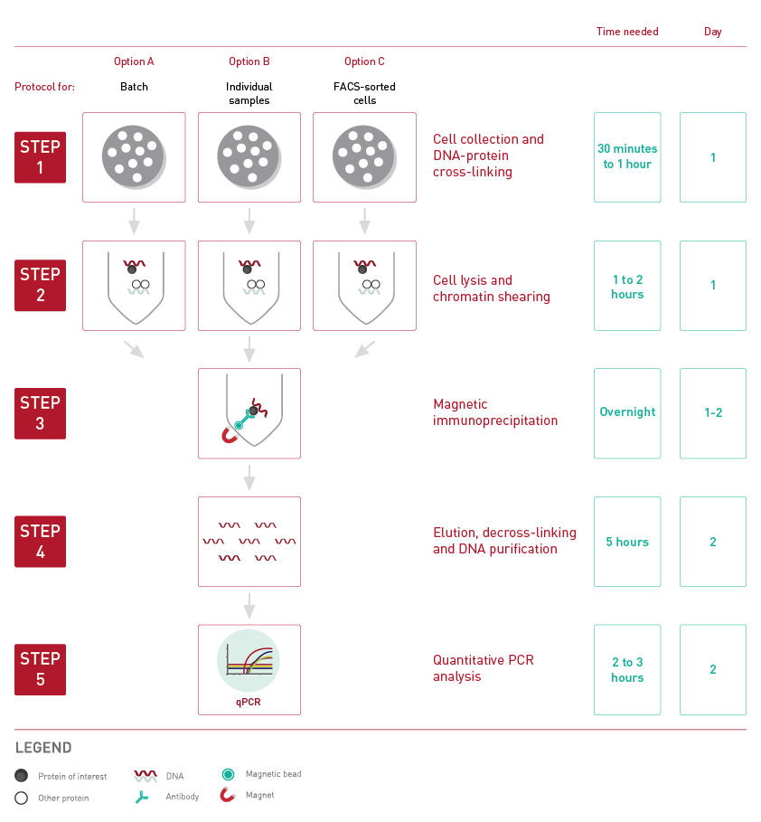
<p>The True MicroChIP-seq kit protocol has been optimized for the use of 10,000 - 100,000 cells per immunoprecipitation reaction. Regarding chromatin immunoprecipitation, three protocol variants have been optimized:<br />starting with a batch, starting with an individual sample and starting with the FACS-sorted cells.</p>
<div><button id="readmorebtn" style="background-color: #b02736; color: white; border-radius: 5px; border: none; padding: 5px;">Show Workflow</button></div>
<p><br /> <img src="https://www.diagenode.com/img/product/kits/workflow-microchip.png" id="workflowchip" class="hidden" width="600px" /></p>
<p>
<script type="text/javascript">// <![CDATA[
const bouton = document.querySelector('#readmorebtn');
const workflow = document.getElementById('workflowchip');
bouton.addEventListener('click', () => workflow.classList.toggle('hidden'))
// ]]></script>
</p>
<div class="extra-spaced" align="center"></div>
<div class="row">
<div class="carrousel" style="background-position: center;">
<div class="container">
<div class="row" style="background: rgba(255,255,255,0.1);">
<div class="large-12 columns truemicro-slider" id="truemicro-slider">
<div>
<h3>High efficiency ChIP on 10,000 cells</h3>
<div class="large-10 small-12 medium-10 large-centered medium-centered small-centered columns"><img src="https://www.diagenode.com/img/product/kits/true-micro-chip-histone-results.png" width="800px" /></div>
<div class="large-10 small-12 medium-10 large-centered medium-centered small-centered columns"><center>
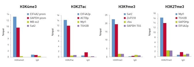
<p><small><strong>Figure 1. </strong>ChIP efficiency on 10,000 cells. ChIP was performed on human Hela cells using the Diagenode antibodies <a href="https://www.diagenode.com/en/p/h3k4me3-polyclonal-antibody-premium-50-ug-50-ul">H3K4me3</a> (Cat. No. C15410003), <a href="https://www.diagenode.com/en/p/h3k27ac-polyclonal-antibody-classic-50-mg-42-ml">H3K27ac</a> (C15410174), <a href="https://www.diagenode.com/en/p/h3k9me3-polyclonal-antibody-classic-50-ug">H3K9me3</a> (C15410056) and <a href="https://www.diagenode.com/en/p/h3k27me3-polyclonal-antibody-classic-50-mg-34-ml">H3K27me3</a> (C15410069). Sheared chromatin from 10,000 cells and 0.1 µg (H3K27ac), 0.25 µg (H3K4me3 and H3K27me3) or 0.5 µg (H3K9me3) of the antibody were used per IP. Corresponding amount of IgG was used as control. Quantitative PCR was performed with primers for corresponding positive and negative loci. Figure shows the recovery, expressed as a % of input (the relative amount of immunoprecipitated DNA compared to input DNA after qPCR analysis).</small></p>
</center></div>
</div>
<div>
<h3>True MicroChIP-seq protocol in a combination with MicroPlex library preparation kit results in reliable and accurate sequencing data</h3>
<div class="large-10 small-12 medium-10 large-centered medium-centered small-centered columns"><img src="https://www.diagenode.com/img/product/kits/fig2-truemicro.jpg" alt="True MicroChip results" width="800px" /></div>
<div class="large-10 small-12 medium-10 large-centered medium-centered small-centered columns"><center>
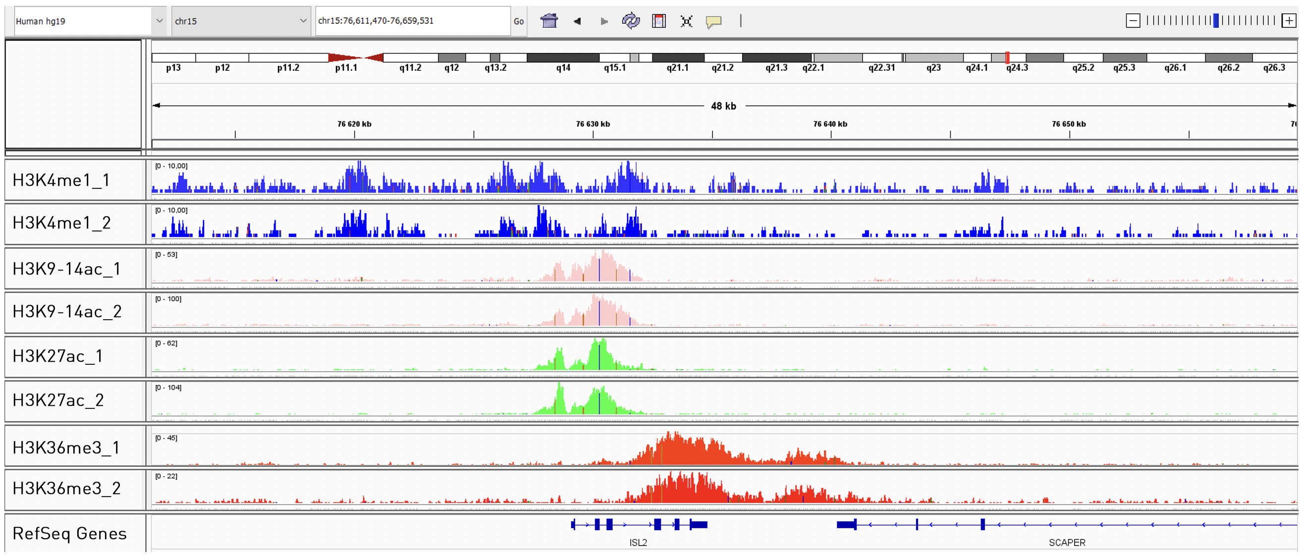
<p><small><strong>Figure 2.</strong> Integrative genomics viewer (IGV) visualization of ChIP-seq experiments using 50.000 of K562 cells. ChIP has been performed accordingly to True MicroChIP protocol followed by the library preparation using MicroPlex Library Preparation Kit (C05010001). The above figure shows the peaks from ChIP-seq experiments using the following antibodies: H3K4me1 (C15410194), H3K9/14ac (C15410200), H3K27ac (C15410196) and H3K36me3 (C15410192).</small></p>
</center></div>
</div>
<div>
<h3>Successful chromatin profiling from 10.000 of FACS-sorted cells</h3>
<div class="large-10 small-12 medium-10 large-centered medium-centered small-centered columns"><img src="https://www.diagenode.com/img/product/kits/fig3ab-truemicro.jpg" alt="small non coding RNA" width="800px" /></div>
<div class="large-10 small-12 medium-10 large-centered medium-centered small-centered columns"><center>
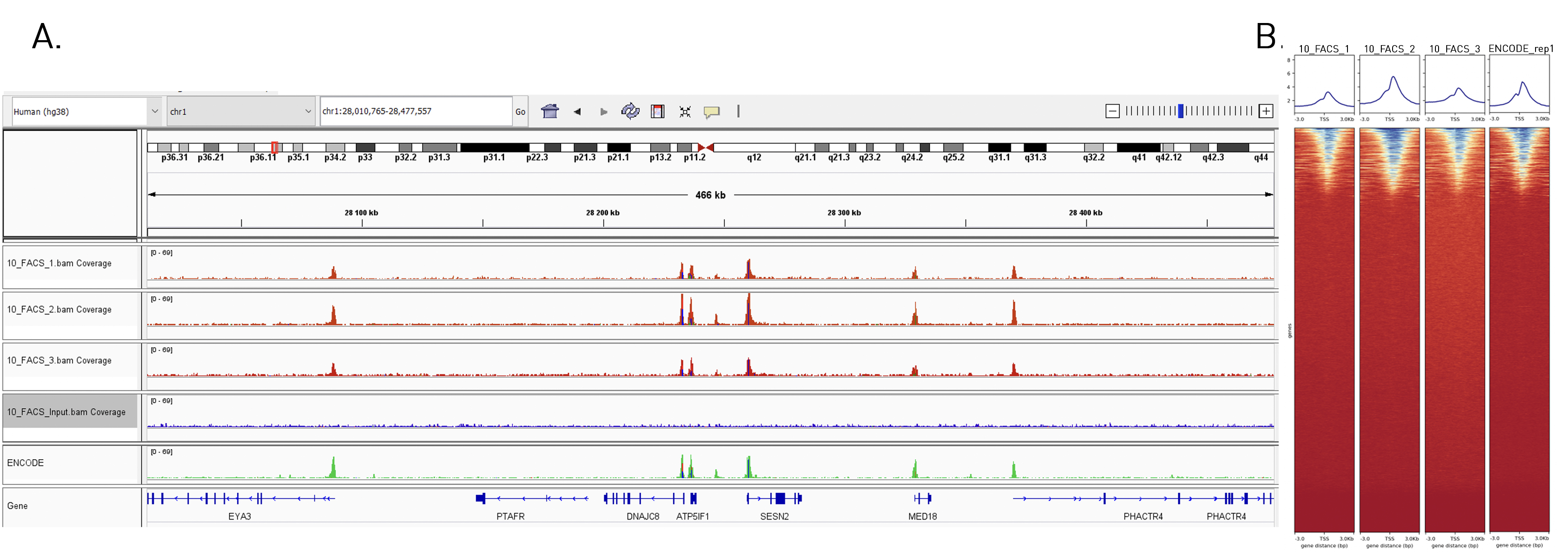
<p><small><strong>Figure 3.</strong> (A) Integrative genomics viewer (IGV) visualization of ChIP-seq experiments and heatmap 3kb upstream and downstream of the TSS (B) for H3K4me3. ChIP has been performed using 10.000 of FACS-sorted cells (K562) and H3K4me3 antibody (C15410003) accordingly to True MicroChIP protocol followed by the library preparation using MicroPlex Library Preparation Kit (C05010001). Data were compared to ENCODE standards.</small></p>
</center></div>
</div>
</div>
</div>
</div>
</div>
</div>
<p>
<script type="text/javascript">// <![CDATA[
$('.truemicro-slider').slick({
arrows: true,
dots: true,
autoplay:true,
autoplaySpeed: 3000
});
// ]]></script>
</p>',
'label2' => 'Additional solutions compatible with the True MicroChIP-seq Kit',
'info2' => '<p><span style="font-weight: 400;">The <a href="https://www.diagenode.com/en/p/chromatin-shearing-optimization-kit-high-sds-100-million-cells">Chromatin EasyShear Kit – High SDS</a></span><span style="font-weight: 400;"> Recommended for the optimizing chromatin shearing.</span></p>
<p><a href="https://www.diagenode.com/en/categories/chip-seq-grade-antibodies"><span style="font-weight: 400;">ChIP-seq grade antibodies</span></a><span style="font-weight: 400;"> for high yields, specificity, and sensitivity.</span></p>
<p><span style="font-weight: 400;">Check the list of available </span><a href="https://www.diagenode.com/en/categories/primer-pairs"><span style="font-weight: 400;">primer pairs</span></a><span style="font-weight: 400;"> designed for high specificity to specific genomic regions.</span></p>
<p><span style="font-weight: 400;">For library preparation of immunoprecipitated samples we recommend to use the </span><b> </b><a href="https://www.diagenode.com/en/categories/library-preparation-for-ChIP-seq"><span style="font-weight: 400;">MicroPlex Library Preparation Kit</span></a><span style="font-weight: 400;"> - validated for library preparation from picogram inputs.</span></p>
<p><span style="font-weight: 400;">For IP-Star Automation users, check out the </span><a href="https://www.diagenode.com/en/p/auto-true-microchip-kit-16-rxns"><span style="font-weight: 400;">automated version</span></a><span style="font-weight: 400;"> of this kit.</span></p>
<p><span style="font-weight: 400;">Application note: </span><a href="https://www.diagenode.com/files/application_notes/Diagenode_AATI_Joint.pdf"><span style="font-weight: 400;">Best Workflow Practices for ChIP-seq Analysis with Small Samples</span></a></p>
<p></p>',
'label3' => 'Species, cell lines, tissues tested',
'info3' => '<p>The True MicroChIP-seq kit is compatible with a broad variety of cell lines, tissues and species - some examples are shown below. Other species / cell lines / tissues can be used with this kit.</p>
<p><strong>Cell lines:</strong></p>
<p>Bovine: blastocysts,<br />Drosophila: embryos, salivary glands<br />Human: EndoC-ẞH1 cells, HeLa cells, PBMC, urothelial cells<br />Mouse: adipocytes, B cells, blastocysts, pre-B cells, BMDM cells, chondrocytes, embryonic stem cells, KH2 cells, LSK cells, macrophages, MEP cells, microglia, NK cells, oocytes, pancreatic cells, P19Cl6 cells, RPE cells,</p>
<p>Other cell lines / species: compatible, not tested</p>
<p><strong>Tissues:</strong></p>
<p>Horse: adipose tissue</p>
<p>Mice: intestine tissue</p>
<p>Other tissues: not tested</p>',
'format' => '20 rxns',
'catalog_number' => 'C01010132',
'old_catalog_number' => 'C01010130',
'sf_code' => 'C01010132-',
'type' => 'RFR',
'search_order' => '04-undefined',
'price_EUR' => '625',
'price_USD' => '680',
'price_GBP' => '575',
'price_JPY' => '102405',
'price_CNY' => '',
'price_AUD' => '1700',
'country' => 'ALL',
'except_countries' => 'None',
'quote' => false,
'in_stock' => false,
'featured' => true,
'no_promo' => false,
'online' => true,
'master' => true,
'last_datasheet_update' => '0000-00-00',
'slug' => 'true-microchip-kit-x16-16-rxns',
'meta_title' => 'True MicroChIP-seq Kit | Diagenode C01010132',
'meta_keywords' => '',
'meta_description' => 'True MicroChIP-seq Kit provides a robust ChIP protocol suitable for the investigation of histone modifications within chromatin from as few as 10 000 cells, including FACS sorted cells. Compatible with ChIP-qPCR as well as ChIP-seq.',
'modified' => '2023-04-20 16:06:10',
'created' => '2015-06-29 14:08:20',
'locale' => 'eng'
),
'Antibody' => array(
'host' => '*****',
'id' => null,
'name' => null,
'description' => null,
'clonality' => null,
'isotype' => null,
'lot' => null,
'concentration' => null,
'reactivity' => null,
'type' => null,
'purity' => null,
'classification' => null,
'application_table' => null,
'storage_conditions' => null,
'storage_buffer' => null,
'precautions' => null,
'uniprot_acc' => null,
'slug' => null,
'meta_keywords' => null,
'meta_description' => null,
'modified' => null,
'created' => null,
'select_label' => null
),
'Slave' => array(),
'Group' => array(),
'Related' => array(
(int) 0 => array(
[maximum depth reached]
),
(int) 1 => array(
[maximum depth reached]
),
(int) 2 => array(
[maximum depth reached]
)
),
'Application' => array(
(int) 0 => array(
[maximum depth reached]
),
(int) 1 => array(
[maximum depth reached]
)
),
'Category' => array(
(int) 0 => array(
[maximum depth reached]
)
),
'Document' => array(
(int) 0 => array(
[maximum depth reached]
),
(int) 1 => array(
[maximum depth reached]
),
(int) 2 => array(
[maximum depth reached]
),
(int) 3 => array(
[maximum depth reached]
),
(int) 4 => array(
[maximum depth reached]
),
(int) 5 => array(
[maximum depth reached]
)
),
'Feature' => array(),
'Image' => array(
(int) 0 => array(
[maximum depth reached]
)
),
'Promotion' => array(),
'Protocol' => array(),
'Publication' => array(
(int) 0 => array(
[maximum depth reached]
),
(int) 1 => array(
[maximum depth reached]
),
(int) 2 => array(
[maximum depth reached]
),
(int) 3 => array(
[maximum depth reached]
),
(int) 4 => array(
[maximum depth reached]
),
(int) 5 => array(
[maximum depth reached]
),
(int) 6 => array(
[maximum depth reached]
),
(int) 7 => array(
[maximum depth reached]
),
(int) 8 => array(
[maximum depth reached]
),
(int) 9 => array(
[maximum depth reached]
),
(int) 10 => array(
[maximum depth reached]
),
(int) 11 => array(
[maximum depth reached]
),
(int) 12 => array(
[maximum depth reached]
),
(int) 13 => array(
[maximum depth reached]
),
(int) 14 => array(
[maximum depth reached]
),
(int) 15 => array(
[maximum depth reached]
),
(int) 16 => array(
[maximum depth reached]
),
(int) 17 => array(
[maximum depth reached]
),
(int) 18 => array(
[maximum depth reached]
),
(int) 19 => array(
[maximum depth reached]
),
(int) 20 => array(
[maximum depth reached]
),
(int) 21 => array(
[maximum depth reached]
),
(int) 22 => array(
[maximum depth reached]
),
(int) 23 => array(
[maximum depth reached]
),
(int) 24 => array(
[maximum depth reached]
),
(int) 25 => array(
[maximum depth reached]
),
(int) 26 => array(
[maximum depth reached]
),
(int) 27 => array(
[maximum depth reached]
),
(int) 28 => array(
[maximum depth reached]
),
(int) 29 => array(
[maximum depth reached]
)
),
'Testimonial' => array(
(int) 0 => array(
[maximum depth reached]
)
),
'Area' => array(),
'SafetySheet' => array(
(int) 0 => array(
[maximum depth reached]
),
(int) 1 => array(
[maximum depth reached]
),
(int) 2 => array(
[maximum depth reached]
),
(int) 3 => array(
[maximum depth reached]
),
(int) 4 => array(
[maximum depth reached]
),
(int) 5 => array(
[maximum depth reached]
),
(int) 6 => array(
[maximum depth reached]
),
(int) 7 => array(
[maximum depth reached]
)
)
)
)
$language = 'en'
$meta_keywords = ''
$meta_description = 'True MicroChIP-seq Kit provides a robust ChIP protocol suitable for the investigation of histone modifications within chromatin from as few as 10 000 cells, including FACS sorted cells. Compatible with ChIP-qPCR as well as ChIP-seq.'
$meta_title = 'True MicroChIP-seq Kit | Diagenode C01010132'
$product = array(
'Product' => array(
'id' => '1856',
'antibody_id' => null,
'name' => 'True MicroChIP-seq Kit',
'description' => '<p><a href="https://www.diagenode.com/files/products/kits/truemicrochipseq-kit-manual.pdf"><img src="https://www.diagenode.com/img/buttons/bt-manual.png" /></a></p>
<p>The <b>True </b><b>MicroChIP-seq</b><b> kit </b>provides a robust ChIP protocol suitable for the investigation of histone modifications within chromatin from as few as <b>10 000 cells</b>, including <b>FACS sorted cells</b>. The kit can be used for chromatin preparation for downstream ChIP-qPCR or ChIP-seq analysis. The <b>complete kit</b> contains everything you need for start-to-finish ChIP including all validated buffers and reagents for chromatin shearing, immunoprecipitation and DNA purification for exceptional <strong>ChIP-qPCR</strong> or <strong>ChIP-seq</strong> results. In addition, positive control antibodies and negative control PCR primers are included for your convenience and assurance of result sensitivity and specificity.</p>
<p>The True MicroChIP-seq kit offers unique benefits:</p>
<ul>
<li>An <b>optimized chromatin preparation </b>protocol compatible with low number of cells (<b>10.000</b>) in combination with the Bioruptor™ shearing device</li>
<li>Most <b>complete kit </b>available (covers all steps and includes control antibodies and primers)</li>
<li><b>Magnetic beads </b>make ChIP easy, fast, and more reproducible</li>
<li>MicroChIP DiaPure columns (included in the kit) enable the <b>maximum recovery </b>of immunoprecipitation DNA suitable for any downstream application</li>
<li><b>Excellent </b><b>ChIP</b><b>-seq </b>result when combined with <a href="https://www.diagenode.com/en/categories/library-preparation-for-ChIP-seq">MicroPlex</a><a href="https://www.diagenode.com/en/categories/library-preparation-for-ChIP-seq"> Library Preparation kit </a>adapted for low input</li>
</ul>
<p>For fast ChIP-seq on low input – check out Diagenode’s <a href="https://www.diagenode.com/en/p/uchipmentation-for-histones-24-rxns">µ</a><a href="https://www.diagenode.com/en/p/uchipmentation-for-histones-24-rxns">ChIPmentation</a><a href="https://www.diagenode.com/en/p/uchipmentation-for-histones-24-rxns"> for histones</a>.</p>
<p><sub>The True MicroChIP-seq kit, Cat. No. C01010132 is an upgraded version of the kit True MicroChIP, Cat. No. C01010130, with the new validated protocols (e.g. FACS sorted cells) and MicroChIP DiaPure columns included in the kit.</sub></p>',
'label1' => 'Characteristics',
'info1' => '<ul>
<li><b>Revolutionary:</b> Only 10,000 cells needed for complete ChIP-seq procedure</li>
<li><b>Validated on</b> studies for histone marks</li>
<li><b>Automated protocol </b>for the IP-Star<sup>®</sup> Compact Automated Platform available</li>
</ul>
<p></p>
<p>The True MicroChIP-seq kit protocol has been optimized for the use of 10,000 - 100,000 cells per immunoprecipitation reaction. Regarding chromatin immunoprecipitation, three protocol variants have been optimized:<br />starting with a batch, starting with an individual sample and starting with the FACS-sorted cells.</p>
<div><button id="readmorebtn" style="background-color: #b02736; color: white; border-radius: 5px; border: none; padding: 5px;">Show Workflow</button></div>
<p><br /> <img src="https://www.diagenode.com/img/product/kits/workflow-microchip.png" id="workflowchip" class="hidden" width="600px" /></p>
<p>
<script type="text/javascript">// <![CDATA[
const bouton = document.querySelector('#readmorebtn');
const workflow = document.getElementById('workflowchip');
bouton.addEventListener('click', () => workflow.classList.toggle('hidden'))
// ]]></script>
</p>
<div class="extra-spaced" align="center"></div>
<div class="row">
<div class="carrousel" style="background-position: center;">
<div class="container">
<div class="row" style="background: rgba(255,255,255,0.1);">
<div class="large-12 columns truemicro-slider" id="truemicro-slider">
<div>
<h3>High efficiency ChIP on 10,000 cells</h3>
<div class="large-10 small-12 medium-10 large-centered medium-centered small-centered columns"><img src="https://www.diagenode.com/img/product/kits/true-micro-chip-histone-results.png" width="800px" /></div>
<div class="large-10 small-12 medium-10 large-centered medium-centered small-centered columns"><center>
<p><small><strong>Figure 1. </strong>ChIP efficiency on 10,000 cells. ChIP was performed on human Hela cells using the Diagenode antibodies <a href="https://www.diagenode.com/en/p/h3k4me3-polyclonal-antibody-premium-50-ug-50-ul">H3K4me3</a> (Cat. No. C15410003), <a href="https://www.diagenode.com/en/p/h3k27ac-polyclonal-antibody-classic-50-mg-42-ml">H3K27ac</a> (C15410174), <a href="https://www.diagenode.com/en/p/h3k9me3-polyclonal-antibody-classic-50-ug">H3K9me3</a> (C15410056) and <a href="https://www.diagenode.com/en/p/h3k27me3-polyclonal-antibody-classic-50-mg-34-ml">H3K27me3</a> (C15410069). Sheared chromatin from 10,000 cells and 0.1 µg (H3K27ac), 0.25 µg (H3K4me3 and H3K27me3) or 0.5 µg (H3K9me3) of the antibody were used per IP. Corresponding amount of IgG was used as control. Quantitative PCR was performed with primers for corresponding positive and negative loci. Figure shows the recovery, expressed as a % of input (the relative amount of immunoprecipitated DNA compared to input DNA after qPCR analysis).</small></p>
</center></div>
</div>
<div>
<h3>True MicroChIP-seq protocol in a combination with MicroPlex library preparation kit results in reliable and accurate sequencing data</h3>
<div class="large-10 small-12 medium-10 large-centered medium-centered small-centered columns"><img src="https://www.diagenode.com/img/product/kits/fig2-truemicro.jpg" alt="True MicroChip results" width="800px" /></div>
<div class="large-10 small-12 medium-10 large-centered medium-centered small-centered columns"><center>
<p><small><strong>Figure 2.</strong> Integrative genomics viewer (IGV) visualization of ChIP-seq experiments using 50.000 of K562 cells. ChIP has been performed accordingly to True MicroChIP protocol followed by the library preparation using MicroPlex Library Preparation Kit (C05010001). The above figure shows the peaks from ChIP-seq experiments using the following antibodies: H3K4me1 (C15410194), H3K9/14ac (C15410200), H3K27ac (C15410196) and H3K36me3 (C15410192).</small></p>
</center></div>
</div>
<div>
<h3>Successful chromatin profiling from 10.000 of FACS-sorted cells</h3>
<div class="large-10 small-12 medium-10 large-centered medium-centered small-centered columns"><img src="https://www.diagenode.com/img/product/kits/fig3ab-truemicro.jpg" alt="small non coding RNA" width="800px" /></div>
<div class="large-10 small-12 medium-10 large-centered medium-centered small-centered columns"><center>
<p><small><strong>Figure 3.</strong> (A) Integrative genomics viewer (IGV) visualization of ChIP-seq experiments and heatmap 3kb upstream and downstream of the TSS (B) for H3K4me3. ChIP has been performed using 10.000 of FACS-sorted cells (K562) and H3K4me3 antibody (C15410003) accordingly to True MicroChIP protocol followed by the library preparation using MicroPlex Library Preparation Kit (C05010001). Data were compared to ENCODE standards.</small></p>
</center></div>
</div>
</div>
</div>
</div>
</div>
</div>
<p>
<script type="text/javascript">// <![CDATA[
$('.truemicro-slider').slick({
arrows: true,
dots: true,
autoplay:true,
autoplaySpeed: 3000
});
// ]]></script>
</p>',
'label2' => 'Additional solutions compatible with the True MicroChIP-seq Kit',
'info2' => '<p><span style="font-weight: 400;">The <a href="https://www.diagenode.com/en/p/chromatin-shearing-optimization-kit-high-sds-100-million-cells">Chromatin EasyShear Kit – High SDS</a></span><span style="font-weight: 400;"> Recommended for the optimizing chromatin shearing.</span></p>
<p><a href="https://www.diagenode.com/en/categories/chip-seq-grade-antibodies"><span style="font-weight: 400;">ChIP-seq grade antibodies</span></a><span style="font-weight: 400;"> for high yields, specificity, and sensitivity.</span></p>
<p><span style="font-weight: 400;">Check the list of available </span><a href="https://www.diagenode.com/en/categories/primer-pairs"><span style="font-weight: 400;">primer pairs</span></a><span style="font-weight: 400;"> designed for high specificity to specific genomic regions.</span></p>
<p><span style="font-weight: 400;">For library preparation of immunoprecipitated samples we recommend to use the </span><b> </b><a href="https://www.diagenode.com/en/categories/library-preparation-for-ChIP-seq"><span style="font-weight: 400;">MicroPlex Library Preparation Kit</span></a><span style="font-weight: 400;"> - validated for library preparation from picogram inputs.</span></p>
<p><span style="font-weight: 400;">For IP-Star Automation users, check out the </span><a href="https://www.diagenode.com/en/p/auto-true-microchip-kit-16-rxns"><span style="font-weight: 400;">automated version</span></a><span style="font-weight: 400;"> of this kit.</span></p>
<p><span style="font-weight: 400;">Application note: </span><a href="https://www.diagenode.com/files/application_notes/Diagenode_AATI_Joint.pdf"><span style="font-weight: 400;">Best Workflow Practices for ChIP-seq Analysis with Small Samples</span></a></p>
<p></p>',
'label3' => 'Species, cell lines, tissues tested',
'info3' => '<p>The True MicroChIP-seq kit is compatible with a broad variety of cell lines, tissues and species - some examples are shown below. Other species / cell lines / tissues can be used with this kit.</p>
<p><strong>Cell lines:</strong></p>
<p>Bovine: blastocysts,<br />Drosophila: embryos, salivary glands<br />Human: EndoC-ẞH1 cells, HeLa cells, PBMC, urothelial cells<br />Mouse: adipocytes, B cells, blastocysts, pre-B cells, BMDM cells, chondrocytes, embryonic stem cells, KH2 cells, LSK cells, macrophages, MEP cells, microglia, NK cells, oocytes, pancreatic cells, P19Cl6 cells, RPE cells,</p>
<p>Other cell lines / species: compatible, not tested</p>
<p><strong>Tissues:</strong></p>
<p>Horse: adipose tissue</p>
<p>Mice: intestine tissue</p>
<p>Other tissues: not tested</p>',
'format' => '20 rxns',
'catalog_number' => 'C01010132',
'old_catalog_number' => 'C01010130',
'sf_code' => 'C01010132-',
'type' => 'RFR',
'search_order' => '04-undefined',
'price_EUR' => '625',
'price_USD' => '680',
'price_GBP' => '575',
'price_JPY' => '102405',
'price_CNY' => '',
'price_AUD' => '1700',
'country' => 'ALL',
'except_countries' => 'None',
'quote' => false,
'in_stock' => false,
'featured' => true,
'no_promo' => false,
'online' => true,
'master' => true,
'last_datasheet_update' => '0000-00-00',
'slug' => 'true-microchip-kit-x16-16-rxns',
'meta_title' => 'True MicroChIP-seq Kit | Diagenode C01010132',
'meta_keywords' => '',
'meta_description' => 'True MicroChIP-seq Kit provides a robust ChIP protocol suitable for the investigation of histone modifications within chromatin from as few as 10 000 cells, including FACS sorted cells. Compatible with ChIP-qPCR as well as ChIP-seq.',
'modified' => '2023-04-20 16:06:10',
'created' => '2015-06-29 14:08:20',
'locale' => 'eng'
),
'Antibody' => array(
'host' => '*****',
'id' => null,
'name' => null,
'description' => null,
'clonality' => null,
'isotype' => null,
'lot' => null,
'concentration' => null,
'reactivity' => null,
'type' => null,
'purity' => null,
'classification' => null,
'application_table' => null,
'storage_conditions' => null,
'storage_buffer' => null,
'precautions' => null,
'uniprot_acc' => null,
'slug' => null,
'meta_keywords' => null,
'meta_description' => null,
'modified' => null,
'created' => null,
'select_label' => null
),
'Slave' => array(),
'Group' => array(),
'Related' => array(
(int) 0 => array(
'id' => '1869',
'antibody_id' => null,
'name' => 'Chromatin EasyShear Kit - High SDS',
'description' => '<p><a href="https://www.diagenode.com/files/products/kits/Chromatin_Shearing_kit_HighSDS_manual.pdf"><img src="https://www.diagenode.com/img/buttons/bt-manual.png" /></a></p>
<p style="text-align: justify;">Previous name: Chromatin Shearing Optimization Kit - High SDS (True Micro ChIP kit)</p>
<p style="text-align: justify;">A high quality chromatin preparation is very complex and requires a lot of optimization. Chromatin EasyShear Kit – High SDS is an optimized solution for efficient chromatin preparation prior to ChIP. The protocol, buffers composition, SDS concentration (1%) is optimized for the preparation of chromatin prior to ChIP on low amount of starting material<b> </b>and it is compatible with Diagenode's <a href="https://www.diagenode.com/en/p/true-microchip-kit-x16-16-rxns">True MicroChIP-seq kit</a> and <a href="https://www.diagenode.com/en/p/uchipmentation-for-histones-24-rxns">µChIPmentation Kit for Histones</a>. The kit has been validated with the Bioruptor ultrasonicator for efficient chromatin shearing, leading to chromatin fragments<span> </span><strong>suitable for ChIP</strong><span> </span>with the preserved<span> </span><strong>epitopes</strong>.</p>
<p style="text-align: justify;">Check all <a href="https://www.diagenode.com/en/categories/chromatin-shearing">Chromatin EasyShear Kits</a>.</p>
<p style="text-align: justify;">Guide for the optimal chromatin preparation using Chromatin EasyShear Kits – <a href="https://www.diagenode.com/en/pages/chromatin-prep-easyshear-kit-guide">Read more</a></p>',
'label1' => '',
'info1' => '',
'label2' => '',
'info2' => '',
'label3' => '',
'info3' => '',
'format' => '100 million cells',
'catalog_number' => 'C01020012',
'old_catalog_number' => 'AA-003-0100',
'sf_code' => 'C01020012-',
'type' => 'RFR',
'search_order' => '04-undefined',
'price_EUR' => '310',
'price_USD' => '355',
'price_GBP' => '285',
'price_JPY' => '50795',
'price_CNY' => '',
'price_AUD' => '888',
'country' => 'ALL',
'except_countries' => 'None',
'quote' => false,
'in_stock' => false,
'featured' => false,
'no_promo' => false,
'online' => true,
'master' => true,
'last_datasheet_update' => '0000-00-00',
'slug' => 'chromatin-shearing-optimization-kit-high-sds-100-million-cells',
'meta_title' => 'Chromatin shearing optimization kit - High SDS',
'meta_keywords' => '',
'meta_description' => 'Chromatin shearing optimization kit - High SDS',
'modified' => '2023-04-20 16:33:08',
'created' => '2015-06-29 14:08:20',
'ProductsRelated' => array(
[maximum depth reached]
),
'Image' => array(
[maximum depth reached]
)
),
(int) 1 => array(
'id' => '1927',
'antibody_id' => null,
'name' => 'MicroPlex Library Preparation Kit v2 (12 indexes)',
'description' => '<p><a href="https://www.diagenode.com/files/products/kits/MicroPlex-Libary-Prep-Kit-v2-manual.pdf"><img src="https://www.diagenode.com/img/buttons/bt-manual.png" /></a></p>
<p><span><strong>Specifically optimized for ChIP-seq</strong></span><br /><br /><span>The MicroPlex Library Preparation™ kit is the only kit on the market which is validated for ChIP-seq and which allows the preparation of indexed libraries from just picogram inputs. In combination with the </span><a href="./true-microchip-kit-x16-16-rxns">True MicroChIP kit</a><span>, it allows for performing ChIP-seq on as few as 10,000 cells. Less input, fewer steps, fewer supplies, faster time to results! </span></p>
<p>The MicroPlex v2 kit (Cat. No. C05010012) contains all necessary reagents including single indexes for multiplexing up to 12 samples using single barcoding. For higher multiplexing (using dual indexes) check <a href="https://www.diagenode.com/en/p/microplex-lib-prep-kit-v3-48-rxns">MicroPlex Library Preparation Kits v3</a>.</p>',
'label1' => 'Characteristics',
'info1' => '<ul>
<li><strong>1 tube, 2 hours, 3 steps</strong> protocol</li>
<li><strong>Input: </strong>50 pg – 50 ng</li>
<li><strong>Reduce potential bias</strong> - few PCR amplification cycles needed</li>
<li><strong>High sensitivity ChIP-seq</strong> - low PCR duplication rate</li>
<li><strong>Great multiplexing flexibility</strong> with 12 barcodes (8 nt) included</li>
<li><strong>Validated with the <a href="https://www.diagenode.com/p/sx-8g-ip-star-compact-automated-system-1-unit" title="IP-Star Automated System">IP-Star<sup>®</sup> Automated Platform</a></strong></li>
</ul>
<h3>How it works</h3>
<center><img src="https://www.diagenode.com/img/product/kits/microplex-method-overview-v2.png" /></center>
<p style="margin-bottom: 0;"><small><strong>Microplex workflow - protocol with single indexes</strong><br />An input of 50 pg to 50 ng of fragmented dsDNA is converted into sequencing-ready libraries for Illumina® NGS platforms using a fast and simple 3-step protocol</small></p>
<ul class="accordion" data-accordion="" id="readmore" style="margin-left: 0;">
<li class="accordion-navigation"><a href="#first" style="background: #ffffff; padding: 0rem; margin: 0rem; color: #13b2a2;"><small>Read more about MicroPlex workflow</small></a>
<div id="first" class="content">
<p><small><strong>Step 1. Template Preparation</strong> provides efficient repair of the fragmented double-stranded DNA input.</small></p>
<p><small>In this step, the DNA is repaired and yields molecules with blunt ends.</small></p>
<p><small><strong>Step 2. Library Synthesis.</strong> enables ligation of MicroPlex patented stem- loop adapters.</small></p>
<p><small>In the next step, stem-loop adaptors with blocked 5’ ends are ligated with high efficiency to the 5’ end of the genomic DNA, leaving a nick at the 3’ end. The adaptors cannot ligate to each other and do not have single- strand tails, both of which contribute to non-specific background found with many other NGS preparations.</small></p>
<p><small><strong>Step 3. Library Amplification</strong> enables extension of the template, cleavage of the stem-loop adaptors, and amplification of the library. Illumina- compatible indexes are also introduced using a high-fidelity, highly- processive, low-bias DNA polymerase.</small></p>
<p><small>In the final step, the 3’ ends of the genomic DNA are extended to complete library synthesis and Illumina-compatible indexes are added through a high-fidelity amplification. Any remaining free adaptors are destroyed. Hands-on time and the risk of contamination are minimized by using a single tube and eliminating intermediate purifications.</small></p>
<p><small>Obtained libraries are purified, quantified and sized. The libraries pooling can be performed as well before sequencing.</small></p>
</div>
</li>
</ul>
<p></p>
<h3>Reliable detection of enrichments in ChIP-seq</h3>
<p><img src="https://www.diagenode.com/img/product/kits/microplex-library-prep-kit-figure-a.png" alt="Reliable detection of enrichments in ChIP-seq figure 1" style="display: block; margin-left: auto; margin-right: auto;" /></p>
<p><strong>Figure A.</strong> ChIP has been peformed with H3K4me3 antibody, amplification of 17 pg of DNA ChIP'd from 10.000 cells and amplification of 35 pg of DNA ChIP'd from 100.000 cells (control experiment). The IP'd DNA was amplified and transformed into a sequencing-ready preparation for the Illumina plateform with the MicroPlex Library Preparation kit. The library was then analysed on an Illumina<sup>®</sup> Genome Analyzer. Cluster generation and sequencing were performed according to the manufacturer's instructions.</p>
<p><img src="https://www.diagenode.com/img/product/kits/microplex-library-prep-kit-figure-b.png" alt="Reliable detection of enrichments in ChIP-seq figure 2" style="display: block; margin-left: auto; margin-right: auto;" /></p>
<p><strong>Figure B.</strong> We observed a perfect match between the top 40% of True MicroChIP peaks and the reference dataset. Based on the NIH Encode project criterion, ChIP-seq results are considered reproducible between an original and reproduced dataset if the top 40% of peaks have at least an 80% overlap ratio with the compared dataset.</p>',
'label2' => '',
'info2' => '',
'label3' => '',
'info3' => '',
'format' => '12 rxns',
'catalog_number' => 'C05010012',
'old_catalog_number' => 'C05010010',
'sf_code' => 'C05010012-',
'type' => 'FRE',
'search_order' => '04-undefined',
'price_EUR' => '955',
'price_USD' => '1250',
'price_GBP' => '855',
'price_JPY' => '156475',
'price_CNY' => '',
'price_AUD' => '3125',
'country' => 'ALL',
'except_countries' => 'None',
'quote' => false,
'in_stock' => false,
'featured' => false,
'no_promo' => false,
'online' => true,
'master' => true,
'last_datasheet_update' => '0000-00-00',
'slug' => 'microplex-library-preparation-kit-v2-x12-12-indices-12-rxns',
'meta_title' => 'MicroPlex Library Preparation Kit v2 x12 (12 indices)',
'meta_keywords' => '',
'meta_description' => 'MicroPlex Library Preparation Kit v2 x12 (12 indices)',
'modified' => '2023-04-20 15:01:16',
'created' => '2015-06-29 14:08:20',
'ProductsRelated' => array(
[maximum depth reached]
),
'Image' => array(
[maximum depth reached]
)
),
(int) 2 => array(
'id' => '1819',
'antibody_id' => null,
'name' => 'DiaMag 0.2ml - magnetic rack',
'description' => '<p>The DiaMag02 is a powerful magnet which has been designed for controlled and rapid isolation of your DNA bound to magnetic beads. It allows for processing 16 samples at a time.</p>',
'label1' => '',
'info1' => '',
'label2' => '',
'info2' => '',
'label3' => '',
'info3' => '',
'format' => '1 unit',
'catalog_number' => 'B04000001',
'old_catalog_number' => 'kch-816-001',
'sf_code' => 'B04000001-',
'type' => 'ACC',
'search_order' => '04-undefined',
'price_EUR' => '250',
'price_USD' => '240',
'price_GBP' => '220',
'price_JPY' => '40960',
'price_CNY' => '',
'price_AUD' => '600',
'country' => 'ALL',
'except_countries' => 'None',
'quote' => false,
'in_stock' => false,
'featured' => false,
'no_promo' => false,
'online' => true,
'master' => true,
'last_datasheet_update' => '0000-00-00',
'slug' => 'diamag02-magnetic-rack-1-unit',
'meta_title' => 'DiaMag02 - magnetic rack',
'meta_keywords' => '',
'meta_description' => 'DiaMag02 - magnetic rack',
'modified' => '2019-06-11 16:27:35',
'created' => '2015-06-29 14:08:20',
'ProductsRelated' => array(
[maximum depth reached]
),
'Image' => array(
[maximum depth reached]
)
)
),
'Application' => array(
(int) 0 => array(
'id' => '9',
'position' => '10',
'parent_id' => '2',
'name' => 'ChIP-seq',
'description' => '<div class="row">
<div class="large-12 columns">Chromatin Immunoprecipitation (ChIP) coupled with high-throughput massively parallel sequencing as a detection method (ChIP-seq) has become one of the primary methods for epigenomics researchers, namely to investigate protein-DNA interaction on a genome-wide scale. This technique is now used in a variety of life science disciplines including cellular differentiation, tumor suppressor gene silencing, and the effect of histone modifications on gene expression.</div>
<div class="large-12 columns"></div>
<h5 class="large-12 columns"><strong></strong></h5>
<h5 class="large-12 columns"><strong>The ChIP-seq workflow</strong></h5>
<div class="small-12 medium-12 large-12 columns text-center"><br /><img src="https://www.diagenode.com/img/chip-seq-diagram.png" /></div>
<div class="large-12 columns"><br />
<ol>
<li class="large-12 columns"><strong>Chromatin preparation: </strong>Crosslink chromatin-bound proteins (histones or transcription factors) to DNA followed by cell lysis.</li>
<li class="large-12 columns"><strong>Chromatin shearing:</strong> Fragment chromatin by sonication to desired fragment size (100-500 bp)</li>
<li class="large-12 columns"><strong>Chromatin IP</strong>: Capture protein-DNA complexes with <strong><a href="../categories/chip-seq-grade-antibodies">specific ChIP-seq grade antibodies</a></strong> against the histone or transcription factor of interest</li>
<li class="large-12 columns"><strong>DNA purification</strong>: Reverse cross-links, elute, and purify </li>
<li class="large-12 columns"><strong>NGS Library Preparation</strong>: Ligate adapters and amplify IP'd material</li>
<li class="large-12 columns"><strong>Bioinformatic analysis</strong>: Perform r<span style="font-weight: 400;">ead filtering and trimming</span>, r<span style="font-weight: 400;">ead specific alignment, enrichment specific peak calling, QC metrics, multi-sample cross-comparison etc. </span></li>
</ol>
</div>
</div>
<div class="row" style="margin-top: 32px;">
<div class="small-12 medium-10 large-9 small-centered columns">
<div class="radius panel" style="background-color: #fff;">
<h3 class="text-center" style="color: #b21329;">Need guidance?</h3>
<p class="text-justify">Choose our full ChIP kits or simply choose what you need from antibodies, buffers, beads, chromatin shearing and purification reagents. With the ChIP Kit Customizer, you have complete flexibility on which components you want from our validated ChIP kits.</p>
<div class="row">
<div class="small-6 medium-6 large-6 columns"><a href="../pages/which-kit-to-choose"><img alt="" src="https://www.diagenode.com/img/banners/banner-decide.png" /></a></div>
<div class="small-6 medium-6 large-6 columns"><a href="../pages/chip-kit-customizer-1"><img alt="" src="https://www.diagenode.com/img/banners/banner-customizer.png" /></a></div>
</div>
</div>
</div>
</div>',
'in_footer' => false,
'in_menu' => true,
'online' => true,
'tabular' => true,
'slug' => 'chromatin-immunoprecipitation-sequencing',
'meta_keywords' => 'Chromatin Immunoprecipitation Sequencing,ChIP-Seq,ChIP-seq grade antibodies,DNA purification,qPCR,Shearing of chromatin',
'meta_description' => 'Diagenode offers wide range of kits and antibodies for Chromatin Immunoprecipitation Sequencing (ChIP-Seq) and also provides Bioruptor for chromatin shearing',
'meta_title' => 'Chromatin Immunoprecipitation - ChIP-seq Kits - Dna methylation | Diagenode',
'modified' => '2017-11-14 09:57:16',
'created' => '2015-04-12 18:08:46',
'ProductsApplication' => array(
[maximum depth reached]
)
),
(int) 1 => array(
'id' => '10',
'position' => '10',
'parent_id' => '2',
'name' => 'ChIP-qPCR',
'description' => '<div class="row">
<div class="small-12 medium-12 large-12 columns text-justify">
<p class="text-justify">Chromatin Immunoprecipitation (ChIP) coupled with quantitative PCR can be used to investigate protein-DNA interaction at known genomic binding sites. if sites are not known, qPCR primers can also be designed against potential regulatory regions such as promoters. ChIP-qPCR is advantageous in studies that focus on specific genes and potential regulatory regions across differing experimental conditions as the cost of performing real-time PCR is minimal. This technique is now used in a variety of life science disciplines including cellular differentiation, tumor suppressor gene silencing, and the effect of histone modifications on gene expression.</p>
<p class="text-justify"><strong>The ChIP-qPCR workflow</strong></p>
</div>
<div class="small-12 medium-12 large-12 columns text-center"><br /> <img src="https://www.diagenode.com/img/chip-qpcr-diagram.png" /></div>
<div class="small-12 medium-12 large-12 columns"><br />
<ol>
<li class="large-12 columns"><strong>Chromatin preparation: </strong>cell fixation (cross-linking) of chromatin-bound proteins such as histones or transcription factors to DNA followed by cell lysis.</li>
<li class="large-12 columns"><strong>Chromatin shearing: </strong>fragmentation of chromatin<strong> </strong>by sonication down to desired fragment size (100-500 bp)</li>
<li class="large-12 columns"><strong>Chromatin IP</strong>: protein-DNA complexe capture using<strong> <a href="https://www.diagenode.com/en/categories/chip-grade-antibodies">specific ChIP-grade antibodies</a></strong> against the histone or transcription factor of interest</li>
<li class="large-12 columns"><strong>DNA purification</strong>: chromatin reverse cross-linking and elution followed by purification<strong> </strong></li>
<li class="large-12 columns"><strong>qPCR and analysis</strong>: using previously designed primers to amplify IP'd material at specific loci</li>
</ol>
</div>
</div>
<div class="row" style="margin-top: 32px;">
<div class="small-12 medium-10 large-9 small-centered columns">
<div class="radius panel" style="background-color: #fff;">
<h3 class="text-center" style="color: #b21329;">Need guidance?</h3>
<p class="text-justify">Choose our full ChIP kits or simply choose what you need from antibodies, buffers, beads, chromatin shearing and purification reagents. With the ChIP Kit Customizer, you have complete flexibility on which components you want from our validated ChIP kits.</p>
<div class="row">
<div class="small-6 medium-6 large-6 columns"><a href="https://www.diagenode.com/pages/which-kit-to-choose"><img src="https://www.diagenode.com/img/banners/banner-decide.png" alt="" /></a></div>
<div class="small-6 medium-6 large-6 columns"><a href="https://www.diagenode.com/pages/chip-kit-customizer-1"><img src="https://www.diagenode.com/img/banners/banner-customizer.png" alt="" /></a></div>
</div>
</div>
</div>
</div>',
'in_footer' => false,
'in_menu' => true,
'online' => true,
'tabular' => true,
'slug' => 'chip-qpcr',
'meta_keywords' => 'Chromatin immunoprecipitation,ChIP Quantitative PCR,polymerase chain reaction (PCR)',
'meta_description' => 'Diagenode's ChIP qPCR kits can be used to quantify enriched DNA after chromatin immunoprecipitation. ChIP-qPCR is advantageous in studies that focus on specific genes and potential regulatory regions across differing experimental conditions as the cost of',
'meta_title' => 'ChIP Quantitative PCR (ChIP-qPCR) | Diagenode',
'modified' => '2018-01-09 16:46:56',
'created' => '2014-12-11 00:22:08',
'ProductsApplication' => array(
[maximum depth reached]
)
)
),
'Category' => array(
(int) 0 => array(
'id' => '120',
'position' => '2',
'parent_id' => '59',
'name' => 'Low-input ChIP kits',
'description' => '',
'no_promo' => false,
'in_menu' => false,
'online' => true,
'tabular' => true,
'hide' => false,
'all_format' => false,
'is_antibody' => false,
'slug' => 'chromatin-ip-low-input-chip-kits',
'cookies_tag_id' => null,
'meta_keywords' => '',
'meta_description' => '',
'meta_title' => '',
'modified' => '2017-06-16 12:03:38',
'created' => '2016-07-19 17:02:02',
'ProductsCategory' => array(
[maximum depth reached]
),
'CookiesTag' => array([maximum depth reached])
)
),
'Document' => array(
(int) 0 => array(
'id' => '37',
'name' => 'Chromatin Brochure',
'description' => '<p>Whether you are experienced or new to the field of chromatin immunoprecipitation, Diagenode has everything you need to make ChIP easy and convenient while ensuring consistent data between samples and experiments. As an expert in the field of epigenetics, Diagenode is committed to providing complete solutions from chromatin shearing reagents, shearing instruments such as the Bioruptor® (the gold standard for chromatin shearing), ChIP kits, the largest number of validated and trusted antibodies on the market, and the SX-8G IP-Star® Compact Automated System to achieve unparalleled productivity and reproducibility.</p>',
'image_id' => null,
'type' => 'Brochure',
'url' => 'files/brochures/Chromatin_Immunoprecipitation_Brochure.pdf',
'slug' => 'chromatin-immunoprecipitation-brochure',
'meta_keywords' => '',
'meta_description' => '',
'modified' => '2022-03-24 12:34:11',
'created' => '2015-07-03 16:05:27',
'ProductsDocument' => array(
[maximum depth reached]
)
),
(int) 1 => array(
'id' => '19',
'name' => 'True MicroChIP and MicroPlex kits',
'description' => '<p><span>From minuscule amounts to magnificent results: reliable ChIP-seq data from 10,000 cells with the True MicroChIP</span>™ <span>and the MicroPlex Library Preparation</span>™ <span>kits. </span></p>',
'image_id' => null,
'type' => 'Application note',
'url' => 'files/application_notes/True_MicroChIP_and_MicroPlex_kits_Application_Note.pdf',
'slug' => 'true-microchip-and-microplex-kits-application-note',
'meta_keywords' => '',
'meta_description' => '',
'modified' => '2016-05-18 11:36:11',
'created' => '2015-07-03 16:05:20',
'ProductsDocument' => array(
[maximum depth reached]
)
),
(int) 2 => array(
'id' => '16',
'name' => 'ChIP kit results with True MicroChIP kit',
'description' => '<p style="text-align: justify;"><span>Chromatin immunoprecipitation coupled with high-throughput sequencing (ChIP-seq) has become the gold standard for whole-genome mapping of protein-DNA interactions. However, conventional ChIP protocols require abundant amounts of starting material (at least hundreds of thousands of cells per immunoprecipitation) limiting the application for the ChIP technology to few cell samples. </span></p>',
'image_id' => null,
'type' => 'Poster',
'url' => 'files/posters/ChIP_kit_results_with_True_MicroChIP_kit_Poster.pdf',
'slug' => 'chip-kit-results-with-true-microchip-kit-poster',
'meta_keywords' => '',
'meta_description' => '',
'modified' => '2015-10-01 20:09:25',
'created' => '2015-07-03 16:05:15',
'ProductsDocument' => array(
[maximum depth reached]
)
),
(int) 3 => array(
'id' => '945',
'name' => 'Application note: Best Workflow Practices for ChIP-seq Analysis with Small Samples',
'description' => '<p><span>Combined chromatin immunoprecipitation and next-generation sequencing (ChIP-seq) has become the gold standard to investigate genome-wide epigenetic profiles. However, ChIP from a limited amount of cells has been a challenge. Diagenode and Advanced Analytical Technologies, Inc. (AATI) provide the first complete and robust workflow solution for ChIP-seq from small numbers of cells using critical quality checkpoints to assure success. Download the application note.</span></p>',
'image_id' => null,
'type' => 'Application note',
'url' => 'files/application_notes/Diagenode_AATI_Joint.pdf',
'slug' => 'chip-seq-application-note',
'meta_keywords' => '',
'meta_description' => '',
'modified' => '2017-04-10 10:15:48',
'created' => '2017-04-07 15:01:20',
'ProductsDocument' => array(
[maximum depth reached]
)
),
(int) 4 => array(
'id' => '959',
'name' => 'Chromatin shearing with the Diagenode One',
'description' => '<p> The Diagenode One is a new affordable solution for Chromatin shearing in ChIP analysis for small samples. Designed to fit any bench, it is the smallest and lightest Diagenode shearing device. The fully integrated cooling system and the all-new 20 and 50 μl microfluidic chips have been enhanced to deliver the highest performance.</p>',
'image_id' => '247',
'type' => 'Application Note',
'url' => '../files/protocols/chromatin_shearing_diagenode_one.pdf',
'slug' => 'chromatin-shearing-with-the-diagenode-one',
'meta_keywords' => '',
'meta_description' => '',
'modified' => '2017-06-02 10:49:39',
'created' => '2017-05-22 09:40:15',
'ProductsDocument' => array(
[maximum depth reached]
)
),
(int) 5 => array(
'id' => '1137',
'name' => 'True MicroChIP-seq Kit',
'description' => '<p>The <b>True </b><b>MicroChIP-seq</b><b> kit </b>provides a robust ChIP protocol suitable for the investigation of histone modifications within chromatin from as few as <b>10 000 cells</b>, including <b>FACS sorted cells</b>.</p>',
'image_id' => null,
'type' => 'Manual',
'url' => 'files/products/kits/truemicrochipseq-kit-manual.pdf',
'slug' => 'truemicrochipseq-kit-manual.pdf',
'meta_keywords' => '',
'meta_description' => '',
'modified' => '2021-06-14 11:19:44',
'created' => '2021-06-10 11:21:37',
'ProductsDocument' => array(
[maximum depth reached]
)
)
),
'Feature' => array(),
'Image' => array(
(int) 0 => array(
'id' => '1775',
'name' => 'product/kits/chip-kit-icon.png',
'alt' => 'ChIP kit icon',
'modified' => '2018-04-17 11:52:29',
'created' => '2018-03-15 15:50:34',
'ProductsImage' => array(
[maximum depth reached]
)
)
),
'Promotion' => array(),
'Protocol' => array(),
'Publication' => array(
(int) 0 => array(
'id' => '5011',
'name' => 'PDGFRA is a conserved HAND2 effector during early cardiac development',
'authors' => 'Yanli Xu et al.',
'description' => '<p><span>The basic helix–loop–helix transcription factor HAND2 has multiple roles during vertebrate organogenesis, including cardiogenesis. However, much remains to be uncovered about its mechanism of action. Here, we show the generation of several </span><i>hand2</i><span><span> </span>mutant alleles in zebrafish and demonstrate that dimerization-deficient mutants display the null phenotype but DNA-binding-deficient mutants do not. Rescue experiments with Hand2 variants using a newly identified<span> </span></span><i>hand2</i><span><span> </span>enhancer confirmed these observations. To identify Hand2 effectors critical for cardiogenesis, we analyzed the transcriptomes of<span> </span></span><i>hand2</i><span><span> </span>loss- and gain-of-function embryonic cardiomyocytes and tested the function of eight candidate genes in vivo;<span> </span></span><i>pdgfra</i><span><span> </span>was most effective in rescuing myocardial migration in<span> </span></span><i>hand2</i><span><span> </span>mutants. Accordingly, we identified a putative Hand2-binding region in the zebrafish<span> </span></span><i>pdgfra</i><span><span> </span>locus that is important for its expression. In addition,<span> </span></span><i>Hand2</i><span><span> </span>loss- and gain-of-function experiments in mouse embryonic stem cell-derived cardiac cells decreased and increased<span> </span></span><i>Pdgfra</i><span><span> </span>expression, respectively. Altogether, these results further our mechanistic understanding of HAND2 function during early cardiogenesis.</span></p>',
'date' => '2024-12-10',
'pmid' => 'https://www.nature.com/articles/s44161-024-00574-1',
'doi' => 'https://doi.org/10.1038/s44161-024-00574-1',
'modified' => '2024-12-12 15:02:37',
'created' => '2024-12-12 15:02:37',
'ProductsPublication' => array(
[maximum depth reached]
)
),
(int) 1 => array(
'id' => '4977',
'name' => 'Single-cell multi-omics, spatial transcriptomics and systematic perturbation decode circuitry of neural crest fate decisions',
'authors' => 'Hu Z. et al.',
'description' => '<p><span>Cranial neural crest (NC) cells, which can migrate, adopt multiple fates, and form most of the craniofacial skeleton, are an excellent model for studying cell fate decisions. Using time-resolved single-cell multi-omics, spatial transcriptomics, and systematic Perturb-seq, we fully deciphered zebrafish cranial NC programs, including 23 cell states and three spatial trajectories, reconstructed and tested the complete gene regulatory network (GRN). Our GRN model, combined with a novel velocity-embedded simulation method, accurately predicted functions of all major regulons, with over a 3-fold increase in correlation between in vivo and in silico perturbations. Using our new approach based on regulatory synchronization, we discovered a post-epithelial-mesenchymal-transition endothelial-like program crucial for migration, identified motif coordinators for dual-fate priming, and quantified lineage-specific cooperative transcription factor functions. This study provides a comprehensive and validated NC regulatory landscape with unprecedented resolution, offering general regulatory models for cell fate decisions in vertebrates.</span></p>',
'date' => '2024-09-17',
'pmid' => 'https://www.biorxiv.org/content/10.1101/2024.09.17.613303v1',
'doi' => ' https://doi.org/10.1101/2024.09.17.613303',
'modified' => '2024-09-24 12:23:31',
'created' => '2024-09-24 12:23:31',
'ProductsPublication' => array(
[maximum depth reached]
)
),
(int) 2 => array(
'id' => '4578',
'name' => 'The aryl hydrocarbon receptor cell intrinsically promotes resident memoryCD8 T cell differentiation and function.',
'authors' => 'Dean J. W. et al.',
'description' => '<p>The Aryl hydrocarbon receptor (Ahr) regulates the differentiation and function of CD4 T cells; however, its cell-intrinsic role in CD8 T cells remains elusive. Herein we show that Ahr acts as a promoter of resident memory CD8 T cell (T) differentiation and function. Genetic ablation of Ahr in mouse CD8 T cells leads to increased CD127KLRG1 short-lived effector cells and CD44CD62L T central memory cells but reduced granzyme-B-producing CD69CD103 T cells. Genome-wide analyses reveal that Ahr suppresses the circulating while promoting the resident memory core gene program. A tumor resident polyfunctional CD8 T cell population, revealed by single-cell RNA-seq, is diminished upon Ahr deletion, compromising anti-tumor immunity. Human intestinal intraepithelial CD8 T cells also highly express AHR that regulates in vitro T differentiation and granzyme B production. Collectively, these data suggest that Ahr is an important cell-intrinsic factor for CD8 T cell immunity.</p>',
'date' => '2023-01-01',
'pmid' => 'https://www.ncbi.nlm.nih.gov/pubmed/36640340',
'doi' => '10.1016/j.celrep.2022.111963',
'modified' => '2023-04-11 10:14:26',
'created' => '2023-02-21 09:59:46',
'ProductsPublication' => array(
[maximum depth reached]
)
),
(int) 3 => array(
'id' => '4733',
'name' => 'FXR inhibition may protect from SARS-CoV-2 infection by reducingACE2.',
'authors' => 'Brevini Teresa et al.',
'description' => '<p>Prevention of SARS-CoV-2 infection through the modulation of viral host receptors, such as ACE2, could represent a new chemoprophylactic approach for COVID-19 complementing vaccination. However, the mechanisms controlling ACE2 expression remain elusive. Here, we identify the farnesoid X receptor (FXR) as a direct regulator of ACE2 transcription in multiple COVID19-affected tissues, including the gastrointestinal and respiratory systems. We then use the over-the-counter compound z-guggulsterone (ZGG) and the off-patent drug ursodeoxycholic acid (UDCA) to reduce FXR signalling and downregulate ACE2 in human lung, cholangiocyte and intestinal organoids and in the corresponding tissues in mice and hamsters. We demonstrate that UDCA-mediated ACE2 downregulation reduces susceptibility to SARS-CoV-2 infection in vitro, in vivo and in human lungs and livers perfused ex situ. Furthermore, we illustrate that UDCA reduces ACE2 expression in the nasal epithelium in humans. Finally, we identify a correlation between UDCA treatment and positive clinical outcomes following SARS-CoV-2 infection using retrospective registry data, and confirm these findings in an independent validation cohort of liver transplant recipients. In conclusion, we identify a novel function of FXR in controlling ACE2 expression and provide evidence that modulation of this pathway could be beneficial for reducing SARS-CoV-2 infection, paving the road for future clinical trials.</p>',
'date' => '2022-12-01',
'pmid' => 'https://www.ncbi.nlm.nih.gov/pubmed/36470304',
'doi' => '10.1038/s41586-022-05594-0',
'modified' => '2023-03-13 08:52:11',
'created' => '2023-02-28 12:19:11',
'ProductsPublication' => array(
[maximum depth reached]
)
),
(int) 4 => array(
'id' => '4651',
'name' => 'TCDD induces multigenerational alterations in the expression ofmicroRNA in the thymus through epigenetic modifications',
'authors' => 'Singh Narendra P et al.',
'description' => '<p>2,3,7,8-Tetrachlorodibenzo-p-dioxin (TCDD), a potent AhR ligand, is an environmental contaminant that is known for mediating toxicity across generations. However, whether TCDD can induce multigenerational changes in the expression of miRNAs (miRs) has not been previously studied. In the current study, we investigated the effect of administration of TCDD in pregnant mice (F0) on gestational day 14, on the expression of miRs in the thymus of F0 and subsequent generations (F1 and F2). Of the 3200 miRs screened, 160 miRs were dysregulated similarly in F0, F1, and F2 generations while 46 miRs were differentially altered in F0-F2 generations. Pathway analysis revealed that the changes in miR signature profile mediated by TCDD affected the genes that regulate cell signaling, apoptosis, thymic atrophy, cancer, immunosuppression, and other physiological pathways. A significant number of miRs that showed altered expression exhibited dioxin response elements (DRE) on their promoters. Focusing on one such miR, namely miR-203 that expressed DREs and was induced across F0-F2 by TCDD, promoter analysis showed that one of the DREs expressed by miR-203 was functional to TCDD-mediated upregulation. Also, the histone methylation status of H3K4me3 in the miR-203 promoter was significantly increased near the transcriptional start site (TSS) in TCDD-treated thymocytes across F0-F2 generations. Genome-wide ChIP-seq study suggested that TCDD may cause alterations in histone methylation in certain genes across the three generations. Together, the current study demonstrates that gestational exposure to TCDD can alter the expression of miRs in F0 through direct activation of DREs as well as across F0, F1, and F2 generations through epigenetic pathways.</p>',
'date' => '2022-12-01',
'pmid' => 'https://academic.oup.com/pnasnexus/advance-article/doi/10.1093/pnasnexus/pgac290/6886578',
'doi' => 'https://doi.org/10.1093/pnasnexus/pgac290',
'modified' => '2023-03-13 10:55:36',
'created' => '2023-02-21 09:59:46',
'ProductsPublication' => array(
[maximum depth reached]
)
),
(int) 5 => array(
'id' => '4575',
'name' => 'Intranasal administration of Acinetobacter lwoffii in a murine model ofasthma induces IL-6-mediated protection associated with cecal microbiotachanges.',
'authors' => 'Alashkar A. B. et al.',
'description' => '<p>BACKGROUND: Early-life exposure to certain environmental bacteria including Acinetobacter lwoffii (AL) has been implicated in protection from chronic inflammatory diseases including asthma later in life. However, the underlying mechanisms at the immune-microbe interface remain largely unknown. METHODS: The effects of repeated intranasal AL exposure on local and systemic innate immune responses were investigated in wild-type and Il6 , Il10 , and Il17 mice exposed to ovalbumin-induced allergic airway inflammation. Those investigations were expanded by microbiome analyses. To assess for AL-associated changes in gene expression, the picture arising from animal data was supplemented by in vitro experiments of macrophage and T-cell responses, yielding expression and epigenetic data. RESULTS: The asthma preventive effect of AL was confirmed in the lung. Repeated intranasal AL administration triggered a proinflammatory immune response particularly characterized by elevated levels of IL-6, and consequently, IL-6 induced IL-10 production in CD4 T-cells. Both IL-6 and IL-10, but not IL-17, were required for asthma protection. AL had a profound impact on the gene regulatory landscape of CD4 T-cells which could be largely recapitulated by recombinant IL-6. AL administration also induced marked changes in the gastrointestinal microbiome but not in the lung microbiome. By comparing the effects on the microbiota according to mouse genotype and AL-treatment status, we have identified microbial taxa that were associated with either disease protection or activity. CONCLUSION: These experiments provide a novel mechanism of Acinetobacter lwoffii-induced asthma protection operating through IL-6-mediated epigenetic activation of IL-10 production and with associated effects on the intestinal microbiome.</p>',
'date' => '2022-12-01',
'pmid' => 'https://www.ncbi.nlm.nih.gov/pubmed/36458896',
'doi' => '10.1111/all.15606',
'modified' => '2023-04-11 10:23:07',
'created' => '2023-02-21 09:59:46',
'ProductsPublication' => array(
[maximum depth reached]
)
),
(int) 6 => array(
'id' => '4221',
'name' => 'Epigenetic Mechanisms Mediating Cell State Transitions in Chondrocytes',
'authors' => 'Wuelling M. et al.',
'description' => '<p><span>Epigenetic modifications play critical roles in regulating cell lineage differentiation, but the epigenetic mechanisms guiding specific differentiation steps within a cell lineage have rarely been investigated. To decipher such mechanisms, we used the defined transition from proliferating (PC) into hypertrophic chondrocytes (HC) during endochondral ossification as a model. We established a map of activating and repressive histone modifications for each cell type. ChromHMM state transition analysis and Pareto-based integration of differential levels of mRNA and epigenetic marks revealed that differentiation-associated gene repression is initiated by the addition of H3K27me3 to promoters still carrying substantial levels of activating marks. Moreover, the integrative analysis identified genes specifically expressed in cells undergoing the transition into hypertrophy. Investigation of enhancer profiles detected surprising differences in enhancer number, location, and transcription factor binding sites between the two closely related cell types. Furthermore, cell type-specific upregulation of gene expression was associated with increased numbers of H3K27ac peaks. Pathway analysis identified PC-specific enhancers associated with chondrogenic genes, whereas HC-specific enhancers mainly control metabolic pathways linking epigenetic signature to biological functions. Since HC-specific enhancers show a higher conservation in postnatal tissues, the switch to metabolic pathways seems to be a hallmark of differentiated tissues. Surprisingly, the analysis of H3K27ac levels at super-enhancers revealed a rapid adaption of H3K27ac occupancy to changes in gene expression, supporting the importance of enhancer modulation for acute alterations in gene expression. © 2021 The Authors. Journal of Bone and Mineral Research published by Wiley Periodicals LLC on behalf of American Society for Bone and Mineral Research (ASBMR).</span></p>',
'date' => '2022-05-01',
'pmid' => 'https://pubmed.ncbi.nlm.nih.gov/33534175/',
'doi' => '10.1002/jbmr.4263',
'modified' => '2022-04-25 11:46:32',
'created' => '2022-04-21 12:00:53',
'ProductsPublication' => array(
[maximum depth reached]
)
),
(int) 7 => array(
'id' => '4226',
'name' => 'Single-cell-resolved dynamics of chromatin architecture delineate cell
and regulatory states in zebrafish embryos',
'authors' => 'McGarvey, Alison C. and Kopp, Wolfgang and Vučićević,
Dubravka and Mattonet, Kenny and Kempfer, Rieke and Hirsekorn,
Antje and Bilić, Ilija and Gil, Marine and Trinks, Alexandra
and Merks, Anne Margarete and Panáková, Daniela and Pombo,
Ana and Akalin, Al',
'description' => 'DNA accessibility of cis-regulatory elements (CREs) dictates
transcriptional activity and drives cell differentiation during
development. While many genes regulating embryonic development have been
identified, the underlying CRE dynamics controlling their expression
remain largely uncharacterized. To address this, we produced a multimodal
resource and genomic regulatory map for the zebrafish community, which
integrates single-cell combinatorial indexing assay for
transposase-accessible chromatin with high-throughput sequencing
(sci-ATAC-seq) with bulk histone PTMs and Hi-C data to achieve a
genome-wide classification of the regulatory architecture determining
transcriptional activity in the 24-h post-fertilization (hpf) embryo. We
characterized the genome-wide chromatin architecture at bulk and
single-cell resolution, applying sci-ATAC-seq on whole 24-hpf stage
zebrafish embryos, generating accessibility profiles for ∼23,000 single
nuclei. We developed a genome segmentation method, ScregSeg
(single-cell regulatory landscape segmentation), for defining regulatory
programs, and candidate CREs, specific to one or more cell types. We
integrated the ScregSeg output with bulk measurements for histone
post-translational modifications and 3D genome organization and
identified new regulatory principles between chromatin modalities prevalent
during zebrafish development. Sci-ATAC-seq profiling of npas4l/cloche
mutant embryos identified novel cellular roles for this hematovascular
transcriptional master regulator and suggests an intricate mechanism
regulating its expression. Our work defines regulatory architecture and
principles in the zebrafish embryo and establishes a resource of
cell-type-specific genome-wide regulatory annotations and candidate CREs,
providing a valuable open resource for genomics, developmental, molecular,
and computational biology.',
'date' => '2022-01-01',
'pmid' => 'https://doi.org/10.1016%2Fj.xgen.2021.100083',
'doi' => '10.1016/j.xgen.2021.100083',
'modified' => '2022-05-19 10:41:50',
'created' => '2022-05-19 10:41:50',
'ProductsPublication' => array(
[maximum depth reached]
)
),
(int) 8 => array(
'id' => '4233',
'name' => 'Autocrine Vitamin D-signaling switches off pro-inflammatory programsof Th1 cells',
'authors' => 'Chauss D.et al.',
'description' => '<p>The molecular mechanisms governing orderly shutdown and retraction of CD4+ T helper (Th)1 responses remain poorly understood. Here, we show that complement triggers contraction of Th1 responses by inducing intrinsic expression of the vitamin D (VitD) receptor (VDR) and the VitD-activating enzyme CYP27B1, permitting T cells to both activate and respond to VitD. VitD then initiated transition from pro-inflammatory IFN-γ + Th1 cells to suppressive IL-10+ cells. This process was primed by dynamic changes in the epigenetic landscape of CD4+ T cells, generating super-enhancers and recruiting several transcription factors, notably c-JUN, STAT3 and BACH2, which together with VDR shaped the transcriptional response to VitD. Accordingly, VitD did not induce IL-10 in cells with dysfunctional BACH2 or STAT3. Bronchoalveolar lavage fluid CD4+ T cells of COVID-19 patients were Th1-skewed and showed de-repression of genes down-regulated by VitD, either from lack of substrate (VitD deficiency) and/or abnormal regulation of this system.</p>',
'date' => '2021-10-01',
'pmid' => 'https://www.ncbi.nlm.nih.gov/pubmed/34764490',
'doi' => '10.1038/s41590-021-01080-3',
'modified' => '2022-05-19 16:57:27',
'created' => '2022-05-19 10:41:50',
'ProductsPublication' => array(
[maximum depth reached]
)
),
(int) 9 => array(
'id' => '4345',
'name' => 'Altered Chromatin States Drive Cryptic Transcription in AgingMammalian Stem Cells.',
'authors' => 'McCauley Brenna S et al.',
'description' => '<p>A repressive chromatin state featuring trimethylated lysine 36 on histone H3 (H3K36me3) and DNA methylation suppresses cryptic transcription in embryonic stem cells. Cryptic transcription is elevated with age in yeast and nematodes, and reducing it extends yeast lifespan, though whether this occurs in mammals is unknown. We show that cryptic transcription is elevated in aged mammalian stem cells, including murine hematopoietic stem cells (mHSCs) and neural stem cells (NSCs) and human mesenchymal stem cells (hMSCs). Precise mapping allowed quantification of age-associated cryptic transcription in hMSCs aged . Regions with significant age-associated cryptic transcription have a unique chromatin signature: decreased H3K36me3 and increased H3K4me1, H3K4me3, and H3K27ac with age. Genomic regions undergoing such changes resemble known promoter sequences and are bound by TBP even in young cells. Hence, the more permissive chromatin state at intragenic cryptic promoters likely underlies increased cryptic transcription in aged mammalian stem cells.</p>',
'date' => '2021-08-01',
'pmid' => 'https://doi.org/10.1038%2Fs43587-021-00091-x',
'doi' => '10.1038/s43587-021-00091-x',
'modified' => '2022-06-22 12:30:19',
'created' => '2022-05-19 10:41:50',
'ProductsPublication' => array(
[maximum depth reached]
)
),
(int) 10 => array(
'id' => '4184',
'name' => 'Inactivating histone deacetylase HDA promotes longevity by mobilizingtrehalose metabolism.',
'authors' => 'Yu, Ruofan et al.',
'description' => '<p>Histone acetylations are important epigenetic markers for transcriptional activation in response to metabolic changes and various stresses. Using the high-throughput SEquencing-Based Yeast replicative Lifespan screen method and the yeast knockout collection, we demonstrate that the HDA complex, a class-II histone deacetylase (HDAC), regulates aging through its target of acetylated H3K18 at storage carbohydrate genes. We find that, in addition to longer lifespan, disruption of HDA results in resistance to DNA damage and osmotic stresses. We show that these effects are due to increased promoter H3K18 acetylation and transcriptional activation in the trehalose metabolic pathway in the absence of HDA. Furthermore, we determine that the longevity effect of HDA is independent of the Cyc8-Tup1 repressor complex known to interact with HDA and coordinate transcriptional repression. Silencing the HDA homologs in C. elegans and Drosophila increases their lifespan and delays aging-associated physical declines in adult flies. Hence, we demonstrate that this HDAC controls an evolutionarily conserved longevity pathway.</p>',
'date' => '2021-03-01',
'pmid' => 'https://www.ncbi.nlm.nih.gov/pubmed/33790287',
'doi' => '10.1038/s41467-021-22257-2',
'modified' => '2021-12-21 16:58:11',
'created' => '2021-12-06 15:53:19',
'ProductsPublication' => array(
[maximum depth reached]
)
),
(int) 11 => array(
'id' => '4038',
'name' => 'Histone H1 loss drives lymphoma by disrupting 3D chromatin architecture.',
'authors' => 'Yusufova, Nevin and Kloetgen, Andreas and Teater, Matt and Osunsade,Adewola and Camarillo, Jeannie M and Chin, Christopher R and Doane, AshleyS and Venters, Bryan J and Portillo-Ledesma, Stephanie and Conway, Josephand Phillip, Jude M and Elemento, Oli',
'description' => '<p>Linker histone H1 proteins bind to nucleosomes and facilitate chromatin compaction, although their biological functions are poorly understood. Mutations in the genes that encode H1 isoforms B-E (H1B, H1C, H1D and H1E; also known as H1-5, H1-2, H1-3 and H1-4, respectively) are highly recurrent in B cell lymphomas, but the pathogenic relevance of these mutations to cancer and the mechanisms that are involved are unknown. Here we show that lymphoma-associated H1 alleles are genetic driver mutations in lymphomas. Disruption of H1 function results in a profound architectural remodelling of the genome, which is characterized by large-scale yet focal shifts of chromatin from a compacted to a relaxed state. This decompaction drives distinct changes in epigenetic states, primarily owing to a gain of histone H3 dimethylation at lysine 36 (H3K36me2) and/or loss of repressive H3 trimethylation at lysine 27 (H3K27me3). These changes unlock the expression of stem cell genes that are normally silenced during early development. In mice, loss of H1c and H1e (also known as H1f2 and H1f4, respectively) conferred germinal centre B cells with enhanced fitness and self-renewal properties, ultimately leading to aggressive lymphomas with an increased repopulating potential. Collectively, our data indicate that H1 proteins are normally required to sequester early developmental genes into architecturally inaccessible genomic compartments. We also establish H1 as a bona fide tumour suppressor and show that mutations in H1 drive malignant transformation primarily through three-dimensional genome reorganization, which leads to epigenetic reprogramming and derepression of developmentally silenced genes.</p>',
'date' => '2020-12-01',
'pmid' => 'https://www.ncbi.nlm.nih.gov/pubmed/33299181',
'doi' => '10.1038/s41586-020-3017-y',
'modified' => '2021-02-18 17:15:52',
'created' => '2021-02-18 10:21:53',
'ProductsPublication' => array(
[maximum depth reached]
)
),
(int) 12 => array(
'id' => '4069',
'name' => 'Increased H3K4me3 methylation and decreased miR-7113-5p expression lead toenhanced Wnt/β-catenin signaling in immune cells from PTSD patientsleading to inflammatory phenotype.',
'authors' => 'Bam, Marpe and Yang, Xiaoming and Busbee, Brandon P and Aiello, Allison Eand Uddin, Monica and Ginsberg, Jay P and Galea, Sandro and Nagarkatti,Prakash S and Nagarkatti, Mitzi',
'description' => '<p>BACKGROUND: Posttraumatic stress disorder (PTSD) is a psychiatric disorder accompanied by chronic peripheral inflammation. What triggers inflammation in PTSD is currently unclear. In the present study, we identified potential defects in signaling pathways in peripheral blood mononuclear cells (PBMCs) from individuals with PTSD. METHODS: RNAseq (5 samples each for controls and PTSD), ChIPseq (5 samples each) and miRNA array (6 samples each) were used in combination with bioinformatics tools to identify dysregulated genes in PBMCs. Real time qRT-PCR (24 samples each) and in vitro assays were employed to validate our primary findings and hypothesis. RESULTS: By RNA-seq analysis of PBMCs, we found that Wnt signaling pathway was upregulated in PTSD when compared to normal controls. Specifically, we found increased expression of WNT10B in the PTSD group when compared to controls. Our findings were confirmed using NCBI's GEO database involving a larger sample size. Additionally, in vitro activation studies revealed that activated but not naïve PBMCs from control individuals expressed more IFNγ in the presence of recombinant WNT10B suggesting that Wnt signaling played a crucial role in exacerbating inflammation. Next, we investigated the mechanism of induction of WNT10B and found that increased expression of WNT10B may result from epigenetic modulation involving downregulation of hsa-miR-7113-5p which targeted WNT10B. Furthermore, we also observed that WNT10B overexpression was linked to higher expression of H3K4me3 histone modification around the promotor of WNT10B. Additionally, knockdown of histone demethylase specific to H3K4me3, using siRNA, led to increased expression of WNT10B providing conclusive evidence that H3K4me3 indeed controlled WNT10B expression. CONCLUSIONS: In summary, our data demonstrate for the first time that Wnt signaling pathway is upregulated in PBMCs of PTSD patients resulting from epigenetic changes involving microRNA dysregulation and histone modifications, which in turn may promote the inflammatory phenotype in such cells.</p>',
'date' => '2020-11-01',
'pmid' => 'https://www.ncbi.nlm.nih.gov/pubmed/33189141',
'doi' => '10.1186/s10020-020-00238-3',
'modified' => '2021-02-19 17:54:52',
'created' => '2021-02-18 10:21:53',
'ProductsPublication' => array(
[maximum depth reached]
)
),
(int) 13 => array(
'id' => '4210',
'name' => 'Trans- and cis-acting effects of Firre on epigenetic features of theinactive X chromosome.',
'authors' => 'Fang, He and Bonora, Giancarlo and Lewandowski, Jordan P and Thakur,Jitendra and Filippova, Galina N and Henikoff, Steven and Shendure, Jay andDuan, Zhijun and Rinn, John L and Deng, Xinxian and Noble, William S andDisteche, Christine M',
'description' => '<p>Firre encodes a lncRNA involved in nuclear organization. Here, we show that Firre RNA expressed from the active X chromosome maintains histone H3K27me3 enrichment on the inactive X chromosome (Xi) in somatic cells. This trans-acting effect involves SUZ12, reflecting interactions between Firre RNA and components of the Polycomb repressive complexes. Without Firre RNA, H3K27me3 decreases on the Xi and the Xi-perinucleolar location is disrupted, possibly due to decreased CTCF binding on the Xi. We also observe widespread gene dysregulation, but not on the Xi. These effects are measurably rescued by ectopic expression of mouse or human Firre/FIRRE transgenes, supporting conserved trans-acting roles. We also find that the compact 3D structure of the Xi partly depends on the Firre locus and its RNA. In common lymphoid progenitors and T-cells Firre exerts a cis-acting effect on maintenance of H3K27me3 in a 26 Mb region around the locus, demonstrating cell type-specific trans- and cis-acting roles of this lncRNA.</p>',
'date' => '2020-11-01',
'pmid' => 'https://www.ncbi.nlm.nih.gov/pubmed/33247132',
'doi' => '10.1038/s41467-020-19879-3',
'modified' => '2022-01-13 15:03:45',
'created' => '2021-12-06 15:53:19',
'ProductsPublication' => array(
[maximum depth reached]
)
),
(int) 14 => array(
'id' => '4384',
'name' => 'Age-associated cryptic transcription in mammalian stem cells is linked topermissive chromatin at cryptic promoters',
'authors' => 'McCauley B. S. et al.',
'description' => '<p>Suppressing spurious cryptic transcription by a repressive intragenic chromatin state featuring trimethylated lysine 36 on histone H3 (H3K36me3) and DNA methylation is critical for maintaining self-renewal capacity in mouse embryonic stem cells. In yeast and nematodes, such cryptic transcription is elevated with age, and reducing the levels of age-associated cryptic transcription extends yeast lifespan. Whether cryptic transcription is also increased during mammalian aging is unknown. We show for the first time an age-associated elevation in cryptic transcription in several stem cell populations, including murine hematopoietic stem cells (mHSCs) and neural stem cells (NSCs) and human mesenchymal stem cells (hMSCs). Using DECAP-seq, we mapped and quantified age-associated cryptic transcription in hMSCs aged in vitro. Regions with significant age-associated cryptic transcription have a unique chromatin signature: decreased H3K36me3 and increased H3K4me1, H3K4me3, and H3K27ac with age. Furthermore, genomic regions undergoing such age-dependent chromatin changes resemble known promoter sequences and are bound by the promoter-associated protein TBP even in young cells. Hence, the more permissive chromatin state at intragenic cryptic promoters likely underlies the increase of cryptic transcription in aged mammalian stem cells.</p>',
'date' => '2020-10-01',
'pmid' => 'https://europepmc.org/article/ppr/ppr221829',
'doi' => '10.21203/rs.3.rs-82156/v1',
'modified' => '2022-08-04 16:24:46',
'created' => '2022-08-04 14:55:36',
'ProductsPublication' => array(
[maximum depth reached]
)
),
(int) 15 => array(
'id' => '3950',
'name' => 'Mutant EZH2 Induces a Pre-malignant Lymphoma Niche by Reprogramming the Immune Response.',
'authors' => 'Béguelin W, Teater M, Meydan C, Hoehn KB, Phillip JM, Soshnev AA, Venturutti L, Rivas MA, Calvo-Fernández MT, Gutierrez J, Camarillo JM, Takata K, Tarte K, Kelleher NL, Steidl C, Mason CE, Elemento O, Allis CD, Kleinstein SH, Melnick AM',
'description' => '<p>Follicular lymphomas (FLs) are slow-growing, indolent tumors containing extensive follicular dendritic cell (FDC) networks and recurrent EZH2 gain-of-function mutations. Paradoxically, FLs originate from highly proliferative germinal center (GC) B cells with proliferation strictly dependent on interactions with T follicular helper cells. Herein, we show that EZH2 mutations initiate FL by attenuating GC B cell requirement for T cell help and driving slow expansion of GC centrocytes that become enmeshed with and dependent on FDCs. By impairing T cell help, mutant EZH2 prevents induction of proliferative MYC programs. Thus, EZH2 mutation fosters malignant transformation by epigenetically reprograming B cells to form an aberrant immunological niche that reflects characteristic features of human FLs, explaining how indolent tumors arise from GC B cells.</p>',
'date' => '2020-05-11',
'pmid' => 'http://www.pubmed.gov/32396861',
'doi' => '10.1016/j.ccell.2020.04.004',
'modified' => '2020-08-17 09:56:58',
'created' => '2020-08-10 12:12:25',
'ProductsPublication' => array(
[maximum depth reached]
)
),
(int) 16 => array(
'id' => '3931',
'name' => 'Transferrin Receptor 1 Regulates Thermogenic Capacity and Cell Fate in Brown/Beige Adipocytes',
'authors' => 'Jin Li, Xiaohan Pan, Guihua Pan, Zijun Song, Yao He, Susu Zhang, Xueru Ye, Xiang Yang, Enjun Xie, Xinhui Wang, Xudong Mai, Xiangju Yin, Biyao Tang, Xuan Shu, Pengyu Chen, Xiaoshuang Dai, Ye Tian, Liheng Yao, Mulan Han, Guohuan Xu, Huijie Zhang, Jia Sun, H',
'description' => '<p>Iron homeostasis is essential for maintaining cellular function in a wide range of cell types. However, whether iron affects the thermogenic properties of adipocytes is currently unknown. Using integrative analyses of multi-omics data, transferrin receptor 1 (Tfr1) is identified as a candidate for regulating thermogenesis in beige adipocytes. Furthermore, it is shown that mice lacking Tfr1 specifically in adipocytes have impaired thermogenesis, increased insulin resistance, and low-grade inflammation accompanied by iron deficiency and mitochondrial dysfunction. Mechanistically, the cold treatment in beige adipocytes selectively stabilizes hypoxia-inducible factor 1-alpha (HIF1α), upregulating the Tfr1 gene, and thermogenic adipocyte-specific Hif1α deletion reduces thermogenic gene expression in beige fat without altering core body temperature. Notably, Tfr1 deficiency in interscapular brown adipose tissue (iBAT) leads to the transdifferentiation of brown preadipocytes into white adipocytes and muscle cells; in contrast, long-term exposure to a low-iron diet fails to phenocopy the transdifferentiation effect found in Tfr1-deficient mice. Moreover, mice lacking transmembrane serine protease 6 (Tmprss6) develop iron deficiency in both inguinal white adipose tissue (iWAT) and iBAT, and have impaired cold-induced beige adipocyte formation and brown fat thermogenesis. Taken together, these findings indicate that Tfr1 plays an essential role in thermogenic adipocytes via both iron-dependent and iron-independent mechanisms.</p>',
'date' => '2020-02-24',
'pmid' => 'https://onlinelibrary.wiley.com/doi/10.1002/advs.201903366',
'doi' => 'https://doi.org/10.1002/advs.201903366',
'modified' => '2020-08-17 10:42:09',
'created' => '2020-08-10 12:12:25',
'ProductsPublication' => array(
[maximum depth reached]
)
),
(int) 17 => array(
'id' => '3839',
'name' => 'Functionally Annotating Regulatory Elements in the Equine Genome Using Histone Mark ChIP-Seq.',
'authors' => 'Kingsley NB, Kern C, Creppe C, Hales EN, Zhou H, Kalbfleisch TS, MacLeod JN, Petersen JL, Finno CJ, Bellone RR',
'description' => '<p>One of the primary aims of the Functional Annotation of ANimal Genomes (FAANG) initiative is to characterize tissue-specific regulation within animal genomes. To this end, we used chromatin immunoprecipitation followed by sequencing (ChIP-Seq) to map four histone modifications (H3K4me1, H3K4me3, H3K27ac, and H3K27me3) in eight prioritized tissues collected as part of the FAANG equine biobank from two thoroughbred mares. Data were generated according to optimized experimental parameters developed during quality control testing. To ensure that we obtained sufficient ChIP and successful peak-calling, data and peak-calls were assessed using six quality metrics, replicate comparisons, and site-specific evaluations. Tissue specificity was explored by identifying binding motifs within unique active regions, and motifs were further characterized by gene ontology (GO) and protein-protein interaction analyses. The histone marks identified in this study represent some of the first resources for tissue-specific regulation within the equine genome. As such, these publicly available annotation data can be used to advance equine studies investigating health, performance, reproduction, and other traits of economic interest in the horse.</p>',
'date' => '2019-12-18',
'pmid' => 'http://www.pubmed.gov/31861495',
'doi' => '10.3390/genes11010003',
'modified' => '2020-02-20 11:20:25',
'created' => '2020-02-13 10:02:44',
'ProductsPublication' => array(
[maximum depth reached]
)
),
(int) 18 => array(
'id' => '3792',
'name' => 'Wnt5a is a transcriptional target of Gli3 and Trps1 at the onset of chondrocyte hypertrophy.',
'authors' => 'Wuelling M, Schneider S, Schröther VA, Waterkamp C, Hoffmann D, Vortkamp A',
'description' => '<p>During endochondral ossification, the differentiation of proliferating into hypertrophic chondrocytes is a key step determining the pace of bone formation and the future length of the skeletal elements. A variety of transcription factors are expressed at the onset of hypertrophy coordinating the expression of different signaling molecules like Bmps, Ihh and Wnt proteins. In this study, we characterized the murine Wnt5a promoter and provide evidence that two alternative Wnt5a transcripts, Ts1 and Ts2, are differentially expressed in the developing skeletal elements. Ts2 expression decreases while Ts1 expression increases during chondrocyte differentiation. The transcription factor Trps1 and the activator form of Gli3 (Gli3A), which is a mediator of Hedgehog signaling, activate Wnt5a expression. In Chromatin Immunoprecipitation and reporter gene assays, we identified two upstream regulatory sequences (URS) in the Wnt5a promoter mediating either activating or repressive functions. The activating URS1 is bound by Trps1 and Gli3A in vitro and in vivo to upregulate Wnt5a expression. Loss of both transcription factors decreases endogenous Wnt5a mRNA and protein levels during chondrocyte differentiation, thereby identifying Wnt5a as a target gene of Trps1 and Gli3A in chondrocytes.</p>',
'date' => '2019-09-21',
'pmid' => 'http://www.pubmed.gov/31550480',
'doi' => '10.1016/j.ydbio.2019.09.012',
'modified' => '2019-12-05 11:44:07',
'created' => '2019-12-02 15:25:44',
'ProductsPublication' => array(
[maximum depth reached]
)
),
(int) 19 => array(
'id' => '3735',
'name' => 'Interaction of Sox2 with RNA binding proteins in mouse embryonic stem cells.',
'authors' => 'Samudyata , Amaral PP, Engström PG, Robson SC, Nielsen ML, Kouzarides T, Castelo-Branco G',
'description' => '<p>Sox2 is a master transcriptional regulator of embryonic development. In this study, we determined the protein interactome of Sox2 in the chromatin and nucleoplasm of mouse embryonic stem (mES) cells. Apart from canonical interactions with pluripotency-regulating transcription factors, we identified interactions with several chromatin modulators, including members of the heterochromatin protein 1 (HP1) family, suggesting a role for Sox2 in chromatin-mediated transcriptional repression. Sox2 was also found to interact with RNA binding proteins (RBPs), including proteins involved in RNA processing. RNA immunoprecipitation followed by sequencing revealed that Sox2 associates with different messenger RNAs, as well as small nucleolar RNA Snord34 and the non-coding RNA 7SK. 7SK has been shown to regulate transcription at gene regulatory regions, which could suggest a functional interaction with Sox2 for chromatin recruitment. Nevertheless, we found no evidence of Sox2 modulating recruitment of 7SK to chromatin when examining 7SK chromatin occupancy by Chromatin Isolation by RNA Purification (ChIRP) in Sox2 depleted mES cells. In addition, knockdown of 7SK in mES cells did not lead to any change in Sox2 occupancy at 7SK-regulated genes. Thus, our results show that Sox2 extensively interacts with RBPs, and suggest that Sox2 and 7SK co-exist in a ribonucleoprotein complex whose function is not to regulate chromatin recruitment, but could rather regulate other processes in the nucleoplasm.</p>',
'date' => '2019-08-01',
'pmid' => 'http://www.pubmed.gov/31077711',
'doi' => '10.1016/j.yexcr.2019.05.006',
'modified' => '2019-08-06 17:01:21',
'created' => '2019-07-31 13:35:50',
'ProductsPublication' => array(
[maximum depth reached]
)
),
(int) 20 => array(
'id' => '3742',
'name' => 'Development and epigenetic plasticity of murine Müller glia.',
'authors' => 'Dvoriantchikova G, Seemungal RJ, Ivanov D',
'description' => '<p>The ability to regenerate the entire retina and restore lost sight after injury is found in some species and relies mostly on the epigenetic plasticity of Müller glia. To understand the role of mammalian Müller glia as a source of progenitors for retinal regeneration, we investigated changes in gene expression during differentiation of retinal progenitor cells (RPCs) into Müller glia. We also analyzed the global epigenetic profile of adult Müller glia. We observed significant changes in gene expression during differentiation of RPCs into Müller glia in only a small group of genes. We found a high similarity between RPCs and Müller glia on the transcriptomic and epigenomic levels. Our findings also indicate that Müller glia are epigenetically very close to late-born retinal neurons, but not early-born retinal neurons. Importantly, we found that key genes required for phototransduction were highly methylated. Thus, our data suggest that Müller glia are epigenetically very similar to late RPCs. Meanwhile, obstacles for regeneration of the entire mammalian retina from Müller glia may consist of repressive chromatin and highly methylated DNA in the promoter regions of many genes required for the development of early-born retinal neurons. In addition, DNA demethylation may be required for proper reprogramming and differentiation of Müller glia into rod photoreceptors.</p>
<div id="ConnectiveDocSignExtentionInstalled" data-extension-version="1.0.4"></div>
<div id="ConnectiveDocSignExtentionInstalled" data-extension-version="1.0.4"></div>',
'date' => '2019-07-02',
'pmid' => 'http://www.pubmed.gov/31276697',
'doi' => '10.1016/j.bbamcr.2019.06.019',
'modified' => '2019-08-13 10:50:24',
'created' => '2019-07-31 13:35:50',
'ProductsPublication' => array(
[maximum depth reached]
)
),
(int) 21 => array(
'id' => '3744',
'name' => 'Whsc1 links pluripotency exit with mesendoderm specification.',
'authors' => 'Tian TV, Di Stefano B, Stik G, Vila-Casadesús M, Sardina JL, Vidal E, Dasti A, Segura-Morales C, De Andrés-Aguayo L, Gómez A, Goldmann J, Jaenisch R, Graf T',
'description' => '<p>How pluripotent stem cells differentiate into the main germ layers is a key question of developmental biology. Here, we show that the chromatin-related factor Whsc1 (also known as Nsd2 and MMSET) has a dual role in pluripotency exit and germ layer specification of embryonic stem cells. On induction of differentiation, a proportion of Whsc1-depleted embryonic stem cells remain entrapped in a pluripotent state and fail to form mesendoderm, although they are still capable of generating neuroectoderm. These functions of Whsc1 are independent of its methyltransferase activity. Whsc1 binds to enhancers of the mesendodermal regulators Gata4, T (Brachyury), Gata6 and Foxa2, together with Brd4, and activates the expression of these genes. Depleting each of these regulators also delays pluripotency exit, suggesting that they mediate the effects observed with Whsc1. Our data indicate that Whsc1 links silencing of the pluripotency regulatory network with activation of mesendoderm lineages.</p>',
'date' => '2019-07-01',
'pmid' => 'http://www.pubmed.gov/31235934',
'doi' => '10.1038/s41556-019-0342-1',
'modified' => '2019-08-06 16:35:35',
'created' => '2019-07-31 13:35:50',
'ProductsPublication' => array(
[maximum depth reached]
)
),
(int) 22 => array(
'id' => '3569',
'name' => 'The epigenetic basis for the impaired ability of adult murine retinal pigment epithelium cells to regenerate retinal tissue.',
'authors' => 'Dvoriantchikova G, Seemungal RJ, Ivanov D',
'description' => '<p>The epigenetic plasticity of amphibian retinal pigment epithelium (RPE) allows them to regenerate the entire retina, a trait known to be absent in mammals. In this study, we investigated the epigenetic plasticity of adult murine RPE to identify possible mechanisms that prevent mammalian RPE from regenerating retinal tissue. RPE were analyzed using microarray, ChIP-seq, and whole-genome bisulfite sequencing approaches. We found that the majority of key genes required for progenitor phenotypes were in a permissive chromatin state and unmethylated in RPE. We observed that the majority of non-photoreceptor genes had promoters in a repressive chromatin state, but these promoters were in unmethylated or low-methylated regions. Meanwhile, the majority of promoters for photoreceptor genes were found in a permissive chromatin state, but were highly-methylated. Methylome states of photoreceptor-related genes in adult RPE and embryonic retina (which mostly contain progenitors) were very similar. However, promoters of these genes were demethylated and activated during retinal development. Our data suggest that, epigenetically, adult murine RPE cells are a progenitor-like cell type. Most likely two mechanisms prevent adult RPE from reprogramming and differentiating into retinal neurons: 1) repressive chromatin in the promoter regions of non-photoreceptor retinal neuron genes; 2) highly-methylated promoters of photoreceptor-related genes.</p>',
'date' => '2019-03-07',
'pmid' => 'http://www.pubmed.gov/30846751',
'doi' => '10.1038/s41598-019-40262-w',
'modified' => '2019-05-09 17:33:09',
'created' => '2019-03-21 14:12:08',
'ProductsPublication' => array(
[maximum depth reached]
)
),
(int) 23 => array(
'id' => '3639',
'name' => 'Spatial confinement downsizes the inflammatory response of macrophages.',
'authors' => 'Jain N, Vogel V',
'description' => '<p>Macrophages respond to chemical/metabolic and physical stimuli, but their effects cannot be readily decoupled in vivo during pro-inflammatory activation. Here, we show that preventing macrophage spreading by spatial confinement, as imposed by micropatterning, microporous substrates or cell crowding, suppresses late lipopolysaccharide (LPS)-activated transcriptional programs (biomarkers IL-6, CXCL9, IL-1β, and iNOS) by mechanomodulating chromatin compaction and epigenetic alterations (HDAC3 levels and H3K36-dimethylation). Mechanistically, confinement reduces actin polymerization, thereby lowers the LPS-stimulated nuclear translocation of MRTF-A. This lowers the activity of the MRTF-A-SRF complex and subsequently downregulates the inflammatory response, as confirmed by chromatin immunoprecipitation coupled with quantitative PCR and RNA sequencing analysis. Confinement thus downregulates pro-inflammatory cytokine secretion and, well before any activation processes, the phagocytic potential of macrophages. Contrarily, early events, including activation of the LPS receptor TLR4, and downstream NF-κB and IRF3 signalling and hence the expression of early LPS-responsive genes were marginally affected by confinement. These findings have broad implications in the context of mechanobiology, inflammation and immunology, as well as in tissue engineering and regenerative medicine.</p>',
'date' => '2018-12-01',
'pmid' => 'http://www.pubmed.gov/30349032',
'doi' => '10.1038/s41563-018-0190-6',
'modified' => '2019-06-07 10:23:26',
'created' => '2019-06-06 12:11:18',
'ProductsPublication' => array(
[maximum depth reached]
)
),
(int) 24 => array(
'id' => '3623',
'name' => 'Automethylation-induced conformational switch in Clr4 (Suv39h) maintains epigenetic stability.',
'authors' => 'Iglesias N, Currie MA, Jih G, Paulo JA, Siuti N, Kalocsay M, Gygi SP, Moazed D',
'description' => '<p>Histone H3 lysine 9 methylation (H3K9me) mediates heterochromatic gene silencing and is important for genome stability and the regulation of gene expression. The establishment and epigenetic maintenance of heterochromatin involve the recruitment of H3K9 methyltransferases to specific sites on DNA, followed by the recognition of pre-existing H3K9me by the methyltransferase and methylation of proximal histone H3. This positive feedback loop must be tightly regulated to prevent deleterious epigenetic gene silencing. Extrinsic anti-silencing mechanisms involving histone demethylation or boundary elements help to limit the spread of inappropriate H3K9me. However, how H3K9 methyltransferase activity is locally restricted or prevented from initiating random H3K9me-which would lead to aberrant gene silencing and epigenetic instability-is not fully understood. Here we reveal an autoinhibited conformation in the conserved H3K9 methyltransferase Clr4 (also known as Suv39h) of the fission yeast Schizosaccharomyces pombe that has a critical role in preventing aberrant heterochromatin formation. Biochemical and X-ray crystallographic data show that an internal loop in Clr4 inhibits the catalytic activity of this enzyme by blocking the histone H3K9 substrate-binding pocket, and that automethylation of specific lysines in this loop promotes a conformational switch that enhances the H3K9me activity of Clr4. Mutations that are predicted to disrupt this regulation lead to aberrant H3K9me, loss of heterochromatin domains and inhibition of growth, demonstrating the importance of the intrinsic inhibition and auto-activation of Clr4 in regulating the deposition of H3K9me and in preventing epigenetic instability. Conservation of the Clr4 autoregulatory loop in other H3K9 methyltransferases and the automethylation of a corresponding lysine in the human SUV39H2 homologue suggest that the mechanism described here is broadly conserved.</p>',
'date' => '2018-08-01',
'pmid' => 'http://www.pubmed.gov/30051891',
'doi' => '10.1038/s41586-018-0398-2',
'modified' => '2019-05-16 11:19:37',
'created' => '2019-04-25 11:11:44',
'ProductsPublication' => array(
[maximum depth reached]
)
),
(int) 25 => array(
'id' => '3626',
'name' => 'Epigenetic inheritance mediated by coupling of RNAi and histone H3K9 methylation.',
'authors' => 'Yu R, Wang X, Moazed D',
'description' => '<p>Histone post-translational modifications (PTMs) are associated with epigenetic states that form the basis for cell-type-specific gene expression. Once established, histone PTMs can be maintained by positive feedback involving enzymes that recognize a pre-existing histone modification and catalyse the same modification on newly deposited histones. Recent studies suggest that in wild-type cells, histone PTM-based positive feedback is too weak to mediate epigenetic inheritance in the absence of other inputs. RNA interference (RNAi)-mediated histone H3 lysine 9 methylation (H3K9me) and heterochromatin formation define a potential epigenetic inheritance mechanism in which positive feedback involving short interfering RNA (siRNA) amplification can be directly coupled to histone PTM positive feedback. However, it is not known whether the coupling of these two feedback loops can maintain epigenetic silencing independently of DNA sequence and in the absence of enabling mutations that disrupt genome-wide chromatin structure or transcription. Here, using the fission yeast Schizosaccharomyces pombe, we show that siRNA-induced H3K9me and silencing of a euchromatic gene can be epigenetically inherited in cis during multiple mitotic and meiotic cell divisions in wild-type cells. This inheritance involves the spreading of secondary siRNAs and H3K9me3 to the targeted gene and surrounding areas, and requires both RNAi and H3K9me, suggesting that the siRNA and H3K9me positive-feedback loops act synergistically to maintain silencing. By contrast, when maintained solely by histone PTM positive feedback, silencing is erased by H3K9 demethylation promoted by Epe1, or by interallelic interactions that occur after mating to cells containing an expressed allele even in the absence of Epe1. These findings demonstrate that the RNAi machinery can mediate transgenerational epigenetic inheritance independently of DNA sequence or enabling mutations, and reveal a role for the coupling of the siRNA and H3K9me positive-feedback loops in the protection of epigenetic alleles from erasure.</p>',
'date' => '2018-06-20',
'pmid' => 'http://www.pubmed.gov/29925950',
'doi' => '10.1038/s41586-018-0239-3',
'modified' => '2019-05-16 11:13:23',
'created' => '2019-04-25 11:11:44',
'ProductsPublication' => array(
[maximum depth reached]
)
),
(int) 26 => array(
'id' => '3562',
'name' => 'Insulin promoter in human pancreatic β cells contacts diabetes susceptibility loci and regulates genes affecting insulin metabolism.',
'authors' => 'Jian X, Felsenfeld G',
'description' => '<p>Both type 1 and type 2 diabetes involve a complex interplay between genetic, epigenetic, and environmental factors. Our laboratory has been interested in the physical interactions, in nuclei of human pancreatic β cells, between the insulin ( gene and other genes that are involved in insulin metabolism. We have identified, using Circularized Chromosome Conformation Capture (4C), many physical contacts in a human pancreatic β cell line between the promoter on chromosome 11 and sites on most other chromosomes. Many of these contacts are associated with type 1 or type 2 diabetes susceptibility loci. To determine whether physical contact is correlated with an ability of the locus to affect expression of these genes, we knock down expression by targeting the promoter; 259 genes are either up or down-regulated. Of these, 46 make physical contact with We analyze a subset of the contacted genes and show that all are associated with acetylation of histone H3 lysine 27, a marker of actively expressed genes. To demonstrate the usefulness of this approach in revealing regulatory pathways, we identify from among the contacted sites the previously uncharacterized gene and show that it plays an important role in controlling the effect of somatostatin-28 on insulin secretion. These results are consistent with models in which clustering of genes supports transcriptional activity. This may be a particularly important mechanism in pancreatic β cells and in other cells where a small subset of genes is expressed at high levels.</p>',
'date' => '2018-05-15',
'pmid' => 'http://www.pubmed.gov/29712868',
'doi' => '10.1073/pnas.1803146115',
'modified' => '2019-03-25 11:27:48',
'created' => '2019-03-21 14:12:08',
'ProductsPublication' => array(
[maximum depth reached]
)
),
(int) 27 => array(
'id' => '3427',
'name' => 'Retinoid-Sensitive Epigenetic Regulation of the Hoxb Cluster Maintains Normal Hematopoiesis and Inhibits Leukemogenesis.',
'authors' => 'Qian P, De Kumar B, He XC, Nolte C, Gogol M, Ahn Y, Chen S, Li Z, Xu H, Perry JM, Hu D, Tao F, Zhao M, Han Y, Hall K, Peak A, Paulson A, Zhao C, Venkatraman A, Box A, Perera A, Haug JS, Parmely T, Li H, Krumlauf R, Li L',
'description' => '<p>Hox genes modulate the properties of hematopoietic stem cells (HSCs) and reacquired Hox expression in progenitors contributes to leukemogenesis. Here, our transcriptome and DNA methylome analyses revealed that Hoxb cluster and retinoid signaling genes are predominantly enriched in LT-HSCs, and this coordinate regulation of Hoxb expression is mediated by a retinoid-dependent cis-regulatory element, distal element RARE (DERARE). Deletion of the DERARE reduced Hoxb expression, resulting in changes to many downstream signaling pathways (e.g., non-canonical Wnt signaling) and loss of HSC self-renewal and reconstitution capacity. DNA methyltransferases mediate DNA methylation on the DERARE, leading to reduced Hoxb cluster expression. Acute myeloid leukemia patients with DNMT3A mutations exhibit DERARE hypomethylation, elevated HOXB expression, and adverse outcomes. CRISPR-Cas9-mediated specific DNA methylation at DERARE attenuated HOXB expression and alleviated leukemogenesis. Collectively, these findings demonstrate pivotal roles for retinoid signaling and the DERARE in maintaining HSCs and preventing leukemogenesis by coordinate regulation of Hoxb genes.</p>',
'date' => '2018-05-03',
'pmid' => 'http://www.pubmed.gov/29727682',
'doi' => '10.1016/j.stem.2018.04.012',
'modified' => '2018-12-31 11:53:00',
'created' => '2018-12-04 09:51:07',
'ProductsPublication' => array(
[maximum depth reached]
)
),
(int) 28 => array(
'id' => '3477',
'name' => 'Contrasting epigenetic states of heterochromatin in the different types of mouse pluripotent stem cells.',
'authors' => 'Tosolini M, Brochard V, Adenot P, Chebrout M, Grillo G, Navia V, Beaujean N, Francastel C, Bonnet-Garnier A, Jouneau A',
'description' => '<p>Mouse embryonic stem cells (ESCs) and epiblast stem cells (EpiSCs) represent naive and primed pluripotency states, respectively, and are maintained in vitro by specific signalling pathways. Furthermore, ESCs cultured in serum-free medium with two kinase inhibitors (2i-ESCs) are thought to be the ground naïve pluripotent state. Here, we present a comparative study of the epigenetic and transcriptional states of pericentromeric heterochromatin satellite sequences found in these pluripotent states. We show that 2i-ESCs are distinguished from other pluripotent cells by a prominent enrichment in H3K27me3 and low levels of DNA methylation at pericentromeric heterochromatin. In contrast, serum-containing ESCs exhibit higher levels of major satellite repeat transcription, which is lower in 2i-ESCs and even more repressed in primed EpiSCs. Removal of either DNA methylation or H3K9me3 at PCH in 2i-ESCs leads to enhanced deposition of H3K27me3 with few changes in satellite transcript levels. In contrast, their removal in EpiSCs does not lead to deposition of H3K27me3 but rather removes transcriptional repression. Altogether, our data show that the epigenetic state of PCH is modified during transition from naive to primed pluripotency states towards a more repressive state, which tightly represses the transcription of satellite repeats.</p>',
'date' => '2018-04-10',
'pmid' => 'http://www.pubmed.gov/29636490',
'doi' => '10.1038/s41598-018-23822-4',
'modified' => '2019-02-15 20:26:34',
'created' => '2019-02-14 15:01:22',
'ProductsPublication' => array(
[maximum depth reached]
)
),
(int) 29 => array(
'id' => '3532',
'name' => 'Histone Deacetylases 1 and 2 Regulate Microglia Function during Development, Homeostasis, and Neurodegeneration in a Context-Dependent Manner.',
'authors' => 'Datta M, Staszewski O, Raschi E, Frosch M, Hagemeyer N, Tay TL, Blank T, Kreutzfeldt M, Merkler D, Ziegler-Waldkirch S, Matthias P, Meyer-Luehmann M, Prinz M',
'description' => '<p>Microglia as tissue macrophages contribute to the defense and maintenance of central nervous system (CNS) homeostasis. Little is known about the epigenetic signals controlling microglia function in vivo. We employed constitutive and inducible mutagenesis in microglia to delete two class I histone deacetylases, Hdac1 and Hdac2. Prenatal ablation of Hdac1 and Hdac2 impaired microglial development. Mechanistically, the promoters of pro-apoptotic and cell cycle genes were hyperacetylated in absence of Hdac1 and Hdac2, leading to increased apoptosis and reduced survival. In contrast, Hdac1 and Hdac2 were not required for adult microglia survival during homeostasis. In a mouse model of Alzheimer's disease, deletion of Hdac1 and Hdac2 in microglia, but not in neuroectodermal cells, resulted in a decrease in amyloid load and improved cognitive impairment by enhancing microglial amyloid phagocytosis. Collectively, we report a role for epigenetic factors that differentially affect microglia development, homeostasis, and disease that could potentially be utilized therapeutically.</p>',
'date' => '2018-03-20',
'pmid' => 'http://www.pubmed.gov/29548672',
'doi' => '10.1016/j.immuni.2018.02.016',
'modified' => '2019-02-28 10:46:00',
'created' => '2019-02-27 12:54:44',
'ProductsPublication' => array(
[maximum depth reached]
)
)
),
'Testimonial' => array(
(int) 0 => array(
'id' => '43',
'name' => 'Microchip Andrea',
'description' => '<p>I am working with the <a href="../p/true-microchip-kit-x16-16-rxns">True MicroChIP</a> & <a href="../p/microplex-library-preparation-kit-v2-x12-12-indices-12-rxns">Microplex Library Preparation</a> Kits and several histone modification antibodies like H3K27ac, H3K4me3, H3K36me3, and H3K27me3. I got always very good and reproducible results for my ChIP-seq experiments.</p>',
'author' => 'Andrea Thiesen, ZMB, Developmental Biology, Prof. Dr. Andrea Vortkamp´s lab, University Duisburg-Essen, Germany',
'featured' => false,
'slug' => 'microchip-andrea',
'meta_keywords' => '',
'meta_description' => '',
'modified' => '2016-03-09 16:00:08',
'created' => '2015-12-07 13:04:58',
'ProductsTestimonial' => array(
[maximum depth reached]
)
)
),
'Area' => array(),
'SafetySheet' => array(
(int) 0 => array(
'id' => '616',
'name' => 'True MicroChIP Kit SDS US en',
'language' => 'en',
'url' => 'files/SDS/True-MicroChIP/SDS-C01010130-True_MicroChIP_Kit-US-en-1_0.pdf',
'countries' => 'US',
'modified' => '2020-07-01 16:08:44',
'created' => '2020-07-01 16:08:44',
'ProductsSafetySheet' => array(
[maximum depth reached]
)
),
(int) 1 => array(
'id' => '614',
'name' => 'True MicroChIP Kit SDS GB en',
'language' => 'en',
'url' => 'files/SDS/True-MicroChIP/SDS-C01010130-True_MicroChIP_Kit-GB-en-1_0.pdf',
'countries' => 'GB',
'modified' => '2020-07-01 16:07:50',
'created' => '2020-07-01 16:07:50',
'ProductsSafetySheet' => array(
[maximum depth reached]
)
),
(int) 2 => array(
'id' => '612',
'name' => 'True MicroChIP Kit SDS ES es',
'language' => 'es',
'url' => 'files/SDS/True-MicroChIP/SDS-C01010130-True_MicroChIP_Kit-ES-es-1_0.pdf',
'countries' => 'ES',
'modified' => '2020-07-01 16:06:58',
'created' => '2020-07-01 16:06:58',
'ProductsSafetySheet' => array(
[maximum depth reached]
)
),
(int) 3 => array(
'id' => '615',
'name' => 'True MicroChIP Kit SDS JP ja',
'language' => 'ja',
'url' => 'files/SDS/True-MicroChIP/SDS-C01010130-True_MicroChIP_Kit-JP-ja-1_1.pdf',
'countries' => 'JP',
'modified' => '2020-07-01 16:08:17',
'created' => '2020-07-01 16:08:17',
'ProductsSafetySheet' => array(
[maximum depth reached]
)
),
(int) 4 => array(
'id' => '611',
'name' => 'True MicroChIP Kit SDS DE de',
'language' => 'de',
'url' => 'files/SDS/True-MicroChIP/SDS-C01010130-True_MicroChIP_Kit-DE-de-1_0.pdf',
'countries' => 'DE',
'modified' => '2020-07-01 16:06:30',
'created' => '2020-07-01 16:06:30',
'ProductsSafetySheet' => array(
[maximum depth reached]
)
),
(int) 5 => array(
'id' => '610',
'name' => 'True MicroChIP Kit SDS BE nl',
'language' => 'nl',
'url' => 'files/SDS/True-MicroChIP/SDS-C01010130-True_MicroChIP_Kit-BE-nl-1_0.pdf',
'countries' => 'BE',
'modified' => '2020-07-01 16:06:04',
'created' => '2020-07-01 16:06:04',
'ProductsSafetySheet' => array(
[maximum depth reached]
)
),
(int) 6 => array(
'id' => '613',
'name' => 'True MicroChIP Kit SDS FR fr',
'language' => 'fr',
'url' => 'files/SDS/True-MicroChIP/SDS-C01010130-True_MicroChIP_Kit-FR-fr-1_0.pdf',
'countries' => 'FR',
'modified' => '2020-07-01 16:07:25',
'created' => '2020-07-01 16:07:25',
'ProductsSafetySheet' => array(
[maximum depth reached]
)
),
(int) 7 => array(
'id' => '609',
'name' => 'True MicroChIP Kit SDS BE fr',
'language' => 'fr',
'url' => 'files/SDS/True-MicroChIP/SDS-C01010130-True_MicroChIP_Kit-BE-fr-1_0.pdf',
'countries' => 'BE',
'modified' => '2020-07-01 16:05:41',
'created' => '2020-07-01 16:05:41',
'ProductsSafetySheet' => array(
[maximum depth reached]
)
)
)
)
$country = 'US'
$countries_allowed = array(
(int) 0 => 'CA',
(int) 1 => 'US',
(int) 2 => 'IE',
(int) 3 => 'GB',
(int) 4 => 'DK',
(int) 5 => 'NO',
(int) 6 => 'SE',
(int) 7 => 'FI',
(int) 8 => 'NL',
(int) 9 => 'BE',
(int) 10 => 'LU',
(int) 11 => 'FR',
(int) 12 => 'DE',
(int) 13 => 'CH',
(int) 14 => 'AT',
(int) 15 => 'ES',
(int) 16 => 'IT',
(int) 17 => 'PT'
)
$outsource = false
$other_formats = array()
$edit = ''
$testimonials = '<blockquote><p>I am working with the <a href="../p/true-microchip-kit-x16-16-rxns">True MicroChIP</a> & <a href="../p/microplex-library-preparation-kit-v2-x12-12-indices-12-rxns">Microplex Library Preparation</a> Kits and several histone modification antibodies like H3K27ac, H3K4me3, H3K36me3, and H3K27me3. I got always very good and reproducible results for my ChIP-seq experiments.</p><cite>Andrea Thiesen, ZMB, Developmental Biology, Prof. Dr. Andrea Vortkamp´s lab, University Duisburg-Essen, Germany</cite></blockquote>
'
$featured_testimonials = ''
$testimonial = array(
'id' => '43',
'name' => 'Microchip Andrea',
'description' => '<p>I am working with the <a href="../p/true-microchip-kit-x16-16-rxns">True MicroChIP</a> & <a href="../p/microplex-library-preparation-kit-v2-x12-12-indices-12-rxns">Microplex Library Preparation</a> Kits and several histone modification antibodies like H3K27ac, H3K4me3, H3K36me3, and H3K27me3. I got always very good and reproducible results for my ChIP-seq experiments.</p>',
'author' => 'Andrea Thiesen, ZMB, Developmental Biology, Prof. Dr. Andrea Vortkamp´s lab, University Duisburg-Essen, Germany',
'featured' => false,
'slug' => 'microchip-andrea',
'meta_keywords' => '',
'meta_description' => '',
'modified' => '2016-03-09 16:00:08',
'created' => '2015-12-07 13:04:58',
'ProductsTestimonial' => array(
'id' => '71',
'product_id' => '1856',
'testimonial_id' => '43'
)
)
$related_products = '<li>
<div class="row">
<div class="small-12 columns">
<a href="/en/p/chromatin-shearing-optimization-kit-high-sds-100-million-cells"><img src="/img/product/kits/chip-kit-icon.png" alt="ChIP kit icon" class="th"/></a> </div>
<div class="small-12 columns">
<div class="small-6 columns" style="padding-left:0px;padding-right:0px;margin-top:-6px;margin-left:-1px">
<span class="success label" style="">C01020012</span>
</div>
<div class="small-6 columns text-right" style="padding-left:0px;padding-right:0px;margin-top:-6px">
<!--a href="#" style="color:#B21329"><i class="fa fa-info-circle"></i></a-->
<!-- BEGIN: ADD TO CART MODAL --><div id="cartModal-1869" class="reveal-modal small" data-reveal aria-labelledby="modalTitle" aria-hidden="true" role="dialog">
<form action="/en/carts/add/1869" id="CartAdd/1869Form" method="post" accept-charset="utf-8"><div style="display:none;"><input type="hidden" name="_method" value="POST"/></div><input type="hidden" name="data[Cart][product_id]" value="1869" id="CartProductId"/>
<div class="row">
<div class="small-12 medium-12 large-12 columns">
<p>Add <input name="data[Cart][quantity]" placeholder="1" value="1" min="1" style="width:60px;display:inline" type="number" id="CartQuantity" required="required"/> <strong> Chromatin EasyShear Kit - High SDS</strong> to my shopping cart.</p>
<div class="row">
<div class="small-6 medium-6 large-6 columns">
<button class="alert small button expand" onclick="$(this).addToCart('Chromatin EasyShear Kit - High SDS',
'C01020012',
'355',
$('#CartQuantity').val());" name="checkout" id="checkout" value="checkout" type="submit">Checkout</button> </div>
<div class="small-6 medium-6 large-6 columns">
<button class="alert small button expand" onclick="$(this).addToCart('Chromatin EasyShear Kit - High SDS',
'C01020012',
'355',
$('#CartQuantity').val());" name="keepshop" id="keepshop" type="submit">Keep shopping</button> </div>
</div>
</div>
</div>
</form><a class="close-reveal-modal" aria-label="Close">×</a></div><!-- END: ADD TO CART MODAL --><a href="#" id="chromatin-shearing-optimization-kit-high-sds-100-million-cells" data-reveal-id="cartModal-1869" class="" style="color:#B21329"><i class="fa fa-cart-plus"></i></a>
</div>
</div>
<div class="small-12 columns" >
<h6 style="height:60px">Chromatin EasyShear Kit - High SDS</h6>
</div>
</div>
</li>
<li>
<div class="row">
<div class="small-12 columns">
<a href="/en/p/microplex-library-preparation-kit-v2-x12-12-indices-12-rxns"><img src="/img/product/kits/chip-kit-icon.png" alt="ChIP kit icon" class="th"/></a> </div>
<div class="small-12 columns">
<div class="small-6 columns" style="padding-left:0px;padding-right:0px;margin-top:-6px;margin-left:-1px">
<span class="success label" style="">C05010012</span>
</div>
<div class="small-6 columns text-right" style="padding-left:0px;padding-right:0px;margin-top:-6px">
<!--a href="#" style="color:#B21329"><i class="fa fa-info-circle"></i></a-->
<!-- BEGIN: ADD TO CART MODAL --><div id="cartModal-1927" class="reveal-modal small" data-reveal aria-labelledby="modalTitle" aria-hidden="true" role="dialog">
<form action="/en/carts/add/1927" id="CartAdd/1927Form" method="post" accept-charset="utf-8"><div style="display:none;"><input type="hidden" name="_method" value="POST"/></div><input type="hidden" name="data[Cart][product_id]" value="1927" id="CartProductId"/>
<div class="row">
<div class="small-12 medium-12 large-12 columns">
<p>Add <input name="data[Cart][quantity]" placeholder="1" value="1" min="1" style="width:60px;display:inline" type="number" id="CartQuantity" required="required"/> <strong> MicroPlex Library Preparation Kit v2 (12 indexes)</strong> to my shopping cart.</p>
<div class="row">
<div class="small-6 medium-6 large-6 columns">
<button class="alert small button expand" onclick="$(this).addToCart('MicroPlex Library Preparation Kit v2 (12 indexes)',
'C05010012',
'1250',
$('#CartQuantity').val());" name="checkout" id="checkout" value="checkout" type="submit">Checkout</button> </div>
<div class="small-6 medium-6 large-6 columns">
<button class="alert small button expand" onclick="$(this).addToCart('MicroPlex Library Preparation Kit v2 (12 indexes)',
'C05010012',
'1250',
$('#CartQuantity').val());" name="keepshop" id="keepshop" type="submit">Keep shopping</button> </div>
</div>
</div>
</div>
</form><a class="close-reveal-modal" aria-label="Close">×</a></div><!-- END: ADD TO CART MODAL --><a href="#" id="microplex-library-preparation-kit-v2-x12-12-indices-12-rxns" data-reveal-id="cartModal-1927" class="" style="color:#B21329"><i class="fa fa-cart-plus"></i></a>
</div>
</div>
<div class="small-12 columns" >
<h6 style="height:60px">MicroPlex Library Preparation Kit v2 (12 indexes)</h6>
</div>
</div>
</li>
<li>
<div class="row">
<div class="small-12 columns">
<a href="/en/p/diamag02-magnetic-rack-1-unit"><img src="/img/product/reagents/diamag02.png" alt="some alt" class="th"/></a> </div>
<div class="small-12 columns">
<div class="small-6 columns" style="padding-left:0px;padding-right:0px;margin-top:-6px;margin-left:-1px">
<span class="success label" style="">B04000001</span>
</div>
<div class="small-6 columns text-right" style="padding-left:0px;padding-right:0px;margin-top:-6px">
<!--a href="#" style="color:#B21329"><i class="fa fa-info-circle"></i></a-->
<!-- BEGIN: ADD TO CART MODAL --><div id="cartModal-1819" class="reveal-modal small" data-reveal aria-labelledby="modalTitle" aria-hidden="true" role="dialog">
<form action="/en/carts/add/1819" id="CartAdd/1819Form" method="post" accept-charset="utf-8"><div style="display:none;"><input type="hidden" name="_method" value="POST"/></div><input type="hidden" name="data[Cart][product_id]" value="1819" id="CartProductId"/>
<div class="row">
<div class="small-12 medium-12 large-12 columns">
<p>Add <input name="data[Cart][quantity]" placeholder="1" value="1" min="1" style="width:60px;display:inline" type="number" id="CartQuantity" required="required"/> <strong> DiaMag 0.2ml - magnetic rack</strong> to my shopping cart.</p>
<div class="row">
<div class="small-6 medium-6 large-6 columns">
<button class="alert small button expand" onclick="$(this).addToCart('DiaMag 0.2ml - magnetic rack',
'B04000001',
'240',
$('#CartQuantity').val());" name="checkout" id="checkout" value="checkout" type="submit">Checkout</button> </div>
<div class="small-6 medium-6 large-6 columns">
<button class="alert small button expand" onclick="$(this).addToCart('DiaMag 0.2ml - magnetic rack',
'B04000001',
'240',
$('#CartQuantity').val());" name="keepshop" id="keepshop" type="submit">Keep shopping</button> </div>
</div>
</div>
</div>
</form><a class="close-reveal-modal" aria-label="Close">×</a></div><!-- END: ADD TO CART MODAL --><a href="#" id="diamag02-magnetic-rack-1-unit" data-reveal-id="cartModal-1819" class="" style="color:#B21329"><i class="fa fa-cart-plus"></i></a>
</div>
</div>
<div class="small-12 columns" >
<h6 style="height:60px">DiaMag 0.2ml - magnetic rack</h6>
</div>
</div>
</li>
'
$related = array(
'id' => '1819',
'antibody_id' => null,
'name' => 'DiaMag 0.2ml - magnetic rack',
'description' => '<p>The DiaMag02 is a powerful magnet which has been designed for controlled and rapid isolation of your DNA bound to magnetic beads. It allows for processing 16 samples at a time.</p>',
'label1' => '',
'info1' => '',
'label2' => '',
'info2' => '',
'label3' => '',
'info3' => '',
'format' => '1 unit',
'catalog_number' => 'B04000001',
'old_catalog_number' => 'kch-816-001',
'sf_code' => 'B04000001-',
'type' => 'ACC',
'search_order' => '04-undefined',
'price_EUR' => '250',
'price_USD' => '240',
'price_GBP' => '220',
'price_JPY' => '40960',
'price_CNY' => '',
'price_AUD' => '600',
'country' => 'ALL',
'except_countries' => 'None',
'quote' => false,
'in_stock' => false,
'featured' => false,
'no_promo' => false,
'online' => true,
'master' => true,
'last_datasheet_update' => '0000-00-00',
'slug' => 'diamag02-magnetic-rack-1-unit',
'meta_title' => 'DiaMag02 - magnetic rack',
'meta_keywords' => '',
'meta_description' => 'DiaMag02 - magnetic rack',
'modified' => '2019-06-11 16:27:35',
'created' => '2015-06-29 14:08:20',
'ProductsRelated' => array(
'id' => '2284',
'product_id' => '1856',
'related_id' => '1819'
),
'Image' => array(
(int) 0 => array(
'id' => '77',
'name' => 'product/reagents/diamag02.png',
'alt' => 'some alt',
'modified' => '2015-06-10 17:28:55',
'created' => '2015-06-10 17:28:55',
'ProductsImage' => array(
[maximum depth reached]
)
)
)
)
$rrbs_service = array(
(int) 0 => (int) 1894,
(int) 1 => (int) 1895
)
$chipseq_service = array(
(int) 0 => (int) 2683,
(int) 1 => (int) 1835,
(int) 2 => (int) 1836,
(int) 3 => (int) 2684,
(int) 4 => (int) 1838,
(int) 5 => (int) 1839,
(int) 6 => (int) 1856
)
$label = '<img src="/img/banners/banner-customizer-back.png" alt=""/>'
$labelize = object(Closure) {
}
$old_catalog_number = '<br/><small><span style="color:#CCC">(C01010130)</span></small>'
$country_code = 'US'
$document = array(
'id' => '1137',
'name' => 'True MicroChIP-seq Kit',
'description' => '<p>The <b>True </b><b>MicroChIP-seq</b><b> kit </b>provides a robust ChIP protocol suitable for the investigation of histone modifications within chromatin from as few as <b>10 000 cells</b>, including <b>FACS sorted cells</b>.</p>',
'image_id' => null,
'type' => 'Manual',
'url' => 'files/products/kits/truemicrochipseq-kit-manual.pdf',
'slug' => 'truemicrochipseq-kit-manual.pdf',
'meta_keywords' => '',
'meta_description' => '',
'modified' => '2021-06-14 11:19:44',
'created' => '2021-06-10 11:21:37',
'ProductsDocument' => array(
'id' => '3127',
'product_id' => '1856',
'document_id' => '1137'
)
)
$sds = array(
'id' => '609',
'name' => 'True MicroChIP Kit SDS BE fr',
'language' => 'fr',
'url' => 'files/SDS/True-MicroChIP/SDS-C01010130-True_MicroChIP_Kit-BE-fr-1_0.pdf',
'countries' => 'BE',
'modified' => '2020-07-01 16:05:41',
'created' => '2020-07-01 16:05:41',
'ProductsSafetySheet' => array(
'id' => '1144',
'product_id' => '1856',
'safety_sheet_id' => '609'
)
)
$publication = array(
'id' => '3532',
'name' => 'Histone Deacetylases 1 and 2 Regulate Microglia Function during Development, Homeostasis, and Neurodegeneration in a Context-Dependent Manner.',
'authors' => 'Datta M, Staszewski O, Raschi E, Frosch M, Hagemeyer N, Tay TL, Blank T, Kreutzfeldt M, Merkler D, Ziegler-Waldkirch S, Matthias P, Meyer-Luehmann M, Prinz M',
'description' => '<p>Microglia as tissue macrophages contribute to the defense and maintenance of central nervous system (CNS) homeostasis. Little is known about the epigenetic signals controlling microglia function in vivo. We employed constitutive and inducible mutagenesis in microglia to delete two class I histone deacetylases, Hdac1 and Hdac2. Prenatal ablation of Hdac1 and Hdac2 impaired microglial development. Mechanistically, the promoters of pro-apoptotic and cell cycle genes were hyperacetylated in absence of Hdac1 and Hdac2, leading to increased apoptosis and reduced survival. In contrast, Hdac1 and Hdac2 were not required for adult microglia survival during homeostasis. In a mouse model of Alzheimer's disease, deletion of Hdac1 and Hdac2 in microglia, but not in neuroectodermal cells, resulted in a decrease in amyloid load and improved cognitive impairment by enhancing microglial amyloid phagocytosis. Collectively, we report a role for epigenetic factors that differentially affect microglia development, homeostasis, and disease that could potentially be utilized therapeutically.</p>',
'date' => '2018-03-20',
'pmid' => 'http://www.pubmed.gov/29548672',
'doi' => '10.1016/j.immuni.2018.02.016',
'modified' => '2019-02-28 10:46:00',
'created' => '2019-02-27 12:54:44',
'ProductsPublication' => array(
'id' => '3267',
'product_id' => '1856',
'publication_id' => '3532'
)
)
$externalLink = ' <a href="http://www.pubmed.gov/29548672" target="_blank"><i class="fa fa-external-link"></i></a>'
include - APP/View/Products/view.ctp, line 755
View::_evaluate() - CORE/Cake/View/View.php, line 971
View::_render() - CORE/Cake/View/View.php, line 933
View::render() - CORE/Cake/View/View.php, line 473
Controller::render() - CORE/Cake/Controller/Controller.php, line 963
ProductsController::slug() - APP/Controller/ProductsController.php, line 1052
ReflectionMethod::invokeArgs() - [internal], line ??
Controller::invokeAction() - CORE/Cake/Controller/Controller.php, line 491
Dispatcher::_invoke() - CORE/Cake/Routing/Dispatcher.php, line 193
Dispatcher::dispatch() - CORE/Cake/Routing/Dispatcher.php, line 167
[main] - APP/webroot/index.php, line 118
Notice (8): Undefined variable: header [APP/View/Products/view.ctp, line 755]Code Context<!-- BEGIN: REQUEST_FORM MODAL -->
<div id="request_formModal" class="reveal-modal medium" data-reveal aria-labelledby="modalTitle" aria-hidden="true" role="dialog">
<?= $this->element('Forms/simple_form', array('solution_of_interest' => $solution_of_interest, 'header' => $header, 'message' => $message, 'campaign_id' => $campaign_id)) ?>
$viewFile = '/home/website-server/www/app/View/Products/view.ctp'
$dataForView = array(
'language' => 'en',
'meta_keywords' => '',
'meta_description' => 'True MicroChIP-seq Kit provides a robust ChIP protocol suitable for the investigation of histone modifications within chromatin from as few as 10 000 cells, including FACS sorted cells. Compatible with ChIP-qPCR as well as ChIP-seq.',
'meta_title' => 'True MicroChIP-seq Kit | Diagenode C01010132',
'product' => array(
'Product' => array(
'id' => '1856',
'antibody_id' => null,
'name' => 'True MicroChIP-seq Kit',
'description' => '<p><a href="https://www.diagenode.com/files/products/kits/truemicrochipseq-kit-manual.pdf"><img src="https://www.diagenode.com/img/buttons/bt-manual.png" /></a></p>
<p>The <b>True </b><b>MicroChIP-seq</b><b> kit </b>provides a robust ChIP protocol suitable for the investigation of histone modifications within chromatin from as few as <b>10 000 cells</b>, including <b>FACS sorted cells</b>. The kit can be used for chromatin preparation for downstream ChIP-qPCR or ChIP-seq analysis. The <b>complete kit</b> contains everything you need for start-to-finish ChIP including all validated buffers and reagents for chromatin shearing, immunoprecipitation and DNA purification for exceptional <strong>ChIP-qPCR</strong> or <strong>ChIP-seq</strong> results. In addition, positive control antibodies and negative control PCR primers are included for your convenience and assurance of result sensitivity and specificity.</p>
<p>The True MicroChIP-seq kit offers unique benefits:</p>
<ul>
<li>An <b>optimized chromatin preparation </b>protocol compatible with low number of cells (<b>10.000</b>) in combination with the Bioruptor™ shearing device</li>
<li>Most <b>complete kit </b>available (covers all steps and includes control antibodies and primers)</li>
<li><b>Magnetic beads </b>make ChIP easy, fast, and more reproducible</li>
<li>MicroChIP DiaPure columns (included in the kit) enable the <b>maximum recovery </b>of immunoprecipitation DNA suitable for any downstream application</li>
<li><b>Excellent </b><b>ChIP</b><b>-seq </b>result when combined with <a href="https://www.diagenode.com/en/categories/library-preparation-for-ChIP-seq">MicroPlex</a><a href="https://www.diagenode.com/en/categories/library-preparation-for-ChIP-seq"> Library Preparation kit </a>adapted for low input</li>
</ul>
<p>For fast ChIP-seq on low input – check out Diagenode’s <a href="https://www.diagenode.com/en/p/uchipmentation-for-histones-24-rxns">µ</a><a href="https://www.diagenode.com/en/p/uchipmentation-for-histones-24-rxns">ChIPmentation</a><a href="https://www.diagenode.com/en/p/uchipmentation-for-histones-24-rxns"> for histones</a>.</p>
<p><sub>The True MicroChIP-seq kit, Cat. No. C01010132 is an upgraded version of the kit True MicroChIP, Cat. No. C01010130, with the new validated protocols (e.g. FACS sorted cells) and MicroChIP DiaPure columns included in the kit.</sub></p>',
'label1' => 'Characteristics',
'info1' => '<ul>
<li><b>Revolutionary:</b> Only 10,000 cells needed for complete ChIP-seq procedure</li>
<li><b>Validated on</b> studies for histone marks</li>
<li><b>Automated protocol </b>for the IP-Star<sup>®</sup> Compact Automated Platform available</li>
</ul>
<p></p>
<p>The True MicroChIP-seq kit protocol has been optimized for the use of 10,000 - 100,000 cells per immunoprecipitation reaction. Regarding chromatin immunoprecipitation, three protocol variants have been optimized:<br />starting with a batch, starting with an individual sample and starting with the FACS-sorted cells.</p>
<div><button id="readmorebtn" style="background-color: #b02736; color: white; border-radius: 5px; border: none; padding: 5px;">Show Workflow</button></div>
<p><br /> <img src="https://www.diagenode.com/img/product/kits/workflow-microchip.png" id="workflowchip" class="hidden" width="600px" /></p>
<p>
<script type="text/javascript">// <![CDATA[
const bouton = document.querySelector('#readmorebtn');
const workflow = document.getElementById('workflowchip');
bouton.addEventListener('click', () => workflow.classList.toggle('hidden'))
// ]]></script>
</p>
<div class="extra-spaced" align="center"></div>
<div class="row">
<div class="carrousel" style="background-position: center;">
<div class="container">
<div class="row" style="background: rgba(255,255,255,0.1);">
<div class="large-12 columns truemicro-slider" id="truemicro-slider">
<div>
<h3>High efficiency ChIP on 10,000 cells</h3>
<div class="large-10 small-12 medium-10 large-centered medium-centered small-centered columns"><img src="https://www.diagenode.com/img/product/kits/true-micro-chip-histone-results.png" width="800px" /></div>
<div class="large-10 small-12 medium-10 large-centered medium-centered small-centered columns"><center>
<p><small><strong>Figure 1. </strong>ChIP efficiency on 10,000 cells. ChIP was performed on human Hela cells using the Diagenode antibodies <a href="https://www.diagenode.com/en/p/h3k4me3-polyclonal-antibody-premium-50-ug-50-ul">H3K4me3</a> (Cat. No. C15410003), <a href="https://www.diagenode.com/en/p/h3k27ac-polyclonal-antibody-classic-50-mg-42-ml">H3K27ac</a> (C15410174), <a href="https://www.diagenode.com/en/p/h3k9me3-polyclonal-antibody-classic-50-ug">H3K9me3</a> (C15410056) and <a href="https://www.diagenode.com/en/p/h3k27me3-polyclonal-antibody-classic-50-mg-34-ml">H3K27me3</a> (C15410069). Sheared chromatin from 10,000 cells and 0.1 µg (H3K27ac), 0.25 µg (H3K4me3 and H3K27me3) or 0.5 µg (H3K9me3) of the antibody were used per IP. Corresponding amount of IgG was used as control. Quantitative PCR was performed with primers for corresponding positive and negative loci. Figure shows the recovery, expressed as a % of input (the relative amount of immunoprecipitated DNA compared to input DNA after qPCR analysis).</small></p>
</center></div>
</div>
<div>
<h3>True MicroChIP-seq protocol in a combination with MicroPlex library preparation kit results in reliable and accurate sequencing data</h3>
<div class="large-10 small-12 medium-10 large-centered medium-centered small-centered columns"><img src="https://www.diagenode.com/img/product/kits/fig2-truemicro.jpg" alt="True MicroChip results" width="800px" /></div>
<div class="large-10 small-12 medium-10 large-centered medium-centered small-centered columns"><center>
<p><small><strong>Figure 2.</strong> Integrative genomics viewer (IGV) visualization of ChIP-seq experiments using 50.000 of K562 cells. ChIP has been performed accordingly to True MicroChIP protocol followed by the library preparation using MicroPlex Library Preparation Kit (C05010001). The above figure shows the peaks from ChIP-seq experiments using the following antibodies: H3K4me1 (C15410194), H3K9/14ac (C15410200), H3K27ac (C15410196) and H3K36me3 (C15410192).</small></p>
</center></div>
</div>
<div>
<h3>Successful chromatin profiling from 10.000 of FACS-sorted cells</h3>
<div class="large-10 small-12 medium-10 large-centered medium-centered small-centered columns"><img src="https://www.diagenode.com/img/product/kits/fig3ab-truemicro.jpg" alt="small non coding RNA" width="800px" /></div>
<div class="large-10 small-12 medium-10 large-centered medium-centered small-centered columns"><center>
<p><small><strong>Figure 3.</strong> (A) Integrative genomics viewer (IGV) visualization of ChIP-seq experiments and heatmap 3kb upstream and downstream of the TSS (B) for H3K4me3. ChIP has been performed using 10.000 of FACS-sorted cells (K562) and H3K4me3 antibody (C15410003) accordingly to True MicroChIP protocol followed by the library preparation using MicroPlex Library Preparation Kit (C05010001). Data were compared to ENCODE standards.</small></p>
</center></div>
</div>
</div>
</div>
</div>
</div>
</div>
<p>
<script type="text/javascript">// <![CDATA[
$('.truemicro-slider').slick({
arrows: true,
dots: true,
autoplay:true,
autoplaySpeed: 3000
});
// ]]></script>
</p>',
'label2' => 'Additional solutions compatible with the True MicroChIP-seq Kit',
'info2' => '<p><span style="font-weight: 400;">The <a href="https://www.diagenode.com/en/p/chromatin-shearing-optimization-kit-high-sds-100-million-cells">Chromatin EasyShear Kit – High SDS</a></span><span style="font-weight: 400;"> Recommended for the optimizing chromatin shearing.</span></p>
<p><a href="https://www.diagenode.com/en/categories/chip-seq-grade-antibodies"><span style="font-weight: 400;">ChIP-seq grade antibodies</span></a><span style="font-weight: 400;"> for high yields, specificity, and sensitivity.</span></p>
<p><span style="font-weight: 400;">Check the list of available </span><a href="https://www.diagenode.com/en/categories/primer-pairs"><span style="font-weight: 400;">primer pairs</span></a><span style="font-weight: 400;"> designed for high specificity to specific genomic regions.</span></p>
<p><span style="font-weight: 400;">For library preparation of immunoprecipitated samples we recommend to use the </span><b> </b><a href="https://www.diagenode.com/en/categories/library-preparation-for-ChIP-seq"><span style="font-weight: 400;">MicroPlex Library Preparation Kit</span></a><span style="font-weight: 400;"> - validated for library preparation from picogram inputs.</span></p>
<p><span style="font-weight: 400;">For IP-Star Automation users, check out the </span><a href="https://www.diagenode.com/en/p/auto-true-microchip-kit-16-rxns"><span style="font-weight: 400;">automated version</span></a><span style="font-weight: 400;"> of this kit.</span></p>
<p><span style="font-weight: 400;">Application note: </span><a href="https://www.diagenode.com/files/application_notes/Diagenode_AATI_Joint.pdf"><span style="font-weight: 400;">Best Workflow Practices for ChIP-seq Analysis with Small Samples</span></a></p>
<p></p>',
'label3' => 'Species, cell lines, tissues tested',
'info3' => '<p>The True MicroChIP-seq kit is compatible with a broad variety of cell lines, tissues and species - some examples are shown below. Other species / cell lines / tissues can be used with this kit.</p>
<p><strong>Cell lines:</strong></p>
<p>Bovine: blastocysts,<br />Drosophila: embryos, salivary glands<br />Human: EndoC-ẞH1 cells, HeLa cells, PBMC, urothelial cells<br />Mouse: adipocytes, B cells, blastocysts, pre-B cells, BMDM cells, chondrocytes, embryonic stem cells, KH2 cells, LSK cells, macrophages, MEP cells, microglia, NK cells, oocytes, pancreatic cells, P19Cl6 cells, RPE cells,</p>
<p>Other cell lines / species: compatible, not tested</p>
<p><strong>Tissues:</strong></p>
<p>Horse: adipose tissue</p>
<p>Mice: intestine tissue</p>
<p>Other tissues: not tested</p>',
'format' => '20 rxns',
'catalog_number' => 'C01010132',
'old_catalog_number' => 'C01010130',
'sf_code' => 'C01010132-',
'type' => 'RFR',
'search_order' => '04-undefined',
'price_EUR' => '625',
'price_USD' => '680',
'price_GBP' => '575',
'price_JPY' => '102405',
'price_CNY' => '',
'price_AUD' => '1700',
'country' => 'ALL',
'except_countries' => 'None',
'quote' => false,
'in_stock' => false,
'featured' => true,
'no_promo' => false,
'online' => true,
'master' => true,
'last_datasheet_update' => '0000-00-00',
'slug' => 'true-microchip-kit-x16-16-rxns',
'meta_title' => 'True MicroChIP-seq Kit | Diagenode C01010132',
'meta_keywords' => '',
'meta_description' => 'True MicroChIP-seq Kit provides a robust ChIP protocol suitable for the investigation of histone modifications within chromatin from as few as 10 000 cells, including FACS sorted cells. Compatible with ChIP-qPCR as well as ChIP-seq.',
'modified' => '2023-04-20 16:06:10',
'created' => '2015-06-29 14:08:20',
'locale' => 'eng'
),
'Antibody' => array(
'host' => '*****',
'id' => null,
'name' => null,
'description' => null,
'clonality' => null,
'isotype' => null,
'lot' => null,
'concentration' => null,
'reactivity' => null,
'type' => null,
'purity' => null,
'classification' => null,
'application_table' => null,
'storage_conditions' => null,
'storage_buffer' => null,
'precautions' => null,
'uniprot_acc' => null,
'slug' => null,
'meta_keywords' => null,
'meta_description' => null,
'modified' => null,
'created' => null,
'select_label' => null
),
'Slave' => array(),
'Group' => array(),
'Related' => array(
(int) 0 => array(
[maximum depth reached]
),
(int) 1 => array(
[maximum depth reached]
),
(int) 2 => array(
[maximum depth reached]
)
),
'Application' => array(
(int) 0 => array(
[maximum depth reached]
),
(int) 1 => array(
[maximum depth reached]
)
),
'Category' => array(
(int) 0 => array(
[maximum depth reached]
)
),
'Document' => array(
(int) 0 => array(
[maximum depth reached]
),
(int) 1 => array(
[maximum depth reached]
),
(int) 2 => array(
[maximum depth reached]
),
(int) 3 => array(
[maximum depth reached]
),
(int) 4 => array(
[maximum depth reached]
),
(int) 5 => array(
[maximum depth reached]
)
),
'Feature' => array(),
'Image' => array(
(int) 0 => array(
[maximum depth reached]
)
),
'Promotion' => array(),
'Protocol' => array(),
'Publication' => array(
(int) 0 => array(
[maximum depth reached]
),
(int) 1 => array(
[maximum depth reached]
),
(int) 2 => array(
[maximum depth reached]
),
(int) 3 => array(
[maximum depth reached]
),
(int) 4 => array(
[maximum depth reached]
),
(int) 5 => array(
[maximum depth reached]
),
(int) 6 => array(
[maximum depth reached]
),
(int) 7 => array(
[maximum depth reached]
),
(int) 8 => array(
[maximum depth reached]
),
(int) 9 => array(
[maximum depth reached]
),
(int) 10 => array(
[maximum depth reached]
),
(int) 11 => array(
[maximum depth reached]
),
(int) 12 => array(
[maximum depth reached]
),
(int) 13 => array(
[maximum depth reached]
),
(int) 14 => array(
[maximum depth reached]
),
(int) 15 => array(
[maximum depth reached]
),
(int) 16 => array(
[maximum depth reached]
),
(int) 17 => array(
[maximum depth reached]
),
(int) 18 => array(
[maximum depth reached]
),
(int) 19 => array(
[maximum depth reached]
),
(int) 20 => array(
[maximum depth reached]
),
(int) 21 => array(
[maximum depth reached]
),
(int) 22 => array(
[maximum depth reached]
),
(int) 23 => array(
[maximum depth reached]
),
(int) 24 => array(
[maximum depth reached]
),
(int) 25 => array(
[maximum depth reached]
),
(int) 26 => array(
[maximum depth reached]
),
(int) 27 => array(
[maximum depth reached]
),
(int) 28 => array(
[maximum depth reached]
),
(int) 29 => array(
[maximum depth reached]
)
),
'Testimonial' => array(
(int) 0 => array(
[maximum depth reached]
)
),
'Area' => array(),
'SafetySheet' => array(
(int) 0 => array(
[maximum depth reached]
),
(int) 1 => array(
[maximum depth reached]
),
(int) 2 => array(
[maximum depth reached]
),
(int) 3 => array(
[maximum depth reached]
),
(int) 4 => array(
[maximum depth reached]
),
(int) 5 => array(
[maximum depth reached]
),
(int) 6 => array(
[maximum depth reached]
),
(int) 7 => array(
[maximum depth reached]
)
)
)
)
$language = 'en'
$meta_keywords = ''
$meta_description = 'True MicroChIP-seq Kit provides a robust ChIP protocol suitable for the investigation of histone modifications within chromatin from as few as 10 000 cells, including FACS sorted cells. Compatible with ChIP-qPCR as well as ChIP-seq.'
$meta_title = 'True MicroChIP-seq Kit | Diagenode C01010132'
$product = array(
'Product' => array(
'id' => '1856',
'antibody_id' => null,
'name' => 'True MicroChIP-seq Kit',
'description' => '<p><a href="https://www.diagenode.com/files/products/kits/truemicrochipseq-kit-manual.pdf"><img src="https://www.diagenode.com/img/buttons/bt-manual.png" /></a></p>
<p>The <b>True </b><b>MicroChIP-seq</b><b> kit </b>provides a robust ChIP protocol suitable for the investigation of histone modifications within chromatin from as few as <b>10 000 cells</b>, including <b>FACS sorted cells</b>. The kit can be used for chromatin preparation for downstream ChIP-qPCR or ChIP-seq analysis. The <b>complete kit</b> contains everything you need for start-to-finish ChIP including all validated buffers and reagents for chromatin shearing, immunoprecipitation and DNA purification for exceptional <strong>ChIP-qPCR</strong> or <strong>ChIP-seq</strong> results. In addition, positive control antibodies and negative control PCR primers are included for your convenience and assurance of result sensitivity and specificity.</p>
<p>The True MicroChIP-seq kit offers unique benefits:</p>
<ul>
<li>An <b>optimized chromatin preparation </b>protocol compatible with low number of cells (<b>10.000</b>) in combination with the Bioruptor™ shearing device</li>
<li>Most <b>complete kit </b>available (covers all steps and includes control antibodies and primers)</li>
<li><b>Magnetic beads </b>make ChIP easy, fast, and more reproducible</li>
<li>MicroChIP DiaPure columns (included in the kit) enable the <b>maximum recovery </b>of immunoprecipitation DNA suitable for any downstream application</li>
<li><b>Excellent </b><b>ChIP</b><b>-seq </b>result when combined with <a href="https://www.diagenode.com/en/categories/library-preparation-for-ChIP-seq">MicroPlex</a><a href="https://www.diagenode.com/en/categories/library-preparation-for-ChIP-seq"> Library Preparation kit </a>adapted for low input</li>
</ul>
<p>For fast ChIP-seq on low input – check out Diagenode’s <a href="https://www.diagenode.com/en/p/uchipmentation-for-histones-24-rxns">µ</a><a href="https://www.diagenode.com/en/p/uchipmentation-for-histones-24-rxns">ChIPmentation</a><a href="https://www.diagenode.com/en/p/uchipmentation-for-histones-24-rxns"> for histones</a>.</p>
<p><sub>The True MicroChIP-seq kit, Cat. No. C01010132 is an upgraded version of the kit True MicroChIP, Cat. No. C01010130, with the new validated protocols (e.g. FACS sorted cells) and MicroChIP DiaPure columns included in the kit.</sub></p>',
'label1' => 'Characteristics',
'info1' => '<ul>
<li><b>Revolutionary:</b> Only 10,000 cells needed for complete ChIP-seq procedure</li>
<li><b>Validated on</b> studies for histone marks</li>
<li><b>Automated protocol </b>for the IP-Star<sup>®</sup> Compact Automated Platform available</li>
</ul>
<p></p>
<p>The True MicroChIP-seq kit protocol has been optimized for the use of 10,000 - 100,000 cells per immunoprecipitation reaction. Regarding chromatin immunoprecipitation, three protocol variants have been optimized:<br />starting with a batch, starting with an individual sample and starting with the FACS-sorted cells.</p>
<div><button id="readmorebtn" style="background-color: #b02736; color: white; border-radius: 5px; border: none; padding: 5px;">Show Workflow</button></div>
<p><br /> <img src="https://www.diagenode.com/img/product/kits/workflow-microchip.png" id="workflowchip" class="hidden" width="600px" /></p>
<p>
<script type="text/javascript">// <![CDATA[
const bouton = document.querySelector('#readmorebtn');
const workflow = document.getElementById('workflowchip');
bouton.addEventListener('click', () => workflow.classList.toggle('hidden'))
// ]]></script>
</p>
<div class="extra-spaced" align="center"></div>
<div class="row">
<div class="carrousel" style="background-position: center;">
<div class="container">
<div class="row" style="background: rgba(255,255,255,0.1);">
<div class="large-12 columns truemicro-slider" id="truemicro-slider">
<div>
<h3>High efficiency ChIP on 10,000 cells</h3>
<div class="large-10 small-12 medium-10 large-centered medium-centered small-centered columns"><img src="https://www.diagenode.com/img/product/kits/true-micro-chip-histone-results.png" width="800px" /></div>
<div class="large-10 small-12 medium-10 large-centered medium-centered small-centered columns"><center>
<p><small><strong>Figure 1. </strong>ChIP efficiency on 10,000 cells. ChIP was performed on human Hela cells using the Diagenode antibodies <a href="https://www.diagenode.com/en/p/h3k4me3-polyclonal-antibody-premium-50-ug-50-ul">H3K4me3</a> (Cat. No. C15410003), <a href="https://www.diagenode.com/en/p/h3k27ac-polyclonal-antibody-classic-50-mg-42-ml">H3K27ac</a> (C15410174), <a href="https://www.diagenode.com/en/p/h3k9me3-polyclonal-antibody-classic-50-ug">H3K9me3</a> (C15410056) and <a href="https://www.diagenode.com/en/p/h3k27me3-polyclonal-antibody-classic-50-mg-34-ml">H3K27me3</a> (C15410069). Sheared chromatin from 10,000 cells and 0.1 µg (H3K27ac), 0.25 µg (H3K4me3 and H3K27me3) or 0.5 µg (H3K9me3) of the antibody were used per IP. Corresponding amount of IgG was used as control. Quantitative PCR was performed with primers for corresponding positive and negative loci. Figure shows the recovery, expressed as a % of input (the relative amount of immunoprecipitated DNA compared to input DNA after qPCR analysis).</small></p>
</center></div>
</div>
<div>
<h3>True MicroChIP-seq protocol in a combination with MicroPlex library preparation kit results in reliable and accurate sequencing data</h3>
<div class="large-10 small-12 medium-10 large-centered medium-centered small-centered columns"><img src="https://www.diagenode.com/img/product/kits/fig2-truemicro.jpg" alt="True MicroChip results" width="800px" /></div>
<div class="large-10 small-12 medium-10 large-centered medium-centered small-centered columns"><center>
<p><small><strong>Figure 2.</strong> Integrative genomics viewer (IGV) visualization of ChIP-seq experiments using 50.000 of K562 cells. ChIP has been performed accordingly to True MicroChIP protocol followed by the library preparation using MicroPlex Library Preparation Kit (C05010001). The above figure shows the peaks from ChIP-seq experiments using the following antibodies: H3K4me1 (C15410194), H3K9/14ac (C15410200), H3K27ac (C15410196) and H3K36me3 (C15410192).</small></p>
</center></div>
</div>
<div>
<h3>Successful chromatin profiling from 10.000 of FACS-sorted cells</h3>
<div class="large-10 small-12 medium-10 large-centered medium-centered small-centered columns"><img src="https://www.diagenode.com/img/product/kits/fig3ab-truemicro.jpg" alt="small non coding RNA" width="800px" /></div>
<div class="large-10 small-12 medium-10 large-centered medium-centered small-centered columns"><center>
<p><small><strong>Figure 3.</strong> (A) Integrative genomics viewer (IGV) visualization of ChIP-seq experiments and heatmap 3kb upstream and downstream of the TSS (B) for H3K4me3. ChIP has been performed using 10.000 of FACS-sorted cells (K562) and H3K4me3 antibody (C15410003) accordingly to True MicroChIP protocol followed by the library preparation using MicroPlex Library Preparation Kit (C05010001). Data were compared to ENCODE standards.</small></p>
</center></div>
</div>
</div>
</div>
</div>
</div>
</div>
<p>
<script type="text/javascript">// <![CDATA[
$('.truemicro-slider').slick({
arrows: true,
dots: true,
autoplay:true,
autoplaySpeed: 3000
});
// ]]></script>
</p>',
'label2' => 'Additional solutions compatible with the True MicroChIP-seq Kit',
'info2' => '<p><span style="font-weight: 400;">The <a href="https://www.diagenode.com/en/p/chromatin-shearing-optimization-kit-high-sds-100-million-cells">Chromatin EasyShear Kit – High SDS</a></span><span style="font-weight: 400;"> Recommended for the optimizing chromatin shearing.</span></p>
<p><a href="https://www.diagenode.com/en/categories/chip-seq-grade-antibodies"><span style="font-weight: 400;">ChIP-seq grade antibodies</span></a><span style="font-weight: 400;"> for high yields, specificity, and sensitivity.</span></p>
<p><span style="font-weight: 400;">Check the list of available </span><a href="https://www.diagenode.com/en/categories/primer-pairs"><span style="font-weight: 400;">primer pairs</span></a><span style="font-weight: 400;"> designed for high specificity to specific genomic regions.</span></p>
<p><span style="font-weight: 400;">For library preparation of immunoprecipitated samples we recommend to use the </span><b> </b><a href="https://www.diagenode.com/en/categories/library-preparation-for-ChIP-seq"><span style="font-weight: 400;">MicroPlex Library Preparation Kit</span></a><span style="font-weight: 400;"> - validated for library preparation from picogram inputs.</span></p>
<p><span style="font-weight: 400;">For IP-Star Automation users, check out the </span><a href="https://www.diagenode.com/en/p/auto-true-microchip-kit-16-rxns"><span style="font-weight: 400;">automated version</span></a><span style="font-weight: 400;"> of this kit.</span></p>
<p><span style="font-weight: 400;">Application note: </span><a href="https://www.diagenode.com/files/application_notes/Diagenode_AATI_Joint.pdf"><span style="font-weight: 400;">Best Workflow Practices for ChIP-seq Analysis with Small Samples</span></a></p>
<p></p>',
'label3' => 'Species, cell lines, tissues tested',
'info3' => '<p>The True MicroChIP-seq kit is compatible with a broad variety of cell lines, tissues and species - some examples are shown below. Other species / cell lines / tissues can be used with this kit.</p>
<p><strong>Cell lines:</strong></p>
<p>Bovine: blastocysts,<br />Drosophila: embryos, salivary glands<br />Human: EndoC-ẞH1 cells, HeLa cells, PBMC, urothelial cells<br />Mouse: adipocytes, B cells, blastocysts, pre-B cells, BMDM cells, chondrocytes, embryonic stem cells, KH2 cells, LSK cells, macrophages, MEP cells, microglia, NK cells, oocytes, pancreatic cells, P19Cl6 cells, RPE cells,</p>
<p>Other cell lines / species: compatible, not tested</p>
<p><strong>Tissues:</strong></p>
<p>Horse: adipose tissue</p>
<p>Mice: intestine tissue</p>
<p>Other tissues: not tested</p>',
'format' => '20 rxns',
'catalog_number' => 'C01010132',
'old_catalog_number' => 'C01010130',
'sf_code' => 'C01010132-',
'type' => 'RFR',
'search_order' => '04-undefined',
'price_EUR' => '625',
'price_USD' => '680',
'price_GBP' => '575',
'price_JPY' => '102405',
'price_CNY' => '',
'price_AUD' => '1700',
'country' => 'ALL',
'except_countries' => 'None',
'quote' => false,
'in_stock' => false,
'featured' => true,
'no_promo' => false,
'online' => true,
'master' => true,
'last_datasheet_update' => '0000-00-00',
'slug' => 'true-microchip-kit-x16-16-rxns',
'meta_title' => 'True MicroChIP-seq Kit | Diagenode C01010132',
'meta_keywords' => '',
'meta_description' => 'True MicroChIP-seq Kit provides a robust ChIP protocol suitable for the investigation of histone modifications within chromatin from as few as 10 000 cells, including FACS sorted cells. Compatible with ChIP-qPCR as well as ChIP-seq.',
'modified' => '2023-04-20 16:06:10',
'created' => '2015-06-29 14:08:20',
'locale' => 'eng'
),
'Antibody' => array(
'host' => '*****',
'id' => null,
'name' => null,
'description' => null,
'clonality' => null,
'isotype' => null,
'lot' => null,
'concentration' => null,
'reactivity' => null,
'type' => null,
'purity' => null,
'classification' => null,
'application_table' => null,
'storage_conditions' => null,
'storage_buffer' => null,
'precautions' => null,
'uniprot_acc' => null,
'slug' => null,
'meta_keywords' => null,
'meta_description' => null,
'modified' => null,
'created' => null,
'select_label' => null
),
'Slave' => array(),
'Group' => array(),
'Related' => array(
(int) 0 => array(
'id' => '1869',
'antibody_id' => null,
'name' => 'Chromatin EasyShear Kit - High SDS',
'description' => '<p><a href="https://www.diagenode.com/files/products/kits/Chromatin_Shearing_kit_HighSDS_manual.pdf"><img src="https://www.diagenode.com/img/buttons/bt-manual.png" /></a></p>
<p style="text-align: justify;">Previous name: Chromatin Shearing Optimization Kit - High SDS (True Micro ChIP kit)</p>
<p style="text-align: justify;">A high quality chromatin preparation is very complex and requires a lot of optimization. Chromatin EasyShear Kit – High SDS is an optimized solution for efficient chromatin preparation prior to ChIP. The protocol, buffers composition, SDS concentration (1%) is optimized for the preparation of chromatin prior to ChIP on low amount of starting material<b> </b>and it is compatible with Diagenode's <a href="https://www.diagenode.com/en/p/true-microchip-kit-x16-16-rxns">True MicroChIP-seq kit</a> and <a href="https://www.diagenode.com/en/p/uchipmentation-for-histones-24-rxns">µChIPmentation Kit for Histones</a>. The kit has been validated with the Bioruptor ultrasonicator for efficient chromatin shearing, leading to chromatin fragments<span> </span><strong>suitable for ChIP</strong><span> </span>with the preserved<span> </span><strong>epitopes</strong>.</p>
<p style="text-align: justify;">Check all <a href="https://www.diagenode.com/en/categories/chromatin-shearing">Chromatin EasyShear Kits</a>.</p>
<p style="text-align: justify;">Guide for the optimal chromatin preparation using Chromatin EasyShear Kits – <a href="https://www.diagenode.com/en/pages/chromatin-prep-easyshear-kit-guide">Read more</a></p>',
'label1' => '',
'info1' => '',
'label2' => '',
'info2' => '',
'label3' => '',
'info3' => '',
'format' => '100 million cells',
'catalog_number' => 'C01020012',
'old_catalog_number' => 'AA-003-0100',
'sf_code' => 'C01020012-',
'type' => 'RFR',
'search_order' => '04-undefined',
'price_EUR' => '310',
'price_USD' => '355',
'price_GBP' => '285',
'price_JPY' => '50795',
'price_CNY' => '',
'price_AUD' => '888',
'country' => 'ALL',
'except_countries' => 'None',
'quote' => false,
'in_stock' => false,
'featured' => false,
'no_promo' => false,
'online' => true,
'master' => true,
'last_datasheet_update' => '0000-00-00',
'slug' => 'chromatin-shearing-optimization-kit-high-sds-100-million-cells',
'meta_title' => 'Chromatin shearing optimization kit - High SDS',
'meta_keywords' => '',
'meta_description' => 'Chromatin shearing optimization kit - High SDS',
'modified' => '2023-04-20 16:33:08',
'created' => '2015-06-29 14:08:20',
'ProductsRelated' => array(
[maximum depth reached]
),
'Image' => array(
[maximum depth reached]
)
),
(int) 1 => array(
'id' => '1927',
'antibody_id' => null,
'name' => 'MicroPlex Library Preparation Kit v2 (12 indexes)',
'description' => '<p><a href="https://www.diagenode.com/files/products/kits/MicroPlex-Libary-Prep-Kit-v2-manual.pdf"><img src="https://www.diagenode.com/img/buttons/bt-manual.png" /></a></p>
<p><span><strong>Specifically optimized for ChIP-seq</strong></span><br /><br /><span>The MicroPlex Library Preparation™ kit is the only kit on the market which is validated for ChIP-seq and which allows the preparation of indexed libraries from just picogram inputs. In combination with the </span><a href="./true-microchip-kit-x16-16-rxns">True MicroChIP kit</a><span>, it allows for performing ChIP-seq on as few as 10,000 cells. Less input, fewer steps, fewer supplies, faster time to results! </span></p>
<p>The MicroPlex v2 kit (Cat. No. C05010012) contains all necessary reagents including single indexes for multiplexing up to 12 samples using single barcoding. For higher multiplexing (using dual indexes) check <a href="https://www.diagenode.com/en/p/microplex-lib-prep-kit-v3-48-rxns">MicroPlex Library Preparation Kits v3</a>.</p>',
'label1' => 'Characteristics',
'info1' => '<ul>
<li><strong>1 tube, 2 hours, 3 steps</strong> protocol</li>
<li><strong>Input: </strong>50 pg – 50 ng</li>
<li><strong>Reduce potential bias</strong> - few PCR amplification cycles needed</li>
<li><strong>High sensitivity ChIP-seq</strong> - low PCR duplication rate</li>
<li><strong>Great multiplexing flexibility</strong> with 12 barcodes (8 nt) included</li>
<li><strong>Validated with the <a href="https://www.diagenode.com/p/sx-8g-ip-star-compact-automated-system-1-unit" title="IP-Star Automated System">IP-Star<sup>®</sup> Automated Platform</a></strong></li>
</ul>
<h3>How it works</h3>
<center><img src="https://www.diagenode.com/img/product/kits/microplex-method-overview-v2.png" /></center>
<p style="margin-bottom: 0;"><small><strong>Microplex workflow - protocol with single indexes</strong><br />An input of 50 pg to 50 ng of fragmented dsDNA is converted into sequencing-ready libraries for Illumina® NGS platforms using a fast and simple 3-step protocol</small></p>
<ul class="accordion" data-accordion="" id="readmore" style="margin-left: 0;">
<li class="accordion-navigation"><a href="#first" style="background: #ffffff; padding: 0rem; margin: 0rem; color: #13b2a2;"><small>Read more about MicroPlex workflow</small></a>
<div id="first" class="content">
<p><small><strong>Step 1. Template Preparation</strong> provides efficient repair of the fragmented double-stranded DNA input.</small></p>
<p><small>In this step, the DNA is repaired and yields molecules with blunt ends.</small></p>
<p><small><strong>Step 2. Library Synthesis.</strong> enables ligation of MicroPlex patented stem- loop adapters.</small></p>
<p><small>In the next step, stem-loop adaptors with blocked 5’ ends are ligated with high efficiency to the 5’ end of the genomic DNA, leaving a nick at the 3’ end. The adaptors cannot ligate to each other and do not have single- strand tails, both of which contribute to non-specific background found with many other NGS preparations.</small></p>
<p><small><strong>Step 3. Library Amplification</strong> enables extension of the template, cleavage of the stem-loop adaptors, and amplification of the library. Illumina- compatible indexes are also introduced using a high-fidelity, highly- processive, low-bias DNA polymerase.</small></p>
<p><small>In the final step, the 3’ ends of the genomic DNA are extended to complete library synthesis and Illumina-compatible indexes are added through a high-fidelity amplification. Any remaining free adaptors are destroyed. Hands-on time and the risk of contamination are minimized by using a single tube and eliminating intermediate purifications.</small></p>
<p><small>Obtained libraries are purified, quantified and sized. The libraries pooling can be performed as well before sequencing.</small></p>
</div>
</li>
</ul>
<p></p>
<h3>Reliable detection of enrichments in ChIP-seq</h3>
<p><img src="https://www.diagenode.com/img/product/kits/microplex-library-prep-kit-figure-a.png" alt="Reliable detection of enrichments in ChIP-seq figure 1" style="display: block; margin-left: auto; margin-right: auto;" /></p>
<p><strong>Figure A.</strong> ChIP has been peformed with H3K4me3 antibody, amplification of 17 pg of DNA ChIP'd from 10.000 cells and amplification of 35 pg of DNA ChIP'd from 100.000 cells (control experiment). The IP'd DNA was amplified and transformed into a sequencing-ready preparation for the Illumina plateform with the MicroPlex Library Preparation kit. The library was then analysed on an Illumina<sup>®</sup> Genome Analyzer. Cluster generation and sequencing were performed according to the manufacturer's instructions.</p>
<p><img src="https://www.diagenode.com/img/product/kits/microplex-library-prep-kit-figure-b.png" alt="Reliable detection of enrichments in ChIP-seq figure 2" style="display: block; margin-left: auto; margin-right: auto;" /></p>
<p><strong>Figure B.</strong> We observed a perfect match between the top 40% of True MicroChIP peaks and the reference dataset. Based on the NIH Encode project criterion, ChIP-seq results are considered reproducible between an original and reproduced dataset if the top 40% of peaks have at least an 80% overlap ratio with the compared dataset.</p>',
'label2' => '',
'info2' => '',
'label3' => '',
'info3' => '',
'format' => '12 rxns',
'catalog_number' => 'C05010012',
'old_catalog_number' => 'C05010010',
'sf_code' => 'C05010012-',
'type' => 'FRE',
'search_order' => '04-undefined',
'price_EUR' => '955',
'price_USD' => '1250',
'price_GBP' => '855',
'price_JPY' => '156475',
'price_CNY' => '',
'price_AUD' => '3125',
'country' => 'ALL',
'except_countries' => 'None',
'quote' => false,
'in_stock' => false,
'featured' => false,
'no_promo' => false,
'online' => true,
'master' => true,
'last_datasheet_update' => '0000-00-00',
'slug' => 'microplex-library-preparation-kit-v2-x12-12-indices-12-rxns',
'meta_title' => 'MicroPlex Library Preparation Kit v2 x12 (12 indices)',
'meta_keywords' => '',
'meta_description' => 'MicroPlex Library Preparation Kit v2 x12 (12 indices)',
'modified' => '2023-04-20 15:01:16',
'created' => '2015-06-29 14:08:20',
'ProductsRelated' => array(
[maximum depth reached]
),
'Image' => array(
[maximum depth reached]
)
),
(int) 2 => array(
'id' => '1819',
'antibody_id' => null,
'name' => 'DiaMag 0.2ml - magnetic rack',
'description' => '<p>The DiaMag02 is a powerful magnet which has been designed for controlled and rapid isolation of your DNA bound to magnetic beads. It allows for processing 16 samples at a time.</p>',
'label1' => '',
'info1' => '',
'label2' => '',
'info2' => '',
'label3' => '',
'info3' => '',
'format' => '1 unit',
'catalog_number' => 'B04000001',
'old_catalog_number' => 'kch-816-001',
'sf_code' => 'B04000001-',
'type' => 'ACC',
'search_order' => '04-undefined',
'price_EUR' => '250',
'price_USD' => '240',
'price_GBP' => '220',
'price_JPY' => '40960',
'price_CNY' => '',
'price_AUD' => '600',
'country' => 'ALL',
'except_countries' => 'None',
'quote' => false,
'in_stock' => false,
'featured' => false,
'no_promo' => false,
'online' => true,
'master' => true,
'last_datasheet_update' => '0000-00-00',
'slug' => 'diamag02-magnetic-rack-1-unit',
'meta_title' => 'DiaMag02 - magnetic rack',
'meta_keywords' => '',
'meta_description' => 'DiaMag02 - magnetic rack',
'modified' => '2019-06-11 16:27:35',
'created' => '2015-06-29 14:08:20',
'ProductsRelated' => array(
[maximum depth reached]
),
'Image' => array(
[maximum depth reached]
)
)
),
'Application' => array(
(int) 0 => array(
'id' => '9',
'position' => '10',
'parent_id' => '2',
'name' => 'ChIP-seq',
'description' => '<div class="row">
<div class="large-12 columns">Chromatin Immunoprecipitation (ChIP) coupled with high-throughput massively parallel sequencing as a detection method (ChIP-seq) has become one of the primary methods for epigenomics researchers, namely to investigate protein-DNA interaction on a genome-wide scale. This technique is now used in a variety of life science disciplines including cellular differentiation, tumor suppressor gene silencing, and the effect of histone modifications on gene expression.</div>
<div class="large-12 columns"></div>
<h5 class="large-12 columns"><strong></strong></h5>
<h5 class="large-12 columns"><strong>The ChIP-seq workflow</strong></h5>
<div class="small-12 medium-12 large-12 columns text-center"><br /><img src="https://www.diagenode.com/img/chip-seq-diagram.png" /></div>
<div class="large-12 columns"><br />
<ol>
<li class="large-12 columns"><strong>Chromatin preparation: </strong>Crosslink chromatin-bound proteins (histones or transcription factors) to DNA followed by cell lysis.</li>
<li class="large-12 columns"><strong>Chromatin shearing:</strong> Fragment chromatin by sonication to desired fragment size (100-500 bp)</li>
<li class="large-12 columns"><strong>Chromatin IP</strong>: Capture protein-DNA complexes with <strong><a href="../categories/chip-seq-grade-antibodies">specific ChIP-seq grade antibodies</a></strong> against the histone or transcription factor of interest</li>
<li class="large-12 columns"><strong>DNA purification</strong>: Reverse cross-links, elute, and purify </li>
<li class="large-12 columns"><strong>NGS Library Preparation</strong>: Ligate adapters and amplify IP'd material</li>
<li class="large-12 columns"><strong>Bioinformatic analysis</strong>: Perform r<span style="font-weight: 400;">ead filtering and trimming</span>, r<span style="font-weight: 400;">ead specific alignment, enrichment specific peak calling, QC metrics, multi-sample cross-comparison etc. </span></li>
</ol>
</div>
</div>
<div class="row" style="margin-top: 32px;">
<div class="small-12 medium-10 large-9 small-centered columns">
<div class="radius panel" style="background-color: #fff;">
<h3 class="text-center" style="color: #b21329;">Need guidance?</h3>
<p class="text-justify">Choose our full ChIP kits or simply choose what you need from antibodies, buffers, beads, chromatin shearing and purification reagents. With the ChIP Kit Customizer, you have complete flexibility on which components you want from our validated ChIP kits.</p>
<div class="row">
<div class="small-6 medium-6 large-6 columns"><a href="../pages/which-kit-to-choose"><img alt="" src="https://www.diagenode.com/img/banners/banner-decide.png" /></a></div>
<div class="small-6 medium-6 large-6 columns"><a href="../pages/chip-kit-customizer-1"><img alt="" src="https://www.diagenode.com/img/banners/banner-customizer.png" /></a></div>
</div>
</div>
</div>
</div>',
'in_footer' => false,
'in_menu' => true,
'online' => true,
'tabular' => true,
'slug' => 'chromatin-immunoprecipitation-sequencing',
'meta_keywords' => 'Chromatin Immunoprecipitation Sequencing,ChIP-Seq,ChIP-seq grade antibodies,DNA purification,qPCR,Shearing of chromatin',
'meta_description' => 'Diagenode offers wide range of kits and antibodies for Chromatin Immunoprecipitation Sequencing (ChIP-Seq) and also provides Bioruptor for chromatin shearing',
'meta_title' => 'Chromatin Immunoprecipitation - ChIP-seq Kits - Dna methylation | Diagenode',
'modified' => '2017-11-14 09:57:16',
'created' => '2015-04-12 18:08:46',
'ProductsApplication' => array(
[maximum depth reached]
)
),
(int) 1 => array(
'id' => '10',
'position' => '10',
'parent_id' => '2',
'name' => 'ChIP-qPCR',
'description' => '<div class="row">
<div class="small-12 medium-12 large-12 columns text-justify">
<p class="text-justify">Chromatin Immunoprecipitation (ChIP) coupled with quantitative PCR can be used to investigate protein-DNA interaction at known genomic binding sites. if sites are not known, qPCR primers can also be designed against potential regulatory regions such as promoters. ChIP-qPCR is advantageous in studies that focus on specific genes and potential regulatory regions across differing experimental conditions as the cost of performing real-time PCR is minimal. This technique is now used in a variety of life science disciplines including cellular differentiation, tumor suppressor gene silencing, and the effect of histone modifications on gene expression.</p>
<p class="text-justify"><strong>The ChIP-qPCR workflow</strong></p>
</div>
<div class="small-12 medium-12 large-12 columns text-center"><br /> <img src="https://www.diagenode.com/img/chip-qpcr-diagram.png" /></div>
<div class="small-12 medium-12 large-12 columns"><br />
<ol>
<li class="large-12 columns"><strong>Chromatin preparation: </strong>cell fixation (cross-linking) of chromatin-bound proteins such as histones or transcription factors to DNA followed by cell lysis.</li>
<li class="large-12 columns"><strong>Chromatin shearing: </strong>fragmentation of chromatin<strong> </strong>by sonication down to desired fragment size (100-500 bp)</li>
<li class="large-12 columns"><strong>Chromatin IP</strong>: protein-DNA complexe capture using<strong> <a href="https://www.diagenode.com/en/categories/chip-grade-antibodies">specific ChIP-grade antibodies</a></strong> against the histone or transcription factor of interest</li>
<li class="large-12 columns"><strong>DNA purification</strong>: chromatin reverse cross-linking and elution followed by purification<strong> </strong></li>
<li class="large-12 columns"><strong>qPCR and analysis</strong>: using previously designed primers to amplify IP'd material at specific loci</li>
</ol>
</div>
</div>
<div class="row" style="margin-top: 32px;">
<div class="small-12 medium-10 large-9 small-centered columns">
<div class="radius panel" style="background-color: #fff;">
<h3 class="text-center" style="color: #b21329;">Need guidance?</h3>
<p class="text-justify">Choose our full ChIP kits or simply choose what you need from antibodies, buffers, beads, chromatin shearing and purification reagents. With the ChIP Kit Customizer, you have complete flexibility on which components you want from our validated ChIP kits.</p>
<div class="row">
<div class="small-6 medium-6 large-6 columns"><a href="https://www.diagenode.com/pages/which-kit-to-choose"><img src="https://www.diagenode.com/img/banners/banner-decide.png" alt="" /></a></div>
<div class="small-6 medium-6 large-6 columns"><a href="https://www.diagenode.com/pages/chip-kit-customizer-1"><img src="https://www.diagenode.com/img/banners/banner-customizer.png" alt="" /></a></div>
</div>
</div>
</div>
</div>',
'in_footer' => false,
'in_menu' => true,
'online' => true,
'tabular' => true,
'slug' => 'chip-qpcr',
'meta_keywords' => 'Chromatin immunoprecipitation,ChIP Quantitative PCR,polymerase chain reaction (PCR)',
'meta_description' => 'Diagenode's ChIP qPCR kits can be used to quantify enriched DNA after chromatin immunoprecipitation. ChIP-qPCR is advantageous in studies that focus on specific genes and potential regulatory regions across differing experimental conditions as the cost of',
'meta_title' => 'ChIP Quantitative PCR (ChIP-qPCR) | Diagenode',
'modified' => '2018-01-09 16:46:56',
'created' => '2014-12-11 00:22:08',
'ProductsApplication' => array(
[maximum depth reached]
)
)
),
'Category' => array(
(int) 0 => array(
'id' => '120',
'position' => '2',
'parent_id' => '59',
'name' => 'Low-input ChIP kits',
'description' => '',
'no_promo' => false,
'in_menu' => false,
'online' => true,
'tabular' => true,
'hide' => false,
'all_format' => false,
'is_antibody' => false,
'slug' => 'chromatin-ip-low-input-chip-kits',
'cookies_tag_id' => null,
'meta_keywords' => '',
'meta_description' => '',
'meta_title' => '',
'modified' => '2017-06-16 12:03:38',
'created' => '2016-07-19 17:02:02',
'ProductsCategory' => array(
[maximum depth reached]
),
'CookiesTag' => array([maximum depth reached])
)
),
'Document' => array(
(int) 0 => array(
'id' => '37',
'name' => 'Chromatin Brochure',
'description' => '<p>Whether you are experienced or new to the field of chromatin immunoprecipitation, Diagenode has everything you need to make ChIP easy and convenient while ensuring consistent data between samples and experiments. As an expert in the field of epigenetics, Diagenode is committed to providing complete solutions from chromatin shearing reagents, shearing instruments such as the Bioruptor® (the gold standard for chromatin shearing), ChIP kits, the largest number of validated and trusted antibodies on the market, and the SX-8G IP-Star® Compact Automated System to achieve unparalleled productivity and reproducibility.</p>',
'image_id' => null,
'type' => 'Brochure',
'url' => 'files/brochures/Chromatin_Immunoprecipitation_Brochure.pdf',
'slug' => 'chromatin-immunoprecipitation-brochure',
'meta_keywords' => '',
'meta_description' => '',
'modified' => '2022-03-24 12:34:11',
'created' => '2015-07-03 16:05:27',
'ProductsDocument' => array(
[maximum depth reached]
)
),
(int) 1 => array(
'id' => '19',
'name' => 'True MicroChIP and MicroPlex kits',
'description' => '<p><span>From minuscule amounts to magnificent results: reliable ChIP-seq data from 10,000 cells with the True MicroChIP</span>™ <span>and the MicroPlex Library Preparation</span>™ <span>kits. </span></p>',
'image_id' => null,
'type' => 'Application note',
'url' => 'files/application_notes/True_MicroChIP_and_MicroPlex_kits_Application_Note.pdf',
'slug' => 'true-microchip-and-microplex-kits-application-note',
'meta_keywords' => '',
'meta_description' => '',
'modified' => '2016-05-18 11:36:11',
'created' => '2015-07-03 16:05:20',
'ProductsDocument' => array(
[maximum depth reached]
)
),
(int) 2 => array(
'id' => '16',
'name' => 'ChIP kit results with True MicroChIP kit',
'description' => '<p style="text-align: justify;"><span>Chromatin immunoprecipitation coupled with high-throughput sequencing (ChIP-seq) has become the gold standard for whole-genome mapping of protein-DNA interactions. However, conventional ChIP protocols require abundant amounts of starting material (at least hundreds of thousands of cells per immunoprecipitation) limiting the application for the ChIP technology to few cell samples. </span></p>',
'image_id' => null,
'type' => 'Poster',
'url' => 'files/posters/ChIP_kit_results_with_True_MicroChIP_kit_Poster.pdf',
'slug' => 'chip-kit-results-with-true-microchip-kit-poster',
'meta_keywords' => '',
'meta_description' => '',
'modified' => '2015-10-01 20:09:25',
'created' => '2015-07-03 16:05:15',
'ProductsDocument' => array(
[maximum depth reached]
)
),
(int) 3 => array(
'id' => '945',
'name' => 'Application note: Best Workflow Practices for ChIP-seq Analysis with Small Samples',
'description' => '<p><span>Combined chromatin immunoprecipitation and next-generation sequencing (ChIP-seq) has become the gold standard to investigate genome-wide epigenetic profiles. However, ChIP from a limited amount of cells has been a challenge. Diagenode and Advanced Analytical Technologies, Inc. (AATI) provide the first complete and robust workflow solution for ChIP-seq from small numbers of cells using critical quality checkpoints to assure success. Download the application note.</span></p>',
'image_id' => null,
'type' => 'Application note',
'url' => 'files/application_notes/Diagenode_AATI_Joint.pdf',
'slug' => 'chip-seq-application-note',
'meta_keywords' => '',
'meta_description' => '',
'modified' => '2017-04-10 10:15:48',
'created' => '2017-04-07 15:01:20',
'ProductsDocument' => array(
[maximum depth reached]
)
),
(int) 4 => array(
'id' => '959',
'name' => 'Chromatin shearing with the Diagenode One',
'description' => '<p> The Diagenode One is a new affordable solution for Chromatin shearing in ChIP analysis for small samples. Designed to fit any bench, it is the smallest and lightest Diagenode shearing device. The fully integrated cooling system and the all-new 20 and 50 μl microfluidic chips have been enhanced to deliver the highest performance.</p>',
'image_id' => '247',
'type' => 'Application Note',
'url' => '../files/protocols/chromatin_shearing_diagenode_one.pdf',
'slug' => 'chromatin-shearing-with-the-diagenode-one',
'meta_keywords' => '',
'meta_description' => '',
'modified' => '2017-06-02 10:49:39',
'created' => '2017-05-22 09:40:15',
'ProductsDocument' => array(
[maximum depth reached]
)
),
(int) 5 => array(
'id' => '1137',
'name' => 'True MicroChIP-seq Kit',
'description' => '<p>The <b>True </b><b>MicroChIP-seq</b><b> kit </b>provides a robust ChIP protocol suitable for the investigation of histone modifications within chromatin from as few as <b>10 000 cells</b>, including <b>FACS sorted cells</b>.</p>',
'image_id' => null,
'type' => 'Manual',
'url' => 'files/products/kits/truemicrochipseq-kit-manual.pdf',
'slug' => 'truemicrochipseq-kit-manual.pdf',
'meta_keywords' => '',
'meta_description' => '',
'modified' => '2021-06-14 11:19:44',
'created' => '2021-06-10 11:21:37',
'ProductsDocument' => array(
[maximum depth reached]
)
)
),
'Feature' => array(),
'Image' => array(
(int) 0 => array(
'id' => '1775',
'name' => 'product/kits/chip-kit-icon.png',
'alt' => 'ChIP kit icon',
'modified' => '2018-04-17 11:52:29',
'created' => '2018-03-15 15:50:34',
'ProductsImage' => array(
[maximum depth reached]
)
)
),
'Promotion' => array(),
'Protocol' => array(),
'Publication' => array(
(int) 0 => array(
'id' => '5011',
'name' => 'PDGFRA is a conserved HAND2 effector during early cardiac development',
'authors' => 'Yanli Xu et al.',
'description' => '<p><span>The basic helix–loop–helix transcription factor HAND2 has multiple roles during vertebrate organogenesis, including cardiogenesis. However, much remains to be uncovered about its mechanism of action. Here, we show the generation of several </span><i>hand2</i><span><span> </span>mutant alleles in zebrafish and demonstrate that dimerization-deficient mutants display the null phenotype but DNA-binding-deficient mutants do not. Rescue experiments with Hand2 variants using a newly identified<span> </span></span><i>hand2</i><span><span> </span>enhancer confirmed these observations. To identify Hand2 effectors critical for cardiogenesis, we analyzed the transcriptomes of<span> </span></span><i>hand2</i><span><span> </span>loss- and gain-of-function embryonic cardiomyocytes and tested the function of eight candidate genes in vivo;<span> </span></span><i>pdgfra</i><span><span> </span>was most effective in rescuing myocardial migration in<span> </span></span><i>hand2</i><span><span> </span>mutants. Accordingly, we identified a putative Hand2-binding region in the zebrafish<span> </span></span><i>pdgfra</i><span><span> </span>locus that is important for its expression. In addition,<span> </span></span><i>Hand2</i><span><span> </span>loss- and gain-of-function experiments in mouse embryonic stem cell-derived cardiac cells decreased and increased<span> </span></span><i>Pdgfra</i><span><span> </span>expression, respectively. Altogether, these results further our mechanistic understanding of HAND2 function during early cardiogenesis.</span></p>',
'date' => '2024-12-10',
'pmid' => 'https://www.nature.com/articles/s44161-024-00574-1',
'doi' => 'https://doi.org/10.1038/s44161-024-00574-1',
'modified' => '2024-12-12 15:02:37',
'created' => '2024-12-12 15:02:37',
'ProductsPublication' => array(
[maximum depth reached]
)
),
(int) 1 => array(
'id' => '4977',
'name' => 'Single-cell multi-omics, spatial transcriptomics and systematic perturbation decode circuitry of neural crest fate decisions',
'authors' => 'Hu Z. et al.',
'description' => '<p><span>Cranial neural crest (NC) cells, which can migrate, adopt multiple fates, and form most of the craniofacial skeleton, are an excellent model for studying cell fate decisions. Using time-resolved single-cell multi-omics, spatial transcriptomics, and systematic Perturb-seq, we fully deciphered zebrafish cranial NC programs, including 23 cell states and three spatial trajectories, reconstructed and tested the complete gene regulatory network (GRN). Our GRN model, combined with a novel velocity-embedded simulation method, accurately predicted functions of all major regulons, with over a 3-fold increase in correlation between in vivo and in silico perturbations. Using our new approach based on regulatory synchronization, we discovered a post-epithelial-mesenchymal-transition endothelial-like program crucial for migration, identified motif coordinators for dual-fate priming, and quantified lineage-specific cooperative transcription factor functions. This study provides a comprehensive and validated NC regulatory landscape with unprecedented resolution, offering general regulatory models for cell fate decisions in vertebrates.</span></p>',
'date' => '2024-09-17',
'pmid' => 'https://www.biorxiv.org/content/10.1101/2024.09.17.613303v1',
'doi' => ' https://doi.org/10.1101/2024.09.17.613303',
'modified' => '2024-09-24 12:23:31',
'created' => '2024-09-24 12:23:31',
'ProductsPublication' => array(
[maximum depth reached]
)
),
(int) 2 => array(
'id' => '4578',
'name' => 'The aryl hydrocarbon receptor cell intrinsically promotes resident memoryCD8 T cell differentiation and function.',
'authors' => 'Dean J. W. et al.',
'description' => '<p>The Aryl hydrocarbon receptor (Ahr) regulates the differentiation and function of CD4 T cells; however, its cell-intrinsic role in CD8 T cells remains elusive. Herein we show that Ahr acts as a promoter of resident memory CD8 T cell (T) differentiation and function. Genetic ablation of Ahr in mouse CD8 T cells leads to increased CD127KLRG1 short-lived effector cells and CD44CD62L T central memory cells but reduced granzyme-B-producing CD69CD103 T cells. Genome-wide analyses reveal that Ahr suppresses the circulating while promoting the resident memory core gene program. A tumor resident polyfunctional CD8 T cell population, revealed by single-cell RNA-seq, is diminished upon Ahr deletion, compromising anti-tumor immunity. Human intestinal intraepithelial CD8 T cells also highly express AHR that regulates in vitro T differentiation and granzyme B production. Collectively, these data suggest that Ahr is an important cell-intrinsic factor for CD8 T cell immunity.</p>',
'date' => '2023-01-01',
'pmid' => 'https://www.ncbi.nlm.nih.gov/pubmed/36640340',
'doi' => '10.1016/j.celrep.2022.111963',
'modified' => '2023-04-11 10:14:26',
'created' => '2023-02-21 09:59:46',
'ProductsPublication' => array(
[maximum depth reached]
)
),
(int) 3 => array(
'id' => '4733',
'name' => 'FXR inhibition may protect from SARS-CoV-2 infection by reducingACE2.',
'authors' => 'Brevini Teresa et al.',
'description' => '<p>Prevention of SARS-CoV-2 infection through the modulation of viral host receptors, such as ACE2, could represent a new chemoprophylactic approach for COVID-19 complementing vaccination. However, the mechanisms controlling ACE2 expression remain elusive. Here, we identify the farnesoid X receptor (FXR) as a direct regulator of ACE2 transcription in multiple COVID19-affected tissues, including the gastrointestinal and respiratory systems. We then use the over-the-counter compound z-guggulsterone (ZGG) and the off-patent drug ursodeoxycholic acid (UDCA) to reduce FXR signalling and downregulate ACE2 in human lung, cholangiocyte and intestinal organoids and in the corresponding tissues in mice and hamsters. We demonstrate that UDCA-mediated ACE2 downregulation reduces susceptibility to SARS-CoV-2 infection in vitro, in vivo and in human lungs and livers perfused ex situ. Furthermore, we illustrate that UDCA reduces ACE2 expression in the nasal epithelium in humans. Finally, we identify a correlation between UDCA treatment and positive clinical outcomes following SARS-CoV-2 infection using retrospective registry data, and confirm these findings in an independent validation cohort of liver transplant recipients. In conclusion, we identify a novel function of FXR in controlling ACE2 expression and provide evidence that modulation of this pathway could be beneficial for reducing SARS-CoV-2 infection, paving the road for future clinical trials.</p>',
'date' => '2022-12-01',
'pmid' => 'https://www.ncbi.nlm.nih.gov/pubmed/36470304',
'doi' => '10.1038/s41586-022-05594-0',
'modified' => '2023-03-13 08:52:11',
'created' => '2023-02-28 12:19:11',
'ProductsPublication' => array(
[maximum depth reached]
)
),
(int) 4 => array(
'id' => '4651',
'name' => 'TCDD induces multigenerational alterations in the expression ofmicroRNA in the thymus through epigenetic modifications',
'authors' => 'Singh Narendra P et al.',
'description' => '<p>2,3,7,8-Tetrachlorodibenzo-p-dioxin (TCDD), a potent AhR ligand, is an environmental contaminant that is known for mediating toxicity across generations. However, whether TCDD can induce multigenerational changes in the expression of miRNAs (miRs) has not been previously studied. In the current study, we investigated the effect of administration of TCDD in pregnant mice (F0) on gestational day 14, on the expression of miRs in the thymus of F0 and subsequent generations (F1 and F2). Of the 3200 miRs screened, 160 miRs were dysregulated similarly in F0, F1, and F2 generations while 46 miRs were differentially altered in F0-F2 generations. Pathway analysis revealed that the changes in miR signature profile mediated by TCDD affected the genes that regulate cell signaling, apoptosis, thymic atrophy, cancer, immunosuppression, and other physiological pathways. A significant number of miRs that showed altered expression exhibited dioxin response elements (DRE) on their promoters. Focusing on one such miR, namely miR-203 that expressed DREs and was induced across F0-F2 by TCDD, promoter analysis showed that one of the DREs expressed by miR-203 was functional to TCDD-mediated upregulation. Also, the histone methylation status of H3K4me3 in the miR-203 promoter was significantly increased near the transcriptional start site (TSS) in TCDD-treated thymocytes across F0-F2 generations. Genome-wide ChIP-seq study suggested that TCDD may cause alterations in histone methylation in certain genes across the three generations. Together, the current study demonstrates that gestational exposure to TCDD can alter the expression of miRs in F0 through direct activation of DREs as well as across F0, F1, and F2 generations through epigenetic pathways.</p>',
'date' => '2022-12-01',
'pmid' => 'https://academic.oup.com/pnasnexus/advance-article/doi/10.1093/pnasnexus/pgac290/6886578',
'doi' => 'https://doi.org/10.1093/pnasnexus/pgac290',
'modified' => '2023-03-13 10:55:36',
'created' => '2023-02-21 09:59:46',
'ProductsPublication' => array(
[maximum depth reached]
)
),
(int) 5 => array(
'id' => '4575',
'name' => 'Intranasal administration of Acinetobacter lwoffii in a murine model ofasthma induces IL-6-mediated protection associated with cecal microbiotachanges.',
'authors' => 'Alashkar A. B. et al.',
'description' => '<p>BACKGROUND: Early-life exposure to certain environmental bacteria including Acinetobacter lwoffii (AL) has been implicated in protection from chronic inflammatory diseases including asthma later in life. However, the underlying mechanisms at the immune-microbe interface remain largely unknown. METHODS: The effects of repeated intranasal AL exposure on local and systemic innate immune responses were investigated in wild-type and Il6 , Il10 , and Il17 mice exposed to ovalbumin-induced allergic airway inflammation. Those investigations were expanded by microbiome analyses. To assess for AL-associated changes in gene expression, the picture arising from animal data was supplemented by in vitro experiments of macrophage and T-cell responses, yielding expression and epigenetic data. RESULTS: The asthma preventive effect of AL was confirmed in the lung. Repeated intranasal AL administration triggered a proinflammatory immune response particularly characterized by elevated levels of IL-6, and consequently, IL-6 induced IL-10 production in CD4 T-cells. Both IL-6 and IL-10, but not IL-17, were required for asthma protection. AL had a profound impact on the gene regulatory landscape of CD4 T-cells which could be largely recapitulated by recombinant IL-6. AL administration also induced marked changes in the gastrointestinal microbiome but not in the lung microbiome. By comparing the effects on the microbiota according to mouse genotype and AL-treatment status, we have identified microbial taxa that were associated with either disease protection or activity. CONCLUSION: These experiments provide a novel mechanism of Acinetobacter lwoffii-induced asthma protection operating through IL-6-mediated epigenetic activation of IL-10 production and with associated effects on the intestinal microbiome.</p>',
'date' => '2022-12-01',
'pmid' => 'https://www.ncbi.nlm.nih.gov/pubmed/36458896',
'doi' => '10.1111/all.15606',
'modified' => '2023-04-11 10:23:07',
'created' => '2023-02-21 09:59:46',
'ProductsPublication' => array(
[maximum depth reached]
)
),
(int) 6 => array(
'id' => '4221',
'name' => 'Epigenetic Mechanisms Mediating Cell State Transitions in Chondrocytes',
'authors' => 'Wuelling M. et al.',
'description' => '<p><span>Epigenetic modifications play critical roles in regulating cell lineage differentiation, but the epigenetic mechanisms guiding specific differentiation steps within a cell lineage have rarely been investigated. To decipher such mechanisms, we used the defined transition from proliferating (PC) into hypertrophic chondrocytes (HC) during endochondral ossification as a model. We established a map of activating and repressive histone modifications for each cell type. ChromHMM state transition analysis and Pareto-based integration of differential levels of mRNA and epigenetic marks revealed that differentiation-associated gene repression is initiated by the addition of H3K27me3 to promoters still carrying substantial levels of activating marks. Moreover, the integrative analysis identified genes specifically expressed in cells undergoing the transition into hypertrophy. Investigation of enhancer profiles detected surprising differences in enhancer number, location, and transcription factor binding sites between the two closely related cell types. Furthermore, cell type-specific upregulation of gene expression was associated with increased numbers of H3K27ac peaks. Pathway analysis identified PC-specific enhancers associated with chondrogenic genes, whereas HC-specific enhancers mainly control metabolic pathways linking epigenetic signature to biological functions. Since HC-specific enhancers show a higher conservation in postnatal tissues, the switch to metabolic pathways seems to be a hallmark of differentiated tissues. Surprisingly, the analysis of H3K27ac levels at super-enhancers revealed a rapid adaption of H3K27ac occupancy to changes in gene expression, supporting the importance of enhancer modulation for acute alterations in gene expression. © 2021 The Authors. Journal of Bone and Mineral Research published by Wiley Periodicals LLC on behalf of American Society for Bone and Mineral Research (ASBMR).</span></p>',
'date' => '2022-05-01',
'pmid' => 'https://pubmed.ncbi.nlm.nih.gov/33534175/',
'doi' => '10.1002/jbmr.4263',
'modified' => '2022-04-25 11:46:32',
'created' => '2022-04-21 12:00:53',
'ProductsPublication' => array(
[maximum depth reached]
)
),
(int) 7 => array(
'id' => '4226',
'name' => 'Single-cell-resolved dynamics of chromatin architecture delineate cell
and regulatory states in zebrafish embryos',
'authors' => 'McGarvey, Alison C. and Kopp, Wolfgang and Vučićević,
Dubravka and Mattonet, Kenny and Kempfer, Rieke and Hirsekorn,
Antje and Bilić, Ilija and Gil, Marine and Trinks, Alexandra
and Merks, Anne Margarete and Panáková, Daniela and Pombo,
Ana and Akalin, Al',
'description' => 'DNA accessibility of cis-regulatory elements (CREs) dictates
transcriptional activity and drives cell differentiation during
development. While many genes regulating embryonic development have been
identified, the underlying CRE dynamics controlling their expression
remain largely uncharacterized. To address this, we produced a multimodal
resource and genomic regulatory map for the zebrafish community, which
integrates single-cell combinatorial indexing assay for
transposase-accessible chromatin with high-throughput sequencing
(sci-ATAC-seq) with bulk histone PTMs and Hi-C data to achieve a
genome-wide classification of the regulatory architecture determining
transcriptional activity in the 24-h post-fertilization (hpf) embryo. We
characterized the genome-wide chromatin architecture at bulk and
single-cell resolution, applying sci-ATAC-seq on whole 24-hpf stage
zebrafish embryos, generating accessibility profiles for ∼23,000 single
nuclei. We developed a genome segmentation method, ScregSeg
(single-cell regulatory landscape segmentation), for defining regulatory
programs, and candidate CREs, specific to one or more cell types. We
integrated the ScregSeg output with bulk measurements for histone
post-translational modifications and 3D genome organization and
identified new regulatory principles between chromatin modalities prevalent
during zebrafish development. Sci-ATAC-seq profiling of npas4l/cloche
mutant embryos identified novel cellular roles for this hematovascular
transcriptional master regulator and suggests an intricate mechanism
regulating its expression. Our work defines regulatory architecture and
principles in the zebrafish embryo and establishes a resource of
cell-type-specific genome-wide regulatory annotations and candidate CREs,
providing a valuable open resource for genomics, developmental, molecular,
and computational biology.',
'date' => '2022-01-01',
'pmid' => 'https://doi.org/10.1016%2Fj.xgen.2021.100083',
'doi' => '10.1016/j.xgen.2021.100083',
'modified' => '2022-05-19 10:41:50',
'created' => '2022-05-19 10:41:50',
'ProductsPublication' => array(
[maximum depth reached]
)
),
(int) 8 => array(
'id' => '4233',
'name' => 'Autocrine Vitamin D-signaling switches off pro-inflammatory programsof Th1 cells',
'authors' => 'Chauss D.et al.',
'description' => '<p>The molecular mechanisms governing orderly shutdown and retraction of CD4+ T helper (Th)1 responses remain poorly understood. Here, we show that complement triggers contraction of Th1 responses by inducing intrinsic expression of the vitamin D (VitD) receptor (VDR) and the VitD-activating enzyme CYP27B1, permitting T cells to both activate and respond to VitD. VitD then initiated transition from pro-inflammatory IFN-γ + Th1 cells to suppressive IL-10+ cells. This process was primed by dynamic changes in the epigenetic landscape of CD4+ T cells, generating super-enhancers and recruiting several transcription factors, notably c-JUN, STAT3 and BACH2, which together with VDR shaped the transcriptional response to VitD. Accordingly, VitD did not induce IL-10 in cells with dysfunctional BACH2 or STAT3. Bronchoalveolar lavage fluid CD4+ T cells of COVID-19 patients were Th1-skewed and showed de-repression of genes down-regulated by VitD, either from lack of substrate (VitD deficiency) and/or abnormal regulation of this system.</p>',
'date' => '2021-10-01',
'pmid' => 'https://www.ncbi.nlm.nih.gov/pubmed/34764490',
'doi' => '10.1038/s41590-021-01080-3',
'modified' => '2022-05-19 16:57:27',
'created' => '2022-05-19 10:41:50',
'ProductsPublication' => array(
[maximum depth reached]
)
),
(int) 9 => array(
'id' => '4345',
'name' => 'Altered Chromatin States Drive Cryptic Transcription in AgingMammalian Stem Cells.',
'authors' => 'McCauley Brenna S et al.',
'description' => '<p>A repressive chromatin state featuring trimethylated lysine 36 on histone H3 (H3K36me3) and DNA methylation suppresses cryptic transcription in embryonic stem cells. Cryptic transcription is elevated with age in yeast and nematodes, and reducing it extends yeast lifespan, though whether this occurs in mammals is unknown. We show that cryptic transcription is elevated in aged mammalian stem cells, including murine hematopoietic stem cells (mHSCs) and neural stem cells (NSCs) and human mesenchymal stem cells (hMSCs). Precise mapping allowed quantification of age-associated cryptic transcription in hMSCs aged . Regions with significant age-associated cryptic transcription have a unique chromatin signature: decreased H3K36me3 and increased H3K4me1, H3K4me3, and H3K27ac with age. Genomic regions undergoing such changes resemble known promoter sequences and are bound by TBP even in young cells. Hence, the more permissive chromatin state at intragenic cryptic promoters likely underlies increased cryptic transcription in aged mammalian stem cells.</p>',
'date' => '2021-08-01',
'pmid' => 'https://doi.org/10.1038%2Fs43587-021-00091-x',
'doi' => '10.1038/s43587-021-00091-x',
'modified' => '2022-06-22 12:30:19',
'created' => '2022-05-19 10:41:50',
'ProductsPublication' => array(
[maximum depth reached]
)
),
(int) 10 => array(
'id' => '4184',
'name' => 'Inactivating histone deacetylase HDA promotes longevity by mobilizingtrehalose metabolism.',
'authors' => 'Yu, Ruofan et al.',
'description' => '<p>Histone acetylations are important epigenetic markers for transcriptional activation in response to metabolic changes and various stresses. Using the high-throughput SEquencing-Based Yeast replicative Lifespan screen method and the yeast knockout collection, we demonstrate that the HDA complex, a class-II histone deacetylase (HDAC), regulates aging through its target of acetylated H3K18 at storage carbohydrate genes. We find that, in addition to longer lifespan, disruption of HDA results in resistance to DNA damage and osmotic stresses. We show that these effects are due to increased promoter H3K18 acetylation and transcriptional activation in the trehalose metabolic pathway in the absence of HDA. Furthermore, we determine that the longevity effect of HDA is independent of the Cyc8-Tup1 repressor complex known to interact with HDA and coordinate transcriptional repression. Silencing the HDA homologs in C. elegans and Drosophila increases their lifespan and delays aging-associated physical declines in adult flies. Hence, we demonstrate that this HDAC controls an evolutionarily conserved longevity pathway.</p>',
'date' => '2021-03-01',
'pmid' => 'https://www.ncbi.nlm.nih.gov/pubmed/33790287',
'doi' => '10.1038/s41467-021-22257-2',
'modified' => '2021-12-21 16:58:11',
'created' => '2021-12-06 15:53:19',
'ProductsPublication' => array(
[maximum depth reached]
)
),
(int) 11 => array(
'id' => '4038',
'name' => 'Histone H1 loss drives lymphoma by disrupting 3D chromatin architecture.',
'authors' => 'Yusufova, Nevin and Kloetgen, Andreas and Teater, Matt and Osunsade,Adewola and Camarillo, Jeannie M and Chin, Christopher R and Doane, AshleyS and Venters, Bryan J and Portillo-Ledesma, Stephanie and Conway, Josephand Phillip, Jude M and Elemento, Oli',
'description' => '<p>Linker histone H1 proteins bind to nucleosomes and facilitate chromatin compaction, although their biological functions are poorly understood. Mutations in the genes that encode H1 isoforms B-E (H1B, H1C, H1D and H1E; also known as H1-5, H1-2, H1-3 and H1-4, respectively) are highly recurrent in B cell lymphomas, but the pathogenic relevance of these mutations to cancer and the mechanisms that are involved are unknown. Here we show that lymphoma-associated H1 alleles are genetic driver mutations in lymphomas. Disruption of H1 function results in a profound architectural remodelling of the genome, which is characterized by large-scale yet focal shifts of chromatin from a compacted to a relaxed state. This decompaction drives distinct changes in epigenetic states, primarily owing to a gain of histone H3 dimethylation at lysine 36 (H3K36me2) and/or loss of repressive H3 trimethylation at lysine 27 (H3K27me3). These changes unlock the expression of stem cell genes that are normally silenced during early development. In mice, loss of H1c and H1e (also known as H1f2 and H1f4, respectively) conferred germinal centre B cells with enhanced fitness and self-renewal properties, ultimately leading to aggressive lymphomas with an increased repopulating potential. Collectively, our data indicate that H1 proteins are normally required to sequester early developmental genes into architecturally inaccessible genomic compartments. We also establish H1 as a bona fide tumour suppressor and show that mutations in H1 drive malignant transformation primarily through three-dimensional genome reorganization, which leads to epigenetic reprogramming and derepression of developmentally silenced genes.</p>',
'date' => '2020-12-01',
'pmid' => 'https://www.ncbi.nlm.nih.gov/pubmed/33299181',
'doi' => '10.1038/s41586-020-3017-y',
'modified' => '2021-02-18 17:15:52',
'created' => '2021-02-18 10:21:53',
'ProductsPublication' => array(
[maximum depth reached]
)
),
(int) 12 => array(
'id' => '4069',
'name' => 'Increased H3K4me3 methylation and decreased miR-7113-5p expression lead toenhanced Wnt/β-catenin signaling in immune cells from PTSD patientsleading to inflammatory phenotype.',
'authors' => 'Bam, Marpe and Yang, Xiaoming and Busbee, Brandon P and Aiello, Allison Eand Uddin, Monica and Ginsberg, Jay P and Galea, Sandro and Nagarkatti,Prakash S and Nagarkatti, Mitzi',
'description' => '<p>BACKGROUND: Posttraumatic stress disorder (PTSD) is a psychiatric disorder accompanied by chronic peripheral inflammation. What triggers inflammation in PTSD is currently unclear. In the present study, we identified potential defects in signaling pathways in peripheral blood mononuclear cells (PBMCs) from individuals with PTSD. METHODS: RNAseq (5 samples each for controls and PTSD), ChIPseq (5 samples each) and miRNA array (6 samples each) were used in combination with bioinformatics tools to identify dysregulated genes in PBMCs. Real time qRT-PCR (24 samples each) and in vitro assays were employed to validate our primary findings and hypothesis. RESULTS: By RNA-seq analysis of PBMCs, we found that Wnt signaling pathway was upregulated in PTSD when compared to normal controls. Specifically, we found increased expression of WNT10B in the PTSD group when compared to controls. Our findings were confirmed using NCBI's GEO database involving a larger sample size. Additionally, in vitro activation studies revealed that activated but not naïve PBMCs from control individuals expressed more IFNγ in the presence of recombinant WNT10B suggesting that Wnt signaling played a crucial role in exacerbating inflammation. Next, we investigated the mechanism of induction of WNT10B and found that increased expression of WNT10B may result from epigenetic modulation involving downregulation of hsa-miR-7113-5p which targeted WNT10B. Furthermore, we also observed that WNT10B overexpression was linked to higher expression of H3K4me3 histone modification around the promotor of WNT10B. Additionally, knockdown of histone demethylase specific to H3K4me3, using siRNA, led to increased expression of WNT10B providing conclusive evidence that H3K4me3 indeed controlled WNT10B expression. CONCLUSIONS: In summary, our data demonstrate for the first time that Wnt signaling pathway is upregulated in PBMCs of PTSD patients resulting from epigenetic changes involving microRNA dysregulation and histone modifications, which in turn may promote the inflammatory phenotype in such cells.</p>',
'date' => '2020-11-01',
'pmid' => 'https://www.ncbi.nlm.nih.gov/pubmed/33189141',
'doi' => '10.1186/s10020-020-00238-3',
'modified' => '2021-02-19 17:54:52',
'created' => '2021-02-18 10:21:53',
'ProductsPublication' => array(
[maximum depth reached]
)
),
(int) 13 => array(
'id' => '4210',
'name' => 'Trans- and cis-acting effects of Firre on epigenetic features of theinactive X chromosome.',
'authors' => 'Fang, He and Bonora, Giancarlo and Lewandowski, Jordan P and Thakur,Jitendra and Filippova, Galina N and Henikoff, Steven and Shendure, Jay andDuan, Zhijun and Rinn, John L and Deng, Xinxian and Noble, William S andDisteche, Christine M',
'description' => '<p>Firre encodes a lncRNA involved in nuclear organization. Here, we show that Firre RNA expressed from the active X chromosome maintains histone H3K27me3 enrichment on the inactive X chromosome (Xi) in somatic cells. This trans-acting effect involves SUZ12, reflecting interactions between Firre RNA and components of the Polycomb repressive complexes. Without Firre RNA, H3K27me3 decreases on the Xi and the Xi-perinucleolar location is disrupted, possibly due to decreased CTCF binding on the Xi. We also observe widespread gene dysregulation, but not on the Xi. These effects are measurably rescued by ectopic expression of mouse or human Firre/FIRRE transgenes, supporting conserved trans-acting roles. We also find that the compact 3D structure of the Xi partly depends on the Firre locus and its RNA. In common lymphoid progenitors and T-cells Firre exerts a cis-acting effect on maintenance of H3K27me3 in a 26 Mb region around the locus, demonstrating cell type-specific trans- and cis-acting roles of this lncRNA.</p>',
'date' => '2020-11-01',
'pmid' => 'https://www.ncbi.nlm.nih.gov/pubmed/33247132',
'doi' => '10.1038/s41467-020-19879-3',
'modified' => '2022-01-13 15:03:45',
'created' => '2021-12-06 15:53:19',
'ProductsPublication' => array(
[maximum depth reached]
)
),
(int) 14 => array(
'id' => '4384',
'name' => 'Age-associated cryptic transcription in mammalian stem cells is linked topermissive chromatin at cryptic promoters',
'authors' => 'McCauley B. S. et al.',
'description' => '<p>Suppressing spurious cryptic transcription by a repressive intragenic chromatin state featuring trimethylated lysine 36 on histone H3 (H3K36me3) and DNA methylation is critical for maintaining self-renewal capacity in mouse embryonic stem cells. In yeast and nematodes, such cryptic transcription is elevated with age, and reducing the levels of age-associated cryptic transcription extends yeast lifespan. Whether cryptic transcription is also increased during mammalian aging is unknown. We show for the first time an age-associated elevation in cryptic transcription in several stem cell populations, including murine hematopoietic stem cells (mHSCs) and neural stem cells (NSCs) and human mesenchymal stem cells (hMSCs). Using DECAP-seq, we mapped and quantified age-associated cryptic transcription in hMSCs aged in vitro. Regions with significant age-associated cryptic transcription have a unique chromatin signature: decreased H3K36me3 and increased H3K4me1, H3K4me3, and H3K27ac with age. Furthermore, genomic regions undergoing such age-dependent chromatin changes resemble known promoter sequences and are bound by the promoter-associated protein TBP even in young cells. Hence, the more permissive chromatin state at intragenic cryptic promoters likely underlies the increase of cryptic transcription in aged mammalian stem cells.</p>',
'date' => '2020-10-01',
'pmid' => 'https://europepmc.org/article/ppr/ppr221829',
'doi' => '10.21203/rs.3.rs-82156/v1',
'modified' => '2022-08-04 16:24:46',
'created' => '2022-08-04 14:55:36',
'ProductsPublication' => array(
[maximum depth reached]
)
),
(int) 15 => array(
'id' => '3950',
'name' => 'Mutant EZH2 Induces a Pre-malignant Lymphoma Niche by Reprogramming the Immune Response.',
'authors' => 'Béguelin W, Teater M, Meydan C, Hoehn KB, Phillip JM, Soshnev AA, Venturutti L, Rivas MA, Calvo-Fernández MT, Gutierrez J, Camarillo JM, Takata K, Tarte K, Kelleher NL, Steidl C, Mason CE, Elemento O, Allis CD, Kleinstein SH, Melnick AM',
'description' => '<p>Follicular lymphomas (FLs) are slow-growing, indolent tumors containing extensive follicular dendritic cell (FDC) networks and recurrent EZH2 gain-of-function mutations. Paradoxically, FLs originate from highly proliferative germinal center (GC) B cells with proliferation strictly dependent on interactions with T follicular helper cells. Herein, we show that EZH2 mutations initiate FL by attenuating GC B cell requirement for T cell help and driving slow expansion of GC centrocytes that become enmeshed with and dependent on FDCs. By impairing T cell help, mutant EZH2 prevents induction of proliferative MYC programs. Thus, EZH2 mutation fosters malignant transformation by epigenetically reprograming B cells to form an aberrant immunological niche that reflects characteristic features of human FLs, explaining how indolent tumors arise from GC B cells.</p>',
'date' => '2020-05-11',
'pmid' => 'http://www.pubmed.gov/32396861',
'doi' => '10.1016/j.ccell.2020.04.004',
'modified' => '2020-08-17 09:56:58',
'created' => '2020-08-10 12:12:25',
'ProductsPublication' => array(
[maximum depth reached]
)
),
(int) 16 => array(
'id' => '3931',
'name' => 'Transferrin Receptor 1 Regulates Thermogenic Capacity and Cell Fate in Brown/Beige Adipocytes',
'authors' => 'Jin Li, Xiaohan Pan, Guihua Pan, Zijun Song, Yao He, Susu Zhang, Xueru Ye, Xiang Yang, Enjun Xie, Xinhui Wang, Xudong Mai, Xiangju Yin, Biyao Tang, Xuan Shu, Pengyu Chen, Xiaoshuang Dai, Ye Tian, Liheng Yao, Mulan Han, Guohuan Xu, Huijie Zhang, Jia Sun, H',
'description' => '<p>Iron homeostasis is essential for maintaining cellular function in a wide range of cell types. However, whether iron affects the thermogenic properties of adipocytes is currently unknown. Using integrative analyses of multi-omics data, transferrin receptor 1 (Tfr1) is identified as a candidate for regulating thermogenesis in beige adipocytes. Furthermore, it is shown that mice lacking Tfr1 specifically in adipocytes have impaired thermogenesis, increased insulin resistance, and low-grade inflammation accompanied by iron deficiency and mitochondrial dysfunction. Mechanistically, the cold treatment in beige adipocytes selectively stabilizes hypoxia-inducible factor 1-alpha (HIF1α), upregulating the Tfr1 gene, and thermogenic adipocyte-specific Hif1α deletion reduces thermogenic gene expression in beige fat without altering core body temperature. Notably, Tfr1 deficiency in interscapular brown adipose tissue (iBAT) leads to the transdifferentiation of brown preadipocytes into white adipocytes and muscle cells; in contrast, long-term exposure to a low-iron diet fails to phenocopy the transdifferentiation effect found in Tfr1-deficient mice. Moreover, mice lacking transmembrane serine protease 6 (Tmprss6) develop iron deficiency in both inguinal white adipose tissue (iWAT) and iBAT, and have impaired cold-induced beige adipocyte formation and brown fat thermogenesis. Taken together, these findings indicate that Tfr1 plays an essential role in thermogenic adipocytes via both iron-dependent and iron-independent mechanisms.</p>',
'date' => '2020-02-24',
'pmid' => 'https://onlinelibrary.wiley.com/doi/10.1002/advs.201903366',
'doi' => 'https://doi.org/10.1002/advs.201903366',
'modified' => '2020-08-17 10:42:09',
'created' => '2020-08-10 12:12:25',
'ProductsPublication' => array(
[maximum depth reached]
)
),
(int) 17 => array(
'id' => '3839',
'name' => 'Functionally Annotating Regulatory Elements in the Equine Genome Using Histone Mark ChIP-Seq.',
'authors' => 'Kingsley NB, Kern C, Creppe C, Hales EN, Zhou H, Kalbfleisch TS, MacLeod JN, Petersen JL, Finno CJ, Bellone RR',
'description' => '<p>One of the primary aims of the Functional Annotation of ANimal Genomes (FAANG) initiative is to characterize tissue-specific regulation within animal genomes. To this end, we used chromatin immunoprecipitation followed by sequencing (ChIP-Seq) to map four histone modifications (H3K4me1, H3K4me3, H3K27ac, and H3K27me3) in eight prioritized tissues collected as part of the FAANG equine biobank from two thoroughbred mares. Data were generated according to optimized experimental parameters developed during quality control testing. To ensure that we obtained sufficient ChIP and successful peak-calling, data and peak-calls were assessed using six quality metrics, replicate comparisons, and site-specific evaluations. Tissue specificity was explored by identifying binding motifs within unique active regions, and motifs were further characterized by gene ontology (GO) and protein-protein interaction analyses. The histone marks identified in this study represent some of the first resources for tissue-specific regulation within the equine genome. As such, these publicly available annotation data can be used to advance equine studies investigating health, performance, reproduction, and other traits of economic interest in the horse.</p>',
'date' => '2019-12-18',
'pmid' => 'http://www.pubmed.gov/31861495',
'doi' => '10.3390/genes11010003',
'modified' => '2020-02-20 11:20:25',
'created' => '2020-02-13 10:02:44',
'ProductsPublication' => array(
[maximum depth reached]
)
),
(int) 18 => array(
'id' => '3792',
'name' => 'Wnt5a is a transcriptional target of Gli3 and Trps1 at the onset of chondrocyte hypertrophy.',
'authors' => 'Wuelling M, Schneider S, Schröther VA, Waterkamp C, Hoffmann D, Vortkamp A',
'description' => '<p>During endochondral ossification, the differentiation of proliferating into hypertrophic chondrocytes is a key step determining the pace of bone formation and the future length of the skeletal elements. A variety of transcription factors are expressed at the onset of hypertrophy coordinating the expression of different signaling molecules like Bmps, Ihh and Wnt proteins. In this study, we characterized the murine Wnt5a promoter and provide evidence that two alternative Wnt5a transcripts, Ts1 and Ts2, are differentially expressed in the developing skeletal elements. Ts2 expression decreases while Ts1 expression increases during chondrocyte differentiation. The transcription factor Trps1 and the activator form of Gli3 (Gli3A), which is a mediator of Hedgehog signaling, activate Wnt5a expression. In Chromatin Immunoprecipitation and reporter gene assays, we identified two upstream regulatory sequences (URS) in the Wnt5a promoter mediating either activating or repressive functions. The activating URS1 is bound by Trps1 and Gli3A in vitro and in vivo to upregulate Wnt5a expression. Loss of both transcription factors decreases endogenous Wnt5a mRNA and protein levels during chondrocyte differentiation, thereby identifying Wnt5a as a target gene of Trps1 and Gli3A in chondrocytes.</p>',
'date' => '2019-09-21',
'pmid' => 'http://www.pubmed.gov/31550480',
'doi' => '10.1016/j.ydbio.2019.09.012',
'modified' => '2019-12-05 11:44:07',
'created' => '2019-12-02 15:25:44',
'ProductsPublication' => array(
[maximum depth reached]
)
),
(int) 19 => array(
'id' => '3735',
'name' => 'Interaction of Sox2 with RNA binding proteins in mouse embryonic stem cells.',
'authors' => 'Samudyata , Amaral PP, Engström PG, Robson SC, Nielsen ML, Kouzarides T, Castelo-Branco G',
'description' => '<p>Sox2 is a master transcriptional regulator of embryonic development. In this study, we determined the protein interactome of Sox2 in the chromatin and nucleoplasm of mouse embryonic stem (mES) cells. Apart from canonical interactions with pluripotency-regulating transcription factors, we identified interactions with several chromatin modulators, including members of the heterochromatin protein 1 (HP1) family, suggesting a role for Sox2 in chromatin-mediated transcriptional repression. Sox2 was also found to interact with RNA binding proteins (RBPs), including proteins involved in RNA processing. RNA immunoprecipitation followed by sequencing revealed that Sox2 associates with different messenger RNAs, as well as small nucleolar RNA Snord34 and the non-coding RNA 7SK. 7SK has been shown to regulate transcription at gene regulatory regions, which could suggest a functional interaction with Sox2 for chromatin recruitment. Nevertheless, we found no evidence of Sox2 modulating recruitment of 7SK to chromatin when examining 7SK chromatin occupancy by Chromatin Isolation by RNA Purification (ChIRP) in Sox2 depleted mES cells. In addition, knockdown of 7SK in mES cells did not lead to any change in Sox2 occupancy at 7SK-regulated genes. Thus, our results show that Sox2 extensively interacts with RBPs, and suggest that Sox2 and 7SK co-exist in a ribonucleoprotein complex whose function is not to regulate chromatin recruitment, but could rather regulate other processes in the nucleoplasm.</p>',
'date' => '2019-08-01',
'pmid' => 'http://www.pubmed.gov/31077711',
'doi' => '10.1016/j.yexcr.2019.05.006',
'modified' => '2019-08-06 17:01:21',
'created' => '2019-07-31 13:35:50',
'ProductsPublication' => array(
[maximum depth reached]
)
),
(int) 20 => array(
'id' => '3742',
'name' => 'Development and epigenetic plasticity of murine Müller glia.',
'authors' => 'Dvoriantchikova G, Seemungal RJ, Ivanov D',
'description' => '<p>The ability to regenerate the entire retina and restore lost sight after injury is found in some species and relies mostly on the epigenetic plasticity of Müller glia. To understand the role of mammalian Müller glia as a source of progenitors for retinal regeneration, we investigated changes in gene expression during differentiation of retinal progenitor cells (RPCs) into Müller glia. We also analyzed the global epigenetic profile of adult Müller glia. We observed significant changes in gene expression during differentiation of RPCs into Müller glia in only a small group of genes. We found a high similarity between RPCs and Müller glia on the transcriptomic and epigenomic levels. Our findings also indicate that Müller glia are epigenetically very close to late-born retinal neurons, but not early-born retinal neurons. Importantly, we found that key genes required for phototransduction were highly methylated. Thus, our data suggest that Müller glia are epigenetically very similar to late RPCs. Meanwhile, obstacles for regeneration of the entire mammalian retina from Müller glia may consist of repressive chromatin and highly methylated DNA in the promoter regions of many genes required for the development of early-born retinal neurons. In addition, DNA demethylation may be required for proper reprogramming and differentiation of Müller glia into rod photoreceptors.</p>
<div id="ConnectiveDocSignExtentionInstalled" data-extension-version="1.0.4"></div>
<div id="ConnectiveDocSignExtentionInstalled" data-extension-version="1.0.4"></div>',
'date' => '2019-07-02',
'pmid' => 'http://www.pubmed.gov/31276697',
'doi' => '10.1016/j.bbamcr.2019.06.019',
'modified' => '2019-08-13 10:50:24',
'created' => '2019-07-31 13:35:50',
'ProductsPublication' => array(
[maximum depth reached]
)
),
(int) 21 => array(
'id' => '3744',
'name' => 'Whsc1 links pluripotency exit with mesendoderm specification.',
'authors' => 'Tian TV, Di Stefano B, Stik G, Vila-Casadesús M, Sardina JL, Vidal E, Dasti A, Segura-Morales C, De Andrés-Aguayo L, Gómez A, Goldmann J, Jaenisch R, Graf T',
'description' => '<p>How pluripotent stem cells differentiate into the main germ layers is a key question of developmental biology. Here, we show that the chromatin-related factor Whsc1 (also known as Nsd2 and MMSET) has a dual role in pluripotency exit and germ layer specification of embryonic stem cells. On induction of differentiation, a proportion of Whsc1-depleted embryonic stem cells remain entrapped in a pluripotent state and fail to form mesendoderm, although they are still capable of generating neuroectoderm. These functions of Whsc1 are independent of its methyltransferase activity. Whsc1 binds to enhancers of the mesendodermal regulators Gata4, T (Brachyury), Gata6 and Foxa2, together with Brd4, and activates the expression of these genes. Depleting each of these regulators also delays pluripotency exit, suggesting that they mediate the effects observed with Whsc1. Our data indicate that Whsc1 links silencing of the pluripotency regulatory network with activation of mesendoderm lineages.</p>',
'date' => '2019-07-01',
'pmid' => 'http://www.pubmed.gov/31235934',
'doi' => '10.1038/s41556-019-0342-1',
'modified' => '2019-08-06 16:35:35',
'created' => '2019-07-31 13:35:50',
'ProductsPublication' => array(
[maximum depth reached]
)
),
(int) 22 => array(
'id' => '3569',
'name' => 'The epigenetic basis for the impaired ability of adult murine retinal pigment epithelium cells to regenerate retinal tissue.',
'authors' => 'Dvoriantchikova G, Seemungal RJ, Ivanov D',
'description' => '<p>The epigenetic plasticity of amphibian retinal pigment epithelium (RPE) allows them to regenerate the entire retina, a trait known to be absent in mammals. In this study, we investigated the epigenetic plasticity of adult murine RPE to identify possible mechanisms that prevent mammalian RPE from regenerating retinal tissue. RPE were analyzed using microarray, ChIP-seq, and whole-genome bisulfite sequencing approaches. We found that the majority of key genes required for progenitor phenotypes were in a permissive chromatin state and unmethylated in RPE. We observed that the majority of non-photoreceptor genes had promoters in a repressive chromatin state, but these promoters were in unmethylated or low-methylated regions. Meanwhile, the majority of promoters for photoreceptor genes were found in a permissive chromatin state, but were highly-methylated. Methylome states of photoreceptor-related genes in adult RPE and embryonic retina (which mostly contain progenitors) were very similar. However, promoters of these genes were demethylated and activated during retinal development. Our data suggest that, epigenetically, adult murine RPE cells are a progenitor-like cell type. Most likely two mechanisms prevent adult RPE from reprogramming and differentiating into retinal neurons: 1) repressive chromatin in the promoter regions of non-photoreceptor retinal neuron genes; 2) highly-methylated promoters of photoreceptor-related genes.</p>',
'date' => '2019-03-07',
'pmid' => 'http://www.pubmed.gov/30846751',
'doi' => '10.1038/s41598-019-40262-w',
'modified' => '2019-05-09 17:33:09',
'created' => '2019-03-21 14:12:08',
'ProductsPublication' => array(
[maximum depth reached]
)
),
(int) 23 => array(
'id' => '3639',
'name' => 'Spatial confinement downsizes the inflammatory response of macrophages.',
'authors' => 'Jain N, Vogel V',
'description' => '<p>Macrophages respond to chemical/metabolic and physical stimuli, but their effects cannot be readily decoupled in vivo during pro-inflammatory activation. Here, we show that preventing macrophage spreading by spatial confinement, as imposed by micropatterning, microporous substrates or cell crowding, suppresses late lipopolysaccharide (LPS)-activated transcriptional programs (biomarkers IL-6, CXCL9, IL-1β, and iNOS) by mechanomodulating chromatin compaction and epigenetic alterations (HDAC3 levels and H3K36-dimethylation). Mechanistically, confinement reduces actin polymerization, thereby lowers the LPS-stimulated nuclear translocation of MRTF-A. This lowers the activity of the MRTF-A-SRF complex and subsequently downregulates the inflammatory response, as confirmed by chromatin immunoprecipitation coupled with quantitative PCR and RNA sequencing analysis. Confinement thus downregulates pro-inflammatory cytokine secretion and, well before any activation processes, the phagocytic potential of macrophages. Contrarily, early events, including activation of the LPS receptor TLR4, and downstream NF-κB and IRF3 signalling and hence the expression of early LPS-responsive genes were marginally affected by confinement. These findings have broad implications in the context of mechanobiology, inflammation and immunology, as well as in tissue engineering and regenerative medicine.</p>',
'date' => '2018-12-01',
'pmid' => 'http://www.pubmed.gov/30349032',
'doi' => '10.1038/s41563-018-0190-6',
'modified' => '2019-06-07 10:23:26',
'created' => '2019-06-06 12:11:18',
'ProductsPublication' => array(
[maximum depth reached]
)
),
(int) 24 => array(
'id' => '3623',
'name' => 'Automethylation-induced conformational switch in Clr4 (Suv39h) maintains epigenetic stability.',
'authors' => 'Iglesias N, Currie MA, Jih G, Paulo JA, Siuti N, Kalocsay M, Gygi SP, Moazed D',
'description' => '<p>Histone H3 lysine 9 methylation (H3K9me) mediates heterochromatic gene silencing and is important for genome stability and the regulation of gene expression. The establishment and epigenetic maintenance of heterochromatin involve the recruitment of H3K9 methyltransferases to specific sites on DNA, followed by the recognition of pre-existing H3K9me by the methyltransferase and methylation of proximal histone H3. This positive feedback loop must be tightly regulated to prevent deleterious epigenetic gene silencing. Extrinsic anti-silencing mechanisms involving histone demethylation or boundary elements help to limit the spread of inappropriate H3K9me. However, how H3K9 methyltransferase activity is locally restricted or prevented from initiating random H3K9me-which would lead to aberrant gene silencing and epigenetic instability-is not fully understood. Here we reveal an autoinhibited conformation in the conserved H3K9 methyltransferase Clr4 (also known as Suv39h) of the fission yeast Schizosaccharomyces pombe that has a critical role in preventing aberrant heterochromatin formation. Biochemical and X-ray crystallographic data show that an internal loop in Clr4 inhibits the catalytic activity of this enzyme by blocking the histone H3K9 substrate-binding pocket, and that automethylation of specific lysines in this loop promotes a conformational switch that enhances the H3K9me activity of Clr4. Mutations that are predicted to disrupt this regulation lead to aberrant H3K9me, loss of heterochromatin domains and inhibition of growth, demonstrating the importance of the intrinsic inhibition and auto-activation of Clr4 in regulating the deposition of H3K9me and in preventing epigenetic instability. Conservation of the Clr4 autoregulatory loop in other H3K9 methyltransferases and the automethylation of a corresponding lysine in the human SUV39H2 homologue suggest that the mechanism described here is broadly conserved.</p>',
'date' => '2018-08-01',
'pmid' => 'http://www.pubmed.gov/30051891',
'doi' => '10.1038/s41586-018-0398-2',
'modified' => '2019-05-16 11:19:37',
'created' => '2019-04-25 11:11:44',
'ProductsPublication' => array(
[maximum depth reached]
)
),
(int) 25 => array(
'id' => '3626',
'name' => 'Epigenetic inheritance mediated by coupling of RNAi and histone H3K9 methylation.',
'authors' => 'Yu R, Wang X, Moazed D',
'description' => '<p>Histone post-translational modifications (PTMs) are associated with epigenetic states that form the basis for cell-type-specific gene expression. Once established, histone PTMs can be maintained by positive feedback involving enzymes that recognize a pre-existing histone modification and catalyse the same modification on newly deposited histones. Recent studies suggest that in wild-type cells, histone PTM-based positive feedback is too weak to mediate epigenetic inheritance in the absence of other inputs. RNA interference (RNAi)-mediated histone H3 lysine 9 methylation (H3K9me) and heterochromatin formation define a potential epigenetic inheritance mechanism in which positive feedback involving short interfering RNA (siRNA) amplification can be directly coupled to histone PTM positive feedback. However, it is not known whether the coupling of these two feedback loops can maintain epigenetic silencing independently of DNA sequence and in the absence of enabling mutations that disrupt genome-wide chromatin structure or transcription. Here, using the fission yeast Schizosaccharomyces pombe, we show that siRNA-induced H3K9me and silencing of a euchromatic gene can be epigenetically inherited in cis during multiple mitotic and meiotic cell divisions in wild-type cells. This inheritance involves the spreading of secondary siRNAs and H3K9me3 to the targeted gene and surrounding areas, and requires both RNAi and H3K9me, suggesting that the siRNA and H3K9me positive-feedback loops act synergistically to maintain silencing. By contrast, when maintained solely by histone PTM positive feedback, silencing is erased by H3K9 demethylation promoted by Epe1, or by interallelic interactions that occur after mating to cells containing an expressed allele even in the absence of Epe1. These findings demonstrate that the RNAi machinery can mediate transgenerational epigenetic inheritance independently of DNA sequence or enabling mutations, and reveal a role for the coupling of the siRNA and H3K9me positive-feedback loops in the protection of epigenetic alleles from erasure.</p>',
'date' => '2018-06-20',
'pmid' => 'http://www.pubmed.gov/29925950',
'doi' => '10.1038/s41586-018-0239-3',
'modified' => '2019-05-16 11:13:23',
'created' => '2019-04-25 11:11:44',
'ProductsPublication' => array(
[maximum depth reached]
)
),
(int) 26 => array(
'id' => '3562',
'name' => 'Insulin promoter in human pancreatic β cells contacts diabetes susceptibility loci and regulates genes affecting insulin metabolism.',
'authors' => 'Jian X, Felsenfeld G',
'description' => '<p>Both type 1 and type 2 diabetes involve a complex interplay between genetic, epigenetic, and environmental factors. Our laboratory has been interested in the physical interactions, in nuclei of human pancreatic β cells, between the insulin ( gene and other genes that are involved in insulin metabolism. We have identified, using Circularized Chromosome Conformation Capture (4C), many physical contacts in a human pancreatic β cell line between the promoter on chromosome 11 and sites on most other chromosomes. Many of these contacts are associated with type 1 or type 2 diabetes susceptibility loci. To determine whether physical contact is correlated with an ability of the locus to affect expression of these genes, we knock down expression by targeting the promoter; 259 genes are either up or down-regulated. Of these, 46 make physical contact with We analyze a subset of the contacted genes and show that all are associated with acetylation of histone H3 lysine 27, a marker of actively expressed genes. To demonstrate the usefulness of this approach in revealing regulatory pathways, we identify from among the contacted sites the previously uncharacterized gene and show that it plays an important role in controlling the effect of somatostatin-28 on insulin secretion. These results are consistent with models in which clustering of genes supports transcriptional activity. This may be a particularly important mechanism in pancreatic β cells and in other cells where a small subset of genes is expressed at high levels.</p>',
'date' => '2018-05-15',
'pmid' => 'http://www.pubmed.gov/29712868',
'doi' => '10.1073/pnas.1803146115',
'modified' => '2019-03-25 11:27:48',
'created' => '2019-03-21 14:12:08',
'ProductsPublication' => array(
[maximum depth reached]
)
),
(int) 27 => array(
'id' => '3427',
'name' => 'Retinoid-Sensitive Epigenetic Regulation of the Hoxb Cluster Maintains Normal Hematopoiesis and Inhibits Leukemogenesis.',
'authors' => 'Qian P, De Kumar B, He XC, Nolte C, Gogol M, Ahn Y, Chen S, Li Z, Xu H, Perry JM, Hu D, Tao F, Zhao M, Han Y, Hall K, Peak A, Paulson A, Zhao C, Venkatraman A, Box A, Perera A, Haug JS, Parmely T, Li H, Krumlauf R, Li L',
'description' => '<p>Hox genes modulate the properties of hematopoietic stem cells (HSCs) and reacquired Hox expression in progenitors contributes to leukemogenesis. Here, our transcriptome and DNA methylome analyses revealed that Hoxb cluster and retinoid signaling genes are predominantly enriched in LT-HSCs, and this coordinate regulation of Hoxb expression is mediated by a retinoid-dependent cis-regulatory element, distal element RARE (DERARE). Deletion of the DERARE reduced Hoxb expression, resulting in changes to many downstream signaling pathways (e.g., non-canonical Wnt signaling) and loss of HSC self-renewal and reconstitution capacity. DNA methyltransferases mediate DNA methylation on the DERARE, leading to reduced Hoxb cluster expression. Acute myeloid leukemia patients with DNMT3A mutations exhibit DERARE hypomethylation, elevated HOXB expression, and adverse outcomes. CRISPR-Cas9-mediated specific DNA methylation at DERARE attenuated HOXB expression and alleviated leukemogenesis. Collectively, these findings demonstrate pivotal roles for retinoid signaling and the DERARE in maintaining HSCs and preventing leukemogenesis by coordinate regulation of Hoxb genes.</p>',
'date' => '2018-05-03',
'pmid' => 'http://www.pubmed.gov/29727682',
'doi' => '10.1016/j.stem.2018.04.012',
'modified' => '2018-12-31 11:53:00',
'created' => '2018-12-04 09:51:07',
'ProductsPublication' => array(
[maximum depth reached]
)
),
(int) 28 => array(
'id' => '3477',
'name' => 'Contrasting epigenetic states of heterochromatin in the different types of mouse pluripotent stem cells.',
'authors' => 'Tosolini M, Brochard V, Adenot P, Chebrout M, Grillo G, Navia V, Beaujean N, Francastel C, Bonnet-Garnier A, Jouneau A',
'description' => '<p>Mouse embryonic stem cells (ESCs) and epiblast stem cells (EpiSCs) represent naive and primed pluripotency states, respectively, and are maintained in vitro by specific signalling pathways. Furthermore, ESCs cultured in serum-free medium with two kinase inhibitors (2i-ESCs) are thought to be the ground naïve pluripotent state. Here, we present a comparative study of the epigenetic and transcriptional states of pericentromeric heterochromatin satellite sequences found in these pluripotent states. We show that 2i-ESCs are distinguished from other pluripotent cells by a prominent enrichment in H3K27me3 and low levels of DNA methylation at pericentromeric heterochromatin. In contrast, serum-containing ESCs exhibit higher levels of major satellite repeat transcription, which is lower in 2i-ESCs and even more repressed in primed EpiSCs. Removal of either DNA methylation or H3K9me3 at PCH in 2i-ESCs leads to enhanced deposition of H3K27me3 with few changes in satellite transcript levels. In contrast, their removal in EpiSCs does not lead to deposition of H3K27me3 but rather removes transcriptional repression. Altogether, our data show that the epigenetic state of PCH is modified during transition from naive to primed pluripotency states towards a more repressive state, which tightly represses the transcription of satellite repeats.</p>',
'date' => '2018-04-10',
'pmid' => 'http://www.pubmed.gov/29636490',
'doi' => '10.1038/s41598-018-23822-4',
'modified' => '2019-02-15 20:26:34',
'created' => '2019-02-14 15:01:22',
'ProductsPublication' => array(
[maximum depth reached]
)
),
(int) 29 => array(
'id' => '3532',
'name' => 'Histone Deacetylases 1 and 2 Regulate Microglia Function during Development, Homeostasis, and Neurodegeneration in a Context-Dependent Manner.',
'authors' => 'Datta M, Staszewski O, Raschi E, Frosch M, Hagemeyer N, Tay TL, Blank T, Kreutzfeldt M, Merkler D, Ziegler-Waldkirch S, Matthias P, Meyer-Luehmann M, Prinz M',
'description' => '<p>Microglia as tissue macrophages contribute to the defense and maintenance of central nervous system (CNS) homeostasis. Little is known about the epigenetic signals controlling microglia function in vivo. We employed constitutive and inducible mutagenesis in microglia to delete two class I histone deacetylases, Hdac1 and Hdac2. Prenatal ablation of Hdac1 and Hdac2 impaired microglial development. Mechanistically, the promoters of pro-apoptotic and cell cycle genes were hyperacetylated in absence of Hdac1 and Hdac2, leading to increased apoptosis and reduced survival. In contrast, Hdac1 and Hdac2 were not required for adult microglia survival during homeostasis. In a mouse model of Alzheimer's disease, deletion of Hdac1 and Hdac2 in microglia, but not in neuroectodermal cells, resulted in a decrease in amyloid load and improved cognitive impairment by enhancing microglial amyloid phagocytosis. Collectively, we report a role for epigenetic factors that differentially affect microglia development, homeostasis, and disease that could potentially be utilized therapeutically.</p>',
'date' => '2018-03-20',
'pmid' => 'http://www.pubmed.gov/29548672',
'doi' => '10.1016/j.immuni.2018.02.016',
'modified' => '2019-02-28 10:46:00',
'created' => '2019-02-27 12:54:44',
'ProductsPublication' => array(
[maximum depth reached]
)
)
),
'Testimonial' => array(
(int) 0 => array(
'id' => '43',
'name' => 'Microchip Andrea',
'description' => '<p>I am working with the <a href="../p/true-microchip-kit-x16-16-rxns">True MicroChIP</a> & <a href="../p/microplex-library-preparation-kit-v2-x12-12-indices-12-rxns">Microplex Library Preparation</a> Kits and several histone modification antibodies like H3K27ac, H3K4me3, H3K36me3, and H3K27me3. I got always very good and reproducible results for my ChIP-seq experiments.</p>',
'author' => 'Andrea Thiesen, ZMB, Developmental Biology, Prof. Dr. Andrea Vortkamp´s lab, University Duisburg-Essen, Germany',
'featured' => false,
'slug' => 'microchip-andrea',
'meta_keywords' => '',
'meta_description' => '',
'modified' => '2016-03-09 16:00:08',
'created' => '2015-12-07 13:04:58',
'ProductsTestimonial' => array(
[maximum depth reached]
)
)
),
'Area' => array(),
'SafetySheet' => array(
(int) 0 => array(
'id' => '616',
'name' => 'True MicroChIP Kit SDS US en',
'language' => 'en',
'url' => 'files/SDS/True-MicroChIP/SDS-C01010130-True_MicroChIP_Kit-US-en-1_0.pdf',
'countries' => 'US',
'modified' => '2020-07-01 16:08:44',
'created' => '2020-07-01 16:08:44',
'ProductsSafetySheet' => array(
[maximum depth reached]
)
),
(int) 1 => array(
'id' => '614',
'name' => 'True MicroChIP Kit SDS GB en',
'language' => 'en',
'url' => 'files/SDS/True-MicroChIP/SDS-C01010130-True_MicroChIP_Kit-GB-en-1_0.pdf',
'countries' => 'GB',
'modified' => '2020-07-01 16:07:50',
'created' => '2020-07-01 16:07:50',
'ProductsSafetySheet' => array(
[maximum depth reached]
)
),
(int) 2 => array(
'id' => '612',
'name' => 'True MicroChIP Kit SDS ES es',
'language' => 'es',
'url' => 'files/SDS/True-MicroChIP/SDS-C01010130-True_MicroChIP_Kit-ES-es-1_0.pdf',
'countries' => 'ES',
'modified' => '2020-07-01 16:06:58',
'created' => '2020-07-01 16:06:58',
'ProductsSafetySheet' => array(
[maximum depth reached]
)
),
(int) 3 => array(
'id' => '615',
'name' => 'True MicroChIP Kit SDS JP ja',
'language' => 'ja',
'url' => 'files/SDS/True-MicroChIP/SDS-C01010130-True_MicroChIP_Kit-JP-ja-1_1.pdf',
'countries' => 'JP',
'modified' => '2020-07-01 16:08:17',
'created' => '2020-07-01 16:08:17',
'ProductsSafetySheet' => array(
[maximum depth reached]
)
),
(int) 4 => array(
'id' => '611',
'name' => 'True MicroChIP Kit SDS DE de',
'language' => 'de',
'url' => 'files/SDS/True-MicroChIP/SDS-C01010130-True_MicroChIP_Kit-DE-de-1_0.pdf',
'countries' => 'DE',
'modified' => '2020-07-01 16:06:30',
'created' => '2020-07-01 16:06:30',
'ProductsSafetySheet' => array(
[maximum depth reached]
)
),
(int) 5 => array(
'id' => '610',
'name' => 'True MicroChIP Kit SDS BE nl',
'language' => 'nl',
'url' => 'files/SDS/True-MicroChIP/SDS-C01010130-True_MicroChIP_Kit-BE-nl-1_0.pdf',
'countries' => 'BE',
'modified' => '2020-07-01 16:06:04',
'created' => '2020-07-01 16:06:04',
'ProductsSafetySheet' => array(
[maximum depth reached]
)
),
(int) 6 => array(
'id' => '613',
'name' => 'True MicroChIP Kit SDS FR fr',
'language' => 'fr',
'url' => 'files/SDS/True-MicroChIP/SDS-C01010130-True_MicroChIP_Kit-FR-fr-1_0.pdf',
'countries' => 'FR',
'modified' => '2020-07-01 16:07:25',
'created' => '2020-07-01 16:07:25',
'ProductsSafetySheet' => array(
[maximum depth reached]
)
),
(int) 7 => array(
'id' => '609',
'name' => 'True MicroChIP Kit SDS BE fr',
'language' => 'fr',
'url' => 'files/SDS/True-MicroChIP/SDS-C01010130-True_MicroChIP_Kit-BE-fr-1_0.pdf',
'countries' => 'BE',
'modified' => '2020-07-01 16:05:41',
'created' => '2020-07-01 16:05:41',
'ProductsSafetySheet' => array(
[maximum depth reached]
)
)
)
)
$country = 'US'
$countries_allowed = array(
(int) 0 => 'CA',
(int) 1 => 'US',
(int) 2 => 'IE',
(int) 3 => 'GB',
(int) 4 => 'DK',
(int) 5 => 'NO',
(int) 6 => 'SE',
(int) 7 => 'FI',
(int) 8 => 'NL',
(int) 9 => 'BE',
(int) 10 => 'LU',
(int) 11 => 'FR',
(int) 12 => 'DE',
(int) 13 => 'CH',
(int) 14 => 'AT',
(int) 15 => 'ES',
(int) 16 => 'IT',
(int) 17 => 'PT'
)
$outsource = false
$other_formats = array()
$edit = ''
$testimonials = '<blockquote><p>I am working with the <a href="../p/true-microchip-kit-x16-16-rxns">True MicroChIP</a> & <a href="../p/microplex-library-preparation-kit-v2-x12-12-indices-12-rxns">Microplex Library Preparation</a> Kits and several histone modification antibodies like H3K27ac, H3K4me3, H3K36me3, and H3K27me3. I got always very good and reproducible results for my ChIP-seq experiments.</p><cite>Andrea Thiesen, ZMB, Developmental Biology, Prof. Dr. Andrea Vortkamp´s lab, University Duisburg-Essen, Germany</cite></blockquote>
'
$featured_testimonials = ''
$testimonial = array(
'id' => '43',
'name' => 'Microchip Andrea',
'description' => '<p>I am working with the <a href="../p/true-microchip-kit-x16-16-rxns">True MicroChIP</a> & <a href="../p/microplex-library-preparation-kit-v2-x12-12-indices-12-rxns">Microplex Library Preparation</a> Kits and several histone modification antibodies like H3K27ac, H3K4me3, H3K36me3, and H3K27me3. I got always very good and reproducible results for my ChIP-seq experiments.</p>',
'author' => 'Andrea Thiesen, ZMB, Developmental Biology, Prof. Dr. Andrea Vortkamp´s lab, University Duisburg-Essen, Germany',
'featured' => false,
'slug' => 'microchip-andrea',
'meta_keywords' => '',
'meta_description' => '',
'modified' => '2016-03-09 16:00:08',
'created' => '2015-12-07 13:04:58',
'ProductsTestimonial' => array(
'id' => '71',
'product_id' => '1856',
'testimonial_id' => '43'
)
)
$related_products = '<li>
<div class="row">
<div class="small-12 columns">
<a href="/en/p/chromatin-shearing-optimization-kit-high-sds-100-million-cells"><img src="/img/product/kits/chip-kit-icon.png" alt="ChIP kit icon" class="th"/></a> </div>
<div class="small-12 columns">
<div class="small-6 columns" style="padding-left:0px;padding-right:0px;margin-top:-6px;margin-left:-1px">
<span class="success label" style="">C01020012</span>
</div>
<div class="small-6 columns text-right" style="padding-left:0px;padding-right:0px;margin-top:-6px">
<!--a href="#" style="color:#B21329"><i class="fa fa-info-circle"></i></a-->
<!-- BEGIN: ADD TO CART MODAL --><div id="cartModal-1869" class="reveal-modal small" data-reveal aria-labelledby="modalTitle" aria-hidden="true" role="dialog">
<form action="/en/carts/add/1869" id="CartAdd/1869Form" method="post" accept-charset="utf-8"><div style="display:none;"><input type="hidden" name="_method" value="POST"/></div><input type="hidden" name="data[Cart][product_id]" value="1869" id="CartProductId"/>
<div class="row">
<div class="small-12 medium-12 large-12 columns">
<p>Add <input name="data[Cart][quantity]" placeholder="1" value="1" min="1" style="width:60px;display:inline" type="number" id="CartQuantity" required="required"/> <strong> Chromatin EasyShear Kit - High SDS</strong> to my shopping cart.</p>
<div class="row">
<div class="small-6 medium-6 large-6 columns">
<button class="alert small button expand" onclick="$(this).addToCart('Chromatin EasyShear Kit - High SDS',
'C01020012',
'355',
$('#CartQuantity').val());" name="checkout" id="checkout" value="checkout" type="submit">Checkout</button> </div>
<div class="small-6 medium-6 large-6 columns">
<button class="alert small button expand" onclick="$(this).addToCart('Chromatin EasyShear Kit - High SDS',
'C01020012',
'355',
$('#CartQuantity').val());" name="keepshop" id="keepshop" type="submit">Keep shopping</button> </div>
</div>
</div>
</div>
</form><a class="close-reveal-modal" aria-label="Close">×</a></div><!-- END: ADD TO CART MODAL --><a href="#" id="chromatin-shearing-optimization-kit-high-sds-100-million-cells" data-reveal-id="cartModal-1869" class="" style="color:#B21329"><i class="fa fa-cart-plus"></i></a>
</div>
</div>
<div class="small-12 columns" >
<h6 style="height:60px">Chromatin EasyShear Kit - High SDS</h6>
</div>
</div>
</li>
<li>
<div class="row">
<div class="small-12 columns">
<a href="/en/p/microplex-library-preparation-kit-v2-x12-12-indices-12-rxns"><img src="/img/product/kits/chip-kit-icon.png" alt="ChIP kit icon" class="th"/></a> </div>
<div class="small-12 columns">
<div class="small-6 columns" style="padding-left:0px;padding-right:0px;margin-top:-6px;margin-left:-1px">
<span class="success label" style="">C05010012</span>
</div>
<div class="small-6 columns text-right" style="padding-left:0px;padding-right:0px;margin-top:-6px">
<!--a href="#" style="color:#B21329"><i class="fa fa-info-circle"></i></a-->
<!-- BEGIN: ADD TO CART MODAL --><div id="cartModal-1927" class="reveal-modal small" data-reveal aria-labelledby="modalTitle" aria-hidden="true" role="dialog">
<form action="/en/carts/add/1927" id="CartAdd/1927Form" method="post" accept-charset="utf-8"><div style="display:none;"><input type="hidden" name="_method" value="POST"/></div><input type="hidden" name="data[Cart][product_id]" value="1927" id="CartProductId"/>
<div class="row">
<div class="small-12 medium-12 large-12 columns">
<p>Add <input name="data[Cart][quantity]" placeholder="1" value="1" min="1" style="width:60px;display:inline" type="number" id="CartQuantity" required="required"/> <strong> MicroPlex Library Preparation Kit v2 (12 indexes)</strong> to my shopping cart.</p>
<div class="row">
<div class="small-6 medium-6 large-6 columns">
<button class="alert small button expand" onclick="$(this).addToCart('MicroPlex Library Preparation Kit v2 (12 indexes)',
'C05010012',
'1250',
$('#CartQuantity').val());" name="checkout" id="checkout" value="checkout" type="submit">Checkout</button> </div>
<div class="small-6 medium-6 large-6 columns">
<button class="alert small button expand" onclick="$(this).addToCart('MicroPlex Library Preparation Kit v2 (12 indexes)',
'C05010012',
'1250',
$('#CartQuantity').val());" name="keepshop" id="keepshop" type="submit">Keep shopping</button> </div>
</div>
</div>
</div>
</form><a class="close-reveal-modal" aria-label="Close">×</a></div><!-- END: ADD TO CART MODAL --><a href="#" id="microplex-library-preparation-kit-v2-x12-12-indices-12-rxns" data-reveal-id="cartModal-1927" class="" style="color:#B21329"><i class="fa fa-cart-plus"></i></a>
</div>
</div>
<div class="small-12 columns" >
<h6 style="height:60px">MicroPlex Library Preparation Kit v2 (12 indexes)</h6>
</div>
</div>
</li>
<li>
<div class="row">
<div class="small-12 columns">
<a href="/en/p/diamag02-magnetic-rack-1-unit"><img src="/img/product/reagents/diamag02.png" alt="some alt" class="th"/></a> </div>
<div class="small-12 columns">
<div class="small-6 columns" style="padding-left:0px;padding-right:0px;margin-top:-6px;margin-left:-1px">
<span class="success label" style="">B04000001</span>
</div>
<div class="small-6 columns text-right" style="padding-left:0px;padding-right:0px;margin-top:-6px">
<!--a href="#" style="color:#B21329"><i class="fa fa-info-circle"></i></a-->
<!-- BEGIN: ADD TO CART MODAL --><div id="cartModal-1819" class="reveal-modal small" data-reveal aria-labelledby="modalTitle" aria-hidden="true" role="dialog">
<form action="/en/carts/add/1819" id="CartAdd/1819Form" method="post" accept-charset="utf-8"><div style="display:none;"><input type="hidden" name="_method" value="POST"/></div><input type="hidden" name="data[Cart][product_id]" value="1819" id="CartProductId"/>
<div class="row">
<div class="small-12 medium-12 large-12 columns">
<p>Add <input name="data[Cart][quantity]" placeholder="1" value="1" min="1" style="width:60px;display:inline" type="number" id="CartQuantity" required="required"/> <strong> DiaMag 0.2ml - magnetic rack</strong> to my shopping cart.</p>
<div class="row">
<div class="small-6 medium-6 large-6 columns">
<button class="alert small button expand" onclick="$(this).addToCart('DiaMag 0.2ml - magnetic rack',
'B04000001',
'240',
$('#CartQuantity').val());" name="checkout" id="checkout" value="checkout" type="submit">Checkout</button> </div>
<div class="small-6 medium-6 large-6 columns">
<button class="alert small button expand" onclick="$(this).addToCart('DiaMag 0.2ml - magnetic rack',
'B04000001',
'240',
$('#CartQuantity').val());" name="keepshop" id="keepshop" type="submit">Keep shopping</button> </div>
</div>
</div>
</div>
</form><a class="close-reveal-modal" aria-label="Close">×</a></div><!-- END: ADD TO CART MODAL --><a href="#" id="diamag02-magnetic-rack-1-unit" data-reveal-id="cartModal-1819" class="" style="color:#B21329"><i class="fa fa-cart-plus"></i></a>
</div>
</div>
<div class="small-12 columns" >
<h6 style="height:60px">DiaMag 0.2ml - magnetic rack</h6>
</div>
</div>
</li>
'
$related = array(
'id' => '1819',
'antibody_id' => null,
'name' => 'DiaMag 0.2ml - magnetic rack',
'description' => '<p>The DiaMag02 is a powerful magnet which has been designed for controlled and rapid isolation of your DNA bound to magnetic beads. It allows for processing 16 samples at a time.</p>',
'label1' => '',
'info1' => '',
'label2' => '',
'info2' => '',
'label3' => '',
'info3' => '',
'format' => '1 unit',
'catalog_number' => 'B04000001',
'old_catalog_number' => 'kch-816-001',
'sf_code' => 'B04000001-',
'type' => 'ACC',
'search_order' => '04-undefined',
'price_EUR' => '250',
'price_USD' => '240',
'price_GBP' => '220',
'price_JPY' => '40960',
'price_CNY' => '',
'price_AUD' => '600',
'country' => 'ALL',
'except_countries' => 'None',
'quote' => false,
'in_stock' => false,
'featured' => false,
'no_promo' => false,
'online' => true,
'master' => true,
'last_datasheet_update' => '0000-00-00',
'slug' => 'diamag02-magnetic-rack-1-unit',
'meta_title' => 'DiaMag02 - magnetic rack',
'meta_keywords' => '',
'meta_description' => 'DiaMag02 - magnetic rack',
'modified' => '2019-06-11 16:27:35',
'created' => '2015-06-29 14:08:20',
'ProductsRelated' => array(
'id' => '2284',
'product_id' => '1856',
'related_id' => '1819'
),
'Image' => array(
(int) 0 => array(
'id' => '77',
'name' => 'product/reagents/diamag02.png',
'alt' => 'some alt',
'modified' => '2015-06-10 17:28:55',
'created' => '2015-06-10 17:28:55',
'ProductsImage' => array(
[maximum depth reached]
)
)
)
)
$rrbs_service = array(
(int) 0 => (int) 1894,
(int) 1 => (int) 1895
)
$chipseq_service = array(
(int) 0 => (int) 2683,
(int) 1 => (int) 1835,
(int) 2 => (int) 1836,
(int) 3 => (int) 2684,
(int) 4 => (int) 1838,
(int) 5 => (int) 1839,
(int) 6 => (int) 1856
)
$label = '<img src="/img/banners/banner-customizer-back.png" alt=""/>'
$labelize = object(Closure) {
}
$old_catalog_number = '<br/><small><span style="color:#CCC">(C01010130)</span></small>'
$country_code = 'US'
$document = array(
'id' => '1137',
'name' => 'True MicroChIP-seq Kit',
'description' => '<p>The <b>True </b><b>MicroChIP-seq</b><b> kit </b>provides a robust ChIP protocol suitable for the investigation of histone modifications within chromatin from as few as <b>10 000 cells</b>, including <b>FACS sorted cells</b>.</p>',
'image_id' => null,
'type' => 'Manual',
'url' => 'files/products/kits/truemicrochipseq-kit-manual.pdf',
'slug' => 'truemicrochipseq-kit-manual.pdf',
'meta_keywords' => '',
'meta_description' => '',
'modified' => '2021-06-14 11:19:44',
'created' => '2021-06-10 11:21:37',
'ProductsDocument' => array(
'id' => '3127',
'product_id' => '1856',
'document_id' => '1137'
)
)
$sds = array(
'id' => '609',
'name' => 'True MicroChIP Kit SDS BE fr',
'language' => 'fr',
'url' => 'files/SDS/True-MicroChIP/SDS-C01010130-True_MicroChIP_Kit-BE-fr-1_0.pdf',
'countries' => 'BE',
'modified' => '2020-07-01 16:05:41',
'created' => '2020-07-01 16:05:41',
'ProductsSafetySheet' => array(
'id' => '1144',
'product_id' => '1856',
'safety_sheet_id' => '609'
)
)
$publication = array(
'id' => '3532',
'name' => 'Histone Deacetylases 1 and 2 Regulate Microglia Function during Development, Homeostasis, and Neurodegeneration in a Context-Dependent Manner.',
'authors' => 'Datta M, Staszewski O, Raschi E, Frosch M, Hagemeyer N, Tay TL, Blank T, Kreutzfeldt M, Merkler D, Ziegler-Waldkirch S, Matthias P, Meyer-Luehmann M, Prinz M',
'description' => '<p>Microglia as tissue macrophages contribute to the defense and maintenance of central nervous system (CNS) homeostasis. Little is known about the epigenetic signals controlling microglia function in vivo. We employed constitutive and inducible mutagenesis in microglia to delete two class I histone deacetylases, Hdac1 and Hdac2. Prenatal ablation of Hdac1 and Hdac2 impaired microglial development. Mechanistically, the promoters of pro-apoptotic and cell cycle genes were hyperacetylated in absence of Hdac1 and Hdac2, leading to increased apoptosis and reduced survival. In contrast, Hdac1 and Hdac2 were not required for adult microglia survival during homeostasis. In a mouse model of Alzheimer's disease, deletion of Hdac1 and Hdac2 in microglia, but not in neuroectodermal cells, resulted in a decrease in amyloid load and improved cognitive impairment by enhancing microglial amyloid phagocytosis. Collectively, we report a role for epigenetic factors that differentially affect microglia development, homeostasis, and disease that could potentially be utilized therapeutically.</p>',
'date' => '2018-03-20',
'pmid' => 'http://www.pubmed.gov/29548672',
'doi' => '10.1016/j.immuni.2018.02.016',
'modified' => '2019-02-28 10:46:00',
'created' => '2019-02-27 12:54:44',
'ProductsPublication' => array(
'id' => '3267',
'product_id' => '1856',
'publication_id' => '3532'
)
)
$externalLink = ' <a href="http://www.pubmed.gov/29548672" target="_blank"><i class="fa fa-external-link"></i></a>'
include - APP/View/Products/view.ctp, line 755
View::_evaluate() - CORE/Cake/View/View.php, line 971
View::_render() - CORE/Cake/View/View.php, line 933
View::render() - CORE/Cake/View/View.php, line 473
Controller::render() - CORE/Cake/Controller/Controller.php, line 963
ProductsController::slug() - APP/Controller/ProductsController.php, line 1052
ReflectionMethod::invokeArgs() - [internal], line ??
Controller::invokeAction() - CORE/Cake/Controller/Controller.php, line 491
Dispatcher::_invoke() - CORE/Cake/Routing/Dispatcher.php, line 193
Dispatcher::dispatch() - CORE/Cake/Routing/Dispatcher.php, line 167
[main] - APP/webroot/index.php, line 118
Notice (8): Undefined variable: message [APP/View/Products/view.ctp, line 755]Code Context<!-- BEGIN: REQUEST_FORM MODAL -->
<div id="request_formModal" class="reveal-modal medium" data-reveal aria-labelledby="modalTitle" aria-hidden="true" role="dialog">
<?= $this->element('Forms/simple_form', array('solution_of_interest' => $solution_of_interest, 'header' => $header, 'message' => $message, 'campaign_id' => $campaign_id)) ?>
$viewFile = '/home/website-server/www/app/View/Products/view.ctp'
$dataForView = array(
'language' => 'en',
'meta_keywords' => '',
'meta_description' => 'True MicroChIP-seq Kit provides a robust ChIP protocol suitable for the investigation of histone modifications within chromatin from as few as 10 000 cells, including FACS sorted cells. Compatible with ChIP-qPCR as well as ChIP-seq.',
'meta_title' => 'True MicroChIP-seq Kit | Diagenode C01010132',
'product' => array(
'Product' => array(
'id' => '1856',
'antibody_id' => null,
'name' => 'True MicroChIP-seq Kit',
'description' => '<p><a href="https://www.diagenode.com/files/products/kits/truemicrochipseq-kit-manual.pdf"><img src="https://www.diagenode.com/img/buttons/bt-manual.png" /></a></p>
<p>The <b>True </b><b>MicroChIP-seq</b><b> kit </b>provides a robust ChIP protocol suitable for the investigation of histone modifications within chromatin from as few as <b>10 000 cells</b>, including <b>FACS sorted cells</b>. The kit can be used for chromatin preparation for downstream ChIP-qPCR or ChIP-seq analysis. The <b>complete kit</b> contains everything you need for start-to-finish ChIP including all validated buffers and reagents for chromatin shearing, immunoprecipitation and DNA purification for exceptional <strong>ChIP-qPCR</strong> or <strong>ChIP-seq</strong> results. In addition, positive control antibodies and negative control PCR primers are included for your convenience and assurance of result sensitivity and specificity.</p>
<p>The True MicroChIP-seq kit offers unique benefits:</p>
<ul>
<li>An <b>optimized chromatin preparation </b>protocol compatible with low number of cells (<b>10.000</b>) in combination with the Bioruptor™ shearing device</li>
<li>Most <b>complete kit </b>available (covers all steps and includes control antibodies and primers)</li>
<li><b>Magnetic beads </b>make ChIP easy, fast, and more reproducible</li>
<li>MicroChIP DiaPure columns (included in the kit) enable the <b>maximum recovery </b>of immunoprecipitation DNA suitable for any downstream application</li>
<li><b>Excellent </b><b>ChIP</b><b>-seq </b>result when combined with <a href="https://www.diagenode.com/en/categories/library-preparation-for-ChIP-seq">MicroPlex</a><a href="https://www.diagenode.com/en/categories/library-preparation-for-ChIP-seq"> Library Preparation kit </a>adapted for low input</li>
</ul>
<p>For fast ChIP-seq on low input – check out Diagenode’s <a href="https://www.diagenode.com/en/p/uchipmentation-for-histones-24-rxns">µ</a><a href="https://www.diagenode.com/en/p/uchipmentation-for-histones-24-rxns">ChIPmentation</a><a href="https://www.diagenode.com/en/p/uchipmentation-for-histones-24-rxns"> for histones</a>.</p>
<p><sub>The True MicroChIP-seq kit, Cat. No. C01010132 is an upgraded version of the kit True MicroChIP, Cat. No. C01010130, with the new validated protocols (e.g. FACS sorted cells) and MicroChIP DiaPure columns included in the kit.</sub></p>',
'label1' => 'Characteristics',
'info1' => '<ul>
<li><b>Revolutionary:</b> Only 10,000 cells needed for complete ChIP-seq procedure</li>
<li><b>Validated on</b> studies for histone marks</li>
<li><b>Automated protocol </b>for the IP-Star<sup>®</sup> Compact Automated Platform available</li>
</ul>
<p></p>
<p>The True MicroChIP-seq kit protocol has been optimized for the use of 10,000 - 100,000 cells per immunoprecipitation reaction. Regarding chromatin immunoprecipitation, three protocol variants have been optimized:<br />starting with a batch, starting with an individual sample and starting with the FACS-sorted cells.</p>
<div><button id="readmorebtn" style="background-color: #b02736; color: white; border-radius: 5px; border: none; padding: 5px;">Show Workflow</button></div>
<p><br /> <img src="https://www.diagenode.com/img/product/kits/workflow-microchip.png" id="workflowchip" class="hidden" width="600px" /></p>
<p>
<script type="text/javascript">// <![CDATA[
const bouton = document.querySelector('#readmorebtn');
const workflow = document.getElementById('workflowchip');
bouton.addEventListener('click', () => workflow.classList.toggle('hidden'))
// ]]></script>
</p>
<div class="extra-spaced" align="center"></div>
<div class="row">
<div class="carrousel" style="background-position: center;">
<div class="container">
<div class="row" style="background: rgba(255,255,255,0.1);">
<div class="large-12 columns truemicro-slider" id="truemicro-slider">
<div>
<h3>High efficiency ChIP on 10,000 cells</h3>
<div class="large-10 small-12 medium-10 large-centered medium-centered small-centered columns"><img src="https://www.diagenode.com/img/product/kits/true-micro-chip-histone-results.png" width="800px" /></div>
<div class="large-10 small-12 medium-10 large-centered medium-centered small-centered columns"><center>
<p><small><strong>Figure 1. </strong>ChIP efficiency on 10,000 cells. ChIP was performed on human Hela cells using the Diagenode antibodies <a href="https://www.diagenode.com/en/p/h3k4me3-polyclonal-antibody-premium-50-ug-50-ul">H3K4me3</a> (Cat. No. C15410003), <a href="https://www.diagenode.com/en/p/h3k27ac-polyclonal-antibody-classic-50-mg-42-ml">H3K27ac</a> (C15410174), <a href="https://www.diagenode.com/en/p/h3k9me3-polyclonal-antibody-classic-50-ug">H3K9me3</a> (C15410056) and <a href="https://www.diagenode.com/en/p/h3k27me3-polyclonal-antibody-classic-50-mg-34-ml">H3K27me3</a> (C15410069). Sheared chromatin from 10,000 cells and 0.1 µg (H3K27ac), 0.25 µg (H3K4me3 and H3K27me3) or 0.5 µg (H3K9me3) of the antibody were used per IP. Corresponding amount of IgG was used as control. Quantitative PCR was performed with primers for corresponding positive and negative loci. Figure shows the recovery, expressed as a % of input (the relative amount of immunoprecipitated DNA compared to input DNA after qPCR analysis).</small></p>
</center></div>
</div>
<div>
<h3>True MicroChIP-seq protocol in a combination with MicroPlex library preparation kit results in reliable and accurate sequencing data</h3>
<div class="large-10 small-12 medium-10 large-centered medium-centered small-centered columns"><img src="https://www.diagenode.com/img/product/kits/fig2-truemicro.jpg" alt="True MicroChip results" width="800px" /></div>
<div class="large-10 small-12 medium-10 large-centered medium-centered small-centered columns"><center>
<p><small><strong>Figure 2.</strong> Integrative genomics viewer (IGV) visualization of ChIP-seq experiments using 50.000 of K562 cells. ChIP has been performed accordingly to True MicroChIP protocol followed by the library preparation using MicroPlex Library Preparation Kit (C05010001). The above figure shows the peaks from ChIP-seq experiments using the following antibodies: H3K4me1 (C15410194), H3K9/14ac (C15410200), H3K27ac (C15410196) and H3K36me3 (C15410192).</small></p>
</center></div>
</div>
<div>
<h3>Successful chromatin profiling from 10.000 of FACS-sorted cells</h3>
<div class="large-10 small-12 medium-10 large-centered medium-centered small-centered columns"><img src="https://www.diagenode.com/img/product/kits/fig3ab-truemicro.jpg" alt="small non coding RNA" width="800px" /></div>
<div class="large-10 small-12 medium-10 large-centered medium-centered small-centered columns"><center>
<p><small><strong>Figure 3.</strong> (A) Integrative genomics viewer (IGV) visualization of ChIP-seq experiments and heatmap 3kb upstream and downstream of the TSS (B) for H3K4me3. ChIP has been performed using 10.000 of FACS-sorted cells (K562) and H3K4me3 antibody (C15410003) accordingly to True MicroChIP protocol followed by the library preparation using MicroPlex Library Preparation Kit (C05010001). Data were compared to ENCODE standards.</small></p>
</center></div>
</div>
</div>
</div>
</div>
</div>
</div>
<p>
<script type="text/javascript">// <![CDATA[
$('.truemicro-slider').slick({
arrows: true,
dots: true,
autoplay:true,
autoplaySpeed: 3000
});
// ]]></script>
</p>',
'label2' => 'Additional solutions compatible with the True MicroChIP-seq Kit',
'info2' => '<p><span style="font-weight: 400;">The <a href="https://www.diagenode.com/en/p/chromatin-shearing-optimization-kit-high-sds-100-million-cells">Chromatin EasyShear Kit – High SDS</a></span><span style="font-weight: 400;"> Recommended for the optimizing chromatin shearing.</span></p>
<p><a href="https://www.diagenode.com/en/categories/chip-seq-grade-antibodies"><span style="font-weight: 400;">ChIP-seq grade antibodies</span></a><span style="font-weight: 400;"> for high yields, specificity, and sensitivity.</span></p>
<p><span style="font-weight: 400;">Check the list of available </span><a href="https://www.diagenode.com/en/categories/primer-pairs"><span style="font-weight: 400;">primer pairs</span></a><span style="font-weight: 400;"> designed for high specificity to specific genomic regions.</span></p>
<p><span style="font-weight: 400;">For library preparation of immunoprecipitated samples we recommend to use the </span><b> </b><a href="https://www.diagenode.com/en/categories/library-preparation-for-ChIP-seq"><span style="font-weight: 400;">MicroPlex Library Preparation Kit</span></a><span style="font-weight: 400;"> - validated for library preparation from picogram inputs.</span></p>
<p><span style="font-weight: 400;">For IP-Star Automation users, check out the </span><a href="https://www.diagenode.com/en/p/auto-true-microchip-kit-16-rxns"><span style="font-weight: 400;">automated version</span></a><span style="font-weight: 400;"> of this kit.</span></p>
<p><span style="font-weight: 400;">Application note: </span><a href="https://www.diagenode.com/files/application_notes/Diagenode_AATI_Joint.pdf"><span style="font-weight: 400;">Best Workflow Practices for ChIP-seq Analysis with Small Samples</span></a></p>
<p></p>',
'label3' => 'Species, cell lines, tissues tested',
'info3' => '<p>The True MicroChIP-seq kit is compatible with a broad variety of cell lines, tissues and species - some examples are shown below. Other species / cell lines / tissues can be used with this kit.</p>
<p><strong>Cell lines:</strong></p>
<p>Bovine: blastocysts,<br />Drosophila: embryos, salivary glands<br />Human: EndoC-ẞH1 cells, HeLa cells, PBMC, urothelial cells<br />Mouse: adipocytes, B cells, blastocysts, pre-B cells, BMDM cells, chondrocytes, embryonic stem cells, KH2 cells, LSK cells, macrophages, MEP cells, microglia, NK cells, oocytes, pancreatic cells, P19Cl6 cells, RPE cells,</p>
<p>Other cell lines / species: compatible, not tested</p>
<p><strong>Tissues:</strong></p>
<p>Horse: adipose tissue</p>
<p>Mice: intestine tissue</p>
<p>Other tissues: not tested</p>',
'format' => '20 rxns',
'catalog_number' => 'C01010132',
'old_catalog_number' => 'C01010130',
'sf_code' => 'C01010132-',
'type' => 'RFR',
'search_order' => '04-undefined',
'price_EUR' => '625',
'price_USD' => '680',
'price_GBP' => '575',
'price_JPY' => '102405',
'price_CNY' => '',
'price_AUD' => '1700',
'country' => 'ALL',
'except_countries' => 'None',
'quote' => false,
'in_stock' => false,
'featured' => true,
'no_promo' => false,
'online' => true,
'master' => true,
'last_datasheet_update' => '0000-00-00',
'slug' => 'true-microchip-kit-x16-16-rxns',
'meta_title' => 'True MicroChIP-seq Kit | Diagenode C01010132',
'meta_keywords' => '',
'meta_description' => 'True MicroChIP-seq Kit provides a robust ChIP protocol suitable for the investigation of histone modifications within chromatin from as few as 10 000 cells, including FACS sorted cells. Compatible with ChIP-qPCR as well as ChIP-seq.',
'modified' => '2023-04-20 16:06:10',
'created' => '2015-06-29 14:08:20',
'locale' => 'eng'
),
'Antibody' => array(
'host' => '*****',
'id' => null,
'name' => null,
'description' => null,
'clonality' => null,
'isotype' => null,
'lot' => null,
'concentration' => null,
'reactivity' => null,
'type' => null,
'purity' => null,
'classification' => null,
'application_table' => null,
'storage_conditions' => null,
'storage_buffer' => null,
'precautions' => null,
'uniprot_acc' => null,
'slug' => null,
'meta_keywords' => null,
'meta_description' => null,
'modified' => null,
'created' => null,
'select_label' => null
),
'Slave' => array(),
'Group' => array(),
'Related' => array(
(int) 0 => array(
[maximum depth reached]
),
(int) 1 => array(
[maximum depth reached]
),
(int) 2 => array(
[maximum depth reached]
)
),
'Application' => array(
(int) 0 => array(
[maximum depth reached]
),
(int) 1 => array(
[maximum depth reached]
)
),
'Category' => array(
(int) 0 => array(
[maximum depth reached]
)
),
'Document' => array(
(int) 0 => array(
[maximum depth reached]
),
(int) 1 => array(
[maximum depth reached]
),
(int) 2 => array(
[maximum depth reached]
),
(int) 3 => array(
[maximum depth reached]
),
(int) 4 => array(
[maximum depth reached]
),
(int) 5 => array(
[maximum depth reached]
)
),
'Feature' => array(),
'Image' => array(
(int) 0 => array(
[maximum depth reached]
)
),
'Promotion' => array(),
'Protocol' => array(),
'Publication' => array(
(int) 0 => array(
[maximum depth reached]
),
(int) 1 => array(
[maximum depth reached]
),
(int) 2 => array(
[maximum depth reached]
),
(int) 3 => array(
[maximum depth reached]
),
(int) 4 => array(
[maximum depth reached]
),
(int) 5 => array(
[maximum depth reached]
),
(int) 6 => array(
[maximum depth reached]
),
(int) 7 => array(
[maximum depth reached]
),
(int) 8 => array(
[maximum depth reached]
),
(int) 9 => array(
[maximum depth reached]
),
(int) 10 => array(
[maximum depth reached]
),
(int) 11 => array(
[maximum depth reached]
),
(int) 12 => array(
[maximum depth reached]
),
(int) 13 => array(
[maximum depth reached]
),
(int) 14 => array(
[maximum depth reached]
),
(int) 15 => array(
[maximum depth reached]
),
(int) 16 => array(
[maximum depth reached]
),
(int) 17 => array(
[maximum depth reached]
),
(int) 18 => array(
[maximum depth reached]
),
(int) 19 => array(
[maximum depth reached]
),
(int) 20 => array(
[maximum depth reached]
),
(int) 21 => array(
[maximum depth reached]
),
(int) 22 => array(
[maximum depth reached]
),
(int) 23 => array(
[maximum depth reached]
),
(int) 24 => array(
[maximum depth reached]
),
(int) 25 => array(
[maximum depth reached]
),
(int) 26 => array(
[maximum depth reached]
),
(int) 27 => array(
[maximum depth reached]
),
(int) 28 => array(
[maximum depth reached]
),
(int) 29 => array(
[maximum depth reached]
)
),
'Testimonial' => array(
(int) 0 => array(
[maximum depth reached]
)
),
'Area' => array(),
'SafetySheet' => array(
(int) 0 => array(
[maximum depth reached]
),
(int) 1 => array(
[maximum depth reached]
),
(int) 2 => array(
[maximum depth reached]
),
(int) 3 => array(
[maximum depth reached]
),
(int) 4 => array(
[maximum depth reached]
),
(int) 5 => array(
[maximum depth reached]
),
(int) 6 => array(
[maximum depth reached]
),
(int) 7 => array(
[maximum depth reached]
)
)
)
)
$language = 'en'
$meta_keywords = ''
$meta_description = 'True MicroChIP-seq Kit provides a robust ChIP protocol suitable for the investigation of histone modifications within chromatin from as few as 10 000 cells, including FACS sorted cells. Compatible with ChIP-qPCR as well as ChIP-seq.'
$meta_title = 'True MicroChIP-seq Kit | Diagenode C01010132'
$product = array(
'Product' => array(
'id' => '1856',
'antibody_id' => null,
'name' => 'True MicroChIP-seq Kit',
'description' => '<p><a href="https://www.diagenode.com/files/products/kits/truemicrochipseq-kit-manual.pdf"><img src="https://www.diagenode.com/img/buttons/bt-manual.png" /></a></p>
<p>The <b>True </b><b>MicroChIP-seq</b><b> kit </b>provides a robust ChIP protocol suitable for the investigation of histone modifications within chromatin from as few as <b>10 000 cells</b>, including <b>FACS sorted cells</b>. The kit can be used for chromatin preparation for downstream ChIP-qPCR or ChIP-seq analysis. The <b>complete kit</b> contains everything you need for start-to-finish ChIP including all validated buffers and reagents for chromatin shearing, immunoprecipitation and DNA purification for exceptional <strong>ChIP-qPCR</strong> or <strong>ChIP-seq</strong> results. In addition, positive control antibodies and negative control PCR primers are included for your convenience and assurance of result sensitivity and specificity.</p>
<p>The True MicroChIP-seq kit offers unique benefits:</p>
<ul>
<li>An <b>optimized chromatin preparation </b>protocol compatible with low number of cells (<b>10.000</b>) in combination with the Bioruptor™ shearing device</li>
<li>Most <b>complete kit </b>available (covers all steps and includes control antibodies and primers)</li>
<li><b>Magnetic beads </b>make ChIP easy, fast, and more reproducible</li>
<li>MicroChIP DiaPure columns (included in the kit) enable the <b>maximum recovery </b>of immunoprecipitation DNA suitable for any downstream application</li>
<li><b>Excellent </b><b>ChIP</b><b>-seq </b>result when combined with <a href="https://www.diagenode.com/en/categories/library-preparation-for-ChIP-seq">MicroPlex</a><a href="https://www.diagenode.com/en/categories/library-preparation-for-ChIP-seq"> Library Preparation kit </a>adapted for low input</li>
</ul>
<p>For fast ChIP-seq on low input – check out Diagenode’s <a href="https://www.diagenode.com/en/p/uchipmentation-for-histones-24-rxns">µ</a><a href="https://www.diagenode.com/en/p/uchipmentation-for-histones-24-rxns">ChIPmentation</a><a href="https://www.diagenode.com/en/p/uchipmentation-for-histones-24-rxns"> for histones</a>.</p>
<p><sub>The True MicroChIP-seq kit, Cat. No. C01010132 is an upgraded version of the kit True MicroChIP, Cat. No. C01010130, with the new validated protocols (e.g. FACS sorted cells) and MicroChIP DiaPure columns included in the kit.</sub></p>',
'label1' => 'Characteristics',
'info1' => '<ul>
<li><b>Revolutionary:</b> Only 10,000 cells needed for complete ChIP-seq procedure</li>
<li><b>Validated on</b> studies for histone marks</li>
<li><b>Automated protocol </b>for the IP-Star<sup>®</sup> Compact Automated Platform available</li>
</ul>
<p></p>
<p>The True MicroChIP-seq kit protocol has been optimized for the use of 10,000 - 100,000 cells per immunoprecipitation reaction. Regarding chromatin immunoprecipitation, three protocol variants have been optimized:<br />starting with a batch, starting with an individual sample and starting with the FACS-sorted cells.</p>
<div><button id="readmorebtn" style="background-color: #b02736; color: white; border-radius: 5px; border: none; padding: 5px;">Show Workflow</button></div>
<p><br /> <img src="https://www.diagenode.com/img/product/kits/workflow-microchip.png" id="workflowchip" class="hidden" width="600px" /></p>
<p>
<script type="text/javascript">// <![CDATA[
const bouton = document.querySelector('#readmorebtn');
const workflow = document.getElementById('workflowchip');
bouton.addEventListener('click', () => workflow.classList.toggle('hidden'))
// ]]></script>
</p>
<div class="extra-spaced" align="center"></div>
<div class="row">
<div class="carrousel" style="background-position: center;">
<div class="container">
<div class="row" style="background: rgba(255,255,255,0.1);">
<div class="large-12 columns truemicro-slider" id="truemicro-slider">
<div>
<h3>High efficiency ChIP on 10,000 cells</h3>
<div class="large-10 small-12 medium-10 large-centered medium-centered small-centered columns"><img src="https://www.diagenode.com/img/product/kits/true-micro-chip-histone-results.png" width="800px" /></div>
<div class="large-10 small-12 medium-10 large-centered medium-centered small-centered columns"><center>
<p><small><strong>Figure 1. </strong>ChIP efficiency on 10,000 cells. ChIP was performed on human Hela cells using the Diagenode antibodies <a href="https://www.diagenode.com/en/p/h3k4me3-polyclonal-antibody-premium-50-ug-50-ul">H3K4me3</a> (Cat. No. C15410003), <a href="https://www.diagenode.com/en/p/h3k27ac-polyclonal-antibody-classic-50-mg-42-ml">H3K27ac</a> (C15410174), <a href="https://www.diagenode.com/en/p/h3k9me3-polyclonal-antibody-classic-50-ug">H3K9me3</a> (C15410056) and <a href="https://www.diagenode.com/en/p/h3k27me3-polyclonal-antibody-classic-50-mg-34-ml">H3K27me3</a> (C15410069). Sheared chromatin from 10,000 cells and 0.1 µg (H3K27ac), 0.25 µg (H3K4me3 and H3K27me3) or 0.5 µg (H3K9me3) of the antibody were used per IP. Corresponding amount of IgG was used as control. Quantitative PCR was performed with primers for corresponding positive and negative loci. Figure shows the recovery, expressed as a % of input (the relative amount of immunoprecipitated DNA compared to input DNA after qPCR analysis).</small></p>
</center></div>
</div>
<div>
<h3>True MicroChIP-seq protocol in a combination with MicroPlex library preparation kit results in reliable and accurate sequencing data</h3>
<div class="large-10 small-12 medium-10 large-centered medium-centered small-centered columns"><img src="https://www.diagenode.com/img/product/kits/fig2-truemicro.jpg" alt="True MicroChip results" width="800px" /></div>
<div class="large-10 small-12 medium-10 large-centered medium-centered small-centered columns"><center>
<p><small><strong>Figure 2.</strong> Integrative genomics viewer (IGV) visualization of ChIP-seq experiments using 50.000 of K562 cells. ChIP has been performed accordingly to True MicroChIP protocol followed by the library preparation using MicroPlex Library Preparation Kit (C05010001). The above figure shows the peaks from ChIP-seq experiments using the following antibodies: H3K4me1 (C15410194), H3K9/14ac (C15410200), H3K27ac (C15410196) and H3K36me3 (C15410192).</small></p>
</center></div>
</div>
<div>
<h3>Successful chromatin profiling from 10.000 of FACS-sorted cells</h3>
<div class="large-10 small-12 medium-10 large-centered medium-centered small-centered columns"><img src="https://www.diagenode.com/img/product/kits/fig3ab-truemicro.jpg" alt="small non coding RNA" width="800px" /></div>
<div class="large-10 small-12 medium-10 large-centered medium-centered small-centered columns"><center>
<p><small><strong>Figure 3.</strong> (A) Integrative genomics viewer (IGV) visualization of ChIP-seq experiments and heatmap 3kb upstream and downstream of the TSS (B) for H3K4me3. ChIP has been performed using 10.000 of FACS-sorted cells (K562) and H3K4me3 antibody (C15410003) accordingly to True MicroChIP protocol followed by the library preparation using MicroPlex Library Preparation Kit (C05010001). Data were compared to ENCODE standards.</small></p>
</center></div>
</div>
</div>
</div>
</div>
</div>
</div>
<p>
<script type="text/javascript">// <![CDATA[
$('.truemicro-slider').slick({
arrows: true,
dots: true,
autoplay:true,
autoplaySpeed: 3000
});
// ]]></script>
</p>',
'label2' => 'Additional solutions compatible with the True MicroChIP-seq Kit',
'info2' => '<p><span style="font-weight: 400;">The <a href="https://www.diagenode.com/en/p/chromatin-shearing-optimization-kit-high-sds-100-million-cells">Chromatin EasyShear Kit – High SDS</a></span><span style="font-weight: 400;"> Recommended for the optimizing chromatin shearing.</span></p>
<p><a href="https://www.diagenode.com/en/categories/chip-seq-grade-antibodies"><span style="font-weight: 400;">ChIP-seq grade antibodies</span></a><span style="font-weight: 400;"> for high yields, specificity, and sensitivity.</span></p>
<p><span style="font-weight: 400;">Check the list of available </span><a href="https://www.diagenode.com/en/categories/primer-pairs"><span style="font-weight: 400;">primer pairs</span></a><span style="font-weight: 400;"> designed for high specificity to specific genomic regions.</span></p>
<p><span style="font-weight: 400;">For library preparation of immunoprecipitated samples we recommend to use the </span><b> </b><a href="https://www.diagenode.com/en/categories/library-preparation-for-ChIP-seq"><span style="font-weight: 400;">MicroPlex Library Preparation Kit</span></a><span style="font-weight: 400;"> - validated for library preparation from picogram inputs.</span></p>
<p><span style="font-weight: 400;">For IP-Star Automation users, check out the </span><a href="https://www.diagenode.com/en/p/auto-true-microchip-kit-16-rxns"><span style="font-weight: 400;">automated version</span></a><span style="font-weight: 400;"> of this kit.</span></p>
<p><span style="font-weight: 400;">Application note: </span><a href="https://www.diagenode.com/files/application_notes/Diagenode_AATI_Joint.pdf"><span style="font-weight: 400;">Best Workflow Practices for ChIP-seq Analysis with Small Samples</span></a></p>
<p></p>',
'label3' => 'Species, cell lines, tissues tested',
'info3' => '<p>The True MicroChIP-seq kit is compatible with a broad variety of cell lines, tissues and species - some examples are shown below. Other species / cell lines / tissues can be used with this kit.</p>
<p><strong>Cell lines:</strong></p>
<p>Bovine: blastocysts,<br />Drosophila: embryos, salivary glands<br />Human: EndoC-ẞH1 cells, HeLa cells, PBMC, urothelial cells<br />Mouse: adipocytes, B cells, blastocysts, pre-B cells, BMDM cells, chondrocytes, embryonic stem cells, KH2 cells, LSK cells, macrophages, MEP cells, microglia, NK cells, oocytes, pancreatic cells, P19Cl6 cells, RPE cells,</p>
<p>Other cell lines / species: compatible, not tested</p>
<p><strong>Tissues:</strong></p>
<p>Horse: adipose tissue</p>
<p>Mice: intestine tissue</p>
<p>Other tissues: not tested</p>',
'format' => '20 rxns',
'catalog_number' => 'C01010132',
'old_catalog_number' => 'C01010130',
'sf_code' => 'C01010132-',
'type' => 'RFR',
'search_order' => '04-undefined',
'price_EUR' => '625',
'price_USD' => '680',
'price_GBP' => '575',
'price_JPY' => '102405',
'price_CNY' => '',
'price_AUD' => '1700',
'country' => 'ALL',
'except_countries' => 'None',
'quote' => false,
'in_stock' => false,
'featured' => true,
'no_promo' => false,
'online' => true,
'master' => true,
'last_datasheet_update' => '0000-00-00',
'slug' => 'true-microchip-kit-x16-16-rxns',
'meta_title' => 'True MicroChIP-seq Kit | Diagenode C01010132',
'meta_keywords' => '',
'meta_description' => 'True MicroChIP-seq Kit provides a robust ChIP protocol suitable for the investigation of histone modifications within chromatin from as few as 10 000 cells, including FACS sorted cells. Compatible with ChIP-qPCR as well as ChIP-seq.',
'modified' => '2023-04-20 16:06:10',
'created' => '2015-06-29 14:08:20',
'locale' => 'eng'
),
'Antibody' => array(
'host' => '*****',
'id' => null,
'name' => null,
'description' => null,
'clonality' => null,
'isotype' => null,
'lot' => null,
'concentration' => null,
'reactivity' => null,
'type' => null,
'purity' => null,
'classification' => null,
'application_table' => null,
'storage_conditions' => null,
'storage_buffer' => null,
'precautions' => null,
'uniprot_acc' => null,
'slug' => null,
'meta_keywords' => null,
'meta_description' => null,
'modified' => null,
'created' => null,
'select_label' => null
),
'Slave' => array(),
'Group' => array(),
'Related' => array(
(int) 0 => array(
'id' => '1869',
'antibody_id' => null,
'name' => 'Chromatin EasyShear Kit - High SDS',
'description' => '<p><a href="https://www.diagenode.com/files/products/kits/Chromatin_Shearing_kit_HighSDS_manual.pdf"><img src="https://www.diagenode.com/img/buttons/bt-manual.png" /></a></p>
<p style="text-align: justify;">Previous name: Chromatin Shearing Optimization Kit - High SDS (True Micro ChIP kit)</p>
<p style="text-align: justify;">A high quality chromatin preparation is very complex and requires a lot of optimization. Chromatin EasyShear Kit – High SDS is an optimized solution for efficient chromatin preparation prior to ChIP. The protocol, buffers composition, SDS concentration (1%) is optimized for the preparation of chromatin prior to ChIP on low amount of starting material<b> </b>and it is compatible with Diagenode's <a href="https://www.diagenode.com/en/p/true-microchip-kit-x16-16-rxns">True MicroChIP-seq kit</a> and <a href="https://www.diagenode.com/en/p/uchipmentation-for-histones-24-rxns">µChIPmentation Kit for Histones</a>. The kit has been validated with the Bioruptor ultrasonicator for efficient chromatin shearing, leading to chromatin fragments<span> </span><strong>suitable for ChIP</strong><span> </span>with the preserved<span> </span><strong>epitopes</strong>.</p>
<p style="text-align: justify;">Check all <a href="https://www.diagenode.com/en/categories/chromatin-shearing">Chromatin EasyShear Kits</a>.</p>
<p style="text-align: justify;">Guide for the optimal chromatin preparation using Chromatin EasyShear Kits – <a href="https://www.diagenode.com/en/pages/chromatin-prep-easyshear-kit-guide">Read more</a></p>',
'label1' => '',
'info1' => '',
'label2' => '',
'info2' => '',
'label3' => '',
'info3' => '',
'format' => '100 million cells',
'catalog_number' => 'C01020012',
'old_catalog_number' => 'AA-003-0100',
'sf_code' => 'C01020012-',
'type' => 'RFR',
'search_order' => '04-undefined',
'price_EUR' => '310',
'price_USD' => '355',
'price_GBP' => '285',
'price_JPY' => '50795',
'price_CNY' => '',
'price_AUD' => '888',
'country' => 'ALL',
'except_countries' => 'None',
'quote' => false,
'in_stock' => false,
'featured' => false,
'no_promo' => false,
'online' => true,
'master' => true,
'last_datasheet_update' => '0000-00-00',
'slug' => 'chromatin-shearing-optimization-kit-high-sds-100-million-cells',
'meta_title' => 'Chromatin shearing optimization kit - High SDS',
'meta_keywords' => '',
'meta_description' => 'Chromatin shearing optimization kit - High SDS',
'modified' => '2023-04-20 16:33:08',
'created' => '2015-06-29 14:08:20',
'ProductsRelated' => array(
[maximum depth reached]
),
'Image' => array(
[maximum depth reached]
)
),
(int) 1 => array(
'id' => '1927',
'antibody_id' => null,
'name' => 'MicroPlex Library Preparation Kit v2 (12 indexes)',
'description' => '<p><a href="https://www.diagenode.com/files/products/kits/MicroPlex-Libary-Prep-Kit-v2-manual.pdf"><img src="https://www.diagenode.com/img/buttons/bt-manual.png" /></a></p>
<p><span><strong>Specifically optimized for ChIP-seq</strong></span><br /><br /><span>The MicroPlex Library Preparation™ kit is the only kit on the market which is validated for ChIP-seq and which allows the preparation of indexed libraries from just picogram inputs. In combination with the </span><a href="./true-microchip-kit-x16-16-rxns">True MicroChIP kit</a><span>, it allows for performing ChIP-seq on as few as 10,000 cells. Less input, fewer steps, fewer supplies, faster time to results! </span></p>
<p>The MicroPlex v2 kit (Cat. No. C05010012) contains all necessary reagents including single indexes for multiplexing up to 12 samples using single barcoding. For higher multiplexing (using dual indexes) check <a href="https://www.diagenode.com/en/p/microplex-lib-prep-kit-v3-48-rxns">MicroPlex Library Preparation Kits v3</a>.</p>',
'label1' => 'Characteristics',
'info1' => '<ul>
<li><strong>1 tube, 2 hours, 3 steps</strong> protocol</li>
<li><strong>Input: </strong>50 pg – 50 ng</li>
<li><strong>Reduce potential bias</strong> - few PCR amplification cycles needed</li>
<li><strong>High sensitivity ChIP-seq</strong> - low PCR duplication rate</li>
<li><strong>Great multiplexing flexibility</strong> with 12 barcodes (8 nt) included</li>
<li><strong>Validated with the <a href="https://www.diagenode.com/p/sx-8g-ip-star-compact-automated-system-1-unit" title="IP-Star Automated System">IP-Star<sup>®</sup> Automated Platform</a></strong></li>
</ul>
<h3>How it works</h3>
<center><img src="https://www.diagenode.com/img/product/kits/microplex-method-overview-v2.png" /></center>
<p style="margin-bottom: 0;"><small><strong>Microplex workflow - protocol with single indexes</strong><br />An input of 50 pg to 50 ng of fragmented dsDNA is converted into sequencing-ready libraries for Illumina® NGS platforms using a fast and simple 3-step protocol</small></p>
<ul class="accordion" data-accordion="" id="readmore" style="margin-left: 0;">
<li class="accordion-navigation"><a href="#first" style="background: #ffffff; padding: 0rem; margin: 0rem; color: #13b2a2;"><small>Read more about MicroPlex workflow</small></a>
<div id="first" class="content">
<p><small><strong>Step 1. Template Preparation</strong> provides efficient repair of the fragmented double-stranded DNA input.</small></p>
<p><small>In this step, the DNA is repaired and yields molecules with blunt ends.</small></p>
<p><small><strong>Step 2. Library Synthesis.</strong> enables ligation of MicroPlex patented stem- loop adapters.</small></p>
<p><small>In the next step, stem-loop adaptors with blocked 5’ ends are ligated with high efficiency to the 5’ end of the genomic DNA, leaving a nick at the 3’ end. The adaptors cannot ligate to each other and do not have single- strand tails, both of which contribute to non-specific background found with many other NGS preparations.</small></p>
<p><small><strong>Step 3. Library Amplification</strong> enables extension of the template, cleavage of the stem-loop adaptors, and amplification of the library. Illumina- compatible indexes are also introduced using a high-fidelity, highly- processive, low-bias DNA polymerase.</small></p>
<p><small>In the final step, the 3’ ends of the genomic DNA are extended to complete library synthesis and Illumina-compatible indexes are added through a high-fidelity amplification. Any remaining free adaptors are destroyed. Hands-on time and the risk of contamination are minimized by using a single tube and eliminating intermediate purifications.</small></p>
<p><small>Obtained libraries are purified, quantified and sized. The libraries pooling can be performed as well before sequencing.</small></p>
</div>
</li>
</ul>
<p></p>
<h3>Reliable detection of enrichments in ChIP-seq</h3>
<p><img src="https://www.diagenode.com/img/product/kits/microplex-library-prep-kit-figure-a.png" alt="Reliable detection of enrichments in ChIP-seq figure 1" style="display: block; margin-left: auto; margin-right: auto;" /></p>
<p><strong>Figure A.</strong> ChIP has been peformed with H3K4me3 antibody, amplification of 17 pg of DNA ChIP'd from 10.000 cells and amplification of 35 pg of DNA ChIP'd from 100.000 cells (control experiment). The IP'd DNA was amplified and transformed into a sequencing-ready preparation for the Illumina plateform with the MicroPlex Library Preparation kit. The library was then analysed on an Illumina<sup>®</sup> Genome Analyzer. Cluster generation and sequencing were performed according to the manufacturer's instructions.</p>
<p><img src="https://www.diagenode.com/img/product/kits/microplex-library-prep-kit-figure-b.png" alt="Reliable detection of enrichments in ChIP-seq figure 2" style="display: block; margin-left: auto; margin-right: auto;" /></p>
<p><strong>Figure B.</strong> We observed a perfect match between the top 40% of True MicroChIP peaks and the reference dataset. Based on the NIH Encode project criterion, ChIP-seq results are considered reproducible between an original and reproduced dataset if the top 40% of peaks have at least an 80% overlap ratio with the compared dataset.</p>',
'label2' => '',
'info2' => '',
'label3' => '',
'info3' => '',
'format' => '12 rxns',
'catalog_number' => 'C05010012',
'old_catalog_number' => 'C05010010',
'sf_code' => 'C05010012-',
'type' => 'FRE',
'search_order' => '04-undefined',
'price_EUR' => '955',
'price_USD' => '1250',
'price_GBP' => '855',
'price_JPY' => '156475',
'price_CNY' => '',
'price_AUD' => '3125',
'country' => 'ALL',
'except_countries' => 'None',
'quote' => false,
'in_stock' => false,
'featured' => false,
'no_promo' => false,
'online' => true,
'master' => true,
'last_datasheet_update' => '0000-00-00',
'slug' => 'microplex-library-preparation-kit-v2-x12-12-indices-12-rxns',
'meta_title' => 'MicroPlex Library Preparation Kit v2 x12 (12 indices)',
'meta_keywords' => '',
'meta_description' => 'MicroPlex Library Preparation Kit v2 x12 (12 indices)',
'modified' => '2023-04-20 15:01:16',
'created' => '2015-06-29 14:08:20',
'ProductsRelated' => array(
[maximum depth reached]
),
'Image' => array(
[maximum depth reached]
)
),
(int) 2 => array(
'id' => '1819',
'antibody_id' => null,
'name' => 'DiaMag 0.2ml - magnetic rack',
'description' => '<p>The DiaMag02 is a powerful magnet which has been designed for controlled and rapid isolation of your DNA bound to magnetic beads. It allows for processing 16 samples at a time.</p>',
'label1' => '',
'info1' => '',
'label2' => '',
'info2' => '',
'label3' => '',
'info3' => '',
'format' => '1 unit',
'catalog_number' => 'B04000001',
'old_catalog_number' => 'kch-816-001',
'sf_code' => 'B04000001-',
'type' => 'ACC',
'search_order' => '04-undefined',
'price_EUR' => '250',
'price_USD' => '240',
'price_GBP' => '220',
'price_JPY' => '40960',
'price_CNY' => '',
'price_AUD' => '600',
'country' => 'ALL',
'except_countries' => 'None',
'quote' => false,
'in_stock' => false,
'featured' => false,
'no_promo' => false,
'online' => true,
'master' => true,
'last_datasheet_update' => '0000-00-00',
'slug' => 'diamag02-magnetic-rack-1-unit',
'meta_title' => 'DiaMag02 - magnetic rack',
'meta_keywords' => '',
'meta_description' => 'DiaMag02 - magnetic rack',
'modified' => '2019-06-11 16:27:35',
'created' => '2015-06-29 14:08:20',
'ProductsRelated' => array(
[maximum depth reached]
),
'Image' => array(
[maximum depth reached]
)
)
),
'Application' => array(
(int) 0 => array(
'id' => '9',
'position' => '10',
'parent_id' => '2',
'name' => 'ChIP-seq',
'description' => '<div class="row">
<div class="large-12 columns">Chromatin Immunoprecipitation (ChIP) coupled with high-throughput massively parallel sequencing as a detection method (ChIP-seq) has become one of the primary methods for epigenomics researchers, namely to investigate protein-DNA interaction on a genome-wide scale. This technique is now used in a variety of life science disciplines including cellular differentiation, tumor suppressor gene silencing, and the effect of histone modifications on gene expression.</div>
<div class="large-12 columns"></div>
<h5 class="large-12 columns"><strong></strong></h5>
<h5 class="large-12 columns"><strong>The ChIP-seq workflow</strong></h5>
<div class="small-12 medium-12 large-12 columns text-center"><br /><img src="https://www.diagenode.com/img/chip-seq-diagram.png" /></div>
<div class="large-12 columns"><br />
<ol>
<li class="large-12 columns"><strong>Chromatin preparation: </strong>Crosslink chromatin-bound proteins (histones or transcription factors) to DNA followed by cell lysis.</li>
<li class="large-12 columns"><strong>Chromatin shearing:</strong> Fragment chromatin by sonication to desired fragment size (100-500 bp)</li>
<li class="large-12 columns"><strong>Chromatin IP</strong>: Capture protein-DNA complexes with <strong><a href="../categories/chip-seq-grade-antibodies">specific ChIP-seq grade antibodies</a></strong> against the histone or transcription factor of interest</li>
<li class="large-12 columns"><strong>DNA purification</strong>: Reverse cross-links, elute, and purify </li>
<li class="large-12 columns"><strong>NGS Library Preparation</strong>: Ligate adapters and amplify IP'd material</li>
<li class="large-12 columns"><strong>Bioinformatic analysis</strong>: Perform r<span style="font-weight: 400;">ead filtering and trimming</span>, r<span style="font-weight: 400;">ead specific alignment, enrichment specific peak calling, QC metrics, multi-sample cross-comparison etc. </span></li>
</ol>
</div>
</div>
<div class="row" style="margin-top: 32px;">
<div class="small-12 medium-10 large-9 small-centered columns">
<div class="radius panel" style="background-color: #fff;">
<h3 class="text-center" style="color: #b21329;">Need guidance?</h3>
<p class="text-justify">Choose our full ChIP kits or simply choose what you need from antibodies, buffers, beads, chromatin shearing and purification reagents. With the ChIP Kit Customizer, you have complete flexibility on which components you want from our validated ChIP kits.</p>
<div class="row">
<div class="small-6 medium-6 large-6 columns"><a href="../pages/which-kit-to-choose"><img alt="" src="https://www.diagenode.com/img/banners/banner-decide.png" /></a></div>
<div class="small-6 medium-6 large-6 columns"><a href="../pages/chip-kit-customizer-1"><img alt="" src="https://www.diagenode.com/img/banners/banner-customizer.png" /></a></div>
</div>
</div>
</div>
</div>',
'in_footer' => false,
'in_menu' => true,
'online' => true,
'tabular' => true,
'slug' => 'chromatin-immunoprecipitation-sequencing',
'meta_keywords' => 'Chromatin Immunoprecipitation Sequencing,ChIP-Seq,ChIP-seq grade antibodies,DNA purification,qPCR,Shearing of chromatin',
'meta_description' => 'Diagenode offers wide range of kits and antibodies for Chromatin Immunoprecipitation Sequencing (ChIP-Seq) and also provides Bioruptor for chromatin shearing',
'meta_title' => 'Chromatin Immunoprecipitation - ChIP-seq Kits - Dna methylation | Diagenode',
'modified' => '2017-11-14 09:57:16',
'created' => '2015-04-12 18:08:46',
'ProductsApplication' => array(
[maximum depth reached]
)
),
(int) 1 => array(
'id' => '10',
'position' => '10',
'parent_id' => '2',
'name' => 'ChIP-qPCR',
'description' => '<div class="row">
<div class="small-12 medium-12 large-12 columns text-justify">
<p class="text-justify">Chromatin Immunoprecipitation (ChIP) coupled with quantitative PCR can be used to investigate protein-DNA interaction at known genomic binding sites. if sites are not known, qPCR primers can also be designed against potential regulatory regions such as promoters. ChIP-qPCR is advantageous in studies that focus on specific genes and potential regulatory regions across differing experimental conditions as the cost of performing real-time PCR is minimal. This technique is now used in a variety of life science disciplines including cellular differentiation, tumor suppressor gene silencing, and the effect of histone modifications on gene expression.</p>
<p class="text-justify"><strong>The ChIP-qPCR workflow</strong></p>
</div>
<div class="small-12 medium-12 large-12 columns text-center"><br /> <img src="https://www.diagenode.com/img/chip-qpcr-diagram.png" /></div>
<div class="small-12 medium-12 large-12 columns"><br />
<ol>
<li class="large-12 columns"><strong>Chromatin preparation: </strong>cell fixation (cross-linking) of chromatin-bound proteins such as histones or transcription factors to DNA followed by cell lysis.</li>
<li class="large-12 columns"><strong>Chromatin shearing: </strong>fragmentation of chromatin<strong> </strong>by sonication down to desired fragment size (100-500 bp)</li>
<li class="large-12 columns"><strong>Chromatin IP</strong>: protein-DNA complexe capture using<strong> <a href="https://www.diagenode.com/en/categories/chip-grade-antibodies">specific ChIP-grade antibodies</a></strong> against the histone or transcription factor of interest</li>
<li class="large-12 columns"><strong>DNA purification</strong>: chromatin reverse cross-linking and elution followed by purification<strong> </strong></li>
<li class="large-12 columns"><strong>qPCR and analysis</strong>: using previously designed primers to amplify IP'd material at specific loci</li>
</ol>
</div>
</div>
<div class="row" style="margin-top: 32px;">
<div class="small-12 medium-10 large-9 small-centered columns">
<div class="radius panel" style="background-color: #fff;">
<h3 class="text-center" style="color: #b21329;">Need guidance?</h3>
<p class="text-justify">Choose our full ChIP kits or simply choose what you need from antibodies, buffers, beads, chromatin shearing and purification reagents. With the ChIP Kit Customizer, you have complete flexibility on which components you want from our validated ChIP kits.</p>
<div class="row">
<div class="small-6 medium-6 large-6 columns"><a href="https://www.diagenode.com/pages/which-kit-to-choose"><img src="https://www.diagenode.com/img/banners/banner-decide.png" alt="" /></a></div>
<div class="small-6 medium-6 large-6 columns"><a href="https://www.diagenode.com/pages/chip-kit-customizer-1"><img src="https://www.diagenode.com/img/banners/banner-customizer.png" alt="" /></a></div>
</div>
</div>
</div>
</div>',
'in_footer' => false,
'in_menu' => true,
'online' => true,
'tabular' => true,
'slug' => 'chip-qpcr',
'meta_keywords' => 'Chromatin immunoprecipitation,ChIP Quantitative PCR,polymerase chain reaction (PCR)',
'meta_description' => 'Diagenode's ChIP qPCR kits can be used to quantify enriched DNA after chromatin immunoprecipitation. ChIP-qPCR is advantageous in studies that focus on specific genes and potential regulatory regions across differing experimental conditions as the cost of',
'meta_title' => 'ChIP Quantitative PCR (ChIP-qPCR) | Diagenode',
'modified' => '2018-01-09 16:46:56',
'created' => '2014-12-11 00:22:08',
'ProductsApplication' => array(
[maximum depth reached]
)
)
),
'Category' => array(
(int) 0 => array(
'id' => '120',
'position' => '2',
'parent_id' => '59',
'name' => 'Low-input ChIP kits',
'description' => '',
'no_promo' => false,
'in_menu' => false,
'online' => true,
'tabular' => true,
'hide' => false,
'all_format' => false,
'is_antibody' => false,
'slug' => 'chromatin-ip-low-input-chip-kits',
'cookies_tag_id' => null,
'meta_keywords' => '',
'meta_description' => '',
'meta_title' => '',
'modified' => '2017-06-16 12:03:38',
'created' => '2016-07-19 17:02:02',
'ProductsCategory' => array(
[maximum depth reached]
),
'CookiesTag' => array([maximum depth reached])
)
),
'Document' => array(
(int) 0 => array(
'id' => '37',
'name' => 'Chromatin Brochure',
'description' => '<p>Whether you are experienced or new to the field of chromatin immunoprecipitation, Diagenode has everything you need to make ChIP easy and convenient while ensuring consistent data between samples and experiments. As an expert in the field of epigenetics, Diagenode is committed to providing complete solutions from chromatin shearing reagents, shearing instruments such as the Bioruptor® (the gold standard for chromatin shearing), ChIP kits, the largest number of validated and trusted antibodies on the market, and the SX-8G IP-Star® Compact Automated System to achieve unparalleled productivity and reproducibility.</p>',
'image_id' => null,
'type' => 'Brochure',
'url' => 'files/brochures/Chromatin_Immunoprecipitation_Brochure.pdf',
'slug' => 'chromatin-immunoprecipitation-brochure',
'meta_keywords' => '',
'meta_description' => '',
'modified' => '2022-03-24 12:34:11',
'created' => '2015-07-03 16:05:27',
'ProductsDocument' => array(
[maximum depth reached]
)
),
(int) 1 => array(
'id' => '19',
'name' => 'True MicroChIP and MicroPlex kits',
'description' => '<p><span>From minuscule amounts to magnificent results: reliable ChIP-seq data from 10,000 cells with the True MicroChIP</span>™ <span>and the MicroPlex Library Preparation</span>™ <span>kits. </span></p>',
'image_id' => null,
'type' => 'Application note',
'url' => 'files/application_notes/True_MicroChIP_and_MicroPlex_kits_Application_Note.pdf',
'slug' => 'true-microchip-and-microplex-kits-application-note',
'meta_keywords' => '',
'meta_description' => '',
'modified' => '2016-05-18 11:36:11',
'created' => '2015-07-03 16:05:20',
'ProductsDocument' => array(
[maximum depth reached]
)
),
(int) 2 => array(
'id' => '16',
'name' => 'ChIP kit results with True MicroChIP kit',
'description' => '<p style="text-align: justify;"><span>Chromatin immunoprecipitation coupled with high-throughput sequencing (ChIP-seq) has become the gold standard for whole-genome mapping of protein-DNA interactions. However, conventional ChIP protocols require abundant amounts of starting material (at least hundreds of thousands of cells per immunoprecipitation) limiting the application for the ChIP technology to few cell samples. </span></p>',
'image_id' => null,
'type' => 'Poster',
'url' => 'files/posters/ChIP_kit_results_with_True_MicroChIP_kit_Poster.pdf',
'slug' => 'chip-kit-results-with-true-microchip-kit-poster',
'meta_keywords' => '',
'meta_description' => '',
'modified' => '2015-10-01 20:09:25',
'created' => '2015-07-03 16:05:15',
'ProductsDocument' => array(
[maximum depth reached]
)
),
(int) 3 => array(
'id' => '945',
'name' => 'Application note: Best Workflow Practices for ChIP-seq Analysis with Small Samples',
'description' => '<p><span>Combined chromatin immunoprecipitation and next-generation sequencing (ChIP-seq) has become the gold standard to investigate genome-wide epigenetic profiles. However, ChIP from a limited amount of cells has been a challenge. Diagenode and Advanced Analytical Technologies, Inc. (AATI) provide the first complete and robust workflow solution for ChIP-seq from small numbers of cells using critical quality checkpoints to assure success. Download the application note.</span></p>',
'image_id' => null,
'type' => 'Application note',
'url' => 'files/application_notes/Diagenode_AATI_Joint.pdf',
'slug' => 'chip-seq-application-note',
'meta_keywords' => '',
'meta_description' => '',
'modified' => '2017-04-10 10:15:48',
'created' => '2017-04-07 15:01:20',
'ProductsDocument' => array(
[maximum depth reached]
)
),
(int) 4 => array(
'id' => '959',
'name' => 'Chromatin shearing with the Diagenode One',
'description' => '<p> The Diagenode One is a new affordable solution for Chromatin shearing in ChIP analysis for small samples. Designed to fit any bench, it is the smallest and lightest Diagenode shearing device. The fully integrated cooling system and the all-new 20 and 50 μl microfluidic chips have been enhanced to deliver the highest performance.</p>',
'image_id' => '247',
'type' => 'Application Note',
'url' => '../files/protocols/chromatin_shearing_diagenode_one.pdf',
'slug' => 'chromatin-shearing-with-the-diagenode-one',
'meta_keywords' => '',
'meta_description' => '',
'modified' => '2017-06-02 10:49:39',
'created' => '2017-05-22 09:40:15',
'ProductsDocument' => array(
[maximum depth reached]
)
),
(int) 5 => array(
'id' => '1137',
'name' => 'True MicroChIP-seq Kit',
'description' => '<p>The <b>True </b><b>MicroChIP-seq</b><b> kit </b>provides a robust ChIP protocol suitable for the investigation of histone modifications within chromatin from as few as <b>10 000 cells</b>, including <b>FACS sorted cells</b>.</p>',
'image_id' => null,
'type' => 'Manual',
'url' => 'files/products/kits/truemicrochipseq-kit-manual.pdf',
'slug' => 'truemicrochipseq-kit-manual.pdf',
'meta_keywords' => '',
'meta_description' => '',
'modified' => '2021-06-14 11:19:44',
'created' => '2021-06-10 11:21:37',
'ProductsDocument' => array(
[maximum depth reached]
)
)
),
'Feature' => array(),
'Image' => array(
(int) 0 => array(
'id' => '1775',
'name' => 'product/kits/chip-kit-icon.png',
'alt' => 'ChIP kit icon',
'modified' => '2018-04-17 11:52:29',
'created' => '2018-03-15 15:50:34',
'ProductsImage' => array(
[maximum depth reached]
)
)
),
'Promotion' => array(),
'Protocol' => array(),
'Publication' => array(
(int) 0 => array(
'id' => '5011',
'name' => 'PDGFRA is a conserved HAND2 effector during early cardiac development',
'authors' => 'Yanli Xu et al.',
'description' => '<p><span>The basic helix–loop–helix transcription factor HAND2 has multiple roles during vertebrate organogenesis, including cardiogenesis. However, much remains to be uncovered about its mechanism of action. Here, we show the generation of several </span><i>hand2</i><span><span> </span>mutant alleles in zebrafish and demonstrate that dimerization-deficient mutants display the null phenotype but DNA-binding-deficient mutants do not. Rescue experiments with Hand2 variants using a newly identified<span> </span></span><i>hand2</i><span><span> </span>enhancer confirmed these observations. To identify Hand2 effectors critical for cardiogenesis, we analyzed the transcriptomes of<span> </span></span><i>hand2</i><span><span> </span>loss- and gain-of-function embryonic cardiomyocytes and tested the function of eight candidate genes in vivo;<span> </span></span><i>pdgfra</i><span><span> </span>was most effective in rescuing myocardial migration in<span> </span></span><i>hand2</i><span><span> </span>mutants. Accordingly, we identified a putative Hand2-binding region in the zebrafish<span> </span></span><i>pdgfra</i><span><span> </span>locus that is important for its expression. In addition,<span> </span></span><i>Hand2</i><span><span> </span>loss- and gain-of-function experiments in mouse embryonic stem cell-derived cardiac cells decreased and increased<span> </span></span><i>Pdgfra</i><span><span> </span>expression, respectively. Altogether, these results further our mechanistic understanding of HAND2 function during early cardiogenesis.</span></p>',
'date' => '2024-12-10',
'pmid' => 'https://www.nature.com/articles/s44161-024-00574-1',
'doi' => 'https://doi.org/10.1038/s44161-024-00574-1',
'modified' => '2024-12-12 15:02:37',
'created' => '2024-12-12 15:02:37',
'ProductsPublication' => array(
[maximum depth reached]
)
),
(int) 1 => array(
'id' => '4977',
'name' => 'Single-cell multi-omics, spatial transcriptomics and systematic perturbation decode circuitry of neural crest fate decisions',
'authors' => 'Hu Z. et al.',
'description' => '<p><span>Cranial neural crest (NC) cells, which can migrate, adopt multiple fates, and form most of the craniofacial skeleton, are an excellent model for studying cell fate decisions. Using time-resolved single-cell multi-omics, spatial transcriptomics, and systematic Perturb-seq, we fully deciphered zebrafish cranial NC programs, including 23 cell states and three spatial trajectories, reconstructed and tested the complete gene regulatory network (GRN). Our GRN model, combined with a novel velocity-embedded simulation method, accurately predicted functions of all major regulons, with over a 3-fold increase in correlation between in vivo and in silico perturbations. Using our new approach based on regulatory synchronization, we discovered a post-epithelial-mesenchymal-transition endothelial-like program crucial for migration, identified motif coordinators for dual-fate priming, and quantified lineage-specific cooperative transcription factor functions. This study provides a comprehensive and validated NC regulatory landscape with unprecedented resolution, offering general regulatory models for cell fate decisions in vertebrates.</span></p>',
'date' => '2024-09-17',
'pmid' => 'https://www.biorxiv.org/content/10.1101/2024.09.17.613303v1',
'doi' => ' https://doi.org/10.1101/2024.09.17.613303',
'modified' => '2024-09-24 12:23:31',
'created' => '2024-09-24 12:23:31',
'ProductsPublication' => array(
[maximum depth reached]
)
),
(int) 2 => array(
'id' => '4578',
'name' => 'The aryl hydrocarbon receptor cell intrinsically promotes resident memoryCD8 T cell differentiation and function.',
'authors' => 'Dean J. W. et al.',
'description' => '<p>The Aryl hydrocarbon receptor (Ahr) regulates the differentiation and function of CD4 T cells; however, its cell-intrinsic role in CD8 T cells remains elusive. Herein we show that Ahr acts as a promoter of resident memory CD8 T cell (T) differentiation and function. Genetic ablation of Ahr in mouse CD8 T cells leads to increased CD127KLRG1 short-lived effector cells and CD44CD62L T central memory cells but reduced granzyme-B-producing CD69CD103 T cells. Genome-wide analyses reveal that Ahr suppresses the circulating while promoting the resident memory core gene program. A tumor resident polyfunctional CD8 T cell population, revealed by single-cell RNA-seq, is diminished upon Ahr deletion, compromising anti-tumor immunity. Human intestinal intraepithelial CD8 T cells also highly express AHR that regulates in vitro T differentiation and granzyme B production. Collectively, these data suggest that Ahr is an important cell-intrinsic factor for CD8 T cell immunity.</p>',
'date' => '2023-01-01',
'pmid' => 'https://www.ncbi.nlm.nih.gov/pubmed/36640340',
'doi' => '10.1016/j.celrep.2022.111963',
'modified' => '2023-04-11 10:14:26',
'created' => '2023-02-21 09:59:46',
'ProductsPublication' => array(
[maximum depth reached]
)
),
(int) 3 => array(
'id' => '4733',
'name' => 'FXR inhibition may protect from SARS-CoV-2 infection by reducingACE2.',
'authors' => 'Brevini Teresa et al.',
'description' => '<p>Prevention of SARS-CoV-2 infection through the modulation of viral host receptors, such as ACE2, could represent a new chemoprophylactic approach for COVID-19 complementing vaccination. However, the mechanisms controlling ACE2 expression remain elusive. Here, we identify the farnesoid X receptor (FXR) as a direct regulator of ACE2 transcription in multiple COVID19-affected tissues, including the gastrointestinal and respiratory systems. We then use the over-the-counter compound z-guggulsterone (ZGG) and the off-patent drug ursodeoxycholic acid (UDCA) to reduce FXR signalling and downregulate ACE2 in human lung, cholangiocyte and intestinal organoids and in the corresponding tissues in mice and hamsters. We demonstrate that UDCA-mediated ACE2 downregulation reduces susceptibility to SARS-CoV-2 infection in vitro, in vivo and in human lungs and livers perfused ex situ. Furthermore, we illustrate that UDCA reduces ACE2 expression in the nasal epithelium in humans. Finally, we identify a correlation between UDCA treatment and positive clinical outcomes following SARS-CoV-2 infection using retrospective registry data, and confirm these findings in an independent validation cohort of liver transplant recipients. In conclusion, we identify a novel function of FXR in controlling ACE2 expression and provide evidence that modulation of this pathway could be beneficial for reducing SARS-CoV-2 infection, paving the road for future clinical trials.</p>',
'date' => '2022-12-01',
'pmid' => 'https://www.ncbi.nlm.nih.gov/pubmed/36470304',
'doi' => '10.1038/s41586-022-05594-0',
'modified' => '2023-03-13 08:52:11',
'created' => '2023-02-28 12:19:11',
'ProductsPublication' => array(
[maximum depth reached]
)
),
(int) 4 => array(
'id' => '4651',
'name' => 'TCDD induces multigenerational alterations in the expression ofmicroRNA in the thymus through epigenetic modifications',
'authors' => 'Singh Narendra P et al.',
'description' => '<p>2,3,7,8-Tetrachlorodibenzo-p-dioxin (TCDD), a potent AhR ligand, is an environmental contaminant that is known for mediating toxicity across generations. However, whether TCDD can induce multigenerational changes in the expression of miRNAs (miRs) has not been previously studied. In the current study, we investigated the effect of administration of TCDD in pregnant mice (F0) on gestational day 14, on the expression of miRs in the thymus of F0 and subsequent generations (F1 and F2). Of the 3200 miRs screened, 160 miRs were dysregulated similarly in F0, F1, and F2 generations while 46 miRs were differentially altered in F0-F2 generations. Pathway analysis revealed that the changes in miR signature profile mediated by TCDD affected the genes that regulate cell signaling, apoptosis, thymic atrophy, cancer, immunosuppression, and other physiological pathways. A significant number of miRs that showed altered expression exhibited dioxin response elements (DRE) on their promoters. Focusing on one such miR, namely miR-203 that expressed DREs and was induced across F0-F2 by TCDD, promoter analysis showed that one of the DREs expressed by miR-203 was functional to TCDD-mediated upregulation. Also, the histone methylation status of H3K4me3 in the miR-203 promoter was significantly increased near the transcriptional start site (TSS) in TCDD-treated thymocytes across F0-F2 generations. Genome-wide ChIP-seq study suggested that TCDD may cause alterations in histone methylation in certain genes across the three generations. Together, the current study demonstrates that gestational exposure to TCDD can alter the expression of miRs in F0 through direct activation of DREs as well as across F0, F1, and F2 generations through epigenetic pathways.</p>',
'date' => '2022-12-01',
'pmid' => 'https://academic.oup.com/pnasnexus/advance-article/doi/10.1093/pnasnexus/pgac290/6886578',
'doi' => 'https://doi.org/10.1093/pnasnexus/pgac290',
'modified' => '2023-03-13 10:55:36',
'created' => '2023-02-21 09:59:46',
'ProductsPublication' => array(
[maximum depth reached]
)
),
(int) 5 => array(
'id' => '4575',
'name' => 'Intranasal administration of Acinetobacter lwoffii in a murine model ofasthma induces IL-6-mediated protection associated with cecal microbiotachanges.',
'authors' => 'Alashkar A. B. et al.',
'description' => '<p>BACKGROUND: Early-life exposure to certain environmental bacteria including Acinetobacter lwoffii (AL) has been implicated in protection from chronic inflammatory diseases including asthma later in life. However, the underlying mechanisms at the immune-microbe interface remain largely unknown. METHODS: The effects of repeated intranasal AL exposure on local and systemic innate immune responses were investigated in wild-type and Il6 , Il10 , and Il17 mice exposed to ovalbumin-induced allergic airway inflammation. Those investigations were expanded by microbiome analyses. To assess for AL-associated changes in gene expression, the picture arising from animal data was supplemented by in vitro experiments of macrophage and T-cell responses, yielding expression and epigenetic data. RESULTS: The asthma preventive effect of AL was confirmed in the lung. Repeated intranasal AL administration triggered a proinflammatory immune response particularly characterized by elevated levels of IL-6, and consequently, IL-6 induced IL-10 production in CD4 T-cells. Both IL-6 and IL-10, but not IL-17, were required for asthma protection. AL had a profound impact on the gene regulatory landscape of CD4 T-cells which could be largely recapitulated by recombinant IL-6. AL administration also induced marked changes in the gastrointestinal microbiome but not in the lung microbiome. By comparing the effects on the microbiota according to mouse genotype and AL-treatment status, we have identified microbial taxa that were associated with either disease protection or activity. CONCLUSION: These experiments provide a novel mechanism of Acinetobacter lwoffii-induced asthma protection operating through IL-6-mediated epigenetic activation of IL-10 production and with associated effects on the intestinal microbiome.</p>',
'date' => '2022-12-01',
'pmid' => 'https://www.ncbi.nlm.nih.gov/pubmed/36458896',
'doi' => '10.1111/all.15606',
'modified' => '2023-04-11 10:23:07',
'created' => '2023-02-21 09:59:46',
'ProductsPublication' => array(
[maximum depth reached]
)
),
(int) 6 => array(
'id' => '4221',
'name' => 'Epigenetic Mechanisms Mediating Cell State Transitions in Chondrocytes',
'authors' => 'Wuelling M. et al.',
'description' => '<p><span>Epigenetic modifications play critical roles in regulating cell lineage differentiation, but the epigenetic mechanisms guiding specific differentiation steps within a cell lineage have rarely been investigated. To decipher such mechanisms, we used the defined transition from proliferating (PC) into hypertrophic chondrocytes (HC) during endochondral ossification as a model. We established a map of activating and repressive histone modifications for each cell type. ChromHMM state transition analysis and Pareto-based integration of differential levels of mRNA and epigenetic marks revealed that differentiation-associated gene repression is initiated by the addition of H3K27me3 to promoters still carrying substantial levels of activating marks. Moreover, the integrative analysis identified genes specifically expressed in cells undergoing the transition into hypertrophy. Investigation of enhancer profiles detected surprising differences in enhancer number, location, and transcription factor binding sites between the two closely related cell types. Furthermore, cell type-specific upregulation of gene expression was associated with increased numbers of H3K27ac peaks. Pathway analysis identified PC-specific enhancers associated with chondrogenic genes, whereas HC-specific enhancers mainly control metabolic pathways linking epigenetic signature to biological functions. Since HC-specific enhancers show a higher conservation in postnatal tissues, the switch to metabolic pathways seems to be a hallmark of differentiated tissues. Surprisingly, the analysis of H3K27ac levels at super-enhancers revealed a rapid adaption of H3K27ac occupancy to changes in gene expression, supporting the importance of enhancer modulation for acute alterations in gene expression. © 2021 The Authors. Journal of Bone and Mineral Research published by Wiley Periodicals LLC on behalf of American Society for Bone and Mineral Research (ASBMR).</span></p>',
'date' => '2022-05-01',
'pmid' => 'https://pubmed.ncbi.nlm.nih.gov/33534175/',
'doi' => '10.1002/jbmr.4263',
'modified' => '2022-04-25 11:46:32',
'created' => '2022-04-21 12:00:53',
'ProductsPublication' => array(
[maximum depth reached]
)
),
(int) 7 => array(
'id' => '4226',
'name' => 'Single-cell-resolved dynamics of chromatin architecture delineate cell
and regulatory states in zebrafish embryos',
'authors' => 'McGarvey, Alison C. and Kopp, Wolfgang and Vučićević,
Dubravka and Mattonet, Kenny and Kempfer, Rieke and Hirsekorn,
Antje and Bilić, Ilija and Gil, Marine and Trinks, Alexandra
and Merks, Anne Margarete and Panáková, Daniela and Pombo,
Ana and Akalin, Al',
'description' => 'DNA accessibility of cis-regulatory elements (CREs) dictates
transcriptional activity and drives cell differentiation during
development. While many genes regulating embryonic development have been
identified, the underlying CRE dynamics controlling their expression
remain largely uncharacterized. To address this, we produced a multimodal
resource and genomic regulatory map for the zebrafish community, which
integrates single-cell combinatorial indexing assay for
transposase-accessible chromatin with high-throughput sequencing
(sci-ATAC-seq) with bulk histone PTMs and Hi-C data to achieve a
genome-wide classification of the regulatory architecture determining
transcriptional activity in the 24-h post-fertilization (hpf) embryo. We
characterized the genome-wide chromatin architecture at bulk and
single-cell resolution, applying sci-ATAC-seq on whole 24-hpf stage
zebrafish embryos, generating accessibility profiles for ∼23,000 single
nuclei. We developed a genome segmentation method, ScregSeg
(single-cell regulatory landscape segmentation), for defining regulatory
programs, and candidate CREs, specific to one or more cell types. We
integrated the ScregSeg output with bulk measurements for histone
post-translational modifications and 3D genome organization and
identified new regulatory principles between chromatin modalities prevalent
during zebrafish development. Sci-ATAC-seq profiling of npas4l/cloche
mutant embryos identified novel cellular roles for this hematovascular
transcriptional master regulator and suggests an intricate mechanism
regulating its expression. Our work defines regulatory architecture and
principles in the zebrafish embryo and establishes a resource of
cell-type-specific genome-wide regulatory annotations and candidate CREs,
providing a valuable open resource for genomics, developmental, molecular,
and computational biology.',
'date' => '2022-01-01',
'pmid' => 'https://doi.org/10.1016%2Fj.xgen.2021.100083',
'doi' => '10.1016/j.xgen.2021.100083',
'modified' => '2022-05-19 10:41:50',
'created' => '2022-05-19 10:41:50',
'ProductsPublication' => array(
[maximum depth reached]
)
),
(int) 8 => array(
'id' => '4233',
'name' => 'Autocrine Vitamin D-signaling switches off pro-inflammatory programsof Th1 cells',
'authors' => 'Chauss D.et al.',
'description' => '<p>The molecular mechanisms governing orderly shutdown and retraction of CD4+ T helper (Th)1 responses remain poorly understood. Here, we show that complement triggers contraction of Th1 responses by inducing intrinsic expression of the vitamin D (VitD) receptor (VDR) and the VitD-activating enzyme CYP27B1, permitting T cells to both activate and respond to VitD. VitD then initiated transition from pro-inflammatory IFN-γ + Th1 cells to suppressive IL-10+ cells. This process was primed by dynamic changes in the epigenetic landscape of CD4+ T cells, generating super-enhancers and recruiting several transcription factors, notably c-JUN, STAT3 and BACH2, which together with VDR shaped the transcriptional response to VitD. Accordingly, VitD did not induce IL-10 in cells with dysfunctional BACH2 or STAT3. Bronchoalveolar lavage fluid CD4+ T cells of COVID-19 patients were Th1-skewed and showed de-repression of genes down-regulated by VitD, either from lack of substrate (VitD deficiency) and/or abnormal regulation of this system.</p>',
'date' => '2021-10-01',
'pmid' => 'https://www.ncbi.nlm.nih.gov/pubmed/34764490',
'doi' => '10.1038/s41590-021-01080-3',
'modified' => '2022-05-19 16:57:27',
'created' => '2022-05-19 10:41:50',
'ProductsPublication' => array(
[maximum depth reached]
)
),
(int) 9 => array(
'id' => '4345',
'name' => 'Altered Chromatin States Drive Cryptic Transcription in AgingMammalian Stem Cells.',
'authors' => 'McCauley Brenna S et al.',
'description' => '<p>A repressive chromatin state featuring trimethylated lysine 36 on histone H3 (H3K36me3) and DNA methylation suppresses cryptic transcription in embryonic stem cells. Cryptic transcription is elevated with age in yeast and nematodes, and reducing it extends yeast lifespan, though whether this occurs in mammals is unknown. We show that cryptic transcription is elevated in aged mammalian stem cells, including murine hematopoietic stem cells (mHSCs) and neural stem cells (NSCs) and human mesenchymal stem cells (hMSCs). Precise mapping allowed quantification of age-associated cryptic transcription in hMSCs aged . Regions with significant age-associated cryptic transcription have a unique chromatin signature: decreased H3K36me3 and increased H3K4me1, H3K4me3, and H3K27ac with age. Genomic regions undergoing such changes resemble known promoter sequences and are bound by TBP even in young cells. Hence, the more permissive chromatin state at intragenic cryptic promoters likely underlies increased cryptic transcription in aged mammalian stem cells.</p>',
'date' => '2021-08-01',
'pmid' => 'https://doi.org/10.1038%2Fs43587-021-00091-x',
'doi' => '10.1038/s43587-021-00091-x',
'modified' => '2022-06-22 12:30:19',
'created' => '2022-05-19 10:41:50',
'ProductsPublication' => array(
[maximum depth reached]
)
),
(int) 10 => array(
'id' => '4184',
'name' => 'Inactivating histone deacetylase HDA promotes longevity by mobilizingtrehalose metabolism.',
'authors' => 'Yu, Ruofan et al.',
'description' => '<p>Histone acetylations are important epigenetic markers for transcriptional activation in response to metabolic changes and various stresses. Using the high-throughput SEquencing-Based Yeast replicative Lifespan screen method and the yeast knockout collection, we demonstrate that the HDA complex, a class-II histone deacetylase (HDAC), regulates aging through its target of acetylated H3K18 at storage carbohydrate genes. We find that, in addition to longer lifespan, disruption of HDA results in resistance to DNA damage and osmotic stresses. We show that these effects are due to increased promoter H3K18 acetylation and transcriptional activation in the trehalose metabolic pathway in the absence of HDA. Furthermore, we determine that the longevity effect of HDA is independent of the Cyc8-Tup1 repressor complex known to interact with HDA and coordinate transcriptional repression. Silencing the HDA homologs in C. elegans and Drosophila increases their lifespan and delays aging-associated physical declines in adult flies. Hence, we demonstrate that this HDAC controls an evolutionarily conserved longevity pathway.</p>',
'date' => '2021-03-01',
'pmid' => 'https://www.ncbi.nlm.nih.gov/pubmed/33790287',
'doi' => '10.1038/s41467-021-22257-2',
'modified' => '2021-12-21 16:58:11',
'created' => '2021-12-06 15:53:19',
'ProductsPublication' => array(
[maximum depth reached]
)
),
(int) 11 => array(
'id' => '4038',
'name' => 'Histone H1 loss drives lymphoma by disrupting 3D chromatin architecture.',
'authors' => 'Yusufova, Nevin and Kloetgen, Andreas and Teater, Matt and Osunsade,Adewola and Camarillo, Jeannie M and Chin, Christopher R and Doane, AshleyS and Venters, Bryan J and Portillo-Ledesma, Stephanie and Conway, Josephand Phillip, Jude M and Elemento, Oli',
'description' => '<p>Linker histone H1 proteins bind to nucleosomes and facilitate chromatin compaction, although their biological functions are poorly understood. Mutations in the genes that encode H1 isoforms B-E (H1B, H1C, H1D and H1E; also known as H1-5, H1-2, H1-3 and H1-4, respectively) are highly recurrent in B cell lymphomas, but the pathogenic relevance of these mutations to cancer and the mechanisms that are involved are unknown. Here we show that lymphoma-associated H1 alleles are genetic driver mutations in lymphomas. Disruption of H1 function results in a profound architectural remodelling of the genome, which is characterized by large-scale yet focal shifts of chromatin from a compacted to a relaxed state. This decompaction drives distinct changes in epigenetic states, primarily owing to a gain of histone H3 dimethylation at lysine 36 (H3K36me2) and/or loss of repressive H3 trimethylation at lysine 27 (H3K27me3). These changes unlock the expression of stem cell genes that are normally silenced during early development. In mice, loss of H1c and H1e (also known as H1f2 and H1f4, respectively) conferred germinal centre B cells with enhanced fitness and self-renewal properties, ultimately leading to aggressive lymphomas with an increased repopulating potential. Collectively, our data indicate that H1 proteins are normally required to sequester early developmental genes into architecturally inaccessible genomic compartments. We also establish H1 as a bona fide tumour suppressor and show that mutations in H1 drive malignant transformation primarily through three-dimensional genome reorganization, which leads to epigenetic reprogramming and derepression of developmentally silenced genes.</p>',
'date' => '2020-12-01',
'pmid' => 'https://www.ncbi.nlm.nih.gov/pubmed/33299181',
'doi' => '10.1038/s41586-020-3017-y',
'modified' => '2021-02-18 17:15:52',
'created' => '2021-02-18 10:21:53',
'ProductsPublication' => array(
[maximum depth reached]
)
),
(int) 12 => array(
'id' => '4069',
'name' => 'Increased H3K4me3 methylation and decreased miR-7113-5p expression lead toenhanced Wnt/β-catenin signaling in immune cells from PTSD patientsleading to inflammatory phenotype.',
'authors' => 'Bam, Marpe and Yang, Xiaoming and Busbee, Brandon P and Aiello, Allison Eand Uddin, Monica and Ginsberg, Jay P and Galea, Sandro and Nagarkatti,Prakash S and Nagarkatti, Mitzi',
'description' => '<p>BACKGROUND: Posttraumatic stress disorder (PTSD) is a psychiatric disorder accompanied by chronic peripheral inflammation. What triggers inflammation in PTSD is currently unclear. In the present study, we identified potential defects in signaling pathways in peripheral blood mononuclear cells (PBMCs) from individuals with PTSD. METHODS: RNAseq (5 samples each for controls and PTSD), ChIPseq (5 samples each) and miRNA array (6 samples each) were used in combination with bioinformatics tools to identify dysregulated genes in PBMCs. Real time qRT-PCR (24 samples each) and in vitro assays were employed to validate our primary findings and hypothesis. RESULTS: By RNA-seq analysis of PBMCs, we found that Wnt signaling pathway was upregulated in PTSD when compared to normal controls. Specifically, we found increased expression of WNT10B in the PTSD group when compared to controls. Our findings were confirmed using NCBI's GEO database involving a larger sample size. Additionally, in vitro activation studies revealed that activated but not naïve PBMCs from control individuals expressed more IFNγ in the presence of recombinant WNT10B suggesting that Wnt signaling played a crucial role in exacerbating inflammation. Next, we investigated the mechanism of induction of WNT10B and found that increased expression of WNT10B may result from epigenetic modulation involving downregulation of hsa-miR-7113-5p which targeted WNT10B. Furthermore, we also observed that WNT10B overexpression was linked to higher expression of H3K4me3 histone modification around the promotor of WNT10B. Additionally, knockdown of histone demethylase specific to H3K4me3, using siRNA, led to increased expression of WNT10B providing conclusive evidence that H3K4me3 indeed controlled WNT10B expression. CONCLUSIONS: In summary, our data demonstrate for the first time that Wnt signaling pathway is upregulated in PBMCs of PTSD patients resulting from epigenetic changes involving microRNA dysregulation and histone modifications, which in turn may promote the inflammatory phenotype in such cells.</p>',
'date' => '2020-11-01',
'pmid' => 'https://www.ncbi.nlm.nih.gov/pubmed/33189141',
'doi' => '10.1186/s10020-020-00238-3',
'modified' => '2021-02-19 17:54:52',
'created' => '2021-02-18 10:21:53',
'ProductsPublication' => array(
[maximum depth reached]
)
),
(int) 13 => array(
'id' => '4210',
'name' => 'Trans- and cis-acting effects of Firre on epigenetic features of theinactive X chromosome.',
'authors' => 'Fang, He and Bonora, Giancarlo and Lewandowski, Jordan P and Thakur,Jitendra and Filippova, Galina N and Henikoff, Steven and Shendure, Jay andDuan, Zhijun and Rinn, John L and Deng, Xinxian and Noble, William S andDisteche, Christine M',
'description' => '<p>Firre encodes a lncRNA involved in nuclear organization. Here, we show that Firre RNA expressed from the active X chromosome maintains histone H3K27me3 enrichment on the inactive X chromosome (Xi) in somatic cells. This trans-acting effect involves SUZ12, reflecting interactions between Firre RNA and components of the Polycomb repressive complexes. Without Firre RNA, H3K27me3 decreases on the Xi and the Xi-perinucleolar location is disrupted, possibly due to decreased CTCF binding on the Xi. We also observe widespread gene dysregulation, but not on the Xi. These effects are measurably rescued by ectopic expression of mouse or human Firre/FIRRE transgenes, supporting conserved trans-acting roles. We also find that the compact 3D structure of the Xi partly depends on the Firre locus and its RNA. In common lymphoid progenitors and T-cells Firre exerts a cis-acting effect on maintenance of H3K27me3 in a 26 Mb region around the locus, demonstrating cell type-specific trans- and cis-acting roles of this lncRNA.</p>',
'date' => '2020-11-01',
'pmid' => 'https://www.ncbi.nlm.nih.gov/pubmed/33247132',
'doi' => '10.1038/s41467-020-19879-3',
'modified' => '2022-01-13 15:03:45',
'created' => '2021-12-06 15:53:19',
'ProductsPublication' => array(
[maximum depth reached]
)
),
(int) 14 => array(
'id' => '4384',
'name' => 'Age-associated cryptic transcription in mammalian stem cells is linked topermissive chromatin at cryptic promoters',
'authors' => 'McCauley B. S. et al.',
'description' => '<p>Suppressing spurious cryptic transcription by a repressive intragenic chromatin state featuring trimethylated lysine 36 on histone H3 (H3K36me3) and DNA methylation is critical for maintaining self-renewal capacity in mouse embryonic stem cells. In yeast and nematodes, such cryptic transcription is elevated with age, and reducing the levels of age-associated cryptic transcription extends yeast lifespan. Whether cryptic transcription is also increased during mammalian aging is unknown. We show for the first time an age-associated elevation in cryptic transcription in several stem cell populations, including murine hematopoietic stem cells (mHSCs) and neural stem cells (NSCs) and human mesenchymal stem cells (hMSCs). Using DECAP-seq, we mapped and quantified age-associated cryptic transcription in hMSCs aged in vitro. Regions with significant age-associated cryptic transcription have a unique chromatin signature: decreased H3K36me3 and increased H3K4me1, H3K4me3, and H3K27ac with age. Furthermore, genomic regions undergoing such age-dependent chromatin changes resemble known promoter sequences and are bound by the promoter-associated protein TBP even in young cells. Hence, the more permissive chromatin state at intragenic cryptic promoters likely underlies the increase of cryptic transcription in aged mammalian stem cells.</p>',
'date' => '2020-10-01',
'pmid' => 'https://europepmc.org/article/ppr/ppr221829',
'doi' => '10.21203/rs.3.rs-82156/v1',
'modified' => '2022-08-04 16:24:46',
'created' => '2022-08-04 14:55:36',
'ProductsPublication' => array(
[maximum depth reached]
)
),
(int) 15 => array(
'id' => '3950',
'name' => 'Mutant EZH2 Induces a Pre-malignant Lymphoma Niche by Reprogramming the Immune Response.',
'authors' => 'Béguelin W, Teater M, Meydan C, Hoehn KB, Phillip JM, Soshnev AA, Venturutti L, Rivas MA, Calvo-Fernández MT, Gutierrez J, Camarillo JM, Takata K, Tarte K, Kelleher NL, Steidl C, Mason CE, Elemento O, Allis CD, Kleinstein SH, Melnick AM',
'description' => '<p>Follicular lymphomas (FLs) are slow-growing, indolent tumors containing extensive follicular dendritic cell (FDC) networks and recurrent EZH2 gain-of-function mutations. Paradoxically, FLs originate from highly proliferative germinal center (GC) B cells with proliferation strictly dependent on interactions with T follicular helper cells. Herein, we show that EZH2 mutations initiate FL by attenuating GC B cell requirement for T cell help and driving slow expansion of GC centrocytes that become enmeshed with and dependent on FDCs. By impairing T cell help, mutant EZH2 prevents induction of proliferative MYC programs. Thus, EZH2 mutation fosters malignant transformation by epigenetically reprograming B cells to form an aberrant immunological niche that reflects characteristic features of human FLs, explaining how indolent tumors arise from GC B cells.</p>',
'date' => '2020-05-11',
'pmid' => 'http://www.pubmed.gov/32396861',
'doi' => '10.1016/j.ccell.2020.04.004',
'modified' => '2020-08-17 09:56:58',
'created' => '2020-08-10 12:12:25',
'ProductsPublication' => array(
[maximum depth reached]
)
),
(int) 16 => array(
'id' => '3931',
'name' => 'Transferrin Receptor 1 Regulates Thermogenic Capacity and Cell Fate in Brown/Beige Adipocytes',
'authors' => 'Jin Li, Xiaohan Pan, Guihua Pan, Zijun Song, Yao He, Susu Zhang, Xueru Ye, Xiang Yang, Enjun Xie, Xinhui Wang, Xudong Mai, Xiangju Yin, Biyao Tang, Xuan Shu, Pengyu Chen, Xiaoshuang Dai, Ye Tian, Liheng Yao, Mulan Han, Guohuan Xu, Huijie Zhang, Jia Sun, H',
'description' => '<p>Iron homeostasis is essential for maintaining cellular function in a wide range of cell types. However, whether iron affects the thermogenic properties of adipocytes is currently unknown. Using integrative analyses of multi-omics data, transferrin receptor 1 (Tfr1) is identified as a candidate for regulating thermogenesis in beige adipocytes. Furthermore, it is shown that mice lacking Tfr1 specifically in adipocytes have impaired thermogenesis, increased insulin resistance, and low-grade inflammation accompanied by iron deficiency and mitochondrial dysfunction. Mechanistically, the cold treatment in beige adipocytes selectively stabilizes hypoxia-inducible factor 1-alpha (HIF1α), upregulating the Tfr1 gene, and thermogenic adipocyte-specific Hif1α deletion reduces thermogenic gene expression in beige fat without altering core body temperature. Notably, Tfr1 deficiency in interscapular brown adipose tissue (iBAT) leads to the transdifferentiation of brown preadipocytes into white adipocytes and muscle cells; in contrast, long-term exposure to a low-iron diet fails to phenocopy the transdifferentiation effect found in Tfr1-deficient mice. Moreover, mice lacking transmembrane serine protease 6 (Tmprss6) develop iron deficiency in both inguinal white adipose tissue (iWAT) and iBAT, and have impaired cold-induced beige adipocyte formation and brown fat thermogenesis. Taken together, these findings indicate that Tfr1 plays an essential role in thermogenic adipocytes via both iron-dependent and iron-independent mechanisms.</p>',
'date' => '2020-02-24',
'pmid' => 'https://onlinelibrary.wiley.com/doi/10.1002/advs.201903366',
'doi' => 'https://doi.org/10.1002/advs.201903366',
'modified' => '2020-08-17 10:42:09',
'created' => '2020-08-10 12:12:25',
'ProductsPublication' => array(
[maximum depth reached]
)
),
(int) 17 => array(
'id' => '3839',
'name' => 'Functionally Annotating Regulatory Elements in the Equine Genome Using Histone Mark ChIP-Seq.',
'authors' => 'Kingsley NB, Kern C, Creppe C, Hales EN, Zhou H, Kalbfleisch TS, MacLeod JN, Petersen JL, Finno CJ, Bellone RR',
'description' => '<p>One of the primary aims of the Functional Annotation of ANimal Genomes (FAANG) initiative is to characterize tissue-specific regulation within animal genomes. To this end, we used chromatin immunoprecipitation followed by sequencing (ChIP-Seq) to map four histone modifications (H3K4me1, H3K4me3, H3K27ac, and H3K27me3) in eight prioritized tissues collected as part of the FAANG equine biobank from two thoroughbred mares. Data were generated according to optimized experimental parameters developed during quality control testing. To ensure that we obtained sufficient ChIP and successful peak-calling, data and peak-calls were assessed using six quality metrics, replicate comparisons, and site-specific evaluations. Tissue specificity was explored by identifying binding motifs within unique active regions, and motifs were further characterized by gene ontology (GO) and protein-protein interaction analyses. The histone marks identified in this study represent some of the first resources for tissue-specific regulation within the equine genome. As such, these publicly available annotation data can be used to advance equine studies investigating health, performance, reproduction, and other traits of economic interest in the horse.</p>',
'date' => '2019-12-18',
'pmid' => 'http://www.pubmed.gov/31861495',
'doi' => '10.3390/genes11010003',
'modified' => '2020-02-20 11:20:25',
'created' => '2020-02-13 10:02:44',
'ProductsPublication' => array(
[maximum depth reached]
)
),
(int) 18 => array(
'id' => '3792',
'name' => 'Wnt5a is a transcriptional target of Gli3 and Trps1 at the onset of chondrocyte hypertrophy.',
'authors' => 'Wuelling M, Schneider S, Schröther VA, Waterkamp C, Hoffmann D, Vortkamp A',
'description' => '<p>During endochondral ossification, the differentiation of proliferating into hypertrophic chondrocytes is a key step determining the pace of bone formation and the future length of the skeletal elements. A variety of transcription factors are expressed at the onset of hypertrophy coordinating the expression of different signaling molecules like Bmps, Ihh and Wnt proteins. In this study, we characterized the murine Wnt5a promoter and provide evidence that two alternative Wnt5a transcripts, Ts1 and Ts2, are differentially expressed in the developing skeletal elements. Ts2 expression decreases while Ts1 expression increases during chondrocyte differentiation. The transcription factor Trps1 and the activator form of Gli3 (Gli3A), which is a mediator of Hedgehog signaling, activate Wnt5a expression. In Chromatin Immunoprecipitation and reporter gene assays, we identified two upstream regulatory sequences (URS) in the Wnt5a promoter mediating either activating or repressive functions. The activating URS1 is bound by Trps1 and Gli3A in vitro and in vivo to upregulate Wnt5a expression. Loss of both transcription factors decreases endogenous Wnt5a mRNA and protein levels during chondrocyte differentiation, thereby identifying Wnt5a as a target gene of Trps1 and Gli3A in chondrocytes.</p>',
'date' => '2019-09-21',
'pmid' => 'http://www.pubmed.gov/31550480',
'doi' => '10.1016/j.ydbio.2019.09.012',
'modified' => '2019-12-05 11:44:07',
'created' => '2019-12-02 15:25:44',
'ProductsPublication' => array(
[maximum depth reached]
)
),
(int) 19 => array(
'id' => '3735',
'name' => 'Interaction of Sox2 with RNA binding proteins in mouse embryonic stem cells.',
'authors' => 'Samudyata , Amaral PP, Engström PG, Robson SC, Nielsen ML, Kouzarides T, Castelo-Branco G',
'description' => '<p>Sox2 is a master transcriptional regulator of embryonic development. In this study, we determined the protein interactome of Sox2 in the chromatin and nucleoplasm of mouse embryonic stem (mES) cells. Apart from canonical interactions with pluripotency-regulating transcription factors, we identified interactions with several chromatin modulators, including members of the heterochromatin protein 1 (HP1) family, suggesting a role for Sox2 in chromatin-mediated transcriptional repression. Sox2 was also found to interact with RNA binding proteins (RBPs), including proteins involved in RNA processing. RNA immunoprecipitation followed by sequencing revealed that Sox2 associates with different messenger RNAs, as well as small nucleolar RNA Snord34 and the non-coding RNA 7SK. 7SK has been shown to regulate transcription at gene regulatory regions, which could suggest a functional interaction with Sox2 for chromatin recruitment. Nevertheless, we found no evidence of Sox2 modulating recruitment of 7SK to chromatin when examining 7SK chromatin occupancy by Chromatin Isolation by RNA Purification (ChIRP) in Sox2 depleted mES cells. In addition, knockdown of 7SK in mES cells did not lead to any change in Sox2 occupancy at 7SK-regulated genes. Thus, our results show that Sox2 extensively interacts with RBPs, and suggest that Sox2 and 7SK co-exist in a ribonucleoprotein complex whose function is not to regulate chromatin recruitment, but could rather regulate other processes in the nucleoplasm.</p>',
'date' => '2019-08-01',
'pmid' => 'http://www.pubmed.gov/31077711',
'doi' => '10.1016/j.yexcr.2019.05.006',
'modified' => '2019-08-06 17:01:21',
'created' => '2019-07-31 13:35:50',
'ProductsPublication' => array(
[maximum depth reached]
)
),
(int) 20 => array(
'id' => '3742',
'name' => 'Development and epigenetic plasticity of murine Müller glia.',
'authors' => 'Dvoriantchikova G, Seemungal RJ, Ivanov D',
'description' => '<p>The ability to regenerate the entire retina and restore lost sight after injury is found in some species and relies mostly on the epigenetic plasticity of Müller glia. To understand the role of mammalian Müller glia as a source of progenitors for retinal regeneration, we investigated changes in gene expression during differentiation of retinal progenitor cells (RPCs) into Müller glia. We also analyzed the global epigenetic profile of adult Müller glia. We observed significant changes in gene expression during differentiation of RPCs into Müller glia in only a small group of genes. We found a high similarity between RPCs and Müller glia on the transcriptomic and epigenomic levels. Our findings also indicate that Müller glia are epigenetically very close to late-born retinal neurons, but not early-born retinal neurons. Importantly, we found that key genes required for phototransduction were highly methylated. Thus, our data suggest that Müller glia are epigenetically very similar to late RPCs. Meanwhile, obstacles for regeneration of the entire mammalian retina from Müller glia may consist of repressive chromatin and highly methylated DNA in the promoter regions of many genes required for the development of early-born retinal neurons. In addition, DNA demethylation may be required for proper reprogramming and differentiation of Müller glia into rod photoreceptors.</p>
<div id="ConnectiveDocSignExtentionInstalled" data-extension-version="1.0.4"></div>
<div id="ConnectiveDocSignExtentionInstalled" data-extension-version="1.0.4"></div>',
'date' => '2019-07-02',
'pmid' => 'http://www.pubmed.gov/31276697',
'doi' => '10.1016/j.bbamcr.2019.06.019',
'modified' => '2019-08-13 10:50:24',
'created' => '2019-07-31 13:35:50',
'ProductsPublication' => array(
[maximum depth reached]
)
),
(int) 21 => array(
'id' => '3744',
'name' => 'Whsc1 links pluripotency exit with mesendoderm specification.',
'authors' => 'Tian TV, Di Stefano B, Stik G, Vila-Casadesús M, Sardina JL, Vidal E, Dasti A, Segura-Morales C, De Andrés-Aguayo L, Gómez A, Goldmann J, Jaenisch R, Graf T',
'description' => '<p>How pluripotent stem cells differentiate into the main germ layers is a key question of developmental biology. Here, we show that the chromatin-related factor Whsc1 (also known as Nsd2 and MMSET) has a dual role in pluripotency exit and germ layer specification of embryonic stem cells. On induction of differentiation, a proportion of Whsc1-depleted embryonic stem cells remain entrapped in a pluripotent state and fail to form mesendoderm, although they are still capable of generating neuroectoderm. These functions of Whsc1 are independent of its methyltransferase activity. Whsc1 binds to enhancers of the mesendodermal regulators Gata4, T (Brachyury), Gata6 and Foxa2, together with Brd4, and activates the expression of these genes. Depleting each of these regulators also delays pluripotency exit, suggesting that they mediate the effects observed with Whsc1. Our data indicate that Whsc1 links silencing of the pluripotency regulatory network with activation of mesendoderm lineages.</p>',
'date' => '2019-07-01',
'pmid' => 'http://www.pubmed.gov/31235934',
'doi' => '10.1038/s41556-019-0342-1',
'modified' => '2019-08-06 16:35:35',
'created' => '2019-07-31 13:35:50',
'ProductsPublication' => array(
[maximum depth reached]
)
),
(int) 22 => array(
'id' => '3569',
'name' => 'The epigenetic basis for the impaired ability of adult murine retinal pigment epithelium cells to regenerate retinal tissue.',
'authors' => 'Dvoriantchikova G, Seemungal RJ, Ivanov D',
'description' => '<p>The epigenetic plasticity of amphibian retinal pigment epithelium (RPE) allows them to regenerate the entire retina, a trait known to be absent in mammals. In this study, we investigated the epigenetic plasticity of adult murine RPE to identify possible mechanisms that prevent mammalian RPE from regenerating retinal tissue. RPE were analyzed using microarray, ChIP-seq, and whole-genome bisulfite sequencing approaches. We found that the majority of key genes required for progenitor phenotypes were in a permissive chromatin state and unmethylated in RPE. We observed that the majority of non-photoreceptor genes had promoters in a repressive chromatin state, but these promoters were in unmethylated or low-methylated regions. Meanwhile, the majority of promoters for photoreceptor genes were found in a permissive chromatin state, but were highly-methylated. Methylome states of photoreceptor-related genes in adult RPE and embryonic retina (which mostly contain progenitors) were very similar. However, promoters of these genes were demethylated and activated during retinal development. Our data suggest that, epigenetically, adult murine RPE cells are a progenitor-like cell type. Most likely two mechanisms prevent adult RPE from reprogramming and differentiating into retinal neurons: 1) repressive chromatin in the promoter regions of non-photoreceptor retinal neuron genes; 2) highly-methylated promoters of photoreceptor-related genes.</p>',
'date' => '2019-03-07',
'pmid' => 'http://www.pubmed.gov/30846751',
'doi' => '10.1038/s41598-019-40262-w',
'modified' => '2019-05-09 17:33:09',
'created' => '2019-03-21 14:12:08',
'ProductsPublication' => array(
[maximum depth reached]
)
),
(int) 23 => array(
'id' => '3639',
'name' => 'Spatial confinement downsizes the inflammatory response of macrophages.',
'authors' => 'Jain N, Vogel V',
'description' => '<p>Macrophages respond to chemical/metabolic and physical stimuli, but their effects cannot be readily decoupled in vivo during pro-inflammatory activation. Here, we show that preventing macrophage spreading by spatial confinement, as imposed by micropatterning, microporous substrates or cell crowding, suppresses late lipopolysaccharide (LPS)-activated transcriptional programs (biomarkers IL-6, CXCL9, IL-1β, and iNOS) by mechanomodulating chromatin compaction and epigenetic alterations (HDAC3 levels and H3K36-dimethylation). Mechanistically, confinement reduces actin polymerization, thereby lowers the LPS-stimulated nuclear translocation of MRTF-A. This lowers the activity of the MRTF-A-SRF complex and subsequently downregulates the inflammatory response, as confirmed by chromatin immunoprecipitation coupled with quantitative PCR and RNA sequencing analysis. Confinement thus downregulates pro-inflammatory cytokine secretion and, well before any activation processes, the phagocytic potential of macrophages. Contrarily, early events, including activation of the LPS receptor TLR4, and downstream NF-κB and IRF3 signalling and hence the expression of early LPS-responsive genes were marginally affected by confinement. These findings have broad implications in the context of mechanobiology, inflammation and immunology, as well as in tissue engineering and regenerative medicine.</p>',
'date' => '2018-12-01',
'pmid' => 'http://www.pubmed.gov/30349032',
'doi' => '10.1038/s41563-018-0190-6',
'modified' => '2019-06-07 10:23:26',
'created' => '2019-06-06 12:11:18',
'ProductsPublication' => array(
[maximum depth reached]
)
),
(int) 24 => array(
'id' => '3623',
'name' => 'Automethylation-induced conformational switch in Clr4 (Suv39h) maintains epigenetic stability.',
'authors' => 'Iglesias N, Currie MA, Jih G, Paulo JA, Siuti N, Kalocsay M, Gygi SP, Moazed D',
'description' => '<p>Histone H3 lysine 9 methylation (H3K9me) mediates heterochromatic gene silencing and is important for genome stability and the regulation of gene expression. The establishment and epigenetic maintenance of heterochromatin involve the recruitment of H3K9 methyltransferases to specific sites on DNA, followed by the recognition of pre-existing H3K9me by the methyltransferase and methylation of proximal histone H3. This positive feedback loop must be tightly regulated to prevent deleterious epigenetic gene silencing. Extrinsic anti-silencing mechanisms involving histone demethylation or boundary elements help to limit the spread of inappropriate H3K9me. However, how H3K9 methyltransferase activity is locally restricted or prevented from initiating random H3K9me-which would lead to aberrant gene silencing and epigenetic instability-is not fully understood. Here we reveal an autoinhibited conformation in the conserved H3K9 methyltransferase Clr4 (also known as Suv39h) of the fission yeast Schizosaccharomyces pombe that has a critical role in preventing aberrant heterochromatin formation. Biochemical and X-ray crystallographic data show that an internal loop in Clr4 inhibits the catalytic activity of this enzyme by blocking the histone H3K9 substrate-binding pocket, and that automethylation of specific lysines in this loop promotes a conformational switch that enhances the H3K9me activity of Clr4. Mutations that are predicted to disrupt this regulation lead to aberrant H3K9me, loss of heterochromatin domains and inhibition of growth, demonstrating the importance of the intrinsic inhibition and auto-activation of Clr4 in regulating the deposition of H3K9me and in preventing epigenetic instability. Conservation of the Clr4 autoregulatory loop in other H3K9 methyltransferases and the automethylation of a corresponding lysine in the human SUV39H2 homologue suggest that the mechanism described here is broadly conserved.</p>',
'date' => '2018-08-01',
'pmid' => 'http://www.pubmed.gov/30051891',
'doi' => '10.1038/s41586-018-0398-2',
'modified' => '2019-05-16 11:19:37',
'created' => '2019-04-25 11:11:44',
'ProductsPublication' => array(
[maximum depth reached]
)
),
(int) 25 => array(
'id' => '3626',
'name' => 'Epigenetic inheritance mediated by coupling of RNAi and histone H3K9 methylation.',
'authors' => 'Yu R, Wang X, Moazed D',
'description' => '<p>Histone post-translational modifications (PTMs) are associated with epigenetic states that form the basis for cell-type-specific gene expression. Once established, histone PTMs can be maintained by positive feedback involving enzymes that recognize a pre-existing histone modification and catalyse the same modification on newly deposited histones. Recent studies suggest that in wild-type cells, histone PTM-based positive feedback is too weak to mediate epigenetic inheritance in the absence of other inputs. RNA interference (RNAi)-mediated histone H3 lysine 9 methylation (H3K9me) and heterochromatin formation define a potential epigenetic inheritance mechanism in which positive feedback involving short interfering RNA (siRNA) amplification can be directly coupled to histone PTM positive feedback. However, it is not known whether the coupling of these two feedback loops can maintain epigenetic silencing independently of DNA sequence and in the absence of enabling mutations that disrupt genome-wide chromatin structure or transcription. Here, using the fission yeast Schizosaccharomyces pombe, we show that siRNA-induced H3K9me and silencing of a euchromatic gene can be epigenetically inherited in cis during multiple mitotic and meiotic cell divisions in wild-type cells. This inheritance involves the spreading of secondary siRNAs and H3K9me3 to the targeted gene and surrounding areas, and requires both RNAi and H3K9me, suggesting that the siRNA and H3K9me positive-feedback loops act synergistically to maintain silencing. By contrast, when maintained solely by histone PTM positive feedback, silencing is erased by H3K9 demethylation promoted by Epe1, or by interallelic interactions that occur after mating to cells containing an expressed allele even in the absence of Epe1. These findings demonstrate that the RNAi machinery can mediate transgenerational epigenetic inheritance independently of DNA sequence or enabling mutations, and reveal a role for the coupling of the siRNA and H3K9me positive-feedback loops in the protection of epigenetic alleles from erasure.</p>',
'date' => '2018-06-20',
'pmid' => 'http://www.pubmed.gov/29925950',
'doi' => '10.1038/s41586-018-0239-3',
'modified' => '2019-05-16 11:13:23',
'created' => '2019-04-25 11:11:44',
'ProductsPublication' => array(
[maximum depth reached]
)
),
(int) 26 => array(
'id' => '3562',
'name' => 'Insulin promoter in human pancreatic β cells contacts diabetes susceptibility loci and regulates genes affecting insulin metabolism.',
'authors' => 'Jian X, Felsenfeld G',
'description' => '<p>Both type 1 and type 2 diabetes involve a complex interplay between genetic, epigenetic, and environmental factors. Our laboratory has been interested in the physical interactions, in nuclei of human pancreatic β cells, between the insulin ( gene and other genes that are involved in insulin metabolism. We have identified, using Circularized Chromosome Conformation Capture (4C), many physical contacts in a human pancreatic β cell line between the promoter on chromosome 11 and sites on most other chromosomes. Many of these contacts are associated with type 1 or type 2 diabetes susceptibility loci. To determine whether physical contact is correlated with an ability of the locus to affect expression of these genes, we knock down expression by targeting the promoter; 259 genes are either up or down-regulated. Of these, 46 make physical contact with We analyze a subset of the contacted genes and show that all are associated with acetylation of histone H3 lysine 27, a marker of actively expressed genes. To demonstrate the usefulness of this approach in revealing regulatory pathways, we identify from among the contacted sites the previously uncharacterized gene and show that it plays an important role in controlling the effect of somatostatin-28 on insulin secretion. These results are consistent with models in which clustering of genes supports transcriptional activity. This may be a particularly important mechanism in pancreatic β cells and in other cells where a small subset of genes is expressed at high levels.</p>',
'date' => '2018-05-15',
'pmid' => 'http://www.pubmed.gov/29712868',
'doi' => '10.1073/pnas.1803146115',
'modified' => '2019-03-25 11:27:48',
'created' => '2019-03-21 14:12:08',
'ProductsPublication' => array(
[maximum depth reached]
)
),
(int) 27 => array(
'id' => '3427',
'name' => 'Retinoid-Sensitive Epigenetic Regulation of the Hoxb Cluster Maintains Normal Hematopoiesis and Inhibits Leukemogenesis.',
'authors' => 'Qian P, De Kumar B, He XC, Nolte C, Gogol M, Ahn Y, Chen S, Li Z, Xu H, Perry JM, Hu D, Tao F, Zhao M, Han Y, Hall K, Peak A, Paulson A, Zhao C, Venkatraman A, Box A, Perera A, Haug JS, Parmely T, Li H, Krumlauf R, Li L',
'description' => '<p>Hox genes modulate the properties of hematopoietic stem cells (HSCs) and reacquired Hox expression in progenitors contributes to leukemogenesis. Here, our transcriptome and DNA methylome analyses revealed that Hoxb cluster and retinoid signaling genes are predominantly enriched in LT-HSCs, and this coordinate regulation of Hoxb expression is mediated by a retinoid-dependent cis-regulatory element, distal element RARE (DERARE). Deletion of the DERARE reduced Hoxb expression, resulting in changes to many downstream signaling pathways (e.g., non-canonical Wnt signaling) and loss of HSC self-renewal and reconstitution capacity. DNA methyltransferases mediate DNA methylation on the DERARE, leading to reduced Hoxb cluster expression. Acute myeloid leukemia patients with DNMT3A mutations exhibit DERARE hypomethylation, elevated HOXB expression, and adverse outcomes. CRISPR-Cas9-mediated specific DNA methylation at DERARE attenuated HOXB expression and alleviated leukemogenesis. Collectively, these findings demonstrate pivotal roles for retinoid signaling and the DERARE in maintaining HSCs and preventing leukemogenesis by coordinate regulation of Hoxb genes.</p>',
'date' => '2018-05-03',
'pmid' => 'http://www.pubmed.gov/29727682',
'doi' => '10.1016/j.stem.2018.04.012',
'modified' => '2018-12-31 11:53:00',
'created' => '2018-12-04 09:51:07',
'ProductsPublication' => array(
[maximum depth reached]
)
),
(int) 28 => array(
'id' => '3477',
'name' => 'Contrasting epigenetic states of heterochromatin in the different types of mouse pluripotent stem cells.',
'authors' => 'Tosolini M, Brochard V, Adenot P, Chebrout M, Grillo G, Navia V, Beaujean N, Francastel C, Bonnet-Garnier A, Jouneau A',
'description' => '<p>Mouse embryonic stem cells (ESCs) and epiblast stem cells (EpiSCs) represent naive and primed pluripotency states, respectively, and are maintained in vitro by specific signalling pathways. Furthermore, ESCs cultured in serum-free medium with two kinase inhibitors (2i-ESCs) are thought to be the ground naïve pluripotent state. Here, we present a comparative study of the epigenetic and transcriptional states of pericentromeric heterochromatin satellite sequences found in these pluripotent states. We show that 2i-ESCs are distinguished from other pluripotent cells by a prominent enrichment in H3K27me3 and low levels of DNA methylation at pericentromeric heterochromatin. In contrast, serum-containing ESCs exhibit higher levels of major satellite repeat transcription, which is lower in 2i-ESCs and even more repressed in primed EpiSCs. Removal of either DNA methylation or H3K9me3 at PCH in 2i-ESCs leads to enhanced deposition of H3K27me3 with few changes in satellite transcript levels. In contrast, their removal in EpiSCs does not lead to deposition of H3K27me3 but rather removes transcriptional repression. Altogether, our data show that the epigenetic state of PCH is modified during transition from naive to primed pluripotency states towards a more repressive state, which tightly represses the transcription of satellite repeats.</p>',
'date' => '2018-04-10',
'pmid' => 'http://www.pubmed.gov/29636490',
'doi' => '10.1038/s41598-018-23822-4',
'modified' => '2019-02-15 20:26:34',
'created' => '2019-02-14 15:01:22',
'ProductsPublication' => array(
[maximum depth reached]
)
),
(int) 29 => array(
'id' => '3532',
'name' => 'Histone Deacetylases 1 and 2 Regulate Microglia Function during Development, Homeostasis, and Neurodegeneration in a Context-Dependent Manner.',
'authors' => 'Datta M, Staszewski O, Raschi E, Frosch M, Hagemeyer N, Tay TL, Blank T, Kreutzfeldt M, Merkler D, Ziegler-Waldkirch S, Matthias P, Meyer-Luehmann M, Prinz M',
'description' => '<p>Microglia as tissue macrophages contribute to the defense and maintenance of central nervous system (CNS) homeostasis. Little is known about the epigenetic signals controlling microglia function in vivo. We employed constitutive and inducible mutagenesis in microglia to delete two class I histone deacetylases, Hdac1 and Hdac2. Prenatal ablation of Hdac1 and Hdac2 impaired microglial development. Mechanistically, the promoters of pro-apoptotic and cell cycle genes were hyperacetylated in absence of Hdac1 and Hdac2, leading to increased apoptosis and reduced survival. In contrast, Hdac1 and Hdac2 were not required for adult microglia survival during homeostasis. In a mouse model of Alzheimer's disease, deletion of Hdac1 and Hdac2 in microglia, but not in neuroectodermal cells, resulted in a decrease in amyloid load and improved cognitive impairment by enhancing microglial amyloid phagocytosis. Collectively, we report a role for epigenetic factors that differentially affect microglia development, homeostasis, and disease that could potentially be utilized therapeutically.</p>',
'date' => '2018-03-20',
'pmid' => 'http://www.pubmed.gov/29548672',
'doi' => '10.1016/j.immuni.2018.02.016',
'modified' => '2019-02-28 10:46:00',
'created' => '2019-02-27 12:54:44',
'ProductsPublication' => array(
[maximum depth reached]
)
)
),
'Testimonial' => array(
(int) 0 => array(
'id' => '43',
'name' => 'Microchip Andrea',
'description' => '<p>I am working with the <a href="../p/true-microchip-kit-x16-16-rxns">True MicroChIP</a> & <a href="../p/microplex-library-preparation-kit-v2-x12-12-indices-12-rxns">Microplex Library Preparation</a> Kits and several histone modification antibodies like H3K27ac, H3K4me3, H3K36me3, and H3K27me3. I got always very good and reproducible results for my ChIP-seq experiments.</p>',
'author' => 'Andrea Thiesen, ZMB, Developmental Biology, Prof. Dr. Andrea Vortkamp´s lab, University Duisburg-Essen, Germany',
'featured' => false,
'slug' => 'microchip-andrea',
'meta_keywords' => '',
'meta_description' => '',
'modified' => '2016-03-09 16:00:08',
'created' => '2015-12-07 13:04:58',
'ProductsTestimonial' => array(
[maximum depth reached]
)
)
),
'Area' => array(),
'SafetySheet' => array(
(int) 0 => array(
'id' => '616',
'name' => 'True MicroChIP Kit SDS US en',
'language' => 'en',
'url' => 'files/SDS/True-MicroChIP/SDS-C01010130-True_MicroChIP_Kit-US-en-1_0.pdf',
'countries' => 'US',
'modified' => '2020-07-01 16:08:44',
'created' => '2020-07-01 16:08:44',
'ProductsSafetySheet' => array(
[maximum depth reached]
)
),
(int) 1 => array(
'id' => '614',
'name' => 'True MicroChIP Kit SDS GB en',
'language' => 'en',
'url' => 'files/SDS/True-MicroChIP/SDS-C01010130-True_MicroChIP_Kit-GB-en-1_0.pdf',
'countries' => 'GB',
'modified' => '2020-07-01 16:07:50',
'created' => '2020-07-01 16:07:50',
'ProductsSafetySheet' => array(
[maximum depth reached]
)
),
(int) 2 => array(
'id' => '612',
'name' => 'True MicroChIP Kit SDS ES es',
'language' => 'es',
'url' => 'files/SDS/True-MicroChIP/SDS-C01010130-True_MicroChIP_Kit-ES-es-1_0.pdf',
'countries' => 'ES',
'modified' => '2020-07-01 16:06:58',
'created' => '2020-07-01 16:06:58',
'ProductsSafetySheet' => array(
[maximum depth reached]
)
),
(int) 3 => array(
'id' => '615',
'name' => 'True MicroChIP Kit SDS JP ja',
'language' => 'ja',
'url' => 'files/SDS/True-MicroChIP/SDS-C01010130-True_MicroChIP_Kit-JP-ja-1_1.pdf',
'countries' => 'JP',
'modified' => '2020-07-01 16:08:17',
'created' => '2020-07-01 16:08:17',
'ProductsSafetySheet' => array(
[maximum depth reached]
)
),
(int) 4 => array(
'id' => '611',
'name' => 'True MicroChIP Kit SDS DE de',
'language' => 'de',
'url' => 'files/SDS/True-MicroChIP/SDS-C01010130-True_MicroChIP_Kit-DE-de-1_0.pdf',
'countries' => 'DE',
'modified' => '2020-07-01 16:06:30',
'created' => '2020-07-01 16:06:30',
'ProductsSafetySheet' => array(
[maximum depth reached]
)
),
(int) 5 => array(
'id' => '610',
'name' => 'True MicroChIP Kit SDS BE nl',
'language' => 'nl',
'url' => 'files/SDS/True-MicroChIP/SDS-C01010130-True_MicroChIP_Kit-BE-nl-1_0.pdf',
'countries' => 'BE',
'modified' => '2020-07-01 16:06:04',
'created' => '2020-07-01 16:06:04',
'ProductsSafetySheet' => array(
[maximum depth reached]
)
),
(int) 6 => array(
'id' => '613',
'name' => 'True MicroChIP Kit SDS FR fr',
'language' => 'fr',
'url' => 'files/SDS/True-MicroChIP/SDS-C01010130-True_MicroChIP_Kit-FR-fr-1_0.pdf',
'countries' => 'FR',
'modified' => '2020-07-01 16:07:25',
'created' => '2020-07-01 16:07:25',
'ProductsSafetySheet' => array(
[maximum depth reached]
)
),
(int) 7 => array(
'id' => '609',
'name' => 'True MicroChIP Kit SDS BE fr',
'language' => 'fr',
'url' => 'files/SDS/True-MicroChIP/SDS-C01010130-True_MicroChIP_Kit-BE-fr-1_0.pdf',
'countries' => 'BE',
'modified' => '2020-07-01 16:05:41',
'created' => '2020-07-01 16:05:41',
'ProductsSafetySheet' => array(
[maximum depth reached]
)
)
)
)
$country = 'US'
$countries_allowed = array(
(int) 0 => 'CA',
(int) 1 => 'US',
(int) 2 => 'IE',
(int) 3 => 'GB',
(int) 4 => 'DK',
(int) 5 => 'NO',
(int) 6 => 'SE',
(int) 7 => 'FI',
(int) 8 => 'NL',
(int) 9 => 'BE',
(int) 10 => 'LU',
(int) 11 => 'FR',
(int) 12 => 'DE',
(int) 13 => 'CH',
(int) 14 => 'AT',
(int) 15 => 'ES',
(int) 16 => 'IT',
(int) 17 => 'PT'
)
$outsource = false
$other_formats = array()
$edit = ''
$testimonials = '<blockquote><p>I am working with the <a href="../p/true-microchip-kit-x16-16-rxns">True MicroChIP</a> & <a href="../p/microplex-library-preparation-kit-v2-x12-12-indices-12-rxns">Microplex Library Preparation</a> Kits and several histone modification antibodies like H3K27ac, H3K4me3, H3K36me3, and H3K27me3. I got always very good and reproducible results for my ChIP-seq experiments.</p><cite>Andrea Thiesen, ZMB, Developmental Biology, Prof. Dr. Andrea Vortkamp´s lab, University Duisburg-Essen, Germany</cite></blockquote>
'
$featured_testimonials = ''
$testimonial = array(
'id' => '43',
'name' => 'Microchip Andrea',
'description' => '<p>I am working with the <a href="../p/true-microchip-kit-x16-16-rxns">True MicroChIP</a> & <a href="../p/microplex-library-preparation-kit-v2-x12-12-indices-12-rxns">Microplex Library Preparation</a> Kits and several histone modification antibodies like H3K27ac, H3K4me3, H3K36me3, and H3K27me3. I got always very good and reproducible results for my ChIP-seq experiments.</p>',
'author' => 'Andrea Thiesen, ZMB, Developmental Biology, Prof. Dr. Andrea Vortkamp´s lab, University Duisburg-Essen, Germany',
'featured' => false,
'slug' => 'microchip-andrea',
'meta_keywords' => '',
'meta_description' => '',
'modified' => '2016-03-09 16:00:08',
'created' => '2015-12-07 13:04:58',
'ProductsTestimonial' => array(
'id' => '71',
'product_id' => '1856',
'testimonial_id' => '43'
)
)
$related_products = '<li>
<div class="row">
<div class="small-12 columns">
<a href="/en/p/chromatin-shearing-optimization-kit-high-sds-100-million-cells"><img src="/img/product/kits/chip-kit-icon.png" alt="ChIP kit icon" class="th"/></a> </div>
<div class="small-12 columns">
<div class="small-6 columns" style="padding-left:0px;padding-right:0px;margin-top:-6px;margin-left:-1px">
<span class="success label" style="">C01020012</span>
</div>
<div class="small-6 columns text-right" style="padding-left:0px;padding-right:0px;margin-top:-6px">
<!--a href="#" style="color:#B21329"><i class="fa fa-info-circle"></i></a-->
<!-- BEGIN: ADD TO CART MODAL --><div id="cartModal-1869" class="reveal-modal small" data-reveal aria-labelledby="modalTitle" aria-hidden="true" role="dialog">
<form action="/en/carts/add/1869" id="CartAdd/1869Form" method="post" accept-charset="utf-8"><div style="display:none;"><input type="hidden" name="_method" value="POST"/></div><input type="hidden" name="data[Cart][product_id]" value="1869" id="CartProductId"/>
<div class="row">
<div class="small-12 medium-12 large-12 columns">
<p>Add <input name="data[Cart][quantity]" placeholder="1" value="1" min="1" style="width:60px;display:inline" type="number" id="CartQuantity" required="required"/> <strong> Chromatin EasyShear Kit - High SDS</strong> to my shopping cart.</p>
<div class="row">
<div class="small-6 medium-6 large-6 columns">
<button class="alert small button expand" onclick="$(this).addToCart('Chromatin EasyShear Kit - High SDS',
'C01020012',
'355',
$('#CartQuantity').val());" name="checkout" id="checkout" value="checkout" type="submit">Checkout</button> </div>
<div class="small-6 medium-6 large-6 columns">
<button class="alert small button expand" onclick="$(this).addToCart('Chromatin EasyShear Kit - High SDS',
'C01020012',
'355',
$('#CartQuantity').val());" name="keepshop" id="keepshop" type="submit">Keep shopping</button> </div>
</div>
</div>
</div>
</form><a class="close-reveal-modal" aria-label="Close">×</a></div><!-- END: ADD TO CART MODAL --><a href="#" id="chromatin-shearing-optimization-kit-high-sds-100-million-cells" data-reveal-id="cartModal-1869" class="" style="color:#B21329"><i class="fa fa-cart-plus"></i></a>
</div>
</div>
<div class="small-12 columns" >
<h6 style="height:60px">Chromatin EasyShear Kit - High SDS</h6>
</div>
</div>
</li>
<li>
<div class="row">
<div class="small-12 columns">
<a href="/en/p/microplex-library-preparation-kit-v2-x12-12-indices-12-rxns"><img src="/img/product/kits/chip-kit-icon.png" alt="ChIP kit icon" class="th"/></a> </div>
<div class="small-12 columns">
<div class="small-6 columns" style="padding-left:0px;padding-right:0px;margin-top:-6px;margin-left:-1px">
<span class="success label" style="">C05010012</span>
</div>
<div class="small-6 columns text-right" style="padding-left:0px;padding-right:0px;margin-top:-6px">
<!--a href="#" style="color:#B21329"><i class="fa fa-info-circle"></i></a-->
<!-- BEGIN: ADD TO CART MODAL --><div id="cartModal-1927" class="reveal-modal small" data-reveal aria-labelledby="modalTitle" aria-hidden="true" role="dialog">
<form action="/en/carts/add/1927" id="CartAdd/1927Form" method="post" accept-charset="utf-8"><div style="display:none;"><input type="hidden" name="_method" value="POST"/></div><input type="hidden" name="data[Cart][product_id]" value="1927" id="CartProductId"/>
<div class="row">
<div class="small-12 medium-12 large-12 columns">
<p>Add <input name="data[Cart][quantity]" placeholder="1" value="1" min="1" style="width:60px;display:inline" type="number" id="CartQuantity" required="required"/> <strong> MicroPlex Library Preparation Kit v2 (12 indexes)</strong> to my shopping cart.</p>
<div class="row">
<div class="small-6 medium-6 large-6 columns">
<button class="alert small button expand" onclick="$(this).addToCart('MicroPlex Library Preparation Kit v2 (12 indexes)',
'C05010012',
'1250',
$('#CartQuantity').val());" name="checkout" id="checkout" value="checkout" type="submit">Checkout</button> </div>
<div class="small-6 medium-6 large-6 columns">
<button class="alert small button expand" onclick="$(this).addToCart('MicroPlex Library Preparation Kit v2 (12 indexes)',
'C05010012',
'1250',
$('#CartQuantity').val());" name="keepshop" id="keepshop" type="submit">Keep shopping</button> </div>
</div>
</div>
</div>
</form><a class="close-reveal-modal" aria-label="Close">×</a></div><!-- END: ADD TO CART MODAL --><a href="#" id="microplex-library-preparation-kit-v2-x12-12-indices-12-rxns" data-reveal-id="cartModal-1927" class="" style="color:#B21329"><i class="fa fa-cart-plus"></i></a>
</div>
</div>
<div class="small-12 columns" >
<h6 style="height:60px">MicroPlex Library Preparation Kit v2 (12 indexes)</h6>
</div>
</div>
</li>
<li>
<div class="row">
<div class="small-12 columns">
<a href="/en/p/diamag02-magnetic-rack-1-unit"><img src="/img/product/reagents/diamag02.png" alt="some alt" class="th"/></a> </div>
<div class="small-12 columns">
<div class="small-6 columns" style="padding-left:0px;padding-right:0px;margin-top:-6px;margin-left:-1px">
<span class="success label" style="">B04000001</span>
</div>
<div class="small-6 columns text-right" style="padding-left:0px;padding-right:0px;margin-top:-6px">
<!--a href="#" style="color:#B21329"><i class="fa fa-info-circle"></i></a-->
<!-- BEGIN: ADD TO CART MODAL --><div id="cartModal-1819" class="reveal-modal small" data-reveal aria-labelledby="modalTitle" aria-hidden="true" role="dialog">
<form action="/en/carts/add/1819" id="CartAdd/1819Form" method="post" accept-charset="utf-8"><div style="display:none;"><input type="hidden" name="_method" value="POST"/></div><input type="hidden" name="data[Cart][product_id]" value="1819" id="CartProductId"/>
<div class="row">
<div class="small-12 medium-12 large-12 columns">
<p>Add <input name="data[Cart][quantity]" placeholder="1" value="1" min="1" style="width:60px;display:inline" type="number" id="CartQuantity" required="required"/> <strong> DiaMag 0.2ml - magnetic rack</strong> to my shopping cart.</p>
<div class="row">
<div class="small-6 medium-6 large-6 columns">
<button class="alert small button expand" onclick="$(this).addToCart('DiaMag 0.2ml - magnetic rack',
'B04000001',
'240',
$('#CartQuantity').val());" name="checkout" id="checkout" value="checkout" type="submit">Checkout</button> </div>
<div class="small-6 medium-6 large-6 columns">
<button class="alert small button expand" onclick="$(this).addToCart('DiaMag 0.2ml - magnetic rack',
'B04000001',
'240',
$('#CartQuantity').val());" name="keepshop" id="keepshop" type="submit">Keep shopping</button> </div>
</div>
</div>
</div>
</form><a class="close-reveal-modal" aria-label="Close">×</a></div><!-- END: ADD TO CART MODAL --><a href="#" id="diamag02-magnetic-rack-1-unit" data-reveal-id="cartModal-1819" class="" style="color:#B21329"><i class="fa fa-cart-plus"></i></a>
</div>
</div>
<div class="small-12 columns" >
<h6 style="height:60px">DiaMag 0.2ml - magnetic rack</h6>
</div>
</div>
</li>
'
$related = array(
'id' => '1819',
'antibody_id' => null,
'name' => 'DiaMag 0.2ml - magnetic rack',
'description' => '<p>The DiaMag02 is a powerful magnet which has been designed for controlled and rapid isolation of your DNA bound to magnetic beads. It allows for processing 16 samples at a time.</p>',
'label1' => '',
'info1' => '',
'label2' => '',
'info2' => '',
'label3' => '',
'info3' => '',
'format' => '1 unit',
'catalog_number' => 'B04000001',
'old_catalog_number' => 'kch-816-001',
'sf_code' => 'B04000001-',
'type' => 'ACC',
'search_order' => '04-undefined',
'price_EUR' => '250',
'price_USD' => '240',
'price_GBP' => '220',
'price_JPY' => '40960',
'price_CNY' => '',
'price_AUD' => '600',
'country' => 'ALL',
'except_countries' => 'None',
'quote' => false,
'in_stock' => false,
'featured' => false,
'no_promo' => false,
'online' => true,
'master' => true,
'last_datasheet_update' => '0000-00-00',
'slug' => 'diamag02-magnetic-rack-1-unit',
'meta_title' => 'DiaMag02 - magnetic rack',
'meta_keywords' => '',
'meta_description' => 'DiaMag02 - magnetic rack',
'modified' => '2019-06-11 16:27:35',
'created' => '2015-06-29 14:08:20',
'ProductsRelated' => array(
'id' => '2284',
'product_id' => '1856',
'related_id' => '1819'
),
'Image' => array(
(int) 0 => array(
'id' => '77',
'name' => 'product/reagents/diamag02.png',
'alt' => 'some alt',
'modified' => '2015-06-10 17:28:55',
'created' => '2015-06-10 17:28:55',
'ProductsImage' => array(
[maximum depth reached]
)
)
)
)
$rrbs_service = array(
(int) 0 => (int) 1894,
(int) 1 => (int) 1895
)
$chipseq_service = array(
(int) 0 => (int) 2683,
(int) 1 => (int) 1835,
(int) 2 => (int) 1836,
(int) 3 => (int) 2684,
(int) 4 => (int) 1838,
(int) 5 => (int) 1839,
(int) 6 => (int) 1856
)
$label = '<img src="/img/banners/banner-customizer-back.png" alt=""/>'
$labelize = object(Closure) {
}
$old_catalog_number = '<br/><small><span style="color:#CCC">(C01010130)</span></small>'
$country_code = 'US'
$document = array(
'id' => '1137',
'name' => 'True MicroChIP-seq Kit',
'description' => '<p>The <b>True </b><b>MicroChIP-seq</b><b> kit </b>provides a robust ChIP protocol suitable for the investigation of histone modifications within chromatin from as few as <b>10 000 cells</b>, including <b>FACS sorted cells</b>.</p>',
'image_id' => null,
'type' => 'Manual',
'url' => 'files/products/kits/truemicrochipseq-kit-manual.pdf',
'slug' => 'truemicrochipseq-kit-manual.pdf',
'meta_keywords' => '',
'meta_description' => '',
'modified' => '2021-06-14 11:19:44',
'created' => '2021-06-10 11:21:37',
'ProductsDocument' => array(
'id' => '3127',
'product_id' => '1856',
'document_id' => '1137'
)
)
$sds = array(
'id' => '609',
'name' => 'True MicroChIP Kit SDS BE fr',
'language' => 'fr',
'url' => 'files/SDS/True-MicroChIP/SDS-C01010130-True_MicroChIP_Kit-BE-fr-1_0.pdf',
'countries' => 'BE',
'modified' => '2020-07-01 16:05:41',
'created' => '2020-07-01 16:05:41',
'ProductsSafetySheet' => array(
'id' => '1144',
'product_id' => '1856',
'safety_sheet_id' => '609'
)
)
$publication = array(
'id' => '3532',
'name' => 'Histone Deacetylases 1 and 2 Regulate Microglia Function during Development, Homeostasis, and Neurodegeneration in a Context-Dependent Manner.',
'authors' => 'Datta M, Staszewski O, Raschi E, Frosch M, Hagemeyer N, Tay TL, Blank T, Kreutzfeldt M, Merkler D, Ziegler-Waldkirch S, Matthias P, Meyer-Luehmann M, Prinz M',
'description' => '<p>Microglia as tissue macrophages contribute to the defense and maintenance of central nervous system (CNS) homeostasis. Little is known about the epigenetic signals controlling microglia function in vivo. We employed constitutive and inducible mutagenesis in microglia to delete two class I histone deacetylases, Hdac1 and Hdac2. Prenatal ablation of Hdac1 and Hdac2 impaired microglial development. Mechanistically, the promoters of pro-apoptotic and cell cycle genes were hyperacetylated in absence of Hdac1 and Hdac2, leading to increased apoptosis and reduced survival. In contrast, Hdac1 and Hdac2 were not required for adult microglia survival during homeostasis. In a mouse model of Alzheimer's disease, deletion of Hdac1 and Hdac2 in microglia, but not in neuroectodermal cells, resulted in a decrease in amyloid load and improved cognitive impairment by enhancing microglial amyloid phagocytosis. Collectively, we report a role for epigenetic factors that differentially affect microglia development, homeostasis, and disease that could potentially be utilized therapeutically.</p>',
'date' => '2018-03-20',
'pmid' => 'http://www.pubmed.gov/29548672',
'doi' => '10.1016/j.immuni.2018.02.016',
'modified' => '2019-02-28 10:46:00',
'created' => '2019-02-27 12:54:44',
'ProductsPublication' => array(
'id' => '3267',
'product_id' => '1856',
'publication_id' => '3532'
)
)
$externalLink = ' <a href="http://www.pubmed.gov/29548672" target="_blank"><i class="fa fa-external-link"></i></a>'
include - APP/View/Products/view.ctp, line 755
View::_evaluate() - CORE/Cake/View/View.php, line 971
View::_render() - CORE/Cake/View/View.php, line 933
View::render() - CORE/Cake/View/View.php, line 473
Controller::render() - CORE/Cake/Controller/Controller.php, line 963
ProductsController::slug() - APP/Controller/ProductsController.php, line 1052
ReflectionMethod::invokeArgs() - [internal], line ??
Controller::invokeAction() - CORE/Cake/Controller/Controller.php, line 491
Dispatcher::_invoke() - CORE/Cake/Routing/Dispatcher.php, line 193
Dispatcher::dispatch() - CORE/Cake/Routing/Dispatcher.php, line 167
[main] - APP/webroot/index.php, line 118
Notice (8): Undefined variable: campaign_id [APP/View/Products/view.ctp, line 755]Code Context<!-- BEGIN: REQUEST_FORM MODAL -->
<div id="request_formModal" class="reveal-modal medium" data-reveal aria-labelledby="modalTitle" aria-hidden="true" role="dialog">
<?= $this->element('Forms/simple_form', array('solution_of_interest' => $solution_of_interest, 'header' => $header, 'message' => $message, 'campaign_id' => $campaign_id)) ?>
$viewFile = '/home/website-server/www/app/View/Products/view.ctp'
$dataForView = array(
'language' => 'en',
'meta_keywords' => '',
'meta_description' => 'True MicroChIP-seq Kit provides a robust ChIP protocol suitable for the investigation of histone modifications within chromatin from as few as 10 000 cells, including FACS sorted cells. Compatible with ChIP-qPCR as well as ChIP-seq.',
'meta_title' => 'True MicroChIP-seq Kit | Diagenode C01010132',
'product' => array(
'Product' => array(
'id' => '1856',
'antibody_id' => null,
'name' => 'True MicroChIP-seq Kit',
'description' => '<p><a href="https://www.diagenode.com/files/products/kits/truemicrochipseq-kit-manual.pdf"><img src="https://www.diagenode.com/img/buttons/bt-manual.png" /></a></p>
<p>The <b>True </b><b>MicroChIP-seq</b><b> kit </b>provides a robust ChIP protocol suitable for the investigation of histone modifications within chromatin from as few as <b>10 000 cells</b>, including <b>FACS sorted cells</b>. The kit can be used for chromatin preparation for downstream ChIP-qPCR or ChIP-seq analysis. The <b>complete kit</b> contains everything you need for start-to-finish ChIP including all validated buffers and reagents for chromatin shearing, immunoprecipitation and DNA purification for exceptional <strong>ChIP-qPCR</strong> or <strong>ChIP-seq</strong> results. In addition, positive control antibodies and negative control PCR primers are included for your convenience and assurance of result sensitivity and specificity.</p>
<p>The True MicroChIP-seq kit offers unique benefits:</p>
<ul>
<li>An <b>optimized chromatin preparation </b>protocol compatible with low number of cells (<b>10.000</b>) in combination with the Bioruptor™ shearing device</li>
<li>Most <b>complete kit </b>available (covers all steps and includes control antibodies and primers)</li>
<li><b>Magnetic beads </b>make ChIP easy, fast, and more reproducible</li>
<li>MicroChIP DiaPure columns (included in the kit) enable the <b>maximum recovery </b>of immunoprecipitation DNA suitable for any downstream application</li>
<li><b>Excellent </b><b>ChIP</b><b>-seq </b>result when combined with <a href="https://www.diagenode.com/en/categories/library-preparation-for-ChIP-seq">MicroPlex</a><a href="https://www.diagenode.com/en/categories/library-preparation-for-ChIP-seq"> Library Preparation kit </a>adapted for low input</li>
</ul>
<p>For fast ChIP-seq on low input – check out Diagenode’s <a href="https://www.diagenode.com/en/p/uchipmentation-for-histones-24-rxns">µ</a><a href="https://www.diagenode.com/en/p/uchipmentation-for-histones-24-rxns">ChIPmentation</a><a href="https://www.diagenode.com/en/p/uchipmentation-for-histones-24-rxns"> for histones</a>.</p>
<p><sub>The True MicroChIP-seq kit, Cat. No. C01010132 is an upgraded version of the kit True MicroChIP, Cat. No. C01010130, with the new validated protocols (e.g. FACS sorted cells) and MicroChIP DiaPure columns included in the kit.</sub></p>',
'label1' => 'Characteristics',
'info1' => '<ul>
<li><b>Revolutionary:</b> Only 10,000 cells needed for complete ChIP-seq procedure</li>
<li><b>Validated on</b> studies for histone marks</li>
<li><b>Automated protocol </b>for the IP-Star<sup>®</sup> Compact Automated Platform available</li>
</ul>
<p></p>
<p>The True MicroChIP-seq kit protocol has been optimized for the use of 10,000 - 100,000 cells per immunoprecipitation reaction. Regarding chromatin immunoprecipitation, three protocol variants have been optimized:<br />starting with a batch, starting with an individual sample and starting with the FACS-sorted cells.</p>
<div><button id="readmorebtn" style="background-color: #b02736; color: white; border-radius: 5px; border: none; padding: 5px;">Show Workflow</button></div>
<p><br /> <img src="https://www.diagenode.com/img/product/kits/workflow-microchip.png" id="workflowchip" class="hidden" width="600px" /></p>
<p>
<script type="text/javascript">// <![CDATA[
const bouton = document.querySelector('#readmorebtn');
const workflow = document.getElementById('workflowchip');
bouton.addEventListener('click', () => workflow.classList.toggle('hidden'))
// ]]></script>
</p>
<div class="extra-spaced" align="center"></div>
<div class="row">
<div class="carrousel" style="background-position: center;">
<div class="container">
<div class="row" style="background: rgba(255,255,255,0.1);">
<div class="large-12 columns truemicro-slider" id="truemicro-slider">
<div>
<h3>High efficiency ChIP on 10,000 cells</h3>
<div class="large-10 small-12 medium-10 large-centered medium-centered small-centered columns"><img src="https://www.diagenode.com/img/product/kits/true-micro-chip-histone-results.png" width="800px" /></div>
<div class="large-10 small-12 medium-10 large-centered medium-centered small-centered columns"><center>
<p><small><strong>Figure 1. </strong>ChIP efficiency on 10,000 cells. ChIP was performed on human Hela cells using the Diagenode antibodies <a href="https://www.diagenode.com/en/p/h3k4me3-polyclonal-antibody-premium-50-ug-50-ul">H3K4me3</a> (Cat. No. C15410003), <a href="https://www.diagenode.com/en/p/h3k27ac-polyclonal-antibody-classic-50-mg-42-ml">H3K27ac</a> (C15410174), <a href="https://www.diagenode.com/en/p/h3k9me3-polyclonal-antibody-classic-50-ug">H3K9me3</a> (C15410056) and <a href="https://www.diagenode.com/en/p/h3k27me3-polyclonal-antibody-classic-50-mg-34-ml">H3K27me3</a> (C15410069). Sheared chromatin from 10,000 cells and 0.1 µg (H3K27ac), 0.25 µg (H3K4me3 and H3K27me3) or 0.5 µg (H3K9me3) of the antibody were used per IP. Corresponding amount of IgG was used as control. Quantitative PCR was performed with primers for corresponding positive and negative loci. Figure shows the recovery, expressed as a % of input (the relative amount of immunoprecipitated DNA compared to input DNA after qPCR analysis).</small></p>
</center></div>
</div>
<div>
<h3>True MicroChIP-seq protocol in a combination with MicroPlex library preparation kit results in reliable and accurate sequencing data</h3>
<div class="large-10 small-12 medium-10 large-centered medium-centered small-centered columns"><img src="https://www.diagenode.com/img/product/kits/fig2-truemicro.jpg" alt="True MicroChip results" width="800px" /></div>
<div class="large-10 small-12 medium-10 large-centered medium-centered small-centered columns"><center>
<p><small><strong>Figure 2.</strong> Integrative genomics viewer (IGV) visualization of ChIP-seq experiments using 50.000 of K562 cells. ChIP has been performed accordingly to True MicroChIP protocol followed by the library preparation using MicroPlex Library Preparation Kit (C05010001). The above figure shows the peaks from ChIP-seq experiments using the following antibodies: H3K4me1 (C15410194), H3K9/14ac (C15410200), H3K27ac (C15410196) and H3K36me3 (C15410192).</small></p>
</center></div>
</div>
<div>
<h3>Successful chromatin profiling from 10.000 of FACS-sorted cells</h3>
<div class="large-10 small-12 medium-10 large-centered medium-centered small-centered columns"><img src="https://www.diagenode.com/img/product/kits/fig3ab-truemicro.jpg" alt="small non coding RNA" width="800px" /></div>
<div class="large-10 small-12 medium-10 large-centered medium-centered small-centered columns"><center>
<p><small><strong>Figure 3.</strong> (A) Integrative genomics viewer (IGV) visualization of ChIP-seq experiments and heatmap 3kb upstream and downstream of the TSS (B) for H3K4me3. ChIP has been performed using 10.000 of FACS-sorted cells (K562) and H3K4me3 antibody (C15410003) accordingly to True MicroChIP protocol followed by the library preparation using MicroPlex Library Preparation Kit (C05010001). Data were compared to ENCODE standards.</small></p>
</center></div>
</div>
</div>
</div>
</div>
</div>
</div>
<p>
<script type="text/javascript">// <![CDATA[
$('.truemicro-slider').slick({
arrows: true,
dots: true,
autoplay:true,
autoplaySpeed: 3000
});
// ]]></script>
</p>',
'label2' => 'Additional solutions compatible with the True MicroChIP-seq Kit',
'info2' => '<p><span style="font-weight: 400;">The <a href="https://www.diagenode.com/en/p/chromatin-shearing-optimization-kit-high-sds-100-million-cells">Chromatin EasyShear Kit – High SDS</a></span><span style="font-weight: 400;"> Recommended for the optimizing chromatin shearing.</span></p>
<p><a href="https://www.diagenode.com/en/categories/chip-seq-grade-antibodies"><span style="font-weight: 400;">ChIP-seq grade antibodies</span></a><span style="font-weight: 400;"> for high yields, specificity, and sensitivity.</span></p>
<p><span style="font-weight: 400;">Check the list of available </span><a href="https://www.diagenode.com/en/categories/primer-pairs"><span style="font-weight: 400;">primer pairs</span></a><span style="font-weight: 400;"> designed for high specificity to specific genomic regions.</span></p>
<p><span style="font-weight: 400;">For library preparation of immunoprecipitated samples we recommend to use the </span><b> </b><a href="https://www.diagenode.com/en/categories/library-preparation-for-ChIP-seq"><span style="font-weight: 400;">MicroPlex Library Preparation Kit</span></a><span style="font-weight: 400;"> - validated for library preparation from picogram inputs.</span></p>
<p><span style="font-weight: 400;">For IP-Star Automation users, check out the </span><a href="https://www.diagenode.com/en/p/auto-true-microchip-kit-16-rxns"><span style="font-weight: 400;">automated version</span></a><span style="font-weight: 400;"> of this kit.</span></p>
<p><span style="font-weight: 400;">Application note: </span><a href="https://www.diagenode.com/files/application_notes/Diagenode_AATI_Joint.pdf"><span style="font-weight: 400;">Best Workflow Practices for ChIP-seq Analysis with Small Samples</span></a></p>
<p></p>',
'label3' => 'Species, cell lines, tissues tested',
'info3' => '<p>The True MicroChIP-seq kit is compatible with a broad variety of cell lines, tissues and species - some examples are shown below. Other species / cell lines / tissues can be used with this kit.</p>
<p><strong>Cell lines:</strong></p>
<p>Bovine: blastocysts,<br />Drosophila: embryos, salivary glands<br />Human: EndoC-ẞH1 cells, HeLa cells, PBMC, urothelial cells<br />Mouse: adipocytes, B cells, blastocysts, pre-B cells, BMDM cells, chondrocytes, embryonic stem cells, KH2 cells, LSK cells, macrophages, MEP cells, microglia, NK cells, oocytes, pancreatic cells, P19Cl6 cells, RPE cells,</p>
<p>Other cell lines / species: compatible, not tested</p>
<p><strong>Tissues:</strong></p>
<p>Horse: adipose tissue</p>
<p>Mice: intestine tissue</p>
<p>Other tissues: not tested</p>',
'format' => '20 rxns',
'catalog_number' => 'C01010132',
'old_catalog_number' => 'C01010130',
'sf_code' => 'C01010132-',
'type' => 'RFR',
'search_order' => '04-undefined',
'price_EUR' => '625',
'price_USD' => '680',
'price_GBP' => '575',
'price_JPY' => '102405',
'price_CNY' => '',
'price_AUD' => '1700',
'country' => 'ALL',
'except_countries' => 'None',
'quote' => false,
'in_stock' => false,
'featured' => true,
'no_promo' => false,
'online' => true,
'master' => true,
'last_datasheet_update' => '0000-00-00',
'slug' => 'true-microchip-kit-x16-16-rxns',
'meta_title' => 'True MicroChIP-seq Kit | Diagenode C01010132',
'meta_keywords' => '',
'meta_description' => 'True MicroChIP-seq Kit provides a robust ChIP protocol suitable for the investigation of histone modifications within chromatin from as few as 10 000 cells, including FACS sorted cells. Compatible with ChIP-qPCR as well as ChIP-seq.',
'modified' => '2023-04-20 16:06:10',
'created' => '2015-06-29 14:08:20',
'locale' => 'eng'
),
'Antibody' => array(
'host' => '*****',
'id' => null,
'name' => null,
'description' => null,
'clonality' => null,
'isotype' => null,
'lot' => null,
'concentration' => null,
'reactivity' => null,
'type' => null,
'purity' => null,
'classification' => null,
'application_table' => null,
'storage_conditions' => null,
'storage_buffer' => null,
'precautions' => null,
'uniprot_acc' => null,
'slug' => null,
'meta_keywords' => null,
'meta_description' => null,
'modified' => null,
'created' => null,
'select_label' => null
),
'Slave' => array(),
'Group' => array(),
'Related' => array(
(int) 0 => array(
[maximum depth reached]
),
(int) 1 => array(
[maximum depth reached]
),
(int) 2 => array(
[maximum depth reached]
)
),
'Application' => array(
(int) 0 => array(
[maximum depth reached]
),
(int) 1 => array(
[maximum depth reached]
)
),
'Category' => array(
(int) 0 => array(
[maximum depth reached]
)
),
'Document' => array(
(int) 0 => array(
[maximum depth reached]
),
(int) 1 => array(
[maximum depth reached]
),
(int) 2 => array(
[maximum depth reached]
),
(int) 3 => array(
[maximum depth reached]
),
(int) 4 => array(
[maximum depth reached]
),
(int) 5 => array(
[maximum depth reached]
)
),
'Feature' => array(),
'Image' => array(
(int) 0 => array(
[maximum depth reached]
)
),
'Promotion' => array(),
'Protocol' => array(),
'Publication' => array(
(int) 0 => array(
[maximum depth reached]
),
(int) 1 => array(
[maximum depth reached]
),
(int) 2 => array(
[maximum depth reached]
),
(int) 3 => array(
[maximum depth reached]
),
(int) 4 => array(
[maximum depth reached]
),
(int) 5 => array(
[maximum depth reached]
),
(int) 6 => array(
[maximum depth reached]
),
(int) 7 => array(
[maximum depth reached]
),
(int) 8 => array(
[maximum depth reached]
),
(int) 9 => array(
[maximum depth reached]
),
(int) 10 => array(
[maximum depth reached]
),
(int) 11 => array(
[maximum depth reached]
),
(int) 12 => array(
[maximum depth reached]
),
(int) 13 => array(
[maximum depth reached]
),
(int) 14 => array(
[maximum depth reached]
),
(int) 15 => array(
[maximum depth reached]
),
(int) 16 => array(
[maximum depth reached]
),
(int) 17 => array(
[maximum depth reached]
),
(int) 18 => array(
[maximum depth reached]
),
(int) 19 => array(
[maximum depth reached]
),
(int) 20 => array(
[maximum depth reached]
),
(int) 21 => array(
[maximum depth reached]
),
(int) 22 => array(
[maximum depth reached]
),
(int) 23 => array(
[maximum depth reached]
),
(int) 24 => array(
[maximum depth reached]
),
(int) 25 => array(
[maximum depth reached]
),
(int) 26 => array(
[maximum depth reached]
),
(int) 27 => array(
[maximum depth reached]
),
(int) 28 => array(
[maximum depth reached]
),
(int) 29 => array(
[maximum depth reached]
)
),
'Testimonial' => array(
(int) 0 => array(
[maximum depth reached]
)
),
'Area' => array(),
'SafetySheet' => array(
(int) 0 => array(
[maximum depth reached]
),
(int) 1 => array(
[maximum depth reached]
),
(int) 2 => array(
[maximum depth reached]
),
(int) 3 => array(
[maximum depth reached]
),
(int) 4 => array(
[maximum depth reached]
),
(int) 5 => array(
[maximum depth reached]
),
(int) 6 => array(
[maximum depth reached]
),
(int) 7 => array(
[maximum depth reached]
)
)
)
)
$language = 'en'
$meta_keywords = ''
$meta_description = 'True MicroChIP-seq Kit provides a robust ChIP protocol suitable for the investigation of histone modifications within chromatin from as few as 10 000 cells, including FACS sorted cells. Compatible with ChIP-qPCR as well as ChIP-seq.'
$meta_title = 'True MicroChIP-seq Kit | Diagenode C01010132'
$product = array(
'Product' => array(
'id' => '1856',
'antibody_id' => null,
'name' => 'True MicroChIP-seq Kit',
'description' => '<p><a href="https://www.diagenode.com/files/products/kits/truemicrochipseq-kit-manual.pdf"><img src="https://www.diagenode.com/img/buttons/bt-manual.png" /></a></p>
<p>The <b>True </b><b>MicroChIP-seq</b><b> kit </b>provides a robust ChIP protocol suitable for the investigation of histone modifications within chromatin from as few as <b>10 000 cells</b>, including <b>FACS sorted cells</b>. The kit can be used for chromatin preparation for downstream ChIP-qPCR or ChIP-seq analysis. The <b>complete kit</b> contains everything you need for start-to-finish ChIP including all validated buffers and reagents for chromatin shearing, immunoprecipitation and DNA purification for exceptional <strong>ChIP-qPCR</strong> or <strong>ChIP-seq</strong> results. In addition, positive control antibodies and negative control PCR primers are included for your convenience and assurance of result sensitivity and specificity.</p>
<p>The True MicroChIP-seq kit offers unique benefits:</p>
<ul>
<li>An <b>optimized chromatin preparation </b>protocol compatible with low number of cells (<b>10.000</b>) in combination with the Bioruptor™ shearing device</li>
<li>Most <b>complete kit </b>available (covers all steps and includes control antibodies and primers)</li>
<li><b>Magnetic beads </b>make ChIP easy, fast, and more reproducible</li>
<li>MicroChIP DiaPure columns (included in the kit) enable the <b>maximum recovery </b>of immunoprecipitation DNA suitable for any downstream application</li>
<li><b>Excellent </b><b>ChIP</b><b>-seq </b>result when combined with <a href="https://www.diagenode.com/en/categories/library-preparation-for-ChIP-seq">MicroPlex</a><a href="https://www.diagenode.com/en/categories/library-preparation-for-ChIP-seq"> Library Preparation kit </a>adapted for low input</li>
</ul>
<p>For fast ChIP-seq on low input – check out Diagenode’s <a href="https://www.diagenode.com/en/p/uchipmentation-for-histones-24-rxns">µ</a><a href="https://www.diagenode.com/en/p/uchipmentation-for-histones-24-rxns">ChIPmentation</a><a href="https://www.diagenode.com/en/p/uchipmentation-for-histones-24-rxns"> for histones</a>.</p>
<p><sub>The True MicroChIP-seq kit, Cat. No. C01010132 is an upgraded version of the kit True MicroChIP, Cat. No. C01010130, with the new validated protocols (e.g. FACS sorted cells) and MicroChIP DiaPure columns included in the kit.</sub></p>',
'label1' => 'Characteristics',
'info1' => '<ul>
<li><b>Revolutionary:</b> Only 10,000 cells needed for complete ChIP-seq procedure</li>
<li><b>Validated on</b> studies for histone marks</li>
<li><b>Automated protocol </b>for the IP-Star<sup>®</sup> Compact Automated Platform available</li>
</ul>
<p></p>
<p>The True MicroChIP-seq kit protocol has been optimized for the use of 10,000 - 100,000 cells per immunoprecipitation reaction. Regarding chromatin immunoprecipitation, three protocol variants have been optimized:<br />starting with a batch, starting with an individual sample and starting with the FACS-sorted cells.</p>
<div><button id="readmorebtn" style="background-color: #b02736; color: white; border-radius: 5px; border: none; padding: 5px;">Show Workflow</button></div>
<p><br /> <img src="https://www.diagenode.com/img/product/kits/workflow-microchip.png" id="workflowchip" class="hidden" width="600px" /></p>
<p>
<script type="text/javascript">// <![CDATA[
const bouton = document.querySelector('#readmorebtn');
const workflow = document.getElementById('workflowchip');
bouton.addEventListener('click', () => workflow.classList.toggle('hidden'))
// ]]></script>
</p>
<div class="extra-spaced" align="center"></div>
<div class="row">
<div class="carrousel" style="background-position: center;">
<div class="container">
<div class="row" style="background: rgba(255,255,255,0.1);">
<div class="large-12 columns truemicro-slider" id="truemicro-slider">
<div>
<h3>High efficiency ChIP on 10,000 cells</h3>
<div class="large-10 small-12 medium-10 large-centered medium-centered small-centered columns"><img src="https://www.diagenode.com/img/product/kits/true-micro-chip-histone-results.png" width="800px" /></div>
<div class="large-10 small-12 medium-10 large-centered medium-centered small-centered columns"><center>
<p><small><strong>Figure 1. </strong>ChIP efficiency on 10,000 cells. ChIP was performed on human Hela cells using the Diagenode antibodies <a href="https://www.diagenode.com/en/p/h3k4me3-polyclonal-antibody-premium-50-ug-50-ul">H3K4me3</a> (Cat. No. C15410003), <a href="https://www.diagenode.com/en/p/h3k27ac-polyclonal-antibody-classic-50-mg-42-ml">H3K27ac</a> (C15410174), <a href="https://www.diagenode.com/en/p/h3k9me3-polyclonal-antibody-classic-50-ug">H3K9me3</a> (C15410056) and <a href="https://www.diagenode.com/en/p/h3k27me3-polyclonal-antibody-classic-50-mg-34-ml">H3K27me3</a> (C15410069). Sheared chromatin from 10,000 cells and 0.1 µg (H3K27ac), 0.25 µg (H3K4me3 and H3K27me3) or 0.5 µg (H3K9me3) of the antibody were used per IP. Corresponding amount of IgG was used as control. Quantitative PCR was performed with primers for corresponding positive and negative loci. Figure shows the recovery, expressed as a % of input (the relative amount of immunoprecipitated DNA compared to input DNA after qPCR analysis).</small></p>
</center></div>
</div>
<div>
<h3>True MicroChIP-seq protocol in a combination with MicroPlex library preparation kit results in reliable and accurate sequencing data</h3>
<div class="large-10 small-12 medium-10 large-centered medium-centered small-centered columns"><img src="https://www.diagenode.com/img/product/kits/fig2-truemicro.jpg" alt="True MicroChip results" width="800px" /></div>
<div class="large-10 small-12 medium-10 large-centered medium-centered small-centered columns"><center>
<p><small><strong>Figure 2.</strong> Integrative genomics viewer (IGV) visualization of ChIP-seq experiments using 50.000 of K562 cells. ChIP has been performed accordingly to True MicroChIP protocol followed by the library preparation using MicroPlex Library Preparation Kit (C05010001). The above figure shows the peaks from ChIP-seq experiments using the following antibodies: H3K4me1 (C15410194), H3K9/14ac (C15410200), H3K27ac (C15410196) and H3K36me3 (C15410192).</small></p>
</center></div>
</div>
<div>
<h3>Successful chromatin profiling from 10.000 of FACS-sorted cells</h3>
<div class="large-10 small-12 medium-10 large-centered medium-centered small-centered columns"><img src="https://www.diagenode.com/img/product/kits/fig3ab-truemicro.jpg" alt="small non coding RNA" width="800px" /></div>
<div class="large-10 small-12 medium-10 large-centered medium-centered small-centered columns"><center>
<p><small><strong>Figure 3.</strong> (A) Integrative genomics viewer (IGV) visualization of ChIP-seq experiments and heatmap 3kb upstream and downstream of the TSS (B) for H3K4me3. ChIP has been performed using 10.000 of FACS-sorted cells (K562) and H3K4me3 antibody (C15410003) accordingly to True MicroChIP protocol followed by the library preparation using MicroPlex Library Preparation Kit (C05010001). Data were compared to ENCODE standards.</small></p>
</center></div>
</div>
</div>
</div>
</div>
</div>
</div>
<p>
<script type="text/javascript">// <![CDATA[
$('.truemicro-slider').slick({
arrows: true,
dots: true,
autoplay:true,
autoplaySpeed: 3000
});
// ]]></script>
</p>',
'label2' => 'Additional solutions compatible with the True MicroChIP-seq Kit',
'info2' => '<p><span style="font-weight: 400;">The <a href="https://www.diagenode.com/en/p/chromatin-shearing-optimization-kit-high-sds-100-million-cells">Chromatin EasyShear Kit – High SDS</a></span><span style="font-weight: 400;"> Recommended for the optimizing chromatin shearing.</span></p>
<p><a href="https://www.diagenode.com/en/categories/chip-seq-grade-antibodies"><span style="font-weight: 400;">ChIP-seq grade antibodies</span></a><span style="font-weight: 400;"> for high yields, specificity, and sensitivity.</span></p>
<p><span style="font-weight: 400;">Check the list of available </span><a href="https://www.diagenode.com/en/categories/primer-pairs"><span style="font-weight: 400;">primer pairs</span></a><span style="font-weight: 400;"> designed for high specificity to specific genomic regions.</span></p>
<p><span style="font-weight: 400;">For library preparation of immunoprecipitated samples we recommend to use the </span><b> </b><a href="https://www.diagenode.com/en/categories/library-preparation-for-ChIP-seq"><span style="font-weight: 400;">MicroPlex Library Preparation Kit</span></a><span style="font-weight: 400;"> - validated for library preparation from picogram inputs.</span></p>
<p><span style="font-weight: 400;">For IP-Star Automation users, check out the </span><a href="https://www.diagenode.com/en/p/auto-true-microchip-kit-16-rxns"><span style="font-weight: 400;">automated version</span></a><span style="font-weight: 400;"> of this kit.</span></p>
<p><span style="font-weight: 400;">Application note: </span><a href="https://www.diagenode.com/files/application_notes/Diagenode_AATI_Joint.pdf"><span style="font-weight: 400;">Best Workflow Practices for ChIP-seq Analysis with Small Samples</span></a></p>
<p></p>',
'label3' => 'Species, cell lines, tissues tested',
'info3' => '<p>The True MicroChIP-seq kit is compatible with a broad variety of cell lines, tissues and species - some examples are shown below. Other species / cell lines / tissues can be used with this kit.</p>
<p><strong>Cell lines:</strong></p>
<p>Bovine: blastocysts,<br />Drosophila: embryos, salivary glands<br />Human: EndoC-ẞH1 cells, HeLa cells, PBMC, urothelial cells<br />Mouse: adipocytes, B cells, blastocysts, pre-B cells, BMDM cells, chondrocytes, embryonic stem cells, KH2 cells, LSK cells, macrophages, MEP cells, microglia, NK cells, oocytes, pancreatic cells, P19Cl6 cells, RPE cells,</p>
<p>Other cell lines / species: compatible, not tested</p>
<p><strong>Tissues:</strong></p>
<p>Horse: adipose tissue</p>
<p>Mice: intestine tissue</p>
<p>Other tissues: not tested</p>',
'format' => '20 rxns',
'catalog_number' => 'C01010132',
'old_catalog_number' => 'C01010130',
'sf_code' => 'C01010132-',
'type' => 'RFR',
'search_order' => '04-undefined',
'price_EUR' => '625',
'price_USD' => '680',
'price_GBP' => '575',
'price_JPY' => '102405',
'price_CNY' => '',
'price_AUD' => '1700',
'country' => 'ALL',
'except_countries' => 'None',
'quote' => false,
'in_stock' => false,
'featured' => true,
'no_promo' => false,
'online' => true,
'master' => true,
'last_datasheet_update' => '0000-00-00',
'slug' => 'true-microchip-kit-x16-16-rxns',
'meta_title' => 'True MicroChIP-seq Kit | Diagenode C01010132',
'meta_keywords' => '',
'meta_description' => 'True MicroChIP-seq Kit provides a robust ChIP protocol suitable for the investigation of histone modifications within chromatin from as few as 10 000 cells, including FACS sorted cells. Compatible with ChIP-qPCR as well as ChIP-seq.',
'modified' => '2023-04-20 16:06:10',
'created' => '2015-06-29 14:08:20',
'locale' => 'eng'
),
'Antibody' => array(
'host' => '*****',
'id' => null,
'name' => null,
'description' => null,
'clonality' => null,
'isotype' => null,
'lot' => null,
'concentration' => null,
'reactivity' => null,
'type' => null,
'purity' => null,
'classification' => null,
'application_table' => null,
'storage_conditions' => null,
'storage_buffer' => null,
'precautions' => null,
'uniprot_acc' => null,
'slug' => null,
'meta_keywords' => null,
'meta_description' => null,
'modified' => null,
'created' => null,
'select_label' => null
),
'Slave' => array(),
'Group' => array(),
'Related' => array(
(int) 0 => array(
'id' => '1869',
'antibody_id' => null,
'name' => 'Chromatin EasyShear Kit - High SDS',
'description' => '<p><a href="https://www.diagenode.com/files/products/kits/Chromatin_Shearing_kit_HighSDS_manual.pdf"><img src="https://www.diagenode.com/img/buttons/bt-manual.png" /></a></p>
<p style="text-align: justify;">Previous name: Chromatin Shearing Optimization Kit - High SDS (True Micro ChIP kit)</p>
<p style="text-align: justify;">A high quality chromatin preparation is very complex and requires a lot of optimization. Chromatin EasyShear Kit – High SDS is an optimized solution for efficient chromatin preparation prior to ChIP. The protocol, buffers composition, SDS concentration (1%) is optimized for the preparation of chromatin prior to ChIP on low amount of starting material<b> </b>and it is compatible with Diagenode's <a href="https://www.diagenode.com/en/p/true-microchip-kit-x16-16-rxns">True MicroChIP-seq kit</a> and <a href="https://www.diagenode.com/en/p/uchipmentation-for-histones-24-rxns">µChIPmentation Kit for Histones</a>. The kit has been validated with the Bioruptor ultrasonicator for efficient chromatin shearing, leading to chromatin fragments<span> </span><strong>suitable for ChIP</strong><span> </span>with the preserved<span> </span><strong>epitopes</strong>.</p>
<p style="text-align: justify;">Check all <a href="https://www.diagenode.com/en/categories/chromatin-shearing">Chromatin EasyShear Kits</a>.</p>
<p style="text-align: justify;">Guide for the optimal chromatin preparation using Chromatin EasyShear Kits – <a href="https://www.diagenode.com/en/pages/chromatin-prep-easyshear-kit-guide">Read more</a></p>',
'label1' => '',
'info1' => '',
'label2' => '',
'info2' => '',
'label3' => '',
'info3' => '',
'format' => '100 million cells',
'catalog_number' => 'C01020012',
'old_catalog_number' => 'AA-003-0100',
'sf_code' => 'C01020012-',
'type' => 'RFR',
'search_order' => '04-undefined',
'price_EUR' => '310',
'price_USD' => '355',
'price_GBP' => '285',
'price_JPY' => '50795',
'price_CNY' => '',
'price_AUD' => '888',
'country' => 'ALL',
'except_countries' => 'None',
'quote' => false,
'in_stock' => false,
'featured' => false,
'no_promo' => false,
'online' => true,
'master' => true,
'last_datasheet_update' => '0000-00-00',
'slug' => 'chromatin-shearing-optimization-kit-high-sds-100-million-cells',
'meta_title' => 'Chromatin shearing optimization kit - High SDS',
'meta_keywords' => '',
'meta_description' => 'Chromatin shearing optimization kit - High SDS',
'modified' => '2023-04-20 16:33:08',
'created' => '2015-06-29 14:08:20',
'ProductsRelated' => array(
[maximum depth reached]
),
'Image' => array(
[maximum depth reached]
)
),
(int) 1 => array(
'id' => '1927',
'antibody_id' => null,
'name' => 'MicroPlex Library Preparation Kit v2 (12 indexes)',
'description' => '<p><a href="https://www.diagenode.com/files/products/kits/MicroPlex-Libary-Prep-Kit-v2-manual.pdf"><img src="https://www.diagenode.com/img/buttons/bt-manual.png" /></a></p>
<p><span><strong>Specifically optimized for ChIP-seq</strong></span><br /><br /><span>The MicroPlex Library Preparation™ kit is the only kit on the market which is validated for ChIP-seq and which allows the preparation of indexed libraries from just picogram inputs. In combination with the </span><a href="./true-microchip-kit-x16-16-rxns">True MicroChIP kit</a><span>, it allows for performing ChIP-seq on as few as 10,000 cells. Less input, fewer steps, fewer supplies, faster time to results! </span></p>
<p>The MicroPlex v2 kit (Cat. No. C05010012) contains all necessary reagents including single indexes for multiplexing up to 12 samples using single barcoding. For higher multiplexing (using dual indexes) check <a href="https://www.diagenode.com/en/p/microplex-lib-prep-kit-v3-48-rxns">MicroPlex Library Preparation Kits v3</a>.</p>',
'label1' => 'Characteristics',
'info1' => '<ul>
<li><strong>1 tube, 2 hours, 3 steps</strong> protocol</li>
<li><strong>Input: </strong>50 pg – 50 ng</li>
<li><strong>Reduce potential bias</strong> - few PCR amplification cycles needed</li>
<li><strong>High sensitivity ChIP-seq</strong> - low PCR duplication rate</li>
<li><strong>Great multiplexing flexibility</strong> with 12 barcodes (8 nt) included</li>
<li><strong>Validated with the <a href="https://www.diagenode.com/p/sx-8g-ip-star-compact-automated-system-1-unit" title="IP-Star Automated System">IP-Star<sup>®</sup> Automated Platform</a></strong></li>
</ul>
<h3>How it works</h3>
<center><img src="https://www.diagenode.com/img/product/kits/microplex-method-overview-v2.png" /></center>
<p style="margin-bottom: 0;"><small><strong>Microplex workflow - protocol with single indexes</strong><br />An input of 50 pg to 50 ng of fragmented dsDNA is converted into sequencing-ready libraries for Illumina® NGS platforms using a fast and simple 3-step protocol</small></p>
<ul class="accordion" data-accordion="" id="readmore" style="margin-left: 0;">
<li class="accordion-navigation"><a href="#first" style="background: #ffffff; padding: 0rem; margin: 0rem; color: #13b2a2;"><small>Read more about MicroPlex workflow</small></a>
<div id="first" class="content">
<p><small><strong>Step 1. Template Preparation</strong> provides efficient repair of the fragmented double-stranded DNA input.</small></p>
<p><small>In this step, the DNA is repaired and yields molecules with blunt ends.</small></p>
<p><small><strong>Step 2. Library Synthesis.</strong> enables ligation of MicroPlex patented stem- loop adapters.</small></p>
<p><small>In the next step, stem-loop adaptors with blocked 5’ ends are ligated with high efficiency to the 5’ end of the genomic DNA, leaving a nick at the 3’ end. The adaptors cannot ligate to each other and do not have single- strand tails, both of which contribute to non-specific background found with many other NGS preparations.</small></p>
<p><small><strong>Step 3. Library Amplification</strong> enables extension of the template, cleavage of the stem-loop adaptors, and amplification of the library. Illumina- compatible indexes are also introduced using a high-fidelity, highly- processive, low-bias DNA polymerase.</small></p>
<p><small>In the final step, the 3’ ends of the genomic DNA are extended to complete library synthesis and Illumina-compatible indexes are added through a high-fidelity amplification. Any remaining free adaptors are destroyed. Hands-on time and the risk of contamination are minimized by using a single tube and eliminating intermediate purifications.</small></p>
<p><small>Obtained libraries are purified, quantified and sized. The libraries pooling can be performed as well before sequencing.</small></p>
</div>
</li>
</ul>
<p></p>
<h3>Reliable detection of enrichments in ChIP-seq</h3>
<p><img src="https://www.diagenode.com/img/product/kits/microplex-library-prep-kit-figure-a.png" alt="Reliable detection of enrichments in ChIP-seq figure 1" style="display: block; margin-left: auto; margin-right: auto;" /></p>
<p><strong>Figure A.</strong> ChIP has been peformed with H3K4me3 antibody, amplification of 17 pg of DNA ChIP'd from 10.000 cells and amplification of 35 pg of DNA ChIP'd from 100.000 cells (control experiment). The IP'd DNA was amplified and transformed into a sequencing-ready preparation for the Illumina plateform with the MicroPlex Library Preparation kit. The library was then analysed on an Illumina<sup>®</sup> Genome Analyzer. Cluster generation and sequencing were performed according to the manufacturer's instructions.</p>
<p><img src="https://www.diagenode.com/img/product/kits/microplex-library-prep-kit-figure-b.png" alt="Reliable detection of enrichments in ChIP-seq figure 2" style="display: block; margin-left: auto; margin-right: auto;" /></p>
<p><strong>Figure B.</strong> We observed a perfect match between the top 40% of True MicroChIP peaks and the reference dataset. Based on the NIH Encode project criterion, ChIP-seq results are considered reproducible between an original and reproduced dataset if the top 40% of peaks have at least an 80% overlap ratio with the compared dataset.</p>',
'label2' => '',
'info2' => '',
'label3' => '',
'info3' => '',
'format' => '12 rxns',
'catalog_number' => 'C05010012',
'old_catalog_number' => 'C05010010',
'sf_code' => 'C05010012-',
'type' => 'FRE',
'search_order' => '04-undefined',
'price_EUR' => '955',
'price_USD' => '1250',
'price_GBP' => '855',
'price_JPY' => '156475',
'price_CNY' => '',
'price_AUD' => '3125',
'country' => 'ALL',
'except_countries' => 'None',
'quote' => false,
'in_stock' => false,
'featured' => false,
'no_promo' => false,
'online' => true,
'master' => true,
'last_datasheet_update' => '0000-00-00',
'slug' => 'microplex-library-preparation-kit-v2-x12-12-indices-12-rxns',
'meta_title' => 'MicroPlex Library Preparation Kit v2 x12 (12 indices)',
'meta_keywords' => '',
'meta_description' => 'MicroPlex Library Preparation Kit v2 x12 (12 indices)',
'modified' => '2023-04-20 15:01:16',
'created' => '2015-06-29 14:08:20',
'ProductsRelated' => array(
[maximum depth reached]
),
'Image' => array(
[maximum depth reached]
)
),
(int) 2 => array(
'id' => '1819',
'antibody_id' => null,
'name' => 'DiaMag 0.2ml - magnetic rack',
'description' => '<p>The DiaMag02 is a powerful magnet which has been designed for controlled and rapid isolation of your DNA bound to magnetic beads. It allows for processing 16 samples at a time.</p>',
'label1' => '',
'info1' => '',
'label2' => '',
'info2' => '',
'label3' => '',
'info3' => '',
'format' => '1 unit',
'catalog_number' => 'B04000001',
'old_catalog_number' => 'kch-816-001',
'sf_code' => 'B04000001-',
'type' => 'ACC',
'search_order' => '04-undefined',
'price_EUR' => '250',
'price_USD' => '240',
'price_GBP' => '220',
'price_JPY' => '40960',
'price_CNY' => '',
'price_AUD' => '600',
'country' => 'ALL',
'except_countries' => 'None',
'quote' => false,
'in_stock' => false,
'featured' => false,
'no_promo' => false,
'online' => true,
'master' => true,
'last_datasheet_update' => '0000-00-00',
'slug' => 'diamag02-magnetic-rack-1-unit',
'meta_title' => 'DiaMag02 - magnetic rack',
'meta_keywords' => '',
'meta_description' => 'DiaMag02 - magnetic rack',
'modified' => '2019-06-11 16:27:35',
'created' => '2015-06-29 14:08:20',
'ProductsRelated' => array(
[maximum depth reached]
),
'Image' => array(
[maximum depth reached]
)
)
),
'Application' => array(
(int) 0 => array(
'id' => '9',
'position' => '10',
'parent_id' => '2',
'name' => 'ChIP-seq',
'description' => '<div class="row">
<div class="large-12 columns">Chromatin Immunoprecipitation (ChIP) coupled with high-throughput massively parallel sequencing as a detection method (ChIP-seq) has become one of the primary methods for epigenomics researchers, namely to investigate protein-DNA interaction on a genome-wide scale. This technique is now used in a variety of life science disciplines including cellular differentiation, tumor suppressor gene silencing, and the effect of histone modifications on gene expression.</div>
<div class="large-12 columns"></div>
<h5 class="large-12 columns"><strong></strong></h5>
<h5 class="large-12 columns"><strong>The ChIP-seq workflow</strong></h5>
<div class="small-12 medium-12 large-12 columns text-center"><br /><img src="https://www.diagenode.com/img/chip-seq-diagram.png" /></div>
<div class="large-12 columns"><br />
<ol>
<li class="large-12 columns"><strong>Chromatin preparation: </strong>Crosslink chromatin-bound proteins (histones or transcription factors) to DNA followed by cell lysis.</li>
<li class="large-12 columns"><strong>Chromatin shearing:</strong> Fragment chromatin by sonication to desired fragment size (100-500 bp)</li>
<li class="large-12 columns"><strong>Chromatin IP</strong>: Capture protein-DNA complexes with <strong><a href="../categories/chip-seq-grade-antibodies">specific ChIP-seq grade antibodies</a></strong> against the histone or transcription factor of interest</li>
<li class="large-12 columns"><strong>DNA purification</strong>: Reverse cross-links, elute, and purify </li>
<li class="large-12 columns"><strong>NGS Library Preparation</strong>: Ligate adapters and amplify IP'd material</li>
<li class="large-12 columns"><strong>Bioinformatic analysis</strong>: Perform r<span style="font-weight: 400;">ead filtering and trimming</span>, r<span style="font-weight: 400;">ead specific alignment, enrichment specific peak calling, QC metrics, multi-sample cross-comparison etc. </span></li>
</ol>
</div>
</div>
<div class="row" style="margin-top: 32px;">
<div class="small-12 medium-10 large-9 small-centered columns">
<div class="radius panel" style="background-color: #fff;">
<h3 class="text-center" style="color: #b21329;">Need guidance?</h3>
<p class="text-justify">Choose our full ChIP kits or simply choose what you need from antibodies, buffers, beads, chromatin shearing and purification reagents. With the ChIP Kit Customizer, you have complete flexibility on which components you want from our validated ChIP kits.</p>
<div class="row">
<div class="small-6 medium-6 large-6 columns"><a href="../pages/which-kit-to-choose"><img alt="" src="https://www.diagenode.com/img/banners/banner-decide.png" /></a></div>
<div class="small-6 medium-6 large-6 columns"><a href="../pages/chip-kit-customizer-1"><img alt="" src="https://www.diagenode.com/img/banners/banner-customizer.png" /></a></div>
</div>
</div>
</div>
</div>',
'in_footer' => false,
'in_menu' => true,
'online' => true,
'tabular' => true,
'slug' => 'chromatin-immunoprecipitation-sequencing',
'meta_keywords' => 'Chromatin Immunoprecipitation Sequencing,ChIP-Seq,ChIP-seq grade antibodies,DNA purification,qPCR,Shearing of chromatin',
'meta_description' => 'Diagenode offers wide range of kits and antibodies for Chromatin Immunoprecipitation Sequencing (ChIP-Seq) and also provides Bioruptor for chromatin shearing',
'meta_title' => 'Chromatin Immunoprecipitation - ChIP-seq Kits - Dna methylation | Diagenode',
'modified' => '2017-11-14 09:57:16',
'created' => '2015-04-12 18:08:46',
'ProductsApplication' => array(
[maximum depth reached]
)
),
(int) 1 => array(
'id' => '10',
'position' => '10',
'parent_id' => '2',
'name' => 'ChIP-qPCR',
'description' => '<div class="row">
<div class="small-12 medium-12 large-12 columns text-justify">
<p class="text-justify">Chromatin Immunoprecipitation (ChIP) coupled with quantitative PCR can be used to investigate protein-DNA interaction at known genomic binding sites. if sites are not known, qPCR primers can also be designed against potential regulatory regions such as promoters. ChIP-qPCR is advantageous in studies that focus on specific genes and potential regulatory regions across differing experimental conditions as the cost of performing real-time PCR is minimal. This technique is now used in a variety of life science disciplines including cellular differentiation, tumor suppressor gene silencing, and the effect of histone modifications on gene expression.</p>
<p class="text-justify"><strong>The ChIP-qPCR workflow</strong></p>
</div>
<div class="small-12 medium-12 large-12 columns text-center"><br /> <img src="https://www.diagenode.com/img/chip-qpcr-diagram.png" /></div>
<div class="small-12 medium-12 large-12 columns"><br />
<ol>
<li class="large-12 columns"><strong>Chromatin preparation: </strong>cell fixation (cross-linking) of chromatin-bound proteins such as histones or transcription factors to DNA followed by cell lysis.</li>
<li class="large-12 columns"><strong>Chromatin shearing: </strong>fragmentation of chromatin<strong> </strong>by sonication down to desired fragment size (100-500 bp)</li>
<li class="large-12 columns"><strong>Chromatin IP</strong>: protein-DNA complexe capture using<strong> <a href="https://www.diagenode.com/en/categories/chip-grade-antibodies">specific ChIP-grade antibodies</a></strong> against the histone or transcription factor of interest</li>
<li class="large-12 columns"><strong>DNA purification</strong>: chromatin reverse cross-linking and elution followed by purification<strong> </strong></li>
<li class="large-12 columns"><strong>qPCR and analysis</strong>: using previously designed primers to amplify IP'd material at specific loci</li>
</ol>
</div>
</div>
<div class="row" style="margin-top: 32px;">
<div class="small-12 medium-10 large-9 small-centered columns">
<div class="radius panel" style="background-color: #fff;">
<h3 class="text-center" style="color: #b21329;">Need guidance?</h3>
<p class="text-justify">Choose our full ChIP kits or simply choose what you need from antibodies, buffers, beads, chromatin shearing and purification reagents. With the ChIP Kit Customizer, you have complete flexibility on which components you want from our validated ChIP kits.</p>
<div class="row">
<div class="small-6 medium-6 large-6 columns"><a href="https://www.diagenode.com/pages/which-kit-to-choose"><img src="https://www.diagenode.com/img/banners/banner-decide.png" alt="" /></a></div>
<div class="small-6 medium-6 large-6 columns"><a href="https://www.diagenode.com/pages/chip-kit-customizer-1"><img src="https://www.diagenode.com/img/banners/banner-customizer.png" alt="" /></a></div>
</div>
</div>
</div>
</div>',
'in_footer' => false,
'in_menu' => true,
'online' => true,
'tabular' => true,
'slug' => 'chip-qpcr',
'meta_keywords' => 'Chromatin immunoprecipitation,ChIP Quantitative PCR,polymerase chain reaction (PCR)',
'meta_description' => 'Diagenode's ChIP qPCR kits can be used to quantify enriched DNA after chromatin immunoprecipitation. ChIP-qPCR is advantageous in studies that focus on specific genes and potential regulatory regions across differing experimental conditions as the cost of',
'meta_title' => 'ChIP Quantitative PCR (ChIP-qPCR) | Diagenode',
'modified' => '2018-01-09 16:46:56',
'created' => '2014-12-11 00:22:08',
'ProductsApplication' => array(
[maximum depth reached]
)
)
),
'Category' => array(
(int) 0 => array(
'id' => '120',
'position' => '2',
'parent_id' => '59',
'name' => 'Low-input ChIP kits',
'description' => '',
'no_promo' => false,
'in_menu' => false,
'online' => true,
'tabular' => true,
'hide' => false,
'all_format' => false,
'is_antibody' => false,
'slug' => 'chromatin-ip-low-input-chip-kits',
'cookies_tag_id' => null,
'meta_keywords' => '',
'meta_description' => '',
'meta_title' => '',
'modified' => '2017-06-16 12:03:38',
'created' => '2016-07-19 17:02:02',
'ProductsCategory' => array(
[maximum depth reached]
),
'CookiesTag' => array([maximum depth reached])
)
),
'Document' => array(
(int) 0 => array(
'id' => '37',
'name' => 'Chromatin Brochure',
'description' => '<p>Whether you are experienced or new to the field of chromatin immunoprecipitation, Diagenode has everything you need to make ChIP easy and convenient while ensuring consistent data between samples and experiments. As an expert in the field of epigenetics, Diagenode is committed to providing complete solutions from chromatin shearing reagents, shearing instruments such as the Bioruptor® (the gold standard for chromatin shearing), ChIP kits, the largest number of validated and trusted antibodies on the market, and the SX-8G IP-Star® Compact Automated System to achieve unparalleled productivity and reproducibility.</p>',
'image_id' => null,
'type' => 'Brochure',
'url' => 'files/brochures/Chromatin_Immunoprecipitation_Brochure.pdf',
'slug' => 'chromatin-immunoprecipitation-brochure',
'meta_keywords' => '',
'meta_description' => '',
'modified' => '2022-03-24 12:34:11',
'created' => '2015-07-03 16:05:27',
'ProductsDocument' => array(
[maximum depth reached]
)
),
(int) 1 => array(
'id' => '19',
'name' => 'True MicroChIP and MicroPlex kits',
'description' => '<p><span>From minuscule amounts to magnificent results: reliable ChIP-seq data from 10,000 cells with the True MicroChIP</span>™ <span>and the MicroPlex Library Preparation</span>™ <span>kits. </span></p>',
'image_id' => null,
'type' => 'Application note',
'url' => 'files/application_notes/True_MicroChIP_and_MicroPlex_kits_Application_Note.pdf',
'slug' => 'true-microchip-and-microplex-kits-application-note',
'meta_keywords' => '',
'meta_description' => '',
'modified' => '2016-05-18 11:36:11',
'created' => '2015-07-03 16:05:20',
'ProductsDocument' => array(
[maximum depth reached]
)
),
(int) 2 => array(
'id' => '16',
'name' => 'ChIP kit results with True MicroChIP kit',
'description' => '<p style="text-align: justify;"><span>Chromatin immunoprecipitation coupled with high-throughput sequencing (ChIP-seq) has become the gold standard for whole-genome mapping of protein-DNA interactions. However, conventional ChIP protocols require abundant amounts of starting material (at least hundreds of thousands of cells per immunoprecipitation) limiting the application for the ChIP technology to few cell samples. </span></p>',
'image_id' => null,
'type' => 'Poster',
'url' => 'files/posters/ChIP_kit_results_with_True_MicroChIP_kit_Poster.pdf',
'slug' => 'chip-kit-results-with-true-microchip-kit-poster',
'meta_keywords' => '',
'meta_description' => '',
'modified' => '2015-10-01 20:09:25',
'created' => '2015-07-03 16:05:15',
'ProductsDocument' => array(
[maximum depth reached]
)
),
(int) 3 => array(
'id' => '945',
'name' => 'Application note: Best Workflow Practices for ChIP-seq Analysis with Small Samples',
'description' => '<p><span>Combined chromatin immunoprecipitation and next-generation sequencing (ChIP-seq) has become the gold standard to investigate genome-wide epigenetic profiles. However, ChIP from a limited amount of cells has been a challenge. Diagenode and Advanced Analytical Technologies, Inc. (AATI) provide the first complete and robust workflow solution for ChIP-seq from small numbers of cells using critical quality checkpoints to assure success. Download the application note.</span></p>',
'image_id' => null,
'type' => 'Application note',
'url' => 'files/application_notes/Diagenode_AATI_Joint.pdf',
'slug' => 'chip-seq-application-note',
'meta_keywords' => '',
'meta_description' => '',
'modified' => '2017-04-10 10:15:48',
'created' => '2017-04-07 15:01:20',
'ProductsDocument' => array(
[maximum depth reached]
)
),
(int) 4 => array(
'id' => '959',
'name' => 'Chromatin shearing with the Diagenode One',
'description' => '<p> The Diagenode One is a new affordable solution for Chromatin shearing in ChIP analysis for small samples. Designed to fit any bench, it is the smallest and lightest Diagenode shearing device. The fully integrated cooling system and the all-new 20 and 50 μl microfluidic chips have been enhanced to deliver the highest performance.</p>',
'image_id' => '247',
'type' => 'Application Note',
'url' => '../files/protocols/chromatin_shearing_diagenode_one.pdf',
'slug' => 'chromatin-shearing-with-the-diagenode-one',
'meta_keywords' => '',
'meta_description' => '',
'modified' => '2017-06-02 10:49:39',
'created' => '2017-05-22 09:40:15',
'ProductsDocument' => array(
[maximum depth reached]
)
),
(int) 5 => array(
'id' => '1137',
'name' => 'True MicroChIP-seq Kit',
'description' => '<p>The <b>True </b><b>MicroChIP-seq</b><b> kit </b>provides a robust ChIP protocol suitable for the investigation of histone modifications within chromatin from as few as <b>10 000 cells</b>, including <b>FACS sorted cells</b>.</p>',
'image_id' => null,
'type' => 'Manual',
'url' => 'files/products/kits/truemicrochipseq-kit-manual.pdf',
'slug' => 'truemicrochipseq-kit-manual.pdf',
'meta_keywords' => '',
'meta_description' => '',
'modified' => '2021-06-14 11:19:44',
'created' => '2021-06-10 11:21:37',
'ProductsDocument' => array(
[maximum depth reached]
)
)
),
'Feature' => array(),
'Image' => array(
(int) 0 => array(
'id' => '1775',
'name' => 'product/kits/chip-kit-icon.png',
'alt' => 'ChIP kit icon',
'modified' => '2018-04-17 11:52:29',
'created' => '2018-03-15 15:50:34',
'ProductsImage' => array(
[maximum depth reached]
)
)
),
'Promotion' => array(),
'Protocol' => array(),
'Publication' => array(
(int) 0 => array(
'id' => '5011',
'name' => 'PDGFRA is a conserved HAND2 effector during early cardiac development',
'authors' => 'Yanli Xu et al.',
'description' => '<p><span>The basic helix–loop–helix transcription factor HAND2 has multiple roles during vertebrate organogenesis, including cardiogenesis. However, much remains to be uncovered about its mechanism of action. Here, we show the generation of several </span><i>hand2</i><span><span> </span>mutant alleles in zebrafish and demonstrate that dimerization-deficient mutants display the null phenotype but DNA-binding-deficient mutants do not. Rescue experiments with Hand2 variants using a newly identified<span> </span></span><i>hand2</i><span><span> </span>enhancer confirmed these observations. To identify Hand2 effectors critical for cardiogenesis, we analyzed the transcriptomes of<span> </span></span><i>hand2</i><span><span> </span>loss- and gain-of-function embryonic cardiomyocytes and tested the function of eight candidate genes in vivo;<span> </span></span><i>pdgfra</i><span><span> </span>was most effective in rescuing myocardial migration in<span> </span></span><i>hand2</i><span><span> </span>mutants. Accordingly, we identified a putative Hand2-binding region in the zebrafish<span> </span></span><i>pdgfra</i><span><span> </span>locus that is important for its expression. In addition,<span> </span></span><i>Hand2</i><span><span> </span>loss- and gain-of-function experiments in mouse embryonic stem cell-derived cardiac cells decreased and increased<span> </span></span><i>Pdgfra</i><span><span> </span>expression, respectively. Altogether, these results further our mechanistic understanding of HAND2 function during early cardiogenesis.</span></p>',
'date' => '2024-12-10',
'pmid' => 'https://www.nature.com/articles/s44161-024-00574-1',
'doi' => 'https://doi.org/10.1038/s44161-024-00574-1',
'modified' => '2024-12-12 15:02:37',
'created' => '2024-12-12 15:02:37',
'ProductsPublication' => array(
[maximum depth reached]
)
),
(int) 1 => array(
'id' => '4977',
'name' => 'Single-cell multi-omics, spatial transcriptomics and systematic perturbation decode circuitry of neural crest fate decisions',
'authors' => 'Hu Z. et al.',
'description' => '<p><span>Cranial neural crest (NC) cells, which can migrate, adopt multiple fates, and form most of the craniofacial skeleton, are an excellent model for studying cell fate decisions. Using time-resolved single-cell multi-omics, spatial transcriptomics, and systematic Perturb-seq, we fully deciphered zebrafish cranial NC programs, including 23 cell states and three spatial trajectories, reconstructed and tested the complete gene regulatory network (GRN). Our GRN model, combined with a novel velocity-embedded simulation method, accurately predicted functions of all major regulons, with over a 3-fold increase in correlation between in vivo and in silico perturbations. Using our new approach based on regulatory synchronization, we discovered a post-epithelial-mesenchymal-transition endothelial-like program crucial for migration, identified motif coordinators for dual-fate priming, and quantified lineage-specific cooperative transcription factor functions. This study provides a comprehensive and validated NC regulatory landscape with unprecedented resolution, offering general regulatory models for cell fate decisions in vertebrates.</span></p>',
'date' => '2024-09-17',
'pmid' => 'https://www.biorxiv.org/content/10.1101/2024.09.17.613303v1',
'doi' => ' https://doi.org/10.1101/2024.09.17.613303',
'modified' => '2024-09-24 12:23:31',
'created' => '2024-09-24 12:23:31',
'ProductsPublication' => array(
[maximum depth reached]
)
),
(int) 2 => array(
'id' => '4578',
'name' => 'The aryl hydrocarbon receptor cell intrinsically promotes resident memoryCD8 T cell differentiation and function.',
'authors' => 'Dean J. W. et al.',
'description' => '<p>The Aryl hydrocarbon receptor (Ahr) regulates the differentiation and function of CD4 T cells; however, its cell-intrinsic role in CD8 T cells remains elusive. Herein we show that Ahr acts as a promoter of resident memory CD8 T cell (T) differentiation and function. Genetic ablation of Ahr in mouse CD8 T cells leads to increased CD127KLRG1 short-lived effector cells and CD44CD62L T central memory cells but reduced granzyme-B-producing CD69CD103 T cells. Genome-wide analyses reveal that Ahr suppresses the circulating while promoting the resident memory core gene program. A tumor resident polyfunctional CD8 T cell population, revealed by single-cell RNA-seq, is diminished upon Ahr deletion, compromising anti-tumor immunity. Human intestinal intraepithelial CD8 T cells also highly express AHR that regulates in vitro T differentiation and granzyme B production. Collectively, these data suggest that Ahr is an important cell-intrinsic factor for CD8 T cell immunity.</p>',
'date' => '2023-01-01',
'pmid' => 'https://www.ncbi.nlm.nih.gov/pubmed/36640340',
'doi' => '10.1016/j.celrep.2022.111963',
'modified' => '2023-04-11 10:14:26',
'created' => '2023-02-21 09:59:46',
'ProductsPublication' => array(
[maximum depth reached]
)
),
(int) 3 => array(
'id' => '4733',
'name' => 'FXR inhibition may protect from SARS-CoV-2 infection by reducingACE2.',
'authors' => 'Brevini Teresa et al.',
'description' => '<p>Prevention of SARS-CoV-2 infection through the modulation of viral host receptors, such as ACE2, could represent a new chemoprophylactic approach for COVID-19 complementing vaccination. However, the mechanisms controlling ACE2 expression remain elusive. Here, we identify the farnesoid X receptor (FXR) as a direct regulator of ACE2 transcription in multiple COVID19-affected tissues, including the gastrointestinal and respiratory systems. We then use the over-the-counter compound z-guggulsterone (ZGG) and the off-patent drug ursodeoxycholic acid (UDCA) to reduce FXR signalling and downregulate ACE2 in human lung, cholangiocyte and intestinal organoids and in the corresponding tissues in mice and hamsters. We demonstrate that UDCA-mediated ACE2 downregulation reduces susceptibility to SARS-CoV-2 infection in vitro, in vivo and in human lungs and livers perfused ex situ. Furthermore, we illustrate that UDCA reduces ACE2 expression in the nasal epithelium in humans. Finally, we identify a correlation between UDCA treatment and positive clinical outcomes following SARS-CoV-2 infection using retrospective registry data, and confirm these findings in an independent validation cohort of liver transplant recipients. In conclusion, we identify a novel function of FXR in controlling ACE2 expression and provide evidence that modulation of this pathway could be beneficial for reducing SARS-CoV-2 infection, paving the road for future clinical trials.</p>',
'date' => '2022-12-01',
'pmid' => 'https://www.ncbi.nlm.nih.gov/pubmed/36470304',
'doi' => '10.1038/s41586-022-05594-0',
'modified' => '2023-03-13 08:52:11',
'created' => '2023-02-28 12:19:11',
'ProductsPublication' => array(
[maximum depth reached]
)
),
(int) 4 => array(
'id' => '4651',
'name' => 'TCDD induces multigenerational alterations in the expression ofmicroRNA in the thymus through epigenetic modifications',
'authors' => 'Singh Narendra P et al.',
'description' => '<p>2,3,7,8-Tetrachlorodibenzo-p-dioxin (TCDD), a potent AhR ligand, is an environmental contaminant that is known for mediating toxicity across generations. However, whether TCDD can induce multigenerational changes in the expression of miRNAs (miRs) has not been previously studied. In the current study, we investigated the effect of administration of TCDD in pregnant mice (F0) on gestational day 14, on the expression of miRs in the thymus of F0 and subsequent generations (F1 and F2). Of the 3200 miRs screened, 160 miRs were dysregulated similarly in F0, F1, and F2 generations while 46 miRs were differentially altered in F0-F2 generations. Pathway analysis revealed that the changes in miR signature profile mediated by TCDD affected the genes that regulate cell signaling, apoptosis, thymic atrophy, cancer, immunosuppression, and other physiological pathways. A significant number of miRs that showed altered expression exhibited dioxin response elements (DRE) on their promoters. Focusing on one such miR, namely miR-203 that expressed DREs and was induced across F0-F2 by TCDD, promoter analysis showed that one of the DREs expressed by miR-203 was functional to TCDD-mediated upregulation. Also, the histone methylation status of H3K4me3 in the miR-203 promoter was significantly increased near the transcriptional start site (TSS) in TCDD-treated thymocytes across F0-F2 generations. Genome-wide ChIP-seq study suggested that TCDD may cause alterations in histone methylation in certain genes across the three generations. Together, the current study demonstrates that gestational exposure to TCDD can alter the expression of miRs in F0 through direct activation of DREs as well as across F0, F1, and F2 generations through epigenetic pathways.</p>',
'date' => '2022-12-01',
'pmid' => 'https://academic.oup.com/pnasnexus/advance-article/doi/10.1093/pnasnexus/pgac290/6886578',
'doi' => 'https://doi.org/10.1093/pnasnexus/pgac290',
'modified' => '2023-03-13 10:55:36',
'created' => '2023-02-21 09:59:46',
'ProductsPublication' => array(
[maximum depth reached]
)
),
(int) 5 => array(
'id' => '4575',
'name' => 'Intranasal administration of Acinetobacter lwoffii in a murine model ofasthma induces IL-6-mediated protection associated with cecal microbiotachanges.',
'authors' => 'Alashkar A. B. et al.',
'description' => '<p>BACKGROUND: Early-life exposure to certain environmental bacteria including Acinetobacter lwoffii (AL) has been implicated in protection from chronic inflammatory diseases including asthma later in life. However, the underlying mechanisms at the immune-microbe interface remain largely unknown. METHODS: The effects of repeated intranasal AL exposure on local and systemic innate immune responses were investigated in wild-type and Il6 , Il10 , and Il17 mice exposed to ovalbumin-induced allergic airway inflammation. Those investigations were expanded by microbiome analyses. To assess for AL-associated changes in gene expression, the picture arising from animal data was supplemented by in vitro experiments of macrophage and T-cell responses, yielding expression and epigenetic data. RESULTS: The asthma preventive effect of AL was confirmed in the lung. Repeated intranasal AL administration triggered a proinflammatory immune response particularly characterized by elevated levels of IL-6, and consequently, IL-6 induced IL-10 production in CD4 T-cells. Both IL-6 and IL-10, but not IL-17, were required for asthma protection. AL had a profound impact on the gene regulatory landscape of CD4 T-cells which could be largely recapitulated by recombinant IL-6. AL administration also induced marked changes in the gastrointestinal microbiome but not in the lung microbiome. By comparing the effects on the microbiota according to mouse genotype and AL-treatment status, we have identified microbial taxa that were associated with either disease protection or activity. CONCLUSION: These experiments provide a novel mechanism of Acinetobacter lwoffii-induced asthma protection operating through IL-6-mediated epigenetic activation of IL-10 production and with associated effects on the intestinal microbiome.</p>',
'date' => '2022-12-01',
'pmid' => 'https://www.ncbi.nlm.nih.gov/pubmed/36458896',
'doi' => '10.1111/all.15606',
'modified' => '2023-04-11 10:23:07',
'created' => '2023-02-21 09:59:46',
'ProductsPublication' => array(
[maximum depth reached]
)
),
(int) 6 => array(
'id' => '4221',
'name' => 'Epigenetic Mechanisms Mediating Cell State Transitions in Chondrocytes',
'authors' => 'Wuelling M. et al.',
'description' => '<p><span>Epigenetic modifications play critical roles in regulating cell lineage differentiation, but the epigenetic mechanisms guiding specific differentiation steps within a cell lineage have rarely been investigated. To decipher such mechanisms, we used the defined transition from proliferating (PC) into hypertrophic chondrocytes (HC) during endochondral ossification as a model. We established a map of activating and repressive histone modifications for each cell type. ChromHMM state transition analysis and Pareto-based integration of differential levels of mRNA and epigenetic marks revealed that differentiation-associated gene repression is initiated by the addition of H3K27me3 to promoters still carrying substantial levels of activating marks. Moreover, the integrative analysis identified genes specifically expressed in cells undergoing the transition into hypertrophy. Investigation of enhancer profiles detected surprising differences in enhancer number, location, and transcription factor binding sites between the two closely related cell types. Furthermore, cell type-specific upregulation of gene expression was associated with increased numbers of H3K27ac peaks. Pathway analysis identified PC-specific enhancers associated with chondrogenic genes, whereas HC-specific enhancers mainly control metabolic pathways linking epigenetic signature to biological functions. Since HC-specific enhancers show a higher conservation in postnatal tissues, the switch to metabolic pathways seems to be a hallmark of differentiated tissues. Surprisingly, the analysis of H3K27ac levels at super-enhancers revealed a rapid adaption of H3K27ac occupancy to changes in gene expression, supporting the importance of enhancer modulation for acute alterations in gene expression. © 2021 The Authors. Journal of Bone and Mineral Research published by Wiley Periodicals LLC on behalf of American Society for Bone and Mineral Research (ASBMR).</span></p>',
'date' => '2022-05-01',
'pmid' => 'https://pubmed.ncbi.nlm.nih.gov/33534175/',
'doi' => '10.1002/jbmr.4263',
'modified' => '2022-04-25 11:46:32',
'created' => '2022-04-21 12:00:53',
'ProductsPublication' => array(
[maximum depth reached]
)
),
(int) 7 => array(
'id' => '4226',
'name' => 'Single-cell-resolved dynamics of chromatin architecture delineate cell
and regulatory states in zebrafish embryos',
'authors' => 'McGarvey, Alison C. and Kopp, Wolfgang and Vučićević,
Dubravka and Mattonet, Kenny and Kempfer, Rieke and Hirsekorn,
Antje and Bilić, Ilija and Gil, Marine and Trinks, Alexandra
and Merks, Anne Margarete and Panáková, Daniela and Pombo,
Ana and Akalin, Al',
'description' => 'DNA accessibility of cis-regulatory elements (CREs) dictates
transcriptional activity and drives cell differentiation during
development. While many genes regulating embryonic development have been
identified, the underlying CRE dynamics controlling their expression
remain largely uncharacterized. To address this, we produced a multimodal
resource and genomic regulatory map for the zebrafish community, which
integrates single-cell combinatorial indexing assay for
transposase-accessible chromatin with high-throughput sequencing
(sci-ATAC-seq) with bulk histone PTMs and Hi-C data to achieve a
genome-wide classification of the regulatory architecture determining
transcriptional activity in the 24-h post-fertilization (hpf) embryo. We
characterized the genome-wide chromatin architecture at bulk and
single-cell resolution, applying sci-ATAC-seq on whole 24-hpf stage
zebrafish embryos, generating accessibility profiles for ∼23,000 single
nuclei. We developed a genome segmentation method, ScregSeg
(single-cell regulatory landscape segmentation), for defining regulatory
programs, and candidate CREs, specific to one or more cell types. We
integrated the ScregSeg output with bulk measurements for histone
post-translational modifications and 3D genome organization and
identified new regulatory principles between chromatin modalities prevalent
during zebrafish development. Sci-ATAC-seq profiling of npas4l/cloche
mutant embryos identified novel cellular roles for this hematovascular
transcriptional master regulator and suggests an intricate mechanism
regulating its expression. Our work defines regulatory architecture and
principles in the zebrafish embryo and establishes a resource of
cell-type-specific genome-wide regulatory annotations and candidate CREs,
providing a valuable open resource for genomics, developmental, molecular,
and computational biology.',
'date' => '2022-01-01',
'pmid' => 'https://doi.org/10.1016%2Fj.xgen.2021.100083',
'doi' => '10.1016/j.xgen.2021.100083',
'modified' => '2022-05-19 10:41:50',
'created' => '2022-05-19 10:41:50',
'ProductsPublication' => array(
[maximum depth reached]
)
),
(int) 8 => array(
'id' => '4233',
'name' => 'Autocrine Vitamin D-signaling switches off pro-inflammatory programsof Th1 cells',
'authors' => 'Chauss D.et al.',
'description' => '<p>The molecular mechanisms governing orderly shutdown and retraction of CD4+ T helper (Th)1 responses remain poorly understood. Here, we show that complement triggers contraction of Th1 responses by inducing intrinsic expression of the vitamin D (VitD) receptor (VDR) and the VitD-activating enzyme CYP27B1, permitting T cells to both activate and respond to VitD. VitD then initiated transition from pro-inflammatory IFN-γ + Th1 cells to suppressive IL-10+ cells. This process was primed by dynamic changes in the epigenetic landscape of CD4+ T cells, generating super-enhancers and recruiting several transcription factors, notably c-JUN, STAT3 and BACH2, which together with VDR shaped the transcriptional response to VitD. Accordingly, VitD did not induce IL-10 in cells with dysfunctional BACH2 or STAT3. Bronchoalveolar lavage fluid CD4+ T cells of COVID-19 patients were Th1-skewed and showed de-repression of genes down-regulated by VitD, either from lack of substrate (VitD deficiency) and/or abnormal regulation of this system.</p>',
'date' => '2021-10-01',
'pmid' => 'https://www.ncbi.nlm.nih.gov/pubmed/34764490',
'doi' => '10.1038/s41590-021-01080-3',
'modified' => '2022-05-19 16:57:27',
'created' => '2022-05-19 10:41:50',
'ProductsPublication' => array(
[maximum depth reached]
)
),
(int) 9 => array(
'id' => '4345',
'name' => 'Altered Chromatin States Drive Cryptic Transcription in AgingMammalian Stem Cells.',
'authors' => 'McCauley Brenna S et al.',
'description' => '<p>A repressive chromatin state featuring trimethylated lysine 36 on histone H3 (H3K36me3) and DNA methylation suppresses cryptic transcription in embryonic stem cells. Cryptic transcription is elevated with age in yeast and nematodes, and reducing it extends yeast lifespan, though whether this occurs in mammals is unknown. We show that cryptic transcription is elevated in aged mammalian stem cells, including murine hematopoietic stem cells (mHSCs) and neural stem cells (NSCs) and human mesenchymal stem cells (hMSCs). Precise mapping allowed quantification of age-associated cryptic transcription in hMSCs aged . Regions with significant age-associated cryptic transcription have a unique chromatin signature: decreased H3K36me3 and increased H3K4me1, H3K4me3, and H3K27ac with age. Genomic regions undergoing such changes resemble known promoter sequences and are bound by TBP even in young cells. Hence, the more permissive chromatin state at intragenic cryptic promoters likely underlies increased cryptic transcription in aged mammalian stem cells.</p>',
'date' => '2021-08-01',
'pmid' => 'https://doi.org/10.1038%2Fs43587-021-00091-x',
'doi' => '10.1038/s43587-021-00091-x',
'modified' => '2022-06-22 12:30:19',
'created' => '2022-05-19 10:41:50',
'ProductsPublication' => array(
[maximum depth reached]
)
),
(int) 10 => array(
'id' => '4184',
'name' => 'Inactivating histone deacetylase HDA promotes longevity by mobilizingtrehalose metabolism.',
'authors' => 'Yu, Ruofan et al.',
'description' => '<p>Histone acetylations are important epigenetic markers for transcriptional activation in response to metabolic changes and various stresses. Using the high-throughput SEquencing-Based Yeast replicative Lifespan screen method and the yeast knockout collection, we demonstrate that the HDA complex, a class-II histone deacetylase (HDAC), regulates aging through its target of acetylated H3K18 at storage carbohydrate genes. We find that, in addition to longer lifespan, disruption of HDA results in resistance to DNA damage and osmotic stresses. We show that these effects are due to increased promoter H3K18 acetylation and transcriptional activation in the trehalose metabolic pathway in the absence of HDA. Furthermore, we determine that the longevity effect of HDA is independent of the Cyc8-Tup1 repressor complex known to interact with HDA and coordinate transcriptional repression. Silencing the HDA homologs in C. elegans and Drosophila increases their lifespan and delays aging-associated physical declines in adult flies. Hence, we demonstrate that this HDAC controls an evolutionarily conserved longevity pathway.</p>',
'date' => '2021-03-01',
'pmid' => 'https://www.ncbi.nlm.nih.gov/pubmed/33790287',
'doi' => '10.1038/s41467-021-22257-2',
'modified' => '2021-12-21 16:58:11',
'created' => '2021-12-06 15:53:19',
'ProductsPublication' => array(
[maximum depth reached]
)
),
(int) 11 => array(
'id' => '4038',
'name' => 'Histone H1 loss drives lymphoma by disrupting 3D chromatin architecture.',
'authors' => 'Yusufova, Nevin and Kloetgen, Andreas and Teater, Matt and Osunsade,Adewola and Camarillo, Jeannie M and Chin, Christopher R and Doane, AshleyS and Venters, Bryan J and Portillo-Ledesma, Stephanie and Conway, Josephand Phillip, Jude M and Elemento, Oli',
'description' => '<p>Linker histone H1 proteins bind to nucleosomes and facilitate chromatin compaction, although their biological functions are poorly understood. Mutations in the genes that encode H1 isoforms B-E (H1B, H1C, H1D and H1E; also known as H1-5, H1-2, H1-3 and H1-4, respectively) are highly recurrent in B cell lymphomas, but the pathogenic relevance of these mutations to cancer and the mechanisms that are involved are unknown. Here we show that lymphoma-associated H1 alleles are genetic driver mutations in lymphomas. Disruption of H1 function results in a profound architectural remodelling of the genome, which is characterized by large-scale yet focal shifts of chromatin from a compacted to a relaxed state. This decompaction drives distinct changes in epigenetic states, primarily owing to a gain of histone H3 dimethylation at lysine 36 (H3K36me2) and/or loss of repressive H3 trimethylation at lysine 27 (H3K27me3). These changes unlock the expression of stem cell genes that are normally silenced during early development. In mice, loss of H1c and H1e (also known as H1f2 and H1f4, respectively) conferred germinal centre B cells with enhanced fitness and self-renewal properties, ultimately leading to aggressive lymphomas with an increased repopulating potential. Collectively, our data indicate that H1 proteins are normally required to sequester early developmental genes into architecturally inaccessible genomic compartments. We also establish H1 as a bona fide tumour suppressor and show that mutations in H1 drive malignant transformation primarily through three-dimensional genome reorganization, which leads to epigenetic reprogramming and derepression of developmentally silenced genes.</p>',
'date' => '2020-12-01',
'pmid' => 'https://www.ncbi.nlm.nih.gov/pubmed/33299181',
'doi' => '10.1038/s41586-020-3017-y',
'modified' => '2021-02-18 17:15:52',
'created' => '2021-02-18 10:21:53',
'ProductsPublication' => array(
[maximum depth reached]
)
),
(int) 12 => array(
'id' => '4069',
'name' => 'Increased H3K4me3 methylation and decreased miR-7113-5p expression lead toenhanced Wnt/β-catenin signaling in immune cells from PTSD patientsleading to inflammatory phenotype.',
'authors' => 'Bam, Marpe and Yang, Xiaoming and Busbee, Brandon P and Aiello, Allison Eand Uddin, Monica and Ginsberg, Jay P and Galea, Sandro and Nagarkatti,Prakash S and Nagarkatti, Mitzi',
'description' => '<p>BACKGROUND: Posttraumatic stress disorder (PTSD) is a psychiatric disorder accompanied by chronic peripheral inflammation. What triggers inflammation in PTSD is currently unclear. In the present study, we identified potential defects in signaling pathways in peripheral blood mononuclear cells (PBMCs) from individuals with PTSD. METHODS: RNAseq (5 samples each for controls and PTSD), ChIPseq (5 samples each) and miRNA array (6 samples each) were used in combination with bioinformatics tools to identify dysregulated genes in PBMCs. Real time qRT-PCR (24 samples each) and in vitro assays were employed to validate our primary findings and hypothesis. RESULTS: By RNA-seq analysis of PBMCs, we found that Wnt signaling pathway was upregulated in PTSD when compared to normal controls. Specifically, we found increased expression of WNT10B in the PTSD group when compared to controls. Our findings were confirmed using NCBI's GEO database involving a larger sample size. Additionally, in vitro activation studies revealed that activated but not naïve PBMCs from control individuals expressed more IFNγ in the presence of recombinant WNT10B suggesting that Wnt signaling played a crucial role in exacerbating inflammation. Next, we investigated the mechanism of induction of WNT10B and found that increased expression of WNT10B may result from epigenetic modulation involving downregulation of hsa-miR-7113-5p which targeted WNT10B. Furthermore, we also observed that WNT10B overexpression was linked to higher expression of H3K4me3 histone modification around the promotor of WNT10B. Additionally, knockdown of histone demethylase specific to H3K4me3, using siRNA, led to increased expression of WNT10B providing conclusive evidence that H3K4me3 indeed controlled WNT10B expression. CONCLUSIONS: In summary, our data demonstrate for the first time that Wnt signaling pathway is upregulated in PBMCs of PTSD patients resulting from epigenetic changes involving microRNA dysregulation and histone modifications, which in turn may promote the inflammatory phenotype in such cells.</p>',
'date' => '2020-11-01',
'pmid' => 'https://www.ncbi.nlm.nih.gov/pubmed/33189141',
'doi' => '10.1186/s10020-020-00238-3',
'modified' => '2021-02-19 17:54:52',
'created' => '2021-02-18 10:21:53',
'ProductsPublication' => array(
[maximum depth reached]
)
),
(int) 13 => array(
'id' => '4210',
'name' => 'Trans- and cis-acting effects of Firre on epigenetic features of theinactive X chromosome.',
'authors' => 'Fang, He and Bonora, Giancarlo and Lewandowski, Jordan P and Thakur,Jitendra and Filippova, Galina N and Henikoff, Steven and Shendure, Jay andDuan, Zhijun and Rinn, John L and Deng, Xinxian and Noble, William S andDisteche, Christine M',
'description' => '<p>Firre encodes a lncRNA involved in nuclear organization. Here, we show that Firre RNA expressed from the active X chromosome maintains histone H3K27me3 enrichment on the inactive X chromosome (Xi) in somatic cells. This trans-acting effect involves SUZ12, reflecting interactions between Firre RNA and components of the Polycomb repressive complexes. Without Firre RNA, H3K27me3 decreases on the Xi and the Xi-perinucleolar location is disrupted, possibly due to decreased CTCF binding on the Xi. We also observe widespread gene dysregulation, but not on the Xi. These effects are measurably rescued by ectopic expression of mouse or human Firre/FIRRE transgenes, supporting conserved trans-acting roles. We also find that the compact 3D structure of the Xi partly depends on the Firre locus and its RNA. In common lymphoid progenitors and T-cells Firre exerts a cis-acting effect on maintenance of H3K27me3 in a 26 Mb region around the locus, demonstrating cell type-specific trans- and cis-acting roles of this lncRNA.</p>',
'date' => '2020-11-01',
'pmid' => 'https://www.ncbi.nlm.nih.gov/pubmed/33247132',
'doi' => '10.1038/s41467-020-19879-3',
'modified' => '2022-01-13 15:03:45',
'created' => '2021-12-06 15:53:19',
'ProductsPublication' => array(
[maximum depth reached]
)
),
(int) 14 => array(
'id' => '4384',
'name' => 'Age-associated cryptic transcription in mammalian stem cells is linked topermissive chromatin at cryptic promoters',
'authors' => 'McCauley B. S. et al.',
'description' => '<p>Suppressing spurious cryptic transcription by a repressive intragenic chromatin state featuring trimethylated lysine 36 on histone H3 (H3K36me3) and DNA methylation is critical for maintaining self-renewal capacity in mouse embryonic stem cells. In yeast and nematodes, such cryptic transcription is elevated with age, and reducing the levels of age-associated cryptic transcription extends yeast lifespan. Whether cryptic transcription is also increased during mammalian aging is unknown. We show for the first time an age-associated elevation in cryptic transcription in several stem cell populations, including murine hematopoietic stem cells (mHSCs) and neural stem cells (NSCs) and human mesenchymal stem cells (hMSCs). Using DECAP-seq, we mapped and quantified age-associated cryptic transcription in hMSCs aged in vitro. Regions with significant age-associated cryptic transcription have a unique chromatin signature: decreased H3K36me3 and increased H3K4me1, H3K4me3, and H3K27ac with age. Furthermore, genomic regions undergoing such age-dependent chromatin changes resemble known promoter sequences and are bound by the promoter-associated protein TBP even in young cells. Hence, the more permissive chromatin state at intragenic cryptic promoters likely underlies the increase of cryptic transcription in aged mammalian stem cells.</p>',
'date' => '2020-10-01',
'pmid' => 'https://europepmc.org/article/ppr/ppr221829',
'doi' => '10.21203/rs.3.rs-82156/v1',
'modified' => '2022-08-04 16:24:46',
'created' => '2022-08-04 14:55:36',
'ProductsPublication' => array(
[maximum depth reached]
)
),
(int) 15 => array(
'id' => '3950',
'name' => 'Mutant EZH2 Induces a Pre-malignant Lymphoma Niche by Reprogramming the Immune Response.',
'authors' => 'Béguelin W, Teater M, Meydan C, Hoehn KB, Phillip JM, Soshnev AA, Venturutti L, Rivas MA, Calvo-Fernández MT, Gutierrez J, Camarillo JM, Takata K, Tarte K, Kelleher NL, Steidl C, Mason CE, Elemento O, Allis CD, Kleinstein SH, Melnick AM',
'description' => '<p>Follicular lymphomas (FLs) are slow-growing, indolent tumors containing extensive follicular dendritic cell (FDC) networks and recurrent EZH2 gain-of-function mutations. Paradoxically, FLs originate from highly proliferative germinal center (GC) B cells with proliferation strictly dependent on interactions with T follicular helper cells. Herein, we show that EZH2 mutations initiate FL by attenuating GC B cell requirement for T cell help and driving slow expansion of GC centrocytes that become enmeshed with and dependent on FDCs. By impairing T cell help, mutant EZH2 prevents induction of proliferative MYC programs. Thus, EZH2 mutation fosters malignant transformation by epigenetically reprograming B cells to form an aberrant immunological niche that reflects characteristic features of human FLs, explaining how indolent tumors arise from GC B cells.</p>',
'date' => '2020-05-11',
'pmid' => 'http://www.pubmed.gov/32396861',
'doi' => '10.1016/j.ccell.2020.04.004',
'modified' => '2020-08-17 09:56:58',
'created' => '2020-08-10 12:12:25',
'ProductsPublication' => array(
[maximum depth reached]
)
),
(int) 16 => array(
'id' => '3931',
'name' => 'Transferrin Receptor 1 Regulates Thermogenic Capacity and Cell Fate in Brown/Beige Adipocytes',
'authors' => 'Jin Li, Xiaohan Pan, Guihua Pan, Zijun Song, Yao He, Susu Zhang, Xueru Ye, Xiang Yang, Enjun Xie, Xinhui Wang, Xudong Mai, Xiangju Yin, Biyao Tang, Xuan Shu, Pengyu Chen, Xiaoshuang Dai, Ye Tian, Liheng Yao, Mulan Han, Guohuan Xu, Huijie Zhang, Jia Sun, H',
'description' => '<p>Iron homeostasis is essential for maintaining cellular function in a wide range of cell types. However, whether iron affects the thermogenic properties of adipocytes is currently unknown. Using integrative analyses of multi-omics data, transferrin receptor 1 (Tfr1) is identified as a candidate for regulating thermogenesis in beige adipocytes. Furthermore, it is shown that mice lacking Tfr1 specifically in adipocytes have impaired thermogenesis, increased insulin resistance, and low-grade inflammation accompanied by iron deficiency and mitochondrial dysfunction. Mechanistically, the cold treatment in beige adipocytes selectively stabilizes hypoxia-inducible factor 1-alpha (HIF1α), upregulating the Tfr1 gene, and thermogenic adipocyte-specific Hif1α deletion reduces thermogenic gene expression in beige fat without altering core body temperature. Notably, Tfr1 deficiency in interscapular brown adipose tissue (iBAT) leads to the transdifferentiation of brown preadipocytes into white adipocytes and muscle cells; in contrast, long-term exposure to a low-iron diet fails to phenocopy the transdifferentiation effect found in Tfr1-deficient mice. Moreover, mice lacking transmembrane serine protease 6 (Tmprss6) develop iron deficiency in both inguinal white adipose tissue (iWAT) and iBAT, and have impaired cold-induced beige adipocyte formation and brown fat thermogenesis. Taken together, these findings indicate that Tfr1 plays an essential role in thermogenic adipocytes via both iron-dependent and iron-independent mechanisms.</p>',
'date' => '2020-02-24',
'pmid' => 'https://onlinelibrary.wiley.com/doi/10.1002/advs.201903366',
'doi' => 'https://doi.org/10.1002/advs.201903366',
'modified' => '2020-08-17 10:42:09',
'created' => '2020-08-10 12:12:25',
'ProductsPublication' => array(
[maximum depth reached]
)
),
(int) 17 => array(
'id' => '3839',
'name' => 'Functionally Annotating Regulatory Elements in the Equine Genome Using Histone Mark ChIP-Seq.',
'authors' => 'Kingsley NB, Kern C, Creppe C, Hales EN, Zhou H, Kalbfleisch TS, MacLeod JN, Petersen JL, Finno CJ, Bellone RR',
'description' => '<p>One of the primary aims of the Functional Annotation of ANimal Genomes (FAANG) initiative is to characterize tissue-specific regulation within animal genomes. To this end, we used chromatin immunoprecipitation followed by sequencing (ChIP-Seq) to map four histone modifications (H3K4me1, H3K4me3, H3K27ac, and H3K27me3) in eight prioritized tissues collected as part of the FAANG equine biobank from two thoroughbred mares. Data were generated according to optimized experimental parameters developed during quality control testing. To ensure that we obtained sufficient ChIP and successful peak-calling, data and peak-calls were assessed using six quality metrics, replicate comparisons, and site-specific evaluations. Tissue specificity was explored by identifying binding motifs within unique active regions, and motifs were further characterized by gene ontology (GO) and protein-protein interaction analyses. The histone marks identified in this study represent some of the first resources for tissue-specific regulation within the equine genome. As such, these publicly available annotation data can be used to advance equine studies investigating health, performance, reproduction, and other traits of economic interest in the horse.</p>',
'date' => '2019-12-18',
'pmid' => 'http://www.pubmed.gov/31861495',
'doi' => '10.3390/genes11010003',
'modified' => '2020-02-20 11:20:25',
'created' => '2020-02-13 10:02:44',
'ProductsPublication' => array(
[maximum depth reached]
)
),
(int) 18 => array(
'id' => '3792',
'name' => 'Wnt5a is a transcriptional target of Gli3 and Trps1 at the onset of chondrocyte hypertrophy.',
'authors' => 'Wuelling M, Schneider S, Schröther VA, Waterkamp C, Hoffmann D, Vortkamp A',
'description' => '<p>During endochondral ossification, the differentiation of proliferating into hypertrophic chondrocytes is a key step determining the pace of bone formation and the future length of the skeletal elements. A variety of transcription factors are expressed at the onset of hypertrophy coordinating the expression of different signaling molecules like Bmps, Ihh and Wnt proteins. In this study, we characterized the murine Wnt5a promoter and provide evidence that two alternative Wnt5a transcripts, Ts1 and Ts2, are differentially expressed in the developing skeletal elements. Ts2 expression decreases while Ts1 expression increases during chondrocyte differentiation. The transcription factor Trps1 and the activator form of Gli3 (Gli3A), which is a mediator of Hedgehog signaling, activate Wnt5a expression. In Chromatin Immunoprecipitation and reporter gene assays, we identified two upstream regulatory sequences (URS) in the Wnt5a promoter mediating either activating or repressive functions. The activating URS1 is bound by Trps1 and Gli3A in vitro and in vivo to upregulate Wnt5a expression. Loss of both transcription factors decreases endogenous Wnt5a mRNA and protein levels during chondrocyte differentiation, thereby identifying Wnt5a as a target gene of Trps1 and Gli3A in chondrocytes.</p>',
'date' => '2019-09-21',
'pmid' => 'http://www.pubmed.gov/31550480',
'doi' => '10.1016/j.ydbio.2019.09.012',
'modified' => '2019-12-05 11:44:07',
'created' => '2019-12-02 15:25:44',
'ProductsPublication' => array(
[maximum depth reached]
)
),
(int) 19 => array(
'id' => '3735',
'name' => 'Interaction of Sox2 with RNA binding proteins in mouse embryonic stem cells.',
'authors' => 'Samudyata , Amaral PP, Engström PG, Robson SC, Nielsen ML, Kouzarides T, Castelo-Branco G',
'description' => '<p>Sox2 is a master transcriptional regulator of embryonic development. In this study, we determined the protein interactome of Sox2 in the chromatin and nucleoplasm of mouse embryonic stem (mES) cells. Apart from canonical interactions with pluripotency-regulating transcription factors, we identified interactions with several chromatin modulators, including members of the heterochromatin protein 1 (HP1) family, suggesting a role for Sox2 in chromatin-mediated transcriptional repression. Sox2 was also found to interact with RNA binding proteins (RBPs), including proteins involved in RNA processing. RNA immunoprecipitation followed by sequencing revealed that Sox2 associates with different messenger RNAs, as well as small nucleolar RNA Snord34 and the non-coding RNA 7SK. 7SK has been shown to regulate transcription at gene regulatory regions, which could suggest a functional interaction with Sox2 for chromatin recruitment. Nevertheless, we found no evidence of Sox2 modulating recruitment of 7SK to chromatin when examining 7SK chromatin occupancy by Chromatin Isolation by RNA Purification (ChIRP) in Sox2 depleted mES cells. In addition, knockdown of 7SK in mES cells did not lead to any change in Sox2 occupancy at 7SK-regulated genes. Thus, our results show that Sox2 extensively interacts with RBPs, and suggest that Sox2 and 7SK co-exist in a ribonucleoprotein complex whose function is not to regulate chromatin recruitment, but could rather regulate other processes in the nucleoplasm.</p>',
'date' => '2019-08-01',
'pmid' => 'http://www.pubmed.gov/31077711',
'doi' => '10.1016/j.yexcr.2019.05.006',
'modified' => '2019-08-06 17:01:21',
'created' => '2019-07-31 13:35:50',
'ProductsPublication' => array(
[maximum depth reached]
)
),
(int) 20 => array(
'id' => '3742',
'name' => 'Development and epigenetic plasticity of murine Müller glia.',
'authors' => 'Dvoriantchikova G, Seemungal RJ, Ivanov D',
'description' => '<p>The ability to regenerate the entire retina and restore lost sight after injury is found in some species and relies mostly on the epigenetic plasticity of Müller glia. To understand the role of mammalian Müller glia as a source of progenitors for retinal regeneration, we investigated changes in gene expression during differentiation of retinal progenitor cells (RPCs) into Müller glia. We also analyzed the global epigenetic profile of adult Müller glia. We observed significant changes in gene expression during differentiation of RPCs into Müller glia in only a small group of genes. We found a high similarity between RPCs and Müller glia on the transcriptomic and epigenomic levels. Our findings also indicate that Müller glia are epigenetically very close to late-born retinal neurons, but not early-born retinal neurons. Importantly, we found that key genes required for phototransduction were highly methylated. Thus, our data suggest that Müller glia are epigenetically very similar to late RPCs. Meanwhile, obstacles for regeneration of the entire mammalian retina from Müller glia may consist of repressive chromatin and highly methylated DNA in the promoter regions of many genes required for the development of early-born retinal neurons. In addition, DNA demethylation may be required for proper reprogramming and differentiation of Müller glia into rod photoreceptors.</p>
<div id="ConnectiveDocSignExtentionInstalled" data-extension-version="1.0.4"></div>
<div id="ConnectiveDocSignExtentionInstalled" data-extension-version="1.0.4"></div>',
'date' => '2019-07-02',
'pmid' => 'http://www.pubmed.gov/31276697',
'doi' => '10.1016/j.bbamcr.2019.06.019',
'modified' => '2019-08-13 10:50:24',
'created' => '2019-07-31 13:35:50',
'ProductsPublication' => array(
[maximum depth reached]
)
),
(int) 21 => array(
'id' => '3744',
'name' => 'Whsc1 links pluripotency exit with mesendoderm specification.',
'authors' => 'Tian TV, Di Stefano B, Stik G, Vila-Casadesús M, Sardina JL, Vidal E, Dasti A, Segura-Morales C, De Andrés-Aguayo L, Gómez A, Goldmann J, Jaenisch R, Graf T',
'description' => '<p>How pluripotent stem cells differentiate into the main germ layers is a key question of developmental biology. Here, we show that the chromatin-related factor Whsc1 (also known as Nsd2 and MMSET) has a dual role in pluripotency exit and germ layer specification of embryonic stem cells. On induction of differentiation, a proportion of Whsc1-depleted embryonic stem cells remain entrapped in a pluripotent state and fail to form mesendoderm, although they are still capable of generating neuroectoderm. These functions of Whsc1 are independent of its methyltransferase activity. Whsc1 binds to enhancers of the mesendodermal regulators Gata4, T (Brachyury), Gata6 and Foxa2, together with Brd4, and activates the expression of these genes. Depleting each of these regulators also delays pluripotency exit, suggesting that they mediate the effects observed with Whsc1. Our data indicate that Whsc1 links silencing of the pluripotency regulatory network with activation of mesendoderm lineages.</p>',
'date' => '2019-07-01',
'pmid' => 'http://www.pubmed.gov/31235934',
'doi' => '10.1038/s41556-019-0342-1',
'modified' => '2019-08-06 16:35:35',
'created' => '2019-07-31 13:35:50',
'ProductsPublication' => array(
[maximum depth reached]
)
),
(int) 22 => array(
'id' => '3569',
'name' => 'The epigenetic basis for the impaired ability of adult murine retinal pigment epithelium cells to regenerate retinal tissue.',
'authors' => 'Dvoriantchikova G, Seemungal RJ, Ivanov D',
'description' => '<p>The epigenetic plasticity of amphibian retinal pigment epithelium (RPE) allows them to regenerate the entire retina, a trait known to be absent in mammals. In this study, we investigated the epigenetic plasticity of adult murine RPE to identify possible mechanisms that prevent mammalian RPE from regenerating retinal tissue. RPE were analyzed using microarray, ChIP-seq, and whole-genome bisulfite sequencing approaches. We found that the majority of key genes required for progenitor phenotypes were in a permissive chromatin state and unmethylated in RPE. We observed that the majority of non-photoreceptor genes had promoters in a repressive chromatin state, but these promoters were in unmethylated or low-methylated regions. Meanwhile, the majority of promoters for photoreceptor genes were found in a permissive chromatin state, but were highly-methylated. Methylome states of photoreceptor-related genes in adult RPE and embryonic retina (which mostly contain progenitors) were very similar. However, promoters of these genes were demethylated and activated during retinal development. Our data suggest that, epigenetically, adult murine RPE cells are a progenitor-like cell type. Most likely two mechanisms prevent adult RPE from reprogramming and differentiating into retinal neurons: 1) repressive chromatin in the promoter regions of non-photoreceptor retinal neuron genes; 2) highly-methylated promoters of photoreceptor-related genes.</p>',
'date' => '2019-03-07',
'pmid' => 'http://www.pubmed.gov/30846751',
'doi' => '10.1038/s41598-019-40262-w',
'modified' => '2019-05-09 17:33:09',
'created' => '2019-03-21 14:12:08',
'ProductsPublication' => array(
[maximum depth reached]
)
),
(int) 23 => array(
'id' => '3639',
'name' => 'Spatial confinement downsizes the inflammatory response of macrophages.',
'authors' => 'Jain N, Vogel V',
'description' => '<p>Macrophages respond to chemical/metabolic and physical stimuli, but their effects cannot be readily decoupled in vivo during pro-inflammatory activation. Here, we show that preventing macrophage spreading by spatial confinement, as imposed by micropatterning, microporous substrates or cell crowding, suppresses late lipopolysaccharide (LPS)-activated transcriptional programs (biomarkers IL-6, CXCL9, IL-1β, and iNOS) by mechanomodulating chromatin compaction and epigenetic alterations (HDAC3 levels and H3K36-dimethylation). Mechanistically, confinement reduces actin polymerization, thereby lowers the LPS-stimulated nuclear translocation of MRTF-A. This lowers the activity of the MRTF-A-SRF complex and subsequently downregulates the inflammatory response, as confirmed by chromatin immunoprecipitation coupled with quantitative PCR and RNA sequencing analysis. Confinement thus downregulates pro-inflammatory cytokine secretion and, well before any activation processes, the phagocytic potential of macrophages. Contrarily, early events, including activation of the LPS receptor TLR4, and downstream NF-κB and IRF3 signalling and hence the expression of early LPS-responsive genes were marginally affected by confinement. These findings have broad implications in the context of mechanobiology, inflammation and immunology, as well as in tissue engineering and regenerative medicine.</p>',
'date' => '2018-12-01',
'pmid' => 'http://www.pubmed.gov/30349032',
'doi' => '10.1038/s41563-018-0190-6',
'modified' => '2019-06-07 10:23:26',
'created' => '2019-06-06 12:11:18',
'ProductsPublication' => array(
[maximum depth reached]
)
),
(int) 24 => array(
'id' => '3623',
'name' => 'Automethylation-induced conformational switch in Clr4 (Suv39h) maintains epigenetic stability.',
'authors' => 'Iglesias N, Currie MA, Jih G, Paulo JA, Siuti N, Kalocsay M, Gygi SP, Moazed D',
'description' => '<p>Histone H3 lysine 9 methylation (H3K9me) mediates heterochromatic gene silencing and is important for genome stability and the regulation of gene expression. The establishment and epigenetic maintenance of heterochromatin involve the recruitment of H3K9 methyltransferases to specific sites on DNA, followed by the recognition of pre-existing H3K9me by the methyltransferase and methylation of proximal histone H3. This positive feedback loop must be tightly regulated to prevent deleterious epigenetic gene silencing. Extrinsic anti-silencing mechanisms involving histone demethylation or boundary elements help to limit the spread of inappropriate H3K9me. However, how H3K9 methyltransferase activity is locally restricted or prevented from initiating random H3K9me-which would lead to aberrant gene silencing and epigenetic instability-is not fully understood. Here we reveal an autoinhibited conformation in the conserved H3K9 methyltransferase Clr4 (also known as Suv39h) of the fission yeast Schizosaccharomyces pombe that has a critical role in preventing aberrant heterochromatin formation. Biochemical and X-ray crystallographic data show that an internal loop in Clr4 inhibits the catalytic activity of this enzyme by blocking the histone H3K9 substrate-binding pocket, and that automethylation of specific lysines in this loop promotes a conformational switch that enhances the H3K9me activity of Clr4. Mutations that are predicted to disrupt this regulation lead to aberrant H3K9me, loss of heterochromatin domains and inhibition of growth, demonstrating the importance of the intrinsic inhibition and auto-activation of Clr4 in regulating the deposition of H3K9me and in preventing epigenetic instability. Conservation of the Clr4 autoregulatory loop in other H3K9 methyltransferases and the automethylation of a corresponding lysine in the human SUV39H2 homologue suggest that the mechanism described here is broadly conserved.</p>',
'date' => '2018-08-01',
'pmid' => 'http://www.pubmed.gov/30051891',
'doi' => '10.1038/s41586-018-0398-2',
'modified' => '2019-05-16 11:19:37',
'created' => '2019-04-25 11:11:44',
'ProductsPublication' => array(
[maximum depth reached]
)
),
(int) 25 => array(
'id' => '3626',
'name' => 'Epigenetic inheritance mediated by coupling of RNAi and histone H3K9 methylation.',
'authors' => 'Yu R, Wang X, Moazed D',
'description' => '<p>Histone post-translational modifications (PTMs) are associated with epigenetic states that form the basis for cell-type-specific gene expression. Once established, histone PTMs can be maintained by positive feedback involving enzymes that recognize a pre-existing histone modification and catalyse the same modification on newly deposited histones. Recent studies suggest that in wild-type cells, histone PTM-based positive feedback is too weak to mediate epigenetic inheritance in the absence of other inputs. RNA interference (RNAi)-mediated histone H3 lysine 9 methylation (H3K9me) and heterochromatin formation define a potential epigenetic inheritance mechanism in which positive feedback involving short interfering RNA (siRNA) amplification can be directly coupled to histone PTM positive feedback. However, it is not known whether the coupling of these two feedback loops can maintain epigenetic silencing independently of DNA sequence and in the absence of enabling mutations that disrupt genome-wide chromatin structure or transcription. Here, using the fission yeast Schizosaccharomyces pombe, we show that siRNA-induced H3K9me and silencing of a euchromatic gene can be epigenetically inherited in cis during multiple mitotic and meiotic cell divisions in wild-type cells. This inheritance involves the spreading of secondary siRNAs and H3K9me3 to the targeted gene and surrounding areas, and requires both RNAi and H3K9me, suggesting that the siRNA and H3K9me positive-feedback loops act synergistically to maintain silencing. By contrast, when maintained solely by histone PTM positive feedback, silencing is erased by H3K9 demethylation promoted by Epe1, or by interallelic interactions that occur after mating to cells containing an expressed allele even in the absence of Epe1. These findings demonstrate that the RNAi machinery can mediate transgenerational epigenetic inheritance independently of DNA sequence or enabling mutations, and reveal a role for the coupling of the siRNA and H3K9me positive-feedback loops in the protection of epigenetic alleles from erasure.</p>',
'date' => '2018-06-20',
'pmid' => 'http://www.pubmed.gov/29925950',
'doi' => '10.1038/s41586-018-0239-3',
'modified' => '2019-05-16 11:13:23',
'created' => '2019-04-25 11:11:44',
'ProductsPublication' => array(
[maximum depth reached]
)
),
(int) 26 => array(
'id' => '3562',
'name' => 'Insulin promoter in human pancreatic β cells contacts diabetes susceptibility loci and regulates genes affecting insulin metabolism.',
'authors' => 'Jian X, Felsenfeld G',
'description' => '<p>Both type 1 and type 2 diabetes involve a complex interplay between genetic, epigenetic, and environmental factors. Our laboratory has been interested in the physical interactions, in nuclei of human pancreatic β cells, between the insulin ( gene and other genes that are involved in insulin metabolism. We have identified, using Circularized Chromosome Conformation Capture (4C), many physical contacts in a human pancreatic β cell line between the promoter on chromosome 11 and sites on most other chromosomes. Many of these contacts are associated with type 1 or type 2 diabetes susceptibility loci. To determine whether physical contact is correlated with an ability of the locus to affect expression of these genes, we knock down expression by targeting the promoter; 259 genes are either up or down-regulated. Of these, 46 make physical contact with We analyze a subset of the contacted genes and show that all are associated with acetylation of histone H3 lysine 27, a marker of actively expressed genes. To demonstrate the usefulness of this approach in revealing regulatory pathways, we identify from among the contacted sites the previously uncharacterized gene and show that it plays an important role in controlling the effect of somatostatin-28 on insulin secretion. These results are consistent with models in which clustering of genes supports transcriptional activity. This may be a particularly important mechanism in pancreatic β cells and in other cells where a small subset of genes is expressed at high levels.</p>',
'date' => '2018-05-15',
'pmid' => 'http://www.pubmed.gov/29712868',
'doi' => '10.1073/pnas.1803146115',
'modified' => '2019-03-25 11:27:48',
'created' => '2019-03-21 14:12:08',
'ProductsPublication' => array(
[maximum depth reached]
)
),
(int) 27 => array(
'id' => '3427',
'name' => 'Retinoid-Sensitive Epigenetic Regulation of the Hoxb Cluster Maintains Normal Hematopoiesis and Inhibits Leukemogenesis.',
'authors' => 'Qian P, De Kumar B, He XC, Nolte C, Gogol M, Ahn Y, Chen S, Li Z, Xu H, Perry JM, Hu D, Tao F, Zhao M, Han Y, Hall K, Peak A, Paulson A, Zhao C, Venkatraman A, Box A, Perera A, Haug JS, Parmely T, Li H, Krumlauf R, Li L',
'description' => '<p>Hox genes modulate the properties of hematopoietic stem cells (HSCs) and reacquired Hox expression in progenitors contributes to leukemogenesis. Here, our transcriptome and DNA methylome analyses revealed that Hoxb cluster and retinoid signaling genes are predominantly enriched in LT-HSCs, and this coordinate regulation of Hoxb expression is mediated by a retinoid-dependent cis-regulatory element, distal element RARE (DERARE). Deletion of the DERARE reduced Hoxb expression, resulting in changes to many downstream signaling pathways (e.g., non-canonical Wnt signaling) and loss of HSC self-renewal and reconstitution capacity. DNA methyltransferases mediate DNA methylation on the DERARE, leading to reduced Hoxb cluster expression. Acute myeloid leukemia patients with DNMT3A mutations exhibit DERARE hypomethylation, elevated HOXB expression, and adverse outcomes. CRISPR-Cas9-mediated specific DNA methylation at DERARE attenuated HOXB expression and alleviated leukemogenesis. Collectively, these findings demonstrate pivotal roles for retinoid signaling and the DERARE in maintaining HSCs and preventing leukemogenesis by coordinate regulation of Hoxb genes.</p>',
'date' => '2018-05-03',
'pmid' => 'http://www.pubmed.gov/29727682',
'doi' => '10.1016/j.stem.2018.04.012',
'modified' => '2018-12-31 11:53:00',
'created' => '2018-12-04 09:51:07',
'ProductsPublication' => array(
[maximum depth reached]
)
),
(int) 28 => array(
'id' => '3477',
'name' => 'Contrasting epigenetic states of heterochromatin in the different types of mouse pluripotent stem cells.',
'authors' => 'Tosolini M, Brochard V, Adenot P, Chebrout M, Grillo G, Navia V, Beaujean N, Francastel C, Bonnet-Garnier A, Jouneau A',
'description' => '<p>Mouse embryonic stem cells (ESCs) and epiblast stem cells (EpiSCs) represent naive and primed pluripotency states, respectively, and are maintained in vitro by specific signalling pathways. Furthermore, ESCs cultured in serum-free medium with two kinase inhibitors (2i-ESCs) are thought to be the ground naïve pluripotent state. Here, we present a comparative study of the epigenetic and transcriptional states of pericentromeric heterochromatin satellite sequences found in these pluripotent states. We show that 2i-ESCs are distinguished from other pluripotent cells by a prominent enrichment in H3K27me3 and low levels of DNA methylation at pericentromeric heterochromatin. In contrast, serum-containing ESCs exhibit higher levels of major satellite repeat transcription, which is lower in 2i-ESCs and even more repressed in primed EpiSCs. Removal of either DNA methylation or H3K9me3 at PCH in 2i-ESCs leads to enhanced deposition of H3K27me3 with few changes in satellite transcript levels. In contrast, their removal in EpiSCs does not lead to deposition of H3K27me3 but rather removes transcriptional repression. Altogether, our data show that the epigenetic state of PCH is modified during transition from naive to primed pluripotency states towards a more repressive state, which tightly represses the transcription of satellite repeats.</p>',
'date' => '2018-04-10',
'pmid' => 'http://www.pubmed.gov/29636490',
'doi' => '10.1038/s41598-018-23822-4',
'modified' => '2019-02-15 20:26:34',
'created' => '2019-02-14 15:01:22',
'ProductsPublication' => array(
[maximum depth reached]
)
),
(int) 29 => array(
'id' => '3532',
'name' => 'Histone Deacetylases 1 and 2 Regulate Microglia Function during Development, Homeostasis, and Neurodegeneration in a Context-Dependent Manner.',
'authors' => 'Datta M, Staszewski O, Raschi E, Frosch M, Hagemeyer N, Tay TL, Blank T, Kreutzfeldt M, Merkler D, Ziegler-Waldkirch S, Matthias P, Meyer-Luehmann M, Prinz M',
'description' => '<p>Microglia as tissue macrophages contribute to the defense and maintenance of central nervous system (CNS) homeostasis. Little is known about the epigenetic signals controlling microglia function in vivo. We employed constitutive and inducible mutagenesis in microglia to delete two class I histone deacetylases, Hdac1 and Hdac2. Prenatal ablation of Hdac1 and Hdac2 impaired microglial development. Mechanistically, the promoters of pro-apoptotic and cell cycle genes were hyperacetylated in absence of Hdac1 and Hdac2, leading to increased apoptosis and reduced survival. In contrast, Hdac1 and Hdac2 were not required for adult microglia survival during homeostasis. In a mouse model of Alzheimer's disease, deletion of Hdac1 and Hdac2 in microglia, but not in neuroectodermal cells, resulted in a decrease in amyloid load and improved cognitive impairment by enhancing microglial amyloid phagocytosis. Collectively, we report a role for epigenetic factors that differentially affect microglia development, homeostasis, and disease that could potentially be utilized therapeutically.</p>',
'date' => '2018-03-20',
'pmid' => 'http://www.pubmed.gov/29548672',
'doi' => '10.1016/j.immuni.2018.02.016',
'modified' => '2019-02-28 10:46:00',
'created' => '2019-02-27 12:54:44',
'ProductsPublication' => array(
[maximum depth reached]
)
)
),
'Testimonial' => array(
(int) 0 => array(
'id' => '43',
'name' => 'Microchip Andrea',
'description' => '<p>I am working with the <a href="../p/true-microchip-kit-x16-16-rxns">True MicroChIP</a> & <a href="../p/microplex-library-preparation-kit-v2-x12-12-indices-12-rxns">Microplex Library Preparation</a> Kits and several histone modification antibodies like H3K27ac, H3K4me3, H3K36me3, and H3K27me3. I got always very good and reproducible results for my ChIP-seq experiments.</p>',
'author' => 'Andrea Thiesen, ZMB, Developmental Biology, Prof. Dr. Andrea Vortkamp´s lab, University Duisburg-Essen, Germany',
'featured' => false,
'slug' => 'microchip-andrea',
'meta_keywords' => '',
'meta_description' => '',
'modified' => '2016-03-09 16:00:08',
'created' => '2015-12-07 13:04:58',
'ProductsTestimonial' => array(
[maximum depth reached]
)
)
),
'Area' => array(),
'SafetySheet' => array(
(int) 0 => array(
'id' => '616',
'name' => 'True MicroChIP Kit SDS US en',
'language' => 'en',
'url' => 'files/SDS/True-MicroChIP/SDS-C01010130-True_MicroChIP_Kit-US-en-1_0.pdf',
'countries' => 'US',
'modified' => '2020-07-01 16:08:44',
'created' => '2020-07-01 16:08:44',
'ProductsSafetySheet' => array(
[maximum depth reached]
)
),
(int) 1 => array(
'id' => '614',
'name' => 'True MicroChIP Kit SDS GB en',
'language' => 'en',
'url' => 'files/SDS/True-MicroChIP/SDS-C01010130-True_MicroChIP_Kit-GB-en-1_0.pdf',
'countries' => 'GB',
'modified' => '2020-07-01 16:07:50',
'created' => '2020-07-01 16:07:50',
'ProductsSafetySheet' => array(
[maximum depth reached]
)
),
(int) 2 => array(
'id' => '612',
'name' => 'True MicroChIP Kit SDS ES es',
'language' => 'es',
'url' => 'files/SDS/True-MicroChIP/SDS-C01010130-True_MicroChIP_Kit-ES-es-1_0.pdf',
'countries' => 'ES',
'modified' => '2020-07-01 16:06:58',
'created' => '2020-07-01 16:06:58',
'ProductsSafetySheet' => array(
[maximum depth reached]
)
),
(int) 3 => array(
'id' => '615',
'name' => 'True MicroChIP Kit SDS JP ja',
'language' => 'ja',
'url' => 'files/SDS/True-MicroChIP/SDS-C01010130-True_MicroChIP_Kit-JP-ja-1_1.pdf',
'countries' => 'JP',
'modified' => '2020-07-01 16:08:17',
'created' => '2020-07-01 16:08:17',
'ProductsSafetySheet' => array(
[maximum depth reached]
)
),
(int) 4 => array(
'id' => '611',
'name' => 'True MicroChIP Kit SDS DE de',
'language' => 'de',
'url' => 'files/SDS/True-MicroChIP/SDS-C01010130-True_MicroChIP_Kit-DE-de-1_0.pdf',
'countries' => 'DE',
'modified' => '2020-07-01 16:06:30',
'created' => '2020-07-01 16:06:30',
'ProductsSafetySheet' => array(
[maximum depth reached]
)
),
(int) 5 => array(
'id' => '610',
'name' => 'True MicroChIP Kit SDS BE nl',
'language' => 'nl',
'url' => 'files/SDS/True-MicroChIP/SDS-C01010130-True_MicroChIP_Kit-BE-nl-1_0.pdf',
'countries' => 'BE',
'modified' => '2020-07-01 16:06:04',
'created' => '2020-07-01 16:06:04',
'ProductsSafetySheet' => array(
[maximum depth reached]
)
),
(int) 6 => array(
'id' => '613',
'name' => 'True MicroChIP Kit SDS FR fr',
'language' => 'fr',
'url' => 'files/SDS/True-MicroChIP/SDS-C01010130-True_MicroChIP_Kit-FR-fr-1_0.pdf',
'countries' => 'FR',
'modified' => '2020-07-01 16:07:25',
'created' => '2020-07-01 16:07:25',
'ProductsSafetySheet' => array(
[maximum depth reached]
)
),
(int) 7 => array(
'id' => '609',
'name' => 'True MicroChIP Kit SDS BE fr',
'language' => 'fr',
'url' => 'files/SDS/True-MicroChIP/SDS-C01010130-True_MicroChIP_Kit-BE-fr-1_0.pdf',
'countries' => 'BE',
'modified' => '2020-07-01 16:05:41',
'created' => '2020-07-01 16:05:41',
'ProductsSafetySheet' => array(
[maximum depth reached]
)
)
)
)
$country = 'US'
$countries_allowed = array(
(int) 0 => 'CA',
(int) 1 => 'US',
(int) 2 => 'IE',
(int) 3 => 'GB',
(int) 4 => 'DK',
(int) 5 => 'NO',
(int) 6 => 'SE',
(int) 7 => 'FI',
(int) 8 => 'NL',
(int) 9 => 'BE',
(int) 10 => 'LU',
(int) 11 => 'FR',
(int) 12 => 'DE',
(int) 13 => 'CH',
(int) 14 => 'AT',
(int) 15 => 'ES',
(int) 16 => 'IT',
(int) 17 => 'PT'
)
$outsource = false
$other_formats = array()
$edit = ''
$testimonials = '<blockquote><p>I am working with the <a href="../p/true-microchip-kit-x16-16-rxns">True MicroChIP</a> & <a href="../p/microplex-library-preparation-kit-v2-x12-12-indices-12-rxns">Microplex Library Preparation</a> Kits and several histone modification antibodies like H3K27ac, H3K4me3, H3K36me3, and H3K27me3. I got always very good and reproducible results for my ChIP-seq experiments.</p><cite>Andrea Thiesen, ZMB, Developmental Biology, Prof. Dr. Andrea Vortkamp´s lab, University Duisburg-Essen, Germany</cite></blockquote>
'
$featured_testimonials = ''
$testimonial = array(
'id' => '43',
'name' => 'Microchip Andrea',
'description' => '<p>I am working with the <a href="../p/true-microchip-kit-x16-16-rxns">True MicroChIP</a> & <a href="../p/microplex-library-preparation-kit-v2-x12-12-indices-12-rxns">Microplex Library Preparation</a> Kits and several histone modification antibodies like H3K27ac, H3K4me3, H3K36me3, and H3K27me3. I got always very good and reproducible results for my ChIP-seq experiments.</p>',
'author' => 'Andrea Thiesen, ZMB, Developmental Biology, Prof. Dr. Andrea Vortkamp´s lab, University Duisburg-Essen, Germany',
'featured' => false,
'slug' => 'microchip-andrea',
'meta_keywords' => '',
'meta_description' => '',
'modified' => '2016-03-09 16:00:08',
'created' => '2015-12-07 13:04:58',
'ProductsTestimonial' => array(
'id' => '71',
'product_id' => '1856',
'testimonial_id' => '43'
)
)
$related_products = '<li>
<div class="row">
<div class="small-12 columns">
<a href="/en/p/chromatin-shearing-optimization-kit-high-sds-100-million-cells"><img src="/img/product/kits/chip-kit-icon.png" alt="ChIP kit icon" class="th"/></a> </div>
<div class="small-12 columns">
<div class="small-6 columns" style="padding-left:0px;padding-right:0px;margin-top:-6px;margin-left:-1px">
<span class="success label" style="">C01020012</span>
</div>
<div class="small-6 columns text-right" style="padding-left:0px;padding-right:0px;margin-top:-6px">
<!--a href="#" style="color:#B21329"><i class="fa fa-info-circle"></i></a-->
<!-- BEGIN: ADD TO CART MODAL --><div id="cartModal-1869" class="reveal-modal small" data-reveal aria-labelledby="modalTitle" aria-hidden="true" role="dialog">
<form action="/en/carts/add/1869" id="CartAdd/1869Form" method="post" accept-charset="utf-8"><div style="display:none;"><input type="hidden" name="_method" value="POST"/></div><input type="hidden" name="data[Cart][product_id]" value="1869" id="CartProductId"/>
<div class="row">
<div class="small-12 medium-12 large-12 columns">
<p>Add <input name="data[Cart][quantity]" placeholder="1" value="1" min="1" style="width:60px;display:inline" type="number" id="CartQuantity" required="required"/> <strong> Chromatin EasyShear Kit - High SDS</strong> to my shopping cart.</p>
<div class="row">
<div class="small-6 medium-6 large-6 columns">
<button class="alert small button expand" onclick="$(this).addToCart('Chromatin EasyShear Kit - High SDS',
'C01020012',
'355',
$('#CartQuantity').val());" name="checkout" id="checkout" value="checkout" type="submit">Checkout</button> </div>
<div class="small-6 medium-6 large-6 columns">
<button class="alert small button expand" onclick="$(this).addToCart('Chromatin EasyShear Kit - High SDS',
'C01020012',
'355',
$('#CartQuantity').val());" name="keepshop" id="keepshop" type="submit">Keep shopping</button> </div>
</div>
</div>
</div>
</form><a class="close-reveal-modal" aria-label="Close">×</a></div><!-- END: ADD TO CART MODAL --><a href="#" id="chromatin-shearing-optimization-kit-high-sds-100-million-cells" data-reveal-id="cartModal-1869" class="" style="color:#B21329"><i class="fa fa-cart-plus"></i></a>
</div>
</div>
<div class="small-12 columns" >
<h6 style="height:60px">Chromatin EasyShear Kit - High SDS</h6>
</div>
</div>
</li>
<li>
<div class="row">
<div class="small-12 columns">
<a href="/en/p/microplex-library-preparation-kit-v2-x12-12-indices-12-rxns"><img src="/img/product/kits/chip-kit-icon.png" alt="ChIP kit icon" class="th"/></a> </div>
<div class="small-12 columns">
<div class="small-6 columns" style="padding-left:0px;padding-right:0px;margin-top:-6px;margin-left:-1px">
<span class="success label" style="">C05010012</span>
</div>
<div class="small-6 columns text-right" style="padding-left:0px;padding-right:0px;margin-top:-6px">
<!--a href="#" style="color:#B21329"><i class="fa fa-info-circle"></i></a-->
<!-- BEGIN: ADD TO CART MODAL --><div id="cartModal-1927" class="reveal-modal small" data-reveal aria-labelledby="modalTitle" aria-hidden="true" role="dialog">
<form action="/en/carts/add/1927" id="CartAdd/1927Form" method="post" accept-charset="utf-8"><div style="display:none;"><input type="hidden" name="_method" value="POST"/></div><input type="hidden" name="data[Cart][product_id]" value="1927" id="CartProductId"/>
<div class="row">
<div class="small-12 medium-12 large-12 columns">
<p>Add <input name="data[Cart][quantity]" placeholder="1" value="1" min="1" style="width:60px;display:inline" type="number" id="CartQuantity" required="required"/> <strong> MicroPlex Library Preparation Kit v2 (12 indexes)</strong> to my shopping cart.</p>
<div class="row">
<div class="small-6 medium-6 large-6 columns">
<button class="alert small button expand" onclick="$(this).addToCart('MicroPlex Library Preparation Kit v2 (12 indexes)',
'C05010012',
'1250',
$('#CartQuantity').val());" name="checkout" id="checkout" value="checkout" type="submit">Checkout</button> </div>
<div class="small-6 medium-6 large-6 columns">
<button class="alert small button expand" onclick="$(this).addToCart('MicroPlex Library Preparation Kit v2 (12 indexes)',
'C05010012',
'1250',
$('#CartQuantity').val());" name="keepshop" id="keepshop" type="submit">Keep shopping</button> </div>
</div>
</div>
</div>
</form><a class="close-reveal-modal" aria-label="Close">×</a></div><!-- END: ADD TO CART MODAL --><a href="#" id="microplex-library-preparation-kit-v2-x12-12-indices-12-rxns" data-reveal-id="cartModal-1927" class="" style="color:#B21329"><i class="fa fa-cart-plus"></i></a>
</div>
</div>
<div class="small-12 columns" >
<h6 style="height:60px">MicroPlex Library Preparation Kit v2 (12 indexes)</h6>
</div>
</div>
</li>
<li>
<div class="row">
<div class="small-12 columns">
<a href="/en/p/diamag02-magnetic-rack-1-unit"><img src="/img/product/reagents/diamag02.png" alt="some alt" class="th"/></a> </div>
<div class="small-12 columns">
<div class="small-6 columns" style="padding-left:0px;padding-right:0px;margin-top:-6px;margin-left:-1px">
<span class="success label" style="">B04000001</span>
</div>
<div class="small-6 columns text-right" style="padding-left:0px;padding-right:0px;margin-top:-6px">
<!--a href="#" style="color:#B21329"><i class="fa fa-info-circle"></i></a-->
<!-- BEGIN: ADD TO CART MODAL --><div id="cartModal-1819" class="reveal-modal small" data-reveal aria-labelledby="modalTitle" aria-hidden="true" role="dialog">
<form action="/en/carts/add/1819" id="CartAdd/1819Form" method="post" accept-charset="utf-8"><div style="display:none;"><input type="hidden" name="_method" value="POST"/></div><input type="hidden" name="data[Cart][product_id]" value="1819" id="CartProductId"/>
<div class="row">
<div class="small-12 medium-12 large-12 columns">
<p>Add <input name="data[Cart][quantity]" placeholder="1" value="1" min="1" style="width:60px;display:inline" type="number" id="CartQuantity" required="required"/> <strong> DiaMag 0.2ml - magnetic rack</strong> to my shopping cart.</p>
<div class="row">
<div class="small-6 medium-6 large-6 columns">
<button class="alert small button expand" onclick="$(this).addToCart('DiaMag 0.2ml - magnetic rack',
'B04000001',
'240',
$('#CartQuantity').val());" name="checkout" id="checkout" value="checkout" type="submit">Checkout</button> </div>
<div class="small-6 medium-6 large-6 columns">
<button class="alert small button expand" onclick="$(this).addToCart('DiaMag 0.2ml - magnetic rack',
'B04000001',
'240',
$('#CartQuantity').val());" name="keepshop" id="keepshop" type="submit">Keep shopping</button> </div>
</div>
</div>
</div>
</form><a class="close-reveal-modal" aria-label="Close">×</a></div><!-- END: ADD TO CART MODAL --><a href="#" id="diamag02-magnetic-rack-1-unit" data-reveal-id="cartModal-1819" class="" style="color:#B21329"><i class="fa fa-cart-plus"></i></a>
</div>
</div>
<div class="small-12 columns" >
<h6 style="height:60px">DiaMag 0.2ml - magnetic rack</h6>
</div>
</div>
</li>
'
$related = array(
'id' => '1819',
'antibody_id' => null,
'name' => 'DiaMag 0.2ml - magnetic rack',
'description' => '<p>The DiaMag02 is a powerful magnet which has been designed for controlled and rapid isolation of your DNA bound to magnetic beads. It allows for processing 16 samples at a time.</p>',
'label1' => '',
'info1' => '',
'label2' => '',
'info2' => '',
'label3' => '',
'info3' => '',
'format' => '1 unit',
'catalog_number' => 'B04000001',
'old_catalog_number' => 'kch-816-001',
'sf_code' => 'B04000001-',
'type' => 'ACC',
'search_order' => '04-undefined',
'price_EUR' => '250',
'price_USD' => '240',
'price_GBP' => '220',
'price_JPY' => '40960',
'price_CNY' => '',
'price_AUD' => '600',
'country' => 'ALL',
'except_countries' => 'None',
'quote' => false,
'in_stock' => false,
'featured' => false,
'no_promo' => false,
'online' => true,
'master' => true,
'last_datasheet_update' => '0000-00-00',
'slug' => 'diamag02-magnetic-rack-1-unit',
'meta_title' => 'DiaMag02 - magnetic rack',
'meta_keywords' => '',
'meta_description' => 'DiaMag02 - magnetic rack',
'modified' => '2019-06-11 16:27:35',
'created' => '2015-06-29 14:08:20',
'ProductsRelated' => array(
'id' => '2284',
'product_id' => '1856',
'related_id' => '1819'
),
'Image' => array(
(int) 0 => array(
'id' => '77',
'name' => 'product/reagents/diamag02.png',
'alt' => 'some alt',
'modified' => '2015-06-10 17:28:55',
'created' => '2015-06-10 17:28:55',
'ProductsImage' => array(
[maximum depth reached]
)
)
)
)
$rrbs_service = array(
(int) 0 => (int) 1894,
(int) 1 => (int) 1895
)
$chipseq_service = array(
(int) 0 => (int) 2683,
(int) 1 => (int) 1835,
(int) 2 => (int) 1836,
(int) 3 => (int) 2684,
(int) 4 => (int) 1838,
(int) 5 => (int) 1839,
(int) 6 => (int) 1856
)
$label = '<img src="/img/banners/banner-customizer-back.png" alt=""/>'
$labelize = object(Closure) {
}
$old_catalog_number = '<br/><small><span style="color:#CCC">(C01010130)</span></small>'
$country_code = 'US'
$document = array(
'id' => '1137',
'name' => 'True MicroChIP-seq Kit',
'description' => '<p>The <b>True </b><b>MicroChIP-seq</b><b> kit </b>provides a robust ChIP protocol suitable for the investigation of histone modifications within chromatin from as few as <b>10 000 cells</b>, including <b>FACS sorted cells</b>.</p>',
'image_id' => null,
'type' => 'Manual',
'url' => 'files/products/kits/truemicrochipseq-kit-manual.pdf',
'slug' => 'truemicrochipseq-kit-manual.pdf',
'meta_keywords' => '',
'meta_description' => '',
'modified' => '2021-06-14 11:19:44',
'created' => '2021-06-10 11:21:37',
'ProductsDocument' => array(
'id' => '3127',
'product_id' => '1856',
'document_id' => '1137'
)
)
$sds = array(
'id' => '609',
'name' => 'True MicroChIP Kit SDS BE fr',
'language' => 'fr',
'url' => 'files/SDS/True-MicroChIP/SDS-C01010130-True_MicroChIP_Kit-BE-fr-1_0.pdf',
'countries' => 'BE',
'modified' => '2020-07-01 16:05:41',
'created' => '2020-07-01 16:05:41',
'ProductsSafetySheet' => array(
'id' => '1144',
'product_id' => '1856',
'safety_sheet_id' => '609'
)
)
$publication = array(
'id' => '3532',
'name' => 'Histone Deacetylases 1 and 2 Regulate Microglia Function during Development, Homeostasis, and Neurodegeneration in a Context-Dependent Manner.',
'authors' => 'Datta M, Staszewski O, Raschi E, Frosch M, Hagemeyer N, Tay TL, Blank T, Kreutzfeldt M, Merkler D, Ziegler-Waldkirch S, Matthias P, Meyer-Luehmann M, Prinz M',
'description' => '<p>Microglia as tissue macrophages contribute to the defense and maintenance of central nervous system (CNS) homeostasis. Little is known about the epigenetic signals controlling microglia function in vivo. We employed constitutive and inducible mutagenesis in microglia to delete two class I histone deacetylases, Hdac1 and Hdac2. Prenatal ablation of Hdac1 and Hdac2 impaired microglial development. Mechanistically, the promoters of pro-apoptotic and cell cycle genes were hyperacetylated in absence of Hdac1 and Hdac2, leading to increased apoptosis and reduced survival. In contrast, Hdac1 and Hdac2 were not required for adult microglia survival during homeostasis. In a mouse model of Alzheimer's disease, deletion of Hdac1 and Hdac2 in microglia, but not in neuroectodermal cells, resulted in a decrease in amyloid load and improved cognitive impairment by enhancing microglial amyloid phagocytosis. Collectively, we report a role for epigenetic factors that differentially affect microglia development, homeostasis, and disease that could potentially be utilized therapeutically.</p>',
'date' => '2018-03-20',
'pmid' => 'http://www.pubmed.gov/29548672',
'doi' => '10.1016/j.immuni.2018.02.016',
'modified' => '2019-02-28 10:46:00',
'created' => '2019-02-27 12:54:44',
'ProductsPublication' => array(
'id' => '3267',
'product_id' => '1856',
'publication_id' => '3532'
)
)
$externalLink = ' <a href="http://www.pubmed.gov/29548672" target="_blank"><i class="fa fa-external-link"></i></a>'
include - APP/View/Products/view.ctp, line 755
View::_evaluate() - CORE/Cake/View/View.php, line 971
View::_render() - CORE/Cake/View/View.php, line 933
View::render() - CORE/Cake/View/View.php, line 473
Controller::render() - CORE/Cake/Controller/Controller.php, line 963
ProductsController::slug() - APP/Controller/ProductsController.php, line 1052
ReflectionMethod::invokeArgs() - [internal], line ??
Controller::invokeAction() - CORE/Cake/Controller/Controller.php, line 491
Dispatcher::_invoke() - CORE/Cake/Routing/Dispatcher.php, line 193
Dispatcher::dispatch() - CORE/Cake/Routing/Dispatcher.php, line 167
[main] - APP/webroot/index.php, line 118
×