| WB Western blot : The quality of antibodies used in this technique is crucial for correct and specific protein identification. Diagenode offers huge selection of highly sensitive and specific western blot-validated antibodies. Learn more about: Load... Read more |
| IF Immunofluorescence: Diagenode offers huge selection of highly sensitive antibodies validated in IF. Immunofluorescence using the Diagenode monoclonal antibody directed against CRISPR/Cas9 HeLa cells transfected with a Cas9 expression vector (... Read more |
| ChIP-seq (ab) Read more |
| ChIP-qPCR (ab) Read more |
WDR5 Antibody
Catalog Number
Format
Price
Other format

Alternative names: BIG3, SWD3
Polyclonal antibody raised in rabbit against human WDR5 (WD (tryptophan-aspartate) repeat domain 5), using a recombinant protein.
| Lot | 001 |
|---|---|
| Concentration | 2.0 µg/µl |
| Species reactivity | Human |
| Type | Polyclonal |
| Purity | Protein G purified |
| Host | Rabbit |
| Precautions | This product is for research use only. Not for use in diagnostic or therapeutic procedures. |
| Applications | Suggested dilution | References |
|---|---|---|
| ChIP/ChIP-seq* | 2 μg per ChIP | Fig 1, 2 |
| Western Blotting | 1:1,000 | Fig 3 |
| Immunofluorescence | 1:1,000 | Fig 4 |
*Please note that the optimal antibody amount per ChIP should be determined by the end-user. We recommend testing 1-5 μg per ChIP.
- Validation Data
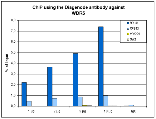
Figure 1. ChIP results obtained with the Diagenode antibody directed against WDR5
ChIP assays were performed using HeLa cells, the Diagenode antibody against WDR5 (Cat. No. C15410027) and optimized PCR primer sets for qPCR. ChIP was performed with the “iDeal ChIP-seq” kit (cat. No. C01010055), using sheared chromatin from 4 million cells. A titration consisting of 1, 2, 5 and 10 μg of antibody per ChIP experiment was analyzed. IgG (2 μg/IP) was used as a negative IP control. Quantitative PCR was performed with optimized primers for the promoters of the RPL41 and RPS4X ribosomal protein genes, used as positive controls, and for the MYOD1 gene and the Sat2 satellite repeat, used as negative controls. Figure 1 shows the recovery, expressed as a % of input (the relative amount of immunoprecipitated DNA compared to input DNA after qPCR analysis).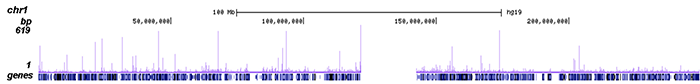
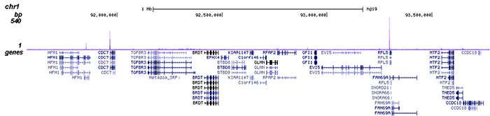
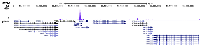
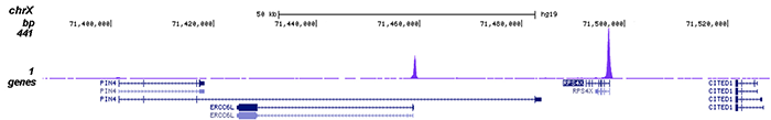
Figure 2. ChIP-seq results obtained with the Diagenode antibody directed against WDR5
ChIP was performed on sheared chromatin from 4 million HeLa cells using 2 μg of the Diagenode antibody against WDR5 (Cat. No. C15410027) as described above. The IP’d DNA was subsequently analysed on an Illumina HiSeq. Library preparation, cluster generation and sequencing were performed according to the manufacturer’s instructions. The 50 bp tags were aligned to the human genome using the BWA algorithm. Figure 2 shows the enrichment along the complete sequence and a 2 Mb region of human chromosome 1 (fig 2A and B), and in two genomic regions surrounding the RPL41 and RPS4X positive control genes (fig 2C and D).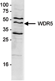
Figure 3. Western blot analysis using the Diagenode antibody directed against WDR5
Nclear extracts from HeLa cells (20 μg) were analysed by Western blot using the Diagenode antibody against WDR5 (Cat. No. C15410027) diluted 1:1,000 in TBS-Tween containing 5% skimmed milk. The position of the protein of interest is indicated on the right; the marker (in kDa) is shown on the left.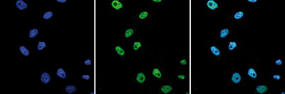
Figure 4. Immunofluorescence using the Diagenode antibody directed against WDR
HeLa cells were stained with the Diagenode antibody against WDR5 (Cat. No. C15410027) and with DAPI. Cells were fixed with 4% formaldehyde for 10’ and blocked with PBS/TX-100 containing 5% normal goat serum and 1% BSA. The cells were immunofluorescently labeled with the WDR5 antibody (left) diluted 1:1,000 in blocking solution followed by an anti-rabbit antibody conjugated to Alexa488. The middle panel shows staining of the nuclei with DAPI. A merge of the two stainings is shown on the right. - Publications

How to properly cite our product/service in your work
We strongly recommend using this: WDR5 Antibody (Hologic Diagenode Cat# C15410027 Lot# 001). Click here to copy to clipboard.
Using our products or services in your publication? Let us know!