Notice (8): Undefined variable: solution_of_interest [APP/View/Products/view.ctp, line 755]Code Context<!-- BEGIN: REQUEST_FORM MODAL -->
<div id="request_formModal" class="reveal-modal medium" data-reveal aria-labelledby="modalTitle" aria-hidden="true" role="dialog">
<?= $this->element('Forms/simple_form', array('solution_of_interest' => $solution_of_interest, 'header' => $header, 'message' => $message, 'campaign_id' => $campaign_id)) ?>
$viewFile = '/home/website-server/www/app/View/Products/view.ctp'
$dataForView = array(
'language' => 'jp',
'meta_keywords' => '',
'meta_description' => '5-mC monoclonal antibody cl. b - Classic',
'meta_title' => '5-mC monoclonal antibody cl. b - Classic',
'product' => array(
'Product' => array(
'id' => '1966',
'antibody_id' => '624',
'name' => '5-methylcytosine (5-mC) monoclonal antibody cl. b ',
'description' => '<p><span>Monoclonal antibody raised in mouse against 5-mC (5-methylcytosine) conjugated to ovalbumine.</span></p>',
'label1' => 'Validation Data',
'info1' => '<div class="row">
<div class="small-6 columns">
<p><img src="https://www.diagenode.com/img/product/antibodies/C15200006-500_MeDIP_1.png" alt="5-methylcytosine (5-mC) Antibody validated in MeDIP" caption="false" /></p>
</div>
<div class="small-6 columns">
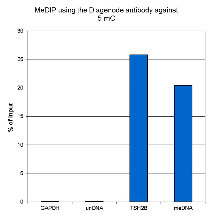
<p><small><strong>Figure 1. Methylated DNA immunoprecipitation (MeDIP) results obtained with the Diagenode monoclonal antibody directed against 5-mC</strong><br />MeDIP (Methylated DNA immunoprecipitation) was performed on 1 µg fragmented human genomic DNA using 0.2 µg of the Diagenode monoclonal antibody against 5-mC (cat. No. C15200006) and the MagMeDIP Kit (cat. No. C02010021). The fragmented DNA was spiked with the internal controls present in the kit (methylated DNA (meDNA) as a positive and unmethylated DNA (unDNA) as a negative control) prior to performing the IP. QPCR was performed with optimized primer sets, included in the kit, specific for the methylated and unmethylated DNA controls, and for a known methylated (TSH2B) and unmethylated (GAPDH) genomic region. Figure 1 shows the recovery expressed as a % of input (the relative amount of immunoprecipitated DNA compared to input DNA after qPCR analysis). </small></p>
</div>
</div>
<p></p>
<div class="row">
<div class="small-4 columns">
<p><img src="https://www.diagenode.com/img/product/antibodies/C15200006_MeDIP_2.png" alt="5-methylcytosine (5-mC) Antibody validated method" caption="false" width="278" height="333" /></p>
</div>
<div class="small-8 columns">
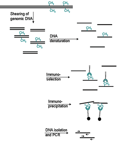
<p><small><strong>Figure 2. Methylated DNA immunoprecipitation (MeDIP) method</strong></small></p>
<ul>
<li><small>Prepare genomic DNA from cultured cells</small></li>
<li><small>Shear genomic DNA</small></li>
<li><small>Denature the sheared genomic DNA</small></li>
<li><small>Immunoprecipitate with the antibody against 5-meC</small></li>
<li><small>Isolate DNA and perform PCR</small></li>
</ul>
</div>
</div>
<div class="extra-spaced"></div>
<div class="row">
<div class="small-4 columns">
<p><img src="https://www.diagenode.com/img/product/antibodies/C15200006_if.jpg" alt="5-methylcytosine (5-mC) Antibody for IF" caption="false" width="278" height="91" /></p>
</div>
<div class="small-8 columns">
<p><small><strong>Figure 3. Immunofluorescence using the Diagenode monoclonal antibody directed against 5-mC</strong> <br />HeLa cells were stained with the Diagenode antibody against 5-mC (Cat. No. C15200006) and with DAPI. Cells were fixed with 4% formaldehyde for 10’ and blocked with PBS/TX-100 containing 1% BSA. The cells were immunofluorescently labelled with the 5-mC antibody (middle) diluted 1:1,000 in blocking solution followed by an anti-mouse antibody conjugated to Alexa594. The left panel shows staining of the nuclei with DAPI. A merge of the two stainings is shown on the right.</small></p>
</div>
</div>',
'label2' => '',
'info2' => '',
'label3' => '',
'info3' => '',
'format' => '500 µg',
'catalog_number' => 'C15200006-500',
'old_catalog_number' => 'MAb-006-500',
'sf_code' => 'C15200006-D001-000527',
'type' => 'FRE',
'search_order' => '03-Antibody',
'price_EUR' => '1345',
'price_USD' => '1715',
'price_GBP' => '1145',
'price_JPY' => '210695',
'price_CNY' => '',
'price_AUD' => '4288',
'country' => 'ALL',
'except_countries' => 'None',
'quote' => false,
'in_stock' => false,
'featured' => false,
'no_promo' => false,
'online' => true,
'master' => false,
'last_datasheet_update' => 'November 17, 2020',
'slug' => '5-mc-monoclonal-antibody-cl-b-classic-500',
'meta_title' => '5-mC monoclonal antibody cl. b - Classic',
'meta_keywords' => '',
'meta_description' => '5-mC monoclonal antibody cl. b - Classic',
'modified' => '2022-10-06 12:03:13',
'created' => '2015-06-29 14:08:20',
'locale' => 'jpn'
),
'Antibody' => array(
'host' => '*****',
'id' => '624',
'name' => '5-mC monoclonal antibody (500µg)',
'description' => '',
'clonality' => '',
'isotype' => 'IgG1',
'lot' => '007',
'concentration' => '1.1 µg/µl',
'reactivity' => 'Human, mouse, rat, cow, alligator, zebrafish, plants, finch, wide range expected.',
'type' => 'Monoclonal <br /><strong>MEDIP-grade</strong>',
'purity' => 'Protein A purified monoclonal antibody',
'classification' => '',
'application_table' => '<table>
<thead>
<tr>
<th>Applications</th>
<th>Suggested dilution</th>
<th>References</th>
</tr>
</thead>
<tbody>
<tr>
<td>MeDIP*</td>
<td>0.5 - 1 µg/IP</td>
<td>Fig 1</td>
</tr>
<tr>
<td>Immunofluorescence</td>
<td>1:1,000</td>
<td>Fig 3</td>
</tr>
</tbody>
</table>
<p></p>
<p><small><sup>*</sup> Please note that the optimal antibody amount per IP should be determined by the end-user. We recommend testing 0.2-5 µg per IP.</small></p>',
'storage_conditions' => 'Store at -20°C; for long storage, store at -80°C. Avoid multiple freeze-thaw cycles.',
'storage_buffer' => 'PBS containing 0.05% azide.',
'precautions' => 'This product is for research use only. Not for use in diagnostic or therapeutic procedures.',
'uniprot_acc' => '',
'slug' => '5-mc-monoclonal-antibody-cl-b-classic-500',
'meta_keywords' => '',
'meta_description' => '',
'modified' => '2022-10-06 11:42:54',
'created' => '2020-11-26 10:41:49',
'select_label' => '624 - 5-mC monoclonal antibody (500µg) (007 - 1.1 µg/µl - Human, mouse, rat, cow, alligator, zebrafish, plants, finch, wide range expected. - Protein A purified monoclonal antibody - Mouse)'
),
'Slave' => array(),
'Group' => array(
'Group' => array(
[maximum depth reached]
),
'Master' => array(
[maximum depth reached]
),
'Product' => array(
[maximum depth reached]
)
),
'Related' => array(
(int) 0 => array(
[maximum depth reached]
),
(int) 1 => array(
[maximum depth reached]
),
(int) 2 => array(
[maximum depth reached]
),
(int) 3 => array(
[maximum depth reached]
),
(int) 4 => array(
[maximum depth reached]
),
(int) 5 => array(
[maximum depth reached]
)
),
'Application' => array(
(int) 0 => array(
[maximum depth reached]
),
(int) 1 => array(
[maximum depth reached]
)
),
'Category' => array(
(int) 0 => array(
[maximum depth reached]
),
(int) 1 => array(
[maximum depth reached]
)
),
'Document' => array(
(int) 0 => array(
[maximum depth reached]
),
(int) 1 => array(
[maximum depth reached]
),
(int) 2 => array(
[maximum depth reached]
)
),
'Feature' => array(),
'Image' => array(
(int) 0 => array(
[maximum depth reached]
)
),
'Promotion' => array(),
'Protocol' => array(),
'Publication' => array(
(int) 0 => array(
[maximum depth reached]
),
(int) 1 => array(
[maximum depth reached]
),
(int) 2 => array(
[maximum depth reached]
),
(int) 3 => array(
[maximum depth reached]
),
(int) 4 => array(
[maximum depth reached]
),
(int) 5 => array(
[maximum depth reached]
),
(int) 6 => array(
[maximum depth reached]
),
(int) 7 => array(
[maximum depth reached]
),
(int) 8 => array(
[maximum depth reached]
),
(int) 9 => array(
[maximum depth reached]
),
(int) 10 => array(
[maximum depth reached]
),
(int) 11 => array(
[maximum depth reached]
),
(int) 12 => array(
[maximum depth reached]
),
(int) 13 => array(
[maximum depth reached]
),
(int) 14 => array(
[maximum depth reached]
),
(int) 15 => array(
[maximum depth reached]
),
(int) 16 => array(
[maximum depth reached]
),
(int) 17 => array(
[maximum depth reached]
),
(int) 18 => array(
[maximum depth reached]
),
(int) 19 => array(
[maximum depth reached]
),
(int) 20 => array(
[maximum depth reached]
),
(int) 21 => array(
[maximum depth reached]
),
(int) 22 => array(
[maximum depth reached]
),
(int) 23 => array(
[maximum depth reached]
),
(int) 24 => array(
[maximum depth reached]
),
(int) 25 => array(
[maximum depth reached]
),
(int) 26 => array(
[maximum depth reached]
),
(int) 27 => array(
[maximum depth reached]
),
(int) 28 => array(
[maximum depth reached]
),
(int) 29 => array(
[maximum depth reached]
),
(int) 30 => array(
[maximum depth reached]
),
(int) 31 => array(
[maximum depth reached]
),
(int) 32 => array(
[maximum depth reached]
),
(int) 33 => array(
[maximum depth reached]
),
(int) 34 => array(
[maximum depth reached]
),
(int) 35 => array(
[maximum depth reached]
),
(int) 36 => array(
[maximum depth reached]
),
(int) 37 => array(
[maximum depth reached]
),
(int) 38 => array(
[maximum depth reached]
),
(int) 39 => array(
[maximum depth reached]
),
(int) 40 => array(
[maximum depth reached]
),
(int) 41 => array(
[maximum depth reached]
),
(int) 42 => array(
[maximum depth reached]
),
(int) 43 => array(
[maximum depth reached]
),
(int) 44 => array(
[maximum depth reached]
),
(int) 45 => array(
[maximum depth reached]
),
(int) 46 => array(
[maximum depth reached]
),
(int) 47 => array(
[maximum depth reached]
),
(int) 48 => array(
[maximum depth reached]
),
(int) 49 => array(
[maximum depth reached]
),
(int) 50 => array(
[maximum depth reached]
),
(int) 51 => array(
[maximum depth reached]
)
),
'Testimonial' => array(),
'Area' => array(),
'SafetySheet' => array(
(int) 0 => array(
[maximum depth reached]
),
(int) 1 => array(
[maximum depth reached]
),
(int) 2 => array(
[maximum depth reached]
),
(int) 3 => array(
[maximum depth reached]
),
(int) 4 => array(
[maximum depth reached]
),
(int) 5 => array(
[maximum depth reached]
),
(int) 6 => array(
[maximum depth reached]
),
(int) 7 => array(
[maximum depth reached]
)
)
),
'meta_canonical' => 'https://www.diagenode.com/jp/p/5-mc-monoclonal-antibody-cl-b-classic-100-ug-50-ul'
)
$language = 'jp'
$meta_keywords = ''
$meta_description = '5-mC monoclonal antibody cl. b - Classic'
$meta_title = '5-mC monoclonal antibody cl. b - Classic'
$product = array(
'Product' => array(
'id' => '1966',
'antibody_id' => '624',
'name' => '5-methylcytosine (5-mC) monoclonal antibody cl. b ',
'description' => '<p><span>Monoclonal antibody raised in mouse against 5-mC (5-methylcytosine) conjugated to ovalbumine.</span></p>',
'label1' => 'Validation Data',
'info1' => '<div class="row">
<div class="small-6 columns">
<p><img src="https://www.diagenode.com/img/product/antibodies/C15200006-500_MeDIP_1.png" alt="5-methylcytosine (5-mC) Antibody validated in MeDIP" caption="false" /></p>
</div>
<div class="small-6 columns">
<p><small><strong>Figure 1. Methylated DNA immunoprecipitation (MeDIP) results obtained with the Diagenode monoclonal antibody directed against 5-mC</strong><br />MeDIP (Methylated DNA immunoprecipitation) was performed on 1 µg fragmented human genomic DNA using 0.2 µg of the Diagenode monoclonal antibody against 5-mC (cat. No. C15200006) and the MagMeDIP Kit (cat. No. C02010021). The fragmented DNA was spiked with the internal controls present in the kit (methylated DNA (meDNA) as a positive and unmethylated DNA (unDNA) as a negative control) prior to performing the IP. QPCR was performed with optimized primer sets, included in the kit, specific for the methylated and unmethylated DNA controls, and for a known methylated (TSH2B) and unmethylated (GAPDH) genomic region. Figure 1 shows the recovery expressed as a % of input (the relative amount of immunoprecipitated DNA compared to input DNA after qPCR analysis). </small></p>
</div>
</div>
<p></p>
<div class="row">
<div class="small-4 columns">
<p><img src="https://www.diagenode.com/img/product/antibodies/C15200006_MeDIP_2.png" alt="5-methylcytosine (5-mC) Antibody validated method" caption="false" width="278" height="333" /></p>
</div>
<div class="small-8 columns">
<p><small><strong>Figure 2. Methylated DNA immunoprecipitation (MeDIP) method</strong></small></p>
<ul>
<li><small>Prepare genomic DNA from cultured cells</small></li>
<li><small>Shear genomic DNA</small></li>
<li><small>Denature the sheared genomic DNA</small></li>
<li><small>Immunoprecipitate with the antibody against 5-meC</small></li>
<li><small>Isolate DNA and perform PCR</small></li>
</ul>
</div>
</div>
<div class="extra-spaced"></div>
<div class="row">
<div class="small-4 columns">
<p><img src="https://www.diagenode.com/img/product/antibodies/C15200006_if.jpg" alt="5-methylcytosine (5-mC) Antibody for IF" caption="false" width="278" height="91" /></p>
</div>
<div class="small-8 columns">
<p><small><strong>Figure 3. Immunofluorescence using the Diagenode monoclonal antibody directed against 5-mC</strong> <br />HeLa cells were stained with the Diagenode antibody against 5-mC (Cat. No. C15200006) and with DAPI. Cells were fixed with 4% formaldehyde for 10’ and blocked with PBS/TX-100 containing 1% BSA. The cells were immunofluorescently labelled with the 5-mC antibody (middle) diluted 1:1,000 in blocking solution followed by an anti-mouse antibody conjugated to Alexa594. The left panel shows staining of the nuclei with DAPI. A merge of the two stainings is shown on the right.</small></p>
</div>
</div>',
'label2' => '',
'info2' => '',
'label3' => '',
'info3' => '',
'format' => '500 µg',
'catalog_number' => 'C15200006-500',
'old_catalog_number' => 'MAb-006-500',
'sf_code' => 'C15200006-D001-000527',
'type' => 'FRE',
'search_order' => '03-Antibody',
'price_EUR' => '1345',
'price_USD' => '1715',
'price_GBP' => '1145',
'price_JPY' => '210695',
'price_CNY' => '',
'price_AUD' => '4288',
'country' => 'ALL',
'except_countries' => 'None',
'quote' => false,
'in_stock' => false,
'featured' => false,
'no_promo' => false,
'online' => true,
'master' => false,
'last_datasheet_update' => 'November 17, 2020',
'slug' => '5-mc-monoclonal-antibody-cl-b-classic-500',
'meta_title' => '5-mC monoclonal antibody cl. b - Classic',
'meta_keywords' => '',
'meta_description' => '5-mC monoclonal antibody cl. b - Classic',
'modified' => '2022-10-06 12:03:13',
'created' => '2015-06-29 14:08:20',
'locale' => 'jpn'
),
'Antibody' => array(
'host' => '*****',
'id' => '624',
'name' => '5-mC monoclonal antibody (500µg)',
'description' => '',
'clonality' => '',
'isotype' => 'IgG1',
'lot' => '007',
'concentration' => '1.1 µg/µl',
'reactivity' => 'Human, mouse, rat, cow, alligator, zebrafish, plants, finch, wide range expected.',
'type' => 'Monoclonal <br /><strong>MEDIP-grade</strong>',
'purity' => 'Protein A purified monoclonal antibody',
'classification' => '',
'application_table' => '<table>
<thead>
<tr>
<th>Applications</th>
<th>Suggested dilution</th>
<th>References</th>
</tr>
</thead>
<tbody>
<tr>
<td>MeDIP*</td>
<td>0.5 - 1 µg/IP</td>
<td>Fig 1</td>
</tr>
<tr>
<td>Immunofluorescence</td>
<td>1:1,000</td>
<td>Fig 3</td>
</tr>
</tbody>
</table>
<p></p>
<p><small><sup>*</sup> Please note that the optimal antibody amount per IP should be determined by the end-user. We recommend testing 0.2-5 µg per IP.</small></p>',
'storage_conditions' => 'Store at -20°C; for long storage, store at -80°C. Avoid multiple freeze-thaw cycles.',
'storage_buffer' => 'PBS containing 0.05% azide.',
'precautions' => 'This product is for research use only. Not for use in diagnostic or therapeutic procedures.',
'uniprot_acc' => '',
'slug' => '5-mc-monoclonal-antibody-cl-b-classic-500',
'meta_keywords' => '',
'meta_description' => '',
'modified' => '2022-10-06 11:42:54',
'created' => '2020-11-26 10:41:49',
'select_label' => '624 - 5-mC monoclonal antibody (500µg) (007 - 1.1 µg/µl - Human, mouse, rat, cow, alligator, zebrafish, plants, finch, wide range expected. - Protein A purified monoclonal antibody - Mouse)'
),
'Slave' => array(),
'Group' => array(
'Group' => array(
'id' => '166',
'name' => 'C15200006',
'product_id' => '1965',
'modified' => '2016-02-23 20:46:06',
'created' => '2016-02-23 20:46:06'
),
'Master' => array(
'id' => '1965',
'antibody_id' => '302',
'name' => '5-methylcytosine (5-mC) Antibody - cl. b ',
'description' => '<p><span>Monoclonal antibody raised in mouse against <strong>5-mC</strong> (<strong>5-methylcytosine</strong>) conjugated to ovalbumine.</span></p>',
'label1' => 'Validation Data',
'info1' => '<div class="row">
<div class="small-4 columns">
<p><img src="https://www.diagenode.com/img/product/antibodies/C15200006_MeDIP_1.png" alt="5-methylcytosine (5-mC) Antibody validated in MeDIP" caption="false" width="278" height="292" /></p>
</div>
<div class="small-8 columns">
<p><small><strong>Figure 1. Methylated DNA immunoprecipitation (MeDIP) results obtained with the Diagenode monoclonal antibody directed against 5-mC</strong><br />MeDIP (Methylated DNA immunoprecipitation) was performed on 1 µg fragmented human genomic DNA using 0.2 µg of the Diagenode monoclonal antibody against 5-mC (Cat. No. C15200006) and the MagMeDIP Kit (Cat. No. C02010021). The fragmented DNA was spiked with the internal controls present in the kit (methylated DNA (meDNA) as a positive and unmethylated DNA (unDNA) as a negative control) prior to performing the IP. QPCR was performed with optimized primer sets, included in the kit, specific for the methylated and unmethylated DNA controls, and for a known methylated (TSH2B) and unmethylated (GAPDH) genomic region. Figure 2 shows the recovery expressed as a % of input (the relative amount of immunoprecipitated DNA compared to input DNA after qPCR analysis).</small></p>
</div>
</div>
<div class="row">
<div class="small-4 columns">
<p><img src="https://www.diagenode.com/img/product/antibodies/C15200006_MeDIP_2.png" alt="5-methylcytosine (5-mC) Antibody validated method" caption="false" width="278" height="333" /></p>
</div>
<div class="small-8 columns">
<p><small><strong>Figure 2. Methylated DNA immunoprecipitation (MeDIP) method</strong></small></p>
<ul>
<li><small>Prepare genomic DNA from cultured cells</small></li>
<li><small>Shear genomic DNA</small></li>
<li><small>Denature the sheared genomic DNA</small></li>
<li><small>Immunoprecipitate with the antibody against 5-meC</small></li>
<li><small>Isolate DNA and perform PCR</small></li>
</ul>
</div>
</div>
<div class="row">
<div class="small-4 columns">
<p><img src="https://www.diagenode.com/img/product/antibodies/C15200006_if.jpg" alt="5-methylcytosine (5-mC) Antibody for IF" caption="false" width="278" height="91" /></p>
</div>
<div class="small-8 columns">
<p><small><strong>Figure 3. Immunofluorescence using the Diagenode monoclonal antibody directed against 5-mC</strong> <br />HeLa cells were stained with the Diagenode antibody against 5-mC (Cat. No. C15200006) and with DAPI. Cells were fixed with 4% formaldehyde for 10’ and blocked with PBS/TX-100 containing 1% BSA. The cells were immunofluorescently labelled with the 5-mC antibody (left) diluted 1:1,000 in blocking solution followed by an anti-mouse antibody conjugated to Alexa594. The middle panel shows staining of the nuclei with DAPI. A merge of the two stainings is shown on the right.</small></p>
</div>
</div>',
'label2' => '',
'info2' => '',
'label3' => '',
'info3' => '',
'format' => '100 µg',
'catalog_number' => 'C15200006-100',
'old_catalog_number' => 'MAb-006-100',
'sf_code' => 'C15200006-D001-000526',
'type' => 'FRE',
'search_order' => '03-Antibody',
'price_EUR' => '385',
'price_USD' => '450',
'price_GBP' => '350',
'price_JPY' => '60310',
'price_CNY' => '',
'price_AUD' => '1125',
'country' => 'ALL',
'except_countries' => 'None',
'quote' => false,
'in_stock' => false,
'featured' => false,
'no_promo' => false,
'online' => true,
'master' => true,
'last_datasheet_update' => 'February 11, 2019',
'slug' => '5-mc-monoclonal-antibody-cl-b-classic-100-ug-50-ul',
'meta_title' => '5-methylcytosine (5-mC) Antibody - cl. b (C15200006) | Diagenode',
'meta_keywords' => '5-methylcytosine (5-mC),monoclonal antibody,Methylated DNA Immunoprecipitation',
'meta_description' => '5-methylcytosine (5-mC) Monoclonal Antibody cl. b validated in MeDIP and IF. Batch-specific data available on the website. Sample size available.',
'modified' => '2023-08-03 10:25:19',
'created' => '2015-06-29 14:08:20'
),
'Product' => array(
(int) 0 => array(
[maximum depth reached]
),
(int) 1 => array(
[maximum depth reached]
)
)
),
'Related' => array(
(int) 0 => array(
'id' => '1882',
'antibody_id' => null,
'name' => 'hMeDIP kit x16 (monoclonal mouse antibody)',
'description' => '<p><a href="https://www.diagenode.com/files/products/kits/hMeDIP_kit_manual.pdf"><img src="https://www.diagenode.com/img/buttons/bt-manual.png" /></a></p>
<p><span>The hMeDIP kit is designed for enrichment of hydroxymethylated DNA from fragmented genomic DNA<span><span> </span>samples for use in genome-wide methylation analysis. It features</span></span><span> a highly specific monoclonal antibody against </span>5-hydroxymethylcytosine (5-hmC) for the immunoprecipitation of hydroxymethylated DNA<span>. It includes control DNA and primers to assess the effiency of the assay. </span>Performing hydroxymethylation profiling with the hMeDIP kit is fast, reliable and highly specific.</p>
<p><em>Looking for hMeDIP-seq protocol? <a href="https://go.diagenode.com/l/928883/2022-01-07/2m1ht" target="_blank" title="Contact us">Contact us</a></em></p>
<p><span></span></p>
<p><span></span></p>',
'label1' => 'Characteristics',
'info1' => '<ul style="list-style-type: disc;">
<li><span>Robust enrichment & immunoprecipitation of hydroxymethylated DNA</span></li>
<li>Highly specific monoclonal antibody against 5-hmC<span> for reliable, reproducible results</span></li>
<li>Including control DNA and primers to <span>monitor the efficiency of the assay</span>
<ul style="list-style-type: circle;">
<li>hmeDNA and unmethylated DNA sequences and primer pairs</li>
<li>Mouse primer pairs against Sfi1 targeting hydroxymethylated gene in mouse</li>
</ul>
</li>
<li>Improved single-tube, magnetic bead-based protocol</li>
</ul>',
'label2' => '',
'info2' => '',
'label3' => '',
'info3' => '',
'format' => '16 rxns',
'catalog_number' => 'C02010031',
'old_catalog_number' => 'AF-110-0016',
'sf_code' => 'C02010031-',
'type' => 'RFR',
'search_order' => '04-undefined',
'price_EUR' => '630',
'price_USD' => '690',
'price_GBP' => '580',
'price_JPY' => '98690',
'price_CNY' => '',
'price_AUD' => '1725',
'country' => 'ALL',
'except_countries' => 'None',
'quote' => false,
'in_stock' => false,
'featured' => true,
'no_promo' => false,
'online' => true,
'master' => true,
'last_datasheet_update' => '0000-00-00',
'slug' => 'hmedip-kit-x16-monoclonal-mouse-antibody-16-rxns',
'meta_title' => 'hMeDIP kit x16 (monoclonal mouse antibody)',
'meta_keywords' => '',
'meta_description' => 'hMeDIP kit x16 (monoclonal mouse antibody)',
'modified' => '2023-04-20 16:12:48',
'created' => '2015-06-29 14:08:20',
'ProductsRelated' => array(
[maximum depth reached]
),
'Image' => array(
[maximum depth reached]
)
),
(int) 1 => array(
'id' => '1880',
'antibody_id' => null,
'name' => 'MagMeDIP Kit',
'description' => '<p><a href="https://www.diagenode.com/files/products/kits/magmedip-kit-manual-C02010020-21.pdf"><img src="https://www.diagenode.com/img/buttons/bt-manual.png" /></a></p>
<p>Perform <strong>MeDIP</strong> (<strong>Me</strong>thylated <strong>D</strong>NA <strong>I</strong>mmuno<strong>p</strong>recipitation) followed by qPCR or NGS to estimate DNA methylation status of your sample using a highly sensitive 5-methylcytosine antibody. Our MagMeDIP kit contains high quality reagents to get the highest enrichment of methylated DNA with an optimized user-friendly protocol.</p>
<h3><span>Features</span></h3>
<ul>
<li>Starting DNA amount: <strong>10 ng – 1 µg</strong></li>
<li>Content: <strong>all reagents included</strong> for DNA extraction, immunoprecipitation (including the 5-mC antibody, spike-in controls and their corresponding qPCR primer pairs) as well as DNA isolation after IP.</li>
<li>Application: <strong>qPCR</strong> and <strong>NGS</strong></li>
<li>Robust method, <strong>superior enrichment</strong>, and easy-to-use protocol</li>
<li><strong>High reproducibility</strong> between replicates and repetitive experiments</li>
<li>Compatible with <strong>all species </strong></li>
</ul>
<p> </p>
<div class="small-12 medium-4 large-4 columns"><center></center><center></center><center></center><center><a href="https://www.ncbi.nlm.nih.gov/pubmed/30429608" target="_blank"><img src="https://www.diagenode.com/img/banners/banner-nature-publication-580.png" alt="Click here to read more about MeDIP " caption="false" width="80%" /></a></center></div>
<div class="small-12 medium-8 large-8 columns">
<h3 style="text-align: justify;">Sensitive tumour detection and classification using plasma cell-free DNA methylomes<br /><a href="https://www.ncbi.nlm.nih.gov/pubmed/30429608" target="_blank">Read the publication</a></h3>
<h3 class="c-article-title u-h1" data-test="article-title" itemprop="name headline" style="text-align: justify;">Preparation of cfMeDIP-seq libraries for methylome profiling of plasma cell-free DNA<br /><a href="https://www.nature.com/articles/s41596-019-0202-2" target="_blank" title="cfMeDIP-seq Nature Method">Read the method</a></h3>
</div>
<p></p>
<p></p>
<p></p>
<div class="row">
<div class="small-12 medium-4 large-4 columns"><center>
<script>// <![CDATA[
var date = new Date(); var heure = date.getHours(); var jour = date.getDay(); var semaine = Math.floor(date.getDate() / 7) + 1; if (jour === 2 && ( (heure >= 9 && heure < 9.5) || (heure >= 18 && heure < 18.5) )) { document.write('<a href="https://us02web.zoom.us/j/85467619762"><img src="https://www.diagenode.com/img/epicafe-ON.gif"></a>'); } else { document.write('<a href="https://go.diagenode.com/l/928883/2023-04-26/3kq1v"><img src="https://www.diagenode.com/img/epicafe-OFF.png"></a>'); }
// ]]></script>
</center></div>
<div class="small-12 medium-8 large-8 columns"><br />
<p></p>
</div>
</div>
<h3></h3>',
'label1' => 'MagMeDIP workflow',
'info1' => '<p>DNA methylation occurs primarily as 5-methylcytosine (5-mC), and the Diagenode MagMeDIP Kit takes advantage of a specific antibody targeting this 5-mC to immunoprecipitate methylated DNA, which can be thereafter directly analyzed by qPCR or Next-Generation Sequencing (NGS).</p>
<h3><span>How it works</span></h3>
<p>In brief, after the cell collection and lysis, the genomic DNA is extracted, sheared, and then denatured. In the next step the antibody directed against 5 methylcytosine and antibody binding beads are used for immunoselection and immunoprecipitation of methylated DNA fragments. Then, the IP’d methylated DNA is isolated and can be used for any subsequent analysis as qPCR, amplification, hybridization on microarrays or next generation sequencing.</p>
<center><img src="https://www.diagenode.com/img/product/kits/MagMeDIP-workflow.png" width="70%" alt="5-methylcytosine" caption="false" /></center>
<script async="" src="https://edge.fullstory.com/s/fs.js" crossorigin="anonymous"></script>
<script src="chrome-extension://hhojmcideegachlhfgfdhailpfhgknjm/web_accessible_resources/index.js"></script>',
'label2' => 'MeDIP-qPCR',
'info2' => '<p>The kit MagMeDIP contains all reagents necessary for a complete MeDIP-qPCR workflow. Two MagMeDIP protocols have been validated: for manual processing as well as for automated processing, using the Diagenode’s IP-Star Compact Automated System (please refer to the kit manual).</p>
<ul>
<li><strong>Complete kit</strong> including DNA extraction module, IP antibody and reagents, DNA isolation buffer</li>
<li><strong>Quality control of the IP:</strong> due to methylated and unmethylated DNA spike-in controls and their associated qPCR primers</li>
<li><strong>Easy to use</strong> with user-friendly magnetic beads and rack</li>
<li><strong>Highly validated protocol</strong></li>
<li>Automated protocol supplied</li>
</ul>
<center><img src="https://www.diagenode.com/img/product/kits/fig1-magmedipkit.png" width="85%" alt="Methylated DNA Immunoprecipitation" caption="false" /></center>
<p style="font-size: 0.9em;"><em><strong>Figure 1.</strong> Immunoprecipitation results obtained with Diagenode MagMeDIP Kit</em></p>
<p style="font-size: 0.9em;">MeDIP assays were performed manually using 1 µg or 50 ng gDNA from blood cells with the MagMeDIP kit (Diagenode). The IP was performed with the Methylated and Unmethylated spike-in controls included in the kit, together with the human DNA samples. The DNA was isolated/purified using DIB. Afterwards, qPCR was performed using the primer pairs included in this kit.</p>
<script async="" src="https://edge.fullstory.com/s/fs.js" crossorigin="anonymous"></script>
<script src="chrome-extension://hhojmcideegachlhfgfdhailpfhgknjm/web_accessible_resources/index.js"></script>',
'label3' => 'MeDIP-seq',
'info3' => '<p>For DNA methylation analysis on the whole genome, MagMeDIP kit can be coupled with Next-Generation Sequencing. To perform MeDIP-sequencing we recommend the following strategy:</p>
<ul style="list-style-type: circle;">
<li>Choose a library preparation solution which is compatible with the starting amount of DNA you are planning to use (from 10 ng to 1 μg). It can be a home-made solution or a commercial one.</li>
<li>Choose the indexing system that fits your needs considering the following features:</li>
<ul>
<ul>
<ul>
<li>Single-indexing, combinatorial dual-indexing or unique dual-indexing</li>
<li>Number of barcodes</li>
<li>Full-length adaptors containing the barcodes or barcoding at the final amplification step</li>
<li>Presence / absence of Unique Molecular Identifiers (for PCR duplicates removal)</li>
</ul>
</ul>
</ul>
<li>Standard library preparation protocols are compatible with double-stranded DNA only, therefore the first steps of the library preparation (end repair, A-tailing, adaptor ligation and clean-up) will have to be performed on sheared DNA, before the IP.</li>
</ul>
<p style="padding-left: 30px;"><strong>CAUTION:</strong> As the immunoprecipitation step occurs at the middle of the library preparation workflow, single-tube solutions for library preparation are usually not compatible with MeDIP-sequencing.</p>
<ul style="list-style-type: circle;">
<li>For DNA isolation after the IP, we recommend using the <a href="https://www.diagenode.com/en/p/ipure-kit-v2-x24" title="IPure kit v2">IPure kit v2</a> (available separately, Cat. No. C03010014) instead of DNA isolation Buffer.</li>
</ul>
<ul style="list-style-type: circle;">
<li>Perform library amplification after the DNA isolation following the standard protocol of the chosen library preparation solution.</li>
</ul>
<h3><span>MeDIP-seq workflow</span></h3>
<center><img src="https://www.diagenode.com/img/product/kits/MeDIP-seq-workflow.png" width="110%" alt="MagMeDIP qPCR Kit x10 workflow" caption="false" /></center>
<h3><span>Example of results</span></h3>
<center><img src="https://www.diagenode.com/img/product/kits/medip-specificity.png" alt="MagMeDIP qPCR Kit Result" caption="false" width="951" height="488" /></center>
<p></p>
<p style="font-size: 0.9em;"><strong>Figure 1. qPCR analysis of external spike-in DNA controls (methylated and unmethylated) after IP.</strong> Samples were prepared using 1μg – 100ng -10ng sheared human gDNA with the MagMeDIP kit (Diagenode) and a commercially available library prep kit. DNA isolation after IP has been performed with IPure kit V2 (Diagenode).</p>
<p></p>
<p></p>
<center><img src="https://www.diagenode.com/img/product/kits/medip-saturation-analysis.png" alt=" MagMeDIP kit " caption="false" width="951" height="461" /></center>
<p></p>
<p style="font-size: 0.9em;"><strong>Figure 2. Saturation analysis.</strong> Clean reads were aligned to the human genome (hg19) using Burrows-Wheeler aligner (BWA) algorithm after which duplicated and unmapped reads were removed resulting in a mapping efficiency >98% for all samples. Quality and validity check of the mapped MeDIP-seq data was performed using MEDIPS R package. Saturation plots show that all sets of reads have sufficient complexity and depth to saturate the coverage profile of the reference genome and that this is reproducible between replicates and repetitive experiments (data shown for 50 ng gDNA input: left panel = replicate a, right panel = replicate b).</p>
<p></p>
<p></p>
<center><img src="https://www.diagenode.com/img/product/kits/medip-libraries-prep.png" alt="MagMeDIP x10 " caption="false" width="951" height="708" /></center>
<p></p>
<p style="font-size: 0.9em;"><strong>Figure 3. Sequencing profiles of MeDIP-seq libraries prepared from different starting amounts of sheared gDNA on the positive and negative methylated control regions.</strong> MeDIP-seq libraries were prepared from decreasing starting amounts of gDNA (1 μg (green), 50 ng (red), and 10ng (blue)) originating from human blood with the MagMeDIP kit (Diagenode) and a commercially available library prep kit. DNA isolation after IP has been performed with IPure kit V2 (Diagenode). IP and corresponding INPUT samples were sequenced on Illumina NovaSeq SP with 2x50 PE reads. The reads were mapped to the human genome (hg19) with bwa and the alignments were loaded into IGV (the tracks use an identical scale). The top IGV figure shows the TSH2B (also known as H2BC1) gene (marked by blue boxes in the bottom track) and its surroundings. The TSH2B gene is coding for a histone variant that does not occur in blood cells, and it is known to be silenced by methylation. Accordingly, we see a high coverage in the vicinity of this gene. The bottom IGV figure shows the GADPH locus (marked by blue boxes in the bottom track) and its surroundings. The GADPH gene is a highly active transcription region and should not be methylated, resulting in no reads accumulation following MeDIP-seq experiment.</p>
<p></p>
<ul>
<ul>
<script async="" src="https://edge.fullstory.com/s/fs.js" crossorigin="anonymous"></script>
<script src="chrome-extension://hhojmcideegachlhfgfdhailpfhgknjm/web_accessible_resources/index.js"></script>
</ul>
</ul>',
'format' => '48 rxns (IP)',
'catalog_number' => 'C02010021',
'old_catalog_number' => 'mc-magme-048',
'sf_code' => 'C02010021-',
'type' => 'RFR',
'search_order' => '04-undefined',
'price_EUR' => '745',
'price_USD' => '750',
'price_GBP' => '680',
'price_JPY' => '116705',
'price_CNY' => '',
'price_AUD' => '1875',
'country' => 'ALL',
'except_countries' => 'None',
'quote' => false,
'in_stock' => false,
'featured' => true,
'no_promo' => false,
'online' => true,
'master' => true,
'last_datasheet_update' => '0000-00-00',
'slug' => 'magmedip-kit-x48-48-rxns',
'meta_title' => 'MagMeDIP Kit for efficient immunoprecipitation of methylated DNA | Diagenode',
'meta_keywords' => '',
'meta_description' => 'Perform Methylated DNA Immunoprecipitation (MeDIP) to estimate DNA methylation status of your sample using highly specific 5-mC antibody. This kit allows the preparation of cfMeDIP-seq libraries.',
'modified' => '2024-12-04 16:52:47',
'created' => '2015-06-29 14:08:20',
'ProductsRelated' => array(
[maximum depth reached]
),
'Image' => array(
[maximum depth reached]
)
),
(int) 2 => array(
'id' => '1887',
'antibody_id' => null,
'name' => 'MethylCap kit',
'description' => '<p>The MethylCap kit allows to specifically capture DNA fragments containing methylated CpGs. The assay is based on the affinity purification of methylated DNA using methyl-CpG-binding domain (MBD) of human MeCP2 protein. The procedure has been adapted to both manual process or <a href="https://www.diagenode.com/en/p/sx-8g-ip-star-compact-automated-system-1-unit">IP-Star® Compact Automated System</a>. Libraries of captured methylated DNA can be prepared for next-generation sequencing (NGS) by combining MBD technology with the <a href="https://www.diagenode.com/en/p/microplex-lib-prep-kit-v3-48-rxns">MicroPlex Library Preparation Kit v3</a>.</p>',
'label1' => 'Characteristics',
'info1' => '<ul style="list-style-type: circle;">
<li><strong>Fast & sensitive capture</strong> of methylated DNA</li>
<li><strong>High capture efficiency</strong></li>
<li><strong>Differential fractionation</strong> of methylated DNA by CpG density (3 eluted fractions)</li>
<li><strong>On-day protocol</strong></li>
<li><strong>NGS compatibility</strong></li>
</ul>
<h3>MBD-seq allows for detection of genomic regions with different CpG density</h3>
<p><img src="https://www.diagenode.com/img/product/kits/mbd_results1.png" alt="MBD-sequencing results have been validated by bisulfite sequencing" style="display: block; margin-left: auto; margin-right: auto;" /></p>
<p><strong></strong></p>
<p><strong></strong><strong>F</strong><strong>igure 1.</strong> Using the MBD approach, two methylated regions were detected in different elution fractions according to their methylated CpG density (A). Low, Medium and High refer to the sequenced DNA from different elution fractions with increasing salt concentration. Methylated patterns of these two different methylated regions were validated by bisulfite conversion assay (B).</p>',
'label2' => '',
'info2' => '',
'label3' => '',
'info3' => '',
'format' => '48 rxns',
'catalog_number' => 'C02020010',
'old_catalog_number' => 'AF-100-0048',
'sf_code' => 'C02020010-',
'type' => 'RFR',
'search_order' => '04-undefined',
'price_EUR' => '740',
'price_USD' => '695',
'price_GBP' => '675',
'price_JPY' => '115920',
'price_CNY' => '',
'price_AUD' => '1738',
'country' => 'ALL',
'except_countries' => 'None',
'quote' => false,
'in_stock' => true,
'featured' => true,
'no_promo' => false,
'online' => true,
'master' => true,
'last_datasheet_update' => '0000-00-00',
'slug' => 'methylcap-kit-x48-48-rxns',
'meta_title' => 'MethylCap kit x48',
'meta_keywords' => '',
'meta_description' => 'MethylCap kit x48',
'modified' => '2024-11-21 06:38:46',
'created' => '2015-06-29 14:08:20',
'ProductsRelated' => array(
[maximum depth reached]
),
'Image' => array(
[maximum depth reached]
)
),
(int) 3 => array(
'id' => '1888',
'antibody_id' => null,
'name' => 'Auto MethylCap kit',
'description' => '<p>The Auto MethylCap kit allows to specifically capture DNA fragments containing methylated CpGs. The assay is based on the affinity purification of methylated DNA using methyl-CpG-binding domain (MBD) of human MeCP2 protein. This procedure has been optimized to perform automated immunoprecipitation of chromatin using the <a href="https://www.diagenode.com/en/p/sx-8g-ip-star-compact-automated-system-1-unit">IP-Star® Compact Automated System</a> enabling highly reproducible results and allowing for high throughput.</p>',
'label1' => ' Characteristics',
'info1' => '<ul>
<li><strong>Fast & sensitive capture</strong> of methylated DNA</li>
<li><strong>High capture efficiency</strong></li>
<li><strong>Differential fractionation</strong> of methylated DNA by CpG density (3 eluted fractions)</li>
<li><strong>Automation compatibility</strong><strong></strong>
<h3>MBD-seq allows for detection of genomic regions with different CpG density</h3>
<p><img src="https://www.diagenode.com/img/product/kits/mbd_results1.png" alt="MBD-sequencing results have been validated by bisulfite sequencing" style="display: block; margin-left: auto; margin-right: auto;" /></p>
<p><strong>F</strong><strong>igure 1.</strong><span> </span>Using the MBD approach, two methylated regions were detected in different elution fractions according to their methylated CpG density (A). Low, Medium and High refer to the sequenced DNA from different elution fractions with increasing salt concentration. Methylated patterns of these two different methylated regions were validated by bisulfite conversion assay (B).<br /><strong></strong></p>
</li>
</ul>',
'label2' => '',
'info2' => '',
'label3' => '',
'info3' => '',
'format' => '48 rxns',
'catalog_number' => 'C02020011',
'old_catalog_number' => 'AF-Auto01-0048',
'sf_code' => 'C02020011-',
'type' => 'RFR',
'search_order' => '04-undefined',
'price_EUR' => '740',
'price_USD' => '695',
'price_GBP' => '675',
'price_JPY' => '115920',
'price_CNY' => '',
'price_AUD' => '1738',
'country' => 'ALL',
'except_countries' => 'Japan',
'quote' => false,
'in_stock' => true,
'featured' => false,
'no_promo' => false,
'online' => true,
'master' => true,
'last_datasheet_update' => '0000-00-00',
'slug' => 'auto-methylcap-kit-x48-48-rxns',
'meta_title' => 'Auto MethylCap kit x48',
'meta_keywords' => '',
'meta_description' => 'Auto MethylCap kit x48',
'modified' => '2024-12-03 14:12:54',
'created' => '2015-06-29 14:08:20',
'ProductsRelated' => array(
[maximum depth reached]
),
'Image' => array(
[maximum depth reached]
)
),
(int) 4 => array(
'id' => '1892',
'antibody_id' => null,
'name' => 'Premium Bisulfite kit',
'description' => '<p style="text-align: center;"><a href="https://www.diagenode.com/files/products/kits/Premium_Bisulfite_kit_manual.pdf"><img src="https://www.diagenode.com/img/buttons/bt-manual.png" /></a></p>
<p style="text-align: center;"><strong>Make your Bisulfite conversion now in only 60 minutes !</strong></p>
<p>Diagenode's Premium Bisulfite Kit rapidly converts DNA through bisulfite treatment. Our conversion reagent is added directly to DNA, requires no intermediate steps, and results in high yields of DNA ready for downstream analysis methods including PCR and Next-Generation Sequencing.</p>',
'label1' => '',
'info1' => '',
'label2' => '',
'info2' => '',
'label3' => '',
'info3' => '',
'format' => '50 rxns',
'catalog_number' => 'C02030030',
'old_catalog_number' => '',
'sf_code' => 'C02030030-',
'type' => 'REF',
'search_order' => '04-undefined',
'price_EUR' => '255',
'price_USD' => '240',
'price_GBP' => '230',
'price_JPY' => '39945',
'price_CNY' => '',
'price_AUD' => '600',
'country' => 'ALL',
'except_countries' => 'None',
'quote' => false,
'in_stock' => false,
'featured' => true,
'no_promo' => false,
'online' => true,
'master' => true,
'last_datasheet_update' => '0000-00-00',
'slug' => 'premium-bisulfite-kit-50-rxns',
'meta_title' => 'Premium Bisulfite kit',
'meta_keywords' => '',
'meta_description' => 'Premium Bisulfite kit',
'modified' => '2023-04-20 16:13:50',
'created' => '2015-06-29 14:08:20',
'ProductsRelated' => array(
[maximum depth reached]
),
'Image' => array([maximum depth reached])
),
(int) 5 => array(
'id' => '1980',
'antibody_id' => '630',
'name' => '5-methylcytosine (5-mC) Antibody - clone 33D3',
'description' => '<p><span>Monoclonal antibody raised in mouse against </span><b>5-mC</b><span><span> </span>(</span><b>5-methylcytosine</b><span>) conjugated to ovalbumine (</span><b>33D3 clone</b><span>).</span></p>',
'label1' => 'Validation Data',
'info1' => '<div class="row">
<div class="small-12 columns">
<p class="text-center"><img src="https://www.diagenode.com/img/product/antibodies/C15200081_ChIPSeq-A.png" alt="5-mC (5-methylcytosine) Antibody validated in MeDIP-seq" caption="false" width="886" height="173" /></p>
<p class="text-center"><img src="https://www.diagenode.com/img/product/antibodies/C15200081_ChIPSeq-B.png" alt="5-mC (5-methylcytosine) Antibody validated in MeDIP-seq" caption="false" width="886" height="184" /></p>
</div>
</div>
<div class="row">
<div class="small-12 columns">
<p><small><strong>Figure 1. MeDIP-seq with the Diagenode monoclonal antibody directed against 5-mC</strong><br /> Genomic DNA from E14 ES cells was sheared with the Bioruptor® to generate random fragments (size range 300 to 700 bp). One µg of the fragmented DNA was ligated to Illumina adapters and the resulting DNA was used for a standard MeDIP assay, using 2 µg of the Diagenode monoclonal against 5-mC (Cat. No. C15200081). After recovery of the methylated DNA, Illumina sequencing libraries were generated and sequenced on an Illumina Genome Analyzer according to the manufacturer’s instructions. Figure 1A and 1B show Genome browser views of CA simple repeat elements with read distributions specific for 5-mC at 2 gene locations (SigleC15 and Mfsd4). Visual inspection of the peak profiles in a genome browser reveals high enrichment of CA simple repeats in affinity-enriched methylated fragments after MeDIP with the Diagenode 5-mC monoclonal antibody.</small></p>
</div>
</div>
<div class="row">
<div class="small-5 columns">
<p><img src="https://www.diagenode.com/img/product/antibodies/C15200081_medip.png" alt="5-mC (5-methylcytosine) Antibody validated in MeDIP" caption="false" width="355" height="372" /></p>
</div>
<div class="small-7 columns">
<p><small><strong>Figure 2. MeDIP results obtained with the Diagenode monoclonal antibody directed against 5-mC</strong><br /> MeDIP (Methylated DNA immunoprecipitation) was performed on 1 µg fragmented human genomic DNA using 0.2 µg of the Diagenode monoclonal antibody against 5-mC (cat. No. C15200081) and the MagMeDIP Kit (cat. No. C02010021). The fragmented DNA was spiked with the internal controls present in the kit (methylated DNA (meDNA) as a positive and unmethylated DNA (unDNA) as a negative control) prior to performing the IP. QPCR was performed with optimized primer sets, included in the kit, specific for the methylated and unmethylated DNA controls, and for a known methylated (TSH2B) and unmethylated (GAPDH) genomic region. Figure 2 shows the recovery expressed as a % of input (the relative amount of immunoprecipitated DNA compared to input DNA after qPCR analysis).</small></p>
</div>
</div>
<div class="row">
<div class="small-3 columns">
<p><img src="https://www.diagenode.com/img/product/antibodies/C15200081_Dotblot.png" alt=" 5-mC (5-methylcytosine) Antibody validated in dot blot" caption="false" width="201" height="196" /></p>
</div>
<div class="small-9 columns">
<p><small><strong>Figure 3. Dot blot analysis using the Diagenode monoclonal antibody directed against 5-mC</strong><br />To demonstrate the specificity of the Diagenode antibody against 5-mC (cat. No. C15200081), a Dot blot analysis was performed using the hmC, mC and C controls from the Diagenode “5-hmC, 5-mC & cytosine DNA Standard Pack” (cat. No. C02040010). One hundred to 4 ng (equivalent of 5 to 0.2 pmol of C-bases) of the controls were spotted on a membrane. Figure 3 shows a high specificity of the antibody for the methylated control.</small></p>
</div>
</div>
<div class="row">
<div class="small-12 columns"><center><img src="https://www.diagenode.com/img/product/antibodies/C15200081_IF1.png" alt="5-mC (5-methylcytosine) Antibody for immunofluorescence" height="121" width="500" caption="false" /></center></div>
</div>
<div class="row">
<div class="small-12 columns">
<p><small><strong>Figure 4. Immunofluorescence using the Diagenode monoclonal antibody directed against 5-mC</strong><br />HeLa cells were stained with the Diagenode antibody against 5-mC (Cat. No. C15200081) and with DAPI. Cells were fixed with 4% formaldehyde for 10’ and blocked with PBS/TX-100 containing 1% BSA. The cells were immunofluorescently labelled with the 5-mC antibody (middle) diluted 1:500 in blocking solution followed by an anti-mouse antibody conjugated to Alexa594. The left panel shows staining of the nuclei with DAPI. A merge of the two stainings is shown on the right.</small></p>
</div>
</div>
<!--
<div class="row">
<div class="small-12 columns"><center><img src="https://www.diagenode.com/img/product/antibodies/C15200081_SPR.png" alt="5-methylcytosine (5-mC) Antibody" surface="" plasmon="" resonance="" caption="false" width="700" height="372" /></center></div>
</div>
<div class="row">
<div class="small-12 columns">
<p><small><strong>Figure 5. Surface plasmon resonance (SPR) analysis of the the Diagenode monoclonal antibody directed against 5-mC</strong><br />A synthesized biotin-labeled 5-mC conjugate was immobilized on a CM4 BIAcore sensorchip (GE Healthcare, France). Briefly, two flowcells were prepared by sequential injections of EDC/NHS, streptavidin, and ethanolamine. One of these flowcells served as negative control (biotinylated spacer without 5-mC), while biotinylated 5-mC conjugate was injected in the other one, to get an immobilization level of 55 response units (RU). All SPR experiments were performed, using HBS-N buffer (10 mM HEPES,150 mM NaCl, pH 7.4), at a flow rate of 5 µl/min. Interaction assays involved injections of 2 different dilutions of the Diagenode 5-mC monoclonal antibody (Cat. No. C15200081) over the biotinylated 5-mC conjugate and negative control surfaces, followed by a 3 min washing step with HBS-N buffer to allow dissociation of the complexes formed. At the end of each cycle, the streptavidin surface was regenerated by injection of 0.1M citric acid (pH=3).</small></p>
<p><small>The sensorgrams correspond to the biotinylated 5-mC conjugate surface signal subtracted with the negative control. Data from the sensorgrams that reached binding equilibrium were used for Scatchard analysis. The value of the dissociation constant (kd) obtained by global fitting and 1:1 Langmuir model is 65 nM.</small></p>
</div>
</div>-->',
'label2' => '',
'info2' => '',
'label3' => '',
'info3' => '',
'format' => '100 µg',
'catalog_number' => 'C15200081-100',
'old_catalog_number' => 'MAb-081-100',
'sf_code' => 'C15200081-D001-000526',
'type' => 'FRE',
'search_order' => '03-Antibody',
'price_EUR' => '505',
'price_USD' => '575',
'price_GBP' => '450',
'price_JPY' => '79110',
'price_CNY' => '0',
'price_AUD' => '1438',
'country' => 'ALL',
'except_countries' => 'None',
'quote' => false,
'in_stock' => false,
'featured' => false,
'no_promo' => false,
'online' => true,
'master' => true,
'last_datasheet_update' => 'October 27, 2020',
'slug' => '5-mc-monoclonal-antibody-33d3-premium-100-ug-50-ul',
'meta_title' => '5-methylcytosine (5-mC) Antibody - clone 33D3 (C15200081) | Diagenode',
'meta_keywords' => '5-methylcytosine (5-mC),monoclonal antibody,Methylated DNA Immunoprecipitation',
'meta_description' => '5-methylcytosine (5-mC) Monoclonal Antibody, clone 33D3 validated in MeDIP-seq, MeDIP, DB and IF. Batch-specific data available on the website. Sample size available.',
'modified' => '2023-05-17 10:08:33',
'created' => '2015-06-29 14:08:20',
'ProductsRelated' => array(
[maximum depth reached]
),
'Image' => array(
[maximum depth reached]
)
)
),
'Application' => array(
(int) 0 => array(
'id' => '29',
'position' => '10',
'parent_id' => '40',
'name' => 'IF',
'description' => '<p><strong>Immunofluorescence</strong>:</p>
<p>Diagenode offers huge selection of highly sensitive antibodies validated in IF.</p>
<p><img src="https://www.diagenode.com/img/product/antibodies/C15200229-IF.jpg" alt="" height="245" width="256" /></p>
<p><sup><strong>Immunofluorescence using the Diagenode monoclonal antibody directed against CRISPR/Cas9</strong></sup></p>
<p><sup>HeLa cells transfected with a Cas9 expression vector (left) or untransfected cells (right) were fixed in methanol at -20°C, permeabilized with acetone at -20°C and blocked with PBS containing 2% BSA. The cells were stained with the Cas9 C-terminal antibody (Cat. No. C15200229) diluted 1:400, followed by incubation with an anti-mouse secondary antibody coupled to AF488. The bottom images show counter-staining of the nuclei with Hoechst 33342.</sup></p>
<h5><sup>Check our selection of antibodies validated in IF.</sup></h5>',
'in_footer' => false,
'in_menu' => false,
'online' => true,
'tabular' => true,
'slug' => 'immunofluorescence',
'meta_keywords' => 'Immunofluorescence,Monoclonal antibody,Polyclonal antibody',
'meta_description' => 'Diagenode offers a wide range of antibodies and technical support for Immunofluorescence applications',
'meta_title' => 'Immunofluorescence - Monoclonal antibody - Polyclonal antibody | Diagenode',
'modified' => '2016-04-27 16:23:10',
'created' => '2015-07-08 13:46:02',
'ProductsApplication' => array(
[maximum depth reached]
)
),
(int) 1 => array(
'id' => '7',
'position' => '10',
'parent_id' => '1',
'name' => 'Methylated DNA immunoprecipitation',
'description' => '<div class="row extra-spaced">
<div class="small-12 medium-3 large-3 columns"><center><a href="https://www.ncbi.nlm.nih.gov/pubmed/30429608" target="_blank"><img src="https://www.diagenode.com/img/banners/banner-nature-publication-580.png" /></a></center></div>
<div class="small-12 medium-9 large-9 columns">
<h3>Sensitive tumour detection and classification using plasma cell-free DNA methylomes<br /><a href="https://www.ncbi.nlm.nih.gov/pubmed/30429608" target="_blank">Read the publication</a></h3>
<h3 class="c-article-title u-h1" data-test="article-title" itemprop="name headline">Preparation of cfMeDIP-seq libraries for methylome profiling of plasma cell-free DNA<br /><a href="https://www.nature.com/articles/s41596-019-0202-2" target="_blank" title="cfMeDIP-seq Nature Method">Read the method</a></h3>
</div>
</div>
<div class="row">
<div class="large-12 columns"><span>The Methylated DNA Immunoprecipitation is based on the affinity purification of methylated and hydroxymethylated DNA using, respectively, an antibody directed against 5-methylcytosine (5-mC) in the case of MeDIP or 5-hydroxymethylcytosine (5-hmC) in the case of hMeDIP.</span><br />
<h2></h2>
<h2>How it works</h2>
<p>In brief, Methyl DNA IP is performed as follows: Genomic DNA from cultured cells or tissues is prepared, sheared, and then denatured. Then, immunoselection and immunoprecipitation can take place using the antibody directed against 5 methylcytosine and antibody binding beads. After isolation and purification is performed, the IP’d methylated DNA is ready for any subsequent analysis as qPCR, amplification, hybridization on microarrays or next generation sequencing.</p>
<h2>Applications</h2>
<div align="center"><a href="https://www.diagenode.com/en/p/magmedip-kit-x48-48-rxns" class="center alert radius button"> qPCR analysis</a></div>
<div align="center"><a href="https://www.diagenode.com/en/p/magmedip-seq-package-V2-x10" class="center alert radius button"> NGS analysis </a></div>
<h2>Advantages</h2>
<ul style="font-size: 19px;" class="nobullet">
<li><i class="fa fa-arrow-circle-right"></i> <strong>Unaffected</strong> DNA</li>
<li><i class="fa fa-arrow-circle-right"></i> <strong>High enrichment</strong> yield</li>
<li><i class="fa fa-arrow-circle-right"></i> <strong>Robust</strong> & <strong>reproducible</strong> techniques</li>
<li><i class="fa fa-arrow-circle-right"></i> <strong>NGS</strong> compatible</li>
</ul>
<h2></h2>
</div>
</div>
<div id="gtx-trans" style="position: absolute; left: 17px; top: 652.938px;">
<div class="gtx-trans-icon"></div>
</div>',
'in_footer' => false,
'in_menu' => true,
'online' => true,
'tabular' => true,
'slug' => 'methylated-dna-immunoprecipitation',
'meta_keywords' => 'Methylated DNA immunoprecipitation,Epigenetic,DNA Methylation,qPCR,5 methylcytosine (5-mC)',
'meta_description' => 'Methylated DNA immunoprecipitation method is based on the affinity purification of methylated DNA using an antibody directed against 5 methylcytosine (5-mC). ',
'meta_title' => 'Methylated DNA immunoprecipitation(MeDIP) - Dna methylation | Diagenode',
'modified' => '2021-08-19 12:08:03',
'created' => '2014-09-14 05:33:34',
'ProductsApplication' => array(
[maximum depth reached]
)
)
),
'Category' => array(
(int) 0 => array(
'id' => '23',
'position' => '40',
'parent_id' => '4',
'name' => 'DNA modifications',
'description' => '<p><span style="font-weight: 400;">T</span><span style="font-weight: 400;">he pattern of <strong>DNA modifications</strong> is critical for genome stability and the control of gene expression in the cell. Methylation of 5-cytosine (5-mC), one of the best-studied epigenetic marks, is carried out by the <strong>DNA methyltransferases</strong> DNMT3A and B and DNMT1. DNMT3A and DNMT3B are responsible for </span><i><span style="font-weight: 400;">de novo</span></i><span style="font-weight: 400;"> DNA methylation, whereas DNMT1 maintains existing methylation. 5-mC undergoes active demethylation which is performed by the <strong>Ten-Eleven Translocation</strong> (TET) familly of DNA hydroxylases. The latter consists of 3 members TET1, 2 and 3. All 3 members catalyze the conversion of <strong>5-methylcytosine</strong> (5-mC) into <strong>5-hydroxymethylcytosine</strong> (5-hmC), and further into <strong>5-formylcytosine</strong> (5-fC) and <strong>5-carboxycytosine</strong> (5-caC). 5-fC and 5-caC can be converted to unmodified cytosine by <strong>Thymine DNA Glycosylase</strong> (TDG). It is not yet clear if 5-hmC, 5-fC and 5-caC have specific functions or are simply intermediates in the demethylation of 5-mC.</span></p>
<p><span style="font-weight: 400;">DNA methylation is generally considered as a repressive mark and is usually associated with gene silencing. It is essential that the balance between DNA methylation and demethylation is precisely maintained. Dysregulation of DNA methylation may lead to many different human diseases and is often observed in cancer cells.</span></p>
<p><span style="font-weight: 400;">Diagenode offers highly validated antibodies against different proteins involved in DNA modifications as well as against the modified bases allowing the study of all steps and intermediates in the DNA methylation/demethylation pathway:</span></p>
<p><img src="https://www.diagenode.com/img/categories/antibodies/dna-methylation.jpg" height="599" width="816" /></p>
<p><strong>Diagenode exclusively sources the original 5-methylcytosine monoclonal antibody (clone 33D3).</strong></p>
<p>Check out the list below to see all proposed antibodies for DNA modifications.</p>
<p>Diagenode’s highly validated antibodies:</p>
<ul>
<li>Highly sensitive and specific</li>
<li>Cost-effective (requires less antibody per reaction)</li>
<li>Batch-specific data is available on the website</li>
<li>Expert technical support</li>
<li>Sample sizes available</li>
<li>100% satisfaction guarantee</li>
</ul>',
'no_promo' => false,
'in_menu' => true,
'online' => true,
'tabular' => false,
'hide' => true,
'all_format' => false,
'is_antibody' => true,
'slug' => 'dna-methylation-antibody',
'cookies_tag_id' => null,
'meta_keywords' => 'Monoclonal Antibodies,Polyclonal antibodies,DNA methylation,Diagenode',
'meta_description' => 'Diagenode offers Monoclonal and Polyclonal antibodies for DNA Methylation. The pattern of DNA modifications is critical for genome stability and the control of gene expression in the cell. ',
'meta_title' => 'DNA modifications - Monoclonal and Polyclonal Antibodies for DNA Methylation | Diagenode',
'modified' => '2020-08-03 15:25:37',
'created' => '2014-07-31 03:51:53',
'ProductsCategory' => array(
[maximum depth reached]
),
'CookiesTag' => array([maximum depth reached])
),
(int) 1 => array(
'id' => '103',
'position' => '0',
'parent_id' => '4',
'name' => 'All antibodies',
'description' => '<p><span style="font-weight: 400;">All Diagenode’s antibodies are listed below. Please, use our Quick search field to find the antibody of interest by target name, application, purity.</span></p>
<p><span style="font-weight: 400;">Diagenode’s highly validated antibodies:</span></p>
<ul>
<li>Highly sensitive and specific</li>
<li>Cost-effective (requires less antibody per reaction)</li>
<li>Batch-specific data is available on the website</li>
<li>Expert technical support</li>
<li>Sample sizes available</li>
<li>100% satisfaction guarantee</li>
</ul>',
'no_promo' => false,
'in_menu' => true,
'online' => true,
'tabular' => false,
'hide' => true,
'all_format' => false,
'is_antibody' => true,
'slug' => 'all-antibodies',
'cookies_tag_id' => null,
'meta_keywords' => 'Antibodies,Premium Antibodies,Classic,Pioneer',
'meta_description' => 'Diagenode Offers Strict quality standards with Rigorous QC and validated Antibodies. Classified based on level of validation for flexibility of Application. Comprehensive selection of histone and non-histone Antibodies',
'meta_title' => 'Diagenode's selection of Antibodies is exclusively dedicated for Epigenetic Research | Diagenode',
'modified' => '2019-07-03 10:55:44',
'created' => '2015-11-02 14:49:22',
'ProductsCategory' => array(
[maximum depth reached]
),
'CookiesTag' => array([maximum depth reached])
)
),
'Document' => array(
(int) 0 => array(
'id' => '11',
'name' => 'Antibodies you can trust',
'description' => '<p style="text-align: justify;"><span>Epigenetic research tools have evolved over time from endpoint PCR to qPCR to the analyses of large sets of genome-wide sequencing data. ChIP sequencing (ChIP-seq) has now become the gold standard method for chromatin studies, given the accuracy and coverage scale of the approach over other methods. Successful ChIP-seq, however, requires a higher level of experimental accuracy and consistency in all steps of ChIP than ever before. Particularly crucial is the quality of ChIP antibodies. </span></p>',
'image_id' => null,
'type' => 'Poster',
'url' => 'files/posters/Antibodies_you_can_trust_Poster.pdf',
'slug' => 'antibodies-you-can-trust-poster',
'meta_keywords' => '',
'meta_description' => '',
'modified' => '2015-10-01 20:18:31',
'created' => '2015-07-03 16:05:15',
'ProductsDocument' => array(
[maximum depth reached]
)
),
(int) 1 => array(
'id' => '38',
'name' => 'Epigenetic Antibodies Brochure',
'description' => '<p>More than in any other immuoprecipitation assays, quality antibodies are critical tools in many epigenetics experiments. Since 10 years, Diagenode has developed the most stringent quality production available on the market for antibodies exclusively focused on epigenetic uses. All our antibodies have been qualified to work in epigenetic applications.</p>',
'image_id' => null,
'type' => 'Brochure',
'url' => 'files/brochures/Epigenetic_Antibodies_Brochure.pdf',
'slug' => 'epigenetic-antibodies-brochure',
'meta_keywords' => '',
'meta_description' => '',
'modified' => '2016-06-15 11:24:06',
'created' => '2015-07-03 16:05:27',
'ProductsDocument' => array(
[maximum depth reached]
)
),
(int) 2 => array(
'id' => '1103',
'name' => 'Datasheet 5mC C15200006-500',
'description' => '<p><span>Monoclonal antibody raised in mouse against 5-mC(5-methylcytosine) conjugated to ovalbumine.</span></p>',
'image_id' => null,
'type' => 'Datasheet',
'url' => 'files/products/antibodies/Datasheet_5mC_C15200006-500.pdf',
'slug' => 'datasheet-5mc-c15200006-500',
'meta_keywords' => '',
'meta_description' => '',
'modified' => '2020-11-26 10:55:19',
'created' => '2020-11-26 10:55:19',
'ProductsDocument' => array(
[maximum depth reached]
)
)
),
'Feature' => array(),
'Image' => array(
(int) 0 => array(
'id' => '250',
'name' => 'product/antibodies/antibody.png',
'alt' => 'Mouse IgG',
'modified' => '2020-11-27 07:00:09',
'created' => '2015-07-17 10:12:18',
'ProductsImage' => array(
[maximum depth reached]
)
)
),
'Promotion' => array(),
'Protocol' => array(),
'Publication' => array(
(int) 0 => array(
'id' => '4806',
'name' => 'An enriched maternal environment and stereotypies of sows differentiallyaffect the neuro-epigenome of brain regions related to emotionality intheir piglets.',
'authors' => 'Tatemoto P. et al.',
'description' => '<p><span>Epigenetic mechanisms are important modulators of neurodevelopmental outcomes in the offspring of animals challenged during pregnancy. Pregnant sows living in a confined environment are challenged with stress and lack of stimulation which may result in the expression of stereotypies (repetitive behaviours without an apparent function). Little attention has been devoted to the postnatal effects of maternal stereotypies in the offspring. We investigated how the environment and stereotypies of pregnant sows affected the neuro-epigenome of their piglets. We focused on the amygdala, frontal cortex, and hippocampus, brain regions related to emotionality, learning, memory, and stress response. Differentially methylated regions (DMRs) were investigated in these brain regions of male piglets born from sows kept in an enriched vs a barren environment. Within the latter group of piglets, we compared the brain methylomes of piglets born from sows expressing stereotypies vs sows not expressing stereotypies. DMRs emerged in each comparison. While the epigenome of the hippocampus and frontal cortex of piglets is mainly affected by the maternal environment, the epigenome of the amygdala is mainly affected by maternal stereotypies. The molecular pathways and mechanisms triggered in the brains of piglets by maternal environment or stereotypies are different, which is reflected on the differential gene function associated to the DMRs found in each piglets' brain region . The present study is the first to investigate the neuro-epigenomic effects of maternal enrichment in pigs' offspring and the first to investigate the neuro-epigenomic effects of maternal stereotypies in the offspring of a mammal.</span></p>',
'date' => '2023-12-01',
'pmid' => 'https://www.ncbi.nlm.nih.gov/pubmed/37192378',
'doi' => '10.1080/15592294.2023.2196656',
'modified' => '2023-06-15 08:49:05',
'created' => '2023-06-13 21:11:31',
'ProductsPublication' => array(
[maximum depth reached]
)
),
(int) 1 => array(
'id' => '4596',
'name' => 'Role of epigenetics in the etiology of hypospadias through penileforeskin DNA methylation alterations.',
'authors' => 'Kaefer M. et al.',
'description' => '<p>Abnormal penile foreskin development in hypospadias is the most frequent genital malformation in male children, which has increased dramatically in recent decades. A number of environmental factors have been shown to be associated with hypospadias development. The current study investigated the role of epigenetics in the etiology of hypospadias and compared mild (distal), moderate (mid shaft), and severe (proximal) hypospadias. Penile foreskin samples were collected from hypospadias and non-hypospadias individuals to identify alterations in DNA methylation associated with hypospadias. Dramatic numbers of differential DNA methylation regions (DMRs) were observed in the mild hypospadias, with reduced numbers in moderate and low numbers in severe hypospadias. Atresia (cell loss) of the principal foreskin fibroblast is suspected to be a component of the disease etiology. A genome-wide (> 95\%) epigenetic analysis was used and the genomic features of the DMRs identified. The DMR associated genes identified a number of novel hypospadias associated genes and pathways, as well as genes and networks known to be involved in hypospadias etiology. Observations demonstrate altered DNA methylation sites in penile foreskin is a component of hypospadias etiology. In addition, a potential role of environmental epigenetics and epigenetic inheritance in hypospadias disease etiology is suggested.</p>',
'date' => '2023-01-01',
'pmid' => 'https://www.ncbi.nlm.nih.gov/pubmed/36631595',
'doi' => '10.1038/s41598-023-27763-5',
'modified' => '2023-04-06 09:08:57',
'created' => '2023-02-21 09:59:46',
'ProductsPublication' => array(
[maximum depth reached]
)
),
(int) 2 => array(
'id' => '4538',
'name' => 'Examination of Generational Impacts of Adolescent Chemotherapy:Ifosfamide and Potential for Epigenetic TransgenerationalInheritance',
'authors' => 'Thompson R. P. et al.',
'description' => '<p>The current study was designed to use a rodent model to determine if exposure to the chemotherapy drug ifosfamide during puberty can induce altered phenotypes and disease in the grand-offspring of exposed individuals through epigenetic transgenerational inheritance. Pathologies such as delayed pubertal onset, kidney disease and multiple pathologies were observed to be significantly more frequent in the F1 generation offspring of ifosfamide lineage females. The F2 generation grand-offspring ifosfamide lineage males had transgenerational pathology phenotypes of early pubertal onset and reduced testis pathology. Reduced levels of anxiety were observed in both males and females in the transgenerational F2 generation grandoffspring. Differential DNA methylated regions (DMRs) in chemotherapy lineage sperm in the F1 and F2 generations were identified. Therefore, chemotherapy exposure impacts pathology susceptibility in subsequent generations. Observations highlight the importance that prior to chemotherapy, individuals need to consider cryopreservation of germ cells as a precautionary measure if they have children</p>',
'date' => '2022-11-01',
'pmid' => 'https://doi.org/10.1016%2Fj.isci.2022.105570',
'doi' => '10.1016/j.isci.2022.105570',
'modified' => '2022-11-25 08:59:32',
'created' => '2022-11-24 08:49:52',
'ProductsPublication' => array(
[maximum depth reached]
)
),
(int) 3 => array(
'id' => '4656',
'name' => 'Epigenome-wide association study of physical activity and physiologicalparameters in discordant monozygotic twins.',
'authors' => 'Duncan Glen E et al.',
'description' => '<p>An epigenome-wide association study (EWAS) was performed on buccal cells from monozygotic-twins (MZ) reared together as children, but who live apart as adults. Cohorts of twin pairs were used to investigate associations between neighborhood walkability and objectively measured physical activity (PA) levels. Due to dramatic cellular epigenetic sex differences, male and female MZ twin pairs were analyzed separately to identify differential DNA methylation regions (DMRs). A priori comparisons were made on MZ twin pairs discordant on body mass index (BMI), PA levels, and neighborhood walkability. In addition to direct comparative analysis to identify specific DMRs, a weighted genome coexpression network analysis (WGCNA) was performed to identify DNA methylation sites associated with the physiological traits of interest. The pairs discordant in PA levels had epigenetic alterations that correlated with reduced metabolic parameters (i.e., BMI and waist circumference). The DNA methylation sites are associated with over fifty genes previously found to be specific to vigorous PA, metabolic risk factors, and sex. Combined observations demonstrate that behavioral factors, such as physical activity, appear to promote systemic epigenetic alterations that impact metabolic risk factors. The epigenetic DNA methylation sites and associated genes identified provide insight into PA impacts on metabolic parameters and the etiology of obesity.</p>',
'date' => '2022-11-01',
'pmid' => 'https://www.ncbi.nlm.nih.gov/pubmed/36424439',
'doi' => '10.1038/s41598-022-24642-3',
'modified' => '2023-03-07 08:56:57',
'created' => '2023-02-21 09:59:46',
'ProductsPublication' => array(
[maximum depth reached]
)
),
(int) 4 => array(
'id' => '4557',
'name' => 'Environmental induced transgenerational inheritance impacts systemsepigenetics in disease etiology.',
'authors' => 'Beck D. et al.',
'description' => '<p>Environmental toxicants have been shown to promote the epigenetic transgenerational inheritance of disease through exposure specific epigenetic alterations in the germline. The current study examines the actions of hydrocarbon jet fuel, dioxin, pesticides (permethrin and methoxychlor), plastics, and herbicides (glyphosate and atrazine) in the promotion of transgenerational disease in the great grand-offspring rats that correlates with specific disease associated differential DNA methylation regions (DMRs). The transgenerational disease observed was similar for all exposures and includes pathologies of the kidney, prostate, and testis, pubertal abnormalities, and obesity. The disease specific DMRs in sperm were exposure specific for each pathology with negligible overlap. Therefore, for each disease the DMRs and associated genes were distinct for each exposure generational lineage. Observations suggest a large number of DMRs and associated genes are involved in a specific pathology, and various environmental exposures influence unique subsets of DMRs and genes to promote the transgenerational developmental origins of disease susceptibility later in life. A novel multiscale systems biology basis of disease etiology is proposed involving an integration of environmental epigenetics, genetics and generational toxicology.</p>',
'date' => '2022-04-01',
'pmid' => 'https://www.ncbi.nlm.nih.gov/pubmed/35440735',
'doi' => '10.1038/s41598-022-09336-0',
'modified' => '2022-11-24 09:32:20',
'created' => '2022-11-24 08:49:52',
'ProductsPublication' => array(
[maximum depth reached]
)
),
(int) 5 => array(
'id' => '4378',
'name' => 'GBS-MeDIP: A protocol for parallel identification of genetic andepigenetic variation in the same reduced fraction of genomes acrossindividuals.',
'authors' => 'Rezaei S. et al.',
'description' => '<p>The GBS-MeDIP protocol combines two previously described techniques, Genotype-by-Sequencing (GBS) and Methylated-DNA-Immunoprecipitation (MeDIP). Our method allows for parallel and cost-efficient interrogation of genetic and methylomic variants in the DNA of many reduced genomes, taking advantage of the barcoding of DNA samples performed in the GBS and the subsequent creation of DNA pools, then used as an input for the MeDIP. The GBS-MeDIP is particularly suitable to identify genetic and methylomic biomarkers when resources for whole genome interrogation are lacking.</p>',
'date' => '2022-03-01',
'pmid' => 'https://www.ncbi.nlm.nih.gov/pubmed/35257114',
'doi' => '10.1016/j.xpro.2022.101202',
'modified' => '2022-08-04 16:12:41',
'created' => '2022-08-04 14:55:36',
'ProductsPublication' => array(
[maximum depth reached]
)
),
(int) 6 => array(
'id' => '4558',
'name' => 'Preterm birth buccal cell epigenetic biomarkers to facilitatepreventative medicine.',
'authors' => 'Winchester P. et al.',
'description' => '<p>Preterm birth is the major cause of newborn and infant mortality affecting nearly one in every ten live births. The current study was designed to develop an epigenetic biomarker for susceptibility of preterm birth using buccal cells from the mother, father, and child (triads). An epigenome-wide association study (EWAS) was used to identify differential DNA methylation regions (DMRs) using a comparison of control term birth versus preterm birth triads. Epigenetic DMR associations with preterm birth were identified for both the mother and father that were distinct and suggest potential epigenetic contributions from both parents. The mother (165 DMRs) and female child (136 DMRs) at p < 1e-04 had the highest number of DMRs and were highly similar suggesting potential epigenetic inheritance of the epimutations. The male child had negligible DMR associations. The DMR associated genes for each group involve previously identified preterm birth associated genes. Observations identify a potential paternal germline contribution for preterm birth and identify the potential epigenetic inheritance of preterm birth susceptibility for the female child later in life. Although expanded clinical trials and preconception trials are required to optimize the potential epigenetic biomarkers, such epigenetic biomarkers may allow preventative medicine strategies to reduce the incidence of preterm birth.</p>',
'date' => '2022-03-01',
'pmid' => 'https://www.ncbi.nlm.nih.gov/pubmed/35232984',
'doi' => '10.1038/s41598-022-07262-9',
'modified' => '2022-11-24 09:33:24',
'created' => '2022-11-24 08:49:52',
'ProductsPublication' => array(
[maximum depth reached]
)
),
(int) 7 => array(
'id' => '4312',
'name' => 'Epigenetic inheritance of DNA methylation changes in fish living inhydrogen sulfide-rich springs.',
'authors' => 'Kelley J. et al.',
'description' => '<p>Environmental factors can promote phenotypic variation through alterations in the epigenome and facilitate adaptation of an organism to the environment. Although hydrogen sulfide is toxic to most organisms, the fish has adapted to survive in environments with high levels that exceed toxicity thresholds by orders of magnitude. Epigenetic changes in response to this environmental stressor were examined by assessing DNA methylation alterations in red blood cells, which are nucleated in fish. Males and females were sampled from sulfidic and nonsulfidic natural environments; individuals were also propagated for two generations in a nonsulfidic laboratory environment. We compared epimutations between the sexes as well as field and laboratory populations. For both the wild-caught (F0) and the laboratory-reared (F2) fish, comparing the sulfidic and nonsulfidic populations revealed evidence for significant differential DNA methylation regions (DMRs). More importantly, there was over 80\% overlap in DMRs across generations, suggesting that the DMRs have stable generational inheritance in the absence of the sulfidic environment. This is an example of epigenetic generational stability after the removal of an environmental stressor. The DMR-associated genes were related to sulfur toxicity and metabolic processes. These findings suggest that adaptation of to sulfidic environments in southern Mexico may, in part, be promoted through epigenetic DNA methylation alterations that become stable and are inherited by subsequent generations independent of the environment.</p>',
'date' => '2021-06-01',
'pmid' => 'https://pubmed.ncbi.nlm.nih.gov/34185679/',
'doi' => '10.1073/pnas.2014929118',
'modified' => '2022-08-02 16:41:22',
'created' => '2022-05-19 10:41:50',
'ProductsPublication' => array(
[maximum depth reached]
)
),
(int) 8 => array(
'id' => '4051',
'name' => 'Epigenome-wide association study for pesticide (Permethrin and DEET)induced DNA methylation epimutation biomarkers for specifictransgenerational disease.',
'authors' => 'Thorson, Jennifer L M and Beck, Daniel and Ben Maamar, Millissia andNilsson, Eric E and Skinner, Michael K',
'description' => '<p>BACKGROUND: Permethrin and N,N-diethyl-meta-toluamide (DEET) are the pesticides and insect repellent most commonly used by humans. These pesticides have been shown to promote the epigenetic transgenerational inheritance of disease in rats. The current study was designed as an epigenome-wide association study (EWAS) to identify potential sperm DNA methylation epimutation biomarkers for specific transgenerational disease. METHODS: Outbred Sprague Dawley gestating female rats (F0) were transiently exposed during fetal gonadal sex determination to the pesticide combination including Permethrin and DEET. The F3 generation great-grand offspring within the pesticide lineage were aged to 1 year. The transgenerational adult male rat sperm were collected from individuals with single and multiple diseases and compared to non-diseased animals to identify differential DNA methylation regions (DMRs) as biomarkers for specific transgenerational disease. RESULTS: The exposure of gestating female rats to a permethrin and DEET pesticide combination promoted transgenerational testis disease, prostate disease, kidney disease, and the presence of multiple disease in the subsequent F3 generation great-grand offspring. The disease DMRs were found to be disease specific with negligible overlap between different diseases. The genomic features of CpG density, DMR length, and chromosomal locations of the disease specific DMRs were investigated. Interestingly, the majority of the disease specific sperm DMR associated genes have been previously found to be linked to relevant disease specific genes. CONCLUSIONS: Observations demonstrate the EWAS approach identified disease specific biomarkers that can be potentially used to assess transgenerational disease susceptibility and facilitate the clinical management of environmentally induced pathology.</p>',
'date' => '2020-11-01',
'pmid' => 'https://www.ncbi.nlm.nih.gov/pubmed/33148267',
'doi' => '10.1186/s12940-020-00666-y',
'modified' => '2021-02-19 14:49:21',
'created' => '2021-02-18 10:21:53',
'ProductsPublication' => array(
[maximum depth reached]
)
),
(int) 9 => array(
'id' => '4064',
'name' => 'Between-Generation Phenotypic and Epigenetic Stability in a Clonal Snail.',
'authors' => 'Smithson, Mark and Thorson, Jennifer L M and Sadler-Riggleman, Ingrid andBeck, Daniel and Skinner, Michael K and Dybdahl, Mark',
'description' => '<p>Epigenetic variation might play an important role in generating adaptive phenotypes by underpinning within-generation developmental plasticity, persistent parental effects of the environment (e.g., transgenerational plasticity), or heritable epigenetically based polymorphism. These adaptive mechanisms should be most critical in organisms where genetic sources of variation are limited. Using a clonally reproducing freshwater snail (Potamopyrgus antipodarum), we examined the stability of an adaptive phenotype (shell shape) and of DNA methylation between generations. First, we raised three generations of snails adapted to river currents in the lab without current. We showed that habitat-specific adaptive shell shape was relatively stable across three generations but shifted slightly over generations two and three toward a no-current lake phenotype. We also showed that DNA methylation specific to high-current environments was stable across one generation. This study provides the first evidence of stability of DNA methylation patterns across one generation in an asexual animal. Together, our observations are consistent with the hypothesis that adaptive shell shape variation is at least in part determined by transgenerational plasticity, and that DNA methylation provides a potential mechanism for stability of shell shape across one generation.</p>',
'date' => '2020-09-01',
'pmid' => 'https://www.ncbi.nlm.nih.gov/pubmed/32877512',
'doi' => '10.1093/gbe/evaa181',
'modified' => '2021-02-19 17:43:55',
'created' => '2021-02-18 10:21:53',
'ProductsPublication' => array(
[maximum depth reached]
)
),
(int) 10 => array(
'id' => '3967',
'name' => 'DNA methylation variation in the brain of laying hens in relation to differential behavioral patterns',
'authors' => 'Guerrero-Bosagna Carlos, Pértille Fábio, Gomez Yamenah, Rezaei Shiva, Gebhardt Sabine, Vögeli Sabine, Stratmann Ariane, Vöelkl Bernhard, Toscano Michael J.',
'description' => '<p>Domesticated animals are unique to investigate the contribution of genetic and non-genetic factors to specific phenotypes. Among non-genetic factors involved in phenotype formation are epigenetic mechanisms. Here we aimed to identify whether relative DNA methylation differences in the nidopallium between groups of individuals are among the non-genetic factors involved in the emergence of differential behavioral patterns in hens. The nidopallium was selected due to its important role in complex cognitive function (i.e., decision making) in birds. Behavioral patterns that spontaneously emerge in hens living in a highly controlled environment were identified with a unique tracking system that recorded their transitions between pen zones. Behavioral activity patterns were characterized through three classification schemes: (i) daily specific features of behavioral routines (Entropy), (ii) daily spatio-temporal activity patterns (Dynamic Time Warping), and (iii) social leading behavior (Leading Index). Unique differentially methylated regions (DMRs) were identified between behavioral patterns emerging within classification schemes, with entropy having the higher number. Functionally, DTW had double the proportion of affected promoters and half of the distal intergenic regions. Pathway enrichment analysis of DMR-associated genes revealed that Entropy relates mainly to cell cycle checkpoints, Leading Index to mitochondrial function, and DTW to gene expression regulation. Our study suggests that different biological functions within neurons (particularly in the nidopallium) could be responsible for the emergence of distinct behavior patterns and that epigenetic variation within brain tissues would be an important factor to explain behavioral variation.</p>',
'date' => '2020-05-17',
'pmid' => 'https://www.sciencedirect.com/science/article/abs/pii/S1744117X20300472',
'doi' => '10.1016/j.cbd.2020.100700',
'modified' => '2020-08-12 09:35:05',
'created' => '2020-08-10 12:12:25',
'ProductsPublication' => array(
[maximum depth reached]
)
),
(int) 11 => array(
'id' => '3816',
'name' => 'Sperm DNA Methylation Epimutation Biomarkers for Male Infertility and FSH Therapeutic Responsiveness.',
'authors' => 'Luján S, Caroppo E, Niederberger C, Arce JC, Sadler-Riggleman I, Beck D, Nilsson E, Skinner MK',
'description' => '<p>Male factor infertility is increasing and recognized as playing a key role in reproductive health and disease. The current primary diagnostic approach is to assess sperm quality associated with reduced sperm number and motility, which has been historically of limited success in separating fertile from infertile males. The current study was designed to develop a molecular analysis to identify male idiopathic infertility using genome wide alterations in sperm DNA methylation. A signature of differential DNA methylation regions (DMRs) was identified to be associated with male idiopathic infertility patients. A promising therapeutic treatment of male infertility is the use of follicle stimulating hormone (FSH) analogs which improved sperm numbers and motility in a sub-population of infertility patients. The current study also identified genome-wide DMRs that were associated with the patients that were responsive to FSH therapy versus those that were non-responsive. This novel use of epigenetic biomarkers to identify responsive versus non-responsive patient populations is anticipated to dramatically improve clinical trials and facilitate therapeutic treatment of male infertility patients. The use of epigenetic biomarkers for disease and therapeutic responsiveness is anticipated to be applicable for other medical conditions.</p>',
'date' => '2019-11-14',
'pmid' => 'http://www.pubmed.gov/31727924',
'doi' => '10.1038/s41598-019-52903-1',
'modified' => '2019-12-05 10:56:51',
'created' => '2019-12-02 15:25:44',
'ProductsPublication' => array(
[maximum depth reached]
)
),
(int) 12 => array(
'id' => '3804',
'name' => 'Epigenetic transgenerational inheritance of parent-of-origin allelic transmission of outcross pathology and sperm epimutations',
'authors' => 'Ben Maamar Millissia, King Stephanie E., Nilsson Eric, Beck Daniel, Skinner Michael K.',
'description' => '<p>Epigenetic transgenerational inheritance potentially impacts disease etiology, phenotypic variation, and evolution. An increasing number of environmental factors from nutrition to toxicants have been shown to promote the epigenetic transgenerational inheritance of disease. Previous observations have demonstrated that the agricultural fungicide vinclozolin and pesticide DDT (dichlorodiphenyltrichloroethane) induce transgenerational sperm epimutations involving DNA methylation, ncRNA, and histone modifications or retention. These two environmental toxicants were used to investigate the impacts of parent-oforigin outcross on the epigenetic transgenerational inheritance of disease. Male and female rats were collected from a paternal outcross (POC) or a maternal outcross (MOC) F4 generation control and exposure lineages for pathology and epigenetic analysis. This model allows the parental allelic transmission of disease and epimutations to be investigated. There was increased pathology incidence in the MOC F4 generation male prostate, kidney, obesity, and multiple diseases through a maternal allelic transmission. The POC F4 generation female offspring had increased pathology incidence for kidney, obesity and multiple types of diseases through the paternal allelic transmission. Some disease such as testis or ovarian pathology appear to be transmitted through the combined actions of both male and female alleles. Analysis of the F4 generation sperm epigenomes identified differential DNA methylated regions (DMRs) in a genomewide analysis. Observations demonstrate that DDT and vinclozolin have the potential to promote the epigenetic transgenerational inheritance of disease and sperm epimutations to the outcross F4 generation in a sex specific and exposure specific manner. The parent-of-origin allelic transmission observed appears similar to the process involved with imprinted-like genes.</p>',
'date' => '2019-10-29',
'pmid' => 'https://www.ncbi.nlm.nih.gov/pubmed/31682807',
'doi' => '10.1016/j.ydbio.2019.10.030',
'modified' => '2019-12-05 11:24:40',
'created' => '2019-12-02 15:25:44',
'ProductsPublication' => array(
[maximum depth reached]
)
),
(int) 13 => array(
'id' => '3706',
'name' => 'TET3 prevents terminal differentiation of adult NSCs by a non-catalytic action at Snrpn.',
'authors' => 'Montalbán-Loro R, Lozano-Ureña A, Ito M, Krueger C, Reik W, Ferguson-Smith AC, Ferrón SR',
'description' => '<p>Ten-eleven-translocation (TET) proteins catalyze DNA hydroxylation, playing an important role in demethylation of DNA in mammals. Remarkably, although hydroxymethylation levels are high in the mouse brain, the potential role of TET proteins in adult neurogenesis is unknown. We show here that a non-catalytic action of TET3 is essentially required for the maintenance of the neural stem cell (NSC) pool in the adult subventricular zone (SVZ) niche by preventing premature differentiation of NSCs into non-neurogenic astrocytes. This occurs through direct binding of TET3 to the paternal transcribed allele of the imprinted gene Small nuclear ribonucleoprotein-associated polypeptide N (Snrpn), contributing to transcriptional repression of the gene. The study also identifies BMP2 as an effector of the astrocytic terminal differentiation mediated by SNRPN. Our work describes a novel mechanism of control of an imprinted gene in the regulation of adult neurogenesis through an unconventional role of TET3.</p>',
'date' => '2019-04-12',
'pmid' => 'http://www.pubmed.gov/30979904',
'doi' => '10.1038/s41467-019-09665-1',
'modified' => '2019-07-05 14:37:26',
'created' => '2019-07-04 10:42:34',
'ProductsPublication' => array(
[maximum depth reached]
)
),
(int) 14 => array(
'id' => '3681',
'name' => 'Environmental Toxicant Induced Epigenetic Transgenerational Inheritance of Prostate Pathology and Stromal-Epithelial Cell Epigenome and Transcriptome Alterations: Ancestral Origins of Prostate Disease.',
'authors' => 'Klukovich R, Nilsson E, Sadler-Riggleman I, Beck D, Xie Y, Yan W, Skinner MK',
'description' => '<p>Prostate diseases include prostate cancer, which is the second most common male neoplasia, and benign prostatic hyperplasia (BPH), which affects approximately 50% of men. The incidence of prostate disease is increasing, and some of this increase may be attributable to ancestral exposure to environmental toxicants and epigenetic transgenerational inheritance mechanisms. The goal of the current study was to determine the effects that exposure of gestating female rats to vinclozolin has on the epigenetic transgenerational inheritance of prostate disease, and to characterize by what molecular epigenetic mechanisms this has occurred. Gestating female rats (F0 generation) were exposed to vinclozolin during E8-E14 of gestation. F1 generation offspring were bred to produce the F2 generation, which were bred to produce the transgenerational F3 generation. The transgenerational F3 generation vinclozolin lineage males at 12 months of age had an increased incidence of prostate histopathology and abnormalities compared to the control lineage. Ventral prostate epithelial and stromal cells were isolated from F3 generation 20-day old rats, prior to the onset of pathology, and used to obtain DNA and RNA for analysis. Results indicate that there were transgenerational changes in gene expression, noncoding RNA expression, and DNA methylation in both cell types. Our results suggest that ancestral exposure to vinclozolin at a critical period of gestation induces the epigenetic transgenerational inheritance of prostate stromal and epithelial cell changes in both the epigenome and transcriptome that ultimately lead to prostate disease susceptibility and may serve as a source of the increased incidence of prostate pathology observed in recent years.</p>',
'date' => '2019-02-18',
'pmid' => 'http://www.pubmed.gov/30778168',
'doi' => '10.1038/s41598-019-38741-1',
'modified' => '2019-07-01 11:17:35',
'created' => '2019-06-21 14:55:31',
'ProductsPublication' => array(
[maximum depth reached]
)
),
(int) 15 => array(
'id' => '3580',
'name' => 'Genomic integrity of ground-state pluripotency.',
'authors' => 'Jafari N, Giehr P, Hesaraki M, Baas R, de Graaf P, Timmers HTM, Walter J, Baharvand H, Totonchi M',
'description' => '<p>Pluripotent cells appear to be in a transient state during early development. These cells have the capability to transition into embryonic stem cells (ESCs). It has been reported that mouse pluripotent cells cultivated in chemically defined media sustain the ground state of pluripotency. Because the epigenetic pattern of pluripotent cells reflects their environment, culture under different conditions causes epigenetic changes, which could lead to genomic instability. This study focused on the DNA methylation pattern of repetitive elements (REs) and their activation levels under two ground-state conditions and assessed the genomic integrity of ESCs. We measured the methylation and expression level of REs in different media. The results indicated that although the ground-state conditions show higher REs activity, they did not lead to DNA damage; therefore, the level of genomic instability is lower under the ground-state compared with the conventional condition. Our results indicated that when choosing an optimum condition, different features of the condition must be considered to have epigenetically and genomically stable stem cells.</p>',
'date' => '2018-12-01',
'pmid' => 'http://www.pubmed.gov/30171711',
'doi' => '10.1002/jcb.27296',
'modified' => '2019-04-17 15:53:51',
'created' => '2019-04-16 12:25:30',
'ProductsPublication' => array(
[maximum depth reached]
)
),
(int) 16 => array(
'id' => '3457',
'name' => 'Developmental origins of transgenerational sperm DNA methylation epimutations following ancestral DDT exposure.',
'authors' => 'Ben Maamar M, Nilsson E, Sadler-Riggleman I, Beck D, McCarrey JR, Skinner MK',
'description' => '<p>Epigenetic alterations in the germline can be triggered by a number of different environmental factors from diet to toxicants. These environmentally induced germline changes can promote the epigenetic transgenerational inheritance of disease and phenotypic variation. In previous studies, the pesticide DDT was shown to promote the transgenerational inheritance of sperm differential DNA methylation regions (DMRs), also called epimutations, which can in part mediate this epigenetic inheritance. In the current study, the developmental origins of the transgenerational DMRs during gametogenesis have been investigated. Male control and DDT lineage F3 generation rats were used to isolate embryonic day 16 (E16) prospermatogonia, postnatal day 10 (P10) spermatogonia, adult pachytene spermatocytes, round spermatids, caput epididymal spermatozoa, and caudal sperm. The DMRs between the control versus DDT lineage samples were determined at each developmental stage. The top 100 statistically significant DMRs at each stage were compared and the developmental origins of the caudal epididymal sperm DMRs were assessed. The chromosomal locations and genomic features of the different stage DMRs were analyzed. Although previous studies have demonstrated alterations in the DMRs of primordial germ cells (PGCs), the majority of the DMRs identified in the caudal sperm originated during the spermatogonia stages in the testis. Interestingly, a cascade of epigenetic alterations initiated in the PGCs is required to alter the epigenetic programming during spermatogenesis to obtain the sperm epigenetics involved in the epigenetic transgenerational inheritance phenomenon.</p>',
'date' => '2018-11-27',
'pmid' => 'http://www.pubmed.gov/30500333',
'doi' => '10.1016/j.ydbio.2018.11.016',
'modified' => '2019-02-15 20:36:25',
'created' => '2019-02-14 15:01:22',
'ProductsPublication' => array(
[maximum depth reached]
)
),
(int) 17 => array(
'id' => '3431',
'name' => 'Molecular Signatures of Regression of the Canine Transmissible Venereal Tumor.',
'authors' => 'Frampton D, Schwenzer H, Marino G, Butcher LM, Pollara G, Kriston-Vizi J, Venturini C, Austin R, de Castro KF, Ketteler R, Chain B, Goldstein RA, Weiss RA, Beck S, Fassati A',
'description' => '<p>The canine transmissible venereal tumor (CTVT) is a clonally transmissible cancer that regresses spontaneously or after treatment with vincristine, but we know little about the regression mechanisms. We performed global transcriptional, methylation, and functional pathway analyses on serial biopsies of vincristine-treated CTVTs and found that regression occurs in sequential steps; activation of the innate immune system and host epithelial tissue remodeling followed by immune infiltration of the tumor, arrest in the cell cycle, and repair of tissue damage. We identified CCL5 as a possible driver of CTVT regression. Changes in gene expression are associated with methylation changes at specific intragenic sites. Our results underscore the critical role of host innate immunity in triggering cancer regression.</p>',
'date' => '2018-04-09',
'pmid' => 'http://www.pubmed.gov/29634949',
'doi' => '10.1016/j.ccell.2018.03.003',
'modified' => '2018-12-31 11:57:33',
'created' => '2018-12-04 09:51:07',
'ProductsPublication' => array(
[maximum depth reached]
)
),
(int) 18 => array(
'id' => '3450',
'name' => 'Environmental toxicant induced epigenetic transgenerational inheritance of ovarian pathology and granulosa cell epigenome and transcriptome alterations: ancestral origins of polycystic ovarian syndrome and primary ovarian insufiency.',
'authors' => 'Nilsson E, Klukovich R, Sadler-Riggleman I, Beck D, Xie Y, Yan W, Skinner MK',
'description' => '<p>Two of the most prevalent ovarian diseases affecting women's fertility and health are Primary Ovarian Insufficiency (POI) and Polycystic Ovarian Syndrome (PCOS). Previous studies have shown that exposure to a number of environmental toxicants can promote the epigenetic transgenerational inheritance of ovarian disease. In the current study, transgenerational changes to the transcriptome and epigenome of ovarian granulosa cells are characterized in F3 generation rats after ancestral vinclozolin or DDT exposures. In purified granulosa cells from 20-day-old F3 generation females, 164 differentially methylated regions (DMRs) (P < 1 x 10) were found in the F3 generation vinclozolin lineage and 293 DMRs (P < 1 x 10) in the DDT lineage, compared to controls. Long noncoding RNAs (lncRNAs) and small noncoding RNAs (sncRNAs) were found to be differentially expressed in both the vinclozolin and DDT lineage granulosa cells. There were 492 sncRNAs (P < 1 x 10) in the vinclozolin lineage and 1,085 sncRNAs (P < 1 x 10) in the DDT lineage. There were 123 lncRNAs and 51 lncRNAs in the vinclozolin and DDT lineages, respectively (P < 1 x 10). Differentially expressed mRNAs were also found in the vinclozolin lineage (174 mRNAs at P < 1 x 10) and the DDT lineage (212 mRNAs at P < 1 x 10) granulosa cells. Comparisons with known ovarian disease associated genes were made. These transgenerational epigenetic changes appear to contribute to the dysregulation of the ovary and disease susceptibility that can occur in later life. Observations suggest that ancestral exposure to toxicants is a risk factor that must be considered in the molecular etiology of ovarian disease.</p>',
'date' => '2018-01-01',
'pmid' => 'http://www.pubmed.gov/30207508',
'doi' => '10.1080/15592294.2018.1521223',
'modified' => '2019-02-15 21:42:44',
'created' => '2019-02-14 15:01:22',
'ProductsPublication' => array(
[maximum depth reached]
)
),
(int) 19 => array(
'id' => '3254',
'name' => 'Epigenetic variation between urban and rural populations of Darwin's finches',
'authors' => 'McNew S.M. et al.',
'description' => '<div class="js-CollapseSection">
<div xmlns:func="http://oscar.fig.bmc.com" xmlns="http://www.w3.org/1999/xhtml" class="AbstractSection" id="ASec1">
<h3 xmlns="" class="Heading">Background</h3>
<p id="Par1" class="Para">The molecular basis of evolutionary change is assumed to be genetic variation. However, growing evidence suggests that epigenetic mechanisms, such as DNA methylation, may also be involved in rapid adaptation to new environments. An important first step in evaluating this hypothesis is to test for the presence of epigenetic variation between natural populations living under different environmental conditions.</p>
</div>
<div xmlns:func="http://oscar.fig.bmc.com" xmlns="http://www.w3.org/1999/xhtml" class="AbstractSection" id="ASec2">
<h3 xmlns="" class="Heading">Results</h3>
<p id="Par2" class="Para">In the current study we explored variation between populations of Darwin’s finches, which comprise one of the best-studied examples of adaptive radiation. We tested for morphological, genetic, and epigenetic differences between adjacent “urban” and “rural” populations of each of two species of ground finches, <em xmlns="" class="EmphasisTypeItalic">Geospiza fortis</em> and <em xmlns="" class="EmphasisTypeItalic">G. fuliginosa,</em> on Santa Cruz Island in the Galápagos. Using data collected from more than 1000 birds, we found significant morphological differences between populations of <em xmlns="" class="EmphasisTypeItalic">G. fortis</em>, but not <em xmlns="" class="EmphasisTypeItalic">G. fuliginosa</em>. We did not find large size copy number variation (CNV) genetic differences between populations of either species. However, other genetic variants were not investigated. In contrast, we did find dramatic epigenetic differences between the urban and rural populations of both species, based on DNA methylation analysis. We explored genomic features and gene associations of the differentially DNA methylated regions (DMR), as well as their possible functional significance.</p>
</div>
<div xmlns:func="http://oscar.fig.bmc.com" xmlns="http://www.w3.org/1999/xhtml" class="AbstractSection" id="ASec3">
<h3 xmlns="" class="Heading">Conclusions</h3>
<p id="Par3" class="Para">In summary, our study documents local population epigenetic variation within each of two species of Darwin’s finches.</p>
</div>
</div>',
'date' => '2017-08-24',
'pmid' => 'https://bmcevolbiol.biomedcentral.com/articles/10.1186/s12862-017-1025-9',
'doi' => '',
'modified' => '2017-10-02 15:05:40',
'created' => '2017-10-02 15:05:40',
'ProductsPublication' => array(
[maximum depth reached]
)
),
(int) 20 => array(
'id' => '3202',
'name' => 'Mercury-induced epigenetic transgenerational inheritance of abnormal neurobehavior is correlated with sperm epimutations in zebrafish.',
'authors' => 'Carvan M.J. et al.',
'description' => '<p>Methylmercury (MeHg) is a ubiquitous environmental neurotoxicant, with human exposures predominantly resulting from fish consumption. Developmental exposure of zebrafish to MeHg is known to alter their neurobehavior. The current study investigated the direct exposure and transgenerational effects of MeHg, at tissue doses similar to those detected in exposed human populations, on sperm epimutations (i.e., differential DNA methylation regions [DMRs]) and neurobehavior (i.e., visual startle and spontaneous locomotion) in zebrafish, an established human health model. F0 generation embryos were exposed to MeHg (0, 1, 3, 10, 30, and 100 nM) for 24 hours ex vivo. F0 generation control and MeHg-exposed lineages were reared to adults and bred to yield the F1 generation, which was subsequently bred to the F2 generation. Direct exposure (F0 generation) and transgenerational actions (F2 generation) were then evaluated. Hyperactivity and visual deficit were observed in the unexposed descendants (F2 generation) of the MeHg-exposed lineage compared to control. An increase in F2 generation sperm epimutations was observed relative to the F0 generation. Investigation of the DMRs in the F2 generation MeHg-exposed lineage sperm revealed associated genes in the neuroactive ligand-receptor interaction and actin-cytoskeleton pathways being effected, which correlate to the observed neurobehavioral phenotypes. Developmental MeHg-induced epigenetic transgenerational inheritance of abnormal neurobehavior is correlated with sperm epimutations in F2 generation adult zebrafish. Therefore, mercury can promote the epigenetic transgenerational inheritance of disease in zebrafish, which significantly impacts its environmental health considerations in all species including humans.</p>',
'date' => '2017-05-02',
'pmid' => 'https://www.ncbi.nlm.nih.gov/pubmed/28464002',
'doi' => '',
'modified' => '2017-07-03 10:09:40',
'created' => '2017-07-03 10:09:40',
'ProductsPublication' => array(
[maximum depth reached]
)
),
(int) 21 => array(
'id' => '3128',
'name' => 'Genomic characterization and dynamic methylation of promoter facilitates transcriptional regulation of H2A variants, H2A.1 and H2A.2 in various pathophysiological states of hepatocyte',
'authors' => 'Tyagi M. et al.',
'description' => '<p>Differential expression of homomorphous variants of H2A family of histone H2A.1 and H2A.2 have been associated with hepatocellular carcinoma and maintenance of undifferentiated state of hepatocyte. However, not much is known about the transcriptional regulation of these H2A variants. The current study revealed the presence of 43bp 5'-regulatory region upstream of translation start site and a 26bp 3' stem loop conserved region for both the H2A.1 and H2A.2 variants. However, alignment of both H2A.1 and H2A.2 5'-untranslated region (UTR) sequences revealed no significant degree of homology between them despite the coding exon being very similar amongst the variants. Further, transient transfection coupled with dual luciferase assay of cloned 5' upstream sequences of H2A.1 and H2A.2 of length 1.2 (-1056 to +144) and 1.379kb (-1160 to +219) from experimentally identified 5'UTR in rat liver cell line (CL38) confirmed their promoter activity. Moreover, in silico analysis revealed a presence of multiple CpG sites interspersed in the cloned promoter of H2A.1 and a CpG island near TSS for H2A.2, suggesting that histone variants transcription might be regulated epigenetically. Indeed, treatment with DNMT and HDAC inhibitors increased the expression of H2A.2 with no significant change in H2A.1 levels. Further, methyl DNA immunoprecipitation coupled with quantitative analysis of DNA methylation using real-time PCR revealed hypo-methylation and hyper-methylation of H2A.1 and H2A.2 respectively in embryonic and HCC compared to control adult liver tissue. Collectively, the data suggests that differential DNA methylation on histone promoters is a dynamic player regulating their expression status in different pathophysiological stages of liver.</p>',
'date' => '2017-02-03',
'pmid' => 'https://www.ncbi.nlm.nih.gov/labs/articles/28163185/',
'doi' => '',
'modified' => '2017-02-23 11:11:23',
'created' => '2017-02-23 11:11:23',
'ProductsPublication' => array(
[maximum depth reached]
)
),
(int) 22 => array(
'id' => '3132',
'name' => 'Differential DNA Methylation Regions in Adult Human Sperm following Adolescent Chemotherapy: Potential for Epigenetic Inheritance.',
'authors' => 'Shnorhavorian M. et al.',
'description' => '<div class="">
<h4>BACKGROUND:</h4>
<p><abstracttext label="BACKGROUND" nlmcategory="BACKGROUND">The potential that adolescent chemotherapy can impact the epigenetic programming of the germ line to influence later life adult fertility and promote epigenetic inheritance was investigated. Previous studies have demonstrated a number of environmental exposures such as abnormal nutrition and toxicants can promote sperm epigenetic changes that impact offspring.</abstracttext></p>
<h4>METHODS:</h4>
<p><abstracttext label="METHODS" nlmcategory="METHODS">Adult males approximately ten years after pubertal exposure to chemotherapy were compared to adult males with no previous exposure. Sperm were collected to examine differential DNA methylation regions (DMRs) between the exposed and control populations. Gene associations and correlations to genetic mutations (copy number variation) were also investigated.</abstracttext></p>
<h4>METHODS AND FINDINGS:</h4>
<p><abstracttext label="METHODS AND FINDINGS" nlmcategory="RESULTS">A signature of statistically significant DMRs was identified in the chemotherapy exposed male sperm. The DMRs, termed epimutations, were found in CpG desert regions of primarily 1 kilobase size. Observations indicate adolescent chemotherapy exposure can promote epigenetic alterations that persist in later life.</abstracttext></p>
<h4>CONCLUSIONS:</h4>
<p><abstracttext label="CONCLUSIONS" nlmcategory="CONCLUSIONS">This is the first observation in humans that an early life chemical exposure can permanently reprogram the spermatogenic stem cell epigenome. The germline (i.e., sperm) epimutations identified suggest chemotherapy has the potential to promote epigenetic inheritance to the next generation.</abstracttext></p>
</div>',
'date' => '2017-02-01',
'pmid' => 'https://www.ncbi.nlm.nih.gov/pubmed/28146567',
'doi' => '',
'modified' => '2017-03-07 15:44:15',
'created' => '2017-03-07 15:44:15',
'ProductsPublication' => array(
[maximum depth reached]
)
),
(int) 23 => array(
'id' => '3005',
'name' => 'Combined analysis of DNA methylome and transcriptome reveal novel candidate genes with susceptibility to bovine Staphylococcus aureus subclinical mastitis',
'authors' => 'Song M et al.',
'description' => '<p>Subclinical mastitis is a widely spread disease of lactating cows. Its major pathogen is <i>Staphylococcus aureus</i> (<i>S. aureus</i>). In this study, we performed genome-wide integrative analysis of DNA methylation and transcriptional expression to identify candidate genes and pathways relevant to bovine <i>S. aureus</i> subclinical mastitis. The genome-scale DNA methylation profiles of peripheral blood lymphocytes in cows with <i>S. aureus</i> subclinical mastitis (SA group) and healthy controls (CK) were generated by methylated DNA immunoprecipitation combined with microarrays. We identified 1078 differentially methylated genes in SA cows compared with the controls. By integrating DNA methylation and transcriptome data, 58 differentially methylated genes were shared with differently expressed genes, in which 20.7% distinctly hypermethylated genes showed down-regulated expression in SA versus CK, whereas 14.3% dramatically hypomethylated genes showed up-regulated expression. Integrated pathway analysis suggested that these genes were related to inflammation, ErbB signalling pathway and mismatch repair. Further functional analysis revealed that three genes, <i>NRG1</i>, <i>MST1</i> and <i>NAT9</i>, were strongly correlated with the progression of <i>S. aureus</i> subclinical mastitis and could be used as powerful biomarkers for the improvement of bovine mastitis resistance. Our studies lay the groundwork for epigenetic modification and mechanistic studies on susceptibility of bovine mastitis.</p>',
'date' => '2016-07-16',
'pmid' => 'http://www.nature.com/articles/srep29390',
'doi' => '',
'modified' => '2016-08-26 11:18:33',
'created' => '2016-08-26 11:18:33',
'ProductsPublication' => array(
[maximum depth reached]
)
),
(int) 24 => array(
'id' => '2935',
'name' => 'RESEARCH RESOURCE: Changes in gene expression and Estrogen Receptor cistrome in mouse liver upon acute E2 treatment.',
'authors' => 'Palierne G et al.',
'description' => '<p>Transcriptional regulation by the Estrogen Receptor α (ER) has been investigated mainly in breast cancer cell lines but estrogens such as 17β-Estradiol (E2) exert numerous extra-reproductive effects, particularly in the liver where E2 exhibits both protective metabolic and deleterious thrombotic actions. To analyze the direct and early transcriptional effects of estrogens in the liver, we determined the E2-sensitive transcriptome and ER cistrome in mice following acute administration of E2 or placebo. These analyses revealed the early induction of genes involved in lipid metabolism, which fits with the crucial role of ER in the prevention of liver steatosis. Characterization of the chromatin state of ER binding sites (BSs) in mice expressing or not ER demonstrated that ER is not required per se for the establishment and/or maintenance of chromatin modifications at the majority of its BSs. This is presumably a consequence of a strong overlap between ER and Hepatocyte nuclear factor 4 α (Hnf4α) BSs. In contrast, 40% of the BSs of the pioneer factor Foxa2 were dependent upon ER expression, and ER expression also affected the distribution of nucleosomes harboring dimethylated H3K4 around Foxa2 BSs. We finally show that, in addition to a network of liver-specific transcription factors including Cebpα/β and Hnf4α, ER might be required for proper Foxa2 function in this tissue.</p>',
'date' => '2016-05-10',
'pmid' => 'http://www.ncbi.nlm.nih.gov/pubmed/27164166',
'doi' => 'http://dx.doi.org/10.1210/me.2015-1311#sthash.HbVbN8aR.dpuf',
'modified' => '2016-05-26 10:04:48',
'created' => '2016-05-26 10:04:48',
'ProductsPublication' => array(
[maximum depth reached]
)
),
(int) 25 => array(
'id' => '2919',
'name' => 'Alteration of Gene Expression, DNA Methylation, and Histone Methylation in Free Radical Scavenging Networks in Adult Mouse Hippocampus following Fetal Alcohol Exposure',
'authors' => 'Chater-Diehl EJ, Laufer BI, Castellani CA, Alberry BL, Singh SM',
'description' => '<p>The molecular basis of Fetal Alcohol Spectrum Disorders (FASD) is poorly understood; however, epigenetic and gene expression changes have been implicated. We have developed a mouse model of FASD characterized by learning and memory impairment and persistent gene expression changes. Epigenetic marks may maintain expression changes over a mouse's lifetime, an area few have explored. Here, mice were injected with saline or ethanol on postnatal days four and seven. At 70 days of age gene expression microarray, methylated DNA immunoprecipitation microarray, H3K4me3 and H3K27me3 chromatin immunoprecipitation microarray were performed. Following extensive pathway analysis of the affected genes, we identified the top affected gene expression pathway as "Free radical scavenging". We confirmed six of these changes by droplet digital PCR including the caspase Casp3 and Wnt transcription factor Tcf7l2. The top pathway for all methylation-affected genes was "Peroxisome biogenesis"; we confirmed differential DNA methylation in the Acca1 thiolase promoter. Altered methylation and gene expression in oxidative stress pathways in the adult hippocampus suggests a novel interface between epigenetic and oxidative stress mechanisms in FASD.</p>',
'date' => '2016-05-02',
'pmid' => 'http://www.ncbi.nlm.nih.gov/pubmed/27136348',
'doi' => ' 10.1371/journal.pone.0154836',
'modified' => '2016-05-13 12:30:41',
'created' => '2016-05-13 12:30:41',
'ProductsPublication' => array(
[maximum depth reached]
)
),
(int) 26 => array(
'id' => '2927',
'name' => '3/16 Epigenetic Programming Alterations in Alligators from Environmentally Contaminated Lakes.',
'authors' => 'Guillette LJ Jr et al.',
'description' => '<p>Previous studies examining the reproductive health of alligators in Florida lakes indicate that a variety of developmental and health impacts can be attributed to a combination of environmental quality and exposures to environmental contaminants. The majority of these environmental contaminants have been shown to disrupt normal endocrine signaling. The potential that these environmental conditions and contaminants may influence epigenetic status and correlate to the health abnormalities was investigated in the current study. The red blood cell (RBC) (erythrocyte) in the alligator is nucleated so was used as an easily purified marker cell to investigate epigenetic programming. RBCs were collected from adult male alligators captured at three sites in Florida, each characterized by varying degrees of contamination. While Lake Woodruff (WO) has remained relatively pristine, Lake Apopka (AP) and Merritt Island (MI) convey exposures to different suites of contaminants. DNA was isolated and methylated DNA immunoprecipitation (MeDIP) was used to isolate methylated DNA that was then analyzed in a competitive hybridization using a genome-wide alligator tiling array for a MeDIP-Chip analysis. Pairwise comparisons of alligators from AP and MI to WO revealed alterations in the DNA methylome. The AP vs. WO comparison identified 85 differential DNA methylation regions (DMRs) with ⩾3 adjacent oligonucleotide tiling array probes and 15,451 DMRs with a single oligo probe analysis. The MI vs. WO comparison identified 75 DMRs with the ⩾3 oligo probe and 17,411 DMRs with the single oligo probe analysis. There was negligible overlap between the DMRs identified in AP vs. WO and MI vs. WO comparisons. In both comparisons DMRs were primarily associated with CpG deserts which are regions of low CpG density (1-2 CpG/100bp). Although the alligator genome is not fully annotated, gene associations were identified and correlated to major gene class function functional categories and pathways of endocrine relevance. Observations demonstrate that environmental quality may be associated with epigenetic programming and health status in the alligator. The epigenetic alterations may provide biomarkers to assess the environmental exposures and health impacts on these populations of alligators.</p>',
'date' => '2016-04-11',
'pmid' => 'http://www.ncbi.nlm.nih.gov/pubmed/27080547',
'doi' => '10.1016/j.ygcen.2016.04.012',
'modified' => '2016-05-18 10:17:26',
'created' => '2016-05-18 10:17:26',
'ProductsPublication' => array(
[maximum depth reached]
)
),
(int) 27 => array(
'id' => '2821',
'name' => 'Differential Expression of Genes and DNA Methylation associated with Prenatal Protein Undernutrition by Albumen Removal in an avian model',
'authors' => 'Willems E, Guerrero-Bosagna C, Decuypere E, Janssens S, Buyse J, Buys N, Jensen P, Everaert N',
'description' => '<p>Previously, long-term effects on body weight and reproductive performance have been demonstrated in the chicken model of prenatal protein undernutrition by albumen removal. Introduction of such persistent alterations in phenotype suggests stable changes in gene expression. Therefore, a genome-wide screening of the hepatic transcriptome by RNA-Seq was performed in adult hens. The albumen-deprived hens were created by partial removal of the albumen from eggs and replacement with saline early during embryonic development. Results were compared to sham-manipulated hens and non-manipulated hens. Grouping of the differentially expressed (DE) genes according to biological functions revealed the involvement of processes such as ‘embryonic and organismal development’ and ‘reproductive system development and function’. Molecular pathways that were altered were ‘amino acid metabolism’, ‘carbohydrate metabolism’ and ‘protein synthesis’. Three key central genes interacting with many DE genes were identified: UBC, NR3C1, and ELAVL1. The DNA methylation of 9 DE genes and 3 key central genes was examined by MeDIP-qPCR. The DNA methylation of a fragment (UBC_3) of the UBC gene was increased in the albumen-deprived hens compared to the non-manipulated hens. In conclusion, these results demonstrated that prenatal protein undernutrition by albumen removal leads to long-term alterations of the hepatic transcriptome in the chicken.</p>',
'date' => '2016-02-10',
'pmid' => 'http://www.nature.com/articles/srep20837',
'doi' => '',
'modified' => '2016-02-15 12:05:56',
'created' => '2016-02-15 12:05:56',
'ProductsPublication' => array(
[maximum depth reached]
)
),
(int) 28 => array(
'id' => '2978',
'name' => 'TET-catalyzed oxidation of intragenic 5-methylcytosine regulates CTCF-dependent alternative splicing.',
'authors' => 'Marina RJ et al.',
'description' => '<p>Intragenic 5-methylcytosine and CTCF mediate opposing effects on pre-mRNA splicing: CTCF promotes inclusion of weak upstream exons through RNA polymerase II pausing, whereas 5-methylcytosine evicts CTCF, leading to exon exclusion. However, the mechanisms governing dynamic DNA methylation at CTCF-binding sites were unclear. Here, we reveal the methylcytosine dioxygenases TET1 and TET2 as active regulators of CTCF-mediated alternative splicing through conversion of 5-methylcytosine to its oxidation derivatives. 5-hydroxymethylcytosine and 5-carboxylcytosine are enriched at an intragenic CTCF-binding sites in the CD45 model gene and are associated with alternative exon inclusion. Reduced TET levels culminate in increased 5-methylcytosine, resulting in CTCF eviction and exon exclusion. In vitro analyses establish the oxidation derivatives are not sufficient to stimulate splicing, but efficiently promote CTCF association. We further show genomewide that reciprocal exchange of 5-hydroxymethylcytosine and 5-methylcytosine at downstream CTCF-binding sites is a general feature of alternative splicing in naïve and activated CD4(+) T cells. These findings significantly expand our current concept of the pre-mRNA "splicing code" to include dynamic intragenic DNA methylation catalyzed by the TET proteins.</p>',
'date' => '2016-02-01',
'pmid' => 'http://www.ncbi.nlm.nih.gov/pubmed/26711177',
'doi' => ' 10.15252/embj.201593235',
'modified' => '2016-07-08 10:05:02',
'created' => '2016-07-08 10:05:02',
'ProductsPublication' => array(
[maximum depth reached]
)
),
(int) 29 => array(
'id' => '2845',
'name' => 'Optimized method for methylated DNA immuno-precipitation',
'authors' => 'Guerrero-Bosagna C, Jensen P',
'description' => '<p>Methylated DNA immunoprecipitation (MeDIP) is one of the most widely used methods to evaluate DNA methylation on a whole genome scale, and involves the capture of the methylated fraction of the DNA by an antibody specific to methyl-cytosine. MeDIP was initially coupled with microarray hybridization to detect local DNA methylation enrichments along the genome. More recently, MeDIP has been coupled with next generation sequencing, which highlights its current and future applicability. In previous studies in which MeDIP was applied, the protocol took around 3 days to be performed. Given the importance of MeDIP for studies involving DNA methylation, it was important to optimize the method in order to deliver faster turnouts. The present article describes optimization steps of the MeDIP method. The length of the procedure was reduced in half without compromising the quality of the results. This was achieved by:•Reduction of the number of washes in different stages of the protocol, after a careful evaluation of the number of indispensable washes.•Reduction of reaction times for detaching methylated DNA fragments from the complex agarose beads:antibody.•Modification of the methods to purify methylated DNA, which incorporates new devices and procedures, and eliminates a lengthy phenol and chloroform:isoamyl alcohol extraction.</p>',
'date' => '2015-10-19',
'pmid' => 'http://www.ncbi.nlm.nih.gov/pubmed/26740923',
'doi' => '10.1016/j.mex.2015.10.006',
'modified' => '2016-03-09 17:50:14',
'created' => '2016-03-09 17:50:14',
'ProductsPublication' => array(
[maximum depth reached]
)
),
(int) 30 => array(
'id' => '2873',
'name' => 'Arabidopsis CMT3 activity is positively regulated by AtSIZ1-mediated sumoylation',
'authors' => 'Kim do Y, Han YJ, Kim SI, Song JT, Seo HS',
'description' => '<p>The activities of mammalian DNA and histone methyltransferases are regulated by post-translational modifications such as phosphorylation and sumoylation; however, it is unclear how the activities of these enzymes are regulated at the post-translational level in plants. Here, we demonstrate that the DNA methylation activity of Arabidopsis CHROMOMETHYLASE 3 (CMT3) is positively regulated by the E3 SUMO ligase AtSIZ1. The methylation level of the Arabidopsis genome, including transposons, was significantly lower in the siz1-2 mutant than in wild-type plants. CMT3 was sumoylated by the E3 ligase activity of AtSIZ1 through a direct interaction, and the DNA methyltransferase activity of CMT3 was enhanced by this modification. In addition, the methylation levels of a large number of genes, including the nitrate reductase gene NIA2, were lower in siz1-2 and cmt3 plants than in wild-type plants. Furthermore, the CHG methylation activity of CMT3 was specific for NIA2and not NIA1 (the other nitrate reductase gene in Arabidopsis), indicating that CMT3 selectively regulates the CHG methylation levels of target genes. Taken together, our results indicate that the sumoylation of CMT3 is critical for its role in the control of gene expression and that AtSIZ1 positively controls the epigenetic repression of CMT3-mediated gene expression.</p>',
'date' => '2015-10-01',
'pmid' => 'http://www.ncbi.nlm.nih.gov/pubmed/26398805',
'doi' => '10.1016/j.plantsci.2015.08.003',
'modified' => '2016-03-25 12:53:30',
'created' => '2016-03-25 12:53:30',
'ProductsPublication' => array(
[maximum depth reached]
)
),
(int) 31 => array(
'id' => '2171',
'name' => 'Loss of neuronal 3D chromatin organization causes transcriptional and behavioural deficits related to serotonergic dysfunction.',
'authors' => 'Ito S, Magalska A, Alcaraz-Iborra M, Lopez-Atalaya JP, Rovira V, Contreras-Moreira B, Lipinski M, Olivares R, Martinez-Hernandez J, Ruszczycki B, Lujan R, Geijo-Barrientos E, Wilczynski GM, Barco A',
'description' => 'The interior of the neuronal cell nucleus is a highly organized three-dimensional (3D) structure where regions of the genome that are linearly millions of bases apart establish sub-structures with specialized functions. To investigate neuronal chromatin organization and dynamics in vivo, we generated bitransgenic mice expressing GFP-tagged histone H2B in principal neurons of the forebrain. Surprisingly, the expression of this chimeric histone in mature neurons caused chromocenter declustering and disrupted the association of heterochromatin with the nuclear lamina. The loss of these structures did not affect neuronal viability but was associated with specific transcriptional and behavioural deficits related to serotonergic dysfunction. Overall, our results demonstrate that the 3D organization of chromatin within neuronal cells provides an additional level of epigenetic regulation of gene expression that critically impacts neuronal function. This in turn suggests that some loci associated with neuropsychiatric disorders may be particularly sensitive to changes in chromatin architecture.',
'date' => '2014-07-18',
'pmid' => 'https://www.ncbi.nlm.nih.gov/pubmed/25034090',
'doi' => '',
'modified' => '2015-07-24 15:39:03',
'created' => '2015-07-24 15:39:03',
'ProductsPublication' => array(
[maximum depth reached]
)
),
(int) 32 => array(
'id' => '2150',
'name' => 'Prenatal Exposure to BPA Alters the Epigenome of the Rat Mammary Gland and Increases the Propensity to Neoplastic Development.',
'authors' => 'Dhimolea E, Wadia PR, Murray TJ, Settles ML, Treitman JD, Sonnenschein C, Shioda T, Soto AM',
'description' => 'Exposure to environmental estrogens (xenoestrogens) may play a causal role in the increased breast cancer incidence which has been observed in Europe and the US over the last 50 years. The xenoestrogen bisphenol A (BPA) leaches from plastic food/beverage containers and dental materials. Fetal exposure to BPA induces preneoplastic and neoplastic lesions in the adult rat mammary gland. Previous results suggest that BPA acts through the estrogen receptors which are detected exclusively in the mesenchyme during the exposure period by directly altering gene expression, leading to alterations of the reciprocal interactions between mesenchyme and epithelium. This initiates a long sequence of altered morphogenetic events leading to neoplastic transformation. Additionally, BPA induces epigenetic changes in some tissues. To explore this mechanism in the mammary gland, Wistar-Furth rats were exposed subcutaneously via osmotic pumps to vehicle or 250 µg BPA/kg BW/day, a dose that induced ductal carcinomas in situ. Females exposed from gestational day 9 to postnatal day (PND) 1 were sacrificed at PND4, PND21 and at first estrus after PND50. Genomic DNA (gDNA) was isolated from the mammary tissue and immuno-precipitated using anti-5-methylcytosine antibodies. Detection and quantification of gDNA methylation status using the Nimblegen ChIP array revealed 7412 differentially methylated gDNA segments (out of 58207 segments), with the majority of changes occurring at PND21. Transcriptomal analysis revealed that the majority of gene expression differences between BPA- and vehicle-treated animals were observed later (PND50). BPA exposure resulted in higher levels of pro-activation histone H3K4 trimethylation at the transcriptional initiation site of the alpha-lactalbumin gene at PND4, concomitantly enhancing mRNA expression of this gene. These results show that fetal BPA exposure triggers changes in the postnatal and adult mammary gland epigenome and alters gene expression patterns. These events may contribute to the development of pre-neoplastic and neoplastic lesions that manifest during adulthood.',
'date' => '2014-07-02',
'pmid' => 'https://www.ncbi.nlm.nih.gov/pubmed/24988533',
'doi' => '',
'modified' => '2015-07-24 15:39:03',
'created' => '2015-07-24 15:39:03',
'ProductsPublication' => array(
[maximum depth reached]
)
),
(int) 33 => array(
'id' => '2090',
'name' => 'Analysis of the leaf methylomes of parents and their hybrids provides new insight into hybrid vigor in Populus deltoides',
'authors' => 'Gao M, Huang Q, Chu Y, Ding C, Zhang B, Su X',
'description' => 'Background Plants with heterosis/hybrid vigor perform better than their parents in many traits. However, the biological mechanisms underlying heterosis remain unclear. To investigate the significance of DNA methylation to heterosis, a comprehensive analysis of whole-genome DNA methylome profiles of Populus deltoides cl.'55/65' and '10/17' parental lines and their intraspecific F1 hybrids lines was performed using methylated DNA immunoprecipitation (MeDIP) and high-throughput sequencing. Results Here, a total of 486.27 million reads were mapped to the reference genome of Populus trichocarpa, with an average unique mapping rate of 57.8%. The parents with similar genetic background had distinct DNA methylation levels. F1 hybrids with hybrid vigor possessed non-additive DNA methylation level (their levels were higher than mid-parent values). The DNA methylation levels in promoter and repetitive sequences and transposable element of better-parent F1 hybrids and parents and lower-parent F1 hybrids were different. Compared with the maternal parent, better-parent F1 hybrids had fewer hypermethylated genes and more hypomethylated ones. Compared with the paternal parent and lower-parent L1, better-parent F1 hybrids had more hypermethylated genes and fewer hypomethylated ones. The differentially methylated genes between better-parent F1 hybrids, the parents and lower-parent F1 hybrids were enriched in the categories metabolic processes, response to stress, binding, and catalytic activity, development, and involved in hormone biosynthesis, signaling pathway. Conclusions The methylation patterns of the parents both partially and dynamically passed onto their hybrids, and F1 hybrids has a non-additive mathylation level. A multidimensional process is involved in the formation of heterosis. ',
'date' => '2014-06-20',
'pmid' => 'http://www.biomedcentral.com/1471-2156/15/S1/S8/abstract',
'doi' => '',
'modified' => '2015-07-24 15:39:02',
'created' => '2015-07-24 15:39:02',
'ProductsPublication' => array(
[maximum depth reached]
)
),
(int) 34 => array(
'id' => '1517',
'name' => 'Imprinted Chromatin around DIRAS3 Regulates Alternative Splicing of GNG12-AS1, a Long Noncoding RNA.',
'authors' => 'Niemczyk M, Ito Y, Huddleston J, Git A, Abu-Amero S, Caldas C, Moore GE, Stojic L, Murrell A',
'description' => 'Imprinted gene clusters are regulated by long noncoding RNAs (lncRNAs), CCCTC binding factor (CTCF)-mediated boundaries, and DNA methylation. DIRAS3 (also known as ARH1 or NOEY1) is an imprinted gene encoding a protein belonging to the RAS superfamily of GTPases and is located within an intron of a lncRNA called GNG12-AS1. In this study, we investigated whether GNG12-AS1 is imprinted and coregulated with DIRAS3. We report that GNG12-AS1 is coexpressed with DIRAS3 in several tissues and coordinately downregulated with DIRAS3 in breast cancers. GNG12-AS1 has several splice variants, all of which initiate from a single transcription start site. In placenta tissue and normal cell lines, GNG12-AS1 is biallelically expressed but some isoforms are allele-specifically spliced. Cohesin plays a role in allele-specific splicing of GNG12-AS1. In breast cancer cell lines with loss of DIRAS3 imprinting, DIRAS3 and GNG12-AS1 are silenced in cis and the remaining GNG12-AS1 transcripts are predominantly monoallelic. The GNG12-AS1 locus, which includes DIRAS3, provides an example of imprinted cotranscriptional splicing and a potential model system for studying the long-range effects of CTCF-cohesin binding on splicing and transcriptional interference.',
'date' => '2013-07-17',
'pmid' => 'https://www.ncbi.nlm.nih.gov/pubmed/23871723',
'doi' => '',
'modified' => '2015-07-24 15:39:00',
'created' => '2015-07-24 15:39:00',
'ProductsPublication' => array(
[maximum depth reached]
)
),
(int) 35 => array(
'id' => '1295',
'name' => 'Dynamics of 5-hydroxymethylcytosine and chromatin marks in Mammalian neurogenesis.',
'authors' => 'Hahn MA, Qiu R, Wu X, Li AX, Zhang H, Wang J, Jui J, Jin SG, Jiang Y, Pfeifer GP, Lu Q',
'description' => 'DNA methylation in mammals is highly dynamic during germ cell and preimplantation development but is relatively static during the development of somatic tissues. 5-hydroxymethylcytosine (5hmC), created by oxidation of 5-methylcytosine (5mC) by Tet proteins and most abundant in the brain, is thought to be an intermediary toward 5mC demethylation. We investigated patterns of 5mC and 5hmC during neurogenesis in the embryonic mouse brain. 5hmC levels increase during neuronal differentiation. In neuronal cells, 5hmC is not enriched at enhancers but associates preferentially with gene bodies of activated neuronal function-related genes. Within these genes, gain of 5hmC is often accompanied by loss of H3K27me3. Enrichment of 5hmC is not associated with substantial DNA demethylation, suggesting that 5hmC is a stable epigenetic mark. Functional perturbation of the H3K27 methyltransferase Ezh2 or of Tet2 and Tet3 leads to defects in neuronal differentiation, suggesting that formation of 5hmC and loss of H3K27me3 cooperate to promote brain development.',
'date' => '2013-02-21',
'pmid' => 'https://www.ncbi.nlm.nih.gov/pubmed/23403289',
'doi' => '',
'modified' => '2015-07-24 15:38:59',
'created' => '2015-07-24 15:38:59',
'ProductsPublication' => array(
[maximum depth reached]
)
),
(int) 36 => array(
'id' => '1062',
'name' => 'Features of the Arabidopsis recombination landscape resulting from the combined loss of sequence variation and DNA methylation.',
'authors' => 'Colomé-Tatché M, Cortijo S, Wardenaar R, Morgado L, Lahouze B, Sarazin A, Etcheverry M, Martin A, Feng S, Duvernois-Berthet E, Labadie K, Wincker P, Jacobsen SE, Jansen RC, Colot V, Johannes F',
'description' => 'The rate of meiotic crossing over (CO) varies considerably along chromosomes, leading to marked distortions between physical and genetic distances. The causes underlying this variation are being unraveled, and DNA sequence and chromatin states have emerged as key factors. However, the extent to which the suppression of COs within the repeat-rich pericentromeric regions of plant and mammalian chromosomes results from their high level of DNA polymorphisms and from their heterochromatic state, notably their dense DNA methylation, remains unknown. Here, we test the combined effect of removing sequence polymorphisms and repeat-associated DNA methylation on the meiotic recombination landscape of an Arabidopsis mapping population. To do so, we use genome-wide DNA methylation data from a large panel of isogenic epigenetic recombinant inbred lines (epiRILs) to derive a recombination map based on 126 meiotically stable, differentially methylated regions covering 81.9% of the genome. We demonstrate that the suppression of COs within pericentromeric regions of chromosomes persists in this experimental setting. Moreover, suppression is reinforced within 3-Mb regions flanking pericentromeric boundaries, and this effect appears to be compensated by increased recombination activity in chromosome arms. A direct comparison with 17 classical Arabidopsis crosses shows that these recombination changes place the epiRILs at the boundary of the range of natural variation but are not severe enough to transgress that boundary significantly. This level of robustness is remarkable, considering that this population represents an extreme with key recombination barriers having been forced to a minimum.',
'date' => '2012-10-02',
'pmid' => 'https://www.ncbi.nlm.nih.gov/pubmed/22988127',
'doi' => '',
'modified' => '2015-07-24 15:38:59',
'created' => '2015-07-24 15:38:59',
'ProductsPublication' => array(
[maximum depth reached]
)
),
(int) 37 => array(
'id' => '429',
'name' => 'Dynamic DNA cytosine methylation in the Populus trichocarpa genome: tissue-level variation and relationship to gene expression.',
'authors' => 'Vining KJ, Pomraning KR, Wilhelm LJ, Priest HD, Pellegrini M, Mockler TC, Freitag M, Strauss S',
'description' => 'ABSTRACT: BACKGROUND: DNA cytosine methylation is an epigenetic modification that has been implicated in many biological processes. However, large-scale epigenomic studies have been applied to very few plant species, and variability in methylation among specialized tissues and its relationship to gene expression is poorly understood. RESULTS: We surveyed DNA methylation from seven distinct tissue types (vegetative bud, male inflorescence [catkin], female catkin, leaf, root, xylem, phloem) in the reference tree species black cottonwood (Populus trichocarpa). Using 5-methyl-cytosine DNA immunoprecipitation followed by Illumina sequencing (MeDIP-seq), we mapped a total of 129,360,151 36- or 32-mer reads to the P. trichocarpa reference genome. We validated MeDIP-seq results by bisulfite sequencing, and compared methylation and gene expression using published microarray data. Qualitative DNA methylation differences among tissues were obvious on a chromosome scale. Methylated genes had lower expression than unmethylated genes, but genes with methylation in transcribed regions ("gene body methylation") had even lower expression than genes with promoter methylation. Promoter methylation was more frequent than gene body methylation in all tissues except male catkins. Male catkins differed in demethylation of particular transposable element categories, in level of gene body methylation, and in expression range of genes with methylated transcribed regions. Tissue-specific gene expression patterns were correlated with both gene body and promoter methylation. CONCLUSIONS: We found striking differences among tissues in methylation, which were apparent at the chromosomal scale and when genes and transposable elements were examined. In contrast to other studies in plants, gene body methylation had a more repressive effect on transcription than promoter methylation.',
'date' => '2012-01-17',
'pmid' => 'https://www.ncbi.nlm.nih.gov/pubmed/22251412',
'doi' => '',
'modified' => '2015-07-24 15:38:57',
'created' => '2015-07-24 15:38:57',
'ProductsPublication' => array(
[maximum depth reached]
)
),
(int) 38 => array(
'id' => '394',
'name' => 'Distinct Epigenomic Features in End-Stage Failing Human Hearts',
'authors' => 'Movassagh M, Choy MK, Knowles DA, Cordeddu L, Haider S, Down T, Siggens L, Vujic A, Simeoni I, Penkett C, Goddard M, Lio P, Bennett MR, Foo RSY, ',
'description' => 'Background—The epigenome refers to marks on the genome, including DNA methylation and histone modifications, that regulate the expression of underlying genes. A consistent profile of gene expression changes in end-stage cardiomyopathy led us to hypothesize that distinct global patterns of the epigenome may also exist. Methods and Results—We constructed genome-wide maps of DNA methylation and histone-3 lysine-36 trimethylation (H3K36me3) enrichment for cardiomyopathic and normal human hearts. More than 506 Mb sequences per library were generated by high-throughput sequencing, allowing us to assign methylation scores to 28 million CG dinucleotides in the human genome. DNA methylation was significantly different in promoter CpG islands, intragenic CpG islands, gene bodies, and H3K36me3-enriched regions of the genome. DNA methylation differences were present in promoters of upregulated genes but not downregulated genes. H3K36me3 enrichment itself was also significantly different in coding regions of the genome. Specifically, abundance of RNA transcripts encoded by the DUX4 locus correlated to differential DNA methylation and H3K36me3 enrichment. In vitro, Dux gene expression was responsive to a specific inhibitor of DNA methyltransferase, and Dux siRNA knockdown led to reduced cell viability. Conclusions—Distinct epigenomic patterns exist in important DNA elements of the cardiac genome in human end-stage cardiomyopathy. The epigenome may control the expression of local or distal genes with critical functions in myocardial stress response. If epigenomic patterns track with disease progression, assays for the epigenome may be useful for assessing prognosis in heart failure. Further studies are needed to determine whether and how the epigenome contributes to the development of cardiomyopathy.',
'date' => '2011-11-29',
'pmid' => 'http://www.ncbi.nlm.nih.gov/pubmed/22025602',
'doi' => '',
'modified' => '2015-07-24 15:38:57',
'created' => '2015-07-24 15:38:57',
'ProductsPublication' => array(
[maximum depth reached]
)
),
(int) 39 => array(
'id' => '272',
'name' => 'CTCF-promoted RNA polymerase II pausing links DNA methylation to splicing.',
'authors' => 'Shukla S, Kavak E, Gregory M, Imashimizu M, Shutinoski B, Kashlev M, Oberdoerffer P, Sandberg R, Oberdoerffer S',
'description' => 'Alternative splicing of pre-messenger RNA is a key feature of transcriptome expansion in eukaryotic cells, yet its regulation is poorly understood. Spliceosome assembly occurs co-transcriptionally, raising the possibility that DNA structure may directly influence alternative splicing. Supporting such an association, recent reports have identified distinct histone methylation patterns, elevated nucleosome occupancy and enriched DNA methylation at exons relative to introns. Moreover, the rate of transcription elongation has been linked to alternative splicing. Here we provide the first evidence that a DNA-binding protein, CCCTC-binding factor (CTCF), can promote inclusion of weak upstream exons by mediating local RNA polymerase II pausing both in a mammalian model system for alternative splicing, CD45, and genome-wide. We further show that CTCF binding to CD45 exon 5 is inhibited by DNA methylation, leading to reciprocal effects on exon 5 inclusion. These findings provide a mechanistic basis for developmental regulation of splicing outcome through heritable epigenetic marks.',
'date' => '2011-10-02',
'pmid' => 'https://www.ncbi.nlm.nih.gov/pubmed/21964334',
'doi' => '',
'modified' => '2015-07-24 15:38:57',
'created' => '2015-07-24 15:38:57',
'ProductsPublication' => array(
[maximum depth reached]
)
),
(int) 40 => array(
'id' => '288',
'name' => 'Epigenetic switch involved in activation of pioneer factor FOXA1-dependent enhancers.',
'authors' => 'Sérandour AA, Avner S, Percevault F, Demay F, Bizot M, Lucchetti-Miganeh C, Barloy-Hubler F, Brown M, Lupien M, Métivier R, Salbert G, Eeckhoute J',
'description' => 'Transcription factors (TFs) bind specifically to discrete regions of mammalian genomes called cis-regulatory elements. Among those are enhancers, which play key roles in regulation of gene expression during development and differentiation. Despite the recognized central regulatory role exerted by chromatin in control of TF functions, much remains to be learned regarding the chromatin structure of enhancers and how it is established. Here, we have analyzed on a genomic-scale enhancers that recruit FOXA1, a pioneer transcription factor that triggers transcriptional competency of these cis-regulatory sites. Importantly, we found that FOXA1 binds to genomic regions showing local DNA hypomethylation and that its cell-type-specific recruitment to chromatin is linked to differential DNA methylation levels of its binding sites. Using neural differentiation as a model, we showed that induction of FOXA1 expression and its subsequent recruitment to enhancers is associated with DNA demethylation. Concomitantly, histone H3 lysine 4 methylation is induced at these enhancers. These epigenetic changes may both stabilize FOXA1 binding and allow for subsequent recruitment of transcriptional regulatory effectors. Interestingly, when cloned into reporter constructs, FOXA1-dependent enhancers were able to recapitulate their cell type specificity. However, their activities were inhibited by DNA methylation. Hence, these enhancers are intrinsic cell-type-specific regulatory regions of which activities have to be potentiated by FOXA1 through induction of an epigenetic switch that includes notably DNA demethylation.',
'date' => '2011-04-01',
'pmid' => 'https://www.ncbi.nlm.nih.gov/pubmed/21233399',
'doi' => '',
'modified' => '2015-07-24 15:38:57',
'created' => '2015-07-24 15:38:57',
'ProductsPublication' => array(
[maximum depth reached]
)
),
(int) 41 => array(
'id' => '242',
'name' => 'Comprehensive analysis of DNA-methylation in mammalian tissues using MeDIP-chip.',
'authors' => 'Pälmke N, Santacruz D, Walter J',
'description' => 'Genome-wide mapping of epigenetic changes is essential for understanding the mechanisms involved in gene regulation during cell differentiation and embryonic development. DNA-methylation is one of these key epigenetic marks that is directly linked to gene expression is. Methylated DNA immunoprecipitation (MeDIP) is a recently devised method used to determine the distribution of DNA-methylation within functional regions (e.g., promoters) or in the entire genome robustly and cost-efficiently. This approach is based on the enrichment of methylated DNA with an antibody that specifically binds to 5-methyl-cytosine and can be combined with PCR, microarrays or high-throughput sequencing. This article outlines the experimental procedure of MeDIP-chip and provides a comprehensive summary of quality control strategies and primary data analysis.',
'date' => '2011-02-01',
'pmid' => 'https://www.ncbi.nlm.nih.gov/pubmed/20638478',
'doi' => '',
'modified' => '2015-07-24 15:38:57',
'created' => '2015-07-24 15:38:57',
'ProductsPublication' => array(
[maximum depth reached]
)
),
(int) 42 => array(
'id' => '345',
'name' => 'Microplate-based platform for combined chromatin and DNA methylation immunoprecipitation assays.',
'authors' => 'Yu J, Feng Q, Ruan Y, Komers R, Kiviat N, Bomsztyk K',
'description' => 'UNLABELLED: ABSTRACT: BACKGROUND: The processes that compose expression of a given gene are far more complex than previously thought presenting unprecedented conceptual and mechanistic challenges that require development of new tools. Chromatin structure, which is regulated by DNA methylation and histone modification, is at the center of gene regulation. Immunoprecipitations of chromatin (ChIP) and methylated DNA (MeDIP) represent a major achievement in this area that allow researchers to probe chromatin modifications as well as specific protein-DNA interactions in vivo and to estimate the density of proteins at specific sites genome-wide. Although a critical component of chromatin structure, DNA methylation has often been studied independently of other chromatin events and transcription. RESULTS: To allow simultaneous measurements of DNA methylation with other genomic processes, we developed and validated a simple and easy-to-use high throughput microplate-based platform for analysis of DNA methylation. Compared to the traditional beads-based MeDIP the microplate MeDIP was more sensitive and had lower non-specific binding. We integrated the MeDIP method with a microplate ChIP assay which allows measurements of both DNA methylation and histone marks at the same time, Matrix ChIP-MeDIP platform. We illustrated several applications of this platform to relate DNA methylation, with chromatin and transcription events at selected genes in cultured cells, human cancer and in a model of diabetic kidney disease. CONCLUSION: The high throughput capacity of Matrix ChIP-MeDIP to profile tens and potentially hundreds of different genomic events at the same time as DNA methylation represents a powerful platform to explore complex genomic mechanism at selected genes in cultured cells and in whole tissues. In this regard, Matrix ChIP-MeDIP should be useful to complement genome-wide studies where the rich chromatin and transcription database resources provide fruitful foundation to pursue mechanistic, functional and diagnostic information at genes of interest in health and disease.',
'date' => '2011-01-01',
'pmid' => 'https://www.ncbi.nlm.nih.gov/pubmed/22098709',
'doi' => '',
'modified' => '2015-07-24 15:38:57',
'created' => '2015-07-24 15:38:57',
'ProductsPublication' => array(
[maximum depth reached]
)
),
(int) 43 => array(
'id' => '391',
'name' => 'Genome-wide conserved consensus transcription factor binding motifs are hyper-methylated.',
'authors' => 'Choy MK, Movassagh M, Goh HG, Bennett MR, Down TA, Foo RS',
'description' => 'BACKGROUND: DNA methylation can regulate gene expression by modulating the interaction between DNA and proteins or protein complexes. Conserved consensus motifs exist across the human genome ("predicted transcription factor binding sites": "predicted TFBS") but the large majority of these are proven by chromatin immunoprecipitation and high throughput sequencing (ChIP-seq) not to be biological transcription factor binding sites ("empirical TFBS"). We hypothesize that DNA methylation at conserved consensus motifs prevents promiscuous or disorderly transcription factor binding. RESULTS: Using genome-wide methylation maps of the human heart and sperm, we found that all conserved consensus motifs as well as the subset of those that reside outside CpG islands have an aggregate profile of hyper-methylation. In contrast, empirical TFBS with conserved consensus motifs have a profile of hypo-methylation. 40% of empirical TFBS with conserved consensus motifs resided in CpG islands whereas only 7% of all conserved consensus motifs were in CpG islands. Finally we further identified a minority subset of TF whose profiles are either hypo-methylated or neutral at their respective conserved consensus motifs implicating that these TF may be responsible for establishing or maintaining an un-methylated DNA state, or whose binding is not regulated by DNA methylation. CONCLUSIONS: Our analysis supports the hypothesis that at least for a subset of TF, empirical binding to conserved consensus motifs genome-wide may be controlled by DNA methylation.',
'date' => '2010-01-01',
'pmid' => 'https://www.ncbi.nlm.nih.gov/pubmed/20875111',
'doi' => '',
'modified' => '2015-07-24 15:38:57',
'created' => '2015-07-24 15:38:57',
'ProductsPublication' => array(
[maximum depth reached]
)
),
(int) 44 => array(
'id' => '62',
'name' => 'The epigenetic landscape of latent Kaposi sarcoma-associated herpesvirus genomes.',
'authors' => 'Günther T, Grundhoff A',
'description' => 'Herpesvirus latency is generally thought to be governed by epigenetic modifications, but the dynamics of viral chromatin at early timepoints of latent infection are poorly understood. Here, we report a comprehensive spatial and temporal analysis of DNA methylation and histone modifications during latent infection with Kaposi Sarcoma-associated herpesvirus (KSHV), the etiologic agent of Kaposi Sarcoma and primary effusion lymphoma (PEL). By use of high resolution tiling microarrays in conjunction with immunoprecipitation of methylated DNA (MeDIP) or modified histones (chromatin IP, ChIP), our study revealed highly distinct landscapes of epigenetic modifications associated with latent KSHV infection in several tumor-derived cell lines as well as de novo infected endothelial cells. We find that KSHV genomes are subject to profound methylation at CpG dinucleotides, leading to the establishment of characteristic global DNA methylation patterns. However, such patterns evolve slowly and thus are unlikely to control early latency. In contrast, we observed that latency-specific histone modification patterns were rapidly established upon a de novo infection. Our analysis furthermore demonstrates that such patterns are not characterized by the absence of activating histone modifications, as H3K9/K14-ac and H3K4-me3 marks were prominently detected at several loci, including the promoter of the lytic cycle transactivator Rta. While these regions were furthermore largely devoid of the constitutive heterochromatin marker H3K9-me3, we observed rapid and widespread deposition of H3K27-me3 across latent KSHV genomes, a bivalent modification which is able to repress transcription in spite of the simultaneous presence of activating marks. Our findings suggest that the modification patterns identified here induce a poised state of repression during viral latency, which can be rapidly reversed once the lytic cycle is induced.',
'date' => '2010-01-01',
'pmid' => 'https://www.ncbi.nlm.nih.gov/pubmed/20532208',
'doi' => '',
'modified' => '2015-07-24 15:38:56',
'created' => '2015-07-24 15:38:56',
'ProductsPublication' => array(
[maximum depth reached]
)
),
(int) 45 => array(
'id' => '61',
'name' => 'Genome-wide analysis of aberrant methylation in human breast cancer cells using methyl-DNA immunoprecipitation combined with high-throughput sequencing.',
'authors' => 'Ruike Y, Imanaka Y, Sato F, Shimizu K, Tsujimoto G',
'description' => 'BACKGROUND: Cancer cells undergo massive alterations to their DNA methylation patterns that result in aberrant gene expression and malignant phenotypes. However, the mechanisms that underlie methylome changes are not well understood nor is the genomic distribution of DNA methylation changes well characterized. RESULTS: Here, we performed methylated DNA immunoprecipitation combined with high-throughput sequencing (MeDIP-seq) to obtain whole-genome DNA methylation profiles for eight human breast cancer cell (BCC) lines and for normal human mammary epithelial cells (HMEC). The MeDIP-seq analysis generated non-biased DNA methylation maps by covering almost the entire genome with sufficient depth and resolution. The most prominent feature of the BCC lines compared to HMEC was a massively reduced methylation level particularly in CpG-poor regions. While hypomethylation did not appear to be associated with particular genomic features, hypermethylation preferentially occurred at CpG-rich gene-related regions independently of the distance from transcription start sites. We also investigated methylome alterations during epithelial-to-mesenchymal transition (EMT) in MCF7 cells. EMT induction was associated with specific alterations to the methylation patterns of gene-related CpG-rich regions, although overall methylation levels were not significantly altered. Moreover, approximately 40% of the epithelial cell-specific methylation patterns in gene-related regions were altered to those typical of mesenchymal cells, suggesting a cell-type specific regulation of DNA methylation. CONCLUSIONS: This study provides the most comprehensive analysis to date of the methylome of human mammary cell lines and has produced novel insights into the mechanisms of methylome alteration during tumorigenesis and the interdependence between DNA methylome alterations and morphological changes.',
'date' => '2010-01-01',
'pmid' => 'https://www.ncbi.nlm.nih.gov/pubmed/20181289',
'doi' => '',
'modified' => '2015-07-24 15:38:56',
'created' => '2015-07-24 15:38:56',
'ProductsPublication' => array(
[maximum depth reached]
)
),
(int) 46 => array(
'id' => '64',
'name' => 'Genome-wide high throughput analysis of DNA methylation in eukaryotes.',
'authors' => 'Pomraning KR, Smith KM, Freitag M',
'description' => 'Cytosine methylation is the quintessential epigenetic mark. Two well-established methods, bisulfite sequencing and methyl-DNA immunoprecipitation (MeDIP) lend themselves to the genome-wide analysis of DNA methylation by high throughput sequencing. Here we provide an overview and brief review of these methods. We summarize our experience with MeDIP followed by high throughput Illumina/Solexa sequencing, exemplified by the analysis of the methylated fraction of the Neurospora crassa genome ("methylome"). We provide detailed methods for DNA isolation, processing and the generation of in vitro libraries for Illumina/Solexa sequencing. We discuss potential problems in the generation of sequencing libraries. Finally, we provide an overview of software that is appropriate for the analysis of high throughput sequencing data generated by Illumina/Solexa-type sequencing by synthesis, with a special emphasis on approaches and applications that can generate more accurate depictions of sequence reads that fall in repeated regions of a chosen reference genome.',
'date' => '2009-03-01',
'pmid' => 'https://www.ncbi.nlm.nih.gov/pubmed/18950712',
'doi' => '',
'modified' => '2015-07-24 15:38:56',
'created' => '2015-07-24 15:38:56',
'ProductsPublication' => array(
[maximum depth reached]
)
),
(int) 47 => array(
'id' => '129',
'name' => 'Methylated DNA immunoprecipitation and microarray-based analysis: detection of DNA methylation in breast cancer cell lines.',
'authors' => 'Weng YI, Huang TH, Yan PS',
'description' => 'The methylated DNA immunoprecipitation microarray (MeDIP-chip) is a genome-wide, high-resolution approach to detect DNA methylation in whole genome or CpG (cytosine base followed by a guanine base) islands. The method utilizes anti-methylcytosine antibody to immunoprecipitate DNA that contains highly methylated CpG sites. Enriched methylated DNA can be interrogated using DNA microarrays or by massive parallel sequencing techniques. This combined approach allows researchers to rapidly identify methylated regions in a genome-wide manner, and compare DNA methylation patterns between two samples with diversely different DNA methylation status. MeDIP-chip has been applied successfully for analyses of methylated DNA in the different targets including animal and plant tissues. Here we present a MeDIP-chip protocol that is routinely used in our laboratory, illustrated with specific examples from MeDIP-chip analysis of breast cancer cell lines. Potential technical pitfalls and solutions are also provided to serve as workflow guidelines.',
'date' => '2009-01-01',
'pmid' => 'https://www.ncbi.nlm.nih.gov/pubmed/19763503',
'doi' => '',
'modified' => '2015-07-24 15:38:57',
'created' => '2015-07-24 15:38:57',
'ProductsPublication' => array(
[maximum depth reached]
)
),
(int) 48 => array(
'id' => '1148',
'name' => 'Chromatin immunoprecipitation analysis in filamentous fungi.',
'authors' => 'Boedi S, Reyes-Dominguez Y, Strauss J.',
'description' => 'Chromatin immunoprecipitation (ChIP) is used to map the interaction between proteins and DNA at a specific genomic locus in the living cell. The protein-DNA complexes are stabilized already in vivo by reversible crosslinking and the DNA is sheared by sonication or enzymatic digestion into fragments suitable for the subsequent immunoprecipitation step. Antibodies recognizing chromatin-linked proteins, transcription factors, artificial tags, or specific protein modifications are then used to pull down DNA-protein complexes containing the target. After reversal of crosslinks and DNA purification locus-specific quantitative PCR is used to determine the amount of DNA that was associated with the target at a given time point and experimental condition. DNA quantification can be carried out for several genomic regions by multiple qPCRs or at a genome-wide scale by massive parallel sequencing (ChIP-Seq).',
'date' => '0000-00-00',
'pmid' => 'http://www.ncbi.nlm.nih.gov/pubmed/23065620',
'doi' => '',
'modified' => '2015-07-24 15:38:59',
'created' => '2015-07-24 15:38:59',
'ProductsPublication' => array(
[maximum depth reached]
)
),
(int) 49 => array(
'id' => '452',
'name' => 'Role of transcriptional and post-transcriptional regulation of methionine adenosyltransferases in liver cancer progression',
'authors' => 'Frau M, Tomasi ML, Simile MM, Demartis MI, Salis F, Latte G, Calvisi DF, Seddaiu MA, Daino L, Feo CF, Brozzetti S, Solinas G, Yamashita S, Ushijima T, Feo F, Pascale RM',
'description' => 'Downregulation of liver-specific MAT1Agene, encoding S-adenosylmethionine (SAM) synthesizing isozymes MATI/III, and upregulation of widely expressedMAT2A, encoding MATII isozyme, known as MAT1A:MAT2A switch, occurs in hepatocellular carcinoma (HCC). Here, we found Mat1A:Mat2A switch and low SAM levels, associated with CpG hypermethylation and histone H4 deacetylation of Mat1A promoter, and prevalent CpG hypomethylation and histone H4 acetylation in Mat2A promoter of fast growing HCC of F344 rats, genetically susceptible to hepatocarcinogenesis. In HCC of genetically resistant BN rats, very low changes in Mat1A:Mat2A ratio, CpG methylation, and histone H4 acetylation occurred. Highest MAT1A promoter hypermethylation and MAT2A promoter hypomethylation occurred in human HCC with poorer prognosis. Furthermore, levels of AUF1 protein, which destabilizes MAT1A mRNA, MAT1A-AUF1 ribonucleoprotein, HuR protein, which stabilizes MAT2AmRNA, and MAT2A-HuR ribonucleoprotein, sharply increased in F344 and human HCC, and underwent low/no increase in BN HCC. In human HCC, MAT1A:MAT2Aexpression and MATI/III:MATII activity ratios correlated negatively with cell proliferation and genomic instability, and positively with apoptosis and DNA methylation. Noticeably, MATI/III:MATII ratio strongly predicted patients' survival length. Forced MAT1A overexpression in HepG2 and HuH7 cells led to rise in SAM level, decreased cell proliferation, increased apoptosis, downregulation of Cyclin D1, E2F1, IKK, NF-kB,and antiapoptotic BCL2and XIAP genes, and upregulation of BAX and BAK proapoptotic genes. In conclusion, we found for the first time a post-transcriptional regulation of MAT1A and MAT2A by AUF1 and HuR in HCC. Low MATI/III:MATII ratio is a prognostic marker that contributes to determine a phenotype susceptible to HCC and patients' survival. Interference with cell cycle progression and IKK/NF-kB signaling contributes to the anti-proliferative and pro-apoptotic effect of high SAM levels in HCC. (HEPATOLOGY 2012.)',
'date' => '0000-00-00',
'pmid' => 'http://dx.doi.org/10.1002/hep.25643',
'doi' => '',
'modified' => '2015-07-24 15:38:57',
'created' => '2015-07-24 15:38:57',
'ProductsPublication' => array(
[maximum depth reached]
)
),
(int) 50 => array(
'id' => '73',
'name' => 'Promoter DNA Methylation Patterns of Differentiated Cells Are Largely Programmed at the Progenitor Stage',
'authors' => 'Sørensen AL, Jacobsen BM, Reiner AH, Andersen IS, Collas P',
'description' => 'Mesenchymal stem cells (MSCs) isolated from various tissues share common phenotypic and functional properties. However, intrinsic molecular evidence supporting these observations has been lacking. Here, we unravel overlapping genome-wide promoter DNA methylation patterns between MSCs from adipose tissue, bone marrow, and skeletal muscle, whereas hematopoietic progenitors are more epigenetically distant from MSCs as a whole. Commonly hypermethylated genes are enriched in signaling, metabolic, and developmental functions, whereas genes hypermethylated only in MSCs are associated with early development functions. We find that most lineage-specification promoters are DNA hypomethylated and harbor a combination of trimethylated H3K4 and H3K27, whereas early developmental genes are DNA hypermethylated with or without H3K27 methylation. Promoter DNA methylation patterns of differentiated cells are largely established at the progenitor stage; yet, differentiation segregates a minor fraction of the commonly hypermethylated promoters, generating greater epigenetic divergence between differentiated cell types than between their undifferentiated counterparts. We also show an effect of promoter CpG content on methylation dynamics upon differentiation and distinct methylation profiles on transcriptionally active and inactive promoters. We infer that methylation state of lineage-specific promoters in MSCs is not a primary determinant of differentiation capacity. Our results support the view of a common origin of mesenchymal progenitors.',
'date' => '0000-00-00',
'pmid' => '',
'doi' => '',
'modified' => '2015-07-24 15:38:56',
'created' => '2015-07-24 15:38:56',
'ProductsPublication' => array(
[maximum depth reached]
)
),
(int) 51 => array(
'id' => '72',
'name' => 'Chromatin Environment of Histone Variant H3.3 Revealed by Quantitative Imaging and Genome-scale Chromatin and DNA Immunoprecipitation',
'authors' => 'Delbarre E, Jacobsen BM, Reiner AH, Sørensen AL, Kuntziger T, Collas P',
'description' => 'In contrast to canonical histones, histone variant H3.3 is incorporated into chromatin in a replication-independent manner. Posttranslational modifications of H3.3 have been identified; however, the epigenetic environment of incorporated H3.3 is unclear. We have investigated the genomic distribution of epitope-tagged H3.3 in relation to histone modifications, DNA methylation, and transcription in mesenchymal stem cells. Quantitative imaging at the nucleus level shows that H3.3, relative to replicative H3.2 or canonical H2B, is enriched in chromatin domains marked by histone modifications of active or potentially active genes. Chromatin immunoprecipitation of epitope-tagged H3.3 and array hybridization identified 1649 H3.3-enriched promoters, a fraction of which is coenriched in H3K4me3 alone or together with H3K27me3, whereas H3K9me3 is excluded, corroborating nucleus-level imaging data. H3.3-enriched promoters are predominantly CpG-rich and preferentially DNA methylated, relative to the proportion of methylated RefSeq promoters in the genome. Most but not all H3.3-enriched promoters are transcriptionally active, and coenrichment of H3.3 with repressive H3K27me3 correlates with an enhanced proportion of expressed genes carrying this mark. H3.3-target genes are enriched in mesodermal differentiation and signaling functions. Our data suggest that in mesenchymal stem cells, H3.3 targets lineage-priming genes with a potential for activation facilitated by H3K4me3 in facultative association with H3K27me3.',
'date' => '0000-00-00',
'pmid' => '',
'doi' => '',
'modified' => '2015-07-24 15:38:56',
'created' => '2015-07-24 15:38:56',
'ProductsPublication' => array(
[maximum depth reached]
)
)
),
'Testimonial' => array(),
'Area' => array(),
'SafetySheet' => array(
(int) 0 => array(
'id' => '1208',
'name' => '5-mC antibody cl.b SDS US en',
'language' => 'en',
'url' => 'files/SDS/5-mC/SDS-C15200006-5-methylcytosine_5-mC_Antibody_-_cl_b-US-en-GHS_2_0.pdf',
'countries' => 'US',
'modified' => '2021-08-25 16:20:56',
'created' => '2021-08-25 16:20:56',
'ProductsSafetySheet' => array(
[maximum depth reached]
)
),
(int) 1 => array(
'id' => '1206',
'name' => '5-mC antibody cl.b SDS GB en',
'language' => 'en',
'url' => 'files/SDS/5-mC/SDS-C15200006-5-methylcytosine_5-mC_Antibody_-_cl_b-GB-en-GHS_2_0.pdf',
'countries' => 'GB',
'modified' => '2021-08-25 16:20:56',
'created' => '2021-08-25 16:20:56',
'ProductsSafetySheet' => array(
[maximum depth reached]
)
),
(int) 2 => array(
'id' => '1204',
'name' => '5-mC antibody cl.b SDS ES es',
'language' => 'es',
'url' => 'files/SDS/5-mC/SDS-C15200006-5-methylcytosine_5-mC_Antibody_-_cl_b-ES-es-GHS_2_0.pdf',
'countries' => 'ES',
'modified' => '2021-08-25 16:20:56',
'created' => '2021-08-25 16:20:56',
'ProductsSafetySheet' => array(
[maximum depth reached]
)
),
(int) 3 => array(
'id' => '1203',
'name' => '5-mC antibody cl.b SDS DE de',
'language' => 'de',
'url' => 'files/SDS/5-mC/SDS-C15200006-5-methylcytosine_5-mC_Antibody_-_cl_b-DE-de-GHS_2_0.pdf',
'countries' => 'DE',
'modified' => '2021-08-25 16:20:56',
'created' => '2021-08-25 16:20:56',
'ProductsSafetySheet' => array(
[maximum depth reached]
)
),
(int) 4 => array(
'id' => '1207',
'name' => '5-mC antibody cl.b SDS JP ja',
'language' => 'ja',
'url' => 'files/SDS/5-mC/SDS-C15200006-5-methylcytosine_5-mC_Antibody_-_cl_b-JP-ja-GHS_2_0.pdf',
'countries' => 'JP',
'modified' => '2021-08-25 16:20:56',
'created' => '2021-08-25 16:20:56',
'ProductsSafetySheet' => array(
[maximum depth reached]
)
),
(int) 5 => array(
'id' => '1202',
'name' => '5-mC antibody cl.b SDS BE nl',
'language' => 'nl',
'url' => 'files/SDS/5-mC/SDS-C15200006-5-methylcytosine_5-mC_Antibody_-_cl_b-BE-nl-GHS_2_0.pdf',
'countries' => 'BE',
'modified' => '2021-08-25 16:20:56',
'created' => '2021-08-25 16:20:56',
'ProductsSafetySheet' => array(
[maximum depth reached]
)
),
(int) 6 => array(
'id' => '1201',
'name' => '5-mC antibody cl.b SDS BE fr',
'language' => 'fr',
'url' => 'files/SDS/5-mC/SDS-C15200006-5-methylcytosine_5-mC_Antibody_-_cl_b-BE-fr-GHS_2_0.pdf',
'countries' => 'BE',
'modified' => '2021-08-25 16:20:56',
'created' => '2021-08-25 16:20:56',
'ProductsSafetySheet' => array(
[maximum depth reached]
)
),
(int) 7 => array(
'id' => '1205',
'name' => '5-mC antibody cl.b SDS FR fr',
'language' => 'fr',
'url' => 'files/SDS/5-mC/SDS-C15200006-5-methylcytosine_5-mC_Antibody_-_cl_b-FR-fr-GHS_2_0.pdf',
'countries' => 'FR',
'modified' => '2021-08-25 16:20:56',
'created' => '2021-08-25 16:20:56',
'ProductsSafetySheet' => array(
[maximum depth reached]
)
)
)
)
$meta_canonical = 'https://www.diagenode.com/jp/p/5-mc-monoclonal-antibody-cl-b-classic-100-ug-50-ul'
$country = 'US'
$countries_allowed = array(
(int) 0 => 'CA',
(int) 1 => 'US',
(int) 2 => 'IE',
(int) 3 => 'GB',
(int) 4 => 'DK',
(int) 5 => 'NO',
(int) 6 => 'SE',
(int) 7 => 'FI',
(int) 8 => 'NL',
(int) 9 => 'BE',
(int) 10 => 'LU',
(int) 11 => 'FR',
(int) 12 => 'DE',
(int) 13 => 'CH',
(int) 14 => 'AT',
(int) 15 => 'ES',
(int) 16 => 'IT',
(int) 17 => 'PT'
)
$outsource = false
$other_formats = array(
(int) 0 => array(
'id' => '1965',
'antibody_id' => '302',
'name' => '5-methylcytosine (5-mC) Antibody - cl. b ',
'description' => '<p><span>Monoclonal antibody raised in mouse against <strong>5-mC</strong> (<strong>5-methylcytosine</strong>) conjugated to ovalbumine.</span></p>',
'label1' => 'Validation Data',
'info1' => '<div class="row">
<div class="small-4 columns">
<p><img src="https://www.diagenode.com/img/product/antibodies/C15200006_MeDIP_1.png" alt="5-methylcytosine (5-mC) Antibody validated in MeDIP" caption="false" width="278" height="292" /></p>
</div>
<div class="small-8 columns">
<p><small><strong>Figure 1. Methylated DNA immunoprecipitation (MeDIP) results obtained with the Diagenode monoclonal antibody directed against 5-mC</strong><br />MeDIP (Methylated DNA immunoprecipitation) was performed on 1 µg fragmented human genomic DNA using 0.2 µg of the Diagenode monoclonal antibody against 5-mC (Cat. No. C15200006) and the MagMeDIP Kit (Cat. No. C02010021). The fragmented DNA was spiked with the internal controls present in the kit (methylated DNA (meDNA) as a positive and unmethylated DNA (unDNA) as a negative control) prior to performing the IP. QPCR was performed with optimized primer sets, included in the kit, specific for the methylated and unmethylated DNA controls, and for a known methylated (TSH2B) and unmethylated (GAPDH) genomic region. Figure 2 shows the recovery expressed as a % of input (the relative amount of immunoprecipitated DNA compared to input DNA after qPCR analysis).</small></p>
</div>
</div>
<div class="row">
<div class="small-4 columns">
<p><img src="https://www.diagenode.com/img/product/antibodies/C15200006_MeDIP_2.png" alt="5-methylcytosine (5-mC) Antibody validated method" caption="false" width="278" height="333" /></p>
</div>
<div class="small-8 columns">
<p><small><strong>Figure 2. Methylated DNA immunoprecipitation (MeDIP) method</strong></small></p>
<ul>
<li><small>Prepare genomic DNA from cultured cells</small></li>
<li><small>Shear genomic DNA</small></li>
<li><small>Denature the sheared genomic DNA</small></li>
<li><small>Immunoprecipitate with the antibody against 5-meC</small></li>
<li><small>Isolate DNA and perform PCR</small></li>
</ul>
</div>
</div>
<div class="row">
<div class="small-4 columns">
<p><img src="https://www.diagenode.com/img/product/antibodies/C15200006_if.jpg" alt="5-methylcytosine (5-mC) Antibody for IF" caption="false" width="278" height="91" /></p>
</div>
<div class="small-8 columns">
<p><small><strong>Figure 3. Immunofluorescence using the Diagenode monoclonal antibody directed against 5-mC</strong> <br />HeLa cells were stained with the Diagenode antibody against 5-mC (Cat. No. C15200006) and with DAPI. Cells were fixed with 4% formaldehyde for 10’ and blocked with PBS/TX-100 containing 1% BSA. The cells were immunofluorescently labelled with the 5-mC antibody (left) diluted 1:1,000 in blocking solution followed by an anti-mouse antibody conjugated to Alexa594. The middle panel shows staining of the nuclei with DAPI. A merge of the two stainings is shown on the right.</small></p>
</div>
</div>',
'label2' => '',
'info2' => '',
'label3' => '',
'info3' => '',
'format' => '100 µg',
'catalog_number' => 'C15200006-100',
'old_catalog_number' => 'MAb-006-100',
'sf_code' => 'C15200006-D001-000526',
'type' => 'FRE',
'search_order' => '03-Antibody',
'price_EUR' => '385',
'price_USD' => '450',
'price_GBP' => '350',
'price_JPY' => '60310',
'price_CNY' => '',
'price_AUD' => '1125',
'country' => 'ALL',
'except_countries' => 'None',
'quote' => false,
'in_stock' => false,
'featured' => false,
'no_promo' => false,
'online' => true,
'master' => true,
'last_datasheet_update' => 'February 11, 2019',
'slug' => '5-mc-monoclonal-antibody-cl-b-classic-100-ug-50-ul',
'meta_title' => '5-methylcytosine (5-mC) Antibody - cl. b (C15200006) | Diagenode',
'meta_keywords' => '5-methylcytosine (5-mC),monoclonal antibody,Methylated DNA Immunoprecipitation',
'meta_description' => '5-methylcytosine (5-mC) Monoclonal Antibody cl. b validated in MeDIP and IF. Batch-specific data available on the website. Sample size available.',
'modified' => '2023-08-03 10:25:19',
'created' => '2015-06-29 14:08:20'
),
(int) 1 => array(
'id' => '3002',
'antibody_id' => '302',
'name' => '5-methylcytosine (5-mC) Antibody - cl. b (sample size)',
'description' => '',
'label1' => 'Validation data',
'info1' => '<div class="row">
<div class="small-4 columns">
<p><img src="https://www.diagenode.com/img/product/antibodies/C15200006_MeDIP_1.png" alt="5-methylcytosine (5-mC) Antibody validated in MeDIP" caption="false" width="278" height="292" /></p>
</div>
<div class="small-8 columns">
<p><small><strong>Figure 1. Methylated DNA immunoprecipitation (MeDIP) results obtained with the Diagenode monoclonal antibody directed against 5-mC</strong><br />MeDIP (Methylated DNA immunoprecipitation) was performed on 1 µg fragmented human genomic DNA using 0.2 µg of the Diagenode monoclonal antibody against 5-mC (Cat. No. C15200006) and the MagMeDIP Kit (Cat. No. C02010021). The fragmented DNA was spiked with the internal controls present in the kit (methylated DNA (meDNA) as a positive and unmethylated DNA (unDNA) as a negative control) prior to performing the IP. QPCR was performed with optimized primer sets, included in the kit, specific for the methylated and unmethylated DNA controls, and for a known methylated (TSH2B) and unmethylated (GAPDH) genomic region. Figure 2 shows the recovery expressed as a % of input (the relative amount of immunoprecipitated DNA compared to input DNA after qPCR analysis).</small></p>
</div>
</div>
<div class="row">
<div class="small-4 columns">
<p><img src="https://www.diagenode.com/img/product/antibodies/C15200006_MeDIP_2.png" alt="5-methylcytosine (5-mC) Antibody validated method" caption="false" width="278" height="333" /></p>
</div>
<div class="small-8 columns">
<p><small><strong>Figure 2. Methylated DNA immunoprecipitation (MeDIP) method</strong></small></p>
<ul>
<li><small>Prepare genomic DNA from cultured cells</small></li>
<li><small>Shear genomic DNA</small></li>
<li><small>Denature the sheared genomic DNA</small></li>
<li><small>Immunoprecipitate with the antibody against 5-meC</small></li>
<li><small>Isolate DNA and perform PCR</small></li>
</ul>
</div>
</div>
<div class="row">
<div class="small-4 columns">
<p><img src="https://www.diagenode.com/img/product/antibodies/C15200006_if.jpg" alt="5-methylcytosine (5-mC) Antibody for IF" caption="false" width="278" height="91" /></p>
</div>
<div class="small-8 columns">
<p><small><strong>Figure 3. Immunofluorescence using the Diagenode monoclonal antibody directed against 5-mC</strong> <br />HeLa cells were stained with the Diagenode antibody against 5-mC (Cat. No. C15200006) and with DAPI. Cells were fixed with 4% formaldehyde for 10’ and blocked with PBS/TX-100 containing 1% BSA. The cells were immunofluorescently labelled with the 5-mC antibody (left) diluted 1:1,000 in blocking solution followed by an anti-mouse antibody conjugated to Alexa594. The middle panel shows staining of the nuclei with DAPI. A merge of the two stainings is shown on the right.</small></p>
</div>
</div>',
'label2' => '',
'info2' => '',
'label3' => '',
'info3' => '',
'format' => '10 µg',
'catalog_number' => 'C15200006-10',
'old_catalog_number' => '',
'sf_code' => 'C15200006-D001-000582',
'type' => 'FRE',
'search_order' => '03-Antibody',
'price_EUR' => '105',
'price_USD' => '115',
'price_GBP' => '100',
'price_JPY' => '16450',
'price_CNY' => '',
'price_AUD' => '288',
'country' => 'ALL',
'except_countries' => 'None',
'quote' => false,
'in_stock' => false,
'featured' => false,
'no_promo' => false,
'online' => true,
'master' => false,
'last_datasheet_update' => '',
'slug' => '5-mc-monoclonal-antibody-cl-b-classic-10-ug',
'meta_title' => '5-methylcytosine (5-mC) Antibody - cl. b (C15200006) | Diagenode',
'meta_keywords' => '',
'meta_description' => '5-methylcytosine (5-mC) Monoclonal Antibody cl. b validated in MeDIP and IF. Batch-specific data available on the website. Sample size available.',
'modified' => '2021-02-19 14:48:07',
'created' => '2018-11-12 12:45:06',
'ProductsGroup' => array(
'id' => '285',
'product_id' => '3002',
'group_id' => '166'
)
)
)
$pro = array(
'id' => '3002',
'antibody_id' => '302',
'name' => '5-methylcytosine (5-mC) Antibody - cl. b (sample size)',
'description' => '',
'label1' => 'Validation data',
'info1' => '<div class="row">
<div class="small-4 columns">
<p><img src="https://www.diagenode.com/img/product/antibodies/C15200006_MeDIP_1.png" alt="5-methylcytosine (5-mC) Antibody validated in MeDIP" caption="false" width="278" height="292" /></p>
</div>
<div class="small-8 columns">
<p><small><strong>Figure 1. Methylated DNA immunoprecipitation (MeDIP) results obtained with the Diagenode monoclonal antibody directed against 5-mC</strong><br />MeDIP (Methylated DNA immunoprecipitation) was performed on 1 µg fragmented human genomic DNA using 0.2 µg of the Diagenode monoclonal antibody against 5-mC (Cat. No. C15200006) and the MagMeDIP Kit (Cat. No. C02010021). The fragmented DNA was spiked with the internal controls present in the kit (methylated DNA (meDNA) as a positive and unmethylated DNA (unDNA) as a negative control) prior to performing the IP. QPCR was performed with optimized primer sets, included in the kit, specific for the methylated and unmethylated DNA controls, and for a known methylated (TSH2B) and unmethylated (GAPDH) genomic region. Figure 2 shows the recovery expressed as a % of input (the relative amount of immunoprecipitated DNA compared to input DNA after qPCR analysis).</small></p>
</div>
</div>
<div class="row">
<div class="small-4 columns">
<p><img src="https://www.diagenode.com/img/product/antibodies/C15200006_MeDIP_2.png" alt="5-methylcytosine (5-mC) Antibody validated method" caption="false" width="278" height="333" /></p>
</div>
<div class="small-8 columns">
<p><small><strong>Figure 2. Methylated DNA immunoprecipitation (MeDIP) method</strong></small></p>
<ul>
<li><small>Prepare genomic DNA from cultured cells</small></li>
<li><small>Shear genomic DNA</small></li>
<li><small>Denature the sheared genomic DNA</small></li>
<li><small>Immunoprecipitate with the antibody against 5-meC</small></li>
<li><small>Isolate DNA and perform PCR</small></li>
</ul>
</div>
</div>
<div class="row">
<div class="small-4 columns">
<p><img src="https://www.diagenode.com/img/product/antibodies/C15200006_if.jpg" alt="5-methylcytosine (5-mC) Antibody for IF" caption="false" width="278" height="91" /></p>
</div>
<div class="small-8 columns">
<p><small><strong>Figure 3. Immunofluorescence using the Diagenode monoclonal antibody directed against 5-mC</strong> <br />HeLa cells were stained with the Diagenode antibody against 5-mC (Cat. No. C15200006) and with DAPI. Cells were fixed with 4% formaldehyde for 10’ and blocked with PBS/TX-100 containing 1% BSA. The cells were immunofluorescently labelled with the 5-mC antibody (left) diluted 1:1,000 in blocking solution followed by an anti-mouse antibody conjugated to Alexa594. The middle panel shows staining of the nuclei with DAPI. A merge of the two stainings is shown on the right.</small></p>
</div>
</div>',
'label2' => '',
'info2' => '',
'label3' => '',
'info3' => '',
'format' => '10 µg',
'catalog_number' => 'C15200006-10',
'old_catalog_number' => '',
'sf_code' => 'C15200006-D001-000582',
'type' => 'FRE',
'search_order' => '03-Antibody',
'price_EUR' => '105',
'price_USD' => '115',
'price_GBP' => '100',
'price_JPY' => '16450',
'price_CNY' => '',
'price_AUD' => '288',
'country' => 'ALL',
'except_countries' => 'None',
'quote' => false,
'in_stock' => false,
'featured' => false,
'no_promo' => false,
'online' => true,
'master' => false,
'last_datasheet_update' => '',
'slug' => '5-mc-monoclonal-antibody-cl-b-classic-10-ug',
'meta_title' => '5-methylcytosine (5-mC) Antibody - cl. b (C15200006) | Diagenode',
'meta_keywords' => '',
'meta_description' => '5-methylcytosine (5-mC) Monoclonal Antibody cl. b validated in MeDIP and IF. Batch-specific data available on the website. Sample size available.',
'modified' => '2021-02-19 14:48:07',
'created' => '2018-11-12 12:45:06',
'ProductsGroup' => array(
'id' => '285',
'product_id' => '3002',
'group_id' => '166'
)
)
$edit = ''
$testimonials = ''
$featured_testimonials = ''
$related_products = '<li>
<div class="row">
<div class="small-12 columns">
<a href="/jp/p/hmedip-kit-x16-monoclonal-mouse-antibody-16-rxns"><img src="/img/product/kits/methyl-kit-icon.png" alt="Methylation kit icon" class="th"/></a> </div>
<div class="small-12 columns">
<div class="small-6 columns" style="padding-left:0px;padding-right:0px;margin-top:-6px;margin-left:-1px">
<span class="success label" style="">C02010031</span>
</div>
<div class="small-6 columns text-right" style="padding-left:0px;padding-right:0px;margin-top:-6px">
<!--a href="#" style="color:#B21329"><i class="fa fa-info-circle"></i></a-->
<!-- BEGIN: ADD TO CART MODAL --><div id="cartModal-1882" class="reveal-modal small" data-reveal aria-labelledby="modalTitle" aria-hidden="true" role="dialog">
<form action="/jp/carts/add/1882" id="CartAdd/1882Form" method="post" accept-charset="utf-8"><div style="display:none;"><input type="hidden" name="_method" value="POST"/></div><input type="hidden" name="data[Cart][product_id]" value="1882" id="CartProductId"/>
<div class="row">
<div class="small-12 medium-12 large-12 columns">
<p><strong><input name="data[Cart][quantity]" placeholder="1" value="1" min="1" style="width:60px;display:inline" type="number" id="CartQuantity" required="required"/></strong>hMeDIP kit x16 (monoclonal mouse antibody)個カートに追加。</p>
<div class="row">
<div class="small-6 medium-6 large-6 columns">
<button class="alert small button expand" onclick="$(this).addToCart('hMeDIP kit x16 (monoclonal mouse antibody)',
'C02010031',
'690',
$('#CartQuantity').val());" name="checkout" id="checkout" value="checkout" type="submit">お会計</button> </div>
<div class="small-6 medium-6 large-6 columns">
<button class="alert small button expand" onclick="$(this).addToCart('hMeDIP kit x16 (monoclonal mouse antibody)',
'C02010031',
'690',
$('#CartQuantity').val());" name="keepshop" id="keepshop" type="submit">お買い物を続ける</button> </div>
</div>
</div>
</div>
</form><a class="close-reveal-modal" aria-label="Close">×</a></div><!-- END: ADD TO CART MODAL --><a href="#" id="hmedip-kit-x16-monoclonal-mouse-antibody-16-rxns" data-reveal-id="cartModal-1882" class="" style="color:#B21329"><i class="fa fa-cart-plus"></i></a>
</div>
</div>
<div class="small-12 columns" >
<h6 style="height:60px">hMeDIP kit x16 (monoclonal mouse antibody)</h6>
</div>
</div>
</li>
<li>
<div class="row">
<div class="small-12 columns">
<a href="/jp/p/magmedip-kit-x48-48-rxns"><img src="/img/product/kits/C02010021-magmedip-qpcr.jpg" alt="MagMeDIP qPCR Kit box" class="th"/></a> </div>
<div class="small-12 columns">
<div class="small-6 columns" style="padding-left:0px;padding-right:0px;margin-top:-6px;margin-left:-1px">
<span class="success label" style="">C02010021</span>
</div>
<div class="small-6 columns text-right" style="padding-left:0px;padding-right:0px;margin-top:-6px">
<!--a href="#" style="color:#B21329"><i class="fa fa-info-circle"></i></a-->
<!-- BEGIN: ADD TO CART MODAL --><div id="cartModal-1880" class="reveal-modal small" data-reveal aria-labelledby="modalTitle" aria-hidden="true" role="dialog">
<form action="/jp/carts/add/1880" id="CartAdd/1880Form" method="post" accept-charset="utf-8"><div style="display:none;"><input type="hidden" name="_method" value="POST"/></div><input type="hidden" name="data[Cart][product_id]" value="1880" id="CartProductId"/>
<div class="row">
<div class="small-12 medium-12 large-12 columns">
<p><strong><input name="data[Cart][quantity]" placeholder="1" value="1" min="1" style="width:60px;display:inline" type="number" id="CartQuantity" required="required"/></strong>MagMeDIP Kit個カートに追加。</p>
<div class="row">
<div class="small-6 medium-6 large-6 columns">
<button class="alert small button expand" onclick="$(this).addToCart('MagMeDIP Kit',
'C02010021',
'750',
$('#CartQuantity').val());" name="checkout" id="checkout" value="checkout" type="submit">お会計</button> </div>
<div class="small-6 medium-6 large-6 columns">
<button class="alert small button expand" onclick="$(this).addToCart('MagMeDIP Kit',
'C02010021',
'750',
$('#CartQuantity').val());" name="keepshop" id="keepshop" type="submit">お買い物を続ける</button> </div>
</div>
</div>
</div>
</form><a class="close-reveal-modal" aria-label="Close">×</a></div><!-- END: ADD TO CART MODAL --><a href="#" id="magmedip-kit-x48-48-rxns" data-reveal-id="cartModal-1880" class="" style="color:#B21329"><i class="fa fa-cart-plus"></i></a>
</div>
</div>
<div class="small-12 columns" >
<h6 style="height:60px">MagMeDIP qPCR kit</h6>
</div>
</div>
</li>
<li>
<div class="row">
<div class="small-12 columns">
<a href="/jp/p/methylcap-kit-x48-48-rxns"><img src="/img/product/kits/methyl-kit-icon.png" alt="Methylation kit icon" class="th"/></a> </div>
<div class="small-12 columns">
<div class="small-6 columns" style="padding-left:0px;padding-right:0px;margin-top:-6px;margin-left:-1px">
<span class="success label" style="">C02020010</span>
</div>
<div class="small-6 columns text-right" style="padding-left:0px;padding-right:0px;margin-top:-6px">
<!--a href="#" style="color:#B21329"><i class="fa fa-info-circle"></i></a-->
</div>
</div>
<div class="small-12 columns" >
<h6 style="height:60px">MethylCap kit</h6>
</div>
</div>
</li>
<li>
<div class="row">
<div class="small-12 columns">
<a href="/jp/p/auto-methylcap-kit-x48-48-rxns"><img src="/img/product/kits/methyl-kit-icon.png" alt="Methylation kit icon" class="th"/></a> </div>
<div class="small-12 columns">
<div class="small-6 columns" style="padding-left:0px;padding-right:0px;margin-top:-6px;margin-left:-1px">
<span class="success label" style="">C02020011</span>
</div>
<div class="small-6 columns text-right" style="padding-left:0px;padding-right:0px;margin-top:-6px">
<!--a href="#" style="color:#B21329"><i class="fa fa-info-circle"></i></a-->
</div>
</div>
<div class="small-12 columns" >
<h6 style="height:60px">Auto MethylCap kit</h6>
</div>
</div>
</li>
<li>
<div class="row">
<div class="small-12 columns">
<a href="/jp/p/premium-bisulfite-kit-50-rxns"><img src="/img/grey-logo.jpg" alt="default alt" class="th"/></a> </div>
<div class="small-12 columns">
<div class="small-6 columns" style="padding-left:0px;padding-right:0px;margin-top:-6px;margin-left:-1px">
<span class="success label" style="">C02030030</span>
</div>
<div class="small-6 columns text-right" style="padding-left:0px;padding-right:0px;margin-top:-6px">
<!--a href="#" style="color:#B21329"><i class="fa fa-info-circle"></i></a-->
<!-- BEGIN: ADD TO CART MODAL --><div id="cartModal-1892" class="reveal-modal small" data-reveal aria-labelledby="modalTitle" aria-hidden="true" role="dialog">
<form action="/jp/carts/add/1892" id="CartAdd/1892Form" method="post" accept-charset="utf-8"><div style="display:none;"><input type="hidden" name="_method" value="POST"/></div><input type="hidden" name="data[Cart][product_id]" value="1892" id="CartProductId"/>
<div class="row">
<div class="small-12 medium-12 large-12 columns">
<p><strong><input name="data[Cart][quantity]" placeholder="1" value="1" min="1" style="width:60px;display:inline" type="number" id="CartQuantity" required="required"/></strong>Premium Bisulfite kit個カートに追加。</p>
<div class="row">
<div class="small-6 medium-6 large-6 columns">
<button class="alert small button expand" onclick="$(this).addToCart('Premium Bisulfite kit',
'C02030030',
'240',
$('#CartQuantity').val());" name="checkout" id="checkout" value="checkout" type="submit">お会計</button> </div>
<div class="small-6 medium-6 large-6 columns">
<button class="alert small button expand" onclick="$(this).addToCart('Premium Bisulfite kit',
'C02030030',
'240',
$('#CartQuantity').val());" name="keepshop" id="keepshop" type="submit">お買い物を続ける</button> </div>
</div>
</div>
</div>
</form><a class="close-reveal-modal" aria-label="Close">×</a></div><!-- END: ADD TO CART MODAL --><a href="#" id="premium-bisulfite-kit-50-rxns" data-reveal-id="cartModal-1892" class="" style="color:#B21329"><i class="fa fa-cart-plus"></i></a>
</div>
</div>
<div class="small-12 columns" >
<h6 style="height:60px">Premium Bisulfite kit</h6>
</div>
</div>
</li>
<li>
<div class="row">
<div class="small-12 columns">
<a href="/jp/p/5-mc-monoclonal-antibody-33d3-premium-100-ug-50-ul"><img src="/img/product/antibodies/antibody.png" alt="Mouse IgG" class="th"/></a> </div>
<div class="small-12 columns">
<div class="small-6 columns" style="padding-left:0px;padding-right:0px;margin-top:-6px;margin-left:-1px">
<span class="success label" style="">C15200081-100</span>
</div>
<div class="small-6 columns text-right" style="padding-left:0px;padding-right:0px;margin-top:-6px">
<!--a href="#" style="color:#B21329"><i class="fa fa-info-circle"></i></a-->
<!-- BEGIN: ADD TO CART MODAL --><div id="cartModal-1980" class="reveal-modal small" data-reveal aria-labelledby="modalTitle" aria-hidden="true" role="dialog">
<form action="/jp/carts/add/1980" id="CartAdd/1980Form" method="post" accept-charset="utf-8"><div style="display:none;"><input type="hidden" name="_method" value="POST"/></div><input type="hidden" name="data[Cart][product_id]" value="1980" id="CartProductId"/>
<div class="row">
<div class="small-12 medium-12 large-12 columns">
<p><strong><input name="data[Cart][quantity]" placeholder="1" value="1" min="1" style="width:60px;display:inline" type="number" id="CartQuantity" required="required"/></strong>5-methylcytosine (5-mC) Antibody - clone 33D3個カートに追加。</p>
<div class="row">
<div class="small-6 medium-6 large-6 columns">
<button class="alert small button expand" onclick="$(this).addToCart('5-methylcytosine (5-mC) Antibody - clone 33D3',
'C15200081-100',
'575',
$('#CartQuantity').val());" name="checkout" id="checkout" value="checkout" type="submit">お会計</button> </div>
<div class="small-6 medium-6 large-6 columns">
<button class="alert small button expand" onclick="$(this).addToCart('5-methylcytosine (5-mC) Antibody - clone 33D3',
'C15200081-100',
'575',
$('#CartQuantity').val());" name="keepshop" id="keepshop" type="submit">お買い物を続ける</button> </div>
</div>
</div>
</div>
</form><a class="close-reveal-modal" aria-label="Close">×</a></div><!-- END: ADD TO CART MODAL --><a href="#" id="5-mc-monoclonal-antibody-33d3-premium-100-ug-50-ul" data-reveal-id="cartModal-1980" class="" style="color:#B21329"><i class="fa fa-cart-plus"></i></a>
</div>
</div>
<div class="small-12 columns" >
<h6 style="height:60px">5-methylcytosine (5-mC) monoclonal antibody 33D3 </h6>
</div>
</div>
</li>
'
$related = array(
'id' => '1980',
'antibody_id' => '630',
'name' => '5-methylcytosine (5-mC) Antibody - clone 33D3',
'description' => '<p><span>Monoclonal antibody raised in mouse against </span><b>5-mC</b><span><span> </span>(</span><b>5-methylcytosine</b><span>) conjugated to ovalbumine (</span><b>33D3 clone</b><span>).</span></p>',
'label1' => 'Validation Data',
'info1' => '<div class="row">
<div class="small-12 columns">
<p class="text-center"><img src="https://www.diagenode.com/img/product/antibodies/C15200081_ChIPSeq-A.png" alt="5-mC (5-methylcytosine) Antibody validated in MeDIP-seq" caption="false" width="886" height="173" /></p>
<p class="text-center"><img src="https://www.diagenode.com/img/product/antibodies/C15200081_ChIPSeq-B.png" alt="5-mC (5-methylcytosine) Antibody validated in MeDIP-seq" caption="false" width="886" height="184" /></p>
</div>
</div>
<div class="row">
<div class="small-12 columns">
<p><small><strong>Figure 1. MeDIP-seq with the Diagenode monoclonal antibody directed against 5-mC</strong><br /> Genomic DNA from E14 ES cells was sheared with the Bioruptor® to generate random fragments (size range 300 to 700 bp). One µg of the fragmented DNA was ligated to Illumina adapters and the resulting DNA was used for a standard MeDIP assay, using 2 µg of the Diagenode monoclonal against 5-mC (Cat. No. C15200081). After recovery of the methylated DNA, Illumina sequencing libraries were generated and sequenced on an Illumina Genome Analyzer according to the manufacturer’s instructions. Figure 1A and 1B show Genome browser views of CA simple repeat elements with read distributions specific for 5-mC at 2 gene locations (SigleC15 and Mfsd4). Visual inspection of the peak profiles in a genome browser reveals high enrichment of CA simple repeats in affinity-enriched methylated fragments after MeDIP with the Diagenode 5-mC monoclonal antibody.</small></p>
</div>
</div>
<div class="row">
<div class="small-5 columns">
<p><img src="https://www.diagenode.com/img/product/antibodies/C15200081_medip.png" alt="5-mC (5-methylcytosine) Antibody validated in MeDIP" caption="false" width="355" height="372" /></p>
</div>
<div class="small-7 columns">
<p><small><strong>Figure 2. MeDIP results obtained with the Diagenode monoclonal antibody directed against 5-mC</strong><br /> MeDIP (Methylated DNA immunoprecipitation) was performed on 1 µg fragmented human genomic DNA using 0.2 µg of the Diagenode monoclonal antibody against 5-mC (cat. No. C15200081) and the MagMeDIP Kit (cat. No. C02010021). The fragmented DNA was spiked with the internal controls present in the kit (methylated DNA (meDNA) as a positive and unmethylated DNA (unDNA) as a negative control) prior to performing the IP. QPCR was performed with optimized primer sets, included in the kit, specific for the methylated and unmethylated DNA controls, and for a known methylated (TSH2B) and unmethylated (GAPDH) genomic region. Figure 2 shows the recovery expressed as a % of input (the relative amount of immunoprecipitated DNA compared to input DNA after qPCR analysis).</small></p>
</div>
</div>
<div class="row">
<div class="small-3 columns">
<p><img src="https://www.diagenode.com/img/product/antibodies/C15200081_Dotblot.png" alt=" 5-mC (5-methylcytosine) Antibody validated in dot blot" caption="false" width="201" height="196" /></p>
</div>
<div class="small-9 columns">
<p><small><strong>Figure 3. Dot blot analysis using the Diagenode monoclonal antibody directed against 5-mC</strong><br />To demonstrate the specificity of the Diagenode antibody against 5-mC (cat. No. C15200081), a Dot blot analysis was performed using the hmC, mC and C controls from the Diagenode “5-hmC, 5-mC & cytosine DNA Standard Pack” (cat. No. C02040010). One hundred to 4 ng (equivalent of 5 to 0.2 pmol of C-bases) of the controls were spotted on a membrane. Figure 3 shows a high specificity of the antibody for the methylated control.</small></p>
</div>
</div>
<div class="row">
<div class="small-12 columns"><center><img src="https://www.diagenode.com/img/product/antibodies/C15200081_IF1.png" alt="5-mC (5-methylcytosine) Antibody for immunofluorescence" height="121" width="500" caption="false" /></center></div>
</div>
<div class="row">
<div class="small-12 columns">
<p><small><strong>Figure 4. Immunofluorescence using the Diagenode monoclonal antibody directed against 5-mC</strong><br />HeLa cells were stained with the Diagenode antibody against 5-mC (Cat. No. C15200081) and with DAPI. Cells were fixed with 4% formaldehyde for 10’ and blocked with PBS/TX-100 containing 1% BSA. The cells were immunofluorescently labelled with the 5-mC antibody (middle) diluted 1:500 in blocking solution followed by an anti-mouse antibody conjugated to Alexa594. The left panel shows staining of the nuclei with DAPI. A merge of the two stainings is shown on the right.</small></p>
</div>
</div>
<!--
<div class="row">
<div class="small-12 columns"><center><img src="https://www.diagenode.com/img/product/antibodies/C15200081_SPR.png" alt="5-methylcytosine (5-mC) Antibody" surface="" plasmon="" resonance="" caption="false" width="700" height="372" /></center></div>
</div>
<div class="row">
<div class="small-12 columns">
<p><small><strong>Figure 5. Surface plasmon resonance (SPR) analysis of the the Diagenode monoclonal antibody directed against 5-mC</strong><br />A synthesized biotin-labeled 5-mC conjugate was immobilized on a CM4 BIAcore sensorchip (GE Healthcare, France). Briefly, two flowcells were prepared by sequential injections of EDC/NHS, streptavidin, and ethanolamine. One of these flowcells served as negative control (biotinylated spacer without 5-mC), while biotinylated 5-mC conjugate was injected in the other one, to get an immobilization level of 55 response units (RU). All SPR experiments were performed, using HBS-N buffer (10 mM HEPES,150 mM NaCl, pH 7.4), at a flow rate of 5 µl/min. Interaction assays involved injections of 2 different dilutions of the Diagenode 5-mC monoclonal antibody (Cat. No. C15200081) over the biotinylated 5-mC conjugate and negative control surfaces, followed by a 3 min washing step with HBS-N buffer to allow dissociation of the complexes formed. At the end of each cycle, the streptavidin surface was regenerated by injection of 0.1M citric acid (pH=3).</small></p>
<p><small>The sensorgrams correspond to the biotinylated 5-mC conjugate surface signal subtracted with the negative control. Data from the sensorgrams that reached binding equilibrium were used for Scatchard analysis. The value of the dissociation constant (kd) obtained by global fitting and 1:1 Langmuir model is 65 nM.</small></p>
</div>
</div>-->',
'label2' => '',
'info2' => '',
'label3' => '',
'info3' => '',
'format' => '100 µg',
'catalog_number' => 'C15200081-100',
'old_catalog_number' => 'MAb-081-100',
'sf_code' => 'C15200081-D001-000526',
'type' => 'FRE',
'search_order' => '03-Antibody',
'price_EUR' => '505',
'price_USD' => '575',
'price_GBP' => '450',
'price_JPY' => '79110',
'price_CNY' => '0',
'price_AUD' => '1438',
'country' => 'ALL',
'except_countries' => 'None',
'quote' => false,
'in_stock' => false,
'featured' => false,
'no_promo' => false,
'online' => true,
'master' => true,
'last_datasheet_update' => 'October 27, 2020',
'slug' => '5-mc-monoclonal-antibody-33d3-premium-100-ug-50-ul',
'meta_title' => '5-methylcytosine (5-mC) Antibody - clone 33D3 (C15200081) | Diagenode',
'meta_keywords' => '5-methylcytosine (5-mC),monoclonal antibody,Methylated DNA Immunoprecipitation',
'meta_description' => '5-methylcytosine (5-mC) Monoclonal Antibody, clone 33D3 validated in MeDIP-seq, MeDIP, DB and IF. Batch-specific data available on the website. Sample size available.',
'modified' => '2023-05-17 10:08:33',
'created' => '2015-06-29 14:08:20',
'ProductsRelated' => array(
'id' => '2565',
'product_id' => '1966',
'related_id' => '1980'
),
'Image' => array(
(int) 0 => array(
'id' => '250',
'name' => 'product/antibodies/antibody.png',
'alt' => 'Mouse IgG',
'modified' => '2020-11-27 07:00:09',
'created' => '2015-07-17 10:12:18',
'ProductsImage' => array(
[maximum depth reached]
)
)
)
)
$rrbs_service = array(
(int) 0 => (int) 1894,
(int) 1 => (int) 1895
)
$chipseq_service = array(
(int) 0 => (int) 2683,
(int) 1 => (int) 1835,
(int) 2 => (int) 1836,
(int) 3 => (int) 2684,
(int) 4 => (int) 1838,
(int) 5 => (int) 1839,
(int) 6 => (int) 1856
)
$labelize = object(Closure) {
}
$old_catalog_number = ''
$country_code = 'US'
$other_format = array(
'id' => '3002',
'antibody_id' => '302',
'name' => '5-methylcytosine (5-mC) Antibody - cl. b (sample size)',
'description' => '',
'label1' => 'Validation data',
'info1' => '<div class="row">
<div class="small-4 columns">
<p><img src="https://www.diagenode.com/img/product/antibodies/C15200006_MeDIP_1.png" alt="5-methylcytosine (5-mC) Antibody validated in MeDIP" caption="false" width="278" height="292" /></p>
</div>
<div class="small-8 columns">
<p><small><strong>Figure 1. Methylated DNA immunoprecipitation (MeDIP) results obtained with the Diagenode monoclonal antibody directed against 5-mC</strong><br />MeDIP (Methylated DNA immunoprecipitation) was performed on 1 µg fragmented human genomic DNA using 0.2 µg of the Diagenode monoclonal antibody against 5-mC (Cat. No. C15200006) and the MagMeDIP Kit (Cat. No. C02010021). The fragmented DNA was spiked with the internal controls present in the kit (methylated DNA (meDNA) as a positive and unmethylated DNA (unDNA) as a negative control) prior to performing the IP. QPCR was performed with optimized primer sets, included in the kit, specific for the methylated and unmethylated DNA controls, and for a known methylated (TSH2B) and unmethylated (GAPDH) genomic region. Figure 2 shows the recovery expressed as a % of input (the relative amount of immunoprecipitated DNA compared to input DNA after qPCR analysis).</small></p>
</div>
</div>
<div class="row">
<div class="small-4 columns">
<p><img src="https://www.diagenode.com/img/product/antibodies/C15200006_MeDIP_2.png" alt="5-methylcytosine (5-mC) Antibody validated method" caption="false" width="278" height="333" /></p>
</div>
<div class="small-8 columns">
<p><small><strong>Figure 2. Methylated DNA immunoprecipitation (MeDIP) method</strong></small></p>
<ul>
<li><small>Prepare genomic DNA from cultured cells</small></li>
<li><small>Shear genomic DNA</small></li>
<li><small>Denature the sheared genomic DNA</small></li>
<li><small>Immunoprecipitate with the antibody against 5-meC</small></li>
<li><small>Isolate DNA and perform PCR</small></li>
</ul>
</div>
</div>
<div class="row">
<div class="small-4 columns">
<p><img src="https://www.diagenode.com/img/product/antibodies/C15200006_if.jpg" alt="5-methylcytosine (5-mC) Antibody for IF" caption="false" width="278" height="91" /></p>
</div>
<div class="small-8 columns">
<p><small><strong>Figure 3. Immunofluorescence using the Diagenode monoclonal antibody directed against 5-mC</strong> <br />HeLa cells were stained with the Diagenode antibody against 5-mC (Cat. No. C15200006) and with DAPI. Cells were fixed with 4% formaldehyde for 10’ and blocked with PBS/TX-100 containing 1% BSA. The cells were immunofluorescently labelled with the 5-mC antibody (left) diluted 1:1,000 in blocking solution followed by an anti-mouse antibody conjugated to Alexa594. The middle panel shows staining of the nuclei with DAPI. A merge of the two stainings is shown on the right.</small></p>
</div>
</div>',
'label2' => '',
'info2' => '',
'label3' => '',
'info3' => '',
'format' => '10 µg',
'catalog_number' => 'C15200006-10',
'old_catalog_number' => '',
'sf_code' => 'C15200006-D001-000582',
'type' => 'FRE',
'search_order' => '03-Antibody',
'price_EUR' => '105',
'price_USD' => '115',
'price_GBP' => '100',
'price_JPY' => '16450',
'price_CNY' => '',
'price_AUD' => '288',
'country' => 'ALL',
'except_countries' => 'None',
'quote' => false,
'in_stock' => false,
'featured' => false,
'no_promo' => false,
'online' => true,
'master' => false,
'last_datasheet_update' => '',
'slug' => '5-mc-monoclonal-antibody-cl-b-classic-10-ug',
'meta_title' => '5-methylcytosine (5-mC) Antibody - cl. b (C15200006) | Diagenode',
'meta_keywords' => '',
'meta_description' => '5-methylcytosine (5-mC) Monoclonal Antibody cl. b validated in MeDIP and IF. Batch-specific data available on the website. Sample size available.',
'modified' => '2021-02-19 14:48:07',
'created' => '2018-11-12 12:45:06',
'ProductsGroup' => array(
'id' => '285',
'product_id' => '3002',
'group_id' => '166'
)
)
$img = 'banners/banner-cut_tag-chipmentation-500.jpg'
$label = '<img src="/img/banners/banner-customizer-back.png" alt=""/>'
$application = array(
'id' => '7',
'position' => '10',
'parent_id' => '1',
'name' => 'Methylated DNA immunoprecipitation',
'description' => '<div class="row extra-spaced">
<div class="small-12 medium-3 large-3 columns"><center><a href="https://www.ncbi.nlm.nih.gov/pubmed/30429608" target="_blank"><img src="https://www.diagenode.com/img/banners/banner-nature-publication-580.png" /></a></center></div>
<div class="small-12 medium-9 large-9 columns">
<h3>Sensitive tumour detection and classification using plasma cell-free DNA methylomes<br /><a href="https://www.ncbi.nlm.nih.gov/pubmed/30429608" target="_blank">Read the publication</a></h3>
<h3 class="c-article-title u-h1" data-test="article-title" itemprop="name headline">Preparation of cfMeDIP-seq libraries for methylome profiling of plasma cell-free DNA<br /><a href="https://www.nature.com/articles/s41596-019-0202-2" target="_blank" title="cfMeDIP-seq Nature Method">Read the method</a></h3>
</div>
</div>
<div class="row">
<div class="large-12 columns"><span>The Methylated DNA Immunoprecipitation is based on the affinity purification of methylated and hydroxymethylated DNA using, respectively, an antibody directed against 5-methylcytosine (5-mC) in the case of MeDIP or 5-hydroxymethylcytosine (5-hmC) in the case of hMeDIP.</span><br />
<h2></h2>
<h2>How it works</h2>
<p>In brief, Methyl DNA IP is performed as follows: Genomic DNA from cultured cells or tissues is prepared, sheared, and then denatured. Then, immunoselection and immunoprecipitation can take place using the antibody directed against 5 methylcytosine and antibody binding beads. After isolation and purification is performed, the IP’d methylated DNA is ready for any subsequent analysis as qPCR, amplification, hybridization on microarrays or next generation sequencing.</p>
<h2>Applications</h2>
<div align="center"><a href="https://www.diagenode.com/en/p/magmedip-kit-x48-48-rxns" class="center alert radius button"> qPCR analysis</a></div>
<div align="center"><a href="https://www.diagenode.com/en/p/magmedip-seq-package-V2-x10" class="center alert radius button"> NGS analysis </a></div>
<h2>Advantages</h2>
<ul style="font-size: 19px;" class="nobullet">
<li><i class="fa fa-arrow-circle-right"></i> <strong>Unaffected</strong> DNA</li>
<li><i class="fa fa-arrow-circle-right"></i> <strong>High enrichment</strong> yield</li>
<li><i class="fa fa-arrow-circle-right"></i> <strong>Robust</strong> & <strong>reproducible</strong> techniques</li>
<li><i class="fa fa-arrow-circle-right"></i> <strong>NGS</strong> compatible</li>
</ul>
<h2></h2>
</div>
</div>
<div id="gtx-trans" style="position: absolute; left: 17px; top: 652.938px;">
<div class="gtx-trans-icon"></div>
</div>',
'in_footer' => false,
'in_menu' => true,
'online' => true,
'tabular' => true,
'slug' => 'methylated-dna-immunoprecipitation',
'meta_keywords' => 'Methylated DNA immunoprecipitation,Epigenetic,DNA Methylation,qPCR,5 methylcytosine (5-mC)',
'meta_description' => 'Methylated DNA immunoprecipitation method is based on the affinity purification of methylated DNA using an antibody directed against 5 methylcytosine (5-mC). ',
'meta_title' => 'Methylated DNA immunoprecipitation(MeDIP) - Dna methylation | Diagenode',
'modified' => '2021-08-19 12:08:03',
'created' => '2014-09-14 05:33:34',
'ProductsApplication' => array(
'id' => '3777',
'product_id' => '1966',
'application_id' => '7'
)
)
$slugs = array(
(int) 0 => 'methylated-dna-immunoprecipitation'
)
$applications = array(
'id' => '7',
'position' => '10',
'parent_id' => '1',
'name' => 'メチル化DNA免疫沈降',
'description' => '<div class="row">
<div class="large-12 columns">hMeDIPの場合、メチル化DNA IP(MeDIP)は5-メチルシトシン(5-mC)または5-ヒドロキシメチルシトシン(5-hmC)に対する抗体を用いたメチル化DNAのアフィニティー精製に基づきます。
<h3>使い方</h3>
メチルDNA IPは以下のようにして実施されます。(要約):培養細胞または組織からのゲノムDNAを調製、せん断、その後変性させます。 次いで、免疫選択および免疫沈降を、5メチルシトシンに対する抗体および抗体結合ビーズを用いて行うことが可能です。 単離および精製が行われた後、IP化されたメチル化DNAは、qPCR、増幅、マイクロアレイ上のハイブリダイゼーションまたは次世代シーケンシングなど、その次の分析に対応準備が完了します。
<h3>概要</h3>
<p class="text-center"><img src="https://www.diagenode.com/img/applications/magnetic_medip_overview.jpg" caption="false" width="726" height="916" /></p>
</div>
</div>',
'in_footer' => false,
'in_menu' => true,
'online' => true,
'tabular' => true,
'slug' => 'methylated-dna-immunoprecipitation',
'meta_keywords' => 'Methylated DNA immunoprecipitation,Epigenetic,DNA Methylation,qPCR,5 methylcytosine (5-mC)',
'meta_description' => 'Methylated DNA immunoprecipitation method is based on the affinity purification of methylated DNA using an antibody directed against 5 methylcytosine (5-mC). ',
'meta_title' => 'Methylated DNA immunoprecipitation(MeDIP) - Dna methylation | Diagenode',
'modified' => '2021-08-19 12:08:03',
'created' => '2014-09-14 05:33:34',
'locale' => 'jpn'
)
$description = '<div class="row">
<div class="large-12 columns">hMeDIPの場合、メチル化DNA IP(MeDIP)は5-メチルシトシン(5-mC)または5-ヒドロキシメチルシトシン(5-hmC)に対する抗体を用いたメチル化DNAのアフィニティー精製に基づきます。
<h3>使い方</h3>
メチルDNA IPは以下のようにして実施されます。(要約):培養細胞または組織からのゲノムDNAを調製、せん断、その後変性させます。 次いで、免疫選択および免疫沈降を、5メチルシトシンに対する抗体および抗体結合ビーズを用いて行うことが可能です。 単離および精製が行われた後、IP化されたメチル化DNAは、qPCR、増幅、マイクロアレイ上のハイブリダイゼーションまたは次世代シーケンシングなど、その次の分析に対応準備が完了します。
<h3>概要</h3>
<p class="text-center"><img src="https://www.diagenode.com/img/applications/magnetic_medip_overview.jpg" caption="false" width="726" height="916" /></p>
</div>
</div>'
$name = 'メチル化DNA免疫沈降'
$document = array(
'id' => '1103',
'name' => 'Datasheet 5mC C15200006-500',
'description' => '<p><span>Monoclonal antibody raised in mouse against 5-mC(5-methylcytosine) conjugated to ovalbumine.</span></p>',
'image_id' => null,
'type' => 'Datasheet',
'url' => 'files/products/antibodies/Datasheet_5mC_C15200006-500.pdf',
'slug' => 'datasheet-5mc-c15200006-500',
'meta_keywords' => '',
'meta_description' => '',
'modified' => '2020-11-26 10:55:19',
'created' => '2020-11-26 10:55:19',
'ProductsDocument' => array(
'id' => '3028',
'product_id' => '1966',
'document_id' => '1103'
)
)
$sds = array(
'id' => '1205',
'name' => '5-mC antibody cl.b SDS FR fr',
'language' => 'fr',
'url' => 'files/SDS/5-mC/SDS-C15200006-5-methylcytosine_5-mC_Antibody_-_cl_b-FR-fr-GHS_2_0.pdf',
'countries' => 'FR',
'modified' => '2021-08-25 16:20:56',
'created' => '2021-08-25 16:20:56',
'ProductsSafetySheet' => array(
'id' => '2116',
'product_id' => '1966',
'safety_sheet_id' => '1205'
)
)
$publication = array(
'id' => '72',
'name' => 'Chromatin Environment of Histone Variant H3.3 Revealed by Quantitative Imaging and Genome-scale Chromatin and DNA Immunoprecipitation',
'authors' => 'Delbarre E, Jacobsen BM, Reiner AH, Sørensen AL, Kuntziger T, Collas P',
'description' => 'In contrast to canonical histones, histone variant H3.3 is incorporated into chromatin in a replication-independent manner. Posttranslational modifications of H3.3 have been identified; however, the epigenetic environment of incorporated H3.3 is unclear. We have investigated the genomic distribution of epitope-tagged H3.3 in relation to histone modifications, DNA methylation, and transcription in mesenchymal stem cells. Quantitative imaging at the nucleus level shows that H3.3, relative to replicative H3.2 or canonical H2B, is enriched in chromatin domains marked by histone modifications of active or potentially active genes. Chromatin immunoprecipitation of epitope-tagged H3.3 and array hybridization identified 1649 H3.3-enriched promoters, a fraction of which is coenriched in H3K4me3 alone or together with H3K27me3, whereas H3K9me3 is excluded, corroborating nucleus-level imaging data. H3.3-enriched promoters are predominantly CpG-rich and preferentially DNA methylated, relative to the proportion of methylated RefSeq promoters in the genome. Most but not all H3.3-enriched promoters are transcriptionally active, and coenrichment of H3.3 with repressive H3K27me3 correlates with an enhanced proportion of expressed genes carrying this mark. H3.3-target genes are enriched in mesodermal differentiation and signaling functions. Our data suggest that in mesenchymal stem cells, H3.3 targets lineage-priming genes with a potential for activation facilitated by H3K4me3 in facultative association with H3K27me3.',
'date' => '0000-00-00',
'pmid' => '',
'doi' => '',
'modified' => '2015-07-24 15:38:56',
'created' => '2015-07-24 15:38:56',
'ProductsPublication' => array(
'id' => '754',
'product_id' => '1966',
'publication_id' => '72'
)
)
$externalLink = ''
include - APP/View/Products/view.ctp, line 755
View::_evaluate() - CORE/Cake/View/View.php, line 971
View::_render() - CORE/Cake/View/View.php, line 933
View::render() - CORE/Cake/View/View.php, line 473
Controller::render() - CORE/Cake/Controller/Controller.php, line 963
ProductsController::slug() - APP/Controller/ProductsController.php, line 1052
ReflectionMethod::invokeArgs() - [internal], line ??
Controller::invokeAction() - CORE/Cake/Controller/Controller.php, line 491
Dispatcher::_invoke() - CORE/Cake/Routing/Dispatcher.php, line 193
Dispatcher::dispatch() - CORE/Cake/Routing/Dispatcher.php, line 167
[main] - APP/webroot/index.php, line 118
Notice (8): Undefined variable: header [APP/View/Products/view.ctp, line 755]Code Context<!-- BEGIN: REQUEST_FORM MODAL -->
<div id="request_formModal" class="reveal-modal medium" data-reveal aria-labelledby="modalTitle" aria-hidden="true" role="dialog">
<?= $this->element('Forms/simple_form', array('solution_of_interest' => $solution_of_interest, 'header' => $header, 'message' => $message, 'campaign_id' => $campaign_id)) ?>
$viewFile = '/home/website-server/www/app/View/Products/view.ctp'
$dataForView = array(
'language' => 'jp',
'meta_keywords' => '',
'meta_description' => '5-mC monoclonal antibody cl. b - Classic',
'meta_title' => '5-mC monoclonal antibody cl. b - Classic',
'product' => array(
'Product' => array(
'id' => '1966',
'antibody_id' => '624',
'name' => '5-methylcytosine (5-mC) monoclonal antibody cl. b ',
'description' => '<p><span>Monoclonal antibody raised in mouse against 5-mC (5-methylcytosine) conjugated to ovalbumine.</span></p>',
'label1' => 'Validation Data',
'info1' => '<div class="row">
<div class="small-6 columns">
<p><img src="https://www.diagenode.com/img/product/antibodies/C15200006-500_MeDIP_1.png" alt="5-methylcytosine (5-mC) Antibody validated in MeDIP" caption="false" /></p>
</div>
<div class="small-6 columns">
<p><small><strong>Figure 1. Methylated DNA immunoprecipitation (MeDIP) results obtained with the Diagenode monoclonal antibody directed against 5-mC</strong><br />MeDIP (Methylated DNA immunoprecipitation) was performed on 1 µg fragmented human genomic DNA using 0.2 µg of the Diagenode monoclonal antibody against 5-mC (cat. No. C15200006) and the MagMeDIP Kit (cat. No. C02010021). The fragmented DNA was spiked with the internal controls present in the kit (methylated DNA (meDNA) as a positive and unmethylated DNA (unDNA) as a negative control) prior to performing the IP. QPCR was performed with optimized primer sets, included in the kit, specific for the methylated and unmethylated DNA controls, and for a known methylated (TSH2B) and unmethylated (GAPDH) genomic region. Figure 1 shows the recovery expressed as a % of input (the relative amount of immunoprecipitated DNA compared to input DNA after qPCR analysis). </small></p>
</div>
</div>
<p></p>
<div class="row">
<div class="small-4 columns">
<p><img src="https://www.diagenode.com/img/product/antibodies/C15200006_MeDIP_2.png" alt="5-methylcytosine (5-mC) Antibody validated method" caption="false" width="278" height="333" /></p>
</div>
<div class="small-8 columns">
<p><small><strong>Figure 2. Methylated DNA immunoprecipitation (MeDIP) method</strong></small></p>
<ul>
<li><small>Prepare genomic DNA from cultured cells</small></li>
<li><small>Shear genomic DNA</small></li>
<li><small>Denature the sheared genomic DNA</small></li>
<li><small>Immunoprecipitate with the antibody against 5-meC</small></li>
<li><small>Isolate DNA and perform PCR</small></li>
</ul>
</div>
</div>
<div class="extra-spaced"></div>
<div class="row">
<div class="small-4 columns">
<p><img src="https://www.diagenode.com/img/product/antibodies/C15200006_if.jpg" alt="5-methylcytosine (5-mC) Antibody for IF" caption="false" width="278" height="91" /></p>
</div>
<div class="small-8 columns">
<p><small><strong>Figure 3. Immunofluorescence using the Diagenode monoclonal antibody directed against 5-mC</strong> <br />HeLa cells were stained with the Diagenode antibody against 5-mC (Cat. No. C15200006) and with DAPI. Cells were fixed with 4% formaldehyde for 10’ and blocked with PBS/TX-100 containing 1% BSA. The cells were immunofluorescently labelled with the 5-mC antibody (middle) diluted 1:1,000 in blocking solution followed by an anti-mouse antibody conjugated to Alexa594. The left panel shows staining of the nuclei with DAPI. A merge of the two stainings is shown on the right.</small></p>
</div>
</div>',
'label2' => '',
'info2' => '',
'label3' => '',
'info3' => '',
'format' => '500 µg',
'catalog_number' => 'C15200006-500',
'old_catalog_number' => 'MAb-006-500',
'sf_code' => 'C15200006-D001-000527',
'type' => 'FRE',
'search_order' => '03-Antibody',
'price_EUR' => '1345',
'price_USD' => '1715',
'price_GBP' => '1145',
'price_JPY' => '210695',
'price_CNY' => '',
'price_AUD' => '4288',
'country' => 'ALL',
'except_countries' => 'None',
'quote' => false,
'in_stock' => false,
'featured' => false,
'no_promo' => false,
'online' => true,
'master' => false,
'last_datasheet_update' => 'November 17, 2020',
'slug' => '5-mc-monoclonal-antibody-cl-b-classic-500',
'meta_title' => '5-mC monoclonal antibody cl. b - Classic',
'meta_keywords' => '',
'meta_description' => '5-mC monoclonal antibody cl. b - Classic',
'modified' => '2022-10-06 12:03:13',
'created' => '2015-06-29 14:08:20',
'locale' => 'jpn'
),
'Antibody' => array(
'host' => '*****',
'id' => '624',
'name' => '5-mC monoclonal antibody (500µg)',
'description' => '',
'clonality' => '',
'isotype' => 'IgG1',
'lot' => '007',
'concentration' => '1.1 µg/µl',
'reactivity' => 'Human, mouse, rat, cow, alligator, zebrafish, plants, finch, wide range expected.',
'type' => 'Monoclonal <br /><strong>MEDIP-grade</strong>',
'purity' => 'Protein A purified monoclonal antibody',
'classification' => '',
'application_table' => '<table>
<thead>
<tr>
<th>Applications</th>
<th>Suggested dilution</th>
<th>References</th>
</tr>
</thead>
<tbody>
<tr>
<td>MeDIP*</td>
<td>0.5 - 1 µg/IP</td>
<td>Fig 1</td>
</tr>
<tr>
<td>Immunofluorescence</td>
<td>1:1,000</td>
<td>Fig 3</td>
</tr>
</tbody>
</table>
<p></p>
<p><small><sup>*</sup> Please note that the optimal antibody amount per IP should be determined by the end-user. We recommend testing 0.2-5 µg per IP.</small></p>',
'storage_conditions' => 'Store at -20°C; for long storage, store at -80°C. Avoid multiple freeze-thaw cycles.',
'storage_buffer' => 'PBS containing 0.05% azide.',
'precautions' => 'This product is for research use only. Not for use in diagnostic or therapeutic procedures.',
'uniprot_acc' => '',
'slug' => '5-mc-monoclonal-antibody-cl-b-classic-500',
'meta_keywords' => '',
'meta_description' => '',
'modified' => '2022-10-06 11:42:54',
'created' => '2020-11-26 10:41:49',
'select_label' => '624 - 5-mC monoclonal antibody (500µg) (007 - 1.1 µg/µl - Human, mouse, rat, cow, alligator, zebrafish, plants, finch, wide range expected. - Protein A purified monoclonal antibody - Mouse)'
),
'Slave' => array(),
'Group' => array(
'Group' => array(
[maximum depth reached]
),
'Master' => array(
[maximum depth reached]
),
'Product' => array(
[maximum depth reached]
)
),
'Related' => array(
(int) 0 => array(
[maximum depth reached]
),
(int) 1 => array(
[maximum depth reached]
),
(int) 2 => array(
[maximum depth reached]
),
(int) 3 => array(
[maximum depth reached]
),
(int) 4 => array(
[maximum depth reached]
),
(int) 5 => array(
[maximum depth reached]
)
),
'Application' => array(
(int) 0 => array(
[maximum depth reached]
),
(int) 1 => array(
[maximum depth reached]
)
),
'Category' => array(
(int) 0 => array(
[maximum depth reached]
),
(int) 1 => array(
[maximum depth reached]
)
),
'Document' => array(
(int) 0 => array(
[maximum depth reached]
),
(int) 1 => array(
[maximum depth reached]
),
(int) 2 => array(
[maximum depth reached]
)
),
'Feature' => array(),
'Image' => array(
(int) 0 => array(
[maximum depth reached]
)
),
'Promotion' => array(),
'Protocol' => array(),
'Publication' => array(
(int) 0 => array(
[maximum depth reached]
),
(int) 1 => array(
[maximum depth reached]
),
(int) 2 => array(
[maximum depth reached]
),
(int) 3 => array(
[maximum depth reached]
),
(int) 4 => array(
[maximum depth reached]
),
(int) 5 => array(
[maximum depth reached]
),
(int) 6 => array(
[maximum depth reached]
),
(int) 7 => array(
[maximum depth reached]
),
(int) 8 => array(
[maximum depth reached]
),
(int) 9 => array(
[maximum depth reached]
),
(int) 10 => array(
[maximum depth reached]
),
(int) 11 => array(
[maximum depth reached]
),
(int) 12 => array(
[maximum depth reached]
),
(int) 13 => array(
[maximum depth reached]
),
(int) 14 => array(
[maximum depth reached]
),
(int) 15 => array(
[maximum depth reached]
),
(int) 16 => array(
[maximum depth reached]
),
(int) 17 => array(
[maximum depth reached]
),
(int) 18 => array(
[maximum depth reached]
),
(int) 19 => array(
[maximum depth reached]
),
(int) 20 => array(
[maximum depth reached]
),
(int) 21 => array(
[maximum depth reached]
),
(int) 22 => array(
[maximum depth reached]
),
(int) 23 => array(
[maximum depth reached]
),
(int) 24 => array(
[maximum depth reached]
),
(int) 25 => array(
[maximum depth reached]
),
(int) 26 => array(
[maximum depth reached]
),
(int) 27 => array(
[maximum depth reached]
),
(int) 28 => array(
[maximum depth reached]
),
(int) 29 => array(
[maximum depth reached]
),
(int) 30 => array(
[maximum depth reached]
),
(int) 31 => array(
[maximum depth reached]
),
(int) 32 => array(
[maximum depth reached]
),
(int) 33 => array(
[maximum depth reached]
),
(int) 34 => array(
[maximum depth reached]
),
(int) 35 => array(
[maximum depth reached]
),
(int) 36 => array(
[maximum depth reached]
),
(int) 37 => array(
[maximum depth reached]
),
(int) 38 => array(
[maximum depth reached]
),
(int) 39 => array(
[maximum depth reached]
),
(int) 40 => array(
[maximum depth reached]
),
(int) 41 => array(
[maximum depth reached]
),
(int) 42 => array(
[maximum depth reached]
),
(int) 43 => array(
[maximum depth reached]
),
(int) 44 => array(
[maximum depth reached]
),
(int) 45 => array(
[maximum depth reached]
),
(int) 46 => array(
[maximum depth reached]
),
(int) 47 => array(
[maximum depth reached]
),
(int) 48 => array(
[maximum depth reached]
),
(int) 49 => array(
[maximum depth reached]
),
(int) 50 => array(
[maximum depth reached]
),
(int) 51 => array(
[maximum depth reached]
)
),
'Testimonial' => array(),
'Area' => array(),
'SafetySheet' => array(
(int) 0 => array(
[maximum depth reached]
),
(int) 1 => array(
[maximum depth reached]
),
(int) 2 => array(
[maximum depth reached]
),
(int) 3 => array(
[maximum depth reached]
),
(int) 4 => array(
[maximum depth reached]
),
(int) 5 => array(
[maximum depth reached]
),
(int) 6 => array(
[maximum depth reached]
),
(int) 7 => array(
[maximum depth reached]
)
)
),
'meta_canonical' => 'https://www.diagenode.com/jp/p/5-mc-monoclonal-antibody-cl-b-classic-100-ug-50-ul'
)
$language = 'jp'
$meta_keywords = ''
$meta_description = '5-mC monoclonal antibody cl. b - Classic'
$meta_title = '5-mC monoclonal antibody cl. b - Classic'
$product = array(
'Product' => array(
'id' => '1966',
'antibody_id' => '624',
'name' => '5-methylcytosine (5-mC) monoclonal antibody cl. b ',
'description' => '<p><span>Monoclonal antibody raised in mouse against 5-mC (5-methylcytosine) conjugated to ovalbumine.</span></p>',
'label1' => 'Validation Data',
'info1' => '<div class="row">
<div class="small-6 columns">
<p><img src="https://www.diagenode.com/img/product/antibodies/C15200006-500_MeDIP_1.png" alt="5-methylcytosine (5-mC) Antibody validated in MeDIP" caption="false" /></p>
</div>
<div class="small-6 columns">
<p><small><strong>Figure 1. Methylated DNA immunoprecipitation (MeDIP) results obtained with the Diagenode monoclonal antibody directed against 5-mC</strong><br />MeDIP (Methylated DNA immunoprecipitation) was performed on 1 µg fragmented human genomic DNA using 0.2 µg of the Diagenode monoclonal antibody against 5-mC (cat. No. C15200006) and the MagMeDIP Kit (cat. No. C02010021). The fragmented DNA was spiked with the internal controls present in the kit (methylated DNA (meDNA) as a positive and unmethylated DNA (unDNA) as a negative control) prior to performing the IP. QPCR was performed with optimized primer sets, included in the kit, specific for the methylated and unmethylated DNA controls, and for a known methylated (TSH2B) and unmethylated (GAPDH) genomic region. Figure 1 shows the recovery expressed as a % of input (the relative amount of immunoprecipitated DNA compared to input DNA after qPCR analysis). </small></p>
</div>
</div>
<p></p>
<div class="row">
<div class="small-4 columns">
<p><img src="https://www.diagenode.com/img/product/antibodies/C15200006_MeDIP_2.png" alt="5-methylcytosine (5-mC) Antibody validated method" caption="false" width="278" height="333" /></p>
</div>
<div class="small-8 columns">
<p><small><strong>Figure 2. Methylated DNA immunoprecipitation (MeDIP) method</strong></small></p>
<ul>
<li><small>Prepare genomic DNA from cultured cells</small></li>
<li><small>Shear genomic DNA</small></li>
<li><small>Denature the sheared genomic DNA</small></li>
<li><small>Immunoprecipitate with the antibody against 5-meC</small></li>
<li><small>Isolate DNA and perform PCR</small></li>
</ul>
</div>
</div>
<div class="extra-spaced"></div>
<div class="row">
<div class="small-4 columns">
<p><img src="https://www.diagenode.com/img/product/antibodies/C15200006_if.jpg" alt="5-methylcytosine (5-mC) Antibody for IF" caption="false" width="278" height="91" /></p>
</div>
<div class="small-8 columns">
<p><small><strong>Figure 3. Immunofluorescence using the Diagenode monoclonal antibody directed against 5-mC</strong> <br />HeLa cells were stained with the Diagenode antibody against 5-mC (Cat. No. C15200006) and with DAPI. Cells were fixed with 4% formaldehyde for 10’ and blocked with PBS/TX-100 containing 1% BSA. The cells were immunofluorescently labelled with the 5-mC antibody (middle) diluted 1:1,000 in blocking solution followed by an anti-mouse antibody conjugated to Alexa594. The left panel shows staining of the nuclei with DAPI. A merge of the two stainings is shown on the right.</small></p>
</div>
</div>',
'label2' => '',
'info2' => '',
'label3' => '',
'info3' => '',
'format' => '500 µg',
'catalog_number' => 'C15200006-500',
'old_catalog_number' => 'MAb-006-500',
'sf_code' => 'C15200006-D001-000527',
'type' => 'FRE',
'search_order' => '03-Antibody',
'price_EUR' => '1345',
'price_USD' => '1715',
'price_GBP' => '1145',
'price_JPY' => '210695',
'price_CNY' => '',
'price_AUD' => '4288',
'country' => 'ALL',
'except_countries' => 'None',
'quote' => false,
'in_stock' => false,
'featured' => false,
'no_promo' => false,
'online' => true,
'master' => false,
'last_datasheet_update' => 'November 17, 2020',
'slug' => '5-mc-monoclonal-antibody-cl-b-classic-500',
'meta_title' => '5-mC monoclonal antibody cl. b - Classic',
'meta_keywords' => '',
'meta_description' => '5-mC monoclonal antibody cl. b - Classic',
'modified' => '2022-10-06 12:03:13',
'created' => '2015-06-29 14:08:20',
'locale' => 'jpn'
),
'Antibody' => array(
'host' => '*****',
'id' => '624',
'name' => '5-mC monoclonal antibody (500µg)',
'description' => '',
'clonality' => '',
'isotype' => 'IgG1',
'lot' => '007',
'concentration' => '1.1 µg/µl',
'reactivity' => 'Human, mouse, rat, cow, alligator, zebrafish, plants, finch, wide range expected.',
'type' => 'Monoclonal <br /><strong>MEDIP-grade</strong>',
'purity' => 'Protein A purified monoclonal antibody',
'classification' => '',
'application_table' => '<table>
<thead>
<tr>
<th>Applications</th>
<th>Suggested dilution</th>
<th>References</th>
</tr>
</thead>
<tbody>
<tr>
<td>MeDIP*</td>
<td>0.5 - 1 µg/IP</td>
<td>Fig 1</td>
</tr>
<tr>
<td>Immunofluorescence</td>
<td>1:1,000</td>
<td>Fig 3</td>
</tr>
</tbody>
</table>
<p></p>
<p><small><sup>*</sup> Please note that the optimal antibody amount per IP should be determined by the end-user. We recommend testing 0.2-5 µg per IP.</small></p>',
'storage_conditions' => 'Store at -20°C; for long storage, store at -80°C. Avoid multiple freeze-thaw cycles.',
'storage_buffer' => 'PBS containing 0.05% azide.',
'precautions' => 'This product is for research use only. Not for use in diagnostic or therapeutic procedures.',
'uniprot_acc' => '',
'slug' => '5-mc-monoclonal-antibody-cl-b-classic-500',
'meta_keywords' => '',
'meta_description' => '',
'modified' => '2022-10-06 11:42:54',
'created' => '2020-11-26 10:41:49',
'select_label' => '624 - 5-mC monoclonal antibody (500µg) (007 - 1.1 µg/µl - Human, mouse, rat, cow, alligator, zebrafish, plants, finch, wide range expected. - Protein A purified monoclonal antibody - Mouse)'
),
'Slave' => array(),
'Group' => array(
'Group' => array(
'id' => '166',
'name' => 'C15200006',
'product_id' => '1965',
'modified' => '2016-02-23 20:46:06',
'created' => '2016-02-23 20:46:06'
),
'Master' => array(
'id' => '1965',
'antibody_id' => '302',
'name' => '5-methylcytosine (5-mC) Antibody - cl. b ',
'description' => '<p><span>Monoclonal antibody raised in mouse against <strong>5-mC</strong> (<strong>5-methylcytosine</strong>) conjugated to ovalbumine.</span></p>',
'label1' => 'Validation Data',
'info1' => '<div class="row">
<div class="small-4 columns">
<p><img src="https://www.diagenode.com/img/product/antibodies/C15200006_MeDIP_1.png" alt="5-methylcytosine (5-mC) Antibody validated in MeDIP" caption="false" width="278" height="292" /></p>
</div>
<div class="small-8 columns">
<p><small><strong>Figure 1. Methylated DNA immunoprecipitation (MeDIP) results obtained with the Diagenode monoclonal antibody directed against 5-mC</strong><br />MeDIP (Methylated DNA immunoprecipitation) was performed on 1 µg fragmented human genomic DNA using 0.2 µg of the Diagenode monoclonal antibody against 5-mC (Cat. No. C15200006) and the MagMeDIP Kit (Cat. No. C02010021). The fragmented DNA was spiked with the internal controls present in the kit (methylated DNA (meDNA) as a positive and unmethylated DNA (unDNA) as a negative control) prior to performing the IP. QPCR was performed with optimized primer sets, included in the kit, specific for the methylated and unmethylated DNA controls, and for a known methylated (TSH2B) and unmethylated (GAPDH) genomic region. Figure 2 shows the recovery expressed as a % of input (the relative amount of immunoprecipitated DNA compared to input DNA after qPCR analysis).</small></p>
</div>
</div>
<div class="row">
<div class="small-4 columns">
<p><img src="https://www.diagenode.com/img/product/antibodies/C15200006_MeDIP_2.png" alt="5-methylcytosine (5-mC) Antibody validated method" caption="false" width="278" height="333" /></p>
</div>
<div class="small-8 columns">
<p><small><strong>Figure 2. Methylated DNA immunoprecipitation (MeDIP) method</strong></small></p>
<ul>
<li><small>Prepare genomic DNA from cultured cells</small></li>
<li><small>Shear genomic DNA</small></li>
<li><small>Denature the sheared genomic DNA</small></li>
<li><small>Immunoprecipitate with the antibody against 5-meC</small></li>
<li><small>Isolate DNA and perform PCR</small></li>
</ul>
</div>
</div>
<div class="row">
<div class="small-4 columns">
<p><img src="https://www.diagenode.com/img/product/antibodies/C15200006_if.jpg" alt="5-methylcytosine (5-mC) Antibody for IF" caption="false" width="278" height="91" /></p>
</div>
<div class="small-8 columns">
<p><small><strong>Figure 3. Immunofluorescence using the Diagenode monoclonal antibody directed against 5-mC</strong> <br />HeLa cells were stained with the Diagenode antibody against 5-mC (Cat. No. C15200006) and with DAPI. Cells were fixed with 4% formaldehyde for 10’ and blocked with PBS/TX-100 containing 1% BSA. The cells were immunofluorescently labelled with the 5-mC antibody (left) diluted 1:1,000 in blocking solution followed by an anti-mouse antibody conjugated to Alexa594. The middle panel shows staining of the nuclei with DAPI. A merge of the two stainings is shown on the right.</small></p>
</div>
</div>',
'label2' => '',
'info2' => '',
'label3' => '',
'info3' => '',
'format' => '100 µg',
'catalog_number' => 'C15200006-100',
'old_catalog_number' => 'MAb-006-100',
'sf_code' => 'C15200006-D001-000526',
'type' => 'FRE',
'search_order' => '03-Antibody',
'price_EUR' => '385',
'price_USD' => '450',
'price_GBP' => '350',
'price_JPY' => '60310',
'price_CNY' => '',
'price_AUD' => '1125',
'country' => 'ALL',
'except_countries' => 'None',
'quote' => false,
'in_stock' => false,
'featured' => false,
'no_promo' => false,
'online' => true,
'master' => true,
'last_datasheet_update' => 'February 11, 2019',
'slug' => '5-mc-monoclonal-antibody-cl-b-classic-100-ug-50-ul',
'meta_title' => '5-methylcytosine (5-mC) Antibody - cl. b (C15200006) | Diagenode',
'meta_keywords' => '5-methylcytosine (5-mC),monoclonal antibody,Methylated DNA Immunoprecipitation',
'meta_description' => '5-methylcytosine (5-mC) Monoclonal Antibody cl. b validated in MeDIP and IF. Batch-specific data available on the website. Sample size available.',
'modified' => '2023-08-03 10:25:19',
'created' => '2015-06-29 14:08:20'
),
'Product' => array(
(int) 0 => array(
[maximum depth reached]
),
(int) 1 => array(
[maximum depth reached]
)
)
),
'Related' => array(
(int) 0 => array(
'id' => '1882',
'antibody_id' => null,
'name' => 'hMeDIP kit x16 (monoclonal mouse antibody)',
'description' => '<p><a href="https://www.diagenode.com/files/products/kits/hMeDIP_kit_manual.pdf"><img src="https://www.diagenode.com/img/buttons/bt-manual.png" /></a></p>
<p><span>The hMeDIP kit is designed for enrichment of hydroxymethylated DNA from fragmented genomic DNA<span><span> </span>samples for use in genome-wide methylation analysis. It features</span></span><span> a highly specific monoclonal antibody against </span>5-hydroxymethylcytosine (5-hmC) for the immunoprecipitation of hydroxymethylated DNA<span>. It includes control DNA and primers to assess the effiency of the assay. </span>Performing hydroxymethylation profiling with the hMeDIP kit is fast, reliable and highly specific.</p>
<p><em>Looking for hMeDIP-seq protocol? <a href="https://go.diagenode.com/l/928883/2022-01-07/2m1ht" target="_blank" title="Contact us">Contact us</a></em></p>
<p><span></span></p>
<p><span></span></p>',
'label1' => 'Characteristics',
'info1' => '<ul style="list-style-type: disc;">
<li><span>Robust enrichment & immunoprecipitation of hydroxymethylated DNA</span></li>
<li>Highly specific monoclonal antibody against 5-hmC<span> for reliable, reproducible results</span></li>
<li>Including control DNA and primers to <span>monitor the efficiency of the assay</span>
<ul style="list-style-type: circle;">
<li>hmeDNA and unmethylated DNA sequences and primer pairs</li>
<li>Mouse primer pairs against Sfi1 targeting hydroxymethylated gene in mouse</li>
</ul>
</li>
<li>Improved single-tube, magnetic bead-based protocol</li>
</ul>',
'label2' => '',
'info2' => '',
'label3' => '',
'info3' => '',
'format' => '16 rxns',
'catalog_number' => 'C02010031',
'old_catalog_number' => 'AF-110-0016',
'sf_code' => 'C02010031-',
'type' => 'RFR',
'search_order' => '04-undefined',
'price_EUR' => '630',
'price_USD' => '690',
'price_GBP' => '580',
'price_JPY' => '98690',
'price_CNY' => '',
'price_AUD' => '1725',
'country' => 'ALL',
'except_countries' => 'None',
'quote' => false,
'in_stock' => false,
'featured' => true,
'no_promo' => false,
'online' => true,
'master' => true,
'last_datasheet_update' => '0000-00-00',
'slug' => 'hmedip-kit-x16-monoclonal-mouse-antibody-16-rxns',
'meta_title' => 'hMeDIP kit x16 (monoclonal mouse antibody)',
'meta_keywords' => '',
'meta_description' => 'hMeDIP kit x16 (monoclonal mouse antibody)',
'modified' => '2023-04-20 16:12:48',
'created' => '2015-06-29 14:08:20',
'ProductsRelated' => array(
[maximum depth reached]
),
'Image' => array(
[maximum depth reached]
)
),
(int) 1 => array(
'id' => '1880',
'antibody_id' => null,
'name' => 'MagMeDIP Kit',
'description' => '<p><a href="https://www.diagenode.com/files/products/kits/magmedip-kit-manual-C02010020-21.pdf"><img src="https://www.diagenode.com/img/buttons/bt-manual.png" /></a></p>
<p>Perform <strong>MeDIP</strong> (<strong>Me</strong>thylated <strong>D</strong>NA <strong>I</strong>mmuno<strong>p</strong>recipitation) followed by qPCR or NGS to estimate DNA methylation status of your sample using a highly sensitive 5-methylcytosine antibody. Our MagMeDIP kit contains high quality reagents to get the highest enrichment of methylated DNA with an optimized user-friendly protocol.</p>
<h3><span>Features</span></h3>
<ul>
<li>Starting DNA amount: <strong>10 ng – 1 µg</strong></li>
<li>Content: <strong>all reagents included</strong> for DNA extraction, immunoprecipitation (including the 5-mC antibody, spike-in controls and their corresponding qPCR primer pairs) as well as DNA isolation after IP.</li>
<li>Application: <strong>qPCR</strong> and <strong>NGS</strong></li>
<li>Robust method, <strong>superior enrichment</strong>, and easy-to-use protocol</li>
<li><strong>High reproducibility</strong> between replicates and repetitive experiments</li>
<li>Compatible with <strong>all species </strong></li>
</ul>
<p> </p>
<div class="small-12 medium-4 large-4 columns"><center></center><center></center><center></center><center><a href="https://www.ncbi.nlm.nih.gov/pubmed/30429608" target="_blank"><img src="https://www.diagenode.com/img/banners/banner-nature-publication-580.png" alt="Click here to read more about MeDIP " caption="false" width="80%" /></a></center></div>
<div class="small-12 medium-8 large-8 columns">
<h3 style="text-align: justify;">Sensitive tumour detection and classification using plasma cell-free DNA methylomes<br /><a href="https://www.ncbi.nlm.nih.gov/pubmed/30429608" target="_blank">Read the publication</a></h3>
<h3 class="c-article-title u-h1" data-test="article-title" itemprop="name headline" style="text-align: justify;">Preparation of cfMeDIP-seq libraries for methylome profiling of plasma cell-free DNA<br /><a href="https://www.nature.com/articles/s41596-019-0202-2" target="_blank" title="cfMeDIP-seq Nature Method">Read the method</a></h3>
</div>
<p></p>
<p></p>
<p></p>
<div class="row">
<div class="small-12 medium-4 large-4 columns"><center>
<script>// <![CDATA[
var date = new Date(); var heure = date.getHours(); var jour = date.getDay(); var semaine = Math.floor(date.getDate() / 7) + 1; if (jour === 2 && ( (heure >= 9 && heure < 9.5) || (heure >= 18 && heure < 18.5) )) { document.write('<a href="https://us02web.zoom.us/j/85467619762"><img src="https://www.diagenode.com/img/epicafe-ON.gif"></a>'); } else { document.write('<a href="https://go.diagenode.com/l/928883/2023-04-26/3kq1v"><img src="https://www.diagenode.com/img/epicafe-OFF.png"></a>'); }
// ]]></script>
</center></div>
<div class="small-12 medium-8 large-8 columns"><br />
<p></p>
</div>
</div>
<h3></h3>',
'label1' => 'MagMeDIP workflow',
'info1' => '<p>DNA methylation occurs primarily as 5-methylcytosine (5-mC), and the Diagenode MagMeDIP Kit takes advantage of a specific antibody targeting this 5-mC to immunoprecipitate methylated DNA, which can be thereafter directly analyzed by qPCR or Next-Generation Sequencing (NGS).</p>
<h3><span>How it works</span></h3>
<p>In brief, after the cell collection and lysis, the genomic DNA is extracted, sheared, and then denatured. In the next step the antibody directed against 5 methylcytosine and antibody binding beads are used for immunoselection and immunoprecipitation of methylated DNA fragments. Then, the IP’d methylated DNA is isolated and can be used for any subsequent analysis as qPCR, amplification, hybridization on microarrays or next generation sequencing.</p>
<center><img src="https://www.diagenode.com/img/product/kits/MagMeDIP-workflow.png" width="70%" alt="5-methylcytosine" caption="false" /></center>
<script async="" src="https://edge.fullstory.com/s/fs.js" crossorigin="anonymous"></script>
<script src="chrome-extension://hhojmcideegachlhfgfdhailpfhgknjm/web_accessible_resources/index.js"></script>',
'label2' => 'MeDIP-qPCR',
'info2' => '<p>The kit MagMeDIP contains all reagents necessary for a complete MeDIP-qPCR workflow. Two MagMeDIP protocols have been validated: for manual processing as well as for automated processing, using the Diagenode’s IP-Star Compact Automated System (please refer to the kit manual).</p>
<ul>
<li><strong>Complete kit</strong> including DNA extraction module, IP antibody and reagents, DNA isolation buffer</li>
<li><strong>Quality control of the IP:</strong> due to methylated and unmethylated DNA spike-in controls and their associated qPCR primers</li>
<li><strong>Easy to use</strong> with user-friendly magnetic beads and rack</li>
<li><strong>Highly validated protocol</strong></li>
<li>Automated protocol supplied</li>
</ul>
<center><img src="https://www.diagenode.com/img/product/kits/fig1-magmedipkit.png" width="85%" alt="Methylated DNA Immunoprecipitation" caption="false" /></center>
<p style="font-size: 0.9em;"><em><strong>Figure 1.</strong> Immunoprecipitation results obtained with Diagenode MagMeDIP Kit</em></p>
<p style="font-size: 0.9em;">MeDIP assays were performed manually using 1 µg or 50 ng gDNA from blood cells with the MagMeDIP kit (Diagenode). The IP was performed with the Methylated and Unmethylated spike-in controls included in the kit, together with the human DNA samples. The DNA was isolated/purified using DIB. Afterwards, qPCR was performed using the primer pairs included in this kit.</p>
<script async="" src="https://edge.fullstory.com/s/fs.js" crossorigin="anonymous"></script>
<script src="chrome-extension://hhojmcideegachlhfgfdhailpfhgknjm/web_accessible_resources/index.js"></script>',
'label3' => 'MeDIP-seq',
'info3' => '<p>For DNA methylation analysis on the whole genome, MagMeDIP kit can be coupled with Next-Generation Sequencing. To perform MeDIP-sequencing we recommend the following strategy:</p>
<ul style="list-style-type: circle;">
<li>Choose a library preparation solution which is compatible with the starting amount of DNA you are planning to use (from 10 ng to 1 μg). It can be a home-made solution or a commercial one.</li>
<li>Choose the indexing system that fits your needs considering the following features:</li>
<ul>
<ul>
<ul>
<li>Single-indexing, combinatorial dual-indexing or unique dual-indexing</li>
<li>Number of barcodes</li>
<li>Full-length adaptors containing the barcodes or barcoding at the final amplification step</li>
<li>Presence / absence of Unique Molecular Identifiers (for PCR duplicates removal)</li>
</ul>
</ul>
</ul>
<li>Standard library preparation protocols are compatible with double-stranded DNA only, therefore the first steps of the library preparation (end repair, A-tailing, adaptor ligation and clean-up) will have to be performed on sheared DNA, before the IP.</li>
</ul>
<p style="padding-left: 30px;"><strong>CAUTION:</strong> As the immunoprecipitation step occurs at the middle of the library preparation workflow, single-tube solutions for library preparation are usually not compatible with MeDIP-sequencing.</p>
<ul style="list-style-type: circle;">
<li>For DNA isolation after the IP, we recommend using the <a href="https://www.diagenode.com/en/p/ipure-kit-v2-x24" title="IPure kit v2">IPure kit v2</a> (available separately, Cat. No. C03010014) instead of DNA isolation Buffer.</li>
</ul>
<ul style="list-style-type: circle;">
<li>Perform library amplification after the DNA isolation following the standard protocol of the chosen library preparation solution.</li>
</ul>
<h3><span>MeDIP-seq workflow</span></h3>
<center><img src="https://www.diagenode.com/img/product/kits/MeDIP-seq-workflow.png" width="110%" alt="MagMeDIP qPCR Kit x10 workflow" caption="false" /></center>
<h3><span>Example of results</span></h3>
<center><img src="https://www.diagenode.com/img/product/kits/medip-specificity.png" alt="MagMeDIP qPCR Kit Result" caption="false" width="951" height="488" /></center>
<p></p>
<p style="font-size: 0.9em;"><strong>Figure 1. qPCR analysis of external spike-in DNA controls (methylated and unmethylated) after IP.</strong> Samples were prepared using 1μg – 100ng -10ng sheared human gDNA with the MagMeDIP kit (Diagenode) and a commercially available library prep kit. DNA isolation after IP has been performed with IPure kit V2 (Diagenode).</p>
<p></p>
<p></p>
<center><img src="https://www.diagenode.com/img/product/kits/medip-saturation-analysis.png" alt=" MagMeDIP kit " caption="false" width="951" height="461" /></center>
<p></p>
<p style="font-size: 0.9em;"><strong>Figure 2. Saturation analysis.</strong> Clean reads were aligned to the human genome (hg19) using Burrows-Wheeler aligner (BWA) algorithm after which duplicated and unmapped reads were removed resulting in a mapping efficiency >98% for all samples. Quality and validity check of the mapped MeDIP-seq data was performed using MEDIPS R package. Saturation plots show that all sets of reads have sufficient complexity and depth to saturate the coverage profile of the reference genome and that this is reproducible between replicates and repetitive experiments (data shown for 50 ng gDNA input: left panel = replicate a, right panel = replicate b).</p>
<p></p>
<p></p>
<center><img src="https://www.diagenode.com/img/product/kits/medip-libraries-prep.png" alt="MagMeDIP x10 " caption="false" width="951" height="708" /></center>
<p></p>
<p style="font-size: 0.9em;"><strong>Figure 3. Sequencing profiles of MeDIP-seq libraries prepared from different starting amounts of sheared gDNA on the positive and negative methylated control regions.</strong> MeDIP-seq libraries were prepared from decreasing starting amounts of gDNA (1 μg (green), 50 ng (red), and 10ng (blue)) originating from human blood with the MagMeDIP kit (Diagenode) and a commercially available library prep kit. DNA isolation after IP has been performed with IPure kit V2 (Diagenode). IP and corresponding INPUT samples were sequenced on Illumina NovaSeq SP with 2x50 PE reads. The reads were mapped to the human genome (hg19) with bwa and the alignments were loaded into IGV (the tracks use an identical scale). The top IGV figure shows the TSH2B (also known as H2BC1) gene (marked by blue boxes in the bottom track) and its surroundings. The TSH2B gene is coding for a histone variant that does not occur in blood cells, and it is known to be silenced by methylation. Accordingly, we see a high coverage in the vicinity of this gene. The bottom IGV figure shows the GADPH locus (marked by blue boxes in the bottom track) and its surroundings. The GADPH gene is a highly active transcription region and should not be methylated, resulting in no reads accumulation following MeDIP-seq experiment.</p>
<p></p>
<ul>
<ul>
<script async="" src="https://edge.fullstory.com/s/fs.js" crossorigin="anonymous"></script>
<script src="chrome-extension://hhojmcideegachlhfgfdhailpfhgknjm/web_accessible_resources/index.js"></script>
</ul>
</ul>',
'format' => '48 rxns (IP)',
'catalog_number' => 'C02010021',
'old_catalog_number' => 'mc-magme-048',
'sf_code' => 'C02010021-',
'type' => 'RFR',
'search_order' => '04-undefined',
'price_EUR' => '745',
'price_USD' => '750',
'price_GBP' => '680',
'price_JPY' => '116705',
'price_CNY' => '',
'price_AUD' => '1875',
'country' => 'ALL',
'except_countries' => 'None',
'quote' => false,
'in_stock' => false,
'featured' => true,
'no_promo' => false,
'online' => true,
'master' => true,
'last_datasheet_update' => '0000-00-00',
'slug' => 'magmedip-kit-x48-48-rxns',
'meta_title' => 'MagMeDIP Kit for efficient immunoprecipitation of methylated DNA | Diagenode',
'meta_keywords' => '',
'meta_description' => 'Perform Methylated DNA Immunoprecipitation (MeDIP) to estimate DNA methylation status of your sample using highly specific 5-mC antibody. This kit allows the preparation of cfMeDIP-seq libraries.',
'modified' => '2024-12-04 16:52:47',
'created' => '2015-06-29 14:08:20',
'ProductsRelated' => array(
[maximum depth reached]
),
'Image' => array(
[maximum depth reached]
)
),
(int) 2 => array(
'id' => '1887',
'antibody_id' => null,
'name' => 'MethylCap kit',
'description' => '<p>The MethylCap kit allows to specifically capture DNA fragments containing methylated CpGs. The assay is based on the affinity purification of methylated DNA using methyl-CpG-binding domain (MBD) of human MeCP2 protein. The procedure has been adapted to both manual process or <a href="https://www.diagenode.com/en/p/sx-8g-ip-star-compact-automated-system-1-unit">IP-Star® Compact Automated System</a>. Libraries of captured methylated DNA can be prepared for next-generation sequencing (NGS) by combining MBD technology with the <a href="https://www.diagenode.com/en/p/microplex-lib-prep-kit-v3-48-rxns">MicroPlex Library Preparation Kit v3</a>.</p>',
'label1' => 'Characteristics',
'info1' => '<ul style="list-style-type: circle;">
<li><strong>Fast & sensitive capture</strong> of methylated DNA</li>
<li><strong>High capture efficiency</strong></li>
<li><strong>Differential fractionation</strong> of methylated DNA by CpG density (3 eluted fractions)</li>
<li><strong>On-day protocol</strong></li>
<li><strong>NGS compatibility</strong></li>
</ul>
<h3>MBD-seq allows for detection of genomic regions with different CpG density</h3>
<p><img src="https://www.diagenode.com/img/product/kits/mbd_results1.png" alt="MBD-sequencing results have been validated by bisulfite sequencing" style="display: block; margin-left: auto; margin-right: auto;" /></p>
<p><strong></strong></p>
<p><strong></strong><strong>F</strong><strong>igure 1.</strong> Using the MBD approach, two methylated regions were detected in different elution fractions according to their methylated CpG density (A). Low, Medium and High refer to the sequenced DNA from different elution fractions with increasing salt concentration. Methylated patterns of these two different methylated regions were validated by bisulfite conversion assay (B).</p>',
'label2' => '',
'info2' => '',
'label3' => '',
'info3' => '',
'format' => '48 rxns',
'catalog_number' => 'C02020010',
'old_catalog_number' => 'AF-100-0048',
'sf_code' => 'C02020010-',
'type' => 'RFR',
'search_order' => '04-undefined',
'price_EUR' => '740',
'price_USD' => '695',
'price_GBP' => '675',
'price_JPY' => '115920',
'price_CNY' => '',
'price_AUD' => '1738',
'country' => 'ALL',
'except_countries' => 'None',
'quote' => false,
'in_stock' => true,
'featured' => true,
'no_promo' => false,
'online' => true,
'master' => true,
'last_datasheet_update' => '0000-00-00',
'slug' => 'methylcap-kit-x48-48-rxns',
'meta_title' => 'MethylCap kit x48',
'meta_keywords' => '',
'meta_description' => 'MethylCap kit x48',
'modified' => '2024-11-21 06:38:46',
'created' => '2015-06-29 14:08:20',
'ProductsRelated' => array(
[maximum depth reached]
),
'Image' => array(
[maximum depth reached]
)
),
(int) 3 => array(
'id' => '1888',
'antibody_id' => null,
'name' => 'Auto MethylCap kit',
'description' => '<p>The Auto MethylCap kit allows to specifically capture DNA fragments containing methylated CpGs. The assay is based on the affinity purification of methylated DNA using methyl-CpG-binding domain (MBD) of human MeCP2 protein. This procedure has been optimized to perform automated immunoprecipitation of chromatin using the <a href="https://www.diagenode.com/en/p/sx-8g-ip-star-compact-automated-system-1-unit">IP-Star® Compact Automated System</a> enabling highly reproducible results and allowing for high throughput.</p>',
'label1' => ' Characteristics',
'info1' => '<ul>
<li><strong>Fast & sensitive capture</strong> of methylated DNA</li>
<li><strong>High capture efficiency</strong></li>
<li><strong>Differential fractionation</strong> of methylated DNA by CpG density (3 eluted fractions)</li>
<li><strong>Automation compatibility</strong><strong></strong>
<h3>MBD-seq allows for detection of genomic regions with different CpG density</h3>
<p><img src="https://www.diagenode.com/img/product/kits/mbd_results1.png" alt="MBD-sequencing results have been validated by bisulfite sequencing" style="display: block; margin-left: auto; margin-right: auto;" /></p>
<p><strong>F</strong><strong>igure 1.</strong><span> </span>Using the MBD approach, two methylated regions were detected in different elution fractions according to their methylated CpG density (A). Low, Medium and High refer to the sequenced DNA from different elution fractions with increasing salt concentration. Methylated patterns of these two different methylated regions were validated by bisulfite conversion assay (B).<br /><strong></strong></p>
</li>
</ul>',
'label2' => '',
'info2' => '',
'label3' => '',
'info3' => '',
'format' => '48 rxns',
'catalog_number' => 'C02020011',
'old_catalog_number' => 'AF-Auto01-0048',
'sf_code' => 'C02020011-',
'type' => 'RFR',
'search_order' => '04-undefined',
'price_EUR' => '740',
'price_USD' => '695',
'price_GBP' => '675',
'price_JPY' => '115920',
'price_CNY' => '',
'price_AUD' => '1738',
'country' => 'ALL',
'except_countries' => 'Japan',
'quote' => false,
'in_stock' => true,
'featured' => false,
'no_promo' => false,
'online' => true,
'master' => true,
'last_datasheet_update' => '0000-00-00',
'slug' => 'auto-methylcap-kit-x48-48-rxns',
'meta_title' => 'Auto MethylCap kit x48',
'meta_keywords' => '',
'meta_description' => 'Auto MethylCap kit x48',
'modified' => '2024-12-03 14:12:54',
'created' => '2015-06-29 14:08:20',
'ProductsRelated' => array(
[maximum depth reached]
),
'Image' => array(
[maximum depth reached]
)
),
(int) 4 => array(
'id' => '1892',
'antibody_id' => null,
'name' => 'Premium Bisulfite kit',
'description' => '<p style="text-align: center;"><a href="https://www.diagenode.com/files/products/kits/Premium_Bisulfite_kit_manual.pdf"><img src="https://www.diagenode.com/img/buttons/bt-manual.png" /></a></p>
<p style="text-align: center;"><strong>Make your Bisulfite conversion now in only 60 minutes !</strong></p>
<p>Diagenode's Premium Bisulfite Kit rapidly converts DNA through bisulfite treatment. Our conversion reagent is added directly to DNA, requires no intermediate steps, and results in high yields of DNA ready for downstream analysis methods including PCR and Next-Generation Sequencing.</p>',
'label1' => '',
'info1' => '',
'label2' => '',
'info2' => '',
'label3' => '',
'info3' => '',
'format' => '50 rxns',
'catalog_number' => 'C02030030',
'old_catalog_number' => '',
'sf_code' => 'C02030030-',
'type' => 'REF',
'search_order' => '04-undefined',
'price_EUR' => '255',
'price_USD' => '240',
'price_GBP' => '230',
'price_JPY' => '39945',
'price_CNY' => '',
'price_AUD' => '600',
'country' => 'ALL',
'except_countries' => 'None',
'quote' => false,
'in_stock' => false,
'featured' => true,
'no_promo' => false,
'online' => true,
'master' => true,
'last_datasheet_update' => '0000-00-00',
'slug' => 'premium-bisulfite-kit-50-rxns',
'meta_title' => 'Premium Bisulfite kit',
'meta_keywords' => '',
'meta_description' => 'Premium Bisulfite kit',
'modified' => '2023-04-20 16:13:50',
'created' => '2015-06-29 14:08:20',
'ProductsRelated' => array(
[maximum depth reached]
),
'Image' => array([maximum depth reached])
),
(int) 5 => array(
'id' => '1980',
'antibody_id' => '630',
'name' => '5-methylcytosine (5-mC) Antibody - clone 33D3',
'description' => '<p><span>Monoclonal antibody raised in mouse against </span><b>5-mC</b><span><span> </span>(</span><b>5-methylcytosine</b><span>) conjugated to ovalbumine (</span><b>33D3 clone</b><span>).</span></p>',
'label1' => 'Validation Data',
'info1' => '<div class="row">
<div class="small-12 columns">
<p class="text-center"><img src="https://www.diagenode.com/img/product/antibodies/C15200081_ChIPSeq-A.png" alt="5-mC (5-methylcytosine) Antibody validated in MeDIP-seq" caption="false" width="886" height="173" /></p>
<p class="text-center"><img src="https://www.diagenode.com/img/product/antibodies/C15200081_ChIPSeq-B.png" alt="5-mC (5-methylcytosine) Antibody validated in MeDIP-seq" caption="false" width="886" height="184" /></p>
</div>
</div>
<div class="row">
<div class="small-12 columns">
<p><small><strong>Figure 1. MeDIP-seq with the Diagenode monoclonal antibody directed against 5-mC</strong><br /> Genomic DNA from E14 ES cells was sheared with the Bioruptor® to generate random fragments (size range 300 to 700 bp). One µg of the fragmented DNA was ligated to Illumina adapters and the resulting DNA was used for a standard MeDIP assay, using 2 µg of the Diagenode monoclonal against 5-mC (Cat. No. C15200081). After recovery of the methylated DNA, Illumina sequencing libraries were generated and sequenced on an Illumina Genome Analyzer according to the manufacturer’s instructions. Figure 1A and 1B show Genome browser views of CA simple repeat elements with read distributions specific for 5-mC at 2 gene locations (SigleC15 and Mfsd4). Visual inspection of the peak profiles in a genome browser reveals high enrichment of CA simple repeats in affinity-enriched methylated fragments after MeDIP with the Diagenode 5-mC monoclonal antibody.</small></p>
</div>
</div>
<div class="row">
<div class="small-5 columns">
<p><img src="https://www.diagenode.com/img/product/antibodies/C15200081_medip.png" alt="5-mC (5-methylcytosine) Antibody validated in MeDIP" caption="false" width="355" height="372" /></p>
</div>
<div class="small-7 columns">
<p><small><strong>Figure 2. MeDIP results obtained with the Diagenode monoclonal antibody directed against 5-mC</strong><br /> MeDIP (Methylated DNA immunoprecipitation) was performed on 1 µg fragmented human genomic DNA using 0.2 µg of the Diagenode monoclonal antibody against 5-mC (cat. No. C15200081) and the MagMeDIP Kit (cat. No. C02010021). The fragmented DNA was spiked with the internal controls present in the kit (methylated DNA (meDNA) as a positive and unmethylated DNA (unDNA) as a negative control) prior to performing the IP. QPCR was performed with optimized primer sets, included in the kit, specific for the methylated and unmethylated DNA controls, and for a known methylated (TSH2B) and unmethylated (GAPDH) genomic region. Figure 2 shows the recovery expressed as a % of input (the relative amount of immunoprecipitated DNA compared to input DNA after qPCR analysis).</small></p>
</div>
</div>
<div class="row">
<div class="small-3 columns">
<p><img src="https://www.diagenode.com/img/product/antibodies/C15200081_Dotblot.png" alt=" 5-mC (5-methylcytosine) Antibody validated in dot blot" caption="false" width="201" height="196" /></p>
</div>
<div class="small-9 columns">
<p><small><strong>Figure 3. Dot blot analysis using the Diagenode monoclonal antibody directed against 5-mC</strong><br />To demonstrate the specificity of the Diagenode antibody against 5-mC (cat. No. C15200081), a Dot blot analysis was performed using the hmC, mC and C controls from the Diagenode “5-hmC, 5-mC & cytosine DNA Standard Pack” (cat. No. C02040010). One hundred to 4 ng (equivalent of 5 to 0.2 pmol of C-bases) of the controls were spotted on a membrane. Figure 3 shows a high specificity of the antibody for the methylated control.</small></p>
</div>
</div>
<div class="row">
<div class="small-12 columns"><center><img src="https://www.diagenode.com/img/product/antibodies/C15200081_IF1.png" alt="5-mC (5-methylcytosine) Antibody for immunofluorescence" height="121" width="500" caption="false" /></center></div>
</div>
<div class="row">
<div class="small-12 columns">
<p><small><strong>Figure 4. Immunofluorescence using the Diagenode monoclonal antibody directed against 5-mC</strong><br />HeLa cells were stained with the Diagenode antibody against 5-mC (Cat. No. C15200081) and with DAPI. Cells were fixed with 4% formaldehyde for 10’ and blocked with PBS/TX-100 containing 1% BSA. The cells were immunofluorescently labelled with the 5-mC antibody (middle) diluted 1:500 in blocking solution followed by an anti-mouse antibody conjugated to Alexa594. The left panel shows staining of the nuclei with DAPI. A merge of the two stainings is shown on the right.</small></p>
</div>
</div>
<!--
<div class="row">
<div class="small-12 columns"><center><img src="https://www.diagenode.com/img/product/antibodies/C15200081_SPR.png" alt="5-methylcytosine (5-mC) Antibody" surface="" plasmon="" resonance="" caption="false" width="700" height="372" /></center></div>
</div>
<div class="row">
<div class="small-12 columns">
<p><small><strong>Figure 5. Surface plasmon resonance (SPR) analysis of the the Diagenode monoclonal antibody directed against 5-mC</strong><br />A synthesized biotin-labeled 5-mC conjugate was immobilized on a CM4 BIAcore sensorchip (GE Healthcare, France). Briefly, two flowcells were prepared by sequential injections of EDC/NHS, streptavidin, and ethanolamine. One of these flowcells served as negative control (biotinylated spacer without 5-mC), while biotinylated 5-mC conjugate was injected in the other one, to get an immobilization level of 55 response units (RU). All SPR experiments were performed, using HBS-N buffer (10 mM HEPES,150 mM NaCl, pH 7.4), at a flow rate of 5 µl/min. Interaction assays involved injections of 2 different dilutions of the Diagenode 5-mC monoclonal antibody (Cat. No. C15200081) over the biotinylated 5-mC conjugate and negative control surfaces, followed by a 3 min washing step with HBS-N buffer to allow dissociation of the complexes formed. At the end of each cycle, the streptavidin surface was regenerated by injection of 0.1M citric acid (pH=3).</small></p>
<p><small>The sensorgrams correspond to the biotinylated 5-mC conjugate surface signal subtracted with the negative control. Data from the sensorgrams that reached binding equilibrium were used for Scatchard analysis. The value of the dissociation constant (kd) obtained by global fitting and 1:1 Langmuir model is 65 nM.</small></p>
</div>
</div>-->',
'label2' => '',
'info2' => '',
'label3' => '',
'info3' => '',
'format' => '100 µg',
'catalog_number' => 'C15200081-100',
'old_catalog_number' => 'MAb-081-100',
'sf_code' => 'C15200081-D001-000526',
'type' => 'FRE',
'search_order' => '03-Antibody',
'price_EUR' => '505',
'price_USD' => '575',
'price_GBP' => '450',
'price_JPY' => '79110',
'price_CNY' => '0',
'price_AUD' => '1438',
'country' => 'ALL',
'except_countries' => 'None',
'quote' => false,
'in_stock' => false,
'featured' => false,
'no_promo' => false,
'online' => true,
'master' => true,
'last_datasheet_update' => 'October 27, 2020',
'slug' => '5-mc-monoclonal-antibody-33d3-premium-100-ug-50-ul',
'meta_title' => '5-methylcytosine (5-mC) Antibody - clone 33D3 (C15200081) | Diagenode',
'meta_keywords' => '5-methylcytosine (5-mC),monoclonal antibody,Methylated DNA Immunoprecipitation',
'meta_description' => '5-methylcytosine (5-mC) Monoclonal Antibody, clone 33D3 validated in MeDIP-seq, MeDIP, DB and IF. Batch-specific data available on the website. Sample size available.',
'modified' => '2023-05-17 10:08:33',
'created' => '2015-06-29 14:08:20',
'ProductsRelated' => array(
[maximum depth reached]
),
'Image' => array(
[maximum depth reached]
)
)
),
'Application' => array(
(int) 0 => array(
'id' => '29',
'position' => '10',
'parent_id' => '40',
'name' => 'IF',
'description' => '<p><strong>Immunofluorescence</strong>:</p>
<p>Diagenode offers huge selection of highly sensitive antibodies validated in IF.</p>
<p><img src="https://www.diagenode.com/img/product/antibodies/C15200229-IF.jpg" alt="" height="245" width="256" /></p>
<p><sup><strong>Immunofluorescence using the Diagenode monoclonal antibody directed against CRISPR/Cas9</strong></sup></p>
<p><sup>HeLa cells transfected with a Cas9 expression vector (left) or untransfected cells (right) were fixed in methanol at -20°C, permeabilized with acetone at -20°C and blocked with PBS containing 2% BSA. The cells were stained with the Cas9 C-terminal antibody (Cat. No. C15200229) diluted 1:400, followed by incubation with an anti-mouse secondary antibody coupled to AF488. The bottom images show counter-staining of the nuclei with Hoechst 33342.</sup></p>
<h5><sup>Check our selection of antibodies validated in IF.</sup></h5>',
'in_footer' => false,
'in_menu' => false,
'online' => true,
'tabular' => true,
'slug' => 'immunofluorescence',
'meta_keywords' => 'Immunofluorescence,Monoclonal antibody,Polyclonal antibody',
'meta_description' => 'Diagenode offers a wide range of antibodies and technical support for Immunofluorescence applications',
'meta_title' => 'Immunofluorescence - Monoclonal antibody - Polyclonal antibody | Diagenode',
'modified' => '2016-04-27 16:23:10',
'created' => '2015-07-08 13:46:02',
'ProductsApplication' => array(
[maximum depth reached]
)
),
(int) 1 => array(
'id' => '7',
'position' => '10',
'parent_id' => '1',
'name' => 'Methylated DNA immunoprecipitation',
'description' => '<div class="row extra-spaced">
<div class="small-12 medium-3 large-3 columns"><center><a href="https://www.ncbi.nlm.nih.gov/pubmed/30429608" target="_blank"><img src="https://www.diagenode.com/img/banners/banner-nature-publication-580.png" /></a></center></div>
<div class="small-12 medium-9 large-9 columns">
<h3>Sensitive tumour detection and classification using plasma cell-free DNA methylomes<br /><a href="https://www.ncbi.nlm.nih.gov/pubmed/30429608" target="_blank">Read the publication</a></h3>
<h3 class="c-article-title u-h1" data-test="article-title" itemprop="name headline">Preparation of cfMeDIP-seq libraries for methylome profiling of plasma cell-free DNA<br /><a href="https://www.nature.com/articles/s41596-019-0202-2" target="_blank" title="cfMeDIP-seq Nature Method">Read the method</a></h3>
</div>
</div>
<div class="row">
<div class="large-12 columns"><span>The Methylated DNA Immunoprecipitation is based on the affinity purification of methylated and hydroxymethylated DNA using, respectively, an antibody directed against 5-methylcytosine (5-mC) in the case of MeDIP or 5-hydroxymethylcytosine (5-hmC) in the case of hMeDIP.</span><br />
<h2></h2>
<h2>How it works</h2>
<p>In brief, Methyl DNA IP is performed as follows: Genomic DNA from cultured cells or tissues is prepared, sheared, and then denatured. Then, immunoselection and immunoprecipitation can take place using the antibody directed against 5 methylcytosine and antibody binding beads. After isolation and purification is performed, the IP’d methylated DNA is ready for any subsequent analysis as qPCR, amplification, hybridization on microarrays or next generation sequencing.</p>
<h2>Applications</h2>
<div align="center"><a href="https://www.diagenode.com/en/p/magmedip-kit-x48-48-rxns" class="center alert radius button"> qPCR analysis</a></div>
<div align="center"><a href="https://www.diagenode.com/en/p/magmedip-seq-package-V2-x10" class="center alert radius button"> NGS analysis </a></div>
<h2>Advantages</h2>
<ul style="font-size: 19px;" class="nobullet">
<li><i class="fa fa-arrow-circle-right"></i> <strong>Unaffected</strong> DNA</li>
<li><i class="fa fa-arrow-circle-right"></i> <strong>High enrichment</strong> yield</li>
<li><i class="fa fa-arrow-circle-right"></i> <strong>Robust</strong> & <strong>reproducible</strong> techniques</li>
<li><i class="fa fa-arrow-circle-right"></i> <strong>NGS</strong> compatible</li>
</ul>
<h2></h2>
</div>
</div>
<div id="gtx-trans" style="position: absolute; left: 17px; top: 652.938px;">
<div class="gtx-trans-icon"></div>
</div>',
'in_footer' => false,
'in_menu' => true,
'online' => true,
'tabular' => true,
'slug' => 'methylated-dna-immunoprecipitation',
'meta_keywords' => 'Methylated DNA immunoprecipitation,Epigenetic,DNA Methylation,qPCR,5 methylcytosine (5-mC)',
'meta_description' => 'Methylated DNA immunoprecipitation method is based on the affinity purification of methylated DNA using an antibody directed against 5 methylcytosine (5-mC). ',
'meta_title' => 'Methylated DNA immunoprecipitation(MeDIP) - Dna methylation | Diagenode',
'modified' => '2021-08-19 12:08:03',
'created' => '2014-09-14 05:33:34',
'ProductsApplication' => array(
[maximum depth reached]
)
)
),
'Category' => array(
(int) 0 => array(
'id' => '23',
'position' => '40',
'parent_id' => '4',
'name' => 'DNA modifications',
'description' => '<p><span style="font-weight: 400;">T</span><span style="font-weight: 400;">he pattern of <strong>DNA modifications</strong> is critical for genome stability and the control of gene expression in the cell. Methylation of 5-cytosine (5-mC), one of the best-studied epigenetic marks, is carried out by the <strong>DNA methyltransferases</strong> DNMT3A and B and DNMT1. DNMT3A and DNMT3B are responsible for </span><i><span style="font-weight: 400;">de novo</span></i><span style="font-weight: 400;"> DNA methylation, whereas DNMT1 maintains existing methylation. 5-mC undergoes active demethylation which is performed by the <strong>Ten-Eleven Translocation</strong> (TET) familly of DNA hydroxylases. The latter consists of 3 members TET1, 2 and 3. All 3 members catalyze the conversion of <strong>5-methylcytosine</strong> (5-mC) into <strong>5-hydroxymethylcytosine</strong> (5-hmC), and further into <strong>5-formylcytosine</strong> (5-fC) and <strong>5-carboxycytosine</strong> (5-caC). 5-fC and 5-caC can be converted to unmodified cytosine by <strong>Thymine DNA Glycosylase</strong> (TDG). It is not yet clear if 5-hmC, 5-fC and 5-caC have specific functions or are simply intermediates in the demethylation of 5-mC.</span></p>
<p><span style="font-weight: 400;">DNA methylation is generally considered as a repressive mark and is usually associated with gene silencing. It is essential that the balance between DNA methylation and demethylation is precisely maintained. Dysregulation of DNA methylation may lead to many different human diseases and is often observed in cancer cells.</span></p>
<p><span style="font-weight: 400;">Diagenode offers highly validated antibodies against different proteins involved in DNA modifications as well as against the modified bases allowing the study of all steps and intermediates in the DNA methylation/demethylation pathway:</span></p>
<p><img src="https://www.diagenode.com/img/categories/antibodies/dna-methylation.jpg" height="599" width="816" /></p>
<p><strong>Diagenode exclusively sources the original 5-methylcytosine monoclonal antibody (clone 33D3).</strong></p>
<p>Check out the list below to see all proposed antibodies for DNA modifications.</p>
<p>Diagenode’s highly validated antibodies:</p>
<ul>
<li>Highly sensitive and specific</li>
<li>Cost-effective (requires less antibody per reaction)</li>
<li>Batch-specific data is available on the website</li>
<li>Expert technical support</li>
<li>Sample sizes available</li>
<li>100% satisfaction guarantee</li>
</ul>',
'no_promo' => false,
'in_menu' => true,
'online' => true,
'tabular' => false,
'hide' => true,
'all_format' => false,
'is_antibody' => true,
'slug' => 'dna-methylation-antibody',
'cookies_tag_id' => null,
'meta_keywords' => 'Monoclonal Antibodies,Polyclonal antibodies,DNA methylation,Diagenode',
'meta_description' => 'Diagenode offers Monoclonal and Polyclonal antibodies for DNA Methylation. The pattern of DNA modifications is critical for genome stability and the control of gene expression in the cell. ',
'meta_title' => 'DNA modifications - Monoclonal and Polyclonal Antibodies for DNA Methylation | Diagenode',
'modified' => '2020-08-03 15:25:37',
'created' => '2014-07-31 03:51:53',
'ProductsCategory' => array(
[maximum depth reached]
),
'CookiesTag' => array([maximum depth reached])
),
(int) 1 => array(
'id' => '103',
'position' => '0',
'parent_id' => '4',
'name' => 'All antibodies',
'description' => '<p><span style="font-weight: 400;">All Diagenode’s antibodies are listed below. Please, use our Quick search field to find the antibody of interest by target name, application, purity.</span></p>
<p><span style="font-weight: 400;">Diagenode’s highly validated antibodies:</span></p>
<ul>
<li>Highly sensitive and specific</li>
<li>Cost-effective (requires less antibody per reaction)</li>
<li>Batch-specific data is available on the website</li>
<li>Expert technical support</li>
<li>Sample sizes available</li>
<li>100% satisfaction guarantee</li>
</ul>',
'no_promo' => false,
'in_menu' => true,
'online' => true,
'tabular' => false,
'hide' => true,
'all_format' => false,
'is_antibody' => true,
'slug' => 'all-antibodies',
'cookies_tag_id' => null,
'meta_keywords' => 'Antibodies,Premium Antibodies,Classic,Pioneer',
'meta_description' => 'Diagenode Offers Strict quality standards with Rigorous QC and validated Antibodies. Classified based on level of validation for flexibility of Application. Comprehensive selection of histone and non-histone Antibodies',
'meta_title' => 'Diagenode's selection of Antibodies is exclusively dedicated for Epigenetic Research | Diagenode',
'modified' => '2019-07-03 10:55:44',
'created' => '2015-11-02 14:49:22',
'ProductsCategory' => array(
[maximum depth reached]
),
'CookiesTag' => array([maximum depth reached])
)
),
'Document' => array(
(int) 0 => array(
'id' => '11',
'name' => 'Antibodies you can trust',
'description' => '<p style="text-align: justify;"><span>Epigenetic research tools have evolved over time from endpoint PCR to qPCR to the analyses of large sets of genome-wide sequencing data. ChIP sequencing (ChIP-seq) has now become the gold standard method for chromatin studies, given the accuracy and coverage scale of the approach over other methods. Successful ChIP-seq, however, requires a higher level of experimental accuracy and consistency in all steps of ChIP than ever before. Particularly crucial is the quality of ChIP antibodies. </span></p>',
'image_id' => null,
'type' => 'Poster',
'url' => 'files/posters/Antibodies_you_can_trust_Poster.pdf',
'slug' => 'antibodies-you-can-trust-poster',
'meta_keywords' => '',
'meta_description' => '',
'modified' => '2015-10-01 20:18:31',
'created' => '2015-07-03 16:05:15',
'ProductsDocument' => array(
[maximum depth reached]
)
),
(int) 1 => array(
'id' => '38',
'name' => 'Epigenetic Antibodies Brochure',
'description' => '<p>More than in any other immuoprecipitation assays, quality antibodies are critical tools in many epigenetics experiments. Since 10 years, Diagenode has developed the most stringent quality production available on the market for antibodies exclusively focused on epigenetic uses. All our antibodies have been qualified to work in epigenetic applications.</p>',
'image_id' => null,
'type' => 'Brochure',
'url' => 'files/brochures/Epigenetic_Antibodies_Brochure.pdf',
'slug' => 'epigenetic-antibodies-brochure',
'meta_keywords' => '',
'meta_description' => '',
'modified' => '2016-06-15 11:24:06',
'created' => '2015-07-03 16:05:27',
'ProductsDocument' => array(
[maximum depth reached]
)
),
(int) 2 => array(
'id' => '1103',
'name' => 'Datasheet 5mC C15200006-500',
'description' => '<p><span>Monoclonal antibody raised in mouse against 5-mC(5-methylcytosine) conjugated to ovalbumine.</span></p>',
'image_id' => null,
'type' => 'Datasheet',
'url' => 'files/products/antibodies/Datasheet_5mC_C15200006-500.pdf',
'slug' => 'datasheet-5mc-c15200006-500',
'meta_keywords' => '',
'meta_description' => '',
'modified' => '2020-11-26 10:55:19',
'created' => '2020-11-26 10:55:19',
'ProductsDocument' => array(
[maximum depth reached]
)
)
),
'Feature' => array(),
'Image' => array(
(int) 0 => array(
'id' => '250',
'name' => 'product/antibodies/antibody.png',
'alt' => 'Mouse IgG',
'modified' => '2020-11-27 07:00:09',
'created' => '2015-07-17 10:12:18',
'ProductsImage' => array(
[maximum depth reached]
)
)
),
'Promotion' => array(),
'Protocol' => array(),
'Publication' => array(
(int) 0 => array(
'id' => '4806',
'name' => 'An enriched maternal environment and stereotypies of sows differentiallyaffect the neuro-epigenome of brain regions related to emotionality intheir piglets.',
'authors' => 'Tatemoto P. et al.',
'description' => '<p><span>Epigenetic mechanisms are important modulators of neurodevelopmental outcomes in the offspring of animals challenged during pregnancy. Pregnant sows living in a confined environment are challenged with stress and lack of stimulation which may result in the expression of stereotypies (repetitive behaviours without an apparent function). Little attention has been devoted to the postnatal effects of maternal stereotypies in the offspring. We investigated how the environment and stereotypies of pregnant sows affected the neuro-epigenome of their piglets. We focused on the amygdala, frontal cortex, and hippocampus, brain regions related to emotionality, learning, memory, and stress response. Differentially methylated regions (DMRs) were investigated in these brain regions of male piglets born from sows kept in an enriched vs a barren environment. Within the latter group of piglets, we compared the brain methylomes of piglets born from sows expressing stereotypies vs sows not expressing stereotypies. DMRs emerged in each comparison. While the epigenome of the hippocampus and frontal cortex of piglets is mainly affected by the maternal environment, the epigenome of the amygdala is mainly affected by maternal stereotypies. The molecular pathways and mechanisms triggered in the brains of piglets by maternal environment or stereotypies are different, which is reflected on the differential gene function associated to the DMRs found in each piglets' brain region . The present study is the first to investigate the neuro-epigenomic effects of maternal enrichment in pigs' offspring and the first to investigate the neuro-epigenomic effects of maternal stereotypies in the offspring of a mammal.</span></p>',
'date' => '2023-12-01',
'pmid' => 'https://www.ncbi.nlm.nih.gov/pubmed/37192378',
'doi' => '10.1080/15592294.2023.2196656',
'modified' => '2023-06-15 08:49:05',
'created' => '2023-06-13 21:11:31',
'ProductsPublication' => array(
[maximum depth reached]
)
),
(int) 1 => array(
'id' => '4596',
'name' => 'Role of epigenetics in the etiology of hypospadias through penileforeskin DNA methylation alterations.',
'authors' => 'Kaefer M. et al.',
'description' => '<p>Abnormal penile foreskin development in hypospadias is the most frequent genital malformation in male children, which has increased dramatically in recent decades. A number of environmental factors have been shown to be associated with hypospadias development. The current study investigated the role of epigenetics in the etiology of hypospadias and compared mild (distal), moderate (mid shaft), and severe (proximal) hypospadias. Penile foreskin samples were collected from hypospadias and non-hypospadias individuals to identify alterations in DNA methylation associated with hypospadias. Dramatic numbers of differential DNA methylation regions (DMRs) were observed in the mild hypospadias, with reduced numbers in moderate and low numbers in severe hypospadias. Atresia (cell loss) of the principal foreskin fibroblast is suspected to be a component of the disease etiology. A genome-wide (> 95\%) epigenetic analysis was used and the genomic features of the DMRs identified. The DMR associated genes identified a number of novel hypospadias associated genes and pathways, as well as genes and networks known to be involved in hypospadias etiology. Observations demonstrate altered DNA methylation sites in penile foreskin is a component of hypospadias etiology. In addition, a potential role of environmental epigenetics and epigenetic inheritance in hypospadias disease etiology is suggested.</p>',
'date' => '2023-01-01',
'pmid' => 'https://www.ncbi.nlm.nih.gov/pubmed/36631595',
'doi' => '10.1038/s41598-023-27763-5',
'modified' => '2023-04-06 09:08:57',
'created' => '2023-02-21 09:59:46',
'ProductsPublication' => array(
[maximum depth reached]
)
),
(int) 2 => array(
'id' => '4538',
'name' => 'Examination of Generational Impacts of Adolescent Chemotherapy:Ifosfamide and Potential for Epigenetic TransgenerationalInheritance',
'authors' => 'Thompson R. P. et al.',
'description' => '<p>The current study was designed to use a rodent model to determine if exposure to the chemotherapy drug ifosfamide during puberty can induce altered phenotypes and disease in the grand-offspring of exposed individuals through epigenetic transgenerational inheritance. Pathologies such as delayed pubertal onset, kidney disease and multiple pathologies were observed to be significantly more frequent in the F1 generation offspring of ifosfamide lineage females. The F2 generation grand-offspring ifosfamide lineage males had transgenerational pathology phenotypes of early pubertal onset and reduced testis pathology. Reduced levels of anxiety were observed in both males and females in the transgenerational F2 generation grandoffspring. Differential DNA methylated regions (DMRs) in chemotherapy lineage sperm in the F1 and F2 generations were identified. Therefore, chemotherapy exposure impacts pathology susceptibility in subsequent generations. Observations highlight the importance that prior to chemotherapy, individuals need to consider cryopreservation of germ cells as a precautionary measure if they have children</p>',
'date' => '2022-11-01',
'pmid' => 'https://doi.org/10.1016%2Fj.isci.2022.105570',
'doi' => '10.1016/j.isci.2022.105570',
'modified' => '2022-11-25 08:59:32',
'created' => '2022-11-24 08:49:52',
'ProductsPublication' => array(
[maximum depth reached]
)
),
(int) 3 => array(
'id' => '4656',
'name' => 'Epigenome-wide association study of physical activity and physiologicalparameters in discordant monozygotic twins.',
'authors' => 'Duncan Glen E et al.',
'description' => '<p>An epigenome-wide association study (EWAS) was performed on buccal cells from monozygotic-twins (MZ) reared together as children, but who live apart as adults. Cohorts of twin pairs were used to investigate associations between neighborhood walkability and objectively measured physical activity (PA) levels. Due to dramatic cellular epigenetic sex differences, male and female MZ twin pairs were analyzed separately to identify differential DNA methylation regions (DMRs). A priori comparisons were made on MZ twin pairs discordant on body mass index (BMI), PA levels, and neighborhood walkability. In addition to direct comparative analysis to identify specific DMRs, a weighted genome coexpression network analysis (WGCNA) was performed to identify DNA methylation sites associated with the physiological traits of interest. The pairs discordant in PA levels had epigenetic alterations that correlated with reduced metabolic parameters (i.e., BMI and waist circumference). The DNA methylation sites are associated with over fifty genes previously found to be specific to vigorous PA, metabolic risk factors, and sex. Combined observations demonstrate that behavioral factors, such as physical activity, appear to promote systemic epigenetic alterations that impact metabolic risk factors. The epigenetic DNA methylation sites and associated genes identified provide insight into PA impacts on metabolic parameters and the etiology of obesity.</p>',
'date' => '2022-11-01',
'pmid' => 'https://www.ncbi.nlm.nih.gov/pubmed/36424439',
'doi' => '10.1038/s41598-022-24642-3',
'modified' => '2023-03-07 08:56:57',
'created' => '2023-02-21 09:59:46',
'ProductsPublication' => array(
[maximum depth reached]
)
),
(int) 4 => array(
'id' => '4557',
'name' => 'Environmental induced transgenerational inheritance impacts systemsepigenetics in disease etiology.',
'authors' => 'Beck D. et al.',
'description' => '<p>Environmental toxicants have been shown to promote the epigenetic transgenerational inheritance of disease through exposure specific epigenetic alterations in the germline. The current study examines the actions of hydrocarbon jet fuel, dioxin, pesticides (permethrin and methoxychlor), plastics, and herbicides (glyphosate and atrazine) in the promotion of transgenerational disease in the great grand-offspring rats that correlates with specific disease associated differential DNA methylation regions (DMRs). The transgenerational disease observed was similar for all exposures and includes pathologies of the kidney, prostate, and testis, pubertal abnormalities, and obesity. The disease specific DMRs in sperm were exposure specific for each pathology with negligible overlap. Therefore, for each disease the DMRs and associated genes were distinct for each exposure generational lineage. Observations suggest a large number of DMRs and associated genes are involved in a specific pathology, and various environmental exposures influence unique subsets of DMRs and genes to promote the transgenerational developmental origins of disease susceptibility later in life. A novel multiscale systems biology basis of disease etiology is proposed involving an integration of environmental epigenetics, genetics and generational toxicology.</p>',
'date' => '2022-04-01',
'pmid' => 'https://www.ncbi.nlm.nih.gov/pubmed/35440735',
'doi' => '10.1038/s41598-022-09336-0',
'modified' => '2022-11-24 09:32:20',
'created' => '2022-11-24 08:49:52',
'ProductsPublication' => array(
[maximum depth reached]
)
),
(int) 5 => array(
'id' => '4378',
'name' => 'GBS-MeDIP: A protocol for parallel identification of genetic andepigenetic variation in the same reduced fraction of genomes acrossindividuals.',
'authors' => 'Rezaei S. et al.',
'description' => '<p>The GBS-MeDIP protocol combines two previously described techniques, Genotype-by-Sequencing (GBS) and Methylated-DNA-Immunoprecipitation (MeDIP). Our method allows for parallel and cost-efficient interrogation of genetic and methylomic variants in the DNA of many reduced genomes, taking advantage of the barcoding of DNA samples performed in the GBS and the subsequent creation of DNA pools, then used as an input for the MeDIP. The GBS-MeDIP is particularly suitable to identify genetic and methylomic biomarkers when resources for whole genome interrogation are lacking.</p>',
'date' => '2022-03-01',
'pmid' => 'https://www.ncbi.nlm.nih.gov/pubmed/35257114',
'doi' => '10.1016/j.xpro.2022.101202',
'modified' => '2022-08-04 16:12:41',
'created' => '2022-08-04 14:55:36',
'ProductsPublication' => array(
[maximum depth reached]
)
),
(int) 6 => array(
'id' => '4558',
'name' => 'Preterm birth buccal cell epigenetic biomarkers to facilitatepreventative medicine.',
'authors' => 'Winchester P. et al.',
'description' => '<p>Preterm birth is the major cause of newborn and infant mortality affecting nearly one in every ten live births. The current study was designed to develop an epigenetic biomarker for susceptibility of preterm birth using buccal cells from the mother, father, and child (triads). An epigenome-wide association study (EWAS) was used to identify differential DNA methylation regions (DMRs) using a comparison of control term birth versus preterm birth triads. Epigenetic DMR associations with preterm birth were identified for both the mother and father that were distinct and suggest potential epigenetic contributions from both parents. The mother (165 DMRs) and female child (136 DMRs) at p < 1e-04 had the highest number of DMRs and were highly similar suggesting potential epigenetic inheritance of the epimutations. The male child had negligible DMR associations. The DMR associated genes for each group involve previously identified preterm birth associated genes. Observations identify a potential paternal germline contribution for preterm birth and identify the potential epigenetic inheritance of preterm birth susceptibility for the female child later in life. Although expanded clinical trials and preconception trials are required to optimize the potential epigenetic biomarkers, such epigenetic biomarkers may allow preventative medicine strategies to reduce the incidence of preterm birth.</p>',
'date' => '2022-03-01',
'pmid' => 'https://www.ncbi.nlm.nih.gov/pubmed/35232984',
'doi' => '10.1038/s41598-022-07262-9',
'modified' => '2022-11-24 09:33:24',
'created' => '2022-11-24 08:49:52',
'ProductsPublication' => array(
[maximum depth reached]
)
),
(int) 7 => array(
'id' => '4312',
'name' => 'Epigenetic inheritance of DNA methylation changes in fish living inhydrogen sulfide-rich springs.',
'authors' => 'Kelley J. et al.',
'description' => '<p>Environmental factors can promote phenotypic variation through alterations in the epigenome and facilitate adaptation of an organism to the environment. Although hydrogen sulfide is toxic to most organisms, the fish has adapted to survive in environments with high levels that exceed toxicity thresholds by orders of magnitude. Epigenetic changes in response to this environmental stressor were examined by assessing DNA methylation alterations in red blood cells, which are nucleated in fish. Males and females were sampled from sulfidic and nonsulfidic natural environments; individuals were also propagated for two generations in a nonsulfidic laboratory environment. We compared epimutations between the sexes as well as field and laboratory populations. For both the wild-caught (F0) and the laboratory-reared (F2) fish, comparing the sulfidic and nonsulfidic populations revealed evidence for significant differential DNA methylation regions (DMRs). More importantly, there was over 80\% overlap in DMRs across generations, suggesting that the DMRs have stable generational inheritance in the absence of the sulfidic environment. This is an example of epigenetic generational stability after the removal of an environmental stressor. The DMR-associated genes were related to sulfur toxicity and metabolic processes. These findings suggest that adaptation of to sulfidic environments in southern Mexico may, in part, be promoted through epigenetic DNA methylation alterations that become stable and are inherited by subsequent generations independent of the environment.</p>',
'date' => '2021-06-01',
'pmid' => 'https://pubmed.ncbi.nlm.nih.gov/34185679/',
'doi' => '10.1073/pnas.2014929118',
'modified' => '2022-08-02 16:41:22',
'created' => '2022-05-19 10:41:50',
'ProductsPublication' => array(
[maximum depth reached]
)
),
(int) 8 => array(
'id' => '4051',
'name' => 'Epigenome-wide association study for pesticide (Permethrin and DEET)induced DNA methylation epimutation biomarkers for specifictransgenerational disease.',
'authors' => 'Thorson, Jennifer L M and Beck, Daniel and Ben Maamar, Millissia andNilsson, Eric E and Skinner, Michael K',
'description' => '<p>BACKGROUND: Permethrin and N,N-diethyl-meta-toluamide (DEET) are the pesticides and insect repellent most commonly used by humans. These pesticides have been shown to promote the epigenetic transgenerational inheritance of disease in rats. The current study was designed as an epigenome-wide association study (EWAS) to identify potential sperm DNA methylation epimutation biomarkers for specific transgenerational disease. METHODS: Outbred Sprague Dawley gestating female rats (F0) were transiently exposed during fetal gonadal sex determination to the pesticide combination including Permethrin and DEET. The F3 generation great-grand offspring within the pesticide lineage were aged to 1 year. The transgenerational adult male rat sperm were collected from individuals with single and multiple diseases and compared to non-diseased animals to identify differential DNA methylation regions (DMRs) as biomarkers for specific transgenerational disease. RESULTS: The exposure of gestating female rats to a permethrin and DEET pesticide combination promoted transgenerational testis disease, prostate disease, kidney disease, and the presence of multiple disease in the subsequent F3 generation great-grand offspring. The disease DMRs were found to be disease specific with negligible overlap between different diseases. The genomic features of CpG density, DMR length, and chromosomal locations of the disease specific DMRs were investigated. Interestingly, the majority of the disease specific sperm DMR associated genes have been previously found to be linked to relevant disease specific genes. CONCLUSIONS: Observations demonstrate the EWAS approach identified disease specific biomarkers that can be potentially used to assess transgenerational disease susceptibility and facilitate the clinical management of environmentally induced pathology.</p>',
'date' => '2020-11-01',
'pmid' => 'https://www.ncbi.nlm.nih.gov/pubmed/33148267',
'doi' => '10.1186/s12940-020-00666-y',
'modified' => '2021-02-19 14:49:21',
'created' => '2021-02-18 10:21:53',
'ProductsPublication' => array(
[maximum depth reached]
)
),
(int) 9 => array(
'id' => '4064',
'name' => 'Between-Generation Phenotypic and Epigenetic Stability in a Clonal Snail.',
'authors' => 'Smithson, Mark and Thorson, Jennifer L M and Sadler-Riggleman, Ingrid andBeck, Daniel and Skinner, Michael K and Dybdahl, Mark',
'description' => '<p>Epigenetic variation might play an important role in generating adaptive phenotypes by underpinning within-generation developmental plasticity, persistent parental effects of the environment (e.g., transgenerational plasticity), or heritable epigenetically based polymorphism. These adaptive mechanisms should be most critical in organisms where genetic sources of variation are limited. Using a clonally reproducing freshwater snail (Potamopyrgus antipodarum), we examined the stability of an adaptive phenotype (shell shape) and of DNA methylation between generations. First, we raised three generations of snails adapted to river currents in the lab without current. We showed that habitat-specific adaptive shell shape was relatively stable across three generations but shifted slightly over generations two and three toward a no-current lake phenotype. We also showed that DNA methylation specific to high-current environments was stable across one generation. This study provides the first evidence of stability of DNA methylation patterns across one generation in an asexual animal. Together, our observations are consistent with the hypothesis that adaptive shell shape variation is at least in part determined by transgenerational plasticity, and that DNA methylation provides a potential mechanism for stability of shell shape across one generation.</p>',
'date' => '2020-09-01',
'pmid' => 'https://www.ncbi.nlm.nih.gov/pubmed/32877512',
'doi' => '10.1093/gbe/evaa181',
'modified' => '2021-02-19 17:43:55',
'created' => '2021-02-18 10:21:53',
'ProductsPublication' => array(
[maximum depth reached]
)
),
(int) 10 => array(
'id' => '3967',
'name' => 'DNA methylation variation in the brain of laying hens in relation to differential behavioral patterns',
'authors' => 'Guerrero-Bosagna Carlos, Pértille Fábio, Gomez Yamenah, Rezaei Shiva, Gebhardt Sabine, Vögeli Sabine, Stratmann Ariane, Vöelkl Bernhard, Toscano Michael J.',
'description' => '<p>Domesticated animals are unique to investigate the contribution of genetic and non-genetic factors to specific phenotypes. Among non-genetic factors involved in phenotype formation are epigenetic mechanisms. Here we aimed to identify whether relative DNA methylation differences in the nidopallium between groups of individuals are among the non-genetic factors involved in the emergence of differential behavioral patterns in hens. The nidopallium was selected due to its important role in complex cognitive function (i.e., decision making) in birds. Behavioral patterns that spontaneously emerge in hens living in a highly controlled environment were identified with a unique tracking system that recorded their transitions between pen zones. Behavioral activity patterns were characterized through three classification schemes: (i) daily specific features of behavioral routines (Entropy), (ii) daily spatio-temporal activity patterns (Dynamic Time Warping), and (iii) social leading behavior (Leading Index). Unique differentially methylated regions (DMRs) were identified between behavioral patterns emerging within classification schemes, with entropy having the higher number. Functionally, DTW had double the proportion of affected promoters and half of the distal intergenic regions. Pathway enrichment analysis of DMR-associated genes revealed that Entropy relates mainly to cell cycle checkpoints, Leading Index to mitochondrial function, and DTW to gene expression regulation. Our study suggests that different biological functions within neurons (particularly in the nidopallium) could be responsible for the emergence of distinct behavior patterns and that epigenetic variation within brain tissues would be an important factor to explain behavioral variation.</p>',
'date' => '2020-05-17',
'pmid' => 'https://www.sciencedirect.com/science/article/abs/pii/S1744117X20300472',
'doi' => '10.1016/j.cbd.2020.100700',
'modified' => '2020-08-12 09:35:05',
'created' => '2020-08-10 12:12:25',
'ProductsPublication' => array(
[maximum depth reached]
)
),
(int) 11 => array(
'id' => '3816',
'name' => 'Sperm DNA Methylation Epimutation Biomarkers for Male Infertility and FSH Therapeutic Responsiveness.',
'authors' => 'Luján S, Caroppo E, Niederberger C, Arce JC, Sadler-Riggleman I, Beck D, Nilsson E, Skinner MK',
'description' => '<p>Male factor infertility is increasing and recognized as playing a key role in reproductive health and disease. The current primary diagnostic approach is to assess sperm quality associated with reduced sperm number and motility, which has been historically of limited success in separating fertile from infertile males. The current study was designed to develop a molecular analysis to identify male idiopathic infertility using genome wide alterations in sperm DNA methylation. A signature of differential DNA methylation regions (DMRs) was identified to be associated with male idiopathic infertility patients. A promising therapeutic treatment of male infertility is the use of follicle stimulating hormone (FSH) analogs which improved sperm numbers and motility in a sub-population of infertility patients. The current study also identified genome-wide DMRs that were associated with the patients that were responsive to FSH therapy versus those that were non-responsive. This novel use of epigenetic biomarkers to identify responsive versus non-responsive patient populations is anticipated to dramatically improve clinical trials and facilitate therapeutic treatment of male infertility patients. The use of epigenetic biomarkers for disease and therapeutic responsiveness is anticipated to be applicable for other medical conditions.</p>',
'date' => '2019-11-14',
'pmid' => 'http://www.pubmed.gov/31727924',
'doi' => '10.1038/s41598-019-52903-1',
'modified' => '2019-12-05 10:56:51',
'created' => '2019-12-02 15:25:44',
'ProductsPublication' => array(
[maximum depth reached]
)
),
(int) 12 => array(
'id' => '3804',
'name' => 'Epigenetic transgenerational inheritance of parent-of-origin allelic transmission of outcross pathology and sperm epimutations',
'authors' => 'Ben Maamar Millissia, King Stephanie E., Nilsson Eric, Beck Daniel, Skinner Michael K.',
'description' => '<p>Epigenetic transgenerational inheritance potentially impacts disease etiology, phenotypic variation, and evolution. An increasing number of environmental factors from nutrition to toxicants have been shown to promote the epigenetic transgenerational inheritance of disease. Previous observations have demonstrated that the agricultural fungicide vinclozolin and pesticide DDT (dichlorodiphenyltrichloroethane) induce transgenerational sperm epimutations involving DNA methylation, ncRNA, and histone modifications or retention. These two environmental toxicants were used to investigate the impacts of parent-oforigin outcross on the epigenetic transgenerational inheritance of disease. Male and female rats were collected from a paternal outcross (POC) or a maternal outcross (MOC) F4 generation control and exposure lineages for pathology and epigenetic analysis. This model allows the parental allelic transmission of disease and epimutations to be investigated. There was increased pathology incidence in the MOC F4 generation male prostate, kidney, obesity, and multiple diseases through a maternal allelic transmission. The POC F4 generation female offspring had increased pathology incidence for kidney, obesity and multiple types of diseases through the paternal allelic transmission. Some disease such as testis or ovarian pathology appear to be transmitted through the combined actions of both male and female alleles. Analysis of the F4 generation sperm epigenomes identified differential DNA methylated regions (DMRs) in a genomewide analysis. Observations demonstrate that DDT and vinclozolin have the potential to promote the epigenetic transgenerational inheritance of disease and sperm epimutations to the outcross F4 generation in a sex specific and exposure specific manner. The parent-of-origin allelic transmission observed appears similar to the process involved with imprinted-like genes.</p>',
'date' => '2019-10-29',
'pmid' => 'https://www.ncbi.nlm.nih.gov/pubmed/31682807',
'doi' => '10.1016/j.ydbio.2019.10.030',
'modified' => '2019-12-05 11:24:40',
'created' => '2019-12-02 15:25:44',
'ProductsPublication' => array(
[maximum depth reached]
)
),
(int) 13 => array(
'id' => '3706',
'name' => 'TET3 prevents terminal differentiation of adult NSCs by a non-catalytic action at Snrpn.',
'authors' => 'Montalbán-Loro R, Lozano-Ureña A, Ito M, Krueger C, Reik W, Ferguson-Smith AC, Ferrón SR',
'description' => '<p>Ten-eleven-translocation (TET) proteins catalyze DNA hydroxylation, playing an important role in demethylation of DNA in mammals. Remarkably, although hydroxymethylation levels are high in the mouse brain, the potential role of TET proteins in adult neurogenesis is unknown. We show here that a non-catalytic action of TET3 is essentially required for the maintenance of the neural stem cell (NSC) pool in the adult subventricular zone (SVZ) niche by preventing premature differentiation of NSCs into non-neurogenic astrocytes. This occurs through direct binding of TET3 to the paternal transcribed allele of the imprinted gene Small nuclear ribonucleoprotein-associated polypeptide N (Snrpn), contributing to transcriptional repression of the gene. The study also identifies BMP2 as an effector of the astrocytic terminal differentiation mediated by SNRPN. Our work describes a novel mechanism of control of an imprinted gene in the regulation of adult neurogenesis through an unconventional role of TET3.</p>',
'date' => '2019-04-12',
'pmid' => 'http://www.pubmed.gov/30979904',
'doi' => '10.1038/s41467-019-09665-1',
'modified' => '2019-07-05 14:37:26',
'created' => '2019-07-04 10:42:34',
'ProductsPublication' => array(
[maximum depth reached]
)
),
(int) 14 => array(
'id' => '3681',
'name' => 'Environmental Toxicant Induced Epigenetic Transgenerational Inheritance of Prostate Pathology and Stromal-Epithelial Cell Epigenome and Transcriptome Alterations: Ancestral Origins of Prostate Disease.',
'authors' => 'Klukovich R, Nilsson E, Sadler-Riggleman I, Beck D, Xie Y, Yan W, Skinner MK',
'description' => '<p>Prostate diseases include prostate cancer, which is the second most common male neoplasia, and benign prostatic hyperplasia (BPH), which affects approximately 50% of men. The incidence of prostate disease is increasing, and some of this increase may be attributable to ancestral exposure to environmental toxicants and epigenetic transgenerational inheritance mechanisms. The goal of the current study was to determine the effects that exposure of gestating female rats to vinclozolin has on the epigenetic transgenerational inheritance of prostate disease, and to characterize by what molecular epigenetic mechanisms this has occurred. Gestating female rats (F0 generation) were exposed to vinclozolin during E8-E14 of gestation. F1 generation offspring were bred to produce the F2 generation, which were bred to produce the transgenerational F3 generation. The transgenerational F3 generation vinclozolin lineage males at 12 months of age had an increased incidence of prostate histopathology and abnormalities compared to the control lineage. Ventral prostate epithelial and stromal cells were isolated from F3 generation 20-day old rats, prior to the onset of pathology, and used to obtain DNA and RNA for analysis. Results indicate that there were transgenerational changes in gene expression, noncoding RNA expression, and DNA methylation in both cell types. Our results suggest that ancestral exposure to vinclozolin at a critical period of gestation induces the epigenetic transgenerational inheritance of prostate stromal and epithelial cell changes in both the epigenome and transcriptome that ultimately lead to prostate disease susceptibility and may serve as a source of the increased incidence of prostate pathology observed in recent years.</p>',
'date' => '2019-02-18',
'pmid' => 'http://www.pubmed.gov/30778168',
'doi' => '10.1038/s41598-019-38741-1',
'modified' => '2019-07-01 11:17:35',
'created' => '2019-06-21 14:55:31',
'ProductsPublication' => array(
[maximum depth reached]
)
),
(int) 15 => array(
'id' => '3580',
'name' => 'Genomic integrity of ground-state pluripotency.',
'authors' => 'Jafari N, Giehr P, Hesaraki M, Baas R, de Graaf P, Timmers HTM, Walter J, Baharvand H, Totonchi M',
'description' => '<p>Pluripotent cells appear to be in a transient state during early development. These cells have the capability to transition into embryonic stem cells (ESCs). It has been reported that mouse pluripotent cells cultivated in chemically defined media sustain the ground state of pluripotency. Because the epigenetic pattern of pluripotent cells reflects their environment, culture under different conditions causes epigenetic changes, which could lead to genomic instability. This study focused on the DNA methylation pattern of repetitive elements (REs) and their activation levels under two ground-state conditions and assessed the genomic integrity of ESCs. We measured the methylation and expression level of REs in different media. The results indicated that although the ground-state conditions show higher REs activity, they did not lead to DNA damage; therefore, the level of genomic instability is lower under the ground-state compared with the conventional condition. Our results indicated that when choosing an optimum condition, different features of the condition must be considered to have epigenetically and genomically stable stem cells.</p>',
'date' => '2018-12-01',
'pmid' => 'http://www.pubmed.gov/30171711',
'doi' => '10.1002/jcb.27296',
'modified' => '2019-04-17 15:53:51',
'created' => '2019-04-16 12:25:30',
'ProductsPublication' => array(
[maximum depth reached]
)
),
(int) 16 => array(
'id' => '3457',
'name' => 'Developmental origins of transgenerational sperm DNA methylation epimutations following ancestral DDT exposure.',
'authors' => 'Ben Maamar M, Nilsson E, Sadler-Riggleman I, Beck D, McCarrey JR, Skinner MK',
'description' => '<p>Epigenetic alterations in the germline can be triggered by a number of different environmental factors from diet to toxicants. These environmentally induced germline changes can promote the epigenetic transgenerational inheritance of disease and phenotypic variation. In previous studies, the pesticide DDT was shown to promote the transgenerational inheritance of sperm differential DNA methylation regions (DMRs), also called epimutations, which can in part mediate this epigenetic inheritance. In the current study, the developmental origins of the transgenerational DMRs during gametogenesis have been investigated. Male control and DDT lineage F3 generation rats were used to isolate embryonic day 16 (E16) prospermatogonia, postnatal day 10 (P10) spermatogonia, adult pachytene spermatocytes, round spermatids, caput epididymal spermatozoa, and caudal sperm. The DMRs between the control versus DDT lineage samples were determined at each developmental stage. The top 100 statistically significant DMRs at each stage were compared and the developmental origins of the caudal epididymal sperm DMRs were assessed. The chromosomal locations and genomic features of the different stage DMRs were analyzed. Although previous studies have demonstrated alterations in the DMRs of primordial germ cells (PGCs), the majority of the DMRs identified in the caudal sperm originated during the spermatogonia stages in the testis. Interestingly, a cascade of epigenetic alterations initiated in the PGCs is required to alter the epigenetic programming during spermatogenesis to obtain the sperm epigenetics involved in the epigenetic transgenerational inheritance phenomenon.</p>',
'date' => '2018-11-27',
'pmid' => 'http://www.pubmed.gov/30500333',
'doi' => '10.1016/j.ydbio.2018.11.016',
'modified' => '2019-02-15 20:36:25',
'created' => '2019-02-14 15:01:22',
'ProductsPublication' => array(
[maximum depth reached]
)
),
(int) 17 => array(
'id' => '3431',
'name' => 'Molecular Signatures of Regression of the Canine Transmissible Venereal Tumor.',
'authors' => 'Frampton D, Schwenzer H, Marino G, Butcher LM, Pollara G, Kriston-Vizi J, Venturini C, Austin R, de Castro KF, Ketteler R, Chain B, Goldstein RA, Weiss RA, Beck S, Fassati A',
'description' => '<p>The canine transmissible venereal tumor (CTVT) is a clonally transmissible cancer that regresses spontaneously or after treatment with vincristine, but we know little about the regression mechanisms. We performed global transcriptional, methylation, and functional pathway analyses on serial biopsies of vincristine-treated CTVTs and found that regression occurs in sequential steps; activation of the innate immune system and host epithelial tissue remodeling followed by immune infiltration of the tumor, arrest in the cell cycle, and repair of tissue damage. We identified CCL5 as a possible driver of CTVT regression. Changes in gene expression are associated with methylation changes at specific intragenic sites. Our results underscore the critical role of host innate immunity in triggering cancer regression.</p>',
'date' => '2018-04-09',
'pmid' => 'http://www.pubmed.gov/29634949',
'doi' => '10.1016/j.ccell.2018.03.003',
'modified' => '2018-12-31 11:57:33',
'created' => '2018-12-04 09:51:07',
'ProductsPublication' => array(
[maximum depth reached]
)
),
(int) 18 => array(
'id' => '3450',
'name' => 'Environmental toxicant induced epigenetic transgenerational inheritance of ovarian pathology and granulosa cell epigenome and transcriptome alterations: ancestral origins of polycystic ovarian syndrome and primary ovarian insufiency.',
'authors' => 'Nilsson E, Klukovich R, Sadler-Riggleman I, Beck D, Xie Y, Yan W, Skinner MK',
'description' => '<p>Two of the most prevalent ovarian diseases affecting women's fertility and health are Primary Ovarian Insufficiency (POI) and Polycystic Ovarian Syndrome (PCOS). Previous studies have shown that exposure to a number of environmental toxicants can promote the epigenetic transgenerational inheritance of ovarian disease. In the current study, transgenerational changes to the transcriptome and epigenome of ovarian granulosa cells are characterized in F3 generation rats after ancestral vinclozolin or DDT exposures. In purified granulosa cells from 20-day-old F3 generation females, 164 differentially methylated regions (DMRs) (P < 1 x 10) were found in the F3 generation vinclozolin lineage and 293 DMRs (P < 1 x 10) in the DDT lineage, compared to controls. Long noncoding RNAs (lncRNAs) and small noncoding RNAs (sncRNAs) were found to be differentially expressed in both the vinclozolin and DDT lineage granulosa cells. There were 492 sncRNAs (P < 1 x 10) in the vinclozolin lineage and 1,085 sncRNAs (P < 1 x 10) in the DDT lineage. There were 123 lncRNAs and 51 lncRNAs in the vinclozolin and DDT lineages, respectively (P < 1 x 10). Differentially expressed mRNAs were also found in the vinclozolin lineage (174 mRNAs at P < 1 x 10) and the DDT lineage (212 mRNAs at P < 1 x 10) granulosa cells. Comparisons with known ovarian disease associated genes were made. These transgenerational epigenetic changes appear to contribute to the dysregulation of the ovary and disease susceptibility that can occur in later life. Observations suggest that ancestral exposure to toxicants is a risk factor that must be considered in the molecular etiology of ovarian disease.</p>',
'date' => '2018-01-01',
'pmid' => 'http://www.pubmed.gov/30207508',
'doi' => '10.1080/15592294.2018.1521223',
'modified' => '2019-02-15 21:42:44',
'created' => '2019-02-14 15:01:22',
'ProductsPublication' => array(
[maximum depth reached]
)
),
(int) 19 => array(
'id' => '3254',
'name' => 'Epigenetic variation between urban and rural populations of Darwin's finches',
'authors' => 'McNew S.M. et al.',
'description' => '<div class="js-CollapseSection">
<div xmlns:func="http://oscar.fig.bmc.com" xmlns="http://www.w3.org/1999/xhtml" class="AbstractSection" id="ASec1">
<h3 xmlns="" class="Heading">Background</h3>
<p id="Par1" class="Para">The molecular basis of evolutionary change is assumed to be genetic variation. However, growing evidence suggests that epigenetic mechanisms, such as DNA methylation, may also be involved in rapid adaptation to new environments. An important first step in evaluating this hypothesis is to test for the presence of epigenetic variation between natural populations living under different environmental conditions.</p>
</div>
<div xmlns:func="http://oscar.fig.bmc.com" xmlns="http://www.w3.org/1999/xhtml" class="AbstractSection" id="ASec2">
<h3 xmlns="" class="Heading">Results</h3>
<p id="Par2" class="Para">In the current study we explored variation between populations of Darwin’s finches, which comprise one of the best-studied examples of adaptive radiation. We tested for morphological, genetic, and epigenetic differences between adjacent “urban” and “rural” populations of each of two species of ground finches, <em xmlns="" class="EmphasisTypeItalic">Geospiza fortis</em> and <em xmlns="" class="EmphasisTypeItalic">G. fuliginosa,</em> on Santa Cruz Island in the Galápagos. Using data collected from more than 1000 birds, we found significant morphological differences between populations of <em xmlns="" class="EmphasisTypeItalic">G. fortis</em>, but not <em xmlns="" class="EmphasisTypeItalic">G. fuliginosa</em>. We did not find large size copy number variation (CNV) genetic differences between populations of either species. However, other genetic variants were not investigated. In contrast, we did find dramatic epigenetic differences between the urban and rural populations of both species, based on DNA methylation analysis. We explored genomic features and gene associations of the differentially DNA methylated regions (DMR), as well as their possible functional significance.</p>
</div>
<div xmlns:func="http://oscar.fig.bmc.com" xmlns="http://www.w3.org/1999/xhtml" class="AbstractSection" id="ASec3">
<h3 xmlns="" class="Heading">Conclusions</h3>
<p id="Par3" class="Para">In summary, our study documents local population epigenetic variation within each of two species of Darwin’s finches.</p>
</div>
</div>',
'date' => '2017-08-24',
'pmid' => 'https://bmcevolbiol.biomedcentral.com/articles/10.1186/s12862-017-1025-9',
'doi' => '',
'modified' => '2017-10-02 15:05:40',
'created' => '2017-10-02 15:05:40',
'ProductsPublication' => array(
[maximum depth reached]
)
),
(int) 20 => array(
'id' => '3202',
'name' => 'Mercury-induced epigenetic transgenerational inheritance of abnormal neurobehavior is correlated with sperm epimutations in zebrafish.',
'authors' => 'Carvan M.J. et al.',
'description' => '<p>Methylmercury (MeHg) is a ubiquitous environmental neurotoxicant, with human exposures predominantly resulting from fish consumption. Developmental exposure of zebrafish to MeHg is known to alter their neurobehavior. The current study investigated the direct exposure and transgenerational effects of MeHg, at tissue doses similar to those detected in exposed human populations, on sperm epimutations (i.e., differential DNA methylation regions [DMRs]) and neurobehavior (i.e., visual startle and spontaneous locomotion) in zebrafish, an established human health model. F0 generation embryos were exposed to MeHg (0, 1, 3, 10, 30, and 100 nM) for 24 hours ex vivo. F0 generation control and MeHg-exposed lineages were reared to adults and bred to yield the F1 generation, which was subsequently bred to the F2 generation. Direct exposure (F0 generation) and transgenerational actions (F2 generation) were then evaluated. Hyperactivity and visual deficit were observed in the unexposed descendants (F2 generation) of the MeHg-exposed lineage compared to control. An increase in F2 generation sperm epimutations was observed relative to the F0 generation. Investigation of the DMRs in the F2 generation MeHg-exposed lineage sperm revealed associated genes in the neuroactive ligand-receptor interaction and actin-cytoskeleton pathways being effected, which correlate to the observed neurobehavioral phenotypes. Developmental MeHg-induced epigenetic transgenerational inheritance of abnormal neurobehavior is correlated with sperm epimutations in F2 generation adult zebrafish. Therefore, mercury can promote the epigenetic transgenerational inheritance of disease in zebrafish, which significantly impacts its environmental health considerations in all species including humans.</p>',
'date' => '2017-05-02',
'pmid' => 'https://www.ncbi.nlm.nih.gov/pubmed/28464002',
'doi' => '',
'modified' => '2017-07-03 10:09:40',
'created' => '2017-07-03 10:09:40',
'ProductsPublication' => array(
[maximum depth reached]
)
),
(int) 21 => array(
'id' => '3128',
'name' => 'Genomic characterization and dynamic methylation of promoter facilitates transcriptional regulation of H2A variants, H2A.1 and H2A.2 in various pathophysiological states of hepatocyte',
'authors' => 'Tyagi M. et al.',
'description' => '<p>Differential expression of homomorphous variants of H2A family of histone H2A.1 and H2A.2 have been associated with hepatocellular carcinoma and maintenance of undifferentiated state of hepatocyte. However, not much is known about the transcriptional regulation of these H2A variants. The current study revealed the presence of 43bp 5'-regulatory region upstream of translation start site and a 26bp 3' stem loop conserved region for both the H2A.1 and H2A.2 variants. However, alignment of both H2A.1 and H2A.2 5'-untranslated region (UTR) sequences revealed no significant degree of homology between them despite the coding exon being very similar amongst the variants. Further, transient transfection coupled with dual luciferase assay of cloned 5' upstream sequences of H2A.1 and H2A.2 of length 1.2 (-1056 to +144) and 1.379kb (-1160 to +219) from experimentally identified 5'UTR in rat liver cell line (CL38) confirmed their promoter activity. Moreover, in silico analysis revealed a presence of multiple CpG sites interspersed in the cloned promoter of H2A.1 and a CpG island near TSS for H2A.2, suggesting that histone variants transcription might be regulated epigenetically. Indeed, treatment with DNMT and HDAC inhibitors increased the expression of H2A.2 with no significant change in H2A.1 levels. Further, methyl DNA immunoprecipitation coupled with quantitative analysis of DNA methylation using real-time PCR revealed hypo-methylation and hyper-methylation of H2A.1 and H2A.2 respectively in embryonic and HCC compared to control adult liver tissue. Collectively, the data suggests that differential DNA methylation on histone promoters is a dynamic player regulating their expression status in different pathophysiological stages of liver.</p>',
'date' => '2017-02-03',
'pmid' => 'https://www.ncbi.nlm.nih.gov/labs/articles/28163185/',
'doi' => '',
'modified' => '2017-02-23 11:11:23',
'created' => '2017-02-23 11:11:23',
'ProductsPublication' => array(
[maximum depth reached]
)
),
(int) 22 => array(
'id' => '3132',
'name' => 'Differential DNA Methylation Regions in Adult Human Sperm following Adolescent Chemotherapy: Potential for Epigenetic Inheritance.',
'authors' => 'Shnorhavorian M. et al.',
'description' => '<div class="">
<h4>BACKGROUND:</h4>
<p><abstracttext label="BACKGROUND" nlmcategory="BACKGROUND">The potential that adolescent chemotherapy can impact the epigenetic programming of the germ line to influence later life adult fertility and promote epigenetic inheritance was investigated. Previous studies have demonstrated a number of environmental exposures such as abnormal nutrition and toxicants can promote sperm epigenetic changes that impact offspring.</abstracttext></p>
<h4>METHODS:</h4>
<p><abstracttext label="METHODS" nlmcategory="METHODS">Adult males approximately ten years after pubertal exposure to chemotherapy were compared to adult males with no previous exposure. Sperm were collected to examine differential DNA methylation regions (DMRs) between the exposed and control populations. Gene associations and correlations to genetic mutations (copy number variation) were also investigated.</abstracttext></p>
<h4>METHODS AND FINDINGS:</h4>
<p><abstracttext label="METHODS AND FINDINGS" nlmcategory="RESULTS">A signature of statistically significant DMRs was identified in the chemotherapy exposed male sperm. The DMRs, termed epimutations, were found in CpG desert regions of primarily 1 kilobase size. Observations indicate adolescent chemotherapy exposure can promote epigenetic alterations that persist in later life.</abstracttext></p>
<h4>CONCLUSIONS:</h4>
<p><abstracttext label="CONCLUSIONS" nlmcategory="CONCLUSIONS">This is the first observation in humans that an early life chemical exposure can permanently reprogram the spermatogenic stem cell epigenome. The germline (i.e., sperm) epimutations identified suggest chemotherapy has the potential to promote epigenetic inheritance to the next generation.</abstracttext></p>
</div>',
'date' => '2017-02-01',
'pmid' => 'https://www.ncbi.nlm.nih.gov/pubmed/28146567',
'doi' => '',
'modified' => '2017-03-07 15:44:15',
'created' => '2017-03-07 15:44:15',
'ProductsPublication' => array(
[maximum depth reached]
)
),
(int) 23 => array(
'id' => '3005',
'name' => 'Combined analysis of DNA methylome and transcriptome reveal novel candidate genes with susceptibility to bovine Staphylococcus aureus subclinical mastitis',
'authors' => 'Song M et al.',
'description' => '<p>Subclinical mastitis is a widely spread disease of lactating cows. Its major pathogen is <i>Staphylococcus aureus</i> (<i>S. aureus</i>). In this study, we performed genome-wide integrative analysis of DNA methylation and transcriptional expression to identify candidate genes and pathways relevant to bovine <i>S. aureus</i> subclinical mastitis. The genome-scale DNA methylation profiles of peripheral blood lymphocytes in cows with <i>S. aureus</i> subclinical mastitis (SA group) and healthy controls (CK) were generated by methylated DNA immunoprecipitation combined with microarrays. We identified 1078 differentially methylated genes in SA cows compared with the controls. By integrating DNA methylation and transcriptome data, 58 differentially methylated genes were shared with differently expressed genes, in which 20.7% distinctly hypermethylated genes showed down-regulated expression in SA versus CK, whereas 14.3% dramatically hypomethylated genes showed up-regulated expression. Integrated pathway analysis suggested that these genes were related to inflammation, ErbB signalling pathway and mismatch repair. Further functional analysis revealed that three genes, <i>NRG1</i>, <i>MST1</i> and <i>NAT9</i>, were strongly correlated with the progression of <i>S. aureus</i> subclinical mastitis and could be used as powerful biomarkers for the improvement of bovine mastitis resistance. Our studies lay the groundwork for epigenetic modification and mechanistic studies on susceptibility of bovine mastitis.</p>',
'date' => '2016-07-16',
'pmid' => 'http://www.nature.com/articles/srep29390',
'doi' => '',
'modified' => '2016-08-26 11:18:33',
'created' => '2016-08-26 11:18:33',
'ProductsPublication' => array(
[maximum depth reached]
)
),
(int) 24 => array(
'id' => '2935',
'name' => 'RESEARCH RESOURCE: Changes in gene expression and Estrogen Receptor cistrome in mouse liver upon acute E2 treatment.',
'authors' => 'Palierne G et al.',
'description' => '<p>Transcriptional regulation by the Estrogen Receptor α (ER) has been investigated mainly in breast cancer cell lines but estrogens such as 17β-Estradiol (E2) exert numerous extra-reproductive effects, particularly in the liver where E2 exhibits both protective metabolic and deleterious thrombotic actions. To analyze the direct and early transcriptional effects of estrogens in the liver, we determined the E2-sensitive transcriptome and ER cistrome in mice following acute administration of E2 or placebo. These analyses revealed the early induction of genes involved in lipid metabolism, which fits with the crucial role of ER in the prevention of liver steatosis. Characterization of the chromatin state of ER binding sites (BSs) in mice expressing or not ER demonstrated that ER is not required per se for the establishment and/or maintenance of chromatin modifications at the majority of its BSs. This is presumably a consequence of a strong overlap between ER and Hepatocyte nuclear factor 4 α (Hnf4α) BSs. In contrast, 40% of the BSs of the pioneer factor Foxa2 were dependent upon ER expression, and ER expression also affected the distribution of nucleosomes harboring dimethylated H3K4 around Foxa2 BSs. We finally show that, in addition to a network of liver-specific transcription factors including Cebpα/β and Hnf4α, ER might be required for proper Foxa2 function in this tissue.</p>',
'date' => '2016-05-10',
'pmid' => 'http://www.ncbi.nlm.nih.gov/pubmed/27164166',
'doi' => 'http://dx.doi.org/10.1210/me.2015-1311#sthash.HbVbN8aR.dpuf',
'modified' => '2016-05-26 10:04:48',
'created' => '2016-05-26 10:04:48',
'ProductsPublication' => array(
[maximum depth reached]
)
),
(int) 25 => array(
'id' => '2919',
'name' => 'Alteration of Gene Expression, DNA Methylation, and Histone Methylation in Free Radical Scavenging Networks in Adult Mouse Hippocampus following Fetal Alcohol Exposure',
'authors' => 'Chater-Diehl EJ, Laufer BI, Castellani CA, Alberry BL, Singh SM',
'description' => '<p>The molecular basis of Fetal Alcohol Spectrum Disorders (FASD) is poorly understood; however, epigenetic and gene expression changes have been implicated. We have developed a mouse model of FASD characterized by learning and memory impairment and persistent gene expression changes. Epigenetic marks may maintain expression changes over a mouse's lifetime, an area few have explored. Here, mice were injected with saline or ethanol on postnatal days four and seven. At 70 days of age gene expression microarray, methylated DNA immunoprecipitation microarray, H3K4me3 and H3K27me3 chromatin immunoprecipitation microarray were performed. Following extensive pathway analysis of the affected genes, we identified the top affected gene expression pathway as "Free radical scavenging". We confirmed six of these changes by droplet digital PCR including the caspase Casp3 and Wnt transcription factor Tcf7l2. The top pathway for all methylation-affected genes was "Peroxisome biogenesis"; we confirmed differential DNA methylation in the Acca1 thiolase promoter. Altered methylation and gene expression in oxidative stress pathways in the adult hippocampus suggests a novel interface between epigenetic and oxidative stress mechanisms in FASD.</p>',
'date' => '2016-05-02',
'pmid' => 'http://www.ncbi.nlm.nih.gov/pubmed/27136348',
'doi' => ' 10.1371/journal.pone.0154836',
'modified' => '2016-05-13 12:30:41',
'created' => '2016-05-13 12:30:41',
'ProductsPublication' => array(
[maximum depth reached]
)
),
(int) 26 => array(
'id' => '2927',
'name' => '3/16 Epigenetic Programming Alterations in Alligators from Environmentally Contaminated Lakes.',
'authors' => 'Guillette LJ Jr et al.',
'description' => '<p>Previous studies examining the reproductive health of alligators in Florida lakes indicate that a variety of developmental and health impacts can be attributed to a combination of environmental quality and exposures to environmental contaminants. The majority of these environmental contaminants have been shown to disrupt normal endocrine signaling. The potential that these environmental conditions and contaminants may influence epigenetic status and correlate to the health abnormalities was investigated in the current study. The red blood cell (RBC) (erythrocyte) in the alligator is nucleated so was used as an easily purified marker cell to investigate epigenetic programming. RBCs were collected from adult male alligators captured at three sites in Florida, each characterized by varying degrees of contamination. While Lake Woodruff (WO) has remained relatively pristine, Lake Apopka (AP) and Merritt Island (MI) convey exposures to different suites of contaminants. DNA was isolated and methylated DNA immunoprecipitation (MeDIP) was used to isolate methylated DNA that was then analyzed in a competitive hybridization using a genome-wide alligator tiling array for a MeDIP-Chip analysis. Pairwise comparisons of alligators from AP and MI to WO revealed alterations in the DNA methylome. The AP vs. WO comparison identified 85 differential DNA methylation regions (DMRs) with ⩾3 adjacent oligonucleotide tiling array probes and 15,451 DMRs with a single oligo probe analysis. The MI vs. WO comparison identified 75 DMRs with the ⩾3 oligo probe and 17,411 DMRs with the single oligo probe analysis. There was negligible overlap between the DMRs identified in AP vs. WO and MI vs. WO comparisons. In both comparisons DMRs were primarily associated with CpG deserts which are regions of low CpG density (1-2 CpG/100bp). Although the alligator genome is not fully annotated, gene associations were identified and correlated to major gene class function functional categories and pathways of endocrine relevance. Observations demonstrate that environmental quality may be associated with epigenetic programming and health status in the alligator. The epigenetic alterations may provide biomarkers to assess the environmental exposures and health impacts on these populations of alligators.</p>',
'date' => '2016-04-11',
'pmid' => 'http://www.ncbi.nlm.nih.gov/pubmed/27080547',
'doi' => '10.1016/j.ygcen.2016.04.012',
'modified' => '2016-05-18 10:17:26',
'created' => '2016-05-18 10:17:26',
'ProductsPublication' => array(
[maximum depth reached]
)
),
(int) 27 => array(
'id' => '2821',
'name' => 'Differential Expression of Genes and DNA Methylation associated with Prenatal Protein Undernutrition by Albumen Removal in an avian model',
'authors' => 'Willems E, Guerrero-Bosagna C, Decuypere E, Janssens S, Buyse J, Buys N, Jensen P, Everaert N',
'description' => '<p>Previously, long-term effects on body weight and reproductive performance have been demonstrated in the chicken model of prenatal protein undernutrition by albumen removal. Introduction of such persistent alterations in phenotype suggests stable changes in gene expression. Therefore, a genome-wide screening of the hepatic transcriptome by RNA-Seq was performed in adult hens. The albumen-deprived hens were created by partial removal of the albumen from eggs and replacement with saline early during embryonic development. Results were compared to sham-manipulated hens and non-manipulated hens. Grouping of the differentially expressed (DE) genes according to biological functions revealed the involvement of processes such as ‘embryonic and organismal development’ and ‘reproductive system development and function’. Molecular pathways that were altered were ‘amino acid metabolism’, ‘carbohydrate metabolism’ and ‘protein synthesis’. Three key central genes interacting with many DE genes were identified: UBC, NR3C1, and ELAVL1. The DNA methylation of 9 DE genes and 3 key central genes was examined by MeDIP-qPCR. The DNA methylation of a fragment (UBC_3) of the UBC gene was increased in the albumen-deprived hens compared to the non-manipulated hens. In conclusion, these results demonstrated that prenatal protein undernutrition by albumen removal leads to long-term alterations of the hepatic transcriptome in the chicken.</p>',
'date' => '2016-02-10',
'pmid' => 'http://www.nature.com/articles/srep20837',
'doi' => '',
'modified' => '2016-02-15 12:05:56',
'created' => '2016-02-15 12:05:56',
'ProductsPublication' => array(
[maximum depth reached]
)
),
(int) 28 => array(
'id' => '2978',
'name' => 'TET-catalyzed oxidation of intragenic 5-methylcytosine regulates CTCF-dependent alternative splicing.',
'authors' => 'Marina RJ et al.',
'description' => '<p>Intragenic 5-methylcytosine and CTCF mediate opposing effects on pre-mRNA splicing: CTCF promotes inclusion of weak upstream exons through RNA polymerase II pausing, whereas 5-methylcytosine evicts CTCF, leading to exon exclusion. However, the mechanisms governing dynamic DNA methylation at CTCF-binding sites were unclear. Here, we reveal the methylcytosine dioxygenases TET1 and TET2 as active regulators of CTCF-mediated alternative splicing through conversion of 5-methylcytosine to its oxidation derivatives. 5-hydroxymethylcytosine and 5-carboxylcytosine are enriched at an intragenic CTCF-binding sites in the CD45 model gene and are associated with alternative exon inclusion. Reduced TET levels culminate in increased 5-methylcytosine, resulting in CTCF eviction and exon exclusion. In vitro analyses establish the oxidation derivatives are not sufficient to stimulate splicing, but efficiently promote CTCF association. We further show genomewide that reciprocal exchange of 5-hydroxymethylcytosine and 5-methylcytosine at downstream CTCF-binding sites is a general feature of alternative splicing in naïve and activated CD4(+) T cells. These findings significantly expand our current concept of the pre-mRNA "splicing code" to include dynamic intragenic DNA methylation catalyzed by the TET proteins.</p>',
'date' => '2016-02-01',
'pmid' => 'http://www.ncbi.nlm.nih.gov/pubmed/26711177',
'doi' => ' 10.15252/embj.201593235',
'modified' => '2016-07-08 10:05:02',
'created' => '2016-07-08 10:05:02',
'ProductsPublication' => array(
[maximum depth reached]
)
),
(int) 29 => array(
'id' => '2845',
'name' => 'Optimized method for methylated DNA immuno-precipitation',
'authors' => 'Guerrero-Bosagna C, Jensen P',
'description' => '<p>Methylated DNA immunoprecipitation (MeDIP) is one of the most widely used methods to evaluate DNA methylation on a whole genome scale, and involves the capture of the methylated fraction of the DNA by an antibody specific to methyl-cytosine. MeDIP was initially coupled with microarray hybridization to detect local DNA methylation enrichments along the genome. More recently, MeDIP has been coupled with next generation sequencing, which highlights its current and future applicability. In previous studies in which MeDIP was applied, the protocol took around 3 days to be performed. Given the importance of MeDIP for studies involving DNA methylation, it was important to optimize the method in order to deliver faster turnouts. The present article describes optimization steps of the MeDIP method. The length of the procedure was reduced in half without compromising the quality of the results. This was achieved by:•Reduction of the number of washes in different stages of the protocol, after a careful evaluation of the number of indispensable washes.•Reduction of reaction times for detaching methylated DNA fragments from the complex agarose beads:antibody.•Modification of the methods to purify methylated DNA, which incorporates new devices and procedures, and eliminates a lengthy phenol and chloroform:isoamyl alcohol extraction.</p>',
'date' => '2015-10-19',
'pmid' => 'http://www.ncbi.nlm.nih.gov/pubmed/26740923',
'doi' => '10.1016/j.mex.2015.10.006',
'modified' => '2016-03-09 17:50:14',
'created' => '2016-03-09 17:50:14',
'ProductsPublication' => array(
[maximum depth reached]
)
),
(int) 30 => array(
'id' => '2873',
'name' => 'Arabidopsis CMT3 activity is positively regulated by AtSIZ1-mediated sumoylation',
'authors' => 'Kim do Y, Han YJ, Kim SI, Song JT, Seo HS',
'description' => '<p>The activities of mammalian DNA and histone methyltransferases are regulated by post-translational modifications such as phosphorylation and sumoylation; however, it is unclear how the activities of these enzymes are regulated at the post-translational level in plants. Here, we demonstrate that the DNA methylation activity of Arabidopsis CHROMOMETHYLASE 3 (CMT3) is positively regulated by the E3 SUMO ligase AtSIZ1. The methylation level of the Arabidopsis genome, including transposons, was significantly lower in the siz1-2 mutant than in wild-type plants. CMT3 was sumoylated by the E3 ligase activity of AtSIZ1 through a direct interaction, and the DNA methyltransferase activity of CMT3 was enhanced by this modification. In addition, the methylation levels of a large number of genes, including the nitrate reductase gene NIA2, were lower in siz1-2 and cmt3 plants than in wild-type plants. Furthermore, the CHG methylation activity of CMT3 was specific for NIA2and not NIA1 (the other nitrate reductase gene in Arabidopsis), indicating that CMT3 selectively regulates the CHG methylation levels of target genes. Taken together, our results indicate that the sumoylation of CMT3 is critical for its role in the control of gene expression and that AtSIZ1 positively controls the epigenetic repression of CMT3-mediated gene expression.</p>',
'date' => '2015-10-01',
'pmid' => 'http://www.ncbi.nlm.nih.gov/pubmed/26398805',
'doi' => '10.1016/j.plantsci.2015.08.003',
'modified' => '2016-03-25 12:53:30',
'created' => '2016-03-25 12:53:30',
'ProductsPublication' => array(
[maximum depth reached]
)
),
(int) 31 => array(
'id' => '2171',
'name' => 'Loss of neuronal 3D chromatin organization causes transcriptional and behavioural deficits related to serotonergic dysfunction.',
'authors' => 'Ito S, Magalska A, Alcaraz-Iborra M, Lopez-Atalaya JP, Rovira V, Contreras-Moreira B, Lipinski M, Olivares R, Martinez-Hernandez J, Ruszczycki B, Lujan R, Geijo-Barrientos E, Wilczynski GM, Barco A',
'description' => 'The interior of the neuronal cell nucleus is a highly organized three-dimensional (3D) structure where regions of the genome that are linearly millions of bases apart establish sub-structures with specialized functions. To investigate neuronal chromatin organization and dynamics in vivo, we generated bitransgenic mice expressing GFP-tagged histone H2B in principal neurons of the forebrain. Surprisingly, the expression of this chimeric histone in mature neurons caused chromocenter declustering and disrupted the association of heterochromatin with the nuclear lamina. The loss of these structures did not affect neuronal viability but was associated with specific transcriptional and behavioural deficits related to serotonergic dysfunction. Overall, our results demonstrate that the 3D organization of chromatin within neuronal cells provides an additional level of epigenetic regulation of gene expression that critically impacts neuronal function. This in turn suggests that some loci associated with neuropsychiatric disorders may be particularly sensitive to changes in chromatin architecture.',
'date' => '2014-07-18',
'pmid' => 'https://www.ncbi.nlm.nih.gov/pubmed/25034090',
'doi' => '',
'modified' => '2015-07-24 15:39:03',
'created' => '2015-07-24 15:39:03',
'ProductsPublication' => array(
[maximum depth reached]
)
),
(int) 32 => array(
'id' => '2150',
'name' => 'Prenatal Exposure to BPA Alters the Epigenome of the Rat Mammary Gland and Increases the Propensity to Neoplastic Development.',
'authors' => 'Dhimolea E, Wadia PR, Murray TJ, Settles ML, Treitman JD, Sonnenschein C, Shioda T, Soto AM',
'description' => 'Exposure to environmental estrogens (xenoestrogens) may play a causal role in the increased breast cancer incidence which has been observed in Europe and the US over the last 50 years. The xenoestrogen bisphenol A (BPA) leaches from plastic food/beverage containers and dental materials. Fetal exposure to BPA induces preneoplastic and neoplastic lesions in the adult rat mammary gland. Previous results suggest that BPA acts through the estrogen receptors which are detected exclusively in the mesenchyme during the exposure period by directly altering gene expression, leading to alterations of the reciprocal interactions between mesenchyme and epithelium. This initiates a long sequence of altered morphogenetic events leading to neoplastic transformation. Additionally, BPA induces epigenetic changes in some tissues. To explore this mechanism in the mammary gland, Wistar-Furth rats were exposed subcutaneously via osmotic pumps to vehicle or 250 µg BPA/kg BW/day, a dose that induced ductal carcinomas in situ. Females exposed from gestational day 9 to postnatal day (PND) 1 were sacrificed at PND4, PND21 and at first estrus after PND50. Genomic DNA (gDNA) was isolated from the mammary tissue and immuno-precipitated using anti-5-methylcytosine antibodies. Detection and quantification of gDNA methylation status using the Nimblegen ChIP array revealed 7412 differentially methylated gDNA segments (out of 58207 segments), with the majority of changes occurring at PND21. Transcriptomal analysis revealed that the majority of gene expression differences between BPA- and vehicle-treated animals were observed later (PND50). BPA exposure resulted in higher levels of pro-activation histone H3K4 trimethylation at the transcriptional initiation site of the alpha-lactalbumin gene at PND4, concomitantly enhancing mRNA expression of this gene. These results show that fetal BPA exposure triggers changes in the postnatal and adult mammary gland epigenome and alters gene expression patterns. These events may contribute to the development of pre-neoplastic and neoplastic lesions that manifest during adulthood.',
'date' => '2014-07-02',
'pmid' => 'https://www.ncbi.nlm.nih.gov/pubmed/24988533',
'doi' => '',
'modified' => '2015-07-24 15:39:03',
'created' => '2015-07-24 15:39:03',
'ProductsPublication' => array(
[maximum depth reached]
)
),
(int) 33 => array(
'id' => '2090',
'name' => 'Analysis of the leaf methylomes of parents and their hybrids provides new insight into hybrid vigor in Populus deltoides',
'authors' => 'Gao M, Huang Q, Chu Y, Ding C, Zhang B, Su X',
'description' => 'Background Plants with heterosis/hybrid vigor perform better than their parents in many traits. However, the biological mechanisms underlying heterosis remain unclear. To investigate the significance of DNA methylation to heterosis, a comprehensive analysis of whole-genome DNA methylome profiles of Populus deltoides cl.'55/65' and '10/17' parental lines and their intraspecific F1 hybrids lines was performed using methylated DNA immunoprecipitation (MeDIP) and high-throughput sequencing. Results Here, a total of 486.27 million reads were mapped to the reference genome of Populus trichocarpa, with an average unique mapping rate of 57.8%. The parents with similar genetic background had distinct DNA methylation levels. F1 hybrids with hybrid vigor possessed non-additive DNA methylation level (their levels were higher than mid-parent values). The DNA methylation levels in promoter and repetitive sequences and transposable element of better-parent F1 hybrids and parents and lower-parent F1 hybrids were different. Compared with the maternal parent, better-parent F1 hybrids had fewer hypermethylated genes and more hypomethylated ones. Compared with the paternal parent and lower-parent L1, better-parent F1 hybrids had more hypermethylated genes and fewer hypomethylated ones. The differentially methylated genes between better-parent F1 hybrids, the parents and lower-parent F1 hybrids were enriched in the categories metabolic processes, response to stress, binding, and catalytic activity, development, and involved in hormone biosynthesis, signaling pathway. Conclusions The methylation patterns of the parents both partially and dynamically passed onto their hybrids, and F1 hybrids has a non-additive mathylation level. A multidimensional process is involved in the formation of heterosis. ',
'date' => '2014-06-20',
'pmid' => 'http://www.biomedcentral.com/1471-2156/15/S1/S8/abstract',
'doi' => '',
'modified' => '2015-07-24 15:39:02',
'created' => '2015-07-24 15:39:02',
'ProductsPublication' => array(
[maximum depth reached]
)
),
(int) 34 => array(
'id' => '1517',
'name' => 'Imprinted Chromatin around DIRAS3 Regulates Alternative Splicing of GNG12-AS1, a Long Noncoding RNA.',
'authors' => 'Niemczyk M, Ito Y, Huddleston J, Git A, Abu-Amero S, Caldas C, Moore GE, Stojic L, Murrell A',
'description' => 'Imprinted gene clusters are regulated by long noncoding RNAs (lncRNAs), CCCTC binding factor (CTCF)-mediated boundaries, and DNA methylation. DIRAS3 (also known as ARH1 or NOEY1) is an imprinted gene encoding a protein belonging to the RAS superfamily of GTPases and is located within an intron of a lncRNA called GNG12-AS1. In this study, we investigated whether GNG12-AS1 is imprinted and coregulated with DIRAS3. We report that GNG12-AS1 is coexpressed with DIRAS3 in several tissues and coordinately downregulated with DIRAS3 in breast cancers. GNG12-AS1 has several splice variants, all of which initiate from a single transcription start site. In placenta tissue and normal cell lines, GNG12-AS1 is biallelically expressed but some isoforms are allele-specifically spliced. Cohesin plays a role in allele-specific splicing of GNG12-AS1. In breast cancer cell lines with loss of DIRAS3 imprinting, DIRAS3 and GNG12-AS1 are silenced in cis and the remaining GNG12-AS1 transcripts are predominantly monoallelic. The GNG12-AS1 locus, which includes DIRAS3, provides an example of imprinted cotranscriptional splicing and a potential model system for studying the long-range effects of CTCF-cohesin binding on splicing and transcriptional interference.',
'date' => '2013-07-17',
'pmid' => 'https://www.ncbi.nlm.nih.gov/pubmed/23871723',
'doi' => '',
'modified' => '2015-07-24 15:39:00',
'created' => '2015-07-24 15:39:00',
'ProductsPublication' => array(
[maximum depth reached]
)
),
(int) 35 => array(
'id' => '1295',
'name' => 'Dynamics of 5-hydroxymethylcytosine and chromatin marks in Mammalian neurogenesis.',
'authors' => 'Hahn MA, Qiu R, Wu X, Li AX, Zhang H, Wang J, Jui J, Jin SG, Jiang Y, Pfeifer GP, Lu Q',
'description' => 'DNA methylation in mammals is highly dynamic during germ cell and preimplantation development but is relatively static during the development of somatic tissues. 5-hydroxymethylcytosine (5hmC), created by oxidation of 5-methylcytosine (5mC) by Tet proteins and most abundant in the brain, is thought to be an intermediary toward 5mC demethylation. We investigated patterns of 5mC and 5hmC during neurogenesis in the embryonic mouse brain. 5hmC levels increase during neuronal differentiation. In neuronal cells, 5hmC is not enriched at enhancers but associates preferentially with gene bodies of activated neuronal function-related genes. Within these genes, gain of 5hmC is often accompanied by loss of H3K27me3. Enrichment of 5hmC is not associated with substantial DNA demethylation, suggesting that 5hmC is a stable epigenetic mark. Functional perturbation of the H3K27 methyltransferase Ezh2 or of Tet2 and Tet3 leads to defects in neuronal differentiation, suggesting that formation of 5hmC and loss of H3K27me3 cooperate to promote brain development.',
'date' => '2013-02-21',
'pmid' => 'https://www.ncbi.nlm.nih.gov/pubmed/23403289',
'doi' => '',
'modified' => '2015-07-24 15:38:59',
'created' => '2015-07-24 15:38:59',
'ProductsPublication' => array(
[maximum depth reached]
)
),
(int) 36 => array(
'id' => '1062',
'name' => 'Features of the Arabidopsis recombination landscape resulting from the combined loss of sequence variation and DNA methylation.',
'authors' => 'Colomé-Tatché M, Cortijo S, Wardenaar R, Morgado L, Lahouze B, Sarazin A, Etcheverry M, Martin A, Feng S, Duvernois-Berthet E, Labadie K, Wincker P, Jacobsen SE, Jansen RC, Colot V, Johannes F',
'description' => 'The rate of meiotic crossing over (CO) varies considerably along chromosomes, leading to marked distortions between physical and genetic distances. The causes underlying this variation are being unraveled, and DNA sequence and chromatin states have emerged as key factors. However, the extent to which the suppression of COs within the repeat-rich pericentromeric regions of plant and mammalian chromosomes results from their high level of DNA polymorphisms and from their heterochromatic state, notably their dense DNA methylation, remains unknown. Here, we test the combined effect of removing sequence polymorphisms and repeat-associated DNA methylation on the meiotic recombination landscape of an Arabidopsis mapping population. To do so, we use genome-wide DNA methylation data from a large panel of isogenic epigenetic recombinant inbred lines (epiRILs) to derive a recombination map based on 126 meiotically stable, differentially methylated regions covering 81.9% of the genome. We demonstrate that the suppression of COs within pericentromeric regions of chromosomes persists in this experimental setting. Moreover, suppression is reinforced within 3-Mb regions flanking pericentromeric boundaries, and this effect appears to be compensated by increased recombination activity in chromosome arms. A direct comparison with 17 classical Arabidopsis crosses shows that these recombination changes place the epiRILs at the boundary of the range of natural variation but are not severe enough to transgress that boundary significantly. This level of robustness is remarkable, considering that this population represents an extreme with key recombination barriers having been forced to a minimum.',
'date' => '2012-10-02',
'pmid' => 'https://www.ncbi.nlm.nih.gov/pubmed/22988127',
'doi' => '',
'modified' => '2015-07-24 15:38:59',
'created' => '2015-07-24 15:38:59',
'ProductsPublication' => array(
[maximum depth reached]
)
),
(int) 37 => array(
'id' => '429',
'name' => 'Dynamic DNA cytosine methylation in the Populus trichocarpa genome: tissue-level variation and relationship to gene expression.',
'authors' => 'Vining KJ, Pomraning KR, Wilhelm LJ, Priest HD, Pellegrini M, Mockler TC, Freitag M, Strauss S',
'description' => 'ABSTRACT: BACKGROUND: DNA cytosine methylation is an epigenetic modification that has been implicated in many biological processes. However, large-scale epigenomic studies have been applied to very few plant species, and variability in methylation among specialized tissues and its relationship to gene expression is poorly understood. RESULTS: We surveyed DNA methylation from seven distinct tissue types (vegetative bud, male inflorescence [catkin], female catkin, leaf, root, xylem, phloem) in the reference tree species black cottonwood (Populus trichocarpa). Using 5-methyl-cytosine DNA immunoprecipitation followed by Illumina sequencing (MeDIP-seq), we mapped a total of 129,360,151 36- or 32-mer reads to the P. trichocarpa reference genome. We validated MeDIP-seq results by bisulfite sequencing, and compared methylation and gene expression using published microarray data. Qualitative DNA methylation differences among tissues were obvious on a chromosome scale. Methylated genes had lower expression than unmethylated genes, but genes with methylation in transcribed regions ("gene body methylation") had even lower expression than genes with promoter methylation. Promoter methylation was more frequent than gene body methylation in all tissues except male catkins. Male catkins differed in demethylation of particular transposable element categories, in level of gene body methylation, and in expression range of genes with methylated transcribed regions. Tissue-specific gene expression patterns were correlated with both gene body and promoter methylation. CONCLUSIONS: We found striking differences among tissues in methylation, which were apparent at the chromosomal scale and when genes and transposable elements were examined. In contrast to other studies in plants, gene body methylation had a more repressive effect on transcription than promoter methylation.',
'date' => '2012-01-17',
'pmid' => 'https://www.ncbi.nlm.nih.gov/pubmed/22251412',
'doi' => '',
'modified' => '2015-07-24 15:38:57',
'created' => '2015-07-24 15:38:57',
'ProductsPublication' => array(
[maximum depth reached]
)
),
(int) 38 => array(
'id' => '394',
'name' => 'Distinct Epigenomic Features in End-Stage Failing Human Hearts',
'authors' => 'Movassagh M, Choy MK, Knowles DA, Cordeddu L, Haider S, Down T, Siggens L, Vujic A, Simeoni I, Penkett C, Goddard M, Lio P, Bennett MR, Foo RSY, ',
'description' => 'Background—The epigenome refers to marks on the genome, including DNA methylation and histone modifications, that regulate the expression of underlying genes. A consistent profile of gene expression changes in end-stage cardiomyopathy led us to hypothesize that distinct global patterns of the epigenome may also exist. Methods and Results—We constructed genome-wide maps of DNA methylation and histone-3 lysine-36 trimethylation (H3K36me3) enrichment for cardiomyopathic and normal human hearts. More than 506 Mb sequences per library were generated by high-throughput sequencing, allowing us to assign methylation scores to 28 million CG dinucleotides in the human genome. DNA methylation was significantly different in promoter CpG islands, intragenic CpG islands, gene bodies, and H3K36me3-enriched regions of the genome. DNA methylation differences were present in promoters of upregulated genes but not downregulated genes. H3K36me3 enrichment itself was also significantly different in coding regions of the genome. Specifically, abundance of RNA transcripts encoded by the DUX4 locus correlated to differential DNA methylation and H3K36me3 enrichment. In vitro, Dux gene expression was responsive to a specific inhibitor of DNA methyltransferase, and Dux siRNA knockdown led to reduced cell viability. Conclusions—Distinct epigenomic patterns exist in important DNA elements of the cardiac genome in human end-stage cardiomyopathy. The epigenome may control the expression of local or distal genes with critical functions in myocardial stress response. If epigenomic patterns track with disease progression, assays for the epigenome may be useful for assessing prognosis in heart failure. Further studies are needed to determine whether and how the epigenome contributes to the development of cardiomyopathy.',
'date' => '2011-11-29',
'pmid' => 'http://www.ncbi.nlm.nih.gov/pubmed/22025602',
'doi' => '',
'modified' => '2015-07-24 15:38:57',
'created' => '2015-07-24 15:38:57',
'ProductsPublication' => array(
[maximum depth reached]
)
),
(int) 39 => array(
'id' => '272',
'name' => 'CTCF-promoted RNA polymerase II pausing links DNA methylation to splicing.',
'authors' => 'Shukla S, Kavak E, Gregory M, Imashimizu M, Shutinoski B, Kashlev M, Oberdoerffer P, Sandberg R, Oberdoerffer S',
'description' => 'Alternative splicing of pre-messenger RNA is a key feature of transcriptome expansion in eukaryotic cells, yet its regulation is poorly understood. Spliceosome assembly occurs co-transcriptionally, raising the possibility that DNA structure may directly influence alternative splicing. Supporting such an association, recent reports have identified distinct histone methylation patterns, elevated nucleosome occupancy and enriched DNA methylation at exons relative to introns. Moreover, the rate of transcription elongation has been linked to alternative splicing. Here we provide the first evidence that a DNA-binding protein, CCCTC-binding factor (CTCF), can promote inclusion of weak upstream exons by mediating local RNA polymerase II pausing both in a mammalian model system for alternative splicing, CD45, and genome-wide. We further show that CTCF binding to CD45 exon 5 is inhibited by DNA methylation, leading to reciprocal effects on exon 5 inclusion. These findings provide a mechanistic basis for developmental regulation of splicing outcome through heritable epigenetic marks.',
'date' => '2011-10-02',
'pmid' => 'https://www.ncbi.nlm.nih.gov/pubmed/21964334',
'doi' => '',
'modified' => '2015-07-24 15:38:57',
'created' => '2015-07-24 15:38:57',
'ProductsPublication' => array(
[maximum depth reached]
)
),
(int) 40 => array(
'id' => '288',
'name' => 'Epigenetic switch involved in activation of pioneer factor FOXA1-dependent enhancers.',
'authors' => 'Sérandour AA, Avner S, Percevault F, Demay F, Bizot M, Lucchetti-Miganeh C, Barloy-Hubler F, Brown M, Lupien M, Métivier R, Salbert G, Eeckhoute J',
'description' => 'Transcription factors (TFs) bind specifically to discrete regions of mammalian genomes called cis-regulatory elements. Among those are enhancers, which play key roles in regulation of gene expression during development and differentiation. Despite the recognized central regulatory role exerted by chromatin in control of TF functions, much remains to be learned regarding the chromatin structure of enhancers and how it is established. Here, we have analyzed on a genomic-scale enhancers that recruit FOXA1, a pioneer transcription factor that triggers transcriptional competency of these cis-regulatory sites. Importantly, we found that FOXA1 binds to genomic regions showing local DNA hypomethylation and that its cell-type-specific recruitment to chromatin is linked to differential DNA methylation levels of its binding sites. Using neural differentiation as a model, we showed that induction of FOXA1 expression and its subsequent recruitment to enhancers is associated with DNA demethylation. Concomitantly, histone H3 lysine 4 methylation is induced at these enhancers. These epigenetic changes may both stabilize FOXA1 binding and allow for subsequent recruitment of transcriptional regulatory effectors. Interestingly, when cloned into reporter constructs, FOXA1-dependent enhancers were able to recapitulate their cell type specificity. However, their activities were inhibited by DNA methylation. Hence, these enhancers are intrinsic cell-type-specific regulatory regions of which activities have to be potentiated by FOXA1 through induction of an epigenetic switch that includes notably DNA demethylation.',
'date' => '2011-04-01',
'pmid' => 'https://www.ncbi.nlm.nih.gov/pubmed/21233399',
'doi' => '',
'modified' => '2015-07-24 15:38:57',
'created' => '2015-07-24 15:38:57',
'ProductsPublication' => array(
[maximum depth reached]
)
),
(int) 41 => array(
'id' => '242',
'name' => 'Comprehensive analysis of DNA-methylation in mammalian tissues using MeDIP-chip.',
'authors' => 'Pälmke N, Santacruz D, Walter J',
'description' => 'Genome-wide mapping of epigenetic changes is essential for understanding the mechanisms involved in gene regulation during cell differentiation and embryonic development. DNA-methylation is one of these key epigenetic marks that is directly linked to gene expression is. Methylated DNA immunoprecipitation (MeDIP) is a recently devised method used to determine the distribution of DNA-methylation within functional regions (e.g., promoters) or in the entire genome robustly and cost-efficiently. This approach is based on the enrichment of methylated DNA with an antibody that specifically binds to 5-methyl-cytosine and can be combined with PCR, microarrays or high-throughput sequencing. This article outlines the experimental procedure of MeDIP-chip and provides a comprehensive summary of quality control strategies and primary data analysis.',
'date' => '2011-02-01',
'pmid' => 'https://www.ncbi.nlm.nih.gov/pubmed/20638478',
'doi' => '',
'modified' => '2015-07-24 15:38:57',
'created' => '2015-07-24 15:38:57',
'ProductsPublication' => array(
[maximum depth reached]
)
),
(int) 42 => array(
'id' => '345',
'name' => 'Microplate-based platform for combined chromatin and DNA methylation immunoprecipitation assays.',
'authors' => 'Yu J, Feng Q, Ruan Y, Komers R, Kiviat N, Bomsztyk K',
'description' => 'UNLABELLED: ABSTRACT: BACKGROUND: The processes that compose expression of a given gene are far more complex than previously thought presenting unprecedented conceptual and mechanistic challenges that require development of new tools. Chromatin structure, which is regulated by DNA methylation and histone modification, is at the center of gene regulation. Immunoprecipitations of chromatin (ChIP) and methylated DNA (MeDIP) represent a major achievement in this area that allow researchers to probe chromatin modifications as well as specific protein-DNA interactions in vivo and to estimate the density of proteins at specific sites genome-wide. Although a critical component of chromatin structure, DNA methylation has often been studied independently of other chromatin events and transcription. RESULTS: To allow simultaneous measurements of DNA methylation with other genomic processes, we developed and validated a simple and easy-to-use high throughput microplate-based platform for analysis of DNA methylation. Compared to the traditional beads-based MeDIP the microplate MeDIP was more sensitive and had lower non-specific binding. We integrated the MeDIP method with a microplate ChIP assay which allows measurements of both DNA methylation and histone marks at the same time, Matrix ChIP-MeDIP platform. We illustrated several applications of this platform to relate DNA methylation, with chromatin and transcription events at selected genes in cultured cells, human cancer and in a model of diabetic kidney disease. CONCLUSION: The high throughput capacity of Matrix ChIP-MeDIP to profile tens and potentially hundreds of different genomic events at the same time as DNA methylation represents a powerful platform to explore complex genomic mechanism at selected genes in cultured cells and in whole tissues. In this regard, Matrix ChIP-MeDIP should be useful to complement genome-wide studies where the rich chromatin and transcription database resources provide fruitful foundation to pursue mechanistic, functional and diagnostic information at genes of interest in health and disease.',
'date' => '2011-01-01',
'pmid' => 'https://www.ncbi.nlm.nih.gov/pubmed/22098709',
'doi' => '',
'modified' => '2015-07-24 15:38:57',
'created' => '2015-07-24 15:38:57',
'ProductsPublication' => array(
[maximum depth reached]
)
),
(int) 43 => array(
'id' => '391',
'name' => 'Genome-wide conserved consensus transcription factor binding motifs are hyper-methylated.',
'authors' => 'Choy MK, Movassagh M, Goh HG, Bennett MR, Down TA, Foo RS',
'description' => 'BACKGROUND: DNA methylation can regulate gene expression by modulating the interaction between DNA and proteins or protein complexes. Conserved consensus motifs exist across the human genome ("predicted transcription factor binding sites": "predicted TFBS") but the large majority of these are proven by chromatin immunoprecipitation and high throughput sequencing (ChIP-seq) not to be biological transcription factor binding sites ("empirical TFBS"). We hypothesize that DNA methylation at conserved consensus motifs prevents promiscuous or disorderly transcription factor binding. RESULTS: Using genome-wide methylation maps of the human heart and sperm, we found that all conserved consensus motifs as well as the subset of those that reside outside CpG islands have an aggregate profile of hyper-methylation. In contrast, empirical TFBS with conserved consensus motifs have a profile of hypo-methylation. 40% of empirical TFBS with conserved consensus motifs resided in CpG islands whereas only 7% of all conserved consensus motifs were in CpG islands. Finally we further identified a minority subset of TF whose profiles are either hypo-methylated or neutral at their respective conserved consensus motifs implicating that these TF may be responsible for establishing or maintaining an un-methylated DNA state, or whose binding is not regulated by DNA methylation. CONCLUSIONS: Our analysis supports the hypothesis that at least for a subset of TF, empirical binding to conserved consensus motifs genome-wide may be controlled by DNA methylation.',
'date' => '2010-01-01',
'pmid' => 'https://www.ncbi.nlm.nih.gov/pubmed/20875111',
'doi' => '',
'modified' => '2015-07-24 15:38:57',
'created' => '2015-07-24 15:38:57',
'ProductsPublication' => array(
[maximum depth reached]
)
),
(int) 44 => array(
'id' => '62',
'name' => 'The epigenetic landscape of latent Kaposi sarcoma-associated herpesvirus genomes.',
'authors' => 'Günther T, Grundhoff A',
'description' => 'Herpesvirus latency is generally thought to be governed by epigenetic modifications, but the dynamics of viral chromatin at early timepoints of latent infection are poorly understood. Here, we report a comprehensive spatial and temporal analysis of DNA methylation and histone modifications during latent infection with Kaposi Sarcoma-associated herpesvirus (KSHV), the etiologic agent of Kaposi Sarcoma and primary effusion lymphoma (PEL). By use of high resolution tiling microarrays in conjunction with immunoprecipitation of methylated DNA (MeDIP) or modified histones (chromatin IP, ChIP), our study revealed highly distinct landscapes of epigenetic modifications associated with latent KSHV infection in several tumor-derived cell lines as well as de novo infected endothelial cells. We find that KSHV genomes are subject to profound methylation at CpG dinucleotides, leading to the establishment of characteristic global DNA methylation patterns. However, such patterns evolve slowly and thus are unlikely to control early latency. In contrast, we observed that latency-specific histone modification patterns were rapidly established upon a de novo infection. Our analysis furthermore demonstrates that such patterns are not characterized by the absence of activating histone modifications, as H3K9/K14-ac and H3K4-me3 marks were prominently detected at several loci, including the promoter of the lytic cycle transactivator Rta. While these regions were furthermore largely devoid of the constitutive heterochromatin marker H3K9-me3, we observed rapid and widespread deposition of H3K27-me3 across latent KSHV genomes, a bivalent modification which is able to repress transcription in spite of the simultaneous presence of activating marks. Our findings suggest that the modification patterns identified here induce a poised state of repression during viral latency, which can be rapidly reversed once the lytic cycle is induced.',
'date' => '2010-01-01',
'pmid' => 'https://www.ncbi.nlm.nih.gov/pubmed/20532208',
'doi' => '',
'modified' => '2015-07-24 15:38:56',
'created' => '2015-07-24 15:38:56',
'ProductsPublication' => array(
[maximum depth reached]
)
),
(int) 45 => array(
'id' => '61',
'name' => 'Genome-wide analysis of aberrant methylation in human breast cancer cells using methyl-DNA immunoprecipitation combined with high-throughput sequencing.',
'authors' => 'Ruike Y, Imanaka Y, Sato F, Shimizu K, Tsujimoto G',
'description' => 'BACKGROUND: Cancer cells undergo massive alterations to their DNA methylation patterns that result in aberrant gene expression and malignant phenotypes. However, the mechanisms that underlie methylome changes are not well understood nor is the genomic distribution of DNA methylation changes well characterized. RESULTS: Here, we performed methylated DNA immunoprecipitation combined with high-throughput sequencing (MeDIP-seq) to obtain whole-genome DNA methylation profiles for eight human breast cancer cell (BCC) lines and for normal human mammary epithelial cells (HMEC). The MeDIP-seq analysis generated non-biased DNA methylation maps by covering almost the entire genome with sufficient depth and resolution. The most prominent feature of the BCC lines compared to HMEC was a massively reduced methylation level particularly in CpG-poor regions. While hypomethylation did not appear to be associated with particular genomic features, hypermethylation preferentially occurred at CpG-rich gene-related regions independently of the distance from transcription start sites. We also investigated methylome alterations during epithelial-to-mesenchymal transition (EMT) in MCF7 cells. EMT induction was associated with specific alterations to the methylation patterns of gene-related CpG-rich regions, although overall methylation levels were not significantly altered. Moreover, approximately 40% of the epithelial cell-specific methylation patterns in gene-related regions were altered to those typical of mesenchymal cells, suggesting a cell-type specific regulation of DNA methylation. CONCLUSIONS: This study provides the most comprehensive analysis to date of the methylome of human mammary cell lines and has produced novel insights into the mechanisms of methylome alteration during tumorigenesis and the interdependence between DNA methylome alterations and morphological changes.',
'date' => '2010-01-01',
'pmid' => 'https://www.ncbi.nlm.nih.gov/pubmed/20181289',
'doi' => '',
'modified' => '2015-07-24 15:38:56',
'created' => '2015-07-24 15:38:56',
'ProductsPublication' => array(
[maximum depth reached]
)
),
(int) 46 => array(
'id' => '64',
'name' => 'Genome-wide high throughput analysis of DNA methylation in eukaryotes.',
'authors' => 'Pomraning KR, Smith KM, Freitag M',
'description' => 'Cytosine methylation is the quintessential epigenetic mark. Two well-established methods, bisulfite sequencing and methyl-DNA immunoprecipitation (MeDIP) lend themselves to the genome-wide analysis of DNA methylation by high throughput sequencing. Here we provide an overview and brief review of these methods. We summarize our experience with MeDIP followed by high throughput Illumina/Solexa sequencing, exemplified by the analysis of the methylated fraction of the Neurospora crassa genome ("methylome"). We provide detailed methods for DNA isolation, processing and the generation of in vitro libraries for Illumina/Solexa sequencing. We discuss potential problems in the generation of sequencing libraries. Finally, we provide an overview of software that is appropriate for the analysis of high throughput sequencing data generated by Illumina/Solexa-type sequencing by synthesis, with a special emphasis on approaches and applications that can generate more accurate depictions of sequence reads that fall in repeated regions of a chosen reference genome.',
'date' => '2009-03-01',
'pmid' => 'https://www.ncbi.nlm.nih.gov/pubmed/18950712',
'doi' => '',
'modified' => '2015-07-24 15:38:56',
'created' => '2015-07-24 15:38:56',
'ProductsPublication' => array(
[maximum depth reached]
)
),
(int) 47 => array(
'id' => '129',
'name' => 'Methylated DNA immunoprecipitation and microarray-based analysis: detection of DNA methylation in breast cancer cell lines.',
'authors' => 'Weng YI, Huang TH, Yan PS',
'description' => 'The methylated DNA immunoprecipitation microarray (MeDIP-chip) is a genome-wide, high-resolution approach to detect DNA methylation in whole genome or CpG (cytosine base followed by a guanine base) islands. The method utilizes anti-methylcytosine antibody to immunoprecipitate DNA that contains highly methylated CpG sites. Enriched methylated DNA can be interrogated using DNA microarrays or by massive parallel sequencing techniques. This combined approach allows researchers to rapidly identify methylated regions in a genome-wide manner, and compare DNA methylation patterns between two samples with diversely different DNA methylation status. MeDIP-chip has been applied successfully for analyses of methylated DNA in the different targets including animal and plant tissues. Here we present a MeDIP-chip protocol that is routinely used in our laboratory, illustrated with specific examples from MeDIP-chip analysis of breast cancer cell lines. Potential technical pitfalls and solutions are also provided to serve as workflow guidelines.',
'date' => '2009-01-01',
'pmid' => 'https://www.ncbi.nlm.nih.gov/pubmed/19763503',
'doi' => '',
'modified' => '2015-07-24 15:38:57',
'created' => '2015-07-24 15:38:57',
'ProductsPublication' => array(
[maximum depth reached]
)
),
(int) 48 => array(
'id' => '1148',
'name' => 'Chromatin immunoprecipitation analysis in filamentous fungi.',
'authors' => 'Boedi S, Reyes-Dominguez Y, Strauss J.',
'description' => 'Chromatin immunoprecipitation (ChIP) is used to map the interaction between proteins and DNA at a specific genomic locus in the living cell. The protein-DNA complexes are stabilized already in vivo by reversible crosslinking and the DNA is sheared by sonication or enzymatic digestion into fragments suitable for the subsequent immunoprecipitation step. Antibodies recognizing chromatin-linked proteins, transcription factors, artificial tags, or specific protein modifications are then used to pull down DNA-protein complexes containing the target. After reversal of crosslinks and DNA purification locus-specific quantitative PCR is used to determine the amount of DNA that was associated with the target at a given time point and experimental condition. DNA quantification can be carried out for several genomic regions by multiple qPCRs or at a genome-wide scale by massive parallel sequencing (ChIP-Seq).',
'date' => '0000-00-00',
'pmid' => 'http://www.ncbi.nlm.nih.gov/pubmed/23065620',
'doi' => '',
'modified' => '2015-07-24 15:38:59',
'created' => '2015-07-24 15:38:59',
'ProductsPublication' => array(
[maximum depth reached]
)
),
(int) 49 => array(
'id' => '452',
'name' => 'Role of transcriptional and post-transcriptional regulation of methionine adenosyltransferases in liver cancer progression',
'authors' => 'Frau M, Tomasi ML, Simile MM, Demartis MI, Salis F, Latte G, Calvisi DF, Seddaiu MA, Daino L, Feo CF, Brozzetti S, Solinas G, Yamashita S, Ushijima T, Feo F, Pascale RM',
'description' => 'Downregulation of liver-specific MAT1Agene, encoding S-adenosylmethionine (SAM) synthesizing isozymes MATI/III, and upregulation of widely expressedMAT2A, encoding MATII isozyme, known as MAT1A:MAT2A switch, occurs in hepatocellular carcinoma (HCC). Here, we found Mat1A:Mat2A switch and low SAM levels, associated with CpG hypermethylation and histone H4 deacetylation of Mat1A promoter, and prevalent CpG hypomethylation and histone H4 acetylation in Mat2A promoter of fast growing HCC of F344 rats, genetically susceptible to hepatocarcinogenesis. In HCC of genetically resistant BN rats, very low changes in Mat1A:Mat2A ratio, CpG methylation, and histone H4 acetylation occurred. Highest MAT1A promoter hypermethylation and MAT2A promoter hypomethylation occurred in human HCC with poorer prognosis. Furthermore, levels of AUF1 protein, which destabilizes MAT1A mRNA, MAT1A-AUF1 ribonucleoprotein, HuR protein, which stabilizes MAT2AmRNA, and MAT2A-HuR ribonucleoprotein, sharply increased in F344 and human HCC, and underwent low/no increase in BN HCC. In human HCC, MAT1A:MAT2Aexpression and MATI/III:MATII activity ratios correlated negatively with cell proliferation and genomic instability, and positively with apoptosis and DNA methylation. Noticeably, MATI/III:MATII ratio strongly predicted patients' survival length. Forced MAT1A overexpression in HepG2 and HuH7 cells led to rise in SAM level, decreased cell proliferation, increased apoptosis, downregulation of Cyclin D1, E2F1, IKK, NF-kB,and antiapoptotic BCL2and XIAP genes, and upregulation of BAX and BAK proapoptotic genes. In conclusion, we found for the first time a post-transcriptional regulation of MAT1A and MAT2A by AUF1 and HuR in HCC. Low MATI/III:MATII ratio is a prognostic marker that contributes to determine a phenotype susceptible to HCC and patients' survival. Interference with cell cycle progression and IKK/NF-kB signaling contributes to the anti-proliferative and pro-apoptotic effect of high SAM levels in HCC. (HEPATOLOGY 2012.)',
'date' => '0000-00-00',
'pmid' => 'http://dx.doi.org/10.1002/hep.25643',
'doi' => '',
'modified' => '2015-07-24 15:38:57',
'created' => '2015-07-24 15:38:57',
'ProductsPublication' => array(
[maximum depth reached]
)
),
(int) 50 => array(
'id' => '73',
'name' => 'Promoter DNA Methylation Patterns of Differentiated Cells Are Largely Programmed at the Progenitor Stage',
'authors' => 'Sørensen AL, Jacobsen BM, Reiner AH, Andersen IS, Collas P',
'description' => 'Mesenchymal stem cells (MSCs) isolated from various tissues share common phenotypic and functional properties. However, intrinsic molecular evidence supporting these observations has been lacking. Here, we unravel overlapping genome-wide promoter DNA methylation patterns between MSCs from adipose tissue, bone marrow, and skeletal muscle, whereas hematopoietic progenitors are more epigenetically distant from MSCs as a whole. Commonly hypermethylated genes are enriched in signaling, metabolic, and developmental functions, whereas genes hypermethylated only in MSCs are associated with early development functions. We find that most lineage-specification promoters are DNA hypomethylated and harbor a combination of trimethylated H3K4 and H3K27, whereas early developmental genes are DNA hypermethylated with or without H3K27 methylation. Promoter DNA methylation patterns of differentiated cells are largely established at the progenitor stage; yet, differentiation segregates a minor fraction of the commonly hypermethylated promoters, generating greater epigenetic divergence between differentiated cell types than between their undifferentiated counterparts. We also show an effect of promoter CpG content on methylation dynamics upon differentiation and distinct methylation profiles on transcriptionally active and inactive promoters. We infer that methylation state of lineage-specific promoters in MSCs is not a primary determinant of differentiation capacity. Our results support the view of a common origin of mesenchymal progenitors.',
'date' => '0000-00-00',
'pmid' => '',
'doi' => '',
'modified' => '2015-07-24 15:38:56',
'created' => '2015-07-24 15:38:56',
'ProductsPublication' => array(
[maximum depth reached]
)
),
(int) 51 => array(
'id' => '72',
'name' => 'Chromatin Environment of Histone Variant H3.3 Revealed by Quantitative Imaging and Genome-scale Chromatin and DNA Immunoprecipitation',
'authors' => 'Delbarre E, Jacobsen BM, Reiner AH, Sørensen AL, Kuntziger T, Collas P',
'description' => 'In contrast to canonical histones, histone variant H3.3 is incorporated into chromatin in a replication-independent manner. Posttranslational modifications of H3.3 have been identified; however, the epigenetic environment of incorporated H3.3 is unclear. We have investigated the genomic distribution of epitope-tagged H3.3 in relation to histone modifications, DNA methylation, and transcription in mesenchymal stem cells. Quantitative imaging at the nucleus level shows that H3.3, relative to replicative H3.2 or canonical H2B, is enriched in chromatin domains marked by histone modifications of active or potentially active genes. Chromatin immunoprecipitation of epitope-tagged H3.3 and array hybridization identified 1649 H3.3-enriched promoters, a fraction of which is coenriched in H3K4me3 alone or together with H3K27me3, whereas H3K9me3 is excluded, corroborating nucleus-level imaging data. H3.3-enriched promoters are predominantly CpG-rich and preferentially DNA methylated, relative to the proportion of methylated RefSeq promoters in the genome. Most but not all H3.3-enriched promoters are transcriptionally active, and coenrichment of H3.3 with repressive H3K27me3 correlates with an enhanced proportion of expressed genes carrying this mark. H3.3-target genes are enriched in mesodermal differentiation and signaling functions. Our data suggest that in mesenchymal stem cells, H3.3 targets lineage-priming genes with a potential for activation facilitated by H3K4me3 in facultative association with H3K27me3.',
'date' => '0000-00-00',
'pmid' => '',
'doi' => '',
'modified' => '2015-07-24 15:38:56',
'created' => '2015-07-24 15:38:56',
'ProductsPublication' => array(
[maximum depth reached]
)
)
),
'Testimonial' => array(),
'Area' => array(),
'SafetySheet' => array(
(int) 0 => array(
'id' => '1208',
'name' => '5-mC antibody cl.b SDS US en',
'language' => 'en',
'url' => 'files/SDS/5-mC/SDS-C15200006-5-methylcytosine_5-mC_Antibody_-_cl_b-US-en-GHS_2_0.pdf',
'countries' => 'US',
'modified' => '2021-08-25 16:20:56',
'created' => '2021-08-25 16:20:56',
'ProductsSafetySheet' => array(
[maximum depth reached]
)
),
(int) 1 => array(
'id' => '1206',
'name' => '5-mC antibody cl.b SDS GB en',
'language' => 'en',
'url' => 'files/SDS/5-mC/SDS-C15200006-5-methylcytosine_5-mC_Antibody_-_cl_b-GB-en-GHS_2_0.pdf',
'countries' => 'GB',
'modified' => '2021-08-25 16:20:56',
'created' => '2021-08-25 16:20:56',
'ProductsSafetySheet' => array(
[maximum depth reached]
)
),
(int) 2 => array(
'id' => '1204',
'name' => '5-mC antibody cl.b SDS ES es',
'language' => 'es',
'url' => 'files/SDS/5-mC/SDS-C15200006-5-methylcytosine_5-mC_Antibody_-_cl_b-ES-es-GHS_2_0.pdf',
'countries' => 'ES',
'modified' => '2021-08-25 16:20:56',
'created' => '2021-08-25 16:20:56',
'ProductsSafetySheet' => array(
[maximum depth reached]
)
),
(int) 3 => array(
'id' => '1203',
'name' => '5-mC antibody cl.b SDS DE de',
'language' => 'de',
'url' => 'files/SDS/5-mC/SDS-C15200006-5-methylcytosine_5-mC_Antibody_-_cl_b-DE-de-GHS_2_0.pdf',
'countries' => 'DE',
'modified' => '2021-08-25 16:20:56',
'created' => '2021-08-25 16:20:56',
'ProductsSafetySheet' => array(
[maximum depth reached]
)
),
(int) 4 => array(
'id' => '1207',
'name' => '5-mC antibody cl.b SDS JP ja',
'language' => 'ja',
'url' => 'files/SDS/5-mC/SDS-C15200006-5-methylcytosine_5-mC_Antibody_-_cl_b-JP-ja-GHS_2_0.pdf',
'countries' => 'JP',
'modified' => '2021-08-25 16:20:56',
'created' => '2021-08-25 16:20:56',
'ProductsSafetySheet' => array(
[maximum depth reached]
)
),
(int) 5 => array(
'id' => '1202',
'name' => '5-mC antibody cl.b SDS BE nl',
'language' => 'nl',
'url' => 'files/SDS/5-mC/SDS-C15200006-5-methylcytosine_5-mC_Antibody_-_cl_b-BE-nl-GHS_2_0.pdf',
'countries' => 'BE',
'modified' => '2021-08-25 16:20:56',
'created' => '2021-08-25 16:20:56',
'ProductsSafetySheet' => array(
[maximum depth reached]
)
),
(int) 6 => array(
'id' => '1201',
'name' => '5-mC antibody cl.b SDS BE fr',
'language' => 'fr',
'url' => 'files/SDS/5-mC/SDS-C15200006-5-methylcytosine_5-mC_Antibody_-_cl_b-BE-fr-GHS_2_0.pdf',
'countries' => 'BE',
'modified' => '2021-08-25 16:20:56',
'created' => '2021-08-25 16:20:56',
'ProductsSafetySheet' => array(
[maximum depth reached]
)
),
(int) 7 => array(
'id' => '1205',
'name' => '5-mC antibody cl.b SDS FR fr',
'language' => 'fr',
'url' => 'files/SDS/5-mC/SDS-C15200006-5-methylcytosine_5-mC_Antibody_-_cl_b-FR-fr-GHS_2_0.pdf',
'countries' => 'FR',
'modified' => '2021-08-25 16:20:56',
'created' => '2021-08-25 16:20:56',
'ProductsSafetySheet' => array(
[maximum depth reached]
)
)
)
)
$meta_canonical = 'https://www.diagenode.com/jp/p/5-mc-monoclonal-antibody-cl-b-classic-100-ug-50-ul'
$country = 'US'
$countries_allowed = array(
(int) 0 => 'CA',
(int) 1 => 'US',
(int) 2 => 'IE',
(int) 3 => 'GB',
(int) 4 => 'DK',
(int) 5 => 'NO',
(int) 6 => 'SE',
(int) 7 => 'FI',
(int) 8 => 'NL',
(int) 9 => 'BE',
(int) 10 => 'LU',
(int) 11 => 'FR',
(int) 12 => 'DE',
(int) 13 => 'CH',
(int) 14 => 'AT',
(int) 15 => 'ES',
(int) 16 => 'IT',
(int) 17 => 'PT'
)
$outsource = false
$other_formats = array(
(int) 0 => array(
'id' => '1965',
'antibody_id' => '302',
'name' => '5-methylcytosine (5-mC) Antibody - cl. b ',
'description' => '<p><span>Monoclonal antibody raised in mouse against <strong>5-mC</strong> (<strong>5-methylcytosine</strong>) conjugated to ovalbumine.</span></p>',
'label1' => 'Validation Data',
'info1' => '<div class="row">
<div class="small-4 columns">
<p><img src="https://www.diagenode.com/img/product/antibodies/C15200006_MeDIP_1.png" alt="5-methylcytosine (5-mC) Antibody validated in MeDIP" caption="false" width="278" height="292" /></p>
</div>
<div class="small-8 columns">
<p><small><strong>Figure 1. Methylated DNA immunoprecipitation (MeDIP) results obtained with the Diagenode monoclonal antibody directed against 5-mC</strong><br />MeDIP (Methylated DNA immunoprecipitation) was performed on 1 µg fragmented human genomic DNA using 0.2 µg of the Diagenode monoclonal antibody against 5-mC (Cat. No. C15200006) and the MagMeDIP Kit (Cat. No. C02010021). The fragmented DNA was spiked with the internal controls present in the kit (methylated DNA (meDNA) as a positive and unmethylated DNA (unDNA) as a negative control) prior to performing the IP. QPCR was performed with optimized primer sets, included in the kit, specific for the methylated and unmethylated DNA controls, and for a known methylated (TSH2B) and unmethylated (GAPDH) genomic region. Figure 2 shows the recovery expressed as a % of input (the relative amount of immunoprecipitated DNA compared to input DNA after qPCR analysis).</small></p>
</div>
</div>
<div class="row">
<div class="small-4 columns">
<p><img src="https://www.diagenode.com/img/product/antibodies/C15200006_MeDIP_2.png" alt="5-methylcytosine (5-mC) Antibody validated method" caption="false" width="278" height="333" /></p>
</div>
<div class="small-8 columns">
<p><small><strong>Figure 2. Methylated DNA immunoprecipitation (MeDIP) method</strong></small></p>
<ul>
<li><small>Prepare genomic DNA from cultured cells</small></li>
<li><small>Shear genomic DNA</small></li>
<li><small>Denature the sheared genomic DNA</small></li>
<li><small>Immunoprecipitate with the antibody against 5-meC</small></li>
<li><small>Isolate DNA and perform PCR</small></li>
</ul>
</div>
</div>
<div class="row">
<div class="small-4 columns">
<p><img src="https://www.diagenode.com/img/product/antibodies/C15200006_if.jpg" alt="5-methylcytosine (5-mC) Antibody for IF" caption="false" width="278" height="91" /></p>
</div>
<div class="small-8 columns">
<p><small><strong>Figure 3. Immunofluorescence using the Diagenode monoclonal antibody directed against 5-mC</strong> <br />HeLa cells were stained with the Diagenode antibody against 5-mC (Cat. No. C15200006) and with DAPI. Cells were fixed with 4% formaldehyde for 10’ and blocked with PBS/TX-100 containing 1% BSA. The cells were immunofluorescently labelled with the 5-mC antibody (left) diluted 1:1,000 in blocking solution followed by an anti-mouse antibody conjugated to Alexa594. The middle panel shows staining of the nuclei with DAPI. A merge of the two stainings is shown on the right.</small></p>
</div>
</div>',
'label2' => '',
'info2' => '',
'label3' => '',
'info3' => '',
'format' => '100 µg',
'catalog_number' => 'C15200006-100',
'old_catalog_number' => 'MAb-006-100',
'sf_code' => 'C15200006-D001-000526',
'type' => 'FRE',
'search_order' => '03-Antibody',
'price_EUR' => '385',
'price_USD' => '450',
'price_GBP' => '350',
'price_JPY' => '60310',
'price_CNY' => '',
'price_AUD' => '1125',
'country' => 'ALL',
'except_countries' => 'None',
'quote' => false,
'in_stock' => false,
'featured' => false,
'no_promo' => false,
'online' => true,
'master' => true,
'last_datasheet_update' => 'February 11, 2019',
'slug' => '5-mc-monoclonal-antibody-cl-b-classic-100-ug-50-ul',
'meta_title' => '5-methylcytosine (5-mC) Antibody - cl. b (C15200006) | Diagenode',
'meta_keywords' => '5-methylcytosine (5-mC),monoclonal antibody,Methylated DNA Immunoprecipitation',
'meta_description' => '5-methylcytosine (5-mC) Monoclonal Antibody cl. b validated in MeDIP and IF. Batch-specific data available on the website. Sample size available.',
'modified' => '2023-08-03 10:25:19',
'created' => '2015-06-29 14:08:20'
),
(int) 1 => array(
'id' => '3002',
'antibody_id' => '302',
'name' => '5-methylcytosine (5-mC) Antibody - cl. b (sample size)',
'description' => '',
'label1' => 'Validation data',
'info1' => '<div class="row">
<div class="small-4 columns">
<p><img src="https://www.diagenode.com/img/product/antibodies/C15200006_MeDIP_1.png" alt="5-methylcytosine (5-mC) Antibody validated in MeDIP" caption="false" width="278" height="292" /></p>
</div>
<div class="small-8 columns">
<p><small><strong>Figure 1. Methylated DNA immunoprecipitation (MeDIP) results obtained with the Diagenode monoclonal antibody directed against 5-mC</strong><br />MeDIP (Methylated DNA immunoprecipitation) was performed on 1 µg fragmented human genomic DNA using 0.2 µg of the Diagenode monoclonal antibody against 5-mC (Cat. No. C15200006) and the MagMeDIP Kit (Cat. No. C02010021). The fragmented DNA was spiked with the internal controls present in the kit (methylated DNA (meDNA) as a positive and unmethylated DNA (unDNA) as a negative control) prior to performing the IP. QPCR was performed with optimized primer sets, included in the kit, specific for the methylated and unmethylated DNA controls, and for a known methylated (TSH2B) and unmethylated (GAPDH) genomic region. Figure 2 shows the recovery expressed as a % of input (the relative amount of immunoprecipitated DNA compared to input DNA after qPCR analysis).</small></p>
</div>
</div>
<div class="row">
<div class="small-4 columns">
<p><img src="https://www.diagenode.com/img/product/antibodies/C15200006_MeDIP_2.png" alt="5-methylcytosine (5-mC) Antibody validated method" caption="false" width="278" height="333" /></p>
</div>
<div class="small-8 columns">
<p><small><strong>Figure 2. Methylated DNA immunoprecipitation (MeDIP) method</strong></small></p>
<ul>
<li><small>Prepare genomic DNA from cultured cells</small></li>
<li><small>Shear genomic DNA</small></li>
<li><small>Denature the sheared genomic DNA</small></li>
<li><small>Immunoprecipitate with the antibody against 5-meC</small></li>
<li><small>Isolate DNA and perform PCR</small></li>
</ul>
</div>
</div>
<div class="row">
<div class="small-4 columns">
<p><img src="https://www.diagenode.com/img/product/antibodies/C15200006_if.jpg" alt="5-methylcytosine (5-mC) Antibody for IF" caption="false" width="278" height="91" /></p>
</div>
<div class="small-8 columns">
<p><small><strong>Figure 3. Immunofluorescence using the Diagenode monoclonal antibody directed against 5-mC</strong> <br />HeLa cells were stained with the Diagenode antibody against 5-mC (Cat. No. C15200006) and with DAPI. Cells were fixed with 4% formaldehyde for 10’ and blocked with PBS/TX-100 containing 1% BSA. The cells were immunofluorescently labelled with the 5-mC antibody (left) diluted 1:1,000 in blocking solution followed by an anti-mouse antibody conjugated to Alexa594. The middle panel shows staining of the nuclei with DAPI. A merge of the two stainings is shown on the right.</small></p>
</div>
</div>',
'label2' => '',
'info2' => '',
'label3' => '',
'info3' => '',
'format' => '10 µg',
'catalog_number' => 'C15200006-10',
'old_catalog_number' => '',
'sf_code' => 'C15200006-D001-000582',
'type' => 'FRE',
'search_order' => '03-Antibody',
'price_EUR' => '105',
'price_USD' => '115',
'price_GBP' => '100',
'price_JPY' => '16450',
'price_CNY' => '',
'price_AUD' => '288',
'country' => 'ALL',
'except_countries' => 'None',
'quote' => false,
'in_stock' => false,
'featured' => false,
'no_promo' => false,
'online' => true,
'master' => false,
'last_datasheet_update' => '',
'slug' => '5-mc-monoclonal-antibody-cl-b-classic-10-ug',
'meta_title' => '5-methylcytosine (5-mC) Antibody - cl. b (C15200006) | Diagenode',
'meta_keywords' => '',
'meta_description' => '5-methylcytosine (5-mC) Monoclonal Antibody cl. b validated in MeDIP and IF. Batch-specific data available on the website. Sample size available.',
'modified' => '2021-02-19 14:48:07',
'created' => '2018-11-12 12:45:06',
'ProductsGroup' => array(
'id' => '285',
'product_id' => '3002',
'group_id' => '166'
)
)
)
$pro = array(
'id' => '3002',
'antibody_id' => '302',
'name' => '5-methylcytosine (5-mC) Antibody - cl. b (sample size)',
'description' => '',
'label1' => 'Validation data',
'info1' => '<div class="row">
<div class="small-4 columns">
<p><img src="https://www.diagenode.com/img/product/antibodies/C15200006_MeDIP_1.png" alt="5-methylcytosine (5-mC) Antibody validated in MeDIP" caption="false" width="278" height="292" /></p>
</div>
<div class="small-8 columns">
<p><small><strong>Figure 1. Methylated DNA immunoprecipitation (MeDIP) results obtained with the Diagenode monoclonal antibody directed against 5-mC</strong><br />MeDIP (Methylated DNA immunoprecipitation) was performed on 1 µg fragmented human genomic DNA using 0.2 µg of the Diagenode monoclonal antibody against 5-mC (Cat. No. C15200006) and the MagMeDIP Kit (Cat. No. C02010021). The fragmented DNA was spiked with the internal controls present in the kit (methylated DNA (meDNA) as a positive and unmethylated DNA (unDNA) as a negative control) prior to performing the IP. QPCR was performed with optimized primer sets, included in the kit, specific for the methylated and unmethylated DNA controls, and for a known methylated (TSH2B) and unmethylated (GAPDH) genomic region. Figure 2 shows the recovery expressed as a % of input (the relative amount of immunoprecipitated DNA compared to input DNA after qPCR analysis).</small></p>
</div>
</div>
<div class="row">
<div class="small-4 columns">
<p><img src="https://www.diagenode.com/img/product/antibodies/C15200006_MeDIP_2.png" alt="5-methylcytosine (5-mC) Antibody validated method" caption="false" width="278" height="333" /></p>
</div>
<div class="small-8 columns">
<p><small><strong>Figure 2. Methylated DNA immunoprecipitation (MeDIP) method</strong></small></p>
<ul>
<li><small>Prepare genomic DNA from cultured cells</small></li>
<li><small>Shear genomic DNA</small></li>
<li><small>Denature the sheared genomic DNA</small></li>
<li><small>Immunoprecipitate with the antibody against 5-meC</small></li>
<li><small>Isolate DNA and perform PCR</small></li>
</ul>
</div>
</div>
<div class="row">
<div class="small-4 columns">
<p><img src="https://www.diagenode.com/img/product/antibodies/C15200006_if.jpg" alt="5-methylcytosine (5-mC) Antibody for IF" caption="false" width="278" height="91" /></p>
</div>
<div class="small-8 columns">
<p><small><strong>Figure 3. Immunofluorescence using the Diagenode monoclonal antibody directed against 5-mC</strong> <br />HeLa cells were stained with the Diagenode antibody against 5-mC (Cat. No. C15200006) and with DAPI. Cells were fixed with 4% formaldehyde for 10’ and blocked with PBS/TX-100 containing 1% BSA. The cells were immunofluorescently labelled with the 5-mC antibody (left) diluted 1:1,000 in blocking solution followed by an anti-mouse antibody conjugated to Alexa594. The middle panel shows staining of the nuclei with DAPI. A merge of the two stainings is shown on the right.</small></p>
</div>
</div>',
'label2' => '',
'info2' => '',
'label3' => '',
'info3' => '',
'format' => '10 µg',
'catalog_number' => 'C15200006-10',
'old_catalog_number' => '',
'sf_code' => 'C15200006-D001-000582',
'type' => 'FRE',
'search_order' => '03-Antibody',
'price_EUR' => '105',
'price_USD' => '115',
'price_GBP' => '100',
'price_JPY' => '16450',
'price_CNY' => '',
'price_AUD' => '288',
'country' => 'ALL',
'except_countries' => 'None',
'quote' => false,
'in_stock' => false,
'featured' => false,
'no_promo' => false,
'online' => true,
'master' => false,
'last_datasheet_update' => '',
'slug' => '5-mc-monoclonal-antibody-cl-b-classic-10-ug',
'meta_title' => '5-methylcytosine (5-mC) Antibody - cl. b (C15200006) | Diagenode',
'meta_keywords' => '',
'meta_description' => '5-methylcytosine (5-mC) Monoclonal Antibody cl. b validated in MeDIP and IF. Batch-specific data available on the website. Sample size available.',
'modified' => '2021-02-19 14:48:07',
'created' => '2018-11-12 12:45:06',
'ProductsGroup' => array(
'id' => '285',
'product_id' => '3002',
'group_id' => '166'
)
)
$edit = ''
$testimonials = ''
$featured_testimonials = ''
$related_products = '<li>
<div class="row">
<div class="small-12 columns">
<a href="/jp/p/hmedip-kit-x16-monoclonal-mouse-antibody-16-rxns"><img src="/img/product/kits/methyl-kit-icon.png" alt="Methylation kit icon" class="th"/></a> </div>
<div class="small-12 columns">
<div class="small-6 columns" style="padding-left:0px;padding-right:0px;margin-top:-6px;margin-left:-1px">
<span class="success label" style="">C02010031</span>
</div>
<div class="small-6 columns text-right" style="padding-left:0px;padding-right:0px;margin-top:-6px">
<!--a href="#" style="color:#B21329"><i class="fa fa-info-circle"></i></a-->
<!-- BEGIN: ADD TO CART MODAL --><div id="cartModal-1882" class="reveal-modal small" data-reveal aria-labelledby="modalTitle" aria-hidden="true" role="dialog">
<form action="/jp/carts/add/1882" id="CartAdd/1882Form" method="post" accept-charset="utf-8"><div style="display:none;"><input type="hidden" name="_method" value="POST"/></div><input type="hidden" name="data[Cart][product_id]" value="1882" id="CartProductId"/>
<div class="row">
<div class="small-12 medium-12 large-12 columns">
<p><strong><input name="data[Cart][quantity]" placeholder="1" value="1" min="1" style="width:60px;display:inline" type="number" id="CartQuantity" required="required"/></strong>hMeDIP kit x16 (monoclonal mouse antibody)個カートに追加。</p>
<div class="row">
<div class="small-6 medium-6 large-6 columns">
<button class="alert small button expand" onclick="$(this).addToCart('hMeDIP kit x16 (monoclonal mouse antibody)',
'C02010031',
'690',
$('#CartQuantity').val());" name="checkout" id="checkout" value="checkout" type="submit">お会計</button> </div>
<div class="small-6 medium-6 large-6 columns">
<button class="alert small button expand" onclick="$(this).addToCart('hMeDIP kit x16 (monoclonal mouse antibody)',
'C02010031',
'690',
$('#CartQuantity').val());" name="keepshop" id="keepshop" type="submit">お買い物を続ける</button> </div>
</div>
</div>
</div>
</form><a class="close-reveal-modal" aria-label="Close">×</a></div><!-- END: ADD TO CART MODAL --><a href="#" id="hmedip-kit-x16-monoclonal-mouse-antibody-16-rxns" data-reveal-id="cartModal-1882" class="" style="color:#B21329"><i class="fa fa-cart-plus"></i></a>
</div>
</div>
<div class="small-12 columns" >
<h6 style="height:60px">hMeDIP kit x16 (monoclonal mouse antibody)</h6>
</div>
</div>
</li>
<li>
<div class="row">
<div class="small-12 columns">
<a href="/jp/p/magmedip-kit-x48-48-rxns"><img src="/img/product/kits/C02010021-magmedip-qpcr.jpg" alt="MagMeDIP qPCR Kit box" class="th"/></a> </div>
<div class="small-12 columns">
<div class="small-6 columns" style="padding-left:0px;padding-right:0px;margin-top:-6px;margin-left:-1px">
<span class="success label" style="">C02010021</span>
</div>
<div class="small-6 columns text-right" style="padding-left:0px;padding-right:0px;margin-top:-6px">
<!--a href="#" style="color:#B21329"><i class="fa fa-info-circle"></i></a-->
<!-- BEGIN: ADD TO CART MODAL --><div id="cartModal-1880" class="reveal-modal small" data-reveal aria-labelledby="modalTitle" aria-hidden="true" role="dialog">
<form action="/jp/carts/add/1880" id="CartAdd/1880Form" method="post" accept-charset="utf-8"><div style="display:none;"><input type="hidden" name="_method" value="POST"/></div><input type="hidden" name="data[Cart][product_id]" value="1880" id="CartProductId"/>
<div class="row">
<div class="small-12 medium-12 large-12 columns">
<p><strong><input name="data[Cart][quantity]" placeholder="1" value="1" min="1" style="width:60px;display:inline" type="number" id="CartQuantity" required="required"/></strong>MagMeDIP Kit個カートに追加。</p>
<div class="row">
<div class="small-6 medium-6 large-6 columns">
<button class="alert small button expand" onclick="$(this).addToCart('MagMeDIP Kit',
'C02010021',
'750',
$('#CartQuantity').val());" name="checkout" id="checkout" value="checkout" type="submit">お会計</button> </div>
<div class="small-6 medium-6 large-6 columns">
<button class="alert small button expand" onclick="$(this).addToCart('MagMeDIP Kit',
'C02010021',
'750',
$('#CartQuantity').val());" name="keepshop" id="keepshop" type="submit">お買い物を続ける</button> </div>
</div>
</div>
</div>
</form><a class="close-reveal-modal" aria-label="Close">×</a></div><!-- END: ADD TO CART MODAL --><a href="#" id="magmedip-kit-x48-48-rxns" data-reveal-id="cartModal-1880" class="" style="color:#B21329"><i class="fa fa-cart-plus"></i></a>
</div>
</div>
<div class="small-12 columns" >
<h6 style="height:60px">MagMeDIP qPCR kit</h6>
</div>
</div>
</li>
<li>
<div class="row">
<div class="small-12 columns">
<a href="/jp/p/methylcap-kit-x48-48-rxns"><img src="/img/product/kits/methyl-kit-icon.png" alt="Methylation kit icon" class="th"/></a> </div>
<div class="small-12 columns">
<div class="small-6 columns" style="padding-left:0px;padding-right:0px;margin-top:-6px;margin-left:-1px">
<span class="success label" style="">C02020010</span>
</div>
<div class="small-6 columns text-right" style="padding-left:0px;padding-right:0px;margin-top:-6px">
<!--a href="#" style="color:#B21329"><i class="fa fa-info-circle"></i></a-->
</div>
</div>
<div class="small-12 columns" >
<h6 style="height:60px">MethylCap kit</h6>
</div>
</div>
</li>
<li>
<div class="row">
<div class="small-12 columns">
<a href="/jp/p/auto-methylcap-kit-x48-48-rxns"><img src="/img/product/kits/methyl-kit-icon.png" alt="Methylation kit icon" class="th"/></a> </div>
<div class="small-12 columns">
<div class="small-6 columns" style="padding-left:0px;padding-right:0px;margin-top:-6px;margin-left:-1px">
<span class="success label" style="">C02020011</span>
</div>
<div class="small-6 columns text-right" style="padding-left:0px;padding-right:0px;margin-top:-6px">
<!--a href="#" style="color:#B21329"><i class="fa fa-info-circle"></i></a-->
</div>
</div>
<div class="small-12 columns" >
<h6 style="height:60px">Auto MethylCap kit</h6>
</div>
</div>
</li>
<li>
<div class="row">
<div class="small-12 columns">
<a href="/jp/p/premium-bisulfite-kit-50-rxns"><img src="/img/grey-logo.jpg" alt="default alt" class="th"/></a> </div>
<div class="small-12 columns">
<div class="small-6 columns" style="padding-left:0px;padding-right:0px;margin-top:-6px;margin-left:-1px">
<span class="success label" style="">C02030030</span>
</div>
<div class="small-6 columns text-right" style="padding-left:0px;padding-right:0px;margin-top:-6px">
<!--a href="#" style="color:#B21329"><i class="fa fa-info-circle"></i></a-->
<!-- BEGIN: ADD TO CART MODAL --><div id="cartModal-1892" class="reveal-modal small" data-reveal aria-labelledby="modalTitle" aria-hidden="true" role="dialog">
<form action="/jp/carts/add/1892" id="CartAdd/1892Form" method="post" accept-charset="utf-8"><div style="display:none;"><input type="hidden" name="_method" value="POST"/></div><input type="hidden" name="data[Cart][product_id]" value="1892" id="CartProductId"/>
<div class="row">
<div class="small-12 medium-12 large-12 columns">
<p><strong><input name="data[Cart][quantity]" placeholder="1" value="1" min="1" style="width:60px;display:inline" type="number" id="CartQuantity" required="required"/></strong>Premium Bisulfite kit個カートに追加。</p>
<div class="row">
<div class="small-6 medium-6 large-6 columns">
<button class="alert small button expand" onclick="$(this).addToCart('Premium Bisulfite kit',
'C02030030',
'240',
$('#CartQuantity').val());" name="checkout" id="checkout" value="checkout" type="submit">お会計</button> </div>
<div class="small-6 medium-6 large-6 columns">
<button class="alert small button expand" onclick="$(this).addToCart('Premium Bisulfite kit',
'C02030030',
'240',
$('#CartQuantity').val());" name="keepshop" id="keepshop" type="submit">お買い物を続ける</button> </div>
</div>
</div>
</div>
</form><a class="close-reveal-modal" aria-label="Close">×</a></div><!-- END: ADD TO CART MODAL --><a href="#" id="premium-bisulfite-kit-50-rxns" data-reveal-id="cartModal-1892" class="" style="color:#B21329"><i class="fa fa-cart-plus"></i></a>
</div>
</div>
<div class="small-12 columns" >
<h6 style="height:60px">Premium Bisulfite kit</h6>
</div>
</div>
</li>
<li>
<div class="row">
<div class="small-12 columns">
<a href="/jp/p/5-mc-monoclonal-antibody-33d3-premium-100-ug-50-ul"><img src="/img/product/antibodies/antibody.png" alt="Mouse IgG" class="th"/></a> </div>
<div class="small-12 columns">
<div class="small-6 columns" style="padding-left:0px;padding-right:0px;margin-top:-6px;margin-left:-1px">
<span class="success label" style="">C15200081-100</span>
</div>
<div class="small-6 columns text-right" style="padding-left:0px;padding-right:0px;margin-top:-6px">
<!--a href="#" style="color:#B21329"><i class="fa fa-info-circle"></i></a-->
<!-- BEGIN: ADD TO CART MODAL --><div id="cartModal-1980" class="reveal-modal small" data-reveal aria-labelledby="modalTitle" aria-hidden="true" role="dialog">
<form action="/jp/carts/add/1980" id="CartAdd/1980Form" method="post" accept-charset="utf-8"><div style="display:none;"><input type="hidden" name="_method" value="POST"/></div><input type="hidden" name="data[Cart][product_id]" value="1980" id="CartProductId"/>
<div class="row">
<div class="small-12 medium-12 large-12 columns">
<p><strong><input name="data[Cart][quantity]" placeholder="1" value="1" min="1" style="width:60px;display:inline" type="number" id="CartQuantity" required="required"/></strong>5-methylcytosine (5-mC) Antibody - clone 33D3個カートに追加。</p>
<div class="row">
<div class="small-6 medium-6 large-6 columns">
<button class="alert small button expand" onclick="$(this).addToCart('5-methylcytosine (5-mC) Antibody - clone 33D3',
'C15200081-100',
'575',
$('#CartQuantity').val());" name="checkout" id="checkout" value="checkout" type="submit">お会計</button> </div>
<div class="small-6 medium-6 large-6 columns">
<button class="alert small button expand" onclick="$(this).addToCart('5-methylcytosine (5-mC) Antibody - clone 33D3',
'C15200081-100',
'575',
$('#CartQuantity').val());" name="keepshop" id="keepshop" type="submit">お買い物を続ける</button> </div>
</div>
</div>
</div>
</form><a class="close-reveal-modal" aria-label="Close">×</a></div><!-- END: ADD TO CART MODAL --><a href="#" id="5-mc-monoclonal-antibody-33d3-premium-100-ug-50-ul" data-reveal-id="cartModal-1980" class="" style="color:#B21329"><i class="fa fa-cart-plus"></i></a>
</div>
</div>
<div class="small-12 columns" >
<h6 style="height:60px">5-methylcytosine (5-mC) monoclonal antibody 33D3 </h6>
</div>
</div>
</li>
'
$related = array(
'id' => '1980',
'antibody_id' => '630',
'name' => '5-methylcytosine (5-mC) Antibody - clone 33D3',
'description' => '<p><span>Monoclonal antibody raised in mouse against </span><b>5-mC</b><span><span> </span>(</span><b>5-methylcytosine</b><span>) conjugated to ovalbumine (</span><b>33D3 clone</b><span>).</span></p>',
'label1' => 'Validation Data',
'info1' => '<div class="row">
<div class="small-12 columns">
<p class="text-center"><img src="https://www.diagenode.com/img/product/antibodies/C15200081_ChIPSeq-A.png" alt="5-mC (5-methylcytosine) Antibody validated in MeDIP-seq" caption="false" width="886" height="173" /></p>
<p class="text-center"><img src="https://www.diagenode.com/img/product/antibodies/C15200081_ChIPSeq-B.png" alt="5-mC (5-methylcytosine) Antibody validated in MeDIP-seq" caption="false" width="886" height="184" /></p>
</div>
</div>
<div class="row">
<div class="small-12 columns">
<p><small><strong>Figure 1. MeDIP-seq with the Diagenode monoclonal antibody directed against 5-mC</strong><br /> Genomic DNA from E14 ES cells was sheared with the Bioruptor® to generate random fragments (size range 300 to 700 bp). One µg of the fragmented DNA was ligated to Illumina adapters and the resulting DNA was used for a standard MeDIP assay, using 2 µg of the Diagenode monoclonal against 5-mC (Cat. No. C15200081). After recovery of the methylated DNA, Illumina sequencing libraries were generated and sequenced on an Illumina Genome Analyzer according to the manufacturer’s instructions. Figure 1A and 1B show Genome browser views of CA simple repeat elements with read distributions specific for 5-mC at 2 gene locations (SigleC15 and Mfsd4). Visual inspection of the peak profiles in a genome browser reveals high enrichment of CA simple repeats in affinity-enriched methylated fragments after MeDIP with the Diagenode 5-mC monoclonal antibody.</small></p>
</div>
</div>
<div class="row">
<div class="small-5 columns">
<p><img src="https://www.diagenode.com/img/product/antibodies/C15200081_medip.png" alt="5-mC (5-methylcytosine) Antibody validated in MeDIP" caption="false" width="355" height="372" /></p>
</div>
<div class="small-7 columns">
<p><small><strong>Figure 2. MeDIP results obtained with the Diagenode monoclonal antibody directed against 5-mC</strong><br /> MeDIP (Methylated DNA immunoprecipitation) was performed on 1 µg fragmented human genomic DNA using 0.2 µg of the Diagenode monoclonal antibody against 5-mC (cat. No. C15200081) and the MagMeDIP Kit (cat. No. C02010021). The fragmented DNA was spiked with the internal controls present in the kit (methylated DNA (meDNA) as a positive and unmethylated DNA (unDNA) as a negative control) prior to performing the IP. QPCR was performed with optimized primer sets, included in the kit, specific for the methylated and unmethylated DNA controls, and for a known methylated (TSH2B) and unmethylated (GAPDH) genomic region. Figure 2 shows the recovery expressed as a % of input (the relative amount of immunoprecipitated DNA compared to input DNA after qPCR analysis).</small></p>
</div>
</div>
<div class="row">
<div class="small-3 columns">
<p><img src="https://www.diagenode.com/img/product/antibodies/C15200081_Dotblot.png" alt=" 5-mC (5-methylcytosine) Antibody validated in dot blot" caption="false" width="201" height="196" /></p>
</div>
<div class="small-9 columns">
<p><small><strong>Figure 3. Dot blot analysis using the Diagenode monoclonal antibody directed against 5-mC</strong><br />To demonstrate the specificity of the Diagenode antibody against 5-mC (cat. No. C15200081), a Dot blot analysis was performed using the hmC, mC and C controls from the Diagenode “5-hmC, 5-mC & cytosine DNA Standard Pack” (cat. No. C02040010). One hundred to 4 ng (equivalent of 5 to 0.2 pmol of C-bases) of the controls were spotted on a membrane. Figure 3 shows a high specificity of the antibody for the methylated control.</small></p>
</div>
</div>
<div class="row">
<div class="small-12 columns"><center><img src="https://www.diagenode.com/img/product/antibodies/C15200081_IF1.png" alt="5-mC (5-methylcytosine) Antibody for immunofluorescence" height="121" width="500" caption="false" /></center></div>
</div>
<div class="row">
<div class="small-12 columns">
<p><small><strong>Figure 4. Immunofluorescence using the Diagenode monoclonal antibody directed against 5-mC</strong><br />HeLa cells were stained with the Diagenode antibody against 5-mC (Cat. No. C15200081) and with DAPI. Cells were fixed with 4% formaldehyde for 10’ and blocked with PBS/TX-100 containing 1% BSA. The cells were immunofluorescently labelled with the 5-mC antibody (middle) diluted 1:500 in blocking solution followed by an anti-mouse antibody conjugated to Alexa594. The left panel shows staining of the nuclei with DAPI. A merge of the two stainings is shown on the right.</small></p>
</div>
</div>
<!--
<div class="row">
<div class="small-12 columns"><center><img src="https://www.diagenode.com/img/product/antibodies/C15200081_SPR.png" alt="5-methylcytosine (5-mC) Antibody" surface="" plasmon="" resonance="" caption="false" width="700" height="372" /></center></div>
</div>
<div class="row">
<div class="small-12 columns">
<p><small><strong>Figure 5. Surface plasmon resonance (SPR) analysis of the the Diagenode monoclonal antibody directed against 5-mC</strong><br />A synthesized biotin-labeled 5-mC conjugate was immobilized on a CM4 BIAcore sensorchip (GE Healthcare, France). Briefly, two flowcells were prepared by sequential injections of EDC/NHS, streptavidin, and ethanolamine. One of these flowcells served as negative control (biotinylated spacer without 5-mC), while biotinylated 5-mC conjugate was injected in the other one, to get an immobilization level of 55 response units (RU). All SPR experiments were performed, using HBS-N buffer (10 mM HEPES,150 mM NaCl, pH 7.4), at a flow rate of 5 µl/min. Interaction assays involved injections of 2 different dilutions of the Diagenode 5-mC monoclonal antibody (Cat. No. C15200081) over the biotinylated 5-mC conjugate and negative control surfaces, followed by a 3 min washing step with HBS-N buffer to allow dissociation of the complexes formed. At the end of each cycle, the streptavidin surface was regenerated by injection of 0.1M citric acid (pH=3).</small></p>
<p><small>The sensorgrams correspond to the biotinylated 5-mC conjugate surface signal subtracted with the negative control. Data from the sensorgrams that reached binding equilibrium were used for Scatchard analysis. The value of the dissociation constant (kd) obtained by global fitting and 1:1 Langmuir model is 65 nM.</small></p>
</div>
</div>-->',
'label2' => '',
'info2' => '',
'label3' => '',
'info3' => '',
'format' => '100 µg',
'catalog_number' => 'C15200081-100',
'old_catalog_number' => 'MAb-081-100',
'sf_code' => 'C15200081-D001-000526',
'type' => 'FRE',
'search_order' => '03-Antibody',
'price_EUR' => '505',
'price_USD' => '575',
'price_GBP' => '450',
'price_JPY' => '79110',
'price_CNY' => '0',
'price_AUD' => '1438',
'country' => 'ALL',
'except_countries' => 'None',
'quote' => false,
'in_stock' => false,
'featured' => false,
'no_promo' => false,
'online' => true,
'master' => true,
'last_datasheet_update' => 'October 27, 2020',
'slug' => '5-mc-monoclonal-antibody-33d3-premium-100-ug-50-ul',
'meta_title' => '5-methylcytosine (5-mC) Antibody - clone 33D3 (C15200081) | Diagenode',
'meta_keywords' => '5-methylcytosine (5-mC),monoclonal antibody,Methylated DNA Immunoprecipitation',
'meta_description' => '5-methylcytosine (5-mC) Monoclonal Antibody, clone 33D3 validated in MeDIP-seq, MeDIP, DB and IF. Batch-specific data available on the website. Sample size available.',
'modified' => '2023-05-17 10:08:33',
'created' => '2015-06-29 14:08:20',
'ProductsRelated' => array(
'id' => '2565',
'product_id' => '1966',
'related_id' => '1980'
),
'Image' => array(
(int) 0 => array(
'id' => '250',
'name' => 'product/antibodies/antibody.png',
'alt' => 'Mouse IgG',
'modified' => '2020-11-27 07:00:09',
'created' => '2015-07-17 10:12:18',
'ProductsImage' => array(
[maximum depth reached]
)
)
)
)
$rrbs_service = array(
(int) 0 => (int) 1894,
(int) 1 => (int) 1895
)
$chipseq_service = array(
(int) 0 => (int) 2683,
(int) 1 => (int) 1835,
(int) 2 => (int) 1836,
(int) 3 => (int) 2684,
(int) 4 => (int) 1838,
(int) 5 => (int) 1839,
(int) 6 => (int) 1856
)
$labelize = object(Closure) {
}
$old_catalog_number = ''
$country_code = 'US'
$other_format = array(
'id' => '3002',
'antibody_id' => '302',
'name' => '5-methylcytosine (5-mC) Antibody - cl. b (sample size)',
'description' => '',
'label1' => 'Validation data',
'info1' => '<div class="row">
<div class="small-4 columns">
<p><img src="https://www.diagenode.com/img/product/antibodies/C15200006_MeDIP_1.png" alt="5-methylcytosine (5-mC) Antibody validated in MeDIP" caption="false" width="278" height="292" /></p>
</div>
<div class="small-8 columns">
<p><small><strong>Figure 1. Methylated DNA immunoprecipitation (MeDIP) results obtained with the Diagenode monoclonal antibody directed against 5-mC</strong><br />MeDIP (Methylated DNA immunoprecipitation) was performed on 1 µg fragmented human genomic DNA using 0.2 µg of the Diagenode monoclonal antibody against 5-mC (Cat. No. C15200006) and the MagMeDIP Kit (Cat. No. C02010021). The fragmented DNA was spiked with the internal controls present in the kit (methylated DNA (meDNA) as a positive and unmethylated DNA (unDNA) as a negative control) prior to performing the IP. QPCR was performed with optimized primer sets, included in the kit, specific for the methylated and unmethylated DNA controls, and for a known methylated (TSH2B) and unmethylated (GAPDH) genomic region. Figure 2 shows the recovery expressed as a % of input (the relative amount of immunoprecipitated DNA compared to input DNA after qPCR analysis).</small></p>
</div>
</div>
<div class="row">
<div class="small-4 columns">
<p><img src="https://www.diagenode.com/img/product/antibodies/C15200006_MeDIP_2.png" alt="5-methylcytosine (5-mC) Antibody validated method" caption="false" width="278" height="333" /></p>
</div>
<div class="small-8 columns">
<p><small><strong>Figure 2. Methylated DNA immunoprecipitation (MeDIP) method</strong></small></p>
<ul>
<li><small>Prepare genomic DNA from cultured cells</small></li>
<li><small>Shear genomic DNA</small></li>
<li><small>Denature the sheared genomic DNA</small></li>
<li><small>Immunoprecipitate with the antibody against 5-meC</small></li>
<li><small>Isolate DNA and perform PCR</small></li>
</ul>
</div>
</div>
<div class="row">
<div class="small-4 columns">
<p><img src="https://www.diagenode.com/img/product/antibodies/C15200006_if.jpg" alt="5-methylcytosine (5-mC) Antibody for IF" caption="false" width="278" height="91" /></p>
</div>
<div class="small-8 columns">
<p><small><strong>Figure 3. Immunofluorescence using the Diagenode monoclonal antibody directed against 5-mC</strong> <br />HeLa cells were stained with the Diagenode antibody against 5-mC (Cat. No. C15200006) and with DAPI. Cells were fixed with 4% formaldehyde for 10’ and blocked with PBS/TX-100 containing 1% BSA. The cells were immunofluorescently labelled with the 5-mC antibody (left) diluted 1:1,000 in blocking solution followed by an anti-mouse antibody conjugated to Alexa594. The middle panel shows staining of the nuclei with DAPI. A merge of the two stainings is shown on the right.</small></p>
</div>
</div>',
'label2' => '',
'info2' => '',
'label3' => '',
'info3' => '',
'format' => '10 µg',
'catalog_number' => 'C15200006-10',
'old_catalog_number' => '',
'sf_code' => 'C15200006-D001-000582',
'type' => 'FRE',
'search_order' => '03-Antibody',
'price_EUR' => '105',
'price_USD' => '115',
'price_GBP' => '100',
'price_JPY' => '16450',
'price_CNY' => '',
'price_AUD' => '288',
'country' => 'ALL',
'except_countries' => 'None',
'quote' => false,
'in_stock' => false,
'featured' => false,
'no_promo' => false,
'online' => true,
'master' => false,
'last_datasheet_update' => '',
'slug' => '5-mc-monoclonal-antibody-cl-b-classic-10-ug',
'meta_title' => '5-methylcytosine (5-mC) Antibody - cl. b (C15200006) | Diagenode',
'meta_keywords' => '',
'meta_description' => '5-methylcytosine (5-mC) Monoclonal Antibody cl. b validated in MeDIP and IF. Batch-specific data available on the website. Sample size available.',
'modified' => '2021-02-19 14:48:07',
'created' => '2018-11-12 12:45:06',
'ProductsGroup' => array(
'id' => '285',
'product_id' => '3002',
'group_id' => '166'
)
)
$img = 'banners/banner-cut_tag-chipmentation-500.jpg'
$label = '<img src="/img/banners/banner-customizer-back.png" alt=""/>'
$application = array(
'id' => '7',
'position' => '10',
'parent_id' => '1',
'name' => 'Methylated DNA immunoprecipitation',
'description' => '<div class="row extra-spaced">
<div class="small-12 medium-3 large-3 columns"><center><a href="https://www.ncbi.nlm.nih.gov/pubmed/30429608" target="_blank"><img src="https://www.diagenode.com/img/banners/banner-nature-publication-580.png" /></a></center></div>
<div class="small-12 medium-9 large-9 columns">
<h3>Sensitive tumour detection and classification using plasma cell-free DNA methylomes<br /><a href="https://www.ncbi.nlm.nih.gov/pubmed/30429608" target="_blank">Read the publication</a></h3>
<h3 class="c-article-title u-h1" data-test="article-title" itemprop="name headline">Preparation of cfMeDIP-seq libraries for methylome profiling of plasma cell-free DNA<br /><a href="https://www.nature.com/articles/s41596-019-0202-2" target="_blank" title="cfMeDIP-seq Nature Method">Read the method</a></h3>
</div>
</div>
<div class="row">
<div class="large-12 columns"><span>The Methylated DNA Immunoprecipitation is based on the affinity purification of methylated and hydroxymethylated DNA using, respectively, an antibody directed against 5-methylcytosine (5-mC) in the case of MeDIP or 5-hydroxymethylcytosine (5-hmC) in the case of hMeDIP.</span><br />
<h2></h2>
<h2>How it works</h2>
<p>In brief, Methyl DNA IP is performed as follows: Genomic DNA from cultured cells or tissues is prepared, sheared, and then denatured. Then, immunoselection and immunoprecipitation can take place using the antibody directed against 5 methylcytosine and antibody binding beads. After isolation and purification is performed, the IP’d methylated DNA is ready for any subsequent analysis as qPCR, amplification, hybridization on microarrays or next generation sequencing.</p>
<h2>Applications</h2>
<div align="center"><a href="https://www.diagenode.com/en/p/magmedip-kit-x48-48-rxns" class="center alert radius button"> qPCR analysis</a></div>
<div align="center"><a href="https://www.diagenode.com/en/p/magmedip-seq-package-V2-x10" class="center alert radius button"> NGS analysis </a></div>
<h2>Advantages</h2>
<ul style="font-size: 19px;" class="nobullet">
<li><i class="fa fa-arrow-circle-right"></i> <strong>Unaffected</strong> DNA</li>
<li><i class="fa fa-arrow-circle-right"></i> <strong>High enrichment</strong> yield</li>
<li><i class="fa fa-arrow-circle-right"></i> <strong>Robust</strong> & <strong>reproducible</strong> techniques</li>
<li><i class="fa fa-arrow-circle-right"></i> <strong>NGS</strong> compatible</li>
</ul>
<h2></h2>
</div>
</div>
<div id="gtx-trans" style="position: absolute; left: 17px; top: 652.938px;">
<div class="gtx-trans-icon"></div>
</div>',
'in_footer' => false,
'in_menu' => true,
'online' => true,
'tabular' => true,
'slug' => 'methylated-dna-immunoprecipitation',
'meta_keywords' => 'Methylated DNA immunoprecipitation,Epigenetic,DNA Methylation,qPCR,5 methylcytosine (5-mC)',
'meta_description' => 'Methylated DNA immunoprecipitation method is based on the affinity purification of methylated DNA using an antibody directed against 5 methylcytosine (5-mC). ',
'meta_title' => 'Methylated DNA immunoprecipitation(MeDIP) - Dna methylation | Diagenode',
'modified' => '2021-08-19 12:08:03',
'created' => '2014-09-14 05:33:34',
'ProductsApplication' => array(
'id' => '3777',
'product_id' => '1966',
'application_id' => '7'
)
)
$slugs = array(
(int) 0 => 'methylated-dna-immunoprecipitation'
)
$applications = array(
'id' => '7',
'position' => '10',
'parent_id' => '1',
'name' => 'メチル化DNA免疫沈降',
'description' => '<div class="row">
<div class="large-12 columns">hMeDIPの場合、メチル化DNA IP(MeDIP)は5-メチルシトシン(5-mC)または5-ヒドロキシメチルシトシン(5-hmC)に対する抗体を用いたメチル化DNAのアフィニティー精製に基づきます。
<h3>使い方</h3>
メチルDNA IPは以下のようにして実施されます。(要約):培養細胞または組織からのゲノムDNAを調製、せん断、その後変性させます。 次いで、免疫選択および免疫沈降を、5メチルシトシンに対する抗体および抗体結合ビーズを用いて行うことが可能です。 単離および精製が行われた後、IP化されたメチル化DNAは、qPCR、増幅、マイクロアレイ上のハイブリダイゼーションまたは次世代シーケンシングなど、その次の分析に対応準備が完了します。
<h3>概要</h3>
<p class="text-center"><img src="https://www.diagenode.com/img/applications/magnetic_medip_overview.jpg" caption="false" width="726" height="916" /></p>
</div>
</div>',
'in_footer' => false,
'in_menu' => true,
'online' => true,
'tabular' => true,
'slug' => 'methylated-dna-immunoprecipitation',
'meta_keywords' => 'Methylated DNA immunoprecipitation,Epigenetic,DNA Methylation,qPCR,5 methylcytosine (5-mC)',
'meta_description' => 'Methylated DNA immunoprecipitation method is based on the affinity purification of methylated DNA using an antibody directed against 5 methylcytosine (5-mC). ',
'meta_title' => 'Methylated DNA immunoprecipitation(MeDIP) - Dna methylation | Diagenode',
'modified' => '2021-08-19 12:08:03',
'created' => '2014-09-14 05:33:34',
'locale' => 'jpn'
)
$description = '<div class="row">
<div class="large-12 columns">hMeDIPの場合、メチル化DNA IP(MeDIP)は5-メチルシトシン(5-mC)または5-ヒドロキシメチルシトシン(5-hmC)に対する抗体を用いたメチル化DNAのアフィニティー精製に基づきます。
<h3>使い方</h3>
メチルDNA IPは以下のようにして実施されます。(要約):培養細胞または組織からのゲノムDNAを調製、せん断、その後変性させます。 次いで、免疫選択および免疫沈降を、5メチルシトシンに対する抗体および抗体結合ビーズを用いて行うことが可能です。 単離および精製が行われた後、IP化されたメチル化DNAは、qPCR、増幅、マイクロアレイ上のハイブリダイゼーションまたは次世代シーケンシングなど、その次の分析に対応準備が完了します。
<h3>概要</h3>
<p class="text-center"><img src="https://www.diagenode.com/img/applications/magnetic_medip_overview.jpg" caption="false" width="726" height="916" /></p>
</div>
</div>'
$name = 'メチル化DNA免疫沈降'
$document = array(
'id' => '1103',
'name' => 'Datasheet 5mC C15200006-500',
'description' => '<p><span>Monoclonal antibody raised in mouse against 5-mC(5-methylcytosine) conjugated to ovalbumine.</span></p>',
'image_id' => null,
'type' => 'Datasheet',
'url' => 'files/products/antibodies/Datasheet_5mC_C15200006-500.pdf',
'slug' => 'datasheet-5mc-c15200006-500',
'meta_keywords' => '',
'meta_description' => '',
'modified' => '2020-11-26 10:55:19',
'created' => '2020-11-26 10:55:19',
'ProductsDocument' => array(
'id' => '3028',
'product_id' => '1966',
'document_id' => '1103'
)
)
$sds = array(
'id' => '1205',
'name' => '5-mC antibody cl.b SDS FR fr',
'language' => 'fr',
'url' => 'files/SDS/5-mC/SDS-C15200006-5-methylcytosine_5-mC_Antibody_-_cl_b-FR-fr-GHS_2_0.pdf',
'countries' => 'FR',
'modified' => '2021-08-25 16:20:56',
'created' => '2021-08-25 16:20:56',
'ProductsSafetySheet' => array(
'id' => '2116',
'product_id' => '1966',
'safety_sheet_id' => '1205'
)
)
$publication = array(
'id' => '72',
'name' => 'Chromatin Environment of Histone Variant H3.3 Revealed by Quantitative Imaging and Genome-scale Chromatin and DNA Immunoprecipitation',
'authors' => 'Delbarre E, Jacobsen BM, Reiner AH, Sørensen AL, Kuntziger T, Collas P',
'description' => 'In contrast to canonical histones, histone variant H3.3 is incorporated into chromatin in a replication-independent manner. Posttranslational modifications of H3.3 have been identified; however, the epigenetic environment of incorporated H3.3 is unclear. We have investigated the genomic distribution of epitope-tagged H3.3 in relation to histone modifications, DNA methylation, and transcription in mesenchymal stem cells. Quantitative imaging at the nucleus level shows that H3.3, relative to replicative H3.2 or canonical H2B, is enriched in chromatin domains marked by histone modifications of active or potentially active genes. Chromatin immunoprecipitation of epitope-tagged H3.3 and array hybridization identified 1649 H3.3-enriched promoters, a fraction of which is coenriched in H3K4me3 alone or together with H3K27me3, whereas H3K9me3 is excluded, corroborating nucleus-level imaging data. H3.3-enriched promoters are predominantly CpG-rich and preferentially DNA methylated, relative to the proportion of methylated RefSeq promoters in the genome. Most but not all H3.3-enriched promoters are transcriptionally active, and coenrichment of H3.3 with repressive H3K27me3 correlates with an enhanced proportion of expressed genes carrying this mark. H3.3-target genes are enriched in mesodermal differentiation and signaling functions. Our data suggest that in mesenchymal stem cells, H3.3 targets lineage-priming genes with a potential for activation facilitated by H3K4me3 in facultative association with H3K27me3.',
'date' => '0000-00-00',
'pmid' => '',
'doi' => '',
'modified' => '2015-07-24 15:38:56',
'created' => '2015-07-24 15:38:56',
'ProductsPublication' => array(
'id' => '754',
'product_id' => '1966',
'publication_id' => '72'
)
)
$externalLink = ''
include - APP/View/Products/view.ctp, line 755
View::_evaluate() - CORE/Cake/View/View.php, line 971
View::_render() - CORE/Cake/View/View.php, line 933
View::render() - CORE/Cake/View/View.php, line 473
Controller::render() - CORE/Cake/Controller/Controller.php, line 963
ProductsController::slug() - APP/Controller/ProductsController.php, line 1052
ReflectionMethod::invokeArgs() - [internal], line ??
Controller::invokeAction() - CORE/Cake/Controller/Controller.php, line 491
Dispatcher::_invoke() - CORE/Cake/Routing/Dispatcher.php, line 193
Dispatcher::dispatch() - CORE/Cake/Routing/Dispatcher.php, line 167
[main] - APP/webroot/index.php, line 118
Notice (8): Undefined variable: message [APP/View/Products/view.ctp, line 755]Code Context<!-- BEGIN: REQUEST_FORM MODAL -->
<div id="request_formModal" class="reveal-modal medium" data-reveal aria-labelledby="modalTitle" aria-hidden="true" role="dialog">
<?= $this->element('Forms/simple_form', array('solution_of_interest' => $solution_of_interest, 'header' => $header, 'message' => $message, 'campaign_id' => $campaign_id)) ?>
$viewFile = '/home/website-server/www/app/View/Products/view.ctp'
$dataForView = array(
'language' => 'jp',
'meta_keywords' => '',
'meta_description' => '5-mC monoclonal antibody cl. b - Classic',
'meta_title' => '5-mC monoclonal antibody cl. b - Classic',
'product' => array(
'Product' => array(
'id' => '1966',
'antibody_id' => '624',
'name' => '5-methylcytosine (5-mC) monoclonal antibody cl. b ',
'description' => '<p><span>Monoclonal antibody raised in mouse against 5-mC (5-methylcytosine) conjugated to ovalbumine.</span></p>',
'label1' => 'Validation Data',
'info1' => '<div class="row">
<div class="small-6 columns">
<p><img src="https://www.diagenode.com/img/product/antibodies/C15200006-500_MeDIP_1.png" alt="5-methylcytosine (5-mC) Antibody validated in MeDIP" caption="false" /></p>
</div>
<div class="small-6 columns">
<p><small><strong>Figure 1. Methylated DNA immunoprecipitation (MeDIP) results obtained with the Diagenode monoclonal antibody directed against 5-mC</strong><br />MeDIP (Methylated DNA immunoprecipitation) was performed on 1 µg fragmented human genomic DNA using 0.2 µg of the Diagenode monoclonal antibody against 5-mC (cat. No. C15200006) and the MagMeDIP Kit (cat. No. C02010021). The fragmented DNA was spiked with the internal controls present in the kit (methylated DNA (meDNA) as a positive and unmethylated DNA (unDNA) as a negative control) prior to performing the IP. QPCR was performed with optimized primer sets, included in the kit, specific for the methylated and unmethylated DNA controls, and for a known methylated (TSH2B) and unmethylated (GAPDH) genomic region. Figure 1 shows the recovery expressed as a % of input (the relative amount of immunoprecipitated DNA compared to input DNA after qPCR analysis). </small></p>
</div>
</div>
<p></p>
<div class="row">
<div class="small-4 columns">
<p><img src="https://www.diagenode.com/img/product/antibodies/C15200006_MeDIP_2.png" alt="5-methylcytosine (5-mC) Antibody validated method" caption="false" width="278" height="333" /></p>
</div>
<div class="small-8 columns">
<p><small><strong>Figure 2. Methylated DNA immunoprecipitation (MeDIP) method</strong></small></p>
<ul>
<li><small>Prepare genomic DNA from cultured cells</small></li>
<li><small>Shear genomic DNA</small></li>
<li><small>Denature the sheared genomic DNA</small></li>
<li><small>Immunoprecipitate with the antibody against 5-meC</small></li>
<li><small>Isolate DNA and perform PCR</small></li>
</ul>
</div>
</div>
<div class="extra-spaced"></div>
<div class="row">
<div class="small-4 columns">
<p><img src="https://www.diagenode.com/img/product/antibodies/C15200006_if.jpg" alt="5-methylcytosine (5-mC) Antibody for IF" caption="false" width="278" height="91" /></p>
</div>
<div class="small-8 columns">
<p><small><strong>Figure 3. Immunofluorescence using the Diagenode monoclonal antibody directed against 5-mC</strong> <br />HeLa cells were stained with the Diagenode antibody against 5-mC (Cat. No. C15200006) and with DAPI. Cells were fixed with 4% formaldehyde for 10’ and blocked with PBS/TX-100 containing 1% BSA. The cells were immunofluorescently labelled with the 5-mC antibody (middle) diluted 1:1,000 in blocking solution followed by an anti-mouse antibody conjugated to Alexa594. The left panel shows staining of the nuclei with DAPI. A merge of the two stainings is shown on the right.</small></p>
</div>
</div>',
'label2' => '',
'info2' => '',
'label3' => '',
'info3' => '',
'format' => '500 µg',
'catalog_number' => 'C15200006-500',
'old_catalog_number' => 'MAb-006-500',
'sf_code' => 'C15200006-D001-000527',
'type' => 'FRE',
'search_order' => '03-Antibody',
'price_EUR' => '1345',
'price_USD' => '1715',
'price_GBP' => '1145',
'price_JPY' => '210695',
'price_CNY' => '',
'price_AUD' => '4288',
'country' => 'ALL',
'except_countries' => 'None',
'quote' => false,
'in_stock' => false,
'featured' => false,
'no_promo' => false,
'online' => true,
'master' => false,
'last_datasheet_update' => 'November 17, 2020',
'slug' => '5-mc-monoclonal-antibody-cl-b-classic-500',
'meta_title' => '5-mC monoclonal antibody cl. b - Classic',
'meta_keywords' => '',
'meta_description' => '5-mC monoclonal antibody cl. b - Classic',
'modified' => '2022-10-06 12:03:13',
'created' => '2015-06-29 14:08:20',
'locale' => 'jpn'
),
'Antibody' => array(
'host' => '*****',
'id' => '624',
'name' => '5-mC monoclonal antibody (500µg)',
'description' => '',
'clonality' => '',
'isotype' => 'IgG1',
'lot' => '007',
'concentration' => '1.1 µg/µl',
'reactivity' => 'Human, mouse, rat, cow, alligator, zebrafish, plants, finch, wide range expected.',
'type' => 'Monoclonal <br /><strong>MEDIP-grade</strong>',
'purity' => 'Protein A purified monoclonal antibody',
'classification' => '',
'application_table' => '<table>
<thead>
<tr>
<th>Applications</th>
<th>Suggested dilution</th>
<th>References</th>
</tr>
</thead>
<tbody>
<tr>
<td>MeDIP*</td>
<td>0.5 - 1 µg/IP</td>
<td>Fig 1</td>
</tr>
<tr>
<td>Immunofluorescence</td>
<td>1:1,000</td>
<td>Fig 3</td>
</tr>
</tbody>
</table>
<p></p>
<p><small><sup>*</sup> Please note that the optimal antibody amount per IP should be determined by the end-user. We recommend testing 0.2-5 µg per IP.</small></p>',
'storage_conditions' => 'Store at -20°C; for long storage, store at -80°C. Avoid multiple freeze-thaw cycles.',
'storage_buffer' => 'PBS containing 0.05% azide.',
'precautions' => 'This product is for research use only. Not for use in diagnostic or therapeutic procedures.',
'uniprot_acc' => '',
'slug' => '5-mc-monoclonal-antibody-cl-b-classic-500',
'meta_keywords' => '',
'meta_description' => '',
'modified' => '2022-10-06 11:42:54',
'created' => '2020-11-26 10:41:49',
'select_label' => '624 - 5-mC monoclonal antibody (500µg) (007 - 1.1 µg/µl - Human, mouse, rat, cow, alligator, zebrafish, plants, finch, wide range expected. - Protein A purified monoclonal antibody - Mouse)'
),
'Slave' => array(),
'Group' => array(
'Group' => array(
[maximum depth reached]
),
'Master' => array(
[maximum depth reached]
),
'Product' => array(
[maximum depth reached]
)
),
'Related' => array(
(int) 0 => array(
[maximum depth reached]
),
(int) 1 => array(
[maximum depth reached]
),
(int) 2 => array(
[maximum depth reached]
),
(int) 3 => array(
[maximum depth reached]
),
(int) 4 => array(
[maximum depth reached]
),
(int) 5 => array(
[maximum depth reached]
)
),
'Application' => array(
(int) 0 => array(
[maximum depth reached]
),
(int) 1 => array(
[maximum depth reached]
)
),
'Category' => array(
(int) 0 => array(
[maximum depth reached]
),
(int) 1 => array(
[maximum depth reached]
)
),
'Document' => array(
(int) 0 => array(
[maximum depth reached]
),
(int) 1 => array(
[maximum depth reached]
),
(int) 2 => array(
[maximum depth reached]
)
),
'Feature' => array(),
'Image' => array(
(int) 0 => array(
[maximum depth reached]
)
),
'Promotion' => array(),
'Protocol' => array(),
'Publication' => array(
(int) 0 => array(
[maximum depth reached]
),
(int) 1 => array(
[maximum depth reached]
),
(int) 2 => array(
[maximum depth reached]
),
(int) 3 => array(
[maximum depth reached]
),
(int) 4 => array(
[maximum depth reached]
),
(int) 5 => array(
[maximum depth reached]
),
(int) 6 => array(
[maximum depth reached]
),
(int) 7 => array(
[maximum depth reached]
),
(int) 8 => array(
[maximum depth reached]
),
(int) 9 => array(
[maximum depth reached]
),
(int) 10 => array(
[maximum depth reached]
),
(int) 11 => array(
[maximum depth reached]
),
(int) 12 => array(
[maximum depth reached]
),
(int) 13 => array(
[maximum depth reached]
),
(int) 14 => array(
[maximum depth reached]
),
(int) 15 => array(
[maximum depth reached]
),
(int) 16 => array(
[maximum depth reached]
),
(int) 17 => array(
[maximum depth reached]
),
(int) 18 => array(
[maximum depth reached]
),
(int) 19 => array(
[maximum depth reached]
),
(int) 20 => array(
[maximum depth reached]
),
(int) 21 => array(
[maximum depth reached]
),
(int) 22 => array(
[maximum depth reached]
),
(int) 23 => array(
[maximum depth reached]
),
(int) 24 => array(
[maximum depth reached]
),
(int) 25 => array(
[maximum depth reached]
),
(int) 26 => array(
[maximum depth reached]
),
(int) 27 => array(
[maximum depth reached]
),
(int) 28 => array(
[maximum depth reached]
),
(int) 29 => array(
[maximum depth reached]
),
(int) 30 => array(
[maximum depth reached]
),
(int) 31 => array(
[maximum depth reached]
),
(int) 32 => array(
[maximum depth reached]
),
(int) 33 => array(
[maximum depth reached]
),
(int) 34 => array(
[maximum depth reached]
),
(int) 35 => array(
[maximum depth reached]
),
(int) 36 => array(
[maximum depth reached]
),
(int) 37 => array(
[maximum depth reached]
),
(int) 38 => array(
[maximum depth reached]
),
(int) 39 => array(
[maximum depth reached]
),
(int) 40 => array(
[maximum depth reached]
),
(int) 41 => array(
[maximum depth reached]
),
(int) 42 => array(
[maximum depth reached]
),
(int) 43 => array(
[maximum depth reached]
),
(int) 44 => array(
[maximum depth reached]
),
(int) 45 => array(
[maximum depth reached]
),
(int) 46 => array(
[maximum depth reached]
),
(int) 47 => array(
[maximum depth reached]
),
(int) 48 => array(
[maximum depth reached]
),
(int) 49 => array(
[maximum depth reached]
),
(int) 50 => array(
[maximum depth reached]
),
(int) 51 => array(
[maximum depth reached]
)
),
'Testimonial' => array(),
'Area' => array(),
'SafetySheet' => array(
(int) 0 => array(
[maximum depth reached]
),
(int) 1 => array(
[maximum depth reached]
),
(int) 2 => array(
[maximum depth reached]
),
(int) 3 => array(
[maximum depth reached]
),
(int) 4 => array(
[maximum depth reached]
),
(int) 5 => array(
[maximum depth reached]
),
(int) 6 => array(
[maximum depth reached]
),
(int) 7 => array(
[maximum depth reached]
)
)
),
'meta_canonical' => 'https://www.diagenode.com/jp/p/5-mc-monoclonal-antibody-cl-b-classic-100-ug-50-ul'
)
$language = 'jp'
$meta_keywords = ''
$meta_description = '5-mC monoclonal antibody cl. b - Classic'
$meta_title = '5-mC monoclonal antibody cl. b - Classic'
$product = array(
'Product' => array(
'id' => '1966',
'antibody_id' => '624',
'name' => '5-methylcytosine (5-mC) monoclonal antibody cl. b ',
'description' => '<p><span>Monoclonal antibody raised in mouse against 5-mC (5-methylcytosine) conjugated to ovalbumine.</span></p>',
'label1' => 'Validation Data',
'info1' => '<div class="row">
<div class="small-6 columns">
<p><img src="https://www.diagenode.com/img/product/antibodies/C15200006-500_MeDIP_1.png" alt="5-methylcytosine (5-mC) Antibody validated in MeDIP" caption="false" /></p>
</div>
<div class="small-6 columns">
<p><small><strong>Figure 1. Methylated DNA immunoprecipitation (MeDIP) results obtained with the Diagenode monoclonal antibody directed against 5-mC</strong><br />MeDIP (Methylated DNA immunoprecipitation) was performed on 1 µg fragmented human genomic DNA using 0.2 µg of the Diagenode monoclonal antibody against 5-mC (cat. No. C15200006) and the MagMeDIP Kit (cat. No. C02010021). The fragmented DNA was spiked with the internal controls present in the kit (methylated DNA (meDNA) as a positive and unmethylated DNA (unDNA) as a negative control) prior to performing the IP. QPCR was performed with optimized primer sets, included in the kit, specific for the methylated and unmethylated DNA controls, and for a known methylated (TSH2B) and unmethylated (GAPDH) genomic region. Figure 1 shows the recovery expressed as a % of input (the relative amount of immunoprecipitated DNA compared to input DNA after qPCR analysis). </small></p>
</div>
</div>
<p></p>
<div class="row">
<div class="small-4 columns">
<p><img src="https://www.diagenode.com/img/product/antibodies/C15200006_MeDIP_2.png" alt="5-methylcytosine (5-mC) Antibody validated method" caption="false" width="278" height="333" /></p>
</div>
<div class="small-8 columns">
<p><small><strong>Figure 2. Methylated DNA immunoprecipitation (MeDIP) method</strong></small></p>
<ul>
<li><small>Prepare genomic DNA from cultured cells</small></li>
<li><small>Shear genomic DNA</small></li>
<li><small>Denature the sheared genomic DNA</small></li>
<li><small>Immunoprecipitate with the antibody against 5-meC</small></li>
<li><small>Isolate DNA and perform PCR</small></li>
</ul>
</div>
</div>
<div class="extra-spaced"></div>
<div class="row">
<div class="small-4 columns">
<p><img src="https://www.diagenode.com/img/product/antibodies/C15200006_if.jpg" alt="5-methylcytosine (5-mC) Antibody for IF" caption="false" width="278" height="91" /></p>
</div>
<div class="small-8 columns">
<p><small><strong>Figure 3. Immunofluorescence using the Diagenode monoclonal antibody directed against 5-mC</strong> <br />HeLa cells were stained with the Diagenode antibody against 5-mC (Cat. No. C15200006) and with DAPI. Cells were fixed with 4% formaldehyde for 10’ and blocked with PBS/TX-100 containing 1% BSA. The cells were immunofluorescently labelled with the 5-mC antibody (middle) diluted 1:1,000 in blocking solution followed by an anti-mouse antibody conjugated to Alexa594. The left panel shows staining of the nuclei with DAPI. A merge of the two stainings is shown on the right.</small></p>
</div>
</div>',
'label2' => '',
'info2' => '',
'label3' => '',
'info3' => '',
'format' => '500 µg',
'catalog_number' => 'C15200006-500',
'old_catalog_number' => 'MAb-006-500',
'sf_code' => 'C15200006-D001-000527',
'type' => 'FRE',
'search_order' => '03-Antibody',
'price_EUR' => '1345',
'price_USD' => '1715',
'price_GBP' => '1145',
'price_JPY' => '210695',
'price_CNY' => '',
'price_AUD' => '4288',
'country' => 'ALL',
'except_countries' => 'None',
'quote' => false,
'in_stock' => false,
'featured' => false,
'no_promo' => false,
'online' => true,
'master' => false,
'last_datasheet_update' => 'November 17, 2020',
'slug' => '5-mc-monoclonal-antibody-cl-b-classic-500',
'meta_title' => '5-mC monoclonal antibody cl. b - Classic',
'meta_keywords' => '',
'meta_description' => '5-mC monoclonal antibody cl. b - Classic',
'modified' => '2022-10-06 12:03:13',
'created' => '2015-06-29 14:08:20',
'locale' => 'jpn'
),
'Antibody' => array(
'host' => '*****',
'id' => '624',
'name' => '5-mC monoclonal antibody (500µg)',
'description' => '',
'clonality' => '',
'isotype' => 'IgG1',
'lot' => '007',
'concentration' => '1.1 µg/µl',
'reactivity' => 'Human, mouse, rat, cow, alligator, zebrafish, plants, finch, wide range expected.',
'type' => 'Monoclonal <br /><strong>MEDIP-grade</strong>',
'purity' => 'Protein A purified monoclonal antibody',
'classification' => '',
'application_table' => '<table>
<thead>
<tr>
<th>Applications</th>
<th>Suggested dilution</th>
<th>References</th>
</tr>
</thead>
<tbody>
<tr>
<td>MeDIP*</td>
<td>0.5 - 1 µg/IP</td>
<td>Fig 1</td>
</tr>
<tr>
<td>Immunofluorescence</td>
<td>1:1,000</td>
<td>Fig 3</td>
</tr>
</tbody>
</table>
<p></p>
<p><small><sup>*</sup> Please note that the optimal antibody amount per IP should be determined by the end-user. We recommend testing 0.2-5 µg per IP.</small></p>',
'storage_conditions' => 'Store at -20°C; for long storage, store at -80°C. Avoid multiple freeze-thaw cycles.',
'storage_buffer' => 'PBS containing 0.05% azide.',
'precautions' => 'This product is for research use only. Not for use in diagnostic or therapeutic procedures.',
'uniprot_acc' => '',
'slug' => '5-mc-monoclonal-antibody-cl-b-classic-500',
'meta_keywords' => '',
'meta_description' => '',
'modified' => '2022-10-06 11:42:54',
'created' => '2020-11-26 10:41:49',
'select_label' => '624 - 5-mC monoclonal antibody (500µg) (007 - 1.1 µg/µl - Human, mouse, rat, cow, alligator, zebrafish, plants, finch, wide range expected. - Protein A purified monoclonal antibody - Mouse)'
),
'Slave' => array(),
'Group' => array(
'Group' => array(
'id' => '166',
'name' => 'C15200006',
'product_id' => '1965',
'modified' => '2016-02-23 20:46:06',
'created' => '2016-02-23 20:46:06'
),
'Master' => array(
'id' => '1965',
'antibody_id' => '302',
'name' => '5-methylcytosine (5-mC) Antibody - cl. b ',
'description' => '<p><span>Monoclonal antibody raised in mouse against <strong>5-mC</strong> (<strong>5-methylcytosine</strong>) conjugated to ovalbumine.</span></p>',
'label1' => 'Validation Data',
'info1' => '<div class="row">
<div class="small-4 columns">
<p><img src="https://www.diagenode.com/img/product/antibodies/C15200006_MeDIP_1.png" alt="5-methylcytosine (5-mC) Antibody validated in MeDIP" caption="false" width="278" height="292" /></p>
</div>
<div class="small-8 columns">
<p><small><strong>Figure 1. Methylated DNA immunoprecipitation (MeDIP) results obtained with the Diagenode monoclonal antibody directed against 5-mC</strong><br />MeDIP (Methylated DNA immunoprecipitation) was performed on 1 µg fragmented human genomic DNA using 0.2 µg of the Diagenode monoclonal antibody against 5-mC (Cat. No. C15200006) and the MagMeDIP Kit (Cat. No. C02010021). The fragmented DNA was spiked with the internal controls present in the kit (methylated DNA (meDNA) as a positive and unmethylated DNA (unDNA) as a negative control) prior to performing the IP. QPCR was performed with optimized primer sets, included in the kit, specific for the methylated and unmethylated DNA controls, and for a known methylated (TSH2B) and unmethylated (GAPDH) genomic region. Figure 2 shows the recovery expressed as a % of input (the relative amount of immunoprecipitated DNA compared to input DNA after qPCR analysis).</small></p>
</div>
</div>
<div class="row">
<div class="small-4 columns">
<p><img src="https://www.diagenode.com/img/product/antibodies/C15200006_MeDIP_2.png" alt="5-methylcytosine (5-mC) Antibody validated method" caption="false" width="278" height="333" /></p>
</div>
<div class="small-8 columns">
<p><small><strong>Figure 2. Methylated DNA immunoprecipitation (MeDIP) method</strong></small></p>
<ul>
<li><small>Prepare genomic DNA from cultured cells</small></li>
<li><small>Shear genomic DNA</small></li>
<li><small>Denature the sheared genomic DNA</small></li>
<li><small>Immunoprecipitate with the antibody against 5-meC</small></li>
<li><small>Isolate DNA and perform PCR</small></li>
</ul>
</div>
</div>
<div class="row">
<div class="small-4 columns">
<p><img src="https://www.diagenode.com/img/product/antibodies/C15200006_if.jpg" alt="5-methylcytosine (5-mC) Antibody for IF" caption="false" width="278" height="91" /></p>
</div>
<div class="small-8 columns">
<p><small><strong>Figure 3. Immunofluorescence using the Diagenode monoclonal antibody directed against 5-mC</strong> <br />HeLa cells were stained with the Diagenode antibody against 5-mC (Cat. No. C15200006) and with DAPI. Cells were fixed with 4% formaldehyde for 10’ and blocked with PBS/TX-100 containing 1% BSA. The cells were immunofluorescently labelled with the 5-mC antibody (left) diluted 1:1,000 in blocking solution followed by an anti-mouse antibody conjugated to Alexa594. The middle panel shows staining of the nuclei with DAPI. A merge of the two stainings is shown on the right.</small></p>
</div>
</div>',
'label2' => '',
'info2' => '',
'label3' => '',
'info3' => '',
'format' => '100 µg',
'catalog_number' => 'C15200006-100',
'old_catalog_number' => 'MAb-006-100',
'sf_code' => 'C15200006-D001-000526',
'type' => 'FRE',
'search_order' => '03-Antibody',
'price_EUR' => '385',
'price_USD' => '450',
'price_GBP' => '350',
'price_JPY' => '60310',
'price_CNY' => '',
'price_AUD' => '1125',
'country' => 'ALL',
'except_countries' => 'None',
'quote' => false,
'in_stock' => false,
'featured' => false,
'no_promo' => false,
'online' => true,
'master' => true,
'last_datasheet_update' => 'February 11, 2019',
'slug' => '5-mc-monoclonal-antibody-cl-b-classic-100-ug-50-ul',
'meta_title' => '5-methylcytosine (5-mC) Antibody - cl. b (C15200006) | Diagenode',
'meta_keywords' => '5-methylcytosine (5-mC),monoclonal antibody,Methylated DNA Immunoprecipitation',
'meta_description' => '5-methylcytosine (5-mC) Monoclonal Antibody cl. b validated in MeDIP and IF. Batch-specific data available on the website. Sample size available.',
'modified' => '2023-08-03 10:25:19',
'created' => '2015-06-29 14:08:20'
),
'Product' => array(
(int) 0 => array(
[maximum depth reached]
),
(int) 1 => array(
[maximum depth reached]
)
)
),
'Related' => array(
(int) 0 => array(
'id' => '1882',
'antibody_id' => null,
'name' => 'hMeDIP kit x16 (monoclonal mouse antibody)',
'description' => '<p><a href="https://www.diagenode.com/files/products/kits/hMeDIP_kit_manual.pdf"><img src="https://www.diagenode.com/img/buttons/bt-manual.png" /></a></p>
<p><span>The hMeDIP kit is designed for enrichment of hydroxymethylated DNA from fragmented genomic DNA<span><span> </span>samples for use in genome-wide methylation analysis. It features</span></span><span> a highly specific monoclonal antibody against </span>5-hydroxymethylcytosine (5-hmC) for the immunoprecipitation of hydroxymethylated DNA<span>. It includes control DNA and primers to assess the effiency of the assay. </span>Performing hydroxymethylation profiling with the hMeDIP kit is fast, reliable and highly specific.</p>
<p><em>Looking for hMeDIP-seq protocol? <a href="https://go.diagenode.com/l/928883/2022-01-07/2m1ht" target="_blank" title="Contact us">Contact us</a></em></p>
<p><span></span></p>
<p><span></span></p>',
'label1' => 'Characteristics',
'info1' => '<ul style="list-style-type: disc;">
<li><span>Robust enrichment & immunoprecipitation of hydroxymethylated DNA</span></li>
<li>Highly specific monoclonal antibody against 5-hmC<span> for reliable, reproducible results</span></li>
<li>Including control DNA and primers to <span>monitor the efficiency of the assay</span>
<ul style="list-style-type: circle;">
<li>hmeDNA and unmethylated DNA sequences and primer pairs</li>
<li>Mouse primer pairs against Sfi1 targeting hydroxymethylated gene in mouse</li>
</ul>
</li>
<li>Improved single-tube, magnetic bead-based protocol</li>
</ul>',
'label2' => '',
'info2' => '',
'label3' => '',
'info3' => '',
'format' => '16 rxns',
'catalog_number' => 'C02010031',
'old_catalog_number' => 'AF-110-0016',
'sf_code' => 'C02010031-',
'type' => 'RFR',
'search_order' => '04-undefined',
'price_EUR' => '630',
'price_USD' => '690',
'price_GBP' => '580',
'price_JPY' => '98690',
'price_CNY' => '',
'price_AUD' => '1725',
'country' => 'ALL',
'except_countries' => 'None',
'quote' => false,
'in_stock' => false,
'featured' => true,
'no_promo' => false,
'online' => true,
'master' => true,
'last_datasheet_update' => '0000-00-00',
'slug' => 'hmedip-kit-x16-monoclonal-mouse-antibody-16-rxns',
'meta_title' => 'hMeDIP kit x16 (monoclonal mouse antibody)',
'meta_keywords' => '',
'meta_description' => 'hMeDIP kit x16 (monoclonal mouse antibody)',
'modified' => '2023-04-20 16:12:48',
'created' => '2015-06-29 14:08:20',
'ProductsRelated' => array(
[maximum depth reached]
),
'Image' => array(
[maximum depth reached]
)
),
(int) 1 => array(
'id' => '1880',
'antibody_id' => null,
'name' => 'MagMeDIP Kit',
'description' => '<p><a href="https://www.diagenode.com/files/products/kits/magmedip-kit-manual-C02010020-21.pdf"><img src="https://www.diagenode.com/img/buttons/bt-manual.png" /></a></p>
<p>Perform <strong>MeDIP</strong> (<strong>Me</strong>thylated <strong>D</strong>NA <strong>I</strong>mmuno<strong>p</strong>recipitation) followed by qPCR or NGS to estimate DNA methylation status of your sample using a highly sensitive 5-methylcytosine antibody. Our MagMeDIP kit contains high quality reagents to get the highest enrichment of methylated DNA with an optimized user-friendly protocol.</p>
<h3><span>Features</span></h3>
<ul>
<li>Starting DNA amount: <strong>10 ng – 1 µg</strong></li>
<li>Content: <strong>all reagents included</strong> for DNA extraction, immunoprecipitation (including the 5-mC antibody, spike-in controls and their corresponding qPCR primer pairs) as well as DNA isolation after IP.</li>
<li>Application: <strong>qPCR</strong> and <strong>NGS</strong></li>
<li>Robust method, <strong>superior enrichment</strong>, and easy-to-use protocol</li>
<li><strong>High reproducibility</strong> between replicates and repetitive experiments</li>
<li>Compatible with <strong>all species </strong></li>
</ul>
<p> </p>
<div class="small-12 medium-4 large-4 columns"><center></center><center></center><center></center><center><a href="https://www.ncbi.nlm.nih.gov/pubmed/30429608" target="_blank"><img src="https://www.diagenode.com/img/banners/banner-nature-publication-580.png" alt="Click here to read more about MeDIP " caption="false" width="80%" /></a></center></div>
<div class="small-12 medium-8 large-8 columns">
<h3 style="text-align: justify;">Sensitive tumour detection and classification using plasma cell-free DNA methylomes<br /><a href="https://www.ncbi.nlm.nih.gov/pubmed/30429608" target="_blank">Read the publication</a></h3>
<h3 class="c-article-title u-h1" data-test="article-title" itemprop="name headline" style="text-align: justify;">Preparation of cfMeDIP-seq libraries for methylome profiling of plasma cell-free DNA<br /><a href="https://www.nature.com/articles/s41596-019-0202-2" target="_blank" title="cfMeDIP-seq Nature Method">Read the method</a></h3>
</div>
<p></p>
<p></p>
<p></p>
<div class="row">
<div class="small-12 medium-4 large-4 columns"><center>
<script>// <![CDATA[
var date = new Date(); var heure = date.getHours(); var jour = date.getDay(); var semaine = Math.floor(date.getDate() / 7) + 1; if (jour === 2 && ( (heure >= 9 && heure < 9.5) || (heure >= 18 && heure < 18.5) )) { document.write('<a href="https://us02web.zoom.us/j/85467619762"><img src="https://www.diagenode.com/img/epicafe-ON.gif"></a>'); } else { document.write('<a href="https://go.diagenode.com/l/928883/2023-04-26/3kq1v"><img src="https://www.diagenode.com/img/epicafe-OFF.png"></a>'); }
// ]]></script>
</center></div>
<div class="small-12 medium-8 large-8 columns"><br />
<p></p>
</div>
</div>
<h3></h3>',
'label1' => 'MagMeDIP workflow',
'info1' => '<p>DNA methylation occurs primarily as 5-methylcytosine (5-mC), and the Diagenode MagMeDIP Kit takes advantage of a specific antibody targeting this 5-mC to immunoprecipitate methylated DNA, which can be thereafter directly analyzed by qPCR or Next-Generation Sequencing (NGS).</p>
<h3><span>How it works</span></h3>
<p>In brief, after the cell collection and lysis, the genomic DNA is extracted, sheared, and then denatured. In the next step the antibody directed against 5 methylcytosine and antibody binding beads are used for immunoselection and immunoprecipitation of methylated DNA fragments. Then, the IP’d methylated DNA is isolated and can be used for any subsequent analysis as qPCR, amplification, hybridization on microarrays or next generation sequencing.</p>
<center><img src="https://www.diagenode.com/img/product/kits/MagMeDIP-workflow.png" width="70%" alt="5-methylcytosine" caption="false" /></center>
<script async="" src="https://edge.fullstory.com/s/fs.js" crossorigin="anonymous"></script>
<script src="chrome-extension://hhojmcideegachlhfgfdhailpfhgknjm/web_accessible_resources/index.js"></script>',
'label2' => 'MeDIP-qPCR',
'info2' => '<p>The kit MagMeDIP contains all reagents necessary for a complete MeDIP-qPCR workflow. Two MagMeDIP protocols have been validated: for manual processing as well as for automated processing, using the Diagenode’s IP-Star Compact Automated System (please refer to the kit manual).</p>
<ul>
<li><strong>Complete kit</strong> including DNA extraction module, IP antibody and reagents, DNA isolation buffer</li>
<li><strong>Quality control of the IP:</strong> due to methylated and unmethylated DNA spike-in controls and their associated qPCR primers</li>
<li><strong>Easy to use</strong> with user-friendly magnetic beads and rack</li>
<li><strong>Highly validated protocol</strong></li>
<li>Automated protocol supplied</li>
</ul>
<center><img src="https://www.diagenode.com/img/product/kits/fig1-magmedipkit.png" width="85%" alt="Methylated DNA Immunoprecipitation" caption="false" /></center>
<p style="font-size: 0.9em;"><em><strong>Figure 1.</strong> Immunoprecipitation results obtained with Diagenode MagMeDIP Kit</em></p>
<p style="font-size: 0.9em;">MeDIP assays were performed manually using 1 µg or 50 ng gDNA from blood cells with the MagMeDIP kit (Diagenode). The IP was performed with the Methylated and Unmethylated spike-in controls included in the kit, together with the human DNA samples. The DNA was isolated/purified using DIB. Afterwards, qPCR was performed using the primer pairs included in this kit.</p>
<script async="" src="https://edge.fullstory.com/s/fs.js" crossorigin="anonymous"></script>
<script src="chrome-extension://hhojmcideegachlhfgfdhailpfhgknjm/web_accessible_resources/index.js"></script>',
'label3' => 'MeDIP-seq',
'info3' => '<p>For DNA methylation analysis on the whole genome, MagMeDIP kit can be coupled with Next-Generation Sequencing. To perform MeDIP-sequencing we recommend the following strategy:</p>
<ul style="list-style-type: circle;">
<li>Choose a library preparation solution which is compatible with the starting amount of DNA you are planning to use (from 10 ng to 1 μg). It can be a home-made solution or a commercial one.</li>
<li>Choose the indexing system that fits your needs considering the following features:</li>
<ul>
<ul>
<ul>
<li>Single-indexing, combinatorial dual-indexing or unique dual-indexing</li>
<li>Number of barcodes</li>
<li>Full-length adaptors containing the barcodes or barcoding at the final amplification step</li>
<li>Presence / absence of Unique Molecular Identifiers (for PCR duplicates removal)</li>
</ul>
</ul>
</ul>
<li>Standard library preparation protocols are compatible with double-stranded DNA only, therefore the first steps of the library preparation (end repair, A-tailing, adaptor ligation and clean-up) will have to be performed on sheared DNA, before the IP.</li>
</ul>
<p style="padding-left: 30px;"><strong>CAUTION:</strong> As the immunoprecipitation step occurs at the middle of the library preparation workflow, single-tube solutions for library preparation are usually not compatible with MeDIP-sequencing.</p>
<ul style="list-style-type: circle;">
<li>For DNA isolation after the IP, we recommend using the <a href="https://www.diagenode.com/en/p/ipure-kit-v2-x24" title="IPure kit v2">IPure kit v2</a> (available separately, Cat. No. C03010014) instead of DNA isolation Buffer.</li>
</ul>
<ul style="list-style-type: circle;">
<li>Perform library amplification after the DNA isolation following the standard protocol of the chosen library preparation solution.</li>
</ul>
<h3><span>MeDIP-seq workflow</span></h3>
<center><img src="https://www.diagenode.com/img/product/kits/MeDIP-seq-workflow.png" width="110%" alt="MagMeDIP qPCR Kit x10 workflow" caption="false" /></center>
<h3><span>Example of results</span></h3>
<center><img src="https://www.diagenode.com/img/product/kits/medip-specificity.png" alt="MagMeDIP qPCR Kit Result" caption="false" width="951" height="488" /></center>
<p></p>
<p style="font-size: 0.9em;"><strong>Figure 1. qPCR analysis of external spike-in DNA controls (methylated and unmethylated) after IP.</strong> Samples were prepared using 1μg – 100ng -10ng sheared human gDNA with the MagMeDIP kit (Diagenode) and a commercially available library prep kit. DNA isolation after IP has been performed with IPure kit V2 (Diagenode).</p>
<p></p>
<p></p>
<center><img src="https://www.diagenode.com/img/product/kits/medip-saturation-analysis.png" alt=" MagMeDIP kit " caption="false" width="951" height="461" /></center>
<p></p>
<p style="font-size: 0.9em;"><strong>Figure 2. Saturation analysis.</strong> Clean reads were aligned to the human genome (hg19) using Burrows-Wheeler aligner (BWA) algorithm after which duplicated and unmapped reads were removed resulting in a mapping efficiency >98% for all samples. Quality and validity check of the mapped MeDIP-seq data was performed using MEDIPS R package. Saturation plots show that all sets of reads have sufficient complexity and depth to saturate the coverage profile of the reference genome and that this is reproducible between replicates and repetitive experiments (data shown for 50 ng gDNA input: left panel = replicate a, right panel = replicate b).</p>
<p></p>
<p></p>
<center><img src="https://www.diagenode.com/img/product/kits/medip-libraries-prep.png" alt="MagMeDIP x10 " caption="false" width="951" height="708" /></center>
<p></p>
<p style="font-size: 0.9em;"><strong>Figure 3. Sequencing profiles of MeDIP-seq libraries prepared from different starting amounts of sheared gDNA on the positive and negative methylated control regions.</strong> MeDIP-seq libraries were prepared from decreasing starting amounts of gDNA (1 μg (green), 50 ng (red), and 10ng (blue)) originating from human blood with the MagMeDIP kit (Diagenode) and a commercially available library prep kit. DNA isolation after IP has been performed with IPure kit V2 (Diagenode). IP and corresponding INPUT samples were sequenced on Illumina NovaSeq SP with 2x50 PE reads. The reads were mapped to the human genome (hg19) with bwa and the alignments were loaded into IGV (the tracks use an identical scale). The top IGV figure shows the TSH2B (also known as H2BC1) gene (marked by blue boxes in the bottom track) and its surroundings. The TSH2B gene is coding for a histone variant that does not occur in blood cells, and it is known to be silenced by methylation. Accordingly, we see a high coverage in the vicinity of this gene. The bottom IGV figure shows the GADPH locus (marked by blue boxes in the bottom track) and its surroundings. The GADPH gene is a highly active transcription region and should not be methylated, resulting in no reads accumulation following MeDIP-seq experiment.</p>
<p></p>
<ul>
<ul>
<script async="" src="https://edge.fullstory.com/s/fs.js" crossorigin="anonymous"></script>
<script src="chrome-extension://hhojmcideegachlhfgfdhailpfhgknjm/web_accessible_resources/index.js"></script>
</ul>
</ul>',
'format' => '48 rxns (IP)',
'catalog_number' => 'C02010021',
'old_catalog_number' => 'mc-magme-048',
'sf_code' => 'C02010021-',
'type' => 'RFR',
'search_order' => '04-undefined',
'price_EUR' => '745',
'price_USD' => '750',
'price_GBP' => '680',
'price_JPY' => '116705',
'price_CNY' => '',
'price_AUD' => '1875',
'country' => 'ALL',
'except_countries' => 'None',
'quote' => false,
'in_stock' => false,
'featured' => true,
'no_promo' => false,
'online' => true,
'master' => true,
'last_datasheet_update' => '0000-00-00',
'slug' => 'magmedip-kit-x48-48-rxns',
'meta_title' => 'MagMeDIP Kit for efficient immunoprecipitation of methylated DNA | Diagenode',
'meta_keywords' => '',
'meta_description' => 'Perform Methylated DNA Immunoprecipitation (MeDIP) to estimate DNA methylation status of your sample using highly specific 5-mC antibody. This kit allows the preparation of cfMeDIP-seq libraries.',
'modified' => '2024-12-04 16:52:47',
'created' => '2015-06-29 14:08:20',
'ProductsRelated' => array(
[maximum depth reached]
),
'Image' => array(
[maximum depth reached]
)
),
(int) 2 => array(
'id' => '1887',
'antibody_id' => null,
'name' => 'MethylCap kit',
'description' => '<p>The MethylCap kit allows to specifically capture DNA fragments containing methylated CpGs. The assay is based on the affinity purification of methylated DNA using methyl-CpG-binding domain (MBD) of human MeCP2 protein. The procedure has been adapted to both manual process or <a href="https://www.diagenode.com/en/p/sx-8g-ip-star-compact-automated-system-1-unit">IP-Star® Compact Automated System</a>. Libraries of captured methylated DNA can be prepared for next-generation sequencing (NGS) by combining MBD technology with the <a href="https://www.diagenode.com/en/p/microplex-lib-prep-kit-v3-48-rxns">MicroPlex Library Preparation Kit v3</a>.</p>',
'label1' => 'Characteristics',
'info1' => '<ul style="list-style-type: circle;">
<li><strong>Fast & sensitive capture</strong> of methylated DNA</li>
<li><strong>High capture efficiency</strong></li>
<li><strong>Differential fractionation</strong> of methylated DNA by CpG density (3 eluted fractions)</li>
<li><strong>On-day protocol</strong></li>
<li><strong>NGS compatibility</strong></li>
</ul>
<h3>MBD-seq allows for detection of genomic regions with different CpG density</h3>
<p><img src="https://www.diagenode.com/img/product/kits/mbd_results1.png" alt="MBD-sequencing results have been validated by bisulfite sequencing" style="display: block; margin-left: auto; margin-right: auto;" /></p>
<p><strong></strong></p>
<p><strong></strong><strong>F</strong><strong>igure 1.</strong> Using the MBD approach, two methylated regions were detected in different elution fractions according to their methylated CpG density (A). Low, Medium and High refer to the sequenced DNA from different elution fractions with increasing salt concentration. Methylated patterns of these two different methylated regions were validated by bisulfite conversion assay (B).</p>',
'label2' => '',
'info2' => '',
'label3' => '',
'info3' => '',
'format' => '48 rxns',
'catalog_number' => 'C02020010',
'old_catalog_number' => 'AF-100-0048',
'sf_code' => 'C02020010-',
'type' => 'RFR',
'search_order' => '04-undefined',
'price_EUR' => '740',
'price_USD' => '695',
'price_GBP' => '675',
'price_JPY' => '115920',
'price_CNY' => '',
'price_AUD' => '1738',
'country' => 'ALL',
'except_countries' => 'None',
'quote' => false,
'in_stock' => true,
'featured' => true,
'no_promo' => false,
'online' => true,
'master' => true,
'last_datasheet_update' => '0000-00-00',
'slug' => 'methylcap-kit-x48-48-rxns',
'meta_title' => 'MethylCap kit x48',
'meta_keywords' => '',
'meta_description' => 'MethylCap kit x48',
'modified' => '2024-11-21 06:38:46',
'created' => '2015-06-29 14:08:20',
'ProductsRelated' => array(
[maximum depth reached]
),
'Image' => array(
[maximum depth reached]
)
),
(int) 3 => array(
'id' => '1888',
'antibody_id' => null,
'name' => 'Auto MethylCap kit',
'description' => '<p>The Auto MethylCap kit allows to specifically capture DNA fragments containing methylated CpGs. The assay is based on the affinity purification of methylated DNA using methyl-CpG-binding domain (MBD) of human MeCP2 protein. This procedure has been optimized to perform automated immunoprecipitation of chromatin using the <a href="https://www.diagenode.com/en/p/sx-8g-ip-star-compact-automated-system-1-unit">IP-Star® Compact Automated System</a> enabling highly reproducible results and allowing for high throughput.</p>',
'label1' => ' Characteristics',
'info1' => '<ul>
<li><strong>Fast & sensitive capture</strong> of methylated DNA</li>
<li><strong>High capture efficiency</strong></li>
<li><strong>Differential fractionation</strong> of methylated DNA by CpG density (3 eluted fractions)</li>
<li><strong>Automation compatibility</strong><strong></strong>
<h3>MBD-seq allows for detection of genomic regions with different CpG density</h3>
<p><img src="https://www.diagenode.com/img/product/kits/mbd_results1.png" alt="MBD-sequencing results have been validated by bisulfite sequencing" style="display: block; margin-left: auto; margin-right: auto;" /></p>
<p><strong>F</strong><strong>igure 1.</strong><span> </span>Using the MBD approach, two methylated regions were detected in different elution fractions according to their methylated CpG density (A). Low, Medium and High refer to the sequenced DNA from different elution fractions with increasing salt concentration. Methylated patterns of these two different methylated regions were validated by bisulfite conversion assay (B).<br /><strong></strong></p>
</li>
</ul>',
'label2' => '',
'info2' => '',
'label3' => '',
'info3' => '',
'format' => '48 rxns',
'catalog_number' => 'C02020011',
'old_catalog_number' => 'AF-Auto01-0048',
'sf_code' => 'C02020011-',
'type' => 'RFR',
'search_order' => '04-undefined',
'price_EUR' => '740',
'price_USD' => '695',
'price_GBP' => '675',
'price_JPY' => '115920',
'price_CNY' => '',
'price_AUD' => '1738',
'country' => 'ALL',
'except_countries' => 'Japan',
'quote' => false,
'in_stock' => true,
'featured' => false,
'no_promo' => false,
'online' => true,
'master' => true,
'last_datasheet_update' => '0000-00-00',
'slug' => 'auto-methylcap-kit-x48-48-rxns',
'meta_title' => 'Auto MethylCap kit x48',
'meta_keywords' => '',
'meta_description' => 'Auto MethylCap kit x48',
'modified' => '2024-12-03 14:12:54',
'created' => '2015-06-29 14:08:20',
'ProductsRelated' => array(
[maximum depth reached]
),
'Image' => array(
[maximum depth reached]
)
),
(int) 4 => array(
'id' => '1892',
'antibody_id' => null,
'name' => 'Premium Bisulfite kit',
'description' => '<p style="text-align: center;"><a href="https://www.diagenode.com/files/products/kits/Premium_Bisulfite_kit_manual.pdf"><img src="https://www.diagenode.com/img/buttons/bt-manual.png" /></a></p>
<p style="text-align: center;"><strong>Make your Bisulfite conversion now in only 60 minutes !</strong></p>
<p>Diagenode's Premium Bisulfite Kit rapidly converts DNA through bisulfite treatment. Our conversion reagent is added directly to DNA, requires no intermediate steps, and results in high yields of DNA ready for downstream analysis methods including PCR and Next-Generation Sequencing.</p>',
'label1' => '',
'info1' => '',
'label2' => '',
'info2' => '',
'label3' => '',
'info3' => '',
'format' => '50 rxns',
'catalog_number' => 'C02030030',
'old_catalog_number' => '',
'sf_code' => 'C02030030-',
'type' => 'REF',
'search_order' => '04-undefined',
'price_EUR' => '255',
'price_USD' => '240',
'price_GBP' => '230',
'price_JPY' => '39945',
'price_CNY' => '',
'price_AUD' => '600',
'country' => 'ALL',
'except_countries' => 'None',
'quote' => false,
'in_stock' => false,
'featured' => true,
'no_promo' => false,
'online' => true,
'master' => true,
'last_datasheet_update' => '0000-00-00',
'slug' => 'premium-bisulfite-kit-50-rxns',
'meta_title' => 'Premium Bisulfite kit',
'meta_keywords' => '',
'meta_description' => 'Premium Bisulfite kit',
'modified' => '2023-04-20 16:13:50',
'created' => '2015-06-29 14:08:20',
'ProductsRelated' => array(
[maximum depth reached]
),
'Image' => array([maximum depth reached])
),
(int) 5 => array(
'id' => '1980',
'antibody_id' => '630',
'name' => '5-methylcytosine (5-mC) Antibody - clone 33D3',
'description' => '<p><span>Monoclonal antibody raised in mouse against </span><b>5-mC</b><span><span> </span>(</span><b>5-methylcytosine</b><span>) conjugated to ovalbumine (</span><b>33D3 clone</b><span>).</span></p>',
'label1' => 'Validation Data',
'info1' => '<div class="row">
<div class="small-12 columns">
<p class="text-center"><img src="https://www.diagenode.com/img/product/antibodies/C15200081_ChIPSeq-A.png" alt="5-mC (5-methylcytosine) Antibody validated in MeDIP-seq" caption="false" width="886" height="173" /></p>
<p class="text-center"><img src="https://www.diagenode.com/img/product/antibodies/C15200081_ChIPSeq-B.png" alt="5-mC (5-methylcytosine) Antibody validated in MeDIP-seq" caption="false" width="886" height="184" /></p>
</div>
</div>
<div class="row">
<div class="small-12 columns">
<p><small><strong>Figure 1. MeDIP-seq with the Diagenode monoclonal antibody directed against 5-mC</strong><br /> Genomic DNA from E14 ES cells was sheared with the Bioruptor® to generate random fragments (size range 300 to 700 bp). One µg of the fragmented DNA was ligated to Illumina adapters and the resulting DNA was used for a standard MeDIP assay, using 2 µg of the Diagenode monoclonal against 5-mC (Cat. No. C15200081). After recovery of the methylated DNA, Illumina sequencing libraries were generated and sequenced on an Illumina Genome Analyzer according to the manufacturer’s instructions. Figure 1A and 1B show Genome browser views of CA simple repeat elements with read distributions specific for 5-mC at 2 gene locations (SigleC15 and Mfsd4). Visual inspection of the peak profiles in a genome browser reveals high enrichment of CA simple repeats in affinity-enriched methylated fragments after MeDIP with the Diagenode 5-mC monoclonal antibody.</small></p>
</div>
</div>
<div class="row">
<div class="small-5 columns">
<p><img src="https://www.diagenode.com/img/product/antibodies/C15200081_medip.png" alt="5-mC (5-methylcytosine) Antibody validated in MeDIP" caption="false" width="355" height="372" /></p>
</div>
<div class="small-7 columns">
<p><small><strong>Figure 2. MeDIP results obtained with the Diagenode monoclonal antibody directed against 5-mC</strong><br /> MeDIP (Methylated DNA immunoprecipitation) was performed on 1 µg fragmented human genomic DNA using 0.2 µg of the Diagenode monoclonal antibody against 5-mC (cat. No. C15200081) and the MagMeDIP Kit (cat. No. C02010021). The fragmented DNA was spiked with the internal controls present in the kit (methylated DNA (meDNA) as a positive and unmethylated DNA (unDNA) as a negative control) prior to performing the IP. QPCR was performed with optimized primer sets, included in the kit, specific for the methylated and unmethylated DNA controls, and for a known methylated (TSH2B) and unmethylated (GAPDH) genomic region. Figure 2 shows the recovery expressed as a % of input (the relative amount of immunoprecipitated DNA compared to input DNA after qPCR analysis).</small></p>
</div>
</div>
<div class="row">
<div class="small-3 columns">
<p><img src="https://www.diagenode.com/img/product/antibodies/C15200081_Dotblot.png" alt=" 5-mC (5-methylcytosine) Antibody validated in dot blot" caption="false" width="201" height="196" /></p>
</div>
<div class="small-9 columns">
<p><small><strong>Figure 3. Dot blot analysis using the Diagenode monoclonal antibody directed against 5-mC</strong><br />To demonstrate the specificity of the Diagenode antibody against 5-mC (cat. No. C15200081), a Dot blot analysis was performed using the hmC, mC and C controls from the Diagenode “5-hmC, 5-mC & cytosine DNA Standard Pack” (cat. No. C02040010). One hundred to 4 ng (equivalent of 5 to 0.2 pmol of C-bases) of the controls were spotted on a membrane. Figure 3 shows a high specificity of the antibody for the methylated control.</small></p>
</div>
</div>
<div class="row">
<div class="small-12 columns"><center><img src="https://www.diagenode.com/img/product/antibodies/C15200081_IF1.png" alt="5-mC (5-methylcytosine) Antibody for immunofluorescence" height="121" width="500" caption="false" /></center></div>
</div>
<div class="row">
<div class="small-12 columns">
<p><small><strong>Figure 4. Immunofluorescence using the Diagenode monoclonal antibody directed against 5-mC</strong><br />HeLa cells were stained with the Diagenode antibody against 5-mC (Cat. No. C15200081) and with DAPI. Cells were fixed with 4% formaldehyde for 10’ and blocked with PBS/TX-100 containing 1% BSA. The cells were immunofluorescently labelled with the 5-mC antibody (middle) diluted 1:500 in blocking solution followed by an anti-mouse antibody conjugated to Alexa594. The left panel shows staining of the nuclei with DAPI. A merge of the two stainings is shown on the right.</small></p>
</div>
</div>
<!--
<div class="row">
<div class="small-12 columns"><center><img src="https://www.diagenode.com/img/product/antibodies/C15200081_SPR.png" alt="5-methylcytosine (5-mC) Antibody" surface="" plasmon="" resonance="" caption="false" width="700" height="372" /></center></div>
</div>
<div class="row">
<div class="small-12 columns">
<p><small><strong>Figure 5. Surface plasmon resonance (SPR) analysis of the the Diagenode monoclonal antibody directed against 5-mC</strong><br />A synthesized biotin-labeled 5-mC conjugate was immobilized on a CM4 BIAcore sensorchip (GE Healthcare, France). Briefly, two flowcells were prepared by sequential injections of EDC/NHS, streptavidin, and ethanolamine. One of these flowcells served as negative control (biotinylated spacer without 5-mC), while biotinylated 5-mC conjugate was injected in the other one, to get an immobilization level of 55 response units (RU). All SPR experiments were performed, using HBS-N buffer (10 mM HEPES,150 mM NaCl, pH 7.4), at a flow rate of 5 µl/min. Interaction assays involved injections of 2 different dilutions of the Diagenode 5-mC monoclonal antibody (Cat. No. C15200081) over the biotinylated 5-mC conjugate and negative control surfaces, followed by a 3 min washing step with HBS-N buffer to allow dissociation of the complexes formed. At the end of each cycle, the streptavidin surface was regenerated by injection of 0.1M citric acid (pH=3).</small></p>
<p><small>The sensorgrams correspond to the biotinylated 5-mC conjugate surface signal subtracted with the negative control. Data from the sensorgrams that reached binding equilibrium were used for Scatchard analysis. The value of the dissociation constant (kd) obtained by global fitting and 1:1 Langmuir model is 65 nM.</small></p>
</div>
</div>-->',
'label2' => '',
'info2' => '',
'label3' => '',
'info3' => '',
'format' => '100 µg',
'catalog_number' => 'C15200081-100',
'old_catalog_number' => 'MAb-081-100',
'sf_code' => 'C15200081-D001-000526',
'type' => 'FRE',
'search_order' => '03-Antibody',
'price_EUR' => '505',
'price_USD' => '575',
'price_GBP' => '450',
'price_JPY' => '79110',
'price_CNY' => '0',
'price_AUD' => '1438',
'country' => 'ALL',
'except_countries' => 'None',
'quote' => false,
'in_stock' => false,
'featured' => false,
'no_promo' => false,
'online' => true,
'master' => true,
'last_datasheet_update' => 'October 27, 2020',
'slug' => '5-mc-monoclonal-antibody-33d3-premium-100-ug-50-ul',
'meta_title' => '5-methylcytosine (5-mC) Antibody - clone 33D3 (C15200081) | Diagenode',
'meta_keywords' => '5-methylcytosine (5-mC),monoclonal antibody,Methylated DNA Immunoprecipitation',
'meta_description' => '5-methylcytosine (5-mC) Monoclonal Antibody, clone 33D3 validated in MeDIP-seq, MeDIP, DB and IF. Batch-specific data available on the website. Sample size available.',
'modified' => '2023-05-17 10:08:33',
'created' => '2015-06-29 14:08:20',
'ProductsRelated' => array(
[maximum depth reached]
),
'Image' => array(
[maximum depth reached]
)
)
),
'Application' => array(
(int) 0 => array(
'id' => '29',
'position' => '10',
'parent_id' => '40',
'name' => 'IF',
'description' => '<p><strong>Immunofluorescence</strong>:</p>
<p>Diagenode offers huge selection of highly sensitive antibodies validated in IF.</p>
<p><img src="https://www.diagenode.com/img/product/antibodies/C15200229-IF.jpg" alt="" height="245" width="256" /></p>
<p><sup><strong>Immunofluorescence using the Diagenode monoclonal antibody directed against CRISPR/Cas9</strong></sup></p>
<p><sup>HeLa cells transfected with a Cas9 expression vector (left) or untransfected cells (right) were fixed in methanol at -20°C, permeabilized with acetone at -20°C and blocked with PBS containing 2% BSA. The cells were stained with the Cas9 C-terminal antibody (Cat. No. C15200229) diluted 1:400, followed by incubation with an anti-mouse secondary antibody coupled to AF488. The bottom images show counter-staining of the nuclei with Hoechst 33342.</sup></p>
<h5><sup>Check our selection of antibodies validated in IF.</sup></h5>',
'in_footer' => false,
'in_menu' => false,
'online' => true,
'tabular' => true,
'slug' => 'immunofluorescence',
'meta_keywords' => 'Immunofluorescence,Monoclonal antibody,Polyclonal antibody',
'meta_description' => 'Diagenode offers a wide range of antibodies and technical support for Immunofluorescence applications',
'meta_title' => 'Immunofluorescence - Monoclonal antibody - Polyclonal antibody | Diagenode',
'modified' => '2016-04-27 16:23:10',
'created' => '2015-07-08 13:46:02',
'ProductsApplication' => array(
[maximum depth reached]
)
),
(int) 1 => array(
'id' => '7',
'position' => '10',
'parent_id' => '1',
'name' => 'Methylated DNA immunoprecipitation',
'description' => '<div class="row extra-spaced">
<div class="small-12 medium-3 large-3 columns"><center><a href="https://www.ncbi.nlm.nih.gov/pubmed/30429608" target="_blank"><img src="https://www.diagenode.com/img/banners/banner-nature-publication-580.png" /></a></center></div>
<div class="small-12 medium-9 large-9 columns">
<h3>Sensitive tumour detection and classification using plasma cell-free DNA methylomes<br /><a href="https://www.ncbi.nlm.nih.gov/pubmed/30429608" target="_blank">Read the publication</a></h3>
<h3 class="c-article-title u-h1" data-test="article-title" itemprop="name headline">Preparation of cfMeDIP-seq libraries for methylome profiling of plasma cell-free DNA<br /><a href="https://www.nature.com/articles/s41596-019-0202-2" target="_blank" title="cfMeDIP-seq Nature Method">Read the method</a></h3>
</div>
</div>
<div class="row">
<div class="large-12 columns"><span>The Methylated DNA Immunoprecipitation is based on the affinity purification of methylated and hydroxymethylated DNA using, respectively, an antibody directed against 5-methylcytosine (5-mC) in the case of MeDIP or 5-hydroxymethylcytosine (5-hmC) in the case of hMeDIP.</span><br />
<h2></h2>
<h2>How it works</h2>
<p>In brief, Methyl DNA IP is performed as follows: Genomic DNA from cultured cells or tissues is prepared, sheared, and then denatured. Then, immunoselection and immunoprecipitation can take place using the antibody directed against 5 methylcytosine and antibody binding beads. After isolation and purification is performed, the IP’d methylated DNA is ready for any subsequent analysis as qPCR, amplification, hybridization on microarrays or next generation sequencing.</p>
<h2>Applications</h2>
<div align="center"><a href="https://www.diagenode.com/en/p/magmedip-kit-x48-48-rxns" class="center alert radius button"> qPCR analysis</a></div>
<div align="center"><a href="https://www.diagenode.com/en/p/magmedip-seq-package-V2-x10" class="center alert radius button"> NGS analysis </a></div>
<h2>Advantages</h2>
<ul style="font-size: 19px;" class="nobullet">
<li><i class="fa fa-arrow-circle-right"></i> <strong>Unaffected</strong> DNA</li>
<li><i class="fa fa-arrow-circle-right"></i> <strong>High enrichment</strong> yield</li>
<li><i class="fa fa-arrow-circle-right"></i> <strong>Robust</strong> & <strong>reproducible</strong> techniques</li>
<li><i class="fa fa-arrow-circle-right"></i> <strong>NGS</strong> compatible</li>
</ul>
<h2></h2>
</div>
</div>
<div id="gtx-trans" style="position: absolute; left: 17px; top: 652.938px;">
<div class="gtx-trans-icon"></div>
</div>',
'in_footer' => false,
'in_menu' => true,
'online' => true,
'tabular' => true,
'slug' => 'methylated-dna-immunoprecipitation',
'meta_keywords' => 'Methylated DNA immunoprecipitation,Epigenetic,DNA Methylation,qPCR,5 methylcytosine (5-mC)',
'meta_description' => 'Methylated DNA immunoprecipitation method is based on the affinity purification of methylated DNA using an antibody directed against 5 methylcytosine (5-mC). ',
'meta_title' => 'Methylated DNA immunoprecipitation(MeDIP) - Dna methylation | Diagenode',
'modified' => '2021-08-19 12:08:03',
'created' => '2014-09-14 05:33:34',
'ProductsApplication' => array(
[maximum depth reached]
)
)
),
'Category' => array(
(int) 0 => array(
'id' => '23',
'position' => '40',
'parent_id' => '4',
'name' => 'DNA modifications',
'description' => '<p><span style="font-weight: 400;">T</span><span style="font-weight: 400;">he pattern of <strong>DNA modifications</strong> is critical for genome stability and the control of gene expression in the cell. Methylation of 5-cytosine (5-mC), one of the best-studied epigenetic marks, is carried out by the <strong>DNA methyltransferases</strong> DNMT3A and B and DNMT1. DNMT3A and DNMT3B are responsible for </span><i><span style="font-weight: 400;">de novo</span></i><span style="font-weight: 400;"> DNA methylation, whereas DNMT1 maintains existing methylation. 5-mC undergoes active demethylation which is performed by the <strong>Ten-Eleven Translocation</strong> (TET) familly of DNA hydroxylases. The latter consists of 3 members TET1, 2 and 3. All 3 members catalyze the conversion of <strong>5-methylcytosine</strong> (5-mC) into <strong>5-hydroxymethylcytosine</strong> (5-hmC), and further into <strong>5-formylcytosine</strong> (5-fC) and <strong>5-carboxycytosine</strong> (5-caC). 5-fC and 5-caC can be converted to unmodified cytosine by <strong>Thymine DNA Glycosylase</strong> (TDG). It is not yet clear if 5-hmC, 5-fC and 5-caC have specific functions or are simply intermediates in the demethylation of 5-mC.</span></p>
<p><span style="font-weight: 400;">DNA methylation is generally considered as a repressive mark and is usually associated with gene silencing. It is essential that the balance between DNA methylation and demethylation is precisely maintained. Dysregulation of DNA methylation may lead to many different human diseases and is often observed in cancer cells.</span></p>
<p><span style="font-weight: 400;">Diagenode offers highly validated antibodies against different proteins involved in DNA modifications as well as against the modified bases allowing the study of all steps and intermediates in the DNA methylation/demethylation pathway:</span></p>
<p><img src="https://www.diagenode.com/img/categories/antibodies/dna-methylation.jpg" height="599" width="816" /></p>
<p><strong>Diagenode exclusively sources the original 5-methylcytosine monoclonal antibody (clone 33D3).</strong></p>
<p>Check out the list below to see all proposed antibodies for DNA modifications.</p>
<p>Diagenode’s highly validated antibodies:</p>
<ul>
<li>Highly sensitive and specific</li>
<li>Cost-effective (requires less antibody per reaction)</li>
<li>Batch-specific data is available on the website</li>
<li>Expert technical support</li>
<li>Sample sizes available</li>
<li>100% satisfaction guarantee</li>
</ul>',
'no_promo' => false,
'in_menu' => true,
'online' => true,
'tabular' => false,
'hide' => true,
'all_format' => false,
'is_antibody' => true,
'slug' => 'dna-methylation-antibody',
'cookies_tag_id' => null,
'meta_keywords' => 'Monoclonal Antibodies,Polyclonal antibodies,DNA methylation,Diagenode',
'meta_description' => 'Diagenode offers Monoclonal and Polyclonal antibodies for DNA Methylation. The pattern of DNA modifications is critical for genome stability and the control of gene expression in the cell. ',
'meta_title' => 'DNA modifications - Monoclonal and Polyclonal Antibodies for DNA Methylation | Diagenode',
'modified' => '2020-08-03 15:25:37',
'created' => '2014-07-31 03:51:53',
'ProductsCategory' => array(
[maximum depth reached]
),
'CookiesTag' => array([maximum depth reached])
),
(int) 1 => array(
'id' => '103',
'position' => '0',
'parent_id' => '4',
'name' => 'All antibodies',
'description' => '<p><span style="font-weight: 400;">All Diagenode’s antibodies are listed below. Please, use our Quick search field to find the antibody of interest by target name, application, purity.</span></p>
<p><span style="font-weight: 400;">Diagenode’s highly validated antibodies:</span></p>
<ul>
<li>Highly sensitive and specific</li>
<li>Cost-effective (requires less antibody per reaction)</li>
<li>Batch-specific data is available on the website</li>
<li>Expert technical support</li>
<li>Sample sizes available</li>
<li>100% satisfaction guarantee</li>
</ul>',
'no_promo' => false,
'in_menu' => true,
'online' => true,
'tabular' => false,
'hide' => true,
'all_format' => false,
'is_antibody' => true,
'slug' => 'all-antibodies',
'cookies_tag_id' => null,
'meta_keywords' => 'Antibodies,Premium Antibodies,Classic,Pioneer',
'meta_description' => 'Diagenode Offers Strict quality standards with Rigorous QC and validated Antibodies. Classified based on level of validation for flexibility of Application. Comprehensive selection of histone and non-histone Antibodies',
'meta_title' => 'Diagenode's selection of Antibodies is exclusively dedicated for Epigenetic Research | Diagenode',
'modified' => '2019-07-03 10:55:44',
'created' => '2015-11-02 14:49:22',
'ProductsCategory' => array(
[maximum depth reached]
),
'CookiesTag' => array([maximum depth reached])
)
),
'Document' => array(
(int) 0 => array(
'id' => '11',
'name' => 'Antibodies you can trust',
'description' => '<p style="text-align: justify;"><span>Epigenetic research tools have evolved over time from endpoint PCR to qPCR to the analyses of large sets of genome-wide sequencing data. ChIP sequencing (ChIP-seq) has now become the gold standard method for chromatin studies, given the accuracy and coverage scale of the approach over other methods. Successful ChIP-seq, however, requires a higher level of experimental accuracy and consistency in all steps of ChIP than ever before. Particularly crucial is the quality of ChIP antibodies. </span></p>',
'image_id' => null,
'type' => 'Poster',
'url' => 'files/posters/Antibodies_you_can_trust_Poster.pdf',
'slug' => 'antibodies-you-can-trust-poster',
'meta_keywords' => '',
'meta_description' => '',
'modified' => '2015-10-01 20:18:31',
'created' => '2015-07-03 16:05:15',
'ProductsDocument' => array(
[maximum depth reached]
)
),
(int) 1 => array(
'id' => '38',
'name' => 'Epigenetic Antibodies Brochure',
'description' => '<p>More than in any other immuoprecipitation assays, quality antibodies are critical tools in many epigenetics experiments. Since 10 years, Diagenode has developed the most stringent quality production available on the market for antibodies exclusively focused on epigenetic uses. All our antibodies have been qualified to work in epigenetic applications.</p>',
'image_id' => null,
'type' => 'Brochure',
'url' => 'files/brochures/Epigenetic_Antibodies_Brochure.pdf',
'slug' => 'epigenetic-antibodies-brochure',
'meta_keywords' => '',
'meta_description' => '',
'modified' => '2016-06-15 11:24:06',
'created' => '2015-07-03 16:05:27',
'ProductsDocument' => array(
[maximum depth reached]
)
),
(int) 2 => array(
'id' => '1103',
'name' => 'Datasheet 5mC C15200006-500',
'description' => '<p><span>Monoclonal antibody raised in mouse against 5-mC(5-methylcytosine) conjugated to ovalbumine.</span></p>',
'image_id' => null,
'type' => 'Datasheet',
'url' => 'files/products/antibodies/Datasheet_5mC_C15200006-500.pdf',
'slug' => 'datasheet-5mc-c15200006-500',
'meta_keywords' => '',
'meta_description' => '',
'modified' => '2020-11-26 10:55:19',
'created' => '2020-11-26 10:55:19',
'ProductsDocument' => array(
[maximum depth reached]
)
)
),
'Feature' => array(),
'Image' => array(
(int) 0 => array(
'id' => '250',
'name' => 'product/antibodies/antibody.png',
'alt' => 'Mouse IgG',
'modified' => '2020-11-27 07:00:09',
'created' => '2015-07-17 10:12:18',
'ProductsImage' => array(
[maximum depth reached]
)
)
),
'Promotion' => array(),
'Protocol' => array(),
'Publication' => array(
(int) 0 => array(
'id' => '4806',
'name' => 'An enriched maternal environment and stereotypies of sows differentiallyaffect the neuro-epigenome of brain regions related to emotionality intheir piglets.',
'authors' => 'Tatemoto P. et al.',
'description' => '<p><span>Epigenetic mechanisms are important modulators of neurodevelopmental outcomes in the offspring of animals challenged during pregnancy. Pregnant sows living in a confined environment are challenged with stress and lack of stimulation which may result in the expression of stereotypies (repetitive behaviours without an apparent function). Little attention has been devoted to the postnatal effects of maternal stereotypies in the offspring. We investigated how the environment and stereotypies of pregnant sows affected the neuro-epigenome of their piglets. We focused on the amygdala, frontal cortex, and hippocampus, brain regions related to emotionality, learning, memory, and stress response. Differentially methylated regions (DMRs) were investigated in these brain regions of male piglets born from sows kept in an enriched vs a barren environment. Within the latter group of piglets, we compared the brain methylomes of piglets born from sows expressing stereotypies vs sows not expressing stereotypies. DMRs emerged in each comparison. While the epigenome of the hippocampus and frontal cortex of piglets is mainly affected by the maternal environment, the epigenome of the amygdala is mainly affected by maternal stereotypies. The molecular pathways and mechanisms triggered in the brains of piglets by maternal environment or stereotypies are different, which is reflected on the differential gene function associated to the DMRs found in each piglets' brain region . The present study is the first to investigate the neuro-epigenomic effects of maternal enrichment in pigs' offspring and the first to investigate the neuro-epigenomic effects of maternal stereotypies in the offspring of a mammal.</span></p>',
'date' => '2023-12-01',
'pmid' => 'https://www.ncbi.nlm.nih.gov/pubmed/37192378',
'doi' => '10.1080/15592294.2023.2196656',
'modified' => '2023-06-15 08:49:05',
'created' => '2023-06-13 21:11:31',
'ProductsPublication' => array(
[maximum depth reached]
)
),
(int) 1 => array(
'id' => '4596',
'name' => 'Role of epigenetics in the etiology of hypospadias through penileforeskin DNA methylation alterations.',
'authors' => 'Kaefer M. et al.',
'description' => '<p>Abnormal penile foreskin development in hypospadias is the most frequent genital malformation in male children, which has increased dramatically in recent decades. A number of environmental factors have been shown to be associated with hypospadias development. The current study investigated the role of epigenetics in the etiology of hypospadias and compared mild (distal), moderate (mid shaft), and severe (proximal) hypospadias. Penile foreskin samples were collected from hypospadias and non-hypospadias individuals to identify alterations in DNA methylation associated with hypospadias. Dramatic numbers of differential DNA methylation regions (DMRs) were observed in the mild hypospadias, with reduced numbers in moderate and low numbers in severe hypospadias. Atresia (cell loss) of the principal foreskin fibroblast is suspected to be a component of the disease etiology. A genome-wide (> 95\%) epigenetic analysis was used and the genomic features of the DMRs identified. The DMR associated genes identified a number of novel hypospadias associated genes and pathways, as well as genes and networks known to be involved in hypospadias etiology. Observations demonstrate altered DNA methylation sites in penile foreskin is a component of hypospadias etiology. In addition, a potential role of environmental epigenetics and epigenetic inheritance in hypospadias disease etiology is suggested.</p>',
'date' => '2023-01-01',
'pmid' => 'https://www.ncbi.nlm.nih.gov/pubmed/36631595',
'doi' => '10.1038/s41598-023-27763-5',
'modified' => '2023-04-06 09:08:57',
'created' => '2023-02-21 09:59:46',
'ProductsPublication' => array(
[maximum depth reached]
)
),
(int) 2 => array(
'id' => '4538',
'name' => 'Examination of Generational Impacts of Adolescent Chemotherapy:Ifosfamide and Potential for Epigenetic TransgenerationalInheritance',
'authors' => 'Thompson R. P. et al.',
'description' => '<p>The current study was designed to use a rodent model to determine if exposure to the chemotherapy drug ifosfamide during puberty can induce altered phenotypes and disease in the grand-offspring of exposed individuals through epigenetic transgenerational inheritance. Pathologies such as delayed pubertal onset, kidney disease and multiple pathologies were observed to be significantly more frequent in the F1 generation offspring of ifosfamide lineage females. The F2 generation grand-offspring ifosfamide lineage males had transgenerational pathology phenotypes of early pubertal onset and reduced testis pathology. Reduced levels of anxiety were observed in both males and females in the transgenerational F2 generation grandoffspring. Differential DNA methylated regions (DMRs) in chemotherapy lineage sperm in the F1 and F2 generations were identified. Therefore, chemotherapy exposure impacts pathology susceptibility in subsequent generations. Observations highlight the importance that prior to chemotherapy, individuals need to consider cryopreservation of germ cells as a precautionary measure if they have children</p>',
'date' => '2022-11-01',
'pmid' => 'https://doi.org/10.1016%2Fj.isci.2022.105570',
'doi' => '10.1016/j.isci.2022.105570',
'modified' => '2022-11-25 08:59:32',
'created' => '2022-11-24 08:49:52',
'ProductsPublication' => array(
[maximum depth reached]
)
),
(int) 3 => array(
'id' => '4656',
'name' => 'Epigenome-wide association study of physical activity and physiologicalparameters in discordant monozygotic twins.',
'authors' => 'Duncan Glen E et al.',
'description' => '<p>An epigenome-wide association study (EWAS) was performed on buccal cells from monozygotic-twins (MZ) reared together as children, but who live apart as adults. Cohorts of twin pairs were used to investigate associations between neighborhood walkability and objectively measured physical activity (PA) levels. Due to dramatic cellular epigenetic sex differences, male and female MZ twin pairs were analyzed separately to identify differential DNA methylation regions (DMRs). A priori comparisons were made on MZ twin pairs discordant on body mass index (BMI), PA levels, and neighborhood walkability. In addition to direct comparative analysis to identify specific DMRs, a weighted genome coexpression network analysis (WGCNA) was performed to identify DNA methylation sites associated with the physiological traits of interest. The pairs discordant in PA levels had epigenetic alterations that correlated with reduced metabolic parameters (i.e., BMI and waist circumference). The DNA methylation sites are associated with over fifty genes previously found to be specific to vigorous PA, metabolic risk factors, and sex. Combined observations demonstrate that behavioral factors, such as physical activity, appear to promote systemic epigenetic alterations that impact metabolic risk factors. The epigenetic DNA methylation sites and associated genes identified provide insight into PA impacts on metabolic parameters and the etiology of obesity.</p>',
'date' => '2022-11-01',
'pmid' => 'https://www.ncbi.nlm.nih.gov/pubmed/36424439',
'doi' => '10.1038/s41598-022-24642-3',
'modified' => '2023-03-07 08:56:57',
'created' => '2023-02-21 09:59:46',
'ProductsPublication' => array(
[maximum depth reached]
)
),
(int) 4 => array(
'id' => '4557',
'name' => 'Environmental induced transgenerational inheritance impacts systemsepigenetics in disease etiology.',
'authors' => 'Beck D. et al.',
'description' => '<p>Environmental toxicants have been shown to promote the epigenetic transgenerational inheritance of disease through exposure specific epigenetic alterations in the germline. The current study examines the actions of hydrocarbon jet fuel, dioxin, pesticides (permethrin and methoxychlor), plastics, and herbicides (glyphosate and atrazine) in the promotion of transgenerational disease in the great grand-offspring rats that correlates with specific disease associated differential DNA methylation regions (DMRs). The transgenerational disease observed was similar for all exposures and includes pathologies of the kidney, prostate, and testis, pubertal abnormalities, and obesity. The disease specific DMRs in sperm were exposure specific for each pathology with negligible overlap. Therefore, for each disease the DMRs and associated genes were distinct for each exposure generational lineage. Observations suggest a large number of DMRs and associated genes are involved in a specific pathology, and various environmental exposures influence unique subsets of DMRs and genes to promote the transgenerational developmental origins of disease susceptibility later in life. A novel multiscale systems biology basis of disease etiology is proposed involving an integration of environmental epigenetics, genetics and generational toxicology.</p>',
'date' => '2022-04-01',
'pmid' => 'https://www.ncbi.nlm.nih.gov/pubmed/35440735',
'doi' => '10.1038/s41598-022-09336-0',
'modified' => '2022-11-24 09:32:20',
'created' => '2022-11-24 08:49:52',
'ProductsPublication' => array(
[maximum depth reached]
)
),
(int) 5 => array(
'id' => '4378',
'name' => 'GBS-MeDIP: A protocol for parallel identification of genetic andepigenetic variation in the same reduced fraction of genomes acrossindividuals.',
'authors' => 'Rezaei S. et al.',
'description' => '<p>The GBS-MeDIP protocol combines two previously described techniques, Genotype-by-Sequencing (GBS) and Methylated-DNA-Immunoprecipitation (MeDIP). Our method allows for parallel and cost-efficient interrogation of genetic and methylomic variants in the DNA of many reduced genomes, taking advantage of the barcoding of DNA samples performed in the GBS and the subsequent creation of DNA pools, then used as an input for the MeDIP. The GBS-MeDIP is particularly suitable to identify genetic and methylomic biomarkers when resources for whole genome interrogation are lacking.</p>',
'date' => '2022-03-01',
'pmid' => 'https://www.ncbi.nlm.nih.gov/pubmed/35257114',
'doi' => '10.1016/j.xpro.2022.101202',
'modified' => '2022-08-04 16:12:41',
'created' => '2022-08-04 14:55:36',
'ProductsPublication' => array(
[maximum depth reached]
)
),
(int) 6 => array(
'id' => '4558',
'name' => 'Preterm birth buccal cell epigenetic biomarkers to facilitatepreventative medicine.',
'authors' => 'Winchester P. et al.',
'description' => '<p>Preterm birth is the major cause of newborn and infant mortality affecting nearly one in every ten live births. The current study was designed to develop an epigenetic biomarker for susceptibility of preterm birth using buccal cells from the mother, father, and child (triads). An epigenome-wide association study (EWAS) was used to identify differential DNA methylation regions (DMRs) using a comparison of control term birth versus preterm birth triads. Epigenetic DMR associations with preterm birth were identified for both the mother and father that were distinct and suggest potential epigenetic contributions from both parents. The mother (165 DMRs) and female child (136 DMRs) at p < 1e-04 had the highest number of DMRs and were highly similar suggesting potential epigenetic inheritance of the epimutations. The male child had negligible DMR associations. The DMR associated genes for each group involve previously identified preterm birth associated genes. Observations identify a potential paternal germline contribution for preterm birth and identify the potential epigenetic inheritance of preterm birth susceptibility for the female child later in life. Although expanded clinical trials and preconception trials are required to optimize the potential epigenetic biomarkers, such epigenetic biomarkers may allow preventative medicine strategies to reduce the incidence of preterm birth.</p>',
'date' => '2022-03-01',
'pmid' => 'https://www.ncbi.nlm.nih.gov/pubmed/35232984',
'doi' => '10.1038/s41598-022-07262-9',
'modified' => '2022-11-24 09:33:24',
'created' => '2022-11-24 08:49:52',
'ProductsPublication' => array(
[maximum depth reached]
)
),
(int) 7 => array(
'id' => '4312',
'name' => 'Epigenetic inheritance of DNA methylation changes in fish living inhydrogen sulfide-rich springs.',
'authors' => 'Kelley J. et al.',
'description' => '<p>Environmental factors can promote phenotypic variation through alterations in the epigenome and facilitate adaptation of an organism to the environment. Although hydrogen sulfide is toxic to most organisms, the fish has adapted to survive in environments with high levels that exceed toxicity thresholds by orders of magnitude. Epigenetic changes in response to this environmental stressor were examined by assessing DNA methylation alterations in red blood cells, which are nucleated in fish. Males and females were sampled from sulfidic and nonsulfidic natural environments; individuals were also propagated for two generations in a nonsulfidic laboratory environment. We compared epimutations between the sexes as well as field and laboratory populations. For both the wild-caught (F0) and the laboratory-reared (F2) fish, comparing the sulfidic and nonsulfidic populations revealed evidence for significant differential DNA methylation regions (DMRs). More importantly, there was over 80\% overlap in DMRs across generations, suggesting that the DMRs have stable generational inheritance in the absence of the sulfidic environment. This is an example of epigenetic generational stability after the removal of an environmental stressor. The DMR-associated genes were related to sulfur toxicity and metabolic processes. These findings suggest that adaptation of to sulfidic environments in southern Mexico may, in part, be promoted through epigenetic DNA methylation alterations that become stable and are inherited by subsequent generations independent of the environment.</p>',
'date' => '2021-06-01',
'pmid' => 'https://pubmed.ncbi.nlm.nih.gov/34185679/',
'doi' => '10.1073/pnas.2014929118',
'modified' => '2022-08-02 16:41:22',
'created' => '2022-05-19 10:41:50',
'ProductsPublication' => array(
[maximum depth reached]
)
),
(int) 8 => array(
'id' => '4051',
'name' => 'Epigenome-wide association study for pesticide (Permethrin and DEET)induced DNA methylation epimutation biomarkers for specifictransgenerational disease.',
'authors' => 'Thorson, Jennifer L M and Beck, Daniel and Ben Maamar, Millissia andNilsson, Eric E and Skinner, Michael K',
'description' => '<p>BACKGROUND: Permethrin and N,N-diethyl-meta-toluamide (DEET) are the pesticides and insect repellent most commonly used by humans. These pesticides have been shown to promote the epigenetic transgenerational inheritance of disease in rats. The current study was designed as an epigenome-wide association study (EWAS) to identify potential sperm DNA methylation epimutation biomarkers for specific transgenerational disease. METHODS: Outbred Sprague Dawley gestating female rats (F0) were transiently exposed during fetal gonadal sex determination to the pesticide combination including Permethrin and DEET. The F3 generation great-grand offspring within the pesticide lineage were aged to 1 year. The transgenerational adult male rat sperm were collected from individuals with single and multiple diseases and compared to non-diseased animals to identify differential DNA methylation regions (DMRs) as biomarkers for specific transgenerational disease. RESULTS: The exposure of gestating female rats to a permethrin and DEET pesticide combination promoted transgenerational testis disease, prostate disease, kidney disease, and the presence of multiple disease in the subsequent F3 generation great-grand offspring. The disease DMRs were found to be disease specific with negligible overlap between different diseases. The genomic features of CpG density, DMR length, and chromosomal locations of the disease specific DMRs were investigated. Interestingly, the majority of the disease specific sperm DMR associated genes have been previously found to be linked to relevant disease specific genes. CONCLUSIONS: Observations demonstrate the EWAS approach identified disease specific biomarkers that can be potentially used to assess transgenerational disease susceptibility and facilitate the clinical management of environmentally induced pathology.</p>',
'date' => '2020-11-01',
'pmid' => 'https://www.ncbi.nlm.nih.gov/pubmed/33148267',
'doi' => '10.1186/s12940-020-00666-y',
'modified' => '2021-02-19 14:49:21',
'created' => '2021-02-18 10:21:53',
'ProductsPublication' => array(
[maximum depth reached]
)
),
(int) 9 => array(
'id' => '4064',
'name' => 'Between-Generation Phenotypic and Epigenetic Stability in a Clonal Snail.',
'authors' => 'Smithson, Mark and Thorson, Jennifer L M and Sadler-Riggleman, Ingrid andBeck, Daniel and Skinner, Michael K and Dybdahl, Mark',
'description' => '<p>Epigenetic variation might play an important role in generating adaptive phenotypes by underpinning within-generation developmental plasticity, persistent parental effects of the environment (e.g., transgenerational plasticity), or heritable epigenetically based polymorphism. These adaptive mechanisms should be most critical in organisms where genetic sources of variation are limited. Using a clonally reproducing freshwater snail (Potamopyrgus antipodarum), we examined the stability of an adaptive phenotype (shell shape) and of DNA methylation between generations. First, we raised three generations of snails adapted to river currents in the lab without current. We showed that habitat-specific adaptive shell shape was relatively stable across three generations but shifted slightly over generations two and three toward a no-current lake phenotype. We also showed that DNA methylation specific to high-current environments was stable across one generation. This study provides the first evidence of stability of DNA methylation patterns across one generation in an asexual animal. Together, our observations are consistent with the hypothesis that adaptive shell shape variation is at least in part determined by transgenerational plasticity, and that DNA methylation provides a potential mechanism for stability of shell shape across one generation.</p>',
'date' => '2020-09-01',
'pmid' => 'https://www.ncbi.nlm.nih.gov/pubmed/32877512',
'doi' => '10.1093/gbe/evaa181',
'modified' => '2021-02-19 17:43:55',
'created' => '2021-02-18 10:21:53',
'ProductsPublication' => array(
[maximum depth reached]
)
),
(int) 10 => array(
'id' => '3967',
'name' => 'DNA methylation variation in the brain of laying hens in relation to differential behavioral patterns',
'authors' => 'Guerrero-Bosagna Carlos, Pértille Fábio, Gomez Yamenah, Rezaei Shiva, Gebhardt Sabine, Vögeli Sabine, Stratmann Ariane, Vöelkl Bernhard, Toscano Michael J.',
'description' => '<p>Domesticated animals are unique to investigate the contribution of genetic and non-genetic factors to specific phenotypes. Among non-genetic factors involved in phenotype formation are epigenetic mechanisms. Here we aimed to identify whether relative DNA methylation differences in the nidopallium between groups of individuals are among the non-genetic factors involved in the emergence of differential behavioral patterns in hens. The nidopallium was selected due to its important role in complex cognitive function (i.e., decision making) in birds. Behavioral patterns that spontaneously emerge in hens living in a highly controlled environment were identified with a unique tracking system that recorded their transitions between pen zones. Behavioral activity patterns were characterized through three classification schemes: (i) daily specific features of behavioral routines (Entropy), (ii) daily spatio-temporal activity patterns (Dynamic Time Warping), and (iii) social leading behavior (Leading Index). Unique differentially methylated regions (DMRs) were identified between behavioral patterns emerging within classification schemes, with entropy having the higher number. Functionally, DTW had double the proportion of affected promoters and half of the distal intergenic regions. Pathway enrichment analysis of DMR-associated genes revealed that Entropy relates mainly to cell cycle checkpoints, Leading Index to mitochondrial function, and DTW to gene expression regulation. Our study suggests that different biological functions within neurons (particularly in the nidopallium) could be responsible for the emergence of distinct behavior patterns and that epigenetic variation within brain tissues would be an important factor to explain behavioral variation.</p>',
'date' => '2020-05-17',
'pmid' => 'https://www.sciencedirect.com/science/article/abs/pii/S1744117X20300472',
'doi' => '10.1016/j.cbd.2020.100700',
'modified' => '2020-08-12 09:35:05',
'created' => '2020-08-10 12:12:25',
'ProductsPublication' => array(
[maximum depth reached]
)
),
(int) 11 => array(
'id' => '3816',
'name' => 'Sperm DNA Methylation Epimutation Biomarkers for Male Infertility and FSH Therapeutic Responsiveness.',
'authors' => 'Luján S, Caroppo E, Niederberger C, Arce JC, Sadler-Riggleman I, Beck D, Nilsson E, Skinner MK',
'description' => '<p>Male factor infertility is increasing and recognized as playing a key role in reproductive health and disease. The current primary diagnostic approach is to assess sperm quality associated with reduced sperm number and motility, which has been historically of limited success in separating fertile from infertile males. The current study was designed to develop a molecular analysis to identify male idiopathic infertility using genome wide alterations in sperm DNA methylation. A signature of differential DNA methylation regions (DMRs) was identified to be associated with male idiopathic infertility patients. A promising therapeutic treatment of male infertility is the use of follicle stimulating hormone (FSH) analogs which improved sperm numbers and motility in a sub-population of infertility patients. The current study also identified genome-wide DMRs that were associated with the patients that were responsive to FSH therapy versus those that were non-responsive. This novel use of epigenetic biomarkers to identify responsive versus non-responsive patient populations is anticipated to dramatically improve clinical trials and facilitate therapeutic treatment of male infertility patients. The use of epigenetic biomarkers for disease and therapeutic responsiveness is anticipated to be applicable for other medical conditions.</p>',
'date' => '2019-11-14',
'pmid' => 'http://www.pubmed.gov/31727924',
'doi' => '10.1038/s41598-019-52903-1',
'modified' => '2019-12-05 10:56:51',
'created' => '2019-12-02 15:25:44',
'ProductsPublication' => array(
[maximum depth reached]
)
),
(int) 12 => array(
'id' => '3804',
'name' => 'Epigenetic transgenerational inheritance of parent-of-origin allelic transmission of outcross pathology and sperm epimutations',
'authors' => 'Ben Maamar Millissia, King Stephanie E., Nilsson Eric, Beck Daniel, Skinner Michael K.',
'description' => '<p>Epigenetic transgenerational inheritance potentially impacts disease etiology, phenotypic variation, and evolution. An increasing number of environmental factors from nutrition to toxicants have been shown to promote the epigenetic transgenerational inheritance of disease. Previous observations have demonstrated that the agricultural fungicide vinclozolin and pesticide DDT (dichlorodiphenyltrichloroethane) induce transgenerational sperm epimutations involving DNA methylation, ncRNA, and histone modifications or retention. These two environmental toxicants were used to investigate the impacts of parent-oforigin outcross on the epigenetic transgenerational inheritance of disease. Male and female rats were collected from a paternal outcross (POC) or a maternal outcross (MOC) F4 generation control and exposure lineages for pathology and epigenetic analysis. This model allows the parental allelic transmission of disease and epimutations to be investigated. There was increased pathology incidence in the MOC F4 generation male prostate, kidney, obesity, and multiple diseases through a maternal allelic transmission. The POC F4 generation female offspring had increased pathology incidence for kidney, obesity and multiple types of diseases through the paternal allelic transmission. Some disease such as testis or ovarian pathology appear to be transmitted through the combined actions of both male and female alleles. Analysis of the F4 generation sperm epigenomes identified differential DNA methylated regions (DMRs) in a genomewide analysis. Observations demonstrate that DDT and vinclozolin have the potential to promote the epigenetic transgenerational inheritance of disease and sperm epimutations to the outcross F4 generation in a sex specific and exposure specific manner. The parent-of-origin allelic transmission observed appears similar to the process involved with imprinted-like genes.</p>',
'date' => '2019-10-29',
'pmid' => 'https://www.ncbi.nlm.nih.gov/pubmed/31682807',
'doi' => '10.1016/j.ydbio.2019.10.030',
'modified' => '2019-12-05 11:24:40',
'created' => '2019-12-02 15:25:44',
'ProductsPublication' => array(
[maximum depth reached]
)
),
(int) 13 => array(
'id' => '3706',
'name' => 'TET3 prevents terminal differentiation of adult NSCs by a non-catalytic action at Snrpn.',
'authors' => 'Montalbán-Loro R, Lozano-Ureña A, Ito M, Krueger C, Reik W, Ferguson-Smith AC, Ferrón SR',
'description' => '<p>Ten-eleven-translocation (TET) proteins catalyze DNA hydroxylation, playing an important role in demethylation of DNA in mammals. Remarkably, although hydroxymethylation levels are high in the mouse brain, the potential role of TET proteins in adult neurogenesis is unknown. We show here that a non-catalytic action of TET3 is essentially required for the maintenance of the neural stem cell (NSC) pool in the adult subventricular zone (SVZ) niche by preventing premature differentiation of NSCs into non-neurogenic astrocytes. This occurs through direct binding of TET3 to the paternal transcribed allele of the imprinted gene Small nuclear ribonucleoprotein-associated polypeptide N (Snrpn), contributing to transcriptional repression of the gene. The study also identifies BMP2 as an effector of the astrocytic terminal differentiation mediated by SNRPN. Our work describes a novel mechanism of control of an imprinted gene in the regulation of adult neurogenesis through an unconventional role of TET3.</p>',
'date' => '2019-04-12',
'pmid' => 'http://www.pubmed.gov/30979904',
'doi' => '10.1038/s41467-019-09665-1',
'modified' => '2019-07-05 14:37:26',
'created' => '2019-07-04 10:42:34',
'ProductsPublication' => array(
[maximum depth reached]
)
),
(int) 14 => array(
'id' => '3681',
'name' => 'Environmental Toxicant Induced Epigenetic Transgenerational Inheritance of Prostate Pathology and Stromal-Epithelial Cell Epigenome and Transcriptome Alterations: Ancestral Origins of Prostate Disease.',
'authors' => 'Klukovich R, Nilsson E, Sadler-Riggleman I, Beck D, Xie Y, Yan W, Skinner MK',
'description' => '<p>Prostate diseases include prostate cancer, which is the second most common male neoplasia, and benign prostatic hyperplasia (BPH), which affects approximately 50% of men. The incidence of prostate disease is increasing, and some of this increase may be attributable to ancestral exposure to environmental toxicants and epigenetic transgenerational inheritance mechanisms. The goal of the current study was to determine the effects that exposure of gestating female rats to vinclozolin has on the epigenetic transgenerational inheritance of prostate disease, and to characterize by what molecular epigenetic mechanisms this has occurred. Gestating female rats (F0 generation) were exposed to vinclozolin during E8-E14 of gestation. F1 generation offspring were bred to produce the F2 generation, which were bred to produce the transgenerational F3 generation. The transgenerational F3 generation vinclozolin lineage males at 12 months of age had an increased incidence of prostate histopathology and abnormalities compared to the control lineage. Ventral prostate epithelial and stromal cells were isolated from F3 generation 20-day old rats, prior to the onset of pathology, and used to obtain DNA and RNA for analysis. Results indicate that there were transgenerational changes in gene expression, noncoding RNA expression, and DNA methylation in both cell types. Our results suggest that ancestral exposure to vinclozolin at a critical period of gestation induces the epigenetic transgenerational inheritance of prostate stromal and epithelial cell changes in both the epigenome and transcriptome that ultimately lead to prostate disease susceptibility and may serve as a source of the increased incidence of prostate pathology observed in recent years.</p>',
'date' => '2019-02-18',
'pmid' => 'http://www.pubmed.gov/30778168',
'doi' => '10.1038/s41598-019-38741-1',
'modified' => '2019-07-01 11:17:35',
'created' => '2019-06-21 14:55:31',
'ProductsPublication' => array(
[maximum depth reached]
)
),
(int) 15 => array(
'id' => '3580',
'name' => 'Genomic integrity of ground-state pluripotency.',
'authors' => 'Jafari N, Giehr P, Hesaraki M, Baas R, de Graaf P, Timmers HTM, Walter J, Baharvand H, Totonchi M',
'description' => '<p>Pluripotent cells appear to be in a transient state during early development. These cells have the capability to transition into embryonic stem cells (ESCs). It has been reported that mouse pluripotent cells cultivated in chemically defined media sustain the ground state of pluripotency. Because the epigenetic pattern of pluripotent cells reflects their environment, culture under different conditions causes epigenetic changes, which could lead to genomic instability. This study focused on the DNA methylation pattern of repetitive elements (REs) and their activation levels under two ground-state conditions and assessed the genomic integrity of ESCs. We measured the methylation and expression level of REs in different media. The results indicated that although the ground-state conditions show higher REs activity, they did not lead to DNA damage; therefore, the level of genomic instability is lower under the ground-state compared with the conventional condition. Our results indicated that when choosing an optimum condition, different features of the condition must be considered to have epigenetically and genomically stable stem cells.</p>',
'date' => '2018-12-01',
'pmid' => 'http://www.pubmed.gov/30171711',
'doi' => '10.1002/jcb.27296',
'modified' => '2019-04-17 15:53:51',
'created' => '2019-04-16 12:25:30',
'ProductsPublication' => array(
[maximum depth reached]
)
),
(int) 16 => array(
'id' => '3457',
'name' => 'Developmental origins of transgenerational sperm DNA methylation epimutations following ancestral DDT exposure.',
'authors' => 'Ben Maamar M, Nilsson E, Sadler-Riggleman I, Beck D, McCarrey JR, Skinner MK',
'description' => '<p>Epigenetic alterations in the germline can be triggered by a number of different environmental factors from diet to toxicants. These environmentally induced germline changes can promote the epigenetic transgenerational inheritance of disease and phenotypic variation. In previous studies, the pesticide DDT was shown to promote the transgenerational inheritance of sperm differential DNA methylation regions (DMRs), also called epimutations, which can in part mediate this epigenetic inheritance. In the current study, the developmental origins of the transgenerational DMRs during gametogenesis have been investigated. Male control and DDT lineage F3 generation rats were used to isolate embryonic day 16 (E16) prospermatogonia, postnatal day 10 (P10) spermatogonia, adult pachytene spermatocytes, round spermatids, caput epididymal spermatozoa, and caudal sperm. The DMRs between the control versus DDT lineage samples were determined at each developmental stage. The top 100 statistically significant DMRs at each stage were compared and the developmental origins of the caudal epididymal sperm DMRs were assessed. The chromosomal locations and genomic features of the different stage DMRs were analyzed. Although previous studies have demonstrated alterations in the DMRs of primordial germ cells (PGCs), the majority of the DMRs identified in the caudal sperm originated during the spermatogonia stages in the testis. Interestingly, a cascade of epigenetic alterations initiated in the PGCs is required to alter the epigenetic programming during spermatogenesis to obtain the sperm epigenetics involved in the epigenetic transgenerational inheritance phenomenon.</p>',
'date' => '2018-11-27',
'pmid' => 'http://www.pubmed.gov/30500333',
'doi' => '10.1016/j.ydbio.2018.11.016',
'modified' => '2019-02-15 20:36:25',
'created' => '2019-02-14 15:01:22',
'ProductsPublication' => array(
[maximum depth reached]
)
),
(int) 17 => array(
'id' => '3431',
'name' => 'Molecular Signatures of Regression of the Canine Transmissible Venereal Tumor.',
'authors' => 'Frampton D, Schwenzer H, Marino G, Butcher LM, Pollara G, Kriston-Vizi J, Venturini C, Austin R, de Castro KF, Ketteler R, Chain B, Goldstein RA, Weiss RA, Beck S, Fassati A',
'description' => '<p>The canine transmissible venereal tumor (CTVT) is a clonally transmissible cancer that regresses spontaneously or after treatment with vincristine, but we know little about the regression mechanisms. We performed global transcriptional, methylation, and functional pathway analyses on serial biopsies of vincristine-treated CTVTs and found that regression occurs in sequential steps; activation of the innate immune system and host epithelial tissue remodeling followed by immune infiltration of the tumor, arrest in the cell cycle, and repair of tissue damage. We identified CCL5 as a possible driver of CTVT regression. Changes in gene expression are associated with methylation changes at specific intragenic sites. Our results underscore the critical role of host innate immunity in triggering cancer regression.</p>',
'date' => '2018-04-09',
'pmid' => 'http://www.pubmed.gov/29634949',
'doi' => '10.1016/j.ccell.2018.03.003',
'modified' => '2018-12-31 11:57:33',
'created' => '2018-12-04 09:51:07',
'ProductsPublication' => array(
[maximum depth reached]
)
),
(int) 18 => array(
'id' => '3450',
'name' => 'Environmental toxicant induced epigenetic transgenerational inheritance of ovarian pathology and granulosa cell epigenome and transcriptome alterations: ancestral origins of polycystic ovarian syndrome and primary ovarian insufiency.',
'authors' => 'Nilsson E, Klukovich R, Sadler-Riggleman I, Beck D, Xie Y, Yan W, Skinner MK',
'description' => '<p>Two of the most prevalent ovarian diseases affecting women's fertility and health are Primary Ovarian Insufficiency (POI) and Polycystic Ovarian Syndrome (PCOS). Previous studies have shown that exposure to a number of environmental toxicants can promote the epigenetic transgenerational inheritance of ovarian disease. In the current study, transgenerational changes to the transcriptome and epigenome of ovarian granulosa cells are characterized in F3 generation rats after ancestral vinclozolin or DDT exposures. In purified granulosa cells from 20-day-old F3 generation females, 164 differentially methylated regions (DMRs) (P < 1 x 10) were found in the F3 generation vinclozolin lineage and 293 DMRs (P < 1 x 10) in the DDT lineage, compared to controls. Long noncoding RNAs (lncRNAs) and small noncoding RNAs (sncRNAs) were found to be differentially expressed in both the vinclozolin and DDT lineage granulosa cells. There were 492 sncRNAs (P < 1 x 10) in the vinclozolin lineage and 1,085 sncRNAs (P < 1 x 10) in the DDT lineage. There were 123 lncRNAs and 51 lncRNAs in the vinclozolin and DDT lineages, respectively (P < 1 x 10). Differentially expressed mRNAs were also found in the vinclozolin lineage (174 mRNAs at P < 1 x 10) and the DDT lineage (212 mRNAs at P < 1 x 10) granulosa cells. Comparisons with known ovarian disease associated genes were made. These transgenerational epigenetic changes appear to contribute to the dysregulation of the ovary and disease susceptibility that can occur in later life. Observations suggest that ancestral exposure to toxicants is a risk factor that must be considered in the molecular etiology of ovarian disease.</p>',
'date' => '2018-01-01',
'pmid' => 'http://www.pubmed.gov/30207508',
'doi' => '10.1080/15592294.2018.1521223',
'modified' => '2019-02-15 21:42:44',
'created' => '2019-02-14 15:01:22',
'ProductsPublication' => array(
[maximum depth reached]
)
),
(int) 19 => array(
'id' => '3254',
'name' => 'Epigenetic variation between urban and rural populations of Darwin's finches',
'authors' => 'McNew S.M. et al.',
'description' => '<div class="js-CollapseSection">
<div xmlns:func="http://oscar.fig.bmc.com" xmlns="http://www.w3.org/1999/xhtml" class="AbstractSection" id="ASec1">
<h3 xmlns="" class="Heading">Background</h3>
<p id="Par1" class="Para">The molecular basis of evolutionary change is assumed to be genetic variation. However, growing evidence suggests that epigenetic mechanisms, such as DNA methylation, may also be involved in rapid adaptation to new environments. An important first step in evaluating this hypothesis is to test for the presence of epigenetic variation between natural populations living under different environmental conditions.</p>
</div>
<div xmlns:func="http://oscar.fig.bmc.com" xmlns="http://www.w3.org/1999/xhtml" class="AbstractSection" id="ASec2">
<h3 xmlns="" class="Heading">Results</h3>
<p id="Par2" class="Para">In the current study we explored variation between populations of Darwin’s finches, which comprise one of the best-studied examples of adaptive radiation. We tested for morphological, genetic, and epigenetic differences between adjacent “urban” and “rural” populations of each of two species of ground finches, <em xmlns="" class="EmphasisTypeItalic">Geospiza fortis</em> and <em xmlns="" class="EmphasisTypeItalic">G. fuliginosa,</em> on Santa Cruz Island in the Galápagos. Using data collected from more than 1000 birds, we found significant morphological differences between populations of <em xmlns="" class="EmphasisTypeItalic">G. fortis</em>, but not <em xmlns="" class="EmphasisTypeItalic">G. fuliginosa</em>. We did not find large size copy number variation (CNV) genetic differences between populations of either species. However, other genetic variants were not investigated. In contrast, we did find dramatic epigenetic differences between the urban and rural populations of both species, based on DNA methylation analysis. We explored genomic features and gene associations of the differentially DNA methylated regions (DMR), as well as their possible functional significance.</p>
</div>
<div xmlns:func="http://oscar.fig.bmc.com" xmlns="http://www.w3.org/1999/xhtml" class="AbstractSection" id="ASec3">
<h3 xmlns="" class="Heading">Conclusions</h3>
<p id="Par3" class="Para">In summary, our study documents local population epigenetic variation within each of two species of Darwin’s finches.</p>
</div>
</div>',
'date' => '2017-08-24',
'pmid' => 'https://bmcevolbiol.biomedcentral.com/articles/10.1186/s12862-017-1025-9',
'doi' => '',
'modified' => '2017-10-02 15:05:40',
'created' => '2017-10-02 15:05:40',
'ProductsPublication' => array(
[maximum depth reached]
)
),
(int) 20 => array(
'id' => '3202',
'name' => 'Mercury-induced epigenetic transgenerational inheritance of abnormal neurobehavior is correlated with sperm epimutations in zebrafish.',
'authors' => 'Carvan M.J. et al.',
'description' => '<p>Methylmercury (MeHg) is a ubiquitous environmental neurotoxicant, with human exposures predominantly resulting from fish consumption. Developmental exposure of zebrafish to MeHg is known to alter their neurobehavior. The current study investigated the direct exposure and transgenerational effects of MeHg, at tissue doses similar to those detected in exposed human populations, on sperm epimutations (i.e., differential DNA methylation regions [DMRs]) and neurobehavior (i.e., visual startle and spontaneous locomotion) in zebrafish, an established human health model. F0 generation embryos were exposed to MeHg (0, 1, 3, 10, 30, and 100 nM) for 24 hours ex vivo. F0 generation control and MeHg-exposed lineages were reared to adults and bred to yield the F1 generation, which was subsequently bred to the F2 generation. Direct exposure (F0 generation) and transgenerational actions (F2 generation) were then evaluated. Hyperactivity and visual deficit were observed in the unexposed descendants (F2 generation) of the MeHg-exposed lineage compared to control. An increase in F2 generation sperm epimutations was observed relative to the F0 generation. Investigation of the DMRs in the F2 generation MeHg-exposed lineage sperm revealed associated genes in the neuroactive ligand-receptor interaction and actin-cytoskeleton pathways being effected, which correlate to the observed neurobehavioral phenotypes. Developmental MeHg-induced epigenetic transgenerational inheritance of abnormal neurobehavior is correlated with sperm epimutations in F2 generation adult zebrafish. Therefore, mercury can promote the epigenetic transgenerational inheritance of disease in zebrafish, which significantly impacts its environmental health considerations in all species including humans.</p>',
'date' => '2017-05-02',
'pmid' => 'https://www.ncbi.nlm.nih.gov/pubmed/28464002',
'doi' => '',
'modified' => '2017-07-03 10:09:40',
'created' => '2017-07-03 10:09:40',
'ProductsPublication' => array(
[maximum depth reached]
)
),
(int) 21 => array(
'id' => '3128',
'name' => 'Genomic characterization and dynamic methylation of promoter facilitates transcriptional regulation of H2A variants, H2A.1 and H2A.2 in various pathophysiological states of hepatocyte',
'authors' => 'Tyagi M. et al.',
'description' => '<p>Differential expression of homomorphous variants of H2A family of histone H2A.1 and H2A.2 have been associated with hepatocellular carcinoma and maintenance of undifferentiated state of hepatocyte. However, not much is known about the transcriptional regulation of these H2A variants. The current study revealed the presence of 43bp 5'-regulatory region upstream of translation start site and a 26bp 3' stem loop conserved region for both the H2A.1 and H2A.2 variants. However, alignment of both H2A.1 and H2A.2 5'-untranslated region (UTR) sequences revealed no significant degree of homology between them despite the coding exon being very similar amongst the variants. Further, transient transfection coupled with dual luciferase assay of cloned 5' upstream sequences of H2A.1 and H2A.2 of length 1.2 (-1056 to +144) and 1.379kb (-1160 to +219) from experimentally identified 5'UTR in rat liver cell line (CL38) confirmed their promoter activity. Moreover, in silico analysis revealed a presence of multiple CpG sites interspersed in the cloned promoter of H2A.1 and a CpG island near TSS for H2A.2, suggesting that histone variants transcription might be regulated epigenetically. Indeed, treatment with DNMT and HDAC inhibitors increased the expression of H2A.2 with no significant change in H2A.1 levels. Further, methyl DNA immunoprecipitation coupled with quantitative analysis of DNA methylation using real-time PCR revealed hypo-methylation and hyper-methylation of H2A.1 and H2A.2 respectively in embryonic and HCC compared to control adult liver tissue. Collectively, the data suggests that differential DNA methylation on histone promoters is a dynamic player regulating their expression status in different pathophysiological stages of liver.</p>',
'date' => '2017-02-03',
'pmid' => 'https://www.ncbi.nlm.nih.gov/labs/articles/28163185/',
'doi' => '',
'modified' => '2017-02-23 11:11:23',
'created' => '2017-02-23 11:11:23',
'ProductsPublication' => array(
[maximum depth reached]
)
),
(int) 22 => array(
'id' => '3132',
'name' => 'Differential DNA Methylation Regions in Adult Human Sperm following Adolescent Chemotherapy: Potential for Epigenetic Inheritance.',
'authors' => 'Shnorhavorian M. et al.',
'description' => '<div class="">
<h4>BACKGROUND:</h4>
<p><abstracttext label="BACKGROUND" nlmcategory="BACKGROUND">The potential that adolescent chemotherapy can impact the epigenetic programming of the germ line to influence later life adult fertility and promote epigenetic inheritance was investigated. Previous studies have demonstrated a number of environmental exposures such as abnormal nutrition and toxicants can promote sperm epigenetic changes that impact offspring.</abstracttext></p>
<h4>METHODS:</h4>
<p><abstracttext label="METHODS" nlmcategory="METHODS">Adult males approximately ten years after pubertal exposure to chemotherapy were compared to adult males with no previous exposure. Sperm were collected to examine differential DNA methylation regions (DMRs) between the exposed and control populations. Gene associations and correlations to genetic mutations (copy number variation) were also investigated.</abstracttext></p>
<h4>METHODS AND FINDINGS:</h4>
<p><abstracttext label="METHODS AND FINDINGS" nlmcategory="RESULTS">A signature of statistically significant DMRs was identified in the chemotherapy exposed male sperm. The DMRs, termed epimutations, were found in CpG desert regions of primarily 1 kilobase size. Observations indicate adolescent chemotherapy exposure can promote epigenetic alterations that persist in later life.</abstracttext></p>
<h4>CONCLUSIONS:</h4>
<p><abstracttext label="CONCLUSIONS" nlmcategory="CONCLUSIONS">This is the first observation in humans that an early life chemical exposure can permanently reprogram the spermatogenic stem cell epigenome. The germline (i.e., sperm) epimutations identified suggest chemotherapy has the potential to promote epigenetic inheritance to the next generation.</abstracttext></p>
</div>',
'date' => '2017-02-01',
'pmid' => 'https://www.ncbi.nlm.nih.gov/pubmed/28146567',
'doi' => '',
'modified' => '2017-03-07 15:44:15',
'created' => '2017-03-07 15:44:15',
'ProductsPublication' => array(
[maximum depth reached]
)
),
(int) 23 => array(
'id' => '3005',
'name' => 'Combined analysis of DNA methylome and transcriptome reveal novel candidate genes with susceptibility to bovine Staphylococcus aureus subclinical mastitis',
'authors' => 'Song M et al.',
'description' => '<p>Subclinical mastitis is a widely spread disease of lactating cows. Its major pathogen is <i>Staphylococcus aureus</i> (<i>S. aureus</i>). In this study, we performed genome-wide integrative analysis of DNA methylation and transcriptional expression to identify candidate genes and pathways relevant to bovine <i>S. aureus</i> subclinical mastitis. The genome-scale DNA methylation profiles of peripheral blood lymphocytes in cows with <i>S. aureus</i> subclinical mastitis (SA group) and healthy controls (CK) were generated by methylated DNA immunoprecipitation combined with microarrays. We identified 1078 differentially methylated genes in SA cows compared with the controls. By integrating DNA methylation and transcriptome data, 58 differentially methylated genes were shared with differently expressed genes, in which 20.7% distinctly hypermethylated genes showed down-regulated expression in SA versus CK, whereas 14.3% dramatically hypomethylated genes showed up-regulated expression. Integrated pathway analysis suggested that these genes were related to inflammation, ErbB signalling pathway and mismatch repair. Further functional analysis revealed that three genes, <i>NRG1</i>, <i>MST1</i> and <i>NAT9</i>, were strongly correlated with the progression of <i>S. aureus</i> subclinical mastitis and could be used as powerful biomarkers for the improvement of bovine mastitis resistance. Our studies lay the groundwork for epigenetic modification and mechanistic studies on susceptibility of bovine mastitis.</p>',
'date' => '2016-07-16',
'pmid' => 'http://www.nature.com/articles/srep29390',
'doi' => '',
'modified' => '2016-08-26 11:18:33',
'created' => '2016-08-26 11:18:33',
'ProductsPublication' => array(
[maximum depth reached]
)
),
(int) 24 => array(
'id' => '2935',
'name' => 'RESEARCH RESOURCE: Changes in gene expression and Estrogen Receptor cistrome in mouse liver upon acute E2 treatment.',
'authors' => 'Palierne G et al.',
'description' => '<p>Transcriptional regulation by the Estrogen Receptor α (ER) has been investigated mainly in breast cancer cell lines but estrogens such as 17β-Estradiol (E2) exert numerous extra-reproductive effects, particularly in the liver where E2 exhibits both protective metabolic and deleterious thrombotic actions. To analyze the direct and early transcriptional effects of estrogens in the liver, we determined the E2-sensitive transcriptome and ER cistrome in mice following acute administration of E2 or placebo. These analyses revealed the early induction of genes involved in lipid metabolism, which fits with the crucial role of ER in the prevention of liver steatosis. Characterization of the chromatin state of ER binding sites (BSs) in mice expressing or not ER demonstrated that ER is not required per se for the establishment and/or maintenance of chromatin modifications at the majority of its BSs. This is presumably a consequence of a strong overlap between ER and Hepatocyte nuclear factor 4 α (Hnf4α) BSs. In contrast, 40% of the BSs of the pioneer factor Foxa2 were dependent upon ER expression, and ER expression also affected the distribution of nucleosomes harboring dimethylated H3K4 around Foxa2 BSs. We finally show that, in addition to a network of liver-specific transcription factors including Cebpα/β and Hnf4α, ER might be required for proper Foxa2 function in this tissue.</p>',
'date' => '2016-05-10',
'pmid' => 'http://www.ncbi.nlm.nih.gov/pubmed/27164166',
'doi' => 'http://dx.doi.org/10.1210/me.2015-1311#sthash.HbVbN8aR.dpuf',
'modified' => '2016-05-26 10:04:48',
'created' => '2016-05-26 10:04:48',
'ProductsPublication' => array(
[maximum depth reached]
)
),
(int) 25 => array(
'id' => '2919',
'name' => 'Alteration of Gene Expression, DNA Methylation, and Histone Methylation in Free Radical Scavenging Networks in Adult Mouse Hippocampus following Fetal Alcohol Exposure',
'authors' => 'Chater-Diehl EJ, Laufer BI, Castellani CA, Alberry BL, Singh SM',
'description' => '<p>The molecular basis of Fetal Alcohol Spectrum Disorders (FASD) is poorly understood; however, epigenetic and gene expression changes have been implicated. We have developed a mouse model of FASD characterized by learning and memory impairment and persistent gene expression changes. Epigenetic marks may maintain expression changes over a mouse's lifetime, an area few have explored. Here, mice were injected with saline or ethanol on postnatal days four and seven. At 70 days of age gene expression microarray, methylated DNA immunoprecipitation microarray, H3K4me3 and H3K27me3 chromatin immunoprecipitation microarray were performed. Following extensive pathway analysis of the affected genes, we identified the top affected gene expression pathway as "Free radical scavenging". We confirmed six of these changes by droplet digital PCR including the caspase Casp3 and Wnt transcription factor Tcf7l2. The top pathway for all methylation-affected genes was "Peroxisome biogenesis"; we confirmed differential DNA methylation in the Acca1 thiolase promoter. Altered methylation and gene expression in oxidative stress pathways in the adult hippocampus suggests a novel interface between epigenetic and oxidative stress mechanisms in FASD.</p>',
'date' => '2016-05-02',
'pmid' => 'http://www.ncbi.nlm.nih.gov/pubmed/27136348',
'doi' => ' 10.1371/journal.pone.0154836',
'modified' => '2016-05-13 12:30:41',
'created' => '2016-05-13 12:30:41',
'ProductsPublication' => array(
[maximum depth reached]
)
),
(int) 26 => array(
'id' => '2927',
'name' => '3/16 Epigenetic Programming Alterations in Alligators from Environmentally Contaminated Lakes.',
'authors' => 'Guillette LJ Jr et al.',
'description' => '<p>Previous studies examining the reproductive health of alligators in Florida lakes indicate that a variety of developmental and health impacts can be attributed to a combination of environmental quality and exposures to environmental contaminants. The majority of these environmental contaminants have been shown to disrupt normal endocrine signaling. The potential that these environmental conditions and contaminants may influence epigenetic status and correlate to the health abnormalities was investigated in the current study. The red blood cell (RBC) (erythrocyte) in the alligator is nucleated so was used as an easily purified marker cell to investigate epigenetic programming. RBCs were collected from adult male alligators captured at three sites in Florida, each characterized by varying degrees of contamination. While Lake Woodruff (WO) has remained relatively pristine, Lake Apopka (AP) and Merritt Island (MI) convey exposures to different suites of contaminants. DNA was isolated and methylated DNA immunoprecipitation (MeDIP) was used to isolate methylated DNA that was then analyzed in a competitive hybridization using a genome-wide alligator tiling array for a MeDIP-Chip analysis. Pairwise comparisons of alligators from AP and MI to WO revealed alterations in the DNA methylome. The AP vs. WO comparison identified 85 differential DNA methylation regions (DMRs) with ⩾3 adjacent oligonucleotide tiling array probes and 15,451 DMRs with a single oligo probe analysis. The MI vs. WO comparison identified 75 DMRs with the ⩾3 oligo probe and 17,411 DMRs with the single oligo probe analysis. There was negligible overlap between the DMRs identified in AP vs. WO and MI vs. WO comparisons. In both comparisons DMRs were primarily associated with CpG deserts which are regions of low CpG density (1-2 CpG/100bp). Although the alligator genome is not fully annotated, gene associations were identified and correlated to major gene class function functional categories and pathways of endocrine relevance. Observations demonstrate that environmental quality may be associated with epigenetic programming and health status in the alligator. The epigenetic alterations may provide biomarkers to assess the environmental exposures and health impacts on these populations of alligators.</p>',
'date' => '2016-04-11',
'pmid' => 'http://www.ncbi.nlm.nih.gov/pubmed/27080547',
'doi' => '10.1016/j.ygcen.2016.04.012',
'modified' => '2016-05-18 10:17:26',
'created' => '2016-05-18 10:17:26',
'ProductsPublication' => array(
[maximum depth reached]
)
),
(int) 27 => array(
'id' => '2821',
'name' => 'Differential Expression of Genes and DNA Methylation associated with Prenatal Protein Undernutrition by Albumen Removal in an avian model',
'authors' => 'Willems E, Guerrero-Bosagna C, Decuypere E, Janssens S, Buyse J, Buys N, Jensen P, Everaert N',
'description' => '<p>Previously, long-term effects on body weight and reproductive performance have been demonstrated in the chicken model of prenatal protein undernutrition by albumen removal. Introduction of such persistent alterations in phenotype suggests stable changes in gene expression. Therefore, a genome-wide screening of the hepatic transcriptome by RNA-Seq was performed in adult hens. The albumen-deprived hens were created by partial removal of the albumen from eggs and replacement with saline early during embryonic development. Results were compared to sham-manipulated hens and non-manipulated hens. Grouping of the differentially expressed (DE) genes according to biological functions revealed the involvement of processes such as ‘embryonic and organismal development’ and ‘reproductive system development and function’. Molecular pathways that were altered were ‘amino acid metabolism’, ‘carbohydrate metabolism’ and ‘protein synthesis’. Three key central genes interacting with many DE genes were identified: UBC, NR3C1, and ELAVL1. The DNA methylation of 9 DE genes and 3 key central genes was examined by MeDIP-qPCR. The DNA methylation of a fragment (UBC_3) of the UBC gene was increased in the albumen-deprived hens compared to the non-manipulated hens. In conclusion, these results demonstrated that prenatal protein undernutrition by albumen removal leads to long-term alterations of the hepatic transcriptome in the chicken.</p>',
'date' => '2016-02-10',
'pmid' => 'http://www.nature.com/articles/srep20837',
'doi' => '',
'modified' => '2016-02-15 12:05:56',
'created' => '2016-02-15 12:05:56',
'ProductsPublication' => array(
[maximum depth reached]
)
),
(int) 28 => array(
'id' => '2978',
'name' => 'TET-catalyzed oxidation of intragenic 5-methylcytosine regulates CTCF-dependent alternative splicing.',
'authors' => 'Marina RJ et al.',
'description' => '<p>Intragenic 5-methylcytosine and CTCF mediate opposing effects on pre-mRNA splicing: CTCF promotes inclusion of weak upstream exons through RNA polymerase II pausing, whereas 5-methylcytosine evicts CTCF, leading to exon exclusion. However, the mechanisms governing dynamic DNA methylation at CTCF-binding sites were unclear. Here, we reveal the methylcytosine dioxygenases TET1 and TET2 as active regulators of CTCF-mediated alternative splicing through conversion of 5-methylcytosine to its oxidation derivatives. 5-hydroxymethylcytosine and 5-carboxylcytosine are enriched at an intragenic CTCF-binding sites in the CD45 model gene and are associated with alternative exon inclusion. Reduced TET levels culminate in increased 5-methylcytosine, resulting in CTCF eviction and exon exclusion. In vitro analyses establish the oxidation derivatives are not sufficient to stimulate splicing, but efficiently promote CTCF association. We further show genomewide that reciprocal exchange of 5-hydroxymethylcytosine and 5-methylcytosine at downstream CTCF-binding sites is a general feature of alternative splicing in naïve and activated CD4(+) T cells. These findings significantly expand our current concept of the pre-mRNA "splicing code" to include dynamic intragenic DNA methylation catalyzed by the TET proteins.</p>',
'date' => '2016-02-01',
'pmid' => 'http://www.ncbi.nlm.nih.gov/pubmed/26711177',
'doi' => ' 10.15252/embj.201593235',
'modified' => '2016-07-08 10:05:02',
'created' => '2016-07-08 10:05:02',
'ProductsPublication' => array(
[maximum depth reached]
)
),
(int) 29 => array(
'id' => '2845',
'name' => 'Optimized method for methylated DNA immuno-precipitation',
'authors' => 'Guerrero-Bosagna C, Jensen P',
'description' => '<p>Methylated DNA immunoprecipitation (MeDIP) is one of the most widely used methods to evaluate DNA methylation on a whole genome scale, and involves the capture of the methylated fraction of the DNA by an antibody specific to methyl-cytosine. MeDIP was initially coupled with microarray hybridization to detect local DNA methylation enrichments along the genome. More recently, MeDIP has been coupled with next generation sequencing, which highlights its current and future applicability. In previous studies in which MeDIP was applied, the protocol took around 3 days to be performed. Given the importance of MeDIP for studies involving DNA methylation, it was important to optimize the method in order to deliver faster turnouts. The present article describes optimization steps of the MeDIP method. The length of the procedure was reduced in half without compromising the quality of the results. This was achieved by:•Reduction of the number of washes in different stages of the protocol, after a careful evaluation of the number of indispensable washes.•Reduction of reaction times for detaching methylated DNA fragments from the complex agarose beads:antibody.•Modification of the methods to purify methylated DNA, which incorporates new devices and procedures, and eliminates a lengthy phenol and chloroform:isoamyl alcohol extraction.</p>',
'date' => '2015-10-19',
'pmid' => 'http://www.ncbi.nlm.nih.gov/pubmed/26740923',
'doi' => '10.1016/j.mex.2015.10.006',
'modified' => '2016-03-09 17:50:14',
'created' => '2016-03-09 17:50:14',
'ProductsPublication' => array(
[maximum depth reached]
)
),
(int) 30 => array(
'id' => '2873',
'name' => 'Arabidopsis CMT3 activity is positively regulated by AtSIZ1-mediated sumoylation',
'authors' => 'Kim do Y, Han YJ, Kim SI, Song JT, Seo HS',
'description' => '<p>The activities of mammalian DNA and histone methyltransferases are regulated by post-translational modifications such as phosphorylation and sumoylation; however, it is unclear how the activities of these enzymes are regulated at the post-translational level in plants. Here, we demonstrate that the DNA methylation activity of Arabidopsis CHROMOMETHYLASE 3 (CMT3) is positively regulated by the E3 SUMO ligase AtSIZ1. The methylation level of the Arabidopsis genome, including transposons, was significantly lower in the siz1-2 mutant than in wild-type plants. CMT3 was sumoylated by the E3 ligase activity of AtSIZ1 through a direct interaction, and the DNA methyltransferase activity of CMT3 was enhanced by this modification. In addition, the methylation levels of a large number of genes, including the nitrate reductase gene NIA2, were lower in siz1-2 and cmt3 plants than in wild-type plants. Furthermore, the CHG methylation activity of CMT3 was specific for NIA2and not NIA1 (the other nitrate reductase gene in Arabidopsis), indicating that CMT3 selectively regulates the CHG methylation levels of target genes. Taken together, our results indicate that the sumoylation of CMT3 is critical for its role in the control of gene expression and that AtSIZ1 positively controls the epigenetic repression of CMT3-mediated gene expression.</p>',
'date' => '2015-10-01',
'pmid' => 'http://www.ncbi.nlm.nih.gov/pubmed/26398805',
'doi' => '10.1016/j.plantsci.2015.08.003',
'modified' => '2016-03-25 12:53:30',
'created' => '2016-03-25 12:53:30',
'ProductsPublication' => array(
[maximum depth reached]
)
),
(int) 31 => array(
'id' => '2171',
'name' => 'Loss of neuronal 3D chromatin organization causes transcriptional and behavioural deficits related to serotonergic dysfunction.',
'authors' => 'Ito S, Magalska A, Alcaraz-Iborra M, Lopez-Atalaya JP, Rovira V, Contreras-Moreira B, Lipinski M, Olivares R, Martinez-Hernandez J, Ruszczycki B, Lujan R, Geijo-Barrientos E, Wilczynski GM, Barco A',
'description' => 'The interior of the neuronal cell nucleus is a highly organized three-dimensional (3D) structure where regions of the genome that are linearly millions of bases apart establish sub-structures with specialized functions. To investigate neuronal chromatin organization and dynamics in vivo, we generated bitransgenic mice expressing GFP-tagged histone H2B in principal neurons of the forebrain. Surprisingly, the expression of this chimeric histone in mature neurons caused chromocenter declustering and disrupted the association of heterochromatin with the nuclear lamina. The loss of these structures did not affect neuronal viability but was associated with specific transcriptional and behavioural deficits related to serotonergic dysfunction. Overall, our results demonstrate that the 3D organization of chromatin within neuronal cells provides an additional level of epigenetic regulation of gene expression that critically impacts neuronal function. This in turn suggests that some loci associated with neuropsychiatric disorders may be particularly sensitive to changes in chromatin architecture.',
'date' => '2014-07-18',
'pmid' => 'https://www.ncbi.nlm.nih.gov/pubmed/25034090',
'doi' => '',
'modified' => '2015-07-24 15:39:03',
'created' => '2015-07-24 15:39:03',
'ProductsPublication' => array(
[maximum depth reached]
)
),
(int) 32 => array(
'id' => '2150',
'name' => 'Prenatal Exposure to BPA Alters the Epigenome of the Rat Mammary Gland and Increases the Propensity to Neoplastic Development.',
'authors' => 'Dhimolea E, Wadia PR, Murray TJ, Settles ML, Treitman JD, Sonnenschein C, Shioda T, Soto AM',
'description' => 'Exposure to environmental estrogens (xenoestrogens) may play a causal role in the increased breast cancer incidence which has been observed in Europe and the US over the last 50 years. The xenoestrogen bisphenol A (BPA) leaches from plastic food/beverage containers and dental materials. Fetal exposure to BPA induces preneoplastic and neoplastic lesions in the adult rat mammary gland. Previous results suggest that BPA acts through the estrogen receptors which are detected exclusively in the mesenchyme during the exposure period by directly altering gene expression, leading to alterations of the reciprocal interactions between mesenchyme and epithelium. This initiates a long sequence of altered morphogenetic events leading to neoplastic transformation. Additionally, BPA induces epigenetic changes in some tissues. To explore this mechanism in the mammary gland, Wistar-Furth rats were exposed subcutaneously via osmotic pumps to vehicle or 250 µg BPA/kg BW/day, a dose that induced ductal carcinomas in situ. Females exposed from gestational day 9 to postnatal day (PND) 1 were sacrificed at PND4, PND21 and at first estrus after PND50. Genomic DNA (gDNA) was isolated from the mammary tissue and immuno-precipitated using anti-5-methylcytosine antibodies. Detection and quantification of gDNA methylation status using the Nimblegen ChIP array revealed 7412 differentially methylated gDNA segments (out of 58207 segments), with the majority of changes occurring at PND21. Transcriptomal analysis revealed that the majority of gene expression differences between BPA- and vehicle-treated animals were observed later (PND50). BPA exposure resulted in higher levels of pro-activation histone H3K4 trimethylation at the transcriptional initiation site of the alpha-lactalbumin gene at PND4, concomitantly enhancing mRNA expression of this gene. These results show that fetal BPA exposure triggers changes in the postnatal and adult mammary gland epigenome and alters gene expression patterns. These events may contribute to the development of pre-neoplastic and neoplastic lesions that manifest during adulthood.',
'date' => '2014-07-02',
'pmid' => 'https://www.ncbi.nlm.nih.gov/pubmed/24988533',
'doi' => '',
'modified' => '2015-07-24 15:39:03',
'created' => '2015-07-24 15:39:03',
'ProductsPublication' => array(
[maximum depth reached]
)
),
(int) 33 => array(
'id' => '2090',
'name' => 'Analysis of the leaf methylomes of parents and their hybrids provides new insight into hybrid vigor in Populus deltoides',
'authors' => 'Gao M, Huang Q, Chu Y, Ding C, Zhang B, Su X',
'description' => 'Background Plants with heterosis/hybrid vigor perform better than their parents in many traits. However, the biological mechanisms underlying heterosis remain unclear. To investigate the significance of DNA methylation to heterosis, a comprehensive analysis of whole-genome DNA methylome profiles of Populus deltoides cl.'55/65' and '10/17' parental lines and their intraspecific F1 hybrids lines was performed using methylated DNA immunoprecipitation (MeDIP) and high-throughput sequencing. Results Here, a total of 486.27 million reads were mapped to the reference genome of Populus trichocarpa, with an average unique mapping rate of 57.8%. The parents with similar genetic background had distinct DNA methylation levels. F1 hybrids with hybrid vigor possessed non-additive DNA methylation level (their levels were higher than mid-parent values). The DNA methylation levels in promoter and repetitive sequences and transposable element of better-parent F1 hybrids and parents and lower-parent F1 hybrids were different. Compared with the maternal parent, better-parent F1 hybrids had fewer hypermethylated genes and more hypomethylated ones. Compared with the paternal parent and lower-parent L1, better-parent F1 hybrids had more hypermethylated genes and fewer hypomethylated ones. The differentially methylated genes between better-parent F1 hybrids, the parents and lower-parent F1 hybrids were enriched in the categories metabolic processes, response to stress, binding, and catalytic activity, development, and involved in hormone biosynthesis, signaling pathway. Conclusions The methylation patterns of the parents both partially and dynamically passed onto their hybrids, and F1 hybrids has a non-additive mathylation level. A multidimensional process is involved in the formation of heterosis. ',
'date' => '2014-06-20',
'pmid' => 'http://www.biomedcentral.com/1471-2156/15/S1/S8/abstract',
'doi' => '',
'modified' => '2015-07-24 15:39:02',
'created' => '2015-07-24 15:39:02',
'ProductsPublication' => array(
[maximum depth reached]
)
),
(int) 34 => array(
'id' => '1517',
'name' => 'Imprinted Chromatin around DIRAS3 Regulates Alternative Splicing of GNG12-AS1, a Long Noncoding RNA.',
'authors' => 'Niemczyk M, Ito Y, Huddleston J, Git A, Abu-Amero S, Caldas C, Moore GE, Stojic L, Murrell A',
'description' => 'Imprinted gene clusters are regulated by long noncoding RNAs (lncRNAs), CCCTC binding factor (CTCF)-mediated boundaries, and DNA methylation. DIRAS3 (also known as ARH1 or NOEY1) is an imprinted gene encoding a protein belonging to the RAS superfamily of GTPases and is located within an intron of a lncRNA called GNG12-AS1. In this study, we investigated whether GNG12-AS1 is imprinted and coregulated with DIRAS3. We report that GNG12-AS1 is coexpressed with DIRAS3 in several tissues and coordinately downregulated with DIRAS3 in breast cancers. GNG12-AS1 has several splice variants, all of which initiate from a single transcription start site. In placenta tissue and normal cell lines, GNG12-AS1 is biallelically expressed but some isoforms are allele-specifically spliced. Cohesin plays a role in allele-specific splicing of GNG12-AS1. In breast cancer cell lines with loss of DIRAS3 imprinting, DIRAS3 and GNG12-AS1 are silenced in cis and the remaining GNG12-AS1 transcripts are predominantly monoallelic. The GNG12-AS1 locus, which includes DIRAS3, provides an example of imprinted cotranscriptional splicing and a potential model system for studying the long-range effects of CTCF-cohesin binding on splicing and transcriptional interference.',
'date' => '2013-07-17',
'pmid' => 'https://www.ncbi.nlm.nih.gov/pubmed/23871723',
'doi' => '',
'modified' => '2015-07-24 15:39:00',
'created' => '2015-07-24 15:39:00',
'ProductsPublication' => array(
[maximum depth reached]
)
),
(int) 35 => array(
'id' => '1295',
'name' => 'Dynamics of 5-hydroxymethylcytosine and chromatin marks in Mammalian neurogenesis.',
'authors' => 'Hahn MA, Qiu R, Wu X, Li AX, Zhang H, Wang J, Jui J, Jin SG, Jiang Y, Pfeifer GP, Lu Q',
'description' => 'DNA methylation in mammals is highly dynamic during germ cell and preimplantation development but is relatively static during the development of somatic tissues. 5-hydroxymethylcytosine (5hmC), created by oxidation of 5-methylcytosine (5mC) by Tet proteins and most abundant in the brain, is thought to be an intermediary toward 5mC demethylation. We investigated patterns of 5mC and 5hmC during neurogenesis in the embryonic mouse brain. 5hmC levels increase during neuronal differentiation. In neuronal cells, 5hmC is not enriched at enhancers but associates preferentially with gene bodies of activated neuronal function-related genes. Within these genes, gain of 5hmC is often accompanied by loss of H3K27me3. Enrichment of 5hmC is not associated with substantial DNA demethylation, suggesting that 5hmC is a stable epigenetic mark. Functional perturbation of the H3K27 methyltransferase Ezh2 or of Tet2 and Tet3 leads to defects in neuronal differentiation, suggesting that formation of 5hmC and loss of H3K27me3 cooperate to promote brain development.',
'date' => '2013-02-21',
'pmid' => 'https://www.ncbi.nlm.nih.gov/pubmed/23403289',
'doi' => '',
'modified' => '2015-07-24 15:38:59',
'created' => '2015-07-24 15:38:59',
'ProductsPublication' => array(
[maximum depth reached]
)
),
(int) 36 => array(
'id' => '1062',
'name' => 'Features of the Arabidopsis recombination landscape resulting from the combined loss of sequence variation and DNA methylation.',
'authors' => 'Colomé-Tatché M, Cortijo S, Wardenaar R, Morgado L, Lahouze B, Sarazin A, Etcheverry M, Martin A, Feng S, Duvernois-Berthet E, Labadie K, Wincker P, Jacobsen SE, Jansen RC, Colot V, Johannes F',
'description' => 'The rate of meiotic crossing over (CO) varies considerably along chromosomes, leading to marked distortions between physical and genetic distances. The causes underlying this variation are being unraveled, and DNA sequence and chromatin states have emerged as key factors. However, the extent to which the suppression of COs within the repeat-rich pericentromeric regions of plant and mammalian chromosomes results from their high level of DNA polymorphisms and from their heterochromatic state, notably their dense DNA methylation, remains unknown. Here, we test the combined effect of removing sequence polymorphisms and repeat-associated DNA methylation on the meiotic recombination landscape of an Arabidopsis mapping population. To do so, we use genome-wide DNA methylation data from a large panel of isogenic epigenetic recombinant inbred lines (epiRILs) to derive a recombination map based on 126 meiotically stable, differentially methylated regions covering 81.9% of the genome. We demonstrate that the suppression of COs within pericentromeric regions of chromosomes persists in this experimental setting. Moreover, suppression is reinforced within 3-Mb regions flanking pericentromeric boundaries, and this effect appears to be compensated by increased recombination activity in chromosome arms. A direct comparison with 17 classical Arabidopsis crosses shows that these recombination changes place the epiRILs at the boundary of the range of natural variation but are not severe enough to transgress that boundary significantly. This level of robustness is remarkable, considering that this population represents an extreme with key recombination barriers having been forced to a minimum.',
'date' => '2012-10-02',
'pmid' => 'https://www.ncbi.nlm.nih.gov/pubmed/22988127',
'doi' => '',
'modified' => '2015-07-24 15:38:59',
'created' => '2015-07-24 15:38:59',
'ProductsPublication' => array(
[maximum depth reached]
)
),
(int) 37 => array(
'id' => '429',
'name' => 'Dynamic DNA cytosine methylation in the Populus trichocarpa genome: tissue-level variation and relationship to gene expression.',
'authors' => 'Vining KJ, Pomraning KR, Wilhelm LJ, Priest HD, Pellegrini M, Mockler TC, Freitag M, Strauss S',
'description' => 'ABSTRACT: BACKGROUND: DNA cytosine methylation is an epigenetic modification that has been implicated in many biological processes. However, large-scale epigenomic studies have been applied to very few plant species, and variability in methylation among specialized tissues and its relationship to gene expression is poorly understood. RESULTS: We surveyed DNA methylation from seven distinct tissue types (vegetative bud, male inflorescence [catkin], female catkin, leaf, root, xylem, phloem) in the reference tree species black cottonwood (Populus trichocarpa). Using 5-methyl-cytosine DNA immunoprecipitation followed by Illumina sequencing (MeDIP-seq), we mapped a total of 129,360,151 36- or 32-mer reads to the P. trichocarpa reference genome. We validated MeDIP-seq results by bisulfite sequencing, and compared methylation and gene expression using published microarray data. Qualitative DNA methylation differences among tissues were obvious on a chromosome scale. Methylated genes had lower expression than unmethylated genes, but genes with methylation in transcribed regions ("gene body methylation") had even lower expression than genes with promoter methylation. Promoter methylation was more frequent than gene body methylation in all tissues except male catkins. Male catkins differed in demethylation of particular transposable element categories, in level of gene body methylation, and in expression range of genes with methylated transcribed regions. Tissue-specific gene expression patterns were correlated with both gene body and promoter methylation. CONCLUSIONS: We found striking differences among tissues in methylation, which were apparent at the chromosomal scale and when genes and transposable elements were examined. In contrast to other studies in plants, gene body methylation had a more repressive effect on transcription than promoter methylation.',
'date' => '2012-01-17',
'pmid' => 'https://www.ncbi.nlm.nih.gov/pubmed/22251412',
'doi' => '',
'modified' => '2015-07-24 15:38:57',
'created' => '2015-07-24 15:38:57',
'ProductsPublication' => array(
[maximum depth reached]
)
),
(int) 38 => array(
'id' => '394',
'name' => 'Distinct Epigenomic Features in End-Stage Failing Human Hearts',
'authors' => 'Movassagh M, Choy MK, Knowles DA, Cordeddu L, Haider S, Down T, Siggens L, Vujic A, Simeoni I, Penkett C, Goddard M, Lio P, Bennett MR, Foo RSY, ',
'description' => 'Background—The epigenome refers to marks on the genome, including DNA methylation and histone modifications, that regulate the expression of underlying genes. A consistent profile of gene expression changes in end-stage cardiomyopathy led us to hypothesize that distinct global patterns of the epigenome may also exist. Methods and Results—We constructed genome-wide maps of DNA methylation and histone-3 lysine-36 trimethylation (H3K36me3) enrichment for cardiomyopathic and normal human hearts. More than 506 Mb sequences per library were generated by high-throughput sequencing, allowing us to assign methylation scores to 28 million CG dinucleotides in the human genome. DNA methylation was significantly different in promoter CpG islands, intragenic CpG islands, gene bodies, and H3K36me3-enriched regions of the genome. DNA methylation differences were present in promoters of upregulated genes but not downregulated genes. H3K36me3 enrichment itself was also significantly different in coding regions of the genome. Specifically, abundance of RNA transcripts encoded by the DUX4 locus correlated to differential DNA methylation and H3K36me3 enrichment. In vitro, Dux gene expression was responsive to a specific inhibitor of DNA methyltransferase, and Dux siRNA knockdown led to reduced cell viability. Conclusions—Distinct epigenomic patterns exist in important DNA elements of the cardiac genome in human end-stage cardiomyopathy. The epigenome may control the expression of local or distal genes with critical functions in myocardial stress response. If epigenomic patterns track with disease progression, assays for the epigenome may be useful for assessing prognosis in heart failure. Further studies are needed to determine whether and how the epigenome contributes to the development of cardiomyopathy.',
'date' => '2011-11-29',
'pmid' => 'http://www.ncbi.nlm.nih.gov/pubmed/22025602',
'doi' => '',
'modified' => '2015-07-24 15:38:57',
'created' => '2015-07-24 15:38:57',
'ProductsPublication' => array(
[maximum depth reached]
)
),
(int) 39 => array(
'id' => '272',
'name' => 'CTCF-promoted RNA polymerase II pausing links DNA methylation to splicing.',
'authors' => 'Shukla S, Kavak E, Gregory M, Imashimizu M, Shutinoski B, Kashlev M, Oberdoerffer P, Sandberg R, Oberdoerffer S',
'description' => 'Alternative splicing of pre-messenger RNA is a key feature of transcriptome expansion in eukaryotic cells, yet its regulation is poorly understood. Spliceosome assembly occurs co-transcriptionally, raising the possibility that DNA structure may directly influence alternative splicing. Supporting such an association, recent reports have identified distinct histone methylation patterns, elevated nucleosome occupancy and enriched DNA methylation at exons relative to introns. Moreover, the rate of transcription elongation has been linked to alternative splicing. Here we provide the first evidence that a DNA-binding protein, CCCTC-binding factor (CTCF), can promote inclusion of weak upstream exons by mediating local RNA polymerase II pausing both in a mammalian model system for alternative splicing, CD45, and genome-wide. We further show that CTCF binding to CD45 exon 5 is inhibited by DNA methylation, leading to reciprocal effects on exon 5 inclusion. These findings provide a mechanistic basis for developmental regulation of splicing outcome through heritable epigenetic marks.',
'date' => '2011-10-02',
'pmid' => 'https://www.ncbi.nlm.nih.gov/pubmed/21964334',
'doi' => '',
'modified' => '2015-07-24 15:38:57',
'created' => '2015-07-24 15:38:57',
'ProductsPublication' => array(
[maximum depth reached]
)
),
(int) 40 => array(
'id' => '288',
'name' => 'Epigenetic switch involved in activation of pioneer factor FOXA1-dependent enhancers.',
'authors' => 'Sérandour AA, Avner S, Percevault F, Demay F, Bizot M, Lucchetti-Miganeh C, Barloy-Hubler F, Brown M, Lupien M, Métivier R, Salbert G, Eeckhoute J',
'description' => 'Transcription factors (TFs) bind specifically to discrete regions of mammalian genomes called cis-regulatory elements. Among those are enhancers, which play key roles in regulation of gene expression during development and differentiation. Despite the recognized central regulatory role exerted by chromatin in control of TF functions, much remains to be learned regarding the chromatin structure of enhancers and how it is established. Here, we have analyzed on a genomic-scale enhancers that recruit FOXA1, a pioneer transcription factor that triggers transcriptional competency of these cis-regulatory sites. Importantly, we found that FOXA1 binds to genomic regions showing local DNA hypomethylation and that its cell-type-specific recruitment to chromatin is linked to differential DNA methylation levels of its binding sites. Using neural differentiation as a model, we showed that induction of FOXA1 expression and its subsequent recruitment to enhancers is associated with DNA demethylation. Concomitantly, histone H3 lysine 4 methylation is induced at these enhancers. These epigenetic changes may both stabilize FOXA1 binding and allow for subsequent recruitment of transcriptional regulatory effectors. Interestingly, when cloned into reporter constructs, FOXA1-dependent enhancers were able to recapitulate their cell type specificity. However, their activities were inhibited by DNA methylation. Hence, these enhancers are intrinsic cell-type-specific regulatory regions of which activities have to be potentiated by FOXA1 through induction of an epigenetic switch that includes notably DNA demethylation.',
'date' => '2011-04-01',
'pmid' => 'https://www.ncbi.nlm.nih.gov/pubmed/21233399',
'doi' => '',
'modified' => '2015-07-24 15:38:57',
'created' => '2015-07-24 15:38:57',
'ProductsPublication' => array(
[maximum depth reached]
)
),
(int) 41 => array(
'id' => '242',
'name' => 'Comprehensive analysis of DNA-methylation in mammalian tissues using MeDIP-chip.',
'authors' => 'Pälmke N, Santacruz D, Walter J',
'description' => 'Genome-wide mapping of epigenetic changes is essential for understanding the mechanisms involved in gene regulation during cell differentiation and embryonic development. DNA-methylation is one of these key epigenetic marks that is directly linked to gene expression is. Methylated DNA immunoprecipitation (MeDIP) is a recently devised method used to determine the distribution of DNA-methylation within functional regions (e.g., promoters) or in the entire genome robustly and cost-efficiently. This approach is based on the enrichment of methylated DNA with an antibody that specifically binds to 5-methyl-cytosine and can be combined with PCR, microarrays or high-throughput sequencing. This article outlines the experimental procedure of MeDIP-chip and provides a comprehensive summary of quality control strategies and primary data analysis.',
'date' => '2011-02-01',
'pmid' => 'https://www.ncbi.nlm.nih.gov/pubmed/20638478',
'doi' => '',
'modified' => '2015-07-24 15:38:57',
'created' => '2015-07-24 15:38:57',
'ProductsPublication' => array(
[maximum depth reached]
)
),
(int) 42 => array(
'id' => '345',
'name' => 'Microplate-based platform for combined chromatin and DNA methylation immunoprecipitation assays.',
'authors' => 'Yu J, Feng Q, Ruan Y, Komers R, Kiviat N, Bomsztyk K',
'description' => 'UNLABELLED: ABSTRACT: BACKGROUND: The processes that compose expression of a given gene are far more complex than previously thought presenting unprecedented conceptual and mechanistic challenges that require development of new tools. Chromatin structure, which is regulated by DNA methylation and histone modification, is at the center of gene regulation. Immunoprecipitations of chromatin (ChIP) and methylated DNA (MeDIP) represent a major achievement in this area that allow researchers to probe chromatin modifications as well as specific protein-DNA interactions in vivo and to estimate the density of proteins at specific sites genome-wide. Although a critical component of chromatin structure, DNA methylation has often been studied independently of other chromatin events and transcription. RESULTS: To allow simultaneous measurements of DNA methylation with other genomic processes, we developed and validated a simple and easy-to-use high throughput microplate-based platform for analysis of DNA methylation. Compared to the traditional beads-based MeDIP the microplate MeDIP was more sensitive and had lower non-specific binding. We integrated the MeDIP method with a microplate ChIP assay which allows measurements of both DNA methylation and histone marks at the same time, Matrix ChIP-MeDIP platform. We illustrated several applications of this platform to relate DNA methylation, with chromatin and transcription events at selected genes in cultured cells, human cancer and in a model of diabetic kidney disease. CONCLUSION: The high throughput capacity of Matrix ChIP-MeDIP to profile tens and potentially hundreds of different genomic events at the same time as DNA methylation represents a powerful platform to explore complex genomic mechanism at selected genes in cultured cells and in whole tissues. In this regard, Matrix ChIP-MeDIP should be useful to complement genome-wide studies where the rich chromatin and transcription database resources provide fruitful foundation to pursue mechanistic, functional and diagnostic information at genes of interest in health and disease.',
'date' => '2011-01-01',
'pmid' => 'https://www.ncbi.nlm.nih.gov/pubmed/22098709',
'doi' => '',
'modified' => '2015-07-24 15:38:57',
'created' => '2015-07-24 15:38:57',
'ProductsPublication' => array(
[maximum depth reached]
)
),
(int) 43 => array(
'id' => '391',
'name' => 'Genome-wide conserved consensus transcription factor binding motifs are hyper-methylated.',
'authors' => 'Choy MK, Movassagh M, Goh HG, Bennett MR, Down TA, Foo RS',
'description' => 'BACKGROUND: DNA methylation can regulate gene expression by modulating the interaction between DNA and proteins or protein complexes. Conserved consensus motifs exist across the human genome ("predicted transcription factor binding sites": "predicted TFBS") but the large majority of these are proven by chromatin immunoprecipitation and high throughput sequencing (ChIP-seq) not to be biological transcription factor binding sites ("empirical TFBS"). We hypothesize that DNA methylation at conserved consensus motifs prevents promiscuous or disorderly transcription factor binding. RESULTS: Using genome-wide methylation maps of the human heart and sperm, we found that all conserved consensus motifs as well as the subset of those that reside outside CpG islands have an aggregate profile of hyper-methylation. In contrast, empirical TFBS with conserved consensus motifs have a profile of hypo-methylation. 40% of empirical TFBS with conserved consensus motifs resided in CpG islands whereas only 7% of all conserved consensus motifs were in CpG islands. Finally we further identified a minority subset of TF whose profiles are either hypo-methylated or neutral at their respective conserved consensus motifs implicating that these TF may be responsible for establishing or maintaining an un-methylated DNA state, or whose binding is not regulated by DNA methylation. CONCLUSIONS: Our analysis supports the hypothesis that at least for a subset of TF, empirical binding to conserved consensus motifs genome-wide may be controlled by DNA methylation.',
'date' => '2010-01-01',
'pmid' => 'https://www.ncbi.nlm.nih.gov/pubmed/20875111',
'doi' => '',
'modified' => '2015-07-24 15:38:57',
'created' => '2015-07-24 15:38:57',
'ProductsPublication' => array(
[maximum depth reached]
)
),
(int) 44 => array(
'id' => '62',
'name' => 'The epigenetic landscape of latent Kaposi sarcoma-associated herpesvirus genomes.',
'authors' => 'Günther T, Grundhoff A',
'description' => 'Herpesvirus latency is generally thought to be governed by epigenetic modifications, but the dynamics of viral chromatin at early timepoints of latent infection are poorly understood. Here, we report a comprehensive spatial and temporal analysis of DNA methylation and histone modifications during latent infection with Kaposi Sarcoma-associated herpesvirus (KSHV), the etiologic agent of Kaposi Sarcoma and primary effusion lymphoma (PEL). By use of high resolution tiling microarrays in conjunction with immunoprecipitation of methylated DNA (MeDIP) or modified histones (chromatin IP, ChIP), our study revealed highly distinct landscapes of epigenetic modifications associated with latent KSHV infection in several tumor-derived cell lines as well as de novo infected endothelial cells. We find that KSHV genomes are subject to profound methylation at CpG dinucleotides, leading to the establishment of characteristic global DNA methylation patterns. However, such patterns evolve slowly and thus are unlikely to control early latency. In contrast, we observed that latency-specific histone modification patterns were rapidly established upon a de novo infection. Our analysis furthermore demonstrates that such patterns are not characterized by the absence of activating histone modifications, as H3K9/K14-ac and H3K4-me3 marks were prominently detected at several loci, including the promoter of the lytic cycle transactivator Rta. While these regions were furthermore largely devoid of the constitutive heterochromatin marker H3K9-me3, we observed rapid and widespread deposition of H3K27-me3 across latent KSHV genomes, a bivalent modification which is able to repress transcription in spite of the simultaneous presence of activating marks. Our findings suggest that the modification patterns identified here induce a poised state of repression during viral latency, which can be rapidly reversed once the lytic cycle is induced.',
'date' => '2010-01-01',
'pmid' => 'https://www.ncbi.nlm.nih.gov/pubmed/20532208',
'doi' => '',
'modified' => '2015-07-24 15:38:56',
'created' => '2015-07-24 15:38:56',
'ProductsPublication' => array(
[maximum depth reached]
)
),
(int) 45 => array(
'id' => '61',
'name' => 'Genome-wide analysis of aberrant methylation in human breast cancer cells using methyl-DNA immunoprecipitation combined with high-throughput sequencing.',
'authors' => 'Ruike Y, Imanaka Y, Sato F, Shimizu K, Tsujimoto G',
'description' => 'BACKGROUND: Cancer cells undergo massive alterations to their DNA methylation patterns that result in aberrant gene expression and malignant phenotypes. However, the mechanisms that underlie methylome changes are not well understood nor is the genomic distribution of DNA methylation changes well characterized. RESULTS: Here, we performed methylated DNA immunoprecipitation combined with high-throughput sequencing (MeDIP-seq) to obtain whole-genome DNA methylation profiles for eight human breast cancer cell (BCC) lines and for normal human mammary epithelial cells (HMEC). The MeDIP-seq analysis generated non-biased DNA methylation maps by covering almost the entire genome with sufficient depth and resolution. The most prominent feature of the BCC lines compared to HMEC was a massively reduced methylation level particularly in CpG-poor regions. While hypomethylation did not appear to be associated with particular genomic features, hypermethylation preferentially occurred at CpG-rich gene-related regions independently of the distance from transcription start sites. We also investigated methylome alterations during epithelial-to-mesenchymal transition (EMT) in MCF7 cells. EMT induction was associated with specific alterations to the methylation patterns of gene-related CpG-rich regions, although overall methylation levels were not significantly altered. Moreover, approximately 40% of the epithelial cell-specific methylation patterns in gene-related regions were altered to those typical of mesenchymal cells, suggesting a cell-type specific regulation of DNA methylation. CONCLUSIONS: This study provides the most comprehensive analysis to date of the methylome of human mammary cell lines and has produced novel insights into the mechanisms of methylome alteration during tumorigenesis and the interdependence between DNA methylome alterations and morphological changes.',
'date' => '2010-01-01',
'pmid' => 'https://www.ncbi.nlm.nih.gov/pubmed/20181289',
'doi' => '',
'modified' => '2015-07-24 15:38:56',
'created' => '2015-07-24 15:38:56',
'ProductsPublication' => array(
[maximum depth reached]
)
),
(int) 46 => array(
'id' => '64',
'name' => 'Genome-wide high throughput analysis of DNA methylation in eukaryotes.',
'authors' => 'Pomraning KR, Smith KM, Freitag M',
'description' => 'Cytosine methylation is the quintessential epigenetic mark. Two well-established methods, bisulfite sequencing and methyl-DNA immunoprecipitation (MeDIP) lend themselves to the genome-wide analysis of DNA methylation by high throughput sequencing. Here we provide an overview and brief review of these methods. We summarize our experience with MeDIP followed by high throughput Illumina/Solexa sequencing, exemplified by the analysis of the methylated fraction of the Neurospora crassa genome ("methylome"). We provide detailed methods for DNA isolation, processing and the generation of in vitro libraries for Illumina/Solexa sequencing. We discuss potential problems in the generation of sequencing libraries. Finally, we provide an overview of software that is appropriate for the analysis of high throughput sequencing data generated by Illumina/Solexa-type sequencing by synthesis, with a special emphasis on approaches and applications that can generate more accurate depictions of sequence reads that fall in repeated regions of a chosen reference genome.',
'date' => '2009-03-01',
'pmid' => 'https://www.ncbi.nlm.nih.gov/pubmed/18950712',
'doi' => '',
'modified' => '2015-07-24 15:38:56',
'created' => '2015-07-24 15:38:56',
'ProductsPublication' => array(
[maximum depth reached]
)
),
(int) 47 => array(
'id' => '129',
'name' => 'Methylated DNA immunoprecipitation and microarray-based analysis: detection of DNA methylation in breast cancer cell lines.',
'authors' => 'Weng YI, Huang TH, Yan PS',
'description' => 'The methylated DNA immunoprecipitation microarray (MeDIP-chip) is a genome-wide, high-resolution approach to detect DNA methylation in whole genome or CpG (cytosine base followed by a guanine base) islands. The method utilizes anti-methylcytosine antibody to immunoprecipitate DNA that contains highly methylated CpG sites. Enriched methylated DNA can be interrogated using DNA microarrays or by massive parallel sequencing techniques. This combined approach allows researchers to rapidly identify methylated regions in a genome-wide manner, and compare DNA methylation patterns between two samples with diversely different DNA methylation status. MeDIP-chip has been applied successfully for analyses of methylated DNA in the different targets including animal and plant tissues. Here we present a MeDIP-chip protocol that is routinely used in our laboratory, illustrated with specific examples from MeDIP-chip analysis of breast cancer cell lines. Potential technical pitfalls and solutions are also provided to serve as workflow guidelines.',
'date' => '2009-01-01',
'pmid' => 'https://www.ncbi.nlm.nih.gov/pubmed/19763503',
'doi' => '',
'modified' => '2015-07-24 15:38:57',
'created' => '2015-07-24 15:38:57',
'ProductsPublication' => array(
[maximum depth reached]
)
),
(int) 48 => array(
'id' => '1148',
'name' => 'Chromatin immunoprecipitation analysis in filamentous fungi.',
'authors' => 'Boedi S, Reyes-Dominguez Y, Strauss J.',
'description' => 'Chromatin immunoprecipitation (ChIP) is used to map the interaction between proteins and DNA at a specific genomic locus in the living cell. The protein-DNA complexes are stabilized already in vivo by reversible crosslinking and the DNA is sheared by sonication or enzymatic digestion into fragments suitable for the subsequent immunoprecipitation step. Antibodies recognizing chromatin-linked proteins, transcription factors, artificial tags, or specific protein modifications are then used to pull down DNA-protein complexes containing the target. After reversal of crosslinks and DNA purification locus-specific quantitative PCR is used to determine the amount of DNA that was associated with the target at a given time point and experimental condition. DNA quantification can be carried out for several genomic regions by multiple qPCRs or at a genome-wide scale by massive parallel sequencing (ChIP-Seq).',
'date' => '0000-00-00',
'pmid' => 'http://www.ncbi.nlm.nih.gov/pubmed/23065620',
'doi' => '',
'modified' => '2015-07-24 15:38:59',
'created' => '2015-07-24 15:38:59',
'ProductsPublication' => array(
[maximum depth reached]
)
),
(int) 49 => array(
'id' => '452',
'name' => 'Role of transcriptional and post-transcriptional regulation of methionine adenosyltransferases in liver cancer progression',
'authors' => 'Frau M, Tomasi ML, Simile MM, Demartis MI, Salis F, Latte G, Calvisi DF, Seddaiu MA, Daino L, Feo CF, Brozzetti S, Solinas G, Yamashita S, Ushijima T, Feo F, Pascale RM',
'description' => 'Downregulation of liver-specific MAT1Agene, encoding S-adenosylmethionine (SAM) synthesizing isozymes MATI/III, and upregulation of widely expressedMAT2A, encoding MATII isozyme, known as MAT1A:MAT2A switch, occurs in hepatocellular carcinoma (HCC). Here, we found Mat1A:Mat2A switch and low SAM levels, associated with CpG hypermethylation and histone H4 deacetylation of Mat1A promoter, and prevalent CpG hypomethylation and histone H4 acetylation in Mat2A promoter of fast growing HCC of F344 rats, genetically susceptible to hepatocarcinogenesis. In HCC of genetically resistant BN rats, very low changes in Mat1A:Mat2A ratio, CpG methylation, and histone H4 acetylation occurred. Highest MAT1A promoter hypermethylation and MAT2A promoter hypomethylation occurred in human HCC with poorer prognosis. Furthermore, levels of AUF1 protein, which destabilizes MAT1A mRNA, MAT1A-AUF1 ribonucleoprotein, HuR protein, which stabilizes MAT2AmRNA, and MAT2A-HuR ribonucleoprotein, sharply increased in F344 and human HCC, and underwent low/no increase in BN HCC. In human HCC, MAT1A:MAT2Aexpression and MATI/III:MATII activity ratios correlated negatively with cell proliferation and genomic instability, and positively with apoptosis and DNA methylation. Noticeably, MATI/III:MATII ratio strongly predicted patients' survival length. Forced MAT1A overexpression in HepG2 and HuH7 cells led to rise in SAM level, decreased cell proliferation, increased apoptosis, downregulation of Cyclin D1, E2F1, IKK, NF-kB,and antiapoptotic BCL2and XIAP genes, and upregulation of BAX and BAK proapoptotic genes. In conclusion, we found for the first time a post-transcriptional regulation of MAT1A and MAT2A by AUF1 and HuR in HCC. Low MATI/III:MATII ratio is a prognostic marker that contributes to determine a phenotype susceptible to HCC and patients' survival. Interference with cell cycle progression and IKK/NF-kB signaling contributes to the anti-proliferative and pro-apoptotic effect of high SAM levels in HCC. (HEPATOLOGY 2012.)',
'date' => '0000-00-00',
'pmid' => 'http://dx.doi.org/10.1002/hep.25643',
'doi' => '',
'modified' => '2015-07-24 15:38:57',
'created' => '2015-07-24 15:38:57',
'ProductsPublication' => array(
[maximum depth reached]
)
),
(int) 50 => array(
'id' => '73',
'name' => 'Promoter DNA Methylation Patterns of Differentiated Cells Are Largely Programmed at the Progenitor Stage',
'authors' => 'Sørensen AL, Jacobsen BM, Reiner AH, Andersen IS, Collas P',
'description' => 'Mesenchymal stem cells (MSCs) isolated from various tissues share common phenotypic and functional properties. However, intrinsic molecular evidence supporting these observations has been lacking. Here, we unravel overlapping genome-wide promoter DNA methylation patterns between MSCs from adipose tissue, bone marrow, and skeletal muscle, whereas hematopoietic progenitors are more epigenetically distant from MSCs as a whole. Commonly hypermethylated genes are enriched in signaling, metabolic, and developmental functions, whereas genes hypermethylated only in MSCs are associated with early development functions. We find that most lineage-specification promoters are DNA hypomethylated and harbor a combination of trimethylated H3K4 and H3K27, whereas early developmental genes are DNA hypermethylated with or without H3K27 methylation. Promoter DNA methylation patterns of differentiated cells are largely established at the progenitor stage; yet, differentiation segregates a minor fraction of the commonly hypermethylated promoters, generating greater epigenetic divergence between differentiated cell types than between their undifferentiated counterparts. We also show an effect of promoter CpG content on methylation dynamics upon differentiation and distinct methylation profiles on transcriptionally active and inactive promoters. We infer that methylation state of lineage-specific promoters in MSCs is not a primary determinant of differentiation capacity. Our results support the view of a common origin of mesenchymal progenitors.',
'date' => '0000-00-00',
'pmid' => '',
'doi' => '',
'modified' => '2015-07-24 15:38:56',
'created' => '2015-07-24 15:38:56',
'ProductsPublication' => array(
[maximum depth reached]
)
),
(int) 51 => array(
'id' => '72',
'name' => 'Chromatin Environment of Histone Variant H3.3 Revealed by Quantitative Imaging and Genome-scale Chromatin and DNA Immunoprecipitation',
'authors' => 'Delbarre E, Jacobsen BM, Reiner AH, Sørensen AL, Kuntziger T, Collas P',
'description' => 'In contrast to canonical histones, histone variant H3.3 is incorporated into chromatin in a replication-independent manner. Posttranslational modifications of H3.3 have been identified; however, the epigenetic environment of incorporated H3.3 is unclear. We have investigated the genomic distribution of epitope-tagged H3.3 in relation to histone modifications, DNA methylation, and transcription in mesenchymal stem cells. Quantitative imaging at the nucleus level shows that H3.3, relative to replicative H3.2 or canonical H2B, is enriched in chromatin domains marked by histone modifications of active or potentially active genes. Chromatin immunoprecipitation of epitope-tagged H3.3 and array hybridization identified 1649 H3.3-enriched promoters, a fraction of which is coenriched in H3K4me3 alone or together with H3K27me3, whereas H3K9me3 is excluded, corroborating nucleus-level imaging data. H3.3-enriched promoters are predominantly CpG-rich and preferentially DNA methylated, relative to the proportion of methylated RefSeq promoters in the genome. Most but not all H3.3-enriched promoters are transcriptionally active, and coenrichment of H3.3 with repressive H3K27me3 correlates with an enhanced proportion of expressed genes carrying this mark. H3.3-target genes are enriched in mesodermal differentiation and signaling functions. Our data suggest that in mesenchymal stem cells, H3.3 targets lineage-priming genes with a potential for activation facilitated by H3K4me3 in facultative association with H3K27me3.',
'date' => '0000-00-00',
'pmid' => '',
'doi' => '',
'modified' => '2015-07-24 15:38:56',
'created' => '2015-07-24 15:38:56',
'ProductsPublication' => array(
[maximum depth reached]
)
)
),
'Testimonial' => array(),
'Area' => array(),
'SafetySheet' => array(
(int) 0 => array(
'id' => '1208',
'name' => '5-mC antibody cl.b SDS US en',
'language' => 'en',
'url' => 'files/SDS/5-mC/SDS-C15200006-5-methylcytosine_5-mC_Antibody_-_cl_b-US-en-GHS_2_0.pdf',
'countries' => 'US',
'modified' => '2021-08-25 16:20:56',
'created' => '2021-08-25 16:20:56',
'ProductsSafetySheet' => array(
[maximum depth reached]
)
),
(int) 1 => array(
'id' => '1206',
'name' => '5-mC antibody cl.b SDS GB en',
'language' => 'en',
'url' => 'files/SDS/5-mC/SDS-C15200006-5-methylcytosine_5-mC_Antibody_-_cl_b-GB-en-GHS_2_0.pdf',
'countries' => 'GB',
'modified' => '2021-08-25 16:20:56',
'created' => '2021-08-25 16:20:56',
'ProductsSafetySheet' => array(
[maximum depth reached]
)
),
(int) 2 => array(
'id' => '1204',
'name' => '5-mC antibody cl.b SDS ES es',
'language' => 'es',
'url' => 'files/SDS/5-mC/SDS-C15200006-5-methylcytosine_5-mC_Antibody_-_cl_b-ES-es-GHS_2_0.pdf',
'countries' => 'ES',
'modified' => '2021-08-25 16:20:56',
'created' => '2021-08-25 16:20:56',
'ProductsSafetySheet' => array(
[maximum depth reached]
)
),
(int) 3 => array(
'id' => '1203',
'name' => '5-mC antibody cl.b SDS DE de',
'language' => 'de',
'url' => 'files/SDS/5-mC/SDS-C15200006-5-methylcytosine_5-mC_Antibody_-_cl_b-DE-de-GHS_2_0.pdf',
'countries' => 'DE',
'modified' => '2021-08-25 16:20:56',
'created' => '2021-08-25 16:20:56',
'ProductsSafetySheet' => array(
[maximum depth reached]
)
),
(int) 4 => array(
'id' => '1207',
'name' => '5-mC antibody cl.b SDS JP ja',
'language' => 'ja',
'url' => 'files/SDS/5-mC/SDS-C15200006-5-methylcytosine_5-mC_Antibody_-_cl_b-JP-ja-GHS_2_0.pdf',
'countries' => 'JP',
'modified' => '2021-08-25 16:20:56',
'created' => '2021-08-25 16:20:56',
'ProductsSafetySheet' => array(
[maximum depth reached]
)
),
(int) 5 => array(
'id' => '1202',
'name' => '5-mC antibody cl.b SDS BE nl',
'language' => 'nl',
'url' => 'files/SDS/5-mC/SDS-C15200006-5-methylcytosine_5-mC_Antibody_-_cl_b-BE-nl-GHS_2_0.pdf',
'countries' => 'BE',
'modified' => '2021-08-25 16:20:56',
'created' => '2021-08-25 16:20:56',
'ProductsSafetySheet' => array(
[maximum depth reached]
)
),
(int) 6 => array(
'id' => '1201',
'name' => '5-mC antibody cl.b SDS BE fr',
'language' => 'fr',
'url' => 'files/SDS/5-mC/SDS-C15200006-5-methylcytosine_5-mC_Antibody_-_cl_b-BE-fr-GHS_2_0.pdf',
'countries' => 'BE',
'modified' => '2021-08-25 16:20:56',
'created' => '2021-08-25 16:20:56',
'ProductsSafetySheet' => array(
[maximum depth reached]
)
),
(int) 7 => array(
'id' => '1205',
'name' => '5-mC antibody cl.b SDS FR fr',
'language' => 'fr',
'url' => 'files/SDS/5-mC/SDS-C15200006-5-methylcytosine_5-mC_Antibody_-_cl_b-FR-fr-GHS_2_0.pdf',
'countries' => 'FR',
'modified' => '2021-08-25 16:20:56',
'created' => '2021-08-25 16:20:56',
'ProductsSafetySheet' => array(
[maximum depth reached]
)
)
)
)
$meta_canonical = 'https://www.diagenode.com/jp/p/5-mc-monoclonal-antibody-cl-b-classic-100-ug-50-ul'
$country = 'US'
$countries_allowed = array(
(int) 0 => 'CA',
(int) 1 => 'US',
(int) 2 => 'IE',
(int) 3 => 'GB',
(int) 4 => 'DK',
(int) 5 => 'NO',
(int) 6 => 'SE',
(int) 7 => 'FI',
(int) 8 => 'NL',
(int) 9 => 'BE',
(int) 10 => 'LU',
(int) 11 => 'FR',
(int) 12 => 'DE',
(int) 13 => 'CH',
(int) 14 => 'AT',
(int) 15 => 'ES',
(int) 16 => 'IT',
(int) 17 => 'PT'
)
$outsource = false
$other_formats = array(
(int) 0 => array(
'id' => '1965',
'antibody_id' => '302',
'name' => '5-methylcytosine (5-mC) Antibody - cl. b ',
'description' => '<p><span>Monoclonal antibody raised in mouse against <strong>5-mC</strong> (<strong>5-methylcytosine</strong>) conjugated to ovalbumine.</span></p>',
'label1' => 'Validation Data',
'info1' => '<div class="row">
<div class="small-4 columns">
<p><img src="https://www.diagenode.com/img/product/antibodies/C15200006_MeDIP_1.png" alt="5-methylcytosine (5-mC) Antibody validated in MeDIP" caption="false" width="278" height="292" /></p>
</div>
<div class="small-8 columns">
<p><small><strong>Figure 1. Methylated DNA immunoprecipitation (MeDIP) results obtained with the Diagenode monoclonal antibody directed against 5-mC</strong><br />MeDIP (Methylated DNA immunoprecipitation) was performed on 1 µg fragmented human genomic DNA using 0.2 µg of the Diagenode monoclonal antibody against 5-mC (Cat. No. C15200006) and the MagMeDIP Kit (Cat. No. C02010021). The fragmented DNA was spiked with the internal controls present in the kit (methylated DNA (meDNA) as a positive and unmethylated DNA (unDNA) as a negative control) prior to performing the IP. QPCR was performed with optimized primer sets, included in the kit, specific for the methylated and unmethylated DNA controls, and for a known methylated (TSH2B) and unmethylated (GAPDH) genomic region. Figure 2 shows the recovery expressed as a % of input (the relative amount of immunoprecipitated DNA compared to input DNA after qPCR analysis).</small></p>
</div>
</div>
<div class="row">
<div class="small-4 columns">
<p><img src="https://www.diagenode.com/img/product/antibodies/C15200006_MeDIP_2.png" alt="5-methylcytosine (5-mC) Antibody validated method" caption="false" width="278" height="333" /></p>
</div>
<div class="small-8 columns">
<p><small><strong>Figure 2. Methylated DNA immunoprecipitation (MeDIP) method</strong></small></p>
<ul>
<li><small>Prepare genomic DNA from cultured cells</small></li>
<li><small>Shear genomic DNA</small></li>
<li><small>Denature the sheared genomic DNA</small></li>
<li><small>Immunoprecipitate with the antibody against 5-meC</small></li>
<li><small>Isolate DNA and perform PCR</small></li>
</ul>
</div>
</div>
<div class="row">
<div class="small-4 columns">
<p><img src="https://www.diagenode.com/img/product/antibodies/C15200006_if.jpg" alt="5-methylcytosine (5-mC) Antibody for IF" caption="false" width="278" height="91" /></p>
</div>
<div class="small-8 columns">
<p><small><strong>Figure 3. Immunofluorescence using the Diagenode monoclonal antibody directed against 5-mC</strong> <br />HeLa cells were stained with the Diagenode antibody against 5-mC (Cat. No. C15200006) and with DAPI. Cells were fixed with 4% formaldehyde for 10’ and blocked with PBS/TX-100 containing 1% BSA. The cells were immunofluorescently labelled with the 5-mC antibody (left) diluted 1:1,000 in blocking solution followed by an anti-mouse antibody conjugated to Alexa594. The middle panel shows staining of the nuclei with DAPI. A merge of the two stainings is shown on the right.</small></p>
</div>
</div>',
'label2' => '',
'info2' => '',
'label3' => '',
'info3' => '',
'format' => '100 µg',
'catalog_number' => 'C15200006-100',
'old_catalog_number' => 'MAb-006-100',
'sf_code' => 'C15200006-D001-000526',
'type' => 'FRE',
'search_order' => '03-Antibody',
'price_EUR' => '385',
'price_USD' => '450',
'price_GBP' => '350',
'price_JPY' => '60310',
'price_CNY' => '',
'price_AUD' => '1125',
'country' => 'ALL',
'except_countries' => 'None',
'quote' => false,
'in_stock' => false,
'featured' => false,
'no_promo' => false,
'online' => true,
'master' => true,
'last_datasheet_update' => 'February 11, 2019',
'slug' => '5-mc-monoclonal-antibody-cl-b-classic-100-ug-50-ul',
'meta_title' => '5-methylcytosine (5-mC) Antibody - cl. b (C15200006) | Diagenode',
'meta_keywords' => '5-methylcytosine (5-mC),monoclonal antibody,Methylated DNA Immunoprecipitation',
'meta_description' => '5-methylcytosine (5-mC) Monoclonal Antibody cl. b validated in MeDIP and IF. Batch-specific data available on the website. Sample size available.',
'modified' => '2023-08-03 10:25:19',
'created' => '2015-06-29 14:08:20'
),
(int) 1 => array(
'id' => '3002',
'antibody_id' => '302',
'name' => '5-methylcytosine (5-mC) Antibody - cl. b (sample size)',
'description' => '',
'label1' => 'Validation data',
'info1' => '<div class="row">
<div class="small-4 columns">
<p><img src="https://www.diagenode.com/img/product/antibodies/C15200006_MeDIP_1.png" alt="5-methylcytosine (5-mC) Antibody validated in MeDIP" caption="false" width="278" height="292" /></p>
</div>
<div class="small-8 columns">
<p><small><strong>Figure 1. Methylated DNA immunoprecipitation (MeDIP) results obtained with the Diagenode monoclonal antibody directed against 5-mC</strong><br />MeDIP (Methylated DNA immunoprecipitation) was performed on 1 µg fragmented human genomic DNA using 0.2 µg of the Diagenode monoclonal antibody against 5-mC (Cat. No. C15200006) and the MagMeDIP Kit (Cat. No. C02010021). The fragmented DNA was spiked with the internal controls present in the kit (methylated DNA (meDNA) as a positive and unmethylated DNA (unDNA) as a negative control) prior to performing the IP. QPCR was performed with optimized primer sets, included in the kit, specific for the methylated and unmethylated DNA controls, and for a known methylated (TSH2B) and unmethylated (GAPDH) genomic region. Figure 2 shows the recovery expressed as a % of input (the relative amount of immunoprecipitated DNA compared to input DNA after qPCR analysis).</small></p>
</div>
</div>
<div class="row">
<div class="small-4 columns">
<p><img src="https://www.diagenode.com/img/product/antibodies/C15200006_MeDIP_2.png" alt="5-methylcytosine (5-mC) Antibody validated method" caption="false" width="278" height="333" /></p>
</div>
<div class="small-8 columns">
<p><small><strong>Figure 2. Methylated DNA immunoprecipitation (MeDIP) method</strong></small></p>
<ul>
<li><small>Prepare genomic DNA from cultured cells</small></li>
<li><small>Shear genomic DNA</small></li>
<li><small>Denature the sheared genomic DNA</small></li>
<li><small>Immunoprecipitate with the antibody against 5-meC</small></li>
<li><small>Isolate DNA and perform PCR</small></li>
</ul>
</div>
</div>
<div class="row">
<div class="small-4 columns">
<p><img src="https://www.diagenode.com/img/product/antibodies/C15200006_if.jpg" alt="5-methylcytosine (5-mC) Antibody for IF" caption="false" width="278" height="91" /></p>
</div>
<div class="small-8 columns">
<p><small><strong>Figure 3. Immunofluorescence using the Diagenode monoclonal antibody directed against 5-mC</strong> <br />HeLa cells were stained with the Diagenode antibody against 5-mC (Cat. No. C15200006) and with DAPI. Cells were fixed with 4% formaldehyde for 10’ and blocked with PBS/TX-100 containing 1% BSA. The cells were immunofluorescently labelled with the 5-mC antibody (left) diluted 1:1,000 in blocking solution followed by an anti-mouse antibody conjugated to Alexa594. The middle panel shows staining of the nuclei with DAPI. A merge of the two stainings is shown on the right.</small></p>
</div>
</div>',
'label2' => '',
'info2' => '',
'label3' => '',
'info3' => '',
'format' => '10 µg',
'catalog_number' => 'C15200006-10',
'old_catalog_number' => '',
'sf_code' => 'C15200006-D001-000582',
'type' => 'FRE',
'search_order' => '03-Antibody',
'price_EUR' => '105',
'price_USD' => '115',
'price_GBP' => '100',
'price_JPY' => '16450',
'price_CNY' => '',
'price_AUD' => '288',
'country' => 'ALL',
'except_countries' => 'None',
'quote' => false,
'in_stock' => false,
'featured' => false,
'no_promo' => false,
'online' => true,
'master' => false,
'last_datasheet_update' => '',
'slug' => '5-mc-monoclonal-antibody-cl-b-classic-10-ug',
'meta_title' => '5-methylcytosine (5-mC) Antibody - cl. b (C15200006) | Diagenode',
'meta_keywords' => '',
'meta_description' => '5-methylcytosine (5-mC) Monoclonal Antibody cl. b validated in MeDIP and IF. Batch-specific data available on the website. Sample size available.',
'modified' => '2021-02-19 14:48:07',
'created' => '2018-11-12 12:45:06',
'ProductsGroup' => array(
'id' => '285',
'product_id' => '3002',
'group_id' => '166'
)
)
)
$pro = array(
'id' => '3002',
'antibody_id' => '302',
'name' => '5-methylcytosine (5-mC) Antibody - cl. b (sample size)',
'description' => '',
'label1' => 'Validation data',
'info1' => '<div class="row">
<div class="small-4 columns">
<p><img src="https://www.diagenode.com/img/product/antibodies/C15200006_MeDIP_1.png" alt="5-methylcytosine (5-mC) Antibody validated in MeDIP" caption="false" width="278" height="292" /></p>
</div>
<div class="small-8 columns">
<p><small><strong>Figure 1. Methylated DNA immunoprecipitation (MeDIP) results obtained with the Diagenode monoclonal antibody directed against 5-mC</strong><br />MeDIP (Methylated DNA immunoprecipitation) was performed on 1 µg fragmented human genomic DNA using 0.2 µg of the Diagenode monoclonal antibody against 5-mC (Cat. No. C15200006) and the MagMeDIP Kit (Cat. No. C02010021). The fragmented DNA was spiked with the internal controls present in the kit (methylated DNA (meDNA) as a positive and unmethylated DNA (unDNA) as a negative control) prior to performing the IP. QPCR was performed with optimized primer sets, included in the kit, specific for the methylated and unmethylated DNA controls, and for a known methylated (TSH2B) and unmethylated (GAPDH) genomic region. Figure 2 shows the recovery expressed as a % of input (the relative amount of immunoprecipitated DNA compared to input DNA after qPCR analysis).</small></p>
</div>
</div>
<div class="row">
<div class="small-4 columns">
<p><img src="https://www.diagenode.com/img/product/antibodies/C15200006_MeDIP_2.png" alt="5-methylcytosine (5-mC) Antibody validated method" caption="false" width="278" height="333" /></p>
</div>
<div class="small-8 columns">
<p><small><strong>Figure 2. Methylated DNA immunoprecipitation (MeDIP) method</strong></small></p>
<ul>
<li><small>Prepare genomic DNA from cultured cells</small></li>
<li><small>Shear genomic DNA</small></li>
<li><small>Denature the sheared genomic DNA</small></li>
<li><small>Immunoprecipitate with the antibody against 5-meC</small></li>
<li><small>Isolate DNA and perform PCR</small></li>
</ul>
</div>
</div>
<div class="row">
<div class="small-4 columns">
<p><img src="https://www.diagenode.com/img/product/antibodies/C15200006_if.jpg" alt="5-methylcytosine (5-mC) Antibody for IF" caption="false" width="278" height="91" /></p>
</div>
<div class="small-8 columns">
<p><small><strong>Figure 3. Immunofluorescence using the Diagenode monoclonal antibody directed against 5-mC</strong> <br />HeLa cells were stained with the Diagenode antibody against 5-mC (Cat. No. C15200006) and with DAPI. Cells were fixed with 4% formaldehyde for 10’ and blocked with PBS/TX-100 containing 1% BSA. The cells were immunofluorescently labelled with the 5-mC antibody (left) diluted 1:1,000 in blocking solution followed by an anti-mouse antibody conjugated to Alexa594. The middle panel shows staining of the nuclei with DAPI. A merge of the two stainings is shown on the right.</small></p>
</div>
</div>',
'label2' => '',
'info2' => '',
'label3' => '',
'info3' => '',
'format' => '10 µg',
'catalog_number' => 'C15200006-10',
'old_catalog_number' => '',
'sf_code' => 'C15200006-D001-000582',
'type' => 'FRE',
'search_order' => '03-Antibody',
'price_EUR' => '105',
'price_USD' => '115',
'price_GBP' => '100',
'price_JPY' => '16450',
'price_CNY' => '',
'price_AUD' => '288',
'country' => 'ALL',
'except_countries' => 'None',
'quote' => false,
'in_stock' => false,
'featured' => false,
'no_promo' => false,
'online' => true,
'master' => false,
'last_datasheet_update' => '',
'slug' => '5-mc-monoclonal-antibody-cl-b-classic-10-ug',
'meta_title' => '5-methylcytosine (5-mC) Antibody - cl. b (C15200006) | Diagenode',
'meta_keywords' => '',
'meta_description' => '5-methylcytosine (5-mC) Monoclonal Antibody cl. b validated in MeDIP and IF. Batch-specific data available on the website. Sample size available.',
'modified' => '2021-02-19 14:48:07',
'created' => '2018-11-12 12:45:06',
'ProductsGroup' => array(
'id' => '285',
'product_id' => '3002',
'group_id' => '166'
)
)
$edit = ''
$testimonials = ''
$featured_testimonials = ''
$related_products = '<li>
<div class="row">
<div class="small-12 columns">
<a href="/jp/p/hmedip-kit-x16-monoclonal-mouse-antibody-16-rxns"><img src="/img/product/kits/methyl-kit-icon.png" alt="Methylation kit icon" class="th"/></a> </div>
<div class="small-12 columns">
<div class="small-6 columns" style="padding-left:0px;padding-right:0px;margin-top:-6px;margin-left:-1px">
<span class="success label" style="">C02010031</span>
</div>
<div class="small-6 columns text-right" style="padding-left:0px;padding-right:0px;margin-top:-6px">
<!--a href="#" style="color:#B21329"><i class="fa fa-info-circle"></i></a-->
<!-- BEGIN: ADD TO CART MODAL --><div id="cartModal-1882" class="reveal-modal small" data-reveal aria-labelledby="modalTitle" aria-hidden="true" role="dialog">
<form action="/jp/carts/add/1882" id="CartAdd/1882Form" method="post" accept-charset="utf-8"><div style="display:none;"><input type="hidden" name="_method" value="POST"/></div><input type="hidden" name="data[Cart][product_id]" value="1882" id="CartProductId"/>
<div class="row">
<div class="small-12 medium-12 large-12 columns">
<p><strong><input name="data[Cart][quantity]" placeholder="1" value="1" min="1" style="width:60px;display:inline" type="number" id="CartQuantity" required="required"/></strong>hMeDIP kit x16 (monoclonal mouse antibody)個カートに追加。</p>
<div class="row">
<div class="small-6 medium-6 large-6 columns">
<button class="alert small button expand" onclick="$(this).addToCart('hMeDIP kit x16 (monoclonal mouse antibody)',
'C02010031',
'690',
$('#CartQuantity').val());" name="checkout" id="checkout" value="checkout" type="submit">お会計</button> </div>
<div class="small-6 medium-6 large-6 columns">
<button class="alert small button expand" onclick="$(this).addToCart('hMeDIP kit x16 (monoclonal mouse antibody)',
'C02010031',
'690',
$('#CartQuantity').val());" name="keepshop" id="keepshop" type="submit">お買い物を続ける</button> </div>
</div>
</div>
</div>
</form><a class="close-reveal-modal" aria-label="Close">×</a></div><!-- END: ADD TO CART MODAL --><a href="#" id="hmedip-kit-x16-monoclonal-mouse-antibody-16-rxns" data-reveal-id="cartModal-1882" class="" style="color:#B21329"><i class="fa fa-cart-plus"></i></a>
</div>
</div>
<div class="small-12 columns" >
<h6 style="height:60px">hMeDIP kit x16 (monoclonal mouse antibody)</h6>
</div>
</div>
</li>
<li>
<div class="row">
<div class="small-12 columns">
<a href="/jp/p/magmedip-kit-x48-48-rxns"><img src="/img/product/kits/C02010021-magmedip-qpcr.jpg" alt="MagMeDIP qPCR Kit box" class="th"/></a> </div>
<div class="small-12 columns">
<div class="small-6 columns" style="padding-left:0px;padding-right:0px;margin-top:-6px;margin-left:-1px">
<span class="success label" style="">C02010021</span>
</div>
<div class="small-6 columns text-right" style="padding-left:0px;padding-right:0px;margin-top:-6px">
<!--a href="#" style="color:#B21329"><i class="fa fa-info-circle"></i></a-->
<!-- BEGIN: ADD TO CART MODAL --><div id="cartModal-1880" class="reveal-modal small" data-reveal aria-labelledby="modalTitle" aria-hidden="true" role="dialog">
<form action="/jp/carts/add/1880" id="CartAdd/1880Form" method="post" accept-charset="utf-8"><div style="display:none;"><input type="hidden" name="_method" value="POST"/></div><input type="hidden" name="data[Cart][product_id]" value="1880" id="CartProductId"/>
<div class="row">
<div class="small-12 medium-12 large-12 columns">
<p><strong><input name="data[Cart][quantity]" placeholder="1" value="1" min="1" style="width:60px;display:inline" type="number" id="CartQuantity" required="required"/></strong>MagMeDIP Kit個カートに追加。</p>
<div class="row">
<div class="small-6 medium-6 large-6 columns">
<button class="alert small button expand" onclick="$(this).addToCart('MagMeDIP Kit',
'C02010021',
'750',
$('#CartQuantity').val());" name="checkout" id="checkout" value="checkout" type="submit">お会計</button> </div>
<div class="small-6 medium-6 large-6 columns">
<button class="alert small button expand" onclick="$(this).addToCart('MagMeDIP Kit',
'C02010021',
'750',
$('#CartQuantity').val());" name="keepshop" id="keepshop" type="submit">お買い物を続ける</button> </div>
</div>
</div>
</div>
</form><a class="close-reveal-modal" aria-label="Close">×</a></div><!-- END: ADD TO CART MODAL --><a href="#" id="magmedip-kit-x48-48-rxns" data-reveal-id="cartModal-1880" class="" style="color:#B21329"><i class="fa fa-cart-plus"></i></a>
</div>
</div>
<div class="small-12 columns" >
<h6 style="height:60px">MagMeDIP qPCR kit</h6>
</div>
</div>
</li>
<li>
<div class="row">
<div class="small-12 columns">
<a href="/jp/p/methylcap-kit-x48-48-rxns"><img src="/img/product/kits/methyl-kit-icon.png" alt="Methylation kit icon" class="th"/></a> </div>
<div class="small-12 columns">
<div class="small-6 columns" style="padding-left:0px;padding-right:0px;margin-top:-6px;margin-left:-1px">
<span class="success label" style="">C02020010</span>
</div>
<div class="small-6 columns text-right" style="padding-left:0px;padding-right:0px;margin-top:-6px">
<!--a href="#" style="color:#B21329"><i class="fa fa-info-circle"></i></a-->
</div>
</div>
<div class="small-12 columns" >
<h6 style="height:60px">MethylCap kit</h6>
</div>
</div>
</li>
<li>
<div class="row">
<div class="small-12 columns">
<a href="/jp/p/auto-methylcap-kit-x48-48-rxns"><img src="/img/product/kits/methyl-kit-icon.png" alt="Methylation kit icon" class="th"/></a> </div>
<div class="small-12 columns">
<div class="small-6 columns" style="padding-left:0px;padding-right:0px;margin-top:-6px;margin-left:-1px">
<span class="success label" style="">C02020011</span>
</div>
<div class="small-6 columns text-right" style="padding-left:0px;padding-right:0px;margin-top:-6px">
<!--a href="#" style="color:#B21329"><i class="fa fa-info-circle"></i></a-->
</div>
</div>
<div class="small-12 columns" >
<h6 style="height:60px">Auto MethylCap kit</h6>
</div>
</div>
</li>
<li>
<div class="row">
<div class="small-12 columns">
<a href="/jp/p/premium-bisulfite-kit-50-rxns"><img src="/img/grey-logo.jpg" alt="default alt" class="th"/></a> </div>
<div class="small-12 columns">
<div class="small-6 columns" style="padding-left:0px;padding-right:0px;margin-top:-6px;margin-left:-1px">
<span class="success label" style="">C02030030</span>
</div>
<div class="small-6 columns text-right" style="padding-left:0px;padding-right:0px;margin-top:-6px">
<!--a href="#" style="color:#B21329"><i class="fa fa-info-circle"></i></a-->
<!-- BEGIN: ADD TO CART MODAL --><div id="cartModal-1892" class="reveal-modal small" data-reveal aria-labelledby="modalTitle" aria-hidden="true" role="dialog">
<form action="/jp/carts/add/1892" id="CartAdd/1892Form" method="post" accept-charset="utf-8"><div style="display:none;"><input type="hidden" name="_method" value="POST"/></div><input type="hidden" name="data[Cart][product_id]" value="1892" id="CartProductId"/>
<div class="row">
<div class="small-12 medium-12 large-12 columns">
<p><strong><input name="data[Cart][quantity]" placeholder="1" value="1" min="1" style="width:60px;display:inline" type="number" id="CartQuantity" required="required"/></strong>Premium Bisulfite kit個カートに追加。</p>
<div class="row">
<div class="small-6 medium-6 large-6 columns">
<button class="alert small button expand" onclick="$(this).addToCart('Premium Bisulfite kit',
'C02030030',
'240',
$('#CartQuantity').val());" name="checkout" id="checkout" value="checkout" type="submit">お会計</button> </div>
<div class="small-6 medium-6 large-6 columns">
<button class="alert small button expand" onclick="$(this).addToCart('Premium Bisulfite kit',
'C02030030',
'240',
$('#CartQuantity').val());" name="keepshop" id="keepshop" type="submit">お買い物を続ける</button> </div>
</div>
</div>
</div>
</form><a class="close-reveal-modal" aria-label="Close">×</a></div><!-- END: ADD TO CART MODAL --><a href="#" id="premium-bisulfite-kit-50-rxns" data-reveal-id="cartModal-1892" class="" style="color:#B21329"><i class="fa fa-cart-plus"></i></a>
</div>
</div>
<div class="small-12 columns" >
<h6 style="height:60px">Premium Bisulfite kit</h6>
</div>
</div>
</li>
<li>
<div class="row">
<div class="small-12 columns">
<a href="/jp/p/5-mc-monoclonal-antibody-33d3-premium-100-ug-50-ul"><img src="/img/product/antibodies/antibody.png" alt="Mouse IgG" class="th"/></a> </div>
<div class="small-12 columns">
<div class="small-6 columns" style="padding-left:0px;padding-right:0px;margin-top:-6px;margin-left:-1px">
<span class="success label" style="">C15200081-100</span>
</div>
<div class="small-6 columns text-right" style="padding-left:0px;padding-right:0px;margin-top:-6px">
<!--a href="#" style="color:#B21329"><i class="fa fa-info-circle"></i></a-->
<!-- BEGIN: ADD TO CART MODAL --><div id="cartModal-1980" class="reveal-modal small" data-reveal aria-labelledby="modalTitle" aria-hidden="true" role="dialog">
<form action="/jp/carts/add/1980" id="CartAdd/1980Form" method="post" accept-charset="utf-8"><div style="display:none;"><input type="hidden" name="_method" value="POST"/></div><input type="hidden" name="data[Cart][product_id]" value="1980" id="CartProductId"/>
<div class="row">
<div class="small-12 medium-12 large-12 columns">
<p><strong><input name="data[Cart][quantity]" placeholder="1" value="1" min="1" style="width:60px;display:inline" type="number" id="CartQuantity" required="required"/></strong>5-methylcytosine (5-mC) Antibody - clone 33D3個カートに追加。</p>
<div class="row">
<div class="small-6 medium-6 large-6 columns">
<button class="alert small button expand" onclick="$(this).addToCart('5-methylcytosine (5-mC) Antibody - clone 33D3',
'C15200081-100',
'575',
$('#CartQuantity').val());" name="checkout" id="checkout" value="checkout" type="submit">お会計</button> </div>
<div class="small-6 medium-6 large-6 columns">
<button class="alert small button expand" onclick="$(this).addToCart('5-methylcytosine (5-mC) Antibody - clone 33D3',
'C15200081-100',
'575',
$('#CartQuantity').val());" name="keepshop" id="keepshop" type="submit">お買い物を続ける</button> </div>
</div>
</div>
</div>
</form><a class="close-reveal-modal" aria-label="Close">×</a></div><!-- END: ADD TO CART MODAL --><a href="#" id="5-mc-monoclonal-antibody-33d3-premium-100-ug-50-ul" data-reveal-id="cartModal-1980" class="" style="color:#B21329"><i class="fa fa-cart-plus"></i></a>
</div>
</div>
<div class="small-12 columns" >
<h6 style="height:60px">5-methylcytosine (5-mC) monoclonal antibody 33D3 </h6>
</div>
</div>
</li>
'
$related = array(
'id' => '1980',
'antibody_id' => '630',
'name' => '5-methylcytosine (5-mC) Antibody - clone 33D3',
'description' => '<p><span>Monoclonal antibody raised in mouse against </span><b>5-mC</b><span><span> </span>(</span><b>5-methylcytosine</b><span>) conjugated to ovalbumine (</span><b>33D3 clone</b><span>).</span></p>',
'label1' => 'Validation Data',
'info1' => '<div class="row">
<div class="small-12 columns">
<p class="text-center"><img src="https://www.diagenode.com/img/product/antibodies/C15200081_ChIPSeq-A.png" alt="5-mC (5-methylcytosine) Antibody validated in MeDIP-seq" caption="false" width="886" height="173" /></p>
<p class="text-center"><img src="https://www.diagenode.com/img/product/antibodies/C15200081_ChIPSeq-B.png" alt="5-mC (5-methylcytosine) Antibody validated in MeDIP-seq" caption="false" width="886" height="184" /></p>
</div>
</div>
<div class="row">
<div class="small-12 columns">
<p><small><strong>Figure 1. MeDIP-seq with the Diagenode monoclonal antibody directed against 5-mC</strong><br /> Genomic DNA from E14 ES cells was sheared with the Bioruptor® to generate random fragments (size range 300 to 700 bp). One µg of the fragmented DNA was ligated to Illumina adapters and the resulting DNA was used for a standard MeDIP assay, using 2 µg of the Diagenode monoclonal against 5-mC (Cat. No. C15200081). After recovery of the methylated DNA, Illumina sequencing libraries were generated and sequenced on an Illumina Genome Analyzer according to the manufacturer’s instructions. Figure 1A and 1B show Genome browser views of CA simple repeat elements with read distributions specific for 5-mC at 2 gene locations (SigleC15 and Mfsd4). Visual inspection of the peak profiles in a genome browser reveals high enrichment of CA simple repeats in affinity-enriched methylated fragments after MeDIP with the Diagenode 5-mC monoclonal antibody.</small></p>
</div>
</div>
<div class="row">
<div class="small-5 columns">
<p><img src="https://www.diagenode.com/img/product/antibodies/C15200081_medip.png" alt="5-mC (5-methylcytosine) Antibody validated in MeDIP" caption="false" width="355" height="372" /></p>
</div>
<div class="small-7 columns">
<p><small><strong>Figure 2. MeDIP results obtained with the Diagenode monoclonal antibody directed against 5-mC</strong><br /> MeDIP (Methylated DNA immunoprecipitation) was performed on 1 µg fragmented human genomic DNA using 0.2 µg of the Diagenode monoclonal antibody against 5-mC (cat. No. C15200081) and the MagMeDIP Kit (cat. No. C02010021). The fragmented DNA was spiked with the internal controls present in the kit (methylated DNA (meDNA) as a positive and unmethylated DNA (unDNA) as a negative control) prior to performing the IP. QPCR was performed with optimized primer sets, included in the kit, specific for the methylated and unmethylated DNA controls, and for a known methylated (TSH2B) and unmethylated (GAPDH) genomic region. Figure 2 shows the recovery expressed as a % of input (the relative amount of immunoprecipitated DNA compared to input DNA after qPCR analysis).</small></p>
</div>
</div>
<div class="row">
<div class="small-3 columns">
<p><img src="https://www.diagenode.com/img/product/antibodies/C15200081_Dotblot.png" alt=" 5-mC (5-methylcytosine) Antibody validated in dot blot" caption="false" width="201" height="196" /></p>
</div>
<div class="small-9 columns">
<p><small><strong>Figure 3. Dot blot analysis using the Diagenode monoclonal antibody directed against 5-mC</strong><br />To demonstrate the specificity of the Diagenode antibody against 5-mC (cat. No. C15200081), a Dot blot analysis was performed using the hmC, mC and C controls from the Diagenode “5-hmC, 5-mC & cytosine DNA Standard Pack” (cat. No. C02040010). One hundred to 4 ng (equivalent of 5 to 0.2 pmol of C-bases) of the controls were spotted on a membrane. Figure 3 shows a high specificity of the antibody for the methylated control.</small></p>
</div>
</div>
<div class="row">
<div class="small-12 columns"><center><img src="https://www.diagenode.com/img/product/antibodies/C15200081_IF1.png" alt="5-mC (5-methylcytosine) Antibody for immunofluorescence" height="121" width="500" caption="false" /></center></div>
</div>
<div class="row">
<div class="small-12 columns">
<p><small><strong>Figure 4. Immunofluorescence using the Diagenode monoclonal antibody directed against 5-mC</strong><br />HeLa cells were stained with the Diagenode antibody against 5-mC (Cat. No. C15200081) and with DAPI. Cells were fixed with 4% formaldehyde for 10’ and blocked with PBS/TX-100 containing 1% BSA. The cells were immunofluorescently labelled with the 5-mC antibody (middle) diluted 1:500 in blocking solution followed by an anti-mouse antibody conjugated to Alexa594. The left panel shows staining of the nuclei with DAPI. A merge of the two stainings is shown on the right.</small></p>
</div>
</div>
<!--
<div class="row">
<div class="small-12 columns"><center><img src="https://www.diagenode.com/img/product/antibodies/C15200081_SPR.png" alt="5-methylcytosine (5-mC) Antibody" surface="" plasmon="" resonance="" caption="false" width="700" height="372" /></center></div>
</div>
<div class="row">
<div class="small-12 columns">
<p><small><strong>Figure 5. Surface plasmon resonance (SPR) analysis of the the Diagenode monoclonal antibody directed against 5-mC</strong><br />A synthesized biotin-labeled 5-mC conjugate was immobilized on a CM4 BIAcore sensorchip (GE Healthcare, France). Briefly, two flowcells were prepared by sequential injections of EDC/NHS, streptavidin, and ethanolamine. One of these flowcells served as negative control (biotinylated spacer without 5-mC), while biotinylated 5-mC conjugate was injected in the other one, to get an immobilization level of 55 response units (RU). All SPR experiments were performed, using HBS-N buffer (10 mM HEPES,150 mM NaCl, pH 7.4), at a flow rate of 5 µl/min. Interaction assays involved injections of 2 different dilutions of the Diagenode 5-mC monoclonal antibody (Cat. No. C15200081) over the biotinylated 5-mC conjugate and negative control surfaces, followed by a 3 min washing step with HBS-N buffer to allow dissociation of the complexes formed. At the end of each cycle, the streptavidin surface was regenerated by injection of 0.1M citric acid (pH=3).</small></p>
<p><small>The sensorgrams correspond to the biotinylated 5-mC conjugate surface signal subtracted with the negative control. Data from the sensorgrams that reached binding equilibrium were used for Scatchard analysis. The value of the dissociation constant (kd) obtained by global fitting and 1:1 Langmuir model is 65 nM.</small></p>
</div>
</div>-->',
'label2' => '',
'info2' => '',
'label3' => '',
'info3' => '',
'format' => '100 µg',
'catalog_number' => 'C15200081-100',
'old_catalog_number' => 'MAb-081-100',
'sf_code' => 'C15200081-D001-000526',
'type' => 'FRE',
'search_order' => '03-Antibody',
'price_EUR' => '505',
'price_USD' => '575',
'price_GBP' => '450',
'price_JPY' => '79110',
'price_CNY' => '0',
'price_AUD' => '1438',
'country' => 'ALL',
'except_countries' => 'None',
'quote' => false,
'in_stock' => false,
'featured' => false,
'no_promo' => false,
'online' => true,
'master' => true,
'last_datasheet_update' => 'October 27, 2020',
'slug' => '5-mc-monoclonal-antibody-33d3-premium-100-ug-50-ul',
'meta_title' => '5-methylcytosine (5-mC) Antibody - clone 33D3 (C15200081) | Diagenode',
'meta_keywords' => '5-methylcytosine (5-mC),monoclonal antibody,Methylated DNA Immunoprecipitation',
'meta_description' => '5-methylcytosine (5-mC) Monoclonal Antibody, clone 33D3 validated in MeDIP-seq, MeDIP, DB and IF. Batch-specific data available on the website. Sample size available.',
'modified' => '2023-05-17 10:08:33',
'created' => '2015-06-29 14:08:20',
'ProductsRelated' => array(
'id' => '2565',
'product_id' => '1966',
'related_id' => '1980'
),
'Image' => array(
(int) 0 => array(
'id' => '250',
'name' => 'product/antibodies/antibody.png',
'alt' => 'Mouse IgG',
'modified' => '2020-11-27 07:00:09',
'created' => '2015-07-17 10:12:18',
'ProductsImage' => array(
[maximum depth reached]
)
)
)
)
$rrbs_service = array(
(int) 0 => (int) 1894,
(int) 1 => (int) 1895
)
$chipseq_service = array(
(int) 0 => (int) 2683,
(int) 1 => (int) 1835,
(int) 2 => (int) 1836,
(int) 3 => (int) 2684,
(int) 4 => (int) 1838,
(int) 5 => (int) 1839,
(int) 6 => (int) 1856
)
$labelize = object(Closure) {
}
$old_catalog_number = ''
$country_code = 'US'
$other_format = array(
'id' => '3002',
'antibody_id' => '302',
'name' => '5-methylcytosine (5-mC) Antibody - cl. b (sample size)',
'description' => '',
'label1' => 'Validation data',
'info1' => '<div class="row">
<div class="small-4 columns">
<p><img src="https://www.diagenode.com/img/product/antibodies/C15200006_MeDIP_1.png" alt="5-methylcytosine (5-mC) Antibody validated in MeDIP" caption="false" width="278" height="292" /></p>
</div>
<div class="small-8 columns">
<p><small><strong>Figure 1. Methylated DNA immunoprecipitation (MeDIP) results obtained with the Diagenode monoclonal antibody directed against 5-mC</strong><br />MeDIP (Methylated DNA immunoprecipitation) was performed on 1 µg fragmented human genomic DNA using 0.2 µg of the Diagenode monoclonal antibody against 5-mC (Cat. No. C15200006) and the MagMeDIP Kit (Cat. No. C02010021). The fragmented DNA was spiked with the internal controls present in the kit (methylated DNA (meDNA) as a positive and unmethylated DNA (unDNA) as a negative control) prior to performing the IP. QPCR was performed with optimized primer sets, included in the kit, specific for the methylated and unmethylated DNA controls, and for a known methylated (TSH2B) and unmethylated (GAPDH) genomic region. Figure 2 shows the recovery expressed as a % of input (the relative amount of immunoprecipitated DNA compared to input DNA after qPCR analysis).</small></p>
</div>
</div>
<div class="row">
<div class="small-4 columns">
<p><img src="https://www.diagenode.com/img/product/antibodies/C15200006_MeDIP_2.png" alt="5-methylcytosine (5-mC) Antibody validated method" caption="false" width="278" height="333" /></p>
</div>
<div class="small-8 columns">
<p><small><strong>Figure 2. Methylated DNA immunoprecipitation (MeDIP) method</strong></small></p>
<ul>
<li><small>Prepare genomic DNA from cultured cells</small></li>
<li><small>Shear genomic DNA</small></li>
<li><small>Denature the sheared genomic DNA</small></li>
<li><small>Immunoprecipitate with the antibody against 5-meC</small></li>
<li><small>Isolate DNA and perform PCR</small></li>
</ul>
</div>
</div>
<div class="row">
<div class="small-4 columns">
<p><img src="https://www.diagenode.com/img/product/antibodies/C15200006_if.jpg" alt="5-methylcytosine (5-mC) Antibody for IF" caption="false" width="278" height="91" /></p>
</div>
<div class="small-8 columns">
<p><small><strong>Figure 3. Immunofluorescence using the Diagenode monoclonal antibody directed against 5-mC</strong> <br />HeLa cells were stained with the Diagenode antibody against 5-mC (Cat. No. C15200006) and with DAPI. Cells were fixed with 4% formaldehyde for 10’ and blocked with PBS/TX-100 containing 1% BSA. The cells were immunofluorescently labelled with the 5-mC antibody (left) diluted 1:1,000 in blocking solution followed by an anti-mouse antibody conjugated to Alexa594. The middle panel shows staining of the nuclei with DAPI. A merge of the two stainings is shown on the right.</small></p>
</div>
</div>',
'label2' => '',
'info2' => '',
'label3' => '',
'info3' => '',
'format' => '10 µg',
'catalog_number' => 'C15200006-10',
'old_catalog_number' => '',
'sf_code' => 'C15200006-D001-000582',
'type' => 'FRE',
'search_order' => '03-Antibody',
'price_EUR' => '105',
'price_USD' => '115',
'price_GBP' => '100',
'price_JPY' => '16450',
'price_CNY' => '',
'price_AUD' => '288',
'country' => 'ALL',
'except_countries' => 'None',
'quote' => false,
'in_stock' => false,
'featured' => false,
'no_promo' => false,
'online' => true,
'master' => false,
'last_datasheet_update' => '',
'slug' => '5-mc-monoclonal-antibody-cl-b-classic-10-ug',
'meta_title' => '5-methylcytosine (5-mC) Antibody - cl. b (C15200006) | Diagenode',
'meta_keywords' => '',
'meta_description' => '5-methylcytosine (5-mC) Monoclonal Antibody cl. b validated in MeDIP and IF. Batch-specific data available on the website. Sample size available.',
'modified' => '2021-02-19 14:48:07',
'created' => '2018-11-12 12:45:06',
'ProductsGroup' => array(
'id' => '285',
'product_id' => '3002',
'group_id' => '166'
)
)
$img = 'banners/banner-cut_tag-chipmentation-500.jpg'
$label = '<img src="/img/banners/banner-customizer-back.png" alt=""/>'
$application = array(
'id' => '7',
'position' => '10',
'parent_id' => '1',
'name' => 'Methylated DNA immunoprecipitation',
'description' => '<div class="row extra-spaced">
<div class="small-12 medium-3 large-3 columns"><center><a href="https://www.ncbi.nlm.nih.gov/pubmed/30429608" target="_blank"><img src="https://www.diagenode.com/img/banners/banner-nature-publication-580.png" /></a></center></div>
<div class="small-12 medium-9 large-9 columns">
<h3>Sensitive tumour detection and classification using plasma cell-free DNA methylomes<br /><a href="https://www.ncbi.nlm.nih.gov/pubmed/30429608" target="_blank">Read the publication</a></h3>
<h3 class="c-article-title u-h1" data-test="article-title" itemprop="name headline">Preparation of cfMeDIP-seq libraries for methylome profiling of plasma cell-free DNA<br /><a href="https://www.nature.com/articles/s41596-019-0202-2" target="_blank" title="cfMeDIP-seq Nature Method">Read the method</a></h3>
</div>
</div>
<div class="row">
<div class="large-12 columns"><span>The Methylated DNA Immunoprecipitation is based on the affinity purification of methylated and hydroxymethylated DNA using, respectively, an antibody directed against 5-methylcytosine (5-mC) in the case of MeDIP or 5-hydroxymethylcytosine (5-hmC) in the case of hMeDIP.</span><br />
<h2></h2>
<h2>How it works</h2>
<p>In brief, Methyl DNA IP is performed as follows: Genomic DNA from cultured cells or tissues is prepared, sheared, and then denatured. Then, immunoselection and immunoprecipitation can take place using the antibody directed against 5 methylcytosine and antibody binding beads. After isolation and purification is performed, the IP’d methylated DNA is ready for any subsequent analysis as qPCR, amplification, hybridization on microarrays or next generation sequencing.</p>
<h2>Applications</h2>
<div align="center"><a href="https://www.diagenode.com/en/p/magmedip-kit-x48-48-rxns" class="center alert radius button"> qPCR analysis</a></div>
<div align="center"><a href="https://www.diagenode.com/en/p/magmedip-seq-package-V2-x10" class="center alert radius button"> NGS analysis </a></div>
<h2>Advantages</h2>
<ul style="font-size: 19px;" class="nobullet">
<li><i class="fa fa-arrow-circle-right"></i> <strong>Unaffected</strong> DNA</li>
<li><i class="fa fa-arrow-circle-right"></i> <strong>High enrichment</strong> yield</li>
<li><i class="fa fa-arrow-circle-right"></i> <strong>Robust</strong> & <strong>reproducible</strong> techniques</li>
<li><i class="fa fa-arrow-circle-right"></i> <strong>NGS</strong> compatible</li>
</ul>
<h2></h2>
</div>
</div>
<div id="gtx-trans" style="position: absolute; left: 17px; top: 652.938px;">
<div class="gtx-trans-icon"></div>
</div>',
'in_footer' => false,
'in_menu' => true,
'online' => true,
'tabular' => true,
'slug' => 'methylated-dna-immunoprecipitation',
'meta_keywords' => 'Methylated DNA immunoprecipitation,Epigenetic,DNA Methylation,qPCR,5 methylcytosine (5-mC)',
'meta_description' => 'Methylated DNA immunoprecipitation method is based on the affinity purification of methylated DNA using an antibody directed against 5 methylcytosine (5-mC). ',
'meta_title' => 'Methylated DNA immunoprecipitation(MeDIP) - Dna methylation | Diagenode',
'modified' => '2021-08-19 12:08:03',
'created' => '2014-09-14 05:33:34',
'ProductsApplication' => array(
'id' => '3777',
'product_id' => '1966',
'application_id' => '7'
)
)
$slugs = array(
(int) 0 => 'methylated-dna-immunoprecipitation'
)
$applications = array(
'id' => '7',
'position' => '10',
'parent_id' => '1',
'name' => 'メチル化DNA免疫沈降',
'description' => '<div class="row">
<div class="large-12 columns">hMeDIPの場合、メチル化DNA IP(MeDIP)は5-メチルシトシン(5-mC)または5-ヒドロキシメチルシトシン(5-hmC)に対する抗体を用いたメチル化DNAのアフィニティー精製に基づきます。
<h3>使い方</h3>
メチルDNA IPは以下のようにして実施されます。(要約):培養細胞または組織からのゲノムDNAを調製、せん断、その後変性させます。 次いで、免疫選択および免疫沈降を、5メチルシトシンに対する抗体および抗体結合ビーズを用いて行うことが可能です。 単離および精製が行われた後、IP化されたメチル化DNAは、qPCR、増幅、マイクロアレイ上のハイブリダイゼーションまたは次世代シーケンシングなど、その次の分析に対応準備が完了します。
<h3>概要</h3>
<p class="text-center"><img src="https://www.diagenode.com/img/applications/magnetic_medip_overview.jpg" caption="false" width="726" height="916" /></p>
</div>
</div>',
'in_footer' => false,
'in_menu' => true,
'online' => true,
'tabular' => true,
'slug' => 'methylated-dna-immunoprecipitation',
'meta_keywords' => 'Methylated DNA immunoprecipitation,Epigenetic,DNA Methylation,qPCR,5 methylcytosine (5-mC)',
'meta_description' => 'Methylated DNA immunoprecipitation method is based on the affinity purification of methylated DNA using an antibody directed against 5 methylcytosine (5-mC). ',
'meta_title' => 'Methylated DNA immunoprecipitation(MeDIP) - Dna methylation | Diagenode',
'modified' => '2021-08-19 12:08:03',
'created' => '2014-09-14 05:33:34',
'locale' => 'jpn'
)
$description = '<div class="row">
<div class="large-12 columns">hMeDIPの場合、メチル化DNA IP(MeDIP)は5-メチルシトシン(5-mC)または5-ヒドロキシメチルシトシン(5-hmC)に対する抗体を用いたメチル化DNAのアフィニティー精製に基づきます。
<h3>使い方</h3>
メチルDNA IPは以下のようにして実施されます。(要約):培養細胞または組織からのゲノムDNAを調製、せん断、その後変性させます。 次いで、免疫選択および免疫沈降を、5メチルシトシンに対する抗体および抗体結合ビーズを用いて行うことが可能です。 単離および精製が行われた後、IP化されたメチル化DNAは、qPCR、増幅、マイクロアレイ上のハイブリダイゼーションまたは次世代シーケンシングなど、その次の分析に対応準備が完了します。
<h3>概要</h3>
<p class="text-center"><img src="https://www.diagenode.com/img/applications/magnetic_medip_overview.jpg" caption="false" width="726" height="916" /></p>
</div>
</div>'
$name = 'メチル化DNA免疫沈降'
$document = array(
'id' => '1103',
'name' => 'Datasheet 5mC C15200006-500',
'description' => '<p><span>Monoclonal antibody raised in mouse against 5-mC(5-methylcytosine) conjugated to ovalbumine.</span></p>',
'image_id' => null,
'type' => 'Datasheet',
'url' => 'files/products/antibodies/Datasheet_5mC_C15200006-500.pdf',
'slug' => 'datasheet-5mc-c15200006-500',
'meta_keywords' => '',
'meta_description' => '',
'modified' => '2020-11-26 10:55:19',
'created' => '2020-11-26 10:55:19',
'ProductsDocument' => array(
'id' => '3028',
'product_id' => '1966',
'document_id' => '1103'
)
)
$sds = array(
'id' => '1205',
'name' => '5-mC antibody cl.b SDS FR fr',
'language' => 'fr',
'url' => 'files/SDS/5-mC/SDS-C15200006-5-methylcytosine_5-mC_Antibody_-_cl_b-FR-fr-GHS_2_0.pdf',
'countries' => 'FR',
'modified' => '2021-08-25 16:20:56',
'created' => '2021-08-25 16:20:56',
'ProductsSafetySheet' => array(
'id' => '2116',
'product_id' => '1966',
'safety_sheet_id' => '1205'
)
)
$publication = array(
'id' => '72',
'name' => 'Chromatin Environment of Histone Variant H3.3 Revealed by Quantitative Imaging and Genome-scale Chromatin and DNA Immunoprecipitation',
'authors' => 'Delbarre E, Jacobsen BM, Reiner AH, Sørensen AL, Kuntziger T, Collas P',
'description' => 'In contrast to canonical histones, histone variant H3.3 is incorporated into chromatin in a replication-independent manner. Posttranslational modifications of H3.3 have been identified; however, the epigenetic environment of incorporated H3.3 is unclear. We have investigated the genomic distribution of epitope-tagged H3.3 in relation to histone modifications, DNA methylation, and transcription in mesenchymal stem cells. Quantitative imaging at the nucleus level shows that H3.3, relative to replicative H3.2 or canonical H2B, is enriched in chromatin domains marked by histone modifications of active or potentially active genes. Chromatin immunoprecipitation of epitope-tagged H3.3 and array hybridization identified 1649 H3.3-enriched promoters, a fraction of which is coenriched in H3K4me3 alone or together with H3K27me3, whereas H3K9me3 is excluded, corroborating nucleus-level imaging data. H3.3-enriched promoters are predominantly CpG-rich and preferentially DNA methylated, relative to the proportion of methylated RefSeq promoters in the genome. Most but not all H3.3-enriched promoters are transcriptionally active, and coenrichment of H3.3 with repressive H3K27me3 correlates with an enhanced proportion of expressed genes carrying this mark. H3.3-target genes are enriched in mesodermal differentiation and signaling functions. Our data suggest that in mesenchymal stem cells, H3.3 targets lineage-priming genes with a potential for activation facilitated by H3K4me3 in facultative association with H3K27me3.',
'date' => '0000-00-00',
'pmid' => '',
'doi' => '',
'modified' => '2015-07-24 15:38:56',
'created' => '2015-07-24 15:38:56',
'ProductsPublication' => array(
'id' => '754',
'product_id' => '1966',
'publication_id' => '72'
)
)
$externalLink = ''
include - APP/View/Products/view.ctp, line 755
View::_evaluate() - CORE/Cake/View/View.php, line 971
View::_render() - CORE/Cake/View/View.php, line 933
View::render() - CORE/Cake/View/View.php, line 473
Controller::render() - CORE/Cake/Controller/Controller.php, line 963
ProductsController::slug() - APP/Controller/ProductsController.php, line 1052
ReflectionMethod::invokeArgs() - [internal], line ??
Controller::invokeAction() - CORE/Cake/Controller/Controller.php, line 491
Dispatcher::_invoke() - CORE/Cake/Routing/Dispatcher.php, line 193
Dispatcher::dispatch() - CORE/Cake/Routing/Dispatcher.php, line 167
[main] - APP/webroot/index.php, line 118
Notice (8): Undefined variable: campaign_id [APP/View/Products/view.ctp, line 755]Code Context<!-- BEGIN: REQUEST_FORM MODAL -->
<div id="request_formModal" class="reveal-modal medium" data-reveal aria-labelledby="modalTitle" aria-hidden="true" role="dialog">
<?= $this->element('Forms/simple_form', array('solution_of_interest' => $solution_of_interest, 'header' => $header, 'message' => $message, 'campaign_id' => $campaign_id)) ?>
$viewFile = '/home/website-server/www/app/View/Products/view.ctp'
$dataForView = array(
'language' => 'jp',
'meta_keywords' => '',
'meta_description' => '5-mC monoclonal antibody cl. b - Classic',
'meta_title' => '5-mC monoclonal antibody cl. b - Classic',
'product' => array(
'Product' => array(
'id' => '1966',
'antibody_id' => '624',
'name' => '5-methylcytosine (5-mC) monoclonal antibody cl. b ',
'description' => '<p><span>Monoclonal antibody raised in mouse against 5-mC (5-methylcytosine) conjugated to ovalbumine.</span></p>',
'label1' => 'Validation Data',
'info1' => '<div class="row">
<div class="small-6 columns">
<p><img src="https://www.diagenode.com/img/product/antibodies/C15200006-500_MeDIP_1.png" alt="5-methylcytosine (5-mC) Antibody validated in MeDIP" caption="false" /></p>
</div>
<div class="small-6 columns">
<p><small><strong>Figure 1. Methylated DNA immunoprecipitation (MeDIP) results obtained with the Diagenode monoclonal antibody directed against 5-mC</strong><br />MeDIP (Methylated DNA immunoprecipitation) was performed on 1 µg fragmented human genomic DNA using 0.2 µg of the Diagenode monoclonal antibody against 5-mC (cat. No. C15200006) and the MagMeDIP Kit (cat. No. C02010021). The fragmented DNA was spiked with the internal controls present in the kit (methylated DNA (meDNA) as a positive and unmethylated DNA (unDNA) as a negative control) prior to performing the IP. QPCR was performed with optimized primer sets, included in the kit, specific for the methylated and unmethylated DNA controls, and for a known methylated (TSH2B) and unmethylated (GAPDH) genomic region. Figure 1 shows the recovery expressed as a % of input (the relative amount of immunoprecipitated DNA compared to input DNA after qPCR analysis). </small></p>
</div>
</div>
<p></p>
<div class="row">
<div class="small-4 columns">
<p><img src="https://www.diagenode.com/img/product/antibodies/C15200006_MeDIP_2.png" alt="5-methylcytosine (5-mC) Antibody validated method" caption="false" width="278" height="333" /></p>
</div>
<div class="small-8 columns">
<p><small><strong>Figure 2. Methylated DNA immunoprecipitation (MeDIP) method</strong></small></p>
<ul>
<li><small>Prepare genomic DNA from cultured cells</small></li>
<li><small>Shear genomic DNA</small></li>
<li><small>Denature the sheared genomic DNA</small></li>
<li><small>Immunoprecipitate with the antibody against 5-meC</small></li>
<li><small>Isolate DNA and perform PCR</small></li>
</ul>
</div>
</div>
<div class="extra-spaced"></div>
<div class="row">
<div class="small-4 columns">
<p><img src="https://www.diagenode.com/img/product/antibodies/C15200006_if.jpg" alt="5-methylcytosine (5-mC) Antibody for IF" caption="false" width="278" height="91" /></p>
</div>
<div class="small-8 columns">
<p><small><strong>Figure 3. Immunofluorescence using the Diagenode monoclonal antibody directed against 5-mC</strong> <br />HeLa cells were stained with the Diagenode antibody against 5-mC (Cat. No. C15200006) and with DAPI. Cells were fixed with 4% formaldehyde for 10’ and blocked with PBS/TX-100 containing 1% BSA. The cells were immunofluorescently labelled with the 5-mC antibody (middle) diluted 1:1,000 in blocking solution followed by an anti-mouse antibody conjugated to Alexa594. The left panel shows staining of the nuclei with DAPI. A merge of the two stainings is shown on the right.</small></p>
</div>
</div>',
'label2' => '',
'info2' => '',
'label3' => '',
'info3' => '',
'format' => '500 µg',
'catalog_number' => 'C15200006-500',
'old_catalog_number' => 'MAb-006-500',
'sf_code' => 'C15200006-D001-000527',
'type' => 'FRE',
'search_order' => '03-Antibody',
'price_EUR' => '1345',
'price_USD' => '1715',
'price_GBP' => '1145',
'price_JPY' => '210695',
'price_CNY' => '',
'price_AUD' => '4288',
'country' => 'ALL',
'except_countries' => 'None',
'quote' => false,
'in_stock' => false,
'featured' => false,
'no_promo' => false,
'online' => true,
'master' => false,
'last_datasheet_update' => 'November 17, 2020',
'slug' => '5-mc-monoclonal-antibody-cl-b-classic-500',
'meta_title' => '5-mC monoclonal antibody cl. b - Classic',
'meta_keywords' => '',
'meta_description' => '5-mC monoclonal antibody cl. b - Classic',
'modified' => '2022-10-06 12:03:13',
'created' => '2015-06-29 14:08:20',
'locale' => 'jpn'
),
'Antibody' => array(
'host' => '*****',
'id' => '624',
'name' => '5-mC monoclonal antibody (500µg)',
'description' => '',
'clonality' => '',
'isotype' => 'IgG1',
'lot' => '007',
'concentration' => '1.1 µg/µl',
'reactivity' => 'Human, mouse, rat, cow, alligator, zebrafish, plants, finch, wide range expected.',
'type' => 'Monoclonal <br /><strong>MEDIP-grade</strong>',
'purity' => 'Protein A purified monoclonal antibody',
'classification' => '',
'application_table' => '<table>
<thead>
<tr>
<th>Applications</th>
<th>Suggested dilution</th>
<th>References</th>
</tr>
</thead>
<tbody>
<tr>
<td>MeDIP*</td>
<td>0.5 - 1 µg/IP</td>
<td>Fig 1</td>
</tr>
<tr>
<td>Immunofluorescence</td>
<td>1:1,000</td>
<td>Fig 3</td>
</tr>
</tbody>
</table>
<p></p>
<p><small><sup>*</sup> Please note that the optimal antibody amount per IP should be determined by the end-user. We recommend testing 0.2-5 µg per IP.</small></p>',
'storage_conditions' => 'Store at -20°C; for long storage, store at -80°C. Avoid multiple freeze-thaw cycles.',
'storage_buffer' => 'PBS containing 0.05% azide.',
'precautions' => 'This product is for research use only. Not for use in diagnostic or therapeutic procedures.',
'uniprot_acc' => '',
'slug' => '5-mc-monoclonal-antibody-cl-b-classic-500',
'meta_keywords' => '',
'meta_description' => '',
'modified' => '2022-10-06 11:42:54',
'created' => '2020-11-26 10:41:49',
'select_label' => '624 - 5-mC monoclonal antibody (500µg) (007 - 1.1 µg/µl - Human, mouse, rat, cow, alligator, zebrafish, plants, finch, wide range expected. - Protein A purified monoclonal antibody - Mouse)'
),
'Slave' => array(),
'Group' => array(
'Group' => array(
[maximum depth reached]
),
'Master' => array(
[maximum depth reached]
),
'Product' => array(
[maximum depth reached]
)
),
'Related' => array(
(int) 0 => array(
[maximum depth reached]
),
(int) 1 => array(
[maximum depth reached]
),
(int) 2 => array(
[maximum depth reached]
),
(int) 3 => array(
[maximum depth reached]
),
(int) 4 => array(
[maximum depth reached]
),
(int) 5 => array(
[maximum depth reached]
)
),
'Application' => array(
(int) 0 => array(
[maximum depth reached]
),
(int) 1 => array(
[maximum depth reached]
)
),
'Category' => array(
(int) 0 => array(
[maximum depth reached]
),
(int) 1 => array(
[maximum depth reached]
)
),
'Document' => array(
(int) 0 => array(
[maximum depth reached]
),
(int) 1 => array(
[maximum depth reached]
),
(int) 2 => array(
[maximum depth reached]
)
),
'Feature' => array(),
'Image' => array(
(int) 0 => array(
[maximum depth reached]
)
),
'Promotion' => array(),
'Protocol' => array(),
'Publication' => array(
(int) 0 => array(
[maximum depth reached]
),
(int) 1 => array(
[maximum depth reached]
),
(int) 2 => array(
[maximum depth reached]
),
(int) 3 => array(
[maximum depth reached]
),
(int) 4 => array(
[maximum depth reached]
),
(int) 5 => array(
[maximum depth reached]
),
(int) 6 => array(
[maximum depth reached]
),
(int) 7 => array(
[maximum depth reached]
),
(int) 8 => array(
[maximum depth reached]
),
(int) 9 => array(
[maximum depth reached]
),
(int) 10 => array(
[maximum depth reached]
),
(int) 11 => array(
[maximum depth reached]
),
(int) 12 => array(
[maximum depth reached]
),
(int) 13 => array(
[maximum depth reached]
),
(int) 14 => array(
[maximum depth reached]
),
(int) 15 => array(
[maximum depth reached]
),
(int) 16 => array(
[maximum depth reached]
),
(int) 17 => array(
[maximum depth reached]
),
(int) 18 => array(
[maximum depth reached]
),
(int) 19 => array(
[maximum depth reached]
),
(int) 20 => array(
[maximum depth reached]
),
(int) 21 => array(
[maximum depth reached]
),
(int) 22 => array(
[maximum depth reached]
),
(int) 23 => array(
[maximum depth reached]
),
(int) 24 => array(
[maximum depth reached]
),
(int) 25 => array(
[maximum depth reached]
),
(int) 26 => array(
[maximum depth reached]
),
(int) 27 => array(
[maximum depth reached]
),
(int) 28 => array(
[maximum depth reached]
),
(int) 29 => array(
[maximum depth reached]
),
(int) 30 => array(
[maximum depth reached]
),
(int) 31 => array(
[maximum depth reached]
),
(int) 32 => array(
[maximum depth reached]
),
(int) 33 => array(
[maximum depth reached]
),
(int) 34 => array(
[maximum depth reached]
),
(int) 35 => array(
[maximum depth reached]
),
(int) 36 => array(
[maximum depth reached]
),
(int) 37 => array(
[maximum depth reached]
),
(int) 38 => array(
[maximum depth reached]
),
(int) 39 => array(
[maximum depth reached]
),
(int) 40 => array(
[maximum depth reached]
),
(int) 41 => array(
[maximum depth reached]
),
(int) 42 => array(
[maximum depth reached]
),
(int) 43 => array(
[maximum depth reached]
),
(int) 44 => array(
[maximum depth reached]
),
(int) 45 => array(
[maximum depth reached]
),
(int) 46 => array(
[maximum depth reached]
),
(int) 47 => array(
[maximum depth reached]
),
(int) 48 => array(
[maximum depth reached]
),
(int) 49 => array(
[maximum depth reached]
),
(int) 50 => array(
[maximum depth reached]
),
(int) 51 => array(
[maximum depth reached]
)
),
'Testimonial' => array(),
'Area' => array(),
'SafetySheet' => array(
(int) 0 => array(
[maximum depth reached]
),
(int) 1 => array(
[maximum depth reached]
),
(int) 2 => array(
[maximum depth reached]
),
(int) 3 => array(
[maximum depth reached]
),
(int) 4 => array(
[maximum depth reached]
),
(int) 5 => array(
[maximum depth reached]
),
(int) 6 => array(
[maximum depth reached]
),
(int) 7 => array(
[maximum depth reached]
)
)
),
'meta_canonical' => 'https://www.diagenode.com/jp/p/5-mc-monoclonal-antibody-cl-b-classic-100-ug-50-ul'
)
$language = 'jp'
$meta_keywords = ''
$meta_description = '5-mC monoclonal antibody cl. b - Classic'
$meta_title = '5-mC monoclonal antibody cl. b - Classic'
$product = array(
'Product' => array(
'id' => '1966',
'antibody_id' => '624',
'name' => '5-methylcytosine (5-mC) monoclonal antibody cl. b ',
'description' => '<p><span>Monoclonal antibody raised in mouse against 5-mC (5-methylcytosine) conjugated to ovalbumine.</span></p>',
'label1' => 'Validation Data',
'info1' => '<div class="row">
<div class="small-6 columns">
<p><img src="https://www.diagenode.com/img/product/antibodies/C15200006-500_MeDIP_1.png" alt="5-methylcytosine (5-mC) Antibody validated in MeDIP" caption="false" /></p>
</div>
<div class="small-6 columns">
<p><small><strong>Figure 1. Methylated DNA immunoprecipitation (MeDIP) results obtained with the Diagenode monoclonal antibody directed against 5-mC</strong><br />MeDIP (Methylated DNA immunoprecipitation) was performed on 1 µg fragmented human genomic DNA using 0.2 µg of the Diagenode monoclonal antibody against 5-mC (cat. No. C15200006) and the MagMeDIP Kit (cat. No. C02010021). The fragmented DNA was spiked with the internal controls present in the kit (methylated DNA (meDNA) as a positive and unmethylated DNA (unDNA) as a negative control) prior to performing the IP. QPCR was performed with optimized primer sets, included in the kit, specific for the methylated and unmethylated DNA controls, and for a known methylated (TSH2B) and unmethylated (GAPDH) genomic region. Figure 1 shows the recovery expressed as a % of input (the relative amount of immunoprecipitated DNA compared to input DNA after qPCR analysis). </small></p>
</div>
</div>
<p></p>
<div class="row">
<div class="small-4 columns">
<p><img src="https://www.diagenode.com/img/product/antibodies/C15200006_MeDIP_2.png" alt="5-methylcytosine (5-mC) Antibody validated method" caption="false" width="278" height="333" /></p>
</div>
<div class="small-8 columns">
<p><small><strong>Figure 2. Methylated DNA immunoprecipitation (MeDIP) method</strong></small></p>
<ul>
<li><small>Prepare genomic DNA from cultured cells</small></li>
<li><small>Shear genomic DNA</small></li>
<li><small>Denature the sheared genomic DNA</small></li>
<li><small>Immunoprecipitate with the antibody against 5-meC</small></li>
<li><small>Isolate DNA and perform PCR</small></li>
</ul>
</div>
</div>
<div class="extra-spaced"></div>
<div class="row">
<div class="small-4 columns">
<p><img src="https://www.diagenode.com/img/product/antibodies/C15200006_if.jpg" alt="5-methylcytosine (5-mC) Antibody for IF" caption="false" width="278" height="91" /></p>
</div>
<div class="small-8 columns">
<p><small><strong>Figure 3. Immunofluorescence using the Diagenode monoclonal antibody directed against 5-mC</strong> <br />HeLa cells were stained with the Diagenode antibody against 5-mC (Cat. No. C15200006) and with DAPI. Cells were fixed with 4% formaldehyde for 10’ and blocked with PBS/TX-100 containing 1% BSA. The cells were immunofluorescently labelled with the 5-mC antibody (middle) diluted 1:1,000 in blocking solution followed by an anti-mouse antibody conjugated to Alexa594. The left panel shows staining of the nuclei with DAPI. A merge of the two stainings is shown on the right.</small></p>
</div>
</div>',
'label2' => '',
'info2' => '',
'label3' => '',
'info3' => '',
'format' => '500 µg',
'catalog_number' => 'C15200006-500',
'old_catalog_number' => 'MAb-006-500',
'sf_code' => 'C15200006-D001-000527',
'type' => 'FRE',
'search_order' => '03-Antibody',
'price_EUR' => '1345',
'price_USD' => '1715',
'price_GBP' => '1145',
'price_JPY' => '210695',
'price_CNY' => '',
'price_AUD' => '4288',
'country' => 'ALL',
'except_countries' => 'None',
'quote' => false,
'in_stock' => false,
'featured' => false,
'no_promo' => false,
'online' => true,
'master' => false,
'last_datasheet_update' => 'November 17, 2020',
'slug' => '5-mc-monoclonal-antibody-cl-b-classic-500',
'meta_title' => '5-mC monoclonal antibody cl. b - Classic',
'meta_keywords' => '',
'meta_description' => '5-mC monoclonal antibody cl. b - Classic',
'modified' => '2022-10-06 12:03:13',
'created' => '2015-06-29 14:08:20',
'locale' => 'jpn'
),
'Antibody' => array(
'host' => '*****',
'id' => '624',
'name' => '5-mC monoclonal antibody (500µg)',
'description' => '',
'clonality' => '',
'isotype' => 'IgG1',
'lot' => '007',
'concentration' => '1.1 µg/µl',
'reactivity' => 'Human, mouse, rat, cow, alligator, zebrafish, plants, finch, wide range expected.',
'type' => 'Monoclonal <br /><strong>MEDIP-grade</strong>',
'purity' => 'Protein A purified monoclonal antibody',
'classification' => '',
'application_table' => '<table>
<thead>
<tr>
<th>Applications</th>
<th>Suggested dilution</th>
<th>References</th>
</tr>
</thead>
<tbody>
<tr>
<td>MeDIP*</td>
<td>0.5 - 1 µg/IP</td>
<td>Fig 1</td>
</tr>
<tr>
<td>Immunofluorescence</td>
<td>1:1,000</td>
<td>Fig 3</td>
</tr>
</tbody>
</table>
<p></p>
<p><small><sup>*</sup> Please note that the optimal antibody amount per IP should be determined by the end-user. We recommend testing 0.2-5 µg per IP.</small></p>',
'storage_conditions' => 'Store at -20°C; for long storage, store at -80°C. Avoid multiple freeze-thaw cycles.',
'storage_buffer' => 'PBS containing 0.05% azide.',
'precautions' => 'This product is for research use only. Not for use in diagnostic or therapeutic procedures.',
'uniprot_acc' => '',
'slug' => '5-mc-monoclonal-antibody-cl-b-classic-500',
'meta_keywords' => '',
'meta_description' => '',
'modified' => '2022-10-06 11:42:54',
'created' => '2020-11-26 10:41:49',
'select_label' => '624 - 5-mC monoclonal antibody (500µg) (007 - 1.1 µg/µl - Human, mouse, rat, cow, alligator, zebrafish, plants, finch, wide range expected. - Protein A purified monoclonal antibody - Mouse)'
),
'Slave' => array(),
'Group' => array(
'Group' => array(
'id' => '166',
'name' => 'C15200006',
'product_id' => '1965',
'modified' => '2016-02-23 20:46:06',
'created' => '2016-02-23 20:46:06'
),
'Master' => array(
'id' => '1965',
'antibody_id' => '302',
'name' => '5-methylcytosine (5-mC) Antibody - cl. b ',
'description' => '<p><span>Monoclonal antibody raised in mouse against <strong>5-mC</strong> (<strong>5-methylcytosine</strong>) conjugated to ovalbumine.</span></p>',
'label1' => 'Validation Data',
'info1' => '<div class="row">
<div class="small-4 columns">
<p><img src="https://www.diagenode.com/img/product/antibodies/C15200006_MeDIP_1.png" alt="5-methylcytosine (5-mC) Antibody validated in MeDIP" caption="false" width="278" height="292" /></p>
</div>
<div class="small-8 columns">
<p><small><strong>Figure 1. Methylated DNA immunoprecipitation (MeDIP) results obtained with the Diagenode monoclonal antibody directed against 5-mC</strong><br />MeDIP (Methylated DNA immunoprecipitation) was performed on 1 µg fragmented human genomic DNA using 0.2 µg of the Diagenode monoclonal antibody against 5-mC (Cat. No. C15200006) and the MagMeDIP Kit (Cat. No. C02010021). The fragmented DNA was spiked with the internal controls present in the kit (methylated DNA (meDNA) as a positive and unmethylated DNA (unDNA) as a negative control) prior to performing the IP. QPCR was performed with optimized primer sets, included in the kit, specific for the methylated and unmethylated DNA controls, and for a known methylated (TSH2B) and unmethylated (GAPDH) genomic region. Figure 2 shows the recovery expressed as a % of input (the relative amount of immunoprecipitated DNA compared to input DNA after qPCR analysis).</small></p>
</div>
</div>
<div class="row">
<div class="small-4 columns">
<p><img src="https://www.diagenode.com/img/product/antibodies/C15200006_MeDIP_2.png" alt="5-methylcytosine (5-mC) Antibody validated method" caption="false" width="278" height="333" /></p>
</div>
<div class="small-8 columns">
<p><small><strong>Figure 2. Methylated DNA immunoprecipitation (MeDIP) method</strong></small></p>
<ul>
<li><small>Prepare genomic DNA from cultured cells</small></li>
<li><small>Shear genomic DNA</small></li>
<li><small>Denature the sheared genomic DNA</small></li>
<li><small>Immunoprecipitate with the antibody against 5-meC</small></li>
<li><small>Isolate DNA and perform PCR</small></li>
</ul>
</div>
</div>
<div class="row">
<div class="small-4 columns">
<p><img src="https://www.diagenode.com/img/product/antibodies/C15200006_if.jpg" alt="5-methylcytosine (5-mC) Antibody for IF" caption="false" width="278" height="91" /></p>
</div>
<div class="small-8 columns">
<p><small><strong>Figure 3. Immunofluorescence using the Diagenode monoclonal antibody directed against 5-mC</strong> <br />HeLa cells were stained with the Diagenode antibody against 5-mC (Cat. No. C15200006) and with DAPI. Cells were fixed with 4% formaldehyde for 10’ and blocked with PBS/TX-100 containing 1% BSA. The cells were immunofluorescently labelled with the 5-mC antibody (left) diluted 1:1,000 in blocking solution followed by an anti-mouse antibody conjugated to Alexa594. The middle panel shows staining of the nuclei with DAPI. A merge of the two stainings is shown on the right.</small></p>
</div>
</div>',
'label2' => '',
'info2' => '',
'label3' => '',
'info3' => '',
'format' => '100 µg',
'catalog_number' => 'C15200006-100',
'old_catalog_number' => 'MAb-006-100',
'sf_code' => 'C15200006-D001-000526',
'type' => 'FRE',
'search_order' => '03-Antibody',
'price_EUR' => '385',
'price_USD' => '450',
'price_GBP' => '350',
'price_JPY' => '60310',
'price_CNY' => '',
'price_AUD' => '1125',
'country' => 'ALL',
'except_countries' => 'None',
'quote' => false,
'in_stock' => false,
'featured' => false,
'no_promo' => false,
'online' => true,
'master' => true,
'last_datasheet_update' => 'February 11, 2019',
'slug' => '5-mc-monoclonal-antibody-cl-b-classic-100-ug-50-ul',
'meta_title' => '5-methylcytosine (5-mC) Antibody - cl. b (C15200006) | Diagenode',
'meta_keywords' => '5-methylcytosine (5-mC),monoclonal antibody,Methylated DNA Immunoprecipitation',
'meta_description' => '5-methylcytosine (5-mC) Monoclonal Antibody cl. b validated in MeDIP and IF. Batch-specific data available on the website. Sample size available.',
'modified' => '2023-08-03 10:25:19',
'created' => '2015-06-29 14:08:20'
),
'Product' => array(
(int) 0 => array(
[maximum depth reached]
),
(int) 1 => array(
[maximum depth reached]
)
)
),
'Related' => array(
(int) 0 => array(
'id' => '1882',
'antibody_id' => null,
'name' => 'hMeDIP kit x16 (monoclonal mouse antibody)',
'description' => '<p><a href="https://www.diagenode.com/files/products/kits/hMeDIP_kit_manual.pdf"><img src="https://www.diagenode.com/img/buttons/bt-manual.png" /></a></p>
<p><span>The hMeDIP kit is designed for enrichment of hydroxymethylated DNA from fragmented genomic DNA<span><span> </span>samples for use in genome-wide methylation analysis. It features</span></span><span> a highly specific monoclonal antibody against </span>5-hydroxymethylcytosine (5-hmC) for the immunoprecipitation of hydroxymethylated DNA<span>. It includes control DNA and primers to assess the effiency of the assay. </span>Performing hydroxymethylation profiling with the hMeDIP kit is fast, reliable and highly specific.</p>
<p><em>Looking for hMeDIP-seq protocol? <a href="https://go.diagenode.com/l/928883/2022-01-07/2m1ht" target="_blank" title="Contact us">Contact us</a></em></p>
<p><span></span></p>
<p><span></span></p>',
'label1' => 'Characteristics',
'info1' => '<ul style="list-style-type: disc;">
<li><span>Robust enrichment & immunoprecipitation of hydroxymethylated DNA</span></li>
<li>Highly specific monoclonal antibody against 5-hmC<span> for reliable, reproducible results</span></li>
<li>Including control DNA and primers to <span>monitor the efficiency of the assay</span>
<ul style="list-style-type: circle;">
<li>hmeDNA and unmethylated DNA sequences and primer pairs</li>
<li>Mouse primer pairs against Sfi1 targeting hydroxymethylated gene in mouse</li>
</ul>
</li>
<li>Improved single-tube, magnetic bead-based protocol</li>
</ul>',
'label2' => '',
'info2' => '',
'label3' => '',
'info3' => '',
'format' => '16 rxns',
'catalog_number' => 'C02010031',
'old_catalog_number' => 'AF-110-0016',
'sf_code' => 'C02010031-',
'type' => 'RFR',
'search_order' => '04-undefined',
'price_EUR' => '630',
'price_USD' => '690',
'price_GBP' => '580',
'price_JPY' => '98690',
'price_CNY' => '',
'price_AUD' => '1725',
'country' => 'ALL',
'except_countries' => 'None',
'quote' => false,
'in_stock' => false,
'featured' => true,
'no_promo' => false,
'online' => true,
'master' => true,
'last_datasheet_update' => '0000-00-00',
'slug' => 'hmedip-kit-x16-monoclonal-mouse-antibody-16-rxns',
'meta_title' => 'hMeDIP kit x16 (monoclonal mouse antibody)',
'meta_keywords' => '',
'meta_description' => 'hMeDIP kit x16 (monoclonal mouse antibody)',
'modified' => '2023-04-20 16:12:48',
'created' => '2015-06-29 14:08:20',
'ProductsRelated' => array(
[maximum depth reached]
),
'Image' => array(
[maximum depth reached]
)
),
(int) 1 => array(
'id' => '1880',
'antibody_id' => null,
'name' => 'MagMeDIP Kit',
'description' => '<p><a href="https://www.diagenode.com/files/products/kits/magmedip-kit-manual-C02010020-21.pdf"><img src="https://www.diagenode.com/img/buttons/bt-manual.png" /></a></p>
<p>Perform <strong>MeDIP</strong> (<strong>Me</strong>thylated <strong>D</strong>NA <strong>I</strong>mmuno<strong>p</strong>recipitation) followed by qPCR or NGS to estimate DNA methylation status of your sample using a highly sensitive 5-methylcytosine antibody. Our MagMeDIP kit contains high quality reagents to get the highest enrichment of methylated DNA with an optimized user-friendly protocol.</p>
<h3><span>Features</span></h3>
<ul>
<li>Starting DNA amount: <strong>10 ng – 1 µg</strong></li>
<li>Content: <strong>all reagents included</strong> for DNA extraction, immunoprecipitation (including the 5-mC antibody, spike-in controls and their corresponding qPCR primer pairs) as well as DNA isolation after IP.</li>
<li>Application: <strong>qPCR</strong> and <strong>NGS</strong></li>
<li>Robust method, <strong>superior enrichment</strong>, and easy-to-use protocol</li>
<li><strong>High reproducibility</strong> between replicates and repetitive experiments</li>
<li>Compatible with <strong>all species </strong></li>
</ul>
<p> </p>
<div class="small-12 medium-4 large-4 columns"><center></center><center></center><center></center><center><a href="https://www.ncbi.nlm.nih.gov/pubmed/30429608" target="_blank"><img src="https://www.diagenode.com/img/banners/banner-nature-publication-580.png" alt="Click here to read more about MeDIP " caption="false" width="80%" /></a></center></div>
<div class="small-12 medium-8 large-8 columns">
<h3 style="text-align: justify;">Sensitive tumour detection and classification using plasma cell-free DNA methylomes<br /><a href="https://www.ncbi.nlm.nih.gov/pubmed/30429608" target="_blank">Read the publication</a></h3>
<h3 class="c-article-title u-h1" data-test="article-title" itemprop="name headline" style="text-align: justify;">Preparation of cfMeDIP-seq libraries for methylome profiling of plasma cell-free DNA<br /><a href="https://www.nature.com/articles/s41596-019-0202-2" target="_blank" title="cfMeDIP-seq Nature Method">Read the method</a></h3>
</div>
<p></p>
<p></p>
<p></p>
<div class="row">
<div class="small-12 medium-4 large-4 columns"><center>
<script>// <![CDATA[
var date = new Date(); var heure = date.getHours(); var jour = date.getDay(); var semaine = Math.floor(date.getDate() / 7) + 1; if (jour === 2 && ( (heure >= 9 && heure < 9.5) || (heure >= 18 && heure < 18.5) )) { document.write('<a href="https://us02web.zoom.us/j/85467619762"><img src="https://www.diagenode.com/img/epicafe-ON.gif"></a>'); } else { document.write('<a href="https://go.diagenode.com/l/928883/2023-04-26/3kq1v"><img src="https://www.diagenode.com/img/epicafe-OFF.png"></a>'); }
// ]]></script>
</center></div>
<div class="small-12 medium-8 large-8 columns"><br />
<p></p>
</div>
</div>
<h3></h3>',
'label1' => 'MagMeDIP workflow',
'info1' => '<p>DNA methylation occurs primarily as 5-methylcytosine (5-mC), and the Diagenode MagMeDIP Kit takes advantage of a specific antibody targeting this 5-mC to immunoprecipitate methylated DNA, which can be thereafter directly analyzed by qPCR or Next-Generation Sequencing (NGS).</p>
<h3><span>How it works</span></h3>
<p>In brief, after the cell collection and lysis, the genomic DNA is extracted, sheared, and then denatured. In the next step the antibody directed against 5 methylcytosine and antibody binding beads are used for immunoselection and immunoprecipitation of methylated DNA fragments. Then, the IP’d methylated DNA is isolated and can be used for any subsequent analysis as qPCR, amplification, hybridization on microarrays or next generation sequencing.</p>
<center><img src="https://www.diagenode.com/img/product/kits/MagMeDIP-workflow.png" width="70%" alt="5-methylcytosine" caption="false" /></center>
<script async="" src="https://edge.fullstory.com/s/fs.js" crossorigin="anonymous"></script>
<script src="chrome-extension://hhojmcideegachlhfgfdhailpfhgknjm/web_accessible_resources/index.js"></script>',
'label2' => 'MeDIP-qPCR',
'info2' => '<p>The kit MagMeDIP contains all reagents necessary for a complete MeDIP-qPCR workflow. Two MagMeDIP protocols have been validated: for manual processing as well as for automated processing, using the Diagenode’s IP-Star Compact Automated System (please refer to the kit manual).</p>
<ul>
<li><strong>Complete kit</strong> including DNA extraction module, IP antibody and reagents, DNA isolation buffer</li>
<li><strong>Quality control of the IP:</strong> due to methylated and unmethylated DNA spike-in controls and their associated qPCR primers</li>
<li><strong>Easy to use</strong> with user-friendly magnetic beads and rack</li>
<li><strong>Highly validated protocol</strong></li>
<li>Automated protocol supplied</li>
</ul>
<center><img src="https://www.diagenode.com/img/product/kits/fig1-magmedipkit.png" width="85%" alt="Methylated DNA Immunoprecipitation" caption="false" /></center>
<p style="font-size: 0.9em;"><em><strong>Figure 1.</strong> Immunoprecipitation results obtained with Diagenode MagMeDIP Kit</em></p>
<p style="font-size: 0.9em;">MeDIP assays were performed manually using 1 µg or 50 ng gDNA from blood cells with the MagMeDIP kit (Diagenode). The IP was performed with the Methylated and Unmethylated spike-in controls included in the kit, together with the human DNA samples. The DNA was isolated/purified using DIB. Afterwards, qPCR was performed using the primer pairs included in this kit.</p>
<script async="" src="https://edge.fullstory.com/s/fs.js" crossorigin="anonymous"></script>
<script src="chrome-extension://hhojmcideegachlhfgfdhailpfhgknjm/web_accessible_resources/index.js"></script>',
'label3' => 'MeDIP-seq',
'info3' => '<p>For DNA methylation analysis on the whole genome, MagMeDIP kit can be coupled with Next-Generation Sequencing. To perform MeDIP-sequencing we recommend the following strategy:</p>
<ul style="list-style-type: circle;">
<li>Choose a library preparation solution which is compatible with the starting amount of DNA you are planning to use (from 10 ng to 1 μg). It can be a home-made solution or a commercial one.</li>
<li>Choose the indexing system that fits your needs considering the following features:</li>
<ul>
<ul>
<ul>
<li>Single-indexing, combinatorial dual-indexing or unique dual-indexing</li>
<li>Number of barcodes</li>
<li>Full-length adaptors containing the barcodes or barcoding at the final amplification step</li>
<li>Presence / absence of Unique Molecular Identifiers (for PCR duplicates removal)</li>
</ul>
</ul>
</ul>
<li>Standard library preparation protocols are compatible with double-stranded DNA only, therefore the first steps of the library preparation (end repair, A-tailing, adaptor ligation and clean-up) will have to be performed on sheared DNA, before the IP.</li>
</ul>
<p style="padding-left: 30px;"><strong>CAUTION:</strong> As the immunoprecipitation step occurs at the middle of the library preparation workflow, single-tube solutions for library preparation are usually not compatible with MeDIP-sequencing.</p>
<ul style="list-style-type: circle;">
<li>For DNA isolation after the IP, we recommend using the <a href="https://www.diagenode.com/en/p/ipure-kit-v2-x24" title="IPure kit v2">IPure kit v2</a> (available separately, Cat. No. C03010014) instead of DNA isolation Buffer.</li>
</ul>
<ul style="list-style-type: circle;">
<li>Perform library amplification after the DNA isolation following the standard protocol of the chosen library preparation solution.</li>
</ul>
<h3><span>MeDIP-seq workflow</span></h3>
<center><img src="https://www.diagenode.com/img/product/kits/MeDIP-seq-workflow.png" width="110%" alt="MagMeDIP qPCR Kit x10 workflow" caption="false" /></center>
<h3><span>Example of results</span></h3>
<center><img src="https://www.diagenode.com/img/product/kits/medip-specificity.png" alt="MagMeDIP qPCR Kit Result" caption="false" width="951" height="488" /></center>
<p></p>
<p style="font-size: 0.9em;"><strong>Figure 1. qPCR analysis of external spike-in DNA controls (methylated and unmethylated) after IP.</strong> Samples were prepared using 1μg – 100ng -10ng sheared human gDNA with the MagMeDIP kit (Diagenode) and a commercially available library prep kit. DNA isolation after IP has been performed with IPure kit V2 (Diagenode).</p>
<p></p>
<p></p>
<center><img src="https://www.diagenode.com/img/product/kits/medip-saturation-analysis.png" alt=" MagMeDIP kit " caption="false" width="951" height="461" /></center>
<p></p>
<p style="font-size: 0.9em;"><strong>Figure 2. Saturation analysis.</strong> Clean reads were aligned to the human genome (hg19) using Burrows-Wheeler aligner (BWA) algorithm after which duplicated and unmapped reads were removed resulting in a mapping efficiency >98% for all samples. Quality and validity check of the mapped MeDIP-seq data was performed using MEDIPS R package. Saturation plots show that all sets of reads have sufficient complexity and depth to saturate the coverage profile of the reference genome and that this is reproducible between replicates and repetitive experiments (data shown for 50 ng gDNA input: left panel = replicate a, right panel = replicate b).</p>
<p></p>
<p></p>
<center><img src="https://www.diagenode.com/img/product/kits/medip-libraries-prep.png" alt="MagMeDIP x10 " caption="false" width="951" height="708" /></center>
<p></p>
<p style="font-size: 0.9em;"><strong>Figure 3. Sequencing profiles of MeDIP-seq libraries prepared from different starting amounts of sheared gDNA on the positive and negative methylated control regions.</strong> MeDIP-seq libraries were prepared from decreasing starting amounts of gDNA (1 μg (green), 50 ng (red), and 10ng (blue)) originating from human blood with the MagMeDIP kit (Diagenode) and a commercially available library prep kit. DNA isolation after IP has been performed with IPure kit V2 (Diagenode). IP and corresponding INPUT samples were sequenced on Illumina NovaSeq SP with 2x50 PE reads. The reads were mapped to the human genome (hg19) with bwa and the alignments were loaded into IGV (the tracks use an identical scale). The top IGV figure shows the TSH2B (also known as H2BC1) gene (marked by blue boxes in the bottom track) and its surroundings. The TSH2B gene is coding for a histone variant that does not occur in blood cells, and it is known to be silenced by methylation. Accordingly, we see a high coverage in the vicinity of this gene. The bottom IGV figure shows the GADPH locus (marked by blue boxes in the bottom track) and its surroundings. The GADPH gene is a highly active transcription region and should not be methylated, resulting in no reads accumulation following MeDIP-seq experiment.</p>
<p></p>
<ul>
<ul>
<script async="" src="https://edge.fullstory.com/s/fs.js" crossorigin="anonymous"></script>
<script src="chrome-extension://hhojmcideegachlhfgfdhailpfhgknjm/web_accessible_resources/index.js"></script>
</ul>
</ul>',
'format' => '48 rxns (IP)',
'catalog_number' => 'C02010021',
'old_catalog_number' => 'mc-magme-048',
'sf_code' => 'C02010021-',
'type' => 'RFR',
'search_order' => '04-undefined',
'price_EUR' => '745',
'price_USD' => '750',
'price_GBP' => '680',
'price_JPY' => '116705',
'price_CNY' => '',
'price_AUD' => '1875',
'country' => 'ALL',
'except_countries' => 'None',
'quote' => false,
'in_stock' => false,
'featured' => true,
'no_promo' => false,
'online' => true,
'master' => true,
'last_datasheet_update' => '0000-00-00',
'slug' => 'magmedip-kit-x48-48-rxns',
'meta_title' => 'MagMeDIP Kit for efficient immunoprecipitation of methylated DNA | Diagenode',
'meta_keywords' => '',
'meta_description' => 'Perform Methylated DNA Immunoprecipitation (MeDIP) to estimate DNA methylation status of your sample using highly specific 5-mC antibody. This kit allows the preparation of cfMeDIP-seq libraries.',
'modified' => '2024-12-04 16:52:47',
'created' => '2015-06-29 14:08:20',
'ProductsRelated' => array(
[maximum depth reached]
),
'Image' => array(
[maximum depth reached]
)
),
(int) 2 => array(
'id' => '1887',
'antibody_id' => null,
'name' => 'MethylCap kit',
'description' => '<p>The MethylCap kit allows to specifically capture DNA fragments containing methylated CpGs. The assay is based on the affinity purification of methylated DNA using methyl-CpG-binding domain (MBD) of human MeCP2 protein. The procedure has been adapted to both manual process or <a href="https://www.diagenode.com/en/p/sx-8g-ip-star-compact-automated-system-1-unit">IP-Star® Compact Automated System</a>. Libraries of captured methylated DNA can be prepared for next-generation sequencing (NGS) by combining MBD technology with the <a href="https://www.diagenode.com/en/p/microplex-lib-prep-kit-v3-48-rxns">MicroPlex Library Preparation Kit v3</a>.</p>',
'label1' => 'Characteristics',
'info1' => '<ul style="list-style-type: circle;">
<li><strong>Fast & sensitive capture</strong> of methylated DNA</li>
<li><strong>High capture efficiency</strong></li>
<li><strong>Differential fractionation</strong> of methylated DNA by CpG density (3 eluted fractions)</li>
<li><strong>On-day protocol</strong></li>
<li><strong>NGS compatibility</strong></li>
</ul>
<h3>MBD-seq allows for detection of genomic regions with different CpG density</h3>
<p><img src="https://www.diagenode.com/img/product/kits/mbd_results1.png" alt="MBD-sequencing results have been validated by bisulfite sequencing" style="display: block; margin-left: auto; margin-right: auto;" /></p>
<p><strong></strong></p>
<p><strong></strong><strong>F</strong><strong>igure 1.</strong> Using the MBD approach, two methylated regions were detected in different elution fractions according to their methylated CpG density (A). Low, Medium and High refer to the sequenced DNA from different elution fractions with increasing salt concentration. Methylated patterns of these two different methylated regions were validated by bisulfite conversion assay (B).</p>',
'label2' => '',
'info2' => '',
'label3' => '',
'info3' => '',
'format' => '48 rxns',
'catalog_number' => 'C02020010',
'old_catalog_number' => 'AF-100-0048',
'sf_code' => 'C02020010-',
'type' => 'RFR',
'search_order' => '04-undefined',
'price_EUR' => '740',
'price_USD' => '695',
'price_GBP' => '675',
'price_JPY' => '115920',
'price_CNY' => '',
'price_AUD' => '1738',
'country' => 'ALL',
'except_countries' => 'None',
'quote' => false,
'in_stock' => true,
'featured' => true,
'no_promo' => false,
'online' => true,
'master' => true,
'last_datasheet_update' => '0000-00-00',
'slug' => 'methylcap-kit-x48-48-rxns',
'meta_title' => 'MethylCap kit x48',
'meta_keywords' => '',
'meta_description' => 'MethylCap kit x48',
'modified' => '2024-11-21 06:38:46',
'created' => '2015-06-29 14:08:20',
'ProductsRelated' => array(
[maximum depth reached]
),
'Image' => array(
[maximum depth reached]
)
),
(int) 3 => array(
'id' => '1888',
'antibody_id' => null,
'name' => 'Auto MethylCap kit',
'description' => '<p>The Auto MethylCap kit allows to specifically capture DNA fragments containing methylated CpGs. The assay is based on the affinity purification of methylated DNA using methyl-CpG-binding domain (MBD) of human MeCP2 protein. This procedure has been optimized to perform automated immunoprecipitation of chromatin using the <a href="https://www.diagenode.com/en/p/sx-8g-ip-star-compact-automated-system-1-unit">IP-Star® Compact Automated System</a> enabling highly reproducible results and allowing for high throughput.</p>',
'label1' => ' Characteristics',
'info1' => '<ul>
<li><strong>Fast & sensitive capture</strong> of methylated DNA</li>
<li><strong>High capture efficiency</strong></li>
<li><strong>Differential fractionation</strong> of methylated DNA by CpG density (3 eluted fractions)</li>
<li><strong>Automation compatibility</strong><strong></strong>
<h3>MBD-seq allows for detection of genomic regions with different CpG density</h3>
<p><img src="https://www.diagenode.com/img/product/kits/mbd_results1.png" alt="MBD-sequencing results have been validated by bisulfite sequencing" style="display: block; margin-left: auto; margin-right: auto;" /></p>
<p><strong>F</strong><strong>igure 1.</strong><span> </span>Using the MBD approach, two methylated regions were detected in different elution fractions according to their methylated CpG density (A). Low, Medium and High refer to the sequenced DNA from different elution fractions with increasing salt concentration. Methylated patterns of these two different methylated regions were validated by bisulfite conversion assay (B).<br /><strong></strong></p>
</li>
</ul>',
'label2' => '',
'info2' => '',
'label3' => '',
'info3' => '',
'format' => '48 rxns',
'catalog_number' => 'C02020011',
'old_catalog_number' => 'AF-Auto01-0048',
'sf_code' => 'C02020011-',
'type' => 'RFR',
'search_order' => '04-undefined',
'price_EUR' => '740',
'price_USD' => '695',
'price_GBP' => '675',
'price_JPY' => '115920',
'price_CNY' => '',
'price_AUD' => '1738',
'country' => 'ALL',
'except_countries' => 'Japan',
'quote' => false,
'in_stock' => true,
'featured' => false,
'no_promo' => false,
'online' => true,
'master' => true,
'last_datasheet_update' => '0000-00-00',
'slug' => 'auto-methylcap-kit-x48-48-rxns',
'meta_title' => 'Auto MethylCap kit x48',
'meta_keywords' => '',
'meta_description' => 'Auto MethylCap kit x48',
'modified' => '2024-12-03 14:12:54',
'created' => '2015-06-29 14:08:20',
'ProductsRelated' => array(
[maximum depth reached]
),
'Image' => array(
[maximum depth reached]
)
),
(int) 4 => array(
'id' => '1892',
'antibody_id' => null,
'name' => 'Premium Bisulfite kit',
'description' => '<p style="text-align: center;"><a href="https://www.diagenode.com/files/products/kits/Premium_Bisulfite_kit_manual.pdf"><img src="https://www.diagenode.com/img/buttons/bt-manual.png" /></a></p>
<p style="text-align: center;"><strong>Make your Bisulfite conversion now in only 60 minutes !</strong></p>
<p>Diagenode's Premium Bisulfite Kit rapidly converts DNA through bisulfite treatment. Our conversion reagent is added directly to DNA, requires no intermediate steps, and results in high yields of DNA ready for downstream analysis methods including PCR and Next-Generation Sequencing.</p>',
'label1' => '',
'info1' => '',
'label2' => '',
'info2' => '',
'label3' => '',
'info3' => '',
'format' => '50 rxns',
'catalog_number' => 'C02030030',
'old_catalog_number' => '',
'sf_code' => 'C02030030-',
'type' => 'REF',
'search_order' => '04-undefined',
'price_EUR' => '255',
'price_USD' => '240',
'price_GBP' => '230',
'price_JPY' => '39945',
'price_CNY' => '',
'price_AUD' => '600',
'country' => 'ALL',
'except_countries' => 'None',
'quote' => false,
'in_stock' => false,
'featured' => true,
'no_promo' => false,
'online' => true,
'master' => true,
'last_datasheet_update' => '0000-00-00',
'slug' => 'premium-bisulfite-kit-50-rxns',
'meta_title' => 'Premium Bisulfite kit',
'meta_keywords' => '',
'meta_description' => 'Premium Bisulfite kit',
'modified' => '2023-04-20 16:13:50',
'created' => '2015-06-29 14:08:20',
'ProductsRelated' => array(
[maximum depth reached]
),
'Image' => array([maximum depth reached])
),
(int) 5 => array(
'id' => '1980',
'antibody_id' => '630',
'name' => '5-methylcytosine (5-mC) Antibody - clone 33D3',
'description' => '<p><span>Monoclonal antibody raised in mouse against </span><b>5-mC</b><span><span> </span>(</span><b>5-methylcytosine</b><span>) conjugated to ovalbumine (</span><b>33D3 clone</b><span>).</span></p>',
'label1' => 'Validation Data',
'info1' => '<div class="row">
<div class="small-12 columns">
<p class="text-center"><img src="https://www.diagenode.com/img/product/antibodies/C15200081_ChIPSeq-A.png" alt="5-mC (5-methylcytosine) Antibody validated in MeDIP-seq" caption="false" width="886" height="173" /></p>
<p class="text-center"><img src="https://www.diagenode.com/img/product/antibodies/C15200081_ChIPSeq-B.png" alt="5-mC (5-methylcytosine) Antibody validated in MeDIP-seq" caption="false" width="886" height="184" /></p>
</div>
</div>
<div class="row">
<div class="small-12 columns">
<p><small><strong>Figure 1. MeDIP-seq with the Diagenode monoclonal antibody directed against 5-mC</strong><br /> Genomic DNA from E14 ES cells was sheared with the Bioruptor® to generate random fragments (size range 300 to 700 bp). One µg of the fragmented DNA was ligated to Illumina adapters and the resulting DNA was used for a standard MeDIP assay, using 2 µg of the Diagenode monoclonal against 5-mC (Cat. No. C15200081). After recovery of the methylated DNA, Illumina sequencing libraries were generated and sequenced on an Illumina Genome Analyzer according to the manufacturer’s instructions. Figure 1A and 1B show Genome browser views of CA simple repeat elements with read distributions specific for 5-mC at 2 gene locations (SigleC15 and Mfsd4). Visual inspection of the peak profiles in a genome browser reveals high enrichment of CA simple repeats in affinity-enriched methylated fragments after MeDIP with the Diagenode 5-mC monoclonal antibody.</small></p>
</div>
</div>
<div class="row">
<div class="small-5 columns">
<p><img src="https://www.diagenode.com/img/product/antibodies/C15200081_medip.png" alt="5-mC (5-methylcytosine) Antibody validated in MeDIP" caption="false" width="355" height="372" /></p>
</div>
<div class="small-7 columns">
<p><small><strong>Figure 2. MeDIP results obtained with the Diagenode monoclonal antibody directed against 5-mC</strong><br /> MeDIP (Methylated DNA immunoprecipitation) was performed on 1 µg fragmented human genomic DNA using 0.2 µg of the Diagenode monoclonal antibody against 5-mC (cat. No. C15200081) and the MagMeDIP Kit (cat. No. C02010021). The fragmented DNA was spiked with the internal controls present in the kit (methylated DNA (meDNA) as a positive and unmethylated DNA (unDNA) as a negative control) prior to performing the IP. QPCR was performed with optimized primer sets, included in the kit, specific for the methylated and unmethylated DNA controls, and for a known methylated (TSH2B) and unmethylated (GAPDH) genomic region. Figure 2 shows the recovery expressed as a % of input (the relative amount of immunoprecipitated DNA compared to input DNA after qPCR analysis).</small></p>
</div>
</div>
<div class="row">
<div class="small-3 columns">
<p><img src="https://www.diagenode.com/img/product/antibodies/C15200081_Dotblot.png" alt=" 5-mC (5-methylcytosine) Antibody validated in dot blot" caption="false" width="201" height="196" /></p>
</div>
<div class="small-9 columns">
<p><small><strong>Figure 3. Dot blot analysis using the Diagenode monoclonal antibody directed against 5-mC</strong><br />To demonstrate the specificity of the Diagenode antibody against 5-mC (cat. No. C15200081), a Dot blot analysis was performed using the hmC, mC and C controls from the Diagenode “5-hmC, 5-mC & cytosine DNA Standard Pack” (cat. No. C02040010). One hundred to 4 ng (equivalent of 5 to 0.2 pmol of C-bases) of the controls were spotted on a membrane. Figure 3 shows a high specificity of the antibody for the methylated control.</small></p>
</div>
</div>
<div class="row">
<div class="small-12 columns"><center><img src="https://www.diagenode.com/img/product/antibodies/C15200081_IF1.png" alt="5-mC (5-methylcytosine) Antibody for immunofluorescence" height="121" width="500" caption="false" /></center></div>
</div>
<div class="row">
<div class="small-12 columns">
<p><small><strong>Figure 4. Immunofluorescence using the Diagenode monoclonal antibody directed against 5-mC</strong><br />HeLa cells were stained with the Diagenode antibody against 5-mC (Cat. No. C15200081) and with DAPI. Cells were fixed with 4% formaldehyde for 10’ and blocked with PBS/TX-100 containing 1% BSA. The cells were immunofluorescently labelled with the 5-mC antibody (middle) diluted 1:500 in blocking solution followed by an anti-mouse antibody conjugated to Alexa594. The left panel shows staining of the nuclei with DAPI. A merge of the two stainings is shown on the right.</small></p>
</div>
</div>
<!--
<div class="row">
<div class="small-12 columns"><center><img src="https://www.diagenode.com/img/product/antibodies/C15200081_SPR.png" alt="5-methylcytosine (5-mC) Antibody" surface="" plasmon="" resonance="" caption="false" width="700" height="372" /></center></div>
</div>
<div class="row">
<div class="small-12 columns">
<p><small><strong>Figure 5. Surface plasmon resonance (SPR) analysis of the the Diagenode monoclonal antibody directed against 5-mC</strong><br />A synthesized biotin-labeled 5-mC conjugate was immobilized on a CM4 BIAcore sensorchip (GE Healthcare, France). Briefly, two flowcells were prepared by sequential injections of EDC/NHS, streptavidin, and ethanolamine. One of these flowcells served as negative control (biotinylated spacer without 5-mC), while biotinylated 5-mC conjugate was injected in the other one, to get an immobilization level of 55 response units (RU). All SPR experiments were performed, using HBS-N buffer (10 mM HEPES,150 mM NaCl, pH 7.4), at a flow rate of 5 µl/min. Interaction assays involved injections of 2 different dilutions of the Diagenode 5-mC monoclonal antibody (Cat. No. C15200081) over the biotinylated 5-mC conjugate and negative control surfaces, followed by a 3 min washing step with HBS-N buffer to allow dissociation of the complexes formed. At the end of each cycle, the streptavidin surface was regenerated by injection of 0.1M citric acid (pH=3).</small></p>
<p><small>The sensorgrams correspond to the biotinylated 5-mC conjugate surface signal subtracted with the negative control. Data from the sensorgrams that reached binding equilibrium were used for Scatchard analysis. The value of the dissociation constant (kd) obtained by global fitting and 1:1 Langmuir model is 65 nM.</small></p>
</div>
</div>-->',
'label2' => '',
'info2' => '',
'label3' => '',
'info3' => '',
'format' => '100 µg',
'catalog_number' => 'C15200081-100',
'old_catalog_number' => 'MAb-081-100',
'sf_code' => 'C15200081-D001-000526',
'type' => 'FRE',
'search_order' => '03-Antibody',
'price_EUR' => '505',
'price_USD' => '575',
'price_GBP' => '450',
'price_JPY' => '79110',
'price_CNY' => '0',
'price_AUD' => '1438',
'country' => 'ALL',
'except_countries' => 'None',
'quote' => false,
'in_stock' => false,
'featured' => false,
'no_promo' => false,
'online' => true,
'master' => true,
'last_datasheet_update' => 'October 27, 2020',
'slug' => '5-mc-monoclonal-antibody-33d3-premium-100-ug-50-ul',
'meta_title' => '5-methylcytosine (5-mC) Antibody - clone 33D3 (C15200081) | Diagenode',
'meta_keywords' => '5-methylcytosine (5-mC),monoclonal antibody,Methylated DNA Immunoprecipitation',
'meta_description' => '5-methylcytosine (5-mC) Monoclonal Antibody, clone 33D3 validated in MeDIP-seq, MeDIP, DB and IF. Batch-specific data available on the website. Sample size available.',
'modified' => '2023-05-17 10:08:33',
'created' => '2015-06-29 14:08:20',
'ProductsRelated' => array(
[maximum depth reached]
),
'Image' => array(
[maximum depth reached]
)
)
),
'Application' => array(
(int) 0 => array(
'id' => '29',
'position' => '10',
'parent_id' => '40',
'name' => 'IF',
'description' => '<p><strong>Immunofluorescence</strong>:</p>
<p>Diagenode offers huge selection of highly sensitive antibodies validated in IF.</p>
<p><img src="https://www.diagenode.com/img/product/antibodies/C15200229-IF.jpg" alt="" height="245" width="256" /></p>
<p><sup><strong>Immunofluorescence using the Diagenode monoclonal antibody directed against CRISPR/Cas9</strong></sup></p>
<p><sup>HeLa cells transfected with a Cas9 expression vector (left) or untransfected cells (right) were fixed in methanol at -20°C, permeabilized with acetone at -20°C and blocked with PBS containing 2% BSA. The cells were stained with the Cas9 C-terminal antibody (Cat. No. C15200229) diluted 1:400, followed by incubation with an anti-mouse secondary antibody coupled to AF488. The bottom images show counter-staining of the nuclei with Hoechst 33342.</sup></p>
<h5><sup>Check our selection of antibodies validated in IF.</sup></h5>',
'in_footer' => false,
'in_menu' => false,
'online' => true,
'tabular' => true,
'slug' => 'immunofluorescence',
'meta_keywords' => 'Immunofluorescence,Monoclonal antibody,Polyclonal antibody',
'meta_description' => 'Diagenode offers a wide range of antibodies and technical support for Immunofluorescence applications',
'meta_title' => 'Immunofluorescence - Monoclonal antibody - Polyclonal antibody | Diagenode',
'modified' => '2016-04-27 16:23:10',
'created' => '2015-07-08 13:46:02',
'ProductsApplication' => array(
[maximum depth reached]
)
),
(int) 1 => array(
'id' => '7',
'position' => '10',
'parent_id' => '1',
'name' => 'Methylated DNA immunoprecipitation',
'description' => '<div class="row extra-spaced">
<div class="small-12 medium-3 large-3 columns"><center><a href="https://www.ncbi.nlm.nih.gov/pubmed/30429608" target="_blank"><img src="https://www.diagenode.com/img/banners/banner-nature-publication-580.png" /></a></center></div>
<div class="small-12 medium-9 large-9 columns">
<h3>Sensitive tumour detection and classification using plasma cell-free DNA methylomes<br /><a href="https://www.ncbi.nlm.nih.gov/pubmed/30429608" target="_blank">Read the publication</a></h3>
<h3 class="c-article-title u-h1" data-test="article-title" itemprop="name headline">Preparation of cfMeDIP-seq libraries for methylome profiling of plasma cell-free DNA<br /><a href="https://www.nature.com/articles/s41596-019-0202-2" target="_blank" title="cfMeDIP-seq Nature Method">Read the method</a></h3>
</div>
</div>
<div class="row">
<div class="large-12 columns"><span>The Methylated DNA Immunoprecipitation is based on the affinity purification of methylated and hydroxymethylated DNA using, respectively, an antibody directed against 5-methylcytosine (5-mC) in the case of MeDIP or 5-hydroxymethylcytosine (5-hmC) in the case of hMeDIP.</span><br />
<h2></h2>
<h2>How it works</h2>
<p>In brief, Methyl DNA IP is performed as follows: Genomic DNA from cultured cells or tissues is prepared, sheared, and then denatured. Then, immunoselection and immunoprecipitation can take place using the antibody directed against 5 methylcytosine and antibody binding beads. After isolation and purification is performed, the IP’d methylated DNA is ready for any subsequent analysis as qPCR, amplification, hybridization on microarrays or next generation sequencing.</p>
<h2>Applications</h2>
<div align="center"><a href="https://www.diagenode.com/en/p/magmedip-kit-x48-48-rxns" class="center alert radius button"> qPCR analysis</a></div>
<div align="center"><a href="https://www.diagenode.com/en/p/magmedip-seq-package-V2-x10" class="center alert radius button"> NGS analysis </a></div>
<h2>Advantages</h2>
<ul style="font-size: 19px;" class="nobullet">
<li><i class="fa fa-arrow-circle-right"></i> <strong>Unaffected</strong> DNA</li>
<li><i class="fa fa-arrow-circle-right"></i> <strong>High enrichment</strong> yield</li>
<li><i class="fa fa-arrow-circle-right"></i> <strong>Robust</strong> & <strong>reproducible</strong> techniques</li>
<li><i class="fa fa-arrow-circle-right"></i> <strong>NGS</strong> compatible</li>
</ul>
<h2></h2>
</div>
</div>
<div id="gtx-trans" style="position: absolute; left: 17px; top: 652.938px;">
<div class="gtx-trans-icon"></div>
</div>',
'in_footer' => false,
'in_menu' => true,
'online' => true,
'tabular' => true,
'slug' => 'methylated-dna-immunoprecipitation',
'meta_keywords' => 'Methylated DNA immunoprecipitation,Epigenetic,DNA Methylation,qPCR,5 methylcytosine (5-mC)',
'meta_description' => 'Methylated DNA immunoprecipitation method is based on the affinity purification of methylated DNA using an antibody directed against 5 methylcytosine (5-mC). ',
'meta_title' => 'Methylated DNA immunoprecipitation(MeDIP) - Dna methylation | Diagenode',
'modified' => '2021-08-19 12:08:03',
'created' => '2014-09-14 05:33:34',
'ProductsApplication' => array(
[maximum depth reached]
)
)
),
'Category' => array(
(int) 0 => array(
'id' => '23',
'position' => '40',
'parent_id' => '4',
'name' => 'DNA modifications',
'description' => '<p><span style="font-weight: 400;">T</span><span style="font-weight: 400;">he pattern of <strong>DNA modifications</strong> is critical for genome stability and the control of gene expression in the cell. Methylation of 5-cytosine (5-mC), one of the best-studied epigenetic marks, is carried out by the <strong>DNA methyltransferases</strong> DNMT3A and B and DNMT1. DNMT3A and DNMT3B are responsible for </span><i><span style="font-weight: 400;">de novo</span></i><span style="font-weight: 400;"> DNA methylation, whereas DNMT1 maintains existing methylation. 5-mC undergoes active demethylation which is performed by the <strong>Ten-Eleven Translocation</strong> (TET) familly of DNA hydroxylases. The latter consists of 3 members TET1, 2 and 3. All 3 members catalyze the conversion of <strong>5-methylcytosine</strong> (5-mC) into <strong>5-hydroxymethylcytosine</strong> (5-hmC), and further into <strong>5-formylcytosine</strong> (5-fC) and <strong>5-carboxycytosine</strong> (5-caC). 5-fC and 5-caC can be converted to unmodified cytosine by <strong>Thymine DNA Glycosylase</strong> (TDG). It is not yet clear if 5-hmC, 5-fC and 5-caC have specific functions or are simply intermediates in the demethylation of 5-mC.</span></p>
<p><span style="font-weight: 400;">DNA methylation is generally considered as a repressive mark and is usually associated with gene silencing. It is essential that the balance between DNA methylation and demethylation is precisely maintained. Dysregulation of DNA methylation may lead to many different human diseases and is often observed in cancer cells.</span></p>
<p><span style="font-weight: 400;">Diagenode offers highly validated antibodies against different proteins involved in DNA modifications as well as against the modified bases allowing the study of all steps and intermediates in the DNA methylation/demethylation pathway:</span></p>
<p><img src="https://www.diagenode.com/img/categories/antibodies/dna-methylation.jpg" height="599" width="816" /></p>
<p><strong>Diagenode exclusively sources the original 5-methylcytosine monoclonal antibody (clone 33D3).</strong></p>
<p>Check out the list below to see all proposed antibodies for DNA modifications.</p>
<p>Diagenode’s highly validated antibodies:</p>
<ul>
<li>Highly sensitive and specific</li>
<li>Cost-effective (requires less antibody per reaction)</li>
<li>Batch-specific data is available on the website</li>
<li>Expert technical support</li>
<li>Sample sizes available</li>
<li>100% satisfaction guarantee</li>
</ul>',
'no_promo' => false,
'in_menu' => true,
'online' => true,
'tabular' => false,
'hide' => true,
'all_format' => false,
'is_antibody' => true,
'slug' => 'dna-methylation-antibody',
'cookies_tag_id' => null,
'meta_keywords' => 'Monoclonal Antibodies,Polyclonal antibodies,DNA methylation,Diagenode',
'meta_description' => 'Diagenode offers Monoclonal and Polyclonal antibodies for DNA Methylation. The pattern of DNA modifications is critical for genome stability and the control of gene expression in the cell. ',
'meta_title' => 'DNA modifications - Monoclonal and Polyclonal Antibodies for DNA Methylation | Diagenode',
'modified' => '2020-08-03 15:25:37',
'created' => '2014-07-31 03:51:53',
'ProductsCategory' => array(
[maximum depth reached]
),
'CookiesTag' => array([maximum depth reached])
),
(int) 1 => array(
'id' => '103',
'position' => '0',
'parent_id' => '4',
'name' => 'All antibodies',
'description' => '<p><span style="font-weight: 400;">All Diagenode’s antibodies are listed below. Please, use our Quick search field to find the antibody of interest by target name, application, purity.</span></p>
<p><span style="font-weight: 400;">Diagenode’s highly validated antibodies:</span></p>
<ul>
<li>Highly sensitive and specific</li>
<li>Cost-effective (requires less antibody per reaction)</li>
<li>Batch-specific data is available on the website</li>
<li>Expert technical support</li>
<li>Sample sizes available</li>
<li>100% satisfaction guarantee</li>
</ul>',
'no_promo' => false,
'in_menu' => true,
'online' => true,
'tabular' => false,
'hide' => true,
'all_format' => false,
'is_antibody' => true,
'slug' => 'all-antibodies',
'cookies_tag_id' => null,
'meta_keywords' => 'Antibodies,Premium Antibodies,Classic,Pioneer',
'meta_description' => 'Diagenode Offers Strict quality standards with Rigorous QC and validated Antibodies. Classified based on level of validation for flexibility of Application. Comprehensive selection of histone and non-histone Antibodies',
'meta_title' => 'Diagenode's selection of Antibodies is exclusively dedicated for Epigenetic Research | Diagenode',
'modified' => '2019-07-03 10:55:44',
'created' => '2015-11-02 14:49:22',
'ProductsCategory' => array(
[maximum depth reached]
),
'CookiesTag' => array([maximum depth reached])
)
),
'Document' => array(
(int) 0 => array(
'id' => '11',
'name' => 'Antibodies you can trust',
'description' => '<p style="text-align: justify;"><span>Epigenetic research tools have evolved over time from endpoint PCR to qPCR to the analyses of large sets of genome-wide sequencing data. ChIP sequencing (ChIP-seq) has now become the gold standard method for chromatin studies, given the accuracy and coverage scale of the approach over other methods. Successful ChIP-seq, however, requires a higher level of experimental accuracy and consistency in all steps of ChIP than ever before. Particularly crucial is the quality of ChIP antibodies. </span></p>',
'image_id' => null,
'type' => 'Poster',
'url' => 'files/posters/Antibodies_you_can_trust_Poster.pdf',
'slug' => 'antibodies-you-can-trust-poster',
'meta_keywords' => '',
'meta_description' => '',
'modified' => '2015-10-01 20:18:31',
'created' => '2015-07-03 16:05:15',
'ProductsDocument' => array(
[maximum depth reached]
)
),
(int) 1 => array(
'id' => '38',
'name' => 'Epigenetic Antibodies Brochure',
'description' => '<p>More than in any other immuoprecipitation assays, quality antibodies are critical tools in many epigenetics experiments. Since 10 years, Diagenode has developed the most stringent quality production available on the market for antibodies exclusively focused on epigenetic uses. All our antibodies have been qualified to work in epigenetic applications.</p>',
'image_id' => null,
'type' => 'Brochure',
'url' => 'files/brochures/Epigenetic_Antibodies_Brochure.pdf',
'slug' => 'epigenetic-antibodies-brochure',
'meta_keywords' => '',
'meta_description' => '',
'modified' => '2016-06-15 11:24:06',
'created' => '2015-07-03 16:05:27',
'ProductsDocument' => array(
[maximum depth reached]
)
),
(int) 2 => array(
'id' => '1103',
'name' => 'Datasheet 5mC C15200006-500',
'description' => '<p><span>Monoclonal antibody raised in mouse against 5-mC(5-methylcytosine) conjugated to ovalbumine.</span></p>',
'image_id' => null,
'type' => 'Datasheet',
'url' => 'files/products/antibodies/Datasheet_5mC_C15200006-500.pdf',
'slug' => 'datasheet-5mc-c15200006-500',
'meta_keywords' => '',
'meta_description' => '',
'modified' => '2020-11-26 10:55:19',
'created' => '2020-11-26 10:55:19',
'ProductsDocument' => array(
[maximum depth reached]
)
)
),
'Feature' => array(),
'Image' => array(
(int) 0 => array(
'id' => '250',
'name' => 'product/antibodies/antibody.png',
'alt' => 'Mouse IgG',
'modified' => '2020-11-27 07:00:09',
'created' => '2015-07-17 10:12:18',
'ProductsImage' => array(
[maximum depth reached]
)
)
),
'Promotion' => array(),
'Protocol' => array(),
'Publication' => array(
(int) 0 => array(
'id' => '4806',
'name' => 'An enriched maternal environment and stereotypies of sows differentiallyaffect the neuro-epigenome of brain regions related to emotionality intheir piglets.',
'authors' => 'Tatemoto P. et al.',
'description' => '<p><span>Epigenetic mechanisms are important modulators of neurodevelopmental outcomes in the offspring of animals challenged during pregnancy. Pregnant sows living in a confined environment are challenged with stress and lack of stimulation which may result in the expression of stereotypies (repetitive behaviours without an apparent function). Little attention has been devoted to the postnatal effects of maternal stereotypies in the offspring. We investigated how the environment and stereotypies of pregnant sows affected the neuro-epigenome of their piglets. We focused on the amygdala, frontal cortex, and hippocampus, brain regions related to emotionality, learning, memory, and stress response. Differentially methylated regions (DMRs) were investigated in these brain regions of male piglets born from sows kept in an enriched vs a barren environment. Within the latter group of piglets, we compared the brain methylomes of piglets born from sows expressing stereotypies vs sows not expressing stereotypies. DMRs emerged in each comparison. While the epigenome of the hippocampus and frontal cortex of piglets is mainly affected by the maternal environment, the epigenome of the amygdala is mainly affected by maternal stereotypies. The molecular pathways and mechanisms triggered in the brains of piglets by maternal environment or stereotypies are different, which is reflected on the differential gene function associated to the DMRs found in each piglets' brain region . The present study is the first to investigate the neuro-epigenomic effects of maternal enrichment in pigs' offspring and the first to investigate the neuro-epigenomic effects of maternal stereotypies in the offspring of a mammal.</span></p>',
'date' => '2023-12-01',
'pmid' => 'https://www.ncbi.nlm.nih.gov/pubmed/37192378',
'doi' => '10.1080/15592294.2023.2196656',
'modified' => '2023-06-15 08:49:05',
'created' => '2023-06-13 21:11:31',
'ProductsPublication' => array(
[maximum depth reached]
)
),
(int) 1 => array(
'id' => '4596',
'name' => 'Role of epigenetics in the etiology of hypospadias through penileforeskin DNA methylation alterations.',
'authors' => 'Kaefer M. et al.',
'description' => '<p>Abnormal penile foreskin development in hypospadias is the most frequent genital malformation in male children, which has increased dramatically in recent decades. A number of environmental factors have been shown to be associated with hypospadias development. The current study investigated the role of epigenetics in the etiology of hypospadias and compared mild (distal), moderate (mid shaft), and severe (proximal) hypospadias. Penile foreskin samples were collected from hypospadias and non-hypospadias individuals to identify alterations in DNA methylation associated with hypospadias. Dramatic numbers of differential DNA methylation regions (DMRs) were observed in the mild hypospadias, with reduced numbers in moderate and low numbers in severe hypospadias. Atresia (cell loss) of the principal foreskin fibroblast is suspected to be a component of the disease etiology. A genome-wide (> 95\%) epigenetic analysis was used and the genomic features of the DMRs identified. The DMR associated genes identified a number of novel hypospadias associated genes and pathways, as well as genes and networks known to be involved in hypospadias etiology. Observations demonstrate altered DNA methylation sites in penile foreskin is a component of hypospadias etiology. In addition, a potential role of environmental epigenetics and epigenetic inheritance in hypospadias disease etiology is suggested.</p>',
'date' => '2023-01-01',
'pmid' => 'https://www.ncbi.nlm.nih.gov/pubmed/36631595',
'doi' => '10.1038/s41598-023-27763-5',
'modified' => '2023-04-06 09:08:57',
'created' => '2023-02-21 09:59:46',
'ProductsPublication' => array(
[maximum depth reached]
)
),
(int) 2 => array(
'id' => '4538',
'name' => 'Examination of Generational Impacts of Adolescent Chemotherapy:Ifosfamide and Potential for Epigenetic TransgenerationalInheritance',
'authors' => 'Thompson R. P. et al.',
'description' => '<p>The current study was designed to use a rodent model to determine if exposure to the chemotherapy drug ifosfamide during puberty can induce altered phenotypes and disease in the grand-offspring of exposed individuals through epigenetic transgenerational inheritance. Pathologies such as delayed pubertal onset, kidney disease and multiple pathologies were observed to be significantly more frequent in the F1 generation offspring of ifosfamide lineage females. The F2 generation grand-offspring ifosfamide lineage males had transgenerational pathology phenotypes of early pubertal onset and reduced testis pathology. Reduced levels of anxiety were observed in both males and females in the transgenerational F2 generation grandoffspring. Differential DNA methylated regions (DMRs) in chemotherapy lineage sperm in the F1 and F2 generations were identified. Therefore, chemotherapy exposure impacts pathology susceptibility in subsequent generations. Observations highlight the importance that prior to chemotherapy, individuals need to consider cryopreservation of germ cells as a precautionary measure if they have children</p>',
'date' => '2022-11-01',
'pmid' => 'https://doi.org/10.1016%2Fj.isci.2022.105570',
'doi' => '10.1016/j.isci.2022.105570',
'modified' => '2022-11-25 08:59:32',
'created' => '2022-11-24 08:49:52',
'ProductsPublication' => array(
[maximum depth reached]
)
),
(int) 3 => array(
'id' => '4656',
'name' => 'Epigenome-wide association study of physical activity and physiologicalparameters in discordant monozygotic twins.',
'authors' => 'Duncan Glen E et al.',
'description' => '<p>An epigenome-wide association study (EWAS) was performed on buccal cells from monozygotic-twins (MZ) reared together as children, but who live apart as adults. Cohorts of twin pairs were used to investigate associations between neighborhood walkability and objectively measured physical activity (PA) levels. Due to dramatic cellular epigenetic sex differences, male and female MZ twin pairs were analyzed separately to identify differential DNA methylation regions (DMRs). A priori comparisons were made on MZ twin pairs discordant on body mass index (BMI), PA levels, and neighborhood walkability. In addition to direct comparative analysis to identify specific DMRs, a weighted genome coexpression network analysis (WGCNA) was performed to identify DNA methylation sites associated with the physiological traits of interest. The pairs discordant in PA levels had epigenetic alterations that correlated with reduced metabolic parameters (i.e., BMI and waist circumference). The DNA methylation sites are associated with over fifty genes previously found to be specific to vigorous PA, metabolic risk factors, and sex. Combined observations demonstrate that behavioral factors, such as physical activity, appear to promote systemic epigenetic alterations that impact metabolic risk factors. The epigenetic DNA methylation sites and associated genes identified provide insight into PA impacts on metabolic parameters and the etiology of obesity.</p>',
'date' => '2022-11-01',
'pmid' => 'https://www.ncbi.nlm.nih.gov/pubmed/36424439',
'doi' => '10.1038/s41598-022-24642-3',
'modified' => '2023-03-07 08:56:57',
'created' => '2023-02-21 09:59:46',
'ProductsPublication' => array(
[maximum depth reached]
)
),
(int) 4 => array(
'id' => '4557',
'name' => 'Environmental induced transgenerational inheritance impacts systemsepigenetics in disease etiology.',
'authors' => 'Beck D. et al.',
'description' => '<p>Environmental toxicants have been shown to promote the epigenetic transgenerational inheritance of disease through exposure specific epigenetic alterations in the germline. The current study examines the actions of hydrocarbon jet fuel, dioxin, pesticides (permethrin and methoxychlor), plastics, and herbicides (glyphosate and atrazine) in the promotion of transgenerational disease in the great grand-offspring rats that correlates with specific disease associated differential DNA methylation regions (DMRs). The transgenerational disease observed was similar for all exposures and includes pathologies of the kidney, prostate, and testis, pubertal abnormalities, and obesity. The disease specific DMRs in sperm were exposure specific for each pathology with negligible overlap. Therefore, for each disease the DMRs and associated genes were distinct for each exposure generational lineage. Observations suggest a large number of DMRs and associated genes are involved in a specific pathology, and various environmental exposures influence unique subsets of DMRs and genes to promote the transgenerational developmental origins of disease susceptibility later in life. A novel multiscale systems biology basis of disease etiology is proposed involving an integration of environmental epigenetics, genetics and generational toxicology.</p>',
'date' => '2022-04-01',
'pmid' => 'https://www.ncbi.nlm.nih.gov/pubmed/35440735',
'doi' => '10.1038/s41598-022-09336-0',
'modified' => '2022-11-24 09:32:20',
'created' => '2022-11-24 08:49:52',
'ProductsPublication' => array(
[maximum depth reached]
)
),
(int) 5 => array(
'id' => '4378',
'name' => 'GBS-MeDIP: A protocol for parallel identification of genetic andepigenetic variation in the same reduced fraction of genomes acrossindividuals.',
'authors' => 'Rezaei S. et al.',
'description' => '<p>The GBS-MeDIP protocol combines two previously described techniques, Genotype-by-Sequencing (GBS) and Methylated-DNA-Immunoprecipitation (MeDIP). Our method allows for parallel and cost-efficient interrogation of genetic and methylomic variants in the DNA of many reduced genomes, taking advantage of the barcoding of DNA samples performed in the GBS and the subsequent creation of DNA pools, then used as an input for the MeDIP. The GBS-MeDIP is particularly suitable to identify genetic and methylomic biomarkers when resources for whole genome interrogation are lacking.</p>',
'date' => '2022-03-01',
'pmid' => 'https://www.ncbi.nlm.nih.gov/pubmed/35257114',
'doi' => '10.1016/j.xpro.2022.101202',
'modified' => '2022-08-04 16:12:41',
'created' => '2022-08-04 14:55:36',
'ProductsPublication' => array(
[maximum depth reached]
)
),
(int) 6 => array(
'id' => '4558',
'name' => 'Preterm birth buccal cell epigenetic biomarkers to facilitatepreventative medicine.',
'authors' => 'Winchester P. et al.',
'description' => '<p>Preterm birth is the major cause of newborn and infant mortality affecting nearly one in every ten live births. The current study was designed to develop an epigenetic biomarker for susceptibility of preterm birth using buccal cells from the mother, father, and child (triads). An epigenome-wide association study (EWAS) was used to identify differential DNA methylation regions (DMRs) using a comparison of control term birth versus preterm birth triads. Epigenetic DMR associations with preterm birth were identified for both the mother and father that were distinct and suggest potential epigenetic contributions from both parents. The mother (165 DMRs) and female child (136 DMRs) at p < 1e-04 had the highest number of DMRs and were highly similar suggesting potential epigenetic inheritance of the epimutations. The male child had negligible DMR associations. The DMR associated genes for each group involve previously identified preterm birth associated genes. Observations identify a potential paternal germline contribution for preterm birth and identify the potential epigenetic inheritance of preterm birth susceptibility for the female child later in life. Although expanded clinical trials and preconception trials are required to optimize the potential epigenetic biomarkers, such epigenetic biomarkers may allow preventative medicine strategies to reduce the incidence of preterm birth.</p>',
'date' => '2022-03-01',
'pmid' => 'https://www.ncbi.nlm.nih.gov/pubmed/35232984',
'doi' => '10.1038/s41598-022-07262-9',
'modified' => '2022-11-24 09:33:24',
'created' => '2022-11-24 08:49:52',
'ProductsPublication' => array(
[maximum depth reached]
)
),
(int) 7 => array(
'id' => '4312',
'name' => 'Epigenetic inheritance of DNA methylation changes in fish living inhydrogen sulfide-rich springs.',
'authors' => 'Kelley J. et al.',
'description' => '<p>Environmental factors can promote phenotypic variation through alterations in the epigenome and facilitate adaptation of an organism to the environment. Although hydrogen sulfide is toxic to most organisms, the fish has adapted to survive in environments with high levels that exceed toxicity thresholds by orders of magnitude. Epigenetic changes in response to this environmental stressor were examined by assessing DNA methylation alterations in red blood cells, which are nucleated in fish. Males and females were sampled from sulfidic and nonsulfidic natural environments; individuals were also propagated for two generations in a nonsulfidic laboratory environment. We compared epimutations between the sexes as well as field and laboratory populations. For both the wild-caught (F0) and the laboratory-reared (F2) fish, comparing the sulfidic and nonsulfidic populations revealed evidence for significant differential DNA methylation regions (DMRs). More importantly, there was over 80\% overlap in DMRs across generations, suggesting that the DMRs have stable generational inheritance in the absence of the sulfidic environment. This is an example of epigenetic generational stability after the removal of an environmental stressor. The DMR-associated genes were related to sulfur toxicity and metabolic processes. These findings suggest that adaptation of to sulfidic environments in southern Mexico may, in part, be promoted through epigenetic DNA methylation alterations that become stable and are inherited by subsequent generations independent of the environment.</p>',
'date' => '2021-06-01',
'pmid' => 'https://pubmed.ncbi.nlm.nih.gov/34185679/',
'doi' => '10.1073/pnas.2014929118',
'modified' => '2022-08-02 16:41:22',
'created' => '2022-05-19 10:41:50',
'ProductsPublication' => array(
[maximum depth reached]
)
),
(int) 8 => array(
'id' => '4051',
'name' => 'Epigenome-wide association study for pesticide (Permethrin and DEET)induced DNA methylation epimutation biomarkers for specifictransgenerational disease.',
'authors' => 'Thorson, Jennifer L M and Beck, Daniel and Ben Maamar, Millissia andNilsson, Eric E and Skinner, Michael K',
'description' => '<p>BACKGROUND: Permethrin and N,N-diethyl-meta-toluamide (DEET) are the pesticides and insect repellent most commonly used by humans. These pesticides have been shown to promote the epigenetic transgenerational inheritance of disease in rats. The current study was designed as an epigenome-wide association study (EWAS) to identify potential sperm DNA methylation epimutation biomarkers for specific transgenerational disease. METHODS: Outbred Sprague Dawley gestating female rats (F0) were transiently exposed during fetal gonadal sex determination to the pesticide combination including Permethrin and DEET. The F3 generation great-grand offspring within the pesticide lineage were aged to 1 year. The transgenerational adult male rat sperm were collected from individuals with single and multiple diseases and compared to non-diseased animals to identify differential DNA methylation regions (DMRs) as biomarkers for specific transgenerational disease. RESULTS: The exposure of gestating female rats to a permethrin and DEET pesticide combination promoted transgenerational testis disease, prostate disease, kidney disease, and the presence of multiple disease in the subsequent F3 generation great-grand offspring. The disease DMRs were found to be disease specific with negligible overlap between different diseases. The genomic features of CpG density, DMR length, and chromosomal locations of the disease specific DMRs were investigated. Interestingly, the majority of the disease specific sperm DMR associated genes have been previously found to be linked to relevant disease specific genes. CONCLUSIONS: Observations demonstrate the EWAS approach identified disease specific biomarkers that can be potentially used to assess transgenerational disease susceptibility and facilitate the clinical management of environmentally induced pathology.</p>',
'date' => '2020-11-01',
'pmid' => 'https://www.ncbi.nlm.nih.gov/pubmed/33148267',
'doi' => '10.1186/s12940-020-00666-y',
'modified' => '2021-02-19 14:49:21',
'created' => '2021-02-18 10:21:53',
'ProductsPublication' => array(
[maximum depth reached]
)
),
(int) 9 => array(
'id' => '4064',
'name' => 'Between-Generation Phenotypic and Epigenetic Stability in a Clonal Snail.',
'authors' => 'Smithson, Mark and Thorson, Jennifer L M and Sadler-Riggleman, Ingrid andBeck, Daniel and Skinner, Michael K and Dybdahl, Mark',
'description' => '<p>Epigenetic variation might play an important role in generating adaptive phenotypes by underpinning within-generation developmental plasticity, persistent parental effects of the environment (e.g., transgenerational plasticity), or heritable epigenetically based polymorphism. These adaptive mechanisms should be most critical in organisms where genetic sources of variation are limited. Using a clonally reproducing freshwater snail (Potamopyrgus antipodarum), we examined the stability of an adaptive phenotype (shell shape) and of DNA methylation between generations. First, we raised three generations of snails adapted to river currents in the lab without current. We showed that habitat-specific adaptive shell shape was relatively stable across three generations but shifted slightly over generations two and three toward a no-current lake phenotype. We also showed that DNA methylation specific to high-current environments was stable across one generation. This study provides the first evidence of stability of DNA methylation patterns across one generation in an asexual animal. Together, our observations are consistent with the hypothesis that adaptive shell shape variation is at least in part determined by transgenerational plasticity, and that DNA methylation provides a potential mechanism for stability of shell shape across one generation.</p>',
'date' => '2020-09-01',
'pmid' => 'https://www.ncbi.nlm.nih.gov/pubmed/32877512',
'doi' => '10.1093/gbe/evaa181',
'modified' => '2021-02-19 17:43:55',
'created' => '2021-02-18 10:21:53',
'ProductsPublication' => array(
[maximum depth reached]
)
),
(int) 10 => array(
'id' => '3967',
'name' => 'DNA methylation variation in the brain of laying hens in relation to differential behavioral patterns',
'authors' => 'Guerrero-Bosagna Carlos, Pértille Fábio, Gomez Yamenah, Rezaei Shiva, Gebhardt Sabine, Vögeli Sabine, Stratmann Ariane, Vöelkl Bernhard, Toscano Michael J.',
'description' => '<p>Domesticated animals are unique to investigate the contribution of genetic and non-genetic factors to specific phenotypes. Among non-genetic factors involved in phenotype formation are epigenetic mechanisms. Here we aimed to identify whether relative DNA methylation differences in the nidopallium between groups of individuals are among the non-genetic factors involved in the emergence of differential behavioral patterns in hens. The nidopallium was selected due to its important role in complex cognitive function (i.e., decision making) in birds. Behavioral patterns that spontaneously emerge in hens living in a highly controlled environment were identified with a unique tracking system that recorded their transitions between pen zones. Behavioral activity patterns were characterized through three classification schemes: (i) daily specific features of behavioral routines (Entropy), (ii) daily spatio-temporal activity patterns (Dynamic Time Warping), and (iii) social leading behavior (Leading Index). Unique differentially methylated regions (DMRs) were identified between behavioral patterns emerging within classification schemes, with entropy having the higher number. Functionally, DTW had double the proportion of affected promoters and half of the distal intergenic regions. Pathway enrichment analysis of DMR-associated genes revealed that Entropy relates mainly to cell cycle checkpoints, Leading Index to mitochondrial function, and DTW to gene expression regulation. Our study suggests that different biological functions within neurons (particularly in the nidopallium) could be responsible for the emergence of distinct behavior patterns and that epigenetic variation within brain tissues would be an important factor to explain behavioral variation.</p>',
'date' => '2020-05-17',
'pmid' => 'https://www.sciencedirect.com/science/article/abs/pii/S1744117X20300472',
'doi' => '10.1016/j.cbd.2020.100700',
'modified' => '2020-08-12 09:35:05',
'created' => '2020-08-10 12:12:25',
'ProductsPublication' => array(
[maximum depth reached]
)
),
(int) 11 => array(
'id' => '3816',
'name' => 'Sperm DNA Methylation Epimutation Biomarkers for Male Infertility and FSH Therapeutic Responsiveness.',
'authors' => 'Luján S, Caroppo E, Niederberger C, Arce JC, Sadler-Riggleman I, Beck D, Nilsson E, Skinner MK',
'description' => '<p>Male factor infertility is increasing and recognized as playing a key role in reproductive health and disease. The current primary diagnostic approach is to assess sperm quality associated with reduced sperm number and motility, which has been historically of limited success in separating fertile from infertile males. The current study was designed to develop a molecular analysis to identify male idiopathic infertility using genome wide alterations in sperm DNA methylation. A signature of differential DNA methylation regions (DMRs) was identified to be associated with male idiopathic infertility patients. A promising therapeutic treatment of male infertility is the use of follicle stimulating hormone (FSH) analogs which improved sperm numbers and motility in a sub-population of infertility patients. The current study also identified genome-wide DMRs that were associated with the patients that were responsive to FSH therapy versus those that were non-responsive. This novel use of epigenetic biomarkers to identify responsive versus non-responsive patient populations is anticipated to dramatically improve clinical trials and facilitate therapeutic treatment of male infertility patients. The use of epigenetic biomarkers for disease and therapeutic responsiveness is anticipated to be applicable for other medical conditions.</p>',
'date' => '2019-11-14',
'pmid' => 'http://www.pubmed.gov/31727924',
'doi' => '10.1038/s41598-019-52903-1',
'modified' => '2019-12-05 10:56:51',
'created' => '2019-12-02 15:25:44',
'ProductsPublication' => array(
[maximum depth reached]
)
),
(int) 12 => array(
'id' => '3804',
'name' => 'Epigenetic transgenerational inheritance of parent-of-origin allelic transmission of outcross pathology and sperm epimutations',
'authors' => 'Ben Maamar Millissia, King Stephanie E., Nilsson Eric, Beck Daniel, Skinner Michael K.',
'description' => '<p>Epigenetic transgenerational inheritance potentially impacts disease etiology, phenotypic variation, and evolution. An increasing number of environmental factors from nutrition to toxicants have been shown to promote the epigenetic transgenerational inheritance of disease. Previous observations have demonstrated that the agricultural fungicide vinclozolin and pesticide DDT (dichlorodiphenyltrichloroethane) induce transgenerational sperm epimutations involving DNA methylation, ncRNA, and histone modifications or retention. These two environmental toxicants were used to investigate the impacts of parent-oforigin outcross on the epigenetic transgenerational inheritance of disease. Male and female rats were collected from a paternal outcross (POC) or a maternal outcross (MOC) F4 generation control and exposure lineages for pathology and epigenetic analysis. This model allows the parental allelic transmission of disease and epimutations to be investigated. There was increased pathology incidence in the MOC F4 generation male prostate, kidney, obesity, and multiple diseases through a maternal allelic transmission. The POC F4 generation female offspring had increased pathology incidence for kidney, obesity and multiple types of diseases through the paternal allelic transmission. Some disease such as testis or ovarian pathology appear to be transmitted through the combined actions of both male and female alleles. Analysis of the F4 generation sperm epigenomes identified differential DNA methylated regions (DMRs) in a genomewide analysis. Observations demonstrate that DDT and vinclozolin have the potential to promote the epigenetic transgenerational inheritance of disease and sperm epimutations to the outcross F4 generation in a sex specific and exposure specific manner. The parent-of-origin allelic transmission observed appears similar to the process involved with imprinted-like genes.</p>',
'date' => '2019-10-29',
'pmid' => 'https://www.ncbi.nlm.nih.gov/pubmed/31682807',
'doi' => '10.1016/j.ydbio.2019.10.030',
'modified' => '2019-12-05 11:24:40',
'created' => '2019-12-02 15:25:44',
'ProductsPublication' => array(
[maximum depth reached]
)
),
(int) 13 => array(
'id' => '3706',
'name' => 'TET3 prevents terminal differentiation of adult NSCs by a non-catalytic action at Snrpn.',
'authors' => 'Montalbán-Loro R, Lozano-Ureña A, Ito M, Krueger C, Reik W, Ferguson-Smith AC, Ferrón SR',
'description' => '<p>Ten-eleven-translocation (TET) proteins catalyze DNA hydroxylation, playing an important role in demethylation of DNA in mammals. Remarkably, although hydroxymethylation levels are high in the mouse brain, the potential role of TET proteins in adult neurogenesis is unknown. We show here that a non-catalytic action of TET3 is essentially required for the maintenance of the neural stem cell (NSC) pool in the adult subventricular zone (SVZ) niche by preventing premature differentiation of NSCs into non-neurogenic astrocytes. This occurs through direct binding of TET3 to the paternal transcribed allele of the imprinted gene Small nuclear ribonucleoprotein-associated polypeptide N (Snrpn), contributing to transcriptional repression of the gene. The study also identifies BMP2 as an effector of the astrocytic terminal differentiation mediated by SNRPN. Our work describes a novel mechanism of control of an imprinted gene in the regulation of adult neurogenesis through an unconventional role of TET3.</p>',
'date' => '2019-04-12',
'pmid' => 'http://www.pubmed.gov/30979904',
'doi' => '10.1038/s41467-019-09665-1',
'modified' => '2019-07-05 14:37:26',
'created' => '2019-07-04 10:42:34',
'ProductsPublication' => array(
[maximum depth reached]
)
),
(int) 14 => array(
'id' => '3681',
'name' => 'Environmental Toxicant Induced Epigenetic Transgenerational Inheritance of Prostate Pathology and Stromal-Epithelial Cell Epigenome and Transcriptome Alterations: Ancestral Origins of Prostate Disease.',
'authors' => 'Klukovich R, Nilsson E, Sadler-Riggleman I, Beck D, Xie Y, Yan W, Skinner MK',
'description' => '<p>Prostate diseases include prostate cancer, which is the second most common male neoplasia, and benign prostatic hyperplasia (BPH), which affects approximately 50% of men. The incidence of prostate disease is increasing, and some of this increase may be attributable to ancestral exposure to environmental toxicants and epigenetic transgenerational inheritance mechanisms. The goal of the current study was to determine the effects that exposure of gestating female rats to vinclozolin has on the epigenetic transgenerational inheritance of prostate disease, and to characterize by what molecular epigenetic mechanisms this has occurred. Gestating female rats (F0 generation) were exposed to vinclozolin during E8-E14 of gestation. F1 generation offspring were bred to produce the F2 generation, which were bred to produce the transgenerational F3 generation. The transgenerational F3 generation vinclozolin lineage males at 12 months of age had an increased incidence of prostate histopathology and abnormalities compared to the control lineage. Ventral prostate epithelial and stromal cells were isolated from F3 generation 20-day old rats, prior to the onset of pathology, and used to obtain DNA and RNA for analysis. Results indicate that there were transgenerational changes in gene expression, noncoding RNA expression, and DNA methylation in both cell types. Our results suggest that ancestral exposure to vinclozolin at a critical period of gestation induces the epigenetic transgenerational inheritance of prostate stromal and epithelial cell changes in both the epigenome and transcriptome that ultimately lead to prostate disease susceptibility and may serve as a source of the increased incidence of prostate pathology observed in recent years.</p>',
'date' => '2019-02-18',
'pmid' => 'http://www.pubmed.gov/30778168',
'doi' => '10.1038/s41598-019-38741-1',
'modified' => '2019-07-01 11:17:35',
'created' => '2019-06-21 14:55:31',
'ProductsPublication' => array(
[maximum depth reached]
)
),
(int) 15 => array(
'id' => '3580',
'name' => 'Genomic integrity of ground-state pluripotency.',
'authors' => 'Jafari N, Giehr P, Hesaraki M, Baas R, de Graaf P, Timmers HTM, Walter J, Baharvand H, Totonchi M',
'description' => '<p>Pluripotent cells appear to be in a transient state during early development. These cells have the capability to transition into embryonic stem cells (ESCs). It has been reported that mouse pluripotent cells cultivated in chemically defined media sustain the ground state of pluripotency. Because the epigenetic pattern of pluripotent cells reflects their environment, culture under different conditions causes epigenetic changes, which could lead to genomic instability. This study focused on the DNA methylation pattern of repetitive elements (REs) and their activation levels under two ground-state conditions and assessed the genomic integrity of ESCs. We measured the methylation and expression level of REs in different media. The results indicated that although the ground-state conditions show higher REs activity, they did not lead to DNA damage; therefore, the level of genomic instability is lower under the ground-state compared with the conventional condition. Our results indicated that when choosing an optimum condition, different features of the condition must be considered to have epigenetically and genomically stable stem cells.</p>',
'date' => '2018-12-01',
'pmid' => 'http://www.pubmed.gov/30171711',
'doi' => '10.1002/jcb.27296',
'modified' => '2019-04-17 15:53:51',
'created' => '2019-04-16 12:25:30',
'ProductsPublication' => array(
[maximum depth reached]
)
),
(int) 16 => array(
'id' => '3457',
'name' => 'Developmental origins of transgenerational sperm DNA methylation epimutations following ancestral DDT exposure.',
'authors' => 'Ben Maamar M, Nilsson E, Sadler-Riggleman I, Beck D, McCarrey JR, Skinner MK',
'description' => '<p>Epigenetic alterations in the germline can be triggered by a number of different environmental factors from diet to toxicants. These environmentally induced germline changes can promote the epigenetic transgenerational inheritance of disease and phenotypic variation. In previous studies, the pesticide DDT was shown to promote the transgenerational inheritance of sperm differential DNA methylation regions (DMRs), also called epimutations, which can in part mediate this epigenetic inheritance. In the current study, the developmental origins of the transgenerational DMRs during gametogenesis have been investigated. Male control and DDT lineage F3 generation rats were used to isolate embryonic day 16 (E16) prospermatogonia, postnatal day 10 (P10) spermatogonia, adult pachytene spermatocytes, round spermatids, caput epididymal spermatozoa, and caudal sperm. The DMRs between the control versus DDT lineage samples were determined at each developmental stage. The top 100 statistically significant DMRs at each stage were compared and the developmental origins of the caudal epididymal sperm DMRs were assessed. The chromosomal locations and genomic features of the different stage DMRs were analyzed. Although previous studies have demonstrated alterations in the DMRs of primordial germ cells (PGCs), the majority of the DMRs identified in the caudal sperm originated during the spermatogonia stages in the testis. Interestingly, a cascade of epigenetic alterations initiated in the PGCs is required to alter the epigenetic programming during spermatogenesis to obtain the sperm epigenetics involved in the epigenetic transgenerational inheritance phenomenon.</p>',
'date' => '2018-11-27',
'pmid' => 'http://www.pubmed.gov/30500333',
'doi' => '10.1016/j.ydbio.2018.11.016',
'modified' => '2019-02-15 20:36:25',
'created' => '2019-02-14 15:01:22',
'ProductsPublication' => array(
[maximum depth reached]
)
),
(int) 17 => array(
'id' => '3431',
'name' => 'Molecular Signatures of Regression of the Canine Transmissible Venereal Tumor.',
'authors' => 'Frampton D, Schwenzer H, Marino G, Butcher LM, Pollara G, Kriston-Vizi J, Venturini C, Austin R, de Castro KF, Ketteler R, Chain B, Goldstein RA, Weiss RA, Beck S, Fassati A',
'description' => '<p>The canine transmissible venereal tumor (CTVT) is a clonally transmissible cancer that regresses spontaneously or after treatment with vincristine, but we know little about the regression mechanisms. We performed global transcriptional, methylation, and functional pathway analyses on serial biopsies of vincristine-treated CTVTs and found that regression occurs in sequential steps; activation of the innate immune system and host epithelial tissue remodeling followed by immune infiltration of the tumor, arrest in the cell cycle, and repair of tissue damage. We identified CCL5 as a possible driver of CTVT regression. Changes in gene expression are associated with methylation changes at specific intragenic sites. Our results underscore the critical role of host innate immunity in triggering cancer regression.</p>',
'date' => '2018-04-09',
'pmid' => 'http://www.pubmed.gov/29634949',
'doi' => '10.1016/j.ccell.2018.03.003',
'modified' => '2018-12-31 11:57:33',
'created' => '2018-12-04 09:51:07',
'ProductsPublication' => array(
[maximum depth reached]
)
),
(int) 18 => array(
'id' => '3450',
'name' => 'Environmental toxicant induced epigenetic transgenerational inheritance of ovarian pathology and granulosa cell epigenome and transcriptome alterations: ancestral origins of polycystic ovarian syndrome and primary ovarian insufiency.',
'authors' => 'Nilsson E, Klukovich R, Sadler-Riggleman I, Beck D, Xie Y, Yan W, Skinner MK',
'description' => '<p>Two of the most prevalent ovarian diseases affecting women's fertility and health are Primary Ovarian Insufficiency (POI) and Polycystic Ovarian Syndrome (PCOS). Previous studies have shown that exposure to a number of environmental toxicants can promote the epigenetic transgenerational inheritance of ovarian disease. In the current study, transgenerational changes to the transcriptome and epigenome of ovarian granulosa cells are characterized in F3 generation rats after ancestral vinclozolin or DDT exposures. In purified granulosa cells from 20-day-old F3 generation females, 164 differentially methylated regions (DMRs) (P < 1 x 10) were found in the F3 generation vinclozolin lineage and 293 DMRs (P < 1 x 10) in the DDT lineage, compared to controls. Long noncoding RNAs (lncRNAs) and small noncoding RNAs (sncRNAs) were found to be differentially expressed in both the vinclozolin and DDT lineage granulosa cells. There were 492 sncRNAs (P < 1 x 10) in the vinclozolin lineage and 1,085 sncRNAs (P < 1 x 10) in the DDT lineage. There were 123 lncRNAs and 51 lncRNAs in the vinclozolin and DDT lineages, respectively (P < 1 x 10). Differentially expressed mRNAs were also found in the vinclozolin lineage (174 mRNAs at P < 1 x 10) and the DDT lineage (212 mRNAs at P < 1 x 10) granulosa cells. Comparisons with known ovarian disease associated genes were made. These transgenerational epigenetic changes appear to contribute to the dysregulation of the ovary and disease susceptibility that can occur in later life. Observations suggest that ancestral exposure to toxicants is a risk factor that must be considered in the molecular etiology of ovarian disease.</p>',
'date' => '2018-01-01',
'pmid' => 'http://www.pubmed.gov/30207508',
'doi' => '10.1080/15592294.2018.1521223',
'modified' => '2019-02-15 21:42:44',
'created' => '2019-02-14 15:01:22',
'ProductsPublication' => array(
[maximum depth reached]
)
),
(int) 19 => array(
'id' => '3254',
'name' => 'Epigenetic variation between urban and rural populations of Darwin's finches',
'authors' => 'McNew S.M. et al.',
'description' => '<div class="js-CollapseSection">
<div xmlns:func="http://oscar.fig.bmc.com" xmlns="http://www.w3.org/1999/xhtml" class="AbstractSection" id="ASec1">
<h3 xmlns="" class="Heading">Background</h3>
<p id="Par1" class="Para">The molecular basis of evolutionary change is assumed to be genetic variation. However, growing evidence suggests that epigenetic mechanisms, such as DNA methylation, may also be involved in rapid adaptation to new environments. An important first step in evaluating this hypothesis is to test for the presence of epigenetic variation between natural populations living under different environmental conditions.</p>
</div>
<div xmlns:func="http://oscar.fig.bmc.com" xmlns="http://www.w3.org/1999/xhtml" class="AbstractSection" id="ASec2">
<h3 xmlns="" class="Heading">Results</h3>
<p id="Par2" class="Para">In the current study we explored variation between populations of Darwin’s finches, which comprise one of the best-studied examples of adaptive radiation. We tested for morphological, genetic, and epigenetic differences between adjacent “urban” and “rural” populations of each of two species of ground finches, <em xmlns="" class="EmphasisTypeItalic">Geospiza fortis</em> and <em xmlns="" class="EmphasisTypeItalic">G. fuliginosa,</em> on Santa Cruz Island in the Galápagos. Using data collected from more than 1000 birds, we found significant morphological differences between populations of <em xmlns="" class="EmphasisTypeItalic">G. fortis</em>, but not <em xmlns="" class="EmphasisTypeItalic">G. fuliginosa</em>. We did not find large size copy number variation (CNV) genetic differences between populations of either species. However, other genetic variants were not investigated. In contrast, we did find dramatic epigenetic differences between the urban and rural populations of both species, based on DNA methylation analysis. We explored genomic features and gene associations of the differentially DNA methylated regions (DMR), as well as their possible functional significance.</p>
</div>
<div xmlns:func="http://oscar.fig.bmc.com" xmlns="http://www.w3.org/1999/xhtml" class="AbstractSection" id="ASec3">
<h3 xmlns="" class="Heading">Conclusions</h3>
<p id="Par3" class="Para">In summary, our study documents local population epigenetic variation within each of two species of Darwin’s finches.</p>
</div>
</div>',
'date' => '2017-08-24',
'pmid' => 'https://bmcevolbiol.biomedcentral.com/articles/10.1186/s12862-017-1025-9',
'doi' => '',
'modified' => '2017-10-02 15:05:40',
'created' => '2017-10-02 15:05:40',
'ProductsPublication' => array(
[maximum depth reached]
)
),
(int) 20 => array(
'id' => '3202',
'name' => 'Mercury-induced epigenetic transgenerational inheritance of abnormal neurobehavior is correlated with sperm epimutations in zebrafish.',
'authors' => 'Carvan M.J. et al.',
'description' => '<p>Methylmercury (MeHg) is a ubiquitous environmental neurotoxicant, with human exposures predominantly resulting from fish consumption. Developmental exposure of zebrafish to MeHg is known to alter their neurobehavior. The current study investigated the direct exposure and transgenerational effects of MeHg, at tissue doses similar to those detected in exposed human populations, on sperm epimutations (i.e., differential DNA methylation regions [DMRs]) and neurobehavior (i.e., visual startle and spontaneous locomotion) in zebrafish, an established human health model. F0 generation embryos were exposed to MeHg (0, 1, 3, 10, 30, and 100 nM) for 24 hours ex vivo. F0 generation control and MeHg-exposed lineages were reared to adults and bred to yield the F1 generation, which was subsequently bred to the F2 generation. Direct exposure (F0 generation) and transgenerational actions (F2 generation) were then evaluated. Hyperactivity and visual deficit were observed in the unexposed descendants (F2 generation) of the MeHg-exposed lineage compared to control. An increase in F2 generation sperm epimutations was observed relative to the F0 generation. Investigation of the DMRs in the F2 generation MeHg-exposed lineage sperm revealed associated genes in the neuroactive ligand-receptor interaction and actin-cytoskeleton pathways being effected, which correlate to the observed neurobehavioral phenotypes. Developmental MeHg-induced epigenetic transgenerational inheritance of abnormal neurobehavior is correlated with sperm epimutations in F2 generation adult zebrafish. Therefore, mercury can promote the epigenetic transgenerational inheritance of disease in zebrafish, which significantly impacts its environmental health considerations in all species including humans.</p>',
'date' => '2017-05-02',
'pmid' => 'https://www.ncbi.nlm.nih.gov/pubmed/28464002',
'doi' => '',
'modified' => '2017-07-03 10:09:40',
'created' => '2017-07-03 10:09:40',
'ProductsPublication' => array(
[maximum depth reached]
)
),
(int) 21 => array(
'id' => '3128',
'name' => 'Genomic characterization and dynamic methylation of promoter facilitates transcriptional regulation of H2A variants, H2A.1 and H2A.2 in various pathophysiological states of hepatocyte',
'authors' => 'Tyagi M. et al.',
'description' => '<p>Differential expression of homomorphous variants of H2A family of histone H2A.1 and H2A.2 have been associated with hepatocellular carcinoma and maintenance of undifferentiated state of hepatocyte. However, not much is known about the transcriptional regulation of these H2A variants. The current study revealed the presence of 43bp 5'-regulatory region upstream of translation start site and a 26bp 3' stem loop conserved region for both the H2A.1 and H2A.2 variants. However, alignment of both H2A.1 and H2A.2 5'-untranslated region (UTR) sequences revealed no significant degree of homology between them despite the coding exon being very similar amongst the variants. Further, transient transfection coupled with dual luciferase assay of cloned 5' upstream sequences of H2A.1 and H2A.2 of length 1.2 (-1056 to +144) and 1.379kb (-1160 to +219) from experimentally identified 5'UTR in rat liver cell line (CL38) confirmed their promoter activity. Moreover, in silico analysis revealed a presence of multiple CpG sites interspersed in the cloned promoter of H2A.1 and a CpG island near TSS for H2A.2, suggesting that histone variants transcription might be regulated epigenetically. Indeed, treatment with DNMT and HDAC inhibitors increased the expression of H2A.2 with no significant change in H2A.1 levels. Further, methyl DNA immunoprecipitation coupled with quantitative analysis of DNA methylation using real-time PCR revealed hypo-methylation and hyper-methylation of H2A.1 and H2A.2 respectively in embryonic and HCC compared to control adult liver tissue. Collectively, the data suggests that differential DNA methylation on histone promoters is a dynamic player regulating their expression status in different pathophysiological stages of liver.</p>',
'date' => '2017-02-03',
'pmid' => 'https://www.ncbi.nlm.nih.gov/labs/articles/28163185/',
'doi' => '',
'modified' => '2017-02-23 11:11:23',
'created' => '2017-02-23 11:11:23',
'ProductsPublication' => array(
[maximum depth reached]
)
),
(int) 22 => array(
'id' => '3132',
'name' => 'Differential DNA Methylation Regions in Adult Human Sperm following Adolescent Chemotherapy: Potential for Epigenetic Inheritance.',
'authors' => 'Shnorhavorian M. et al.',
'description' => '<div class="">
<h4>BACKGROUND:</h4>
<p><abstracttext label="BACKGROUND" nlmcategory="BACKGROUND">The potential that adolescent chemotherapy can impact the epigenetic programming of the germ line to influence later life adult fertility and promote epigenetic inheritance was investigated. Previous studies have demonstrated a number of environmental exposures such as abnormal nutrition and toxicants can promote sperm epigenetic changes that impact offspring.</abstracttext></p>
<h4>METHODS:</h4>
<p><abstracttext label="METHODS" nlmcategory="METHODS">Adult males approximately ten years after pubertal exposure to chemotherapy were compared to adult males with no previous exposure. Sperm were collected to examine differential DNA methylation regions (DMRs) between the exposed and control populations. Gene associations and correlations to genetic mutations (copy number variation) were also investigated.</abstracttext></p>
<h4>METHODS AND FINDINGS:</h4>
<p><abstracttext label="METHODS AND FINDINGS" nlmcategory="RESULTS">A signature of statistically significant DMRs was identified in the chemotherapy exposed male sperm. The DMRs, termed epimutations, were found in CpG desert regions of primarily 1 kilobase size. Observations indicate adolescent chemotherapy exposure can promote epigenetic alterations that persist in later life.</abstracttext></p>
<h4>CONCLUSIONS:</h4>
<p><abstracttext label="CONCLUSIONS" nlmcategory="CONCLUSIONS">This is the first observation in humans that an early life chemical exposure can permanently reprogram the spermatogenic stem cell epigenome. The germline (i.e., sperm) epimutations identified suggest chemotherapy has the potential to promote epigenetic inheritance to the next generation.</abstracttext></p>
</div>',
'date' => '2017-02-01',
'pmid' => 'https://www.ncbi.nlm.nih.gov/pubmed/28146567',
'doi' => '',
'modified' => '2017-03-07 15:44:15',
'created' => '2017-03-07 15:44:15',
'ProductsPublication' => array(
[maximum depth reached]
)
),
(int) 23 => array(
'id' => '3005',
'name' => 'Combined analysis of DNA methylome and transcriptome reveal novel candidate genes with susceptibility to bovine Staphylococcus aureus subclinical mastitis',
'authors' => 'Song M et al.',
'description' => '<p>Subclinical mastitis is a widely spread disease of lactating cows. Its major pathogen is <i>Staphylococcus aureus</i> (<i>S. aureus</i>). In this study, we performed genome-wide integrative analysis of DNA methylation and transcriptional expression to identify candidate genes and pathways relevant to bovine <i>S. aureus</i> subclinical mastitis. The genome-scale DNA methylation profiles of peripheral blood lymphocytes in cows with <i>S. aureus</i> subclinical mastitis (SA group) and healthy controls (CK) were generated by methylated DNA immunoprecipitation combined with microarrays. We identified 1078 differentially methylated genes in SA cows compared with the controls. By integrating DNA methylation and transcriptome data, 58 differentially methylated genes were shared with differently expressed genes, in which 20.7% distinctly hypermethylated genes showed down-regulated expression in SA versus CK, whereas 14.3% dramatically hypomethylated genes showed up-regulated expression. Integrated pathway analysis suggested that these genes were related to inflammation, ErbB signalling pathway and mismatch repair. Further functional analysis revealed that three genes, <i>NRG1</i>, <i>MST1</i> and <i>NAT9</i>, were strongly correlated with the progression of <i>S. aureus</i> subclinical mastitis and could be used as powerful biomarkers for the improvement of bovine mastitis resistance. Our studies lay the groundwork for epigenetic modification and mechanistic studies on susceptibility of bovine mastitis.</p>',
'date' => '2016-07-16',
'pmid' => 'http://www.nature.com/articles/srep29390',
'doi' => '',
'modified' => '2016-08-26 11:18:33',
'created' => '2016-08-26 11:18:33',
'ProductsPublication' => array(
[maximum depth reached]
)
),
(int) 24 => array(
'id' => '2935',
'name' => 'RESEARCH RESOURCE: Changes in gene expression and Estrogen Receptor cistrome in mouse liver upon acute E2 treatment.',
'authors' => 'Palierne G et al.',
'description' => '<p>Transcriptional regulation by the Estrogen Receptor α (ER) has been investigated mainly in breast cancer cell lines but estrogens such as 17β-Estradiol (E2) exert numerous extra-reproductive effects, particularly in the liver where E2 exhibits both protective metabolic and deleterious thrombotic actions. To analyze the direct and early transcriptional effects of estrogens in the liver, we determined the E2-sensitive transcriptome and ER cistrome in mice following acute administration of E2 or placebo. These analyses revealed the early induction of genes involved in lipid metabolism, which fits with the crucial role of ER in the prevention of liver steatosis. Characterization of the chromatin state of ER binding sites (BSs) in mice expressing or not ER demonstrated that ER is not required per se for the establishment and/or maintenance of chromatin modifications at the majority of its BSs. This is presumably a consequence of a strong overlap between ER and Hepatocyte nuclear factor 4 α (Hnf4α) BSs. In contrast, 40% of the BSs of the pioneer factor Foxa2 were dependent upon ER expression, and ER expression also affected the distribution of nucleosomes harboring dimethylated H3K4 around Foxa2 BSs. We finally show that, in addition to a network of liver-specific transcription factors including Cebpα/β and Hnf4α, ER might be required for proper Foxa2 function in this tissue.</p>',
'date' => '2016-05-10',
'pmid' => 'http://www.ncbi.nlm.nih.gov/pubmed/27164166',
'doi' => 'http://dx.doi.org/10.1210/me.2015-1311#sthash.HbVbN8aR.dpuf',
'modified' => '2016-05-26 10:04:48',
'created' => '2016-05-26 10:04:48',
'ProductsPublication' => array(
[maximum depth reached]
)
),
(int) 25 => array(
'id' => '2919',
'name' => 'Alteration of Gene Expression, DNA Methylation, and Histone Methylation in Free Radical Scavenging Networks in Adult Mouse Hippocampus following Fetal Alcohol Exposure',
'authors' => 'Chater-Diehl EJ, Laufer BI, Castellani CA, Alberry BL, Singh SM',
'description' => '<p>The molecular basis of Fetal Alcohol Spectrum Disorders (FASD) is poorly understood; however, epigenetic and gene expression changes have been implicated. We have developed a mouse model of FASD characterized by learning and memory impairment and persistent gene expression changes. Epigenetic marks may maintain expression changes over a mouse's lifetime, an area few have explored. Here, mice were injected with saline or ethanol on postnatal days four and seven. At 70 days of age gene expression microarray, methylated DNA immunoprecipitation microarray, H3K4me3 and H3K27me3 chromatin immunoprecipitation microarray were performed. Following extensive pathway analysis of the affected genes, we identified the top affected gene expression pathway as "Free radical scavenging". We confirmed six of these changes by droplet digital PCR including the caspase Casp3 and Wnt transcription factor Tcf7l2. The top pathway for all methylation-affected genes was "Peroxisome biogenesis"; we confirmed differential DNA methylation in the Acca1 thiolase promoter. Altered methylation and gene expression in oxidative stress pathways in the adult hippocampus suggests a novel interface between epigenetic and oxidative stress mechanisms in FASD.</p>',
'date' => '2016-05-02',
'pmid' => 'http://www.ncbi.nlm.nih.gov/pubmed/27136348',
'doi' => ' 10.1371/journal.pone.0154836',
'modified' => '2016-05-13 12:30:41',
'created' => '2016-05-13 12:30:41',
'ProductsPublication' => array(
[maximum depth reached]
)
),
(int) 26 => array(
'id' => '2927',
'name' => '3/16 Epigenetic Programming Alterations in Alligators from Environmentally Contaminated Lakes.',
'authors' => 'Guillette LJ Jr et al.',
'description' => '<p>Previous studies examining the reproductive health of alligators in Florida lakes indicate that a variety of developmental and health impacts can be attributed to a combination of environmental quality and exposures to environmental contaminants. The majority of these environmental contaminants have been shown to disrupt normal endocrine signaling. The potential that these environmental conditions and contaminants may influence epigenetic status and correlate to the health abnormalities was investigated in the current study. The red blood cell (RBC) (erythrocyte) in the alligator is nucleated so was used as an easily purified marker cell to investigate epigenetic programming. RBCs were collected from adult male alligators captured at three sites in Florida, each characterized by varying degrees of contamination. While Lake Woodruff (WO) has remained relatively pristine, Lake Apopka (AP) and Merritt Island (MI) convey exposures to different suites of contaminants. DNA was isolated and methylated DNA immunoprecipitation (MeDIP) was used to isolate methylated DNA that was then analyzed in a competitive hybridization using a genome-wide alligator tiling array for a MeDIP-Chip analysis. Pairwise comparisons of alligators from AP and MI to WO revealed alterations in the DNA methylome. The AP vs. WO comparison identified 85 differential DNA methylation regions (DMRs) with ⩾3 adjacent oligonucleotide tiling array probes and 15,451 DMRs with a single oligo probe analysis. The MI vs. WO comparison identified 75 DMRs with the ⩾3 oligo probe and 17,411 DMRs with the single oligo probe analysis. There was negligible overlap between the DMRs identified in AP vs. WO and MI vs. WO comparisons. In both comparisons DMRs were primarily associated with CpG deserts which are regions of low CpG density (1-2 CpG/100bp). Although the alligator genome is not fully annotated, gene associations were identified and correlated to major gene class function functional categories and pathways of endocrine relevance. Observations demonstrate that environmental quality may be associated with epigenetic programming and health status in the alligator. The epigenetic alterations may provide biomarkers to assess the environmental exposures and health impacts on these populations of alligators.</p>',
'date' => '2016-04-11',
'pmid' => 'http://www.ncbi.nlm.nih.gov/pubmed/27080547',
'doi' => '10.1016/j.ygcen.2016.04.012',
'modified' => '2016-05-18 10:17:26',
'created' => '2016-05-18 10:17:26',
'ProductsPublication' => array(
[maximum depth reached]
)
),
(int) 27 => array(
'id' => '2821',
'name' => 'Differential Expression of Genes and DNA Methylation associated with Prenatal Protein Undernutrition by Albumen Removal in an avian model',
'authors' => 'Willems E, Guerrero-Bosagna C, Decuypere E, Janssens S, Buyse J, Buys N, Jensen P, Everaert N',
'description' => '<p>Previously, long-term effects on body weight and reproductive performance have been demonstrated in the chicken model of prenatal protein undernutrition by albumen removal. Introduction of such persistent alterations in phenotype suggests stable changes in gene expression. Therefore, a genome-wide screening of the hepatic transcriptome by RNA-Seq was performed in adult hens. The albumen-deprived hens were created by partial removal of the albumen from eggs and replacement with saline early during embryonic development. Results were compared to sham-manipulated hens and non-manipulated hens. Grouping of the differentially expressed (DE) genes according to biological functions revealed the involvement of processes such as ‘embryonic and organismal development’ and ‘reproductive system development and function’. Molecular pathways that were altered were ‘amino acid metabolism’, ‘carbohydrate metabolism’ and ‘protein synthesis’. Three key central genes interacting with many DE genes were identified: UBC, NR3C1, and ELAVL1. The DNA methylation of 9 DE genes and 3 key central genes was examined by MeDIP-qPCR. The DNA methylation of a fragment (UBC_3) of the UBC gene was increased in the albumen-deprived hens compared to the non-manipulated hens. In conclusion, these results demonstrated that prenatal protein undernutrition by albumen removal leads to long-term alterations of the hepatic transcriptome in the chicken.</p>',
'date' => '2016-02-10',
'pmid' => 'http://www.nature.com/articles/srep20837',
'doi' => '',
'modified' => '2016-02-15 12:05:56',
'created' => '2016-02-15 12:05:56',
'ProductsPublication' => array(
[maximum depth reached]
)
),
(int) 28 => array(
'id' => '2978',
'name' => 'TET-catalyzed oxidation of intragenic 5-methylcytosine regulates CTCF-dependent alternative splicing.',
'authors' => 'Marina RJ et al.',
'description' => '<p>Intragenic 5-methylcytosine and CTCF mediate opposing effects on pre-mRNA splicing: CTCF promotes inclusion of weak upstream exons through RNA polymerase II pausing, whereas 5-methylcytosine evicts CTCF, leading to exon exclusion. However, the mechanisms governing dynamic DNA methylation at CTCF-binding sites were unclear. Here, we reveal the methylcytosine dioxygenases TET1 and TET2 as active regulators of CTCF-mediated alternative splicing through conversion of 5-methylcytosine to its oxidation derivatives. 5-hydroxymethylcytosine and 5-carboxylcytosine are enriched at an intragenic CTCF-binding sites in the CD45 model gene and are associated with alternative exon inclusion. Reduced TET levels culminate in increased 5-methylcytosine, resulting in CTCF eviction and exon exclusion. In vitro analyses establish the oxidation derivatives are not sufficient to stimulate splicing, but efficiently promote CTCF association. We further show genomewide that reciprocal exchange of 5-hydroxymethylcytosine and 5-methylcytosine at downstream CTCF-binding sites is a general feature of alternative splicing in naïve and activated CD4(+) T cells. These findings significantly expand our current concept of the pre-mRNA "splicing code" to include dynamic intragenic DNA methylation catalyzed by the TET proteins.</p>',
'date' => '2016-02-01',
'pmid' => 'http://www.ncbi.nlm.nih.gov/pubmed/26711177',
'doi' => ' 10.15252/embj.201593235',
'modified' => '2016-07-08 10:05:02',
'created' => '2016-07-08 10:05:02',
'ProductsPublication' => array(
[maximum depth reached]
)
),
(int) 29 => array(
'id' => '2845',
'name' => 'Optimized method for methylated DNA immuno-precipitation',
'authors' => 'Guerrero-Bosagna C, Jensen P',
'description' => '<p>Methylated DNA immunoprecipitation (MeDIP) is one of the most widely used methods to evaluate DNA methylation on a whole genome scale, and involves the capture of the methylated fraction of the DNA by an antibody specific to methyl-cytosine. MeDIP was initially coupled with microarray hybridization to detect local DNA methylation enrichments along the genome. More recently, MeDIP has been coupled with next generation sequencing, which highlights its current and future applicability. In previous studies in which MeDIP was applied, the protocol took around 3 days to be performed. Given the importance of MeDIP for studies involving DNA methylation, it was important to optimize the method in order to deliver faster turnouts. The present article describes optimization steps of the MeDIP method. The length of the procedure was reduced in half without compromising the quality of the results. This was achieved by:•Reduction of the number of washes in different stages of the protocol, after a careful evaluation of the number of indispensable washes.•Reduction of reaction times for detaching methylated DNA fragments from the complex agarose beads:antibody.•Modification of the methods to purify methylated DNA, which incorporates new devices and procedures, and eliminates a lengthy phenol and chloroform:isoamyl alcohol extraction.</p>',
'date' => '2015-10-19',
'pmid' => 'http://www.ncbi.nlm.nih.gov/pubmed/26740923',
'doi' => '10.1016/j.mex.2015.10.006',
'modified' => '2016-03-09 17:50:14',
'created' => '2016-03-09 17:50:14',
'ProductsPublication' => array(
[maximum depth reached]
)
),
(int) 30 => array(
'id' => '2873',
'name' => 'Arabidopsis CMT3 activity is positively regulated by AtSIZ1-mediated sumoylation',
'authors' => 'Kim do Y, Han YJ, Kim SI, Song JT, Seo HS',
'description' => '<p>The activities of mammalian DNA and histone methyltransferases are regulated by post-translational modifications such as phosphorylation and sumoylation; however, it is unclear how the activities of these enzymes are regulated at the post-translational level in plants. Here, we demonstrate that the DNA methylation activity of Arabidopsis CHROMOMETHYLASE 3 (CMT3) is positively regulated by the E3 SUMO ligase AtSIZ1. The methylation level of the Arabidopsis genome, including transposons, was significantly lower in the siz1-2 mutant than in wild-type plants. CMT3 was sumoylated by the E3 ligase activity of AtSIZ1 through a direct interaction, and the DNA methyltransferase activity of CMT3 was enhanced by this modification. In addition, the methylation levels of a large number of genes, including the nitrate reductase gene NIA2, were lower in siz1-2 and cmt3 plants than in wild-type plants. Furthermore, the CHG methylation activity of CMT3 was specific for NIA2and not NIA1 (the other nitrate reductase gene in Arabidopsis), indicating that CMT3 selectively regulates the CHG methylation levels of target genes. Taken together, our results indicate that the sumoylation of CMT3 is critical for its role in the control of gene expression and that AtSIZ1 positively controls the epigenetic repression of CMT3-mediated gene expression.</p>',
'date' => '2015-10-01',
'pmid' => 'http://www.ncbi.nlm.nih.gov/pubmed/26398805',
'doi' => '10.1016/j.plantsci.2015.08.003',
'modified' => '2016-03-25 12:53:30',
'created' => '2016-03-25 12:53:30',
'ProductsPublication' => array(
[maximum depth reached]
)
),
(int) 31 => array(
'id' => '2171',
'name' => 'Loss of neuronal 3D chromatin organization causes transcriptional and behavioural deficits related to serotonergic dysfunction.',
'authors' => 'Ito S, Magalska A, Alcaraz-Iborra M, Lopez-Atalaya JP, Rovira V, Contreras-Moreira B, Lipinski M, Olivares R, Martinez-Hernandez J, Ruszczycki B, Lujan R, Geijo-Barrientos E, Wilczynski GM, Barco A',
'description' => 'The interior of the neuronal cell nucleus is a highly organized three-dimensional (3D) structure where regions of the genome that are linearly millions of bases apart establish sub-structures with specialized functions. To investigate neuronal chromatin organization and dynamics in vivo, we generated bitransgenic mice expressing GFP-tagged histone H2B in principal neurons of the forebrain. Surprisingly, the expression of this chimeric histone in mature neurons caused chromocenter declustering and disrupted the association of heterochromatin with the nuclear lamina. The loss of these structures did not affect neuronal viability but was associated with specific transcriptional and behavioural deficits related to serotonergic dysfunction. Overall, our results demonstrate that the 3D organization of chromatin within neuronal cells provides an additional level of epigenetic regulation of gene expression that critically impacts neuronal function. This in turn suggests that some loci associated with neuropsychiatric disorders may be particularly sensitive to changes in chromatin architecture.',
'date' => '2014-07-18',
'pmid' => 'https://www.ncbi.nlm.nih.gov/pubmed/25034090',
'doi' => '',
'modified' => '2015-07-24 15:39:03',
'created' => '2015-07-24 15:39:03',
'ProductsPublication' => array(
[maximum depth reached]
)
),
(int) 32 => array(
'id' => '2150',
'name' => 'Prenatal Exposure to BPA Alters the Epigenome of the Rat Mammary Gland and Increases the Propensity to Neoplastic Development.',
'authors' => 'Dhimolea E, Wadia PR, Murray TJ, Settles ML, Treitman JD, Sonnenschein C, Shioda T, Soto AM',
'description' => 'Exposure to environmental estrogens (xenoestrogens) may play a causal role in the increased breast cancer incidence which has been observed in Europe and the US over the last 50 years. The xenoestrogen bisphenol A (BPA) leaches from plastic food/beverage containers and dental materials. Fetal exposure to BPA induces preneoplastic and neoplastic lesions in the adult rat mammary gland. Previous results suggest that BPA acts through the estrogen receptors which are detected exclusively in the mesenchyme during the exposure period by directly altering gene expression, leading to alterations of the reciprocal interactions between mesenchyme and epithelium. This initiates a long sequence of altered morphogenetic events leading to neoplastic transformation. Additionally, BPA induces epigenetic changes in some tissues. To explore this mechanism in the mammary gland, Wistar-Furth rats were exposed subcutaneously via osmotic pumps to vehicle or 250 µg BPA/kg BW/day, a dose that induced ductal carcinomas in situ. Females exposed from gestational day 9 to postnatal day (PND) 1 were sacrificed at PND4, PND21 and at first estrus after PND50. Genomic DNA (gDNA) was isolated from the mammary tissue and immuno-precipitated using anti-5-methylcytosine antibodies. Detection and quantification of gDNA methylation status using the Nimblegen ChIP array revealed 7412 differentially methylated gDNA segments (out of 58207 segments), with the majority of changes occurring at PND21. Transcriptomal analysis revealed that the majority of gene expression differences between BPA- and vehicle-treated animals were observed later (PND50). BPA exposure resulted in higher levels of pro-activation histone H3K4 trimethylation at the transcriptional initiation site of the alpha-lactalbumin gene at PND4, concomitantly enhancing mRNA expression of this gene. These results show that fetal BPA exposure triggers changes in the postnatal and adult mammary gland epigenome and alters gene expression patterns. These events may contribute to the development of pre-neoplastic and neoplastic lesions that manifest during adulthood.',
'date' => '2014-07-02',
'pmid' => 'https://www.ncbi.nlm.nih.gov/pubmed/24988533',
'doi' => '',
'modified' => '2015-07-24 15:39:03',
'created' => '2015-07-24 15:39:03',
'ProductsPublication' => array(
[maximum depth reached]
)
),
(int) 33 => array(
'id' => '2090',
'name' => 'Analysis of the leaf methylomes of parents and their hybrids provides new insight into hybrid vigor in Populus deltoides',
'authors' => 'Gao M, Huang Q, Chu Y, Ding C, Zhang B, Su X',
'description' => 'Background Plants with heterosis/hybrid vigor perform better than their parents in many traits. However, the biological mechanisms underlying heterosis remain unclear. To investigate the significance of DNA methylation to heterosis, a comprehensive analysis of whole-genome DNA methylome profiles of Populus deltoides cl.'55/65' and '10/17' parental lines and their intraspecific F1 hybrids lines was performed using methylated DNA immunoprecipitation (MeDIP) and high-throughput sequencing. Results Here, a total of 486.27 million reads were mapped to the reference genome of Populus trichocarpa, with an average unique mapping rate of 57.8%. The parents with similar genetic background had distinct DNA methylation levels. F1 hybrids with hybrid vigor possessed non-additive DNA methylation level (their levels were higher than mid-parent values). The DNA methylation levels in promoter and repetitive sequences and transposable element of better-parent F1 hybrids and parents and lower-parent F1 hybrids were different. Compared with the maternal parent, better-parent F1 hybrids had fewer hypermethylated genes and more hypomethylated ones. Compared with the paternal parent and lower-parent L1, better-parent F1 hybrids had more hypermethylated genes and fewer hypomethylated ones. The differentially methylated genes between better-parent F1 hybrids, the parents and lower-parent F1 hybrids were enriched in the categories metabolic processes, response to stress, binding, and catalytic activity, development, and involved in hormone biosynthesis, signaling pathway. Conclusions The methylation patterns of the parents both partially and dynamically passed onto their hybrids, and F1 hybrids has a non-additive mathylation level. A multidimensional process is involved in the formation of heterosis. ',
'date' => '2014-06-20',
'pmid' => 'http://www.biomedcentral.com/1471-2156/15/S1/S8/abstract',
'doi' => '',
'modified' => '2015-07-24 15:39:02',
'created' => '2015-07-24 15:39:02',
'ProductsPublication' => array(
[maximum depth reached]
)
),
(int) 34 => array(
'id' => '1517',
'name' => 'Imprinted Chromatin around DIRAS3 Regulates Alternative Splicing of GNG12-AS1, a Long Noncoding RNA.',
'authors' => 'Niemczyk M, Ito Y, Huddleston J, Git A, Abu-Amero S, Caldas C, Moore GE, Stojic L, Murrell A',
'description' => 'Imprinted gene clusters are regulated by long noncoding RNAs (lncRNAs), CCCTC binding factor (CTCF)-mediated boundaries, and DNA methylation. DIRAS3 (also known as ARH1 or NOEY1) is an imprinted gene encoding a protein belonging to the RAS superfamily of GTPases and is located within an intron of a lncRNA called GNG12-AS1. In this study, we investigated whether GNG12-AS1 is imprinted and coregulated with DIRAS3. We report that GNG12-AS1 is coexpressed with DIRAS3 in several tissues and coordinately downregulated with DIRAS3 in breast cancers. GNG12-AS1 has several splice variants, all of which initiate from a single transcription start site. In placenta tissue and normal cell lines, GNG12-AS1 is biallelically expressed but some isoforms are allele-specifically spliced. Cohesin plays a role in allele-specific splicing of GNG12-AS1. In breast cancer cell lines with loss of DIRAS3 imprinting, DIRAS3 and GNG12-AS1 are silenced in cis and the remaining GNG12-AS1 transcripts are predominantly monoallelic. The GNG12-AS1 locus, which includes DIRAS3, provides an example of imprinted cotranscriptional splicing and a potential model system for studying the long-range effects of CTCF-cohesin binding on splicing and transcriptional interference.',
'date' => '2013-07-17',
'pmid' => 'https://www.ncbi.nlm.nih.gov/pubmed/23871723',
'doi' => '',
'modified' => '2015-07-24 15:39:00',
'created' => '2015-07-24 15:39:00',
'ProductsPublication' => array(
[maximum depth reached]
)
),
(int) 35 => array(
'id' => '1295',
'name' => 'Dynamics of 5-hydroxymethylcytosine and chromatin marks in Mammalian neurogenesis.',
'authors' => 'Hahn MA, Qiu R, Wu X, Li AX, Zhang H, Wang J, Jui J, Jin SG, Jiang Y, Pfeifer GP, Lu Q',
'description' => 'DNA methylation in mammals is highly dynamic during germ cell and preimplantation development but is relatively static during the development of somatic tissues. 5-hydroxymethylcytosine (5hmC), created by oxidation of 5-methylcytosine (5mC) by Tet proteins and most abundant in the brain, is thought to be an intermediary toward 5mC demethylation. We investigated patterns of 5mC and 5hmC during neurogenesis in the embryonic mouse brain. 5hmC levels increase during neuronal differentiation. In neuronal cells, 5hmC is not enriched at enhancers but associates preferentially with gene bodies of activated neuronal function-related genes. Within these genes, gain of 5hmC is often accompanied by loss of H3K27me3. Enrichment of 5hmC is not associated with substantial DNA demethylation, suggesting that 5hmC is a stable epigenetic mark. Functional perturbation of the H3K27 methyltransferase Ezh2 or of Tet2 and Tet3 leads to defects in neuronal differentiation, suggesting that formation of 5hmC and loss of H3K27me3 cooperate to promote brain development.',
'date' => '2013-02-21',
'pmid' => 'https://www.ncbi.nlm.nih.gov/pubmed/23403289',
'doi' => '',
'modified' => '2015-07-24 15:38:59',
'created' => '2015-07-24 15:38:59',
'ProductsPublication' => array(
[maximum depth reached]
)
),
(int) 36 => array(
'id' => '1062',
'name' => 'Features of the Arabidopsis recombination landscape resulting from the combined loss of sequence variation and DNA methylation.',
'authors' => 'Colomé-Tatché M, Cortijo S, Wardenaar R, Morgado L, Lahouze B, Sarazin A, Etcheverry M, Martin A, Feng S, Duvernois-Berthet E, Labadie K, Wincker P, Jacobsen SE, Jansen RC, Colot V, Johannes F',
'description' => 'The rate of meiotic crossing over (CO) varies considerably along chromosomes, leading to marked distortions between physical and genetic distances. The causes underlying this variation are being unraveled, and DNA sequence and chromatin states have emerged as key factors. However, the extent to which the suppression of COs within the repeat-rich pericentromeric regions of plant and mammalian chromosomes results from their high level of DNA polymorphisms and from their heterochromatic state, notably their dense DNA methylation, remains unknown. Here, we test the combined effect of removing sequence polymorphisms and repeat-associated DNA methylation on the meiotic recombination landscape of an Arabidopsis mapping population. To do so, we use genome-wide DNA methylation data from a large panel of isogenic epigenetic recombinant inbred lines (epiRILs) to derive a recombination map based on 126 meiotically stable, differentially methylated regions covering 81.9% of the genome. We demonstrate that the suppression of COs within pericentromeric regions of chromosomes persists in this experimental setting. Moreover, suppression is reinforced within 3-Mb regions flanking pericentromeric boundaries, and this effect appears to be compensated by increased recombination activity in chromosome arms. A direct comparison with 17 classical Arabidopsis crosses shows that these recombination changes place the epiRILs at the boundary of the range of natural variation but are not severe enough to transgress that boundary significantly. This level of robustness is remarkable, considering that this population represents an extreme with key recombination barriers having been forced to a minimum.',
'date' => '2012-10-02',
'pmid' => 'https://www.ncbi.nlm.nih.gov/pubmed/22988127',
'doi' => '',
'modified' => '2015-07-24 15:38:59',
'created' => '2015-07-24 15:38:59',
'ProductsPublication' => array(
[maximum depth reached]
)
),
(int) 37 => array(
'id' => '429',
'name' => 'Dynamic DNA cytosine methylation in the Populus trichocarpa genome: tissue-level variation and relationship to gene expression.',
'authors' => 'Vining KJ, Pomraning KR, Wilhelm LJ, Priest HD, Pellegrini M, Mockler TC, Freitag M, Strauss S',
'description' => 'ABSTRACT: BACKGROUND: DNA cytosine methylation is an epigenetic modification that has been implicated in many biological processes. However, large-scale epigenomic studies have been applied to very few plant species, and variability in methylation among specialized tissues and its relationship to gene expression is poorly understood. RESULTS: We surveyed DNA methylation from seven distinct tissue types (vegetative bud, male inflorescence [catkin], female catkin, leaf, root, xylem, phloem) in the reference tree species black cottonwood (Populus trichocarpa). Using 5-methyl-cytosine DNA immunoprecipitation followed by Illumina sequencing (MeDIP-seq), we mapped a total of 129,360,151 36- or 32-mer reads to the P. trichocarpa reference genome. We validated MeDIP-seq results by bisulfite sequencing, and compared methylation and gene expression using published microarray data. Qualitative DNA methylation differences among tissues were obvious on a chromosome scale. Methylated genes had lower expression than unmethylated genes, but genes with methylation in transcribed regions ("gene body methylation") had even lower expression than genes with promoter methylation. Promoter methylation was more frequent than gene body methylation in all tissues except male catkins. Male catkins differed in demethylation of particular transposable element categories, in level of gene body methylation, and in expression range of genes with methylated transcribed regions. Tissue-specific gene expression patterns were correlated with both gene body and promoter methylation. CONCLUSIONS: We found striking differences among tissues in methylation, which were apparent at the chromosomal scale and when genes and transposable elements were examined. In contrast to other studies in plants, gene body methylation had a more repressive effect on transcription than promoter methylation.',
'date' => '2012-01-17',
'pmid' => 'https://www.ncbi.nlm.nih.gov/pubmed/22251412',
'doi' => '',
'modified' => '2015-07-24 15:38:57',
'created' => '2015-07-24 15:38:57',
'ProductsPublication' => array(
[maximum depth reached]
)
),
(int) 38 => array(
'id' => '394',
'name' => 'Distinct Epigenomic Features in End-Stage Failing Human Hearts',
'authors' => 'Movassagh M, Choy MK, Knowles DA, Cordeddu L, Haider S, Down T, Siggens L, Vujic A, Simeoni I, Penkett C, Goddard M, Lio P, Bennett MR, Foo RSY, ',
'description' => 'Background—The epigenome refers to marks on the genome, including DNA methylation and histone modifications, that regulate the expression of underlying genes. A consistent profile of gene expression changes in end-stage cardiomyopathy led us to hypothesize that distinct global patterns of the epigenome may also exist. Methods and Results—We constructed genome-wide maps of DNA methylation and histone-3 lysine-36 trimethylation (H3K36me3) enrichment for cardiomyopathic and normal human hearts. More than 506 Mb sequences per library were generated by high-throughput sequencing, allowing us to assign methylation scores to 28 million CG dinucleotides in the human genome. DNA methylation was significantly different in promoter CpG islands, intragenic CpG islands, gene bodies, and H3K36me3-enriched regions of the genome. DNA methylation differences were present in promoters of upregulated genes but not downregulated genes. H3K36me3 enrichment itself was also significantly different in coding regions of the genome. Specifically, abundance of RNA transcripts encoded by the DUX4 locus correlated to differential DNA methylation and H3K36me3 enrichment. In vitro, Dux gene expression was responsive to a specific inhibitor of DNA methyltransferase, and Dux siRNA knockdown led to reduced cell viability. Conclusions—Distinct epigenomic patterns exist in important DNA elements of the cardiac genome in human end-stage cardiomyopathy. The epigenome may control the expression of local or distal genes with critical functions in myocardial stress response. If epigenomic patterns track with disease progression, assays for the epigenome may be useful for assessing prognosis in heart failure. Further studies are needed to determine whether and how the epigenome contributes to the development of cardiomyopathy.',
'date' => '2011-11-29',
'pmid' => 'http://www.ncbi.nlm.nih.gov/pubmed/22025602',
'doi' => '',
'modified' => '2015-07-24 15:38:57',
'created' => '2015-07-24 15:38:57',
'ProductsPublication' => array(
[maximum depth reached]
)
),
(int) 39 => array(
'id' => '272',
'name' => 'CTCF-promoted RNA polymerase II pausing links DNA methylation to splicing.',
'authors' => 'Shukla S, Kavak E, Gregory M, Imashimizu M, Shutinoski B, Kashlev M, Oberdoerffer P, Sandberg R, Oberdoerffer S',
'description' => 'Alternative splicing of pre-messenger RNA is a key feature of transcriptome expansion in eukaryotic cells, yet its regulation is poorly understood. Spliceosome assembly occurs co-transcriptionally, raising the possibility that DNA structure may directly influence alternative splicing. Supporting such an association, recent reports have identified distinct histone methylation patterns, elevated nucleosome occupancy and enriched DNA methylation at exons relative to introns. Moreover, the rate of transcription elongation has been linked to alternative splicing. Here we provide the first evidence that a DNA-binding protein, CCCTC-binding factor (CTCF), can promote inclusion of weak upstream exons by mediating local RNA polymerase II pausing both in a mammalian model system for alternative splicing, CD45, and genome-wide. We further show that CTCF binding to CD45 exon 5 is inhibited by DNA methylation, leading to reciprocal effects on exon 5 inclusion. These findings provide a mechanistic basis for developmental regulation of splicing outcome through heritable epigenetic marks.',
'date' => '2011-10-02',
'pmid' => 'https://www.ncbi.nlm.nih.gov/pubmed/21964334',
'doi' => '',
'modified' => '2015-07-24 15:38:57',
'created' => '2015-07-24 15:38:57',
'ProductsPublication' => array(
[maximum depth reached]
)
),
(int) 40 => array(
'id' => '288',
'name' => 'Epigenetic switch involved in activation of pioneer factor FOXA1-dependent enhancers.',
'authors' => 'Sérandour AA, Avner S, Percevault F, Demay F, Bizot M, Lucchetti-Miganeh C, Barloy-Hubler F, Brown M, Lupien M, Métivier R, Salbert G, Eeckhoute J',
'description' => 'Transcription factors (TFs) bind specifically to discrete regions of mammalian genomes called cis-regulatory elements. Among those are enhancers, which play key roles in regulation of gene expression during development and differentiation. Despite the recognized central regulatory role exerted by chromatin in control of TF functions, much remains to be learned regarding the chromatin structure of enhancers and how it is established. Here, we have analyzed on a genomic-scale enhancers that recruit FOXA1, a pioneer transcription factor that triggers transcriptional competency of these cis-regulatory sites. Importantly, we found that FOXA1 binds to genomic regions showing local DNA hypomethylation and that its cell-type-specific recruitment to chromatin is linked to differential DNA methylation levels of its binding sites. Using neural differentiation as a model, we showed that induction of FOXA1 expression and its subsequent recruitment to enhancers is associated with DNA demethylation. Concomitantly, histone H3 lysine 4 methylation is induced at these enhancers. These epigenetic changes may both stabilize FOXA1 binding and allow for subsequent recruitment of transcriptional regulatory effectors. Interestingly, when cloned into reporter constructs, FOXA1-dependent enhancers were able to recapitulate their cell type specificity. However, their activities were inhibited by DNA methylation. Hence, these enhancers are intrinsic cell-type-specific regulatory regions of which activities have to be potentiated by FOXA1 through induction of an epigenetic switch that includes notably DNA demethylation.',
'date' => '2011-04-01',
'pmid' => 'https://www.ncbi.nlm.nih.gov/pubmed/21233399',
'doi' => '',
'modified' => '2015-07-24 15:38:57',
'created' => '2015-07-24 15:38:57',
'ProductsPublication' => array(
[maximum depth reached]
)
),
(int) 41 => array(
'id' => '242',
'name' => 'Comprehensive analysis of DNA-methylation in mammalian tissues using MeDIP-chip.',
'authors' => 'Pälmke N, Santacruz D, Walter J',
'description' => 'Genome-wide mapping of epigenetic changes is essential for understanding the mechanisms involved in gene regulation during cell differentiation and embryonic development. DNA-methylation is one of these key epigenetic marks that is directly linked to gene expression is. Methylated DNA immunoprecipitation (MeDIP) is a recently devised method used to determine the distribution of DNA-methylation within functional regions (e.g., promoters) or in the entire genome robustly and cost-efficiently. This approach is based on the enrichment of methylated DNA with an antibody that specifically binds to 5-methyl-cytosine and can be combined with PCR, microarrays or high-throughput sequencing. This article outlines the experimental procedure of MeDIP-chip and provides a comprehensive summary of quality control strategies and primary data analysis.',
'date' => '2011-02-01',
'pmid' => 'https://www.ncbi.nlm.nih.gov/pubmed/20638478',
'doi' => '',
'modified' => '2015-07-24 15:38:57',
'created' => '2015-07-24 15:38:57',
'ProductsPublication' => array(
[maximum depth reached]
)
),
(int) 42 => array(
'id' => '345',
'name' => 'Microplate-based platform for combined chromatin and DNA methylation immunoprecipitation assays.',
'authors' => 'Yu J, Feng Q, Ruan Y, Komers R, Kiviat N, Bomsztyk K',
'description' => 'UNLABELLED: ABSTRACT: BACKGROUND: The processes that compose expression of a given gene are far more complex than previously thought presenting unprecedented conceptual and mechanistic challenges that require development of new tools. Chromatin structure, which is regulated by DNA methylation and histone modification, is at the center of gene regulation. Immunoprecipitations of chromatin (ChIP) and methylated DNA (MeDIP) represent a major achievement in this area that allow researchers to probe chromatin modifications as well as specific protein-DNA interactions in vivo and to estimate the density of proteins at specific sites genome-wide. Although a critical component of chromatin structure, DNA methylation has often been studied independently of other chromatin events and transcription. RESULTS: To allow simultaneous measurements of DNA methylation with other genomic processes, we developed and validated a simple and easy-to-use high throughput microplate-based platform for analysis of DNA methylation. Compared to the traditional beads-based MeDIP the microplate MeDIP was more sensitive and had lower non-specific binding. We integrated the MeDIP method with a microplate ChIP assay which allows measurements of both DNA methylation and histone marks at the same time, Matrix ChIP-MeDIP platform. We illustrated several applications of this platform to relate DNA methylation, with chromatin and transcription events at selected genes in cultured cells, human cancer and in a model of diabetic kidney disease. CONCLUSION: The high throughput capacity of Matrix ChIP-MeDIP to profile tens and potentially hundreds of different genomic events at the same time as DNA methylation represents a powerful platform to explore complex genomic mechanism at selected genes in cultured cells and in whole tissues. In this regard, Matrix ChIP-MeDIP should be useful to complement genome-wide studies where the rich chromatin and transcription database resources provide fruitful foundation to pursue mechanistic, functional and diagnostic information at genes of interest in health and disease.',
'date' => '2011-01-01',
'pmid' => 'https://www.ncbi.nlm.nih.gov/pubmed/22098709',
'doi' => '',
'modified' => '2015-07-24 15:38:57',
'created' => '2015-07-24 15:38:57',
'ProductsPublication' => array(
[maximum depth reached]
)
),
(int) 43 => array(
'id' => '391',
'name' => 'Genome-wide conserved consensus transcription factor binding motifs are hyper-methylated.',
'authors' => 'Choy MK, Movassagh M, Goh HG, Bennett MR, Down TA, Foo RS',
'description' => 'BACKGROUND: DNA methylation can regulate gene expression by modulating the interaction between DNA and proteins or protein complexes. Conserved consensus motifs exist across the human genome ("predicted transcription factor binding sites": "predicted TFBS") but the large majority of these are proven by chromatin immunoprecipitation and high throughput sequencing (ChIP-seq) not to be biological transcription factor binding sites ("empirical TFBS"). We hypothesize that DNA methylation at conserved consensus motifs prevents promiscuous or disorderly transcription factor binding. RESULTS: Using genome-wide methylation maps of the human heart and sperm, we found that all conserved consensus motifs as well as the subset of those that reside outside CpG islands have an aggregate profile of hyper-methylation. In contrast, empirical TFBS with conserved consensus motifs have a profile of hypo-methylation. 40% of empirical TFBS with conserved consensus motifs resided in CpG islands whereas only 7% of all conserved consensus motifs were in CpG islands. Finally we further identified a minority subset of TF whose profiles are either hypo-methylated or neutral at their respective conserved consensus motifs implicating that these TF may be responsible for establishing or maintaining an un-methylated DNA state, or whose binding is not regulated by DNA methylation. CONCLUSIONS: Our analysis supports the hypothesis that at least for a subset of TF, empirical binding to conserved consensus motifs genome-wide may be controlled by DNA methylation.',
'date' => '2010-01-01',
'pmid' => 'https://www.ncbi.nlm.nih.gov/pubmed/20875111',
'doi' => '',
'modified' => '2015-07-24 15:38:57',
'created' => '2015-07-24 15:38:57',
'ProductsPublication' => array(
[maximum depth reached]
)
),
(int) 44 => array(
'id' => '62',
'name' => 'The epigenetic landscape of latent Kaposi sarcoma-associated herpesvirus genomes.',
'authors' => 'Günther T, Grundhoff A',
'description' => 'Herpesvirus latency is generally thought to be governed by epigenetic modifications, but the dynamics of viral chromatin at early timepoints of latent infection are poorly understood. Here, we report a comprehensive spatial and temporal analysis of DNA methylation and histone modifications during latent infection with Kaposi Sarcoma-associated herpesvirus (KSHV), the etiologic agent of Kaposi Sarcoma and primary effusion lymphoma (PEL). By use of high resolution tiling microarrays in conjunction with immunoprecipitation of methylated DNA (MeDIP) or modified histones (chromatin IP, ChIP), our study revealed highly distinct landscapes of epigenetic modifications associated with latent KSHV infection in several tumor-derived cell lines as well as de novo infected endothelial cells. We find that KSHV genomes are subject to profound methylation at CpG dinucleotides, leading to the establishment of characteristic global DNA methylation patterns. However, such patterns evolve slowly and thus are unlikely to control early latency. In contrast, we observed that latency-specific histone modification patterns were rapidly established upon a de novo infection. Our analysis furthermore demonstrates that such patterns are not characterized by the absence of activating histone modifications, as H3K9/K14-ac and H3K4-me3 marks were prominently detected at several loci, including the promoter of the lytic cycle transactivator Rta. While these regions were furthermore largely devoid of the constitutive heterochromatin marker H3K9-me3, we observed rapid and widespread deposition of H3K27-me3 across latent KSHV genomes, a bivalent modification which is able to repress transcription in spite of the simultaneous presence of activating marks. Our findings suggest that the modification patterns identified here induce a poised state of repression during viral latency, which can be rapidly reversed once the lytic cycle is induced.',
'date' => '2010-01-01',
'pmid' => 'https://www.ncbi.nlm.nih.gov/pubmed/20532208',
'doi' => '',
'modified' => '2015-07-24 15:38:56',
'created' => '2015-07-24 15:38:56',
'ProductsPublication' => array(
[maximum depth reached]
)
),
(int) 45 => array(
'id' => '61',
'name' => 'Genome-wide analysis of aberrant methylation in human breast cancer cells using methyl-DNA immunoprecipitation combined with high-throughput sequencing.',
'authors' => 'Ruike Y, Imanaka Y, Sato F, Shimizu K, Tsujimoto G',
'description' => 'BACKGROUND: Cancer cells undergo massive alterations to their DNA methylation patterns that result in aberrant gene expression and malignant phenotypes. However, the mechanisms that underlie methylome changes are not well understood nor is the genomic distribution of DNA methylation changes well characterized. RESULTS: Here, we performed methylated DNA immunoprecipitation combined with high-throughput sequencing (MeDIP-seq) to obtain whole-genome DNA methylation profiles for eight human breast cancer cell (BCC) lines and for normal human mammary epithelial cells (HMEC). The MeDIP-seq analysis generated non-biased DNA methylation maps by covering almost the entire genome with sufficient depth and resolution. The most prominent feature of the BCC lines compared to HMEC was a massively reduced methylation level particularly in CpG-poor regions. While hypomethylation did not appear to be associated with particular genomic features, hypermethylation preferentially occurred at CpG-rich gene-related regions independently of the distance from transcription start sites. We also investigated methylome alterations during epithelial-to-mesenchymal transition (EMT) in MCF7 cells. EMT induction was associated with specific alterations to the methylation patterns of gene-related CpG-rich regions, although overall methylation levels were not significantly altered. Moreover, approximately 40% of the epithelial cell-specific methylation patterns in gene-related regions were altered to those typical of mesenchymal cells, suggesting a cell-type specific regulation of DNA methylation. CONCLUSIONS: This study provides the most comprehensive analysis to date of the methylome of human mammary cell lines and has produced novel insights into the mechanisms of methylome alteration during tumorigenesis and the interdependence between DNA methylome alterations and morphological changes.',
'date' => '2010-01-01',
'pmid' => 'https://www.ncbi.nlm.nih.gov/pubmed/20181289',
'doi' => '',
'modified' => '2015-07-24 15:38:56',
'created' => '2015-07-24 15:38:56',
'ProductsPublication' => array(
[maximum depth reached]
)
),
(int) 46 => array(
'id' => '64',
'name' => 'Genome-wide high throughput analysis of DNA methylation in eukaryotes.',
'authors' => 'Pomraning KR, Smith KM, Freitag M',
'description' => 'Cytosine methylation is the quintessential epigenetic mark. Two well-established methods, bisulfite sequencing and methyl-DNA immunoprecipitation (MeDIP) lend themselves to the genome-wide analysis of DNA methylation by high throughput sequencing. Here we provide an overview and brief review of these methods. We summarize our experience with MeDIP followed by high throughput Illumina/Solexa sequencing, exemplified by the analysis of the methylated fraction of the Neurospora crassa genome ("methylome"). We provide detailed methods for DNA isolation, processing and the generation of in vitro libraries for Illumina/Solexa sequencing. We discuss potential problems in the generation of sequencing libraries. Finally, we provide an overview of software that is appropriate for the analysis of high throughput sequencing data generated by Illumina/Solexa-type sequencing by synthesis, with a special emphasis on approaches and applications that can generate more accurate depictions of sequence reads that fall in repeated regions of a chosen reference genome.',
'date' => '2009-03-01',
'pmid' => 'https://www.ncbi.nlm.nih.gov/pubmed/18950712',
'doi' => '',
'modified' => '2015-07-24 15:38:56',
'created' => '2015-07-24 15:38:56',
'ProductsPublication' => array(
[maximum depth reached]
)
),
(int) 47 => array(
'id' => '129',
'name' => 'Methylated DNA immunoprecipitation and microarray-based analysis: detection of DNA methylation in breast cancer cell lines.',
'authors' => 'Weng YI, Huang TH, Yan PS',
'description' => 'The methylated DNA immunoprecipitation microarray (MeDIP-chip) is a genome-wide, high-resolution approach to detect DNA methylation in whole genome or CpG (cytosine base followed by a guanine base) islands. The method utilizes anti-methylcytosine antibody to immunoprecipitate DNA that contains highly methylated CpG sites. Enriched methylated DNA can be interrogated using DNA microarrays or by massive parallel sequencing techniques. This combined approach allows researchers to rapidly identify methylated regions in a genome-wide manner, and compare DNA methylation patterns between two samples with diversely different DNA methylation status. MeDIP-chip has been applied successfully for analyses of methylated DNA in the different targets including animal and plant tissues. Here we present a MeDIP-chip protocol that is routinely used in our laboratory, illustrated with specific examples from MeDIP-chip analysis of breast cancer cell lines. Potential technical pitfalls and solutions are also provided to serve as workflow guidelines.',
'date' => '2009-01-01',
'pmid' => 'https://www.ncbi.nlm.nih.gov/pubmed/19763503',
'doi' => '',
'modified' => '2015-07-24 15:38:57',
'created' => '2015-07-24 15:38:57',
'ProductsPublication' => array(
[maximum depth reached]
)
),
(int) 48 => array(
'id' => '1148',
'name' => 'Chromatin immunoprecipitation analysis in filamentous fungi.',
'authors' => 'Boedi S, Reyes-Dominguez Y, Strauss J.',
'description' => 'Chromatin immunoprecipitation (ChIP) is used to map the interaction between proteins and DNA at a specific genomic locus in the living cell. The protein-DNA complexes are stabilized already in vivo by reversible crosslinking and the DNA is sheared by sonication or enzymatic digestion into fragments suitable for the subsequent immunoprecipitation step. Antibodies recognizing chromatin-linked proteins, transcription factors, artificial tags, or specific protein modifications are then used to pull down DNA-protein complexes containing the target. After reversal of crosslinks and DNA purification locus-specific quantitative PCR is used to determine the amount of DNA that was associated with the target at a given time point and experimental condition. DNA quantification can be carried out for several genomic regions by multiple qPCRs or at a genome-wide scale by massive parallel sequencing (ChIP-Seq).',
'date' => '0000-00-00',
'pmid' => 'http://www.ncbi.nlm.nih.gov/pubmed/23065620',
'doi' => '',
'modified' => '2015-07-24 15:38:59',
'created' => '2015-07-24 15:38:59',
'ProductsPublication' => array(
[maximum depth reached]
)
),
(int) 49 => array(
'id' => '452',
'name' => 'Role of transcriptional and post-transcriptional regulation of methionine adenosyltransferases in liver cancer progression',
'authors' => 'Frau M, Tomasi ML, Simile MM, Demartis MI, Salis F, Latte G, Calvisi DF, Seddaiu MA, Daino L, Feo CF, Brozzetti S, Solinas G, Yamashita S, Ushijima T, Feo F, Pascale RM',
'description' => 'Downregulation of liver-specific MAT1Agene, encoding S-adenosylmethionine (SAM) synthesizing isozymes MATI/III, and upregulation of widely expressedMAT2A, encoding MATII isozyme, known as MAT1A:MAT2A switch, occurs in hepatocellular carcinoma (HCC). Here, we found Mat1A:Mat2A switch and low SAM levels, associated with CpG hypermethylation and histone H4 deacetylation of Mat1A promoter, and prevalent CpG hypomethylation and histone H4 acetylation in Mat2A promoter of fast growing HCC of F344 rats, genetically susceptible to hepatocarcinogenesis. In HCC of genetically resistant BN rats, very low changes in Mat1A:Mat2A ratio, CpG methylation, and histone H4 acetylation occurred. Highest MAT1A promoter hypermethylation and MAT2A promoter hypomethylation occurred in human HCC with poorer prognosis. Furthermore, levels of AUF1 protein, which destabilizes MAT1A mRNA, MAT1A-AUF1 ribonucleoprotein, HuR protein, which stabilizes MAT2AmRNA, and MAT2A-HuR ribonucleoprotein, sharply increased in F344 and human HCC, and underwent low/no increase in BN HCC. In human HCC, MAT1A:MAT2Aexpression and MATI/III:MATII activity ratios correlated negatively with cell proliferation and genomic instability, and positively with apoptosis and DNA methylation. Noticeably, MATI/III:MATII ratio strongly predicted patients' survival length. Forced MAT1A overexpression in HepG2 and HuH7 cells led to rise in SAM level, decreased cell proliferation, increased apoptosis, downregulation of Cyclin D1, E2F1, IKK, NF-kB,and antiapoptotic BCL2and XIAP genes, and upregulation of BAX and BAK proapoptotic genes. In conclusion, we found for the first time a post-transcriptional regulation of MAT1A and MAT2A by AUF1 and HuR in HCC. Low MATI/III:MATII ratio is a prognostic marker that contributes to determine a phenotype susceptible to HCC and patients' survival. Interference with cell cycle progression and IKK/NF-kB signaling contributes to the anti-proliferative and pro-apoptotic effect of high SAM levels in HCC. (HEPATOLOGY 2012.)',
'date' => '0000-00-00',
'pmid' => 'http://dx.doi.org/10.1002/hep.25643',
'doi' => '',
'modified' => '2015-07-24 15:38:57',
'created' => '2015-07-24 15:38:57',
'ProductsPublication' => array(
[maximum depth reached]
)
),
(int) 50 => array(
'id' => '73',
'name' => 'Promoter DNA Methylation Patterns of Differentiated Cells Are Largely Programmed at the Progenitor Stage',
'authors' => 'Sørensen AL, Jacobsen BM, Reiner AH, Andersen IS, Collas P',
'description' => 'Mesenchymal stem cells (MSCs) isolated from various tissues share common phenotypic and functional properties. However, intrinsic molecular evidence supporting these observations has been lacking. Here, we unravel overlapping genome-wide promoter DNA methylation patterns between MSCs from adipose tissue, bone marrow, and skeletal muscle, whereas hematopoietic progenitors are more epigenetically distant from MSCs as a whole. Commonly hypermethylated genes are enriched in signaling, metabolic, and developmental functions, whereas genes hypermethylated only in MSCs are associated with early development functions. We find that most lineage-specification promoters are DNA hypomethylated and harbor a combination of trimethylated H3K4 and H3K27, whereas early developmental genes are DNA hypermethylated with or without H3K27 methylation. Promoter DNA methylation patterns of differentiated cells are largely established at the progenitor stage; yet, differentiation segregates a minor fraction of the commonly hypermethylated promoters, generating greater epigenetic divergence between differentiated cell types than between their undifferentiated counterparts. We also show an effect of promoter CpG content on methylation dynamics upon differentiation and distinct methylation profiles on transcriptionally active and inactive promoters. We infer that methylation state of lineage-specific promoters in MSCs is not a primary determinant of differentiation capacity. Our results support the view of a common origin of mesenchymal progenitors.',
'date' => '0000-00-00',
'pmid' => '',
'doi' => '',
'modified' => '2015-07-24 15:38:56',
'created' => '2015-07-24 15:38:56',
'ProductsPublication' => array(
[maximum depth reached]
)
),
(int) 51 => array(
'id' => '72',
'name' => 'Chromatin Environment of Histone Variant H3.3 Revealed by Quantitative Imaging and Genome-scale Chromatin and DNA Immunoprecipitation',
'authors' => 'Delbarre E, Jacobsen BM, Reiner AH, Sørensen AL, Kuntziger T, Collas P',
'description' => 'In contrast to canonical histones, histone variant H3.3 is incorporated into chromatin in a replication-independent manner. Posttranslational modifications of H3.3 have been identified; however, the epigenetic environment of incorporated H3.3 is unclear. We have investigated the genomic distribution of epitope-tagged H3.3 in relation to histone modifications, DNA methylation, and transcription in mesenchymal stem cells. Quantitative imaging at the nucleus level shows that H3.3, relative to replicative H3.2 or canonical H2B, is enriched in chromatin domains marked by histone modifications of active or potentially active genes. Chromatin immunoprecipitation of epitope-tagged H3.3 and array hybridization identified 1649 H3.3-enriched promoters, a fraction of which is coenriched in H3K4me3 alone or together with H3K27me3, whereas H3K9me3 is excluded, corroborating nucleus-level imaging data. H3.3-enriched promoters are predominantly CpG-rich and preferentially DNA methylated, relative to the proportion of methylated RefSeq promoters in the genome. Most but not all H3.3-enriched promoters are transcriptionally active, and coenrichment of H3.3 with repressive H3K27me3 correlates with an enhanced proportion of expressed genes carrying this mark. H3.3-target genes are enriched in mesodermal differentiation and signaling functions. Our data suggest that in mesenchymal stem cells, H3.3 targets lineage-priming genes with a potential for activation facilitated by H3K4me3 in facultative association with H3K27me3.',
'date' => '0000-00-00',
'pmid' => '',
'doi' => '',
'modified' => '2015-07-24 15:38:56',
'created' => '2015-07-24 15:38:56',
'ProductsPublication' => array(
[maximum depth reached]
)
)
),
'Testimonial' => array(),
'Area' => array(),
'SafetySheet' => array(
(int) 0 => array(
'id' => '1208',
'name' => '5-mC antibody cl.b SDS US en',
'language' => 'en',
'url' => 'files/SDS/5-mC/SDS-C15200006-5-methylcytosine_5-mC_Antibody_-_cl_b-US-en-GHS_2_0.pdf',
'countries' => 'US',
'modified' => '2021-08-25 16:20:56',
'created' => '2021-08-25 16:20:56',
'ProductsSafetySheet' => array(
[maximum depth reached]
)
),
(int) 1 => array(
'id' => '1206',
'name' => '5-mC antibody cl.b SDS GB en',
'language' => 'en',
'url' => 'files/SDS/5-mC/SDS-C15200006-5-methylcytosine_5-mC_Antibody_-_cl_b-GB-en-GHS_2_0.pdf',
'countries' => 'GB',
'modified' => '2021-08-25 16:20:56',
'created' => '2021-08-25 16:20:56',
'ProductsSafetySheet' => array(
[maximum depth reached]
)
),
(int) 2 => array(
'id' => '1204',
'name' => '5-mC antibody cl.b SDS ES es',
'language' => 'es',
'url' => 'files/SDS/5-mC/SDS-C15200006-5-methylcytosine_5-mC_Antibody_-_cl_b-ES-es-GHS_2_0.pdf',
'countries' => 'ES',
'modified' => '2021-08-25 16:20:56',
'created' => '2021-08-25 16:20:56',
'ProductsSafetySheet' => array(
[maximum depth reached]
)
),
(int) 3 => array(
'id' => '1203',
'name' => '5-mC antibody cl.b SDS DE de',
'language' => 'de',
'url' => 'files/SDS/5-mC/SDS-C15200006-5-methylcytosine_5-mC_Antibody_-_cl_b-DE-de-GHS_2_0.pdf',
'countries' => 'DE',
'modified' => '2021-08-25 16:20:56',
'created' => '2021-08-25 16:20:56',
'ProductsSafetySheet' => array(
[maximum depth reached]
)
),
(int) 4 => array(
'id' => '1207',
'name' => '5-mC antibody cl.b SDS JP ja',
'language' => 'ja',
'url' => 'files/SDS/5-mC/SDS-C15200006-5-methylcytosine_5-mC_Antibody_-_cl_b-JP-ja-GHS_2_0.pdf',
'countries' => 'JP',
'modified' => '2021-08-25 16:20:56',
'created' => '2021-08-25 16:20:56',
'ProductsSafetySheet' => array(
[maximum depth reached]
)
),
(int) 5 => array(
'id' => '1202',
'name' => '5-mC antibody cl.b SDS BE nl',
'language' => 'nl',
'url' => 'files/SDS/5-mC/SDS-C15200006-5-methylcytosine_5-mC_Antibody_-_cl_b-BE-nl-GHS_2_0.pdf',
'countries' => 'BE',
'modified' => '2021-08-25 16:20:56',
'created' => '2021-08-25 16:20:56',
'ProductsSafetySheet' => array(
[maximum depth reached]
)
),
(int) 6 => array(
'id' => '1201',
'name' => '5-mC antibody cl.b SDS BE fr',
'language' => 'fr',
'url' => 'files/SDS/5-mC/SDS-C15200006-5-methylcytosine_5-mC_Antibody_-_cl_b-BE-fr-GHS_2_0.pdf',
'countries' => 'BE',
'modified' => '2021-08-25 16:20:56',
'created' => '2021-08-25 16:20:56',
'ProductsSafetySheet' => array(
[maximum depth reached]
)
),
(int) 7 => array(
'id' => '1205',
'name' => '5-mC antibody cl.b SDS FR fr',
'language' => 'fr',
'url' => 'files/SDS/5-mC/SDS-C15200006-5-methylcytosine_5-mC_Antibody_-_cl_b-FR-fr-GHS_2_0.pdf',
'countries' => 'FR',
'modified' => '2021-08-25 16:20:56',
'created' => '2021-08-25 16:20:56',
'ProductsSafetySheet' => array(
[maximum depth reached]
)
)
)
)
$meta_canonical = 'https://www.diagenode.com/jp/p/5-mc-monoclonal-antibody-cl-b-classic-100-ug-50-ul'
$country = 'US'
$countries_allowed = array(
(int) 0 => 'CA',
(int) 1 => 'US',
(int) 2 => 'IE',
(int) 3 => 'GB',
(int) 4 => 'DK',
(int) 5 => 'NO',
(int) 6 => 'SE',
(int) 7 => 'FI',
(int) 8 => 'NL',
(int) 9 => 'BE',
(int) 10 => 'LU',
(int) 11 => 'FR',
(int) 12 => 'DE',
(int) 13 => 'CH',
(int) 14 => 'AT',
(int) 15 => 'ES',
(int) 16 => 'IT',
(int) 17 => 'PT'
)
$outsource = false
$other_formats = array(
(int) 0 => array(
'id' => '1965',
'antibody_id' => '302',
'name' => '5-methylcytosine (5-mC) Antibody - cl. b ',
'description' => '<p><span>Monoclonal antibody raised in mouse against <strong>5-mC</strong> (<strong>5-methylcytosine</strong>) conjugated to ovalbumine.</span></p>',
'label1' => 'Validation Data',
'info1' => '<div class="row">
<div class="small-4 columns">
<p><img src="https://www.diagenode.com/img/product/antibodies/C15200006_MeDIP_1.png" alt="5-methylcytosine (5-mC) Antibody validated in MeDIP" caption="false" width="278" height="292" /></p>
</div>
<div class="small-8 columns">
<p><small><strong>Figure 1. Methylated DNA immunoprecipitation (MeDIP) results obtained with the Diagenode monoclonal antibody directed against 5-mC</strong><br />MeDIP (Methylated DNA immunoprecipitation) was performed on 1 µg fragmented human genomic DNA using 0.2 µg of the Diagenode monoclonal antibody against 5-mC (Cat. No. C15200006) and the MagMeDIP Kit (Cat. No. C02010021). The fragmented DNA was spiked with the internal controls present in the kit (methylated DNA (meDNA) as a positive and unmethylated DNA (unDNA) as a negative control) prior to performing the IP. QPCR was performed with optimized primer sets, included in the kit, specific for the methylated and unmethylated DNA controls, and for a known methylated (TSH2B) and unmethylated (GAPDH) genomic region. Figure 2 shows the recovery expressed as a % of input (the relative amount of immunoprecipitated DNA compared to input DNA after qPCR analysis).</small></p>
</div>
</div>
<div class="row">
<div class="small-4 columns">
<p><img src="https://www.diagenode.com/img/product/antibodies/C15200006_MeDIP_2.png" alt="5-methylcytosine (5-mC) Antibody validated method" caption="false" width="278" height="333" /></p>
</div>
<div class="small-8 columns">
<p><small><strong>Figure 2. Methylated DNA immunoprecipitation (MeDIP) method</strong></small></p>
<ul>
<li><small>Prepare genomic DNA from cultured cells</small></li>
<li><small>Shear genomic DNA</small></li>
<li><small>Denature the sheared genomic DNA</small></li>
<li><small>Immunoprecipitate with the antibody against 5-meC</small></li>
<li><small>Isolate DNA and perform PCR</small></li>
</ul>
</div>
</div>
<div class="row">
<div class="small-4 columns">
<p><img src="https://www.diagenode.com/img/product/antibodies/C15200006_if.jpg" alt="5-methylcytosine (5-mC) Antibody for IF" caption="false" width="278" height="91" /></p>
</div>
<div class="small-8 columns">
<p><small><strong>Figure 3. Immunofluorescence using the Diagenode monoclonal antibody directed against 5-mC</strong> <br />HeLa cells were stained with the Diagenode antibody against 5-mC (Cat. No. C15200006) and with DAPI. Cells were fixed with 4% formaldehyde for 10’ and blocked with PBS/TX-100 containing 1% BSA. The cells were immunofluorescently labelled with the 5-mC antibody (left) diluted 1:1,000 in blocking solution followed by an anti-mouse antibody conjugated to Alexa594. The middle panel shows staining of the nuclei with DAPI. A merge of the two stainings is shown on the right.</small></p>
</div>
</div>',
'label2' => '',
'info2' => '',
'label3' => '',
'info3' => '',
'format' => '100 µg',
'catalog_number' => 'C15200006-100',
'old_catalog_number' => 'MAb-006-100',
'sf_code' => 'C15200006-D001-000526',
'type' => 'FRE',
'search_order' => '03-Antibody',
'price_EUR' => '385',
'price_USD' => '450',
'price_GBP' => '350',
'price_JPY' => '60310',
'price_CNY' => '',
'price_AUD' => '1125',
'country' => 'ALL',
'except_countries' => 'None',
'quote' => false,
'in_stock' => false,
'featured' => false,
'no_promo' => false,
'online' => true,
'master' => true,
'last_datasheet_update' => 'February 11, 2019',
'slug' => '5-mc-monoclonal-antibody-cl-b-classic-100-ug-50-ul',
'meta_title' => '5-methylcytosine (5-mC) Antibody - cl. b (C15200006) | Diagenode',
'meta_keywords' => '5-methylcytosine (5-mC),monoclonal antibody,Methylated DNA Immunoprecipitation',
'meta_description' => '5-methylcytosine (5-mC) Monoclonal Antibody cl. b validated in MeDIP and IF. Batch-specific data available on the website. Sample size available.',
'modified' => '2023-08-03 10:25:19',
'created' => '2015-06-29 14:08:20'
),
(int) 1 => array(
'id' => '3002',
'antibody_id' => '302',
'name' => '5-methylcytosine (5-mC) Antibody - cl. b (sample size)',
'description' => '',
'label1' => 'Validation data',
'info1' => '<div class="row">
<div class="small-4 columns">
<p><img src="https://www.diagenode.com/img/product/antibodies/C15200006_MeDIP_1.png" alt="5-methylcytosine (5-mC) Antibody validated in MeDIP" caption="false" width="278" height="292" /></p>
</div>
<div class="small-8 columns">
<p><small><strong>Figure 1. Methylated DNA immunoprecipitation (MeDIP) results obtained with the Diagenode monoclonal antibody directed against 5-mC</strong><br />MeDIP (Methylated DNA immunoprecipitation) was performed on 1 µg fragmented human genomic DNA using 0.2 µg of the Diagenode monoclonal antibody against 5-mC (Cat. No. C15200006) and the MagMeDIP Kit (Cat. No. C02010021). The fragmented DNA was spiked with the internal controls present in the kit (methylated DNA (meDNA) as a positive and unmethylated DNA (unDNA) as a negative control) prior to performing the IP. QPCR was performed with optimized primer sets, included in the kit, specific for the methylated and unmethylated DNA controls, and for a known methylated (TSH2B) and unmethylated (GAPDH) genomic region. Figure 2 shows the recovery expressed as a % of input (the relative amount of immunoprecipitated DNA compared to input DNA after qPCR analysis).</small></p>
</div>
</div>
<div class="row">
<div class="small-4 columns">
<p><img src="https://www.diagenode.com/img/product/antibodies/C15200006_MeDIP_2.png" alt="5-methylcytosine (5-mC) Antibody validated method" caption="false" width="278" height="333" /></p>
</div>
<div class="small-8 columns">
<p><small><strong>Figure 2. Methylated DNA immunoprecipitation (MeDIP) method</strong></small></p>
<ul>
<li><small>Prepare genomic DNA from cultured cells</small></li>
<li><small>Shear genomic DNA</small></li>
<li><small>Denature the sheared genomic DNA</small></li>
<li><small>Immunoprecipitate with the antibody against 5-meC</small></li>
<li><small>Isolate DNA and perform PCR</small></li>
</ul>
</div>
</div>
<div class="row">
<div class="small-4 columns">
<p><img src="https://www.diagenode.com/img/product/antibodies/C15200006_if.jpg" alt="5-methylcytosine (5-mC) Antibody for IF" caption="false" width="278" height="91" /></p>
</div>
<div class="small-8 columns">
<p><small><strong>Figure 3. Immunofluorescence using the Diagenode monoclonal antibody directed against 5-mC</strong> <br />HeLa cells were stained with the Diagenode antibody against 5-mC (Cat. No. C15200006) and with DAPI. Cells were fixed with 4% formaldehyde for 10’ and blocked with PBS/TX-100 containing 1% BSA. The cells were immunofluorescently labelled with the 5-mC antibody (left) diluted 1:1,000 in blocking solution followed by an anti-mouse antibody conjugated to Alexa594. The middle panel shows staining of the nuclei with DAPI. A merge of the two stainings is shown on the right.</small></p>
</div>
</div>',
'label2' => '',
'info2' => '',
'label3' => '',
'info3' => '',
'format' => '10 µg',
'catalog_number' => 'C15200006-10',
'old_catalog_number' => '',
'sf_code' => 'C15200006-D001-000582',
'type' => 'FRE',
'search_order' => '03-Antibody',
'price_EUR' => '105',
'price_USD' => '115',
'price_GBP' => '100',
'price_JPY' => '16450',
'price_CNY' => '',
'price_AUD' => '288',
'country' => 'ALL',
'except_countries' => 'None',
'quote' => false,
'in_stock' => false,
'featured' => false,
'no_promo' => false,
'online' => true,
'master' => false,
'last_datasheet_update' => '',
'slug' => '5-mc-monoclonal-antibody-cl-b-classic-10-ug',
'meta_title' => '5-methylcytosine (5-mC) Antibody - cl. b (C15200006) | Diagenode',
'meta_keywords' => '',
'meta_description' => '5-methylcytosine (5-mC) Monoclonal Antibody cl. b validated in MeDIP and IF. Batch-specific data available on the website. Sample size available.',
'modified' => '2021-02-19 14:48:07',
'created' => '2018-11-12 12:45:06',
'ProductsGroup' => array(
'id' => '285',
'product_id' => '3002',
'group_id' => '166'
)
)
)
$pro = array(
'id' => '3002',
'antibody_id' => '302',
'name' => '5-methylcytosine (5-mC) Antibody - cl. b (sample size)',
'description' => '',
'label1' => 'Validation data',
'info1' => '<div class="row">
<div class="small-4 columns">
<p><img src="https://www.diagenode.com/img/product/antibodies/C15200006_MeDIP_1.png" alt="5-methylcytosine (5-mC) Antibody validated in MeDIP" caption="false" width="278" height="292" /></p>
</div>
<div class="small-8 columns">
<p><small><strong>Figure 1. Methylated DNA immunoprecipitation (MeDIP) results obtained with the Diagenode monoclonal antibody directed against 5-mC</strong><br />MeDIP (Methylated DNA immunoprecipitation) was performed on 1 µg fragmented human genomic DNA using 0.2 µg of the Diagenode monoclonal antibody against 5-mC (Cat. No. C15200006) and the MagMeDIP Kit (Cat. No. C02010021). The fragmented DNA was spiked with the internal controls present in the kit (methylated DNA (meDNA) as a positive and unmethylated DNA (unDNA) as a negative control) prior to performing the IP. QPCR was performed with optimized primer sets, included in the kit, specific for the methylated and unmethylated DNA controls, and for a known methylated (TSH2B) and unmethylated (GAPDH) genomic region. Figure 2 shows the recovery expressed as a % of input (the relative amount of immunoprecipitated DNA compared to input DNA after qPCR analysis).</small></p>
</div>
</div>
<div class="row">
<div class="small-4 columns">
<p><img src="https://www.diagenode.com/img/product/antibodies/C15200006_MeDIP_2.png" alt="5-methylcytosine (5-mC) Antibody validated method" caption="false" width="278" height="333" /></p>
</div>
<div class="small-8 columns">
<p><small><strong>Figure 2. Methylated DNA immunoprecipitation (MeDIP) method</strong></small></p>
<ul>
<li><small>Prepare genomic DNA from cultured cells</small></li>
<li><small>Shear genomic DNA</small></li>
<li><small>Denature the sheared genomic DNA</small></li>
<li><small>Immunoprecipitate with the antibody against 5-meC</small></li>
<li><small>Isolate DNA and perform PCR</small></li>
</ul>
</div>
</div>
<div class="row">
<div class="small-4 columns">
<p><img src="https://www.diagenode.com/img/product/antibodies/C15200006_if.jpg" alt="5-methylcytosine (5-mC) Antibody for IF" caption="false" width="278" height="91" /></p>
</div>
<div class="small-8 columns">
<p><small><strong>Figure 3. Immunofluorescence using the Diagenode monoclonal antibody directed against 5-mC</strong> <br />HeLa cells were stained with the Diagenode antibody against 5-mC (Cat. No. C15200006) and with DAPI. Cells were fixed with 4% formaldehyde for 10’ and blocked with PBS/TX-100 containing 1% BSA. The cells were immunofluorescently labelled with the 5-mC antibody (left) diluted 1:1,000 in blocking solution followed by an anti-mouse antibody conjugated to Alexa594. The middle panel shows staining of the nuclei with DAPI. A merge of the two stainings is shown on the right.</small></p>
</div>
</div>',
'label2' => '',
'info2' => '',
'label3' => '',
'info3' => '',
'format' => '10 µg',
'catalog_number' => 'C15200006-10',
'old_catalog_number' => '',
'sf_code' => 'C15200006-D001-000582',
'type' => 'FRE',
'search_order' => '03-Antibody',
'price_EUR' => '105',
'price_USD' => '115',
'price_GBP' => '100',
'price_JPY' => '16450',
'price_CNY' => '',
'price_AUD' => '288',
'country' => 'ALL',
'except_countries' => 'None',
'quote' => false,
'in_stock' => false,
'featured' => false,
'no_promo' => false,
'online' => true,
'master' => false,
'last_datasheet_update' => '',
'slug' => '5-mc-monoclonal-antibody-cl-b-classic-10-ug',
'meta_title' => '5-methylcytosine (5-mC) Antibody - cl. b (C15200006) | Diagenode',
'meta_keywords' => '',
'meta_description' => '5-methylcytosine (5-mC) Monoclonal Antibody cl. b validated in MeDIP and IF. Batch-specific data available on the website. Sample size available.',
'modified' => '2021-02-19 14:48:07',
'created' => '2018-11-12 12:45:06',
'ProductsGroup' => array(
'id' => '285',
'product_id' => '3002',
'group_id' => '166'
)
)
$edit = ''
$testimonials = ''
$featured_testimonials = ''
$related_products = '<li>
<div class="row">
<div class="small-12 columns">
<a href="/jp/p/hmedip-kit-x16-monoclonal-mouse-antibody-16-rxns"><img src="/img/product/kits/methyl-kit-icon.png" alt="Methylation kit icon" class="th"/></a> </div>
<div class="small-12 columns">
<div class="small-6 columns" style="padding-left:0px;padding-right:0px;margin-top:-6px;margin-left:-1px">
<span class="success label" style="">C02010031</span>
</div>
<div class="small-6 columns text-right" style="padding-left:0px;padding-right:0px;margin-top:-6px">
<!--a href="#" style="color:#B21329"><i class="fa fa-info-circle"></i></a-->
<!-- BEGIN: ADD TO CART MODAL --><div id="cartModal-1882" class="reveal-modal small" data-reveal aria-labelledby="modalTitle" aria-hidden="true" role="dialog">
<form action="/jp/carts/add/1882" id="CartAdd/1882Form" method="post" accept-charset="utf-8"><div style="display:none;"><input type="hidden" name="_method" value="POST"/></div><input type="hidden" name="data[Cart][product_id]" value="1882" id="CartProductId"/>
<div class="row">
<div class="small-12 medium-12 large-12 columns">
<p><strong><input name="data[Cart][quantity]" placeholder="1" value="1" min="1" style="width:60px;display:inline" type="number" id="CartQuantity" required="required"/></strong>hMeDIP kit x16 (monoclonal mouse antibody)個カートに追加。</p>
<div class="row">
<div class="small-6 medium-6 large-6 columns">
<button class="alert small button expand" onclick="$(this).addToCart('hMeDIP kit x16 (monoclonal mouse antibody)',
'C02010031',
'690',
$('#CartQuantity').val());" name="checkout" id="checkout" value="checkout" type="submit">お会計</button> </div>
<div class="small-6 medium-6 large-6 columns">
<button class="alert small button expand" onclick="$(this).addToCart('hMeDIP kit x16 (monoclonal mouse antibody)',
'C02010031',
'690',
$('#CartQuantity').val());" name="keepshop" id="keepshop" type="submit">お買い物を続ける</button> </div>
</div>
</div>
</div>
</form><a class="close-reveal-modal" aria-label="Close">×</a></div><!-- END: ADD TO CART MODAL --><a href="#" id="hmedip-kit-x16-monoclonal-mouse-antibody-16-rxns" data-reveal-id="cartModal-1882" class="" style="color:#B21329"><i class="fa fa-cart-plus"></i></a>
</div>
</div>
<div class="small-12 columns" >
<h6 style="height:60px">hMeDIP kit x16 (monoclonal mouse antibody)</h6>
</div>
</div>
</li>
<li>
<div class="row">
<div class="small-12 columns">
<a href="/jp/p/magmedip-kit-x48-48-rxns"><img src="/img/product/kits/C02010021-magmedip-qpcr.jpg" alt="MagMeDIP qPCR Kit box" class="th"/></a> </div>
<div class="small-12 columns">
<div class="small-6 columns" style="padding-left:0px;padding-right:0px;margin-top:-6px;margin-left:-1px">
<span class="success label" style="">C02010021</span>
</div>
<div class="small-6 columns text-right" style="padding-left:0px;padding-right:0px;margin-top:-6px">
<!--a href="#" style="color:#B21329"><i class="fa fa-info-circle"></i></a-->
<!-- BEGIN: ADD TO CART MODAL --><div id="cartModal-1880" class="reveal-modal small" data-reveal aria-labelledby="modalTitle" aria-hidden="true" role="dialog">
<form action="/jp/carts/add/1880" id="CartAdd/1880Form" method="post" accept-charset="utf-8"><div style="display:none;"><input type="hidden" name="_method" value="POST"/></div><input type="hidden" name="data[Cart][product_id]" value="1880" id="CartProductId"/>
<div class="row">
<div class="small-12 medium-12 large-12 columns">
<p><strong><input name="data[Cart][quantity]" placeholder="1" value="1" min="1" style="width:60px;display:inline" type="number" id="CartQuantity" required="required"/></strong>MagMeDIP Kit個カートに追加。</p>
<div class="row">
<div class="small-6 medium-6 large-6 columns">
<button class="alert small button expand" onclick="$(this).addToCart('MagMeDIP Kit',
'C02010021',
'750',
$('#CartQuantity').val());" name="checkout" id="checkout" value="checkout" type="submit">お会計</button> </div>
<div class="small-6 medium-6 large-6 columns">
<button class="alert small button expand" onclick="$(this).addToCart('MagMeDIP Kit',
'C02010021',
'750',
$('#CartQuantity').val());" name="keepshop" id="keepshop" type="submit">お買い物を続ける</button> </div>
</div>
</div>
</div>
</form><a class="close-reveal-modal" aria-label="Close">×</a></div><!-- END: ADD TO CART MODAL --><a href="#" id="magmedip-kit-x48-48-rxns" data-reveal-id="cartModal-1880" class="" style="color:#B21329"><i class="fa fa-cart-plus"></i></a>
</div>
</div>
<div class="small-12 columns" >
<h6 style="height:60px">MagMeDIP qPCR kit</h6>
</div>
</div>
</li>
<li>
<div class="row">
<div class="small-12 columns">
<a href="/jp/p/methylcap-kit-x48-48-rxns"><img src="/img/product/kits/methyl-kit-icon.png" alt="Methylation kit icon" class="th"/></a> </div>
<div class="small-12 columns">
<div class="small-6 columns" style="padding-left:0px;padding-right:0px;margin-top:-6px;margin-left:-1px">
<span class="success label" style="">C02020010</span>
</div>
<div class="small-6 columns text-right" style="padding-left:0px;padding-right:0px;margin-top:-6px">
<!--a href="#" style="color:#B21329"><i class="fa fa-info-circle"></i></a-->
</div>
</div>
<div class="small-12 columns" >
<h6 style="height:60px">MethylCap kit</h6>
</div>
</div>
</li>
<li>
<div class="row">
<div class="small-12 columns">
<a href="/jp/p/auto-methylcap-kit-x48-48-rxns"><img src="/img/product/kits/methyl-kit-icon.png" alt="Methylation kit icon" class="th"/></a> </div>
<div class="small-12 columns">
<div class="small-6 columns" style="padding-left:0px;padding-right:0px;margin-top:-6px;margin-left:-1px">
<span class="success label" style="">C02020011</span>
</div>
<div class="small-6 columns text-right" style="padding-left:0px;padding-right:0px;margin-top:-6px">
<!--a href="#" style="color:#B21329"><i class="fa fa-info-circle"></i></a-->
</div>
</div>
<div class="small-12 columns" >
<h6 style="height:60px">Auto MethylCap kit</h6>
</div>
</div>
</li>
<li>
<div class="row">
<div class="small-12 columns">
<a href="/jp/p/premium-bisulfite-kit-50-rxns"><img src="/img/grey-logo.jpg" alt="default alt" class="th"/></a> </div>
<div class="small-12 columns">
<div class="small-6 columns" style="padding-left:0px;padding-right:0px;margin-top:-6px;margin-left:-1px">
<span class="success label" style="">C02030030</span>
</div>
<div class="small-6 columns text-right" style="padding-left:0px;padding-right:0px;margin-top:-6px">
<!--a href="#" style="color:#B21329"><i class="fa fa-info-circle"></i></a-->
<!-- BEGIN: ADD TO CART MODAL --><div id="cartModal-1892" class="reveal-modal small" data-reveal aria-labelledby="modalTitle" aria-hidden="true" role="dialog">
<form action="/jp/carts/add/1892" id="CartAdd/1892Form" method="post" accept-charset="utf-8"><div style="display:none;"><input type="hidden" name="_method" value="POST"/></div><input type="hidden" name="data[Cart][product_id]" value="1892" id="CartProductId"/>
<div class="row">
<div class="small-12 medium-12 large-12 columns">
<p><strong><input name="data[Cart][quantity]" placeholder="1" value="1" min="1" style="width:60px;display:inline" type="number" id="CartQuantity" required="required"/></strong>Premium Bisulfite kit個カートに追加。</p>
<div class="row">
<div class="small-6 medium-6 large-6 columns">
<button class="alert small button expand" onclick="$(this).addToCart('Premium Bisulfite kit',
'C02030030',
'240',
$('#CartQuantity').val());" name="checkout" id="checkout" value="checkout" type="submit">お会計</button> </div>
<div class="small-6 medium-6 large-6 columns">
<button class="alert small button expand" onclick="$(this).addToCart('Premium Bisulfite kit',
'C02030030',
'240',
$('#CartQuantity').val());" name="keepshop" id="keepshop" type="submit">お買い物を続ける</button> </div>
</div>
</div>
</div>
</form><a class="close-reveal-modal" aria-label="Close">×</a></div><!-- END: ADD TO CART MODAL --><a href="#" id="premium-bisulfite-kit-50-rxns" data-reveal-id="cartModal-1892" class="" style="color:#B21329"><i class="fa fa-cart-plus"></i></a>
</div>
</div>
<div class="small-12 columns" >
<h6 style="height:60px">Premium Bisulfite kit</h6>
</div>
</div>
</li>
<li>
<div class="row">
<div class="small-12 columns">
<a href="/jp/p/5-mc-monoclonal-antibody-33d3-premium-100-ug-50-ul"><img src="/img/product/antibodies/antibody.png" alt="Mouse IgG" class="th"/></a> </div>
<div class="small-12 columns">
<div class="small-6 columns" style="padding-left:0px;padding-right:0px;margin-top:-6px;margin-left:-1px">
<span class="success label" style="">C15200081-100</span>
</div>
<div class="small-6 columns text-right" style="padding-left:0px;padding-right:0px;margin-top:-6px">
<!--a href="#" style="color:#B21329"><i class="fa fa-info-circle"></i></a-->
<!-- BEGIN: ADD TO CART MODAL --><div id="cartModal-1980" class="reveal-modal small" data-reveal aria-labelledby="modalTitle" aria-hidden="true" role="dialog">
<form action="/jp/carts/add/1980" id="CartAdd/1980Form" method="post" accept-charset="utf-8"><div style="display:none;"><input type="hidden" name="_method" value="POST"/></div><input type="hidden" name="data[Cart][product_id]" value="1980" id="CartProductId"/>
<div class="row">
<div class="small-12 medium-12 large-12 columns">
<p><strong><input name="data[Cart][quantity]" placeholder="1" value="1" min="1" style="width:60px;display:inline" type="number" id="CartQuantity" required="required"/></strong>5-methylcytosine (5-mC) Antibody - clone 33D3個カートに追加。</p>
<div class="row">
<div class="small-6 medium-6 large-6 columns">
<button class="alert small button expand" onclick="$(this).addToCart('5-methylcytosine (5-mC) Antibody - clone 33D3',
'C15200081-100',
'575',
$('#CartQuantity').val());" name="checkout" id="checkout" value="checkout" type="submit">お会計</button> </div>
<div class="small-6 medium-6 large-6 columns">
<button class="alert small button expand" onclick="$(this).addToCart('5-methylcytosine (5-mC) Antibody - clone 33D3',
'C15200081-100',
'575',
$('#CartQuantity').val());" name="keepshop" id="keepshop" type="submit">お買い物を続ける</button> </div>
</div>
</div>
</div>
</form><a class="close-reveal-modal" aria-label="Close">×</a></div><!-- END: ADD TO CART MODAL --><a href="#" id="5-mc-monoclonal-antibody-33d3-premium-100-ug-50-ul" data-reveal-id="cartModal-1980" class="" style="color:#B21329"><i class="fa fa-cart-plus"></i></a>
</div>
</div>
<div class="small-12 columns" >
<h6 style="height:60px">5-methylcytosine (5-mC) monoclonal antibody 33D3 </h6>
</div>
</div>
</li>
'
$related = array(
'id' => '1980',
'antibody_id' => '630',
'name' => '5-methylcytosine (5-mC) Antibody - clone 33D3',
'description' => '<p><span>Monoclonal antibody raised in mouse against </span><b>5-mC</b><span><span> </span>(</span><b>5-methylcytosine</b><span>) conjugated to ovalbumine (</span><b>33D3 clone</b><span>).</span></p>',
'label1' => 'Validation Data',
'info1' => '<div class="row">
<div class="small-12 columns">
<p class="text-center"><img src="https://www.diagenode.com/img/product/antibodies/C15200081_ChIPSeq-A.png" alt="5-mC (5-methylcytosine) Antibody validated in MeDIP-seq" caption="false" width="886" height="173" /></p>
<p class="text-center"><img src="https://www.diagenode.com/img/product/antibodies/C15200081_ChIPSeq-B.png" alt="5-mC (5-methylcytosine) Antibody validated in MeDIP-seq" caption="false" width="886" height="184" /></p>
</div>
</div>
<div class="row">
<div class="small-12 columns">
<p><small><strong>Figure 1. MeDIP-seq with the Diagenode monoclonal antibody directed against 5-mC</strong><br /> Genomic DNA from E14 ES cells was sheared with the Bioruptor® to generate random fragments (size range 300 to 700 bp). One µg of the fragmented DNA was ligated to Illumina adapters and the resulting DNA was used for a standard MeDIP assay, using 2 µg of the Diagenode monoclonal against 5-mC (Cat. No. C15200081). After recovery of the methylated DNA, Illumina sequencing libraries were generated and sequenced on an Illumina Genome Analyzer according to the manufacturer’s instructions. Figure 1A and 1B show Genome browser views of CA simple repeat elements with read distributions specific for 5-mC at 2 gene locations (SigleC15 and Mfsd4). Visual inspection of the peak profiles in a genome browser reveals high enrichment of CA simple repeats in affinity-enriched methylated fragments after MeDIP with the Diagenode 5-mC monoclonal antibody.</small></p>
</div>
</div>
<div class="row">
<div class="small-5 columns">
<p><img src="https://www.diagenode.com/img/product/antibodies/C15200081_medip.png" alt="5-mC (5-methylcytosine) Antibody validated in MeDIP" caption="false" width="355" height="372" /></p>
</div>
<div class="small-7 columns">
<p><small><strong>Figure 2. MeDIP results obtained with the Diagenode monoclonal antibody directed against 5-mC</strong><br /> MeDIP (Methylated DNA immunoprecipitation) was performed on 1 µg fragmented human genomic DNA using 0.2 µg of the Diagenode monoclonal antibody against 5-mC (cat. No. C15200081) and the MagMeDIP Kit (cat. No. C02010021). The fragmented DNA was spiked with the internal controls present in the kit (methylated DNA (meDNA) as a positive and unmethylated DNA (unDNA) as a negative control) prior to performing the IP. QPCR was performed with optimized primer sets, included in the kit, specific for the methylated and unmethylated DNA controls, and for a known methylated (TSH2B) and unmethylated (GAPDH) genomic region. Figure 2 shows the recovery expressed as a % of input (the relative amount of immunoprecipitated DNA compared to input DNA after qPCR analysis).</small></p>
</div>
</div>
<div class="row">
<div class="small-3 columns">
<p><img src="https://www.diagenode.com/img/product/antibodies/C15200081_Dotblot.png" alt=" 5-mC (5-methylcytosine) Antibody validated in dot blot" caption="false" width="201" height="196" /></p>
</div>
<div class="small-9 columns">
<p><small><strong>Figure 3. Dot blot analysis using the Diagenode monoclonal antibody directed against 5-mC</strong><br />To demonstrate the specificity of the Diagenode antibody against 5-mC (cat. No. C15200081), a Dot blot analysis was performed using the hmC, mC and C controls from the Diagenode “5-hmC, 5-mC & cytosine DNA Standard Pack” (cat. No. C02040010). One hundred to 4 ng (equivalent of 5 to 0.2 pmol of C-bases) of the controls were spotted on a membrane. Figure 3 shows a high specificity of the antibody for the methylated control.</small></p>
</div>
</div>
<div class="row">
<div class="small-12 columns"><center><img src="https://www.diagenode.com/img/product/antibodies/C15200081_IF1.png" alt="5-mC (5-methylcytosine) Antibody for immunofluorescence" height="121" width="500" caption="false" /></center></div>
</div>
<div class="row">
<div class="small-12 columns">
<p><small><strong>Figure 4. Immunofluorescence using the Diagenode monoclonal antibody directed against 5-mC</strong><br />HeLa cells were stained with the Diagenode antibody against 5-mC (Cat. No. C15200081) and with DAPI. Cells were fixed with 4% formaldehyde for 10’ and blocked with PBS/TX-100 containing 1% BSA. The cells were immunofluorescently labelled with the 5-mC antibody (middle) diluted 1:500 in blocking solution followed by an anti-mouse antibody conjugated to Alexa594. The left panel shows staining of the nuclei with DAPI. A merge of the two stainings is shown on the right.</small></p>
</div>
</div>
<!--
<div class="row">
<div class="small-12 columns"><center><img src="https://www.diagenode.com/img/product/antibodies/C15200081_SPR.png" alt="5-methylcytosine (5-mC) Antibody" surface="" plasmon="" resonance="" caption="false" width="700" height="372" /></center></div>
</div>
<div class="row">
<div class="small-12 columns">
<p><small><strong>Figure 5. Surface plasmon resonance (SPR) analysis of the the Diagenode monoclonal antibody directed against 5-mC</strong><br />A synthesized biotin-labeled 5-mC conjugate was immobilized on a CM4 BIAcore sensorchip (GE Healthcare, France). Briefly, two flowcells were prepared by sequential injections of EDC/NHS, streptavidin, and ethanolamine. One of these flowcells served as negative control (biotinylated spacer without 5-mC), while biotinylated 5-mC conjugate was injected in the other one, to get an immobilization level of 55 response units (RU). All SPR experiments were performed, using HBS-N buffer (10 mM HEPES,150 mM NaCl, pH 7.4), at a flow rate of 5 µl/min. Interaction assays involved injections of 2 different dilutions of the Diagenode 5-mC monoclonal antibody (Cat. No. C15200081) over the biotinylated 5-mC conjugate and negative control surfaces, followed by a 3 min washing step with HBS-N buffer to allow dissociation of the complexes formed. At the end of each cycle, the streptavidin surface was regenerated by injection of 0.1M citric acid (pH=3).</small></p>
<p><small>The sensorgrams correspond to the biotinylated 5-mC conjugate surface signal subtracted with the negative control. Data from the sensorgrams that reached binding equilibrium were used for Scatchard analysis. The value of the dissociation constant (kd) obtained by global fitting and 1:1 Langmuir model is 65 nM.</small></p>
</div>
</div>-->',
'label2' => '',
'info2' => '',
'label3' => '',
'info3' => '',
'format' => '100 µg',
'catalog_number' => 'C15200081-100',
'old_catalog_number' => 'MAb-081-100',
'sf_code' => 'C15200081-D001-000526',
'type' => 'FRE',
'search_order' => '03-Antibody',
'price_EUR' => '505',
'price_USD' => '575',
'price_GBP' => '450',
'price_JPY' => '79110',
'price_CNY' => '0',
'price_AUD' => '1438',
'country' => 'ALL',
'except_countries' => 'None',
'quote' => false,
'in_stock' => false,
'featured' => false,
'no_promo' => false,
'online' => true,
'master' => true,
'last_datasheet_update' => 'October 27, 2020',
'slug' => '5-mc-monoclonal-antibody-33d3-premium-100-ug-50-ul',
'meta_title' => '5-methylcytosine (5-mC) Antibody - clone 33D3 (C15200081) | Diagenode',
'meta_keywords' => '5-methylcytosine (5-mC),monoclonal antibody,Methylated DNA Immunoprecipitation',
'meta_description' => '5-methylcytosine (5-mC) Monoclonal Antibody, clone 33D3 validated in MeDIP-seq, MeDIP, DB and IF. Batch-specific data available on the website. Sample size available.',
'modified' => '2023-05-17 10:08:33',
'created' => '2015-06-29 14:08:20',
'ProductsRelated' => array(
'id' => '2565',
'product_id' => '1966',
'related_id' => '1980'
),
'Image' => array(
(int) 0 => array(
'id' => '250',
'name' => 'product/antibodies/antibody.png',
'alt' => 'Mouse IgG',
'modified' => '2020-11-27 07:00:09',
'created' => '2015-07-17 10:12:18',
'ProductsImage' => array(
[maximum depth reached]
)
)
)
)
$rrbs_service = array(
(int) 0 => (int) 1894,
(int) 1 => (int) 1895
)
$chipseq_service = array(
(int) 0 => (int) 2683,
(int) 1 => (int) 1835,
(int) 2 => (int) 1836,
(int) 3 => (int) 2684,
(int) 4 => (int) 1838,
(int) 5 => (int) 1839,
(int) 6 => (int) 1856
)
$labelize = object(Closure) {
}
$old_catalog_number = ''
$country_code = 'US'
$other_format = array(
'id' => '3002',
'antibody_id' => '302',
'name' => '5-methylcytosine (5-mC) Antibody - cl. b (sample size)',
'description' => '',
'label1' => 'Validation data',
'info1' => '<div class="row">
<div class="small-4 columns">
<p><img src="https://www.diagenode.com/img/product/antibodies/C15200006_MeDIP_1.png" alt="5-methylcytosine (5-mC) Antibody validated in MeDIP" caption="false" width="278" height="292" /></p>
</div>
<div class="small-8 columns">
<p><small><strong>Figure 1. Methylated DNA immunoprecipitation (MeDIP) results obtained with the Diagenode monoclonal antibody directed against 5-mC</strong><br />MeDIP (Methylated DNA immunoprecipitation) was performed on 1 µg fragmented human genomic DNA using 0.2 µg of the Diagenode monoclonal antibody against 5-mC (Cat. No. C15200006) and the MagMeDIP Kit (Cat. No. C02010021). The fragmented DNA was spiked with the internal controls present in the kit (methylated DNA (meDNA) as a positive and unmethylated DNA (unDNA) as a negative control) prior to performing the IP. QPCR was performed with optimized primer sets, included in the kit, specific for the methylated and unmethylated DNA controls, and for a known methylated (TSH2B) and unmethylated (GAPDH) genomic region. Figure 2 shows the recovery expressed as a % of input (the relative amount of immunoprecipitated DNA compared to input DNA after qPCR analysis).</small></p>
</div>
</div>
<div class="row">
<div class="small-4 columns">
<p><img src="https://www.diagenode.com/img/product/antibodies/C15200006_MeDIP_2.png" alt="5-methylcytosine (5-mC) Antibody validated method" caption="false" width="278" height="333" /></p>
</div>
<div class="small-8 columns">
<p><small><strong>Figure 2. Methylated DNA immunoprecipitation (MeDIP) method</strong></small></p>
<ul>
<li><small>Prepare genomic DNA from cultured cells</small></li>
<li><small>Shear genomic DNA</small></li>
<li><small>Denature the sheared genomic DNA</small></li>
<li><small>Immunoprecipitate with the antibody against 5-meC</small></li>
<li><small>Isolate DNA and perform PCR</small></li>
</ul>
</div>
</div>
<div class="row">
<div class="small-4 columns">
<p><img src="https://www.diagenode.com/img/product/antibodies/C15200006_if.jpg" alt="5-methylcytosine (5-mC) Antibody for IF" caption="false" width="278" height="91" /></p>
</div>
<div class="small-8 columns">
<p><small><strong>Figure 3. Immunofluorescence using the Diagenode monoclonal antibody directed against 5-mC</strong> <br />HeLa cells were stained with the Diagenode antibody against 5-mC (Cat. No. C15200006) and with DAPI. Cells were fixed with 4% formaldehyde for 10’ and blocked with PBS/TX-100 containing 1% BSA. The cells were immunofluorescently labelled with the 5-mC antibody (left) diluted 1:1,000 in blocking solution followed by an anti-mouse antibody conjugated to Alexa594. The middle panel shows staining of the nuclei with DAPI. A merge of the two stainings is shown on the right.</small></p>
</div>
</div>',
'label2' => '',
'info2' => '',
'label3' => '',
'info3' => '',
'format' => '10 µg',
'catalog_number' => 'C15200006-10',
'old_catalog_number' => '',
'sf_code' => 'C15200006-D001-000582',
'type' => 'FRE',
'search_order' => '03-Antibody',
'price_EUR' => '105',
'price_USD' => '115',
'price_GBP' => '100',
'price_JPY' => '16450',
'price_CNY' => '',
'price_AUD' => '288',
'country' => 'ALL',
'except_countries' => 'None',
'quote' => false,
'in_stock' => false,
'featured' => false,
'no_promo' => false,
'online' => true,
'master' => false,
'last_datasheet_update' => '',
'slug' => '5-mc-monoclonal-antibody-cl-b-classic-10-ug',
'meta_title' => '5-methylcytosine (5-mC) Antibody - cl. b (C15200006) | Diagenode',
'meta_keywords' => '',
'meta_description' => '5-methylcytosine (5-mC) Monoclonal Antibody cl. b validated in MeDIP and IF. Batch-specific data available on the website. Sample size available.',
'modified' => '2021-02-19 14:48:07',
'created' => '2018-11-12 12:45:06',
'ProductsGroup' => array(
'id' => '285',
'product_id' => '3002',
'group_id' => '166'
)
)
$img = 'banners/banner-cut_tag-chipmentation-500.jpg'
$label = '<img src="/img/banners/banner-customizer-back.png" alt=""/>'
$application = array(
'id' => '7',
'position' => '10',
'parent_id' => '1',
'name' => 'Methylated DNA immunoprecipitation',
'description' => '<div class="row extra-spaced">
<div class="small-12 medium-3 large-3 columns"><center><a href="https://www.ncbi.nlm.nih.gov/pubmed/30429608" target="_blank"><img src="https://www.diagenode.com/img/banners/banner-nature-publication-580.png" /></a></center></div>
<div class="small-12 medium-9 large-9 columns">
<h3>Sensitive tumour detection and classification using plasma cell-free DNA methylomes<br /><a href="https://www.ncbi.nlm.nih.gov/pubmed/30429608" target="_blank">Read the publication</a></h3>
<h3 class="c-article-title u-h1" data-test="article-title" itemprop="name headline">Preparation of cfMeDIP-seq libraries for methylome profiling of plasma cell-free DNA<br /><a href="https://www.nature.com/articles/s41596-019-0202-2" target="_blank" title="cfMeDIP-seq Nature Method">Read the method</a></h3>
</div>
</div>
<div class="row">
<div class="large-12 columns"><span>The Methylated DNA Immunoprecipitation is based on the affinity purification of methylated and hydroxymethylated DNA using, respectively, an antibody directed against 5-methylcytosine (5-mC) in the case of MeDIP or 5-hydroxymethylcytosine (5-hmC) in the case of hMeDIP.</span><br />
<h2></h2>
<h2>How it works</h2>
<p>In brief, Methyl DNA IP is performed as follows: Genomic DNA from cultured cells or tissues is prepared, sheared, and then denatured. Then, immunoselection and immunoprecipitation can take place using the antibody directed against 5 methylcytosine and antibody binding beads. After isolation and purification is performed, the IP’d methylated DNA is ready for any subsequent analysis as qPCR, amplification, hybridization on microarrays or next generation sequencing.</p>
<h2>Applications</h2>
<div align="center"><a href="https://www.diagenode.com/en/p/magmedip-kit-x48-48-rxns" class="center alert radius button"> qPCR analysis</a></div>
<div align="center"><a href="https://www.diagenode.com/en/p/magmedip-seq-package-V2-x10" class="center alert radius button"> NGS analysis </a></div>
<h2>Advantages</h2>
<ul style="font-size: 19px;" class="nobullet">
<li><i class="fa fa-arrow-circle-right"></i> <strong>Unaffected</strong> DNA</li>
<li><i class="fa fa-arrow-circle-right"></i> <strong>High enrichment</strong> yield</li>
<li><i class="fa fa-arrow-circle-right"></i> <strong>Robust</strong> & <strong>reproducible</strong> techniques</li>
<li><i class="fa fa-arrow-circle-right"></i> <strong>NGS</strong> compatible</li>
</ul>
<h2></h2>
</div>
</div>
<div id="gtx-trans" style="position: absolute; left: 17px; top: 652.938px;">
<div class="gtx-trans-icon"></div>
</div>',
'in_footer' => false,
'in_menu' => true,
'online' => true,
'tabular' => true,
'slug' => 'methylated-dna-immunoprecipitation',
'meta_keywords' => 'Methylated DNA immunoprecipitation,Epigenetic,DNA Methylation,qPCR,5 methylcytosine (5-mC)',
'meta_description' => 'Methylated DNA immunoprecipitation method is based on the affinity purification of methylated DNA using an antibody directed against 5 methylcytosine (5-mC). ',
'meta_title' => 'Methylated DNA immunoprecipitation(MeDIP) - Dna methylation | Diagenode',
'modified' => '2021-08-19 12:08:03',
'created' => '2014-09-14 05:33:34',
'ProductsApplication' => array(
'id' => '3777',
'product_id' => '1966',
'application_id' => '7'
)
)
$slugs = array(
(int) 0 => 'methylated-dna-immunoprecipitation'
)
$applications = array(
'id' => '7',
'position' => '10',
'parent_id' => '1',
'name' => 'メチル化DNA免疫沈降',
'description' => '<div class="row">
<div class="large-12 columns">hMeDIPの場合、メチル化DNA IP(MeDIP)は5-メチルシトシン(5-mC)または5-ヒドロキシメチルシトシン(5-hmC)に対する抗体を用いたメチル化DNAのアフィニティー精製に基づきます。
<h3>使い方</h3>
メチルDNA IPは以下のようにして実施されます。(要約):培養細胞または組織からのゲノムDNAを調製、せん断、その後変性させます。 次いで、免疫選択および免疫沈降を、5メチルシトシンに対する抗体および抗体結合ビーズを用いて行うことが可能です。 単離および精製が行われた後、IP化されたメチル化DNAは、qPCR、増幅、マイクロアレイ上のハイブリダイゼーションまたは次世代シーケンシングなど、その次の分析に対応準備が完了します。
<h3>概要</h3>
<p class="text-center"><img src="https://www.diagenode.com/img/applications/magnetic_medip_overview.jpg" caption="false" width="726" height="916" /></p>
</div>
</div>',
'in_footer' => false,
'in_menu' => true,
'online' => true,
'tabular' => true,
'slug' => 'methylated-dna-immunoprecipitation',
'meta_keywords' => 'Methylated DNA immunoprecipitation,Epigenetic,DNA Methylation,qPCR,5 methylcytosine (5-mC)',
'meta_description' => 'Methylated DNA immunoprecipitation method is based on the affinity purification of methylated DNA using an antibody directed against 5 methylcytosine (5-mC). ',
'meta_title' => 'Methylated DNA immunoprecipitation(MeDIP) - Dna methylation | Diagenode',
'modified' => '2021-08-19 12:08:03',
'created' => '2014-09-14 05:33:34',
'locale' => 'jpn'
)
$description = '<div class="row">
<div class="large-12 columns">hMeDIPの場合、メチル化DNA IP(MeDIP)は5-メチルシトシン(5-mC)または5-ヒドロキシメチルシトシン(5-hmC)に対する抗体を用いたメチル化DNAのアフィニティー精製に基づきます。
<h3>使い方</h3>
メチルDNA IPは以下のようにして実施されます。(要約):培養細胞または組織からのゲノムDNAを調製、せん断、その後変性させます。 次いで、免疫選択および免疫沈降を、5メチルシトシンに対する抗体および抗体結合ビーズを用いて行うことが可能です。 単離および精製が行われた後、IP化されたメチル化DNAは、qPCR、増幅、マイクロアレイ上のハイブリダイゼーションまたは次世代シーケンシングなど、その次の分析に対応準備が完了します。
<h3>概要</h3>
<p class="text-center"><img src="https://www.diagenode.com/img/applications/magnetic_medip_overview.jpg" caption="false" width="726" height="916" /></p>
</div>
</div>'
$name = 'メチル化DNA免疫沈降'
$document = array(
'id' => '1103',
'name' => 'Datasheet 5mC C15200006-500',
'description' => '<p><span>Monoclonal antibody raised in mouse against 5-mC(5-methylcytosine) conjugated to ovalbumine.</span></p>',
'image_id' => null,
'type' => 'Datasheet',
'url' => 'files/products/antibodies/Datasheet_5mC_C15200006-500.pdf',
'slug' => 'datasheet-5mc-c15200006-500',
'meta_keywords' => '',
'meta_description' => '',
'modified' => '2020-11-26 10:55:19',
'created' => '2020-11-26 10:55:19',
'ProductsDocument' => array(
'id' => '3028',
'product_id' => '1966',
'document_id' => '1103'
)
)
$sds = array(
'id' => '1205',
'name' => '5-mC antibody cl.b SDS FR fr',
'language' => 'fr',
'url' => 'files/SDS/5-mC/SDS-C15200006-5-methylcytosine_5-mC_Antibody_-_cl_b-FR-fr-GHS_2_0.pdf',
'countries' => 'FR',
'modified' => '2021-08-25 16:20:56',
'created' => '2021-08-25 16:20:56',
'ProductsSafetySheet' => array(
'id' => '2116',
'product_id' => '1966',
'safety_sheet_id' => '1205'
)
)
$publication = array(
'id' => '72',
'name' => 'Chromatin Environment of Histone Variant H3.3 Revealed by Quantitative Imaging and Genome-scale Chromatin and DNA Immunoprecipitation',
'authors' => 'Delbarre E, Jacobsen BM, Reiner AH, Sørensen AL, Kuntziger T, Collas P',
'description' => 'In contrast to canonical histones, histone variant H3.3 is incorporated into chromatin in a replication-independent manner. Posttranslational modifications of H3.3 have been identified; however, the epigenetic environment of incorporated H3.3 is unclear. We have investigated the genomic distribution of epitope-tagged H3.3 in relation to histone modifications, DNA methylation, and transcription in mesenchymal stem cells. Quantitative imaging at the nucleus level shows that H3.3, relative to replicative H3.2 or canonical H2B, is enriched in chromatin domains marked by histone modifications of active or potentially active genes. Chromatin immunoprecipitation of epitope-tagged H3.3 and array hybridization identified 1649 H3.3-enriched promoters, a fraction of which is coenriched in H3K4me3 alone or together with H3K27me3, whereas H3K9me3 is excluded, corroborating nucleus-level imaging data. H3.3-enriched promoters are predominantly CpG-rich and preferentially DNA methylated, relative to the proportion of methylated RefSeq promoters in the genome. Most but not all H3.3-enriched promoters are transcriptionally active, and coenrichment of H3.3 with repressive H3K27me3 correlates with an enhanced proportion of expressed genes carrying this mark. H3.3-target genes are enriched in mesodermal differentiation and signaling functions. Our data suggest that in mesenchymal stem cells, H3.3 targets lineage-priming genes with a potential for activation facilitated by H3K4me3 in facultative association with H3K27me3.',
'date' => '0000-00-00',
'pmid' => '',
'doi' => '',
'modified' => '2015-07-24 15:38:56',
'created' => '2015-07-24 15:38:56',
'ProductsPublication' => array(
'id' => '754',
'product_id' => '1966',
'publication_id' => '72'
)
)
$externalLink = ''
include - APP/View/Products/view.ctp, line 755
View::_evaluate() - CORE/Cake/View/View.php, line 971
View::_render() - CORE/Cake/View/View.php, line 933
View::render() - CORE/Cake/View/View.php, line 473
Controller::render() - CORE/Cake/Controller/Controller.php, line 963
ProductsController::slug() - APP/Controller/ProductsController.php, line 1052
ReflectionMethod::invokeArgs() - [internal], line ??
Controller::invokeAction() - CORE/Cake/Controller/Controller.php, line 491
Dispatcher::_invoke() - CORE/Cake/Routing/Dispatcher.php, line 193
Dispatcher::dispatch() - CORE/Cake/Routing/Dispatcher.php, line 167
[main] - APP/webroot/index.php, line 118
×