Notice (8): Undefined variable: solution_of_interest [APP/View/Products/view.ctp, line 755]Code Context<!-- BEGIN: REQUEST_FORM MODAL -->
<div id="request_formModal" class="reveal-modal medium" data-reveal aria-labelledby="modalTitle" aria-hidden="true" role="dialog">
<?= $this->element('Forms/simple_form', array('solution_of_interest' => $solution_of_interest, 'header' => $header, 'message' => $message, 'campaign_id' => $campaign_id)) ?>
$viewFile = '/home/website-server/www/app/View/Products/view.ctp'
$dataForView = array(
'language' => 'jp',
'meta_keywords' => '',
'meta_description' => 'MethylCap kit x48',
'meta_title' => 'MethylCap kit x48',
'product' => array(
'Product' => array(
'id' => '1887',
'antibody_id' => null,
'name' => 'MethylCap kit',
'description' => '<p>MethylCapキットを用いたアッセイは、ヒトMeCP2タンパク質のメチルCpG結合ドメイン(MBD)を使用したメチル化DNAのアフィニティー精製に基づき、メチル化されたCpGを含むDNAフラグメントを特異的にキャプチャーします。手順は手動プロセスまたは<a href="https://www.diagenode.com/jp/p/sx-8g-ip-star-compact-automated-system-1-unit">IP-Star®</a>コンパクト自動システムの両方で行えます。MBDテクノロジーと<a href="https://www.diagenode.com/jp/p/microplex-lib-prep-kit-v3-48-rxns">MicroPlex Library Preparation Kit v3</a>を組み合わせて、キャプチャーされたメチル化DNAのライブラリーは、次世代シーケンシング(NGS)の解析の準備が整います。</p>
<div id="gtx-trans" style="position: absolute; left: 25px; top: 70px;">
<div class="gtx-trans-icon"></div>
</div>',
'label1' => '特徴',
'info1' => '<ul style="list-style-type: circle;">
<li><strong>Fast & sensitive capture</strong> of methylated DNA</li>
<li><strong>High capture efficiency</strong></li>
<li><strong>Differential fractionation</strong> of methylated DNA by CpG density (3 eluted fractions)</li>
<li><strong>On-day protocol</strong></li>
<li><strong>NGS compatibility</strong></li>
</ul>
<h3>MBD-seq allows for detection of genomic regions with different CpG density</h3>
<p><img src="https://www.diagenode.com/img/product/kits/mbd_results1.png" alt="MBD-sequencing results have been validated by bisulfite sequencing" style="display: block; margin-left: auto; margin-right: auto;" /></p>
<p><strong></strong></p>
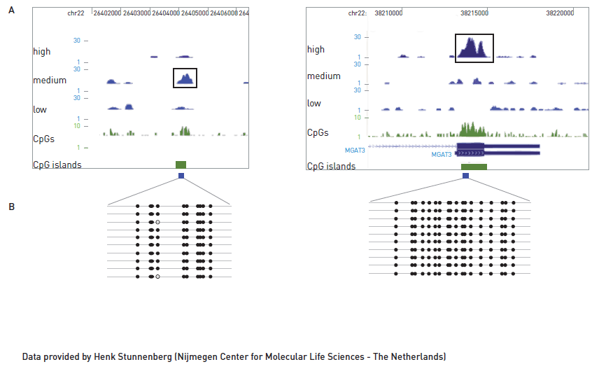
<p><strong></strong><strong>F</strong><strong>igure 1.</strong> Using the MBD approach, two methylated regions were detected in different elution fractions according to their methylated CpG density (A). Low, Medium and High refer to the sequenced DNA from different elution fractions with increasing salt concentration. Methylated patterns of these two different methylated regions were validated by bisulfite conversion assay (B).</p>',
'label2' => '',
'info2' => '',
'label3' => '',
'info3' => '',
'format' => '48 rxns',
'catalog_number' => 'C02020010',
'old_catalog_number' => 'AF-100-0048',
'sf_code' => 'C02020010-',
'type' => 'RFR',
'search_order' => '04-undefined',
'price_EUR' => '740',
'price_USD' => '695',
'price_GBP' => '675',
'price_JPY' => '115920',
'price_CNY' => '',
'price_AUD' => '1738',
'country' => 'ALL',
'except_countries' => 'None',
'quote' => false,
'in_stock' => false,
'featured' => true,
'no_promo' => false,
'online' => true,
'master' => true,
'last_datasheet_update' => '0000-00-00',
'slug' => 'methylcap-kit-x48-48-rxns',
'meta_title' => 'MethylCap kit x48',
'meta_keywords' => '',
'meta_description' => 'MethylCap kit x48',
'modified' => '2021-02-19 17:55:38',
'created' => '2015-06-29 14:08:20',
'locale' => 'jpn'
),
'Antibody' => array(
'host' => '*****',
'id' => null,
'name' => null,
'description' => null,
'clonality' => null,
'isotype' => null,
'lot' => null,
'concentration' => null,
'reactivity' => null,
'type' => null,
'purity' => null,
'classification' => null,
'application_table' => null,
'storage_conditions' => null,
'storage_buffer' => null,
'precautions' => null,
'uniprot_acc' => null,
'slug' => null,
'meta_keywords' => null,
'meta_description' => null,
'modified' => null,
'created' => null,
'select_label' => null
),
'Slave' => array(),
'Group' => array(),
'Related' => array(
(int) 0 => array(
[maximum depth reached]
),
(int) 1 => array(
[maximum depth reached]
),
(int) 2 => array(
[maximum depth reached]
),
(int) 3 => array(
[maximum depth reached]
),
(int) 4 => array(
[maximum depth reached]
),
(int) 5 => array(
[maximum depth reached]
),
(int) 6 => array(
[maximum depth reached]
)
),
'Application' => array(
(int) 0 => array(
[maximum depth reached]
),
(int) 1 => array(
[maximum depth reached]
)
),
'Category' => array(
(int) 0 => array(
[maximum depth reached]
)
),
'Document' => array(
(int) 0 => array(
[maximum depth reached]
),
(int) 1 => array(
[maximum depth reached]
)
),
'Feature' => array(),
'Image' => array(
(int) 0 => array(
[maximum depth reached]
)
),
'Promotion' => array(),
'Protocol' => array(),
'Publication' => array(
(int) 0 => array(
[maximum depth reached]
),
(int) 1 => array(
[maximum depth reached]
),
(int) 2 => array(
[maximum depth reached]
),
(int) 3 => array(
[maximum depth reached]
),
(int) 4 => array(
[maximum depth reached]
),
(int) 5 => array(
[maximum depth reached]
),
(int) 6 => array(
[maximum depth reached]
),
(int) 7 => array(
[maximum depth reached]
),
(int) 8 => array(
[maximum depth reached]
),
(int) 9 => array(
[maximum depth reached]
),
(int) 10 => array(
[maximum depth reached]
),
(int) 11 => array(
[maximum depth reached]
),
(int) 12 => array(
[maximum depth reached]
),
(int) 13 => array(
[maximum depth reached]
),
(int) 14 => array(
[maximum depth reached]
),
(int) 15 => array(
[maximum depth reached]
),
(int) 16 => array(
[maximum depth reached]
),
(int) 17 => array(
[maximum depth reached]
),
(int) 18 => array(
[maximum depth reached]
),
(int) 19 => array(
[maximum depth reached]
),
(int) 20 => array(
[maximum depth reached]
),
(int) 21 => array(
[maximum depth reached]
),
(int) 22 => array(
[maximum depth reached]
),
(int) 23 => array(
[maximum depth reached]
),
(int) 24 => array(
[maximum depth reached]
),
(int) 25 => array(
[maximum depth reached]
),
(int) 26 => array(
[maximum depth reached]
),
(int) 27 => array(
[maximum depth reached]
),
(int) 28 => array(
[maximum depth reached]
),
(int) 29 => array(
[maximum depth reached]
),
(int) 30 => array(
[maximum depth reached]
),
(int) 31 => array(
[maximum depth reached]
),
(int) 32 => array(
[maximum depth reached]
),
(int) 33 => array(
[maximum depth reached]
),
(int) 34 => array(
[maximum depth reached]
),
(int) 35 => array(
[maximum depth reached]
),
(int) 36 => array(
[maximum depth reached]
),
(int) 37 => array(
[maximum depth reached]
)
),
'Testimonial' => array(),
'Area' => array(),
'SafetySheet' => array(
(int) 0 => array(
[maximum depth reached]
),
(int) 1 => array(
[maximum depth reached]
),
(int) 2 => array(
[maximum depth reached]
),
(int) 3 => array(
[maximum depth reached]
),
(int) 4 => array(
[maximum depth reached]
),
(int) 5 => array(
[maximum depth reached]
),
(int) 6 => array(
[maximum depth reached]
),
(int) 7 => array(
[maximum depth reached]
)
)
)
)
$language = 'jp'
$meta_keywords = ''
$meta_description = 'MethylCap kit x48'
$meta_title = 'MethylCap kit x48'
$product = array(
'Product' => array(
'id' => '1887',
'antibody_id' => null,
'name' => 'MethylCap kit',
'description' => '<p>MethylCapキットを用いたアッセイは、ヒトMeCP2タンパク質のメチルCpG結合ドメイン(MBD)を使用したメチル化DNAのアフィニティー精製に基づき、メチル化されたCpGを含むDNAフラグメントを特異的にキャプチャーします。手順は手動プロセスまたは<a href="https://www.diagenode.com/jp/p/sx-8g-ip-star-compact-automated-system-1-unit">IP-Star®</a>コンパクト自動システムの両方で行えます。MBDテクノロジーと<a href="https://www.diagenode.com/jp/p/microplex-lib-prep-kit-v3-48-rxns">MicroPlex Library Preparation Kit v3</a>を組み合わせて、キャプチャーされたメチル化DNAのライブラリーは、次世代シーケンシング(NGS)の解析の準備が整います。</p>
<div id="gtx-trans" style="position: absolute; left: 25px; top: 70px;">
<div class="gtx-trans-icon"></div>
</div>',
'label1' => '特徴',
'info1' => '<ul style="list-style-type: circle;">
<li><strong>Fast & sensitive capture</strong> of methylated DNA</li>
<li><strong>High capture efficiency</strong></li>
<li><strong>Differential fractionation</strong> of methylated DNA by CpG density (3 eluted fractions)</li>
<li><strong>On-day protocol</strong></li>
<li><strong>NGS compatibility</strong></li>
</ul>
<h3>MBD-seq allows for detection of genomic regions with different CpG density</h3>
<p><img src="https://www.diagenode.com/img/product/kits/mbd_results1.png" alt="MBD-sequencing results have been validated by bisulfite sequencing" style="display: block; margin-left: auto; margin-right: auto;" /></p>
<p><strong></strong></p>
<p><strong></strong><strong>F</strong><strong>igure 1.</strong> Using the MBD approach, two methylated regions were detected in different elution fractions according to their methylated CpG density (A). Low, Medium and High refer to the sequenced DNA from different elution fractions with increasing salt concentration. Methylated patterns of these two different methylated regions were validated by bisulfite conversion assay (B).</p>',
'label2' => '',
'info2' => '',
'label3' => '',
'info3' => '',
'format' => '48 rxns',
'catalog_number' => 'C02020010',
'old_catalog_number' => 'AF-100-0048',
'sf_code' => 'C02020010-',
'type' => 'RFR',
'search_order' => '04-undefined',
'price_EUR' => '740',
'price_USD' => '695',
'price_GBP' => '675',
'price_JPY' => '115920',
'price_CNY' => '',
'price_AUD' => '1738',
'country' => 'ALL',
'except_countries' => 'None',
'quote' => false,
'in_stock' => false,
'featured' => true,
'no_promo' => false,
'online' => true,
'master' => true,
'last_datasheet_update' => '0000-00-00',
'slug' => 'methylcap-kit-x48-48-rxns',
'meta_title' => 'MethylCap kit x48',
'meta_keywords' => '',
'meta_description' => 'MethylCap kit x48',
'modified' => '2021-02-19 17:55:38',
'created' => '2015-06-29 14:08:20',
'locale' => 'jpn'
),
'Antibody' => array(
'host' => '*****',
'id' => null,
'name' => null,
'description' => null,
'clonality' => null,
'isotype' => null,
'lot' => null,
'concentration' => null,
'reactivity' => null,
'type' => null,
'purity' => null,
'classification' => null,
'application_table' => null,
'storage_conditions' => null,
'storage_buffer' => null,
'precautions' => null,
'uniprot_acc' => null,
'slug' => null,
'meta_keywords' => null,
'meta_description' => null,
'modified' => null,
'created' => null,
'select_label' => null
),
'Slave' => array(),
'Group' => array(),
'Related' => array(
(int) 0 => array(
'id' => '1888',
'antibody_id' => null,
'name' => 'Auto MethylCap kit',
'description' => '<p>The Auto MethylCap kit allows to specifically capture DNA fragments containing methylated CpGs. The assay is based on the affinity purification of methylated DNA using methyl-CpG-binding domain (MBD) of human MeCP2 protein. This procedure has been optimized to perform automated immunoprecipitation of chromatin using the <a href="https://www.diagenode.com/en/p/sx-8g-ip-star-compact-automated-system-1-unit">IP-Star® Compact Automated System</a> enabling highly reproducible results and allowing for high throughput.</p>',
'label1' => ' Characteristics',
'info1' => '<ul>
<li><strong>Fast & sensitive capture</strong> of methylated DNA</li>
<li><strong>High capture efficiency</strong></li>
<li><strong>Differential fractionation</strong> of methylated DNA by CpG density (3 eluted fractions)</li>
<li><strong>Automation compatibility</strong><strong></strong>
<h3>MBD-seq allows for detection of genomic regions with different CpG density</h3>
<p><img src="https://www.diagenode.com/img/product/kits/mbd_results1.png" alt="MBD-sequencing results have been validated by bisulfite sequencing" style="display: block; margin-left: auto; margin-right: auto;" /></p>
<p><strong>F</strong><strong>igure 1.</strong><span> </span>Using the MBD approach, two methylated regions were detected in different elution fractions according to their methylated CpG density (A). Low, Medium and High refer to the sequenced DNA from different elution fractions with increasing salt concentration. Methylated patterns of these two different methylated regions were validated by bisulfite conversion assay (B).<br /><strong></strong></p>
</li>
</ul>',
'label2' => '',
'info2' => '',
'label3' => '',
'info3' => '',
'format' => '48 rxns',
'catalog_number' => 'C02020011',
'old_catalog_number' => 'AF-Auto01-0048',
'sf_code' => 'C02020011-',
'type' => 'RFR',
'search_order' => '04-undefined',
'price_EUR' => '740',
'price_USD' => '695',
'price_GBP' => '675',
'price_JPY' => '115920',
'price_CNY' => '',
'price_AUD' => '1738',
'country' => 'ALL',
'except_countries' => 'Japan',
'quote' => false,
'in_stock' => false,
'featured' => false,
'no_promo' => false,
'online' => true,
'master' => true,
'last_datasheet_update' => '0000-00-00',
'slug' => 'auto-methylcap-kit-x48-48-rxns',
'meta_title' => 'Auto MethylCap kit x48',
'meta_keywords' => '',
'meta_description' => 'Auto MethylCap kit x48',
'modified' => '2020-09-17 13:33:12',
'created' => '2015-06-29 14:08:20',
'ProductsRelated' => array(
[maximum depth reached]
),
'Image' => array(
[maximum depth reached]
)
),
(int) 1 => array(
'id' => '1922',
'antibody_id' => null,
'name' => 'XL GenDNA Extraction Module',
'description' => '<p><span>This module has been designed for the extraction of <strong>60 µg </strong>of<strong> genomic DNA</strong> from cultured cells. The protocol and reagents allow you to perform <strong>6 extractions</strong> of <strong>10 µg</strong> of genomic DNA per sample, starting with 1 to 1.5 million cells.</span></p>
<p><span>Please check our manual to scale your needs based on your starting material.</span></p>',
'label1' => '',
'info1' => '',
'label2' => '',
'info2' => '',
'label3' => '',
'info3' => '',
'format' => '6 rxns',
'catalog_number' => 'C03030020',
'old_catalog_number' => 'mc-magme-003',
'sf_code' => 'C03030020-',
'type' => 'RFR',
'search_order' => '04-undefined',
'price_EUR' => '115',
'price_USD' => '135',
'price_GBP' => '105',
'price_JPY' => '18015',
'price_CNY' => '',
'price_AUD' => '338',
'country' => 'ALL',
'except_countries' => 'None',
'quote' => false,
'in_stock' => false,
'featured' => false,
'no_promo' => false,
'online' => true,
'master' => true,
'last_datasheet_update' => '0000-00-00',
'slug' => 'xl-gendna-extraction-module-6-rxns',
'meta_title' => 'XL GenDNA Extraction Module',
'meta_keywords' => '',
'meta_description' => 'XL GenDNA Extraction Module',
'modified' => '2019-04-11 14:22:10',
'created' => '2015-06-29 14:08:20',
'ProductsRelated' => array(
[maximum depth reached]
),
'Image' => array(
[maximum depth reached]
)
),
(int) 2 => array(
'id' => '1889',
'antibody_id' => null,
'name' => 'MethylCap protein',
'description' => '<p>The MethylCap protein has been extensively validated for specific isolation of DNA fragments containing methylated CpGs. It consists of the methyl-CpG-binding domain (MBD) of human MeCP2, as a C-terminal fusion with Glutathione-S-transferase (GST) containing an N-terminal His6-tag. A single fully methylated CpG is sufficient for the interaction between the MethylCap protein and methylated DNA fragments.</p>',
'label1' => '',
'info1' => '',
'label2' => '',
'info2' => '',
'label3' => '',
'info3' => '',
'format' => '100 µg',
'catalog_number' => 'C02020012',
'old_catalog_number' => 'mbd-001-100',
'sf_code' => 'C02020012-D001-000526',
'type' => 'FRE',
'search_order' => '04-undefined',
'price_EUR' => '500',
'price_USD' => '565',
'price_GBP' => '460',
'price_JPY' => '78325',
'price_CNY' => '',
'price_AUD' => '1412',
'country' => 'ALL',
'except_countries' => 'None',
'quote' => false,
'in_stock' => false,
'featured' => false,
'no_promo' => false,
'online' => true,
'master' => true,
'last_datasheet_update' => '0000-00-00',
'slug' => 'methylcap-protein-100-ug',
'meta_title' => 'MethylCap protein',
'meta_keywords' => '',
'meta_description' => 'MethylCap protein',
'modified' => '2020-09-17 13:07:33',
'created' => '2015-06-29 14:08:20',
'ProductsRelated' => array(
[maximum depth reached]
),
'Image' => array([maximum depth reached])
),
(int) 3 => array(
'id' => '1819',
'antibody_id' => null,
'name' => 'DiaMag 0.2ml - magnetic rack',
'description' => '<p>The DiaMag02 is a powerful magnet which has been designed for controlled and rapid isolation of your DNA bound to magnetic beads. It allows for processing 16 samples at a time.</p>',
'label1' => '',
'info1' => '',
'label2' => '',
'info2' => '',
'label3' => '',
'info3' => '',
'format' => '1 unit',
'catalog_number' => 'B04000001',
'old_catalog_number' => 'kch-816-001',
'sf_code' => 'B04000001-',
'type' => 'ACC',
'search_order' => '04-undefined',
'price_EUR' => '245',
'price_USD' => '235',
'price_GBP' => '215',
'price_JPY' => '38380',
'price_CNY' => '',
'price_AUD' => '588',
'country' => 'ALL',
'except_countries' => 'None',
'quote' => false,
'in_stock' => false,
'featured' => false,
'no_promo' => false,
'online' => true,
'master' => true,
'last_datasheet_update' => '0000-00-00',
'slug' => 'diamag02-magnetic-rack-1-unit',
'meta_title' => 'DiaMag02 - magnetic rack',
'meta_keywords' => '',
'meta_description' => 'DiaMag02 - magnetic rack',
'modified' => '2019-06-11 16:27:35',
'created' => '2015-06-29 14:08:20',
'ProductsRelated' => array(
[maximum depth reached]
),
'Image' => array(
[maximum depth reached]
)
),
(int) 4 => array(
'id' => '1923',
'antibody_id' => null,
'name' => 'MicroChIP DiaPure columns',
'description' => '<p><a href="https://www.diagenode.com/files/products/reagents/MicroChIP_DiaPure_manual.pdf"><img src="https://www.diagenode.com/img/buttons/bt-manual.png" /></a></p>
<p>Diagenode’s MicroChIP DiaPure columns have been optimized for the purification and elution of very low amounts of DNA. This rapid method has been validated for epigenetic applications like low input ChIP (e.g. using the True MicroChIP kit) and CUT&Tag (e.g. using Diagenode’s pA-Tn5), but is also compatible with many other applications. The DNA can be eluted at high concentrations in volumes down to 6 μl and it is suitable for any downstream application (e.g. NGS).</p>
<p>Benefits of the MicroChIP DiaPure columns:</p>
<ul>
<li>Optimized for the purification of very low DNA amounts</li>
<li>Fast and easy protocol</li>
<li>Non-toxic</li>
<li>Validated for ChIP and Cut&Tag</li>
</ul>',
'label1' => 'Examples of results',
'info1' => '<h2 style="text-align: center;">MicroChIP DiaPure columns after ChIP</h2>
<p>Successful ChIP-seq results generated on 50,000 of K562 cells using True MicroChIP technology. ChIP has been performed accordingly to True MicroChIP protocol (Diagenode, Cat. No. C01010130), including DNA purification using the MicroChIP DiaPure columns. For the library preparation the MicroPlex Library Preparation Kit (Diagenode, Cat. No. C05010001) has been used. The below figure shows the peaks from ChIP-seq experiments using the following Diagenode antibodies: H3K4me1 (C15410194), H3K9/14ac (C15410200), H3K27ac (C15410196) and H3K36me3 (C15410192).</p>
<p><img src="https://www.diagenode.com/img/product/kits/figure-igv-microchip.png" style="display: block; margin-left: auto; margin-right: auto;" /></p>
<p><strong>Figure 1:</strong> Integrative genomics viewer (IGV) visualization of ChIP-seq experiments using 50,000 of K562 cells.</p>
<p></p>
<h2 style="text-align: center;"></h2>
<h2 style="text-align: center;">MicroChIP DiaPure columns after CUT&Tag</h2>
<p>Successful CUT&Tag results showing a low background with high region-specific enrichment has been generated using 50.000 of K562 cells, 1 µg of H3K27me3 antibody (Diagenode, Cat. No. C15410069) and proteinA-Tn5 (1:250) (Diagenode, Cat. No. C01070001). 1 µg of IgG (Diagenode, Cat. No. C15410206) was used as negative control. Samples were purified using the MicroChIP DiaPure columns or phenol-chloroform purification. The below figure presenst the comparison of two purification methods.</p>
<p><img src="https://www.diagenode.com/img/product/kits/figure-diapure-igv.png" style="display: block; margin-left: auto; margin-right: auto;" /></p>
<p><strong>Figure 2:</strong> Integrative genomics viewer (IGV) visualization of CUT&Tag experiments: MicroChIP DiaPure columns vs phenol-chloroform purification using the H3K27me3 antibody.</p>',
'label2' => '',
'info2' => '',
'label3' => '',
'info3' => '',
'format' => '50 rxns',
'catalog_number' => 'C03040001',
'old_catalog_number' => '',
'sf_code' => 'C03040001-',
'type' => 'REF',
'search_order' => '04-undefined',
'price_EUR' => '125',
'price_USD' => '125',
'price_GBP' => '115',
'price_JPY' => '19580',
'price_CNY' => '',
'price_AUD' => '312',
'country' => 'ALL',
'except_countries' => 'None',
'quote' => false,
'in_stock' => false,
'featured' => false,
'no_promo' => false,
'online' => true,
'master' => true,
'last_datasheet_update' => '0000-00-00',
'slug' => 'microchip-diapure-columns-50-rxns',
'meta_title' => 'MicroChIP DiaPure columns',
'meta_keywords' => '',
'meta_description' => 'MicroChIP DiaPure columns',
'modified' => '2023-04-20 16:11:26',
'created' => '2015-06-29 14:08:20',
'ProductsRelated' => array(
[maximum depth reached]
),
'Image' => array([maximum depth reached])
),
(int) 5 => array(
'id' => '2686',
'antibody_id' => null,
'name' => 'IPure kit v2',
'description' => '<p><a href="https://www.diagenode.com/files/products/kits/ipure_kit_v2_manual.pdf"><img src="https://www.diagenode.com/img/buttons/bt-manual.png" /></a></p>
<p>Diagenode’s<span> </span><b>IPure</b><b><span> </span>kit<span> </span></b>is the only DNA purification kit using magnetic beads, that is specifically optimized for extracting DNA from<span> </span><b>ChIP</b><b>,<span> </span></b><b>MeDIP</b><span> </span>and<span> </span><b>CUT&Tag</b>. The use of the magnetic beads allows for a clear separation of DNA and increases therefore the reproducibility of your DNA purification. This simple and straightforward protocol delivers pure DNA ready for any downstream application (e.g. next generation sequencing). Comparing to phenol-chloroform extraction, the IPure technology has the advantage of being nontoxic and much easier to be carried out on multiple samples.</p>
<center>
<h4>High DNA recovery after purification of ChIP samples using IPure technology</h4>
<center><img src="https://www.diagenode.com/img/product/kits/ipure-chromatin-function.png" width="500" /></center>
<p></p>
<p><small>ChIP assays were performed using different amounts of U2OS cells and the H3K9me3 antibody (Cat. No.<span> </span><span>C15410056</span>; 2 g/IP). <span>The purified DNA was eluted in 50 µl of water and quantified with a Nanodrop.</span></small></p>
<p></p>
<p><strong>Benefits of the IPure kit:</strong></p>
<ul>
<li style="text-align: left;">Provides pure DNA for any downstream application (e. g. Next generation sequencing)</li>
<li style="text-align: left;">Non-toxic</li>
<li style="text-align: left;">Fast & easy to use</li>
<li style="text-align: left;">Optimized for DNA purification after ChIP, MeDIP and CUT&Tag</li>
<li style="text-align: left;">Compatible with automation</li>
<li style="text-align: left;">Validated on the IP-Star Compact</li>
</ul>
</center>',
'label1' => 'Examples of results',
'info1' => '<h2>IPure after ChIP</h2>
<p><img src="https://www.diagenode.com/img/product/kits/ideal-TF-chip-seq-A.png" alt="ChIP-seq figure A" style="display: block; margin-left: auto; margin-right: auto;" /></p>
<p><img src="https://www.diagenode.com/img/product/kits/ideal-TF-chip-seq-B.png" alt="ChIP-seq figure B" style="display: block; margin-left: auto; margin-right: auto;" /></p>
<p><img src="https://www.diagenode.com/img/product/kits/ideal-TF-chip-seq-C.png" alt="ChIP-seq figure C" style="display: block; margin-left: auto; margin-right: auto;" /></p>
<p><small><strong>Figure 1.</strong> Chromatin Immunoprecipitation has been performed using chromatin from HeLa cells, the iDeal ChIP-seq kit for Transcription Factors (containing the IPure module for DNA purification) and the Diagenode ChIP-seq-grade HDAC1 (A), LSD1 (B) and p53 antibody (C). The IP'd DNA was subsequently analysed on an Illumina® Genome Analyzer. Library preparation, cluster generation and sequencing were performed according to the manufacturer's instructions. This figure shows the peak distribution in regions of chromosome 3 (A), chromosome 12 (B) and chromosome 6 (C) respectively.</small></p>
<p></p>
<h2>IPure after CUT&Tag</h2>
<p>Successful CUT&Tag results showing a low background with high region-specific enrichment has been generated using 50.000 of K562 cells, 1 µg of H3K4me3 or H3K27me3 antibody (Diagenode, C15410003 or C15410069, respectively) and proteinA-Tn5 (1:250) (Diagenode, C01070001). 1 µg of IgG (C15410206) was used as negative control. Samples were purified using the IPure kit v2 or phenol-chloroform purification. The below figures present the comparison of two purification methods.</p>
<center><img src="https://www.diagenode.com/img/product/kits/ipure-fig2.png" style="display: block; margin-left: auto; margin-right: auto;" width="400" /></center><center>
<p style="text-align: center;"><small><strong>Figure 2.</strong> Heatmap 3kb upstream and downstream of the TSS for H3K4me3</small></p>
</center>
<p></p>
<p><img src="https://www.diagenode.com/img/product/kits/ipure-fig3.png" style="display: block; margin-left: auto; margin-right: auto;" width="600" /></p>
<p></p>
<center><small><strong>Figure 3.</strong> Integrative genomics viewer (IGV) visualization of CUT&Tag experiments using Diagenode’s pA-Tn5 transposase (Cat. No. C01070002), H3K27me3 antibody (Cat. No. C15410069) and IPure kit v2 vs phenol chloroform purification (PC).</small></center>
<p></p>
<p></p>
<h2>IPure after MeDIP</h2>
<center><img src="https://www.diagenode.com/img/product/kits/magmedip-seq-figure_multi3.jpg" alt="medip sequencing coverage" width="600" /></center><center></center><center>
<p></p>
<small><strong>Figure 4.</strong> Consistent coverage and methylation detection from different starting amounts of DNA with the Diagenode MagMeDIP-seq Package (including the Ipure kit for DNA purification). Samples containing decreasing starting amounts of DNA (from the top down: 1000 ng (red), 250 ng (blue), 100 ng (green)) originating from human blood were prepared, revealing a consistent coverage profile for the three different starting amounts, which enables reproducible methylation detection. The CpG islands (CGIs) (marked by yellow boxes in the bottom track) are predominantly unmethylated in the human genome, and as expected, we see a depletion of reads at and around CGIs.</small></center>
<script src="chrome-extension://hhojmcideegachlhfgfdhailpfhgknjm/web_accessible_resources/index.js"></script>',
'label2' => 'iPure Workflow',
'info2' => '<h2 style="text-align: center;">Kit Method Overview & Time table</h2>
<p><img src="https://www.diagenode.com/img/product/kits/workflow-ipure-cuttag.png" style="display: block; margin-left: auto; margin-right: auto;" /></p>
<h3><strong>Workflow description</strong></h3>
<h5><strong>IPure after ChIP</strong></h5>
<p><strong>Step 1:</strong> Chromatin is decrosslinked and eluted from beads (magnetic or agarose) which are discarded. <strong>Magnetic beads</strong> <strong>for purification</strong> are added.<br /> <strong>Step 2:</strong> Magnetic beads acquire positive charge to bind the negatively charged phosphate backbone of DNA. DNA-bead complex is separated using a magnet.<br /> <strong>Step 3:</strong> Proteins and remaining buffer are washed away.<br /> <strong>Step 4:</strong> DNA is eluted from magnetic beads, which are discarded. Purified DNA is ready for any downstream application (NGS, qPCR, amplification, microarray).<br /><br /><br /></p>
<h5><strong>IPure after MeDIP</strong></h5>
<p><strong>Step 1:</strong> DNA is eluted from beads (magnetic or agarose) which are discarded. <strong>Magnetic beads</strong> <strong>for purification</strong> are added. <br /><strong>Step 2:</strong> Magnetic beads acquire positive charge to bind the negatively charged phosphate backbone of DNA. DNA-bead complex is separated using a magnet. <br /><strong>Step 3:</strong> Remaining buffer are washed away.<br /><strong>Step 4:</strong> DNA is eluted from magnetic beads, which are discarded. Purified DNA is ready for any downstream application (NGS, qPCR, amplification, microarray).<br /><br /><br /></p>
<h5><strong>IPure after CUT&Tag</strong></h5>
<p><strong>Step 1:</strong> pA-Tn5 is inactivated and DNA released from the cells. <strong>Magnetic beads</strong> <strong>for purification</strong> are added. <br /><strong>Step 2:</strong> Magnetic beads acquire positive charge to bind the negatively charged phosphate backbone of DNA. DNA-bead complex is separated using a magnet. <br /><strong>Step 3:</strong> Proteins and remaining buffer are washed away. <br /><strong>Step 4:</strong> DNA is eluted from magnetic beads, which are discarded. Purified DNA is ready for any downstream application (NGS, qPCR, amplification, microarray).</p>
<p></p>
<p></p>
<script async="" src="https://edge.fullstory.com/s/fs.js" crossorigin="anonymous"></script>
<script src="chrome-extension://hhojmcideegachlhfgfdhailpfhgknjm/web_accessible_resources/index.js"></script>
<script src="chrome-extension://hhojmcideegachlhfgfdhailpfhgknjm/web_accessible_resources/index.js"></script>',
'label3' => '',
'info3' => '<p></p>
<script src="chrome-extension://hhojmcideegachlhfgfdhailpfhgknjm/web_accessible_resources/index.js"></script>',
'format' => '100 rxns',
'catalog_number' => 'C03010015',
'old_catalog_number' => 'C03010012',
'sf_code' => 'C03010015-',
'type' => 'RFR',
'search_order' => '',
'price_EUR' => '385',
'price_USD' => '445',
'price_GBP' => '350',
'price_JPY' => '60310',
'price_CNY' => '',
'price_AUD' => '1112',
'country' => 'ALL',
'except_countries' => 'None',
'quote' => false,
'in_stock' => false,
'featured' => false,
'no_promo' => false,
'online' => true,
'master' => true,
'last_datasheet_update' => '0000-00-00',
'slug' => 'ipure-kit-v2-x100',
'meta_title' => 'IPure kit v2 | Diagenode',
'meta_keywords' => '',
'meta_description' => 'IPure kit v2',
'modified' => '2023-04-20 16:09:27',
'created' => '2015-09-09 10:52:23',
'ProductsRelated' => array(
[maximum depth reached]
),
'Image' => array([maximum depth reached])
),
(int) 6 => array(
'id' => '3032',
'antibody_id' => null,
'name' => 'MicroPlex Library Preparation Kit v3 /48 rxns',
'description' => '<p><a href="https://www.diagenode.com/files/products/kits/Microplex-library-prep-v3.pdf"><img src="https://www.diagenode.com/img/buttons/bt-manual.png" /></a></p>
<p>Diagenode’s <strong>MicroPlex Library Preparation Kits v3</strong> have been extensively validated for ChIP-seq samples and are optimized to generate DNA libraries with high molecular complexity from the lowest input amounts – down to 50 pg. The kit MicroPlex v3 includes all buffers and enzymes necessary for the library preparation. For flexibility of the choice different formats of compatible primer indexes are available separately:</p>
<ul>
<li><a href="https://www.diagenode.com/en/p/24-dual-indexes-for-microplex-kit-v3-48-rxns">C05010003 - 24 Dual indexes for MicroPlex Kit v3 /48 rxns</a></li>
<li><a href="https://www.diagenode.com/en/p/96-dual-indexes-for-microplex-kit-v3-set-1">C05010004 - 96 Dual indexes for MicroPlex Kit v3 – Set I /96 rxns</a></li>
<li><a href="https://www.diagenode.com/en/p/96-dual-indexes-for-microplex-kit-v3-set-2">C05010005 - 96 Dual indexes for MicroPlex Kit v3 – Set II /96 rxns</a></li>
<li><a href="https://www.diagenode.com/en/p/96-dual-indexes-for-microplex-kit-v3-set-3">C05010006 - 96 Dual indexes for MicroPlex Kit v3 – Set III /96 rxns</a></li>
<li><a href="https://www.diagenode.com/en/p/96-dual-indexes-for-microplex-kit-v3-set-4">C05010007 - 96 Dual indexes for MicroPlex Kit v3 – Set IV /96 rxns</a></li>
</ul>
<p style="padding-left: 30px;">NEW! Unique dual indexes :</p>
<ul>
<li><a href="https://www.diagenode.com/en/p/24-unique-dual-indexes-for-microplex-kit-v3-set1">C05010008 - 24 UDI for MicroPlex Kit v3 - Set I</a></li>
<li><a href="https://www.diagenode.com/en/p/24-unique-dual-indexes-for-microplex-kit-v3-set2">C05010009 - 24 UDI for MicroPlex Kit v3 - Set II</a></li>
</ul>
<p>Read more about <a href="https://www.diagenode.com/en/categories/library-preparation-for-ChIP-seq">library preparation for ChIP-seq</a></p>
<script async="" src="https://edge.fullstory.com/s/fs.js" crossorigin="anonymous"></script>
<script src="chrome-extension://hhojmcideegachlhfgfdhailpfhgknjm/web_accessible_resources/index.js"></script>',
'label1' => 'Characteristics',
'info1' => '<ul>
<li><strong>1 tube</strong>, <strong>2 hours</strong>, <strong>3 steps</strong> protocol</li>
<li><strong>Input</strong>: 50 pg – 50 ng</li>
<li><strong>Reduce potential bias</strong> - few PCR amplification cycles needed</li>
<li><strong>High sensitivity ChIP-seq</strong> - low PCR duplication rate</li>
<li><strong>Great multiplexing flexibility</strong> with 24 dual indexes (8 nt)</li>
<li><strong>Validated with the IP-<a href="https://www.diagenode.com/en/p/sx-8g-ip-star-compact-automated-system-1-unit">Star<sup>®</sup></a></strong><a href="https://www.diagenode.com/en/p/sx-8g-ip-star-compact-automated-system-1-unit"> Automated Platform</a></li>
</ul>
<h3>How it works</h3>
<center><img alt="MicroPlex Library Preparation Kit v3 /48 rxns" src="https://www.diagenode.com/img/product/kits/microplex-3-method-overview.png" /></center>
<p style="margin-bottom: 0;"><small><strong>Microplex workflow - protocol with dual indexes</strong><br />An input of 50 pg to 50 ng of fragmented dsDNA is converted into sequencing-ready libraries for Illumina® NGS platforms using a fast and simple 3-step protocol</small></p>
<ul class="accordion" data-accordion="" id="readmore" style="margin-left: 0;">
<li class="accordion-navigation"><a href="#first" style="background: #ffffff; padding: 0rem; margin: 0rem; color: #13b2a2;"><small>Read more about MicroPlex workflow</small></a>
<div id="first" class="content">
<p><small><strong>Step 1. Template Preparation</strong> provides efficient repair of the fragmented double-stranded DNA input.</small></p>
<p><small>In this step, the DNA is repaired and yields molecules with blunt ends.</small></p>
<p><small><strong>Step 2. Library Synthesis.</strong> enables ligation of MicroPlex patented stem- loop adapters.</small></p>
<p><small>In the next step, stem-loop adaptors with blocked 5’ ends are ligated with high efficiency to the 5’ end of the genomic DNA, leaving a nick at the 3’ end. The adaptors cannot ligate to each other and do not have single- strand tails, both of which contribute to non-specific background found with many other NGS preparations.</small></p>
<p><small><strong>Step 3. Library Amplification</strong> enables extension of the template, cleavage of the stem-loop adaptors, and amplification of the library. Illumina- compatible indexes are also introduced using a high-fidelity, highly- processive, low-bias DNA polymerase.</small></p>
<p><small>In the final step, the 3’ ends of the genomic DNA are extended to complete library synthesis and Illumina-compatible indexes are added through a high-fidelity amplification. Any remaining free adaptors are destroyed. Hands-on time and the risk of contamination are minimized by using a single tube and eliminating intermediate purifications.</small></p>
<p><small>Obtained libraries are purified, quantified and sized. The libraries pooling can be performed as well before sequencing.</small></p>
</div>
</li>
</ul>
<script async="" src="https://edge.fullstory.com/s/fs.js" crossorigin="anonymous"></script>
<script src="chrome-extension://hhojmcideegachlhfgfdhailpfhgknjm/web_accessible_resources/index.js"></script>',
'label2' => '',
'info2' => '<p></p>
<script async="" src="https://edge.fullstory.com/s/fs.js" crossorigin="anonymous"></script>
<script src="chrome-extension://hhojmcideegachlhfgfdhailpfhgknjm/web_accessible_resources/index.js"></script>',
'label3' => '',
'info3' => '<p></p>
<script async="" src="https://edge.fullstory.com/s/fs.js" crossorigin="anonymous"></script>
<script src="chrome-extension://hhojmcideegachlhfgfdhailpfhgknjm/web_accessible_resources/index.js"></script>',
'format' => '48 rxns',
'catalog_number' => 'C05010001',
'old_catalog_number' => '',
'sf_code' => 'C05010001-',
'type' => 'FRE',
'search_order' => '04-undefined',
'price_EUR' => '1750',
'price_USD' => '1555',
'price_GBP' => '1555',
'price_JPY' => '274140',
'price_CNY' => '0',
'price_AUD' => '3888',
'country' => 'ALL',
'except_countries' => 'None',
'quote' => false,
'in_stock' => false,
'featured' => true,
'no_promo' => false,
'online' => true,
'master' => true,
'last_datasheet_update' => '',
'slug' => 'microplex-lib-prep-kit-v3-48-rxns',
'meta_title' => 'MicroPlex Library Preparation Kit v3 /48 rxns | Diagenode',
'meta_keywords' => 'MicroPlex Library Preparation Kit',
'meta_description' => 'MicroPlex Library Preparation Kits v3 have been extensively validated for ChIP-seq samples and are optimized to generate DNA libraries with high molecular complexity from the lowest input amounts – down to 50 pg.',
'modified' => '2023-04-06 12:08:35',
'created' => '2019-07-16 16:03:58',
'ProductsRelated' => array(
[maximum depth reached]
),
'Image' => array(
[maximum depth reached]
)
)
),
'Application' => array(
(int) 0 => array(
'id' => '6',
'position' => '10',
'parent_id' => '1',
'name' => 'メチル化DNA結合タンパク質',
'description' => '<div class="row">
<div class="large-12 columns">MBD方法は、メチル化DNAに対するH6-GST-MBD融合タンパク質の非常に高い親和性に基づいています。 このタンパク質は、N末端His6タグを含むグルタチオン-S-トランスフェラーゼ(GST)とのC末端融合物として、ヒトMeCP2のメチル結合ドメイン(MBD)を含有します。 このH6-GST-MBD融合タンパク質を用いて、メチル化CpGを含むDNAを特異的に単離する事が可能です。<br /><br />DiagenodeのMethylCap®キットは、二本鎖DNAの高濃縮と、メチル化CpG密度の関数における微分分画を可能にします。 分画はサンプルの複雑さを軽減し、次世代のシーケンシングを容易にします。 MethylCapアッセイに先立ち、DNAを最初に抽出し、Picoruptorソニケーターを用いて断片化します。<br />
<h3>概要</h3>
<p class="text-center"><br /><img src="https://www.diagenode.com/img/applications/methyl_binding_domain_overview.jpg" /></p>
</div>
</div>',
'in_footer' => false,
'in_menu' => true,
'online' => true,
'tabular' => false,
'slug' => 'methylbinding-domain-protein',
'meta_keywords' => 'Epigenetic,Methylbinding Domain Protein,MBD,DNA methylation,DNA replication,MethylCap,MethylCap assay,',
'meta_description' => 'Methylbinding Domain Protein(MBD) approach is based on the very high affinity of a H6-GST-MBD fusion protein for methylated DNA. This protein consists of the methyl binding domain (MBD) of human MeCP2, as a C-terminal fusion with Glutathione-S-transferase',
'meta_title' => 'Epigenetic Methylbinding Domain Protein(MBD) - DNA methylation | Diagenode',
'modified' => '2019-03-22 12:32:12',
'created' => '2015-06-02 17:05:42',
'ProductsApplication' => array(
[maximum depth reached]
)
),
(int) 1 => array(
'id' => '1',
'position' => '9',
'parent_id' => null,
'name' => 'DNA Methylation',
'description' => '<div class="row">
<div class="large-12 columns">
<div style="text-align: justify;" class="small-12 medium-8 large-8 columns">
<h2>Complete solutions for DNA methylation studies</h2>
<p>Whether you are experienced or new to the field of DNA methylation, Diagenode has everything you need to make your assay as easy and convenient as possible while ensuring consistent data between samples and experiments. Diagenode offers sonication instruments, reagent kits, high quality antibodies, and high-throughput automation capability to address all of your specific DNA methylation analysis requirements.</p>
</div>
<div class="small-12 medium-4 large-4 columns text-center"><a href="../landing-pages/dna-methylation-grant-applications"><img src="https://www.diagenode.com/img/banners/banner-dna-grant.png" alt="" /></a></div>
<div style="text-align: justify;" class="small-12 medium-12 large-12 columns">
<p>DNA methylation was the first discovered epigenetic mark and is the most widely studied topic in epigenetics. <em>In vivo</em>, DNA is methylated following DNA replication and is involved in a number of biological processes including the regulation of imprinted genes, X chromosome inactivation. and tumor suppressor gene silencing in cancer cells. Methylation often occurs in cytosine-guanine rich regions of DNA (CpG islands), which are commonly upstream of promoter regions.</p>
</div>
<div class="small-12 medium-12 large-12 columns"><br /><br />
<ul class="accordion" data-accordion="">
<li class="accordion-navigation"><a href="#dnamethyl"><i class="fa fa-caret-right"></i> Learn more</a>
<div id="dnamethyl" class="content">5-methylcytosine (5-mC) has been known for a long time as the only modification of DNA for epigenetic regulation. In 2009, however, Kriaucionis discovered a second methylated cytosine, 5-hydroxymethylcytosine (5-hmC). The so-called 6th base, is generated by enzymatic conversion of 5-methylcytosine (5-mC) into 5-hydroxymethylcytosine by the TET family of oxygenases. Early reports suggested that 5-hmC may represent an intermediate of active demethylation in a new pathway which demethylates DNA, converting 5-mC to cytosine. Recent evidence fuel this hypothesis suggesting that further oxidation of the hydroxymethyl group leads to a formyl or carboxyl group followed by either deformylation or decarboxylation. The formyl and carboxyl groups of 5-formylcytosine (5-fC) and 5-carboxylcytosine (5-caC) could be enzymatically removed without excision of the base.
<p class="text-center"><img src="https://www.diagenode.com/img/categories/kits_dna/dna_methylation_variants.jpg" /></p>
</div>
</li>
</ul>
<br />
<h2>Main DNA methylation technologies</h2>
<p style="text-align: justify;">Overview of the <span style="font-weight: 400;">three main approaches for studying DNA methylation.</span></p>
<div class="row">
<ol>
<li style="font-weight: 400;"><span style="font-weight: 400;">Chemical modification with bisulfite – Bisulfite conversion</span></li>
<li style="font-weight: 400;"><span style="font-weight: 400;">Enrichment of methylated DNA (including MeDIP and MBD)</span></li>
<li style="font-weight: 400;"><span style="font-weight: 400;">Treatment with methylation-sensitive or dependent restriction enzymes</span></li>
</ol>
<p><span style="font-weight: 400;"> </span></p>
<div class="row">
<table>
<thead>
<tr>
<th></th>
<th>Description</th>
<th width="350">Features</th>
</tr>
</thead>
<tbody>
<tr>
<td><strong>Bisulfite conversion</strong></td>
<td><span style="font-weight: 400;">Chemical conversion of unmethylated cytosine to uracil. Methylated cytosines are protected from this conversion allowing to determine DNA methylation at single nucleotide resolution.</span></td>
<td>
<ul style="list-style-type: circle;">
<li style="font-weight: 400;"><span style="font-weight: 400;">Single nucleotide resolution</span></li>
<li style="font-weight: 400;"><span style="font-weight: 400;">Quantitative analysis - methylation rate (%)</span></li>
<li style="font-weight: 400;"><span style="font-weight: 400;">Gold standard and well studied</span></li>
<li style="font-weight: 400;"><span style="font-weight: 400;">Compatible with automation</span></li>
</ul>
</td>
</tr>
<tr>
<td><b>Methylated DNA enrichment</b></td>
<td><span style="font-weight: 400;">(Hydroxy-)Methylated DNA is enriched by using specific antibodies (hMeDIP or MeDIP) or proteins (MBD) that specifically bind methylated CpG sites in fragmented genomic DNA.</span></td>
<td>
<ul style="list-style-type: circle;">
<li style="font-weight: 400;"><span style="font-weight: 400;">Resolution depends on the fragment size of the enriched methylated DNA (300 bp)</span></li>
<li style="font-weight: 400;"><span style="font-weight: 400;">Qualitative analysis</span></li>
<li style="font-weight: 400;"><span style="font-weight: 400;">Compatible with automation</span></li>
</ul>
</td>
</tr>
<tr>
<td><strong>Restriction enzyme-based digestion</strong></td>
<td><span style="font-weight: 400;">Use of (hydroxy)methylation-sensitive or (hydroxy)methylation-dependent restriction enzymes for DNA methylation analysis at specific sites.</span></td>
<td>
<ul style="list-style-type: circle;">
<li style="font-weight: 400;"><span style="font-weight: 400;">Determination of methylation status is limited by the enzyme recognition site</span></li>
<li style="font-weight: 400;"><span style="font-weight: 400;">Easy to use</span></li>
</ul>
</td>
</tr>
</tbody>
</table>
</div>
</div>
<div class="row"></div>
</div>
</div>
<div class="large-12 columns"></div>
</div>',
'in_footer' => true,
'in_menu' => true,
'online' => true,
'tabular' => true,
'slug' => 'epigenetics-dna-methylation',
'meta_keywords' => 'Epigenetics, DNA Methylation,5-hmC monoclonal antibody,hMeDIP,Bisulfite conversion,Methylated DNA immunoprecipitation',
'meta_description' => 'Complete, optimized solutions for analyzing DNA methylation manually or on our automated system.',
'meta_title' => 'DNA Methylation - Bisulfite sequencing - Epigenetics | Diagenode',
'modified' => '2019-03-25 10:07:27',
'created' => '2015-05-03 13:47:53',
'ProductsApplication' => array(
[maximum depth reached]
)
)
),
'Category' => array(
(int) 0 => array(
'id' => '53',
'position' => '4',
'parent_id' => '12',
'name' => 'Methylbinding domain protein',
'description' => '<p>The MBD technology used in our <a href="https://www.diagenode.com/en/p/methylcap-kit-x48-48-rxns">MethylCap kit</a> is based on the very high affinity of the <a href="https://www.diagenode.com/en/p/methylcap-protein-100-ug">MethylCap protein</a> for methylated DNA. This protein consists of the methyl-CpG-binding domain (MBD) of human MeCP2, as a C-terminal fusion with Glutathione-S-transferase (GST) containing an N-terminal His6-tag. </p>
<p>Diagenode’s <a href="https://www.diagenode.com/en/p/methylcap-kit-x48-48-rxns">MethylCap kit</a> enables high enrichment of double-stranded DNA and a differential fractionation in function of the methylated CpG density. Fractionation reduces the complexity of samples and makes subsequent next generation sequencing easier. Prior to the MethylCap assay, DNA is first extracted and sheared using the <a href="https://www.diagenode.com/en/p/bioruptorpico2">Bioruptor® sonication device</a>.</p>
<h2>How it works</h2>
<center><img src="https://www.diagenode.com/img/categories/bisulfite-conversion/methyl_binding_domain_overview.jpg" /></center>
<h3 class="diacol">ADVANTAGES</h3>
<ul style="font-size: 19px;" class="nobullet">
<li><i class="fa fa-arrow-circle-right"></i> <strong>Unaffected</strong> DNA</li>
<li><i class="fa fa-arrow-circle-right"></i> <strong>Robust</strong> & <strong>reproducible</strong> technique</li>
<li><i class="fa fa-arrow-circle-right"></i> <strong>NGS</strong> compatible</li>
</ul>',
'no_promo' => false,
'in_menu' => true,
'online' => true,
'tabular' => true,
'hide' => false,
'all_format' => false,
'is_antibody' => false,
'slug' => 'methylbinding-domain-protein-assay',
'cookies_tag_id' => null,
'meta_keywords' => 'Methylbinding domain protein,DNA methylation',
'meta_description' => 'Diagenode's High affinity protein-based isolation of methylated DNA for Methylbinding domain protein assay',
'meta_title' => 'Methylbinding domain protein assay for DNA methylation | Diagenode',
'modified' => '2020-09-17 13:24:55',
'created' => '2015-07-08 09:36:55',
'ProductsCategory' => array(
[maximum depth reached]
),
'CookiesTag' => array([maximum depth reached])
)
),
'Document' => array(
(int) 0 => array(
'id' => '86',
'name' => 'MethylCap kit',
'description' => '<p><span>The MethylCap kit allows to specifically capture DNA fragments containing methylated CpGs. The assay is based on the affinity purification of methylated DNA using methyl-CpG-binding domain (MBD) of human MeCP2 protein. The procedure has been adapted to both manual process or </span><a href="https://www.diagenode.com/en/p/sx-8g-ip-star-compact-automated-system-1-unit">IP-Star® Compact Automated System</a><span>. Libraries of captured methylated DNA can be prepared for next-generation sequencing (NGS) by combining MBD technology with the<span> </span></span><a href="https://www.diagenode.com/en/p/microplex-lib-prep-kit-v3-48-rxns">MicroPlex Library Preparation Kit v3</a><span>.</span></p>',
'image_id' => null,
'type' => 'Manual',
'url' => 'files/products/kits/MethylCap_kit_manual.pdf',
'slug' => 'methylcap-kit-manual',
'meta_keywords' => '',
'meta_description' => '',
'modified' => '2020-09-17 12:56:20',
'created' => '2015-07-07 11:47:43',
'ProductsDocument' => array(
[maximum depth reached]
)
),
(int) 1 => array(
'id' => '102',
'name' => 'Datasheet MethylCap Protein',
'description' => '<p>The MethylCap protein has been extensively validated for specific isolation of DNA fragments containing methylated CpGs. It consists of the methyl-CpG-binding domain (MBD) of human MeCP2, as a C-terminal fusion with Glutathione-S-transferase (GST) containing an N-terminal His6-tag. A single fully methylated CpG is sufficient for the interaction between the MethylCap protein and methylated DNA fragments.</p>',
'image_id' => null,
'type' => 'Datasheet',
'url' => 'files/products/kits/Datasheet_MethylCap_Protein.pdf',
'slug' => 'datasheet-methylcap-protein',
'meta_keywords' => '',
'meta_description' => '',
'modified' => '2020-09-17 13:07:11',
'created' => '2015-07-07 11:47:43',
'ProductsDocument' => array(
[maximum depth reached]
)
)
),
'Feature' => array(),
'Image' => array(
(int) 0 => array(
'id' => '1778',
'name' => 'product/kits/methyl-kit-icon.png',
'alt' => 'Methylation kit icon',
'modified' => '2019-04-23 15:17:01',
'created' => '2018-03-15 15:52:12',
'ProductsImage' => array(
[maximum depth reached]
)
)
),
'Promotion' => array(),
'Protocol' => array(),
'Publication' => array(
(int) 0 => array(
'id' => '4686',
'name' => 'Genome-wide DNA methylation analysis in an antimigraine-treatedpreclinical model of cortical spreading depolarization.',
'authors' => 'Vila-Pueyo M. et al.',
'description' => '<p>BACKGROUND: Cortical spreading depolarization, the cause of migraine aura, is a short-lasting depolarization wave that moves across the brain cortex, transiently suppressing neuronal activity. Prophylactic treatments for migraine, such as topiramate or valproate, reduce the number of cortical spreading depression events in rodents. OBJECTIVE: To investigate whether cortical spreading depolarization with and without chronic treatment with topiramate or valproate affect the DNA methylation of the cortex. METHODS: Sprague-Dawley rats were intraperitoneally injected with saline, topiramate or valproate for four weeks when cortical spreading depolarization were induced and genome-wide DNA methylation was performed in the cortex of six rats per group. RESULTS: The DNA methylation profile of the cortex was significantly modified after cortical spreading depolarization, with and without topiramate or valproate. Interestingly, topiramate reduced by almost 50\% the number of differentially methylated regions, whereas valproate increased them by 17\%, when comparing to the non-treated group after cortical spreading depolarization induction. The majority of the differentially methylated regions lay within intragenic regions, and the analyses of functional group over-representation retrieved several enriched functions, including functions related to protein processing in the cortical spreading depolarization without treatment group; functions related to metabolic processes in the cortical spreading depolarization with topiramate group; and functions related to synapse and ErbB, MAPK or retrograde endocannabinoid signaling in the cortical spreading depolarization with valproate group. CONCLUSIONS: Our results may provide insights into the underlying physiological mechanisms of migraine with aura and emphasize the role of epigenetics in migraine susceptibility.</p>',
'date' => '2023-02-01',
'pmid' => 'https://www.ncbi.nlm.nih.gov/pubmed/36759321',
'doi' => '10.1177/03331024221146317',
'modified' => '2023-04-14 09:15:12',
'created' => '2023-02-28 12:19:11',
'ProductsPublication' => array(
[maximum depth reached]
)
),
(int) 1 => array(
'id' => '4532',
'name' => 'Extra-hematopoietic immunomodulatory role of the guanine-exchange factorDOCK2.',
'authors' => 'Scharler C. et al.',
'description' => '<p>Stromal cells interact with immune cells during initiation and resolution of immune responses, though the precise underlying mechanisms remain to be resolved. Lessons learned from stromal cell-based therapies indicate that environmental signals instruct their immunomodulatory action contributing to immune response control. Here, to the best of our knowledge, we show a novel function for the guanine-exchange factor DOCK2 in regulating immunosuppressive function in three human stromal cell models and by siRNA-mediated DOCK2 knockdown. To identify immune function-related stromal cell molecular signatures, we first reprogrammed mesenchymal stem/progenitor cells (MSPCs) into induced pluripotent stem cells (iPSCs) before differentiating these iPSCs in a back-loop into MSPCs. The iPSCs and immature iPS-MSPCs lacked immunosuppressive potential. Successive maturation facilitated immunomodulation, while maintaining clonogenicity, comparable to their parental MSPCs. Sequential transcriptomics and methylomics displayed time-dependent immune-related gene expression trajectories, including DOCK2, eventually resembling parental MSPCs. Severe combined immunodeficiency (SCID) patient-derived fibroblasts harboring bi-allelic DOCK2 mutations showed significantly reduced immunomodulatory capacity compared to non-mutated fibroblasts. Conditional DOCK2 siRNA knockdown in iPS-MSPCs and fibroblasts also immediately reduced immunomodulatory capacity. Conclusively, CRISPR/Cas9-mediated DOCK2 knockout in iPS-MSPCs also resulted in significantly reduced immunomodulation, reduced CDC42 Rho family GTPase activation and blunted filopodia formation. These data identify G protein signaling as key element devising stromal cell immunomodulation.</p>',
'date' => '2022-11-01',
'pmid' => 'https://www.ncbi.nlm.nih.gov/pubmed/36380073',
'doi' => '10.1038/s42003-022-04078-1',
'modified' => '2022-11-24 08:56:01',
'created' => '2022-11-24 08:49:52',
'ProductsPublication' => array(
[maximum depth reached]
)
),
(int) 2 => array(
'id' => '4434',
'name' => 'Genome-wide DNA hypermethylation opposes healing in chronic woundpatients by impairing epithelial-to-mesenchymal transition.',
'authors' => 'Singh Kanhaiya et al.',
'description' => '<p>An extreme chronic wound tissue microenvironment causes epigenetic gene silencing. Unbiased whole-genome methylome was studied in the wound-edge (WE) tissue of chronic wound patients. A total of 4689 differentially methylated regions (DMRs) were identified in chronic WE compared to unwounded (UW) human skin. Hypermethylation was more frequently observed (3661 DMRs) in the chronic WE compared to hypomethylation (1028 DMRs). Twenty-six hypermethylated DMRs were involved in epithelial to mesenchymal transition (EMT). Bisulfite sequencing validated hypermethylation of a predicted specific upstream regulator TP53. RNA sequencing analysis was performed to qualify findings from methylome analysis. Analysis of the downregulated genes identified the TP53 signaling pathway as being significantly silenced. Direct comparison of hypermethylation and downregulated genes identified four genes, ADAM17, NOTCH, TWIST1 and SMURF1, that functionally represent the EMT pathway. Single-cell RNA sequencing studies identified that these effects on gene expression were limited to the keratinocyte cell compartment. Experimental murine studies established that tissue ischemia potently induces WE gene methylation and that 5'-azacytidine, inhibitor of methylation, improved wound closure. To specifically address the significance of TP53 methylation, keratinocyte-specific editing of TP53 methylation at the WE was achieved by a tissue nanotransfection (TNT) based CRISPR/dCas9 approach. This work identified that reversal of methylation-dependent keratinocyte gene-silencing represents a productive therapeutic strategy to improve wound closure.</p>',
'date' => '2022-07-01',
'pmid' => 'https://pubmed.ncbi.nlm.nih.gov/35819852/',
'doi' => '10.1172/JCI157279',
'modified' => '2022-09-28 09:15:04',
'created' => '2022-09-08 16:32:20',
'ProductsPublication' => array(
[maximum depth reached]
)
),
(int) 3 => array(
'id' => '4215',
'name' => 'Cancer Detection and Classification by CpG Island Hypermethylation Signatures in Plasma Cell-Free DNA',
'authors' => 'Huang J, Soupir AC, Schlick BD, Teng M, Sahin IH, Permuth JB, Siegel EM, Manley BJ, Pellini B, Wang L.',
'description' => '<p><span>Cell-free DNA (cfDNA) methylation has emerged as a promising biomarker for early cancer detection, tumor type classification, and treatment response monitoring. Enrichment-based cfDNA methylation profiling methods such as cfMeDIP-seq have shown high accuracy in the classification of multiple cancer types. We have previously optimized another enrichment-based approach for ultra-low input cfDNA methylome profiling, termed cfMBD-seq. We reported that cfMBD-seq outperforms cfMeDIP-seq in the enrichment of high-CpG-density regions, such as CpG islands. However, the clinical feasibility of cfMBD-seq is unknown. In this study, we applied cfMBD-seq to profiling the cfDNA methylome using plasma samples from cancer patients and non-cancer controls. We identified 1759, 1783, and 1548 differentially hypermethylated CpG islands (DMCGIs) in lung, colorectal, and pancreatic cancer patients, respectively. Interestingly, the vast majority of DMCGIs were overlapped with aberrant methylation changes in corresponding tumor tissues, indicating that DMCGIs detected by cfMBD-seq were mainly driven by tumor-specific DNA methylation patterns. From the overlapping DMCGIs, we carried out machine learning analyses and identified a set of discriminating methylation signatures that had robust performance in cancer detection and classification. Overall, our study demonstrates that cfMBD-seq is a powerful tool for sensitive detection of tumor-derived epigenomic signals in cfDNA.</span></p>',
'date' => '2021-11-21',
'pmid' => 'https://pubmed.ncbi.nlm.nih.gov/34830765/',
'doi' => '10.3390/cancers13225611',
'modified' => '2022-03-17 10:01:55',
'created' => '2022-03-17 10:01:55',
'ProductsPublication' => array(
[maximum depth reached]
)
),
(int) 4 => array(
'id' => '4287',
'name' => 'Transcriptome and Methylome Analysis Reveal ComplexCross-Talks between Thyroid Hormone and GlucocorticoidSignaling at Xenopus Metamorphosis.',
'authors' => 'Buisine Nicolas et al.',
'description' => '<p>BACKGROUND: Most work in endocrinology focus on the action of a single hormone, and very little on the cross-talks between two hormones. Here we characterize the nature of interactions between thyroid hormone and glucocorticoid signaling during metamorphosis. METHODS: We used functional genomics to derive genome wide profiles of methylated DNA and measured changes of gene expression after hormonal treatments of a highly responsive tissue, tailfin. Clustering classified the data into four types of biological responses, and biological networks were modeled by system biology. RESULTS: We found that gene expression is mostly regulated by either T or CORT, or their additive effect when they both regulate the same genes. A small but non-negligible fraction of genes (12\%) displayed non-trivial regulations indicative of complex interactions between the signaling pathways. Strikingly, DNA methylation changes display the opposite and are dominated by cross-talks. CONCLUSION: Cross-talks between thyroid hormones and glucocorticoids are more complex than initially envisioned and are not limited to the simple addition of their individual effects, a statement that can be summarized with the pseudo-equation: TH GC > TH + GC. DNA methylation changes are highly dynamic and buffered from genome expression.</p>',
'date' => '2021-09-01',
'pmid' => 'https://www.ncbi.nlm.nih.gov/pubmed/34572025',
'doi' => '10.3390/cells10092375',
'modified' => '2022-05-24 09:12:29',
'created' => '2022-05-19 10:41:50',
'ProductsPublication' => array(
[maximum depth reached]
)
),
(int) 5 => array(
'id' => '4104',
'name' => 'Cell-free DNA methylome profiling by MBD-seq with ultra-low input',
'authors' => 'Jinyong Huang, Alex C. Soupir & Liang Wang',
'description' => '<p><span>Methylation signatures in cell-free DNA (cfDNA) have shown great sensitivity and specificity in the characterization of tumour status and classification of tumour types, as well as the response to therapy and recurrence. Currently, most cfDNA methylation studies are based on bisulphite conversion, especially targeted bisulphite sequencing, while enrichment-based methods such as cfMeDIP-seq are beginning to show potential. Here, we report an enrichment-based ultra-low input cfDNA methylation profiling method using methyl-CpG binding proteins capture, termed cfMBD-seq. We optimized the conditions for cfMBD capture by adjusting the amount of MethylCap protein along with using methylated filler DNA. Our data show high correlation between low input cfMBD-seq and standard MBD-seq (>1000 ng input). When compared to cfMEDIP-seq, cfMBD-seq demonstrates higher sequencing data quality with more sequenced reads passed filter and less duplicate rate. cfMBD-seq also outperforms cfMeDIP-seq in the enrichment of CpG islands. This new bisulphite-free ultra-low input methylation profiling technology has great potential in non-invasive and cost-effective cancer detection and classification.</span></p>',
'date' => '2021-03-16',
'pmid' => 'https://doi.org/10.1080/15592294.2021.1896984',
'doi' => '10.1080/15592294.2021.1896984',
'modified' => '2021-06-28 15:00:36',
'created' => '2021-06-28 15:00:36',
'ProductsPublication' => array(
[maximum depth reached]
)
),
(int) 6 => array(
'id' => '4194',
'name' => 'Genome-wide DNA methylation and RNA-seq analyses identify genes andpathways associated with doxorubicin resistance in a canine diffuse largeB-cell lymphoma cell line.',
'authors' => 'Hsu, C.-H. et al.',
'description' => '<p>Doxorubicin resistance is a major challenge in the successful treatment of canine diffuse large B-cell lymphoma (cDLBCL). In the present study, MethylCap-seq and RNA-seq were performed to characterize the genome-wide DNA methylation and differential gene expression patterns respectively in CLBL-1 8.0, a doxorubicin-resistant cDLBCL cell line, and in CLBL-1 as control, to investigate the underlying mechanisms of doxorubicin resistance in cDLBCL. A total of 20289 hypermethylated differentially methylated regions (DMRs) were detected. Among these, 1339 hypermethylated DMRs were in promoter regions, of which 24 genes showed an inverse correlation between methylation and gene expression. These 24 genes were involved in cell migration, according to gene ontology (GO) analysis. Also, 12855 hypermethylated DMRs were in gene-body regions. Among these, 353 genes showed a positive correlation between methylation and gene expression. Functional analysis of these 353 genes highlighted that TGF-β and lysosome-mediated signal pathways are significantly associated with the drug resistance of CLBL-1. The tumorigenic role of TGF-β signaling pathway in CLBL-1 8.0 was further validated by treating the cells with a TGF-β inhibitor(s) to show the increased chemo-sensitivity and intracellular doxorubicin accumulation, as well as decreased p-glycoprotein expression. In summary, the present study performed an integrative analysis of DNA methylation and gene expression in CLBL-1 8.0 and CLBL-1. The candidate genes and pathways identified in this study hold potential promise for overcoming doxorubicin resistance in cDLBCL.</p>',
'date' => '2021-01-01',
'pmid' => 'https://pubmed.ncbi.nlm.nih.gov/33961622/',
'doi' => '10.1371/journal.pone.0250013',
'modified' => '2022-01-06 14:24:18',
'created' => '2021-12-06 15:53:19',
'ProductsPublication' => array(
[maximum depth reached]
)
),
(int) 7 => array(
'id' => '4063',
'name' => 'Genome-wide DNA methylation analysis using MethylCap-seq in caninehigh-grade B-cell lymphoma.',
'authors' => 'Hsu, Chia-Hsin and Tomiyasu, Hirotaka and Lee, Jih-Jong and Tung, Chun-Weiand Liao, Chi-Hsun and Chuang, Cheng-Hsun and Huang, Ling-Ya and Liao,Kuang-Wen and Chou, Chung-Hsi and Liao, Albert T C and Lin, Chen-Si',
'description' => '<p>DNA methylation is a comprehensively studied epigenetic modification and plays crucial roles in cancer development. In the present study, MethylCap-seq was used to characterize the genome-wide DNA methylation patterns in canine high-grade B-cell lymphoma (cHGBL). Canine methylated DNA fragments were captured and the MEDIUM-HIGH and LOW fraction of methylated DNA was obtained based on variation in CpG methylation density. In the MEDIUM-HIGH and LOW fraction, 2144 and 1987 cHGBL-specific hypermethylated genes, respectively, were identified. Functional analysis highlighted pathways strongly related to oncogenesis. The relevant signaling pathways associated with neuronal system were also revealed, echoing recent novel findings that neurogenesis plays key roles in tumor establishment. In addition, 14 genes were hypermethylated in all the cHGBL cases but not in the healthy dogs. These genes might be potential signatures for tracing cHGBL, and some of them have been reported to play roles in various types of cancers. Further, the distinct methylation pattern of cHGBL showed a concordance with the clinical outcome, suggesting that aberrant epigenetic changes may influence tumor behavior. In summary, our study characterized genome-wide DNA methylation patterns using MethylCap-seq in cHGBL; the findings suggest that specific DNA hypermethylation holds promise for dissecting tumorigenesis and uncovering biomarkers for monitoring the progression of cHGBL.</p>',
'date' => '2020-10-01',
'pmid' => 'https://www.ncbi.nlm.nih.gov/pubmed/33031589',
'doi' => '10.1002/JLB.2A0820-673R',
'modified' => '2021-02-19 17:42:07',
'created' => '2021-02-18 10:21:53',
'ProductsPublication' => array(
[maximum depth reached]
)
),
(int) 8 => array(
'id' => '4070',
'name' => 'Benchmarking DNA methylation assays in a reef-building coral.',
'authors' => 'Dixon, Groves and Matz, Mikhail',
'description' => '<p>Interrogation of chromatin modifications, such as DNA methylation, has the potential to improve forecasting and conservation of marine ecosystems. The standard method for assaying DNA methylation (whole genome bisulphite sequencing), however, is currently too costly to apply at the scales required for ecological research. Here, we evaluate different methods for measuring DNA methylation for ecological epigenetics. We compare whole genome bisulphite sequencing (WGBS) with methylated CpG binding domain sequencing (MBD-seq), and a modified version of MethylRAD we term methylation-dependent restriction site-associated DNA sequencing (mdRAD). We evaluate these three assays in measuring variation in methylation across the genome, between genotypes, and between polyp types in the reef-building coral Acropora millepora. We find that all three assays measure absolute methylation levels similarly for gene bodies (gbM), as well as exons and 1 Kb windows with a minimum Pearson correlation 0.66. Differential gbM estimates were less correlated, but still concurrent across assays. We conclude that MBD-seq and mdRAD are reliable and cost-effective alternatives to WGBS. The considerably lower sequencing effort required for mdRAD to produce comparable methylation estimates makes it particularly useful for ecological epigenetics.</p>',
'date' => '2020-10-01',
'pmid' => 'https://www.ncbi.nlm.nih.gov/pubmed/33058551',
'doi' => '10.1111/1755-0998.13282',
'modified' => '2021-02-19 17:56:00',
'created' => '2021-02-18 10:21:53',
'ProductsPublication' => array(
[maximum depth reached]
)
),
(int) 9 => array(
'id' => '4015',
'name' => 'Targeting DNA methylation depletes uterine leiomyoma stem-cell enrichedpopulation by stimulating their differentiation.',
'authors' => 'Liu, S and Yin, P and Xu, J and Dotts, AJ and Kujawa, SA and Coon, VJS and Zhao, H and Hilatifard, AS and Dai, Y and Bulun, SE',
'description' => '<p>Uterine leiomyoma is the most common tumor in women and can cause severe morbidity. Leiomyoma growth requires maintenance and proliferation of a stem cell population. Dysregulated DNA methylation has been reported in leiomyoma, but its role in leiomyoma stem cell regulation remains unclear. Here, we FACS sorted cells from human leiomyoma tissues into three populations: stem-cell like cells (LSC, 5%), intermediate cells (LIC, 7%), and differentiated cells (LDC, 88%) and analyzed the transcriptome and epigenetic landscape of leiomyoma cells at different differentiation stages. LSC harbored a unique methylome, with 8862 differentially methylated regions compared to LIC and 9444 compared to LDC, most of which were hypermethylated. Consistent with global hypermethylation, transcript levels of TET1 and TET3 methylcytosine dioxygenases were lower in LSC. Integrative analyses revealed an inverse relationship between methylation and gene expression changes during LSC differentiation. In LSC, hypermethylation suppressed genes important for myometrium- and leiomyoma-associated functions, including muscle contraction and hormone action, to maintain stemness. The hypomethylating drug, 5'-Aza stimulated LSC differentiation, depleting the stem cell population and inhibiting tumor initiation. Our data suggest that DNA methylation maintains the pool of LSC, which is critical for the regeneration of leiomyoma tumors.</p>',
'date' => '2020-08-19',
'pmid' => 'http://www.pubmed.gov/32812024',
'doi' => '10.1210/endocr/bqaa143/5894164',
'modified' => '2020-12-16 17:35:05',
'created' => '2020-10-12 14:54:59',
'ProductsPublication' => array(
[maximum depth reached]
)
),
(int) 10 => array(
'id' => '3973',
'name' => 'DNA methylation dynamics underlie metamorphic gene regulation programs in Xenopus tadpole brain.',
'authors' => 'Kyono Y, Raj S, Sifuentes CJ, Buisine N, Sachs L, Denver RJ',
'description' => '<p>Methylation of cytosine residues in DNA influences chromatin structure and gene transcription, and its regulation is crucial for brain development. There is mounting evidence that DNA methylation can be modulated by hormone signaling. We analyzed genome-wide changes in DNA methylation and their relationship to gene regulation in the brain of Xenopus tadpoles during metamorphosis, a thyroid hormone-dependent developmental process. We studied the region of the tadpole brain containing neurosecretory neurons that control pituitary hormone secretion, a region that is highly responsive to thyroid hormone action. Using Methylated DNA Capture sequencing (MethylCap-seq) we discovered a diverse landscape of DNA methylation across the tadpole neural cell genome, and pairwise stage comparisons identified several thousand differentially methylated regions (DMRs). During the pre-to pro-metamorphic period, the number of DMRs was lowest (1,163), with demethylation predominating. From pre-metamorphosis to metamorphic climax DMRs nearly doubled (2,204), with methylation predominating. The largest changes in DNA methylation were seen from metamorphic climax to the completion of metamorphosis (2960 DMRs), with 80% of the DMRs representing demethylation. Using RNA sequencing, we found negative correlations between differentially expressed genes and DMRs localized to gene bodies and regions upstream of transcription start sites. DNA demethylation at metamorphosis revealed by MethylCap-seq was corroborated by increased immunoreactivity for the DNA demethylation intermediates 5-hydroxymethylcytosine and 5-carboxymethylcytosine, and the methylcytosine dioxygenase ten eleven translocation 3 that catalyzes DNA demethylation. Our findings show that the genome of tadpole neural cells undergoes significant changes in DNA methylation during metamorphosis, and these changes likely influence chromatin architecture, and gene regulation programs occurring during this developmental period.</p>',
'date' => '2020-06-15',
'pmid' => 'http://www.pubmed.gov/32240642',
'doi' => '10.1016/j.ydbio.2020.03.013',
'modified' => '2020-08-12 09:26:12',
'created' => '2020-08-10 12:12:25',
'ProductsPublication' => array(
[maximum depth reached]
)
),
(int) 11 => array(
'id' => '3849',
'name' => 'SMaSH: Sample matching using SNPs in humans.',
'authors' => 'Westphal M, Frankhouser D, Sonzone C, Shields PG, Yan P, Bundschuh R',
'description' => '<p>BACKGROUND: Inadvertent sample swaps are a real threat to data quality in any medium to large scale omics studies. While matches between samples from the same individual can in principle be identified from a few well characterized single nucleotide polymorphisms (SNPs), omics data types often only provide low to moderate coverage, thus requiring integration of evidence from a large number of SNPs to determine if two samples derive from the same individual or not. METHODS: We select about six thousand SNPs in the human genome and develop a Bayesian framework that is able to robustly identify sample matches between next generation sequencing data sets. RESULTS: We validate our approach on a variety of data sets. Most importantly, we show that our approach can establish identity between different omics data types such as Exome, RNA-Seq, and MethylCap-Seq. We demonstrate how identity detection degrades with sample quality and read coverage, but show that twenty million reads of a fairly low quality RNA-Seq sample are still sufficient for reliable sample identification. CONCLUSION: Our tool, SMASH, is able to identify sample mismatches in next generation sequencing data sets between different sequencing modalities and for low quality sequencing data.</p>',
'date' => '2019-12-30',
'pmid' => 'http://www.pubmed.gov/31888490',
'doi' => '10.1186/s12864-019-6332-7',
'modified' => '2020-02-13 13:59:11',
'created' => '2020-02-13 10:02:44',
'ProductsPublication' => array(
[maximum depth reached]
)
),
(int) 12 => array(
'id' => '3836',
'name' => 'Increased presence and differential molecular imprinting of transit amplifying cells in psoriasis.',
'authors' => 'Witte K, Jürchott K, Christou D, Hecht J, Salinas G, Krüger U, Klein O, Kokolakis G, Witte-Händel E, Mössner R, Volk HD, Wolk K, Sabat R',
'description' => '<p>Psoriasis is a very common chronic inflammatory skin disease characterized by epidermal thickening and scaling resulting from keratinocyte hyperproliferation and impaired differentiation. Pathomechanistic studies in psoriasis are often limited by using whole skin tissue biopsies, neglecting their stratification and cellular diversity. This study aimed at characterizing epidermal alterations in psoriasis at the level of keratinocyte populations. Epidermal cell populations were purified from skin biopsies of psoriasis patients and healthy donors using a novel cell type-specific approach. Molecular characterization of the transit-amplifying cells (TAC), the key players of epidermal renewal, was performed using immunocytofluorescence-technique and integrated multiscale-omics analyses. Already TAC from non-lesional psoriatic skin showed altered methylation and differential expression in 1.7% and 1.0% of all protein-coding genes, respectively. In psoriatic lesions, TAC were strongly expanded showing further increased differentially methylated (10-fold) and expressed (22-fold) genes numbers. Importantly, 17.2% of differentially expressed genes were associated with respective gene methylations. Compared with non-lesional TAC, pathway analyses revealed metabolic alterations as one feature predominantly changed in TAC derived from active psoriatic lesions. Overall, our study showed stage-specific molecular alterations, allows new insights into the pathogenesis, and implies the involvement of epigenetic mechanisms in lesion development in psoriasis. KEY MESSAGES: Transit amplifying cell (TAC) numbers are highly increased in psoriatic lesions Psoriatic TAC show profound molecular alterations & stage-specific identity TAC from unaffected areas already show first signs of molecular alterations Lesional TAC show a preference in metabolic-related alterations.</p>',
'date' => '2019-12-12',
'pmid' => 'http://www.pubmed.gov/31832701',
'doi' => '10.1007/s00109-019-01860-3',
'modified' => '2020-02-25 13:23:26',
'created' => '2020-02-13 10:02:44',
'ProductsPublication' => array(
[maximum depth reached]
)
),
(int) 13 => array(
'id' => '3815',
'name' => 'Plasticity of histone modifications around Cidea and Cidec genes with secondary bile in the amelioration of developmentally-programmed hepatic steatosis.',
'authors' => 'Urmi JF, Itoh H, Muramatsu-Kato K, Kohmura-Kobayashi Y, Hariya N, Jain D, Tamura N, Uchida T, Suzuki K, Ogawa Y, Shiraki N, Mochizuki K, Kubota T, Kanayama N',
'description' => '<p>We recently reported that a treatment with tauroursodeoxycholic acid (TUDCA), a secondary bile acid, improved developmentally-deteriorated hepatic steatosis in an undernourishment (UN, 40% caloric restriction) in utero mouse model after a postnatal high-fat diet (HFD). We performed a microarray analysis and focused on two genes (Cidea and Cidec) because they are enhancers of lipid droplet (LD) sizes in hepatocytes and showed the greatest up-regulation in expression by UN that were completely recovered by TUDCA, concomitant with parallel changes in LD sizes. TUDCA remodeled developmentally-induced histone modifications (dimethylation of H3K4, H3K27, or H3K36), but not DNA methylation, around the Cidea and Cidec genes in UN pups only. Changes in these histone modifications may contribute to the markedly down-regulated expression of Cidea and Cidec genes in UN pups, which was observed in the alleviation of hepatic fat deposition, even under HFD. These results provide an insight into the future of precision medicine for developmentally-programmed hepatic steatosis by targeting histone modifications.</p>',
'date' => '2019-11-19',
'pmid' => 'http://www.pubmed.gov/31745102',
'doi' => '10.1038/s41598-019-52943-7',
'modified' => '2019-12-05 10:57:34',
'created' => '2019-12-02 15:25:44',
'ProductsPublication' => array(
[maximum depth reached]
)
),
(int) 14 => array(
'id' => '4034',
'name' => 'Role of gene body methylation in acclimatization and adaptation in a basalmetazoan.',
'authors' => 'Dixon, Groves and Liao, Yi and Bay, Line K and Matz, Mikhail V',
'description' => '<p>Gene body methylation (GBM) has been hypothesized to modulate responses to environmental change, including transgenerational plasticity, but the evidence thus far has been lacking. Here we show that coral fragments reciprocally transplanted between two distant reefs respond predominantly by increase or decrease in genome-wide GBM disparity: The range of methylation levels between lowly and highly methylated genes becomes either wider or narrower. Remarkably, at a broad functional level this simple adjustment correlated very well with gene expression change, reflecting a shifting balance between expressions of environmentally responsive and housekeeping genes. In our experiment, corals in a lower-quality habitat up-regulated genes involved in environmental responses, while corals in a higher-quality habitat invested more in housekeeping genes. Transplanted fragments showing closer GBM match to local corals attained higher fitness characteristics, which supports GBM's role in acclimatization. Fixed differences in GBM between populations did not align with plastic GBM changes and were mostly observed in genes with elevated , which suggests that they arose predominantly through genetic divergence. However, we cannot completely rule out transgenerational inheritance of acquired GBM states.</p>',
'date' => '2018-12-01',
'pmid' => 'https://www.ncbi.nlm.nih.gov/pubmed/30530646',
'doi' => '10.1073/pnas.1813749115',
'modified' => '2021-02-18 17:09:00',
'created' => '2021-02-18 10:21:53',
'ProductsPublication' => array(
[maximum depth reached]
)
),
(int) 15 => array(
'id' => '3482',
'name' => 'DNA Methylation and Regulatory Elements during Chicken Germline Stem Cell Differentiation.',
'authors' => 'He Y, Zuo Q, Edwards J, Zhao K, Lei J, Cai W, Nie Q, Li B, Song J',
'description' => '<p>The production of germ cells in vitro would open important new avenues for stem biology and human medicine, but the mechanisms of germ cell differentiation are not well understood. The chicken, as a great model for embryology and development, was used in this study to help us explore its regulatory mechanisms. In this study, we reported a comprehensive genome-wide DNA methylation landscape in chicken germ cells, and transcriptomic dynamics was also presented. By uncovering DNA methylation patterns on individual genes, some genes accurately modulated by DNA methylation were found to be associated with cancers and virus infection, e.g., AKT1 and CTNNB1. Chicken-unique markers were also discovered for identifying male germ cells. Importantly, integrated epigenetic mechanisms were explored during male germ cell differentiation, which provides deep insight into the epigenetic processes associated with male germ cell differentiation and possibly improves treatment options to male infertility in animals and humans.</p>',
'date' => '2018-06-05',
'pmid' => 'http://www.pubmed.gov/29681542',
'doi' => '10.1016/j.stemcr.2018.03.018',
'modified' => '2019-02-14 17:09:47',
'created' => '2019-02-14 15:01:22',
'ProductsPublication' => array(
[maximum depth reached]
)
),
(int) 16 => array(
'id' => '3354',
'name' => 'Antioxydation And Cell Migration Genes Are Identified as Potential Therapeutic Targets in Basal-Like and BRCA1 Mutated Breast Cancer Cell Lines',
'authors' => 'Privat M. et al.',
'description' => '<p>Basal-like breast cancers are among the most aggressive cancers and effective targeted therapies are still missing. In order to identify new therapeutic targets, we performed Methyl-Seq and RNA-Seq of 10 breast cancer cell lines with different phenotypes. We confirmed that breast cancer subtypes cluster the RNA-Seq data but not the Methyl-Seq data. Basal-like tumor hypermethylated phenotype was not confirmed in our study but RNA-Seq analysis allowed to identify 77 genes significantly overexpressed in basal-like breast cancer cell lines. Among them, 48 were overexpressed in triple negative breast cancers of TCGA data. Some molecular functions were overrepresented in this candidate gene list. Genes involved in antioxydation, such as SOD1, MGST3 and PRDX or cadherin-binding genes, such as PFN1, ITGB1 and ANXA1, could thus be considered as basal like breast cancer biomarkers. We then sought if these genes were linked to BRCA1, since this gene is often inactivated in basal-like breast cancers. Nine genes were identified overexpressed in both basal-like breast cancer cells and BRCA1 mutated cells. Amongst them, at least 3 genes code for proteins implicated in epithelial cell migration and epithelial to mesenchymal transition (VIM, ITGB1 and RhoA). Our study provided several potential therapeutic targets for triple negative and BRCA1 mutated breast cancers. It seems that migration and mesenchymal properties acquisition of basal-like breast cancer cells is a key functional pathway in these tumors with a high metastatic potential.</p>',
'date' => '2018-01-01',
'pmid' => 'https://www.ncbi.nlm.nih.gov/pubmed/29333087',
'doi' => '',
'modified' => '2018-04-05 11:37:25',
'created' => '2018-04-05 11:37:25',
'ProductsPublication' => array(
[maximum depth reached]
)
),
(int) 17 => array(
'id' => '3654',
'name' => 'Gene body methylation is involved in coarse genome-wide adjustment of gene expression during acclimatization',
'authors' => 'Groves Dixon1, Yi Liao1, Line K. Bay2 and Mikhail V. Matz',
'description' => '<p>Gene body methylation (GBM) is a taxonomically widespread epigenetic modification of the DNA the function of which remains unclear 1,2. GBM is bimodally distributed among genes: it is high in ubiquitously expressed housekeeping genes and low in context-dependent inducible genes 2,3, and it has been hypothesized that changes in GBM might modulate responses to environmental change, including transgenerational plasticity 4,5. Here, we profiled GBM, gene expression, genotype, and fitness characteristics in clonal fragments of a reef building coral Acropora millepora reciprocally transplanted between two distant reefs. We find that genotype-specific GBM is considerably more stable than gene expression and responds to transplantation predominantly by genome-wide increase or decrease in disparity of methylation levels among genes. A proxy of this change, GBM difference between the two gene classes (housekeeping vs. inducible), was the most important determinant of genomewide GBM variation in our experiment, explaining 33% of it. Surprisingly, despite apparent lack of capacity for environmental specificity, this simple genome-wide GBM adjustment was a good predictor of broad-scale functional shifts in gene expression and of fragments’ fitness in the new environment, which supports GBM’s role in acclimatization. At the same time, constitutive differences in GBM between populations did not align with plastic GBM changes upon transplantation and were mostly observed among FST outliers, indicating that they arose through genetic divergence rather than through transgenerational inheritance of acquired GBM states. We propose that during acclimatization GBM acts as a “single-knob equalizer” to rapidly achieve coarse genome-wide adjustment of gene expression, after which further finetuning is provided by expression plasticity of individual genes and longer-term genetic adaptation of both GBM and gene expression to local conditions.</p>',
'date' => '2017-09-04',
'pmid' => 'Gene body methylation is involved in coarse genome-wide adjustment of gene expression during acclimatization',
'doi' => '10.1101/184457.',
'modified' => '2022-05-18 18:50:59',
'created' => '2019-06-06 12:11:18',
'ProductsPublication' => array(
[maximum depth reached]
)
),
(int) 18 => array(
'id' => '3203',
'name' => 'Methylome analysis of extreme chemoresponsive patients identifies novel markers of platinum sensitivity in high-grade serous ovarian cancer',
'authors' => 'Tomar T. et al.',
'description' => '<div class="">
<h4>BACKGROUND:</h4>
<p><abstracttext label="BACKGROUND" nlmcategory="BACKGROUND">Despite an early response to platinum-based chemotherapy in advanced stage high-grade serous ovarian cancer (HGSOC), the majority of patients will relapse with drug-resistant disease. Aberrant epigenetic alterations like DNA methylation are common in HGSOC. Differences in DNA methylation are associated with chemoresponse in these patients. The objective of this study was to identify and validate novel epigenetic markers of chemoresponse using genome-wide analysis of DNA methylation in extreme chemoresponsive HGSOC patients.</abstracttext></p>
<h4>METHODS:</h4>
<p><abstracttext label="METHODS" nlmcategory="METHODS">Genome-wide next-generation sequencing was performed on methylation-enriched tumor DNA of two HGSOC patient groups with residual disease, extreme responders (≥18 months progression-free survival (PFS), n = 8) and non-responders (≤6 months PFS, n = 10) to platinum-based chemotherapy. DNA methylation and expression data of the same patients were integrated to create a gene list. Genes were validated on an independent cohort of extreme responders (n = 21) and non-responders (n = 31) using pyrosequencing and qRT-PCR. In silico validation was performed using publicly available DNA methylation (n = 91) and expression (n = 208) datasets of unselected advanced stage HGSOC patients. Functional validation of FZD10 on chemosensitivity was carried out in ovarian cancer cell lines using siRNA-mediated silencing.</abstracttext></p>
<h4>RESULTS:</h4>
<p><abstracttext label="RESULTS" nlmcategory="RESULTS">Integrated genome-wide methylome and expression analysis identified 45 significantly differentially methylated and expressed genes between two chemoresponse groups. Four genes FZD10, FAM83A, MYO18B, and MKX were successfully validated in an external set of extreme chemoresponsive HGSOC patients. High FZD10 and MKX methylation were related with extreme responders and high FAM83A and MYO18B methylation with non-responders. In publicly available advanced stage HGSOC datasets, FZD10 and MKX methylation levels were associated with PFS. High FZD10 methylation was strongly associated with improved PFS in univariate analysis (hazard ratio (HR) = 0.43; 95% CI, 0.27-0.71; P = 0.001) and multivariate analysis (HR = 0.39; 95% CI, 0.23-0.65; P = 0.003). Consistently, low FZD10 expression was associated with improved PFS (HR = 1.36; 95% CI, 0.99-1.88; P = 0.058). FZD10 silencing caused significant sensitization towards cisplatin treatment in survival assays and apoptosis assays.</abstracttext></p>
<h4>CONCLUSIONS:</h4>
<p><abstracttext label="CONCLUSIONS" nlmcategory="CONCLUSIONS">By applying genome-wide integrated methylome analysis on extreme chemoresponsive HGSOC patients, we identified novel clinically relevant, epigenetically-regulated markers of platinum-sensitivity in HGSOC patients. The clinical potential of these markers in predictive and therapeutic approaches has to be further validated in prospective studies.</abstracttext></p>
</div>',
'date' => '2017-06-23',
'pmid' => 'https://www.ncbi.nlm.nih.gov/pubmed/28641578',
'doi' => '',
'modified' => '2017-07-03 10:15:36',
'created' => '2017-07-03 10:15:36',
'ProductsPublication' => array(
[maximum depth reached]
)
),
(int) 19 => array(
'id' => '3239',
'name' => 'ALDH1A3 is epigenetically regulated during melanocyte transformation and is a target for melanoma treatment',
'authors' => 'Pérez-Alea M. et al.',
'description' => '<p>Despite the promising targeted and immune-based interventions in melanoma treatment, long-lasting responses are limited. Melanoma cells present an aberrant redox state that leads to the production of toxic aldehydes that must be converted into less reactive molecules. Targeting the detoxification machinery constitutes a novel therapeutic avenue for melanoma. Here, using 56 cell lines representing nine different tumor types, we demonstrate that melanoma cells exhibit a strong correlation between reactive oxygen species amounts and aldehyde dehydrogenase 1 (ALDH1) activity. We found that ALDH1A3 is upregulated by epigenetic mechanisms in melanoma cells compared with normal melanocytes. Furthermore, it is highly expressed in a large percentage of human nevi and melanomas during melanocyte transformation, which is consistent with the data from the TCGA, CCLE and protein atlas databases. Melanoma treatment with the novel irreversible isoform-specific ALDH1 inhibitor [4-dimethylamino-4-methyl-pent-2-ynthioic acid-S methylester] di-methyl-ampal-thio-ester (DIMATE) or depletion of ALDH1A1 and/or ALDH1A3, promoted the accumulation of apoptogenic aldehydes leading to apoptosis and tumor growth inhibition in immunocompetent, immunosuppressed and patient-derived xenograft mouse models. Interestingly, DIMATE also targeted the slow cycling label-retaining tumor cell population containing the tumorigenic and chemoresistant cells. Our findings suggest that aldehyde detoxification is relevant metabolic mechanism in melanoma cells, which can be used as a novel approach for melanoma treatment.</p>',
'date' => '2017-06-05',
'pmid' => 'https://www.ncbi.nlm.nih.gov/pubmed/28581514',
'doi' => '',
'modified' => '2017-08-29 09:33:55',
'created' => '2017-08-29 09:33:55',
'ProductsPublication' => array(
[maximum depth reached]
)
),
(int) 20 => array(
'id' => '3195',
'name' => 'Male fertility status is associated with DNA methylation signatures in sperm and transcriptomic profiles of bovine preimplantation embryos',
'authors' => 'Kropp J. et al.',
'description' => '<div class="">
<h4>BACKGROUND:</h4>
<p><abstracttext label="BACKGROUND" nlmcategory="BACKGROUND">Infertility in dairy cattle is a concern where reduced fertilization rates and high embryonic loss are contributing factors. Studies of the paternal contribution to reproductive performance are limited. However, recent discoveries have shown that, in addition to DNA, sperm delivers transcription factors and epigenetic components that are required for fertilization and proper embryonic development. Hence, characterization of the paternal contribution at the time of fertilization is warranted. We hypothesized that sire fertility is associated with differences in DNA methylation patterns in sperm and that the embryonic transcriptomic profiles are influenced by the fertility status of the bull. Embryos were generated in vitro by fertilization with either a high or low fertility Holstein bull. Blastocysts derived from each high and low fertility bulls were evaluated for morphology, development, and transcriptomic analysis using RNA-Sequencing. Additionally, DNA methylation signatures of sperm from high and low fertility sires were characterized by performing whole-genome DNA methylation binding domain sequencing.</abstracttext></p>
<h4>RESULTS:</h4>
<p><abstracttext label="RESULTS" nlmcategory="RESULTS">Embryo morphology and developmental capacity did not differ between embryos generated from either a high or low fertility bull. However, RNA-Sequencing revealed 98 genes to be differentially expressed at a false discovery rate < 1%. A total of 65 genes were upregulated in high fertility bull derived embryos, and 33 genes were upregulated in low fertility derived embryos. Expression of the genes CYCS, EEA1, SLC16A7, MEPCE, and TFB2M was validated in three new pairs of biological replicates of embryos. The role of the differentially expressed gene TFB2M in embryonic development was further assessed through expression knockdown at the zygotic stage, which resulted in decreased development to the blastocyst stage. Assessment of the epigenetic signature of spermatozoa between high and low fertility bulls revealed 76 differentially methylated regions.</abstracttext></p>
<h4>CONCLUSIONS:</h4>
<p><abstracttext label="CONCLUSIONS" nlmcategory="CONCLUSIONS">Despite similar morphology and development to the blastocyst stage, preimplantation embryos derived from high and low fertility bulls displayed significant transcriptomic differences. The relationship between the paternal contribution and the embryonic transcriptome is unclear, although differences in methylated regions were identified which could influence the reprogramming of the early embryo. Further characterization of paternal factors delivered to the oocyte could lead to the identification of biomarkers for better selection of sires to improve reproductive efficiency.</abstracttext></p>
</div>',
'date' => '2017-04-04',
'pmid' => 'https://www.ncbi.nlm.nih.gov/pubmed/28381255',
'doi' => '',
'modified' => '2017-06-20 08:55:05',
'created' => '2017-06-20 08:55:05',
'ProductsPublication' => array(
[maximum depth reached]
)
),
(int) 21 => array(
'id' => '3184',
'name' => 'Comparative analysis of MBD-seq and MeDIP-seq and estimation of gene expression changes in a rodent model of schizophrenia',
'authors' => 'Neary J.L. et al.',
'description' => '<p>We conducted a comparative study of multiplexed affinity enrichment sequence methodologies (MBD-seq and MeDIP-seq) in a rodent model of schizophrenia, induced by in utero methylazoxymethanol acetate (MAM) exposure. We also examined related gene expression changes using a pooled sample approach. MBD-seq and MeDIP-seq identified 769 and 1771 differentially methylated regions (DMRs) between F2 offspring of MAM-exposed rats and saline control rats, respectively. The assays showed good concordance, with ~ 56% of MBD-seq-detected DMRs being identified by or proximal to MeDIP-seq DMRs. There was no significant overlap between DMRs and differentially expressed genes, suggesting that DNA methylation regulatory effects may act upon more distal genes, or are too subtle to detect using our approach. Methylation and gene expression gene ontology enrichment analyses identified biological processes important to schizophrenia pathophysiology, including neuron differentiation, prepulse inhibition, amphetamine response, and glutamatergic synaptic transmission regulation, reinforcing the utility of the MAM rodent model for schizophrenia research.</p>',
'date' => '2017-03-29',
'pmid' => 'http://www.sciencedirect.com/science/article/pii/S088875431730023X',
'doi' => '',
'modified' => '2017-05-22 09:53:51',
'created' => '2017-05-22 09:53:51',
'ProductsPublication' => array(
[maximum depth reached]
)
),
(int) 22 => array(
'id' => '3146',
'name' => 'Conserved effect of aging on DNA methylation and association with EZH2 polycomb protein in mice and humans.',
'authors' => 'Mozhui K. and Pandey A.K.',
'description' => '<p>In humans, DNA methylation at specific CpG sites can be used to estimate the 'epigenetic clock', a biomarker of aging and health. The mechanisms that regulate the aging epigenome and level of conservation are not entirely clear. We performed affinity-based enrichment with methyl-CpG binding domain protein followed by high-throughput sequencing (MBD-seq) to assay DNA methylation in mouse samples. Consistent with previous reports, aging is associated with increase in methylation at CpG islands that likely overlap regulatory regions of genes that have been implicated in cancers (e.g., C1ql3, Srd5a2 and Ptk7). The differentially methylated regions in mice have high sequence conservation in humans and the pattern of methylation is also largely conserved between the two species. Based on human ENCODE data, these sites are targeted by polycomb proteins, including EZH2. Chromatin immunoprecipitation confirmed that these regions interact with EZH2 in mice as well, and there may be reduction in EZH2 occupancy with age at C1ql3. This adds to the growing evidence that EZH2 is part of the protein machinery that shapes the aging epigenome. The conservation in both sequence and methylation patterns of the age-dependent CpGs indicate that the epigenetic clock is a fundamental feature of aging in mammals.</p>',
'date' => '2017-02-27',
'pmid' => 'https://www.ncbi.nlm.nih.gov/pubmed/28249716',
'doi' => '',
'modified' => '2017-03-24 17:02:15',
'created' => '2017-03-24 17:02:15',
'ProductsPublication' => array(
[maximum depth reached]
)
),
(int) 23 => array(
'id' => '3127',
'name' => 'Epigenetic sampling effects: nephrectomy modifies the clear cell renal cell cancer methylome',
'authors' => 'Van Neste C. et al.',
'description' => '<div class="AbstractSection" id="ASec1">
<h3 class="Heading">Purpose</h3>
<p class="Para">Currently, it is unclear to what extent sampling procedures affect the epigenome. Here, this phenomenon was evaluated by studying the impact of artery ligation on DNA methylation in clear cell renal cancer.</p>
</div>
<div class="AbstractSection" id="ASec2">
<h3 class="Heading">Methods</h3>
<p class="Para">DNA methylation profiles between vascularised tumour biopsy samples and devascularised nephrectomy samples from two individuals were compared. The relevance of significantly altered methylation profiles was validated in an independent clinical trial cohort.</p>
</div>
<div class="AbstractSection" id="ASec3">
<h3 class="Heading">Results</h3>
<p class="Para">We found that six genes were differentially methylated in the test samples, of which four were linked to ischaemia or hypoxia (REXO1L1, TLR4, hsa-mir-1299, ANKRD2). Three of these genes were also found to be significantly differentially methylated in the validation cohort, indicating that the observed effects are genuine.</p>
</div>
<div class="AbstractSection" id="ASec4">
<h3 class="Heading">Conclusion</h3>
<p class="Para">Tissue ischaemia during normal surgical removal of tumour can cause epigenetic changes. Based on these results, we conclude that the impact of sampling procedures in clinical epigenetic studies should be considered and discussed, particularly after inducing hypoxia/ischaemia, which occurs in most oncological surgery procedures through which tissues are collected for translational research.</p>
</div>',
'date' => '2017-01-10',
'pmid' => 'http://link.springer.com/article/10.1007/s13402-016-0313-5',
'doi' => '',
'modified' => '2017-02-23 11:08:09',
'created' => '2017-02-23 11:08:09',
'ProductsPublication' => array(
[maximum depth reached]
)
),
(int) 24 => array(
'id' => '3095',
'name' => 'Determinants of orofacial clefting II: Effects of 5-Aza-2′-deoxycytidine on gene methylation during development of the first branchial arch',
'authors' => 'Seelan R.S. et al.',
'description' => '<p>Defects in development of the secondary palate, which arise from the embryonic first branchial arch (1-BA), can cause cleft palate (CP). Administration of 5-Aza-2′-deoxycytidine (AzaD), a demethylating agent, to pregnant mice on gestational day 9.5 resulted in complete penetrance of CP in fetuses. Several genes critical for normal palatogenesis were found to be upregulated in 1-BA, 12 h after AzaD exposure. MethylCap-Seq (MCS) analysis identified several differentially methylated regions (DMRs) in DNA extracted from AzaD-exposed 1-BAs. Hypomethylated DMRs did not correlate with the upregulation of genes in AzaD-exposed 1-BAs. However, most DMRs were associated with endogenous retroviral elements. Expression analyses suggested that interferon signaling was activated in AzaD-exposed 1-BAs. Our data, thus, suggest that a 12-h <em>in utero</em> AzaD exposure demethylates and activates endogenous retroviral elements in the 1-BA, thereby triggering an interferon-mediated response. This may result in the dysregulation of key signaling pathways during palatogenesis, causing CP.</p>',
'date' => '2017-01-01',
'pmid' => 'http://www.sciencedirect.com/science/article/pii/S0890623816304543',
'doi' => '',
'modified' => '2017-01-03 11:02:42',
'created' => '2017-01-03 11:02:42',
'ProductsPublication' => array(
[maximum depth reached]
)
),
(int) 25 => array(
'id' => '4035',
'name' => 'Evolutionary Consequences of DNA Methylation in a Basal Metazoan.',
'authors' => 'Dixon, Groves B and Bay, Line K and Matz, Mikhail V',
'description' => '<p>Gene body methylation (gbM) is an ancestral and widespread feature in Eukarya, yet its adaptive value and evolutionary implications remain unresolved. The occurrence of gbM within protein-coding sequences is particularly puzzling, because methylation causes cytosine hypermutability and hence is likely to produce deleterious amino acid substitutions. We investigate this enigma using an evolutionarily basal group of Metazoa, the stony corals (order Scleractinia, class Anthozoa, phylum Cnidaria). We show that patterns of coral gbM are similar to other invertebrate species, predicting wide and active transcription and slower sequence evolution. We also find a strong correlation between gbM and codon bias, resulting from systematic replacement of CpG bearing codons. We conclude that gbM has strong effects on codon evolution and speculate that this may influence establishment of optimal codons.</p>',
'date' => '2016-09-01',
'pmid' => 'https://www.ncbi.nlm.nih.gov/pubmed/27189563',
'doi' => '10.1093/molbev/msw100',
'modified' => '2021-02-18 17:10:34',
'created' => '2021-02-18 10:21:53',
'ProductsPublication' => array(
[maximum depth reached]
)
),
(int) 26 => array(
'id' => '3018',
'name' => 'Comparative DNA Methylation and Gene Expression Analysis Identifies Novel Genes for Structural Congenital Heart Diseases',
'authors' => 'Grunert M et al.',
'description' => '<div class="inner-collapsable-content-wrapper">
<p id="p-1"><strong>Aims</strong> For the majority of congenital heart diseases (CHDs), the full complexity of the causative molecular network, which is driven by genetic, epigenetic and environmental factors, is yet to be elucidated. Epigenetic alterations are suggested to play a pivotal role in modulating the phenotypic expression of CHDs and their clinical course during life. Candidate approaches implied that DNA methylation might have a developmental role in CHD and contributes to the long-term progress of non-structural cardiac diseases. The aim of the present study is to define the postnatal epigenome of two common cardiac malformations, representing epigenetic memory and adaption to hemodynamic alterations, which are jointly relevant for the disease course.</p>
<p id="p-2"><strong>Methods and Results</strong> We present the first analysis of genome-wide DNA methylation data obtained from myocardial biopsies of Tetralogy of Fallot (TOF) and ventricular septal defect (VSD) patients. We defined stringent sets of differentially methylated regions between patients and controls, which are significantly enriched for genomic features like promoters, exons and cardiac enhancers. For TOF, we linked DNA methylation with genome-wide expression data and found a significant overlap for hypermethylated promoters and down-regulated genes, and vice versa. We validated and replicated the methylation of selected CpGs and performed functional assays. We identified a hypermethylated novel developmental CpG island in the promoter of <em>SCO2</em> and demonstrate its functional impact. Moreover, we discovered methylation changes co-localized with novel, differential splicing events among sarcomeric genes as well as transcription factor binding sites. Finally, we demonstrated the interaction of differentially methylated and expressed genes in TOF with mutated CHD genes in a molecular network.</p>
<p id="p-3"><strong>Conclusions</strong> By interrogating DNA methylation and gene expression data, we identify two novel mechanism contributing to the phenotypic expression of CHDs: aberrant methylation of promoter CpG islands and methylation alterations leading to differential splicing.</p>
</div>',
'date' => '2016-08-05',
'pmid' => 'http://cardiovascres.oxfordjournals.org/content/early/2016/08/04/cvr.cvw195',
'doi' => '',
'modified' => '2016-08-31 09:52:18',
'created' => '2016-08-31 09:52:18',
'ProductsPublication' => array(
[maximum depth reached]
)
),
(int) 27 => array(
'id' => '3014',
'name' => 'Molecular and epigenetic features of melanomas and tumor immune microenvironment linked to durable remission to ipilimumab - based immunotherapy in metastatic patients',
'authors' => 'Seremet T et al.',
'description' => '<div class="AbstractSection" id="ASec1">
<h3 class="Heading">Background</h3>
<p id="Par1" class="Para">Ipilimumab (Ipi) improves the survival of advanced melanoma patients with an incremental long-term benefit in 10–15 % of patients. A tumor signature that correlates with this survival benefit could help optimizing individualized treatment strategies.</p>
</div>
<div class="AbstractSection" id="ASec2">
<h3 class="Heading">Methods</h3>
<p id="Par2" class="Para">Freshly frozen melanoma metastases were collected from patients treated with either Ipi alone (n: 7) or Ipi combined with a dendritic cell vaccine (TriMixDC-MEL) (n: 11). Samples were profiled by immunohistochemistry (IHC), whole transcriptome (RNA-seq) and methyl-DNA sequencing (MBD-seq).</p>
</div>
<div class="AbstractSection" id="ASec3">
<h3 class="Heading">Results</h3>
<p id="Par3" class="Para">Patients were divided in two groups according to clinical evolution: durable benefit (DB; 5 patients) and no clinical benefit (NB; 13 patients). 20 metastases were profiled by IHC and 12 were profiled by RNA- and MBD-seq. 325 genes were identified as differentially expressed between DB and NB. Many of these genes reflected a humoral and cellular immune response. MBD-seq revealed differences between DB and NB patients in the methylation of genes linked to nervous system development and neuron differentiation. DB tumors were more infiltrated by CD8<sup>+</sup> and PD-L1<sup>+</sup> cells than NB tumors. B cells (CD20<sup>+</sup>) and macrophages (CD163<sup>+</sup>) co-localized with T cells. Focal loss of HLA class I and TAP-1 expression was observed in several NB samples.</p>
</div>
<div class="AbstractSection" id="ASec4">
<h3 class="Heading">Conclusion</h3>
<p id="Par4" class="Para">Combined analyses of melanoma metastases with IHC, gene expression and methylation profiling can potentially identify durable responders to Ipi-based immunotherapy.</p>
</div>',
'date' => '2016-08-02',
'pmid' => 'http://link.springer.com/article/10.1186/s12967-016-0990-x?view=classic',
'doi' => '',
'modified' => '2016-08-31 09:13:40',
'created' => '2016-08-31 09:13:40',
'ProductsPublication' => array(
[maximum depth reached]
)
),
(int) 28 => array(
'id' => '3024',
'name' => 'Integrative epigenomic analysis reveals unique epigenetic signatures involved in unipotency of mouse female germline stem cells',
'authors' => 'Zhang XL et al.',
'description' => '<div xmlns="http://www.w3.org/1999/xhtml" class="AbstractSection" id="ASec1">
<h3 xmlns="" class="Heading">Background</h3>
<p id="Par1" class="Para">Germline stem cells play an essential role in establishing the fertility of an organism. Although extensively characterized, the regulatory mechanisms that govern the fundamental properties of mammalian female germline stem cells remain poorly understood.</p>
</div>
<div xmlns="http://www.w3.org/1999/xhtml" class="AbstractSection" id="ASec2">
<h3 xmlns="" class="Heading">Results</h3>
<p id="Par2" class="Para">We generate genome-wide profiles of the histone modifications H3K4me1, H3K27ac, H3K4me3, and H3K27me3, DNA methylation, and RNA polymerase II occupancy and perform transcriptome analysis in mouse female germline stem cells. Comparison of enhancer regions between embryonic stem cells and female germline stem cells identifies the lineage-specific enhancers involved in germline stem cell features. Additionally, our results indicate that DNA methylation primarily contributes to female germline stem cell unipotency by suppressing the somatic program and is potentially involved in maintenance of sexual identity when compared with male germline stem cells. Moreover, we demonstrate down-regulation of Prmt5 triggers differentiation and thus uncover a role for Prmt5 in maintaining the undifferentiated status of female germline stem cells.</p>
</div>
<div xmlns="http://www.w3.org/1999/xhtml" class="AbstractSection" id="ASec3">
<h3 xmlns="" class="Heading">Conclusions</h3>
<p id="Par3" class="Para">The genome-wide epigenetic signatures and the transcription regulators identified here provide an invaluable resource for understanding the fundamental features of mouse female germline stem cells.</p>
</div>',
'date' => '2016-07-27',
'pmid' => 'https://genomebiology.biomedcentral.com/articles/10.1186/s13059-016-1023-z',
'doi' => '',
'modified' => '2016-09-02 09:44:10',
'created' => '2016-09-02 09:44:10',
'ProductsPublication' => array(
[maximum depth reached]
)
),
(int) 29 => array(
'id' => '2998',
'name' => 'RAB25 expression is epigenetically downregulated in oral and oropharyngeal squamous cell carcinoma with lymph node metastasis',
'authors' => 'Clausen MJ et al.',
'description' => '<p>Oral and oropharyngeal squamous cell carcinoma (OOSCC) have a low survival rate, mainly due to metastasis to the regional lymph nodes. For optimal treatment of these metastases, a neck dissection is required; however, inaccurate detection methods results in under- and over-treatment. New DNA prognostic methylation biomarkers might improve lymph node metastases detection. To identify epigenetically regulated genes associated with lymph node metastases, genome-wide methylation analysis was performed on 6 OOSCC with (pN+) and 6 OOSCC without (pN0) lymph node metastases and combined with a gene expression signature predictive for pN+ status in OOSCC. Selected genes were validated using an independent OOSCC cohort by immunohistochemistry and pyrosequencing, and on data retrieved from The Cancer Genome Atlas. A two-step statistical selection of differentially methylated sequences revealed 14 genes with increased methylation status and mRNA downregulation in pN+ OOSCC. RAB25, a known tumor suppressor gene, was the highest-ranking gene in the discovery set. In the validation sets, both RAB25 mRNA (P = 0.015) and protein levels (P = 0.012) were lower in pN+ OOSCC. RAB25 mRNA levels were negatively correlated with RAB25 methylation levels (P < 0.001) but RAB25 protein expression was not. Our data revealed that promoter methylation is a mechanism resulting in downregulation of RAB25 expression in pN+ OOSCC and decreased expression is associated with lymph node metastasis. Detection of RAB25 methylation might contribute to lymph node metastasis diagnosis and serve as a potential new therapeutic target in OOSCC.</p>',
'date' => '2016-07-05',
'pmid' => 'http://www.ncbi.nlm.nih.gov/pubmed/27379752',
'doi' => '',
'modified' => '2016-08-24 09:27:19',
'created' => '2016-08-24 09:27:19',
'ProductsPublication' => array(
[maximum depth reached]
)
),
(int) 30 => array(
'id' => '2934',
'name' => 'Genic DNA methylation drives codon bias in stony corals',
'authors' => 'Dixon G et al.',
'description' => '<p>Gene body methylation (gbM) is an ancestral and widespread feature in Eukarya, yet its adaptive value and evolutionary implications remain unresolved. The occurrence of gbM within protein coding sequences is particularly puzzling, because methylation causes cytosine hypermutability and hence is likely to produce deleterious amino acid substitutions. We investigate this enigma using an evolutionarily basal group of Metazoa, the stony corals (order Scleractinia, class Anthozoa, phylum Cnidaria). We show that patterns of coral gbM are similar to other invertebrate species, predicting wide and active transcription and slower sequence evolution. We also find a strong correlation between gbM and codon bias, resulting from systematic replacement of CpG bearing codons. We conclude that gbM has strong effects on codon evolution and speculate that this may influence establishment of optimal codons.</p>',
'date' => '2016-05-14',
'pmid' => 'http://mbe.oxfordjournals.org/content/early/2016/05/13/molbev.msw100.short?rss=1',
'doi' => '10.1093/molbev/msw100',
'modified' => '2016-05-26 09:47:25',
'created' => '2016-05-26 09:47:25',
'ProductsPublication' => array(
[maximum depth reached]
)
),
(int) 31 => array(
'id' => '2820',
'name' => 'A genome-wide search for eigenetically regulated genes in zebra finch using MethylCap-seq and RNA-seq',
'authors' => 'Sandra Steyaert, Jolien Diddens, Jeroen Galle, Ellen De Meester, Sarah De Keulenaer, Antje Bakker, Nina Sohnius-Wilhelmi, Carolina Frankl-Vilches, Annemie Van der Linden, Wim Van Criekinge, Wim Vanden Berghe & Tim De Meyer',
'description' => '<p><span>Learning and memory formation are known to require dynamic CpG (de)methylation and gene expression changes. Here, we aimed at establishing a genome-wide DNA methylation map of the zebra finch genome, a model organism in neuroscience, as well as identifying putatively epigenetically regulated genes. RNA- and MethylCap-seq experiments were performed on two zebra finch cell lines in presence or absence of 5-aza-2′-deoxycytidine induced demethylation. First, the MethylCap-seq methodology was validated in zebra finch by comparison with RRBS-generated data. To assess the influence of (variable) methylation on gene expression, RNA-seq experiments were performed as well. Comparison of RNA-seq and MethylCap-seq results showed that at least 357 of the 3,457 AZA-upregulated genes are putatively regulated by methylation in the promoter region, for which a pathway analysis showed remarkable enrichment for neurological networks. A subset of genes was validated using Exon Arrays, quantitative RT-PCR and CpG pyrosequencing on bisulfite-treated samples. To our knowledge, this study provides the first genome-wide DNA methylation map of the zebra finch genome as well as a comprehensive set of genes of which transcription is under putative methylation control.</span></p>',
'date' => '2016-02-11',
'pmid' => 'http://www.nature.com/articles/srep20957',
'doi' => '10.1038/srep20957',
'modified' => '2016-02-12 10:56:51',
'created' => '2016-02-12 10:56:51',
'ProductsPublication' => array(
[maximum depth reached]
)
),
(int) 32 => array(
'id' => '3057',
'name' => 'DNA methylation profiling of primary neuroblastoma tumors using methyl-CpG-binding domain sequencing',
'authors' => 'Decock A et al.',
'description' => '<p>Comprehensive genome-wide DNA methylation studies in neuroblastoma (NB), a childhood tumor that originates from precursor cells of the sympathetic nervous system, are scarce. Recently, we profiled the DNA methylome of 102 well-annotated primary NB tumors by methyl-CpG-binding domain (MBD) sequencing, in order to identify prognostic biomarker candidates. In this data descriptor, we give details on how this data set was generated and which bioinformatics analyses were applied during data processing. Through a series of technical validations, we illustrate that the data are of high quality and that the sequenced fragments represent methylated genomic regions. Furthermore, genes previously described to be methylated in NB are confirmed. As such, these MBD sequencing data are a valuable resource to further study the association of NB risk factors with the NB methylome, and offer the opportunity to integrate methylome data with other -omic data sets on the same tumor samples such as gene copy number and gene expression, also publically available.</p>',
'date' => '2016-02-02',
'pmid' => 'https://www.ncbi.nlm.nih.gov/pubmed/26836295',
'doi' => '',
'modified' => '2016-10-27 15:30:20',
'created' => '2016-10-27 15:30:20',
'ProductsPublication' => array(
[maximum depth reached]
)
),
(int) 33 => array(
'id' => '2885',
'name' => 'Identification and validation of WISP1 as an epigenetic regulator of metastasis in oral squamous cell carcinoma',
'authors' => 'Clausen MJ, Melchers LJ, Mastik MF, Slagter-Menkema L, Groen HJ, van der Laan BF, van Criekinge W, de Meyer T, Denil S, Wisman GB, Roodenburg JL, Schuuring E',
'description' => '<p>Lymph node (LN) metastasis is the most important prognostic factor in oral squamous cell carcinoma (OSCC) patients. However, in approximately one third of OSCC patients nodal metastases remain undetected, and thus are not adequately treated. Therefore, clinical assessment of LN metastasis needs to be improved. The purpose of this study was to identify DNA methylation biomarkers to predict LN metastases in OSCC. Genome wide methylation assessment was performed on six OSCC with (N+) and six without LN metastases (N0). Differentially methylated sequences were selected based on the likelihood of differential methylation and validated using an independent OSCC cohort as well as OSCC from The Cancer Genome Atlas (TCGA). Expression of WISP1 using immunohistochemistry was analyzed on a large OSCC cohort (n = 204). MethylCap-Seq analysis revealed 268 differentially methylated markers. WISP1 was the highest ranking annotated gene that showed hypomethylation in the N+ group. Bisulfite pyrosequencing confirmed significant hypomethylation within the WISP1 promoter region in N+ OSCC (P = 0.03) and showed an association between WISP1 hypomethylation and high WISP1 expression (P = 0.01). Both these results were confirmed using 148 OSCC retrieved from the TCGA database. In a large OSCC cohort, high WISP1 expression was associated with LN metastasis (P = 0.05), disease-specific survival (P = 0.022), and regional disease-free survival (P = 0.027). These data suggest that WISP1 expression is regulated by methylation and WISP1 hypomethylation contributes to LN metastasis in OSCC. WISP1 is a potential biomarker to predict the presence of LN metastases.</p>',
'date' => '2016-01-01',
'pmid' => 'http://www.ncbi.nlm.nih.gov/pubmed/26391330',
'doi' => '10.1002/gcc.22310',
'modified' => '2016-04-08 10:28:41',
'created' => '2016-04-08 10:28:41',
'ProductsPublication' => array(
[maximum depth reached]
)
),
(int) 34 => array(
'id' => '2971',
'name' => 'DNA methylation in an engineered heart tissue model of cardiac hypertrophy: common signatures and effects of DNA methylation inhibitors',
'authors' => 'Stenzig J et al.',
'description' => '<p>DNA methylation affects transcriptional regulation and constitutes a drug target in cancer biology. In cardiac hypertrophy, DNA methylation may control the fetal gene program. We therefore investigated DNA methylation signatures and their dynamics in an in vitro model of cardiac hypertrophy based on engineered heart tissue (EHT). We exposed EHTs from neonatal rat cardiomyocytes to a 12-fold increased afterload (AE) or to phenylephrine (PE 20 µM) and compared DNA methylation signatures to control EHT by pull-down assay and DNA methylation microarray. A 7-day intervention sufficed to induce contractile dysfunction and significantly decrease promoter methylation of hypertrophy-associated upregulated genes such as Nppa (encoding ANP) and Acta1 (α-skeletal actin) in both intervention groups. To evaluate whether pathological consequences of AE are affected by inhibiting de novo DNA methylation we applied AE in the absence and presence of DNA methyltransferase (DNMT) inhibitors: 5-aza-2'-deoxycytidine (aza, 100 µM, nucleosidic inhibitor), RG108 (60 µM, non-nucleosidic) or methylene disalicylic acid (MDSA, 25 µM, non-nucleosidic). Aza had no effect on EHT function, but RG108 and MDSA partially prevented the detrimental consequences of AE on force, contraction and relaxation velocity. RG108 reduced AE-induced Atp2a2 (SERCA2a) promoter methylation. The results provide evidence for dynamic DNA methylation in cardiac hypertrophy and warrant further investigation of the potential of DNA methylation in the treatment of cardiac hypertrophy.</p>',
'date' => '2016-01-01',
'pmid' => 'http://www.ncbi.nlm.nih.gov/pubmed/26680771',
'doi' => ' 10.1007/s00395-015-0528-z',
'modified' => '2016-06-30 10:20:31',
'created' => '2016-06-30 10:20:31',
'ProductsPublication' => array(
[maximum depth reached]
)
),
(int) 35 => array(
'id' => '2891',
'name' => '∆ DNMT3B4-del Contributes to Aberrant DNA Methylation Patterns in Lung Tumorigenesis',
'authors' => 'Mark Z. Ma, Ruxian Lin, José Carrillo, Manisha Bhutani, Ashutosh Pathak, Hening Ren, Yaokun Li, Jiuzhou Song, Li Mao',
'description' => '<p>Aberrant DNA methylation is a hallmark of cancer but mechanisms contributing to the abnormality remain elusive. We have previously shown that <em>∆DNMT3B</em> is the predominantly expressed form of <em>DNMT3B</em>. In this study, we found that most of the lung cancer cell lines tested predominantly expressed <em>DNMT3B</em> isoforms without exons 21, 22 or both 21 and 22 (a region corresponding to the enzymatic domain of DNMT3B) termed <em>DNMT3B/∆DNMT3B-del</em>. In normal bronchial epithelial cells, <em>DNMT3B/ΔDNMT3B</em> and <em>DNMT3B/∆DNMT3B-del</em> displayed equal levels of expression. In contrast, in patients with non-small cell lung cancer NSCLC), 111 (93%) of the 119 tumors predominantly expressed <em>DNMT3B/ΔDNMT3B-del,</em> including 47 (39%) tumors with no detectable <em>DNMT3B/∆DNMT3B</em>. Using a transgenic mouse model, we further demonstrated the biological impact of <em>∆DNMT3B4-del</em>, the <em>∆DNMT3B-del</em> isoform most abundantly expressed in NSCLC, in global DNA methylation patterns and lung tumorigenesis. Expression of <em>∆DNMT3B4-del</em> in the mouse lungs resulted in an increased global DNA hypomethylation, focal DNA hypermethylation, epithelial hyperplastia and tumor formation when challenged with a tobacco carcinogen. Our results demonstrate <em>∆DNMT3B4-del</em> as a critical factor in developing aberrant DNA methylation patterns during lung tumorigenesis and suggest that <em>∆DNMT3B4-del</em> may be a target for lung cancer prevention.</p>',
'date' => '2015-10-01',
'pmid' => 'http://www.sciencedirect.com/science/article/pii/S2352396415301249',
'doi' => '10.1016/j.ebiom.2015.09.002',
'modified' => '2016-04-13 17:10:52',
'created' => '2016-04-13 17:10:52',
'ProductsPublication' => array(
[maximum depth reached]
)
),
(int) 36 => array(
'id' => '2867',
'name' => 'Prenatal Exposure to DEHP Affects Spermatogenesis and Sperm DNA Methylation in a Strain-Dependent Manner.',
'authors' => 'Prados J, Stenz L, Somm E, Stouder C, Dayer A, Paoloni-Giacobino A',
'description' => '<p>Di-(2-ethylhexyl)phtalate (DEHP) is a plasticizer with endocrine disrupting properties found ubiquitously in the environment and altering reproduction in rodents. Here we investigated the impact of prenatal exposure to DEHP on spermatogenesis and DNA sperm methylation in two distinct, selected, and sequenced mice strains. FVB/N and C57BL/6J mice were orally exposed to 300 mg/kg/day of DEHP from gestation day 9 to 19. Prenatal DEHP exposure significantly decreased spermatogenesis in C57BL/6J (fold-change = 0.6, p-value = 8.7*10-4), but not in FVB/N (fold-change = 1, p-value = 0.9). The number of differentially methylated regions (DMRs) by DEHP-exposure across the entire genome showed increased hyper- and decreased hypo-methylation in C57BL/6J compared to FVB/N. At the promoter level, three important subsets of genes were massively affected. Promoters of vomeronasal and olfactory receptors coding genes globally followed the same trend, more pronounced in the C57BL/6J strain, of being hyper-methylated in DEHP related conditions. In contrast, a large set of micro-RNAs were hypo-methylated, with a trend more pronounced in the FVB/N strain. We additionally analyze both the presence of functional genetic variations within genes that were associated with the detected DMRs and that could be involved in spermatogenesis, and DMRs related with the DEHP exposure that affected both strains in an opposite manner. The major finding in this study indicates that prenatal exposure to DEHP can decrease spermatogenesis in a strain-dependent manner and affects sperm DNA methylation in promoters of large sets of genes putatively involved in both sperm chemotaxis and post-transcriptional regulatory mechanisms.</p>',
'date' => '2015-08-05',
'pmid' => 'http://www.ncbi.nlm.nih.gov/pubmed/26244509',
'doi' => '10.1371/journal.pone.0132136',
'modified' => '2016-03-23 09:56:34',
'created' => '2016-03-23 09:56:34',
'ProductsPublication' => array(
[maximum depth reached]
)
),
(int) 37 => array(
'id' => '1342',
'name' => 'Quality evaluation of methyl binding domain based kits for enrichment DNA-methylation sequencing.',
'authors' => 'De Meyer T, Mampaey E, Vlemmix M, Denil S, Trooskens G, Renard JP, De Keulenaer S, Dehan P, Menschaert G, Van Criekinge W',
'description' => '<p>DNA-methylation is an important epigenetic feature in health and disease. Methylated sequence capturing by Methyl Binding Domain (MBD) based enrichment followed by second-generation sequencing provides the best combination of sensitivity and cost-efficiency for genome-wide DNA-methylation profiling. However, existing implementations are numerous, and quality control and optimization require expensive external validation. Therefore, this study has two aims: 1) to identify a best performing kit for MBD-based enrichment using independent validation data, and 2) to evaluate whether quality evaluation can also be performed solely based on the characteristics of the generated sequences. Five commercially available kits for MBD enrichment were combined with Illumina GAIIx sequencing for three cell lines (HCT15, DU145, PC3). Reduced representation bisulfite sequencing data (all three cell lines) and publicly available Illumina Infinium BeadChip data (DU145 and PC3) were used for benchmarking. Consistent large-scale differences in yield, sensitivity and specificity between the different kits could be identified, with Diagenode's MethylCap kit as overall best performing kit under the tested conditions. This kit could also be identified with the Fragment CpG-plot, which summarizes the CpG content of the captured fragments, implying that the latter can be used as a tool to monitor data quality. In conclusion, there are major quality differences between kits for MBD-based capturing of methylated DNA, with the MethylCap kit performing best under the used settings. The Fragment CpG-plot is able to monitor data quality based on inherent sequence data characteristics, and is therefore a cost-efficient tool for experimental optimization, but also to monitor quality throughout routine applications.</p>',
'date' => '2013-03-15',
'pmid' => 'https://www.ncbi.nlm.nih.gov/pubmed/23554971',
'doi' => '10.1371/journal.pone.0059068',
'modified' => '2016-02-01 10:57:14',
'created' => '2015-07-24 15:39:00',
'ProductsPublication' => array(
[maximum depth reached]
)
)
),
'Testimonial' => array(),
'Area' => array(),
'SafetySheet' => array(
(int) 0 => array(
'id' => '262',
'name' => 'MethylCap kit SDS GB en',
'language' => 'en',
'url' => 'files/SDS/MethylCap_Kit/SDS-C02020010-MethylCap_kit-GB-en-1_0.pdf',
'countries' => 'GB',
'modified' => '2020-06-09 10:10:58',
'created' => '2020-06-09 10:10:58',
'ProductsSafetySheet' => array(
[maximum depth reached]
)
),
(int) 1 => array(
'id' => '264',
'name' => 'MethylCap kit SDS US en',
'language' => 'en',
'url' => 'files/SDS/MethylCap_Kit/SDS-C02020010-MethylCap_kit-US-en-1_0.pdf',
'countries' => 'US',
'modified' => '2020-06-09 10:12:15',
'created' => '2020-06-09 10:12:15',
'ProductsSafetySheet' => array(
[maximum depth reached]
)
),
(int) 2 => array(
'id' => '259',
'name' => 'MethylCap kit SDS DE de',
'language' => 'de',
'url' => 'files/SDS/MethylCap_Kit/SDS-C02020010-MethylCap_kit-DE-de-1_0.pdf',
'countries' => 'DE',
'modified' => '2020-06-09 10:05:05',
'created' => '2020-06-09 10:05:05',
'ProductsSafetySheet' => array(
[maximum depth reached]
)
),
(int) 3 => array(
'id' => '263',
'name' => 'MethylCap kit SDS JP ja',
'language' => 'ja',
'url' => 'files/SDS/MethylCap_Kit/SDS-C02020010-MethylCap_kit-JP-ja-1_1.pdf',
'countries' => 'JP',
'modified' => '2020-06-09 10:11:32',
'created' => '2020-06-09 10:11:32',
'ProductsSafetySheet' => array(
[maximum depth reached]
)
),
(int) 4 => array(
'id' => '258',
'name' => 'MethylCap kit SDS BE nl',
'language' => 'nl',
'url' => 'files/SDS/MethylCap_Kit/SDS-C02020010-MethylCap_kit-BE-nl-1_0.pdf',
'countries' => 'BE',
'modified' => '2020-06-09 10:04:36',
'created' => '2020-06-09 10:04:36',
'ProductsSafetySheet' => array(
[maximum depth reached]
)
),
(int) 5 => array(
'id' => '257',
'name' => 'MethylCap kit SDS BE fr',
'language' => 'fr',
'url' => 'files/SDS/MethylCap_Kit/SDS-C02020010-MethylCap_kit-BE-fr-1_0.pdf',
'countries' => 'BE',
'modified' => '2020-06-09 10:04:09',
'created' => '2020-06-09 10:04:09',
'ProductsSafetySheet' => array(
[maximum depth reached]
)
),
(int) 6 => array(
'id' => '261',
'name' => 'MethylCap kit SDS FR fr',
'language' => 'fr',
'url' => 'files/SDS/MethylCap_Kit/SDS-C02020010-MethylCap_kit-FR-fr-1_0.pdf',
'countries' => 'FR',
'modified' => '2020-06-09 10:10:12',
'created' => '2020-06-09 10:10:12',
'ProductsSafetySheet' => array(
[maximum depth reached]
)
),
(int) 7 => array(
'id' => '260',
'name' => 'MethylCap kit SDS ES es',
'language' => 'es',
'url' => 'files/SDS/MethylCap_Kit/SDS-C02020010-MethylCap_kit-ES-es-1_0.pdf',
'countries' => 'ES',
'modified' => '2020-06-09 10:09:26',
'created' => '2020-06-09 10:09:26',
'ProductsSafetySheet' => array(
[maximum depth reached]
)
)
)
)
$country = 'US'
$countries_allowed = array(
(int) 0 => 'CA',
(int) 1 => 'US',
(int) 2 => 'IE',
(int) 3 => 'GB',
(int) 4 => 'DK',
(int) 5 => 'NO',
(int) 6 => 'SE',
(int) 7 => 'FI',
(int) 8 => 'NL',
(int) 9 => 'BE',
(int) 10 => 'LU',
(int) 11 => 'FR',
(int) 12 => 'DE',
(int) 13 => 'CH',
(int) 14 => 'AT',
(int) 15 => 'ES',
(int) 16 => 'IT',
(int) 17 => 'PT'
)
$outsource = false
$other_formats = array()
$edit = ''
$testimonials = ''
$featured_testimonials = ''
$related_products = '<li>
<div class="row">
<div class="small-12 columns">
<a href="/jp/p/auto-methylcap-kit-x48-48-rxns"><img src="/img/product/kits/methyl-kit-icon.png" alt="Methylation kit icon" class="th"/></a> </div>
<div class="small-12 columns">
<div class="small-6 columns" style="padding-left:0px;padding-right:0px;margin-top:-6px;margin-left:-1px">
<span class="success label" style="">C02020011</span>
</div>
<div class="small-6 columns text-right" style="padding-left:0px;padding-right:0px;margin-top:-6px">
<!--a href="#" style="color:#B21329"><i class="fa fa-info-circle"></i></a-->
<!-- BEGIN: ADD TO CART MODAL --><div id="cartModal-1888" class="reveal-modal small" data-reveal aria-labelledby="modalTitle" aria-hidden="true" role="dialog">
<form action="/jp/carts/add/1888" id="CartAdd/1888Form" method="post" accept-charset="utf-8"><div style="display:none;"><input type="hidden" name="_method" value="POST"/></div><input type="hidden" name="data[Cart][product_id]" value="1888" id="CartProductId"/>
<div class="row">
<div class="small-12 medium-12 large-12 columns">
<p><strong><input name="data[Cart][quantity]" placeholder="1" value="1" min="1" style="width:60px;display:inline" type="number" id="CartQuantity" required="required"/></strong>Auto MethylCap kit個カートに追加。</p>
<div class="row">
<div class="small-6 medium-6 large-6 columns">
<button class="alert small button expand" onclick="$(this).addToCart('Auto MethylCap kit',
'C02020011',
'695',
$('#CartQuantity').val());" name="checkout" id="checkout" value="checkout" type="submit">お会計</button> </div>
<div class="small-6 medium-6 large-6 columns">
<button class="alert small button expand" onclick="$(this).addToCart('Auto MethylCap kit',
'C02020011',
'695',
$('#CartQuantity').val());" name="keepshop" id="keepshop" type="submit">お買い物を続ける</button> </div>
</div>
</div>
</div>
</form><a class="close-reveal-modal" aria-label="Close">×</a></div><!-- END: ADD TO CART MODAL --><a href="#" id="auto-methylcap-kit-x48-48-rxns" data-reveal-id="cartModal-1888" class="" style="color:#B21329"><i class="fa fa-cart-plus"></i></a>
</div>
</div>
<div class="small-12 columns" >
<h6 style="height:60px">Auto MethylCap kit</h6>
</div>
</div>
</li>
<li>
<div class="row">
<div class="small-12 columns">
<a href="/jp/p/xl-gendna-extraction-module-6-rxns"><img src="/img/product/kits/methyl-kit-icon.png" alt="Methylation kit icon" class="th"/></a> </div>
<div class="small-12 columns">
<div class="small-6 columns" style="padding-left:0px;padding-right:0px;margin-top:-6px;margin-left:-1px">
<span class="success label" style="">C03030020</span>
</div>
<div class="small-6 columns text-right" style="padding-left:0px;padding-right:0px;margin-top:-6px">
<!--a href="#" style="color:#B21329"><i class="fa fa-info-circle"></i></a-->
<!-- BEGIN: ADD TO CART MODAL --><div id="cartModal-1922" class="reveal-modal small" data-reveal aria-labelledby="modalTitle" aria-hidden="true" role="dialog">
<form action="/jp/carts/add/1922" id="CartAdd/1922Form" method="post" accept-charset="utf-8"><div style="display:none;"><input type="hidden" name="_method" value="POST"/></div><input type="hidden" name="data[Cart][product_id]" value="1922" id="CartProductId"/>
<div class="row">
<div class="small-12 medium-12 large-12 columns">
<p><strong><input name="data[Cart][quantity]" placeholder="1" value="1" min="1" style="width:60px;display:inline" type="number" id="CartQuantity" required="required"/></strong>XL GenDNA Extraction Module個カートに追加。</p>
<div class="row">
<div class="small-6 medium-6 large-6 columns">
<button class="alert small button expand" onclick="$(this).addToCart('XL GenDNA Extraction Module',
'C03030020',
'135',
$('#CartQuantity').val());" name="checkout" id="checkout" value="checkout" type="submit">お会計</button> </div>
<div class="small-6 medium-6 large-6 columns">
<button class="alert small button expand" onclick="$(this).addToCart('XL GenDNA Extraction Module',
'C03030020',
'135',
$('#CartQuantity').val());" name="keepshop" id="keepshop" type="submit">お買い物を続ける</button> </div>
</div>
</div>
</div>
</form><a class="close-reveal-modal" aria-label="Close">×</a></div><!-- END: ADD TO CART MODAL --><a href="#" id="xl-gendna-extraction-module-6-rxns" data-reveal-id="cartModal-1922" class="" style="color:#B21329"><i class="fa fa-cart-plus"></i></a>
</div>
</div>
<div class="small-12 columns" >
<h6 style="height:60px">XL GenDNA Extraction Module</h6>
</div>
</div>
</li>
<li>
<div class="row">
<div class="small-12 columns">
<a href="/jp/p/methylcap-protein-100-ug"><img src="/img/grey-logo.jpg" alt="default alt" class="th"/></a> </div>
<div class="small-12 columns">
<div class="small-6 columns" style="padding-left:0px;padding-right:0px;margin-top:-6px;margin-left:-1px">
<span class="success label" style="">C02020012</span>
</div>
<div class="small-6 columns text-right" style="padding-left:0px;padding-right:0px;margin-top:-6px">
<!--a href="#" style="color:#B21329"><i class="fa fa-info-circle"></i></a-->
<!-- BEGIN: ADD TO CART MODAL --><div id="cartModal-1889" class="reveal-modal small" data-reveal aria-labelledby="modalTitle" aria-hidden="true" role="dialog">
<form action="/jp/carts/add/1889" id="CartAdd/1889Form" method="post" accept-charset="utf-8"><div style="display:none;"><input type="hidden" name="_method" value="POST"/></div><input type="hidden" name="data[Cart][product_id]" value="1889" id="CartProductId"/>
<div class="row">
<div class="small-12 medium-12 large-12 columns">
<p><strong><input name="data[Cart][quantity]" placeholder="1" value="1" min="1" style="width:60px;display:inline" type="number" id="CartQuantity" required="required"/></strong>MethylCap protein個カートに追加。</p>
<div class="row">
<div class="small-6 medium-6 large-6 columns">
<button class="alert small button expand" onclick="$(this).addToCart('MethylCap protein',
'C02020012',
'565',
$('#CartQuantity').val());" name="checkout" id="checkout" value="checkout" type="submit">お会計</button> </div>
<div class="small-6 medium-6 large-6 columns">
<button class="alert small button expand" onclick="$(this).addToCart('MethylCap protein',
'C02020012',
'565',
$('#CartQuantity').val());" name="keepshop" id="keepshop" type="submit">お買い物を続ける</button> </div>
</div>
</div>
</div>
</form><a class="close-reveal-modal" aria-label="Close">×</a></div><!-- END: ADD TO CART MODAL --><a href="#" id="methylcap-protein-100-ug" data-reveal-id="cartModal-1889" class="" style="color:#B21329"><i class="fa fa-cart-plus"></i></a>
</div>
</div>
<div class="small-12 columns" >
<h6 style="height:60px">MethylCap protein</h6>
</div>
</div>
</li>
<li>
<div class="row">
<div class="small-12 columns">
<a href="/jp/p/diamag02-magnetic-rack-1-unit"><img src="/img/product/reagents/diamag02.png" alt="some alt" class="th"/></a> </div>
<div class="small-12 columns">
<div class="small-6 columns" style="padding-left:0px;padding-right:0px;margin-top:-6px;margin-left:-1px">
<span class="success label" style="">B04000001</span>
</div>
<div class="small-6 columns text-right" style="padding-left:0px;padding-right:0px;margin-top:-6px">
<!--a href="#" style="color:#B21329"><i class="fa fa-info-circle"></i></a-->
<!-- BEGIN: ADD TO CART MODAL --><div id="cartModal-1819" class="reveal-modal small" data-reveal aria-labelledby="modalTitle" aria-hidden="true" role="dialog">
<form action="/jp/carts/add/1819" id="CartAdd/1819Form" method="post" accept-charset="utf-8"><div style="display:none;"><input type="hidden" name="_method" value="POST"/></div><input type="hidden" name="data[Cart][product_id]" value="1819" id="CartProductId"/>
<div class="row">
<div class="small-12 medium-12 large-12 columns">
<p><strong><input name="data[Cart][quantity]" placeholder="1" value="1" min="1" style="width:60px;display:inline" type="number" id="CartQuantity" required="required"/></strong>DiaMag 0.2ml - magnetic rack個カートに追加。</p>
<div class="row">
<div class="small-6 medium-6 large-6 columns">
<button class="alert small button expand" onclick="$(this).addToCart('DiaMag 0.2ml - magnetic rack',
'B04000001',
'235',
$('#CartQuantity').val());" name="checkout" id="checkout" value="checkout" type="submit">お会計</button> </div>
<div class="small-6 medium-6 large-6 columns">
<button class="alert small button expand" onclick="$(this).addToCart('DiaMag 0.2ml - magnetic rack',
'B04000001',
'235',
$('#CartQuantity').val());" name="keepshop" id="keepshop" type="submit">お買い物を続ける</button> </div>
</div>
</div>
</div>
</form><a class="close-reveal-modal" aria-label="Close">×</a></div><!-- END: ADD TO CART MODAL --><a href="#" id="diamag02-magnetic-rack-1-unit" data-reveal-id="cartModal-1819" class="" style="color:#B21329"><i class="fa fa-cart-plus"></i></a>
</div>
</div>
<div class="small-12 columns" >
<h6 style="height:60px">DiaMag 0.2ml - マグネットラック</h6>
</div>
</div>
</li>
<li>
<div class="row">
<div class="small-12 columns">
<a href="/jp/p/microchip-diapure-columns-50-rxns"><img src="/img/grey-logo.jpg" alt="default alt" class="th"/></a> </div>
<div class="small-12 columns">
<div class="small-6 columns" style="padding-left:0px;padding-right:0px;margin-top:-6px;margin-left:-1px">
<span class="success label" style="">C03040001</span>
</div>
<div class="small-6 columns text-right" style="padding-left:0px;padding-right:0px;margin-top:-6px">
<!--a href="#" style="color:#B21329"><i class="fa fa-info-circle"></i></a-->
<!-- BEGIN: ADD TO CART MODAL --><div id="cartModal-1923" class="reveal-modal small" data-reveal aria-labelledby="modalTitle" aria-hidden="true" role="dialog">
<form action="/jp/carts/add/1923" id="CartAdd/1923Form" method="post" accept-charset="utf-8"><div style="display:none;"><input type="hidden" name="_method" value="POST"/></div><input type="hidden" name="data[Cart][product_id]" value="1923" id="CartProductId"/>
<div class="row">
<div class="small-12 medium-12 large-12 columns">
<p><strong><input name="data[Cart][quantity]" placeholder="1" value="1" min="1" style="width:60px;display:inline" type="number" id="CartQuantity" required="required"/></strong>MicroChIP DiaPure columns個カートに追加。</p>
<div class="row">
<div class="small-6 medium-6 large-6 columns">
<button class="alert small button expand" onclick="$(this).addToCart('MicroChIP DiaPure columns',
'C03040001',
'125',
$('#CartQuantity').val());" name="checkout" id="checkout" value="checkout" type="submit">お会計</button> </div>
<div class="small-6 medium-6 large-6 columns">
<button class="alert small button expand" onclick="$(this).addToCart('MicroChIP DiaPure columns',
'C03040001',
'125',
$('#CartQuantity').val());" name="keepshop" id="keepshop" type="submit">お買い物を続ける</button> </div>
</div>
</div>
</div>
</form><a class="close-reveal-modal" aria-label="Close">×</a></div><!-- END: ADD TO CART MODAL --><a href="#" id="microchip-diapure-columns-50-rxns" data-reveal-id="cartModal-1923" class="" style="color:#B21329"><i class="fa fa-cart-plus"></i></a>
</div>
</div>
<div class="small-12 columns" >
<h6 style="height:60px">MicroChIP DiaPure columns</h6>
</div>
</div>
</li>
<li>
<div class="row">
<div class="small-12 columns">
<a href="/jp/p/ipure-kit-v2-x100"><img src="/img/grey-logo.jpg" alt="default alt" class="th"/></a> </div>
<div class="small-12 columns">
<div class="small-6 columns" style="padding-left:0px;padding-right:0px;margin-top:-6px;margin-left:-1px">
<span class="success label" style="">C03010015</span>
</div>
<div class="small-6 columns text-right" style="padding-left:0px;padding-right:0px;margin-top:-6px">
<!--a href="#" style="color:#B21329"><i class="fa fa-info-circle"></i></a-->
<!-- BEGIN: ADD TO CART MODAL --><div id="cartModal-2686" class="reveal-modal small" data-reveal aria-labelledby="modalTitle" aria-hidden="true" role="dialog">
<form action="/jp/carts/add/2686" id="CartAdd/2686Form" method="post" accept-charset="utf-8"><div style="display:none;"><input type="hidden" name="_method" value="POST"/></div><input type="hidden" name="data[Cart][product_id]" value="2686" id="CartProductId"/>
<div class="row">
<div class="small-12 medium-12 large-12 columns">
<p><strong><input name="data[Cart][quantity]" placeholder="1" value="1" min="1" style="width:60px;display:inline" type="number" id="CartQuantity" required="required"/></strong>IPure kit v2個カートに追加。</p>
<div class="row">
<div class="small-6 medium-6 large-6 columns">
<button class="alert small button expand" onclick="$(this).addToCart('IPure kit v2',
'C03010015',
'445',
$('#CartQuantity').val());" name="checkout" id="checkout" value="checkout" type="submit">お会計</button> </div>
<div class="small-6 medium-6 large-6 columns">
<button class="alert small button expand" onclick="$(this).addToCart('IPure kit v2',
'C03010015',
'445',
$('#CartQuantity').val());" name="keepshop" id="keepshop" type="submit">お買い物を続ける</button> </div>
</div>
</div>
</div>
</form><a class="close-reveal-modal" aria-label="Close">×</a></div><!-- END: ADD TO CART MODAL --><a href="#" id="ipure-kit-v2-x100" data-reveal-id="cartModal-2686" class="" style="color:#B21329"><i class="fa fa-cart-plus"></i></a>
</div>
</div>
<div class="small-12 columns" >
<h6 style="height:60px">IPure kit v2</h6>
</div>
</div>
</li>
<li>
<div class="row">
<div class="small-12 columns">
<a href="/jp/p/microplex-lib-prep-kit-v3-48-rxns"><img src="/img/product/kits/chip-kit-icon.png" alt="chip kit icon" class="th"/></a> </div>
<div class="small-12 columns">
<div class="small-6 columns" style="padding-left:0px;padding-right:0px;margin-top:-6px;margin-left:-1px">
<span class="success label" style="">C05010001</span>
</div>
<div class="small-6 columns text-right" style="padding-left:0px;padding-right:0px;margin-top:-6px">
<!--a href="#" style="color:#B21329"><i class="fa fa-info-circle"></i></a-->
<!-- BEGIN: ADD TO CART MODAL --><div id="cartModal-3032" class="reveal-modal small" data-reveal aria-labelledby="modalTitle" aria-hidden="true" role="dialog">
<form action="/jp/carts/add/3032" id="CartAdd/3032Form" method="post" accept-charset="utf-8"><div style="display:none;"><input type="hidden" name="_method" value="POST"/></div><input type="hidden" name="data[Cart][product_id]" value="3032" id="CartProductId"/>
<div class="row">
<div class="small-12 medium-12 large-12 columns">
<p><strong><input name="data[Cart][quantity]" placeholder="1" value="1" min="1" style="width:60px;display:inline" type="number" id="CartQuantity" required="required"/></strong>MicroPlex Library Preparation Kit v3 /48 rxns個カートに追加。</p>
<div class="row">
<div class="small-6 medium-6 large-6 columns">
<button class="alert small button expand" onclick="$(this).addToCart('MicroPlex Library Preparation Kit v3 /48 rxns',
'C05010001',
'1555',
$('#CartQuantity').val());" name="checkout" id="checkout" value="checkout" type="submit">お会計</button> </div>
<div class="small-6 medium-6 large-6 columns">
<button class="alert small button expand" onclick="$(this).addToCart('MicroPlex Library Preparation Kit v3 /48 rxns',
'C05010001',
'1555',
$('#CartQuantity').val());" name="keepshop" id="keepshop" type="submit">お買い物を続ける</button> </div>
</div>
</div>
</div>
</form><a class="close-reveal-modal" aria-label="Close">×</a></div><!-- END: ADD TO CART MODAL --><a href="#" id="microplex-lib-prep-kit-v3-48-rxns" data-reveal-id="cartModal-3032" class="" style="color:#B21329"><i class="fa fa-cart-plus"></i></a>
</div>
</div>
<div class="small-12 columns" >
<h6 style="height:60px">MicroPlex Library Preparation Kit v3</h6>
</div>
</div>
</li>
'
$related = array(
'id' => '3032',
'antibody_id' => null,
'name' => 'MicroPlex Library Preparation Kit v3 /48 rxns',
'description' => '<p><a href="https://www.diagenode.com/files/products/kits/Microplex-library-prep-v3.pdf"><img src="https://www.diagenode.com/img/buttons/bt-manual.png" /></a></p>
<p>Diagenode’s <strong>MicroPlex Library Preparation Kits v3</strong> have been extensively validated for ChIP-seq samples and are optimized to generate DNA libraries with high molecular complexity from the lowest input amounts – down to 50 pg. The kit MicroPlex v3 includes all buffers and enzymes necessary for the library preparation. For flexibility of the choice different formats of compatible primer indexes are available separately:</p>
<ul>
<li><a href="https://www.diagenode.com/en/p/24-dual-indexes-for-microplex-kit-v3-48-rxns">C05010003 - 24 Dual indexes for MicroPlex Kit v3 /48 rxns</a></li>
<li><a href="https://www.diagenode.com/en/p/96-dual-indexes-for-microplex-kit-v3-set-1">C05010004 - 96 Dual indexes for MicroPlex Kit v3 – Set I /96 rxns</a></li>
<li><a href="https://www.diagenode.com/en/p/96-dual-indexes-for-microplex-kit-v3-set-2">C05010005 - 96 Dual indexes for MicroPlex Kit v3 – Set II /96 rxns</a></li>
<li><a href="https://www.diagenode.com/en/p/96-dual-indexes-for-microplex-kit-v3-set-3">C05010006 - 96 Dual indexes for MicroPlex Kit v3 – Set III /96 rxns</a></li>
<li><a href="https://www.diagenode.com/en/p/96-dual-indexes-for-microplex-kit-v3-set-4">C05010007 - 96 Dual indexes for MicroPlex Kit v3 – Set IV /96 rxns</a></li>
</ul>
<p style="padding-left: 30px;">NEW! Unique dual indexes :</p>
<ul>
<li><a href="https://www.diagenode.com/en/p/24-unique-dual-indexes-for-microplex-kit-v3-set1">C05010008 - 24 UDI for MicroPlex Kit v3 - Set I</a></li>
<li><a href="https://www.diagenode.com/en/p/24-unique-dual-indexes-for-microplex-kit-v3-set2">C05010009 - 24 UDI for MicroPlex Kit v3 - Set II</a></li>
</ul>
<p>Read more about <a href="https://www.diagenode.com/en/categories/library-preparation-for-ChIP-seq">library preparation for ChIP-seq</a></p>
<script async="" src="https://edge.fullstory.com/s/fs.js" crossorigin="anonymous"></script>
<script src="chrome-extension://hhojmcideegachlhfgfdhailpfhgknjm/web_accessible_resources/index.js"></script>',
'label1' => 'Characteristics',
'info1' => '<ul>
<li><strong>1 tube</strong>, <strong>2 hours</strong>, <strong>3 steps</strong> protocol</li>
<li><strong>Input</strong>: 50 pg – 50 ng</li>
<li><strong>Reduce potential bias</strong> - few PCR amplification cycles needed</li>
<li><strong>High sensitivity ChIP-seq</strong> - low PCR duplication rate</li>
<li><strong>Great multiplexing flexibility</strong> with 24 dual indexes (8 nt)</li>
<li><strong>Validated with the IP-<a href="https://www.diagenode.com/en/p/sx-8g-ip-star-compact-automated-system-1-unit">Star<sup>®</sup></a></strong><a href="https://www.diagenode.com/en/p/sx-8g-ip-star-compact-automated-system-1-unit"> Automated Platform</a></li>
</ul>
<h3>How it works</h3>
<center><img alt="MicroPlex Library Preparation Kit v3 /48 rxns" src="https://www.diagenode.com/img/product/kits/microplex-3-method-overview.png" /></center>
<p style="margin-bottom: 0;"><small><strong>Microplex workflow - protocol with dual indexes</strong><br />An input of 50 pg to 50 ng of fragmented dsDNA is converted into sequencing-ready libraries for Illumina® NGS platforms using a fast and simple 3-step protocol</small></p>
<ul class="accordion" data-accordion="" id="readmore" style="margin-left: 0;">
<li class="accordion-navigation"><a href="#first" style="background: #ffffff; padding: 0rem; margin: 0rem; color: #13b2a2;"><small>Read more about MicroPlex workflow</small></a>
<div id="first" class="content">
<p><small><strong>Step 1. Template Preparation</strong> provides efficient repair of the fragmented double-stranded DNA input.</small></p>
<p><small>In this step, the DNA is repaired and yields molecules with blunt ends.</small></p>
<p><small><strong>Step 2. Library Synthesis.</strong> enables ligation of MicroPlex patented stem- loop adapters.</small></p>
<p><small>In the next step, stem-loop adaptors with blocked 5’ ends are ligated with high efficiency to the 5’ end of the genomic DNA, leaving a nick at the 3’ end. The adaptors cannot ligate to each other and do not have single- strand tails, both of which contribute to non-specific background found with many other NGS preparations.</small></p>
<p><small><strong>Step 3. Library Amplification</strong> enables extension of the template, cleavage of the stem-loop adaptors, and amplification of the library. Illumina- compatible indexes are also introduced using a high-fidelity, highly- processive, low-bias DNA polymerase.</small></p>
<p><small>In the final step, the 3’ ends of the genomic DNA are extended to complete library synthesis and Illumina-compatible indexes are added through a high-fidelity amplification. Any remaining free adaptors are destroyed. Hands-on time and the risk of contamination are minimized by using a single tube and eliminating intermediate purifications.</small></p>
<p><small>Obtained libraries are purified, quantified and sized. The libraries pooling can be performed as well before sequencing.</small></p>
</div>
</li>
</ul>
<script async="" src="https://edge.fullstory.com/s/fs.js" crossorigin="anonymous"></script>
<script src="chrome-extension://hhojmcideegachlhfgfdhailpfhgknjm/web_accessible_resources/index.js"></script>',
'label2' => '',
'info2' => '<p></p>
<script async="" src="https://edge.fullstory.com/s/fs.js" crossorigin="anonymous"></script>
<script src="chrome-extension://hhojmcideegachlhfgfdhailpfhgknjm/web_accessible_resources/index.js"></script>',
'label3' => '',
'info3' => '<p></p>
<script async="" src="https://edge.fullstory.com/s/fs.js" crossorigin="anonymous"></script>
<script src="chrome-extension://hhojmcideegachlhfgfdhailpfhgknjm/web_accessible_resources/index.js"></script>',
'format' => '48 rxns',
'catalog_number' => 'C05010001',
'old_catalog_number' => '',
'sf_code' => 'C05010001-',
'type' => 'FRE',
'search_order' => '04-undefined',
'price_EUR' => '1750',
'price_USD' => '1555',
'price_GBP' => '1555',
'price_JPY' => '274140',
'price_CNY' => '0',
'price_AUD' => '3888',
'country' => 'ALL',
'except_countries' => 'None',
'quote' => false,
'in_stock' => false,
'featured' => true,
'no_promo' => false,
'online' => true,
'master' => true,
'last_datasheet_update' => '',
'slug' => 'microplex-lib-prep-kit-v3-48-rxns',
'meta_title' => 'MicroPlex Library Preparation Kit v3 /48 rxns | Diagenode',
'meta_keywords' => 'MicroPlex Library Preparation Kit',
'meta_description' => 'MicroPlex Library Preparation Kits v3 have been extensively validated for ChIP-seq samples and are optimized to generate DNA libraries with high molecular complexity from the lowest input amounts – down to 50 pg.',
'modified' => '2023-04-06 12:08:35',
'created' => '2019-07-16 16:03:58',
'ProductsRelated' => array(
'id' => '4655',
'product_id' => '1887',
'related_id' => '3032'
),
'Image' => array(
(int) 0 => array(
'id' => '1759',
'name' => 'product/kits/chip-kit-icon.png',
'alt' => 'chip kit icon',
'modified' => '2018-01-18 15:26:13',
'created' => '2018-01-18 15:08:38',
'ProductsImage' => array(
[maximum depth reached]
)
)
)
)
$rrbs_service = array(
(int) 0 => (int) 1894,
(int) 1 => (int) 1895
)
$chipseq_service = array(
(int) 0 => (int) 2683,
(int) 1 => (int) 1835,
(int) 2 => (int) 1836,
(int) 3 => (int) 2684,
(int) 4 => (int) 1838,
(int) 5 => (int) 1839,
(int) 6 => (int) 1856
)
$labelize = object(Closure) {
}
$old_catalog_number = '<br/><small><span style="color:#CCC">(AF-100-0048)</span></small>'
$country_code = 'US'
$label = '<img src="/img/banners/banner-customizer-back.png" alt=""/>'
$document = array(
'id' => '102',
'name' => 'Datasheet MethylCap Protein',
'description' => '<p>The MethylCap protein has been extensively validated for specific isolation of DNA fragments containing methylated CpGs. It consists of the methyl-CpG-binding domain (MBD) of human MeCP2, as a C-terminal fusion with Glutathione-S-transferase (GST) containing an N-terminal His6-tag. A single fully methylated CpG is sufficient for the interaction between the MethylCap protein and methylated DNA fragments.</p>',
'image_id' => null,
'type' => 'Datasheet',
'url' => 'files/products/kits/Datasheet_MethylCap_Protein.pdf',
'slug' => 'datasheet-methylcap-protein',
'meta_keywords' => '',
'meta_description' => '',
'modified' => '2020-09-17 13:07:11',
'created' => '2015-07-07 11:47:43',
'ProductsDocument' => array(
'id' => '2466',
'product_id' => '1887',
'document_id' => '102'
)
)
$sds = array(
'id' => '260',
'name' => 'MethylCap kit SDS ES es',
'language' => 'es',
'url' => 'files/SDS/MethylCap_Kit/SDS-C02020010-MethylCap_kit-ES-es-1_0.pdf',
'countries' => 'ES',
'modified' => '2020-06-09 10:09:26',
'created' => '2020-06-09 10:09:26',
'ProductsSafetySheet' => array(
'id' => '491',
'product_id' => '1887',
'safety_sheet_id' => '260'
)
)
$publication = array(
'id' => '1342',
'name' => 'Quality evaluation of methyl binding domain based kits for enrichment DNA-methylation sequencing.',
'authors' => 'De Meyer T, Mampaey E, Vlemmix M, Denil S, Trooskens G, Renard JP, De Keulenaer S, Dehan P, Menschaert G, Van Criekinge W',
'description' => '<p>DNA-methylation is an important epigenetic feature in health and disease. Methylated sequence capturing by Methyl Binding Domain (MBD) based enrichment followed by second-generation sequencing provides the best combination of sensitivity and cost-efficiency for genome-wide DNA-methylation profiling. However, existing implementations are numerous, and quality control and optimization require expensive external validation. Therefore, this study has two aims: 1) to identify a best performing kit for MBD-based enrichment using independent validation data, and 2) to evaluate whether quality evaluation can also be performed solely based on the characteristics of the generated sequences. Five commercially available kits for MBD enrichment were combined with Illumina GAIIx sequencing for three cell lines (HCT15, DU145, PC3). Reduced representation bisulfite sequencing data (all three cell lines) and publicly available Illumina Infinium BeadChip data (DU145 and PC3) were used for benchmarking. Consistent large-scale differences in yield, sensitivity and specificity between the different kits could be identified, with Diagenode's MethylCap kit as overall best performing kit under the tested conditions. This kit could also be identified with the Fragment CpG-plot, which summarizes the CpG content of the captured fragments, implying that the latter can be used as a tool to monitor data quality. In conclusion, there are major quality differences between kits for MBD-based capturing of methylated DNA, with the MethylCap kit performing best under the used settings. The Fragment CpG-plot is able to monitor data quality based on inherent sequence data characteristics, and is therefore a cost-efficient tool for experimental optimization, but also to monitor quality throughout routine applications.</p>',
'date' => '2013-03-15',
'pmid' => 'https://www.ncbi.nlm.nih.gov/pubmed/23554971',
'doi' => '10.1371/journal.pone.0059068',
'modified' => '2016-02-01 10:57:14',
'created' => '2015-07-24 15:39:00',
'ProductsPublication' => array(
'id' => '415',
'product_id' => '1887',
'publication_id' => '1342'
)
)
$externalLink = ' <a href="https://www.ncbi.nlm.nih.gov/pubmed/23554971" target="_blank"><i class="fa fa-external-link"></i></a>'
include - APP/View/Products/view.ctp, line 755
View::_evaluate() - CORE/Cake/View/View.php, line 971
View::_render() - CORE/Cake/View/View.php, line 933
View::render() - CORE/Cake/View/View.php, line 473
Controller::render() - CORE/Cake/Controller/Controller.php, line 963
ProductsController::slug() - APP/Controller/ProductsController.php, line 1052
ReflectionMethod::invokeArgs() - [internal], line ??
Controller::invokeAction() - CORE/Cake/Controller/Controller.php, line 491
Dispatcher::_invoke() - CORE/Cake/Routing/Dispatcher.php, line 193
Dispatcher::dispatch() - CORE/Cake/Routing/Dispatcher.php, line 167
[main] - APP/webroot/index.php, line 118
Notice (8): Undefined variable: header [APP/View/Products/view.ctp, line 755]Code Context<!-- BEGIN: REQUEST_FORM MODAL -->
<div id="request_formModal" class="reveal-modal medium" data-reveal aria-labelledby="modalTitle" aria-hidden="true" role="dialog">
<?= $this->element('Forms/simple_form', array('solution_of_interest' => $solution_of_interest, 'header' => $header, 'message' => $message, 'campaign_id' => $campaign_id)) ?>
$viewFile = '/home/website-server/www/app/View/Products/view.ctp'
$dataForView = array(
'language' => 'jp',
'meta_keywords' => '',
'meta_description' => 'MethylCap kit x48',
'meta_title' => 'MethylCap kit x48',
'product' => array(
'Product' => array(
'id' => '1887',
'antibody_id' => null,
'name' => 'MethylCap kit',
'description' => '<p>MethylCapキットを用いたアッセイは、ヒトMeCP2タンパク質のメチルCpG結合ドメイン(MBD)を使用したメチル化DNAのアフィニティー精製に基づき、メチル化されたCpGを含むDNAフラグメントを特異的にキャプチャーします。手順は手動プロセスまたは<a href="https://www.diagenode.com/jp/p/sx-8g-ip-star-compact-automated-system-1-unit">IP-Star®</a>コンパクト自動システムの両方で行えます。MBDテクノロジーと<a href="https://www.diagenode.com/jp/p/microplex-lib-prep-kit-v3-48-rxns">MicroPlex Library Preparation Kit v3</a>を組み合わせて、キャプチャーされたメチル化DNAのライブラリーは、次世代シーケンシング(NGS)の解析の準備が整います。</p>
<div id="gtx-trans" style="position: absolute; left: 25px; top: 70px;">
<div class="gtx-trans-icon"></div>
</div>',
'label1' => '特徴',
'info1' => '<ul style="list-style-type: circle;">
<li><strong>Fast & sensitive capture</strong> of methylated DNA</li>
<li><strong>High capture efficiency</strong></li>
<li><strong>Differential fractionation</strong> of methylated DNA by CpG density (3 eluted fractions)</li>
<li><strong>On-day protocol</strong></li>
<li><strong>NGS compatibility</strong></li>
</ul>
<h3>MBD-seq allows for detection of genomic regions with different CpG density</h3>
<p><img src="https://www.diagenode.com/img/product/kits/mbd_results1.png" alt="MBD-sequencing results have been validated by bisulfite sequencing" style="display: block; margin-left: auto; margin-right: auto;" /></p>
<p><strong></strong></p>
<p><strong></strong><strong>F</strong><strong>igure 1.</strong> Using the MBD approach, two methylated regions were detected in different elution fractions according to their methylated CpG density (A). Low, Medium and High refer to the sequenced DNA from different elution fractions with increasing salt concentration. Methylated patterns of these two different methylated regions were validated by bisulfite conversion assay (B).</p>',
'label2' => '',
'info2' => '',
'label3' => '',
'info3' => '',
'format' => '48 rxns',
'catalog_number' => 'C02020010',
'old_catalog_number' => 'AF-100-0048',
'sf_code' => 'C02020010-',
'type' => 'RFR',
'search_order' => '04-undefined',
'price_EUR' => '740',
'price_USD' => '695',
'price_GBP' => '675',
'price_JPY' => '115920',
'price_CNY' => '',
'price_AUD' => '1738',
'country' => 'ALL',
'except_countries' => 'None',
'quote' => false,
'in_stock' => false,
'featured' => true,
'no_promo' => false,
'online' => true,
'master' => true,
'last_datasheet_update' => '0000-00-00',
'slug' => 'methylcap-kit-x48-48-rxns',
'meta_title' => 'MethylCap kit x48',
'meta_keywords' => '',
'meta_description' => 'MethylCap kit x48',
'modified' => '2021-02-19 17:55:38',
'created' => '2015-06-29 14:08:20',
'locale' => 'jpn'
),
'Antibody' => array(
'host' => '*****',
'id' => null,
'name' => null,
'description' => null,
'clonality' => null,
'isotype' => null,
'lot' => null,
'concentration' => null,
'reactivity' => null,
'type' => null,
'purity' => null,
'classification' => null,
'application_table' => null,
'storage_conditions' => null,
'storage_buffer' => null,
'precautions' => null,
'uniprot_acc' => null,
'slug' => null,
'meta_keywords' => null,
'meta_description' => null,
'modified' => null,
'created' => null,
'select_label' => null
),
'Slave' => array(),
'Group' => array(),
'Related' => array(
(int) 0 => array(
[maximum depth reached]
),
(int) 1 => array(
[maximum depth reached]
),
(int) 2 => array(
[maximum depth reached]
),
(int) 3 => array(
[maximum depth reached]
),
(int) 4 => array(
[maximum depth reached]
),
(int) 5 => array(
[maximum depth reached]
),
(int) 6 => array(
[maximum depth reached]
)
),
'Application' => array(
(int) 0 => array(
[maximum depth reached]
),
(int) 1 => array(
[maximum depth reached]
)
),
'Category' => array(
(int) 0 => array(
[maximum depth reached]
)
),
'Document' => array(
(int) 0 => array(
[maximum depth reached]
),
(int) 1 => array(
[maximum depth reached]
)
),
'Feature' => array(),
'Image' => array(
(int) 0 => array(
[maximum depth reached]
)
),
'Promotion' => array(),
'Protocol' => array(),
'Publication' => array(
(int) 0 => array(
[maximum depth reached]
),
(int) 1 => array(
[maximum depth reached]
),
(int) 2 => array(
[maximum depth reached]
),
(int) 3 => array(
[maximum depth reached]
),
(int) 4 => array(
[maximum depth reached]
),
(int) 5 => array(
[maximum depth reached]
),
(int) 6 => array(
[maximum depth reached]
),
(int) 7 => array(
[maximum depth reached]
),
(int) 8 => array(
[maximum depth reached]
),
(int) 9 => array(
[maximum depth reached]
),
(int) 10 => array(
[maximum depth reached]
),
(int) 11 => array(
[maximum depth reached]
),
(int) 12 => array(
[maximum depth reached]
),
(int) 13 => array(
[maximum depth reached]
),
(int) 14 => array(
[maximum depth reached]
),
(int) 15 => array(
[maximum depth reached]
),
(int) 16 => array(
[maximum depth reached]
),
(int) 17 => array(
[maximum depth reached]
),
(int) 18 => array(
[maximum depth reached]
),
(int) 19 => array(
[maximum depth reached]
),
(int) 20 => array(
[maximum depth reached]
),
(int) 21 => array(
[maximum depth reached]
),
(int) 22 => array(
[maximum depth reached]
),
(int) 23 => array(
[maximum depth reached]
),
(int) 24 => array(
[maximum depth reached]
),
(int) 25 => array(
[maximum depth reached]
),
(int) 26 => array(
[maximum depth reached]
),
(int) 27 => array(
[maximum depth reached]
),
(int) 28 => array(
[maximum depth reached]
),
(int) 29 => array(
[maximum depth reached]
),
(int) 30 => array(
[maximum depth reached]
),
(int) 31 => array(
[maximum depth reached]
),
(int) 32 => array(
[maximum depth reached]
),
(int) 33 => array(
[maximum depth reached]
),
(int) 34 => array(
[maximum depth reached]
),
(int) 35 => array(
[maximum depth reached]
),
(int) 36 => array(
[maximum depth reached]
),
(int) 37 => array(
[maximum depth reached]
)
),
'Testimonial' => array(),
'Area' => array(),
'SafetySheet' => array(
(int) 0 => array(
[maximum depth reached]
),
(int) 1 => array(
[maximum depth reached]
),
(int) 2 => array(
[maximum depth reached]
),
(int) 3 => array(
[maximum depth reached]
),
(int) 4 => array(
[maximum depth reached]
),
(int) 5 => array(
[maximum depth reached]
),
(int) 6 => array(
[maximum depth reached]
),
(int) 7 => array(
[maximum depth reached]
)
)
)
)
$language = 'jp'
$meta_keywords = ''
$meta_description = 'MethylCap kit x48'
$meta_title = 'MethylCap kit x48'
$product = array(
'Product' => array(
'id' => '1887',
'antibody_id' => null,
'name' => 'MethylCap kit',
'description' => '<p>MethylCapキットを用いたアッセイは、ヒトMeCP2タンパク質のメチルCpG結合ドメイン(MBD)を使用したメチル化DNAのアフィニティー精製に基づき、メチル化されたCpGを含むDNAフラグメントを特異的にキャプチャーします。手順は手動プロセスまたは<a href="https://www.diagenode.com/jp/p/sx-8g-ip-star-compact-automated-system-1-unit">IP-Star®</a>コンパクト自動システムの両方で行えます。MBDテクノロジーと<a href="https://www.diagenode.com/jp/p/microplex-lib-prep-kit-v3-48-rxns">MicroPlex Library Preparation Kit v3</a>を組み合わせて、キャプチャーされたメチル化DNAのライブラリーは、次世代シーケンシング(NGS)の解析の準備が整います。</p>
<div id="gtx-trans" style="position: absolute; left: 25px; top: 70px;">
<div class="gtx-trans-icon"></div>
</div>',
'label1' => '特徴',
'info1' => '<ul style="list-style-type: circle;">
<li><strong>Fast & sensitive capture</strong> of methylated DNA</li>
<li><strong>High capture efficiency</strong></li>
<li><strong>Differential fractionation</strong> of methylated DNA by CpG density (3 eluted fractions)</li>
<li><strong>On-day protocol</strong></li>
<li><strong>NGS compatibility</strong></li>
</ul>
<h3>MBD-seq allows for detection of genomic regions with different CpG density</h3>
<p><img src="https://www.diagenode.com/img/product/kits/mbd_results1.png" alt="MBD-sequencing results have been validated by bisulfite sequencing" style="display: block; margin-left: auto; margin-right: auto;" /></p>
<p><strong></strong></p>
<p><strong></strong><strong>F</strong><strong>igure 1.</strong> Using the MBD approach, two methylated regions were detected in different elution fractions according to their methylated CpG density (A). Low, Medium and High refer to the sequenced DNA from different elution fractions with increasing salt concentration. Methylated patterns of these two different methylated regions were validated by bisulfite conversion assay (B).</p>',
'label2' => '',
'info2' => '',
'label3' => '',
'info3' => '',
'format' => '48 rxns',
'catalog_number' => 'C02020010',
'old_catalog_number' => 'AF-100-0048',
'sf_code' => 'C02020010-',
'type' => 'RFR',
'search_order' => '04-undefined',
'price_EUR' => '740',
'price_USD' => '695',
'price_GBP' => '675',
'price_JPY' => '115920',
'price_CNY' => '',
'price_AUD' => '1738',
'country' => 'ALL',
'except_countries' => 'None',
'quote' => false,
'in_stock' => false,
'featured' => true,
'no_promo' => false,
'online' => true,
'master' => true,
'last_datasheet_update' => '0000-00-00',
'slug' => 'methylcap-kit-x48-48-rxns',
'meta_title' => 'MethylCap kit x48',
'meta_keywords' => '',
'meta_description' => 'MethylCap kit x48',
'modified' => '2021-02-19 17:55:38',
'created' => '2015-06-29 14:08:20',
'locale' => 'jpn'
),
'Antibody' => array(
'host' => '*****',
'id' => null,
'name' => null,
'description' => null,
'clonality' => null,
'isotype' => null,
'lot' => null,
'concentration' => null,
'reactivity' => null,
'type' => null,
'purity' => null,
'classification' => null,
'application_table' => null,
'storage_conditions' => null,
'storage_buffer' => null,
'precautions' => null,
'uniprot_acc' => null,
'slug' => null,
'meta_keywords' => null,
'meta_description' => null,
'modified' => null,
'created' => null,
'select_label' => null
),
'Slave' => array(),
'Group' => array(),
'Related' => array(
(int) 0 => array(
'id' => '1888',
'antibody_id' => null,
'name' => 'Auto MethylCap kit',
'description' => '<p>The Auto MethylCap kit allows to specifically capture DNA fragments containing methylated CpGs. The assay is based on the affinity purification of methylated DNA using methyl-CpG-binding domain (MBD) of human MeCP2 protein. This procedure has been optimized to perform automated immunoprecipitation of chromatin using the <a href="https://www.diagenode.com/en/p/sx-8g-ip-star-compact-automated-system-1-unit">IP-Star® Compact Automated System</a> enabling highly reproducible results and allowing for high throughput.</p>',
'label1' => ' Characteristics',
'info1' => '<ul>
<li><strong>Fast & sensitive capture</strong> of methylated DNA</li>
<li><strong>High capture efficiency</strong></li>
<li><strong>Differential fractionation</strong> of methylated DNA by CpG density (3 eluted fractions)</li>
<li><strong>Automation compatibility</strong><strong></strong>
<h3>MBD-seq allows for detection of genomic regions with different CpG density</h3>
<p><img src="https://www.diagenode.com/img/product/kits/mbd_results1.png" alt="MBD-sequencing results have been validated by bisulfite sequencing" style="display: block; margin-left: auto; margin-right: auto;" /></p>
<p><strong>F</strong><strong>igure 1.</strong><span> </span>Using the MBD approach, two methylated regions were detected in different elution fractions according to their methylated CpG density (A). Low, Medium and High refer to the sequenced DNA from different elution fractions with increasing salt concentration. Methylated patterns of these two different methylated regions were validated by bisulfite conversion assay (B).<br /><strong></strong></p>
</li>
</ul>',
'label2' => '',
'info2' => '',
'label3' => '',
'info3' => '',
'format' => '48 rxns',
'catalog_number' => 'C02020011',
'old_catalog_number' => 'AF-Auto01-0048',
'sf_code' => 'C02020011-',
'type' => 'RFR',
'search_order' => '04-undefined',
'price_EUR' => '740',
'price_USD' => '695',
'price_GBP' => '675',
'price_JPY' => '115920',
'price_CNY' => '',
'price_AUD' => '1738',
'country' => 'ALL',
'except_countries' => 'Japan',
'quote' => false,
'in_stock' => false,
'featured' => false,
'no_promo' => false,
'online' => true,
'master' => true,
'last_datasheet_update' => '0000-00-00',
'slug' => 'auto-methylcap-kit-x48-48-rxns',
'meta_title' => 'Auto MethylCap kit x48',
'meta_keywords' => '',
'meta_description' => 'Auto MethylCap kit x48',
'modified' => '2020-09-17 13:33:12',
'created' => '2015-06-29 14:08:20',
'ProductsRelated' => array(
[maximum depth reached]
),
'Image' => array(
[maximum depth reached]
)
),
(int) 1 => array(
'id' => '1922',
'antibody_id' => null,
'name' => 'XL GenDNA Extraction Module',
'description' => '<p><span>This module has been designed for the extraction of <strong>60 µg </strong>of<strong> genomic DNA</strong> from cultured cells. The protocol and reagents allow you to perform <strong>6 extractions</strong> of <strong>10 µg</strong> of genomic DNA per sample, starting with 1 to 1.5 million cells.</span></p>
<p><span>Please check our manual to scale your needs based on your starting material.</span></p>',
'label1' => '',
'info1' => '',
'label2' => '',
'info2' => '',
'label3' => '',
'info3' => '',
'format' => '6 rxns',
'catalog_number' => 'C03030020',
'old_catalog_number' => 'mc-magme-003',
'sf_code' => 'C03030020-',
'type' => 'RFR',
'search_order' => '04-undefined',
'price_EUR' => '115',
'price_USD' => '135',
'price_GBP' => '105',
'price_JPY' => '18015',
'price_CNY' => '',
'price_AUD' => '338',
'country' => 'ALL',
'except_countries' => 'None',
'quote' => false,
'in_stock' => false,
'featured' => false,
'no_promo' => false,
'online' => true,
'master' => true,
'last_datasheet_update' => '0000-00-00',
'slug' => 'xl-gendna-extraction-module-6-rxns',
'meta_title' => 'XL GenDNA Extraction Module',
'meta_keywords' => '',
'meta_description' => 'XL GenDNA Extraction Module',
'modified' => '2019-04-11 14:22:10',
'created' => '2015-06-29 14:08:20',
'ProductsRelated' => array(
[maximum depth reached]
),
'Image' => array(
[maximum depth reached]
)
),
(int) 2 => array(
'id' => '1889',
'antibody_id' => null,
'name' => 'MethylCap protein',
'description' => '<p>The MethylCap protein has been extensively validated for specific isolation of DNA fragments containing methylated CpGs. It consists of the methyl-CpG-binding domain (MBD) of human MeCP2, as a C-terminal fusion with Glutathione-S-transferase (GST) containing an N-terminal His6-tag. A single fully methylated CpG is sufficient for the interaction between the MethylCap protein and methylated DNA fragments.</p>',
'label1' => '',
'info1' => '',
'label2' => '',
'info2' => '',
'label3' => '',
'info3' => '',
'format' => '100 µg',
'catalog_number' => 'C02020012',
'old_catalog_number' => 'mbd-001-100',
'sf_code' => 'C02020012-D001-000526',
'type' => 'FRE',
'search_order' => '04-undefined',
'price_EUR' => '500',
'price_USD' => '565',
'price_GBP' => '460',
'price_JPY' => '78325',
'price_CNY' => '',
'price_AUD' => '1412',
'country' => 'ALL',
'except_countries' => 'None',
'quote' => false,
'in_stock' => false,
'featured' => false,
'no_promo' => false,
'online' => true,
'master' => true,
'last_datasheet_update' => '0000-00-00',
'slug' => 'methylcap-protein-100-ug',
'meta_title' => 'MethylCap protein',
'meta_keywords' => '',
'meta_description' => 'MethylCap protein',
'modified' => '2020-09-17 13:07:33',
'created' => '2015-06-29 14:08:20',
'ProductsRelated' => array(
[maximum depth reached]
),
'Image' => array([maximum depth reached])
),
(int) 3 => array(
'id' => '1819',
'antibody_id' => null,
'name' => 'DiaMag 0.2ml - magnetic rack',
'description' => '<p>The DiaMag02 is a powerful magnet which has been designed for controlled and rapid isolation of your DNA bound to magnetic beads. It allows for processing 16 samples at a time.</p>',
'label1' => '',
'info1' => '',
'label2' => '',
'info2' => '',
'label3' => '',
'info3' => '',
'format' => '1 unit',
'catalog_number' => 'B04000001',
'old_catalog_number' => 'kch-816-001',
'sf_code' => 'B04000001-',
'type' => 'ACC',
'search_order' => '04-undefined',
'price_EUR' => '245',
'price_USD' => '235',
'price_GBP' => '215',
'price_JPY' => '38380',
'price_CNY' => '',
'price_AUD' => '588',
'country' => 'ALL',
'except_countries' => 'None',
'quote' => false,
'in_stock' => false,
'featured' => false,
'no_promo' => false,
'online' => true,
'master' => true,
'last_datasheet_update' => '0000-00-00',
'slug' => 'diamag02-magnetic-rack-1-unit',
'meta_title' => 'DiaMag02 - magnetic rack',
'meta_keywords' => '',
'meta_description' => 'DiaMag02 - magnetic rack',
'modified' => '2019-06-11 16:27:35',
'created' => '2015-06-29 14:08:20',
'ProductsRelated' => array(
[maximum depth reached]
),
'Image' => array(
[maximum depth reached]
)
),
(int) 4 => array(
'id' => '1923',
'antibody_id' => null,
'name' => 'MicroChIP DiaPure columns',
'description' => '<p><a href="https://www.diagenode.com/files/products/reagents/MicroChIP_DiaPure_manual.pdf"><img src="https://www.diagenode.com/img/buttons/bt-manual.png" /></a></p>
<p>Diagenode’s MicroChIP DiaPure columns have been optimized for the purification and elution of very low amounts of DNA. This rapid method has been validated for epigenetic applications like low input ChIP (e.g. using the True MicroChIP kit) and CUT&Tag (e.g. using Diagenode’s pA-Tn5), but is also compatible with many other applications. The DNA can be eluted at high concentrations in volumes down to 6 μl and it is suitable for any downstream application (e.g. NGS).</p>
<p>Benefits of the MicroChIP DiaPure columns:</p>
<ul>
<li>Optimized for the purification of very low DNA amounts</li>
<li>Fast and easy protocol</li>
<li>Non-toxic</li>
<li>Validated for ChIP and Cut&Tag</li>
</ul>',
'label1' => 'Examples of results',
'info1' => '<h2 style="text-align: center;">MicroChIP DiaPure columns after ChIP</h2>
<p>Successful ChIP-seq results generated on 50,000 of K562 cells using True MicroChIP technology. ChIP has been performed accordingly to True MicroChIP protocol (Diagenode, Cat. No. C01010130), including DNA purification using the MicroChIP DiaPure columns. For the library preparation the MicroPlex Library Preparation Kit (Diagenode, Cat. No. C05010001) has been used. The below figure shows the peaks from ChIP-seq experiments using the following Diagenode antibodies: H3K4me1 (C15410194), H3K9/14ac (C15410200), H3K27ac (C15410196) and H3K36me3 (C15410192).</p>
<p><img src="https://www.diagenode.com/img/product/kits/figure-igv-microchip.png" style="display: block; margin-left: auto; margin-right: auto;" /></p>
<p><strong>Figure 1:</strong> Integrative genomics viewer (IGV) visualization of ChIP-seq experiments using 50,000 of K562 cells.</p>
<p></p>
<h2 style="text-align: center;"></h2>
<h2 style="text-align: center;">MicroChIP DiaPure columns after CUT&Tag</h2>
<p>Successful CUT&Tag results showing a low background with high region-specific enrichment has been generated using 50.000 of K562 cells, 1 µg of H3K27me3 antibody (Diagenode, Cat. No. C15410069) and proteinA-Tn5 (1:250) (Diagenode, Cat. No. C01070001). 1 µg of IgG (Diagenode, Cat. No. C15410206) was used as negative control. Samples were purified using the MicroChIP DiaPure columns or phenol-chloroform purification. The below figure presenst the comparison of two purification methods.</p>
<p><img src="https://www.diagenode.com/img/product/kits/figure-diapure-igv.png" style="display: block; margin-left: auto; margin-right: auto;" /></p>
<p><strong>Figure 2:</strong> Integrative genomics viewer (IGV) visualization of CUT&Tag experiments: MicroChIP DiaPure columns vs phenol-chloroform purification using the H3K27me3 antibody.</p>',
'label2' => '',
'info2' => '',
'label3' => '',
'info3' => '',
'format' => '50 rxns',
'catalog_number' => 'C03040001',
'old_catalog_number' => '',
'sf_code' => 'C03040001-',
'type' => 'REF',
'search_order' => '04-undefined',
'price_EUR' => '125',
'price_USD' => '125',
'price_GBP' => '115',
'price_JPY' => '19580',
'price_CNY' => '',
'price_AUD' => '312',
'country' => 'ALL',
'except_countries' => 'None',
'quote' => false,
'in_stock' => false,
'featured' => false,
'no_promo' => false,
'online' => true,
'master' => true,
'last_datasheet_update' => '0000-00-00',
'slug' => 'microchip-diapure-columns-50-rxns',
'meta_title' => 'MicroChIP DiaPure columns',
'meta_keywords' => '',
'meta_description' => 'MicroChIP DiaPure columns',
'modified' => '2023-04-20 16:11:26',
'created' => '2015-06-29 14:08:20',
'ProductsRelated' => array(
[maximum depth reached]
),
'Image' => array([maximum depth reached])
),
(int) 5 => array(
'id' => '2686',
'antibody_id' => null,
'name' => 'IPure kit v2',
'description' => '<p><a href="https://www.diagenode.com/files/products/kits/ipure_kit_v2_manual.pdf"><img src="https://www.diagenode.com/img/buttons/bt-manual.png" /></a></p>
<p>Diagenode’s<span> </span><b>IPure</b><b><span> </span>kit<span> </span></b>is the only DNA purification kit using magnetic beads, that is specifically optimized for extracting DNA from<span> </span><b>ChIP</b><b>,<span> </span></b><b>MeDIP</b><span> </span>and<span> </span><b>CUT&Tag</b>. The use of the magnetic beads allows for a clear separation of DNA and increases therefore the reproducibility of your DNA purification. This simple and straightforward protocol delivers pure DNA ready for any downstream application (e.g. next generation sequencing). Comparing to phenol-chloroform extraction, the IPure technology has the advantage of being nontoxic and much easier to be carried out on multiple samples.</p>
<center>
<h4>High DNA recovery after purification of ChIP samples using IPure technology</h4>
<center><img src="https://www.diagenode.com/img/product/kits/ipure-chromatin-function.png" width="500" /></center>
<p></p>
<p><small>ChIP assays were performed using different amounts of U2OS cells and the H3K9me3 antibody (Cat. No.<span> </span><span>C15410056</span>; 2 g/IP). <span>The purified DNA was eluted in 50 µl of water and quantified with a Nanodrop.</span></small></p>
<p></p>
<p><strong>Benefits of the IPure kit:</strong></p>
<ul>
<li style="text-align: left;">Provides pure DNA for any downstream application (e. g. Next generation sequencing)</li>
<li style="text-align: left;">Non-toxic</li>
<li style="text-align: left;">Fast & easy to use</li>
<li style="text-align: left;">Optimized for DNA purification after ChIP, MeDIP and CUT&Tag</li>
<li style="text-align: left;">Compatible with automation</li>
<li style="text-align: left;">Validated on the IP-Star Compact</li>
</ul>
</center>',
'label1' => 'Examples of results',
'info1' => '<h2>IPure after ChIP</h2>
<p><img src="https://www.diagenode.com/img/product/kits/ideal-TF-chip-seq-A.png" alt="ChIP-seq figure A" style="display: block; margin-left: auto; margin-right: auto;" /></p>
<p><img src="https://www.diagenode.com/img/product/kits/ideal-TF-chip-seq-B.png" alt="ChIP-seq figure B" style="display: block; margin-left: auto; margin-right: auto;" /></p>
<p><img src="https://www.diagenode.com/img/product/kits/ideal-TF-chip-seq-C.png" alt="ChIP-seq figure C" style="display: block; margin-left: auto; margin-right: auto;" /></p>
<p><small><strong>Figure 1.</strong> Chromatin Immunoprecipitation has been performed using chromatin from HeLa cells, the iDeal ChIP-seq kit for Transcription Factors (containing the IPure module for DNA purification) and the Diagenode ChIP-seq-grade HDAC1 (A), LSD1 (B) and p53 antibody (C). The IP'd DNA was subsequently analysed on an Illumina® Genome Analyzer. Library preparation, cluster generation and sequencing were performed according to the manufacturer's instructions. This figure shows the peak distribution in regions of chromosome 3 (A), chromosome 12 (B) and chromosome 6 (C) respectively.</small></p>
<p></p>
<h2>IPure after CUT&Tag</h2>
<p>Successful CUT&Tag results showing a low background with high region-specific enrichment has been generated using 50.000 of K562 cells, 1 µg of H3K4me3 or H3K27me3 antibody (Diagenode, C15410003 or C15410069, respectively) and proteinA-Tn5 (1:250) (Diagenode, C01070001). 1 µg of IgG (C15410206) was used as negative control. Samples were purified using the IPure kit v2 or phenol-chloroform purification. The below figures present the comparison of two purification methods.</p>
<center><img src="https://www.diagenode.com/img/product/kits/ipure-fig2.png" style="display: block; margin-left: auto; margin-right: auto;" width="400" /></center><center>
<p style="text-align: center;"><small><strong>Figure 2.</strong> Heatmap 3kb upstream and downstream of the TSS for H3K4me3</small></p>
</center>
<p></p>
<p><img src="https://www.diagenode.com/img/product/kits/ipure-fig3.png" style="display: block; margin-left: auto; margin-right: auto;" width="600" /></p>
<p></p>
<center><small><strong>Figure 3.</strong> Integrative genomics viewer (IGV) visualization of CUT&Tag experiments using Diagenode’s pA-Tn5 transposase (Cat. No. C01070002), H3K27me3 antibody (Cat. No. C15410069) and IPure kit v2 vs phenol chloroform purification (PC).</small></center>
<p></p>
<p></p>
<h2>IPure after MeDIP</h2>
<center><img src="https://www.diagenode.com/img/product/kits/magmedip-seq-figure_multi3.jpg" alt="medip sequencing coverage" width="600" /></center><center></center><center>
<p></p>
<small><strong>Figure 4.</strong> Consistent coverage and methylation detection from different starting amounts of DNA with the Diagenode MagMeDIP-seq Package (including the Ipure kit for DNA purification). Samples containing decreasing starting amounts of DNA (from the top down: 1000 ng (red), 250 ng (blue), 100 ng (green)) originating from human blood were prepared, revealing a consistent coverage profile for the three different starting amounts, which enables reproducible methylation detection. The CpG islands (CGIs) (marked by yellow boxes in the bottom track) are predominantly unmethylated in the human genome, and as expected, we see a depletion of reads at and around CGIs.</small></center>
<script src="chrome-extension://hhojmcideegachlhfgfdhailpfhgknjm/web_accessible_resources/index.js"></script>',
'label2' => 'iPure Workflow',
'info2' => '<h2 style="text-align: center;">Kit Method Overview & Time table</h2>
<p><img src="https://www.diagenode.com/img/product/kits/workflow-ipure-cuttag.png" style="display: block; margin-left: auto; margin-right: auto;" /></p>
<h3><strong>Workflow description</strong></h3>
<h5><strong>IPure after ChIP</strong></h5>
<p><strong>Step 1:</strong> Chromatin is decrosslinked and eluted from beads (magnetic or agarose) which are discarded. <strong>Magnetic beads</strong> <strong>for purification</strong> are added.<br /> <strong>Step 2:</strong> Magnetic beads acquire positive charge to bind the negatively charged phosphate backbone of DNA. DNA-bead complex is separated using a magnet.<br /> <strong>Step 3:</strong> Proteins and remaining buffer are washed away.<br /> <strong>Step 4:</strong> DNA is eluted from magnetic beads, which are discarded. Purified DNA is ready for any downstream application (NGS, qPCR, amplification, microarray).<br /><br /><br /></p>
<h5><strong>IPure after MeDIP</strong></h5>
<p><strong>Step 1:</strong> DNA is eluted from beads (magnetic or agarose) which are discarded. <strong>Magnetic beads</strong> <strong>for purification</strong> are added. <br /><strong>Step 2:</strong> Magnetic beads acquire positive charge to bind the negatively charged phosphate backbone of DNA. DNA-bead complex is separated using a magnet. <br /><strong>Step 3:</strong> Remaining buffer are washed away.<br /><strong>Step 4:</strong> DNA is eluted from magnetic beads, which are discarded. Purified DNA is ready for any downstream application (NGS, qPCR, amplification, microarray).<br /><br /><br /></p>
<h5><strong>IPure after CUT&Tag</strong></h5>
<p><strong>Step 1:</strong> pA-Tn5 is inactivated and DNA released from the cells. <strong>Magnetic beads</strong> <strong>for purification</strong> are added. <br /><strong>Step 2:</strong> Magnetic beads acquire positive charge to bind the negatively charged phosphate backbone of DNA. DNA-bead complex is separated using a magnet. <br /><strong>Step 3:</strong> Proteins and remaining buffer are washed away. <br /><strong>Step 4:</strong> DNA is eluted from magnetic beads, which are discarded. Purified DNA is ready for any downstream application (NGS, qPCR, amplification, microarray).</p>
<p></p>
<p></p>
<script async="" src="https://edge.fullstory.com/s/fs.js" crossorigin="anonymous"></script>
<script src="chrome-extension://hhojmcideegachlhfgfdhailpfhgknjm/web_accessible_resources/index.js"></script>
<script src="chrome-extension://hhojmcideegachlhfgfdhailpfhgknjm/web_accessible_resources/index.js"></script>',
'label3' => '',
'info3' => '<p></p>
<script src="chrome-extension://hhojmcideegachlhfgfdhailpfhgknjm/web_accessible_resources/index.js"></script>',
'format' => '100 rxns',
'catalog_number' => 'C03010015',
'old_catalog_number' => 'C03010012',
'sf_code' => 'C03010015-',
'type' => 'RFR',
'search_order' => '',
'price_EUR' => '385',
'price_USD' => '445',
'price_GBP' => '350',
'price_JPY' => '60310',
'price_CNY' => '',
'price_AUD' => '1112',
'country' => 'ALL',
'except_countries' => 'None',
'quote' => false,
'in_stock' => false,
'featured' => false,
'no_promo' => false,
'online' => true,
'master' => true,
'last_datasheet_update' => '0000-00-00',
'slug' => 'ipure-kit-v2-x100',
'meta_title' => 'IPure kit v2 | Diagenode',
'meta_keywords' => '',
'meta_description' => 'IPure kit v2',
'modified' => '2023-04-20 16:09:27',
'created' => '2015-09-09 10:52:23',
'ProductsRelated' => array(
[maximum depth reached]
),
'Image' => array([maximum depth reached])
),
(int) 6 => array(
'id' => '3032',
'antibody_id' => null,
'name' => 'MicroPlex Library Preparation Kit v3 /48 rxns',
'description' => '<p><a href="https://www.diagenode.com/files/products/kits/Microplex-library-prep-v3.pdf"><img src="https://www.diagenode.com/img/buttons/bt-manual.png" /></a></p>
<p>Diagenode’s <strong>MicroPlex Library Preparation Kits v3</strong> have been extensively validated for ChIP-seq samples and are optimized to generate DNA libraries with high molecular complexity from the lowest input amounts – down to 50 pg. The kit MicroPlex v3 includes all buffers and enzymes necessary for the library preparation. For flexibility of the choice different formats of compatible primer indexes are available separately:</p>
<ul>
<li><a href="https://www.diagenode.com/en/p/24-dual-indexes-for-microplex-kit-v3-48-rxns">C05010003 - 24 Dual indexes for MicroPlex Kit v3 /48 rxns</a></li>
<li><a href="https://www.diagenode.com/en/p/96-dual-indexes-for-microplex-kit-v3-set-1">C05010004 - 96 Dual indexes for MicroPlex Kit v3 – Set I /96 rxns</a></li>
<li><a href="https://www.diagenode.com/en/p/96-dual-indexes-for-microplex-kit-v3-set-2">C05010005 - 96 Dual indexes for MicroPlex Kit v3 – Set II /96 rxns</a></li>
<li><a href="https://www.diagenode.com/en/p/96-dual-indexes-for-microplex-kit-v3-set-3">C05010006 - 96 Dual indexes for MicroPlex Kit v3 – Set III /96 rxns</a></li>
<li><a href="https://www.diagenode.com/en/p/96-dual-indexes-for-microplex-kit-v3-set-4">C05010007 - 96 Dual indexes for MicroPlex Kit v3 – Set IV /96 rxns</a></li>
</ul>
<p style="padding-left: 30px;">NEW! Unique dual indexes :</p>
<ul>
<li><a href="https://www.diagenode.com/en/p/24-unique-dual-indexes-for-microplex-kit-v3-set1">C05010008 - 24 UDI for MicroPlex Kit v3 - Set I</a></li>
<li><a href="https://www.diagenode.com/en/p/24-unique-dual-indexes-for-microplex-kit-v3-set2">C05010009 - 24 UDI for MicroPlex Kit v3 - Set II</a></li>
</ul>
<p>Read more about <a href="https://www.diagenode.com/en/categories/library-preparation-for-ChIP-seq">library preparation for ChIP-seq</a></p>
<script async="" src="https://edge.fullstory.com/s/fs.js" crossorigin="anonymous"></script>
<script src="chrome-extension://hhojmcideegachlhfgfdhailpfhgknjm/web_accessible_resources/index.js"></script>',
'label1' => 'Characteristics',
'info1' => '<ul>
<li><strong>1 tube</strong>, <strong>2 hours</strong>, <strong>3 steps</strong> protocol</li>
<li><strong>Input</strong>: 50 pg – 50 ng</li>
<li><strong>Reduce potential bias</strong> - few PCR amplification cycles needed</li>
<li><strong>High sensitivity ChIP-seq</strong> - low PCR duplication rate</li>
<li><strong>Great multiplexing flexibility</strong> with 24 dual indexes (8 nt)</li>
<li><strong>Validated with the IP-<a href="https://www.diagenode.com/en/p/sx-8g-ip-star-compact-automated-system-1-unit">Star<sup>®</sup></a></strong><a href="https://www.diagenode.com/en/p/sx-8g-ip-star-compact-automated-system-1-unit"> Automated Platform</a></li>
</ul>
<h3>How it works</h3>
<center><img alt="MicroPlex Library Preparation Kit v3 /48 rxns" src="https://www.diagenode.com/img/product/kits/microplex-3-method-overview.png" /></center>
<p style="margin-bottom: 0;"><small><strong>Microplex workflow - protocol with dual indexes</strong><br />An input of 50 pg to 50 ng of fragmented dsDNA is converted into sequencing-ready libraries for Illumina® NGS platforms using a fast and simple 3-step protocol</small></p>
<ul class="accordion" data-accordion="" id="readmore" style="margin-left: 0;">
<li class="accordion-navigation"><a href="#first" style="background: #ffffff; padding: 0rem; margin: 0rem; color: #13b2a2;"><small>Read more about MicroPlex workflow</small></a>
<div id="first" class="content">
<p><small><strong>Step 1. Template Preparation</strong> provides efficient repair of the fragmented double-stranded DNA input.</small></p>
<p><small>In this step, the DNA is repaired and yields molecules with blunt ends.</small></p>
<p><small><strong>Step 2. Library Synthesis.</strong> enables ligation of MicroPlex patented stem- loop adapters.</small></p>
<p><small>In the next step, stem-loop adaptors with blocked 5’ ends are ligated with high efficiency to the 5’ end of the genomic DNA, leaving a nick at the 3’ end. The adaptors cannot ligate to each other and do not have single- strand tails, both of which contribute to non-specific background found with many other NGS preparations.</small></p>
<p><small><strong>Step 3. Library Amplification</strong> enables extension of the template, cleavage of the stem-loop adaptors, and amplification of the library. Illumina- compatible indexes are also introduced using a high-fidelity, highly- processive, low-bias DNA polymerase.</small></p>
<p><small>In the final step, the 3’ ends of the genomic DNA are extended to complete library synthesis and Illumina-compatible indexes are added through a high-fidelity amplification. Any remaining free adaptors are destroyed. Hands-on time and the risk of contamination are minimized by using a single tube and eliminating intermediate purifications.</small></p>
<p><small>Obtained libraries are purified, quantified and sized. The libraries pooling can be performed as well before sequencing.</small></p>
</div>
</li>
</ul>
<script async="" src="https://edge.fullstory.com/s/fs.js" crossorigin="anonymous"></script>
<script src="chrome-extension://hhojmcideegachlhfgfdhailpfhgknjm/web_accessible_resources/index.js"></script>',
'label2' => '',
'info2' => '<p></p>
<script async="" src="https://edge.fullstory.com/s/fs.js" crossorigin="anonymous"></script>
<script src="chrome-extension://hhojmcideegachlhfgfdhailpfhgknjm/web_accessible_resources/index.js"></script>',
'label3' => '',
'info3' => '<p></p>
<script async="" src="https://edge.fullstory.com/s/fs.js" crossorigin="anonymous"></script>
<script src="chrome-extension://hhojmcideegachlhfgfdhailpfhgknjm/web_accessible_resources/index.js"></script>',
'format' => '48 rxns',
'catalog_number' => 'C05010001',
'old_catalog_number' => '',
'sf_code' => 'C05010001-',
'type' => 'FRE',
'search_order' => '04-undefined',
'price_EUR' => '1750',
'price_USD' => '1555',
'price_GBP' => '1555',
'price_JPY' => '274140',
'price_CNY' => '0',
'price_AUD' => '3888',
'country' => 'ALL',
'except_countries' => 'None',
'quote' => false,
'in_stock' => false,
'featured' => true,
'no_promo' => false,
'online' => true,
'master' => true,
'last_datasheet_update' => '',
'slug' => 'microplex-lib-prep-kit-v3-48-rxns',
'meta_title' => 'MicroPlex Library Preparation Kit v3 /48 rxns | Diagenode',
'meta_keywords' => 'MicroPlex Library Preparation Kit',
'meta_description' => 'MicroPlex Library Preparation Kits v3 have been extensively validated for ChIP-seq samples and are optimized to generate DNA libraries with high molecular complexity from the lowest input amounts – down to 50 pg.',
'modified' => '2023-04-06 12:08:35',
'created' => '2019-07-16 16:03:58',
'ProductsRelated' => array(
[maximum depth reached]
),
'Image' => array(
[maximum depth reached]
)
)
),
'Application' => array(
(int) 0 => array(
'id' => '6',
'position' => '10',
'parent_id' => '1',
'name' => 'メチル化DNA結合タンパク質',
'description' => '<div class="row">
<div class="large-12 columns">MBD方法は、メチル化DNAに対するH6-GST-MBD融合タンパク質の非常に高い親和性に基づいています。 このタンパク質は、N末端His6タグを含むグルタチオン-S-トランスフェラーゼ(GST)とのC末端融合物として、ヒトMeCP2のメチル結合ドメイン(MBD)を含有します。 このH6-GST-MBD融合タンパク質を用いて、メチル化CpGを含むDNAを特異的に単離する事が可能です。<br /><br />DiagenodeのMethylCap®キットは、二本鎖DNAの高濃縮と、メチル化CpG密度の関数における微分分画を可能にします。 分画はサンプルの複雑さを軽減し、次世代のシーケンシングを容易にします。 MethylCapアッセイに先立ち、DNAを最初に抽出し、Picoruptorソニケーターを用いて断片化します。<br />
<h3>概要</h3>
<p class="text-center"><br /><img src="https://www.diagenode.com/img/applications/methyl_binding_domain_overview.jpg" /></p>
</div>
</div>',
'in_footer' => false,
'in_menu' => true,
'online' => true,
'tabular' => false,
'slug' => 'methylbinding-domain-protein',
'meta_keywords' => 'Epigenetic,Methylbinding Domain Protein,MBD,DNA methylation,DNA replication,MethylCap,MethylCap assay,',
'meta_description' => 'Methylbinding Domain Protein(MBD) approach is based on the very high affinity of a H6-GST-MBD fusion protein for methylated DNA. This protein consists of the methyl binding domain (MBD) of human MeCP2, as a C-terminal fusion with Glutathione-S-transferase',
'meta_title' => 'Epigenetic Methylbinding Domain Protein(MBD) - DNA methylation | Diagenode',
'modified' => '2019-03-22 12:32:12',
'created' => '2015-06-02 17:05:42',
'ProductsApplication' => array(
[maximum depth reached]
)
),
(int) 1 => array(
'id' => '1',
'position' => '9',
'parent_id' => null,
'name' => 'DNA Methylation',
'description' => '<div class="row">
<div class="large-12 columns">
<div style="text-align: justify;" class="small-12 medium-8 large-8 columns">
<h2>Complete solutions for DNA methylation studies</h2>
<p>Whether you are experienced or new to the field of DNA methylation, Diagenode has everything you need to make your assay as easy and convenient as possible while ensuring consistent data between samples and experiments. Diagenode offers sonication instruments, reagent kits, high quality antibodies, and high-throughput automation capability to address all of your specific DNA methylation analysis requirements.</p>
</div>
<div class="small-12 medium-4 large-4 columns text-center"><a href="../landing-pages/dna-methylation-grant-applications"><img src="https://www.diagenode.com/img/banners/banner-dna-grant.png" alt="" /></a></div>
<div style="text-align: justify;" class="small-12 medium-12 large-12 columns">
<p>DNA methylation was the first discovered epigenetic mark and is the most widely studied topic in epigenetics. <em>In vivo</em>, DNA is methylated following DNA replication and is involved in a number of biological processes including the regulation of imprinted genes, X chromosome inactivation. and tumor suppressor gene silencing in cancer cells. Methylation often occurs in cytosine-guanine rich regions of DNA (CpG islands), which are commonly upstream of promoter regions.</p>
</div>
<div class="small-12 medium-12 large-12 columns"><br /><br />
<ul class="accordion" data-accordion="">
<li class="accordion-navigation"><a href="#dnamethyl"><i class="fa fa-caret-right"></i> Learn more</a>
<div id="dnamethyl" class="content">5-methylcytosine (5-mC) has been known for a long time as the only modification of DNA for epigenetic regulation. In 2009, however, Kriaucionis discovered a second methylated cytosine, 5-hydroxymethylcytosine (5-hmC). The so-called 6th base, is generated by enzymatic conversion of 5-methylcytosine (5-mC) into 5-hydroxymethylcytosine by the TET family of oxygenases. Early reports suggested that 5-hmC may represent an intermediate of active demethylation in a new pathway which demethylates DNA, converting 5-mC to cytosine. Recent evidence fuel this hypothesis suggesting that further oxidation of the hydroxymethyl group leads to a formyl or carboxyl group followed by either deformylation or decarboxylation. The formyl and carboxyl groups of 5-formylcytosine (5-fC) and 5-carboxylcytosine (5-caC) could be enzymatically removed without excision of the base.
<p class="text-center"><img src="https://www.diagenode.com/img/categories/kits_dna/dna_methylation_variants.jpg" /></p>
</div>
</li>
</ul>
<br />
<h2>Main DNA methylation technologies</h2>
<p style="text-align: justify;">Overview of the <span style="font-weight: 400;">three main approaches for studying DNA methylation.</span></p>
<div class="row">
<ol>
<li style="font-weight: 400;"><span style="font-weight: 400;">Chemical modification with bisulfite – Bisulfite conversion</span></li>
<li style="font-weight: 400;"><span style="font-weight: 400;">Enrichment of methylated DNA (including MeDIP and MBD)</span></li>
<li style="font-weight: 400;"><span style="font-weight: 400;">Treatment with methylation-sensitive or dependent restriction enzymes</span></li>
</ol>
<p><span style="font-weight: 400;"> </span></p>
<div class="row">
<table>
<thead>
<tr>
<th></th>
<th>Description</th>
<th width="350">Features</th>
</tr>
</thead>
<tbody>
<tr>
<td><strong>Bisulfite conversion</strong></td>
<td><span style="font-weight: 400;">Chemical conversion of unmethylated cytosine to uracil. Methylated cytosines are protected from this conversion allowing to determine DNA methylation at single nucleotide resolution.</span></td>
<td>
<ul style="list-style-type: circle;">
<li style="font-weight: 400;"><span style="font-weight: 400;">Single nucleotide resolution</span></li>
<li style="font-weight: 400;"><span style="font-weight: 400;">Quantitative analysis - methylation rate (%)</span></li>
<li style="font-weight: 400;"><span style="font-weight: 400;">Gold standard and well studied</span></li>
<li style="font-weight: 400;"><span style="font-weight: 400;">Compatible with automation</span></li>
</ul>
</td>
</tr>
<tr>
<td><b>Methylated DNA enrichment</b></td>
<td><span style="font-weight: 400;">(Hydroxy-)Methylated DNA is enriched by using specific antibodies (hMeDIP or MeDIP) or proteins (MBD) that specifically bind methylated CpG sites in fragmented genomic DNA.</span></td>
<td>
<ul style="list-style-type: circle;">
<li style="font-weight: 400;"><span style="font-weight: 400;">Resolution depends on the fragment size of the enriched methylated DNA (300 bp)</span></li>
<li style="font-weight: 400;"><span style="font-weight: 400;">Qualitative analysis</span></li>
<li style="font-weight: 400;"><span style="font-weight: 400;">Compatible with automation</span></li>
</ul>
</td>
</tr>
<tr>
<td><strong>Restriction enzyme-based digestion</strong></td>
<td><span style="font-weight: 400;">Use of (hydroxy)methylation-sensitive or (hydroxy)methylation-dependent restriction enzymes for DNA methylation analysis at specific sites.</span></td>
<td>
<ul style="list-style-type: circle;">
<li style="font-weight: 400;"><span style="font-weight: 400;">Determination of methylation status is limited by the enzyme recognition site</span></li>
<li style="font-weight: 400;"><span style="font-weight: 400;">Easy to use</span></li>
</ul>
</td>
</tr>
</tbody>
</table>
</div>
</div>
<div class="row"></div>
</div>
</div>
<div class="large-12 columns"></div>
</div>',
'in_footer' => true,
'in_menu' => true,
'online' => true,
'tabular' => true,
'slug' => 'epigenetics-dna-methylation',
'meta_keywords' => 'Epigenetics, DNA Methylation,5-hmC monoclonal antibody,hMeDIP,Bisulfite conversion,Methylated DNA immunoprecipitation',
'meta_description' => 'Complete, optimized solutions for analyzing DNA methylation manually or on our automated system.',
'meta_title' => 'DNA Methylation - Bisulfite sequencing - Epigenetics | Diagenode',
'modified' => '2019-03-25 10:07:27',
'created' => '2015-05-03 13:47:53',
'ProductsApplication' => array(
[maximum depth reached]
)
)
),
'Category' => array(
(int) 0 => array(
'id' => '53',
'position' => '4',
'parent_id' => '12',
'name' => 'Methylbinding domain protein',
'description' => '<p>The MBD technology used in our <a href="https://www.diagenode.com/en/p/methylcap-kit-x48-48-rxns">MethylCap kit</a> is based on the very high affinity of the <a href="https://www.diagenode.com/en/p/methylcap-protein-100-ug">MethylCap protein</a> for methylated DNA. This protein consists of the methyl-CpG-binding domain (MBD) of human MeCP2, as a C-terminal fusion with Glutathione-S-transferase (GST) containing an N-terminal His6-tag. </p>
<p>Diagenode’s <a href="https://www.diagenode.com/en/p/methylcap-kit-x48-48-rxns">MethylCap kit</a> enables high enrichment of double-stranded DNA and a differential fractionation in function of the methylated CpG density. Fractionation reduces the complexity of samples and makes subsequent next generation sequencing easier. Prior to the MethylCap assay, DNA is first extracted and sheared using the <a href="https://www.diagenode.com/en/p/bioruptorpico2">Bioruptor® sonication device</a>.</p>
<h2>How it works</h2>
<center><img src="https://www.diagenode.com/img/categories/bisulfite-conversion/methyl_binding_domain_overview.jpg" /></center>
<h3 class="diacol">ADVANTAGES</h3>
<ul style="font-size: 19px;" class="nobullet">
<li><i class="fa fa-arrow-circle-right"></i> <strong>Unaffected</strong> DNA</li>
<li><i class="fa fa-arrow-circle-right"></i> <strong>Robust</strong> & <strong>reproducible</strong> technique</li>
<li><i class="fa fa-arrow-circle-right"></i> <strong>NGS</strong> compatible</li>
</ul>',
'no_promo' => false,
'in_menu' => true,
'online' => true,
'tabular' => true,
'hide' => false,
'all_format' => false,
'is_antibody' => false,
'slug' => 'methylbinding-domain-protein-assay',
'cookies_tag_id' => null,
'meta_keywords' => 'Methylbinding domain protein,DNA methylation',
'meta_description' => 'Diagenode's High affinity protein-based isolation of methylated DNA for Methylbinding domain protein assay',
'meta_title' => 'Methylbinding domain protein assay for DNA methylation | Diagenode',
'modified' => '2020-09-17 13:24:55',
'created' => '2015-07-08 09:36:55',
'ProductsCategory' => array(
[maximum depth reached]
),
'CookiesTag' => array([maximum depth reached])
)
),
'Document' => array(
(int) 0 => array(
'id' => '86',
'name' => 'MethylCap kit',
'description' => '<p><span>The MethylCap kit allows to specifically capture DNA fragments containing methylated CpGs. The assay is based on the affinity purification of methylated DNA using methyl-CpG-binding domain (MBD) of human MeCP2 protein. The procedure has been adapted to both manual process or </span><a href="https://www.diagenode.com/en/p/sx-8g-ip-star-compact-automated-system-1-unit">IP-Star® Compact Automated System</a><span>. Libraries of captured methylated DNA can be prepared for next-generation sequencing (NGS) by combining MBD technology with the<span> </span></span><a href="https://www.diagenode.com/en/p/microplex-lib-prep-kit-v3-48-rxns">MicroPlex Library Preparation Kit v3</a><span>.</span></p>',
'image_id' => null,
'type' => 'Manual',
'url' => 'files/products/kits/MethylCap_kit_manual.pdf',
'slug' => 'methylcap-kit-manual',
'meta_keywords' => '',
'meta_description' => '',
'modified' => '2020-09-17 12:56:20',
'created' => '2015-07-07 11:47:43',
'ProductsDocument' => array(
[maximum depth reached]
)
),
(int) 1 => array(
'id' => '102',
'name' => 'Datasheet MethylCap Protein',
'description' => '<p>The MethylCap protein has been extensively validated for specific isolation of DNA fragments containing methylated CpGs. It consists of the methyl-CpG-binding domain (MBD) of human MeCP2, as a C-terminal fusion with Glutathione-S-transferase (GST) containing an N-terminal His6-tag. A single fully methylated CpG is sufficient for the interaction between the MethylCap protein and methylated DNA fragments.</p>',
'image_id' => null,
'type' => 'Datasheet',
'url' => 'files/products/kits/Datasheet_MethylCap_Protein.pdf',
'slug' => 'datasheet-methylcap-protein',
'meta_keywords' => '',
'meta_description' => '',
'modified' => '2020-09-17 13:07:11',
'created' => '2015-07-07 11:47:43',
'ProductsDocument' => array(
[maximum depth reached]
)
)
),
'Feature' => array(),
'Image' => array(
(int) 0 => array(
'id' => '1778',
'name' => 'product/kits/methyl-kit-icon.png',
'alt' => 'Methylation kit icon',
'modified' => '2019-04-23 15:17:01',
'created' => '2018-03-15 15:52:12',
'ProductsImage' => array(
[maximum depth reached]
)
)
),
'Promotion' => array(),
'Protocol' => array(),
'Publication' => array(
(int) 0 => array(
'id' => '4686',
'name' => 'Genome-wide DNA methylation analysis in an antimigraine-treatedpreclinical model of cortical spreading depolarization.',
'authors' => 'Vila-Pueyo M. et al.',
'description' => '<p>BACKGROUND: Cortical spreading depolarization, the cause of migraine aura, is a short-lasting depolarization wave that moves across the brain cortex, transiently suppressing neuronal activity. Prophylactic treatments for migraine, such as topiramate or valproate, reduce the number of cortical spreading depression events in rodents. OBJECTIVE: To investigate whether cortical spreading depolarization with and without chronic treatment with topiramate or valproate affect the DNA methylation of the cortex. METHODS: Sprague-Dawley rats were intraperitoneally injected with saline, topiramate or valproate for four weeks when cortical spreading depolarization were induced and genome-wide DNA methylation was performed in the cortex of six rats per group. RESULTS: The DNA methylation profile of the cortex was significantly modified after cortical spreading depolarization, with and without topiramate or valproate. Interestingly, topiramate reduced by almost 50\% the number of differentially methylated regions, whereas valproate increased them by 17\%, when comparing to the non-treated group after cortical spreading depolarization induction. The majority of the differentially methylated regions lay within intragenic regions, and the analyses of functional group over-representation retrieved several enriched functions, including functions related to protein processing in the cortical spreading depolarization without treatment group; functions related to metabolic processes in the cortical spreading depolarization with topiramate group; and functions related to synapse and ErbB, MAPK or retrograde endocannabinoid signaling in the cortical spreading depolarization with valproate group. CONCLUSIONS: Our results may provide insights into the underlying physiological mechanisms of migraine with aura and emphasize the role of epigenetics in migraine susceptibility.</p>',
'date' => '2023-02-01',
'pmid' => 'https://www.ncbi.nlm.nih.gov/pubmed/36759321',
'doi' => '10.1177/03331024221146317',
'modified' => '2023-04-14 09:15:12',
'created' => '2023-02-28 12:19:11',
'ProductsPublication' => array(
[maximum depth reached]
)
),
(int) 1 => array(
'id' => '4532',
'name' => 'Extra-hematopoietic immunomodulatory role of the guanine-exchange factorDOCK2.',
'authors' => 'Scharler C. et al.',
'description' => '<p>Stromal cells interact with immune cells during initiation and resolution of immune responses, though the precise underlying mechanisms remain to be resolved. Lessons learned from stromal cell-based therapies indicate that environmental signals instruct their immunomodulatory action contributing to immune response control. Here, to the best of our knowledge, we show a novel function for the guanine-exchange factor DOCK2 in regulating immunosuppressive function in three human stromal cell models and by siRNA-mediated DOCK2 knockdown. To identify immune function-related stromal cell molecular signatures, we first reprogrammed mesenchymal stem/progenitor cells (MSPCs) into induced pluripotent stem cells (iPSCs) before differentiating these iPSCs in a back-loop into MSPCs. The iPSCs and immature iPS-MSPCs lacked immunosuppressive potential. Successive maturation facilitated immunomodulation, while maintaining clonogenicity, comparable to their parental MSPCs. Sequential transcriptomics and methylomics displayed time-dependent immune-related gene expression trajectories, including DOCK2, eventually resembling parental MSPCs. Severe combined immunodeficiency (SCID) patient-derived fibroblasts harboring bi-allelic DOCK2 mutations showed significantly reduced immunomodulatory capacity compared to non-mutated fibroblasts. Conditional DOCK2 siRNA knockdown in iPS-MSPCs and fibroblasts also immediately reduced immunomodulatory capacity. Conclusively, CRISPR/Cas9-mediated DOCK2 knockout in iPS-MSPCs also resulted in significantly reduced immunomodulation, reduced CDC42 Rho family GTPase activation and blunted filopodia formation. These data identify G protein signaling as key element devising stromal cell immunomodulation.</p>',
'date' => '2022-11-01',
'pmid' => 'https://www.ncbi.nlm.nih.gov/pubmed/36380073',
'doi' => '10.1038/s42003-022-04078-1',
'modified' => '2022-11-24 08:56:01',
'created' => '2022-11-24 08:49:52',
'ProductsPublication' => array(
[maximum depth reached]
)
),
(int) 2 => array(
'id' => '4434',
'name' => 'Genome-wide DNA hypermethylation opposes healing in chronic woundpatients by impairing epithelial-to-mesenchymal transition.',
'authors' => 'Singh Kanhaiya et al.',
'description' => '<p>An extreme chronic wound tissue microenvironment causes epigenetic gene silencing. Unbiased whole-genome methylome was studied in the wound-edge (WE) tissue of chronic wound patients. A total of 4689 differentially methylated regions (DMRs) were identified in chronic WE compared to unwounded (UW) human skin. Hypermethylation was more frequently observed (3661 DMRs) in the chronic WE compared to hypomethylation (1028 DMRs). Twenty-six hypermethylated DMRs were involved in epithelial to mesenchymal transition (EMT). Bisulfite sequencing validated hypermethylation of a predicted specific upstream regulator TP53. RNA sequencing analysis was performed to qualify findings from methylome analysis. Analysis of the downregulated genes identified the TP53 signaling pathway as being significantly silenced. Direct comparison of hypermethylation and downregulated genes identified four genes, ADAM17, NOTCH, TWIST1 and SMURF1, that functionally represent the EMT pathway. Single-cell RNA sequencing studies identified that these effects on gene expression were limited to the keratinocyte cell compartment. Experimental murine studies established that tissue ischemia potently induces WE gene methylation and that 5'-azacytidine, inhibitor of methylation, improved wound closure. To specifically address the significance of TP53 methylation, keratinocyte-specific editing of TP53 methylation at the WE was achieved by a tissue nanotransfection (TNT) based CRISPR/dCas9 approach. This work identified that reversal of methylation-dependent keratinocyte gene-silencing represents a productive therapeutic strategy to improve wound closure.</p>',
'date' => '2022-07-01',
'pmid' => 'https://pubmed.ncbi.nlm.nih.gov/35819852/',
'doi' => '10.1172/JCI157279',
'modified' => '2022-09-28 09:15:04',
'created' => '2022-09-08 16:32:20',
'ProductsPublication' => array(
[maximum depth reached]
)
),
(int) 3 => array(
'id' => '4215',
'name' => 'Cancer Detection and Classification by CpG Island Hypermethylation Signatures in Plasma Cell-Free DNA',
'authors' => 'Huang J, Soupir AC, Schlick BD, Teng M, Sahin IH, Permuth JB, Siegel EM, Manley BJ, Pellini B, Wang L.',
'description' => '<p><span>Cell-free DNA (cfDNA) methylation has emerged as a promising biomarker for early cancer detection, tumor type classification, and treatment response monitoring. Enrichment-based cfDNA methylation profiling methods such as cfMeDIP-seq have shown high accuracy in the classification of multiple cancer types. We have previously optimized another enrichment-based approach for ultra-low input cfDNA methylome profiling, termed cfMBD-seq. We reported that cfMBD-seq outperforms cfMeDIP-seq in the enrichment of high-CpG-density regions, such as CpG islands. However, the clinical feasibility of cfMBD-seq is unknown. In this study, we applied cfMBD-seq to profiling the cfDNA methylome using plasma samples from cancer patients and non-cancer controls. We identified 1759, 1783, and 1548 differentially hypermethylated CpG islands (DMCGIs) in lung, colorectal, and pancreatic cancer patients, respectively. Interestingly, the vast majority of DMCGIs were overlapped with aberrant methylation changes in corresponding tumor tissues, indicating that DMCGIs detected by cfMBD-seq were mainly driven by tumor-specific DNA methylation patterns. From the overlapping DMCGIs, we carried out machine learning analyses and identified a set of discriminating methylation signatures that had robust performance in cancer detection and classification. Overall, our study demonstrates that cfMBD-seq is a powerful tool for sensitive detection of tumor-derived epigenomic signals in cfDNA.</span></p>',
'date' => '2021-11-21',
'pmid' => 'https://pubmed.ncbi.nlm.nih.gov/34830765/',
'doi' => '10.3390/cancers13225611',
'modified' => '2022-03-17 10:01:55',
'created' => '2022-03-17 10:01:55',
'ProductsPublication' => array(
[maximum depth reached]
)
),
(int) 4 => array(
'id' => '4287',
'name' => 'Transcriptome and Methylome Analysis Reveal ComplexCross-Talks between Thyroid Hormone and GlucocorticoidSignaling at Xenopus Metamorphosis.',
'authors' => 'Buisine Nicolas et al.',
'description' => '<p>BACKGROUND: Most work in endocrinology focus on the action of a single hormone, and very little on the cross-talks between two hormones. Here we characterize the nature of interactions between thyroid hormone and glucocorticoid signaling during metamorphosis. METHODS: We used functional genomics to derive genome wide profiles of methylated DNA and measured changes of gene expression after hormonal treatments of a highly responsive tissue, tailfin. Clustering classified the data into four types of biological responses, and biological networks were modeled by system biology. RESULTS: We found that gene expression is mostly regulated by either T or CORT, or their additive effect when they both regulate the same genes. A small but non-negligible fraction of genes (12\%) displayed non-trivial regulations indicative of complex interactions between the signaling pathways. Strikingly, DNA methylation changes display the opposite and are dominated by cross-talks. CONCLUSION: Cross-talks between thyroid hormones and glucocorticoids are more complex than initially envisioned and are not limited to the simple addition of their individual effects, a statement that can be summarized with the pseudo-equation: TH GC > TH + GC. DNA methylation changes are highly dynamic and buffered from genome expression.</p>',
'date' => '2021-09-01',
'pmid' => 'https://www.ncbi.nlm.nih.gov/pubmed/34572025',
'doi' => '10.3390/cells10092375',
'modified' => '2022-05-24 09:12:29',
'created' => '2022-05-19 10:41:50',
'ProductsPublication' => array(
[maximum depth reached]
)
),
(int) 5 => array(
'id' => '4104',
'name' => 'Cell-free DNA methylome profiling by MBD-seq with ultra-low input',
'authors' => 'Jinyong Huang, Alex C. Soupir & Liang Wang',
'description' => '<p><span>Methylation signatures in cell-free DNA (cfDNA) have shown great sensitivity and specificity in the characterization of tumour status and classification of tumour types, as well as the response to therapy and recurrence. Currently, most cfDNA methylation studies are based on bisulphite conversion, especially targeted bisulphite sequencing, while enrichment-based methods such as cfMeDIP-seq are beginning to show potential. Here, we report an enrichment-based ultra-low input cfDNA methylation profiling method using methyl-CpG binding proteins capture, termed cfMBD-seq. We optimized the conditions for cfMBD capture by adjusting the amount of MethylCap protein along with using methylated filler DNA. Our data show high correlation between low input cfMBD-seq and standard MBD-seq (>1000 ng input). When compared to cfMEDIP-seq, cfMBD-seq demonstrates higher sequencing data quality with more sequenced reads passed filter and less duplicate rate. cfMBD-seq also outperforms cfMeDIP-seq in the enrichment of CpG islands. This new bisulphite-free ultra-low input methylation profiling technology has great potential in non-invasive and cost-effective cancer detection and classification.</span></p>',
'date' => '2021-03-16',
'pmid' => 'https://doi.org/10.1080/15592294.2021.1896984',
'doi' => '10.1080/15592294.2021.1896984',
'modified' => '2021-06-28 15:00:36',
'created' => '2021-06-28 15:00:36',
'ProductsPublication' => array(
[maximum depth reached]
)
),
(int) 6 => array(
'id' => '4194',
'name' => 'Genome-wide DNA methylation and RNA-seq analyses identify genes andpathways associated with doxorubicin resistance in a canine diffuse largeB-cell lymphoma cell line.',
'authors' => 'Hsu, C.-H. et al.',
'description' => '<p>Doxorubicin resistance is a major challenge in the successful treatment of canine diffuse large B-cell lymphoma (cDLBCL). In the present study, MethylCap-seq and RNA-seq were performed to characterize the genome-wide DNA methylation and differential gene expression patterns respectively in CLBL-1 8.0, a doxorubicin-resistant cDLBCL cell line, and in CLBL-1 as control, to investigate the underlying mechanisms of doxorubicin resistance in cDLBCL. A total of 20289 hypermethylated differentially methylated regions (DMRs) were detected. Among these, 1339 hypermethylated DMRs were in promoter regions, of which 24 genes showed an inverse correlation between methylation and gene expression. These 24 genes were involved in cell migration, according to gene ontology (GO) analysis. Also, 12855 hypermethylated DMRs were in gene-body regions. Among these, 353 genes showed a positive correlation between methylation and gene expression. Functional analysis of these 353 genes highlighted that TGF-β and lysosome-mediated signal pathways are significantly associated with the drug resistance of CLBL-1. The tumorigenic role of TGF-β signaling pathway in CLBL-1 8.0 was further validated by treating the cells with a TGF-β inhibitor(s) to show the increased chemo-sensitivity and intracellular doxorubicin accumulation, as well as decreased p-glycoprotein expression. In summary, the present study performed an integrative analysis of DNA methylation and gene expression in CLBL-1 8.0 and CLBL-1. The candidate genes and pathways identified in this study hold potential promise for overcoming doxorubicin resistance in cDLBCL.</p>',
'date' => '2021-01-01',
'pmid' => 'https://pubmed.ncbi.nlm.nih.gov/33961622/',
'doi' => '10.1371/journal.pone.0250013',
'modified' => '2022-01-06 14:24:18',
'created' => '2021-12-06 15:53:19',
'ProductsPublication' => array(
[maximum depth reached]
)
),
(int) 7 => array(
'id' => '4063',
'name' => 'Genome-wide DNA methylation analysis using MethylCap-seq in caninehigh-grade B-cell lymphoma.',
'authors' => 'Hsu, Chia-Hsin and Tomiyasu, Hirotaka and Lee, Jih-Jong and Tung, Chun-Weiand Liao, Chi-Hsun and Chuang, Cheng-Hsun and Huang, Ling-Ya and Liao,Kuang-Wen and Chou, Chung-Hsi and Liao, Albert T C and Lin, Chen-Si',
'description' => '<p>DNA methylation is a comprehensively studied epigenetic modification and plays crucial roles in cancer development. In the present study, MethylCap-seq was used to characterize the genome-wide DNA methylation patterns in canine high-grade B-cell lymphoma (cHGBL). Canine methylated DNA fragments were captured and the MEDIUM-HIGH and LOW fraction of methylated DNA was obtained based on variation in CpG methylation density. In the MEDIUM-HIGH and LOW fraction, 2144 and 1987 cHGBL-specific hypermethylated genes, respectively, were identified. Functional analysis highlighted pathways strongly related to oncogenesis. The relevant signaling pathways associated with neuronal system were also revealed, echoing recent novel findings that neurogenesis plays key roles in tumor establishment. In addition, 14 genes were hypermethylated in all the cHGBL cases but not in the healthy dogs. These genes might be potential signatures for tracing cHGBL, and some of them have been reported to play roles in various types of cancers. Further, the distinct methylation pattern of cHGBL showed a concordance with the clinical outcome, suggesting that aberrant epigenetic changes may influence tumor behavior. In summary, our study characterized genome-wide DNA methylation patterns using MethylCap-seq in cHGBL; the findings suggest that specific DNA hypermethylation holds promise for dissecting tumorigenesis and uncovering biomarkers for monitoring the progression of cHGBL.</p>',
'date' => '2020-10-01',
'pmid' => 'https://www.ncbi.nlm.nih.gov/pubmed/33031589',
'doi' => '10.1002/JLB.2A0820-673R',
'modified' => '2021-02-19 17:42:07',
'created' => '2021-02-18 10:21:53',
'ProductsPublication' => array(
[maximum depth reached]
)
),
(int) 8 => array(
'id' => '4070',
'name' => 'Benchmarking DNA methylation assays in a reef-building coral.',
'authors' => 'Dixon, Groves and Matz, Mikhail',
'description' => '<p>Interrogation of chromatin modifications, such as DNA methylation, has the potential to improve forecasting and conservation of marine ecosystems. The standard method for assaying DNA methylation (whole genome bisulphite sequencing), however, is currently too costly to apply at the scales required for ecological research. Here, we evaluate different methods for measuring DNA methylation for ecological epigenetics. We compare whole genome bisulphite sequencing (WGBS) with methylated CpG binding domain sequencing (MBD-seq), and a modified version of MethylRAD we term methylation-dependent restriction site-associated DNA sequencing (mdRAD). We evaluate these three assays in measuring variation in methylation across the genome, between genotypes, and between polyp types in the reef-building coral Acropora millepora. We find that all three assays measure absolute methylation levels similarly for gene bodies (gbM), as well as exons and 1 Kb windows with a minimum Pearson correlation 0.66. Differential gbM estimates were less correlated, but still concurrent across assays. We conclude that MBD-seq and mdRAD are reliable and cost-effective alternatives to WGBS. The considerably lower sequencing effort required for mdRAD to produce comparable methylation estimates makes it particularly useful for ecological epigenetics.</p>',
'date' => '2020-10-01',
'pmid' => 'https://www.ncbi.nlm.nih.gov/pubmed/33058551',
'doi' => '10.1111/1755-0998.13282',
'modified' => '2021-02-19 17:56:00',
'created' => '2021-02-18 10:21:53',
'ProductsPublication' => array(
[maximum depth reached]
)
),
(int) 9 => array(
'id' => '4015',
'name' => 'Targeting DNA methylation depletes uterine leiomyoma stem-cell enrichedpopulation by stimulating their differentiation.',
'authors' => 'Liu, S and Yin, P and Xu, J and Dotts, AJ and Kujawa, SA and Coon, VJS and Zhao, H and Hilatifard, AS and Dai, Y and Bulun, SE',
'description' => '<p>Uterine leiomyoma is the most common tumor in women and can cause severe morbidity. Leiomyoma growth requires maintenance and proliferation of a stem cell population. Dysregulated DNA methylation has been reported in leiomyoma, but its role in leiomyoma stem cell regulation remains unclear. Here, we FACS sorted cells from human leiomyoma tissues into three populations: stem-cell like cells (LSC, 5%), intermediate cells (LIC, 7%), and differentiated cells (LDC, 88%) and analyzed the transcriptome and epigenetic landscape of leiomyoma cells at different differentiation stages. LSC harbored a unique methylome, with 8862 differentially methylated regions compared to LIC and 9444 compared to LDC, most of which were hypermethylated. Consistent with global hypermethylation, transcript levels of TET1 and TET3 methylcytosine dioxygenases were lower in LSC. Integrative analyses revealed an inverse relationship between methylation and gene expression changes during LSC differentiation. In LSC, hypermethylation suppressed genes important for myometrium- and leiomyoma-associated functions, including muscle contraction and hormone action, to maintain stemness. The hypomethylating drug, 5'-Aza stimulated LSC differentiation, depleting the stem cell population and inhibiting tumor initiation. Our data suggest that DNA methylation maintains the pool of LSC, which is critical for the regeneration of leiomyoma tumors.</p>',
'date' => '2020-08-19',
'pmid' => 'http://www.pubmed.gov/32812024',
'doi' => '10.1210/endocr/bqaa143/5894164',
'modified' => '2020-12-16 17:35:05',
'created' => '2020-10-12 14:54:59',
'ProductsPublication' => array(
[maximum depth reached]
)
),
(int) 10 => array(
'id' => '3973',
'name' => 'DNA methylation dynamics underlie metamorphic gene regulation programs in Xenopus tadpole brain.',
'authors' => 'Kyono Y, Raj S, Sifuentes CJ, Buisine N, Sachs L, Denver RJ',
'description' => '<p>Methylation of cytosine residues in DNA influences chromatin structure and gene transcription, and its regulation is crucial for brain development. There is mounting evidence that DNA methylation can be modulated by hormone signaling. We analyzed genome-wide changes in DNA methylation and their relationship to gene regulation in the brain of Xenopus tadpoles during metamorphosis, a thyroid hormone-dependent developmental process. We studied the region of the tadpole brain containing neurosecretory neurons that control pituitary hormone secretion, a region that is highly responsive to thyroid hormone action. Using Methylated DNA Capture sequencing (MethylCap-seq) we discovered a diverse landscape of DNA methylation across the tadpole neural cell genome, and pairwise stage comparisons identified several thousand differentially methylated regions (DMRs). During the pre-to pro-metamorphic period, the number of DMRs was lowest (1,163), with demethylation predominating. From pre-metamorphosis to metamorphic climax DMRs nearly doubled (2,204), with methylation predominating. The largest changes in DNA methylation were seen from metamorphic climax to the completion of metamorphosis (2960 DMRs), with 80% of the DMRs representing demethylation. Using RNA sequencing, we found negative correlations between differentially expressed genes and DMRs localized to gene bodies and regions upstream of transcription start sites. DNA demethylation at metamorphosis revealed by MethylCap-seq was corroborated by increased immunoreactivity for the DNA demethylation intermediates 5-hydroxymethylcytosine and 5-carboxymethylcytosine, and the methylcytosine dioxygenase ten eleven translocation 3 that catalyzes DNA demethylation. Our findings show that the genome of tadpole neural cells undergoes significant changes in DNA methylation during metamorphosis, and these changes likely influence chromatin architecture, and gene regulation programs occurring during this developmental period.</p>',
'date' => '2020-06-15',
'pmid' => 'http://www.pubmed.gov/32240642',
'doi' => '10.1016/j.ydbio.2020.03.013',
'modified' => '2020-08-12 09:26:12',
'created' => '2020-08-10 12:12:25',
'ProductsPublication' => array(
[maximum depth reached]
)
),
(int) 11 => array(
'id' => '3849',
'name' => 'SMaSH: Sample matching using SNPs in humans.',
'authors' => 'Westphal M, Frankhouser D, Sonzone C, Shields PG, Yan P, Bundschuh R',
'description' => '<p>BACKGROUND: Inadvertent sample swaps are a real threat to data quality in any medium to large scale omics studies. While matches between samples from the same individual can in principle be identified from a few well characterized single nucleotide polymorphisms (SNPs), omics data types often only provide low to moderate coverage, thus requiring integration of evidence from a large number of SNPs to determine if two samples derive from the same individual or not. METHODS: We select about six thousand SNPs in the human genome and develop a Bayesian framework that is able to robustly identify sample matches between next generation sequencing data sets. RESULTS: We validate our approach on a variety of data sets. Most importantly, we show that our approach can establish identity between different omics data types such as Exome, RNA-Seq, and MethylCap-Seq. We demonstrate how identity detection degrades with sample quality and read coverage, but show that twenty million reads of a fairly low quality RNA-Seq sample are still sufficient for reliable sample identification. CONCLUSION: Our tool, SMASH, is able to identify sample mismatches in next generation sequencing data sets between different sequencing modalities and for low quality sequencing data.</p>',
'date' => '2019-12-30',
'pmid' => 'http://www.pubmed.gov/31888490',
'doi' => '10.1186/s12864-019-6332-7',
'modified' => '2020-02-13 13:59:11',
'created' => '2020-02-13 10:02:44',
'ProductsPublication' => array(
[maximum depth reached]
)
),
(int) 12 => array(
'id' => '3836',
'name' => 'Increased presence and differential molecular imprinting of transit amplifying cells in psoriasis.',
'authors' => 'Witte K, Jürchott K, Christou D, Hecht J, Salinas G, Krüger U, Klein O, Kokolakis G, Witte-Händel E, Mössner R, Volk HD, Wolk K, Sabat R',
'description' => '<p>Psoriasis is a very common chronic inflammatory skin disease characterized by epidermal thickening and scaling resulting from keratinocyte hyperproliferation and impaired differentiation. Pathomechanistic studies in psoriasis are often limited by using whole skin tissue biopsies, neglecting their stratification and cellular diversity. This study aimed at characterizing epidermal alterations in psoriasis at the level of keratinocyte populations. Epidermal cell populations were purified from skin biopsies of psoriasis patients and healthy donors using a novel cell type-specific approach. Molecular characterization of the transit-amplifying cells (TAC), the key players of epidermal renewal, was performed using immunocytofluorescence-technique and integrated multiscale-omics analyses. Already TAC from non-lesional psoriatic skin showed altered methylation and differential expression in 1.7% and 1.0% of all protein-coding genes, respectively. In psoriatic lesions, TAC were strongly expanded showing further increased differentially methylated (10-fold) and expressed (22-fold) genes numbers. Importantly, 17.2% of differentially expressed genes were associated with respective gene methylations. Compared with non-lesional TAC, pathway analyses revealed metabolic alterations as one feature predominantly changed in TAC derived from active psoriatic lesions. Overall, our study showed stage-specific molecular alterations, allows new insights into the pathogenesis, and implies the involvement of epigenetic mechanisms in lesion development in psoriasis. KEY MESSAGES: Transit amplifying cell (TAC) numbers are highly increased in psoriatic lesions Psoriatic TAC show profound molecular alterations & stage-specific identity TAC from unaffected areas already show first signs of molecular alterations Lesional TAC show a preference in metabolic-related alterations.</p>',
'date' => '2019-12-12',
'pmid' => 'http://www.pubmed.gov/31832701',
'doi' => '10.1007/s00109-019-01860-3',
'modified' => '2020-02-25 13:23:26',
'created' => '2020-02-13 10:02:44',
'ProductsPublication' => array(
[maximum depth reached]
)
),
(int) 13 => array(
'id' => '3815',
'name' => 'Plasticity of histone modifications around Cidea and Cidec genes with secondary bile in the amelioration of developmentally-programmed hepatic steatosis.',
'authors' => 'Urmi JF, Itoh H, Muramatsu-Kato K, Kohmura-Kobayashi Y, Hariya N, Jain D, Tamura N, Uchida T, Suzuki K, Ogawa Y, Shiraki N, Mochizuki K, Kubota T, Kanayama N',
'description' => '<p>We recently reported that a treatment with tauroursodeoxycholic acid (TUDCA), a secondary bile acid, improved developmentally-deteriorated hepatic steatosis in an undernourishment (UN, 40% caloric restriction) in utero mouse model after a postnatal high-fat diet (HFD). We performed a microarray analysis and focused on two genes (Cidea and Cidec) because they are enhancers of lipid droplet (LD) sizes in hepatocytes and showed the greatest up-regulation in expression by UN that were completely recovered by TUDCA, concomitant with parallel changes in LD sizes. TUDCA remodeled developmentally-induced histone modifications (dimethylation of H3K4, H3K27, or H3K36), but not DNA methylation, around the Cidea and Cidec genes in UN pups only. Changes in these histone modifications may contribute to the markedly down-regulated expression of Cidea and Cidec genes in UN pups, which was observed in the alleviation of hepatic fat deposition, even under HFD. These results provide an insight into the future of precision medicine for developmentally-programmed hepatic steatosis by targeting histone modifications.</p>',
'date' => '2019-11-19',
'pmid' => 'http://www.pubmed.gov/31745102',
'doi' => '10.1038/s41598-019-52943-7',
'modified' => '2019-12-05 10:57:34',
'created' => '2019-12-02 15:25:44',
'ProductsPublication' => array(
[maximum depth reached]
)
),
(int) 14 => array(
'id' => '4034',
'name' => 'Role of gene body methylation in acclimatization and adaptation in a basalmetazoan.',
'authors' => 'Dixon, Groves and Liao, Yi and Bay, Line K and Matz, Mikhail V',
'description' => '<p>Gene body methylation (GBM) has been hypothesized to modulate responses to environmental change, including transgenerational plasticity, but the evidence thus far has been lacking. Here we show that coral fragments reciprocally transplanted between two distant reefs respond predominantly by increase or decrease in genome-wide GBM disparity: The range of methylation levels between lowly and highly methylated genes becomes either wider or narrower. Remarkably, at a broad functional level this simple adjustment correlated very well with gene expression change, reflecting a shifting balance between expressions of environmentally responsive and housekeeping genes. In our experiment, corals in a lower-quality habitat up-regulated genes involved in environmental responses, while corals in a higher-quality habitat invested more in housekeeping genes. Transplanted fragments showing closer GBM match to local corals attained higher fitness characteristics, which supports GBM's role in acclimatization. Fixed differences in GBM between populations did not align with plastic GBM changes and were mostly observed in genes with elevated , which suggests that they arose predominantly through genetic divergence. However, we cannot completely rule out transgenerational inheritance of acquired GBM states.</p>',
'date' => '2018-12-01',
'pmid' => 'https://www.ncbi.nlm.nih.gov/pubmed/30530646',
'doi' => '10.1073/pnas.1813749115',
'modified' => '2021-02-18 17:09:00',
'created' => '2021-02-18 10:21:53',
'ProductsPublication' => array(
[maximum depth reached]
)
),
(int) 15 => array(
'id' => '3482',
'name' => 'DNA Methylation and Regulatory Elements during Chicken Germline Stem Cell Differentiation.',
'authors' => 'He Y, Zuo Q, Edwards J, Zhao K, Lei J, Cai W, Nie Q, Li B, Song J',
'description' => '<p>The production of germ cells in vitro would open important new avenues for stem biology and human medicine, but the mechanisms of germ cell differentiation are not well understood. The chicken, as a great model for embryology and development, was used in this study to help us explore its regulatory mechanisms. In this study, we reported a comprehensive genome-wide DNA methylation landscape in chicken germ cells, and transcriptomic dynamics was also presented. By uncovering DNA methylation patterns on individual genes, some genes accurately modulated by DNA methylation were found to be associated with cancers and virus infection, e.g., AKT1 and CTNNB1. Chicken-unique markers were also discovered for identifying male germ cells. Importantly, integrated epigenetic mechanisms were explored during male germ cell differentiation, which provides deep insight into the epigenetic processes associated with male germ cell differentiation and possibly improves treatment options to male infertility in animals and humans.</p>',
'date' => '2018-06-05',
'pmid' => 'http://www.pubmed.gov/29681542',
'doi' => '10.1016/j.stemcr.2018.03.018',
'modified' => '2019-02-14 17:09:47',
'created' => '2019-02-14 15:01:22',
'ProductsPublication' => array(
[maximum depth reached]
)
),
(int) 16 => array(
'id' => '3354',
'name' => 'Antioxydation And Cell Migration Genes Are Identified as Potential Therapeutic Targets in Basal-Like and BRCA1 Mutated Breast Cancer Cell Lines',
'authors' => 'Privat M. et al.',
'description' => '<p>Basal-like breast cancers are among the most aggressive cancers and effective targeted therapies are still missing. In order to identify new therapeutic targets, we performed Methyl-Seq and RNA-Seq of 10 breast cancer cell lines with different phenotypes. We confirmed that breast cancer subtypes cluster the RNA-Seq data but not the Methyl-Seq data. Basal-like tumor hypermethylated phenotype was not confirmed in our study but RNA-Seq analysis allowed to identify 77 genes significantly overexpressed in basal-like breast cancer cell lines. Among them, 48 were overexpressed in triple negative breast cancers of TCGA data. Some molecular functions were overrepresented in this candidate gene list. Genes involved in antioxydation, such as SOD1, MGST3 and PRDX or cadherin-binding genes, such as PFN1, ITGB1 and ANXA1, could thus be considered as basal like breast cancer biomarkers. We then sought if these genes were linked to BRCA1, since this gene is often inactivated in basal-like breast cancers. Nine genes were identified overexpressed in both basal-like breast cancer cells and BRCA1 mutated cells. Amongst them, at least 3 genes code for proteins implicated in epithelial cell migration and epithelial to mesenchymal transition (VIM, ITGB1 and RhoA). Our study provided several potential therapeutic targets for triple negative and BRCA1 mutated breast cancers. It seems that migration and mesenchymal properties acquisition of basal-like breast cancer cells is a key functional pathway in these tumors with a high metastatic potential.</p>',
'date' => '2018-01-01',
'pmid' => 'https://www.ncbi.nlm.nih.gov/pubmed/29333087',
'doi' => '',
'modified' => '2018-04-05 11:37:25',
'created' => '2018-04-05 11:37:25',
'ProductsPublication' => array(
[maximum depth reached]
)
),
(int) 17 => array(
'id' => '3654',
'name' => 'Gene body methylation is involved in coarse genome-wide adjustment of gene expression during acclimatization',
'authors' => 'Groves Dixon1, Yi Liao1, Line K. Bay2 and Mikhail V. Matz',
'description' => '<p>Gene body methylation (GBM) is a taxonomically widespread epigenetic modification of the DNA the function of which remains unclear 1,2. GBM is bimodally distributed among genes: it is high in ubiquitously expressed housekeeping genes and low in context-dependent inducible genes 2,3, and it has been hypothesized that changes in GBM might modulate responses to environmental change, including transgenerational plasticity 4,5. Here, we profiled GBM, gene expression, genotype, and fitness characteristics in clonal fragments of a reef building coral Acropora millepora reciprocally transplanted between two distant reefs. We find that genotype-specific GBM is considerably more stable than gene expression and responds to transplantation predominantly by genome-wide increase or decrease in disparity of methylation levels among genes. A proxy of this change, GBM difference between the two gene classes (housekeeping vs. inducible), was the most important determinant of genomewide GBM variation in our experiment, explaining 33% of it. Surprisingly, despite apparent lack of capacity for environmental specificity, this simple genome-wide GBM adjustment was a good predictor of broad-scale functional shifts in gene expression and of fragments’ fitness in the new environment, which supports GBM’s role in acclimatization. At the same time, constitutive differences in GBM between populations did not align with plastic GBM changes upon transplantation and were mostly observed among FST outliers, indicating that they arose through genetic divergence rather than through transgenerational inheritance of acquired GBM states. We propose that during acclimatization GBM acts as a “single-knob equalizer” to rapidly achieve coarse genome-wide adjustment of gene expression, after which further finetuning is provided by expression plasticity of individual genes and longer-term genetic adaptation of both GBM and gene expression to local conditions.</p>',
'date' => '2017-09-04',
'pmid' => 'Gene body methylation is involved in coarse genome-wide adjustment of gene expression during acclimatization',
'doi' => '10.1101/184457.',
'modified' => '2022-05-18 18:50:59',
'created' => '2019-06-06 12:11:18',
'ProductsPublication' => array(
[maximum depth reached]
)
),
(int) 18 => array(
'id' => '3203',
'name' => 'Methylome analysis of extreme chemoresponsive patients identifies novel markers of platinum sensitivity in high-grade serous ovarian cancer',
'authors' => 'Tomar T. et al.',
'description' => '<div class="">
<h4>BACKGROUND:</h4>
<p><abstracttext label="BACKGROUND" nlmcategory="BACKGROUND">Despite an early response to platinum-based chemotherapy in advanced stage high-grade serous ovarian cancer (HGSOC), the majority of patients will relapse with drug-resistant disease. Aberrant epigenetic alterations like DNA methylation are common in HGSOC. Differences in DNA methylation are associated with chemoresponse in these patients. The objective of this study was to identify and validate novel epigenetic markers of chemoresponse using genome-wide analysis of DNA methylation in extreme chemoresponsive HGSOC patients.</abstracttext></p>
<h4>METHODS:</h4>
<p><abstracttext label="METHODS" nlmcategory="METHODS">Genome-wide next-generation sequencing was performed on methylation-enriched tumor DNA of two HGSOC patient groups with residual disease, extreme responders (≥18 months progression-free survival (PFS), n = 8) and non-responders (≤6 months PFS, n = 10) to platinum-based chemotherapy. DNA methylation and expression data of the same patients were integrated to create a gene list. Genes were validated on an independent cohort of extreme responders (n = 21) and non-responders (n = 31) using pyrosequencing and qRT-PCR. In silico validation was performed using publicly available DNA methylation (n = 91) and expression (n = 208) datasets of unselected advanced stage HGSOC patients. Functional validation of FZD10 on chemosensitivity was carried out in ovarian cancer cell lines using siRNA-mediated silencing.</abstracttext></p>
<h4>RESULTS:</h4>
<p><abstracttext label="RESULTS" nlmcategory="RESULTS">Integrated genome-wide methylome and expression analysis identified 45 significantly differentially methylated and expressed genes between two chemoresponse groups. Four genes FZD10, FAM83A, MYO18B, and MKX were successfully validated in an external set of extreme chemoresponsive HGSOC patients. High FZD10 and MKX methylation were related with extreme responders and high FAM83A and MYO18B methylation with non-responders. In publicly available advanced stage HGSOC datasets, FZD10 and MKX methylation levels were associated with PFS. High FZD10 methylation was strongly associated with improved PFS in univariate analysis (hazard ratio (HR) = 0.43; 95% CI, 0.27-0.71; P = 0.001) and multivariate analysis (HR = 0.39; 95% CI, 0.23-0.65; P = 0.003). Consistently, low FZD10 expression was associated with improved PFS (HR = 1.36; 95% CI, 0.99-1.88; P = 0.058). FZD10 silencing caused significant sensitization towards cisplatin treatment in survival assays and apoptosis assays.</abstracttext></p>
<h4>CONCLUSIONS:</h4>
<p><abstracttext label="CONCLUSIONS" nlmcategory="CONCLUSIONS">By applying genome-wide integrated methylome analysis on extreme chemoresponsive HGSOC patients, we identified novel clinically relevant, epigenetically-regulated markers of platinum-sensitivity in HGSOC patients. The clinical potential of these markers in predictive and therapeutic approaches has to be further validated in prospective studies.</abstracttext></p>
</div>',
'date' => '2017-06-23',
'pmid' => 'https://www.ncbi.nlm.nih.gov/pubmed/28641578',
'doi' => '',
'modified' => '2017-07-03 10:15:36',
'created' => '2017-07-03 10:15:36',
'ProductsPublication' => array(
[maximum depth reached]
)
),
(int) 19 => array(
'id' => '3239',
'name' => 'ALDH1A3 is epigenetically regulated during melanocyte transformation and is a target for melanoma treatment',
'authors' => 'Pérez-Alea M. et al.',
'description' => '<p>Despite the promising targeted and immune-based interventions in melanoma treatment, long-lasting responses are limited. Melanoma cells present an aberrant redox state that leads to the production of toxic aldehydes that must be converted into less reactive molecules. Targeting the detoxification machinery constitutes a novel therapeutic avenue for melanoma. Here, using 56 cell lines representing nine different tumor types, we demonstrate that melanoma cells exhibit a strong correlation between reactive oxygen species amounts and aldehyde dehydrogenase 1 (ALDH1) activity. We found that ALDH1A3 is upregulated by epigenetic mechanisms in melanoma cells compared with normal melanocytes. Furthermore, it is highly expressed in a large percentage of human nevi and melanomas during melanocyte transformation, which is consistent with the data from the TCGA, CCLE and protein atlas databases. Melanoma treatment with the novel irreversible isoform-specific ALDH1 inhibitor [4-dimethylamino-4-methyl-pent-2-ynthioic acid-S methylester] di-methyl-ampal-thio-ester (DIMATE) or depletion of ALDH1A1 and/or ALDH1A3, promoted the accumulation of apoptogenic aldehydes leading to apoptosis and tumor growth inhibition in immunocompetent, immunosuppressed and patient-derived xenograft mouse models. Interestingly, DIMATE also targeted the slow cycling label-retaining tumor cell population containing the tumorigenic and chemoresistant cells. Our findings suggest that aldehyde detoxification is relevant metabolic mechanism in melanoma cells, which can be used as a novel approach for melanoma treatment.</p>',
'date' => '2017-06-05',
'pmid' => 'https://www.ncbi.nlm.nih.gov/pubmed/28581514',
'doi' => '',
'modified' => '2017-08-29 09:33:55',
'created' => '2017-08-29 09:33:55',
'ProductsPublication' => array(
[maximum depth reached]
)
),
(int) 20 => array(
'id' => '3195',
'name' => 'Male fertility status is associated with DNA methylation signatures in sperm and transcriptomic profiles of bovine preimplantation embryos',
'authors' => 'Kropp J. et al.',
'description' => '<div class="">
<h4>BACKGROUND:</h4>
<p><abstracttext label="BACKGROUND" nlmcategory="BACKGROUND">Infertility in dairy cattle is a concern where reduced fertilization rates and high embryonic loss are contributing factors. Studies of the paternal contribution to reproductive performance are limited. However, recent discoveries have shown that, in addition to DNA, sperm delivers transcription factors and epigenetic components that are required for fertilization and proper embryonic development. Hence, characterization of the paternal contribution at the time of fertilization is warranted. We hypothesized that sire fertility is associated with differences in DNA methylation patterns in sperm and that the embryonic transcriptomic profiles are influenced by the fertility status of the bull. Embryos were generated in vitro by fertilization with either a high or low fertility Holstein bull. Blastocysts derived from each high and low fertility bulls were evaluated for morphology, development, and transcriptomic analysis using RNA-Sequencing. Additionally, DNA methylation signatures of sperm from high and low fertility sires were characterized by performing whole-genome DNA methylation binding domain sequencing.</abstracttext></p>
<h4>RESULTS:</h4>
<p><abstracttext label="RESULTS" nlmcategory="RESULTS">Embryo morphology and developmental capacity did not differ between embryos generated from either a high or low fertility bull. However, RNA-Sequencing revealed 98 genes to be differentially expressed at a false discovery rate < 1%. A total of 65 genes were upregulated in high fertility bull derived embryos, and 33 genes were upregulated in low fertility derived embryos. Expression of the genes CYCS, EEA1, SLC16A7, MEPCE, and TFB2M was validated in three new pairs of biological replicates of embryos. The role of the differentially expressed gene TFB2M in embryonic development was further assessed through expression knockdown at the zygotic stage, which resulted in decreased development to the blastocyst stage. Assessment of the epigenetic signature of spermatozoa between high and low fertility bulls revealed 76 differentially methylated regions.</abstracttext></p>
<h4>CONCLUSIONS:</h4>
<p><abstracttext label="CONCLUSIONS" nlmcategory="CONCLUSIONS">Despite similar morphology and development to the blastocyst stage, preimplantation embryos derived from high and low fertility bulls displayed significant transcriptomic differences. The relationship between the paternal contribution and the embryonic transcriptome is unclear, although differences in methylated regions were identified which could influence the reprogramming of the early embryo. Further characterization of paternal factors delivered to the oocyte could lead to the identification of biomarkers for better selection of sires to improve reproductive efficiency.</abstracttext></p>
</div>',
'date' => '2017-04-04',
'pmid' => 'https://www.ncbi.nlm.nih.gov/pubmed/28381255',
'doi' => '',
'modified' => '2017-06-20 08:55:05',
'created' => '2017-06-20 08:55:05',
'ProductsPublication' => array(
[maximum depth reached]
)
),
(int) 21 => array(
'id' => '3184',
'name' => 'Comparative analysis of MBD-seq and MeDIP-seq and estimation of gene expression changes in a rodent model of schizophrenia',
'authors' => 'Neary J.L. et al.',
'description' => '<p>We conducted a comparative study of multiplexed affinity enrichment sequence methodologies (MBD-seq and MeDIP-seq) in a rodent model of schizophrenia, induced by in utero methylazoxymethanol acetate (MAM) exposure. We also examined related gene expression changes using a pooled sample approach. MBD-seq and MeDIP-seq identified 769 and 1771 differentially methylated regions (DMRs) between F2 offspring of MAM-exposed rats and saline control rats, respectively. The assays showed good concordance, with ~ 56% of MBD-seq-detected DMRs being identified by or proximal to MeDIP-seq DMRs. There was no significant overlap between DMRs and differentially expressed genes, suggesting that DNA methylation regulatory effects may act upon more distal genes, or are too subtle to detect using our approach. Methylation and gene expression gene ontology enrichment analyses identified biological processes important to schizophrenia pathophysiology, including neuron differentiation, prepulse inhibition, amphetamine response, and glutamatergic synaptic transmission regulation, reinforcing the utility of the MAM rodent model for schizophrenia research.</p>',
'date' => '2017-03-29',
'pmid' => 'http://www.sciencedirect.com/science/article/pii/S088875431730023X',
'doi' => '',
'modified' => '2017-05-22 09:53:51',
'created' => '2017-05-22 09:53:51',
'ProductsPublication' => array(
[maximum depth reached]
)
),
(int) 22 => array(
'id' => '3146',
'name' => 'Conserved effect of aging on DNA methylation and association with EZH2 polycomb protein in mice and humans.',
'authors' => 'Mozhui K. and Pandey A.K.',
'description' => '<p>In humans, DNA methylation at specific CpG sites can be used to estimate the 'epigenetic clock', a biomarker of aging and health. The mechanisms that regulate the aging epigenome and level of conservation are not entirely clear. We performed affinity-based enrichment with methyl-CpG binding domain protein followed by high-throughput sequencing (MBD-seq) to assay DNA methylation in mouse samples. Consistent with previous reports, aging is associated with increase in methylation at CpG islands that likely overlap regulatory regions of genes that have been implicated in cancers (e.g., C1ql3, Srd5a2 and Ptk7). The differentially methylated regions in mice have high sequence conservation in humans and the pattern of methylation is also largely conserved between the two species. Based on human ENCODE data, these sites are targeted by polycomb proteins, including EZH2. Chromatin immunoprecipitation confirmed that these regions interact with EZH2 in mice as well, and there may be reduction in EZH2 occupancy with age at C1ql3. This adds to the growing evidence that EZH2 is part of the protein machinery that shapes the aging epigenome. The conservation in both sequence and methylation patterns of the age-dependent CpGs indicate that the epigenetic clock is a fundamental feature of aging in mammals.</p>',
'date' => '2017-02-27',
'pmid' => 'https://www.ncbi.nlm.nih.gov/pubmed/28249716',
'doi' => '',
'modified' => '2017-03-24 17:02:15',
'created' => '2017-03-24 17:02:15',
'ProductsPublication' => array(
[maximum depth reached]
)
),
(int) 23 => array(
'id' => '3127',
'name' => 'Epigenetic sampling effects: nephrectomy modifies the clear cell renal cell cancer methylome',
'authors' => 'Van Neste C. et al.',
'description' => '<div class="AbstractSection" id="ASec1">
<h3 class="Heading">Purpose</h3>
<p class="Para">Currently, it is unclear to what extent sampling procedures affect the epigenome. Here, this phenomenon was evaluated by studying the impact of artery ligation on DNA methylation in clear cell renal cancer.</p>
</div>
<div class="AbstractSection" id="ASec2">
<h3 class="Heading">Methods</h3>
<p class="Para">DNA methylation profiles between vascularised tumour biopsy samples and devascularised nephrectomy samples from two individuals were compared. The relevance of significantly altered methylation profiles was validated in an independent clinical trial cohort.</p>
</div>
<div class="AbstractSection" id="ASec3">
<h3 class="Heading">Results</h3>
<p class="Para">We found that six genes were differentially methylated in the test samples, of which four were linked to ischaemia or hypoxia (REXO1L1, TLR4, hsa-mir-1299, ANKRD2). Three of these genes were also found to be significantly differentially methylated in the validation cohort, indicating that the observed effects are genuine.</p>
</div>
<div class="AbstractSection" id="ASec4">
<h3 class="Heading">Conclusion</h3>
<p class="Para">Tissue ischaemia during normal surgical removal of tumour can cause epigenetic changes. Based on these results, we conclude that the impact of sampling procedures in clinical epigenetic studies should be considered and discussed, particularly after inducing hypoxia/ischaemia, which occurs in most oncological surgery procedures through which tissues are collected for translational research.</p>
</div>',
'date' => '2017-01-10',
'pmid' => 'http://link.springer.com/article/10.1007/s13402-016-0313-5',
'doi' => '',
'modified' => '2017-02-23 11:08:09',
'created' => '2017-02-23 11:08:09',
'ProductsPublication' => array(
[maximum depth reached]
)
),
(int) 24 => array(
'id' => '3095',
'name' => 'Determinants of orofacial clefting II: Effects of 5-Aza-2′-deoxycytidine on gene methylation during development of the first branchial arch',
'authors' => 'Seelan R.S. et al.',
'description' => '<p>Defects in development of the secondary palate, which arise from the embryonic first branchial arch (1-BA), can cause cleft palate (CP). Administration of 5-Aza-2′-deoxycytidine (AzaD), a demethylating agent, to pregnant mice on gestational day 9.5 resulted in complete penetrance of CP in fetuses. Several genes critical for normal palatogenesis were found to be upregulated in 1-BA, 12 h after AzaD exposure. MethylCap-Seq (MCS) analysis identified several differentially methylated regions (DMRs) in DNA extracted from AzaD-exposed 1-BAs. Hypomethylated DMRs did not correlate with the upregulation of genes in AzaD-exposed 1-BAs. However, most DMRs were associated with endogenous retroviral elements. Expression analyses suggested that interferon signaling was activated in AzaD-exposed 1-BAs. Our data, thus, suggest that a 12-h <em>in utero</em> AzaD exposure demethylates and activates endogenous retroviral elements in the 1-BA, thereby triggering an interferon-mediated response. This may result in the dysregulation of key signaling pathways during palatogenesis, causing CP.</p>',
'date' => '2017-01-01',
'pmid' => 'http://www.sciencedirect.com/science/article/pii/S0890623816304543',
'doi' => '',
'modified' => '2017-01-03 11:02:42',
'created' => '2017-01-03 11:02:42',
'ProductsPublication' => array(
[maximum depth reached]
)
),
(int) 25 => array(
'id' => '4035',
'name' => 'Evolutionary Consequences of DNA Methylation in a Basal Metazoan.',
'authors' => 'Dixon, Groves B and Bay, Line K and Matz, Mikhail V',
'description' => '<p>Gene body methylation (gbM) is an ancestral and widespread feature in Eukarya, yet its adaptive value and evolutionary implications remain unresolved. The occurrence of gbM within protein-coding sequences is particularly puzzling, because methylation causes cytosine hypermutability and hence is likely to produce deleterious amino acid substitutions. We investigate this enigma using an evolutionarily basal group of Metazoa, the stony corals (order Scleractinia, class Anthozoa, phylum Cnidaria). We show that patterns of coral gbM are similar to other invertebrate species, predicting wide and active transcription and slower sequence evolution. We also find a strong correlation between gbM and codon bias, resulting from systematic replacement of CpG bearing codons. We conclude that gbM has strong effects on codon evolution and speculate that this may influence establishment of optimal codons.</p>',
'date' => '2016-09-01',
'pmid' => 'https://www.ncbi.nlm.nih.gov/pubmed/27189563',
'doi' => '10.1093/molbev/msw100',
'modified' => '2021-02-18 17:10:34',
'created' => '2021-02-18 10:21:53',
'ProductsPublication' => array(
[maximum depth reached]
)
),
(int) 26 => array(
'id' => '3018',
'name' => 'Comparative DNA Methylation and Gene Expression Analysis Identifies Novel Genes for Structural Congenital Heart Diseases',
'authors' => 'Grunert M et al.',
'description' => '<div class="inner-collapsable-content-wrapper">
<p id="p-1"><strong>Aims</strong> For the majority of congenital heart diseases (CHDs), the full complexity of the causative molecular network, which is driven by genetic, epigenetic and environmental factors, is yet to be elucidated. Epigenetic alterations are suggested to play a pivotal role in modulating the phenotypic expression of CHDs and their clinical course during life. Candidate approaches implied that DNA methylation might have a developmental role in CHD and contributes to the long-term progress of non-structural cardiac diseases. The aim of the present study is to define the postnatal epigenome of two common cardiac malformations, representing epigenetic memory and adaption to hemodynamic alterations, which are jointly relevant for the disease course.</p>
<p id="p-2"><strong>Methods and Results</strong> We present the first analysis of genome-wide DNA methylation data obtained from myocardial biopsies of Tetralogy of Fallot (TOF) and ventricular septal defect (VSD) patients. We defined stringent sets of differentially methylated regions between patients and controls, which are significantly enriched for genomic features like promoters, exons and cardiac enhancers. For TOF, we linked DNA methylation with genome-wide expression data and found a significant overlap for hypermethylated promoters and down-regulated genes, and vice versa. We validated and replicated the methylation of selected CpGs and performed functional assays. We identified a hypermethylated novel developmental CpG island in the promoter of <em>SCO2</em> and demonstrate its functional impact. Moreover, we discovered methylation changes co-localized with novel, differential splicing events among sarcomeric genes as well as transcription factor binding sites. Finally, we demonstrated the interaction of differentially methylated and expressed genes in TOF with mutated CHD genes in a molecular network.</p>
<p id="p-3"><strong>Conclusions</strong> By interrogating DNA methylation and gene expression data, we identify two novel mechanism contributing to the phenotypic expression of CHDs: aberrant methylation of promoter CpG islands and methylation alterations leading to differential splicing.</p>
</div>',
'date' => '2016-08-05',
'pmid' => 'http://cardiovascres.oxfordjournals.org/content/early/2016/08/04/cvr.cvw195',
'doi' => '',
'modified' => '2016-08-31 09:52:18',
'created' => '2016-08-31 09:52:18',
'ProductsPublication' => array(
[maximum depth reached]
)
),
(int) 27 => array(
'id' => '3014',
'name' => 'Molecular and epigenetic features of melanomas and tumor immune microenvironment linked to durable remission to ipilimumab - based immunotherapy in metastatic patients',
'authors' => 'Seremet T et al.',
'description' => '<div class="AbstractSection" id="ASec1">
<h3 class="Heading">Background</h3>
<p id="Par1" class="Para">Ipilimumab (Ipi) improves the survival of advanced melanoma patients with an incremental long-term benefit in 10–15 % of patients. A tumor signature that correlates with this survival benefit could help optimizing individualized treatment strategies.</p>
</div>
<div class="AbstractSection" id="ASec2">
<h3 class="Heading">Methods</h3>
<p id="Par2" class="Para">Freshly frozen melanoma metastases were collected from patients treated with either Ipi alone (n: 7) or Ipi combined with a dendritic cell vaccine (TriMixDC-MEL) (n: 11). Samples were profiled by immunohistochemistry (IHC), whole transcriptome (RNA-seq) and methyl-DNA sequencing (MBD-seq).</p>
</div>
<div class="AbstractSection" id="ASec3">
<h3 class="Heading">Results</h3>
<p id="Par3" class="Para">Patients were divided in two groups according to clinical evolution: durable benefit (DB; 5 patients) and no clinical benefit (NB; 13 patients). 20 metastases were profiled by IHC and 12 were profiled by RNA- and MBD-seq. 325 genes were identified as differentially expressed between DB and NB. Many of these genes reflected a humoral and cellular immune response. MBD-seq revealed differences between DB and NB patients in the methylation of genes linked to nervous system development and neuron differentiation. DB tumors were more infiltrated by CD8<sup>+</sup> and PD-L1<sup>+</sup> cells than NB tumors. B cells (CD20<sup>+</sup>) and macrophages (CD163<sup>+</sup>) co-localized with T cells. Focal loss of HLA class I and TAP-1 expression was observed in several NB samples.</p>
</div>
<div class="AbstractSection" id="ASec4">
<h3 class="Heading">Conclusion</h3>
<p id="Par4" class="Para">Combined analyses of melanoma metastases with IHC, gene expression and methylation profiling can potentially identify durable responders to Ipi-based immunotherapy.</p>
</div>',
'date' => '2016-08-02',
'pmid' => 'http://link.springer.com/article/10.1186/s12967-016-0990-x?view=classic',
'doi' => '',
'modified' => '2016-08-31 09:13:40',
'created' => '2016-08-31 09:13:40',
'ProductsPublication' => array(
[maximum depth reached]
)
),
(int) 28 => array(
'id' => '3024',
'name' => 'Integrative epigenomic analysis reveals unique epigenetic signatures involved in unipotency of mouse female germline stem cells',
'authors' => 'Zhang XL et al.',
'description' => '<div xmlns="http://www.w3.org/1999/xhtml" class="AbstractSection" id="ASec1">
<h3 xmlns="" class="Heading">Background</h3>
<p id="Par1" class="Para">Germline stem cells play an essential role in establishing the fertility of an organism. Although extensively characterized, the regulatory mechanisms that govern the fundamental properties of mammalian female germline stem cells remain poorly understood.</p>
</div>
<div xmlns="http://www.w3.org/1999/xhtml" class="AbstractSection" id="ASec2">
<h3 xmlns="" class="Heading">Results</h3>
<p id="Par2" class="Para">We generate genome-wide profiles of the histone modifications H3K4me1, H3K27ac, H3K4me3, and H3K27me3, DNA methylation, and RNA polymerase II occupancy and perform transcriptome analysis in mouse female germline stem cells. Comparison of enhancer regions between embryonic stem cells and female germline stem cells identifies the lineage-specific enhancers involved in germline stem cell features. Additionally, our results indicate that DNA methylation primarily contributes to female germline stem cell unipotency by suppressing the somatic program and is potentially involved in maintenance of sexual identity when compared with male germline stem cells. Moreover, we demonstrate down-regulation of Prmt5 triggers differentiation and thus uncover a role for Prmt5 in maintaining the undifferentiated status of female germline stem cells.</p>
</div>
<div xmlns="http://www.w3.org/1999/xhtml" class="AbstractSection" id="ASec3">
<h3 xmlns="" class="Heading">Conclusions</h3>
<p id="Par3" class="Para">The genome-wide epigenetic signatures and the transcription regulators identified here provide an invaluable resource for understanding the fundamental features of mouse female germline stem cells.</p>
</div>',
'date' => '2016-07-27',
'pmid' => 'https://genomebiology.biomedcentral.com/articles/10.1186/s13059-016-1023-z',
'doi' => '',
'modified' => '2016-09-02 09:44:10',
'created' => '2016-09-02 09:44:10',
'ProductsPublication' => array(
[maximum depth reached]
)
),
(int) 29 => array(
'id' => '2998',
'name' => 'RAB25 expression is epigenetically downregulated in oral and oropharyngeal squamous cell carcinoma with lymph node metastasis',
'authors' => 'Clausen MJ et al.',
'description' => '<p>Oral and oropharyngeal squamous cell carcinoma (OOSCC) have a low survival rate, mainly due to metastasis to the regional lymph nodes. For optimal treatment of these metastases, a neck dissection is required; however, inaccurate detection methods results in under- and over-treatment. New DNA prognostic methylation biomarkers might improve lymph node metastases detection. To identify epigenetically regulated genes associated with lymph node metastases, genome-wide methylation analysis was performed on 6 OOSCC with (pN+) and 6 OOSCC without (pN0) lymph node metastases and combined with a gene expression signature predictive for pN+ status in OOSCC. Selected genes were validated using an independent OOSCC cohort by immunohistochemistry and pyrosequencing, and on data retrieved from The Cancer Genome Atlas. A two-step statistical selection of differentially methylated sequences revealed 14 genes with increased methylation status and mRNA downregulation in pN+ OOSCC. RAB25, a known tumor suppressor gene, was the highest-ranking gene in the discovery set. In the validation sets, both RAB25 mRNA (P = 0.015) and protein levels (P = 0.012) were lower in pN+ OOSCC. RAB25 mRNA levels were negatively correlated with RAB25 methylation levels (P < 0.001) but RAB25 protein expression was not. Our data revealed that promoter methylation is a mechanism resulting in downregulation of RAB25 expression in pN+ OOSCC and decreased expression is associated with lymph node metastasis. Detection of RAB25 methylation might contribute to lymph node metastasis diagnosis and serve as a potential new therapeutic target in OOSCC.</p>',
'date' => '2016-07-05',
'pmid' => 'http://www.ncbi.nlm.nih.gov/pubmed/27379752',
'doi' => '',
'modified' => '2016-08-24 09:27:19',
'created' => '2016-08-24 09:27:19',
'ProductsPublication' => array(
[maximum depth reached]
)
),
(int) 30 => array(
'id' => '2934',
'name' => 'Genic DNA methylation drives codon bias in stony corals',
'authors' => 'Dixon G et al.',
'description' => '<p>Gene body methylation (gbM) is an ancestral and widespread feature in Eukarya, yet its adaptive value and evolutionary implications remain unresolved. The occurrence of gbM within protein coding sequences is particularly puzzling, because methylation causes cytosine hypermutability and hence is likely to produce deleterious amino acid substitutions. We investigate this enigma using an evolutionarily basal group of Metazoa, the stony corals (order Scleractinia, class Anthozoa, phylum Cnidaria). We show that patterns of coral gbM are similar to other invertebrate species, predicting wide and active transcription and slower sequence evolution. We also find a strong correlation between gbM and codon bias, resulting from systematic replacement of CpG bearing codons. We conclude that gbM has strong effects on codon evolution and speculate that this may influence establishment of optimal codons.</p>',
'date' => '2016-05-14',
'pmid' => 'http://mbe.oxfordjournals.org/content/early/2016/05/13/molbev.msw100.short?rss=1',
'doi' => '10.1093/molbev/msw100',
'modified' => '2016-05-26 09:47:25',
'created' => '2016-05-26 09:47:25',
'ProductsPublication' => array(
[maximum depth reached]
)
),
(int) 31 => array(
'id' => '2820',
'name' => 'A genome-wide search for eigenetically regulated genes in zebra finch using MethylCap-seq and RNA-seq',
'authors' => 'Sandra Steyaert, Jolien Diddens, Jeroen Galle, Ellen De Meester, Sarah De Keulenaer, Antje Bakker, Nina Sohnius-Wilhelmi, Carolina Frankl-Vilches, Annemie Van der Linden, Wim Van Criekinge, Wim Vanden Berghe & Tim De Meyer',
'description' => '<p><span>Learning and memory formation are known to require dynamic CpG (de)methylation and gene expression changes. Here, we aimed at establishing a genome-wide DNA methylation map of the zebra finch genome, a model organism in neuroscience, as well as identifying putatively epigenetically regulated genes. RNA- and MethylCap-seq experiments were performed on two zebra finch cell lines in presence or absence of 5-aza-2′-deoxycytidine induced demethylation. First, the MethylCap-seq methodology was validated in zebra finch by comparison with RRBS-generated data. To assess the influence of (variable) methylation on gene expression, RNA-seq experiments were performed as well. Comparison of RNA-seq and MethylCap-seq results showed that at least 357 of the 3,457 AZA-upregulated genes are putatively regulated by methylation in the promoter region, for which a pathway analysis showed remarkable enrichment for neurological networks. A subset of genes was validated using Exon Arrays, quantitative RT-PCR and CpG pyrosequencing on bisulfite-treated samples. To our knowledge, this study provides the first genome-wide DNA methylation map of the zebra finch genome as well as a comprehensive set of genes of which transcription is under putative methylation control.</span></p>',
'date' => '2016-02-11',
'pmid' => 'http://www.nature.com/articles/srep20957',
'doi' => '10.1038/srep20957',
'modified' => '2016-02-12 10:56:51',
'created' => '2016-02-12 10:56:51',
'ProductsPublication' => array(
[maximum depth reached]
)
),
(int) 32 => array(
'id' => '3057',
'name' => 'DNA methylation profiling of primary neuroblastoma tumors using methyl-CpG-binding domain sequencing',
'authors' => 'Decock A et al.',
'description' => '<p>Comprehensive genome-wide DNA methylation studies in neuroblastoma (NB), a childhood tumor that originates from precursor cells of the sympathetic nervous system, are scarce. Recently, we profiled the DNA methylome of 102 well-annotated primary NB tumors by methyl-CpG-binding domain (MBD) sequencing, in order to identify prognostic biomarker candidates. In this data descriptor, we give details on how this data set was generated and which bioinformatics analyses were applied during data processing. Through a series of technical validations, we illustrate that the data are of high quality and that the sequenced fragments represent methylated genomic regions. Furthermore, genes previously described to be methylated in NB are confirmed. As such, these MBD sequencing data are a valuable resource to further study the association of NB risk factors with the NB methylome, and offer the opportunity to integrate methylome data with other -omic data sets on the same tumor samples such as gene copy number and gene expression, also publically available.</p>',
'date' => '2016-02-02',
'pmid' => 'https://www.ncbi.nlm.nih.gov/pubmed/26836295',
'doi' => '',
'modified' => '2016-10-27 15:30:20',
'created' => '2016-10-27 15:30:20',
'ProductsPublication' => array(
[maximum depth reached]
)
),
(int) 33 => array(
'id' => '2885',
'name' => 'Identification and validation of WISP1 as an epigenetic regulator of metastasis in oral squamous cell carcinoma',
'authors' => 'Clausen MJ, Melchers LJ, Mastik MF, Slagter-Menkema L, Groen HJ, van der Laan BF, van Criekinge W, de Meyer T, Denil S, Wisman GB, Roodenburg JL, Schuuring E',
'description' => '<p>Lymph node (LN) metastasis is the most important prognostic factor in oral squamous cell carcinoma (OSCC) patients. However, in approximately one third of OSCC patients nodal metastases remain undetected, and thus are not adequately treated. Therefore, clinical assessment of LN metastasis needs to be improved. The purpose of this study was to identify DNA methylation biomarkers to predict LN metastases in OSCC. Genome wide methylation assessment was performed on six OSCC with (N+) and six without LN metastases (N0). Differentially methylated sequences were selected based on the likelihood of differential methylation and validated using an independent OSCC cohort as well as OSCC from The Cancer Genome Atlas (TCGA). Expression of WISP1 using immunohistochemistry was analyzed on a large OSCC cohort (n = 204). MethylCap-Seq analysis revealed 268 differentially methylated markers. WISP1 was the highest ranking annotated gene that showed hypomethylation in the N+ group. Bisulfite pyrosequencing confirmed significant hypomethylation within the WISP1 promoter region in N+ OSCC (P = 0.03) and showed an association between WISP1 hypomethylation and high WISP1 expression (P = 0.01). Both these results were confirmed using 148 OSCC retrieved from the TCGA database. In a large OSCC cohort, high WISP1 expression was associated with LN metastasis (P = 0.05), disease-specific survival (P = 0.022), and regional disease-free survival (P = 0.027). These data suggest that WISP1 expression is regulated by methylation and WISP1 hypomethylation contributes to LN metastasis in OSCC. WISP1 is a potential biomarker to predict the presence of LN metastases.</p>',
'date' => '2016-01-01',
'pmid' => 'http://www.ncbi.nlm.nih.gov/pubmed/26391330',
'doi' => '10.1002/gcc.22310',
'modified' => '2016-04-08 10:28:41',
'created' => '2016-04-08 10:28:41',
'ProductsPublication' => array(
[maximum depth reached]
)
),
(int) 34 => array(
'id' => '2971',
'name' => 'DNA methylation in an engineered heart tissue model of cardiac hypertrophy: common signatures and effects of DNA methylation inhibitors',
'authors' => 'Stenzig J et al.',
'description' => '<p>DNA methylation affects transcriptional regulation and constitutes a drug target in cancer biology. In cardiac hypertrophy, DNA methylation may control the fetal gene program. We therefore investigated DNA methylation signatures and their dynamics in an in vitro model of cardiac hypertrophy based on engineered heart tissue (EHT). We exposed EHTs from neonatal rat cardiomyocytes to a 12-fold increased afterload (AE) or to phenylephrine (PE 20 µM) and compared DNA methylation signatures to control EHT by pull-down assay and DNA methylation microarray. A 7-day intervention sufficed to induce contractile dysfunction and significantly decrease promoter methylation of hypertrophy-associated upregulated genes such as Nppa (encoding ANP) and Acta1 (α-skeletal actin) in both intervention groups. To evaluate whether pathological consequences of AE are affected by inhibiting de novo DNA methylation we applied AE in the absence and presence of DNA methyltransferase (DNMT) inhibitors: 5-aza-2'-deoxycytidine (aza, 100 µM, nucleosidic inhibitor), RG108 (60 µM, non-nucleosidic) or methylene disalicylic acid (MDSA, 25 µM, non-nucleosidic). Aza had no effect on EHT function, but RG108 and MDSA partially prevented the detrimental consequences of AE on force, contraction and relaxation velocity. RG108 reduced AE-induced Atp2a2 (SERCA2a) promoter methylation. The results provide evidence for dynamic DNA methylation in cardiac hypertrophy and warrant further investigation of the potential of DNA methylation in the treatment of cardiac hypertrophy.</p>',
'date' => '2016-01-01',
'pmid' => 'http://www.ncbi.nlm.nih.gov/pubmed/26680771',
'doi' => ' 10.1007/s00395-015-0528-z',
'modified' => '2016-06-30 10:20:31',
'created' => '2016-06-30 10:20:31',
'ProductsPublication' => array(
[maximum depth reached]
)
),
(int) 35 => array(
'id' => '2891',
'name' => '∆ DNMT3B4-del Contributes to Aberrant DNA Methylation Patterns in Lung Tumorigenesis',
'authors' => 'Mark Z. Ma, Ruxian Lin, José Carrillo, Manisha Bhutani, Ashutosh Pathak, Hening Ren, Yaokun Li, Jiuzhou Song, Li Mao',
'description' => '<p>Aberrant DNA methylation is a hallmark of cancer but mechanisms contributing to the abnormality remain elusive. We have previously shown that <em>∆DNMT3B</em> is the predominantly expressed form of <em>DNMT3B</em>. In this study, we found that most of the lung cancer cell lines tested predominantly expressed <em>DNMT3B</em> isoforms without exons 21, 22 or both 21 and 22 (a region corresponding to the enzymatic domain of DNMT3B) termed <em>DNMT3B/∆DNMT3B-del</em>. In normal bronchial epithelial cells, <em>DNMT3B/ΔDNMT3B</em> and <em>DNMT3B/∆DNMT3B-del</em> displayed equal levels of expression. In contrast, in patients with non-small cell lung cancer NSCLC), 111 (93%) of the 119 tumors predominantly expressed <em>DNMT3B/ΔDNMT3B-del,</em> including 47 (39%) tumors with no detectable <em>DNMT3B/∆DNMT3B</em>. Using a transgenic mouse model, we further demonstrated the biological impact of <em>∆DNMT3B4-del</em>, the <em>∆DNMT3B-del</em> isoform most abundantly expressed in NSCLC, in global DNA methylation patterns and lung tumorigenesis. Expression of <em>∆DNMT3B4-del</em> in the mouse lungs resulted in an increased global DNA hypomethylation, focal DNA hypermethylation, epithelial hyperplastia and tumor formation when challenged with a tobacco carcinogen. Our results demonstrate <em>∆DNMT3B4-del</em> as a critical factor in developing aberrant DNA methylation patterns during lung tumorigenesis and suggest that <em>∆DNMT3B4-del</em> may be a target for lung cancer prevention.</p>',
'date' => '2015-10-01',
'pmid' => 'http://www.sciencedirect.com/science/article/pii/S2352396415301249',
'doi' => '10.1016/j.ebiom.2015.09.002',
'modified' => '2016-04-13 17:10:52',
'created' => '2016-04-13 17:10:52',
'ProductsPublication' => array(
[maximum depth reached]
)
),
(int) 36 => array(
'id' => '2867',
'name' => 'Prenatal Exposure to DEHP Affects Spermatogenesis and Sperm DNA Methylation in a Strain-Dependent Manner.',
'authors' => 'Prados J, Stenz L, Somm E, Stouder C, Dayer A, Paoloni-Giacobino A',
'description' => '<p>Di-(2-ethylhexyl)phtalate (DEHP) is a plasticizer with endocrine disrupting properties found ubiquitously in the environment and altering reproduction in rodents. Here we investigated the impact of prenatal exposure to DEHP on spermatogenesis and DNA sperm methylation in two distinct, selected, and sequenced mice strains. FVB/N and C57BL/6J mice were orally exposed to 300 mg/kg/day of DEHP from gestation day 9 to 19. Prenatal DEHP exposure significantly decreased spermatogenesis in C57BL/6J (fold-change = 0.6, p-value = 8.7*10-4), but not in FVB/N (fold-change = 1, p-value = 0.9). The number of differentially methylated regions (DMRs) by DEHP-exposure across the entire genome showed increased hyper- and decreased hypo-methylation in C57BL/6J compared to FVB/N. At the promoter level, three important subsets of genes were massively affected. Promoters of vomeronasal and olfactory receptors coding genes globally followed the same trend, more pronounced in the C57BL/6J strain, of being hyper-methylated in DEHP related conditions. In contrast, a large set of micro-RNAs were hypo-methylated, with a trend more pronounced in the FVB/N strain. We additionally analyze both the presence of functional genetic variations within genes that were associated with the detected DMRs and that could be involved in spermatogenesis, and DMRs related with the DEHP exposure that affected both strains in an opposite manner. The major finding in this study indicates that prenatal exposure to DEHP can decrease spermatogenesis in a strain-dependent manner and affects sperm DNA methylation in promoters of large sets of genes putatively involved in both sperm chemotaxis and post-transcriptional regulatory mechanisms.</p>',
'date' => '2015-08-05',
'pmid' => 'http://www.ncbi.nlm.nih.gov/pubmed/26244509',
'doi' => '10.1371/journal.pone.0132136',
'modified' => '2016-03-23 09:56:34',
'created' => '2016-03-23 09:56:34',
'ProductsPublication' => array(
[maximum depth reached]
)
),
(int) 37 => array(
'id' => '1342',
'name' => 'Quality evaluation of methyl binding domain based kits for enrichment DNA-methylation sequencing.',
'authors' => 'De Meyer T, Mampaey E, Vlemmix M, Denil S, Trooskens G, Renard JP, De Keulenaer S, Dehan P, Menschaert G, Van Criekinge W',
'description' => '<p>DNA-methylation is an important epigenetic feature in health and disease. Methylated sequence capturing by Methyl Binding Domain (MBD) based enrichment followed by second-generation sequencing provides the best combination of sensitivity and cost-efficiency for genome-wide DNA-methylation profiling. However, existing implementations are numerous, and quality control and optimization require expensive external validation. Therefore, this study has two aims: 1) to identify a best performing kit for MBD-based enrichment using independent validation data, and 2) to evaluate whether quality evaluation can also be performed solely based on the characteristics of the generated sequences. Five commercially available kits for MBD enrichment were combined with Illumina GAIIx sequencing for three cell lines (HCT15, DU145, PC3). Reduced representation bisulfite sequencing data (all three cell lines) and publicly available Illumina Infinium BeadChip data (DU145 and PC3) were used for benchmarking. Consistent large-scale differences in yield, sensitivity and specificity between the different kits could be identified, with Diagenode's MethylCap kit as overall best performing kit under the tested conditions. This kit could also be identified with the Fragment CpG-plot, which summarizes the CpG content of the captured fragments, implying that the latter can be used as a tool to monitor data quality. In conclusion, there are major quality differences between kits for MBD-based capturing of methylated DNA, with the MethylCap kit performing best under the used settings. The Fragment CpG-plot is able to monitor data quality based on inherent sequence data characteristics, and is therefore a cost-efficient tool for experimental optimization, but also to monitor quality throughout routine applications.</p>',
'date' => '2013-03-15',
'pmid' => 'https://www.ncbi.nlm.nih.gov/pubmed/23554971',
'doi' => '10.1371/journal.pone.0059068',
'modified' => '2016-02-01 10:57:14',
'created' => '2015-07-24 15:39:00',
'ProductsPublication' => array(
[maximum depth reached]
)
)
),
'Testimonial' => array(),
'Area' => array(),
'SafetySheet' => array(
(int) 0 => array(
'id' => '262',
'name' => 'MethylCap kit SDS GB en',
'language' => 'en',
'url' => 'files/SDS/MethylCap_Kit/SDS-C02020010-MethylCap_kit-GB-en-1_0.pdf',
'countries' => 'GB',
'modified' => '2020-06-09 10:10:58',
'created' => '2020-06-09 10:10:58',
'ProductsSafetySheet' => array(
[maximum depth reached]
)
),
(int) 1 => array(
'id' => '264',
'name' => 'MethylCap kit SDS US en',
'language' => 'en',
'url' => 'files/SDS/MethylCap_Kit/SDS-C02020010-MethylCap_kit-US-en-1_0.pdf',
'countries' => 'US',
'modified' => '2020-06-09 10:12:15',
'created' => '2020-06-09 10:12:15',
'ProductsSafetySheet' => array(
[maximum depth reached]
)
),
(int) 2 => array(
'id' => '259',
'name' => 'MethylCap kit SDS DE de',
'language' => 'de',
'url' => 'files/SDS/MethylCap_Kit/SDS-C02020010-MethylCap_kit-DE-de-1_0.pdf',
'countries' => 'DE',
'modified' => '2020-06-09 10:05:05',
'created' => '2020-06-09 10:05:05',
'ProductsSafetySheet' => array(
[maximum depth reached]
)
),
(int) 3 => array(
'id' => '263',
'name' => 'MethylCap kit SDS JP ja',
'language' => 'ja',
'url' => 'files/SDS/MethylCap_Kit/SDS-C02020010-MethylCap_kit-JP-ja-1_1.pdf',
'countries' => 'JP',
'modified' => '2020-06-09 10:11:32',
'created' => '2020-06-09 10:11:32',
'ProductsSafetySheet' => array(
[maximum depth reached]
)
),
(int) 4 => array(
'id' => '258',
'name' => 'MethylCap kit SDS BE nl',
'language' => 'nl',
'url' => 'files/SDS/MethylCap_Kit/SDS-C02020010-MethylCap_kit-BE-nl-1_0.pdf',
'countries' => 'BE',
'modified' => '2020-06-09 10:04:36',
'created' => '2020-06-09 10:04:36',
'ProductsSafetySheet' => array(
[maximum depth reached]
)
),
(int) 5 => array(
'id' => '257',
'name' => 'MethylCap kit SDS BE fr',
'language' => 'fr',
'url' => 'files/SDS/MethylCap_Kit/SDS-C02020010-MethylCap_kit-BE-fr-1_0.pdf',
'countries' => 'BE',
'modified' => '2020-06-09 10:04:09',
'created' => '2020-06-09 10:04:09',
'ProductsSafetySheet' => array(
[maximum depth reached]
)
),
(int) 6 => array(
'id' => '261',
'name' => 'MethylCap kit SDS FR fr',
'language' => 'fr',
'url' => 'files/SDS/MethylCap_Kit/SDS-C02020010-MethylCap_kit-FR-fr-1_0.pdf',
'countries' => 'FR',
'modified' => '2020-06-09 10:10:12',
'created' => '2020-06-09 10:10:12',
'ProductsSafetySheet' => array(
[maximum depth reached]
)
),
(int) 7 => array(
'id' => '260',
'name' => 'MethylCap kit SDS ES es',
'language' => 'es',
'url' => 'files/SDS/MethylCap_Kit/SDS-C02020010-MethylCap_kit-ES-es-1_0.pdf',
'countries' => 'ES',
'modified' => '2020-06-09 10:09:26',
'created' => '2020-06-09 10:09:26',
'ProductsSafetySheet' => array(
[maximum depth reached]
)
)
)
)
$country = 'US'
$countries_allowed = array(
(int) 0 => 'CA',
(int) 1 => 'US',
(int) 2 => 'IE',
(int) 3 => 'GB',
(int) 4 => 'DK',
(int) 5 => 'NO',
(int) 6 => 'SE',
(int) 7 => 'FI',
(int) 8 => 'NL',
(int) 9 => 'BE',
(int) 10 => 'LU',
(int) 11 => 'FR',
(int) 12 => 'DE',
(int) 13 => 'CH',
(int) 14 => 'AT',
(int) 15 => 'ES',
(int) 16 => 'IT',
(int) 17 => 'PT'
)
$outsource = false
$other_formats = array()
$edit = ''
$testimonials = ''
$featured_testimonials = ''
$related_products = '<li>
<div class="row">
<div class="small-12 columns">
<a href="/jp/p/auto-methylcap-kit-x48-48-rxns"><img src="/img/product/kits/methyl-kit-icon.png" alt="Methylation kit icon" class="th"/></a> </div>
<div class="small-12 columns">
<div class="small-6 columns" style="padding-left:0px;padding-right:0px;margin-top:-6px;margin-left:-1px">
<span class="success label" style="">C02020011</span>
</div>
<div class="small-6 columns text-right" style="padding-left:0px;padding-right:0px;margin-top:-6px">
<!--a href="#" style="color:#B21329"><i class="fa fa-info-circle"></i></a-->
<!-- BEGIN: ADD TO CART MODAL --><div id="cartModal-1888" class="reveal-modal small" data-reveal aria-labelledby="modalTitle" aria-hidden="true" role="dialog">
<form action="/jp/carts/add/1888" id="CartAdd/1888Form" method="post" accept-charset="utf-8"><div style="display:none;"><input type="hidden" name="_method" value="POST"/></div><input type="hidden" name="data[Cart][product_id]" value="1888" id="CartProductId"/>
<div class="row">
<div class="small-12 medium-12 large-12 columns">
<p><strong><input name="data[Cart][quantity]" placeholder="1" value="1" min="1" style="width:60px;display:inline" type="number" id="CartQuantity" required="required"/></strong>Auto MethylCap kit個カートに追加。</p>
<div class="row">
<div class="small-6 medium-6 large-6 columns">
<button class="alert small button expand" onclick="$(this).addToCart('Auto MethylCap kit',
'C02020011',
'695',
$('#CartQuantity').val());" name="checkout" id="checkout" value="checkout" type="submit">お会計</button> </div>
<div class="small-6 medium-6 large-6 columns">
<button class="alert small button expand" onclick="$(this).addToCart('Auto MethylCap kit',
'C02020011',
'695',
$('#CartQuantity').val());" name="keepshop" id="keepshop" type="submit">お買い物を続ける</button> </div>
</div>
</div>
</div>
</form><a class="close-reveal-modal" aria-label="Close">×</a></div><!-- END: ADD TO CART MODAL --><a href="#" id="auto-methylcap-kit-x48-48-rxns" data-reveal-id="cartModal-1888" class="" style="color:#B21329"><i class="fa fa-cart-plus"></i></a>
</div>
</div>
<div class="small-12 columns" >
<h6 style="height:60px">Auto MethylCap kit</h6>
</div>
</div>
</li>
<li>
<div class="row">
<div class="small-12 columns">
<a href="/jp/p/xl-gendna-extraction-module-6-rxns"><img src="/img/product/kits/methyl-kit-icon.png" alt="Methylation kit icon" class="th"/></a> </div>
<div class="small-12 columns">
<div class="small-6 columns" style="padding-left:0px;padding-right:0px;margin-top:-6px;margin-left:-1px">
<span class="success label" style="">C03030020</span>
</div>
<div class="small-6 columns text-right" style="padding-left:0px;padding-right:0px;margin-top:-6px">
<!--a href="#" style="color:#B21329"><i class="fa fa-info-circle"></i></a-->
<!-- BEGIN: ADD TO CART MODAL --><div id="cartModal-1922" class="reveal-modal small" data-reveal aria-labelledby="modalTitle" aria-hidden="true" role="dialog">
<form action="/jp/carts/add/1922" id="CartAdd/1922Form" method="post" accept-charset="utf-8"><div style="display:none;"><input type="hidden" name="_method" value="POST"/></div><input type="hidden" name="data[Cart][product_id]" value="1922" id="CartProductId"/>
<div class="row">
<div class="small-12 medium-12 large-12 columns">
<p><strong><input name="data[Cart][quantity]" placeholder="1" value="1" min="1" style="width:60px;display:inline" type="number" id="CartQuantity" required="required"/></strong>XL GenDNA Extraction Module個カートに追加。</p>
<div class="row">
<div class="small-6 medium-6 large-6 columns">
<button class="alert small button expand" onclick="$(this).addToCart('XL GenDNA Extraction Module',
'C03030020',
'135',
$('#CartQuantity').val());" name="checkout" id="checkout" value="checkout" type="submit">お会計</button> </div>
<div class="small-6 medium-6 large-6 columns">
<button class="alert small button expand" onclick="$(this).addToCart('XL GenDNA Extraction Module',
'C03030020',
'135',
$('#CartQuantity').val());" name="keepshop" id="keepshop" type="submit">お買い物を続ける</button> </div>
</div>
</div>
</div>
</form><a class="close-reveal-modal" aria-label="Close">×</a></div><!-- END: ADD TO CART MODAL --><a href="#" id="xl-gendna-extraction-module-6-rxns" data-reveal-id="cartModal-1922" class="" style="color:#B21329"><i class="fa fa-cart-plus"></i></a>
</div>
</div>
<div class="small-12 columns" >
<h6 style="height:60px">XL GenDNA Extraction Module</h6>
</div>
</div>
</li>
<li>
<div class="row">
<div class="small-12 columns">
<a href="/jp/p/methylcap-protein-100-ug"><img src="/img/grey-logo.jpg" alt="default alt" class="th"/></a> </div>
<div class="small-12 columns">
<div class="small-6 columns" style="padding-left:0px;padding-right:0px;margin-top:-6px;margin-left:-1px">
<span class="success label" style="">C02020012</span>
</div>
<div class="small-6 columns text-right" style="padding-left:0px;padding-right:0px;margin-top:-6px">
<!--a href="#" style="color:#B21329"><i class="fa fa-info-circle"></i></a-->
<!-- BEGIN: ADD TO CART MODAL --><div id="cartModal-1889" class="reveal-modal small" data-reveal aria-labelledby="modalTitle" aria-hidden="true" role="dialog">
<form action="/jp/carts/add/1889" id="CartAdd/1889Form" method="post" accept-charset="utf-8"><div style="display:none;"><input type="hidden" name="_method" value="POST"/></div><input type="hidden" name="data[Cart][product_id]" value="1889" id="CartProductId"/>
<div class="row">
<div class="small-12 medium-12 large-12 columns">
<p><strong><input name="data[Cart][quantity]" placeholder="1" value="1" min="1" style="width:60px;display:inline" type="number" id="CartQuantity" required="required"/></strong>MethylCap protein個カートに追加。</p>
<div class="row">
<div class="small-6 medium-6 large-6 columns">
<button class="alert small button expand" onclick="$(this).addToCart('MethylCap protein',
'C02020012',
'565',
$('#CartQuantity').val());" name="checkout" id="checkout" value="checkout" type="submit">お会計</button> </div>
<div class="small-6 medium-6 large-6 columns">
<button class="alert small button expand" onclick="$(this).addToCart('MethylCap protein',
'C02020012',
'565',
$('#CartQuantity').val());" name="keepshop" id="keepshop" type="submit">お買い物を続ける</button> </div>
</div>
</div>
</div>
</form><a class="close-reveal-modal" aria-label="Close">×</a></div><!-- END: ADD TO CART MODAL --><a href="#" id="methylcap-protein-100-ug" data-reveal-id="cartModal-1889" class="" style="color:#B21329"><i class="fa fa-cart-plus"></i></a>
</div>
</div>
<div class="small-12 columns" >
<h6 style="height:60px">MethylCap protein</h6>
</div>
</div>
</li>
<li>
<div class="row">
<div class="small-12 columns">
<a href="/jp/p/diamag02-magnetic-rack-1-unit"><img src="/img/product/reagents/diamag02.png" alt="some alt" class="th"/></a> </div>
<div class="small-12 columns">
<div class="small-6 columns" style="padding-left:0px;padding-right:0px;margin-top:-6px;margin-left:-1px">
<span class="success label" style="">B04000001</span>
</div>
<div class="small-6 columns text-right" style="padding-left:0px;padding-right:0px;margin-top:-6px">
<!--a href="#" style="color:#B21329"><i class="fa fa-info-circle"></i></a-->
<!-- BEGIN: ADD TO CART MODAL --><div id="cartModal-1819" class="reveal-modal small" data-reveal aria-labelledby="modalTitle" aria-hidden="true" role="dialog">
<form action="/jp/carts/add/1819" id="CartAdd/1819Form" method="post" accept-charset="utf-8"><div style="display:none;"><input type="hidden" name="_method" value="POST"/></div><input type="hidden" name="data[Cart][product_id]" value="1819" id="CartProductId"/>
<div class="row">
<div class="small-12 medium-12 large-12 columns">
<p><strong><input name="data[Cart][quantity]" placeholder="1" value="1" min="1" style="width:60px;display:inline" type="number" id="CartQuantity" required="required"/></strong>DiaMag 0.2ml - magnetic rack個カートに追加。</p>
<div class="row">
<div class="small-6 medium-6 large-6 columns">
<button class="alert small button expand" onclick="$(this).addToCart('DiaMag 0.2ml - magnetic rack',
'B04000001',
'235',
$('#CartQuantity').val());" name="checkout" id="checkout" value="checkout" type="submit">お会計</button> </div>
<div class="small-6 medium-6 large-6 columns">
<button class="alert small button expand" onclick="$(this).addToCart('DiaMag 0.2ml - magnetic rack',
'B04000001',
'235',
$('#CartQuantity').val());" name="keepshop" id="keepshop" type="submit">お買い物を続ける</button> </div>
</div>
</div>
</div>
</form><a class="close-reveal-modal" aria-label="Close">×</a></div><!-- END: ADD TO CART MODAL --><a href="#" id="diamag02-magnetic-rack-1-unit" data-reveal-id="cartModal-1819" class="" style="color:#B21329"><i class="fa fa-cart-plus"></i></a>
</div>
</div>
<div class="small-12 columns" >
<h6 style="height:60px">DiaMag 0.2ml - マグネットラック</h6>
</div>
</div>
</li>
<li>
<div class="row">
<div class="small-12 columns">
<a href="/jp/p/microchip-diapure-columns-50-rxns"><img src="/img/grey-logo.jpg" alt="default alt" class="th"/></a> </div>
<div class="small-12 columns">
<div class="small-6 columns" style="padding-left:0px;padding-right:0px;margin-top:-6px;margin-left:-1px">
<span class="success label" style="">C03040001</span>
</div>
<div class="small-6 columns text-right" style="padding-left:0px;padding-right:0px;margin-top:-6px">
<!--a href="#" style="color:#B21329"><i class="fa fa-info-circle"></i></a-->
<!-- BEGIN: ADD TO CART MODAL --><div id="cartModal-1923" class="reveal-modal small" data-reveal aria-labelledby="modalTitle" aria-hidden="true" role="dialog">
<form action="/jp/carts/add/1923" id="CartAdd/1923Form" method="post" accept-charset="utf-8"><div style="display:none;"><input type="hidden" name="_method" value="POST"/></div><input type="hidden" name="data[Cart][product_id]" value="1923" id="CartProductId"/>
<div class="row">
<div class="small-12 medium-12 large-12 columns">
<p><strong><input name="data[Cart][quantity]" placeholder="1" value="1" min="1" style="width:60px;display:inline" type="number" id="CartQuantity" required="required"/></strong>MicroChIP DiaPure columns個カートに追加。</p>
<div class="row">
<div class="small-6 medium-6 large-6 columns">
<button class="alert small button expand" onclick="$(this).addToCart('MicroChIP DiaPure columns',
'C03040001',
'125',
$('#CartQuantity').val());" name="checkout" id="checkout" value="checkout" type="submit">お会計</button> </div>
<div class="small-6 medium-6 large-6 columns">
<button class="alert small button expand" onclick="$(this).addToCart('MicroChIP DiaPure columns',
'C03040001',
'125',
$('#CartQuantity').val());" name="keepshop" id="keepshop" type="submit">お買い物を続ける</button> </div>
</div>
</div>
</div>
</form><a class="close-reveal-modal" aria-label="Close">×</a></div><!-- END: ADD TO CART MODAL --><a href="#" id="microchip-diapure-columns-50-rxns" data-reveal-id="cartModal-1923" class="" style="color:#B21329"><i class="fa fa-cart-plus"></i></a>
</div>
</div>
<div class="small-12 columns" >
<h6 style="height:60px">MicroChIP DiaPure columns</h6>
</div>
</div>
</li>
<li>
<div class="row">
<div class="small-12 columns">
<a href="/jp/p/ipure-kit-v2-x100"><img src="/img/grey-logo.jpg" alt="default alt" class="th"/></a> </div>
<div class="small-12 columns">
<div class="small-6 columns" style="padding-left:0px;padding-right:0px;margin-top:-6px;margin-left:-1px">
<span class="success label" style="">C03010015</span>
</div>
<div class="small-6 columns text-right" style="padding-left:0px;padding-right:0px;margin-top:-6px">
<!--a href="#" style="color:#B21329"><i class="fa fa-info-circle"></i></a-->
<!-- BEGIN: ADD TO CART MODAL --><div id="cartModal-2686" class="reveal-modal small" data-reveal aria-labelledby="modalTitle" aria-hidden="true" role="dialog">
<form action="/jp/carts/add/2686" id="CartAdd/2686Form" method="post" accept-charset="utf-8"><div style="display:none;"><input type="hidden" name="_method" value="POST"/></div><input type="hidden" name="data[Cart][product_id]" value="2686" id="CartProductId"/>
<div class="row">
<div class="small-12 medium-12 large-12 columns">
<p><strong><input name="data[Cart][quantity]" placeholder="1" value="1" min="1" style="width:60px;display:inline" type="number" id="CartQuantity" required="required"/></strong>IPure kit v2個カートに追加。</p>
<div class="row">
<div class="small-6 medium-6 large-6 columns">
<button class="alert small button expand" onclick="$(this).addToCart('IPure kit v2',
'C03010015',
'445',
$('#CartQuantity').val());" name="checkout" id="checkout" value="checkout" type="submit">お会計</button> </div>
<div class="small-6 medium-6 large-6 columns">
<button class="alert small button expand" onclick="$(this).addToCart('IPure kit v2',
'C03010015',
'445',
$('#CartQuantity').val());" name="keepshop" id="keepshop" type="submit">お買い物を続ける</button> </div>
</div>
</div>
</div>
</form><a class="close-reveal-modal" aria-label="Close">×</a></div><!-- END: ADD TO CART MODAL --><a href="#" id="ipure-kit-v2-x100" data-reveal-id="cartModal-2686" class="" style="color:#B21329"><i class="fa fa-cart-plus"></i></a>
</div>
</div>
<div class="small-12 columns" >
<h6 style="height:60px">IPure kit v2</h6>
</div>
</div>
</li>
<li>
<div class="row">
<div class="small-12 columns">
<a href="/jp/p/microplex-lib-prep-kit-v3-48-rxns"><img src="/img/product/kits/chip-kit-icon.png" alt="chip kit icon" class="th"/></a> </div>
<div class="small-12 columns">
<div class="small-6 columns" style="padding-left:0px;padding-right:0px;margin-top:-6px;margin-left:-1px">
<span class="success label" style="">C05010001</span>
</div>
<div class="small-6 columns text-right" style="padding-left:0px;padding-right:0px;margin-top:-6px">
<!--a href="#" style="color:#B21329"><i class="fa fa-info-circle"></i></a-->
<!-- BEGIN: ADD TO CART MODAL --><div id="cartModal-3032" class="reveal-modal small" data-reveal aria-labelledby="modalTitle" aria-hidden="true" role="dialog">
<form action="/jp/carts/add/3032" id="CartAdd/3032Form" method="post" accept-charset="utf-8"><div style="display:none;"><input type="hidden" name="_method" value="POST"/></div><input type="hidden" name="data[Cart][product_id]" value="3032" id="CartProductId"/>
<div class="row">
<div class="small-12 medium-12 large-12 columns">
<p><strong><input name="data[Cart][quantity]" placeholder="1" value="1" min="1" style="width:60px;display:inline" type="number" id="CartQuantity" required="required"/></strong>MicroPlex Library Preparation Kit v3 /48 rxns個カートに追加。</p>
<div class="row">
<div class="small-6 medium-6 large-6 columns">
<button class="alert small button expand" onclick="$(this).addToCart('MicroPlex Library Preparation Kit v3 /48 rxns',
'C05010001',
'1555',
$('#CartQuantity').val());" name="checkout" id="checkout" value="checkout" type="submit">お会計</button> </div>
<div class="small-6 medium-6 large-6 columns">
<button class="alert small button expand" onclick="$(this).addToCart('MicroPlex Library Preparation Kit v3 /48 rxns',
'C05010001',
'1555',
$('#CartQuantity').val());" name="keepshop" id="keepshop" type="submit">お買い物を続ける</button> </div>
</div>
</div>
</div>
</form><a class="close-reveal-modal" aria-label="Close">×</a></div><!-- END: ADD TO CART MODAL --><a href="#" id="microplex-lib-prep-kit-v3-48-rxns" data-reveal-id="cartModal-3032" class="" style="color:#B21329"><i class="fa fa-cart-plus"></i></a>
</div>
</div>
<div class="small-12 columns" >
<h6 style="height:60px">MicroPlex Library Preparation Kit v3</h6>
</div>
</div>
</li>
'
$related = array(
'id' => '3032',
'antibody_id' => null,
'name' => 'MicroPlex Library Preparation Kit v3 /48 rxns',
'description' => '<p><a href="https://www.diagenode.com/files/products/kits/Microplex-library-prep-v3.pdf"><img src="https://www.diagenode.com/img/buttons/bt-manual.png" /></a></p>
<p>Diagenode’s <strong>MicroPlex Library Preparation Kits v3</strong> have been extensively validated for ChIP-seq samples and are optimized to generate DNA libraries with high molecular complexity from the lowest input amounts – down to 50 pg. The kit MicroPlex v3 includes all buffers and enzymes necessary for the library preparation. For flexibility of the choice different formats of compatible primer indexes are available separately:</p>
<ul>
<li><a href="https://www.diagenode.com/en/p/24-dual-indexes-for-microplex-kit-v3-48-rxns">C05010003 - 24 Dual indexes for MicroPlex Kit v3 /48 rxns</a></li>
<li><a href="https://www.diagenode.com/en/p/96-dual-indexes-for-microplex-kit-v3-set-1">C05010004 - 96 Dual indexes for MicroPlex Kit v3 – Set I /96 rxns</a></li>
<li><a href="https://www.diagenode.com/en/p/96-dual-indexes-for-microplex-kit-v3-set-2">C05010005 - 96 Dual indexes for MicroPlex Kit v3 – Set II /96 rxns</a></li>
<li><a href="https://www.diagenode.com/en/p/96-dual-indexes-for-microplex-kit-v3-set-3">C05010006 - 96 Dual indexes for MicroPlex Kit v3 – Set III /96 rxns</a></li>
<li><a href="https://www.diagenode.com/en/p/96-dual-indexes-for-microplex-kit-v3-set-4">C05010007 - 96 Dual indexes for MicroPlex Kit v3 – Set IV /96 rxns</a></li>
</ul>
<p style="padding-left: 30px;">NEW! Unique dual indexes :</p>
<ul>
<li><a href="https://www.diagenode.com/en/p/24-unique-dual-indexes-for-microplex-kit-v3-set1">C05010008 - 24 UDI for MicroPlex Kit v3 - Set I</a></li>
<li><a href="https://www.diagenode.com/en/p/24-unique-dual-indexes-for-microplex-kit-v3-set2">C05010009 - 24 UDI for MicroPlex Kit v3 - Set II</a></li>
</ul>
<p>Read more about <a href="https://www.diagenode.com/en/categories/library-preparation-for-ChIP-seq">library preparation for ChIP-seq</a></p>
<script async="" src="https://edge.fullstory.com/s/fs.js" crossorigin="anonymous"></script>
<script src="chrome-extension://hhojmcideegachlhfgfdhailpfhgknjm/web_accessible_resources/index.js"></script>',
'label1' => 'Characteristics',
'info1' => '<ul>
<li><strong>1 tube</strong>, <strong>2 hours</strong>, <strong>3 steps</strong> protocol</li>
<li><strong>Input</strong>: 50 pg – 50 ng</li>
<li><strong>Reduce potential bias</strong> - few PCR amplification cycles needed</li>
<li><strong>High sensitivity ChIP-seq</strong> - low PCR duplication rate</li>
<li><strong>Great multiplexing flexibility</strong> with 24 dual indexes (8 nt)</li>
<li><strong>Validated with the IP-<a href="https://www.diagenode.com/en/p/sx-8g-ip-star-compact-automated-system-1-unit">Star<sup>®</sup></a></strong><a href="https://www.diagenode.com/en/p/sx-8g-ip-star-compact-automated-system-1-unit"> Automated Platform</a></li>
</ul>
<h3>How it works</h3>
<center><img alt="MicroPlex Library Preparation Kit v3 /48 rxns" src="https://www.diagenode.com/img/product/kits/microplex-3-method-overview.png" /></center>
<p style="margin-bottom: 0;"><small><strong>Microplex workflow - protocol with dual indexes</strong><br />An input of 50 pg to 50 ng of fragmented dsDNA is converted into sequencing-ready libraries for Illumina® NGS platforms using a fast and simple 3-step protocol</small></p>
<ul class="accordion" data-accordion="" id="readmore" style="margin-left: 0;">
<li class="accordion-navigation"><a href="#first" style="background: #ffffff; padding: 0rem; margin: 0rem; color: #13b2a2;"><small>Read more about MicroPlex workflow</small></a>
<div id="first" class="content">
<p><small><strong>Step 1. Template Preparation</strong> provides efficient repair of the fragmented double-stranded DNA input.</small></p>
<p><small>In this step, the DNA is repaired and yields molecules with blunt ends.</small></p>
<p><small><strong>Step 2. Library Synthesis.</strong> enables ligation of MicroPlex patented stem- loop adapters.</small></p>
<p><small>In the next step, stem-loop adaptors with blocked 5’ ends are ligated with high efficiency to the 5’ end of the genomic DNA, leaving a nick at the 3’ end. The adaptors cannot ligate to each other and do not have single- strand tails, both of which contribute to non-specific background found with many other NGS preparations.</small></p>
<p><small><strong>Step 3. Library Amplification</strong> enables extension of the template, cleavage of the stem-loop adaptors, and amplification of the library. Illumina- compatible indexes are also introduced using a high-fidelity, highly- processive, low-bias DNA polymerase.</small></p>
<p><small>In the final step, the 3’ ends of the genomic DNA are extended to complete library synthesis and Illumina-compatible indexes are added through a high-fidelity amplification. Any remaining free adaptors are destroyed. Hands-on time and the risk of contamination are minimized by using a single tube and eliminating intermediate purifications.</small></p>
<p><small>Obtained libraries are purified, quantified and sized. The libraries pooling can be performed as well before sequencing.</small></p>
</div>
</li>
</ul>
<script async="" src="https://edge.fullstory.com/s/fs.js" crossorigin="anonymous"></script>
<script src="chrome-extension://hhojmcideegachlhfgfdhailpfhgknjm/web_accessible_resources/index.js"></script>',
'label2' => '',
'info2' => '<p></p>
<script async="" src="https://edge.fullstory.com/s/fs.js" crossorigin="anonymous"></script>
<script src="chrome-extension://hhojmcideegachlhfgfdhailpfhgknjm/web_accessible_resources/index.js"></script>',
'label3' => '',
'info3' => '<p></p>
<script async="" src="https://edge.fullstory.com/s/fs.js" crossorigin="anonymous"></script>
<script src="chrome-extension://hhojmcideegachlhfgfdhailpfhgknjm/web_accessible_resources/index.js"></script>',
'format' => '48 rxns',
'catalog_number' => 'C05010001',
'old_catalog_number' => '',
'sf_code' => 'C05010001-',
'type' => 'FRE',
'search_order' => '04-undefined',
'price_EUR' => '1750',
'price_USD' => '1555',
'price_GBP' => '1555',
'price_JPY' => '274140',
'price_CNY' => '0',
'price_AUD' => '3888',
'country' => 'ALL',
'except_countries' => 'None',
'quote' => false,
'in_stock' => false,
'featured' => true,
'no_promo' => false,
'online' => true,
'master' => true,
'last_datasheet_update' => '',
'slug' => 'microplex-lib-prep-kit-v3-48-rxns',
'meta_title' => 'MicroPlex Library Preparation Kit v3 /48 rxns | Diagenode',
'meta_keywords' => 'MicroPlex Library Preparation Kit',
'meta_description' => 'MicroPlex Library Preparation Kits v3 have been extensively validated for ChIP-seq samples and are optimized to generate DNA libraries with high molecular complexity from the lowest input amounts – down to 50 pg.',
'modified' => '2023-04-06 12:08:35',
'created' => '2019-07-16 16:03:58',
'ProductsRelated' => array(
'id' => '4655',
'product_id' => '1887',
'related_id' => '3032'
),
'Image' => array(
(int) 0 => array(
'id' => '1759',
'name' => 'product/kits/chip-kit-icon.png',
'alt' => 'chip kit icon',
'modified' => '2018-01-18 15:26:13',
'created' => '2018-01-18 15:08:38',
'ProductsImage' => array(
[maximum depth reached]
)
)
)
)
$rrbs_service = array(
(int) 0 => (int) 1894,
(int) 1 => (int) 1895
)
$chipseq_service = array(
(int) 0 => (int) 2683,
(int) 1 => (int) 1835,
(int) 2 => (int) 1836,
(int) 3 => (int) 2684,
(int) 4 => (int) 1838,
(int) 5 => (int) 1839,
(int) 6 => (int) 1856
)
$labelize = object(Closure) {
}
$old_catalog_number = '<br/><small><span style="color:#CCC">(AF-100-0048)</span></small>'
$country_code = 'US'
$label = '<img src="/img/banners/banner-customizer-back.png" alt=""/>'
$document = array(
'id' => '102',
'name' => 'Datasheet MethylCap Protein',
'description' => '<p>The MethylCap protein has been extensively validated for specific isolation of DNA fragments containing methylated CpGs. It consists of the methyl-CpG-binding domain (MBD) of human MeCP2, as a C-terminal fusion with Glutathione-S-transferase (GST) containing an N-terminal His6-tag. A single fully methylated CpG is sufficient for the interaction between the MethylCap protein and methylated DNA fragments.</p>',
'image_id' => null,
'type' => 'Datasheet',
'url' => 'files/products/kits/Datasheet_MethylCap_Protein.pdf',
'slug' => 'datasheet-methylcap-protein',
'meta_keywords' => '',
'meta_description' => '',
'modified' => '2020-09-17 13:07:11',
'created' => '2015-07-07 11:47:43',
'ProductsDocument' => array(
'id' => '2466',
'product_id' => '1887',
'document_id' => '102'
)
)
$sds = array(
'id' => '260',
'name' => 'MethylCap kit SDS ES es',
'language' => 'es',
'url' => 'files/SDS/MethylCap_Kit/SDS-C02020010-MethylCap_kit-ES-es-1_0.pdf',
'countries' => 'ES',
'modified' => '2020-06-09 10:09:26',
'created' => '2020-06-09 10:09:26',
'ProductsSafetySheet' => array(
'id' => '491',
'product_id' => '1887',
'safety_sheet_id' => '260'
)
)
$publication = array(
'id' => '1342',
'name' => 'Quality evaluation of methyl binding domain based kits for enrichment DNA-methylation sequencing.',
'authors' => 'De Meyer T, Mampaey E, Vlemmix M, Denil S, Trooskens G, Renard JP, De Keulenaer S, Dehan P, Menschaert G, Van Criekinge W',
'description' => '<p>DNA-methylation is an important epigenetic feature in health and disease. Methylated sequence capturing by Methyl Binding Domain (MBD) based enrichment followed by second-generation sequencing provides the best combination of sensitivity and cost-efficiency for genome-wide DNA-methylation profiling. However, existing implementations are numerous, and quality control and optimization require expensive external validation. Therefore, this study has two aims: 1) to identify a best performing kit for MBD-based enrichment using independent validation data, and 2) to evaluate whether quality evaluation can also be performed solely based on the characteristics of the generated sequences. Five commercially available kits for MBD enrichment were combined with Illumina GAIIx sequencing for three cell lines (HCT15, DU145, PC3). Reduced representation bisulfite sequencing data (all three cell lines) and publicly available Illumina Infinium BeadChip data (DU145 and PC3) were used for benchmarking. Consistent large-scale differences in yield, sensitivity and specificity between the different kits could be identified, with Diagenode's MethylCap kit as overall best performing kit under the tested conditions. This kit could also be identified with the Fragment CpG-plot, which summarizes the CpG content of the captured fragments, implying that the latter can be used as a tool to monitor data quality. In conclusion, there are major quality differences between kits for MBD-based capturing of methylated DNA, with the MethylCap kit performing best under the used settings. The Fragment CpG-plot is able to monitor data quality based on inherent sequence data characteristics, and is therefore a cost-efficient tool for experimental optimization, but also to monitor quality throughout routine applications.</p>',
'date' => '2013-03-15',
'pmid' => 'https://www.ncbi.nlm.nih.gov/pubmed/23554971',
'doi' => '10.1371/journal.pone.0059068',
'modified' => '2016-02-01 10:57:14',
'created' => '2015-07-24 15:39:00',
'ProductsPublication' => array(
'id' => '415',
'product_id' => '1887',
'publication_id' => '1342'
)
)
$externalLink = ' <a href="https://www.ncbi.nlm.nih.gov/pubmed/23554971" target="_blank"><i class="fa fa-external-link"></i></a>'
include - APP/View/Products/view.ctp, line 755
View::_evaluate() - CORE/Cake/View/View.php, line 971
View::_render() - CORE/Cake/View/View.php, line 933
View::render() - CORE/Cake/View/View.php, line 473
Controller::render() - CORE/Cake/Controller/Controller.php, line 963
ProductsController::slug() - APP/Controller/ProductsController.php, line 1052
ReflectionMethod::invokeArgs() - [internal], line ??
Controller::invokeAction() - CORE/Cake/Controller/Controller.php, line 491
Dispatcher::_invoke() - CORE/Cake/Routing/Dispatcher.php, line 193
Dispatcher::dispatch() - CORE/Cake/Routing/Dispatcher.php, line 167
[main] - APP/webroot/index.php, line 118
Notice (8): Undefined variable: message [APP/View/Products/view.ctp, line 755]Code Context<!-- BEGIN: REQUEST_FORM MODAL -->
<div id="request_formModal" class="reveal-modal medium" data-reveal aria-labelledby="modalTitle" aria-hidden="true" role="dialog">
<?= $this->element('Forms/simple_form', array('solution_of_interest' => $solution_of_interest, 'header' => $header, 'message' => $message, 'campaign_id' => $campaign_id)) ?>
$viewFile = '/home/website-server/www/app/View/Products/view.ctp'
$dataForView = array(
'language' => 'jp',
'meta_keywords' => '',
'meta_description' => 'MethylCap kit x48',
'meta_title' => 'MethylCap kit x48',
'product' => array(
'Product' => array(
'id' => '1887',
'antibody_id' => null,
'name' => 'MethylCap kit',
'description' => '<p>MethylCapキットを用いたアッセイは、ヒトMeCP2タンパク質のメチルCpG結合ドメイン(MBD)を使用したメチル化DNAのアフィニティー精製に基づき、メチル化されたCpGを含むDNAフラグメントを特異的にキャプチャーします。手順は手動プロセスまたは<a href="https://www.diagenode.com/jp/p/sx-8g-ip-star-compact-automated-system-1-unit">IP-Star®</a>コンパクト自動システムの両方で行えます。MBDテクノロジーと<a href="https://www.diagenode.com/jp/p/microplex-lib-prep-kit-v3-48-rxns">MicroPlex Library Preparation Kit v3</a>を組み合わせて、キャプチャーされたメチル化DNAのライブラリーは、次世代シーケンシング(NGS)の解析の準備が整います。</p>
<div id="gtx-trans" style="position: absolute; left: 25px; top: 70px;">
<div class="gtx-trans-icon"></div>
</div>',
'label1' => '特徴',
'info1' => '<ul style="list-style-type: circle;">
<li><strong>Fast & sensitive capture</strong> of methylated DNA</li>
<li><strong>High capture efficiency</strong></li>
<li><strong>Differential fractionation</strong> of methylated DNA by CpG density (3 eluted fractions)</li>
<li><strong>On-day protocol</strong></li>
<li><strong>NGS compatibility</strong></li>
</ul>
<h3>MBD-seq allows for detection of genomic regions with different CpG density</h3>
<p><img src="https://www.diagenode.com/img/product/kits/mbd_results1.png" alt="MBD-sequencing results have been validated by bisulfite sequencing" style="display: block; margin-left: auto; margin-right: auto;" /></p>
<p><strong></strong></p>
<p><strong></strong><strong>F</strong><strong>igure 1.</strong> Using the MBD approach, two methylated regions were detected in different elution fractions according to their methylated CpG density (A). Low, Medium and High refer to the sequenced DNA from different elution fractions with increasing salt concentration. Methylated patterns of these two different methylated regions were validated by bisulfite conversion assay (B).</p>',
'label2' => '',
'info2' => '',
'label3' => '',
'info3' => '',
'format' => '48 rxns',
'catalog_number' => 'C02020010',
'old_catalog_number' => 'AF-100-0048',
'sf_code' => 'C02020010-',
'type' => 'RFR',
'search_order' => '04-undefined',
'price_EUR' => '740',
'price_USD' => '695',
'price_GBP' => '675',
'price_JPY' => '115920',
'price_CNY' => '',
'price_AUD' => '1738',
'country' => 'ALL',
'except_countries' => 'None',
'quote' => false,
'in_stock' => false,
'featured' => true,
'no_promo' => false,
'online' => true,
'master' => true,
'last_datasheet_update' => '0000-00-00',
'slug' => 'methylcap-kit-x48-48-rxns',
'meta_title' => 'MethylCap kit x48',
'meta_keywords' => '',
'meta_description' => 'MethylCap kit x48',
'modified' => '2021-02-19 17:55:38',
'created' => '2015-06-29 14:08:20',
'locale' => 'jpn'
),
'Antibody' => array(
'host' => '*****',
'id' => null,
'name' => null,
'description' => null,
'clonality' => null,
'isotype' => null,
'lot' => null,
'concentration' => null,
'reactivity' => null,
'type' => null,
'purity' => null,
'classification' => null,
'application_table' => null,
'storage_conditions' => null,
'storage_buffer' => null,
'precautions' => null,
'uniprot_acc' => null,
'slug' => null,
'meta_keywords' => null,
'meta_description' => null,
'modified' => null,
'created' => null,
'select_label' => null
),
'Slave' => array(),
'Group' => array(),
'Related' => array(
(int) 0 => array(
[maximum depth reached]
),
(int) 1 => array(
[maximum depth reached]
),
(int) 2 => array(
[maximum depth reached]
),
(int) 3 => array(
[maximum depth reached]
),
(int) 4 => array(
[maximum depth reached]
),
(int) 5 => array(
[maximum depth reached]
),
(int) 6 => array(
[maximum depth reached]
)
),
'Application' => array(
(int) 0 => array(
[maximum depth reached]
),
(int) 1 => array(
[maximum depth reached]
)
),
'Category' => array(
(int) 0 => array(
[maximum depth reached]
)
),
'Document' => array(
(int) 0 => array(
[maximum depth reached]
),
(int) 1 => array(
[maximum depth reached]
)
),
'Feature' => array(),
'Image' => array(
(int) 0 => array(
[maximum depth reached]
)
),
'Promotion' => array(),
'Protocol' => array(),
'Publication' => array(
(int) 0 => array(
[maximum depth reached]
),
(int) 1 => array(
[maximum depth reached]
),
(int) 2 => array(
[maximum depth reached]
),
(int) 3 => array(
[maximum depth reached]
),
(int) 4 => array(
[maximum depth reached]
),
(int) 5 => array(
[maximum depth reached]
),
(int) 6 => array(
[maximum depth reached]
),
(int) 7 => array(
[maximum depth reached]
),
(int) 8 => array(
[maximum depth reached]
),
(int) 9 => array(
[maximum depth reached]
),
(int) 10 => array(
[maximum depth reached]
),
(int) 11 => array(
[maximum depth reached]
),
(int) 12 => array(
[maximum depth reached]
),
(int) 13 => array(
[maximum depth reached]
),
(int) 14 => array(
[maximum depth reached]
),
(int) 15 => array(
[maximum depth reached]
),
(int) 16 => array(
[maximum depth reached]
),
(int) 17 => array(
[maximum depth reached]
),
(int) 18 => array(
[maximum depth reached]
),
(int) 19 => array(
[maximum depth reached]
),
(int) 20 => array(
[maximum depth reached]
),
(int) 21 => array(
[maximum depth reached]
),
(int) 22 => array(
[maximum depth reached]
),
(int) 23 => array(
[maximum depth reached]
),
(int) 24 => array(
[maximum depth reached]
),
(int) 25 => array(
[maximum depth reached]
),
(int) 26 => array(
[maximum depth reached]
),
(int) 27 => array(
[maximum depth reached]
),
(int) 28 => array(
[maximum depth reached]
),
(int) 29 => array(
[maximum depth reached]
),
(int) 30 => array(
[maximum depth reached]
),
(int) 31 => array(
[maximum depth reached]
),
(int) 32 => array(
[maximum depth reached]
),
(int) 33 => array(
[maximum depth reached]
),
(int) 34 => array(
[maximum depth reached]
),
(int) 35 => array(
[maximum depth reached]
),
(int) 36 => array(
[maximum depth reached]
),
(int) 37 => array(
[maximum depth reached]
)
),
'Testimonial' => array(),
'Area' => array(),
'SafetySheet' => array(
(int) 0 => array(
[maximum depth reached]
),
(int) 1 => array(
[maximum depth reached]
),
(int) 2 => array(
[maximum depth reached]
),
(int) 3 => array(
[maximum depth reached]
),
(int) 4 => array(
[maximum depth reached]
),
(int) 5 => array(
[maximum depth reached]
),
(int) 6 => array(
[maximum depth reached]
),
(int) 7 => array(
[maximum depth reached]
)
)
)
)
$language = 'jp'
$meta_keywords = ''
$meta_description = 'MethylCap kit x48'
$meta_title = 'MethylCap kit x48'
$product = array(
'Product' => array(
'id' => '1887',
'antibody_id' => null,
'name' => 'MethylCap kit',
'description' => '<p>MethylCapキットを用いたアッセイは、ヒトMeCP2タンパク質のメチルCpG結合ドメイン(MBD)を使用したメチル化DNAのアフィニティー精製に基づき、メチル化されたCpGを含むDNAフラグメントを特異的にキャプチャーします。手順は手動プロセスまたは<a href="https://www.diagenode.com/jp/p/sx-8g-ip-star-compact-automated-system-1-unit">IP-Star®</a>コンパクト自動システムの両方で行えます。MBDテクノロジーと<a href="https://www.diagenode.com/jp/p/microplex-lib-prep-kit-v3-48-rxns">MicroPlex Library Preparation Kit v3</a>を組み合わせて、キャプチャーされたメチル化DNAのライブラリーは、次世代シーケンシング(NGS)の解析の準備が整います。</p>
<div id="gtx-trans" style="position: absolute; left: 25px; top: 70px;">
<div class="gtx-trans-icon"></div>
</div>',
'label1' => '特徴',
'info1' => '<ul style="list-style-type: circle;">
<li><strong>Fast & sensitive capture</strong> of methylated DNA</li>
<li><strong>High capture efficiency</strong></li>
<li><strong>Differential fractionation</strong> of methylated DNA by CpG density (3 eluted fractions)</li>
<li><strong>On-day protocol</strong></li>
<li><strong>NGS compatibility</strong></li>
</ul>
<h3>MBD-seq allows for detection of genomic regions with different CpG density</h3>
<p><img src="https://www.diagenode.com/img/product/kits/mbd_results1.png" alt="MBD-sequencing results have been validated by bisulfite sequencing" style="display: block; margin-left: auto; margin-right: auto;" /></p>
<p><strong></strong></p>
<p><strong></strong><strong>F</strong><strong>igure 1.</strong> Using the MBD approach, two methylated regions were detected in different elution fractions according to their methylated CpG density (A). Low, Medium and High refer to the sequenced DNA from different elution fractions with increasing salt concentration. Methylated patterns of these two different methylated regions were validated by bisulfite conversion assay (B).</p>',
'label2' => '',
'info2' => '',
'label3' => '',
'info3' => '',
'format' => '48 rxns',
'catalog_number' => 'C02020010',
'old_catalog_number' => 'AF-100-0048',
'sf_code' => 'C02020010-',
'type' => 'RFR',
'search_order' => '04-undefined',
'price_EUR' => '740',
'price_USD' => '695',
'price_GBP' => '675',
'price_JPY' => '115920',
'price_CNY' => '',
'price_AUD' => '1738',
'country' => 'ALL',
'except_countries' => 'None',
'quote' => false,
'in_stock' => false,
'featured' => true,
'no_promo' => false,
'online' => true,
'master' => true,
'last_datasheet_update' => '0000-00-00',
'slug' => 'methylcap-kit-x48-48-rxns',
'meta_title' => 'MethylCap kit x48',
'meta_keywords' => '',
'meta_description' => 'MethylCap kit x48',
'modified' => '2021-02-19 17:55:38',
'created' => '2015-06-29 14:08:20',
'locale' => 'jpn'
),
'Antibody' => array(
'host' => '*****',
'id' => null,
'name' => null,
'description' => null,
'clonality' => null,
'isotype' => null,
'lot' => null,
'concentration' => null,
'reactivity' => null,
'type' => null,
'purity' => null,
'classification' => null,
'application_table' => null,
'storage_conditions' => null,
'storage_buffer' => null,
'precautions' => null,
'uniprot_acc' => null,
'slug' => null,
'meta_keywords' => null,
'meta_description' => null,
'modified' => null,
'created' => null,
'select_label' => null
),
'Slave' => array(),
'Group' => array(),
'Related' => array(
(int) 0 => array(
'id' => '1888',
'antibody_id' => null,
'name' => 'Auto MethylCap kit',
'description' => '<p>The Auto MethylCap kit allows to specifically capture DNA fragments containing methylated CpGs. The assay is based on the affinity purification of methylated DNA using methyl-CpG-binding domain (MBD) of human MeCP2 protein. This procedure has been optimized to perform automated immunoprecipitation of chromatin using the <a href="https://www.diagenode.com/en/p/sx-8g-ip-star-compact-automated-system-1-unit">IP-Star® Compact Automated System</a> enabling highly reproducible results and allowing for high throughput.</p>',
'label1' => ' Characteristics',
'info1' => '<ul>
<li><strong>Fast & sensitive capture</strong> of methylated DNA</li>
<li><strong>High capture efficiency</strong></li>
<li><strong>Differential fractionation</strong> of methylated DNA by CpG density (3 eluted fractions)</li>
<li><strong>Automation compatibility</strong><strong></strong>
<h3>MBD-seq allows for detection of genomic regions with different CpG density</h3>
<p><img src="https://www.diagenode.com/img/product/kits/mbd_results1.png" alt="MBD-sequencing results have been validated by bisulfite sequencing" style="display: block; margin-left: auto; margin-right: auto;" /></p>
<p><strong>F</strong><strong>igure 1.</strong><span> </span>Using the MBD approach, two methylated regions were detected in different elution fractions according to their methylated CpG density (A). Low, Medium and High refer to the sequenced DNA from different elution fractions with increasing salt concentration. Methylated patterns of these two different methylated regions were validated by bisulfite conversion assay (B).<br /><strong></strong></p>
</li>
</ul>',
'label2' => '',
'info2' => '',
'label3' => '',
'info3' => '',
'format' => '48 rxns',
'catalog_number' => 'C02020011',
'old_catalog_number' => 'AF-Auto01-0048',
'sf_code' => 'C02020011-',
'type' => 'RFR',
'search_order' => '04-undefined',
'price_EUR' => '740',
'price_USD' => '695',
'price_GBP' => '675',
'price_JPY' => '115920',
'price_CNY' => '',
'price_AUD' => '1738',
'country' => 'ALL',
'except_countries' => 'Japan',
'quote' => false,
'in_stock' => false,
'featured' => false,
'no_promo' => false,
'online' => true,
'master' => true,
'last_datasheet_update' => '0000-00-00',
'slug' => 'auto-methylcap-kit-x48-48-rxns',
'meta_title' => 'Auto MethylCap kit x48',
'meta_keywords' => '',
'meta_description' => 'Auto MethylCap kit x48',
'modified' => '2020-09-17 13:33:12',
'created' => '2015-06-29 14:08:20',
'ProductsRelated' => array(
[maximum depth reached]
),
'Image' => array(
[maximum depth reached]
)
),
(int) 1 => array(
'id' => '1922',
'antibody_id' => null,
'name' => 'XL GenDNA Extraction Module',
'description' => '<p><span>This module has been designed for the extraction of <strong>60 µg </strong>of<strong> genomic DNA</strong> from cultured cells. The protocol and reagents allow you to perform <strong>6 extractions</strong> of <strong>10 µg</strong> of genomic DNA per sample, starting with 1 to 1.5 million cells.</span></p>
<p><span>Please check our manual to scale your needs based on your starting material.</span></p>',
'label1' => '',
'info1' => '',
'label2' => '',
'info2' => '',
'label3' => '',
'info3' => '',
'format' => '6 rxns',
'catalog_number' => 'C03030020',
'old_catalog_number' => 'mc-magme-003',
'sf_code' => 'C03030020-',
'type' => 'RFR',
'search_order' => '04-undefined',
'price_EUR' => '115',
'price_USD' => '135',
'price_GBP' => '105',
'price_JPY' => '18015',
'price_CNY' => '',
'price_AUD' => '338',
'country' => 'ALL',
'except_countries' => 'None',
'quote' => false,
'in_stock' => false,
'featured' => false,
'no_promo' => false,
'online' => true,
'master' => true,
'last_datasheet_update' => '0000-00-00',
'slug' => 'xl-gendna-extraction-module-6-rxns',
'meta_title' => 'XL GenDNA Extraction Module',
'meta_keywords' => '',
'meta_description' => 'XL GenDNA Extraction Module',
'modified' => '2019-04-11 14:22:10',
'created' => '2015-06-29 14:08:20',
'ProductsRelated' => array(
[maximum depth reached]
),
'Image' => array(
[maximum depth reached]
)
),
(int) 2 => array(
'id' => '1889',
'antibody_id' => null,
'name' => 'MethylCap protein',
'description' => '<p>The MethylCap protein has been extensively validated for specific isolation of DNA fragments containing methylated CpGs. It consists of the methyl-CpG-binding domain (MBD) of human MeCP2, as a C-terminal fusion with Glutathione-S-transferase (GST) containing an N-terminal His6-tag. A single fully methylated CpG is sufficient for the interaction between the MethylCap protein and methylated DNA fragments.</p>',
'label1' => '',
'info1' => '',
'label2' => '',
'info2' => '',
'label3' => '',
'info3' => '',
'format' => '100 µg',
'catalog_number' => 'C02020012',
'old_catalog_number' => 'mbd-001-100',
'sf_code' => 'C02020012-D001-000526',
'type' => 'FRE',
'search_order' => '04-undefined',
'price_EUR' => '500',
'price_USD' => '565',
'price_GBP' => '460',
'price_JPY' => '78325',
'price_CNY' => '',
'price_AUD' => '1412',
'country' => 'ALL',
'except_countries' => 'None',
'quote' => false,
'in_stock' => false,
'featured' => false,
'no_promo' => false,
'online' => true,
'master' => true,
'last_datasheet_update' => '0000-00-00',
'slug' => 'methylcap-protein-100-ug',
'meta_title' => 'MethylCap protein',
'meta_keywords' => '',
'meta_description' => 'MethylCap protein',
'modified' => '2020-09-17 13:07:33',
'created' => '2015-06-29 14:08:20',
'ProductsRelated' => array(
[maximum depth reached]
),
'Image' => array([maximum depth reached])
),
(int) 3 => array(
'id' => '1819',
'antibody_id' => null,
'name' => 'DiaMag 0.2ml - magnetic rack',
'description' => '<p>The DiaMag02 is a powerful magnet which has been designed for controlled and rapid isolation of your DNA bound to magnetic beads. It allows for processing 16 samples at a time.</p>',
'label1' => '',
'info1' => '',
'label2' => '',
'info2' => '',
'label3' => '',
'info3' => '',
'format' => '1 unit',
'catalog_number' => 'B04000001',
'old_catalog_number' => 'kch-816-001',
'sf_code' => 'B04000001-',
'type' => 'ACC',
'search_order' => '04-undefined',
'price_EUR' => '245',
'price_USD' => '235',
'price_GBP' => '215',
'price_JPY' => '38380',
'price_CNY' => '',
'price_AUD' => '588',
'country' => 'ALL',
'except_countries' => 'None',
'quote' => false,
'in_stock' => false,
'featured' => false,
'no_promo' => false,
'online' => true,
'master' => true,
'last_datasheet_update' => '0000-00-00',
'slug' => 'diamag02-magnetic-rack-1-unit',
'meta_title' => 'DiaMag02 - magnetic rack',
'meta_keywords' => '',
'meta_description' => 'DiaMag02 - magnetic rack',
'modified' => '2019-06-11 16:27:35',
'created' => '2015-06-29 14:08:20',
'ProductsRelated' => array(
[maximum depth reached]
),
'Image' => array(
[maximum depth reached]
)
),
(int) 4 => array(
'id' => '1923',
'antibody_id' => null,
'name' => 'MicroChIP DiaPure columns',
'description' => '<p><a href="https://www.diagenode.com/files/products/reagents/MicroChIP_DiaPure_manual.pdf"><img src="https://www.diagenode.com/img/buttons/bt-manual.png" /></a></p>
<p>Diagenode’s MicroChIP DiaPure columns have been optimized for the purification and elution of very low amounts of DNA. This rapid method has been validated for epigenetic applications like low input ChIP (e.g. using the True MicroChIP kit) and CUT&Tag (e.g. using Diagenode’s pA-Tn5), but is also compatible with many other applications. The DNA can be eluted at high concentrations in volumes down to 6 μl and it is suitable for any downstream application (e.g. NGS).</p>
<p>Benefits of the MicroChIP DiaPure columns:</p>
<ul>
<li>Optimized for the purification of very low DNA amounts</li>
<li>Fast and easy protocol</li>
<li>Non-toxic</li>
<li>Validated for ChIP and Cut&Tag</li>
</ul>',
'label1' => 'Examples of results',
'info1' => '<h2 style="text-align: center;">MicroChIP DiaPure columns after ChIP</h2>
<p>Successful ChIP-seq results generated on 50,000 of K562 cells using True MicroChIP technology. ChIP has been performed accordingly to True MicroChIP protocol (Diagenode, Cat. No. C01010130), including DNA purification using the MicroChIP DiaPure columns. For the library preparation the MicroPlex Library Preparation Kit (Diagenode, Cat. No. C05010001) has been used. The below figure shows the peaks from ChIP-seq experiments using the following Diagenode antibodies: H3K4me1 (C15410194), H3K9/14ac (C15410200), H3K27ac (C15410196) and H3K36me3 (C15410192).</p>
<p><img src="https://www.diagenode.com/img/product/kits/figure-igv-microchip.png" style="display: block; margin-left: auto; margin-right: auto;" /></p>
<p><strong>Figure 1:</strong> Integrative genomics viewer (IGV) visualization of ChIP-seq experiments using 50,000 of K562 cells.</p>
<p></p>
<h2 style="text-align: center;"></h2>
<h2 style="text-align: center;">MicroChIP DiaPure columns after CUT&Tag</h2>
<p>Successful CUT&Tag results showing a low background with high region-specific enrichment has been generated using 50.000 of K562 cells, 1 µg of H3K27me3 antibody (Diagenode, Cat. No. C15410069) and proteinA-Tn5 (1:250) (Diagenode, Cat. No. C01070001). 1 µg of IgG (Diagenode, Cat. No. C15410206) was used as negative control. Samples were purified using the MicroChIP DiaPure columns or phenol-chloroform purification. The below figure presenst the comparison of two purification methods.</p>
<p><img src="https://www.diagenode.com/img/product/kits/figure-diapure-igv.png" style="display: block; margin-left: auto; margin-right: auto;" /></p>
<p><strong>Figure 2:</strong> Integrative genomics viewer (IGV) visualization of CUT&Tag experiments: MicroChIP DiaPure columns vs phenol-chloroform purification using the H3K27me3 antibody.</p>',
'label2' => '',
'info2' => '',
'label3' => '',
'info3' => '',
'format' => '50 rxns',
'catalog_number' => 'C03040001',
'old_catalog_number' => '',
'sf_code' => 'C03040001-',
'type' => 'REF',
'search_order' => '04-undefined',
'price_EUR' => '125',
'price_USD' => '125',
'price_GBP' => '115',
'price_JPY' => '19580',
'price_CNY' => '',
'price_AUD' => '312',
'country' => 'ALL',
'except_countries' => 'None',
'quote' => false,
'in_stock' => false,
'featured' => false,
'no_promo' => false,
'online' => true,
'master' => true,
'last_datasheet_update' => '0000-00-00',
'slug' => 'microchip-diapure-columns-50-rxns',
'meta_title' => 'MicroChIP DiaPure columns',
'meta_keywords' => '',
'meta_description' => 'MicroChIP DiaPure columns',
'modified' => '2023-04-20 16:11:26',
'created' => '2015-06-29 14:08:20',
'ProductsRelated' => array(
[maximum depth reached]
),
'Image' => array([maximum depth reached])
),
(int) 5 => array(
'id' => '2686',
'antibody_id' => null,
'name' => 'IPure kit v2',
'description' => '<p><a href="https://www.diagenode.com/files/products/kits/ipure_kit_v2_manual.pdf"><img src="https://www.diagenode.com/img/buttons/bt-manual.png" /></a></p>
<p>Diagenode’s<span> </span><b>IPure</b><b><span> </span>kit<span> </span></b>is the only DNA purification kit using magnetic beads, that is specifically optimized for extracting DNA from<span> </span><b>ChIP</b><b>,<span> </span></b><b>MeDIP</b><span> </span>and<span> </span><b>CUT&Tag</b>. The use of the magnetic beads allows for a clear separation of DNA and increases therefore the reproducibility of your DNA purification. This simple and straightforward protocol delivers pure DNA ready for any downstream application (e.g. next generation sequencing). Comparing to phenol-chloroform extraction, the IPure technology has the advantage of being nontoxic and much easier to be carried out on multiple samples.</p>
<center>
<h4>High DNA recovery after purification of ChIP samples using IPure technology</h4>
<center><img src="https://www.diagenode.com/img/product/kits/ipure-chromatin-function.png" width="500" /></center>
<p></p>
<p><small>ChIP assays were performed using different amounts of U2OS cells and the H3K9me3 antibody (Cat. No.<span> </span><span>C15410056</span>; 2 g/IP). <span>The purified DNA was eluted in 50 µl of water and quantified with a Nanodrop.</span></small></p>
<p></p>
<p><strong>Benefits of the IPure kit:</strong></p>
<ul>
<li style="text-align: left;">Provides pure DNA for any downstream application (e. g. Next generation sequencing)</li>
<li style="text-align: left;">Non-toxic</li>
<li style="text-align: left;">Fast & easy to use</li>
<li style="text-align: left;">Optimized for DNA purification after ChIP, MeDIP and CUT&Tag</li>
<li style="text-align: left;">Compatible with automation</li>
<li style="text-align: left;">Validated on the IP-Star Compact</li>
</ul>
</center>',
'label1' => 'Examples of results',
'info1' => '<h2>IPure after ChIP</h2>
<p><img src="https://www.diagenode.com/img/product/kits/ideal-TF-chip-seq-A.png" alt="ChIP-seq figure A" style="display: block; margin-left: auto; margin-right: auto;" /></p>
<p><img src="https://www.diagenode.com/img/product/kits/ideal-TF-chip-seq-B.png" alt="ChIP-seq figure B" style="display: block; margin-left: auto; margin-right: auto;" /></p>
<p><img src="https://www.diagenode.com/img/product/kits/ideal-TF-chip-seq-C.png" alt="ChIP-seq figure C" style="display: block; margin-left: auto; margin-right: auto;" /></p>
<p><small><strong>Figure 1.</strong> Chromatin Immunoprecipitation has been performed using chromatin from HeLa cells, the iDeal ChIP-seq kit for Transcription Factors (containing the IPure module for DNA purification) and the Diagenode ChIP-seq-grade HDAC1 (A), LSD1 (B) and p53 antibody (C). The IP'd DNA was subsequently analysed on an Illumina® Genome Analyzer. Library preparation, cluster generation and sequencing were performed according to the manufacturer's instructions. This figure shows the peak distribution in regions of chromosome 3 (A), chromosome 12 (B) and chromosome 6 (C) respectively.</small></p>
<p></p>
<h2>IPure after CUT&Tag</h2>
<p>Successful CUT&Tag results showing a low background with high region-specific enrichment has been generated using 50.000 of K562 cells, 1 µg of H3K4me3 or H3K27me3 antibody (Diagenode, C15410003 or C15410069, respectively) and proteinA-Tn5 (1:250) (Diagenode, C01070001). 1 µg of IgG (C15410206) was used as negative control. Samples were purified using the IPure kit v2 or phenol-chloroform purification. The below figures present the comparison of two purification methods.</p>
<center><img src="https://www.diagenode.com/img/product/kits/ipure-fig2.png" style="display: block; margin-left: auto; margin-right: auto;" width="400" /></center><center>
<p style="text-align: center;"><small><strong>Figure 2.</strong> Heatmap 3kb upstream and downstream of the TSS for H3K4me3</small></p>
</center>
<p></p>
<p><img src="https://www.diagenode.com/img/product/kits/ipure-fig3.png" style="display: block; margin-left: auto; margin-right: auto;" width="600" /></p>
<p></p>
<center><small><strong>Figure 3.</strong> Integrative genomics viewer (IGV) visualization of CUT&Tag experiments using Diagenode’s pA-Tn5 transposase (Cat. No. C01070002), H3K27me3 antibody (Cat. No. C15410069) and IPure kit v2 vs phenol chloroform purification (PC).</small></center>
<p></p>
<p></p>
<h2>IPure after MeDIP</h2>
<center><img src="https://www.diagenode.com/img/product/kits/magmedip-seq-figure_multi3.jpg" alt="medip sequencing coverage" width="600" /></center><center></center><center>
<p></p>
<small><strong>Figure 4.</strong> Consistent coverage and methylation detection from different starting amounts of DNA with the Diagenode MagMeDIP-seq Package (including the Ipure kit for DNA purification). Samples containing decreasing starting amounts of DNA (from the top down: 1000 ng (red), 250 ng (blue), 100 ng (green)) originating from human blood were prepared, revealing a consistent coverage profile for the three different starting amounts, which enables reproducible methylation detection. The CpG islands (CGIs) (marked by yellow boxes in the bottom track) are predominantly unmethylated in the human genome, and as expected, we see a depletion of reads at and around CGIs.</small></center>
<script src="chrome-extension://hhojmcideegachlhfgfdhailpfhgknjm/web_accessible_resources/index.js"></script>',
'label2' => 'iPure Workflow',
'info2' => '<h2 style="text-align: center;">Kit Method Overview & Time table</h2>
<p><img src="https://www.diagenode.com/img/product/kits/workflow-ipure-cuttag.png" style="display: block; margin-left: auto; margin-right: auto;" /></p>
<h3><strong>Workflow description</strong></h3>
<h5><strong>IPure after ChIP</strong></h5>
<p><strong>Step 1:</strong> Chromatin is decrosslinked and eluted from beads (magnetic or agarose) which are discarded. <strong>Magnetic beads</strong> <strong>for purification</strong> are added.<br /> <strong>Step 2:</strong> Magnetic beads acquire positive charge to bind the negatively charged phosphate backbone of DNA. DNA-bead complex is separated using a magnet.<br /> <strong>Step 3:</strong> Proteins and remaining buffer are washed away.<br /> <strong>Step 4:</strong> DNA is eluted from magnetic beads, which are discarded. Purified DNA is ready for any downstream application (NGS, qPCR, amplification, microarray).<br /><br /><br /></p>
<h5><strong>IPure after MeDIP</strong></h5>
<p><strong>Step 1:</strong> DNA is eluted from beads (magnetic or agarose) which are discarded. <strong>Magnetic beads</strong> <strong>for purification</strong> are added. <br /><strong>Step 2:</strong> Magnetic beads acquire positive charge to bind the negatively charged phosphate backbone of DNA. DNA-bead complex is separated using a magnet. <br /><strong>Step 3:</strong> Remaining buffer are washed away.<br /><strong>Step 4:</strong> DNA is eluted from magnetic beads, which are discarded. Purified DNA is ready for any downstream application (NGS, qPCR, amplification, microarray).<br /><br /><br /></p>
<h5><strong>IPure after CUT&Tag</strong></h5>
<p><strong>Step 1:</strong> pA-Tn5 is inactivated and DNA released from the cells. <strong>Magnetic beads</strong> <strong>for purification</strong> are added. <br /><strong>Step 2:</strong> Magnetic beads acquire positive charge to bind the negatively charged phosphate backbone of DNA. DNA-bead complex is separated using a magnet. <br /><strong>Step 3:</strong> Proteins and remaining buffer are washed away. <br /><strong>Step 4:</strong> DNA is eluted from magnetic beads, which are discarded. Purified DNA is ready for any downstream application (NGS, qPCR, amplification, microarray).</p>
<p></p>
<p></p>
<script async="" src="https://edge.fullstory.com/s/fs.js" crossorigin="anonymous"></script>
<script src="chrome-extension://hhojmcideegachlhfgfdhailpfhgknjm/web_accessible_resources/index.js"></script>
<script src="chrome-extension://hhojmcideegachlhfgfdhailpfhgknjm/web_accessible_resources/index.js"></script>',
'label3' => '',
'info3' => '<p></p>
<script src="chrome-extension://hhojmcideegachlhfgfdhailpfhgknjm/web_accessible_resources/index.js"></script>',
'format' => '100 rxns',
'catalog_number' => 'C03010015',
'old_catalog_number' => 'C03010012',
'sf_code' => 'C03010015-',
'type' => 'RFR',
'search_order' => '',
'price_EUR' => '385',
'price_USD' => '445',
'price_GBP' => '350',
'price_JPY' => '60310',
'price_CNY' => '',
'price_AUD' => '1112',
'country' => 'ALL',
'except_countries' => 'None',
'quote' => false,
'in_stock' => false,
'featured' => false,
'no_promo' => false,
'online' => true,
'master' => true,
'last_datasheet_update' => '0000-00-00',
'slug' => 'ipure-kit-v2-x100',
'meta_title' => 'IPure kit v2 | Diagenode',
'meta_keywords' => '',
'meta_description' => 'IPure kit v2',
'modified' => '2023-04-20 16:09:27',
'created' => '2015-09-09 10:52:23',
'ProductsRelated' => array(
[maximum depth reached]
),
'Image' => array([maximum depth reached])
),
(int) 6 => array(
'id' => '3032',
'antibody_id' => null,
'name' => 'MicroPlex Library Preparation Kit v3 /48 rxns',
'description' => '<p><a href="https://www.diagenode.com/files/products/kits/Microplex-library-prep-v3.pdf"><img src="https://www.diagenode.com/img/buttons/bt-manual.png" /></a></p>
<p>Diagenode’s <strong>MicroPlex Library Preparation Kits v3</strong> have been extensively validated for ChIP-seq samples and are optimized to generate DNA libraries with high molecular complexity from the lowest input amounts – down to 50 pg. The kit MicroPlex v3 includes all buffers and enzymes necessary for the library preparation. For flexibility of the choice different formats of compatible primer indexes are available separately:</p>
<ul>
<li><a href="https://www.diagenode.com/en/p/24-dual-indexes-for-microplex-kit-v3-48-rxns">C05010003 - 24 Dual indexes for MicroPlex Kit v3 /48 rxns</a></li>
<li><a href="https://www.diagenode.com/en/p/96-dual-indexes-for-microplex-kit-v3-set-1">C05010004 - 96 Dual indexes for MicroPlex Kit v3 – Set I /96 rxns</a></li>
<li><a href="https://www.diagenode.com/en/p/96-dual-indexes-for-microplex-kit-v3-set-2">C05010005 - 96 Dual indexes for MicroPlex Kit v3 – Set II /96 rxns</a></li>
<li><a href="https://www.diagenode.com/en/p/96-dual-indexes-for-microplex-kit-v3-set-3">C05010006 - 96 Dual indexes for MicroPlex Kit v3 – Set III /96 rxns</a></li>
<li><a href="https://www.diagenode.com/en/p/96-dual-indexes-for-microplex-kit-v3-set-4">C05010007 - 96 Dual indexes for MicroPlex Kit v3 – Set IV /96 rxns</a></li>
</ul>
<p style="padding-left: 30px;">NEW! Unique dual indexes :</p>
<ul>
<li><a href="https://www.diagenode.com/en/p/24-unique-dual-indexes-for-microplex-kit-v3-set1">C05010008 - 24 UDI for MicroPlex Kit v3 - Set I</a></li>
<li><a href="https://www.diagenode.com/en/p/24-unique-dual-indexes-for-microplex-kit-v3-set2">C05010009 - 24 UDI for MicroPlex Kit v3 - Set II</a></li>
</ul>
<p>Read more about <a href="https://www.diagenode.com/en/categories/library-preparation-for-ChIP-seq">library preparation for ChIP-seq</a></p>
<script async="" src="https://edge.fullstory.com/s/fs.js" crossorigin="anonymous"></script>
<script src="chrome-extension://hhojmcideegachlhfgfdhailpfhgknjm/web_accessible_resources/index.js"></script>',
'label1' => 'Characteristics',
'info1' => '<ul>
<li><strong>1 tube</strong>, <strong>2 hours</strong>, <strong>3 steps</strong> protocol</li>
<li><strong>Input</strong>: 50 pg – 50 ng</li>
<li><strong>Reduce potential bias</strong> - few PCR amplification cycles needed</li>
<li><strong>High sensitivity ChIP-seq</strong> - low PCR duplication rate</li>
<li><strong>Great multiplexing flexibility</strong> with 24 dual indexes (8 nt)</li>
<li><strong>Validated with the IP-<a href="https://www.diagenode.com/en/p/sx-8g-ip-star-compact-automated-system-1-unit">Star<sup>®</sup></a></strong><a href="https://www.diagenode.com/en/p/sx-8g-ip-star-compact-automated-system-1-unit"> Automated Platform</a></li>
</ul>
<h3>How it works</h3>
<center><img alt="MicroPlex Library Preparation Kit v3 /48 rxns" src="https://www.diagenode.com/img/product/kits/microplex-3-method-overview.png" /></center>
<p style="margin-bottom: 0;"><small><strong>Microplex workflow - protocol with dual indexes</strong><br />An input of 50 pg to 50 ng of fragmented dsDNA is converted into sequencing-ready libraries for Illumina® NGS platforms using a fast and simple 3-step protocol</small></p>
<ul class="accordion" data-accordion="" id="readmore" style="margin-left: 0;">
<li class="accordion-navigation"><a href="#first" style="background: #ffffff; padding: 0rem; margin: 0rem; color: #13b2a2;"><small>Read more about MicroPlex workflow</small></a>
<div id="first" class="content">
<p><small><strong>Step 1. Template Preparation</strong> provides efficient repair of the fragmented double-stranded DNA input.</small></p>
<p><small>In this step, the DNA is repaired and yields molecules with blunt ends.</small></p>
<p><small><strong>Step 2. Library Synthesis.</strong> enables ligation of MicroPlex patented stem- loop adapters.</small></p>
<p><small>In the next step, stem-loop adaptors with blocked 5’ ends are ligated with high efficiency to the 5’ end of the genomic DNA, leaving a nick at the 3’ end. The adaptors cannot ligate to each other and do not have single- strand tails, both of which contribute to non-specific background found with many other NGS preparations.</small></p>
<p><small><strong>Step 3. Library Amplification</strong> enables extension of the template, cleavage of the stem-loop adaptors, and amplification of the library. Illumina- compatible indexes are also introduced using a high-fidelity, highly- processive, low-bias DNA polymerase.</small></p>
<p><small>In the final step, the 3’ ends of the genomic DNA are extended to complete library synthesis and Illumina-compatible indexes are added through a high-fidelity amplification. Any remaining free adaptors are destroyed. Hands-on time and the risk of contamination are minimized by using a single tube and eliminating intermediate purifications.</small></p>
<p><small>Obtained libraries are purified, quantified and sized. The libraries pooling can be performed as well before sequencing.</small></p>
</div>
</li>
</ul>
<script async="" src="https://edge.fullstory.com/s/fs.js" crossorigin="anonymous"></script>
<script src="chrome-extension://hhojmcideegachlhfgfdhailpfhgknjm/web_accessible_resources/index.js"></script>',
'label2' => '',
'info2' => '<p></p>
<script async="" src="https://edge.fullstory.com/s/fs.js" crossorigin="anonymous"></script>
<script src="chrome-extension://hhojmcideegachlhfgfdhailpfhgknjm/web_accessible_resources/index.js"></script>',
'label3' => '',
'info3' => '<p></p>
<script async="" src="https://edge.fullstory.com/s/fs.js" crossorigin="anonymous"></script>
<script src="chrome-extension://hhojmcideegachlhfgfdhailpfhgknjm/web_accessible_resources/index.js"></script>',
'format' => '48 rxns',
'catalog_number' => 'C05010001',
'old_catalog_number' => '',
'sf_code' => 'C05010001-',
'type' => 'FRE',
'search_order' => '04-undefined',
'price_EUR' => '1750',
'price_USD' => '1555',
'price_GBP' => '1555',
'price_JPY' => '274140',
'price_CNY' => '0',
'price_AUD' => '3888',
'country' => 'ALL',
'except_countries' => 'None',
'quote' => false,
'in_stock' => false,
'featured' => true,
'no_promo' => false,
'online' => true,
'master' => true,
'last_datasheet_update' => '',
'slug' => 'microplex-lib-prep-kit-v3-48-rxns',
'meta_title' => 'MicroPlex Library Preparation Kit v3 /48 rxns | Diagenode',
'meta_keywords' => 'MicroPlex Library Preparation Kit',
'meta_description' => 'MicroPlex Library Preparation Kits v3 have been extensively validated for ChIP-seq samples and are optimized to generate DNA libraries with high molecular complexity from the lowest input amounts – down to 50 pg.',
'modified' => '2023-04-06 12:08:35',
'created' => '2019-07-16 16:03:58',
'ProductsRelated' => array(
[maximum depth reached]
),
'Image' => array(
[maximum depth reached]
)
)
),
'Application' => array(
(int) 0 => array(
'id' => '6',
'position' => '10',
'parent_id' => '1',
'name' => 'メチル化DNA結合タンパク質',
'description' => '<div class="row">
<div class="large-12 columns">MBD方法は、メチル化DNAに対するH6-GST-MBD融合タンパク質の非常に高い親和性に基づいています。 このタンパク質は、N末端His6タグを含むグルタチオン-S-トランスフェラーゼ(GST)とのC末端融合物として、ヒトMeCP2のメチル結合ドメイン(MBD)を含有します。 このH6-GST-MBD融合タンパク質を用いて、メチル化CpGを含むDNAを特異的に単離する事が可能です。<br /><br />DiagenodeのMethylCap®キットは、二本鎖DNAの高濃縮と、メチル化CpG密度の関数における微分分画を可能にします。 分画はサンプルの複雑さを軽減し、次世代のシーケンシングを容易にします。 MethylCapアッセイに先立ち、DNAを最初に抽出し、Picoruptorソニケーターを用いて断片化します。<br />
<h3>概要</h3>
<p class="text-center"><br /><img src="https://www.diagenode.com/img/applications/methyl_binding_domain_overview.jpg" /></p>
</div>
</div>',
'in_footer' => false,
'in_menu' => true,
'online' => true,
'tabular' => false,
'slug' => 'methylbinding-domain-protein',
'meta_keywords' => 'Epigenetic,Methylbinding Domain Protein,MBD,DNA methylation,DNA replication,MethylCap,MethylCap assay,',
'meta_description' => 'Methylbinding Domain Protein(MBD) approach is based on the very high affinity of a H6-GST-MBD fusion protein for methylated DNA. This protein consists of the methyl binding domain (MBD) of human MeCP2, as a C-terminal fusion with Glutathione-S-transferase',
'meta_title' => 'Epigenetic Methylbinding Domain Protein(MBD) - DNA methylation | Diagenode',
'modified' => '2019-03-22 12:32:12',
'created' => '2015-06-02 17:05:42',
'ProductsApplication' => array(
[maximum depth reached]
)
),
(int) 1 => array(
'id' => '1',
'position' => '9',
'parent_id' => null,
'name' => 'DNA Methylation',
'description' => '<div class="row">
<div class="large-12 columns">
<div style="text-align: justify;" class="small-12 medium-8 large-8 columns">
<h2>Complete solutions for DNA methylation studies</h2>
<p>Whether you are experienced or new to the field of DNA methylation, Diagenode has everything you need to make your assay as easy and convenient as possible while ensuring consistent data between samples and experiments. Diagenode offers sonication instruments, reagent kits, high quality antibodies, and high-throughput automation capability to address all of your specific DNA methylation analysis requirements.</p>
</div>
<div class="small-12 medium-4 large-4 columns text-center"><a href="../landing-pages/dna-methylation-grant-applications"><img src="https://www.diagenode.com/img/banners/banner-dna-grant.png" alt="" /></a></div>
<div style="text-align: justify;" class="small-12 medium-12 large-12 columns">
<p>DNA methylation was the first discovered epigenetic mark and is the most widely studied topic in epigenetics. <em>In vivo</em>, DNA is methylated following DNA replication and is involved in a number of biological processes including the regulation of imprinted genes, X chromosome inactivation. and tumor suppressor gene silencing in cancer cells. Methylation often occurs in cytosine-guanine rich regions of DNA (CpG islands), which are commonly upstream of promoter regions.</p>
</div>
<div class="small-12 medium-12 large-12 columns"><br /><br />
<ul class="accordion" data-accordion="">
<li class="accordion-navigation"><a href="#dnamethyl"><i class="fa fa-caret-right"></i> Learn more</a>
<div id="dnamethyl" class="content">5-methylcytosine (5-mC) has been known for a long time as the only modification of DNA for epigenetic regulation. In 2009, however, Kriaucionis discovered a second methylated cytosine, 5-hydroxymethylcytosine (5-hmC). The so-called 6th base, is generated by enzymatic conversion of 5-methylcytosine (5-mC) into 5-hydroxymethylcytosine by the TET family of oxygenases. Early reports suggested that 5-hmC may represent an intermediate of active demethylation in a new pathway which demethylates DNA, converting 5-mC to cytosine. Recent evidence fuel this hypothesis suggesting that further oxidation of the hydroxymethyl group leads to a formyl or carboxyl group followed by either deformylation or decarboxylation. The formyl and carboxyl groups of 5-formylcytosine (5-fC) and 5-carboxylcytosine (5-caC) could be enzymatically removed without excision of the base.
<p class="text-center"><img src="https://www.diagenode.com/img/categories/kits_dna/dna_methylation_variants.jpg" /></p>
</div>
</li>
</ul>
<br />
<h2>Main DNA methylation technologies</h2>
<p style="text-align: justify;">Overview of the <span style="font-weight: 400;">three main approaches for studying DNA methylation.</span></p>
<div class="row">
<ol>
<li style="font-weight: 400;"><span style="font-weight: 400;">Chemical modification with bisulfite – Bisulfite conversion</span></li>
<li style="font-weight: 400;"><span style="font-weight: 400;">Enrichment of methylated DNA (including MeDIP and MBD)</span></li>
<li style="font-weight: 400;"><span style="font-weight: 400;">Treatment with methylation-sensitive or dependent restriction enzymes</span></li>
</ol>
<p><span style="font-weight: 400;"> </span></p>
<div class="row">
<table>
<thead>
<tr>
<th></th>
<th>Description</th>
<th width="350">Features</th>
</tr>
</thead>
<tbody>
<tr>
<td><strong>Bisulfite conversion</strong></td>
<td><span style="font-weight: 400;">Chemical conversion of unmethylated cytosine to uracil. Methylated cytosines are protected from this conversion allowing to determine DNA methylation at single nucleotide resolution.</span></td>
<td>
<ul style="list-style-type: circle;">
<li style="font-weight: 400;"><span style="font-weight: 400;">Single nucleotide resolution</span></li>
<li style="font-weight: 400;"><span style="font-weight: 400;">Quantitative analysis - methylation rate (%)</span></li>
<li style="font-weight: 400;"><span style="font-weight: 400;">Gold standard and well studied</span></li>
<li style="font-weight: 400;"><span style="font-weight: 400;">Compatible with automation</span></li>
</ul>
</td>
</tr>
<tr>
<td><b>Methylated DNA enrichment</b></td>
<td><span style="font-weight: 400;">(Hydroxy-)Methylated DNA is enriched by using specific antibodies (hMeDIP or MeDIP) or proteins (MBD) that specifically bind methylated CpG sites in fragmented genomic DNA.</span></td>
<td>
<ul style="list-style-type: circle;">
<li style="font-weight: 400;"><span style="font-weight: 400;">Resolution depends on the fragment size of the enriched methylated DNA (300 bp)</span></li>
<li style="font-weight: 400;"><span style="font-weight: 400;">Qualitative analysis</span></li>
<li style="font-weight: 400;"><span style="font-weight: 400;">Compatible with automation</span></li>
</ul>
</td>
</tr>
<tr>
<td><strong>Restriction enzyme-based digestion</strong></td>
<td><span style="font-weight: 400;">Use of (hydroxy)methylation-sensitive or (hydroxy)methylation-dependent restriction enzymes for DNA methylation analysis at specific sites.</span></td>
<td>
<ul style="list-style-type: circle;">
<li style="font-weight: 400;"><span style="font-weight: 400;">Determination of methylation status is limited by the enzyme recognition site</span></li>
<li style="font-weight: 400;"><span style="font-weight: 400;">Easy to use</span></li>
</ul>
</td>
</tr>
</tbody>
</table>
</div>
</div>
<div class="row"></div>
</div>
</div>
<div class="large-12 columns"></div>
</div>',
'in_footer' => true,
'in_menu' => true,
'online' => true,
'tabular' => true,
'slug' => 'epigenetics-dna-methylation',
'meta_keywords' => 'Epigenetics, DNA Methylation,5-hmC monoclonal antibody,hMeDIP,Bisulfite conversion,Methylated DNA immunoprecipitation',
'meta_description' => 'Complete, optimized solutions for analyzing DNA methylation manually or on our automated system.',
'meta_title' => 'DNA Methylation - Bisulfite sequencing - Epigenetics | Diagenode',
'modified' => '2019-03-25 10:07:27',
'created' => '2015-05-03 13:47:53',
'ProductsApplication' => array(
[maximum depth reached]
)
)
),
'Category' => array(
(int) 0 => array(
'id' => '53',
'position' => '4',
'parent_id' => '12',
'name' => 'Methylbinding domain protein',
'description' => '<p>The MBD technology used in our <a href="https://www.diagenode.com/en/p/methylcap-kit-x48-48-rxns">MethylCap kit</a> is based on the very high affinity of the <a href="https://www.diagenode.com/en/p/methylcap-protein-100-ug">MethylCap protein</a> for methylated DNA. This protein consists of the methyl-CpG-binding domain (MBD) of human MeCP2, as a C-terminal fusion with Glutathione-S-transferase (GST) containing an N-terminal His6-tag. </p>
<p>Diagenode’s <a href="https://www.diagenode.com/en/p/methylcap-kit-x48-48-rxns">MethylCap kit</a> enables high enrichment of double-stranded DNA and a differential fractionation in function of the methylated CpG density. Fractionation reduces the complexity of samples and makes subsequent next generation sequencing easier. Prior to the MethylCap assay, DNA is first extracted and sheared using the <a href="https://www.diagenode.com/en/p/bioruptorpico2">Bioruptor® sonication device</a>.</p>
<h2>How it works</h2>
<center><img src="https://www.diagenode.com/img/categories/bisulfite-conversion/methyl_binding_domain_overview.jpg" /></center>
<h3 class="diacol">ADVANTAGES</h3>
<ul style="font-size: 19px;" class="nobullet">
<li><i class="fa fa-arrow-circle-right"></i> <strong>Unaffected</strong> DNA</li>
<li><i class="fa fa-arrow-circle-right"></i> <strong>Robust</strong> & <strong>reproducible</strong> technique</li>
<li><i class="fa fa-arrow-circle-right"></i> <strong>NGS</strong> compatible</li>
</ul>',
'no_promo' => false,
'in_menu' => true,
'online' => true,
'tabular' => true,
'hide' => false,
'all_format' => false,
'is_antibody' => false,
'slug' => 'methylbinding-domain-protein-assay',
'cookies_tag_id' => null,
'meta_keywords' => 'Methylbinding domain protein,DNA methylation',
'meta_description' => 'Diagenode's High affinity protein-based isolation of methylated DNA for Methylbinding domain protein assay',
'meta_title' => 'Methylbinding domain protein assay for DNA methylation | Diagenode',
'modified' => '2020-09-17 13:24:55',
'created' => '2015-07-08 09:36:55',
'ProductsCategory' => array(
[maximum depth reached]
),
'CookiesTag' => array([maximum depth reached])
)
),
'Document' => array(
(int) 0 => array(
'id' => '86',
'name' => 'MethylCap kit',
'description' => '<p><span>The MethylCap kit allows to specifically capture DNA fragments containing methylated CpGs. The assay is based on the affinity purification of methylated DNA using methyl-CpG-binding domain (MBD) of human MeCP2 protein. The procedure has been adapted to both manual process or </span><a href="https://www.diagenode.com/en/p/sx-8g-ip-star-compact-automated-system-1-unit">IP-Star® Compact Automated System</a><span>. Libraries of captured methylated DNA can be prepared for next-generation sequencing (NGS) by combining MBD technology with the<span> </span></span><a href="https://www.diagenode.com/en/p/microplex-lib-prep-kit-v3-48-rxns">MicroPlex Library Preparation Kit v3</a><span>.</span></p>',
'image_id' => null,
'type' => 'Manual',
'url' => 'files/products/kits/MethylCap_kit_manual.pdf',
'slug' => 'methylcap-kit-manual',
'meta_keywords' => '',
'meta_description' => '',
'modified' => '2020-09-17 12:56:20',
'created' => '2015-07-07 11:47:43',
'ProductsDocument' => array(
[maximum depth reached]
)
),
(int) 1 => array(
'id' => '102',
'name' => 'Datasheet MethylCap Protein',
'description' => '<p>The MethylCap protein has been extensively validated for specific isolation of DNA fragments containing methylated CpGs. It consists of the methyl-CpG-binding domain (MBD) of human MeCP2, as a C-terminal fusion with Glutathione-S-transferase (GST) containing an N-terminal His6-tag. A single fully methylated CpG is sufficient for the interaction between the MethylCap protein and methylated DNA fragments.</p>',
'image_id' => null,
'type' => 'Datasheet',
'url' => 'files/products/kits/Datasheet_MethylCap_Protein.pdf',
'slug' => 'datasheet-methylcap-protein',
'meta_keywords' => '',
'meta_description' => '',
'modified' => '2020-09-17 13:07:11',
'created' => '2015-07-07 11:47:43',
'ProductsDocument' => array(
[maximum depth reached]
)
)
),
'Feature' => array(),
'Image' => array(
(int) 0 => array(
'id' => '1778',
'name' => 'product/kits/methyl-kit-icon.png',
'alt' => 'Methylation kit icon',
'modified' => '2019-04-23 15:17:01',
'created' => '2018-03-15 15:52:12',
'ProductsImage' => array(
[maximum depth reached]
)
)
),
'Promotion' => array(),
'Protocol' => array(),
'Publication' => array(
(int) 0 => array(
'id' => '4686',
'name' => 'Genome-wide DNA methylation analysis in an antimigraine-treatedpreclinical model of cortical spreading depolarization.',
'authors' => 'Vila-Pueyo M. et al.',
'description' => '<p>BACKGROUND: Cortical spreading depolarization, the cause of migraine aura, is a short-lasting depolarization wave that moves across the brain cortex, transiently suppressing neuronal activity. Prophylactic treatments for migraine, such as topiramate or valproate, reduce the number of cortical spreading depression events in rodents. OBJECTIVE: To investigate whether cortical spreading depolarization with and without chronic treatment with topiramate or valproate affect the DNA methylation of the cortex. METHODS: Sprague-Dawley rats were intraperitoneally injected with saline, topiramate or valproate for four weeks when cortical spreading depolarization were induced and genome-wide DNA methylation was performed in the cortex of six rats per group. RESULTS: The DNA methylation profile of the cortex was significantly modified after cortical spreading depolarization, with and without topiramate or valproate. Interestingly, topiramate reduced by almost 50\% the number of differentially methylated regions, whereas valproate increased them by 17\%, when comparing to the non-treated group after cortical spreading depolarization induction. The majority of the differentially methylated regions lay within intragenic regions, and the analyses of functional group over-representation retrieved several enriched functions, including functions related to protein processing in the cortical spreading depolarization without treatment group; functions related to metabolic processes in the cortical spreading depolarization with topiramate group; and functions related to synapse and ErbB, MAPK or retrograde endocannabinoid signaling in the cortical spreading depolarization with valproate group. CONCLUSIONS: Our results may provide insights into the underlying physiological mechanisms of migraine with aura and emphasize the role of epigenetics in migraine susceptibility.</p>',
'date' => '2023-02-01',
'pmid' => 'https://www.ncbi.nlm.nih.gov/pubmed/36759321',
'doi' => '10.1177/03331024221146317',
'modified' => '2023-04-14 09:15:12',
'created' => '2023-02-28 12:19:11',
'ProductsPublication' => array(
[maximum depth reached]
)
),
(int) 1 => array(
'id' => '4532',
'name' => 'Extra-hematopoietic immunomodulatory role of the guanine-exchange factorDOCK2.',
'authors' => 'Scharler C. et al.',
'description' => '<p>Stromal cells interact with immune cells during initiation and resolution of immune responses, though the precise underlying mechanisms remain to be resolved. Lessons learned from stromal cell-based therapies indicate that environmental signals instruct their immunomodulatory action contributing to immune response control. Here, to the best of our knowledge, we show a novel function for the guanine-exchange factor DOCK2 in regulating immunosuppressive function in three human stromal cell models and by siRNA-mediated DOCK2 knockdown. To identify immune function-related stromal cell molecular signatures, we first reprogrammed mesenchymal stem/progenitor cells (MSPCs) into induced pluripotent stem cells (iPSCs) before differentiating these iPSCs in a back-loop into MSPCs. The iPSCs and immature iPS-MSPCs lacked immunosuppressive potential. Successive maturation facilitated immunomodulation, while maintaining clonogenicity, comparable to their parental MSPCs. Sequential transcriptomics and methylomics displayed time-dependent immune-related gene expression trajectories, including DOCK2, eventually resembling parental MSPCs. Severe combined immunodeficiency (SCID) patient-derived fibroblasts harboring bi-allelic DOCK2 mutations showed significantly reduced immunomodulatory capacity compared to non-mutated fibroblasts. Conditional DOCK2 siRNA knockdown in iPS-MSPCs and fibroblasts also immediately reduced immunomodulatory capacity. Conclusively, CRISPR/Cas9-mediated DOCK2 knockout in iPS-MSPCs also resulted in significantly reduced immunomodulation, reduced CDC42 Rho family GTPase activation and blunted filopodia formation. These data identify G protein signaling as key element devising stromal cell immunomodulation.</p>',
'date' => '2022-11-01',
'pmid' => 'https://www.ncbi.nlm.nih.gov/pubmed/36380073',
'doi' => '10.1038/s42003-022-04078-1',
'modified' => '2022-11-24 08:56:01',
'created' => '2022-11-24 08:49:52',
'ProductsPublication' => array(
[maximum depth reached]
)
),
(int) 2 => array(
'id' => '4434',
'name' => 'Genome-wide DNA hypermethylation opposes healing in chronic woundpatients by impairing epithelial-to-mesenchymal transition.',
'authors' => 'Singh Kanhaiya et al.',
'description' => '<p>An extreme chronic wound tissue microenvironment causes epigenetic gene silencing. Unbiased whole-genome methylome was studied in the wound-edge (WE) tissue of chronic wound patients. A total of 4689 differentially methylated regions (DMRs) were identified in chronic WE compared to unwounded (UW) human skin. Hypermethylation was more frequently observed (3661 DMRs) in the chronic WE compared to hypomethylation (1028 DMRs). Twenty-six hypermethylated DMRs were involved in epithelial to mesenchymal transition (EMT). Bisulfite sequencing validated hypermethylation of a predicted specific upstream regulator TP53. RNA sequencing analysis was performed to qualify findings from methylome analysis. Analysis of the downregulated genes identified the TP53 signaling pathway as being significantly silenced. Direct comparison of hypermethylation and downregulated genes identified four genes, ADAM17, NOTCH, TWIST1 and SMURF1, that functionally represent the EMT pathway. Single-cell RNA sequencing studies identified that these effects on gene expression were limited to the keratinocyte cell compartment. Experimental murine studies established that tissue ischemia potently induces WE gene methylation and that 5'-azacytidine, inhibitor of methylation, improved wound closure. To specifically address the significance of TP53 methylation, keratinocyte-specific editing of TP53 methylation at the WE was achieved by a tissue nanotransfection (TNT) based CRISPR/dCas9 approach. This work identified that reversal of methylation-dependent keratinocyte gene-silencing represents a productive therapeutic strategy to improve wound closure.</p>',
'date' => '2022-07-01',
'pmid' => 'https://pubmed.ncbi.nlm.nih.gov/35819852/',
'doi' => '10.1172/JCI157279',
'modified' => '2022-09-28 09:15:04',
'created' => '2022-09-08 16:32:20',
'ProductsPublication' => array(
[maximum depth reached]
)
),
(int) 3 => array(
'id' => '4215',
'name' => 'Cancer Detection and Classification by CpG Island Hypermethylation Signatures in Plasma Cell-Free DNA',
'authors' => 'Huang J, Soupir AC, Schlick BD, Teng M, Sahin IH, Permuth JB, Siegel EM, Manley BJ, Pellini B, Wang L.',
'description' => '<p><span>Cell-free DNA (cfDNA) methylation has emerged as a promising biomarker for early cancer detection, tumor type classification, and treatment response monitoring. Enrichment-based cfDNA methylation profiling methods such as cfMeDIP-seq have shown high accuracy in the classification of multiple cancer types. We have previously optimized another enrichment-based approach for ultra-low input cfDNA methylome profiling, termed cfMBD-seq. We reported that cfMBD-seq outperforms cfMeDIP-seq in the enrichment of high-CpG-density regions, such as CpG islands. However, the clinical feasibility of cfMBD-seq is unknown. In this study, we applied cfMBD-seq to profiling the cfDNA methylome using plasma samples from cancer patients and non-cancer controls. We identified 1759, 1783, and 1548 differentially hypermethylated CpG islands (DMCGIs) in lung, colorectal, and pancreatic cancer patients, respectively. Interestingly, the vast majority of DMCGIs were overlapped with aberrant methylation changes in corresponding tumor tissues, indicating that DMCGIs detected by cfMBD-seq were mainly driven by tumor-specific DNA methylation patterns. From the overlapping DMCGIs, we carried out machine learning analyses and identified a set of discriminating methylation signatures that had robust performance in cancer detection and classification. Overall, our study demonstrates that cfMBD-seq is a powerful tool for sensitive detection of tumor-derived epigenomic signals in cfDNA.</span></p>',
'date' => '2021-11-21',
'pmid' => 'https://pubmed.ncbi.nlm.nih.gov/34830765/',
'doi' => '10.3390/cancers13225611',
'modified' => '2022-03-17 10:01:55',
'created' => '2022-03-17 10:01:55',
'ProductsPublication' => array(
[maximum depth reached]
)
),
(int) 4 => array(
'id' => '4287',
'name' => 'Transcriptome and Methylome Analysis Reveal ComplexCross-Talks between Thyroid Hormone and GlucocorticoidSignaling at Xenopus Metamorphosis.',
'authors' => 'Buisine Nicolas et al.',
'description' => '<p>BACKGROUND: Most work in endocrinology focus on the action of a single hormone, and very little on the cross-talks between two hormones. Here we characterize the nature of interactions between thyroid hormone and glucocorticoid signaling during metamorphosis. METHODS: We used functional genomics to derive genome wide profiles of methylated DNA and measured changes of gene expression after hormonal treatments of a highly responsive tissue, tailfin. Clustering classified the data into four types of biological responses, and biological networks were modeled by system biology. RESULTS: We found that gene expression is mostly regulated by either T or CORT, or their additive effect when they both regulate the same genes. A small but non-negligible fraction of genes (12\%) displayed non-trivial regulations indicative of complex interactions between the signaling pathways. Strikingly, DNA methylation changes display the opposite and are dominated by cross-talks. CONCLUSION: Cross-talks between thyroid hormones and glucocorticoids are more complex than initially envisioned and are not limited to the simple addition of their individual effects, a statement that can be summarized with the pseudo-equation: TH GC > TH + GC. DNA methylation changes are highly dynamic and buffered from genome expression.</p>',
'date' => '2021-09-01',
'pmid' => 'https://www.ncbi.nlm.nih.gov/pubmed/34572025',
'doi' => '10.3390/cells10092375',
'modified' => '2022-05-24 09:12:29',
'created' => '2022-05-19 10:41:50',
'ProductsPublication' => array(
[maximum depth reached]
)
),
(int) 5 => array(
'id' => '4104',
'name' => 'Cell-free DNA methylome profiling by MBD-seq with ultra-low input',
'authors' => 'Jinyong Huang, Alex C. Soupir & Liang Wang',
'description' => '<p><span>Methylation signatures in cell-free DNA (cfDNA) have shown great sensitivity and specificity in the characterization of tumour status and classification of tumour types, as well as the response to therapy and recurrence. Currently, most cfDNA methylation studies are based on bisulphite conversion, especially targeted bisulphite sequencing, while enrichment-based methods such as cfMeDIP-seq are beginning to show potential. Here, we report an enrichment-based ultra-low input cfDNA methylation profiling method using methyl-CpG binding proteins capture, termed cfMBD-seq. We optimized the conditions for cfMBD capture by adjusting the amount of MethylCap protein along with using methylated filler DNA. Our data show high correlation between low input cfMBD-seq and standard MBD-seq (>1000 ng input). When compared to cfMEDIP-seq, cfMBD-seq demonstrates higher sequencing data quality with more sequenced reads passed filter and less duplicate rate. cfMBD-seq also outperforms cfMeDIP-seq in the enrichment of CpG islands. This new bisulphite-free ultra-low input methylation profiling technology has great potential in non-invasive and cost-effective cancer detection and classification.</span></p>',
'date' => '2021-03-16',
'pmid' => 'https://doi.org/10.1080/15592294.2021.1896984',
'doi' => '10.1080/15592294.2021.1896984',
'modified' => '2021-06-28 15:00:36',
'created' => '2021-06-28 15:00:36',
'ProductsPublication' => array(
[maximum depth reached]
)
),
(int) 6 => array(
'id' => '4194',
'name' => 'Genome-wide DNA methylation and RNA-seq analyses identify genes andpathways associated with doxorubicin resistance in a canine diffuse largeB-cell lymphoma cell line.',
'authors' => 'Hsu, C.-H. et al.',
'description' => '<p>Doxorubicin resistance is a major challenge in the successful treatment of canine diffuse large B-cell lymphoma (cDLBCL). In the present study, MethylCap-seq and RNA-seq were performed to characterize the genome-wide DNA methylation and differential gene expression patterns respectively in CLBL-1 8.0, a doxorubicin-resistant cDLBCL cell line, and in CLBL-1 as control, to investigate the underlying mechanisms of doxorubicin resistance in cDLBCL. A total of 20289 hypermethylated differentially methylated regions (DMRs) were detected. Among these, 1339 hypermethylated DMRs were in promoter regions, of which 24 genes showed an inverse correlation between methylation and gene expression. These 24 genes were involved in cell migration, according to gene ontology (GO) analysis. Also, 12855 hypermethylated DMRs were in gene-body regions. Among these, 353 genes showed a positive correlation between methylation and gene expression. Functional analysis of these 353 genes highlighted that TGF-β and lysosome-mediated signal pathways are significantly associated with the drug resistance of CLBL-1. The tumorigenic role of TGF-β signaling pathway in CLBL-1 8.0 was further validated by treating the cells with a TGF-β inhibitor(s) to show the increased chemo-sensitivity and intracellular doxorubicin accumulation, as well as decreased p-glycoprotein expression. In summary, the present study performed an integrative analysis of DNA methylation and gene expression in CLBL-1 8.0 and CLBL-1. The candidate genes and pathways identified in this study hold potential promise for overcoming doxorubicin resistance in cDLBCL.</p>',
'date' => '2021-01-01',
'pmid' => 'https://pubmed.ncbi.nlm.nih.gov/33961622/',
'doi' => '10.1371/journal.pone.0250013',
'modified' => '2022-01-06 14:24:18',
'created' => '2021-12-06 15:53:19',
'ProductsPublication' => array(
[maximum depth reached]
)
),
(int) 7 => array(
'id' => '4063',
'name' => 'Genome-wide DNA methylation analysis using MethylCap-seq in caninehigh-grade B-cell lymphoma.',
'authors' => 'Hsu, Chia-Hsin and Tomiyasu, Hirotaka and Lee, Jih-Jong and Tung, Chun-Weiand Liao, Chi-Hsun and Chuang, Cheng-Hsun and Huang, Ling-Ya and Liao,Kuang-Wen and Chou, Chung-Hsi and Liao, Albert T C and Lin, Chen-Si',
'description' => '<p>DNA methylation is a comprehensively studied epigenetic modification and plays crucial roles in cancer development. In the present study, MethylCap-seq was used to characterize the genome-wide DNA methylation patterns in canine high-grade B-cell lymphoma (cHGBL). Canine methylated DNA fragments were captured and the MEDIUM-HIGH and LOW fraction of methylated DNA was obtained based on variation in CpG methylation density. In the MEDIUM-HIGH and LOW fraction, 2144 and 1987 cHGBL-specific hypermethylated genes, respectively, were identified. Functional analysis highlighted pathways strongly related to oncogenesis. The relevant signaling pathways associated with neuronal system were also revealed, echoing recent novel findings that neurogenesis plays key roles in tumor establishment. In addition, 14 genes were hypermethylated in all the cHGBL cases but not in the healthy dogs. These genes might be potential signatures for tracing cHGBL, and some of them have been reported to play roles in various types of cancers. Further, the distinct methylation pattern of cHGBL showed a concordance with the clinical outcome, suggesting that aberrant epigenetic changes may influence tumor behavior. In summary, our study characterized genome-wide DNA methylation patterns using MethylCap-seq in cHGBL; the findings suggest that specific DNA hypermethylation holds promise for dissecting tumorigenesis and uncovering biomarkers for monitoring the progression of cHGBL.</p>',
'date' => '2020-10-01',
'pmid' => 'https://www.ncbi.nlm.nih.gov/pubmed/33031589',
'doi' => '10.1002/JLB.2A0820-673R',
'modified' => '2021-02-19 17:42:07',
'created' => '2021-02-18 10:21:53',
'ProductsPublication' => array(
[maximum depth reached]
)
),
(int) 8 => array(
'id' => '4070',
'name' => 'Benchmarking DNA methylation assays in a reef-building coral.',
'authors' => 'Dixon, Groves and Matz, Mikhail',
'description' => '<p>Interrogation of chromatin modifications, such as DNA methylation, has the potential to improve forecasting and conservation of marine ecosystems. The standard method for assaying DNA methylation (whole genome bisulphite sequencing), however, is currently too costly to apply at the scales required for ecological research. Here, we evaluate different methods for measuring DNA methylation for ecological epigenetics. We compare whole genome bisulphite sequencing (WGBS) with methylated CpG binding domain sequencing (MBD-seq), and a modified version of MethylRAD we term methylation-dependent restriction site-associated DNA sequencing (mdRAD). We evaluate these three assays in measuring variation in methylation across the genome, between genotypes, and between polyp types in the reef-building coral Acropora millepora. We find that all three assays measure absolute methylation levels similarly for gene bodies (gbM), as well as exons and 1 Kb windows with a minimum Pearson correlation 0.66. Differential gbM estimates were less correlated, but still concurrent across assays. We conclude that MBD-seq and mdRAD are reliable and cost-effective alternatives to WGBS. The considerably lower sequencing effort required for mdRAD to produce comparable methylation estimates makes it particularly useful for ecological epigenetics.</p>',
'date' => '2020-10-01',
'pmid' => 'https://www.ncbi.nlm.nih.gov/pubmed/33058551',
'doi' => '10.1111/1755-0998.13282',
'modified' => '2021-02-19 17:56:00',
'created' => '2021-02-18 10:21:53',
'ProductsPublication' => array(
[maximum depth reached]
)
),
(int) 9 => array(
'id' => '4015',
'name' => 'Targeting DNA methylation depletes uterine leiomyoma stem-cell enrichedpopulation by stimulating their differentiation.',
'authors' => 'Liu, S and Yin, P and Xu, J and Dotts, AJ and Kujawa, SA and Coon, VJS and Zhao, H and Hilatifard, AS and Dai, Y and Bulun, SE',
'description' => '<p>Uterine leiomyoma is the most common tumor in women and can cause severe morbidity. Leiomyoma growth requires maintenance and proliferation of a stem cell population. Dysregulated DNA methylation has been reported in leiomyoma, but its role in leiomyoma stem cell regulation remains unclear. Here, we FACS sorted cells from human leiomyoma tissues into three populations: stem-cell like cells (LSC, 5%), intermediate cells (LIC, 7%), and differentiated cells (LDC, 88%) and analyzed the transcriptome and epigenetic landscape of leiomyoma cells at different differentiation stages. LSC harbored a unique methylome, with 8862 differentially methylated regions compared to LIC and 9444 compared to LDC, most of which were hypermethylated. Consistent with global hypermethylation, transcript levels of TET1 and TET3 methylcytosine dioxygenases were lower in LSC. Integrative analyses revealed an inverse relationship between methylation and gene expression changes during LSC differentiation. In LSC, hypermethylation suppressed genes important for myometrium- and leiomyoma-associated functions, including muscle contraction and hormone action, to maintain stemness. The hypomethylating drug, 5'-Aza stimulated LSC differentiation, depleting the stem cell population and inhibiting tumor initiation. Our data suggest that DNA methylation maintains the pool of LSC, which is critical for the regeneration of leiomyoma tumors.</p>',
'date' => '2020-08-19',
'pmid' => 'http://www.pubmed.gov/32812024',
'doi' => '10.1210/endocr/bqaa143/5894164',
'modified' => '2020-12-16 17:35:05',
'created' => '2020-10-12 14:54:59',
'ProductsPublication' => array(
[maximum depth reached]
)
),
(int) 10 => array(
'id' => '3973',
'name' => 'DNA methylation dynamics underlie metamorphic gene regulation programs in Xenopus tadpole brain.',
'authors' => 'Kyono Y, Raj S, Sifuentes CJ, Buisine N, Sachs L, Denver RJ',
'description' => '<p>Methylation of cytosine residues in DNA influences chromatin structure and gene transcription, and its regulation is crucial for brain development. There is mounting evidence that DNA methylation can be modulated by hormone signaling. We analyzed genome-wide changes in DNA methylation and their relationship to gene regulation in the brain of Xenopus tadpoles during metamorphosis, a thyroid hormone-dependent developmental process. We studied the region of the tadpole brain containing neurosecretory neurons that control pituitary hormone secretion, a region that is highly responsive to thyroid hormone action. Using Methylated DNA Capture sequencing (MethylCap-seq) we discovered a diverse landscape of DNA methylation across the tadpole neural cell genome, and pairwise stage comparisons identified several thousand differentially methylated regions (DMRs). During the pre-to pro-metamorphic period, the number of DMRs was lowest (1,163), with demethylation predominating. From pre-metamorphosis to metamorphic climax DMRs nearly doubled (2,204), with methylation predominating. The largest changes in DNA methylation were seen from metamorphic climax to the completion of metamorphosis (2960 DMRs), with 80% of the DMRs representing demethylation. Using RNA sequencing, we found negative correlations between differentially expressed genes and DMRs localized to gene bodies and regions upstream of transcription start sites. DNA demethylation at metamorphosis revealed by MethylCap-seq was corroborated by increased immunoreactivity for the DNA demethylation intermediates 5-hydroxymethylcytosine and 5-carboxymethylcytosine, and the methylcytosine dioxygenase ten eleven translocation 3 that catalyzes DNA demethylation. Our findings show that the genome of tadpole neural cells undergoes significant changes in DNA methylation during metamorphosis, and these changes likely influence chromatin architecture, and gene regulation programs occurring during this developmental period.</p>',
'date' => '2020-06-15',
'pmid' => 'http://www.pubmed.gov/32240642',
'doi' => '10.1016/j.ydbio.2020.03.013',
'modified' => '2020-08-12 09:26:12',
'created' => '2020-08-10 12:12:25',
'ProductsPublication' => array(
[maximum depth reached]
)
),
(int) 11 => array(
'id' => '3849',
'name' => 'SMaSH: Sample matching using SNPs in humans.',
'authors' => 'Westphal M, Frankhouser D, Sonzone C, Shields PG, Yan P, Bundschuh R',
'description' => '<p>BACKGROUND: Inadvertent sample swaps are a real threat to data quality in any medium to large scale omics studies. While matches between samples from the same individual can in principle be identified from a few well characterized single nucleotide polymorphisms (SNPs), omics data types often only provide low to moderate coverage, thus requiring integration of evidence from a large number of SNPs to determine if two samples derive from the same individual or not. METHODS: We select about six thousand SNPs in the human genome and develop a Bayesian framework that is able to robustly identify sample matches between next generation sequencing data sets. RESULTS: We validate our approach on a variety of data sets. Most importantly, we show that our approach can establish identity between different omics data types such as Exome, RNA-Seq, and MethylCap-Seq. We demonstrate how identity detection degrades with sample quality and read coverage, but show that twenty million reads of a fairly low quality RNA-Seq sample are still sufficient for reliable sample identification. CONCLUSION: Our tool, SMASH, is able to identify sample mismatches in next generation sequencing data sets between different sequencing modalities and for low quality sequencing data.</p>',
'date' => '2019-12-30',
'pmid' => 'http://www.pubmed.gov/31888490',
'doi' => '10.1186/s12864-019-6332-7',
'modified' => '2020-02-13 13:59:11',
'created' => '2020-02-13 10:02:44',
'ProductsPublication' => array(
[maximum depth reached]
)
),
(int) 12 => array(
'id' => '3836',
'name' => 'Increased presence and differential molecular imprinting of transit amplifying cells in psoriasis.',
'authors' => 'Witte K, Jürchott K, Christou D, Hecht J, Salinas G, Krüger U, Klein O, Kokolakis G, Witte-Händel E, Mössner R, Volk HD, Wolk K, Sabat R',
'description' => '<p>Psoriasis is a very common chronic inflammatory skin disease characterized by epidermal thickening and scaling resulting from keratinocyte hyperproliferation and impaired differentiation. Pathomechanistic studies in psoriasis are often limited by using whole skin tissue biopsies, neglecting their stratification and cellular diversity. This study aimed at characterizing epidermal alterations in psoriasis at the level of keratinocyte populations. Epidermal cell populations were purified from skin biopsies of psoriasis patients and healthy donors using a novel cell type-specific approach. Molecular characterization of the transit-amplifying cells (TAC), the key players of epidermal renewal, was performed using immunocytofluorescence-technique and integrated multiscale-omics analyses. Already TAC from non-lesional psoriatic skin showed altered methylation and differential expression in 1.7% and 1.0% of all protein-coding genes, respectively. In psoriatic lesions, TAC were strongly expanded showing further increased differentially methylated (10-fold) and expressed (22-fold) genes numbers. Importantly, 17.2% of differentially expressed genes were associated with respective gene methylations. Compared with non-lesional TAC, pathway analyses revealed metabolic alterations as one feature predominantly changed in TAC derived from active psoriatic lesions. Overall, our study showed stage-specific molecular alterations, allows new insights into the pathogenesis, and implies the involvement of epigenetic mechanisms in lesion development in psoriasis. KEY MESSAGES: Transit amplifying cell (TAC) numbers are highly increased in psoriatic lesions Psoriatic TAC show profound molecular alterations & stage-specific identity TAC from unaffected areas already show first signs of molecular alterations Lesional TAC show a preference in metabolic-related alterations.</p>',
'date' => '2019-12-12',
'pmid' => 'http://www.pubmed.gov/31832701',
'doi' => '10.1007/s00109-019-01860-3',
'modified' => '2020-02-25 13:23:26',
'created' => '2020-02-13 10:02:44',
'ProductsPublication' => array(
[maximum depth reached]
)
),
(int) 13 => array(
'id' => '3815',
'name' => 'Plasticity of histone modifications around Cidea and Cidec genes with secondary bile in the amelioration of developmentally-programmed hepatic steatosis.',
'authors' => 'Urmi JF, Itoh H, Muramatsu-Kato K, Kohmura-Kobayashi Y, Hariya N, Jain D, Tamura N, Uchida T, Suzuki K, Ogawa Y, Shiraki N, Mochizuki K, Kubota T, Kanayama N',
'description' => '<p>We recently reported that a treatment with tauroursodeoxycholic acid (TUDCA), a secondary bile acid, improved developmentally-deteriorated hepatic steatosis in an undernourishment (UN, 40% caloric restriction) in utero mouse model after a postnatal high-fat diet (HFD). We performed a microarray analysis and focused on two genes (Cidea and Cidec) because they are enhancers of lipid droplet (LD) sizes in hepatocytes and showed the greatest up-regulation in expression by UN that were completely recovered by TUDCA, concomitant with parallel changes in LD sizes. TUDCA remodeled developmentally-induced histone modifications (dimethylation of H3K4, H3K27, or H3K36), but not DNA methylation, around the Cidea and Cidec genes in UN pups only. Changes in these histone modifications may contribute to the markedly down-regulated expression of Cidea and Cidec genes in UN pups, which was observed in the alleviation of hepatic fat deposition, even under HFD. These results provide an insight into the future of precision medicine for developmentally-programmed hepatic steatosis by targeting histone modifications.</p>',
'date' => '2019-11-19',
'pmid' => 'http://www.pubmed.gov/31745102',
'doi' => '10.1038/s41598-019-52943-7',
'modified' => '2019-12-05 10:57:34',
'created' => '2019-12-02 15:25:44',
'ProductsPublication' => array(
[maximum depth reached]
)
),
(int) 14 => array(
'id' => '4034',
'name' => 'Role of gene body methylation in acclimatization and adaptation in a basalmetazoan.',
'authors' => 'Dixon, Groves and Liao, Yi and Bay, Line K and Matz, Mikhail V',
'description' => '<p>Gene body methylation (GBM) has been hypothesized to modulate responses to environmental change, including transgenerational plasticity, but the evidence thus far has been lacking. Here we show that coral fragments reciprocally transplanted between two distant reefs respond predominantly by increase or decrease in genome-wide GBM disparity: The range of methylation levels between lowly and highly methylated genes becomes either wider or narrower. Remarkably, at a broad functional level this simple adjustment correlated very well with gene expression change, reflecting a shifting balance between expressions of environmentally responsive and housekeeping genes. In our experiment, corals in a lower-quality habitat up-regulated genes involved in environmental responses, while corals in a higher-quality habitat invested more in housekeeping genes. Transplanted fragments showing closer GBM match to local corals attained higher fitness characteristics, which supports GBM's role in acclimatization. Fixed differences in GBM between populations did not align with plastic GBM changes and were mostly observed in genes with elevated , which suggests that they arose predominantly through genetic divergence. However, we cannot completely rule out transgenerational inheritance of acquired GBM states.</p>',
'date' => '2018-12-01',
'pmid' => 'https://www.ncbi.nlm.nih.gov/pubmed/30530646',
'doi' => '10.1073/pnas.1813749115',
'modified' => '2021-02-18 17:09:00',
'created' => '2021-02-18 10:21:53',
'ProductsPublication' => array(
[maximum depth reached]
)
),
(int) 15 => array(
'id' => '3482',
'name' => 'DNA Methylation and Regulatory Elements during Chicken Germline Stem Cell Differentiation.',
'authors' => 'He Y, Zuo Q, Edwards J, Zhao K, Lei J, Cai W, Nie Q, Li B, Song J',
'description' => '<p>The production of germ cells in vitro would open important new avenues for stem biology and human medicine, but the mechanisms of germ cell differentiation are not well understood. The chicken, as a great model for embryology and development, was used in this study to help us explore its regulatory mechanisms. In this study, we reported a comprehensive genome-wide DNA methylation landscape in chicken germ cells, and transcriptomic dynamics was also presented. By uncovering DNA methylation patterns on individual genes, some genes accurately modulated by DNA methylation were found to be associated with cancers and virus infection, e.g., AKT1 and CTNNB1. Chicken-unique markers were also discovered for identifying male germ cells. Importantly, integrated epigenetic mechanisms were explored during male germ cell differentiation, which provides deep insight into the epigenetic processes associated with male germ cell differentiation and possibly improves treatment options to male infertility in animals and humans.</p>',
'date' => '2018-06-05',
'pmid' => 'http://www.pubmed.gov/29681542',
'doi' => '10.1016/j.stemcr.2018.03.018',
'modified' => '2019-02-14 17:09:47',
'created' => '2019-02-14 15:01:22',
'ProductsPublication' => array(
[maximum depth reached]
)
),
(int) 16 => array(
'id' => '3354',
'name' => 'Antioxydation And Cell Migration Genes Are Identified as Potential Therapeutic Targets in Basal-Like and BRCA1 Mutated Breast Cancer Cell Lines',
'authors' => 'Privat M. et al.',
'description' => '<p>Basal-like breast cancers are among the most aggressive cancers and effective targeted therapies are still missing. In order to identify new therapeutic targets, we performed Methyl-Seq and RNA-Seq of 10 breast cancer cell lines with different phenotypes. We confirmed that breast cancer subtypes cluster the RNA-Seq data but not the Methyl-Seq data. Basal-like tumor hypermethylated phenotype was not confirmed in our study but RNA-Seq analysis allowed to identify 77 genes significantly overexpressed in basal-like breast cancer cell lines. Among them, 48 were overexpressed in triple negative breast cancers of TCGA data. Some molecular functions were overrepresented in this candidate gene list. Genes involved in antioxydation, such as SOD1, MGST3 and PRDX or cadherin-binding genes, such as PFN1, ITGB1 and ANXA1, could thus be considered as basal like breast cancer biomarkers. We then sought if these genes were linked to BRCA1, since this gene is often inactivated in basal-like breast cancers. Nine genes were identified overexpressed in both basal-like breast cancer cells and BRCA1 mutated cells. Amongst them, at least 3 genes code for proteins implicated in epithelial cell migration and epithelial to mesenchymal transition (VIM, ITGB1 and RhoA). Our study provided several potential therapeutic targets for triple negative and BRCA1 mutated breast cancers. It seems that migration and mesenchymal properties acquisition of basal-like breast cancer cells is a key functional pathway in these tumors with a high metastatic potential.</p>',
'date' => '2018-01-01',
'pmid' => 'https://www.ncbi.nlm.nih.gov/pubmed/29333087',
'doi' => '',
'modified' => '2018-04-05 11:37:25',
'created' => '2018-04-05 11:37:25',
'ProductsPublication' => array(
[maximum depth reached]
)
),
(int) 17 => array(
'id' => '3654',
'name' => 'Gene body methylation is involved in coarse genome-wide adjustment of gene expression during acclimatization',
'authors' => 'Groves Dixon1, Yi Liao1, Line K. Bay2 and Mikhail V. Matz',
'description' => '<p>Gene body methylation (GBM) is a taxonomically widespread epigenetic modification of the DNA the function of which remains unclear 1,2. GBM is bimodally distributed among genes: it is high in ubiquitously expressed housekeeping genes and low in context-dependent inducible genes 2,3, and it has been hypothesized that changes in GBM might modulate responses to environmental change, including transgenerational plasticity 4,5. Here, we profiled GBM, gene expression, genotype, and fitness characteristics in clonal fragments of a reef building coral Acropora millepora reciprocally transplanted between two distant reefs. We find that genotype-specific GBM is considerably more stable than gene expression and responds to transplantation predominantly by genome-wide increase or decrease in disparity of methylation levels among genes. A proxy of this change, GBM difference between the two gene classes (housekeeping vs. inducible), was the most important determinant of genomewide GBM variation in our experiment, explaining 33% of it. Surprisingly, despite apparent lack of capacity for environmental specificity, this simple genome-wide GBM adjustment was a good predictor of broad-scale functional shifts in gene expression and of fragments’ fitness in the new environment, which supports GBM’s role in acclimatization. At the same time, constitutive differences in GBM between populations did not align with plastic GBM changes upon transplantation and were mostly observed among FST outliers, indicating that they arose through genetic divergence rather than through transgenerational inheritance of acquired GBM states. We propose that during acclimatization GBM acts as a “single-knob equalizer” to rapidly achieve coarse genome-wide adjustment of gene expression, after which further finetuning is provided by expression plasticity of individual genes and longer-term genetic adaptation of both GBM and gene expression to local conditions.</p>',
'date' => '2017-09-04',
'pmid' => 'Gene body methylation is involved in coarse genome-wide adjustment of gene expression during acclimatization',
'doi' => '10.1101/184457.',
'modified' => '2022-05-18 18:50:59',
'created' => '2019-06-06 12:11:18',
'ProductsPublication' => array(
[maximum depth reached]
)
),
(int) 18 => array(
'id' => '3203',
'name' => 'Methylome analysis of extreme chemoresponsive patients identifies novel markers of platinum sensitivity in high-grade serous ovarian cancer',
'authors' => 'Tomar T. et al.',
'description' => '<div class="">
<h4>BACKGROUND:</h4>
<p><abstracttext label="BACKGROUND" nlmcategory="BACKGROUND">Despite an early response to platinum-based chemotherapy in advanced stage high-grade serous ovarian cancer (HGSOC), the majority of patients will relapse with drug-resistant disease. Aberrant epigenetic alterations like DNA methylation are common in HGSOC. Differences in DNA methylation are associated with chemoresponse in these patients. The objective of this study was to identify and validate novel epigenetic markers of chemoresponse using genome-wide analysis of DNA methylation in extreme chemoresponsive HGSOC patients.</abstracttext></p>
<h4>METHODS:</h4>
<p><abstracttext label="METHODS" nlmcategory="METHODS">Genome-wide next-generation sequencing was performed on methylation-enriched tumor DNA of two HGSOC patient groups with residual disease, extreme responders (≥18 months progression-free survival (PFS), n = 8) and non-responders (≤6 months PFS, n = 10) to platinum-based chemotherapy. DNA methylation and expression data of the same patients were integrated to create a gene list. Genes were validated on an independent cohort of extreme responders (n = 21) and non-responders (n = 31) using pyrosequencing and qRT-PCR. In silico validation was performed using publicly available DNA methylation (n = 91) and expression (n = 208) datasets of unselected advanced stage HGSOC patients. Functional validation of FZD10 on chemosensitivity was carried out in ovarian cancer cell lines using siRNA-mediated silencing.</abstracttext></p>
<h4>RESULTS:</h4>
<p><abstracttext label="RESULTS" nlmcategory="RESULTS">Integrated genome-wide methylome and expression analysis identified 45 significantly differentially methylated and expressed genes between two chemoresponse groups. Four genes FZD10, FAM83A, MYO18B, and MKX were successfully validated in an external set of extreme chemoresponsive HGSOC patients. High FZD10 and MKX methylation were related with extreme responders and high FAM83A and MYO18B methylation with non-responders. In publicly available advanced stage HGSOC datasets, FZD10 and MKX methylation levels were associated with PFS. High FZD10 methylation was strongly associated with improved PFS in univariate analysis (hazard ratio (HR) = 0.43; 95% CI, 0.27-0.71; P = 0.001) and multivariate analysis (HR = 0.39; 95% CI, 0.23-0.65; P = 0.003). Consistently, low FZD10 expression was associated with improved PFS (HR = 1.36; 95% CI, 0.99-1.88; P = 0.058). FZD10 silencing caused significant sensitization towards cisplatin treatment in survival assays and apoptosis assays.</abstracttext></p>
<h4>CONCLUSIONS:</h4>
<p><abstracttext label="CONCLUSIONS" nlmcategory="CONCLUSIONS">By applying genome-wide integrated methylome analysis on extreme chemoresponsive HGSOC patients, we identified novel clinically relevant, epigenetically-regulated markers of platinum-sensitivity in HGSOC patients. The clinical potential of these markers in predictive and therapeutic approaches has to be further validated in prospective studies.</abstracttext></p>
</div>',
'date' => '2017-06-23',
'pmid' => 'https://www.ncbi.nlm.nih.gov/pubmed/28641578',
'doi' => '',
'modified' => '2017-07-03 10:15:36',
'created' => '2017-07-03 10:15:36',
'ProductsPublication' => array(
[maximum depth reached]
)
),
(int) 19 => array(
'id' => '3239',
'name' => 'ALDH1A3 is epigenetically regulated during melanocyte transformation and is a target for melanoma treatment',
'authors' => 'Pérez-Alea M. et al.',
'description' => '<p>Despite the promising targeted and immune-based interventions in melanoma treatment, long-lasting responses are limited. Melanoma cells present an aberrant redox state that leads to the production of toxic aldehydes that must be converted into less reactive molecules. Targeting the detoxification machinery constitutes a novel therapeutic avenue for melanoma. Here, using 56 cell lines representing nine different tumor types, we demonstrate that melanoma cells exhibit a strong correlation between reactive oxygen species amounts and aldehyde dehydrogenase 1 (ALDH1) activity. We found that ALDH1A3 is upregulated by epigenetic mechanisms in melanoma cells compared with normal melanocytes. Furthermore, it is highly expressed in a large percentage of human nevi and melanomas during melanocyte transformation, which is consistent with the data from the TCGA, CCLE and protein atlas databases. Melanoma treatment with the novel irreversible isoform-specific ALDH1 inhibitor [4-dimethylamino-4-methyl-pent-2-ynthioic acid-S methylester] di-methyl-ampal-thio-ester (DIMATE) or depletion of ALDH1A1 and/or ALDH1A3, promoted the accumulation of apoptogenic aldehydes leading to apoptosis and tumor growth inhibition in immunocompetent, immunosuppressed and patient-derived xenograft mouse models. Interestingly, DIMATE also targeted the slow cycling label-retaining tumor cell population containing the tumorigenic and chemoresistant cells. Our findings suggest that aldehyde detoxification is relevant metabolic mechanism in melanoma cells, which can be used as a novel approach for melanoma treatment.</p>',
'date' => '2017-06-05',
'pmid' => 'https://www.ncbi.nlm.nih.gov/pubmed/28581514',
'doi' => '',
'modified' => '2017-08-29 09:33:55',
'created' => '2017-08-29 09:33:55',
'ProductsPublication' => array(
[maximum depth reached]
)
),
(int) 20 => array(
'id' => '3195',
'name' => 'Male fertility status is associated with DNA methylation signatures in sperm and transcriptomic profiles of bovine preimplantation embryos',
'authors' => 'Kropp J. et al.',
'description' => '<div class="">
<h4>BACKGROUND:</h4>
<p><abstracttext label="BACKGROUND" nlmcategory="BACKGROUND">Infertility in dairy cattle is a concern where reduced fertilization rates and high embryonic loss are contributing factors. Studies of the paternal contribution to reproductive performance are limited. However, recent discoveries have shown that, in addition to DNA, sperm delivers transcription factors and epigenetic components that are required for fertilization and proper embryonic development. Hence, characterization of the paternal contribution at the time of fertilization is warranted. We hypothesized that sire fertility is associated with differences in DNA methylation patterns in sperm and that the embryonic transcriptomic profiles are influenced by the fertility status of the bull. Embryos were generated in vitro by fertilization with either a high or low fertility Holstein bull. Blastocysts derived from each high and low fertility bulls were evaluated for morphology, development, and transcriptomic analysis using RNA-Sequencing. Additionally, DNA methylation signatures of sperm from high and low fertility sires were characterized by performing whole-genome DNA methylation binding domain sequencing.</abstracttext></p>
<h4>RESULTS:</h4>
<p><abstracttext label="RESULTS" nlmcategory="RESULTS">Embryo morphology and developmental capacity did not differ between embryos generated from either a high or low fertility bull. However, RNA-Sequencing revealed 98 genes to be differentially expressed at a false discovery rate < 1%. A total of 65 genes were upregulated in high fertility bull derived embryos, and 33 genes were upregulated in low fertility derived embryos. Expression of the genes CYCS, EEA1, SLC16A7, MEPCE, and TFB2M was validated in three new pairs of biological replicates of embryos. The role of the differentially expressed gene TFB2M in embryonic development was further assessed through expression knockdown at the zygotic stage, which resulted in decreased development to the blastocyst stage. Assessment of the epigenetic signature of spermatozoa between high and low fertility bulls revealed 76 differentially methylated regions.</abstracttext></p>
<h4>CONCLUSIONS:</h4>
<p><abstracttext label="CONCLUSIONS" nlmcategory="CONCLUSIONS">Despite similar morphology and development to the blastocyst stage, preimplantation embryos derived from high and low fertility bulls displayed significant transcriptomic differences. The relationship between the paternal contribution and the embryonic transcriptome is unclear, although differences in methylated regions were identified which could influence the reprogramming of the early embryo. Further characterization of paternal factors delivered to the oocyte could lead to the identification of biomarkers for better selection of sires to improve reproductive efficiency.</abstracttext></p>
</div>',
'date' => '2017-04-04',
'pmid' => 'https://www.ncbi.nlm.nih.gov/pubmed/28381255',
'doi' => '',
'modified' => '2017-06-20 08:55:05',
'created' => '2017-06-20 08:55:05',
'ProductsPublication' => array(
[maximum depth reached]
)
),
(int) 21 => array(
'id' => '3184',
'name' => 'Comparative analysis of MBD-seq and MeDIP-seq and estimation of gene expression changes in a rodent model of schizophrenia',
'authors' => 'Neary J.L. et al.',
'description' => '<p>We conducted a comparative study of multiplexed affinity enrichment sequence methodologies (MBD-seq and MeDIP-seq) in a rodent model of schizophrenia, induced by in utero methylazoxymethanol acetate (MAM) exposure. We also examined related gene expression changes using a pooled sample approach. MBD-seq and MeDIP-seq identified 769 and 1771 differentially methylated regions (DMRs) between F2 offspring of MAM-exposed rats and saline control rats, respectively. The assays showed good concordance, with ~ 56% of MBD-seq-detected DMRs being identified by or proximal to MeDIP-seq DMRs. There was no significant overlap between DMRs and differentially expressed genes, suggesting that DNA methylation regulatory effects may act upon more distal genes, or are too subtle to detect using our approach. Methylation and gene expression gene ontology enrichment analyses identified biological processes important to schizophrenia pathophysiology, including neuron differentiation, prepulse inhibition, amphetamine response, and glutamatergic synaptic transmission regulation, reinforcing the utility of the MAM rodent model for schizophrenia research.</p>',
'date' => '2017-03-29',
'pmid' => 'http://www.sciencedirect.com/science/article/pii/S088875431730023X',
'doi' => '',
'modified' => '2017-05-22 09:53:51',
'created' => '2017-05-22 09:53:51',
'ProductsPublication' => array(
[maximum depth reached]
)
),
(int) 22 => array(
'id' => '3146',
'name' => 'Conserved effect of aging on DNA methylation and association with EZH2 polycomb protein in mice and humans.',
'authors' => 'Mozhui K. and Pandey A.K.',
'description' => '<p>In humans, DNA methylation at specific CpG sites can be used to estimate the 'epigenetic clock', a biomarker of aging and health. The mechanisms that regulate the aging epigenome and level of conservation are not entirely clear. We performed affinity-based enrichment with methyl-CpG binding domain protein followed by high-throughput sequencing (MBD-seq) to assay DNA methylation in mouse samples. Consistent with previous reports, aging is associated with increase in methylation at CpG islands that likely overlap regulatory regions of genes that have been implicated in cancers (e.g., C1ql3, Srd5a2 and Ptk7). The differentially methylated regions in mice have high sequence conservation in humans and the pattern of methylation is also largely conserved between the two species. Based on human ENCODE data, these sites are targeted by polycomb proteins, including EZH2. Chromatin immunoprecipitation confirmed that these regions interact with EZH2 in mice as well, and there may be reduction in EZH2 occupancy with age at C1ql3. This adds to the growing evidence that EZH2 is part of the protein machinery that shapes the aging epigenome. The conservation in both sequence and methylation patterns of the age-dependent CpGs indicate that the epigenetic clock is a fundamental feature of aging in mammals.</p>',
'date' => '2017-02-27',
'pmid' => 'https://www.ncbi.nlm.nih.gov/pubmed/28249716',
'doi' => '',
'modified' => '2017-03-24 17:02:15',
'created' => '2017-03-24 17:02:15',
'ProductsPublication' => array(
[maximum depth reached]
)
),
(int) 23 => array(
'id' => '3127',
'name' => 'Epigenetic sampling effects: nephrectomy modifies the clear cell renal cell cancer methylome',
'authors' => 'Van Neste C. et al.',
'description' => '<div class="AbstractSection" id="ASec1">
<h3 class="Heading">Purpose</h3>
<p class="Para">Currently, it is unclear to what extent sampling procedures affect the epigenome. Here, this phenomenon was evaluated by studying the impact of artery ligation on DNA methylation in clear cell renal cancer.</p>
</div>
<div class="AbstractSection" id="ASec2">
<h3 class="Heading">Methods</h3>
<p class="Para">DNA methylation profiles between vascularised tumour biopsy samples and devascularised nephrectomy samples from two individuals were compared. The relevance of significantly altered methylation profiles was validated in an independent clinical trial cohort.</p>
</div>
<div class="AbstractSection" id="ASec3">
<h3 class="Heading">Results</h3>
<p class="Para">We found that six genes were differentially methylated in the test samples, of which four were linked to ischaemia or hypoxia (REXO1L1, TLR4, hsa-mir-1299, ANKRD2). Three of these genes were also found to be significantly differentially methylated in the validation cohort, indicating that the observed effects are genuine.</p>
</div>
<div class="AbstractSection" id="ASec4">
<h3 class="Heading">Conclusion</h3>
<p class="Para">Tissue ischaemia during normal surgical removal of tumour can cause epigenetic changes. Based on these results, we conclude that the impact of sampling procedures in clinical epigenetic studies should be considered and discussed, particularly after inducing hypoxia/ischaemia, which occurs in most oncological surgery procedures through which tissues are collected for translational research.</p>
</div>',
'date' => '2017-01-10',
'pmid' => 'http://link.springer.com/article/10.1007/s13402-016-0313-5',
'doi' => '',
'modified' => '2017-02-23 11:08:09',
'created' => '2017-02-23 11:08:09',
'ProductsPublication' => array(
[maximum depth reached]
)
),
(int) 24 => array(
'id' => '3095',
'name' => 'Determinants of orofacial clefting II: Effects of 5-Aza-2′-deoxycytidine on gene methylation during development of the first branchial arch',
'authors' => 'Seelan R.S. et al.',
'description' => '<p>Defects in development of the secondary palate, which arise from the embryonic first branchial arch (1-BA), can cause cleft palate (CP). Administration of 5-Aza-2′-deoxycytidine (AzaD), a demethylating agent, to pregnant mice on gestational day 9.5 resulted in complete penetrance of CP in fetuses. Several genes critical for normal palatogenesis were found to be upregulated in 1-BA, 12 h after AzaD exposure. MethylCap-Seq (MCS) analysis identified several differentially methylated regions (DMRs) in DNA extracted from AzaD-exposed 1-BAs. Hypomethylated DMRs did not correlate with the upregulation of genes in AzaD-exposed 1-BAs. However, most DMRs were associated with endogenous retroviral elements. Expression analyses suggested that interferon signaling was activated in AzaD-exposed 1-BAs. Our data, thus, suggest that a 12-h <em>in utero</em> AzaD exposure demethylates and activates endogenous retroviral elements in the 1-BA, thereby triggering an interferon-mediated response. This may result in the dysregulation of key signaling pathways during palatogenesis, causing CP.</p>',
'date' => '2017-01-01',
'pmid' => 'http://www.sciencedirect.com/science/article/pii/S0890623816304543',
'doi' => '',
'modified' => '2017-01-03 11:02:42',
'created' => '2017-01-03 11:02:42',
'ProductsPublication' => array(
[maximum depth reached]
)
),
(int) 25 => array(
'id' => '4035',
'name' => 'Evolutionary Consequences of DNA Methylation in a Basal Metazoan.',
'authors' => 'Dixon, Groves B and Bay, Line K and Matz, Mikhail V',
'description' => '<p>Gene body methylation (gbM) is an ancestral and widespread feature in Eukarya, yet its adaptive value and evolutionary implications remain unresolved. The occurrence of gbM within protein-coding sequences is particularly puzzling, because methylation causes cytosine hypermutability and hence is likely to produce deleterious amino acid substitutions. We investigate this enigma using an evolutionarily basal group of Metazoa, the stony corals (order Scleractinia, class Anthozoa, phylum Cnidaria). We show that patterns of coral gbM are similar to other invertebrate species, predicting wide and active transcription and slower sequence evolution. We also find a strong correlation between gbM and codon bias, resulting from systematic replacement of CpG bearing codons. We conclude that gbM has strong effects on codon evolution and speculate that this may influence establishment of optimal codons.</p>',
'date' => '2016-09-01',
'pmid' => 'https://www.ncbi.nlm.nih.gov/pubmed/27189563',
'doi' => '10.1093/molbev/msw100',
'modified' => '2021-02-18 17:10:34',
'created' => '2021-02-18 10:21:53',
'ProductsPublication' => array(
[maximum depth reached]
)
),
(int) 26 => array(
'id' => '3018',
'name' => 'Comparative DNA Methylation and Gene Expression Analysis Identifies Novel Genes for Structural Congenital Heart Diseases',
'authors' => 'Grunert M et al.',
'description' => '<div class="inner-collapsable-content-wrapper">
<p id="p-1"><strong>Aims</strong> For the majority of congenital heart diseases (CHDs), the full complexity of the causative molecular network, which is driven by genetic, epigenetic and environmental factors, is yet to be elucidated. Epigenetic alterations are suggested to play a pivotal role in modulating the phenotypic expression of CHDs and their clinical course during life. Candidate approaches implied that DNA methylation might have a developmental role in CHD and contributes to the long-term progress of non-structural cardiac diseases. The aim of the present study is to define the postnatal epigenome of two common cardiac malformations, representing epigenetic memory and adaption to hemodynamic alterations, which are jointly relevant for the disease course.</p>
<p id="p-2"><strong>Methods and Results</strong> We present the first analysis of genome-wide DNA methylation data obtained from myocardial biopsies of Tetralogy of Fallot (TOF) and ventricular septal defect (VSD) patients. We defined stringent sets of differentially methylated regions between patients and controls, which are significantly enriched for genomic features like promoters, exons and cardiac enhancers. For TOF, we linked DNA methylation with genome-wide expression data and found a significant overlap for hypermethylated promoters and down-regulated genes, and vice versa. We validated and replicated the methylation of selected CpGs and performed functional assays. We identified a hypermethylated novel developmental CpG island in the promoter of <em>SCO2</em> and demonstrate its functional impact. Moreover, we discovered methylation changes co-localized with novel, differential splicing events among sarcomeric genes as well as transcription factor binding sites. Finally, we demonstrated the interaction of differentially methylated and expressed genes in TOF with mutated CHD genes in a molecular network.</p>
<p id="p-3"><strong>Conclusions</strong> By interrogating DNA methylation and gene expression data, we identify two novel mechanism contributing to the phenotypic expression of CHDs: aberrant methylation of promoter CpG islands and methylation alterations leading to differential splicing.</p>
</div>',
'date' => '2016-08-05',
'pmid' => 'http://cardiovascres.oxfordjournals.org/content/early/2016/08/04/cvr.cvw195',
'doi' => '',
'modified' => '2016-08-31 09:52:18',
'created' => '2016-08-31 09:52:18',
'ProductsPublication' => array(
[maximum depth reached]
)
),
(int) 27 => array(
'id' => '3014',
'name' => 'Molecular and epigenetic features of melanomas and tumor immune microenvironment linked to durable remission to ipilimumab - based immunotherapy in metastatic patients',
'authors' => 'Seremet T et al.',
'description' => '<div class="AbstractSection" id="ASec1">
<h3 class="Heading">Background</h3>
<p id="Par1" class="Para">Ipilimumab (Ipi) improves the survival of advanced melanoma patients with an incremental long-term benefit in 10–15 % of patients. A tumor signature that correlates with this survival benefit could help optimizing individualized treatment strategies.</p>
</div>
<div class="AbstractSection" id="ASec2">
<h3 class="Heading">Methods</h3>
<p id="Par2" class="Para">Freshly frozen melanoma metastases were collected from patients treated with either Ipi alone (n: 7) or Ipi combined with a dendritic cell vaccine (TriMixDC-MEL) (n: 11). Samples were profiled by immunohistochemistry (IHC), whole transcriptome (RNA-seq) and methyl-DNA sequencing (MBD-seq).</p>
</div>
<div class="AbstractSection" id="ASec3">
<h3 class="Heading">Results</h3>
<p id="Par3" class="Para">Patients were divided in two groups according to clinical evolution: durable benefit (DB; 5 patients) and no clinical benefit (NB; 13 patients). 20 metastases were profiled by IHC and 12 were profiled by RNA- and MBD-seq. 325 genes were identified as differentially expressed between DB and NB. Many of these genes reflected a humoral and cellular immune response. MBD-seq revealed differences between DB and NB patients in the methylation of genes linked to nervous system development and neuron differentiation. DB tumors were more infiltrated by CD8<sup>+</sup> and PD-L1<sup>+</sup> cells than NB tumors. B cells (CD20<sup>+</sup>) and macrophages (CD163<sup>+</sup>) co-localized with T cells. Focal loss of HLA class I and TAP-1 expression was observed in several NB samples.</p>
</div>
<div class="AbstractSection" id="ASec4">
<h3 class="Heading">Conclusion</h3>
<p id="Par4" class="Para">Combined analyses of melanoma metastases with IHC, gene expression and methylation profiling can potentially identify durable responders to Ipi-based immunotherapy.</p>
</div>',
'date' => '2016-08-02',
'pmid' => 'http://link.springer.com/article/10.1186/s12967-016-0990-x?view=classic',
'doi' => '',
'modified' => '2016-08-31 09:13:40',
'created' => '2016-08-31 09:13:40',
'ProductsPublication' => array(
[maximum depth reached]
)
),
(int) 28 => array(
'id' => '3024',
'name' => 'Integrative epigenomic analysis reveals unique epigenetic signatures involved in unipotency of mouse female germline stem cells',
'authors' => 'Zhang XL et al.',
'description' => '<div xmlns="http://www.w3.org/1999/xhtml" class="AbstractSection" id="ASec1">
<h3 xmlns="" class="Heading">Background</h3>
<p id="Par1" class="Para">Germline stem cells play an essential role in establishing the fertility of an organism. Although extensively characterized, the regulatory mechanisms that govern the fundamental properties of mammalian female germline stem cells remain poorly understood.</p>
</div>
<div xmlns="http://www.w3.org/1999/xhtml" class="AbstractSection" id="ASec2">
<h3 xmlns="" class="Heading">Results</h3>
<p id="Par2" class="Para">We generate genome-wide profiles of the histone modifications H3K4me1, H3K27ac, H3K4me3, and H3K27me3, DNA methylation, and RNA polymerase II occupancy and perform transcriptome analysis in mouse female germline stem cells. Comparison of enhancer regions between embryonic stem cells and female germline stem cells identifies the lineage-specific enhancers involved in germline stem cell features. Additionally, our results indicate that DNA methylation primarily contributes to female germline stem cell unipotency by suppressing the somatic program and is potentially involved in maintenance of sexual identity when compared with male germline stem cells. Moreover, we demonstrate down-regulation of Prmt5 triggers differentiation and thus uncover a role for Prmt5 in maintaining the undifferentiated status of female germline stem cells.</p>
</div>
<div xmlns="http://www.w3.org/1999/xhtml" class="AbstractSection" id="ASec3">
<h3 xmlns="" class="Heading">Conclusions</h3>
<p id="Par3" class="Para">The genome-wide epigenetic signatures and the transcription regulators identified here provide an invaluable resource for understanding the fundamental features of mouse female germline stem cells.</p>
</div>',
'date' => '2016-07-27',
'pmid' => 'https://genomebiology.biomedcentral.com/articles/10.1186/s13059-016-1023-z',
'doi' => '',
'modified' => '2016-09-02 09:44:10',
'created' => '2016-09-02 09:44:10',
'ProductsPublication' => array(
[maximum depth reached]
)
),
(int) 29 => array(
'id' => '2998',
'name' => 'RAB25 expression is epigenetically downregulated in oral and oropharyngeal squamous cell carcinoma with lymph node metastasis',
'authors' => 'Clausen MJ et al.',
'description' => '<p>Oral and oropharyngeal squamous cell carcinoma (OOSCC) have a low survival rate, mainly due to metastasis to the regional lymph nodes. For optimal treatment of these metastases, a neck dissection is required; however, inaccurate detection methods results in under- and over-treatment. New DNA prognostic methylation biomarkers might improve lymph node metastases detection. To identify epigenetically regulated genes associated with lymph node metastases, genome-wide methylation analysis was performed on 6 OOSCC with (pN+) and 6 OOSCC without (pN0) lymph node metastases and combined with a gene expression signature predictive for pN+ status in OOSCC. Selected genes were validated using an independent OOSCC cohort by immunohistochemistry and pyrosequencing, and on data retrieved from The Cancer Genome Atlas. A two-step statistical selection of differentially methylated sequences revealed 14 genes with increased methylation status and mRNA downregulation in pN+ OOSCC. RAB25, a known tumor suppressor gene, was the highest-ranking gene in the discovery set. In the validation sets, both RAB25 mRNA (P = 0.015) and protein levels (P = 0.012) were lower in pN+ OOSCC. RAB25 mRNA levels were negatively correlated with RAB25 methylation levels (P < 0.001) but RAB25 protein expression was not. Our data revealed that promoter methylation is a mechanism resulting in downregulation of RAB25 expression in pN+ OOSCC and decreased expression is associated with lymph node metastasis. Detection of RAB25 methylation might contribute to lymph node metastasis diagnosis and serve as a potential new therapeutic target in OOSCC.</p>',
'date' => '2016-07-05',
'pmid' => 'http://www.ncbi.nlm.nih.gov/pubmed/27379752',
'doi' => '',
'modified' => '2016-08-24 09:27:19',
'created' => '2016-08-24 09:27:19',
'ProductsPublication' => array(
[maximum depth reached]
)
),
(int) 30 => array(
'id' => '2934',
'name' => 'Genic DNA methylation drives codon bias in stony corals',
'authors' => 'Dixon G et al.',
'description' => '<p>Gene body methylation (gbM) is an ancestral and widespread feature in Eukarya, yet its adaptive value and evolutionary implications remain unresolved. The occurrence of gbM within protein coding sequences is particularly puzzling, because methylation causes cytosine hypermutability and hence is likely to produce deleterious amino acid substitutions. We investigate this enigma using an evolutionarily basal group of Metazoa, the stony corals (order Scleractinia, class Anthozoa, phylum Cnidaria). We show that patterns of coral gbM are similar to other invertebrate species, predicting wide and active transcription and slower sequence evolution. We also find a strong correlation between gbM and codon bias, resulting from systematic replacement of CpG bearing codons. We conclude that gbM has strong effects on codon evolution and speculate that this may influence establishment of optimal codons.</p>',
'date' => '2016-05-14',
'pmid' => 'http://mbe.oxfordjournals.org/content/early/2016/05/13/molbev.msw100.short?rss=1',
'doi' => '10.1093/molbev/msw100',
'modified' => '2016-05-26 09:47:25',
'created' => '2016-05-26 09:47:25',
'ProductsPublication' => array(
[maximum depth reached]
)
),
(int) 31 => array(
'id' => '2820',
'name' => 'A genome-wide search for eigenetically regulated genes in zebra finch using MethylCap-seq and RNA-seq',
'authors' => 'Sandra Steyaert, Jolien Diddens, Jeroen Galle, Ellen De Meester, Sarah De Keulenaer, Antje Bakker, Nina Sohnius-Wilhelmi, Carolina Frankl-Vilches, Annemie Van der Linden, Wim Van Criekinge, Wim Vanden Berghe & Tim De Meyer',
'description' => '<p><span>Learning and memory formation are known to require dynamic CpG (de)methylation and gene expression changes. Here, we aimed at establishing a genome-wide DNA methylation map of the zebra finch genome, a model organism in neuroscience, as well as identifying putatively epigenetically regulated genes. RNA- and MethylCap-seq experiments were performed on two zebra finch cell lines in presence or absence of 5-aza-2′-deoxycytidine induced demethylation. First, the MethylCap-seq methodology was validated in zebra finch by comparison with RRBS-generated data. To assess the influence of (variable) methylation on gene expression, RNA-seq experiments were performed as well. Comparison of RNA-seq and MethylCap-seq results showed that at least 357 of the 3,457 AZA-upregulated genes are putatively regulated by methylation in the promoter region, for which a pathway analysis showed remarkable enrichment for neurological networks. A subset of genes was validated using Exon Arrays, quantitative RT-PCR and CpG pyrosequencing on bisulfite-treated samples. To our knowledge, this study provides the first genome-wide DNA methylation map of the zebra finch genome as well as a comprehensive set of genes of which transcription is under putative methylation control.</span></p>',
'date' => '2016-02-11',
'pmid' => 'http://www.nature.com/articles/srep20957',
'doi' => '10.1038/srep20957',
'modified' => '2016-02-12 10:56:51',
'created' => '2016-02-12 10:56:51',
'ProductsPublication' => array(
[maximum depth reached]
)
),
(int) 32 => array(
'id' => '3057',
'name' => 'DNA methylation profiling of primary neuroblastoma tumors using methyl-CpG-binding domain sequencing',
'authors' => 'Decock A et al.',
'description' => '<p>Comprehensive genome-wide DNA methylation studies in neuroblastoma (NB), a childhood tumor that originates from precursor cells of the sympathetic nervous system, are scarce. Recently, we profiled the DNA methylome of 102 well-annotated primary NB tumors by methyl-CpG-binding domain (MBD) sequencing, in order to identify prognostic biomarker candidates. In this data descriptor, we give details on how this data set was generated and which bioinformatics analyses were applied during data processing. Through a series of technical validations, we illustrate that the data are of high quality and that the sequenced fragments represent methylated genomic regions. Furthermore, genes previously described to be methylated in NB are confirmed. As such, these MBD sequencing data are a valuable resource to further study the association of NB risk factors with the NB methylome, and offer the opportunity to integrate methylome data with other -omic data sets on the same tumor samples such as gene copy number and gene expression, also publically available.</p>',
'date' => '2016-02-02',
'pmid' => 'https://www.ncbi.nlm.nih.gov/pubmed/26836295',
'doi' => '',
'modified' => '2016-10-27 15:30:20',
'created' => '2016-10-27 15:30:20',
'ProductsPublication' => array(
[maximum depth reached]
)
),
(int) 33 => array(
'id' => '2885',
'name' => 'Identification and validation of WISP1 as an epigenetic regulator of metastasis in oral squamous cell carcinoma',
'authors' => 'Clausen MJ, Melchers LJ, Mastik MF, Slagter-Menkema L, Groen HJ, van der Laan BF, van Criekinge W, de Meyer T, Denil S, Wisman GB, Roodenburg JL, Schuuring E',
'description' => '<p>Lymph node (LN) metastasis is the most important prognostic factor in oral squamous cell carcinoma (OSCC) patients. However, in approximately one third of OSCC patients nodal metastases remain undetected, and thus are not adequately treated. Therefore, clinical assessment of LN metastasis needs to be improved. The purpose of this study was to identify DNA methylation biomarkers to predict LN metastases in OSCC. Genome wide methylation assessment was performed on six OSCC with (N+) and six without LN metastases (N0). Differentially methylated sequences were selected based on the likelihood of differential methylation and validated using an independent OSCC cohort as well as OSCC from The Cancer Genome Atlas (TCGA). Expression of WISP1 using immunohistochemistry was analyzed on a large OSCC cohort (n = 204). MethylCap-Seq analysis revealed 268 differentially methylated markers. WISP1 was the highest ranking annotated gene that showed hypomethylation in the N+ group. Bisulfite pyrosequencing confirmed significant hypomethylation within the WISP1 promoter region in N+ OSCC (P = 0.03) and showed an association between WISP1 hypomethylation and high WISP1 expression (P = 0.01). Both these results were confirmed using 148 OSCC retrieved from the TCGA database. In a large OSCC cohort, high WISP1 expression was associated with LN metastasis (P = 0.05), disease-specific survival (P = 0.022), and regional disease-free survival (P = 0.027). These data suggest that WISP1 expression is regulated by methylation and WISP1 hypomethylation contributes to LN metastasis in OSCC. WISP1 is a potential biomarker to predict the presence of LN metastases.</p>',
'date' => '2016-01-01',
'pmid' => 'http://www.ncbi.nlm.nih.gov/pubmed/26391330',
'doi' => '10.1002/gcc.22310',
'modified' => '2016-04-08 10:28:41',
'created' => '2016-04-08 10:28:41',
'ProductsPublication' => array(
[maximum depth reached]
)
),
(int) 34 => array(
'id' => '2971',
'name' => 'DNA methylation in an engineered heart tissue model of cardiac hypertrophy: common signatures and effects of DNA methylation inhibitors',
'authors' => 'Stenzig J et al.',
'description' => '<p>DNA methylation affects transcriptional regulation and constitutes a drug target in cancer biology. In cardiac hypertrophy, DNA methylation may control the fetal gene program. We therefore investigated DNA methylation signatures and their dynamics in an in vitro model of cardiac hypertrophy based on engineered heart tissue (EHT). We exposed EHTs from neonatal rat cardiomyocytes to a 12-fold increased afterload (AE) or to phenylephrine (PE 20 µM) and compared DNA methylation signatures to control EHT by pull-down assay and DNA methylation microarray. A 7-day intervention sufficed to induce contractile dysfunction and significantly decrease promoter methylation of hypertrophy-associated upregulated genes such as Nppa (encoding ANP) and Acta1 (α-skeletal actin) in both intervention groups. To evaluate whether pathological consequences of AE are affected by inhibiting de novo DNA methylation we applied AE in the absence and presence of DNA methyltransferase (DNMT) inhibitors: 5-aza-2'-deoxycytidine (aza, 100 µM, nucleosidic inhibitor), RG108 (60 µM, non-nucleosidic) or methylene disalicylic acid (MDSA, 25 µM, non-nucleosidic). Aza had no effect on EHT function, but RG108 and MDSA partially prevented the detrimental consequences of AE on force, contraction and relaxation velocity. RG108 reduced AE-induced Atp2a2 (SERCA2a) promoter methylation. The results provide evidence for dynamic DNA methylation in cardiac hypertrophy and warrant further investigation of the potential of DNA methylation in the treatment of cardiac hypertrophy.</p>',
'date' => '2016-01-01',
'pmid' => 'http://www.ncbi.nlm.nih.gov/pubmed/26680771',
'doi' => ' 10.1007/s00395-015-0528-z',
'modified' => '2016-06-30 10:20:31',
'created' => '2016-06-30 10:20:31',
'ProductsPublication' => array(
[maximum depth reached]
)
),
(int) 35 => array(
'id' => '2891',
'name' => '∆ DNMT3B4-del Contributes to Aberrant DNA Methylation Patterns in Lung Tumorigenesis',
'authors' => 'Mark Z. Ma, Ruxian Lin, José Carrillo, Manisha Bhutani, Ashutosh Pathak, Hening Ren, Yaokun Li, Jiuzhou Song, Li Mao',
'description' => '<p>Aberrant DNA methylation is a hallmark of cancer but mechanisms contributing to the abnormality remain elusive. We have previously shown that <em>∆DNMT3B</em> is the predominantly expressed form of <em>DNMT3B</em>. In this study, we found that most of the lung cancer cell lines tested predominantly expressed <em>DNMT3B</em> isoforms without exons 21, 22 or both 21 and 22 (a region corresponding to the enzymatic domain of DNMT3B) termed <em>DNMT3B/∆DNMT3B-del</em>. In normal bronchial epithelial cells, <em>DNMT3B/ΔDNMT3B</em> and <em>DNMT3B/∆DNMT3B-del</em> displayed equal levels of expression. In contrast, in patients with non-small cell lung cancer NSCLC), 111 (93%) of the 119 tumors predominantly expressed <em>DNMT3B/ΔDNMT3B-del,</em> including 47 (39%) tumors with no detectable <em>DNMT3B/∆DNMT3B</em>. Using a transgenic mouse model, we further demonstrated the biological impact of <em>∆DNMT3B4-del</em>, the <em>∆DNMT3B-del</em> isoform most abundantly expressed in NSCLC, in global DNA methylation patterns and lung tumorigenesis. Expression of <em>∆DNMT3B4-del</em> in the mouse lungs resulted in an increased global DNA hypomethylation, focal DNA hypermethylation, epithelial hyperplastia and tumor formation when challenged with a tobacco carcinogen. Our results demonstrate <em>∆DNMT3B4-del</em> as a critical factor in developing aberrant DNA methylation patterns during lung tumorigenesis and suggest that <em>∆DNMT3B4-del</em> may be a target for lung cancer prevention.</p>',
'date' => '2015-10-01',
'pmid' => 'http://www.sciencedirect.com/science/article/pii/S2352396415301249',
'doi' => '10.1016/j.ebiom.2015.09.002',
'modified' => '2016-04-13 17:10:52',
'created' => '2016-04-13 17:10:52',
'ProductsPublication' => array(
[maximum depth reached]
)
),
(int) 36 => array(
'id' => '2867',
'name' => 'Prenatal Exposure to DEHP Affects Spermatogenesis and Sperm DNA Methylation in a Strain-Dependent Manner.',
'authors' => 'Prados J, Stenz L, Somm E, Stouder C, Dayer A, Paoloni-Giacobino A',
'description' => '<p>Di-(2-ethylhexyl)phtalate (DEHP) is a plasticizer with endocrine disrupting properties found ubiquitously in the environment and altering reproduction in rodents. Here we investigated the impact of prenatal exposure to DEHP on spermatogenesis and DNA sperm methylation in two distinct, selected, and sequenced mice strains. FVB/N and C57BL/6J mice were orally exposed to 300 mg/kg/day of DEHP from gestation day 9 to 19. Prenatal DEHP exposure significantly decreased spermatogenesis in C57BL/6J (fold-change = 0.6, p-value = 8.7*10-4), but not in FVB/N (fold-change = 1, p-value = 0.9). The number of differentially methylated regions (DMRs) by DEHP-exposure across the entire genome showed increased hyper- and decreased hypo-methylation in C57BL/6J compared to FVB/N. At the promoter level, three important subsets of genes were massively affected. Promoters of vomeronasal and olfactory receptors coding genes globally followed the same trend, more pronounced in the C57BL/6J strain, of being hyper-methylated in DEHP related conditions. In contrast, a large set of micro-RNAs were hypo-methylated, with a trend more pronounced in the FVB/N strain. We additionally analyze both the presence of functional genetic variations within genes that were associated with the detected DMRs and that could be involved in spermatogenesis, and DMRs related with the DEHP exposure that affected both strains in an opposite manner. The major finding in this study indicates that prenatal exposure to DEHP can decrease spermatogenesis in a strain-dependent manner and affects sperm DNA methylation in promoters of large sets of genes putatively involved in both sperm chemotaxis and post-transcriptional regulatory mechanisms.</p>',
'date' => '2015-08-05',
'pmid' => 'http://www.ncbi.nlm.nih.gov/pubmed/26244509',
'doi' => '10.1371/journal.pone.0132136',
'modified' => '2016-03-23 09:56:34',
'created' => '2016-03-23 09:56:34',
'ProductsPublication' => array(
[maximum depth reached]
)
),
(int) 37 => array(
'id' => '1342',
'name' => 'Quality evaluation of methyl binding domain based kits for enrichment DNA-methylation sequencing.',
'authors' => 'De Meyer T, Mampaey E, Vlemmix M, Denil S, Trooskens G, Renard JP, De Keulenaer S, Dehan P, Menschaert G, Van Criekinge W',
'description' => '<p>DNA-methylation is an important epigenetic feature in health and disease. Methylated sequence capturing by Methyl Binding Domain (MBD) based enrichment followed by second-generation sequencing provides the best combination of sensitivity and cost-efficiency for genome-wide DNA-methylation profiling. However, existing implementations are numerous, and quality control and optimization require expensive external validation. Therefore, this study has two aims: 1) to identify a best performing kit for MBD-based enrichment using independent validation data, and 2) to evaluate whether quality evaluation can also be performed solely based on the characteristics of the generated sequences. Five commercially available kits for MBD enrichment were combined with Illumina GAIIx sequencing for three cell lines (HCT15, DU145, PC3). Reduced representation bisulfite sequencing data (all three cell lines) and publicly available Illumina Infinium BeadChip data (DU145 and PC3) were used for benchmarking. Consistent large-scale differences in yield, sensitivity and specificity between the different kits could be identified, with Diagenode's MethylCap kit as overall best performing kit under the tested conditions. This kit could also be identified with the Fragment CpG-plot, which summarizes the CpG content of the captured fragments, implying that the latter can be used as a tool to monitor data quality. In conclusion, there are major quality differences between kits for MBD-based capturing of methylated DNA, with the MethylCap kit performing best under the used settings. The Fragment CpG-plot is able to monitor data quality based on inherent sequence data characteristics, and is therefore a cost-efficient tool for experimental optimization, but also to monitor quality throughout routine applications.</p>',
'date' => '2013-03-15',
'pmid' => 'https://www.ncbi.nlm.nih.gov/pubmed/23554971',
'doi' => '10.1371/journal.pone.0059068',
'modified' => '2016-02-01 10:57:14',
'created' => '2015-07-24 15:39:00',
'ProductsPublication' => array(
[maximum depth reached]
)
)
),
'Testimonial' => array(),
'Area' => array(),
'SafetySheet' => array(
(int) 0 => array(
'id' => '262',
'name' => 'MethylCap kit SDS GB en',
'language' => 'en',
'url' => 'files/SDS/MethylCap_Kit/SDS-C02020010-MethylCap_kit-GB-en-1_0.pdf',
'countries' => 'GB',
'modified' => '2020-06-09 10:10:58',
'created' => '2020-06-09 10:10:58',
'ProductsSafetySheet' => array(
[maximum depth reached]
)
),
(int) 1 => array(
'id' => '264',
'name' => 'MethylCap kit SDS US en',
'language' => 'en',
'url' => 'files/SDS/MethylCap_Kit/SDS-C02020010-MethylCap_kit-US-en-1_0.pdf',
'countries' => 'US',
'modified' => '2020-06-09 10:12:15',
'created' => '2020-06-09 10:12:15',
'ProductsSafetySheet' => array(
[maximum depth reached]
)
),
(int) 2 => array(
'id' => '259',
'name' => 'MethylCap kit SDS DE de',
'language' => 'de',
'url' => 'files/SDS/MethylCap_Kit/SDS-C02020010-MethylCap_kit-DE-de-1_0.pdf',
'countries' => 'DE',
'modified' => '2020-06-09 10:05:05',
'created' => '2020-06-09 10:05:05',
'ProductsSafetySheet' => array(
[maximum depth reached]
)
),
(int) 3 => array(
'id' => '263',
'name' => 'MethylCap kit SDS JP ja',
'language' => 'ja',
'url' => 'files/SDS/MethylCap_Kit/SDS-C02020010-MethylCap_kit-JP-ja-1_1.pdf',
'countries' => 'JP',
'modified' => '2020-06-09 10:11:32',
'created' => '2020-06-09 10:11:32',
'ProductsSafetySheet' => array(
[maximum depth reached]
)
),
(int) 4 => array(
'id' => '258',
'name' => 'MethylCap kit SDS BE nl',
'language' => 'nl',
'url' => 'files/SDS/MethylCap_Kit/SDS-C02020010-MethylCap_kit-BE-nl-1_0.pdf',
'countries' => 'BE',
'modified' => '2020-06-09 10:04:36',
'created' => '2020-06-09 10:04:36',
'ProductsSafetySheet' => array(
[maximum depth reached]
)
),
(int) 5 => array(
'id' => '257',
'name' => 'MethylCap kit SDS BE fr',
'language' => 'fr',
'url' => 'files/SDS/MethylCap_Kit/SDS-C02020010-MethylCap_kit-BE-fr-1_0.pdf',
'countries' => 'BE',
'modified' => '2020-06-09 10:04:09',
'created' => '2020-06-09 10:04:09',
'ProductsSafetySheet' => array(
[maximum depth reached]
)
),
(int) 6 => array(
'id' => '261',
'name' => 'MethylCap kit SDS FR fr',
'language' => 'fr',
'url' => 'files/SDS/MethylCap_Kit/SDS-C02020010-MethylCap_kit-FR-fr-1_0.pdf',
'countries' => 'FR',
'modified' => '2020-06-09 10:10:12',
'created' => '2020-06-09 10:10:12',
'ProductsSafetySheet' => array(
[maximum depth reached]
)
),
(int) 7 => array(
'id' => '260',
'name' => 'MethylCap kit SDS ES es',
'language' => 'es',
'url' => 'files/SDS/MethylCap_Kit/SDS-C02020010-MethylCap_kit-ES-es-1_0.pdf',
'countries' => 'ES',
'modified' => '2020-06-09 10:09:26',
'created' => '2020-06-09 10:09:26',
'ProductsSafetySheet' => array(
[maximum depth reached]
)
)
)
)
$country = 'US'
$countries_allowed = array(
(int) 0 => 'CA',
(int) 1 => 'US',
(int) 2 => 'IE',
(int) 3 => 'GB',
(int) 4 => 'DK',
(int) 5 => 'NO',
(int) 6 => 'SE',
(int) 7 => 'FI',
(int) 8 => 'NL',
(int) 9 => 'BE',
(int) 10 => 'LU',
(int) 11 => 'FR',
(int) 12 => 'DE',
(int) 13 => 'CH',
(int) 14 => 'AT',
(int) 15 => 'ES',
(int) 16 => 'IT',
(int) 17 => 'PT'
)
$outsource = false
$other_formats = array()
$edit = ''
$testimonials = ''
$featured_testimonials = ''
$related_products = '<li>
<div class="row">
<div class="small-12 columns">
<a href="/jp/p/auto-methylcap-kit-x48-48-rxns"><img src="/img/product/kits/methyl-kit-icon.png" alt="Methylation kit icon" class="th"/></a> </div>
<div class="small-12 columns">
<div class="small-6 columns" style="padding-left:0px;padding-right:0px;margin-top:-6px;margin-left:-1px">
<span class="success label" style="">C02020011</span>
</div>
<div class="small-6 columns text-right" style="padding-left:0px;padding-right:0px;margin-top:-6px">
<!--a href="#" style="color:#B21329"><i class="fa fa-info-circle"></i></a-->
<!-- BEGIN: ADD TO CART MODAL --><div id="cartModal-1888" class="reveal-modal small" data-reveal aria-labelledby="modalTitle" aria-hidden="true" role="dialog">
<form action="/jp/carts/add/1888" id="CartAdd/1888Form" method="post" accept-charset="utf-8"><div style="display:none;"><input type="hidden" name="_method" value="POST"/></div><input type="hidden" name="data[Cart][product_id]" value="1888" id="CartProductId"/>
<div class="row">
<div class="small-12 medium-12 large-12 columns">
<p><strong><input name="data[Cart][quantity]" placeholder="1" value="1" min="1" style="width:60px;display:inline" type="number" id="CartQuantity" required="required"/></strong>Auto MethylCap kit個カートに追加。</p>
<div class="row">
<div class="small-6 medium-6 large-6 columns">
<button class="alert small button expand" onclick="$(this).addToCart('Auto MethylCap kit',
'C02020011',
'695',
$('#CartQuantity').val());" name="checkout" id="checkout" value="checkout" type="submit">お会計</button> </div>
<div class="small-6 medium-6 large-6 columns">
<button class="alert small button expand" onclick="$(this).addToCart('Auto MethylCap kit',
'C02020011',
'695',
$('#CartQuantity').val());" name="keepshop" id="keepshop" type="submit">お買い物を続ける</button> </div>
</div>
</div>
</div>
</form><a class="close-reveal-modal" aria-label="Close">×</a></div><!-- END: ADD TO CART MODAL --><a href="#" id="auto-methylcap-kit-x48-48-rxns" data-reveal-id="cartModal-1888" class="" style="color:#B21329"><i class="fa fa-cart-plus"></i></a>
</div>
</div>
<div class="small-12 columns" >
<h6 style="height:60px">Auto MethylCap kit</h6>
</div>
</div>
</li>
<li>
<div class="row">
<div class="small-12 columns">
<a href="/jp/p/xl-gendna-extraction-module-6-rxns"><img src="/img/product/kits/methyl-kit-icon.png" alt="Methylation kit icon" class="th"/></a> </div>
<div class="small-12 columns">
<div class="small-6 columns" style="padding-left:0px;padding-right:0px;margin-top:-6px;margin-left:-1px">
<span class="success label" style="">C03030020</span>
</div>
<div class="small-6 columns text-right" style="padding-left:0px;padding-right:0px;margin-top:-6px">
<!--a href="#" style="color:#B21329"><i class="fa fa-info-circle"></i></a-->
<!-- BEGIN: ADD TO CART MODAL --><div id="cartModal-1922" class="reveal-modal small" data-reveal aria-labelledby="modalTitle" aria-hidden="true" role="dialog">
<form action="/jp/carts/add/1922" id="CartAdd/1922Form" method="post" accept-charset="utf-8"><div style="display:none;"><input type="hidden" name="_method" value="POST"/></div><input type="hidden" name="data[Cart][product_id]" value="1922" id="CartProductId"/>
<div class="row">
<div class="small-12 medium-12 large-12 columns">
<p><strong><input name="data[Cart][quantity]" placeholder="1" value="1" min="1" style="width:60px;display:inline" type="number" id="CartQuantity" required="required"/></strong>XL GenDNA Extraction Module個カートに追加。</p>
<div class="row">
<div class="small-6 medium-6 large-6 columns">
<button class="alert small button expand" onclick="$(this).addToCart('XL GenDNA Extraction Module',
'C03030020',
'135',
$('#CartQuantity').val());" name="checkout" id="checkout" value="checkout" type="submit">お会計</button> </div>
<div class="small-6 medium-6 large-6 columns">
<button class="alert small button expand" onclick="$(this).addToCart('XL GenDNA Extraction Module',
'C03030020',
'135',
$('#CartQuantity').val());" name="keepshop" id="keepshop" type="submit">お買い物を続ける</button> </div>
</div>
</div>
</div>
</form><a class="close-reveal-modal" aria-label="Close">×</a></div><!-- END: ADD TO CART MODAL --><a href="#" id="xl-gendna-extraction-module-6-rxns" data-reveal-id="cartModal-1922" class="" style="color:#B21329"><i class="fa fa-cart-plus"></i></a>
</div>
</div>
<div class="small-12 columns" >
<h6 style="height:60px">XL GenDNA Extraction Module</h6>
</div>
</div>
</li>
<li>
<div class="row">
<div class="small-12 columns">
<a href="/jp/p/methylcap-protein-100-ug"><img src="/img/grey-logo.jpg" alt="default alt" class="th"/></a> </div>
<div class="small-12 columns">
<div class="small-6 columns" style="padding-left:0px;padding-right:0px;margin-top:-6px;margin-left:-1px">
<span class="success label" style="">C02020012</span>
</div>
<div class="small-6 columns text-right" style="padding-left:0px;padding-right:0px;margin-top:-6px">
<!--a href="#" style="color:#B21329"><i class="fa fa-info-circle"></i></a-->
<!-- BEGIN: ADD TO CART MODAL --><div id="cartModal-1889" class="reveal-modal small" data-reveal aria-labelledby="modalTitle" aria-hidden="true" role="dialog">
<form action="/jp/carts/add/1889" id="CartAdd/1889Form" method="post" accept-charset="utf-8"><div style="display:none;"><input type="hidden" name="_method" value="POST"/></div><input type="hidden" name="data[Cart][product_id]" value="1889" id="CartProductId"/>
<div class="row">
<div class="small-12 medium-12 large-12 columns">
<p><strong><input name="data[Cart][quantity]" placeholder="1" value="1" min="1" style="width:60px;display:inline" type="number" id="CartQuantity" required="required"/></strong>MethylCap protein個カートに追加。</p>
<div class="row">
<div class="small-6 medium-6 large-6 columns">
<button class="alert small button expand" onclick="$(this).addToCart('MethylCap protein',
'C02020012',
'565',
$('#CartQuantity').val());" name="checkout" id="checkout" value="checkout" type="submit">お会計</button> </div>
<div class="small-6 medium-6 large-6 columns">
<button class="alert small button expand" onclick="$(this).addToCart('MethylCap protein',
'C02020012',
'565',
$('#CartQuantity').val());" name="keepshop" id="keepshop" type="submit">お買い物を続ける</button> </div>
</div>
</div>
</div>
</form><a class="close-reveal-modal" aria-label="Close">×</a></div><!-- END: ADD TO CART MODAL --><a href="#" id="methylcap-protein-100-ug" data-reveal-id="cartModal-1889" class="" style="color:#B21329"><i class="fa fa-cart-plus"></i></a>
</div>
</div>
<div class="small-12 columns" >
<h6 style="height:60px">MethylCap protein</h6>
</div>
</div>
</li>
<li>
<div class="row">
<div class="small-12 columns">
<a href="/jp/p/diamag02-magnetic-rack-1-unit"><img src="/img/product/reagents/diamag02.png" alt="some alt" class="th"/></a> </div>
<div class="small-12 columns">
<div class="small-6 columns" style="padding-left:0px;padding-right:0px;margin-top:-6px;margin-left:-1px">
<span class="success label" style="">B04000001</span>
</div>
<div class="small-6 columns text-right" style="padding-left:0px;padding-right:0px;margin-top:-6px">
<!--a href="#" style="color:#B21329"><i class="fa fa-info-circle"></i></a-->
<!-- BEGIN: ADD TO CART MODAL --><div id="cartModal-1819" class="reveal-modal small" data-reveal aria-labelledby="modalTitle" aria-hidden="true" role="dialog">
<form action="/jp/carts/add/1819" id="CartAdd/1819Form" method="post" accept-charset="utf-8"><div style="display:none;"><input type="hidden" name="_method" value="POST"/></div><input type="hidden" name="data[Cart][product_id]" value="1819" id="CartProductId"/>
<div class="row">
<div class="small-12 medium-12 large-12 columns">
<p><strong><input name="data[Cart][quantity]" placeholder="1" value="1" min="1" style="width:60px;display:inline" type="number" id="CartQuantity" required="required"/></strong>DiaMag 0.2ml - magnetic rack個カートに追加。</p>
<div class="row">
<div class="small-6 medium-6 large-6 columns">
<button class="alert small button expand" onclick="$(this).addToCart('DiaMag 0.2ml - magnetic rack',
'B04000001',
'235',
$('#CartQuantity').val());" name="checkout" id="checkout" value="checkout" type="submit">お会計</button> </div>
<div class="small-6 medium-6 large-6 columns">
<button class="alert small button expand" onclick="$(this).addToCart('DiaMag 0.2ml - magnetic rack',
'B04000001',
'235',
$('#CartQuantity').val());" name="keepshop" id="keepshop" type="submit">お買い物を続ける</button> </div>
</div>
</div>
</div>
</form><a class="close-reveal-modal" aria-label="Close">×</a></div><!-- END: ADD TO CART MODAL --><a href="#" id="diamag02-magnetic-rack-1-unit" data-reveal-id="cartModal-1819" class="" style="color:#B21329"><i class="fa fa-cart-plus"></i></a>
</div>
</div>
<div class="small-12 columns" >
<h6 style="height:60px">DiaMag 0.2ml - マグネットラック</h6>
</div>
</div>
</li>
<li>
<div class="row">
<div class="small-12 columns">
<a href="/jp/p/microchip-diapure-columns-50-rxns"><img src="/img/grey-logo.jpg" alt="default alt" class="th"/></a> </div>
<div class="small-12 columns">
<div class="small-6 columns" style="padding-left:0px;padding-right:0px;margin-top:-6px;margin-left:-1px">
<span class="success label" style="">C03040001</span>
</div>
<div class="small-6 columns text-right" style="padding-left:0px;padding-right:0px;margin-top:-6px">
<!--a href="#" style="color:#B21329"><i class="fa fa-info-circle"></i></a-->
<!-- BEGIN: ADD TO CART MODAL --><div id="cartModal-1923" class="reveal-modal small" data-reveal aria-labelledby="modalTitle" aria-hidden="true" role="dialog">
<form action="/jp/carts/add/1923" id="CartAdd/1923Form" method="post" accept-charset="utf-8"><div style="display:none;"><input type="hidden" name="_method" value="POST"/></div><input type="hidden" name="data[Cart][product_id]" value="1923" id="CartProductId"/>
<div class="row">
<div class="small-12 medium-12 large-12 columns">
<p><strong><input name="data[Cart][quantity]" placeholder="1" value="1" min="1" style="width:60px;display:inline" type="number" id="CartQuantity" required="required"/></strong>MicroChIP DiaPure columns個カートに追加。</p>
<div class="row">
<div class="small-6 medium-6 large-6 columns">
<button class="alert small button expand" onclick="$(this).addToCart('MicroChIP DiaPure columns',
'C03040001',
'125',
$('#CartQuantity').val());" name="checkout" id="checkout" value="checkout" type="submit">お会計</button> </div>
<div class="small-6 medium-6 large-6 columns">
<button class="alert small button expand" onclick="$(this).addToCart('MicroChIP DiaPure columns',
'C03040001',
'125',
$('#CartQuantity').val());" name="keepshop" id="keepshop" type="submit">お買い物を続ける</button> </div>
</div>
</div>
</div>
</form><a class="close-reveal-modal" aria-label="Close">×</a></div><!-- END: ADD TO CART MODAL --><a href="#" id="microchip-diapure-columns-50-rxns" data-reveal-id="cartModal-1923" class="" style="color:#B21329"><i class="fa fa-cart-plus"></i></a>
</div>
</div>
<div class="small-12 columns" >
<h6 style="height:60px">MicroChIP DiaPure columns</h6>
</div>
</div>
</li>
<li>
<div class="row">
<div class="small-12 columns">
<a href="/jp/p/ipure-kit-v2-x100"><img src="/img/grey-logo.jpg" alt="default alt" class="th"/></a> </div>
<div class="small-12 columns">
<div class="small-6 columns" style="padding-left:0px;padding-right:0px;margin-top:-6px;margin-left:-1px">
<span class="success label" style="">C03010015</span>
</div>
<div class="small-6 columns text-right" style="padding-left:0px;padding-right:0px;margin-top:-6px">
<!--a href="#" style="color:#B21329"><i class="fa fa-info-circle"></i></a-->
<!-- BEGIN: ADD TO CART MODAL --><div id="cartModal-2686" class="reveal-modal small" data-reveal aria-labelledby="modalTitle" aria-hidden="true" role="dialog">
<form action="/jp/carts/add/2686" id="CartAdd/2686Form" method="post" accept-charset="utf-8"><div style="display:none;"><input type="hidden" name="_method" value="POST"/></div><input type="hidden" name="data[Cart][product_id]" value="2686" id="CartProductId"/>
<div class="row">
<div class="small-12 medium-12 large-12 columns">
<p><strong><input name="data[Cart][quantity]" placeholder="1" value="1" min="1" style="width:60px;display:inline" type="number" id="CartQuantity" required="required"/></strong>IPure kit v2個カートに追加。</p>
<div class="row">
<div class="small-6 medium-6 large-6 columns">
<button class="alert small button expand" onclick="$(this).addToCart('IPure kit v2',
'C03010015',
'445',
$('#CartQuantity').val());" name="checkout" id="checkout" value="checkout" type="submit">お会計</button> </div>
<div class="small-6 medium-6 large-6 columns">
<button class="alert small button expand" onclick="$(this).addToCart('IPure kit v2',
'C03010015',
'445',
$('#CartQuantity').val());" name="keepshop" id="keepshop" type="submit">お買い物を続ける</button> </div>
</div>
</div>
</div>
</form><a class="close-reveal-modal" aria-label="Close">×</a></div><!-- END: ADD TO CART MODAL --><a href="#" id="ipure-kit-v2-x100" data-reveal-id="cartModal-2686" class="" style="color:#B21329"><i class="fa fa-cart-plus"></i></a>
</div>
</div>
<div class="small-12 columns" >
<h6 style="height:60px">IPure kit v2</h6>
</div>
</div>
</li>
<li>
<div class="row">
<div class="small-12 columns">
<a href="/jp/p/microplex-lib-prep-kit-v3-48-rxns"><img src="/img/product/kits/chip-kit-icon.png" alt="chip kit icon" class="th"/></a> </div>
<div class="small-12 columns">
<div class="small-6 columns" style="padding-left:0px;padding-right:0px;margin-top:-6px;margin-left:-1px">
<span class="success label" style="">C05010001</span>
</div>
<div class="small-6 columns text-right" style="padding-left:0px;padding-right:0px;margin-top:-6px">
<!--a href="#" style="color:#B21329"><i class="fa fa-info-circle"></i></a-->
<!-- BEGIN: ADD TO CART MODAL --><div id="cartModal-3032" class="reveal-modal small" data-reveal aria-labelledby="modalTitle" aria-hidden="true" role="dialog">
<form action="/jp/carts/add/3032" id="CartAdd/3032Form" method="post" accept-charset="utf-8"><div style="display:none;"><input type="hidden" name="_method" value="POST"/></div><input type="hidden" name="data[Cart][product_id]" value="3032" id="CartProductId"/>
<div class="row">
<div class="small-12 medium-12 large-12 columns">
<p><strong><input name="data[Cart][quantity]" placeholder="1" value="1" min="1" style="width:60px;display:inline" type="number" id="CartQuantity" required="required"/></strong>MicroPlex Library Preparation Kit v3 /48 rxns個カートに追加。</p>
<div class="row">
<div class="small-6 medium-6 large-6 columns">
<button class="alert small button expand" onclick="$(this).addToCart('MicroPlex Library Preparation Kit v3 /48 rxns',
'C05010001',
'1555',
$('#CartQuantity').val());" name="checkout" id="checkout" value="checkout" type="submit">お会計</button> </div>
<div class="small-6 medium-6 large-6 columns">
<button class="alert small button expand" onclick="$(this).addToCart('MicroPlex Library Preparation Kit v3 /48 rxns',
'C05010001',
'1555',
$('#CartQuantity').val());" name="keepshop" id="keepshop" type="submit">お買い物を続ける</button> </div>
</div>
</div>
</div>
</form><a class="close-reveal-modal" aria-label="Close">×</a></div><!-- END: ADD TO CART MODAL --><a href="#" id="microplex-lib-prep-kit-v3-48-rxns" data-reveal-id="cartModal-3032" class="" style="color:#B21329"><i class="fa fa-cart-plus"></i></a>
</div>
</div>
<div class="small-12 columns" >
<h6 style="height:60px">MicroPlex Library Preparation Kit v3</h6>
</div>
</div>
</li>
'
$related = array(
'id' => '3032',
'antibody_id' => null,
'name' => 'MicroPlex Library Preparation Kit v3 /48 rxns',
'description' => '<p><a href="https://www.diagenode.com/files/products/kits/Microplex-library-prep-v3.pdf"><img src="https://www.diagenode.com/img/buttons/bt-manual.png" /></a></p>
<p>Diagenode’s <strong>MicroPlex Library Preparation Kits v3</strong> have been extensively validated for ChIP-seq samples and are optimized to generate DNA libraries with high molecular complexity from the lowest input amounts – down to 50 pg. The kit MicroPlex v3 includes all buffers and enzymes necessary for the library preparation. For flexibility of the choice different formats of compatible primer indexes are available separately:</p>
<ul>
<li><a href="https://www.diagenode.com/en/p/24-dual-indexes-for-microplex-kit-v3-48-rxns">C05010003 - 24 Dual indexes for MicroPlex Kit v3 /48 rxns</a></li>
<li><a href="https://www.diagenode.com/en/p/96-dual-indexes-for-microplex-kit-v3-set-1">C05010004 - 96 Dual indexes for MicroPlex Kit v3 – Set I /96 rxns</a></li>
<li><a href="https://www.diagenode.com/en/p/96-dual-indexes-for-microplex-kit-v3-set-2">C05010005 - 96 Dual indexes for MicroPlex Kit v3 – Set II /96 rxns</a></li>
<li><a href="https://www.diagenode.com/en/p/96-dual-indexes-for-microplex-kit-v3-set-3">C05010006 - 96 Dual indexes for MicroPlex Kit v3 – Set III /96 rxns</a></li>
<li><a href="https://www.diagenode.com/en/p/96-dual-indexes-for-microplex-kit-v3-set-4">C05010007 - 96 Dual indexes for MicroPlex Kit v3 – Set IV /96 rxns</a></li>
</ul>
<p style="padding-left: 30px;">NEW! Unique dual indexes :</p>
<ul>
<li><a href="https://www.diagenode.com/en/p/24-unique-dual-indexes-for-microplex-kit-v3-set1">C05010008 - 24 UDI for MicroPlex Kit v3 - Set I</a></li>
<li><a href="https://www.diagenode.com/en/p/24-unique-dual-indexes-for-microplex-kit-v3-set2">C05010009 - 24 UDI for MicroPlex Kit v3 - Set II</a></li>
</ul>
<p>Read more about <a href="https://www.diagenode.com/en/categories/library-preparation-for-ChIP-seq">library preparation for ChIP-seq</a></p>
<script async="" src="https://edge.fullstory.com/s/fs.js" crossorigin="anonymous"></script>
<script src="chrome-extension://hhojmcideegachlhfgfdhailpfhgknjm/web_accessible_resources/index.js"></script>',
'label1' => 'Characteristics',
'info1' => '<ul>
<li><strong>1 tube</strong>, <strong>2 hours</strong>, <strong>3 steps</strong> protocol</li>
<li><strong>Input</strong>: 50 pg – 50 ng</li>
<li><strong>Reduce potential bias</strong> - few PCR amplification cycles needed</li>
<li><strong>High sensitivity ChIP-seq</strong> - low PCR duplication rate</li>
<li><strong>Great multiplexing flexibility</strong> with 24 dual indexes (8 nt)</li>
<li><strong>Validated with the IP-<a href="https://www.diagenode.com/en/p/sx-8g-ip-star-compact-automated-system-1-unit">Star<sup>®</sup></a></strong><a href="https://www.diagenode.com/en/p/sx-8g-ip-star-compact-automated-system-1-unit"> Automated Platform</a></li>
</ul>
<h3>How it works</h3>
<center><img alt="MicroPlex Library Preparation Kit v3 /48 rxns" src="https://www.diagenode.com/img/product/kits/microplex-3-method-overview.png" /></center>
<p style="margin-bottom: 0;"><small><strong>Microplex workflow - protocol with dual indexes</strong><br />An input of 50 pg to 50 ng of fragmented dsDNA is converted into sequencing-ready libraries for Illumina® NGS platforms using a fast and simple 3-step protocol</small></p>
<ul class="accordion" data-accordion="" id="readmore" style="margin-left: 0;">
<li class="accordion-navigation"><a href="#first" style="background: #ffffff; padding: 0rem; margin: 0rem; color: #13b2a2;"><small>Read more about MicroPlex workflow</small></a>
<div id="first" class="content">
<p><small><strong>Step 1. Template Preparation</strong> provides efficient repair of the fragmented double-stranded DNA input.</small></p>
<p><small>In this step, the DNA is repaired and yields molecules with blunt ends.</small></p>
<p><small><strong>Step 2. Library Synthesis.</strong> enables ligation of MicroPlex patented stem- loop adapters.</small></p>
<p><small>In the next step, stem-loop adaptors with blocked 5’ ends are ligated with high efficiency to the 5’ end of the genomic DNA, leaving a nick at the 3’ end. The adaptors cannot ligate to each other and do not have single- strand tails, both of which contribute to non-specific background found with many other NGS preparations.</small></p>
<p><small><strong>Step 3. Library Amplification</strong> enables extension of the template, cleavage of the stem-loop adaptors, and amplification of the library. Illumina- compatible indexes are also introduced using a high-fidelity, highly- processive, low-bias DNA polymerase.</small></p>
<p><small>In the final step, the 3’ ends of the genomic DNA are extended to complete library synthesis and Illumina-compatible indexes are added through a high-fidelity amplification. Any remaining free adaptors are destroyed. Hands-on time and the risk of contamination are minimized by using a single tube and eliminating intermediate purifications.</small></p>
<p><small>Obtained libraries are purified, quantified and sized. The libraries pooling can be performed as well before sequencing.</small></p>
</div>
</li>
</ul>
<script async="" src="https://edge.fullstory.com/s/fs.js" crossorigin="anonymous"></script>
<script src="chrome-extension://hhojmcideegachlhfgfdhailpfhgknjm/web_accessible_resources/index.js"></script>',
'label2' => '',
'info2' => '<p></p>
<script async="" src="https://edge.fullstory.com/s/fs.js" crossorigin="anonymous"></script>
<script src="chrome-extension://hhojmcideegachlhfgfdhailpfhgknjm/web_accessible_resources/index.js"></script>',
'label3' => '',
'info3' => '<p></p>
<script async="" src="https://edge.fullstory.com/s/fs.js" crossorigin="anonymous"></script>
<script src="chrome-extension://hhojmcideegachlhfgfdhailpfhgknjm/web_accessible_resources/index.js"></script>',
'format' => '48 rxns',
'catalog_number' => 'C05010001',
'old_catalog_number' => '',
'sf_code' => 'C05010001-',
'type' => 'FRE',
'search_order' => '04-undefined',
'price_EUR' => '1750',
'price_USD' => '1555',
'price_GBP' => '1555',
'price_JPY' => '274140',
'price_CNY' => '0',
'price_AUD' => '3888',
'country' => 'ALL',
'except_countries' => 'None',
'quote' => false,
'in_stock' => false,
'featured' => true,
'no_promo' => false,
'online' => true,
'master' => true,
'last_datasheet_update' => '',
'slug' => 'microplex-lib-prep-kit-v3-48-rxns',
'meta_title' => 'MicroPlex Library Preparation Kit v3 /48 rxns | Diagenode',
'meta_keywords' => 'MicroPlex Library Preparation Kit',
'meta_description' => 'MicroPlex Library Preparation Kits v3 have been extensively validated for ChIP-seq samples and are optimized to generate DNA libraries with high molecular complexity from the lowest input amounts – down to 50 pg.',
'modified' => '2023-04-06 12:08:35',
'created' => '2019-07-16 16:03:58',
'ProductsRelated' => array(
'id' => '4655',
'product_id' => '1887',
'related_id' => '3032'
),
'Image' => array(
(int) 0 => array(
'id' => '1759',
'name' => 'product/kits/chip-kit-icon.png',
'alt' => 'chip kit icon',
'modified' => '2018-01-18 15:26:13',
'created' => '2018-01-18 15:08:38',
'ProductsImage' => array(
[maximum depth reached]
)
)
)
)
$rrbs_service = array(
(int) 0 => (int) 1894,
(int) 1 => (int) 1895
)
$chipseq_service = array(
(int) 0 => (int) 2683,
(int) 1 => (int) 1835,
(int) 2 => (int) 1836,
(int) 3 => (int) 2684,
(int) 4 => (int) 1838,
(int) 5 => (int) 1839,
(int) 6 => (int) 1856
)
$labelize = object(Closure) {
}
$old_catalog_number = '<br/><small><span style="color:#CCC">(AF-100-0048)</span></small>'
$country_code = 'US'
$label = '<img src="/img/banners/banner-customizer-back.png" alt=""/>'
$document = array(
'id' => '102',
'name' => 'Datasheet MethylCap Protein',
'description' => '<p>The MethylCap protein has been extensively validated for specific isolation of DNA fragments containing methylated CpGs. It consists of the methyl-CpG-binding domain (MBD) of human MeCP2, as a C-terminal fusion with Glutathione-S-transferase (GST) containing an N-terminal His6-tag. A single fully methylated CpG is sufficient for the interaction between the MethylCap protein and methylated DNA fragments.</p>',
'image_id' => null,
'type' => 'Datasheet',
'url' => 'files/products/kits/Datasheet_MethylCap_Protein.pdf',
'slug' => 'datasheet-methylcap-protein',
'meta_keywords' => '',
'meta_description' => '',
'modified' => '2020-09-17 13:07:11',
'created' => '2015-07-07 11:47:43',
'ProductsDocument' => array(
'id' => '2466',
'product_id' => '1887',
'document_id' => '102'
)
)
$sds = array(
'id' => '260',
'name' => 'MethylCap kit SDS ES es',
'language' => 'es',
'url' => 'files/SDS/MethylCap_Kit/SDS-C02020010-MethylCap_kit-ES-es-1_0.pdf',
'countries' => 'ES',
'modified' => '2020-06-09 10:09:26',
'created' => '2020-06-09 10:09:26',
'ProductsSafetySheet' => array(
'id' => '491',
'product_id' => '1887',
'safety_sheet_id' => '260'
)
)
$publication = array(
'id' => '1342',
'name' => 'Quality evaluation of methyl binding domain based kits for enrichment DNA-methylation sequencing.',
'authors' => 'De Meyer T, Mampaey E, Vlemmix M, Denil S, Trooskens G, Renard JP, De Keulenaer S, Dehan P, Menschaert G, Van Criekinge W',
'description' => '<p>DNA-methylation is an important epigenetic feature in health and disease. Methylated sequence capturing by Methyl Binding Domain (MBD) based enrichment followed by second-generation sequencing provides the best combination of sensitivity and cost-efficiency for genome-wide DNA-methylation profiling. However, existing implementations are numerous, and quality control and optimization require expensive external validation. Therefore, this study has two aims: 1) to identify a best performing kit for MBD-based enrichment using independent validation data, and 2) to evaluate whether quality evaluation can also be performed solely based on the characteristics of the generated sequences. Five commercially available kits for MBD enrichment were combined with Illumina GAIIx sequencing for three cell lines (HCT15, DU145, PC3). Reduced representation bisulfite sequencing data (all three cell lines) and publicly available Illumina Infinium BeadChip data (DU145 and PC3) were used for benchmarking. Consistent large-scale differences in yield, sensitivity and specificity between the different kits could be identified, with Diagenode's MethylCap kit as overall best performing kit under the tested conditions. This kit could also be identified with the Fragment CpG-plot, which summarizes the CpG content of the captured fragments, implying that the latter can be used as a tool to monitor data quality. In conclusion, there are major quality differences between kits for MBD-based capturing of methylated DNA, with the MethylCap kit performing best under the used settings. The Fragment CpG-plot is able to monitor data quality based on inherent sequence data characteristics, and is therefore a cost-efficient tool for experimental optimization, but also to monitor quality throughout routine applications.</p>',
'date' => '2013-03-15',
'pmid' => 'https://www.ncbi.nlm.nih.gov/pubmed/23554971',
'doi' => '10.1371/journal.pone.0059068',
'modified' => '2016-02-01 10:57:14',
'created' => '2015-07-24 15:39:00',
'ProductsPublication' => array(
'id' => '415',
'product_id' => '1887',
'publication_id' => '1342'
)
)
$externalLink = ' <a href="https://www.ncbi.nlm.nih.gov/pubmed/23554971" target="_blank"><i class="fa fa-external-link"></i></a>'
include - APP/View/Products/view.ctp, line 755
View::_evaluate() - CORE/Cake/View/View.php, line 971
View::_render() - CORE/Cake/View/View.php, line 933
View::render() - CORE/Cake/View/View.php, line 473
Controller::render() - CORE/Cake/Controller/Controller.php, line 963
ProductsController::slug() - APP/Controller/ProductsController.php, line 1052
ReflectionMethod::invokeArgs() - [internal], line ??
Controller::invokeAction() - CORE/Cake/Controller/Controller.php, line 491
Dispatcher::_invoke() - CORE/Cake/Routing/Dispatcher.php, line 193
Dispatcher::dispatch() - CORE/Cake/Routing/Dispatcher.php, line 167
[main] - APP/webroot/index.php, line 118
Notice (8): Undefined variable: campaign_id [APP/View/Products/view.ctp, line 755]Code Context<!-- BEGIN: REQUEST_FORM MODAL -->
<div id="request_formModal" class="reveal-modal medium" data-reveal aria-labelledby="modalTitle" aria-hidden="true" role="dialog">
<?= $this->element('Forms/simple_form', array('solution_of_interest' => $solution_of_interest, 'header' => $header, 'message' => $message, 'campaign_id' => $campaign_id)) ?>
$viewFile = '/home/website-server/www/app/View/Products/view.ctp'
$dataForView = array(
'language' => 'jp',
'meta_keywords' => '',
'meta_description' => 'MethylCap kit x48',
'meta_title' => 'MethylCap kit x48',
'product' => array(
'Product' => array(
'id' => '1887',
'antibody_id' => null,
'name' => 'MethylCap kit',
'description' => '<p>MethylCapキットを用いたアッセイは、ヒトMeCP2タンパク質のメチルCpG結合ドメイン(MBD)を使用したメチル化DNAのアフィニティー精製に基づき、メチル化されたCpGを含むDNAフラグメントを特異的にキャプチャーします。手順は手動プロセスまたは<a href="https://www.diagenode.com/jp/p/sx-8g-ip-star-compact-automated-system-1-unit">IP-Star®</a>コンパクト自動システムの両方で行えます。MBDテクノロジーと<a href="https://www.diagenode.com/jp/p/microplex-lib-prep-kit-v3-48-rxns">MicroPlex Library Preparation Kit v3</a>を組み合わせて、キャプチャーされたメチル化DNAのライブラリーは、次世代シーケンシング(NGS)の解析の準備が整います。</p>
<div id="gtx-trans" style="position: absolute; left: 25px; top: 70px;">
<div class="gtx-trans-icon"></div>
</div>',
'label1' => '特徴',
'info1' => '<ul style="list-style-type: circle;">
<li><strong>Fast & sensitive capture</strong> of methylated DNA</li>
<li><strong>High capture efficiency</strong></li>
<li><strong>Differential fractionation</strong> of methylated DNA by CpG density (3 eluted fractions)</li>
<li><strong>On-day protocol</strong></li>
<li><strong>NGS compatibility</strong></li>
</ul>
<h3>MBD-seq allows for detection of genomic regions with different CpG density</h3>
<p><img src="https://www.diagenode.com/img/product/kits/mbd_results1.png" alt="MBD-sequencing results have been validated by bisulfite sequencing" style="display: block; margin-left: auto; margin-right: auto;" /></p>
<p><strong></strong></p>
<p><strong></strong><strong>F</strong><strong>igure 1.</strong> Using the MBD approach, two methylated regions were detected in different elution fractions according to their methylated CpG density (A). Low, Medium and High refer to the sequenced DNA from different elution fractions with increasing salt concentration. Methylated patterns of these two different methylated regions were validated by bisulfite conversion assay (B).</p>',
'label2' => '',
'info2' => '',
'label3' => '',
'info3' => '',
'format' => '48 rxns',
'catalog_number' => 'C02020010',
'old_catalog_number' => 'AF-100-0048',
'sf_code' => 'C02020010-',
'type' => 'RFR',
'search_order' => '04-undefined',
'price_EUR' => '740',
'price_USD' => '695',
'price_GBP' => '675',
'price_JPY' => '115920',
'price_CNY' => '',
'price_AUD' => '1738',
'country' => 'ALL',
'except_countries' => 'None',
'quote' => false,
'in_stock' => false,
'featured' => true,
'no_promo' => false,
'online' => true,
'master' => true,
'last_datasheet_update' => '0000-00-00',
'slug' => 'methylcap-kit-x48-48-rxns',
'meta_title' => 'MethylCap kit x48',
'meta_keywords' => '',
'meta_description' => 'MethylCap kit x48',
'modified' => '2021-02-19 17:55:38',
'created' => '2015-06-29 14:08:20',
'locale' => 'jpn'
),
'Antibody' => array(
'host' => '*****',
'id' => null,
'name' => null,
'description' => null,
'clonality' => null,
'isotype' => null,
'lot' => null,
'concentration' => null,
'reactivity' => null,
'type' => null,
'purity' => null,
'classification' => null,
'application_table' => null,
'storage_conditions' => null,
'storage_buffer' => null,
'precautions' => null,
'uniprot_acc' => null,
'slug' => null,
'meta_keywords' => null,
'meta_description' => null,
'modified' => null,
'created' => null,
'select_label' => null
),
'Slave' => array(),
'Group' => array(),
'Related' => array(
(int) 0 => array(
[maximum depth reached]
),
(int) 1 => array(
[maximum depth reached]
),
(int) 2 => array(
[maximum depth reached]
),
(int) 3 => array(
[maximum depth reached]
),
(int) 4 => array(
[maximum depth reached]
),
(int) 5 => array(
[maximum depth reached]
),
(int) 6 => array(
[maximum depth reached]
)
),
'Application' => array(
(int) 0 => array(
[maximum depth reached]
),
(int) 1 => array(
[maximum depth reached]
)
),
'Category' => array(
(int) 0 => array(
[maximum depth reached]
)
),
'Document' => array(
(int) 0 => array(
[maximum depth reached]
),
(int) 1 => array(
[maximum depth reached]
)
),
'Feature' => array(),
'Image' => array(
(int) 0 => array(
[maximum depth reached]
)
),
'Promotion' => array(),
'Protocol' => array(),
'Publication' => array(
(int) 0 => array(
[maximum depth reached]
),
(int) 1 => array(
[maximum depth reached]
),
(int) 2 => array(
[maximum depth reached]
),
(int) 3 => array(
[maximum depth reached]
),
(int) 4 => array(
[maximum depth reached]
),
(int) 5 => array(
[maximum depth reached]
),
(int) 6 => array(
[maximum depth reached]
),
(int) 7 => array(
[maximum depth reached]
),
(int) 8 => array(
[maximum depth reached]
),
(int) 9 => array(
[maximum depth reached]
),
(int) 10 => array(
[maximum depth reached]
),
(int) 11 => array(
[maximum depth reached]
),
(int) 12 => array(
[maximum depth reached]
),
(int) 13 => array(
[maximum depth reached]
),
(int) 14 => array(
[maximum depth reached]
),
(int) 15 => array(
[maximum depth reached]
),
(int) 16 => array(
[maximum depth reached]
),
(int) 17 => array(
[maximum depth reached]
),
(int) 18 => array(
[maximum depth reached]
),
(int) 19 => array(
[maximum depth reached]
),
(int) 20 => array(
[maximum depth reached]
),
(int) 21 => array(
[maximum depth reached]
),
(int) 22 => array(
[maximum depth reached]
),
(int) 23 => array(
[maximum depth reached]
),
(int) 24 => array(
[maximum depth reached]
),
(int) 25 => array(
[maximum depth reached]
),
(int) 26 => array(
[maximum depth reached]
),
(int) 27 => array(
[maximum depth reached]
),
(int) 28 => array(
[maximum depth reached]
),
(int) 29 => array(
[maximum depth reached]
),
(int) 30 => array(
[maximum depth reached]
),
(int) 31 => array(
[maximum depth reached]
),
(int) 32 => array(
[maximum depth reached]
),
(int) 33 => array(
[maximum depth reached]
),
(int) 34 => array(
[maximum depth reached]
),
(int) 35 => array(
[maximum depth reached]
),
(int) 36 => array(
[maximum depth reached]
),
(int) 37 => array(
[maximum depth reached]
)
),
'Testimonial' => array(),
'Area' => array(),
'SafetySheet' => array(
(int) 0 => array(
[maximum depth reached]
),
(int) 1 => array(
[maximum depth reached]
),
(int) 2 => array(
[maximum depth reached]
),
(int) 3 => array(
[maximum depth reached]
),
(int) 4 => array(
[maximum depth reached]
),
(int) 5 => array(
[maximum depth reached]
),
(int) 6 => array(
[maximum depth reached]
),
(int) 7 => array(
[maximum depth reached]
)
)
)
)
$language = 'jp'
$meta_keywords = ''
$meta_description = 'MethylCap kit x48'
$meta_title = 'MethylCap kit x48'
$product = array(
'Product' => array(
'id' => '1887',
'antibody_id' => null,
'name' => 'MethylCap kit',
'description' => '<p>MethylCapキットを用いたアッセイは、ヒトMeCP2タンパク質のメチルCpG結合ドメイン(MBD)を使用したメチル化DNAのアフィニティー精製に基づき、メチル化されたCpGを含むDNAフラグメントを特異的にキャプチャーします。手順は手動プロセスまたは<a href="https://www.diagenode.com/jp/p/sx-8g-ip-star-compact-automated-system-1-unit">IP-Star®</a>コンパクト自動システムの両方で行えます。MBDテクノロジーと<a href="https://www.diagenode.com/jp/p/microplex-lib-prep-kit-v3-48-rxns">MicroPlex Library Preparation Kit v3</a>を組み合わせて、キャプチャーされたメチル化DNAのライブラリーは、次世代シーケンシング(NGS)の解析の準備が整います。</p>
<div id="gtx-trans" style="position: absolute; left: 25px; top: 70px;">
<div class="gtx-trans-icon"></div>
</div>',
'label1' => '特徴',
'info1' => '<ul style="list-style-type: circle;">
<li><strong>Fast & sensitive capture</strong> of methylated DNA</li>
<li><strong>High capture efficiency</strong></li>
<li><strong>Differential fractionation</strong> of methylated DNA by CpG density (3 eluted fractions)</li>
<li><strong>On-day protocol</strong></li>
<li><strong>NGS compatibility</strong></li>
</ul>
<h3>MBD-seq allows for detection of genomic regions with different CpG density</h3>
<p><img src="https://www.diagenode.com/img/product/kits/mbd_results1.png" alt="MBD-sequencing results have been validated by bisulfite sequencing" style="display: block; margin-left: auto; margin-right: auto;" /></p>
<p><strong></strong></p>
<p><strong></strong><strong>F</strong><strong>igure 1.</strong> Using the MBD approach, two methylated regions were detected in different elution fractions according to their methylated CpG density (A). Low, Medium and High refer to the sequenced DNA from different elution fractions with increasing salt concentration. Methylated patterns of these two different methylated regions were validated by bisulfite conversion assay (B).</p>',
'label2' => '',
'info2' => '',
'label3' => '',
'info3' => '',
'format' => '48 rxns',
'catalog_number' => 'C02020010',
'old_catalog_number' => 'AF-100-0048',
'sf_code' => 'C02020010-',
'type' => 'RFR',
'search_order' => '04-undefined',
'price_EUR' => '740',
'price_USD' => '695',
'price_GBP' => '675',
'price_JPY' => '115920',
'price_CNY' => '',
'price_AUD' => '1738',
'country' => 'ALL',
'except_countries' => 'None',
'quote' => false,
'in_stock' => false,
'featured' => true,
'no_promo' => false,
'online' => true,
'master' => true,
'last_datasheet_update' => '0000-00-00',
'slug' => 'methylcap-kit-x48-48-rxns',
'meta_title' => 'MethylCap kit x48',
'meta_keywords' => '',
'meta_description' => 'MethylCap kit x48',
'modified' => '2021-02-19 17:55:38',
'created' => '2015-06-29 14:08:20',
'locale' => 'jpn'
),
'Antibody' => array(
'host' => '*****',
'id' => null,
'name' => null,
'description' => null,
'clonality' => null,
'isotype' => null,
'lot' => null,
'concentration' => null,
'reactivity' => null,
'type' => null,
'purity' => null,
'classification' => null,
'application_table' => null,
'storage_conditions' => null,
'storage_buffer' => null,
'precautions' => null,
'uniprot_acc' => null,
'slug' => null,
'meta_keywords' => null,
'meta_description' => null,
'modified' => null,
'created' => null,
'select_label' => null
),
'Slave' => array(),
'Group' => array(),
'Related' => array(
(int) 0 => array(
'id' => '1888',
'antibody_id' => null,
'name' => 'Auto MethylCap kit',
'description' => '<p>The Auto MethylCap kit allows to specifically capture DNA fragments containing methylated CpGs. The assay is based on the affinity purification of methylated DNA using methyl-CpG-binding domain (MBD) of human MeCP2 protein. This procedure has been optimized to perform automated immunoprecipitation of chromatin using the <a href="https://www.diagenode.com/en/p/sx-8g-ip-star-compact-automated-system-1-unit">IP-Star® Compact Automated System</a> enabling highly reproducible results and allowing for high throughput.</p>',
'label1' => ' Characteristics',
'info1' => '<ul>
<li><strong>Fast & sensitive capture</strong> of methylated DNA</li>
<li><strong>High capture efficiency</strong></li>
<li><strong>Differential fractionation</strong> of methylated DNA by CpG density (3 eluted fractions)</li>
<li><strong>Automation compatibility</strong><strong></strong>
<h3>MBD-seq allows for detection of genomic regions with different CpG density</h3>
<p><img src="https://www.diagenode.com/img/product/kits/mbd_results1.png" alt="MBD-sequencing results have been validated by bisulfite sequencing" style="display: block; margin-left: auto; margin-right: auto;" /></p>
<p><strong>F</strong><strong>igure 1.</strong><span> </span>Using the MBD approach, two methylated regions were detected in different elution fractions according to their methylated CpG density (A). Low, Medium and High refer to the sequenced DNA from different elution fractions with increasing salt concentration. Methylated patterns of these two different methylated regions were validated by bisulfite conversion assay (B).<br /><strong></strong></p>
</li>
</ul>',
'label2' => '',
'info2' => '',
'label3' => '',
'info3' => '',
'format' => '48 rxns',
'catalog_number' => 'C02020011',
'old_catalog_number' => 'AF-Auto01-0048',
'sf_code' => 'C02020011-',
'type' => 'RFR',
'search_order' => '04-undefined',
'price_EUR' => '740',
'price_USD' => '695',
'price_GBP' => '675',
'price_JPY' => '115920',
'price_CNY' => '',
'price_AUD' => '1738',
'country' => 'ALL',
'except_countries' => 'Japan',
'quote' => false,
'in_stock' => false,
'featured' => false,
'no_promo' => false,
'online' => true,
'master' => true,
'last_datasheet_update' => '0000-00-00',
'slug' => 'auto-methylcap-kit-x48-48-rxns',
'meta_title' => 'Auto MethylCap kit x48',
'meta_keywords' => '',
'meta_description' => 'Auto MethylCap kit x48',
'modified' => '2020-09-17 13:33:12',
'created' => '2015-06-29 14:08:20',
'ProductsRelated' => array(
[maximum depth reached]
),
'Image' => array(
[maximum depth reached]
)
),
(int) 1 => array(
'id' => '1922',
'antibody_id' => null,
'name' => 'XL GenDNA Extraction Module',
'description' => '<p><span>This module has been designed for the extraction of <strong>60 µg </strong>of<strong> genomic DNA</strong> from cultured cells. The protocol and reagents allow you to perform <strong>6 extractions</strong> of <strong>10 µg</strong> of genomic DNA per sample, starting with 1 to 1.5 million cells.</span></p>
<p><span>Please check our manual to scale your needs based on your starting material.</span></p>',
'label1' => '',
'info1' => '',
'label2' => '',
'info2' => '',
'label3' => '',
'info3' => '',
'format' => '6 rxns',
'catalog_number' => 'C03030020',
'old_catalog_number' => 'mc-magme-003',
'sf_code' => 'C03030020-',
'type' => 'RFR',
'search_order' => '04-undefined',
'price_EUR' => '115',
'price_USD' => '135',
'price_GBP' => '105',
'price_JPY' => '18015',
'price_CNY' => '',
'price_AUD' => '338',
'country' => 'ALL',
'except_countries' => 'None',
'quote' => false,
'in_stock' => false,
'featured' => false,
'no_promo' => false,
'online' => true,
'master' => true,
'last_datasheet_update' => '0000-00-00',
'slug' => 'xl-gendna-extraction-module-6-rxns',
'meta_title' => 'XL GenDNA Extraction Module',
'meta_keywords' => '',
'meta_description' => 'XL GenDNA Extraction Module',
'modified' => '2019-04-11 14:22:10',
'created' => '2015-06-29 14:08:20',
'ProductsRelated' => array(
[maximum depth reached]
),
'Image' => array(
[maximum depth reached]
)
),
(int) 2 => array(
'id' => '1889',
'antibody_id' => null,
'name' => 'MethylCap protein',
'description' => '<p>The MethylCap protein has been extensively validated for specific isolation of DNA fragments containing methylated CpGs. It consists of the methyl-CpG-binding domain (MBD) of human MeCP2, as a C-terminal fusion with Glutathione-S-transferase (GST) containing an N-terminal His6-tag. A single fully methylated CpG is sufficient for the interaction between the MethylCap protein and methylated DNA fragments.</p>',
'label1' => '',
'info1' => '',
'label2' => '',
'info2' => '',
'label3' => '',
'info3' => '',
'format' => '100 µg',
'catalog_number' => 'C02020012',
'old_catalog_number' => 'mbd-001-100',
'sf_code' => 'C02020012-D001-000526',
'type' => 'FRE',
'search_order' => '04-undefined',
'price_EUR' => '500',
'price_USD' => '565',
'price_GBP' => '460',
'price_JPY' => '78325',
'price_CNY' => '',
'price_AUD' => '1412',
'country' => 'ALL',
'except_countries' => 'None',
'quote' => false,
'in_stock' => false,
'featured' => false,
'no_promo' => false,
'online' => true,
'master' => true,
'last_datasheet_update' => '0000-00-00',
'slug' => 'methylcap-protein-100-ug',
'meta_title' => 'MethylCap protein',
'meta_keywords' => '',
'meta_description' => 'MethylCap protein',
'modified' => '2020-09-17 13:07:33',
'created' => '2015-06-29 14:08:20',
'ProductsRelated' => array(
[maximum depth reached]
),
'Image' => array([maximum depth reached])
),
(int) 3 => array(
'id' => '1819',
'antibody_id' => null,
'name' => 'DiaMag 0.2ml - magnetic rack',
'description' => '<p>The DiaMag02 is a powerful magnet which has been designed for controlled and rapid isolation of your DNA bound to magnetic beads. It allows for processing 16 samples at a time.</p>',
'label1' => '',
'info1' => '',
'label2' => '',
'info2' => '',
'label3' => '',
'info3' => '',
'format' => '1 unit',
'catalog_number' => 'B04000001',
'old_catalog_number' => 'kch-816-001',
'sf_code' => 'B04000001-',
'type' => 'ACC',
'search_order' => '04-undefined',
'price_EUR' => '245',
'price_USD' => '235',
'price_GBP' => '215',
'price_JPY' => '38380',
'price_CNY' => '',
'price_AUD' => '588',
'country' => 'ALL',
'except_countries' => 'None',
'quote' => false,
'in_stock' => false,
'featured' => false,
'no_promo' => false,
'online' => true,
'master' => true,
'last_datasheet_update' => '0000-00-00',
'slug' => 'diamag02-magnetic-rack-1-unit',
'meta_title' => 'DiaMag02 - magnetic rack',
'meta_keywords' => '',
'meta_description' => 'DiaMag02 - magnetic rack',
'modified' => '2019-06-11 16:27:35',
'created' => '2015-06-29 14:08:20',
'ProductsRelated' => array(
[maximum depth reached]
),
'Image' => array(
[maximum depth reached]
)
),
(int) 4 => array(
'id' => '1923',
'antibody_id' => null,
'name' => 'MicroChIP DiaPure columns',
'description' => '<p><a href="https://www.diagenode.com/files/products/reagents/MicroChIP_DiaPure_manual.pdf"><img src="https://www.diagenode.com/img/buttons/bt-manual.png" /></a></p>
<p>Diagenode’s MicroChIP DiaPure columns have been optimized for the purification and elution of very low amounts of DNA. This rapid method has been validated for epigenetic applications like low input ChIP (e.g. using the True MicroChIP kit) and CUT&Tag (e.g. using Diagenode’s pA-Tn5), but is also compatible with many other applications. The DNA can be eluted at high concentrations in volumes down to 6 μl and it is suitable for any downstream application (e.g. NGS).</p>
<p>Benefits of the MicroChIP DiaPure columns:</p>
<ul>
<li>Optimized for the purification of very low DNA amounts</li>
<li>Fast and easy protocol</li>
<li>Non-toxic</li>
<li>Validated for ChIP and Cut&Tag</li>
</ul>',
'label1' => 'Examples of results',
'info1' => '<h2 style="text-align: center;">MicroChIP DiaPure columns after ChIP</h2>
<p>Successful ChIP-seq results generated on 50,000 of K562 cells using True MicroChIP technology. ChIP has been performed accordingly to True MicroChIP protocol (Diagenode, Cat. No. C01010130), including DNA purification using the MicroChIP DiaPure columns. For the library preparation the MicroPlex Library Preparation Kit (Diagenode, Cat. No. C05010001) has been used. The below figure shows the peaks from ChIP-seq experiments using the following Diagenode antibodies: H3K4me1 (C15410194), H3K9/14ac (C15410200), H3K27ac (C15410196) and H3K36me3 (C15410192).</p>
<p><img src="https://www.diagenode.com/img/product/kits/figure-igv-microchip.png" style="display: block; margin-left: auto; margin-right: auto;" /></p>
<p><strong>Figure 1:</strong> Integrative genomics viewer (IGV) visualization of ChIP-seq experiments using 50,000 of K562 cells.</p>
<p></p>
<h2 style="text-align: center;"></h2>
<h2 style="text-align: center;">MicroChIP DiaPure columns after CUT&Tag</h2>
<p>Successful CUT&Tag results showing a low background with high region-specific enrichment has been generated using 50.000 of K562 cells, 1 µg of H3K27me3 antibody (Diagenode, Cat. No. C15410069) and proteinA-Tn5 (1:250) (Diagenode, Cat. No. C01070001). 1 µg of IgG (Diagenode, Cat. No. C15410206) was used as negative control. Samples were purified using the MicroChIP DiaPure columns or phenol-chloroform purification. The below figure presenst the comparison of two purification methods.</p>
<p><img src="https://www.diagenode.com/img/product/kits/figure-diapure-igv.png" style="display: block; margin-left: auto; margin-right: auto;" /></p>
<p><strong>Figure 2:</strong> Integrative genomics viewer (IGV) visualization of CUT&Tag experiments: MicroChIP DiaPure columns vs phenol-chloroform purification using the H3K27me3 antibody.</p>',
'label2' => '',
'info2' => '',
'label3' => '',
'info3' => '',
'format' => '50 rxns',
'catalog_number' => 'C03040001',
'old_catalog_number' => '',
'sf_code' => 'C03040001-',
'type' => 'REF',
'search_order' => '04-undefined',
'price_EUR' => '125',
'price_USD' => '125',
'price_GBP' => '115',
'price_JPY' => '19580',
'price_CNY' => '',
'price_AUD' => '312',
'country' => 'ALL',
'except_countries' => 'None',
'quote' => false,
'in_stock' => false,
'featured' => false,
'no_promo' => false,
'online' => true,
'master' => true,
'last_datasheet_update' => '0000-00-00',
'slug' => 'microchip-diapure-columns-50-rxns',
'meta_title' => 'MicroChIP DiaPure columns',
'meta_keywords' => '',
'meta_description' => 'MicroChIP DiaPure columns',
'modified' => '2023-04-20 16:11:26',
'created' => '2015-06-29 14:08:20',
'ProductsRelated' => array(
[maximum depth reached]
),
'Image' => array([maximum depth reached])
),
(int) 5 => array(
'id' => '2686',
'antibody_id' => null,
'name' => 'IPure kit v2',
'description' => '<p><a href="https://www.diagenode.com/files/products/kits/ipure_kit_v2_manual.pdf"><img src="https://www.diagenode.com/img/buttons/bt-manual.png" /></a></p>
<p>Diagenode’s<span> </span><b>IPure</b><b><span> </span>kit<span> </span></b>is the only DNA purification kit using magnetic beads, that is specifically optimized for extracting DNA from<span> </span><b>ChIP</b><b>,<span> </span></b><b>MeDIP</b><span> </span>and<span> </span><b>CUT&Tag</b>. The use of the magnetic beads allows for a clear separation of DNA and increases therefore the reproducibility of your DNA purification. This simple and straightforward protocol delivers pure DNA ready for any downstream application (e.g. next generation sequencing). Comparing to phenol-chloroform extraction, the IPure technology has the advantage of being nontoxic and much easier to be carried out on multiple samples.</p>
<center>
<h4>High DNA recovery after purification of ChIP samples using IPure technology</h4>
<center><img src="https://www.diagenode.com/img/product/kits/ipure-chromatin-function.png" width="500" /></center>
<p></p>
<p><small>ChIP assays were performed using different amounts of U2OS cells and the H3K9me3 antibody (Cat. No.<span> </span><span>C15410056</span>; 2 g/IP). <span>The purified DNA was eluted in 50 µl of water and quantified with a Nanodrop.</span></small></p>
<p></p>
<p><strong>Benefits of the IPure kit:</strong></p>
<ul>
<li style="text-align: left;">Provides pure DNA for any downstream application (e. g. Next generation sequencing)</li>
<li style="text-align: left;">Non-toxic</li>
<li style="text-align: left;">Fast & easy to use</li>
<li style="text-align: left;">Optimized for DNA purification after ChIP, MeDIP and CUT&Tag</li>
<li style="text-align: left;">Compatible with automation</li>
<li style="text-align: left;">Validated on the IP-Star Compact</li>
</ul>
</center>',
'label1' => 'Examples of results',
'info1' => '<h2>IPure after ChIP</h2>
<p><img src="https://www.diagenode.com/img/product/kits/ideal-TF-chip-seq-A.png" alt="ChIP-seq figure A" style="display: block; margin-left: auto; margin-right: auto;" /></p>
<p><img src="https://www.diagenode.com/img/product/kits/ideal-TF-chip-seq-B.png" alt="ChIP-seq figure B" style="display: block; margin-left: auto; margin-right: auto;" /></p>
<p><img src="https://www.diagenode.com/img/product/kits/ideal-TF-chip-seq-C.png" alt="ChIP-seq figure C" style="display: block; margin-left: auto; margin-right: auto;" /></p>
<p><small><strong>Figure 1.</strong> Chromatin Immunoprecipitation has been performed using chromatin from HeLa cells, the iDeal ChIP-seq kit for Transcription Factors (containing the IPure module for DNA purification) and the Diagenode ChIP-seq-grade HDAC1 (A), LSD1 (B) and p53 antibody (C). The IP'd DNA was subsequently analysed on an Illumina® Genome Analyzer. Library preparation, cluster generation and sequencing were performed according to the manufacturer's instructions. This figure shows the peak distribution in regions of chromosome 3 (A), chromosome 12 (B) and chromosome 6 (C) respectively.</small></p>
<p></p>
<h2>IPure after CUT&Tag</h2>
<p>Successful CUT&Tag results showing a low background with high region-specific enrichment has been generated using 50.000 of K562 cells, 1 µg of H3K4me3 or H3K27me3 antibody (Diagenode, C15410003 or C15410069, respectively) and proteinA-Tn5 (1:250) (Diagenode, C01070001). 1 µg of IgG (C15410206) was used as negative control. Samples were purified using the IPure kit v2 or phenol-chloroform purification. The below figures present the comparison of two purification methods.</p>
<center><img src="https://www.diagenode.com/img/product/kits/ipure-fig2.png" style="display: block; margin-left: auto; margin-right: auto;" width="400" /></center><center>
<p style="text-align: center;"><small><strong>Figure 2.</strong> Heatmap 3kb upstream and downstream of the TSS for H3K4me3</small></p>
</center>
<p></p>
<p><img src="https://www.diagenode.com/img/product/kits/ipure-fig3.png" style="display: block; margin-left: auto; margin-right: auto;" width="600" /></p>
<p></p>
<center><small><strong>Figure 3.</strong> Integrative genomics viewer (IGV) visualization of CUT&Tag experiments using Diagenode’s pA-Tn5 transposase (Cat. No. C01070002), H3K27me3 antibody (Cat. No. C15410069) and IPure kit v2 vs phenol chloroform purification (PC).</small></center>
<p></p>
<p></p>
<h2>IPure after MeDIP</h2>
<center><img src="https://www.diagenode.com/img/product/kits/magmedip-seq-figure_multi3.jpg" alt="medip sequencing coverage" width="600" /></center><center></center><center>
<p></p>
<small><strong>Figure 4.</strong> Consistent coverage and methylation detection from different starting amounts of DNA with the Diagenode MagMeDIP-seq Package (including the Ipure kit for DNA purification). Samples containing decreasing starting amounts of DNA (from the top down: 1000 ng (red), 250 ng (blue), 100 ng (green)) originating from human blood were prepared, revealing a consistent coverage profile for the three different starting amounts, which enables reproducible methylation detection. The CpG islands (CGIs) (marked by yellow boxes in the bottom track) are predominantly unmethylated in the human genome, and as expected, we see a depletion of reads at and around CGIs.</small></center>
<script src="chrome-extension://hhojmcideegachlhfgfdhailpfhgknjm/web_accessible_resources/index.js"></script>',
'label2' => 'iPure Workflow',
'info2' => '<h2 style="text-align: center;">Kit Method Overview & Time table</h2>
<p><img src="https://www.diagenode.com/img/product/kits/workflow-ipure-cuttag.png" style="display: block; margin-left: auto; margin-right: auto;" /></p>
<h3><strong>Workflow description</strong></h3>
<h5><strong>IPure after ChIP</strong></h5>
<p><strong>Step 1:</strong> Chromatin is decrosslinked and eluted from beads (magnetic or agarose) which are discarded. <strong>Magnetic beads</strong> <strong>for purification</strong> are added.<br /> <strong>Step 2:</strong> Magnetic beads acquire positive charge to bind the negatively charged phosphate backbone of DNA. DNA-bead complex is separated using a magnet.<br /> <strong>Step 3:</strong> Proteins and remaining buffer are washed away.<br /> <strong>Step 4:</strong> DNA is eluted from magnetic beads, which are discarded. Purified DNA is ready for any downstream application (NGS, qPCR, amplification, microarray).<br /><br /><br /></p>
<h5><strong>IPure after MeDIP</strong></h5>
<p><strong>Step 1:</strong> DNA is eluted from beads (magnetic or agarose) which are discarded. <strong>Magnetic beads</strong> <strong>for purification</strong> are added. <br /><strong>Step 2:</strong> Magnetic beads acquire positive charge to bind the negatively charged phosphate backbone of DNA. DNA-bead complex is separated using a magnet. <br /><strong>Step 3:</strong> Remaining buffer are washed away.<br /><strong>Step 4:</strong> DNA is eluted from magnetic beads, which are discarded. Purified DNA is ready for any downstream application (NGS, qPCR, amplification, microarray).<br /><br /><br /></p>
<h5><strong>IPure after CUT&Tag</strong></h5>
<p><strong>Step 1:</strong> pA-Tn5 is inactivated and DNA released from the cells. <strong>Magnetic beads</strong> <strong>for purification</strong> are added. <br /><strong>Step 2:</strong> Magnetic beads acquire positive charge to bind the negatively charged phosphate backbone of DNA. DNA-bead complex is separated using a magnet. <br /><strong>Step 3:</strong> Proteins and remaining buffer are washed away. <br /><strong>Step 4:</strong> DNA is eluted from magnetic beads, which are discarded. Purified DNA is ready for any downstream application (NGS, qPCR, amplification, microarray).</p>
<p></p>
<p></p>
<script async="" src="https://edge.fullstory.com/s/fs.js" crossorigin="anonymous"></script>
<script src="chrome-extension://hhojmcideegachlhfgfdhailpfhgknjm/web_accessible_resources/index.js"></script>
<script src="chrome-extension://hhojmcideegachlhfgfdhailpfhgknjm/web_accessible_resources/index.js"></script>',
'label3' => '',
'info3' => '<p></p>
<script src="chrome-extension://hhojmcideegachlhfgfdhailpfhgknjm/web_accessible_resources/index.js"></script>',
'format' => '100 rxns',
'catalog_number' => 'C03010015',
'old_catalog_number' => 'C03010012',
'sf_code' => 'C03010015-',
'type' => 'RFR',
'search_order' => '',
'price_EUR' => '385',
'price_USD' => '445',
'price_GBP' => '350',
'price_JPY' => '60310',
'price_CNY' => '',
'price_AUD' => '1112',
'country' => 'ALL',
'except_countries' => 'None',
'quote' => false,
'in_stock' => false,
'featured' => false,
'no_promo' => false,
'online' => true,
'master' => true,
'last_datasheet_update' => '0000-00-00',
'slug' => 'ipure-kit-v2-x100',
'meta_title' => 'IPure kit v2 | Diagenode',
'meta_keywords' => '',
'meta_description' => 'IPure kit v2',
'modified' => '2023-04-20 16:09:27',
'created' => '2015-09-09 10:52:23',
'ProductsRelated' => array(
[maximum depth reached]
),
'Image' => array([maximum depth reached])
),
(int) 6 => array(
'id' => '3032',
'antibody_id' => null,
'name' => 'MicroPlex Library Preparation Kit v3 /48 rxns',
'description' => '<p><a href="https://www.diagenode.com/files/products/kits/Microplex-library-prep-v3.pdf"><img src="https://www.diagenode.com/img/buttons/bt-manual.png" /></a></p>
<p>Diagenode’s <strong>MicroPlex Library Preparation Kits v3</strong> have been extensively validated for ChIP-seq samples and are optimized to generate DNA libraries with high molecular complexity from the lowest input amounts – down to 50 pg. The kit MicroPlex v3 includes all buffers and enzymes necessary for the library preparation. For flexibility of the choice different formats of compatible primer indexes are available separately:</p>
<ul>
<li><a href="https://www.diagenode.com/en/p/24-dual-indexes-for-microplex-kit-v3-48-rxns">C05010003 - 24 Dual indexes for MicroPlex Kit v3 /48 rxns</a></li>
<li><a href="https://www.diagenode.com/en/p/96-dual-indexes-for-microplex-kit-v3-set-1">C05010004 - 96 Dual indexes for MicroPlex Kit v3 – Set I /96 rxns</a></li>
<li><a href="https://www.diagenode.com/en/p/96-dual-indexes-for-microplex-kit-v3-set-2">C05010005 - 96 Dual indexes for MicroPlex Kit v3 – Set II /96 rxns</a></li>
<li><a href="https://www.diagenode.com/en/p/96-dual-indexes-for-microplex-kit-v3-set-3">C05010006 - 96 Dual indexes for MicroPlex Kit v3 – Set III /96 rxns</a></li>
<li><a href="https://www.diagenode.com/en/p/96-dual-indexes-for-microplex-kit-v3-set-4">C05010007 - 96 Dual indexes for MicroPlex Kit v3 – Set IV /96 rxns</a></li>
</ul>
<p style="padding-left: 30px;">NEW! Unique dual indexes :</p>
<ul>
<li><a href="https://www.diagenode.com/en/p/24-unique-dual-indexes-for-microplex-kit-v3-set1">C05010008 - 24 UDI for MicroPlex Kit v3 - Set I</a></li>
<li><a href="https://www.diagenode.com/en/p/24-unique-dual-indexes-for-microplex-kit-v3-set2">C05010009 - 24 UDI for MicroPlex Kit v3 - Set II</a></li>
</ul>
<p>Read more about <a href="https://www.diagenode.com/en/categories/library-preparation-for-ChIP-seq">library preparation for ChIP-seq</a></p>
<script async="" src="https://edge.fullstory.com/s/fs.js" crossorigin="anonymous"></script>
<script src="chrome-extension://hhojmcideegachlhfgfdhailpfhgknjm/web_accessible_resources/index.js"></script>',
'label1' => 'Characteristics',
'info1' => '<ul>
<li><strong>1 tube</strong>, <strong>2 hours</strong>, <strong>3 steps</strong> protocol</li>
<li><strong>Input</strong>: 50 pg – 50 ng</li>
<li><strong>Reduce potential bias</strong> - few PCR amplification cycles needed</li>
<li><strong>High sensitivity ChIP-seq</strong> - low PCR duplication rate</li>
<li><strong>Great multiplexing flexibility</strong> with 24 dual indexes (8 nt)</li>
<li><strong>Validated with the IP-<a href="https://www.diagenode.com/en/p/sx-8g-ip-star-compact-automated-system-1-unit">Star<sup>®</sup></a></strong><a href="https://www.diagenode.com/en/p/sx-8g-ip-star-compact-automated-system-1-unit"> Automated Platform</a></li>
</ul>
<h3>How it works</h3>
<center><img alt="MicroPlex Library Preparation Kit v3 /48 rxns" src="https://www.diagenode.com/img/product/kits/microplex-3-method-overview.png" /></center>
<p style="margin-bottom: 0;"><small><strong>Microplex workflow - protocol with dual indexes</strong><br />An input of 50 pg to 50 ng of fragmented dsDNA is converted into sequencing-ready libraries for Illumina® NGS platforms using a fast and simple 3-step protocol</small></p>
<ul class="accordion" data-accordion="" id="readmore" style="margin-left: 0;">
<li class="accordion-navigation"><a href="#first" style="background: #ffffff; padding: 0rem; margin: 0rem; color: #13b2a2;"><small>Read more about MicroPlex workflow</small></a>
<div id="first" class="content">
<p><small><strong>Step 1. Template Preparation</strong> provides efficient repair of the fragmented double-stranded DNA input.</small></p>
<p><small>In this step, the DNA is repaired and yields molecules with blunt ends.</small></p>
<p><small><strong>Step 2. Library Synthesis.</strong> enables ligation of MicroPlex patented stem- loop adapters.</small></p>
<p><small>In the next step, stem-loop adaptors with blocked 5’ ends are ligated with high efficiency to the 5’ end of the genomic DNA, leaving a nick at the 3’ end. The adaptors cannot ligate to each other and do not have single- strand tails, both of which contribute to non-specific background found with many other NGS preparations.</small></p>
<p><small><strong>Step 3. Library Amplification</strong> enables extension of the template, cleavage of the stem-loop adaptors, and amplification of the library. Illumina- compatible indexes are also introduced using a high-fidelity, highly- processive, low-bias DNA polymerase.</small></p>
<p><small>In the final step, the 3’ ends of the genomic DNA are extended to complete library synthesis and Illumina-compatible indexes are added through a high-fidelity amplification. Any remaining free adaptors are destroyed. Hands-on time and the risk of contamination are minimized by using a single tube and eliminating intermediate purifications.</small></p>
<p><small>Obtained libraries are purified, quantified and sized. The libraries pooling can be performed as well before sequencing.</small></p>
</div>
</li>
</ul>
<script async="" src="https://edge.fullstory.com/s/fs.js" crossorigin="anonymous"></script>
<script src="chrome-extension://hhojmcideegachlhfgfdhailpfhgknjm/web_accessible_resources/index.js"></script>',
'label2' => '',
'info2' => '<p></p>
<script async="" src="https://edge.fullstory.com/s/fs.js" crossorigin="anonymous"></script>
<script src="chrome-extension://hhojmcideegachlhfgfdhailpfhgknjm/web_accessible_resources/index.js"></script>',
'label3' => '',
'info3' => '<p></p>
<script async="" src="https://edge.fullstory.com/s/fs.js" crossorigin="anonymous"></script>
<script src="chrome-extension://hhojmcideegachlhfgfdhailpfhgknjm/web_accessible_resources/index.js"></script>',
'format' => '48 rxns',
'catalog_number' => 'C05010001',
'old_catalog_number' => '',
'sf_code' => 'C05010001-',
'type' => 'FRE',
'search_order' => '04-undefined',
'price_EUR' => '1750',
'price_USD' => '1555',
'price_GBP' => '1555',
'price_JPY' => '274140',
'price_CNY' => '0',
'price_AUD' => '3888',
'country' => 'ALL',
'except_countries' => 'None',
'quote' => false,
'in_stock' => false,
'featured' => true,
'no_promo' => false,
'online' => true,
'master' => true,
'last_datasheet_update' => '',
'slug' => 'microplex-lib-prep-kit-v3-48-rxns',
'meta_title' => 'MicroPlex Library Preparation Kit v3 /48 rxns | Diagenode',
'meta_keywords' => 'MicroPlex Library Preparation Kit',
'meta_description' => 'MicroPlex Library Preparation Kits v3 have been extensively validated for ChIP-seq samples and are optimized to generate DNA libraries with high molecular complexity from the lowest input amounts – down to 50 pg.',
'modified' => '2023-04-06 12:08:35',
'created' => '2019-07-16 16:03:58',
'ProductsRelated' => array(
[maximum depth reached]
),
'Image' => array(
[maximum depth reached]
)
)
),
'Application' => array(
(int) 0 => array(
'id' => '6',
'position' => '10',
'parent_id' => '1',
'name' => 'メチル化DNA結合タンパク質',
'description' => '<div class="row">
<div class="large-12 columns">MBD方法は、メチル化DNAに対するH6-GST-MBD融合タンパク質の非常に高い親和性に基づいています。 このタンパク質は、N末端His6タグを含むグルタチオン-S-トランスフェラーゼ(GST)とのC末端融合物として、ヒトMeCP2のメチル結合ドメイン(MBD)を含有します。 このH6-GST-MBD融合タンパク質を用いて、メチル化CpGを含むDNAを特異的に単離する事が可能です。<br /><br />DiagenodeのMethylCap®キットは、二本鎖DNAの高濃縮と、メチル化CpG密度の関数における微分分画を可能にします。 分画はサンプルの複雑さを軽減し、次世代のシーケンシングを容易にします。 MethylCapアッセイに先立ち、DNAを最初に抽出し、Picoruptorソニケーターを用いて断片化します。<br />
<h3>概要</h3>
<p class="text-center"><br /><img src="https://www.diagenode.com/img/applications/methyl_binding_domain_overview.jpg" /></p>
</div>
</div>',
'in_footer' => false,
'in_menu' => true,
'online' => true,
'tabular' => false,
'slug' => 'methylbinding-domain-protein',
'meta_keywords' => 'Epigenetic,Methylbinding Domain Protein,MBD,DNA methylation,DNA replication,MethylCap,MethylCap assay,',
'meta_description' => 'Methylbinding Domain Protein(MBD) approach is based on the very high affinity of a H6-GST-MBD fusion protein for methylated DNA. This protein consists of the methyl binding domain (MBD) of human MeCP2, as a C-terminal fusion with Glutathione-S-transferase',
'meta_title' => 'Epigenetic Methylbinding Domain Protein(MBD) - DNA methylation | Diagenode',
'modified' => '2019-03-22 12:32:12',
'created' => '2015-06-02 17:05:42',
'ProductsApplication' => array(
[maximum depth reached]
)
),
(int) 1 => array(
'id' => '1',
'position' => '9',
'parent_id' => null,
'name' => 'DNA Methylation',
'description' => '<div class="row">
<div class="large-12 columns">
<div style="text-align: justify;" class="small-12 medium-8 large-8 columns">
<h2>Complete solutions for DNA methylation studies</h2>
<p>Whether you are experienced or new to the field of DNA methylation, Diagenode has everything you need to make your assay as easy and convenient as possible while ensuring consistent data between samples and experiments. Diagenode offers sonication instruments, reagent kits, high quality antibodies, and high-throughput automation capability to address all of your specific DNA methylation analysis requirements.</p>
</div>
<div class="small-12 medium-4 large-4 columns text-center"><a href="../landing-pages/dna-methylation-grant-applications"><img src="https://www.diagenode.com/img/banners/banner-dna-grant.png" alt="" /></a></div>
<div style="text-align: justify;" class="small-12 medium-12 large-12 columns">
<p>DNA methylation was the first discovered epigenetic mark and is the most widely studied topic in epigenetics. <em>In vivo</em>, DNA is methylated following DNA replication and is involved in a number of biological processes including the regulation of imprinted genes, X chromosome inactivation. and tumor suppressor gene silencing in cancer cells. Methylation often occurs in cytosine-guanine rich regions of DNA (CpG islands), which are commonly upstream of promoter regions.</p>
</div>
<div class="small-12 medium-12 large-12 columns"><br /><br />
<ul class="accordion" data-accordion="">
<li class="accordion-navigation"><a href="#dnamethyl"><i class="fa fa-caret-right"></i> Learn more</a>
<div id="dnamethyl" class="content">5-methylcytosine (5-mC) has been known for a long time as the only modification of DNA for epigenetic regulation. In 2009, however, Kriaucionis discovered a second methylated cytosine, 5-hydroxymethylcytosine (5-hmC). The so-called 6th base, is generated by enzymatic conversion of 5-methylcytosine (5-mC) into 5-hydroxymethylcytosine by the TET family of oxygenases. Early reports suggested that 5-hmC may represent an intermediate of active demethylation in a new pathway which demethylates DNA, converting 5-mC to cytosine. Recent evidence fuel this hypothesis suggesting that further oxidation of the hydroxymethyl group leads to a formyl or carboxyl group followed by either deformylation or decarboxylation. The formyl and carboxyl groups of 5-formylcytosine (5-fC) and 5-carboxylcytosine (5-caC) could be enzymatically removed without excision of the base.
<p class="text-center"><img src="https://www.diagenode.com/img/categories/kits_dna/dna_methylation_variants.jpg" /></p>
</div>
</li>
</ul>
<br />
<h2>Main DNA methylation technologies</h2>
<p style="text-align: justify;">Overview of the <span style="font-weight: 400;">three main approaches for studying DNA methylation.</span></p>
<div class="row">
<ol>
<li style="font-weight: 400;"><span style="font-weight: 400;">Chemical modification with bisulfite – Bisulfite conversion</span></li>
<li style="font-weight: 400;"><span style="font-weight: 400;">Enrichment of methylated DNA (including MeDIP and MBD)</span></li>
<li style="font-weight: 400;"><span style="font-weight: 400;">Treatment with methylation-sensitive or dependent restriction enzymes</span></li>
</ol>
<p><span style="font-weight: 400;"> </span></p>
<div class="row">
<table>
<thead>
<tr>
<th></th>
<th>Description</th>
<th width="350">Features</th>
</tr>
</thead>
<tbody>
<tr>
<td><strong>Bisulfite conversion</strong></td>
<td><span style="font-weight: 400;">Chemical conversion of unmethylated cytosine to uracil. Methylated cytosines are protected from this conversion allowing to determine DNA methylation at single nucleotide resolution.</span></td>
<td>
<ul style="list-style-type: circle;">
<li style="font-weight: 400;"><span style="font-weight: 400;">Single nucleotide resolution</span></li>
<li style="font-weight: 400;"><span style="font-weight: 400;">Quantitative analysis - methylation rate (%)</span></li>
<li style="font-weight: 400;"><span style="font-weight: 400;">Gold standard and well studied</span></li>
<li style="font-weight: 400;"><span style="font-weight: 400;">Compatible with automation</span></li>
</ul>
</td>
</tr>
<tr>
<td><b>Methylated DNA enrichment</b></td>
<td><span style="font-weight: 400;">(Hydroxy-)Methylated DNA is enriched by using specific antibodies (hMeDIP or MeDIP) or proteins (MBD) that specifically bind methylated CpG sites in fragmented genomic DNA.</span></td>
<td>
<ul style="list-style-type: circle;">
<li style="font-weight: 400;"><span style="font-weight: 400;">Resolution depends on the fragment size of the enriched methylated DNA (300 bp)</span></li>
<li style="font-weight: 400;"><span style="font-weight: 400;">Qualitative analysis</span></li>
<li style="font-weight: 400;"><span style="font-weight: 400;">Compatible with automation</span></li>
</ul>
</td>
</tr>
<tr>
<td><strong>Restriction enzyme-based digestion</strong></td>
<td><span style="font-weight: 400;">Use of (hydroxy)methylation-sensitive or (hydroxy)methylation-dependent restriction enzymes for DNA methylation analysis at specific sites.</span></td>
<td>
<ul style="list-style-type: circle;">
<li style="font-weight: 400;"><span style="font-weight: 400;">Determination of methylation status is limited by the enzyme recognition site</span></li>
<li style="font-weight: 400;"><span style="font-weight: 400;">Easy to use</span></li>
</ul>
</td>
</tr>
</tbody>
</table>
</div>
</div>
<div class="row"></div>
</div>
</div>
<div class="large-12 columns"></div>
</div>',
'in_footer' => true,
'in_menu' => true,
'online' => true,
'tabular' => true,
'slug' => 'epigenetics-dna-methylation',
'meta_keywords' => 'Epigenetics, DNA Methylation,5-hmC monoclonal antibody,hMeDIP,Bisulfite conversion,Methylated DNA immunoprecipitation',
'meta_description' => 'Complete, optimized solutions for analyzing DNA methylation manually or on our automated system.',
'meta_title' => 'DNA Methylation - Bisulfite sequencing - Epigenetics | Diagenode',
'modified' => '2019-03-25 10:07:27',
'created' => '2015-05-03 13:47:53',
'ProductsApplication' => array(
[maximum depth reached]
)
)
),
'Category' => array(
(int) 0 => array(
'id' => '53',
'position' => '4',
'parent_id' => '12',
'name' => 'Methylbinding domain protein',
'description' => '<p>The MBD technology used in our <a href="https://www.diagenode.com/en/p/methylcap-kit-x48-48-rxns">MethylCap kit</a> is based on the very high affinity of the <a href="https://www.diagenode.com/en/p/methylcap-protein-100-ug">MethylCap protein</a> for methylated DNA. This protein consists of the methyl-CpG-binding domain (MBD) of human MeCP2, as a C-terminal fusion with Glutathione-S-transferase (GST) containing an N-terminal His6-tag. </p>
<p>Diagenode’s <a href="https://www.diagenode.com/en/p/methylcap-kit-x48-48-rxns">MethylCap kit</a> enables high enrichment of double-stranded DNA and a differential fractionation in function of the methylated CpG density. Fractionation reduces the complexity of samples and makes subsequent next generation sequencing easier. Prior to the MethylCap assay, DNA is first extracted and sheared using the <a href="https://www.diagenode.com/en/p/bioruptorpico2">Bioruptor® sonication device</a>.</p>
<h2>How it works</h2>
<center><img src="https://www.diagenode.com/img/categories/bisulfite-conversion/methyl_binding_domain_overview.jpg" /></center>
<h3 class="diacol">ADVANTAGES</h3>
<ul style="font-size: 19px;" class="nobullet">
<li><i class="fa fa-arrow-circle-right"></i> <strong>Unaffected</strong> DNA</li>
<li><i class="fa fa-arrow-circle-right"></i> <strong>Robust</strong> & <strong>reproducible</strong> technique</li>
<li><i class="fa fa-arrow-circle-right"></i> <strong>NGS</strong> compatible</li>
</ul>',
'no_promo' => false,
'in_menu' => true,
'online' => true,
'tabular' => true,
'hide' => false,
'all_format' => false,
'is_antibody' => false,
'slug' => 'methylbinding-domain-protein-assay',
'cookies_tag_id' => null,
'meta_keywords' => 'Methylbinding domain protein,DNA methylation',
'meta_description' => 'Diagenode's High affinity protein-based isolation of methylated DNA for Methylbinding domain protein assay',
'meta_title' => 'Methylbinding domain protein assay for DNA methylation | Diagenode',
'modified' => '2020-09-17 13:24:55',
'created' => '2015-07-08 09:36:55',
'ProductsCategory' => array(
[maximum depth reached]
),
'CookiesTag' => array([maximum depth reached])
)
),
'Document' => array(
(int) 0 => array(
'id' => '86',
'name' => 'MethylCap kit',
'description' => '<p><span>The MethylCap kit allows to specifically capture DNA fragments containing methylated CpGs. The assay is based on the affinity purification of methylated DNA using methyl-CpG-binding domain (MBD) of human MeCP2 protein. The procedure has been adapted to both manual process or </span><a href="https://www.diagenode.com/en/p/sx-8g-ip-star-compact-automated-system-1-unit">IP-Star® Compact Automated System</a><span>. Libraries of captured methylated DNA can be prepared for next-generation sequencing (NGS) by combining MBD technology with the<span> </span></span><a href="https://www.diagenode.com/en/p/microplex-lib-prep-kit-v3-48-rxns">MicroPlex Library Preparation Kit v3</a><span>.</span></p>',
'image_id' => null,
'type' => 'Manual',
'url' => 'files/products/kits/MethylCap_kit_manual.pdf',
'slug' => 'methylcap-kit-manual',
'meta_keywords' => '',
'meta_description' => '',
'modified' => '2020-09-17 12:56:20',
'created' => '2015-07-07 11:47:43',
'ProductsDocument' => array(
[maximum depth reached]
)
),
(int) 1 => array(
'id' => '102',
'name' => 'Datasheet MethylCap Protein',
'description' => '<p>The MethylCap protein has been extensively validated for specific isolation of DNA fragments containing methylated CpGs. It consists of the methyl-CpG-binding domain (MBD) of human MeCP2, as a C-terminal fusion with Glutathione-S-transferase (GST) containing an N-terminal His6-tag. A single fully methylated CpG is sufficient for the interaction between the MethylCap protein and methylated DNA fragments.</p>',
'image_id' => null,
'type' => 'Datasheet',
'url' => 'files/products/kits/Datasheet_MethylCap_Protein.pdf',
'slug' => 'datasheet-methylcap-protein',
'meta_keywords' => '',
'meta_description' => '',
'modified' => '2020-09-17 13:07:11',
'created' => '2015-07-07 11:47:43',
'ProductsDocument' => array(
[maximum depth reached]
)
)
),
'Feature' => array(),
'Image' => array(
(int) 0 => array(
'id' => '1778',
'name' => 'product/kits/methyl-kit-icon.png',
'alt' => 'Methylation kit icon',
'modified' => '2019-04-23 15:17:01',
'created' => '2018-03-15 15:52:12',
'ProductsImage' => array(
[maximum depth reached]
)
)
),
'Promotion' => array(),
'Protocol' => array(),
'Publication' => array(
(int) 0 => array(
'id' => '4686',
'name' => 'Genome-wide DNA methylation analysis in an antimigraine-treatedpreclinical model of cortical spreading depolarization.',
'authors' => 'Vila-Pueyo M. et al.',
'description' => '<p>BACKGROUND: Cortical spreading depolarization, the cause of migraine aura, is a short-lasting depolarization wave that moves across the brain cortex, transiently suppressing neuronal activity. Prophylactic treatments for migraine, such as topiramate or valproate, reduce the number of cortical spreading depression events in rodents. OBJECTIVE: To investigate whether cortical spreading depolarization with and without chronic treatment with topiramate or valproate affect the DNA methylation of the cortex. METHODS: Sprague-Dawley rats were intraperitoneally injected with saline, topiramate or valproate for four weeks when cortical spreading depolarization were induced and genome-wide DNA methylation was performed in the cortex of six rats per group. RESULTS: The DNA methylation profile of the cortex was significantly modified after cortical spreading depolarization, with and without topiramate or valproate. Interestingly, topiramate reduced by almost 50\% the number of differentially methylated regions, whereas valproate increased them by 17\%, when comparing to the non-treated group after cortical spreading depolarization induction. The majority of the differentially methylated regions lay within intragenic regions, and the analyses of functional group over-representation retrieved several enriched functions, including functions related to protein processing in the cortical spreading depolarization without treatment group; functions related to metabolic processes in the cortical spreading depolarization with topiramate group; and functions related to synapse and ErbB, MAPK or retrograde endocannabinoid signaling in the cortical spreading depolarization with valproate group. CONCLUSIONS: Our results may provide insights into the underlying physiological mechanisms of migraine with aura and emphasize the role of epigenetics in migraine susceptibility.</p>',
'date' => '2023-02-01',
'pmid' => 'https://www.ncbi.nlm.nih.gov/pubmed/36759321',
'doi' => '10.1177/03331024221146317',
'modified' => '2023-04-14 09:15:12',
'created' => '2023-02-28 12:19:11',
'ProductsPublication' => array(
[maximum depth reached]
)
),
(int) 1 => array(
'id' => '4532',
'name' => 'Extra-hematopoietic immunomodulatory role of the guanine-exchange factorDOCK2.',
'authors' => 'Scharler C. et al.',
'description' => '<p>Stromal cells interact with immune cells during initiation and resolution of immune responses, though the precise underlying mechanisms remain to be resolved. Lessons learned from stromal cell-based therapies indicate that environmental signals instruct their immunomodulatory action contributing to immune response control. Here, to the best of our knowledge, we show a novel function for the guanine-exchange factor DOCK2 in regulating immunosuppressive function in three human stromal cell models and by siRNA-mediated DOCK2 knockdown. To identify immune function-related stromal cell molecular signatures, we first reprogrammed mesenchymal stem/progenitor cells (MSPCs) into induced pluripotent stem cells (iPSCs) before differentiating these iPSCs in a back-loop into MSPCs. The iPSCs and immature iPS-MSPCs lacked immunosuppressive potential. Successive maturation facilitated immunomodulation, while maintaining clonogenicity, comparable to their parental MSPCs. Sequential transcriptomics and methylomics displayed time-dependent immune-related gene expression trajectories, including DOCK2, eventually resembling parental MSPCs. Severe combined immunodeficiency (SCID) patient-derived fibroblasts harboring bi-allelic DOCK2 mutations showed significantly reduced immunomodulatory capacity compared to non-mutated fibroblasts. Conditional DOCK2 siRNA knockdown in iPS-MSPCs and fibroblasts also immediately reduced immunomodulatory capacity. Conclusively, CRISPR/Cas9-mediated DOCK2 knockout in iPS-MSPCs also resulted in significantly reduced immunomodulation, reduced CDC42 Rho family GTPase activation and blunted filopodia formation. These data identify G protein signaling as key element devising stromal cell immunomodulation.</p>',
'date' => '2022-11-01',
'pmid' => 'https://www.ncbi.nlm.nih.gov/pubmed/36380073',
'doi' => '10.1038/s42003-022-04078-1',
'modified' => '2022-11-24 08:56:01',
'created' => '2022-11-24 08:49:52',
'ProductsPublication' => array(
[maximum depth reached]
)
),
(int) 2 => array(
'id' => '4434',
'name' => 'Genome-wide DNA hypermethylation opposes healing in chronic woundpatients by impairing epithelial-to-mesenchymal transition.',
'authors' => 'Singh Kanhaiya et al.',
'description' => '<p>An extreme chronic wound tissue microenvironment causes epigenetic gene silencing. Unbiased whole-genome methylome was studied in the wound-edge (WE) tissue of chronic wound patients. A total of 4689 differentially methylated regions (DMRs) were identified in chronic WE compared to unwounded (UW) human skin. Hypermethylation was more frequently observed (3661 DMRs) in the chronic WE compared to hypomethylation (1028 DMRs). Twenty-six hypermethylated DMRs were involved in epithelial to mesenchymal transition (EMT). Bisulfite sequencing validated hypermethylation of a predicted specific upstream regulator TP53. RNA sequencing analysis was performed to qualify findings from methylome analysis. Analysis of the downregulated genes identified the TP53 signaling pathway as being significantly silenced. Direct comparison of hypermethylation and downregulated genes identified four genes, ADAM17, NOTCH, TWIST1 and SMURF1, that functionally represent the EMT pathway. Single-cell RNA sequencing studies identified that these effects on gene expression were limited to the keratinocyte cell compartment. Experimental murine studies established that tissue ischemia potently induces WE gene methylation and that 5'-azacytidine, inhibitor of methylation, improved wound closure. To specifically address the significance of TP53 methylation, keratinocyte-specific editing of TP53 methylation at the WE was achieved by a tissue nanotransfection (TNT) based CRISPR/dCas9 approach. This work identified that reversal of methylation-dependent keratinocyte gene-silencing represents a productive therapeutic strategy to improve wound closure.</p>',
'date' => '2022-07-01',
'pmid' => 'https://pubmed.ncbi.nlm.nih.gov/35819852/',
'doi' => '10.1172/JCI157279',
'modified' => '2022-09-28 09:15:04',
'created' => '2022-09-08 16:32:20',
'ProductsPublication' => array(
[maximum depth reached]
)
),
(int) 3 => array(
'id' => '4215',
'name' => 'Cancer Detection and Classification by CpG Island Hypermethylation Signatures in Plasma Cell-Free DNA',
'authors' => 'Huang J, Soupir AC, Schlick BD, Teng M, Sahin IH, Permuth JB, Siegel EM, Manley BJ, Pellini B, Wang L.',
'description' => '<p><span>Cell-free DNA (cfDNA) methylation has emerged as a promising biomarker for early cancer detection, tumor type classification, and treatment response monitoring. Enrichment-based cfDNA methylation profiling methods such as cfMeDIP-seq have shown high accuracy in the classification of multiple cancer types. We have previously optimized another enrichment-based approach for ultra-low input cfDNA methylome profiling, termed cfMBD-seq. We reported that cfMBD-seq outperforms cfMeDIP-seq in the enrichment of high-CpG-density regions, such as CpG islands. However, the clinical feasibility of cfMBD-seq is unknown. In this study, we applied cfMBD-seq to profiling the cfDNA methylome using plasma samples from cancer patients and non-cancer controls. We identified 1759, 1783, and 1548 differentially hypermethylated CpG islands (DMCGIs) in lung, colorectal, and pancreatic cancer patients, respectively. Interestingly, the vast majority of DMCGIs were overlapped with aberrant methylation changes in corresponding tumor tissues, indicating that DMCGIs detected by cfMBD-seq were mainly driven by tumor-specific DNA methylation patterns. From the overlapping DMCGIs, we carried out machine learning analyses and identified a set of discriminating methylation signatures that had robust performance in cancer detection and classification. Overall, our study demonstrates that cfMBD-seq is a powerful tool for sensitive detection of tumor-derived epigenomic signals in cfDNA.</span></p>',
'date' => '2021-11-21',
'pmid' => 'https://pubmed.ncbi.nlm.nih.gov/34830765/',
'doi' => '10.3390/cancers13225611',
'modified' => '2022-03-17 10:01:55',
'created' => '2022-03-17 10:01:55',
'ProductsPublication' => array(
[maximum depth reached]
)
),
(int) 4 => array(
'id' => '4287',
'name' => 'Transcriptome and Methylome Analysis Reveal ComplexCross-Talks between Thyroid Hormone and GlucocorticoidSignaling at Xenopus Metamorphosis.',
'authors' => 'Buisine Nicolas et al.',
'description' => '<p>BACKGROUND: Most work in endocrinology focus on the action of a single hormone, and very little on the cross-talks between two hormones. Here we characterize the nature of interactions between thyroid hormone and glucocorticoid signaling during metamorphosis. METHODS: We used functional genomics to derive genome wide profiles of methylated DNA and measured changes of gene expression after hormonal treatments of a highly responsive tissue, tailfin. Clustering classified the data into four types of biological responses, and biological networks were modeled by system biology. RESULTS: We found that gene expression is mostly regulated by either T or CORT, or their additive effect when they both regulate the same genes. A small but non-negligible fraction of genes (12\%) displayed non-trivial regulations indicative of complex interactions between the signaling pathways. Strikingly, DNA methylation changes display the opposite and are dominated by cross-talks. CONCLUSION: Cross-talks between thyroid hormones and glucocorticoids are more complex than initially envisioned and are not limited to the simple addition of their individual effects, a statement that can be summarized with the pseudo-equation: TH GC > TH + GC. DNA methylation changes are highly dynamic and buffered from genome expression.</p>',
'date' => '2021-09-01',
'pmid' => 'https://www.ncbi.nlm.nih.gov/pubmed/34572025',
'doi' => '10.3390/cells10092375',
'modified' => '2022-05-24 09:12:29',
'created' => '2022-05-19 10:41:50',
'ProductsPublication' => array(
[maximum depth reached]
)
),
(int) 5 => array(
'id' => '4104',
'name' => 'Cell-free DNA methylome profiling by MBD-seq with ultra-low input',
'authors' => 'Jinyong Huang, Alex C. Soupir & Liang Wang',
'description' => '<p><span>Methylation signatures in cell-free DNA (cfDNA) have shown great sensitivity and specificity in the characterization of tumour status and classification of tumour types, as well as the response to therapy and recurrence. Currently, most cfDNA methylation studies are based on bisulphite conversion, especially targeted bisulphite sequencing, while enrichment-based methods such as cfMeDIP-seq are beginning to show potential. Here, we report an enrichment-based ultra-low input cfDNA methylation profiling method using methyl-CpG binding proteins capture, termed cfMBD-seq. We optimized the conditions for cfMBD capture by adjusting the amount of MethylCap protein along with using methylated filler DNA. Our data show high correlation between low input cfMBD-seq and standard MBD-seq (>1000 ng input). When compared to cfMEDIP-seq, cfMBD-seq demonstrates higher sequencing data quality with more sequenced reads passed filter and less duplicate rate. cfMBD-seq also outperforms cfMeDIP-seq in the enrichment of CpG islands. This new bisulphite-free ultra-low input methylation profiling technology has great potential in non-invasive and cost-effective cancer detection and classification.</span></p>',
'date' => '2021-03-16',
'pmid' => 'https://doi.org/10.1080/15592294.2021.1896984',
'doi' => '10.1080/15592294.2021.1896984',
'modified' => '2021-06-28 15:00:36',
'created' => '2021-06-28 15:00:36',
'ProductsPublication' => array(
[maximum depth reached]
)
),
(int) 6 => array(
'id' => '4194',
'name' => 'Genome-wide DNA methylation and RNA-seq analyses identify genes andpathways associated with doxorubicin resistance in a canine diffuse largeB-cell lymphoma cell line.',
'authors' => 'Hsu, C.-H. et al.',
'description' => '<p>Doxorubicin resistance is a major challenge in the successful treatment of canine diffuse large B-cell lymphoma (cDLBCL). In the present study, MethylCap-seq and RNA-seq were performed to characterize the genome-wide DNA methylation and differential gene expression patterns respectively in CLBL-1 8.0, a doxorubicin-resistant cDLBCL cell line, and in CLBL-1 as control, to investigate the underlying mechanisms of doxorubicin resistance in cDLBCL. A total of 20289 hypermethylated differentially methylated regions (DMRs) were detected. Among these, 1339 hypermethylated DMRs were in promoter regions, of which 24 genes showed an inverse correlation between methylation and gene expression. These 24 genes were involved in cell migration, according to gene ontology (GO) analysis. Also, 12855 hypermethylated DMRs were in gene-body regions. Among these, 353 genes showed a positive correlation between methylation and gene expression. Functional analysis of these 353 genes highlighted that TGF-β and lysosome-mediated signal pathways are significantly associated with the drug resistance of CLBL-1. The tumorigenic role of TGF-β signaling pathway in CLBL-1 8.0 was further validated by treating the cells with a TGF-β inhibitor(s) to show the increased chemo-sensitivity and intracellular doxorubicin accumulation, as well as decreased p-glycoprotein expression. In summary, the present study performed an integrative analysis of DNA methylation and gene expression in CLBL-1 8.0 and CLBL-1. The candidate genes and pathways identified in this study hold potential promise for overcoming doxorubicin resistance in cDLBCL.</p>',
'date' => '2021-01-01',
'pmid' => 'https://pubmed.ncbi.nlm.nih.gov/33961622/',
'doi' => '10.1371/journal.pone.0250013',
'modified' => '2022-01-06 14:24:18',
'created' => '2021-12-06 15:53:19',
'ProductsPublication' => array(
[maximum depth reached]
)
),
(int) 7 => array(
'id' => '4063',
'name' => 'Genome-wide DNA methylation analysis using MethylCap-seq in caninehigh-grade B-cell lymphoma.',
'authors' => 'Hsu, Chia-Hsin and Tomiyasu, Hirotaka and Lee, Jih-Jong and Tung, Chun-Weiand Liao, Chi-Hsun and Chuang, Cheng-Hsun and Huang, Ling-Ya and Liao,Kuang-Wen and Chou, Chung-Hsi and Liao, Albert T C and Lin, Chen-Si',
'description' => '<p>DNA methylation is a comprehensively studied epigenetic modification and plays crucial roles in cancer development. In the present study, MethylCap-seq was used to characterize the genome-wide DNA methylation patterns in canine high-grade B-cell lymphoma (cHGBL). Canine methylated DNA fragments were captured and the MEDIUM-HIGH and LOW fraction of methylated DNA was obtained based on variation in CpG methylation density. In the MEDIUM-HIGH and LOW fraction, 2144 and 1987 cHGBL-specific hypermethylated genes, respectively, were identified. Functional analysis highlighted pathways strongly related to oncogenesis. The relevant signaling pathways associated with neuronal system were also revealed, echoing recent novel findings that neurogenesis plays key roles in tumor establishment. In addition, 14 genes were hypermethylated in all the cHGBL cases but not in the healthy dogs. These genes might be potential signatures for tracing cHGBL, and some of them have been reported to play roles in various types of cancers. Further, the distinct methylation pattern of cHGBL showed a concordance with the clinical outcome, suggesting that aberrant epigenetic changes may influence tumor behavior. In summary, our study characterized genome-wide DNA methylation patterns using MethylCap-seq in cHGBL; the findings suggest that specific DNA hypermethylation holds promise for dissecting tumorigenesis and uncovering biomarkers for monitoring the progression of cHGBL.</p>',
'date' => '2020-10-01',
'pmid' => 'https://www.ncbi.nlm.nih.gov/pubmed/33031589',
'doi' => '10.1002/JLB.2A0820-673R',
'modified' => '2021-02-19 17:42:07',
'created' => '2021-02-18 10:21:53',
'ProductsPublication' => array(
[maximum depth reached]
)
),
(int) 8 => array(
'id' => '4070',
'name' => 'Benchmarking DNA methylation assays in a reef-building coral.',
'authors' => 'Dixon, Groves and Matz, Mikhail',
'description' => '<p>Interrogation of chromatin modifications, such as DNA methylation, has the potential to improve forecasting and conservation of marine ecosystems. The standard method for assaying DNA methylation (whole genome bisulphite sequencing), however, is currently too costly to apply at the scales required for ecological research. Here, we evaluate different methods for measuring DNA methylation for ecological epigenetics. We compare whole genome bisulphite sequencing (WGBS) with methylated CpG binding domain sequencing (MBD-seq), and a modified version of MethylRAD we term methylation-dependent restriction site-associated DNA sequencing (mdRAD). We evaluate these three assays in measuring variation in methylation across the genome, between genotypes, and between polyp types in the reef-building coral Acropora millepora. We find that all three assays measure absolute methylation levels similarly for gene bodies (gbM), as well as exons and 1 Kb windows with a minimum Pearson correlation 0.66. Differential gbM estimates were less correlated, but still concurrent across assays. We conclude that MBD-seq and mdRAD are reliable and cost-effective alternatives to WGBS. The considerably lower sequencing effort required for mdRAD to produce comparable methylation estimates makes it particularly useful for ecological epigenetics.</p>',
'date' => '2020-10-01',
'pmid' => 'https://www.ncbi.nlm.nih.gov/pubmed/33058551',
'doi' => '10.1111/1755-0998.13282',
'modified' => '2021-02-19 17:56:00',
'created' => '2021-02-18 10:21:53',
'ProductsPublication' => array(
[maximum depth reached]
)
),
(int) 9 => array(
'id' => '4015',
'name' => 'Targeting DNA methylation depletes uterine leiomyoma stem-cell enrichedpopulation by stimulating their differentiation.',
'authors' => 'Liu, S and Yin, P and Xu, J and Dotts, AJ and Kujawa, SA and Coon, VJS and Zhao, H and Hilatifard, AS and Dai, Y and Bulun, SE',
'description' => '<p>Uterine leiomyoma is the most common tumor in women and can cause severe morbidity. Leiomyoma growth requires maintenance and proliferation of a stem cell population. Dysregulated DNA methylation has been reported in leiomyoma, but its role in leiomyoma stem cell regulation remains unclear. Here, we FACS sorted cells from human leiomyoma tissues into three populations: stem-cell like cells (LSC, 5%), intermediate cells (LIC, 7%), and differentiated cells (LDC, 88%) and analyzed the transcriptome and epigenetic landscape of leiomyoma cells at different differentiation stages. LSC harbored a unique methylome, with 8862 differentially methylated regions compared to LIC and 9444 compared to LDC, most of which were hypermethylated. Consistent with global hypermethylation, transcript levels of TET1 and TET3 methylcytosine dioxygenases were lower in LSC. Integrative analyses revealed an inverse relationship between methylation and gene expression changes during LSC differentiation. In LSC, hypermethylation suppressed genes important for myometrium- and leiomyoma-associated functions, including muscle contraction and hormone action, to maintain stemness. The hypomethylating drug, 5'-Aza stimulated LSC differentiation, depleting the stem cell population and inhibiting tumor initiation. Our data suggest that DNA methylation maintains the pool of LSC, which is critical for the regeneration of leiomyoma tumors.</p>',
'date' => '2020-08-19',
'pmid' => 'http://www.pubmed.gov/32812024',
'doi' => '10.1210/endocr/bqaa143/5894164',
'modified' => '2020-12-16 17:35:05',
'created' => '2020-10-12 14:54:59',
'ProductsPublication' => array(
[maximum depth reached]
)
),
(int) 10 => array(
'id' => '3973',
'name' => 'DNA methylation dynamics underlie metamorphic gene regulation programs in Xenopus tadpole brain.',
'authors' => 'Kyono Y, Raj S, Sifuentes CJ, Buisine N, Sachs L, Denver RJ',
'description' => '<p>Methylation of cytosine residues in DNA influences chromatin structure and gene transcription, and its regulation is crucial for brain development. There is mounting evidence that DNA methylation can be modulated by hormone signaling. We analyzed genome-wide changes in DNA methylation and their relationship to gene regulation in the brain of Xenopus tadpoles during metamorphosis, a thyroid hormone-dependent developmental process. We studied the region of the tadpole brain containing neurosecretory neurons that control pituitary hormone secretion, a region that is highly responsive to thyroid hormone action. Using Methylated DNA Capture sequencing (MethylCap-seq) we discovered a diverse landscape of DNA methylation across the tadpole neural cell genome, and pairwise stage comparisons identified several thousand differentially methylated regions (DMRs). During the pre-to pro-metamorphic period, the number of DMRs was lowest (1,163), with demethylation predominating. From pre-metamorphosis to metamorphic climax DMRs nearly doubled (2,204), with methylation predominating. The largest changes in DNA methylation were seen from metamorphic climax to the completion of metamorphosis (2960 DMRs), with 80% of the DMRs representing demethylation. Using RNA sequencing, we found negative correlations between differentially expressed genes and DMRs localized to gene bodies and regions upstream of transcription start sites. DNA demethylation at metamorphosis revealed by MethylCap-seq was corroborated by increased immunoreactivity for the DNA demethylation intermediates 5-hydroxymethylcytosine and 5-carboxymethylcytosine, and the methylcytosine dioxygenase ten eleven translocation 3 that catalyzes DNA demethylation. Our findings show that the genome of tadpole neural cells undergoes significant changes in DNA methylation during metamorphosis, and these changes likely influence chromatin architecture, and gene regulation programs occurring during this developmental period.</p>',
'date' => '2020-06-15',
'pmid' => 'http://www.pubmed.gov/32240642',
'doi' => '10.1016/j.ydbio.2020.03.013',
'modified' => '2020-08-12 09:26:12',
'created' => '2020-08-10 12:12:25',
'ProductsPublication' => array(
[maximum depth reached]
)
),
(int) 11 => array(
'id' => '3849',
'name' => 'SMaSH: Sample matching using SNPs in humans.',
'authors' => 'Westphal M, Frankhouser D, Sonzone C, Shields PG, Yan P, Bundschuh R',
'description' => '<p>BACKGROUND: Inadvertent sample swaps are a real threat to data quality in any medium to large scale omics studies. While matches between samples from the same individual can in principle be identified from a few well characterized single nucleotide polymorphisms (SNPs), omics data types often only provide low to moderate coverage, thus requiring integration of evidence from a large number of SNPs to determine if two samples derive from the same individual or not. METHODS: We select about six thousand SNPs in the human genome and develop a Bayesian framework that is able to robustly identify sample matches between next generation sequencing data sets. RESULTS: We validate our approach on a variety of data sets. Most importantly, we show that our approach can establish identity between different omics data types such as Exome, RNA-Seq, and MethylCap-Seq. We demonstrate how identity detection degrades with sample quality and read coverage, but show that twenty million reads of a fairly low quality RNA-Seq sample are still sufficient for reliable sample identification. CONCLUSION: Our tool, SMASH, is able to identify sample mismatches in next generation sequencing data sets between different sequencing modalities and for low quality sequencing data.</p>',
'date' => '2019-12-30',
'pmid' => 'http://www.pubmed.gov/31888490',
'doi' => '10.1186/s12864-019-6332-7',
'modified' => '2020-02-13 13:59:11',
'created' => '2020-02-13 10:02:44',
'ProductsPublication' => array(
[maximum depth reached]
)
),
(int) 12 => array(
'id' => '3836',
'name' => 'Increased presence and differential molecular imprinting of transit amplifying cells in psoriasis.',
'authors' => 'Witte K, Jürchott K, Christou D, Hecht J, Salinas G, Krüger U, Klein O, Kokolakis G, Witte-Händel E, Mössner R, Volk HD, Wolk K, Sabat R',
'description' => '<p>Psoriasis is a very common chronic inflammatory skin disease characterized by epidermal thickening and scaling resulting from keratinocyte hyperproliferation and impaired differentiation. Pathomechanistic studies in psoriasis are often limited by using whole skin tissue biopsies, neglecting their stratification and cellular diversity. This study aimed at characterizing epidermal alterations in psoriasis at the level of keratinocyte populations. Epidermal cell populations were purified from skin biopsies of psoriasis patients and healthy donors using a novel cell type-specific approach. Molecular characterization of the transit-amplifying cells (TAC), the key players of epidermal renewal, was performed using immunocytofluorescence-technique and integrated multiscale-omics analyses. Already TAC from non-lesional psoriatic skin showed altered methylation and differential expression in 1.7% and 1.0% of all protein-coding genes, respectively. In psoriatic lesions, TAC were strongly expanded showing further increased differentially methylated (10-fold) and expressed (22-fold) genes numbers. Importantly, 17.2% of differentially expressed genes were associated with respective gene methylations. Compared with non-lesional TAC, pathway analyses revealed metabolic alterations as one feature predominantly changed in TAC derived from active psoriatic lesions. Overall, our study showed stage-specific molecular alterations, allows new insights into the pathogenesis, and implies the involvement of epigenetic mechanisms in lesion development in psoriasis. KEY MESSAGES: Transit amplifying cell (TAC) numbers are highly increased in psoriatic lesions Psoriatic TAC show profound molecular alterations & stage-specific identity TAC from unaffected areas already show first signs of molecular alterations Lesional TAC show a preference in metabolic-related alterations.</p>',
'date' => '2019-12-12',
'pmid' => 'http://www.pubmed.gov/31832701',
'doi' => '10.1007/s00109-019-01860-3',
'modified' => '2020-02-25 13:23:26',
'created' => '2020-02-13 10:02:44',
'ProductsPublication' => array(
[maximum depth reached]
)
),
(int) 13 => array(
'id' => '3815',
'name' => 'Plasticity of histone modifications around Cidea and Cidec genes with secondary bile in the amelioration of developmentally-programmed hepatic steatosis.',
'authors' => 'Urmi JF, Itoh H, Muramatsu-Kato K, Kohmura-Kobayashi Y, Hariya N, Jain D, Tamura N, Uchida T, Suzuki K, Ogawa Y, Shiraki N, Mochizuki K, Kubota T, Kanayama N',
'description' => '<p>We recently reported that a treatment with tauroursodeoxycholic acid (TUDCA), a secondary bile acid, improved developmentally-deteriorated hepatic steatosis in an undernourishment (UN, 40% caloric restriction) in utero mouse model after a postnatal high-fat diet (HFD). We performed a microarray analysis and focused on two genes (Cidea and Cidec) because they are enhancers of lipid droplet (LD) sizes in hepatocytes and showed the greatest up-regulation in expression by UN that were completely recovered by TUDCA, concomitant with parallel changes in LD sizes. TUDCA remodeled developmentally-induced histone modifications (dimethylation of H3K4, H3K27, or H3K36), but not DNA methylation, around the Cidea and Cidec genes in UN pups only. Changes in these histone modifications may contribute to the markedly down-regulated expression of Cidea and Cidec genes in UN pups, which was observed in the alleviation of hepatic fat deposition, even under HFD. These results provide an insight into the future of precision medicine for developmentally-programmed hepatic steatosis by targeting histone modifications.</p>',
'date' => '2019-11-19',
'pmid' => 'http://www.pubmed.gov/31745102',
'doi' => '10.1038/s41598-019-52943-7',
'modified' => '2019-12-05 10:57:34',
'created' => '2019-12-02 15:25:44',
'ProductsPublication' => array(
[maximum depth reached]
)
),
(int) 14 => array(
'id' => '4034',
'name' => 'Role of gene body methylation in acclimatization and adaptation in a basalmetazoan.',
'authors' => 'Dixon, Groves and Liao, Yi and Bay, Line K and Matz, Mikhail V',
'description' => '<p>Gene body methylation (GBM) has been hypothesized to modulate responses to environmental change, including transgenerational plasticity, but the evidence thus far has been lacking. Here we show that coral fragments reciprocally transplanted between two distant reefs respond predominantly by increase or decrease in genome-wide GBM disparity: The range of methylation levels between lowly and highly methylated genes becomes either wider or narrower. Remarkably, at a broad functional level this simple adjustment correlated very well with gene expression change, reflecting a shifting balance between expressions of environmentally responsive and housekeeping genes. In our experiment, corals in a lower-quality habitat up-regulated genes involved in environmental responses, while corals in a higher-quality habitat invested more in housekeeping genes. Transplanted fragments showing closer GBM match to local corals attained higher fitness characteristics, which supports GBM's role in acclimatization. Fixed differences in GBM between populations did not align with plastic GBM changes and were mostly observed in genes with elevated , which suggests that they arose predominantly through genetic divergence. However, we cannot completely rule out transgenerational inheritance of acquired GBM states.</p>',
'date' => '2018-12-01',
'pmid' => 'https://www.ncbi.nlm.nih.gov/pubmed/30530646',
'doi' => '10.1073/pnas.1813749115',
'modified' => '2021-02-18 17:09:00',
'created' => '2021-02-18 10:21:53',
'ProductsPublication' => array(
[maximum depth reached]
)
),
(int) 15 => array(
'id' => '3482',
'name' => 'DNA Methylation and Regulatory Elements during Chicken Germline Stem Cell Differentiation.',
'authors' => 'He Y, Zuo Q, Edwards J, Zhao K, Lei J, Cai W, Nie Q, Li B, Song J',
'description' => '<p>The production of germ cells in vitro would open important new avenues for stem biology and human medicine, but the mechanisms of germ cell differentiation are not well understood. The chicken, as a great model for embryology and development, was used in this study to help us explore its regulatory mechanisms. In this study, we reported a comprehensive genome-wide DNA methylation landscape in chicken germ cells, and transcriptomic dynamics was also presented. By uncovering DNA methylation patterns on individual genes, some genes accurately modulated by DNA methylation were found to be associated with cancers and virus infection, e.g., AKT1 and CTNNB1. Chicken-unique markers were also discovered for identifying male germ cells. Importantly, integrated epigenetic mechanisms were explored during male germ cell differentiation, which provides deep insight into the epigenetic processes associated with male germ cell differentiation and possibly improves treatment options to male infertility in animals and humans.</p>',
'date' => '2018-06-05',
'pmid' => 'http://www.pubmed.gov/29681542',
'doi' => '10.1016/j.stemcr.2018.03.018',
'modified' => '2019-02-14 17:09:47',
'created' => '2019-02-14 15:01:22',
'ProductsPublication' => array(
[maximum depth reached]
)
),
(int) 16 => array(
'id' => '3354',
'name' => 'Antioxydation And Cell Migration Genes Are Identified as Potential Therapeutic Targets in Basal-Like and BRCA1 Mutated Breast Cancer Cell Lines',
'authors' => 'Privat M. et al.',
'description' => '<p>Basal-like breast cancers are among the most aggressive cancers and effective targeted therapies are still missing. In order to identify new therapeutic targets, we performed Methyl-Seq and RNA-Seq of 10 breast cancer cell lines with different phenotypes. We confirmed that breast cancer subtypes cluster the RNA-Seq data but not the Methyl-Seq data. Basal-like tumor hypermethylated phenotype was not confirmed in our study but RNA-Seq analysis allowed to identify 77 genes significantly overexpressed in basal-like breast cancer cell lines. Among them, 48 were overexpressed in triple negative breast cancers of TCGA data. Some molecular functions were overrepresented in this candidate gene list. Genes involved in antioxydation, such as SOD1, MGST3 and PRDX or cadherin-binding genes, such as PFN1, ITGB1 and ANXA1, could thus be considered as basal like breast cancer biomarkers. We then sought if these genes were linked to BRCA1, since this gene is often inactivated in basal-like breast cancers. Nine genes were identified overexpressed in both basal-like breast cancer cells and BRCA1 mutated cells. Amongst them, at least 3 genes code for proteins implicated in epithelial cell migration and epithelial to mesenchymal transition (VIM, ITGB1 and RhoA). Our study provided several potential therapeutic targets for triple negative and BRCA1 mutated breast cancers. It seems that migration and mesenchymal properties acquisition of basal-like breast cancer cells is a key functional pathway in these tumors with a high metastatic potential.</p>',
'date' => '2018-01-01',
'pmid' => 'https://www.ncbi.nlm.nih.gov/pubmed/29333087',
'doi' => '',
'modified' => '2018-04-05 11:37:25',
'created' => '2018-04-05 11:37:25',
'ProductsPublication' => array(
[maximum depth reached]
)
),
(int) 17 => array(
'id' => '3654',
'name' => 'Gene body methylation is involved in coarse genome-wide adjustment of gene expression during acclimatization',
'authors' => 'Groves Dixon1, Yi Liao1, Line K. Bay2 and Mikhail V. Matz',
'description' => '<p>Gene body methylation (GBM) is a taxonomically widespread epigenetic modification of the DNA the function of which remains unclear 1,2. GBM is bimodally distributed among genes: it is high in ubiquitously expressed housekeeping genes and low in context-dependent inducible genes 2,3, and it has been hypothesized that changes in GBM might modulate responses to environmental change, including transgenerational plasticity 4,5. Here, we profiled GBM, gene expression, genotype, and fitness characteristics in clonal fragments of a reef building coral Acropora millepora reciprocally transplanted between two distant reefs. We find that genotype-specific GBM is considerably more stable than gene expression and responds to transplantation predominantly by genome-wide increase or decrease in disparity of methylation levels among genes. A proxy of this change, GBM difference between the two gene classes (housekeeping vs. inducible), was the most important determinant of genomewide GBM variation in our experiment, explaining 33% of it. Surprisingly, despite apparent lack of capacity for environmental specificity, this simple genome-wide GBM adjustment was a good predictor of broad-scale functional shifts in gene expression and of fragments’ fitness in the new environment, which supports GBM’s role in acclimatization. At the same time, constitutive differences in GBM between populations did not align with plastic GBM changes upon transplantation and were mostly observed among FST outliers, indicating that they arose through genetic divergence rather than through transgenerational inheritance of acquired GBM states. We propose that during acclimatization GBM acts as a “single-knob equalizer” to rapidly achieve coarse genome-wide adjustment of gene expression, after which further finetuning is provided by expression plasticity of individual genes and longer-term genetic adaptation of both GBM and gene expression to local conditions.</p>',
'date' => '2017-09-04',
'pmid' => 'Gene body methylation is involved in coarse genome-wide adjustment of gene expression during acclimatization',
'doi' => '10.1101/184457.',
'modified' => '2022-05-18 18:50:59',
'created' => '2019-06-06 12:11:18',
'ProductsPublication' => array(
[maximum depth reached]
)
),
(int) 18 => array(
'id' => '3203',
'name' => 'Methylome analysis of extreme chemoresponsive patients identifies novel markers of platinum sensitivity in high-grade serous ovarian cancer',
'authors' => 'Tomar T. et al.',
'description' => '<div class="">
<h4>BACKGROUND:</h4>
<p><abstracttext label="BACKGROUND" nlmcategory="BACKGROUND">Despite an early response to platinum-based chemotherapy in advanced stage high-grade serous ovarian cancer (HGSOC), the majority of patients will relapse with drug-resistant disease. Aberrant epigenetic alterations like DNA methylation are common in HGSOC. Differences in DNA methylation are associated with chemoresponse in these patients. The objective of this study was to identify and validate novel epigenetic markers of chemoresponse using genome-wide analysis of DNA methylation in extreme chemoresponsive HGSOC patients.</abstracttext></p>
<h4>METHODS:</h4>
<p><abstracttext label="METHODS" nlmcategory="METHODS">Genome-wide next-generation sequencing was performed on methylation-enriched tumor DNA of two HGSOC patient groups with residual disease, extreme responders (≥18 months progression-free survival (PFS), n = 8) and non-responders (≤6 months PFS, n = 10) to platinum-based chemotherapy. DNA methylation and expression data of the same patients were integrated to create a gene list. Genes were validated on an independent cohort of extreme responders (n = 21) and non-responders (n = 31) using pyrosequencing and qRT-PCR. In silico validation was performed using publicly available DNA methylation (n = 91) and expression (n = 208) datasets of unselected advanced stage HGSOC patients. Functional validation of FZD10 on chemosensitivity was carried out in ovarian cancer cell lines using siRNA-mediated silencing.</abstracttext></p>
<h4>RESULTS:</h4>
<p><abstracttext label="RESULTS" nlmcategory="RESULTS">Integrated genome-wide methylome and expression analysis identified 45 significantly differentially methylated and expressed genes between two chemoresponse groups. Four genes FZD10, FAM83A, MYO18B, and MKX were successfully validated in an external set of extreme chemoresponsive HGSOC patients. High FZD10 and MKX methylation were related with extreme responders and high FAM83A and MYO18B methylation with non-responders. In publicly available advanced stage HGSOC datasets, FZD10 and MKX methylation levels were associated with PFS. High FZD10 methylation was strongly associated with improved PFS in univariate analysis (hazard ratio (HR) = 0.43; 95% CI, 0.27-0.71; P = 0.001) and multivariate analysis (HR = 0.39; 95% CI, 0.23-0.65; P = 0.003). Consistently, low FZD10 expression was associated with improved PFS (HR = 1.36; 95% CI, 0.99-1.88; P = 0.058). FZD10 silencing caused significant sensitization towards cisplatin treatment in survival assays and apoptosis assays.</abstracttext></p>
<h4>CONCLUSIONS:</h4>
<p><abstracttext label="CONCLUSIONS" nlmcategory="CONCLUSIONS">By applying genome-wide integrated methylome analysis on extreme chemoresponsive HGSOC patients, we identified novel clinically relevant, epigenetically-regulated markers of platinum-sensitivity in HGSOC patients. The clinical potential of these markers in predictive and therapeutic approaches has to be further validated in prospective studies.</abstracttext></p>
</div>',
'date' => '2017-06-23',
'pmid' => 'https://www.ncbi.nlm.nih.gov/pubmed/28641578',
'doi' => '',
'modified' => '2017-07-03 10:15:36',
'created' => '2017-07-03 10:15:36',
'ProductsPublication' => array(
[maximum depth reached]
)
),
(int) 19 => array(
'id' => '3239',
'name' => 'ALDH1A3 is epigenetically regulated during melanocyte transformation and is a target for melanoma treatment',
'authors' => 'Pérez-Alea M. et al.',
'description' => '<p>Despite the promising targeted and immune-based interventions in melanoma treatment, long-lasting responses are limited. Melanoma cells present an aberrant redox state that leads to the production of toxic aldehydes that must be converted into less reactive molecules. Targeting the detoxification machinery constitutes a novel therapeutic avenue for melanoma. Here, using 56 cell lines representing nine different tumor types, we demonstrate that melanoma cells exhibit a strong correlation between reactive oxygen species amounts and aldehyde dehydrogenase 1 (ALDH1) activity. We found that ALDH1A3 is upregulated by epigenetic mechanisms in melanoma cells compared with normal melanocytes. Furthermore, it is highly expressed in a large percentage of human nevi and melanomas during melanocyte transformation, which is consistent with the data from the TCGA, CCLE and protein atlas databases. Melanoma treatment with the novel irreversible isoform-specific ALDH1 inhibitor [4-dimethylamino-4-methyl-pent-2-ynthioic acid-S methylester] di-methyl-ampal-thio-ester (DIMATE) or depletion of ALDH1A1 and/or ALDH1A3, promoted the accumulation of apoptogenic aldehydes leading to apoptosis and tumor growth inhibition in immunocompetent, immunosuppressed and patient-derived xenograft mouse models. Interestingly, DIMATE also targeted the slow cycling label-retaining tumor cell population containing the tumorigenic and chemoresistant cells. Our findings suggest that aldehyde detoxification is relevant metabolic mechanism in melanoma cells, which can be used as a novel approach for melanoma treatment.</p>',
'date' => '2017-06-05',
'pmid' => 'https://www.ncbi.nlm.nih.gov/pubmed/28581514',
'doi' => '',
'modified' => '2017-08-29 09:33:55',
'created' => '2017-08-29 09:33:55',
'ProductsPublication' => array(
[maximum depth reached]
)
),
(int) 20 => array(
'id' => '3195',
'name' => 'Male fertility status is associated with DNA methylation signatures in sperm and transcriptomic profiles of bovine preimplantation embryos',
'authors' => 'Kropp J. et al.',
'description' => '<div class="">
<h4>BACKGROUND:</h4>
<p><abstracttext label="BACKGROUND" nlmcategory="BACKGROUND">Infertility in dairy cattle is a concern where reduced fertilization rates and high embryonic loss are contributing factors. Studies of the paternal contribution to reproductive performance are limited. However, recent discoveries have shown that, in addition to DNA, sperm delivers transcription factors and epigenetic components that are required for fertilization and proper embryonic development. Hence, characterization of the paternal contribution at the time of fertilization is warranted. We hypothesized that sire fertility is associated with differences in DNA methylation patterns in sperm and that the embryonic transcriptomic profiles are influenced by the fertility status of the bull. Embryos were generated in vitro by fertilization with either a high or low fertility Holstein bull. Blastocysts derived from each high and low fertility bulls were evaluated for morphology, development, and transcriptomic analysis using RNA-Sequencing. Additionally, DNA methylation signatures of sperm from high and low fertility sires were characterized by performing whole-genome DNA methylation binding domain sequencing.</abstracttext></p>
<h4>RESULTS:</h4>
<p><abstracttext label="RESULTS" nlmcategory="RESULTS">Embryo morphology and developmental capacity did not differ between embryos generated from either a high or low fertility bull. However, RNA-Sequencing revealed 98 genes to be differentially expressed at a false discovery rate < 1%. A total of 65 genes were upregulated in high fertility bull derived embryos, and 33 genes were upregulated in low fertility derived embryos. Expression of the genes CYCS, EEA1, SLC16A7, MEPCE, and TFB2M was validated in three new pairs of biological replicates of embryos. The role of the differentially expressed gene TFB2M in embryonic development was further assessed through expression knockdown at the zygotic stage, which resulted in decreased development to the blastocyst stage. Assessment of the epigenetic signature of spermatozoa between high and low fertility bulls revealed 76 differentially methylated regions.</abstracttext></p>
<h4>CONCLUSIONS:</h4>
<p><abstracttext label="CONCLUSIONS" nlmcategory="CONCLUSIONS">Despite similar morphology and development to the blastocyst stage, preimplantation embryos derived from high and low fertility bulls displayed significant transcriptomic differences. The relationship between the paternal contribution and the embryonic transcriptome is unclear, although differences in methylated regions were identified which could influence the reprogramming of the early embryo. Further characterization of paternal factors delivered to the oocyte could lead to the identification of biomarkers for better selection of sires to improve reproductive efficiency.</abstracttext></p>
</div>',
'date' => '2017-04-04',
'pmid' => 'https://www.ncbi.nlm.nih.gov/pubmed/28381255',
'doi' => '',
'modified' => '2017-06-20 08:55:05',
'created' => '2017-06-20 08:55:05',
'ProductsPublication' => array(
[maximum depth reached]
)
),
(int) 21 => array(
'id' => '3184',
'name' => 'Comparative analysis of MBD-seq and MeDIP-seq and estimation of gene expression changes in a rodent model of schizophrenia',
'authors' => 'Neary J.L. et al.',
'description' => '<p>We conducted a comparative study of multiplexed affinity enrichment sequence methodologies (MBD-seq and MeDIP-seq) in a rodent model of schizophrenia, induced by in utero methylazoxymethanol acetate (MAM) exposure. We also examined related gene expression changes using a pooled sample approach. MBD-seq and MeDIP-seq identified 769 and 1771 differentially methylated regions (DMRs) between F2 offspring of MAM-exposed rats and saline control rats, respectively. The assays showed good concordance, with ~ 56% of MBD-seq-detected DMRs being identified by or proximal to MeDIP-seq DMRs. There was no significant overlap between DMRs and differentially expressed genes, suggesting that DNA methylation regulatory effects may act upon more distal genes, or are too subtle to detect using our approach. Methylation and gene expression gene ontology enrichment analyses identified biological processes important to schizophrenia pathophysiology, including neuron differentiation, prepulse inhibition, amphetamine response, and glutamatergic synaptic transmission regulation, reinforcing the utility of the MAM rodent model for schizophrenia research.</p>',
'date' => '2017-03-29',
'pmid' => 'http://www.sciencedirect.com/science/article/pii/S088875431730023X',
'doi' => '',
'modified' => '2017-05-22 09:53:51',
'created' => '2017-05-22 09:53:51',
'ProductsPublication' => array(
[maximum depth reached]
)
),
(int) 22 => array(
'id' => '3146',
'name' => 'Conserved effect of aging on DNA methylation and association with EZH2 polycomb protein in mice and humans.',
'authors' => 'Mozhui K. and Pandey A.K.',
'description' => '<p>In humans, DNA methylation at specific CpG sites can be used to estimate the 'epigenetic clock', a biomarker of aging and health. The mechanisms that regulate the aging epigenome and level of conservation are not entirely clear. We performed affinity-based enrichment with methyl-CpG binding domain protein followed by high-throughput sequencing (MBD-seq) to assay DNA methylation in mouse samples. Consistent with previous reports, aging is associated with increase in methylation at CpG islands that likely overlap regulatory regions of genes that have been implicated in cancers (e.g., C1ql3, Srd5a2 and Ptk7). The differentially methylated regions in mice have high sequence conservation in humans and the pattern of methylation is also largely conserved between the two species. Based on human ENCODE data, these sites are targeted by polycomb proteins, including EZH2. Chromatin immunoprecipitation confirmed that these regions interact with EZH2 in mice as well, and there may be reduction in EZH2 occupancy with age at C1ql3. This adds to the growing evidence that EZH2 is part of the protein machinery that shapes the aging epigenome. The conservation in both sequence and methylation patterns of the age-dependent CpGs indicate that the epigenetic clock is a fundamental feature of aging in mammals.</p>',
'date' => '2017-02-27',
'pmid' => 'https://www.ncbi.nlm.nih.gov/pubmed/28249716',
'doi' => '',
'modified' => '2017-03-24 17:02:15',
'created' => '2017-03-24 17:02:15',
'ProductsPublication' => array(
[maximum depth reached]
)
),
(int) 23 => array(
'id' => '3127',
'name' => 'Epigenetic sampling effects: nephrectomy modifies the clear cell renal cell cancer methylome',
'authors' => 'Van Neste C. et al.',
'description' => '<div class="AbstractSection" id="ASec1">
<h3 class="Heading">Purpose</h3>
<p class="Para">Currently, it is unclear to what extent sampling procedures affect the epigenome. Here, this phenomenon was evaluated by studying the impact of artery ligation on DNA methylation in clear cell renal cancer.</p>
</div>
<div class="AbstractSection" id="ASec2">
<h3 class="Heading">Methods</h3>
<p class="Para">DNA methylation profiles between vascularised tumour biopsy samples and devascularised nephrectomy samples from two individuals were compared. The relevance of significantly altered methylation profiles was validated in an independent clinical trial cohort.</p>
</div>
<div class="AbstractSection" id="ASec3">
<h3 class="Heading">Results</h3>
<p class="Para">We found that six genes were differentially methylated in the test samples, of which four were linked to ischaemia or hypoxia (REXO1L1, TLR4, hsa-mir-1299, ANKRD2). Three of these genes were also found to be significantly differentially methylated in the validation cohort, indicating that the observed effects are genuine.</p>
</div>
<div class="AbstractSection" id="ASec4">
<h3 class="Heading">Conclusion</h3>
<p class="Para">Tissue ischaemia during normal surgical removal of tumour can cause epigenetic changes. Based on these results, we conclude that the impact of sampling procedures in clinical epigenetic studies should be considered and discussed, particularly after inducing hypoxia/ischaemia, which occurs in most oncological surgery procedures through which tissues are collected for translational research.</p>
</div>',
'date' => '2017-01-10',
'pmid' => 'http://link.springer.com/article/10.1007/s13402-016-0313-5',
'doi' => '',
'modified' => '2017-02-23 11:08:09',
'created' => '2017-02-23 11:08:09',
'ProductsPublication' => array(
[maximum depth reached]
)
),
(int) 24 => array(
'id' => '3095',
'name' => 'Determinants of orofacial clefting II: Effects of 5-Aza-2′-deoxycytidine on gene methylation during development of the first branchial arch',
'authors' => 'Seelan R.S. et al.',
'description' => '<p>Defects in development of the secondary palate, which arise from the embryonic first branchial arch (1-BA), can cause cleft palate (CP). Administration of 5-Aza-2′-deoxycytidine (AzaD), a demethylating agent, to pregnant mice on gestational day 9.5 resulted in complete penetrance of CP in fetuses. Several genes critical for normal palatogenesis were found to be upregulated in 1-BA, 12 h after AzaD exposure. MethylCap-Seq (MCS) analysis identified several differentially methylated regions (DMRs) in DNA extracted from AzaD-exposed 1-BAs. Hypomethylated DMRs did not correlate with the upregulation of genes in AzaD-exposed 1-BAs. However, most DMRs were associated with endogenous retroviral elements. Expression analyses suggested that interferon signaling was activated in AzaD-exposed 1-BAs. Our data, thus, suggest that a 12-h <em>in utero</em> AzaD exposure demethylates and activates endogenous retroviral elements in the 1-BA, thereby triggering an interferon-mediated response. This may result in the dysregulation of key signaling pathways during palatogenesis, causing CP.</p>',
'date' => '2017-01-01',
'pmid' => 'http://www.sciencedirect.com/science/article/pii/S0890623816304543',
'doi' => '',
'modified' => '2017-01-03 11:02:42',
'created' => '2017-01-03 11:02:42',
'ProductsPublication' => array(
[maximum depth reached]
)
),
(int) 25 => array(
'id' => '4035',
'name' => 'Evolutionary Consequences of DNA Methylation in a Basal Metazoan.',
'authors' => 'Dixon, Groves B and Bay, Line K and Matz, Mikhail V',
'description' => '<p>Gene body methylation (gbM) is an ancestral and widespread feature in Eukarya, yet its adaptive value and evolutionary implications remain unresolved. The occurrence of gbM within protein-coding sequences is particularly puzzling, because methylation causes cytosine hypermutability and hence is likely to produce deleterious amino acid substitutions. We investigate this enigma using an evolutionarily basal group of Metazoa, the stony corals (order Scleractinia, class Anthozoa, phylum Cnidaria). We show that patterns of coral gbM are similar to other invertebrate species, predicting wide and active transcription and slower sequence evolution. We also find a strong correlation between gbM and codon bias, resulting from systematic replacement of CpG bearing codons. We conclude that gbM has strong effects on codon evolution and speculate that this may influence establishment of optimal codons.</p>',
'date' => '2016-09-01',
'pmid' => 'https://www.ncbi.nlm.nih.gov/pubmed/27189563',
'doi' => '10.1093/molbev/msw100',
'modified' => '2021-02-18 17:10:34',
'created' => '2021-02-18 10:21:53',
'ProductsPublication' => array(
[maximum depth reached]
)
),
(int) 26 => array(
'id' => '3018',
'name' => 'Comparative DNA Methylation and Gene Expression Analysis Identifies Novel Genes for Structural Congenital Heart Diseases',
'authors' => 'Grunert M et al.',
'description' => '<div class="inner-collapsable-content-wrapper">
<p id="p-1"><strong>Aims</strong> For the majority of congenital heart diseases (CHDs), the full complexity of the causative molecular network, which is driven by genetic, epigenetic and environmental factors, is yet to be elucidated. Epigenetic alterations are suggested to play a pivotal role in modulating the phenotypic expression of CHDs and their clinical course during life. Candidate approaches implied that DNA methylation might have a developmental role in CHD and contributes to the long-term progress of non-structural cardiac diseases. The aim of the present study is to define the postnatal epigenome of two common cardiac malformations, representing epigenetic memory and adaption to hemodynamic alterations, which are jointly relevant for the disease course.</p>
<p id="p-2"><strong>Methods and Results</strong> We present the first analysis of genome-wide DNA methylation data obtained from myocardial biopsies of Tetralogy of Fallot (TOF) and ventricular septal defect (VSD) patients. We defined stringent sets of differentially methylated regions between patients and controls, which are significantly enriched for genomic features like promoters, exons and cardiac enhancers. For TOF, we linked DNA methylation with genome-wide expression data and found a significant overlap for hypermethylated promoters and down-regulated genes, and vice versa. We validated and replicated the methylation of selected CpGs and performed functional assays. We identified a hypermethylated novel developmental CpG island in the promoter of <em>SCO2</em> and demonstrate its functional impact. Moreover, we discovered methylation changes co-localized with novel, differential splicing events among sarcomeric genes as well as transcription factor binding sites. Finally, we demonstrated the interaction of differentially methylated and expressed genes in TOF with mutated CHD genes in a molecular network.</p>
<p id="p-3"><strong>Conclusions</strong> By interrogating DNA methylation and gene expression data, we identify two novel mechanism contributing to the phenotypic expression of CHDs: aberrant methylation of promoter CpG islands and methylation alterations leading to differential splicing.</p>
</div>',
'date' => '2016-08-05',
'pmid' => 'http://cardiovascres.oxfordjournals.org/content/early/2016/08/04/cvr.cvw195',
'doi' => '',
'modified' => '2016-08-31 09:52:18',
'created' => '2016-08-31 09:52:18',
'ProductsPublication' => array(
[maximum depth reached]
)
),
(int) 27 => array(
'id' => '3014',
'name' => 'Molecular and epigenetic features of melanomas and tumor immune microenvironment linked to durable remission to ipilimumab - based immunotherapy in metastatic patients',
'authors' => 'Seremet T et al.',
'description' => '<div class="AbstractSection" id="ASec1">
<h3 class="Heading">Background</h3>
<p id="Par1" class="Para">Ipilimumab (Ipi) improves the survival of advanced melanoma patients with an incremental long-term benefit in 10–15 % of patients. A tumor signature that correlates with this survival benefit could help optimizing individualized treatment strategies.</p>
</div>
<div class="AbstractSection" id="ASec2">
<h3 class="Heading">Methods</h3>
<p id="Par2" class="Para">Freshly frozen melanoma metastases were collected from patients treated with either Ipi alone (n: 7) or Ipi combined with a dendritic cell vaccine (TriMixDC-MEL) (n: 11). Samples were profiled by immunohistochemistry (IHC), whole transcriptome (RNA-seq) and methyl-DNA sequencing (MBD-seq).</p>
</div>
<div class="AbstractSection" id="ASec3">
<h3 class="Heading">Results</h3>
<p id="Par3" class="Para">Patients were divided in two groups according to clinical evolution: durable benefit (DB; 5 patients) and no clinical benefit (NB; 13 patients). 20 metastases were profiled by IHC and 12 were profiled by RNA- and MBD-seq. 325 genes were identified as differentially expressed between DB and NB. Many of these genes reflected a humoral and cellular immune response. MBD-seq revealed differences between DB and NB patients in the methylation of genes linked to nervous system development and neuron differentiation. DB tumors were more infiltrated by CD8<sup>+</sup> and PD-L1<sup>+</sup> cells than NB tumors. B cells (CD20<sup>+</sup>) and macrophages (CD163<sup>+</sup>) co-localized with T cells. Focal loss of HLA class I and TAP-1 expression was observed in several NB samples.</p>
</div>
<div class="AbstractSection" id="ASec4">
<h3 class="Heading">Conclusion</h3>
<p id="Par4" class="Para">Combined analyses of melanoma metastases with IHC, gene expression and methylation profiling can potentially identify durable responders to Ipi-based immunotherapy.</p>
</div>',
'date' => '2016-08-02',
'pmid' => 'http://link.springer.com/article/10.1186/s12967-016-0990-x?view=classic',
'doi' => '',
'modified' => '2016-08-31 09:13:40',
'created' => '2016-08-31 09:13:40',
'ProductsPublication' => array(
[maximum depth reached]
)
),
(int) 28 => array(
'id' => '3024',
'name' => 'Integrative epigenomic analysis reveals unique epigenetic signatures involved in unipotency of mouse female germline stem cells',
'authors' => 'Zhang XL et al.',
'description' => '<div xmlns="http://www.w3.org/1999/xhtml" class="AbstractSection" id="ASec1">
<h3 xmlns="" class="Heading">Background</h3>
<p id="Par1" class="Para">Germline stem cells play an essential role in establishing the fertility of an organism. Although extensively characterized, the regulatory mechanisms that govern the fundamental properties of mammalian female germline stem cells remain poorly understood.</p>
</div>
<div xmlns="http://www.w3.org/1999/xhtml" class="AbstractSection" id="ASec2">
<h3 xmlns="" class="Heading">Results</h3>
<p id="Par2" class="Para">We generate genome-wide profiles of the histone modifications H3K4me1, H3K27ac, H3K4me3, and H3K27me3, DNA methylation, and RNA polymerase II occupancy and perform transcriptome analysis in mouse female germline stem cells. Comparison of enhancer regions between embryonic stem cells and female germline stem cells identifies the lineage-specific enhancers involved in germline stem cell features. Additionally, our results indicate that DNA methylation primarily contributes to female germline stem cell unipotency by suppressing the somatic program and is potentially involved in maintenance of sexual identity when compared with male germline stem cells. Moreover, we demonstrate down-regulation of Prmt5 triggers differentiation and thus uncover a role for Prmt5 in maintaining the undifferentiated status of female germline stem cells.</p>
</div>
<div xmlns="http://www.w3.org/1999/xhtml" class="AbstractSection" id="ASec3">
<h3 xmlns="" class="Heading">Conclusions</h3>
<p id="Par3" class="Para">The genome-wide epigenetic signatures and the transcription regulators identified here provide an invaluable resource for understanding the fundamental features of mouse female germline stem cells.</p>
</div>',
'date' => '2016-07-27',
'pmid' => 'https://genomebiology.biomedcentral.com/articles/10.1186/s13059-016-1023-z',
'doi' => '',
'modified' => '2016-09-02 09:44:10',
'created' => '2016-09-02 09:44:10',
'ProductsPublication' => array(
[maximum depth reached]
)
),
(int) 29 => array(
'id' => '2998',
'name' => 'RAB25 expression is epigenetically downregulated in oral and oropharyngeal squamous cell carcinoma with lymph node metastasis',
'authors' => 'Clausen MJ et al.',
'description' => '<p>Oral and oropharyngeal squamous cell carcinoma (OOSCC) have a low survival rate, mainly due to metastasis to the regional lymph nodes. For optimal treatment of these metastases, a neck dissection is required; however, inaccurate detection methods results in under- and over-treatment. New DNA prognostic methylation biomarkers might improve lymph node metastases detection. To identify epigenetically regulated genes associated with lymph node metastases, genome-wide methylation analysis was performed on 6 OOSCC with (pN+) and 6 OOSCC without (pN0) lymph node metastases and combined with a gene expression signature predictive for pN+ status in OOSCC. Selected genes were validated using an independent OOSCC cohort by immunohistochemistry and pyrosequencing, and on data retrieved from The Cancer Genome Atlas. A two-step statistical selection of differentially methylated sequences revealed 14 genes with increased methylation status and mRNA downregulation in pN+ OOSCC. RAB25, a known tumor suppressor gene, was the highest-ranking gene in the discovery set. In the validation sets, both RAB25 mRNA (P = 0.015) and protein levels (P = 0.012) were lower in pN+ OOSCC. RAB25 mRNA levels were negatively correlated with RAB25 methylation levels (P < 0.001) but RAB25 protein expression was not. Our data revealed that promoter methylation is a mechanism resulting in downregulation of RAB25 expression in pN+ OOSCC and decreased expression is associated with lymph node metastasis. Detection of RAB25 methylation might contribute to lymph node metastasis diagnosis and serve as a potential new therapeutic target in OOSCC.</p>',
'date' => '2016-07-05',
'pmid' => 'http://www.ncbi.nlm.nih.gov/pubmed/27379752',
'doi' => '',
'modified' => '2016-08-24 09:27:19',
'created' => '2016-08-24 09:27:19',
'ProductsPublication' => array(
[maximum depth reached]
)
),
(int) 30 => array(
'id' => '2934',
'name' => 'Genic DNA methylation drives codon bias in stony corals',
'authors' => 'Dixon G et al.',
'description' => '<p>Gene body methylation (gbM) is an ancestral and widespread feature in Eukarya, yet its adaptive value and evolutionary implications remain unresolved. The occurrence of gbM within protein coding sequences is particularly puzzling, because methylation causes cytosine hypermutability and hence is likely to produce deleterious amino acid substitutions. We investigate this enigma using an evolutionarily basal group of Metazoa, the stony corals (order Scleractinia, class Anthozoa, phylum Cnidaria). We show that patterns of coral gbM are similar to other invertebrate species, predicting wide and active transcription and slower sequence evolution. We also find a strong correlation between gbM and codon bias, resulting from systematic replacement of CpG bearing codons. We conclude that gbM has strong effects on codon evolution and speculate that this may influence establishment of optimal codons.</p>',
'date' => '2016-05-14',
'pmid' => 'http://mbe.oxfordjournals.org/content/early/2016/05/13/molbev.msw100.short?rss=1',
'doi' => '10.1093/molbev/msw100',
'modified' => '2016-05-26 09:47:25',
'created' => '2016-05-26 09:47:25',
'ProductsPublication' => array(
[maximum depth reached]
)
),
(int) 31 => array(
'id' => '2820',
'name' => 'A genome-wide search for eigenetically regulated genes in zebra finch using MethylCap-seq and RNA-seq',
'authors' => 'Sandra Steyaert, Jolien Diddens, Jeroen Galle, Ellen De Meester, Sarah De Keulenaer, Antje Bakker, Nina Sohnius-Wilhelmi, Carolina Frankl-Vilches, Annemie Van der Linden, Wim Van Criekinge, Wim Vanden Berghe & Tim De Meyer',
'description' => '<p><span>Learning and memory formation are known to require dynamic CpG (de)methylation and gene expression changes. Here, we aimed at establishing a genome-wide DNA methylation map of the zebra finch genome, a model organism in neuroscience, as well as identifying putatively epigenetically regulated genes. RNA- and MethylCap-seq experiments were performed on two zebra finch cell lines in presence or absence of 5-aza-2′-deoxycytidine induced demethylation. First, the MethylCap-seq methodology was validated in zebra finch by comparison with RRBS-generated data. To assess the influence of (variable) methylation on gene expression, RNA-seq experiments were performed as well. Comparison of RNA-seq and MethylCap-seq results showed that at least 357 of the 3,457 AZA-upregulated genes are putatively regulated by methylation in the promoter region, for which a pathway analysis showed remarkable enrichment for neurological networks. A subset of genes was validated using Exon Arrays, quantitative RT-PCR and CpG pyrosequencing on bisulfite-treated samples. To our knowledge, this study provides the first genome-wide DNA methylation map of the zebra finch genome as well as a comprehensive set of genes of which transcription is under putative methylation control.</span></p>',
'date' => '2016-02-11',
'pmid' => 'http://www.nature.com/articles/srep20957',
'doi' => '10.1038/srep20957',
'modified' => '2016-02-12 10:56:51',
'created' => '2016-02-12 10:56:51',
'ProductsPublication' => array(
[maximum depth reached]
)
),
(int) 32 => array(
'id' => '3057',
'name' => 'DNA methylation profiling of primary neuroblastoma tumors using methyl-CpG-binding domain sequencing',
'authors' => 'Decock A et al.',
'description' => '<p>Comprehensive genome-wide DNA methylation studies in neuroblastoma (NB), a childhood tumor that originates from precursor cells of the sympathetic nervous system, are scarce. Recently, we profiled the DNA methylome of 102 well-annotated primary NB tumors by methyl-CpG-binding domain (MBD) sequencing, in order to identify prognostic biomarker candidates. In this data descriptor, we give details on how this data set was generated and which bioinformatics analyses were applied during data processing. Through a series of technical validations, we illustrate that the data are of high quality and that the sequenced fragments represent methylated genomic regions. Furthermore, genes previously described to be methylated in NB are confirmed. As such, these MBD sequencing data are a valuable resource to further study the association of NB risk factors with the NB methylome, and offer the opportunity to integrate methylome data with other -omic data sets on the same tumor samples such as gene copy number and gene expression, also publically available.</p>',
'date' => '2016-02-02',
'pmid' => 'https://www.ncbi.nlm.nih.gov/pubmed/26836295',
'doi' => '',
'modified' => '2016-10-27 15:30:20',
'created' => '2016-10-27 15:30:20',
'ProductsPublication' => array(
[maximum depth reached]
)
),
(int) 33 => array(
'id' => '2885',
'name' => 'Identification and validation of WISP1 as an epigenetic regulator of metastasis in oral squamous cell carcinoma',
'authors' => 'Clausen MJ, Melchers LJ, Mastik MF, Slagter-Menkema L, Groen HJ, van der Laan BF, van Criekinge W, de Meyer T, Denil S, Wisman GB, Roodenburg JL, Schuuring E',
'description' => '<p>Lymph node (LN) metastasis is the most important prognostic factor in oral squamous cell carcinoma (OSCC) patients. However, in approximately one third of OSCC patients nodal metastases remain undetected, and thus are not adequately treated. Therefore, clinical assessment of LN metastasis needs to be improved. The purpose of this study was to identify DNA methylation biomarkers to predict LN metastases in OSCC. Genome wide methylation assessment was performed on six OSCC with (N+) and six without LN metastases (N0). Differentially methylated sequences were selected based on the likelihood of differential methylation and validated using an independent OSCC cohort as well as OSCC from The Cancer Genome Atlas (TCGA). Expression of WISP1 using immunohistochemistry was analyzed on a large OSCC cohort (n = 204). MethylCap-Seq analysis revealed 268 differentially methylated markers. WISP1 was the highest ranking annotated gene that showed hypomethylation in the N+ group. Bisulfite pyrosequencing confirmed significant hypomethylation within the WISP1 promoter region in N+ OSCC (P = 0.03) and showed an association between WISP1 hypomethylation and high WISP1 expression (P = 0.01). Both these results were confirmed using 148 OSCC retrieved from the TCGA database. In a large OSCC cohort, high WISP1 expression was associated with LN metastasis (P = 0.05), disease-specific survival (P = 0.022), and regional disease-free survival (P = 0.027). These data suggest that WISP1 expression is regulated by methylation and WISP1 hypomethylation contributes to LN metastasis in OSCC. WISP1 is a potential biomarker to predict the presence of LN metastases.</p>',
'date' => '2016-01-01',
'pmid' => 'http://www.ncbi.nlm.nih.gov/pubmed/26391330',
'doi' => '10.1002/gcc.22310',
'modified' => '2016-04-08 10:28:41',
'created' => '2016-04-08 10:28:41',
'ProductsPublication' => array(
[maximum depth reached]
)
),
(int) 34 => array(
'id' => '2971',
'name' => 'DNA methylation in an engineered heart tissue model of cardiac hypertrophy: common signatures and effects of DNA methylation inhibitors',
'authors' => 'Stenzig J et al.',
'description' => '<p>DNA methylation affects transcriptional regulation and constitutes a drug target in cancer biology. In cardiac hypertrophy, DNA methylation may control the fetal gene program. We therefore investigated DNA methylation signatures and their dynamics in an in vitro model of cardiac hypertrophy based on engineered heart tissue (EHT). We exposed EHTs from neonatal rat cardiomyocytes to a 12-fold increased afterload (AE) or to phenylephrine (PE 20 µM) and compared DNA methylation signatures to control EHT by pull-down assay and DNA methylation microarray. A 7-day intervention sufficed to induce contractile dysfunction and significantly decrease promoter methylation of hypertrophy-associated upregulated genes such as Nppa (encoding ANP) and Acta1 (α-skeletal actin) in both intervention groups. To evaluate whether pathological consequences of AE are affected by inhibiting de novo DNA methylation we applied AE in the absence and presence of DNA methyltransferase (DNMT) inhibitors: 5-aza-2'-deoxycytidine (aza, 100 µM, nucleosidic inhibitor), RG108 (60 µM, non-nucleosidic) or methylene disalicylic acid (MDSA, 25 µM, non-nucleosidic). Aza had no effect on EHT function, but RG108 and MDSA partially prevented the detrimental consequences of AE on force, contraction and relaxation velocity. RG108 reduced AE-induced Atp2a2 (SERCA2a) promoter methylation. The results provide evidence for dynamic DNA methylation in cardiac hypertrophy and warrant further investigation of the potential of DNA methylation in the treatment of cardiac hypertrophy.</p>',
'date' => '2016-01-01',
'pmid' => 'http://www.ncbi.nlm.nih.gov/pubmed/26680771',
'doi' => ' 10.1007/s00395-015-0528-z',
'modified' => '2016-06-30 10:20:31',
'created' => '2016-06-30 10:20:31',
'ProductsPublication' => array(
[maximum depth reached]
)
),
(int) 35 => array(
'id' => '2891',
'name' => '∆ DNMT3B4-del Contributes to Aberrant DNA Methylation Patterns in Lung Tumorigenesis',
'authors' => 'Mark Z. Ma, Ruxian Lin, José Carrillo, Manisha Bhutani, Ashutosh Pathak, Hening Ren, Yaokun Li, Jiuzhou Song, Li Mao',
'description' => '<p>Aberrant DNA methylation is a hallmark of cancer but mechanisms contributing to the abnormality remain elusive. We have previously shown that <em>∆DNMT3B</em> is the predominantly expressed form of <em>DNMT3B</em>. In this study, we found that most of the lung cancer cell lines tested predominantly expressed <em>DNMT3B</em> isoforms without exons 21, 22 or both 21 and 22 (a region corresponding to the enzymatic domain of DNMT3B) termed <em>DNMT3B/∆DNMT3B-del</em>. In normal bronchial epithelial cells, <em>DNMT3B/ΔDNMT3B</em> and <em>DNMT3B/∆DNMT3B-del</em> displayed equal levels of expression. In contrast, in patients with non-small cell lung cancer NSCLC), 111 (93%) of the 119 tumors predominantly expressed <em>DNMT3B/ΔDNMT3B-del,</em> including 47 (39%) tumors with no detectable <em>DNMT3B/∆DNMT3B</em>. Using a transgenic mouse model, we further demonstrated the biological impact of <em>∆DNMT3B4-del</em>, the <em>∆DNMT3B-del</em> isoform most abundantly expressed in NSCLC, in global DNA methylation patterns and lung tumorigenesis. Expression of <em>∆DNMT3B4-del</em> in the mouse lungs resulted in an increased global DNA hypomethylation, focal DNA hypermethylation, epithelial hyperplastia and tumor formation when challenged with a tobacco carcinogen. Our results demonstrate <em>∆DNMT3B4-del</em> as a critical factor in developing aberrant DNA methylation patterns during lung tumorigenesis and suggest that <em>∆DNMT3B4-del</em> may be a target for lung cancer prevention.</p>',
'date' => '2015-10-01',
'pmid' => 'http://www.sciencedirect.com/science/article/pii/S2352396415301249',
'doi' => '10.1016/j.ebiom.2015.09.002',
'modified' => '2016-04-13 17:10:52',
'created' => '2016-04-13 17:10:52',
'ProductsPublication' => array(
[maximum depth reached]
)
),
(int) 36 => array(
'id' => '2867',
'name' => 'Prenatal Exposure to DEHP Affects Spermatogenesis and Sperm DNA Methylation in a Strain-Dependent Manner.',
'authors' => 'Prados J, Stenz L, Somm E, Stouder C, Dayer A, Paoloni-Giacobino A',
'description' => '<p>Di-(2-ethylhexyl)phtalate (DEHP) is a plasticizer with endocrine disrupting properties found ubiquitously in the environment and altering reproduction in rodents. Here we investigated the impact of prenatal exposure to DEHP on spermatogenesis and DNA sperm methylation in two distinct, selected, and sequenced mice strains. FVB/N and C57BL/6J mice were orally exposed to 300 mg/kg/day of DEHP from gestation day 9 to 19. Prenatal DEHP exposure significantly decreased spermatogenesis in C57BL/6J (fold-change = 0.6, p-value = 8.7*10-4), but not in FVB/N (fold-change = 1, p-value = 0.9). The number of differentially methylated regions (DMRs) by DEHP-exposure across the entire genome showed increased hyper- and decreased hypo-methylation in C57BL/6J compared to FVB/N. At the promoter level, three important subsets of genes were massively affected. Promoters of vomeronasal and olfactory receptors coding genes globally followed the same trend, more pronounced in the C57BL/6J strain, of being hyper-methylated in DEHP related conditions. In contrast, a large set of micro-RNAs were hypo-methylated, with a trend more pronounced in the FVB/N strain. We additionally analyze both the presence of functional genetic variations within genes that were associated with the detected DMRs and that could be involved in spermatogenesis, and DMRs related with the DEHP exposure that affected both strains in an opposite manner. The major finding in this study indicates that prenatal exposure to DEHP can decrease spermatogenesis in a strain-dependent manner and affects sperm DNA methylation in promoters of large sets of genes putatively involved in both sperm chemotaxis and post-transcriptional regulatory mechanisms.</p>',
'date' => '2015-08-05',
'pmid' => 'http://www.ncbi.nlm.nih.gov/pubmed/26244509',
'doi' => '10.1371/journal.pone.0132136',
'modified' => '2016-03-23 09:56:34',
'created' => '2016-03-23 09:56:34',
'ProductsPublication' => array(
[maximum depth reached]
)
),
(int) 37 => array(
'id' => '1342',
'name' => 'Quality evaluation of methyl binding domain based kits for enrichment DNA-methylation sequencing.',
'authors' => 'De Meyer T, Mampaey E, Vlemmix M, Denil S, Trooskens G, Renard JP, De Keulenaer S, Dehan P, Menschaert G, Van Criekinge W',
'description' => '<p>DNA-methylation is an important epigenetic feature in health and disease. Methylated sequence capturing by Methyl Binding Domain (MBD) based enrichment followed by second-generation sequencing provides the best combination of sensitivity and cost-efficiency for genome-wide DNA-methylation profiling. However, existing implementations are numerous, and quality control and optimization require expensive external validation. Therefore, this study has two aims: 1) to identify a best performing kit for MBD-based enrichment using independent validation data, and 2) to evaluate whether quality evaluation can also be performed solely based on the characteristics of the generated sequences. Five commercially available kits for MBD enrichment were combined with Illumina GAIIx sequencing for three cell lines (HCT15, DU145, PC3). Reduced representation bisulfite sequencing data (all three cell lines) and publicly available Illumina Infinium BeadChip data (DU145 and PC3) were used for benchmarking. Consistent large-scale differences in yield, sensitivity and specificity between the different kits could be identified, with Diagenode's MethylCap kit as overall best performing kit under the tested conditions. This kit could also be identified with the Fragment CpG-plot, which summarizes the CpG content of the captured fragments, implying that the latter can be used as a tool to monitor data quality. In conclusion, there are major quality differences between kits for MBD-based capturing of methylated DNA, with the MethylCap kit performing best under the used settings. The Fragment CpG-plot is able to monitor data quality based on inherent sequence data characteristics, and is therefore a cost-efficient tool for experimental optimization, but also to monitor quality throughout routine applications.</p>',
'date' => '2013-03-15',
'pmid' => 'https://www.ncbi.nlm.nih.gov/pubmed/23554971',
'doi' => '10.1371/journal.pone.0059068',
'modified' => '2016-02-01 10:57:14',
'created' => '2015-07-24 15:39:00',
'ProductsPublication' => array(
[maximum depth reached]
)
)
),
'Testimonial' => array(),
'Area' => array(),
'SafetySheet' => array(
(int) 0 => array(
'id' => '262',
'name' => 'MethylCap kit SDS GB en',
'language' => 'en',
'url' => 'files/SDS/MethylCap_Kit/SDS-C02020010-MethylCap_kit-GB-en-1_0.pdf',
'countries' => 'GB',
'modified' => '2020-06-09 10:10:58',
'created' => '2020-06-09 10:10:58',
'ProductsSafetySheet' => array(
[maximum depth reached]
)
),
(int) 1 => array(
'id' => '264',
'name' => 'MethylCap kit SDS US en',
'language' => 'en',
'url' => 'files/SDS/MethylCap_Kit/SDS-C02020010-MethylCap_kit-US-en-1_0.pdf',
'countries' => 'US',
'modified' => '2020-06-09 10:12:15',
'created' => '2020-06-09 10:12:15',
'ProductsSafetySheet' => array(
[maximum depth reached]
)
),
(int) 2 => array(
'id' => '259',
'name' => 'MethylCap kit SDS DE de',
'language' => 'de',
'url' => 'files/SDS/MethylCap_Kit/SDS-C02020010-MethylCap_kit-DE-de-1_0.pdf',
'countries' => 'DE',
'modified' => '2020-06-09 10:05:05',
'created' => '2020-06-09 10:05:05',
'ProductsSafetySheet' => array(
[maximum depth reached]
)
),
(int) 3 => array(
'id' => '263',
'name' => 'MethylCap kit SDS JP ja',
'language' => 'ja',
'url' => 'files/SDS/MethylCap_Kit/SDS-C02020010-MethylCap_kit-JP-ja-1_1.pdf',
'countries' => 'JP',
'modified' => '2020-06-09 10:11:32',
'created' => '2020-06-09 10:11:32',
'ProductsSafetySheet' => array(
[maximum depth reached]
)
),
(int) 4 => array(
'id' => '258',
'name' => 'MethylCap kit SDS BE nl',
'language' => 'nl',
'url' => 'files/SDS/MethylCap_Kit/SDS-C02020010-MethylCap_kit-BE-nl-1_0.pdf',
'countries' => 'BE',
'modified' => '2020-06-09 10:04:36',
'created' => '2020-06-09 10:04:36',
'ProductsSafetySheet' => array(
[maximum depth reached]
)
),
(int) 5 => array(
'id' => '257',
'name' => 'MethylCap kit SDS BE fr',
'language' => 'fr',
'url' => 'files/SDS/MethylCap_Kit/SDS-C02020010-MethylCap_kit-BE-fr-1_0.pdf',
'countries' => 'BE',
'modified' => '2020-06-09 10:04:09',
'created' => '2020-06-09 10:04:09',
'ProductsSafetySheet' => array(
[maximum depth reached]
)
),
(int) 6 => array(
'id' => '261',
'name' => 'MethylCap kit SDS FR fr',
'language' => 'fr',
'url' => 'files/SDS/MethylCap_Kit/SDS-C02020010-MethylCap_kit-FR-fr-1_0.pdf',
'countries' => 'FR',
'modified' => '2020-06-09 10:10:12',
'created' => '2020-06-09 10:10:12',
'ProductsSafetySheet' => array(
[maximum depth reached]
)
),
(int) 7 => array(
'id' => '260',
'name' => 'MethylCap kit SDS ES es',
'language' => 'es',
'url' => 'files/SDS/MethylCap_Kit/SDS-C02020010-MethylCap_kit-ES-es-1_0.pdf',
'countries' => 'ES',
'modified' => '2020-06-09 10:09:26',
'created' => '2020-06-09 10:09:26',
'ProductsSafetySheet' => array(
[maximum depth reached]
)
)
)
)
$country = 'US'
$countries_allowed = array(
(int) 0 => 'CA',
(int) 1 => 'US',
(int) 2 => 'IE',
(int) 3 => 'GB',
(int) 4 => 'DK',
(int) 5 => 'NO',
(int) 6 => 'SE',
(int) 7 => 'FI',
(int) 8 => 'NL',
(int) 9 => 'BE',
(int) 10 => 'LU',
(int) 11 => 'FR',
(int) 12 => 'DE',
(int) 13 => 'CH',
(int) 14 => 'AT',
(int) 15 => 'ES',
(int) 16 => 'IT',
(int) 17 => 'PT'
)
$outsource = false
$other_formats = array()
$edit = ''
$testimonials = ''
$featured_testimonials = ''
$related_products = '<li>
<div class="row">
<div class="small-12 columns">
<a href="/jp/p/auto-methylcap-kit-x48-48-rxns"><img src="/img/product/kits/methyl-kit-icon.png" alt="Methylation kit icon" class="th"/></a> </div>
<div class="small-12 columns">
<div class="small-6 columns" style="padding-left:0px;padding-right:0px;margin-top:-6px;margin-left:-1px">
<span class="success label" style="">C02020011</span>
</div>
<div class="small-6 columns text-right" style="padding-left:0px;padding-right:0px;margin-top:-6px">
<!--a href="#" style="color:#B21329"><i class="fa fa-info-circle"></i></a-->
<!-- BEGIN: ADD TO CART MODAL --><div id="cartModal-1888" class="reveal-modal small" data-reveal aria-labelledby="modalTitle" aria-hidden="true" role="dialog">
<form action="/jp/carts/add/1888" id="CartAdd/1888Form" method="post" accept-charset="utf-8"><div style="display:none;"><input type="hidden" name="_method" value="POST"/></div><input type="hidden" name="data[Cart][product_id]" value="1888" id="CartProductId"/>
<div class="row">
<div class="small-12 medium-12 large-12 columns">
<p><strong><input name="data[Cart][quantity]" placeholder="1" value="1" min="1" style="width:60px;display:inline" type="number" id="CartQuantity" required="required"/></strong>Auto MethylCap kit個カートに追加。</p>
<div class="row">
<div class="small-6 medium-6 large-6 columns">
<button class="alert small button expand" onclick="$(this).addToCart('Auto MethylCap kit',
'C02020011',
'695',
$('#CartQuantity').val());" name="checkout" id="checkout" value="checkout" type="submit">お会計</button> </div>
<div class="small-6 medium-6 large-6 columns">
<button class="alert small button expand" onclick="$(this).addToCart('Auto MethylCap kit',
'C02020011',
'695',
$('#CartQuantity').val());" name="keepshop" id="keepshop" type="submit">お買い物を続ける</button> </div>
</div>
</div>
</div>
</form><a class="close-reveal-modal" aria-label="Close">×</a></div><!-- END: ADD TO CART MODAL --><a href="#" id="auto-methylcap-kit-x48-48-rxns" data-reveal-id="cartModal-1888" class="" style="color:#B21329"><i class="fa fa-cart-plus"></i></a>
</div>
</div>
<div class="small-12 columns" >
<h6 style="height:60px">Auto MethylCap kit</h6>
</div>
</div>
</li>
<li>
<div class="row">
<div class="small-12 columns">
<a href="/jp/p/xl-gendna-extraction-module-6-rxns"><img src="/img/product/kits/methyl-kit-icon.png" alt="Methylation kit icon" class="th"/></a> </div>
<div class="small-12 columns">
<div class="small-6 columns" style="padding-left:0px;padding-right:0px;margin-top:-6px;margin-left:-1px">
<span class="success label" style="">C03030020</span>
</div>
<div class="small-6 columns text-right" style="padding-left:0px;padding-right:0px;margin-top:-6px">
<!--a href="#" style="color:#B21329"><i class="fa fa-info-circle"></i></a-->
<!-- BEGIN: ADD TO CART MODAL --><div id="cartModal-1922" class="reveal-modal small" data-reveal aria-labelledby="modalTitle" aria-hidden="true" role="dialog">
<form action="/jp/carts/add/1922" id="CartAdd/1922Form" method="post" accept-charset="utf-8"><div style="display:none;"><input type="hidden" name="_method" value="POST"/></div><input type="hidden" name="data[Cart][product_id]" value="1922" id="CartProductId"/>
<div class="row">
<div class="small-12 medium-12 large-12 columns">
<p><strong><input name="data[Cart][quantity]" placeholder="1" value="1" min="1" style="width:60px;display:inline" type="number" id="CartQuantity" required="required"/></strong>XL GenDNA Extraction Module個カートに追加。</p>
<div class="row">
<div class="small-6 medium-6 large-6 columns">
<button class="alert small button expand" onclick="$(this).addToCart('XL GenDNA Extraction Module',
'C03030020',
'135',
$('#CartQuantity').val());" name="checkout" id="checkout" value="checkout" type="submit">お会計</button> </div>
<div class="small-6 medium-6 large-6 columns">
<button class="alert small button expand" onclick="$(this).addToCart('XL GenDNA Extraction Module',
'C03030020',
'135',
$('#CartQuantity').val());" name="keepshop" id="keepshop" type="submit">お買い物を続ける</button> </div>
</div>
</div>
</div>
</form><a class="close-reveal-modal" aria-label="Close">×</a></div><!-- END: ADD TO CART MODAL --><a href="#" id="xl-gendna-extraction-module-6-rxns" data-reveal-id="cartModal-1922" class="" style="color:#B21329"><i class="fa fa-cart-plus"></i></a>
</div>
</div>
<div class="small-12 columns" >
<h6 style="height:60px">XL GenDNA Extraction Module</h6>
</div>
</div>
</li>
<li>
<div class="row">
<div class="small-12 columns">
<a href="/jp/p/methylcap-protein-100-ug"><img src="/img/grey-logo.jpg" alt="default alt" class="th"/></a> </div>
<div class="small-12 columns">
<div class="small-6 columns" style="padding-left:0px;padding-right:0px;margin-top:-6px;margin-left:-1px">
<span class="success label" style="">C02020012</span>
</div>
<div class="small-6 columns text-right" style="padding-left:0px;padding-right:0px;margin-top:-6px">
<!--a href="#" style="color:#B21329"><i class="fa fa-info-circle"></i></a-->
<!-- BEGIN: ADD TO CART MODAL --><div id="cartModal-1889" class="reveal-modal small" data-reveal aria-labelledby="modalTitle" aria-hidden="true" role="dialog">
<form action="/jp/carts/add/1889" id="CartAdd/1889Form" method="post" accept-charset="utf-8"><div style="display:none;"><input type="hidden" name="_method" value="POST"/></div><input type="hidden" name="data[Cart][product_id]" value="1889" id="CartProductId"/>
<div class="row">
<div class="small-12 medium-12 large-12 columns">
<p><strong><input name="data[Cart][quantity]" placeholder="1" value="1" min="1" style="width:60px;display:inline" type="number" id="CartQuantity" required="required"/></strong>MethylCap protein個カートに追加。</p>
<div class="row">
<div class="small-6 medium-6 large-6 columns">
<button class="alert small button expand" onclick="$(this).addToCart('MethylCap protein',
'C02020012',
'565',
$('#CartQuantity').val());" name="checkout" id="checkout" value="checkout" type="submit">お会計</button> </div>
<div class="small-6 medium-6 large-6 columns">
<button class="alert small button expand" onclick="$(this).addToCart('MethylCap protein',
'C02020012',
'565',
$('#CartQuantity').val());" name="keepshop" id="keepshop" type="submit">お買い物を続ける</button> </div>
</div>
</div>
</div>
</form><a class="close-reveal-modal" aria-label="Close">×</a></div><!-- END: ADD TO CART MODAL --><a href="#" id="methylcap-protein-100-ug" data-reveal-id="cartModal-1889" class="" style="color:#B21329"><i class="fa fa-cart-plus"></i></a>
</div>
</div>
<div class="small-12 columns" >
<h6 style="height:60px">MethylCap protein</h6>
</div>
</div>
</li>
<li>
<div class="row">
<div class="small-12 columns">
<a href="/jp/p/diamag02-magnetic-rack-1-unit"><img src="/img/product/reagents/diamag02.png" alt="some alt" class="th"/></a> </div>
<div class="small-12 columns">
<div class="small-6 columns" style="padding-left:0px;padding-right:0px;margin-top:-6px;margin-left:-1px">
<span class="success label" style="">B04000001</span>
</div>
<div class="small-6 columns text-right" style="padding-left:0px;padding-right:0px;margin-top:-6px">
<!--a href="#" style="color:#B21329"><i class="fa fa-info-circle"></i></a-->
<!-- BEGIN: ADD TO CART MODAL --><div id="cartModal-1819" class="reveal-modal small" data-reveal aria-labelledby="modalTitle" aria-hidden="true" role="dialog">
<form action="/jp/carts/add/1819" id="CartAdd/1819Form" method="post" accept-charset="utf-8"><div style="display:none;"><input type="hidden" name="_method" value="POST"/></div><input type="hidden" name="data[Cart][product_id]" value="1819" id="CartProductId"/>
<div class="row">
<div class="small-12 medium-12 large-12 columns">
<p><strong><input name="data[Cart][quantity]" placeholder="1" value="1" min="1" style="width:60px;display:inline" type="number" id="CartQuantity" required="required"/></strong>DiaMag 0.2ml - magnetic rack個カートに追加。</p>
<div class="row">
<div class="small-6 medium-6 large-6 columns">
<button class="alert small button expand" onclick="$(this).addToCart('DiaMag 0.2ml - magnetic rack',
'B04000001',
'235',
$('#CartQuantity').val());" name="checkout" id="checkout" value="checkout" type="submit">お会計</button> </div>
<div class="small-6 medium-6 large-6 columns">
<button class="alert small button expand" onclick="$(this).addToCart('DiaMag 0.2ml - magnetic rack',
'B04000001',
'235',
$('#CartQuantity').val());" name="keepshop" id="keepshop" type="submit">お買い物を続ける</button> </div>
</div>
</div>
</div>
</form><a class="close-reveal-modal" aria-label="Close">×</a></div><!-- END: ADD TO CART MODAL --><a href="#" id="diamag02-magnetic-rack-1-unit" data-reveal-id="cartModal-1819" class="" style="color:#B21329"><i class="fa fa-cart-plus"></i></a>
</div>
</div>
<div class="small-12 columns" >
<h6 style="height:60px">DiaMag 0.2ml - マグネットラック</h6>
</div>
</div>
</li>
<li>
<div class="row">
<div class="small-12 columns">
<a href="/jp/p/microchip-diapure-columns-50-rxns"><img src="/img/grey-logo.jpg" alt="default alt" class="th"/></a> </div>
<div class="small-12 columns">
<div class="small-6 columns" style="padding-left:0px;padding-right:0px;margin-top:-6px;margin-left:-1px">
<span class="success label" style="">C03040001</span>
</div>
<div class="small-6 columns text-right" style="padding-left:0px;padding-right:0px;margin-top:-6px">
<!--a href="#" style="color:#B21329"><i class="fa fa-info-circle"></i></a-->
<!-- BEGIN: ADD TO CART MODAL --><div id="cartModal-1923" class="reveal-modal small" data-reveal aria-labelledby="modalTitle" aria-hidden="true" role="dialog">
<form action="/jp/carts/add/1923" id="CartAdd/1923Form" method="post" accept-charset="utf-8"><div style="display:none;"><input type="hidden" name="_method" value="POST"/></div><input type="hidden" name="data[Cart][product_id]" value="1923" id="CartProductId"/>
<div class="row">
<div class="small-12 medium-12 large-12 columns">
<p><strong><input name="data[Cart][quantity]" placeholder="1" value="1" min="1" style="width:60px;display:inline" type="number" id="CartQuantity" required="required"/></strong>MicroChIP DiaPure columns個カートに追加。</p>
<div class="row">
<div class="small-6 medium-6 large-6 columns">
<button class="alert small button expand" onclick="$(this).addToCart('MicroChIP DiaPure columns',
'C03040001',
'125',
$('#CartQuantity').val());" name="checkout" id="checkout" value="checkout" type="submit">お会計</button> </div>
<div class="small-6 medium-6 large-6 columns">
<button class="alert small button expand" onclick="$(this).addToCart('MicroChIP DiaPure columns',
'C03040001',
'125',
$('#CartQuantity').val());" name="keepshop" id="keepshop" type="submit">お買い物を続ける</button> </div>
</div>
</div>
</div>
</form><a class="close-reveal-modal" aria-label="Close">×</a></div><!-- END: ADD TO CART MODAL --><a href="#" id="microchip-diapure-columns-50-rxns" data-reveal-id="cartModal-1923" class="" style="color:#B21329"><i class="fa fa-cart-plus"></i></a>
</div>
</div>
<div class="small-12 columns" >
<h6 style="height:60px">MicroChIP DiaPure columns</h6>
</div>
</div>
</li>
<li>
<div class="row">
<div class="small-12 columns">
<a href="/jp/p/ipure-kit-v2-x100"><img src="/img/grey-logo.jpg" alt="default alt" class="th"/></a> </div>
<div class="small-12 columns">
<div class="small-6 columns" style="padding-left:0px;padding-right:0px;margin-top:-6px;margin-left:-1px">
<span class="success label" style="">C03010015</span>
</div>
<div class="small-6 columns text-right" style="padding-left:0px;padding-right:0px;margin-top:-6px">
<!--a href="#" style="color:#B21329"><i class="fa fa-info-circle"></i></a-->
<!-- BEGIN: ADD TO CART MODAL --><div id="cartModal-2686" class="reveal-modal small" data-reveal aria-labelledby="modalTitle" aria-hidden="true" role="dialog">
<form action="/jp/carts/add/2686" id="CartAdd/2686Form" method="post" accept-charset="utf-8"><div style="display:none;"><input type="hidden" name="_method" value="POST"/></div><input type="hidden" name="data[Cart][product_id]" value="2686" id="CartProductId"/>
<div class="row">
<div class="small-12 medium-12 large-12 columns">
<p><strong><input name="data[Cart][quantity]" placeholder="1" value="1" min="1" style="width:60px;display:inline" type="number" id="CartQuantity" required="required"/></strong>IPure kit v2個カートに追加。</p>
<div class="row">
<div class="small-6 medium-6 large-6 columns">
<button class="alert small button expand" onclick="$(this).addToCart('IPure kit v2',
'C03010015',
'445',
$('#CartQuantity').val());" name="checkout" id="checkout" value="checkout" type="submit">お会計</button> </div>
<div class="small-6 medium-6 large-6 columns">
<button class="alert small button expand" onclick="$(this).addToCart('IPure kit v2',
'C03010015',
'445',
$('#CartQuantity').val());" name="keepshop" id="keepshop" type="submit">お買い物を続ける</button> </div>
</div>
</div>
</div>
</form><a class="close-reveal-modal" aria-label="Close">×</a></div><!-- END: ADD TO CART MODAL --><a href="#" id="ipure-kit-v2-x100" data-reveal-id="cartModal-2686" class="" style="color:#B21329"><i class="fa fa-cart-plus"></i></a>
</div>
</div>
<div class="small-12 columns" >
<h6 style="height:60px">IPure kit v2</h6>
</div>
</div>
</li>
<li>
<div class="row">
<div class="small-12 columns">
<a href="/jp/p/microplex-lib-prep-kit-v3-48-rxns"><img src="/img/product/kits/chip-kit-icon.png" alt="chip kit icon" class="th"/></a> </div>
<div class="small-12 columns">
<div class="small-6 columns" style="padding-left:0px;padding-right:0px;margin-top:-6px;margin-left:-1px">
<span class="success label" style="">C05010001</span>
</div>
<div class="small-6 columns text-right" style="padding-left:0px;padding-right:0px;margin-top:-6px">
<!--a href="#" style="color:#B21329"><i class="fa fa-info-circle"></i></a-->
<!-- BEGIN: ADD TO CART MODAL --><div id="cartModal-3032" class="reveal-modal small" data-reveal aria-labelledby="modalTitle" aria-hidden="true" role="dialog">
<form action="/jp/carts/add/3032" id="CartAdd/3032Form" method="post" accept-charset="utf-8"><div style="display:none;"><input type="hidden" name="_method" value="POST"/></div><input type="hidden" name="data[Cart][product_id]" value="3032" id="CartProductId"/>
<div class="row">
<div class="small-12 medium-12 large-12 columns">
<p><strong><input name="data[Cart][quantity]" placeholder="1" value="1" min="1" style="width:60px;display:inline" type="number" id="CartQuantity" required="required"/></strong>MicroPlex Library Preparation Kit v3 /48 rxns個カートに追加。</p>
<div class="row">
<div class="small-6 medium-6 large-6 columns">
<button class="alert small button expand" onclick="$(this).addToCart('MicroPlex Library Preparation Kit v3 /48 rxns',
'C05010001',
'1555',
$('#CartQuantity').val());" name="checkout" id="checkout" value="checkout" type="submit">お会計</button> </div>
<div class="small-6 medium-6 large-6 columns">
<button class="alert small button expand" onclick="$(this).addToCart('MicroPlex Library Preparation Kit v3 /48 rxns',
'C05010001',
'1555',
$('#CartQuantity').val());" name="keepshop" id="keepshop" type="submit">お買い物を続ける</button> </div>
</div>
</div>
</div>
</form><a class="close-reveal-modal" aria-label="Close">×</a></div><!-- END: ADD TO CART MODAL --><a href="#" id="microplex-lib-prep-kit-v3-48-rxns" data-reveal-id="cartModal-3032" class="" style="color:#B21329"><i class="fa fa-cart-plus"></i></a>
</div>
</div>
<div class="small-12 columns" >
<h6 style="height:60px">MicroPlex Library Preparation Kit v3</h6>
</div>
</div>
</li>
'
$related = array(
'id' => '3032',
'antibody_id' => null,
'name' => 'MicroPlex Library Preparation Kit v3 /48 rxns',
'description' => '<p><a href="https://www.diagenode.com/files/products/kits/Microplex-library-prep-v3.pdf"><img src="https://www.diagenode.com/img/buttons/bt-manual.png" /></a></p>
<p>Diagenode’s <strong>MicroPlex Library Preparation Kits v3</strong> have been extensively validated for ChIP-seq samples and are optimized to generate DNA libraries with high molecular complexity from the lowest input amounts – down to 50 pg. The kit MicroPlex v3 includes all buffers and enzymes necessary for the library preparation. For flexibility of the choice different formats of compatible primer indexes are available separately:</p>
<ul>
<li><a href="https://www.diagenode.com/en/p/24-dual-indexes-for-microplex-kit-v3-48-rxns">C05010003 - 24 Dual indexes for MicroPlex Kit v3 /48 rxns</a></li>
<li><a href="https://www.diagenode.com/en/p/96-dual-indexes-for-microplex-kit-v3-set-1">C05010004 - 96 Dual indexes for MicroPlex Kit v3 – Set I /96 rxns</a></li>
<li><a href="https://www.diagenode.com/en/p/96-dual-indexes-for-microplex-kit-v3-set-2">C05010005 - 96 Dual indexes for MicroPlex Kit v3 – Set II /96 rxns</a></li>
<li><a href="https://www.diagenode.com/en/p/96-dual-indexes-for-microplex-kit-v3-set-3">C05010006 - 96 Dual indexes for MicroPlex Kit v3 – Set III /96 rxns</a></li>
<li><a href="https://www.diagenode.com/en/p/96-dual-indexes-for-microplex-kit-v3-set-4">C05010007 - 96 Dual indexes for MicroPlex Kit v3 – Set IV /96 rxns</a></li>
</ul>
<p style="padding-left: 30px;">NEW! Unique dual indexes :</p>
<ul>
<li><a href="https://www.diagenode.com/en/p/24-unique-dual-indexes-for-microplex-kit-v3-set1">C05010008 - 24 UDI for MicroPlex Kit v3 - Set I</a></li>
<li><a href="https://www.diagenode.com/en/p/24-unique-dual-indexes-for-microplex-kit-v3-set2">C05010009 - 24 UDI for MicroPlex Kit v3 - Set II</a></li>
</ul>
<p>Read more about <a href="https://www.diagenode.com/en/categories/library-preparation-for-ChIP-seq">library preparation for ChIP-seq</a></p>
<script async="" src="https://edge.fullstory.com/s/fs.js" crossorigin="anonymous"></script>
<script src="chrome-extension://hhojmcideegachlhfgfdhailpfhgknjm/web_accessible_resources/index.js"></script>',
'label1' => 'Characteristics',
'info1' => '<ul>
<li><strong>1 tube</strong>, <strong>2 hours</strong>, <strong>3 steps</strong> protocol</li>
<li><strong>Input</strong>: 50 pg – 50 ng</li>
<li><strong>Reduce potential bias</strong> - few PCR amplification cycles needed</li>
<li><strong>High sensitivity ChIP-seq</strong> - low PCR duplication rate</li>
<li><strong>Great multiplexing flexibility</strong> with 24 dual indexes (8 nt)</li>
<li><strong>Validated with the IP-<a href="https://www.diagenode.com/en/p/sx-8g-ip-star-compact-automated-system-1-unit">Star<sup>®</sup></a></strong><a href="https://www.diagenode.com/en/p/sx-8g-ip-star-compact-automated-system-1-unit"> Automated Platform</a></li>
</ul>
<h3>How it works</h3>
<center><img alt="MicroPlex Library Preparation Kit v3 /48 rxns" src="https://www.diagenode.com/img/product/kits/microplex-3-method-overview.png" /></center>
<p style="margin-bottom: 0;"><small><strong>Microplex workflow - protocol with dual indexes</strong><br />An input of 50 pg to 50 ng of fragmented dsDNA is converted into sequencing-ready libraries for Illumina® NGS platforms using a fast and simple 3-step protocol</small></p>
<ul class="accordion" data-accordion="" id="readmore" style="margin-left: 0;">
<li class="accordion-navigation"><a href="#first" style="background: #ffffff; padding: 0rem; margin: 0rem; color: #13b2a2;"><small>Read more about MicroPlex workflow</small></a>
<div id="first" class="content">
<p><small><strong>Step 1. Template Preparation</strong> provides efficient repair of the fragmented double-stranded DNA input.</small></p>
<p><small>In this step, the DNA is repaired and yields molecules with blunt ends.</small></p>
<p><small><strong>Step 2. Library Synthesis.</strong> enables ligation of MicroPlex patented stem- loop adapters.</small></p>
<p><small>In the next step, stem-loop adaptors with blocked 5’ ends are ligated with high efficiency to the 5’ end of the genomic DNA, leaving a nick at the 3’ end. The adaptors cannot ligate to each other and do not have single- strand tails, both of which contribute to non-specific background found with many other NGS preparations.</small></p>
<p><small><strong>Step 3. Library Amplification</strong> enables extension of the template, cleavage of the stem-loop adaptors, and amplification of the library. Illumina- compatible indexes are also introduced using a high-fidelity, highly- processive, low-bias DNA polymerase.</small></p>
<p><small>In the final step, the 3’ ends of the genomic DNA are extended to complete library synthesis and Illumina-compatible indexes are added through a high-fidelity amplification. Any remaining free adaptors are destroyed. Hands-on time and the risk of contamination are minimized by using a single tube and eliminating intermediate purifications.</small></p>
<p><small>Obtained libraries are purified, quantified and sized. The libraries pooling can be performed as well before sequencing.</small></p>
</div>
</li>
</ul>
<script async="" src="https://edge.fullstory.com/s/fs.js" crossorigin="anonymous"></script>
<script src="chrome-extension://hhojmcideegachlhfgfdhailpfhgknjm/web_accessible_resources/index.js"></script>',
'label2' => '',
'info2' => '<p></p>
<script async="" src="https://edge.fullstory.com/s/fs.js" crossorigin="anonymous"></script>
<script src="chrome-extension://hhojmcideegachlhfgfdhailpfhgknjm/web_accessible_resources/index.js"></script>',
'label3' => '',
'info3' => '<p></p>
<script async="" src="https://edge.fullstory.com/s/fs.js" crossorigin="anonymous"></script>
<script src="chrome-extension://hhojmcideegachlhfgfdhailpfhgknjm/web_accessible_resources/index.js"></script>',
'format' => '48 rxns',
'catalog_number' => 'C05010001',
'old_catalog_number' => '',
'sf_code' => 'C05010001-',
'type' => 'FRE',
'search_order' => '04-undefined',
'price_EUR' => '1750',
'price_USD' => '1555',
'price_GBP' => '1555',
'price_JPY' => '274140',
'price_CNY' => '0',
'price_AUD' => '3888',
'country' => 'ALL',
'except_countries' => 'None',
'quote' => false,
'in_stock' => false,
'featured' => true,
'no_promo' => false,
'online' => true,
'master' => true,
'last_datasheet_update' => '',
'slug' => 'microplex-lib-prep-kit-v3-48-rxns',
'meta_title' => 'MicroPlex Library Preparation Kit v3 /48 rxns | Diagenode',
'meta_keywords' => 'MicroPlex Library Preparation Kit',
'meta_description' => 'MicroPlex Library Preparation Kits v3 have been extensively validated for ChIP-seq samples and are optimized to generate DNA libraries with high molecular complexity from the lowest input amounts – down to 50 pg.',
'modified' => '2023-04-06 12:08:35',
'created' => '2019-07-16 16:03:58',
'ProductsRelated' => array(
'id' => '4655',
'product_id' => '1887',
'related_id' => '3032'
),
'Image' => array(
(int) 0 => array(
'id' => '1759',
'name' => 'product/kits/chip-kit-icon.png',
'alt' => 'chip kit icon',
'modified' => '2018-01-18 15:26:13',
'created' => '2018-01-18 15:08:38',
'ProductsImage' => array(
[maximum depth reached]
)
)
)
)
$rrbs_service = array(
(int) 0 => (int) 1894,
(int) 1 => (int) 1895
)
$chipseq_service = array(
(int) 0 => (int) 2683,
(int) 1 => (int) 1835,
(int) 2 => (int) 1836,
(int) 3 => (int) 2684,
(int) 4 => (int) 1838,
(int) 5 => (int) 1839,
(int) 6 => (int) 1856
)
$labelize = object(Closure) {
}
$old_catalog_number = '<br/><small><span style="color:#CCC">(AF-100-0048)</span></small>'
$country_code = 'US'
$label = '<img src="/img/banners/banner-customizer-back.png" alt=""/>'
$document = array(
'id' => '102',
'name' => 'Datasheet MethylCap Protein',
'description' => '<p>The MethylCap protein has been extensively validated for specific isolation of DNA fragments containing methylated CpGs. It consists of the methyl-CpG-binding domain (MBD) of human MeCP2, as a C-terminal fusion with Glutathione-S-transferase (GST) containing an N-terminal His6-tag. A single fully methylated CpG is sufficient for the interaction between the MethylCap protein and methylated DNA fragments.</p>',
'image_id' => null,
'type' => 'Datasheet',
'url' => 'files/products/kits/Datasheet_MethylCap_Protein.pdf',
'slug' => 'datasheet-methylcap-protein',
'meta_keywords' => '',
'meta_description' => '',
'modified' => '2020-09-17 13:07:11',
'created' => '2015-07-07 11:47:43',
'ProductsDocument' => array(
'id' => '2466',
'product_id' => '1887',
'document_id' => '102'
)
)
$sds = array(
'id' => '260',
'name' => 'MethylCap kit SDS ES es',
'language' => 'es',
'url' => 'files/SDS/MethylCap_Kit/SDS-C02020010-MethylCap_kit-ES-es-1_0.pdf',
'countries' => 'ES',
'modified' => '2020-06-09 10:09:26',
'created' => '2020-06-09 10:09:26',
'ProductsSafetySheet' => array(
'id' => '491',
'product_id' => '1887',
'safety_sheet_id' => '260'
)
)
$publication = array(
'id' => '1342',
'name' => 'Quality evaluation of methyl binding domain based kits for enrichment DNA-methylation sequencing.',
'authors' => 'De Meyer T, Mampaey E, Vlemmix M, Denil S, Trooskens G, Renard JP, De Keulenaer S, Dehan P, Menschaert G, Van Criekinge W',
'description' => '<p>DNA-methylation is an important epigenetic feature in health and disease. Methylated sequence capturing by Methyl Binding Domain (MBD) based enrichment followed by second-generation sequencing provides the best combination of sensitivity and cost-efficiency for genome-wide DNA-methylation profiling. However, existing implementations are numerous, and quality control and optimization require expensive external validation. Therefore, this study has two aims: 1) to identify a best performing kit for MBD-based enrichment using independent validation data, and 2) to evaluate whether quality evaluation can also be performed solely based on the characteristics of the generated sequences. Five commercially available kits for MBD enrichment were combined with Illumina GAIIx sequencing for three cell lines (HCT15, DU145, PC3). Reduced representation bisulfite sequencing data (all three cell lines) and publicly available Illumina Infinium BeadChip data (DU145 and PC3) were used for benchmarking. Consistent large-scale differences in yield, sensitivity and specificity between the different kits could be identified, with Diagenode's MethylCap kit as overall best performing kit under the tested conditions. This kit could also be identified with the Fragment CpG-plot, which summarizes the CpG content of the captured fragments, implying that the latter can be used as a tool to monitor data quality. In conclusion, there are major quality differences between kits for MBD-based capturing of methylated DNA, with the MethylCap kit performing best under the used settings. The Fragment CpG-plot is able to monitor data quality based on inherent sequence data characteristics, and is therefore a cost-efficient tool for experimental optimization, but also to monitor quality throughout routine applications.</p>',
'date' => '2013-03-15',
'pmid' => 'https://www.ncbi.nlm.nih.gov/pubmed/23554971',
'doi' => '10.1371/journal.pone.0059068',
'modified' => '2016-02-01 10:57:14',
'created' => '2015-07-24 15:39:00',
'ProductsPublication' => array(
'id' => '415',
'product_id' => '1887',
'publication_id' => '1342'
)
)
$externalLink = ' <a href="https://www.ncbi.nlm.nih.gov/pubmed/23554971" target="_blank"><i class="fa fa-external-link"></i></a>'
include - APP/View/Products/view.ctp, line 755
View::_evaluate() - CORE/Cake/View/View.php, line 971
View::_render() - CORE/Cake/View/View.php, line 933
View::render() - CORE/Cake/View/View.php, line 473
Controller::render() - CORE/Cake/Controller/Controller.php, line 963
ProductsController::slug() - APP/Controller/ProductsController.php, line 1052
ReflectionMethod::invokeArgs() - [internal], line ??
Controller::invokeAction() - CORE/Cake/Controller/Controller.php, line 491
Dispatcher::_invoke() - CORE/Cake/Routing/Dispatcher.php, line 193
Dispatcher::dispatch() - CORE/Cake/Routing/Dispatcher.php, line 167
[main] - APP/webroot/index.php, line 118
×